EP3597216A1 - Human antibodies that bind lymphocyte activation gene-3 (lag-3) and uses thereof - Google Patents
Human antibodies that bind lymphocyte activation gene-3 (lag-3) and uses thereof Download PDFInfo
- Publication number
- EP3597216A1 EP3597216A1 EP19186310.9A EP19186310A EP3597216A1 EP 3597216 A1 EP3597216 A1 EP 3597216A1 EP 19186310 A EP19186310 A EP 19186310A EP 3597216 A1 EP3597216 A1 EP 3597216A1
- Authority
- EP
- European Patent Office
- Prior art keywords
- antibody
- lag
- human
- seq
- antibodies
- Prior art date
- Legal status (The legal status is an assumption and is not a legal conclusion. Google has not performed a legal analysis and makes no representation as to the accuracy of the status listed.)
- Granted
Links
Images


Classifications
-
- C—CHEMISTRY; METALLURGY
- C07—ORGANIC CHEMISTRY
- C07K—PEPTIDES
- C07K16/00—Immunoglobulins [IGs], e.g. monoclonal or polyclonal antibodies
- C07K16/18—Immunoglobulins [IGs], e.g. monoclonal or polyclonal antibodies against material from animals or humans
- C07K16/28—Immunoglobulins [IGs], e.g. monoclonal or polyclonal antibodies against material from animals or humans against receptors, cell surface antigens or cell surface determinants
- C07K16/2803—Immunoglobulins [IGs], e.g. monoclonal or polyclonal antibodies against material from animals or humans against receptors, cell surface antigens or cell surface determinants against the immunoglobulin superfamily
-
- A—HUMAN NECESSITIES
- A61—MEDICAL OR VETERINARY SCIENCE; HYGIENE
- A61P—SPECIFIC THERAPEUTIC ACTIVITY OF CHEMICAL COMPOUNDS OR MEDICINAL PREPARATIONS
- A61P31/00—Antiinfectives, i.e. antibiotics, antiseptics, chemotherapeutics
- A61P31/12—Antivirals
-
- A—HUMAN NECESSITIES
- A61—MEDICAL OR VETERINARY SCIENCE; HYGIENE
- A61P—SPECIFIC THERAPEUTIC ACTIVITY OF CHEMICAL COMPOUNDS OR MEDICINAL PREPARATIONS
- A61P31/00—Antiinfectives, i.e. antibiotics, antiseptics, chemotherapeutics
- A61P31/12—Antivirals
- A61P31/14—Antivirals for RNA viruses
- A61P31/18—Antivirals for RNA viruses for HIV
-
- A—HUMAN NECESSITIES
- A61—MEDICAL OR VETERINARY SCIENCE; HYGIENE
- A61P—SPECIFIC THERAPEUTIC ACTIVITY OF CHEMICAL COMPOUNDS OR MEDICINAL PREPARATIONS
- A61P35/00—Antineoplastic agents
-
- A—HUMAN NECESSITIES
- A61—MEDICAL OR VETERINARY SCIENCE; HYGIENE
- A61P—SPECIFIC THERAPEUTIC ACTIVITY OF CHEMICAL COMPOUNDS OR MEDICINAL PREPARATIONS
- A61P35/00—Antineoplastic agents
- A61P35/02—Antineoplastic agents specific for leukemia
-
- A—HUMAN NECESSITIES
- A61—MEDICAL OR VETERINARY SCIENCE; HYGIENE
- A61P—SPECIFIC THERAPEUTIC ACTIVITY OF CHEMICAL COMPOUNDS OR MEDICINAL PREPARATIONS
- A61P37/00—Drugs for immunological or allergic disorders
-
- A—HUMAN NECESSITIES
- A61—MEDICAL OR VETERINARY SCIENCE; HYGIENE
- A61P—SPECIFIC THERAPEUTIC ACTIVITY OF CHEMICAL COMPOUNDS OR MEDICINAL PREPARATIONS
- A61P37/00—Drugs for immunological or allergic disorders
- A61P37/02—Immunomodulators
- A61P37/04—Immunostimulants
-
- A—HUMAN NECESSITIES
- A61—MEDICAL OR VETERINARY SCIENCE; HYGIENE
- A61P—SPECIFIC THERAPEUTIC ACTIVITY OF CHEMICAL COMPOUNDS OR MEDICINAL PREPARATIONS
- A61P43/00—Drugs for specific purposes, not provided for in groups A61P1/00-A61P41/00
-
- C—CHEMISTRY; METALLURGY
- C07—ORGANIC CHEMISTRY
- C07K—PEPTIDES
- C07K16/00—Immunoglobulins [IGs], e.g. monoclonal or polyclonal antibodies
- C07K16/18—Immunoglobulins [IGs], e.g. monoclonal or polyclonal antibodies against material from animals or humans
- C07K16/28—Immunoglobulins [IGs], e.g. monoclonal or polyclonal antibodies against material from animals or humans against receptors, cell surface antigens or cell surface determinants
- C07K16/2803—Immunoglobulins [IGs], e.g. monoclonal or polyclonal antibodies against material from animals or humans against receptors, cell surface antigens or cell surface determinants against the immunoglobulin superfamily
- C07K16/2827—Immunoglobulins [IGs], e.g. monoclonal or polyclonal antibodies against material from animals or humans against receptors, cell surface antigens or cell surface determinants against the immunoglobulin superfamily against B7 molecules, e.g. CD80, CD86
-
- A—HUMAN NECESSITIES
- A61—MEDICAL OR VETERINARY SCIENCE; HYGIENE
- A61K—PREPARATIONS FOR MEDICAL, DENTAL OR TOILETRY PURPOSES
- A61K39/00—Medicinal preparations containing antigens or antibodies
- A61K2039/505—Medicinal preparations containing antigens or antibodies comprising antibodies
-
- A—HUMAN NECESSITIES
- A61—MEDICAL OR VETERINARY SCIENCE; HYGIENE
- A61K—PREPARATIONS FOR MEDICAL, DENTAL OR TOILETRY PURPOSES
- A61K39/00—Medicinal preparations containing antigens or antibodies
- A61K2039/505—Medicinal preparations containing antigens or antibodies comprising antibodies
- A61K2039/507—Comprising a combination of two or more separate antibodies
-
- C—CHEMISTRY; METALLURGY
- C07—ORGANIC CHEMISTRY
- C07K—PEPTIDES
- C07K2317/00—Immunoglobulins specific features
- C07K2317/20—Immunoglobulins specific features characterized by taxonomic origin
- C07K2317/21—Immunoglobulins specific features characterized by taxonomic origin from primates, e.g. man
-
- C—CHEMISTRY; METALLURGY
- C07—ORGANIC CHEMISTRY
- C07K—PEPTIDES
- C07K2317/00—Immunoglobulins specific features
- C07K2317/30—Immunoglobulins specific features characterized by aspects of specificity or valency
- C07K2317/33—Crossreactivity, e.g. for species or epitope, or lack of said crossreactivity
-
- C—CHEMISTRY; METALLURGY
- C07—ORGANIC CHEMISTRY
- C07K—PEPTIDES
- C07K2317/00—Immunoglobulins specific features
- C07K2317/30—Immunoglobulins specific features characterized by aspects of specificity or valency
- C07K2317/34—Identification of a linear epitope shorter than 20 amino acid residues or of a conformational epitope defined by amino acid residues
-
- C—CHEMISTRY; METALLURGY
- C07—ORGANIC CHEMISTRY
- C07K—PEPTIDES
- C07K2317/00—Immunoglobulins specific features
- C07K2317/70—Immunoglobulins specific features characterized by effect upon binding to a cell or to an antigen
- C07K2317/73—Inducing cell death, e.g. apoptosis, necrosis or inhibition of cell proliferation
-
- C—CHEMISTRY; METALLURGY
- C07—ORGANIC CHEMISTRY
- C07K—PEPTIDES
- C07K2317/00—Immunoglobulins specific features
- C07K2317/70—Immunoglobulins specific features characterized by effect upon binding to a cell or to an antigen
- C07K2317/76—Antagonist effect on antigen, e.g. neutralization or inhibition of binding
-
- C—CHEMISTRY; METALLURGY
- C07—ORGANIC CHEMISTRY
- C07K—PEPTIDES
- C07K2317/00—Immunoglobulins specific features
- C07K2317/90—Immunoglobulins specific features characterized by (pharmaco)kinetic aspects or by stability of the immunoglobulin
- C07K2317/92—Affinity (KD), association rate (Ka), dissociation rate (Kd) or EC50 value
Definitions
- Lymphocyte Activation Gene-3 is a member of the immunoglobulin supergene family and is structurally and genetically related to CD4. LAG-3 is not expressed on resting peripheral blood lymphocytes but is expressed on activated T cells and NK cells. LAG-3 is a membrane protein encoded by a gene located on the distal part of the short arm of chromosome 12, near the CD4 gene, suggesting that the LAG-3 gene may have evolved through gene duplication ( Triebel et al. (1990) J. Exp. Med. 171:1393-1405 ).
- LAG-3 has been demonstrated to interact with MHC Class II molecules but, unlike CD4, LAG-3 does not interact with the human immunodeficiency virus gp120 protein ( Baixeras et al. (1992) J. Exp. Med. 176:327-337 ).
- LAG-3 antibodies led to increased T cell proliferation, higher expression of activation antigens such as CD25, and higher concentrations of cytokines such as interferon-gamma and interleukin-4, supporting a role for the LAG-/MHC class II interaction in down-regulating antigen-dependent stimulation of CD4 + T lymphocytes ( Huard et al. (1994) Eur. J. Immunol. 24:3216-3221 ).
- LAP protein termed LAP, which is thought to be a signal transduction molecule involved in the downregulation of the CD3/TCR activation pathway ( Iouzalen et al.
- T reg CD4 + CD25 + regulatory T cells
- LAG-3 also has been shown to have immunostimulatory effects.
- LAG-3 transfected tumor cells transplanted into syngeneic mice showed marked growth reduction or complete regression as compared to untransfected tumor cells, suggesting that LAG-3 expression on the tumor cells stimulated an anti-tumor response by triggering antigen presenting cells via MHC class II molecules ( Prigent et al. (1999) Eur. J. Immunol. 29:3867-3876 ).
- soluble LAG-3 Ig fusion protein has been shown to stimulate both humoral and cellular immune responses when administered to mice together with an antigen, indicating that soluble LAG-3Ig can function as a vaccine adjuvant ( El Mir and Triebel (2000) J. Immunol.
- LAG-3Ig has been shown to amplify the in vitro generation of type I tumor-specific immunity ( Casati et al. (2006) Cancer Res. 66:4450-4460 ). The functional activity of LAG-3 is reviewed further in Triebel (2003) Trends Immunol. 24:619-622 . In view of the above, additional agents for modulating the activity of LAG-3 are of interest.
- the present disclosure provides isolated monoclonal antibodies, in particular human monoclonal antibodies, that specifically bind LAG-3 and that have desirable functional properties. These properties include high affinity binding to human LAG-3, binding to human and monkey LAG-3 (e.g ., cynomolgus and/or rhesus monkey LAG-3) but not to mouse LAG-3, the ability to inhibit binding of LAG-3 to major histocompatibility (MHC) Class II molecules and/or the ability to stimulate antigen-specific T cell responses.
- the antibodies of the invention can be used, for example, to detect LAG-3 protein or to stimulate antigen-specific T cell responses, such as in a tumor-bearing subject or a virus-bearing subject.
- the invention pertains to an isolated human monoclonal antibody, or an antigen-binding portion thereof, wherein the antibody binds human LAG-3 and exhibits at least one of the following properties:
- the antibody stimulates an antigen-specific T cell response, such as interleukin-2 (IL-2) production in an antigen-specific T cell response.
- the antibody stimulates an immune response such as an anti-tumor response (e.g., inhibits tumor growth in an in vivo tumor graft model) or an autoimmune response (e.g., promotes the development of diabetes in NOD mice).
- the antibody binds an epitope of human LAG-3 comprising the amino acid sequence PGHPLAPG (SEQ ID NO: 76).
- the antibody binds an epitope of human LAG-3 comprising the amino acid sequence HPAAPSSW (SEQ ID NO: 77) or PAAPSSWG (SEQ ID NO: 78).
- the antibody binds to human LAG-3 with a K D of 1 x 10 -7 M or less, or binds to human LAG-3 with a K D of 1 x 10 -8 M or less, or binds to human LAG-3 with a K D of 5 x 10 -9 M or less, or binds to human LAG-3 with a K D of 1 x 10 -9 M or less.
- the antibody stains pituitary tissue by immunohistochemistry, whereas in another embodiment, the antibody does not stain pituitary tissue by immunohistochemistry.
- the invention pertains to an isolated human monoclonal antibody, or antigen binding portion thereof, wherein the antibody cross-competes for binding to human LAG-3 with a reference antibody, wherein the reference antibody comprises:
- the reference antibody comprises a heavy chain variable region comprising the amino acid sequence of SEQ ID NO: 37 and a light chain variable region comprising the amino acid sequence of SEQ ID NO: 43.
- the reference antibody comprises a heavy chain variable region comprising the amino acid sequence of SEQ ID NO: 38 and a light chain variable region comprising the amino acid sequence of SEQ ID NO: 44.
- the reference antibody comprises a heavy chain variable region comprising the amino acid sequence of SEQ ID NO: 39 and a light chain variable region comprising the amino acid sequence of SEQ ID NO: 45.
- the reference antibody comprises a heavy chain variable region comprising the amino acid sequence of SEQ ID NO: 40 and a light chain variable region comprising the amino acid sequence of SEQ ID NO: 46. In another preferred embodiment, the reference antibody comprises a heavy chain variable region comprising the amino acid sequence of SEQ ID NO: 41 and a light chain variable region comprising the amino acid sequence of SEQ ID NO: 47. In another preferred embodiment, the reference antibody comprises a heavy chain variable region comprising the amino acid sequence of SEQ ID NO: 42 and a light chain variable region comprising the amino acid sequence of SEQ ID NO: 48.
- the invention pertains to an isolated monoclonal antibody, or an antigen-binding portion thereof, comprising a heavy chain variable region that is the product of or derived from a human V H 3-20 gene, a human V H 4-34 gene, a human V H 3-33 gene or a human V H 1-24 gene, wherein the antibody specifically binds human LAG-3.
- the invention pertains to an isolated monoclonal antibody, or an antigen-binding portion thereof, comprising a light chain variable region that is the product of or derived from a human V K L18 gene, a human V K L6 gene or a human V K A27 gene, wherein the antibody specifically binds human LAG-3.
- the invention provides an isolated monoclonal antibody, or an antigen-binding portion thereof, comprising:
- the invention pertains to an isolated monoclonal antibody, or antigen binding portion thereof, comprising:
- a preferred combination comprises:
- Another preferred combination comprises:
- Another preferred combination comprises:
- Another preferred combination comprises:
- Another preferred combination comprises:
- Another preferred combination comprises:
- the antibodies of the invention can be, for example, full-length antibodies, for example of an IgG1, IgG2 or IgG4 isotype.
- the antibody is an IgG4 isotype.
- the antibody is an IgG4 isotype having a serine to proline mutation in the heavy chain constant region hinge region (at a position corresponding to position 241 as described in Angal et al. (1993) Mol. Immunol. 30:105-108 ), such that inter-heavy chain disulfide bridge heterogeneity is reduced or abolished.
- the antibodies can be antibody fragments, such as Fab, Fab' or Fab'2 fragments, or single chain antibodies.
- This disclosure also provides an immunoconjugate comprising an antibody of the invention, or antigen-binding portion thereof, linked to a therapeutic agent, e.g ., a cytotoxin or a radioactive isotope.
- a therapeutic agent e.g ., a cytotoxin or a radioactive isotope.
- This disclosure also provides a bispecific molecule comprising an antibody, or antigen-binding portion thereof, of the invention, linked to a second functional moiety having a different binding specificity than said antibody, or antigen binding portion thereof.
- compositions comprising an antibody, or antigen-binding portion thereof, or immunoconjugate or bispecific molecule of the invention and a pharmaceutically acceptable carrier are also provided.
- Nucleic acid molecules encoding the antibodies, or antigen-binding portions thereof, of the invention are also encompassed by this disclosure, as well as expression vectors comprising such nucleic acids and host cells comprising such expression vectors.
- Methods for preparing anti-LAG-3 antibodies using the host cells comprising such expression vectors are also provided and can include the steps of (i) expressing the antibody in the host cell and (ii) isolating the antibody from the host cell.
- the invention pertains to methods of stimulating immune responses using the anti-LAG-3 antibodies of the invention.
- the invention provides a method of stimulating an antigen-specific T cell response comprising contacting said T cell with an antibody of the invention such that an antigen-specific T cell response is stimulated.
- interleukin-2 production by the antigen-specific T cell is stimulated.
- the invention provides a method of stimulating an immune response (e.g ., an antigen-specific T cell response) in a subject comprising administering an antibody of the invention to the subject such that an immune response ( e.g ., an antigen-specific T cell response) in the subject is stimulated.
- the subject is a tumor-bearing subject and an immune response against the tumor is stimulated.
- the subject is a virus-bearing subject and an immune response against the virus is stimulated.
- the invention provides a method for inhibiting growth of tumor cells in a subject comprising administering to the subject an antibody of the invention such that growth of the tumor is inhibited in the subject.
- the invention provides a method for treating viral infection in a subject comprising administering to the subject an antibody of the invention such that the viral infection is treated in the subject.
- the invention provides a method for stimulating an immune response in a subject comprising administering to the subject an anti-LAG-3 antibody and at least one additional immunostimulatory antibody, such as an anti-PD-1 antibody, an anti-PD-L1 antibody and/or an anti-CTLA-4 antibody, such that an immune response is stimulated in the subject, for example to inhibit tumor growth or to stimulate an antiviral response.
- the subject is administered an anti-LAG-3 antibody and an anti-PD-1 antibody.
- the subject is administered an anti-LAG-3 antibody and an anti-PD-L1 antibody.
- the subject is administered an anti-LAG-3 antibody and an anti-CTLA-4 antibody.
- the anti-LAG-3 antibody is a human antibody, such as an antibody of the disclosure.
- the anti-LAG-3 antibody can be, for example, a chimeric or humanized antibody.
- the at least one additional immunostimulatory antibody e.g., anti-PD-1, anti-PD-L1 and/or anti-CTLA-4 antibody
- the at least one additional immunostimulatory antibody is a human antibody.
- the at least one additional immunostimulatory antibody can be, for example, a chimeric or humanized antibody.
- the invention pertains to a method for preparing an anti-LAG-3 antibody.
- the method comprises:
- the present disclosure relates to isolated monoclonal antibodies, particularly human monoclonal antibodies, which bind to human LAG-3 and that have desirable functional properties.
- the antibodies of the invention are derived from particular heavy and light chain germline sequences and/or comprise particular structural features such as CDR regions comprising particular amino acid sequences.
- This disclosure provides isolated antibodies, methods of making such antibodies, immunoconjugates and bispecific molecules comprising such antibodies and pharmaceutical compositions containing the antibodies, immunoconjugates or bispecific molecules of the invention.
- This disclosure also relates to methods of using the antibodies, such as to detect LAG-3 protein, as well as to methods of using the anti-LAG-3 antibodies of the invention to stimulate immune responses, alone or in combination with other immunostimulatory antibodies. Accordingly, this disclosure also provides methods of using the anti-LAG-3 antibodies of the invention to, for example, inhibit tumor growth or treat viral infection.
- LAG-3 refers to Lymphocyte Activation Gene-3.
- LAG-3 includes variants, isoforms, homologs, orthologs and paralogs.
- antibodies specific for a human LAG-3 protein may, in certain cases, cross-react with a LAG-3 protein from a species other than human.
- the antibodies specific for a human LAG-3 protein may be completely specific for the human LAG-3 protein and may not exhibit species or other types of cross-reactivity, or may cross-react with LAG-3 from certain other species but not all other species (e.g., cross-react with monkey LAG-3 but not mouse LAG-3).
- human LAG-3 refers to human sequence LAG-3, such as the complete amino acid sequence of human LAG-3 having Genbank Accession No. NP_002277.
- mouse LAG-3 refers to mouse sequence LAG-3, such as the complete amino acid sequence of mouse LAG-3 having Genbank Accession No. NP_032505.
- LAG-3 is also known in the art as, for example, CD223.
- the human LAG-3 sequence may differ from human LAG-3 of Genbank Accession No. NP_002277 by having, e.g ., conserved mutations or mutations in non-conserved regions and the LAG-3 has substantially the same biological function as the human LAG-3 of Genbank Accession No. NP_002277.
- a biological function of human LAG-3 is having an epitope in the extracellular domain of LAG-3 that is specifically bound by an antibody of the instant disclosure or a biological function of human LAG-3 is binding to MHC Class II molecules.
- monkey LAG-3 is intended to encompass LAG-3 proteins expressed by Old World and New World monkeys, including but not limited to cynomolgus monkey LAG-3 and rhesus monkey LAG-3.
- a representative amino acid sequence for monkey LAG-3 is the rhesus monkey LAG-3 amino acid sequence shown in Figure 19 and SEQ ID NO: 85, which is also deposited as Genbank Accession No. XM_001108923.
- Another representative amino acid sequence for monkey LAG-3 is the alternative rhesus monkey sequence of clone pa23-5 shown in Figure 19 and SEQ ID NO: 84, isolated as described in Example 3A, subsection 3. This alternative rhesus sequence exhibits a single amino acid difference, at position 419, as compared to the Genbank-deposited sequence.
- a particular human LAG-3 sequence will generally be at least 90% identical in amino acids sequence to human LAG-3 of Genbank Accession No. NP_002277 and contains amino acid residues that identify the amino acid sequence as being human when compared to LAG-3 amino acid sequences of other species (e.g., murine).
- a human LAG-3 can be at least 95%, or even at least 96%, 97%, 98%, or 99% identical in amino acid sequence to LAG-3 of Genbank Accession No. NP_002277.
- a human LAG-3 sequence will display no more than 10 amino acid differences from the LAG-3 sequence of Genbank Accession No. NP_002277.
- the human LAG-3 can display no more than 5, or even no more than 4, 3, 2, or 1 amino acid difference from the LAG-3 sequence of Genbank Accession No. NP_002277. Percent identity can be determined as described herein.
- immune response refers to the action of, for example, lymphocytes, antigen presenting cells, phagocytic cells, granulocytes, and soluble macromolecules produced by the above cells or the liver (including antibodies, cytokines, and complement) that results in selective damage to, destruction of, or elimination from the human body of invading pathogens, cells or tissues infected with pathogens, cancerous cells, or, in cases of autoimmunity or pathological inflammation, normal human cells or tissues.
- an "antigen-specific T cell response” refers to responses by a T cell that result from stimulation of the T cell with the antigen for which the T cell is specific.
- responses by a T cell upon antigen-specific stimulation include proliferation and cytokine production (e.g ., IL-2 production).
- antibody as referred to herein includes whole antibodies and any antigen binding fragment (i.e ., "antigen-binding portion") or single chains thereof.
- Whole antibodies are glycoproteins comprising at least two heavy (H) chains and two light (L) chains inter-connected by disulfide bonds.
- Each heavy chain is comprised of a heavy chain variable region (abbreviated herein as V H ) and a heavy chain constant region.
- the heavy chain constant region is comprised of three domains, C H 1, C H 2 and C H 3.
- Each light chain is comprised of a light chain variable region (abbreviated herein as V L ) and a light chain constant region.
- the light chain constant region is comprised of one domain, C L .
- V H and V L regions can be further subdivided into regions of hypervariability, termed complementarity determining regions (CDR), interspersed with regions that are more conserved, termed framework regions (FR).
- CDR complementarity determining regions
- FR framework regions
- Each V H and V L is composed of three CDRs and four FRs, arranged from amino-terminus to carboxy-terminus in the following order: FR1, CDR1, FR2, CDR2, FR3, CDR3, FR4.
- the variable regions of the heavy and light chains contain a binding domain that interacts with an antigen.
- the constant regions of the antibodies can mediate the binding of the immunoglobulin to host tissues or factors, including various cells of the immune system (e.g ., effector cells) and the first component (Clq) of the classical complement system.
- antibody portion refers to one or more fragments of an antibody that retain the ability to specifically bind to an antigen (e.g ., a LAG-3 protein). It has been shown that the antigen-binding function of an antibody can be performed by fragments of a full-length antibody.
- binding fragments encompassed within the term "antigen-binding portion" of an antibody include (i) a Fab fragment, a monovalent fragment consisting of the V L , V H , C L and C H 1 domains; (ii) a F(ab') 2 fragment, a bivalent fragment co h is essentially an Fab with part of the hinge region ( see , FUNDAMENTAL IMMUNOLOGY (Paul ed., 3.sup.rd ed.
- the two domains of the Fv fragment, V L and V H are coded for by separate genes, they can be joined, using recombinant methods, by a synthetic linker that enables them to be made as a single protein chain in which the V L and V H regions pair to form monovalent molecules (known as single chain Fv (scFv); see e.g. , Bird et al. (1988) Science 242:423-426 ; and Huston et al. (1988) Proc. Natl. Acad. Sci. USA 85:5879-5883 ).
- single chain Fv single chain Fv
- Such single chain antibodies are also intended to be encompassed within the term "antigen-binding portion" of an antibody.
- an "isolated antibody”, as used herein, is intended to refer to an antibody that is substantially free of other antibodies having different antigenic specificities (e.g ., an isolated antibody that specifically binds a LAG-3 protein is substantially free of antibodies that specifically bind antigens other than LAG-3 proteins).
- An isolated antibody that specifically binds a human LAG-3 protein may, however, have cross-reactivity to other antigens, such as LAG-3 proteins from other species.
- an isolated antibody can be substantially free of other cellular material and/or chemicals.
- monoclonal antibody or “monoclonal antibody composition” as used herein refer to a preparation of antibody molecules of single molecular composition.
- a monoclonal antibody composition displays a single binding specificity and affinity for a particular epitope.
- human antibody is intended to include antibodies having variable regions in which both the framework and CDR regions are derived from human germline immunoglobulin sequences. Furthermore, if the antibody contains a constant region, the constant region also is derived from human germline immunoglobulin sequences.
- the human antibodies of the invention can include amino acid residues not encoded by human germline immunoglobulin sequences ( e.g ., mutations introduced by random or site-specific mutagenesis in vitro or by somatic mutation in vivo ) .
- the term "human antibody”, as used herein is not intended to include antibodies in which CDR sequences derived from the germline of another mammalian species, such as a mouse, have been grafted onto human framework sequences.
- human monoclonal antibody refers to antibodies displaying a single binding specificity, which have variable regions in which both the framework and CDR regions are derived from human germline immunoglobulin sequences.
- the human monoclonal antibodies are produced by a hybridoma which includes a B cell obtained from a transgenic nonhuman animal, e.g ., a transgenic mouse, having a genome comprising a human heavy chain transgene and a light chain transgene fused to an immortalized cell.
- recombinant human antibody includes all human antibodies that are prepared, expressed, created or isolated by recombinant means, such as (a) antibodies isolated from an animal (e.g ., a mouse) that is transgenic or transchromosomal for human immunoglobulin genes or a hybridoma prepared therefrom (described further below), (b) antibodies isolated from a host cell transformed to express the human antibody, e.g ., from a transfectoma, (c) antibodies isolated from a recombinant, combinatorial human antibody library, and (d) antibodies prepared, expressed, created or isolated by any other means that involve splicing of human immunoglobulin gene sequences to other DNA sequences.
- Such recombinant human antibodies have variable regions in which the framework and CDR regions are derived from human germline immunoglobulin sequences.
- such recombinant human antibodies can be subjected to in vitro mutagenesis (or, when an animal transgenic for human Ig sequences is used, in vivo somatic mutagenesis) and thus the amino acid sequences of the V H and V L regions of the recombinant antibodies are sequences that, while derived from and related to human germline V H and V L sequences, may not naturally exist within the human antibody germline repertoire in vivo.
- isotype refers to the antibody class (e.g ., IgM or IgGl) that is encoded by the heavy chain constant region genes.
- an antibody recognizing an antigen and "an antibody specific for an antigen” are used interchangeably herein with the term “an antibody which binds specifically to an antigen.”
- human antibody derivatives refers to any modified form of the human antibody, e.g ., a conjugate of the antibody and another agent or antibody.
- humanized antibody is intended to refer to antibodies in which CDR sequences derived from the germline of another mammalian species, such as a mouse, have been grafted onto human framework sequences. Additional framework region modifications can be made within the human framework sequences.
- chimeric antibody is intended to refer to antibodies in which the variable region sequences are derived from one species and the constant region sequences are derived from another species, such as an antibody in which the variable region sequences are derived from a mouse antibody and the constant region sequences are derived from a human antibody.
- an antibody that "specifically binds human LAG-3" is intended to refer to an antibody that binds to human LAG-3 protein (and possibly a LAG-3 protein from one or more non-human species) but does not substantially bind to non-LAG-3 proteins.
- the antibody binds to a human LAG-3 protein with "high affinity", namely with a K D of 1 x 10 -7 M or less, more preferably 5 x 10 -8 M or less, more preferably 3 x 10 -8 M or less, more preferably 1 x 10 -8 M or less, more preferably 5 x 10 -9 M or less or even more preferably 1 x 10 -9 M or less.
- does not substantially bind to a protein or cells, as used herein, means does not bind or does not bind with a high affinity to the protein or cells, i.e. binds to the protein or cells with a K D of 1 x 10 -6 M or more, more preferably 1 x 10 -5 M or more, more preferably 1 x 10 -4 M or more, more preferably 1 x 10 -3 M or more, even more preferably 1 x 10 -2 M or more.
- K assoc or "K a ", as used herein, is intended to refer to the association rate of a particular antibody-antigen interaction
- K dis or "K d ,” as used herein, is intended to refer to the dissociation rate of a particular antibody-antigen interaction
- K D is intended to refer to the dissociation constant, which is obtained from the ratio of K d to K a ( i.e ., K d /K a ) and is expressed as a molar concentration (M).
- K D values for antibodies can be determined using methods well established in the art. A preferred method for determining the K D of an antibody is by using surface plasmon resonance, preferably using a biosensor system such as a Biacore® system.
- high affinity for an IgG antibody refers to an antibody having a K D of 1 x 10 -7 M or less, more preferably 5 x 10 -8 M or less, even more preferably 1x10 -8 M or less, even more preferably 5 x 10 -9 M or less and even more preferably 1 x 10 -9 M or less for a target antigen.
- high affinity binding can vary for other antibody isotypes.
- “high affinity” binding for an IgM isotype refers to an antibody having a K D of 10 -6 M or less, more preferably 10 -7 M or less, even more preferably 10 -8 M or less.
- subject includes any human or nonhuman animal.
- nonhuman animal includes all vertebrates, e.g ., mammals and non-mammals, such as non-human primates, sheep, dogs, cats, cows, horses, chickens, amphibians, and reptiles, although mammals are preferred, such as non-human primates, sheep, dogs, cats, cows and horses.
- the antibodies of the invention are characterized by particular functional features or properties of the antibodies.
- the antibodies specifically bind to human LAG-3 and may bind to LAG-3 from certain other species, e.g., monkey LAG-3 (e.g ., cynomolgus monkey, rhesus monkey), but do not substantially bind to LAG-3 from certain other species, e.g ., mouse LAG-3.
- an antibody of the invention binds to human LAG-3 with high affinity.
- an antibody of the invention binds to human LAG-3 and exhibits an ability to stimulate an antigen-specific T cell response. In other embodiments, an antibody of the invention binds to human LAG-3 but does not exhibit an ability to stimulate an antigen-specific T cell response.
- IL-2 interleukin-2
- Other means by which to evaluate the ability of the antibody to stimulate an immune response include the ability of the antibody to inhibit tumor growth, such as in an in vivo tumor graft model (see , e.g ., Example 6) or the ability of the antibody to stimulate an autoimmune response, such as the ability to promote the development of an autoimmune disease in an autoimmune model, such as the ability to promote the development of diabetes in the NOD mouse model ( see , e.g ., Example 7).
- an antibody of the invention can be assessed using one or more techniques well established in the art.
- an antibody can be tested by a flow cytometry assay in which the antibody is reacted with a cell line that expresses human LAG-3, such as CHO cells that have been transfected to express LAG-3 (e.g ., human LAG-3, or monkey LAG-3 ( e.g ., rhesus or cynomolgus monkey) or mouse LAG-3) on their cell surface ( see , e.g ., Example 3A for a suitable assay).
- a cell line that expresses human LAG-3 such as CHO cells that have been transfected to express LAG-3 (e.g ., human LAG-3, or monkey LAG-3 ( e.g ., rhesus or cynomolgus monkey) or mouse LAG-3) on their cell surface ( see , e.g ., Example 3A for a suitable assay).
- suitable cells for use in flow cytometry assays include anti-CD3-stimulated CD4 + activated T cells, which express native LAG-3. Additionally or alternatively, the binding of the antibody, including the binding kinetics (e.g ., K D value) can be tested in BIAcore binding assays (see , e.g ., Example 3B for suitable assays). Still other suitable binding assays include ELISA assays, for example using a recombinant LAG-3 protein ( see , e.g ., Example 1 for a suitable assay).
- an antibody of the invention binds to a LAG-3 protein with a K D of 5 x 10 -8 M or less, binds to a LAG-3 protein with a K D of 2 x 10 -8 M or less, binds to a LAG-3 protein with a K D of 5 x 10 -9 M or less, binds to a LAG-3 protein with a K D of 4 x 10 -9 M or less, binds to a LAG-3 protein with a K D of 3 x 10 -9 M or less, binds to a LAG-3 protein with a K D of 2 x 10 -9 M or less, binds to a LAG-3 protein with a K D of 1 x 10 -9 M or less, binds to a LAG-3 protein with a K D of 5 x 10 -10 M or less, or binds to a LAG-3 protein with a K D of 1 x 10 -10 M or less.
- an antibody of the invention binds to LAG-3 in lymphoid tissues, such as tonsil, spleen or thymus, which can be detected by immunohistochemistry.
- lymphoid tissues such as tonsil, spleen or thymus
- certain anti-LAG-3 antibodies of the invention stain pituitary tissue ( e.g ., are retained in the pituitary) as measured by immunohistochemistry, whereas other anti-LAG-3 antibodies of the invention do not stain pituitary tissue ( e.g ., are not retained in the pituitary) as measured by immunohistochemistry.
- the invention provides a human anti-LAG-3 antibody that stains pituitary tissue by immunohistochemistry, whereas in another embodiment, the invention provides a human anti-LAG-3 antibody that does not stain pituitary tissue by immunohistochemistry.
- antibodies of the invention are human monoclonal antibodies. Additionally or alternatively, the antibodies can be, for example, chimeric or humanized monoclonal antibodies.
- Preferred antibodies of the invention are the human monoclonal antibodies 25F7, 26H10, 25E3, 8B7, 11F2 and 17E5 isolated and structurally characterized as described in Examples 1 and 2.
- the V H amino acid sequences of 25F7, 26H10, 25E3, 8B7, 11F2 and 17E5 are shown in SEQ ID NOs: 37-42, respectively.
- the V K amino acid sequences of 25F7, 26H10, 25E3, 8B7, 11F2 and 17E5 are shown in SEQ ID NOs: 43-48, respectively.
- V H and V L sequences can be "mixed and matched" to create other anti-LAG-3 binding molecules of the invention.
- V H and V L chains are mixed and matched, a V H sequence from a particular V H /V L pairing is replaced with a structurally similar V H sequence.
- a V L sequence from a particular V H /V L pairing is replaced with a structurally similar V L sequence.
- this disclosure provides an isolated monoclonal antibody, or antigen binding portion thereof comprising:
- this disclosure provides antibodies that comprise the heavy chain and light chain CDR1s, CDR2s and CDR3s of 25F7, 26H10, 25E3, 8B7, 11F2 or 17E5, or combinations thereof.
- the amino acid sequences of the V H CDR1s of 25F7, 26H10, 25E3, 8B7, 11F2 and 17E5 are shown in SEQ ID NOs: 37-42, respectively.
- the amino acid sequences of the V H CDR2s of 25F7, 26H10, 25E3, 8B7, 11F2 and 17E5 are shown in SEQ ID NOs: 43-48, respectively.
- the amino acid sequences of the V H CDR3s of 25F7, 26H10, 25E3, 8B7, 11F2 and 17E5 are shown in SEQ ID NOs: 13-14, GGY and 16-18, respectively.
- the amino acid sequences of the V K CDR1s of 25F7, 26H10, 25E3, 8B7, 11F2 and 17E5 are shown in SEQ ID NOs: 19-24 respectively.
- the amino acid sequences of the V K CDR2s of 25F7, 26H10, 25E3, 8B7, 11F2 and 17E5 are shown in SEQ ID NOs: 25-30.
- the amino acid sequences of the V K CDR3s of 25F7, 26H10, 25E3, 8B7, 11F2 and 17E5 are shown in SEQ ID NOs: 31-36, respectively.
- the CDR regions are delineated using the Kabat system ( Kabat et al. (1991) Sequences of Proteins of Immunological Interest, Fifth Edition, U.S. Department of Health and Human Services, NIH Publication No. 91-3242 ).
- V H CDR1, CDR2, and CDR3 sequences and V L CDR1, CDR2, and CDR3 sequences can be "mixed and matched" (i.e ., CDRs from different antibodies can be mixed and match, although each antibody must contain a V H CDR1, CDR2, and CDR3 and a V L CDR1, CDR2, and CDR3) to create other anti-LAG-3 binding molecules of the invention.
- LAG-3 binding of such "mixed and matched" antibodies can be tested using the binding assays described above and in the Examples (e.g ., ELISAs, Biacore® analysis).
- the CDR1, CDR2 and/or CDR3 sequence from a particular V H sequence is replaced with a structurally similar CDR sequence(s).
- the CDR1, CDR2 and/or CDR3 sequence from a particular V L sequence preferably is replaced with a structurally similar CDR sequence(s).
- V H and V L sequences can be created by substituting one or more V H and/or V L CDR region sequences with structurally similar sequences from the CDR sequences disclosed herein for monoclonal antibodies 25F7, 26H10, 25E3, 8B7, 11F2 and 17E5.
- this disclosure provides an isolated monoclonal antibody, or antigen binding portion thereof comprising:
- the antibody comprises:
- the antibody comprises:
- the antibody comprises:
- the antibody comprises:
- the antibody comprises:
- the CDR3 domain independently from the CDR1 and/or CDR2 domain(s), alone can determine the binding specificity of an antibody for a cognate antigen and that multiple antibodies can predictably be generated having the same binding specificity based on a common CDR3 sequence. See, e.g., Klimka et al., British J. of Cancer 83(2):252-260 (2000 ); Beiboer et al., J. Mol. Biol. 296:833-849 (2000 ); Rader et al., Proc. Natl. Acad. Sci. U.S.A. 95:8910-8915 (1998 ); Barbas et al., J. Am. Chem. Soc.
- the present disclosure provides monoclonal antibodies comprising one or more heavy and/or light chain CDR3 domains from an antibody derived from a human or non-human animal, wherein the monoclonal antibody is capable of specifically binding to human LAG-3.
- the present disclosure provides monoclonal antibodies comprising one or more heavy and/or light chain CDR3 domain from a non-human antibody, such as a mouse or rat antibody, wherein the monoclonal antibody is capable of specifically binding to LAG-3.
- inventive antibodies comprising one or more heavy and/or light chain CDR3 domain from a non-human antibody (a) are capable of competing for binding with; (b) retain the functional characteristics; (c) bind to the same epitope; and/or (d) have a similar binding affinity as the corresponding parental non-human antibody.
- the present disclosure provides monoclonal antibodies comprising one or more heavy and/or light chain CDR3 domain from a human antibody, such as, e.g ., a human antibody obtained from a non-human animal, wherein the human antibody is capable of specifically binding to human LAG-3.
- a human antibody such as, e.g ., a human antibody obtained from a non-human animal, wherein the human antibody is capable of specifically binding to human LAG-3.
- the present disclosure provides monoclonal antibodies comprising one or more heavy and/or light chain CDR3 domain from a first human antibody, such as, for example, a human antibody obtained from a non-human animal, wherein the first human antibody is capable of specifically binding to human LAG-3 and wherein the CDR3 domain from the first human antibody replaces a CDR3 domain in a human antibody that is lacking binding specificity for LAG-3 to generate a second human antibody that is capable of specifically binding to human LAG-3.
- a first human antibody such as, for example, a human antibody obtained from a non-human animal
- the first human antibody is capable of specifically binding to human LAG-3
- the CDR3 domain from the first human antibody replaces a CDR3 domain in a human antibody that is lacking binding specificity for LAG-3 to generate a second human antibody that is capable of specifically binding to human LAG-3.
- inventive antibodies comprising one or more heavy and/or light chain CDR3 domain from the first human antibody (a) are capable of competing for binding with; (b) retain the functional characteristics; (c) bind to the same epitope; and/or (d) have a similar binding affinity as the corresponding parental first human antibody.
- an antibody of the invention comprises a heavy chain variable region from a particular germline heavy chain immunoglobulin gene and/or a light chain variable region from a particular germline light chain immunoglobulin gene.
- this disclosure provides an isolated monoclonal antibody, or an antigen-binding portion thereof, comprising a heavy chain variable region that is the product of or derived from a human V H 3-20 gene, a human V H 4-34 gene, a human V H 3-33 gene or a human V H 1-24 gene, wherein the antibody specifically binds human LAG-3.
- this disclosure provides an isolated monoclonal antibody, or an antigen-binding portion thereof, comprising a light chain variable region that is the product of or derived from a human V K L18 gene, a human V K L6 gene or a human V K A27 gene, wherein the antibody specifically binds human LAG-3.
- this disclosure provides an isolated monoclonal antibody, or antigen-binding portion thereof, wherein the antibody comprises a heavy chain variable region that is the product of or derived from a human V H 3-20 gene and comprises a light chain variable region that is the product of or derived from a human V K L18 gene, wherein the antibody specifically binds human LAG-3.
- this disclosure provides an isolated monoclonal antibody, or antigen-binding portion thereof, wherein the antibody comprises a heavy chain variable region that is the product of or derived from a human V H 4-34 gene and comprises a light chain variable region that is the product of or derived from a human V K L6 gene, wherein the antibody specifically binds human LAG-3.
- this disclosure provides an isolated monoclonal antibody, or antigen-binding portion thereof, wherein the antibody comprises a heavy chain variable region that is the product of or derived from a human V H 3-33 gene and comprises a light chain variable region that is the product of or derived from a human V K A27 gene, wherein the antibody specifically binds human LAG-3.
- this disclosure provides an isolated monoclonal antibody, or antigen-binding portion thereof, wherein the antibody comprises a heavy chain variable region that is the product of or derived from a human V H 1-24 gene and comprises a light chain variable region that is the product of or derived from a human V K L6 gene, wherein the antibody specifically binds human LAG-3.
- this disclosure provides an isolated monoclonal antibody, or antigen-binding portion thereof, wherein the antibody comprises a heavy chain variable region that is the product of or derived from a human V H 3-33 gene and comprises a light chain variable region that is the product of or derived from a human V K L6 gene, wherein the antibody specifically binds human LAG-3.
- Such antibodies can also possess one or more of the functional characteristics described in detail above, such as high affinity binding to human LAG-3, binding to monkey LAG-3, lack of binding to mouse LAG-3, the ability to inhibit binding of LAG-3 to MHC Class II molecules and/or the ability to stimulate antigen-specific T cell responses.
- An example of an antibody having V H and V L of V H 3-20 and V K L18, respectively, is the 25E3 antibody.
- Examples of antibodies having V H and V L of V H 4-34 and V K L6, respectively, are the 25F7 and 8B7 antibodies.
- An example of an antibody having V H and V L of V H 3-33 and V K A27, respectively, is the 26H10 antibody.
- An example of an antibody having V H and V L of V H 1-24 and V K L6, respectively, is the 11F2 antibody.
- An example of an antibody having V H and V L of V H 3-33 and V K L6, respectively, is the 17E5 antibody.
- a human antibody comprises heavy or light chain variable regions that is "the product of” or “derived from” a particular germline sequence if the variable regions of the antibody are obtained from a system that uses human germline immunoglobulin genes.
- Such systems include immunizing a transgenic mouse carrying human immunoglobulin genes with the antigen of interest or screening a human immunoglobulin gene library displayed on phage with the antigen of interest.
- a human antibody that is "the product of” or “derived from” a human germline immunoglobulin sequence can be identified as such by comparing the amino acid sequence of the human antibody to the amino acid sequences of human germline immunoglobulins and selecting the human germline immunoglobulin sequence that is closest in sequence ( i.e ., greatest % identity) to the sequence of the human antibody.
- a human antibody that is "the product of” or “derived from” a particular human germline immunoglobulin sequence can contain amino acid differences as compared to the germline sequence, due to, for example, naturally-occurring somatic mutations or intentional introduction of site-directed mutation.
- a selected human antibody typically is at least 90% identical in amino acids sequence to an amino acid sequence encoded by a human germline immunoglobulin gene and contains amino acid residues that identify the human antibody as being human when compared to the germline immunoglobulin amino acid sequences of other species (e.g ., murine germline sequences).
- a human antibody can be at least 95%, or even at least 96%, 97%, 98%, or 99% identical in amino acid sequence to the amino acid sequence encoded by the germline immunoglobulin gene.
- a human antibody derived from a particular human germline sequence will display no more than 10 amino acid differences from the amino acid sequence encoded by the human germline immunoglobulin gene.
- the human antibody can display no more than 5, or even no more than 4, 3, 2, or 1 amino acid difference from the amino acid sequence encoded by the germline immunoglobulin gene.
- an antibody of the invention comprises heavy and light chain variable regions comprising amino acid sequences that are homologous to the amino acid sequences of the preferred antibodies described herein, and wherein the antibodies retain the desired functional properties of the anti-LAG-3 antibodies of the invention.
- this disclosure provides an isolated monoclonal antibody, or antigen binding portion thereof, comprising a heavy chain variable region and a light chain variable region, wherein:
- the antibody can possess one or more of the following functional properties discussed above, such as high affinity binding to human LAG-3, binding to monkey LAG-3, lack of binding to mouse LAG-3, the ability to inhibit binding of LAG-3 to MHC Class II molecules and/or the ability to stimulate antigen-specific T cell responses.
- the antibody can be, for example, a human antibody, a humanized antibody or a chimeric antibody.
- the V H and/or V L amino acid sequences can be 85%, 90%, 95%, 96%, 97%, 98% or 99% homologous to the sequences set forth above.
- An antibody having V H and V L regions having high (i.e ., 80% or greater) homology to the V H and V L regions of the sequences set forth above, can be obtained by mutagenesis (e.g ., site-directed or PCR-mediated mutagenesis) of nucleic acid molecules encoding SEQ ID NOs: 49-54 or 55-60, followed by testing of the encoded altered antibody for retained function ( i.e ., the functions set forth above) using the functional assays described herein.
- the percent homology between two amino acid sequences is equivalent to the percent identity between the two sequences.
- the comparison of sequences and determination of percent identity between two sequences can be accomplished using a mathematical algorithm, as described in the non-limiting examples below.
- the percent identity between two amino acid sequences can be determined using the algorithm of E. Meyers and W. Miller (Comput. Appl. Biosci., 4:11-17 (1988 )) which has been incorporated into the ALIGN program (version 2.0), using a PAM120 weight residue table, a gap length penalty of 12 and a gap penalty of 4.
- the percent identity between two amino acid sequences can be determined using the Needleman and Wunsch (J. Mol. Biol.
- the protein sequences of the present disclosure can further be used as a "query sequence" to perform a search against public databases to, e.g ., to identify related sequences.
- Such searches can be performed using the XBLAST program (version 2.0) of Altschul et al. (1990) J. Mol. Biol. 215:403-10 .
- Gapped BLAST can be utilized as described in Altschul et al., (1997) Nucleic Acids Res. 25(17):3389-3402 .
- the default parameters of the respective programs e.g ., XBLAST and NBLAST
- the default parameters of the respective programs e.g ., XBLAST and NBLAST
- an antibody of the invention comprises a heavy chain variable region comprising CDR1, CDR2 and CDR3 sequences and a light chain variable region comprising CDR1, CDR2 and CDR3 sequences, wherein one or more of these CDR sequences comprise specified amino acid sequences based on the preferred antibodies described herein (e.g., 25F7, 26H10, 25E3, 8B7, 11F2, 17E5), or conservative modifications thereof, and wherein the antibodies retain the desired functional properties of the anti-LAG-3 antibodies of the invention. It is understood in the art that certain conservative sequence modification can be made which do not remove antigen binding. See, e.g ., Brummell et al.
- this disclosure provides an isolated monoclonal antibody, or antigen binding portion thereof, comprising a heavy chain variable region comprising CDR1, CDR2, and CDR3 sequences and a light chain variable region comprising CDR1, CDR2, and CDR3 sequences, wherein:
- the antibody can possess one or more of the following functional properties described above, such as high affinity binding to human LAG-3, binding to monkey LAG-3, lack of binding to mouse LAG-3, the ability to inhibit binding of LAG-3 to MHC Class II molecules and/or the ability to stimulate antigen-specific T cell responses.
- the heavy chain variable region CDR2 sequence comprises an amino acid sequence selected from the group consisting of amino acid sequences of SEQ ID NOs: 7-12, and conservative modifications thereof; and the light chain variable region CDR2 sequence comprises an amino acid sequence selected from the group consisting of amino acid sequences of SEQ ID NOs: 25-30, and conservative modifications thereof.
- the heavy chain variable region CDR1 sequence comprises an amino acid sequence selected from the group consisting of amino acid sequences of SEQ ID NOs: 1-6, and conservative modifications thereof; and the light chain variable region CDR1 sequence comprises an amino acid sequence selected from the group consisting of amino acid sequences of SEQ ID NOs: 19-24, and conservative modifications thereof.
- the antibody can be, for example, human antibodies, humanized antibodies or chimeric antibodies.
- conservative sequence modifications is intended to refer to amino acid modifications that do not significantly affect or alter the binding characteristics of the antibody containing the amino acid sequence. Such conservative modifications include amino acid substitutions, additions and deletions. Modifications can be introduced into an antibody of the invention by standard techniques known in the art, such as site-directed mutagenesis and PCR-mediated mutagenesis. Conservative amino acid substitutions are ones in which the amino acid residue is replaced with an amino acid residue having a similar side chain. Families of amino acid residues having similar side chains have been defined in the art.
- amino acids with basic side chains e.g ., lysine, arginine, histidine
- acidic side chains e.g ., aspartic acid, glutamic acid
- uncharged polar side chains e.g ., glycine, asparagine, glutamine, serine, threonine, tyrosine, cysteine, tryptophan
- nonpolar side chains e.g ., alanine, valine, leucine, isoleucine, proline, phenylalanine, methionine
- beta-branched side chains e.g ., threonine, valine, isoleucine
- aromatic side chains e.g ., tyrosine, phenylalanine, tryptophan, histidine
- one or more amino acid residues within the CDR regions of an antibody of the invention can be replaced with other amino acid residues from the same side chain family and the altered antibody can be tested for retained function (i.e ., the functions set forth above) using the functional assays described herein.
- this disclosure provides antibodies that bind to the same epitope on LAG-3 as any of the anti-LAG-3 monoclonal antibodies of the invention (i.e ., antibodies that have the ability to cross-compete for binding to human LAG-3 with any of the monoclonal antibodies of the invention).
- the reference antibody for cross-competition studies can be the monoclonal antibodies 25F7, 26H10, 25E3, 8B7, 11F2 or 17E5.
- cross-competing antibodies can be identified based on their ability to cross-compete with 25F7, 26H10, 25E3, 8B7, 11F2 and/or 17E5 in standard LAG-3 binding assays.
- standard ELISA assays can be used in which a recombinant human LAG-3 protein is immobilized on the plate, one of the antibodies is fluorescently labeled and the ability of non-labeled antibodies to compete off the binding of the labeled antibody is evaluated.
- BIAcore analysis can be used to assess the ability of the antibodies to cross-compete.
- test antibody to inhibit the binding of, for example, 25F7, 26H10, 25E3, 8B7, 11F2 and/or 17E5, to human LAG-3 demonstrates that the test antibody can compete with 25F7, 26H10, 25E3, 8B7, 11F2 and/or 17E5 for binding to human LAG-3 and thus binds to the same epitope on human LAG-3 as 25F7, 26H10, 25E3, 8B7, 11F2 and/or 17E5.
- the antibody that binds to the same epitope on human LAG-3 as 25E3, 25F7, 8B7, 26H10, 11F2 or 17E5 is a human monoclonal antibody.
- Such human monoclonal antibodies can be prepared and isolated as described in the Examples.
- the binding of 25E3, 25F7 and 8B7 to human LAG-3 has been mapped to an "extra loop" region within the first extracellular domain of human LAG-3.
- the sequence of the extra loop region is set forth in SEQ ID NO: 79.
- PGHPLAPG SEQ ID NO: 76
- HPAAPSSW SEQ ID NO: 77
- PAAPSSWG SEQ ID NO: 78
- the invention provides an anti-LAG-3 antibody that binds an epitope of human LAG-3 comprising the amino acid sequence PGHPLAPG (SEQ ID NO: 76).
- the invention provides an anti-LAG-3 antibody that binds an epitope of human LAG-3 comprising the amino acid sequence HPAAPSSW (SEQ ID NO: 77) or PAAPSSWG (SEQ ID NO: 78).
- An antibody of the invention further can be prepared using an antibody having one or more of the V H and/or V L sequences disclosed herein as starting material to engineer a modified antibody, which modified antibody may have altered properties from the starting antibody.
- An antibody can be engineered by modifying one or more residues within one or both variable regions (i.e ., V H and/or V L ), for example within one or more CDR regions and/or within one or more framework regions. Additionally or alternatively, an antibody can be engineered by modifying residues within the constant region(s), for example to alter the effector function(s) of the antibody.
- CDR grafting can be used to engineer variable regions of antibodies.
- Antibodies interact with target antigens predominantly through amino acid residues that are located in the six heavy and light chain complementarity determining regions (CDRs). For this reason, the amino acid sequences within CDRs are more diverse between individual antibodies than sequences outside of CDRs. Because CDR sequences are responsible for most antibody-antigen interactions, it is possible to express recombinant antibodies that mimic the properties of specific naturally occurring antibodies by constructing expression vectors that include CDR sequences from the specific naturally occurring antibody grafted onto framework sequences from a different antibody with different properties ( see , e.g ., Riechmann et al. (1998) Nature 332:323-327 ; Jones et al.
- another embodiment of the invention pertains to an isolated monoclonal antibody, or antigen binding portion thereof, comprising a heavy chain variable region comprising CDR1, CDR2, and CDR3 sequences comprising an amino acid sequence selected from the group consisting of SEQ ID NOs: 1-6, SEQ ID NOs: 7-12, and SEQ ID NOs: 13-14, GGY and 16-18, respectively, and a light chain variable region comprising CDR1, CDR2, and CDR3 sequences comprising an amino acid sequence selected from the group consisting of SEQ ID NOs: 19-24, SEQ ID NOs: 25-30, and SEQ ID NOs: 31-36, respectively.
- such antibodies contain the V H and V L CDR sequences of monoclonal antibodies 25F7, 26H10, 25E3, 8B7, 11F2 or 17E5 can contain different framework sequences from these antibodies.
- Such framework sequences can be obtained from public DNA databases or published references that include germline antibody gene sequences.
- germline DNA sequences for human heavy and light chain variable region genes can be found in the "VBase" human germline sequence database (available on the Internet at www.mrc-cpe.cam.ac.uk/vbase), as well as in Kabat et al. (1991), cited supra ; Tomlinson et al. (1992) "The Repertoire of Human Germline VH Sequences Reveals about Fifty Groups of VH Segments with Different Hypervariable Loops” J. Mol. Biol. 227:776-798 ; and Cox et al.
- the germline DNA sequences for human heavy and light chain variable region genes can be found in the Genbank database.
- the following heavy chain germline sequences found in the HCo7 HuMAb mouse are available in the accompanying Genbank Accession Nos.: 1-69 (NG_0010109, NT_024637 & BC070333), 3-33 (NG_0010109 & NT_024637) and 3-7 (NG_0010109 & NT_024637).
- Antibody protein sequences are compared against a compiled protein sequence database using one of the sequence similarity searching methods called the Gapped BLAST (Altschul et al. (1997), supra ), which is well known to those skilled in the art.
- Preferred framework sequences for use in the antibodies of the invention are those that are structurally similar to the framework sequences used by selected antibodies of the invention, e.g ., similar to the V H 3-20 (SEQ ID NO: 69), V H 4-34 (SEQ ID NO: 61), V H 3-33 (SEQ ID NO: 65) or V H 1-24 (SEQ ID NO: 73) framework sequences and/or the V K L18 (SEQ ID NO: 71), V K L6 (SEQ ID NO: 63) or V K A27 (SEQ ID NO: 67) framework sequences used by preferred monoclonal antibodies of the invention.
- V H CDR1, CDR2, and CDR3 sequences can be grafted onto framework regions that have the identical sequence as that found in the germline immunoglobulin gene from which the framework sequence derive, or the CDR sequences can be grafted onto framework regions that contain one or more mutations as compared to the germline sequences.
- variable region modification is to mutate amino acid residues within the V H and/or V L CDR1, CDR2 and/or CDR3 regions to thereby improve one or more binding properties (e.g ., affinity) of the antibody of interest.
- Site-directed mutagenesis or PCR-mediated mutagenesis can be performed to introduce the mutation(s) and the effect on antibody binding, or other functional property of interest, can be evaluated in in vitro or in vivo assays as described herein and provided in the Examples.
- Preferably conservative modifications are introduced.
- the mutations can be amino acid substitutions, additions or deletions, but are preferably substitutions.
- typically no more than one, two, three, four or five residues within a CDR region are altered.
- the instant disclosure provides isolated anti-LAG-3 monoclonal antibodies, or antigen binding portions thereof, comprising a heavy chain variable region comprising: (a) a V H CDR1 region comprising an amino acid sequence selected from the group consisting of SEQ ID NOs: 1-6, or an amino acid sequence having one, two, three, four or five amino acid substitutions, deletions or additions as compared to SEQ ID NOs: 1-6; (b) a V H CDR2 region comprising an amino acid sequence selected from the group consisting of SEQ ID NOs: 7-12, or an amino acid sequence having one, two, three, four or five amino acid substitutions, deletions or additions as compared to SEQ ID NOs: 7-12; (c) a V H CDR3 region comprising an amino acid sequence selected from the group consisting of SEQ ID NOs: 13-14, GGY and 16-18, or an amino acid sequence having one, two, three, four or five amino acid substitutions, deletions or additions as compared to SEQ ID
- Engineered antibodies of the invention include those in which modifications have been made to framework residues within V H and/or V L , e.g. to improve the properties of the antibody. Typically such framework modifications are made to decrease the immunogenicity of the antibody. For example, one approach is to "backmutate" one or more framework residues to the corresponding germline sequence. More specifically, an antibody that has undergone somatic mutation can contain framework residues that differ from the germline sequence from which the antibody is derived. Such residues can be identified by comparing the antibody framework sequences to the germline sequences from which the antibody is derived.
- Table A shows regions where a framework region amino acid position (using Kabat numbering system) differs from the germline and how this position can be backmutated to the germline by the indicated substitutions: Table A - Exemplary Backmutations Region Framework Amino Acid Position (Kabat Numbering) Backmutation 25E3 V H 72 G72R 25E3 V H 95 Y95H 25E3 V H 97 T97A 25E3 V H 98 T98R 25F7 V H 69 L69I 25F7 V H 71 L71V 25F7 V H 83 R83S 25F7 V H 97 F97R 8B7 V H 76 K76N 8B7 V H 79 A79S 8B7 V H 83 N83S 8B7 V H 112 P112Q 11F2 V H 3 D3A 17E5 V H 3 H3Q 8B7 V H 59 C59Y 8B7 V H 59 C59S
- Another type of framework modification involves mutating one or more residues within the framework region, or even within one or more CDR regions, to remove T cell epitopes to thereby reduce the potential immunogenicity of the antibody. This approach is also referred to as "deimmunization" and is described in further detail in U.S. Patent Publication No. 20030153043 .
- antibodies of the invention can be engineered to include modifications within the Fc region, typically to alter one or more functional properties of the antibody, such as serum half-life, complement fixation, Fc receptor binding, and/or antigen-dependent cellular cytotoxicity.
- an antibody of the invention can be chemically modified (e.g ., one or more chemical moieties can be attached to the antibody) or be modified to alter its glycosylation, again to alter one or more functional properties of the antibody.
- the antibody is an IgG4 isotype antibody comprising a Serine to Proline mutation at a position corresponding to position 228 (S228P; EU index) in the hinge region of the heavy chain constant region.
- S228P Serine to Proline mutation at a position corresponding to position 228
- This mutation has been reported to abolish the heterogeneity of inter-heavy chain disulfide bridges in the hinge region (Angal et al. supra ; position 241 is based on the Kabat numbering system).
- an anti-LAG-3 antibody of the invention can comprise the heavy chain variable region of 25F7 (SEQ ID NO: 37) or 26H10 (SEQ ID NO: 38) linked to a human IgG4 constant region in which the Serine at a position corresponding to position 241 as described in Angal et al ., supra , has been mutated to Proline.
- this mutation corresponds to an S228P mutation by the EU index.
- the hinge region of CH1 is modified such that the number of cysteine residues in the hinge region is altered, e.g ., increased or decreased.
- This approach is described further in U.S. Patent No. 5,677,425 .
- the number of cysteine residues in the hinge region of CH1 is altered to, for example, facilitate assembly of the light and heavy chains or to increase or decrease the stability of the antibody.
- the Fc hinge region of an antibody is mutated to decrease the biological half life of the antibody. More specifically, one or more amino acid mutations are introduced into the CH2-CH3 domain interface region of the Fc-hinge fragment such that the antibody has impaired Staphylococcyl protein A (SpA) binding relative to native Fc-hinge domain SpA binding.
- SpA Staphylococcyl protein A
- the antibody is modified to increase its biological half life.
- Various approaches are possible. For example, one or more of the following mutations can be introduced: T252L, T254S, T256F, as described in U.S. Patent No. 6,277,375 .
- the antibody can be altered within the CH1 or CL region to contain a salvage receptor binding epitope taken from two loops of a CH2 domain of an Fc region of an IgG, as described in U.S. Patent Nos. 5,869,046 and 6,121,022 .
- the Fc region is altered by replacing at least one amino acid residue with a different amino acid residue to alter the effector function(s) of the antibody.
- one or more amino acids selected from amino acid residues 234, 235, 236, 237, 297, 318, 320 and 322 can be replaced with a different amino acid residue such that the antibody has an altered affinity for an effector ligand but retains the antigen-binding ability of the parent antibody.
- the effector ligand to which affinity is altered can be, for example, an Fc receptor or the C1 component of complement. This approach is described in further detail in U.S. Patent Nos. 5,624,821 and 5,648,260 .
- one or more amino acids selected from amino acid residues 329, 331 and 322 can be replaced with a different amino acid residue such that the antibody has altered C1q binding and/or reduced or abolished complement dependent cytotoxicity (CDC).
- CDC complement dependent cytotoxicity
- one or more amino acid residues within amino acid positions 231 and 239 are altered to thereby alter the ability of the antibody to fix complement. This approach is described further in PCT Publication WO 94/29351 .
- the Fc region is modified to increase the ability of the antibody to mediate antibody dependent cellular cytotoxicity (ADCC) and/or to increase the affinity of the antibody for an Fc ⁇ receptor by modifying one or more amino acids at the following positions: 238, 239, 248, 249, 252, 254, 255, 256, 258, 265, 267, 268, 269, 270, 272, 276, 278, 280, 283, 285, 286, 289, 290, 292, 293, 294, 295, 296, 298, 301, 303, 305, 307, 309, 312, 315, 320, 322, 324, 326, 327, 329, 330, 331, 333, 334, 335, 337, 338, 340, 360, 373, 376, 378, 382, 388, 389, 398, 414, 416, 419, 430, 434, 435, 437, 438 or 439.
- ADCC antibody dependent cellular cytotoxicity
- the glycosylation of an antibody is modified.
- an aglycoslated antibody can be made (i.e ., the antibody lacks glycosylation).
- Glycosylation can be altered to, for example, increase the affinity of the antibody for antigen.
- carbohydrate modifications can be accomplished by, for example, altering one or more sites of glycosylation within the antibody sequence.
- one or more amino acid substitutions can be made that result in elimination of one or more variable region framework glycosylation sites to thereby eliminate glycosylation at that site.
- Such aglycosylation may increase the affinity of the antibody for antigen. See , e.g ., U.S. Patent Nos. 5,714,350 and 6,350,861 .
- an antibody can be made that has an altered type of glycosylation, such as a hypofucosylated antibody having reduced amounts of fucosyl residues or an antibody having increased bisecting GlcNac structures.
- altered glycosylation patterns have been demonstrated to increase the ADCC ability of antibodies.
- carbohydrate modifications can be accomplished by, for example, expressing the antibody in a host cell with altered glycosylation machinery. Cells with altered glycosylation machinery have been described in the art and can be used as host cells in which to express recombinant antibodies of the invention to thereby produce an antibody with altered glycosylation.
- the cell lines Ms704, Ms705, and Ms709 lack the fucosyltransferase gene, FUT8 ( ⁇ (1,6)-fucosyltransferase), such that antibodies expressed in the Ms704, Ms705, and Ms709 cell lines lack fucose on their carbohydrates.
- the Ms704, Ms705, and Ms709 FUT8 -/- cell lines were created by the targeted disruption of the FUT8 gene in CHO/DG44 cells using two replacement vectors ( see U.S. Patent Publication No. 20040110704 and Yamane-Ohnuki et al. (2004) Biotechnol Bioeng 87:614-22 ).
- EP 1,176,195 describes a cell line with a functionally disrupted FUT8 gene, which encodes a fucosyl transferase, such that antibodies expressed in such a cell line exhibit hypofucosylation by reducing or eliminating the ⁇ -1,6 bond-related enzyme.
- EP 1,176,195 also describes cell lines which have a low enzyme activity for adding fucose to the N-acetylglucosamine that binds to the Fc region of the antibody or does not have the enzyme activity, for example the rat myeloma cell line YB2/0 (ATCC CRL 1662).
- PCT Publication WO 03/035835 describes a variant CHO cell line, Lec13 cells, with reduced ability to attach fucose to Asn(297)-linked carbohydrates, also resulting in hypofucosylation of antibodies expressed in that host cell ( see also Shields et al. (2002) J. Biol. Chem. 277:26733-26740 ).
- Antibodies with a modified glycosylation profile can also be produced in chicken eggs, as described in PCT Publication WO 06/089231 .
- antibodies with a modified glycosylation profile can be produced in plant cells, such as Lemna.
- PCT Publication WO 99/54342 describes cell lines engineered to express glycoprotein-modifying glycosyl transferases (e.g ., ⁇ (1,4)-N-acetylglucosaminyltransferase III (GnTIII)) such that antibodies expressed in the engineered cell lines exhibit increased bisecting GlcNac structures which results in increased ADCC activity of the antibodies ( see also Umana et al. (1999) Nat. Biotech. 17:176-180 ).
- glycoprotein-modifying glycosyl transferases e.g ., ⁇ (1,4)-N-acetylglucosaminyltransferase III (GnTIII)
- the fucose residues of the antibody can be cleaved off using a fucosidase enzyme; e.g ., the fucosidase ⁇ -L-fucosidase removes fucosyl residues from antibodies ( Tarentino et al. (1975) Biochem. 14:5516-23 ).
- a fucosidase enzyme e.g ., the fucosidase ⁇ -L-fucosidase removes fucosyl residues from antibodies ( Tarentino et al. (1975) Biochem. 14:5516-23 ).
- An antibody can be pegylated to, for example, increase the biological (e.g ., serum) half life of the antibody.
- the antibody, or fragment thereof typically is reacted with polyethylene glycol (PEG), such as a reactive ester or aldehyde derivative of PEG, under conditions in which one or more PEG groups become attached to the antibody or antibody fragment.
- PEG polyethylene glycol
- the pegylation is carried out via an acylation reaction or an alkylation reaction with a reactive PEG molecule (or an analogous reactive water-soluble polymer).
- polyethylene glycol is intended to encompass any of the forms of PEG that have been used to derivatize other proteins, such as mono (C1-C10) alkoxy- or aryloxy-polyethylene glycol or polyethylene glycol-maleimide.
- the antibody to be pegylated is an aglycosylated antibody. Methods for pegylating proteins are known in the art and can be applied to the antibodies of the invention. See , e.g ., EP 0 154 316 and EP 0 401 384 .
- Antibodies of this disclosure can be characterized by their various physical properties, to detect and/or differentiate different classes thereof.
- Antibodies of the present disclosure can contain one or more glycosylation sites in either the light or heavy chain variable region. Such glycosylation sites may result in increased immunogenicity of the antibody or an alteration of the pK of the antibody due to altered antigen binding ( Marshall et al (1972) Annu Rev Biochem 41:673-702 ; Gala and Morrison (2004) J Immunol 172:5489-94 ; Wallick et al (1988) J Exp Med 168:1099-109 ; Spiro (2002) Glycobiology 12:43R-56R ; Parekh et al (1985) Nature 316:452-7 ; Mimura et al. (2000) Mol Immunol 37:697-706 ).
- Glycosylation has been known to occur at motifs containing an N-X-S/T sequence.
- an anti-LAG-3 antibody that does not contain variable region glycosylation. This can be achieved either by selecting antibodies that do not contain the glycosylation motif in the variable region or by mutating residues within the glycosylation region.
- the antibodies of the present disclosure do not contain asparagine isomerism sites.
- the deamidation of asparagine may occur on N-G or D-G sequences and result in the creation of an isoaspartic acid residue that introduces a kink into the polypeptide chain and decreases its stability (isoaspartic acid effect).
- Each antibody will have a unique isoelectric point (pI), which generally falls in the pH range between 6 and 9.5.
- the pI for an IgG1 antibody typically falls within the pH range of 7-9.5 and the pI for an IgG4 antibody typically falls within the pH range of 6-8.
- pI isoelectric point
- an anti-LAG-3 antibody that contains a pI value that falls in the normal range. This can be achieved either by selecting antibodies with a pI in the normal range or by mutating charged surface residues.
- each antibody will have a characteristic melting temperature, with a higher melting temperature indicating greater overall stability in vivo ( Krishnamurthy R and Manning MC (2002) Curr Pharm Biotechnol 3:361-71 ).
- the T M1 the temperature of initial unfolding
- the melting point of an antibody can be measured using differential scanning calorimetry ( Chen et al (2003) Pharm Res 20:1952-60 ; Ghirlando et al (1999) Immunol Lett 68:47-52 ) or circular dichroism ( Murray et al. (2002) J. Chromatogr Sci 40:343-9 ).
- antibodies are selected that do not degrade rapidly. Degradation of an antibody can be measured using capillary electrophoresis (CE) and MALDI-MS ( Alexander AJ and Hughes DE (1995) Anal Chem 67:3626-32 ).
- CE capillary electrophoresis
- MALDI-MS Alexander AJ and Hughes DE (1995) Anal Chem 67:3626-32 ).
- antibodies are selected that have minimal aggregation effects, which can lead to the triggering of an unwanted immune response and/or altered or unfavorable pharmacokinetic properties.
- antibodies are acceptable with aggregation of 25% or less, preferably 20% or less, even more preferably 15% or less, even more preferably 10% or less and even more preferably 5% or less.
- Aggregation can be measured by several techniques, including size-exclusion column (SEC), high performance liquid chromatography (HPLC), and light scattering.
- the anti-LAG-3 antibodies having V H and V L sequences disclosed herein can be used to create new anti-LAG-3 antibodies by modifying the V H and/or V L sequences, or the constant region(s) attached thereto.
- the structural features of an anti-LAG-3 antibody of the invention e.g . 25F7, 26H10, 25E3, 8B7, 11F2 or 17E5, are used to create structurally related anti-LAG-3 antibodies that retain at least one functional property of the antibodies of the invention, such as binding to human LAG-3.
- one or more CDR regions of 25F7, 26H10, 25E3, 8B7, 11F2 or 17E5, or mutations thereof can be combined recombinantly with known framework regions and/or other CDRs to create additional, recombinantly-engineered, anti-LAG-3 antibodies of the invention, as discussed above.
- the starting material for the engineering method is one or more of the V H and/or V L sequences provided herein, or one or more CDR regions thereof.
- To create the engineered antibody it is not necessary to actually prepare ( i.e ., express as a protein) an antibody having one or more of the V H and/or V L sequences provided herein, or one or more CDR regions thereof. Rather, the information contained in the sequence(s) is used as the starting material to create a "second generation" sequence(s) derived from the original sequence(s) and then the "second generation" sequence(s) is prepared and expressed as a protein.
- this disclosure provides a method for preparing an anti-LAG-3 antibody comprising:
- the antibody encoded by the altered antibody sequence(s) is one that retains one, some or all of the functional properties of the anti-LAG-3 antibodies described herein, which functional properties include, but are not limited to:
- the functional properties of the altered antibodies can be assessed using standard assays available in the art and/or described herein, such as those set forth in the Examples.
- mutations can be introduced randomly or selectively along all or part of an anti-LAG-3 antibody coding sequence and the resulting modified anti-LAG-3 antibodies can be screened for binding activity and/or other functional properties as described herein. Mutational methods have been described in the art ( see , e.g ., PCT Publications WO 02/092780 and WO 03/074679 ).
- nucleic acid molecules that encode the antibodies of the invention.
- the nucleic acids can be present in whole cells, in a cell lysate, or in a partially purified or substantially pure form.
- a nucleic acid is "isolated” or “rendered substantially pure” when purified away from other cellular components or other contaminants, e.g ., other cellular nucleic acids or proteins, by standard techniques, including alkaline/SDS treatment, CsCl banding, column chromatography, agarose gel electrophoresis and others well known in the art. See , Ausubel, et al., ed. (1987) Current Protocols in Molecular Biology, Greene Publishing and Wiley Interscience, New York .
- a nucleic acid of the invention can be, e.g ., DNA or RNA and may or may not contain intronic sequences.
- the nucleic acid is a cDNA molecule.
- Nucleic acids of the invention can be obtained using standard molecular biology techniques.
- hybridomas e.g ., hybridomas prepared from transgenic mice carrying human immunoglobulin genes as described further below
- cDNAs encoding the light and heavy chains of the antibody made by the hybridoma can be obtained by standard PCR amplification or cDNA cloning techniques.
- an immunoglobulin gene library e.g ., using phage display techniques
- a nucleic acid encoding such antibodies can be recovered from the gene library.
- Preferred nucleic acids molecules of the invention are those encoding the V H and V L sequences of the 25E3, 25F7, 8B7, 26H10, 11F2 and 17E5 monoclonal antibodies.
- DNA sequences encoding the V H sequences of 25E3, 25F7, 8B7, 26H10, 11F2 and 17E5 are shown in SEQ ID NOs: 49-54, respectively.
- DNA sequences encoding the V L sequences of 25E3, 25F7, 8B7, 26H10, 11F2 and 17E5 are shown in SEQ ID NOs: 55-60, respectively.
- V H and V L segments are obtained, these DNA fragments can be further manipulated by standard recombinant DNA techniques, for example to convert the variable region genes to full-length antibody chain genes, to Fab fragment genes or to a scFv gene.
- a V L - or V H -encoding DNA fragment is operatively linked to another DNA fragment encoding another protein, such as an antibody constant region or a flexible linker.
- the term "operatively linked”, as used in this context, is intended to mean that the two DNA fragments are joined such that the amino acid sequences encoded by the two DNA fragments remain in-frame.
- the isolated DNA encoding the V H region can be converted to a full-length heavy chain gene by operatively linking the VH-encoding DNA to another DNA molecule encoding heavy chain constant regions (CH1, CH2 and CH3).
- heavy chain constant regions CH1, CH2 and CH3
- the sequences of human heavy chain constant region genes are known in the art (see e.g ., Kabat et al. (1991), supra ) and DNA fragments encompassing these regions can be obtained by standard PCR amplification.
- the heavy chain constant region can be an IgG1, IgG2, IgG3, IgG4, IgA, IgE, IgM or IgD constant region, but most preferably is an IgG1 or IgG4 constant region.
- the V H -encoding DNA can be operatively linked to another DNA molecule encoding only the heavy chain CH1 constant region.
- the isolated DNA encoding the V L region can be converted to a full-length light chain gene (as well as a Fab light chain gene) by operatively linking the V L -encoding DNA to another DNA molecule encoding the light chain constant region, CL.
- the sequences of human light chain constant region genes are known in the art (see e.g ., Kabat et al ., supra ) and DNA fragments encompassing these regions can be obtained by standard PCR amplification.
- the light chain constant region can be a kappa or lambda constant region.
- the V H - and V L -encoding DNA fragments are operatively linked to another fragment encoding a flexible linker, e.g ., encoding the amino acid sequence (Gly 4 -Ser) 3 , such that the V H and V L sequences can be expressed as a contiguous single-chain protein, with the V L and V H regions joined by the flexible linker (see e.g ., Bird et al. (1988) Science 242:423-426 ; Huston et al. (1988) Proc. Natl. Acad. Sci. USA 85:5879-5883 ; McCafferty et al., (1990) Nature 348:552-554 ).
- a flexible linker e.g ., encoding the amino acid sequence (Gly 4 -Ser) 3
- Monoclonal antibodies (mAbs) of the present disclosure can be produced using the well-known somatic cell hybridization (hybridoma) technique of Kohler and Milstein (1975) Nature 256: 495 .
- Other embodiments for producing monoclonal antibodies include viral or oncogenic transformation of B lymphocytes and phage display techniques.
- Chimeric or humanized antibodies are also well known in the art. See e.g ., U.S. Patent Nos. 4,816,567 ; 5,225,539 ; 5,530,101 ; 5,585,089 ; 5,693,762 and 6,180,370 , the contents of which are specifically incorporated herein by reference in their entirety.
- the antibodies of the invention are human monoclonal antibodies.
- Such human monoclonal antibodies directed against human LAG-3 can be generated using transgenic or transchromosomic mice carrying parts of the human immune system rather than the mouse system.
- transgenic and transchromosomic mice include mice referred to herein as the HuMAb Mouse® and KM Mouse®, respectively, and are collectively referred to herein as "human Ig mice.”
- the HuMAb Mouse® (Medarex®, Inc.) contains human immunoglobulin gene miniloci that encode unrearranged human heavy ( ⁇ and ⁇ ) and ⁇ light chain immunoglobulin sequences, together with targeted mutations that inactivate the endogenous ⁇ and ⁇ chain loci (see e.g ., Lonberg et al. (1994) Nature 368(6474): 856-859 ). Accordingly, the mice exhibit reduced expression of mouse IgM or ⁇ , and in response to immunization, the introduced human heavy and light chain transgenes undergo class switching and somatic mutation to generate high affinity human IgG ⁇ monoclonal antibodies (Lonberg et al.
- human antibodies of the invention can be raised using a mouse that carries human immunoglobulin sequences on transgenes and transchomosomes, such as a mouse that carries a human heavy chain transgene and a human light chain transchromosome.
- This mouse is referred to herein as a "KM mouse®,” and is described in detail in PCT Publication WO 02/43478 .
- a modified form of this mouse, which further comprises a homozygous disruption of the endogenous Fc ⁇ RIIB receptor gene, is also described in PCT Publication WO 02/43478 and referred to herein as a "KM/FCGR2D mouse®.”
- mice with either the HCo7 or HCo12 heavy chain transgenes or both can be used.
- Additional transgenic animal embodiments include the Xenomouse (Abgenix, Inc., U.S. Patent Nos. 5,939,598 ; 6,075,181 ; 6,114,598 ; 6,150,584 and 6,162,963 ). Further embodiments include "TC mice” ( Tomizuka et al. (2000) Proc. Natl. Acad. Sci. USA 97:722-727 ) and cows carrying human heavy and light chain transchromosomes ( Kuroiwa et al. (2002) Nature Biotechnology 20:889-894 ; PCT Publication WO 02/092812 ). The contents of these patents and publications are specifically incorporated herein by reference in their entirety.
- human monoclonal antibodies of the invention are prepared using phage display methods for screening libraries of human immunoglobulin genes. See , e.g. U.S. Patent Nos. 5,223,409 ; 5,403,484 ; 5,571,698 ; 5,427,908 ; 5,580,717 ; 5,969,108 ; 6,172,197 ; 5,885,793 ; 6,521,404 ; 6,544,731 ; 6,555,313 ; 6,582,915 ; and 6,593,081 , the contents of which are incorporated herein by reference in their entirety.
- Human monoclonal antibodies of the invention can also be prepared using SCID mice into which human immune cells have been reconstituted such that a human antibody response can be generated upon immunization. See , e.g ., U.S. Patent Nos. 5,476,996 and 5,698,767 , the contents of which are incorporated herein by reference in their entirety.
- human anti-LAG-3 antibodies are prepared using phage display where the phages comprise nucleic acids encoding antibodies generated in transgenic animals previously immunized with LAG-3.
- the transgenic animal is a HuMab, KM, or Kirin mouse. See , e.g. U.S. Patent No. 6,794,132 , the contents of which are incorporated herein by reference in its entirety.
- human Ig mice are immunized with a purified or enriched preparation of a LAG-3 antigen, recombinant LAG-3 protein, or cells expressing a LAG-3 protein. See , e.g ., Lonberg et al. (1994), supra ; Fishwild et al. (1996), supra ; PCT Publications WO 98/24884 or WO 01/14424 , the contents of which are incorporated herein by reference in their entirety. In a preferred embodiment, 6-16 week old mice are immunized with 5-50 ⁇ g of LAG-3 protein. Alternatively, a portion of LAG-3 fused to a non-LAG-3 polypeptide is used.
- the transgenic mice are immunized intraperitoneally (IP) or intravenously (IV) with LAG-3 antigen in complete Freund's adjuvant, followed by subsequent IP or IV immunizations with antigen in incomplete Freund's adjuvant.
- adjuvants other than Freund's or whole cells in the absence of adjuvant are used.
- the plasma can be screened by ELISA and cells from mice with sufficient titers of anti-LAG-3 human immunoglobulin can be used for fusions.
- hybridomas producing human monoclonal antibodies of the invention splenocytes and/or lymph node cells from immunized mice can be isolated and fused to an appropriate immortalized cell line, such as a mouse myeloma cell line.
- an appropriate immortalized cell line such as a mouse myeloma cell line.
- the resulting hybridomas can be screened for the production of antigen-specific antibodies.
- Generation of hybridomas is well-known in the art. See , e.g ., Harlow and Lane (1988) Antibodies, A Laboratory Manual, Cold Spring Harbor Publications, New York .
- Antibodies of the invention also can be produced in a host cell transfectoma using, for example, a combination of recombinant DNA techniques and gene transfection methods as is well known in the art (e.g ., Morrison, S. (1985) Science 229:1202 ).
- DNA encoding partial or full-length light and heavy chains obtained by standard molecular biology techniques is inserted into one or more expression vectors such that the genes are operatively linked to transcriptional and translational regulatory sequences.
- the term "operatively linked” is intended to mean that an antibody gene is ligated into a vector such that transcriptional and translational control sequences within the vector serve their intended function of regulating the transcription and translation of the antibody gene.
- regulatory sequence is intended to include promoters, enhancers and other expression control elements (e.g ., polyadenylation signals) that control the transcription or translation of the antibody chain genes.
- promoters e.g ., promoters, enhancers and other expression control elements (e.g ., polyadenylation signals) that control the transcription or translation of the antibody chain genes.
- enhancers e.g ., polyadenylation signals
- Preferred regulatory sequences for mammalian host cell expression include viral elements that direct high levels of protein expression in mammalian cells, such as promoters and/or enhancers derived from cytomegalovirus (CMV), Simian Virus 40 (SV40), adenovirus, ( e.g ., the adenovirus major late promoter (AdMLP) and polyoma.
- CMV cytomegalovirus
- SV40 Simian Virus 40
- AdMLP adenovirus major late promoter
- nonviral regulatory sequences can be used, such as the ubiquitin promoter or ⁇ -globin promoter.
- regulatory elements composed of sequences from different sources such as the SR ⁇ promoter system, which contains sequences from the SV40 early promoter and the long terminal repeat of human T cell leukemia virus type 1 ( Takebe et al. (1988) Mol. Cell. Biol. 8:466-472 ).
- the expression vector and expression control sequences are chosen to be compatible with
- the antibody light chain gene and the antibody heavy chain gene can be inserted into the same or separate expression vectors.
- the variable regions are used to create full-length antibody genes of any antibody isotype by inserting them into expression vectors already encoding heavy chain constant and light chain constant regions of the desired isotype such that the V H segment is operatively linked to the C H segment(s) within the vector and the V L segment is operatively linked to the C L segment within the vector.
- the recombinant expression vector can encode a signal peptide that facilitates secretion of the antibody chain from a host cell.
- the antibody chain gene can be cloned into the vector such that the signal peptide is linked in-frame to the amino terminus of the antibody chain gene.
- the signal peptide can be an immunoglobulin signal peptide or a heterologous signal peptide ( i.e ., a signal peptide from a non-immunoglobulin protein).
- the recombinant expression vectors of the invention can carry additional sequences, such as sequences that regulate replication of the vector in host cells (e.g ., origins of replication) and selectable marker genes.
- the selectable marker gene facilitates selection of host cells into which the vector has been introduced (see , e.g ., U.S. Pat. Nos. 4,399,216 ; 4,634,665 and 5,179,017 ).
- the selectable marker gene confers resistance to drugs, such as G418, hygromycin or methotrexate, on a host cell into which the vector has been introduced.
- Preferred selectable marker genes include the dihydrofolate reductase (DHFR) gene (for use in dhfr-host cells with methotrexate selection/amplification) and the neo gene (for G418 selection).
- DHFR dihydrofolate reductase
- the expression vector(s) encoding the heavy and light chains is transfected into a host cell by standard techniques.
- the various forms of the term "transfection" are intended to encompass a wide variety of techniques commonly used for the introduction of exogenous DNA into a prokaryotic or eukaryotic host cell, e.g ., electroporation, calcium-phosphate precipitation, DEAE-dextran transfection and the like.
- Preferred mammalian host cells for expressing the recombinant antibodies of the invention include Chinese Hamster Ovary (CHO cells) (including dhfr - CHO cells, described in Urlaub and Chasin, (1980) Proc. Natl. Acad. Sci. USA 77:4216-4220 , used with a DHFR selectable marker, e.g ., as described in R. J. Kaufman and P. A. Sharp (1982) J. Mol. Biol. 159:601-621 ), NSO myeloma cells, COS cells and SP2 cells.
- Chinese Hamster Ovary CHO cells
- dhfr - CHO cells described in Urlaub and Chasin, (1980) Proc. Natl. Acad. Sci. USA 77:4216-4220 , used with a DHFR selectable marker, e.g ., as described in R. J. Kaufman and P. A. Sharp (1982) J. Mol. Biol. 159:601-621
- another preferred expression system is the GS gene expression system disclosed in WO 87/04462 , WO 89/01036 and EP 338,841 .
- the antibodies are produced by culturing the host cells for a period of time sufficient to allow for expression of the antibody in the host cells or, more preferably, secretion of the antibody into the culture medium in which the host cells are grown.
- Antibodies can be recovered from the culture medium using standard protein purification methods.
- Antibodies of the invention can be tested for binding to human LAG-3 by, for example, standard ELISA.
- Anti-LAG-3 human IgGs can be further tested for reactivity with a LAG-3 antigen by Western blotting.
- the binding specificity of an antibody of the invention can also be determined by monitoring binding of the antibody to cells expressing a LAG-3 protein, e.g ., flow cytometry. These methods are known in the art. See, e.g., Harlow and Lane (1988), cited supra.
- Antibodies of this invention can be conjugated to a therapeutic agent to form an immunoconjugate such as an antibody-drug conjugate (ADC).
- Suitable therapeutic agents include antimetabolites, alkylating agents, DNA minor groove binders, DNA intercalators, DNA crosslinkers, histone deacetylase inhibitors, nuclear export inhibitors, proteasome inhibitors, topoisomerase I or II inhibitors, heat shock protein inhibitors, tyrosine kinase inhibitors, antibiotics, and anti-mitotic agents.
- the antibody and therapeutic agent preferably are conjugated via a linker cleavable such as a peptidyl, disulfide, or hydrazone linker.
- the linker is a peptidyl linker such as Val-Cit, Ala-Val, Val-Ala-Val, Lys-Lys, Pro-Val-Gly-Val-Val (SEQ ID NO:15), Ala-Asn-Val, Val-Leu-Lys, Ala-Ala-Asn, Cit-Cit, Val-Lys, Lys, Cit, Ser, or Glu.
- the ADCs can be prepared as described in U.S. Patent Nos.
- the present disclosure features bispecific molecules comprising an anti-LAG-3 antibody linked to at least one other functional molecule, e.g ., another peptide or protein (e.g ., another antibody or ligand for a receptor) to generate a bispecific molecule that binds to at least two different binding sites or target molecules.
- bispecific molecule includes molecules that have three or more specificities.
- the bispecific molecule comprises a first binding specificity for LAG-3 and a second binding specificity for a triggering molecule that recruits cytotoxic effector cells that can kill a LAG-3 expressing target cell.
- suitable triggering molecules are CD64, CD89, CD16, and CD3. See , e.g ., Kufer et al., TRENDS in Biotechnology, 22 (5), 238-244 (2004 ).
- a bispecific molecule has, in addition to an anti-Fc binding specificity and an anti-LAG-3 binding specificity, a third specificity.
- the third specificity can be for an anti-enhancement factor (EF), e.g ., a molecule that binds to a surface protein involved in cytotoxic activity and thereby increases the immune response against the target cell.
- EF anti-enhancement factor
- the anti-enhancement factor can bind a cytotoxic T-cell (e.g. via CD2, CD3, CD8, CD28, CD4, CD40, or ICAM-1) or other immune cell, resulting in an increased immune response against the target cell.
- Bispecific molecules can come in many different formats and sizes. At one end of the size spectrum, a bispecific molecule retains the traditional antibody format, except that, instead of having two binding arms of identical specificity, it has two binding arms each having a different specificity. At the other extreme are bispecific molecules consisting of two single-chain antibody fragments (scFv's) linked by a peptide chain, a so-called Bs(scFv) 2 construct. Intermediate-sized bispecific molecules include two different F(ab) fragments linked by a peptidyl linker. Bispecific molecules of these and other formats can be prepared by genetic engineering, somatic hybridization, or chemical methods.
- the present disclosure provides a pharmaceutical composition
- a pharmaceutical composition comprising an antibody of the present disclosure formulated together with a pharmaceutically acceptable carrier. It may optionally contain one or more additional pharmaceutically active ingredients, such as another antibody or a drug.
- the pharmaceutical compositions of the invention also can be administered in a combination therapy with, for example, another immunostimulatory agent, anti-cancer agent, an antiviral agent, or a vaccine, such that the anti-LAG-3 antibody enhances the immune response against the vaccine.
- the pharmaceutical composition can comprise any number of excipients.
- Excipients that can be used include carriers, surface active agents, thickening or emulsifying agents, solid binders, dispersion or suspension aids, solubilizers, colorants, flavoring agents, coatings, disintegrating agents, lubricants, sweeteners, preservatives, isotonic agents, and combinations thereof.
- the selection and use of suitable excipients is taught in Gennaro, ed., Remington: The Science and Practice of Pharmacy, 20th Ed. (Lippincott Williams & Wilkins 2003 ), the disclosure of which is incorporated herein by reference.
- a pharmaceutical composition is suitable for intravenous, intramuscular, subcutaneous, parenteral, spinal or epidermal administration (e.g ., by injection or infusion).
- the active compound can be coated in a material to protect it from the action of acids and other natural conditions that may inactivate it.
- parenteral administration means modes of administration other than enteral and topical administration, usually by injection, and includes, without limitation, intravenous, intramuscular, intraarterial, intrathecal, intracapsular, intraorbital, intracardiac, intradermal, intraperitoneal, transtracheal, subcutaneous, subcuticular, intraarticular, subcapsular, subarachnoid, intraspinal, epidural and intrasternal injection and infusion.
- an antibody of the invention can be administered via a non-parenteral route, such as a topical, epidermal or mucosal route of administration, e.g ., intranasally, orally, vaginally, rectally, sublingually or topically.
- the pharmaceutical compounds of the invention can be in the form of pharmaceutically acceptable salts.
- a "pharmaceutically acceptable salt” refers to a salt that retains the desired biological activity of the parent compound and does not impart any undesired toxicological effects. Examples of such salts include acid addition salts and base addition salts. Acid addition salts include those derived from nontoxic inorganic acids, such as hydrochloric, nitric, phosphoric, sulfuric, hydrobromic, hydroiodic, phosphorous and the like, as well as from nontoxic organic acids such as aliphatic mono- and dicarboxylic acids, phenyl-substituted alkanoic acids, hydroxy alkanoic acids, aromatic acids, aliphatic and aromatic sulfonic acids and the like.
- Base addition salts include those derived from alkaline earth metals, such as sodium, potassium, magnesium, calcium and the like, as well as from nontoxic organic amines, such as N,N'-dibenzylethylenediamine, N-methylglucamine, chloroprocaine, choline, diethanolamine, ethylenediamine, procaine and the like.
- compositions can be in the form of sterile aqueous solutions or dispersions. They can also be formulated in a microemulsion, liposome, or other ordered structure suitable to high drug concentration.
- the amount of active ingredient which can be combined with a carrier material to produce a single dosage form will vary depending upon the subject being treated and the particular mode of administration and will generally be that amount of the composition which produces a therapeutic effect. Generally, out of one hundred percent, this amount will range from about 0.01% to about ninety-nine percent of active ingredient, preferably from about 0.1% to about 70%, most preferably from about 1% to about 30% of active ingredient in combination with a pharmaceutically acceptable carrier.
- Dosage regimens are adjusted to provide the optimum desired response (e.g., a therapeutic response). For example, a single bolus can be administered, several divided doses can be administered over time or the dose can be proportionally reduced or increased as indicated by the exigencies of the therapeutic situation. It is especially advantageous to formulate parenteral compositions in dosage unit form for ease of administration and uniformity of dosage.
- Dosage unit form as used herein refers to physically discrete units suited as unitary dosages for the subjects to be treated; each unit contains a predetermined quantity of active compound calculated to produce the desired therapeutic effect in association with the required pharmaceutical carrier.
- antibody can be administered as a sustained release formulation, in which case less frequent administration is required.
- the dosage ranges from about 0.0001 to 100 mg/kg, and more usually 0.01 to 5 mg/kg, of the host body weight.
- dosages can be 0.3 mg/kg body weight, 1 mg/kg body weight, 3 mg/kg body weight, 5 mg/kg body weight or 10 mg/kg body weight or within the range of 1-10 mg/kg.
- An exemplary treatment regime entails administration once per week, once every two weeks, once every three weeks, once every four weeks, once a month, once every 3 months or once every three to 6 months.
- Preferred dosage regimens for an anti-LAG-3 antibody of the invention include 1 mg/kg body weight or 3 mg/kg body weight via intravenous administration, with the antibody being given using one of the following dosing schedules: (i) every four weeks for six dosages, then every three months; (ii) every three weeks; (iii) 3 mg/kg body weight once followed by 1 mg/kg body weight every three weeks.
- dosage is adjusted to achieve a plasma antibody concentration of about 1-1000 ⁇ g/ml and in some methods about 25-300 ⁇ g/ml.
- a “therapeutically effective dosage” of an anti-LAG-3 antibody of the invention preferably results in a decrease in severity of disease symptoms, an increase in frequency and duration of disease symptom-free periods, or a prevention of impairment or disability due to the disease affliction.
- a "therapeutically effective dosage” preferably inhibits tumor growth by at least about 20%, more preferably by at least about 40%, even more preferably by at least about 60%, and still more preferably by at least about 80% relative to untreated subjects.
- a therapeutically effective amount of a therapeutic compound can decrease tumor size, or otherwise ameliorate symptoms in a subject, which is typically a human or can be another mammal.
- the pharmaceutical composition can be a controlled release formulation, including implants, transdermal patches, and microencapsulated delivery systems.
- Biodegradable, biocompatible polymers can be used, such as ethylene vinyl acetate, polyanhydrides, polyglycolic acid, collagen, polyorthoesters, and polylactic acid. See, e . g ., Sustained and Controlled Release Drug Delivery Systems, J.R. Robinson, ed., Marcel Dekker, Inc., New York, 1978 .
- compositions can be administered via medical devices such as (1) needleless hypodermic injection devices (e.g., US 5,399,163 ; 5,383,851 ; 5,312,335 ; 5,064,413 ; 4,941,880 ; 4,790,824 ; and 4,596,556 ); (2) micro-infusion pumps ( US 4,487,603 ); (3) transdermal devices ( US 4,486,194 ); (4) infusion apparati ( US 4,447,233 and 4,447,224 ); and (5) osmotic devices ( US 4,439,196 and 4,475,196 ); the disclosures of which are incorporated herein by reference.
- needleless hypodermic injection devices e.g., US 5,399,163 ; 5,383,851 ; 5,312,335 ; 5,064,413 ; 4,941,880 ; 4,790,824 ; and 4,596,556
- micro-infusion pumps US 4,487,603
- transdermal devices US
- the human monoclonal antibodies of the invention can be formulated to ensure proper distribution in vivo.
- the therapeutic compounds of the invention can be formulated in liposomes, which may additionally comprise targeting moieties to enhance selective transport to specific cells or organs. See, e.g. US 4,522,811 ; 5,374,548 ; 5,416,016 ; and 5,399,331 ; V.V. Ranade (1989) J. Clin. Pharmacol. 29:685 ; Umezawa et al., (1988) Biochem. Biophys. Res. Commun. 153:1038 ; Bloeman et al. (1995) FEBS Lett.
- the antibodies, antibody compositions and methods of the present invention have numerous in vitro and in vivo utilities involving, for example, detection of LAG-3 or enhancement of immune response by blockade of LAG-3.
- the antibodies of the present invention are human antibodies.
- these molecules can be administered to cells in culture, in vitro or ex vivo, or to human subjects, e.g., in vivo, to enhance immunity in a variety of situations.
- the invention provides a method of modifying an immune response in a subject comprising administering to the subject the antibody, or antigen-binding portion thereof, of the invention such that the immune response in the subject is modified.
- the response is enhanced, stimulated or up-regulated.
- Preferred subjects include human patients in need of enhancement of an immune response.
- the methods are particularly suitable for treating human patients having a disorder that can be treated by augmenting an immune response (e.g ., the T-cell mediated immune response).
- the methods are particularly suitable for treatment of cancer in vivo.
- the anti-LAG-3 antibodies can be administered together with an antigen of interest or the antigen may already be present in the subject to be treated (e.g., a tumor-bearing or virus-bearing subject).
- the two can be administered in either order or simultaneously.
- the invention further provides methods for detecting the presence of human LAG-3 antigen in a sample, or measuring the amount of human LAG-3 antigen, comprising contacting the sample, and a control sample, with a human monoclonal antibody, or an antigen binding portion thereof, which specifically binds to human LAG-3, under conditions that allow for formation of a complex between the antibody or portion thereof and human LAG-3. The formation of a complex is then detected, wherein a difference complex formation between the sample compared to the control sample is indicative the presence of human LAG-3 antigen in the sample.
- the anti-LAG-3 antibodies of the invention can be used to purify human LAG-3 via immunoaffinity purification.
- the invention also provides in vitro and in vivo methods of using the antibodies of the invention to stimulate, enhance or upregulate antigen-specific T cell responses.
- the invention provides a method of stimulating an antigen-specific T cell response comprising contacting said T cell with the antibody of the invention such that an antigen-specific T cell response is stimulated.
- Any suitable indicator of an antigen-specific T cell response can be used to measure the antigen-specific T cell response.
- suitable indicators include increased T cell proliferation in the presence of the antibody and/or increase cytokine production in the presence of the antibody.
- interleukin-2 production by the antigen-specific T cell is stimulated.
- the invention also provides a method of stimulating an immune response (e.g., an antigen-specific T cell response) in a subject comprising administering an antibody of the invention to the subject such that an immune response (e.g., an antigen-specific T cell response) in the subject is stimulated.
- an immune response e.g., an antigen-specific T cell response
- the subject is a tumor-bearing subject and an immune response against the tumor is stimulated.
- the subject is a virus-bearing subject and an immune response against the virus is stimulated.
- the invention provides a method for inhibiting growth of tumor cells in a subject comprising administering to the subject an antibody of the invention such that growth of the tumor is inhibited in the subject.
- the invention provides a method of treating viral infection in a subject comprising administering to the subject an antibody of the invention such that the viral infection is treated in the subject.
- Blockade of LAG-3 by antibodies can enhance the immune response to cancerous cells in the patient.
- the present invention relates to treatment of a subject in vivo using an anti-LAG-3 antibody such that growth of cancerous tumors is inhibited.
- An anti-LAG-3 antibody can be used alone to inhibit the growth of cancerous tumors.
- an anti-LAG-3 antibody can be used in conjunction with other immunogenic agents, standard cancer treatments, or other antibodies, as described below.
- the invention provides a method of inhibiting growth of tumor cells in a subject, comprising administering to the subject a therapeutically effective amount of an anti-LAG-3 antibody, or antigen-binding portion thereof.
- the antibody is a human anti-LAG-3 antibody (such as any of the human anti-human LAG-3 antibodies described herein). Additionally or alternatively, the antibody can be a chimeric or humanized anti-LAG-3 antibody.
- Preferred cancers whose growth may be inhibited using the antibodies of the invention include cancers typically responsive to immunotherapy.
- preferred cancers for treatment include melanoma (e.g ., metastatic malignant melanoma), renal cancer (e.g. clear cell carcinoma), prostate cancer (e.g. hormone refractory prostate adenocarcinoma), breast cancer, colon cancer and lung cancer ( e.g . non-small cell lung cancer).
- the invention includes refractory or recurrent malignancies whose growth may be inhibited using the antibodies of the invention.
- cancers examples include bone cancer, pancreatic cancer, skin cancer, cancer of the head or neck, cutaneous or intraocular malignant melanoma, uterine cancer, ovarian cancer, rectal cancer, cancer of the anal region, stomach cancer, testicular cancer, carcinoma of the fallopian tubes, carcinoma of the endometrium, carcinoma of the cervix, carcinoma of the vagina, carcinoma of the vulva, Hodgkin's Disease, non-Hodgkin's lymphoma, cancer of the esophagus, cancer of the small intestine, cancer of the endocrine system, cancer of the thyroid gland, cancer of the parathyroid gland, cancer of the adrenal gland, sarcoma of soft tissue, cancer of the urethra, cancer of the penis, chronic or acute leukemias including acute myeloid leukemia, chronic myeloid leukemia, acute lymphoblastic leukemia, chronic lymphocytic leukemia, solid tumors of childhood,
- antibodies to LAG-3 can be combined with an immunogenic agent, such as cancerous cells, purified tumor antigens (including recombinant proteins, peptides, and carbohydrate molecules), cells, and cells transfected with genes encoding immune stimulating cytokines ( He et al (2004) J. Immunol. 173:4919-28 ).
- an immunogenic agent such as cancerous cells, purified tumor antigens (including recombinant proteins, peptides, and carbohydrate molecules), cells, and cells transfected with genes encoding immune stimulating cytokines ( He et al (2004) J. Immunol. 173:4919-28 ).
- tumor vaccines include peptides of melanoma antigens, such as peptides of gp100, MAGE antigens, Trp-2, MART1 and/or tyrosinase, or tumor cells transfected to express the cytokine GM-CSF (discussed further below).
- tumors have been shown to be immunogenic such as melanomas.
- LAG-3 blockade By raising the threshold of T cell activation by LAG-3 blockade, the tumor responses in the host can be activated.
- LAG-3 blockade is likely to be more effective when combined with a vaccination protocol.
- Many experimental strategies for vaccination against tumors have been devised ( see Rosenberg, S., 2000, Development of Cancer Vaccines, ASCO Educational Book Spring: 60-62 ; Logothetis, C., 2000, ASCO Educational Book Spring: 300-302 ; Khayat, D. 2000, ASCO Educational Book Spring: 414-428 ; Foon, K. 2000, ASCO Educational Book Spring: 730-738 ; see also Restifo, N. and Sznol, M., Cancer Vaccines, Ch. 61, pp. 3023-3043 in DeVita et al. (eds.), 1997, Cancer: Principles and Practice of Oncology, Fifth Editi on).
- a vaccine is prepared using autologous or allogeneic tumor cells. These cellular vaccines have been shown to be most effective when the tumor cells are transduced to express GM-CSF. GM-CSF has been shown to be a potent activator of antigen presentation for tumor vaccination ( Dranoff et al. (1993) Proc. Natl. Acad. Sci U.S.A. 90: 3539-43 ).
- tumor specific antigens are differentiation antigens expressed in the tumors and in the cell from which the tumor arose, for example melanocyte antigens gp100, MAGE antigens, and Trp-2. More importantly, many of these antigens can be shown to be the targets of tumor specific T cells found in the host. LAG-3 blockade can be used in conjunction with a collection of recombinant proteins and/or peptides expressed in a tumor in order to generate an immune response to these proteins.
- the tumor antigen can include the protein telomerase, which is required for the synthesis of telomeres of chromosomes and which is expressed in more than 85% of human cancers and in only a limited number of somatic tissues ( Kim et al. (1994) Science 266: 2011-2013 ). (These somatic tissues may be protected from immune attack by various means).
- Tumor antigen can also be "neo-antigens" expressed in cancer cells because of somatic mutations that alter protein sequence or create fusion proteins between two unrelated sequences ( i.e ., bcr-abl in the Philadelphia chromosome), or idiotype from B cell tumors.
- tumor vaccines can include the proteins from viruses implicated in human cancers such a Human Papilloma Viruses (HPV), Hepatitis Viruses (HBV and HCV) and Kaposi's Herpes Sarcoma Virus (KHSV).
- HPV Human Papilloma Viruses
- HBV Hepatitis Viruses
- KHSV Kaposi's Herpes Sarcoma Virus
- Another form of tumor specific antigen which can be used in conjunction with LAG-3 blockade is purified heat shock proteins (HSP) isolated from the tumor tissue itself. These heat shock proteins contain fragments of proteins from the tumor cells and these HSPs are highly efficient at delivery to antigen presenting cells for eliciting tumor immunity ( Suot & Srivastava (1995) Science 269:1585-1588 ; Tamura et al. (1997) Science 278:117-120 ).
- DC Dendritic cells
- DC's can be produced ex vivo and loaded with various protein and peptide antigens as well as tumor cell extracts ( Nestle et al. (1998) Nature Medicine 4: 328-332 ). DCs can also be transduced by genetic means to express these tumor antigens as well. DCs have also been fused directly to tumor cells for the purposes of immunization ( Kugler et al. (2000) Nature Medicine 6:332-336 ). As a method of vaccination, DC immunization can be effectively combined with LAG-3 blockade to activate more potent anti-tumor responses.
- LAG-3 blockade can also be combined with standard cancer treatments. LAG-3 blockade can be effectively combined with chemotherapeutic regimes. In these instances, it may be possible to reduce the dose of chemotherapeutic reagent administered ( Mokyr et al. (1998) Cancer Research 58: 5301-5304 ).
- An example of such a combination is an anti-LAG-3 antibody in combination with decarbazine for the treatment of melanoma.
- Another example of such a combination is an anti-LAG-3 antibody in combination with interleukin-2 (IL-2) for the treatment of melanoma.
- IL-2 interleukin-2
- LAG-3 blockade The scientific rationale behind the combined use of LAG-3 blockade and chemotherapy is that cell death, that is a consequence of the cytotoxic action of most chemotherapeutic compounds, should result in increased levels of tumor antigen in the antigen presentation pathway.
- Other combination therapies that may result in synergy with LAG-3 blockade through cell death are radiation, surgery, and hormone deprivation. Each of these protocols creates a source of tumor antigen in the host.
- Angiogenesis inhibitors can also be combined with LAG-3 blockade. Inhibition of angiogenesis leads to tumor cell death which may feed tumor antigen into host antigen presentation pathways.
- LAG-3 blocking antibodies can also be used in combination with bispecific antibodies that target Fc ⁇ or Fc ⁇ receptor-expressing effectors cells to tumor cells (see, e.g ., U.S. Pat. Nos. 5,922,845 and 5,837,243 ).
- Bispecific antibodies can be used to target two separate antigens.
- anti-Fc receptor/anti tumor antigen e.g., Her-2/neu
- bispecific antibodies have been used to target macrophages to sites of tumor. This targeting may more effectively activate tumor specific responses.
- the T cell arm of these responses would be augmented by the use of LAG-3 blockade.
- antigen may be delivered directly to DCs by the use of bispecific antibodies which bind to tumor antigen and a dendritic cell specific cell surface marker.
- Tumors evade host immune surveillance by a large variety of mechanisms. Many of these mechanisms may be overcome by the inactivation of proteins which are expressed by the tumors and which are immunosuppressive. These include among others TGF- ⁇ ( Kehrl et al. (1986) J. Exp. Med. 163: 1037-1050 ), IL-10 ( Howard & O'Garra (1992) Immunology Today 13: 198-200 ), and Fas ligand ( Hahne et al. (1996) Science 274: 1363-1365 ). Antibodies to each of these entities can be used in combination with anti-LAG-3 to counteract the effects of the immunosuppressive agent and favor tumor immune responses by the host.
- T cell costimulatory molecules such as CTLA- 4 (e.g., US Patent No. 5,811,097 ), OX-40 ( Weinberg et al. (2000) Immunol 164: 2160-2169 ), 4-1BB ( Melero et al. (1997) Nature Medicine 3: 682-685 (1997 ), and ICOS ( Hutloff et al. (1999) Nature 397: 262-266 ) may also provide for increased levels of T cell activation.
- Bone marrow transplantation is currently being used to treat a variety of tumors of hematopoietic origin. While graft versus host disease is a consequence of this treatment, therapeutic benefit may be obtained from graft vs. tumor responses.
- LAG-3 blockade can be used to increase the effectiveness of the donor engrafted tumor specific T cells.
- Another aspect of the invention provides a method of treating an infectious disease in a subject comprising administering to the subject an anti-LAG-3 antibody, or antigen-binding portion thereof, such that the subject is treated for the infectious disease.
- the antibody is a human anti-human LAG-3 antibody (such as any of the human anti-LAG-3 antibodies described herein).
- the antibody can be a chimeric or humanized antibody.
- antibody mediated LAG-3 blockade can be used alone, or as an adjuvant, in combination with vaccines, to stimulate the immune response to pathogens, toxins, and self-antigens.
- pathogens for which this therapeutic approach can be particularly useful include pathogens for which there is currently no effective vaccine, or pathogens for which conventional vaccines are less than completely effective. These include, but are not limited to HIV, Hepatitis (A, B, & C), Influenza, Herpes, Giardia, Malaria, Leishmania, Staphylococcus aureus, Pseudomonas aeruginosa.
- LAG-3 blockade is particularly useful against established infections by agents such as HIV that present altered antigens over the course of the infections. These novel epitopes are recognized as foreign at the time of anti-human LAG-3 administration, thus provoking a strong T cell response that is not dampened by negative signals through LAG-3.
- pathogenic viruses causing infections treatable by methods of the invention include HIV, hepatitis (A, B, or C), herpes virus (e.g ., VZV, HSV-1, HAV-6, HSV-II, and CMV, Epstein Barr virus), adenovirus, influenza virus, flaviviruses, echovirus, rhinovirus, coxsackie virus, coronavirus, respiratory syncytial virus, mumps virus, rotavirus, measles virus, rubella virus, parvovirus, vaccinia virus, HTLV virus, dengue virus, papillomavirus, molluscum virus, poliovirus, rabies virus, JC virus and arboviral encephalitis virus.
- herpes virus e.g ., VZV, HSV-1, HAV-6, HSV-II, and CMV, Epstein Barr virus
- adenovirus e.g ., VZV, HSV-1, HAV-6, HSV-I
- pathogenic bacteria causing infections treatable by methods of the invention include chlamydia, rickettsial bacteria, mycobacteria, staphylococci, streptococci, pneumonococci, meningococci and gonococci, klebsiella, proteus, serratia, pseudomonas, legionella, diphtheria, salmonella, bacilli, cholera, tetanus, botulism, anthrax, plague, leptospirosis, and Lymes disease bacteria.
- pathogenic fungi causing infections treatable by methods of the invention include Candida (albicans, krusei, glabrata, tropicalis, etc.), Cryptococcus neoformans, Aspergillus (fumigatus, niger, etc.), Genus Mucorales (mucor, absidia, rhizopus), Sporothrix schenkii, Blastomyces dermatitidis, Paracoccidioides brasiliensis, Coccidioides immitis and Histoplasma capsulatum.
- pathogenic parasites causing infections treatable by methods of the invention include Entamoeba histolytica, Balantidium coli, Naegleriafowleri, Acanthamoeba sp., Giardia lambia, Cryptosporidium sp., Pneumocystis carinii, Plasmodium vivax, Babesia microti, Trypanosoma brucei, Trypanosoma cruzi, Leishmania donovani, Toxoplasma gondii, Nippostrongylus brasiliensis.
- LAG-3 blockade can be combined with other forms of immunotherapy such as cytokine treatment (e.g ., interferons, GM-CSF, G-CSF, IL-2), or bispecific antibody therapy, which provides for enhanced presentation of tumor antigens (see, e.g., Holliger (1993) Proc. Natl. Acad. Sci. USA 90:6444-6448 ; Poljak (1994) Structure 2:1121-1123 ).
- cytokine treatment e.g ., interferons, GM-CSF, G-CSF, IL-2
- bispecific antibody therapy which provides for enhanced presentation of tumor antigens
- Anti-LAG-3 antibodies may provoke and amplify autoimmune responses. Indeed, induction of anti-tumor responses using tumor cell and peptide vaccines reveals that many anti-tumor responses involve anti-self reactivities ( van Elsas et al. (2001) J. Exp. Med. 194:481-489 ; Overwijk, et al. (1999) Proc. Natl. Acad. Sci. U.S.A. 96: 2982-2987 ; Hurwitz, (2000) supra; Rosenberg & White (1996) J. Immunother Emphasis Tumor Immunol 19 (1): 81-4 ).
- anti-LAG-3 blockade in conjunction with various self proteins in order to devise vaccination protocols to efficiently generate immune responses against these self proteins for disease treatment.
- Alzheimer's disease involves inappropriate accumulation of A ⁇ peptide in amyloid deposits in the brain; antibody responses against amyloid are able to clear these amyloid deposits ( Schenk et al., (1999) Nature 400: 173-177 ).
- Analogous methods as described above for the use of anti-LAG-3 antibody can be used for induction of therapeutic autoimmune responses to treat patients having an inappropriate accumulation of other self-antigens, such as amyloid deposits, including A ⁇ in Alzheimer's disease, cytokines such as TNF ⁇ , and IgE.
- Anti-LAG-3 antibodies can be used to stimulate antigen-specific immune responses by coadministration of an anti-LAG-3 antibody with an antigen of interest (e.g., a vaccine). Accordingly, in another aspect the invention provides a method of enhancing an immune response to an antigen in a subject, comprising administering to the subject: (i) the antigen; and (ii) an anti-LAG-3 antibody, or antigen-binding portion thereof, such that an immune response to the antigen in the subject is enhanced.
- the antibody is a human anti-human LAG-3 antibody (such as any of the human anti-LAG-3 antibodies described herein). Additionally or alternatively, the antibody can be a chimeric or humanized antibody.
- the antigen can be, for example, a tumor antigen, a viral antigen, a bacterial antigen or an antigen from a pathogen.
- antigens include those discussed in the sections above, such as the tumor antigens (or tumor vaccines) discussed above, or antigens from the viruses, bacteria or other pathogens described above.
- Suitable routes of administering the antibody compositions e.g ., human monoclonal antibodies, multispecific and bispecific molecules and immunoconjugates ) of the invention in vivo and in vitro are well known in the art and can be selected by those of ordinary skill.
- the antibody compositions can be administered by injection ( e.g ., intravenous or subcutaneous). Suitable dosages of the molecules used will depend on the age and weight of the subject and the concentration and/or formulation of the antibody composition.
- human anti-LAG-3 antibodies of the invention can be co-administered with one or other more therapeutic agents, e.g ., a cytotoxic agent, a radiotoxic agent or an immunosuppressive agent.
- the antibody can be linked to the agent (as an immuno-complex) or can be administered separate from the agent. In the latter case (separate administration), the antibody can be administered before, after or concurrently with the agent or can be co-administered with other known therapies, e.g ., an anti-cancer therapy, e.g ., radiation.
- Such therapeutic agents include, among others, anti-neoplastic agents such as doxorubicin (adriamycin), cisplatin bleomycin sulfate, carmustine, chlorambucil, dacarbazine and cyclophosphamide hydroxyurea which, by themselves, are only effective at levels which are toxic or subtoxic to a patient.
- anti-neoplastic agents such as doxorubicin (adriamycin), cisplatin bleomycin sulfate, carmustine, chlorambucil, dacarbazine and cyclophosphamide hydroxyurea which, by themselves, are only effective at levels which are toxic or subtoxic to a patient.
- Cisplatin is intravenously administered as a 100 mg/ml dose once every four weeks and adriamycin is intravenously administered as a 60-75 mg/ml dose once every 21 days.
- Co-administration of the human anti-LAG-3 antibodies, or antigen binding fragments thereof, of the present invention with chemotherapeutic agents provides two anti-cancer agents which operate via different mechanisms which yield a cytotoxic effect to human tumor cells. Such co-administration can solve problems due to development of resistance to drugs or a change in the antigenicity of the tumor cells which would render them unreactive with the antibody.
- kits comprising the antibody compositions of the invention (e.g ., human antibodies, bispecific or multispecific molecules, or immunoconjugates) and instructions for use.
- the kit can further contain at least one additional reagent, or one or more additional human antibodies of the invention (e.g., a human antibody having a complementary activity which binds to an epitope in LAG-3 antigen distinct from the first human antibody).
- Kits typically include a label indicating the intended use of the contents of the kit.
- the term label includes any writing, or recorded material supplied on or with the kit, or which otherwise accompanies the kit.
- the invention provides methods of combination therapy in which an anti-LAG-3 antibody is coadministered with one or more additional antibodies that are effective in stimulating immune responses to thereby further enhance, stimulate or upregulate immune responses in a subject.
- the invention provides a method for stimulating an immune response in a subject comprising administering to the subject an anti-LAG-3 antibody and one or more additional immunostimulatory antibodies, such as an anti-PD-1 antibody, an anti-PD-Ll antibody and/or an anti-CTLA-4 antibody, such that an immune response is stimulated in the subject, for example to inhibit tumor growth or to stimulate an anti-viral response.
- the subject is administered an anti-LAG-3 antibody and an anti-PD-1 antibody.
- the subject is administered an anti-LAG-3 antibody and an anti-PD-Ll antibody.
- the subject is administered an anti-LAG-3 antibody and an anti-CTLA-4 antibody.
- the anti-LAG-3 antibody is a human antibody, such as an antibody of the disclosure.
- the anti-LAG-3 antibody can be, for example, a chimeric or humanized antibody ( e.g ., prepared from a mouse anti-LAG-3 mAb).
- the at least one additional immunostimulatory antibody e.g ., anti-PD-1, anti-PD-Ll and/or anti-CTLA-4 antibody
- the at least one additional immunostimulatory antibody is a human antibody.
- the at least one additional immunostimulatory antibody can be, for example, a chimeric or humanized antibody (e.g., prepared from a mouse anti-PD-1, anti-PD-Ll and/or anti-CTLA-4 antibody).
- the present invention provides a method for treating a hyperproliferative disease (e.g ., cancer), comprising administering a LAG-3 antibody and a CTLA-4 antibody to a subject.
- a hyperproliferative disease e.g ., cancer
- the anti-LAG-3 antibody is administered at a subtherapeutic dose
- the anti-CTLA-4 antibody is administered at a subtherapeutic dose
- the present invention provides a method for altering an adverse event associated with treatment of a hyperproliferative disease with an immunostimulatory agent, comprising administering an anti-LAG-3 antibody and a subtherapeutic dose of anti-CTLA-4 antibody to a subject.
- the subject is human.
- the anti-CTLA-4 antibody is human sequence monoclonal antibody 10D1 (described in PCT Publication WO 01/14424 ) and the anti-LAG-3 antibody is human sequence monoclonal antibody, such as 25F7, 26H10, 25E3, 8B7, 11F2 or 17E5 described herein.
- Other anti-CTLA-4 antibodies encompassed by the methods of the present invention include, for example, those disclosed in: WO 98/42752 ; WO 00/37504 ; U.S. Patent No. 6,207,156 ; Hurwitz et al. (1998) Proc. Natl. Acad. Sci. USA 95(17):10067-10071 ; Camacho et al. (2004) J. Clin.
- the anti-CTLA-4 antibody binds to human CTLA-4 with a K D of 5 x 10 -8 M or less, binds to human CTLA-4 with a K D of 1 x 10 -8 M or less, binds to human CTLA-4 with a K D of 5 x 10 -9 M or less, or binds to human CTLA-4 with a K D of between 1 x 10 -8 M and 1 x 10 -10 M or less.
- the present invention provides a method for treating a hyperproliferative disease (e.g ., cancer), comprising administering a LAG-3 antibody and a PD-1 antibody to a subject.
- a hyperproliferative disease e.g ., cancer
- the anti-LAG-3 antibody is administered at a subtherapeutic dose
- the anti-PD-1 antibody is administered at a subtherapeutic dose
- the present invention provides a method for altering an adverse event associated with treatment of a hyperproliferative disease with an immunostimulatory agent, comprising administering an anti-LAG-3 antibody and a subtherapeutic dose of anti-PD-1 antibody to a subject.
- the subject is human.
- the anti-PD-1 antibody is a human sequence monoclonal antibody and the anti-LAG-3 antibody is human sequence monoclonal antibody, such as 25F7, 26H10, 25E3, 8B7, 11F2 or 17E5 described herein.
- human sequence anti-PD-1 antibodies include 17D8, 2D3, 4H1, 5C4 and 4A11, which are described in PCT Publication WO 06/121168 .
- the anti-PD-1 antibody binds to human PD-1 with a K D of 5 x 10 -8 M or less, binds to human PD-1 with a K D of 1 x 10 -8 M or less, binds to human PD-1 with a K D of 5 x 10 -9 M or less, or binds to human PD-1 with a K D of between 1 x 10 -8 M and 1 x 10 -10 M or less.
- the present invention provides a method for treating a hyperproliferative disease (e.g ., cancer), comprising administering a LAG-3 antibody and a PD-L1 antibody to a subject.
- a hyperproliferative disease e.g ., cancer
- the anti-LAG-3 antibody is administered at a subtherapeutic dose
- the anti-PD-Ll antibody is administered at a subtherapeutic dose
- the present invention provides a method for altering an adverse event associated with treatment of a hyperproliferative disease with an immunostimulatory agent, comprising administering an anti-LAG-3 antibody and a subtherapeutic dose of anti-PD-Ll antibody to a subject.
- the subject is human.
- the anti-PD-Ll antibody is a human sequence monoclonal antibody and the anti-LAG-3 antibody is human sequence monoclonal antibody, such as 25F7, 26H10, 25E3, 8B7, 11F2 or 17E5 described herein.
- human sequence anti-PD-Ll antibodies include 3G10, 12A4, 10A5, 5F8, 10H10, 1B12, 7H1, 11E6, 12B7 and 13G4, which are described in PCT Publication WO 07/005874 .
- the anti-PD-Ll antibody binds to human PD-L1 with a K D of 5 x 10 -8 M or less, binds to human PD-L1 with a K D of 1 x 10 -8 M or less, binds to human PD-L1 with a K D of 5 x 10 -9 M or less, or binds to human PD-L1 with a K D of between 1 x 10 -8 M and 1 x 10 -10 M or less.
- Blockade of LAG-3 and one or more second target antigens such as CTLA-4 and/or PD-1 and/or PD-L1 by antibodies can enhance the immune response to cancerous cells in the patient.
- Cancers whose growth may be inhibited using the antibodies of the instant disclosure include cancers typically responsive to immunotherapy.
- Representative examples of cancers for treatment with the combination therapy of the instant disclosure include those cancers specifically listed above in the discussion of monotherapy with anti-LAG-3 antibodies.
- the combination of therapeutic antibodies discussed herein can be administered concurrently as a single composition in a pharmaceutically acceptable carrier, or concurrently as separate compositions with each antibody in a pharmaceutically acceptable carrier.
- the combination of therapeutic antibodies can be administered sequentially.
- an anti-CTLA-4 antibody and an anti-LAG-3 antibody can be administered sequentially, such as anti-CTLA-4 antibody being administered first and anti-LAG-3 antibody second, or anti-LAG-3 antibody being administered first and anti-CTLA-4 antibody second.
- an anti-PD-1 antibody and an anti-LAG-3 antibody can be administered sequentially, such as anti-PD-1 antibody being administered first and anti-LAG-3 antibody second, or anti-LAG-3 antibody being administered first and anti-PD-1 antibody second.
- an anti-PD-Ll antibody and an anti-LAG-3 antibody can be administered sequentially, such as anti-PD-Ll antibody being administered first and anti-LAG-3 antibody second, or anti-LAG-3 antibody being administered first and anti-PD-Ll antibody second.
- sequential administrations can be combined with concurrent administrations, or any combination thereof.
- the first administration of a combination anti-CTLA-4 antibody and anti-LAG-3 antibody can be concurrent
- the second administration can be sequential with anti-CTLA-4 first and anti-LAG-3 second
- the third administration can be sequential with anti-LAG-3 first and anti-CTLA-4 second, etc.
- the first administration of a combination anti-PD-1 antibody and anti-LAG-3 antibody can be concurrent, the second administration can be sequential with anti-PD-1 first and anti-LAG-3 second, and the third administration can be sequential with anti-LAG-3 first and anti-PD-1 second, etc.
- the first administration of a combination anti-PD-Ll antibody and anti-LAG-3 antibody can be concurrent, the second administration can be sequential with anti-PD-Ll first and anti-LAG-3 second, and the third administration can be sequential with anti-LAG-3 first and anti-PD-Ll second, etc.
- Another representative dosing scheme can involve a first administration that is sequential with anti-LAG-3 first and anti-CTLA-4 (and/or anti-PD-1 and/or anti-PD-L1) second, and subsequent administrations may be concurrent.
- the combination of anti-LAG-3 and one or more additional antibodies can be further combined with an immunogenic agent, such as cancerous cells, purified tumor antigens (including recombinant proteins, peptides, and carbohydrate molecules), cells, and cells transfected with genes encoding immune stimulating cytokines ( He et al. (2004) J. Immunol. 173:4919-28 ).
- an immunogenic agent such as cancerous cells, purified tumor antigens (including recombinant proteins, peptides, and carbohydrate molecules), cells, and cells transfected with genes encoding immune stimulating cytokines ( He et al. (2004) J. Immunol. 173:4919-28 ).
- Non-limiting examples of tumor vaccines that can be used include peptides of melanoma antigens, such as peptides of gp100, MAGE antigens, Trp-2, MART1 and/or tyrosinase, or tumor cells transfected to express the cytokine GM-CSF (discussed further below).
- a combined LAG-3 and CTLA-4 and/or PD-1 and/or PD-L1 blockade can be further combined with a vaccination protocol, such as any of the vaccination protocols discussed in detail above with respect to monotherapy with anti-LAG-3 antibodies.
- a combined LAG-3 and CTLA-4 and/or PD-1 and/or PD-L1 blockade can also be further combined with standard cancer treatments.
- a combined LAG-3 and CTLA-4 and/or PD-1 and/or PD-L1 blockade can be effectively combined with chemotherapeutic regimes.
- it is possible to reduce the dose of other chemotherapeutic reagent administered with the combination of the instant disclosure Mokyr et al. (1998) Cancer Research 58: 5301-5304 ).
- An example of such a combination is a combination of anti-LAG-3 and anti-CTLA-4 antibodies and/or anti-PD-1 antibodies and/or anti-PD-Ll antibodies further in combination with decarbazine for the treatment of melanoma.
- Another example is a combination of anti-LAG-3 and anti-CTLA-4 antibodies and/or anti-PD-1 antibodies and/or anti-PD-Ll antibodies further in combination with interleukin-2 (IL-2) for the treatment of melanoma.
- IL-2 interleukin-2
- the scientific rationale behind the combined use of LAG-3 and CTLA-4 and/or PD-1 and/or PD-L1 blockade with chemotherapy is that cell death, which is a consequence of the cytotoxic action of most chemotherapeutic compounds, should result in increased levels of tumor antigen in the antigen presentation pathway.
- Other combination therapies that may result in synergy with a combined LAG-3 and CTLA-4 and/or PD-1 and/or PD-L1 blockade through cell death include radiation, surgery, or hormone deprivation.
- Angiogenesis inhibitors can also be combined with a combined LAG-3 and CTLA-4 and/or PD-1 and/or PD-L1 blockade. Inhibition of angiogenesis leads to tumor cell death, which can be a source of tumor antigen fed into host antigen presentation pathways.
- a combination of LAG-3 and CTLA-4 and/or PD-1 and/or PD-L1 blocking antibodies can also be used in combination with bispecific antibodies that target Fc ⁇ or Fc ⁇ receptor-expressing effector cells to tumor cells (see, e.g., U.S. Pat. Nos. 5,922,845 and 5,837,243 ).
- Bispecific antibodies can be used to target two separate antigens. The T cell arm of these responses would be augmented by the use of a combined LAG-3 and CTLA-4 and/or PD-1 and/or PD-L1 blockade.
- a combination of anti-LAG-3 and anti-CTLA-4 and/or anti-PD-1 antibodies and/or anti-PD-Ll antibodies can be used in conjunction with anti-neoplastic antibodies, such as Rituxan® (rituximab), Herceptin® (trastuzumab), Bexxar® (tositumomab), Zevalin® (ibritumomab), Campath® (alemtuzumab), Lymphocide® (eprtuzumab), Avastin® (bevacizumab), and Tarceva® (erlotinib), and the like.
- anti-neoplastic antibodies such as Rituxan® (rituximab), Herceptin® (trastuzumab), Bexxar® (tositumomab), Zevalin® (ibritumomab), Campath® (alemtuzumab), Lymphocide® (eprtuzumab), Avastin®
- a treatment of a hyperproliferative disease can include an anti-cancer antibody in combination with anti-LAG-3 and anti-CTLA-4 and/or anti-PD-1 and/or anti-PD-Ll antibodies, concurrently or sequentially or any combination thereof, which can potentiate an anti-tumor immune responses by the host.
- Tumors evade host immune surveillance by a large variety of mechanisms. Many of these mechanisms may be overcome by the inactivation of proteins, which are expressed by the tumors and which are immunosuppressive. These include, among others, TGF- ⁇ ( Kehrl et al. (1986) J. Exp. Med. 163: 1037-1050 ), IL-10 ( Howard & O'Garra (1992) Immunology Today 13: 198-200 ), and Fas ligand ( Hahne et al. (1996) Science 274: 1363-1365 ).
- antibodies to each of these entities can be further combined with an anti-LAG-3 and anti-CTLA-4 and/or anti-PD-1 and/or anti-PD-L1 antibody combination to counteract the effects of immunosuppressive agents and favor anti-tumor immune responses by the host.
- Anti-CD40 antibodies can be used in conjunction with an anti-LAG-3 and anti-CTLA-4 and/or anti-PD-1 and/or anti-PD-L1 combination (Ito et al ., supra).
- Other activating antibodies to T cell costimulatory molecules Weinberg et al ., supra, Melero et al . supra, Hutloff et al ., supra ) may also provide for increased levels of T cell activation.
- a combined LAG-3 and CTLA-4 and/or PD-1 and/or PD-L1 blockade can be used to increase the effectiveness of the donor engrafted tumor specific T cells.
- the present invention provides a method for altering an adverse event associated with treatment of a hyperproliferative disease (e.g ., cancer) with an immunostimulatory agent, comprising administering a anti-LAG-3 antibody and a subtherapeutic dose of anti-CTLA-4 and/or anti-PD-land/or anti-PD-Ll antibody to a subject.
- a hyperproliferative disease e.g ., cancer
- the methods of the present invention provide for a method of reducing the incidence of immunostimulatory therapeutic antibody-induced colitis or diarrhea by administering a non-absorbable steroid to the patient. Because any patient who will receive an immunostimulatory therapeutic antibody is at risk for developing colitis or diarrhea induced by such an antibody, this entire patient population is suitable for therapy according to the methods of the present invention.
- steroids have been administered to treat inflammatory bowel disease (IBD) and prevent exacerbations of IBD, they have not been used to prevent (decrease the incidence of) IBD in patients who have not been diagnosed with IBD.
- IBD inflammatory bowel disease
- a combination LAG-3 and CTLA-4 and/or PD-1 and/or PD-L1 blockade i.e ., immunostimulatory therapeutic antibodies anti-LAG-3 and anti-CTLA-4 and/or anti-PD-1 antibodies and/or anti-PD-Ll antibodies
- any non-absorbable steroid i.e ., immunostimulatory therapeutic antibodies anti-LAG-3 and anti-CTLA-4 and/or anti-PD-1 antibodies and/or anti-PD-Ll antibodies
- a non-absorbable steroid is a glucocorticoid that exhibits extensive first pass metabolism such that, following metabolism in the liver, the bioavailability of the steroid is low, i.e ., less than about 20%.
- the non-absorbable steroid is budesonide.
- Budesonide is a locally-acting glucocorticosteroid, which is extensively metabolized, primarily by the liver, following oral administration.
- ENTOCORT EC® (Astra-Zeneca) is a pH- and time-dependent oral formulation of budesonide developed to optimize drug delivery to the ileum and throughout the colon.
- ENTOCORT EC® is approved in the U.S. for the treatment of mild to moderate Crohn's disease involving the ileum and/or ascending colon.
- the usual oral dosage of ENTOCORT EC® for the treatment of Crohn's disease is 6 to 9 mg/day.
- ENTOCORT EC® is released in the intestines before being absorbed and retained in the gut mucosa.
- ENTOCORT EC® is extensively metabolized by the cytochrome P450 system in the liver to metabolites with negligible glucocorticoid activity. Therefore, the bioavailability is low (about 10%).
- the low bioavailability of budesonide results in an improved therapeutic ratio compared to other glucocorticoids with less extensive first-pass metabolism.
- Budesonide results in fewer adverse effects, including less hypothalamic-pituitary suppression, than systemically-acting corticosteroids.
- chronic administration of ENTOCORT EC® can result in systemic glucocorticoid effects such as hypercorticism and adrenal suppression. See PDR 58th ed. 2004; 608-610 .
- a combination LAG-3 and CTLA-4 and/or PD-1 and/or PD-L1 blockade i.e ., immunostimulatory therapeutic antibodies anti-LAG-3 and anti-CTLA-4 and/or anti-PD-1 and/or anti-PD-L1 antibodies
- a non-absorbable steroid in conjunction with a non-absorbable steroid can be further combined with a salicylate.
- Salicylates include 5-ASA agents such as, for example: sulfasalazine (AZULFIDINE®, Pharmacia & UpJohn); olsalazine (DIPENTUM®, Pharmacia & UpJohn); balsalazide (COLAZAL®, Salix Pharmaceuticals, Inc.); and mesalamine (ASACOL®, Procter & Gamble Pharmaceuticals; PENTASA®, Shire US; CANASA®, Axcan Scandipharm, Inc.; ROWASA®, Solvay).
- 5-ASA agents such as, for example: sulfasalazine (AZULFIDINE®, Pharmacia & UpJohn); olsalazine (DIPENTUM®, Pharmacia & UpJohn); balsalazide (COLAZAL®, Salix Pharmaceuticals, Inc.); and mesalamine (ASACOL®, Procter & Gamble Pharmaceuticals; PENTASA®, Shire US; CANASA®, Axcan Scandipharm
- a salicylate administered in combination with anti-LAG-3 and anti-CTLA-4 and/or anti-PD-1 and/or anti-PD-L1 antibodies and a non-absorbable steroid can includes any overlapping or sequential administration of the salicylate and the non-absorbable steroid for the purpose of decreasing the incidence of colitis induced by the immunostimulatory antibodies.
- methods for reducing the incidence of colitis induced by the immunostimulatory antibodies according to the present invention encompass administering a salicylate and a non-absorbable concurrently or sequentially (e.g ., a salicylate is administered 6 hours after a non-absorbable steroid), or any combination thereof.
- a salicylate and a non-absorbable steroid can be administered by the same route (e.g., both are administered orally) or by different routes (e.g., a salicylate is administered orally and a non-absorbable steroid is administered rectally), which may differ from the route(s) used to administer the anti-LAG-3 and anti-CTLA-4 and/or anti-PD-1 and/or anti-PD-L1 antibodies.
- Anti-LAG-3 human monoclonal antibodies were generated using transgenic mice that express human antibody genes, as follows.
- Recombinant human LAG-3 fusion proteins were used as the immunogen to raise anti-human LAG-3 antibodies.
- a fusion protein comprising the entire extracellular region (domains 1-4) of human LAG-3 fused to a human immunoglobulin Fc domain (R&D Systems, Catalog #2319-L3) (D1-D4 hFc) or a mouse immunoglobulin Fc domain (D1-D4 mFc) was used as the immunogen.
- a fusion protein comprising only the first two extracellular domains of human LAG-3 fused to a mouse immunoglobulin Fc domain (D1-D2 mFc) was used as the immunogen.
- the LAG-3 fusion proteins were prepared using standard recombinant DNA techniques.
- Fully human monoclonal antibodies to human LAG-3 were prepared using mice of the transgenic transchromosomic KM MouseTM and KM/FCGR2D MouseTM strains, which expresses human antibody genes.
- the endogenous mouse kappa light chain gene has been homozygously disrupted as described in Chen et al. (1993) EMBO J. 12:811-820 and the endogenous mouse heavy chain gene has been homozygously disrupted as described in Example 1 of PCT Publication WO 01/09187 .
- this mouse strain carries a human kappa light chain transgene, KCo5, as described in Fishwild et al ., supra.
- the strain also contains the SC20 transchromosome, which carries the human Ig heavy chain locus, as described in PCT Publication WO 02/43478 .
- the KM/FCGR2D MouseTM strain is the same as the KM MouseTM strain except that its genome also comprises a homozygous disruption of the endogenous FcyRIIB gene.
- the KM MouseTM and KM/FCGR2D MouseTM strains are also described in detail in U.S. Application Publication No. 20020199213 .
- mice of the KM MouseTM and KM/FCGR2D MouseTM strains were immunized with one of the three different recombinant LAG-3 fusion protein described above (D1-D4 hFc, D1-D4 mFc, D1-D2, mFc).
- LAG-3 fusion protein described above
- General immunization schemes are described in Lonberg et al . (1994) supra; Fishwild et al ., supra and PCT Publication WO 98/24884 .
- the mice were 6-16 weeks of age upon the first infusion of antigen. Mice were immunized intraperitoneally (IP) and/or subcutaneously (SC).
- mice were immunized biweekly four times with 10 ⁇ g of the recombinant LAG-3 fusion protein, followed by immunization twice with 20 ⁇ g of the same immunogen in Ribi as an adjuvant.
- the immune response was monitored by retroorbital bleeds.
- the plasma was screened by ELISA (as described below), and mice with sufficient titers of anti-LAG-3 human immunoglobulin were used for fusions.
- the mice Prior to sacrifice and removal of the spleens, the mice were boosted intravenously and intraperitoneally with 20 ⁇ g of antigen followed by a subsequent intravenous boost with 20 ⁇ g of antigen.
- mice producing antibodies that bound LAG-3 protein were tested by a modified ELISA as originally described by Fishwild et al . (1996). Briefly, microtiter plates were coated with purified recombinant LAG-3 fusion protein at 1 ⁇ g/ml in PBS, 50 ⁇ l/wells incubated 4°C overnight, then blocked with 200 ⁇ l/well of 5% BSA in PBS. Dilutions of plasma from LAG-3-immunized mice were added to each well and incubated for 1-2 hours at ambient temperature.
- the plates were washed with PBS/Tween and then incubated with a goat-anti-human kappa light chain polyclonal antibody conjugated with Horse Radish Peroxidase (HRP) for 1 hour at room temperature. After washing, the plates were developed with ABTS substrate and analyzed by spectrophotometer at OD 405.
- HRP Horse Radish Peroxidase
- mice immunized with the D1-D4 mFc or D1-D2 mFc fusion proteins sera from these mice with were tested by indirect ELISA using goat anti-mouse IgG to coat the plates for one hour prior to coating with the antigen to eliminate nonspecific binding to the mouse Fc part. Then the same ELISA steps as described above were carried out.
- mice that developed the highest titers of anti-LAG-3 antibodies were used for fusions. Fusions were performed as described below and hybridoma supernatants were tested for anti-LAG-3 activity by ELISA.
- mice The mouse splenocytes, isolated from the KM or KM/FCGR2D mice, were fused by electric field based electrofusion using a Cyto Pulse large chamber cull fusion electroporator (Cyto Pulse Sciences, Inc., Glen Burnie, MD) to a mouse myeloma cell line. The resulting hybridomas were then screened for the production of antigen-specific antibodies. Single cell suspensions of splenic lymphocytes from immunized mice were fused to one-fourth the number of P3X63 Ag8.6.53 (ATCC CRL 1580) nonsecreting mouse myeloma cells.
- Cyto Pulse large chamber cull fusion electroporator Cyto Pulse Sciences, Inc., Glen Burnie, MD
- Cells were plated at approximately 1 x 10 5 /well in flat bottom microtiter plate, followed by about two week incubation in selective medium containing 10% fetal calf serum, supplemented with origen (IGEN) in RPMI, L-glutamine, sodium pyruvate, HEPES, penicillin, streptamycin, gentamycin, 1x HAT, and ⁇ -mercaptoethanol. After 1-2 weeks, cells were cultured in medium in which the HAT was replaced with HT. Individual wells were then screened by ELISA (described above) for human anti-LAG-3 monoclonal IgG antibodies. Once extensive hybridoma growth occurred, medium was monitored usually after 10-14 days.
- IGEN origen
- the antibody secreting hybridomas were replated, screened again and, if still positive for human IgG, anti-LAG-3 monoclonal antibodies were subcloned at least twice by limiting dilution. The stable subclones were then cultured in vitro to generate small amounts of antibody in tissue culture medium for further characterization.
- Hybridoma clones 25F7, 26H10, 25E3, 8B7, 11F2 and 17E5 were selected for further analysis and sequencing.
- V-regions of each antibody were amplified using a 3' human-specific constant region primer, paired with the 5' RACE universal primer mix.
- PCR products containing the V-region were cloned into the pCR4-TOPO vector (Invitrogen, Carlsbad, CA) and transformed into E. coli strain TOP10 (Invitrogen, Carlsbad, CA).
- Either miniprep DNA or Templiphi GE Healthcare Biosciences, Piscataway, NJ, USA samples were prepared, and subjected to DNA sequencing (Sequetech, Mountain View, CA). The resultant DNA sequences were analyzed for in-frame rearrangements and other antibody characteristics.
- the expressed proteins were characterized by standard protein chemistry analysis.
- the 25E3, 25F7 and 26H10 clones were found to express an antibody comprising an IgG1 heavy chain and a kappa light chain, whereas the 8B7 and 17E5 clones were found to express an antibody comprising an IgG4 heavy chain and a kappa light chain and the 11F2 clone was found to express an antibody comprising an IgG2 heavy chain and a kappa light chain.
- the nucleotide and amino acid sequences of the heavy chain variable region of 25F7 are shown in Figure 1A and in SEQ ID NO: 49 and 37, respectively.
- the nucleotide and amino acid sequences of the kappa light chain variable region of 25F7 are shown in Figure 1B and in SEQ ID NO: 55 and 43, respectively.
- Comparison of the 25F7 heavy chain immunoglobulin sequence to the known human germline immunoglobulin heavy chain sequences ( Figure 7 ) showed that the 25F7 heavy chain utilizes a V H segment from human germline V H 4-34 (SEQ ID NO:61), and a JH segment from human germline JH5b (SEQ ID NO:62).
- the nucleotide and amino acid sequences of the heavy chain variable region of 26H10 are shown in Figure 2A and in SEQ ID NO: 50 and 38, respectively.
- the nucleotide and amino acid sequences of the light chain variable region of 26H10 are shown in Figure 2B and in SEQ ID NO: 56 and 44, respectively.
- Comparison of the 26H10 heavy chain immunoglobulin sequence to the known human germline immunoglobulin heavy chain sequences ( Figure 9 ) showed that the 26H10 heavy chain utilizes a V H segment from human germline V H 3-33 (SEQ ID NO:65), and a JH segment from human germline JH 6B (SEQ ID NO:66).
- the nucleotide and amino acid sequences of the heavy chain variable region of 25E3 are shown in Figure 3A and in SEQ ID NO: 51 and 39, respectively.
- the nucleotide and amino acid sequences of the light chain variable region of 25E3 are shown in Figure 3B and in SEQ ID NO: 57 and 45, respectively.
- Comparison of the 25E3 heavy chain immunoglobulin sequence to the known human germline immunoglobulin heavy chain sequences ( Figure 11 ) showed that the 25E3 heavy chain utilizes a V H segment from human germline V H 3-20 (SEQ ID NO:69), and a JH segment from human germline JH 4b (SEQ ID NO:70).
- the nucleotide and amino acid sequences of the heavy chain variable region of 8B7 are shown in Figure 4A and in SEQ ID NO: 52 and 40, respectively.
- the nucleotide and amino acid sequences of the light chain variable region of 8B7 are shown in Figure 4B and in SEQ ID NO: 58 and 46, respectively.
- Comparison of the 8B7 heavy chain immunoglobulin sequence to the known human germline immunoglobulin heavy chain sequences ( Figure 13 ) showed that the 8B7 heavy chain utilizes a V H segment from human germline V H 4-34 (SEQ ID NO:61), and a JH segment from human germline JH 5B (SEQ ID NO:62).
- the nucleotide and amino acid sequences of the heavy chain variable region of 11F2 are shown in Figure 5A and in SEQ ID NO: 53 and 41, respectively.
- the nucleotide and amino acid sequences of the light chain variable region of 11F2 are shown in Figure 5B and in SEQ ID NO: 59 and 47, respectively.
- Comparison of the 11F2 heavy chain immunoglobulin sequence to the known human germline immunoglobulin heavy chain sequences ( Figure 15 ) showed that the 11F2 heavy chain utilizes a V H segment from human germline V H 1-24 (SEQ ID NO:73), a D segment from the human germline 2-15, and a JH segment from human germline JH 4B (SEQ ID NO:70).
- the nucleotide and amino acid sequences of the heavy chain variable region of 17E5 are shown in Figure 6A and in SEQ ID NO: 54 and 42, respectively.
- the nucleotide and amino acid sequences of the light chain variable region of 17E5 are shown in Figure 6B and in SEQ ID NO: 60 and 48, respectively.
- Comparison of the 17E5 heavy chain immunoglobulin sequence to the known human germline immunoglobulin heavy chain sequences ( Figure 17 ) showed that the 17E5 heavy chain utilizes a V H segment from human germline V H 3-33 (SEQ ID NO:65), a D segment from the human germline 2-2, and a JH segment from human germline JH 4B (SEQ ID NO:70).
- the 25F7, 26H10, 25E3, 8B7, 11F2 and 17E5 variable regions can be converted to full-length antibodies of any desired isotype using standard recombinant DNA techniques.
- DNA encoding the V H and V L regions can be cloned into an expression vector that carries the heavy and light chain constant regions such that the variable regions are operatively linked to the constant regions.
- separate vectors can be used for expression of the full-length heavy chain and the full-length light chain.
- Non-limiting examples of expression vectors suitable for use in creating full-length antibodies include the pIE vectors described in U.S. Patent Publication No. 20050153394 .
- LAG-3 human, monkey and mouse LAG-3
- flow cytometry Furthermore, binding kinetics to LAG-3 were analyzed by BIACORE analysis. Still further epitope mapping was conducted using a peptide scan experiment.
- the antibodies were incubated with a CHO cell line that had been transfected to express human LAG-3 on the cell surface.
- the 25F7, 26H10, 25E3, 8B7, 11F2 and 17E5 monoclonal antibodies were serially diluted with cold 1x PFAE buffer (1x PBS + 2% FBS, 0.02% sodium azide, 2mM Na EDTA).
- 1x PFAE buffer 1x PBS + 2% FBS, 0.02% sodium azide, 2mM Na EDTA.
- 50 ⁇ l of diluted antibody solution was added to a 50 ⁇ l cell suspension containing 2 x 10 5 cells and the mixture was incubated on ice for 30 minutes. The cells were then washed two times with 1x PFAE buffer.
- FITC-labeled goat anti-human kappa light chain antibody (Bethyl Laboratories, Inc., Cat. # A80-115F) was added and the mixture was incubated for 30 minutes at 4°C, followed by washing twice with cold 1x PFAE buffer. After the final wash, 150 ⁇ l of cold 1x PFAE containing 10 ⁇ g/mL propidium iodide (Roche Applied Science, Cat #1_348_639) was added to each solution and analysis of antibody binding was carried out by flow cytometry using a FACScalibur flow cytometer (BD Bioscience).
- CD4 + T cells were isolated from purified peripheral blood mononuclear cells and subjected to three days of stimulation with a combination of anti-CD3 and anti-CD28 antibodies affixed to polystyrene beads.
- the 25F7, 8B7 and 26H10 monoclonal antibodies were serially diluted with cold 1x PFAE buffer (1x PBS + 2% FBS, 0.02% sodium azide, 2mM Na EDTA).
- 50 ⁇ l of diluted antibody solution was mixed with 50 ⁇ l of PE-labeled anti-human CD4 (BD Bioscience, Cat # 555347).
- Activated T cells were processed by the same protocol described above. The analysis of antibody binding was conducted as described above.
- a cDNA sequence was cloned by RT-PCR from a preparation of pooled cDNA prepared by reverse transcription of RNAs from a collection of cynomolgus and rhesus monkey tissue samples.
- the sequence was first amplified from the cDNA pool using primers (5' forward primer: 5Mcyn1408; 5'-atgtgggaggctcagttcctg-3' (SEQ ID NO: 91) & 3' reverse primer: 3Mcyn1408a; 5'-gtcagagctgctccggctc-3' (SEQ ID NO: 92)) using a GC-rich PCR amplification system (Roche) and was cloned into a recipient TOPO cloning vector (Invitrogen) for sequence analysis.
- Clones matching the reference Genbank rhesus monkey LAG-3 sequence (Genbank Accession No. XM_001108923) were subsequently re-amplified from the TOPO-cloning vector DNA utilizing a second set of primers that incorporated restriction enzyme sites for directional cloning in a mammalian cell expression vector.
- Monkey LAG-3 clone pa23-5 was isolated and sequenced. The isolated monkey sequence exhibited 99.6% identity to the reference Genbank rhesus monkey LAG-3 sequence.
- a comparison of the amino acid sequence of cDNA clone pa23-3 (SEQ ID NO: 93) with rhesus monkey LAG-3 (SEQ ID NO: 94) from Genbank (Accession No. XM_001108923) is shown in Figure 19 .
- cDNA clone pa23-5 represents the rhesus LAG-3 gene sequence.
- the cDNA of clone pa23-5 was inserted into an expression construct, which was transfected into CHO-S suspension cells by nucleofection (Amaxa). Rhesus LAG-3 expression by sorted, selection drug-resistant clones was verified by FACS analysis. This clonal CHO cell line over-expressing rhesus LAG-3 was used in similar FACS assays to those described above to measure antibody cross reactivity to the monkey protein. Briefly, the 25F7, 8B7 and 26H10 monoclonal antibodies were serially diluted with cold 1x PFAE buffer (1x PBS + 2% FBS, 0.02% sodium azide, 2mM Na EDTA).
- the antibodies were tested for binding to cynomolgus monkey LAG-3 using activated cynomolgus monkey T cells.
- In vitro activation of these monkey T cells was achieved through anti-CD3/anti-CD28 treatment of the T cells by essentially the same protocol described above for the in vitro activation of human T cells, followed by flow cytometry analysis performed as described above for staining of in vitro activated human CD4 + T cells.
- mice T cell hybridoma cell line (3A9) that had been transfected to express mouse LAG-3 on its cell surface, followed by FACS analysis to detect antibody binding.
- the binding of the 25E3, 25F7, 8B7, 26H10 and 17E5 antibodies to recombinant LAG-3 protein was examined by BIAcoreTM using a capture method.
- the 25E3, 25F7, 8B7, 26H10 and 17E5 antibodies each were captured using anti-CHI, a reagent antibody that is specific towards the heavy chain constant region 1 of human antibody (Zymed, Clone HP6045, Stock conc. 1.0 mg/mL).
- Anti-CHI was coated on a CM5 chip (BR-1000-14, Research Grade) at high density (9700-11500RUs). The coating was carried out based on the standard immobilization procedure recommended by the manufacturer.
- the antigen was allowed to dissociate for 7.5 minutes.
- the chip surface was regenerated after each cycle with 25 ⁇ L of 25 mM NaOH followed by 30 ⁇ L of HBS-EP wash. Isotype controls were run on the chip, and the data used to subtract non-specific binding.
- the immunoglobulin-like first domain of the extracellular region contains an exposed "extra loop" having the amino acid sequence: GPPAAAPGHPLAPGPHPAAPSSWGPRPRRY (SEQ ID NO: 79).
- GPPAAAPGHPLAPGPHPAAPSSWGPRPRRY SEQ ID NO: 79.
- microtiter plates pre-coated with streptavidin (Sigma-Aldrich, Cat # M5432) were used to capture the biotinylated loop peptide-conjugates applied in a 100 ⁇ l volume at a concentration of 2 ⁇ g/mL and incubated 18 hours at 4°C, after which the plates were washed 3 times and blocked at room temperature for 1 hour with blocking buffer (1x PBS + 10% FBS).
- human anti-LAG-3 antibodies serially diluted 3-fold in blocking buffer from 10 ⁇ g/mL using were applied and the plates incubated at room temperature for 2 hours and then washed three times.
- HRP-conjugated goat anti-human kappa light chain antibody (Bethyl Laboratories, Cat #A80-115P) was diluted to 1 ⁇ g/mL in blocking buffer and applied to assay wells for 1 hour followed by three washes and application of TMB substrate (eBioscience, Cat #00-4201-56). Optical density readings at 650 nm wavelength were made on a Spectramax 340PC spectrophotometer (Molecular Dynamics, Inc.). The results of the peptide scan experiment are summarized below in Table 5.
- antibodies 25E3, 25F7 and 8B7 are capable of recognizing the human LAG-3 extra loop peptide region. Moreover, antibodies 25F7 and 8B7 also bind to the rhesus LAG-3 extra loop peptide region, albeit less well compared to the human sequence, which may be due to the species sequence divergence in this polypeptide. The results also confirm that the 26H10 antibody is not capable of recognizing the LAG-3 extra loop peptide.
- LAG-3 fusion protein comprising human LAG-3 extracellar domain fused to mouse Fc (hLAG-3-mIg)
- Daudi cells which express human MHC Class II molecules.
- 25E3, 25F7, 8B7 and 26H10 were serially diluted from 20 ⁇ g/mL in PFAE buffer and to these serial dilutions was added 1 ⁇ g/ml of hLAG-3-mIg fusion protein. This mixture was incubated for 20 minutes at room temperature prior to adding to 2x10 5 1x PFAE-washed Daudi cells. The mixture was applied to Daudi cells and incubated at 4°C for 30 minutes.
- the cells were pelleted (three minutes, 400 xg), washed once with 1x PFAE buffer and re-pelleted, and binding of hLAG-3-mIg to the Daudi cells was detected using a recombinant PE-labeled anti-mIgG Fc ⁇ secondary reagent. Analysis of LAG-3-mIg binding was carried out with the FACScalibur flow cytometer (BD Bioscience). The results are summarized in Table 7 below, which shows IC 50 values in nM.
- a mouse T cell hybridoma, 3A9, specific for the peptide HEL 48-62 was used as the responder T cell.
- the responder 3A9 T cell was retrovirally transduced to express either human LAG-3 or mouse LAG-3 on its cell surface.
- the antigen presenting cell (APC) used to present the HEL 48-62 peptide antigen to the 3A9 cells was the mouse MHC Class II positive cell line LK35.2.
- IL-2 interleukin-2
- ELISA mouse IL-2 OptEIA kit, BD Bioscience, Cat #555148 according to manufacturer's recommendations.
- the APC 2.5x10 4 cells
- the antigenic peptide 200 nM
- the 3A9 T cells 5.0x10 4 cells expressing either mLAG-3, hLAG-3 or control cells
- an anti-hLAG-3 antibody 25E3, 25F7, 8B7, 26H10, 11F2, 17E5
- the 3A9 T cells were then added to the antigen-pulsed APCs and the culture incubated for 24 hours at 37°C.
- Example 6 Tumor Growth Inhibition by Anti-LAG-3 mAb, Alone or in Combination
- the first model used murine SalN fibrosarcoma cells.
- the second model used the murine MC38 colon cancer cell line.
- mice (A/J strain) were each implanted with 2 x 10 6 SalN fibrosarcoma cells on day 0 and the tumor cells were allowed to grow for seven days.
- the mice On day 7, day 10 and day 12 post-implantation, the mice were treated with 10 mg/kg of either an anti-LAG-3 mAb alone (the rat anti-mouse LAG-3 mAb C9B7W; eBioscience, Cat. No. 14-2231), an anti-PD-Ll antibody alone (an anti-mouse PD-L1 mAb 14D8), the anti-LAG-3 and anti-PD-L1 antibodies in combination, or an IgG1 isotype control antibody.
- the 14D8 mAb is a rat anti-mouse PD-L1 antibody that has been chimerized to contain the mouse IgG1 and mouse kappa constant regions.
- mice Tumor volumes in the mice were measured for over 50 days post-implantation and mean and median tumor volumes were determined. Mean tumor growth inhibition was calculated (based on treatment with the isotype control IgG1 antibody being 0% inhibition). The results for day 24 post-implantation are summarized below in Table 9: Table 9: Mean Tumor Growth Inhibition in SalN Tumor Model Day IgG1 LAG-3 PD-L1 Combo 24 - 68 74.9 95.8 Thus, anti-LAG3 antibody alone, or anti-PD-Ll antibody treatment alone, resulted in tumor growth inhibition, while the combination of both antibodies led to even greater tumor growth inhibition.
- mice treated with anti-LAG3 alone became tumor free, whereas only 1 of 10 mice treated with the control IgG1 antibody became tumor free.
- 4 of 11 mice treated with anti-PD-Ll alone were rendered tumor free.
- Treatment of mice with the combination of anti-LAG3 and anti-PD-Ll resulted in 9 of 10 mice becoming tumor free; the remaining mouse not tumor free had an indolent tumor that remained small throughout the study.
- mice implanted with cells of the murine MC38 colon cancer cell line Two additional studies used mice implanted with cells of the murine MC38 colon cancer cell line.
- C57B1/6 mice were each implanted with 2 x 10 6 MC38 cells on day 0, and were treated on day 7, day 10 and day 12 post-implantation with 200 ⁇ g/dose of anti-LAG-3 alone (C9B7W mAb), anti-PD-1 alone (the 4H2 mAb) or anti-LAG-3 and anti-PD-1 in combination.
- the 4H2 mAb is a rat anti-mouse PD-1 antibody that has been chimerized to contain the mouse IgG1 and mouse kappa constant regions.
- C57B1/6 mice were each implanted with 2 x 10 6 MC38 cells on day 0, and were treated on day 5, day 8 and day 11 post-implantation with 200 ⁇ g/dose of test antibody and/or 400 ⁇ g/dose control IgG antibody, as follows: (i) an anti-IgGl control antibody; (ii) an anti-LAG-3 mAb (C9B7W mAb) together with the control IgG1; (iii) an anti-PD-1 antibody (4H2) together with the control IgG1; (iv) an anti-CTLA-4 antibody (the 9D9 mouse anti-mouse CTLA-4 mAb) together with the control IgG1; (v) the anti-LAG-3 mAb together with the anti-PD-1 mAb; or (vi) the anti-LAG-3 mAb together with the anti-CTLA-4 mAb.
- the 9D9 mAb is a mouse anti-mouse CTLA-4 antibody that was raised
- mice treated with the combination of anti-LAG-3 and anti-CTLA-4 3 of 10 mice were tumor free at the end of the experiment and for the mice treated with the combination of anti-LAG-3 and anti-PD-1, 8 of 10 mice were tumor free at the end of the experiment.
- Example 7 Promotion of Autoimmunity in NOD Mice by Inhibition by Anti-LAG-3 mAb
- NOD mice are known to be prone to developing autoimmune diabetes. Progression of diabetes can be followed in female NOD mice by measuring serum glucose. Thus, the effect of anti-LAG-3 treatment, alone or in combination with either immunostimulatory antibodies, on the development of diabetes in female NOD mice was examined.
- mice Female NOD mice were treated on day 0, day 2 and day 5 with 250 ⁇ g/dose of either: (i) an IgG1 isotype control antibody; (ii) anti-LAG-3 mAb alone (C9B7W mAb); (iii) anti-PD-1 mAb alone (4H2 mAb); (iv) anti-CTLA-4 mAb alone (9D9 mAb); (v) anti-LAG-3 mAb together with anti-PD-1 mAb; or (vi) anti-LAG-3 mAb together with anti-CTLA-4.
- the results demonstrated with anti-LAG-3 treatment alone or anti-PD-1 treatment alone (but not anti-CTLA-4 treatment alone) increased the number of mice converting to the diabetic phenotype.
- the combination treatment of anti-LAG-3 plus anti-PD-1, or anti-LAG-3 plus anti-CTLA-4 was even more effective in converting mice to the diabetic phenotype.
- fluorescently-labeled anti-LAG-3 human antibodies were used in immunohistochemistry experiments.
- Acetone-fixed cryostat sections were used. The sections were stained with FITC-labeled anti-LAG-3 antibody (0.2-5 ⁇ g/ml), followed by staining with a rabbit anti-FITC antibody as a bridge antibody and then visualization using the rabbit EnVisionTM+ System Kit (Dako USA, Carpinteria, CA). The results are summarized below in Table 10.
Landscapes
- Health & Medical Sciences (AREA)
- Chemical & Material Sciences (AREA)
- Immunology (AREA)
- Organic Chemistry (AREA)
- Life Sciences & Earth Sciences (AREA)
- Medicinal Chemistry (AREA)
- General Health & Medical Sciences (AREA)
- Pharmacology & Pharmacy (AREA)
- Animal Behavior & Ethology (AREA)
- Public Health (AREA)
- Veterinary Medicine (AREA)
- Nuclear Medicine, Radiotherapy & Molecular Imaging (AREA)
- General Chemical & Material Sciences (AREA)
- Chemical Kinetics & Catalysis (AREA)
- Molecular Biology (AREA)
- Biophysics (AREA)
- Biochemistry (AREA)
- Genetics & Genomics (AREA)
- Proteomics, Peptides & Aminoacids (AREA)
- Bioinformatics & Cheminformatics (AREA)
- Engineering & Computer Science (AREA)
- Virology (AREA)
- Oncology (AREA)
- Communicable Diseases (AREA)
- Tropical Medicine & Parasitology (AREA)
- AIDS & HIV (AREA)
- Hematology (AREA)
- Peptides Or Proteins (AREA)
- Medicines Containing Antibodies Or Antigens For Use As Internal Diagnostic Agents (AREA)
- Micro-Organisms Or Cultivation Processes Thereof (AREA)
- Preparation Of Compounds By Using Micro-Organisms (AREA)
- Medicines That Contain Protein Lipid Enzymes And Other Medicines (AREA)
- Microbiology (AREA)
- Mycology (AREA)
- Epidemiology (AREA)
- Medicinal Preparation (AREA)
- Measuring Or Testing Involving Enzymes Or Micro-Organisms (AREA)
Abstract
Description
- Lymphocyte Activation Gene-3, or LAG-3 (also known as CD223), is a member of the immunoglobulin supergene family and is structurally and genetically related to CD4. LAG-3 is not expressed on resting peripheral blood lymphocytes but is expressed on activated T cells and NK cells. LAG-3 is a membrane protein encoded by a gene located on the distal part of the short arm of chromosome 12, near the CD4 gene, suggesting that the LAG-3 gene may have evolved through gene duplication (Triebel et al. (1990) J. Exp. Med. 171:1393-1405).
- Similar to CD4, LAG-3 has been demonstrated to interact with MHC Class II molecules but, unlike CD4, LAG-3 does not interact with the human immunodeficiency virus gp120 protein (Baixeras et al. (1992) J. Exp. Med. 176:327-337). Studies using a soluble LAG-3 immunoglobulin fusion protein (sLAG-3Ig) demonstrated direct and specific binding of LAG-3 to MHC class II on the cell surface (Huard et al. (1996) Eur. J. Immunol. 26:1180-1186).
- In in vitro studies of antigen-specific T cell responses, the addition of anti-LAG-3 antibodies led to increased T cell proliferation, higher expression of activation antigens such as CD25, and higher concentrations of cytokines such as interferon-gamma and interleukin-4, supporting a role for the LAG-/MHC class II interaction in down-regulating antigen-dependent stimulation of CD4+ T lymphocytes (Huard et al. (1994) Eur. J. Immunol. 24:3216-3221). The intra-cytoplasmic region of LAG-3 has been demonstrated to interact with a protein termed LAP, which is thought to be a signal transduction molecule involved in the downregulation of the CD3/TCR activation pathway (Iouzalen et al. (2001) Eur. J. Immunol. 31:2885-2891). Furthermore, CD4+CD25+ regulatory T cells (Treg) have been shown to express LAG-3 upon activation and antibodies to LAG-3 inhibit suppression by induced Treg cells, both in vitro and in vivo, suggesting that LAG-3 contributes to the suppressor activity of Treg cells (Huang, C. et al. (2004) Immunity 21:503-513). Still further, LAG-3 has been shown to negatively regulate T cell homeostasis by regulatory T cells in both T cell-dependent and independent mechanisms (Workman, C.J. and Vignali, D.A. (2005) J. Immunol. 174:688-695).
- In certain circumstances, LAG-3 also has been shown to have immunostimulatory effects. For example, LAG-3 transfected tumor cells transplanted into syngeneic mice showed marked growth reduction or complete regression as compared to untransfected tumor cells, suggesting that LAG-3 expression on the tumor cells stimulated an anti-tumor response by triggering antigen presenting cells via MHC class II molecules (Prigent et al. (1999) Eur. J. Immunol. 29:3867-3876). Additionally, soluble LAG-3 Ig fusion protein has been shown to stimulate both humoral and cellular immune responses when administered to mice together with an antigen, indicating that soluble LAG-3Ig can function as a vaccine adjuvant (El Mir and Triebel (2000) J. Immunol. 164:5583-5589). Furthermore, soluble human LAG-3Ig has been shown to amplify the in vitro generation of type I tumor-specific immunity (Casati et al. (2006) Cancer Res. 66:4450-4460). The functional activity of LAG-3 is reviewed further in Triebel (2003) Trends Immunol. 24:619-622. In view of the above, additional agents for modulating the activity of LAG-3 are of interest.
- The present disclosure provides isolated monoclonal antibodies, in particular human monoclonal antibodies, that specifically bind LAG-3 and that have desirable functional properties. These properties include high affinity binding to human LAG-3, binding to human and monkey LAG-3 (e.g., cynomolgus and/or rhesus monkey LAG-3) but not to mouse LAG-3, the ability to inhibit binding of LAG-3 to major histocompatibility (MHC) Class II molecules and/or the ability to stimulate antigen-specific T cell responses. The antibodies of the invention can be used, for example, to detect LAG-3 protein or to stimulate antigen-specific T cell responses, such as in a tumor-bearing subject or a virus-bearing subject.
- In one aspect, the invention pertains to an isolated human monoclonal antibody, or an antigen-binding portion thereof, wherein the antibody binds human LAG-3 and exhibits at least one of the following properties:
- (a) binds monkey LAG-3;
- (b) does not bind mouse LAG-3;
- (c) inhibits binding of LAG-3 to major histocompatibility (MHC) class II molecules; and
- (d) stimulates an immune response.
- In a preferred embodiment, the antibody stimulates an antigen-specific T cell response, such as interleukin-2 (IL-2) production in an antigen-specific T cell response. In other embodiments, the antibody stimulates an immune response such as an anti-tumor response (e.g., inhibits tumor growth in an in vivo tumor graft model) or an autoimmune response (e.g., promotes the development of diabetes in NOD mice). In another preferred embodiment, the antibody binds an epitope of human LAG-3 comprising the amino acid sequence PGHPLAPG (SEQ ID NO: 76). In yet another preferred embodiment, the antibody binds an epitope of human LAG-3 comprising the amino acid sequence HPAAPSSW (SEQ ID NO: 77) or PAAPSSWG (SEQ ID NO: 78). In still other embodiments, the antibody binds to human LAG-3 with a KD of 1 x 10⁻⁷ M or less, or binds to human LAG-3 with a KD of 1 x 10⁻⁸ M or less, or binds to human LAG-3 with a KD of 5 x 10⁻⁹ M or less, or binds to human LAG-3 with a KD of 1 x 10⁻⁹ M or less. In one embodiment, the antibody stains pituitary tissue by immunohistochemistry, whereas in another embodiment, the antibody does not stain pituitary tissue by immunohistochemistry.
- In another aspect, the invention pertains to an isolated human monoclonal antibody, or antigen binding portion thereof, wherein the antibody cross-competes for binding to human LAG-3 with a reference antibody, wherein the reference antibody comprises:
- (a) a heavy chain variable region comprising the amino acid sequence of SEQ ID NO: 37 and a light chain variable region comprising the amino acid sequence of SEQ ID NO: 43;
- (b) a heavy chain variable region comprising the amino acid sequence of SEQ ID NO: 38 and a light chain variable region comprising the amino acid sequence of SEQ ID NO: 44;
- (c) a heavy chain variable region comprising the amino acid sequence of SEQ ID NO: 39 and a light chain variable region comprising the amino acid sequence of SEQ ID NO: 45;
- (d) a heavy chain variable region comprising the amino acid sequence of SEQ ID NO: 40 and a light chain variable region comprising the amino acid sequence of SEQ ID NO: 46;
- (e) a heavy chain variable region comprising the amino acid sequence of SEQ ID NO: 41 and a light chain variable region comprising the amino acid sequence of SEQ ID NO: 47; or
- (f) a heavy chain variable region comprising the amino acid sequence of SEQ ID NO: 42 and a light chain variable region comprising the amino acid sequence of SEQ ID NO: 48.
- In a preferred embodiment, the reference antibody comprises a heavy chain variable region comprising the amino acid sequence of SEQ ID NO: 37 and a light chain variable region comprising the amino acid sequence of SEQ ID NO: 43. In another preferred embodiment, the reference antibody comprises a heavy chain variable region comprising the amino acid sequence of SEQ ID NO: 38 and a light chain variable region comprising the amino acid sequence of SEQ ID NO: 44. In another preferred embodiment, the reference antibody comprises a heavy chain variable region comprising the amino acid sequence of SEQ ID NO: 39 and a light chain variable region comprising the amino acid sequence of SEQ ID NO: 45. In another preferred embodiment, the reference antibody comprises a heavy chain variable region comprising the amino acid sequence of SEQ ID NO: 40 and a light chain variable region comprising the amino acid sequence of SEQ ID NO: 46. In another preferred embodiment, the reference antibody comprises a heavy chain variable region comprising the amino acid sequence of SEQ ID NO: 41 and a light chain variable region comprising the amino acid sequence of SEQ ID NO: 47. In another preferred embodiment, the reference antibody comprises a heavy chain variable region comprising the amino acid sequence of SEQ ID NO: 42 and a light chain variable region comprising the amino acid sequence of SEQ ID NO: 48.
- In another aspect, the invention pertains to an isolated monoclonal antibody, or an antigen-binding portion thereof, comprising a heavy chain variable region that is the product of or derived from a human VH 3-20 gene, a human VH 4-34 gene, a human VH 3-33 gene or a human VH 1-24 gene, wherein the antibody specifically binds human LAG-3. In another aspect, the invention pertains to an isolated monoclonal antibody, or an antigen-binding portion thereof, comprising a light chain variable region that is the product of or derived from a human VK L18 gene, a human VK L6 gene or a human VK A27 gene, wherein the antibody specifically binds human LAG-3. In a preferred embodiment, the invention provides an isolated monoclonal antibody, or an antigen-binding portion thereof, comprising:
- (a) a heavy chain variable region that is the product of or derived from a human VH 4-34 gene and a light chain variable region that is the product of or derived from a human VK L6 gene;
- (b) a heavy chain variable region that is the product of or derived from a human VH 3-33 gene and a light chain variable region that is the product of or derived from a human VK A27 gene;
- (c) a heavy chain variable region that is the product of or derived from a human VH 3-20 gene and a light chain variable region that is the product of or derived from a human VK L18 gene;
- (d) a heavy chain variable region that is the product of or derived from a human VH 1-24 gene and a light chain variable region that is the product of or derived from a human VK L6 gene; or
- (e) a heavy chain variable region that is the product of or derived from a human VH 3-33 gene and a light chain variable region that is the product of or derived from a human VK L6 gene;
- In another aspect, the invention pertains to an isolated monoclonal antibody, or antigen binding portion thereof, comprising:
- (a) a heavy chain variable region comprising an amino acid sequence selected from the group consisting of SEQ ID NOs: 37-42;
- (b) a light chain variable region comprising an amino acid sequence selected from the group consisting of SEQ ID NOs: 43-48;
- A preferred combination comprises:
- (a) a heavy chain variable region comprising the amino acid sequence of SEQ ID NO: 37; and
- (b) a light chain variable region comprising the amino acid sequence of SEQ ID NO: 43.
- Another preferred combination comprises:
- (a) a heavy chain variable region comprising the amino acid sequence of SEQ ID NO: 38; and
- (b) a light chain variable region comprising the amino acid sequence of SEQ ID NO: 44.
- Another preferred combination comprises:
- (a) a heavy chain variable region comprising the amino acid sequence of SEQ ID NO: 39; and
- (b) a light chain variable region comprising the amino acid sequence of SEQ ID NO: 45.
- Another preferred combination comprises:
- (a) a heavy chain variable region comprising the amino acid sequence of SEQ ID NO: 40; and
- (b) a light chain variable region comprising the amino acid sequence of SEQ ID NO: 46.
- Another preferred combination comprises:
- (a) a heavy chain variable region comprising the amino acid sequence of SEQ ID NO: 41; and
- (b) a light chain variable region comprising the amino acid sequence of SEQ ID NO: 47.
- Another preferred combination comprises:
- (a) a heavy chain variable region comprising the amino acid sequence of SEQ ID NO: 42; and
- (b) a light chain variable region comprising the amino acid sequence of SEQ ID NO: 48.
- The antibodies of the invention can be, for example, full-length antibodies, for example of an IgG1, IgG2 or IgG4 isotype. In a preferred embodiment, the antibody is an IgG4 isotype. In another preferred embodiment, the antibody is an IgG4 isotype having a serine to proline mutation in the heavy chain constant region hinge region (at a position corresponding to position 241 as described in Angal et al. (1993) Mol. Immunol. 30:105-108), such that inter-heavy chain disulfide bridge heterogeneity is reduced or abolished. Alternatively, the antibodies can be antibody fragments, such as Fab, Fab' or Fab'2 fragments, or single chain antibodies.
- This disclosure also provides an immunoconjugate comprising an antibody of the invention, or antigen-binding portion thereof, linked to a therapeutic agent, e.g., a cytotoxin or a radioactive isotope. This disclosure also provides a bispecific molecule comprising an antibody, or antigen-binding portion thereof, of the invention, linked to a second functional moiety having a different binding specificity than said antibody, or antigen binding portion thereof.
- Compositions comprising an antibody, or antigen-binding portion thereof, or immunoconjugate or bispecific molecule of the invention and a pharmaceutically acceptable carrier are also provided.
- Nucleic acid molecules encoding the antibodies, or antigen-binding portions thereof, of the invention are also encompassed by this disclosure, as well as expression vectors comprising such nucleic acids and host cells comprising such expression vectors. Methods for preparing anti-LAG-3 antibodies using the host cells comprising such expression vectors are also provided and can include the steps of (i) expressing the antibody in the host cell and (ii) isolating the antibody from the host cell.
- In another aspect, the invention pertains to methods of stimulating immune responses using the anti-LAG-3 antibodies of the invention. For example, in one embodiment, the invention provides a method of stimulating an antigen-specific T cell response comprising contacting said T cell with an antibody of the invention such that an antigen-specific T cell response is stimulated. In a preferred embodiment, interleukin-2 production by the antigen-specific T cell is stimulated. In another embodiment, the invention provides a method of stimulating an immune response (e.g., an antigen-specific T cell response) in a subject comprising administering an antibody of the invention to the subject such that an immune response (e.g., an antigen-specific T cell response) in the subject is stimulated. In a preferred embodiment, the subject is a tumor-bearing subject and an immune response against the tumor is stimulated. In another preferred embodiment, the subject is a virus-bearing subject and an immune response against the virus is stimulated.
- In yet another aspect, the invention provides a method for inhibiting growth of tumor cells in a subject comprising administering to the subject an antibody of the invention such that growth of the tumor is inhibited in the subject. In still another aspect, the invention provides a method for treating viral infection in a subject comprising administering to the subject an antibody of the invention such that the viral infection is treated in the subject.
- In yet another aspect, the invention provides a method for stimulating an immune response in a subject comprising administering to the subject an anti-LAG-3 antibody and at least one additional immunostimulatory antibody, such as an anti-PD-1 antibody, an anti-PD-L1 antibody and/or an anti-CTLA-4 antibody, such that an immune response is stimulated in the subject, for example to inhibit tumor growth or to stimulate an antiviral response. In one embodiment, the subject is administered an anti-LAG-3 antibody and an anti-PD-1 antibody. In another embodiment, the subject is administered an anti-LAG-3 antibody and an anti-PD-L1 antibody. In yet another embodiment, the subject is administered an anti-LAG-3 antibody and an anti-CTLA-4 antibody. In one embodiment, the anti-LAG-3 antibody is a human antibody, such as an antibody of the disclosure. Alternatively, the anti-LAG-3 antibody can be, for example, a chimeric or humanized antibody. In another embodiment, the at least one additional immunostimulatory antibody (e.g., anti-PD-1, anti-PD-L1 and/or anti-CTLA-4 antibody) is a human antibody. Alternatively, the at least one additional immunostimulatory antibody can be, for example, a chimeric or humanized antibody.
- In yet another aspect, the invention pertains to a method for preparing an anti-LAG-3 antibody. The method comprises:
- (a) providing: (i) a heavy chain variable region antibody sequence comprising a CDR1 sequence selected from the group consisting of SEQ ID NOs: 1-6, a CDR2 sequence selected from the group consisting of SEQ ID NOs: 7-12, and/or a CDR3 sequence selected from the group consisting of SEQ ID NOs: 13-14, GGY and 16-18; and/or (ii) a light chain variable region antibody sequence comprising a CDR1 sequence selected from the group consisting of SEQ ID NOs: 19-24, a CDR2 sequence selected from the group consisting of SEQ ID NOs: 25-30, and/or a CDR3 sequence selected from the group consisting of SEQ ID NOs: 31-36;
- (b) altering at least one amino acid residue within the heavy chain variable region antibody sequence and/or the light chain variable region antibody sequence to create at least one altered antibody sequence; and
- (c) expressing the altered antibody sequence as a protein.
- Other features and advantages of the instant disclosure will be apparent from the following detailed description and examples, which should not be construed as limiting. The contents of all references, Genbank entries, patents and published patent applications cited throughout this application are expressly incorporated herein by reference.
-
-
Figure 1A shows the nucleotide sequence (SEQ ID NO: 49) and amino acid sequence (SEQ ID NO: 37) of the heavy chain variable region of the 25F7 human monoclonal antibody. The CDR1 (SEQ ID NO: 1), CDR2 (SEQ ID NO: 7) and CDR3 (SEQ ID NO: 13) regions are delineated and the V, D and J germline derivations are indicated. -
Figure 1B shows the nucleotide sequence (SEQ ID NO: 55) and amino acid sequence (SEQ ID NO: 43) of the kappa light chain variable region of the 25F7 human monoclonal antibody. The CDR1 (SEQ ID NO: 19), CDR2 (SEQ ID NO: 25) and CDR3 (SEQ ID NO: 31) regions are delineated and the V and J germline derivations are indicated. -
Figure 2A shows the nucleotide sequence (SEQ ID NO: 50) and amino acid sequence (SEQ ID NO: 38) of the heavy chain variable region of the 26H10 human monoclonal antibody. The CDR1 (SEQ ID NO: 2), CDR2 (SEQ ID NO: 8) and CDR3 (SEQ ID NO: 14) regions are delineated and the V, D and J germline derivations are indicated. -
Figure 2B shows the nucleotide sequence (SEQ ID NO: 56) and amino acid sequence (SEQ ID NO: 44) of the kappa light chain variable region of the 26H10 human monoclonal antibody. The CDR1 (SEQ ID NO: 20), CDR2 (SEQ ID NO: 26) and CDR3 (SEQ ID NO: 32) regions are delineated and the V and J germline derivations are indicated. -
Figure 3A shows the nucleotide sequence (SEQ ID NO: 51) and amino acid sequence (SEQ ID NO: 39) of the heavy chain variable region of the 25E3 human monoclonal antibody. The CDR1 (SEQ ID NO: 3), CDR2 (SEQ ID NO: 9) and CDR3 regions are delineated and the V, D and J germline derivations are indicated. -
Figure 3B shows the nucleotide sequence (SEQ ID NO: 57) and amino acid sequence (SEQ ID NO: 45) of the kappa light chain variable region of the 25E3 human monoclonal antibody. The CDR1 (SEQ ID NO: 21), CDR2 (SEQ ID NO: 27) and CDR3 (SEQ ID NO: 33) regions are delineated and the V and J germline derivations are indicated. -
Figure 4A shows the nucleotide sequence (SEQ ID NO: 52) and amino acid sequence (SEQ ID NO: 40) of the heavy chain variable region of the 8B7 human monoclonal antibody. The CDR1 (SEQ ID NO: 4), CDR2 (SEQ ID NO: 10) and CDR3 (SEQ ID NO: 16) regions are delineated and the V, D and J germline derivations are indicated. -
Figure 4B shows the nucleotide sequence (SEQ ID NO: 58) and amino acid sequence (SEQ ID NO: 46) of the kappa light chain variable region of the 8B7 human monoclonal antibody. The CDR1 (SEQ ID NO: 22), CDR2 (SEQ ID NO: 28) and CDR3 (SEQ ID NO: 34) regions are delineated and the V and J germline derivations are indicated. -
Figure 5A shows the nucleotide sequence (SEQ ID NO: 53) and amino acid sequence (SEQ ID NO: 41) of the heavy chain variable region of the 11F2 human monoclonal antibody. The CDR1 (SEQ ID NO: 5), CDR2 (SEQ ID NO: 11) and CDR3 (SEQ ID NO: 17) regions are delineated and the V, D and J germline derivations are indicated. -
Figure 5B shows the nucleotide sequence (SEQ ID NO: 59) and amino acid sequence (SEQ ID NO: 47) of the kappa light chain variable region of the 11F2 human monoclonal antibody. The CDR1 (SEQ ID NO: 23), CDR2 (SEQ ID NO: 29) and CDR3 (SEQ ID NO: 35) regions are delineated and the V and J germline derivations are indicated. -
Figure 6A shows the nucleotide sequence (SEQ ID NO: 54) and amino acid sequence (SEQ ID NO: 42) of the heavy chain variable region of the 17E5 human monoclonal antibody. The CDR1 (SEQ ID NO: 6), CDR2 (SEQ ID NO: 12) and CDR3 (SEQ ID NO: 18) regions are delineated and the V, D and J germline derivations are indicated. -
Figure 6B shows the nucleotide sequence (SEQ ID NO: 60) and amino acid sequence (SEQ ID NO: 48) of the kappa light chain variable region of the 17E5 human monoclonal antibody. The CDR1 (SEQ ID NO: 24), CDR2 (SEQ ID NO: 30) and CDR3 (SEQ ID NO: 36) regions are delineated and the V and J germline derivations are indicated. -
Figure 7 shows the alignment of the amino acid sequence of the heavy chain variable regions of 25F7 (SEQ ID NO: 37) with the human germline VH 4-34 and JH5b amino acid sequences (SEQ ID NOS: 61 and 62, respectively). -
Figure 8 shows the alignment of the amino acid sequence of the light chain variable region of 25F7 (SEQ ID NO: 43) with the human germline Vk L6 and JK2 amino acid sequences (SEQ ID NOS: 63 and 64, respectively). -
Figure 9 shows the alignment of the amino acid sequence of the heavy chain variable regions of 26H10 (SEQ ID NO: 38) with the human germline VH 3-33 and JH6B amino acid sequences (SEQ ID NOS: 65 and 66, respectively). -
Figure 10 shows the alignment of the amino acid sequence of the light chain variable region of 26H10 (SEQ ID NO: 44) with the human germline Vk A27 and JK3 amino acid sequences (SEQ ID NO: 67 and 68, respectively). -
Figure 11 shows the alignment of the amino acid sequence of the heavy chain variable regions of 25E3 (SEQ ID NO: 39) with the human germline VH 3-20 and JH4b amino acid sequences (SEQ ID NOS: 69 and 70, respectively). -
Figure 12 shows the alignment of the amino acid sequence of the light chain variable region of 25E3 (SEQ ID NO: 45) with the human germline Vk L18 and JK2 amino acid sequences (SEQ ID NOS: 71 and 64, respectively). -
Figure 13 shows the alignment of the amino acid sequence of the heavy chain variable regions of 8B7 (SEQ ID NO: 40) with the human germline VH 4-34 and JH5b amino acid sequences (SEQ ID NOS: 61 and 62, respectively). -
Figure 14 shows the alignment of the amino acid sequence of the light chain variable region of 8B7 (SEQ ID NO: 46) with the human germline Vk L6 and JK4 amino acid sequences (SEQ ID NOS: 63 and 72, respectively). -
Figure 15 shows the alignment of the amino acid sequence of the heavy chain variable regions of 11F2 (SEQ ID NO: 41) with the human germline VH 1-24 and JH4b amino acid sequences (SEQ ID NOS: 73 and 70, respectively). -
Figure 16 shows the alignment of the amino acid sequence of the light chain variable region of 11F2 (SEQ ID NO: 47) with the human germline Vk L6 and JK1 amino acid sequences (SEQ ID NOS: 63 and 74, respectively). -
Figure 17 shows the alignment of the amino acid sequence of the heavy chain variable regions of 17E5 (SEQ ID NO: 42) with the human germline VH 3-33 and 2-2 amino acid sequences (SEQ ID NOS: 65 and 70, respectively). -
Figure 18 shows the alignment of the amino acid sequence of the light chain variable region of 17E5 (SEQ ID NO: 48) with the human germline Vk L6 amino acid sequence (SEQ ID NOS: 63 and 75, respectively). -
Figure 19 shows the alignment of the protein sequence encoded by the monkey LAG-3 cDNA clone pa23-5 (SEQ ID NO: 93) with the Genbank deposited rhesus monkey LAG-3 protein sequence (SEQ ID NO: 94) (Genbank Accession No. XM_001108923). The extra loop peptide region and transmembrane domain are underlined. The one amino acid difference between the two sequences (amino acid position 419) is highlighted in bold. - The present disclosure relates to isolated monoclonal antibodies, particularly human monoclonal antibodies, which bind to human LAG-3 and that have desirable functional properties. In certain embodiments, the antibodies of the invention are derived from particular heavy and light chain germline sequences and/or comprise particular structural features such as CDR regions comprising particular amino acid sequences. This disclosure provides isolated antibodies, methods of making such antibodies, immunoconjugates and bispecific molecules comprising such antibodies and pharmaceutical compositions containing the antibodies, immunoconjugates or bispecific molecules of the invention. This disclosure also relates to methods of using the antibodies, such as to detect LAG-3 protein, as well as to methods of using the anti-LAG-3 antibodies of the invention to stimulate immune responses, alone or in combination with other immunostimulatory antibodies. Accordingly, this disclosure also provides methods of using the anti-LAG-3 antibodies of the invention to, for example, inhibit tumor growth or treat viral infection.
- In order that the present disclosure may be more readily understood, certain terms are first defined. Additional definitions are set forth throughout the detailed description.
- The term "LAG-3" refers to Lymphocyte Activation Gene-3. The term "LAG-3" includes variants, isoforms, homologs, orthologs and paralogs. For example, antibodies specific for a human LAG-3 protein may, in certain cases, cross-react with a LAG-3 protein from a species other than human. In other embodiments, the antibodies specific for a human LAG-3 protein may be completely specific for the human LAG-3 protein and may not exhibit species or other types of cross-reactivity, or may cross-react with LAG-3 from certain other species but not all other species (e.g., cross-react with monkey LAG-3 but not mouse LAG-3). The term "human LAG-3" refers to human sequence LAG-3, such as the complete amino acid sequence of human LAG-3 having Genbank Accession No. NP_002277. The term "mouse LAG-3" refers to mouse sequence LAG-3, such as the complete amino acid sequence of mouse LAG-3 having Genbank Accession No. NP_032505. LAG-3 is also known in the art as, for example, CD223. The human LAG-3 sequence may differ from human LAG-3 of Genbank Accession No. NP_002277 by having, e.g., conserved mutations or mutations in non-conserved regions and the LAG-3 has substantially the same biological function as the human LAG-3 of Genbank Accession No. NP_002277. For example, a biological function of human LAG-3 is having an epitope in the extracellular domain of LAG-3 that is specifically bound by an antibody of the instant disclosure or a biological function of human LAG-3 is binding to MHC Class II molecules.
- The term "monkey LAG-3" is intended to encompass LAG-3 proteins expressed by Old World and New World monkeys, including but not limited to cynomolgus monkey LAG-3 and rhesus monkey LAG-3. A representative amino acid sequence for monkey LAG-3 is the rhesus monkey LAG-3 amino acid sequence shown in
Figure 19 and SEQ ID NO: 85, which is also deposited as Genbank Accession No. XM_001108923. Another representative amino acid sequence for monkey LAG-3 is the alternative rhesus monkey sequence of clone pa23-5 shown inFigure 19 and SEQ ID NO: 84, isolated as described in Example 3A,subsection 3. This alternative rhesus sequence exhibits a single amino acid difference, at position 419, as compared to the Genbank-deposited sequence. - A particular human LAG-3 sequence will generally be at least 90% identical in amino acids sequence to human LAG-3 of Genbank Accession No. NP_002277 and contains amino acid residues that identify the amino acid sequence as being human when compared to LAG-3 amino acid sequences of other species (e.g., murine). In certain cases, a human LAG-3 can be at least 95%, or even at least 96%, 97%, 98%, or 99% identical in amino acid sequence to LAG-3 of Genbank Accession No. NP_002277. In certain embodiments, a human LAG-3 sequence will display no more than 10 amino acid differences from the LAG-3 sequence of Genbank Accession No. NP_002277. In certain embodiments, the human LAG-3 can display no more than 5, or even no more than 4, 3, 2, or 1 amino acid difference from the LAG-3 sequence of Genbank Accession No. NP_002277. Percent identity can be determined as described herein.
- The term "immune response" refers to the action of, for example, lymphocytes, antigen presenting cells, phagocytic cells, granulocytes, and soluble macromolecules produced by the above cells or the liver (including antibodies, cytokines, and complement) that results in selective damage to, destruction of, or elimination from the human body of invading pathogens, cells or tissues infected with pathogens, cancerous cells, or, in cases of autoimmunity or pathological inflammation, normal human cells or tissues.
- An "antigen-specific T cell response" refers to responses by a T cell that result from stimulation of the T cell with the antigen for which the T cell is specific. Non-limiting examples of responses by a T cell upon antigen-specific stimulation include proliferation and cytokine production (e.g., IL-2 production).
- The term "antibody" as referred to herein includes whole antibodies and any antigen binding fragment (i.e., "antigen-binding portion") or single chains thereof. Whole antibodies are glycoproteins comprising at least two heavy (H) chains and two light (L) chains inter-connected by disulfide bonds. Each heavy chain is comprised of a heavy chain variable region (abbreviated herein as VH) and a heavy chain constant region. The heavy chain constant region is comprised of three domains,
C H1,C H2 andC H3. Each light chain is comprised of a light chain variable region (abbreviated herein as VL) and a light chain constant region. The light chain constant region is comprised of one domain, CL. The VH and VL regions can be further subdivided into regions of hypervariability, termed complementarity determining regions (CDR), interspersed with regions that are more conserved, termed framework regions (FR). Each VH and VL is composed of three CDRs and four FRs, arranged from amino-terminus to carboxy-terminus in the following order: FR1, CDR1, FR2, CDR2, FR3, CDR3, FR4. The variable regions of the heavy and light chains contain a binding domain that interacts with an antigen. The constant regions of the antibodies can mediate the binding of the immunoglobulin to host tissues or factors, including various cells of the immune system (e.g., effector cells) and the first component (Clq) of the classical complement system. - The term "antigen-binding portion" of an antibody (or simply "antibody portion"), as used herein, refers to one or more fragments of an antibody that retain the ability to specifically bind to an antigen (e.g., a LAG-3 protein). It has been shown that the antigen-binding function of an antibody can be performed by fragments of a full-length antibody. Examples of binding fragments encompassed within the term "antigen-binding portion" of an antibody include (i) a Fab fragment, a monovalent fragment consisting of the VL, VH, CL and
C H1 domains; (ii) a F(ab')2 fragment, a bivalent fragment co h is essentially an Fab with part of the hinge region (see, FUNDAMENTAL IMMUNOLOGY (Paul ed., 3.sup.rd ed. 1993); (iv) a Fd fragment consisting of the VH andC H1 domains; (v) a Fv fragment consisting of the VL and VH domains of a single arm of an antibody, (vi) a dAb fragment (Ward et al., (1989) Nature 341:544-546), which consists of a VH domain; (vii) an isolated complementarity determining region (CDR); and (viii) a nanobody, a heavy chain variable region containing a single variable domain and two constant domains. Furthermore, although the two domains of the Fv fragment, VL and VH, are coded for by separate genes, they can be joined, using recombinant methods, by a synthetic linker that enables them to be made as a single protein chain in which the VL and VH regions pair to form monovalent molecules (known as single chain Fv (scFv); see e.g., Bird et al. (1988) Science 242:423-426; and Huston et al. (1988) Proc. Natl. Acad. Sci. USA 85:5879-5883). Such single chain antibodies are also intended to be encompassed within the term "antigen-binding portion" of an antibody. These antibody fragments are obtained using conventional techniques known to those with skill in the art, and the fragments are screened for utility in the same manner as are intact antibodies. - An "isolated antibody", as used herein, is intended to refer to an antibody that is substantially free of other antibodies having different antigenic specificities (e.g., an isolated antibody that specifically binds a LAG-3 protein is substantially free of antibodies that specifically bind antigens other than LAG-3 proteins). An isolated antibody that specifically binds a human LAG-3 protein may, however, have cross-reactivity to other antigens, such as LAG-3 proteins from other species. Moreover, an isolated antibody can be substantially free of other cellular material and/or chemicals.
- The terms "monoclonal antibody" or "monoclonal antibody composition" as used herein refer to a preparation of antibody molecules of single molecular composition. A monoclonal antibody composition displays a single binding specificity and affinity for a particular epitope.
- The term "human antibody", as used herein, is intended to include antibodies having variable regions in which both the framework and CDR regions are derived from human germline immunoglobulin sequences. Furthermore, if the antibody contains a constant region, the constant region also is derived from human germline immunoglobulin sequences. The human antibodies of the invention can include amino acid residues not encoded by human germline immunoglobulin sequences (e.g., mutations introduced by random or site-specific mutagenesis in vitro or by somatic mutation in vivo). However, the term "human antibody", as used herein, is not intended to include antibodies in which CDR sequences derived from the germline of another mammalian species, such as a mouse, have been grafted onto human framework sequences.
- The term "human monoclonal antibody" refers to antibodies displaying a single binding specificity, which have variable regions in which both the framework and CDR regions are derived from human germline immunoglobulin sequences. In one embodiment, the human monoclonal antibodies are produced by a hybridoma which includes a B cell obtained from a transgenic nonhuman animal, e.g., a transgenic mouse, having a genome comprising a human heavy chain transgene and a light chain transgene fused to an immortalized cell.
- The term "recombinant human antibody", as used herein, includes all human antibodies that are prepared, expressed, created or isolated by recombinant means, such as (a) antibodies isolated from an animal (e.g., a mouse) that is transgenic or transchromosomal for human immunoglobulin genes or a hybridoma prepared therefrom (described further below), (b) antibodies isolated from a host cell transformed to express the human antibody, e.g., from a transfectoma, (c) antibodies isolated from a recombinant, combinatorial human antibody library, and (d) antibodies prepared, expressed, created or isolated by any other means that involve splicing of human immunoglobulin gene sequences to other DNA sequences. Such recombinant human antibodies have variable regions in which the framework and CDR regions are derived from human germline immunoglobulin sequences. In certain embodiments, however, such recombinant human antibodies can be subjected to in vitro mutagenesis (or, when an animal transgenic for human Ig sequences is used, in vivo somatic mutagenesis) and thus the amino acid sequences of the VH and VL regions of the recombinant antibodies are sequences that, while derived from and related to human germline VH and VL sequences, may not naturally exist within the human antibody germline repertoire in vivo.
- The term "isotype" refers to the antibody class (e.g., IgM or IgGl) that is encoded by the heavy chain constant region genes.
- The phrases "an antibody recognizing an antigen" and "an antibody specific for an antigen" are used interchangeably herein with the term "an antibody which binds specifically to an antigen."
- The term "human antibody derivatives" refers to any modified form of the human antibody, e.g., a conjugate of the antibody and another agent or antibody.
- The term "humanized antibody" is intended to refer to antibodies in which CDR sequences derived from the germline of another mammalian species, such as a mouse, have been grafted onto human framework sequences. Additional framework region modifications can be made within the human framework sequences.
- The term "chimeric antibody" is intended to refer to antibodies in which the variable region sequences are derived from one species and the constant region sequences are derived from another species, such as an antibody in which the variable region sequences are derived from a mouse antibody and the constant region sequences are derived from a human antibody.
- As used herein, an antibody that "specifically binds human LAG-3" is intended to refer to an antibody that binds to human LAG-3 protein (and possibly a LAG-3 protein from one or more non-human species) but does not substantially bind to non-LAG-3 proteins. Preferably, the antibody binds to a human LAG-3 protein with "high affinity", namely with a KD of 1 x 10⁻⁷ M or less, more preferably 5 x 10⁻⁸ M or less, more preferably 3 x 10⁻⁸ M or less, more preferably 1 x 10⁻⁸ M or less, more preferably 5 x 10⁻⁹ M or less or even more preferably 1 x 10⁻⁹ M or less.
- The term "does not substantially bind" to a protein or cells, as used herein, means does not bind or does not bind with a high affinity to the protein or cells, i.e. binds to the protein or cells with a KD of 1 x 10⁻⁶ M or more, more preferably 1 x 10⁻⁵ M or more, more preferably 1 x 10⁻⁴ M or more, more preferably 1 x 10⁻³ M or more, even more preferably 1 x 10⁻² M or more.
- The term "Kassoc" or "Ka", as used herein, is intended to refer to the association rate of a particular antibody-antigen interaction, whereas the term "Kdis" or "Kd," as used herein, is intended to refer to the dissociation rate of a particular antibody-antigen interaction. The term "KD," as used herein, is intended to refer to the dissociation constant, which is obtained from the ratio of Kd to Ka (i.e., Kd/Ka) and is expressed as a molar concentration (M). KD values for antibodies can be determined using methods well established in the art. A preferred method for determining the KD of an antibody is by using surface plasmon resonance, preferably using a biosensor system such as a Biacore® system.
- The term "high affinity" for an IgG antibody refers to an antibody having a KD of 1 x 10⁻⁷ M or less, more preferably 5 x 10⁻⁸ M or less, even more preferably 1x10⁻⁸ M or less, even more preferably 5 x 10⁻⁹ M or less and even more preferably 1 x 10⁻⁹ M or less for a target antigen. However, "high affinity" binding can vary for other antibody isotypes. For example, "high affinity" binding for an IgM isotype refers to an antibody having a KD of 10-6 M or less, more preferably 10-7 M or less, even more preferably 10-8 M or less.
- The term "subject" includes any human or nonhuman animal. The term "nonhuman animal" includes all vertebrates, e.g., mammals and non-mammals, such as non-human primates, sheep, dogs, cats, cows, horses, chickens, amphibians, and reptiles, although mammals are preferred, such as non-human primates, sheep, dogs, cats, cows and horses.
- Various aspects of the invention are described in further detail in the following subsections.
- The antibodies of the invention are characterized by particular functional features or properties of the antibodies. For example, the antibodies specifically bind to human LAG-3 and may bind to LAG-3 from certain other species, e.g., monkey LAG-3 (e.g., cynomolgus monkey, rhesus monkey), but do not substantially bind to LAG-3 from certain other species, e.g., mouse LAG-3. Preferably, an antibody of the invention binds to human LAG-3 with high affinity.
- The ability of the antibody to stimulate an immune response, such as an antigen-specific T cell response, can be indicated by, for example, the ability of the antibody to stimulate interleukin-2 (IL-2) production in an antigen-specific T cell response. In certain embodiments, an antibody of the invention binds to human LAG-3 and exhibits an ability to stimulate an antigen-specific T cell response. In other embodiments, an antibody of the invention binds to human LAG-3 but does not exhibit an ability to stimulate an antigen-specific T cell response. Other means by which to evaluate the ability of the antibody to stimulate an immune response include the ability of the antibody to inhibit tumor growth, such as in an in vivo tumor graft model (see, e.g., Example 6) or the ability of the antibody to stimulate an autoimmune response, such as the ability to promote the development of an autoimmune disease in an autoimmune model, such as the ability to promote the development of diabetes in the NOD mouse model (see, e.g., Example 7).
- The binding of an antibody of the invention to LAG-3 can be assessed using one or more techniques well established in the art. For example, in a preferred embodiment, an antibody can be tested by a flow cytometry assay in which the antibody is reacted with a cell line that expresses human LAG-3, such as CHO cells that have been transfected to express LAG-3 (e.g., human LAG-3, or monkey LAG-3 (e.g., rhesus or cynomolgus monkey) or mouse LAG-3) on their cell surface (see, e.g., Example 3A for a suitable assay). Other suitable cells for use in flow cytometry assays include anti-CD3-stimulated CD4+ activated T cells, which express native LAG-3. Additionally or alternatively, the binding of the antibody, including the binding kinetics (e.g., KD value) can be tested in BIAcore binding assays (see, e.g., Example 3B for suitable assays). Still other suitable binding assays include ELISA assays, for example using a recombinant LAG-3 protein (see, e.g., Example 1 for a suitable assay).
- Preferably, an antibody of the invention binds to a LAG-3 protein with a KD of 5 x 10⁻⁸ M or less, binds to a LAG-3 protein with a KD of 2 x 10⁻⁸ M or less, binds to a LAG-3 protein with a KD of 5 x 10⁻⁹ M or less, binds to a LAG-3 protein with a KD of 4 x 10⁻⁹ M or less, binds to a LAG-3 protein with a KD of 3 x 10⁻⁹ M or less, binds to a LAG-3 protein with a KD of 2 x 10⁻⁹ M or less, binds to a LAG-3 protein with a KD of 1 x 10⁻⁹ M or less, binds to a LAG-3 protein with a KD of 5 x 10⁻¹⁰ M or less, or binds to a LAG-3 protein with a KD of 1 x 10⁻¹⁰ M or less.
- Typically, an antibody of the invention binds to LAG-3 in lymphoid tissues, such as tonsil, spleen or thymus, which can be detected by immunohistochemistry. Additionally, as described further in Example 8, certain anti-LAG-3 antibodies of the invention stain pituitary tissue (e.g., are retained in the pituitary) as measured by immunohistochemistry, whereas other anti-LAG-3 antibodies of the invention do not stain pituitary tissue (e.g., are not retained in the pituitary) as measured by immunohistochemistry. Thus, in one embodiment, the invention provides a human anti-LAG-3 antibody that stains pituitary tissue by immunohistochemistry, whereas in another embodiment, the invention provides a human anti-LAG-3 antibody that does not stain pituitary tissue by immunohistochemistry.
- Preferred antibodies of the invention are human monoclonal antibodies. Additionally or alternatively, the antibodies can be, for example, chimeric or humanized monoclonal antibodies.
- Preferred antibodies of the invention are the human monoclonal antibodies 25F7, 26H10, 25E3, 8B7, 11F2 and 17E5 isolated and structurally characterized as described in Examples 1 and 2. The VH amino acid sequences of 25F7, 26H10, 25E3, 8B7, 11F2 and 17E5 are shown in SEQ ID NOs: 37-42, respectively. The VK amino acid sequences of 25F7, 26H10, 25E3, 8B7, 11F2 and 17E5 are shown in SEQ ID NOs: 43-48, respectively.
- Given that each of these antibodies can bind to human LAG-3, the VH and VL sequences can be "mixed and matched" to create other anti-LAG-3 binding molecules of the invention. Preferably, when VH and VL chains are mixed and matched, a VH sequence from a particular VH/VL pairing is replaced with a structurally similar VH sequence. Likewise, preferably a VL sequence from a particular VH/VL pairing is replaced with a structurally similar VL sequence.
- Accordingly, in one aspect, this disclosure provides an isolated monoclonal antibody, or antigen binding portion thereof comprising:
- (a) a heavy chain variable region comprising an amino acid sequence selected from the group consisting of SEQ ID NOs: 37-42; and
- (b) a light chain variable region comprising an amino acid sequence selected from the group consisting of SEQ ID NOs: 43-48;
- (a) a heavy chain variable region comprising the amino acid sequence of SEQ ID NO: 37 and a light chain variable region comprising the amino acid sequence of SEQ ID NO: 43;
- (b) a heavy chain variable region comprising the amino acid sequence of SEQ ID NO: 38 and a light chain variable region comprising the amino acid sequence of SEQ ID NO: 44;
- (c) a heavy chain variable region comprising the amino acid sequence of SEQ ID NO: 39 and a light chain variable region comprising the amino acid sequence of SEQ ID NO: 45;
- (d) a heavy chain variable region comprising the amino acid sequence of SEQ ID NO: 40 and a light chain variable region comprising the amino acid sequence of SEQ ID NO: 46;
- (e) a heavy chain variable region comprising the amino acid sequence of SEQ ID NO: 41 and a light chain variable region comprising the amino acid sequence of SEQ ID NO: 47; or
- (f) a heavy chain variable region comprising the amino acid sequence of SEQ ID NO: 42 and a light chain variable region comprising the amino acid sequence of SEQ ID NO: 48.
- In another aspect, this disclosure provides antibodies that comprise the heavy chain and light chain CDR1s, CDR2s and CDR3s of 25F7, 26H10, 25E3, 8B7, 11F2 or 17E5, or combinations thereof. The amino acid sequences of the VH CDR1s of 25F7, 26H10, 25E3, 8B7, 11F2 and 17E5 are shown in SEQ ID NOs: 37-42, respectively. The amino acid sequences of the VH CDR2s of 25F7, 26H10, 25E3, 8B7, 11F2 and 17E5 are shown in SEQ ID NOs: 43-48, respectively. The amino acid sequences of the VH CDR3s of 25F7, 26H10, 25E3, 8B7, 11F2 and 17E5 are shown in SEQ ID NOs: 13-14, GGY and 16-18, respectively. The amino acid sequences of the VK CDR1s of 25F7, 26H10, 25E3, 8B7, 11F2 and 17E5 are shown in SEQ ID NOs: 19-24 respectively. The amino acid sequences of the VK CDR2s of 25F7, 26H10, 25E3, 8B7, 11F2 and 17E5 are shown in SEQ ID NOs: 25-30. The amino acid sequences of the VK CDR3s of 25F7, 26H10, 25E3, 8B7, 11F2 and 17E5 are shown in SEQ ID NOs: 31-36, respectively. The CDR regions are delineated using the Kabat system (Kabat et al. (1991) Sequences of Proteins of Immunological Interest, Fifth Edition, U.S. Department of Health and Human Services, NIH Publication No. 91-3242).
- Given that each of these antibodies can bind to human LAG-3 and that antigen-binding specificity is provided primarily by the CDR1, CDR2, and CDR3 regions, the VH CDR1, CDR2, and CDR3 sequences and VL CDR1, CDR2, and CDR3 sequences can be "mixed and matched" (i.e., CDRs from different antibodies can be mixed and match, although each antibody must contain a VH CDR1, CDR2, and CDR3 and a VL CDR1, CDR2, and CDR3) to create other anti-LAG-3 binding molecules of the invention. LAG-3 binding of such "mixed and matched" antibodies can be tested using the binding assays described above and in the Examples (e.g., ELISAs, Biacore® analysis). Preferably, when VH CDR sequences are mixed and matched, the CDR1, CDR2 and/or CDR3 sequence from a particular VH sequence is replaced with a structurally similar CDR sequence(s). Likewise, when VL CDR sequences are mixed and matched, the CDR1, CDR2 and/or CDR3 sequence from a particular VL sequence preferably is replaced with a structurally similar CDR sequence(s). It will be readily apparent to the ordinarily skilled artisan that novel VH and VL sequences can be created by substituting one or more VH and/or VL CDR region sequences with structurally similar sequences from the CDR sequences disclosed herein for monoclonal antibodies 25F7, 26H10, 25E3, 8B7, 11F2 and 17E5.
- Accordingly, in another aspect, this disclosure provides an isolated monoclonal antibody, or antigen binding portion thereof comprising:
- (a) a heavy chain variable region CDR1 comprising an amino acid sequence selected from the group consisting of SEQ ID NOs: 1-6;
- (b) a heavy chain variable region CDR2 comprising an amino acid sequence selected from the group consisting of SEQ ID NOs: 7-12;
- (c) a heavy chain variable region CDR3 comprising an amino acid sequence selected from the group consisting of SEQ ID NOs: 13-14, GGY and 16-18;
- (d) a light chain variable region CDR1 comprising an amino acid sequence selected from the group consisting of SEQ ID NOs: 19-24;
- (e) a light chain variable region CDR2 comprising an amino acid sequence selected from the group consisting of SEQ ID NOs: 25-30; and
- (f) a light chain variable region CDR3 comprising an amino acid sequence selected from the group consisting of SEQ ID NOs: 31-36;
- (a) a heavy chain variable region CDR1 comprising SEQ ID NO: 1;
- (b) a heavy chain variable region CDR2 comprising SEQ ID NO: 7;
- (c) a heavy chain variable region CDR3 comprising SEQ ID NO: 13;
- (d) a light chain variable region CDR1 comprising SEQ ID NO: 19;
- (e) a light chain variable region CDR2 comprising SEQ ID NO: 25; and
- (f) a light chain variable region CDR3 comprising SEQ ID NO: 31.
- In another preferred embodiment, the antibody comprises:
- (a) a heavy chain variable region CDR1 comprising SEQ ID NO: 2;
- (b) a heavy chain variable region CDR2 comprising SEQ ID NO: 8;
- (c) a heavy chain variable region CDR3 comprising SEQ ID NO: 14;
- (d) a light chain variable region CDR1 comprising SEQ ID NO: 20;
- (e) a light chain variable region CDR2 comprising SEQ ID NO: 26; and
- (f) a light chain variable region CDR3 comprising SEQ ID NO: 32.
- In another preferred embodiment, the antibody comprises:
- (a) a heavy chain variable region CDR1 comprising SEQ ID NO: 3;
- (b) a heavy chain variable region CDR2 comprising SEQ ID NO: 9;
- (c) a heavy chain variable region CDR3 comprising GGY;
- (d) a light chain variable region CDR1 comprising SEQ ID NO: 21;
- (e) a light chain variable region CDR2 comprising SEQ ID NO: 27; and
- (f) a light chain variable region CDR3 comprising SEQ ID NO: 33.
- In another preferred embodiment, the antibody comprises:
- (a) a heavy chain variable region CDR1 comprising SEQ ID NO: 4;
- (b) a heavy chain variable region CDR2 comprising SEQ ID NO: 10;
- (c) a heavy chain variable region CDR3 comprising SEQ ID NO: 16;
- (d) a light chain variable region CDR1 comprising SEQ ID NO: 22;
- (e) a light chain variable region CDR2 comprising SEQ ID NO: 28; and
- (f) a light chain variable region CDR3 comprising SEQ ID NO: 34.
- In another preferred embodiment, the antibody comprises:
- (a) a heavy chain variable region CDR1 comprising SEQ ID NO: 5;
- (b) a heavy chain variable region CDR2 comprising SEQ ID NO: 11;
- (c) a heavy chain variable region CDR3 comprising SEQ ID NO: 17;
- (d) a light chain variable region CDR1 comprising SEQ ID NO: 23;
- (e) a light chain variable region CDR2 comprising SEQ ID NO: 29; and
- (f) a light chain variable region CDR3 comprising SEQ ID NO: 35.
- In another preferred embodiment, the antibody comprises:
- (a) a heavy chain variable region CDR1 comprising SEQ ID NO: 6;
- (b) a heavy chain variable region CDR2 comprising SEQ ID NO: 12;
- (c) a heavy chain variable region CDR3 comprising SEQ ID NO: 18;
- (d) a light chain variable region CDR1 comprising SEQ ID NO: 24;
- (e) a light chain variable region CDR2 comprising SEQ ID NO: 30; and
- (f) a light chain variable region CDR3 comprising SEQ ID NO: 36.
- It is well known in the art that the CDR3 domain, independently from the CDR1 and/or CDR2 domain(s), alone can determine the binding specificity of an antibody for a cognate antigen and that multiple antibodies can predictably be generated having the same binding specificity based on a common CDR3 sequence. See, e.g., Klimka et al., British J. of Cancer 83(2):252-260 (2000); Beiboer et al., J. Mol. Biol. 296:833-849 (2000); Rader et al., Proc. Natl. Acad. Sci. U.S.A. 95:8910-8915 (1998); Barbas et al., J. Am. Chem. Soc. 116:2161-2162 (1994); Barbas et al., Proc. Natl. Acad. Sci. U.S.A. 92:2529-2533 (1995); Ditzel et al., J. Immunol. 157:739-749 (1996); Berezov et al., BIAjournal 8:Scientific Review 8 (2001); Igarashi et al., J. Biochem (Tokyo) 117:452-7 (1995); Bourgeois et al., J. Virol 72:807-10 (1998); Levi et al., Proc. Natl. Acad. Sci. U.S.A. 90:4374-8 (1993); Polymenis and Stoller, J. Immunol. 152:5218-5329 (1994) and Xu and Davis, Immunity 13:37-45 (2000). See also,
US Patents Nos. 6,951,646 ;6,914,128 ;6,090,382 ;6,818,216 ;6,156,313 ;6,827,925 ;5,833,943 ;5,762,905 and5,760,185 . Each of these references is hereby incorporated by reference in its entirety. - Accordingly, the present disclosure provides monoclonal antibodies comprising one or more heavy and/or light chain CDR3 domains from an antibody derived from a human or non-human animal, wherein the monoclonal antibody is capable of specifically binding to human LAG-3. Within certain aspects, the present disclosure provides monoclonal antibodies comprising one or more heavy and/or light chain CDR3 domain from a non-human antibody, such as a mouse or rat antibody, wherein the monoclonal antibody is capable of specifically binding to LAG-3. Within some embodiments, such inventive antibodies comprising one or more heavy and/or light chain CDR3 domain from a non-human antibody (a) are capable of competing for binding with; (b) retain the functional characteristics; (c) bind to the same epitope; and/or (d) have a similar binding affinity as the corresponding parental non-human antibody.
- Within other aspects, the present disclosure provides monoclonal antibodies comprising one or more heavy and/or light chain CDR3 domain from a human antibody, such as, e.g., a human antibody obtained from a non-human animal, wherein the human antibody is capable of specifically binding to human LAG-3. Within other aspects, the present disclosure provides monoclonal antibodies comprising one or more heavy and/or light chain CDR3 domain from a first human antibody, such as, for example, a human antibody obtained from a non-human animal, wherein the first human antibody is capable of specifically binding to human LAG-3 and wherein the CDR3 domain from the first human antibody replaces a CDR3 domain in a human antibody that is lacking binding specificity for LAG-3 to generate a second human antibody that is capable of specifically binding to human LAG-3. Within some embodiments, such inventive antibodies comprising one or more heavy and/or light chain CDR3 domain from the first human antibody (a) are capable of competing for binding with; (b) retain the functional characteristics; (c) bind to the same epitope; and/or (d) have a similar binding affinity as the corresponding parental first human antibody.
- In certain embodiments, an antibody of the invention comprises a heavy chain variable region from a particular germline heavy chain immunoglobulin gene and/or a light chain variable region from a particular germline light chain immunoglobulin gene.
- For example, in a preferred embodiment, this disclosure provides an isolated monoclonal antibody, or an antigen-binding portion thereof, comprising a heavy chain variable region that is the product of or derived from a human VH 3-20 gene, a human VH 4-34 gene, a human VH 3-33 gene or a human VH 1-24 gene, wherein the antibody specifically binds human LAG-3. In another preferred embodiment, this disclosure provides an isolated monoclonal antibody, or an antigen-binding portion thereof, comprising a light chain variable region that is the product of or derived from a human VK L18 gene, a human VK L6 gene or a human VK A27 gene, wherein the antibody specifically binds human LAG-3. In yet another preferred embodiment, this disclosure provides an isolated monoclonal antibody, or antigen-binding portion thereof, wherein the antibody comprises a heavy chain variable region that is the product of or derived from a human VH 3-20 gene and comprises a light chain variable region that is the product of or derived from a human VK L18 gene, wherein the antibody specifically binds human LAG-3. In yet another preferred embodiment, this disclosure provides an isolated monoclonal antibody, or antigen-binding portion thereof, wherein the antibody comprises a heavy chain variable region that is the product of or derived from a human VH 4-34 gene and comprises a light chain variable region that is the product of or derived from a human VK L6 gene, wherein the antibody specifically binds human LAG-3. In yet another preferred embodiment, this disclosure provides an isolated monoclonal antibody, or antigen-binding portion thereof, wherein the antibody comprises a heavy chain variable region that is the product of or derived from a human VH 3-33 gene and comprises a light chain variable region that is the product of or derived from a human VK A27 gene, wherein the antibody specifically binds human LAG-3. In yet another preferred embodiment, this disclosure provides an isolated monoclonal antibody, or antigen-binding portion thereof, wherein the antibody comprises a heavy chain variable region that is the product of or derived from a human VH 1-24 gene and comprises a light chain variable region that is the product of or derived from a human VK L6 gene, wherein the antibody specifically binds human LAG-3. In yet another preferred embodiment, this disclosure provides an isolated monoclonal antibody, or antigen-binding portion thereof, wherein the antibody comprises a heavy chain variable region that is the product of or derived from a human VH 3-33 gene and comprises a light chain variable region that is the product of or derived from a human VK L6 gene, wherein the antibody specifically binds human LAG-3.
- Such antibodies can also possess one or more of the functional characteristics described in detail above, such as high affinity binding to human LAG-3, binding to monkey LAG-3, lack of binding to mouse LAG-3, the ability to inhibit binding of LAG-3 to MHC Class II molecules and/or the ability to stimulate antigen-specific T cell responses.
- An example of an antibody having VH and VL of VH 3-20 and VK L18, respectively, is the 25E3 antibody. Examples of antibodies having VH and VL of VH 4-34 and VK L6, respectively, are the 25F7 and 8B7 antibodies. An example of an antibody having VH and VL of VH 3-33 and VK A27, respectively, is the 26H10 antibody. An example of an antibody having VH and VL of VH 1-24 and VK L6, respectively, is the 11F2 antibody. An example of an antibody having VH and VL of VH 3-33 and VK L6, respectively, is the 17E5 antibody.
- As used herein, a human antibody comprises heavy or light chain variable regions that is "the product of" or "derived from" a particular germline sequence if the variable regions of the antibody are obtained from a system that uses human germline immunoglobulin genes. Such systems include immunizing a transgenic mouse carrying human immunoglobulin genes with the antigen of interest or screening a human immunoglobulin gene library displayed on phage with the antigen of interest. A human antibody that is "the product of" or "derived from" a human germline immunoglobulin sequence can be identified as such by comparing the amino acid sequence of the human antibody to the amino acid sequences of human germline immunoglobulins and selecting the human germline immunoglobulin sequence that is closest in sequence (i.e., greatest % identity) to the sequence of the human antibody. A human antibody that is "the product of" or "derived from" a particular human germline immunoglobulin sequence can contain amino acid differences as compared to the germline sequence, due to, for example, naturally-occurring somatic mutations or intentional introduction of site-directed mutation. However, a selected human antibody typically is at least 90% identical in amino acids sequence to an amino acid sequence encoded by a human germline immunoglobulin gene and contains amino acid residues that identify the human antibody as being human when compared to the germline immunoglobulin amino acid sequences of other species (e.g., murine germline sequences). In certain cases, a human antibody can be at least 95%, or even at least 96%, 97%, 98%, or 99% identical in amino acid sequence to the amino acid sequence encoded by the germline immunoglobulin gene. Typically, a human antibody derived from a particular human germline sequence will display no more than 10 amino acid differences from the amino acid sequence encoded by the human germline immunoglobulin gene. In certain cases, the human antibody can display no more than 5, or even no more than 4, 3, 2, or 1 amino acid difference from the amino acid sequence encoded by the germline immunoglobulin gene.
- In yet another embodiment, an antibody of the invention comprises heavy and light chain variable regions comprising amino acid sequences that are homologous to the amino acid sequences of the preferred antibodies described herein, and wherein the antibodies retain the desired functional properties of the anti-LAG-3 antibodies of the invention. For example, this disclosure provides an isolated monoclonal antibody, or antigen binding portion thereof, comprising a heavy chain variable region and a light chain variable region, wherein:
- (a) the heavy chain variable region comprises an amino acid sequence that is at least 80% homologous to an amino acid sequence selected from the group consisting of SEQ ID NOs: 37-42;
- (b) the light chain variable region comprises an amino acid sequence that is at least 80% homologous to an amino acid sequence selected from the group consisting of SEQ ID NOs: 43-48; and
- (c) the antibody specifically binds to human LAG-3.
- Additionally or alternatively, the antibody can possess one or more of the following functional properties discussed above, such as high affinity binding to human LAG-3, binding to monkey LAG-3, lack of binding to mouse LAG-3, the ability to inhibit binding of LAG-3 to MHC Class II molecules and/or the ability to stimulate antigen-specific T cell responses.
- In various embodiments, the antibody can be, for example, a human antibody, a humanized antibody or a chimeric antibody.
- In other embodiments, the VH and/or VL amino acid sequences can be 85%, 90%, 95%, 96%, 97%, 98% or 99% homologous to the sequences set forth above. An antibody having VH and VL regions having high (i.e., 80% or greater) homology to the VH and VL regions of the sequences set forth above, can be obtained by mutagenesis (e.g., site-directed or PCR-mediated mutagenesis) of nucleic acid molecules encoding SEQ ID NOs: 49-54 or 55-60, followed by testing of the encoded altered antibody for retained function (i.e., the functions set forth above) using the functional assays described herein.
- As used herein, the percent homology between two amino acid sequences is equivalent to the percent identity between the two sequences. The percent identity between the two sequences is a function of the number of identical positions shared by the sequences (i.e., % homology = # of identical positions/total # of positions x 100), taking into account the number of gaps, and the length of each gap, which need to be introduced for optimal alignment of the two sequences. The comparison of sequences and determination of percent identity between two sequences can be accomplished using a mathematical algorithm, as described in the non-limiting examples below.
- The percent identity between two amino acid sequences can be determined using the algorithm of E. Meyers and W. Miller (Comput. Appl. Biosci., 4:11-17 (1988)) which has been incorporated into the ALIGN program (version 2.0), using a PAM120 weight residue table, a gap length penalty of 12 and a gap penalty of 4. In addition, the percent identity between two amino acid sequences can be determined using the Needleman and Wunsch (J. Mol. Biol. 48:444-453 (1970)) algorithm which has been incorporated into the GAP program in the GCG software package (available at http://www.gcg.com), using either a Blossum 62 matrix or a PAM250 matrix, and a gap weight of 16, 14, 12, 10, 8, 6, or 4 and a length weight of 1, 2, 3, 4, 5, or 6.
- Additionally or alternatively, the protein sequences of the present disclosure can further be used as a "query sequence" to perform a search against public databases to, e.g., to identify related sequences. Such searches can be performed using the XBLAST program (version 2.0) of Altschul et al. (1990) J. Mol. Biol. 215:403-10. BLAST protein searches can be performed with the XBLAST program, score = 50, wordlength = 3 to obtain amino acid sequences homologous to the antibody molecules of the invention. To obtain gapped alignments for comparison purposes, Gapped BLAST can be utilized as described in Altschul et al., (1997) Nucleic Acids Res. 25(17):3389-3402. When utilizing BLAST and Gapped BLAST programs, the default parameters of the respective programs (e.g., XBLAST and NBLAST) are useful. See www.ncbi.nlm.nih.gov.
- In certain embodiments, an antibody of the invention comprises a heavy chain variable region comprising CDR1, CDR2 and CDR3 sequences and a light chain variable region comprising CDR1, CDR2 and CDR3 sequences, wherein one or more of these CDR sequences comprise specified amino acid sequences based on the preferred antibodies described herein (e.g., 25F7, 26H10, 25E3, 8B7, 11F2, 17E5), or conservative modifications thereof, and wherein the antibodies retain the desired functional properties of the anti-LAG-3 antibodies of the invention. It is understood in the art that certain conservative sequence modification can be made which do not remove antigen binding. See, e.g., Brummell et al. (1993) Biochem 32:1180-8; de Wildt et al. (1997) Prot. Eng. 10:835-41; Komissarov et al. (1997) J. Biol. Chem. 272:26864-26870; Hall et al. (1992) J. Immunol. 149:1605-12; Kelley and O'Connell (1993) Biochem. 32:6862-35; Adib-Conquy et al. (1998) Int. Immunol. 10:341-6 and Beers et al. (2000) Clin. Can. Res. 6:2835-43. Accordingly, this disclosure provides an isolated monoclonal antibody, or antigen binding portion thereof, comprising a heavy chain variable region comprising CDR1, CDR2, and CDR3 sequences and a light chain variable region comprising CDR1, CDR2, and CDR3 sequences, wherein:
- (a) the heavy chain variable region CDR3 sequence comprises an amino acid sequence selected from the group consisting of amino acid sequences of SEQ ID NOs: 13-14, GGY and 16-18, and conservative modifications thereof;
- (b) the light chain variable region CDR3 sequence comprises an amino acid sequence selected from the group consisting of amino acid sequence of SEQ ID NOs: 31-36, and conservative modifications thereof; and
- (c) the antibody specifically binds human LAG-3.
- Additionally or alternatively, the antibody can possess one or more of the following functional properties described above, such as high affinity binding to human LAG-3, binding to monkey LAG-3, lack of binding to mouse LAG-3, the ability to inhibit binding of LAG-3 to MHC Class II molecules and/or the ability to stimulate antigen-specific T cell responses.
- In a preferred embodiment, the heavy chain variable region CDR2 sequence comprises an amino acid sequence selected from the group consisting of amino acid sequences of SEQ ID NOs: 7-12, and conservative modifications thereof; and the light chain variable region CDR2 sequence comprises an amino acid sequence selected from the group consisting of amino acid sequences of SEQ ID NOs: 25-30, and conservative modifications thereof. In another preferred embodiment, the heavy chain variable region CDR1 sequence comprises an amino acid sequence selected from the group consisting of amino acid sequences of SEQ ID NOs: 1-6, and conservative modifications thereof; and the light chain variable region CDR1 sequence comprises an amino acid sequence selected from the group consisting of amino acid sequences of SEQ ID NOs: 19-24, and conservative modifications thereof.
- In various embodiments, the antibody can be, for example, human antibodies, humanized antibodies or chimeric antibodies.
- As used herein, the term "conservative sequence modifications" is intended to refer to amino acid modifications that do not significantly affect or alter the binding characteristics of the antibody containing the amino acid sequence. Such conservative modifications include amino acid substitutions, additions and deletions. Modifications can be introduced into an antibody of the invention by standard techniques known in the art, such as site-directed mutagenesis and PCR-mediated mutagenesis. Conservative amino acid substitutions are ones in which the amino acid residue is replaced with an amino acid residue having a similar side chain. Families of amino acid residues having similar side chains have been defined in the art. These families include amino acids with basic side chains (e.g., lysine, arginine, histidine), acidic side chains (e.g., aspartic acid, glutamic acid), uncharged polar side chains (e.g., glycine, asparagine, glutamine, serine, threonine, tyrosine, cysteine, tryptophan), nonpolar side chains (e.g., alanine, valine, leucine, isoleucine, proline, phenylalanine, methionine), beta-branched side chains (e.g., threonine, valine, isoleucine) and aromatic side chains (e.g., tyrosine, phenylalanine, tryptophan, histidine). Thus, one or more amino acid residues within the CDR regions of an antibody of the invention can be replaced with other amino acid residues from the same side chain family and the altered antibody can be tested for retained function (i.e., the functions set forth above) using the functional assays described herein.
- In another embodiment, this disclosure provides antibodies that bind to the same epitope on LAG-3 as any of the anti-LAG-3 monoclonal antibodies of the invention (i.e., antibodies that have the ability to cross-compete for binding to human LAG-3 with any of the monoclonal antibodies of the invention). In preferred embodiments, the reference antibody for cross-competition studies can be the monoclonal antibodies 25F7, 26H10, 25E3, 8B7, 11F2 or 17E5.
- Such cross-competing antibodies can be identified based on their ability to cross-compete with 25F7, 26H10, 25E3, 8B7, 11F2 and/or 17E5 in standard LAG-3 binding assays. For example, standard ELISA assays can be used in which a recombinant human LAG-3 protein is immobilized on the plate, one of the antibodies is fluorescently labeled and the ability of non-labeled antibodies to compete off the binding of the labeled antibody is evaluated. Additionally or alternatively, BIAcore analysis can be used to assess the ability of the antibodies to cross-compete. The ability of a test antibody to inhibit the binding of, for example, 25F7, 26H10, 25E3, 8B7, 11F2 and/or 17E5, to human LAG-3 demonstrates that the test antibody can compete with 25F7, 26H10, 25E3, 8B7, 11F2 and/or 17E5 for binding to human LAG-3 and thus binds to the same epitope on human LAG-3 as 25F7, 26H10, 25E3, 8B7, 11F2 and/or 17E5. In a preferred embodiment, the antibody that binds to the same epitope on human LAG-3 as 25E3, 25F7, 8B7, 26H10, 11F2 or 17E5 is a human monoclonal antibody. Such human monoclonal antibodies can be prepared and isolated as described in the Examples.
- As discussed further in Example 3C, the binding of 25E3, 25F7 and 8B7 to human LAG-3 has been mapped to an "extra loop" region within the first extracellular domain of human LAG-3. The sequence of the extra loop region is set forth in SEQ ID NO: 79. Using a peptide scan experiment, the binding of 25E3 to the extra loop region has been mapped to the following amino acid sequence: PGHPLAPG (SEQ ID NO: 76), whereas the binding of 25F7 to the extra loop region has been mapped to the following amino acid sequence: HPAAPSSW (SEQ ID NO: 77) and the binding of 8B7 to the extra loop region has been mapped to the following amino acid sequence: PAAPSSWG (SEQ ID NO: 78). Accordingly, in a preferred embodiment, the invention provides an anti-LAG-3 antibody that binds an epitope of human LAG-3 comprising the amino acid sequence PGHPLAPG (SEQ ID NO: 76). In another preferred embodiment, the invention provides an anti-LAG-3 antibody that binds an epitope of human LAG-3 comprising the amino acid sequence HPAAPSSW (SEQ ID NO: 77) or PAAPSSWG (SEQ ID NO: 78).
- An antibody of the invention further can be prepared using an antibody having one or more of the VH and/or VL sequences disclosed herein as starting material to engineer a modified antibody, which modified antibody may have altered properties from the starting antibody. An antibody can be engineered by modifying one or more residues within one or both variable regions (i.e., VH and/or VL), for example within one or more CDR regions and/or within one or more framework regions. Additionally or alternatively, an antibody can be engineered by modifying residues within the constant region(s), for example to alter the effector function(s) of the antibody.
- In certain embodiments, CDR grafting can be used to engineer variable regions of antibodies. Antibodies interact with target antigens predominantly through amino acid residues that are located in the six heavy and light chain complementarity determining regions (CDRs). For this reason, the amino acid sequences within CDRs are more diverse between individual antibodies than sequences outside of CDRs. Because CDR sequences are responsible for most antibody-antigen interactions, it is possible to express recombinant antibodies that mimic the properties of specific naturally occurring antibodies by constructing expression vectors that include CDR sequences from the specific naturally occurring antibody grafted onto framework sequences from a different antibody with different properties (see, e.g., Riechmann et al. (1998) Nature 332:323-327; Jones et al. (1986) Nature 321:522-525; Queen et al. (1989) Proc. Natl. Acad. See. U.S.A. 86:10029-10033;
U.S. Pat. Nos. 5,225,539 ;5,530,101 ;5,585,089 ;5,693,762 and6,180,370 .) - Accordingly, another embodiment of the invention pertains to an isolated monoclonal antibody, or antigen binding portion thereof, comprising a heavy chain variable region comprising CDR1, CDR2, and CDR3 sequences comprising an amino acid sequence selected from the group consisting of SEQ ID NOs: 1-6, SEQ ID NOs: 7-12, and SEQ ID NOs: 13-14, GGY and 16-18, respectively, and a light chain variable region comprising CDR1, CDR2, and CDR3 sequences comprising an amino acid sequence selected from the group consisting of SEQ ID NOs: 19-24, SEQ ID NOs: 25-30, and SEQ ID NOs: 31-36, respectively. Thus, such antibodies contain the VH and VL CDR sequences of monoclonal antibodies 25F7, 26H10, 25E3, 8B7, 11F2 or 17E5 can contain different framework sequences from these antibodies.
- Such framework sequences can be obtained from public DNA databases or published references that include germline antibody gene sequences. For example, germline DNA sequences for human heavy and light chain variable region genes can be found in the "VBase" human germline sequence database (available on the Internet at www.mrc-cpe.cam.ac.uk/vbase), as well as in Kabat et al. (1991), cited supra; Tomlinson et al. (1992) "The Repertoire of Human Germline VH Sequences Reveals about Fifty Groups of VH Segments with Different Hypervariable Loops" J. Mol. Biol. 227:776-798; and Cox et al. (1994) "A Directory of Human Germ-line VH Segments Reveals a Strong Bias in their Usage" Eur. J. Immunol. 24:827-836; the contents of each of which are expressly incorporated herein by reference. As another example, the germline DNA sequences for human heavy and light chain variable region genes can be found in the Genbank database. For example, the following heavy chain germline sequences found in the HCo7 HuMAb mouse are available in the accompanying Genbank Accession Nos.: 1-69 (NG_0010109, NT_024637 & BC070333), 3-33 (NG_0010109 & NT_024637) and 3-7 (NG_0010109 & NT_024637). As another example, the following heavy chain germline sequences found in the HCo12 HuMAb mouse are available in the accompanying Genbank Accession Nos.: 1-69 (NG_0010109, NT_024637 & BC070333), 5-51 (NG_0010109 & NT_024637), 4-34 (NG_0010109 & NT_024637), 3-30.3 (CAJ556644) & 3-23 (AJ406678).
- Antibody protein sequences are compared against a compiled protein sequence database using one of the sequence similarity searching methods called the Gapped BLAST (Altschul et al. (1997), supra), which is well known to those skilled in the art.
- Preferred framework sequences for use in the antibodies of the invention are those that are structurally similar to the framework sequences used by selected antibodies of the invention, e.g., similar to the VH 3-20 (SEQ ID NO: 69), VH 4-34 (SEQ ID NO: 61), VH 3-33 (SEQ ID NO: 65) or VH 1-24 (SEQ ID NO: 73) framework sequences and/or the VK L18 (SEQ ID NO: 71), VK L6 (SEQ ID NO: 63) or VK A27 (SEQ ID NO: 67) framework sequences used by preferred monoclonal antibodies of the invention. The VH CDR1, CDR2, and CDR3 sequences, and the VK CDR1, CDR2, and CDR3 sequences, can be grafted onto framework regions that have the identical sequence as that found in the germline immunoglobulin gene from which the framework sequence derive, or the CDR sequences can be grafted onto framework regions that contain one or more mutations as compared to the germline sequences. For example, it has been found that in certain instances it is beneficial to mutate residues within the framework regions to maintain or enhance the antigen binding ability of the antibody (see e.g.,
U.S. Patent Nos. 5,530,101 ;5,585,089 ;5,693,762 and6,180,370 ). - Another type of variable region modification is to mutate amino acid residues within the VH and/or VL CDR1, CDR2 and/or CDR3 regions to thereby improve one or more binding properties (e.g., affinity) of the antibody of interest. Site-directed mutagenesis or PCR-mediated mutagenesis can be performed to introduce the mutation(s) and the effect on antibody binding, or other functional property of interest, can be evaluated in in vitro or in vivo assays as described herein and provided in the Examples. Preferably conservative modifications (as discussed above) are introduced. The mutations can be amino acid substitutions, additions or deletions, but are preferably substitutions. Moreover, typically no more than one, two, three, four or five residues within a CDR region are altered.
- Accordingly, in another embodiment, the instant disclosure provides isolated anti-LAG-3 monoclonal antibodies, or antigen binding portions thereof, comprising a heavy chain variable region comprising: (a) a VHCDR1 region comprising an amino acid sequence selected from the group consisting of SEQ ID NOs: 1-6, or an amino acid sequence having one, two, three, four or five amino acid substitutions, deletions or additions as compared to SEQ ID NOs: 1-6; (b) a VHCDR2 region comprising an amino acid sequence selected from the group consisting of SEQ ID NOs: 7-12, or an amino acid sequence having one, two, three, four or five amino acid substitutions, deletions or additions as compared to SEQ ID NOs: 7-12; (c) a VH CDR3 region comprising an amino acid sequence selected from the group consisting of SEQ ID NOs: 13-14, GGY and 16-18, or an amino acid sequence having one, two, three, four or five amino acid substitutions, deletions or additions as compared to SEQ ID NOs: 13-14, GGY and 16-18; (d) a VLCDR1 region comprising an amino acid sequence selected from the group consisting of SEQ ID NOs: 19-24, or an amino acid sequence having one, two, three, four or five amino acid substitutions, deletions or additions as compared to SEQ ID NOs: 19-24; (e) a VLCDR2 region comprising an amino acid sequence selected from the group consisting of SEQ ID NOs: 25-30, or an amino acid sequence having one, two, three, four or five amino acid substitutions, deletions or additions as compared to SEQ ID NOs: 25-30; and (f) a VLCDR3 region comprising an amino acid sequence selected from the group consisting of SEQ ID NOs: 31-36, or an amino acid sequence having one, two, three, four or five amino acid substitutions, deletions or additions as compared to SEQ ID NOs: 31-36.
- Engineered antibodies of the invention include those in which modifications have been made to framework residues within VH and/or VL, e.g. to improve the properties of the antibody. Typically such framework modifications are made to decrease the immunogenicity of the antibody. For example, one approach is to "backmutate" one or more framework residues to the corresponding germline sequence. More specifically, an antibody that has undergone somatic mutation can contain framework residues that differ from the germline sequence from which the antibody is derived. Such residues can be identified by comparing the antibody framework sequences to the germline sequences from which the antibody is derived.
- For example, Table A shows regions where a framework region amino acid position (using Kabat numbering system) differs from the germline and how this position can be backmutated to the germline by the indicated substitutions:
Table A - Exemplary Backmutations Region Framework Amino Acid Position (Kabat Numbering) Backmutation 25E3 VH 72 G72R 25E3 VH 95 Y95H 25E3 VH 97 T97A 25E3 VH 98 T98R 25F7 VH 69 L69I 25F7 VH 71 L71V 25F7 VH 83 R83S 25F7 VH 97 F97R 8B7 VH 76 K76N 8B7 VH 79 A79S 8B7 VH 83 N83S 8B7 VH 112 P112Q 11F2 V H 3 D3A 17E5 V H 3 H3Q 8B7 VH 59 C59Y 8B7 VH 59 C59S - Another type of framework modification involves mutating one or more residues within the framework region, or even within one or more CDR regions, to remove T cell epitopes to thereby reduce the potential immunogenicity of the antibody. This approach is also referred to as "deimmunization" and is described in further detail in
U.S. Patent Publication No. 20030153043 . - In addition or alternative to modifications made within the framework or CDR regions, antibodies of the invention can be engineered to include modifications within the Fc region, typically to alter one or more functional properties of the antibody, such as serum half-life, complement fixation, Fc receptor binding, and/or antigen-dependent cellular cytotoxicity. Furthermore, an antibody of the invention can be chemically modified (e.g., one or more chemical moieties can be attached to the antibody) or be modified to alter its glycosylation, again to alter one or more functional properties of the antibody. Each of these embodiments is described in further detail below. The numbering of residues in the Fc region is that of the EU index of Kabat.
- In a preferred embodiment, the antibody is an IgG4 isotype antibody comprising a Serine to Proline mutation at a position corresponding to position 228 (S228P; EU index) in the hinge region of the heavy chain constant region. This mutation has been reported to abolish the heterogeneity of inter-heavy chain disulfide bridges in the hinge region (Angal et al. supra; position 241 is based on the Kabat numbering system). For example, in various embodiments, an anti-LAG-3 antibody of the invention can comprise the heavy chain variable region of 25F7 (SEQ ID NO: 37) or 26H10 (SEQ ID NO: 38) linked to a human IgG4 constant region in which the Serine at a position corresponding to position 241 as described in Angal et al., supra, has been mutated to Proline. Thus, for the 25F7 and 26H10 heavy chain variable regions linked to a human IgG4 constant region, this mutation corresponds to an S228P mutation by the EU index.
- In one embodiment, the hinge region of CH1 is modified such that the number of cysteine residues in the hinge region is altered, e.g., increased or decreased. This approach is described further in
U.S. Patent No. 5,677,425 . The number of cysteine residues in the hinge region of CH1 is altered to, for example, facilitate assembly of the light and heavy chains or to increase or decrease the stability of the antibody. - In another embodiment, the Fc hinge region of an antibody is mutated to decrease the biological half life of the antibody. More specifically, one or more amino acid mutations are introduced into the CH2-CH3 domain interface region of the Fc-hinge fragment such that the antibody has impaired Staphylococcyl protein A (SpA) binding relative to native Fc-hinge domain SpA binding. This approach is described in further detail in
U.S. Patent No. 6,165,745 . - In another embodiment, the antibody is modified to increase its biological half life. Various approaches are possible. For example, one or more of the following mutations can be introduced: T252L, T254S, T256F, as described in
U.S. Patent No. 6,277,375 . Alternatively, to increase the biological half life, the antibody can be altered within the CH1 or CL region to contain a salvage receptor binding epitope taken from two loops of a CH2 domain of an Fc region of an IgG, as described inU.S. Patent Nos. 5,869,046 and6,121,022 . - In yet other embodiments, the Fc region is altered by replacing at least one amino acid residue with a different amino acid residue to alter the effector function(s) of the antibody. For example, one or more amino acids selected from amino acid residues 234, 235, 236, 237, 297, 318, 320 and 322 can be replaced with a different amino acid residue such that the antibody has an altered affinity for an effector ligand but retains the antigen-binding ability of the parent antibody. The effector ligand to which affinity is altered can be, for example, an Fc receptor or the C1 component of complement. This approach is described in further detail in
U.S. Patent Nos. 5,624,821 and5,648,260 . - In another example, one or more amino acids selected from amino acid residues 329, 331 and 322 can be replaced with a different amino acid residue such that the antibody has altered C1q binding and/or reduced or abolished complement dependent cytotoxicity (CDC). This approach is described in further detail in
U.S. Patent No. 6,194,551 . - In another example, one or more amino acid residues within amino acid positions 231 and 239 are altered to thereby alter the ability of the antibody to fix complement. This approach is described further in
PCT Publication WO 94/29351 - In yet another example, the Fc region is modified to increase the ability of the antibody to mediate antibody dependent cellular cytotoxicity (ADCC) and/or to increase the affinity of the antibody for an Fcγ receptor by modifying one or more amino acids at the following positions: 238, 239, 248, 249, 252, 254, 255, 256, 258, 265, 267, 268, 269, 270, 272, 276, 278, 280, 283, 285, 286, 289, 290, 292, 293, 294, 295, 296, 298, 301, 303, 305, 307, 309, 312, 315, 320, 322, 324, 326, 327, 329, 330, 331, 333, 334, 335, 337, 338, 340, 360, 373, 376, 378, 382, 388, 389, 398, 414, 416, 419, 430, 434, 435, 437, 438 or 439. This approach is described further in
PCT Publication WO 00/42072 - In still another embodiment, the glycosylation of an antibody is modified. For example, an aglycoslated antibody can be made (i.e., the antibody lacks glycosylation). Glycosylation can be altered to, for example, increase the affinity of the antibody for antigen. Such carbohydrate modifications can be accomplished by, for example, altering one or more sites of glycosylation within the antibody sequence. For example, one or more amino acid substitutions can be made that result in elimination of one or more variable region framework glycosylation sites to thereby eliminate glycosylation at that site. Such aglycosylation may increase the affinity of the antibody for antigen. See, e.g.,
U.S. Patent Nos. 5,714,350 and6,350,861 . - Additionally or alternatively, an antibody can be made that has an altered type of glycosylation, such as a hypofucosylated antibody having reduced amounts of fucosyl residues or an antibody having increased bisecting GlcNac structures. Such altered glycosylation patterns have been demonstrated to increase the ADCC ability of antibodies. Such carbohydrate modifications can be accomplished by, for example, expressing the antibody in a host cell with altered glycosylation machinery. Cells with altered glycosylation machinery have been described in the art and can be used as host cells in which to express recombinant antibodies of the invention to thereby produce an antibody with altered glycosylation. For example, the cell lines Ms704, Ms705, and Ms709 lack the fucosyltransferase gene, FUT8 (α (1,6)-fucosyltransferase), such that antibodies expressed in the Ms704, Ms705, and Ms709 cell lines lack fucose on their carbohydrates. The Ms704, Ms705, and Ms709 FUT8-/- cell lines were created by the targeted disruption of the FUT8 gene in CHO/DG44 cells using two replacement vectors (see
U.S. Patent Publication No. 20040110704 and Yamane-Ohnuki et al. (2004) Biotechnol Bioeng 87:614-22). As another example,EP 1,176,195 describes a cell line with a functionally disrupted FUT8 gene, which encodes a fucosyl transferase, such that antibodies expressed in such a cell line exhibit hypofucosylation by reducing or eliminating the α-1,6 bond-related enzyme.EP 1,176,195 also describes cell lines which have a low enzyme activity for adding fucose to the N-acetylglucosamine that binds to the Fc region of the antibody or does not have the enzyme activity, for example the rat myeloma cell line YB2/0 (ATCC CRL 1662). PCT PublicationWO 03/035835 WO 06/089231 PCT Publication WO 99/54342 - Another modification of the antibodies herein that is contemplated by this disclosure is pegylation. An antibody can be pegylated to, for example, increase the biological (e.g., serum) half life of the antibody. To pegylate an antibody, the antibody, or fragment thereof, typically is reacted with polyethylene glycol (PEG), such as a reactive ester or aldehyde derivative of PEG, under conditions in which one or more PEG groups become attached to the antibody or antibody fragment. Preferably, the pegylation is carried out via an acylation reaction or an alkylation reaction with a reactive PEG molecule (or an analogous reactive water-soluble polymer). As used herein, the term "polyethylene glycol" is intended to encompass any of the forms of PEG that have been used to derivatize other proteins, such as mono (C1-C10) alkoxy- or aryloxy-polyethylene glycol or polyethylene glycol-maleimide. In certain embodiments, the antibody to be pegylated is an aglycosylated antibody. Methods for pegylating proteins are known in the art and can be applied to the antibodies of the invention. See, e.g.,
EP 0 154 316 andEP 0 401 384 . - Antibodies of this disclosure can be characterized by their various physical properties, to detect and/or differentiate different classes thereof.
- Antibodies of the present disclosure can contain one or more glycosylation sites in either the light or heavy chain variable region. Such glycosylation sites may result in increased immunogenicity of the antibody or an alteration of the pK of the antibody due to altered antigen binding (Marshall et al (1972) Annu Rev Biochem 41:673-702; Gala and Morrison (2004) J Immunol 172:5489-94; Wallick et al (1988) J Exp Med 168:1099-109; Spiro (2002) Glycobiology 12:43R-56R; Parekh et al (1985) Nature 316:452-7; Mimura et al. (2000) Mol Immunol 37:697-706). Glycosylation has been known to occur at motifs containing an N-X-S/T sequence. In some instances, it is preferred to have an anti-LAG-3 antibody that does not contain variable region glycosylation. This can be achieved either by selecting antibodies that do not contain the glycosylation motif in the variable region or by mutating residues within the glycosylation region.
- In a preferred embodiment, the antibodies of the present disclosure do not contain asparagine isomerism sites. The deamidation of asparagine may occur on N-G or D-G sequences and result in the creation of an isoaspartic acid residue that introduces a kink into the polypeptide chain and decreases its stability (isoaspartic acid effect).
- Each antibody will have a unique isoelectric point (pI), which generally falls in the pH range between 6 and 9.5. The pI for an IgG1 antibody typically falls within the pH range of 7-9.5 and the pI for an IgG4 antibody typically falls within the pH range of 6-8. There is speculation that antibodies with a pI outside the normal range may have some unfolding and instability under in vivo conditions. Thus, it is preferred to have an anti-LAG-3 antibody that contains a pI value that falls in the normal range. This can be achieved either by selecting antibodies with a pI in the normal range or by mutating charged surface residues.
- Each antibody will have a characteristic melting temperature, with a higher melting temperature indicating greater overall stability in vivo (Krishnamurthy R and Manning MC (2002) Curr Pharm Biotechnol 3:361-71). Generally, it is preferred that the TM1 (the temperature of initial unfolding) be greater than 60°C, preferably greater than 65°C, even more preferably greater than 70°C. The melting point of an antibody can be measured using differential scanning calorimetry (Chen et al (2003) Pharm Res 20:1952-60; Ghirlando et al (1999) Immunol Lett 68:47-52) or circular dichroism (Murray et al. (2002) J. Chromatogr Sci 40:343-9).
- In a preferred embodiment, antibodies are selected that do not degrade rapidly. Degradation of an antibody can be measured using capillary electrophoresis (CE) and MALDI-MS (Alexander AJ and Hughes DE (1995) Anal Chem 67:3626-32).
- In another preferred embodiment, antibodies are selected that have minimal aggregation effects, which can lead to the triggering of an unwanted immune response and/or altered or unfavorable pharmacokinetic properties. Generally, antibodies are acceptable with aggregation of 25% or less, preferably 20% or less, even more preferably 15% or less, even more preferably 10% or less and even more preferably 5% or less. Aggregation can be measured by several techniques, including size-exclusion column (SEC), high performance liquid chromatography (HPLC), and light scattering.
- As discussed above, the anti-LAG-3 antibodies having VH and VL sequences disclosed herein can be used to create new anti-LAG-3 antibodies by modifying the VH and/or VL sequences, or the constant region(s) attached thereto. Thus, in another aspect of the invention, the structural features of an anti-LAG-3 antibody of the invention, e.g. 25F7, 26H10, 25E3, 8B7, 11F2 or 17E5, are used to create structurally related anti-LAG-3 antibodies that retain at least one functional property of the antibodies of the invention, such as binding to human LAG-3. For example, one or more CDR regions of 25F7, 26H10, 25E3, 8B7, 11F2 or 17E5, or mutations thereof, can be combined recombinantly with known framework regions and/or other CDRs to create additional, recombinantly-engineered, anti-LAG-3 antibodies of the invention, as discussed above. Other types of modifications include those described in the previous section. The starting material for the engineering method is one or more of the VH and/or VL sequences provided herein, or one or more CDR regions thereof. To create the engineered antibody, it is not necessary to actually prepare (i.e., express as a protein) an antibody having one or more of the VH and/or VL sequences provided herein, or one or more CDR regions thereof. Rather, the information contained in the sequence(s) is used as the starting material to create a "second generation" sequence(s) derived from the original sequence(s) and then the "second generation" sequence(s) is prepared and expressed as a protein.
- Accordingly, in another embodiment, this disclosure provides a method for preparing an anti-LAG-3 antibody comprising:
- (a) providing: (i) a heavy chain variable region antibody sequence comprising a CDR1 sequence selected from the group consisting of SEQ ID NOs: 1-6, a CDR2 sequence selected from the group consisting of SEQ ID NOs: 7-12, and/or a CDR3 sequence selected from the group consisting of SEQ ID NOs: 13-14, GGY and 16-18; and/or (ii) a light chain variable region antibody sequence comprising a CDR1 sequence selected from the group consisting of SEQ ID NOs: 19-24, a CDR2 sequence selected from the group consisting of SEQ ID NOs: 25-30, and/or a CDR3 sequence selected from the group consisting of SEQ ID NOs: 31-36;
- (b) altering at least one amino acid residue within the heavy chain variable region antibody sequence and/or the light chain variable region antibody sequence to create at least one altered antibody sequence; and
- (c) expressing the altered antibody sequence as a protein.
- Standard molecular biology techniques can be used to prepare and express the altered antibody sequence.
- Preferably, the antibody encoded by the altered antibody sequence(s) is one that retains one, some or all of the functional properties of the anti-LAG-3 antibodies described herein, which functional properties include, but are not limited to:
- (i) high affinity binding to human LAG-3;
- (ii) binding to monkey LAG-3;
- (iii) lack of binding to mouse LAG-3
- (iv) an ability to inhibit binding of LAG-3 to MHC Class II molecules; and/or
- (v) an ability to stimulate an immune response (e.g., an antigen-specific T cell response).
- The functional properties of the altered antibodies can be assessed using standard assays available in the art and/or described herein, such as those set forth in the Examples.
- In certain embodiments of the methods of engineering antibodies of the invention, mutations can be introduced randomly or selectively along all or part of an anti-LAG-3 antibody coding sequence and the resulting modified anti-LAG-3 antibodies can be screened for binding activity and/or other functional properties as described herein. Mutational methods have been described in the art (see, e.g.,
PCT Publications WO 02/092780 WO 03/074679 - Another aspect of the invention pertains to nucleic acid molecules that encode the antibodies of the invention. The nucleic acids can be present in whole cells, in a cell lysate, or in a partially purified or substantially pure form. A nucleic acid is "isolated" or "rendered substantially pure" when purified away from other cellular components or other contaminants, e.g., other cellular nucleic acids or proteins, by standard techniques, including alkaline/SDS treatment, CsCl banding, column chromatography, agarose gel electrophoresis and others well known in the art. See, Ausubel, et al., ed. (1987) Current Protocols in Molecular Biology, Greene Publishing and Wiley Interscience, New York. A nucleic acid of the invention can be, e.g., DNA or RNA and may or may not contain intronic sequences. In a preferred embodiment, the nucleic acid is a cDNA molecule.
- Nucleic acids of the invention can be obtained using standard molecular biology techniques. For antibodies expressed by hybridomas (e.g., hybridomas prepared from transgenic mice carrying human immunoglobulin genes as described further below), cDNAs encoding the light and heavy chains of the antibody made by the hybridoma can be obtained by standard PCR amplification or cDNA cloning techniques. For antibodies obtained from an immunoglobulin gene library (e.g., using phage display techniques), a nucleic acid encoding such antibodies can be recovered from the gene library.
- Preferred nucleic acids molecules of the invention are those encoding the VH and VL sequences of the 25E3, 25F7, 8B7, 26H10, 11F2 and 17E5 monoclonal antibodies. DNA sequences encoding the VH sequences of 25E3, 25F7, 8B7, 26H10, 11F2 and 17E5 are shown in SEQ ID NOs: 49-54, respectively. DNA sequences encoding the VL sequences of 25E3, 25F7, 8B7, 26H10, 11F2 and 17E5 are shown in SEQ ID NOs: 55-60, respectively.
- Once DNA fragments encoding VH and VL segments are obtained, these DNA fragments can be further manipulated by standard recombinant DNA techniques, for example to convert the variable region genes to full-length antibody chain genes, to Fab fragment genes or to a scFv gene. In these manipulations, a VL- or VH-encoding DNA fragment is operatively linked to another DNA fragment encoding another protein, such as an antibody constant region or a flexible linker. The term "operatively linked", as used in this context, is intended to mean that the two DNA fragments are joined such that the amino acid sequences encoded by the two DNA fragments remain in-frame.
- The isolated DNA encoding the VH region can be converted to a full-length heavy chain gene by operatively linking the VH-encoding DNA to another DNA molecule encoding heavy chain constant regions (CH1, CH2 and CH3). The sequences of human heavy chain constant region genes are known in the art (see e.g., Kabat et al. (1991), supra) and DNA fragments encompassing these regions can be obtained by standard PCR amplification. The heavy chain constant region can be an IgG1, IgG2, IgG3, IgG4, IgA, IgE, IgM or IgD constant region, but most preferably is an IgG1 or IgG4 constant region. For a Fab fragment heavy chain gene, the VH-encoding DNA can be operatively linked to another DNA molecule encoding only the heavy chain CH1 constant region.
- The isolated DNA encoding the VL region can be converted to a full-length light chain gene (as well as a Fab light chain gene) by operatively linking the VL-encoding DNA to another DNA molecule encoding the light chain constant region, CL. The sequences of human light chain constant region genes are known in the art (see e.g., Kabat et al., supra) and DNA fragments encompassing these regions can be obtained by standard PCR amplification. In preferred embodiments, the light chain constant region can be a kappa or lambda constant region.
- To create a scFv gene, the VH- and VL-encoding DNA fragments are operatively linked to another fragment encoding a flexible linker, e.g., encoding the amino acid sequence (Gly4 -Ser)3, such that the VH and VL sequences can be expressed as a contiguous single-chain protein, with the VL and VH regions joined by the flexible linker (see e.g., Bird et al. (1988) Science 242:423-426; Huston et al. (1988) Proc. Natl. Acad. Sci. USA 85:5879-5883; McCafferty et al., (1990) Nature 348:552-554).
- Monoclonal antibodies (mAbs) of the present disclosure can be produced using the well-known somatic cell hybridization (hybridoma) technique of Kohler and Milstein (1975) Nature 256: 495. Other embodiments for producing monoclonal antibodies include viral or oncogenic transformation of B lymphocytes and phage display techniques. Chimeric or humanized antibodies are also well known in the art. See e.g.,
U.S. Patent Nos. 4,816,567 ;5,225,539 ;5,530,101 ;5,585,089 ;5,693,762 and6,180,370 , the contents of which are specifically incorporated herein by reference in their entirety. - In a preferred embodiment, the antibodies of the invention are human monoclonal antibodies. Such human monoclonal antibodies directed against human LAG-3 can be generated using transgenic or transchromosomic mice carrying parts of the human immune system rather than the mouse system. These transgenic and transchromosomic mice include mice referred to herein as the HuMAb Mouse® and KM Mouse®, respectively, and are collectively referred to herein as "human Ig mice."
- The HuMAb Mouse® (Medarex®, Inc.) contains human immunoglobulin gene miniloci that encode unrearranged human heavy (µ and γ) and κ light chain immunoglobulin sequences, together with targeted mutations that inactivate the endogenous µ and κ chain loci (see e.g., Lonberg et al. (1994) Nature 368(6474): 856-859). Accordingly, the mice exhibit reduced expression of mouse IgM or κ, and in response to immunization, the introduced human heavy and light chain transgenes undergo class switching and somatic mutation to generate high affinity human IgGκ monoclonal antibodies (Lonberg et al. (1994), supra; reviewed in Lonberg (1994) Handbook of Experimental Pharmacology 113:49-101; Lonberg, N. and Huszar, D. (1995) Intern. Rev. Immunol. 13: 65-93, and Harding and Lonberg (1995) Ann. N.Y. Acad. Sci. 764:536-546). Preparation and use of the HuMAb Mouse®, and the genomic modifications carried by such mice, is further described in Taylor et al. (1992) Nucleic Acids Research 20:6287-6295; Chen et al. (1993) International Immunology 5: 647-656; Tuaillon et al. (1993) Proc. Natl. Acad. Sci. USA 90:3720-3724; Choi et al. (1993) Nature Genetics 4:117-123; Chen et al. (1993) EMBO J. 12: 821-830; Tuaillon et al. (1994) J. Immunol. 152:2912-2920; Taylor et al. (1994) International Immunology 6: 579-591; and Fishwild et al. (1996) Nature Biotechnology 14: 845-851, the contents of all of which are hereby specifically incorporated by reference in their entirety. See further,
U.S. Patent Nos. 5,545,806 ;5,569,825 ;5,625,126 ;5,633,425 ;5,789,650 ;5,877,397 ;5,661,016 ;5,814,318 ;5,874,299 ;5,770,429 ; and5,545,807 ;PCT Publication Nos. WO 92/03918 WO 93/12227 WO 94/25585 WO 97/13852 WO 98/24884 WO 99/45962 WO 01/14424 - In another embodiment, human antibodies of the invention can be raised using a mouse that carries human immunoglobulin sequences on transgenes and transchomosomes, such as a mouse that carries a human heavy chain transgene and a human light chain transchromosome. This mouse is referred to herein as a "KM mouse®," and is described in detail in
PCT Publication WO 02/43478 PCT Publication WO 02/43478 - Additional transgenic animal embodiments include the Xenomouse (Abgenix, Inc.,
U.S. Patent Nos. 5,939,598 ;6,075,181 ;6,114,598 ;6,150,584 and6,162,963 ). Further embodiments include "TC mice" (Tomizuka et al. (2000) Proc. Natl. Acad. Sci. USA 97:722-727) and cows carrying human heavy and light chain transchromosomes (Kuroiwa et al. (2002) Nature Biotechnology 20:889-894;PCT Publication WO 02/092812 - In one embodiment, human monoclonal antibodies of the invention are prepared using phage display methods for screening libraries of human immunoglobulin genes. See, e.g.
U.S. Patent Nos. 5,223,409 ;5,403,484 ;5,571,698 ;5,427,908 ;5,580,717 ;5,969,108 ;6,172,197 ;5,885,793 ;6,521,404 ;6,544,731 ;6,555,313 ;6,582,915 ; and6,593,081 , the contents of which are incorporated herein by reference in their entirety. - Human monoclonal antibodies of the invention can also be prepared using SCID mice into which human immune cells have been reconstituted such that a human antibody response can be generated upon immunization. See, e.g.,
U.S. Patent Nos. 5,476,996 and5,698,767 , the contents of which are incorporated herein by reference in their entirety. - In another embodiment, human anti-LAG-3 antibodies are prepared using phage display where the phages comprise nucleic acids encoding antibodies generated in transgenic animals previously immunized with LAG-3. In a preferred embodiment, the transgenic animal is a HuMab, KM, or Kirin mouse. See, e.g.
U.S. Patent No. 6,794,132 , the contents of which are incorporated herein by reference in its entirety. - In one embodiment of the invention, human Ig mice are immunized with a purified or enriched preparation of a LAG-3 antigen, recombinant LAG-3 protein, or cells expressing a LAG-3 protein. See, e.g., Lonberg et al. (1994), supra; Fishwild et al. (1996), supra;
PCT Publications WO 98/24884 WO 01/14424 - In one embodiment, the transgenic mice are immunized intraperitoneally (IP) or intravenously (IV) with LAG-3 antigen in complete Freund's adjuvant, followed by subsequent IP or IV immunizations with antigen in incomplete Freund's adjuvant. In other embodiments, adjuvants other than Freund's or whole cells in the absence of adjuvant are used. The plasma can be screened by ELISA and cells from mice with sufficient titers of anti-LAG-3 human immunoglobulin can be used for fusions.
- To generate hybridomas producing human monoclonal antibodies of the invention, splenocytes and/or lymph node cells from immunized mice can be isolated and fused to an appropriate immortalized cell line, such as a mouse myeloma cell line. The resulting hybridomas can be screened for the production of antigen-specific antibodies. Generation of hybridomas is well-known in the art. See, e.g., Harlow and Lane (1988) Antibodies, A Laboratory Manual, Cold Spring Harbor Publications, New York.
- Antibodies of the invention also can be produced in a host cell transfectoma using, for example, a combination of recombinant DNA techniques and gene transfection methods as is well known in the art (e.g., Morrison, S. (1985) Science 229:1202). In one embodiment, DNA encoding partial or full-length light and heavy chains obtained by standard molecular biology techniques is inserted into one or more expression vectors such that the genes are operatively linked to transcriptional and translational regulatory sequences. In this context, the term "operatively linked" is intended to mean that an antibody gene is ligated into a vector such that transcriptional and translational control sequences within the vector serve their intended function of regulating the transcription and translation of the antibody gene.
- The term "regulatory sequence" is intended to include promoters, enhancers and other expression control elements (e.g., polyadenylation signals) that control the transcription or translation of the antibody chain genes. Such regulatory sequences are described, e.g., in Goeddel (Gene Expression Technology. Methods in Enzymology 185, Academic Press, San Diego, CA (1990)). Preferred regulatory sequences for mammalian host cell expression include viral elements that direct high levels of protein expression in mammalian cells, such as promoters and/or enhancers derived from cytomegalovirus (CMV), Simian Virus 40 (SV40), adenovirus, (e.g., the adenovirus major late promoter (AdMLP) and polyoma. Alternatively, nonviral regulatory sequences can be used, such as the ubiquitin promoter or β-globin promoter. Still further, regulatory elements composed of sequences from different sources, such as the SRα promoter system, which contains sequences from the SV40 early promoter and the long terminal repeat of human T cell leukemia virus type 1 (Takebe et al. (1988) Mol. Cell. Biol. 8:466-472). The expression vector and expression control sequences are chosen to be compatible with the expression host cell used.
- The antibody light chain gene and the antibody heavy chain gene can be inserted into the same or separate expression vectors. In preferred embodiments, the variable regions are used to create full-length antibody genes of any antibody isotype by inserting them into expression vectors already encoding heavy chain constant and light chain constant regions of the desired isotype such that the VH segment is operatively linked to the CH segment(s) within the vector and the VL segment is operatively linked to the CL segment within the vector. Additionally or alternatively, the recombinant expression vector can encode a signal peptide that facilitates secretion of the antibody chain from a host cell. The antibody chain gene can be cloned into the vector such that the signal peptide is linked in-frame to the amino terminus of the antibody chain gene. The signal peptide can be an immunoglobulin signal peptide or a heterologous signal peptide (i.e., a signal peptide from a non-immunoglobulin protein).
- In addition to the antibody chain genes and regulatory sequences, the recombinant expression vectors of the invention can carry additional sequences, such as sequences that regulate replication of the vector in host cells (e.g., origins of replication) and selectable marker genes. The selectable marker gene facilitates selection of host cells into which the vector has been introduced (see, e.g.,
U.S. Pat. Nos. 4,399,216 ;4,634,665 and5,179,017 ). For example, typically the selectable marker gene confers resistance to drugs, such as G418, hygromycin or methotrexate, on a host cell into which the vector has been introduced. Preferred selectable marker genes include the dihydrofolate reductase (DHFR) gene (for use in dhfr-host cells with methotrexate selection/amplification) and the neo gene (for G418 selection). - For expression of the light and heavy chains, the expression vector(s) encoding the heavy and light chains is transfected into a host cell by standard techniques. The various forms of the term "transfection" are intended to encompass a wide variety of techniques commonly used for the introduction of exogenous DNA into a prokaryotic or eukaryotic host cell, e.g., electroporation, calcium-phosphate precipitation, DEAE-dextran transfection and the like. Although it is theoretically possible to express the antibodies of the invention in either prokaryotic or eukaryotic host cells, expression of antibodies in eukaryotic cells, and most preferably mammalian host cells, is the most preferred because such eukaryotic cells, and in particular mammalian cells, are more likely than prokaryotic cells to assemble and secrete a properly folded and immunologically active antibody.
- Preferred mammalian host cells for expressing the recombinant antibodies of the invention include Chinese Hamster Ovary (CHO cells) (including dhfr- CHO cells, described in Urlaub and Chasin, (1980) Proc. Natl. Acad. Sci. USA 77:4216-4220, used with a DHFR selectable marker, e.g., as described in R. J. Kaufman and P. A. Sharp (1982) J. Mol. Biol. 159:601-621), NSO myeloma cells, COS cells and SP2 cells. In particular, for use with NSO myeloma cells, another preferred expression system is the GS gene expression system disclosed in
WO 87/04462 WO 89/01036 EP 338,841 - Antibodies of the invention can be tested for binding to human LAG-3 by, for example, standard ELISA. Anti-LAG-3 human IgGs can be further tested for reactivity with a LAG-3 antigen by Western blotting. The binding specificity of an antibody of the invention can also be determined by monitoring binding of the antibody to cells expressing a LAG-3 protein, e.g., flow cytometry. These methods are known in the art. See, e.g., Harlow and Lane (1988), cited supra.
- Antibodies of this invention can be conjugated to a therapeutic agent to form an immunoconjugate such as an antibody-drug conjugate (ADC). Suitable therapeutic agents include antimetabolites, alkylating agents, DNA minor groove binders, DNA intercalators, DNA crosslinkers, histone deacetylase inhibitors, nuclear export inhibitors, proteasome inhibitors, topoisomerase I or II inhibitors, heat shock protein inhibitors, tyrosine kinase inhibitors, antibiotics, and anti-mitotic agents. In the ADC, the antibody and therapeutic agent preferably are conjugated via a linker cleavable such as a peptidyl, disulfide, or hydrazone linker. More preferably, the linker is a peptidyl linker such as Val-Cit, Ala-Val, Val-Ala-Val, Lys-Lys, Pro-Val-Gly-Val-Val (SEQ ID NO:15), Ala-Asn-Val, Val-Leu-Lys, Ala-Ala-Asn, Cit-Cit, Val-Lys, Lys, Cit, Ser, or Glu. The ADCs can be prepared as described in
U.S. Patent Nos. 7,087,600 ;6,989,452 ; and7,129,261 ;PCT Publications WO 02/096910 WO 07/038658 WO 07/051081 WO 07/059404 WO 08/083312 WO 08/103693 U.S. Patent Publications 20060024317 ;20060004081 ; and20060247295 ; the disclosures of which are incorporated herein by reference. - In another aspect, the present disclosure features bispecific molecules comprising an anti-LAG-3 antibody linked to at least one other functional molecule, e.g., another peptide or protein (e.g., another antibody or ligand for a receptor) to generate a bispecific molecule that binds to at least two different binding sites or target molecules. Thus, as used herein, "bispecific molecule" includes molecules that have three or more specificities. In a preferred embodiment, the bispecific molecule comprises a first binding specificity for LAG-3 and a second binding specificity for a triggering molecule that recruits cytotoxic effector cells that can kill a LAG-3 expressing target cell. Examples of suitable triggering molecules are CD64, CD89, CD16, and CD3. See, e.g., Kufer et al., TRENDS in Biotechnology, 22 (5), 238-244 (2004).
- In an embodiment, a bispecific molecule has, in addition to an anti-Fc binding specificity and an anti-LAG-3 binding specificity, a third specificity. The third specificity can be for an anti-enhancement factor (EF), e.g., a molecule that binds to a surface protein involved in cytotoxic activity and thereby increases the immune response against the target cell. For example, the anti-enhancement factor can bind a cytotoxic T-cell (e.g. via CD2, CD3, CD8, CD28, CD4, CD40, or ICAM-1) or other immune cell, resulting in an increased immune response against the target cell.
- Bispecific molecules can come in many different formats and sizes. At one end of the size spectrum, a bispecific molecule retains the traditional antibody format, except that, instead of having two binding arms of identical specificity, it has two binding arms each having a different specificity. At the other extreme are bispecific molecules consisting of two single-chain antibody fragments (scFv's) linked by a peptide chain, a so-called Bs(scFv)2 construct. Intermediate-sized bispecific molecules include two different F(ab) fragments linked by a peptidyl linker. Bispecific molecules of these and other formats can be prepared by genetic engineering, somatic hybridization, or chemical methods. See, e.g., Kufer et al, cited supra; Cao and Suresh, Bioconjugate Chemistry, 9 (6), 635-644 (1998); and van Spriel et al., Immunology Today, 21 (8), 391-397 (2000), and the references cited therein.
- In another aspect, the present disclosure provides a pharmaceutical composition comprising an antibody of the present disclosure formulated together with a pharmaceutically acceptable carrier. It may optionally contain one or more additional pharmaceutically active ingredients, such as another antibody or a drug. The pharmaceutical compositions of the invention also can be administered in a combination therapy with, for example, another immunostimulatory agent, anti-cancer agent, an antiviral agent, or a vaccine, such that the anti-LAG-3 antibody enhances the immune response against the vaccine.
- The pharmaceutical composition can comprise any number of excipients. Excipients that can be used include carriers, surface active agents, thickening or emulsifying agents, solid binders, dispersion or suspension aids, solubilizers, colorants, flavoring agents, coatings, disintegrating agents, lubricants, sweeteners, preservatives, isotonic agents, and combinations thereof. The selection and use of suitable excipients is taught in Gennaro, ed., Remington: The Science and Practice of Pharmacy, 20th Ed. (Lippincott Williams & Wilkins 2003), the disclosure of which is incorporated herein by reference.
- Preferably, a pharmaceutical composition is suitable for intravenous, intramuscular, subcutaneous, parenteral, spinal or epidermal administration (e.g., by injection or infusion). Depending on the route of administration, the active compound can be coated in a material to protect it from the action of acids and other natural conditions that may inactivate it. The phrase "parenteral administration" as used herein means modes of administration other than enteral and topical administration, usually by injection, and includes, without limitation, intravenous, intramuscular, intraarterial, intrathecal, intracapsular, intraorbital, intracardiac, intradermal, intraperitoneal, transtracheal, subcutaneous, subcuticular, intraarticular, subcapsular, subarachnoid, intraspinal, epidural and intrasternal injection and infusion. Alternatively, an antibody of the invention can be administered via a non-parenteral route, such as a topical, epidermal or mucosal route of administration, e.g., intranasally, orally, vaginally, rectally, sublingually or topically.
- The pharmaceutical compounds of the invention can be in the form of pharmaceutically acceptable salts. A "pharmaceutically acceptable salt" refers to a salt that retains the desired biological activity of the parent compound and does not impart any undesired toxicological effects. Examples of such salts include acid addition salts and base addition salts. Acid addition salts include those derived from nontoxic inorganic acids, such as hydrochloric, nitric, phosphoric, sulfuric, hydrobromic, hydroiodic, phosphorous and the like, as well as from nontoxic organic acids such as aliphatic mono- and dicarboxylic acids, phenyl-substituted alkanoic acids, hydroxy alkanoic acids, aromatic acids, aliphatic and aromatic sulfonic acids and the like. Base addition salts include those derived from alkaline earth metals, such as sodium, potassium, magnesium, calcium and the like, as well as from nontoxic organic amines, such as N,N'-dibenzylethylenediamine, N-methylglucamine, chloroprocaine, choline, diethanolamine, ethylenediamine, procaine and the like.
- Pharmaceutical compositions can be in the form of sterile aqueous solutions or dispersions. They can also be formulated in a microemulsion, liposome, or other ordered structure suitable to high drug concentration.
- The amount of active ingredient which can be combined with a carrier material to produce a single dosage form will vary depending upon the subject being treated and the particular mode of administration and will generally be that amount of the composition which produces a therapeutic effect. Generally, out of one hundred percent, this amount will range from about 0.01% to about ninety-nine percent of active ingredient, preferably from about 0.1% to about 70%, most preferably from about 1% to about 30% of active ingredient in combination with a pharmaceutically acceptable carrier.
- Dosage regimens are adjusted to provide the optimum desired response (e.g., a therapeutic response). For example, a single bolus can be administered, several divided doses can be administered over time or the dose can be proportionally reduced or increased as indicated by the exigencies of the therapeutic situation. It is especially advantageous to formulate parenteral compositions in dosage unit form for ease of administration and uniformity of dosage. Dosage unit form as used herein refers to physically discrete units suited as unitary dosages for the subjects to be treated; each unit contains a predetermined quantity of active compound calculated to produce the desired therapeutic effect in association with the required pharmaceutical carrier. Alternatively, antibody can be administered as a sustained release formulation, in which case less frequent administration is required.
- For administration of the antibody, the dosage ranges from about 0.0001 to 100 mg/kg, and more usually 0.01 to 5 mg/kg, of the host body weight. For example dosages can be 0.3 mg/kg body weight, 1 mg/kg body weight, 3 mg/kg body weight, 5 mg/kg body weight or 10 mg/kg body weight or within the range of 1-10 mg/kg. An exemplary treatment regime entails administration once per week, once every two weeks, once every three weeks, once every four weeks, once a month, once every 3 months or once every three to 6 months. Preferred dosage regimens for an anti-LAG-3 antibody of the invention include 1 mg/kg body weight or 3 mg/kg body weight via intravenous administration, with the antibody being given using one of the following dosing schedules: (i) every four weeks for six dosages, then every three months; (ii) every three weeks; (iii) 3 mg/kg body weight once followed by 1 mg/kg body weight every three weeks. In some methods, dosage is adjusted to achieve a plasma antibody concentration of about 1-1000 µg/ml and in some methods about 25-300 µg/ml.
- A "therapeutically effective dosage" of an anti-LAG-3 antibody of the invention preferably results in a decrease in severity of disease symptoms, an increase in frequency and duration of disease symptom-free periods, or a prevention of impairment or disability due to the disease affliction. For example, for the treatment of tumor-bearing subjects, a "therapeutically effective dosage" preferably inhibits tumor growth by at least about 20%, more preferably by at least about 40%, even more preferably by at least about 60%, and still more preferably by at least about 80% relative to untreated subjects. A therapeutically effective amount of a therapeutic compound can decrease tumor size, or otherwise ameliorate symptoms in a subject, which is typically a human or can be another mammal.
- The pharmaceutical composition can be a controlled release formulation, including implants, transdermal patches, and microencapsulated delivery systems. Biodegradable, biocompatible polymers can be used, such as ethylene vinyl acetate, polyanhydrides, polyglycolic acid, collagen, polyorthoesters, and polylactic acid. See, e.g., Sustained and Controlled Release Drug Delivery Systems, J.R. Robinson, ed., Marcel Dekker, Inc., New York, 1978.
- Therapeutic compositions can be administered via medical devices such as (1) needleless hypodermic injection devices (e.g.,
US 5,399,163 ;5,383,851 ;5,312,335 ;5,064,413 ;4,941,880 ;4,790,824 ; and4,596,556 ); (2) micro-infusion pumps (US 4,487,603 ); (3) transdermal devices (US 4,486,194 ); (4) infusion apparati (US 4,447,233 and4,447,224 ); and (5) osmotic devices (US 4,439,196 and4,475,196 ); the disclosures of which are incorporated herein by reference. - In certain embodiments, the human monoclonal antibodies of the invention can be formulated to ensure proper distribution in vivo. For example, to ensure that the therapeutic compounds of the invention cross the blood-brain barrier, they can be formulated in liposomes, which may additionally comprise targeting moieties to enhance selective transport to specific cells or organs. See, e.g.
US 4,522,811 ;5,374,548 ;5,416,016 ; and5,399,331 ; V.V. Ranade (1989) J. Clin. Pharmacol. 29:685; Umezawa et al., (1988) Biochem. Biophys. Res. Commun. 153:1038; Bloeman et al. (1995) FEBS Lett. 357:140; M. Owais et al. (1995) Antimicrob. Agents Chemother. 39:180; Briscoe et al. (1995) Am. J. Physiol. 1233:134; Schreier et al. (1994) J. Biol. Chem. 269:9090; Keinanen and Laukkanen (1994) FEBS Lett. 346:123; and Killion and Fidler (1994) Immunomethods 4:273. - The antibodies, antibody compositions and methods of the present invention have numerous in vitro and in vivo utilities involving, for example, detection of LAG-3 or enhancement of immune response by blockade of LAG-3. In a preferred embodiment, the antibodies of the present invention are human antibodies. For example, these molecules can be administered to cells in culture, in vitro or ex vivo, or to human subjects, e.g., in vivo, to enhance immunity in a variety of situations. Accordingly, in one aspect, the invention provides a method of modifying an immune response in a subject comprising administering to the subject the antibody, or antigen-binding portion thereof, of the invention such that the immune response in the subject is modified. Preferably, the response is enhanced, stimulated or up-regulated.
- Preferred subjects include human patients in need of enhancement of an immune response. The methods are particularly suitable for treating human patients having a disorder that can be treated by augmenting an immune response (e.g., the T-cell mediated immune response). In a particular embodiment, the methods are particularly suitable for treatment of cancer in vivo. To achieve antigen-specific enhancement of immunity, the anti-LAG-3 antibodies can be administered together with an antigen of interest or the antigen may already be present in the subject to be treated (e.g., a tumor-bearing or virus-bearing subject). When antibodies to LAG-3 are administered together with another agent, the two can be administered in either order or simultaneously.
- The invention further provides methods for detecting the presence of human LAG-3 antigen in a sample, or measuring the amount of human LAG-3 antigen, comprising contacting the sample, and a control sample, with a human monoclonal antibody, or an antigen binding portion thereof, which specifically binds to human LAG-3, under conditions that allow for formation of a complex between the antibody or portion thereof and human LAG-3. The formation of a complex is then detected, wherein a difference complex formation between the sample compared to the control sample is indicative the presence of human LAG-3 antigen in the sample. Moreover, the anti-LAG-3 antibodies of the invention can be used to purify human LAG-3 via immunoaffinity purification.
- Given the ability of anti-LAG-3 antibodies of the invention to inhibit the binding of LAG-3 to MHC Class II molecules and to stimulate antigen-specific T cell responses, the invention also provides in vitro and in vivo methods of using the antibodies of the invention to stimulate, enhance or upregulate antigen-specific T cell responses. For example, the invention provides a method of stimulating an antigen-specific T cell response comprising contacting said T cell with the antibody of the invention such that an antigen-specific T cell response is stimulated. Any suitable indicator of an antigen-specific T cell response can be used to measure the antigen-specific T cell response. Non-limiting examples of such suitable indicators include increased T cell proliferation in the presence of the antibody and/or increase cytokine production in the presence of the antibody. In a preferred embodiment, interleukin-2 production by the antigen-specific T cell is stimulated.
- The invention also provides a method of stimulating an immune response (e.g., an antigen-specific T cell response) in a subject comprising administering an antibody of the invention to the subject such that an immune response (e.g., an antigen-specific T cell response) in the subject is stimulated. In a preferred embodiment, the subject is a tumor-bearing subject and an immune response against the tumor is stimulated. In another preferred embodiment, the subject is a virus-bearing subject and an immune response against the virus is stimulated.
- In another aspect, the invention provides a method for inhibiting growth of tumor cells in a subject comprising administering to the subject an antibody of the invention such that growth of the tumor is inhibited in the subject. In yet another aspect, the invention provides a method of treating viral infection in a subject comprising administering to the subject an antibody of the invention such that the viral infection is treated in the subject.
- These and other methods of the invention are discussed in further detail below.
- Blockade of LAG-3 by antibodies can enhance the immune response to cancerous cells in the patient. In one aspect, the present invention relates to treatment of a subject in vivo using an anti-LAG-3 antibody such that growth of cancerous tumors is inhibited. An anti-LAG-3 antibody can be used alone to inhibit the growth of cancerous tumors. Alternatively, an anti-LAG-3 antibody can be used in conjunction with other immunogenic agents, standard cancer treatments, or other antibodies, as described below.
- Accordingly, in one embodiment, the invention provides a method of inhibiting growth of tumor cells in a subject, comprising administering to the subject a therapeutically effective amount of an anti-LAG-3 antibody, or antigen-binding portion thereof. Preferably, the antibody is a human anti-LAG-3 antibody (such as any of the human anti-human LAG-3 antibodies described herein). Additionally or alternatively, the antibody can be a chimeric or humanized anti-LAG-3 antibody.
- Preferred cancers whose growth may be inhibited using the antibodies of the invention include cancers typically responsive to immunotherapy. Non-limiting examples of preferred cancers for treatment include melanoma (e.g., metastatic malignant melanoma), renal cancer (e.g. clear cell carcinoma), prostate cancer (e.g. hormone refractory prostate adenocarcinoma), breast cancer, colon cancer and lung cancer (e.g. non-small cell lung cancer). Additionally, the invention includes refractory or recurrent malignancies whose growth may be inhibited using the antibodies of the invention.
- Examples of other cancers that can be treated using the methods of the invention include bone cancer, pancreatic cancer, skin cancer, cancer of the head or neck, cutaneous or intraocular malignant melanoma, uterine cancer, ovarian cancer, rectal cancer, cancer of the anal region, stomach cancer, testicular cancer, carcinoma of the fallopian tubes, carcinoma of the endometrium, carcinoma of the cervix, carcinoma of the vagina, carcinoma of the vulva, Hodgkin's Disease, non-Hodgkin's lymphoma, cancer of the esophagus, cancer of the small intestine, cancer of the endocrine system, cancer of the thyroid gland, cancer of the parathyroid gland, cancer of the adrenal gland, sarcoma of soft tissue, cancer of the urethra, cancer of the penis, chronic or acute leukemias including acute myeloid leukemia, chronic myeloid leukemia, acute lymphoblastic leukemia, chronic lymphocytic leukemia, solid tumors of childhood, lymphocytic lymphoma, cancer of the bladder, cancer of the kidney or ureter, carcinoma of the renal pelvis, neoplasm of the central nervous system (CNS), primary CNS lymphoma, tumor angiogenesis, spinal axis tumor, brain stem glioma, pituitary adenoma, Kaposi's sarcoma, epidermoid cancer, squamous cell cancer, T-cell lymphoma, environmentally induced cancers including those induced by asbestos, and combinations of said cancers. The present invention is also useful for treatment of metastatic cancers, especially metastatic cancers that express PD-L1 (Iwai et al. (2005) Int. Immunol. 17:133-144).
- Optionally, antibodies to LAG-3 can be combined with an immunogenic agent, such as cancerous cells, purified tumor antigens (including recombinant proteins, peptides, and carbohydrate molecules), cells, and cells transfected with genes encoding immune stimulating cytokines (He et al (2004) J. Immunol. 173:4919-28). Non-limiting examples of tumor vaccines that can be used include peptides of melanoma antigens, such as peptides of gp100, MAGE antigens, Trp-2, MART1 and/or tyrosinase, or tumor cells transfected to express the cytokine GM-CSF (discussed further below).
- In humans, some tumors have been shown to be immunogenic such as melanomas. By raising the threshold of T cell activation by LAG-3 blockade, the tumor responses in the host can be activated.
- LAG-3 blockade is likely to be more effective when combined with a vaccination protocol. Many experimental strategies for vaccination against tumors have been devised (see Rosenberg, S., 2000, Development of Cancer Vaccines, ASCO Educational Book Spring: 60-62; Logothetis, C., 2000, ASCO Educational Book Spring: 300-302; Khayat, D. 2000, ASCO Educational Book Spring: 414-428; Foon, K. 2000, ASCO Educational Book Spring: 730-738; see also Restifo, N. and Sznol, M., Cancer Vaccines, Ch. 61, pp. 3023-3043 in DeVita et al. (eds.), 1997, Cancer: Principles and Practice of Oncology, Fifth Edition). In one of these strategies, a vaccine is prepared using autologous or allogeneic tumor cells. These cellular vaccines have been shown to be most effective when the tumor cells are transduced to express GM-CSF. GM-CSF has been shown to be a potent activator of antigen presentation for tumor vaccination (Dranoff et al. (1993) Proc. Natl. Acad. Sci U.S.A. 90: 3539-43).
- The study of gene expression and large scale gene expression patterns in various tumors has led to the definition of so called tumor specific antigens (Rosenberg, SA (1999) Immunity 10: 281-7). In many cases, these tumor specific antigens are differentiation antigens expressed in the tumors and in the cell from which the tumor arose, for example melanocyte antigens gp100, MAGE antigens, and Trp-2. More importantly, many of these antigens can be shown to be the targets of tumor specific T cells found in the host. LAG-3 blockade can be used in conjunction with a collection of recombinant proteins and/or peptides expressed in a tumor in order to generate an immune response to these proteins. These proteins are normally viewed by the immune system as self antigens and are therefore tolerant to them. The tumor antigen can include the protein telomerase, which is required for the synthesis of telomeres of chromosomes and which is expressed in more than 85% of human cancers and in only a limited number of somatic tissues (Kim et al. (1994) Science 266: 2011-2013). (These somatic tissues may be protected from immune attack by various means). Tumor antigen can also be "neo-antigens" expressed in cancer cells because of somatic mutations that alter protein sequence or create fusion proteins between two unrelated sequences (i.e., bcr-abl in the Philadelphia chromosome), or idiotype from B cell tumors.
- Other tumor vaccines can include the proteins from viruses implicated in human cancers such a Human Papilloma Viruses (HPV), Hepatitis Viruses (HBV and HCV) and Kaposi's Herpes Sarcoma Virus (KHSV). Another form of tumor specific antigen which can be used in conjunction with LAG-3 blockade is purified heat shock proteins (HSP) isolated from the tumor tissue itself. These heat shock proteins contain fragments of proteins from the tumor cells and these HSPs are highly efficient at delivery to antigen presenting cells for eliciting tumor immunity (Suot & Srivastava (1995) Science 269:1585-1588; Tamura et al. (1997) Science 278:117-120).
- Dendritic cells (DC) are potent antigen presenting cells that can be used to prime antigen-specific responses. DC's can be produced ex vivo and loaded with various protein and peptide antigens as well as tumor cell extracts (Nestle et al. (1998) Nature Medicine 4: 328-332). DCs can also be transduced by genetic means to express these tumor antigens as well. DCs have also been fused directly to tumor cells for the purposes of immunization (Kugler et al. (2000) Nature Medicine 6:332-336). As a method of vaccination, DC immunization can be effectively combined with LAG-3 blockade to activate more potent anti-tumor responses.
- LAG-3 blockade can also be combined with standard cancer treatments. LAG-3 blockade can be effectively combined with chemotherapeutic regimes. In these instances, it may be possible to reduce the dose of chemotherapeutic reagent administered (Mokyr et al. (1998) Cancer Research 58: 5301-5304). An example of such a combination is an anti-LAG-3 antibody in combination with decarbazine for the treatment of melanoma. Another example of such a combination is an anti-LAG-3 antibody in combination with interleukin-2 (IL-2) for the treatment of melanoma. The scientific rationale behind the combined use of LAG-3 blockade and chemotherapy is that cell death, that is a consequence of the cytotoxic action of most chemotherapeutic compounds, should result in increased levels of tumor antigen in the antigen presentation pathway. Other combination therapies that may result in synergy with LAG-3 blockade through cell death are radiation, surgery, and hormone deprivation. Each of these protocols creates a source of tumor antigen in the host. Angiogenesis inhibitors can also be combined with LAG-3 blockade. Inhibition of angiogenesis leads to tumor cell death which may feed tumor antigen into host antigen presentation pathways.
- LAG-3 blocking antibodies can also be used in combination with bispecific antibodies that target Fcα or Fcγ receptor-expressing effectors cells to tumor cells (see, e.g.,
U.S. Pat. Nos. 5,922,845 and5,837,243 ). Bispecific antibodies can be used to target two separate antigens. For example anti-Fc receptor/anti tumor antigen (e.g., Her-2/neu) bispecific antibodies have been used to target macrophages to sites of tumor. This targeting may more effectively activate tumor specific responses. The T cell arm of these responses would be augmented by the use of LAG-3 blockade. Alternatively, antigen may be delivered directly to DCs by the use of bispecific antibodies which bind to tumor antigen and a dendritic cell specific cell surface marker. - Tumors evade host immune surveillance by a large variety of mechanisms. Many of these mechanisms may be overcome by the inactivation of proteins which are expressed by the tumors and which are immunosuppressive. These include among others TGF-β (Kehrl et al. (1986) J. Exp. Med. 163: 1037-1050), IL-10 (Howard & O'Garra (1992) Immunology Today 13: 198-200), and Fas ligand (Hahne et al. (1996) Science 274: 1363-1365). Antibodies to each of these entities can be used in combination with anti-LAG-3 to counteract the effects of the immunosuppressive agent and favor tumor immune responses by the host.
- Other antibodies which activate host immune responsiveness can be used in combination with anti-LAG-3. These include molecules on the surface of dendritic cells which activate DC function and antigen presentation. Anti-CD40 antibodies are able to substitute effectively for T cell helper activity (Ridge et al. (1998) Nature 393: 474-478) and can be used in conjunction with LAG-3 antibodies (Ito et al. (2000) Immunobiology 201 (5) 527-40). Activating antibodies to T cell costimulatory molecules such as CTLA-4 (e.g.,
US Patent No. 5,811,097 ), OX-40 (Weinberg et al. (2000) Immunol 164: 2160-2169), 4-1BB (Melero et al. (1997) Nature Medicine 3: 682-685 (1997), and ICOS (Hutloff et al. (1999) Nature 397: 262-266) may also provide for increased levels of T cell activation. - Bone marrow transplantation is currently being used to treat a variety of tumors of hematopoietic origin. While graft versus host disease is a consequence of this treatment, therapeutic benefit may be obtained from graft vs. tumor responses. LAG-3 blockade can be used to increase the effectiveness of the donor engrafted tumor specific T cells.
- There are also several experimental treatment protocols that involve ex vivo activation and expansion of antigen specific T cells and adoptive transfer of these cells into recipients in order to stimulate antigen-specific T cells against tumor (Greenberg & Riddell (1999) Science 285: 546-51). These methods can also be used to activate T cell responses to infectious agents such as CMV. Ex vivo activation in the presence of anti-LAG-3 antibodies can increase the frequency and activity of the adoptively transferred T cells.
- Other methods of the invention are used to treat patients that have been exposed to particular toxins or pathogens. Accordingly, another aspect of the invention provides a method of treating an infectious disease in a subject comprising administering to the subject an anti-LAG-3 antibody, or antigen-binding portion thereof, such that the subject is treated for the infectious disease. Preferably, the antibody is a human anti-human LAG-3 antibody (such as any of the human anti-LAG-3 antibodies described herein). Additionally or alternatively, the antibody can be a chimeric or humanized antibody.
- Similar to its application to tumors as discussed above, antibody mediated LAG-3 blockade can be used alone, or as an adjuvant, in combination with vaccines, to stimulate the immune response to pathogens, toxins, and self-antigens. Examples of pathogens for which this therapeutic approach can be particularly useful, include pathogens for which there is currently no effective vaccine, or pathogens for which conventional vaccines are less than completely effective. These include, but are not limited to HIV, Hepatitis (A, B, & C), Influenza, Herpes, Giardia, Malaria, Leishmania, Staphylococcus aureus, Pseudomonas aeruginosa. LAG-3 blockade is particularly useful against established infections by agents such as HIV that present altered antigens over the course of the infections. These novel epitopes are recognized as foreign at the time of anti-human LAG-3 administration, thus provoking a strong T cell response that is not dampened by negative signals through LAG-3.
- Some examples of pathogenic viruses causing infections treatable by methods of the invention include HIV, hepatitis (A, B, or C), herpes virus (e.g., VZV, HSV-1, HAV-6, HSV-II, and CMV, Epstein Barr virus), adenovirus, influenza virus, flaviviruses, echovirus, rhinovirus, coxsackie virus, coronavirus, respiratory syncytial virus, mumps virus, rotavirus, measles virus, rubella virus, parvovirus, vaccinia virus, HTLV virus, dengue virus, papillomavirus, molluscum virus, poliovirus, rabies virus, JC virus and arboviral encephalitis virus.
- Some examples of pathogenic bacteria causing infections treatable by methods of the invention include chlamydia, rickettsial bacteria, mycobacteria, staphylococci, streptococci, pneumonococci, meningococci and gonococci, klebsiella, proteus, serratia, pseudomonas, legionella, diphtheria, salmonella, bacilli, cholera, tetanus, botulism, anthrax, plague, leptospirosis, and Lymes disease bacteria.
- Some examples of pathogenic fungi causing infections treatable by methods of the invention include Candida (albicans, krusei, glabrata, tropicalis, etc.), Cryptococcus neoformans, Aspergillus (fumigatus, niger, etc.), Genus Mucorales (mucor, absidia, rhizopus), Sporothrix schenkii, Blastomyces dermatitidis, Paracoccidioides brasiliensis, Coccidioides immitis and Histoplasma capsulatum.
- Some examples of pathogenic parasites causing infections treatable by methods of the invention include Entamoeba histolytica, Balantidium coli, Naegleriafowleri, Acanthamoeba sp., Giardia lambia, Cryptosporidium sp., Pneumocystis carinii, Plasmodium vivax, Babesia microti, Trypanosoma brucei, Trypanosoma cruzi, Leishmania donovani, Toxoplasma gondii, Nippostrongylus brasiliensis.
- In all of the above methods, LAG-3 blockade can be combined with other forms of immunotherapy such as cytokine treatment (e.g., interferons, GM-CSF, G-CSF, IL-2), or bispecific antibody therapy, which provides for enhanced presentation of tumor antigens (see, e.g., Holliger (1993) Proc. Natl. Acad. Sci. USA 90:6444-6448; Poljak (1994) Structure 2:1121-1123).
- Anti-LAG-3 antibodies may provoke and amplify autoimmune responses. Indeed, induction of anti-tumor responses using tumor cell and peptide vaccines reveals that many anti-tumor responses involve anti-self reactivities (van Elsas et al. (2001) J. Exp. Med. 194:481-489; Overwijk, et al. (1999) Proc. Natl. Acad. Sci. U.S.A. 96: 2982-2987; Hurwitz, (2000) supra; Rosenberg & White (1996) J. Immunother Emphasis Tumor Immunol 19 (1): 81-4). Therefore, it is possible to consider using anti-LAG-3 blockade in conjunction with various self proteins in order to devise vaccination protocols to efficiently generate immune responses against these self proteins for disease treatment. For example, Alzheimer's disease involves inappropriate accumulation of Aβ peptide in amyloid deposits in the brain; antibody responses against amyloid are able to clear these amyloid deposits (Schenk et al., (1999) Nature 400: 173-177).
- Other self proteins can also be used as targets such as IgE for the treatment of allergy and asthma, and TNFα for rheumatoid arthritis. Finally, antibody responses to various hormones may be induced by the use of anti-LAG-3 antibody. Neutralizing antibody responses to reproductive hormones can be used for contraception. Neutralizing antibody response to hormones and other soluble factors that are required for the growth of particular tumors can also be considered as possible vaccination targets.
- Analogous methods as described above for the use of anti-LAG-3 antibody can be used for induction of therapeutic autoimmune responses to treat patients having an inappropriate accumulation of other self-antigens, such as amyloid deposits, including Aβ in Alzheimer's disease, cytokines such as TNFα, and IgE.
- Anti-LAG-3 antibodies can be used to stimulate antigen-specific immune responses by coadministration of an anti-LAG-3 antibody with an antigen of interest (e.g., a vaccine). Accordingly, in another aspect the invention provides a method of enhancing an immune response to an antigen in a subject, comprising administering to the subject: (i) the antigen; and (ii) an anti-LAG-3 antibody, or antigen-binding portion thereof, such that an immune response to the antigen in the subject is enhanced. Preferably, the antibody is a human anti-human LAG-3 antibody (such as any of the human anti-LAG-3 antibodies described herein). Additionally or alternatively, the antibody can be a chimeric or humanized antibody. The antigen can be, for example, a tumor antigen, a viral antigen, a bacterial antigen or an antigen from a pathogen. Non-limiting examples of such antigens include those discussed in the sections above, such as the tumor antigens (or tumor vaccines) discussed above, or antigens from the viruses, bacteria or other pathogens described above.
- Suitable routes of administering the antibody compositions (e.g., human monoclonal antibodies, multispecific and bispecific molecules and immunoconjugates ) of the invention in vivo and in vitro are well known in the art and can be selected by those of ordinary skill. For example, the antibody compositions can be administered by injection (e.g., intravenous or subcutaneous). Suitable dosages of the molecules used will depend on the age and weight of the subject and the concentration and/or formulation of the antibody composition.
- As previously described, human anti-LAG-3 antibodies of the invention can be co-administered with one or other more therapeutic agents, e.g., a cytotoxic agent, a radiotoxic agent or an immunosuppressive agent. The antibody can be linked to the agent (as an immuno-complex) or can be administered separate from the agent. In the latter case (separate administration), the antibody can be administered before, after or concurrently with the agent or can be co-administered with other known therapies, e.g., an anti-cancer therapy, e.g., radiation. Such therapeutic agents include, among others, anti-neoplastic agents such as doxorubicin (adriamycin), cisplatin bleomycin sulfate, carmustine, chlorambucil, dacarbazine and cyclophosphamide hydroxyurea which, by themselves, are only effective at levels which are toxic or subtoxic to a patient. Cisplatin is intravenously administered as a 100 mg/ml dose once every four weeks and adriamycin is intravenously administered as a 60-75 mg/ml dose once every 21 days. Co-administration of the human anti-LAG-3 antibodies, or antigen binding fragments thereof, of the present invention with chemotherapeutic agents provides two anti-cancer agents which operate via different mechanisms which yield a cytotoxic effect to human tumor cells. Such co-administration can solve problems due to development of resistance to drugs or a change in the antigenicity of the tumor cells which would render them unreactive with the antibody.
- Also within the scope of the present invention are kits comprising the antibody compositions of the invention (e.g., human antibodies, bispecific or multispecific molecules, or immunoconjugates) and instructions for use. The kit can further contain at least one additional reagent, or one or more additional human antibodies of the invention (e.g., a human antibody having a complementary activity which binds to an epitope in LAG-3 antigen distinct from the first human antibody). Kits typically include a label indicating the intended use of the contents of the kit. The term label includes any writing, or recorded material supplied on or with the kit, or which otherwise accompanies the kit.
- In another aspect, the invention provides methods of combination therapy in which an anti-LAG-3 antibody is coadministered with one or more additional antibodies that are effective in stimulating immune responses to thereby further enhance, stimulate or upregulate immune responses in a subject. For example, the invention provides a method for stimulating an immune response in a subject comprising administering to the subject an anti-LAG-3 antibody and one or more additional immunostimulatory antibodies, such as an anti-PD-1 antibody, an anti-PD-Ll antibody and/or an anti-CTLA-4 antibody, such that an immune response is stimulated in the subject, for example to inhibit tumor growth or to stimulate an anti-viral response. In one embodiment, the subject is administered an anti-LAG-3 antibody and an anti-PD-1 antibody. In another embodiment, the subject is administered an anti-LAG-3 antibody and an anti-PD-Ll antibody. In yet another embodiment, the subject is administered an anti-LAG-3 antibody and an anti-CTLA-4 antibody. In one embodiment, the anti-LAG-3 antibody is a human antibody, such as an antibody of the disclosure. Alternatively, the anti-LAG-3 antibody can be, for example, a chimeric or humanized antibody (e.g., prepared from a mouse anti-LAG-3 mAb). In another embodiment, the at least one additional immunostimulatory antibody (e.g., anti-PD-1, anti-PD-Ll and/or anti-CTLA-4 antibody) is a human antibody. Alternatively, the at least one additional immunostimulatory antibody can be, for example, a chimeric or humanized antibody (e.g., prepared from a mouse anti-PD-1, anti-PD-Ll and/or anti-CTLA-4 antibody).
- In one embodiment, the present invention provides a method for treating a hyperproliferative disease (e.g., cancer), comprising administering a LAG-3 antibody and a CTLA-4 antibody to a subject. In further embodiments, the anti-LAG-3 antibody is administered at a subtherapeutic dose, the anti-CTLA-4 antibody is administered at a subtherapeutic dose, or both are administered at a subtherapeutic dose. In another embodiment, the present invention provides a method for altering an adverse event associated with treatment of a hyperproliferative disease with an immunostimulatory agent, comprising administering an anti-LAG-3 antibody and a subtherapeutic dose of anti-CTLA-4 antibody to a subject. In certain embodiments, the subject is human. In certain embodiments, the anti-CTLA-4 antibody is human sequence monoclonal antibody 10D1 (described in
PCT Publication WO 01/14424 WO 98/42752 WO 00/37504 U.S. Patent No. 6,207,156 ; Hurwitz et al. (1998) Proc. Natl. Acad. Sci. USA 95(17):10067-10071; Camacho et al. (2004) J. Clin. Oncology 22(145): Abstract No. 2505 (antibody CP-675206); and Mokyr et al. (1998) Cancer Res. 58:5301-5304. In certain embodiments, the anti-CTLA-4 antibody binds to human CTLA-4 with a KD of 5 x 10⁻⁸ M or less, binds to human CTLA-4 with a KD of 1 x 10⁻⁸ M or less, binds to human CTLA-4 with a KD of 5 x 10⁻⁹ M or less, or binds to human CTLA-4 with a KD of between 1 x 10⁻⁸ M and 1 x 10⁻¹⁰ M or less. - In one embodiment, the present invention provides a method for treating a hyperproliferative disease (e.g., cancer), comprising administering a LAG-3 antibody and a PD-1 antibody to a subject. In further embodiments, the anti-LAG-3 antibody is administered at a subtherapeutic dose, the anti-PD-1 antibody is administered at a subtherapeutic dose, or both are administered at a subtherapeutic dose. In another embodiment, the present invention provides a method for altering an adverse event associated with treatment of a hyperproliferative disease with an immunostimulatory agent, comprising administering an anti-LAG-3 antibody and a subtherapeutic dose of anti-PD-1 antibody to a subject. In certain embodiments, the subject is human. In certain embodiments, the anti-PD-1 antibody is a human sequence monoclonal antibody and the anti-LAG-3 antibody is human sequence monoclonal antibody, such as 25F7, 26H10, 25E3, 8B7, 11F2 or 17E5 described herein. Examples of human sequence anti-PD-1 antibodies include 17D8, 2D3, 4H1, 5C4 and 4A11, which are described in
PCT Publication WO 06/121168 - In one embodiment, the present invention provides a method for treating a hyperproliferative disease (e.g., cancer), comprising administering a LAG-3 antibody and a PD-L1 antibody to a subject. In further embodiments, the anti-LAG-3 antibody is administered at a subtherapeutic dose, the anti-PD-Ll antibody is administered at a subtherapeutic dose, or both are administered at a subtherapeutic dose. In another embodiment, the present invention provides a method for altering an adverse event associated with treatment of a hyperproliferative disease with an immunostimulatory agent, comprising administering an anti-LAG-3 antibody and a subtherapeutic dose of anti-PD-Ll antibody to a subject. In certain embodiments, the subject is human. In certain embodiments, the anti-PD-Ll antibody is a human sequence monoclonal antibody and the anti-LAG-3 antibody is human sequence monoclonal antibody, such as 25F7, 26H10, 25E3, 8B7, 11F2 or 17E5 described herein. Examples of human sequence anti-PD-Ll antibodies include 3G10, 12A4, 10A5, 5F8, 10H10, 1B12, 7H1, 11E6, 12B7 and 13G4, which are described in
PCT Publication WO 07/005874 - Blockade of LAG-3 and one or more second target antigens such as CTLA-4 and/or PD-1 and/or PD-L1 by antibodies can enhance the immune response to cancerous cells in the patient. Cancers whose growth may be inhibited using the antibodies of the instant disclosure include cancers typically responsive to immunotherapy. Representative examples of cancers for treatment with the combination therapy of the instant disclosure include those cancers specifically listed above in the discussion of monotherapy with anti-LAG-3 antibodies.
- In certain embodiments, the combination of therapeutic antibodies discussed herein can be administered concurrently as a single composition in a pharmaceutically acceptable carrier, or concurrently as separate compositions with each antibody in a pharmaceutically acceptable carrier. In another embodiment, the combination of therapeutic antibodies can be administered sequentially. For example, an anti-CTLA-4 antibody and an anti-LAG-3 antibody can be administered sequentially, such as anti-CTLA-4 antibody being administered first and anti-LAG-3 antibody second, or anti-LAG-3 antibody being administered first and anti-CTLA-4 antibody second. Additionally or alternatively, an anti-PD-1 antibody and an anti-LAG-3 antibody can be administered sequentially, such as anti-PD-1 antibody being administered first and anti-LAG-3 antibody second, or anti-LAG-3 antibody being administered first and anti-PD-1 antibody second. Additionally or alternatively, an anti-PD-Ll antibody and an anti-LAG-3 antibody can be administered sequentially, such as anti-PD-Ll antibody being administered first and anti-LAG-3 antibody second, or anti-LAG-3 antibody being administered first and anti-PD-Ll antibody second.
- Furthermore, if more than one dose of the combination therapy is administered sequentially, the order of the sequential administration can be reversed or kept in the same order at each time point of administration, sequential administrations can be combined with concurrent administrations, or any combination thereof. For example, the first administration of a combination anti-CTLA-4 antibody and anti-LAG-3 antibody can be concurrent, the second administration can be sequential with anti-CTLA-4 first and anti-LAG-3 second, and the third administration can be sequential with anti-LAG-3 first and anti-CTLA-4 second, etc. Additionally or alternatively, the first administration of a combination anti-PD-1 antibody and anti-LAG-3 antibody can be concurrent, the second administration can be sequential with anti-PD-1 first and anti-LAG-3 second, and the third administration can be sequential with anti-LAG-3 first and anti-PD-1 second, etc. Additionally or alternatively, the first administration of a combination anti-PD-Ll antibody and anti-LAG-3 antibody can be concurrent, the second administration can be sequential with anti-PD-Ll first and anti-LAG-3 second, and the third administration can be sequential with anti-LAG-3 first and anti-PD-Ll second, etc. Another representative dosing scheme can involve a first administration that is sequential with anti-LAG-3 first and anti-CTLA-4 (and/or anti-PD-1 and/or anti-PD-L1) second, and subsequent administrations may be concurrent.
- Optionally, the combination of anti-LAG-3 and one or more additional antibodies (e.g., anti-CTLA-4 and/or anti-PD-1 and/or anti-PD-Ll antibodies) can be further combined with an immunogenic agent, such as cancerous cells, purified tumor antigens (including recombinant proteins, peptides, and carbohydrate molecules), cells, and cells transfected with genes encoding immune stimulating cytokines (He et al. (2004) J. Immunol. 173:4919-28). Non-limiting examples of tumor vaccines that can be used include peptides of melanoma antigens, such as peptides of gp100, MAGE antigens, Trp-2, MART1 and/or tyrosinase, or tumor cells transfected to express the cytokine GM-CSF (discussed further below). A combined LAG-3 and CTLA-4 and/or PD-1 and/or PD-L1 blockade can be further combined with a vaccination protocol, such as any of the vaccination protocols discussed in detail above with respect to monotherapy with anti-LAG-3 antibodies.
- A combined LAG-3 and CTLA-4 and/or PD-1 and/or PD-L1 blockade can also be further combined with standard cancer treatments. For example, a combined LAG-3 and CTLA-4 and/or PD-1 and/or PD-L1 blockade can be effectively combined with chemotherapeutic regimes. In these instances, it is possible to reduce the dose of other chemotherapeutic reagent administered with the combination of the instant disclosure (Mokyr et al. (1998) Cancer Research 58: 5301-5304). An example of such a combination is a combination of anti-LAG-3 and anti-CTLA-4 antibodies and/or anti-PD-1 antibodies and/or anti-PD-Ll antibodies further in combination with decarbazine for the treatment of melanoma. Another example is a combination of anti-LAG-3 and anti-CTLA-4 antibodies and/or anti-PD-1 antibodies and/or anti-PD-Ll antibodies further in combination with interleukin-2 (IL-2) for the treatment of melanoma. The scientific rationale behind the combined use of LAG-3 and CTLA-4 and/or PD-1 and/or PD-L1 blockade with chemotherapy is that cell death, which is a consequence of the cytotoxic action of most chemotherapeutic compounds, should result in increased levels of tumor antigen in the antigen presentation pathway. Other combination therapies that may result in synergy with a combined LAG-3 and CTLA-4 and/or PD-1 and/or PD-L1 blockade through cell death include radiation, surgery, or hormone deprivation. Each of these protocols creates a source of tumor antigen in the host. Angiogenesis inhibitors can also be combined with a combined LAG-3 and CTLA-4 and/or PD-1 and/or PD-L1 blockade. Inhibition of angiogenesis leads to tumor cell death, which can be a source of tumor antigen fed into host antigen presentation pathways.
- A combination of LAG-3 and CTLA-4 and/or PD-1 and/or PD-L1 blocking antibodies can also be used in combination with bispecific antibodies that target Fcα or Fcγ receptor-expressing effector cells to tumor cells (see, e.g.,
U.S. Pat. Nos. 5,922,845 and5,837,243 ). Bispecific antibodies can be used to target two separate antigens. The T cell arm of these responses would be augmented by the use of a combined LAG-3 and CTLA-4 and/or PD-1 and/or PD-L1 blockade. - In another example, a combination of anti-LAG-3 and anti-CTLA-4 and/or anti-PD-1 antibodies and/or anti-PD-Ll antibodies can be used in conjunction with anti-neoplastic antibodies, such as Rituxan® (rituximab), Herceptin® (trastuzumab), Bexxar® (tositumomab), Zevalin® (ibritumomab), Campath® (alemtuzumab), Lymphocide® (eprtuzumab), Avastin® (bevacizumab), and Tarceva® (erlotinib), and the like. By way of example and not wishing to be bound by theory, treatment with an anti-cancer antibody or an anti-cancer antibody conjugated to a toxin can lead to cancer cell death (e.g., tumor cells) which would potentiate an immune response mediated by CTLA-4, PD-1, PD-L1 or LAG-3. In an exemplary embodiment, a treatment of a hyperproliferative disease (e.g., a cancer tumor) can include an anti-cancer antibody in combination with anti-LAG-3 and anti-CTLA-4 and/or anti-PD-1 and/or anti-PD-Ll antibodies, concurrently or sequentially or any combination thereof, which can potentiate an anti-tumor immune responses by the host.
- Tumors evade host immune surveillance by a large variety of mechanisms. Many of these mechanisms may be overcome by the inactivation of proteins, which are expressed by the tumors and which are immunosuppressive. These include, among others, TGF-β (Kehrl et al. (1986) J. Exp. Med. 163: 1037-1050), IL-10 (Howard & O'Garra (1992) Immunology Today 13: 198-200), and Fas ligand (Hahne et al. (1996) Science 274: 1363-1365). In another example, antibodies to each of these entities can be further combined with an anti-LAG-3 and anti-CTLA-4 and/or anti-PD-1 and/or anti-PD-L1 antibody combination to counteract the effects of immunosuppressive agents and favor anti-tumor immune responses by the host.
- Other antibodies that can be used to activate host immune responsiveness can be further used in combination with an anti-LAG-3 and anti-CTLA-4 and/or anti-PD-1 and/or anti-PD-Ll antibody combination. These include molecules on the surface of dendritic cells that activate DC function and antigen presentation. Anti-CD40 antibodies (Ridge et al., supra) can be used in conjunction with an anti-LAG-3 and anti-CTLA-4 and/or anti-PD-1 and/or anti-PD-L1 combination (Ito et al., supra). Other activating antibodies to T cell costimulatory molecules Weinberg et al., supra, Melero et al. supra, Hutloff et al., supra) may also provide for increased levels of T cell activation.
- As discussed above, bone marrow transplantation is currently being used to treat a variety of tumors of hematopoietic origin. A combined LAG-3 and CTLA-4 and/or PD-1 and/or PD-L1 blockade can be used to increase the effectiveness of the donor engrafted tumor specific T cells.
- Several experimental treatment protocols involve ex vivo activation and expansion of antigen specific T cells and adoptive transfer of these cells into recipients in order to antigen-specific T cells against tumor (Greenberg & Riddell, supra). These methods can also be used to activate T cell responses to infectious agents such as CMV. Ex vivo activation in the presence of anti-LAG-3 and anti-CTLA-4 and/or anti-PD-1 and/or anti-PD-L1 antibodies can be expected to increase the frequency and activity of the adoptively transferred T cells.
- In certain embodiments, the present invention provides a method for altering an adverse event associated with treatment of a hyperproliferative disease (e.g., cancer) with an immunostimulatory agent, comprising administering a anti-LAG-3 antibody and a subtherapeutic dose of anti-CTLA-4 and/or anti-PD-land/or anti-PD-Ll antibody to a subject. For example, the methods of the present invention provide for a method of reducing the incidence of immunostimulatory therapeutic antibody-induced colitis or diarrhea by administering a non-absorbable steroid to the patient. Because any patient who will receive an immunostimulatory therapeutic antibody is at risk for developing colitis or diarrhea induced by such an antibody, this entire patient population is suitable for therapy according to the methods of the present invention. Although steroids have been administered to treat inflammatory bowel disease (IBD) and prevent exacerbations of IBD, they have not been used to prevent (decrease the incidence of) IBD in patients who have not been diagnosed with IBD. The significant side effects associated with steroids, even non-absorbable steroids, have discouraged prophylactic use.
- In further embodiments, a combination LAG-3 and CTLA-4 and/or PD-1 and/or PD-L1 blockade (i.e., immunostimulatory therapeutic antibodies anti-LAG-3 and anti-CTLA-4 and/or anti-PD-1 antibodies and/or anti-PD-Ll antibodies) can be further combined with the use of any non-absorbable steroid. As used herein, a "non-absorbable steroid" is a glucocorticoid that exhibits extensive first pass metabolism such that, following metabolism in the liver, the bioavailability of the steroid is low, i.e., less than about 20%. In one embodiment of the invention, the non-absorbable steroid is budesonide. Budesonide is a locally-acting glucocorticosteroid, which is extensively metabolized, primarily by the liver, following oral administration. ENTOCORT EC® (Astra-Zeneca) is a pH- and time-dependent oral formulation of budesonide developed to optimize drug delivery to the ileum and throughout the colon. ENTOCORT EC® is approved in the U.S. for the treatment of mild to moderate Crohn's disease involving the ileum and/or ascending colon. The usual oral dosage of ENTOCORT EC® for the treatment of Crohn's disease is 6 to 9 mg/day. ENTOCORT EC® is released in the intestines before being absorbed and retained in the gut mucosa. Once it passes through the gut mucosa target tissue, ENTOCORT EC® is extensively metabolized by the cytochrome P450 system in the liver to metabolites with negligible glucocorticoid activity. Therefore, the bioavailability is low (about 10%). The low bioavailability of budesonide results in an improved therapeutic ratio compared to other glucocorticoids with less extensive first-pass metabolism. Budesonide results in fewer adverse effects, including less hypothalamic-pituitary suppression, than systemically-acting corticosteroids. However, chronic administration of ENTOCORT EC® can result in systemic glucocorticoid effects such as hypercorticism and adrenal suppression. See PDR 58th ed. 2004; 608-610.
- In still further embodiments, a combination LAG-3 and CTLA-4 and/or PD-1 and/or PD-L1 blockade (i.e., immunostimulatory therapeutic antibodies anti-LAG-3 and anti-CTLA-4 and/or anti-PD-1 and/or anti-PD-L1 antibodies) in conjunction with a non-absorbable steroid can be further combined with a salicylate. Salicylates include 5-ASA agents such as, for example: sulfasalazine (AZULFIDINE®, Pharmacia & UpJohn); olsalazine (DIPENTUM®, Pharmacia & UpJohn); balsalazide (COLAZAL®, Salix Pharmaceuticals, Inc.); and mesalamine (ASACOL®, Procter & Gamble Pharmaceuticals; PENTASA®, Shire US; CANASA®, Axcan Scandipharm, Inc.; ROWASA®, Solvay).
- In accordance with the methods of the present invention, a salicylate administered in combination with anti-LAG-3 and anti-CTLA-4 and/or anti-PD-1 and/or anti-PD-L1 antibodies and a non-absorbable steroid can includes any overlapping or sequential administration of the salicylate and the non-absorbable steroid for the purpose of decreasing the incidence of colitis induced by the immunostimulatory antibodies. Thus, for example, methods for reducing the incidence of colitis induced by the immunostimulatory antibodies according to the present invention encompass administering a salicylate and a non-absorbable concurrently or sequentially (e.g., a salicylate is administered 6 hours after a non-absorbable steroid), or any combination thereof. Further, according to the present invention, a salicylate and a non-absorbable steroid can be administered by the same route (e.g., both are administered orally) or by different routes (e.g., a salicylate is administered orally and a non-absorbable steroid is administered rectally), which may differ from the route(s) used to administer the anti-LAG-3 and anti-CTLA-4 and/or anti-PD-1 and/or anti-PD-L1 antibodies.
- The present disclosure is further illustrated by the following examples, which should not be construed as further limiting. The contents of all figures and all references, Genbank sequences, patents and published patent applications cited throughout this application are expressly incorporated herein by reference. In particular, the disclosures of
PCT publications WO 09/045957 WO 09/073533 WO 09/073546 WO 09/054863 - Anti-LAG-3 human monoclonal antibodies were generated using transgenic mice that express human antibody genes, as follows.
- Recombinant human LAG-3 fusion proteins were used as the immunogen to raise anti-human LAG-3 antibodies. In certain immunizations, a fusion protein comprising the entire extracellular region (domains 1-4) of human LAG-3 fused to a human immunoglobulin Fc domain (R&D Systems, Catalog #2319-L3) (D1-D4 hFc) or a mouse immunoglobulin Fc domain (D1-D4 mFc) was used as the immunogen. For other immunizations, a fusion protein comprising only the first two extracellular domains of human LAG-3 fused to a mouse immunoglobulin Fc domain (D1-D2 mFc) was used as the immunogen. The LAG-3 fusion proteins were prepared using standard recombinant DNA techniques.
- Fully human monoclonal antibodies to human LAG-3 were prepared using mice of the transgenic transchromosomic KM Mouse™ and KM/FCGR2D Mouse™ strains, which expresses human antibody genes.
- In the KM Mouse™ strain, the endogenous mouse kappa light chain gene has been homozygously disrupted as described in Chen et al. (1993) EMBO J. 12:811-820 and the endogenous mouse heavy chain gene has been homozygously disrupted as described in Example 1 of
PCT Publication WO 01/09187 PCT Publication WO 02/43478 U.S. Application Publication No. 20020199213 . - To generate fully human monoclonal antibodies to LAG-3, mice of the KM Mouse™ and KM/FCGR2D Mouse™ strains were immunized with one of the three different recombinant LAG-3 fusion protein described above (D1-D4 hFc, D1-D4 mFc, D1-D2, mFc). General immunization schemes are described in Lonberg et al. (1994) supra; Fishwild et al., supra and
PCT Publication WO 98/24884 - To select mice producing antibodies that bound LAG-3 protein, sera from mice immunized with the D1-D4 hFc fusion protein were tested by a modified ELISA as originally described by Fishwild et al. (1996). Briefly, microtiter plates were coated with purified recombinant LAG-3 fusion protein at 1 µg/ml in PBS, 50 µl/wells incubated 4°C overnight, then blocked with 200 µl/well of 5% BSA in PBS. Dilutions of plasma from LAG-3-immunized mice were added to each well and incubated for 1-2 hours at ambient temperature. The plates were washed with PBS/Tween and then incubated with a goat-anti-human kappa light chain polyclonal antibody conjugated with Horse Radish Peroxidase (HRP) for 1 hour at room temperature. After washing, the plates were developed with ABTS substrate and analyzed by spectrophotometer at OD 405.
- For mice immunized with the D1-D4 mFc or D1-D2 mFc fusion proteins, sera from these mice with were tested by indirect ELISA using goat anti-mouse IgG to coat the plates for one hour prior to coating with the antigen to eliminate nonspecific binding to the mouse Fc part. Then the same ELISA steps as described above were carried out.
- Mice that developed the highest titers of anti-LAG-3 antibodies were used for fusions. Fusions were performed as described below and hybridoma supernatants were tested for anti-LAG-3 activity by ELISA.
- The mouse splenocytes, isolated from the KM or KM/FCGR2D mice, were fused by electric field based electrofusion using a Cyto Pulse large chamber cull fusion electroporator (Cyto Pulse Sciences, Inc., Glen Burnie, MD) to a mouse myeloma cell line. The resulting hybridomas were then screened for the production of antigen-specific antibodies. Single cell suspensions of splenic lymphocytes from immunized mice were fused to one-fourth the number of P3X63 Ag8.6.53 (ATCC CRL 1580) nonsecreting mouse myeloma cells. Cells were plated at approximately 1 x 10⁵/well in flat bottom microtiter plate, followed by about two week incubation in selective medium containing 10% fetal calf serum, supplemented with origen (IGEN) in RPMI, L-glutamine, sodium pyruvate, HEPES, penicillin, streptamycin, gentamycin, 1x HAT, and β-mercaptoethanol. After 1-2 weeks, cells were cultured in medium in which the HAT was replaced with HT. Individual wells were then screened by ELISA (described above) for human anti-LAG-3 monoclonal IgG antibodies. Once extensive hybridoma growth occurred, medium was monitored usually after 10-14 days. The antibody secreting hybridomas were replated, screened again and, if still positive for human IgG, anti-LAG-3 monoclonal antibodies were subcloned at least twice by limiting dilution. The stable subclones were then cultured in vitro to generate small amounts of antibody in tissue culture medium for further characterization.
- Hybridoma clones 25F7, 26H10, 25E3, 8B7, 11F2 and 17E5 were selected for further analysis and sequencing.
- The cDNA sequences encoding the heavy and light chain variable regions of the mAbs expressed by the 25F7, 26H10, 25E3, 8B7, 11F2 and 17E5 clones, as described in Example 1, were sequenced using the following protocol. Total RNA was prepared from 5 x 10⁶ hybridoma cells using the RNeasy Mini Kit (Qiagen, Valencia, CA). cDNA was prepared by the 5'-RACE protocol with the SMART RACE cDNA Amplification Kit (Clontech Laboratories, Inc., Mountain View, CA) and SuperScript II Reverse Transcriptase (Invitrogen, Carlsbad, CA). V-regions of each antibody were amplified using a 3' human-specific constant region primer, paired with the 5' RACE universal primer mix. PCR products containing the V-region were cloned into the pCR4-TOPO vector (Invitrogen, Carlsbad, CA) and transformed into E. coli strain TOP10 (Invitrogen, Carlsbad, CA). Either miniprep DNA or Templiphi (GE Healthcare Biosciences, Piscataway, NJ, USA) samples were prepared, and subjected to DNA sequencing (Sequetech, Mountain View, CA). The resultant DNA sequences were analyzed for in-frame rearrangements and other antibody characteristics. The expressed proteins were characterized by standard protein chemistry analysis. The 25E3, 25F7 and 26H10 clones were found to express an antibody comprising an IgG1 heavy chain and a kappa light chain, whereas the 8B7 and 17E5 clones were found to express an antibody comprising an IgG4 heavy chain and a kappa light chain and the 11F2 clone was found to express an antibody comprising an IgG2 heavy chain and a kappa light chain.
- The nucleotide and amino acid sequences of the heavy chain variable region of 25F7 are shown in
Figure 1A and in SEQ ID NO: 49 and 37, respectively. The nucleotide and amino acid sequences of the kappa light chain variable region of 25F7 are shown inFigure 1B and in SEQ ID NO: 55 and 43, respectively. Comparison of the 25F7 heavy chain immunoglobulin sequence to the known human germline immunoglobulin heavy chain sequences (Figure 7 ) showed that the 25F7 heavy chain utilizes a VH segment from human germline VH 4-34 (SEQ ID NO:61), and a JH segment from human germline JH5b (SEQ ID NO:62). Further analysis of the 25F7 VH sequence using the Kabat system of CDR region determination led to the delineation of the heavy chain CDR1, CDR2 and CDR3 regions as shown inFigure 1A and in SEQ ID NOs: 1, 7 and 13, respectively. Comparison of the 25F7 light chain immunoglobulin sequence to the known human germline immunoglobulin light chain sequences (Figure 8 ) showed that the 25F7 kappa light chain utilizes a VK segment from human germline VKL6 (SEQ ID NO:63) and a JK segment from human germline JK 2 (SEQ ID NO:64). Further analysis of the 25F7 VK sequence using the Kabat system of CDR region determination led to the delineation of the light chain CDR1, CDR2 and CDR3 regions as shown inFigure 1B and in SEQ ID NOs: 19, 25 and 31, respectively. - The nucleotide and amino acid sequences of the heavy chain variable region of 26H10 are shown in
Figure 2A and in SEQ ID NO: 50 and 38, respectively. The nucleotide and amino acid sequences of the light chain variable region of 26H10 are shown inFigure 2B and in SEQ ID NO: 56 and 44, respectively. Comparison of the 26H10 heavy chain immunoglobulin sequence to the known human germline immunoglobulin heavy chain sequences (Figure 9 ) showed that the 26H10 heavy chain utilizes a VH segment from human germline VH 3-33 (SEQ ID NO:65), and a JH segment from human germline JH 6B (SEQ ID NO:66). Further analysis of the 26H10 VH sequence using the Kabat system of CDR region determination led to the delineation of the heavy chain CDR1, CDR2 and CDR3 regions as shown inFigure 2A and in SEQ ID NOs: 2, 8 and 14, respectively. Comparison of the 26H10 light chain immunoglobulin sequence to the known human germline immunoglobulin light chain sequences (Figure 10 ) showed that the 26H10 kappa light chain utilizes a Vk segment from human germline VK A27 (SEQ ID NO:67) and a JK segment from human germline JK 3 (SEQ ID NO:68). Further analysis of the 26H10 Vk sequence using the Kabat system of CDR region determination led to the delineation of the light chain CDR1, CDR2 and CDR3 regions as shown inFigure 2B and in SEQ ID NOs: 20, 26 and 32, respectively. - The nucleotide and amino acid sequences of the heavy chain variable region of 25E3 are shown in
Figure 3A and in SEQ ID NO: 51 and 39, respectively. The nucleotide and amino acid sequences of the light chain variable region of 25E3 are shown inFigure 3B and in SEQ ID NO: 57 and 45, respectively. Comparison of the 25E3 heavy chain immunoglobulin sequence to the known human germline immunoglobulin heavy chain sequences (Figure 11 ) showed that the 25E3 heavy chain utilizes a VH segment from human germline VH 3-20 (SEQ ID NO:69), and a JH segment from human germline JH 4b (SEQ ID NO:70). Further analysis of the 25e3 VH sequence using the Kabat system of CDR region determination led to the delineation of the heavy chain CDR1, CDR2 and CDR3 regions as shown inFigure 3A and in SEQ ID NOs: 3, 9 and GGY, respectively. Comparison of the 25E3 light chain immunoglobulin sequence to the known human germline immunoglobulin light chain sequences (Figure 12 ) showed that the 25E3 kappa light chain utilizes a Vk segment from human germline VK L18 (SEQ ID NO:71) and a JK segment from human germline JK 2 (SEQ ID NO:64). Further analysis of the 25E3 Vk sequence using the Kabat system of CDR region determination led to the delineation of the light chain CDR1, CDR2 and CDR3 regions as shown inFigure 3B and in SEQ ID NOs: 21, 27 and 33, respectively. - The nucleotide and amino acid sequences of the heavy chain variable region of 8B7 are shown in
Figure 4A and in SEQ ID NO: 52 and 40, respectively. The nucleotide and amino acid sequences of the light chain variable region of 8B7 are shown inFigure 4B and in SEQ ID NO: 58 and 46, respectively. Comparison of the 8B7 heavy chain immunoglobulin sequence to the known human germline immunoglobulin heavy chain sequences (Figure 13 ) showed that the 8B7 heavy chain utilizes a VH segment from human germline VH 4-34 (SEQ ID NO:61), and a JH segment from human germline JH 5B (SEQ ID NO:62). Further analysis of the 8B7 VH sequence using the Kabat system of CDR region determination led to the delineation of the heavy chain CDR1, CDR2 and CDR3 regions as shown inFigure 4A and in SEQ ID NOs: 4, 10 and 16, respectively. Comparison of the 8B7 light chain immunoglobulin sequence (Figure 14 ) to the known human germline immunoglobulin light chain sequences showed that the 8B7 kappa light chain utilizes a Vk segment from human germline VK L6 (SEQ ID NO:63) and a JK segment from human germline JK 4 (SEQ ID NO:72). Further analysis of the 26H10 Vk sequence using the Kabat system of CDR region determination led to the delineation of the light chain CDR1, CDR2 and CDR3 regions as shown inFigure 4B and in SEQ ID NOs: 22, 28 & 34, respectively. - The nucleotide and amino acid sequences of the heavy chain variable region of 11F2 are shown in
Figure 5A and in SEQ ID NO: 53 and 41, respectively. The nucleotide and amino acid sequences of the light chain variable region of 11F2 are shown inFigure 5B and in SEQ ID NO: 59 and 47, respectively. Comparison of the 11F2 heavy chain immunoglobulin sequence to the known human germline immunoglobulin heavy chain sequences (Figure 15 ) showed that the 11F2 heavy chain utilizes a VH segment from human germline VH 1-24 (SEQ ID NO:73), a D segment from the human germline 2-15, and a JH segment from human germline JH 4B (SEQ ID NO:70). Further analysis of the 11F2 VH sequence using the Kabat system of CDR region determination led to the delineation of the heavy chain CDR1, CDR2 and CDR3 regions as shown inFigure 13A and in SEQ ID NOs: 5, 11 and 17, respectively. Comparison of the 11F2 light chain immunoglobulin sequence to the known human germline immunoglobulin light chain sequences (Figure 16 ) showed that the 11F2 kappa light chain utilizes a Vk segment from human germline VK L6 (SEQ ID NO:63) and a JK segment from human germline JK 1 (SEQ ID NO:74). Further analysis of the 11F2 Vk sequence using the Kabat system of CDR region determination led to the delineation of the light chain CDR1, CDR2 and CDR3 regions as shown inFigure 5B and in SEQ ID NOs: 23, 29 and 35, respectively. - The nucleotide and amino acid sequences of the heavy chain variable region of 17E5 are shown in
Figure 6A and in SEQ ID NO: 54 and 42, respectively. The nucleotide and amino acid sequences of the light chain variable region of 17E5 are shown inFigure 6B and in SEQ ID NO: 60 and 48, respectively. Comparison of the 17E5 heavy chain immunoglobulin sequence to the known human germline immunoglobulin heavy chain sequences (Figure 17 ) showed that the 17E5 heavy chain utilizes a VH segment from human germline VH 3-33 (SEQ ID NO:65), a D segment from the human germline 2-2, and a JH segment from human germline JH 4B (SEQ ID NO:70). Further analysis of the 17E5 VH sequence using the Kabat system of CDR region determination led to the delineation of the heavy chain CDR1, CDR2 and CDR3 regions as shown inFigure 6A and in SEQ ID NOs: 6, 12 and 18, respectively. Comparison of the 17E5 light chain immunoglobulin sequence to the known human germline immunoglobulin light chain sequences (Figure 18 ) showed that the 17E5 kappa light chain utilizes a Vk segment from human germline VK L6 (SEQ ID NO:63) and a JK segment from human germline JK 5 (SEQ ID NO:75). Further analysis of the 17E5 Vk sequence using the Kabat system of CDR region determination led to the delineation of the light chain CDR1, CDR2 and CDR3 regions as shown inFigure 6B and in SEQ ID NOs: 24, 30 and 36, respectively. - The 25F7, 26H10, 25E3, 8B7, 11F2 and 17E5 variable regions can be converted to full-length antibodies of any desired isotype using standard recombinant DNA techniques. For example, DNA encoding the VH and VL regions can be cloned into an expression vector that carries the heavy and light chain constant regions such that the variable regions are operatively linked to the constant regions. Alternatively, separate vectors can be used for expression of the full-length heavy chain and the full-length light chain. Non-limiting examples of expression vectors suitable for use in creating full-length antibodies include the pIE vectors described in
U.S. Patent Publication No. 20050153394 . - In this example, the binding of human anti-LAG-3 antibodies to cell surface LAG-3 (human, monkey and mouse LAG-3) was examined by flow cytometry. Furthermore, binding kinetics to LAG-3 were analyzed by BIACORE analysis. Still further epitope mapping was conducted using a peptide scan experiment.
- To test the ability of the antibodies to bind to cell surface LAG-3 protein, the antibodies were incubated with a CHO cell line that had been transfected to express human LAG-3 on the cell surface. The 25F7, 26H10, 25E3, 8B7, 11F2 and 17E5 monoclonal antibodies were serially diluted with cold 1x PFAE buffer (1x PBS + 2% FBS, 0.02% sodium azide, 2mM Na EDTA). For the binding reaction, 50 µl of diluted antibody solution was added to a 50 µl cell suspension containing 2 x 10⁵ cells and the mixture was incubated on ice for 30 minutes. The cells were then washed two times with 1x PFAE buffer. A 1:100 dilution of FITC-labeled goat anti-human kappa light chain antibody (Bethyl Laboratories, Inc., Cat. # A80-115F) was added and the mixture was incubated for 30 minutes at 4°C, followed by washing twice with cold 1x PFAE buffer. After the final wash, 150 µl of cold 1x PFAE containing 10 µg/mL propidium iodide (Roche Applied Science, Cat #1_348_639) was added to each solution and analysis of antibody binding was carried out by flow cytometry using a FACScalibur flow cytometer (BD Bioscience).
- The results of the flow cytometry analysis are summarized below in Table 1, which shows EC50 for binding to CHO-human LAG-3, demonstrating that 25F7, 26H10, 25E3, 8B7, 11F2 and 17E5 bind effectively to cell-surface human LAG-3, with 25F7 having approximately a 20 fold lower EC50 than 25E3 but approximately equivalent EC50 to that of 8B7 and 26H10. The EC50 results for 11F2 and 17E5 were in the same range as for 25E3.
Table 1: Binding of Anti-LAG-3 Antibodies to CHO Cells Expressing Human LAG-3 Antibody EC 50 (nM) 25F7 0.45 - 2.52 8B7 1.93 - 4.44 26H10 1.81- 3.64 11F2 15.12 25E3 14.9 - 25.39 17E5 12.3 - To test the ability of the antibodies to bind to native human LAG-3 on the surface of activated human T cells, resting CD4+ T cells were isolated from purified peripheral blood mononuclear cells and subjected to three days of stimulation with a combination of anti-CD3 and anti-CD28 antibodies affixed to polystyrene beads. The 25F7, 8B7 and 26H10 monoclonal antibodies were serially diluted with cold 1x PFAE buffer (1x PBS + 2% FBS, 0.02% sodium azide, 2mM Na EDTA). For the binding reaction, 50 µl of diluted antibody solution was mixed with 50 µl of PE-labeled anti-human CD4 (BD Bioscience, Cat # 555347). Activated T cells were processed by the same protocol described above. The analysis of antibody binding was conducted as described above.
- The results of the flow cytometry analysis are summarized below in Table 2, which shows EC50 for binding to activated human CD4+ T cells, demonstrating that all three antibodies bind similarly to cell-surface human LAG-3.
Table 2: Binding of Anti-LAG-3 Antibodies to Activated human CD4 + T cells Antibody EC 50 (nM) 25F7 0.27 - 0.45 26H10 0.41 - 0.84 8B7 0.69 - 1.80 - To determine whether the anti-LAG-3 antibodies cross-react with monkey LAG-3, a cDNA sequence was cloned by RT-PCR from a preparation of pooled cDNA prepared by reverse transcription of RNAs from a collection of cynomolgus and rhesus monkey tissue samples. The sequence was first amplified from the cDNA pool using primers (5' forward primer: 5Mcyn1408; 5'-atgtgggaggctcagttcctg-3' (SEQ ID NO: 91) & 3' reverse primer: 3Mcyn1408a; 5'-gtcagagctgctccggctc-3' (SEQ ID NO: 92)) using a GC-rich PCR amplification system (Roche) and was cloned into a recipient TOPO cloning vector (Invitrogen) for sequence analysis. Clones matching the reference Genbank rhesus monkey LAG-3 sequence (Genbank Accession No. XM_001108923) were subsequently re-amplified from the TOPO-cloning vector DNA utilizing a second set of primers that incorporated restriction enzyme sites for directional cloning in a mammalian cell expression vector.
- Monkey LAG-3 clone pa23-5 was isolated and sequenced. The isolated monkey sequence exhibited 99.6% identity to the reference Genbank rhesus monkey LAG-3 sequence. A comparison of the amino acid sequence of cDNA clone pa23-3 (SEQ ID NO: 93) with rhesus monkey LAG-3 (SEQ ID NO: 94) from Genbank (Accession No. XM_001108923) is shown in
Figure 19 . The two sequences are identical except for a one amino acid difference at position 419 (arginine in clone pa23-5 versus threonine in the Genbank rhesus sequence) and on this basis it is concluded that cDNA clone pa23-5 represents the rhesus LAG-3 gene sequence. - The cDNA of clone pa23-5 was inserted into an expression construct, which was transfected into CHO-S suspension cells by nucleofection (Amaxa). Rhesus LAG-3 expression by sorted, selection drug-resistant clones was verified by FACS analysis. This clonal CHO cell line over-expressing rhesus LAG-3 was used in similar FACS assays to those described above to measure antibody cross reactivity to the monkey protein. Briefly, the 25F7, 8B7 and 26H10 monoclonal antibodies were serially diluted with cold 1x PFAE buffer (1x PBS + 2% FBS, 0.02% sodium azide, 2mM Na EDTA). For the binding reaction, 50µl of diluted antibody solution was added to a 50 µl cell suspension containing 2 x 10⁵ cells and the mixture was incubated on ice for 30 minutes. The cells were processed by the same protocol described above. The analysis of antibody binding was conducted as described above.
- In a separate experiment, the antibodies were tested for binding to cynomolgus monkey LAG-3 using activated cynomolgus monkey T cells. In vitro activation of these monkey T cells was achieved through anti-CD3/anti-CD28 treatment of the T cells by essentially the same protocol described above for the in vitro activation of human T cells, followed by flow cytometry analysis performed as described above for staining of in vitro activated human CD4+ T cells.
- The results of the flow cytometry analyses using the CHO-rhesus LAG-3 cells and the activated cynomolgus T cells are summarized below in Table 3, which shows EC50 for binding to the two different types of cells expressing monkey LAG-3. These results showed that all antibodies bind effectively to both LAG-3 on the activated cynomolgus T cells and the rhesus LAG-3 (SEQ ID NO: 93) transfected into CHO cells. There is a hierarchy, however, of binding affinities, with clone 26H10 showing the highest affinity, which is approximately 2.5 and 6-fold better than that of clones 8B7 and 25F7, respectively. Difference in binding hierarchy between the two cell types may reflect amino acid sequence differences between the rhesus and cynomolgus LAG-3 proteins.
Table 3: Binding of Anti-LAG-3 Antibodies to Monkey LAG-3 Antibody Activated Cyno CD4 + T cells EC 50 (nM) CHO-rhesus LAG3 EC 50 (nM) 26H10 5.19 4.684 25F7 14.18 22.72 8B7 30.45 10.01 - To determine whether the antibodies cross-reacted with mouse LAG-3, similar flow cytometry studies to those described above were performed using as the target cell a mouse T cell hybridoma cell line (3A9) that had been transfected to express mouse LAG-3 on its cell surface, followed by FACS analysis to detect antibody binding. The results indicated that, in contrast to a control anti-mouse LAG3 control antibody which showed strong staining, none of the human antibodies 25E3, 25F7, 8B7 or 26H10 exhibited binding above background levels to cell surface mouse LAG-3, demonstrating that none of these antibodies cross-react with mouse LAG-3.
- The binding of the 25E3, 25F7, 8B7, 26H10 and 17E5 antibodies to recombinant LAG-3 protein was examined by BIAcore™ using a capture method. The 25E3, 25F7, 8B7, 26H10 and 17E5 antibodies each were captured using anti-CHI, a reagent antibody that is specific towards the heavy chain
constant region 1 of human antibody (Zymed, Clone HP6045, Stock conc. 1.0 mg/mL). Anti-CHI was coated on a CM5 chip (BR-1000-14, Research Grade) at high density (9700-11500RUs). The coating was carried out based on the standard immobilization procedure recommended by the manufacturer. The 25E3, 25F7, 8B7, 26H10 or 17E5 purified antibody, with concentrations ranging from 0.5-3 µg/mL, was then captured on the anti-CHI coated surface at the flow-rate of 10uL/min for 1 minute. A single concentration of recombinant human LAG-3 fusion protein (20 nM) was injected over captured antibody for 3 minutes at a flow rate of 25 µg/mL. The antigen was allowed to dissociate for 7.5 minutes. The chip surface was regenerated after each cycle with 25 µL of 25 mM NaOH followed by 30 µL of HBS-EP wash. Isotype controls were run on the chip, and the data used to subtract non-specific binding. All the experiments were carried out on a Biacore 3000 surface plasmon resonance instrument, using BIAcore Control software v 3.2. Data analysis was carried out using BiaEvaluation v3.2 software. The results are shown in Table 4 below. The BIAcore results for 25E3, 25F7, 8B7, 26H10 and 17E5 confirm the flow cytometry results that all five antibodies are capable of binding with high affinity to human LAG-3.Table 4: Binding Kinetics of Anti-LAG-3 Antibody to Recombinant Human LAG-3 Antibody K D x 10 -9 (M) 25E3 0.09 8B7 0.09 26H10 0.10 25F7 0.47 17E5 4.53 - In the LAG-3 protein, the immunoglobulin-like first domain of the extracellular region contains an exposed "extra loop" having the amino acid sequence: GPPAAAPGHPLAPGPHPAAPSSWGPRPRRY (SEQ ID NO: 79). To examine the binding of 25E3, 25F7, 8B7 and 26H10 to this region of LAG-3, and map the epitope bound by each antibody, a peptide scan experiment was performed across this region. A series of 10 overlapping peptides that scanned across the full length of the extra loop sequence were prepared and conjugated to biotin. For ELISA analysis, microtiter plates pre-coated with streptavidin (Sigma-Aldrich, Cat # M5432) were used to capture the biotinylated loop peptide-conjugates applied in a 100 µl volume at a concentration of 2 µg/mL and incubated 18 hours at 4°C, after which the plates were washed 3 times and blocked at room temperature for 1 hour with blocking buffer (1x PBS + 10% FBS). Next, human anti-LAG-3 antibodies serially diluted 3-fold in blocking buffer from 10 µg/mL using were applied and the plates incubated at room temperature for 2 hours and then washed three times. To detect bound human antibody a HRP-conjugated goat anti-human kappa light chain antibody (Bethyl Laboratories, Cat #A80-115P) was diluted to 1 µg/mL in blocking buffer and applied to assay wells for 1 hour followed by three washes and application of TMB substrate (eBioscience, Cat #00-4201-56). Optical density readings at 650 nm wavelength were made on a Spectramax 340PC spectrophotometer (Molecular Dynamics, Inc.). The results of the peptide scan experiment are summarized below in Table 5.
Table 5: Anti-LAG Antibody Binding to Peptide Scan of LAG-3 Extra Loop LAG-3 Extra Loop Peptide Scan SEQ GPPAAAPGHPLAPGPHPAAPSSWGPRPRRY 79 25E3 8B7 25F7 26H10 GPPAAAPGHPLA 80 - - - - PAAAPGHPLAPG 81 ++ - - - AAPGHPLAPGPH 82 ++ - - - PGHPLAPGPHPA 83 + - - - HPLAPGPHPAAP 84 ± - - - LAPGPHPAAPSS 85 - - - - PGPHPAAPSSWG 86 - ++ ++ - PHPAAPSSWGPR 87 - ++ ++ - PAAPSSWGPRPR 88 - ++ + - APSSWGPRPRRY 89 - - - - - The regions identified in this study are underlined in the full-length extra loop sequence:Thus, the peptide scan results indicate that the 25E3, 25F7 and 8B7 antibodies bind to different although closely located epitopes within human LAG-3.

- To further examine binding of these antibodies to the extra loop peptide region, additional ELISA assays were performed. In an ELISA assay using the human full-length extra loop peptide (SEQ ID NO: 79), EC50 values for binding were determined for 25E3, 25F7 and 8B7. Additionally, a similar peptide ELISA was conducted using the full length extra loop peptide sequence from rhesus monkey LAG-3, having the sequence GPPAPAPGHPPAPGHRPAAP YSWGPRPRRY (SEQ ID NO: 90), and EC50 values for binding were determined for 25F7 and 8B7. The results are summarized below in Table 6. The results confirm that antibodies 25E3, 25F7 and 8B7 are capable of recognizing the human LAG-3 extra loop peptide region. Moreover, antibodies 25F7 and 8B7 also bind to the rhesus LAG-3 extra loop peptide region, albeit less well compared to the human sequence, which may be due to the species sequence divergence in this polypeptide. The results also confirm that the 26H10 antibody is not capable of recognizing the LAG-3 extra loop peptide.
Table 6: Binding of Anti-LAG-3 Antibodies to Human and Rhesus LAG-3 Extra Loop Peptide Antibody Human Extra Loop EC 50 (nM) Rhesus Extra Loop EC 50 (nM) 25E3 0.55 Not tested 25F7 0.29-0.95 13.09 8B7 0.28-1.35 0.60 26H10 No binding No binding - To test the ability of the anti-LAG-3 antibodies to inhibit binding of LAG-3 to MHC Class II molecules, an in vitro binding assay was performed in which a LAG-3 fusion protein, comprising human LAG-3 extracellar domain fused to mouse Fc (hLAG-3-mIg), was reacted with Daudi cells, which express human MHC Class II molecules.
- To test antibody inhibition of LAG-3 binding to MHC Class II, 25E3, 25F7, 8B7 and 26H10 were serially diluted from 20 µg/mL in PFAE buffer and to these serial dilutions was added 1 µg/ml of hLAG-3-mIg fusion protein. This mixture was incubated for 20 minutes at room temperature prior to adding to 2x10⁵ 1x PFAE-washed Daudi cells. The mixture was applied to Daudi cells and incubated at 4°C for 30 minutes. The cells were pelleted (three minutes, 400 xg), washed once with 1x PFAE buffer and re-pelleted, and binding of hLAG-3-mIg to the Daudi cells was detected using a recombinant PE-labeled anti-mIgG Fcγ secondary reagent. Analysis of LAG-3-mIg binding was carried out with the FACScalibur flow cytometer (BD Bioscience). The results are summarized in Table 7 below, which shows IC50 values in nM.
Table 7: Inhibition of LAG-3 Binding to MHC Class II by Anti-LAG-3 Antibodies Antibody IC 50 (nM) 25E3 0.8 - 6.78 25F7 0.12 - 0.92 8B7 0.19 - 0.95 26H10 0.10 - To test the ability of the anti-LAG-3 antibodies to stimulate an antigen-specific T cell response, a 3A9 T Cell Peptide Stimulation Assay (see e.g., Workman et al. (2003) J. Immunol. 169:5392-5395; Workman et al. (2002) Eur. J. Immunol. 32:2255-2263) was used.
- In this assay, a mouse T cell hybridoma, 3A9, specific for the peptide HEL48-62, was used as the responder T cell. The responder 3A9 T cell was retrovirally transduced to express either human LAG-3 or mouse LAG-3 on its cell surface. The antigen presenting cell (APC) used to present the HEL48-62 peptide antigen to the 3A9 cells was the mouse MHC Class II positive cell line LK35.2. Separate studies determined that a human LAG-3 fusion protein was capable of binding to mouse MHC Class II molecules, thereby validating the use of LK35.2 mouse APCs in this assay. Antigen-specific stimulation of the 3A9 cells was indicated by production of interleukin-2 (IL-2), the secretion of which was measured by ELISA (mouse IL-2 OptEIA kit, BD Bioscience, Cat #555148 according to manufacturer's recommendations).
- Ectopic expression of human or mouse LAG-3 on the 3A9 T cells, in the absence of any antibodies, led to an inhibitory effect on antigen-specific responses when the transfected T cells were incubated with the LK35.2 APCs presenting the HEL48-62 peptide antigen, as indicated by an increase in the amount of peptide antigen needed to stimulate IL-2 production by the 3A9 cells in comparison to the peptide dose response profile of control 3A9 T cells.
- To test antibody stimulation of the antigen-specific T cell response, the APC (2.5x10⁴ cells) was first preincubated with the antigenic peptide (200 nM) for 30 minutes at 37°C and the 3A9 T cells (5.0x10⁴ cells expressing either mLAG-3, hLAG-3 or control cells) were preincubated with an anti-hLAG-3 antibody (25E3, 25F7, 8B7, 26H10, 11F2, 17E5), serially diluted in three fold dilution from 25 µg/mL) for 15 minutes at 37°C. The 3A9 T cells were then added to the antigen-pulsed APCs and the culture incubated for 24 hours at 37°C. The supernatants were then harvested and measured for production of mouse IL-2. The results for the 3A9 T cells expressing human LAG-3 are in Table 8, which shows IC50 values in nM.
Table 8: Stimulation of Antigen-Specific T Cell Responses by Anti-LAG-3 Antibodies Antibody 3A9-hLAG-3 Peptide Assay IC 50 (nM) 25F7 0.14 - 1.94 26H10 1.45 - 6.49 8B7 3.25 - 13.90 25E3 3.88 - 70.78 11F2 81.50 - 240 17E5 No inhibition - To test the ability of anti-LAG-3 antibody, alone or in combination with another immunostimulatory antibody, to inhibit the growth of tumor cells in vivo, two different syngeneic mouse tumor graft models were used. The first model used murine SalN fibrosarcoma cells. The second model used the murine MC38 colon cancer cell line.
- In a first experiment, mice (A/J strain) were each implanted with 2 x 10⁶ SalN fibrosarcoma cells on day 0 and the tumor cells were allowed to grow for seven days. On day 7, day 10 and day 12 post-implantation, the mice were treated with 10 mg/kg of either an anti-LAG-3 mAb alone (the rat anti-mouse LAG-3 mAb C9B7W; eBioscience, Cat. No. 14-2231), an anti-PD-Ll antibody alone (an anti-mouse PD-L1 mAb 14D8), the anti-LAG-3 and anti-PD-L1 antibodies in combination, or an IgG1 isotype control antibody. The 14D8 mAb is a rat anti-mouse PD-L1 antibody that has been chimerized to contain the mouse IgG1 and mouse kappa constant regions.
- Tumor volumes in the mice were measured for over 50 days post-implantation and mean and median tumor volumes were determined. Mean tumor growth inhibition was calculated (based on treatment with the isotype control IgG1 antibody being 0% inhibition). The results for day 24 post-implantation are summarized below in Table 9:
Table 9: Mean Tumor Growth Inhibition in SalN Tumor Model Day IgG1 LAG-3 PD-L1 Combo 24 - 68 74.9 95.8 - Two additional studies used mice implanted with cells of the murine MC38 colon cancer cell line. In the first experiment, C57B1/6 mice were each implanted with 2 x 10⁶ MC38 cells on day 0, and were treated on day 7, day 10 and day 12 post-implantation with 200 µg/dose of anti-LAG-3 alone (C9B7W mAb), anti-PD-1 alone (the 4H2 mAb) or anti-LAG-3 and anti-PD-1 in combination. An IgG1 isotype matched antibody, at 400 µg/dose, was used as a control. The 4H2 mAb is a rat anti-mouse PD-1 antibody that has been chimerized to contain the mouse IgG1 and mouse kappa constant regions.
- Mean tumor volume, median tumor volume and % survival was determined at 80 days post-implantation. The results showed that LAG-3 monotherapy in this tumor model (MC38) showed little or no activity in inhibiting tumor growth and none of the treated mice survived the duration of the experiment. In contrast, anti-PD-1 monotherapy showed significant activity, with 4 of 10 mice tumor free at the end of the experiment. Moreover, similar to the results with the SalN model, the combination therapy of anti-LAG-3 plus anti-PD-1 was more effective than either treatment alone, with 7 of 8 mice being tumor free at the end of the experiment.
- In a second experiment with the MC38 model, C57B1/6 mice were each implanted with 2 x 10⁶ MC38 cells on day 0, and were treated on day 5, day 8 and day 11 post-implantation with 200 µg/dose of test antibody and/or 400 µg/dose control IgG antibody, as follows: (i) an anti-IgGl control antibody; (ii) an anti-LAG-3 mAb (C9B7W mAb) together with the control IgG1; (iii) an anti-PD-1 antibody (4H2) together with the control IgG1; (iv) an anti-CTLA-4 antibody (the 9D9 mouse anti-mouse CTLA-4 mAb) together with the control IgG1; (v) the anti-LAG-3 mAb together with the anti-PD-1 mAb; or (vi) the anti-LAG-3 mAb together with the anti-CTLA-4 mAb. The 9D9 mAb is a mouse anti-mouse CTLA-4 antibody that was raised in a mouse in which the endogenous mouse CTLA-4 had been knocked out.
- Mean tumor volume, median tumor volume and % survival was determined for over 100 days post-implantation. The results were similar to the first experiment in that LAG-3 monotherapy showed little or no activity in inhibiting MC38 tumor growth and none of the treated mice survived the duration of the experiment. CTLA-4 monotherapy also showed little or no activity in inhibiting MC38 tumor growth and none of the treated mice survived the duration of the experiment. In contrast, anti-PD-1 monotherapy again showed significant activity, with 4 of 10 mice tumor free at the end of the experiment. Moreover, again combination therapy was more effective than monotherapy. For mice treated with the combination of anti-LAG-3 and anti-CTLA-4, 3 of 10 mice were tumor free at the end of the experiment and for the mice treated with the combination of anti-LAG-3 and anti-PD-1, 8 of 10 mice were tumor free at the end of the experiment.
- Thus, the above-described in vivo tumor graft studies demonstrated that, for at least certain tumor models, anti-LAG antibody treatment alone resulted in significant inhibition of tumor growth in vivo. Furthermore, for multiple tumor models, the combination therapy of anti-LAG-3 antibody together with either anti-PD-1 antibody, anti-PD-Ll antibody or anti-CTLA-4 antibody resulted in even greater anti-tumor activity than monotherapy alone.
- To test the ability of anti-LAG-3 antibody to stimulate an immune response, as indicated by the development of autoimmunity, the NOD mouse model of diabetes was utilized. NOD mice are known to be prone to developing autoimmune diabetes. Progression of diabetes can be followed in female NOD mice by measuring serum glucose. Thus, the effect of anti-LAG-3 treatment, alone or in combination with either immunostimulatory antibodies, on the development of diabetes in female NOD mice was examined.
- Female NOD mice were treated on day 0,
day 2 and day 5 with 250 µg/dose of either: (i) an IgG1 isotype control antibody; (ii) anti-LAG-3 mAb alone (C9B7W mAb); (iii) anti-PD-1 mAb alone (4H2 mAb); (iv) anti-CTLA-4 mAb alone (9D9 mAb); (v) anti-LAG-3 mAb together with anti-PD-1 mAb; or (vi) anti-LAG-3 mAb together with anti-CTLA-4. The results demonstrated with anti-LAG-3 treatment alone or anti-PD-1 treatment alone (but not anti-CTLA-4 treatment alone) increased the number of mice converting to the diabetic phenotype. Moreover, the combination treatment of anti-LAG-3 plus anti-PD-1, or anti-LAG-3 plus anti-CTLA-4, was even more effective in converting mice to the diabetic phenotype. - Thus, these results demonstrate that blockade of LAG-3 interaction with its receptor interfered with a negative immunoregulatory signal that allowed for greater immunological activity in the NOD mice, and this greater immunological activity in the LAG-3 treated mice could be enhanced by combination treatment with either anti-PD-1 or anti-CTLA-4 antibody.
- In this experiment, fluorescently-labeled anti-LAG-3 human antibodies were used in immunohistochemistry experiments. The following FITC-labeled, human anti-LAG-3 antibodies were used: 25F7-FITC (F:P = 2.9; IgG1 version); 25F7-G4-FITC (F:P = 2.7; IgG4 version); 8B7-FITC (F:P = 2.6) and 26H10-FITC (F:P = 3.4). A panel of lymphoid tissues, specifically tonsil (two samples), spleen (two samples) and thymus (two samples), was examined, along with pituitary tissue (four samples). LAG-3 transfected CHO cells also were used as a control. Acetone-fixed cryostat sections were used. The sections were stained with FITC-labeled anti-LAG-3 antibody (0.2-5 µg/ml), followed by staining with a rabbit anti-FITC antibody as a bridge antibody and then visualization using the rabbit EnVision™+ System Kit (Dako USA, Carpinteria, CA). The results are summarized below in Table 10.
Table 10: Immunohistochemistry using Anti-LAG-3 mAbs Tissue 25F7-FITC 25F7-G4-FITC 8B7-FITC 26H10-FITC CHO/LAG-3 Cells + (strong) + (strong) + (strong) + (strong) Tonsil (n=2) + (strong; rare in scattered LC, 2/2) + (strong; rare in scattered LC, 2/2) + (strong; rare in scattered LC, 2/2) + (strong; rare in scattered LC, 2/2) Spleen (n=2) + (very weak, mainly in red pulp, 2/2) + (very weak, mainly in red pulp, 2/2) + (weak, mainly in red pulp, 2/2) + (very weak, mainly in red pulp, 2/2) Thymus (n=2) + (strong; very rare in scattered LC, 1/2) + (strong; very rare in scattered LC, 1/2) + (strong; very rare in scattered LC, 1/2) + (strong; very rare in scattered LC, 1/2) Pituitary (n=4) + (strong; occasional in adeno-hypophysis, 3/4) + (strong; occasional in adeno-hypophysis, 3/4) - + (strong; occasional in adeno-hypophysis, 3/4; weak moderate, rare, 1/4) LC = lymphocyte; + = positive staining; - = negative staining -
SEQ ID NO: SEQUENCE SEQ ID NO: SEQUENCE 1 VH CDR1 a.a. 25F7 49 VH n.t. 25F7 2 VH CDR1 a.a. 26H10 50 VH n.t. 26H10 3 VH CDR1 a.a. 25E3 51 VH n.t. 25E3 4 VH CDR1 a.a. 8B7 52 VH n.t. 8B7 5 VH CDR1 a.a. 11F2 53 VH n.t. 11F2 6 VH CDR1 a.a. 17E5 54 VH n.t. 17E5 7 VH CDR2 a.a. 25F7 55 VK n.t. 25F7 8 VH CDR2 a.a. 26H10 56 VK n.t. 26H10 9 VH CDR2 a.a. 25E3 57 VK n.t. 25E3 10 VH CDR2 a.a. 8B7 58 VK n.t. 8B7 11 VH CDR2 a.a. 11F2 59 VK n.t. 11F2 12 VH CDR2 a.a. 17E5 60 VK n.t. 17E5 13 VH CDR3 a.a. 25F7 61 VH 4-34 germline a.a. 14 VH CDR3 a.a. 26H10 62 VH JH5b germline a.a. 15 PVGVV 63 Vk L6 germline a.a. 16 VH CDR3 a.a. 8B7 64 Vk JK2 germline a.a. 17 VH CDR3 a.a. 11F2 65 VH 3-33 germline a.a. 18 VH CDR3 a.a. 17E5 66 VH JH6b germline a.a. 67 Vk A 27 germline a.a. 19 VK CDR1 a.a. 25F7 68 Vk JK3 germline a.a. 20 VK CDR1 a.a. 26H10 69 VH 3-20 germline a.a. 21 VK CDR1 a.a. 25E3 22 VK CDR1 a.a. 8B7 70 VH JH4b germline a.a. 23 VK CDR1 a.a. 11F2 71 Vk L-18 germline a.a. 24 VK CDR1 a.a. 17E5 72 Vk JK4 germline a.a. 73 VH 1-24 germline a.a. 25 VK CDR2 a.a. 25F7 74 Vk JK1 germline a.a. 26 VK CDR2 a.a. 26H10 75 Vk JK5 germline a.a. 27 VK CDR2 a.a. 25E3 28 VK CDR2 a.a. 8B7 76 PGHPLAPG 29 VK CDR2 a.a. 11F2 77 HPAAPSSW 30 VK CDR2 a.a. 17E5 78 PAAPSSWG 79 
31 VK CDR3 a.a. 25F7 80 GPPAAAPGHPLA 32 VK CDR3 a.a. 26H10 81 PAAAPGHPLAPG 33 VK CDR3 a.a. 25E3 82 AAPGHPLAPGPH 34 VK CDR3 a.a. 8B7 83 PGHPLAPGPHPA 35 VK CDR3 a.a. 11F2 84 HPLAPGPHPAAP 36 VK CDR3 a.a. 17E5 85 LAPGPHPAAPSS 86 PGPHPAAPSSWG 37 VH a.a. 25F7 87 PHPAAPSSWGPR 38 VH a.a. 26H10 88 PAAPSSWGPRPR 39 VH a.a. 25E3 89 APSSWGPRPRRY 40 VH a.a. 8B7 90 
41 VH a.a. 11F2 42 VH a.a. 17E5 91 atgtgggaggctcagttcctg 92 gtcagagctgctccggctc 43 VK a.a. 25F7 93 Rhesus LAG-3 clone pa23-5 a.a. 44 VK a.a. 26H10 94 Rhesus LAG-3 a.a. (XM_001108923) 45 VK a.a. 25E3 46 VK a.a. 8B7 47 VK a.a. 11F2 48 VK a.a. 17E5 - The present invention further relates to the following embodiments:
- 1. An isolated human monoclonal antibody, or an antigen-binding portion thereof, wherein the antibody binds human LAG-3 and exhibits at least one of the following properties:
- (a) binds monkey LAG-3;
- (b) does not bind mouse LAG-3;
- (c) inhibits binding of LAG-3 to major histocompatibility (MHC) class II molecules; and
- (d) stimulates an immune response.
- 2. The antibody of
embodiment 1, wherein the antibody exhibits at least two of properties (a), (b), (c) and (d). - 3. The antibody of
embodiment 1, wherein the antibody exhibits at least three of properties (a), (b), (c) and (d). - 4. The antibody of
embodiment 1, wherein the antibody exhibits all four properties (a), (b), (c) and (d). - 5. The antibody of
embodiment 1, wherein the antibody stimulates interleukin-2 (IL-2) production in an antigen-specific T cell response. - 6. The antibody of
embodiment 1, wherein the antibody stimulates an anti-tumor immune response. - 7. The antibody of
embodiment 1, wherein the antibody binds an epitope of human LAG-3 comprising the amino acid sequence PGHPLAPG (SEQ ID NO: 76). - 8. The antibody of
embodiment 1, wherein the antibody binds an epitope of human LAG-3 comprising the amino acid sequence HPAPPSSW (SEQ ID NO: 77) or PAAPSSWG (SEQ ID NO: 78). - 9. The antibody of
embodiment 1, wherein said antibody binds to human LAG-3 with a KD of 1 x 10⁻⁷ M or less. - 10. The antibody of
embodiment 1, wherein said antibody binds to human LAG-3 with a KD of
1 x 10⁻⁸ M or less. - 11. The antibody of
embodiment 1, wherein said antibody stains pituitary tissue by immunohistochemistry. - 12. The antibody of
embodiment 1, wherein said antibody does not stain pituitary tissue by immunohistochemistry. - 13. An isolated human monoclonal antibody, or antigen binding portion thereof, wherein the antibody cross-competes for binding to human LAG-3 with a reference antibody, wherein the reference antibody comprises:
- (a) a heavy chain variable region comprising the amino acid sequence of SEQ ID NO: 37 and a light chain variable region comprising the amino acid sequence of SEQ ID NO: 43;
- (b) a heavy chain variable region comprising the amino acid sequence of SEQ ID NO: 38 and a light chain variable region comprising the amino acid sequence of SEQ ID NO: 44;
- (c) a heavy chain variable region comprising the amino acid sequence of SEQ ID NO: 40 and a light chain variable region comprising the amino acid sequence of SEQ ID NO: 46;
- (d) a heavy chain variable region comprising the amino acid sequence of SEQ ID NO: 39 and a light chain variable region comprising the amino acid sequence of SEQ ID NO: 45;
- (e) a heavy chain variable region comprising the amino acid sequence of SEQ ID NO: 41 and a light chain variable region comprising the amino acid sequence of SEQ ID NO: 47; or
- (f) a heavy chain variable region comprising the amino acid sequence of SEQ ID NO: 42 and a light chain variable region comprising the amino acid sequence of SEQ ID NO: 48.
- 14. The antibody of embodiment 13, wherein the reference antibody comprises: a heavy chain variable region comprising the amino acid sequence of SEQ ID NO: 37 and a light chain variable region comprising the amino acid sequence of SEQ ID NO: 43.
- 15. The antibody of embodiment 13, wherein the reference antibody comprises: a heavy chain variable region comprising the amino acid sequence of SEQ ID NO: 38 and a light chain variable region comprising the amino acid sequence of SEQ ID NO: 44.
- 16. The antibody of embodiment 13, wherein the reference antibody comprises: a heavy chain variable region comprising the amino acid sequence of SEQ ID NO: 40 and a light chain variable region comprising the amino acid sequence of SEQ ID NO: 46.
- 17. The antibody of embodiment 13, wherein the reference antibody comprises: a heavy chain variable region comprising the amino acid sequence of SEQ ID NO: 39 and a light chain variable region comprising the amino acid sequence of SEQ ID NO: 45.
- 18. The antibody of embodiment 13, wherein the reference antibody comprises: a heavy chain variable region comprising the amino acid sequence of SEQ ID NO: 41 and a light chain variable region comprising the amino acid sequence of SEQ ID NO: 47.
- 19. The antibody of embodiment 13, wherein the reference antibody comprises: a heavy chain variable region comprising the amino acid sequence of SEQ ID NO: 42 and a light chain variable region comprising the amino acid sequence of SEQ ID NO: 48.
- 20. An isolated monoclonal antibody, or an antigen-binding portion thereof, comprising a heavy chain variable region that is the product of or derived from a human VH 3-20 gene, a human VH 4-34 gene, a human VH 3-33 gene or a VH 1-24 gene, wherein the antibody specifically binds human LAG-3.
- 21. An isolated monoclonal antibody, or an antigen-binding portion thereof, comprising a light chain variable region that is the product of or derived from a human VK L18 gene, a human VK L6 gene or a human VK A27 gene, wherein the antibody specifically binds human LAG-3.
- 22. An isolated monoclonal antibody, or an antigen-binding portion thereof, comprising:
- (a) a heavy chain variable region that is the product of or derived from a human VH 4-34 gene and a light chain variable region that is the product of or derived from a human VK L6 gene;
- (b) a heavy chain variable region that is the product of or derived from a human VH 3-33 gene and a light chain variable region that is the product of or derived from a human VK A27 gene;
- (c) a heavy chain variable region that is the product of or derived from a human VH 3-20 gene and a light chain variable region that is the product of or derived from a human VK L18 gene;
- (d) a heavy chain variable region that is the product of or derived from a human VH 1-24 gene and a light chain variable region that is the product of or derived from a human VK L6 gene; or
- (e) a heavy chain variable region that is the product of or derived from a human VH 3-33 gene and a light chain variable region that is the product of or derived from a human VK L6 gene;
- 23. The antibody of
embodiment 1, which comprises:- (a) a heavy chain variable region CDR1 comprising SEQ ID NO: 1;
- (b) a heavy chain variable region CDR2 comprising SEQ ID NO: 7;
- (c) a heavy chain variable region CDR3 comprising SEQ ID NO: 13;
- (d) a light chain variable region CDR1 comprising SEQ ID NO: 19;
- (e) a light chain variable region CDR2 comprising SEQ ID NO: 25; and
- (f) a light chain variable region CDR3 comprising SEQ ID NO: 31.
- 24. The antibody of
embodiment 1, which comprises:- (a) a heavy chain variable region CDR1 comprising SEQ ID NO: 2;
- (b) a heavy chain variable region CDR2 comprising SEQ ID NO: 8;
- (c) a heavy chain variable region CDR3 comprising SEQ ID NO: 14;
- (d) a light chain variable region CDR1 comprising SEQ ID NO: 20;
- (e) a light chain variable region CDR2 comprising SEQ ID NO: 26; and
- (f) a light chain variable region CDR3 comprising SEQ ID NO: 32.
- 25. The antibody of
embodiment 1, which comprises:- (a) a heavy chain variable region CDR1 comprising SEQ ID NO: 4;
- (b) a heavy chain variable region CDR2 comprising SEQ ID NO: 10;
- (c) a heavy chain variable region CDR3 comprising SEQ ID NO: 16;
- (d) a light chain variable region CDR1 comprising SEQ ID NO: 22;
- (e) a light chain variable region CDR2 comprising SEQ ID NO: 28; and
- (f) a light chain variable region CDR3 comprising SEQ ID NO: 34.
- 26. The antibody of
embodiment 1, which comprises:- (a) a heavy chain variable region CDR1 comprising SEQ ID NO: 3;
- (b) a heavy chain variable region CDR2 comprising SEQ ID NO: 9;
- (c) a heavy chain variable region CDR3 comprising GGY;
- (d) a light chain variable region CDR1 comprising SEQ ID NO: 21;
- (e) a light chain variable region CDR2 comprising SEQ ID NO: 27; and
- (f) a light chain variable region CDR3 comprising SEQ ID NO: 33.
- 27. The antibody of
embodiment 1, which comprises:- (a) a heavy chain variable region CDR1 comprising SEQ ID NO: 5;
- (b) a heavy chain variable region CDR2 comprising SEQ ID NO: 11;
- (c) a heavy chain variable region CDR3 comprising SEQ ID NO: 17;
- (d) a light chain variable region CDR1 comprising SEQ ID NO: 23;
- (e) a light chain variable region CDR2 comprising SEQ ID NO: 29; and
- (f) a light chain variable region CDR3 comprising SEQ ID NO: 35.
- 28. The antibody of
embodiment 1, which comprises:- (a) a heavy chain variable region CDR1 comprising SEQ ID NO: 6;
- (b) a heavy chain variable region CDR2 comprising SEQ ID NO: 12;
- (c) a heavy chain variable region CDR3 comprising SEQ ID NO: 18;
- (d) a light chain variable region CDR1 comprising SEQ ID NO: 24;
- (e) a light chain variable region CDR2 comprising SEQ ID NO: 30; and
- (f) a light chain variable region CDR3 comprising SEQ ID NO: 36.
- 29. An isolated monoclonal antibody, or antigen binding portion thereof, comprising:
- (a) a heavy chain variable region comprising an amino acid sequence selected from the group consisting of SEQ ID NOs: 37-42; and
- (b) a light chain variable region comprising an amino acid sequence selected from the group consisting of SEQ ID NOs: 43-48;
- 30. The antibody of embodiment 29, which comprises:
- (a) a heavy chain variable region comprising the amino acid sequence of SEQ ID NO: 37; and
- (b) a light chain variable region comprising the amino acid sequence of SEQ ID NO: 43.
- 31. The antibody of embodiment 29, which comprises:
- (a) a heavy chain variable region comprising the amino acid sequence of SEQ ID NO: 38; and
- (b) a light chain variable region comprising the amino acid sequence of SEQ ID NO: 44.
- 32. The antibody of embodiment 29, which comprises:
- (a) a heavy chain variable region comprising the amino acid sequence of SEQ ID NO: 40; and
- (b) a light chain variable region comprising the amino acid sequence of SEQ ID NO: 46.
- 33. The antibody of embodiment 29, which comprises:
- (a) a heavy chain variable region comprising the amino acid sequence of SEQ ID NO: 39; and
- (b) a light chain variable region comprising the amino acid sequence of SEQ ID NO: 45.
- 34. The antibody of embodiment 29, which comprises:
- (a) a heavy chain variable region comprising the amino acid sequence of SEQ ID NO: 41; and
- (b) a light chain variable region comprising the amino acid sequence of SEQ ID NO: 47.
- 35. The antibody of embodiment 29, which comprises:
- (a) a heavy chain variable region comprising the amino acid sequence of SEQ ID NO: 42; and
- (b) a light chain variable region comprising the amino acid sequence of SEQ ID NO: 48.
- 36. A composition comprising the antibody, or antigen-binding portion thereof, of
embodiment 1, and a pharmaceutically acceptable carrier. - 37. An immunoconjugate comprising the antibody, or antigen-binding portion thereof, of
embodiment 1, linked to a therapeutic agent. - 38. A composition comprising the immunoconjugate of embodiment 37 and a pharmaceutically acceptable carrier.
- 39. The immunoconjugate of embodiment 37, wherein the therapeutic agent is a cytotoxin.
- 40. A composition comprising the immunoconjugate of embodiment 39 and a pharmaceutically acceptable carrier.
- 41. The immunoconjugate of embodiment 37, wherein the therapeutic agent is a radioactive isotope.
- 42. A composition comprising the immunoconjugate of embodiment 41 and a pharmaceutically acceptable carrier.
- 43. An isolated nucleic acid molecule encoding the antibody, or antigen-binding portion thereof, of
embodiment 1. - 44. An expression vector comprising the nucleic acid molecule of embodiment 43.
- 45. A host cell comprising the expression vector of embodiment 44.
- 46. A method for preparing an anti-LAG-3 antibody which comprises expressing the antibody in the host cell of embodiment 45 and isolating the antibody from the host cell.
- 47. A method of stimulating an antigen-specific T cell response comprising contacting said T cell with the antibody of
embodiment 1 such that an antigen-specific T cell response is stimulated. - 48. The method of embodiment 47, wherein interleukin-2 production by the antigen-specific T cell is stimulated.
- 49. A method of stimulating an immune response in a subject comprising administering the antibody of
embodiment 1 to the subject such that an immune response in the subject is stimulated. - 50. The method of embodiment 49, wherein the subject is a tumor-bearing subject and an immune response against the tumor is stimulated.
- 51. The method of embodiment 49, wherein the subject is a virus-bearing subject and an immune response against the virus is stimulated.
- 52. A method for inhibiting growth of tumor cells in a subject comprising administering to the subject the antibody of
embodiment 1 such that growth of the tumor is inhibited in the subject. - 53. A method for treating viral infection in a subject comprising administering to the subject the antibody of
embodiment 1 such that the viral infection is treated in the subject. - 54. A method of stimulating an immune response in a subject comprising administering to the subject an anti-LAG-3 antibody and at least one additional immunostimulatory antibody such that an immune response in the subject is stimulated.
- 55. The method of embodiment 54, wherein the at least one additional immunostimulatory antibody is an anti-PD-1 antibody.
- 56. The method of embodiment 54, wherein the at least one additional immunostimulatory antibody is an anti-PD-Ll antibody.
- 57. The method of embodiment 54, wherein the at least one additional immunostimulatory antibody is an anti-CTLA-4 antibody.
- 58. The method of embodiment 54, wherein the anti-LAG-3 antibody is a human antibody.
- 59. The method of embodiment 54, wherein the anti-LAG-3 antibody is a chimeric or humanized antibody.
- 60. The method of embodiment 54, wherein the at least one additional immunostimulatory antibody is a human antibody.
- 61. The method of embodiment 54, wherein the at least one additional immunostimulatory antibody is a chimeric or humanized antibody.
- 62. A method for preparing an anti-LAG-3 antibody comprising:
- (a) providing: (i) a heavy chain variable region antibody sequence comprising a CDR1 sequence selected from the group consisting of SEQ ID NOs: 1-6, a CDR2 sequence selected from the group consisting of SEQ ID NOs: 7-12, and/or a CDR3 sequence selected from the group consisting of SEQ ID NOs: 13-14, GGY and 16-18; and/or (ii) a light chain variable region antibody sequence comprising a CDR1 sequence selected from the group consisting of SEQ ID NOs: 19-24, a CDR2 sequence selected from the group consisting of SEQ ID NOs: 25-30, and/or a CDR3 sequence selected from the group consisting of SEQ ID NOs: 31-36;
- (b) altering at least one amino acid residue within the heavy chain variable region antibody sequence and/or the light chain variable region antibody sequence to create at least one altered antibody sequence; and
- (c) expressing the altered antibody sequence as a protein.
Preferred heavy and light chain combinations include:
In a preferred embodiment, the antibody comprises:


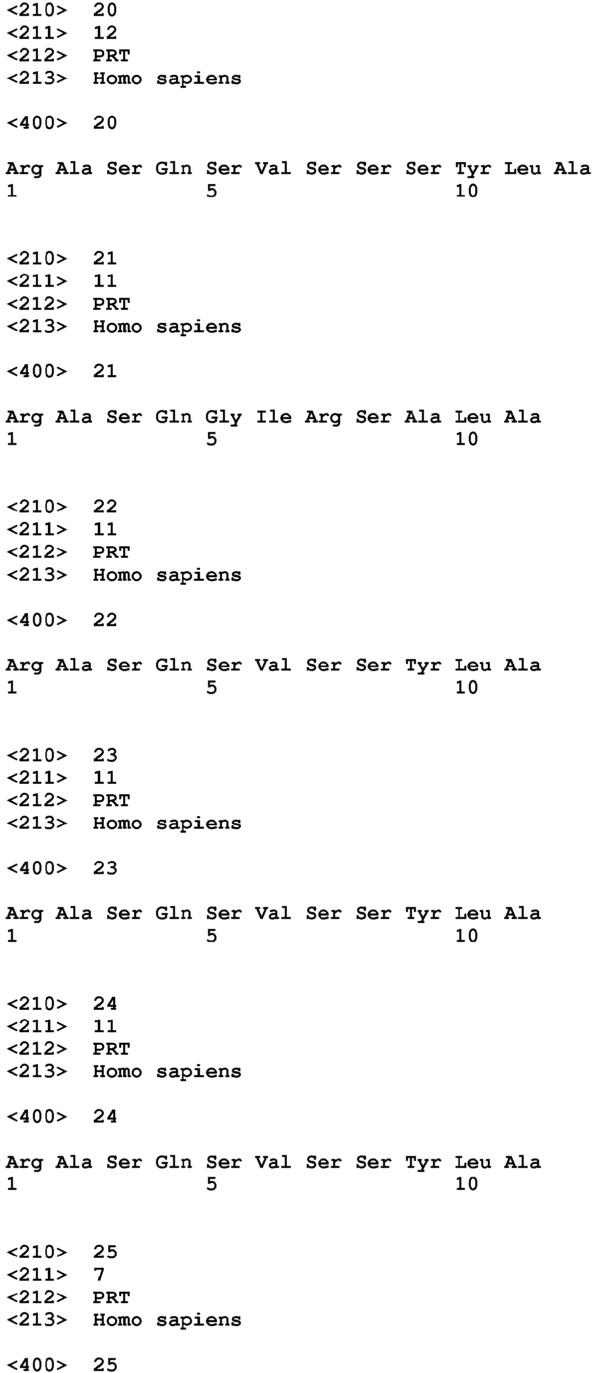


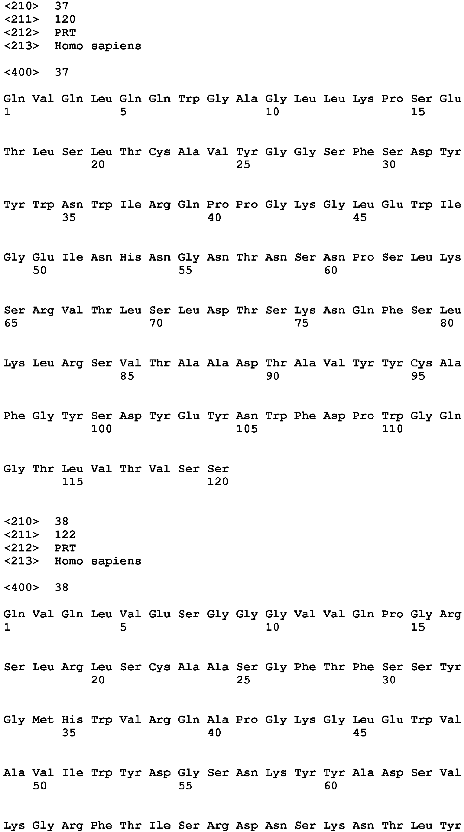


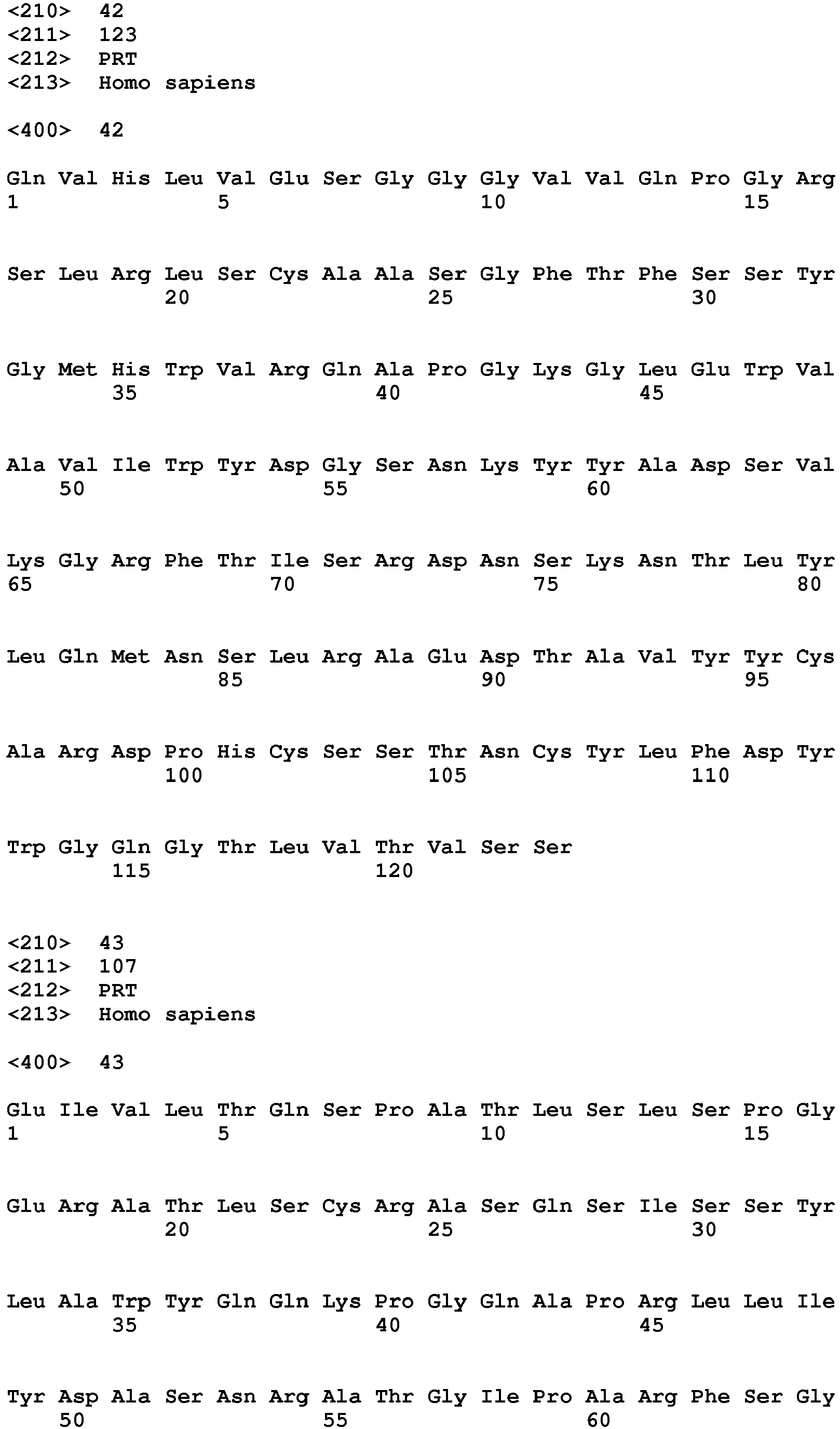


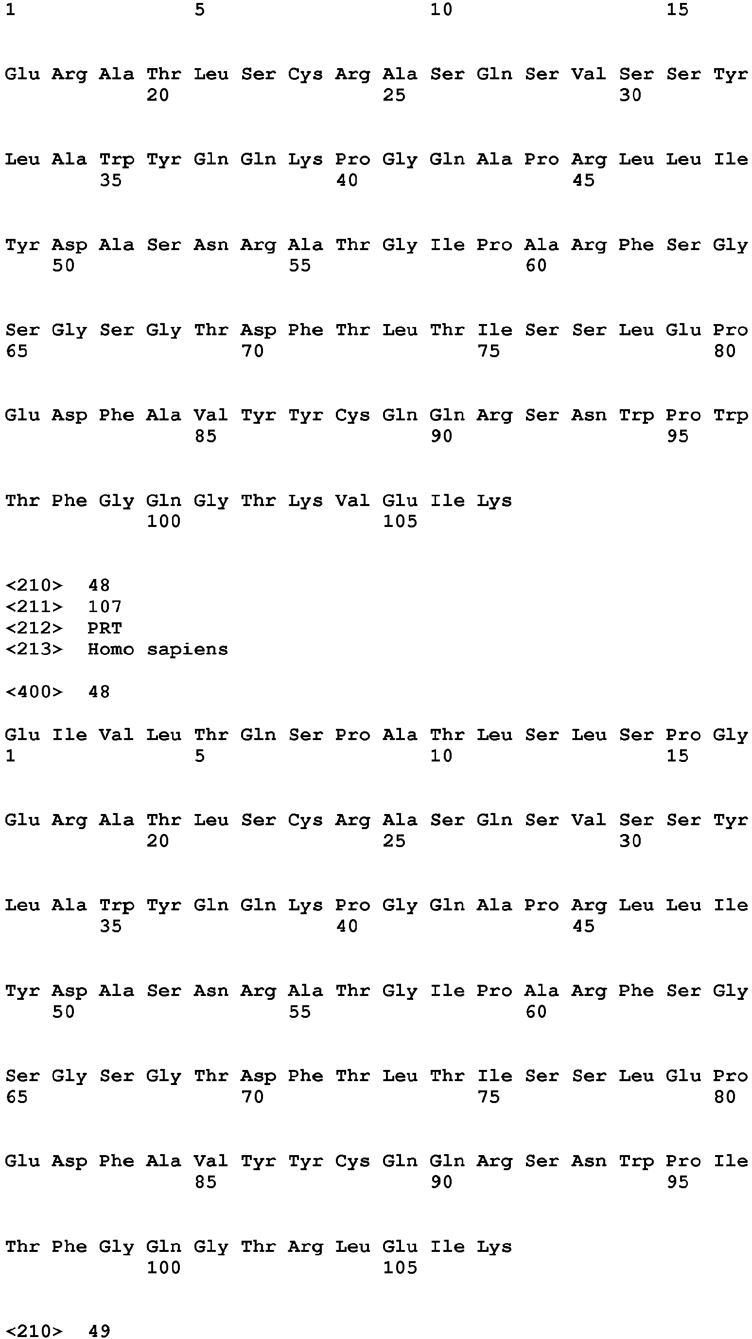

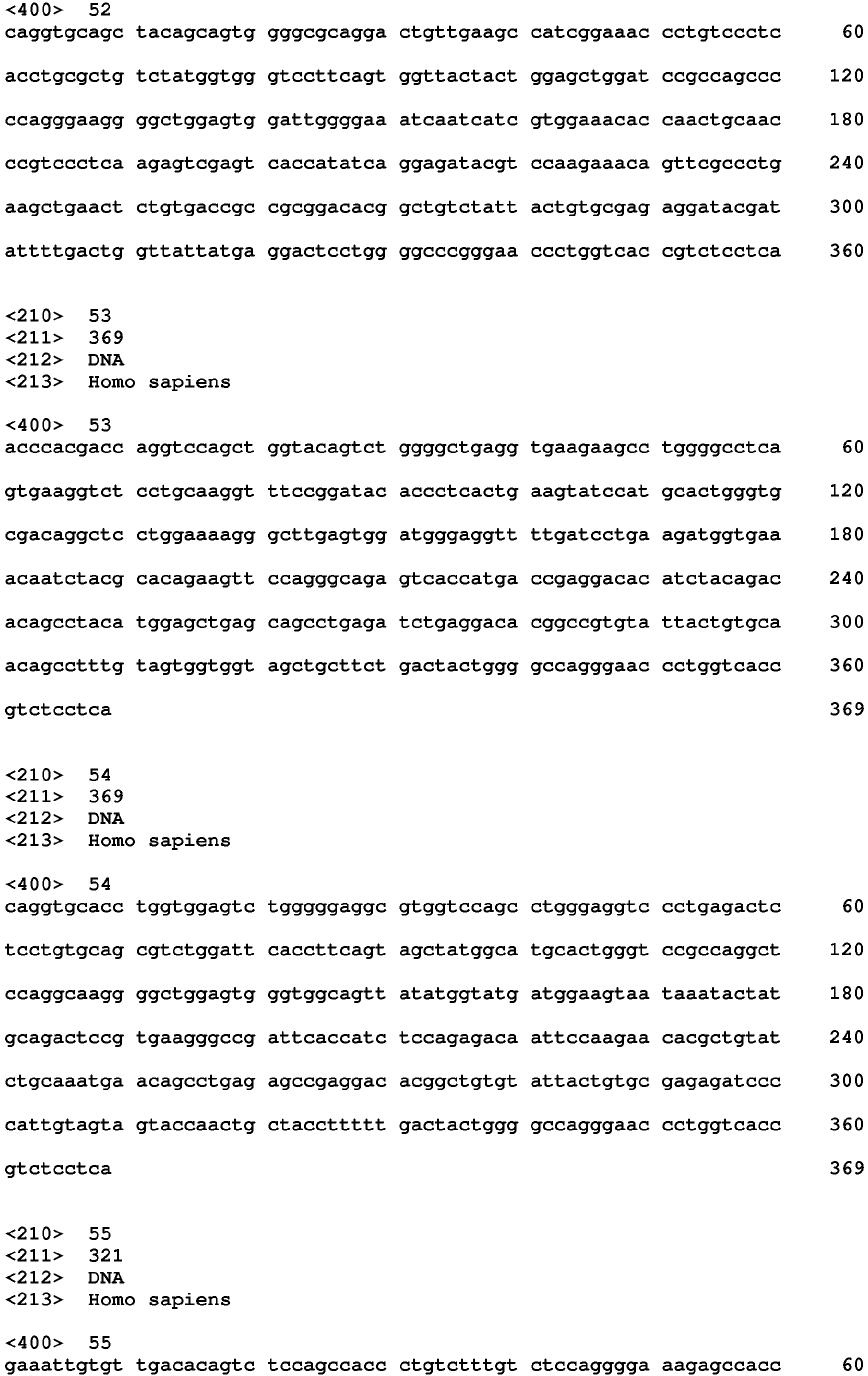

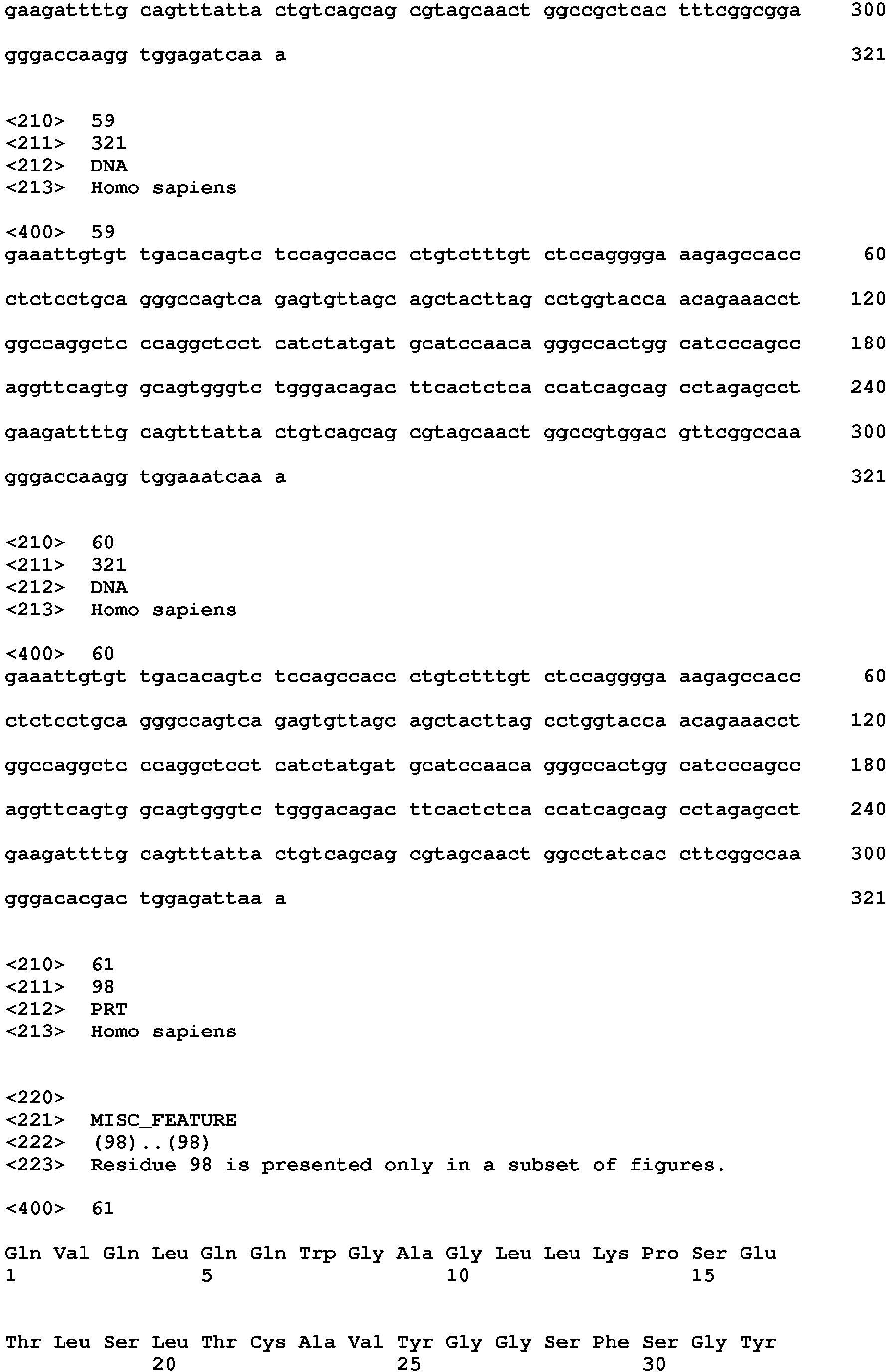


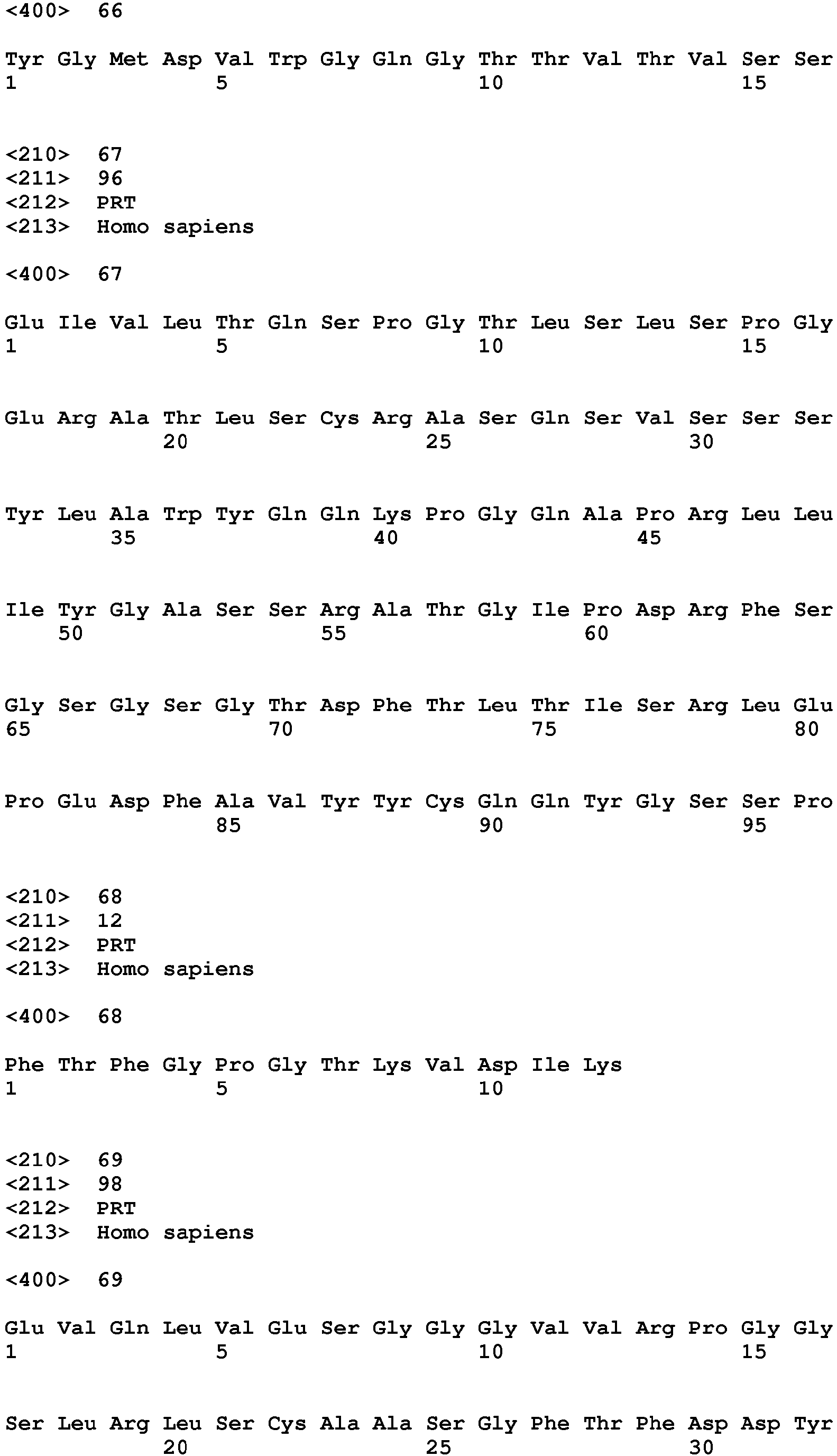


Claims (23)
- An anti-LAG-3 antibody and an additional immunostimulatory antibody for use in a method of stimulating an immune response in a subject, wherein the method comprises administering to the subject the anti-LAG-3 antibody and the additional immunostimulatory antibody.
- An anti-LAG-3 antibody for use in a method of stimulating an immune response in a subject, wherein the method comprises administering to the subject the anti-LAG-3 antibody and an additional immunostimulatory antibody.
- An immunostimulatory antibody for use in a method of stimulating an immune response in a subject, wherein the method comprises administering to the subject the immunostimulatory antibody in addition to an anti-LAG-3 antibody.
- The anti-LAG-3 antibody and/or immunostimulatory antibody for the use of any one of claims 1-3, wherein the immune response is an antigen-specific T cell response.
- The anti-LAG-3 antibody and/or immunostimulatory antibody for the use of any one of claims 1-4, wherein the immune response is against a tumor.
- An anti-LAG-3 antibody and an additional immunostimulatory antibody for use in a method of treating cancer in a subject comprising administering to the subject the anti-LAG-3 antibody and the additional immunostimulatory antibody.
- An anti-LAG-3 antibody for use in a method of treating cancer in a subject comprising administering to the subject the anti-LAG-3 antibody and an additional immunostimulatory antibody.
- An immunostimulatory antibody for use in a method of treating cancer in a subject comprising administering to the subject the immunostimulatory antibody in addition to an anti-LAG-3 antibody.
- The anti-LAG-3 antibody and/or immunostimulatory antibody for the use of any one of claims 6-8, wherein the cancer is melanoma (e.g., metastatic malignant melanoma), renal cancer (e.g. clear cell carcinoma), prostate cancer (e.g. hormone refractory prostate adenocarcinoma), breast cancer, colon cancer, or lung cancer (e.g. non-small cell lung cancer).
- The anti-LAG-3 antibody and/or immunostimulatory antibody for the use of any one of claims 1-9, wherein the anti-LAG-3 antibody has the ability of inhibiting binding of LAG-3 to major histocompatibility (MHC) class II molecules
- The anti-LAG-3 antibody and/or immunostimulatory antibody for the use of any one of claims 1-10, wherein the anti-LAG-3 antibody has the ability of stimulating interleukin-2 (IL-2) production in an antigen-specific T cell response
- The anti-LAG-3 antibody and/or immunostimulatory antibody for the use of any one of claims 1-11, wherein the anti-LAG-3 antibody is a human, chimeric or humanized antibody.
- The anti-LAG-3 antibody and/or immunostimulatory antibody for the use of any one of claims 1-12, wherein the anti-LAG-3 antibody is an IgG4 isotype.
- The anti-LAG-3 antibody and/or immunostimulatory antibody for the use of any one of claims 1-13, wherein the additional immunostimulatory antibody is a human, chimeric or humanized antibody.
- The anti-LAG-3 antibody and/or immunostimulatory antibody for the use of any one of claims 1-14, wherein the additional immunostimulatory antibody is an anti-PD-1 antibody.
- The anti-LAG-3 antibody and/or immunostimulatory antibody for the use of any one of claims 1-14, wherein the additional immunostimulatory antibody is an anti-PD-Ll antibody.
- The anti-LAG-3 antibody and/or immunostimulatory antibody for the use of any one of claims 1-14, wherein the additional immunostimulatory antibody is an anti-CTLA-4 antibody.
- The anti-LAG-3 antibody and/or immunostimulatory antibody for the use of any one of claims 1-17, wherein the anti-LAG-3 antibody is coadministered with the additional immunostimulatory antibody.
- The anti-LAG-3 antibody and/or immunostimulatory antibody for the use of any one of claims 1-17, wherein the anti-LAG-3 antibody and the additional immunostimulatory antibody are administered concurrently as a single composition in a pharmaceutically acceptable carrier, or concurrently as separate compositions with each antibody in a pharmaceutically acceptable carrier.
- The anti-LAG-3 antibody and/or immunostimulatory antibody for the use of any one of claims 1-17, wherein the anti-LAG-3 antibody and the additional immunostimulatory antibody are administered sequentially.
- The anti-LAG-3 antibody and/or immunostimulatory antibody for the use of any one of claims 1-20, further comprising administering a chemotherapeutic agent.
- The anti-LAG-3 antibody and/or immunostimulatory antibody for the use of any one of claims 1-20, further comprising radiation, surgery, or hormone deprivation.
- The anti-LAG-3 antibody and/or immunostimulatory antibody for the use of any one of claims 1-20, further comprising administering an angiogenesis inhibitor.
Priority Applications (3)
| Application Number | Priority Date | Filing Date | Title |
|---|---|---|---|
| EP22185991.1A EP4147714A1 (en) | 2008-08-11 | 2009-08-11 | Human antibodies that bind lymphocyte activation gene-3 (lag-3) and uses thereof |
| HRP20221191TT HRP20221191T1 (en) | 2008-08-11 | 2009-08-11 | Human antibodies that bind lymphocyte activation gene-3 (lag-3) and uses thereof |
| SM20220429T SMT202200429T1 (en) | 2008-08-11 | 2009-08-11 | Human antibodies that bind lymphocyte activation gene-3 (lag-3) and uses thereof |
Applications Claiming Priority (4)
| Application Number | Priority Date | Filing Date | Title |
|---|---|---|---|
| US18854808P | 2008-08-11 | 2008-08-11 | |
| EP09807162.4A EP2320940B1 (en) | 2008-08-11 | 2009-08-11 | Human antibodies that bind lymphocyte activation gene-3 (lag-3), and uses thereof |
| EP15156647.8A EP2905030B1 (en) | 2008-08-11 | 2009-08-11 | Human antibodies that bind lymphocyte activation gene-3 (LAG-3) and uses thereof |
| PCT/US2009/053405 WO2010019570A2 (en) | 2008-08-11 | 2009-08-11 | Human antibodies that bind lymphocyte activation gene-3 (lag-3), and uses thereof |
Related Parent Applications (3)
| Application Number | Title | Priority Date | Filing Date |
|---|---|---|---|
| EP09807162.4A Division EP2320940B1 (en) | 2008-08-11 | 2009-08-11 | Human antibodies that bind lymphocyte activation gene-3 (lag-3), and uses thereof |
| EP15156647.8A Division EP2905030B1 (en) | 2008-08-11 | 2009-08-11 | Human antibodies that bind lymphocyte activation gene-3 (LAG-3) and uses thereof |
| EP15156647.8A Division-Into EP2905030B1 (en) | 2008-08-11 | 2009-08-11 | Human antibodies that bind lymphocyte activation gene-3 (LAG-3) and uses thereof |
Related Child Applications (2)
| Application Number | Title | Priority Date | Filing Date |
|---|---|---|---|
| EP22185991.1A Division-Into EP4147714A1 (en) | 2008-08-11 | 2009-08-11 | Human antibodies that bind lymphocyte activation gene-3 (lag-3) and uses thereof |
| EP22185991.1A Division EP4147714A1 (en) | 2008-08-11 | 2009-08-11 | Human antibodies that bind lymphocyte activation gene-3 (lag-3) and uses thereof |
Publications (2)
| Publication Number | Publication Date |
|---|---|
| EP3597216A1 true EP3597216A1 (en) | 2020-01-22 |
| EP3597216B1 EP3597216B1 (en) | 2022-08-24 |
Family
ID=41669613
Family Applications (4)
| Application Number | Title | Priority Date | Filing Date |
|---|---|---|---|
| EP09807162.4A Active EP2320940B1 (en) | 2008-08-11 | 2009-08-11 | Human antibodies that bind lymphocyte activation gene-3 (lag-3), and uses thereof |
| EP15156647.8A Revoked EP2905030B1 (en) | 2008-08-11 | 2009-08-11 | Human antibodies that bind lymphocyte activation gene-3 (LAG-3) and uses thereof |
| EP19186310.9A Active EP3597216B1 (en) | 2008-08-11 | 2009-08-11 | Human antibodies that bind lymphocyte activation gene-3 (lag-3) and uses thereof |
| EP22185991.1A Pending EP4147714A1 (en) | 2008-08-11 | 2009-08-11 | Human antibodies that bind lymphocyte activation gene-3 (lag-3) and uses thereof |
Family Applications Before (2)
| Application Number | Title | Priority Date | Filing Date |
|---|---|---|---|
| EP09807162.4A Active EP2320940B1 (en) | 2008-08-11 | 2009-08-11 | Human antibodies that bind lymphocyte activation gene-3 (lag-3), and uses thereof |
| EP15156647.8A Revoked EP2905030B1 (en) | 2008-08-11 | 2009-08-11 | Human antibodies that bind lymphocyte activation gene-3 (LAG-3) and uses thereof |
Family Applications After (1)
| Application Number | Title | Priority Date | Filing Date |
|---|---|---|---|
| EP22185991.1A Pending EP4147714A1 (en) | 2008-08-11 | 2009-08-11 | Human antibodies that bind lymphocyte activation gene-3 (lag-3) and uses thereof |
Country Status (31)
| Country | Link |
|---|---|
| US (14) | US20110150892A1 (en) |
| EP (4) | EP2320940B1 (en) |
| JP (7) | JP5647981B2 (en) |
| KR (7) | KR20250097986A (en) |
| CN (4) | CN107474137A (en) |
| AR (3) | AR072999A1 (en) |
| AU (2) | AU2009282134B2 (en) |
| BR (1) | BRPI0917470B8 (en) |
| CA (1) | CA2734335C (en) |
| CL (4) | CL2011000308A1 (en) |
| CO (1) | CO6351751A2 (en) |
| CY (2) | CY1116582T1 (en) |
| DK (3) | DK3597216T3 (en) |
| EA (2) | EA023032B1 (en) |
| ES (3) | ES2537203T3 (en) |
| HR (3) | HRP20221191T1 (en) |
| HU (2) | HUE025250T2 (en) |
| IL (4) | IL210731A (en) |
| LT (2) | LT2905030T (en) |
| MX (2) | MX2011001250A (en) |
| NZ (3) | NZ602780A (en) |
| PE (3) | PE20190373A1 (en) |
| PL (3) | PL2905030T3 (en) |
| PT (3) | PT2320940E (en) |
| SG (1) | SG10201706497QA (en) |
| SI (2) | SI2320940T1 (en) |
| SM (3) | SMT202000048T1 (en) |
| TN (1) | TN2011000025A1 (en) |
| TW (1) | TWI491408B (en) |
| WO (1) | WO2010019570A2 (en) |
| ZA (1) | ZA201101009B (en) |
Cited By (1)
| Publication number | Priority date | Publication date | Assignee | Title |
|---|---|---|---|---|
| EP3597216B1 (en) | 2008-08-11 | 2022-08-24 | E. R. Squibb & Sons, L.L.C. | Human antibodies that bind lymphocyte activation gene-3 (lag-3) and uses thereof |
Families Citing this family (725)
| Publication number | Priority date | Publication date | Assignee | Title |
|---|---|---|---|---|
| EA017812B1 (en) | 2005-12-08 | 2013-03-29 | Медарекс, Инк. | Isolated monoclonal antibody or antigen-binding portion thereof that binds to human protein tyrosine kinase 7 (ptk7) and use thereof |
| MX349057B (en) | 2010-11-30 | 2017-07-07 | Chugai Pharmaceutical Co Ltd | THERAPEUTIC AGENT THAT INDUCES CYTOTOXICITY. |
| WO2012077649A1 (en) * | 2010-12-06 | 2012-06-14 | 大日本住友製薬株式会社 | Human monoclonal antibody |
| US8440797B2 (en) | 2010-12-06 | 2013-05-14 | Dainippon Sumitomo Pharma Co., Ltd. | Human monoclonal antibody |
| CA2860170C (en) | 2010-12-22 | 2022-06-14 | The Board Of Trustees Of The Leland Stanford Junior University | Superagonists and antagonists of interleukin-2 |
| EP3357511B1 (en) | 2011-06-30 | 2020-05-13 | Genzyme Corporation | Inhibitors of t-cell activation |
| PT3409278T (en) | 2011-07-21 | 2020-12-18 | Sumitomo Dainippon Pharma Oncology Inc | Heterocyclic protein kinase inhibitors |
| UY34887A (en) * | 2012-07-02 | 2013-12-31 | Bristol Myers Squibb Company Una Corporacion Del Estado De Delaware | OPTIMIZATION OF ANTIBODIES THAT FIX THE LYMPHOCYTE ACTIVATION GEN 3 (LAG-3) AND ITS USES |
| ES2808654T3 (en) | 2013-03-15 | 2021-03-01 | Glaxosmithkline Ip Dev Ltd | Anti-LAG-3 binding proteins |
| EP4163387A1 (en) | 2013-03-15 | 2023-04-12 | The University of Chicago | Methods and compositions related to t-cell activity |
| TWI654206B (en) | 2013-03-16 | 2019-03-21 | 諾華公司 | Treatment of cancer with a humanized anti-CD19 chimeric antigen receptor |
| HUE053069T2 (en) | 2013-05-02 | 2021-06-28 | Anaptysbio Inc | Antibodies to programmed death-1 (PD-1) |
| JP6611709B2 (en) | 2013-06-25 | 2019-11-27 | バクシネックス インコーポレーティッド | Use of a semaphorin-4D inhibitor molecule in combination with immunomodulatory therapy to inhibit tumor growth and metastasis |
| RU2016107426A (en) | 2013-08-02 | 2017-09-07 | Адуро Байотек Холдингс, Юроуп Б.В. | COMBINING CD27 AGONISTS AND INHIBITING IMMUNE CONTROL POINTS TO STIMULATE IMMUNITY |
| HUE057598T2 (en) | 2013-08-08 | 2022-05-28 | Cytune Pharma | IL-15 and IL-15R alpha sushi domain-based modulokines |
| EP3030262B1 (en) | 2013-08-08 | 2019-10-09 | Cytune Pharma | Combined pharmaceutical composition |
| PT3508502T (en) * | 2013-09-20 | 2023-06-22 | Bristol Myers Squibb Co | Combination of anti-lag-3 antibodies and anti-pd-1 antibodies to treat tumors |
| CN105722860A (en) | 2013-09-24 | 2016-06-29 | 梅迪塞纳医疗股份有限公司 | Interleukin-2 fusion proteins and uses thereof |
| WO2015048312A1 (en) * | 2013-09-26 | 2015-04-02 | Costim Pharmaceuticals Inc. | Methods for treating hematologic cancers |
| WO2015066413A1 (en) | 2013-11-01 | 2015-05-07 | Novartis Ag | Oxazolidinone hydroxamic acid compounds for the treatment of bacterial infections |
| KR20240017102A (en) * | 2013-12-17 | 2024-02-06 | 제넨테크, 인크. | Methods of treating cancers using pd-1 axis binding antagonists and taxanes |
| KR20160089532A (en) * | 2013-12-17 | 2016-07-27 | 제넨테크, 인크. | Methods of treating cancer using pd-1 axis binding antagonists and an anti-cd20 antibody |
| JP6779785B2 (en) | 2013-12-19 | 2020-11-04 | ノバルティス アーゲー | Human mesothelin chimeric antigen receptor and its use |
| GB201322626D0 (en) | 2013-12-19 | 2014-02-05 | Immutep S A | Combined preparations for the treatment of cancer |
| CA2934953C (en) | 2013-12-24 | 2022-09-20 | Bristol-Myers Squibb Company | Substituted pyrido[3,2-b]indoyl compounds and pharmaceutical compositions thereof as anticancer agents |
| JO3517B1 (en) | 2014-01-17 | 2020-07-05 | Novartis Ag | N-azaspirocycloalkane substituted n-heteroaryl compounds and compositions for inhibiting the activity of shp2 |
| JOP20200094A1 (en) | 2014-01-24 | 2017-06-16 | Dana Farber Cancer Inst Inc | Antibody molecules to pd-1 and uses thereof |
| KR20160106762A (en) * | 2014-01-28 | 2016-09-12 | 브리스톨-마이어스 스큅 컴퍼니 | Anti-lag-3 antibodies to treat hematological malignancies |
| JOP20200096A1 (en) | 2014-01-31 | 2017-06-16 | Children’S Medical Center Corp | TIM-3 Antibody Molecules and Their Uses |
| US9394365B1 (en) | 2014-03-12 | 2016-07-19 | Yeda Research And Development Co., Ltd | Reducing systemic regulatory T cell levels or activity for treatment of alzheimer's disease |
| BR112016020919A2 (en) | 2014-03-12 | 2018-01-23 | Yeda Res & Dev | reduced levels or systemic activity of regulatory t cells for the treatment of snc disease and injury |
| US10519237B2 (en) | 2014-03-12 | 2019-12-31 | Yeda Research And Development Co. Ltd | Reducing systemic regulatory T cell levels or activity for treatment of disease and injury of the CNS |
| US10618963B2 (en) | 2014-03-12 | 2020-04-14 | Yeda Research And Development Co. Ltd | Reducing systemic regulatory T cell levels or activity for treatment of disease and injury of the CNS |
| LT3116909T (en) * | 2014-03-14 | 2020-02-10 | Novartis Ag | ANTI-BODY MOLECULES AGAINST LAG-3 AND THEIR USE |
| CN106163547A (en) | 2014-03-15 | 2016-11-23 | 诺华股份有限公司 | Use Chimeric antigen receptor treatment cancer |
| JP6153674B2 (en) | 2014-03-24 | 2017-06-28 | ノバルティス アーゲー | Monobactam organic compounds for treating bacterial infections |
| MY194892A (en) | 2014-04-07 | 2022-12-22 | Chugai Pharmaceutical Co Ltd | Immunoactivating antigen-binding molecule |
| SG10202109752XA (en) | 2014-04-07 | 2021-10-28 | Novartis Ag | Treatment of cancer using anti-cd19 chimeric antigen receptor |
| EP3134102B1 (en) | 2014-04-24 | 2019-07-03 | The Board of Trustees of The Leland Stanford Junior University | Superagonists, partial agonists and antagonists of interleukin-2 |
| CN106459954A (en) | 2014-05-13 | 2017-02-22 | 中外制药株式会社 | T cell-redirected antigen-binding molecule for cells having immunosuppression function |
| AU2015261536B2 (en) | 2014-05-16 | 2020-05-07 | Ablynx Nv | Improved immunoglobulin variable domains |
| KR20250099289A (en) | 2014-05-16 | 2025-07-01 | 아블린쓰 엔.브이. | Improved immunoglobulin variable domains |
| SMT202100116T1 (en) | 2014-05-28 | 2021-05-07 | Agenus Inc | Anti-gitr antibodies and methods of use thereof |
| RU2016149812A (en) | 2014-06-06 | 2018-07-17 | Флексус Байосайенсиз, Инк. | IMMUNO REGULATORY MEANS |
| PT3151921T (en) | 2014-06-06 | 2019-11-21 | Bristol Myers Squibb Co | ANTIBODIES AGAINST GLUCOCORTICOID-INDUCED TUMOR NECROSIS FACTOR RECEIVER AND USES OF THE SAME |
| TWI693232B (en) | 2014-06-26 | 2020-05-11 | 美商宏觀基因股份有限公司 | Covalently bonded diabodies having immunoreactivity with pd-1 and lag-3, and methods of use thereof |
| WO2016014530A1 (en) | 2014-07-21 | 2016-01-28 | Novartis Ag | Combinations of low, immune enhancing. doses of mtor inhibitors and cars |
| EP3172237A2 (en) | 2014-07-21 | 2017-05-31 | Novartis AG | Treatment of cancer using humanized anti-bcma chimeric antigen receptor |
| BR112017001242A2 (en) | 2014-07-21 | 2017-12-05 | Novartis Ag | cancer treatment using a cd33 chimeric antigen receptor |
| WO2016014553A1 (en) | 2014-07-21 | 2016-01-28 | Novartis Ag | Sortase synthesized chimeric antigen receptors |
| EP3660042B1 (en) | 2014-07-31 | 2023-01-11 | Novartis AG | Subset-optimized chimeric antigen receptor-containing t-cells |
| EP3177593A1 (en) | 2014-08-06 | 2017-06-14 | Novartis AG | Quinolone derivatives as antibacterials |
| US20170224777A1 (en) | 2014-08-12 | 2017-08-10 | Massachusetts Institute Of Technology | Synergistic tumor treatment with il-2, a therapeutic antibody, and a cancer vaccine |
| EP3180018B1 (en) | 2014-08-12 | 2019-07-24 | Massachusetts Institute Of Technology | Synergistic tumor treatment with il-2 and integrin-binding-fc-fusion protein |
| US10851149B2 (en) | 2014-08-14 | 2020-12-01 | The Trustees Of The University Of Pennsylvania | Treatment of cancer using GFR α-4 chimeric antigen receptor |
| TW202140557A (en) | 2014-08-19 | 2021-11-01 | 瑞士商諾華公司 | Treatment of cancer using a cd123 chimeric antigen receptor |
| JO3663B1 (en) * | 2014-08-19 | 2020-08-27 | Merck Sharp & Dohme | Anti-lag3 antibodies and antigen-binding fragments |
| CA2960824A1 (en) | 2014-09-13 | 2016-03-17 | Novartis Ag | Combination therapies of alk inhibitors |
| MX2017003645A (en) | 2014-09-17 | 2017-05-30 | Novartis Ag | Targeting cytotoxic cells with chimeric receptors for adoptive immunotherapy. |
| ES2774448T3 (en) | 2014-10-03 | 2020-07-21 | Novartis Ag | Combination therapies |
| MA41044A (en) | 2014-10-08 | 2017-08-15 | Novartis Ag | COMPOSITIONS AND METHODS OF USE FOR INCREASED IMMUNE RESPONSE AND CANCER TREATMENT |
| CN106973568B (en) | 2014-10-08 | 2021-07-23 | 诺华股份有限公司 | Biomarkers for predicting therapeutic responsiveness to chimeric antigen receptor therapy and uses thereof |
| EP4029508A1 (en) | 2014-10-10 | 2022-07-20 | Idera Pharmaceuticals, Inc. | Treatment of cancer using tlr9 agonists and checkpoint inhibitors |
| JP6877339B2 (en) | 2014-10-14 | 2021-05-26 | ノバルティス アーゲー | Antibody molecule against PD-L1 and its use |
| HRP20210809T8 (en) | 2014-10-29 | 2021-09-17 | Bicyclerd Limited | Bicyclic peptide ligands specific for mt1-mmp |
| AR102537A1 (en) | 2014-11-05 | 2017-03-08 | Flexus Biosciences Inc | IMMUNOMODULATING AGENTS |
| UY36390A (en) | 2014-11-05 | 2016-06-01 | Flexus Biosciences Inc | MODULATING COMPOUNDS OF INDOLAMINE ENZYME 2,3-DIOXYGENASE (IDO), ITS SYNTHESIS METHODS AND PHARMACEUTICAL COMPOSITIONS CONTAINING THEM |
| CA2964276A1 (en) | 2014-11-05 | 2016-05-12 | Flexus Biosciences, Inc. | Immunoregulatory agents |
| DK3221346T3 (en) | 2014-11-21 | 2020-10-12 | Bristol Myers Squibb Co | ANTIBODIES INCLUDING MODIFIED CONSTANT AREAS OF THE HEAVY CHAIN |
| DK3221363T3 (en) | 2014-11-21 | 2020-08-10 | Bristol Myers Squibb Co | ANTIBODIES TO CD73 AND USES THEREOF |
| US20180334490A1 (en) | 2014-12-03 | 2018-11-22 | Qilong H. Wu | Methods for b cell preconditioning in car therapy |
| US9827308B2 (en) | 2014-12-10 | 2017-11-28 | Wisconsin Alumni Research Foundation | Mini-intronic plasmid DNA vaccines in combination with LAG3 blockade |
| LT3233843T (en) | 2014-12-16 | 2019-12-10 | Novartis Ag | Isoxazole hydroxamic acid compounds as lpxc inhibitors |
| WO2016100882A1 (en) | 2014-12-19 | 2016-06-23 | Novartis Ag | Combination therapies |
| TW201630907A (en) | 2014-12-22 | 2016-09-01 | 必治妥美雅史谷比公司 | TGF[beta]R antagonists |
| MA40662B1 (en) | 2014-12-23 | 2020-12-31 | Bristol Myers Squibb Co | Antibodies against tigit |
| GB201500374D0 (en) | 2015-01-09 | 2015-02-25 | Immutep S A | Combined preparations for the treatment of cancer |
| WO2016126608A1 (en) | 2015-02-02 | 2016-08-11 | Novartis Ag | Car-expressing cells against multiple tumor antigens and uses thereof |
| MA41463A (en) * | 2015-02-03 | 2017-12-12 | Anaptysbio Inc | ANTIBODIES DIRECTED AGAINST LYMPHOCYTE ACTIVATION GEN 3 (LAG-3) |
| WO2016127052A1 (en) | 2015-02-05 | 2016-08-11 | Bristol-Myers Squibb Company | Cxcl11 and smica as predictive biomarkers for efficacy of anti-ctla4 immunotherapy |
| ES2789331T3 (en) | 2015-03-02 | 2020-10-26 | Rigel Pharmaceuticals Inc | TGF-beta inhibitors |
| JP6692826B2 (en) | 2015-03-10 | 2020-05-13 | アドゥロ バイオテック,インク. | Compositions and methods for activation of "interferon gene stimulator" dependent signaling |
| US20180133327A1 (en) | 2015-03-16 | 2018-05-17 | Amal Therapeutics Sa | Cell Penetrating Peptides and Complexes Comprising the Same |
| HK1247922A1 (en) | 2015-04-03 | 2018-10-05 | Bristol-Myers Squibb Company | Inhibitors of indoleamine 2,3-dioxygenase for the treatment of cancer |
| ES2876974T3 (en) | 2015-04-07 | 2021-11-15 | Novartis Ag | Combination therapy with chimeric antigen receptor and amino pyrimidine derivatives |
| IL254817B2 (en) | 2015-04-08 | 2023-12-01 | Novartis Ag | CD20 treatments, CD22 treatments and combined treatments with CD19 chimeric antigen receptor expressing cells |
| WO2016162505A1 (en) | 2015-04-08 | 2016-10-13 | F-Star Biotechnology Limited | Her2 binding agent therapies |
| PT3283527T (en) | 2015-04-13 | 2021-03-03 | Five Prime Therapeutics Inc | COMBINATION THERAPEUTIC FOR CANCER |
| JP7114457B2 (en) | 2015-04-17 | 2022-08-08 | ザ トラスティーズ オブ ザ ユニバーシティ オブ ペンシルバニア | Methods for Improving Efficacy and Growth of Chimeric Antigen Receptor-Expressing Cells |
| DK3283107T3 (en) | 2015-04-17 | 2020-08-31 | Bristol Myers Squibb Co | COMPOSITIONS INCLUDING A COMBINATION OF IPILIMUMAB AND NIVOLUMAB |
| EP3286211A1 (en) | 2015-04-23 | 2018-02-28 | Novartis AG | Treatment of cancer using chimeric antigen receptor and protein kinase a blocker |
| EP4545082A3 (en) | 2015-05-06 | 2025-07-02 | SNIPR Technologies Limited | Altering microbial populations & modifying microbiota |
| ES2815683T3 (en) | 2015-05-11 | 2021-03-30 | Bristol Myers Squibb Co | Tricyclic compounds as antineoplastic agents |
| US9725449B2 (en) | 2015-05-12 | 2017-08-08 | Bristol-Myers Squibb Company | Tricyclic compounds as anticancer agents |
| WO2016183115A1 (en) | 2015-05-12 | 2016-11-17 | Bristol-Myers Squibb Company | 5h-pyrido[3,2-b]indole compounds as anticancer agents |
| EP4086264B1 (en) | 2015-05-18 | 2023-10-25 | Sumitomo Pharma Oncology, Inc. | Alvocidib prodrugs having increased bioavailability |
| BR112017024899A2 (en) | 2015-05-21 | 2018-11-13 | Harpoon Therapeutics, Inc. | trispecific binding proteins and methods of use. |
| DK3303394T3 (en) | 2015-05-29 | 2020-07-06 | Agenus Inc | ANTI-CTLA-4 ANTIBODIES AND PROCEDURES FOR USE THEREOF |
| DK3303396T5 (en) | 2015-05-29 | 2024-10-07 | Bristol Myers Squibb Co | ANTIBODIES AGAINST OX40 AND USES THEREOF |
| EA039293B1 (en) * | 2015-06-05 | 2021-12-30 | Мерк Шарп И Доум Корп. | Anti-lag3 antibodies and antigen-binding fragments |
| TWI773646B (en) | 2015-06-08 | 2022-08-11 | 美商宏觀基因股份有限公司 | Lag-3-binding molecules and methods of use thereof |
| CN107921120A (en) | 2015-06-24 | 2018-04-17 | 詹森生物科技公司 | Solid tumor is adjusted and treated with the antibody mediated immunity of specific binding CD38 |
| KR20180021833A (en) | 2015-06-29 | 2018-03-05 | 더 락커펠러 유니버시티 | Antibodies to CD40 with enhanced agonist activity |
| FI3858859T3 (en) | 2015-07-14 | 2025-12-04 | Bristol Myers Squibb Co | Method of treating cancer using immune checkpoint inhibitor; antibody that binds to programmed death-1 receptor (pd-1) or programmed death ligand 1 (pd-l1) |
| AU2016291817A1 (en) | 2015-07-16 | 2018-02-22 | Biolinerx Ltd. | Compositions and methods for treating cancer |
| AU2016297014B2 (en) | 2015-07-21 | 2021-06-17 | Novartis Ag | Methods for improving the efficacy and expansion of immune cells |
| AR105444A1 (en) * | 2015-07-22 | 2017-10-04 | Sorrento Therapeutics Inc | THERAPEUTIC ANTIBODIES THAT JOIN THE PROTEIN CODIFIED BY THE GENOPHYPE ACTIVATION GEN 3 (LAG3) |
| WO2017019757A1 (en) | 2015-07-28 | 2017-02-02 | Bristol-Myers Squibb Company | Tgf beta receptor antagonists |
| WO2017019896A1 (en) | 2015-07-29 | 2017-02-02 | Novartis Ag | Combination therapies comprising antibody molecules to pd-1 |
| EP3964528A1 (en) | 2015-07-29 | 2022-03-09 | Novartis AG | Combination therapies comprising antibody molecules to lag-3 |
| EP3878465A1 (en) | 2015-07-29 | 2021-09-15 | Novartis AG | Combination therapies comprising antibody molecules to tim-3 |
| SG10201906059VA (en) | 2015-07-30 | 2019-08-27 | Macrogenics Inc | Pd-1-binding molecules and methods of use thereof |
| WO2017025871A1 (en) | 2015-08-07 | 2017-02-16 | Glaxosmithkline Intellectual Property Development Limited | Combination therapy comprising anti ctla-4 antibodies |
| JP2018525415A (en) | 2015-08-25 | 2018-09-06 | ブリストル−マイヤーズ スクイブ カンパニーBristol−Myers Squibb Company | TGF beta receptor antagonist |
| WO2017040790A1 (en) | 2015-09-01 | 2017-03-09 | Agenus Inc. | Anti-pd-1 antibodies and methods of use thereof |
| MX2018002610A (en) | 2015-09-02 | 2018-09-27 | Immutep Sas | ANTI-LAG-3 ANTIBODIES. |
| US11747346B2 (en) | 2015-09-03 | 2023-09-05 | Novartis Ag | Biomarkers predictive of cytokine release syndrome |
| CN114230571B (en) | 2015-09-14 | 2025-07-08 | 无限药品股份有限公司 | Solid forms of isoquinolinones, methods of making, compositions comprising, and methods of using the same |
| MY192202A (en) | 2015-10-02 | 2022-08-06 | Hoffmann La Roche | Bispecific antibodies specific for pd1 and tim3 |
| TWI756187B (en) * | 2015-10-09 | 2022-03-01 | 美商再生元醫藥公司 | Anti-lag3 antibodies and uses thereof |
| US10149887B2 (en) | 2015-10-23 | 2018-12-11 | Canbas Co., Ltd. | Peptides and peptidomimetics in combination with t cell activating and/or checkpoint inhibiting agents for cancer treatment |
| BR112018008865A8 (en) | 2015-11-02 | 2019-02-26 | Five Prime Therapeutics Inc | cd80 extracellular domain polypeptides and their use in cancer treatment |
| LT3370768T (en) | 2015-11-03 | 2022-05-25 | Janssen Biotech, Inc. | ANTIBODIES SPECIFICLY BINDING PD-1 AND THEIR USE |
| JP2018532810A (en) | 2015-11-07 | 2018-11-08 | マルチビア インコーポレイテッド | Composition comprising tumor suppressor gene therapy and immune checkpoint therapy for the treatment of cancer |
| JP6945456B2 (en) * | 2015-11-13 | 2021-10-06 | マクロジェニクス,インコーポレーテッド | LAG-3 binding molecule and how to use it |
| US11649293B2 (en) | 2015-11-18 | 2023-05-16 | Chugai Seiyaku Kabushiki Kaisha | Method for enhancing humoral immune response |
| WO2017086367A1 (en) | 2015-11-18 | 2017-05-26 | 中外製薬株式会社 | Combination therapy using t cell redirection antigen binding molecule against cell having immunosuppressing function |
| UA121914C2 (en) * | 2015-11-18 | 2020-08-10 | Мерк Шарп І Доум Корп. | Pd1 and/or lag3 binders |
| US20190330336A1 (en) * | 2015-11-19 | 2019-10-31 | Sutro Biopharma, Inc. | Anti-lag3 antibodies, compositions comprising anti-lag3 antibodies and methods of making and using anti-lag3 antibodies |
| CN108738324B (en) | 2015-11-19 | 2022-06-21 | 百时美施贵宝公司 | Anti-glucocorticoid-induced tumor necrosis factor receptor (GITR) antibodies and uses thereof |
| WO2017087780A1 (en) | 2015-11-20 | 2017-05-26 | Regeneron Pharmaceuticals, Inc. | Non-human animals having a humanized lymphocyte-activation gene 3 |
| EP3380523A1 (en) | 2015-11-23 | 2018-10-03 | Five Prime Therapeutics, Inc. | Fgfr2 inhibitors alone or in combination with immune stimulating agents in cancer treatment |
| TW202134282A (en) | 2015-12-02 | 2021-09-16 | 美商艾吉納斯公司 | Antibodies and methods of use thereof |
| CR20180318A (en) | 2015-12-14 | 2018-09-19 | Macrogenics Inc | BISPECIFIC MOLECULES THAT HAVE IMMUNORREACTIVITY WITH PD-1 AND CTLA-4, AND METHODS OF USE OF THE SAME |
| WO2017106291A1 (en) | 2015-12-15 | 2017-06-22 | Bristol-Myers Squibb Company | Cxcr4 receptor antagonists |
| BR112018012352A2 (en) * | 2015-12-16 | 2018-12-11 | Merck Sharp & Dohme Corp. | anti-lag3 antibodies and antigen binding fragments |
| EP4424322A3 (en) | 2015-12-17 | 2025-04-16 | Novartis AG | Antibody molecules to pd-1 and uses thereof |
| US10392442B2 (en) | 2015-12-17 | 2019-08-27 | Bristol-Myers Squibb Company | Use of anti-PD-1 antibody in combination with anti-CD27 antibody in cancer treatment |
| AU2016370813A1 (en) | 2015-12-18 | 2018-06-28 | Novartis Ag | Antibodies targeting CD32b and methods of use thereof |
| EP3389720A1 (en) | 2015-12-18 | 2018-10-24 | The General Hospital Corporation | Polyacetal polymers, conjugates, particles and uses thereof |
| US11413340B2 (en) | 2015-12-22 | 2022-08-16 | Novartis Ag | Mesothelin chimeric antigen receptor (CAR) and antibody against PD-L1 inhibitor for combined use in anticancer therapy |
| MA43859A (en) | 2016-01-11 | 2018-11-21 | Novartis Ag | HUMANIZED MONOCLONAL ANTIBODIES IMMUNOSTIMULANTS DIRECTED AGAINST HUMAN INTERLEUKIN -2, AND THEIR FUSION PROTEINS |
| SG11201806863WA (en) | 2016-02-19 | 2018-09-27 | Novartis Ag | Tetracyclic pyridone compounds as antivirals |
| AU2017225733A1 (en) | 2016-03-04 | 2018-09-27 | Novartis Ag | Cells expressing multiple chimeric antigen receptor (CAR) molecules and uses therefore |
| IL295230A (en) | 2016-03-04 | 2022-10-01 | Bristol Myers Squibb Co | Combination therapy with anti-cd73 antibodies |
| SG10201601719RA (en) * | 2016-03-04 | 2017-10-30 | Agency Science Tech & Res | Anti-LAG-3 Antibodies |
| EP3429693B1 (en) | 2016-03-15 | 2023-08-23 | Mersana Therapeutics, Inc. | Napi2b-targeted antibody-drug conjugates and methods of use thereof |
| AU2017234192B2 (en) | 2016-03-16 | 2024-04-04 | Amal Therapeutics Sa | Combination of an immune checkpoint modulator and a complex comprising a cell penetrating peptide, a cargo and a TLR peptide agonist for use in medicine |
| FI3433257T3 (en) | 2016-03-24 | 2024-01-08 | Novartis Ag | Alkynyl nucleoside analogs as inhibitors of human rhinovirus |
| CA3020830A1 (en) | 2016-04-13 | 2017-10-19 | Vivia Biotech, S.L | Ex vivo bite.rtm. activated t cells |
| SG11201808821WA (en) | 2016-04-18 | 2018-11-29 | Celldex Therapeutics Inc | Agonistic antibodies that bind human cd40 and uses thereof |
| KR20190004742A (en) | 2016-05-04 | 2019-01-14 | 브리스톨-마이어스 스큅 컴퍼니 | Inhibitors of indoleamine 2,3-dioxygenase and methods of use thereof |
| CN109415320A (en) | 2016-05-04 | 2019-03-01 | 百时美施贵宝公司 | The inhibitor and its application method of indole amine 2,3-dioxygenase |
| CN109414420A (en) | 2016-05-04 | 2019-03-01 | 百时美施贵宝公司 | The inhibitor and its application method of indole amine 2,3-dioxygenase |
| JP2019516682A (en) | 2016-05-04 | 2019-06-20 | ブリストル−マイヤーズ スクイブ カンパニーBristol−Myers Squibb Company | Indoleamine 2,3-dioxygenase inhibitors and methods of use thereof |
| JP2019522627A (en) | 2016-05-04 | 2019-08-15 | ブリストル−マイヤーズ スクイブ カンパニーBristol−Myers Squibb Company | Indoleamine 2,3-dioxygenase inhibitor and method of use thereof |
| EP3458478B1 (en) * | 2016-05-18 | 2021-01-06 | Boehringer Ingelheim International GmbH | Anti pd-1 and anti-lag3 antibodies for cancer treatment |
| CA3025019A1 (en) | 2016-05-20 | 2017-11-23 | Biohaven Pharmaceutical Holding Company Ltd. | Use of glutamate modulating agents with immunotherapies to treat cancer |
| US11623958B2 (en) | 2016-05-20 | 2023-04-11 | Harpoon Therapeutics, Inc. | Single chain variable fragment CD3 binding proteins |
| JP7101621B2 (en) | 2016-05-20 | 2022-07-15 | ハープーン セラピューティクス,インク. | Single domain serum albumin binding protein |
| EP3463405A4 (en) | 2016-05-27 | 2020-02-26 | DNAtrix, Inc. | ADENOVIRUS AND IMMUNOMODULATORY POLYTHERAPY |
| GB201609811D0 (en) | 2016-06-05 | 2016-07-20 | Snipr Technologies Ltd | Methods, cells, systems, arrays, RNA and kits |
| MX2018015352A (en) | 2016-06-10 | 2019-03-28 | Io Therapeutics Inc | RETINOID RECEPTORS AND COMPOUNDS OF RETINOID AND REXINOID SELECTIVE FUNCTIONS COMBINED WITH IMMUNE MODULATORS FOR IMMUNOTHERAPY AGAINST CANCER. |
| EP3468581A1 (en) | 2016-06-13 | 2019-04-17 | Torque Therapeutics, Inc. | Methods and compositions for promoting immune cell function |
| JP6941630B2 (en) | 2016-06-14 | 2021-09-29 | ノバルティス アーゲー | Crystal form of (R) -4 (5- (cyclopropylethynyl) isoxazole-3-yl) -N-hydroxy-2-methyl-2- (methylsulfonyl) butaneamide as an antibacterial agent |
| WO2017216685A1 (en) | 2016-06-16 | 2017-12-21 | Novartis Ag | Pentacyclic pyridone compounds as antivirals |
| WO2017216686A1 (en) | 2016-06-16 | 2017-12-21 | Novartis Ag | 8,9-fused 2-oxo-6,7-dihydropyrido-isoquinoline compounds as antivirals |
| DK3472207T3 (en) * | 2016-06-20 | 2021-04-12 | F Star Delta Ltd | BINDING MOLECULES THAT BIND PD-L1 AND LAYER-3 |
| SG11201810509PA (en) | 2016-06-20 | 2018-12-28 | Kymab Ltd | Anti-pd-l1 antibodies |
| CA3027302A1 (en) * | 2016-06-20 | 2017-12-28 | F-Star Beta Limited | Binding members(1) |
| RU2757813C2 (en) * | 2016-06-23 | 2021-10-21 | Цзянсу Хэнжуй Медицин Ко., Лтд. | Antibody against lag-3, its antigen-binding fragment and their pharmaceutical application |
| CN109640999A (en) | 2016-06-24 | 2019-04-16 | 无限药品股份有限公司 | Combination treatment |
| CN109983121A (en) | 2016-06-30 | 2019-07-05 | 昂克诺斯公司 | The pseudotyping oncolytic virus of therapeutical peptide delivers |
| WO2018009466A1 (en) | 2016-07-05 | 2018-01-11 | Aduro Biotech, Inc. | Locked nucleic acid cyclic dinucleotide compounds and uses thereof |
| BR112019000431A2 (en) | 2016-07-14 | 2019-07-09 | Bristol-Myers Squibb Company | antibodies against tim3 and their uses |
| GB201612520D0 (en) | 2016-07-19 | 2016-08-31 | F-Star Beta Ltd | Binding molecules |
| CN116769050A (en) | 2016-07-20 | 2023-09-19 | 犹他大学研究基金会 | CD229 CAR T cells and methods of use thereof |
| EA039718B1 (en) * | 2016-07-21 | 2022-03-03 | Регенерон Фармасьютикалз, Инк. | Anti-lag3 antibodies and uses thereof |
| WO2018017633A1 (en) | 2016-07-21 | 2018-01-25 | Bristol-Myers Squibb Company | TGF Beta RECEPTOR ANTAGONISTS |
| AU2017313496B2 (en) | 2016-08-15 | 2023-09-21 | Fuso Pharmaceutical Industries, Ltd. | Anti-LAG-3 antibody |
| KR20230145510A (en) | 2016-08-17 | 2023-10-17 | 컴퓨젠 엘티디. | Anti-tigit antibodies, anti-pvrig antibodies and combinations thereof |
| JP2019528300A (en) | 2016-08-26 | 2019-10-10 | ブリストル−マイヤーズ スクイブ カンパニーBristol−Myers Squibb Company | Indoleamine 2,3-dioxygenase inhibitors and methods of use thereof |
| TW201811788A (en) | 2016-09-09 | 2018-04-01 | 瑞士商諾華公司 | Polycyclic pyridone compounds as antivirals |
| KR101953706B1 (en) | 2016-09-19 | 2019-03-05 | 아이-맵 | Anti-GM-CSF antibody and its use |
| KR102879364B1 (en) | 2016-09-21 | 2025-11-04 | 아말 테라퓨틱스 에스에이 | A novel complex comprising a cell penetrating peptide, a cargo and a tlr peptide agonist for treatment of colorectal cancer |
| US11673971B2 (en) | 2016-09-23 | 2023-06-13 | Marengo Therapeutics, Inc. | Multispecific antibody molecules comprising lambda and kappa light chains |
| MX2019003447A (en) | 2016-09-27 | 2019-08-29 | Univ Texas | Methods for enhancing immune checkpoint blockade therapy by modulating the microbiome. |
| JOP20190061A1 (en) | 2016-09-28 | 2019-03-26 | Novartis Ag | Beta-lactamase inhibitors |
| MY200337A (en) | 2016-10-07 | 2023-12-20 | Novartis Ag | Nucleic acid molecules encoding chimeric antigen receptors comprising a cd20 binding domain |
| TWI773694B (en) * | 2016-10-11 | 2022-08-11 | 美商艾吉納斯公司 | Anti-lag-3 antibodies and methods of use thereof |
| SG11201903283UA (en) | 2016-10-12 | 2019-05-30 | Univ Texas | Methods and compositions for tusc2 immunotherapy |
| EP3526255A2 (en) * | 2016-10-13 | 2019-08-21 | Symphogen A/S | Anti-lag-3 antibodies and compositions |
| WO2018073753A1 (en) | 2016-10-18 | 2018-04-26 | Novartis Ag | Fused tetracyclic pyridone compounds as antivirals |
| WO2018081329A1 (en) | 2016-10-28 | 2018-05-03 | Merck Sharp & Dohme Corp. | Purification process for removal of tyrosine sulfation antibody variants; purified compositions |
| MA46724A (en) | 2016-11-01 | 2021-04-21 | Anaptysbio Inc | ANTIBODIES DIRECTED AGAINST PROGRAMMED DEATH 1 (PD -1) |
| UY37463A (en) | 2016-11-02 | 2018-05-31 | Glaxosmithkline Ip No 2 Ltd | UNION PROTEINS |
| US10660909B2 (en) | 2016-11-17 | 2020-05-26 | Syntrix Biosystems Inc. | Method for treating cancer using chemokine antagonists |
| US11279694B2 (en) | 2016-11-18 | 2022-03-22 | Sumitomo Dainippon Pharma Oncology, Inc. | Alvocidib prodrugs and their use as protein kinase inhibitors |
| US11135307B2 (en) | 2016-11-23 | 2021-10-05 | Mersana Therapeutics, Inc. | Peptide-containing linkers for antibody-drug conjugates |
| WO2018099539A1 (en) | 2016-11-29 | 2018-06-07 | Horst Lindhofer | Combination of t-cell redirecting multifunctional antibodies with immune checkpoint modulators and uses thereof |
| US20190358262A1 (en) | 2016-12-03 | 2019-11-28 | Juno Therapeutics, Inc. | Methods for modulation of car-t cells |
| WO2018106738A1 (en) | 2016-12-05 | 2018-06-14 | Massachusetts Institute Of Technology | Brush-arm star polymers, conjugates and particles, and uses thereof |
| MA50948A (en) | 2016-12-07 | 2020-10-14 | Agenus Inc | ANTIBODIES AND METHODS OF USING THE SAME |
| AU2017373944B2 (en) | 2016-12-07 | 2022-02-03 | Agenus Inc. | Anti-CTLA-4 antibodies and methods of use thereof |
| CN110381997A (en) | 2016-12-12 | 2019-10-25 | 茂体外尔公司 | For treating and preventing the method and composition comprising gene-virus therapy and immunologic test point inhibitor of cancer and infectious diseases |
| WO2018112266A1 (en) | 2016-12-14 | 2018-06-21 | The Board Of Trustees Of The Leland Stanford Junior University | Il-13 superkine: immune cell targeting constructs and methods of use thereof |
| CN108204958A (en) * | 2016-12-19 | 2018-06-26 | 伊缪泰普有限公司 | binding assay |
| US10961239B2 (en) | 2017-01-05 | 2021-03-30 | Bristol-Myers Squibb Company | TGF beta receptor antagonists |
| MA47208A (en) | 2017-01-09 | 2019-11-13 | Tesaro Inc | CANCER TREATMENT METHODS USING ANTI-PD-1 ANTIBODIES |
| EP3568150A4 (en) | 2017-01-10 | 2020-12-02 | Xcella Biosciences, Inc. | Combination tumor treatment with an integrin-binding-fc fusion protein and immune modulator |
| US10350266B2 (en) | 2017-01-10 | 2019-07-16 | Nodus Therapeutics, Inc. | Method of treating cancer with a multiple integrin binding Fc fusion protein |
| CA3047600A1 (en) | 2017-01-20 | 2018-07-26 | Arcus Biosciences, Inc. | Azolopyrimidine for the treatment of cancer-related disorders |
| MA47456A (en) | 2017-02-10 | 2019-12-18 | Regeneron Pharma | RADIO-LABELED ANTI-LAG3 ANTIBODIES FOR IMMUNO-PET IMAGING |
| US20200291089A1 (en) | 2017-02-16 | 2020-09-17 | Elstar Therapeutics, Inc. | Multifunctional molecules comprising a trimeric ligand and uses thereof |
| WO2018151821A1 (en) | 2017-02-17 | 2018-08-23 | Bristol-Myers Squibb Company | Antibodies to alpha-synuclein and uses thereof |
| WO2018152687A1 (en) * | 2017-02-22 | 2018-08-30 | I-Mab | Anti-lymphocyte activation gene-3 (lag-3) antibodies and uses thereof |
| SG11201907561PA (en) * | 2017-02-22 | 2019-09-27 | I Mab | Anti-lag-3 antibodies and uses thereof |
| TW201834697A (en) | 2017-02-28 | 2018-10-01 | 美商梅爾莎納醫療公司 | Combination therapies of her2-targeted antibody-drug conjugates |
| CA3057687A1 (en) | 2017-03-31 | 2018-10-04 | Five Prime Therapeutics, Inc. | Combination therapy for cancer using anti-gitr antibodies |
| CA3058175A1 (en) | 2017-03-31 | 2018-10-04 | Bristol-Myers Squibb Company | Methods of treating tumor |
| WO2018184964A1 (en) | 2017-04-03 | 2018-10-11 | F. Hoffmann-La Roche Ag | Immunoconjugates of an anti-pd-1 antibody with a mutant il-2 or with il-15 |
| BR112019020662A2 (en) * | 2017-04-05 | 2020-05-05 | Symphogen As | combination therapies targeting pd-1, tim-3 and lag-3 |
| EP3606955B1 (en) | 2017-04-05 | 2024-11-06 | F. Hoffmann-La Roche AG | Bispecific antibodies specifically binding to pd1 and lag3 |
| EP4112644A1 (en) * | 2017-04-05 | 2023-01-04 | F. Hoffmann-La Roche AG | Anti-lag3 antibodies |
| TWI788340B (en) | 2017-04-07 | 2023-01-01 | 美商必治妥美雅史谷比公司 | Anti-icos agonist antibodies and uses thereof |
| CA3058944A1 (en) | 2017-04-19 | 2018-10-25 | Elstar Therapeutics, Inc. | Multispecific molecules and uses thereof |
| CA3059939A1 (en) | 2017-04-21 | 2018-10-25 | Kyn Therapeutics | Indole ahr inhibitors and uses thereof |
| WO2018195552A1 (en) | 2017-04-21 | 2018-10-25 | Sillajen, Inc. | Oncolytic vaccinia virus and checkpoint inhibitor combination therapy |
| CN110621341A (en) | 2017-04-26 | 2019-12-27 | 百时美施贵宝公司 | Antibody production method that minimizes disulfide bond reduction |
| MX2019012793A (en) * | 2017-04-27 | 2020-02-13 | Tesaro Inc | Antibody agents directed against lymphocyte activation gene-3 (lag-3) and uses thereof. |
| AR111419A1 (en) | 2017-04-27 | 2019-07-10 | Novartis Ag | INDAZOL PIRIDONA FUSIONED COMPOUNDS AS ANTIVIRALS |
| UY37695A (en) | 2017-04-28 | 2018-11-30 | Novartis Ag | BIS 2’-5’-RR- (3’F-A) (3’F-A) CYCLE DINUCLEOTIDE COMPOUND AND USES OF THE SAME |
| AU2018258661A1 (en) | 2017-04-28 | 2019-10-17 | Five Prime Therapeutics, Inc. | Methods of treatment with CD80 extracellular domain polypeptides |
| EP3615068A1 (en) | 2017-04-28 | 2020-03-04 | Novartis AG | Bcma-targeting agent, and combination therapy with a gamma secretase inhibitor |
| CA3059769A1 (en) | 2017-04-28 | 2018-11-01 | Elstar Therapeutics, Inc. | Multispecific molecules comprising a non-immunoglobulin heterodimerization domain and uses thereof |
| AR111651A1 (en) | 2017-04-28 | 2019-08-07 | Novartis Ag | CONJUGATES OF ANTIBODIES THAT INCLUDE TOLL TYPE RECEIVER AGONISTS AND COMBINATION THERAPIES |
| EP3615055A1 (en) | 2017-04-28 | 2020-03-04 | Novartis AG | Cells expressing a bcma-targeting chimeric antigen receptor, and combination therapy with a gamma secretase inhibitor |
| AR111658A1 (en) | 2017-05-05 | 2019-08-07 | Novartis Ag | 2-TRICYCLINAL CHINOLINONES AS ANTIBACTERIAL AGENTS |
| CN110621337B (en) * | 2017-05-10 | 2021-11-09 | 浙江时迈药业有限公司 | anti-LAG 3 human monoclonal antibodies and uses thereof |
| CA3063359A1 (en) | 2017-05-12 | 2018-11-15 | Harpoon Therapeutics, Inc. | Mesothelin binding proteins |
| US11066392B2 (en) | 2017-05-12 | 2021-07-20 | Bristol-Myers Squibb Company | Inhibitors of indoleamine 2,3-dioxygenase and methods of their use |
| JP7274426B2 (en) | 2017-05-16 | 2023-05-16 | ブリストル-マイヤーズ スクイブ カンパニー | Treatment of cancer with anti-GITR agonist antibodies |
| EP3634417B1 (en) | 2017-05-17 | 2023-07-12 | Arcus Biosciences, Inc. | Quinazoline-pyrazole derivatives for the treatment of cancer-related disorders |
| CN110719915A (en) | 2017-05-25 | 2020-01-21 | 百时美施贵宝公司 | Antibodies comprising modified heavy chain constant regions |
| EP3630826A1 (en) | 2017-05-29 | 2020-04-08 | Gamamabs Pharma | Cancer-associated immunosuppression inhibitor |
| IL322104A (en) | 2017-05-30 | 2025-09-01 | Bristol Myers Squibb Co | Treatment of tumors that express LAG–3 |
| AU2018275209A1 (en) | 2017-05-30 | 2019-10-17 | Bristol-Myers Squibb Company | Compositions comprising an anti-LAG-3 antibody or an anti-LAG-3 antibody and an anti-PD-1 or anti-PD-L1 antibody |
| MX2019012038A (en) | 2017-05-30 | 2019-11-18 | Bristol Myers Squibb Co | Compositions comprising a combination of an anti-lag-3 antibody, a pd-1 pathway inhibitor, and an immunotherapeutic agent. |
| US20210246227A1 (en) | 2017-05-31 | 2021-08-12 | Elstar Therapeutics, Inc. | Multispecific molecules that bind to myeloproliferative leukemia (mpl) protein and uses thereof |
| EP3630839A1 (en) | 2017-06-01 | 2020-04-08 | Xencor, Inc. | Bispecific antibodies that bind cd 123 cd3 |
| WO2018223004A1 (en) | 2017-06-01 | 2018-12-06 | Xencor, Inc. | Bispecific antibodies that bind cd20 and cd3 |
| CN111225675B (en) | 2017-06-02 | 2024-05-03 | 朱诺治疗学股份有限公司 | Articles and methods of treatment using adoptive cell therapy |
| WO2018229715A1 (en) | 2017-06-16 | 2018-12-20 | Novartis Ag | Compositions comprising anti-cd32b antibodies and methods of use thereof |
| CN111201035A (en) | 2017-06-19 | 2020-05-26 | 梅迪塞纳医疗股份有限公司 | Uses and methods of IL-2 superagonists, agonists and fusions thereof |
| KR20200019865A (en) | 2017-06-22 | 2020-02-25 | 노파르티스 아게 | IL-1beta binding antibody for use in cancer treatment |
| WO2018235056A1 (en) | 2017-06-22 | 2018-12-27 | Novartis Ag | IL-1BETA BINDING ANTIBODIES FOR USE IN THE TREATMENT OF CANCER |
| US11312783B2 (en) | 2017-06-22 | 2022-04-26 | Novartis Ag | Antibody molecules to CD73 and uses thereof |
| KR20200022447A (en) | 2017-06-27 | 2020-03-03 | 노파르티스 아게 | Dosage regimens of anti-TIM-3 antibodies and uses thereof |
| CN111050545A (en) | 2017-06-29 | 2020-04-21 | 朱诺治疗学股份有限公司 | A mouse model for assessing immunotherapy-related toxicity |
| EP3644993B1 (en) | 2017-06-30 | 2023-08-02 | Bristol-Myers Squibb Company | Amorphous and crystalline forms of ido inhibitors |
| AU2018297061B2 (en) | 2017-07-06 | 2021-05-06 | Merus N.V. | Antibodies that modulate a biological activity expressed by a cell |
| JP2020526551A (en) | 2017-07-13 | 2020-08-31 | アイオー セラピューティクス インコーポレイテッド | Immunomodulatory retinoid and lexinoid compounds combined with immunomodulators for cancer immunotherapy |
| US10844121B2 (en) * | 2017-07-13 | 2020-11-24 | Nanjing Leads Biolabs Co., Ltd | Antibodies binding LAG-3 and uses thereof |
| KR20200031659A (en) | 2017-07-20 | 2020-03-24 | 노파르티스 아게 | Dosage regimen of anti-LAG-3 antibody and use thereof |
| CN110997691B (en) | 2017-07-28 | 2023-11-07 | 百时美施贵宝公司 | Cyclic dinucleotides as anticancer agents |
| WO2019025811A1 (en) | 2017-08-04 | 2019-02-07 | Bicycletx Limited | Bicyclic peptide ligands specific for cd137 |
| WO2019035938A1 (en) | 2017-08-16 | 2019-02-21 | Elstar Therapeutics, Inc. | Multispecific molecules that bind to bcma and uses thereof |
| WO2019036657A1 (en) | 2017-08-17 | 2019-02-21 | Kyn Therapeutics | Ahr inhibitors and uses thereof |
| TWI785098B (en) | 2017-08-18 | 2022-12-01 | 開曼群島商科賽睿生命科學公司 | Polymorphic form of tg02 |
| CA3073733A1 (en) * | 2017-08-30 | 2019-03-07 | Phanes Therapeutics, Inc. | Anti-lag-3 antibodies and uses thereof |
| US11667663B2 (en) | 2017-08-31 | 2023-06-06 | Bristol-Myers Squibb Company | Cyclic dinucleotides as anticancer agents |
| ES2904317T3 (en) | 2017-08-31 | 2022-04-04 | Bristol Myers Squibb Co | Cyclic dinucleotides as anticancer agents |
| WO2019046498A1 (en) | 2017-08-31 | 2019-03-07 | Bristol-Myers Squibb Company | Cyclic dinucleotides as anticancer agents |
| SG11202001479RA (en) | 2017-08-31 | 2020-03-30 | Io Therapeutics Inc | Rar selective agonists in combination with immune modulators for cancer immunotherapy |
| JP7196160B2 (en) | 2017-09-12 | 2022-12-26 | スミトモ ファーマ オンコロジー, インコーポレイテッド | Treatment Regimens for Cancers Insensitive to BCL-2 Inhibitors Using the MCL-1 Inhibitor Albocidib |
| US11358948B2 (en) | 2017-09-22 | 2022-06-14 | Kymera Therapeutics, Inc. | CRBN ligands and uses thereof |
| IL273432B (en) | 2017-09-22 | 2022-09-01 | Kymera Therapeutics Inc | Protein degraders and uses thereof |
| EP3693012A4 (en) * | 2017-10-05 | 2021-07-21 | Daiichi Sankyo Company, Limited | COMPOSITION FOR DEPLOYMENT OF T-CYTOTOXIC LYMPHOCYTES |
| WO2019074824A1 (en) | 2017-10-09 | 2019-04-18 | Bristol-Myers Squibb Company | Inhibitors of indoleamine 2,3-dioxygenase and methods of their use |
| WO2019074822A1 (en) | 2017-10-09 | 2019-04-18 | Bristol-Myers Squibb Company | Inhibitors of indoleamine 2,3-dioxygenase and methods of their use |
| US11230601B2 (en) | 2017-10-10 | 2022-01-25 | Tilos Therapeutics, Inc. | Methods of using anti-lap antibodies |
| WO2019074887A1 (en) | 2017-10-10 | 2019-04-18 | Bristol-Myers Squibb Company | Cyclic dinucleotides as anticancer agents |
| KR102425983B1 (en) | 2017-10-13 | 2022-07-29 | 하푼 테라퓨틱스, 인크. | Trispecific Proteins and Methods of Use |
| IL315737A (en) | 2017-10-13 | 2024-11-01 | Harpoon Therapeutics Inc | B cell maturation antigen binding proteins |
| CN111247169A (en) | 2017-10-15 | 2020-06-05 | 百时美施贵宝公司 | Method for treating tumors |
| CN111406063B (en) | 2017-10-16 | 2023-09-15 | 百时美施贵宝公司 | Cyclic dinucleotides as anticancer agents |
| WO2019076277A1 (en) * | 2017-10-17 | 2019-04-25 | 江苏恒瑞医药股份有限公司 | Uses of anti-pd-1 antibody and anti-lag-3 antibody jointly in preparing medicament for treating tumor |
| WO2019077062A1 (en) | 2017-10-18 | 2019-04-25 | Vivia Biotech, S.L. | Bite-activated car-t cells |
| US20210040205A1 (en) | 2017-10-25 | 2021-02-11 | Novartis Ag | Antibodies targeting cd32b and methods of use thereof |
| WO2019089858A2 (en) | 2017-11-01 | 2019-05-09 | Juno Therapeutics, Inc. | Methods of assessing or monitoring a response to a cell therapy |
| CN111511370A (en) | 2017-11-01 | 2020-08-07 | 朱诺治疗学股份有限公司 | Antibodies and chimeric antigen receptors specific for B cell maturation antigens |
| EP3704159A1 (en) | 2017-11-01 | 2020-09-09 | Bristol-Myers Squibb Company | Immunostimulatory agonistic antibodies for use in treating cancer |
| PT3703750T (en) | 2017-11-01 | 2025-01-17 | Memorial Sloan Kettering Cancer Center | Chimeric antigen receptors specific for b-cell maturation antigen and encoding polynucleotides |
| EP3707138B1 (en) | 2017-11-06 | 2022-07-13 | Bristol-Myers Squibb Company | Isofuranone compounds useful as hpk1 inhibitors |
| CA3081602A1 (en) | 2017-11-16 | 2019-05-23 | Novartis Ag | Combination therapies |
| EP3710455A1 (en) | 2017-11-17 | 2020-09-23 | Novartis AG | Novel dihydroisoxazole compounds and their use for the treatment of hepatitis b |
| EP3717021A1 (en) | 2017-11-27 | 2020-10-07 | Mersana Therapeutics, Inc. | Pyrrolobenzodiazepine antibody conjugates |
| KR20200096253A (en) | 2017-11-30 | 2020-08-11 | 노파르티스 아게 | BCMA-targeting chimeric antigen receptor, and uses thereof |
| WO2019113464A1 (en) | 2017-12-08 | 2019-06-13 | Elstar Therapeutics, Inc. | Multispecific molecules and uses thereof |
| CN112204048A (en) | 2017-12-15 | 2021-01-08 | 朱诺治疗学股份有限公司 | Anti-CCT5 binding molecules and methods of use |
| WO2019121906A1 (en) | 2017-12-19 | 2019-06-27 | F-Star Beta Limited | Specific pd-l1 binding sequences inserted in a ch3 domain |
| CN111433210A (en) | 2017-12-20 | 2020-07-17 | 诺华股份有限公司 | Fused tricyclic pyrazolo-dihydropyrazinyl-pyridone compounds as antiviral agents |
| EP3727463A1 (en) | 2017-12-21 | 2020-10-28 | Mersana Therapeutics, Inc. | Pyrrolobenzodiazepine antibody conjugates |
| US20220031842A1 (en) * | 2017-12-22 | 2022-02-03 | Jiangsu Hengrui Medicine Co., Ltd. | Lag-3 antibody pharmaceutical composition and use thereof |
| IL315310A (en) | 2017-12-26 | 2024-10-01 | Kymera Therapeutics Inc | IRAK joints and used in them |
| CN109970856B (en) | 2017-12-27 | 2022-08-23 | 信达生物制药(苏州)有限公司 | anti-LAG-3 antibodies and uses thereof |
| CN109970857B (en) * | 2017-12-27 | 2022-09-30 | 信达生物制药(苏州)有限公司 | anti-PD-L1 antibodies and uses thereof |
| WO2019129137A1 (en) * | 2017-12-27 | 2019-07-04 | 信达生物制药(苏州)有限公司 | Anti-lag-3 antibody and uses thereof |
| CN111788227B (en) | 2017-12-27 | 2025-02-25 | 百时美施贵宝公司 | Anti-CD40 antibodies and uses thereof |
| JP2021508477A (en) | 2017-12-29 | 2021-03-11 | オンコラス, インコーポレイテッド | Oncolytic virus delivery of therapeutic polypeptides |
| WO2019136112A1 (en) | 2018-01-05 | 2019-07-11 | Bristol-Myers Squibb Company | Inhibitors of indoleamine 2,3-dioxygenase and methods of their use |
| US12539308B2 (en) | 2018-01-08 | 2026-02-03 | The Trustees Of The University Of Pennsylvania | Immune-enhancing RNAs for combination with chimeric antigen receptor therapy |
| EP3737692A4 (en) | 2018-01-09 | 2021-09-29 | Elstar Therapeutics, Inc. | CALRETICULIN-BINDING CONSTRUCTS AND GENERALLY MODIFIED T-CELLS FOR THE TREATMENT OF DISEASES |
| WO2019140380A1 (en) | 2018-01-12 | 2019-07-18 | Kymera Therapeutics, Inc. | Protein degraders and uses thereof |
| EP3737675A4 (en) | 2018-01-12 | 2022-01-05 | Kymera Therapeutics, Inc. | CRBN LIGANDS AND USES THEREOF |
| US12129297B2 (en) | 2018-01-12 | 2024-10-29 | Bristol-Myers Squibb Company | Antibodies against TIM3 and uses thereof |
| CN111542546B (en) * | 2018-01-18 | 2022-08-16 | 四川科伦博泰生物医药股份有限公司 | anti-LAG-3 antibodies and uses thereof |
| US12398209B2 (en) | 2018-01-22 | 2025-08-26 | Janssen Biotech, Inc. | Methods of treating cancers with antagonistic anti-PD-1 antibodies |
| SG11202006701TA (en) | 2018-01-29 | 2020-08-28 | Merck Patent Gmbh | Gcn2 inhibitors and uses thereof |
| CN111867581B (en) | 2018-01-29 | 2023-12-26 | 默克专利股份有限公司 | GCN2 inhibitors and their uses |
| MA54118A (en) | 2018-01-31 | 2021-09-15 | Celgene Corp | MULTIPLE THERAPY USING ADOPTIVE CELL THERAPY AND A CHECKPOINT INHIBITOR |
| JP2021511782A (en) | 2018-01-31 | 2021-05-13 | エフ.ホフマン−ラ ロシュ アーゲーF. Hoffmann−La Roche Aktiengesellschaft | Stabilized immunoglobulin domain |
| US20210038659A1 (en) | 2018-01-31 | 2021-02-11 | Novartis Ag | Combination therapy using a chimeric antigen receptor |
| US20200354457A1 (en) | 2018-01-31 | 2020-11-12 | Hoffmann-La Roche Inc. | Bispecific antibodies comprising an antigen-binding site binding to lag3 |
| WO2019160956A1 (en) | 2018-02-13 | 2019-08-22 | Novartis Ag | Chimeric antigen receptor therapy in combination with il-15r and il15 |
| US10519187B2 (en) | 2018-02-13 | 2019-12-31 | Bristol-Myers Squibb Company | Cyclic dinucleotides as anticancer agents |
| WO2019161054A1 (en) | 2018-02-16 | 2019-08-22 | Arcus Biosciences, Inc. | Dosing with an azolopyrimidine compound |
| WO2019165195A1 (en) | 2018-02-22 | 2019-08-29 | Srivastava Satish | Combination therapy for the treatment of cancer |
| MX2020008791A (en) | 2018-02-23 | 2021-01-08 | Bicycletx Ltd | Multimeric bicyclic peptide ligands. |
| WO2019165315A1 (en) | 2018-02-23 | 2019-08-29 | Syntrix Biosystems Inc. | Method for treating cancer using chemokine antagonists alone or in combination |
| CN115057931B (en) * | 2018-02-28 | 2025-08-22 | 广州誉衡生物科技有限公司 | Anti-human LAG-3 antibodies and uses thereof |
| US20210032334A1 (en) | 2018-02-28 | 2021-02-04 | Dana-Farber Cancer Institute, Inc. | Methods for treating cancer using combinations of anti-btnl2 and immune checkpoint blockade agents |
| WO2019165982A1 (en) | 2018-02-28 | 2019-09-06 | WuXi Biologics Ireland Limited | Monoclonal antibody against human lag-3, method for preparing same, and use thereof |
| WO2019166951A1 (en) | 2018-02-28 | 2019-09-06 | Novartis Ag | Indole-2-carbonyl compounds and their use for the treatment of hepatitis b |
| MX2020009290A (en) | 2018-03-05 | 2020-09-28 | Arcus Biosciences Inc | Arginase inhibitors. |
| WO2019173587A1 (en) | 2018-03-08 | 2019-09-12 | Bristol-Myers Squibb Company | Cyclic dinucleotides as anticancer agents |
| WO2019178364A2 (en) | 2018-03-14 | 2019-09-19 | Elstar Therapeutics, Inc. | Multifunctional molecules and uses thereof |
| WO2019178362A1 (en) | 2018-03-14 | 2019-09-19 | Elstar Therapeutics, Inc. | Multifunctional molecules that bind to calreticulin and uses thereof |
| CA3094329A1 (en) | 2018-03-19 | 2020-02-20 | Multivir Inc. | Methods and compositions comprising tumor suppressor gene therapy and cd122/cd132 agonists for the treatment of cancer |
| WO2019179365A1 (en) | 2018-03-20 | 2019-09-26 | WuXi Biologics Ireland Limited | Novel anti-lag-3 antibody polypeptide |
| CA3092589A1 (en) | 2018-03-21 | 2019-09-26 | Five Prime Therapeutics, Inc. | Antibodies binding to vista at acidic ph |
| MX2020009550A (en) | 2018-03-22 | 2020-11-11 | Keires Ag | ANTIGEN BINDING PROTEINS ANTAGONISTS. |
| US11242393B2 (en) | 2018-03-23 | 2022-02-08 | Bristol-Myers Squibb Company | Antibodies against MICA and/or MICB and uses thereof |
| US10760075B2 (en) | 2018-04-30 | 2020-09-01 | Snipr Biome Aps | Treating and preventing microbial infections |
| WO2019185551A1 (en) | 2018-03-25 | 2019-10-03 | Snipr Biome Aps. | Treating & preventing microbial infections |
| WO2019191676A1 (en) | 2018-03-30 | 2019-10-03 | Bristol-Myers Squibb Company | Methods of treating tumor |
| JP7104458B2 (en) | 2018-04-02 | 2022-07-21 | 上海博威生物医薬有限公司 | Lymphocyte activation gene-3 (LAG-3) -binding antibody and its use |
| CN110343178B (en) * | 2018-04-03 | 2022-07-22 | 上海开拓者生物医药有限公司 | Anti-human LAG-3 monoclonal antibody and application thereof |
| US20210147547A1 (en) | 2018-04-13 | 2021-05-20 | Novartis Ag | Dosage Regimens For Anti-Pd-L1 Antibodies And Uses Thereof |
| EP3781162A1 (en) | 2018-04-16 | 2021-02-24 | Arrys Therapeutics, Inc. | Ep4 inhibitors and use thereof |
| MA52289A (en) | 2018-04-18 | 2021-02-24 | Xencor Inc | FC HETERODIMERUS IL-15 / IL-15RA FUSION PROTEINS AND THEIR USES |
| CN112867734A (en) | 2018-04-18 | 2021-05-28 | Xencor股份有限公司 | PD-1 targeting heterodimeric fusion proteins comprising an IL-15/IL-15Ra Fc fusion protein and a PD-1 antigen binding domain and uses thereof |
| US20210047405A1 (en) | 2018-04-27 | 2021-02-18 | Novartis Ag | Car t cell therapies with enhanced efficacy |
| EP3788369A1 (en) | 2018-05-01 | 2021-03-10 | Novartis Ag | Biomarkers for evaluating car-t cells to predict clinical outcome |
| US12037323B2 (en) | 2018-05-03 | 2024-07-16 | Bristol-Myers Squibb Company | Uracil derivatives as Mer-AXL inhibitors |
| TWI869346B (en) | 2018-05-30 | 2025-01-11 | 瑞士商諾華公司 | Entpd2 antibodies, combination therapies, and methods of using the antibodies and combination therapies |
| WO2019232244A2 (en) | 2018-05-31 | 2019-12-05 | Novartis Ag | Antibody molecules to cd73 and uses thereof |
| EP3801617A1 (en) | 2018-06-01 | 2021-04-14 | Novartis Ag | Dosing of a bispecific antibody that bind cd123 and cd3 |
| US11492409B2 (en) | 2018-06-01 | 2022-11-08 | Novartis Ag | Binding molecules against BCMA and uses thereof |
| CN113166972A (en) * | 2018-06-11 | 2021-07-23 | 耶鲁大学 | Novel immune checkpoint inhibitors |
| EP3806962A1 (en) | 2018-06-13 | 2021-04-21 | Novartis AG | Bcma chimeric antigen receptors and uses thereof |
| CN110606892B (en) * | 2018-06-14 | 2023-09-26 | 华博生物医药技术(上海)有限公司 | LAG-3 antibody with high affinity and high biological activity and application thereof |
| CN110615840A (en) * | 2018-06-19 | 2019-12-27 | 信达生物制药(苏州)有限公司 | Fully human anti-LAG-3 antibodies and uses thereof |
| US11180531B2 (en) | 2018-06-22 | 2021-11-23 | Bicycletx Limited | Bicyclic peptide ligands specific for Nectin-4 |
| US11866430B2 (en) | 2018-06-27 | 2024-01-09 | Bristol-Myers Squibb Company | Naphthyridinone compounds useful as T cell activators |
| WO2020006018A1 (en) | 2018-06-27 | 2020-01-02 | Bristol-Myers Squibb Company | Substituted naphthyridinone compounds useful as t cell activators |
| JP7003295B2 (en) * | 2018-06-29 | 2022-01-20 | ワイ-バイオロジクス・インコーポレイテッド | Monoclonal antibody that specifically binds to LAG-3 and its uses |
| CN112955465A (en) | 2018-07-03 | 2021-06-11 | 马伦戈治疗公司 | anti-TCR antibody molecules and uses thereof |
| WO2020010177A1 (en) | 2018-07-06 | 2020-01-09 | Kymera Therapeutics, Inc. | Tricyclic crbn ligands and uses thereof |
| EP3817822A4 (en) | 2018-07-06 | 2022-07-27 | Kymera Therapeutics, Inc. | PROTEIN DEGRADING AGENTS AND THEIR USES |
| SG11202100096XA (en) | 2018-07-09 | 2021-02-25 | Five Prime Therapeutics Inc | Antibodies binding to ilt4 |
| WO2020012334A1 (en) | 2018-07-10 | 2020-01-16 | Novartis Ag | 3-(5-hydroxy-1-oxoisoindolin-2-yl)piperidine-2,6-dione derivatives and their use in the treatment of ikaros family zinc finger 2 (ikzf2)-dependent diseases |
| AR116109A1 (en) | 2018-07-10 | 2021-03-31 | Novartis Ag | DERIVATIVES OF 3- (5-AMINO-1-OXOISOINDOLIN-2-IL) PIPERIDINE-2,6-DIONA AND USES OF THE SAME |
| MX2021000213A (en) | 2018-07-11 | 2021-03-25 | Five Prime Therapeutics Inc | Antibodies binding to vista at acidic ph. |
| GB201811450D0 (en) | 2018-07-12 | 2018-08-29 | F Star Delta Ltd | Mesothelin and CD137 binding molecules |
| GB201811408D0 (en) | 2018-07-12 | 2018-08-29 | F Star Beta Ltd | CD137 Binding Molecules |
| GB201811403D0 (en) | 2018-07-12 | 2018-08-29 | F Star Beta Ltd | Antibody molecules |
| IL280002B2 (en) | 2018-07-12 | 2025-05-01 | F Star Beta Ltd | Antibody molecules that bind cd137 and ox40 |
| GB201811404D0 (en) | 2018-07-12 | 2018-08-29 | F Star Beta Ltd | Anti-CD137 Antibodies |
| WO2020011964A1 (en) | 2018-07-12 | 2020-01-16 | F-Star Beta Limited | Antibody molecules that bind pd-l1 and cd137 |
| GB201811410D0 (en) | 2018-07-12 | 2018-08-29 | F Star Beta Ltd | OX40 Binding molecules |
| GB201811415D0 (en) | 2018-07-12 | 2018-08-29 | F Star Beta Ltd | Anti-Mesothelin Anti bodies |
| WO2020018503A2 (en) * | 2018-07-16 | 2020-01-23 | Regeneron Pharmaceuticals, Inc. | Anti-il36r antibodies |
| JP7522720B2 (en) | 2018-07-18 | 2024-07-25 | アーカス バイオサイエンシズ インコーポレイティド | Solid forms of azolopyrimidine compounds |
| US11214619B2 (en) | 2018-07-20 | 2022-01-04 | Surface Oncology, Inc. | Anti-CD112R compositions and methods |
| US12145927B2 (en) | 2018-07-23 | 2024-11-19 | Bristol-Myers Squibb Company | Inhibitors of indoleamine 2,3-dioxygenase and methods of their use |
| US12059420B2 (en) | 2018-07-23 | 2024-08-13 | Bristol-Myers Squibb Company | Inhibitors of indoleamine 2,3-dioxygenase and methods of their use |
| CN112534052B (en) | 2018-07-25 | 2024-09-20 | 奥美药业有限公司 | New IL-21 prodrug and method of use |
| WO2020021465A1 (en) | 2018-07-25 | 2020-01-30 | Advanced Accelerator Applications (Italy) S.R.L. | Method of treatment of neuroendocrine tumors |
| EP4667493A2 (en) | 2018-07-26 | 2025-12-24 | Bristol-Myers Squibb Company | Lag-3 combination therapy for the treatment of cancer |
| AU2019326635B2 (en) * | 2018-08-21 | 2024-05-23 | Abl Bio Inc. | Anti-PD-L1/anti-LAG3 bispecific antibodies and uses thereof |
| US11253525B2 (en) | 2018-08-29 | 2022-02-22 | Bristol-Myers Squibb Company | Inhibitors of indoleamine 2,3-dioxygenase and methods of their use |
| US10959986B2 (en) | 2018-08-29 | 2021-03-30 | Bristol-Myers Squibb Company | Inhibitors of indoleamine 2,3-dioxygenase and methods of their use |
| EP3846793B1 (en) | 2018-09-07 | 2024-01-24 | PIC Therapeutics, Inc. | Eif4e inhibitors and uses thereof |
| US11072610B2 (en) | 2018-09-12 | 2021-07-27 | Novartis Ag | Antiviral pyridopyrazinedione compounds |
| US12269855B2 (en) | 2018-09-21 | 2025-04-08 | Innovent Biologics (Suzhou) Co., Ltd. | Interleukin-2 and use thereof |
| US12195544B2 (en) | 2018-09-21 | 2025-01-14 | Harpoon Therapeutics, Inc. | EGFR binding proteins and methods of use |
| JP2022501009A (en) | 2018-09-21 | 2022-01-06 | イノベント バイオロジックス (スウツォウ) カンパニー,リミテッド | New interleukin 2 and its use |
| EP3856771A4 (en) | 2018-09-25 | 2022-06-29 | Harpoon Therapeutics, Inc. | Dll3 binding proteins and methods of use |
| EP3856350A1 (en) | 2018-09-27 | 2021-08-04 | Marengo Therapeutics, Inc. | Csf1r/ccr2 multispecific antibodies |
| US20210347851A1 (en) | 2018-09-28 | 2021-11-11 | Novartis Ag | Cd19 chimeric antigen receptor (car) and cd22 car combination therapies |
| WO2020069405A1 (en) | 2018-09-28 | 2020-04-02 | Novartis Ag | Cd22 chimeric antigen receptor (car) therapies |
| JP2022502385A (en) | 2018-09-29 | 2022-01-11 | ノバルティス アーゲー | Method for producing a compound for inhibiting the activity of SHP2 |
| AU2019355971B2 (en) | 2018-10-03 | 2025-05-08 | Xencor, Inc. | IL-12 heterodimeric Fc-fusion proteins |
| TW202035445A (en) | 2018-10-10 | 2020-10-01 | 美商帝洛斯療法股份有限公司 | Anti-lap antibody variants and uses thereof |
| US11377477B2 (en) | 2018-10-12 | 2022-07-05 | Xencor, Inc. | PD-1 targeted IL-15/IL-15RALPHA fc fusion proteins and uses in combination therapies thereof |
| PT4445958T (en) | 2018-10-19 | 2025-11-26 | Bristol Myers Squibb Co | Combination therapy for melanoma |
| CN113365664A (en) | 2018-10-29 | 2021-09-07 | 梅尔莎纳医疗公司 | Cysteine engineered antibody-drug conjugates with peptide-containing linkers |
| EP3873532A1 (en) | 2018-10-31 | 2021-09-08 | Novartis AG | Dc-sign antibody drug conjugates |
| JP7742773B2 (en) | 2018-11-01 | 2025-09-22 | ジュノー セラピューティクス インコーポレイテッド | Chimeric antigen receptor specific for G protein-coupled receptor class C group 5 member D (GPRC5D) |
| EP3873943A2 (en) | 2018-11-01 | 2021-09-08 | Juno Therapeutics, Inc. | Methods for treatment using chimeric antigen receptors specific for b-cell maturation antigen |
| AU2019381827A1 (en) | 2018-11-16 | 2021-06-10 | Juno Therapeutics, Inc. | Methods of dosing engineered T cells for the treatment of B cell malignancies |
| JP7520003B2 (en) | 2018-11-16 | 2024-07-22 | ブリストル-マイヤーズ スクイブ カンパニー | Anti-NKG2A Antibodies and Uses Thereof |
| ES3034676T3 (en) | 2018-11-16 | 2025-08-21 | Arcus Biosciences Inc | Inhibitors of arg1 and/or arg2 |
| CA3119773A1 (en) | 2018-11-30 | 2020-06-04 | Kymera Therapeutics, Inc. | Irak degraders and uses thereof |
| WO2020113194A2 (en) | 2018-11-30 | 2020-06-04 | Juno Therapeutics, Inc. | Methods for treatment using adoptive cell therapy |
| AU2019391097B2 (en) | 2018-12-04 | 2025-07-03 | Sumitomo Pharma America, Inc. | CDK9 inhibitors and polymorphs thereof for use as agents for treatment of cancer |
| GB201820325D0 (en) | 2018-12-13 | 2019-01-30 | Bicyclerd Ltd | Bicyclic peptide ligands specific for psma |
| GB201820288D0 (en) | 2018-12-13 | 2019-01-30 | Bicycle Tx Ltd | Bicycle peptide ligaands specific for MT1-MMP |
| GB201820295D0 (en) | 2018-12-13 | 2019-01-30 | Bicyclerd Ltd | Bicyclic peptide ligands specific for MT1-MMP |
| CN119371526A (en) * | 2018-12-13 | 2025-01-28 | 阿尔金克斯有限公司 | Antibodies against human complement factor C2b and methods of use |
| EP3897637A1 (en) | 2018-12-20 | 2021-10-27 | Novartis AG | Dosing regimen and pharmaceutical combination comprising 3-(1-oxoisoindolin-2-yl)piperidine-2,6-dione derivatives |
| EP3670659A1 (en) | 2018-12-20 | 2020-06-24 | Abivax | Biomarkers, and uses in treatment of viral infections, inflammations, or cancer |
| EP3897853A1 (en) | 2018-12-20 | 2021-10-27 | Xencor, Inc. | Targeted heterodimeric fc fusion proteins containing il-15/il-15ra and nkg2d antigen binding domains |
| CA3119582A1 (en) | 2018-12-21 | 2020-06-25 | Novartis Ag | Use of il-1.beta. binding antibodies |
| WO2020128637A1 (en) | 2018-12-21 | 2020-06-25 | Novartis Ag | Use of il-1 binding antibodies in the treatment of a msi-h cancer |
| EP3897849A1 (en) | 2018-12-21 | 2021-10-27 | BicycleTx Limited | Bicyclic peptide ligands specific for pd-l1 |
| US20220025036A1 (en) | 2018-12-21 | 2022-01-27 | Novartis Ag | Use of il-1beta binding antibodies |
| CA3118182A1 (en) | 2018-12-21 | 2020-06-25 | Onxeo | New conjugated nucleic acid molecules and their uses |
| CN113227137A (en) | 2018-12-21 | 2021-08-06 | 诺华股份有限公司 | Use of IL-1 beta antibodies in the treatment or prevention of myelodysplastic syndrome |
| CN113614109A (en) | 2018-12-21 | 2021-11-05 | Ose免疫疗法公司 | Bifunctional anti-PD-1/IL-7 molecules |
| WO2020154032A1 (en) | 2019-01-23 | 2020-07-30 | Massachusetts Institute Of Technology | Combination immunotherapy dosing regimen for immune checkpoint blockade |
| MX2021009087A (en) | 2019-01-29 | 2021-09-08 | Juno Therapeutics Inc | Antibodies and chimeric antigen receptors specific for receptor tyrosine kinase like orphan receptor 1 (ror1). |
| NZ778055A (en) | 2019-02-12 | 2025-11-28 | Sumitomo Pharma America Inc | Formulations comprising heterocyclic protein kinase inhibitors |
| WO2020165374A1 (en) | 2019-02-14 | 2020-08-20 | Ose Immunotherapeutics | Bifunctional molecule comprising il-15ra |
| ES2982474T3 (en) | 2019-02-15 | 2024-10-16 | Novartis Ag | Substituted 3-(1-oxoisoindolin-2-yl)piperidine-1,6-dione derivatives and their uses |
| CN113490528B (en) | 2019-02-15 | 2024-12-03 | 诺华股份有限公司 | 3-(1-oxo-5-(piperidin-4-yl)isoindolin-2-yl)piperidine-2,6-dione derivatives and uses thereof |
| CN114026122B (en) | 2019-02-21 | 2024-12-31 | 马伦戈治疗公司 | Multifunctional molecules that bind to T cell-associated cancer cells and their uses |
| WO2020172605A1 (en) | 2019-02-21 | 2020-08-27 | Elstar Therapeutics, Inc. | Antibody molecules that bind to nkp30 and uses thereof |
| CN111620949A (en) * | 2019-02-28 | 2020-09-04 | 三生国健药业(上海)股份有限公司 | Antibodies that bind human LAG-3, methods of making, and uses thereof |
| US12453738B2 (en) | 2019-03-12 | 2025-10-28 | Arcus Biosciences, Inc. | Treatment of oncogene-driven cancers |
| CN113795264A (en) | 2019-03-19 | 2021-12-14 | 瓦尔希伯伦私人肿瘤研究基金会 | Combination therapy for cancer with Omomyc and antibodies that bind PD-1 or CTLA-4 |
| JP2022525149A (en) | 2019-03-20 | 2022-05-11 | スミトモ ダイニッポン ファーマ オンコロジー, インコーポレイテッド | Treatment of Acute Myeloid Leukemia (AML) with Venetoclax Failure |
| EP3941463A1 (en) | 2019-03-22 | 2022-01-26 | Sumitomo Dainippon Pharma Oncology, Inc. | Compositions comprising pkm2 modulators and methods of treatment using the same |
| CN114269763A (en) | 2019-03-26 | 2022-04-01 | 美国密歇根州立大学试剂中心 | Small molecule degradant for STAT3 |
| US20220185831A1 (en) | 2019-03-29 | 2022-06-16 | The Regents Of The University Of Michigan | Stat3 protein degraders |
| KR20210144821A (en) | 2019-03-29 | 2021-11-30 | 아르커스 바이오사이언시즈 인코포레이티드 | Cancer treatment method using the identified adenosine fingerprint |
| WO2020201753A1 (en) | 2019-04-02 | 2020-10-08 | Bicycletx Limited | Bicycle toxin conjugates and uses thereof |
| CN120574283A (en) | 2019-04-05 | 2025-09-02 | 凯麦拉医疗公司 | STAT degraders and their uses |
| EP3725370A1 (en) | 2019-04-19 | 2020-10-21 | ImmunoBrain Checkpoint, Inc. | Modified anti-pd-l1 antibodies and methods and uses for treating a neurodegenerative disease |
| WO2020223121A1 (en) | 2019-04-30 | 2020-11-05 | Dana-Farber Cancer Institute, Inc. | Methods for treating cancer using combinations of anti-cx3cr1 and immune checkpoint blockade agents |
| GB201906118D0 (en) | 2019-05-01 | 2019-06-12 | Immutep S A | Anti-LAG-3 binding molecules |
| US20230295087A1 (en) | 2019-05-13 | 2023-09-21 | Bristol-Myers Squibb Company | AGONISTS OF ROR GAMMAt |
| US12012374B2 (en) | 2019-05-13 | 2024-06-18 | Bristol-Myers Squibb Company | Agonists of ROR GAMMAt |
| CN120939219A (en) | 2019-05-13 | 2025-11-14 | 瑞泽恩制药公司 | Combination of PD-1 inhibitors and LAG-3 inhibitors for enhancing efficacy in cancer treatment |
| KR20220016157A (en) | 2019-05-30 | 2022-02-08 | 브리스톨-마이어스 스큅 컴퍼니 | Cell localization signatures and combination therapies |
| JP2022534967A (en) | 2019-05-30 | 2022-08-04 | ブリストル-マイヤーズ スクイブ カンパニー | Multiple tumor gene signatures and their uses |
| EP3976832A1 (en) | 2019-05-30 | 2022-04-06 | Bristol-Myers Squibb Company | Methods of identifying a subject suitable for an immuno-oncology (i-o) therapy |
| CN110320367A (en) * | 2019-05-30 | 2019-10-11 | 广州医科大学附属第一医院(广州呼吸中心) | Diagnostic kit based on LAG-3 and its application on diagnosis of Parkinson disease product |
| TW202108559A (en) | 2019-05-31 | 2021-03-01 | 美商醫肯納腫瘤學公司 | Tead inhibitors and uses thereof |
| WO2020251972A1 (en) | 2019-06-10 | 2020-12-17 | Kymera Therapeutics, Inc. | Smarca degraders and uses thereof |
| WO2020252353A1 (en) | 2019-06-12 | 2020-12-17 | Vanderbilt University | Amino acid transport inhibitors and the uses thereof |
| AU2020291936A1 (en) | 2019-06-12 | 2022-02-03 | Vanderbilt University | Dibenzylamines as amino acid transport inhibitors |
| US11845801B2 (en) | 2019-06-12 | 2023-12-19 | AskGene Pharma, Inc. | IL-15 prodrugs and methods of use thereof |
| CN112079925B (en) * | 2019-06-13 | 2025-04-25 | 上海健信生物医药科技有限公司 | Antibodies and bispecific antibodies targeting LAG-3 and their uses |
| EP3987009A1 (en) * | 2019-06-18 | 2022-04-27 | The U.S.A. as represented by the Secretary, Department of Health and Human Services | Interleukin-27 producing b-cells and uses thereof |
| US11642409B2 (en) | 2019-06-26 | 2023-05-09 | Massachusetts Insttute of Technology | Immunomodulatory fusion protein-metal hydroxide complexes and methods thereof |
| EP3990618A4 (en) | 2019-06-27 | 2023-08-09 | The George Washington University, A Congressionally Chartered Not-For-Profit Corporation | Hdac6-activated macrophages, compositions, and uses thereof |
| JP2022539208A (en) | 2019-07-03 | 2022-09-07 | スミトモ ファーマ オンコロジー, インコーポレイテッド | Tyrosine kinase non-receptor 1 (TNK1) inhibitors and uses thereof |
| IT201900011676A1 (en) | 2019-07-12 | 2021-01-12 | St Superiore Di Sanita | Human recombinant antibody against the LAG3 membrane receptor, its medical and diagnostic uses. |
| AU2020315401B2 (en) | 2019-07-16 | 2025-05-22 | The Regents Of The University Of Michigan | Imidazopyrimidines as EED inhibitors and the use thereof |
| WO2021024020A1 (en) | 2019-08-06 | 2021-02-11 | Astellas Pharma Inc. | Combination therapy involving antibodies against claudin 18.2 and immune checkpoint inhibitors for treatment of cancer |
| WO2021026179A1 (en) | 2019-08-06 | 2021-02-11 | Bristol-Myers Squibb Company | AGONISTS OF ROR GAMMAt |
| JP2022545735A (en) | 2019-08-27 | 2022-10-28 | ザ リージェンツ オブ ザ ユニヴァシティ オブ ミシガン | cereblon E3 ligase inhibitor |
| AR119821A1 (en) | 2019-08-28 | 2022-01-12 | Bristol Myers Squibb Co | SUBSTITUTED PYRIDOPYRIMIDINOL COMPOUNDS USEFUL AS T-CELL ACTIVATORS |
| WO2021050964A1 (en) | 2019-09-13 | 2021-03-18 | Nimbus Saturn, Inc. | Hpk1 antagonists and uses thereof |
| CN114502590A (en) | 2019-09-18 | 2022-05-13 | 诺华股份有限公司 | ENTPD2 antibodies, combination therapies, and methods of using these antibodies and combination therapies |
| AU2020348849A1 (en) | 2019-09-19 | 2022-04-07 | The Regents Of The University Of Michigan | Spirocyclic androgen receptor protein degraders |
| BR112022004831A2 (en) | 2019-09-19 | 2022-06-07 | Bristol Myers Squibb Co | Antibodies that bind to seen at acidic pH |
| CN114450028A (en) | 2019-09-22 | 2022-05-06 | 百时美施贵宝公司 | Quantitative spatial profiling for LAG-3antagonist therapy |
| CN120842223A (en) | 2019-09-26 | 2025-10-28 | 诺华股份有限公司 | Antiviral pyrazolopyridone compounds |
| EP4037700A2 (en) | 2019-10-03 | 2022-08-10 | Xencor, Inc. | Targeted il-12 heterodimeric fc-fusion proteins |
| TW202128757A (en) | 2019-10-11 | 2021-08-01 | 美商建南德克公司 | Pd-1 targeted il-15/il-15ralpha fc fusion proteins with improved properties |
| IL292347A (en) | 2019-10-21 | 2022-06-01 | Novartis Ag | Combination therapies with venetoclax and tim-3 inhibitors |
| BR112022007179A2 (en) | 2019-10-21 | 2022-08-23 | Novartis Ag | TIM-3 INHIBITORS AND USES THEREOF |
| US11459389B2 (en) | 2019-10-24 | 2022-10-04 | Massachusetts Institute Of Technology | Monoclonal antibodies that bind human CD161 |
| KR20220088425A (en) * | 2019-10-25 | 2022-06-27 | 다이이찌 산쿄 가부시키가이샤 | Combination of anti-GARP antibodies and immunomodulators |
| JP2023500506A (en) | 2019-11-04 | 2023-01-06 | アストラゼネカ・アクチエボラーグ | Combination therapy to treat cancer |
| WO2021092221A1 (en) | 2019-11-06 | 2021-05-14 | Bristol-Myers Squibb Company | Methods of identifying a subject with a tumor suitable for a checkpoint inhibitor therapy |
| WO2021092220A1 (en) | 2019-11-06 | 2021-05-14 | Bristol-Myers Squibb Company | Methods of identifying a subject with a tumor suitable for a checkpoint inhibitor therapy |
| JP2022553851A (en) * | 2019-11-08 | 2022-12-26 | ブリストル-マイヤーズ スクイブ カンパニー | LAG-3 antagonists for the treatment of melanoma |
| BR112022009514A2 (en) | 2019-11-19 | 2022-08-16 | Bristol Myers Squibb Co | COMPOUNDS USEFUL AS HELIOS PROTEIN INHIBITORS |
| JP2023502531A (en) | 2019-11-26 | 2023-01-24 | イケナ オンコロジー, インコーポレイテッド | Polymorphic carbazole derivatives and uses thereof |
| CA3159071A1 (en) | 2019-11-26 | 2021-06-03 | Tamar I. ROSENBAUM | Salts/cocrystals of (r)-n-(4-chlorophenyl)-2-((1s,4s)-4-(6-fluoroquinolin-4-yl)cyclohexyl)propanamide |
| KR20220104217A (en) | 2019-11-26 | 2022-07-26 | 노파르티스 아게 | CD19 and CD22 chimeric antigen receptors and uses thereof |
| WO2021113644A1 (en) | 2019-12-05 | 2021-06-10 | Multivir Inc. | Combinations comprising a cd8+ t cell enhancer, an immune checkpoint inhibitor and radiotherapy for targeted and abscopal effects for the treatment of cancer |
| EP4072588A4 (en) | 2019-12-13 | 2024-02-14 | Cugene Inc. | Novel interleukin-15 (il-15) fusion proteins and uses thereof |
| AU2020406083A1 (en) | 2019-12-17 | 2022-06-16 | Boehringer Ingelheim International Gmbh | Bifunctional molecules comprising an IL-7 variant |
| JP2023509366A (en) | 2019-12-17 | 2023-03-08 | カイメラ セラピューティクス, インコーポレイテッド | IRAK dissolving agents and their uses |
| EP4076524A4 (en) | 2019-12-17 | 2023-11-29 | Kymera Therapeutics, Inc. | IRAQ DEGRADATION AGENTS AND THEIR USES |
| WO2021126967A1 (en) * | 2019-12-20 | 2021-06-24 | Merck Sharp & Dohme Corp. | Lag3 antagonist cell based potency assay |
| IL293889A (en) | 2019-12-20 | 2022-08-01 | Novartis Ag | Uses of antitgf-beta antibodies and checkpoint inhibitors for the treatment of proliferative diseases |
| JP7677976B2 (en) | 2019-12-23 | 2025-05-15 | ブリストル-マイヤーズ スクイブ カンパニー | Substituted Heteroaryl Compounds Useful as T Cell Activators - Patent application |
| CN115297931A (en) | 2019-12-23 | 2022-11-04 | 凯麦拉医疗公司 | SMARCA degrading agents and uses thereof |
| AR120823A1 (en) | 2019-12-23 | 2022-03-23 | Bristol Myers Squibb Co | SUBSTITUTED BICYCLIC COMPOUNDS USEFUL AS T-CELL ACTIVATORS |
| ES3038401T3 (en) | 2019-12-23 | 2025-10-13 | Bristol Myers Squibb Co | Substituted quinazolinyl compounds useful as t cell activators |
| AU2020414688A1 (en) | 2019-12-23 | 2022-08-18 | Bristol-Myers Squibb Company | Substituted quinolinonyl piperazine compounds useful as T cell activators |
| MX2022007130A (en) | 2019-12-23 | 2022-07-11 | Bristol Myers Squibb Co | SUBSTITUTED PIPERAZINE DERIVATIVES USEFUL AS T CELL ACTIVATORS. |
| EP4084821A4 (en) | 2020-01-03 | 2024-04-24 | Marengo Therapeutics, Inc. | CD33-BINDING MULTIFUNCTIONAL MOLECULES AND THEIR USES |
| EP4084823A4 (en) | 2020-01-03 | 2024-05-15 | Marengo Therapeutics, Inc. | Anti-tcr antibody molecules and uses thereof |
| AU2021206618A1 (en) | 2020-01-06 | 2022-08-18 | Hifibio, Inc. | Anti-TNFR2 antibody and uses thereof |
| WO2021146370A1 (en) | 2020-01-15 | 2021-07-22 | Blueprint Medicines Corporation | Map4k1 inhibitors |
| WO2021144657A1 (en) | 2020-01-17 | 2021-07-22 | Novartis Ag | Combination comprising a tim-3 inhibitor and a hypomethylating agent for use in treating myelodysplastic syndrome or chronic myelomonocytic leukemia |
| JP2023514152A (en) | 2020-02-06 | 2023-04-05 | ブリストル-マイヤーズ スクイブ カンパニー | IL-10 and its uses |
| WO2021171264A1 (en) | 2020-02-28 | 2021-09-02 | Novartis Ag | Dosing of a bispecific antibody that binds cd123 and cd3 |
| TW202146393A (en) | 2020-03-03 | 2021-12-16 | 美商皮克醫療公司 | Eif4e inhibitors and uses thereof |
| US20230140384A1 (en) | 2020-03-09 | 2023-05-04 | Bristol-Myers Squibb Company | Antibodies to cd40 with enhanced agonist activity |
| US12528785B2 (en) | 2020-03-19 | 2026-01-20 | Kymera Therapeutics, Inc. | MDM2 degraders and uses thereof |
| WO2021188769A1 (en) | 2020-03-19 | 2021-09-23 | Arcus Biosciences, Inc. | Tetralin and tetrahydroquinoline compounds as inhibitors of hif-2alpha |
| TW202140441A (en) | 2020-03-23 | 2021-11-01 | 美商必治妥美雅史谷比公司 | Substituted oxoisoindoline compounds |
| US12522623B2 (en) | 2020-03-26 | 2026-01-13 | Regents Of The University Of Michigan | Small molecule STAT protein degraders |
| EP4126048A4 (en) * | 2020-04-03 | 2025-01-15 | Yuhan Corporation | GENETICALLY ENGINEERED ANTIBODIES THAT BIND LAG3 |
| WO2021207449A1 (en) | 2020-04-09 | 2021-10-14 | Merck Sharp & Dohme Corp. | Affinity matured anti-lap antibodies and uses thereof |
| CN115916223A (en) | 2020-04-10 | 2023-04-04 | 朱诺治疗学股份有限公司 | Methods and uses related to cell therapy engineered with chimeric antigen receptors targeting B cell maturation antigens |
| WO2021231732A1 (en) | 2020-05-15 | 2021-11-18 | Bristol-Myers Squibb Company | Antibodies to garp |
| AU2021278998A1 (en) | 2020-05-27 | 2022-11-03 | Agilent Technologies, Inc. | Anti-human LAG-3 antibodies and their use in immunohistochemistry (IHC) |
| CR20220611A (en) | 2020-06-02 | 2023-06-07 | Arcus Biosciences Inc | ANTI-TIGIT ANTIBODIES |
| WO2021245071A1 (en) | 2020-06-03 | 2021-12-09 | Mv Biotherapeutics Sa | Combination of an atp-hydrolyzing enzyme and an immune checkpoint modulator and uses thereof |
| TW202210483A (en) | 2020-06-03 | 2022-03-16 | 美商凱麥拉醫療公司 | Crystalline forms of irak degraders |
| US20230250110A1 (en) | 2020-06-03 | 2023-08-10 | Kymera Therapeutics, Inc. | Deuterated irak degraders and uses thereof |
| CN111808192B (en) * | 2020-06-05 | 2022-02-15 | 北京天广实生物技术股份有限公司 | Antibodies that bind LAG3 and uses thereof |
| TW202214857A (en) | 2020-06-19 | 2022-04-16 | 法商昂席歐公司 | New conjugated nucleic acid molecules and their uses |
| WO2021258010A1 (en) | 2020-06-19 | 2021-12-23 | Gossamer Bio Services, Inc. | Oxime compounds useful as t cell activators |
| WO2021260528A1 (en) | 2020-06-23 | 2021-12-30 | Novartis Ag | Dosing regimen comprising 3-(1-oxoisoindolin-2-yl)piperidine-2,6-dione derivatives |
| US20250262293A1 (en) | 2020-07-07 | 2025-08-21 | BioNTech SE | Therapeutic rna for hpv-positive cancer |
| WO2022011204A1 (en) | 2020-07-10 | 2022-01-13 | The Regents Of The University Of Michigan | Small molecule androgen receptor protein degraders |
| WO2022011205A1 (en) | 2020-07-10 | 2022-01-13 | The Regents Of The University Of Michigan | Androgen receptor protein degraders |
| EP4188374A4 (en) | 2020-07-30 | 2024-08-14 | Kymera Therapeutics, Inc. | Methods of treating mutant lymphomas |
| WO2022029573A1 (en) | 2020-08-03 | 2022-02-10 | Novartis Ag | Heteroaryl substituted 3-(1-oxoisoindolin-2-yl)piperidine-2,6-dione derivatives and uses thereof |
| CA3189761A1 (en) | 2020-08-03 | 2022-02-10 | Gemma Mudd | Peptide-based linkers |
| AU2021325225A1 (en) | 2020-08-10 | 2023-03-23 | Gv20 Therapeutics Llc | Compositions and methods for treating autoimmune diseases and cancers by targeting IGSF8 |
| WO2022036079A1 (en) | 2020-08-13 | 2022-02-17 | Bristol-Myers Squibb Company | Methods of redirecting of il-2 to target cells of interest |
| MX2023001588A (en) | 2020-08-17 | 2023-05-03 | Bicycletx Ltd | Bicycle conjugates specific for nectin-4 and uses thereof. |
| WO2022047046A1 (en) | 2020-08-26 | 2022-03-03 | Marengo Therapeutics, Inc. | Methods of detecting trbc1 or trbc2 |
| AU2021331476A1 (en) | 2020-08-28 | 2023-05-04 | Bristol-Myers Squibb Company | Lag-3 antagonist therapy for hepatocellular carcinoma |
| US20230303700A1 (en) | 2020-08-31 | 2023-09-28 | Bristol-Myers Squibb Company | Cell localization signature and immunotherapy |
| EP4204021A1 (en) | 2020-08-31 | 2023-07-05 | Advanced Accelerator Applications International S.A. | Method of treating psma-expressing cancers |
| EP4204020A1 (en) | 2020-08-31 | 2023-07-05 | Advanced Accelerator Applications International S.A. | Method of treating psma-expressing cancers |
| CR20230155A (en) | 2020-09-14 | 2023-06-27 | Boehringer Ingelheim Int | HETEROLOGOUS PRIMARY STIMULUS VACCINE |
| CN116601140A (en) | 2020-10-02 | 2023-08-15 | 羿尊生物医药股份有限公司 | For subcutaneous administration containing (S)-2-((S)-2-acetylamino-3-(1H-indol-3-yl)propionylamino)-6-diazo-5-oxohexyl Freeze-dried composition of isopropyl ester and use thereof |
| IL300314A (en) | 2020-10-08 | 2023-04-01 | Affimed Gmbh | Trispecific binders |
| US20230416382A1 (en) | 2020-10-14 | 2023-12-28 | Five Prime Therapeutics, Inc. | Anti-c-c chemokine receptor 8 (ccr8) antibodies and methods of use thereof |
| MX2023004493A (en) | 2020-10-23 | 2023-05-10 | Bristol Myers Squibb Co | Lag-3 antagonist therapy for lung cancer. |
| EP4240491A1 (en) | 2020-11-06 | 2023-09-13 | Novartis AG | Cd19 binding molecules and uses thereof |
| WO2022112198A1 (en) | 2020-11-24 | 2022-06-02 | Worldwide Innovative Network | Method to select the optimal immune checkpoint therapies |
| CN117015531A (en) | 2020-12-02 | 2023-11-07 | 医肯纳肿瘤学公司 | TEAD inhibitors and their uses |
| WO2022117572A2 (en) | 2020-12-02 | 2022-06-09 | Oncurious Nv | An ltbr agonist in combination therapy against cancer |
| WO2022120353A1 (en) | 2020-12-02 | 2022-06-09 | Ikena Oncology, Inc. | Tead inhibitors and uses thereof |
| WO2022120179A1 (en) | 2020-12-03 | 2022-06-09 | Bristol-Myers Squibb Company | Multi-tumor gene signatures and uses thereof |
| CA3204091A1 (en) | 2020-12-08 | 2022-06-16 | Infinity Pharmaceuticals, Inc. | Eganelisib for use in the treatment of pd-l1 negative cancer |
| TW202237119A (en) | 2020-12-10 | 2022-10-01 | 美商住友製藥腫瘤公司 | Alk-5 inhibitors and uses thereof |
| CN114621344B (en) * | 2020-12-10 | 2022-08-30 | 北京东方百泰生物科技股份有限公司 | Purification method of anti-LAG-3 monoclonal antibody |
| CN116964050A (en) | 2020-12-16 | 2023-10-27 | 戈萨默生物服务公司 | Compounds that can be used as T cell activators |
| WO2022135667A1 (en) | 2020-12-21 | 2022-06-30 | BioNTech SE | Therapeutic rna for treating cancer |
| TW202245808A (en) | 2020-12-21 | 2022-12-01 | 德商拜恩迪克公司 | Therapeutic rna for treating cancer |
| WO2022135666A1 (en) | 2020-12-21 | 2022-06-30 | BioNTech SE | Treatment schedule for cytokine proteins |
| MX2023007734A (en) | 2020-12-28 | 2023-08-21 | Bristol Myers Squibb Co | Antibody compositions and methods of use thereof. |
| MX2023007650A (en) | 2020-12-28 | 2023-09-11 | Bristol Myers Squibb Co | Subcutaneous administration of pd1/pd-l1 antibodies. |
| TW202241891A (en) | 2020-12-30 | 2022-11-01 | 美商凱麥拉醫療公司 | Irak degraders and uses thereof |
| WO2022148781A1 (en) | 2021-01-05 | 2022-07-14 | Institut Curie | Combination of mcoln activators and immune checkpoint inhibitors |
| US20250186539A2 (en) | 2021-01-11 | 2025-06-12 | Bicycletx Limited | Methods for treating cancer |
| WO2022165214A1 (en) | 2021-01-29 | 2022-08-04 | Board Of Regents, The University Of Texas System | Methods of treating cancer with kinase inhibitors |
| EP4288427A1 (en) | 2021-02-02 | 2023-12-13 | Liminal Biosciences Limited | Gpr84 antagonists and uses thereof |
| BR112023015584A2 (en) | 2021-02-02 | 2023-10-24 | Liminal Biosciences Ltd | GPR84 ANTAGONISTS AND USES THEREOF |
| MX2023009100A (en) | 2021-02-03 | 2023-09-25 | Mozart Therapeutics Inc | Binding agents and methods of using the same. |
| US20240109899A1 (en) | 2021-02-04 | 2024-04-04 | Bristol-Myers Squibb Company | Benzofuran compounds as sting agonists |
| CA3210553A1 (en) | 2021-02-12 | 2022-08-18 | F. Hoffmann-La Roche Ag | Bicyclic tetrahydroazepine derivatives for the treatment of cancer |
| CN117120090A (en) | 2021-02-12 | 2023-11-24 | 林伯士萨顿公司 | HPK1 antagonists and uses thereof |
| US12171768B2 (en) | 2021-02-15 | 2024-12-24 | Kymera Therapeutics, Inc. | IRAK4 degraders and uses thereof |
| MX2023009178A (en) | 2021-02-15 | 2023-08-21 | Kymera Therapeutics Inc | Irak4 degraders and uses thereof. |
| US20220275090A1 (en) | 2021-02-22 | 2022-09-01 | Janssen Biotech, Inc. | Combination Therapies with Anti-CD38 Antibodies and PARP or Adenosine Receptor Inhibitors |
| JP2024508304A (en) * | 2021-03-02 | 2024-02-26 | ノヴァロック バイオセラピューティクス, リミテッド | Antibodies against Claudin-6 and uses thereof |
| WO2022187419A1 (en) | 2021-03-03 | 2022-09-09 | The Regents Of The University Of Michigan | Small molecule degraders of androgen receptor |
| US20240166647A1 (en) | 2021-03-03 | 2024-05-23 | The Regents Of The University Of Michigan | Cereblon Ligands |
| WO2022187856A1 (en) | 2021-03-05 | 2022-09-09 | Nimbus Saturn, Inc. | Hpk1 antagonists and uses thereof |
| EP4305041A1 (en) | 2021-03-08 | 2024-01-17 | Blueprint Medicines Corporation | Map4k1 inhibitors |
| WO2022197641A1 (en) | 2021-03-15 | 2022-09-22 | Rapt Therapeutics, Inc. | 1h-pyrazolo[3,4-d]pyrimidin-6-yl-amine derivatives as hematopoietic progenitor kinase 1 (hpk1) modulators and/or inhibitors for the treatment of cancer and other diseases |
| EP4313989A4 (en) | 2021-03-29 | 2025-03-05 | Nimbus Saturn, Inc. | HPK1 ANTAGONISTS AND THEIR USES |
| WO2022212400A1 (en) | 2021-03-29 | 2022-10-06 | Juno Therapeutics, Inc. | Methods for dosing and treatment with a combination of a checkpoint inhibitor therapy and a car t cell therapy |
| JP2024514530A (en) | 2021-04-02 | 2024-04-02 | ザ リージェンツ オブ ザ ユニバーシティ オブ カリフォルニア | Antibodies against truncated CDCP1 and uses thereof |
| AU2022253450A1 (en) | 2021-04-05 | 2023-11-16 | Bristol-Myers Squibb Company | Pyridinyl substituted oxoisoindoline compounds for the treatment of cancer |
| SMT202500281T1 (en) | 2021-04-06 | 2025-09-12 | Bristol Myers Squibb Co | Pyridinyl substituted oxoisoindoline compounds |
| TW202304979A (en) | 2021-04-07 | 2023-02-01 | 瑞士商諾華公司 | USES OF ANTI-TGFβ ANTIBODIES AND OTHER THERAPEUTIC AGENTS FOR THE TREATMENT OF PROLIFERATIVE DISEASES |
| EP4319755A4 (en) | 2021-04-08 | 2025-03-26 | Board of Regents, The University of Texas System | Compounds and methods for theranostic targeting of PARP activity |
| WO2022216993A2 (en) | 2021-04-08 | 2022-10-13 | Marengo Therapeutics, Inc. | Multifuntional molecules binding to tcr and uses thereof |
| US20240182572A1 (en) | 2021-04-09 | 2024-06-06 | Ose Immunotherapeutics | Scaffold for bifunctional molecules comprising pd-1 or cd28 and sirp binding domains |
| KR20240007155A (en) | 2021-04-09 | 2024-01-16 | 님버스 클리오, 인코포레이티드 | CBL-B modulators and their uses |
| EP4320155A1 (en) | 2021-04-09 | 2024-02-14 | Ose Immunotherapeutics | New scaffold for bifunctional molecules with improved properties |
| WO2022221227A1 (en) | 2021-04-13 | 2022-10-20 | Nuvalent, Inc. | Amino-substituted heterocycles for treating cancers with egfr mutations |
| WO2022221866A1 (en) | 2021-04-16 | 2022-10-20 | Ikena Oncology, Inc. | Mek inhibitors and uses thereof |
| IL308314A (en) | 2021-05-07 | 2024-01-01 | Kymera Therapeutics Inc | Cdk2 degraders and uses thereof |
| US20240285740A1 (en) | 2021-05-12 | 2024-08-29 | Dana-Farber Cancer Institute, Inc. | Compositions and methods for treating cancer |
| AR125874A1 (en) | 2021-05-18 | 2023-08-23 | Novartis Ag | COMBINATION THERAPIES |
| JP2024521712A (en) | 2021-05-21 | 2024-06-04 | アーカス バイオサイエンシーズ,インコーポレーテッド | AXL Inhibitor Compounds |
| JP2024521706A (en) | 2021-05-21 | 2024-06-04 | アーカス バイオサイエンシーズ,インコーポレーテッド | AXL Compound |
| US20240239907A1 (en) | 2021-05-25 | 2024-07-18 | Edelweiss Immune Inc | C-X-C Motif Chemokine Receptor 6 (CXCR6) Binding Molecules, and Methods of Using the Same |
| TW202307210A (en) | 2021-06-01 | 2023-02-16 | 瑞士商諾華公司 | Cd19 and cd22 chimeric antigen receptors and uses thereof |
| CA3225254A1 (en) | 2021-07-13 | 2023-01-19 | BioNTech SE | Multispecific binding agents against cd40 and cd137 in combination therapy for cancer |
| KR20240083168A (en) | 2021-07-14 | 2024-06-11 | 블루프린트 메디신즈 코포레이션 | Heterocyclic compounds as MAP4K1 inhibitors |
| WO2023288264A1 (en) | 2021-07-15 | 2023-01-19 | Blueprint Medicines Corporation | Map4k1 inhibitors |
| AU2022320948A1 (en) | 2021-07-30 | 2024-01-18 | Affimed Gmbh | Duplexbodies |
| JP2024532276A (en) | 2021-08-25 | 2024-09-05 | ピク セラピューティクス, インコーポレイテッド | eIF4E inhibitors and uses thereof |
| US12234231B2 (en) | 2021-08-25 | 2025-02-25 | PIC Therapeutics, Inc. | EIF4E inhibitors and uses thereof |
| CN118201914A (en) | 2021-09-08 | 2024-06-14 | 雷多纳治疗公司 | PAPD5 and/or PAPD7 inhibitors 4-oxo-1,4-dihydroquinoline-3-carboxylic acid derivatives |
| TW202333802A (en) | 2021-10-11 | 2023-09-01 | 德商拜恩迪克公司 | Therapeutic rna for lung cancer |
| WO2023076876A1 (en) | 2021-10-26 | 2023-05-04 | Mozart Therapeutics, Inc. | Modulation of immune responses to viral vectors |
| IL312348A (en) | 2021-10-29 | 2024-06-01 | Arcus Biosciences Inc | Inhibitors of hif-2alpha and methods of use thereof |
| AU2022375806A1 (en) | 2021-10-29 | 2023-12-14 | Bristol-Myers Squibb Company | Lag-3 antagonist therapy for hematological cancer |
| AR127505A1 (en) | 2021-10-29 | 2024-01-31 | Kymera Therapeutics Inc | IRAQ-4 DEGRADERS AND SYNTHESIS THEREOF |
| CA3237175A1 (en) | 2021-11-03 | 2023-05-11 | Janssen Biotech, Inc. | Corticosteriod reduction in treatment with anti-cd38 antibodies |
| JP2025500922A (en) | 2021-12-16 | 2025-01-15 | ヴァレリオ・セラピューティクス | Novel conjugated nucleic acid molecules and uses thereof |
| WO2023114984A1 (en) | 2021-12-17 | 2023-06-22 | Ikena Oncology, Inc. | Tead inhibitors and uses thereof |
| WO2023122777A1 (en) | 2021-12-22 | 2023-06-29 | Gossamer Bio Services, Inc. | Oxime derivatives useful as t cell activators |
| WO2023122772A1 (en) | 2021-12-22 | 2023-06-29 | Gossamer Bio Services, Inc. | Oxime derivatives useful as t cell activators |
| WO2023122778A1 (en) | 2021-12-22 | 2023-06-29 | Gossamer Bio Services, Inc. | Pyridazinone derivatives useful as t cell activators |
| JP2025503962A (en) | 2022-01-26 | 2025-02-06 | ブリストル-マイヤーズ スクイブ カンパニー | Combination Therapy for Hepatocellular Carcinoma |
| WO2023147594A2 (en) | 2022-01-31 | 2023-08-03 | Kymera Therapeutics, Inc. | Irak degraders and uses thereof |
| WO2023150186A1 (en) | 2022-02-01 | 2023-08-10 | Arvinas Operations, Inc. | Dgk targeting compounds and uses thereof |
| TWI864587B (en) | 2022-02-14 | 2024-12-01 | 美商基利科學股份有限公司 | Antiviral pyrazolopyridinone compounds |
| AU2023226078A1 (en) | 2022-02-25 | 2024-08-22 | Bristol-Myers Squibb Company | Combination therapy for colorectal carcinoma. |
| WO2023168404A1 (en) | 2022-03-04 | 2023-09-07 | Bristol-Myers Squibb Company | Methods of treating a tumor |
| JP2025508076A (en) | 2022-03-08 | 2025-03-21 | アレンティス・セラピューティクス・アクチェンゲゼルシャフト | Use of anti-claudin-1 antibodies to improve T cell availability |
| WO2023173057A1 (en) | 2022-03-10 | 2023-09-14 | Ikena Oncology, Inc. | Mek inhibitors and uses thereof |
| WO2023173053A1 (en) | 2022-03-10 | 2023-09-14 | Ikena Oncology, Inc. | Mek inhibitors and uses thereof |
| US20250198990A1 (en) * | 2022-03-15 | 2025-06-19 | The Board Of Trustees Of The Leland Stanford Junior University | Methods of Identifying Anti-LAG-3 Agents |
| MX2024011278A (en) | 2022-03-15 | 2024-09-25 | Compugen Ltd | Il-18bp antagonist antibodies and their use in monotherapy and combination therapy in the treatment of cancer. |
| WO2023178329A1 (en) | 2022-03-18 | 2023-09-21 | Bristol-Myers Squibb Company | Methods of isolating polypeptides |
| WO2023211889A1 (en) | 2022-04-25 | 2023-11-02 | Ikena Oncology, Inc. | Polymorphic compounds and uses thereof |
| WO2023214325A1 (en) | 2022-05-05 | 2023-11-09 | Novartis Ag | Pyrazolopyrimidine derivatives and uses thereof as tet2 inhibitors |
| TW202404581A (en) | 2022-05-25 | 2024-02-01 | 美商醫肯納腫瘤學公司 | Mek inhibitors and uses thereof |
| CA3258064A1 (en) | 2022-06-02 | 2023-12-07 | Bristol-Myers Squibb Company | Antibody compositions and methods of use thereof |
| JP2025521543A (en) | 2022-06-22 | 2025-07-10 | ジュノー セラピューティクス インコーポレイテッド | Treatment methods for second line therapy of CD19-targeted CAR T cells |
| GB202209518D0 (en) | 2022-06-29 | 2022-08-10 | Snipr Biome Aps | Treating & preventing E coli infections |
| WO2024003360A1 (en) | 2022-07-01 | 2024-01-04 | Institut Curie | Biomarkers and uses thereof for the treatment of neuroblastoma |
| AU2023308064A1 (en) | 2022-07-11 | 2025-02-27 | Genuv Inc. | Cytokine fusion protein |
| EP4554680A1 (en) | 2022-07-15 | 2025-05-21 | Arcus Biosciences, Inc. | Inhibitors of hpk1 and methods of use thereof |
| US20260028336A1 (en) | 2022-07-20 | 2026-01-29 | Arcus Biosciences, Inc. | Cbl-b inhibitors and methods of use thereof |
| CN120019047A (en) | 2022-08-02 | 2025-05-16 | 里米诺生物科学有限公司 | Heteroaryl carboxamides and related GPR84 antagonists and their uses |
| WO2024028386A1 (en) | 2022-08-02 | 2024-02-08 | Ose Immunotherapeutics | Multifunctional molecule directed against cd28 |
| KR20250056924A (en) | 2022-08-02 | 2025-04-28 | 리미널 바이오사이언시스 리미티드 | Substituted pyridone GPR84 antagonists and uses thereof |
| KR20250057801A (en) | 2022-08-02 | 2025-04-29 | 리미널 바이오사이언시스 리미티드 | Aryl-triazolyl and related GPR84 antagonists and uses thereof |
| KR20250047766A (en) | 2022-08-05 | 2025-04-04 | 주노 쎄러퓨티크스 인코퍼레이티드 | Chimeric antigen receptor specific for GPRC5D and BCMA |
| EP4568950A1 (en) | 2022-08-08 | 2025-06-18 | Bristol-Myers Squibb Company | Substituted tetrazolyl compounds useful as t cell activators |
| EP4568963A1 (en) | 2022-08-09 | 2025-06-18 | Bristol-Myers Squibb Company | Tertiary amine substituted bicyclic compounds useful as t cell activators |
| CN119677733A (en) | 2022-08-11 | 2025-03-21 | 豪夫迈·罗氏有限公司 | Bicyclic tetrahydrothiazepine derivatives |
| JP2025526727A (en) | 2022-08-11 | 2025-08-15 | エフ・ホフマン-ラ・ロシュ・アクチェンゲゼルシャフト | Bicyclic tetrahydrothiazepine derivatives |
| CN119677732A (en) | 2022-08-11 | 2025-03-21 | 豪夫迈·罗氏有限公司 | Bicyclic tetrahydroazepine derivatives |
| JP2025526683A (en) | 2022-08-11 | 2025-08-15 | エフ・ホフマン-ラ・ロシュ・アクチェンゲゼルシャフト | Bicyclic tetrahydrothiazepine derivatives |
| EP4572772A1 (en) | 2022-08-17 | 2025-06-25 | Capstan Therapeutics, Inc. | Conditioning for in vivo immune cell engineering |
| JP2025531079A (en) | 2022-09-14 | 2025-09-19 | アーカス バイオサイエンシズ インコーポレイティド | Dispersion of Etrumadenant |
| WO2024081385A1 (en) | 2022-10-14 | 2024-04-18 | Arcus Biosciences, Inc. | Hpk1 inhibitors and methods of use thereof |
| EP4604920A1 (en) | 2022-10-20 | 2025-08-27 | Arcus Biosciences, Inc. | Lyophilized formulations of cd73 compounds |
| EP4608390A1 (en) | 2022-10-24 | 2025-09-03 | Cancer Research Technology Limited | Tumour sensitisation to checkpoint inhibitors with redox status modifier |
| WO2024089417A1 (en) | 2022-10-24 | 2024-05-02 | Memorial Sloan-Kettering Cancer Center | Tumour stratification for responsiveness to an immune checkpoint inhibitor |
| WO2024112894A1 (en) | 2022-11-22 | 2024-05-30 | PIC Therapeutics, Inc. | Eif4e inhibitors and uses thereof |
| CN120390652A (en) | 2022-12-01 | 2025-07-29 | 免疫医疗有限公司 | Combination therapy comprising an anti-PD-L1 antibody and an anti-CD73 antibody for treating cancer |
| AR131320A1 (en) | 2022-12-13 | 2025-03-05 | Juno Therapeutics Inc | Chimeric antigen receptors specific for BAFF-R and CD19 and their methods and uses |
| IL321098A (en) | 2022-12-14 | 2025-07-01 | Astellas Pharma Europe Bv | Combination therapy involving bispecific binding agents binding to cldn18.2 and cd3 and immune checkpoint inhibitors |
| KR20250123912A (en) | 2022-12-21 | 2025-08-18 | 브리스톨-마이어스 스큅 컴퍼니 | Combination therapy for lung cancer |
| WO2024137865A1 (en) | 2022-12-22 | 2024-06-27 | Gossamer Bio Services, Inc. | Compounds useful as t cell activators |
| EP4649173A1 (en) | 2023-01-13 | 2025-11-19 | Akrivia Biomedics Limited | Method of profiling diseases |
| EP4658687A1 (en) | 2023-01-31 | 2025-12-10 | University of Rochester | Immune checkpoint blockade therapy for treating staphylococcus aureus infections |
| EP4665410A1 (en) | 2023-02-17 | 2025-12-24 | Regeneron Pharmaceuticals, Inc. | Radiolabeled anti-lag3 antibodies for immuno-pet imaging |
| IL323023A (en) | 2023-03-13 | 2025-10-01 | Regeneron Pharma | Combination of pd-1 inhibitors and lag-3 inhibitors for enhanced efficacy in treating melanoma |
| WO2024196952A1 (en) | 2023-03-20 | 2024-09-26 | Bristol-Myers Squibb Company | Tumor subtype assessment for cancer therapy |
| EP4687991A1 (en) | 2023-03-30 | 2026-02-11 | Ose Immunotherapeutics | Lipid-based nanoparticle targeted at activated immune cells for the expression of immune cell enhancing molecule and use thereof |
| WO2024200826A1 (en) | 2023-03-30 | 2024-10-03 | Ose Immunotherapeutics | Lipid-based nanoparticle targeted at activated immune cells for the expression of immune cell inhibiting molecule and use thereof |
| WO2024233360A1 (en) | 2023-05-05 | 2024-11-14 | Arcus Biosciences, Inc. | Cbl-b inhibitors and methods of use thereof |
| CN121057732A (en) | 2023-05-08 | 2025-12-02 | 百时美施贵宝公司 | Substituted phenyl oxazolone compounds |
| TW202509014A (en) | 2023-05-10 | 2025-03-01 | 美商纜圖藥品公司 | Gsk3α inhibitors and methods of use thereof |
| WO2024243502A1 (en) | 2023-05-25 | 2024-11-28 | Arcus Biosciences, Inc. | Cbl-b inhibitors and methods of use thereof |
| AU2024281414A1 (en) | 2023-06-02 | 2025-12-11 | Arcus Biosciences, Inc. | Biomarkers for predicting cancer treatment efficacy |
| AU2024285766A1 (en) | 2023-06-07 | 2026-01-08 | Bristol-Myers Squibb Company | Spirocyclic substituted oxoisoindolinyl piperidine-2,6-dione compound |
| CN120865251A (en) | 2023-06-23 | 2025-10-31 | 凯麦拉医疗公司 | IRAK degradation agent and application thereof |
| WO2024263853A1 (en) | 2023-06-23 | 2024-12-26 | Bristol-Myers Squibb Company | Substituted oxoisoindolinyl piperidine-2,6-dione compound as anticancer agent |
| TW202515608A (en) | 2023-06-26 | 2025-04-16 | 以色列商坎布根有限公司 | Il-18bp antagonist antibodies and their use in monotherapy and combination therapy in the treatment of cancer |
| WO2025006799A1 (en) | 2023-06-27 | 2025-01-02 | Capstan Therapeutics, Inc. | Extracorporeal and ex vivo engineering of select cell populations from peripheral blood |
| WO2025030002A2 (en) | 2023-08-02 | 2025-02-06 | Arvinas Operations, Inc. | Dgk targeting compounds and uses thereof |
| WO2025038763A1 (en) | 2023-08-15 | 2025-02-20 | Bristol-Myers Squibb Company | Ceramic hydroxyapatite chromatography flow through method |
| WO2025038857A1 (en) | 2023-08-16 | 2025-02-20 | Arcus Biosciences, Inc. | TETRALINS TARGETING MUTANT HIF-2α |
| AR133696A1 (en) | 2023-09-02 | 2025-10-22 | Bristol Myers Squibb Co | SUBSTITUTED PHENYL OXOOXAZOLYL PIPERIDINADIONE COMPOUNDS |
| WO2025064197A1 (en) | 2023-09-02 | 2025-03-27 | Bristol-Myers Squibb Company | Substituted azetidinyl oxoisoindolinyl piperidine-2,6-dione compounds |
| WO2025054339A1 (en) | 2023-09-08 | 2025-03-13 | Arcus Biosciences, Inc. | Triazolopyridine compounds as inhibitors of kit |
| EP4587429A1 (en) | 2023-09-13 | 2025-07-23 | Bristol-Myers Squibb Company | Substituted oxoisoindolinyl piperidine-2,6-dione compounds |
| WO2025072330A1 (en) | 2023-09-26 | 2025-04-03 | Arcus Biosciences, Inc. | Kit inhibitor compounds and methods of use thereof |
| WO2025068461A1 (en) | 2023-09-29 | 2025-04-03 | Negio Therapeutics | Guanfacine derivatives and their use in treating cancer |
| WO2025068452A1 (en) | 2023-09-29 | 2025-04-03 | Negio Therapeutics | Guanfacine derivatives and their use in treating cancer |
| TW202517260A (en) | 2023-10-06 | 2025-05-01 | 美商阿克思生物科學有限公司 | Cbl-b inhibitors and methods of use thereof |
| TW202535926A (en) | 2023-10-30 | 2025-09-16 | 美商再生元醫藥公司 | Stable antibody formulation |
| WO2025096539A1 (en) | 2023-10-31 | 2025-05-08 | Bristol-Myers Squibb Company | Ubiquitin specific processing protease 1 (usp1) compounds |
| WO2025096505A1 (en) | 2023-10-31 | 2025-05-08 | Bristol-Myers Squibb Company | Ubiquitin specific processing protease 1 (usp1) compounds |
| WO2025096487A1 (en) | 2023-10-31 | 2025-05-08 | Bristol-Myers Squibb Company | Ubiquitin specific processing protease 1 (usp1) compounds |
| WO2025096494A1 (en) | 2023-10-31 | 2025-05-08 | Bristol-Myers Squibb Company | Ubiquitin specific processing protease 1 (usp1) compounds |
| WO2025096490A1 (en) | 2023-10-31 | 2025-05-08 | Bristol-Myers Squibb Company | Ubiquitin specific processing protease 1 (usp1) compounds |
| WO2025096488A1 (en) | 2023-10-31 | 2025-05-08 | Bristol-Myers Squibb Company | Ubiquitin specific processing protease 1 (usp1) compounds |
| TW202523302A (en) | 2023-11-02 | 2025-06-16 | 美商阿克思生物科學有限公司 | Thiazole compounds and methods of use thereof |
| WO2025106736A2 (en) | 2023-11-15 | 2025-05-22 | Regeneron Pharmaceuticals, Inc. | Combination of pd-1 inhibitors and lag-3 inhibitors for enhanced efficacy in treating lung cancer |
| WO2025120867A1 (en) | 2023-12-08 | 2025-06-12 | Astellas Pharma Inc. | Combination therapy involving bispecific binding agents binding to cldn18.2 and cd3 and anti-vegfr2 antibodies |
| WO2025120866A1 (en) | 2023-12-08 | 2025-06-12 | Astellas Pharma Inc. | Combination therapy involving bispecific binding agents binding to cldn18.2 and cd3 and agents stabilizing or increasing expression of cldn18.2 |
| TW202541837A (en) | 2023-12-08 | 2025-11-01 | 日商安斯泰來製藥公司 | Combination therapy involving bispecific binding agents binding to cldn18.2 and cd3 and agents stabilizing or increasing expression of cldn18.2 |
| WO2025132831A1 (en) | 2023-12-19 | 2025-06-26 | Universite D'aix-Marseille | N-heteroaryl derivatives and uses thereof for treating cancer |
| WO2025137370A1 (en) | 2023-12-20 | 2025-06-26 | Arcus Biosciences, Inc. | Salt forms of an axl inhibitor |
| WO2025145207A1 (en) | 2023-12-29 | 2025-07-03 | Bristol-Myers Squibb Company | Combination therapy of kras inhibitor and treg-depleting agent |
| WO2025151487A2 (en) | 2024-01-08 | 2025-07-17 | Regents Of The University Of Michigan | Small-molecule inhibitors of adar1 |
| US12521446B2 (en) | 2024-02-27 | 2026-01-13 | Bristol-Myers Squibb Company | Anti-CEACAM5 antibody drug conjugates |
| WO2025184208A1 (en) | 2024-02-27 | 2025-09-04 | Bristol-Myers Squibb Company | Anti-ceacam5 antibodies and uses thereof |
| WO2025193759A1 (en) | 2024-03-12 | 2025-09-18 | Gilead Sciences, Inc. | Solid forms of an azolopyrimidine compound |
| WO2025226695A1 (en) | 2024-04-23 | 2025-10-30 | Regeneron Pharmaceuticals, Inc. | Stable antibody formulation |
| WO2025226767A1 (en) | 2024-04-24 | 2025-10-30 | Bristol-Myers Squibb Company | Substituted 3-(5-(6-aminopyridin-2-yl)-4-fluoro-1-oxoisoindolin-2-yl)piperidine-2,6-dione compounds for use in the treatment of cancer |
| WO2025242835A1 (en) | 2024-05-22 | 2025-11-27 | Ose Immunotherapeutics | Molecules comprising masking linkers and uses thereof for the treatment of cancer |
| WO2025245489A1 (en) | 2024-05-24 | 2025-11-27 | Bristol-Myers Squibb Company | Treatment of tumors in subjects having fgl-1 positive samples |
| NL2037811B1 (en) | 2024-05-29 | 2025-12-12 | Univ Oslo | Treatment for Cancer |
| GB202408360D0 (en) | 2024-06-11 | 2024-07-24 | Cancer Research Tech Ltd | Tumour sensitisation |
| WO2025262250A1 (en) | 2024-06-20 | 2025-12-26 | Negio Therapeutics | Guanfacine derivatives and their uses |
| WO2026006759A1 (en) | 2024-06-28 | 2026-01-02 | Arcus Biosciences, Inc. | Pharmaceutical compositions, dosage forms, and methods of making and using same |
Citations (108)
| Publication number | Priority date | Publication date | Assignee | Title |
|---|---|---|---|---|
| US153043A (en) | 1874-07-14 | Improvement in middlings-separators | ||
| US4399216A (en) | 1980-02-25 | 1983-08-16 | The Trustees Of Columbia University | Processes for inserting DNA into eucaryotic cells and for producing proteinaceous materials |
| US4439196A (en) | 1982-03-18 | 1984-03-27 | Merck & Co., Inc. | Osmotic drug delivery system |
| US4447224A (en) | 1982-09-20 | 1984-05-08 | Infusaid Corporation | Variable flow implantable infusion apparatus |
| US4447233A (en) | 1981-04-10 | 1984-05-08 | Parker-Hannifin Corporation | Medication infusion pump |
| US4475196A (en) | 1981-03-06 | 1984-10-02 | Zor Clair G | Instrument for locating faults in aircraft passenger reading light and attendant call control system |
| US4486194A (en) | 1983-06-08 | 1984-12-04 | James Ferrara | Therapeutic device for administering medicaments through the skin |
| US4487603A (en) | 1982-11-26 | 1984-12-11 | Cordis Corporation | Implantable microinfusion pump system |
| US4522811A (en) | 1982-07-08 | 1985-06-11 | Syntex (U.S.A.) Inc. | Serial injection of muramyldipeptides and liposomes enhances the anti-infective activity of muramyldipeptides |
| EP0154316A2 (en) | 1984-03-06 | 1985-09-11 | Takeda Chemical Industries, Ltd. | Chemically modified lymphokine and production thereof |
| US4596556A (en) | 1985-03-25 | 1986-06-24 | Bioject, Inc. | Hypodermic injection apparatus |
| US4634665A (en) | 1980-02-25 | 1987-01-06 | The Trustees Of Columbia University In The City Of New York | Processes for inserting DNA into eucaryotic cells and for producing proteinaceous materials |
| WO1987004462A1 (en) | 1986-01-23 | 1987-07-30 | Celltech Limited | Recombinant dna sequences, vectors containing them and method for the use thereof |
| US4790824A (en) | 1987-06-19 | 1988-12-13 | Bioject, Inc. | Non-invasive hypodermic injection device |
| WO1989001036A1 (en) | 1987-07-23 | 1989-02-09 | Celltech Limited | Recombinant dna expression vectors |
| US4816567A (en) | 1983-04-08 | 1989-03-28 | Genentech, Inc. | Recombinant immunoglobin preparations |
| EP0338841A1 (en) | 1988-04-18 | 1989-10-25 | Celltech Limited | Recombinant DNA methods, vectors and host cells |
| US4941880A (en) | 1987-06-19 | 1990-07-17 | Bioject, Inc. | Pre-filled ampule and non-invasive hypodermic injection device assembly |
| EP0401384A1 (en) | 1988-12-22 | 1990-12-12 | Kirin-Amgen, Inc. | Chemically modified granulocyte colony stimulating factor |
| WO1991010682A1 (en) * | 1990-01-08 | 1991-07-25 | Institut National De La Sante Et De La Recherche Medicale | Proteins produced by human lymphocytes, dna sequence coding these proteins, and pharmaceutical and biological uses thereof |
| US5064413A (en) | 1989-11-09 | 1991-11-12 | Bioject, Inc. | Needleless hypodermic injection device |
| WO1992003918A1 (en) | 1990-08-29 | 1992-03-19 | Genpharm International, Inc. | Transgenic non-human animals capable of producing heterologous antibodies |
| US5179017A (en) | 1980-02-25 | 1993-01-12 | The Trustees Of Columbia University In The City Of New York | Processes for inserting DNA into eucaryotic cells and for producing proteinaceous materials |
| WO1993012227A1 (en) | 1991-12-17 | 1993-06-24 | Genpharm International, Inc. | Transgenic non-human animals capable of producing heterologous antibodies |
| US5223409A (en) | 1988-09-02 | 1993-06-29 | Protein Engineering Corp. | Directed evolution of novel binding proteins |
| US5225539A (en) | 1986-03-27 | 1993-07-06 | Medical Research Council | Recombinant altered antibodies and methods of making altered antibodies |
| US5312335A (en) | 1989-11-09 | 1994-05-17 | Bioject Inc. | Needleless hypodermic injection device |
| WO1994025585A1 (en) | 1993-04-26 | 1994-11-10 | Genpharm International, Inc. | Transgenic non-human animals capable of producing heterologous antibodies |
| US5374548A (en) | 1986-05-02 | 1994-12-20 | Genentech, Inc. | Methods and compositions for the attachment of proteins to liposomes using a glycophospholipid anchor |
| WO1994029351A2 (en) | 1993-06-16 | 1994-12-22 | Celltech Limited | Antibodies |
| US5383851A (en) | 1992-07-24 | 1995-01-24 | Bioject Inc. | Needleless hypodermic injection device |
| US5399331A (en) | 1985-06-26 | 1995-03-21 | The Liposome Company, Inc. | Method for protein-liposome coupling |
| US5416016A (en) | 1989-04-03 | 1995-05-16 | Purdue Research Foundation | Method for enhancing transmembrane transport of exogenous molecules |
| US5427908A (en) | 1990-05-01 | 1995-06-27 | Affymax Technologies N.V. | Recombinant library screening methods |
| WO1995030750A2 (en) * | 1994-05-06 | 1995-11-16 | Institut Gustave Roussy | Lag-3 protein soluble polypeptide fractions, method of production, therapeutic composition and anti-idiotype antibody |
| US5476996A (en) | 1988-06-14 | 1995-12-19 | Lidak Pharmaceuticals | Human immune system in non-human animal |
| US5530101A (en) | 1988-12-28 | 1996-06-25 | Protein Design Labs, Inc. | Humanized immunoglobulins |
| US5545807A (en) | 1988-10-12 | 1996-08-13 | The Babraham Institute | Production of antibodies from transgenic animals |
| US5545806A (en) | 1990-08-29 | 1996-08-13 | Genpharm International, Inc. | Ransgenic non-human animals for producing heterologous antibodies |
| WO1997003695A1 (en) * | 1995-07-21 | 1997-02-06 | Applied Research Systems Ars Holding N.V. | Methods for detecting, identifying, isolating, and selectively labelling and targeting th1 lymphocytes by means of the lag-3 protein |
| WO1997013852A1 (en) | 1995-10-10 | 1997-04-17 | Genpharm International, Inc. | Transgenic non-human animals capable of producing heterologous antibodies |
| US5625126A (en) | 1990-08-29 | 1997-04-29 | Genpharm International, Inc. | Transgenic non-human animals for producing heterologous antibodies |
| US5624821A (en) | 1987-03-18 | 1997-04-29 | Scotgen Biopharmaceuticals Incorporated | Antibodies with altered effector functions |
| US5633425A (en) | 1990-08-29 | 1997-05-27 | Genpharm International, Inc. | Transgenic non-human animals capable of producing heterologous antibodies |
| US5661016A (en) | 1990-08-29 | 1997-08-26 | Genpharm International Inc. | Transgenic non-human animals capable of producing heterologous antibodies of various isotypes |
| US5677425A (en) | 1987-09-04 | 1997-10-14 | Celltech Therapeutics Limited | Recombinant antibody |
| US5714350A (en) | 1992-03-09 | 1998-02-03 | Protein Design Labs, Inc. | Increasing antibody affinity by altering glycosylation in the immunoglobulin variable region |
| US5760185A (en) | 1992-11-28 | 1998-06-02 | Juridical Foundation The Chemo-Sero-Therapeutic Research Institute | Anti-feline herpes virus-1 recombinant antibody and gene fragment coding for said antibody |
| US5762905A (en) | 1992-09-16 | 1998-06-09 | The Scripps Research Institute | Human neutralizing monoclonal antibodies to respiratory syncytial virus |
| WO1998024884A1 (en) | 1996-12-02 | 1998-06-11 | Genpharm International | Transgenic non-human animals capable of producing heterologous antibodies |
| US5789650A (en) | 1990-08-29 | 1998-08-04 | Genpharm International, Inc. | Transgenic non-human animals for producing heterologous antibodies |
| US5811097A (en) | 1995-07-25 | 1998-09-22 | The Regents Of The University Of California | Blockade of T lymphocyte down-regulation associated with CTLA-4 signaling |
| US5814318A (en) | 1990-08-29 | 1998-09-29 | Genpharm International Inc. | Transgenic non-human animals for producing heterologous antibodies |
| WO1998042752A1 (en) | 1997-03-21 | 1998-10-01 | Brigham And Women's Hospital Inc. | Immunotherapeutic ctla-4 binding peptides |
| US5833943A (en) | 1991-04-23 | 1998-11-10 | Cancer Therapeutics Limited | Minimum recognition unit of a pem mucin tandem repeat specific monoclonal antibody |
| US5837243A (en) | 1995-06-07 | 1998-11-17 | Medarex, Inc. | Therapeutic compounds comprised of anti-Fc receptor antibodies |
| US5869046A (en) | 1995-04-14 | 1999-02-09 | Genentech, Inc. | Altered polypeptides with increased half-life |
| US5874299A (en) | 1990-08-29 | 1999-02-23 | Genpharm International, Inc. | Transgenic non-human animals capable of producing heterologous antibodies |
| US5877397A (en) | 1990-08-29 | 1999-03-02 | Genpharm International Inc. | Transgenic non-human animals capable of producing heterologous antibodies of various isotypes |
| US5885793A (en) | 1991-12-02 | 1999-03-23 | Medical Research Council | Production of anti-self antibodies from antibody segment repertoires and displayed on phage |
| US5922845A (en) | 1996-07-11 | 1999-07-13 | Medarex, Inc. | Therapeutic multispecific compounds comprised of anti-Fcα receptor antibodies |
| US5939598A (en) | 1990-01-12 | 1999-08-17 | Abgenix, Inc. | Method of making transgenic mice lacking endogenous heavy chains |
| WO1999045962A1 (en) | 1998-03-13 | 1999-09-16 | Genpharm International, Inc. | Transgenic non-human animals capable of producing heterologous antibodies |
| US5969108A (en) | 1990-07-10 | 1999-10-19 | Medical Research Council | Methods for producing members of specific binding pairs |
| WO1999054342A1 (en) | 1998-04-20 | 1999-10-28 | Pablo Umana | Glycosylation engineering of antibodies for improving antibody-dependent cellular cytotoxicity |
| US6075181A (en) | 1990-01-12 | 2000-06-13 | Abgenix, Inc. | Human antibodies derived from immunized xenomice |
| WO2000037504A2 (en) | 1998-12-23 | 2000-06-29 | Pfizer Inc. | Human monoclonal antibodies to ctla-4 |
| US6090382A (en) | 1996-02-09 | 2000-07-18 | Basf Aktiengesellschaft | Human antibodies that bind human TNFα |
| WO2000042072A2 (en) | 1999-01-15 | 2000-07-20 | Genentech, Inc. | Polypeptide variants with altered effector function |
| US6121022A (en) | 1995-04-14 | 2000-09-19 | Genentech, Inc. | Altered polypeptides with increased half-life |
| US6150584A (en) | 1990-01-12 | 2000-11-21 | Abgenix, Inc. | Human antibodies derived from immunized xenomice |
| US6156313A (en) | 1994-01-04 | 2000-12-05 | The Scripps Research Institute | Human monoclonal antibodies to herpes simplex virus and methods therefor |
| US6162963A (en) | 1990-01-12 | 2000-12-19 | Abgenix, Inc. | Generation of Xenogenetic antibodies |
| US6165745A (en) | 1992-04-24 | 2000-12-26 | Board Of Regents, The University Of Texas System | Recombinant production of immunoglobulin-like domains in prokaryotic cells |
| US6172197B1 (en) | 1991-07-10 | 2001-01-09 | Medical Research Council | Methods for producing members of specific binding pairs |
| WO2001009187A2 (en) | 1999-07-29 | 2001-02-08 | Medarex, Inc. | Human monoclonal antibodies to her2/neu |
| US6194551B1 (en) | 1998-04-02 | 2001-02-27 | Genentech, Inc. | Polypeptide variants |
| WO2001014424A2 (en) | 1999-08-24 | 2001-03-01 | Medarex, Inc. | Human ctla-4 antibodies and their uses |
| US6277375B1 (en) | 1997-03-03 | 2001-08-21 | Board Of Regents, The University Of Texas System | Immunoglobulin-like domains with increased half-lives |
| EP1176195A1 (en) | 1999-04-09 | 2002-01-30 | Kyowa Hakko Kogyo Co., Ltd. | Method for controlling the activity of immunologically functional molecule |
| WO2002043478A2 (en) | 2000-11-30 | 2002-06-06 | Medarex, Inc. | Transgenic transchromosomal rodents for making human antibodies |
| WO2002092780A2 (en) | 2001-05-17 | 2002-11-21 | Diversa Corporation | Novel antigen binding molecules for therapeutic, diagnostic, prophylactic, enzymatic, industrial, and agricultural applications, and methods for generating and screening thereof |
| WO2002092812A1 (en) | 2001-05-11 | 2002-11-21 | Kirin Beer Kabushiki Kaisha | ARTIFICIAL HUMAN CHROMOSOME CONTAINING HUMAN ANTIBODY μ LIGHT CHAIN GENE |
| WO2002096910A1 (en) | 2001-05-31 | 2002-12-05 | Medarex, Inc. | Cytotoxins, prodrugs, linkers and stabilizers useful therefor |
| WO2003035835A2 (en) | 2001-10-25 | 2003-05-01 | Genentech, Inc. | Glycoprotein compositions |
| WO2003074679A2 (en) | 2002-03-01 | 2003-09-12 | Xencor | Antibody optimization |
| US20040110704A1 (en) | 2002-04-09 | 2004-06-10 | Kyowa Hakko Kogyo Co., Ltd. | Cells of which genome is modified |
| US6794132B2 (en) | 1999-10-02 | 2004-09-21 | Biosite, Inc. | Human antibodies |
| US6818216B2 (en) | 2000-11-28 | 2004-11-16 | Medimmune, Inc. | Anti-RSV antibodies |
| US6827925B1 (en) | 1998-07-02 | 2004-12-07 | Cambridge Antibody Technology Limited | Specific binding proteins including antibodies which bind to the necrotic center of tumors, and uses thereof |
| US6914128B1 (en) | 1999-03-25 | 2005-07-05 | Abbott Gmbh & Co. Kg | Human antibodies that bind human IL-12 and methods for producing |
| US20050153394A1 (en) | 2003-09-04 | 2005-07-14 | Medarex, Inc. | Expression vector |
| US6951646B1 (en) | 1998-07-21 | 2005-10-04 | Genmab A/S | Anti hepatitis C virus antibody and uses thereof |
| US20060004081A1 (en) | 2004-05-19 | 2006-01-05 | Medarex, Inc. | Cytotoxic compounds and conjugates |
| US20060024317A1 (en) | 2004-05-19 | 2006-02-02 | Medarex, Inc | Chemical linkers and conjugates thereof |
| WO2006089231A2 (en) | 2005-02-18 | 2006-08-24 | Medarex, Inc. | Monoclonal antibodies against prostate specific membrane antigen (psma) lacking in fucosyl residues |
| US20060247295A1 (en) | 2005-04-08 | 2006-11-02 | Medarex, Inc. | Cytotoxic compounds and conjugates with cleavable substrates |
| WO2006121168A1 (en) | 2005-05-09 | 2006-11-16 | Ono Pharmaceutical Co., Ltd. | Human monoclonal antibodies to programmed death 1(pd-1) and methods for treating cancer using anti-pd-1 antibodies alone or in combination with other immunotherapeutics |
| WO2007005874A2 (en) | 2005-07-01 | 2007-01-11 | Medarex, Inc. | Human monoclonal antibodies to programmed death ligand 1 (pd-l1) |
| WO2007038658A2 (en) | 2005-09-26 | 2007-04-05 | Medarex, Inc. | Antibody-drug conjugates and methods of use |
| WO2007051081A1 (en) | 2005-10-26 | 2007-05-03 | Medarex, Inc. | Methods and compounds for preparing cc-1065 analogs |
| WO2007059404A2 (en) | 2005-11-10 | 2007-05-24 | Medarex, Inc. | Duocarmycin derivatives as novel cytotoxic compounds and conjugates |
| WO2008083312A2 (en) | 2006-12-28 | 2008-07-10 | Medarex, Inc. | Chemical linkers and cleavable substrates and conjugates thereof |
| WO2008103693A2 (en) | 2007-02-21 | 2008-08-28 | Medarex, Inc. | Chemical linkers with single amino acids and conjugates thereof |
| WO2009045957A1 (en) | 2007-10-01 | 2009-04-09 | Medarex, Inc. | Human antibodies that bind mesothelin, and uses thereof |
| WO2009054863A2 (en) | 2006-12-13 | 2009-04-30 | Medarex, Inc. | Human antibodies that bind cd19 and uses thereof |
| WO2009073533A2 (en) | 2007-11-30 | 2009-06-11 | Medarex, Inc. | Anti-b7h4 monoclonal antibody-drug conjugate and methods of use |
| WO2009073546A2 (en) | 2007-11-30 | 2009-06-11 | Medarex, Inc. | Monoclonal antibody partner molecule conjugates directed to protein tyrosine kinase 7 (ptk7) |
Family Cites Families (44)
| Publication number | Priority date | Publication date | Assignee | Title |
|---|---|---|---|---|
| US5976877A (en) | 1990-01-08 | 1999-11-02 | Institut National De La Sante Et De La Recherche Medicale (Inserm) | Proteins produced by human lymphocytes DNA sequence encoding these proteins and their pharmaceutical and biological uses |
| CA2109377C (en) * | 1993-07-23 | 1998-08-04 | Joseph C. Kahr | Disc brake shoe assembly |
| AU2071597A (en) * | 1996-03-07 | 1997-09-22 | Eastman Chemical Company | Near infrared fluorescent security thermal transfer printing and marking ribbons |
| JP2000516101A (en) * | 1996-11-28 | 2000-12-05 | アンスティテュ ギュスタブ ルシ | Variants of LAG-3 protein, its expression and use |
| DE69632967T2 (en) * | 1996-11-29 | 2005-08-11 | Applied Research Systems Ars Holding N.V. | Method of preventing graft rejection in transplantation and producing a universal gene therapy host cell using lymphocyte activating gene (LAG-3) |
| ATE319745T1 (en) | 1997-05-21 | 2006-03-15 | Biovation Ltd | METHOD FOR PRODUCING NON-IMMUNOGENIC PROTEINS |
| EP0900841A1 (en) | 1997-06-18 | 1999-03-10 | Institut National De La Sante Et De La Recherche Medicale (Inserm) | LAG-3 splice variants |
| GB9911569D0 (en) * | 1999-05-18 | 1999-07-21 | Oxford Biomedica Ltd | Antibodies |
| US6808710B1 (en) | 1999-08-23 | 2004-10-26 | Genetics Institute, Inc. | Downmodulating an immune response with multivalent antibodies to PD-1 |
| US7605238B2 (en) * | 1999-08-24 | 2009-10-20 | Medarex, Inc. | Human CTLA-4 antibodies and their uses |
| KR20060088905A (en) * | 2000-06-16 | 2006-08-07 | 휴먼 게놈 사이언시즈, 인코포레이티드 | Antibodies that immunospecifically bind to LYXs |
| AU2002302919A1 (en) * | 2001-02-22 | 2002-10-03 | Institut Pasteur | Comparative mycobacterial geneomics as a tool for identifying targets for the diagnosis, prophylaxis or treatment of mycobacterioses |
| US20020146753A1 (en) * | 2001-04-06 | 2002-10-10 | Henrik Ditzel | Autoantibodies to glucose-6-phosphate isomerase and their participation in autoimmune disease |
| ATE519779T1 (en) * | 2001-09-19 | 2011-08-15 | Roussy Inst Gustave | PROTEINS AND PEPTIDES BINDING TO THE GLU-PRO MOTIV, THERAPEUTIC COMPOSITIONS CONTAINING THEM AND THEIR APPLICATIONS |
| WO2003025202A2 (en) * | 2001-09-19 | 2003-03-27 | Alexion Pharmaceuticals, Inc. | Engineered templates and their use in single primer amplification |
| JP2005535290A (en) | 2002-02-22 | 2005-11-24 | ジェネンテック・インコーポレーテッド | Compositions and methods for the treatment of immune related diseases |
| AU2003230874A1 (en) | 2002-04-16 | 2003-11-03 | Genentech, Inc. | Compositions and methods for the diagnosis and treatment of tumor |
| EP2206517B1 (en) | 2002-07-03 | 2023-08-02 | Ono Pharmaceutical Co., Ltd. | Immunopotentiating compositions comprising anti-PD-L1 antibodies |
| JP2006517785A (en) | 2002-10-29 | 2006-08-03 | ジェネンテック・インコーポレーテッド | Novel compositions and methods for the treatment of immune related diseases |
| US20050009136A1 (en) * | 2003-02-19 | 2005-01-13 | Dyax Corporation | PAPP-A ligands |
| EP2659893A3 (en) * | 2003-02-28 | 2014-02-12 | The Johns Hopkins University | T cell regulation |
| EP1678195A4 (en) * | 2003-10-08 | 2008-04-23 | The Feinstein Inst Medical Res | TECHNIQUES AND COMPOSITIONS FOR THE DIAGNOSIS AND TREATMENT OF CHRONIC LYMPHOCYTIC LEUKEMIA OF BETA CELLS |
| PL1737891T3 (en) * | 2004-04-13 | 2013-08-30 | Hoffmann La Roche | Anti-p-selectin antibodies |
| JP5848861B2 (en) * | 2004-04-20 | 2016-01-27 | ジェンマブ エー/エスGenmab A/S | Human monoclonal antibody against CD20 |
| EP1967529A1 (en) * | 2004-07-20 | 2008-09-10 | Symphogen A/S | Anti-rhesus D recombinant polyclonal antibody and methods of manufacture |
| US7329737B2 (en) * | 2004-08-03 | 2008-02-12 | Dyax Corp. | Antibodies that bind hK-1 |
| US20090252741A1 (en) | 2004-09-08 | 2009-10-08 | Ohio State University Research Foundation | Human monoclonal anti-ctla4 antibodies in cancer treatment |
| US7790160B2 (en) * | 2004-10-01 | 2010-09-07 | Medarex, Inc. | Method of treating CD30 positive lymphomas |
| MX2007004176A (en) | 2004-10-06 | 2007-06-15 | Mayo Foundation | B7-h1 and methods of diagnosis, prognosis, and treatment of cancer. |
| RU2007134867A (en) * | 2005-03-23 | 2009-04-27 | Пфайзер Продактс Инк. (Us) | Prostate Cancer Therapy with CTLA4 Antibodies and Hormone Therapy |
| DK1907000T4 (en) | 2005-06-08 | 2020-03-30 | The President And Fellows Of Harvard College | Methods and compositions for treating persistent HIV infections by inhibiting the pathway of programmed cell death 1 (PD-1). |
| EP1957531B1 (en) * | 2005-11-07 | 2016-04-13 | Genentech, Inc. | Binding polypeptides with diversified and consensus vh/vl hypervariable sequences |
| US7850965B2 (en) * | 2005-12-05 | 2010-12-14 | Symphogen A/S | Anti-orthopoxvirus recombinant polyclonal antibody |
| EP2078732B1 (en) * | 2006-07-10 | 2015-09-16 | Fujita Health University | Method of identifying a candidate diagnostic or therapeutic antibody using flow cytometry |
| WO2008073160A2 (en) | 2006-08-17 | 2008-06-19 | The Trustees Of Columbia University In The City Of New York | Methods for converting or inducing protective immunity |
| EP1987839A1 (en) | 2007-04-30 | 2008-11-05 | I.N.S.E.R.M. Institut National de la Sante et de la Recherche Medicale | Cytotoxic anti-LAG-3 monoclonal antibody and its use in the treatment or prevention of organ transplant rejection and autoimmune disease |
| US20090028857A1 (en) * | 2007-07-23 | 2009-01-29 | Cell Genesys, Inc. | Pd-1 antibodies in combination with a cytokine-secreting cell and methods of use thereof |
| WO2009034172A1 (en) | 2007-09-14 | 2009-03-19 | Vrije Universiteit Brussel | Enhancing the t-cells stimulatory capacity of human antigen presenting cells and their use in vaccination |
| AR072999A1 (en) | 2008-08-11 | 2010-10-06 | Medarex Inc | HUMAN ANTIBODIES THAT JOIN GEN 3 OF LYMPHOCYTARY ACTIVATION (LAG-3) AND THE USES OF THESE |
| SG10201700698WA (en) | 2012-05-15 | 2017-02-27 | Bristol Myers Squibb Co | Cancer immunotherapy by disrupting pd-1/pd-l1 signaling |
| UY34887A (en) | 2012-07-02 | 2013-12-31 | Bristol Myers Squibb Company Una Corporacion Del Estado De Delaware | OPTIMIZATION OF ANTIBODIES THAT FIX THE LYMPHOCYTE ACTIVATION GEN 3 (LAG-3) AND ITS USES |
| DK3283107T3 (en) | 2015-04-17 | 2020-08-31 | Bristol Myers Squibb Co | COMPOSITIONS INCLUDING A COMBINATION OF IPILIMUMAB AND NIVOLUMAB |
| MX2018002610A (en) | 2015-09-02 | 2018-09-27 | Immutep Sas | ANTI-LAG-3 ANTIBODIES. |
| TWI756187B (en) | 2015-10-09 | 2022-03-01 | 美商再生元醫藥公司 | Anti-lag3 antibodies and uses thereof |
-
2009
- 2009-08-10 AR ARP090103073A patent/AR072999A1/en not_active Application Discontinuation
- 2009-08-11 HR HRP20221191TT patent/HRP20221191T1/en unknown
- 2009-08-11 JP JP2011523095A patent/JP5647981B2/en active Active
- 2009-08-11 SG SG10201706497QA patent/SG10201706497QA/en unknown
- 2009-08-11 PT PT98071624T patent/PT2320940E/en unknown
- 2009-08-11 CN CN201710463804.9A patent/CN107474137A/en active Pending
- 2009-08-11 SM SM20200048T patent/SMT202000048T1/en unknown
- 2009-08-11 HR HRP20150453TT patent/HRP20150453T1/en unknown
- 2009-08-11 CN CN200980139549.6A patent/CN102176921B/en active Active
- 2009-08-11 PT PT151566478T patent/PT2905030T/en unknown
- 2009-08-11 DK DK19186310.9T patent/DK3597216T3/en active
- 2009-08-11 CN CN201410132483.0A patent/CN103923213A/en active Pending
- 2009-08-11 PL PL15156647T patent/PL2905030T3/en unknown
- 2009-08-11 SM SM20220429T patent/SMT202200429T1/en unknown
- 2009-08-11 DK DK09807162.4T patent/DK2320940T3/en active
- 2009-08-11 EP EP09807162.4A patent/EP2320940B1/en active Active
- 2009-08-11 EA EA201100340A patent/EA023032B1/en not_active IP Right Cessation
- 2009-08-11 NZ NZ602780A patent/NZ602780A/en unknown
- 2009-08-11 TW TW098126950A patent/TWI491408B/en active
- 2009-08-11 US US13/058,492 patent/US20110150892A1/en not_active Abandoned
- 2009-08-11 LT LTEP15156647.8T patent/LT2905030T/en unknown
- 2009-08-11 KR KR1020257020638A patent/KR20250097986A/en active Pending
- 2009-08-11 NZ NZ590991A patent/NZ590991A/en unknown
- 2009-08-11 DK DK15156647.8T patent/DK2905030T3/en active
- 2009-08-11 WO PCT/US2009/053405 patent/WO2010019570A2/en not_active Ceased
- 2009-08-11 EA EA201590777A patent/EA201590777A1/en unknown
- 2009-08-11 MX MX2011001250A patent/MX2011001250A/en active IP Right Grant
- 2009-08-11 EP EP15156647.8A patent/EP2905030B1/en not_active Revoked
- 2009-08-11 EP EP19186310.9A patent/EP3597216B1/en active Active
- 2009-08-11 PL PL19186310.9T patent/PL3597216T3/en unknown
- 2009-08-11 KR KR1020227006511A patent/KR20220032636A/en not_active Ceased
- 2009-08-11 SI SI200931182T patent/SI2320940T1/en unknown
- 2009-08-11 ES ES09807162.4T patent/ES2537203T3/en active Active
- 2009-08-11 LT LTEP19186310.9T patent/LT3597216T/en unknown
- 2009-08-11 EP EP22185991.1A patent/EP4147714A1/en active Pending
- 2009-08-11 KR KR1020117005652A patent/KR101700459B1/en active Active
- 2009-08-11 PE PE2018002011A patent/PE20190373A1/en unknown
- 2009-08-11 CN CN202110560208.9A patent/CN113773386A/en active Pending
- 2009-08-11 ES ES15156647T patent/ES2768576T3/en active Active
- 2009-08-11 HU HUE09807162A patent/HUE025250T2/en unknown
- 2009-08-11 KR KR1020237039782A patent/KR20230165352A/en not_active Ceased
- 2009-08-11 KR KR1020187037430A patent/KR102121141B1/en active Active
- 2009-08-11 CA CA2734335A patent/CA2734335C/en active Active
- 2009-08-11 PT PT191863109T patent/PT3597216T/en unknown
- 2009-08-11 PE PE2011000142A patent/PE20110306A1/en active IP Right Grant
- 2009-08-11 KR KR1020207015798A patent/KR102370201B1/en active Active
- 2009-08-11 KR KR1020177001679A patent/KR20170010901A/en not_active Ceased
- 2009-08-11 SI SI200932018T patent/SI2905030T1/en unknown
- 2009-08-11 PL PL09807162T patent/PL2320940T3/en unknown
- 2009-08-11 BR BRPI0917470A patent/BRPI0917470B8/en active IP Right Grant
- 2009-08-11 ES ES19186310T patent/ES2927547T3/en active Active
- 2009-08-11 PE PE2014000277A patent/PE20141658A1/en not_active Application Discontinuation
- 2009-08-11 NZ NZ623319A patent/NZ623319A/en unknown
- 2009-08-11 HU HUE19186310A patent/HUE060029T2/en unknown
- 2009-08-11 AU AU2009282134A patent/AU2009282134B2/en active Active
-
2011
- 2011-01-18 IL IL210731A patent/IL210731A/en active IP Right Grant
- 2011-01-24 TN TN2011000025A patent/TN2011000025A1/en unknown
- 2011-02-01 MX MX2019000094A patent/MX2019000094A/en unknown
- 2011-02-03 CO CO11012595A patent/CO6351751A2/en active IP Right Grant
- 2011-02-08 ZA ZA2011/01009A patent/ZA201101009B/en unknown
- 2011-02-11 CL CL2011000308A patent/CL2011000308A1/en unknown
-
2013
- 2013-07-17 CL CL2013002062A patent/CL2013002062A1/en unknown
-
2014
- 2014-09-08 AU AU2014221286A patent/AU2014221286A1/en not_active Abandoned
- 2014-11-10 JP JP2014227725A patent/JP2015096503A/en active Pending
-
2015
- 2015-05-22 CY CY20151100461T patent/CY1116582T1/en unknown
- 2015-05-26 SM SM201500125T patent/SMT201500125B/en unknown
-
2016
- 2016-10-14 CL CL2016002624A patent/CL2016002624A1/en unknown
- 2016-10-17 JP JP2016203692A patent/JP2017052765A/en active Pending
-
2017
- 2017-08-07 IL IL253868A patent/IL253868A0/en unknown
- 2017-10-11 US US15/730,363 patent/US10344089B2/en active Active
-
2018
- 2018-07-12 CL CL2018001903A patent/CL2018001903A1/en unknown
- 2018-09-12 IL IL261718A patent/IL261718A/en unknown
- 2018-09-21 JP JP2018177601A patent/JP2019030307A/en active Pending
-
2019
- 2019-05-02 AR ARP190101168A patent/AR115072A2/en unknown
- 2019-05-22 US US16/419,940 patent/US20190276539A1/en not_active Abandoned
- 2019-05-22 US US16/419,938 patent/US10988535B2/en active Active
- 2019-12-20 US US16/722,296 patent/US20200231671A1/en not_active Abandoned
-
2020
- 2020-01-07 AR ARP200100036A patent/AR117774A2/en unknown
- 2020-01-17 HR HRP20200070TT patent/HRP20200070T8/en unknown
- 2020-02-04 CY CY20201100095T patent/CY1122576T1/en unknown
- 2020-08-21 US US16/999,488 patent/US11001630B2/en active Active
- 2020-08-21 US US16/999,463 patent/US10988536B2/en active Active
-
2021
- 2021-02-24 JP JP2021027012A patent/JP7286692B2/en active Active
- 2021-03-29 IL IL281876A patent/IL281876A/en unknown
- 2021-04-09 US US17/226,624 patent/US11236165B2/en active Active
- 2021-04-09 US US17/226,595 patent/US11236163B2/en active Active
- 2021-04-09 US US17/226,609 patent/US11236164B2/en active Active
- 2021-12-07 US US17/544,534 patent/US11530267B2/en active Active
- 2021-12-07 US US17/544,542 patent/US11512130B2/en active Active
-
2022
- 2022-10-27 US US18/050,336 patent/US20230322919A1/en not_active Abandoned
-
2023
- 2023-05-24 JP JP2023085261A patent/JP2023113722A/en active Pending
-
2025
- 2025-01-16 US US19/025,563 patent/US20250154254A1/en active Pending
- 2025-03-25 JP JP2025049302A patent/JP2025094193A/en active Pending
Patent Citations (132)
| Publication number | Priority date | Publication date | Assignee | Title |
|---|---|---|---|---|
| US153043A (en) | 1874-07-14 | Improvement in middlings-separators | ||
| US4399216A (en) | 1980-02-25 | 1983-08-16 | The Trustees Of Columbia University | Processes for inserting DNA into eucaryotic cells and for producing proteinaceous materials |
| US5179017A (en) | 1980-02-25 | 1993-01-12 | The Trustees Of Columbia University In The City Of New York | Processes for inserting DNA into eucaryotic cells and for producing proteinaceous materials |
| US4634665A (en) | 1980-02-25 | 1987-01-06 | The Trustees Of Columbia University In The City Of New York | Processes for inserting DNA into eucaryotic cells and for producing proteinaceous materials |
| US4475196A (en) | 1981-03-06 | 1984-10-02 | Zor Clair G | Instrument for locating faults in aircraft passenger reading light and attendant call control system |
| US4447233A (en) | 1981-04-10 | 1984-05-08 | Parker-Hannifin Corporation | Medication infusion pump |
| US4439196A (en) | 1982-03-18 | 1984-03-27 | Merck & Co., Inc. | Osmotic drug delivery system |
| US4522811A (en) | 1982-07-08 | 1985-06-11 | Syntex (U.S.A.) Inc. | Serial injection of muramyldipeptides and liposomes enhances the anti-infective activity of muramyldipeptides |
| US4447224A (en) | 1982-09-20 | 1984-05-08 | Infusaid Corporation | Variable flow implantable infusion apparatus |
| US4487603A (en) | 1982-11-26 | 1984-12-11 | Cordis Corporation | Implantable microinfusion pump system |
| US4816567A (en) | 1983-04-08 | 1989-03-28 | Genentech, Inc. | Recombinant immunoglobin preparations |
| US4486194A (en) | 1983-06-08 | 1984-12-04 | James Ferrara | Therapeutic device for administering medicaments through the skin |
| EP0154316A2 (en) | 1984-03-06 | 1985-09-11 | Takeda Chemical Industries, Ltd. | Chemically modified lymphokine and production thereof |
| US4596556A (en) | 1985-03-25 | 1986-06-24 | Bioject, Inc. | Hypodermic injection apparatus |
| US5399331A (en) | 1985-06-26 | 1995-03-21 | The Liposome Company, Inc. | Method for protein-liposome coupling |
| WO1987004462A1 (en) | 1986-01-23 | 1987-07-30 | Celltech Limited | Recombinant dna sequences, vectors containing them and method for the use thereof |
| US5225539A (en) | 1986-03-27 | 1993-07-06 | Medical Research Council | Recombinant altered antibodies and methods of making altered antibodies |
| US5374548A (en) | 1986-05-02 | 1994-12-20 | Genentech, Inc. | Methods and compositions for the attachment of proteins to liposomes using a glycophospholipid anchor |
| US5648260A (en) | 1987-03-18 | 1997-07-15 | Scotgen Biopharmaceuticals Incorporated | DNA encoding antibodies with altered effector functions |
| US5624821A (en) | 1987-03-18 | 1997-04-29 | Scotgen Biopharmaceuticals Incorporated | Antibodies with altered effector functions |
| US4790824A (en) | 1987-06-19 | 1988-12-13 | Bioject, Inc. | Non-invasive hypodermic injection device |
| US4941880A (en) | 1987-06-19 | 1990-07-17 | Bioject, Inc. | Pre-filled ampule and non-invasive hypodermic injection device assembly |
| WO1989001036A1 (en) | 1987-07-23 | 1989-02-09 | Celltech Limited | Recombinant dna expression vectors |
| US5677425A (en) | 1987-09-04 | 1997-10-14 | Celltech Therapeutics Limited | Recombinant antibody |
| EP0338841A1 (en) | 1988-04-18 | 1989-10-25 | Celltech Limited | Recombinant DNA methods, vectors and host cells |
| US5476996A (en) | 1988-06-14 | 1995-12-19 | Lidak Pharmaceuticals | Human immune system in non-human animal |
| US5698767A (en) | 1988-06-14 | 1997-12-16 | Lidak Pharmaceuticals | Human immune system in non-human animal |
| US5223409A (en) | 1988-09-02 | 1993-06-29 | Protein Engineering Corp. | Directed evolution of novel binding proteins |
| US5571698A (en) | 1988-09-02 | 1996-11-05 | Protein Engineering Corporation | Directed evolution of novel binding proteins |
| US5403484A (en) | 1988-09-02 | 1995-04-04 | Protein Engineering Corporation | Viruses expressing chimeric binding proteins |
| US5545807A (en) | 1988-10-12 | 1996-08-13 | The Babraham Institute | Production of antibodies from transgenic animals |
| EP0401384A1 (en) | 1988-12-22 | 1990-12-12 | Kirin-Amgen, Inc. | Chemically modified granulocyte colony stimulating factor |
| US6180370B1 (en) | 1988-12-28 | 2001-01-30 | Protein Design Labs, Inc. | Humanized immunoglobulins and methods of making the same |
| US5693762A (en) | 1988-12-28 | 1997-12-02 | Protein Design Labs, Inc. | Humanized immunoglobulins |
| US5585089A (en) | 1988-12-28 | 1996-12-17 | Protein Design Labs, Inc. | Humanized immunoglobulins |
| US5530101A (en) | 1988-12-28 | 1996-06-25 | Protein Design Labs, Inc. | Humanized immunoglobulins |
| US5416016A (en) | 1989-04-03 | 1995-05-16 | Purdue Research Foundation | Method for enhancing transmembrane transport of exogenous molecules |
| US5064413A (en) | 1989-11-09 | 1991-11-12 | Bioject, Inc. | Needleless hypodermic injection device |
| US5312335A (en) | 1989-11-09 | 1994-05-17 | Bioject Inc. | Needleless hypodermic injection device |
| WO1991010682A1 (en) * | 1990-01-08 | 1991-07-25 | Institut National De La Sante Et De La Recherche Medicale | Proteins produced by human lymphocytes, dna sequence coding these proteins, and pharmaceutical and biological uses thereof |
| US6075181A (en) | 1990-01-12 | 2000-06-13 | Abgenix, Inc. | Human antibodies derived from immunized xenomice |
| US6114598A (en) | 1990-01-12 | 2000-09-05 | Abgenix, Inc. | Generation of xenogeneic antibodies |
| US6150584A (en) | 1990-01-12 | 2000-11-21 | Abgenix, Inc. | Human antibodies derived from immunized xenomice |
| US5939598A (en) | 1990-01-12 | 1999-08-17 | Abgenix, Inc. | Method of making transgenic mice lacking endogenous heavy chains |
| US6162963A (en) | 1990-01-12 | 2000-12-19 | Abgenix, Inc. | Generation of Xenogenetic antibodies |
| US5580717A (en) | 1990-05-01 | 1996-12-03 | Affymax Technologies N.V. | Recombinant library screening methods |
| US5427908A (en) | 1990-05-01 | 1995-06-27 | Affymax Technologies N.V. | Recombinant library screening methods |
| US5969108A (en) | 1990-07-10 | 1999-10-19 | Medical Research Council | Methods for producing members of specific binding pairs |
| US5569825A (en) | 1990-08-29 | 1996-10-29 | Genpharm International | Transgenic non-human animals capable of producing heterologous antibodies of various isotypes |
| US5633425A (en) | 1990-08-29 | 1997-05-27 | Genpharm International, Inc. | Transgenic non-human animals capable of producing heterologous antibodies |
| US5814318A (en) | 1990-08-29 | 1998-09-29 | Genpharm International Inc. | Transgenic non-human animals for producing heterologous antibodies |
| US5661016A (en) | 1990-08-29 | 1997-08-26 | Genpharm International Inc. | Transgenic non-human animals capable of producing heterologous antibodies of various isotypes |
| US5545806A (en) | 1990-08-29 | 1996-08-13 | Genpharm International, Inc. | Ransgenic non-human animals for producing heterologous antibodies |
| WO1992003918A1 (en) | 1990-08-29 | 1992-03-19 | Genpharm International, Inc. | Transgenic non-human animals capable of producing heterologous antibodies |
| US5877397A (en) | 1990-08-29 | 1999-03-02 | Genpharm International Inc. | Transgenic non-human animals capable of producing heterologous antibodies of various isotypes |
| US5874299A (en) | 1990-08-29 | 1999-02-23 | Genpharm International, Inc. | Transgenic non-human animals capable of producing heterologous antibodies |
| US5625126A (en) | 1990-08-29 | 1997-04-29 | Genpharm International, Inc. | Transgenic non-human animals for producing heterologous antibodies |
| US5770429A (en) | 1990-08-29 | 1998-06-23 | Genpharm International, Inc. | Transgenic non-human animals capable of producing heterologous antibodies |
| US5789650A (en) | 1990-08-29 | 1998-08-04 | Genpharm International, Inc. | Transgenic non-human animals for producing heterologous antibodies |
| US5833943A (en) | 1991-04-23 | 1998-11-10 | Cancer Therapeutics Limited | Minimum recognition unit of a pem mucin tandem repeat specific monoclonal antibody |
| US6172197B1 (en) | 1991-07-10 | 2001-01-09 | Medical Research Council | Methods for producing members of specific binding pairs |
| US6555313B1 (en) | 1991-12-02 | 2003-04-29 | Medical Research Council | Production of anti-self antibodies from antibody segment repertoires and displayed on phage |
| US6521404B1 (en) | 1991-12-02 | 2003-02-18 | Medical Research Council | Production of anti-self antibodies from antibody segment repertoires and displayed on phage |
| US6593081B1 (en) | 1991-12-02 | 2003-07-15 | Medical Research Council | Production of anti-self antibodies from antibody segment repertoires and displayed on phage |
| US6544731B1 (en) | 1991-12-02 | 2003-04-08 | Medical Research Council | Production of anti-self antibodies from antibody segment repertories and displayed on phage |
| US6582915B1 (en) | 1991-12-02 | 2003-06-24 | Medical Research Council | Production of anti-self bodies from antibody segment repertories and displayed on phage |
| US5885793A (en) | 1991-12-02 | 1999-03-23 | Medical Research Council | Production of anti-self antibodies from antibody segment repertoires and displayed on phage |
| WO1993012227A1 (en) | 1991-12-17 | 1993-06-24 | Genpharm International, Inc. | Transgenic non-human animals capable of producing heterologous antibodies |
| US6350861B1 (en) | 1992-03-09 | 2002-02-26 | Protein Design Labs, Inc. | Antibodies with increased binding affinity |
| US5714350A (en) | 1992-03-09 | 1998-02-03 | Protein Design Labs, Inc. | Increasing antibody affinity by altering glycosylation in the immunoglobulin variable region |
| US6165745A (en) | 1992-04-24 | 2000-12-26 | Board Of Regents, The University Of Texas System | Recombinant production of immunoglobulin-like domains in prokaryotic cells |
| US5383851A (en) | 1992-07-24 | 1995-01-24 | Bioject Inc. | Needleless hypodermic injection device |
| US5399163A (en) | 1992-07-24 | 1995-03-21 | Bioject Inc. | Needleless hypodermic injection methods and device |
| US5762905A (en) | 1992-09-16 | 1998-06-09 | The Scripps Research Institute | Human neutralizing monoclonal antibodies to respiratory syncytial virus |
| US5760185A (en) | 1992-11-28 | 1998-06-02 | Juridical Foundation The Chemo-Sero-Therapeutic Research Institute | Anti-feline herpes virus-1 recombinant antibody and gene fragment coding for said antibody |
| WO1994025585A1 (en) | 1993-04-26 | 1994-11-10 | Genpharm International, Inc. | Transgenic non-human animals capable of producing heterologous antibodies |
| WO1994029351A2 (en) | 1993-06-16 | 1994-12-22 | Celltech Limited | Antibodies |
| US6156313A (en) | 1994-01-04 | 2000-12-05 | The Scripps Research Institute | Human monoclonal antibodies to herpes simplex virus and methods therefor |
| WO1995030750A2 (en) * | 1994-05-06 | 1995-11-16 | Institut Gustave Roussy | Lag-3 protein soluble polypeptide fractions, method of production, therapeutic composition and anti-idiotype antibody |
| US5869046A (en) | 1995-04-14 | 1999-02-09 | Genentech, Inc. | Altered polypeptides with increased half-life |
| US6121022A (en) | 1995-04-14 | 2000-09-19 | Genentech, Inc. | Altered polypeptides with increased half-life |
| US5837243A (en) | 1995-06-07 | 1998-11-17 | Medarex, Inc. | Therapeutic compounds comprised of anti-Fc receptor antibodies |
| WO1997003695A1 (en) * | 1995-07-21 | 1997-02-06 | Applied Research Systems Ars Holding N.V. | Methods for detecting, identifying, isolating, and selectively labelling and targeting th1 lymphocytes by means of the lag-3 protein |
| US6197524B1 (en) * | 1995-07-21 | 2001-03-06 | Institute National De La Sante Et De La Recherche Medicale | Methods for detecting, identifying, isolating, and selectively labelling and targeting TH1 lymphocyte by means of the LAG-3 protein |
| US5811097A (en) | 1995-07-25 | 1998-09-22 | The Regents Of The University Of California | Blockade of T lymphocyte down-regulation associated with CTLA-4 signaling |
| WO1997013852A1 (en) | 1995-10-10 | 1997-04-17 | Genpharm International, Inc. | Transgenic non-human animals capable of producing heterologous antibodies |
| US6090382A (en) | 1996-02-09 | 2000-07-18 | Basf Aktiengesellschaft | Human antibodies that bind human TNFα |
| US5922845A (en) | 1996-07-11 | 1999-07-13 | Medarex, Inc. | Therapeutic multispecific compounds comprised of anti-Fcα receptor antibodies |
| WO1998024884A1 (en) | 1996-12-02 | 1998-06-11 | Genpharm International | Transgenic non-human animals capable of producing heterologous antibodies |
| US6277375B1 (en) | 1997-03-03 | 2001-08-21 | Board Of Regents, The University Of Texas System | Immunoglobulin-like domains with increased half-lives |
| WO1998042752A1 (en) | 1997-03-21 | 1998-10-01 | Brigham And Women's Hospital Inc. | Immunotherapeutic ctla-4 binding peptides |
| US6207156B1 (en) | 1997-03-21 | 2001-03-27 | Brigham And Women's Hospital, Inc. | Specific antibodies and antibody fragments |
| WO1999045962A1 (en) | 1998-03-13 | 1999-09-16 | Genpharm International, Inc. | Transgenic non-human animals capable of producing heterologous antibodies |
| US6194551B1 (en) | 1998-04-02 | 2001-02-27 | Genentech, Inc. | Polypeptide variants |
| WO1999054342A1 (en) | 1998-04-20 | 1999-10-28 | Pablo Umana | Glycosylation engineering of antibodies for improving antibody-dependent cellular cytotoxicity |
| US6827925B1 (en) | 1998-07-02 | 2004-12-07 | Cambridge Antibody Technology Limited | Specific binding proteins including antibodies which bind to the necrotic center of tumors, and uses thereof |
| US6951646B1 (en) | 1998-07-21 | 2005-10-04 | Genmab A/S | Anti hepatitis C virus antibody and uses thereof |
| WO2000037504A2 (en) | 1998-12-23 | 2000-06-29 | Pfizer Inc. | Human monoclonal antibodies to ctla-4 |
| WO2000042072A2 (en) | 1999-01-15 | 2000-07-20 | Genentech, Inc. | Polypeptide variants with altered effector function |
| US6914128B1 (en) | 1999-03-25 | 2005-07-05 | Abbott Gmbh & Co. Kg | Human antibodies that bind human IL-12 and methods for producing |
| EP1176195A1 (en) | 1999-04-09 | 2002-01-30 | Kyowa Hakko Kogyo Co., Ltd. | Method for controlling the activity of immunologically functional molecule |
| WO2001009187A2 (en) | 1999-07-29 | 2001-02-08 | Medarex, Inc. | Human monoclonal antibodies to her2/neu |
| WO2001014424A2 (en) | 1999-08-24 | 2001-03-01 | Medarex, Inc. | Human ctla-4 antibodies and their uses |
| US6794132B2 (en) | 1999-10-02 | 2004-09-21 | Biosite, Inc. | Human antibodies |
| US6818216B2 (en) | 2000-11-28 | 2004-11-16 | Medimmune, Inc. | Anti-RSV antibodies |
| US20020199213A1 (en) | 2000-11-30 | 2002-12-26 | Kirin Beer Kabushiki Kaisha | Transgenic transchromosomal rodents for making human antibodies |
| WO2002043478A2 (en) | 2000-11-30 | 2002-06-06 | Medarex, Inc. | Transgenic transchromosomal rodents for making human antibodies |
| WO2002092812A1 (en) | 2001-05-11 | 2002-11-21 | Kirin Beer Kabushiki Kaisha | ARTIFICIAL HUMAN CHROMOSOME CONTAINING HUMAN ANTIBODY μ LIGHT CHAIN GENE |
| WO2002092780A2 (en) | 2001-05-17 | 2002-11-21 | Diversa Corporation | Novel antigen binding molecules for therapeutic, diagnostic, prophylactic, enzymatic, industrial, and agricultural applications, and methods for generating and screening thereof |
| US6989452B2 (en) | 2001-05-31 | 2006-01-24 | Medarex, Inc. | Disulfide prodrugs and linkers and stabilizers useful therefor |
| US7129261B2 (en) | 2001-05-31 | 2006-10-31 | Medarex, Inc. | Cytotoxic agents |
| WO2002096910A1 (en) | 2001-05-31 | 2002-12-05 | Medarex, Inc. | Cytotoxins, prodrugs, linkers and stabilizers useful therefor |
| US7087600B2 (en) | 2001-05-31 | 2006-08-08 | Medarex, Inc. | Peptidyl prodrugs and linkers and stabilizers useful therefor |
| WO2003035835A2 (en) | 2001-10-25 | 2003-05-01 | Genentech, Inc. | Glycoprotein compositions |
| WO2003074679A2 (en) | 2002-03-01 | 2003-09-12 | Xencor | Antibody optimization |
| US20040110704A1 (en) | 2002-04-09 | 2004-06-10 | Kyowa Hakko Kogyo Co., Ltd. | Cells of which genome is modified |
| US20050153394A1 (en) | 2003-09-04 | 2005-07-14 | Medarex, Inc. | Expression vector |
| US20060004081A1 (en) | 2004-05-19 | 2006-01-05 | Medarex, Inc. | Cytotoxic compounds and conjugates |
| US20060024317A1 (en) | 2004-05-19 | 2006-02-02 | Medarex, Inc | Chemical linkers and conjugates thereof |
| WO2006089231A2 (en) | 2005-02-18 | 2006-08-24 | Medarex, Inc. | Monoclonal antibodies against prostate specific membrane antigen (psma) lacking in fucosyl residues |
| US20060247295A1 (en) | 2005-04-08 | 2006-11-02 | Medarex, Inc. | Cytotoxic compounds and conjugates with cleavable substrates |
| WO2006121168A1 (en) | 2005-05-09 | 2006-11-16 | Ono Pharmaceutical Co., Ltd. | Human monoclonal antibodies to programmed death 1(pd-1) and methods for treating cancer using anti-pd-1 antibodies alone or in combination with other immunotherapeutics |
| WO2007005874A2 (en) | 2005-07-01 | 2007-01-11 | Medarex, Inc. | Human monoclonal antibodies to programmed death ligand 1 (pd-l1) |
| WO2007038658A2 (en) | 2005-09-26 | 2007-04-05 | Medarex, Inc. | Antibody-drug conjugates and methods of use |
| WO2007051081A1 (en) | 2005-10-26 | 2007-05-03 | Medarex, Inc. | Methods and compounds for preparing cc-1065 analogs |
| WO2007059404A2 (en) | 2005-11-10 | 2007-05-24 | Medarex, Inc. | Duocarmycin derivatives as novel cytotoxic compounds and conjugates |
| WO2009054863A2 (en) | 2006-12-13 | 2009-04-30 | Medarex, Inc. | Human antibodies that bind cd19 and uses thereof |
| WO2008083312A2 (en) | 2006-12-28 | 2008-07-10 | Medarex, Inc. | Chemical linkers and cleavable substrates and conjugates thereof |
| WO2008103693A2 (en) | 2007-02-21 | 2008-08-28 | Medarex, Inc. | Chemical linkers with single amino acids and conjugates thereof |
| WO2009045957A1 (en) | 2007-10-01 | 2009-04-09 | Medarex, Inc. | Human antibodies that bind mesothelin, and uses thereof |
| WO2009073533A2 (en) | 2007-11-30 | 2009-06-11 | Medarex, Inc. | Anti-b7h4 monoclonal antibody-drug conjugate and methods of use |
| WO2009073546A2 (en) | 2007-11-30 | 2009-06-11 | Medarex, Inc. | Monoclonal antibody partner molecule conjugates directed to protein tyrosine kinase 7 (ptk7) |
Non-Patent Citations (130)
| Title |
|---|
| "Genb ank", Database accession no. NG_0010109 |
| "Genbank", Database accession no. XM_001108923 |
| "PDR", 2004, pages: 608 - 610 |
| "Sustained and Controlled Release Drug Delivery Systems", 1978, MARCEL DEKKER, INC. |
| ADIB-CONQUY ET AL., INT. IMMUNOL., vol. 10, 1998, pages 341 - 6 |
| ALEXANDER AJHUGHES DE, ANAL CHEM, vol. 67, 1995, pages 3626 - 32 |
| ALTSCHUL ET AL., J. MOL. BIOL., vol. 215, 1990, pages 403 - 10 |
| ALTSCHUL1997 ET AL., NUCLEIC ACIDS RES., vol. 25, no. 17, pages 3389 - 3402 |
| ANGAL ET AL., MOL. IMMUNOL., vol. 30, 1993, pages 105 - 108 |
| AUSUBEL ET AL.: "Current Protocols in Molecular Biology", 1987, GREENE PUBLISHING AND WILEY INTERSCIENCE |
| BAIXERAS E ET AL: "CHARACTERIZATION OF THE LYMPHOCYTE ACTIVATION GENE 3-ENCODED PROTEIN. A NEW LIGAND FOR HUMAN LEUKOCYTE ANTIGEN CLASS II ANTIGENS", THE JOURNAL OF EXPERIMENTAL MEDICINE, ROCKEFELLER UNIVERSITY PRESS, US, vol. 176, 1 August 1992 (1992-08-01), pages 327 - 337, XP000672152, ISSN: 0022-1007, DOI: 10.1084/JEM.176.2.327 * |
| BAIXERAS ET AL., J. EXP. MED., vol. 176, 1992, pages 327 - 337 |
| BARBAS ET AL., J. AM. CHEM. SOC., vol. 116, 1994, pages 2161 - 2162 |
| BARBAS ET AL., PROC. NATL. ACAD. SCI. U.S.A., vol. 92, 1995, pages 2529 - 2533 |
| BEERS ET AL., CLIN. CAN. RES., vol. 6, 2000, pages 2835 - 43 |
| BEIBOER ET AL., J. MOL. BIOL., vol. 296, 2000, pages 833 - 849 |
| BEREZOV ET AL., BIAJOURNAL 8:SCIENTIFIC REVIEW, vol. 8, 2001 |
| BIRD ET AL., SCIENCE, vol. 242, 1988, pages 423 - 426 |
| BLOEMAN ET AL., FEBS LETT, vol. 357, 1995, pages 140 |
| BOURGEOIS ET AL., J. VIROL, vol. 72, 1998, pages 807 - 10 |
| BRISCOE ET AL., AM. J. PHYSIOL., vol. 1233, 1995, pages 134 |
| BRUMMELL ET AL., BIOCHEM, vol. 32, 1993, pages 1180 - 8 |
| CAMACHO ET AL., J. CLIN. ONCOLOGY, vol. 22, no. 145, 2004 |
| CAOSURESH, BIOCONJUGATE CHEMISTRY, vol. 9, no. 6, 1998, pages 635 - 644 |
| CASATI ET AL., CANCER RES., vol. 66, 2006, pages 4450 - 4460 |
| CHEN ET AL., EMBO J., vol. 12, 1993, pages 811 - 820 |
| CHEN ET AL., INTERNATIONAL IMMUNOLOGY, vol. 5, 1993, pages 647 - 656 |
| CHEN ET AL., PHARM RES, vol. 20, 2003, pages 1952 - 60 |
| CHOI ET AL., NATURE GENETICS, vol. 4, 1993, pages 117 - 123 |
| DE WILDT ET AL., PROT. ENG., vol. 10, 1997, pages 835 - 41 |
| DITZEL ET AL., J. IMMUNOL., vol. 157, 1996, pages 739 - 749 |
| DRAKE C G ET AL: "Blocking the regulatory T cell molecule LAG-3 augments in vivo anti-tumor immunity in an autochthonous model of prostate cancer", JOURNAL OF CLINICAL ONCOLOGY, AMERICAN SOCIETY OF CLINICAL ONCOLOGY, US, vol. 24, no. 18, suppl, 20 June 2006 (2006-06-20), pages 2573, XP009138507, ISSN: 0732-183X * |
| DRANOFF ET AL., PROC. NATL. ACAD. SCI U.S.A., vol. 90, 1993, pages 3539 - 43 |
| E. MEYERSW. MILLER, COMPUT. APPL. BIOSCI., vol. 4, 1988, pages 11 - 17 |
| EL MIRTRIEBEL, J. IMMUNOL., vol. 164, 2000, pages 5583 - 5589 |
| FISHWILD ET AL., NATURE BIOTECHNOLOGY, vol. 14, 1996, pages 845 - 851 |
| GALAMORRISON, J IMMUNOL, vol. 172, 2004, pages 5489 - 94 |
| GHIRLANDO ET AL., IMMUNOL LETT, vol. 68, 1999, pages 47 - 52 |
| GOEDDEL: "Methods in Enzymology", vol. 185, 1990, ACADEMIC PRESS, article "Gene Expression Technology" |
| GREENBERGRIDDELL, SCIENCE, vol. 285, 1999, pages 546 - 51 |
| HAHNE ET AL., SCIENCE, vol. 274, 1996, pages 1363 - 1365 |
| HALL ET AL., J. IMMUNOL., vol. 149, 1992, pages 1605 - 12 |
| HARDINGLONBERG, ANN. N.Y. ACAD. SCI., vol. 764, 1995, pages 536 - 546 |
| HE ET AL., J. IMMUNOL., vol. 173, 2004, pages 4919 - 28 |
| HOWARDO'GARRA, IMMUNOLOGY TODAY, vol. 13, 1992, pages 198 - 200 |
| HUANG, C ET AL., IMMUNITY, vol. 21, 2004, pages 503 - 513 |
| HUARD ET AL., EUR. J. IMMUNOL., vol. 26, 1996, pages 1180 - 1186 |
| HUARD ET AL.: "A Directory of Human Germ-line V Segments Reveals a Strong Bias in their Usage", EUR. J. IMMUNOL., vol. 24, 1994, pages 3216 - 3221 |
| HURWITZ ET AL., PROC. NATL. ACAD. SCI. USA, vol. 95, no. 17, 1998, pages 10067 - 10071 |
| HUSTON ET AL., PROC. NATL. ACAD. SCI. USA, vol. 85, 1988, pages 5879 - 5883 |
| HUTLOFF ET AL., NATURE, vol. 400, 1999, pages 173 - 177 |
| IGARASHI ET AL., J. BIOCHEM (TOKYO), vol. 117, 1995, pages 452 - 7 |
| IOUZALEN ET AL., EUR. J. IMMUNOL., vol. 31, 2001, pages 2885 - 2891 |
| ITO ET AL., IMMUNOBIOLOGY, vol. 201, no. 5, 2000, pages 527 - 40 |
| IWAI ET AL., INT. IMMUNOL., vol. 17, 2005, pages 133 - 144 |
| JONES ET AL., NATURE, vol. 321, 1986, pages 522 - 525 |
| KABAT ET AL.: "Sequences of Proteins of Immunological Interest", 1991, U.S. DEPARTMENT OF HEALTH AND HUMAN SERVICES |
| KEHRL ET AL., J. EXP. MED., vol. 163, 1986, pages 1037 - 1050 |
| KEINANENLAUKKANEN, FEBS LETT, vol. 346, 1994, pages 123 |
| KELLEYO'CONNELL, BIOCHEM., vol. 32, 1993, pages 6862 - 35 |
| KILLIONFIDLER, IMMUNOMETHODS, vol. 4, 1994, pages 273 |
| KIM ET AL., SCIENCE, vol. 266, 1994, pages 2011 - 2013 |
| KLIMKA ET AL., BRITISH J. OF CANCER, vol. 83, no. 2, 2000, pages 252 - 260 |
| KOCAK E ET AL: "Combination therapy with anti-CTL antigen-4 and anti-4-1BB antibodies enhances cancer immunity and reduces autoimmunity", CANCER RESEARCH, AMERICAN ASSOCIATION FOR CANCER RESEARCH, US, vol. 66, no. 14, 15 July 2006 (2006-07-15), pages 7276 - 7284, XP002488612, ISSN: 0008-5472, DOI: 10.1158/0008-5472.CAN-05-2128 * |
| KOHLERMILSTEIN, NATURE, vol. 256, 1975, pages 495 |
| KOMISSAROV ET AL., J. BIOL. CHEM., vol. 272, 1997, pages 26864 - 26870 |
| KRISHNAMURTHY RMANNING MC, CURR PHARM BIOTECHNOL, vol. 3, 2002, pages 361 - 71 |
| KUFER ET AL., TRENDS IN BIOTECHNOLOGY, vol. 22, no. 5, 2004, pages 238 - 244 |
| KUGLER ET AL., NATURE MEDICINE, vol. 6, 2000, pages 332 - 336 |
| KUROIWA ET AL., NATURE BIOTECHNOLOGY, vol. 20, 2002, pages 889 - 894 |
| LEVI ET AL., PROC. NATL. ACAD. SCI. U.S.A., vol. 90, 1993, pages 4374 - 8 |
| LOGOTHETIS, C., ASCO EDUCATIONAL BOOK SPRING, 2000, pages 300 - 302 |
| LONBERG ET AL., NATURE, vol. 368, no. 6474, 1994, pages 856 - 859 |
| LONBERG, NHUSZAR, D, INTERN. REV. IMMUNOL., vol. 13, 1995, pages 65 - 93 |
| M. OWAIS ET AL., ANTIMICROB. AGENTS CHEMOTHER., vol. 39, 1995, pages 180 |
| MACON-LEMAITRE L ET AL: "The negative regulatory function of the lymphocyte-activation gene-3 co-receptor (CD223) on human T cells", IMMUNOLOGY, BLACKWELL PUBLISHING, OXFORD, GB, vol. 115, no. 2, 1 June 2005 (2005-06-01), pages 170 - 178, XP002453818, ISSN: 0019-2805, DOI: 10.1111/J.1365-2567.2005.02145.X * |
| MARSHALL ET AL., ANNU REV BIOCHEM, vol. 41, 1972, pages 673 - 702 |
| MCCAFFERTY ET AL., NATURE, vol. 348, 1990, pages 552 - 554 |
| MELERO ET AL., NATURE MEDICINE, vol. 3, 1997, pages 682 - 685 |
| MIMURA ET AL., MOL IMMUNOL, vol. 37, 2000, pages 697 - 706 |
| MOKYR ET AL., CANCER RES., vol. 58, 1998, pages 5301 - 5304 |
| MOKYR ET AL., CANCER RESEARCH, vol. 58, 1998, pages 5301 - 5304 |
| MORRISON, S, SCIENCE, vol. 229, 1985, pages 1202 |
| MURRAY ET AL., J. CHROMATOGR SCI, vol. 40, 2002, pages 343 - 9 |
| NEEDLEMANWUNSCH, J. MOL. BIOL., vol. 48, 1970, pages 444 - 453 |
| NESTLE ET AL., NATURE MEDICINE, vol. 4, 1998, pages 328 - 332 |
| OVERWIJK ET AL., PROC. NATL. ACAD. SCI. U.S.A., vol. 96, 1999, pages 2982 - 2987 |
| PAREKH ET AL., NATURE, vol. 316, 1985, pages 452 - 7 |
| POLJAK, STRUCTURE, vol. 2, 1994, pages 1121 - 1123 |
| POLYMENISSTOLLER, J. IMMUNOL., vol. 152, 1994, pages 2912 - 2920 |
| PRIGENT ET AL., EUR. J. IMMUNOL., vol. 29, 1999, pages 3867 - 3876 |
| QUEEN ET AL.: "86", PROC. NATL. ACAD. SEE. U.S.A., 1989, pages 10029 - 10033 |
| R. J. KAUFMANP. A. SHARP, J. MOL. BIOL., vol. 159, 1982, pages 601 - 621 |
| RADER ET AL., PROC. NATL. ACAD. SCI. U.S.A., vol. 95, 1998, pages 8910 - 8915 |
| RIECHMANN ET AL., NATURE, vol. 393, 1998, pages 474 - 478 |
| ROSENBERG, S.: "ASCO Educational Book Spring", 2000, article "Development of Cancer Vaccines", pages: 730 - 738 |
| ROSENBERG, SA, IMMUNITY, vol. 10, 1999, pages 281 - 7 |
| ROSENBERGWHITE, J. IMMUNOTHER EMPHASIS TUMOR IMMUNOL, vol. 19, no. 1, 1996, pages 81 - 4 |
| S KOENIG: "Targeting B7-H3 in cancer", MEDICOGRAPHIA, vol. 36, no. 3, 1 January 2004 (2004-01-01), pages 285 - 292, XP055400453 * |
| SCHREIER ET AL., J. BIOL. CHEM., vol. 269, 1994, pages 9090 |
| SHAWN D BLACKBURN ET AL: "Coregulation of CD8+ T cell exhaustion by multiple inhibitory receptors during chronic viral infection", NATURE IMMUNOLOGY, vol. 10, no. 1, 30 November 2008 (2008-11-30), pages 29 - 37, XP055047746, ISSN: 1529-2908, DOI: 10.1038/ni.1679 * |
| SHIELDS ET AL., J. BIOL. CHEM., vol. 276, 2001, pages 6591 - 6604 |
| SHIELDS ET AL., J. BIOL. CHEM., vol. 277, 2002, pages 26733 - 26740 |
| SPIRO, GLYCOBIOLOGY, vol. 12, 2002, pages 43R - 56R |
| SUOTSRIVASTAVA, SCIENCE, vol. 269, 1995, pages 1585 - 1588 |
| TAKEBE ET AL., MOL. CELL. BIOL., vol. 8, 1988, pages 466 - 472 |
| TAMURA ET AL., SCIENCE, vol. 278, 1997, pages 3023 - 3043 |
| TARENTINO ET AL., BIOCHEM., vol. 14, 1975, pages 5516 - 23 |
| TAYLOR ET AL., INTERNATIONAL IMMUNOLOGY, vol. 113, 1994, pages 579 - 591 |
| TAYLOR ET AL., NUCLEIC ACIDS RESEARCH, vol. 20, 1992, pages 6287 - 6295 |
| TOMIZUKA ET AL., PROC. NATL. ACAD. SCI. USA, vol. 97, 2000, pages 722 - 727 |
| TOMLINSON ET AL.: "The Repertoire of Human Germline V Sequences Reveals about Fifty Groups of V Segments with Different Hypervariable Loops", J. MOL. BIOL., vol. 227, 1992, pages 776 - 798, XP024020607, doi:10.1016/0022-2836(92)90223-7 |
| TRIEBEL ET AL., J. EXP. MED., vol. 171, 1990, pages 1393 - 1405 |
| TRIEBEL F: "LAG-3: a regulator of T-cell and DC responses and its use in therapeutic vaccination", TRENDS IN IMMUNOLOGY, ELSEVIER, RAHWAY, NJ, US, vol. 24, no. 12, 1 December 2003 (2003-12-01), pages 619 - 622, XP004476041, ISSN: 1471-4906, DOI: 10.1016/J.IT.2003.10.001 * |
| TRIEBEL, TRENDS IMMUNOL, vol. 24, 2003, pages 619 - 622 |
| TUAILLON ET AL., PROC. NATL. ACAD. SCI. USA, vol. 90, 1993, pages 6444 - 6448 |
| UMANA ET AL., NAT. BIOTECH., vol. 17, 1999, pages 176 - 180 |
| UMEZAWA ET AL., BIOCHEM. BIOPHYS. RES. COMMUN., vol. 153, 1988, pages 1038 |
| URLAUBCHASIN, PROC. NATL. ACAD. SCI. USA, vol. 77, 1980, pages 4216 - 4220 |
| V.V. RANADE, J. CLIN. PHARMACOL., vol. 29, 1989, pages 685 |
| VAN ELSAS ET AL., J. EXP. MED., vol. 194, 2001, pages 481 - 489 |
| VAN SPRIEL ET AL., IMMUNOLOGY TODAY, vol. 21, no. 8, 2000, pages 391 - 397 |
| WALLICK ET AL., J EXP MED, vol. 168, 1988, pages 1099 - 109 |
| WARD ET AL., NATURE, vol. 341, 1989, pages 544 - 546 |
| WEINBERG ET AL., IMMUNOL, vol. 164, 2000, pages 2160 - 2169 |
| WORKMAN ET AL., EUR. J. IMMUNOL., vol. 32, 2002, pages 2255 - 2263 |
| WORKMAN ET AL., J. IMMUNOL., vol. 169, 2003, pages 5392 - 5395 |
| WORKMAN, C.J.VIGNALI, D.A., J. IMMUNOL., vol. 174, 2005, pages 688 - 695 |
| XUDAVIS, IMMUNITY, vol. 13, 2000, pages 37 - 45 |
| YAMANE-OHNUKI ET AL., BIOTECHNOL BIOENG, vol. 87, 2004, pages 614 - 22 |
Cited By (1)
| Publication number | Priority date | Publication date | Assignee | Title |
|---|---|---|---|---|
| EP3597216B1 (en) | 2008-08-11 | 2022-08-24 | E. R. Squibb & Sons, L.L.C. | Human antibodies that bind lymphocyte activation gene-3 (lag-3) and uses thereof |
Also Published As
Similar Documents
| Publication | Publication Date | Title |
|---|---|---|
| US11530267B2 (en) | Human antibodies that bind lymphocyte activation gene-3 (LAG-3), and uses thereof | |
| HK40088480A (en) | Human antibodies that bind lymphocyte activation gene-3 (lag-3) and uses thereof | |
| HK40024023A (en) | Human antibodies that bind lymphocyte activation gene-3 (lag-3) and uses thereof | |
| HK40024023B (en) | Human antibodies that bind lymphocyte activation gene-3 (lag-3) and uses thereof | |
| HK1212911B (en) | Human antibodies that bind lymphocyte activation gene-3 (lag-3) and uses thereof | |
| HK1151985B (en) | Human antibodies that bind lymphocyte activation gene-3 (lag-3), and uses thereof |
Legal Events
| Date | Code | Title | Description |
|---|---|---|---|
| REG | Reference to a national code |
Ref country code: HR Ref legal event code: TUEP Ref document number: P20221191T Country of ref document: HR |
|
| PUAI | Public reference made under article 153(3) epc to a published international application that has entered the european phase |
Free format text: ORIGINAL CODE: 0009012 |
|
| STAA | Information on the status of an ep patent application or granted ep patent |
Free format text: STATUS: THE APPLICATION HAS BEEN PUBLISHED |
|
| AC | Divisional application: reference to earlier application |
Ref document number: 2905030 Country of ref document: EP Kind code of ref document: P Ref document number: 2320940 Country of ref document: EP Kind code of ref document: P |
|
| AK | Designated contracting states |
Kind code of ref document: A1 Designated state(s): AT BE BG CH CY CZ DE DK EE ES FI FR GB GR HR HU IE IS IT LI LT LU LV MC MK MT NL NO PL PT RO SE SI SK SM TR |
|
| RIN1 | Information on inventor provided before grant (corrected) |
Inventor name: SELBY, MARK Inventor name: KORMAN, ALAN J. Inventor name: ZENS, KYRA, D. Inventor name: THUDIUM, KENT B. Inventor name: YAMANAKA, MARK Inventor name: LEBLANC, HEIDI |
|
| RIN1 | Information on inventor provided before grant (corrected) |
Inventor name: KORMAN, ALAN J. Inventor name: SELBY, MARK Inventor name: YAMANAKA, MARK Inventor name: LEBLANC, HEIDI Inventor name: ZENS, KYRA, D. Inventor name: THUDIUM, KENT B. |
|
| STAA | Information on the status of an ep patent application or granted ep patent |
Free format text: STATUS: REQUEST FOR EXAMINATION WAS MADE |
|
| 17P | Request for examination filed |
Effective date: 20200722 |
|
| RBV | Designated contracting states (corrected) |
Designated state(s): AT BE BG CH CY CZ DE DK EE ES FI FR GB GR HR HU IE IS IT LI LT LU LV MC MK MT NL NO PL PT RO SE SI SK SM TR |
|
| REG | Reference to a national code |
Ref country code: HK Ref legal event code: DE Ref document number: 40024023 Country of ref document: HK |
|
| STAA | Information on the status of an ep patent application or granted ep patent |
Free format text: STATUS: EXAMINATION IS IN PROGRESS |
|
| 17Q | First examination report despatched |
Effective date: 20211004 |
|
| REG | Reference to a national code |
Ref country code: DE Ref legal event code: R079 Ref document number: 602009064576 Country of ref document: DE Free format text: PREVIOUS MAIN CLASS: A61K0039000000 Ipc: A61P0043000000 |
|
| GRAP | Despatch of communication of intention to grant a patent |
Free format text: ORIGINAL CODE: EPIDOSNIGR1 |
|
| STAA | Information on the status of an ep patent application or granted ep patent |
Free format text: STATUS: GRANT OF PATENT IS INTENDED |
|
| INTG | Intention to grant announced |
Effective date: 20220419 |
|
| RIC1 | Information provided on ipc code assigned before grant |
Ipc: A61K 39/00 20060101ALI20220404BHEP Ipc: C07K 16/28 20060101ALI20220404BHEP Ipc: A61P 35/02 20060101ALI20220404BHEP Ipc: A61P 35/00 20060101ALI20220404BHEP Ipc: A61P 31/18 20060101ALI20220404BHEP Ipc: A61P 31/12 20060101ALI20220404BHEP Ipc: A61P 43/00 20060101AFI20220404BHEP |
|
| GRAS | Grant fee paid |
Free format text: ORIGINAL CODE: EPIDOSNIGR3 |
|
| GRAA | (expected) grant |
Free format text: ORIGINAL CODE: 0009210 |
|
| STAA | Information on the status of an ep patent application or granted ep patent |
Free format text: STATUS: THE PATENT HAS BEEN GRANTED |
|
| AC | Divisional application: reference to earlier application |
Ref document number: 2320940 Country of ref document: EP Kind code of ref document: P Ref document number: 2905030 Country of ref document: EP Kind code of ref document: P |
|
| AK | Designated contracting states |
Kind code of ref document: B1 Designated state(s): AT BE BG CH CY CZ DE DK EE ES FI FR GB GR HR HU IE IS IT LI LT LU LV MC MK MT NL NO PL PT RO SE SI SK SM TR |
|
| REG | Reference to a national code |
Ref country code: CH Ref legal event code: EP |
|
| REG | Reference to a national code |
Ref country code: DE Ref legal event code: R096 Ref document number: 602009064576 Country of ref document: DE |
|
| REG | Reference to a national code |
Ref country code: IE Ref legal event code: FG4D |
|
| REG | Reference to a national code |
Ref country code: AT Ref legal event code: REF Ref document number: 1513235 Country of ref document: AT Kind code of ref document: T Effective date: 20220915 |
|
| REG | Reference to a national code |
Ref country code: PT Ref legal event code: SC4A Ref document number: 3597216 Country of ref document: PT Date of ref document: 20220928 Kind code of ref document: T Free format text: AVAILABILITY OF NATIONAL TRANSLATION Effective date: 20220922 |
|
| REG | Reference to a national code |
Ref country code: DK Ref legal event code: T3 Effective date: 20221027 |
|
| REG | Reference to a national code |
Ref country code: RO Ref legal event code: EPE |
|
| REG | Reference to a national code |
Ref country code: SE Ref legal event code: TRGR Ref country code: ES Ref legal event code: FG2A Ref document number: 2927547 Country of ref document: ES Kind code of ref document: T3 Effective date: 20221108 |
|
| REG | Reference to a national code |
Ref country code: NL Ref legal event code: FP |
|
| REG | Reference to a national code |
Ref country code: GR Ref legal event code: EP Ref document number: 20220401993 Country of ref document: GR Effective date: 20221109 |
|
| REG | Reference to a national code |
Ref country code: NO Ref legal event code: T2 Effective date: 20220824 |
|
| REG | Reference to a national code |
Ref country code: SK Ref legal event code: T3 Ref document number: E 40641 Country of ref document: SK |
|
| REG | Reference to a national code |
Ref country code: HR Ref legal event code: T1PR Ref document number: P20221191 Country of ref document: HR |
|
| REG | Reference to a national code |
Ref country code: EE Ref legal event code: FG4A Ref document number: E022759 Country of ref document: EE Effective date: 20221101 |
|
| REG | Reference to a national code |
Ref country code: HU Ref legal event code: AG4A Ref document number: E060029 Country of ref document: HU |
|
| REG | Reference to a national code |
Ref country code: DE Ref legal event code: R026 Ref document number: 602009064576 Country of ref document: DE |
|
| PLBI | Opposition filed |
Free format text: ORIGINAL CODE: 0009260 |
|
| PLBI | Opposition filed |
Free format text: ORIGINAL CODE: 0009260 |
|
| PLAB | Opposition data, opponent's data or that of the opponent's representative modified |
Free format text: ORIGINAL CODE: 0009299OPPO |
|
| PLAX | Notice of opposition and request to file observation + time limit sent |
Free format text: ORIGINAL CODE: EPIDOSNOBS2 |
|
| 26 | Opposition filed |
Opponent name: MERCK SHARP & DOHME LLC Effective date: 20230427 |
|
| P01 | Opt-out of the competence of the unified patent court (upc) registered |
Effective date: 20230418 |
|
| 26 | Opposition filed |
Opponent name: APPLEYARD LEES IP LLP Effective date: 20230524 Opponent name: STRAWMAN LIMITED Effective date: 20230524 Opponent name: REGENERON PHARMACEUTICALS, INC. Effective date: 20230523 Opponent name: KRAUS & WEISERT PATENTANWAELTE PARTGMBB Effective date: 20230519 |
|
| R26 | Opposition filed (corrected) |
Opponent name: APPLEYARD LEES IP LLP Effective date: 20230524 Opponent name: STRAWMAN LIMITED Effective date: 20230524 |
|
| PLAF | Information modified related to communication of a notice of opposition and request to file observations + time limit |
Free format text: ORIGINAL CODE: EPIDOSCOBS2 |
|
| REG | Reference to a national code |
Ref country code: HR Ref legal event code: ODRP Ref document number: P20221191 Country of ref document: HR Payment date: 20230726 Year of fee payment: 15 |
|
| PLAB | Opposition data, opponent's data or that of the opponent's representative modified |
Free format text: ORIGINAL CODE: 0009299OPPO |
|
| R26 | Opposition filed (corrected) |
Opponent name: MERCK SHARP & DOHME LLC Effective date: 20230427 |
|
| PLAB | Opposition data, opponent's data or that of the opponent's representative modified |
Free format text: ORIGINAL CODE: 0009299OPPO |
|
| PLBB | Reply of patent proprietor to notice(s) of opposition received |
Free format text: ORIGINAL CODE: EPIDOSNOBS3 |
|
| PLAB | Opposition data, opponent's data or that of the opponent's representative modified |
Free format text: ORIGINAL CODE: 0009299OPPO |
|
| R26 | Opposition filed (corrected) |
Opponent name: KRAUS & WEISERT PATENTANWAELTE PARTGMBB Effective date: 20230519 |
|
| PLAB | Opposition data, opponent's data or that of the opponent's representative modified |
Free format text: ORIGINAL CODE: 0009299OPPO |
|
| R26 | Opposition filed (corrected) |
Opponent name: APPLEYARD LEES IP LLP Effective date: 20230524 |
|
| R26 | Opposition filed (corrected) |
Opponent name: KRAUS & LEDERER PARTGMBB Effective date: 20230519 |
|
| REG | Reference to a national code |
Ref country code: HR Ref legal event code: ODRP Ref document number: P20221191 Country of ref document: HR Payment date: 20240709 Year of fee payment: 16 |
|
| PGFP | Annual fee paid to national office [announced via postgrant information from national office to epo] |
Ref country code: SI Payment date: 20250715 Year of fee payment: 17 |
|
| PGFP | Annual fee paid to national office [announced via postgrant information from national office to epo] |
Ref country code: NL Payment date: 20250704 Year of fee payment: 17 Ref country code: LU Payment date: 20250725 Year of fee payment: 17 |
|
| REG | Reference to a national code |
Ref country code: HR Ref legal event code: ODRP Ref document number: P20221191 Country of ref document: HR Payment date: 20250714 Year of fee payment: 17 |
|
| REG | Reference to a national code |
Ref country code: AT Ref legal event code: UEP Ref document number: 1513235 Country of ref document: AT Kind code of ref document: T Effective date: 20220824 |
|
| PGFP | Annual fee paid to national office [announced via postgrant information from national office to epo] |
Ref country code: HU Payment date: 20250728 Year of fee payment: 17 |
|
| PGFP | Annual fee paid to national office [announced via postgrant information from national office to epo] |
Ref country code: ES Payment date: 20250905 Year of fee payment: 17 Ref country code: PT Payment date: 20250807 Year of fee payment: 17 Ref country code: FI Payment date: 20250814 Year of fee payment: 17 |
|
| PGFP | Annual fee paid to national office [announced via postgrant information from national office to epo] |
Ref country code: LT Payment date: 20250703 Year of fee payment: 17 Ref country code: DK Payment date: 20250814 Year of fee payment: 17 Ref country code: DE Payment date: 20250702 Year of fee payment: 17 |
|
| PGFP | Annual fee paid to national office [announced via postgrant information from national office to epo] |
Ref country code: MC Payment date: 20250728 Year of fee payment: 17 Ref country code: NO Payment date: 20250808 Year of fee payment: 17 Ref country code: GR Payment date: 20250716 Year of fee payment: 17 |
|
| PGFP | Annual fee paid to national office [announced via postgrant information from national office to epo] |
Ref country code: TR Payment date: 20250730 Year of fee payment: 17 Ref country code: IT Payment date: 20250722 Year of fee payment: 17 Ref country code: PL Payment date: 20250704 Year of fee payment: 17 |
|
| PGFP | Annual fee paid to national office [announced via postgrant information from national office to epo] |
Ref country code: BG Payment date: 20250715 Year of fee payment: 17 Ref country code: BE Payment date: 20250703 Year of fee payment: 17 Ref country code: GB Payment date: 20250703 Year of fee payment: 17 |
|
| PGFP | Annual fee paid to national office [announced via postgrant information from national office to epo] |
Ref country code: HR Payment date: 20250714 Year of fee payment: 17 |
|
| PGFP | Annual fee paid to national office [announced via postgrant information from national office to epo] |
Ref country code: AT Payment date: 20250725 Year of fee payment: 17 Ref country code: FR Payment date: 20250703 Year of fee payment: 17 |
|
| PGFP | Annual fee paid to national office [announced via postgrant information from national office to epo] |
Ref country code: CH Payment date: 20250901 Year of fee payment: 17 Ref country code: MT Payment date: 20250819 Year of fee payment: 17 Ref country code: SE Payment date: 20250702 Year of fee payment: 17 |
|
| PGFP | Annual fee paid to national office [announced via postgrant information from national office to epo] |
Ref country code: EE Payment date: 20250818 Year of fee payment: 17 Ref country code: CZ Payment date: 20250723 Year of fee payment: 17 Ref country code: IE Payment date: 20250702 Year of fee payment: 17 |
|
| PGFP | Annual fee paid to national office [announced via postgrant information from national office to epo] |
Ref country code: RO Payment date: 20250725 Year of fee payment: 17 |
|
| PGFP | Annual fee paid to national office [announced via postgrant information from national office to epo] |
Ref country code: SK Payment date: 20250714 Year of fee payment: 17 |
|
| PGFP | Annual fee paid to national office [announced via postgrant information from national office to epo] |
Ref country code: IS Payment date: 20250805 Year of fee payment: 17 |
|
| PGFP | Annual fee paid to national office [announced via postgrant information from national office to epo] |
Ref country code: LV Payment date: 20250703 Year of fee payment: 17 |
|
| PGFP | Annual fee paid to national office [announced via postgrant information from national office to epo] |
Ref country code: MK Payment date: 20250703 Year of fee payment: 17 |
|
| PGFP | Annual fee paid to national office [announced via postgrant information from national office to epo] |
Ref country code: SM Payment date: 20251205 Year of fee payment: 17 |
|
| PGFP | Annual fee paid to national office [announced via postgrant information from national office to epo] |
Ref country code: CY Payment date: 20250710 Year of fee payment: 17 |