CN113227137A - Use of IL-1 beta antibodies in the treatment or prevention of myelodysplastic syndrome - Google Patents
Use of IL-1 beta antibodies in the treatment or prevention of myelodysplastic syndrome Download PDFInfo
- Publication number
- CN113227137A CN113227137A CN201980083892.7A CN201980083892A CN113227137A CN 113227137 A CN113227137 A CN 113227137A CN 201980083892 A CN201980083892 A CN 201980083892A CN 113227137 A CN113227137 A CN 113227137A
- Authority
- CN
- China
- Prior art keywords
- patient
- functional fragment
- treatment
- binding antibody
- weeks
- Prior art date
- Legal status (The legal status is an assumption and is not a legal conclusion. Google has not performed a legal analysis and makes no representation as to the accuracy of the status listed.)
- Pending
Links
Images


Classifications
-
- C—CHEMISTRY; METALLURGY
- C07—ORGANIC CHEMISTRY
- C07K—PEPTIDES
- C07K16/00—Immunoglobulins [IGs], e.g. monoclonal or polyclonal antibodies
- C07K16/18—Immunoglobulins [IGs], e.g. monoclonal or polyclonal antibodies against material from animals or humans
- C07K16/24—Immunoglobulins [IGs], e.g. monoclonal or polyclonal antibodies against material from animals or humans against cytokines, lymphokines or interferons
- C07K16/244—Interleukins [IL]
- C07K16/245—IL-1
-
- A—HUMAN NECESSITIES
- A61—MEDICAL OR VETERINARY SCIENCE; HYGIENE
- A61K—PREPARATIONS FOR MEDICAL, DENTAL OR TOILETRY PURPOSES
- A61K39/00—Medicinal preparations containing antigens or antibodies
- A61K39/395—Antibodies; Immunoglobulins; Immune serum, e.g. antilymphocytic serum
- A61K39/39533—Antibodies; Immunoglobulins; Immune serum, e.g. antilymphocytic serum against materials from animals
- A61K39/39558—Antibodies; Immunoglobulins; Immune serum, e.g. antilymphocytic serum against materials from animals against tumor tissues, cells, antigens
-
- A—HUMAN NECESSITIES
- A61—MEDICAL OR VETERINARY SCIENCE; HYGIENE
- A61M—DEVICES FOR INTRODUCING MEDIA INTO, OR ONTO, THE BODY; DEVICES FOR TRANSDUCING BODY MEDIA OR FOR TAKING MEDIA FROM THE BODY; DEVICES FOR PRODUCING OR ENDING SLEEP OR STUPOR
- A61M5/00—Devices for bringing media into the body in a subcutaneous, intra-vascular or intramuscular way; Accessories therefor, e.g. filling or cleaning devices, arm-rests
- A61M5/178—Syringes
- A61M5/20—Automatic syringes, e.g. with automatically actuated piston rod, with automatic needle injection, filling automatically
- A61M5/2053—Media being expelled from injector by pressurised fluid or vacuum
-
- A—HUMAN NECESSITIES
- A61—MEDICAL OR VETERINARY SCIENCE; HYGIENE
- A61P—SPECIFIC THERAPEUTIC ACTIVITY OF CHEMICAL COMPOUNDS OR MEDICINAL PREPARATIONS
- A61P35/00—Antineoplastic agents
-
- A—HUMAN NECESSITIES
- A61—MEDICAL OR VETERINARY SCIENCE; HYGIENE
- A61P—SPECIFIC THERAPEUTIC ACTIVITY OF CHEMICAL COMPOUNDS OR MEDICINAL PREPARATIONS
- A61P35/00—Antineoplastic agents
- A61P35/02—Antineoplastic agents specific for leukemia
-
- A—HUMAN NECESSITIES
- A61—MEDICAL OR VETERINARY SCIENCE; HYGIENE
- A61P—SPECIFIC THERAPEUTIC ACTIVITY OF CHEMICAL COMPOUNDS OR MEDICINAL PREPARATIONS
- A61P35/00—Antineoplastic agents
- A61P35/04—Antineoplastic agents specific for metastasis
-
- C—CHEMISTRY; METALLURGY
- C07—ORGANIC CHEMISTRY
- C07K—PEPTIDES
- C07K16/00—Immunoglobulins [IGs], e.g. monoclonal or polyclonal antibodies
- C07K16/18—Immunoglobulins [IGs], e.g. monoclonal or polyclonal antibodies against material from animals or humans
- C07K16/28—Immunoglobulins [IGs], e.g. monoclonal or polyclonal antibodies against material from animals or humans against receptors, cell surface antigens or cell surface determinants
- C07K16/2803—Immunoglobulins [IGs], e.g. monoclonal or polyclonal antibodies against material from animals or humans against receptors, cell surface antigens or cell surface determinants against the immunoglobulin superfamily
-
- A—HUMAN NECESSITIES
- A61—MEDICAL OR VETERINARY SCIENCE; HYGIENE
- A61K—PREPARATIONS FOR MEDICAL, DENTAL OR TOILETRY PURPOSES
- A61K39/00—Medicinal preparations containing antigens or antibodies
- A61K2039/505—Medicinal preparations containing antigens or antibodies comprising antibodies
-
- A—HUMAN NECESSITIES
- A61—MEDICAL OR VETERINARY SCIENCE; HYGIENE
- A61K—PREPARATIONS FOR MEDICAL, DENTAL OR TOILETRY PURPOSES
- A61K39/00—Medicinal preparations containing antigens or antibodies
- A61K2039/505—Medicinal preparations containing antigens or antibodies comprising antibodies
- A61K2039/507—Comprising a combination of two or more separate antibodies
-
- A—HUMAN NECESSITIES
- A61—MEDICAL OR VETERINARY SCIENCE; HYGIENE
- A61K—PREPARATIONS FOR MEDICAL, DENTAL OR TOILETRY PURPOSES
- A61K39/00—Medicinal preparations containing antigens or antibodies
- A61K2039/545—Medicinal preparations containing antigens or antibodies characterised by the dose, timing or administration schedule
Landscapes
- Health & Medical Sciences (AREA)
- Chemical & Material Sciences (AREA)
- Organic Chemistry (AREA)
- Life Sciences & Earth Sciences (AREA)
- Medicinal Chemistry (AREA)
- General Health & Medical Sciences (AREA)
- Immunology (AREA)
- Public Health (AREA)
- Veterinary Medicine (AREA)
- Animal Behavior & Ethology (AREA)
- Pharmacology & Pharmacy (AREA)
- Genetics & Genomics (AREA)
- Biochemistry (AREA)
- Biophysics (AREA)
- Molecular Biology (AREA)
- Proteomics, Peptides & Aminoacids (AREA)
- Oncology (AREA)
- Nuclear Medicine, Radiotherapy & Molecular Imaging (AREA)
- General Chemical & Material Sciences (AREA)
- Chemical Kinetics & Catalysis (AREA)
- Engineering & Computer Science (AREA)
- Biomedical Technology (AREA)
- Hematology (AREA)
- Bioinformatics & Cheminformatics (AREA)
- Microbiology (AREA)
- Mycology (AREA)
- Epidemiology (AREA)
- Anesthesiology (AREA)
- Heart & Thoracic Surgery (AREA)
- Vascular Medicine (AREA)
- Medicines Containing Antibodies Or Antigens For Use As Internal Diagnostic Agents (AREA)
- Peptides Or Proteins (AREA)
- Medicines That Contain Protein Lipid Enzymes And Other Medicines (AREA)
Abstract
本公开涉及IL‑1β抗体,尤其是卡那吉努单抗和格沃吉珠单抗和生物标记物在治疗和/或预防具有骨髓增生异常综合征(MDS)的癌症中的用途。

The present disclosure relates to the use of IL-1β antibodies, in particular canakinumab and gvacizumab, and biomarkers in the treatment and/or prevention of cancer with myelodysplastic syndrome (MDS).
Description
Technical Field
The present invention relates to the use of IL-1 β binding antibodies or functional fragments thereof for the treatment and/or prevention of cancer (e.g. cancer with at least a partial basis for inflammation).
Background
Most cancers remain incurable. There remains a need to develop new treatment options for cancer.
Disclosure of Invention
The present disclosure relates to the use of an IL-1 β binding antibody or a functional fragment thereof (suitably canargiunumab, suitably gavojizumab) for the treatment and/or prevention of cancer (e.g. a cancer with at least a partial basis of inflammation). In particular, the cancer is myelodysplastic syndrome (MDS).
In another aspect, the invention relates to a specific clinical dosage regimen for the administration of an IL-1 β binding antibody or functional fragment thereof (suitably Canatkinumab, suitably Gevojizumab) for the treatment of MDS. In one embodiment, a preferred dose of canargizumab is administered preferably subcutaneously at about 200mg every 3 weeks or monthly. In one embodiment, the patient receives about 30mg to about 120mg of gemfibrozumab per treatment intravenously, preferably every 3 weeks or monthly.
In another aspect, a subject having MDS is administered one or more anti-cancer therapeutic agents (e.g., chemotherapeutic agents) and/or has received/will receive debulking surgery in addition to the IL-1 β binding antibody or functional fragment thereof (suitably canargizumab, suitably gavojizumab).
Also provided are methods of treating MDS in a human subject, comprising administering to the subject a therapeutically effective amount of an IL-1 β binding antibody or functional fragment thereof.
Another aspect of the invention is the use of an IL-1 β binding antibody or a functional fragment thereof for the manufacture of a medicament for the treatment/prevention of MDS.
The disclosure also provides pharmaceutical compositions comprising a therapeutically effective amount of an IL-1 β binding antibody or functional fragment thereof (suitably canargizumab or gavaglizumab) for the treatment and/or prevention of MDS. In one embodiment, the pharmaceutical composition comprising a therapeutically effective amount of an IL-1 β binding antibody or functional fragment thereof (e.g., canargizumab, e.g., gavojizumab) is in the form of an autoinjector. In one embodiment, about 200mg of canargiunumab is loaded in an autoinjector. In one embodiment, about 250mg of canargiunumab is loaded in an autoinjector.
Drawings
FIG. 1. in vivo model of spontaneous human breast cancer metastasis to human bone predicts the key role of IL-1 β signaling in breast cancer bone metastasis. Two pieces of 0.5cm are placed3Was subcutaneously implanted into 8-week-old female NOD SCID mice (n-10/group). Luciferase labelled MDA-MB-231-luc 2-Tdtomoto or T47D cells were injected into the post-mammary fat pad after 4 weeks. Each experiment was performed at three separate times, with each repetition performed using the bones of a different patient. Histograms show fold-change in IL-1B, IL-1R1, caspase 1 and IL-1Ra copy number (dCT) compared to GAPDH, tumor cells grown in vivo compared to tumor cells grown in tissue culture flasks (a i); metastatic breast tumors compared to non-metastatic breast tumors (aii); (iii) circulating tumor cells compared to tumor cells remaining in the fat pad (aii), and bone metastasis and matching primary tumorTumor ratio (a iv). (b) Fold change in IL-1. beta. protein expression is shown in (c) fold change in copy number of EMT-associated genes (E-cadherin, N-cadherin and JUP) compared to GAPDH is shown. Compared with the original bone, ═ P<0.01,**=P<0.001,***=P<0.0001,^^^=P<0.001。
FIG. 2 Stable transfection of breast cancer cells with IL-1B. MDA-MB-231, MCF7 and T47D breast cancer cells were stably transfected with IL-1B using either the human cDNA ORF plasmid with a C-terminal GFP tag or a control plasmid. a) Pg/ng IL-1. beta. protein from IL-1. beta. positive tumor cell lysates is shown compared to a scrambled sequence control. b) Shows the measured by ELISA from 10,000 IL-1 beta + and control cells secretion of IL-1 beta pg/ml. The effect of IL-1B overexpression on cell proliferation of MDA-MB-231 and MCF7 is shown in (c and d), respectively. Data shown are mean +/-SEM, ═ P <0.01, ═ P <0.001, · P <0.0001 compared to scrambled sequence controls.
FIG. 3. tumor-derived IL-1 β induces epithelial to mesenchymal transition in vitro. MDA-MB-231, MCF7, and T47D cells were stably transfected to express high levels of IL-1B, or scrambled sequences (controls) were transfected to assess the effect of endogenous IL-1B on parameters associated with metastasis. Elevated endogenous IL-1B causes the tumor cells to change from epithelial to mesenchymal phenotype (a). b) Shows the copy number and fold change in protein expression of IL-1B, IL-1R1, E-cadherin, N-cadherin and JUP compared to GAPDH and β -catenin, respectively. (c) The ability of tumor cells to invade osteoblasts through matrigel and/or 8 μ M pores is shown, as well as the ability of cells to migrate within 24 and 48 hours using a wound closure assay (d). Data are shown as mean values +/-SEM, ═ P <0.01, ═ P <0.001, ═ P < 0.0001.
FIG. 4 pharmacological blockade of IL-1 β inhibits spontaneous metastasis to human bone in vivo. Carrying two blocks of 0.5cm3Female NOD-SCID mice of human femur injected with MDA-MB-231Luc2-Tdtomato cells in the udder. One week after tumor cell injection, mice were treated with 1 mg/kg/day IL-1Ra, 20mg/kg/14 day canarginoumab or placebo (control) (n-10/group). All animals were picked 35 days after tumor cell injection. By fluorescencePixilase imaging the effect on bone metastasis was assessed in vivo as well as immediately after autopsy (a) and confirmed ex vivo on tissue sections. Data are shown as the number of photons emitted per second 2 minutes after subcutaneous injection of D-luciferin. (b) Shows the effect on the number of tumor cells detected in the circulation. P ═ P<0.01,**=P<0.001,***=P<0.0001。
FIG. 5 tumor-derived IL-1 β promotes bone homing of breast cancer in vivo. Female BALB/c nude mice 8 weeks old were injected via lateral tail vein with control (scrambled sequence) or MDA-MB-231-IL-1. beta. + cells overexpressing IL-1. beta. Tumor growth in bone and lung was measured in vivo by GFP imaging and findings confirmed ex vivo on tissue sections. a) Shows tumor growth in bone; b) representative μ CT images of tumors with tibia are shown and show the ratio of Bone Volume (BV)/Tissue Volume (TV), indicating an effect on bone destruction by the tumor; c) the number and size of tumors from each cell line detected in the lung are shown. P <0.01, P <0.001, P < 0.0001. (B ═ bone, T ═ tumor, L ═ lung)
FIG. 6 tumor cell-osteocyte interaction stimulates IL-1 β producing cell proliferation. MDA-MB-231 or T47D human breast cancer cell lines were cultured alone or in combination with live human bone, HS5 bone marrow cells or OB1 primary osteoblasts. a) The effect of culturing MDA-MB-231 or T47D cells in a live human pelvic bone on the concentration of IL-1 β secreted into the medium is shown. b) And c) shows the effect of co-culturing MDA-MB-231 or T47D cells with HS5 bone cells on IL-1 β derived from single cell types after cell sorting and on proliferation of these cells. d) The effect of co-culturing MDA-MB-231 or T47D cells with OB1 (osteoblasts) on proliferation is shown. Data are shown as mean values +/-SEM, ═ P <0.01, ═ P <0.001, ═ P < 0.0001.
FIG. 7 IL-1 β in the bone microenvironment stimulates expansion of the bone metastasis microenvironment. (a) Shows the effect of adding 40pg/ml or 5ng/ml recombinant IL-1 β to MDA-MB-231 or T47D breast cancer cells, and B) and c) show the effect of adding 20pg/ml, 40pg/ml or 5ng/ml IL-1B on the proliferation of HS5, bone marrow or OB1 osteoblasts, respectively. (d) IL-1 driven bone vascular changes were measured after CD34 staining in the tibial trabecular region from 10-12 week old female IL-1R1 knockout mice. (e) BALB/C nude mice treated with 1 mg/ml/day of IL-1Ra for 31 days, and (f) C57BL/6 mice treated with 10. mu.M canargiunumab for 4-96 hours. Data are shown as mean values +/-SEM, ═ P <0.01, ═ P <0.001, ═ P < 0.0001.
FIG. 8 inhibition of IL-1 signaling affects bone integrity and blood vessels. Tibia and serum from mice that do not express IL-1R1(IL-1R1 KO), BALB/C nude mice treated with 1mg/kg of IL-1R antagonist for 21 days and 31 days per day, and C57BL/6 mice treated with 10mg/kg of canargiunumab (Ilaris) for 0-96 hours were analyzed for: bone integrity was analyzed by μ CT and blood vessels were analyzed by ELISA for endothelin 1 and pan VEGF. a) The effect of IL-1R1 KO is shown; b) the effect of anakinra, and c) the effect of canarginoumab on bone volume compared to tissue volume (i), the concentration of endothelin 1 (ii), and the concentration of VEGF secreted into the serum. Data shown are mean values +/-SEM, ═ P <0.01, ═ P <0.001, ═ P <0.0001 compared to controls.
FIG. 9. tumor-derived IL-1 β predicts future relapse and bone relapse in patients with stage II and III breast cancer. Primary breast cancer samples from approximately 1300 patients with stage II and III breast cancer with no evidence of metastasis were stained for 17kD active IL-1 β. Tumors were scored for IL-1 β in the tumor cell population. The data shown are Kaplan Meyer curves showing the correlation between tumor-derived IL-1 β and subsequent recurrence in bone a) at any site or b) over a 10 year period.
FIG. 10 simulation of Kanagilunumab PK profiles and hscRP profiles. a) The canargiunumab concentration time spectrum is shown. Solid line and band: median values for each simulated concentration were predicted at intervals of 2.5% -97.5% (300mg Q12W (bottom line), 200mg Q3W (middle line), and 300mg Q4W (top line)). b) The ratio of hsCRP to below the critical point of 1.8mg/L at month 3 for three different populations is shown: all CANTOS patients (scenario 1), confirmed lung cancer patients (scenario 2) and advanced lung cancer patients (scenario 3) and three different dosage regimens. c) Similar to b), the critical point is 2 mg/L. d) The median hsCRP concentration over time for three different doses is shown. e) The percent reduction from baseline hsCRP after a single dose is shown.
FIG. 11 Gene expression analysis by RNA sequencing of colorectal cancer patients receiving PDR001 in combination with Kanagiruzumab, PDR001 in combination with everolimus and PDR001 in combination with others. In the figure of the heatmap, each row represents the RNA level of the marker gene. Patient samples are depicted by vertical lines, screen (pre-treatment) samples are shown in the left column, and cycle 3 (treatment) samples are shown in the right column. The RNA levels of each gene were normalized by row, with black indicating samples with higher RNA levels and white indicating samples with lower RNA levels. Neutrophil-specific genes FCGR3B, CXCR2, FFAR2, OSM and G0S2 are boxed.
Figure 12 clinical data after treatment with gemtuzumab ozogamicin (group a) and its extrapolation to higher doses (groups b, c and d). a) Percent change in adjustment of hsCRP from baseline in the patient. b) Six different hsCRP baseline concentrations are shown for hsCRP exposure response relationships. b) And c) shows the simulation of two different doses of gemtuzumab ozogamicin.
FIG. 13. Effect of anti-IL-1 β treatment in two cancer mouse models. a) B) and c) show data from the MC38 mouse model, d) and e) show data from the LL2 mouse model.
FIG. 14 efficacy of Caragajinoumab in combination with Loliulizumab in inhibiting tumor growth.
Figure 15 preclinical data for canargiunumab in combination with docetaxel treatment of cancer.
Fig. 16 4T1 cells sc were implanted into mice on days 8 and 15 after tumor implantation and treated with indicated treatments. Each group had 10 mice.
Figure 17 neutrophils (upper) and monocytes (lower) in 4T1 tumors 5 days after single dose of docetaxel, 01BSUR or docetaxel in combination with 01 BSUR.
Figure 18 granulocytic (upper) and monocytic (lower) MDSCs in 4T1 tumors 5 days after a single dose of docetaxel, 01BSUR or docetaxel in combination with 01 BSUR.
Figure 19. second dose of docetaxel,TIM-3+ CD4 in 4T1 tumors 4 days after 01BSUR or docetaxel in combination with 01BSUR+(upper) and CD8+(lower) T cells.
Figure 20 tregs expressing TIM-3 in 4T1 tumors 4 days after the second dose of docetaxel, 01BSUR, or a combination of docetaxel and 01 BSUR.
Figure 21. clinical efficacy of canargiunumab against predisposing anemia compared to placebo according to subgroups according to baseline clinical profile. The data are shown as the hazard ratio of the canargizumab combination doses (50mg, 150mg, and 300mg) compared to placebo.
FIG. 22 incidence of anemia in placebo and canarginoumab groups aged > 65 or < 65.
Detailed Description
Many malignancies arise in areas of chronic inflammation, and insufficient regression of inflammation is thought to play a major role in tumor invasion, progression and metastasis (Voronov E et al, PNAS 2003).
There are many observations that suggest that IL-1 β plays a role in MDS. Inflammation is widely described in MDS (Barreyo et al, Blood [ Blood ] 2018), and in particular NLRP3 inflammasome has been shown to be a driver of the myelodysplastic syndrome phenotype, which leads to the production of IL-1 β and the apoptosis of hematopoietic stem and progenitor cells of MDS (Basiorka et al, Blood [ Blood ] 2016; 128(25): 2960-. Alterations in the IL-1 β gene (single nucleotide polymorphisms, SNPs) have been found to be associated with susceptibility to myelodysplastic syndrome, and patients with IL-1 β polymorphisms have lower hemoglobin than patients without IL-1 β polymorphisms (Yin et al, Life sciences 2016; 165: 109-. In addition, IL-1. beta. is involved in transcriptional repression and cellular processing of erythropoietin (Cluzeau et al, Haematologica [ hematology ] 2017; 102(12): 2015-. High levels of IL-1 β block the proliferative effects of erythropoietin on erythroid progenitors in vitro (Schooley et al, 1987), and prolonged exposure of hematopoietic stem cells to elevated IL-1 β promotes myeloid differentiation, inhibits erythroid differentiation and leads to hematopoietic stem cell failure in vivo (Pietraras et al 2016). Also, IL-1 β (along with TNF α) has been identified as a myelosuppressive cytokine which is secreted by bone marrow cells in a p38 MAPK dependent manner, leading to apoptosis of CD34+ stem cells (Navas et al, Leuk Lymphoma [ leukemia and Lymphoma ]. 2008; 49(10): 1963-75).
As reported by Ridker et al (Lancet, 2017), a randomized, double-blind, placebo-controlled trial of Canaganumab was completed in 2017 in the six months (CANTOS trial) in 10061 atherosclerotic patients with myocardial infarction, no previously diagnosed cancer and a high sensitivity C-reactive protein (hscRP) concentration of 2mg/L or higher. To assess the effect of dose response, patients were randomly assigned by computer generated code to three canarginoumab doses (50mg, 150mg, and 300mg subcutaneously every 3 months) or placebo.
The baseline concentrations of hsCRP (median 6 · 0mg/L vs. 4 · 2 mg/L; p <0 · 0001) and interleukin 6(3 · 2 vs. 2 · 6 ng/L; p <0 · 0001) were significantly higher in participants subsequently diagnosed with lung cancer than in participants not diagnosed with cancer. During the 37-year median follow-up period, canargizumab was associated with a dose-dependent reduction in hsCRP concentration of 26% -41% and a reduction in interleukin 6 concentration of 25% -43% compared to placebo (p <0.0001 for all comparisons). The combined canarginoumab group had significantly lower total cancer mortality (n-196) than placebo (trend p-0-0007 between groups), but only significantly lower in the 300mg group alone than placebo (hazard ratio [ HR ] 0-49 [ 95% CI 0-31-0-75 ]; p-0-0009). The frequency of susceptible lung cancer (n-129) was significantly reduced in 150mg (HR0 · 61[ 95% CI0 · 39-0 · 97 ]; p-0 · 034) and 300mg groups (HR0 · 33[ 95% CI0 · 18-0 · 59 ]; p <0 · 0001; trend p <0 · 0001 among the groups). Lung cancer mortality was significantly lower in the canarginoumab 300mg group than in the placebo group (HR0 · 23[ 95% CI0 · 10-0 · 54 ]; p ═ 0 · 0002) and significantly lower in the combined canarginoumab population than in the placebo group (trend p ═ 0 · 0002 between groups).
Biomarker analysis of non-lung cancer patients (particularly GI/GU cancers) from the CANTOS trial showed that their baseline hscRP levels and IL-6 levels were elevated. Furthermore, the time to diagnosis of cancer appears to be shorter for GI/GU cancer patients with higher baseline levels of hscRP and IL-6 than for patients with lower baseline levels (example 11), suggesting the possibility that IL-1 β -mediated inflammation is involved in a broader cancer indication in addition to lung cancer, which warrants targeting IL-1 β in the treatment of these cancers. In addition, hsCRP levels and IL-6 levels in GI/GU patients decreased within comparable ranges for the other patients in the CANTOS trial treatment group, indicating that these patients have inhibited IL-1 β signaling. As further supported by the data provided in the examples, inhibition of IL-1 β alone or, preferably, in combination with other anti-cancer agents, can result in clinical benefit in the treatment of cancer, e.g., cancer with at least a partial basis for inflammation.
Cancer, e.g. having at least a partial basis for inflammation
Thus, in one aspect, the invention provides the use of an IL-1 β binding antibody or a functional fragment thereof (for the sake of brevity, the term "IL-1 β binding antibody or functional fragment thereof" is sometimes referred to herein as "the medicament of the invention", which is to be understood as the same term), suitably canargiunumab or a functional fragment thereof (comprised in the medicament of the invention), suitably gavojizumab or a functional fragment thereof (comprised in the medicament of the invention), for the treatment and/or prevention of MDS.
Advanced studies that delineate the interaction between tumors and the tumor microenvironment indicate that chronic inflammation can promote tumor development, while tumors can promote inflammation, thereby promoting tumor progression and metastasis. Inflammatory microenvironments with cellular and non-cellular secreted factors provide a refuge for tumor progression by inducing angiogenesis, recruiting tumor-promoting cells, immunosuppressive cells, and suppressing immune effector cell-mediated anti-tumor immune responses. One of the major inflammatory pathways that support tumor development and progression is IL-1 β, a pro-inflammatory cytokine produced by tumor and tumor-associated immunosuppressive cells, including neutrophils and macrophages in the tumor microenvironment.
Accordingly, the present disclosure provides methods of treating cancer using IL-1 β binding antibodies or functional fragments thereof, wherein such IL-1 β binding antibodies or functional fragments thereof may reduce inflammation and/or improve the tumor microenvironment, e.g., they may inhibit IL-1 β -mediated inflammation and IL-1 β -mediated immunosuppression in the tumor microenvironment. An example of the use of IL-1 β binding antibodies to modulate the tumor microenvironment is shown in example 6 herein. In some embodiments, the IL-1 β binding antibody or functional fragment thereof is used alone as a monotherapy. In some embodiments, the IL-1 β binding antibody or functional fragment thereof is used in combination with another therapy (e.g., a checkpoint inhibitor and/or one or more chemotherapeutic agents). As discussed herein, inflammation may promote tumor development, and an IL-1 β binding antibody or functional fragment thereof, alone or in combination with another therapy, may be used to treat any cancer that may benefit from reducing IL-1 β -mediated inflammation and/or improving the tumor environment. Although to varying degrees, the inflammatory component is prevalent in the development of cancer.
The meaning of "cancer having at least a partial basis for inflammation" or "cancer having at least a partial basis for inflammation" is well known in the art and, as used herein, refers to any cancer in which an IL-1 β mediated inflammatory response contributes to tumor development and/or spread (including but not limited to metastasis). Such cancers often have concomitant inflammation activation or inflammation mediated in part by Nod-like receptor protein 3(NLRP3) inflammatory body activation and thereby causing the production of local interleukin-1 β. In patients with such cancer, the expression or even overexpression of IL-1 β can usually be detected at the site of the tumor, in particular in the tissue surrounding the tumor, compared to normal tissue. Expression of IL-1 β can be detected by conventional methods known in the art, such as immunostaining in tumors and serum/plasma, ELISA-based assays, ISH, RNA sequencing, or RT-PCR. Expression or higher expression of IL-1 β can be inferred, for example, against a negative control (typically normal tissue at the same site) or if IL-1 β (reference value) is present at a higher level than normal in the serum/plasma of a healthy person. Concurrently or alternatively, patients with such cancer often suffer from chronic inflammation as evidenced by hsCRP (or CRP), IL-6, or TNF α, preferably hsCRP or IL-6, preferably IL-6, which are often above normal levels. This is because IL-6 is immediately downstream of IL-1. beta. HsCRP is further downstream and may be affected by other factors. Cancers, particularly cancers with at least a partial basis for inflammation, including MDS. Cancers also include cancers that may not initially express IL-1 β, and that express IL-1 β is initiated only after treatment of such cancer (e.g., including treatment with chemotherapeutic agents as described herein, which contribute to expression of IL-1 β in the tumor and/or tumor microenvironment). In some embodiments, the methods and uses include treating patients whose cancer has relapsed or relapsed after treatment with the agent. In other embodiments, the agent is associated with IL-1 β expression, and the IL-1 β antibody or functional fragment thereof is administered in combination with the agent.
Inhibition of IL-1 β results in a reduction in inflammatory states, including but not limited to reduced levels of hscRP or IL-6. Thus, the effect of the invention on cancer patients can be measured by reduced inflammatory states, including but not limited to reduced hsCRP or IL-6 levels.
The term "cancer having at least a partial basis for inflammation (cancer which has at least a partial inflammation associated with a cancer) also includes cancers which benefit from treatment with an IL-1 β binding antibody or a functional fragment thereof. Since inflammation usually already promotes tumor growth at an early stage, administration of an IL-1 β binding antibody or functional fragment thereof (canakinumab or gavaglizumab) may be effective to prevent tumor growth at an early stage or to delay tumor progression at an early stage, even if the inflammatory state (e.g., expression or overexpression of IL-1 β, or elevated levels of CRP or hsCRP, IL-6, or TNF α) is still not apparent or measurable. However, patients with early stage cancer may still benefit from treatment with IL-1 β binding antibodies or functional fragments thereof, as may be demonstrated in clinical trials. Clinical benefit can be measured by methods including, but not limited to, Disease Free Survival (DFS), Progression Free Survival (PFS), Overall Response Rate (ORR), Disease Control Rate (DCR), duration of response (DOR), and Overall Survival (OS), preferably in the context of a clinical trial against an appropriate control group, e.g., against the effect achieved by standard of care (SoC) drugs with or without the SoC on top of the SoC. A patient treated with a drug of the invention is considered to benefit from a treatment according to the invention if the patient shows any improvement in one or more of the parameters mentioned above compared to a control.
Available techniques known to those skilled in the art allow detection and quantification of IL-1 β in tissue as well as serum/plasma, particularly when IL-1 β is expressed at higher than normal levels. For example, IL-1. beta. cannot be detected in most healthy donor serum samples using the high sensitivity IL-1. beta. ELISA kit from R & D systems, see Table below
Sample value
Serum/plasma-samples of apparently healthy volunteers were evaluated in this assay for the presence of human IL-1 β.
There is no medical history available for the donors used in this study.
| Sample type | Detectable mean (pg/mL) | % detectable | Range (pg/mL) |
| Serum (n ═ 50) | 0.357 | 10 | ND-0.606 |
| EDTA plasma (n ═ 50) | 0.292 | 12 | ND-0.580 |
| Heparin plasma (n ═ 50) | 0.448 | 14 | ND-1.08 |
ND is undetectable
As shown.
Thus, according to the present test, high sensitivity was used IL-1 beta ELISA kit, IL-1 beta levels in healthy humans were barely detectable or slightly above the detection limit. Cancer patients with at least a partial basis for inflammation are expected to have higher than normal levels of IL-1 β, and the level of IL-1 β can be detected by the same kit. The term "IL-1. beta. higher than normal level" means IL-1. beta. level higher than the reference level, taking the IL-1. beta. expression level of a healthy person as a normal level (reference level). Typically, at least about 2-fold, at least about 5-fold, at least about 10-fold, at least about 15-fold, or at least about 20-fold of the reference level is considered higher than the normal level. Alternatively, with a normal level (reference level) of IL-1 β expression level in a healthy person, the term "IL-1 β higher than the normal level" refers to a level of IL-1 β higher than the reference level, typically higher than 0.8pg/ml, higher than 1pg/ml, higher than 1.3pg/ml, higher than 1.5pg/ml, higher than 2pg/ml, higher than 3pg/ml, as preferably determined by R as described above&D determined by kit. Blocking the IL-1 β pathway will generally trigger a compensatory mechanism, resulting in more IL-1 β production. Thus, the term "IL-1 β above normal levels" also means and includes IL-1 β levels after administration of the IL-1 β binding antibody or fragment thereof or more preferably prior to administration. Treatment of cancer with agents other than IL-1 β inhibitors (e.g., certain chemotherapeutic agents) may result in the production of IL-1 β in the tumor microenvironment. Thus, the term "IL-1 β above normal levels" also refers to IL-1 β levels before or after administration of such agents.
When staining (e.g., immunostaining) is used to detect IL-1 β expression in tissue preparations, the term "IL-1 β above normal levels" means that the staining signal generated by a specific IL-1 β protein or IL-1 β RNA detection molecule is significantly stronger than the staining signal of surrounding tissues that do not express IL-1 β.
Available techniques known to those skilled in the art allow detection and quantification of IL-6 in tissues and serum/plasma, particularly when IL-6 is expressed to a level above normal. For example, IL-6 could be detected in most healthy donor serum samples using the R & D systems company (www.RnDsystems.com) "high quantitation human HS ELISA, human IL-6 immunoassay", as shown in the following table.
Sample value
Samples from apparently healthy volunteers were evaluated in this assay for the presence of human IL-6.
There is no medical history available for the donors used in this study.

ND is undetectable
It is expected that in cancer patients with at least a partial basis for inflammation, the IL-6 levels will generally be higher than normal and can be detected by the same kit. The term "IL-6 expression level higher than normal level" with respect to a normal level (reference level) of IL-6 in a healthy human means a level of IL-6 higher than the reference level, typically higher than 1.9pg/ml, higher than 2pg/ml, higher than 2.2pg/ml, higher than 2.5pg/ml, higher than 2.7pg/ml, higher than 3pg/ml, higher than 3.5pg/ml or higher than 4pg/ml, as preferably determined by the above-mentioned R & D kit. Blocking the IL-1 β pathway will generally trigger a compensatory mechanism, resulting in more IL-1 β production. Thus, the term "IL-6 at levels above normal" also means and includes IL-6 levels after administration of the IL-1 β binding antibody or fragment thereof or more preferably prior to administration. Treatment of cancer with agents other than IL-1 β inhibitors (e.g., certain chemotherapeutic agents) may result in the production of IL-1 β in the tumor microenvironment. Thus, the term "higher than normal levels of IL-6" also refers to IL-6 levels before or after administration of such agents.
When staining (e.g., immunostaining) is used to detect IL-6 expression in tissue preparations, the term "IL-6 at levels above normal" means that the staining signal generated by a specific IL-6 protein or IL-6RNA detector molecule is significantly stronger than the staining signal of surrounding tissues that do not express IL-6.
As used herein, the terms "treatment" and "treating" refer to a reduction or alleviation of the progression, severity, and/or duration of a disorder (e.g., a proliferative disorder) or the alleviation of one or more symptoms (suitably one or more discernible symptoms) of a disorder resulting from the administration of one or more therapies. In particular embodiments, the terms "treat", "treating" and "treatment" refer to ameliorating at least one measurable physical parameter of a proliferative disorder, such as tumor growth, which is not necessarily discernible by the patient. In other embodiments, the terms "treat", "treating" and "treating" refer to inhibiting the progression of a proliferative disorder, either physically, by, for example, stabilizing a discernible symptom, physiologically, by, for example, stabilizing a physical parameter, or both. In other embodiments, the terms "treat," "treatment," and "treating" refer to a reduction or stabilization of MDS factors (quantified using the international prognostic scoring system (IPSS and revised IPSS-R) and/or WHO Prognostic Scoring System (WPSS)) or a reduction or stabilization of cancer cell counts in a patient. For the cancers discussed herein, exemplified by MDS, the term treatment refers to at least one of: ameliorating one or more symptoms of MDS, delaying progression of MDS, ameliorating an MDS factor in a subject, stabilizing an MDS factor in a subject, extending overall survival, extending progression-free survival, preventing or delaying MDS tumor metastasis, preventing or delaying progression of MDS to secondary acute myeloid leukemia, reducing (e.g., eliminating) preexisting MDS metastasis, reducing the incidence or burden of preexisting MDS metastasis or preventing MDS recurrence.
IL-1 beta inhibitors, in particular IL-1 beta binding antibodies or fragments thereof
As used herein, IL-1 β inhibitors include, but are not limited to, Canagagenuzumab or a functional fragment thereof, Gevojizumab or a functional fragment thereof, anakinra, diacerein, linacecept, IL-1 affibody (SOBI 006, Z-FC (Orphan Biovitrum/affibody, Sweden) and Lujizumab (ABT-981) (Yapek corporation), CDP-484 (cell technology corporation (Celltech)), LY-2189102 (Lilly).
In one embodiment of any of the uses or methods of the invention, the IL-1 β binding antibody is canargiunumab. Canagalinumab (ACZ885) is a high affinity, fully human IgG1/k monoclonal antibody against interleukin-1 β, and has been developed for the treatment of IL-1 β -driven inflammatory diseases. It is designed to bind to human IL-1 β, thereby blocking the interaction of this cytokine with its receptor.
In other embodiments of any use or method of the invention, the IL-1 β binding antibody is gavoglizumab. Gevoglizumab (XOMA-052) is a high affinity, humanized IgG2 isotype monoclonal antibody to interleukin-1 beta, which has been developed for the treatment of IL-1 beta driven inflammatory diseases. Gevogezumab modulates the binding of IL-1 β to its signaling receptor.
In one embodiment, the IL-1 β binding antibody is LY-2189102, which is a humanized interleukin-1 β (IL-1 β) monoclonal antibody.
In one embodiment, the IL-1 β binding antibody or functional fragment thereof is CDP-484 (cell technology corporation), an antibody fragment that blocks IL-1 β.
In one embodiment, the IL-1 β binding antibody or functional fragment thereof is an IL-1 affibody (SOBI 006, Z-FC (Orphan Biovitrrum/affibody, Sweden)).
As used herein, an antibody refers to an antibody that has a native biological form of the antibody. This antibody is a glycoprotein, consisting of four polypeptides (two identical heavy chains and two identical light chains) linked to form a "Y" shaped molecule. Each heavy chain consists of a heavy chain variable region (VH) and a heavy chain constant region. The heavy chain constant region is composed of three or four constant domains (CH1, CH2, CH3, and CH4, depending on the antibody class or isotype). Each light chain is composed of a light chain variable region (VL) and a light chain constant region CL having one domain. Papain, a proteolytic enzyme, splits the "Y" into three separate molecules, two of which are called "Fab" fragments (Fab ═ fragment antigen binding) and the other "Fc" fragment (Fc ═ crystallizable fragment). The Fab fragment consists of the entire light chain and part of the heavy chain. The VL and VH domains are located at the ends of the "Y" shaped antibody molecule. VL and VH have three Complementarity Determining Regions (CDRs), respectively.
"IL-1 β binding antibody" refers to any antibody that is capable of specifically binding IL-1 β and thereby inhibiting or modulating the binding of IL-1 β to its receptor and thereby inhibiting IL-1 β function. Preferably, the IL-1 β binding antibody does not bind IL-1 α.
Preferably, the IL-1 β binding antibody comprises:
(1) an antibody comprising three VL CDRs having the amino acid sequence RASQSIGSSLH (SEQ ID NO:1), ASQSFS (SEQ ID NO:2) and HQSSSSLP (SEQ ID NO:3)) and three VH CDRs having the amino acid sequences VYGMN (SEQ ID NO:5), IIWYDGDNQYYADSVKG (SEQ ID NO:6) and DLRTGP (SEQ ID NO: 7));
(2) an antibody comprising three VL CDRs having the amino acid sequences RASQDISNYLS (SEQ ID NO:9), YTSKLHS (SEQ ID NO:10) and LQGKMLPWT (SEQ ID NO:11)) and three VH CDRs having the amino acid sequences TSGMGVG (SEQ ID NO:13), HIWWDGDESYNPSLK (SEQ ID NO:14) and NRYDPPWFVD (SEQ ID NO: 15)); and
(3) an antibody comprising six CDRs as described in (1) or (2), wherein one or more CDR sequences, preferably at most two CDRs, preferably only one CDR differs from the corresponding sequence described in (1) or (2) by one amino acid, respectively.
Preferably, the IL-1 β binding antibody comprises:
(1) an antibody comprising three VL CDRs having amino acid sequence RASQSIGSSLH (SEQ ID NO:1), ASQSFS (SEQ ID NO:2) and HQSSSSLP (SEQ ID NO:3) and comprising a light chain variable region having the amino acid sequence SEQ ID NO:8, VH of the amino acid sequence shown in seq id no;
(2) an antibody comprising a heavy chain having the amino acid sequence of SEQ ID NO: 4 and comprises three VH CDRs (having the amino acid sequences VYGMN (SEQ ID NO:5), IIWYDGDNQYYADSVKG (SEQ ID NO:6) and DLRTGP (SEQ ID NO: 7));
(3) an antibody comprising three VL CDRs having amino acid sequence RASQDISNYLS (SEQ ID NO:9), YTSKLHS (SEQ ID NO:10) and LQGKMLPWT (SEQ ID NO:11)) and comprising a light chain variable region having the amino acid sequence of SEQ ID NO: 16, VH of an amino acid shown in fig. 16;
(4) an antibody comprising a heavy chain having the amino acid sequence of SEQ ID NO: 12 and comprises three VH CDRs having the amino acid sequences TSGMGVG (SEQ ID NO:13), HIWWDGDESYNPSLK (SEQ ID NO:14) and NRYDPPWFVD (SEQ ID NO: 15));
(5) an antibody comprising three VL CDR and VH sequences as described in (1) or (3), wherein one or more VL CDR sequences, preferably at most two CDRs, preferably only one CDR differs by one amino acid from the corresponding sequence described in (1) or (3), respectively, and wherein the VH sequence is at least 90% identical to the corresponding sequence described in (1) or (3), respectively; and
(6) an antibody comprising a VL sequence and three VH CDRs as described in (2) or (4), wherein the VL sequence is at least 90% identical to the corresponding sequence described in (2) or (4), respectively, and wherein one or more of the VH CDR sequences, preferably at most two CDRs, preferably only one CDR, differs from the corresponding sequence described in (2) or (4), respectively, by one amino acid.
Preferably, the IL-1 β binding antibody comprises:
(1) an antibody comprising a heavy chain having the amino acid sequence of SEQ ID NO: 4 and comprises a VL having the amino acid sequence set forth in SEQ ID NO:8, VH of the amino acid sequence shown in seq id no;
(2) an antibody comprising a heavy chain variable region having the amino acid sequence of SEQ ID NO: 12 and comprises a VL having the amino acid sequence set forth in SEQ ID NO: 16, VH of an amino acid shown in fig. 16; and
(3) the antibody of (1) or (2), wherein the constant region of the heavy chain, the constant region of the light chain, or both have been changed to a different isotype as compared to canargizumab or gavagizumab.
Preferably, the IL-1 β binding antibody comprises:
(1) canagalnitumumab (SEQ ID NOS: 17 and 18); and
(2) gevojizumab (SEQ ID NOS: 19 and 20).
The IL-1 β binding antibody as defined above has CDR sequences that are substantially identical or identical to the CDR sequences of canargizumab or gavaglizumab. Thus, it binds to the same epitope on IL-1 β and has a similar binding affinity as either canargizumab or gavagezumab. Clinically relevant doses and dosing regimens that have been established for canargizumab or gavagizumab to have a therapeutic effect in the treatment of cancer, particularly cancers with at least a partial basis for inflammation, would be applicable to other IL-1 β binding antibodies.
Additionally or alternatively, an IL-1 β antibody refers to an antibody that is capable of specifically binding IL-1 β with a similar affinity as canargizumab or gavagizumab. The Kd for canargiunumab in WO 2007/050607 is referenced to 30.5pM, while the Kd for gemfibrozumab is 0.3 pM. Thus, affinities in a similar range refer to about 0.05pM to 300pM, preferably 0.1pM to 100 pM. Although both bind to IL-1 β, Canatkinumab directly inhibits binding to the IL-1 receptor, while Gevojizumab is an allosteric inhibitor. It does not prevent IL-1. beta. binding to the receptor, but does prevent receptor activation. Preferably, the IL-1 β antibody has a binding affinity in a similar range as canargimumab, preferably in the range of 1pM to 300pM, preferably in the range of 10pM to 100pM, wherein preferably the antibody directly inhibits binding. Preferably, the IL-1 β antibody has a binding affinity in a similar range as convolizumab, preferably in the range of 0.05pM to 3pM, preferably in the range of 0.1pM to 1pM, wherein preferably the antibody is an allosteric inhibitor.
As used herein, the term "functional fragment" of an antibody refers to a portion or fragment of an antibody that retains the ability to specifically bind an antigen (e.g., IL-1 β). Examples of binding fragments encompassed within the term "functional fragment" of an antibody include single chain fv (scFv), Fab fragments, which consist of VL、VHA monovalent fragment consisting of the CL and CH1 domains; a f (ab)2 fragment, a bivalent fragment comprising two Fab fragments linked by a disulfide bridge at the hinge region; fd fragment consisting of VHAnd a CH1 domain; fv fragment consisting of a V of one arm of an antibodyLAnd VHDomain composition; dAb fragments (Ward et al, 1989) consisting of VHDomain composition; and an isolated Complementarity Determining Region (CDR); and one or more CDRs arranged on a peptide scaffold, which may be smaller, larger, or differently folded compared to a typical antibody.
The term "functional fragment" may also refer to one of the following:
bispecific single chain Fv dimer (PCT/US 92/09965)
"diabodies" or "triabodies", multivalent or multispecific fragments, which are constructed by gene fusion (Tomlinson I & Hollinger P (2000) Methods Enzymol [ Methods of enzymology ].326: 461-79; W094113804; Holliger P et al, (1993) Proc. Natl. Acad. Sci [ Proc. Natl.Acad.Sci.USA ], 90:6444-48)
Genetic fusion of scFv to the same or different antibodies (Coloma MJ and Morrison SL (1997) Nature Biotechnology [ Nature Biotechnology ],15(2):159-
scFv, diabody or domain antibody fused to an Fc region
scFv fused to the same or a different antibody
Fv, scFv or diabody molecules can be stabilized by incorporation of a disulfide bridge connecting the VH and VL domains (Reiter, Y. et al, (1996) Nature Biotech [ Nature Biotechnology ],14, 1239-1245).
Small antibodies comprising scFv linked to the CH3 domain can also be prepared (Hu, S. et al, (1996) Cancer Res. [ Cancer research ],56, 3055-3061).
Other approaches to fragment binding are Fab ' (which differs from Fab fragments by the addition of residues at the carboxy terminus of the heavy chain CH1 structural domain, including one or more cysteines from the antibody hinge region), and Fab ' -SH (which is a Fab ' fragment in which one or more cysteine residues of the constant domain carry a free thiol group).
Typically and preferably, the functional fragment of an IL-1 β binding antibody is a portion or fragment of an "IL-1 β binding antibody" as defined above.
Dosage regimen of the invention
A therapeutic effect on a patient suffering from a cancer having at least a partial basis of inflammation may be achieved if an IL-1 β inhibitor (e.g., an IL-1 β antibody or functional fragment thereof) is administered in a dosage range effective to reduce the hscRP level of the cancer. The dose range of a particular IL-1 β inhibitor (preferably an IL-1 β antibody or functional fragment thereof) that is effective to reduce hscRP levels is known or can be tested in a clinical setting.
Thus, in one embodiment, the invention includes administering an IL-1 β binding antibody or functional fragment thereof to a patient having a cancer with at least a partial basis of inflammation, each treatment ranging from about 20mg to about 400mg, preferably each treatment ranging from about 30mg to about 200mg, preferably from about 60mg to about 200 mg. In one embodiment, the patient receives treatment about every two weeks, about every three weeks, about every four weeks (monthly), about every 6 weeks, about every two months (about every 2 months), about every nine weeks, or about quarterly (about every 3 months). In one embodiment, the patient receives treatment about every 3 weeks. In one embodiment, the patient receives treatment about every 4 weeks. In the present application, the term "per treatment", especially as used in the present context, is to be understood as the total amount of drug per hospital visit or per self-administration or per assisted administration by a health care provider. Generally and preferably, the total amount of drug received per treatment is administered to the patient within about 2 hours, preferably within about one hour or within about half an hour. In a preferred embodiment, the term "each treatment" is understood to mean that the drug is administered in one injection, preferably in one dose.
In practice, the time interval sometimes cannot be strictly maintained due to limitations in the availability of the doctor, patient or medication/facility. Thus, the time interval may vary slightly, typically between about 5 days, about 4 days, about 3 days, about 2 days or preferably about 1 day.
There is sometimes a need to rapidly reduce inflammation. IL-1 β self-induction has been shown in vitro in human mononuclear blood, human vascular endothelial and vascular smooth muscle cells, and in rabbits, where IL-1 has been shown to induce its own gene expression and circulating IL-1 β levels (Dinarello et al 1987, Warner et al 1987a, and Warner et al 1987 b).
This induction period of more than about 2 weeks by administration of the first dose followed by administration of the second dose about two weeks after the first dose is administered is to ensure that self-induction of the IL-1 β pathway is sufficiently inhibited at the start of treatment. This complete inhibition of IL-1 β -related gene expression by early high dose administration, coupled with a sustained canajirimumab therapeutic effect (which has been demonstrated to last the entire quaternary dosing cycle of CANTOS), is to minimize the likelihood of IL-1 β rebound. Furthermore, data in the context of acute inflammation indicate that the higher initial doses of canarginoumab available by induction are safe and provide an opportunity to improve concerns about potential IL-1 β auto-induction and achieve greater early inhibition of IL-1 β -related gene expression.
Thus, in one embodiment, the present invention specifically contemplates a second administration of the drug of the present invention about one week or up to about two weeks, preferably about two weeks, from the first administration, while maintaining the dosing schedule described above. Then, the third and subsequent administrations will be on a schedule of about every 2 weeks, about every 3 weeks, about every 4 weeks (monthly), about every 6 weeks, every two months (about every 2 months), about every 9 weeks, or about every quarter (about every 3 months).
In one embodiment, the IL-1 β binding antibody is canargiunumab, wherein canargiunumab is administered to a patient having a cancer, e.g., a cancer having at least a partial basis of inflammation, in the range of about 100mg to about 400mg, preferably about 200mg, per treatment. In one embodiment, the patient receives treatment about every 2 weeks, about every 3 weeks, about every 4 weeks (about monthly), about every 6 weeks, about every two months (about every 2 months), about every 9 weeks, or about quarterly (about every 3 months). In one embodiment, the patient receives canarginoumab about monthly or about every three weeks. In one embodiment, the preferred dose of canarginoumab to the patient is about 200mg every 3 weeks. In one embodiment, the preferred dose of canarginoumab is about 200mg per month. When safety concerns arise, the dose may be titrated down, preferably by increasing the dosing interval, preferably by doubling or tripling the dosing interval. For example, a regimen of about 200mg about monthly or about every 3 weeks may be changed to about every 2 months or about every 6 weeks or about every 3 months or about every 9 weeks, respectively. In an alternative embodiment, the patient receives a dose of about 200mg of canargizumab during the titration period or maintenance period without any safety issues or about every two months or about every 6 weeks throughout the treatment period. In an alternative embodiment, the patient receives a dose of about 200mg of canargizumab during the titration period or maintenance period without any safety issues or about every 3 months or about every 9 weeks throughout the treatment period. In an alternative embodiment, the patient receives a dose of canargiunumab that is about 150mg, about 250mg, or about 300 mg. In an alternative embodiment, the patient receives a dose of about 150mg of canarginoumab about every 4 weeks. In an alternative embodiment, the patient receives a dose of about 250mg of canarginoumab about every 4 weeks. In an alternative embodiment, the patient receives a dose of about 300mg of canarginoumab about every 4 weeks.
Suitably, the above dosages and administrations are suitable for use of the functional fragment of canargizumab according to the invention.
The canargiunumab, or a functional fragment thereof, can be administered intravenously or subcutaneously, preferably subcutaneously.
The dosing regimens disclosed herein are applicable to each and every canarginoumab related embodiment disclosed in this application, including but not limited to monotherapy or in combination with one or more anti-cancer therapeutic agents, for adjunctive situational or first, second or third line therapy.
In one embodiment, the invention includes administering to a patient having cancer (e.g., a cancer having at least a partial basis for inflammation) gemfibrozumab ozogamicin in the range of about 20mg to about 240mg per treatment, preferably in the range of about 20mg to about 180mg, preferably in the range of about 30mg to about 120mg, preferably in the range of about 30mg to about 60mg, preferably in the range of about 60mg to about 120mg per treatment. In one embodiment, the patient receives from about 30mg to about 120mg per treatment. In one embodiment, the patient receives from about 30mg to about 60mg per treatment. In one embodiment, the patient receives about 30mg, about 60mg, about 90mg, about 120mg, or about 180mg per treatment. In one embodiment, the patient receives treatment about every 2 weeks, about every 3 weeks, about monthly (about every 4 weeks), about every 6 weeks, about every two months (about every 2 months), about every 9 weeks, or about quarterly (about every 3 months). In one embodiment, the patient receives treatment about every 3 weeks. In one embodiment, the patient receives treatment about every 4 weeks.
When safety concerns arise, the dose may be titrated down, preferably by increasing the dosing interval, preferably by doubling or tripling the dosing interval. For example, a regimen of about 60mg about monthly or about every 3 weeks may be doubled to about every 2 months or about every 6 weeks or tripled to about every 3 months or about every 9 weeks, respectively. In an alternative embodiment, the patient receives a dose of about 30mg to about 120mg of gemfibrozumab during the titration period or maintenance period without any safety issues or about every 2 months or about every 6 weeks throughout the treatment period. In an alternative embodiment, the patient receives a dose of about 30mg to about 120mg of gemfibrozumab during the titration period or maintenance period without any safety issues or about every 3 months or about every 9 weeks throughout the treatment period.
Suitably, the above dosages and administrations are suitable for use of the functional fragments of gemtuzumab ozogamicin according to the invention.
The gavaglizumab or functional fragment thereof may be administered intravenously or subcutaneously, preferably intravenously.
The dosing regimens disclosed herein are applicable to each and every related embodiment of gemfibrozumab disclosed herein, including but not limited to monotherapy or in combination with one or more anti-cancer therapeutic agents, for adjunctive situational or first, second or third line therapy.
When the canargizumab or gavaglizumab is used in combination with one or more anti-cancer therapeutic agents (e.g., chemotherapeutic agents or checkpoint inhibitors), particularly when the one or more therapeutic agents is SoC for a cancer indication, the dosing interval of the canargizumab or gavaglizumab may be adjusted to align with the combination partner for patient convenience. Typically, there is no need to alter the canargizumab or gavaglizumab dose per treatment. For example, about 200mg of canajirimumab is administered about every 3 weeks in combination with lanolizumab. For example, about 200mg of canargiunumab is administered about every 4 weeks in combination with FOLFOX. For example, about 250mg of canargiunumab is administered in combination with MBG453 about every 4 weeks.
Biomarkers
In one aspect, the invention provides the use of an IL-1 β binding antibody or functional fragment thereof (suitably Kanagjirimumab or Gevojizumab) to treat MDS in a patient having a higher than normal level of C-reactive protein (hscRP).
As used herein, "C-reactive protein" and "CRP" refer to serum or plasma C-reactive protein, which is typically used as an indicator of the acute phase response of inflammation. However, in chronic diseases such as cancer, CRP levels may be elevated. CRP levels in serum or plasma can be given in any concentration, e.g., mg/dl, mg/L, nmol/L. The level of CRP can be measured by a variety of well-known methods, such as radioimmunodiffusion, electroimmunoassay, immunoturbidimetry (e.g., particle (e.g., latex) -enhanced turbidimetric immunoassay), ELISA, turbidimetry, fluorescence polarization immunoassay, and laser turbidimetry. The CRP test may employ a standard CRP test or a high sensitivity CRP (hscrp) test (i.e., a high sensitivity test capable of measuring lower levels of CRP in a sample by using immunoassay or laser turbidimetry). Kits for detecting CRP levels are commercially available from a variety of companies, such as Carl Biotechnology Inc. (Calbiotech Inc.), Karman Chemical Inc. (Cayman Chemical), Roche Diagnostics Inc. (Roche Diagnostics Corporation), Abazyme, DADE Behring, Abnova Inc., Anaira Inc., Bio-Quant Inc., Siemens Healthcare Diagnostics, Abbott Laboratories Inc. (Abbott Laboratories), and the like.
As used herein, the term "hsCRP" refers to the level of CRP in blood (serum or plasma) as measured by the high sensitivity CRP test. For example, a Tina quantitative C-reactive protein (latex) high sensitivity assay (roche diagnostics) can be used to quantify hsCRP levels in a subject. Can be at This latex-enhanced turbidimetric immunoassay is analyzed on a platform (roche diagnostics) or roche/hitachi (e.g., Modular P) analyzer. In the CANTOS assay, the hscRP levels are measured by a Tina quantitative C-reactive protein (latex) high sensitivity assay (Roche diagnostics) on a Roche/Hitachi Modular P analyzer, which is typically and preferably used as an assayMethods for determining hsCRP levels. Alternatively, the hsCRP level may be measured by another method, for example by another approved companion diagnostic kit, the value of which may be calibrated against the value measured by Tina's method of quantitation.
Each local laboratory will use a threshold value for abnormal (high) CRP or hsCRP according to the rules for calculating normal maximum CRP for that laboratory (i.e., based on the reference standard for that laboratory). Physicians typically order CRP tests from local laboratories, and local laboratories use the rules for calculating normal CRP (i.e., according to their reference standards) in a particular laboratory to determine CRP or hsCRP values and report normal or abnormal (low or high) CRP. Thus, it can be determined by the local laboratory performing the test whether the patient's C-reactive protein (hscRP) level is higher than normal.
It is possible that an IL-1 β antibody or fragment thereof, e.g., canargizumab or gavaglizumab, is effective in treating and/or preventing MDS, particularly when the patient has higher than normal levels of hsCRP. As with Kanagjirimumab, Gevojizumab binds specifically to IL-1 β. Unlike canargiunumab, which directly inhibits the binding of IL-1 β to its receptor, gavagizumab is an allosteric inhibitor. It does not inhibit IL-1 β binding to its receptor, but prevents activation of the receptor by IL-1 β. Like canarginoumab, gavojizumab was tested in several inflammation-based indications and proved effective in reducing inflammation, e.g., by reducing hsCRP levels in these patients. Furthermore, from the available IC50 values, gavogeuzumab appears to be a more potent inhibitor of IL-1 β than canagekinumab.
Furthermore, the invention provides an effective dose range in which hsCRP levels can be reduced to a threshold below which more patients with MDS can become responders, or below which the same patient can benefit more from the huge therapeutic effect of the drug of the invention with negligible or tolerable side effects.
In one aspect, the invention provides a highly sensitive C-reactive protein (hsCRP) or CRP for treating a biomarker in MDS with an acting IL-1 β inhibitor (e.g., an IL-1 β binding antibody or functional fragment thereof). Thus, hsCRP levels may be relevant in determining whether a patient with diagnosed or undiagnosed cancer or at risk of developing cancer should be treated with an IL-1 β binding antibody or functional fragment thereof. In one embodiment, the patient is eligible for treatment and/or prevention if the level of hsCRP is equal to or greater than about 2.5mg/L, or equal to or greater than about 4.5mg/L, or equal to or greater than about 7.5mg/L, or equal to or greater than about 9.5mg/L, as assessed prior to administration of the IL-1 β binding antibody or functional fragment thereof.
In one embodiment, the invention provides a use of an IL-1 β binding antibody or functional fragment thereof (suitably canargizumab or gavaglizumab) for treating MDS in a patient who preferably has a high sensitivity C-reactive protein (hsCRP) level of equal to or greater than about 2.2mg/L, equal to or greater than about 4.2mg/L, equal to or greater than about 6.2mg/L, equal to or greater than about 10.2mg/L prior to the first administration of the IL-1 β binding antibody or functional fragment thereof. Preferably, the patient has an hsCRP level equal to or greater than about 4.2 mg/L. Preferably, the patient has an hsCRP level equal to or greater than about 6.2 mg/L. Preferably, the patient has an hsCRP level equal to or greater than about 10 mg/L. Preferably, the patient has an hsCRP level equal to or greater than about 20 mg/L.
In one aspect, the invention provides an IL-1 β binding antibody or functional fragment thereof for use in the treatment of MDS in a patient, wherein the efficacy of the treatment is associated with a reduction in hsCRP in said patient compared to a prior treatment. In one embodiment, the invention provides an IL-1 β binding antibody, or functional fragment thereof, for use in the treatment of MDS, wherein the hscRP level of the patient is reduced to less than about 5.2mg/L, preferably to less than about 3.2mg/L, preferably to less than about 2.2mg/L, about 6 months, or preferably about 3 months, after the first administration of an appropriate dose (preferably a dosing regimen according to the invention) of the IL-1 β binding antibody, or functional fragment thereof.
In one aspect, the invention provides an IL-1 β binding antibody or a functional fragment thereof (e.g., canargiunumab or gavage-zumab) for use in treating MDS in a patient, wherein the patient's hsCRP level is reduced by at least about 20%, about 20% -34%, about 35% or at least about 50% or at least about 60% at about 6 months or preferably about 3 months after the first administration of an appropriate dose (preferably according to a dosing regimen of the invention) of the IL-1 β binding antibody or functional fragment thereof as compared to the hsCRP level just prior to the first administration of the IL-1 β binding antibody or functional fragment thereof (canargiunumab or gavage-bizumab). Further preferably, the patient's hsCRP level is reduced by at least about 35%, or at least about 50% or at least about 60% after the first administration of the medicament of the invention according to the dosage regimen of the invention.
In one aspect, the invention provides IL-6 for treating biomarkers in MDS with an inhibitor of acting IL-1 β (e.g., an IL-1 β binding antibody or functional fragment thereof). Thus, IL-6 levels may be relevant for determining whether a patient with diagnosed or undiagnosed cancer or at risk of having cancer should be treated with an IL-1 β binding antibody or a functional fragment thereof. In one embodiment, the patient is eligible for treatment and/or prevention if the level of IL-6 is equal to or greater than about 1.9pg/ml, greater than about 2pg/ml, greater than about 2.2pg/ml, greater than 2.5pg/ml, greater than about 2.7pg/ml, greater than about 3pg/ml, greater than about 3.5pg/ml, as assessed prior to administration of the IL-1 β binding antibody or functional fragment thereof. Preferably, the patient's IL-6 level is equal to or greater than about 2.5 mg/L.
In one aspect, the invention provides an IL-1 β binding antibody, or functional fragment thereof, for use in the treatment of MDS in a subject, wherein the efficacy of the treatment is associated with a reduction in IL-6 in said subject, as compared to prior treatment. In one embodiment, the present invention provides an IL-1 β binding antibody or a functional fragment thereof for use in the treatment of cancer (e.g. a cancer having at least a partial basis of inflammation), wherein the patient's IL-6 level is reduced to below about 2.2pg/ml, preferably to below about 2pg/ml, preferably to below about 1.9pg/ml at about 6 months or preferably about 3 months after the first administration of an appropriate dose (preferably according to the dosing regimen of the present invention) of said IL-1 β binding antibody or functional fragment thereof.
In one aspect, the invention provides an IL-1 β binding antibody or a functional fragment thereof (e.g., canaryitumumab or gavage-zumab) for use in treating MDS in a patient, wherein the patient's IL-6 level is reduced by at least about 20%, about 20-34%, about 35%, or at least about 50% or at least about 60% at about 6 months, or preferably about 3 months, after the first administration of an appropriate dose (preferably according to a dosing regimen of the invention) of the IL-1 β binding antibody or functional fragment thereof (e.g., canaryitumumab or gavage-zumab) as compared to the IL-6 level immediately prior to the first administration. Further preferably, the patient's IL-6 level is reduced by at least about 35%, or at least about 50% or at least about 60% after the first administration of the medicament of the invention according to the dosage regimen of the invention.
The reduction in hsCRP levels and the reduction in IL-6 levels can be used alone or in combination to indicate therapeutic efficacy or as a prognostic indicator.
Inhibition of angiogenesis
In one aspect, the invention provides an IL-1 β binding antibody or functional fragment thereof, suitably canargizumab or gavaglizumab, for use in the treatment of MDS in a patient in need thereof, wherein a therapeutic amount is administered to inhibit angiogenesis in said patient. Without wishing to be bound by theory, it is hypothesized that inhibition of the IL-1 β pathway may result in inhibition or reduction of angiogenesis, a key event in tumor growth and tumor metastasis. Thus, in a clinical setting, inhibition or reduction of angiogenesis can be measured by tumor shrinkage, tumor-free growth (disease stabilization), prevention of metastasis or delay of metastasis.
All uses disclosed throughout this application, including but not limited to dosage and administration regimens, combinations, routes of administration, and biomarkers, can be used in inhibiting or reducing aspects of angiogenesis. In one embodiment, the canargiunumab or gavagizumab is used in combination with one or more anti-cancer therapeutic agents. In one embodiment, the one or more chemotherapeutic agents is an anti-Wnt inhibitor, preferably, myristyl mab. In one embodiment, the one or more therapeutic agents is a VEGF inhibitor, preferably bevacizumab or ramucirumab.
Inhibition of metastasis
Without wishing to be bound by theory, it is hypothesized that inhibition of the IL-1 β pathway may result in inhibition or reduction of tumor metastasis. To date, there has been no report on the effect of canarginoumab on metastasis. The data shown in example 1 indicate that IL-1 β activates a different pro-metastatic mechanism at the primary site compared to the metastatic site: endogenous production of IL-1 β by breast cancer cells promotes epithelial to mesenchymal transition (EMT), invasion, migration, and organ-specific homing. Once the tumor cells reach the bone environment, contact between the tumor cells and osteoblasts or bone marrow cells increases IL-1 β secretion by all three cell types. These high concentrations of IL-1 β cause proliferation of the bone metastasis microenvironment by stimulating the growth of disseminated tumor cells into distinct metastases. These anti-metastatic processes can be inhibited by administering an anti-IL-1 β therapy (e.g., canargizumab or gavaglizumab).
Thus, targeting IL-1 β with IL-1 β binding antibodies represents a new therapeutic approach to prevent cancer patients at risk for developing metastases by preventing the seeding of newly metastasized tumors from established tumors and maintaining tumor cells that have spread into the bone in a dormant state. The described model is aimed at studying bone metastasis and although the data show a strong link between IL-1 β expression and bone homing, it does not exclude the involvement of IL-1 β in metastasis to other sites.
Thus, in one aspect, the invention provides an IL-1 β binding antibody or functional fragment thereof, suitably canargizumab or gavagezumab, for use in the treatment of MDS in a patient, wherein a therapeutic amount is administered to inhibit metastasis in said patient.
All uses disclosed throughout this application, including but not limited to dosage and dosing regimens, combinations, routes of administration, and biomarkers, can be used in the examples of metastasis inhibition.
Prevention of
In one aspect, the invention provides the use of an IL-1 β binding antibody or functional fragment thereof (suitably canargizumab or gavagizumab) in the prevention of cancer (e.g., a cancer with at least a partial basis for inflammation) in a patient. As used herein, the term "preventing" or "prevention" refers to preventing or delaying the onset of cancer in a subject that would otherwise have a high risk of developing cancer. As used herein, the term "preventing" also refers to preventing or delaying the development of secondary Acute Myeloid Leukemia (AML) in a subject with previous MDS. MDS often progresses to secondary AML.
As used herein, the term "preventing" also refers to preventing or delaying the onset of MDS associated with treatment in a subject with a previously different cancer. MDS is a rare but recognized complication of chemotherapy against a variety of earlier cancers. This is also referred to as treatment-related MDS. The incidence of treatment-related MDS is associated with the use of intensive treatment regimens (usually a combination of high dose chemotherapy and radiation therapy) and the use of adjuvant radiation therapy in, for example, head and neck, lung, breast and colon cancers and melanoma. Environmental contamination, industrial chemicals and carcinogens may also be causative factors, as well as the type of primary cancer, the intensity of the chemotherapy regimen, and host characteristics.
As used herein, the term "preventing" also means preventing or delaying the onset of MDS following previous unknown likelihood Clonal Hematopoiesis (CHIP), unknown Clonal Cytopenia (CCUS), or unknown Idiopathic Cytopenia (ICUS). Clonal Hematopoiesis (CHIP) with unknown probability is characterized by: the presence of at least one clinically relevant somatic mutation found in MDS (or other myeloma); no sustained cytopenia; and/or to exclude MDS and all other hematopoietic tumors (and other diseases) as potential etiological agents. Idiopathic Cytopenia of Unknown Significance (ICUS) is characterized by: (ii) a correlated cytopenia in one or more lines lasting at least about 6 months; any other disease cannot be explained; and/or failure to meet diagnostic criteria for myeloma. Clonal Cytopenia (CCUS) of unknown significance is characterized by: one or more somatic mutations detected in bone marrow or peripheral blood cells are additionally found in myeloma patients, wherein the allele burden is ≧ about 2%; persistent cytopenia (> about 4 months) in one or more peripheral blood cell lines; failure to meet diagnostic criteria for myeloma; and/or to exclude all other causes of cytopenia and molecular abnormalities.
In the context of unexplained cytopenia, it is of diagnostic value to perform somatic mutation analysis (e.g., NGS) on DNA from a patient's peripheral blood cells, which can identify CHIP or CCUS. Clonal Hematopoiesis (CH) is a related myeloid cell population with acquired somatic mutations. CH is a characteristic of MDS and leukemia, but is also found in individuals without detectable hematological malignancies. CHIP and CCUS also require thorough bone marrow analysis in order to exclude any invasive tumors. If one or more somatic mutations are detected without persistent cytopenia, then this is called CHIP; if there is a persistent (. gtoreq.4 months) cytopenia, this condition is called CCUS. Individuals with CHIP are at an approximately 10-fold increased risk of developing hematologic malignancies, with the risk increasing with increasing clone size and the estimated overall risk being approximately 0.5% to 1% per year. The transition from CHIP or CCUS to a manifest malignancy usually requires the acquisition of multiple mutations in sequence.
The somatic mutations and genetic abnormalities obtained may lead to the propagation of MDS clones by selectively activating the proinflammatory cytokine response in bone marrow (De Mooij Charlotte et al Blood 2017; 129: 3155-. Thus, targeting a critical innate immune pathway at an early stage can prevent or delay disease progression.
IL-1 β rich environments may increase the selective pressure in stem Cell niches and support the selection and expansion of leukemic stem cells compared to non-leukemic stem cells (De Mooij Charlotte et al Blood 2017; 129: 3155-. Thus, therapeutic targeting of hyperactive IL-1 β signaling may enhance normal hematopoietic function while inhibiting pre/leukemic clones.
Currently, there is no causal treatment for individuals with CHIP, as there is no available evidence that any treatment can be used to prevent the onset of MDS. Thus, if individuals with CHIP develop MDS, they are observed.
The CANTOS test showed a high benefit of administering IL-1 β binding antibodies to CHIP patients and a reduced frequency of unexplained anemia in CHIP patients. Accordingly, one embodiment of the invention is the prevention of progression to MDS in a subject having a prognostic status by administering a therapeutically effective amount of an IL-1 β binding antibody or functional fragment thereof, e.g., canargizumab or gavagezumab.
Without wishing to be bound by theory, it is hypothesized that chronic inflammation (whether local or systemic, particularly local) creates an immunosuppressive microenvironment that promotes tumor growth and spread. An IL-1 β binding antibody or functional fragment thereof reduces chronic inflammation, particularly IL-1 β mediated chronic inflammation, thereby preventing or delaying the onset of cancer in a subject otherwise suffering from local or systemic chronic inflammation.
One method of determining local or systemic chronic inflammation is by measuring the level of C-reactive protein (hsCRP). In one embodiment, the invention provides an IL-1 β binding antibody or functional fragment thereof (suitably canargizumab or gavaglus) for use in preventing cancer (e.g., a cancer with at least a partial basis for inflammation) in a subject having a high sensitivity C-reactive protein (hsCRP) equal to or greater than about 2mg/L, equal to or greater than about 3mg/L, equal to or greater than about 4.2, equal to or greater than about 6.5mg/L, equal to or greater than about 8.5mg/L, or greater than about 11mg/L as assessed prior to administration of the IL-1 β binding antibody or functional fragment thereof.
In a prophylactic setting, it is possible to administer the IL-1 β binding antibody or functional fragment thereof as a monotherapy.
In a prophylactic setting, the dosage of the IL-1 β binding antibody or functional fragment thereof per treatment may be different from, but likely less than, the dosage in the therapeutic setting. The prophylactic dose may be up to about half, preferably about half, of the therapeutic dose. The interval between prophylactic doses may be different from the interval between therapeutic doses, but is likely to be longer. The spacing is likely to be two or three times. The dose per treatment is likely to be the same as in the treatment setting, but the administration interval will be extended. This is preferred because longer application intervals provide convenience and therefore higher compliance. The following are both likely: the dose per treatment is reduced and the administration interval is extended.
In a preferred embodiment, the canarginoumab is preferably administered subcutaneously at a dose of about 100mg to about 400mg, preferably about 200mg, monthly, about every other month, or about quarterly, or at a dose of about 100mg, preferably subcutaneously, monthly, about every other month, or about quarterly. In another embodiment, the IL-1 β binding antibody is gemfibrozumab or a functional fragment thereof. In a preferred embodiment, gemfibrozumab is administered at a dose of about 15mg to about 60 mg. In a preferred embodiment, gemtuzumab ozogamicin is administered about monthly, about every other month, or quarterly. In a preferred embodiment, gemtuzumab ozogamicin is administered at a dose of about 15mg monthly, about every other month, or about quarterly. In a preferred embodiment, gemtuzumab ozogamicin is administered at a dose of about 30mg monthly, about every other month, or about quarterly. In one embodiment, the gemfibrozumab is administered subcutaneously. In one embodiment, gemtuzumab ozogamicin is administered intravenously. In one embodiment, the canargiunumab or gavagizumab is administered by an autoinjector.
In one embodiment, the risk of developing cancer is reduced by at least about 30%, preferably at least about 50%, preferably at least about 60% in a patient receiving prophylactic treatment according to the invention in a prophylactic setting as compared to a patient not receiving treatment according to the invention.
Novel aid
The term neoadjuvant therapy is generally understood to mean radiotherapy or chemotherapy prior to surgery. The goal of neoadjuvant therapy is generally to reduce the size of the tumor so that the tumor is more easily or more thoroughly resected. Because MDS is a fluid tumor, surgical tumor resection cannot be performed in MDS, and therefore, neoadjuvant therapy is not traditionally applicable to MDS. However, another type of surgery is used to treat MDS, i.e., hematopoietic cell transplantation. In this sense, neoadjuvant therapy may be applied to MDS prior to hematopoietic cell transplantation. In particular, as patients often have to wait for a suitable donor, neoadjuvant therapy can be used during this waiting time.
Chronic inflammation and IL-1 β are associated with poor histological response to neoadjuvant therapy and risk of developing cancer (Delitto et al, BMC cancer. 2015' 15: 783). Without wishing to be bound by theory, by reducing inflammation, the IL-1 β binding antibody or functional fragment thereof helps to improve the efficacy of cancer therapy, particularly in synergy with chemotherapy in causing disease improvement.
In one aspect, the invention provides an IL-1 β binding antibody or functional fragment thereof, suitably canargiunumab or gavagizumab, for use in the treatment of cancer prior to hematopoietic cell transplantation, alone or preferably in combination with radiotherapy or in combination with one or more therapeutic agents. In one embodiment, the one or more therapeutic agents are SoC treatments in a neoadjuvant context for the cancer indication. In one embodiment, the one or more therapeutic agents are checkpoint inhibitors, preferably selected from the group consisting of: nivolumab, lanolizumab, altritlizumab, afuzumab, dolvacizumab and sibatuzumab (spartalizumab), preferably lanolizumab or nivolumab. In one embodiment, the one or more therapeutic agents are chemotherapeutic agents. In one embodiment, the one or more therapeutic agents are chemotherapeutic agents, wherein the chemotherapeutic agents are not agents for targeted therapy.
First line therapy
In one embodiment, the invention provides an IL-1 β antibody or functional fragment thereof (suitably canargizumab or gavagucizumab) for use in first-line treatment of MDS. The term "first line therapy" refers to the administration of an IL-1 β antibody or functional fragment thereof to a patient prior to the patient becoming resistant to initial treatment with one or more other therapeutic agents. Preferably, the one or more additional therapeutic agents are platinum-based monotherapy or combination therapy, targeted therapy (e.g., tyrosine inhibitor therapy), checkpoint inhibitor therapy, or any combination thereof. As a first line therapy, an IL-1 β antibody or functional fragment thereof (e.g., canargizumab or gavaglizumab) can be administered to a patient as a monotherapy or preferably in combination with one or more therapeutic agents (e.g., checkpoint inhibitors, particularly PD-1 or PD-L1 inhibitors, preferably lanolizumab), with or without combination with one or more small molecule chemotherapeutic agents. In one embodiment, which is a first line therapy, an IL-1 β antibody or functional fragment thereof (e.g., canargizumab or gavagizumab) can be administered to a patient in combination with standard of care therapy for MDS. Preferably, the canargiunumab or gavagizumab is administered as a first line therapy until disease progression.
Second line therapy
In one embodiment, the invention provides an IL-1 β antibody or functional fragment thereof (suitably canargizumab or gavagucizumab) for use in the second or third line treatment of MDS. The term "second-or third-line therapy" refers to the administration of an IL-1 β antibody or a functional fragment thereof to a patient with cancer progression in or after use of one or more other therapeutic agents, in particular with cancer progression in or after FDA-approved first-line therapy for said cancer. Preferably, the one or more additional therapeutic agents are chemotherapeutic agents, such as platinum-based monotherapy agents or combination therapy agents, targeted therapy agents (e.g., tyrosine inhibitor therapy agents), checkpoint inhibitors, or any combination thereof. As a second or third line therapy, the IL-1 β antibody or functional fragment thereof may be administered to the patient as monotherapy or preferably in combination with one or more therapeutic agents, including continuing early treatment with the same one or more therapeutic agents. Preferably, the canargiunumab or gavagizumab is administered as a second/third line therapy until disease progression.
Continue the treatment
In one aspect, the invention also provides an IL-1 β binding antibody or functional fragment thereof (suitably gavojizumab or canajirimumab) for use in the treatment of MDS, wherein the IL-1 β binding antibody or functional fragment thereof is administered to a patient in more than one treatment line.
Without wishing to be bound by theory, it is hypothesized that unlike chemotherapeutic or targeted therapeutic agents that directly kill or inhibit cancer cells and thus select resistant cells, the drugs of the present invention act on the tumor microenvironment and do not appear to cause drug resistance. Furthermore, unlike chemotherapeutic agents or checkpoint inhibitors, IL-1 β binding antibodies or functional fragments thereof (e.g., gavogeuzumab or canajirimumab) have far fewer undesirable side effects. The patient is less likely to develop intolerance and therefore may continue to receive the drug treatment of the present invention during the course of cancer treatment and continue to eliminate or reduce the benefits of IL-1 β mediated inflammation.
In one embodiment, the medicament of the invention (suitably canargizumab or gavaglizumab) can be used in 2 lines, 3 lines, or all treatment lines of cancer in the same patient. Treatment lines generally include, but are not limited to, neoadjuvant, adjuvant, first line, 2 line, 3 line, and further treatment lines. Patients often change the treatment line after disease progression or after resistance to current therapy. In one embodiment, the medicament of the invention is continued after the patient has developed resistance to the current treatment. In one embodiment, the drug of the present invention continues to the next treatment line. In one embodiment, the medicament of the invention continues after the disease has progressed. In one embodiment, the medication of the invention is continued until death or palliative treatment.
In one embodiment, the invention provides a medicament of the invention (suitably canargizumab or gavaglizumab) for use in the retreatment of MDS in a patient who was treated with the same medicament of the invention in a previous treatment. In one embodiment, the prior treatment is neoadjuvant treatment. In one embodiment, the prior treatment is adjuvant treatment. In one embodiment, the prior treatment is first line treatment. In one embodiment, the prior treatment is a second line treatment.
Combination of
In one aspect, the invention provides an IL-1 β binding antibody or functional fragment thereof (suitably canargiunumab or gavagizumab) for use in the treatment of MDS in a patient in need thereof in combination with radiotherapy, or in combination with one or more therapeutic agents (e.g. a chemotherapeutic agent or e.g. a checkpoint inhibitor), or in combination with radiotherapy and one or more therapeutic agents.
Without being bound by theory, it is believed that typical cancer development requires two steps. First, the genetic alteration results in the cell growth and proliferation no longer being regulated. Second, abnormal tumor cells evade surveillance by the immune system. Inflammation plays an important role in the second step. Thus, controlling inflammation may stop the development of cancer at an early or early stage. Thus, it is expected that blocking the IL-1 β pathway to reduce inflammation would have general benefits, particularly improving therapeutic efficacy on a standard of care basis, which is usually primarily direct inhibition of growth and proliferation of malignant cells. In one embodiment, the one or more therapeutic agents (e.g., chemotherapeutic agents) are standard of care agents for the cancer, particularly cancers having at least a partial basis for inflammation.
Checkpoint inhibitors suppress the immune system by a different mechanism than inhibitors of IL-1 β. Thus, addition of an IL-1 β inhibitor, in particular an IL-1 β binding antibody or functional fragment thereof, to a standard checkpoint inhibitor will further activate the immune response, in particular in the tumor microenvironment.
In one embodiment, the one or more therapeutic agents is nivolumab.
In one embodiment, the one or more therapeutic agents is lanolizumab.
In one embodiment, the one or more therapeutic agents are nivolumab and epilimumab.
In one embodiment, the one or more chemotherapeutic agents is cabozantinib or a pharmaceutically acceptable salt thereof.
In one embodiment, the one or more therapeutic agents is altritlizumab plus bevacizumab.
In one embodiment, the one or more therapeutic agents is bevacizumab.
In one embodiment, the one or more therapeutic agents is a methine agent (HMA).
In one embodiment, the one or more therapeutic agents is azacitidine (AzaC).
In one embodiment, the one or more therapeutic agents is decitabine. In one embodiment, the one or more therapeutic agents is lenalidomide.
In one embodiment, the one or more therapeutic agents are agents for strongly inducing chemotherapy as a standard of acute myeloid leukemia, including cytarabine (ara-C); anthracyclines, such as daunorubicin (daunomycin) or idarubicin; fludarabine (Fludara); cladribine; and/or etoposide.
In one embodiment, the one or more therapeutic agents is midostaurin.
In one embodiment, the one or more therapeutic agents is o-gemtuzumab ozogamicin (gemtuzumab ozogamicin).
Therapeutic agents are cytotoxic and/or cytostatic drugs (drugs that kill malignant cells or inhibit their proliferation, respectively) as well as checkpoint inhibitors. The chemotherapeutic agent may be, for example, a small molecule agent, a biological agent (e.g., antibodies, cells and gene therapy, cancer vaccines), a hormone, or other natural or synthetic peptides or polypeptides. Well-known chemotherapeutic agents include, but are not limited to, platinum agents (e.g., cisplatin, carboplatin, oxaliplatin, nedaplatin, triplatin, lipoplatin, satraplatin, picoplatin), antimetabolites (e.g., methotrexate, 5-fluorouracil, gemcitabine, pemetrexed, mitotic inhibitors (e.g., paclitaxel, albumin-bound paclitaxel, docetaxel, taxotere, docetaxel), alkylating agents (e.g., cyclophosphamide, chloroethylamine hydrochloride, ifosfamide, melphalan, tiatipar), vinca alkaloids (e.g., vinblastine, vincristine, vindesine, vinorelbine), topoisomerase inhibitors (e.g., etoposide, teniposide, topotecan, irinotecan, camptothecin, doxorubicin), antitumor antibiotics (e.g., mitomycin C) and/or hormone modulators (e.g., anastrozole, tamoxifen). Methotrexate, 5-fluorouracil (5-FU), adriamycin
 Prednisone, tamoxifen
 Paclitaxel
 Albumin-binding agent paclitaxel (nab-paclitaxel),
 ) Tetrahydrofolic acid, thiotepa
 Anastrozole
 Docetaxel
 Vinorelbine
 Gemcitabine
 Isocyclophosphamide (ACS)
 Pemetrexed
 Topotecan, melphalan
 Cis-platinum
 Carboplatin
 Oxaliplatin
 Nidaplatin
 Triplatin and liplatinum
 Satraplatin, picoplatin, carmustine (BCNU;
 ) Methotrexate (MTX)
 Idatroxate, mitomycin C
 Mitoxantrone
 Vincristine
 Catharanthine
 Vinorelbine (Navelbine)
 Vindesine
 Fenretinide, topotecan and irinotecan
 9-Aminocamptothecin [9-AC]Binazole, losoxantrone, etoposide, and teniposide.
In one embodiment, a preferred combination partner for an IL-1 β binding antibody or functional fragment thereof (e.g., canargiunumab or gavagizumab) is a mitotic inhibitor, preferably docetaxel. In one embodiment, a preferred combination partner for canargizumab is a mitotic inhibitor, preferably docetaxel. In one embodiment, a preferred combination partner of gavojizumab is a mitotic inhibitor, preferably docetaxel.
In one embodiment, a preferred combination partner for an IL-1 β binding antibody or functional fragment thereof (e.g., canargizumab or gavagezumab) is a platinum agent, preferably cisplatin. In one embodiment, a preferred combination partner for canarginoumab is a platinum agent, preferably cisplatin. In one embodiment, a preferred combination partner for gemfibrozumab is a platinum agent, preferably cisplatin. In one embodiment, the one or more chemotherapeutic agents is platinum-based duplex chemotherapy (PT-DC).
Chemotherapy may include administration of a single anti-cancer agent (drug) or administration of a combination of anti-cancer agents (drugs), for example, one of the following, typically a combination of the following: carboplatin and taconazole (taxol); gemcitabine and cisplatin; gemcitabine and vinorelbine; gemcitabine and paclitaxel; cisplatin and vinorelbine; cisplatin and gemcitabine; cisplatin and paclitaxel (Taxol); cisplatin and docetaxel (Taxotere); cisplatin and etoposide; cisplatin and pemetrexed; carboplatin and vinorelbine; carboplatin and gemcitabine; carboplatin and paclitaxel (Taxol); carboplatin and docetaxel (Taxotere); carboplatin and etoposide; carboplatin and pemetrexed. In one embodiment, the one or more chemotherapeutic agents is platinum-based duplex chemotherapy (PT-DC).
Another class of chemotherapeutic agents are inhibitors, especially tyrosine kinase inhibitors, which specifically target growth promoting receptors, especially VEGF-R, EGFR, PFGF-R and ALK or downstream members of their signal transduction pathways, whose mutation or overproduction leads to or contributes to the carcinogenesis of tumors at that site (targeted therapy). Examples of targeted therapeutic drugs approved by the U.S. Food and Drug Administration (FDA) for targeted therapy of lung cancer include, but are not limited to, bevacizumab Crizotinib
 Erlotinib
 Gefitinib
 Afatinib dimaleate
 Serititinib (LDK 378/Zykadia)TM) Everolimus
 Ramseiumab
 Ocitinib (Tagrisso)TM) Netuzumab (Portrazza)TM) And imatinib
 Abelizumab (Tecntriq)TM) Britinib (Alubrigg)TM) Trametinib
 Dalafini
 Sunitinib
 And cetuximab
In one embodiment, the one or more therapeutic agents to be combined with the IL-1 β binding antibody or fragment thereof (suitably canargizumab or gavaglizumab) is a checkpoint inhibitor. In another embodiment, the checkpoint inhibitor is nivolumab. In one embodiment, the checkpoint inhibitor is lanolizumab. In another embodiment, the checkpoint inhibitor is altlizumab. In another embodiment, the checkpoint inhibitor is PDR-001 (stevazumab). In one embodiment, the checkpoint inhibitor is dulvalumab (durvalumab). In one embodiment, the checkpoint inhibitor is avilumab (avelumab). Immunotherapy against immune checkpoints, also known as checkpoint inhibitors, is currently becoming a key agent in cancer treatment. The immune checkpoint inhibitor may be a receptor inhibitor or a ligand inhibitor. Examples of inhibitory targets include, but are not limited to, co-inhibitory molecules (e.g., PD-1 inhibitors (e.g., anti-PD-1 antibody molecules), PD-L1 inhibitors (e.g., anti-PD-L1 antibody molecules), PD-L2 inhibitors (e.g., anti-PD-L2 antibody molecules), LAG-3 inhibitors (e.g., anti-LAG-3 antibody molecules), TIM-3 inhibitors (e.g., anti-TIM-3 antibody molecules), activators of co-stimulatory molecules (e.g., GITR agonists (e.g., anti-GITR antibody molecules)), cytokines (IL-15 complexed with soluble forms of IL-15 receptor alpha (IL-15 Ra)), inhibitors of cytotoxic T lymphocyte-associated protein 4 (e.g., anti-CTLA-4 antibody molecules), or any combination thereof.
In a preferred embodiment, the checkpoint inhibitor is MBG453 (noval).
PD-1 inhibitors
In one aspect of the invention, an IL-1 β inhibitor or a functional fragment thereof is administered with a PD-1 inhibitor. In one embodiment, the PD-1 inhibitor is selected from PDR001 (sbadalimumab) (nova corporation), nivolumab (behama schneigbao corporation), lanreolizumab (Merck & Co)), pidilizumab (CureTech corporation), MEDI0680 (medimengmus corporation), REGN2810 (Regeneron corporation), TSR-042(Tesaro corporation), PF-06801591 (Pfizer corporation), BGB-a317 (Beigene corporation), BGB-108 (massachusea corporation), inc (incore corporation), or AMP-224 (amplimune corporation).
In one embodiment, the PD-1 inhibitor is an anti-PD-1 antibody. In one embodiment, the PD-1 inhibitor is an anti-PD-1 antibody molecule, as described in US 2015/0210769 published 2015, 7, 30 (which is incorporated by reference in its entirety), entitled "antibody molecule of PD-1 and uses thereof".
In one embodiment, the anti-PD-1 antibody molecule comprises: VH comprising the amino acid sequence of SEQ ID NO 506 and VL comprising the amino acid sequence of SEQ ID NO 520. In one embodiment, the anti-PD-1 antibody molecule comprises: VH comprising the amino acid sequence of SEQ ID NO 506 and VL comprising the amino acid sequence of SEQ ID NO 516.
TABLE 1 amino acid and nucleotide sequences of exemplary anti-PD-1 antibody molecules

In one embodiment, the anti-PD-1 antibody is sibatuzumab.
In one embodiment, the anti-PD-1 antibody is nivolumab.
In one embodiment, the anti-PD-1 antibody molecule is lanolizumab.
In one embodiment, the anti-PD-1 antibody molecule is pidilizumab.
In one embodiment, the anti-PD-1 antibody molecule is MEDI0680 (meidimuir ltd, english), also known as AMP-514. MEDI0680 and other anti-PD-1 antibodies are disclosed in US9,205,148 and WO 2012/145493 (which are incorporated by reference in their entirety). Other exemplary anti-PD-1 molecules include REGN2810 (Producer corporation), PF-06801591 (Perey pharmaceuticals Inc.), BGB-A317/BGB-108 (Baiji State corporation), INCSFR 1210 (Nester corporation), and TSR-042(Tesaro corporation).
Other known anti-PD-1 antibodies include those described, for example, in: WO 2015/112800, WO 2016/092419, WO 2015/085847, WO 2014/179664, WO 2014/194302, WO 2014/209804, WO 2015/200119, US 8,735,553, US 7,488,802, US 8,927,697, US 8,993,731, and US9,102,727 (which are incorporated by reference in their entirety).
In one embodiment, an anti-PD-1 antibody is an antibody that competes with one of the anti-PD-1 antibodies described herein for binding to the same epitope on PD-1 and/or for binding to the same epitope on PD-1.
In one embodiment, the PD-1 inhibitor is a peptide that inhibits the PD-1 signaling pathway, for example as described in US 8,907,053 (which is incorporated by reference in its entirety). In one embodiment, the PD-1 inhibitor is an immunoadhesin (e.g., an immunoadhesin comprising an extracellular or PD-1 binding portion of PD-L1 or PD-L2 fused to a constant region (e.g., the Fc region of an immunoglobulin sequence)). In one embodiment, the PD-1 inhibitor is AMP-224(B7-DCIg (Amplimimum), for example, as disclosed in WO 2010/027827 and WO 2011/066342, which are incorporated by reference in their entirety.
PD-L1 inhibitors
In one aspect of the invention, an IL-1 β inhibitor or functional fragment thereof is administered with a PD-L1 inhibitor. In some embodiments, the PD-L1 inhibitor is selected from FAZ053 (nova corporation); alemtuzumab (genethak/roche); abamelumab (Merck Serono and Peucedanum pharmaceuticals, Inc.); dolacizumab (englero meidimus ltd/asikang); or BMS-936559 (Baishimeibao).
In one embodiment, the PD-L1 inhibitor is an anti-PD-L1 antibody molecule. In one embodiment, the PD-L1 inhibitor is an anti-PD-L1 antibody molecule, as disclosed in US 2016/0108123 (which is incorporated by reference in its entirety) published on 21/4/2016, entitled "antibody molecule of PD-L1 and uses thereof".
In one embodiment, the anti-PD-L1 antibody molecule comprises: VH comprising the amino acid sequence of SEQ ID NO:606 and VL comprising the amino acid sequence of SEQ ID NO: 616. In one embodiment, the anti-PD-L1 antibody molecule comprises: VH comprising the amino acid sequence of SEQ ID NO:620 and VL comprising the amino acid sequence of SEQ ID NO: 624.
TABLE 2 amino acid and nucleotide sequences of exemplary anti-PD-L1 antibody molecules

In one embodiment, the anti-PD-L1 antibody molecule is atelizumab (genet tach/roche), also known as MPDL3280A, RG7446, RO5541267, yw243.55.s70, or TECENTRIQTM. Alemtuzumab and other anti-PD-L1 antibodies are disclosed in US 8,217,149, which are incorporated by reference in their entirety.
In one embodiment, the anti-PD-L1 antibody molecule is avizumab (merck snow lnco and feverfew), also known as MSB 0010718C. Abelmumab and other anti-PD-L1 antibodies are disclosed in WO 2013/079174 (which is incorporated by reference in its entirety).
In one embodiment, the anti-PD-L1 antibody molecule is dutvacizumab (engleri meduius ltd/astrikon), also known as MEDI 4736. Duvaluzumab and other anti-PD-L1 antibodies are disclosed in US 8,779,108 (which is incorporated by reference in its entirety).
In one embodiment, the anti-PD-L1 antibody molecule is BMS-936559 (jacobian), also known as MDX-1105 or 12a 4. BMS-936559 and other anti-PD-L1 antibodies are disclosed in US 7,943,743 and WO 2015/081158 (which are incorporated by reference in their entirety).
Other known anti-PD-L1 antibodies include those described, for example, in: WO 2015/181342, WO 2014/100079, WO 2016/000619, WO 2014/022758, WO 2014/055897, WO 2015/061668, WO 2013/079174, WO 2012/145493, WO 2015/112805, WO 2015/109124, WO 2015/195163, US 8,168,179, US 8,552,154, US 8,460,927, and US9,175,082 (which are incorporated by reference in their entirety).
In one embodiment, the anti-PD-L1 antibody is an antibody that competes with one of the anti-PD-L1 antibodies described herein for binding to the same epitope on PD-L1 and/or binding to the same epitope on PD-L1.
LAG-3 inhibitors
In one aspect of the invention, an inhibitor of IL-1 β or a functional fragment thereof is administered with a LAG-3 inhibitor. In some embodiments, the LAG-3 inhibitor is selected from LAG525 (nova corporation), BMS-986016 (behamei corporation, TSR-033(Tesaro corporation), IMP731 or GSK2831781, and IMP761 (Prima BioMed)).
In one embodiment, the LAG-3 inhibitor is an anti-LAG-3 antibody molecule. In one embodiment, the LAG-3 inhibitor is an anti-LAG-3 antibody molecule, as disclosed in US 2015/0259420 (incorporated by reference in its entirety) published on day 17/9 of 2015 entitled "antibody molecule of LAG-3 and uses thereof".
In one embodiment, the anti-LAG-3 antibody molecule comprises: VH comprising the amino acid sequence of SEQ ID NO. 706 and VL comprising the amino acid sequence of SEQ ID NO. 718. In one embodiment, the anti-LAG-3 antibody molecule comprises: VH comprising the amino acid sequence of SEQ ID NO:724 and VL comprising the amino acid sequence of SEQ ID NO: 730.
TABLE 3 amino acid and nucleotide sequences of exemplary anti-LAG-3 antibody molecules


In one embodiment, the anti-LAG-3 antibody molecule is BMS-986016 (behcet masforth, inc.) also known as BMS 986016. BMS-986016 and other anti-LAG-3 antibodies are disclosed in WO 2015/116539 and US9,505,839 (which are incorporated by reference in their entirety). In one embodiment, the anti-LAG-3 antibody molecule comprises one or more of: the CDR sequences (or overall all CDR sequences), the heavy or light chain variable region sequences, or the heavy or light chain sequences of BMS-986016, e.g., as disclosed in table 4.
In one embodiment, the anti-LAG-3 antibody molecule is IMP731 or GSK2831781(GSK corporation and pragma biomedical corporation). IMP731 and other anti-LAG-3 antibodies are disclosed in WO 2008/132601 and US9,244,059 (which are incorporated by reference in their entirety). In one embodiment, the anti-LAG-3 antibody molecule comprises one or more of: the CDR sequences (or overall CDR sequences) of IMP731, the heavy or light chain variable region sequences, or the heavy or light chain sequences, e.g., as disclosed in table 4.
Other known anti-LAG-3 antibodies include those described in, for example, WO 2008/132601, WO 2010/019570, WO 2014/140180, WO 2015/116539, WO 2015/200119, WO 2016/028672, US9,244,059, US9,505,839 (which are incorporated by reference in their entirety).
In one embodiment, the anti-LAG-3 antibody is an antibody that competes with one of the anti-LAG-3 antibodies described herein for binding to the same epitope on LAG-3 and/or binding to the same epitope on LAG-3.
In one embodiment, the anti-LAG-3 inhibitor is a soluble LAG-3 protein, e.g., IMP321 (procima biomedical corporation), e.g., as disclosed in WO 2009/044273 (which is incorporated by reference in its entirety).
TABLE 4 amino acid sequence of exemplary anti-LAG-3 antibody molecules

TIM-3 inhibitors
Given the immunomodulatory role of TIM-3 in innate and adaptive immunity, and its expression on leukemic stem cells in AML and MDS, TIM-3 inhibitors may not only help restore anti-tumor immune responses, but may also directly target MDS stem cells. As a result, TIM-3 inhibitors may have direct and indirect disease modifying activity in low-risk MDS, while IL-1 β blockade may enhance this activity, the IL-1 β blockade being a therapy against pro-inflammatory pathways.
Exemplary TIM-3 inhibitors
In certain embodiments, the combinations described herein comprise anti-TIM 3 antibody molecules. In one embodiment, the anti-TIM-3 antibody molecules, such as disclosed in US2015/0218274 (incorporated by reference in its entirety) published on 6/8/2015 entitled "antibody molecule of TIM-3 and uses thereof".
In one embodiment, the anti-TIM-3 antibody molecule comprises at least one, two, three, four, five, or six Complementarity Determining Regions (CDRs) (or all CDRs in general) from a heavy and light chain variable region comprising an amino acid sequence set forth in table 5 (e.g., a heavy and light chain variable region sequence from ABTIM3-hum11, or ABTIM3-hum03 disclosed in table 5), or an amino acid sequence encoded by a nucleotide sequence set forth in table 5. In some embodiments, the CDRs are defined according to kabat (e.g., as listed in table 5). In some embodiments, the CDRs are defined according to georgia (e.g., as listed in table 5). In one embodiment, one or more of the CDRs (or collectively all of the CDRs) have one, two, three, four, five, six or more changes, such as amino acid substitutions (e.g., conservative amino acid substitutions) or deletions, relative to the amino acid sequences set forth in table 5, or the amino acid sequences encoded by the nucleotide sequences set forth in table 5.
In one embodiment, an anti-TIM-3 antibody molecule comprises: a heavy chain variable region (VH) comprising the amino acid sequence VHCDR1 of SEQ ID NO:801, the amino acid sequence VHCDR2 of SEQ ID NO:802, and the amino acid sequence VHCDR3 of SEQ ID NO: 803; and a light chain variable region (VL) comprising the VLCDR1 amino acid sequence of SEQ ID NO:810, the VLCDR2 amino acid sequence of SEQ ID NO:811, and the VLCDR3 amino acid sequence of SEQ ID NO:812, each as disclosed in Table 5. In one embodiment, an anti-TIM-3 antibody molecule comprises: a heavy chain variable region (VH) comprising the amino acid sequence VHCDR1 of SEQ ID NO:801, the amino acid sequence VHCDR2 of SEQ ID NO:820, and the amino acid sequence VHCDR3 of SEQ ID NO: 803; and a light chain variable region (VL) comprising the VLCDR1 amino acid sequence of SEQ ID NO:810, the VLCDR2 amino acid sequence of SEQ ID NO:811, and the VLCDR3 amino acid sequence of SEQ ID NO:812, each as disclosed in Table 5.
In one embodiment, the anti-TIM-3 antibody molecule comprises: a VH comprising the amino acid sequence of SEQ ID NO:806, or an amino acid sequence having at least 85%, 90%, 95%, or 99%, or more, identity to SEQ ID NO: 806. In one embodiment, the anti-TIM-3 antibody molecule comprises: a VL comprising the amino acid sequence of SEQ ID NO 816, or an amino acid sequence having at least 85%, 90%, 95%, or 99%, or more, identity to SEQ ID NO 816. In one embodiment, the anti-TIM-3 antibody molecule comprises: a VH comprising the amino acid sequence of SEQ ID NO 822, or an amino acid sequence having at least 85%, 90%, 95%, or 99%, or more, identity to SEQ ID NO 822. In one embodiment, the anti-TIM-3 antibody molecule comprises: VL comprising the amino acid sequence of SEQ ID NO:826, or an amino acid sequence having at least 85%, 90%, 95%, or 99%, or more, identity to SEQ ID NO: 826. In one embodiment, the anti-TIM-3 antibody molecule comprises: a VH comprising the amino acid sequence of SEQ ID NO. 806 and a VL comprising the amino acid sequence of SEQ ID NO. 816. In one embodiment, the anti-TIM-3 antibody molecule comprises: a VH comprising the amino acid sequence of SEQ ID NO 822 and a VL comprising the amino acid sequence of SEQ ID NO 826.
In one embodiment, the antibody molecule comprises: a VH encoded by the nucleotide sequence of SEQ ID NO:807, or a nucleotide sequence having at least 85%, 90%, 95%, or 99%, or more, identity to SEQ ID NO: 807. In one embodiment, the antibody molecule comprises: a VL encoded by the nucleotide sequence of SEQ ID NO:817, or a nucleotide sequence having at least 85%, 90%, 95%, or 99%, or more, identity to SEQ ID NO: 817. In one embodiment, the antibody molecule comprises: a VH encoded by the nucleotide sequence of SEQ ID NO:823, or a nucleotide sequence having at least 85%, 90%, 95%, or 99%, or more, identity to SEQ ID NO: 823. In one embodiment, the antibody molecule comprises: VL encoded by the nucleotide sequence of SEQ ID NO:827, or a nucleotide sequence having at least 85%, 90%, 95%, or 99%, or more, identity to SEQ ID NO: 827. In one embodiment, the antibody molecule comprises a VH encoded by the nucleotide sequence of SEQ ID NO:807 and a VL encoded by the nucleotide sequence of SEQ ID NO: 817. In one embodiment, the antibody molecule comprises the VH encoded by the nucleotide sequence of SEQ ID NO:823 and the VL encoded by the nucleotide sequence of SEQ ID NO: 827.
In one embodiment, the anti-TIM-3 antibody molecule comprises: a heavy chain comprising the amino acid sequence of SEQ ID NO:808, or an amino acid sequence having at least 85%, 90%, 95%, or 99%, or more, identity to SEQ ID NO: 808. In one embodiment, the anti-TIM-3 antibody molecule comprises: a light chain comprising the amino acid sequence of SEQ ID NO. 818, or an amino acid sequence having at least 85%, 90%, 95%, or 99%, or more, identity to SEQ ID NO. 818. In one embodiment, the anti-TIM-3 antibody molecule comprises: a heavy chain comprising the amino acid sequence of SEQ ID NO. 824, or an amino acid sequence having at least 85%, 90%, 95%, or 99%, or more identity to SEQ ID NO. 824. In one embodiment, the anti-TIM-3 antibody molecule comprises: a light chain comprising the amino acid sequence of SEQ ID NO. 828, or an amino acid sequence having at least 85%, 90%, 95%, or 99%, or more, identity to SEQ ID NO. 828. In one embodiment, an anti-TIM-3 antibody molecule comprises: a heavy chain comprising the amino acid sequence of SEQ ID NO:808 and a light chain comprising the amino acid sequence of SEQ ID NO: 818. In one embodiment, an anti-TIM-3 antibody molecule comprises: a heavy chain comprising the amino acid sequence of SEQ ID NO 824 and a light chain comprising the amino acid sequence of SEQ ID NO 828.
In one embodiment, the antibody molecule comprises: a heavy chain encoded by the nucleotide sequence of SEQ ID NO:809, or a nucleotide sequence having at least 85%, 90%, 95%, or 99%, or more, identity to SEQ ID NO: 809. In one embodiment, the antibody molecule comprises: a light chain encoded by the nucleotide sequence of SEQ ID NO 819 or a nucleotide sequence that has at least 85%, 90%, 95%, or 99%, or more, identity to SEQ ID NO 819. In one embodiment, the antibody molecule comprises: a heavy chain encoded by the nucleotide sequence of SEQ ID NO. 825, or a nucleotide sequence having at least 85%, 90%, 95%, or 99%, or more, identity to SEQ ID NO. 825. In one embodiment, the antibody molecule comprises: a light chain encoded by the nucleotide sequence of SEQ ID NO:829 or a nucleotide sequence having at least 85%, 90%, 95%, or 99%, or more, identity to SEQ ID NO: 829. In one embodiment, the antibody molecule comprises: a heavy chain encoded by the nucleotide sequence of SEQ ID NO. 809 and a light chain encoded by the nucleotide sequence of SEQ ID NO. 819. In one embodiment, the antibody molecule comprises: a heavy chain encoded by the nucleotide sequence of SEQ ID NO. 825 and a light chain encoded by the nucleotide sequence of SEQ ID NO. 829.
The antibody molecules described herein can be made by vectors, host cells, and methods described in US2015/0218274 (which is incorporated by reference in its entirety).
TABLE 5 amino acid and nucleotide sequences of exemplary anti-TIM-3 antibody molecules


In one embodiment, the anti-TIM-3 antibody molecule comprises at least one or two heavy chain variable domains (optionally comprising a constant region), at least one or two light chain variable domains (optionally comprising a constant region), or both, comprising the sequence ABTIM3, ABTIM3-hum01, ABTIM3-hum02, ABTIM3-hum 3, ABTIM3-hum 5, ABTIM3-hum 3, ABTIM3-hum 5-hum 3, ABTIM3-hum 3, ABTIM3-hum 5, ABtim 3; or as described in tables 1-4 of US 2015/0218274; or by a nucleotide sequence in tables 1-4; or a sequence that is substantially identical (e.g., has at least 80%, 85%, 90%, 92%, 95%, 97%, 98%, 99% or more identity) to any of the foregoing sequences. The anti-TIM-3 antibody molecule optionally comprises a leader sequence from the heavy chain, light chain, or both as shown in US 2015/0218274; or a sequence substantially identical thereto.
In yet another embodiment, the anti-TIM-3 antibody molecule comprises at least one light chain variable region of an antibody derived from an antibody described herein (e.g., an antibody selected from the group consisting of ABTIM3, ABTIM3-hum01, ABTIM3-hum02, ABTIM3-hum 3, ABTIM3-hum 5, ABTIM3-hum 3, ABTIM3-hum 5, or a variable region determined by one or two light chain variable regions of the antibody molecule described herein; or as described in tables 1-4 of US 2015/0218274; or by a nucleotide sequence in tables 1-4; or a sequence that is substantially identical (e.g., has at least 80%, 85%, 90%, 92%, 95%, 97%, 98%, 99% or more identity) to any of the foregoing sequences.
In yet another embodiment, an anti-TIM-3 antibody molecule comprises at least one, two, or three CDRs (or collectively all CDRs) from a heavy chain variable region comprising an amino acid sequence as set forth in tables 1-4 of US2015/0218274 or an amino acid sequence encoded by a nucleotide sequence set forth in tables 1-4. In one embodiment, one or more of the CDRs (or collectively all of the CDRs) have one, two, three, four, five, six or more changes, such as amino acid substitutions or deletions, relative to the amino acid sequences set forth in tables 1-4 or the amino acid sequences encoded by the nucleotide sequences set forth in tables 1-4.
In yet another embodiment, an anti-TIM-3 antibody molecule comprises at least one, two, or three CDRs (or collectively all CDRs) from a light chain variable region comprising an amino acid sequence as set forth in tables 1-4 of US2015/0218274 or an amino acid sequence encoded by a nucleotide sequence set forth in tables 1-4. In one embodiment, one or more of the CDRs (or collectively all of the CDRs) have one, two, three, four, five, six or more changes, such as amino acid substitutions or deletions, relative to the amino acid sequences set forth in tables 1-4 or the amino acid sequences encoded by the nucleotide sequences set forth in tables 1-4. In certain embodiments, the anti-TIM-3 antibody molecules comprise substitutions in the CDRs of the light chain, for example, one or more substitutions in the CDRs 1, CDR2, and/or CDR3 of the light chain.
In another embodiment, the anti-TIM-3 antibody molecule comprises at least one, two, three, four, five or six CDRs (or all CDRs in total) from the heavy and light chain variable regions comprising the amino acid sequences shown in tables 1-4 of US2015/0218274, or encoded by the nucleotide sequences shown in tables 1-4. In one embodiment, one or more of the CDRs (or collectively all of the CDRs) have one, two, three, four, five, six or more changes, such as amino acid substitutions or deletions, relative to the amino acid sequences set forth in tables 1-4 or the amino acid sequences encoded by the nucleotide sequences set forth in tables 1-4.
In another embodiment, the anti-TIM 3 antibody molecule is MBG 453. Without being bound by theory, MBG453 is generally believed to be a high affinity, ligand blocking, humanized anti-TIM-3 IgG4 antibody that can block the binding of TIM-3 to phosphatidylserine (PtdSer). Historically, MBG453 was commonly mistaken for MGB 453.
Other exemplary TIM-3 inhibitors
In one embodiment, the anti-TIM-3 antibody molecule is TSR-022 (aneptatys bio/thazaro). In one embodiment, the anti-TIM-3 antibody molecule comprises one or more of: the CDR sequences (or overall all CDR sequences) of TSR-022, the heavy or light chain variable region sequences, or the heavy or light chain sequences. In one embodiment, an anti-TIM-3 antibody molecule comprises one or more of: the CDR sequences (or overall all CDR sequences) of APE5137, or APE5121, the heavy or light chain variable region sequences, or the heavy or light chain sequences, e.g., as disclosed in table 6. APE5137, APE5121 and other anti-TIM-3 antibodies are disclosed in WO 2016/161270 (which is incorporated by reference in its entirety).
In one embodiment, the anti-TIM-3 antibody molecule is antibody clone F38-2E 2. In one embodiment, the anti-TIM-3 antibody molecule comprises one or more of: the CDR sequences (or all CDR sequences in general), the heavy chain variable region sequence and/or the light chain variable region sequence, or the heavy chain sequence and/or the light chain sequence of F38-2E 2.
In one embodiment, the anti-TIM-3 antibody molecule is LY3321367 (lei Lilly). In one embodiment, the anti-TIM-3 antibody molecule comprises one or more of: the CDR sequences (or overall all CDR sequences), the heavy chain variable region sequence and/or the light chain variable region sequence, or the heavy chain sequence and/or the light chain sequence of LY 3321367.
In one embodiment, the anti-TIM-3 antibody molecule is Sym023(Symphogen Co.). In one embodiment, the anti-TIM-3 antibody molecule comprises one or more of: the CDR sequences (or overall all CDR sequences) of Sym023, the heavy chain variable region sequence and/or the light chain variable region sequence, or the heavy chain sequence and/or the light chain sequence.
In one embodiment, the anti-TIM-3 antibody molecule is BGB-A425 (Beigene, Baiji Shenzhou). In one embodiment, the anti-TIM-3 antibody molecule comprises one or more of: the CDR sequences (or overall all CDR sequences), the heavy chain variable region sequence and/or the light chain variable region sequence, or the heavy chain sequence and/or the light chain sequence of BGB-a 425.
In one embodiment, the anti-TIM-3 antibody molecule is INCAGN-2390 (Agenus/Incyte). In one embodiment, the anti-TIM-3 antibody molecule comprises one or more of: the CDR sequences (or overall CDR sequences), the heavy chain variable region sequence and/or the light chain variable region sequence, or the heavy chain sequence and/or the light chain sequence of INCAGN-2390.
In one embodiment, the anti-TIM-3 antibody molecule is MBS-986258 (BMS/Five-column company (BMS/Five Prime)). In one embodiment, the anti-TIM-3 antibody molecule comprises one or more of: the CDR sequences (or overall CDR sequences), the heavy chain variable region sequence and/or the light chain variable region sequence, or the heavy chain sequence and/or the light chain sequence of MBS-986258.
In one embodiment, the anti-TIM-3 antibody molecule is RO-7121661 (Roche). In one embodiment, the anti-TIM-3 antibody molecule comprises one or more of: the CDR sequences (or overall all CDR sequences), the heavy chain variable region sequence and/or the light chain variable region sequence, or the heavy chain sequence and/or the light chain sequence of RO-7121661.
In one embodiment, the anti-TIM-3 antibody molecule is LY-3415244 (Gift pharmaceuticals Inc. (Eli Lilly)). In one embodiment, the anti-TIM-3 antibody molecule comprises one or more of: the CDR sequence (or all CDR sequences in general), the heavy chain variable region sequence and/or the light chain variable region sequence, or the heavy chain sequence and/or the light chain sequence of LY-3415244.
Other known anti-TIM-3 antibodies include, for example, those described in WO 2016/111947, WO2016/071448, WO 2016/144803, US 8,552,156, US 8,841,418, and US9,163,087 (which are incorporated by reference in their entirety).
In one embodiment, the anti-TIM-3 antibody is an antibody that competes with one of the anti-TIM-3 antibodies described herein for binding to the same epitope on TIM-3 and/or binding to the same epitope on TIM-3.
TABLE 6 amino acid sequences of other exemplary anti-TIM-3 antibody molecules


In one aspect of the invention, an IL-1 β binding antibody or a functional fragment thereof (suitably canargiunumab or gavaglizumab) for the treatment of MDS in a patient in need thereof is administered in combination with a TIM-3 inhibitor. In some embodiments, the TIM-3 inhibitor is MBG453 (Nowa) or TSR-022 (Tesaro). In a preferred embodiment, the TIM-3 inhibitor is MBG453 (Nowa).
If MBG453 is administered in combination with canajirimumab every 4 weeks, a suitable dose of MBG453 is about 800mg every 4 weeks, while a suitable dose of canajirimumab is about 250mg every 4 weeks. Based on population PK analysis, a 250mg Q4W dosing regimen of canargimumab resulted in a PK comparable to the 200mg Q3W regimen that has been tested in other oncology indications. If MBG453 is administered in combination with canajirimumab every 3 weeks, a suitable dose of MBG453 is about 600mg every 3 weeks, while a suitable dose of canajirimumab is about 200mg every 3 weeks. Thus, when MBG453 is administered in combination with canarginoumab, a dosage of about 800mg MBG453 about every 4 weeks (Q4W), about 600mg MBG453 about every 3 weeks (Q3W), and about 400mg MBG453 about every 2 weeks (Q2W) is also suitable.
In one embodiment, the invention provides an IL-1 β binding antibody or functional fragment thereof (suitably canargizumab or gavagizumab) for use in the treatment of anemia in MDS, suitably anemia in low risk MDS, in combination with MBG453, in a patient in need thereof.
In the CANTOS test, anemia is reduced.
In one embodiment, an IL-1 β binding antibody or functional fragment thereof (suitably canarginoumab or gavaglizumab) for use in the treatment of MDS in a patient in need thereof is administered in combination with MBG453 to a patient with low risk MDS with anemia, thrombocytopenia or neutropenia that the attending physician deems to be in need of treatment and for which there is no standard of care treatment option.
In one embodiment, about Q4W about 250mg canakinumab, in combination with about Q4W 800mg MBG453, is administered to a patient who has a diagnosed very low, or moderate risk myelodysplastic syndrome (MDS) defined by IPSS-R, with one or more of the following:
recurrent, refractory or ESA-intolerant anaemia and is considered by the attending physician to be in need of treatment
ESA preliminary anemia with EPO levels of about 500mU/mL or more, and is considered by the attending physician to be in need of treatment
Thrombocytopenia, which is amenable to response assessment by IWG and is considered by the attending physician to be in need of treatment
Neutropenia, appropriate for the assessment of the response of IWG, which is recurrent, refractory or intolerant to growth factors and is considered by the attending physician to require treatment
In one embodiment, the TIM-3 inhibitor is an anti-TIM-3 antibody molecule. In one embodiment, the TIM-3 inhibitor is an anti-TIM-3 antibody molecule, as disclosed in US2015/0218274 published 2015 8/6 (which is incorporated by reference in its entirety) entitled "antibody molecule of TIM-3 and uses thereof".
In one embodiment, the anti-TIM-3 antibody molecule comprises: a VH comprising the amino acid sequence of SEQ ID NO. 806 and a VL comprising the amino acid sequence of SEQ ID NO. 816. In one embodiment, the anti-TIM-3 antibody molecule comprises: a VH comprising the amino acid sequence of SEQ ID NO 822 and a VL comprising the amino acid sequence of SEQ ID NO 826.
The antibody molecules described herein may be made by vectors, host cells, and methods described in US2015/0218274 (which is incorporated by reference in its entirety).
In one embodiment, the anti-TIM-3 antibody molecule is TSR-022 (aneptatys bio/thazaro). In one embodiment, the anti-TIM-3 antibody molecule comprises one or more of: the CDR sequences (or overall all CDR sequences) of TSR-022, the heavy or light chain variable region sequences, or the heavy or light chain sequences. In one embodiment, an anti-TIM-3 antibody molecule comprises one or more of: the CDR sequences (or overall all CDR sequences) of APE5137, or APE5121, the heavy or light chain variable region sequences, or the heavy or light chain sequences, e.g., as disclosed in table 6. APE5137, APE5121 and other anti-TIM-3 antibodies are disclosed in WO 2016/161270 (which is incorporated by reference in its entirety).
In one embodiment, the anti-TIM-3 antibody molecule is antibody clone F38-2E 2. In one embodiment, the anti-TIM-3 antibody molecule comprises one or more of: a CDR sequence (or overall all CDR sequences), a heavy chain or light chain variable region sequence, or a heavy chain or light chain sequence of F38-2E 2.
Other known anti-TIM-3 antibodies include, for example, those described in WO 2016/111947, WO2016/071448, WO 2016/144803, US 8,552,156, US 8,841,418, and US9,163,087 (which are incorporated by reference in their entirety).
In one embodiment, the anti-TIM-3 antibody is an antibody that competes with one of the anti-TIM-3 antibodies described herein for binding to the same epitope on TIM-3 and/or binding to the same epitope on TIM-3.
GITR agonists
In one aspect of the invention, an IL-1 β inhibitor or functional fragment thereof is administered with a GITR agonist. In some embodiments, the GITR agonist is GWN323 (noval (NVS)), BMS-986156, MK-4166, or MK-1248 (Merck), TRX518 (Leap Therapeutics), incagnn 1876 (lnyte)/aginss (Agenus)), AMG 228 (Amgen), or INBRX-110 (inshibrx).
In one embodiment, the GITR agonist is an anti-GITR antibody molecule. In one embodiment, the GITR agonist is an anti-GITR antibody molecule as described in WO 2016/057846 (incorporated by reference in its entirety) published on day 14/4 of 2016 entitled Compositions and Methods for enhanced Immune Response and Cancer Therapy.
In one embodiment, the anti-GITR antibody molecule comprises: VH comprising the amino acid sequence of SEQ ID NO:901 and VL comprising the amino acid sequence of SEQ ID NO: 902.
Table 7: amino acid and nucleotide sequences of exemplary anti-GITR antibody molecules


In one embodiment, the anti-GITR antibody molecule is BMS-986156 (Bristol-Myers Squibb), also known as BMS986156 or BMS 986156. BMS-986156 and other anti-GITR antibodies are disclosed, for example, in US9,228,016 and WO2016/196792 (which is incorporated by reference in its entirety). In one embodiment, the anti-GITR antibody molecule comprises one or more of: the CDR sequences (or overall all CDR sequences), the heavy or light chain variable region sequences, or the heavy or light chain sequences of BMS-986156, e.g., as disclosed in table 8.
In one embodiment, the anti-GITR antibody molecule is MK-4166 or MK-1248 (Merck). MK-4166, MK-1248, and other anti-GITR antibodies are disclosed in, for example, US 8,709,424, WO 2011/028683, WO 2015/026684, and Mahne et al, Cancer Res [ Cancer research ] 2017; 77(5) 1108-.
In one embodiment, the anti-GITR antibody molecule is TRX518 (lepp therapeutics). TRX518 and other anti-GITR antibodies are disclosed, for example, in US 7,812,135, US 8,388,967, US9,028,823, WO 2006/105021, and Ponte J et al, (2010) Clinical Immunology; 135: S96, which are incorporated by reference in their entirety.
In one embodiment, the anti-GITR antibody molecule is incag 1876 (genepott/agilaws). INCAGN1876 and other anti-GITR antibodies are disclosed, for example, in US 2015/0368349 and WO 2015/184099 (which are incorporated by reference in their entirety).
In one embodiment, the anti-GITR antibody molecule is AMG 228 (america ann company). AMG 228 and other anti-GITR
Antibodies are disclosed, for example, in US9,464,139 and WO 2015/031667 (which are incorporated by reference in their entirety).
In one embodiment, the anti-GITR antibody molecule is INBRX-110 (print sier). INBRX-110 and other anti-GITR antibodies are disclosed, for example, in US 2017/0022284 and WO 2017/015623, which are incorporated by reference in their entirety.
In one embodiment, the GITR agonist (e.g., fusion protein) is MEDI1873 (mediimmune, inc., midi, also known as MEDI 1873). MEDI1873 and other GITR agonists are disclosed in, for example, US 2017/0073386, WO 2017/025610, and Ross et al, Cancer Res [ Cancer research ] 2016; 76(14 suppl) abstract nr 561 (which is incorporated by reference in its entirety). In one embodiment, the GITR agonist comprises one or more of an IgG Fc domain of MEDI1873, a functional multimerization domain, and a receptor binding domain of a glucocorticoid-induced TNF receptor ligand (GITRL).
Additional known GITR agonists (e.g., anti-GITR antibodies) include, for example, those described in WO 2016/054638 (which is incorporated by reference in its entirety).
In one embodiment, the anti-GITR antibody is an antibody that competes with one of the anti-GITR antibodies described herein for binding to and/or binding to the same epitope on GITR.
In one embodiment, the GITR agonist is a peptide that activates the GITR signaling pathway. In one embodiment, the GITR agonist is an immunoadhesin-binding fragment (e.g., an immunoadhesin-binding fragment comprising an extracellular or GITR-binding portion of GITRL) fused to a constant region (e.g., an Fc region of an immunoglobulin sequence).
Table 8: amino acid sequences of exemplary anti-GITR antibody molecules
IL15/IL-15Ra complexes
In one aspect of the invention, an inhibitor of IL-1 β or a functional fragment thereof is administered with the IL-15/IL-15Ra complex. In some embodiments, the IL-15/IL-15Ra complex is selected from NIZ985 (Nowa), ATL-803 (Altor), or CYP0150 (Cytune).
In one embodiment, the IL-15/IL-15Ra complex comprises human IL-15 complexed to a soluble form of human IL-15 Ra. The complex may comprise a soluble form of IL-15 covalently or non-covalently linked to IL-15 Ra. In specific embodiments, the human IL-15 binds non-covalently to the soluble form of IL-15 Ra. In specific embodiments, the human IL-15 of the composition comprises the amino acid sequence of SEQ ID NO:1001 of table 9 and the soluble form of human IL-15Ra comprises the amino acid sequence of SEQ ID NO:1002 of table 9, as described in WO 2014/066527, incorporated by reference in its entirety. These molecules described herein can be made by the vehicles, host cells, and methods described in WO 2007/084342, which is incorporated by reference in its entirety.
TABLE 9 amino acid and nucleotide sequences of exemplary IL-15/IL-15Ra complexes

In one embodiment, the IL-15/IL-15Ra complex is ALT-803(IL-15/IL-15Ra Fc fusion protein (IL-15N72D: IL-15RaSu/Fc soluble complex)). ALT-803 is disclosed in WO 2008/143794, incorporated by reference in its entirety. In one embodiment, the IL-15/IL-15Ra Fc fusion protein comprises a sequence as disclosed in Table 10.
In one embodiment, the IL-15/IL-15Ra complex comprises IL-15 fused to the sushi domain of IL-15Ra (CYP0150, Saiteng pharmaceutical). The sushi domain of IL-15Ra refers to a domain that begins at the first cysteine residue after the signal peptide of IL-15Ra and ends at the fourth cysteine residue after the signal peptide. Complexes of IL-15 fused to the sushi domain of IL-15Ra are disclosed in WO 2007/04606 and WO 2012/175222, which are incorporated by reference in their entirety. In one embodiment, the IL-15/IL-15Ra sushi domain fusion comprises a sequence as disclosed in Table 10.
TABLE 10 amino acid sequences of other exemplary IL-15/IL-15Ra complexes

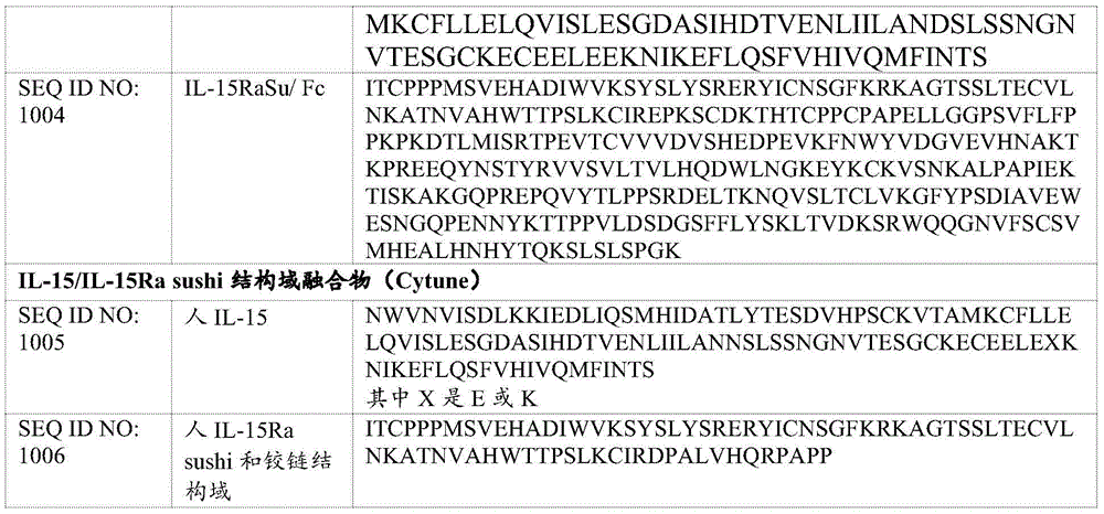
CTLA-4 inhibitors
In one aspect of the invention, the IL-1 β inhibitor or functional fragment thereof is administered together with a CTLA-4 inhibitor. In some embodiments, the CTLA-4 inhibitor is an anti-CTLA-4 antibody or fragment thereof. Exemplary anti-CTLA-4 antibodies include Tremelimumab (Tremelimumab) (formerly tijimumab, CP-675,206); and epilinolimumab (MDX-010, )。
)。
in one embodiment, the present invention provides an IL-1 β antibody or a functional fragment thereof (e.g., canargiunumab or gavagizumab) for use in the treatment of a cancer having at least a partial inflammatory basis, such as a lung cancer, in particular NSCLC, wherein said IL-1 β antibody or functional fragment thereof is administered in combination with one or more chemotherapeutic agents, wherein said one or more chemotherapeutic agents is a checkpoint inhibitor, preferably selected from the group consisting of: nivolumab, lanolizumab, alemtuzumab, aviluzumab, duvulizumab, PDR-001 (sibadazumab), and epilizumab. In one embodiment, the one or more chemotherapeutic agents is a PD-1 or PD-L-1 inhibitor, preferably selected from the group consisting of: nivolumab, lanolinuzumab, altriluzumab, aviluzumab, doluzumab, PDR-001 (sibradizumab), further preferably lanolinuzumab. In another embodiment, the IL-1 β antibody or functional fragment thereof is administered concurrently with the inhibitor of PD-1 or PD-L1.
In one embodiment, the patient's cancer has high PD-L1 expression. Generally, high PD-L1 expression is defined as a Tumor Proportion Score (TPS) equal to or greater than about 50%, as determined by FDA approved testing.
In one embodiment, the patient has a tumor with high PD-L1 expression [ Tumor Proportion Score (TPS) ≧ 50% ], with or without EGFR or ALK genomic tumor abnormalities as determined by FDA-approved testing. In one embodiment, the patient has a tumor with PD-L1 expression (TPS ≧ 1%) as determined by an FDA-approved test.
The term "in combination with … …" is to be understood as the administration of two or more drugs, either subsequently or simultaneously. Alternatively, the term "in combination with … …" should be understood as administering two or more drugs in a manner that contemplates overlapping effective therapeutic concentrations of the drugs over a substantial period of time in a patient. The drug of the invention and one or more combination partners (e.g. another drug, also referred to as "therapeutic agent" or "co-agent") may be administered independently at the same time or separately within time intervals, especially where these time intervals allow the combination partners to show a synergistic (e.g. synergistic) effect. The terms "co-administration" or "combined use" or "combined administration" and the like as used herein are intended to encompass the administration of selected combination partners to a single subject (e.g., patient) in need thereof, and are intended to include treatment regimens in which the agents are not necessarily administered by the same route of administration or simultaneously. The medicaments are administered to the patient simultaneously, concurrently or sequentially, as separate entities, without specific time constraints, wherein such administration provides therapeutically effective levels of both compounds in the patient's body, and the treatment regimen will provide the beneficial effects of the combination of medicaments in treating the conditions or disorders described herein. The latter also applies to mixture therapy, for example the administration of three or more active ingredients.
Applications, formulations and devices
The canarginoumab may be administered intravenously or preferably subcutaneously. Unless in the examples in which the route of administration is specified, both routes of administration are applicable to each of the canarginoumab-related examples disclosed herein.
The gavagizumab may be administered subcutaneously or preferably intravenously. Unless in the examples in which the route of administration is specified, both routes of administration are applicable to each of the grivoglizumab-related examples disclosed herein.
Canarginoumab can be prepared as a drug for reconstitution in lyophilized form. In one embodiment, the canargimumab is provided in lyophilized form for reconstitution, each vial containing at least about 200mg of drug, preferably no more than about 250mg, preferably no more than about 225mg, in one vial.
In one aspect, the invention provides a canargiunumab or gavagizumab for use in the treatment and/or prevention of a cancer in a patient in need thereof, said treatment and/or prevention comprising administering a therapeutically effective amount to the patient, wherein the cancer has at least a partial basis for inflammation, and wherein the canargiunumab or gavagizumab is administered by pre-filled syringe or auto-injector. Preferably, the prefilled syringe or auto-injector contains the entire amount of the therapeutically effective amount of the drug. Preferably, the prefilled syringe or automatic injector contains about 200mg of canargimumab.
Efficacy and safety
Because of its good safety profile, canargizumab or gavaglizumab can be administered to a patient for a long period of time, thereby providing and maintaining the benefits of inhibiting IL-1 β -mediated inflammation. Furthermore, due to their anti-cancer effects, whether monotherapy or in combination with one or more therapeutic agents, the life of the patient may be extended as compared to the case without treatment of the present invention, including but not limited to extending the duration of DFS, PFS, OS, reducing risk. As used herein, the term "treatment of the invention" refers to a medicament of the invention administered according to a dosing regimen as taught herein, which is suitably canargizumab or gavaglizumab. Preferably, clinical efficacy is achieved by administration at a dose of about 200mg of canarginoumab about every 3 weeks or about every month, preferably for at least about 6 months, preferably at least about 12 months, preferably at least about 24 months, preferably up to about 2 years, preferably up to about 3 years. Preferably, results are achieved by administration at a dose of about 30mg to 120mg of gemfibrozumab every 3 weeks or about monthly, preferably for at least about 6 months, preferably at least about 12 months, preferably at least about 24 months, preferably up to about 2 years, preferably up to about 3 years. In one embodiment, the treatment of the invention is a monotherapy. In one embodiment, the treatment of the present invention is added on top of SoC treatment for cancer indications. Although SoC treatment progresses over time, SoC treatment as used herein is understood to not include the drug of the present invention.
Accordingly, in one aspect, the invention provides an IL-1 β binding antibody or functional fragment thereof (suitably canargizumab or gavagezumab) for use in the treatment of MDS in a patient, wherein a therapeutically effective amount of the IL-1 β binding antibody or functional fragment thereof is administered to the patient for at least about 6 months, preferably at least about 12 months, preferably at least about 24 months.
In one aspect, the invention provides an IL-1 β binding antibody or a functional fragment thereof (suitably canargiunumab or gavagizumab) for use in the treatment of MDS in a patient, wherein preferably the risk of cancer death in said patient is reduced by at least about 10%, at least about 20%, at least about 30%, at least about 40% or at least about 50% compared to not receiving treatment according to the invention.
The term "not receiving the treatment of the invention" as used throughout the application includes patients not receiving any drug at all and patients receiving only the treatment then considered as SoC and not receiving the drug of the invention. As will be appreciated by those skilled in the art, clinical efficacy is typically not measured in the same patient receiving or not receiving treatment of the present invention, but rather in the context of clinical trials in the treatment and placebo groups.
In one embodiment, the overall survival (OS, defined as the time from the date of randomization to the date of death due to any cause) of the patient is extended by at least about one month, at least about 3 months, at least about 6 months, at least about 12 months as compared to not receiving treatment of the present invention. In one embodiment, OS is extended for at least about 12 months, preferably at least about 24 months, in an adjuvant therapy setting. In one embodiment, OS is extended for at least about 4 months, preferably at least about 6 months, or at least about 12 months in a first line therapy setting. In one embodiment, OS is extended for at least about one month, at least about 3 months, or preferably at least about 6 months in a two/three-wire therapy setting.
In one embodiment, the overall survival of a patient receiving treatment of the invention is at least about 2 years, at least about 3 years, at least about 5 years, at least about 8 years, or at least about 10 years in an adjuvant therapy setting. In one embodiment, in a first line therapy setting, the overall survival of a patient receiving treatment of the invention is at least about 6 months, at least about one year, or at least about 3 years. In one embodiment, the overall survival of a patient receiving treatment of the invention is at least about 3 months, at least about 6 months, or at least about one year in a two/three-wire therapy setting.
In one embodiment, Progression Free Survival (PFS) of a patient receiving treatment of the invention is preferably extended by at least about one month, at least about 2 months, at least about 3 months, at least about 6 months, or at least about 12 months, as compared to not receiving treatment of the invention. In one embodiment, PFS is extended by at least about 6 months, preferably at least about 12 months, in a first line treatment setting. In one embodiment, PFS is extended for at least about one month, at least about 3 months, or at least about 6 months in a second line therapy setting.
In one embodiment, a patient receiving treatment of the invention has a progression-free survival of at least about 3 months, at least about 6 months, at least about 12 months, or at least about 24 months.
Clinical efficacy can be demonstrated generally in clinical trials comparing treatment and placebo groups, including but not limited to DFS, PFS, HR reduction, OS. In the placebo group, patients received no drug at all or treatment on SoC. In the treatment group, patients received the drug of the present invention as monotherapy or added it to SoC treatment. Alternatively, in the placebo group, the patient received SoC treatment and in the treatment group, the patient received the medicament of the invention.
Even if clinical outcomes (e.g. duration of DFS or HR reduction in cancer mortality) are described as numbers based on statistical analysis of clinical trials, one of ordinary skill in the art would readily extrapolate these statistical data to treatment for individual patients as claimed, since it is expected that the drug of the invention will achieve similar clinical outcomes in a fraction of individual patients receiving treatment of the invention, e.g. in about 95% of patients, when clinical trials show statistical significance (p ≦ 0.05); or for example in about 50% of patients, when the clinical trial provides an average value, for example an average PFS of about 24 months. IL-1 beta blockade may affect the immune system of a patient in combating infection. Thus, in one aspect, the invention provides an IL-1 β binding antibody or a functional fragment thereof (suitably canargizumab or gavagizumab) for use in the treatment and/or prevention of cancer (e.g. a cancer having at least a partial basis of inflammation), wherein the patient is not at high risk of developing a severe infection as a result of the treatment of the invention. Patients will be at high risk of developing serious infections due to the treatment of the present invention in the following cases, but it is not limited to these cases: (a) patients suffer from active infections that require medical intervention. The term "active infection requiring medical intervention" is understood to mean any antiviral and/or any antibacterial agent that a patient is currently taking or has taken or has just completed taking for less than about one month or less than about two weeks; (b) the patient has latent tuberculosis and/or has a history of tuberculosis.
To control the inhibitory effect of IL-1 beta blockade on the immune system, care should be taken not to bind IL-1 betaThe antibody or functional fragment thereof is administered concomitantly with a TNF inhibitor. Preferably, the TNF inhibitor is selected from the group consisting of: (etanercept) and (etanercept),
 (adalimumab)),
 (infliximab)), (infliximab)), (iii) a pharmaceutically acceptable salt thereof, and (iv) a pharmaceutically acceptable carrier,
 (golimumab) and
 (certolizumab pegol). It is also noted that the IL-1 β binding antibody or functional fragment thereof is not administered simultaneously with another IL-1 blocker, wherein preferably said IL-1 blocker is selected from the group consisting of:
 (anakinra) and
 (rilonacept). Furthermore, only one IL-1 β binding antibody or functional fragment thereof is administered in the treatment/prevention of cancer. For example, canargiunumab is not administered in combination with gavojizumab.
When canarginoumab is administered to a patient, it is possible that some patients will develop anti-canarginoumab antibodies (anti-drug antibodies, ADA) which need to be monitored for safety and efficacy reasons. In one aspect, the invention provides canarginoumab for treating and/or preventing cancer (e.g., a cancer having at least a partial basis for inflammation), wherein the patient has a likelihood of developing ADA of less than about 1%, less than about 0.7%, less than about 0.5%, less than about 0.4%. In one embodiment, the antibody is detected by the method described in example 10. In one embodiment, the antibody detection is performed at about 3 months, about 6 months, or about 12 months after the first administration of canarginoumab.
Cancer to be treated according to the invention
In one aspect, the invention provides an IL-1 β binding antibody or a functional fragment thereof (suitably gavojizumab or suitably canajirimumab) alone or in combination with one or more therapeutic agents for use in the treatment of cancer (e.g. a cancer with at least a partial basis for inflammation), wherein the cancer comprises myelodysplastic syndrome (MDS) (suitably low risk MDS), or wherein the cancer comprises other myelomas such as chronic myelomonocytic leukemia (CMML), myeloproliferative neoplasm (MPN) and Multiple Myeloma (MM).
In one aspect, the invention provides an IL-1 β binding antibody or a functional fragment thereof (suitably gavojizumab or suitably canajirimumab) alone or in combination with one or more therapeutic agents for use in the treatment of myelodysplastic syndrome (MDS) (suitably low risk MDS). In one embodiment, the invention provides an IL-1 β binding antibody or functional fragment thereof (suitably gavojizumab or suitably canarginoumab) alone or in combination with one or more therapeutic agents for use in the treatment of anemia (suitably anemia in low risk MDS) in myelodysplastic syndrome (MDS).
Myelodysplastic syndrome (MDS) is a group of cancers characterized by impaired peripheral hematopoiesis (cytopenias) and, most commonly, myelodysplastic, abnormally represented bone marrow. MDS is a disease of hematopoietic stem cells. They are characterized by differentiation and maturation disorders, and by bone marrow stromal alterations. Diagnostic criteria have been set to diagnose MDS: 2 classification systems (French-American-British) [ FAB ] and World Health Organization [ WHO ]) and several Prognostic Scoring systems, the most common being the International Prognostic Scoring System (IPSS) (Nimer, Blood [ Blood ],2008, Germing et al, Dtsch Arztebl Int. [ German International journal of medicine ] 2013). Also, a modified version of IPSS is known as the Revised International Prognostic Scoring System (IPSS-R) for myelodysplastic syndrome. It was developed by the MDS prognostic International Working Group (IWG) for Prognosis in MDS. It can be used by the IPSS-R calculator on the https:// www.mds-foundation.
The IWG also defines response criteria to normalize response assessments for clinical decision making and comparison of clinical trial data across studies. One of these response criteria is hemologically improved hemoglobin (HI-E). The response criteria were recently revised (Platzbecker et al, Blood [ Blood ] (2019) 133(10):1020- > 1030). Infusion dependence and hemoglobin level are parameters of the HI-E response.
WHO currently classifies myelodysplastic syndrome (MDS) as shown in the following table.
Table 11: the WHO myelodysplastic syndrome Classification (Brunning et al and Orazi et al: in WHO classification of tumors of neoplastic and lymphoid tissue [ WHO hematopoietic and lymphoid tissue Classification ], 2008)
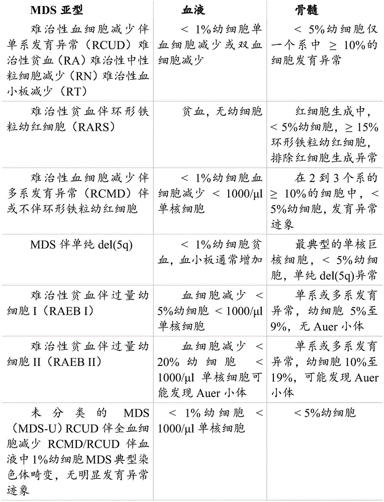
The term "myelodysplastic syndrome" or "MDS" includes three groups of patients, depending on the assessment of the risk of the patient, the level of anemia, and the presence of a del (5q) chromosomal or cytogenetic abnormality: "Low-risk patients who do not have a del (5q) chromosomal/cytogenetic abnormality and who have Epo <500 mU/mL", "Low-risk patients who do not have a del (5q) chromosomal/cytogenetic abnormality and who have Epo >500 mU/mL", and "high-risk patients". Patient risk levels are quantified using the international prognosis scoring system (IPSS and revised IPSS-R) and/or the WHO Prognosis Scoring System (WPSS). Low risk is defined as: IPSS low, medium-1; IPSS-R is extremely low, medium; or WPSS very low, medium. The higher risk is defined as: IPSS medium-2, high; IPSS-R is medium, high and extremely high; or high and extremely high WPSS. Additional genetic biomarkers can be used to identify patients in a low risk category that would benefit from treatment that is typically only performed on high risk patients.
In one embodiment, MDS patients are infusion-dependent.
In one embodiment, a subject with MDS has anemia.
Since chronic inflammation has been implicated in the development of MDS (Barreyro et al, Blood [ Blood ]2018, Basiorka et al, Blood [ Blood ] 2016; 128(25): 2960-.
Since IL-1 β has a direct implication for inhibiting erythropoietin expression (Cluzeau et al, Haematologica [ hematology ] 2017; 102(12): 2015-.
In one embodiment, the invention provides a medicament of the invention (preferably canargizumab or gavaglizumab) for use in the treatment of MDS, wherein the medicament of the invention is administered in combination with one or more therapeutic agents.
In one embodiment, the one or more therapeutic agents are selected from the following: erythropoiesis Stimulating Agents (ESAs) including erythropoietin, epoetin α, epoetin β, epoetin Ω, epoetin δ, epoetin ζ, epoetin θ, epoetin α, methoxypolyethylene glycol-epoetin β; granulocyte colony stimulating factor (G-CSF); lenalidomide; azacitidine (AzaC); decitabine; thrombopoietin receptor agonists (TPOs) including avandrotron (avatrompag), eltrombopag (eltrombopag), lusulbactoqiong (lusutrombopag), promegapoetin, romidepsin, thrombopoietin; and chemotherapeutic agents suitable for potentiating induction chemotherapy. In one embodiment, the one or more chemotherapeutic agents is apilix (alpelisib). The abacteriol is administered in a therapeutically effective amount of about 300mg per day. In one embodiment, the one or more chemotherapeutic agents is eltrombopag. Eltepopapa is administered in a therapeutically effective amount of about 75mg per day.
Depending on the condition of the patient, one, two or three therapeutic agents may be selected from the above list for combination with the medicament of the invention.
In one embodiment, the one or more therapeutic agents are standard of care (SoC) agents for MDS. In a preferred embodiment, the one or more therapeutic agents is AzaC. In a preferred embodiment, the therapeutic agent or agents is decitabine. In one embodiment, the one or more therapeutic agents is lenalidomide. In one embodiment, the one or more therapeutic agents are ESAs with or without G-CSF.
In one embodiment, the one or more therapeutic agents is an HDM2-p53 interaction inhibitor, such as (S) -5- (5-chloro-1-methyl-2-oxo-1, 2-dihydropyridin-3-yl) -6- (4-chloro-phenyl) -2- (2, 4-dimethoxy-pyrimidin-5-yl) -1-isopropyl-5, 6-dihydro-1H-pyrrolo [3,4-d ] imidazol-4-one (HDM201, WO 2013/111105, example 102) or a pharmaceutically acceptable non-covalent derivative thereof (including salts, solvates, hydrates, complexes, co-crystals), preferably a succinic acid derivative such as a succinic acid co-crystal (e.g., form B, prepared according to method D in WO 2013/111105, page 392).
In one embodiment, the agents of the invention are used in the treatment of MDS in combination with immunosuppressive therapy or hematopoietic cell transplantation.
In one embodiment, the agents of the invention are used in the treatment of MDS in combination with one or more therapeutic agents, further in combination with immunosuppressive therapy or hematopoietic cell transplantation. Immunosuppressive therapy may be performed using anti-thymocyte globulin (ATG) with or without cyclosporine.
While the patient is waiting for a suitable hematopoietic cell transplant donor, canargizumab or gavaglizumab may be used in combination with or as a potential combination partner for AzaC or decitabine in patients eligible for or who are not eligible for intensive induction chemotherapy.
In one embodiment, a medicament of the invention (preferably canargizumab or gavaglus) is used in combination with MBG 453.
In one embodiment, the medicament of the invention (preferably canargizumab or gavagetazumab) is used in combination with ruxsacket (luspatercept).
In one embodiment, a medicament of the invention (preferably canargizumab or gavaglizumab) is used alone or, preferably, in combination with one or more therapeutic agents for the first line treatment of MDS. In one embodiment, the one or more therapeutic agents are therapeutic agents for use as first line therapy selected from the group consisting of: ESA including erythropoietin, epoetin alpha, epoetin beta, epoetin omega, epoetin delta, epoetin zeta, epoetin theta, epoetin alpha, methoxypolyethylene glycol-epoetin beta; G-CSF; AzAC; decitabine; or lenalidomide. In one embodiment, the one or more therapeutic agents are ESAs with or without G-CSF. In one embodiment, the one or more therapeutic agents is AzaC, decitabine, or lenalidomide. In one embodiment, immunosuppressive therapy or hematopoietic cell transplantation is administered in place of or in addition to one or more therapeutic agents.
Preferably, the agents of the invention are used in combination with one or more therapeutic agents with SoC agents approved as first line treatment of MDS, such as ESA (including erythropoietin, epoetin alpha, epoetin beta, epoetin omega, epoetin delta, epoetin zeta, epoetin theta, dyepoetin alpha, methoxypolyethylene glycol-epoetin beta), or AzaC, decitabine, or lenalidomide, with or without G-CSF.
In one embodiment, a medicament of the invention (preferably canargizumab or gavaglizumab) is used alone or, preferably, in combination with one or more therapeutic agents for two-or three-line treatment of MDS. In one embodiment, the one or more therapeutic agents is ESA + lenalidomide with or without G-CSF. In one embodiment, the one or more therapeutic agents is TPO.
In one embodiment, the medicament of the invention (preferably canargizumab or gavaglizumab) is used as a second line treatment of MDS after use of: ESA (including erythropoietin, epoetin alpha, epoetin beta, epoetin omega, epoetin delta, epoetin zeta, epoetin theta, bepotetin alpha, methoxypolyethylene glycol-epoetin beta); G-CSF; AzAC; decitabine; lenalidomide; or ruxpiprep.
In one embodiment, the medicament of the invention (preferably canargizumab or gavaglizumab) is used as a second line treatment of MDS following treatment with lux patupip.
All uses disclosed throughout this application, including but not limited to dosages and dosing regimens, combinations, routes of administration, and biomarkers, can be used to treat MDS.
In the specification, the words "a" and "an" are generally defined in the specification as "at least one" or "one or more".
The term "patient" refers to a human patient.
The following examples illustrate the above invention; however, these examples are not intended to limit the scope of the present invention in any way.
Examples of the invention
The following examples are intended to aid in the understanding of the present invention, but are not intended to, and should not be construed to, limit its scope in any way.
Example 1
Tumor-derived IL-1 beta induces different mechanisms for promoting tumor metastasis
Materials and methods
Cell culture
Human breast cancer MDA-MB-231-Luc2-Tdtomato (Calliper Life Sciences, Manchester, UK), MDA-MB-231 (parental) MCF7, T47D (European authoritative cell culture Collection (ECACC)), MDA-MB-231-IV (Nutter et al, 2014), and bone marrow HS5(ECACC) and human primary osteoblasts OB1 were cultured in DMEM + 10% FCS (Gibco, Invitrogen, Pesley, UK). All cell lines were cultured in a humidified incubator at 5% C02 and used at low passage rates > 20.
Transfection of tumor cells
Human MDA-MB-231, MCF7 and T47D cells were stably transfected with plasmid DNA purified from competent E.coli (which had been transduced with ORF plasmids containing human IL1B or IL1R1 (accession numbers NM-000576 and NM-0008777.2, respectively) with C-terminal GFP tags (OriGene Technologies Inc., Rokville, Md.) to overexpress gene IL1B or IL1R 1. Using PureLinkTMPlasmid DNA purification was performed with the HiPure plasmid miniprep kit (themoseher) and DNA was quantified by UV spectroscopy and then introduced into human cells with the aid of Lipofectamine II. Control cells were transfected with DNA isolated from the same plasmid without the IL-1B or IL-1R1 coding sequence.
In vitro study
In vitro studies were carried out with or without the addition of 0-5ng/ml recombinant IL-1 β (R & D systems, Wisbardon, Germany) +/-50 μ M IL-1Ra (Anne (Amgen), Cambridge, UK).
Cells were transferred to fresh medium containing 10% or 1% FCS. By using 1/400mm2Cell proliferation was monitored by a hemocytometer (Hawkley, lanning UK) by manual cell counting for 120 hours every 24 hours or 72 hours using an xcelligene RTCA DP instrument (ace Biosciences, Inc). Invasion of tumor cells was assessed using a 6mm clear well plate (Corning Inc.) with a pore size of 8 μm with or without a basement membrane (20% Matrigel; invitrogen). Tumor cells were plated at 2.5X 10 in DMEM + 1% FCS5(for parental and MDA-MB-231 derivatives) and 5x 10⁵(for T47D) density into the inner chamber, and will be supplemented with 5% FCS 5x 10⁵OB1 osteoblasts were added to the outer chamber. At 24 and 48 hours post-inoculation, cells were removed from the apical surface of the membrane and passed through hematoxylin and eosin (H)&E) Cells that had invaded the wells were stained and then imaged on a Leica DM7900 light microscope and counted manually.
Cell migration was studied by analyzing wound closure: cells were seeded onto 0.2% gelatin in 6-well tissue culture plates (Costar; Corning), and once confluent, mitomycin C was added at 10. mu.g/ml to inhibit cell proliferation and scored 50 μm on a monolayer. The percentage of wound closure was measured at 24 and 48 hours using a CTR7000 inverted microscope and LAS-AF v2.1.1 software (lycra application suite; lycra Microsystems (Leica Microsystems), westerlar, germany). All proliferation, invasion and migration experiments were repeated using an xcelligene RTCA DP instrument and RCTA software (ests biotechnology).
For co-culture studies with human bone, 5x 10⁵MDA-MB-231 or T47D cells were seeded onto tissue culture plastic or 0.5cm3Human pelvic bone for 24 hours. The medium was removed and analyzed for IL-1. beta. concentration by ELISA. For co-culture with HS5 or OB1 cells, 1X 10 cells were cultured5MDA-MB-231 or T47D cells with 2x 10⁵HS5 or OB1 cells were cultured together on plastic. Cells were sorted by FACS 24 hours later, counted and lysed to analyze IL-1 β concentration. Cells were collected every 24 hours, sorted and counted for a total of 120 hours.
Animal(s) production
Experiments with human bone grafts were performed in ten-week-old female NOD SCID mice. In the IL-1. beta./IL-1R 1 overexpression bone homing experiments, 6 to 8 weeks old female BALB/c nude mice were used. To investigate the effect of IL-1. beta. on the bone microenvironment, 10-week-old female C57BL/6 mice (Charles River, Kent, UK) or IL-1R1 were used-/-Mice (Abdulai et al, 2016). Mice were maintained in a 12h light/dark cycle, with free access to food and water. Experiments were conducted with approval from the british department of internal medicine according to project permit 40/3531 of the university of sheffield, uk.
Patient consent and preparation of the pelvic disc
All patients provided written informed consent prior to participation in the study. Human bone samples were collected according to HTA permit 12182 of the musculoskeletal bio-bank, university of sheffield, uk. Trabecular bone cores were prepared from femoral heads of female patients undergoing hip replacement surgery using an Isomat 4000 precision saw (standard instruments) with a precision diamond wafer saw blade (standard instruments (Buehler)). Subsequently, a disc of 5mm diameter was cut using trephine and then stored in sterile PBS at room temperature.
In vivo studies
To mimic the transfer of human breast cancer to a human bone implant, two human bone plates were implanted subcutaneously into 10-week-old female NOD SCID mice (n-10/group) under isoflurane anesthesia. Mice received a 0.003mg injection of vetgessic and Septrin was added to drinking water for 1 week after bone implantation. Mice were left for 4 weeks and then injected in two post-mammary fat pads at 1x 10 in 20% Martigel/79% PBS/1% toluene blue5MDA-MB-231Luc2-Tdtomato, MCF7 Luc2 or T47D Luc2 cells. The development of primary tumors growth and metastasis was monitored weekly after subcutaneous injection of 30mg/ml D-fluorescein (invitrogen), using the IVIS (luminel) system (Caliper Life Sciences). After the experiment was completed, breast tumors, circulating tumor cells, serum and bone metastases were excised. RNA was processed by real-time PCR for downstream analysis, and cell lysates were used for protein analysis and histological examination of the whole tissue as described previously (Nutter et al, 2014; Ottewell et al, 2014 a).
For therapeutic studies in NOD SCID mice, placebo (control), 1mg/kg IL-1Ra (anakinra) was administered starting 7 days after tumor cell injection ) (daily) or 10mg/kg of Canatkinumab (subcutaneous every 14 days). In BALB/C mice and C57BL/6 mice, 1mg/kg IL-1Ra was administered daily for 21 or 31 days, or 10mg/kg canajirimumab was administered as a single subcutaneous injection. Tumor cells, serum and bone were subsequently excised for downstream analysis.
Mixing 5x 10⁵MDA-MB-231GFP (control), MDA-MB-231-IV, MDA-MB-231-IL-1B positive or MDA-MB-231-IL-1R1 positive cellsBone metastases were studied after injection into the lateral tail vein of 6 to 8 week old female BALB/c nude mice (n-12/group). Tumor growth in bones and lungs was monitored weekly in live animals by GFP imaging. Mice were sorted 28 days after tumor cell injection, at which time hind limbs, lungs, and serum were excised and subjected to microcomputerized tomography (μ CT), histology of bone turnover markers and circulating cytokines, and ELISA analysis (Holen et al, 2016).
Isolation of circulating tumor cells
Whole blood was centrifuged at 10,000g for 5 minutes, and then serum was removed for ELISA analysis. The cell pellet was resuspended in 5ml FSM lysis solution (Sigma Aldrich, Pull (Pool), UK) to lyse red blood cells. The remaining cells were re-pelleted, washed 3 times in PBS and resuspended in PBS/10% FCS. Samples of 10 mice per group were collected and TdTimato positive tumor cells were isolated using a MoFlow high efficiency cell sorter (Beckman Coulter, Cambridge, UK) with 470nM laser line from Coherent I-90C permanent argon ion (Coherent, Santa Clara, Calif.). TdTimato fluorescence was detected with a 555LP dichroic long pass and 580/30nm band pass filter. Cell collection and analysis was performed using Summit 4.3 software. Immediately after sorting, the cells were placed in RNA protective cell reagent (Ambion, Persley, Renfree, UK) and stored at-80 ℃ before RNA extraction. To count the number of circulating tumor cells, TdTomato fluorescence was detected using a 561nm laser and YL1-a filter (585/16 emission filter). The collection and analysis of cells was performed using Attune NxT software.
Microcomputer tomography imaging
A microcomputer tomography (μ CT) analysis was performed using a Skyscan 1172X-ray computer μ CT scanner (Skyscan, Aartsell, Belgium) equipped with an X-ray tube (voltage 49 kV; current 200uA) and a 0.5-mm aluminum filter. The pixel size was set to 5.86 μm, as previously described (Ottewell et al, 2008 a; Ottewell et al, 2008b) scanning from the proximal top of the tibia.
Bone histology and measurement of tumor volume
Bone tumor area was measured on three non-serial, H & E stained, 5 μm decalcified tibial histological sections of each mouse using a leica RMRB upright microscope and an osteorecord software (osteorecords, inc., Decauter, usa) and a computer image analysis system as previously described (Ottewell et al, 2008 a).
Western blot
Proteins were extracted using a mammalian cell lysis kit (sigma aldrich, Poole, uk). 30 μ g of protein was run on a 4% -15% pre-made polyacrylamide gel (BioRad, Wattford, UK) and then transferred to Immobilon nitrocellulose membrane (Millipore Corp.). Nonspecific binding was blocked with 1% casein (Vector Laboratories), and then incubated with either rabbit anti-human N-cadherin (D4R1H) monoclonal antibody (1:1000 dilution), E-cadherin (24E10) (1:500 dilution) or gamma-catenin (2303) (1:500 dilution) (Cell signalling) or mouse monoclonal GAPDH (ab8245) (1:1000 dilution) (Abcam, Cambridge, UK) for 16 hours at 4 ℃. The secondary antibody was anti-rabbit or anti-mouse horseradish peroxidase (HRP; 1:15,000), and HRP was detected using the Supersignal chemiluminescence detection kit (Pierce). Quantification of bands was performed using Quantity Once software (BioRad) and normalized to GAPDH.
Gene analysis
Total RNA was extracted using RNeasy kit (Qiagen) and reverse transcribed to cDNA using Superscript III (Invitrogen AB). Relative mRNA expression of IL-1B (Hs02786624), IL-1R1(Hs00174097), CASP (caspase 1) (Hs00354836), IL1RN (Hs00893626), JUP (crosslinked zebulin/γ -catenin) (Hs00984034), N-cadherin (Hs01566408), and E-cadherin (Hs1013933) was compared to the housekeeping gene glyceraldehyde-3-phosphate dehydrogenase (GAPDH; Hs02786624) and evaluated using the ABI 7900PCR system (Perkin Elmer, Foster City, Calif.) and Taqman Universal premix (Muscae Feishi, UK). Fold changes in gene expression between treatment groups were analyzed by inserting CT values into Data Assist V3.01 software (Applied Biosystems), and only genes with CT values ≦ 25 were analyzed for gene expression changes.
Evaluation of IL-1 beta and IL-1R1 in tumors of breast cancer patients
The expression of IL-1 β and IL-1R1 was evaluated on Tissue Microarrays (TMAs) containing a core of primary breast tumors obtained from 1,300 patients included in the azere clinical trial (Coleman et al, 2011). Samples were taken from patients with stage II and III breast cancer with no evidence of metastasis and pre-treated. Subsequently, patients were randomized to receive standard adjuvant therapy with or without addition of zoledronic acid for 10 years (Coleman et al, 2011). IL-1 β (ab2105, 1:200 dilution, Abcam company) and IL-1R1(ab59995, 1:25 dilution, Abcam company) were stained for TMA and blinded assessments of IL-1 β/IL-1R1 in tumor cells or associated stroma were performed under the direction of a histopathologist. Tumor or interstitial IL-1 β or IL-1R1 is then linked to disease recurrence (anywhere) or especially in bone (+/-other sites).
The IL-1 β pathway is upregulated during metastasis of human breast cancer to human bone.
A mouse model of spontaneous human breast cancer metastasis to a human bone implant was used to investigate how the IL-1 β pathway changes at different stages of metastasis. Using this model, the expression levels of genes associated with the IL-1 β pathway were increased stepwise at each stage of the process of heterometastasis in triple negative (MDA-MB-231) and estrogen receptor positive (ER + ve) (T47D) breast cancer cells: genes associated with the IL-1 β signaling pathway (IL-1B, IL-1R1, CASP (caspase 1), and IL-1Ra) were expressed at very low levels in MDA-MB-231 and T47D cells in vitro, and expression of these genes was not altered in primary breast tumors of the same cells that did not metastasize in vivo (FIG. 1 a).
Both IL-1B, IL-1R1 and CASP were significantly increased in breast tumors that subsequently metastasized to human bone (p <0.01 for both cell lines) compared to non-metastatic breast tumors, activating IL-1 β signaling as shown by ELISA against activated 17kD IL-1 β (FIG. 1 b; FIG. 2). IL-1B gene expression was increased in circulating tumor cells compared to metastatic breast tumors (p <0.01 for both cell lines) and IL-1B (p <0.001), IL-1R1(p <0.01), CASP (p <0.001), and IL-1Ra (p <0.01) were further increased in tumor cells isolated from human bone metastases compared to their corresponding breast tumors, resulting in further activation of IL-1 β protein (FIG. 1; FIG. 2). These data indicate that IL-1 β signaling can promote both metastasis from the primary site and the development of breast cancer metastasis in bone.
Tumor-derived IL-1 β promotes EMT and breast cancer metastasis.
In primary tumors metastasized to bone, the expression levels of genes associated with tumor cell adhesion and epithelial to mesenchymal transition (EMT) were significantly altered compared to non-metastasized tumors (fig. 1 c). IL-1 β overexpressing cells (MDA-MB-231-IL-1B +, T47D-IL-1B + and MCF7-IL-1B +) were generated to investigate whether tumor-derived IL-1 β was responsible for inducing EMT and metastasis to bone. All IL-1 β + cell lines showed an increase in EMT, a morphological change from epithelial to mesenchymal phenotype (fig. 3a), as well as a decrease in the expression of E-cadherin and JUP (cross-linked plakoglobin/γ -catenin), and an increase in the expression of N-cadherin genes and proteins (fig. 3 b). Wound closure (p <0.0001 in MDA-MB-231-IL-1. beta. + (FIG. 3 d); p <0.001MCF 7-IL-1. beta. +, and T47D-IL-1. beta. +) and increased migration and invasion into osteoblasts by matrigel in tumor cells with increased IL-1. beta. signaling compared to the respective control group (MDA-MB-231-IL-1. beta. + (FIG. 3c) p < 0.0001; MCF 7-IL-1. beta. +, and T47D-IL-1. beta. + p < 0.001). Increased IL-1 β production in ER-positive and ER-negative breast cancer cells, which spontaneously metastasize in vivo into human bone implants, compared to non-metastatic breast cancer cells (fig. 1). The same association between IL-1 β and metastasis was found in primary tumor samples of stage II and stage III breast cancer patients enrolled in the azere study (Coleman et al, 2011) who experienced cancer recurrence over a period of 10 years. IL-1 β expression in primary tumors of patients with azere correlates with both bone recurrence and recurrence anywhere, suggesting that the presence of this cytokine may often play a role in metastasis. Consistent with this, gene manipulation of breast cancer cells to artificially overexpress IL-1 β can increase the ability of breast cancer cells to migrate and invade in vitro (fig. 3).
Inhibition of IL-1 β signaling reduces spontaneous metastasis of human bone.
Since tumor-derived IL-1 β appears to promote the development of metastases by inducing EMT, the effect of inhibiting IL-1 β signaling with IL-1Ra (anakinra) or human anti-IL-1 β binding antibodies (canarginoumab) on spontaneous metastasis to human bone implants was investigated: both IL-1Ra and canarginoumab reduce metastasis to human bone: in 10 control mice 7 in human bone implant detected metastasis, but in IL-1Ra treatment of 10 mice only 4, in Kanaginumumab treatment of 10 mice in 1. The IL-1Ra and canarginoumab treated groups also had less bone metastasis than the control group (FIG. 4 a). The number of cells detected in the circulation of mice treated with either canarginoumab or IL-1Ra was significantly lower than the number of cells detected in the placebo treated group: only 3 tumor cells/ml were counted in whole blood from mice treated with canarginoumab and anakinra, respectively, compared to 108 tumor cells/ml in blood from placebo-treated mice (fig. 4b), indicating that inhibition of IL-1 signaling prevents shedding of tumor cells from the primary site into the circulation. Thus, inhibition of IL-1 β signaling or inhibition of IL-1R1 with the anti-IL-1 β antibody canargizumab reduces the number of breast cancer cells shed into circulation and reduces metastasis in human bone implants (fig. 4).
Tumor-derived IL-1B promotes homing and colonization of breast cancer cells.
Injection of breast cancer cells into the tail vein of mice often leads to lung metastasis because tumor cells are trapped in the pulmonary capillaries. It has previously been shown that breast cancer cells that preferentially home to the bone microenvironment after intravenous injection express high levels of IL-1 β, suggesting that this cytokine may be involved in tissue-specific homing of breast cancer cells to bone. In this study, intravenous injection of MDA-MB-231-IL-1 β + cells into BALB/c nude mice resulted in a significant increase (75%) in the number of animals undergoing bone metastasis compared to control cells (12%) (p <0.001) (FIG. 5 a). MDA-MB-231-IL-1 β + tumors caused a significantly greater development of osteolytic lesions in mouse bone compared to control cells (p 0.03; fig. 5b), and there was a tendency for reduced lung metastasis in mice injected with MDA-MB-231-IL-1 β + cells compared to control cells (p 0.16; fig. 5 c). These data suggest that endogenous IL-1 β can promote the development of metastases that tumor cells home to the bone environment and at that site.
The interaction of tumor cells with bone cells further induces IL-1B and promotes the development of significant metastases.
Genetic analysis data from a mouse model of human breast cancer metastasis to a human bone implant showed that the IL-1 β pathway was further increased when breast cancer cells were grown in the bone environment compared to metastatic cells at the primary site or in the circulation (figure 1 a). Thus, it was investigated how the production of IL-1 β changes when tumor cells are in contact with bone cells, and how IL-1 β alters the bone microenvironment to affect tumor growth (FIG. 6). Culturing human breast cancer cells into the fully human bone fragment for 48 hours resulted in increased secretion of IL-1 β into the culture medium (p <0.0001 for MDA-MB-231 and T47D cells; FIG. 6 a). Co-culture with human HS5 bone marrow cells showed increased concentrations of IL-1 β derived from cancer cells (p <0.001) and bone marrow cells (p <0.001), with approximately 1000-fold increase in IL-1 β derived from tumor cells and approximately 100-fold increase in IL-1B from HS5 cells (FIG. 6B).
Exogenous IL-1 β does not increase tumor cell proliferation even in cells that overexpress IL-1R 1. In contrast, IL-1 β stimulated proliferation of bone marrow cells, osteoblasts and blood vessels, which in turn induced proliferation of tumor cells (FIG. 6). Thus, the arrival of tumor cells expressing high concentrations of IL-1 β stimulates the expansion of metastatic microenvironment components, and contact between tumor cells expressing IL-1 β and osteoblasts/blood vessels drives tumor colonization of bone. Exogenous IL-1 β and IL-1 β from tumor cells were studied for tumor cells, osteoblasts, bone marrow cells and CD34+Effects of vascular proliferation: co-culture of HS5 bone marrow or OB1 primary osteoblasts with breast cancer cells resulted in increased proliferation of all cell types (P for HS5, MDA-MB-231 or T47D<0.001, FIG. 6c) (for OB1, MDA-MB-231 or T47D, P<0.001, fig. 6). Tumor cells, primary human bone samples, bone marrow cells or osteoblastsDirect contact between the two promotes the release of IL-1. beta. from tumor and bone cells (FIG. 6). Furthermore, administration of IL-1 β increased the proliferation of HS5 or OB1 cells, but not breast cancer cells (fig. 7a-7c), suggesting that tumor cell-bone cell interaction promotes IL-1 β production, which can drive the expansion of the microenvironment and stimulate the formation of significant metastases.
IL-1 β signaling has also been found to have profound effects on bone microvasculature: prevention of IL-1 β signaling in bone by knock-out of IL-1R1, pharmacological blockade of IL-1R with IL-1Ra or reduction of circulating concentrations of IL-1 β by administration of the anti-IL-1 β binding antibody Kanagirudoumab reduces CD34 in tumor-colonized trabecular bone+Average length of blood vessels (for IL-1Ra and Kanagirudumab-treated mice, p<0.01) (fig. 7 c). These findings were confirmed by endorphin staining, which showed a reduced number of blood vessels in bone as well as blood vessel length when IL-1 β signal was disrupted. Analysis of endothelin 1 and VEGF by ELISA showed IL-1R1 compared to control-/-Mouse (p)<0.001 endothelin 1; p is a radical of<0.001VEGF) and with IL-1R antagonists (p)<0.01 of endothelin 1; p is a radical of<0.01VEGF) or Kanagilunumab (p)<0.01 of endothelin 1; p is a radical of<0.001VEGF) were all reduced in the bone marrow of mice treated with these endothelial cell markers (fig. 8). These data indicate that increased IL-1 β associated with tumor cell-bone cells and high levels of IL-1 β in tumor cells can also promote angiogenesis, further stimulating metastasis.
Tumor-derived IL-1 beta predicts future breast cancer recurrence in bone and other organs in patient material
To establish a correlation between the results of the clinical study, the correlation between IL-1 β and its receptor IL-1R1 in patient samples was studied. Approximately 1300 primary tumor samples from stage II/III breast cancer (from AZURE study (Coleman et al, 2011)) with no evidence of metastasis were stained for IL-1R1 or the active form of IL-1 β (17kD), and the expression of these molecules in tumor cells and tumor-associated stroma were biopsied, respectively. Patients were followed for 10 years after biopsy and the multivariate Cox model was used to assess the correlation between IL-1 β/IL-1R1 expression and distant recurrence or bone recurrence. IL-1 β in tumor cells was strongly associated with distant recurrence at any site (p ═ 0.0016), recurrence in bone only (p ═ 0.017), or recurrence in bone at any time (p ═ 0.0387) (fig. 9). Patients with IL-1 β in tumor cells and IL-1R1 in tumor-associated stroma are more likely to develop distant recurrence in the future (p ═ 0.042) than patients without IL-1 β in tumor cells, suggesting that tumor-derived IL-1 β may not only directly promote metastasis, but may also interact with IL-1R1 in the stroma to promote this process. Therefore, IL-1 β is a novel biomarker that can be used to predict the risk of breast cancer recurrence.
Example 2
The Kanagilunumab PK profile and the hscRP profile for lung cancer patients were simulated.
Based on data from the CANTOS study, a model was generated to characterize the relationship between canarginous resistance Pharmacokinetics (PK) and hsCRP.
The following methods were used for this study: model construction was performed using first order condition estimation and interaction methods. The model describes the logarithm of time-resolved hsCRP as:
y(tij)=y0,i+yeff(tij)
wherein y is0,iIs a steady state value and yeff(tij) Indicating a therapeutic effect and depending on systemic exposure. The treatment effect was described using an Emax-type model,

wherein E ismax,iIs the maximum possible response at high exposure, IC50iIs the concentration at which half of the maximal response is obtained.
Respective parameters Emax,iAnd y0,iAnd IC50iIs estimated as the sum of typical values, covpar coviAnd normal distribution between subject variability. The term covpar refers to the estimated covariate effect parameter, and coviIs subject covariate iThe value is obtained. The covariates to be included are selected based on a check of the eta graph versus the covariates. The residual error is described as a combination of a proportional term and an additive term.
Log of baseline hscRP as all three parameters (E)max,i、Y0,iAnd IC50i) The covariates of (a) are included. There are no other covariates in the model. The estimation accuracy of all parameters is high. The effect of the log of baseline hsCRP on the steady state value was less than 1 (equal to 0.67). This indicates that baseline hsCRP does not scale well to steady state values and that steady state values expose a regression relative to the baseline mean. The effect of the log of baseline hsCRP on IC50 and Emax was negative. Thus, patients with high hsCRP at baseline are expected to have a low IC50 and a large maximum decrease. Typically, the model diagnostic program confirms that the model well describes the available hsCRP data.
This model was then used to model the expected hsCRP response to select different dosing regimens in a population of lung cancer patients. Bootstrap methods (bootstrapping) were applied to construct populations with prospective inclusion/exclusion criteria representing potential lung cancer patient populations. Three different lung cancer patient populations, described only by the baseline hsCRP profile, were studied: all CANTOS patients (scenario 1), confirmed lung cancer patients (scenario 2) and advanced lung cancer patients (scenario 3).
The population parameters of the model and the variability between patients were assumed to be the same in all three scenarios. The PK/PD relationship for hscRP observed throughout the cants population was assumed to represent lung cancer patients.
The estimated number is the likelihood that hsCRP will be below the critical point, which may be 2mg/L or 1.8mg/L, by the end of month 3. The median hsCRP level at the end of 3 months of the CANTOS study was 1.8 mg/L. Baseline hscRP > 2mg/L is one of the inclusion criteria, so it is worth investigating whether hscRP levels are below 2mg/L by month 3.
For the CANTOS PK data, a single chamber model with first order absorption and elimination was established. The model is expressed as an ordinary differential equation, and RxODE is used to model the time course of canargiunumab concentration given the individual PK parameters. The subcutaneous canaryitumumab dose regimen of interest was 300mg Q12W, 200mg Q3W, and 300mg Q4W. Exposure metrics (including Cmin, Cmax, AUC over different selected time periods and mean concentration at steady state Cave) were derived from simulated concentration-time curves.
The simulation in scenario 1 is based on the following information:
canatkinumab-alone exposure using RxODE simulation
PD parameter (which is y)0,i、Emax,iAnd IC50iThe components of (a): typical values (THETA (3), THETA (5), THETA (6)), covpars (THETA (4), THETA (7), THETA (8)), and inter-subject variability (ETA (1), ETA (2), ETA (3))
Baseline hsCRP from all 10,059 patients from the CANTOS study (baseline hsCRP: mean 6.18mg/L, mean Standard Error (SEM) ═ 0.10mg/L)
First generating a prediction interval for the target estimator by randomly sampling 1000 THETA (3) - (8) from a normal distribution (where the fixed mean and standard deviation are estimated from the population PK/PD model); bootstrap 2000PK exposures, PD parameters ETA (1) - (3) and baseline hsCRP for all cants patients were then performed for each THETA (3) - (8) group. The 2.5%, 50%, and 97.5% percentiles of the 1000 estimates are reported as the point estimates and the 95% prediction interval.
The simulation in scenario 2 is based on the following information:
single Kanaginumunumab PK exposure using RxODE simulation
PD parameters THETA (3) - (8) and ETA (1) - (3)
Baseline hsCRP for 116 CANTOS patients with confirmed lung cancer (baseline hsCRP: mean 9.75mg/L, SEM 1.14mg/L)
First generating a prediction interval for the target estimator by randomly sampling 1000 THETA (3) - (8) from a normal distribution (where the fixed mean and standard deviation are estimated from the population PKPD model); then for each THETA (3) - (8) group, 2000PK exposures, PD parameters ETA (1) - (3) were bootstrapped from all CANTOS patients, and 2000 baseline hsCRP was bootstrapped from 116 CANTOS patients with confirmed lung cancer. The 2.5%, 50%, and 97.5% percentiles of the 1000 estimates are reported as the point estimates and the 95% prediction interval.
In scenario 3, the point estimator and 95% prediction interval are obtained in a similar manner as scenario 2. The only difference was that 2000 baseline hsCRP values were bootstrapped from the advanced lung cancer population. In the advanced lung cancer population, no separate baseline hsCRP data is published. A usable population level estimate for advanced lung cancer is a baseline hsCRP mean of 23.94mg/L with an SEM of 1.93mg/L [ Vagulene 2011 ]. Using this estimate, the mean was adjusted to 23.94mg/L using an additive constant, and an advanced lung cancer population was derived from 116 CANTOS patients with confirmed lung cancer.
Consistent with the model, the simulated canarginoumab PK was linear. The median and 95% prediction intervals of the concentration time spectra plotted on a natural logarithmic scale for 6 months are shown in figure 10 a.
The median of 1000 estimates of the proportion of subjects with hsCRP response at month 3 and 95% prediction interval at the cut-off points of 1.8mg/L and 2mg/L hsCRP are reported in figures 10b and 10 c. From the simulation data, 200mg Q3W and 300mg Q4W performed similarly and better than 300mg Q12W (the highest dose regimen in CANTOS) for month 3 reduced hsCRP. From scenario 1 to scenario 3, for more severe lung cancer patients, the baseline hsCRP level is assumed to be higher, resulting in a lower likelihood of hsCRP going below the critical point by month 3. Figure 10d shows the median hsCRP concentration versus time for three different doses, and figure 10e shows the percentage reduction in hsCRP after a single dose relative to baseline.
Example 3
PDR001 plus canarginoumab treatment increased effector neutrophils in colorectal tumors.
RNA sequencing was used to drill down the mechanism of action of Kanagilunumab (ACZ885) in cancer. CPDR001X2102 and CPDR001X2103 clinical trials evaluated the safety, tolerability, and pharmacodynamics of gabapentin (PDR001) in combination with other therapies. For each patient, tumor biopsies were taken both before treatment and at cycle 3 of treatment. Briefly, samples are processed by RNA extraction, ribosomal RNA consumption, library construction and sequencing. Sequence reads were aligned to hg19 reference genome and Refseq reference transcriptome by STAR, gene level counts were compiled by HTSeq, and sample level normalization by edgeR using trimmed means of M values.
Figure 11 shows 21 genes that were increased on average in colorectal tumors treated with PDR001+ canargimumab (ACZ885) but not in colorectal tumors treated with PDR001+ everolimus (RAD 001). Treatment with PDR001+ canarginoumab increased RNA levels of IL1B and its receptor IL1R 2. This observation indicates that the on-target of the tumor is compensatory feeding back to increase IL1B RNA levels in response to IL-1 β protein blockade.
Notably, neutrophil-specific genes were increased in the case of PDR001+ canarginoumab, including FCGR3B, CXCR2, FFAR2, OSM, and G0S2 (shown in boxes in fig. 11). The FCGR3B gene is a neutrophil-specific isoform of the CD16 protein. The protein encoded by FCGR3B plays a key role in the secretion of reactive oxygen species in response to immune complexes, consistent with the function of effector neutrophils (Fossati G2002 Arthritis Rheum [ Arthritis & rheumatism ] 46: 1351). Chemokines that bind CXCR2 translocate neutrophils from the bone marrow and into surrounding sites. In addition, an increase in CCL3 RNA was observed when treated with PDR001+ canarginoumab. CCL3 is a chemoattractant for neutrophils (Reichel CA 2012Blood 120: 880).
In summary, this compositional contribution analysis using RNA-seq data indicates that PDR001+ canajirimumab treatment increases effector neutrophils in colorectal tumors, whereas this increase is not observed with PDR001+ everolimus treatment.
Example 4
Efficacy of canajirimonamab (ACZ885) in combination with sbatuzumab (PDR001) for the treatment of cancer.
Patient 5002 + 004 was a 56 year old male initially with stage IIC, microsatellite stable, moderately differentiated ascending colon adenocarcinoma (MSS-CRC) diagnosed in june 2012 and treated with a prior regimen.
Prior treatment regimens included:
folinic acid/5-fluorouracil/oxaliplatin, in the adjuvant case
Capecitabine chemical radiotherapy (metastatic condition)
5-fluorouracil/bevacizumab/folinic acid/irinotecan
Trifluridine and tipiracetam
Irinotecan
Oxaliplatin/5-fluorouracil
5-fluorouracil/bevacizumab/tetrahydrofolic acid
5-Fluorouracil
At the beginning of the study, patients had extensive metastatic disease, including multiple liver and bilateral lung metastases, as well as paraesophageal lymph node, retroperitoneal and peritoneal disease.
The patient was treated with PDR 001400 mg (Q4W) every four weeks plus 100mg (Q8W) ACZ885 every eight weeks. The patient had stable disease after 6 months of treatment, then had significantly reduced disease, and confirmed a partial response of RECIST to treatment at 10 months. The patient subsequently developed progressive disease and the dose was increased to 300mg and then to 600 mg.
Example 5
Calculation of the gavojizumab dose for the cancer patient was selected.
Based on available PK data for clinically effective dose-qualified Vojilizumab revealed by the CANTOS test, the dose of Gevjilizumab to treat a cancer with at least a partial inflammatory basis is selected, taking into account the following factors
Gavojizumab (IC 50 at about 2-5 pM) exhibited about 10-fold higher viral titers than canajirimumab (IC 50 at about 42 ± 3.4 pM). The highest dose of gavaglizumab of 0.3mg/kg (about 20mg) Q4W indicates that a reduction in hsCRP in type 2 diabetic patients can reduce hsCRP by up to 45% (see fig. 12 a).
Next, hsCRP exposure-response relationships were explored using pharmacological models, and clinical data were extrapolated to higher ranges. Since clinical data show a linear correlation between hsCRP concentration and kvojizumab concentration (all in log space), a linear model was used. The results are shown in fig. 12 b. Based on this simulation, a gavaglus concentration between 10000ng/mL and 25000ng/mL was optimal, as hsCRP was greatly reduced in this range, with only a reduced benefit when gavaglus concentration was higher than 15000 ng/mL. However, since hsCRP has been significantly reduced in this range, a gavaglizumab concentration between 4000ng/mL to 10000ng/mL is expected to be effective.
Clinical data indicate that, following subcutaneous administration, the pharmacokinetics of gemtuzumab ozogamicin followed a linear two-compartment model with first-order absorption. The bioavailability of gemfibrozumab was about 56% when administered subcutaneously. Multiple dose kvojizumab (SC) simulations were performed for 100mg every four weeks (see fig. 12c) and 200mg every four weeks (see fig. 12 d). Simulations indicate that a trough concentration of approximately 10700ng/mL was administered every four weeks with 100mg of Gevojizumab. The half-life of gemtuzumab ozogamicin is about 35 days. The trough concentration of 200mg of gemfibrozumab administered every four weeks was approximately 21500 ng/mL.
Example 6
Preclinical data on the efficacy of anti-IL-1 β therapy.
Kanagilunumab is an anti-IL-1 β human IgG1 antibody that cannot be directly evaluated in a mouse model of cancer because it does not cross-react with mouse IL-1 β. Mouse replacement anti-IL-1 β antibodies have been developed and used to evaluate the role of blocking IL-1 β in a mouse model of cancer. The isotype of the surrogate antibody is IgG2a, closely related to human IgG 1.
In the MC38 mouse model of colon cancer, modulation of Tumor Infiltrating Lymphocytes (TILs) was seen after one dose of anti-IL-1 β antibody (FIGS. 13a-13 c). MC38 tumors were implanted subcutaneously in the flank of C57BL/6 mice treated with a dose of either isotype antibody or anti-IL-1 β antibody when the tumors were between 100-150mm 3. Tumors were then harvested five days after the dose and processed to obtain single cell suspensions of immune cells. Cells were then stained ex vivo and analyzed by flow cytometry. After a single dose of IL-1 β blocking antibody, tumor infiltrating CD4+ T cells increased, while CD8+ T cells also increased slightly (fig. 13 a). The increase in CD8+ T cells was small, but may suggest a more active immune response in the tumor microenvironment, which the combination therapy may enhance. CD4+ T cells could be further subdivided into FoxP3+ regulatory T cells (tregs) and this subpopulation decreased upon blocking IL-1 β (fig. 13 b). In myeloid cell populations, blockade of IL-1 β resulted in a decrease in TAM2 in the neutrophil and macrophage M2 subpopulation (fig. 13 c). Both neutrophils and M2 macrophages can suppress other immune cells, such as activated T cells (Pillay et al, 2013; Hao et al, 2013; Oishi et al 2016). Both together, the reduction of tregs, neutrophils and M2 macrophages in the MC38 tumor microenvironment following IL-1 β blockade indicates that the tumor microenvironment becomes immunosuppressively weakened.
A similar trend in the decline of microenvironment immunosuppression was seen after one dose of anti-IL-1 β antibody in the LL2 mouse model for lung cancer (FIGS. 13d-13 f). LL2 tumors were implanted subcutaneously in the flank of C57BL/6 mice treated with one dose of either isotype antibody or anti-IL-1 β antibody when the tumors were between 100-150mm 3. Tumors were then harvested five days after the dose and processed to obtain single cell suspensions of immune cells. Cells were then stained ex vivo and analyzed by flow cytometry. Treg populations were reduced as assessed by expression of FoxP3 and Helios (fig. 13 d). Both FoxP3 and Helios were used as markers for regulatory T cells, and they defined different subsets of Tregs (Thornton et al, 2016). Similar to the MC38 model, both neutrophils and M2 macrophages (TAM2) were reduced after IL-1 β blockade (fig. 13 e). In addition, changes in the bone marrow-derived suppressor cell (MDSC) population following antibody treatment were also evaluated in this model. After anti-IL-1 β treatment, a reduction in the number of granulocytic or Polymorphonuclear (PMN) MDSCs was found (FIG. 13 f). MDSCs are a mixed population of cells of myeloid origin that can actively suppress T cell responses by a variety of mechanisms, including arginase production, Reactive Oxygen Species (ROS) and Nitric Oxide (NO) release (Kumar et al, 2016; Umansky et al, 2016). Likewise, a decrease in Treg, neutrophils, M2 macrophages and PMN MDSCs in the LL2 model following IL-1 β blockade indicates that the tumor microenvironment becomes immunosuppressively weakened.
TIL in the 4T1 triple negative breast cancer model also showed a trend of decreased microenvironment immunosuppression after one dose of mouse replacement with anti-IL-1 β antibody (fig. 13g-13 j). The 4T1 tumor was implanted subcutaneously in the flank of Balb/c mice, and the mice were treated with either isotype antibody or anti-IL-1. beta. antibody when the tumor was between 100-150mm 3. Tumors were then harvested five days after the dose and processed to obtain single cell suspensions of immune cells. Cells were then stained ex vivo and analyzed by flow cytometry. CD4+ T cells decreased after a single dose of anti-IL-1 β antibody (fig. 13g), whereas FoxP3+ tregs decreased in the CD4+ T cell population (fig. 13 h). In addition, both TAM2 and neutrophil numbers were reduced after treatment of tumor-bearing mice (fig. 13 i). All these data again demonstrate that blockade of IL-1 β in the 4T1 breast cancer mouse model results in a microenvironment with reduced immunosuppression. In addition, MDSC populations following antibody treatment were also evaluated in this model. Both granulocytic (PMN) MDSC and mononuclear MDSC were reduced following anti-IL-1 β treatment (FIG. 13 j). These findings, combined with changes in Treg, M2 macrophages and neutrophil populations, describe a reduction in the immunosuppressive tumor microenvironment in the 4T1 tumor model.
Although these data are from colon, lung and breast cancer models, the data can be extrapolated to other types of cancer. Even though these models are not completely associated with the same type of human cancer, the MC38 model is particularly a good surrogate model for hypermutated/MSI (microsatellite instability) colorectal cancer (CRC). According to the transcriptomic characteristics of the MC38 cell line, the four driver mutations in this cell line correspond to known hot spots in human CRC, although they are located at different positions (eframeova et al, 2018). Although this did not make the MC38 mouse model identical to human CRC, it did mean that MC38 might be a relevant model for human MSI CRC. Generally, mouse models are not always associated with the same type of cancer in humans due to the genetic differences in cancer origin in mice as compared to humans. However, the type of cancer is not always important when examining infiltrated immune cells, as immune cells are more important. In this case, since three different mouse models showed similar reductions in tumor suppressive microenvironment, blocking IL-1 β appeared to result in a decreased suppressive tumor microenvironment. The extent of the immunosuppressive changes of the various cell types (Treg, TAM, neutrophils) is reduced compared to isotype controls in various syngeneic mouse tumor models, a novel finding of IL-1 β blockade in cancer mouse models. Although inhibition of cell reduction has been previously found, multiple cell types in each model is a novel finding. Furthermore, changes in MDSC populations were seen downstream of IL-1 β in 4T1 and the lewis lung cancer (LL2) model, but finding that blockade of IL-1 β in the LL2 model could lead to a reduction in MDSCs is a new finding for this study and a mouse surrogate for canargizumab (Elkabets et al, 2010).
Even though these models are not completely associated with the same type of human cancer, the MC38 model is particularly a good surrogate model for hypermutated/MSI (microsatellite instability) colorectal cancer (CRC). According to the transcriptomic characteristics of the MC38 cell line, the four driver mutations in this cell line correspond to known hot spots in human CRC, although they are located at different positions (eframeova et al, 2018). Although this does not make the MC38 mouse model identical to human CRC, it does mean that MC38 may be a relevant model for human MSI CRC (Efremova M, et al Nature Communications 2018; 9:32)
Example 7
Preclinical data for efficacy of canargiunumab in combination with anti-PD-1 (lanolelizumab) for treatment of cancer.
A preliminary study was designed to evaluate the effect of canargizumab on tumor growth and tumor microenvironment as a monotherapy or in combination with anti-PD-1 (lanolizumab). A human NSCLC xenograft model was created by subcutaneous injection of the human lung cancer cell line H358(KRAS mutant) into the BLT mouse xenograft model.
As shown in fig. 14, the H358(KRAS mutant) model is a very fast growing and aggressive model. In this model, the combined treatment of canargiunumab and lanolizumab (shown in purple) resulted in a greater reduction than the canargiunumab single-agent group (shown in red) and lanolizumab single-agent treatment (shown in green), with a 50% reduction in the average volume of tumors observed compared to the vehicle group.
Example 8
Preclinical data for the treatment of cancer with canargiunumab in combination with docetaxel.
In a study in which anti-IL-1 β was used in combination with docetaxel in an aggressive lung model (LL2), modest efficacy was observed for anti-IL-1 β alone and docetaxel alone. The efficacy of the combination group was enhanced compared to the group used alone or the control group (fig. 15A). A reduction in immunosuppressive cells, particularly regulatory T cells and suppressor mouse bone marrow cells (including neutrophils, monocytes, and MDSCs) in tumors following IL-1 β inhibition was observed at PD time points either alone or in combination 5 days after the first dose (fig. 15B-E). These data support that the proposed mechanism of IL-1 β inhibition can be demonstrated in vivo, and some efficacy of anti-IL-1 β monotherapy has also been observed.
Example 9
Treatment of 4T1 tumors with 01BSUR and docetaxel resulted in changes in the tumor microenvironment.
Female Balb/c mice with 4T1 tumor implanted subcutaneously in the right flank, 8 and 15 days after tumor implantation, when the tumor reached about 100mm3When administered, treatment is with isotype antibody, docetaxel, 01BSUR or a combination of docetaxel and 01 BSUR. 01BSUR is a mouse surrogate antibody because canakinumab does not cross-react with murine IL-1 β.01 BSUR belongs to the mouse IgG2a subclass, corresponding to the human IgG1 subclass to which Kanagilumunumab belongs. Tumors were harvested 5 days after the first dose and analyzed for changes in the infiltrating immune cell population. This procedure was performed again at the end of the study 4 days after the second dose.
Tumor burden
A slight reduction in tumor growth was observed in the 01BSUR anti-IL-1 β treated group alone compared to the vehicle/isotype control. This delay was increased in the single agent docetaxel group. The combination group showed a similar growth slowing as docetaxel alone group (fig. 16).
TIL analysis of 4T1 tumors after single dose docetaxel and 01 BSUR-myeloid map
Neutropenia in 4T1 tumors was seen following monotherapy with docetaxel alone or in combination with 01 BSUR. The combination group showed a greater reduction in neutrophil numbers compared to the docetaxel single agent group. Single agent 01BSUR resulted in a slight increase in neutrophils in the 4T1 tumor, although this was not a significant change compared to the control group. Each treatment resulted in a reduction of monocytes compared to the vehicle/isotype set. Single agent 01BSUR treatment resulted in a greater reduction of monocytes compared to docetaxel alone group. Furthermore, the combination showed an even greater reduction of monocytes (P-0.0481) compared to the control group (fig. 17). A similar trend was observed in granulocytic and monocytic myeloid-derived suppressor cells (MDSCs). Both docetaxel alone and in combination with 01BSUR reduced granulocyte MDSCs. All treatments resulted in a reduction in monocyte MDSCs, with the combination resulting in a greater reduction compared to either agent alone (fig. 18).
TIL analysis of 4T1 tumors after second dose of docetaxel and 01BSUR
Four days after the second dose of docetaxel and 01BSUR, 4T1 tumors were analyzed for immune cell infiltration. Determination of TIM-3 expressing CD4+And CD8+Percentage of T cells. Docetaxel alone did not cause changes in TIM-3 expressing cells compared to the control group, whereas TIM-3 expressing cells were decreased following treatment with 01BSUR alone or in combination with docetaxel. For CD4 compared to control group+T cells, the combined group appeared to show a slightly greater reduction (P ═ 0.0063) than TIM-3 expressing cells in the single agent 01BSUR group (fig. 19). A similar trend was observed in the Treg subset of cells, the combined group showed a reduction in the maximum level of TIM-3 expressing cells compared to the control (P ═ 0.0064) (fig. 20).
Conclusion and discussion
Blocking IL-1 β has been shown to be an effective method of altering the inflammatory microenvironment of autoimmune diseases. ACZ885 (canarginoumab) is very effective in treating some inflammatory autoimmune diseases, such as CAPS (Cryopyrin protein-associated periodic syndrome). Since many tumors have inflammatory microenvironments, blockade of IL-1 β is being investigated to determine its effect on the tumor microenvironment alone or in combination with agents that block the PD-1/PD-L1 axis or standard of care chemotherapeutic agents (e.g., docetaxel). Preclinical experiments and CANTOS experiments show that IL-1 beta blockade can have an effect on tumor growth and development. However, the CANTOS test (atherosclerosis test), assesses this in a prophylactic setting in patients who have no known or detectable cancer at the time of enrollment. Patients with established tumors or metastases may have different levels of response to IL-1 β blockade.
Preliminary results from these studies using a combination of 01BSUR (a mouse surrogate for ACZ885) and docetaxel indicate that this combination may have an effect on tumor growth in LL2 and 4T1 tumor models.
The study described herein only examined TIL after a single treatment (1D2 and 01BSUR combination) or after two doses per treatment (01BSUR and docetaxel). The general trend suggests that the inhibitory properties of TME in LL2 and 4T1 tumors were altered.
Despite the global CD4 in the TME of these tumors+And CD8+There was no consistent change in T cells, but there was a trend towards a decrease in tregs in these tumors. In addition, the tregs typically also show a reduction in the percentage of cells expressing TIM-3. Tregs expressing TIM-3 may inhibit T cells more effectively than Tregs not expressing TIM-3 [ Sakuishi, 2013]. In some of the described studies, overall TIM-3 decreased on all T cells. While the effect of these substances on these cells is not currently clear, TIM-3 is a checkpoint and these cells may be more activated than T cells expressing TIM-3. However, further work is required to understand these changes, as some of the T cell changes observed may suggest a less effective treatment than the control treatment.
Although T cells form part of the immune cell infiltration in these tumors, the majority of the infiltrating cells are myeloid cells. Changes in these cells were also analyzed and IL-1 β blockade consistently resulted in a reduction in the number of neutrophils and granulocytic MDSCs in the tumor. These are usually accompanied by a reduction of monocytes and monocyte MDSCs; however, the variability of these clusters is greater. Neutrophils produce both IL-1 β and a response to IL-1 β, whereas MDSC production is generally dependent on IL-1 β, and both cell subsets can suppress the function of other immune cells. The reduction of neutrophils and MDSCs combined with the reduction of tregs may imply a reduction of the immunosuppressive action of the tumor microenvironment after IL-1 β blockade. Lower inhibitory TME may lead to better anti-tumor immune response, especially in case of checkpoint blockade.
These data taken together show that simultaneous blockade of IL-1 β and the PD-1/PD-L1 axis may lead to a more immunocompetent tumor microenvironment, or that combining IL-1 β blockade with chemotherapy may have similar effects.
Example 10
Determination of immunogenicity/sensitization to IL-1 beta antibodies
During the CANTOS test, blood samples for immunogenicity assessment were collected at baseline, 12 months, 24 months, and at the end of study follow-up. Immunogenicity was analyzed using a bridging immunogenic electrochemiluminescent immunoassay (ECLIA). The sample was pretreated with acetic acid and neutralized in a buffer containing labeled drugs (biotinylated ACZ885 and sulfo-TAG (ruthenium) -labeled ACZ 885). Anti-canargiunumab antibodies (anti-drug antibodies) were captured by a combination of biotinylation and sulfo-TAG labeled forms of ACZ 885. The formation of the complex was then detected by electrochemiluminescence by capturing the complex on a Mesoscale Discovery streptavidin (MSD) plate.
Anti-canargiunumab antibodies (anti-drug antibodies) generated upon treatment were detected in low and similar proportion of patients in all treatment groups (0.3%, 0.4% and 0.5% in canargiunumab 300mg, 150mg and placebo groups, respectively) and were not associated with immunogenicity-related AEs or altered hsCRP responses.
Example 11
Biomarker analyses of patients with gastroesophageal, colorectal and pancreatic cancers from the CANTOS trial were grouped into the GI group. Patients with bladder, renal cell, and prostate cancer were grouped into GU groups. Within this group, patients were further divided into above-median and below-median groups according to their baseline IL-6 or CRP levels. The mean and median time to cancer event was calculated as shown in the table below.
There appears to be a trend that patient groups with CRP and IL-6 levels below the median typically have a longer time to develop cancer. This trend appears to be stronger than CRP based on IL-6 analysis, probably because IL-6 is located directly downstream of IL-1b where CTP is far from IL-1b signaling and therefore may also be affected by other factors.
TABLE 12
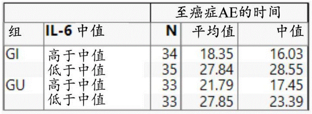

Example 12


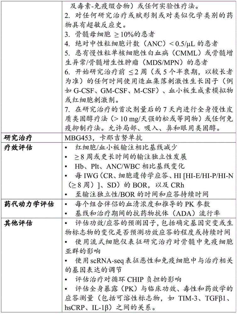
Example 13
Canatkinumab-CANTOS assay in breakthrough anemia
Among the 10,061 randomized participants, baseline anemia (hemoglobin less than 12g/dL in women and less than 13g/dL in men) was present in 417 women and 899 men, while 4 participants had no measurement of baseline hemoglobin. Thus, the analysis included 8,741 CANTOS participants. Blood samples were obtained from all test participants in the canarginoumab and placebo groups during randomization and the trial period (baseline, 3, 6, 9, 12, 18, 24, 30, 36, 42, 48, 54 and 60 months after randomization). All samples received standard hematological assessments including hemoglobin, hematocrit, red blood cell count, differential white blood cell count, and platelet count. The follow-up CBC allowed assessment of sudden anemia, defined prospectively as hemoglobin <13g/dl in men and <12g/dl in women with normal hemoglobin at the time of test enrollment.
Baseline clinical profile (e.g., age, renal function, hsCRP, alcohol consumption, diabetes, and hypertension) that may lead to anemia between placebo or active treatment groups were compared using chi-square analysis for categorical variables. For the continuous variable, the Kruskal-Wallis test was performed for multiple group comparisons, and the Wilcoxon rank-sum test was performed for two group comparisons between placebo and the effective treatment groups. The time-stratified univariate and adjusted Cox proportional hazards model and experimental section following the exponential myocardial infarction was used to assess the relative risk of anemia of susceptibility for the three canarginoumab groups (50mg, 150mg, and 300mg) compared to the group assigned to placebo, based on a pre-specified protocol. The P-values for the trend tests were calculated in these groups. Scores of 0, 1,3 and 6, proportional to canarginoumab dose, were used for trend analysis. The Kaplan-Meier curve was constructed to visually assess any differences between the groups. A similar analysis was performed for concurrent treatment response analysis pre-assigned for the experimental primary cardiovascular endpoint in the CANTOS protocol to determine whether the anti-inflammatory response intensity achieved by individual participants following a single dose of placebo or canarginoumab correlates with anemia liability. This analysis divided the participants to canarginoumab treatment into two groups based on hsCRP levels that were below 2mg/L at three months (strong responders) or above 2mg/L at three months (weak responders). This time point corresponds to the trough before the second dose of canarginoumab, after the first dose. Other sub-group analyses were performed to assess factors associated with anemia and chronic inflammation, including age and kidney function. All analyses were treated as intended. All p-values were bi-directional and all confidence intervals were calculated at a 95% level.
8,741 CANTOS participants who were anemic at baseline randomly received subcutaneously administered placebo or 50mg, 150mg, or 300mg of canajirimumab every 3 months. These groups had a close match of baseline clinical characteristics, including those prone to anemia, such as age, renal function, and underlying inflammation, as assessed by baseline hsCRP (table 1). Participants with anemia (participants not included in the secondary analysis) were significantly older, more likely female, and had a higher complication burden (higher incidence of hypertension and type 2 diabetes)), reduced GFR, and higher levels of hsCRP (table 1) than participants who were anemic at baseline.
TABLE 1 Baseline clinical characteristics of trial participants without Baseline anemia and excluded from Baseline anemia
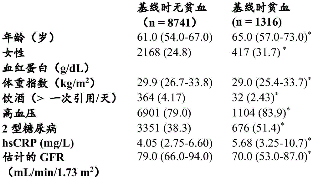
Consecutive data are reported as median (IQR) and binary data are reported as n (%). Significance was noted between groups at baseline. hsCRP is a highly sensitive C-reactive protein. GFR ═ glomerular filtration rate.
For the comparison of participants with anemia at baseline to participants without anemia at baseline,*P<0.05。
baseline levels of hsCRP were associated with anemia prone. In particular, the incidence of anemia per 100 human years in those with lowest (<3.1mg/L), moderate, and highest (>5.45mg/L) titers of hsCRP levels was 5.63, 6.55, and 7.91, respectively (trend P across tertiles < 0.0001).
Compared to placebo, participants assigned to any dose of canarginoumab were statistically significantly reduced in susceptibility to anemia throughout the procedure (fig. 21) compared to placebo (HR ═ 0.84, 95% CI 0.77-0.93, p <0.0001) (hemoglobin <13g/dL in men, and <12g/dL in women). Reduction was dose independent: for the 50mg group (N-1907), the risk ratio for anemia is 0.83 (95% CI 0.73-0.94, P-0.004) compared to placebo. For the 150mg group (N-1987), the risk ratio for anemia is 0.84 (95% CI 0.74-0.95, P-0.006) compared to placebo. For the 300mg group (N-1941), the risk ratio for predisposing anemia compared to placebo was 0.85 (95% CI 0.75-0.96, P-0.008). The incidence of anemia every 100 years was 7.49 in the placebo group, 6.17 in the 50mg group, 6.33 in the 150mg group, 6.34 in the 300mg group, and 6.28 in all active doses of canarginoumab (p ═ 0.014 for the trend of the active dose group compared to placebo). Comparative analysis of the canarginoumab combination dose with placebo showed a significant decrease in the incidence of predisposing anemia in those patients with hsCRP levels below 2mg/L in treatment after the first dose of canarginoumab (HR ═ 0.78, 95% CI 0.70-0.87, p < 0.0001). In contrast, subjects with hsCRP ≧ 2mg/L in treatment had a similar incidence of predisposing anemia to the placebo group (HR1.01, 95% CI 0.91-1.13, p ═ 0.82). Specifically, in those with hsCRP <2mg/L three months after the onset of canarginoumab, the HR was 0.67 for anemia in the 50mg group (95% CI 0.56-0.81, p <0.0001), 0.78 for the 150mg group (95% CI 0.67-0.91, p ═ 0.002), and 0.76 for the 300mg group (95% CI 0.65-0.88, p <0.0001) compared to placebo. In contrast, subjects with hsCRP ≧ 2mg/L at three months compared to placebo had an HR for anemia of 0.97 (95% CI 0.84-1.13, p ═ 0.699) for the 50mg group, 0.89 (95% CI 0.76-1.05, p ═ 0.171) for the 150mg group, and 1.05 (95% CI 0.88-1.25, p ═ 0.570) for the 300mg group. Notably, canajirimumab had a greater effect in reducing predisposed anemia in patients over 65 years of age (HR 0.78, 95% CI 0.68-0.89, p <0.0001) than in patients under 65 years of age (HR 0.88, 95% CI 0.78-1.00, p 0.056) (fig. 22). Specifically, in participants 65 years of age or older, the HR to anemia associated with canarginoumab was 0.80 for the 50mg group (95% CI 0.66-0.96, p ═ 0.017), 0.73 for the 150mg group (95% CI0.61-0.88, p ═ 0.001), and 0.80 for the 300mg group (95% CI 0.67-0.96, p ═ 0.018) compared to placebo (table 2).
Table 2 risk of predisposing anemia stratified by hypersensitive CRP at three months.a

aThe incidence is every 100 people years (number of participants with the event). The p-value for the trend and for the combination of all doses was compared to placebo. CI represents the confidence interval and hscRP represents the high sensitivity C-reactive protein.
It is also contemplated that the eGFR is greater than or equal to 60ml/min/1.73m2Compared with the participants, the eGFR is less than 60mL/min/1.73m2The incidence of anemia was higher in the participants. The incidence of anemia is less than 60mL/min/1.73m at eGFR2Is 14.55 and 11.24/100 years for placebo and all doses of canarginoumab, respectively, and is greater than or equal to 60mL/min/1.73m at an eGFR2The participants in (a) were 6.43 and 5.47/100 years old for placebo and all doses of canargizumab (table 3). For eGFR less than 60mL/min/1.73m2And for eGFR is greater than or equal to 60mL/min/1.73m2The canargiunumab treatment was associated with a significant reduction in predisposed anemia compared to placebo for all doses of canargiunumab. For all groups of canajirimumab, HR for anemia was less than 60mL/min/1.73m for eGFR compared to placebo2Participants in (a) were 0.78 (95% CI 0.65-0.94, p ═ 0.009), and 0.85 (95% CI 0.77-0.95, p ═ 0.005) (table 3).
TABLE 3 susceptibility to stratification by baseline eGFRRisk of anemiaa

aThe incidence is every 100 people years (number of participants with the event). The p-value for the trend and for the combination of all doses was compared to placebo. CI represents the confidence interval.
These data are of practical interest for treating anemia (e.g., anemia of inflammation) using IL-1 β binding antibodies (e.g., canajirimumab) as an adjunct therapy.


Claims (53)
Applications Claiming Priority (3)
| Application Number | Priority Date | Filing Date | Title |
|---|---|---|---|
| US201862783568P | 2018-12-21 | 2018-12-21 | |
| US62/783,568 | 2018-12-21 | ||
| PCT/IB2019/001435 WO2020128636A1 (en) | 2018-12-21 | 2019-12-18 | Use of il-1 beta antibodies in the treatment or prevention of myelodysplastic syndrome |
Publications (1)
| Publication Number | Publication Date |
|---|---|
| CN113227137A true CN113227137A (en) | 2021-08-06 |
Family
ID=70050161
Family Applications (1)
| Application Number | Title | Priority Date | Filing Date |
|---|---|---|---|
| CN201980083892.7A Pending CN113227137A (en) | 2018-12-21 | 2019-12-18 | Use of IL-1 beta antibodies in the treatment or prevention of myelodysplastic syndrome |
Country Status (14)
| Country | Link |
|---|---|
| US (2) | US20200369762A1 (en) |
| EP (1) | EP3898675A1 (en) |
| JP (1) | JP2022516850A (en) |
| KR (1) | KR20210107730A (en) |
| CN (1) | CN113227137A (en) |
| AU (1) | AU2019406840A1 (en) |
| BR (1) | BR112021011351A2 (en) |
| CA (1) | CA3119584A1 (en) |
| CL (1) | CL2021001619A1 (en) |
| IL (1) | IL283518A (en) |
| MX (1) | MX2021007488A (en) |
| SG (1) | SG11202104699TA (en) |
| TW (1) | TW202039555A (en) |
| WO (1) | WO2020128636A1 (en) |
Cited By (1)
| Publication number | Priority date | Publication date | Assignee | Title |
|---|---|---|---|---|
| WO2025002429A1 (en) * | 2023-06-28 | 2025-01-02 | 北京哲源科技有限责任公司 | Bispecific antibody targeting pd-l1 and il-1 and use thereof |
Families Citing this family (4)
| Publication number | Priority date | Publication date | Assignee | Title |
|---|---|---|---|---|
| WO2015048312A1 (en) | 2013-09-26 | 2015-04-02 | Costim Pharmaceuticals Inc. | Methods for treating hematologic cancers |
| JOP20200094A1 (en) | 2014-01-24 | 2017-06-16 | Dana Farber Cancer Inst Inc | Antibody molecules to pd-1 and uses thereof |
| LT3116909T (en) | 2014-03-14 | 2020-02-10 | Novartis Ag | ANTI-BODY MOLECULES AGAINST LAG-3 AND THEIR USE |
| IL293889A (en) * | 2019-12-20 | 2022-08-01 | Novartis Ag | Uses of antitgf-beta antibodies and checkpoint inhibitors for the treatment of proliferative diseases |
Family Cites Families (77)
| Publication number | Priority date | Publication date | Assignee | Title |
|---|---|---|---|---|
| DK0672142T3 (en) | 1992-12-04 | 2001-06-18 | Medical Res Council | Multivalent and multi-specific binding proteins as well as their preparation and use |
| CA3016482A1 (en) | 1999-11-30 | 2001-06-07 | Mayo Foundation For Medical Education And Research | B7-h1, a novel immunoregulatory molecule |
| EP2206517B1 (en) | 2002-07-03 | 2023-08-02 | Ono Pharmaceutical Co., Ltd. | Immunopotentiating compositions comprising anti-PD-L1 antibodies |
| JP4511943B2 (en) | 2002-12-23 | 2010-07-28 | ワイス エルエルシー | Antibody against PD-1 and use thereof |
| CA2602777C (en) | 2005-03-25 | 2018-12-11 | Tolerrx, Inc. | Gitr binding molecules and uses therefor |
| CN104356236B (en) | 2005-07-01 | 2020-07-03 | E.R.施贵宝&圣斯有限责任公司 | Human monoclonal antibody against programmed death ligand 1 (PD-L1) |
| JP5411430B2 (en) | 2005-07-04 | 2014-02-12 | 株式会社 ニコンビジョン | Ranging device |
| JP2009513645A (en) | 2005-10-26 | 2009-04-02 | ノバルティス アクチエンゲゼルシャフト | Novel use of IL-1β compounds |
| WO2007084342A2 (en) | 2006-01-13 | 2007-07-26 | The Government Of The United States, As Represented By The Secretary Of The Department Of Health And Human Services, National Institutes Of Health | Codon optimi zed il- 15 and il- 15r-alpha genes for expression in mammalian cells |
| EP1987839A1 (en) | 2007-04-30 | 2008-11-05 | I.N.S.E.R.M. Institut National de la Sante et de la Recherche Medicale | Cytotoxic anti-LAG-3 monoclonal antibody and its use in the treatment or prevention of organ transplant rejection and autoimmune disease |
| US9244059B2 (en) | 2007-04-30 | 2016-01-26 | Immutep Parc Club Orsay | Cytotoxic anti-LAG-3 monoclonal antibody and its use in the treatment or prevention of organ transplant rejection and autoimmune disease |
| CN101743249B (en) | 2007-05-11 | 2017-08-08 | 阿尔托生物科学有限公司 | Fusion molecules and IL‑15 variants |
| EP2044949A1 (en) | 2007-10-05 | 2009-04-08 | Immutep | Use of recombinant lag-3 or the derivatives thereof for eliciting monocyte immune response |
| AR072999A1 (en) | 2008-08-11 | 2010-10-06 | Medarex Inc | HUMAN ANTIBODIES THAT JOIN GEN 3 OF LYMPHOCYTARY ACTIVATION (LAG-3) AND THE USES OF THESE |
| EP2328919A2 (en) | 2008-08-25 | 2011-06-08 | Amplimmune, Inc. | Pd-i antagonists and methods for treating infectious disease |
| CA2736829C (en) | 2008-09-12 | 2018-02-27 | Isis Innovation Limited | Pd-1 specific antibodies and uses thereof |
| CA2998281C (en) | 2008-09-26 | 2022-08-16 | Dana-Farber Cancer Institute, Inc. | Human anti-pd-1 antobodies and uses therefor |
| EP4331604B9 (en) | 2008-12-09 | 2025-07-23 | F. Hoffmann-La Roche AG | Anti-pd-l1 antibodies and their use to enhance t-cell function |
| CA2772613C (en) | 2009-09-03 | 2020-03-10 | Schering Corporation | Anti-gitr antibodies |
| BR112012007365A2 (en) * | 2009-10-15 | 2016-11-22 | Abbott Lab | il-1 binding proteins |
| CA2778714C (en) | 2009-11-24 | 2018-02-27 | Medimmune Limited | Targeted binding agents against b7-h1 |
| US20130017199A1 (en) | 2009-11-24 | 2013-01-17 | AMPLIMMUNE ,Inc. a corporation | Simultaneous inhibition of pd-l1/pd-l2 |
| US8993731B2 (en) | 2010-03-11 | 2015-03-31 | Ucb Biopharma Sprl | PD-1 antibody |
| JP6158511B2 (en) | 2010-06-11 | 2017-07-05 | 協和発酵キリン株式会社 | Anti-TIM-3 antibody |
| CA2802344C (en) | 2010-06-18 | 2023-06-13 | The Brigham And Women's Hospital, Inc. | Bi-specific antibodies against tim-3 and pd-1 for immunotherapy in chronic immune conditions |
| US8907053B2 (en) | 2010-06-25 | 2014-12-09 | Aurigene Discovery Technologies Limited | Immunosuppression modulating compounds |
| RU2013115927A (en) * | 2010-09-10 | 2014-10-20 | Апексиджен, Инк. | ANTIBODIES AGAINST IL-1β AND METHODS OF APPLICATION |
| PT2699264T (en) | 2011-04-20 | 2018-05-23 | Medimmune Llc | ANTIBODIES AND OTHER MOLECULES CONNECTING B7-H1 AND PD-1 |
| EP2537933A1 (en) | 2011-06-24 | 2012-12-26 | Institut National de la Santé et de la Recherche Médicale (INSERM) | An IL-15 and IL-15Ralpha sushi domain based immunocytokines |
| WO2013006490A2 (en) | 2011-07-01 | 2013-01-10 | Cellerant Therapeutics, Inc. | Antibodies that specifically bind to tim3 |
| ES2808152T3 (en) | 2011-11-28 | 2021-02-25 | Merck Patent Gmbh | Anti-PD-L1 antibodies and their uses |
| UY34591A (en) | 2012-01-26 | 2013-09-02 | Novartis Ag | IMIDAZOPIRROLIDINONA COMPOUNDS |
| JP2015519375A (en) | 2012-05-31 | 2015-07-09 | ソレント・セラピューティクス・インコーポレイテッドSorrento Therapeutics, Inc. | Antigen binding protein that binds to PD-L1 |
| UY34887A (en) | 2012-07-02 | 2013-12-31 | Bristol Myers Squibb Company Una Corporacion Del Estado De Delaware | OPTIMIZATION OF ANTIBODIES THAT FIX THE LYMPHOCYTE ACTIVATION GEN 3 (LAG-3) AND ITS USES |
| WO2014022758A1 (en) | 2012-08-03 | 2014-02-06 | Dana-Farber Cancer Institute, Inc. | Single agent anti-pd-l1 and pd-l2 dual binding antibodies and methods of use |
| CN114507282B (en) | 2012-10-04 | 2025-05-16 | 达纳-法伯癌症研究所公司 | Human monoclonal anti-PD-L1 antibodies and methods of use |
| NZ630790A (en) | 2012-10-24 | 2016-11-25 | Admune Therapeutics Llc | Il-15r alpha forms, cells expressing il-15r alpha forms, and therapeutic uses of il-15r alpha and il-15/il-15r alpha complexes |
| AR093984A1 (en) | 2012-12-21 | 2015-07-01 | Merck Sharp & Dohme | ANTIBODIES THAT JOIN LEGEND 1 OF SCHEDULED DEATH (PD-L1) HUMAN |
| US9911308B2 (en) * | 2012-12-27 | 2018-03-06 | Kaleo, Inc. | Devices, systems and methods for locating and interacting with medicament delivery systems |
| ES2808654T3 (en) | 2013-03-15 | 2021-03-01 | Glaxosmithkline Ip Dev Ltd | Anti-LAG-3 binding proteins |
| HUE053069T2 (en) | 2013-05-02 | 2021-06-28 | Anaptysbio Inc | Antibodies to programmed death-1 (PD-1) |
| CA2913977C (en) | 2013-05-31 | 2022-11-29 | Sorrento Therapeutics, Inc. | Antigen binding proteins that bind pd-1 |
| WO2014209804A1 (en) | 2013-06-24 | 2014-12-31 | Biomed Valley Discoveries, Inc. | Bispecific antibodies |
| AR097306A1 (en) | 2013-08-20 | 2016-03-02 | Merck Sharp & Dohme | MODULATION OF TUMOR IMMUNITY |
| TW201605896A (en) | 2013-08-30 | 2016-02-16 | 安美基股份有限公司 | GITR antigen binding proteins |
| PT3702373T (en) | 2013-09-13 | 2022-09-27 | Beigene Switzerland Gmbh | Anti-pd1 antibodies and their use as therapeutics and diagnostics |
| US10202454B2 (en) | 2013-10-25 | 2019-02-12 | Dana-Farber Cancer Institute, Inc. | Anti-PD-L1 monoclonal antibodies and fragments thereof |
| WO2015081158A1 (en) | 2013-11-26 | 2015-06-04 | Bristol-Myers Squibb Company | Method of treating hiv by disrupting pd-1/pd-l1 signaling |
| SG10201804945WA (en) | 2013-12-12 | 2018-07-30 | Shanghai hengrui pharmaceutical co ltd | Pd-1 antibody, antigen-binding fragment thereof, and medical application thereof |
| US9209965B2 (en) | 2014-01-14 | 2015-12-08 | Microsemi Semiconductor Ulc | Network interface with clock recovery module on line card |
| US10407502B2 (en) | 2014-01-15 | 2019-09-10 | Kadmon Corporation, Llc | Immunomodulatory agents |
| TWI680138B (en) | 2014-01-23 | 2019-12-21 | 美商再生元醫藥公司 | Human antibodies to pd-l1 |
| TWI681969B (en) | 2014-01-23 | 2020-01-11 | 美商再生元醫藥公司 | Human antibodies to pd-1 |
| JOP20200094A1 (en) | 2014-01-24 | 2017-06-16 | Dana Farber Cancer Inst Inc | Antibody molecules to pd-1 and uses thereof |
| KR20160106762A (en) | 2014-01-28 | 2016-09-12 | 브리스톨-마이어스 스큅 컴퍼니 | Anti-lag-3 antibodies to treat hematological malignancies |
| JOP20200096A1 (en) | 2014-01-31 | 2017-06-16 | Children’S Medical Center Corp | TIM-3 Antibody Molecules and Their Uses |
| LT3116909T (en) | 2014-03-14 | 2020-02-10 | Novartis Ag | ANTI-BODY MOLECULES AGAINST LAG-3 AND THEIR USE |
| SMT202100116T1 (en) | 2014-05-28 | 2021-05-07 | Agenus Inc | Anti-gitr antibodies and methods of use thereof |
| EP3149042B1 (en) | 2014-05-29 | 2019-08-28 | Spring Bioscience Corporation | Pd-l1 antibodies and uses thereof |
| PT3151921T (en) | 2014-06-06 | 2019-11-21 | Bristol Myers Squibb Co | ANTIBODIES AGAINST GLUCOCORTICOID-INDUCED TUMOR NECROSIS FACTOR RECEIVER AND USES OF THE SAME |
| WO2015195163A1 (en) | 2014-06-20 | 2015-12-23 | R-Pharm Overseas, Inc. | Pd-l1 antagonist fully human antibody |
| TWI693232B (en) | 2014-06-26 | 2020-05-11 | 美商宏觀基因股份有限公司 | Covalently bonded diabodies having immunoreactivity with pd-1 and lag-3, and methods of use thereof |
| CN110156892B (en) | 2014-07-03 | 2023-05-16 | 百济神州有限公司 | anti-PD-L1 antibodies and their use as therapeutic and diagnostic agents |
| JO3663B1 (en) | 2014-08-19 | 2020-08-27 | Merck Sharp & Dohme | Anti-lag3 antibodies and antigen-binding fragments |
| MX2017004311A (en) | 2014-10-03 | 2017-12-07 | Dana Farber Cancer Inst Inc | Glucocorticoid-induced tumor necrosis factor receptor (gitr) antibodies and methods of use thereof. |
| MA41044A (en) | 2014-10-08 | 2017-08-15 | Novartis Ag | COMPOSITIONS AND METHODS OF USE FOR INCREASED IMMUNE RESPONSE AND CANCER TREATMENT |
| JP6877339B2 (en) | 2014-10-14 | 2021-05-26 | ノバルティス アーゲー | Antibody molecule against PD-L1 and its use |
| CA2964830C (en) | 2014-11-06 | 2024-01-02 | F. Hoffmann-La Roche Ag | Anti-tim3 antibodies and methods of use |
| TWI595006B (en) | 2014-12-09 | 2017-08-11 | 禮納特神經系統科學公司 | Anti-PD-1 antibodies and methods of using same |
| WO2016111947A2 (en) | 2015-01-05 | 2016-07-14 | Jounce Therapeutics, Inc. | Antibodies that inhibit tim-3:lilrb2 interactions and uses thereof |
| TW201639888A (en) | 2015-03-06 | 2016-11-16 | 索倫多醫療公司 | Antibody therapeutics that bind TIM3 |
| SMT202400420T1 (en) | 2015-04-01 | 2024-11-15 | Anaptysbio Inc | Antibodies directed against t cell immunoglobulin and mucin protein 3 (tim-3) |
| BR112017025297A2 (en) | 2015-06-03 | 2018-08-14 | Bristol-Myers Squibb Company | anti-gitr antibodies for cancer diagnosis |
| EP3303388A1 (en) * | 2015-06-04 | 2018-04-11 | Novartis AG | Use of il-1 beta binding antibodies to treat peripheral arterial disease |
| US10093742B2 (en) | 2015-07-23 | 2018-10-09 | Inhibrx, Inc. | Multispecific GITR-binding fusion proteins and methods of use thereof |
| CA2994346A1 (en) | 2015-08-12 | 2017-02-16 | Medimmune Limited | Gitrl fusion proteins and uses thereof |
| EP4424322A3 (en) * | 2015-12-17 | 2025-04-16 | Novartis AG | Antibody molecules to pd-1 and uses thereof |
-
2019
- 2019-12-18 CN CN201980083892.7A patent/CN113227137A/en active Pending
- 2019-12-18 JP JP2021536005A patent/JP2022516850A/en active Pending
- 2019-12-18 EP EP19868166.0A patent/EP3898675A1/en not_active Withdrawn
- 2019-12-18 BR BR112021011351A patent/BR112021011351A2/en unknown
- 2019-12-18 SG SG11202104699TA patent/SG11202104699TA/en unknown
- 2019-12-18 AU AU2019406840A patent/AU2019406840A1/en not_active Abandoned
- 2019-12-18 KR KR1020217022293A patent/KR20210107730A/en not_active Withdrawn
- 2019-12-18 CA CA3119584A patent/CA3119584A1/en active Pending
- 2019-12-18 US US16/719,154 patent/US20200369762A1/en not_active Abandoned
- 2019-12-18 MX MX2021007488A patent/MX2021007488A/en unknown
- 2019-12-18 WO PCT/IB2019/001435 patent/WO2020128636A1/en not_active Ceased
- 2019-12-20 TW TW108147065A patent/TW202039555A/en unknown
-
2021
- 2021-05-27 IL IL283518A patent/IL283518A/en unknown
- 2021-06-17 CL CL2021001619A patent/CL2021001619A1/en unknown
-
2022
- 2022-04-25 US US17/728,265 patent/US20230088070A1/en not_active Abandoned
Cited By (1)
| Publication number | Priority date | Publication date | Assignee | Title |
|---|---|---|---|---|
| WO2025002429A1 (en) * | 2023-06-28 | 2025-01-02 | 北京哲源科技有限责任公司 | Bispecific antibody targeting pd-l1 and il-1 and use thereof |
Also Published As
| Publication number | Publication date |
|---|---|
| WO2020128636A1 (en) | 2020-06-25 |
| MX2021007488A (en) | 2021-08-05 |
| TW202039555A (en) | 2020-11-01 |
| AU2019406840A1 (en) | 2021-06-03 |
| CL2021001619A1 (en) | 2022-02-11 |
| KR20210107730A (en) | 2021-09-01 |
| CA3119584A1 (en) | 2020-06-25 |
| US20200369762A1 (en) | 2020-11-26 |
| IL283518A (en) | 2021-07-29 |
| WO2020128636A8 (en) | 2021-07-01 |
| US20230088070A1 (en) | 2023-03-23 |
| JP2022516850A (en) | 2022-03-03 |
| BR112021011351A2 (en) | 2021-11-16 |
| EP3898675A1 (en) | 2021-10-27 |
| SG11202104699TA (en) | 2021-07-29 |
Similar Documents
| Publication | Publication Date | Title |
|---|---|---|
| US20220389090A1 (en) | Use of il-1beta binding antibodies | |
| TWI821748B (en) | Treatment of lung cancer using a combination of an anti-pd-1 antibody and an anti-ctla-4 antibody | |
| US20230088070A1 (en) | Use of il-1beta binding antibodies | |
| JP2024028805A (en) | Combination therapy using anti-IL-8 and anti-PD-1 antibodies for cancer treatment | |
| US20220056123A1 (en) | Use of il-1beta binding antibodies | |
| US20220025036A1 (en) | Use of il-1beta binding antibodies | |
| WO2018235056A1 (en) | IL-1BETA BINDING ANTIBODIES FOR USE IN THE TREATMENT OF CANCER | |
| WO2020128637A1 (en) | Use of il-1 binding antibodies in the treatment of a msi-h cancer | |
| TWI912619B (en) | Treatment of lung cancer using a combination of an anti-pd-1 antibody and an anti-ctla-4 antibody |
Legal Events
| Date | Code | Title | Description |
|---|---|---|---|
| PB01 | Publication | ||
| PB01 | Publication | ||
| SE01 | Entry into force of request for substantive examination | ||
| SE01 | Entry into force of request for substantive examination |