DE69839147T2 - ARTIFICIAL ANTIBODY POLYPEPTIDE - Google Patents
ARTIFICIAL ANTIBODY POLYPEPTIDE Download PDFInfo
- Publication number
- DE69839147T2 DE69839147T2 DE1998639147 DE69839147T DE69839147T2 DE 69839147 T2 DE69839147 T2 DE 69839147T2 DE 1998639147 DE1998639147 DE 1998639147 DE 69839147 T DE69839147 T DE 69839147T DE 69839147 T2 DE69839147 T2 DE 69839147T2
- Authority
- DE
- Germany
- Prior art keywords
- loop
- nucleic acid
- monobody
- polypeptide
- acid sequence
- Prior art date
- Legal status (The legal status is an assumption and is not a legal conclusion. Google has not performed a legal analysis and makes no representation as to the accuracy of the status listed.)
- Expired - Lifetime
Links
- 108090000765 processed proteins & peptides Proteins 0.000 title claims abstract description 128
- 102000004196 processed proteins & peptides Human genes 0.000 title claims abstract description 84
- 229920001184 polypeptide Polymers 0.000 title claims abstract description 75
- 238000009739 binding Methods 0.000 claims abstract description 104
- 230000027455 binding Effects 0.000 claims abstract description 91
- 238000000034 method Methods 0.000 claims abstract description 67
- 150000007523 nucleic acids Chemical class 0.000 claims abstract description 59
- 238000006243 chemical reaction Methods 0.000 claims abstract description 33
- 108020004707 nucleic acids Proteins 0.000 claims abstract description 29
- 102000039446 nucleic acids Human genes 0.000 claims abstract description 29
- 230000009870 specific binding Effects 0.000 claims abstract description 15
- 241000282326 Felis catus Species 0.000 claims abstract description 12
- 102000002090 Fibronectin type III Human genes 0.000 claims abstract description 10
- 108050009401 Fibronectin type III Proteins 0.000 claims abstract description 10
- 125000003275 alpha amino acid group Chemical group 0.000 claims abstract 5
- 239000003446 ligand Substances 0.000 claims description 54
- 108091028043 Nucleic acid sequence Proteins 0.000 claims description 49
- 108090000848 Ubiquitin Proteins 0.000 claims description 42
- 102000044159 Ubiquitin Human genes 0.000 claims description 42
- 150000001413 amino acids Chemical class 0.000 claims description 31
- 230000035772 mutation Effects 0.000 claims description 31
- 238000005481 NMR spectroscopy Methods 0.000 claims description 25
- 108020004414 DNA Proteins 0.000 claims description 24
- 230000003197 catalytic effect Effects 0.000 claims description 24
- 230000007704 transition Effects 0.000 claims description 24
- 238000003752 polymerase chain reaction Methods 0.000 claims description 16
- 239000013604 expression vector Substances 0.000 claims description 15
- 239000013598 vector Substances 0.000 claims description 15
- 235000001014 amino acid Nutrition 0.000 claims description 13
- 238000012217 deletion Methods 0.000 claims description 13
- 230000037430 deletion Effects 0.000 claims description 13
- 230000014509 gene expression Effects 0.000 claims description 13
- 238000010276 construction Methods 0.000 claims description 12
- 238000003780 insertion Methods 0.000 claims description 12
- 230000037431 insertion Effects 0.000 claims description 12
- 239000013612 plasmid Substances 0.000 claims description 12
- 230000008569 process Effects 0.000 claims description 12
- 241000894007 species Species 0.000 claims description 11
- 239000002773 nucleotide Substances 0.000 claims description 10
- 125000003729 nucleotide group Chemical group 0.000 claims description 10
- 108020004705 Codon Proteins 0.000 claims description 8
- 150000001875 compounds Chemical class 0.000 claims description 8
- GNBHRKFJIUUOQI-UHFFFAOYSA-N fluorescein Chemical compound O1C(=O)C2=CC=CC=C2C21C1=CC=C(O)C=C1OC1=CC(O)=CC=C21 GNBHRKFJIUUOQI-UHFFFAOYSA-N 0.000 claims description 8
- 238000012216 screening Methods 0.000 claims description 8
- 238000002424 x-ray crystallography Methods 0.000 claims description 8
- 230000000295 complement effect Effects 0.000 claims description 7
- 238000010494 dissociation reaction Methods 0.000 claims description 7
- 230000005593 dissociations Effects 0.000 claims description 7
- 230000010076 replication Effects 0.000 claims description 7
- 238000002741 site-directed mutagenesis Methods 0.000 claims description 7
- 102000053602 DNA Human genes 0.000 claims description 6
- 101001027128 Homo sapiens Fibronectin Proteins 0.000 claims description 6
- 108091008324 binding proteins Proteins 0.000 claims description 6
- 108091008146 restriction endonucleases Proteins 0.000 claims description 6
- 239000000047 product Substances 0.000 claims description 5
- 241001515965 unidentified phage Species 0.000 claims description 5
- SHIBSTMRCDJXLN-KCZCNTNESA-N digoxigenin Chemical compound C1([C@@H]2[C@@]3([C@@](CC2)(O)[C@H]2[C@@H]([C@@]4(C)CC[C@H](O)C[C@H]4CC2)C[C@H]3O)C)=CC(=O)OC1 SHIBSTMRCDJXLN-KCZCNTNESA-N 0.000 claims description 4
- SHIBSTMRCDJXLN-UHFFFAOYSA-N Digoxigenin Natural products C1CC(C2C(C3(C)CCC(O)CC3CC2)CC2O)(O)C2(C)C1C1=CC(=O)OC1 SHIBSTMRCDJXLN-UHFFFAOYSA-N 0.000 claims description 3
- QONQRTHLHBTMGP-UHFFFAOYSA-N digitoxigenin Natural products CC12CCC(C3(CCC(O)CC3CC3)C)C3C11OC1CC2C1=CC(=O)OC1 QONQRTHLHBTMGP-UHFFFAOYSA-N 0.000 claims description 3
- 230000003278 mimic effect Effects 0.000 claims description 3
- 230000006641 stabilisation Effects 0.000 claims description 3
- 241000700605 Viruses Species 0.000 claims description 2
- 239000007795 chemical reaction product Substances 0.000 claims description 2
- 235000018417 cysteine Nutrition 0.000 claims description 2
- XUJNEKJLAYXESH-UHFFFAOYSA-N cysteine Natural products SCC(N)C(O)=O XUJNEKJLAYXESH-UHFFFAOYSA-N 0.000 claims description 2
- 238000011105 stabilization Methods 0.000 claims description 2
- GOJUJUVQIVIZAV-UHFFFAOYSA-N 2-amino-4,6-dichloropyrimidine-5-carbaldehyde Chemical group NC1=NC(Cl)=C(C=O)C(Cl)=N1 GOJUJUVQIVIZAV-UHFFFAOYSA-N 0.000 claims 1
- FWMNVWWHGCHHJJ-SKKKGAJSSA-N 4-amino-1-[(2r)-6-amino-2-[[(2r)-2-[[(2r)-2-[[(2r)-2-amino-3-phenylpropanoyl]amino]-3-phenylpropanoyl]amino]-4-methylpentanoyl]amino]hexanoyl]piperidine-4-carboxylic acid Chemical compound C([C@H](C(=O)N[C@H](CC(C)C)C(=O)N[C@H](CCCCN)C(=O)N1CCC(N)(CC1)C(O)=O)NC(=O)[C@H](N)CC=1C=CC=CC=1)C1=CC=CC=C1 FWMNVWWHGCHHJJ-SKKKGAJSSA-N 0.000 claims 1
- 101100041523 Homo sapiens RYR1 gene Proteins 0.000 claims 1
- 102100032122 Ryanodine receptor 1 Human genes 0.000 claims 1
- 102000023732 binding proteins Human genes 0.000 claims 1
- 108090000623 proteins and genes Proteins 0.000 description 142
- OPTASPLRGRRNAP-UHFFFAOYSA-N cytosine Chemical compound NC=1C=CNC(=O)N=1 OPTASPLRGRRNAP-UHFFFAOYSA-N 0.000 description 108
- 102000004169 proteins and genes Human genes 0.000 description 94
- 235000018102 proteins Nutrition 0.000 description 83
- 229940104302 cytosine Drugs 0.000 description 54
- OIRDTQYFTABQOQ-KQYNXXCUSA-N adenosine Chemical compound C1=NC=2C(N)=NC=NC=2N1[C@@H]1O[C@H](CO)[C@@H](O)[C@H]1O OIRDTQYFTABQOQ-KQYNXXCUSA-N 0.000 description 44
- 238000002965 ELISA Methods 0.000 description 37
- 239000000243 solution Substances 0.000 description 36
- NYHBQMYGNKIUIF-UUOKFMHZSA-N Guanosine Chemical compound C1=NC=2C(=O)NC(N)=NC=2N1[C@@H]1O[C@H](CO)[C@@H](O)[C@H]1O NYHBQMYGNKIUIF-UUOKFMHZSA-N 0.000 description 32
- FAPWRFPIFSIZLT-UHFFFAOYSA-M Sodium chloride Chemical compound [Na+].[Cl-] FAPWRFPIFSIZLT-UHFFFAOYSA-M 0.000 description 30
- IQFYYKKMVGJFEH-XLPZGREQSA-N Thymidine Chemical compound O=C1NC(=O)C(C)=CN1[C@@H]1O[C@H](CO)[C@@H](O)C1 IQFYYKKMVGJFEH-XLPZGREQSA-N 0.000 description 30
- 210000004027 cell Anatomy 0.000 description 27
- 102100035360 Cerebellar degeneration-related antigen 1 Human genes 0.000 description 25
- 108091034117 Oligonucleotide Proteins 0.000 description 25
- 239000002126 C01EB10 - Adenosine Substances 0.000 description 22
- 229960005305 adenosine Drugs 0.000 description 22
- 238000002474 experimental method Methods 0.000 description 22
- 239000012634 fragment Substances 0.000 description 21
- 239000000872 buffer Substances 0.000 description 18
- 238000002823 phage display Methods 0.000 description 18
- MIKUYHXYGGJMLM-GIMIYPNGSA-N Crotonoside Natural products C1=NC2=C(N)NC(=O)N=C2N1[C@H]1O[C@@H](CO)[C@H](O)[C@@H]1O MIKUYHXYGGJMLM-GIMIYPNGSA-N 0.000 description 16
- NYHBQMYGNKIUIF-UHFFFAOYSA-N D-guanosine Natural products C1=2NC(N)=NC(=O)C=2N=CN1C1OC(CO)C(O)C1O NYHBQMYGNKIUIF-UHFFFAOYSA-N 0.000 description 16
- 108020001507 fusion proteins Proteins 0.000 description 16
- 102000037865 fusion proteins Human genes 0.000 description 16
- 229940029575 guanosine Drugs 0.000 description 16
- DWRXFEITVBNRMK-UHFFFAOYSA-N Beta-D-1-Arabinofuranosylthymine Natural products O=C1NC(=O)C(C)=CN1C1C(O)C(O)C(CO)O1 DWRXFEITVBNRMK-UHFFFAOYSA-N 0.000 description 15
- JLCPHMBAVCMARE-UHFFFAOYSA-N [3-[[3-[[3-[[3-[[3-[[3-[[3-[[3-[[3-[[3-[[3-[[5-(2-amino-6-oxo-1H-purin-9-yl)-3-[[3-[[3-[[3-[[3-[[3-[[5-(2-amino-6-oxo-1H-purin-9-yl)-3-[[5-(2-amino-6-oxo-1H-purin-9-yl)-3-hydroxyoxolan-2-yl]methoxy-hydroxyphosphoryl]oxyoxolan-2-yl]methoxy-hydroxyphosphoryl]oxy-5-(5-methyl-2,4-dioxopyrimidin-1-yl)oxolan-2-yl]methoxy-hydroxyphosphoryl]oxy-5-(6-aminopurin-9-yl)oxolan-2-yl]methoxy-hydroxyphosphoryl]oxy-5-(6-aminopurin-9-yl)oxolan-2-yl]methoxy-hydroxyphosphoryl]oxy-5-(6-aminopurin-9-yl)oxolan-2-yl]methoxy-hydroxyphosphoryl]oxy-5-(6-aminopurin-9-yl)oxolan-2-yl]methoxy-hydroxyphosphoryl]oxyoxolan-2-yl]methoxy-hydroxyphosphoryl]oxy-5-(5-methyl-2,4-dioxopyrimidin-1-yl)oxolan-2-yl]methoxy-hydroxyphosphoryl]oxy-5-(4-amino-2-oxopyrimidin-1-yl)oxolan-2-yl]methoxy-hydroxyphosphoryl]oxy-5-(5-methyl-2,4-dioxopyrimidin-1-yl)oxolan-2-yl]methoxy-hydroxyphosphoryl]oxy-5-(5-methyl-2,4-dioxopyrimidin-1-yl)oxolan-2-yl]methoxy-hydroxyphosphoryl]oxy-5-(6-aminopurin-9-yl)oxolan-2-yl]methoxy-hydroxyphosphoryl]oxy-5-(6-aminopurin-9-yl)oxolan-2-yl]methoxy-hydroxyphosphoryl]oxy-5-(4-amino-2-oxopyrimidin-1-yl)oxolan-2-yl]methoxy-hydroxyphosphoryl]oxy-5-(4-amino-2-oxopyrimidin-1-yl)oxolan-2-yl]methoxy-hydroxyphosphoryl]oxy-5-(4-amino-2-oxopyrimidin-1-yl)oxolan-2-yl]methoxy-hydroxyphosphoryl]oxy-5-(6-aminopurin-9-yl)oxolan-2-yl]methoxy-hydroxyphosphoryl]oxy-5-(4-amino-2-oxopyrimidin-1-yl)oxolan-2-yl]methyl [5-(6-aminopurin-9-yl)-2-(hydroxymethyl)oxolan-3-yl] hydrogen phosphate Polymers Cc1cn(C2CC(OP(O)(=O)OCC3OC(CC3OP(O)(=O)OCC3OC(CC3O)n3cnc4c3nc(N)[nH]c4=O)n3cnc4c3nc(N)[nH]c4=O)C(COP(O)(=O)OC3CC(OC3COP(O)(=O)OC3CC(OC3COP(O)(=O)OC3CC(OC3COP(O)(=O)OC3CC(OC3COP(O)(=O)OC3CC(OC3COP(O)(=O)OC3CC(OC3COP(O)(=O)OC3CC(OC3COP(O)(=O)OC3CC(OC3COP(O)(=O)OC3CC(OC3COP(O)(=O)OC3CC(OC3COP(O)(=O)OC3CC(OC3COP(O)(=O)OC3CC(OC3COP(O)(=O)OC3CC(OC3COP(O)(=O)OC3CC(OC3COP(O)(=O)OC3CC(OC3COP(O)(=O)OC3CC(OC3COP(O)(=O)OC3CC(OC3CO)n3cnc4c(N)ncnc34)n3ccc(N)nc3=O)n3cnc4c(N)ncnc34)n3ccc(N)nc3=O)n3ccc(N)nc3=O)n3ccc(N)nc3=O)n3cnc4c(N)ncnc34)n3cnc4c(N)ncnc34)n3cc(C)c(=O)[nH]c3=O)n3cc(C)c(=O)[nH]c3=O)n3ccc(N)nc3=O)n3cc(C)c(=O)[nH]c3=O)n3cnc4c3nc(N)[nH]c4=O)n3cnc4c(N)ncnc34)n3cnc4c(N)ncnc34)n3cnc4c(N)ncnc34)n3cnc4c(N)ncnc34)O2)c(=O)[nH]c1=O JLCPHMBAVCMARE-UHFFFAOYSA-N 0.000 description 15
- 239000000427 antigen Substances 0.000 description 15
- IQFYYKKMVGJFEH-UHFFFAOYSA-N beta-L-thymidine Natural products O=C1NC(=O)C(C)=CN1C1OC(CO)C(O)C1 IQFYYKKMVGJFEH-UHFFFAOYSA-N 0.000 description 15
- 239000011780 sodium chloride Substances 0.000 description 15
- 229940104230 thymidine Drugs 0.000 description 15
- 108091007433 antigens Proteins 0.000 description 14
- 102000036639 antigens Human genes 0.000 description 14
- PJJJBBJSCAKJQF-UHFFFAOYSA-N guanidinium chloride Chemical compound [Cl-].NC(N)=[NH2+] PJJJBBJSCAKJQF-UHFFFAOYSA-N 0.000 description 14
- 238000001228 spectrum Methods 0.000 description 14
- 239000000126 substance Substances 0.000 description 14
- 241000588724 Escherichia coli Species 0.000 description 13
- 108060003951 Immunoglobulin Proteins 0.000 description 13
- 102000018358 immunoglobulin Human genes 0.000 description 13
- 238000012986 modification Methods 0.000 description 13
- 230000004048 modification Effects 0.000 description 13
- 230000004927 fusion Effects 0.000 description 12
- 229950001790 tendamistat Drugs 0.000 description 12
- 108010037401 tendamistate Proteins 0.000 description 12
- 230000003993 interaction Effects 0.000 description 11
- 238000004519 manufacturing process Methods 0.000 description 11
- 239000002609 medium Substances 0.000 description 11
- 229940024606 amino acid Drugs 0.000 description 10
- 210000004899 c-terminal region Anatomy 0.000 description 10
- 238000012512 characterization method Methods 0.000 description 10
- 239000000203 mixture Substances 0.000 description 10
- 101100112922 Candida albicans CDR3 gene Proteins 0.000 description 9
- 102000014914 Carrier Proteins Human genes 0.000 description 9
- 108010021625 Immunoglobulin Fragments Proteins 0.000 description 9
- 102000008394 Immunoglobulin Fragments Human genes 0.000 description 9
- 239000007983 Tris buffer Substances 0.000 description 9
- 230000015572 biosynthetic process Effects 0.000 description 9
- 238000005119 centrifugation Methods 0.000 description 9
- 230000002860 competitive effect Effects 0.000 description 9
- 238000005570 heteronuclear single quantum coherence Methods 0.000 description 9
- 238000002703 mutagenesis Methods 0.000 description 9
- 231100000350 mutagenesis Toxicity 0.000 description 9
- LENZDBCJOHFCAS-UHFFFAOYSA-N tris Chemical compound OCC(N)(CO)CO LENZDBCJOHFCAS-UHFFFAOYSA-N 0.000 description 9
- 238000004458 analytical method Methods 0.000 description 8
- 238000002983 circular dichroism Methods 0.000 description 8
- 229960000789 guanidine hydrochloride Drugs 0.000 description 8
- 238000000990 heteronuclear single quantum coherence spectrum Methods 0.000 description 8
- 239000000523 sample Substances 0.000 description 8
- IYMAXBFPHPZYIK-BQBZGAKWSA-N Arg-Gly-Asp Chemical compound NC(N)=NCCC[C@H](N)C(=O)NCC(=O)N[C@@H](CC(O)=O)C(O)=O IYMAXBFPHPZYIK-BQBZGAKWSA-N 0.000 description 7
- 230000008859 change Effects 0.000 description 7
- 108010067306 Fibronectins Proteins 0.000 description 6
- 102000016359 Fibronectins Human genes 0.000 description 6
- 102000016943 Muramidase Human genes 0.000 description 6
- 108010014251 Muramidase Proteins 0.000 description 6
- 108010062010 N-Acetylmuramoyl-L-alanine Amidase Proteins 0.000 description 6
- 108090000190 Thrombin Proteins 0.000 description 6
- 101710120037 Toxin CcdB Proteins 0.000 description 6
- XSQUKJJJFZCRTK-UHFFFAOYSA-N Urea Chemical compound NC(N)=O XSQUKJJJFZCRTK-UHFFFAOYSA-N 0.000 description 6
- KZSNJWFQEVHDMF-UHFFFAOYSA-N Valine Chemical compound CC(C)C(N)C(O)=O KZSNJWFQEVHDMF-UHFFFAOYSA-N 0.000 description 6
- HRHJHXJQMNWQTF-UHFFFAOYSA-N cannabichromenic acid Chemical compound O1C(C)(CCC=C(C)C)C=CC2=C1C=C(CCCCC)C(C(O)=O)=C2O HRHJHXJQMNWQTF-UHFFFAOYSA-N 0.000 description 6
- 230000005284 excitation Effects 0.000 description 6
- RAXXELZNTBOGNW-UHFFFAOYSA-N imidazole Natural products C1=CNC=N1 RAXXELZNTBOGNW-UHFFFAOYSA-N 0.000 description 6
- 229960000274 lysozyme Drugs 0.000 description 6
- 239000004325 lysozyme Substances 0.000 description 6
- 235000010335 lysozyme Nutrition 0.000 description 6
- 238000005259 measurement Methods 0.000 description 6
- YBYRMVIVWMBXKQ-UHFFFAOYSA-N phenylmethanesulfonyl fluoride Chemical compound FS(=O)(=O)CC1=CC=CC=C1 YBYRMVIVWMBXKQ-UHFFFAOYSA-N 0.000 description 6
- 238000002360 preparation method Methods 0.000 description 6
- 238000012360 testing method Methods 0.000 description 6
- 229960004072 thrombin Drugs 0.000 description 6
- 238000005406 washing Methods 0.000 description 6
- 241001524679 Escherichia virus M13 Species 0.000 description 5
- 206010028980 Neoplasm Diseases 0.000 description 5
- 108010090804 Streptavidin Proteins 0.000 description 5
- 150000001408 amides Chemical group 0.000 description 5
- 239000013078 crystal Substances 0.000 description 5
- 238000004091 panning Methods 0.000 description 5
- 239000007974 sodium acetate buffer Substances 0.000 description 5
- 238000003786 synthesis reaction Methods 0.000 description 5
- YBJHBAHKTGYVGT-ZKWXMUAHSA-N (+)-Biotin Chemical compound N1C(=O)N[C@@H]2[C@H](CCCCC(=O)O)SC[C@@H]21 YBJHBAHKTGYVGT-ZKWXMUAHSA-N 0.000 description 4
- 108010078791 Carrier Proteins Proteins 0.000 description 4
- KCXVZYZYPLLWCC-UHFFFAOYSA-N EDTA Chemical compound OC(=O)CN(CC(O)=O)CCN(CC(O)=O)CC(O)=O KCXVZYZYPLLWCC-UHFFFAOYSA-N 0.000 description 4
- 102000004190 Enzymes Human genes 0.000 description 4
- 108090000790 Enzymes Proteins 0.000 description 4
- 241001198387 Escherichia coli BL21(DE3) Species 0.000 description 4
- OUYCCCASQSFEME-QMMMGPOBSA-N L-tyrosine Chemical compound OC(=O)[C@@H](N)CC1=CC=C(O)C=C1 OUYCCCASQSFEME-QMMMGPOBSA-N 0.000 description 4
- 238000012565 NMR experiment Methods 0.000 description 4
- PXHVJJICTQNCMI-UHFFFAOYSA-N Nickel Chemical compound [Ni] PXHVJJICTQNCMI-UHFFFAOYSA-N 0.000 description 4
- 239000002253 acid Substances 0.000 description 4
- 125000000539 amino acid group Chemical group 0.000 description 4
- AVKUERGKIZMTKX-NJBDSQKTSA-N ampicillin Chemical compound C1([C@@H](N)C(=O)N[C@H]2[C@H]3SC([C@@H](N3C2=O)C(O)=O)(C)C)=CC=CC=C1 AVKUERGKIZMTKX-NJBDSQKTSA-N 0.000 description 4
- 229960000723 ampicillin Drugs 0.000 description 4
- 230000008901 benefit Effects 0.000 description 4
- 238000003776 cleavage reaction Methods 0.000 description 4
- 229940088598 enzyme Drugs 0.000 description 4
- 229910052739 hydrogen Inorganic materials 0.000 description 4
- 210000003000 inclusion body Anatomy 0.000 description 4
- 238000002347 injection Methods 0.000 description 4
- 239000007924 injection Substances 0.000 description 4
- 230000007246 mechanism Effects 0.000 description 4
- 229930182817 methionine Natural products 0.000 description 4
- 125000001360 methionine group Chemical group N[C@@H](CCSC)C(=O)* 0.000 description 4
- 239000002245 particle Substances 0.000 description 4
- 230000002829 reductive effect Effects 0.000 description 4
- 230000007017 scission Effects 0.000 description 4
- XLYOFNOQVPJJNP-UHFFFAOYSA-N water Chemical compound O XLYOFNOQVPJJNP-UHFFFAOYSA-N 0.000 description 4
- 241000283690 Bos taurus Species 0.000 description 3
- 108010047041 Complementarity Determining Regions Proteins 0.000 description 3
- 241000724791 Filamentous phage Species 0.000 description 3
- 210000000712 G cell Anatomy 0.000 description 3
- ZDXPYRJPNDTMRX-UHFFFAOYSA-N Glutamine Chemical compound OC(=O)C(N)CCC(N)=O ZDXPYRJPNDTMRX-UHFFFAOYSA-N 0.000 description 3
- DHMQDGOQFOQNFH-UHFFFAOYSA-N Glycine Chemical compound NCC(O)=O DHMQDGOQFOQNFH-UHFFFAOYSA-N 0.000 description 3
- HNDVDQJCIGZPNO-UHFFFAOYSA-N Histidine Chemical compound OC(=O)C(N)CC1=CN=CN1 HNDVDQJCIGZPNO-UHFFFAOYSA-N 0.000 description 3
- QIVBCDIJIAJPQS-VIFPVBQESA-N L-tryptophane Chemical compound C1=CC=C2C(C[C@H](N)C(O)=O)=CNC2=C1 QIVBCDIJIAJPQS-VIFPVBQESA-N 0.000 description 3
- 108010021466 Mutant Proteins Proteins 0.000 description 3
- 102000008300 Mutant Proteins Human genes 0.000 description 3
- 108020004511 Recombinant DNA Proteins 0.000 description 3
- 102100030852 Run domain Beclin-1-interacting and cysteine-rich domain-containing protein Human genes 0.000 description 3
- VMHLLURERBWHNL-UHFFFAOYSA-M Sodium acetate Chemical compound [Na+].CC([O-])=O VMHLLURERBWHNL-UHFFFAOYSA-M 0.000 description 3
- QIVBCDIJIAJPQS-UHFFFAOYSA-N Tryptophan Natural products C1=CC=C2C(CC(N)C(O)=O)=CNC2=C1 QIVBCDIJIAJPQS-UHFFFAOYSA-N 0.000 description 3
- 239000003929 acidic solution Substances 0.000 description 3
- 230000002776 aggregation Effects 0.000 description 3
- 238000004220 aggregation Methods 0.000 description 3
- 238000013459 approach Methods 0.000 description 3
- 229960002685 biotin Drugs 0.000 description 3
- 239000011616 biotin Substances 0.000 description 3
- 239000004202 carbamide Substances 0.000 description 3
- 238000012219 cassette mutagenesis Methods 0.000 description 3
- 239000003054 catalyst Substances 0.000 description 3
- 238000010367 cloning Methods 0.000 description 3
- 239000012141 concentrate Substances 0.000 description 3
- 239000003398 denaturant Substances 0.000 description 3
- 238000001514 detection method Methods 0.000 description 3
- 230000000694 effects Effects 0.000 description 3
- 235000013601 eggs Nutrition 0.000 description 3
- 239000001257 hydrogen Substances 0.000 description 3
- 230000002209 hydrophobic effect Effects 0.000 description 3
- 238000000338 in vitro Methods 0.000 description 3
- 230000000155 isotopic effect Effects 0.000 description 3
- 238000002372 labelling Methods 0.000 description 3
- 239000000463 material Substances 0.000 description 3
- 125000002496 methyl group Chemical group [H]C([H])([H])* 0.000 description 3
- 238000012900 molecular simulation Methods 0.000 description 3
- 230000007935 neutral effect Effects 0.000 description 3
- 238000000655 nuclear magnetic resonance spectrum Methods 0.000 description 3
- 102000013415 peroxidase activity proteins Human genes 0.000 description 3
- 108040007629 peroxidase activity proteins Proteins 0.000 description 3
- 239000008363 phosphate buffer Substances 0.000 description 3
- 238000000159 protein binding assay Methods 0.000 description 3
- 230000008707 rearrangement Effects 0.000 description 3
- 230000002441 reversible effect Effects 0.000 description 3
- 238000012552 review Methods 0.000 description 3
- 239000001632 sodium acetate Substances 0.000 description 3
- 235000017281 sodium acetate Nutrition 0.000 description 3
- 238000010561 standard procedure Methods 0.000 description 3
- 239000000758 substrate Substances 0.000 description 3
- 230000001225 therapeutic effect Effects 0.000 description 3
- 238000002054 transplantation Methods 0.000 description 3
- QKNYBSVHEMOAJP-UHFFFAOYSA-N 2-amino-2-(hydroxymethyl)propane-1,3-diol;hydron;chloride Chemical compound Cl.OCC(N)(CO)CO QKNYBSVHEMOAJP-UHFFFAOYSA-N 0.000 description 2
- 238000003979 3D HSQC-TOCSY Methods 0.000 description 2
- 108010032595 Antibody Binding Sites Proteins 0.000 description 2
- DCXYFEDJOCDNAF-UHFFFAOYSA-N Asparagine Natural products OC(=O)C(N)CC(N)=O DCXYFEDJOCDNAF-UHFFFAOYSA-N 0.000 description 2
- 108091003079 Bovine Serum Albumin Proteins 0.000 description 2
- 101710132601 Capsid protein Proteins 0.000 description 2
- 101710094648 Coat protein Proteins 0.000 description 2
- 102000018832 Cytochromes Human genes 0.000 description 2
- 108010052832 Cytochromes Proteins 0.000 description 2
- 108010017826 DNA Polymerase I Proteins 0.000 description 2
- 102000004594 DNA Polymerase I Human genes 0.000 description 2
- 108010014303 DNA-directed DNA polymerase Proteins 0.000 description 2
- 102000016928 DNA-directed DNA polymerase Human genes 0.000 description 2
- 229920002307 Dextran Polymers 0.000 description 2
- 241000701832 Enterobacteria phage T3 Species 0.000 description 2
- WQZGKKKJIJFFOK-GASJEMHNSA-N Glucose Natural products OC[C@H]1OC(O)[C@H](O)[C@@H](O)[C@@H]1O WQZGKKKJIJFFOK-GASJEMHNSA-N 0.000 description 2
- WHUUTDBJXJRKMK-UHFFFAOYSA-N Glutamic acid Natural products OC(=O)C(N)CCC(O)=O WHUUTDBJXJRKMK-UHFFFAOYSA-N 0.000 description 2
- 102100021181 Golgi phosphoprotein 3 Human genes 0.000 description 2
- 108010054477 Immunoglobulin Fab Fragments Proteins 0.000 description 2
- 102000001706 Immunoglobulin Fab Fragments Human genes 0.000 description 2
- 108010067060 Immunoglobulin Variable Region Proteins 0.000 description 2
- 102000017727 Immunoglobulin Variable Region Human genes 0.000 description 2
- XUJNEKJLAYXESH-REOHCLBHSA-N L-Cysteine Chemical compound SC[C@H](N)C(O)=O XUJNEKJLAYXESH-REOHCLBHSA-N 0.000 description 2
- DCXYFEDJOCDNAF-REOHCLBHSA-N L-asparagine Chemical compound OC(=O)[C@@H](N)CC(N)=O DCXYFEDJOCDNAF-REOHCLBHSA-N 0.000 description 2
- AGPKZVBTJJNPAG-WHFBIAKZSA-N L-isoleucine Chemical compound CC[C@H](C)[C@H](N)C(O)=O AGPKZVBTJJNPAG-WHFBIAKZSA-N 0.000 description 2
- ROHFNLRQFUQHCH-YFKPBYRVSA-N L-leucine Chemical compound CC(C)C[C@H](N)C(O)=O ROHFNLRQFUQHCH-YFKPBYRVSA-N 0.000 description 2
- FFEARJCKVFRZRR-BYPYZUCNSA-N L-methionine Chemical compound CSCC[C@H](N)C(O)=O FFEARJCKVFRZRR-BYPYZUCNSA-N 0.000 description 2
- COLNVLDHVKWLRT-QMMMGPOBSA-N L-phenylalanine Chemical compound OC(=O)[C@@H](N)CC1=CC=CC=C1 COLNVLDHVKWLRT-QMMMGPOBSA-N 0.000 description 2
- AYFVYJQAPQTCCC-GBXIJSLDSA-N L-threonine Chemical compound C[C@@H](O)[C@H](N)C(O)=O AYFVYJQAPQTCCC-GBXIJSLDSA-N 0.000 description 2
- KDXKERNSBIXSRK-UHFFFAOYSA-N Lysine Natural products NCCCCC(N)C(O)=O KDXKERNSBIXSRK-UHFFFAOYSA-N 0.000 description 2
- 101710125418 Major capsid protein Proteins 0.000 description 2
- 241001465754 Metazoa Species 0.000 description 2
- 101710141454 Nucleoprotein Proteins 0.000 description 2
- 241000283973 Oryctolagus cuniculus Species 0.000 description 2
- 102000005891 Pancreatic ribonuclease Human genes 0.000 description 2
- 239000002202 Polyethylene glycol Substances 0.000 description 2
- 229920001213 Polysorbate 20 Polymers 0.000 description 2
- 101710083689 Probable capsid protein Proteins 0.000 description 2
- 108010076504 Protein Sorting Signals Proteins 0.000 description 2
- 101800001554 RNA-directed RNA polymerase Proteins 0.000 description 2
- 240000004808 Saccharomyces cerevisiae Species 0.000 description 2
- 239000012506 Sephacryl® Substances 0.000 description 2
- 108020004682 Single-Stranded DNA Proteins 0.000 description 2
- CDBYLPFSWZWCQE-UHFFFAOYSA-L Sodium Carbonate Chemical compound [Na+].[Na+].[O-]C([O-])=O CDBYLPFSWZWCQE-UHFFFAOYSA-L 0.000 description 2
- UIIMBOGNXHQVGW-UHFFFAOYSA-M Sodium bicarbonate Chemical compound [Na+].OC([O-])=O UIIMBOGNXHQVGW-UHFFFAOYSA-M 0.000 description 2
- 241000187179 Streptomyces tendae Species 0.000 description 2
- 108700005078 Synthetic Genes Proteins 0.000 description 2
- 238000010521 absorption reaction Methods 0.000 description 2
- 230000006978 adaptation Effects 0.000 description 2
- 108010072041 arginyl-glycyl-aspartic acid Proteins 0.000 description 2
- 235000009582 asparagine Nutrition 0.000 description 2
- 229960001230 asparagine Drugs 0.000 description 2
- 235000003704 aspartic acid Nutrition 0.000 description 2
- OQFSQFPPLPISGP-UHFFFAOYSA-N beta-carboxyaspartic acid Natural products OC(=O)C(N)C(C(O)=O)C(O)=O OQFSQFPPLPISGP-UHFFFAOYSA-N 0.000 description 2
- 229960000074 biopharmaceutical Drugs 0.000 description 2
- 235000020958 biotin Nutrition 0.000 description 2
- 229940098773 bovine serum albumin Drugs 0.000 description 2
- 239000007853 buffer solution Substances 0.000 description 2
- 238000001142 circular dichroism spectrum Methods 0.000 description 2
- ZPUCINDJVBIVPJ-LJISPDSOSA-N cocaine Chemical compound O([C@H]1C[C@@H]2CC[C@@H](N2C)[C@H]1C(=O)OC)C(=O)C1=CC=CC=C1 ZPUCINDJVBIVPJ-LJISPDSOSA-N 0.000 description 2
- 230000001332 colony forming effect Effects 0.000 description 2
- 238000007398 colorimetric assay Methods 0.000 description 2
- 238000005859 coupling reaction Methods 0.000 description 2
- 238000004925 denaturation Methods 0.000 description 2
- 230000036425 denaturation Effects 0.000 description 2
- 238000013461 design Methods 0.000 description 2
- 230000001627 detrimental effect Effects 0.000 description 2
- 238000011161 development Methods 0.000 description 2
- NJDNXYGOVLYJHP-UHFFFAOYSA-L disodium;2-(3-oxido-6-oxoxanthen-9-yl)benzoate Chemical compound [Na+].[Na+].[O-]C(=O)C1=CC=CC=C1C1=C2C=CC(=O)C=C2OC2=CC([O-])=CC=C21 NJDNXYGOVLYJHP-UHFFFAOYSA-L 0.000 description 2
- 238000004520 electroporation Methods 0.000 description 2
- 108010030074 endodeoxyribonuclease MluI Proteins 0.000 description 2
- 238000005516 engineering process Methods 0.000 description 2
- 230000007717 exclusion Effects 0.000 description 2
- 238000001506 fluorescence spectroscopy Methods 0.000 description 2
- 102000034238 globular proteins Human genes 0.000 description 2
- 108091005896 globular proteins Proteins 0.000 description 2
- 239000008103 glucose Substances 0.000 description 2
- 235000013922 glutamic acid Nutrition 0.000 description 2
- 239000004220 glutamic acid Substances 0.000 description 2
- 210000000987 immune system Anatomy 0.000 description 2
- 238000002649 immunization Methods 0.000 description 2
- 230000003053 immunization Effects 0.000 description 2
- 229940072221 immunoglobulins Drugs 0.000 description 2
- 230000006698 induction Effects 0.000 description 2
- 229940030980 inova Drugs 0.000 description 2
- 102000006495 integrins Human genes 0.000 description 2
- 108010044426 integrins Proteins 0.000 description 2
- 230000002427 irreversible effect Effects 0.000 description 2
- 238000002955 isolation Methods 0.000 description 2
- 229960000310 isoleucine Drugs 0.000 description 2
- AGPKZVBTJJNPAG-UHFFFAOYSA-N isoleucine Natural products CCC(C)C(N)C(O)=O AGPKZVBTJJNPAG-UHFFFAOYSA-N 0.000 description 2
- 101150109249 lacI gene Proteins 0.000 description 2
- 239000011159 matrix material Substances 0.000 description 2
- 238000010369 molecular cloning Methods 0.000 description 2
- 230000010807 negative regulation of binding Effects 0.000 description 2
- 229910052759 nickel Inorganic materials 0.000 description 2
- 229910052757 nitrogen Inorganic materials 0.000 description 2
- 238000006384 oligomerization reaction Methods 0.000 description 2
- 230000037361 pathway Effects 0.000 description 2
- 239000008188 pellet Substances 0.000 description 2
- 229920001223 polyethylene glycol Polymers 0.000 description 2
- 239000000256 polyoxyethylene sorbitan monolaurate Substances 0.000 description 2
- 235000010486 polyoxyethylene sorbitan monolaurate Nutrition 0.000 description 2
- 238000001556 precipitation Methods 0.000 description 2
- 238000012545 processing Methods 0.000 description 2
- 230000000644 propagated effect Effects 0.000 description 2
- 108020001580 protein domains Proteins 0.000 description 2
- 238000000746 purification Methods 0.000 description 2
- 239000000376 reactant Substances 0.000 description 2
- 238000011084 recovery Methods 0.000 description 2
- 230000000717 retained effect Effects 0.000 description 2
- 238000002864 sequence alignment Methods 0.000 description 2
- 150000003384 small molecules Chemical class 0.000 description 2
- 238000012916 structural analysis Methods 0.000 description 2
- 230000002194 synthesizing effect Effects 0.000 description 2
- 238000004448 titration Methods 0.000 description 2
- 238000001551 total correlation spectroscopy Methods 0.000 description 2
- 230000009466 transformation Effects 0.000 description 2
- OUYCCCASQSFEME-UHFFFAOYSA-N tyrosine Natural products OC(=O)C(N)CC1=CC=C(O)C=C1 OUYCCCASQSFEME-UHFFFAOYSA-N 0.000 description 2
- 238000000108 ultra-filtration Methods 0.000 description 2
- VQTBINYMFPKLQD-UHFFFAOYSA-N (2,5-dioxopyrrolidin-1-yl) 2-(3-hydroxy-6-oxoxanthen-9-yl)benzoate Chemical compound C=12C=CC(=O)C=C2OC2=CC(O)=CC=C2C=1C1=CC=CC=C1C(=O)ON1C(=O)CCC1=O VQTBINYMFPKLQD-UHFFFAOYSA-N 0.000 description 1
- LOVPHSMOAVXQIH-UHFFFAOYSA-N (4-nitrophenyl) hydrogen carbonate Chemical compound OC(=O)OC1=CC=C([N+]([O-])=O)C=C1 LOVPHSMOAVXQIH-UHFFFAOYSA-N 0.000 description 1
- CBWCNKVANCTCAD-UHFFFAOYSA-N (4-nitrophenyl)phosphonic acid Chemical compound OP(O)(=O)C1=CC=C([N+]([O-])=O)C=C1 CBWCNKVANCTCAD-UHFFFAOYSA-N 0.000 description 1
- ZGNLFUXWZJGETL-YUSKDDKASA-N (Z)-[(2S)-2-amino-2-carboxyethyl]-hydroxyimino-oxidoazanium Chemical compound N[C@@H](C\[N+]([O-])=N\O)C(O)=O ZGNLFUXWZJGETL-YUSKDDKASA-N 0.000 description 1
- 108091032973 (ribonucleotides)n+m Proteins 0.000 description 1
- DLZKEQQWXODGGZ-KCJUWKMLSA-N 2-[[(2r)-2-[[(2s)-2-amino-3-(4-hydroxyphenyl)propanoyl]amino]propanoyl]amino]acetic acid Chemical compound OC(=O)CNC(=O)[C@@H](C)NC(=O)[C@@H](N)CC1=CC=C(O)C=C1 DLZKEQQWXODGGZ-KCJUWKMLSA-N 0.000 description 1
- OWMCODAXNFNLCU-UHFFFAOYSA-N 2-[[2-(1h-imidazol-5-yl)ethylamino]methyl]phenol Chemical compound OC1=CC=CC=C1CNCCC1=CN=CN1 OWMCODAXNFNLCU-UHFFFAOYSA-N 0.000 description 1
- IVLXQGJVBGMLRR-UHFFFAOYSA-N 2-aminoacetic acid;hydron;chloride Chemical compound Cl.NCC(O)=O IVLXQGJVBGMLRR-UHFFFAOYSA-N 0.000 description 1
- OZDAOHVKBFBBMZ-UHFFFAOYSA-N 2-aminopentanedioic acid;hydrate Chemical compound O.OC(=O)C(N)CCC(O)=O OZDAOHVKBFBBMZ-UHFFFAOYSA-N 0.000 description 1
- 238000004466 2D NOESY spectrum Methods 0.000 description 1
- UAIUNKRWKOVEES-UHFFFAOYSA-N 3,3',5,5'-tetramethylbenzidine Chemical compound CC1=C(N)C(C)=CC(C=2C=C(C)C(N)=C(C)C=2)=C1 UAIUNKRWKOVEES-UHFFFAOYSA-N 0.000 description 1
- QCVGEOXPDFCNHA-UHFFFAOYSA-N 5,5-dimethyl-2,4-dioxo-1,3-oxazolidine-3-carboxamide Chemical compound CC1(C)OC(=O)N(C(N)=O)C1=O QCVGEOXPDFCNHA-UHFFFAOYSA-N 0.000 description 1
- QTBSBXVTEAMEQO-UHFFFAOYSA-M Acetate Chemical compound CC([O-])=O QTBSBXVTEAMEQO-UHFFFAOYSA-M 0.000 description 1
- 101710171801 Alpha-amylase inhibitor Proteins 0.000 description 1
- 239000004475 Arginine Substances 0.000 description 1
- CKLJMWTZIZZHCS-UHFFFAOYSA-N Aspartic acid Chemical compound OC(=O)C(N)CC(O)=O CKLJMWTZIZZHCS-UHFFFAOYSA-N 0.000 description 1
- 241000894006 Bacteria Species 0.000 description 1
- 101000921522 Bos taurus Cytochrome c Proteins 0.000 description 1
- 101001106957 Bos taurus Ribonuclease pancreatic Proteins 0.000 description 1
- GGOLNRQUAIPJHS-UHFFFAOYSA-N CC[N]1(C)C=C1 Chemical compound CC[N]1(C)C=C1 GGOLNRQUAIPJHS-UHFFFAOYSA-N 0.000 description 1
- 241000282832 Camelidae Species 0.000 description 1
- 241000282836 Camelus dromedarius Species 0.000 description 1
- 108090000565 Capsid Proteins Proteins 0.000 description 1
- BVKZGUZCCUSVTD-UHFFFAOYSA-L Carbonate Chemical compound [O-]C([O-])=O BVKZGUZCCUSVTD-UHFFFAOYSA-L 0.000 description 1
- 108010067225 Cell Adhesion Molecules Proteins 0.000 description 1
- 102000016289 Cell Adhesion Molecules Human genes 0.000 description 1
- 108010001857 Cell Surface Receptors Proteins 0.000 description 1
- 102000000844 Cell Surface Receptors Human genes 0.000 description 1
- 102100023321 Ceruloplasmin Human genes 0.000 description 1
- 108050001186 Chaperonin Cpn60 Proteins 0.000 description 1
- 102000052603 Chaperonins Human genes 0.000 description 1
- 108010069514 Cyclic Peptides Proteins 0.000 description 1
- 102000001189 Cyclic Peptides Human genes 0.000 description 1
- 238000001712 DNA sequencing Methods 0.000 description 1
- 230000004568 DNA-binding Effects 0.000 description 1
- 102000002322 Egg Proteins Human genes 0.000 description 1
- 108010000912 Egg Proteins Proteins 0.000 description 1
- 241000206602 Eukaryota Species 0.000 description 1
- 102000010834 Extracellular Matrix Proteins Human genes 0.000 description 1
- 108010037362 Extracellular Matrix Proteins Proteins 0.000 description 1
- 241000282412 Homo Species 0.000 description 1
- 101001033249 Homo sapiens Interleukin-1 beta Proteins 0.000 description 1
- 101001074954 Homo sapiens Phosphatidylinositol 4,5-bisphosphate 5-phosphatase A Proteins 0.000 description 1
- 102000002265 Human Growth Hormone Human genes 0.000 description 1
- 108010000521 Human Growth Hormone Proteins 0.000 description 1
- 239000000854 Human Growth Hormone Substances 0.000 description 1
- 101150098499 III gene Proteins 0.000 description 1
- 102000004889 Interleukin-6 Human genes 0.000 description 1
- 108090001005 Interleukin-6 Proteins 0.000 description 1
- ONIBWKKTOPOVIA-BYPYZUCNSA-N L-Proline Chemical compound OC(=O)[C@@H]1CCCN1 ONIBWKKTOPOVIA-BYPYZUCNSA-N 0.000 description 1
- QNAYBMKLOCPYGJ-REOHCLBHSA-N L-alanine Chemical compound C[C@H](N)C(O)=O QNAYBMKLOCPYGJ-REOHCLBHSA-N 0.000 description 1
- MLFKVJCWGUZWNV-UHFFFAOYSA-N L-alanosine Natural products OC(=O)C(N)CN(O)N=O MLFKVJCWGUZWNV-UHFFFAOYSA-N 0.000 description 1
- CKLJMWTZIZZHCS-REOHCLBHSA-N L-aspartic acid Chemical compound OC(=O)[C@@H](N)CC(O)=O CKLJMWTZIZZHCS-REOHCLBHSA-N 0.000 description 1
- WHUUTDBJXJRKMK-VKHMYHEASA-N L-glutamic acid Chemical compound OC(=O)[C@@H](N)CCC(O)=O WHUUTDBJXJRKMK-VKHMYHEASA-N 0.000 description 1
- ZDXPYRJPNDTMRX-VKHMYHEASA-N L-glutamine Chemical compound OC(=O)[C@@H](N)CCC(N)=O ZDXPYRJPNDTMRX-VKHMYHEASA-N 0.000 description 1
- HNDVDQJCIGZPNO-YFKPBYRVSA-N L-histidine Chemical compound OC(=O)[C@@H](N)CC1=CN=CN1 HNDVDQJCIGZPNO-YFKPBYRVSA-N 0.000 description 1
- KZSNJWFQEVHDMF-BYPYZUCNSA-N L-valine Chemical compound CC(C)[C@H](N)C(O)=O KZSNJWFQEVHDMF-BYPYZUCNSA-N 0.000 description 1
- ROHFNLRQFUQHCH-UHFFFAOYSA-N Leucine Natural products CC(C)CC(N)C(O)=O ROHFNLRQFUQHCH-UHFFFAOYSA-N 0.000 description 1
- 239000006142 Luria-Bertani Agar Substances 0.000 description 1
- 239000004472 Lysine Substances 0.000 description 1
- 238000005654 Michaelis-Arbuzov synthesis reaction Methods 0.000 description 1
- 241000699666 Mus <mouse, genus> Species 0.000 description 1
- 241000699670 Mus sp. Species 0.000 description 1
- 102000003945 NF-kappa B Human genes 0.000 description 1
- 108010057466 NF-kappa B Proteins 0.000 description 1
- 229910021586 Nickel(II) chloride Inorganic materials 0.000 description 1
- 108020002230 Pancreatic Ribonuclease Proteins 0.000 description 1
- 102100035985 Phosphatidylinositol 4,5-bisphosphate 5-phosphatase A Human genes 0.000 description 1
- 239000004793 Polystyrene Substances 0.000 description 1
- ONIBWKKTOPOVIA-UHFFFAOYSA-N Proline Natural products OC(=O)C1CCCN1 ONIBWKKTOPOVIA-UHFFFAOYSA-N 0.000 description 1
- 241001080180 Quinta Species 0.000 description 1
- 238000012300 Sequence Analysis Methods 0.000 description 1
- MTCFGRXMJLQNBG-UHFFFAOYSA-N Serine Natural products OCC(N)C(O)=O MTCFGRXMJLQNBG-UHFFFAOYSA-N 0.000 description 1
- 241000194017 Streptococcus Species 0.000 description 1
- 108010006785 Taq Polymerase Proteins 0.000 description 1
- AYFVYJQAPQTCCC-UHFFFAOYSA-N Threonine Natural products CC(O)C(N)C(O)=O AYFVYJQAPQTCCC-UHFFFAOYSA-N 0.000 description 1
- 239000004473 Threonine Substances 0.000 description 1
- 108090000631 Trypsin Proteins 0.000 description 1
- 102000004142 Trypsin Human genes 0.000 description 1
- 101710162629 Trypsin inhibitor Proteins 0.000 description 1
- 229940122618 Trypsin inhibitor Drugs 0.000 description 1
- 238000002835 absorbance Methods 0.000 description 1
- 230000001133 acceleration Effects 0.000 description 1
- 238000009825 accumulation Methods 0.000 description 1
- 238000003916 acid precipitation Methods 0.000 description 1
- 230000004913 activation Effects 0.000 description 1
- 230000009824 affinity maturation Effects 0.000 description 1
- 235000004279 alanine Nutrition 0.000 description 1
- 229960003767 alanine Drugs 0.000 description 1
- 229950005033 alanosine Drugs 0.000 description 1
- BFNBIHQBYMNNAN-UHFFFAOYSA-N ammonium sulfate Chemical compound N.N.OS(O)(=O)=O BFNBIHQBYMNNAN-UHFFFAOYSA-N 0.000 description 1
- 229910052921 ammonium sulfate Inorganic materials 0.000 description 1
- 235000011130 ammonium sulphate Nutrition 0.000 description 1
- 239000003392 amylase inhibitor Substances 0.000 description 1
- 230000001130 anti-lysozyme effect Effects 0.000 description 1
- 230000001188 anti-phage Effects 0.000 description 1
- 230000009830 antibody antigen interaction Effects 0.000 description 1
- 230000005875 antibody response Effects 0.000 description 1
- 230000000890 antigenic effect Effects 0.000 description 1
- 239000007864 aqueous solution Substances 0.000 description 1
- ODKSFYDXXFIFQN-UHFFFAOYSA-N arginine Natural products OC(=O)C(N)CCCNC(N)=N ODKSFYDXXFIFQN-UHFFFAOYSA-N 0.000 description 1
- 238000003556 assay Methods 0.000 description 1
- 230000001580 bacterial effect Effects 0.000 description 1
- 230000004888 barrier function Effects 0.000 description 1
- 108091006004 biotinylated proteins Proteins 0.000 description 1
- 230000000903 blocking effect Effects 0.000 description 1
- 201000011510 cancer Diseases 0.000 description 1
- 150000001718 carbodiimides Chemical class 0.000 description 1
- 239000000969 carrier Substances 0.000 description 1
- 230000006652 catabolic pathway Effects 0.000 description 1
- 238000006555 catalytic reaction Methods 0.000 description 1
- 230000006037 cell lysis Effects 0.000 description 1
- 239000003153 chemical reaction reagent Substances 0.000 description 1
- 210000000991 chicken egg Anatomy 0.000 description 1
- 230000004186 co-expression Effects 0.000 description 1
- 239000011248 coating agent Substances 0.000 description 1
- 238000000576 coating method Methods 0.000 description 1
- 229960003920 cocaine Drugs 0.000 description 1
- 230000021615 conjugation Effects 0.000 description 1
- 238000005100 correlation spectroscopy Methods 0.000 description 1
- 230000008878 coupling Effects 0.000 description 1
- 238000010168 coupling process Methods 0.000 description 1
- 238000012866 crystallographic experiment Methods 0.000 description 1
- 102000003675 cytokine receptors Human genes 0.000 description 1
- 108010057085 cytokine receptors Proteins 0.000 description 1
- 230000001086 cytosolic effect Effects 0.000 description 1
- 230000003247 decreasing effect Effects 0.000 description 1
- 230000001934 delay Effects 0.000 description 1
- 108010005905 delta-hGHR Proteins 0.000 description 1
- 238000000502 dialysis Methods 0.000 description 1
- 238000004090 dissolution Methods 0.000 description 1
- 239000012153 distilled water Substances 0.000 description 1
- 238000009826 distribution Methods 0.000 description 1
- 239000003814 drug Substances 0.000 description 1
- 238000009510 drug design Methods 0.000 description 1
- 210000000969 egg white Anatomy 0.000 description 1
- 235000014103 egg white Nutrition 0.000 description 1
- 238000010828 elution Methods 0.000 description 1
- 238000000295 emission spectrum Methods 0.000 description 1
- 238000011067 equilibration Methods 0.000 description 1
- 210000002744 extracellular matrix Anatomy 0.000 description 1
- 239000012847 fine chemical Substances 0.000 description 1
- 238000002875 fluorescence polarization Methods 0.000 description 1
- 230000002068 genetic effect Effects 0.000 description 1
- 238000010362 genome editing Methods 0.000 description 1
- 125000000404 glutamine group Chemical group N[C@@H](CCC(N)=O)C(=O)* 0.000 description 1
- 238000000227 grinding Methods 0.000 description 1
- 230000020169 heat generation Effects 0.000 description 1
- 238000003929 heteronuclear multiple quantum coherence Methods 0.000 description 1
- 239000005556 hormone Substances 0.000 description 1
- 229940088597 hormone Drugs 0.000 description 1
- 230000007062 hydrolysis Effects 0.000 description 1
- 238000006460 hydrolysis reaction Methods 0.000 description 1
- 125000001165 hydrophobic group Chemical group 0.000 description 1
- 230000016784 immunoglobulin production Effects 0.000 description 1
- 230000002779 inactivation Effects 0.000 description 1
- 238000011534 incubation Methods 0.000 description 1
- 239000000411 inducer Substances 0.000 description 1
- 239000003112 inhibitor Substances 0.000 description 1
- 230000002401 inhibitory effect Effects 0.000 description 1
- 230000005764 inhibitory process Effects 0.000 description 1
- 239000002198 insoluble material Substances 0.000 description 1
- 230000008611 intercellular interaction Effects 0.000 description 1
- 230000002452 interceptive effect Effects 0.000 description 1
- 229940100601 interleukin-6 Drugs 0.000 description 1
- 238000011835 investigation Methods 0.000 description 1
- 238000002898 library design Methods 0.000 description 1
- 230000000670 limiting effect Effects 0.000 description 1
- 230000004807 localization Effects 0.000 description 1
- 238000000464 low-speed centrifugation Methods 0.000 description 1
- 238000013507 mapping Methods 0.000 description 1
- 238000004949 mass spectrometry Methods 0.000 description 1
- 239000012528 membrane Substances 0.000 description 1
- 210000004379 membrane Anatomy 0.000 description 1
- -1 methyl-methylene protons Chemical group 0.000 description 1
- 238000000302 molecular modelling Methods 0.000 description 1
- 239000002062 molecular scaffold Substances 0.000 description 1
- 210000004897 n-terminal region Anatomy 0.000 description 1
- 230000003472 neutralizing effect Effects 0.000 description 1
- QMMRZOWCJAIUJA-UHFFFAOYSA-L nickel dichloride Chemical compound Cl[Ni]Cl QMMRZOWCJAIUJA-UHFFFAOYSA-L 0.000 description 1
- 230000009871 nonspecific binding Effects 0.000 description 1
- 150000002894 organic compounds Chemical class 0.000 description 1
- 230000003204 osmotic effect Effects 0.000 description 1
- 230000002018 overexpression Effects 0.000 description 1
- 210000000496 pancreas Anatomy 0.000 description 1
- 230000036961 partial effect Effects 0.000 description 1
- COLNVLDHVKWLRT-UHFFFAOYSA-N phenylalanine Natural products OC(=O)C(N)CC1=CC=CC=C1 COLNVLDHVKWLRT-UHFFFAOYSA-N 0.000 description 1
- UEZVMMHDMIWARA-UHFFFAOYSA-M phosphonate Chemical compound [O-]P(=O)=O UEZVMMHDMIWARA-UHFFFAOYSA-M 0.000 description 1
- 229920002223 polystyrene Polymers 0.000 description 1
- 239000002244 precipitate Substances 0.000 description 1
- 229940002612 prodrug Drugs 0.000 description 1
- 239000000651 prodrug Substances 0.000 description 1
- 230000035755 proliferation Effects 0.000 description 1
- 230000001902 propagating effect Effects 0.000 description 1
- 238000000358 protein NMR Methods 0.000 description 1
- 239000012460 protein solution Substances 0.000 description 1
- 230000004850 protein–protein interaction Effects 0.000 description 1
- 230000017854 proteolysis Effects 0.000 description 1
- 230000002797 proteolythic effect Effects 0.000 description 1
- 238000000425 proton nuclear magnetic resonance spectrum Methods 0.000 description 1
- 238000001472 pulsed field gradient Methods 0.000 description 1
- 238000004445 quantitative analysis Methods 0.000 description 1
- 230000009257 reactivity Effects 0.000 description 1
- 108020003175 receptors Proteins 0.000 description 1
- 102000005962 receptors Human genes 0.000 description 1
- 238000010188 recombinant method Methods 0.000 description 1
- 238000007670 refining Methods 0.000 description 1
- 239000003662 regioselective catalyst Substances 0.000 description 1
- 238000011160 research Methods 0.000 description 1
- 108010038196 saccharide-binding proteins Proteins 0.000 description 1
- 238000013391 scatchard analysis Methods 0.000 description 1
- 230000035945 sensitivity Effects 0.000 description 1
- 238000000926 separation method Methods 0.000 description 1
- 238000012163 sequencing technique Methods 0.000 description 1
- 210000002966 serum Anatomy 0.000 description 1
- 230000035939 shock Effects 0.000 description 1
- 238000002922 simulated annealing Methods 0.000 description 1
- 238000001542 size-exclusion chromatography Methods 0.000 description 1
- 229910000030 sodium bicarbonate Inorganic materials 0.000 description 1
- 235000017557 sodium bicarbonate Nutrition 0.000 description 1
- 229910000029 sodium carbonate Inorganic materials 0.000 description 1
- 238000002415 sodium dodecyl sulfate polyacrylamide gel electrophoresis Methods 0.000 description 1
- 239000012064 sodium phosphate buffer Substances 0.000 description 1
- 239000007787 solid Substances 0.000 description 1
- 125000006850 spacer group Chemical group 0.000 description 1
- 230000003595 spectral effect Effects 0.000 description 1
- 238000004611 spectroscopical analysis Methods 0.000 description 1
- 239000007858 starting material Substances 0.000 description 1
- 239000003203 stereoselective catalyst Substances 0.000 description 1
- 238000000547 structure data Methods 0.000 description 1
- 239000006228 supernatant Substances 0.000 description 1
- 230000001629 suppression Effects 0.000 description 1
- 230000004083 survival effect Effects 0.000 description 1
- 229940124597 therapeutic agent Drugs 0.000 description 1
- 238000002560 therapeutic procedure Methods 0.000 description 1
- 230000036964 tight binding Effects 0.000 description 1
- 231100000331 toxic Toxicity 0.000 description 1
- 230000002588 toxic effect Effects 0.000 description 1
- 231100000419 toxicity Toxicity 0.000 description 1
- 230000001988 toxicity Effects 0.000 description 1
- 238000001890 transfection Methods 0.000 description 1
- 238000012546 transfer Methods 0.000 description 1
- 230000001052 transient effect Effects 0.000 description 1
- 230000001960 triggered effect Effects 0.000 description 1
- 239000002753 trypsin inhibitor Substances 0.000 description 1
- 210000004881 tumor cell Anatomy 0.000 description 1
- 238000002604 ultrasonography Methods 0.000 description 1
- 239000004474 valine Substances 0.000 description 1
Classifications
-
- C—CHEMISTRY; METALLURGY
- C07—ORGANIC CHEMISTRY
- C07K—PEPTIDES
- C07K14/00—Peptides having more than 20 amino acids; Gastrins; Somatostatins; Melanotropins; Derivatives thereof
- C07K14/435—Peptides having more than 20 amino acids; Gastrins; Somatostatins; Melanotropins; Derivatives thereof from animals; from humans
- C07K14/78—Connective tissue peptides, e.g. collagen, elastin, laminin, fibronectin, vitronectin or cold insoluble globulin [CIG]
-
- C—CHEMISTRY; METALLURGY
- C07—ORGANIC CHEMISTRY
- C07K—PEPTIDES
- C07K16/00—Immunoglobulins [IGs], e.g. monoclonal or polyclonal antibodies
-
- C—CHEMISTRY; METALLURGY
- C07—ORGANIC CHEMISTRY
- C07K—PEPTIDES
- C07K16/00—Immunoglobulins [IGs], e.g. monoclonal or polyclonal antibodies
- C07K16/18—Immunoglobulins [IGs], e.g. monoclonal or polyclonal antibodies against material from animals or humans
-
- C—CHEMISTRY; METALLURGY
- C07—ORGANIC CHEMISTRY
- C07K—PEPTIDES
- C07K16/00—Immunoglobulins [IGs], e.g. monoclonal or polyclonal antibodies
- C07K16/40—Immunoglobulins [IGs], e.g. monoclonal or polyclonal antibodies against enzymes
-
- C—CHEMISTRY; METALLURGY
- C12—BIOCHEMISTRY; BEER; SPIRITS; WINE; VINEGAR; MICROBIOLOGY; ENZYMOLOGY; MUTATION OR GENETIC ENGINEERING
- C12N—MICROORGANISMS OR ENZYMES; COMPOSITIONS THEREOF; PROPAGATING, PRESERVING, OR MAINTAINING MICROORGANISMS; MUTATION OR GENETIC ENGINEERING; CULTURE MEDIA
- C12N15/00—Mutation or genetic engineering; DNA or RNA concerning genetic engineering, vectors, e.g. plasmids, or their isolation, preparation or purification; Use of hosts therefor
- C12N15/09—Recombinant DNA-technology
- C12N15/10—Processes for the isolation, preparation or purification of DNA or RNA
- C12N15/1034—Isolating an individual clone by screening libraries
- C12N15/1037—Screening libraries presented on the surface of microorganisms, e.g. phage display, E. coli display
-
- C—CHEMISTRY; METALLURGY
- C12—BIOCHEMISTRY; BEER; SPIRITS; WINE; VINEGAR; MICROBIOLOGY; ENZYMOLOGY; MUTATION OR GENETIC ENGINEERING
- C12N—MICROORGANISMS OR ENZYMES; COMPOSITIONS THEREOF; PROPAGATING, PRESERVING, OR MAINTAINING MICROORGANISMS; MUTATION OR GENETIC ENGINEERING; CULTURE MEDIA
- C12N15/00—Mutation or genetic engineering; DNA or RNA concerning genetic engineering, vectors, e.g. plasmids, or their isolation, preparation or purification; Use of hosts therefor
- C12N15/09—Recombinant DNA-technology
- C12N15/10—Processes for the isolation, preparation or purification of DNA or RNA
- C12N15/1034—Isolating an individual clone by screening libraries
- C12N15/1044—Preparation or screening of libraries displayed on scaffold proteins
-
- C—CHEMISTRY; METALLURGY
- C40—COMBINATORIAL TECHNOLOGY
- C40B—COMBINATORIAL CHEMISTRY; LIBRARIES, e.g. CHEMICAL LIBRARIES
- C40B40/00—Libraries per se, e.g. arrays, mixtures
- C40B40/02—Libraries contained in or displayed by microorganisms, e.g. bacteria or animal cells; Libraries contained in or displayed by vectors, e.g. plasmids; Libraries containing only microorganisms or vectors
-
- G—PHYSICS
- G01—MEASURING; TESTING
- G01N—INVESTIGATING OR ANALYSING MATERIALS BY DETERMINING THEIR CHEMICAL OR PHYSICAL PROPERTIES
- G01N33/00—Investigating or analysing materials by specific methods not covered by groups G01N1/00 - G01N31/00
- G01N33/48—Biological material, e.g. blood, urine; Haemocytometers
- G01N33/50—Chemical analysis of biological material, e.g. blood, urine; Testing involving biospecific ligand binding methods; Immunological testing
- G01N33/68—Chemical analysis of biological material, e.g. blood, urine; Testing involving biospecific ligand binding methods; Immunological testing involving proteins, peptides or amino acids
- G01N33/6803—General methods of protein analysis not limited to specific proteins or families of proteins
- G01N33/6845—Methods of identifying protein-protein interactions in protein mixtures
-
- C—CHEMISTRY; METALLURGY
- C07—ORGANIC CHEMISTRY
- C07K—PEPTIDES
- C07K2317/00—Immunoglobulins specific features
- C07K2317/50—Immunoglobulins specific features characterized by immunoglobulin fragments
- C07K2317/56—Immunoglobulins specific features characterized by immunoglobulin fragments variable (Fv) region, i.e. VH and/or VL
- C07K2317/565—Complementarity determining region [CDR]
-
- C—CHEMISTRY; METALLURGY
- C07—ORGANIC CHEMISTRY
- C07K—PEPTIDES
- C07K2318/00—Antibody mimetics or scaffolds
- C07K2318/20—Antigen-binding scaffold molecules wherein the scaffold is not an immunoglobulin variable region or antibody mimetics
-
- G—PHYSICS
- G01—MEASURING; TESTING
- G01N—INVESTIGATING OR ANALYSING MATERIALS BY DETERMINING THEIR CHEMICAL OR PHYSICAL PROPERTIES
- G01N2333/00—Assays involving biological materials from specific organisms or of a specific nature
- G01N2333/435—Assays involving biological materials from specific organisms or of a specific nature from animals; from humans
- G01N2333/78—Connective tissue peptides, e.g. collagen, elastin, laminin, fibronectin, vitronectin, cold insoluble globulin [CIG]
-
- G—PHYSICS
- G01—MEASURING; TESTING
- G01N—INVESTIGATING OR ANALYSING MATERIALS BY DETERMINING THEIR CHEMICAL OR PHYSICAL PROPERTIES
- G01N33/00—Investigating or analysing materials by specific methods not covered by groups G01N1/00 - G01N31/00
- G01N33/48—Biological material, e.g. blood, urine; Haemocytometers
- G01N33/50—Chemical analysis of biological material, e.g. blood, urine; Testing involving biospecific ligand binding methods; Immunological testing
- G01N33/74—Chemical analysis of biological material, e.g. blood, urine; Testing involving biospecific ligand binding methods; Immunological testing involving hormones or other non-cytokine intercellular protein regulatory factors such as growth factors, including receptors to hormones and growth factors
- G01N33/78—Thyroid gland hormones, e.g. T3, T4, TBH, TBG or their receptors
Landscapes
- Life Sciences & Earth Sciences (AREA)
- Health & Medical Sciences (AREA)
- Chemical & Material Sciences (AREA)
- Organic Chemistry (AREA)
- Molecular Biology (AREA)
- Engineering & Computer Science (AREA)
- Genetics & Genomics (AREA)
- Biochemistry (AREA)
- Biomedical Technology (AREA)
- General Health & Medical Sciences (AREA)
- Biophysics (AREA)
- Immunology (AREA)
- Medicinal Chemistry (AREA)
- Biotechnology (AREA)
- Proteomics, Peptides & Aminoacids (AREA)
- Physics & Mathematics (AREA)
- Bioinformatics & Cheminformatics (AREA)
- Microbiology (AREA)
- Zoology (AREA)
- General Engineering & Computer Science (AREA)
- Hematology (AREA)
- Wood Science & Technology (AREA)
- Urology & Nephrology (AREA)
- Pathology (AREA)
- Crystallography & Structural Chemistry (AREA)
- Cell Biology (AREA)
- Food Science & Technology (AREA)
- Bioinformatics & Computational Biology (AREA)
- Analytical Chemistry (AREA)
- General Physics & Mathematics (AREA)
- Plant Pathology (AREA)
- Chemical Kinetics & Catalysis (AREA)
- Virology (AREA)
- General Chemical & Material Sciences (AREA)
- Toxicology (AREA)
- Gastroenterology & Hepatology (AREA)
- Peptides Or Proteins (AREA)
- Micro-Organisms Or Cultivation Processes Thereof (AREA)
- Measuring Or Testing Involving Enzymes Or Micro-Organisms (AREA)
- Investigating Or Analysing Biological Materials (AREA)
Abstract
Description
Die vorliegende Erfindung betrifft allgemein das Gebiet der Erzeugung und Selektion bindender und katalytischer Polypeptide durch die Verfahren der Molekularbiologie, unter Verwendung sowohl kombinatorischer Chemie als auch rekombinanter DNA. Die Erfindung betrifft in spezifischer Weise die Erzeugung sowohl von Nukleinsäurebibliotheken als auch davon abgeleiteter Polypeptidbibliotheken, die das Molekulargerüst von Fibronektin Typ III (Fn3), das in einer oder mehreren seiner Schleifenregionen modifiziert wurde, codieren. Die Erfindung betrifft außerdem die „künstlichen Miniantikörper" oder „Monobodies", d. h. diejenigen Polypeptide, die ein Fn3-Gerüst umfassen, auf das Schleifenregionen übertragen wurden, die befähigt sind, an eine Vielzahl verschiedener molekularer Strukturen zu binden (wie etwa Antikörperbindungsstellen).The The present invention relates generally to the field of production and selection of binding and catalytic polypeptides by the Method of molecular biology, using both combinatorial Chemistry as well as recombinant DNA. The invention relates in more specific Demonstrate the generation of both nucleic acid libraries and of them derived polypeptide libraries representing the molecular framework of fibronectin Type III (Fn3) located in one or more of its loop regions modified, encode. The invention also relates to the "artificial Miniantibodies "or" monobodies ", d. H. those Polypeptides comprising a Fn3 scaffold, transferred to the loop regions were enabled are to bind to a variety of different molecular structures (such as antibody binding sites).
HINTERGRUND DER ERFINDUNGBACKGROUND OF THE INVENTION
AntikörperstrukturAntibody structure
Ein
Standardantikörper
(Ab) ist eine tetramere Struktur, die aus zwei identischen schweren
Immunglobulin(Ig)-Ketten und zwei identischen leichten Ketten besteht.
Die schweren und die leichten Ketten eines Ab bestehen aus verschiedenen
Domänen.
Jede leichte Kette besitzt eine variable Domäne (VL) und eine konstante
Domäne
(CL), wogegen jede schwere Kette eine variable Domäne (VH)
und drei oder vier konstante Domänen
(CH) besitzt (Alzari et al., 1988). Jede Domäne, bestehend aus ~110 Aminosäureresten,
wird zu einer charakteristischen β-Sandwichstruktur
gefaltet, die aus zwei β-Faltblättern, die
gegeneinander gepackt angeordnet sind – der Immunglobulinfaltung – aufgebaut
ist. Die VH- und VL-Domänen
besitzen jeweils drei Komplementaritäts-bestimmende Regionen (CDR1-3),
die Schleifen oder Biegungen darstellen, die β-Stränge an einem Ende der Domänen verbinden
(
Antikörper-SubstrukturenAntibody substructures
Die funktionellen Substrukturen von Antikörpern können durch Proteolyse und durch rekombinante Verfahren hergestellt werden. Diese beinhalten das Fab-Fragment, das die VH-CH1-Domänen der schweren Kette und die VL-CL1-Domänen der leichten Kette umfasst, verbunden durch eine einzelne zwischen den Ketten liegende Disulfidbindung, sowie das Fv-Fragment, das nur die VH- und VL-Domänen umfasst. In einigen Fällen behält eine einzelne VH-Domäne eine signifikante Affinität bei (Ward et al., 1989). Es ist außerdem gezeigt worden, dass eine bestimmte monomere leichte κ-Kette spezifisch an ihr zugehöriges Antigen binden wird (L. Masat et al., 1994). Für getrennte leichte oder schwere Ketten ist manchmal herausgefunden worden, dass sie eine gewisse Antigen-bindende Aktivität beibehalten (Ward et al., 1989). Diese Antikörperfragmente sind aufgrund ihrer Größe, geringen Löslichkeit oder geringen konformativen Stabilität nicht für eine Strukturanalyse unter Verwendung von NMR-Spektroskopie geeignet.The functional substructures of antibodies can be determined by proteolysis and by recombinant methods are produced. These include that Fab fragment containing the VH CH1 domains of the heavy chain and the VL-CL1 domains of the light chain, linked by a single inter-chain disulfide bond, and the Fv fragment, which includes only the VH and VL domains. In some cases reserves a single VH domain a significant affinity in (Ward et al., 1989). It has also been shown that a certain monomeric κ light chain specific to her Antigen binding (L. Masat et al., 1994). For separate light or heavy Chains have sometimes been found to have a certain amount Antigen-binding activity maintained (Ward et al., 1989). These antibody fragments are due to their size, low solubility or low conformational stability for a structural analysis below Use of NMR spectroscopy suitable.
Eine weitere funktionelle Substruktur ist ein einzelkettiges Fv (scFv), bestehend aus den variablen Regionen der schweren und leichten Immunglobulinkette bei kovalenter Verbindung durch einen Peptidlinker (S-z Hu et al., 1996). Diese kleinen Proteine (Mr 25.000) behalten im allgemeinen Spezifität und Affinität für Antigen in einem einzelnen Polypeptid bei und können einen passenden Baustein für größere, Antigen-spezifische Moleküle bereitstellen. Verschiedene Gruppen haben über Bioverteilungsstudien bei mit Xenotransplantat versehenen, Thymus-losen Mäusen unter Verwendung von scFv, das reaktiv gegenüber einer Vielzahl von Tumorantigenen ist, berichtet, wobei eine spezifische Tumorlokalisation beobachtet wurde. Jedoch begrenzt die kurze Überlebensdauer von scFvs im Blutkreislauf die Exposition der Tumorzellen gegenüber den scFvs, was Grenzen für das Ausmaß der Aufnahme setzt. Als Ergebnis betrug die Tumoraufnahme der scFvs bei Tierstudien allgemein nur 1–5% ID/g im Gegensatz zu intakten Antikörpern, die sich in Tumoren mit 30–40% ID/g lokalisieren können und Mengenniveaus von immerhin 60–70% ID/g erreichten.A another functional substructure is a single-chain Fv (scFv), consisting of the variable regions of the heavy and light immunoglobulin chain when covalently linked by a peptide linker (S-z Hu et al., 1996). These small proteins (Mr 25,000) generally retain specificity and affinity for antigen in a single polypeptide and may be a suitable building block for larger, antigen-specific molecules provide. Various groups have contributed to biodistribution studies xenografted, thymus-free mice using scFv, the reactive opposite a variety of tumor antigens is reported, with a specific Tumor localization was observed. However, the short survival time limits of scFvs in the bloodstream the exposure of the tumor cells to the scFvs, what limits for the extent of Recording sets. As a result, the tumor uptake was the scFvs in animal studies generally only 1-5% ID / g as opposed to intact antibodies arising in tumors with 30-40% ID / g can locate and reached levels of at least 60-70% ID / g.
Ein kleines Proteingerüst, das als „Minibody" (Mini-Antikörper) bezeichnet wurde, wurde unter Verwendung eines Teils der Ig VH-Domäne als Matrize erstellt (Pessi et al., 1993). Minibodies mit hoher Affinität (Dissoziationskonstante (Kd)~10–7 M) gegenüber Interleukin-6 wurden durch gemäß Zufallsprinzip erstellte Schleifen, die CDR1 und CDR2 von VH entsprechen, gefolgt von der Selektion von Mutanten unter Verwendung des Phage-Display-Verfahrens (Martin et al., 1994) identifiziert. Diese Versuche zeigten, dass das Wesentliche der Antikörperfunktion auf ein kleineres System übertragen werden kann. Jedoch hatte der „Minibody" die begrenzte Löslichkeit der VH-Domäne geerbt (Bianchi et al., 1994).A small protein scaffold, termed a "minibody" (mini-antibody), was constructed using a portion of the Ig VH domain as a template (Pessi et al., 1993) High affinity minibodies (dissociation constant (K d ) 10 -7 M) to interleukin-6 were identified by random random loops corresponding to CDR1 and CDR2 of VH, followed by selection of mutants using the phage display method (Martin et al., 1994) showed that the essence of Antibody function can be transferred to a smaller system. However, the "minibody" had inherited the limited solubility of the VH domain (Bianchi et al., 1994).
Es ist berichtet worden, dass Kamelen (Camelus dromedarius) oft variable leichte Kettendomänen fehlen, wenn IgG-artiges Material aus ihrem Serum analysiert wird, was nahelegt, dass eine hinreichende Antikörper-Spezifität und Affinität alleine aus VH-Domänen (drei CDR-Schleifen) abgeleitet werden kann. Davies und Riechmann haben jüngst gezeigt, dass "kamelartig gemachte" VH-Domänen mit hoher Affinität (Kd~10–7 M) und hoher Spezifität erzeugt werden können, indem eine zufällige Ausgestaltung („Randomisierung") nur bei CDR3 vorgenommen wird. Um die Löslichkeit und die Suppression unspezifischer Bindung zu verbessern, wurden drei Mutationen in die Gerüstregion eingeführt (Davies & Riechmann, 1995). Es ist jedoch nicht definitiv gezeigt worden, dass die „kamelartige Umgestaltung" allgemein verwendet werden kann, um die Löslichkeit und Stabilität von VHs zu verbessern.It has been reported that camels (Camelus dromedarius) often lack variable light chain domains when analyzing IgG-like material from their serum, suggesting that sufficient antibody specificity and affinity alone from VH domains (three CDR loops) can be derived. Davies and Riechmann have recently shown that "camelised" VH domains can be generated with high affinity (K d ~ 10 -7 M) and high specificity by randomizing ("randomizing") only CDR3 In order to improve the solubility and suppression of non-specific binding, three mutations were introduced into the framework region (Davies & Riechmann, 1995). However, it has not been definitively demonstrated that the "camel-like transformation" can generally be used to increase the solubility and stability of the framework To improve VHs.
Eine Alternative zu dem Miniantikörper („Minibody") ist der „Diabody". Diabodies sind kleine zweiwertige und bispezifische Antikörperfragmente, d. h. sie besitzen zwei Antigen-Bindungsstellen. Die Fragmente umfassen eine variable Domäne der schweren Kette (VH), verbunden mit einer variablen Domäne der leichten Kette (VL) auf derselben Polypeptidkette (VH-VL). Diabodies sind in ihrer Größe ähnlich zu einem Fab-Fragment. Durch die Verwendung eines Linkers, der zu kurz ist, um eine Paarung zwischen den beiden Domänen auf derselben Kette zu erlauben, werden die Domänen dazu gezwungen, mit den komplementären Domänen einer anderen Kette zu paaren und zwei Antigenbindungsstellen auszubilden. Diese dimeren Antikörperfragmente oder „Diabodies" sind zweiwertig und bispezifisch (P. Holliger et al., PNAS 90: 6444–6448 (1993)).An alternative to the miniantibody ("Minibody") is the "Diabody". Diabodies are small divalent and bispecific antibody fragments, ie they have two antigen-binding sites. The fragments comprise a heavy chain variable domain (V H ) linked to a light chain variable domain (V L ) on the same polypeptide chain (V H -V L ). Diabodies are similar in size to a Fab fragment. By using a linker that is too short to permit pairing between the two domains on the same chain, the domains are forced to pair with the complementary domains of another chain and form two antigenic binding sites. These dimeric antibody fragments or "diabodies" are bivalent and bispecific (P. Holliger et al., PNAS 90: 6444-6448 (1993)).
Seit der Entwicklung der monoklonalen Antikörpertechnologie ist eine große Anzahl an 3D-Strukturen der Antikörperfragmente in komplexierten und/oder freien Zuständen durch Röntgenkristallographie aufgelöst worden (Webster et al., 1994; Wilson & Stanfield, 1994). Die Analyse der Antikörperstrukturen hat offen gelegt, dass fünf der sechs CDRs eine begrenzte Anzahl an Peptidrückgrat-Konformationen besitzen, was es einem somit erlaubt, die Rückgrat-Konformation der CDRs unter Verwendung der so genannten kanonischen Strukturen vorherzusagen (Lesk & Tramontano, 1992; Rees et al., 1994). Die Analyse hat außerdem offen gelegt, dass die CDR3 der VH-Domäne (VH-CDR3) für gewöhnlich die größte Kontaktoberfläche besitzt, und dass ihre Konformation zu variantenreich ist, um kanonische Strukturen zu definieren; für VH-CDR3 ist außerdem bekannt, dass es eine große Variation bei der Länge besitzt (Wu et al., 1993). Daher müssen die Strukturen wesentlicher Regionen der Antikörper-Antigen-Grenzfläche nach wie vor experimentell bestimmt werden.since The development of monoclonal antibody technology is a large number at 3D structures of the Antibody fragments in complexed and / or free states by X-ray crystallography been dissolved (Webster et al., 1994; Wilson & Stanfield, 1994). The analysis of antibody structures has revealed that five of the six CDRs have a limited number of peptide backbone conformations, thus allowing the backbone conformation of the CDRs using the so-called canonical structures to predict (Lesk & Tramontano, 1992; Rees et al., 1994). The analysis also revealed that the CDR3 of the VH domain (VH-CDR3) for usually has the largest contact surface, and that their conformation is too varied, canonical Define structures; For VH-CDR3 is also known to be a big one Variation in length has (Wu et al., 1993). Therefore, the structures must be more substantial Regions of the antibody-antigen interface as determined experimentally before.
Der Vergleich der Kristallstrukturen zwischen den freien und komplexierten Zuständen hat mehrere Typen konformativer Umordnungen aufgedeckt. Diese beinhalten Seitenkettenumordnungen, Segmentverschiebungen, große Umordnungen von VH-CDR3 und Veränderungen der relativen Position der VH- und VL-Domänen (Wilson & Stanfield, 1993). Im freien Zustand wird für CDRs, insbesondere solche, die bei der Bindung große konformative Veränderungen durchma chen, erwartet, dass sie flexibel sind. Da die Röntgenkristallographie nicht für die Charakterisierung flexibler Molekülteile geeignet ist, waren strukturelle Studien im Lösungszustand nicht möglich, um dynamische Bilder der Konformation von Antigenbindungsstellen bereitzustellen.Of the Comparison of crystal structures between the free and complexed states has revealed several types of conformational rearrangements. These include Side chain rearrangements, segment shifts, large rearrangements from VH-CDR3 and changes the relative position of the VH and VL domains (Wilson & Stanfield, 1993). In the free state is for CDRs, especially those that are highly conformational in binding changes go through, expect them to be flexible. Because the X-ray crystallography not for the characterization of flexible parts of the molecule is suitable structural studies in the solution state not possible, for dynamic images of the conformation of antigen binding sites provide.
Nachahmen der Antikörper-BindungsstelleMimicking the antibody binding site
CDR-Peptide und organische CDR-Mimetika sind hergestellt worden (Dougall et al., 1994). CDR-Peptide sind kurze, typischerweise zyklische Peptide, die den Aminosäuresequenzen von CDR-Schleifen von Antikörpern entsprechen. CDR-Schleifen sind verantwortlich für Antikörper-Antigen-Interaktionen. Organische CDR-Mimetika sind Peptide, die CDR-Schleifen entsprechen, die an ein Gerüst angeheftet sind, z. B. an eine kleine organische Verbindung.CDR peptides and organic CDR mimetics have been prepared (Dougall et al., 1994). CDR peptides are short, typically cyclic peptides, the amino acid sequences of CDR loops of antibodies correspond. CDR loops are responsible for antibody-antigen interactions. Organic CDR mimetics are peptides corresponding to CDR loops attached to a scaffold are, for. B. to a small organic compound.
Für CDR-Peptide und organische CDR-Mimetika ist gezeigt worden, dass sie eine gewisse Bindungsaffinität beibehalten (Smyth & von Itzstein, 1994). Jedoch sind sie, wie erwartet, zu klein und zu flexibel, um die vollständige Affinität und Spezifität beizubehalten. Maus-CDRs sind ohne Verlust an Affinität auf die humane Ig-Gerüststruktur transplantiert worden (Jones et al., 1986; Riechmann et al., 1988), auch wenn diese „Humanisierung" die oben genannten Probleme, die für Lösungsstudien spezifisch sind, nicht löst.For CDR peptides and organic CDR mimetics have been shown to confer some binding affinity maintained (Smyth & von Itzstein, 1994). However, as expected, they are too small and too flexible to the full affinity and specificity maintain. Mouse CDRs are without loss of affinity to the human Ig scaffold structure transplanted (Jones et al., 1986, Riechmann et al., 1988), even if this "humanization" the above Problems for dissolution studies are specific, do not solve.
Nachahmung natürlicher Selektionsprozesse für AntikörperImitation of natural Selection processes for antibody
Im Immunsystem werden spezifische Antikörper selektiert und aus einer großen „Bibliothek" amplifiziert (Affinitätsreifung). Diese Prozesse können in vitro unter Verwendung kombinatorischer Bibliothekstechniken reproduziert werden. Die erfolgreiche Präsentation von Antikörperfragmenten auf der Oberfläche von Bakteriophagen hat es möglich gemacht, eine große Anzahl von CDR-Mutationen zu erzeugen und zu durchmustern (McCafferty et al., 1990; Barbas et al., 1991; Winter et al., 1994). Es wird eine steigende Anzahl von Fabs und Fvs (und ihrer Derivate) durch diese Technik erzeugt, was eine reiche Quelle für strukturelle Studien bereitstellt. Die kombinatorische Technik kann mit Antikörper-Nachahmern kombiniert werden.In the immune system, specific antibodies are selected and amplified from a large "library" (affinity maturation) .These processes can be reproduced in vitro using combinatorial library techniques.The successful presentation of antibody fragments on the surface of Bak Teriophage has made it possible to generate and screen a large number of CDR mutations (McCafferty et al., 1990, Barbas et al., 1991, Winter et al., 1994). An increasing number of Fabs and Fvs (and their derivatives) are generated by this technique, providing a rich source for structural studies. The combinatorial technique can be combined with antibody mimics.
Eine Anzahl von Proteindomänen, die potentiell als Proteingerüste dienen können, ist in Form von Fusionen mit Phagen-Capsid-Proteinen exprimiert worden (Übersichtsartikel in Clackson & Wells, Trends Biotechnol. 12: 173–184 (1994)). Tatsächlich sind mehrere dieser Proteindomänen bereits als Gerüste zur Präsentation zufälliger Peptidsequenzen verwendet worden, dies beinhaltend Trypsininhibitor aus Rinderpankreas (Roberts et al., PNAS 89: 2429–2433 (1992)), menschliches Wachstumshormon (Lowman et al., Biochemistry 30: 10832–10838 (1991), Venturini et al., Protein Peptide Letters 1: 70–75 (1994)) und die IgG-bindende Domäne von Streptococcus (O'Neil et al., Techniques in Protein Chemistry V (Crabb, L,. Herausgeber) S. 517–524, Aca demic Press, San Diego (1994)). Diese Gerüste haben eine einzelne nach dem Zufallsprinzip erstellte Schleife oder Region präsentiert.A Number of protein domains, potentially as protein scaffolds can serve, is expressed in the form of fusions with phage capsid proteins (review article in Clackson & Wells, Trends Biotechnol. 12: 173-184 (1994)). Indeed are several of these protein domains already as scaffolding for presentation random Peptide sequences have been used, including trypsin inhibitor from bovine pancreas (Roberts et al., PNAS 89: 2429-2433 (1992)), human growth hormone (Lowman et al., Biochemistry 30: 10832-10838 (1991), Venturini et al., Protein Peptide Letters 1: 70-75 (1994)) and the IgG-binding domain of Streptococcus (O'Neil et al., Techniques in Protein Chemistry V (Crabb, L, Ed.) Pp. 517-524, Aca Demic Press, San Diego (1994)). These scaffolds have a single one after randomly created loop or region.
Forscher haben den kleinen, 74 Aminosäuren großen α-Amylase-Inhibitor Tendamistat als ein Präsentationsgerüst auf dem filamentösen Phagen M13 verwendet (McConnell und Hoess, 1995). Tendamistat ist ein β-Faltblatt-Protein aus Streptomyces tendae. Es besitzt eine Reihe von Merkmalen, die es zu einem attraktiven Gerüst für Peptide machen, einschließlich seiner kleinen Größe, Stabilität und der Verfügbarkeit von hochauflösenden NMR- und Röntgenstrukturdaten. Die Gesamttopologie von Tendamistat ist ähnlich der einer Immunglobulindomäne, mit zwei β-Faltblättern, die durch eine Reihe von Schleifen verbunden sind. Im Gegensatz zu Immunglobulindomänen werden die β-Faltblätter von Tendamistat durch zwei statt eine Disulfidbindung zusammengehalten, was für die beträchtliche Stabilität des Proteins verantwortlich ist. In Analogie mit den CDR-Schleifen, die sich in Immunglobulinen finden, können die Schleifen von Tendamistat einer ähnlichen Funktion dienen und können leicht durch In vitro-Mutagenese einer Zufallsveränderung unterzogen werden.researcher have the small, 74 amino acids large α-amylase inhibitor Tendamistat as a presentation stand on the filamentous Phage M13 (McConnell and Hoess, 1995). Tendamistat is a β-sheet protein from Streptomyces tendae. It has a number of features that it's an attractive scaffolding for peptides make, including its small size, stability and the Availability of high resolution NMR and X-ray structure data. The overall topology of tendamistat is similar to that of an immunoglobulin domain, with two β-sheets, the connected by a series of loops. In contrast to immunoglobulin domains are the β-sheets of Tendamistat held together by two instead of one disulfide bond, what kind of the considerable stability the protein is responsible. In analogy with the CDR loops, which can be found in immunoglobulins, the loops of tendamistat a similar function serve and can easily by in vitro mutagenesis of a random change be subjected.
Tendamistat ist jedoch aus Streptomyces tendae abgeleitet. Somit, obwohl Tendamistat antigen für Menschen sein kann, kann seine kleine Größe seine Antigenität reduzieren oder inhibieren. Außerdem ist die Stabilität von Tendamistat unsicher. Weiterhin wird die Stabilität, die für Tendamistat beschrieben wird, der Gegenwart von zwei Disulfidbindungen zugeschrieben. Disulfidbindungen sind jedoch ein signifikanter Nachteil für solche Moleküle, da sie unter reduzierenden Bedingungen gebrochen werden können und korrekt ausgebildet werden müssen, um eine nützliche Proteinstruktur zu ergeben. Weiterhin müssen sich korrekte Größen ergeben können, um eine nützliche Proteinstruktur auszubilden. Weiterhin ist die Größe der Schleifen in Tendamistat relativ klein, was somit die Größe der Inserts begrenzt, die in dem Gerüst untergebracht werden können. Darüber hinaus ist wohlbekannt, dass die Ausbildung korrekter Disulfidbindungen in neu synthetisierten Peptiden kein direkt ablaufender Weg ist. Wenn ein Protein im zytoplasmatischen Raum von E. coli, dem gebräuchlichsten Wirtsbakterium für die Protein-Überexpression, exprimiert wird, so werden Disulfidbindungen für gewöhnlich nicht ausgebildet, was es potentiell schwer macht, große Mengen an künstlich erstellten Molekülen herzustellen.tendamistat however, is derived from Streptomyces tendae. Thus, though Tendamistat antigen for humans his small size may be his antigenicity reduce or inhibit. Furthermore is the stability unsure of Tendamistat. Furthermore, the stability for Tendamistat is attributed to the presence of two disulfide bonds. However, disulfide bonds are a significant disadvantage for such molecules because they can be broken under reducing conditions and must be trained correctly, a useful one To give protein structure. Furthermore, correct sizes must result can, a useful one Form protein structure. Furthermore, the size of the loops in Tendamistat relatively small, thus limiting the size of the inserts, the in the framework can be accommodated. About that In addition, it is well known that the formation of correct disulfide bonds in newly synthesized peptides, there is no direct pathway. If a protein in the cytoplasmic space of E. coli, the most common Host bacterium for protein overexpression, disulfide bonds are usually not formed, which makes it potentially hard, big Amounts of artificial created molecules manufacture.
Es besteht somit ein anhaltender Bedarf für kleine, einzelkettige künstliche Antikörper für eine Vielzahl therapeutischer, diagnostischer und katalytischer Anwendungen.It There is thus a continuing need for small, single-chain artificial antibody for one Variety of therapeutic, diagnostic and catalytic applications.
Ely K et al, (1995) Protein Engineering 8(8): 823–827 offenbart ein gebräuchliches molekulares Gerüst für zwei nicht verwandte RGD-Moleküle. Eines der Proteine, die das RGD-Signal enthalten, ist das Zell-bindende Typ III-Molekül von Fibronektin (FNIII10).Ely K et al, (1995) Protein Engineering 8 (8): 823-827 discloses a common molecular scaffold for two unrelated RGD molecules. One of the proteins containing the RGD signal is the cell-binding type III molecule of fibronectin (FNIII 10 ).
Le G et al, (1993) Protein Engineering 6(7): 745–754 beschreibt das Installieren von RGD-Sequenzen in "Präsentationsgerüste", d. h. die Immunglobulin VL-Domäne REI und humanes Interleukin-1β.Le G et al, (1993) Protein Engineering 6 (7): 745-754 describes installing of RGD sequences in "presentation scaffolds", d. H. the immunoglobulin VL domain REI and human interleukin-1β.
ZUSAMMENFASSUNG DER ERFINDUNGSUMMARY OF THE INVENTION
Die Erfindung stellt einen Fibronektin-Typ-III-(Fn3-)Polypeptid-Monobody bereit, umfassend eine Vielzahl an Fn3-β-Strang-Domänensequenzen, die mit einer Vielzahl von Schleifenbereichsequenzen verbunden sind, wobei sich eine oder mehrere der Schleifenbereichsequenzen des Monobody durch Deletion, Insertion oder Austausch von wenigstens zwei Aminosäuren von den entsprechenden Schleifenbereichsequenzen im Wildtyp-Fn3 unterscheidet und in der Lage ist, an einen spezifischen Bindungspartner (SBP) unter Ausbildung eines Peptid:SBP-Komplexes oder an eine Übergangszustandanalogon-Verbindung (TSAC) zu binden.The Invention provides a fibronectin type III (Fn3) polypeptide monobody prepared comprising a plurality of Fn3-β-strand domain sequences, which with a Variety of loop area sequences are connected, where one or more of the loop area sequences of the monobody Deletion, insertion or replacement of at least two amino acids of differs from the corresponding loop region sequences in wild-type Fn3 and is able to bind to a specific binding partner (SBP) to form a peptide: SBP complex or to a transition state analogue compound (TSAC).
Bevorzugt umfassen einer oder mehrere der Schleifenbereiche des Monobody die folgenden Aminosäurereste:
- i) von 15 bis 16 in einer AB-Schleife eingeschlossen,
- ii) von 22 bis 30 in einer BC-Schleife eingeschlossen,
- iii) von 39 bis 45 in einer CD-Schleife eingeschlossen,
- iv) von 51 bis 55 in einer DE-Schleife eingeschlossen,
- v) von 60 bis 66 in einer EF-Schleife eingeschlossen, und
- vi) von 76 bis 87 in einer FG-Schleife eingeschlossen.
- i) enclosed from 15 to 16 in an AB loop,
- ii) enclosed from 22 to 30 in a BC loop,
- iii) enclosed in a CD loop from 39 to 45,
- iv) included from 51 to 55 in a DE loop,
- v) included from 60 to 66 in an EF loop, and
- vi) from 76 to 87 in an FG loop.
Die Erfindung stellt außerdem ein Nukleinsäuremolekül bereit, das einen Fn3-Polypeptid-Monobody der Erfindung codiert, ebenso wie einen Expressionsvektor, der dieses Nukleinsäuremolekül umfasst, und eine Wirtszelle, die diesen Vektor umfasst.The Invention also provides a nucleic acid molecule ready, the one Fn3 polypeptide monobody of the Invention as well as an expression vector encoding this Comprises nucleic acid molecule, and a host cell comprising this vector.
Die Erfindung stellt weiterhin ein Verfahren bereit, um einen Fn3-Polypeptid-Monobody herzustellen. Das Verfahren umfasst das Bereitstellen einer DNA-Sequenz, codierend für (eine) Bereichssequenz(en), wobei wenigstens ein Schleifenbereich dieser Sequenz eine singuläre Restriktionsenzymstelle enthält. Die DNA-Sequenz wird an der singulären Restriktionsstelle geschnitten. Dann wird ein vorab ausgewähltes DNA-Segment in die Restriktionsstelle eingesetzt. Das vorab ausgewählte DNA-Segment codiert ein Peptid, das befähigt ist, an einen spezifischen Bindungspartner (SBP) oder eine Übergangszustandanalogon-Verbindung (TSAC) zu binden. Das Einsetzen des vorab ausgewählten DNA-Segments in die DNA-Sequenz erbringt ein DNA-Molekül, das einen Polypeptid-Monobody mit einer Insertion codiert. Das DNA-Molekül wird dann exprimiert, um den Polypeptid-Monobody hervorzubringen.The The invention further provides a method of constructing a Fn3 polypeptide monobody manufacture. The method comprises providing a DNA sequence, coding for (a) area sequence (s), wherein at least one loop area This sequence is a singular Contains restriction enzyme site. The DNA sequence is cut at the unique restriction site. Then a pre-selected DNA segment becomes used in the restriction site. The pre-selected DNA segment encodes a peptide that is capable is to a specific binding partner (SBP) or a transition state analogue compound (TSAC). Inserting the pre-selected DNA segment into the DNA sequence provides a DNA molecule that has a Polypeptide monobody encoded with an insertion. The DNA molecule is then expressed to produce the polypeptide monobody.
Außerdem wird ein Verfahren zur Herstellung eines Fn3-Polypeptid-Monobody bereitgestellt, bei dem man: eine replizierbare DNA-Sequenz bereitstellt, die eine Vielzahl der Fn3-β-Strang-Domänensequenzen, die mit einer Vielzahl der Schleifenbereichsequenzen verbunden sind, codiert, wobei die Nukleotidsequenz von wenigstens einem Schleifenbereich bekannt ist. Es werden Primer für die Polymerasekettenreaktion (PCR) bereitgestellt oder herstellt, die zu der bekannten Schleifensequenz hinreichend komplementär sind, um unter PCR-Bedingungen hybridisierbar zu sein, wobei wenigstens einer der Primer eine modifizierte Nukleinsäuresequenz enthält, die in die DNA-Sequenz eingefügt werden soll. Es wird eine PCR unter Verwendung der replizierbaren DNA-Sequenz und der Primer durchführt. Das Reaktionsprodukt der PCR wird dann exprimiert, um einen Polypeptid-Monobody hervorzubringen.In addition, will provided a method of producing a Fn3 polypeptide monobody, in which one provides: a replicable DNA sequence containing a Variety of Fn3 β-strand domain sequences, which are connected to a plurality of loop area sequences, wherein the nucleotide sequence of at least one loop region is known. There will be primers for provides or manufactures the polymerase chain reaction (PCR), which are sufficiently complementary to the known loop sequence, to be hybridizable under PCR conditions, wherein at least one of the primers contains a modified nucleic acid sequence which inserted into the DNA sequence shall be. It is a PCR using the replicable DNA sequence and the primer performs. The reaction product of PCR is then expressed to produce a polypeptide monobody.
Die Erfindung stellt ferner ein Verfahren zur Herstellung eines Fn3-Polypeptid-Monobody bereit. Das Verfahren umfasst, dass man eine replizierbare DNA-Sequenz bereitstellt, die eine Vielzahl von Fn3-β-Strang-Domänensequenzen, die mit einer Vielzahl von Schleifenbereichsequenzen verbunden sind, codiert, wobei die Nukleotidsequenz von wenigstens einem Schleifenbereich bekannt ist. Es wird eine ortspezifische Mutagenese von wenigstens einem Schleifenbereich durchführt, um so eine Insertionsmutation zu erzeugen. Die resultierende DNA, die die Insertionsmutation umfasst, wird dann exprimiert.The The invention further provides a method of producing a Fn3 polypeptide monobody ready. The method involves having a replicable DNA sequence which provides a variety of Fn3 β-strand domain sequences that are associated with a Variety of loop area sequences are encoded, wherein the nucleotide sequence of at least one loop region is known. It will be a site-specific mutagenesis of at least a loop area, so as to create an insertion mutation. The resulting DNA, which comprises the insertion mutation is then expressed.
Ferner wird eine variabel gestaltete Nukleinsäurebibliothek bereitgestellt, die Fn3-Polypeptid-Monobodies codiert, und die eine Vielzahl von Nukleinsäure-Spezies umfasst, die eine Vielzahl von Fn3-β-Strang-Domänensequenzen, die mit einer Vielzahl von Schleifenbereichsequenzen verbunden sind, codieren, wobei eine oder mehrere der Schleifenbereichsequenzen des Monobody durch Deletion, Insertion oder Austausch von wenigstens zwei Aminosäuren gegenüber den entsprechenden Schleifenbereichsequenzen in wildtypischem Fn3 variieren, und wobei die β-Strang-Domänen des Monobody wenigstens 50% Aminosäuresequenz-Gesamthomologie gegenüber der entsprechenden Aminosäuresequenz der β-Strang-Domänensequenzen des wildtypischen Fn3 besitzen. Die Erfindung stellt außerdem eine Peptid-Display-Bibliothek bereit, die von der variabel gestalteten Nukleinsäurebibliothek der Erfindung abgeleitet ist. Bevorzugt wird das Peptid der Peptid-Display-Bibliothek auf der Oberfläche eines Bakteriophagen präsentiert, z. B. eines M13-Bakteriophagen oder eines fd-Bakteriophagen oder eines Virus.Further a variably designed nucleic acid library is provided, the Fn3 polypeptide monobodies and comprising a plurality of nucleic acid species comprising a Variety of Fn3 β-strand domain sequences, which are connected to a plurality of loop area sequences, encode one or more of the loop region sequences of the monobody by deletion, insertion or replacement of at least two amino acids across from the corresponding loop region sequences in wild-type Fn3 vary, and where the β-strand domains of the Monobody has at least 50% overall amino acid sequence homology to the corresponding amino acid sequence the β-strand domain sequences of wild-type Fn3. The invention also provides a Peptide display library prepared by the variably designed nucleic acid library derived from the invention. Preference is given to the peptide of the peptide display library on the surface presented a bacteriophage, z. A M13 bacteriophage or a fd bacteriophage or a virus.
Die Erfindung stellt außerdem ein Verfahren zum Identifizieren der Aminosäuresequenz eines Polypeptidmoleküls bereit, welches in der Lage ist, an einen spezifischen Bindungspartner (SBP) unter Ausbildung eines Polypeptid:SBP-Komplexes zu binden, wobei die Dissoziati onskonstante des Polypeptid:SBP-Komplexes weniger als 10–6 Mol/Liter ist. Das Verfahren umfasst die folgenden Schritte, bei denen man:
- (a) eine Peptid-Display-Bibliothek der Erfindung bereitstellt,
- (b) die Peptid-Display-Bibliothek von (a) mit einem immobilisierten oder abtrennbaren SBP in Kontakt bringt,
- (c) die Peptid:SBP-Komplexe von den freien Peptiden abtrennt,
- (d) die Replikation der abgetrennten Peptide von (c) veranlasst, um so zu einer neuen Peptid-Display-Bibliothek zu führen, die sich von der in (a) unterscheidet, indem sie eine niedrigere Diversität aufweist und angereichert ist mit präsentierten Peptiden, die in der Lage sind, das SBP zu binden,
- (e) optional die Stufen (b), (c) und (d) mit der neuen Bibliothek von (d) wiederholt und
- (f) die Nukleinsäuresequenz des Bereichs, der das präsentierte Peptid einer Spezies von (d) codiert, bestimmt und folglich die Peptidsequenz, die in der Lage ist, an den SBP zu binden, ableitet.
- (a) provides a peptide display library of the invention,
- (b) contacting the peptide display library of (a) with an immobilized or separable SBP,
- (c) separating the peptide: SBP complexes from the free peptides,
- (d) causing the replication of the separated peptides of (c) to yield a new peptide display library a library that differs from that in (a) in that it has a lower diversity and is enriched with presented peptides capable of binding the SBP,
- (e) optionally repeating steps (b), (c) and (d) with the new library of (d) and
- (f) determining the nucleic acid sequence of the region encoding the displayed peptide of a species of (d), and thus deducing the peptide sequence capable of binding to the SBP.
Die vorliegende Erfindung stellt außerdem ein Verfahren zur Herstellung einer variabel gestalteten Nukleinsäurebibliothek bereit, die Fn3-Polypeptid-Monobodies codiert, und die eine Vielzahl von Nukleinsäure-Spezies aufweist, die jeweils eine Vielzahl von Schleifenregionen umfassen, wobei die Spezies eine Vielzahl von Fn3-β-Strang-Domänensequenzen, die mit einer Vielzahl von Schleifenbereichsequenzen verbunden sind, codieren, wobei eine oder mehrere der Schleifenbereichsequenzen durch Deletion, Insertion oder Austausch von wenigstens zwei Aminosäuren gegenüber den entsprechenden Schleifenbereichsequenzen in wildtypischem Fn3 variieren, und wobei die β-Strang-Domänensequenzen des Monobody wenigstens 50% Aminosäuresequenz-Gesamthomologie gegenüber den entsprechenden Aminosäuresequenzen der β-Strang-Domänensequenzen des wildtypischen Fn3 besitzen, wobei das Verfahren die Stufen umfasst, bei denen man:
- (a) einen Fn3-Polypeptid-Monobody mit einer vorbestimmten Sequenz herstellt,
- (b) das Polypeptid mit einem spezifischen Bindungspartner (SBP) in Kontakt bringt, um so einen Polypeptid:SBP-Komplex auszubilden, wobei die Dissoziationskonstante des Polypeptid:SBP-Komplexes weniger als 106 Mol/Liter ist,
- (c) die Bindungsstruktur des Polypeptid:SBP-Komplexes durch Kernspinresonanzspektroskopie oder Röntgenkristallographie bestimmt, und
- (d) die variabel gestaltete Nukleinsäure-Bibliothek herstellt, wobei die Abwandlung an Positionen der Nukleinsäuresequenz durchgeführt wird, welche nach der in (c) bereitgestellten Information zu einem oder mehreren Polypeptiden mit verbesserter Bindung an das SBP führen.
- (a) produces an Fn3 polypeptide monobody having a predetermined sequence,
- (b) contacting the polypeptide with a specific binding partner (SBP) so as to form a polypeptide: SBP complex, wherein the dissociation constant of the polypeptide: SBP complex is less than 10 6 mol / liter,
- (c) the binding structure of the polypeptide: SBP complex is determined by nuclear magnetic resonance spectroscopy or X-ray crystallography, and
- (d) preparing the variably-designed nucleic acid library, wherein the modification is performed at positions of the nucleic acid sequence which, following the information provided in (c), result in one or more polypeptides having improved binding to the SBP.
Es wird außerdem ein Verfahren zum Identifizieren der Aminosäuresequenz eines Polypeptid-Moleküls, das in der Lage ist eine chemische Reaktion mit einer katalytischen Konstante (kcat) und einer nicht katalytischen Konstante (kuncat) so zu katalysieren, dass das Verhältnis von kcat/kuncat größer als 10 ist, bereitgestellt. Das Verfahren umfasst die Stufen, bei denen man:
- (a) eine Peptid-Display-Bibliothek der Erfindung bereitstellt,
- (b) die Peptid-Display-Bibliothek von (a) mit einer immobilisierten oder abtrennbaren Übergangszustandanalogon-Verbindung (TSAC), die den ungefähren molekularen Übergangszustand der chemischen Reaktion repräsentiert, in Kontakt bringt,
- (c) die Peptid:TSAC-Komplexe von den freien Peptiden abtrennt,
- (d) die Replikation der abgetrennten Peptide von (c) verursacht, um so zu einer neuen Peptid-Display-Bibliothek zu führen, die sich von der in (a) unterscheidet, indem sie eine geringere Diversität aufweist und indem sie angereichert ist mit präsentierten Peptiden, die in der Lage sind, an TSAC zu binden,
- (e) optional die Stufen (b), (c) und (d) mit der neuen Bibliothek von (d) wiederholt und
- (f) die Nukleinsäuresequenz des Bereichs, der das präsentierte Peptid einer Spezies aus (d) codiert, bestimmt und daraus die Peptidsequenz ableitet.
- (a) provides a peptide display library of the invention,
- (b) contacting the peptide display library of (a) with an immobilized or separable transition state analog (TSAC) compound representing the approximate molecular transition state of the chemical reaction,
- (c) separating the peptide: TSAC complexes from the free peptides,
- (d) causing the replication of the separated peptides of (c) so as to result in a new peptide display library different from that in (a), having less diversity and being enriched with presented Peptides capable of binding to TSAC
- (e) optionally repeating steps (b), (c) and (d) with the new library of (d) and
- (f) determining the nucleic acid sequence of the region encoding the presented peptide of a species from (d) and deducing therefrom the peptide sequence.
Die Erfindung stellt außerdem ein Verfahren zur Herstellung einer variabel gestalteten Nukleinsäurebibliothek bereit, die Fn3-Polypeptid-Monobodies codiert, und die eine Vielzahl von Nukleinsäure-Spezies aufweist, die jeweils eine Vielzahl von Schleifenregionen umfassen, wobei die Spezies eine Vielzahl von Fn3-β-Strang-Domänensequenzen, die mit einer Vielzahl von Schleifenbereichsequenzen verbunden sind, codieren, wobei eine oder mehrere der Schleifenbereichsequenzen durch Deletion, Insertion oder Austausch von wenigstens zwei Aminosäuren gegenüber den entsprechenden Schleifenbereichsequenzen in wildtypischem Fn3 variieren, und wobei die β-Strang-Domänensequenzen des Monobody wenigstens 50% Aminosäuresequenz-Gesamthomologie gegenüber den entsprechenden Aminosäuresequenzen der β-Strang-Domänensequenzen des wildtypischen Fn3 besitzen, wobei das Verfahren die Stufen umfasst, bei denen man:
- (a) einen Fn3-Polypeptid-Monobody mit einer vorbestimmten Sequenz herstellt, wobei das Polypeptid in der Lage ist, eine chemische Reaktion mit einer katalytischen Konstante (kcat) und einer nicht katalytischen Konstante (k) so zu katalysieren, dass das Verhältnis von kcat/kuncat größer als 10 ist,
- (b) das Polypeptid mit einer immobilisierten oder abtrennbaren Übergangszustandanalogon-Verbindung (TSAC), die den ungefähren molekularen Übergangszustand der chemischen Reaktion repräsentiert, in Kontakt bringt,
- (c) die Bindungsstruktur des Polypeptid:TSAC-Komplexes durch Kernspinresonanzspektroskopie oder Röntgenkristallographie bestimmt und
- (d) die variabel gestaltete Nukleinsäure-Bibliothek herstellt, wobei die Abwandlung an Positionen in der Nukleinsäuresequenz durchgeführt wird, welche nach den in (c) bereitgestellten Informationen zu einem oder mehreren Polypeptiden mit einer verbesserten Bindung an TSAC oder zu einer Stabilisation von TSAC führen.
- (a) produces an Fn3 polypeptide monobody having a predetermined sequence, wherein the polypeptide is capable of catalyzing a chemical reaction having a catalytic constant (k cat ) and a non-catalytic constant (k) such that the ratio of k cat / k uncat is greater than 10,
- (b) contacting the polypeptide with an immobilized or separable transition state analogue (TSAC) compound representing the approximate molecular transition state of the chemical reaction,
- (c) the binding structure of the polypeptide: TSAC complex determined by nuclear magnetic resonance spectroscopy or X-ray crystallography and
- (d) preparing the variably-designed nucleic acid library, wherein the modification is at positions in the Nu nucleic acid sequence which, according to the information provided in (c), leads to one or more polypeptides with improved binding to TSAC or to stabilization of TSAC.
Die Erfindung stellt außerdem ein Kit für die Durchführung beliebiger Verfahren der Erfindung bereit. Die Erfindung stellt weiterhin eine Zusammensetzung, z. B. ein Polypeptid, hergestellt unter Verwendung des Kits oder identifiziert durch ein beliebiges Verfahren der Erfindung, bereit.The Invention also provides a kit for the implementation any method of the invention. The invention provides furthermore a composition, e.g. A polypeptide using the kit or identified by any Process of the invention, ready.
Die folgenden Abkürzungen sind beim Beschreiben der Aminosäuren, Peptide oder Proteine verwendet worden: Ala oder A, Alanin; Arg oder R, Arginin; Asn oder N, Asparagin; Asp oder D, Asparaginsäure; Cys oder C, Cystein; Gln oder Q, Glutamin; Glu oder E, Glutaminsäure; Gly oder G, Glycin; His oder H, Histidin; Ile oder I, Isoleucin; Leu oder L, Leucin; Lys oder K, Lysin; Met oder M, Methionin; Phe oder F, Phenylalanin; Pro oder P, Prolin; Ser oder S, Serin; Thr oder T, Threonin; Trp oder W, Tryptophan; Tyr oder Y, Tyrosin; Val oder V, Valin.The following abbreviations are in describing the amino acids, Peptides or proteins have been used: Ala or A, alanine; bad or R, arginine; Asn or N, asparagine; Asp or D, aspartic acid; Cys or C, cysteine; Gln or Q, glutamine; Glu or E, glutamic acid; Gly or G, glycine; His or H, histidine; Ile or I, isoleucine; Leu or L, leucine; Lys or K, lysine; Met or M, methionine; Phe or F, phenylalanine; Pro or P, proline; Ser or S, serine; Thr or T, threonine; Trp or W, tryptophan; Tyr or Y, tyrosine; Val or V, Valine.
Die folgenden Abkürzungen sind beim Beschreiben von Nukleinsäuren, DNA oder RNA, verwendet worden: A, Adenosin; T, Thymidin; G, Guanosin; C, Cytosin.The following abbreviations have been used in describing nucleic acids, DNA or RNA: A, adenosine; T, thymidine; G, guanosine; C, cytosine.
KURZE BESCHREIBUNG DER ZEICHNUNGENBRIEF DESCRIPTION OF THE DRAWINGS
- (a) Phagen-ELISA-Analyse der Bindung von Ubi4-Fn3 an Ubiquitin. Es wurde die Bindung von Ubi4-Phagen an mit Ubiquitin beschichtete Wells gemessen. Der Kontrollversuch wurde mit Wells durchgeführt, die kein Ubiquitin enthielten.
- (b) Kompetitiver Phagen-ELISA von Ubi4-Fn3. Ubi4-Fn3-Phagen wurden vorab mit löslichem Ubiquitin in den angezeigten Konzentrationen inkubiert, gefolgt von der Phagen-ELISA-Detektion in mit Ubiquitin beschichteten Wells.
- (c) Kompetitiver Phagen-ELISA-Test der Spezifität des Ubi4-Klons. Die Ubi4-Phagen wurden vorab mit 250 μg/ml an löslichen Proteinen präinkubiert, gefolgt von Phagen-ELISA wie in (b).
- (d) ELISA unter Verwendung freier Proteine.
- (a) Phage ELISA analysis of the binding of Ubi4-Fn3 to ubiquitin. The binding of Ubi4 phage to ubiquitin coated wells was measured. The control experiment was performed on wells that did not contain ubiquitin.
- (b) Competitive phage ELISA of Ubi4-Fn3. Ubi4-Fn3 phages were pre-incubated with soluble ubiquitin at the indicated levels, followed by phage ELISA detection in ubiquitin coated wells.
- (c) Competitive phage ELISA test of the specificity of the Ubi4 clone. The Ubi4 phage were preincubated with 250 μg / ml of soluble proteins in advance, followed by phage ELISA as in (b).
- (d) ELISA using free proteins.
DETAILLIERTE BESCHREIBUNG DER ERFINDUNGDETAILED DESCRIPTION THE INVENTION
Während des letzten Jahrzehnts ist das Immunsystem als eine reiche Quelle von de novo-Katalysatoren genutzt worden. Für katalytische Antikörper ist gezeigt worden, dass sie Chemoselektivität, Enantioselektivität, große Beschleunigungen der Geschwindigkeitsrate und sogar eine Befähigung zur Umleitung chemischer Reaktionen besitzen. In den meisten Fällen sind diese Antikörper gegen Übergangszustandanalogon(TSA)-Haptene erzeugt worden. Diese TSA-Haptene sind stabile niedermolekulare Verbindungen, die erstellt wurden, um die Strukturen der energetisch instabilen Übergangszustandsspezies, die kurzzeitig (ungefähre Halbwertszeit 1013 s) entlang Reaktionswegen zwischen Recktanten und Produkten erscheinen, nachzuahmen. Man nimmt an, dass Anti-TSA-Antikörper ebenso wie natürliche Enzyme selektiv an den Übergangszustand binden und diesen stabilisieren, wodurch sie den Übergang von Recktanten zu Produkten erleichtern. Daher verringert der Antikörper bei der Bindung die Energie des tatsächlichen Übergangszustands und steigert die Reaktionsgeschwindigkeit. Diese Katalysatoren können programmiert werden, um an geometrische und elektrostatische Merkmale des Übergangszustands zu binden, sodass die Reaktionsroute durch die Neutralisierung unvorteilhafter Ladungen, die Überwindung entropischer Barrieren und die Vorgabe stereoelektronischer Merkmale der Reaktion kontrolliert werden kann. Durch dieses Mittel sind sogar Reaktionen, die ansonsten hochgradig benachteiligt sind, katalysiert worden (Janda et al. 1997). Weiterhin sind in vielen Fällen Katalysatoren für Reaktionen hergestellt worden, für die es keine bekannten natürlichen oder durch den Menschen hergestellten Enzyme gibt.During the last decade, the immune system has been used as a rich source of de novo catalysts. Catalytic antibodies have been shown to possess chemoselectivity, enantioselectivity, rapid rates of rate acceleration, and even ability to redirect chemical reactions. In most cases, these antibodies have been generated against transition state analog (TSA) haptens. These TSA haptens are stable low molecular weight compounds designed to provide the structures of the energetically unstable transition state species that are present for a short time (approx ferry half-life 1013 s) along reaction pathways between reactants and products appear to mimic. Anti-TSA antibodies, as well as natural enzymes, are believed to selectively bind to and stabilize the transition state, thereby facilitating the transition from reactants to products. Thus, upon binding, the antibody reduces the energy of the actual transient state and increases the rate of reaction. These catalysts can be programmed to bind to geometric and electrostatic features of the transition state so that the reaction route can be controlled by neutralizing unfavorable charges, overcoming entropic barriers, and providing stereochemical features of the reaction. By this means, even reactions that are otherwise severely disadvantaged have been catalyzed (Janda et al., 1997). Furthermore, in many cases, catalysts have been prepared for reactions for which there are no known natural or man-made enzymes.
Der Erfolg eines jeden kombinatorischen chemischen Systems beim Erlangen einer bestimmten Funktion hängt von der Größe der Bibliothek und der Möglichkeit, auf deren Mitglieder zuzugreifen, ab. Am häufigsten werden die Antikörper, die in einem Tier gegen ein Hapten erzeugt werden, das den Übergangszustand einer Reaktion nachahmt, zunächst auf die Bindung an das Hapten durchmustert und dann wiederum auf katalytische Aktivität hin durchmustert. Ein verbessertes Verfahren erlaubt die direkte Selektion auf Katalyse über Antikörperbibliotheken in Phagen, wodurch Chemie und Replikation miteinander verbunden werden.Of the Success of every combinatorial chemical system in obtaining depends on a specific function the size of the library and the possibility to access their members from. Most commonly, the antibodies that be generated in an animal against a hapten, the transitional state of a Reaction mimics, first screened for binding to the hapten and then on again catalytic activity screened. An improved procedure allows the direct Selection for Catalysis via Antibody Libraries in phages, thereby combining chemistry and replication become.
Eine Bibliothek von Antikörperfragmenten kann auf der Oberfläche von filamentösen Phagenviren erzeugt werden, indem man nach dem Zufallsprinzip erstellte Antikörpergene dem Gen hinzufügt, das das Hüllprotein des Phagen codiert. Jeder Phage exprimiert und präsentiert dann multiple Kopien eines einzelnen Antikörperfragments auf seiner Oberfläche. Da jeder Phage sowohl das auf der Oberfläche präsentierte Antikörperfragment als auch die DNA, die das Fragment codiert, besitzt, kann das Antikörperfragment, das an ein Ziel bindet, identifiziert werden, indem man die zugehörige DNA amplifiziert.A Library of antibody fragments can on the surface of filamentous Phage viruses are generated by randomly creating Antibody genes adds to the gene, the coat protein encoded by the phage. Every phage expresses and presents then multiple copies of a single antibody fragment on its surface. There each phage displays both the antibody fragment presented on the surface as well as the DNA encoding the fragment, the antibody fragment, that binds to a target can be identified by identifying the associated DNA amplified.
Immunchemiker verwenden als Antigene Materialien, die sowenig chemische Reaktivität wie möglich besitzen. Es ist fast immer der Fall, dass man sich wünscht, dass der schließliche Antikörper mit nativen Strukturen interagiert. Bei der reaktiven Immunisierung ist das Konzept genau umgekehrt. Man immunisiert mit Verbindungen, die hochreaktiv sind, sodass bei der Bindung an das Antikörpermolekül während des Induktionsprozesses eine chemische Reaktion erfolgt. Später wird diese gleiche chemische Reaktion Teil des Mechanismus des katalytischen Ereignisses sein. In gewissem Sinne immunisiert man mit einer chemischen Reaktion anstatt mit einer Substanz als solcher. Reaktive Immunogene können als analog zu den Mechanismus-basierten Inhibitoren betrachtet werden, die Enzymologen verwenden, mit dem Unterschied, dass sie in umgekehrter Weise verwendet werden, da sie einen Mechanismus induzieren, statt einen Mechanismus zu inhibieren.immunochemists use as antigens materials that have as little chemical reactivity as possible. It is almost always the case that one wishes to have the final antibody with native structures interacts. In reactive immunization the concept is the other way around. Immunize with compounds, which are highly reactive so that upon binding to the antibody molecule during the Induction process is a chemical reaction. Later will this same chemical reaction is part of the mechanism of catalytic Be event. In a sense, one immunizes with a chemical Reaction rather than a substance as such. Reactive immunogens can as analogous to the mechanism-based Inhibitors that use enzymologists with the Difference that they are used in reverse since they induce a mechanism instead of inhibiting a mechanism.
Vom Menschen hergestellte katalytische Antikörper besitzen ein beträchtliches kommerzielles Potential bei vielen verschiedenen Anwendungen. Auf katalytischen Antikörpern basierende Produkte sind erfolgreich bei Prototyp-Versuchen in therapeutischen Anwendungen verwendet worden, wie etwa bei der Prowirkstoff-Aktivierung und Kokain-Inaktivierung, sowie bei nichttherapeutischen Anwendungen, wie etwa für Biosensoren und die organische Synthese.from Man-made catalytic antibodies possess a considerable amount commercial potential in many different applications. On catalytic antibodies based products are successful in prototype experiments in therapeutic Applications have been used, such as in prodrug activation and cocaine inactivation, as well as in non-therapeutic applications, like for Biosensors and organic synthesis.
Katalytische Antikörper sind theoretisch als therapeutische Mittel attraktiver als nichtkatalytische Antikörper, da sie, weil katalytisch, in niedrigeren Dosierungen verwendet werden können, und außerdem, weil ihre Wirkungen ungewöhnlicher Weise irreversibel sind (beispielsweise Spaltung einer Peptidbindung anstelle des Bindens). Bei der Therapie können gereinigte katalytische Antikörper einem Patienten direkt verabreicht werden, oder alternativ kann die patienteneigene katalytische Antikörperantwort durch die Immunisierung mit einem geeigneten Hapten ausgelöst werden. Katalytische Antikörper können auch als klinisch-diagnostische Werkzeuge oder als regioselektive oder stereoselektive Katalysatoren bei der Synthese von Feinchemikalien verwendet werden.catalytic antibody are theoretically more attractive as therapeutic agents than noncatalytic ones Antibody, because they are used in lower dosages because of catalytic can, and also, because their effects are more unusual Are irreversible (for example cleavage of a peptide bond instead of binding). In therapy, purified catalytic antibody can be administered directly to a patient, or alternatively the patient's own catalytic antibody response through immunization be triggered with a suitable hapten. Catalytic antibodies can also as clinico-diagnostic tools or as regioselective or stereoselective catalysts used in the synthesis of fine chemicals become.
I. Mutation von Fn3-Schleifen und Transplantation von Ab-Schleifen auf Fn3I. Mutation of Fn3 loops and transplantation from Ab loops to Fn3
Ein ideales Gerüst für die CDR-Transplantation ist hochgradig löslich und stabil. Es ist klein genug für die Strukturanalyse, jedoch groß genug, um multiple CDRs zu beherbergen, um so eine feste Bindung und/oder hohe Spezifität zu erreichen.One ideal scaffolding for the CDR transplantation is highly soluble and stable. It's small enough for that Structural analysis, but big enough to accommodate multiple CDRs so as to bind tightly and / or high specificity to reach.
Es wurde eine neue Strategie zur Erzeugung eines künstlichen Ab-Systems auf dem Gerüst eines bestehenden Nicht-Ab-Proteins entwickelt. Ein Vorteil dieses Ansatzes gegenüber der Minimierung eines Ab-Gerüsts besteht darin, dass man die Weitergabe der unerwünschten Eigenschaften der Abs (Antikörper) vermeiden kann.A new strategy for creating an artificial Ab system on the scaffold of an existing non-Ab protein has been developed. An advantage of this approach over minimizing an Ab framework is that it avoids the propagation of the undesirable properties of Abs (antibodies) that can.
Die Fibronektin Typ III-Domäne (Fn3) wurde als Gerüst verwendet. Fibronektin ist ein großes Protein, das wesentliche Rollen bei der Bildung von extrazellulärer Matrix und Zell-ZellInteraktionen spielt; es besteht aus zahlreichen Wiederholungseinheiten dreier Typen (I, II und III) kleiner Domänen (Baron et al., 1991). Fn3 selbst ist das Musterbeispiel einer großen Unterfamilie (Fn3-Familie oder s-Typ-Ig-Familie) der Immunglobulin-Superfamilie (IgSF). Die Fn3-Familie beinhaltet Zelladhäsionsmoleküle, Zelloberflächenhormon und Zytokin-Rezeptoren, Chaperonine und Kohlenhydrat-Bindungsdomänen (für Übersichtsartikel siehe: Bork & Doolittle, 1992; Jones, 1993; Bork et al., 1994; Campbell & Spitzfaden, 1994; Harpez & Chothia, 1994).The Fibronectin type III domain (Fn3) was used as scaffold used. Fibronectin is a great protein that is essential Roles in the formation of extracellular matrix and cell-cell interactions plays; It consists of numerous repeat units of three Types (I, II and III) of small domains (Baron et al., 1991). Fn3 itself is the prime example of a large subfamily (Fn3 family or s-type Ig family) of the immunoglobulin superfamily (IgSF). The Fn3 family includes cell adhesion molecules, cell surface hormone and cytokine receptors, chaperonins, and carbohydrate-binding domains (for review see: Bork & Doolittle, 1992; Jones, 1993; Bork et al., 1994; Campbell & Spitzfaden, 1994; Harpez & Chothia, 1994).
Jüngst haben
kristallographische Studien offenbart, dass die Struktur der DNA-Bindedomänen des Transkriptionsfaktors
NF-κB ebenfalls
nahe verwandt mit der Fn3-Faltung ist (Ghosh et al., 1995; Müller et
al., 1995). Diese Proteine sind alle an der spezifischen molekularen
Erkennung beteiligt, und in den meisten Fällen werden Ligandenbindungsstellen
durch Oberflächen-Schleifen
ausgebildet, was nahelegt, dass das Fn3-Gerüst eine exzellente Rahmenstruktur
für den
Aufbau spezifischer Bindeproteine darstellt. Die 3D-Struktur von Fn3
ist durch NMR (Main et al., 1992) und Röntgenkristallographie (Leahy
et al., 1992; Dickinson et al., 1994) bestimmt worden. Die Struktur
wird am besten als ein β-Sandwich
beschrieben, das ähnlich
demjenigen der Ab-VH-Domäne
ist, mit dem Unterschied, dass Fn3 sieben β-Stränge statt neun besitzt (
Fn3
ist klein (~95 Reste), monomer, löslich und stabil. Es ist eines
von wenigen Mitgliedern von IgSF, das keine Disulfidbindungen besitzt;
VH besitzt eine zwischen den Strängen
liegende Disulfidbindung (
Das zehnte Typ III-Modul von Fibronektin besitzt eine Faltung, die ähnlich derjenigen von Immunglobulindomänen ist, mit sieben β-Strängen, die zwei antiparallele β-Faltblätter ausbilden, die gegeneinander gepackt sind (Main et al., 1992). Die Struktur des Typ II-Moduls besteht aus sieben β-Strängen, die ein Sandwich aus zwei antiparallelen β-Faltblättern bilden, eines, das drei Stränge (ABE) enthält, und das andere mit vier Strängen (C'CFG) (Williams et al., 1988). Das dreisträngige β-Faltblatt besteht aus den Resten Glu-9-Thr-14 (A), Ser-17-Asp-23 (B), und Thr-56-Ser-60 (E). Die Mehrheit der konservierten Reste trägt zu dem hydrophoben Kern bei, wobei die unveränderlichen hydrophoben Reste Trp-22 und Try-68 in Richtung des N-terminalen bzw. C-terminalen Endes des Kerns angeordnet liegen. Die β-Stränge sind viel weniger flexibel und scheinen ein starres Gerüst bereitzustellen, auf dem funktionelle flexible Schleifen aufgebaut werden. Die Topologie ist ähnlich derjenigen von Immunglobulin-C-Domänen.The tenth type III module of fibronectin has a folding similar to that of immunoglobulin domains is, with seven β-strands, the form two antiparallel β-sheets, which are packed against each other (Main et al., 1992). The structure The Type II module consists of seven β-strands that make up a sandwich form two antiparallel β-sheets, one, the three strands Contains (ABE) and the other with four strands (C'CFG) (Williams et al., 1988). The three-stranded β-sheet consists of the residues Glu-9-Thr-14 (A), Ser-17-Asp-23 (B), and Thr-56-Ser-60 (E). The majority of the conserved residues contribute to the hydrophobic core at, being the immutable hydrophobic residues Trp-22 and Try-68 in the direction of the N-terminal or C-terminal end of the core are arranged. The β strands are much less flexible and seem to provide a rigid framework be built on the functional flexible loops. The topology is similar those of immunoglobulin C domains.
Genkonstruktion und MutageneseGene construction and mutagenesis
Es
wurde ein synthetisches Gen für
das zehnte Fn3 von humanem Fibronektin (
Das
Gen wurde wie folgt zusammengebaut: (1) die Gensequenz wurde in
fünf Teile
geteilt, mit Grenzen an erstellten Restriktionsstellen (
Obwohl das vorliegende Verfahren mehr Zeit beansprucht, um ein Gen zusammenzubauen als das Einschritt-Verfahren mit Polymerasekettenreaktion (PCR) (Sandhu et al., 1992), fanden keine Mutationen in dem Gen statt. Durch die eine geringe Wiedergabetreue besitzende Replikation mittels Taq-Polymerase wären wahrscheinlich Mutationen eingeführt worden und hätten ein zeitaufwändiges Gen-Editieren erfordert. Das Gen wurde außerdem in den pET15b(Novagen)-Vektor (pEW1) kloniert. Beide Vektoren exprimierten das Fn3-Gen unter der Kontrolle des Bakteriophagen T7-Promotors (Studier et al. 1990); pAS25 exprimierte nur das 96 Reste große Fn3-Protein, während pEW1 Fn3 als ein Fusionsprotein mit Poly-Histidin-Peptid (His-Tag) exprimierte. Rekombinante DNA-Manipulationen wurden gemäß Molecular Cloning (Sambrook et al., 1989) durchgeführt, solange nicht anders angegeben.Even though the present method takes more time to assemble a gene as the one-step process with polymerase chain reaction (PCR) (Sandhu et al., 1992), no mutations took place in the gene. Due to the low fidelity owning replication means Taq polymerase would be probable Introduced mutations been and have a time-consuming Gene editing requires. The gene was also cloned into the pET15b (Novagen) vector (pEW1). Both vectors expressed the Fn3 gene under the control of Bacteriophage T7 promoter (Studier et al., 1990); pAS25 expressed only the 96 remnants large Fn3 protein, while pEW1 Expressed Fn3 as a fusion protein with poly-histidine peptide (His-tag). Recombinant DNA manipulations were performed according to Molecular Cloning (Sambrook et al., 1989), unless otherwise stated.
Mutationen wurden in das Fn3-Gen entweder unter Verwendung von Kassetten-Mutagenese oder Techniken der Oligonukleotid-basierten positionsspezifischen Mutagenese (Deng & Nickoloff, 1992) eingeführt. Kassetten-Mutagenese wurde unter Verwendung des gleichen Protokolls für die Genkonstruktion, wie es oben beschrieben ist, durchgeführt; ein doppelsträngiges DNA-Fragment, das eine neue Sequenz codiert, wurde in einen Expressionsvektor (pAS25 und/oder pEW1) kloniert. Es können viele Mutationen erzeugt werden, indem man einen neu synthetisierten Strang (der Mutationen codiert) und ein für die Gensynthese verwendetes Oligonukleotid kombiniert. Die resultierenden Gene wurden sequenziert, um zu bestätigen, dass die vorgesehenen Mutationen und keine anderen Mutationen durch Mutagenesereaktionen eingeführt wurden.mutations were inserted into the Fn3 gene using either cassette mutagenesis or techniques oligonucleotide-based site-directed mutagenesis (Deng & Nickoloff, 1992) introduced. Cassette mutagenesis was performed using the same protocol for the Gene construction, as described above, carried out; one double DNA fragment encoding a new sequence was transformed into an expression vector (pAS25 and / or pEW1) cloned. Many mutations can be generated become, by a newly synthesized strand (the mutations coded) and one for the gene synthesis used oligonucleotide combined. The resulting Genes were sequenced to confirm that provided Mutations and no other mutations due to mutagenesis reactions introduced were.
Design und Synthese von Fn3-Mutanten mit Antikörper-CDRsDesign and synthesis of Fn3 mutants with Antibody CDRs
Es
wurden zwei Kandidatenschleifen (FG und BC) für die Transplantation identifiziert.
Antikörper
mit bekannten Kristallstrukturen wurden untersucht, um Kandidaten
als Quellen für
auf Fn3 zu transplantierende Schleifen zu identifizieren. Der Anti-Hühnereiweiß-Lysozym
(HEL)-Antikörper D1.3
(Bhat et al., 1994) wurde als Quelle für eine CDR-Schleife ausgewählt. Die
Gründe
für diese
Auswahl waren wie folgt: (1) Es sind hochauflösende Kristallstrukturen des
freien und komplexierten Zustands verfügbar (
Eine
Analyse der Struktur von D1.3 (
Unterstreichungen zeigen Reste in β-Strängen an. Fettgedruckte Buchstaben zeigen ersetzte Reste an.underlining indicate residues in β-strands. Bold letters indicate replaced residues.
Zusätzlich wurde eine Anti-HEL-Einzel-VH-Domäne, die als VH8 bezeichnet wird (Ward et al., 1989) als Matrize ausgewählt. VH8 wurde durch Bibliotheks-Durchmusterung selektiert, und, trotz des Fehlens der VL-Domäne, besitzt VH8 eine Affinität für HEL von 27 nM, vermutlich aufgrund seiner längeren VH-CDR3 (Tabelle 1). Daher wurde seine VH-CDR3 auf Fn3 transplantiert. Längere Schleifen können an dem Fn3-Gerüst vorteilhaft sein, da sie eine höhere Affinität bereitstellen können und außerdem nahe an der Schleifenlänge von wildtypischem Fn3 liegen. Die 3D-Struktur von VH8 war nicht bekannt, und daher wurde die VH8-CDR3-Sequenz einem Alignment mit derjenigen von D1.3 VH-CDR3 unterzogen; es wurden zwei Schleifen erstellt (Tabelle 1).In addition was an anti-HEL single VH domain, termed VH8 (Ward et al., 1989) as the template. VH8 was selected by library screening, and, despite the Absence of the VL domain, VH8 has an affinity for HEL of 27 nM, presumably because of its longer VH CDR3 (Table 1). Therefore, his VH-CDR3 was transplanted to Fn3. Longer loops can advantageous at the Fn3 scaffold because they are higher affinity can provide and also close to the loop length of Wild Fn3. The 3D structure of VH8 was unknown and therefore the VH8 CDR3 sequence became aligned with that of D1.3 VH-CDR3; there were created two loops (Table 1).
Mutanten-Konstruktion und ProduktionMutant construction and production
Es wurden Versuche der positionsspezifischen Mutagenese durchgeführt, um die konzipierten Sequenzen zu erhalten. Es wurden zwei mutante Fn3, D1.3-1 und D1.3-4 (Tabelle 1) erhalten, und beide wurden als lösliche His-Tag-Fusionsproteine exprimiert. D1.3-4 wurde gereinigt, und der His-Tag-Anteil wurde durch Thrombinspaltung entfernt. D1.3-4 ist bei pH 7,2 mit bis zu wenigstens 1 mM löslich. Es wurde keine Aggregation des Proteins während der Probenherstellung und der Aufnahme der NMR-Daten beobachtet.It Attempts were made to position specific mutagenesis to obtain the designed sequences. There were two mutant Fn3, D1.3-1 and D1.3-4 (Table 1), and both were given as soluble His-tag fusion proteins expressed. D1.3-4 was purified and the His-tag portion became removed by thrombin cleavage. D1.3-4 is up to pH 7.2 at least 1 mM soluble. There was no aggregation of the protein during sample preparation and recording the NMR data.
Proteinexpression und ReinigungProtein expression and purification
E. coli BL21 (DE3) (Novagen) wurde mit einem Expressionsvektor (pAS25, pEW1 und deren Derivate), der ein Gen für den Wildtyp oder eine Mutante enthält, transformiert. Die Vermehrung der Zellen erfolgte in M9-Minimalmedium und M9-Medium, das mit Bactotrypton (Difco), enthaltend Ampicillin (200 μg/ml), supplementiert worden war. Für die Isotopenmarkierung ersetzten 15N NH4Cl und/oder 13C-Glukose die unmarkierten Komponenten. 500 ml Medium in einem 2 Liter-Baffle-Kolben (Kolben mit Schikanen) wurden mit 10 ml einer Übernachtkultur angeimpft und bei 37°C geschüttelt. Es wurde Isopropylthio-β-galaktosid (IPTG) mit einer Endkonzentration von 1 mM hinzugegeben, um die Proteinexpression zu starten, wenn die OD (600 nm) eins erreichte. Die Zellen wurden durch Zentrifugation 3 Stunden nach der Zugabe von IPTG geerntet und bis zur Verwendung gefroren auf –70°C gehalten.E. coli BL21 (DE3) (Novagen) was transformed with an expression vector (pAS25, pEW1 and its derivatives) containing a wild-type or a mutant gene. The proliferation of cells was carried out in M9 minimal medium and M9 medium supplemented with bactotryptone (Difco) containing ampicillin (200 μg / ml). For isotopic labeling, 15 N NH 4 Cl and / or 13 C glucose replaced the unlabelled components. 500 ml of medium in a 2 liter baffled flask were inoculated with 10 ml of an overnight culture and shaken at 37 ° C. Isopropylthio-β-galactoside (IPTG) was added at a final concentration of 1 mM to start protein expression when the OD (600 nm) reached one. The cells were harvested by centrifugation 3 hours after the addition of IPTG and kept frozen at -70 ° C until use.
Fn3 ohne His-Tag wurde wie folgt gereinigt. Die Zeilen wurden in 5 ml/(g Zellen) an Tris (50 mM, pH 7,6), enthaltend Ethylendiamintetraessigsäure (EDTA; 1 mM) und Phenylmethylsulfonyl-Fluorid (1 mM), suspendiert. HEL wurde mit einer Endkonzentration von 0,5 mg/ml hinzugegeben. Nach Inkubieren der Lösung für 30 Minuten bei 37°C wurde diese dreimal für 30 Sekunden auf Eis mit Ultraschall behandelt. Die Zelltrümmer wurden durch Zentrifugation entfernt. Es wurde Ammoniumsulfat zu der Lösung hinzugegeben, und das Präzipitat wurde durch Zentrifugation wiedergewonnen. Das Pellet wurde in 5–10 ml Natriumacetat (50 mM, pH 4,6) gelöst, und unlösliches Material wurde durch Zentrifugation entfernt. Die Lösung wurde auf eine Sephacryl S 100HR-Säule (Pharmacia), die in dem Natriumacetatpuffer äquilibriert worden war, aufgetragen. Fraktionen, die Fn3 enthalten, wurden dann auf eine ResourceS-Säule (Pharmacia), die in Natriumacetat (50 mM, pH 4,6) äquilibriert worden war, aufgetragen und mit einem linearen Gradienten aus Natriumchlorid (0–0,5 M) eluiert. Das Protokoll kann angepasst werden, um mutante Proteine mit abweichenden Oberflächenladungseigenschaften zu reinigen.Fn3 without His tag was purified as follows. Lines were plated in 5 ml / (g cells) of Tris (50 mM, pH 7.6) containing ethylenediaminetetraacetic acid (EDTA; 1mM) and phenylmethylsulfonyl fluoride (1mM). HEL was added at a final concentration of 0.5 mg / ml. After incubating the solution for 30 minutes at 37 ° C, it was sonicated three times for 30 seconds on ice. The cell debris was removed by centrifugation. Ammonium sulfate was added to the solution and the precipitate was recovered by centrifugation. The pellet was dissolved in 5-10 ml of sodium acetate (50 mM, pH 4.6) and insoluble material was removed by centrifugation. The solution was applied to a Sephacryl S 100HR column (Pharmacia) equilibrated in the sodium acetate buffer. Fractions containing Fn3 were then applied to a ResourceS column (Pharmacia) equilibrated in sodium acetate (50 mM, pH 4.6) and eluted with a linear gradient of sodium chloride (0-0.5 M) , The protocol can be adapted to purify mutant proteins with differing surface charge characteristics.
Fn3 mit His-Tag wurde wie folgt gereinigt. Die lösliche Fraktion wurde hergestellt wie oben beschrieben, mit dem Unterschied, dass Natriumphosphat-Puffer (50 mM, pH 7,6), enthaltend Natriumchlorid (100 mM), den Tris-Puffer ersetzte. Die Lösung wurde auf eine Hi-Trap-Chelier-Säule (Pharmacia), die vorab mit Nickel beladen und in dem Phosphatpuffer äquilibriert worden war, aufgetragen. Nach dem Waschen der Säule mit dem Puffer wurde His-Tag-Fn3 in dem Phosphatpuffer, der 50 mM EDTA enthält, eluiert. Fraktionen, die His-Tag-Fn3 enthalten, wurden vereint und auf eine Sephacryl S 100-HR-Säule aufgetragen, was hochgradig reines Protein ergibt. Der His-Tag-Anteil wurde abgespalten, indem man das Fusionsprotein mit Thrombin behandelte, wobei man das Protokoll verwendete, das von Novagen bereitgestellt wurde. Die Abtrennung von Fn3 von dem His-Tag-Peptid und Thrombin erfolgte durch eine ResourceS-Säule unter Verwendung des obigen Protokolls.Fn3 His-tag was purified as follows. The soluble fraction was prepared as described above, with the difference that sodium phosphate buffer (50 mM, pH 7.6) containing sodium chloride (100 mM), the Tris buffer replaced. The solution was placed on a Hi-Trap Chelier column (Pharmacia), preloaded with nickel and equilibrated in the phosphate buffer was applied. After washing the column with the buffer, His-tag Fn3 in the phosphate buffer containing 50 mM EDTA. Fractions containing His-tag Fn3 were combined and applied to a Sephacryl S 100 HR column, which results in highly pure protein. The His-tag share was split off, by treating the fusion protein with thrombin, wherein used the protocol provided by Novagen. The separation of Fn3 from the His-tag peptide and thrombin occurred through a ResourceS column using the above protocol.
Das wildtypische und zwei bislang untersuchte mutante Proteine werden als lösliche Proteine exprimiert. Wenn eine Mutante in Form von Einschlusskörpern (unlösliches Aggregat) exprimiert wird, so wird zunächst untersucht, ob es als ein lösliches Protein bei niedrigerer Temperatur (z. B. 25–30°C) exprimiert werden kann. Wenn dies nicht möglich ist, so werden die Einschlusskörper durch Niedriggeschwindigkeitszentrifugation, folgend auf die Zelllyse gemäß obiger Beschreibung, gesammelt. Das Pellet wird mit Puffer gewaschen, mit Ultraschall behandelt und zentrifugiert. Die Einschlusskörper werden in Phosphatpuffer (50 mM, pH 7,6), enthaltend Guanidiniumchlorid (GdnCl, 6 M), solubilisiert und werden auf eine Hi-Trap-Cheliersäule geladen. Das Protein wird mit dem GdnCl und 50 mM EDTA enthaltenden Puffer eluiert.The wild type and two previously studied mutant proteins as soluble Proteins expressed. If a mutant in the form of inclusion bodies (insoluble Aggregate) is expressed, it is first examined whether it as a soluble one Protein can be expressed at lower temperature (eg 25-30 ° C). If this is not possible is, so are the inclusion bodies by low speed centrifugation following cell lysis according to the above Description, collected. The pellet is washed with buffer, with Ultrasound treated and centrifuged. The inclusion bodies become in phosphate buffer (50 mM, pH 7.6) containing guanidinium chloride (GdnCl, 6 M), solubilized and loaded onto a Hi-trap chelating column. The Protein is eluted with the buffer containing GdnCl and 50 mM EDTA.
Konformation von mutantem Fn3, D1.3-4Conformation of mutant Fn3, D1.3-4
Das 1H-NMR-Spektrum des His-Tag-D1.3-4-Fusionsproteins ähnelt stark dem des Wildtyps, was nahelegt, dass die Mutante in eine ähnliche Konformation gefaltet wird wie die des Wildtyps. Das Spektrum von D1.3-4 nach der Entfernung des His-Tag-Peptids zeigte eine große spektrale Streuung. Eine große Streuung des Amidprotons (7–9,5 ppm) und eine große Anzahl von Downfield-Cα-Protonen (5,0–6,5 ppm) sind charakteristisch für ein β-Faltblattprotein (Wüthrich, 1986).The 1 H NMR spectrum of the His-tag D1.3-4 fusion protein is very similar to that of the wild-type, suggesting that the mutant folds into a similar conformation as that of the wild-type. The spectrum of D1.3-4 after removal of the His-tag peptide showed a large spectral scattering. Large scattering of the amide proton (7-9.5 ppm) and a large number of downfield C α protons (5.0-6.5 ppm) are characteristic of a β-sheet protein (Wüthrich, 1986).
Das
2D-NOESY-Spektrum von D1.3-4 lieferte weitere Anhaltspunkte für eine beibehaltene
Konformation. Die Region in dem Spektrum zeigte Interaktionen zwischen
Upfield-Methylprotonen
(< 0,5 ppm) und
Methyl-Methylen-Protonen. Die Val72-γ-Methyl-Resonanzen wurden in
dem wildtypischen Spektrum gut aufgetrennt (–0,07 und 0,37 ppm; (Baron
et al., 1992)). Resonanzen, die den beiden Methylprotonen entsprechen, sind
in dem D1.3-4-Spektrum vorhanden (–0,07 und 0,44 ppm). Der Kreuzpeak
zwischen diesen zwei Resonanzen und andere konservierte Kreuzpeaks
zeigen an, dass die zwei Resonanzen in dem D1.3-4-Spektrum mit hoher
Wahrscheinlichkeit diejenigen von Val72 sind und dass andere Methylprotonen
sich in einer nahezu identischen Umgebung wie der bei wildtypischem
Fn3 befinden. Geringfügige
Unterschiede zwischen den beiden Spektren liegen wahrscheinlich
an einer kleinen strukturellen Störung aufgrund der Mutationen.
Val72 befindet sich auf dem F-Strang, wo es einen Teil des zentralen
hydrophoben Kerns von Fn3 ausbildet (Main et al., 1992). Es liegt
nur vier Reste von den mutierten Resten der FG-Schleife entfernt
(Tabelle 1). Die Ergebnisse sind bemerkenswert, da D1.3-4 trotz
des Vorliegens von 7 Mutationen und 3 Deletionen in der Schleife (mehr
als 10% der gesamten Reste; 
Die Restriktionsenzymstellen sind unterstrichen. N und K bezeichnen ein äquimolares Gemisch aus A, T, G und C bzw. ein solches aus G und T.The Restriction enzyme sites are underlined. N and K denote an equimolar one Mixture of A, T, G and C or one of G and T.
Struktur- und StabilitätsmessungenStructural and stability measurements
Die Strukturen der Antikörper wurden unter Verwendung quantitativer Verfahren (z. B. DSSP (Kabsch & Sander, 1983) und PDBfit (D. McRee, The Scripps Research Institute)), ebenso wie mittels Computergraphiken (z. B. Quinta (Molecular Simulations) und What if (G. Vriend, Europein Molecular Biology Laborstory)) analysiert, um die Strang-Schleife-Strang-Strukturen von Antikörpern und Fn3 aufeinander zu legen.The Structures of antibodies were prepared using quantitative methods (eg DSSP (Kabsch & Sander, 1983) and PDBfit (D. McRee, The Scripps Research Institute)), as well as using computer graphics (eg Quinta (Molecular Simulations) and what if (G. Vriend, Europein Molecular Biology Laboratory)) analyzed the strand-loop-strand structures of antibodies and Fn3 on top of each other.
Die Stabilität von FnAbs (Fn-Antikörpern) wurde durch Messen von durch Temperatur und chemischen Denaturierungsmitteln induzierten Entfaltungsreaktionen (face et al., 1989) bestimmt. Die durch Temperatur induzierte Entfaltungsreaktion wurde unter Verwendung eines Circulardichroismus-(CD)-Polarimeters gemessen. Die Elliptizität bei 222 und 215 nm wurde aufgezeichnet, wobei die Probentemperatur langsam erhöht wurde. Es wurden Probenkonzentrationen zwischen 10 und 50 μM verwendet. Nachdem die Basislinie der Entfaltung etabliert worden war, wurde die Temperatur gesenkt, um die Umkehrbarkeit der Entfaltungsreaktion zu untersuchen. Die freie Energie der Entfaltung wurde durch Anpassen der Daten an die Gleichung für den Zwei-Zustands-Übergang (Becktel & Schellman, 1987; Pace et al., 1989) bestimmt. Es wurde eine nicht-lineare Least-Squares-Anpassung (Anpassung gemäß der kleinsten Quadrate) unter Verwendung des Programms Igor (WaveMetrics) auf einem Macintosh-Computer durchgeführt.The stability of FnAbs (Fn antibodies) was determined by measuring by temperature and chemical denaturants induced unfolding reactions (face et al., 1989). The temperature-induced unfolding reaction was under Using a circular dichroism (CD) polarimeter. The ellipticity at 222 and 215 nm was recorded, with the sample temperature was slowly increased. Sample concentrations between 10 and 50 μM were used. After the baseline of deployment had been established, lowered the temperature to the reversibility of the deployment reaction to investigate. The free energy of the unfolding was by adjusting the data to the equation for the two state transition (Becktel & Schellman, , 1987; Pace et al., 1989). It became a non-linear least-squares adaptation (Adaptation according to the smallest Squares) using the program Igor (WaveMetrics) performed on a Macintosh computer.
Es wurde die Struktur und Stabilität der beiden selektierten mutanten Fn3 untersucht; die erste Mutante war D1.3-4 (Tabelle 2), und die zweite Mutante, die als AS40 bezeichnet wurde, enthielt vier Mutationen in der BC-Schleife (A26V27T28V29) → TQRQ). AS40 wurde zufällig aus der oben beschriebenen BC-Schleifen-Bibliothek ausgewählt. Beide Mutanten wurden als lösliche Proteine in E. coli exprimiert und wurden auf wenigstens 1 mM konzentriert, was NMR-Studien erlaubt.The structure and stability of the two selected mutant Fn3 were investigated; the first mutant was D1.3-4 (Table 2), and the second mutant, designated AS40, contained four mutations in the BC loop (A 26 V 27 T 28 V 29 ) → TQRQ). AS40 was randomly selected from the BC loop library described above. Both mutants were expressed as soluble proteins in E. coli and were spiked to little at least 1 mM, which allows NMR studies.
Der Wechselpunkt (midpoint) der thermischen Denaturierung für beide Mutanten betrug etwa 69°C, im Vergleich zu etwa 79°C für das wildtypische Protein. Diese Ergebnisse zeigten an, dass die ausgedehnten Mutationen an den beiden Oberflächenschleifen nicht zu einer dramatischen Abnahme der Stabilität von Fn3 führten, und sie zeigten somit die Machbarkeit der Einführung einer großen Anzahl von Mutationen in beide Schleifen.Of the Change point (midpoint) of thermal denaturation for both Mutants was about 69 ° C, compared to about 79 ° C for the wild-type protein. These results indicated that the extended Mutations on the two surface loops did not lead to a dramatic decrease in the stability of Fn3, and they thus showed the feasibility of the introduction a big one Number of mutations in both loops.
Die Stabilität wurde außerdem mittels Guanidiniumchlorid (GdnCl)- und Harnstoffinduzierter Entfaltungsreaktionen bestimmt. Die vorläufigen Entfaltungskurven wurden unter Verwendung eines Fluorometers aufgezeichnet, das mit einer motorgetriebenen Spritze ausgestattet war; GdnCl oder Harnstoff wurden kontinuierlich zu der Proteinlösung in der Küvette hinzugegeben. Basierend auf den vorläufigen Entfaltungskurven, wurden getrennte Proben mit einer variierenden Konzentration eines Denaturierungsmittels hergestellt, und die Fluoreszenz (Anregung bei 290 nm, Emission bei 300–400 nm) oder CD (Elliptizität bei 222 und 215 nm) wurden gemessen, nachdem die Proben für wenigstens eine Stunde bei der Messtemperatur äquilibriert worden waren. Die Kurve wurde durch das „Least-Squares-Verfahren" an die Gleichung für das Zwei-Zustandsmodell angepasst (Santoro & Bolen, 1988; Koide et al., 1993). Die Veränderung der Proteinkonzentration wurde kompensiert, wenn nötig.The stability was also using guanidinium chloride (GdnCl) and urea-induced unfolding reactions certainly. The preliminary Deconvolution curves were recorded using a fluorometer, which was equipped with a motorized syringe; GdnCl or Urea was added continuously to the protein solution in the cuvette. Based on the preliminary Deconvolution curves, were separate samples with a varying Concentration of a denaturant produced, and fluorescence (Excitation at 290 nm, emission at 300-400 nm) or CD (ellipticity at 222 and 215 nm) were measured after samples for at least had been equilibrated for one hour at the measurement temperature. The Curve was added to the equation by the least squares method for the Two-state model adapted (Santoro & Bolen, 1988; Koide et al., 1993). The change in protein concentration was compensated, if necessary.
Sobald die Umkehrbarkeit der thermischen Entfaltungsreaktion festgestellt ist, erfolgt die Messung der Entfaltungsreaktion durch ein Microcal MC-2 Differential-Scan-Kalorimeter (DSC). Die Zelle (~1,3 ml) wird mit FnAb-Lösung (0,1–1 mM) gefüllt, und ΔCp (= ΔH/ΔT) wird gemessen, wobei die Temperatur langsam angehoben wird. Tm (Wechselpunkt der Entfaltung), ΔH der Entfaltung und ΔG der Entfaltung werden durch Anpassen der Übergangskurve (Privalov & Potekhin, 1986) mit der von Microcal gelieferten Software Origin bestimmt.Once the reversibility of the thermal unfolding reaction is determined, the unfolding reaction is measured by a Microcal MC-2 Differential Scanning Calorimeter (DSC). The cell (~ 1.3 ml) is filled with FnAb solution (0.1-1 mM) and ΔCp (= ΔH / ΔT) is measured, raising the temperature slowly. T m (change point of deployment), ΔH of deployment, and ΔG of deployment are determined by fitting the transition curve (Privalov & Potekhin, 1986) to the Origin software provided by Microcal.
Thermische EntfaltungThermal development
Ein
temperaturinduziertes Entfaltungsexperiment an Fn3 wurde unter Verwendung
von Circulardichroismus-(CD)-Spektroskopie durchgeführt, um
die Veränderungen
der Sekundärstruktur
zu überwachen.
Das CD-Spektrum von nativem Fn3 zeigt ein schwaches Signal nahe
222 nm (
Bindungsassaysbinding assays
Die Bindungsreaktion von FnAbs wurde unter Verwendung eines isothermen Titrationskalorimeters (ITC) und Fluoreszenzspektroskopie quantitativ charakterisiert.The Binding reaction of FnAbs was performed using an isothermal Titration calorimeter (ITC) and fluorescence spectroscopy characterized.
Die Enthalpieveränderung (ΔH) der Bindung wurde unter Verwendung eines Microcal Omega ITC (Wiseman et al., 1989) gemessen. Die Probenzelle (~1,3 ml) wurde mit FnAbs-Lösung (≤ 100 μM, verändert gemäß Kd) gefüllt, und die Referenzzelle wurde mit destilliertem Wasser gefüllt; das System wurde bei einer gegebenen Temperatur äquilibriert, bis eine stabile Basislinie erhalten wurde; 5–20 μl einer Ligandenlösung (≤ 2 mM) wurden innerhalb einer kurzen Zeitspanne (20 sec) über eine motorgetriebene Spritze injiziert, gefolgt von einer Äquilibrierungsverzögerung (4 Minuten); die Injektion wurde wiederholt, und die Hitzeerzeugung/Absorption wurde für jede Injektion gemessen.The enthalpy (ΔH) the Binding was done using a Microcal Omega ITC (Wiseman et al., 1989). The sample cell (~ 1.3 ml) was filled with FnAbs solution (≤100 μM, changed according to Kd), and the reference cell was filled with distilled water; the System was equilibrated at a given temperature until a stable Baseline was obtained; 5-20 μl of a ligand solution (≤ 2 mM) within a short period of time (20 sec) via a motor-driven syringe injected, followed by an equilibration delay (4 Minutes); the injection was repeated, and the heat generation / absorption was for every injection measured.
Anhand der Veränderung bei der beobachteten Wärmeänderung als Funktion der Ligandenkonzentration wurden ΔH und Kd bestimmt (Wiseman et al., 1989). ΔG und ΔS der Bindereaktion wurden aus den zwei direkt gemessenen Parametern abgeleitet. Die Abweichung von der theoretischen Kurve wurde bestimmt, um unspezifische (auf multiple Stellen bezogene) Bindung zu bestimmen. Es wurden außerdem Versuche durchgeführt, indem man einen Liganden in die Zelle einbrachte und mit einem FnAb titrierte. Es sollte betont werden, dass nur ITC eine direkte Messung von ΔH ergibt und es dadurch ermöglicht wird, die enthalpischen und entropischen Beiträge zu der Bindungsenergie zu bewerten. ITC wurde erfolgreich verwendet, um die Bindungsreaktion von D1.3 Ab (Tello et al., 1993; Bhat et al., 1994) zu überwachen.Based the change at the observed heat change ΔH and Kd were determined as a function of ligand concentration (Wiseman et al., 1989). ΔG and ΔS the Binding reaction were derived from the two directly measured parameters. The deviation from the theoretical curve was determined to be nonspecific (to refer to multiple sites) binding to determine. There were Furthermore Experiments carried out by introducing a ligand into the cell and using a FnAb titrated. It should be emphasized that only ITC is a direct measurement of ΔH and thereby makes it possible is added to the enthalpic and entropic contributions to the binding energy rate. ITC was successfully used to capture the binding reaction from D1.3 Ab (Tello et al., 1993; Bhat et al., 1994).
Die intrinsische Fluoreszenz wird überwacht, um die Bindungsreaktionen mit Kd im Sub-μM-Bereich zu messen, wo die Bestimmung von Kd durch ITC schwierig ist. Die Trp-Fluoreszenz (Anregung bei ~290 nm, Emission bei 300–350 nm) und die Tyr-Fluoreszenz (Anregung bei 260 nm, Emission bei ~303 nm) wird überwacht, wenn die Fn3-Mutanten-Lösung (≤ 10 μM) mit Ligandenlösung (≤ 100 μM) titriert wird. Die Kd der Reaktion wird durch die nicht-lineare Least-Squares-Anpassung der bimolekularen Bindungsgleichung bestimmt. Die Anwesenheit sekundärer Bindungsstellen wird unter Verwendung von Scatchard-Analyse untersucht. Bei allen Bindungsassays werden Kontrollversuche unter Verwendung von wildtypischem Fn3 (oder nicht-verwandten FnAbs) anstelle der FnAbs von Interesse durchgeführt.Intrinsic fluorescence is monitored to measure the binding reactions with K d in the sub-μM range, where the determination of K d by ITC is difficult. The Trp fluorescence (excitation at ~ 290 nm, Emission at 300-350 nm) and Tyr fluorescence (excitation at 260 nm, emission at ~ 303 nm) is monitored when the Fn3 mutant solution (≤ 10 μM) is titrated with ligand solution (≤ 100 μM). The K d of the reaction is determined by the non-linear least-squares fit of the bimolecular bonding equation. The presence of secondary binding sites is examined using Scatchard analysis. In all binding assays, control experiments are performed using wild type Fn3 (or unrelated FnAbs) instead of the FnAbs of interest.
II. Produktion von Fn3-Mutanten mit FnAbs hoher Affinität und SpezifitätII. Production of Fn3 Mutants with FnAbs high affinity and specificity
Es wurde Bibliotheks-Durchmusterung durchgeführt, um FnAbs zu selektieren, die an spezifische Liganden binden. Dies ist komplementär zu dem oben beschriebenen Modellierungsansatz. Der Vorteil einer kombinatorischen Durchmusterung besteht darin, dass man leicht eine große Anzahl von Varianten (≥ 108) produzieren und durchmustern kann, was mit Ansätzen spezifischer Mutagenese („rationales Design") nicht machbar ist. Es wurde die Technik des Phage Display (Smith, 1985; O'Neil & Hoess, 1995) verwendet, um den Prozess der Durchmusterung durchzuführen. Fn3 wurde an ein Phagenhüllprotein (pIII) fusioniert und auf der Oberfläche filamentöser Phagen präsentiert. Diese Phagen beherbergen ein einzelsträngiges DNA-Genom, das das Gen enthält, das für das Fn3-Fusionsprotein codiert. Die Aminosäuresequenz definierter Regionen von Fn3 wurde unter Verwendung einer degenerierten Nukleotidsequenz einer zufälligen Abwandlung unterzogen, wodurch eine Bibliothek konstruiert wurde. Phagen, die Fn3-Mutanten mit den gewünschten Bindungsfähigkeiten präsentierten, wurden in vitro selektiert, wiedergewonnen und amplifiziert. Die Aminosäuresequenz eines selektierten Klons kann leicht identifiziert werden, indem man das Fn3-Gen des selektierten Phagen sequenziert. Man folgte den Protokollen von Smith (Smith & Scott, 1993) mit geringfügigen Modifikationen.Library screening was performed to select FnAbs that bind to specific ligands. This is complementary to the modeling approach described above. The advantage of combinatorial screening is that it is easy to produce and screen a large number of variants (≥10 8 ), which is not feasible with approaches of specific mutagenesis ("rational design") .The phage display technique (Smith FN3 was fused to a phage coat protein (pIII) and presented on the surface of filamentous phage harboring a single-stranded DNA genome containing the gene The amino acid sequence of defined regions of Fn3 was randomized using a degenerate nucleotide sequence to construct a library Phage displaying Fn3 mutants with the desired binding capabilities were selected in vitro, recovered and amplified The amino acid sequence of a selected cl Ons can be easily identified by sequencing the Fn3 gene of the selected phage. The protocols of Smith (Smith & Scott, 1993) were followed with minor modifications.
Die Zielsetzung bestand darin, FnAbs zu erzeugen, die eine hohe Affinität gegenüber kleinen Proteinliganden besitzen. HEL und die B1-Domäne des Staphylokokken-Proteins G (hier im Folgenden als Protein G bezeichnet) wurden als Liganden verwendet. Protein G ist klein (56 Aminosäuren) und hochstabil (Minor & Kim, 1994; Smith et al., 1994). Seine Struktur wurde mit tels NMR-Spektroskopie als eine Helix, die gegen ein viersträngiges β-Faltblatt gepackt ist, bestimmt (Gronenborn et al., 1991). Die resultierenden FnAb-Protein-G-Komplexe (~150 Reste) gehören zu den kleinsten bis heute erzeugten Protein-Protein-Komplexen und befinden sich gut im Bereich direkter NMR-Verfahren. Die kleine Größe, die hohe Stabilität und Löslichkeit beider Komponenten und die Fähigkeit, jede mit stabilen Isotopen zu markieren (13C und 15N; siehe unten für Protein G) macht die Komplexe zu einem idealen Modellsystem für NMR-Studien an Protein-Protein-Interaktionen.The objective was to produce FnAbs that have a high affinity for small protein ligands. HEL and the B1 domain of the staphylococcal protein G (hereinafter referred to as protein G) were used as ligands. Protein G is small (56 amino acids) and highly stable (Minor & Kim, 1994, Smith et al., 1994). Its structure was determined by NMR spectroscopy as a helix packed against a four-stranded β-sheet (Gronenborn et al., 1991). The resulting FnAb protein-G complexes (~ 150 residues) are among the smallest protein-protein complexes produced to date and are well within the range of direct NMR methods. The small size, high stability and solubility of both components and the ability to label each with stable isotopes ( 13 C and 15 N, see below for protein G) make the complexes an ideal model system for NMR studies on protein-protein interactions.
Der erfolgreiche Schleifenaustausch von Fn3 (bei der Mutante D1.3-4) zeigt, dass mindestens zehn Reste ohne den Verlust der allgemeinen Faltung mutiert werden können. Hierauf basierend, wurde zunächst eine Bibliothek konstruiert, bei der nur Reste in der FG-Schleife einer zufälligen Abwandlung unterzogen werden. Nachdem die Ergebnisse der Schleifenaustauschversuche an der BC-Schleife erhalten wurden, wurde auf die Mutationsstellen ausgedehnt, die die BC-Schleife und andere Stellen beinhalten.Of the successful loop exchange of Fn3 (in the mutant D1.3-4) shows that at least ten residues without the loss of general Folding can be mutated. Based on this, a Library constructed with only residues in the FG loop of a random Be subjected to modification. After the results of the loop exchange experiments were obtained at the BC loop was applied to the mutation sites extended, which include the BC loop and other locations.
Konstruktion des Fn3-Phage-Display-SystemsConstruction of the Fn3 phage display system
Ein auf Phage M13 basierender Expressionsvektor, pASM 1, wurde wie folgt konstruiert: ein Oligonukleotid, das das Signalpeptid von OmpT codiert, wurde an das 5'-Ende des Fn3-Gens kloniert; ein Genfragment, das die C-terminale Domäne von M13 pIII codiert, wurde aus dem wildtypischen Gen-III-Gen von M13 mp18 unter Verwendung von PCR (Corey et al., 1993) hergestellt, und das Fragment wurde an dem 3'-Ende des OmpT-Fn3-Gens inseriert; eine Spacer-Sequenz wurde zwischen Fn3 und pIII eingesetzt. Das resultierende Fragment (OmpT-Fn3-pIII) wurde in die multiple Klonierungsstelle von M13 mp18 kloniert, wo sich das Fusionsgen unter der Kontrolle des lac-Promotors befindet. Dieses System wird das Fn3-pIII-Fusionsprotein ebenso produzieren wie das wildtypische pIII-Protein. Man nimmt an, dass die Co-Expression von wildtypischem pIII die Anzahl an Fusions-pIII-Protein reduziert, wodurch die Infektivität des Phagen gesteigert wird (Corey et al., 1993) (es sind fünf Kopien von pIII auf einem Phagenpartikel vorhanden). Zusätzlich kann eine kleinere Anzahl an Fusions-pIII-Protein dabei vorteilhaft sein, fest bindende Proteine zu selektieren, da der chelierende Effekt aufgrund multipler Bindungsstellen kleiner sein sollte als derjenige mit allen fünf Kopien des Fusions-pIII (Bass et al., 1990). Dieses System hat erfolgreich die Serinprotease Trypsin präsentiert (Corey et al., 1993). Die Phagen wurden unter Verwendung von E. coli K91kan (Smith & Scott, 1993) gemäß einem Standardverfahren (Sambrook et al., 1989) erzeugt und gereinigt, mit dem Unterschied, dass die Phagenpartikel durch eine zweite Polyethylenglykol-Präzipitation und Säurepräzipitation gereinigt wurden.One Phage M13-based expression vector, pASM 1, was as follows constructed: an oligonucleotide encoding the signal peptide of OmpT, got to the 5'-end the Fn3 gene is cloned; a gene fragment encoding the C-terminal domain of M13 pIII was derived from the wild-type gene III gene of M13 mp18 using PCR (Corey et al., 1993), and that Fragment was at the 3'-end of the OmpT Fn3 gene inserted; a spacer sequence was inserted between Fn3 and pIII used. The resulting fragment (OmpT-Fn3-pIII) was in the multiple cloning site of M13 mp18 clones where the Fusion gene is under the control of the lac promoter. This System will produce the Fn3-pIII fusion protein as well wild-type pIII protein. It is believed that co-expression of wild-type pIII reduces the number of fusion pIII protein, causing the infectivity of the phage (Corey et al., 1993) (there are five copies of pIII present on a phage particle). In addition, can a smaller number of fusion pIII protein would be advantageous, to select tight binding proteins because of the chelating effect due to multiple binding sites should be smaller than the one with all five Copies of fusion pIII (Bass et al., 1990). This system has succeeded the serine protease trypsin presents (Corey et al., 1993). The phages were prepared using E. coli K91kan (Smith & Scott, 1993) according to a Standard procedure (Sambrook et al., 1989) and purified, with the difference that the phage particles by a second polyethylene glycol precipitation and acid precipitation were cleaned.
Die erfolgreiche Präsentation von Fn3 auf Fusionsphagen ist durch ELISA unter Verwendung eines Antikörpers gegen Fibronektin (Sigma) bestätigt worden, was klar anzeigt, dass es möglich ist, Bibliotheken unter Verwendung dieses Systems zu konstruieren.The successful presentation of Fn3 on fusion phage has been confirmed by ELISA using an antibody against fibronectin (Sigma), clearly indicating that it is possible to use libraries using this system.
Ein alternatives System unter Verwendung von fUSE5 (Parmley & Smith, 1988) kann ebenfalls verwendet werden. Das Fn3-Gen wird unter Verwendung der Sfil-Restriktionsstellen, die bei der Fn3-Gen-PCR an den 5'- und 3'-Enden eingeführt wurden, in fUSE5 inseriert. Dieses System präsentiert nur das Fusions-pIII-Protein (bis zu fünf Kopien) auf der Oberfläche eines Phagen. Phagen werden hergestellt und gereinigt wie beschrieben (Smith & Scott, 1993). Dieses System ist verwendet worden, um viele Proteine zu präsentieren, und es ist robust. Der Vorteil von fUSE5 besteht in seiner geringen Toxizität. Diese beruht auf der geringen Kopienzahl der Replikationsform (RF) im Wirt, was es wiederum schwierig macht, eine hinreichende Menge an RF für die Bibliothekskonstruktion herzustellen (Smith & Scott, 1993).One alternative system using fUSE5 (Parmley & Smith, 1988) can also be used. The Fn3 gene is used the Sfil restriction sites introduced at Fn3 gene PCR at the 5 'and 3' ends, advertised in fUSE5. This system presents only the fusion pIII protein (up to five Copies) on the surface a phage. Phages are prepared and purified as described (Smith & Scott, 1993). This system has been used to add many proteins present, and it is robust. The advantage of fUSE5 is its small size Toxicity. This is based on the low copy number of the replication form (RF) in the host, which makes it difficult, a sufficient amount RF for to construct the library design (Smith & Scott, 1993).
Konstruktion von BibliothekenConstruction of libraries
Die
erste Bibliothek wurde aus der Fn3-Domäne konstruiert, die auf der
Oberfläche
von MB-Phagen präsentiert
wird, wobei sieben Reste (77–83)
in der FG-Schleife (
Selektion präsentierter FnAbsSelection presented FnAbs
Das Durchmustern der Fn3-Phagenbibliotheken wurde unter Verwendung des „Biopanning"-Protokolls (Smith & Scott, 1993) durchgeführt; ein Ligand wird biotinyliert, und es wurde die starke Interaktion von Biotin-Streptavidin verwendet, um den Liganden an einer mit Streptavidin beschichteten Schale zu immobilisieren. Die Experimente wurden bei Raumtemperatur (~22°C) durchgeführt. Für die anfängliche Wiedergewinnung von Phagen aus einer Bibliothek wurden 10 μg eines biotinylierten Liganden an einer mit Streptavidin beschichteten Polystyrol-Schale (35 mm, Falcon 1008) immobilisiert, und es wurde dann eine Phagenlösung (enthaltend ~1011 pfu (Plaque-bildende Einheiten)) hinzugegeben. Nach dem Waschen der Schale mit einem geeigneten Puffer (typischerweise TEST, Tris-HCl (50 mM, pH 7,5), NaCl (150 mM) und Tween 20 (0,5%)) wurden die gebundenen Phagen durch eine der oder durch Kombination der folgenden Bedingungen eluiert: niedriger pH, Zugabe eines freien Liganden, Harnstoff (bis zu 6 M) und, im Falle von Anti-Protein G-FnAbs, das Spalten des Protein G-Biotin-Linkers durch Thrombin. Die wieder gewonnenen Phagen wurden unter Verwendung des Standardprotokolls unter Verwendung von K91kan als Wirt (Sambrook et al., 1989) amplifiziert. Der Selektionsprozess wurde 3–5-mal wiederholt, um positive Klone zu konzentrieren. Ab der zweiten Runde wurde die Menge an Ligand graduell gesenkt (auf ~1 μg), und der biotinylierte Ligand wurde mit einer Phagenlösung gemischt, bevor er auf eine Schale übertragen wurde (G. P. Smith, persönliche Anmerkung). Nach der letzten Runde wurden 10–20 Klone gepickt, und ihre DNA-Sequenz wird bestimmt. Die Ligandenaffinität der Klone wurde zunächst durch das Phagen-ELISA-Verfahren (siehe unten) gemessen.Fn3 phage library screening was performed using the biopanning protocol (Smith & Scott, 1993), a ligand is biotinylated, and the strong interaction of biotin-streptavidin was used to attach the ligand to a streptavidin-coated dish The experiments were carried out at room temperature (~ 22 ° C.) For the initial recovery of phage from a library, 10 μg of a biotinylated ligand was immobilized on a streptavidin-coated polystyrene dish (35 mm, Falcon 1008) was then added to a phage solution (containing ~10 11 pfu (plaque forming units).) After washing the dish with a suitable buffer (typically TEST, Tris-HCl (50mM, pH 7.5), NaCl (150mM). and Tween 20 (0.5%)), the bound phage were eluted by one of or by a combination of the following conditions: low pH, addition of free ligand, urea (up to 6 M ) and, in the case of anti-protein G-FnAbs, cleavage of the protein G-biotin linker by thrombin. Recovered phage were amplified using the standard protocol using K91kan as the host (Sambrook et al., 1989). The selection process was repeated 3-5 times to concentrate positive clones. From the second round, the amount of ligand was gradually lowered (to ~ 1 μg) and the biotinylated ligand was mixed with a phage solution before being transferred to a dish (GP Smith, personal note). After the last round, 10-20 clones were picked and their DNA sequence determined. The ligand affinity of the clones was first measured by the phage ELISA method (see below).
Um die potentielle Bindung des Fn3-Gerüsts (Hintergrundbindung) an einen Liganden zu unterdrücken, kann wildtypisches Fn3 als ein Kompetitor zu den Puffern hinzugegeben werden. Zusätzlich können nicht-verwandte Proteine (z. B. Rinderserumalbumin, Cytochrom C und RNase A) als Kompetitoren verwendet werden, um hochspezifische FnAbs zu selektieren.Around the potential binding of the Fn3 framework (background binding) to suppress a ligand, can add wild-type Fn3 as a competitor to the buffers become. additionally can be unrelated Proteins (eg bovine serum albumin, cytochrome C and RNase A) as Competitors can be used to select highly specific FnAbs.
Bindungs-AssayBinding Assay
Die Bindungsaffinität von FnAbs an der Phagenoberfläche wird semi-quantitativ charakterisiert, indem man die Phagen-ELISA-Technik (Li et al., 1995) verwendet. Wells von Mikrotiterplatten (Nunc) werden mit einem Ligandenprotein beschichtet (oder mit Streptavidin, gefolgt von der Bindung eines biotinylierten Liganden) und mit der „Blotto-Lösung" (Pierce) geblockt. Gereinigte Phagen (~1010 pfu), die aus einzelnen Plaque (M13)/Kolonien (fUSE5) hervorgehen, werden zu jedem Well hinzugegeben und übersacht bei 4°C inkubiert. Nach dem Waschen der Wells mit einem geeigneten Puffer (siehe oben) werden gebundene Phagen durch das Standard-ELISA-Protokoll unter Verwendung von Anti-M13-Ab (Kaninchen, Sigma) und Anti-Kaninchen-Ig- Peroxidase-Konjugat (Pierce) oder unter Verwendung von Anti-M13-Ab-Peroxidase-Konjugat (Pharmacia) detektiert. Colorimetrische Assays werden unter Verwendung von TMB (3,3',5,5'-Tetramethylbenzidin, Pierce) durchgeführt. Die hohe Affinität von Protein G gegenüber Immunglobulinen stellt ein spezielles Problem dar; Antikörper können nicht bei der Detektion verwendet werden. Daher, um Anti-Protein-G-FnAbs zu detektieren, werden Fusionsphagen in Wells immobilisiert, und die Bindung wird dann unter Verwendung von biotinyliertem Protein G, gefolgt von der Detektion unter Verwendung von Streptavidin-Peroxidase-Konjugat, gemessen.The binding affinity of FnAbs to the phage surface is semi-quantitatively characterized using the phage ELISA technique (Li et al., 1995). Wells from microtiter plates (Nunc) are with egg coated with streptavidin (followed by binding of a biotinylated ligand) and blocked with the "Blotto solution" (Pierce) Purified phage (~ 10 10 pfu) isolated from single plaque (M13) / colonies (fUSE5) are added to each well and incubated overnight at 4 ° C. After washing the wells with a suitable buffer (see above), bound phage are cloned by the standard ELISA protocol using anti-M13 Ab (rabbit, Sigma and anti-rabbit Ig peroxidase conjugate (Pierce) or using anti-M13 Ab peroxidase conjugate (Pharmacia). Colorimetric assays are performed using TMB (3,3 ', 5,5'- The high affinity of protein G for immunoglobulins is a particular problem, antibodies can not be used in detection Therefore, to detect anti-protein G-FnAbs, fusion phages are immobilized in wells, and the binding is then measured using biotinylated protein G followed by detection using streptavidin-peroxidase conjugate.
Produktion löslicher FnAbsProduction more soluble FnAbs
Nach der vorläufigen Charakterisierung von mutanten Fn3s unter Verwendung von Phagen-ELISA werden mutante Gene in den Expressionsvektor pEW1 subkloniert. Mutante Proteine werden als His-Tag-Fusionsproteine produziert und gereinigt, und ihre Konformation, Stabilität und Ligandenaffinität werden charakterisiert.To the provisional Characterization of mutant Fn3s using phage ELISA mutant genes subcloned into the expression vector pEW1. mutant Proteins are produced and purified as His-tag fusion proteins, and their conformation, stability and ligand affinity are characterized.
Somit ist Fn3 das vierte Beispiel eines monomeren Immunglobulin-artigen Gerüsts, das für die Erstellung von Bindeproteinen verwendet werden kann. Eine erfolgreiche Selektion neuer Bindeproteine ist außerdem basiert auf Minibody-, Tendamistat- und kamelhaft gemachten Immunglobulin-VH-Domänen-Gerüsten durchgeführt worden (Martin et al., 1994; Davies & Riechmann, 1995; McConnell & Hoess, 1995). Das Fn3-Gerüst besitzt Vorteile gegenüber diesen Systemen. Bianchi et al. berichteten, dass die Stabilität eines Minibody 2,5 kcal/mol betrug, signifikant weniger als die von Ubi4-K. Es ist bisher keine detaillierte strukturelle Charakterisierung von Minibodies beschrieben worden. Tendamistat und die VH-Domäne enthalten Disulfidbindungen, und somit kann die Präparation korrekt gefalteter Proteine schwierig sein. Davies und Riechmann berichteten, dass die Ausbeuten ihrer kamelhaft gemachten VH-Domänen weniger als 1 mg pro Liter Kultur betrug (Davies & Riechmann, 1996).Consequently Fn3 is the fourth example of a monomeric immunoglobulin-like scaffold, that for the creation of binding proteins can be used. A successful Selection of new binding proteins is also based on minibody, Tendamistat and Camelated immunoglobulin VH domain scaffolds have been performed (Martin et al., 1994; Davies & Riechmann, 1995; McConnell & Hoess, 1995). The Fn3 scaffold has advantages over these systems. Bianchi et al. reported that the stability of a Minibody was 2.5 kcal / mole, significantly less than that of Ubi4-K. There is no detailed structural characterization so far described by minibodies. Tendamistat and the VH domain included Disulfide bonds, and thus the preparation can be folded correctly Proteins be difficult. Davies and Riechmann reported that the yields of their camelized VH domains are less than 1 mg per liter Culture was (Davies & Riechmann, 1996).
Somit kann die Fn3-Rahmenstruktur als Gerüst für die molekulare Erkennung verwendet werden. Seine kleine Größe, Stabilität und gut charakterisierte Struktur machen Fn3 zu einem attraktiven System. Angesichts der ubiquitären Gegenwart von Fn3 bei einer breiten Vielfalt von an der Liganden-Bindung beteiligten natürlichen Proteinen, kann man Fn3-basierte Bindeproteine gegen verschiedene Klassen von Targets erstellen.Consequently may use the Fn3 framework as a scaffold for molecular recognition be used. Its small size, stability and good characterized structure make Fn3 an attractive system. Given the ubiquitous Presence of Fn3 in a wide variety of ligand binding involved natural Proteins, one can use Fn3-based binding proteins against different Create classes of targets.
Die folgenden Beispiele sind dazu gedacht, die Erfindung zu veranschaulichen, nicht jedoch diese einzuschränken.The following examples are intended to illustrate the invention, but not limit this.
BEISPIEL IEXAMPLE I
Konstruktion des Fn3-GensConstruction of the Fn3 gene
Es
wurde ein synthetisches Gen für
das zehnte Fn3 von Fibronektin (
Das
Gen wurde auf folgende Weise zusammengesetzt. Zunächst wurde
die Gensequenz (
Das
Gen wurde außerdem
in die Vektoren pET3a und pET15b (Novagen) (bzw. pAS45 und pAS25) kloniert.
Die Karten der Plasmide sind in den
Die Bindungsreaktion der Monobodies wird quantitativ mittels Fluoreszenzspektroskopie unter Verwendung gereinigter löslicher Monobodies charakterisiert.The Binding reaction of monobodies is quantified by fluorescence spectroscopy using purified soluble Monobodies characterized.
Die intrinsische Fluoreszenz wird überwacht, um Bindungsreaktionen zu messen. Die Trp-Fluoreszenz (Anregung bei ~290 nm, Emission bei 300–350 nm) und die Tyr-Fluoreszenz (Anregung bei ~260 nm, Emission bei ~303 nm) wird überwacht, wenn die Fn3-Mutanten-Lösung (≤ 100 μM) mit einer Ligandenlösung titriert wird. Wenn ein Ligand fluoreszent ist (z. B. Fluorescein), so kann die Fluoreszenz des Liganden verwendet werden. Die Kd der Reaktion wird durch nicht-lineare Least Squares-Anpassung der bimolekularen Bindungsgleichung bestimmt.Intrinsic fluorescence is monitored to measure binding reactions. The Trp fluorescence (excitation at ~ 290 nm, emission at 300-350 nm) and the Tyr fluorescence (excitation at ~ 260 nm, emission at ~ 303 nm) is monitored when the Fn3 mutant solution (≤ 100 μM ) is titrated with a ligand solution. If a ligand is fluorescent (eg, fluorescein), the fluorescence of the ligand can be used. The K d of the reaction is determined by nonlinear least squares fitting of the bimolecular bonding equation.
Wenn die intrinsische Fluoreszenz nicht verwendet werden kann, um die Bindungsreaktion zu überwachen, so werden Monobodies mit Fluorescein-NHS (Pierce) markiert, und es wird Fluoreszenz-Polarisation verwendet, um die Bindungsreaktion zu überwachen (Burke et al., 1996).If the intrinsic fluorescence can not be used to Monitor binding reaction, monobodies are labeled with fluorescein NHS (Pierce), and Fluorescence polarization is used to complete the binding reaction to monitor (Burke et al., 1996).
BEISPIEL IIEXAMPLE II
Modifikationen zur Einbeziehung von Restriktionsstellen in das Fn3-GenModifications to include restriction sites into the Fn3 gene
Die Restriktionsstellen wurden in das synthetische Fn3-Gen eingebaut, ohne die Aminosäuresequenz von Fn3 zu verändern. Die Positionen der Restriktionsstellen wurden so ausgewählt, dass die Genkonstruktion ohne das Synthetisieren langer (> 60 Basen) Oligonukleotide vervollständigt werden konnte, und derart, dass zwei Schleifenregionen durch das Verfahren der Kassettenmutagenese mutiert (einschließlich Randomisierung) werden konnten (d. h. Austauschen eines Fragments durch ein anderes synthetisches Fragment, das Mutationen enthält). Zusätzlich wurden die Restriktionsstellen so ausgewählt, dass die meisten Stellen in dem Vektor für den Phage-Display singulär waren. Singuläre Restriktionsstellen erlauben es einem, Monobody-Klone zu rekombinieren, die bereits selektiert wurden, um einen größeren Sequenzraum bereitzustellen.The Restriction sites were incorporated into the synthetic Fn3 gene, without the amino acid sequence of To change Fn3. The positions of the restriction sites were selected so that the gene construction without synthesizing long (> 60 bases) oligonucleotides completed could be, and such that two loop regions through the Procedure of cassette mutagenesis mutated (including randomization) (ie, replacing one fragment with another synthetic fragment containing mutations). In addition, the restriction sites so selected that most of the sites in the phage display vector were singular. singular Restriction sites allow one to recombine monobody clones, which have already been selected to provide a larger sequence space.
BEISPIEL IIIEXAMPLE III
Konstruktion von M13-Phage-Display-BibliothekenConstruction of M13 phage display libraries
Ein
Vektor für
den Phage-Display, pAS38 (für
seine Karte, siehe
Phagen wurden unter Verwendung eines Helferphagen, M13K07, gemäß einem Standardverfahren (Sambrook, et al, 1989) hergestellt und gereinigt, mit dem Unterschied, dass die Phagenpartikel durch eine zweite Polyethylenglykol-Präzipitation gereinigt wurden. Die erfolgreiche Präsentation von Fn3 auf Fusionsphagen wurde mittels ELISA (Harlow & Lane, 1988) bestätigt, wobei ein Antikörper gegen Fibronektin (Sigma) und ein herkömmlicher Anti-FN3-Antikörper (Cocalico Biologicals, PA, USA) verwendet wurden.phages were performed using a helper phage, M13K07, according to a Standard procedure (Sambrook, et al, 1989) and purified, with the difference that the phage particles by a second polyethylene glycol precipitation were cleaned. The successful presentation of Fn3 on fusion phages was determined by ELISA (Harlow & Lane, 1988), being an antibody against fibronectin (Sigma) and a conventional anti-FN3 antibody (Cocalico Biologicals, PA, USA).
BEISPIEL IVEXAMPLE IV
Bibliotheken, die Schleifenvariationen in der AB-Schleife enthaltenLibraries that loop variations included in the AB loop
Eine Nukleinsäure-Phage-Display-Bibliothek mit Variabilität in der AB-Schleife wird mittels der folgenden Verfahren hergestellt. Die Randomisierung wird durch die Verwendung von Oligonukleotiden erreicht, die eine degenerierte Nukleotidsequenz enthalten. Die Reste, die einer Abwandlung unterzogen werden sollen, werden durch überprüfen der Röntgen- und NMR-Strukturen von Fn3 (Protein Datenbank-Zugangsnummern 1FNA bzw. 1TTF) identifiziert. Oligonukleotide, die NNK für die abgewandelten Reste enthalten, werden synthetisiert (N und K bezeichnen hier ein äquimolares Gemisch von A, T, G und C, bzw. ein äquimolares Gemisch von G und T) (siehe Oligonukleotide BC3, FG2, FG3 und FG4 in Tabelle 2 als Beispiele). Das NNK-Gemisch codiert für alle zwanzig Aminosäuren und ein Stopcodon (TAG). TAG wird jedoch in E. coli XL-1 blue unterdrückt. Einzelsträngige DNAs von pAS38 (und dessen Derivaten) werden unter Verwendung eines Standardprotokolls hergestellt (Sambrook, et al., 1989).A Nucleic acid phage display library with variability in the AB loop is prepared by the following methods. The randomization is through the use of oligonucleotides achieved containing a degenerate nucleotide sequence. The Remains, which are to be subjected to a modification, are checked by the Roentgen- and NMR structures of Fn3 (protein database accession numbers 1FNA and 1TTF, respectively). Oligonucleotides, the NNK for containing the modified residues are synthesized (N and K here's an equimolar one Mixture of A, T, G and C, or an equimolar mixture of G and T) (see oligonucleotides BC3, FG2, FG3 and FG4 in Table 2 as Examples). The NNK mixture coded for all twenty amino acids and a stop codon (TAG). However, TAG is suppressed in E. coli XL-1 blue. Single-stranded DNAs pAS38 (and its derivatives) are prepared using a standard protocol prepared (Sambrook, et al., 1989).
Positionsspezifische Mutagenese wird durchgeführt, indem man veröffentlichten Verfahren folgt (siehe z. B. Kunkel, 1985), wobei ein Muta-Gene Kit (BioRad) verwendet wird. Die Bibliotheken werden durch Elektroporation kompetenter Zellen von E. coli XL-1 Blue (200 μl; Stratagene) mit 1 μg der Plasmid-DNA unter Verwendung einer BTX-Elektrozell-Manipulator „ECM 395 1 mm gap"-Küvette konstruiert. Ein Teil der transformierten Zellen wird auf einer LB-Agarplatte ausplattiert, die Ampicillin enthält (100 μg/ml), um die Transformationseffizienz zu bestimmen. Typischerweise werden mit 1 μg an DNA 3 × 10⁸ Transformanten erhalten, und somit enthält eine Bibliothek 108 bis 109 unabhängige Klone. Phagemid-Partikel wurden hergestellt wie oben beschrieben.Site-specific mutagenesis is performed following published procedures (see, eg, Kunkel, 1985) using a Muta-Gene Kit (BioRad). The libraries are constructed by electroporation of competent cells of E. coli XL-1 Blue (200 μl, Stratagene) with 1 μg of plasmid DNA using a BTX electrocell manipulator "ECM 395 1 mm gap" cuvette The transformed cells are plated on an LB agar plate containing ampicillin (100 μg / ml) to determine the transformation efficiency Typically, with 1 μg of DNA, 3 × 10 8 transformants are obtained and thus one library contains 10 8 to 10 9 independent clones Phagemid particles were prepared as described above.
BEISPIEL VEXAMPLE V
Schleifenvariationen in der BC-, CD-, DE-, EF- oder FG-SchleifeLoop variations in the BC, CD, DE, EF or FG loop
Eine Nukleinsäure-Phage-Display-Bibliothek mit fünf variabel veränderten Resten (Reste Nummer 26–30) in der BC-Schleife und eine mit sieben variabel veränderten Resten (Reste Nummer 78–84) in der FG-Schleife wurden unter Verwendung der in Beispiel IV oben beschriebenen Verfahren hergestellt. Andere Nukleinsäure-Phage-Display-Bibliotheken mit Abwandlungen in der CD-, DE- oder EF-Schleife können durch ähnliche Verfahren hergestellt werden.A Nucleic acid phage display library with five variably changed Residues (residues number 26-30) in the BC loop and one with seven variably altered Residues (residues number 78-84) in the FG loop were prepared using the procedure described in Example IV above prepared method described. Other nucleic acid phage display libraries with modifications in the CD, DE or EF loop can be made by similar Process are produced.
BEISPIEL VI Schleifen-Abwandlungen in der FG- und BC-SchleifeEXAMPLE VI Loop Modifications in the FG and BC loop
Es wurde eine Nukleinsäure-Phage-Display-Bibliothek mit sieben variabel abgewandelten Resten (Reste Nummer 78–84) in der FG-Schleife und fünf variabel abgewandelten Resten (Reste Nummer 26–30) in der BC-Schleife hergestellt. Abwandlungen in der BC-Schleife wurden durch positionsspezifische Mutagenese (Kunkel et al.) unter Verwendung des in Tabelle 1 beschriebenen Oligonukleotids BC3 erzeugt. Mittels Nutzung der BC-Schleifenbibliothek als Ausgangsmaterial und unter Verwendung positionsspezifischer Mutagenese wurden Abwandlungen in der FG-Schleife eingeführt, was somit in Bibliotheken resultiert, die Variationen sowohl der BC- als auch der FG-Schleifen enthalten. Das Oligonukleotid FG2 besitzt die abwandelnden Reste 78–84, und das Oligonukleotid FG4 besitzt die abwandelnden Reste 77–81 und eine Deletion der Reste 82–84.It became a nucleic acid phage display library with seven variably modified residues (residues number 78-84) in the FG loop and five variably modified residues (residues number 26-30) produced in the BC loop. Variations in the BC loop were made by position-specific Mutagenesis (Kunkel et al.) Using the method described in Table 1 Oligonucleotide BC3 generated. Using the BC loop library as starting material and using position-specific Mutagenesis, modifications were introduced in the FG loop, which thus resulting in libraries, the variations of both the BC and FG loops contain. The oligonucleotide FG2 has the modifying residues 78-84, and the oligonucleotide FG4 has the modifying residues 77-81 and a deletion of residues 82-84.
Es
wurde eine Nukleinsäure-Phage-Display-Bibliothek
mit fünf
abgewandelten Resten (Reste 78–84) in
der FG-Schleife und einer Deletion von drei Resten (Reste 82–84) in
der FG-Schleife,
sowie mit fünf
abgewandelten Resten (Reste 26–30)
in der BC-Schleife hergestellt. Die kürzere FG-Schleife wurde in
einem Versuch hergestellt, die Flexibilität der FG-Schleife zu reduzieren;
für diese
Schleife war durch die NMR-Studien von Main, et al. (1992) gezeigt
worden, dass sie in Fn3 hochflexibel ist. Eine hochflexible Schleife
kann nachteilig bei der Ausbildung einer Bindungsstelle mit hoher
Affinität
sein (es wird ein großer
Entropie-Verlust bei der Liganden-Bindung erwartet, da die flexible
Schleife starrer werden sollte). Außerdem besitzen andere Fn3-Domänen (neben
menschlichen) kürzere
FG-Schleifen (für
das Sequenz-Alignment, siehe
Die Randomisierung wurde unter Verwendung von Oligonukleotiden erreicht, die eine degenerierte Nukleotidsequenz enthalten (Oligonukleotid BC3 zum Abwandeln der BC-Schleife und Oligonukleotide FG2 und FG4 zum Abwandeln der FG-Schleifen).The Randomization was achieved using oligonucleotides, which contain a degenerate nucleotide sequence (oligonucleotide BC3 for modifying the BC loop and oligonucleotides FG2 and FG4 for modifying the FG loops).
Es wurde positionsspezifische Mutagenese durchgeführt, indem man veröffentlichten Verfahren folgte (siehe z. B. Kunkel, 1985). Die Konstruktion der Bibliotheken erfolgte durch Elektrotransformation von E. coli XL-1 Blue (Stratagene). Typischerweise enthält eine Bibliothek 108 bis 109 unabhängige Klone. Bibliothek 2 enthält fünf abgewandelte Reste in der BC-Schleife und sieben abgewandelte Reste in der FG-Schleife. Bibliothek 4 enthält fünf abgewandelte Reste in jeder der BC- und FG-Schleifen, und die Länge der FG-Schleife wurde um drei Reste verkürzt.Site-directed mutagenesis was performed following published procedures (see, eg, Kunkel, 1985). The libraries were constructed by electrotransforming E. coli XL-1 Blue (Stratagene). Typically, a library contains 10 8 to 10 9 independent clones. Library 2 contains five modified residues in the BC loop and seven modified residues in the FG loop. Library 4 contains five modified residues in each of the BC and FG loops, and the length of the FG loop was truncated by three residues.
BEISPIEL VIIEXAMPLE VII
fd-Phage-Display-Bibliotheken, die mit Schleifen-Abwandlungen konstruiert werdenfd phage display libraries using Loop modifications are constructed
Phage-Display-Bibliotheken werden unter Verwendung des fd-Phagen als genetischem Vektor konstruiert. Das Fn3-Gen wird in fUSE5 (Parmley & Smith, 1988) unter Verwendung der Sfil-Restriktionsstellen, die mittels PCR an den 5'- und 3'-Enden des Fn3-Gens eingeführt wurden, inseriert. Die Expression dieses Phagen resultiert in der Präsentation des Fusions-pIII-Proteins auf der Oberfläche des fd-Phagen. Abwandlungen in den Fn3-Schleifen werden unter Verwendung positionsspezifischer Mutagenese wie oben beschrieben, oder durch Subklonieren der in M13-Phagen konstruierten Fn3-Bibiotheken in den fUSE5-Vektor eingeführt.Phage display libraries are constructed using the fd phage as a genetic vector. The Fn3 gene is expressed in fUSE5 (Parmley & Smith, 1988) using the Sfil restriction sites generated by PCR at the 5 'and 3' ends of the Fn3 gene introduced were advertised. The expression of this phage results in the presentation of the fusion pIII protein on the surface of the fd phage. Variations in the Fn3 loops are used site specific mutagenesis as described above, or by Subcloning the Fn3 libraries constructed in M13 phage into introduced the fUSE5 vector.
BEISPIEL VIIIEXAMPLE VIII
Weitere Phage-Display-BibliothekenAdditional phage display libraries
T7-Phagen-Bibliotheken (Novagen, Madison, WI) und bakterielle Pilus-Expressionssysteme (Invitrogen) sind ebenfalls nützlich, um das Fn3-Gen zu exprimieren.T7 phage libraries (Novagen, Madison, WI) and bacterial pilus expression systems (Invitrogen) are also useful to express the Fn3 gene.
BEISPIEL IXEXAMPLE IX
Isolierung von Polypeptiden, die an makromolekulare Strukturen bindenIsolation of polypeptides attached to macromolecular Bind structures
Die Selektion von durch Phagen präsentierten Monobodies wurde gemäß den Protokollen von Barbas und Mitarbeitern (Rosenbaum & Barbas, 1995) durchgeführt. Kurz dargestellt, wurde etwa 1 μg eines Zielmoleküls („Antigen") in Natriumcarbonat-Puffer (100 mM, pH 8,5) in den Wells einer Mikrotiterplatte (Maxisorp, Nunc) immobilisiert, indem man übernacht bei 4°C in einem luftdichten Behälter inkubierte. Nach dem Entfernen dieser Lösung wurden die Wells dann mit einer 3%igen Lösung von BSA (Sigma, Fraktion V) in TBS geblockt, indem man die Platte bei 37°C für 1 Stunde inkubierte. Eine Phagemid-Bibliothekslösung (50 μl) mit etwa 1012 Koloniebildenden Einheiten (cfu) an Phagemid wurde für 1 Stunde bei 37°C in jedem Well adsorbiert. Die Wells wurden dann dreimal (einmal für die erste Runde) mit einem geeigneten Puffer gewaschen (typischer Weise TEST, 50 mM Tris-HCl (pH 7,5), 150 mM NaCl und 0,5% Tween20). Gebundener Phage wurde mit einer sauren Lösung (typischerweise 0,1 M Glycin-HCl, pH 2,2; 50 μl) eluiert, und der wiedergewonnene Phage wurde dann umgehend mit 3 μl Tris-Lösung neutralisiert. Alternativ wurde gebundener Phage durch Inkubieren der Wells mit 50 μl an TBS, enthaltend das Antigen (1–10 μM), eluiert. Der wiedergewonnene Phage wurde unter Verwendung des Standard-Protokolls unter Verwendung der XL1Blue-Zellen als Wirt (Sambrook et al.) amplifiziert. Der Selektionsprozess wurde 5-6-mal wiederholt, um positive Klone zu konzentrieren. Nach der letzten Runde wurden einzelne Klone gepickt, und ihre Bindungsaffinitäten und DNA-Sequenzen wurden bestimmt.The selection of phage-displayed monobodies was performed according to the protocols of Barbas and coworkers (Rosenbaum & Barbas, 1995). Briefly, about 1 μg of a target molecule ("antigen") in sodium carbonate buffer (100 mM, pH 8.5) was immobilized in the wells of a microtiter plate (Maxisorp, Nunc) by overnight at 4 ° C in an airtight container After removal of this solution, the wells were then blocked with a 3% solution of BSA (Sigma, fraction V) in TBS by incubating the plate at 37 ° C for 1 hour A phagemid library solution (50 μl) with about 10 12 colony forming units (cfu) of phagemid was adsorbed in each well for 1 hour at 37 ° C. The wells were then washed three times (once for the first round) with an appropriate buffer (typically TEST, 50 mM Tris). HCl (pH 7.5), 150 mM NaCl and 0.5% Tween20.) Bound phage were eluted with an acidic solution (typically 0.1 M glycine-HCl, pH 2.2, 50 μl) and the recovered phage was then immediately neutralized with 3 μl Tris solution, alternatively bound phage by incubating the wells with 50 μl of TBS containing the antigen (1-10 μM). Recovered phage were amplified using the standard protocol using XL1Blue cells as the host (Sambrook et al.). The selection process was repeated 5-6 times to concentrate positive clones. After the last round, individual clones were picked and their binding affinities and DNA sequences determined.
Die Bindungsaffinitäten der Monobodies an der Phagenoberfläche wurden unter Verwendung der Phagen-ELISA-Technik (Li, et al., 1995) charakterisiert. Wells von Mikrotiterplatten (Nunc) wurden mit einem Antigen beschichtet und mit BSA geblockt. Gereinigte Phagen (108–1011 cfu), die aus einer einzelnen Kolonie hervorgehen, wurden zu jedem Well hinzugegeben und für 2 Stunden bei 37°C inkubiert. Nach dem Waschen der Wells mit einem geeigneten Puffer (siehe oben) wurde gebundener Phage durch das Standard-ELISA-Protokoll unter Verwendung von Anti-M13-Antikörper (Kaninchen, Sigma) und Anti-Kaninchen-Ig-Peroxidase-Konjugat (Pierce) detektiert. Die colorimetrischen Assays wurden unter Verwendung von Turbo-TMB (3,3',5,5'-Tetramethylbenzidin, Pierce) als Substrat durchgeführt.The binding affinities of the monobodies on the phage surface were characterized using the phage ELISA technique (Li, et al., 1995). Wells from microtiter plates (Nunc) were coated with an antigen and blocked with BSA. Purified phages (10 8 -10 11 cfu) emerging from a single colony were added to each well and incubated for 2 hours at 37 ° C. After washing the wells with a suitable buffer (see above), bound phage were detected by the standard ELISA protocol using anti-M13 antibody (rabbit, Sigma) and anti-rabbit Ig peroxidase conjugate (Pierce) , The colorimetric assays were performed using Turbo TMB (3,3 ', 5,5'-tetramethylbenzidine, Pierce) as a substrate.
Die Bindungsaffinitäten der Monobodies an der Phagenoberfläche wurden unter Verwendung des kompetitiven ELISA-Verfahrens (Djavadi-Ohaniance, et al., 1996) weiter charakterisiert. Bei diesem Versuch wird ein Phagen-ELISA in derselben Weise wie oben beschrieben durchgeführt, mit dem Unterschied, dass die Phagenlösung einen Liganden in variierten Konzentrationen enthält. Die Phagenlösung wurde bei 4°C für eine Stunde inkubiert, bevor die Bindung eines immobilisierten Liganden im Well einer Mikrotiterplatte erfolgte. Die Affinitäten für Monobodies aus dem Phage-Display werden durch die Abnahme des ELISA-Signals abgeschätzt, wenn die freie Ligandenkonzentration erhöht wird.The binding affinities of the monobodies on the phage surface were further characterized using the competitive ELISA method (Djavadi-Ohaniance, et al., 1996). In this experiment For example, a phage ELISA is performed in the same manner as described above, except that the phage solution contains a ligand in varied concentrations. The phage solution was incubated at 4 ° C for one hour before binding of an immobilized ligand in the well of a microtiter plate. The affinities for monobodies from the phage display are estimated by the decrease of the ELISA signal as the free ligand concentration is increased.
Nach der vorläufigen Charakterisierung von auf der Oberfläche von Phagen präsentierten Monobodies unter Verwendung von Phagen-ELISA wurden Gene für positive Klone in den Expressionsvektor pAS45 subkloniert. E. coli BL21(DE3) (Novagen) wurde mit einem Expressionsvektor (pAS45 und dessen Derivaten) transformiert. Zellen wurden in M9-Minimalmedium und M9-Medium, supplementiert mit Bactotrypton (Difco), enthaltend Ampicillin (200 μg/ml), herangezüchtet. Für die Isotopenmarkierung ersetzten 15N NH4Cl und/oder 13C-Glukose unmarkierte Komponenten. Stabile Isotope wurden bei Isotec und Cambridge Isotope Labs erworben. 500 ml Medium in einem 2 l-Bafflekolben wurden mit 10 ml an Übernachtkultur angeimpft und bei etwa 140 rpm bei 37°C bewegt. IPTG wurde mit einer Endkonzentration von 1 mM hinzugegeben, um die Proteinexpression zu induzieren, wenn die OD (600 nm) etwa 1,0 erreichte. Die Zellen wurden 3 Stunden nach der Zugabe von IPTG durch Zentrifugation geerntet und bis zum Gebrauch gefroren auf –70°C gehalten.After preliminary characterization of monobodies presented on the surface of phage using phage ELISA, genes for positive clones were subcloned into the expression vector pAS45. E. coli BL21 (DE3) (Novagen) was transformed with an expression vector (pAS45 and its derivatives). Cells were grown in M9 minimal medium and M9 medium supplemented with bactotryptone (Difco) containing ampicillin (200 μg / ml). For isotopic labeling, 15 N NH 4 Cl and / or 13 C glucose replaced unlabelled components. Stable isotopes were purchased from Isotec and Cambridge Isotope Labs. 500 ml of medium in a 2 L baffle flask were inoculated with 10 ml of overnight culture and agitated at 37 ° C at about 140 rpm. IPTG was added at a final concentration of 1 mM to induce protein expression when the OD (600 nm) reached about 1.0. The cells were harvested by centrifugation 3 hours after the addition of IPTG and kept frozen at -70 ° C until use.
Fn3 und Monobodies mit His-Tag wurden wie folgt gereinigt. Zellen wurden in 5 ml/(g Zellen) an 50 mM Tris (pH 7,6), enthaltend 1 mM Phenylmethylsulfonyl-Fluorid, suspendiert. HEL (Sigma, 3× kristallisiert) wurde mit einer Endkonzentration von 0,5 mg/ml hinzugegeben. Nach Inkubation der Lösung für 30 min bei 37°C wurde diese dreimal für 30 Sekunden auf Eis mit Ultraschall behandelt, um die Zellen aufzubrechen. Die Zelltrümmer wurden durch Zentrifugation bei 15.000 rpm in einer Sorval RC-2B-Zentrifuge unter Verwendung eines SS-34-Rotors entfernt. Zu der Lösung wird konzentriertes Natriumchlorid mit einer Endkonzentration von 0,5 M hinzugegeben. Die Lösung wurde dann auf einer 1 ml His TrapTM Cheliersäule (Pharmacia) appliziert, die vorab mit Nickelchlorid (0,1 M, 1 ml) beladen worden war, und in dem Tris-Puffer (50 mM, pH 8,0), der 0,5 M Natriumchlorid enthält, äquilibriert. Nach Waschen der Säule mit dem Puffer wurde das gebundene Protein mit einem Tris-Puffer (50 mM, pH 8,0), der 0,5 M Imidazol enthält, eluiert. Der His-Tag-Anteil wurde abgespalten, falls nötig, indem man das Fusionsprotein unter Verwendung des von Novagen (Madison, WI) bereitgestellten Protokolls mit Thrombin behandelte. Fn3 wurde von dem His-Tag-Peptid und Thrombin abgetrennt, wobei dies durch eine Resources®-Säule (Pharmacia) unter Verwendung eines linearen Gradienten aus Natriumchlorid (0–0,5 M) in Natriumacetatpuffer (20 mM, pH 5,0) erfolgte.Fn3 and His-tag monobodies were purified as follows. Cells were suspended in 5 ml / (g cells) of 50 mM Tris (pH 7.6) containing 1 mM phenylmethylsulfonyl fluoride. HEL (Sigma, 3x crystallized) was added at a final concentration of 0.5 mg / ml. After incubation of the solution for 30 min at 37 ° C, it was sonicated three times for 30 seconds on ice to break up the cells. Cell debris was removed by centrifugation at 15,000 rpm in a Sorval RC-2B centrifuge using an SS-34 rotor. To the solution is added concentrated sodium chloride to a final concentration of 0.5M. The solution was then applied to a 1 ml His Trap ™ chelating column (Pharmacia) preloaded with nickel chloride (0.1 M, 1 ml) and Tris buffer (50 mM, pH 8.0), containing 0.5 M sodium chloride, equilibrated. After washing the column with the buffer, the bound protein was eluted with a Tris buffer (50 mM, pH 8.0) containing 0.5 M imidazole. The His-tag portion was cleaved off, if necessary, by treating the fusion protein with thrombin using the protocol provided by Novagen (Madison, WI). Fn3 was separated from the His tag peptide and thrombin by a Resources ® and this column (Pharmacia) using a linear gradient of sodium chloride (0-0.5 M) in sodium acetate buffer (20 mM, pH 5.0) took place.
Kleine Mengen an löslichen Monobodies wurden wie folgt hergestellt. XL-1-Blue-Zellen, die Derivate von pAS38 (Plasmide, die Fn3-pIII-Fusionsproteine codieren) enthalten, wurden in LB-Medium bei 37°C unter heftigem Schütteln vermehrt, bis die OD (600 nm) etwa 1,0 erreichte; IPTG wurde zu der Kultur mit einer Endkonzentration von 1 mM hinzugegeben, und die Zellen wurden übernacht bei 37°C weiter vermehrt. Die Zellen wurden durch Zentrifugation aus dem Medium entfernt, und der Überstand wurde auf ein mit einem Liganden beschichtetes Mikrotiter-Well appliziert. Obwohl XL-1-Blue-Zellen, die pAS38 und dessen Derivate enthalten, FN3-pIII-Fusionsproteine exprimieren, werden aufgrund der Spaltung des Linkers zwischen den Regionen von Fn3 und pIII durch proteolytische Aktivitäten von E. coli (Rosenblum & Barbas, 1995) auch lösliche Proteine produziert. Die Bindung eines Monobody an den Liganden wurde durch das Standard-ELISA-Protokoll unter Verwendung eines gebräuchlichen Antikörpers gegen Fn3 (erworben von Cocalico Biologicals, Reamstown, PA) untersucht. Lösliche Monobodies, die aus der periplasmatischen Fraktion von E. coli-Zellen unter Verwendung eines Standard-Verfahrens des osmotischen Schocks gewonnen wurden, wurden ebenfalls verwendet.little one Amounts of soluble Monobodies were made as follows. XL-1 Blue cells containing derivatives of pAS38 (plasmids encoding Fn3-pIII fusion proteins), were in LB medium at 37 ° C with vigorous shaking increased until the OD (600 nm) reached about 1.0; IPTG became too the culture was added to a final concentration of 1 mM, and the cells were overnight at 37 ° C further increased. The cells were removed by centrifugation Medium removed, and the supernatant was applied to a microtiter well coated with a ligand. Although XL-1 Blue cells containing pAS38 and its derivatives express FN3-pIII fusion proteins, are due to the cleavage of the linker between the regions of Fn3 and pIII by proteolytic activities of E. coli (Rosenblum & Barbas, 1995) also soluble Produces proteins. The binding of a monobody to the ligand was determined by the standard ELISA protocol using a common antibody against Fn3 (purchased from Cocalico Biologicals, Reamstown, PA). soluble Monobodies derived from the periplasmic fraction of E. coli cells using a standard osmotic shock method were also used.
BEISPIEL XEXAMPLE X
Ubiquitin-bindender MonobodyUbiquitin-binding monobody
Ubiquitin ist ein kleines (76 Reste großes) Protein, das am Abbauweg in Eukaryoten beteiligt ist. Es ist ein globuläres Einzeldomänen-Protein. Hefe-Ubiquitin wurde bei der Sigma Chemical Company erworben und wurde ohne weitere Reinigung verwendet.ubiquitin is a small (76 remnants) Protein involved in the degradation pathway in eukaryotes. It is a globular Single domain protein. Yeast ubiquitin was purchased from the Sigma Chemical Company and was used without further purification.
Die Bibliotheken 2 und 4, die in Beispiel VI oben beschrieben sind, wurden verwendet, um Ubiquitin-bindende Monobodies zu selektieren. Ubiquitin (1 μg in 50 μl Natriumbicarbonat-Puffer (100 mM, pH 8,5)) wurde in den Wells einer Mikrotiterplatte immobilisiert, gefolgt von Blocken mit BSA (3% in TBS). Das Panning wurde durchgeführt wie oben beschrieben. In den beiden ersten Runden wurde 1 μg Ubiquitin pro Well immobilisiert, und gebundener Phage wurde mit einer sauren Lösung eluiert. Von der dritten bis zur sechsten Runde wurde 0,1 μg an Ubiquitin pro Well immobilisiert, und der Phage wurde entweder mit einer sauren Lösung oder mit TBS, enthaltend 10 μM Ubiquitin, eluiert.The Libraries 2 and 4, described in Example VI above, were used to select for ubiquitin-binding monobodies. Ubiquitin (1 μg in 50 μl Sodium bicarbonate buffer (100 mM, pH 8.5) was added to the wells of a Microtiter plate immobilized, followed by blocking with BSA (3%). in TBS). The panning was performed as described above. In the first two rounds were 1 μg Ubiquitin was immobilized per well, and bound phage was using an acid solution eluted. From the third to the sixth round was 0.1 μg of ubiquitin immobilized per well, and the phage was either acidic solution or with TBS containing 10 μM Ubiquitin, eluted.
Die
Bindung der selektierten Klone wurde zunächst im polyklonalen Modus
getestet, d. h. vor dem Isolieren einzelner Klone. Selektierte Klone
aus allen Bibliotheken zeigten eine signifikante Bindung an Ubiquitin. Diese
Ergebnisse sind in 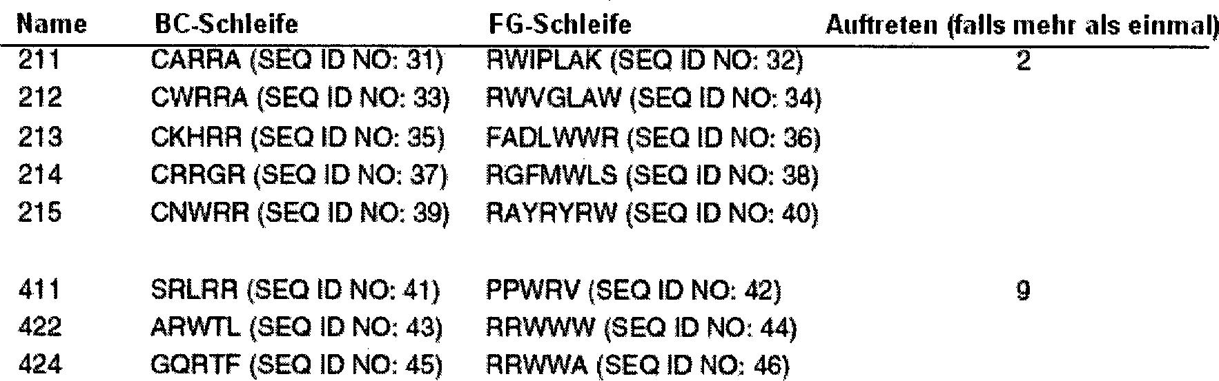
Der
Klon 411, der der am stärksten
angereicherte Klon war, wurde unter Verwendung von Phagen-ELISA
charakterisiert. Der Klon 411 zeigte selektive Bindung und Inhibition
der Bindung in Gegenwart von etwa 10 μM Ubiquitin in Lösung (
BEISPIEL XIEXAMPLE XI
Verfahren für die Immobilisierung kleiner MoleküleProcedure for immobilization small molecules
Zielmoleküle wurden in den Wells einer Mikrotiterplatte (Maxisorp, Nunc) wie hier im Folgenden beschrieben immobilisiert, und die Wells wurden mit BSA geblockt. Zusätzlich zur Verwendung von Trägerprotein wie im Folgenden beschrieben kann ein Konjugat eines Zielmoleküls in Biotin hergestellt werden. Der biotinylierte Ligand kann dann am Well einer Mikrotiterplatte immobilisiert werden, das mit Streptavidin beschichtet wurde.Target molecules were in the wells of a microtiter plate (Maxisorp, Nunc) like here in the Immobilized as described below, and the wells were labeled with BSA blocked. additionally for the use of carrier protein such as described below may be a conjugate of a target molecule in biotin getting produced. The biotinylated ligand can then be used at the well Microtiter plate which has been coated with streptavidin.
Zusätzlich zur Verwendung eines Trägerproteins wie unten beschrieben könnte man ein Konjugat eines Zielmoleküls und Biotin (Pierce) herstellen und einen biotinylierten Liganden an einem Mikrotiterplatten-Well immobilisieren, das mit Streptavidin beschichtet wurde (Smith and Scott, 1993).In addition to Use of a carrier protein as described below a conjugate of a target molecule and biotin (Pierce) and a biotinylated ligand immobilize on a microtiter plate well containing streptavidin was coated (Smith and Scott, 1993).
Kleine Moleküle können mit einem Trägerprotein, wie etwa Rinderserumalbumin (BSA, Sigma), konjugiert und passiv an dem Mikrotiterplatten-Well adsorbiert werden. Alternativ können auch Verfahren der chemischen Konjugation verwendet werden. Zusätzlich können andere feste Träger als Mikrotiterplatten problemlos verwendet werden.little one molecules can with a carrier protein, such as bovine serum albumin (BSA, Sigma), conjugated and passive be adsorbed on the microtiter plate well. Alternatively, too Method of chemical conjugation can be used. In addition, others can solid carriers can be used without problems as microtiter plates.
BEISPIEL XIIEXAMPLE XII
Fluorescein bindender MonobodyFluorescein binding monobody
Fluorescein ist als ein Target für die Selektion von Antikörpern aus kombinatorischen Bibliotheken verwendet worden (Barbas, et al. 1992). NHS-Fluorescein wurde von Pierce erhalten und gemäß den Herstelleranweisungen bei der Herstellung von Konjugaten mit BSA (Sigma) verwendet. Es wurden zwei Typen von Fluorescein-BSA-Konjugaten hergestellt, mit ungefähren molaren Verhältnissen von 17 (Fluorescein) zu eins (BSA).fluorescein is as a target for the selection of antibodies from combinatorial libraries (Barbas, et al. 1992). NHS-fluorescein was obtained from Pierce and according to the manufacturer's instructions used in the preparation of conjugates with BSA (Sigma). It Two types of fluorescein-BSA conjugates were prepared, with approximate molar ratios from 17 (fluorescein) to one (BSA).
Der
Selektionsprozess wurde 5–6-mal
wiederholt, um positive Klone zu konzentrieren. Bei diesem Versuch
wurde die Phagenbibliothek mit einem Proteingemisch (BSA, Cytochrom
C (Sigma, Pferd) und RNaseA (Sigma, Rind), jeweils 1 mg/ml) bei
Raumtemperatur für
30 Minuten inkubiert, bevor die Zugabe zu den Liganden-beschichteten
Wells erfolgte. Gebundener Phage wurde in TBS mit 10 4M löslichem
Fluorescein anstelle einer sauren Elution eluiert. Nach der letzten
Runde wurden einzelne Klone gepickt, und ihre Bindungsaffinitäten (siehe
unten) und DNA-Sequenzen wurden bestimmt. Tabelle
4: Klone
aus Bibliothek #2 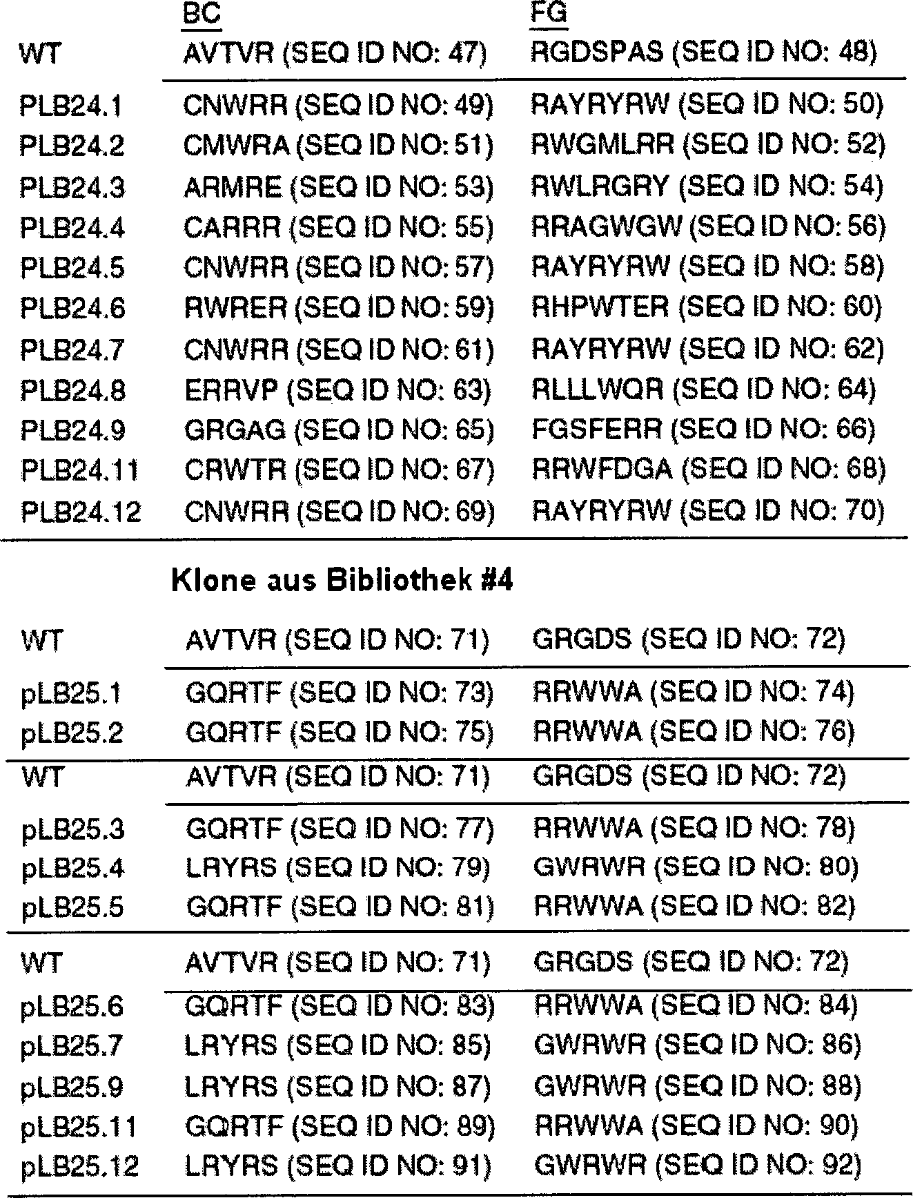
Die
vorläufige
Charakterisierung der Bindungsaffinitäten ausgewählter Klone erfolgte unter
Verwendung von Phagen-ELISA und kompetitivem Phagen-ELISA (siehe
BEISPIEL XIIIEXAMPLE XIII
Digoxigenin bindender MonobodyDigoxigenin binding monobody
Digoxigenin-3-O-methyl-carbonyl-e-aminocapronsäure-NHS (Boehringer Mannheim) wird verwendet, um ein Digoxigenin-BSA-Konjugat herzustellen. Die Kopplungsreaktion wird nach Herstelleranweisungen durchgeführt. Das Digoxigenin-BSA-Konjugat wird in den Wells einer Mikrotiterplatte immobilisiert und für das Panning verwendet. Das Panning wird 5 bis 6-mal wiederholt, um bindende Klone anzureichern. Da Digoxigenin in wässriger Lösung nur schwach löslich ist, werden die gebundenen Phagen unter Verwendung saurer Lösung von dem Well eluiert. Siehe Beispiel XIV.Digoxigenin-3-O-methyl-carbonyl-e-aminocaproic acid-NHS (Boehringer Mannheim) is used to prepare a digoxigenin-BSA conjugate manufacture. The coupling reaction is carried out according to the manufacturer's instructions. The Digoxigenin-BSA conjugate is placed in the wells of a microtiter plate immobilized and for used the panning. The panning is repeated 5 to 6 times, to enrich binding clones. As digoxigenin in aqueous solution only slightly soluble is bound phages using acidic solution of eluted the well. See example XIV.
BEISPIEL XIVEXAMPLE XIV
TSAC (Übergangszustandanalogon-Verbindung)-bindende MonobodiesTSAC (transition state analogue compound) binding monobodies
Carbonat-hydrolysierende Monobodies werden wie folgt selektiert. Ein Übergangszustandanalogon für die Carbonat-Hydrolyse, 4-Nitrophenylphosphonat, wird durch eine Arbuzov-Reaktion synthetisiert, wie zuvor beschrieben (Jacobs und Schultz, 1987). Das Phosphonat wird dann an das Trägerprotein, BSA, unter Verwendung von Carbodiimid gekoppelt, gefolgt von ausgedehnter Dialyse (Jacobs and Schultz, 1987). Das Hapten-BSA-Konjugat wird in den Wells einer Mikrotiterplatte immobilisiert, und die Monobody-Selektion wird durchgeführt wie oben beschrieben. Die katalytischen Aktivitäten der selektierten Monobodies werden unter Verwendung von 4-Nitrophenylcarbonat als Substrat getestet.Carbonate-hydrolyzing monobodies are selected as follows. A transitional state analog for the carbonate hydrolysis, 4-nitrophenylphosphonate, is synthesized by an Arbuzov reaction as previously described (Jacobs and Schultz, 1987). The phosphonate is then coupled to the carrier protein, BSA, using carbodiimide, followed by extensive dialysis (Jacobs and Schultz, 1987). The hapten-BSA conjugate is immobilized in the wells of a microtiter plate and monobody selection is performed as described above. The catalytic activities of the selected monobodies are tested using 4-nitrophenyl carbonate as the substrate.
Andere Haptene, die nützlich für die Produktion katalytischer Monobodies sind, sind in H. Suzuki (1994) und in N. R. Thomas (1994) zusammengefasst.Other Haptene, the useful for the Production of catalytic monobodies are described in H. Suzuki (1994) and summarized in N.R. Thomas (1994).
BEISPIEL XVEXAMPLE XV
NMR-Charakterisierung von Fn3 und Vergleich des von Hefe sekretierten Fn3 mit dem von E. coli sekretiertenNMR characterization of Fn3 and comparison yeast secreted Fn3 with that secreted by E. coli
Es wurden Versuche mit Kernspinresonanz (NMR) durchgeführt, um die Kontaktoberfläche zwischen FnAb und einem Zielmolekül zu identifizieren, z. B. von Monobodies gegenüber Fluorescein, Ubiquitin, RNaseA und löslichen Derivaten von Digoxigenin. Diese Information wird dann verwendet, um die Affinität und Spezifität des Monobody zu verbessern. Gereinigte Monobody-Proben werden in einem geeigneten Puffer für die NMR-Spektroskopie gelöst, wobei hierfür eine Amicon Ultrafiltrationszelle mit einer YM-3-Membran verwendet wird. Die Puffer werden mit 90% H2O/10% D2O (destillierte Qualität, Isotec) oder mit 100% D2O hergestellt. Deuterierte Verbindungen (z. B. Acetat) werden verwendet, um starke Signale von diesen zu eliminieren.Nuclear magnetic resonance (NMR) experiments were performed to identify the contact surface between FnAb and a target molecule, e.g. As monobodies to fluorescein, ubiquitin, RNaseA and soluble derivatives of digoxigenin. This information is then used to enhance the affinity and specificity of the monobody. Purified monobody samples are dissolved in a suitable buffer for NMR spectroscopy using an Amicon ultrafiltration cell with a YM-3 membrane. The buffers are made with 90% H 2 O / 10% D 2 O (distilled grade, Isotec) or with 100% D 2 O. Deuterated compounds (eg, acetate) are used to eliminate strong signals from them.
Die Durchführung der NMR-Versuche erfolgt auf einem Varian Unity INOVA 600 Spektrometer, ausgestattet mit vier RF-Kanälen und einer Dreifach-Resonanzsonde mit Pulsfeldgradienten-Fähigkeit. Die NMR-Spektren werden unter Verwendung von Weiterverarbeitungsprogrammen, wie etwa Felix (Molecular Simulations), nmrPipe, PIPP und CAPP (Garrett, et al., 1991; Delaglio, et al., 1995) auf UNIX-Arbeitsstationen analysiert. Sequenz-spezifische Resonanzzuordnungen erfolgen mittels gut etablierter Strategie unter Verwendung eines Satzes dreifacher Resonanzversuche (CBCA(CO)NH und HNCACB) (Grzesiek & Bax, 1992; Wittenkind & Mueller, 1993).The execution NMR experiments are performed on a Varian Unity INOVA 600 spectrometer, equipped with four RF channels and a triple resonance probe with pulse field gradient capability. The NMR spectra are analyzed using further processing programs, such as Felix (Molecular Simulations), nmrPipe, PIPP and CAPP (Garrett, et al., 1991; Delaglio, et al., 1995) on UNIX workstations. Sequence-specific resonance assignments are made using well-established Strategy using a set of triple resonance experiments (CBCA (CO) NH and HNCACB) (Grzesiek & Bax, 1992, Wittenkind & Mueller, 1993).
Der
Kern-Overhauser-Effekt (NOE) wird zwischen 1H-Kernen
beobachtet, die sich näher
als etwa 5 A sind, was es einem erlaubt, Informationen über den
Abstand zwischen Protonen zu erhalten. Es wird eine Reihe von Doppel-
und Dreifach-Resonanzversuchen (Tabelle 5; für jüngere Übersichtsartikel über diese
Techniken, siehe Bax & Grzesiek,
1993 und Kay, 1995) durchgeführt,
um Zwänge
im Bezug auf den Abstand (d. h. NOE) und den Torsionswinkel (J-Kopplung)
zu ermitteln. Es werden Isotopen-gefilterte Versuche durchgeführt, um
die Resonanzzuordnungen des gebundenen Liganden zu bestimmen und
um die Abstandszwänge
innerhalb des Liganden und diejenigen zwischen FnAb und dem Liganden
zu erhalten. Details der Sequenz-spezifischen Resonanzzuordnungen
und der NOE-Peak-Zuordnung sind an anderer Stelle im Detail beschrieben worden
(Clore & Gronenborn,
1991; Pascal, et al., 1994b; Metzler, et al., 1996). Tabelle 5. NMR-Versuche zur Strukturbestimmung
Die Rückgrat-1H-, 15N- und 13C-Resonanz-Zuordnungen für einen Monobody werden mit denjenigen von wildtypischem Fn3 verglichen, um strukturelle Veränderungen bei der Mutante zu bestimmen. Sobald diese Daten bestätigen, dass die Mutante die allgemeine Struktur beibehält, wird eine strukturelle Verfeinerung unter Verwendung experimenteller NOE-Daten durchgeführt. Da erwartet wird, dass der strukturelle Unterschied bei einem Monobody gering ist, kann die wildtypische Struktur als das Ausgangsmodell verwendet werden, nachdem die Aminosäuresequenz modifiziert wurde. Die Mutationen werden durch interaktives molekulares Modellieren in die wildtypische Struktur eingeführt, und dann wird bei der Struktur unter Verwendung eines Programms des molekularen Modellierens, wie etwa Quants (Molecular Simulations), eine Energie- Minimierung vorgenommen. Die Lösungsstruktur wird unter Verwendung von Zyklen des dynamischen simulierten Annealing (Nilges et al., 1988) mit dem Programm X-PLOR (Brunger, 1992) verfeinert. Typischerweise wird eine Gesamtheit von fünfzig Strukturen berechnet. Die Gültigkeit der verfeinerten Strukturen wird durch Berechnen einer kleineren Anzahl von Strukturen von zufällig erzeugten Anfangsstrukturen in X-PLOR unter Verwendung des YASAP-Protokolls (Nilges, et al., 1991) bestätigt. Die Struktur eines Monobody-Ligaeden-Komplexes wird berechnet, indem man zunächst beide Komponenten einzeln unter Verwendung intramolekularer NOEs verfeinert und die beiden dann unter Verwendung intermolekularer NOEs verbindet.The backbone 1 H, 15 N, and 13 C resonance assignments for a monobody are compared to those of wild type Fn3 to determine structural changes in the mutant. Once these data confirm that the mutant retains the general structure, structural refinement is performed using experimental NOE data. Since it is expected that the structural sub In the case of a monobody, the wild-type structure can be used as the starting model after the amino acid sequence has been modified. The mutations are introduced into the wild-type structure by interactive molecular modeling, and then the structure is minimized using a molecular modeling program, such as quantum (Molecular Simulations). The solution structure is refined using cycles of dynamic simulated annealing (Nilges et al., 1988) with the program X-PLOR (Brunger, 1992). Typically, a total of fifty structures is calculated. The validity of the refined structures is confirmed by computing a smaller number of structures of randomly-generated initial structures in X-PLOR using the YASAP protocol (Nilges, et al., 1991). The structure of a monobody-league complex is calculated by first refining both components individually using intramolecular NOEs and then combining the two using intermolecular NOEs.
Beispielsweise
ist das 1H,15N-HSQC-Spektrum
für den
Fluorescein-bindenden Antikörper
LB25.5 in
Versuche einer Störung der chemischen Verschiebung werden durchgeführt, indem man den Komplex zwischen einem Isotop-markierten FnAb und einem unmarkierten Liganden ausbildet. Die Ausbildung eines stöchiometrischen Komplexes wird verfolgt, indem man das HSQC-Spektrum aufzeichnet. Da die chemische Verschiebung extrem empfindlich gegenüber der Kernumgebung ist, resultiert die Ausbildung eines Komplexes für gewöhnlich in wesentlichen Änderungen der chemischen Verschiebung für die Resonanzen von Aminosäureresten in der Grenzfläche. Es werden Isotopen-editierte NMR-Versuche (2D HSQC und 3D CBCA(CO)NH) verwendet, um die Resonanzen zu identifizieren, die bei der markierten Komponente des Komplexes gestört werden; d. h. bei dem Monobody. Obwohl die Möglichkeit von Artefakten aufgrund konformativer Veränderungen mit großer Reichweite stets berücksichtigt werden muss, so werden wesentliche Unterschiede für Reste, die sich an zusammenhängenden Oberflächen häufen, mit größter Wahrscheinlichkeit aus direkten Kontakten entstehen (Chen et al., 1993; Gronenborn & Clore, 1993).tries a fault The chemical shift is performed by breaking the complex between an isotope-labeled FnAb and an unlabelled ligand is formed. The formation of a stoichiometric Complexity is tracked by recording the HSQC spectrum. Because the chemical shift extremely sensitive to the core environment is the result of the formation of a complex usually in significant changes the chemical shift for the resonances of amino acid residues in the interface. Isotope-edited NMR experiments (2D HSQC and 3D CBCA (CO) NH) used to identify the resonances that are marked at the Complexed component of the complex become; d. H. at the monobody. Although the possibility of artifacts due Conformative changes with big ones Range always considered must be essential differences for remnants, which are connected to each other surfaces piling, most likely arising from direct contacts (Chen et al., 1993; Gronenborn & Clore, 1993).
Ein alternatives Verfahren zur Kartierung der Interaktionsoberfläche verwendet Messungen des Amid-Wasserstoff-Austauschs (HX). Die HX-Raten für jedes Amidproton werden für sowohl freien 15N-markierten Monobody als auch für mit einem Liganden komplexierten Monobody gemessen. Es wird erwartet, dass die Liganden-Bindung in verminderten Amid-HX-Raten für Monobody-Reste in der Grenzfläche zwischen den beiden Proteinen resultiert und somit die Bindungsoberfläche identifiziert. Die HX-Raten für Monobodies in dem Komplex werden gemessen, indem man es gestattet, dass HX nach vom Transfer des Komplexes auf D2O für eine variable Zeitspanne stattfindet; der Komplex wird dissoziiert, indem man den pH verringert, und das HSQC- Spektrum wird bei niedrigem pH aufgezeichnet, wo Amid-HX langsam ist. Fn3 ist stabil und bei niedrigem pH löslich, was der Vorbedingung für die Versuche gerecht wird.An alternative method for mapping the interaction surface uses measurements of amide-hydrogen exchange (HX). The HX rates for each amide proton are measured for both free 15 N-labeled monobody and monobody complexed with a ligand. Ligand binding is expected to result in reduced amide HX rates for monobody residues in the interface between the two proteins, thus identifying the binding surface. The HX rates for monobodies in the complex are measured by allowing HX to take place after transfer of the complex to D 2 O for a variable period of time; the complex is dissociated by lowering the pH, and the HSQC spectrum is recorded at low pH, where amide HX is slow. Fn3 is stable and soluble at low pH, which is a prerequisite for the experiments.
BEISPIEL XVIEXAMPLE XVI
Konstruktion und Analyse eines für Ubiquitin spezifischen Fn3-Display-SystemsConstruction and analysis of one for ubiquitin specific Fn3 display system
Es wurde ein Fn3-Display-System erstellt und synthetisiert, Ubiquitin-bindende Klone wurden isoliert, und eine wesentliche Fn3-Mutante unter diesen Klonen wurde biophysikalisch charakterisiert.It An Fn3 display system was created and synthesized, ubiquitin-binding Clones were isolated and a major Fn3 mutant among them Cloning was characterized biophysically.
Die Genkonstruktion und der Phage-Display von Fn3 wurden durchgeführt wie in den Beispielen I und II oben. Das Fn3-Phagen-pIII-Fusionsprotein wurde über einen Phagemid-Display-Vektor exprimiert, während die anderen Komponenten des M13-Phagen, einschließlich des wildtypischen pIII, unter Verwendung eines Helferphagen (Bass et al., 1990) produziert wurden. Somit sollte ein durch dieses System erzeugter Phage weniger als eine Kopie von Fn3, das auf der Oberfläche präsentiert wird, enthalten. Die Oberflächenpräsentation von Fn3 auf dem Phagen wurde mittels ELISA unter Verwendung eines Anti-Fn3-Antikörpers detektiert. Nur Phagen, die den Fn3-pIII-Fusionsvektor enthalten, reagierten mit dem Antikörper.The Gene construction and phage display of Fn3 were performed as well in Examples I and II above. The Fn3 phage pIII fusion protein was over a phagemid display vector expressed while the other components of the M13 phage, including the wild-type pIII, using a helper phage (Bass et al., 1990) were. Thus, a phage produced by this system should be less as a copy of Fn3 presented on the surface. The surface Exposure of Fn3 on the phage was determined by ELISA using a Anti-Fn3 antibody detected. Only phages containing the Fn3-pIII fusion vector reacted with the antibody.
Nachdem bestätigt wurde, dass die Phagenoberfläche Fn3 präsentiert, wurde eine Phage-Display-Bibliothek von Fn3 konstruiert wie in Beispiel III. Es wurden zufällige Sequenzen in die BC- und FG-Schleifen eingeführt. Bei der ersten Bibliothek wurden fünf Reste (77–81) nach dem Zufallsprinzip verändert, und drei Reste (82–84) wurden aus der FG-Schleife deletiert. Die Deletion hatte den Zweck, die Flexibilität zu reduzieren und die Bindungsaffinität der FG-Schleife zu verbessern. Es wurden außerdem fünf Reste (26–30) in der BC-Schleife nach Zufallsprinzip verändert, um eine größere Kontaktoberfläche mit dem Zielmolekül bereitzustellen. Somit enthält die resultierende Bibliothek fünf nach Zufallsprinzip veränderte Reste in jeder der Schleifen BC und FG (Tabelle 6). Diese Bibliothek enthielt etwa 108 unabhängige Klone.After confirming that the phage surface presents Fn3, a phage display library of Fn3 was constructed as in Example III. Random sequences were introduced into the BC and FG loops. In the first library, five residues (77-81) were randomly changed and three residues (82-84) were deleted from the FG loop. The purpose of the deletion was to reduce flexibility and to improve the binding affinity of the FG loop. In addition, five residues (26-30) in the BC loop were randomly changed to provide a larger contact surface with the target molecule. Thus, the resulting library contains five randomly altered residues in each of the loops BC and FG (Table 6). This library contained about 10 8 independent clones.
Durchmusterung der BibliothekScreening the library
Die
Bibliothek wurde unter Verwendung von Ubiquitin als Zielmolekül durchmustert.
Bei jeder Runde des Panning wurden Fn3-Phagen an eine mit Ubiquitin
beschichtete Oberfläche
adsorbiert, und gebundene Phagen wurden in kompetitiver Weise mit
löslichem
Ubiquitin eluiert. Das Wiedergewinnungsverhältnis verbesserte sich von
4,3 × 10⁻⁷ in
der zweiten Runde auf 4,5 × 10⁻⁶ in
der fünften
Runde, was eine Anreicherung der bindenden Klone nahelegt. Nach
fünf Runden
des Panning wurden die Aminosäuresequenzen
der einzelnen Klone bestimmt (Tabelle 6). Tabelle
6. Sequenzen in den abgewandelten Schleifen angereicherter Klone 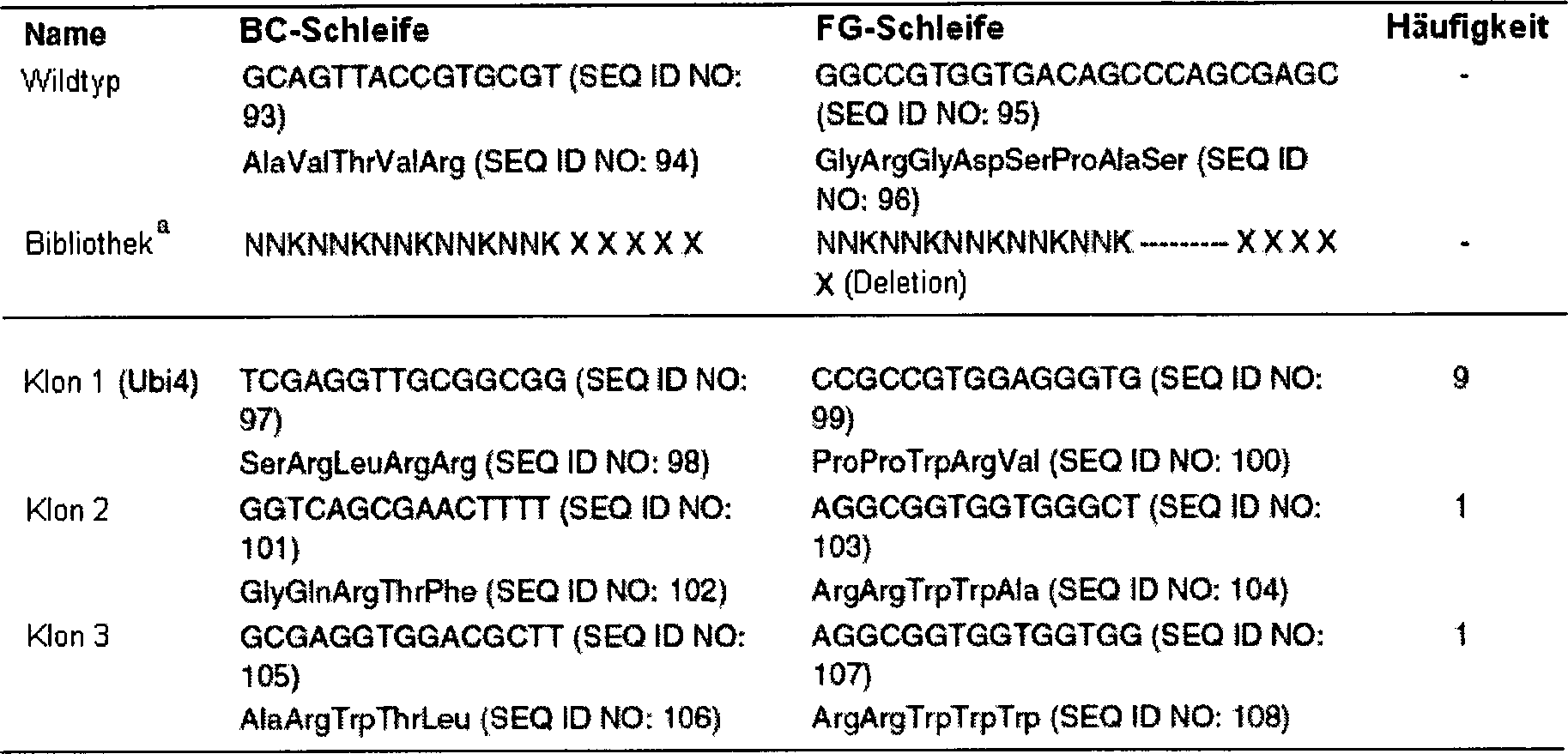
- aN bezeichnet ein äquimolares Gemisch aus A, T, G und C; K bezeichnet ein äquimolares Gemisch aus G und T.
- a N denotes an equimolar mixture of A, T, G and C; K denotes an equimolar mixture of G and T.
Ein als Ubi4 bezeichneter Klon dominierte den angereicherten Pool der Fn3-Varianten. Daher wurde die weitere Untersuchung auf diesen Ubi4-Klon fokussiert. Ubi4 enthält vier Mutationen in der BC-Schleife (Arg 30 in der BC-Schleife war konserviert), sowie fünf Mutationen und drei Deletionen in der FG-Schleife. Somit waren 13% (12 von 94) der Reste in Ubi4 gegenüber der Wildtypsequenz verändert.One Ubi4 designated clone dominated the enriched pool of the Fn3 variants. Therefore, the further investigation on this Ubi4 clone focused. Ubi4 contains there were four mutations in the BC loop (Arg 30 in the BC loop conserved), as well as five Mutations and three deletions in the FG loop. Thus, 13% (12 of 94) of the residues in Ubi4 changed over the wild-type sequence.
Charakterisierung einer Mutanten von Fn3-ProteinCharacterization of a mutant of Fn3 protein
Das Expressionssystem erbrachte 50–100 mg an Fn3-Protein pro Liter Kultur. Ein ähnliches Mengenniveau der Proteinexpression wurde für den Ubi4-Klon und andere mutante Fn3-Proteine beobachtet.The Expression system yielded 50-100 mg of Fn3 protein per liter of culture. A similar level of protein expression was for observed the Ubi4 clone and other mutant Fn3 proteins.
Ubi4-Fn3 wurde als ein unabhängiges Protein exprimiert. Obwohl eine Mehrheit von Ubi4 in E. coli als ein lösliches Protein exprimiert wurde, so wurde für seine Löslichkeit herausgefunden, dass diese signifikant reduziert ist, wenn man sie mit der von wildtypischem Fn3 verglich. Ubi4 war bei niedrigem pH-Wert mit bis zu –20 μM löslich, mit viel geringerer Löslichkeit bei neutralem pH-Wert. Diese Löslichkeit war nicht hoch genug für eine detaillierte strukturelle Charakterisierung unter Verwendung von NMR-Spektroskopie oder Röntgenkristallographie.Ubi4-Fn3 was expressed as an independent protein. Although a majority of Ubi4 was expressed in E. coli as a soluble protein, its solubility was found to be significant is reduced when compared to that of wild-type Fn3. Ubi4 was soluble at low pH up to -20 μM, with much lower solubility at neutral pH. This solubility was not high enough for detailed structural characterization using NMR spectroscopy or X-ray crystallography.
Die Löslichkeit des Ubi4-Proteins wurde verbessert, indem man einen Löslichkeitsschwanz, GKKGK, als C-terminale Verlängerung anfügte. Das Gen für Ubi4-Fn3 wurde unter Verwendung von PCR in den Expressionsvektor pAS45 subkloniert. Der C-terminale Löslichkeits-Tag, GKKGK, wurde bei diesem Schritt eingebaut. E. coli BL21 (DE3) (Novagen) wurde mit dem Expressionsvektor (pAS45 und dessen Derivate) transformiert. Die Zellen wurden in M9-Minimalmedium und M9-Medium, das mit Bactotrypton (Difco), enthaltend Ampicillin (200 μg/ml), supplementiert war, vermehrt. Für die Isotopenmarkierung ersetzte 15N NH4Cl das unmarkierte NH4Cl in den Medien. 500 ml Medium in einem 2 Liter großen Baffle-Kolben wurden mit 10 ml an Übernachtkultur angeimpft und bei 37°C geschüttelt. IPTG wurde mit einer Endkonzentration von 1 mM hinzugegeben, um die Proteinexpression zu starten, wenn die OD (600 nm) eins erreicht. Die Zellen wurden 3 Stunden nach der Zugabe von IPTG durch Zentrifugation geerntet und bis zur Verwendung gefroren bei –70°C gelagert.The solubility of the Ubi4 protein was improved by adding a solubility tail, GKKGK, as C-terminal extension. The gene for Ubi4-Fn3 was subcloned into the expression vector pAS45 using PCR. The C-terminal solubility tag, GKKGK, was incorporated at this step. E. coli BL21 (DE3) (Novagen) was transformed with the expression vector (pAS45 and its derivatives). Cells were propagated in M9 minimal medium and M9 medium supplemented with bactotryptone (Difco) containing ampicillin (200 μg / ml). For isotopic labeling, 15 N NH 4 Cl replaced the unlabelled NH 4 Cl in the media. 500 ml of medium in a 2-liter baffle flask were inoculated with 10 ml of overnight culture and shaken at 37 ° C. IPTG was added at a final concentration of 1 mM to start protein expression when the OD (600 nm) reached one. The cells were harvested by centrifugation 3 hours after the addition of IPTG and stored frozen at -70 ° C until use.
Proteine wurden wie folgt gereinigt. Die Zellen wurden in 5 ml/(g Zellen) an Tris (50 mM, pH 7,6), enthaltend Phenylmethylsulfonyl-Fluorid (1 mM), suspendiert. Hühnereiweiß-Lysozym (Sigma) wurde mit einer Endkonzentration von 0,5 mg/ml hinzugegeben. Nach dem Inkubieren der Lösung für 30 Minuten bei 37°C wurde diese dreimal für 30 Sekunden auf Eis mit Ultraschall behandelt. Die Zelltrümmer wurden durch Zentrifugation entfernt. Konzentriertes Natriumchlorid wurde mit einer Endkonzentration von 0,5 M zu der Lösung hinzugegeben. Die Lösung wurde auf eine Hi-Trap-Cheliersäule (Pharmacia) aufgetragen, die vorab mit Nickel beladen und in dem Tris-Puffer, enthaltend Natriumchlorid (0,5 M), äquilibriert worden war. Nach dem Waschen der Säule mit dem Puffer wurde His-Tag-Fn3 eluiert, wobei der Puffer 500 mM Imidazol enthielt. Das Protein wurde unter Verwendung einer ResourceS-Säule (Pharmacia) mit einem NaCl-Gradienten in einem Natriumacetatpuffer (20 mM, pH 4,6) weiter gereinigt.proteins were cleaned as follows. The cells were incubated in 5 ml / (g cells) Tris (50 mM, pH 7.6) containing phenylmethylsulfonyl fluoride (1 mM). Hen egg white lysozyme (Sigma) was added at a final concentration of 0.5 mg / ml. After incubating the solution for 30 Minutes at 37 ° C was this three times for Sonicated for 30 seconds on ice. The cell debris was removed by centrifugation. Concentrated sodium chloride was added to the solution at a final concentration of 0.5M. The solution was on a hi-trap chandelier (Pharmacia) applied in advance loaded with nickel and in the Tris buffer containing sodium chloride (0.5 M) was equilibrated. To washing the column His-tag Fn3 was eluted with the buffer, the buffer being 500 mM Imidazole contained. The protein was purified using a ResourceS column (Pharmacia) with a NaCl gradient further purified in a sodium acetate buffer (20 mM, pH 4.6).
Mit dem GKKGK-Schwanz (SEQ ID NO: 109) wurde die Löslichkeit des Ubi4-Proteins auf bis zu über 1 mM bei niedrigem pH-Wert und auf bis zu –50 μM bei neutralem pH-Wert gestei gert. Daher wurden weitere Analysen an Ubi4 mit seiner C-terminalen Verlängerung (hier im Folgenden als Ubi4-K bezeichnet) durchgeführt. Es ist berichtet worden, dass die Löslichkeit eines Minibody signifikant gesteigert werden kann, indem man drei Lys-Reste an den N- oder C-Terminus anfügt (Bianchi et al., 1994). Im Falle des Proteins Rop ist ein nicht-strukturierter C-terminaler Schwanz entscheidend für das Aufrechterhalten der Löslichkeit (Smith et al., 1995).With the GKKGK tail (SEQ ID NO: 109) was the solubility of the Ubi4 protein up to more than 1 at low pH and up to -50 μM at neutral pH. Therefore, further analysis was done on Ubi4 with its C-terminal extension (hereinafter referred to as Ubi4-K) performed. It it has been reported that the solubility of a minibody can be significantly increased by taking three Lys residues at the N- or C-terminus appends (Bianchi et al., 1994). In the case of the protein Rop is a non-structured C-terminal tail crucial for maintaining solubility (Smith et al., 1995).
Die Oligomerisierungszustände des Ubi4-Proteins wurden unter Verwendung einer Größenausschlusssäule bestimmt. Das wildtypische Fn3-Protein war bei niedrigen und neutralen pH-Werten monomer. Jedoch war der Peak des Ubi4-K-Proteins signifikant breiter als der von wildtypischem Fn3 und eluierte nach dem wildtypischen Protein. Dies deutet auf Interaktionen zwischen Ubi4-K und dem Säulenmaterial hin, was die Verwendung von Größenausschlusschromatographie zur Bestimmung des Oligomerisierungszustands von Ubi4 ausschließt. NMR-Studien legen nahe, dass das Protein bei niedrigem pH-Wert monomer ist.The oligomerization of the Ubi4 protein were determined using a size exclusion column. The wild type Fn3 protein was monomeric at low and neutral pHs. however the peak of the Ubi4-K protein was significantly wider than that of wild-type Fn3 and eluted after the wild-type protein. This indicates interactions between Ubi4-K and the column material suggesting the use of size exclusion chromatography for the determination of the oligomerization state of Ubi4 excludes. NMR studies suggest that the protein is monomeric at low pH.
Das
Ubi4-K-Protein behielt eine Bindungsaffinität an Ubiquitin bei, wie durch
ELISA bewertet (
Beispiel XVIIExample XVII
Stabilitätsmessungen bei MonobodiesStability measurements in monobodies
Durch Guanidin-Hydrochlorid (GuHCl) induzierte Entfaltungsreaktionen und Reaktionen erneuter Faltung wurden durch Messen der Tryptophan-Fluoreszenz verfolgt. Die Durchführung der Versuche erfolgte auf einem Spectronic AB-2 Spektrofluorometer, das mit einer Motorbetriebenen Spritze (Hamilton Co.) ausgestattet war. Die Küvettentemperatur wurde auf 30°C gehalten. Das Spektrofluorometer und die Spritze wurden durch einen Einzelcomputer unter Verwendung eines im Haus hergestellten Interface kontrolliert. Dieses System nimmt automatisch eine Reihe von Spektren nach der GuHCl-Titration auf. Ein Versuch startete mit 1,5 ml Pufferlösung, die 5 μM an Protein enthielt. Ein Emissionsspektrum (300–400 nm; Anregung bei 290 nm) wurde nach einer Verzögerungszeit (3–5 Minuten) nach jeder Injektion (50 oder 100 μl) einer GuHCl enthaltenden Pufferlösung aufgezeichnet. Diese Schritte wurden wiederholt, bis das Lösungsvolumen die vollständige Kapazität einer Küvette (3,0 ml) erreicht hatte. Die Fluoreszenzintensitäten wurden als Verhältnisse gegenüber der Intensität an einem isofluoreszenten Punkt, der in separaten Versuchen bestimmt wurde, normalisiert. Die Entfaltungskurven wurden mittels ei nes Zwei-Zustands-Modells unter Verwendung einer nicht-linearen Least-Squares-Routine (Santoro & Bolen, 1988) angepasst. Zwischen den Versuchen mit Verzögerungszeiten (Zeit zwischen einer Injektion und dem Beginn der Aufnahme des Spektrums) von 2 Minuten und 10 Minuten wurden keine signifikanten Unterschiede beobachtet, was anzeigt, dass die Entfaltungs/Rückfaltungsreaktionen bei den verwendeten Verzögerungszeiten bei jedem Konzentrationspunkt nahezu ein Gleichgewicht erreichten.Guanidine hydrochloride (GuHCl) induced unfolding reactions and refolding reactions were monitored by measuring the tryptophan fluorescence. The experiments were carried out on a Spectronic AB-2 spectrofluorometer equipped with a motor-driven syringe (Hamilton Co.). The cuvette temperature was maintained at 30 ° C. The spectrofluorometer and syringe were controlled by a single computer using an in-house interface. This system automatically picks up a series of spectra after GuHCl titration. An experiment started with 1.5 ml Buffer solution containing 5 μM of protein. An emission spectrum (300-400 nm, excitation at 290 nm) was recorded after a delay time (3-5 minutes) after each injection (50 or 100 μl) of a buffer solution containing GuHCl. These steps were repeated until the solution volume reached the full capacity of a cuvette (3.0 ml). The fluorescence intensities were normalized as ratios to intensity at an isofluorescent point determined in separate experiments. The unfolding curves were fit by means of a two-state model using a non-linear least squares routine (Santoro & Bolen, 1988). No significant differences were observed between the delays (time between injection and commencement of spectrum acquisition) experiments of 2 minutes and 10 minutes, indicating that the unfolding / refolding reactions almost reached equilibrium at the delay times used at each concentration point.
Die
konformative Stabilität
von Ubi4-K wurde unter Verwendung des oben beschriebenen GuHCl-induzierten
Entfaltungsverfahrens gemessen. Die Messungen wurden unter zwei
Sätzen
von Bedingungen durchgeführt;
erstens bei einem pH von 3,3 in Gegenwart von 300 mM Natriumchlorid,
wo Ubi4-K hochgradig löslich
ist, und zweitens in TBS, das für
die Bibliotheksdurchmusterung verwendet wurde. Unter beiden Bedingungen
war die Entfaltungsreaktion reversibel, und wir detektierten keine
Anzeichen der Aggregation oder irreversiblen Entfaltung.
Obwohl die eingeführten Mutationen in den beiden Schleifen die Stabilität von Ubi4-K im Vergleich zum wildtypischen Fn3 natürlich verringerten, bleibt die Stabilität von Ubi4 vergleichbar mit der eines „typischen" globulären Proteins. Es ist außerdem anzumerken, dass die Stabilität des wildtypischen Proteins und des Ubi4-K-Proteins bei pH 3,3 größer war als bei pH 7,5.Even though the imported ones Mutations in the two loops the stability of Ubi4-K compared to wild-type Fn3 of course decreased, the stability of Ubi4 remains comparable to that of a "typical" globular protein. It is also to note that stability of the wild-type protein and the Ubi4-K protein was greater at pH 3.3 as at pH 7.5.
Das Ubi4-Protein besaß im Vergleich zu wildtypischem Fn3 eine signifikant reduzierte Löslichkeit, jedoch wurde die Löslichkeit durch das Anfügen eines Löslichkeitsschwanzes verbessert. Da die beiden mutierten Schleifen die einzigen Unterschiede zwischen wildtypischem Protein und den Ubi4-Proteinen umfassen, müssen diese Schleifen den Ursprung der reduzierten Löslichkeit darstellen. An diesem Punkt ist nicht klar, ob die Aggregation von Ubi4-K durch Interaktionen zwischen den Schleifen oder durch Interaktionen zwischen den Schleifen und den nicht variablen Regionen des Fn3-Gerüsts verursacht wird.The Ubi4 protein possessed in Compared to wild-type Fn3 a significantly reduced solubility, however, the solubility became by attaching a solubility tail improved. Because the two mutated loops are the only differences between wild-type protein and the Ubi4 proteins, these must Loops represent the origin of reduced solubility. At this Point is not clear whether the aggregation of Ubi4-K through interactions between loops or through interactions between loops and the non-variable regions of the Fn3 scaffold.
Das
Ubi4-K-Protein behielt die allgemeine Faltung von Fn3 bei, was zeigt,
dass dieses Gerüst
eine große
Anzahl von Mutationen in den zwei getesteten Schleifen beherbergen
kann. Obwohl die Stabilität
des Ubi4-K-Proteins signifikant niedriger ist als die des wildtypischen
Fn3-Proteins, so
besitzt das Ubi4-Protein nach wie vor eine konformative Stabilität, die mit
derjenigen für
kleine globuläre
Proteine vergleichbar ist. Die Verwendung einer hochstabilen Domäne als Gerüst ist klar
vorteilhaft für
die Einführung
von Mutationen, ohne dabei die allgemeine Faltung des Gerüsts zu beeinflussen.
Zusätzlich
ist die von GuHCl induzierte Entfaltung des Ubi4-Proteins nahezu vollständig reversibel.
Dies erlaubt die Präparation
eines korrekt gefalteten Proteins, selbst wenn eine Fn3-Mutante
in einer falsch gefalteten Form, wie etwa in Einschlusskörpern, exprimiert
wird. Die mäßige Stabilität von Ubi4
bei Bedingungen, die für
die Bibliotheksdurchmusterung verwendet werden, zeigt an, dass Fn3-Varianten
auf der Phagenoberfläche
gefaltet werden. Dies legt nahe, dass ein Fn3-Klon durch seine Bindungsaffinität in der
gefalteten Form, nicht in einer denaturierten Form, selektiert wird.
Dickinson et al. schlugen vor, dass Val 29 und Arg 30 in der BC-Schleife
Fn3 stabilisieren. Val 29 stellt den Kontakt mit dem hydrophoben
Kern her, und Arg 30 bildet Wasserstoffbindungen mit Gly 52 und
Val 75 aus. In Ubi4-Fn3 ist Val 29 durch Arg ersetzt, während Arg
30 konserviert wird. Die FG-Schleife wurde auch in der Bibliothek mutiert.
Diese Schleife ist in der wildtypischen Struktur flexibel und zeigt
eine große
Variation der Länge
unter humanen Fn3-Domänen
(Main et al., 1992). Diese Beobachtungen legen nahe, dass Mutationen
in der FG-Schleife weniger Einfluss auf die Stabilität besitzen
könnten.
Zusätzlich
ist der N-terminale Schwanz von Fn3 benachbart zu der molekularen
Oberfläche,
die von den Schleifen BC und FG ausgebildet wird (
Beispiel XVIIIExample XVIII
NMR-Spektroskopie von Ubi4-Fn3NMR spectroscopy of Ubi4-Fn3
Ubi4-Fn3 wurde unter Verwendung einer Amicon-Ultrafiltrationseinheit in [2H]-Gly-HCl-Puffer (20 mM, pH 3,3), enthaltend NaCl (300 mM), gelöst. Die endgültige Proteinkonzentration betrug 1 mM. Es wurden NMR-Versuche auf einem Varian Unity 'NOVA 600 Spektrometer durchgeführt, das mit einer Dreifach-Resonanzsonde mit Pulsfeldgradient ausgestattet war. Die Sondentemperatur wurde auf 30°C eingestellt. Es wurden HSQC-, TOCSY-HSQC- und NOESY-HSQC-Spektren unter Verwendung veröffentlichter Verfahren (Kay et al., 1992; Zhang et al., 1994) aufgezeichnet. Die NMR-Spektren wurden unter Verwendung der Software NMRPipe und NMR View (Johnson & Blevins, 1994; Delaglio et al., 1995) auf UNIX-Arbeitsstationen weiterverarbeitet und analysiert. Sequenz-spezifische Resonanzzuordnungen erfolgten unter Verwendung von Standardprozeduren (Wüthrich, 1986; Clore & Gronenborn, 1991). Die Zuordnungen für wildtypisches Fn3 (Baron et al., 1992) wurden unter Verwendung eines 15N-markierten Proteins, das in Natriumacetatpuffer (50 mM, pH 4,6) gelöst war, bei 30°C bestätigt.Ubi4-Fn3 was dissolved using an Amicon ultrafiltration unit in [ 2 H] -Gly-HCl buffer (20 mM, pH 3.3) containing NaCl (300 mM). The final protein concentration was 1 mM. NMR experiments were performed on a Varian Unity 'NOVA 600 spectrometer equipped with a pulsed field gradient triple resonance probe. The probe temperature was set at 30 ° C. HSQC, TOCSY-HSQC and NOESY-HSQC spectra were recorded using published methods (Kay et al., 1992, Zhang et al., 1994). The NMR spectra were further processed and analyzed on UNIX workstations using the software NMRPipe and NMR View (Johnson & Blevins, 1994; Delaglio et al., 1995). Sequence-specific resonance assignments were made using standard procedures (Wüthrich, 1986, Clore & Gronenborn, 1991). The assignments for wild-type Fn3 (Baron et al., 1992) were confirmed using a 15 N-labeled protein dissolved in sodium acetate buffer (50 mM, pH 4.6) at 30 ° C.
Die
dreidimensionale Struktur von Ubi4-K wurde unter Verwendung dieses
heteronukleären NMR-Spektroskopieverfahrens
charakterisiert. Ein Spektrum von hoher Qualität kann mit einer 1 mM Lösung von 15N-markiertem Ubi4 (
Die vorstehende detaillierte Beschreibung und die Beispiele sind nur für die Klarheit des Verständnisses angegeben worden. Es sind keine unnötigen Einschränkungen daraus abzuleiten. Die Erfindung ist nicht auf die gezeigten und beschriebenen exakten Details beschränkt, da Abwandlungen, die für den Fachmann ersichtlich sind, in die durch die Ansprüche definierte Erfindung einbezogen sind.The The above detailed description and the examples are only for the Clarity of understanding been specified. There are no unnecessary restrictions derive from it. The invention is not limited to those shown and limited to the exact details described since modifications that will be apparent to those skilled in the art are in, by the claims defined invention are included.
LITERATURSTELLENREFERENCES
- Alzari, P. N., Lascombe, M.-B. & Poljak, R. J. (1988) Three-dimensional structure of antibodies. Annu. Rev. Immunol. 6, 555–580.Alzari, P.N., Lascombe, M.-B. & Poljak, R.J. (1988) Three-dimensional structure of antibodies. Annu. Rev. Immunol. 6, 555-580.
- Archer, S. J., Ikura, M., Torchia, D. A. & Bax, A. (1991) An alternative 3D NMR technique for correlating backbone 15N with side chain Hb resonances in large proteins. J. Magn. Reson. 95, 636–641.Archer, S.J., Ikura, M., Torchia, D.A. & Bax, A. (1991) To alternative 3D NMR technique for correlating backbone 15N with side chain Hb resonances in large proteins. J. Magn. Reson. 95, 636-641.
- Aukhil, I., Joshi, P., Van, Y. & Erickson, H. P. (1993) Cell- and heparin-binding domains of the hexabrachion arm identified by tenascin expression protein. J. Biol. Chem. 268, 2542–2553.Aukhil, I., Joshi, P., Van, Y. & Erickson, H.P. (1993) Cell and heparin-binding domains of the hexabrachione arm identified by tenascin expression protein. J. Biol. Chem. 268, 2542-2553.
- Barbas, C. F., III, Kang, A. S., Lerner, R. A. & Benkovic, S. J. (1991) Assembly of combinatorial antibody libraries on phage surfaces: the gene III site. Proc. Natl. Acad. Sci. USA 88, 7978–7982.Barbas, C.F., III, Kang, A.S., Lerner, R.A. & Benkovic, S. J. (1991) Assembly of combinatorial antibody libraries on phage surfaces: the gene III site. Proc. Natl. Acad. Sci. USA 88, 7978-7982.
- Barbas, C. F., III, Bam, J. D., Hoekstra, D. M. & Lerner, R. A. (1992) Semisynthetic combinatorial libraries: A chemical solution to the diversity problem. Proc. Natl. Acad. Sci. USA 89, 4457–4461.Barbas, C.F., III, Bam, J.D., Hoekstra, D.M. & Lerner, R.A. (1992) Semisynthetic combinatorial libraries: A chemical solution to the diversity problem. Proc. Natl. Acad. Sci. USA 89, 4457-4461.
- Baron, M., Main, A. L., Driscoll, P. C., Mardon, H. J., Boyd, J. & Campbell, I. D. (1992) 1H NMR assignment and secondary structure of the cell adhesion type II module of fibronectin. Biochemistry 31, 2068–2073.Baron, M., Main, AL, Driscoll, PC, Mardon, HJ, Boyd, J. & Campbell, ID (1992) 1 H NMR assignment and secondary structure of the cell adhesion type II module of fibronectin. Biochemistry 31, 2068-2073.
- Baron, M., Norman, D. G. & Campbell, I. D. (1991) Protein modules. Trends Biochem. Sci. 16, 13–17.Baron, M., Norman, D.G. & Campbell, I.D. (1991) Protein modules. Trends Biochem. Sci. 16, 13-17.
- Bass, S., Greene, R. & Wells, J. A. (1990) Hormone phage: An enrichment method for variant proteins with altered binding properties. Proteins: Struct. Funct. Genet. 8, 309–314.Bass, S., Greene, R. & Wells, J.A. (1990) Hormones phage: an enrichment method for variant proteins with altered binding properties. Protein: Struct. Funct. Genet. 8, 309-314.
- Bax, A. & Grzesiek, S. (1993) Methodological advances in protein NMR. Acc. Chem. Res. 26, 131–138.Bax, A. & Grzesiek, S. (1993) Methodological advances in protein NMR. Acc. Chem. Res. 26, 131-138.
- Becktel, W. J. & Schellman, J. A. (1987) Protein stability curves. Biopolymer 26, 1859–1877.Becktel, W.J. & Schellman, J.A. (1987) Protein stability curves. Biopolymer 26, 1859-1877.
- Bhat, T. N., Bentley, G. A., Boulot, G., Greene, M. I., Tello, D., Dall'acqua, W., Souchon, H., Schwarz, F. P., Mariuzza, R. A. & Poljak, R. J. (1994) Bound water molecules and conformational stabilization help mediate an antigen-antibody association. Proc. Natl. Acad. Sci. USA 91, 1089–1093.Bhat, T.N., Bentley, G.A., Boulot, G., Greene, M.I., Tello, D., Dall'acqua, W., Souchon, H., Schwarz, F.P., Mariuzza, R.A. & Poljak, R.J. (1994) Bound water molecules and conformational stabilization help mediate to antigen-antibody association. Proc. Natl. Acad. Sci. USA 91, 1089-1093.
- Bianchi, E., Venturini, S., Pessi, A., Tramontano, A. & Sollazzo, M. (1994) High level expression and rational mutagenesis of a designed protein, the minibody. From an insoluble to a soluble molecule. J. Mol. Biol. 236, 649.659.Bianchi, E., Venturini, S., Pessi, A., Tramontano, A. & Sollazzo, M. (1994) High level expression and rational mutagenesis of a designed protein, the minibody. From an insoluble to a soluble molecule. J. Mol. Biol. 236, 649.659.
- Billeter, M., Neri, D., Otting, G., Qian, Y. Q. & Wüthrich, K. (1992) Precise vicinal coupling constants 3JHNa in proteins from nonlinear fits of J-modulated [15N, 1H]-COSY experiments. J. Biomol. NMR 2, 257–274.Billeter, M., Neri, D., Otting, G., Qian, YQ & Wüthrich, K. (1992) Precise vicinal coupling constants 3JHNa in proteins from non-linear fits of J-modulated [ 15 N, 1 H] COZY experiments , J. Biomol. NMR 2, 257-274.
- Bodenhausen, G. & Ruben, D. J. (1980) Natural abundance nitrogen-15 NMR by enhanced heteronuclear spectroscopy. Chem. Phys. Lett. 69, 185–189.Bodenhausen, G. & Ruben, D.J. (1980) Natural abundance nitrogen-15 NMR by enhanced heteronuclear spectroscopy. Chem. Phys. Lett. 69, 185-189.
- Bork, P. & Doolittle, R. F., PNAS 89: 8990–8994 (1992).Bork, P. & Doolittle, R.F., PNAS 89: 8990-8994 (1992).
- Bork, P. & Doolittle, R. F. (1992) Proposed acquisition of an animal protein domain by bacteria. Proc. Natl. Acad. Sci. USA 89, 8990–8994.Bork, P. & Doolittle, R.F. (1992) Proposed acquisition of an animal protein domain by bacteria. Proc. Natl. Acad. Sci. USA 89, 8990-8994.
- Bork, P., Hom, L. & Sander, C. (1994) The immunoglobulin fold. Structural classification, sequence patterns and common core. J. Mol. Biol. 242, 309–320.Bork, P., Hom, L. & Sander, C. (1994) The immunoglobulin fold. Structural classification, sequence patterns and common core. J. Mol. Biol. 242, 309-320.
- Brünger, A. T. (1992) X-PLOR (Version 3.1): A system for X-ray crystallography and NMR. Yale Univ. Press, New Haven.Brunger, A.T. (1992) X-PLOR (version 3.1): A system for X-ray crystallography and NMR. Yale Univ. Press, New Haven.
- Burke, T., Bolger, R., Checovich, W. & Lowery, R. (1996) in Phage display of peptides and proteins (Kay, B. K., Winter, J. and McCafferty, J., Herausgeber) Band, S. 305–326, Academic Press, San Diego.Burke, T., Bolger, R., Checovich, W. & Lowery, R. (1996) in Phage display of peptides and proteins (Kay, B.K., Winter, J. and McCafferty, J., Ed.) Vol., Pp. 305-326, Academic Press, San Diego.
- Campbell, I. D. & Spitzfaden, C. (1994) Building proteins with fibronectin type III modules Structure 2, 233–337.Campbell, I.D. & Spitzfaden, C. (1994) Building proteins with fibronectin type III modules Structure 2, 233-337.
- Chef, Y., Reizer, J., Saier, M. H., Fairbrother, W. J. & Wright, P. E. (1993) Mapping the binding interfaces of the proteins of the bacterial phaphotransferase system, HPr and IIAglc. Biochemistry 32, 32–37.Chef, Y., Reizer, J., Saier, M.H., Fairbrother, W.J. & Wright, P.E. (1993) Mapping the binding interfaces of the proteins of the bacterial phaphotransferase system, HPr and IIAglc. Biochemistry 32, 32-37.
- Clackson & Wells, (1994) Trends Biotechnology 12, 173–184.Clackson & Wells, (1994) Trends Biotechnology 12, 173-184.
- Clore, G. M. & Gronenborn, A. M. (1991) Structure of larger proteins in solution: Three- and four-dimensional heteronuclear NMR spectroscopy. Science 252, 1390–1399.Clore, G.M. & Gronenborn, A.M. (1991) Structure of larger protein in solution: Three-and four-dimensional heteronuclear NMR spectroscopy. Science 252, 1390-1399.
- Corey, D. R., Shiau, A. K., Q., Y., Janowski, B. A. & Craik, C. S. (1993) Trypsin display on the surface of bacteriophage. Gene 128, 129–134.Corey, D.R., Shiau, A.K., Q., Y., Janowski, B.A. & Craik, C.S. (1993) Trypsin display on the surface of bacteriophage. Gene 128, 129-134.
- Davies, J. & Riechmann, L. (1996). Single antibody domains as small recognition units: design and in vitro antigen selection of camelized, human VH domains with improved protein stability. Protein Eng., 9(6), 531–537.Davies, J. & Riechmann, L. (1996). Single antibody domains as small recognition units: design and in vitro antigen selection of camelized, human VH domains with improved protein stability. Protein Eng., 9 (6), 531-537.
- Davies, J. & Riechmann, L. (1995) Antobody VH domains as small recognition units. Bio/Technol. 13, 475–479.Davies, J. & Riechmann, L. (1995) Antobody VH domains as small recognition units. Bio / Technol. 13, 475-479.
- Delaglio, F., Grzesiek, S., Vuister, G. W., Zhu, G., Pfeifer, J. & Bax, A. (1995) NMRPipe: a multidimensional spectral processing system based on UNIX pipes. J. Biomol. NMR 6, 277–293.Delaglio, F., Grzesiek, S., Vuister, G.W., Zhu, G., Pfeifer, J. & Bax, A. (1995) NMRPipe: a multidimensional spectral processing system based on UNIX pipes. J. Biomol. NMR 6, 277-293.
- Deng, W. P. & Nickoloff, J. A. (1992) Site-directed mutagenesis of virtually any plasmid by eliminating a unique site. Anal. Biochem. 200, 81–88.Deng, W.P. & Nickoloff, J.A. (1992) Site-directed mutagenesis of virtually any plasmid by eliminating a unique site. Anal. Biochem. 200, 81-88.
- deVos, A. M., Ultsch, M. & Kossiakoff, A. A. (1992) Human Growth hormone and extracellular domain of its receptor: crystal structure of the complex. Science 255, 306–312.deVos, A.M., Ultsch, M. & Kossiakoff, A. A. (1992) Human growth hormone and extracellular domain of its receptor: crystal structure of the complex. Science 255, 306-312.
- Dickinson, C. D., Veerapandian, B., Dai, X. P., Hamlin, R. C., Xuong, N.-H., Ruoslahti, E. & Ely, K. R. (1994) Crystal structure of the tenth type III cell adhesion module of human Fibronectin. J. Mol. Biol. 236, 1079–1092.Dickinson, C.D., Veerapandian, B., Dai, X.P., Hamlin, R.C., Xuong, N.-H., Ruoslahti, E. & Ely, K.R. (1994) Crystal structure of the tenth type III cell adhesion module of human fibronectin. J. Mol. Biol. 236, 1079-1092.
- Djavadi-Ohaniance, L., Goldberg, M. E. & Friguet, B. (1996) in Antibody Engineering. A Practical Approach (McCafferty, J., Hoogenboom, H. R. und Chiswell, D. J., Herausgeber) Band S. 77–97, Oxford Univ. Press, Oxford.Djavadi-Ohaniance, L., Goldberg, M.E. & Friguet, B. (1996) in Antibody Engineering. A Practical Approach (McCafferty, J., Hoogenboom, H.R. and Chiswell, D.J., Ed.) Vol. P. 77-97, Oxford Univ. Press, Oxford.
- Dougall, W. C., Peterson, N. C. & Greene, M. I. (1994) Antibody-structure-based design of pharmacological agnets. Trends Biotechnol. 12, 372–379.Dougall, W.C., Peterson, N.C. & Greene, M.I. (1994) Antibody-structure-based design of pharmacological agnets. Trends Biotechnol. 12, 372-379.
- Garrett, D. S., Powers, R., Gronenborn, A. M. & Clore, G. M. (1991) A common sense approach to peak picking in two-, three- and four-dimensional spectra using automatic computer analysis of contour diagrams. J. Magn. Reson. 95, 214–220.Garrett, D.S., Powers, R., Gronenborn, A.M. & Clore, G.M. (1991) A common sense approach to peak picking in two-, three- and Four-dimensional spectra using automatic computer analysis of contour diagrams. J. Magn. Reson. 95, 214-220.
- Ghosh, G., Van Duyne, G., Ghosh, S. & Sigler, P. B. (1995) Structure of NF-kB p50 homodimer bound to a kB site. Nature 373, 303–310.Ghosh, G., Van Duyne, G., Ghosh, S. & Sigler, P. B. (1995) Structure of NF-kB p50 homodimer bound to a kB site. Nature 373, 303-310.
- Gribskov, M., Devereux, J. & Burgess, R. R. (1984) The codon preference plot: graphic analysis of protein coding sequences and prediction of gene expression. Nuc. Acids. Res. 12, 539–549. Groneborn, A. M., Filpula, D. R., Essig, N. Z., Achari, A., Whitlow, M., Wingfield, P. T. & Clore, G. M. (1991) A novel, highly stabel fold of the immunoglobulin binding domain of Streptococcal protein G. Science 253, 657–661.Gribskov, M., Devereux, J. & Burgess, RR (1984) The codon preference plot: graphic analysis of protein coding sequences and prediction of gene expression. Nuc. Acids. Res. 12, 539-549. Groneborn, AM, Filpula, DR, Vinegar, NZ, Achari, A., Whitlow, M., Wingfield, PT & Clore, GM (1991) A novel, highly stabel fold of the immunoglobulin binding domain of Streptococcal protein G. Science 253, 657-661.
- Gronenborn, A. M. & Clore, G. M. (1993) Identification of the contact surface of a Streptococcal protein G domain complexed with a human Fc fragment. J. Mol. Biol. 233, 331–335.Gronenborn, A.M. & Clore, G.M. (1993) Identification of the contact surface of a Streptococcal protein G domain complexed with a human Fc fragment. J. Mol. Biol. 233, 331-335.
- Grzesiek, S., Anglister, J. & Bax, A. (1993) Correlation of backbone amide and aliphatic side-chain resonances in 13C/15N-enriched proteins by isotropic mixing of 13C magnetization. J. Magn. Reson. B 101, 114–119.Grzesiek, S., Anglister, J. & Bax, A. (1993) Correlation of backbone amide and aliphatic side-chain resonances in 13C / 15N-enriched proteins by isotropic mixing of 13C magnetization. J. Magn. Reson. B 101, 114-119.
- Grzesiek, S. & Bax, A. (1992) Correlating backbone amide and side chain resonances in larger proteins by multiple relayed triple resonance NMR. J. Am. Chem. Soc. 114, 6291–6293.Grzesiek, S. & Bax, A. (1992) Correlating backbone amide and side chain resonances in larger proteins by multiple relayed triple NMR. J. Am. Chem. Soc. 114, 6291-6293.
- Grzesiek, S. & Bax, A. (1993) Amino acid type determination in the sequential assignment procedure of unformly 13C/15N-enriched proteins. J. Biomol. NMR 3, 185–204.Grzesiek, S. & Bax, A. (1993) Amino acid type determination in the sequential assignment procedure of unformly 13C / 15N-enriched proteins. J. Biomol. NMR 3, 185-204.
- Harlow, E. & Lane, D. (1988) Antibodies. A laboratory manual, Cold Spring Harbor Laboratory, Cold Spring Harbor.Harlow, E. & Lane, D. (1988) Antibodies. A laboratory manual, Cold Spring Harbor Laboratory, Cold Spring Harbor.
- Harpez, Y. & Chothia, C. (1994) Many of the immunoglobulin superfamily domains in cell adhesion molecules and surface receptors belong to a new structural set which is close to that containing variable domains J. Mol. Biol. 238, 528–539.Harpez, Y. & Chothia, C. (1994) Many of the immunoglobulin superfamily domains in cell adhesion molecules and surface receptors belong to a new structural set which is close to that containing variable domains J. Mol. Biol. 238, 528-539.
- Hawkins, R. E., Russell, S. J., Baier, M. & Winter, G. (1193) The contribution of contact and non-contact residues of antibody in the affinity of binding to antigen. The interaction of mutant D1.3 antibodies with lysozyme. J. Mol. Biol. 234, 958–964.Hawkins, R.E., Russell, S.J., Baier, M. & Winter, G. (1193) The contribution of contact and non-contact Antibodies in the affinity of binding to antigen. The interaction of mutant D1.3 antibodies with lysozyme. J. Mol. Biol. 234, 958-964.
- Holliger, P. et al., (1993) Proc. Natl. Acad. Sci. 90, 6444–6448.Holliger, P. et al., (1993) Proc. Natl. Acad. Sci. 90, 6444-6448.
- Hu, S-z., et al., Cancer Res. 56: 3055–3061 (1996).Hu, S-z., Et al., Cancer Res. 56: 3055-3061 (1996).
- Ikura, M. & Bax, A. (1992) Isotope-filtered 2D NMR of a protein-peptide complex: study of a skeletal muscle myosin light chain kinase fragment bound to calmodulin. J. Am. Chem. Soc. 114, 2433-2440.Ikura, M. & Bax, A. (1992) Isotope-filtered 2D NMR of a protein-peptide complex: study of a skeletal muscle myosin light chain kinase fragment bound to calmodulin. J. Am. Chem. Soc. 114, 2433-2440.
- Ikura, M., Kay, L. E. & Bax, A. (1991) Improved three-dimensional 1H-13C-1H correlation spectroscopy of a 13C-labeled protein using constant-time evolution. J. Biomol. NMR 1, 299–304.Ikura, M., Kay, L.E. & Bax, A. (1991) Improved three-dimensional 1H-13C-1H correlation spectroscopy of a 13C-labeled protein using constant-time evolution. J. Biomol. NMR 1, 299-304.
- Jacobs, J. & Schultz, P. G. (1987) Catalytic antibodies. J. Am. Chem. Soc. 109, 2174–2176.Jacobs, J. & Schultz, P.G. (1987) Catalytic antibodies. J. Am. Chem. Soc. 109, 2174-2176.
- Janda, K. D., et al.; Science 275: 945–948 (1997).Janda, K.D., et al .; Science 275: 945-948 (1997).
- Jones, E. Y. (1993) The immunoglobulin superfamily. Curr. Opinion struct. Biol. 3, 846–852.Jones, E.Y. (1993) The immunoglobulin superfamily. Curr. Opinion struct. Biol. 3, 846-852.
- Jones, P. T., Dear, P. H., Foote, J., Neuberger, M. S. & Winter, G. (1986) Replacing the complementarity-determining regions in a human antibody with those from a mouse. Nature 321, 522–525.Jones, P.T., Dear, P.H., Foote, J., Neuberger, M.S. & Winter, G. (1986) Replacing the complementarity-determining regions in a human antibody with those from a mouse. Nature 321, 522-525.
- Kabsch, W. & Sander, C. (1983) Dictionary of protein secondary structure: pattern recognition of hydrogen-bonded and geometrical features. Biopolymers 22, 2577–2637.Kabsch, W. & Sander, C. (1983) Dictionary of protein secondary structure: pattern recognition of hydrogen-bonded and geometric features. Biopolymers 22, 2577-2637.
- Kay, L. E. (1995) Field gradient techniques in NMR spectroscopy. Curr. Opinion Struct. Biol. 5, 674–681.Kay, L.E. (1995) Field gradient techniques in NMR spectroscopy. Curr. Opinion Struct. Biol. 5, 674-681.
- Kay, L. E., Keifer, P. & Saarinen, T. (1992) Pure absorption gradient enhanced heteronuclear single quantum correlation spectroscopy with improved sensitivity. J. Am. Chem. Soc. 114, 10663–10665.Kay, L.E., Keifer, P. & Saarinen, T. (1992) Pure absorption gradient enhanced heteronuclear single quantum correlation spectroscopy with improved sensitivity. J. Am. Chem. Soc. 114, 10663-10665.
- Kay, L. E., Xu, G.-Y. & Singer, A. U. (1993) A Gradient-Enhanced HCCH-TOCSY Experiment for Recording Side-Chain 1H and 13C Correlations in H2O Samples of Proteins. J. Magn. Reson. B 101, 333–337.Kay, L.E., Xu, G.-Y. & Singer, A.U. (1993) A Gradient-Enhanced HCCH-TOCSY Experiment for Recording Side-Chain 1H and 13C Correlations in H2O Samples of Proteins. J. Magn. Reson. B 101, 333-337.
- Koide, S., Dyson, H. J. & Wright, P. E. (1993) Characterization of a folding intermediate of apoplastcyanin trapped by proline isomerization. Biochemistry 32, 12299–12310.Koide, S., Dyson, H.J. & Wright, P. E. (1993) Characterization of a folding intermediate of apoplastcyanin trapped by proline isomerization. Biochemistry 32, 12299-12310.
- Kornblihtt, A. R., Umezawa, K., Vibe-Pederson, K. & Baralle, F. E. (1985) Primary structure of human fibronectin: differential splicing may generate at least 10 polypeptides from a single gene. EMBO J. 4, 1755–1759.Kornblihtt, A.R., Umezawa, K., Vibe-Pederson, K. & Baralle, F.E. (1985) Primary structure of human fibronectin: differential splicing may generate at least 10 polypeptides from a single gene. EMBO J. 4, 1755-1759.
- Kraulis, P. (1991) MOLSCRIPT: a program to produce both detailed and scnematic plots of protein structures. J Appl. Cryst. 24, 946–950.Kraulis, P. (1991) MOLSCRIPT: a program to produce both detailed and scnematic plots of protein structures. J Appl. Cryst. 24, 946-950.
- Kunkel, T. A. (1985) Rapid and efficient site-specific mutagenesis without phenotypic selection. Proc. Natl. Acad. Sci. USA 82, 488–492.Kunkel, T.A. (1985) Rapid and efficient site-specific mutagenesis without phenotypic selection. Proc. Natl. Acad. Sci. USA 82, 488-492.
- Leahy, D. J., Hendrickson, W. A., Aukhil, I. & Erickson, H. P. (1992) Structure of a fibronectin type III domain from tenascin phased by MAD analysis of the selenomethionlyl protein. Science 258, 987–991.Leahy, D.J., Hendrickson, W.A., Aukhil, I. & Erickson, H. P. (1992) Structure of a fibronectin type III domain from tenascin phased by MAD analysis of the selenomethionyl protein. Science 258, 987-991.
- Lee, W., Revington, M. J., Arrowsmith, C. & Kay, L. E. (1994) A pulsed field gradient isotopefiltered 3D 13C HMQC-NOESY experiment for extracting intermolecular NOE contacts in molecular complexes. FERS lett. 350, 87–90.Lee, W., Revington, M.J., Arrowsmith, C. & Kay, L.E. (1994) A pulsed field gradient isotopefiltered 3D 13C HMQC-NOESY experiment for extracting intermolecular NOE contacts in molecular complexes. FERS lett. 350, 87-90.
- Lerner, R. A. & Barbas III, C. F., Acta Chemica Scandinavica, 50 672–678 (1996).Lerner, R.A. & Barbas III, C.F., Acta Chemica Scandinavica, 50 672-678 (1996).
- Lesk, A. M. & Tramontano, A. (1992) Antibody structure and structural predictions useful in guiding antibody engineering. In Antibody engineering. A practical guide. (Borrebaeck, C. A. K., Herausgeber) Band, W. H. Freeman & Co., New York.Lesk, A.M. & Tramontano, A. (1992) Antibody structure and structural predictions useful in guiding antibody engineering. In Antibody engineering. A practical sorry. (Borrebaeck, C.A.K., Editors) Vol., W.H. Freeman & Co., New York.
- Li, B., Tom, J. Y., Oare, D., Yen, R., Fairbrother, W. J., Wells, J. A. & Cunningham, B. C. (1995) Minimization of a polypeptide hormone. Science 270, 1657–1660.Li, B., Tom, J.Y., Oare, D., Yen, R., Fairbrother, W.J., Wells, J.A. & Cunningham, B. C. (1995) Minimization of a polypeptide hormone. Science 270, 1657-1660.
- Litvinovich, S. V., Novokhatny, V. V., Brew, S. A. & Inhgam, K. C. (1992) Reversible unfolding of an isolated heparin and DNA binding fragment, the first type III module from fibronectin. Biochim. Biophys. Acta 1119, 57–62.Litvinovich, S.V., Novokhatny, V.V., Brew, S.A. & Inhgam, K.C. (1992) Reversible unfolding of isolated heparin and DNA binding fragment, the first type III module from fibronectin. Biochim. Biophys. Acta 1119, 57-62.
- Logan, T. M., Olejniczak, E. T., Xu, R. X. & Fesik, S. W. (1992) Side chain and backbone assignments in isotopically labeled proteins from two heteronuclear triple resonance experiments. FERS lett. 314, 413–418.Logan, T.M., Olejniczak, E.T., Xu, R.X. & Fesik, S.W. (1992) Side chain and backbone assignments in isotopically labeled proteins from two heteronuclear triple resonance experiments. FERS Lett. 314, 413-418.
- Main, A. L., Harvey, T. S., Baron, M., Boyd, J. & Campbell, I. D. (1992) The three-dimensional structure of the tenth type III module of fibronectin: an insight into RGD-mediated interactions. Cell 71, 671–678.Main, AL, Harvey, TS, Baron, M., Boyd, J. & Campbell, ID (1992) The three-dimensional structure of the tenth type III module of fibronectin: an insight into RGD-mediated interactions. Cell 71, 671-678.
- Masat, L., et al., (1994) PNAS 91: 893–896.Masat, L., et al., (1994) PNAS 91: 893-896.
- Martin, F., Toniatti, C., Ciliberto, G., Cortese, R. & Sollazzo, M. (1994) The affinity-selection of a minibody polypeptide inhibitor of human interleukin-6. EMBO J. 13, 5303–5309.Martin, F., Toniatti, C., Ciliberto, G., Cortese, R. & Sollazzo, M. (1994) The affinity-selection of a minibody polypeptide inhibitor of human interleukin-6. EMBO J. 13, 5303-5309.
- Martin, M. T., Drug Discov. Today, 1: 239–247 (1996).Martin, M.T., Drug Discov. Today, 1: 239-247 (1996).
- McCafferty, J., Griffiths, A. D., Winter, G. & Chiswell, D. J. (1990) Phage antibodies: filamentous phage displaying antibody variable domains. Nature 348, 552–554.McCafferty, J., Griffiths, A.D., Winter, G. & Chiswell, D. J. (1990) Phage antibodies: filamentous phage indicating antibody variable domains. Nature 348, 552-554.
- McConnell, S. J., & Hoess, R. H., J. Mol. Biol. 250: 460–470 (1995).McConnell, S.J., & Hoess, R.H., J. Mol. Biol. 250: 460-470 (1995).
- Metzler, W. J., Leiting, B., Pryor, K., Mueller, L. & Farmer, B. T. I. (1996) The three-dimensional solution structure of the SH2 domain from p55blk kinase. Biochemistry 35, 6201–6211.Metzler, W.J., Leiting, B., Pryor, K., Mueller, L. & Farmer, B.T. I. (1996) The three-dimensional solution structure of the SH2 domain from p55blk kinase. Biochemistry 35, 6201-6211.
- Minor, D. L. J. & Kim, P. S. (1994) Measurement of the β-sheet-forming propensities of amino acids. Nature 367, 660–663.Minor, D.L. J. & Kim, P.S. (1994) Measurement of the β-sheet-forming propensities of amino acids. Nature 367, 660-663.
- Muhandiram, D. R., Xu, G. Y. & Kay, L. E. (1993) An enhanced-sensitivity pure absorption gradient 4D 15N,13C-edited NOESY experiment. J. Biomol. NMR 3, 463–470.Muhandiram, D.R., Xu, G.Y. & Kay, L.E. (1993) An enhanced-sensitivity pure absorption gradient 4D 15N, 13C-edited NOESY experiment. J. Biomol. NMR 3, 463-470.
- Müller, C. W., Rey, F. A., Sodeoka, M., Verdine, G. L. & Harrison, S. C. (1995) Structure of the NH-kB p50 homodimer bound to DNA. Nature 373, 311–117.Miller, C.W., Rey, F.A., Sodeoka, M., Verdine, G.L. & Harrison, S.C. (1995) Structure of the NH-kB p50 homodimer bound to DNA. Nature 373, 311-117.
- Nilges, M., Clore, G. M. & Gronenborn, A. M. (1988) Determination of three-dimensional structures of proteins from interproton distance data by hybrid distance geometry-dynamical simulated annealing calculations. FEBS lett. 229, 317–324.Nilges, M., Clore, G.M. & Gronenborn, A.M. (1988) Determination of three-dimensional structures of proteins from interproton distance data by hybrid distance geometry-dynamical simulated annealing calculations. FEBS Lett. 229, 317-324.
- Nilges, M., Kuszewski, J. & Brünger, A. T. (1991) in Computational aspects of the study of biological macromolecules by nuclear magnetic resonance spectroscopy. (Hoch, J. C., Poulsen, F. M. and Redfield, C., Herausgeber) Band, S. 451–455, Plenum Press, New York.Nilges, M., Kuszewski, J. & Brünger, A. T. (1991) in Computational aspects of the study of biological macromolecules by nuclear magnetic resonance spectroscopy. (Hoch, J.C., Poulsen, F. M. and Redfield, C., Ed.) Vol., Pp. 451-455, Plenum Press, New York.
- O'Neil et al., (1994) in Techniques in Protein Chemistry V (Crabb, L., Herausgeber) S. 517-524, Academic Press, San Diego.O'Neil et al. (1994) in Techniques in Protein Chemistry V (Crabb, L., Ed.) Pp. 517-524, Academic Press, San Diego.
- O'Neil, K. T. & Hoess, R. H. (1995) Phage display: protein engineering by directed evolution. Curr. Opinion Struct. Biol. 5, 443–449.O'Neil, K.T. & Hoess, R.H. (1995) Phage display: protein engineering by directed evolution. Curr. Opinion Struct. Biol. 5, 443-449.
- Pace, C. N., Shirley, B. A. & Thomson, J. A. (1989) Measuring the conformational stability of a protein. In Protein structure. A practical approach (Creighton, T. E., Herausgeber) Band, S. 311–330, IRL Press, Oxford.Pace, C.N., Shirley, B.A. & Thomson, J.A. (1989) Measuring the conformational stability of a protein. In protein structure. A practical approach (Creighton, T.E., editor) Volume, pp. 311-330, IRL Press, Oxford.
- Parmley, S. F. & Smith, G. P. (1988) Antibody-selectable filamentous fd phage vectors: affinity purification of target genes. Gene 73, 305–318.Parmley, S.F. & Smith, G.P. (1988) Antibody-selectable filamentous fd phage vectors: affinity purification of target genes. Gene 73, 305-318.
- Pascal, S. M., Muhandiram, D. R., Yamazaki, T., Forman-Kay, J. D. & Kay, L. E. (1994a) Simultaneous acquisition of 15N- and 13C-edited NOE spectra of proteins dissolved in H2O. J. Magn. Reson. B 103, 197–201.Pascal, S.M., Muhandiram, D.R., Yamazaki, T., Forman-Kay, J.D. & Kay, L.E. (1994a) Simultaneous acquisition of 15N and 13C-edited NOE Spectra of proteins dissolved in H2O. J. Magn. Reson. B 103, 197-201.
- Pascal, S. M., Singer, A. U., Gish, G., Yamazaki, T., Shoelson, S. E., Pawson, T., Kay, L. E. & Forman-Kay, J. D. (1994b) Nuclear magnetic resonance structure of an SH2 domain of phospholipase C-g1 complexed with a high affinity binding peptide. Cell 77, 461–472.Pascal, S.M., Singer, A.U., Gish, G., Yamazaki, T., Shoelson, S.E., Pawson, T., Kay, L.E. & Forman-Kay, J.D. (1994b). Nuclear magnetic structure of SH2 domain of phospholipase C-g1 complexed with a high affinity binding peptide. Cell 77, 461-472.
- Pessi, A., Bianchi, E., Crameri, A., Venturini, S., Tramontano, A. & Sollazzo, M. (1993) A designed metal-binding protein with a novel fold. Nature 362, 3678–369.Pessi, A., Bianchi, E., Crameri, A., Venturini, S., Tramontano, A. & Sollazzo, M. (1993) A designed metal-binding protein with a novel fold. Nature 362, 3678-369.
- Rees, A. R., Staunton, D., Webster, D. M., Searle, S. J., Henry, A. H. & Pedersen, J. T. (1994) Antibody design: beyond the natural limits. Trends Biotechnol. 12, 199–206.Rees, A.R., Staunton, D., Webster, D.M., Searle, S.J., Henry, A.H. & Pedersen, J. T. (1994) Antibody design: beyond the natural limits. trends Biotechnol. 12, 199-206.
- Roberts et al., (1992) Proc. Natl. Acad. Sci. USA 89, 2429–2433.Roberts et al., (1992) Proc. Natl. Acad. Sci. USA 89, 2429-2433.
- Rosenblum, J. S. & Barbas, C. F. I. (1995) in Antobody Engineering (Borrenbaeck, C. A. K., Herausgeber) Band, S. 89–116, Oxford University Press, Oxford.Rosenblum, J.S. & Barbas, C.F.I. (1995) in Antobody Engineering (Borrenbaeck, C.A.K., Editor) Volume, pp. 89-116, Oxford University Press, Oxford.
- Sambrook, J., Fritsch, E. F. & Maniatis, T. (1989) Molecular Cloning: A laboratory manual, 2. Auflage, Cold Spring Harbor Laboratory, Cold Spring Harbor.Sambrook, J., Fritsch, E.F. & Maniatis, T. (1989) Molecular Cloning: A laboratory manual, 2nd edition, Cold Spring Harbor Laboratory, Cold Spring Harbor.
- Sandhu, G. S., Aleff, R. A. & Kline, B. C. (1992) Dual asymmetric PCR: one-step construction of synthetic genes. BioTech. 12, 14–16.Sandhu, G.S., Aleff, R.A. & Kline, B. C. (1992) Dual asymmetric PCR: one-step construction of synthetic genes. BioTech. 12, 14-16.
- Santoro, M. M. & Bolen, D. W. (1988) Unfolding free energy changes determined by the linear extrapolation method. 1. Unfolding of phenylmethanesulfonyl α-chymotrypsin using different denaturants. Biochemistry 27, 8063–8068.Santoro, M.M. & Bolen, D.W. (1988) Unfolding free energy changes determined by the linear extrapolation method. 1. Unfolding of phenylmethanesulfonyl α-chymotrypsin using different denaturants. Biochemistry 27, 8063-8068.
- Smith, G. P. & Scott, J. K. (1993) Libraries of peptides and proteins displayed on filamentous phage. Methods Enzymol. 217, 228–257.Smith, G.P. & Scott, J.K. (1993) Libraries of peptides and proteins displayed on filamentous phage. Methods Enzymol. 217, 228-257.
- Smith, G. P. (1985) Filamentous fusion phage: novel expression vectors that display cloned antigens on the virion surface. Science 228, 1315–1317.Smith, G.P. (1985) Filamentous fusion phage: novel expression vectors that display cloned antigens on the virion surface. Science 228, 1315-1317.
- Smith, C. K., Munson, M. & Regan, L. (1995). Studying α-helix and β-sheet formation in small proteins. Techniques Prof. Chem., 6, 323–332.Smith, C.K., Munson, M. & Regan, L. (1995). Studying α-helix and β-sheet formation in small proteins. Techniques Prof. Chem., 6, 323-332.
- Smith, C. K., Withka, J. M. & Regan, L. (1994) A thermodynamic scale for the b-sheet forming tendencies of the amino acids. Biochemistry 33, 5510–5517.Smith, C.K., Withka, J.M. & Regan, L. (1994) A thermodynamic scale for the b-sheet forming tendencies of the amino acids. Biochemistry 33, 5510-5517.
- Smyth, M. L. & von Itzstein, M. (1994) Design and synthesis of a biologically active antibody mimic based on an antibody-antigen crystal structure. J. Am. Chem. Soc. 116, 2725–2733.Smyth, M.L. & von Itzstein, M. (1994) Design and synthesis of a biologically active antibody mimic based on an antibody-antigenic crystal structure. J. At the. Chem. Soc. 116, 2725-2733.
- Studier, F. W., Rosenberg, A. H., Dunn, J. J. & Dubendorff, J. W. (1990) Use of T7 RNA polymerase to direct expression of cloned genes. Methods Enzymol. 185, 60–89.Studier, FW, Rosenberg, AH, Dunn, JJ & Dubendorff, JW (1990) Use of T7 RNA polymerase to direct expression of cloned genes. Methods Enzymol. 185, 60-89.
- Suzuki, H. (1994) Recent advances in abzyme studies. J. Biochem. 115, 623–628.Suzuki, H. (1994) Recent advances in abzyme studies. J. Biochem. 115, 623-628.
- Tello, D., Goldbaum, F. A., Mariuzza, R. A., Ysern, X., Schwarz, F. P. & Poljak, R. J. (1993) Immunoglobulin superfamily interactions. Biochem. Soc. Trans. 21, 943–946.Tello, D., Goldbaum, F.A., Mariuzza, R.A., Ysern, X., Black, F.P. & Poljak, R.J. (1993) Immunoglobulin superfamily interactions. Biochem. Soc. Trans. 21, 943-946.
- Thomas, N. R. (1994) Hapten design for the generation of catalytic antibodies. Appl. Biochem. Biotech. 47, 345–372.Thomas, N.R. (1994) Hapten design for the generation of catalytic antibodies. Appl. Biochem. Biotech. 47, 345-372.
- Verhoeyen, M., Milstein, C. & Winter, G. (1988) Reshaping human antibodies: Grafting an antilysozyme activity. Science 239, 1534–1536.Verhoeyen, M., Milstein, C. & Winter, G. (1988) Reshaping human antibodies: Grafting at antilysozyme activity. Science 239, 1534-1536.
- Venturini et al., (1994) Protein Peptide Letters 1, 70–75.Venturini et al., (1994) Protein Peptide Letters 1, 70-75.
- Vuister, G. W. & Bax, A. (1992) Resolution enhancement and spectral editing of uniformly 13C-enriched proteins by homonuclear broadband 13C decoupling. J. Magn. Reson. 98, 428–435.Vuister, G.W. & Bax, A. (1992) Resolution enhancement and spectral editing of uniformly 13C-enriched proteins by homonuclear broadband 13C decoupling. J. Magn. Reson. 98, 428-435.
- Vuister, G. W., Clore, G. M., Gronenborn, A. M., Powers, R., Garrett, D. S., Tschudin, R. & Bax, A. (1993) Increased resolution and improved spectral quality in four-dimensional 13C/13C-separated HMQC-NOESY-HMQC spectra using pulsed filed gradients. J. Magn. Reson. B 101, 210–213.Vuister, G.W., Clore, G.M., Gronenborn, A.M., Powers, R., Garrett, D.S., Chudin, R. & Bax, A. (1993) Increased resolution and improved spectral quality in four-dimensional 13C / 13C-separated HMQC-NOESY-HMQC spectra using pulsed filed gradients. J. Magn. Reson. B 101, 210-213.
- Ward, E. S., Güssow, D., Griffiths, A. D., Jones, P. T. & Winter, G. (1989) Binding activities of a repertoire of single immunoglobulin variable domains secreted from Escherichia coli. Nature 341, 554–546.Ward, E.S., Güssow, D., Griffiths, A.D., Jones, P.T. & Winter, G. (1989) Binding activities of a repertoire of single immunoglobulin variable domains secreted from Escherichia coli. Nature 341, 554-546.
- Webster, D. M., Henry, A. H. & Rees, A. R. (1994) Antibody-antigen interactions. Curr. Opinion Struct. Biol. 4, 123–129.Webster, D.M., Henry, A.H. & Rees, A.R. (1994) Antibody-antigenic interactions. Curr. Opinion Struct. Biol. 4, 123-129.
- Williams, A. F. & Barclay, A. N., Ann. Rev. Immunol. 6: 381–405 (1988).Williams, A.F. & Barclay, A.N., Ann. Rev. Immunol. 6: 381-405 (1988).
- Wilson, I. A. & Stanfield, R. L. (1993) Antibody-antigen interactions. Curr. Opinion Struct. Biol. 3, 113–118.Wilson, I.A. & Stanfield, R.L. (1993) Antibody-antigenic interactions. Curr. Opinion Struct. Biol. 3, 113-118.
- Wilson, I. A. & Stanfield, R. L. (1994) Antibody-antigen interactions: new structures and new conformational changes. Curr. Opinion Struct Biol. 4, 857–867.Wilson, I.A. & Stanfield, R.L. (1994) Antibody-antigenic interactions: new structures and new conformational changes. Curr. Opinion Struct Biol. 4, 857-867.
- Winter, G., Griffiths, A. D., Hawkins, R. E. & Hoogenboom, H. R. (1994) Making antibodies by phage display technology. Annu. Rev. Immunol. 12, 433–455.Winter, G., Griffiths, A.D., Hawkins, R.E. & Hoogenboom, H. R. (1994) Making antibodies by phage display technology. Annu. Rev. Immunol. 12, 433-455.
- Wiseman, T., Williston, S., Brandts, J. F. & Lin, L.-N. (1989) Rapid measurement of binding constants and heats of binding using a new titration calorimeter. Anal. Biochem. 179, 131–137.Wiseman, T., Williston, S., Brandts, J.F. & Lin, L.-N. (1989) Rapid measurement of binding constants and heats of binding using a new titration calorimeter. Anal. Biochem. 179, 131-137.
- Wittenkind, M. & Mueller, L. (1993) HNCACB, a high-sensitivity 3D NMR experiment to correlate amide-proton and nitrogen resonances with the alpha- and beba-carbon resonances in proteins J. Magn. Reson. B 101, 201–205.Wittenkind, M. & Mueller, L. (1993) HNCACB, a high-sensitivity 3D NMR experiment to correlate amide-proton and nitrogen resonances with the alpha- and beba-carbon resonances in proteins J. Magn. Reson. B 101, 201-205.
- Wu, T. T., Johnson, G. & Kabat, E. A. (1993) Length distribution of CDRH3 in antibodies. Proteins: Struct. Funct. Genet. 16, 1–7.Wu, T.T., Johnson, G. & Kabat, E.A. (1993) Length distribution of CDRH3 in antibodies. protein: Struct. Funct. Genet. 16, 1-7.
- Wüthrich, K. (1986) NMR of proteins and nucleic acids, John Wiley & Sons, New York.Wüthrich, K. (1986) NMR of proteins and nucleic acids, John Wiley & Sons, New York.
- Yamazaki, T., Forman-Kay, J. D. & Kay, L. E. (1993) Two-Dimensional NMR Experiments for Correlating 13C-beta and 1H-delta/epsilon Chemical Shifts of Aromatic Residues in 13C-Labeled Proteins via Scalar Couplings. J. Am. Chem. Soc. 115, 11054.Yamazaki, T., Forman-Kay, J.D. & Kay, L.E. (1993) Two-Dimensional NMR experiments for Correlating 13C-beta and 1H-delta / epsilon Chemical Shifts of Aromatic Residues in 13C Labeled Proteins via Scalar Couplings. J. Am. Chem. Soc. 115, 11054.
- Zhang, O., Kay, L. E., Olivier, J. P. & Forman-Kay, J. D. (1994) Backbone 1H and 15N resonance assignments of the N-terminal SH3 domain of drk in folded and unfolded states using enhancedsensitivity pulsed field gradient NMR techniques. J. Biomol. NMR 4, 845–858.Zhang, O., Kay, L.E., Olivier, J.P. & Forman-Kay, J.D. (1994) Backbone 1H and 15N domain assignments of the N-terminal SH3 domain drk in folded and unfolded states using enhancedsensitivity pulsed field gradient NMR techniques. J. Biomol. NMR 4, 845-858.
Claims (43)
Applications Claiming Priority (3)
| Application Number | Priority Date | Filing Date | Title |
|---|---|---|---|
| US4941097P | 1997-06-12 | 1997-06-12 | |
| US49410P | 1997-06-12 | ||
| PCT/US1998/012099 WO1998056915A2 (en) | 1997-06-12 | 1998-06-12 | Artificial antibody polypeptides |
Publications (2)
| Publication Number | Publication Date |
|---|---|
| DE69839147D1 DE69839147D1 (en) | 2008-04-03 |
| DE69839147T2 true DE69839147T2 (en) | 2009-02-19 |
Family
ID=21959681
Family Applications (1)
| Application Number | Title | Priority Date | Filing Date |
|---|---|---|---|
| DE1998639147 Expired - Lifetime DE69839147T2 (en) | 1997-06-12 | 1998-06-12 | ARTIFICIAL ANTIBODY POLYPEPTIDE |
Country Status (10)
| Country | Link |
|---|---|
| US (11) | US6673901B2 (en) |
| EP (3) | EP0985039B1 (en) |
| JP (1) | JP3614866B2 (en) |
| AT (1) | ATE386802T1 (en) |
| AU (1) | AU729035B2 (en) |
| CA (1) | CA2293632C (en) |
| DE (1) | DE69839147T2 (en) |
| DK (1) | DK0985039T3 (en) |
| ES (1) | ES2301198T3 (en) |
| WO (1) | WO1998056915A2 (en) |
Families Citing this family (629)
| Publication number | Priority date | Publication date | Assignee | Title |
|---|---|---|---|---|
| DE69839147T2 (en) | 1997-06-12 | 2009-02-19 | Novartis International Pharmaceutical Ltd. | ARTIFICIAL ANTIBODY POLYPEPTIDE |
| AUPP221098A0 (en) * | 1998-03-06 | 1998-04-02 | Diatech Pty Ltd | V-like domain binding molecules |
| ES2639020T3 (en) * | 1998-11-27 | 2017-10-25 | Ucb Pharma S.A. | Compositions and methods to increase bone substance mineralization |
| US20040009535A1 (en) | 1998-11-27 | 2004-01-15 | Celltech R&D, Inc. | Compositions and methods for increasing bone mineralization |
| AU775076B2 (en) * | 1998-12-10 | 2004-07-15 | Bristol-Myers Squibb Company | Protein scaffolds for antibody mimics and other binding proteins |
| US7115396B2 (en) * | 1998-12-10 | 2006-10-03 | Compound Therapeutics, Inc. | Protein scaffolds for antibody mimics and other binding proteins |
| US6818418B1 (en) * | 1998-12-10 | 2004-11-16 | Compound Therapeutics, Inc. | Protein scaffolds for antibody mimics and other binding proteins |
| DE19932688B4 (en) * | 1999-07-13 | 2009-10-08 | Scil Proteins Gmbh | Design of beta-sheet proteins of gamma-II-crystalline antibody-like |
| EP1301538B1 (en) * | 2000-07-11 | 2015-12-09 | Research Corporation Technologies, Inc | Artificial antibody polypeptides |
| CA2418835A1 (en) * | 2000-10-16 | 2002-04-25 | Phylos, Inc. | Protein scaffolds for antibody mimics and other binding proteins |
| US7598352B2 (en) * | 2000-11-17 | 2009-10-06 | University Of Rochester | Method of identifying polypeptide monobodies which bind to target proteins and use thereof |
| EP1572718A4 (en) * | 2001-04-04 | 2006-03-15 | Univ Rochester | BINDING POLYPEPTIDE MONOCORPS WITH INTEGRIN ALPHA NU BETA3 AND USE THEREOF |
| EP1318195A1 (en) * | 2001-12-10 | 2003-06-11 | CatchMabs B.V. | A structure for presenting desired peptide sequences |
| US20050037427A1 (en) * | 2001-12-10 | 2005-02-17 | Erwin Houtzager | Structure for presenting desired peptide sequences |
| US20030215914A1 (en) * | 2001-12-10 | 2003-11-20 | Erwin Houtzager | Structure for presenting desired peptide sequences |
| AU2003207459A1 (en) * | 2002-01-03 | 2003-07-24 | The Scripps Research Institute | Cancer-associated epitope |
| AU2003209272A1 (en) * | 2002-01-16 | 2003-09-02 | Zyomyx, Inc. | Engineered binding proteins |
| US6936427B2 (en) * | 2002-02-08 | 2005-08-30 | Trellis Bioscience, Inc. | Real time detection of intermolecular interaction |
| AU2003221841A1 (en) | 2002-04-03 | 2003-10-27 | Celltech R And D, Inc. | Association of polymorphisms in the sost gene region with bone mineral density |
| US7541150B2 (en) * | 2002-04-08 | 2009-06-02 | University Of Louisville Research Foundation, Inc | Method for the diagnosis and prognosis of malignant diseases |
| US7357928B2 (en) * | 2002-04-08 | 2008-04-15 | University Of Louisville Research Foundation, Inc. | Method for the diagnosis and prognosis of malignant diseases |
| AU2003243436A1 (en) | 2002-06-06 | 2003-12-22 | Shohei Koide | Reconstituted polypeptides |
| CA2489520A1 (en) * | 2002-06-26 | 2004-01-08 | University Of Louisville Research Foundation, Inc. | A method for the detection of apoptosis |
| US20050074865A1 (en) | 2002-08-27 | 2005-04-07 | Compound Therapeutics, Inc. | Adzymes and uses thereof |
| US7083948B1 (en) | 2002-12-24 | 2006-08-01 | Immunex Corporation | Polypeptide purification reagents and methods for their use |
| US7196715B2 (en) * | 2003-01-17 | 2007-03-27 | Hewlett-Packard Development Company, L.P. | Speed control using drive current profile |
| TWI353991B (en) | 2003-05-06 | 2011-12-11 | Syntonix Pharmaceuticals Inc | Immunoglobulin chimeric monomer-dimer hybrids |
| DE10324447A1 (en) * | 2003-05-28 | 2004-12-30 | Scil Proteins Gmbh | Generation of artificial binding proteins based on ubiquitin |
| WO2005014650A2 (en) | 2003-06-16 | 2005-02-17 | Celltech R & D, Inc. | Antibodies specific for sclerostin and methods for increasing bone mineralization |
| US20040266993A1 (en) * | 2003-06-30 | 2004-12-30 | Evans Glen A. | Non-immunoglobulin binding polypeptides |
| WO2005023830A2 (en) * | 2003-09-05 | 2005-03-17 | The Scripps Research Institute | Ozonation products of cholesterol for the treatment and prevention of atherosclerosis and/or cardiovascular diseases |
| BRPI0414154A (en) * | 2003-09-05 | 2006-10-31 | Scripps Resarch Inst | detection of cholesterol ozonation products |
| US20080220049A1 (en) * | 2003-12-05 | 2008-09-11 | Adnexus, A Bristol-Myers Squibb R&D Company | Compositions and methods for intraocular delivery of fibronectin scaffold domain proteins |
| JP5006651B2 (en) | 2003-12-05 | 2012-08-22 | ブリストル−マイヤーズ スクウィブ カンパニー | Inhibitor of type 2 vascular endothelial growth factor receptor |
| DE602005003929T2 (en) * | 2004-03-03 | 2008-12-04 | F. Hoffmann-La Roche Ag | METHOD FOR PURIFYING AN N-TERMINAL FRAGMENT OF THE HEPATOCYTE GROWTH FACTOR |
| US7604947B2 (en) * | 2004-06-09 | 2009-10-20 | Cornell Research Foundation, Inc. | Detection and modulation of cancer stem cells |
| DE102004049479A1 (en) * | 2004-10-11 | 2006-04-13 | Scil Proteins Gmbh | Protein conjugates for use in therapy, diagnosis and chromatography |
| AU2006204459B2 (en) | 2005-01-05 | 2012-11-01 | F-Star Therapeutics Limited | Synthetic immunoglobulin domains with binding properties engineered in regions of the molecule different from the complementarity determining regions |
| ITMI20050739A1 (en) * | 2005-04-22 | 2006-10-23 | Effebi Spa | VALVE-ACTUATOR CONNECTION PLATE |
| US7592429B2 (en) | 2005-05-03 | 2009-09-22 | Ucb Sa | Sclerostin-binding antibody |
| US8003108B2 (en) | 2005-05-03 | 2011-08-23 | Amgen Inc. | Sclerostin epitopes |
| AT503889B1 (en) | 2006-07-05 | 2011-12-15 | Star Biotechnologische Forschungs Und Entwicklungsges M B H F | MULTIVALENT IMMUNE LOBULINE |
| CL2007002567A1 (en) | 2006-09-08 | 2008-02-01 | Amgen Inc | ISOLATED PROTEINS FROM LINK TO ACTIVINE TO HUMAN. |
| CA2666507A1 (en) * | 2006-10-16 | 2008-04-24 | The Arizona Board Of Regents, A Body Corporate Of The State Of Arizona A Cting For And On Behalf Of Arizona State University | Synthetic antibodies |
| ES2658239T3 (en) | 2006-10-19 | 2018-03-08 | Csl Limited | Interleukin-13 alpha 1 receptor high affinity antibody antagonists |
| EP2094731A2 (en) * | 2006-11-10 | 2009-09-02 | UCB Pharma S.A. | Anti human sclerostin antibodies |
| EP2423226A3 (en) | 2006-11-10 | 2012-05-30 | Amgen Inc. | Antibody-based diagnostics and therapeutics |
| EP1925664A1 (en) * | 2006-11-15 | 2008-05-28 | Scil proteins GmbH | Artificial binding proteins based on a modified alpha helical region of ubiquitin |
| AU2007325838B2 (en) | 2006-11-22 | 2013-09-19 | Bristol-Myers Squibb Company | Targeted therapeutics based on engineered proteins for tyrosine kinases receptors, including IGF-IR |
| IN2009DN05758A (en) | 2007-03-12 | 2015-07-24 | Esbatech Ag | |
| NZ581468A (en) * | 2007-06-25 | 2012-09-28 | Esbatech Alcon Biomed Res Unit | Methods of modifying antibodies, and modified antibodies with improved functional properties |
| RU2010102064A (en) * | 2007-06-25 | 2011-07-27 | Эсбатек, Эн Элкон Биомедикал Рисёрч Юнит Ллк (Ch) | METHOD OF DESIGNING BASED ON DETERMINATION OF SEQUENCES, METHOD OF OPTIMIZATION OF SINGLE-CHAIN ANTIBODIES |
| ES2975748T3 (en) | 2007-06-26 | 2024-07-12 | F Star Therapeutics Ltd | Presentation of bonding agents |
| KR20100058509A (en) | 2007-07-31 | 2010-06-03 | 메디뮨 엘엘씨 | Multispecific epitope binding proteins and uses thereof |
| US8680019B2 (en) * | 2007-08-10 | 2014-03-25 | Protelica, Inc. | Universal fibronectin Type III binding-domain libraries |
| US8470966B2 (en) | 2007-08-10 | 2013-06-25 | Protelica, Inc. | Universal fibronectin type III binding-domain libraries |
| EP2222846B1 (en) * | 2007-08-10 | 2017-05-03 | Protelix, Inc. | Universal fibronectin type iii binding-domain libraries |
| US7982016B2 (en) | 2007-09-10 | 2011-07-19 | Amgen Inc. | Antigen binding proteins capable of binding thymic stromal lymphopoietin |
| CL2008002775A1 (en) | 2007-09-17 | 2008-11-07 | Amgen Inc | Use of a sclerostin binding agent to inhibit bone resorption. |
| GB2453589A (en) | 2007-10-12 | 2009-04-15 | King S College London | Protease inhibition |
| KR101686247B1 (en) | 2007-10-31 | 2016-12-14 | 메디뮨 엘엘씨 | Protein scaffolds |
| ES2663077T3 (en) | 2007-11-02 | 2018-04-11 | Novartis Ag | Molecules and methods of modulation of protein 6 related to the low density lipoprotein receptor (lrp6) |
| US9885050B2 (en) | 2007-11-08 | 2018-02-06 | The University Of Chicago | Molecular affinity clamp technology and uses thereof |
| EP2207568B1 (en) | 2007-11-16 | 2017-05-31 | The Rockefeller University | Antibodies specific for the protofibril form of beta-amyloid protein |
| WO2009079471A1 (en) | 2007-12-14 | 2009-06-25 | Amgen Inc. | Method for treating bone fracture with anti-sclerostin antibodies |
| WO2009078799A1 (en) | 2007-12-17 | 2009-06-25 | Marfl Ab | New vaccine for the treatment of mycobacterium related disorders |
| WO2009088837A2 (en) * | 2007-12-31 | 2009-07-16 | The University Of Louisville Research Foundation, Inc. | Methods and products to target, capture and characterize stem cells |
| CN102007145A (en) | 2008-02-14 | 2011-04-06 | 百时美施贵宝公司 | Targeted therapeutics based on engineered proteins that bind egfr |
| JP2011518782A (en) * | 2008-04-10 | 2011-06-30 | フジフィルム・マニュファクチュアリング・ヨーロッパ・ベスローテン・フエンノートシャップ | Recombinant protein enriched with heparin binding site and / or heparan sulfate binding site |
| EP2113255A1 (en) | 2008-05-02 | 2009-11-04 | f-star Biotechnologische Forschungs- und Entwicklungsges.m.b.H. | Cytotoxic immunoglobulin |
| PT2274331E (en) | 2008-05-02 | 2014-02-27 | Novartis Ag | Improved fibronectin-based binding molecules and uses thereof |
| AR071874A1 (en) | 2008-05-22 | 2010-07-21 | Bristol Myers Squibb Co | ARMAZON DOMAIN PROTEINS BASED ON MULTIVALENT FIBRONECTINE |
| WO2009155725A1 (en) | 2008-06-25 | 2009-12-30 | Esbatech, An Alcon Biomedical Research Unit Llc | Solubility optimization of immunobinders |
| ES2643411T3 (en) | 2008-08-05 | 2017-11-22 | Novartis Ag | Compositions and methods for antibodies directed to the complement protein C5 |
| WO2010021718A1 (en) | 2008-08-19 | 2010-02-25 | Nektar Therapeutics | Complexes of small-interfering nucleic acids |
| US8415291B2 (en) * | 2008-10-31 | 2013-04-09 | Centocor Ortho Biotech Inc. | Anti-TNF alpha fibronectin type III domain based scaffold compositions, methods and uses |
| WO2010051274A2 (en) | 2008-10-31 | 2010-05-06 | Centocor Ortho Biotech Inc. | Fibronectin type iii domain based scaffold compositions, methods and uses |
| TWI496582B (en) | 2008-11-24 | 2015-08-21 | 必治妥美雅史谷比公司 | Bispecific egfr/igfir binding molecules |
| US20110275535A1 (en) | 2008-12-16 | 2011-11-10 | Novartis Ag | Yeast Display Systems |
| BRPI1008532B1 (en) * | 2009-02-12 | 2021-12-14 | Janssen Biotech, Inc | ISOLATED PROTEIN FRAMEWORK, METHOD FOR BUILDING AN ISOLATED PROTEIN FRAMEWORK LIBRARY, ISOLATED NUCLEIC ACID MOLECULE, ISOLATED NUCLEIC ACID VECTOR, BACTERIAL OR FUNGUS HOST CELL, COMPOSITION, MEDICAL DEVICE AND MANUFACTURING ARTICLE FOR PRODUCT USE IN HUMAN BEINGS |
| WO2010115932A1 (en) | 2009-04-08 | 2010-10-14 | Novartis Ag | Combination for the treatment of bone loss |
| US8067201B2 (en) | 2009-04-17 | 2011-11-29 | Bristol-Myers Squibb Company | Methods for protein refolding |
| WO2010124113A1 (en) | 2009-04-23 | 2010-10-28 | Infinity Pharmaceuticals, Inc. | Anti-fatty acid amide hydrolase-2 antibodies and uses thereof |
| DK2464657T3 (en) | 2009-08-10 | 2015-06-29 | Morphosys Ag | New screening strategies for the identification of antibodies or fragments thereof which bind an antigen with enzymatic activity |
| WO2011051333A1 (en) | 2009-10-30 | 2011-05-05 | Novartis Ag | Universal fibronectin type iii bottom-side binding domain libraries |
| WO2011051466A1 (en) | 2009-11-02 | 2011-05-05 | Novartis Ag | Anti-idiotypic fibronectin-based binding molecules and uses thereof |
| JP6174320B2 (en) | 2009-11-17 | 2017-08-02 | エムユーエスシー ファウンデーション フォー リサーチ ディベロップメント | Human monoclonal antibody against human nucleolin |
| DE102009047243A1 (en) | 2009-11-27 | 2011-06-01 | Orgentec Diagnostika Gmbh | Monospecific Polypeptide Reagents |
| US20110150885A1 (en) | 2009-12-11 | 2011-06-23 | Atyr Pharma, Inc. | Aminoacyl trna synthetases for modulating hematopoiesis |
| DK2379581T3 (en) | 2009-12-14 | 2014-01-06 | Scil Proteins Gmbh | Method for Identifying Hetero-Multimerically Modified Ubiquitin Proteins with Ability to Link to Ligands |
| MX2012007055A (en) | 2009-12-18 | 2012-10-15 | Amgen Inc | Wise binding agents and epitopes. |
| WO2012091756A1 (en) | 2010-12-30 | 2012-07-05 | Avidbiotics Corp. | Non-natural mic proteins |
| WO2011092313A1 (en) | 2010-01-29 | 2011-08-04 | Morphosys Ag | Rodent combinatorial antibody libraries |
| WO2011092233A1 (en) | 2010-01-29 | 2011-08-04 | Novartis Ag | Yeast mating to produce high-affinity combinations of fibronectin-based binders |
| WO2011100700A2 (en) * | 2010-02-12 | 2011-08-18 | University Of Rochester | Antigenic mimics of discontinuous epitopes of pathogen recognized by broadly neutralizing antibodies |
| NZ601743A (en) | 2010-02-12 | 2014-11-28 | Oncomed Pharm Inc | Methods for identifying and isolating cells expressing a polypeptide |
| MX392483B (en) * | 2010-04-13 | 2025-03-24 | Bristol Myers Squibb Co | FIBRONECTIN-BASED SFIELD DOMAIN PROTEINS THAT BIND THE PROTEIN CONVERTASE SUBTILISIN KEXIN TYPE Q (PCSK9). |
| AU2011240620A1 (en) | 2010-04-13 | 2012-10-18 | Medimmune, Llc | Fibronectin type III domain-based multimeric scaffolds |
| MX2012012050A (en) | 2010-04-16 | 2012-11-22 | Novartis Ag | Methods and compositions for improving implant osseointegration. |
| AU2011248625B2 (en) | 2010-04-26 | 2017-01-05 | Pangu Biopharma Limited | Innovative discovery of therapeutic, diagnostic, and antibody compositions related to protein fragments of cysteinyl-tRNA synthetase |
| WO2011139799A2 (en) | 2010-04-27 | 2011-11-10 | Atyr Pharma, Inc. | Innovative discovery of therapeutic, diagnostic, and antibody compositions related to protein fragments of isoleucyl trna synthetases |
| CN103097524B (en) | 2010-04-28 | 2016-08-03 | Atyr医药公司 | The innovation for the treatment of, diagnosis and the antibody compositions relevant to the protein fragments of Alanyl-tRNA synthetase finds |
| US9068177B2 (en) | 2010-04-29 | 2015-06-30 | Atyr Pharma, Inc | Innovative discovery of therapeutic, diagnostic, and antibody compositions related to protein fragments of glutaminyl-tRNA synthetases |
| CN103097523B (en) | 2010-04-29 | 2016-09-28 | Atyr医药公司 | The innovation for the treatment of, diagnosis and the antibody compositions relevant to the protein fragments of Asparaginyl-tRNA synthetase finds |
| CA2797393C (en) | 2010-04-29 | 2020-03-10 | Atyr Pharma, Inc. | Innovative discovery of therapeutic, diagnostic, and antibody compositions related to protein fragments of valyl trna synthetases |
| PT3103478T (en) | 2010-04-30 | 2019-07-10 | Janssen Biotech Inc | Stabilized fibronectin domain compositions, methods and uses |
| JP6008841B2 (en) | 2010-05-03 | 2016-10-19 | エータイアー ファーマ, インコーポレイテッド | Innovative discovery of therapeutic, diagnostic and antibody compositions related to protein fragments of methionyl tRNA synthetase |
| TW201138808A (en) | 2010-05-03 | 2011-11-16 | Bristol Myers Squibb Co | Serum albumin binding molecules |
| CN103096925A (en) | 2010-05-03 | 2013-05-08 | Atyr医药公司 | Innovative discovery of therapeutic, diagnostic, and antibody compositions related to protein fragments of arginyl-tRNA synthetases |
| WO2011140132A2 (en) | 2010-05-03 | 2011-11-10 | Atyr Pharma, Inc. | Innovative discovery of therapeutic, diagnostic, and antibody compositions related to protein fragments of phenylalanyl-alpha-trna synthetases |
| CN102985103A (en) | 2010-05-04 | 2013-03-20 | Atyr医药公司 | Innovative discovery of therapeutic, diagnostic, and antibody compositions related to protein fragments of p38 multi-tRNA synthetase complex |
| EP2566892B1 (en) | 2010-05-06 | 2017-12-20 | Novartis AG | Compositions and methods of use for therapeutic low density lipoprotein-related protein 6 (lrp6) antibodies |
| CA2798390A1 (en) | 2010-05-06 | 2011-11-10 | Novartis Ag | Compositions and methods of use for therapeutic low density lipoprotein - related protein 6 (lrp6) multivalent antibodies |
| WO2011143608A1 (en) | 2010-05-13 | 2011-11-17 | Avi Biopharma, Inc. | Antisense modulation of interleukins 17 and 23 signaling |
| SMT202000095T1 (en) | 2010-05-14 | 2020-03-13 | Amgen Inc | High concentration anti-sclerostin antibody formulations |
| US8945541B2 (en) | 2010-05-14 | 2015-02-03 | Atyr Pharma, Inc. | Innovative discovery of therapeutic, diagnostic, and antibody compositions related to protein fragments of phenylalanyl-beta-tRNA synthetases |
| JP6027965B2 (en) | 2010-05-17 | 2016-11-16 | エータイアー ファーマ, インコーポレイテッド | Innovative discovery of therapeutic, diagnostic and antibody compositions related to protein fragments of leucyl-tRNA synthetase |
| EP3091028A1 (en) | 2010-05-26 | 2016-11-09 | Bristol-Myers Squibb Company | Fibronectin based scaffold proteins having improved stability |
| US20130157258A1 (en) | 2010-06-15 | 2013-06-20 | Vertex Pharmaceuticals Incorporated | Hcv ns5b protease mutants |
| TW201217527A (en) | 2010-07-09 | 2012-05-01 | Biogen Idec Hemophilia Inc | Processable single chain molecules and polypeptides made using same |
| CN103118695B (en) | 2010-07-12 | 2016-08-03 | Atyr医药公司 | Discovery of therapeutic, diagnostic and antibody compositions related to protein fragments of glycyl-tRNA synthetase |
| SG187046A1 (en) | 2010-07-14 | 2013-02-28 | Genefrontier Corp | Rnf8-fha domain-modified protein and method of producing the same |
| WO2012009705A1 (en) | 2010-07-15 | 2012-01-19 | Zyngenia, Inc. | Ang-2 binding complexes and uses thereof |
| SG187225A1 (en) | 2010-07-30 | 2013-02-28 | Novartis Ag | Fibronectin cradle molecules and libraries thereof |
| PL2606070T3 (en) | 2010-08-20 | 2017-06-30 | Novartis Ag | Antibodies for epidermal growth factor receptor 3 (her3) |
| WO2012027611A2 (en) | 2010-08-25 | 2012-03-01 | Atyr Pharma, Inc. | INNOVATIVE DISCOVERY OF THERAPEUTIC, DIAGNOSTIC, AND ANTIBODY COMPOSITIONS RELATED TO PROTEIN FRAGMENTS OF TYROSYL-tRNA SYNTHETASES |
| CA2815416A1 (en) | 2010-10-21 | 2012-04-26 | Vertex Pharmaceuticals Incorporated | Biomarkers for hcv infected patients |
| WO2012069466A1 (en) | 2010-11-24 | 2012-05-31 | Novartis Ag | Multispecific molecules |
| AU2011348396A1 (en) | 2010-12-23 | 2013-07-04 | Valneva Austria Gmbh | OprF/I agents and their use in hospitalized and other patients |
| JP2014509588A (en) | 2011-03-01 | 2014-04-21 | アムジエン・インコーポレーテツド | Bispecific binding agent |
| WO2012135035A1 (en) | 2011-03-25 | 2012-10-04 | Amgen Inc. | Anti - sclerostin antibody crystals and formulations thereof |
| DK2697257T3 (en) | 2011-04-13 | 2017-01-30 | Bristol Myers Squibb Co | FC FUSION PROTEINS INCLUDING UNKNOWN LINKERS OR EVENTS |
| SMT201800489T1 (en) | 2011-04-19 | 2018-11-09 | Amgen Inc | Method for treating osteoporosis |
| WO2012149246A1 (en) | 2011-04-29 | 2012-11-01 | Novartis Ag | Methods of treating squamous cell carcinoma related applications |
| US20140187488A1 (en) | 2011-05-17 | 2014-07-03 | Bristol-Myers Squibb Company | Methods for maintaining pegylation of polypeptides |
| EP2710382B1 (en) | 2011-05-17 | 2017-10-18 | Bristol-Myers Squibb Company | Improved methods for the selection of binding proteins |
| ES2880291T3 (en) | 2011-06-02 | 2021-11-24 | Univ Louisville Res Found Inc | Nanoparticles conjugated to an antinucleolin agent |
| WO2012172495A1 (en) | 2011-06-14 | 2012-12-20 | Novartis Ag | Compositions and methods for antibodies targeting tem8 |
| WO2012172055A1 (en) | 2011-06-15 | 2012-12-20 | Scil Proteins Gmbh | Dimeric binding proteins based on modified ubiquitins |
| MX355816B (en) | 2011-08-04 | 2018-05-02 | Amgen Inc | Method for treating bone gap defects. |
| CN110725009B (en) | 2011-09-27 | 2024-01-30 | 詹森生物科技公司 | Protein scaffolds based on fibronectin type III repeats with alternative binding surfaces |
| AU2012323316B2 (en) | 2011-10-11 | 2017-08-10 | Viela Bio, Inc. | CD40L-specific Tn3-derived scaffolds and methods of use thereof |
| EP3653222A1 (en) | 2011-10-14 | 2020-05-20 | Novartis AG | Antibodies and methods for wnt pathway-related diseases |
| US20140283153A1 (en) * | 2011-10-28 | 2014-09-18 | Trianni, Inc. | Transgenic animals and methods of use |
| CN104053670A (en) | 2011-10-31 | 2014-09-17 | 百时美施贵宝公司 | Fibronectin binding domains with reduced immunogenicity |
| JP2014533249A (en) | 2011-11-07 | 2014-12-11 | メディミューン,エルエルシー | Multispecific binding proteins with multispecificity and uses thereof |
| SG11201402783YA (en) | 2011-12-05 | 2014-06-27 | Novartis Ag | Antibodies for epidermal growth factor receptor 3 (her3) |
| BR112014013568A2 (en) | 2011-12-05 | 2017-06-13 | Novartis Ag | epidermal growth factor 3 (her3) receptor antibodies directed to her3 domain ii |
| SG10201703249PA (en) | 2011-12-21 | 2017-05-30 | Novartis Ag | Compositions and methods for antibodies targeting factor p |
| EA202091676A1 (en) | 2011-12-28 | 2021-01-29 | Эмджен Инк. | METHOD FOR TREATMENT OF BONE MASS LOSS OF ALVEOLAR PROCESSING BY APPLICATION OF ANTIBODIES TO SCLEROSTIN |
| CA2860579A1 (en) | 2012-01-10 | 2013-07-18 | Biogen Idec Ma Inc. | Enhancement of transport of therapeutic molecules across the blood brain barrier |
| US8835387B2 (en) | 2012-02-16 | 2014-09-16 | Atyr Pharma, Inc. | Histidyl-tRNA synthetases for treating autoimmune and inflammatory diseases |
| WO2013144758A1 (en) | 2012-03-27 | 2013-10-03 | Novartis Ag | Treatment of fibrosis |
| WO2013151649A1 (en) | 2012-04-04 | 2013-10-10 | Sialix Inc | Glycan-interacting compounds |
| EP2657334B1 (en) | 2012-04-26 | 2016-07-06 | GeneFrontier Corporation | Efficient method for displaying protein multimer |
| US9441039B2 (en) | 2012-05-07 | 2016-09-13 | Amgen Inc. | Anti-erythropoietin antibodies |
| WO2013175276A1 (en) | 2012-05-23 | 2013-11-28 | Argen-X B.V | Il-6 binding molecules |
| ES2703540T3 (en) | 2012-06-04 | 2019-03-11 | Novartis Ag | Site-specific marking methods and molecules produced in this way |
| US20140004121A1 (en) | 2012-06-27 | 2014-01-02 | Amgen Inc. | Anti-mesothelin binding proteins |
| AU2013285488B2 (en) | 2012-07-05 | 2018-03-22 | Ucb Pharma S.A. | Treatment for bone diseases |
| WO2014015217A1 (en) | 2012-07-19 | 2014-01-23 | Vertex Pharmaceuticals Incorporated | Biomarkers for hcv infected patients |
| WO2014039775A1 (en) | 2012-09-07 | 2014-03-13 | Rutgers, The State University Of New Jersey | Hiv-1 antigens with discrete conformational forms of the v1/v2 domain and methods of use thereof |
| MX366666B (en) | 2012-09-13 | 2019-07-19 | Bristol Myers Squibb Co | Fibronectin based scaffold domain proteins that bind to myostatin. |
| WO2014047285A1 (en) | 2012-09-19 | 2014-03-27 | Paraskevi Giannakakou | Identifying taxane sensitivity in prostate cancer patients |
| KR20250054125A (en) | 2012-11-21 | 2025-04-22 | 얀센 바이오테크 인코포레이티드 | BISPECIFIC EGFR/c-Met ANTIBODIES |
| US9695228B2 (en) | 2012-11-21 | 2017-07-04 | Janssen Biotech, Inc. | EGFR and c-Met fibronectin type III domain binding molecules |
| UY35148A (en) | 2012-11-21 | 2014-05-30 | Amgen Inc | HETERODIMERIC IMMUNOGLOBULINS |
| WO2014084859A1 (en) | 2012-11-30 | 2014-06-05 | Novartis Ag | Molecules and methods for modulating tmem16a activities |
| AU2013355414B2 (en) | 2012-12-05 | 2017-02-02 | Novartis Ag | Compositions and methods for antibodies targeting EPO |
| KR20150095684A (en) | 2012-12-18 | 2015-08-21 | 노파르티스 아게 | Compositions and methods that utilize a peptide tag that binds to hyaluronan |
| WO2014118705A1 (en) | 2013-01-31 | 2014-08-07 | Novartis Ag | Methods of treating chronic kidney disease-mineral and bone disorder using sclerostin antagonists |
| KR20150136061A (en) | 2013-02-01 | 2015-12-04 | 산타 마리아 바이오테라퓨틱스, 인코포레이티드 | Administration of an anti-activin-a compound to a subject |
| US20150361159A1 (en) | 2013-02-01 | 2015-12-17 | Bristol-Myers Squibb Company | Fibronectin based scaffold proteins |
| WO2014124017A1 (en) | 2013-02-06 | 2014-08-14 | Bristol-Myers Squibb Company | Fibronectin type iii domain proteins with enhanced solubility |
| WO2014124258A2 (en) | 2013-02-08 | 2014-08-14 | Irm Llc | Specific sites for modifying antibodies to make immunoconjugates |
| MX2015010146A (en) | 2013-02-08 | 2016-05-31 | Novartis Ag | Specific sites for modifying antibodies to make immunoconjugates. |
| ES2870802T3 (en) | 2013-02-12 | 2021-10-27 | Bristol Myers Squibb Co | High pH Protein Refolding Methods |
| US20160025749A1 (en) | 2013-03-11 | 2016-01-28 | Amgen Inc. | Protein formulations |
| US9498532B2 (en) | 2013-03-13 | 2016-11-22 | Novartis Ag | Antibody drug conjugates |
| EP3611189A1 (en) | 2013-03-14 | 2020-02-19 | Novartis AG | Antibodies against notch 3 |
| WO2014144817A2 (en) | 2013-03-15 | 2014-09-18 | Amgen Inc. | Inhibitory polypeptides specific to wnt inhibitors |
| US10588949B2 (en) | 2013-03-15 | 2020-03-17 | Bioverativ Therapeutics Inc. | Factor IX polypeptide formulations |
| JP6333943B2 (en) | 2013-03-15 | 2018-05-30 | ノバルティス アーゲー | Antibody drug conjugate |
| CA2907181C (en) | 2013-03-15 | 2023-10-17 | Viktor Roschke | Multivalent and monovalent multispecific complexes and their uses |
| PT2981822T (en) | 2013-05-06 | 2020-12-07 | Scholar Rock Inc | Compositions and methods for growth factor modulation |
| US9562101B2 (en) | 2013-06-21 | 2017-02-07 | Novartis Ag | Lectin-like oxidized LDL receptor 1 antibodies and methods of use |
| AR096601A1 (en) | 2013-06-21 | 2016-01-20 | Novartis Ag | ANTIBODIES OF LEXINED OXIDATED LDL RECEIVER 1 AND METHODS OF USE |
| KR102478402B1 (en) | 2013-10-14 | 2022-12-15 | 얀센 바이오테크 인코포레이티드 | Cysteine engineered fibronectin type iii domain binding molecules |
| WO2015073878A1 (en) | 2013-11-15 | 2015-05-21 | Ur Diet, Llc | Real-time satiety biofeedback |
| AU2014360539A1 (en) | 2013-12-03 | 2016-06-23 | President And Fellows Of Harvard College | Methods and reagents for the assessment of gestational diabetes |
| KR20160097294A (en) | 2013-12-09 | 2016-08-17 | 뉴욕 유니버시티 | Compositions and methods for phagocyte delivery of anti-staphylococcal agents |
| CN103698175B (en) * | 2013-12-09 | 2016-06-22 | 成都欧林生物科技股份有限公司 | Dissociating and content assaying method and detection kit of S. aureus vaccines finished product |
| BR112016012538A2 (en) | 2013-12-17 | 2017-09-26 | Novartis Ag | cytotoxic peptides and conjugates thereof |
| US20150174242A1 (en) | 2013-12-19 | 2015-06-25 | Mayo Foundation For Medical Education And Research | Monovalent anti-cd3 adjuvants |
| JP6779785B2 (en) | 2013-12-19 | 2020-11-04 | ノバルティス アーゲー | Human mesothelin chimeric antigen receptor and its use |
| WO2015090229A1 (en) | 2013-12-20 | 2015-06-25 | Novartis Ag | Regulatable chimeric antigen receptor |
| EP4303229A3 (en) | 2014-01-21 | 2024-04-17 | Novartis AG | Enhanced antigen presenting ability of car t cells by co-introduction of costimulatory molecules |
| US20170073754A1 (en) | 2014-02-07 | 2017-03-16 | Novartis Ag | Impact of genetic factors on disease progression and response to anti-c5 antibody in geographic atrophy |
| AU2015229463A1 (en) | 2014-03-12 | 2016-09-15 | Novartis Ag | Specific sites for modifying antibodies to make immunoconjugates |
| CN106163547A (en) | 2014-03-15 | 2016-11-23 | 诺华股份有限公司 | Use Chimeric antigen receptor treatment cancer |
| WO2015142661A1 (en) | 2014-03-15 | 2015-09-24 | Novartis Ag | Regulatable chimeric antigen receptor |
| ES2736127T3 (en) | 2014-03-20 | 2019-12-26 | Bristol Myers Squibb Co | Type III domains of seroalbumin binding fibronectin |
| SG10202109752XA (en) | 2014-04-07 | 2021-10-28 | Novartis Ag | Treatment of cancer using anti-cd19 chimeric antigen receptor |
| US10590485B2 (en) | 2014-05-29 | 2020-03-17 | Geneticure Llc | Therapeutic regimen for hypertension |
| BR122021025449B1 (en) | 2014-06-12 | 2024-02-15 | Ra Pharmaceuticals, Inc | COMPOUND, POLYPEPTIDE AND COMPOSITION COMPRISING THE SAME |
| AU2015273098B2 (en) | 2014-06-13 | 2018-05-10 | Novartis Ag | Auristatin derivatives and conjugates thereof |
| EP3160991A2 (en) | 2014-06-25 | 2017-05-03 | Novartis AG | Compositions and methods for long acting proteins |
| US20170290876A1 (en) | 2014-06-25 | 2017-10-12 | Novartis Ag | Compositions and methods for long acting proteins |
| JP6634394B2 (en) | 2014-06-26 | 2020-01-22 | アムジェン インコーポレイテッド | Protein preparation |
| CA2954476C (en) | 2014-07-10 | 2023-09-19 | Novartis Ag | Immune-stimulating monoclonal antibodies against human interleukin-2 |
| CN107257691B (en) | 2014-07-16 | 2021-09-21 | 达娜-法勃肿瘤研究所公司 | HER3 inhibition in low-grade serous ovarian cancer |
| WO2016014530A1 (en) | 2014-07-21 | 2016-01-28 | Novartis Ag | Combinations of low, immune enhancing. doses of mtor inhibitors and cars |
| BR112017001242A2 (en) | 2014-07-21 | 2017-12-05 | Novartis Ag | cancer treatment using a cd33 chimeric antigen receptor |
| WO2016014553A1 (en) | 2014-07-21 | 2016-01-28 | Novartis Ag | Sortase synthesized chimeric antigen receptors |
| EP3172237A2 (en) | 2014-07-21 | 2017-05-31 | Novartis AG | Treatment of cancer using humanized anti-bcma chimeric antigen receptor |
| EP3660042B1 (en) | 2014-07-31 | 2023-01-11 | Novartis AG | Subset-optimized chimeric antigen receptor-containing t-cells |
| EP3194968B1 (en) | 2014-08-01 | 2020-09-02 | The Board of Supervisors of Louisiana State University and Agricultural and Mechanical College | Bovine herpesvirus detection and treatment |
| US10786578B2 (en) | 2014-08-05 | 2020-09-29 | Novartis Ag | CKIT antibody drug conjugates |
| DK3177642T3 (en) | 2014-08-07 | 2022-02-21 | Novartis Ag | ANGIOPOIETIN-LIKE 4 ANTIBODIES AND METHODS OF USING IT |
| US9988443B2 (en) | 2014-08-07 | 2018-06-05 | Novartis Ag | Angiopoetin-like 4 (ANGPTL4) antibodies and methods of use |
| BR112017001588A2 (en) | 2014-08-12 | 2017-11-21 | Novartis Ag | cdh6 antibody / drug conjugates |
| US10851149B2 (en) | 2014-08-14 | 2020-12-01 | The Trustees Of The University Of Pennsylvania | Treatment of cancer using GFR α-4 chimeric antigen receptor |
| ES2750689T3 (en) | 2014-08-15 | 2020-03-26 | Adynxx Inc | Oligonucleotide lures for pain management |
| TW202140557A (en) | 2014-08-19 | 2021-11-01 | 瑞士商諾華公司 | Treatment of cancer using a cd123 chimeric antigen receptor |
| EP3191592A1 (en) | 2014-09-11 | 2017-07-19 | Novartis AG | Inhibition of prmt5 to treat mtap-deficiency-related diseases |
| MX2017003645A (en) | 2014-09-17 | 2017-05-30 | Novartis Ag | Targeting cytotoxic cells with chimeric receptors for adoptive immunotherapy. |
| CN106973568B (en) | 2014-10-08 | 2021-07-23 | 诺华股份有限公司 | Biomarkers for predicting therapeutic responsiveness to chimeric antigen receptor therapy and uses thereof |
| EP3215519A1 (en) | 2014-11-06 | 2017-09-13 | Novartis AG | Amatoxin derivatives and conjugates thereof as inhibitors of rna polymerase |
| ES2941897T3 (en) | 2014-11-12 | 2023-05-26 | Seagen Inc | Compounds that interact with glycans and procedures for use |
| US9879087B2 (en) | 2014-11-12 | 2018-01-30 | Siamab Therapeutics, Inc. | Glycan-interacting compounds and methods of use |
| AU2015347015B2 (en) | 2014-11-14 | 2019-02-14 | Novartis Ag | Antibody drug conjugates |
| WO2016089883A1 (en) | 2014-12-01 | 2016-06-09 | Novartis Ag | Compositions and methods for diagnosis and treatment of prostate cancer |
| US20180334490A1 (en) | 2014-12-03 | 2018-11-22 | Qilong H. Wu | Methods for b cell preconditioning in car therapy |
| CN105669863B (en) | 2014-12-05 | 2019-09-13 | 鸿运华宁(杭州)生物医药有限公司 | An antibody that can specifically bind to human endothelin receptor and its application |
| MA41142A (en) | 2014-12-12 | 2017-10-17 | Amgen Inc | ANTI-SCLEROSTINE ANTIBODIES AND THE USE OF THEM TO TREAT BONE CONDITIONS AS PART OF THE TREATMENT PROTOCOL |
| UY36449A (en) | 2014-12-19 | 2016-07-29 | Novartis Ag | COMPOSITIONS AND METHODS FOR ANTIBODIES DIRECTED TO BMP6 |
| IL253149B2 (en) | 2014-12-29 | 2023-11-01 | Novartis Ag | Methods for preparing cells expressing a chimeric receptor antigen |
| ES2900998T3 (en) | 2015-01-28 | 2022-03-21 | Ra Pharmaceuticals Inc | Complement activity modulators |
| WO2016126608A1 (en) | 2015-02-02 | 2016-08-11 | Novartis Ag | Car-expressing cells against multiple tumor antigens and uses thereof |
| EP3253785B1 (en) | 2015-02-06 | 2019-04-10 | Navigo Proteins Gmbh | Novel egfr binding proteins |
| CA2977675A1 (en) | 2015-03-12 | 2016-09-15 | Medimmune, Llc | Method of purifying albumin-fusion proteins |
| ES2876974T3 (en) | 2015-04-07 | 2021-11-15 | Novartis Ag | Combination therapy with chimeric antigen receptor and amino pyrimidine derivatives |
| IL254817B2 (en) | 2015-04-08 | 2023-12-01 | Novartis Ag | CD20 treatments, CD22 treatments and combined treatments with CD19 chimeric antigen receptor expressing cells |
| JP7114457B2 (en) | 2015-04-17 | 2022-08-08 | ザ トラスティーズ オブ ザ ユニバーシティ オブ ペンシルバニア | Methods for Improving Efficacy and Growth of Chimeric Antigen Receptor-Expressing Cells |
| EP3286211A1 (en) | 2015-04-23 | 2018-02-28 | Novartis AG | Treatment of cancer using chimeric antigen receptor and protein kinase a blocker |
| CA3020885A1 (en) | 2015-05-05 | 2016-11-10 | Mohammad Tariq MALIK | Anti-nucleolin agent-conjugated nanoparticles as radio-sensitizers and mri and/or x-ray contrast agents |
| HK1253005A1 (en) | 2015-05-06 | 2019-06-06 | Janssen Biotech, Inc. | Prostate specific membrane antigen binding fibronectin type iii domains |
| CA2982237A1 (en) | 2015-06-05 | 2016-12-08 | Novartis Ag | Antibodies targeting bone morphogenetic protein 9 (bmp9) and methods therefor |
| WO2016193945A2 (en) | 2015-06-05 | 2016-12-08 | Novartis Ag | Methods and compositions for diagnosing, treating, and monitoring treatment of shank3 deficiency associated disorders |
| EP3302532A4 (en) | 2015-06-05 | 2019-01-09 | New York University | COMPOSITIONS AND METHODS RELATED TO ANTISTAPHYLOCOCCAL BIOLOGICAL AGENTS |
| US20190194315A1 (en) | 2015-06-17 | 2019-06-27 | Novartis Ag | Antibody drug conjugates |
| JOP20200312A1 (en) | 2015-06-26 | 2017-06-16 | Novartis Ag | Factor xi antibodies and methods of use |
| WO2017009421A1 (en) | 2015-07-16 | 2017-01-19 | Scil Proteins Gmbh | Novel immunoglobulin-binding proteins and their use in affinity purification |
| JP7105186B2 (en) | 2015-07-17 | 2022-07-22 | ザ・ユニバーシティ・オブ・シカゴ | Methods and compositions for modifying enzymes |
| US10584152B2 (en) | 2015-07-20 | 2020-03-10 | Navigo Proteins Gmbh | Binding proteins based on di-ubiquitin muteins and methods for generation |
| AU2016297014B2 (en) | 2015-07-21 | 2021-06-17 | Novartis Ag | Methods for improving the efficacy and expansion of immune cells |
| WO2017021893A1 (en) | 2015-08-03 | 2017-02-09 | Novartis Ag | Methods of treating fgf21-associated disorders |
| US11747346B2 (en) | 2015-09-03 | 2023-09-05 | Novartis Ag | Biomarkers predictive of cytokine release syndrome |
| MX2018003038A (en) | 2015-09-09 | 2018-04-11 | Novartis Ag | Thymic stromal lymphopoietin (tslp)-binding antibodies and methods of using the antibodies. |
| MY186352A (en) | 2015-09-09 | 2021-07-15 | Novartis Ag | Thymic stromal lymphopoietin (tslp)-binding antibodies and methods of using the antibodies |
| US9862760B2 (en) | 2015-09-16 | 2018-01-09 | Novartis Ag | Polyomavirus neutralizing antibodies |
| CN108290941A (en) | 2015-09-23 | 2018-07-17 | 百时美施贵宝公司 | The seralbumin associativity fibronectin type III domain of fast dissociation rate |
| CN108884147B (en) * | 2015-09-23 | 2024-02-27 | 百时美施贵宝公司 | Fibronectin-based scaffold molecules that bind glypican 3 |
| JP7628764B2 (en) | 2015-09-29 | 2025-02-12 | アムジエン・インコーポレーテツド | mod folkpon edge cargogo search carrier Signalsure procedure always Subaturch curl carrieresert match experiencesch suffer with Japan missed crgoc car |
| MA44334A (en) | 2015-10-29 | 2018-09-05 | Novartis Ag | ANTIBODY CONJUGATES INCLUDING A TOLL-TYPE RECEPTOR AGONIST |
| SG11201803213XA (en) | 2015-11-12 | 2018-05-30 | Siamab Therapeutics Inc | Glycan-interacting compounds and methods of use |
| CN116334143A (en) | 2015-11-23 | 2023-06-27 | 诺华股份有限公司 | Optimized lentiviral transfer vectors and uses thereof |
| MX2018006767A (en) | 2015-12-04 | 2019-03-14 | Novartis Ag | COMPOSITIONS AND METHODS FOR IMMUNOLOGICAL ONCOLOGY. |
| JP7126940B2 (en) | 2015-12-16 | 2022-08-29 | ラ ファーマシューティカルズ インコーポレイテッド | modulator of complement activity |
| AU2016370813A1 (en) | 2015-12-18 | 2018-06-28 | Novartis Ag | Antibodies targeting CD32b and methods of use thereof |
| US11413340B2 (en) | 2015-12-22 | 2022-08-16 | Novartis Ag | Mesothelin chimeric antigen receptor (CAR) and antibody against PD-L1 inhibitor for combined use in anticancer therapy |
| SG11201805449PA (en) | 2015-12-28 | 2018-07-30 | Novartis Ag | Methods of making chimeric antigen receptor -expressing cells |
| WO2017114497A1 (en) | 2015-12-30 | 2017-07-06 | Novartis Ag | Immune effector cell therapies with enhanced efficacy |
| IL260369B2 (en) | 2016-01-06 | 2024-03-01 | Lonza Ag | Inhibition of protein degradation for improved production |
| MA43859A (en) | 2016-01-11 | 2018-11-21 | Novartis Ag | HUMANIZED MONOCLONAL ANTIBODIES IMMUNOSTIMULANTS DIRECTED AGAINST HUMAN INTERLEUKIN -2, AND THEIR FUSION PROTEINS |
| HRP20220147T1 (en) | 2016-01-11 | 2022-04-15 | Rubius Therapeutics, Inc. | PREPARATIONS AND PROCEDURES RELATED TO MULTIMODAL THERAPEUTIC CELL SYSTEMS FOR INDICATIONS OF CANCER |
| ES2847155T3 (en) | 2016-01-21 | 2021-08-02 | Novartis Ag | Multispecific molecules targeting CLL-1 |
| EA037813B1 (en) | 2016-02-19 | 2021-05-24 | МорфоСис АГ | Antibodies for il-17c |
| JP2019510078A (en) | 2016-02-23 | 2019-04-11 | セセン バイオ, インコーポレイテッド | IL-6 antagonist preparation and use thereof |
| US10655125B2 (en) | 2016-03-04 | 2020-05-19 | Mor0059Us.Np | Polypeptide library |
| AU2017225733A1 (en) | 2016-03-04 | 2018-09-27 | Novartis Ag | Cells expressing multiple chimeric antigen receptor (CAR) molecules and uses therefore |
| GB201604124D0 (en) | 2016-03-10 | 2016-04-27 | Ucb Biopharma Sprl | Pharmaceutical formulation |
| US10689873B2 (en) | 2016-03-10 | 2020-06-23 | Lonza Ltd | Customizable facility |
| KR20240058963A (en) | 2016-03-10 | 2024-05-03 | 론자 리미티드 | Customizable facility |
| CN116284392A (en) | 2016-03-10 | 2023-06-23 | 艾科赛扬制药股份有限公司 | Activin type 2 receptor binding proteins and uses thereof |
| WO2017165464A1 (en) | 2016-03-21 | 2017-09-28 | Elstar Therapeutics, Inc. | Multispecific and multifunctional molecules and uses thereof |
| CA3020830A1 (en) | 2016-04-13 | 2017-10-19 | Vivia Biotech, S.L | Ex vivo bite.rtm. activated t cells |
| MX2018012550A (en) | 2016-04-14 | 2019-07-08 | Lonza Ag | Compositions and methods for the detection of host cell proteins. |
| US10954287B2 (en) | 2016-04-15 | 2021-03-23 | Ra Pharmaceuticals, Inc. | Ras binding peptides and methods of use |
| CA3021027A1 (en) | 2016-04-15 | 2017-10-19 | Novartis Ag | Compositions and methods for selective expression of chimeric antigen receptors |
| AU2017252574B2 (en) | 2016-04-22 | 2024-08-22 | Acceleron Pharma Inc. | ALK7 binding proteins and uses thereof |
| WO2017189724A1 (en) | 2016-04-27 | 2017-11-02 | Novartis Ag | Antibodies against growth differentiation factor 15 and uses thereof |
| EA201892503A1 (en) | 2016-05-03 | 2019-09-30 | Лонца Лтд | MODULATION OF LIPID METABOLISM FOR PROTEIN PRODUCTION |
| US11813336B2 (en) | 2016-05-04 | 2023-11-14 | Navigo Proteins Gmbh | Targeted compounds for the site-specific coupling of chemical moieties comprising a peptide linker |
| EP3452492A1 (en) | 2016-05-05 | 2019-03-13 | Novartis AG | Amatoxin derivatives and conjugates thereof as inhibitors of rna polymerase |
| TW201802121A (en) | 2016-05-25 | 2018-01-16 | 諾華公司 | Reversal binding agent against factor XI/XIa antibody and use thereof |
| TWI826351B (en) | 2016-05-31 | 2023-12-21 | 大陸商鴻運華寧(杭州)生物醫藥有限公司 | R antibodies, their pharmaceutical compositions and uses |
| US20210177896A1 (en) | 2016-06-02 | 2021-06-17 | Novartis Ag | Therapeutic regimens for chimeric antigen receptor (car)- expressing cells |
| KR102461666B1 (en) | 2016-06-03 | 2022-11-01 | 얀센 바이오테크 인코포레이티드 | Serum albumin binding fibronectin type III domain |
| US20190225678A1 (en) | 2016-06-07 | 2019-07-25 | Novartis Ag | Anti-c5 antibody for treating patients with complement c5 polymorphism |
| SG11201810975UA (en) | 2016-06-10 | 2019-01-30 | Lonza Ag | Method for stabilizing proteins |
| KR102492057B1 (en) | 2016-06-15 | 2023-01-26 | 노파르티스 아게 | Methods of treating diseases using inhibitors of bone morphogenetic protein 6 (BMP6) |
| SG11201811062XA (en) | 2016-06-21 | 2019-01-30 | Janssen Biotech Inc | Cysteine engineered fibronectin type iii domain binding molecules |
| CN110461315B (en) | 2016-07-15 | 2025-05-02 | 诺华股份有限公司 | Treatment and prevention of cytokine release syndrome using chimeric antigen receptors in combination with kinase inhibitors |
| TW201806619A (en) | 2016-07-28 | 2018-03-01 | 瑞士商諾華公司 | Combination therapies of chimeric antigen receptors and PD-1 inhibitors |
| CA3032581A1 (en) | 2016-08-01 | 2018-02-08 | Novartis Ag | Treatment of cancer using a chimeric antigen receptor in combination with an inhibitor of a pro-m2 macrophage molecule |
| WO2018026577A1 (en) | 2016-08-02 | 2018-02-08 | Lonza Ltd | Customizable facility |
| JP2019528312A (en) | 2016-08-07 | 2019-10-10 | ノバルティス アーゲー | mRNA-mediated immunization methods |
| EP3496744A1 (en) | 2016-08-08 | 2019-06-19 | Amgen Inc. | Method of improving connective tissue attachment using anti-sclerostin antibodies |
| CN117736281A (en) | 2016-08-11 | 2024-03-22 | 瑞普利金公司 | Alkaline stable FC binding proteins for affinity chromatography |
| EP3512541A1 (en) | 2016-09-14 | 2019-07-24 | Janssen Biotech, Inc. | Chimeric antigen receptors comprising bcma-specific fibronectin type iii domains and uses thereof |
| US11673971B2 (en) | 2016-09-23 | 2023-06-13 | Marengo Therapeutics, Inc. | Multispecific antibody molecules comprising lambda and kappa light chains |
| MY200337A (en) * | 2016-10-07 | 2023-12-20 | Novartis Ag | Nucleic acid molecules encoding chimeric antigen receptors comprising a cd20 binding domain |
| US11186635B2 (en) | 2016-10-25 | 2021-11-30 | Institut National De La Santé Et De La Recherché Médical (Inserm) | Monoclonal antibodies binding to the CD160 transmembrane isoform |
| US20180245065A1 (en) | 2016-11-01 | 2018-08-30 | Novartis Ag | Methods and compositions for enhancing gene editing |
| WO2018087720A1 (en) | 2016-11-14 | 2018-05-17 | Novartis Ag | Compositions, methods, and therapeutic uses related to fusogenic protein minion |
| EP3541847A4 (en) | 2016-11-17 | 2020-07-08 | Seattle Genetics, Inc. | GLYCAN INTERACTING COMPOUNDS AND METHOD FOR USE |
| KR20250072712A (en) | 2016-12-02 | 2025-05-26 | 앤젤레스 테라퓨틱스, 인코포레이티드 | Synthetic immune receptors and methods of use thereof |
| US11123399B2 (en) | 2016-12-07 | 2021-09-21 | Ra Pharmaceuticals, Inc. | Modulators of complement activity |
| US10852271B2 (en) | 2016-12-14 | 2020-12-01 | Taiwan Semiconductor Manufacturing Co., Ltd. | On-chip heater |
| EP3554561B1 (en) | 2016-12-14 | 2023-06-28 | Janssen Biotech, Inc. | Cd137 binding fibronectin type iii domains |
| BR112019012154A2 (en) | 2016-12-14 | 2019-11-12 | Janssen Biotech, Inc. | cd8a-binding fibronectin type iii domains |
| EP3554535A4 (en) | 2016-12-14 | 2020-10-21 | Janssen Biotech, Inc. | Pd-l1 binding fibronectin type iii domains |
| WO2018111340A1 (en) | 2016-12-16 | 2018-06-21 | Novartis Ag | Methods for determining potency and proliferative function of chimeric antigen receptor (car)-t cells |
| JOP20190155A1 (en) | 2016-12-21 | 2019-06-23 | Novartis Ag | Antibody drug conjugates for ablating hematopoietic stem cells |
| JP7110199B2 (en) | 2016-12-23 | 2022-08-01 | ノバルティス アーゲー | Methods of treatment with anti-factor XI/XIa antibodies |
| CN110325550B (en) | 2016-12-23 | 2024-03-08 | 诺华股份有限公司 | Factor XI Antibodies and How to Use |
| EP3346001A1 (en) | 2017-01-06 | 2018-07-11 | TXCell | Monospecific regulatory t cell population with cytotoxicity for b cells |
| WO2018127585A1 (en) | 2017-01-06 | 2018-07-12 | Txcell | Monospecific regulatory t cell population with cytotoxicity for b cells |
| EP4043485A1 (en) | 2017-01-26 | 2022-08-17 | Novartis AG | Cd28 compositions and methods for chimeric antigen receptor therapy |
| ES3035810T3 (en) | 2017-02-08 | 2025-09-09 | Novartis Ag | Fgf21 mimetic antibodies and uses thereof |
| US20200291089A1 (en) | 2017-02-16 | 2020-09-17 | Elstar Therapeutics, Inc. | Multifunctional molecules comprising a trimeric ligand and uses thereof |
| EP3589647A1 (en) | 2017-02-28 | 2020-01-08 | Novartis AG | Shp inhibitor compositions and uses for chimeric antigen receptor therapy |
| EP3589654A1 (en) | 2017-03-02 | 2020-01-08 | INSERM (Institut National de la Santé et de la Recherche Médicale) | Antibodies having specificity to nectin-4 and uses thereof |
| CA3055200A1 (en) | 2017-03-03 | 2018-09-07 | Obsidian Therapeutics, Inc. | Compositions and methods for immunotherapy |
| MX2019010202A (en) | 2017-03-03 | 2019-10-02 | Seattle Genetics Inc | Glycan-interacting compounds and methods of use. |
| BR112019018124A2 (en) | 2017-03-22 | 2020-04-07 | Intellia Therapeutics Inc | compositions and methods for immunooncology |
| CA3057171A1 (en) | 2017-03-30 | 2018-10-04 | The University Of Queensland | Chimeric polypeptides containing virus fusion ectodomain and complementary heptad repeats and uses thereof |
| WO2018185618A1 (en) | 2017-04-03 | 2018-10-11 | Novartis Ag | Anti-cdh6 antibody drug conjugates and anti-gitr antibody combinations and methods of treatment |
| CA3058944A1 (en) | 2017-04-19 | 2018-10-25 | Elstar Therapeutics, Inc. | Multispecific molecules and uses thereof |
| EP3612625A4 (en) | 2017-04-21 | 2021-08-18 | Mellitus, LLC | Methods and antibodies for diabetes-related applications |
| EP3615068A1 (en) | 2017-04-28 | 2020-03-04 | Novartis AG | Bcma-targeting agent, and combination therapy with a gamma secretase inhibitor |
| EP3615055A1 (en) | 2017-04-28 | 2020-03-04 | Novartis AG | Cells expressing a bcma-targeting chimeric antigen receptor, and combination therapy with a gamma secretase inhibitor |
| AR111651A1 (en) | 2017-04-28 | 2019-08-07 | Novartis Ag | CONJUGATES OF ANTIBODIES THAT INCLUDE TOLL TYPE RECEIVER AGONISTS AND COMBINATION THERAPIES |
| CA3059769A1 (en) | 2017-04-28 | 2018-11-01 | Elstar Therapeutics, Inc. | Multispecific molecules comprising a non-immunoglobulin heterodimerization domain and uses thereof |
| AR113224A1 (en) | 2017-04-28 | 2020-02-19 | Novartis Ag | ANTIBODY CONJUGATES INCLUDING A STING AGONIST |
| US20210246227A1 (en) | 2017-05-31 | 2021-08-12 | Elstar Therapeutics, Inc. | Multispecific molecules that bind to myeloproliferative leukemia (mpl) protein and uses thereof |
| BR112019025019A2 (en) * | 2017-05-31 | 2020-06-30 | Her Majesty The Queen In Right Of Canada, As Represented By The Minister Of Agriculture And Agri-Food | isolated clostridium perfringens pilus polypeptide, immunogenic polypeptide, vector, vaccine, use of an immunogenic polypeptide, treatment method, antibody, method for detecting infection and method for detecting an immunogenic polypeptide |
| WO2018224441A1 (en) | 2017-06-05 | 2018-12-13 | Numab Innovation Ag | Novel anti-cd3 antibodies |
| AU2018280681B2 (en) | 2017-06-05 | 2025-01-23 | Numab Therapeutics AG | Novel anti-CD3 antibodies |
| WO2018224439A1 (en) | 2017-06-05 | 2018-12-13 | Numab Innovation Ag | Novel anti-hsa antibodies |
| WO2018229715A1 (en) | 2017-06-16 | 2018-12-20 | Novartis Ag | Compositions comprising anti-cd32b antibodies and methods of use thereof |
| WO2018229706A1 (en) | 2017-06-16 | 2018-12-20 | Novartis Ag | Combination therapy for the treatment of cancer |
| CN110785494B (en) | 2017-06-16 | 2024-07-02 | 隆萨有限公司 | A universal self-regulating mammalian cell line platform for the production of biologics |
| DK3642227T3 (en) | 2017-06-22 | 2024-09-02 | Adivo Gmbh | Canine antibody libraries |
| AU2018304173A1 (en) | 2017-07-17 | 2020-01-30 | Janssen Biotech, Inc. | Antigen binding regions against fibronectin type III domains and methods of using the same |
| US20200172868A1 (en) | 2017-07-19 | 2020-06-04 | Rubius Therapeutics, Inc. | Compositions and methods related to multimodal therapeutic cell systems for infectious disease |
| US20200172867A1 (en) | 2017-07-19 | 2020-06-04 | Rubius Therapeutics, Inc. | Compositions and methods related to multimodal therapeutic cell systems for cardiometabolic disease |
| EP3668898B1 (en) | 2017-08-14 | 2023-07-05 | MorphoSys AG | Humanized antibodies for cd3 |
| WO2019035938A1 (en) | 2017-08-16 | 2019-02-21 | Elstar Therapeutics, Inc. | Multispecific molecules that bind to bcma and uses thereof |
| MX2020003046A (en) | 2017-09-19 | 2020-10-12 | Univ British Columbia | Anti-hla-a2 antibodies and methods of using the same. |
| JP7317023B2 (en) | 2017-09-20 | 2023-07-28 | ザ・ユニバーシティ・オブ・ブリティッシュ・コロンビア | Novel anti-HLA-A2 antibodies and uses thereof |
| EP3459968A1 (en) | 2017-09-20 | 2019-03-27 | Numab Innovation AG | Novel stable antibody variable domain framework combinations |
| WO2019057982A1 (en) | 2017-09-25 | 2019-03-28 | Morphosys Ag | Treatment of atopic dermatitis |
| EP3470429A1 (en) | 2017-10-10 | 2019-04-17 | Numab Innovation AG | Antibodies targeting pdl1 and methods of use thereof |
| EP3694879A1 (en) | 2017-10-10 | 2020-08-19 | Numab Therapeutics AG | Antibodies targeting pdl1 and methods of use thereof |
| JP7438939B2 (en) | 2017-10-10 | 2024-02-27 | ヌマブ セラピューティクス アクチェンゲゼルシャフト | Antibodies that target CD137 and how to use them |
| EP3470428A1 (en) | 2017-10-10 | 2019-04-17 | Numab Innovation AG | Antibodies targeting cd137 and methods of use thereof |
| JP7585034B2 (en) | 2017-10-18 | 2024-11-18 | ノバルティス アーゲー | Compositions and methods for selective protein degradation |
| US20210040205A1 (en) | 2017-10-25 | 2021-02-11 | Novartis Ag | Antibodies targeting cd32b and methods of use thereof |
| CA3078270A1 (en) | 2017-10-25 | 2019-05-02 | Novartis Ag | Methods of making chimeric antigen receptor-expressing cells |
| WO2019089798A1 (en) | 2017-10-31 | 2019-05-09 | Novartis Ag | Anti-car compositions and methods |
| US11414466B2 (en) | 2017-11-07 | 2022-08-16 | Navigo Proteins Gmbh | Fusion proteins with specificity for ED-B and long serum half-life for diagnosis or treatment of cancer |
| JP2021502979A (en) | 2017-11-15 | 2021-02-04 | ノバルティス アーゲー | BCMA targeting chimeric antigen receptor, CD19 targeting chimeric antigen receptor and combination therapy |
| WO2019102353A1 (en) | 2017-11-22 | 2019-05-31 | Novartis Ag | Reversal binding agents for anti-factor xi/xia antibodies and uses thereof |
| US11433132B2 (en) | 2017-12-01 | 2022-09-06 | Novartis Ag | Polyomavirus neutralizing antibodies |
| WO2019113464A1 (en) | 2017-12-08 | 2019-06-13 | Elstar Therapeutics, Inc. | Multispecific molecules and uses thereof |
| US12539308B2 (en) | 2018-01-08 | 2026-02-03 | The Trustees Of The University Of Pennsylvania | Immune-enhancing RNAs for combination with chimeric antigen receptor therapy |
| EP3737692A4 (en) | 2018-01-09 | 2021-09-29 | Elstar Therapeutics, Inc. | CALRETICULIN-BINDING CONSTRUCTS AND GENERALLY MODIFIED T-CELLS FOR THE TREATMENT OF DISEASES |
| US11965191B2 (en) | 2018-01-18 | 2024-04-23 | California Institute Of Technology | Programmable protein circuits in living cells |
| WO2019147478A2 (en) | 2018-01-18 | 2019-08-01 | California Institute Of Technology | Programmable protein circuits in living cells |
| US20210038659A1 (en) | 2018-01-31 | 2021-02-11 | Novartis Ag | Combination therapy using a chimeric antigen receptor |
| JP7108036B2 (en) | 2018-02-02 | 2022-07-27 | ロンザ リミテッド | Cell selection method and cell metabolism modification method |
| WO2019160956A1 (en) | 2018-02-13 | 2019-08-22 | Novartis Ag | Chimeric antigen receptor therapy in combination with il-15r and il15 |
| US11680091B2 (en) | 2018-02-23 | 2023-06-20 | The University Of Chicago | Methods and composition involving thermophilic fibronectin type III (FN3) monobodies |
| GB201803563D0 (en) | 2018-03-06 | 2018-04-18 | Galapagos Nv | Antibodies and pharmaceutical compositions thereof for the treatment of autoimmune skin diseases |
| WO2019178364A2 (en) | 2018-03-14 | 2019-09-19 | Elstar Therapeutics, Inc. | Multifunctional molecules and uses thereof |
| WO2019178362A1 (en) | 2018-03-14 | 2019-09-19 | Elstar Therapeutics, Inc. | Multifunctional molecules that bind to calreticulin and uses thereof |
| CN110305211A (en) | 2018-03-20 | 2019-10-08 | 鸿运华宁(杭州)生物医药有限公司 | GIPR antibody and fusion protein thereof with GLP-1, and pharmaceutical composition and application thereof |
| CA3093457A1 (en) | 2018-03-30 | 2019-10-03 | Amgen Inc. | C-terminal antibody variants |
| CN110357959B (en) | 2018-04-10 | 2023-02-28 | 鸿运华宁(杭州)生物医药有限公司 | GCGR antibody, fusion protein of GCGR antibody and GLP-1, and pharmaceutical composition and application of GCGR antibody and fusion protein |
| AU2019250692A1 (en) | 2018-04-13 | 2020-11-05 | Sangamo Therapeutics France | Chimeric antigen receptor specific for Interleukin-23 receptor |
| US20210047405A1 (en) | 2018-04-27 | 2021-02-18 | Novartis Ag | Car t cell therapies with enhanced efficacy |
| EP3788369A1 (en) | 2018-05-01 | 2021-03-10 | Novartis Ag | Biomarkers for evaluating car-t cells to predict clinical outcome |
| EP3788138A1 (en) | 2018-05-02 | 2021-03-10 | Novartis AG | Regulators of human pluripotent stem cells and uses thereof |
| CA3097253A1 (en) | 2018-05-16 | 2019-11-21 | Morphosys Ag | Antibodies targeting glycoprotein vi |
| EP3801769A1 (en) | 2018-05-25 | 2021-04-14 | Novartis AG | Combination therapy with chimeric antigen receptor (car) therapies |
| TWI869346B (en) | 2018-05-30 | 2025-01-11 | 瑞士商諾華公司 | Entpd2 antibodies, combination therapies, and methods of using the antibodies and combination therapies |
| EP3801766A1 (en) | 2018-05-31 | 2021-04-14 | Novartis AG | Hepatitis b antibodies |
| US11492409B2 (en) | 2018-06-01 | 2022-11-08 | Novartis Ag | Binding molecules against BCMA and uses thereof |
| CA3101510A1 (en) | 2018-06-01 | 2019-12-05 | University Of Southern California | Diverse antigen binding domains, novel platforms and other enhancements for cellular therapy |
| US20210123075A1 (en) | 2018-06-08 | 2021-04-29 | Novartis Ag | Compositions and methods for immunooncology |
| US20220403001A1 (en) | 2018-06-12 | 2022-12-22 | Obsidian Therapeutics, Inc. | Pde5 derived regulatory constructs and methods of use in immunotherapy |
| EP3806962A1 (en) | 2018-06-13 | 2021-04-21 | Novartis AG | Bcma chimeric antigen receptors and uses thereof |
| CN119613548A (en) | 2018-06-13 | 2025-03-14 | 鸿运华宁(杭州)生物医药有限公司 | APJ antibody and its fusion protein with Elabela, as well as its pharmaceutical composition and application |
| TW202014209A (en) | 2018-06-20 | 2020-04-16 | 瑞士商諾華公司 | Antibody drug conjugates for ablating hematopoietic stem cells |
| TW202011995A (en) | 2018-07-03 | 2020-04-01 | 比利時商葛萊伯格有限公司 | High concentration liquid antibody formulation |
| CN112955465A (en) | 2018-07-03 | 2021-06-11 | 马伦戈治疗公司 | anti-TCR antibody molecules and uses thereof |
| EP3820893B1 (en) | 2018-07-10 | 2023-04-26 | Institut National de la Santé et de la Recherche Médicale (INSERM) | Antibodies having specificity for the orf2i protein of hepatitis e virus and uses thereof for diagnostic purposes |
| US20210269510A1 (en) | 2018-07-10 | 2021-09-02 | Inserm (Institut National De La Santè Et De La Recherch Médicale | Antibodies having specificity for the orf2i protein of hepatitis e virus and uses thereof for diagnostic purposes |
| EP3807399A1 (en) | 2018-07-13 | 2021-04-21 | Lonza Ltd. | Methods for improving production of biological products by reducing the level of endogenous protein |
| WO2020028616A1 (en) | 2018-08-02 | 2020-02-06 | Lonza Ltd | Methods for manufacturing recombinant protein comprising a disulfide bond |
| WO2020030979A2 (en) | 2018-08-10 | 2020-02-13 | Sangamo Therapeutics France | New car constructs comprising tnfr2 domains |
| US11453893B2 (en) | 2018-08-30 | 2022-09-27 | California Institute Of Technology | RNA-based delivery systems with levels of control |
| EP3844267B1 (en) | 2018-08-31 | 2025-06-25 | Novartis AG | Methods of making chimeric antigen receptor-expressing cells |
| EP3844265A2 (en) | 2018-08-31 | 2021-07-07 | Novartis AG | Methods of making chimeric antigen receptor-expressing cells |
| WO2020047320A1 (en) | 2018-08-31 | 2020-03-05 | California Institute Of Technology | Synthetic protein circuits detecting signal transducer activity |
| WO2020044252A1 (en) | 2018-08-31 | 2020-03-05 | Novartis Ag | Dosage regimes for anti-m-csf antibodies and uses thereof |
| WO2020053742A2 (en) | 2018-09-10 | 2020-03-19 | Novartis Ag | Anti-hla-hbv peptide antibodies |
| EP3856350A1 (en) | 2018-09-27 | 2021-08-04 | Marengo Therapeutics, Inc. | Csf1r/ccr2 multispecific antibodies |
| US10955379B2 (en) | 2018-09-27 | 2021-03-23 | Taiwan Semiconductor Manufacturing Co., Ltd. | Differential sensing with BioFET sensors |
| WO2020069405A1 (en) | 2018-09-28 | 2020-04-02 | Novartis Ag | Cd22 chimeric antigen receptor (car) therapies |
| US20210347851A1 (en) | 2018-09-28 | 2021-11-11 | Novartis Ag | Cd19 chimeric antigen receptor (car) and cd22 car combination therapies |
| JP7592585B2 (en) | 2018-10-04 | 2024-12-02 | アンスティチュ ナショナル ドゥ ラ サンテ エ ドゥ ラ ルシェルシュ メディカル | Methods and pharmaceutical compositions for the treatment of mucosal inflammatory diseases |
| EP3636320A1 (en) | 2018-10-09 | 2020-04-15 | Numab Therapeutics AG | Antibodies targeting cd137 and methods of use thereof |
| TWI839395B (en) | 2018-10-09 | 2024-04-21 | 瑞士商Numab治療公司 | Antibodies targeting cd137 and methods of use thereof |
| UY38407A (en) | 2018-10-15 | 2020-05-29 | Novartis Ag | TREM2 STABILIZING ANTIBODIES |
| US20210386788A1 (en) | 2018-10-24 | 2021-12-16 | Obsidian Therapeutics, Inc. | Er tunable protein regulation |
| WO2020089815A1 (en) | 2018-10-31 | 2020-05-07 | Novartis Ag | Antibody conjugates comprising sting agonist |
| CN113348181A (en) | 2018-10-31 | 2021-09-03 | 诺华股份有限公司 | DC-SIGN antibody conjugates comprising a STING agonist |
| EP3873532A1 (en) | 2018-10-31 | 2021-09-08 | Novartis AG | Dc-sign antibody drug conjugates |
| WO2020102565A2 (en) | 2018-11-14 | 2020-05-22 | Flagship Pioneering Innovations V, Inc. | Systems and methods for nondestructive testing of gametes |
| MX2021006402A (en) | 2018-11-30 | 2021-08-11 | Celularity Inc | Placenta-derived allogeneic car-t cells and uses thereof. |
| CA3121699A1 (en) | 2018-12-05 | 2020-06-11 | Morphosys Ag | Multispecific antigen-binding molecules |
| WO2020120730A1 (en) | 2018-12-14 | 2020-06-18 | Morphosys Ag | Antibody formulations |
| WO2020127224A1 (en) | 2018-12-18 | 2020-06-25 | Navigo Proteins Gmbh | Novel folr1 specific binding proteins for cancer diagnosis and treatment |
| US20220025070A1 (en) | 2018-12-18 | 2022-01-27 | Novartis Ag | Reversal binding agents for anti-factor xi/xia antibodies and uses thereof |
| TW202039554A (en) | 2018-12-19 | 2020-11-01 | 瑞士商諾華公司 | Anti-tnf-alpha antibodies |
| EP4406555A3 (en) | 2018-12-21 | 2024-11-06 | Novartis AG | Antibodies to pmel17 and conjugates thereof |
| CN111349550B (en) * | 2018-12-24 | 2022-12-23 | 中粮生物科技股份有限公司 | Device and method for fermenting aerobic strains at high flux and application of device and method |
| TWI852977B (en) | 2019-01-10 | 2024-08-21 | 美商健生生物科技公司 | Prostate neoantigens and their uses |
| WO2020146627A1 (en) | 2019-01-10 | 2020-07-16 | California Institute Of Technology | A synthetic system for tunable thresholding of protein signals |
| WO2020146706A2 (en) | 2019-01-11 | 2020-07-16 | The University Of North Carolina At Chapel Hill | Highly modular biepitopic and bispecific car-t cells for cancer immunotherapy |
| SG11202107995SA (en) | 2019-01-31 | 2021-08-30 | Numab Therapeutics AG | Multispecific antibodies having specificity for tnfa and il-17a, antibodies targeting il-17a, and methods of use thereof |
| EP3689907A1 (en) | 2019-01-31 | 2020-08-05 | Numab Therapeutics AG | Antibodies targeting il-17a and methods of use thereof |
| WO2020172605A1 (en) | 2019-02-21 | 2020-08-27 | Elstar Therapeutics, Inc. | Antibody molecules that bind to nkp30 and uses thereof |
| CN114127113A (en) | 2019-02-21 | 2022-03-01 | 马伦戈治疗公司 | Multifunctional molecules binding to calreticulin and uses thereof |
| CA3131014A1 (en) | 2019-02-21 | 2020-08-27 | Andreas Loew | Anti-tcr antibody molecules and uses thereof |
| CN114026122B (en) | 2019-02-21 | 2024-12-31 | 马伦戈治疗公司 | Multifunctional molecules that bind to T cell-associated cancer cells and their uses |
| CA3130628A1 (en) | 2019-02-21 | 2020-08-27 | Marengo Therapeutics, Inc. | Multifunctional molecules that bind to t cells and uses thereof to treat autoimmune disorders |
| WO2020172553A1 (en) | 2019-02-22 | 2020-08-27 | Novartis Ag | Combination therapies of egfrviii chimeric antigen receptors and pd-1 inhibitors |
| CA3126087A1 (en) | 2019-02-25 | 2020-09-03 | Novartis Ag | Mesoporous silica particles compositions for viral delivery |
| KR20210149251A (en) | 2019-03-08 | 2021-12-08 | 옵시디안 테라퓨틱스, 인크. | Human carbonic anhydrase 2 compositions and methods for tunable modulation |
| EP3938393A1 (en) | 2019-03-14 | 2022-01-19 | MorphoSys AG | Antibodies targeting c5ar |
| JP7687956B2 (en) | 2019-03-15 | 2025-06-03 | モルフォシス・ゲゼルシャフト・ミット・ベシュレンクテル・ハフツング | Anti-CD38 antibodies and pharmaceutical compositions thereof for the treatment of autoantibody-mediated autoimmune diseases |
| EP3942025A1 (en) | 2019-03-21 | 2022-01-26 | Novartis AG | Car-t cell therapies with enhanced efficacy |
| JP7286796B2 (en) | 2019-04-03 | 2023-06-05 | プレシジョン バイオサイエンシズ,インク. | Genetically modified immune cells containing microRNA-adapted SHRNA (SHRNAMIR) |
| WO2020206231A1 (en) | 2019-04-05 | 2020-10-08 | Precision Biosciences, Inc. | Methods of preparing populations of genetically-modified immune cells |
| WO2020210678A1 (en) | 2019-04-12 | 2020-10-15 | Novartis Ag | Methods of making chimeric antigen receptor-expressing cells |
| CA3137397A1 (en) | 2019-04-19 | 2020-10-22 | Chugai Seiyaku Kabushiki Kaisha | Chimeric receptor that recognizes engineered site in antibody |
| WO2020219742A1 (en) | 2019-04-24 | 2020-10-29 | Novartis Ag | Compositions and methods for selective protein degradation |
| SG11202111130SA (en) | 2019-04-30 | 2021-11-29 | Senti Biosciences Inc | Chimeric receptors and methods of use thereof |
| KR20220007087A (en) | 2019-05-03 | 2022-01-18 | 모르포시스 아게 | Anti-CD19 therapeutics in patients with limited numbers of NK cells |
| US20240124889A1 (en) | 2019-05-07 | 2024-04-18 | Voyager Therapeutics, Inc. | Compositions and methods for the vectored augmentation of protein destruction, expression and/or regulation |
| KR20220010527A (en) | 2019-05-20 | 2022-01-25 | 노파르티스 아게 | Antibody drug conjugates having a linker comprising a hydrophilic group |
| US20230081720A1 (en) | 2019-05-20 | 2023-03-16 | Novartis Ag | Mcl-1 inhibitor antibody-drug conjugates and methods of use |
| KR20250156861A (en) | 2019-05-21 | 2025-11-03 | 노파르티스 아게 | Cd19 binding molecules and uses thereof |
| EP3972993A1 (en) | 2019-05-21 | 2022-03-30 | Novartis AG | Variant cd58 domains and uses thereof |
| CN114173810B (en) | 2019-05-21 | 2024-09-06 | 诺华股份有限公司 | Trispecific binding molecules against BCMA and uses thereof |
| CN113966345B (en) | 2019-06-12 | 2024-12-13 | 诺华股份有限公司 | Natriuretic peptide receptor 1 antibodies and methods of use |
| WO2020261093A1 (en) | 2019-06-24 | 2020-12-30 | Novartis Ag | Dosing regimen and combination therapies for multispecific antibodies targeting b-cell maturation antigen |
| CN112239507A (en) | 2019-07-17 | 2021-01-19 | 鸿运华宁(杭州)生物医药有限公司 | Fusion protein of ETA antibody and TGF-β Trap, and its pharmaceutical composition and application |
| US20220273715A1 (en) | 2019-07-25 | 2022-09-01 | Precision Biosciences, Inc. | Compositions and methods for sequential stacking of nucleic acid sequences into a genomic locus |
| US20220308072A1 (en) | 2019-08-12 | 2022-09-29 | Voyager Therapeutics, Inc. | High-sensitivity immunoassay for the detection of frataxin in biofluids |
| JP2022544825A (en) | 2019-08-20 | 2022-10-21 | プレシジョン バイオサイエンシズ,インク. | Lymphocyte-depleting administration regimens for cellular immunotherapy |
| WO2021041177A1 (en) * | 2019-08-23 | 2021-03-04 | United States Government As Represented By The Department Of Veterans Affairs | Compositions and methods for treating and preventing autoimmune induced cardiac long qt syndrome |
| CN114341183B (en) | 2019-09-04 | 2024-04-16 | Inserm(法国国家健康医学研究院) | IL20-RB specific antibody and its use in treating acute exacerbation of chronic obstructive pulmonary disease |
| US20220348937A1 (en) | 2019-09-06 | 2022-11-03 | Obsidian Therapeutics, Inc. | Compositions and methods for dhfr tunable protein regulation |
| TW202124446A (en) | 2019-09-18 | 2021-07-01 | 瑞士商諾華公司 | Combination therapies with entpd2 antibodies |
| CN112521501A (en) | 2019-09-18 | 2021-03-19 | 鸿运华宁(杭州)生物医药有限公司 | GIPR antibody and fusion protein thereof with GLP-1, and pharmaceutical composition and application thereof |
| CN114502590A (en) | 2019-09-18 | 2022-05-13 | 诺华股份有限公司 | ENTPD2 antibodies, combination therapies, and methods of using these antibodies and combination therapies |
| EP3800201A1 (en) | 2019-10-01 | 2021-04-07 | INSERM (Institut National de la Santé et de la Recherche Médicale) | Cd28h stimulation enhances nk cell killing activities |
| US11781138B2 (en) | 2019-10-14 | 2023-10-10 | Aro Biotherapeutics Company | FN3 domain-siRNA conjugates and uses thereof |
| US12491255B2 (en) | 2019-10-14 | 2025-12-09 | Aro Biotherapeutics Company | EPCAM binding fibronectin type III domains |
| EP4045061A4 (en) | 2019-10-14 | 2024-04-17 | ARO Biotherapeutics Company | FIBRONECTIN TYPE III DOMAINS BINDING TO CD137 |
| EP3808766A1 (en) | 2019-10-15 | 2021-04-21 | Sangamo Therapeutics France | Chimeric antigen receptor specific for interleukin-23 receptor |
| US10984211B1 (en) | 2019-10-18 | 2021-04-20 | Taiwan Semiconductor Manufacturing Co., Ltd. | Semiconductor device with bioFET and biometric sensors |
| US20220411479A1 (en) | 2019-10-30 | 2022-12-29 | Precision Biosciences, Inc. | Cd20 chimeric antigen receptors and methods of use for immunotherapy |
| MX2022005033A (en) | 2019-10-31 | 2022-09-09 | Morphosys Ag | COMBINED ANTITUMORAL THERAPY THAT COMPRISES AN ANTI-GROUP OF DIFFERENTIATION 19 (CD19) ANTIBODY AND GAMMA DELTA T LYMPHOCYTES. |
| EP4051315A1 (en) | 2019-10-31 | 2022-09-07 | MorphoSys AG | Anti-cd19 therapy in combination with lenalidomide for the treatment of leukemia or lymphoma |
| EP4058583A1 (en) | 2019-11-14 | 2022-09-21 | Lonza Ltd. | Methods of cell selection |
| US12018289B2 (en) | 2019-11-18 | 2024-06-25 | Janssen Biotech, Inc. | Vaccines based on mutant CALR and JAK2 and their uses |
| CA3163104A1 (en) | 2019-11-26 | 2021-06-03 | Novartis Ag | Chimeric antigen receptors and uses thereof |
| KR20220104217A (en) | 2019-11-26 | 2022-07-26 | 노파르티스 아게 | CD19 and CD22 chimeric antigen receptors and uses thereof |
| CA3161789A1 (en) | 2019-12-04 | 2021-06-10 | Celularity Inc. | Placenta-derived allogeneic car-t cells and uses thereof |
| US20230036065A1 (en) | 2019-12-06 | 2023-02-02 | Precision Biosciences, Inc. | Methods for cancer immunotherapy |
| US12162953B2 (en) | 2019-12-06 | 2024-12-10 | Yale University | Enhanced targeting platform |
| EP4072682A1 (en) | 2019-12-09 | 2022-10-19 | Institut National de la Santé et de la Recherche Médicale (INSERM) | Antibodies having specificity to her4 and uses thereof |
| US20230020548A1 (en) | 2019-12-09 | 2023-01-19 | Novartis Ag | Anti-interleukin 1 beta antibodies for treatment of sickle cell disease |
| JP7703541B2 (en) | 2019-12-18 | 2025-07-07 | アロ・バイオセラピューティクス・カンパニー | Serum albumin-binding fibronectin type III domain and uses thereof |
| EP4084821A4 (en) | 2020-01-03 | 2024-04-24 | Marengo Therapeutics, Inc. | CD33-BINDING MULTIFUNCTIONAL MOLECULES AND THEIR USES |
| EP4084823A4 (en) | 2020-01-03 | 2024-05-15 | Marengo Therapeutics, Inc. | Anti-tcr antibody molecules and uses thereof |
| TW202140550A (en) | 2020-01-29 | 2021-11-01 | 瑞士商諾華公司 | Methods of treating an inflammatory or obstructive airway disease using anti-tslp antibody |
| TW202144388A (en) | 2020-02-14 | 2021-12-01 | 美商健生生物科技公司 | Neoantigens expressed in ovarian cancer and their uses |
| TW202144389A (en) | 2020-02-14 | 2021-12-01 | 美商健生生物科技公司 | Neoantigens expressed in multiple myeloma and their uses |
| EP4104187A1 (en) | 2020-02-14 | 2022-12-21 | Novartis AG | Method of predicting response to chimeric antigen receptor therapy |
| WO2021173719A1 (en) | 2020-02-25 | 2021-09-02 | Becton, Dickinson And Company | Bi-specific probes to enable the use of single-cell samples as single color compensation control |
| US20230256017A1 (en) | 2020-02-27 | 2023-08-17 | Jennifer Brogdon | Methods of making chimeric antigen receptor-expressing cells |
| MX2022010685A (en) | 2020-02-27 | 2022-09-23 | Novartis Ag | Methods of making chimeric antigen receptor-expressing cells. |
| CN116194130A (en) | 2020-03-16 | 2023-05-30 | 南加利福尼亚大学 | Novel antigen-binding domains and synthetic antigen receptors comprising these antigen-binding domains |
| CN115916823A (en) | 2020-03-20 | 2023-04-04 | 法国国家健康和医学研究院 | Human CD45RC-specific chimeric antigen receptor and use thereof |
| EP4126241A1 (en) | 2020-03-27 | 2023-02-08 | Novartis AG | Bispecific combination therapy for treating proliferative diseases and autoimmune disorders |
| EP4126220A1 (en) | 2020-04-07 | 2023-02-08 | Mabwell Therapeutics Inc. | Anti-tmprss6 antibodies and uses thereof |
| AU2021257848A1 (en) | 2020-04-15 | 2022-12-01 | Voyager Therapeutics, Inc. | Tau binding compounds |
| CN115768890A (en) | 2020-04-15 | 2023-03-07 | 加州理工学院 | Thermal control of T cell immunotherapy by molecular and physical initiation |
| KR20230028242A (en) | 2020-04-24 | 2023-02-28 | 마렝고 테라퓨틱스, 인크. | Multifunctional molecules that bind to T cell-associated cancer cells and their uses |
| US20230181756A1 (en) | 2020-04-30 | 2023-06-15 | Novartis Ag | Ccr7 antibody drug conjugates for treating cancer |
| AU2021270299A1 (en) | 2020-05-12 | 2022-12-15 | Lyell Immunopharma, Inc. | Chimeric antigen receptor spacers |
| CN115551553A (en) | 2020-05-12 | 2022-12-30 | Inserm(法国国家健康医学研究院) | Novel method for treating cutaneous T cell lymphoma and lymphoma of TFH origin |
| MA65766B1 (en) | 2020-05-19 | 2024-06-28 | Les Laboratoires Servier | PARA-AMINO-BENZYL LINKERS, PROCESS FOR THEIR PREPARATION AND THEIR USE IN CONJUGATES |
| AU2021288224A1 (en) | 2020-06-11 | 2023-01-05 | Novartis Ag | ZBTB32 inhibitors and uses thereof |
| KR20230024984A (en) | 2020-06-12 | 2023-02-21 | 엔카르타, 인크. | Genetically Modified Natural Killer Cells for CD70-Directed Cancer Immunotherapy |
| CA3181827A1 (en) | 2020-06-22 | 2021-12-30 | Morphosys Ag | Anti-tumor combination therapy comprising anti-cd19 antibody and polypeptides blocking the sirp?-cd47 innate immune checkpoint |
| EP4175664A2 (en) | 2020-07-06 | 2023-05-10 | Janssen Biotech, Inc. | Prostate neoantigens and their uses |
| MX2023000547A (en) | 2020-07-16 | 2023-02-13 | Novartis Ag | ANTI-BETACELLULIN ANTIBODIES, FRAGMENTS THEREOF, AND MULTI-SPECIFIC BINDING MOLECULES. |
| EP3943097A1 (en) | 2020-07-24 | 2022-01-26 | AB2 Bio SA | Car-t cell therapy |
| US20260028412A1 (en) | 2020-08-10 | 2026-01-29 | Precision Biosciences, Inc. | Antibodies and fragments specific for b-cell maturation antigen and uses thereof |
| TW202227124A (en) | 2020-08-21 | 2022-07-16 | 瑞士商諾華公司 | Compositions and methods for in vivo generation of car expressing cells |
| WO2022047046A1 (en) | 2020-08-26 | 2022-03-03 | Marengo Therapeutics, Inc. | Methods of detecting trbc1 or trbc2 |
| CA3190766A1 (en) | 2020-08-26 | 2022-03-03 | Marengo Therapeutics, Inc. | Antibody molecules that bind to nkp30 and uses thereof |
| CN116249718A (en) | 2020-08-26 | 2023-06-09 | 马伦戈治疗公司 | Multifunctional molecules binding to calreticulin and uses thereof |
| US11999964B2 (en) | 2020-08-28 | 2024-06-04 | California Institute Of Technology | Synthetic mammalian signaling circuits for robust cell population control |
| WO2022045365A1 (en) * | 2020-08-31 | 2022-03-03 | 国立大学法人東海国立大学機構 | Polypeptide containing fibronectin type iii domain scaffold |
| AU2021350342A1 (en) | 2020-09-24 | 2023-03-09 | Morphosys Ag | Novel human antibodies binding to human CD3 epsilon |
| WO2022076547A1 (en) | 2020-10-07 | 2022-04-14 | Precision Biosciences, Inc. | Lipid nanoparticle compositions |
| WO2022097090A1 (en) | 2020-11-05 | 2022-05-12 | Novartis Ag | Dosing regimen for combination therapies with multispecific antibodies targeting b-cell maturation antigen and gamma secretase inhibitors |
| WO2022097061A1 (en) | 2020-11-06 | 2022-05-12 | Novartis Ag | Anti-cd19 agent and b cell targeting agent combination therapy for treating b cell malignancies |
| WO2022097065A2 (en) | 2020-11-06 | 2022-05-12 | Novartis Ag | ANTIBODY Fc VARIANTS |
| EP4240491A1 (en) | 2020-11-06 | 2023-09-13 | Novartis AG | Cd19 binding molecules and uses thereof |
| JP2023549504A (en) | 2020-11-13 | 2023-11-27 | ノバルティス アーゲー | Combination therapy with chimeric antigen receptor (CAR) expressing cells |
| MX2023006010A (en) | 2020-11-24 | 2023-06-08 | Novartis Ag | ANTI-CD48 ANTIBODIES, ANTIBODY-DRUG CONJUGATES, AND USES THEREOF. |
| US20240269304A1 (en) | 2020-11-24 | 2024-08-15 | Novartis Ag | Bcl-xl inhibitor antibody-drug conjugates and methods of use thereof |
| CA3202759A1 (en) | 2020-11-24 | 2022-06-02 | Novartis Ag | Mcl-1 inhibitor antibody-drug conjugates and methods of use |
| CA3204063A1 (en) | 2020-12-04 | 2022-06-09 | Morphosys Ag | Anti-cd19 combination therapy |
| EP4259661A1 (en) | 2020-12-14 | 2023-10-18 | Novartis AG | Reversal binding agents for anti-natriuretic peptide receptor 1 (npr1) antibodies and uses thereof |
| KR20230157296A (en) | 2020-12-16 | 2023-11-16 | 보이저 테라퓨틱스, 인크. | tau binding compound |
| US20250127811A1 (en) | 2021-01-28 | 2025-04-24 | Precision Biosciences, Inc. | Modulation of tgf beta signaling in genetically-modified eukaryotic cells |
| WO2022171736A1 (en) | 2021-02-10 | 2022-08-18 | Limagrain Europe | Multiplex targeted recombinations for trait introgression applications |
| AU2022227686A1 (en) | 2021-02-25 | 2023-07-27 | Lyell Immunopharma, Inc. | Ror1 targeting chimeric antigen receptor |
| CN115141276A (en) | 2021-03-31 | 2022-10-04 | 鸿运华宁(杭州)生物医药有限公司 | Antibody capable of being specifically combined with human endothelin receptor and application thereof in treatment of diabetic nephropathy and chronic nephropathy |
| WO2022216993A2 (en) | 2021-04-08 | 2022-10-13 | Marengo Therapeutics, Inc. | Multifuntional molecules binding to tcr and uses thereof |
| EP4323409A4 (en) | 2021-04-14 | 2025-04-16 | ARO Biotherapeutics Company | Cd71 binding fibronectin type iii domains |
| CA3215367A1 (en) | 2021-04-14 | 2022-10-20 | Swapnil Kulkarni | Fn3 domain-sirna conjugates and uses thereof |
| WO2022221720A1 (en) | 2021-04-16 | 2022-10-20 | Novartis Ag | Antibody drug conjugates and methods for making thereof |
| TW202307210A (en) | 2021-06-01 | 2023-02-16 | 瑞士商諾華公司 | Cd19 and cd22 chimeric antigen receptors and uses thereof |
| US20250032578A1 (en) | 2021-06-18 | 2025-01-30 | Advancecor Gmbh | Use of a pharmaceutical composition |
| CA3224374A1 (en) | 2021-06-29 | 2023-01-05 | Flagship Pioneering Innovations V, Inc. | Immune cells engineered to promote thanotransmission and uses thereof |
| US11958899B2 (en) | 2021-07-13 | 2024-04-16 | Mabwell Therapeutics Inc. | Anti-C1s antibodies and uses thereof |
| EP4377347A1 (en) | 2021-07-27 | 2024-06-05 | MorphoSys AG | Combinations of antigen binding molecules |
| AU2022330406A1 (en) | 2021-08-20 | 2024-03-07 | Novartis Ag | Methods of making chimeric antigen receptor–expressing cells |
| US20250223376A1 (en) | 2021-09-20 | 2025-07-10 | Voyager Therapeutics, Inc. | Compositions and methods for the treatment of her2 positive cancer |
| GB202113673D0 (en) | 2021-09-24 | 2021-11-10 | Reflection Therapeutics Ltd | Targeted cell therapies |
| GB202113674D0 (en) | 2021-09-24 | 2021-11-10 | Reflection Therapeutics Ltd | Targeted cell therapies |
| WO2023064872A1 (en) | 2021-10-14 | 2023-04-20 | Precision Biosciences, Inc. | Combinations of anti-bcma car t cells and gamma secretase inhibitors |
| KR20240099211A9 (en) | 2021-10-27 | 2025-12-10 | 그라나이트 바이오 아게 | Antibodies targeting CCR2 |
| WO2023081767A1 (en) | 2021-11-05 | 2023-05-11 | Precision Biosciences, Inc. | Methods for immunotherapy |
| WO2023091910A1 (en) | 2021-11-16 | 2023-05-25 | Precision Biosciences, Inc. | Methods for cancer immunotherapy |
| JP2024544598A (en) | 2021-11-24 | 2024-12-03 | ノバルティス アーゲー | Modulators of adeno-associated virus transduction and uses thereof - Patents.com |
| US20250041417A1 (en) | 2021-12-10 | 2025-02-06 | Precision Biosciences, Inc. | Methods for cancer immunotherapy |
| US20250382633A1 (en) | 2021-12-21 | 2025-12-18 | Universität Zürich | Adenoviral vectors |
| EP4473010A1 (en) | 2022-02-01 | 2024-12-11 | Institut National de la Santé et de la Recherche Médicale (INSERM) | Method for diagnosing collagen degradatation associated disease |
| CN118891278A (en) | 2022-02-18 | 2024-11-01 | 艾迪沃有限责任公司 | Cat Antibody Library |
| JP2025508472A (en) | 2022-02-25 | 2025-03-26 | インサーム(インスティテュ ナシオナル ドゥ ラ サンテ エ ドゥ ラ ルシェルシェ メディカル) | Antibodies for blocking cleavage of CD95L by metalloproteases - Patent Application 20070223333 |
| WO2023170296A1 (en) | 2022-03-11 | 2023-09-14 | Inserm (Institut National De La Sante Et De La Recherche Medicale) | Nucleic acid system to specifically reprogram b and t cells and uses thereof |
| KR20240158351A (en) | 2022-03-22 | 2024-11-04 | 모르포시스 아게 | Deimmunized antibodies specific for CD3 |
| EP4499846A1 (en) | 2022-03-25 | 2025-02-05 | Universität Zürich | Adenoviral mediated targeting of activated immune cells |
| WO2023187657A1 (en) | 2022-03-30 | 2023-10-05 | Novartis Ag | Methods of treating disorders using anti-natriuretic peptide receptor 1 (npr1) antibodies |
| CA3254129A1 (en) | 2022-04-14 | 2023-10-19 | Novartis Ag | Dosage regimens for anti-cd19 agents and uses thereof |
| CA3255627A1 (en) | 2022-05-06 | 2023-11-09 | Novartis Ag | Novel recombinant aav vp2 fusion polypeptides |
| US20250295809A1 (en) | 2022-05-13 | 2025-09-25 | Voyager Therapeutics, Inc. | Compositions and methods for the treatment of her2 positive cancer |
| PE20251072A1 (en) | 2022-05-20 | 2025-04-10 | Novartis Ag | Antibody-drug conjugates of antineoplastic compounds and methods of using them |
| KR20250027281A (en) | 2022-05-20 | 2025-02-25 | 노파르티스 아게 | EPHA2 BCL-XL inhibitor antibody-drug conjugates and methods of use thereof |
| CA3256012A1 (en) | 2022-05-20 | 2023-11-23 | Les Laboratoires Servier | Met bcl-xl inhibitor antibody-drug conjugates and methods of use thereof |
| US20250243263A1 (en) | 2022-06-22 | 2025-07-31 | Voyager Therapeutics, Inc. | Tau binding compounds |
| CN119487070A (en) | 2022-07-07 | 2025-02-18 | 赛美欧治疗股份公司 | Antibodies targeting CD117 |
| WO2024044770A1 (en) | 2022-08-26 | 2024-02-29 | Core Biotherapeutics, Inc. | Oligonucleotides for the treatment of breast cancer |
| WO2024047114A1 (en) | 2022-08-31 | 2024-03-07 | Universität Zürich | Adenoviral-based in situ delivery of bispecific t cell engagers |
| EP4585617A1 (en) | 2022-09-08 | 2025-07-16 | Gmax Biopharm LLC | Gipr antibody, fusion protein thereof with fgf21, pharmaceutical composition thereof, and use thereof |
| WO2024052503A1 (en) | 2022-09-08 | 2024-03-14 | Institut National de la Santé et de la Recherche Médicale | Antibodies having specificity to ltbp2 and uses thereof |
| KR20250067174A (en) | 2022-09-15 | 2025-05-14 | 노파르티스 아게 | Treatment of autoimmune disorders using chimeric antigen receptor therapy |
| JP2025535203A (en) | 2022-09-15 | 2025-10-23 | ボイジャー セラピューティクス インコーポレイテッド | Tau-binding compounds |
| EP4598945A1 (en) | 2022-10-07 | 2025-08-13 | Universität Zürich | Paracrine adenoviral delivery of biomolecules |
| EP4608861A1 (en) | 2022-10-24 | 2025-09-03 | F. Hoffmann-La Roche AG | Predicting response to il-6 antagonists |
| WO2024099990A1 (en) | 2022-11-07 | 2024-05-16 | Leibniz-Institut Für Immuntherapie (Lit) | TGF-ß SWITCH RECEPTOR CAR T CELLS |
| CN120417944A (en) | 2022-11-28 | 2025-08-01 | 诺华股份有限公司 | PCTA derivatives, conjugates and uses thereof |
| CN120615013A (en) | 2022-12-01 | 2025-09-09 | 世代生物公司 | Stealth lipid nanoparticle compositions for cell targeting |
| EP4637928A1 (en) | 2022-12-23 | 2025-10-29 | Cimeio Therapeutics AG | Antibodies targeting cd45 |
| IL321936A (en) | 2023-01-05 | 2025-09-01 | Prec Biosciences Inc | Optimized engineered meganucleases having specificity for the human t cell receptor alpha constant region gene |
| CN120513256A (en) | 2023-01-20 | 2025-08-19 | 迈威(美国)生物治疗有限公司 | Anti-amyloid beta fibril/oligomer antibodies and uses thereof |
| WO2024170543A1 (en) | 2023-02-14 | 2024-08-22 | Institut National de la Santé et de la Recherche Médicale | Anti-cd44 antibodies and uses thereof |
| KR20250160354A (en) | 2023-03-10 | 2025-11-12 | 노파르티스 아게 | PANRAS inhibitor antibody-drug conjugates and methods of use thereof |
| WO2024200425A1 (en) | 2023-03-27 | 2024-10-03 | Adivo Gmbh | Anti-canine pd-l1 antibodies |
| WO2024236163A1 (en) | 2023-05-17 | 2024-11-21 | Morphosys Ag | T cell receptor beta constant region 2 (trbc2) antibodies |
| WO2024236167A1 (en) | 2023-05-17 | 2024-11-21 | Morphosys Ag | Il-13 receptor alpha 2 binding polypeptides |
| WO2024245988A1 (en) | 2023-05-29 | 2024-12-05 | Granite Bio Ag | Antibodies targeting ccr2 |
| TW202448953A (en) | 2023-06-01 | 2024-12-16 | 法商皮爾法伯製藥公司 | Silenced antibody-based anti-met constructs for the treatment of tumors and metastasis |
| WO2024261027A1 (en) | 2023-06-19 | 2024-12-26 | Vector Biopharma Ag | Il-7-retargeting of adenoviruses for cell specific infection |
| WO2025003511A1 (en) | 2023-06-30 | 2025-01-02 | Morphosys Ag | Dual-targeting of flt3 and cd123 co-expressing tumor cells by functional complementation of cycat® halfbody molecules |
| WO2025036892A1 (en) | 2023-08-14 | 2025-02-20 | Morphosys Ag | Cycat halfbody molecules comprising sterically occluding moieties |
| WO2025061682A1 (en) | 2023-09-21 | 2025-03-27 | Universität Zürich | Adenoviral targeting of dendritic cells |
| WO2025068454A1 (en) | 2023-09-28 | 2025-04-03 | Sixpeaks Bio Ag | Antibodies targeting actriia and actriib |
| WO2025082777A1 (en) | 2023-10-17 | 2025-04-24 | Morphosys Ag | Dual-targeting of muc16 and mesothelin co-expressing tumor cells by functional complementation of cycat® halfbody molecules |
| WO2025087681A1 (en) | 2023-10-26 | 2025-05-01 | Morphosys Ag | Bispecific antibodies against cd3 and cd20 |
| TW202540185A (en) | 2023-11-22 | 2025-10-16 | 法商施維雅藥廠 | Anti-cd74 antibody-drug conjugates and methods of use thereof |
| WO2025111431A1 (en) | 2023-11-22 | 2025-05-30 | Les Laboratoires Servier | Anti-cd7 antibody-drug conjugates and methods of use thereof |
| WO2025109043A2 (en) | 2023-11-22 | 2025-05-30 | Institut National de la Santé et de la Recherche Médicale | Use of neutralizing anti-agr2 antibodies for preventing resistance to chemotherapy |
| WO2025120015A1 (en) | 2023-12-06 | 2025-06-12 | Institut National de la Santé et de la Recherche Médicale | Cd5 targeting antibodies with depleting and t or b-cell activation effects |
| WO2025129085A1 (en) | 2023-12-15 | 2025-06-19 | Port Therapeutics, Inc. | Use of thermal bioswitches in aav-based gene therapy |
| WO2025129120A2 (en) | 2023-12-15 | 2025-06-19 | Port Therapeutics, Inc. | Use of thermal bioswitches for treating autoimmune diseases |
| WO2025224297A1 (en) | 2024-04-26 | 2025-10-30 | Institut National de la Santé et de la Recherche Médicale | Antibodies having specificity to tgfbi and uses thereof |
| WO2025242732A1 (en) | 2024-05-21 | 2025-11-27 | Institut National de la Santé et de la Recherche Médicale | Pan antibodies against sars-cov-2 spike protein and uses thereof for therapeutical purposes |
| WO2025247913A1 (en) | 2024-05-28 | 2025-12-04 | Institut National de la Santé et de la Recherche Médicale | Anti-ifn-omega1 monoclonal antibodies |
| WO2025247918A1 (en) | 2024-05-28 | 2025-12-04 | Institut National de la Santé et de la Recherche Médicale | ANTI-IFN-α2 MONOCLONAL ANTIBODIES |
| WO2025247917A1 (en) | 2024-05-28 | 2025-12-04 | Institut National de la Santé et de la Recherche Médicale | ANTI-IFN-α2 AND ANTI-IFN-ω1 MONOCLONAL ANTIBODIES |
| WO2025247924A1 (en) | 2024-05-28 | 2025-12-04 | Institut National de la Santé et de la Recherche Médicale | ANTI-IFN-α2 MONOCLONAL ANTIBODIES |
| WO2025250739A1 (en) | 2024-05-31 | 2025-12-04 | Janssen Biotech, Inc. | Chimeric antigen receptor with spacer domains derived from human igg |
| WO2025259600A2 (en) | 2024-06-10 | 2025-12-18 | University Of Rochester | Brain protein targets for the treatment of migraine |
| WO2025257181A1 (en) | 2024-06-11 | 2025-12-18 | Institut National de la Santé et de la Recherche Médicale | Antibodies targeting trans-active response dna-binding protein-43 (tdp-43) |
| WO2026011013A1 (en) | 2024-07-02 | 2026-01-08 | Epibiologics, Inc. | Binding agents and uses thereof |
Family Cites Families (19)
| Publication number | Priority date | Publication date | Assignee | Title |
|---|---|---|---|---|
| US4963355A (en) | 1986-06-23 | 1990-10-16 | Igen, Inc. | Production of antibody catalysts |
| US5223409A (en) * | 1988-09-02 | 1993-06-29 | Protein Engineering Corp. | Directed evolution of novel binding proteins |
| US5258289A (en) | 1990-09-05 | 1993-11-02 | Davis Claude G | Method for the selecting of genes encoding catalytic antibodies |
| AU6235294A (en) | 1993-02-02 | 1994-08-29 | Scripps Research Institute, The | Methods for producing polypeptide binding sites |
| CA2159724C (en) | 1993-04-09 | 2002-02-26 | Claude Geoffrey Davis | Selecting genes encoding catalytic antibodies |
| WO1995027045A1 (en) | 1994-03-30 | 1995-10-12 | Igen, Inc. | The isolation and production of catalytic antibodies using phage technology |
| US6348584B1 (en) | 1996-10-17 | 2002-02-19 | John Edward Hodgson | Fibronectin binding protein compounds |
| DE69839147T2 (en) | 1997-06-12 | 2009-02-19 | Novartis International Pharmaceutical Ltd. | ARTIFICIAL ANTIBODY POLYPEPTIDE |
| US6818418B1 (en) | 1998-12-10 | 2004-11-16 | Compound Therapeutics, Inc. | Protein scaffolds for antibody mimics and other binding proteins |
| AU775076B2 (en) | 1998-12-10 | 2004-07-15 | Bristol-Myers Squibb Company | Protein scaffolds for antibody mimics and other binding proteins |
| US7115396B2 (en) | 1998-12-10 | 2006-10-03 | Compound Therapeutics, Inc. | Protein scaffolds for antibody mimics and other binding proteins |
| US6391855B1 (en) | 1999-06-02 | 2002-05-21 | Adherex Technologies, Inc. | Compounds and methods for modulating junctional adhesion molecule-mediated functions |
| EP1301538B1 (en) | 2000-07-11 | 2015-12-09 | Research Corporation Technologies, Inc | Artificial antibody polypeptides |
| US7598352B2 (en) | 2000-11-17 | 2009-10-06 | University Of Rochester | Method of identifying polypeptide monobodies which bind to target proteins and use thereof |
| AU2003243436A1 (en) | 2002-06-06 | 2003-12-22 | Shohei Koide | Reconstituted polypeptides |
| EP1600459A3 (en) * | 2002-06-28 | 2005-12-07 | Domantis Limited | Ligand |
| EP1539941A4 (en) | 2002-08-27 | 2006-11-29 | Adzymes and uses thereof | |
| US20040259155A1 (en) | 2002-09-30 | 2004-12-23 | Compound Therapeutics, Inc. | Methods of engineering spatially conserved motifs in polypeptides |
| JP5006651B2 (en) | 2003-12-05 | 2012-08-22 | ブリストル−マイヤーズ スクウィブ カンパニー | Inhibitor of type 2 vascular endothelial growth factor receptor |
-
1998
- 1998-06-12 DE DE1998639147 patent/DE69839147T2/en not_active Expired - Lifetime
- 1998-06-12 EP EP98930131A patent/EP0985039B1/en not_active Expired - Lifetime
- 1998-06-12 WO PCT/US1998/012099 patent/WO1998056915A2/en not_active Ceased
- 1998-06-12 AT AT98930131T patent/ATE386802T1/en active
- 1998-06-12 EP EP20070022820 patent/EP1958962A3/en not_active Withdrawn
- 1998-06-12 JP JP50319599A patent/JP3614866B2/en not_active Expired - Lifetime
- 1998-06-12 EP EP20100012030 patent/EP2380906A2/en not_active Withdrawn
- 1998-06-12 DK DK98930131T patent/DK0985039T3/en active
- 1998-06-12 ES ES98930131T patent/ES2301198T3/en not_active Expired - Lifetime
- 1998-06-12 AU AU79596/98A patent/AU729035B2/en not_active Ceased
- 1998-06-12 CA CA2293632A patent/CA2293632C/en not_active Expired - Lifetime
- 1998-06-12 US US09/096,749 patent/US6673901B2/en not_active Expired - Lifetime
-
2000
- 2000-08-11 US US09/637,614 patent/US6703199B1/en not_active Expired - Lifetime
- 2000-08-11 US US09/638,202 patent/US6462189B1/en not_active Expired - Lifetime
-
2002
- 2002-06-06 US US10/165,155 patent/US7119171B2/en not_active Expired - Fee Related
- 2002-06-18 US US10/174,717 patent/US7078490B2/en not_active Expired - Fee Related
- 2002-07-03 US US10/190,162 patent/US7153661B2/en not_active Expired - Fee Related
-
2006
- 2006-04-24 US US11/409,939 patent/US7858090B2/en not_active Expired - Fee Related
- 2006-04-24 US US11/410,227 patent/US8062858B2/en not_active Expired - Fee Related
-
2007
- 2007-08-30 US US11/848,155 patent/US8106162B2/en not_active Expired - Fee Related
- 2007-10-31 US US11/981,784 patent/US7981620B2/en not_active Expired - Fee Related
-
2011
- 2011-06-29 US US13/172,605 patent/US9127090B2/en not_active Expired - Fee Related
Also Published As
Similar Documents
| Publication | Publication Date | Title |
|---|---|---|
| DE69839147T2 (en) | ARTIFICIAL ANTIBODY POLYPEPTIDE | |
| US8263741B2 (en) | Artificial antibody polypeptides | |
| US9051357B2 (en) | Reconstituted polypeptides | |
| Koide et al. | The fibronectin type III domain as a scaffold for novel binding proteins | |
| AU2001277867A1 (en) | Artificial antibody polypeptides |
Legal Events
| Date | Code | Title | Description |
|---|---|---|---|
| 8364 | No opposition during term of opposition |