CN103781918A - Methods and products for local or spatial detection of nucleic acids in tissue samples - Google Patents
Methods and products for local or spatial detection of nucleic acids in tissue samples Download PDFInfo
- Publication number
- CN103781918A CN103781918A CN201280029001.8A CN201280029001A CN103781918A CN 103781918 A CN103781918 A CN 103781918A CN 201280029001 A CN201280029001 A CN 201280029001A CN 103781918 A CN103781918 A CN 103781918A
- Authority
- CN
- China
- Prior art keywords
- array
- capture
- domain
- dna
- tissue sample
- Prior art date
- Legal status (The legal status is an assumption and is not a legal conclusion. Google has not performed a legal analysis and makes no representation as to the accuracy of the status listed.)
- Granted
Links
Images


Classifications
-
- C—CHEMISTRY; METALLURGY
- C12—BIOCHEMISTRY; BEER; SPIRITS; WINE; VINEGAR; MICROBIOLOGY; ENZYMOLOGY; MUTATION OR GENETIC ENGINEERING
- C12Q—MEASURING OR TESTING PROCESSES INVOLVING ENZYMES, NUCLEIC ACIDS OR MICROORGANISMS; COMPOSITIONS OR TEST PAPERS THEREFOR; PROCESSES OF PREPARING SUCH COMPOSITIONS; CONDITION-RESPONSIVE CONTROL IN MICROBIOLOGICAL OR ENZYMOLOGICAL PROCESSES
- C12Q1/00—Measuring or testing processes involving enzymes, nucleic acids or microorganisms; Compositions therefor; Processes of preparing such compositions
- C12Q1/68—Measuring or testing processes involving enzymes, nucleic acids or microorganisms; Compositions therefor; Processes of preparing such compositions involving nucleic acids
- C12Q1/6813—Hybridisation assays
- C12Q1/6834—Enzymatic or biochemical coupling of nucleic acids to a solid phase
- C12Q1/6837—Enzymatic or biochemical coupling of nucleic acids to a solid phase using probe arrays or probe chips
-
- C—CHEMISTRY; METALLURGY
- C12—BIOCHEMISTRY; BEER; SPIRITS; WINE; VINEGAR; MICROBIOLOGY; ENZYMOLOGY; MUTATION OR GENETIC ENGINEERING
- C12Q—MEASURING OR TESTING PROCESSES INVOLVING ENZYMES, NUCLEIC ACIDS OR MICROORGANISMS; COMPOSITIONS OR TEST PAPERS THEREFOR; PROCESSES OF PREPARING SUCH COMPOSITIONS; CONDITION-RESPONSIVE CONTROL IN MICROBIOLOGICAL OR ENZYMOLOGICAL PROCESSES
- C12Q1/00—Measuring or testing processes involving enzymes, nucleic acids or microorganisms; Compositions therefor; Processes of preparing such compositions
- C12Q1/68—Measuring or testing processes involving enzymes, nucleic acids or microorganisms; Compositions therefor; Processes of preparing such compositions involving nucleic acids
- C12Q1/6813—Hybridisation assays
- C12Q1/6841—In situ hybridisation
-
- C—CHEMISTRY; METALLURGY
- C12—BIOCHEMISTRY; BEER; SPIRITS; WINE; VINEGAR; MICROBIOLOGY; ENZYMOLOGY; MUTATION OR GENETIC ENGINEERING
- C12N—MICROORGANISMS OR ENZYMES; COMPOSITIONS THEREOF; PROPAGATING, PRESERVING, OR MAINTAINING MICROORGANISMS; MUTATION OR GENETIC ENGINEERING; CULTURE MEDIA
- C12N15/00—Mutation or genetic engineering; DNA or RNA concerning genetic engineering, vectors, e.g. plasmids, or their isolation, preparation or purification; Use of hosts therefor
- C12N15/09—Recombinant DNA-technology
- C12N15/10—Processes for the isolation, preparation or purification of DNA or RNA
- C12N15/1034—Isolating an individual clone by screening libraries
- C12N15/1065—Preparation or screening of tagged libraries, e.g. tagged microorganisms by STM-mutagenesis, tagged polynucleotides, gene tags
-
- C—CHEMISTRY; METALLURGY
- C12—BIOCHEMISTRY; BEER; SPIRITS; WINE; VINEGAR; MICROBIOLOGY; ENZYMOLOGY; MUTATION OR GENETIC ENGINEERING
- C12N—MICROORGANISMS OR ENZYMES; COMPOSITIONS THEREOF; PROPAGATING, PRESERVING, OR MAINTAINING MICROORGANISMS; MUTATION OR GENETIC ENGINEERING; CULTURE MEDIA
- C12N15/00—Mutation or genetic engineering; DNA or RNA concerning genetic engineering, vectors, e.g. plasmids, or their isolation, preparation or purification; Use of hosts therefor
- C12N15/09—Recombinant DNA-technology
- C12N15/11—DNA or RNA fragments; Modified forms thereof; Non-coding nucleic acids having a biological activity
-
- C—CHEMISTRY; METALLURGY
- C12—BIOCHEMISTRY; BEER; SPIRITS; WINE; VINEGAR; MICROBIOLOGY; ENZYMOLOGY; MUTATION OR GENETIC ENGINEERING
- C12Q—MEASURING OR TESTING PROCESSES INVOLVING ENZYMES, NUCLEIC ACIDS OR MICROORGANISMS; COMPOSITIONS OR TEST PAPERS THEREFOR; PROCESSES OF PREPARING SUCH COMPOSITIONS; CONDITION-RESPONSIVE CONTROL IN MICROBIOLOGICAL OR ENZYMOLOGICAL PROCESSES
- C12Q1/00—Measuring or testing processes involving enzymes, nucleic acids or microorganisms; Compositions therefor; Processes of preparing such compositions
- C12Q1/68—Measuring or testing processes involving enzymes, nucleic acids or microorganisms; Compositions therefor; Processes of preparing such compositions involving nucleic acids
- C12Q1/6806—Preparing nucleic acids for analysis, e.g. for polymerase chain reaction [PCR] assay
-
- C—CHEMISTRY; METALLURGY
- C12—BIOCHEMISTRY; BEER; SPIRITS; WINE; VINEGAR; MICROBIOLOGY; ENZYMOLOGY; MUTATION OR GENETIC ENGINEERING
- C12Q—MEASURING OR TESTING PROCESSES INVOLVING ENZYMES, NUCLEIC ACIDS OR MICROORGANISMS; COMPOSITIONS OR TEST PAPERS THEREFOR; PROCESSES OF PREPARING SUCH COMPOSITIONS; CONDITION-RESPONSIVE CONTROL IN MICROBIOLOGICAL OR ENZYMOLOGICAL PROCESSES
- C12Q1/00—Measuring or testing processes involving enzymes, nucleic acids or microorganisms; Compositions therefor; Processes of preparing such compositions
- C12Q1/68—Measuring or testing processes involving enzymes, nucleic acids or microorganisms; Compositions therefor; Processes of preparing such compositions involving nucleic acids
- C12Q1/6813—Hybridisation assays
- C12Q1/6816—Hybridisation assays characterised by the detection means
- C12Q1/682—Signal amplification
-
- C—CHEMISTRY; METALLURGY
- C12—BIOCHEMISTRY; BEER; SPIRITS; WINE; VINEGAR; MICROBIOLOGY; ENZYMOLOGY; MUTATION OR GENETIC ENGINEERING
- C12Q—MEASURING OR TESTING PROCESSES INVOLVING ENZYMES, NUCLEIC ACIDS OR MICROORGANISMS; COMPOSITIONS OR TEST PAPERS THEREFOR; PROCESSES OF PREPARING SUCH COMPOSITIONS; CONDITION-RESPONSIVE CONTROL IN MICROBIOLOGICAL OR ENZYMOLOGICAL PROCESSES
- C12Q1/00—Measuring or testing processes involving enzymes, nucleic acids or microorganisms; Compositions therefor; Processes of preparing such compositions
- C12Q1/68—Measuring or testing processes involving enzymes, nucleic acids or microorganisms; Compositions therefor; Processes of preparing such compositions involving nucleic acids
- C12Q1/6813—Hybridisation assays
- C12Q1/6827—Hybridisation assays for detection of mutation or polymorphism
-
- C—CHEMISTRY; METALLURGY
- C12—BIOCHEMISTRY; BEER; SPIRITS; WINE; VINEGAR; MICROBIOLOGY; ENZYMOLOGY; MUTATION OR GENETIC ENGINEERING
- C12Q—MEASURING OR TESTING PROCESSES INVOLVING ENZYMES, NUCLEIC ACIDS OR MICROORGANISMS; COMPOSITIONS OR TEST PAPERS THEREFOR; PROCESSES OF PREPARING SUCH COMPOSITIONS; CONDITION-RESPONSIVE CONTROL IN MICROBIOLOGICAL OR ENZYMOLOGICAL PROCESSES
- C12Q1/00—Measuring or testing processes involving enzymes, nucleic acids or microorganisms; Compositions therefor; Processes of preparing such compositions
- C12Q1/68—Measuring or testing processes involving enzymes, nucleic acids or microorganisms; Compositions therefor; Processes of preparing such compositions involving nucleic acids
- C12Q1/6844—Nucleic acid amplification reactions
- C12Q1/6853—Nucleic acid amplification reactions using modified primers or templates
-
- C—CHEMISTRY; METALLURGY
- C12—BIOCHEMISTRY; BEER; SPIRITS; WINE; VINEGAR; MICROBIOLOGY; ENZYMOLOGY; MUTATION OR GENETIC ENGINEERING
- C12Q—MEASURING OR TESTING PROCESSES INVOLVING ENZYMES, NUCLEIC ACIDS OR MICROORGANISMS; COMPOSITIONS OR TEST PAPERS THEREFOR; PROCESSES OF PREPARING SUCH COMPOSITIONS; CONDITION-RESPONSIVE CONTROL IN MICROBIOLOGICAL OR ENZYMOLOGICAL PROCESSES
- C12Q1/00—Measuring or testing processes involving enzymes, nucleic acids or microorganisms; Compositions therefor; Processes of preparing such compositions
- C12Q1/68—Measuring or testing processes involving enzymes, nucleic acids or microorganisms; Compositions therefor; Processes of preparing such compositions involving nucleic acids
- C12Q1/6876—Nucleic acid products used in the analysis of nucleic acids, e.g. primers or probes
-
- G—PHYSICS
- G01—MEASURING; TESTING
- G01N—INVESTIGATING OR ANALYSING MATERIALS BY DETERMINING THEIR CHEMICAL OR PHYSICAL PROPERTIES
- G01N1/00—Sampling; Preparing specimens for investigation
- G01N1/28—Preparing specimens for investigation including physical details of (bio-)chemical methods covered elsewhere, e.g. G01N33/50, C12Q
- G01N1/30—Staining; Impregnating ; Fixation; Dehydration; Multistep processes for preparing samples of tissue, cell or nucleic acid material and the like for analysis
-
- G—PHYSICS
- G01—MEASURING; TESTING
- G01N—INVESTIGATING OR ANALYSING MATERIALS BY DETERMINING THEIR CHEMICAL OR PHYSICAL PROPERTIES
- G01N1/00—Sampling; Preparing specimens for investigation
- G01N1/28—Preparing specimens for investigation including physical details of (bio-)chemical methods covered elsewhere, e.g. G01N33/50, C12Q
- G01N1/42—Low-temperature sample treatment, e.g. cryofixation
-
- G—PHYSICS
- G16—INFORMATION AND COMMUNICATION TECHNOLOGY [ICT] SPECIALLY ADAPTED FOR SPECIFIC APPLICATION FIELDS
- G16B—BIOINFORMATICS, i.e. INFORMATION AND COMMUNICATION TECHNOLOGY [ICT] SPECIALLY ADAPTED FOR GENETIC OR PROTEIN-RELATED DATA PROCESSING IN COMPUTATIONAL MOLECULAR BIOLOGY
- G16B30/00—ICT specially adapted for sequence analysis involving nucleotides or amino acids
-
- G—PHYSICS
- G16—INFORMATION AND COMMUNICATION TECHNOLOGY [ICT] SPECIALLY ADAPTED FOR SPECIFIC APPLICATION FIELDS
- G16B—BIOINFORMATICS, i.e. INFORMATION AND COMMUNICATION TECHNOLOGY [ICT] SPECIALLY ADAPTED FOR GENETIC OR PROTEIN-RELATED DATA PROCESSING IN COMPUTATIONAL MOLECULAR BIOLOGY
- G16B50/00—ICT programming tools or database systems specially adapted for bioinformatics
- G16B50/20—Heterogeneous data integration
-
- G—PHYSICS
- G16—INFORMATION AND COMMUNICATION TECHNOLOGY [ICT] SPECIALLY ADAPTED FOR SPECIFIC APPLICATION FIELDS
- G16B—BIOINFORMATICS, i.e. INFORMATION AND COMMUNICATION TECHNOLOGY [ICT] SPECIALLY ADAPTED FOR GENETIC OR PROTEIN-RELATED DATA PROCESSING IN COMPUTATIONAL MOLECULAR BIOLOGY
- G16B50/00—ICT programming tools or database systems specially adapted for bioinformatics
- G16B50/30—Data warehousing; Computing architectures
-
- C—CHEMISTRY; METALLURGY
- C12—BIOCHEMISTRY; BEER; SPIRITS; WINE; VINEGAR; MICROBIOLOGY; ENZYMOLOGY; MUTATION OR GENETIC ENGINEERING
- C12Y—ENZYMES
- C12Y600/00—Ligases (6.)
Landscapes
- Chemical & Material Sciences (AREA)
- Life Sciences & Earth Sciences (AREA)
- Organic Chemistry (AREA)
- Health & Medical Sciences (AREA)
- Engineering & Computer Science (AREA)
- Zoology (AREA)
- Wood Science & Technology (AREA)
- Proteomics, Peptides & Aminoacids (AREA)
- Physics & Mathematics (AREA)
- Genetics & Genomics (AREA)
- General Health & Medical Sciences (AREA)
- Bioinformatics & Cheminformatics (AREA)
- Biotechnology (AREA)
- Analytical Chemistry (AREA)
- Biophysics (AREA)
- Molecular Biology (AREA)
- Biochemistry (AREA)
- General Engineering & Computer Science (AREA)
- Immunology (AREA)
- Microbiology (AREA)
- Biomedical Technology (AREA)
- Chemical Kinetics & Catalysis (AREA)
- Bioinformatics & Computational Biology (AREA)
- Theoretical Computer Science (AREA)
- Medical Informatics (AREA)
- Evolutionary Biology (AREA)
- Spectroscopy & Molecular Physics (AREA)
- General Physics & Mathematics (AREA)
- Pathology (AREA)
- Bioethics (AREA)
- Databases & Information Systems (AREA)
- Plant Pathology (AREA)
- Crystallography & Structural Chemistry (AREA)
- Measuring Or Testing Involving Enzymes Or Micro-Organisms (AREA)
- Apparatus Associated With Microorganisms And Enzymes (AREA)
- Cell Biology (AREA)
Abstract
Description
技术领域technical field
本发明主要涉及组织样本中核酸的局部或空间检测。所述核酸可以是RNA或DNA。因此,本发明提供的方法可用于检测和/或分析RNA(例如RNA转录体)或基因组DNA,以获取组织样本(例如个体细胞)中有关基因定位、分布或表达的空间信息,或确切地说,任何基因组变异(不一定非得是基因中的变异)的定位或分布的空间信息。因此,本发明使空间基因组学和空间转录组学研究成为可能。The present invention is primarily concerned with the localized or spatial detection of nucleic acids in tissue samples. The nucleic acid may be RNA or DNA. Therefore, the methods provided by the present invention can be used to detect and/or analyze RNA (such as RNA transcripts) or genomic DNA to obtain spatial information about gene localization, distribution or expression in tissue samples (such as individual cells), or specifically , the spatial information on the location or distribution of any genomic variation (not necessarily a variation in a gene). Therefore, the present invention enables the study of spatial genomics and spatial transcriptomics.
一种定量和/或定性的方法,能够分析组织样本中的基因组序列分布、位置或表达,其中所述组织样本中保有空间表达、分布或定位格局。因此,所述新方法提供了一种实施“空间转录组学”或“空间基因组学”研究的流程,令使用者能同时测定组织样本中所含的已表达表达基因、基因或基因组位点的表达格局或定位/分布格局。A quantitative and/or qualitative method capable of analyzing the distribution, location or expression of genomic sequences in a tissue sample in which spatial expression, distribution or localization patterns are preserved. Thus, the new method provides a pipeline for conducting "spatial transcriptomics" or "spatial genomics" studies, allowing users to simultaneously determine the presence of expressed genes, genes, or genomic loci contained in tissue samples. Expression pattern or localization/distribution pattern.
具体来说,本发明是基于阵列技术与高通量DNA测序技术的联用,捕获并以定位标记来标记组织样本中的核酸分子(例如,RNA或DNA分子),尤其是mRNA或DNA。然后,合成DNA分子,并对其进行测序和分析,以确定该组织样本的任何部分中哪些基因获得了表达。优选地,可以同时获取该组织样本中每个细胞单独、分开、特定的转录组。因此,本发明的方法可说是提供了组织样本中个体细胞高度并行的综合转录组签名,且不会丢失所述受测组织样本中的空间信息。本发明还提供了一种实施本发明方法的阵列及制作本发明阵列的方法。Specifically, the present invention is based on the combined use of array technology and high-throughput DNA sequencing technology to capture and mark nucleic acid molecules (for example, RNA or DNA molecules), especially mRNA or DNA, in tissue samples with positioning markers. DNA molecules are then synthesized, sequenced and analyzed to determine which genes are expressed in any part of that tissue sample. Preferably, individual, separate and specific transcriptomes of each cell in the tissue sample can be obtained simultaneously. Therefore, the method of the present invention can be said to provide a highly parallel comprehensive transcriptome signature of individual cells in a tissue sample without losing the spatial information in the tested tissue sample. The invention also provides an array implementing the method of the invention and a method for making the array of the invention.
背景技术Background technique
人体含有超过100万亿个细胞,这些细胞组成了超过250个不同的器官和组织。我们还远未理解大脑等复杂器官的发育和组织,因此,还有必要利用量化的方法剖析这些组织中所表达基因的表达形式,确定控制这些组织发育和功能的基因。这些器官自身便是分化细胞的混合体,所述细胞使得如营养运输、防御等各种身体机能得到协同和维持。因此,细胞功能取决于该细胞在特定组织结构中的位置和它与该组织中其它细胞间直接和间接的相互作用。因此,有必要理清这些相互作用如何在转录水平上影响组织中的每个细胞。The human body contains more than 100 trillion cells that make up more than 250 different organs and tissues. We are still far from understanding the development and organization of complex organs such as the brain. Therefore, it is necessary to use quantitative methods to analyze the expression patterns of genes expressed in these tissues and determine the genes that control the development and function of these tissues. These organs are themselves a mixture of differentiated cells that allow various bodily functions such as nutrient transport, defense to be coordinated and maintained. Thus, cellular function depends on the cell's location within a particular tissue structure and its direct and indirect interactions with other cells in that tissue. Therefore, it is necessary to untangle how these interactions affect each cell in the tissue at the transcriptional level.
RNA深度测序的最新发现表明,人体细胞系中的大多数转录体都能被检测,且大部分(75%)的人类蛋白质编码基因都在大多数组织中有所表达。类似地,一项针对1%的人类基因组的详细研究表明,染色体进行的是泛转录,全部碱基中有大部分被包含在初级转录体中。因此,可以说该转录机制在全局水平上是混杂的。Recent findings from RNA deep-sequencing have shown that most transcripts can be detected in human cell lines, and that the majority (75%) of human protein-coding genes are expressed in most tissues. Similarly, a detailed study of 1% of the human genome showed that chromosomes are ubiquitously transcribed, with the majority of all bases contained in primary transcripts. Therefore, it can be said that this transcription mechanism is promiscuous at the global level.
众所周知,转录体仅是蛋白质丰度的近似指标,因为从任何一个转录体生成的蛋白质数量都受到RNA翻译、降解速度等的影响。对此,最近有一项人体器官和组织的免疫分析暗示,组织特异性是通过对蛋白质水平的精确时间、空间控制来获得的,且身体不同组织获取其独有特质的途径,并非通过控制蛋白质表达的种类,而是通过控制每种蛋白质的表达数量。It is well known that transcripts are only approximate indicators of protein abundance, since the amount of protein produced from any one transcript is affected by the rate of RNA translation, degradation, etc. In this regard, a recent immune analysis of human organs and tissues suggests that tissue specificity is achieved through precise temporal and spatial control of protein levels, and that different tissues in the body acquire their unique properties not through control of protein expression type, but by controlling the expression quantity of each protein.
然而,接下来有全局研究比较了转录组和蛋白质组的相关性,证明全体基因中的大部分都得到了表达。有趣的是,研究发现个体基因产物的RNA变化和蛋白质水平间存在高相关性,这就表明,对个体细胞转录组的研究对了解蛋白质的功能角色上具有生物学意义。However, subsequent global studies compared transcriptome and proteome correlations, demonstrating that the majority of genes in the ensemble are expressed. Interestingly, the study found a high correlation between RNA changes in individual gene products and protein levels, suggesting that the study of individual cell transcriptomes has biological implications for understanding the functional roles of proteins.
的确,对生物组织的细胞组织和表达格局的分析,是生物医学研究和诊断学上的里程碑。细胞组织学运用各种染色技术,在一个多世纪前便首先确定了健康器官的基本结构机制和常见病理学变化。该领域的发展,带来了以免疫组织化学和原位杂交的基因表达来研究蛋白质分布的可能性。Indeed, the analysis of the cellular organization and expression patterns of biological tissues is a milestone in biomedical research and diagnostics. Using a variety of staining techniques, cytohistology was the first to define the basic structural mechanisms and common pathological changes in healthy organs more than a century ago. Developments in this field have brought about the possibility of studying protein distribution by immunohistochemistry and gene expression by in situ hybridization.
但是,愈发先进的组织学和基因表达技术并行发展,却导致了成像技术与转录组分析的分离,因而在本发明提供的方法之前,还没有任何将空间分辨率用于全转录组分析的可行办法。However, the parallel development of increasingly advanced histology and gene expression techniques has led to the separation of imaging techniques and transcriptome analysis, so that before the method provided by the present invention, there has not been any method that uses spatial resolution for whole transcriptome analysis. feasible way.
作为原位技术的替代方式或补充方式,蛋白质和核酸的体外分析方法也得到了发展,即:从完整组织样本、单一细胞类型或甚至单个细胞中提取分子,并在所述提取物中通过ELISA、qPRC等方法对特定分子进行定量。As an alternative or in addition to in situ techniques, methods have also been developed for the in vitro analysis of proteins and nucleic acids, whereby molecules are extracted from intact tissue samples, single cell types or even single cells and analyzed in said extracts by ELISA , qPRC and other methods to quantify specific molecules.
基因表达分析的最新发展,为利用微阵列或RNA测序评估组织的完整转录组提供了可能,这些发展有助于我们对生物过程的理解和诊断学研究。然而,典型的转录组分析以从整块组织(或者甚至完整生物体)提取的mRNA为实施对象,但是要收集较小的组织区域或个体细胞用于转录组分析,却一般非常费力、费钱且精确度低。Recent developments in gene expression analysis, which offer the possibility to assess a tissue's complete transcriptome using microarrays or RNA-sequencing, contribute to our understanding of biological processes and diagnostic studies. However, transcriptome analysis is typically performed on mRNA extracted from whole tissues (or even whole organisms), but collecting smaller tissue regions or individual cells for transcriptome analysis is generally laborious and expensive And the accuracy is low.
因此,大多数基于微阵列或下一代RNA测序的基因表达研究,都采用包含许多细胞的代表性样本,由此一来,研究结果代表的是受测基因的平均表达水平。在某些情况下,除全局基因表达平台外,还对具有显著差异的细胞进行了分离(Tang F et al,Nat Protoc.2010;5:516-35;Wang D & BodovitzS,Trends Biotechnol.2010;28:281-90),获得了关于细胞间差异的非常精确的信息。然而,至今为止,还没有以高分辨率来研究完整组织中的转录活性的高通量方法。Therefore, most microarray- or next-generation RNA-sequencing-based gene expression studies use representative samples containing many cells, such that the results represent the average expression level of the genes tested. In some cases, cells with significant differences were isolated in addition to global gene expression platforms (Tang F et al, Nat Protoc. 2010;5:516-35; Wang D & BodovitzS, Trends Biotechnol. 2010; 28:281-90), obtaining very precise information on cell-to-cell differences. However, to date, there is no high-throughput method to study transcriptional activity in intact tissues at high resolution.
发明内容Contents of the invention
因此,基因表达格局分析的现有技术一次仅能提供一个或几个基因的转录信息,或在提供同一样本中的所有基因信息的同时损失其位置信息。因此,显然需要能够同时、分别、特异测定样本中每个细胞的转录组的方法,即是说,需要能够提供含空间分辨率的转录信息的组织样本全局基因表达分析方法,而本发明就满足了这一需要。Therefore, existing techniques for gene expression pattern analysis can only provide transcriptional information for one or a few genes at a time, or provide information for all genes in the same sample while losing their positional information. Therefore, it is obvious that there is a need for a method capable of simultaneously, separately and specifically measuring the transcriptome of each cell in a sample, that is to say, a global gene expression analysis method for a tissue sample that can provide transcriptional information with spatial resolution is needed, and the present invention satisfies the this need.
本发明中所述方法和产物的创新途径,采用了目前业已完善的阵列和测序技术来获得一个样本中全部基因的转录信息,且同时保留每个转录体的位置信息。对本领域技术人员来说,这显然是生命科学中的一个里程碑。该新技术开创了所谓“空间转录组学”的新领域,并可能将对我们对所有多细胞生物的组织发育和组织、细胞功能方面的认识带来深远的影响。显然,这样的技术在促进对疾病状态的成因和发展的理解和开发针对这些疾病的有效治疗措施方面将尤其有用,例如癌症。本发明的方法还可应用在许多疾病的诊断中。The innovative approach of the method and product described in the present invention uses the currently perfect array and sequencing technology to obtain the transcription information of all genes in a sample, and at the same time retains the position information of each transcript. To those skilled in the art, this is clearly a milestone in the life sciences. The new technique opens up a new field called "spatial transcriptomics" and may have a profound impact on our understanding of tissue development and tissue and cell function in all multicellular organisms. Clearly, such techniques would be particularly useful in advancing the understanding of the cause and progression of disease states, such as cancer, and in the development of effective treatments for these diseases. The method of the present invention can also be applied in the diagnosis of many diseases.
虽然如下具体所述,本发明最初的目的在于转录组研究,本发明的原理和方法还可以应用于DNA分析,由此也能用于基因组分析(“空间基因组学”)。由此一来,在最广泛意义上,本发明主要可用于核酸的检测和/或分析。Although the original purpose of the present invention is the study of transcriptomes, as described in detail below, the principles and methods of the present invention can also be applied to the analysis of DNA and thus the analysis of genomes ("spatial genomics"). Thus, in the broadest sense, the invention is primarily applicable to the detection and/or analysis of nucleic acids.
阵列技术,特别是微阵列,发源于斯坦福大学的研究,该研究将少量DNA寡核苷酸成功地贴附在玻璃表面,呈有序排列,即所谓的“阵列”,并用其监控了45个基因的转录(Schena M et al,Science.1995;270:368-9,371)。Array technology, especially microarray, originated from the research of Stanford University, which successfully attached a small amount of DNA oligonucleotides to the glass surface in an orderly arrangement, the so-called "array", and used it to monitor 45 Gene transcription (Schena M et al, Science. 1995;270:368-9,371).
自此以后,全世界的科学家们运用微阵列技术已经发表了超过3万篇论文。多种微阵列被开发出来,以适应各种应用,例如,检测单核苷酸多态性(SNPs)或基因型或重新测序突变基因组,且微阵列技术的一项重要应用是基因表达分析。的确,基因表达微阵列的发明目的是用作一种分析特定样本中已表达基因材料水平的方法,但其真正的成功却在于为同时比较许多基因的表达水平带来了可能。针对此类实验,市场上有若干现成的微阵列平台商品,但也可以制作定制的基因表达阵列。Since then, scientists around the world have published more than 30,000 papers using microarray technology. A variety of microarrays have been developed for various applications, for example, detecting single nucleotide polymorphisms (SNPs) or genotypes or resequencing mutant genomes, and an important application of microarray technology is gene expression analysis. It is true that the gene expression microarray was invented as a means of analyzing the level of expressed genetic material in a given sample, but its real success lies in the possibility to compare the expression levels of many genes simultaneously. There are several off-the-shelf microarray platforms commercially available for such experiments, but custom gene expression arrays can also be fabricated.
虽然微阵列在基因表达研究中的应用现已普及,但显然,更加先进并全面的所谓“下一代DNA测序”(NGS)技术正开始替代DNA微阵列在许多应用中的位置,例如,深度转录组分析。Although the use of microarrays for gene expression studies is now widespread, it is clear that more advanced and comprehensive so-called "next generation DNA sequencing" (NGS) technologies are beginning to replace DNA microarrays in many applications, such as deep transcription group analysis.
用于超快速基因组测序的NGS技术的开发,是生命科学中的一座里程碑(Petterson E et al,Genomics.2009;93:105-11)。这些新技术已经急剧地降低了DNA测序的成本,并能在前所未有的速度下测定高等生物的基因组,包括特定个体的基因组(Wade CM et al Science.2009;326:865-7;Rubin J et al,Nature2010;464:587-91)。高通量基因组学的新进展已给生物学研究格局带来了天翻地覆的变化,除基因组完整鉴定外,还能用数字化定量的方式来研究全转录组。近年来,用于此类全方位数据组的可视化和整合的生物信息学工具也得到了显著的进步。The development of NGS technology for ultra-fast genome sequencing is a milestone in life sciences (Petterson E et al, Genomics. 2009;93:105-11). These new technologies have dramatically reduced the cost of DNA sequencing and enabled the sequencing of genomes of higher organisms, including that of specific individuals, at unprecedented speed (Wade CM et al Science. 2009;326:865-7; Rubin J et al , Nature 2010;464:587-91). New advances in high-throughput genomics have brought about earth-shaking changes to the landscape of biological research. In addition to the complete identification of the genome, it is also possible to study the entire transcriptome in a digital and quantitative manner. Bioinformatics tools for the visualization and integration of such comprehensive datasets have also advanced significantly in recent years.
然而,人们惊奇地发现,对于以二维空间分辨率为其信息特征的组织样本来说,利用细胞组织学技术、微阵列技术和NGS技术的独特结合,可从该样本的多个细胞获得全面的转录或基因组信息。因此,从一个极端角度来说,本发明的方法可用于分析样本中单一细胞的单一基因的表达,同时保留该细胞在其组织样本中的空间信息;而从另一个极端及本发明的优选方面来说,所述方法可用于同时确定一个样本中每个细胞,或基本上所有细胞的每个基因的表达,即:一个组织样本中的全局空间表达格局。显然,本发明的方法也可用于中间分析。However, it has been surprisingly found that for a tissue sample whose information is characterized by two-dimensional spatial resolution, a unique combination of cytohistological, microarray, and NGS techniques can yield comprehensive results from multiple cells in the sample. transcriptional or genomic information. Therefore, from an extreme point of view, the method of the present invention can be used to analyze the expression of a single gene of a single cell in a sample while retaining the spatial information of the cell in its tissue sample; and from another extreme and preferred aspects of the present invention In other words, the method can be used to simultaneously determine the expression of each gene in each cell, or substantially all cells, of a sample, ie, the global spatial expression pattern in a tissue sample. Obviously, the method of the present invention can also be used for intermediate analysis.
以下简要说明本发明的最简化形式。本发明需要反转录(RT)引物,该引物还包括独有的定位标记(结构域),并将该引物在如玻璃载片等目标基板上排列产生“阵列”。这些独有的定位标记对应于该RT引物在阵列上的位置(阵列特征)。将组织薄片放置于阵列上,并在目标载片上进行反转录反应。该RT引物与组织样本中的RNA结合(或杂交),以结合的RNA作为模版并延长获得cDNA,所得cNDA也因此贴附在所述阵列表面。由于RT引物的独一无二定位标记的存在,每个cDNA序列都载有模版RNA在组织切片中的位置信息。在cDNA合成步骤之前或之后,可通过染色或拍照等方法令该组织薄片可视化或成像,令cDNA分子中的定位标记得以与组织样本中的某个位置相关联。对该cDNA进行测序,获得含有准确位置信息的转录组结果。该过程的示意图如图1所示。然后,可将测序数据与组织样本中的某个位置配对,令测序数据与组织切片共同可视化,如借由电脑,例如,展示整个组织上任何感兴趣的基因的表达格局(图2)。类似地,还可以在电脑屏幕上标记组织切片的不同区域,选取任意感兴趣的区域并获得所选区域间不同的表达基因的信息。显然,本发明的方法可获得的数据与运用研究mRNA群的现有方法获得的数据具有鲜明对比。举例来说,基于原位杂交的方法仅能提供单一mRNA转录体的相对信息。因此,本发明的方法比现有的原位技术具有明显的优势。本发明的方法可获得的全局基因表达信息还可包括共表达信息和转录丰度的定量估计。显然,本方法是一种广泛适用的方法策略,可应用于任何物种,例如,动物、植物和真菌的任何组织的分析。The invention in its simplest form is briefly described below. The present invention requires reverse transcription (RT) primers, which also include unique localization tags (domains), and which are arrayed on a substrate of interest, such as a glass slide, to create an "array". These unique positioning marks correspond to the position of the RT primer on the array (array feature). Tissue slices are placed on the array, and reverse transcription reactions are performed on the destination slide. The RT primer binds (or hybridizes) with RNA in the tissue sample, uses the bound RNA as a template and elongates to obtain cDNA, and the obtained cNDA is thus attached to the surface of the array. Due to the unique location markers of the RT primers, each cDNA sequence carries the location information of the template RNA in the tissue section. Before or after the cDNA synthesis step, the tissue slice can be visualized or imaged by staining or photographing, so that the localization markers in the cDNA molecules can be associated with a certain location in the tissue sample. The cDNA is sequenced to obtain transcriptome results with accurate location information. A schematic diagram of the process is shown in Figure 1. The sequencing data can then be paired with a location in the tissue sample, enabling the co-visualization of the sequencing data with the tissue section, such as by computer, to show, for example, the expression pattern of any gene of interest across the tissue (Figure 2). Similarly, it is also possible to mark different regions of the tissue section on the computer screen, select any region of interest and obtain information on the differently expressed genes between the selected regions. Clearly, the data obtainable by the methods of the present invention are in stark contrast to those obtained using existing methods for studying mRNA populations. For example, in situ hybridization-based methods can only provide relative information on single mRNA transcripts. Therefore, the method of the present invention has distinct advantages over existing in situ techniques. Global gene expression information obtainable by the methods of the present invention may also include co-expression information and quantitative estimates of transcript abundance. Clearly, this method is a broadly applicable methodological strategy that can be applied to the analysis of any tissue of any species, for example, animals, plants and fungi.
如上所述,并将在下文中详细论述,该基本方法显然还可以容易地扩展应用于基因组DNA分析,例如,用于包含一个或多个特异突变的组织样本的细胞鉴定。举例来说,可以将基因组DNA断裂成片段,并与引物(相当于如上所述的RT引物)杂交,该引物能捕获DNA片段(例如,可在所述DNA片段上连接一个具有所述引物的互补序列的接合物,或可用酶促等方法延长DNA片段,向其序列末端加入额外的核苷酸,如poly-A尾,来生成能与该引物互补的序列)并引导对捕获分子的互补链的合成。该分析的余下步骤可如上所述。因此,下文中基于转录体分析的背景来描述的本发明具体实施例,也可在适宜情形下用于基因组DNA的分析方法中。As mentioned above, and will be discussed in detail below, it is clear that this basic method can also be easily extended to the analysis of genomic DNA, for example, for the identification of cells in a tissue sample containing one or more specific mutations. For example, genomic DNA can be fragmented and hybridized with primers (equivalent to RT primers as described above) capable of capturing DNA fragments (for example, a DNA fragment with said primer can be attached to said DNA fragments). Conjugates of complementary sequences, or DNA fragments can be extended by enzymatic methods, adding additional nucleotides to the end of the sequence, such as a poly-A tail, to generate a sequence that is complementary to the primer) and guide the complementarity of the capture molecule chain synthesis. The remaining steps of the analysis can be described above. Therefore, the specific embodiments of the present invention described below based on the background of transcriptome analysis can also be used in the analysis method of genomic DNA under appropriate circumstances.
从以上说明中可以看出,与位置信息结合的转录组或基因组信息具有巨大的价值。举例来说,它能够以高分辨率进行全局基因表达绘图,而这一技术可应用于许多方面,例如包括癌症研究和诊断学研究。As can be seen from the above illustrations, transcriptome or genome information combined with positional information is of great value. For example, it enables global gene expression mapping at high resolution, a technique that has many applications including, for example, cancer research and diagnostics research.
进一步来说,显然本发明所述方法与前述用于组织样本全转录组分析的方法间具有显著差异,且这些差异具有众多优点。本发明基于一项令人惊奇的发现,即:使用组织薄片并不会干扰由贴附于阵列表面的引物(例如反转录引物)所引导的DNA(例如,cDNA)的合成。Furthermore, it is clear that the method of the present invention has significant differences from the aforementioned methods for whole-transcriptome analysis of tissue samples, and these differences have numerous advantages. The present invention is based on the surprising discovery that the use of thin slices of tissue does not interfere with the synthesis of DNA (eg, cDNA) directed by primers (eg, reverse transcription primers) attached to the surface of the array.
因此,在其首要且最广泛的意义上,本发明提供一种组织样本中的核酸局部检测方法,包括:Accordingly, the present invention in its first and broadest sense provides a method for the localized detection of nucleic acids in a tissue sample comprising:
(a)提供包括基板的阵列,所述基板上直接或间接地固定了多种捕获探针,每一种类所述探针在所述阵列中各占不同位置,且定向为具有3’自由端以使所述探针能作为引导延长反应或连接反应的引物,其中所述捕获探针的每一种类都包括一个核酸分子,所述核酸分子在5’至3’方向上包括:(a) providing an array comprising a substrate on which is immobilized directly or indirectly a plurality of capture probes, each of which occupies a different position in the array and is oriented to have a 3' free end To enable the probes to serve as primers for elongation reactions or ligation reactions, wherein each species of the capture probes comprises a nucleic acid molecule comprising in the 5' to 3' direction:
(i)定位域,与阵列上的捕获探针的位置相对应,和(i) a localization domain, corresponding to the position of the capture probe on the array, and
(ii)捕获域;(ii) capture domain;
(b)令所述阵列与组织样本接触,以使得阵列上的捕获探针的位置能和所述组织样本上的位置相关联,并允许组织样本上的核酸与所述捕获探针的捕获域杂交;(b) contacting the array with a tissue sample such that the positions of the capture probes on the array can be correlated with the positions on the tissue sample and allow nucleic acids on the tissue sample to interact with the capture domains of the capture probes Hybridization;
(c)以所述捕获探针作为延长引物或连接引物,从已捕获的核酸分子上产生DNA分子,其中所述被延长或连接的DNA分子以定位域标记;(c) using the capture probe as an extension primer or ligation primer to generate a DNA molecule from the captured nucleic acid molecule, wherein the extended or ligated DNA molecule is labeled with a localization domain;
(d)可选地,生成所述被标记DNA的互补链和/或可选地,扩增所述被标记DNA;(d) optionally generating a complementary strand of said labeled DNA and/or optionally amplifying said labeled DNA;
(e)从阵列表面释放至少部分被标记DNA分子和/或它们的互补链或扩增子,其中所述部分包括定位域或其互补链;(e) releasing at least part of the labeled DNA molecules and/or their complementary strands or amplicons from the array surface, wherein said part comprises a localization domain or its complementary strand;
(f)直接或间接分析被释放DNA分子的序列。(f) Direct or indirect analysis of the sequence of the released DNA molecules.
本发明的方法与本领域其他空间转录组学方法相比具有显著优势。例如,本发明描述的方法可获得组织样本中所有转录体的全局和空间分布;此外,还可以量化阵列上的每个位置或特征上的每个基因的表达,因此能在一个简单阵列的数据基础上实施多种分析。因此,本发明的方法使检测和/或量化单一组织样本的所有基因的空间表达成为可能。此外,由于转录体的丰度非直接可视,例如,通过荧光反应,与标准微阵列相似,即使同一样本中的所述转录体以迥然相异的浓度存在,也可以测量单一样本中的基因表达。The method of the present invention has significant advantages over other spatial transcriptomic methods in the field. For example, the method described in this invention obtains the global and spatial distribution of all transcripts in a tissue sample; in addition, the expression of each gene at each position or feature on the array can also be quantified, so it can be obtained in a simple array of data Based on the implementation of various analysis. Thus, the method of the invention makes it possible to detect and/or quantify the spatial expression of all genes of a single tissue sample. Furthermore, since the abundance of transcripts is not directly visualized, e.g., by fluorescence reactions, similar to standard microarrays, it is possible to measure genes in a single sample even if said transcripts are present in widely varying concentrations in the same sample. Express.
相应地,在另一个更具体的方面,本发明可看作提供了一种用于测定和/或分析组织样本转录组的方法,包括:Accordingly, in another more specific aspect, the present invention can be regarded as providing a method for determining and/or analyzing the transcriptome of a tissue sample, comprising:
(a)提供包括基板的阵列,所述基板上直接或间接地固定了多种捕获探针,每一种类所述探针在所述阵列中各占不同位置,且定向为具有3’自由端以使所述探针能作为反转录(RT)引物,其中所述捕获探针的每一种类都包括一个核酸分子,所述核酸分子在5’至3’方向上包括:(a) providing an array comprising a substrate on which is immobilized directly or indirectly a plurality of capture probes, each of which occupies a different position in the array and is oriented to have a 3' free end To enable said probes to serve as reverse transcription (RT) primers, wherein each species of said capture probe comprises a nucleic acid molecule comprising in the 5' to 3' direction:
(i)定位域,与阵列上的捕获探针的位置相对应,和(i) a localization domain, corresponding to the position of the capture probe on the array, and
(ii)捕获域;(ii) capture domain;
(b)令所述组织样本与阵列接触,以使阵列上的捕获探针的位置与组织样本上的位置相关联,且允许该组织样本上的RNA与所述捕获探针上的捕获域杂交;(b) contacting the tissue sample with the array to correlate the positions of the capture probes on the array with the positions on the tissue sample and to allow RNA on the tissue sample to hybridize to the capture domains on the capture probes ;
(c)利用所述捕获探针作为RT引物,从已捕获的RNA分子生成cDNA分子,且可选地,扩增所述cDNA分子;(c) generating cDNA molecules from the captured RNA molecules using the capture probes as RT primers, and optionally amplifying the cDNA molecules;
(d)从阵列表面释放至少部分所述cDNA分子和/或可选地,其扩增子,其中所述被释放的分子可以是cDNA的第一和/或第二链分子或其扩增子,且其中所述部分包括定位域或其互补链;(d) releasing at least part of said cDNA molecules and/or, optionally, amplicons thereof from the array surface, wherein said released molecules may be first and/or second strand molecules of cDNA or amplicons thereof , and wherein said portion comprises a localization domain or its complementary strand;
(e)直接或间接地分析被释放分子的序列。(e) Analysis of the sequence of the released molecules, directly or indirectly.
如下文所述,所述分析步骤中可以采用任何核酸分析方法。典型地,该步骤可以包括测序,但进行序列测定实际上并非必要步骤。举例来说,可以采用序列特异性分析方法。例如,可以实施序列特异性扩增反应,比如通过使用引物,且该引物对所述定位域和/或一种特定的目标序列,例如,一种待测的特定目标DNA(即:与一种特定的cDNA/RNA或基因相对应等),具有特异性。一种典范性的分析方法是序列特异性的PCR反应。As described below, any nucleic acid analysis method may be employed in the analysis step. Typically, this step may include sequencing, although performing a sequence determination is not actually a necessary step. For example, sequence-specific analysis methods can be employed. For example, a sequence-specific amplification reaction can be performed, such as by using primers directed against the localization domain and/or a specific target sequence, e.g., a specific target DNA to be detected (i.e., with a Specific cDNA/RNA or gene corresponding, etc.), with specificity. An exemplary method of analysis is a sequence-specific PCR reaction.
按照步骤(e)所得到的的序列分析信息可被用于获得样本中RNA的空间信息。换句话说,该序列分析信息可以提供有关样本中RNA的位置信息。该空间信息可从测得的序列分析信息的性质来推定,例如,其可以揭示一种特定RNA的存在,而该RNA可能在所用组织样本的环境中本身就具有空间信息意义,以及/或者可以结合阵列上组织样本的位置和测序信息来推定空间信息(例如,空间定位)。因此,本方法可以仅包括将序列分析信息与组织样本的某个位置相关联的步骤,例如,利用定位标记和其与组织样本上某位置间的关联性。但是,如上所述,作为本发明的一种优选实施方式,可以通过关联序列分析数据和组织样本图像来便利地获得空间信息。相应地,在一个优选实施例中,所述方法还可以包括一个步骤,即:The sequence analysis information obtained according to step (e) can be used to obtain spatial information of RNA in the sample. In other words, this sequence analysis information can provide information about the location of the RNA in the sample. This spatial information can be inferred from the nature of the sequence analysis information measured, for example, it can reveal the presence of a particular RNA, which may itself be spatially informative in the context of the tissue sample used, and/or can Combining the location of the tissue sample on the array with the sequencing information to infer spatial information (eg, spatial location). Thus, the method may comprise only the step of associating the sequence analysis information with a location on the tissue sample, for example, using a localization marker and its association to a location on the tissue sample. However, as mentioned above, as a preferred embodiment of the present invention, spatial information can be conveniently obtained by associating sequence analysis data with tissue sample images. Correspondingly, in a preferred embodiment, the method may also include a step, namely:
(f)将所述序列分析信息与所述组织样本的图像相关联,其中该组织样本在步骤(c)之前或之后成像。(f) associating said sequence analysis information with an image of said tissue sample, wherein the tissue sample was imaged before or after step (c).
从其最广义上来说,本发明的方法可以用于组织样本中某一核酸的局部检测。因此,在一个实施例中,本发明的方法可用于测定和/或分析组织样本中的全部转录组或基因组,例如,组织样本的全转录组。但是,本方法并不局限于此,而是包括测定和/或分析全部或部分转录组或基因组。因此,本方法可以包括测定和/或分析转录组或基因组的部分或子集,例如与一个基因子集相对应的转录组,又如,一个特定基因子集,如与一种特定的疾病或症状、组织类型等相关的基因子集。In its broadest sense, the method of the invention can be used for the localized detection of a nucleic acid in a tissue sample. Thus, in one embodiment, the methods of the present invention can be used to determine and/or analyze the entire transcriptome or genome in a tissue sample, eg, the entire transcriptome of a tissue sample. However, the method is not limited thereto, but includes measuring and/or analyzing all or part of the transcriptome or genome. Accordingly, the method may comprise determining and/or analyzing a transcriptome or a portion or subset of a genome, such as a transcriptome corresponding to a subset of genes, or, for example, a particular subset of genes, such as those associated with a particular disease or A subset of genes associated with symptoms, tissue types, etc.
从另一方面来看,如上所述的本方法步骤可以看作是提供了一种用于获取具有空间限定的转录组或基因组的方法,特别是含空间限定的组织样本全局转录组或基因组。Viewed from another aspect, the steps of the method as described above can be regarded as providing a method for obtaining a spatially defined transcriptome or genome, in particular a spatially defined global transcriptome or genome of a tissue sample.
从另一方面来看,本发明的方法可被看做是一种用于组织样本中的核酸(即DNA或RNA)的局部或空间检测方法,或用于组织样本核酸(DNA或RNA)的局部或空间测定和/或分析的方法。特别地,本方法可用于组织样本中基因表达或基因组变异的局部或空间检测、确定和/或分析。该局部/空间检测/确定/分析意味着可对RNA或DNA在组织样本的细胞或组织中的天然位置或位点进行定位。因此,举例来说,可将RNA或DNA定位于样本上的一个细胞、细胞群或细胞类型,或组织样本中的特定区域。所述RNA或DNA的天然位点或位置(或者换句话说,所述RNA或DNA在组织样本中的位点或位置),例如,一个已表达的基因或基因组位点,可得到确定。Viewed from another aspect, the method of the present invention can be seen as a method for the local or spatial detection of nucleic acid (i.e. DNA or RNA) in a tissue sample, or for the detection of nucleic acid (DNA or RNA) in a tissue sample. Methods of local or spatial determination and/or analysis. In particular, the method can be used for local or spatial detection, determination and/or analysis of gene expression or genomic variation in tissue samples. This local/spatial detection/determination/analysis means that the natural position or site of the RNA or DNA in the cells or tissues of the tissue sample can be localized. Thus, for example, RNA or DNA can be localized to a cell, cell population or cell type on a sample, or to a specific region in a tissue sample. The native site or position of the RNA or DNA (or in other words, the site or position of the RNA or DNA in the tissue sample), eg, an expressed gene or genomic site, can be determined.
本发明还可被看作是提供了一种用于本发明的方法中的阵列,包括基板,所述基板上直接或间接地固定了多种捕获探针,每一种类所述探针在所述阵列中各占不同位置,且定向为具有3’自由端以使所述探针能作为反转录(RT)引物,其中所述捕获探针的每一种类都包括一个核酸分子,所述核酸分子在5’至3’方向上含有:The present invention can also be seen as providing an array for use in the method of the present invention, comprising a substrate on which a plurality of capture probes are immobilized, directly or indirectly, each of said probes in said each occupying a different position in the array and oriented to have a 3' free end such that the probes can serve as reverse transcription (RT) primers, wherein each species of the capture probes comprises a nucleic acid molecule, the Nucleic acid molecules contain in the 5' to 3' direction:
(i)定位域,与阵列上的捕获探针的位置相对应,和(i) a localization domain, corresponding to the position of the capture probe on the array, and
(ii)捕获域,用于捕获与所述阵列相接触的组织样本的RNA。(ii) a capture domain for capturing RNA from a tissue sample in contact with the array.
在一个相关方面上,本发明还提供了一种阵列的用途,该阵列包括基板,所述基板上直接或间接地固定了多种捕获探针,每一种类所述探针在所述阵列中各占不同位置,且定向为具有3’自由端以使所述探针具有反转录(RT)引物的功能,其中所述捕获探针的每一种类包括一个核酸分子,该核酸分子从5’端至3’端的方向上含有:In a related aspect, the present invention also provides the use of an array comprising a substrate on which a plurality of capture probes are immobilized directly or indirectly, each type of said probe in said array each occupying a different position and oriented to have a 3' free end such that the probe functions as a reverse transcription (RT) primer, wherein each species of the capture probe comprises a nucleic acid molecule ranging from 5 The direction from the 'end to the 3' end contains:
(i)定位域,与阵列上的捕获探针的位置相对应,和(i) a localization domain, corresponding to the position of the capture probe on the array, and
(ii)捕获域;(ii) capture domain;
用于捕获与所述阵列相接触的组织样本的RNA。for capturing RNA from a tissue sample in contact with the array.
优选地,所述用途是用于测定和/或分析组织样本的转录组,特别是全转录组,且进一步包括以下步骤:Preferably, the use is for determining and/or analyzing the transcriptome of a tissue sample, especially the whole transcriptome, and further comprises the following steps:
(a)以所述捕获探针作为RT引物,从已捕获的核酸分子生成cDNA分子,且可选地,扩增所述cDNA分子;(a) generating cDNA molecules from the captured nucleic acid molecules using the capture probes as RT primers, and optionally amplifying the cDNA molecules;
(b)从所述阵列表面释放至少部分所述cDNA分子和/或可选地,其扩增子,其中所述被释放的分子可以是cDNA分子的第一和/或第二链或其扩增子,且其中所述部分包括定位域或其互补链;(b) releasing at least part of said cDNA molecules and/or, optionally, amplicons thereof from said array surface, wherein said released molecules may be the first and/or second strands of cDNA molecules or their amplicons. an adder, and wherein said portion comprises a localization domain or its complementary strand;
(c)直接或间接地分析被释放分子的序列;且可选地(c) directly or indirectly analyze the sequence of the released molecule; and optionally
(d)将所述序列分析信息与所述组织样本的图像相关联,其中所述组织样本在步骤(a)之前或之后成像。(d) associating the sequence analysis information with an image of the tissue sample, wherein the tissue sample was imaged before or after step (a).
因此可知晓,本发明的阵列可用于捕获RNA,例如与所述阵列接触的组织样本的mRNA。所述阵列还可以用于测定和/或分析组织样本的全转录组或部分转录组,或用于获取组织样本中含空间限定的部分或全局转录组。本发明的方法因此可看作组织样本中一个和多个基因的空间表达的量化方法。换句话说,本发明的方法可用于检测组织样本中一个或多个基因的空间表达。再换句话说,本发明的方法可用于同时测定组织样本中一个或多个位点的一个或多个基因的表达。更进一步,本发明的方法可看作是用二维空间分辨率对组织样本进行部分或全局转录组分析的方法。It will thus be appreciated that the arrays of the invention can be used to capture RNA, for example mRNA, of a tissue sample in contact with the array. The array can also be used to measure and/or analyze the whole transcriptome or a partial transcriptome of a tissue sample, or to obtain a spatially defined partial or global transcriptome in a tissue sample. The method of the invention can thus be viewed as a method for the quantification of the spatial expression of one or more genes in a tissue sample. In other words, the methods of the invention can be used to detect the spatial expression of one or more genes in a tissue sample. In other words, the methods of the invention can be used to simultaneously determine the expression of one or more genes at one or more sites in a tissue sample. Furthermore, the method of the present invention can be regarded as a method for partial or global transcriptome analysis of tissue samples with two-dimensional spatial resolution.
所述RNA可以是细胞中存在的任何RNA分子。因此,它可以是mRNA、tRNA、rRNA、病毒RNA、核内小RNA(snRNA)、核仁小RNA(snoRNA)、微小RNA(miRNA)、小干涉RNA(siRNA)、与piwi蛋白相互作用的RNA(piRNA)、核糖体RNA、反义RNA或非编码RNA。但是,优选为mRNA。The RNA can be any RNA molecule present in a cell. Thus, it can be mRNA, tRNA, rRNA, viral RNA, small nuclear RNA (snRNA), small nucleolar RNA (snoRNA), microRNA (miRNA), small interfering RNA (siRNA), RNA that interacts with piwi proteins (piRNA), ribosomal RNA, antisense RNA, or non-coding RNA. However, mRNA is preferred.
上述方法中(与上述优选使用形式中的步骤(a)相对应的)步骤(c),从已捕获的RNA为模板生成cDNA,可看作涉及cDNA的合成。其包括已捕获的RNA的反转录步骤,以已捕获的RNA为模板,延长具有RT引物功能的捕获探针。该步骤产生所谓的cDNA第一链。下面将详细叙述,在从阵列上释放cDNA第一链之后,可以可选地在阵列上进行cDNA第二链的合成,或在一个分开的步骤中进行。下面还将详细叙述,在某些实施例中,第二链的合成可以发生在被释放的cDNA分子第一链扩增的第一个步骤中。Step (c) of the above method (corresponding to step (a) in the preferred form of use above), generating cDNA from captured RNA as a template, can be regarded as involving cDNA synthesis. It includes the step of reverse transcription of the captured RNA, using the captured RNA as a template to extend the capture probe with the function of RT primer. This step produces the so-called first strand cDNA. As will be described in more detail below, after release of the first strand cDNA from the array, second strand cDNA synthesis can optionally be performed on the array, or in a separate step. As will be described in more detail below, in certain embodiments, second strand synthesis may occur during the first step of first strand amplification of the released cDNA molecule.
下文将讨论及描述阵列在核酸分析中的一般应用和在DNA分析中的具体应用。在此描述的运用于RNA环境下的阵列和捕获探针的具体细节和实施方式,(在适当情况下)同样适用于所有此类阵列,包括用于DNA的阵列。The general application of arrays in nucleic acid analysis and the specific application in DNA analysis will be discussed and described below. Specific details and implementations described herein for use with arrays and capture probes in the context of RNA apply equally (where appropriate) to all such arrays, including arrays for DNA.
本发明中的措辞“多种”意为两种或以上,或至少两种,例如,3、5、10、15、20、30、40、50、60、70、80、90、100、150、200、400、500、1000、2000、5000、10000,或更多,等等。因此,举例来说,捕获探针的数目可以是上述任意两个数字之间的任一整数。但是,可知晓,根据设想,也可以使用含有成百、上千、上万、十万或甚至百万个捕获探针的传统型阵列。The expression "multiple" in the present invention means two or more, or at least two, for example, 3, 5, 10, 15, 20, 30, 40, 50, 60, 70, 80, 90, 100, 150 , 200, 400, 500, 1000, 2000, 5000, 10000, or more, etc. Therefore, for example, the number of capture probes can be any integer between any two numbers mentioned above. However, it will be appreciated that conventional arrays containing hundreds, thousands, tens of thousands, hundreds of thousands or even millions of capture probes may also be used, as contemplated.
因此,在此概述的方法采用了高密度核酸阵列,所述阵列含有“捕获探针”,用以捕获和标记组织样本中所有单个细胞的转录体,,所述组织样本例如组织样品薄片,即“切片”。用于分析的组织样本或切片以高度平行的方式制备,以保存所述切片中的空间信息。每个细胞中已捕获的RNA(优选为mRNA)分子,或“转录组”,被转录为cDNA并进行cDNA分析,例如,进行高通量测序。可通过整合在排布于阵列中的核酸探针上的所谓识别码序列(或ID标记,在此定义为定位域),将所得数据与切片等原始组织样本的图像相关联。Accordingly, the methods outlined here employ high-density nucleic acid arrays containing "capture probes" to capture and label transcripts from all individual cells in a tissue sample, such as a tissue sample thin section, i.e. "slice". Tissue samples or slices for analysis are prepared in a highly parallel fashion to preserve spatial information in the slices. The captured RNA (preferably mRNA) molecules, or "transcriptome", in each cell are transcribed into cDNA and subjected to cDNA analysis, for example, by high-throughput sequencing. The resulting data can be correlated with images of the original tissue sample, such as slices, by means of so-called identification code sequences (or ID tags, defined here as localization domains) integrated on the nucleic acid probes arranged in the array.
高密度核酸阵列或微阵列,是在此所述的空间转录组标记方法的核心部分。微阵列是一种用于分子生物学的多重技术。一个典型的微阵列包括一系列阵列排布的寡核苷酸微点(一个阵列上可整合成千上万个微点,通常为上万个)。每个核酸(寡核苷酸)微点的不同位置(每一种类的寡核苷酸/核酸分子)称为“特征”(因此上述方法中捕获探针的每一种类皆可看作是阵列的一个特定特征;每一特征在阵列上占有不同的位置),且典型地,每个单独的特征含有皮摩尔(10-12摩尔)量级的特定DNA序列(一种“种类”),即所称的“探针”(或“报告子”)。典型情况下,所述探针可以是一小段基因或其他核酸成分,能够在高严格度的杂交条件下与cDNA或cRNA样本(即“目标”)进行杂交。但是,如下所述,本发明中的探针不同于标准微阵列中的探针。High-density nucleic acid arrays, or microarrays, are a central part of the spatial transcriptome labeling approach described herein. Microarray is a multiplex technique used in molecular biology. A typical microarray includes a series of oligonucleotide microdots arranged in an array (thousands of microdots can be integrated on an array, usually tens of thousands). The distinct positions of each nucleic acid (oligonucleotide) microspot (each species of oligonucleotide/nucleic acid molecule) are called "features" (so each species of capture probe in the above method can be considered as an array each feature occupies a different position on the array), and typically each individual feature contains picomolar (10 -12 molar) quantities of specific DNA sequences (a "species"), i.e. So-called "probes" (or "reporters"). Typically, the probe will be a small segment of a gene or other nucleic acid component that hybridizes to a cDNA or cRNA sample (ie, a "target") under high stringency hybridization conditions. However, as described below, the probes of the present invention are different from the probes in standard microarrays.
在基因表达微阵列中,以探针为目标的杂交反应常通过对视觉信号的检测来进行检测和量化,例如连接在所有目标上的荧光团、银离子或化学荧光标记。该视觉信号的强度与样本中每个目标核酸的相对丰度有关。由于一个阵列可以含有上万个探针,一个微阵列实验可以完成许多基因的并行测试。In gene expression microarrays, probe-targeted hybridization reactions are often detected and quantified by the detection of visual signals, such as fluorophores, silver ions, or chemiluminescent labels attached to all targets. The intensity of this visual signal is related to the relative abundance of each target nucleic acid in the sample. Since an array can contain tens of thousands of probes, a microarray experiment can complete the parallel testing of many genes.
在标准微阵列中,探针与环氧硅烷、氨基硅烷、赖氨酸或聚丙烯酰胺等化学基质通过共价键结合附着在固体表面或基板上。典型的基板是玻璃、塑料、硅质芯片或载片,但也有其他的已知微阵列平台,例如微珠。In standard microarrays, probes are covalently bonded to a solid surface or substrate with a chemical matrix such as epoxysilane, aminosilane, lysine, or polyacrylamide. Typical substrates are glass, plastic, silicon chips or slides, but there are other known microarray platforms such as microbeads.
探针可通过任何适当的方法附着在本发明的阵列上。在一个优选实施例中,探针通过化学固定法固定在基板上,所述化学固定法可以是基板(支撑材料)和探针间基于化学反应的相互作用。这样的化学反应一般不依赖于热或光的能量输入,但可以通过施加热量(例如给予化学反应的某个理想温度)或某波长的光来促进反应。举例来说,基板上的功能基团和探针上的对应功能要素之间可以发生化学固定反应。探针的此类对应功能要素可以是探针所固有的化学基团,例如羟基,或另外引入的基团。此类功能基团的一个例子是胺基。典型情况下,待固定的探针含有功能性胺基,或通过化学修饰引入了功能性胺基。此类化学修饰的方法和途径已被熟知。Probes may be attached to the arrays of the invention by any suitable method. In a preferred embodiment, the probes are immobilized on the substrate by chemical immobilization, which may be an interaction between the substrate (support material) and the probes based on chemical reactions. Such chemical reactions generally do not depend on the energy input of heat or light, but the reaction can be promoted by applying heat (for example, to give the chemical reaction a certain ideal temperature) or light of a certain wavelength. For example, a chemical immobilization reaction can take place between functional groups on the substrate and corresponding functional elements on the probe. Such corresponding functional elements of the probe may be chemical groups inherent to the probe, such as hydroxyl groups, or additionally introduced groups. An example of such a functional group is an amine group. Typically, the probe to be immobilized contains a functional amine group, or a functional amine group is introduced through chemical modification. Methods and approaches for such chemical modifications are well known.
待固定的探针中的所述功能基团的定位,可用于控制和塑造探针的结合行为和/或方向,例如,该功能基团可以放置于探针的5’端、3’端或探针序列之中。待固定的探针的典型基板含有能与该探针相结合的基团,例如,能与胺基功能化的核酸相结合的基团。此类基板的示例包括羧基、醛类或环氧基板。此类材料已为本领域技术人员所知。能在由于引入胺基而具有化学活性的探针间引发连接反应的功能基团,以及阵列基板,都为本领域技术人员所知晓。The positioning of said functional group in the probe to be immobilized can be used to control and shape the binding behavior and/or orientation of the probe, for example, the functional group can be placed at the 5' end, 3' end or in the probe sequence. Typical substrates for probes to be immobilized contain groups capable of binding to the probes, for example, groups capable of binding to amine-functionalized nucleic acids. Examples of such substrates include carboxyl, aldehyde or epoxy substrates. Such materials are known to those skilled in the art. Functional groups capable of initiating a ligation reaction between chemically active probes due to the introduction of amine groups, as well as array substrates, are known to those skilled in the art.
可用于探针固定的可选基板可能需要经过化学活化,例如,对阵列基板上的功能基团的活化。术语“活化的基板”涉及一种材料,该材料含有能够相互作用或具有反应活性的化学基团,且所述基团已通过本领域技术人员所知的化学修饰步骤建立或启动。例如,一种含有羧基的基板必须经过活化才能使用。进一步来说,有现成的含有能与核酸探针已有特定基团相反应的功能基团的基板。Alternative substrates that can be used for probe immobilization may require chemical activation, for example, of functional groups on the array substrate. The term "activated substrate" relates to a material containing chemical groups capable of interacting or being reactive, which groups have been established or activated by chemical modification procedures known to those skilled in the art. For example, a substrate containing carboxyl groups must be activated before it can be used. Furthermore, there are ready-made substrates containing functional groups that can react with specific groups already present in nucleic acid probes.
可选地,所述探针可以直接在基板上合成。此类方法的适当步骤为本领域技术人员所知。例如,Agilent Inc.、Affymetrix Inc.、Roche Nimblegen Inc.或Flexgen BV所开发的制造技术。典型情况下,采用激光和镜组对待添加核苷酸的微点进行特定活化。此类方法可以提供如30μm或以上的光斑大小(即特征)Alternatively, the probes can be synthesized directly on the substrate. Appropriate steps for such methods are known to those skilled in the art. For example, manufacturing technologies developed by Agilent Inc., Affymetrix Inc., Roche Nimblegen Inc. or Flexgen BV. Typically, lasers and mirrors are used to specifically activate microdots to which nucleotides are to be added. Such methods can provide spot sizes (i.e. features) such as 30 μm or more
因此,所述基板可以是本领域技术人员所知的任何适当基板。该基板可以采取任何恰当的形式或格式,例如,可以是扁平的,或弯曲的,如相对于组织样本和基板发生相互作用的区域凸起或凹进。特别优选地,基板是一种平面,即二维的芯片或载片。Thus, the substrate may be any suitable substrate known to those skilled in the art. The substrate may take any suitable form or format, for example, may be flat, or curved, such as raised or recessed relative to the area where the tissue sample interacts with the substrate. Particularly preferably, the substrate is a planar, ie two-dimensional chip or slide.
典型地,基板是固相载体,因此基板上的探针能获得准确并可追溯的定位。基板的一个示例是含有功能性化学基团的固相材料或基板,所述化学基团为例如胺基或胺基功能化基团。本发明所设想的基板为无孔基板。优选的无孔基板为玻璃、硅、聚赖氨酸涂层材料、硝化纤维、聚苯乙烯、环状烯烃共聚物(COCs)、环状烯烃高分子(COPs)、聚丙烯、聚乙烯和聚碳酸酯。Typically, the substrate is a solid support so that the probes on the substrate can be positioned accurately and traceably. An example of a substrate is a solid phase material or substrate that contains functional chemical groups, such as amine groups or amine-functionalizing groups. The substrates contemplated by the present invention are non-porous substrates. Preferred nonporous substrates are glass, silicon, polylysine coating materials, nitrocellulose, polystyrene, cyclic olefin copolymers (COCs), cyclic olefin polymers (COPs), polypropylene, polyethylene, and poly Carbonate.
可采用任何本领域技术人员所知的适当材料。典型情况下采用的是玻璃或聚苯乙烯。聚苯乙烯是疏水性材料,适合用于结合负电荷大分子,因为其通常几乎不含亲水性基团。对于固定于玻璃载片上的核酸来说,进一步已知,通过增加玻璃表面的疏水性,可增进核酸固定。这一增进可允许相对来说更为密集的阵型。除聚赖氨酸涂层或表面处理外,该基板,尤其是玻璃基板,还可以进行硅烷化处理,例如,进行环氧硅烷或氨基硅烷处理,烷化处理,或聚丙烯酰胺处理。Any suitable material known to those skilled in the art may be used. Typically glass or polystyrene is used. Polystyrene is a hydrophobic material and is suitable for incorporation of negatively charged macromolecules because it usually contains few hydrophilic groups. For nucleic acids immobilized on glass slides, it is further known that nucleic acid immobilization can be enhanced by increasing the hydrophobicity of the glass surface. This enhancement allows for relatively denser formations. In addition to polylysine coating or surface treatment, the substrates, especially glass substrates, can also be silanized, eg, epoxysilane or aminosilane treated, alkylated, or polyacrylamide treated.
市场上有若干现成的标准阵列商品,其特征的数量和大小都可以变化。本发明中,特征的排列可随着不同组织或器官所具有的细胞尺寸和/或密度而改变。例如,典型的动物细胞横断面在1-100μm左右,而典型的植物细胞的横断面可能在1-10000μm范围内变化。因此,对动物或真菌的组织样本,优选采用具有多达210万特征或420万特征,且特征大小为13微米的
优选地,取决于阵列的化学基质,微阵列上的探针通过5’或3’端固定在阵列上,即:附着或结合于阵列上。典型情况下,对于可购得的商品阵列来说,探针通过3’端的连接实现附着,因此留下5’端为自由端。但是,也有通过5’端的连接实现附着,留下3’端为探针自由端的阵列,且如本发明其他部分所述,此类阵列可以通过本领域公知的标准技术加以合成。Preferably, depending on the chemical matrix of the array, the probes on the microarray are immobilized on the array, i.e. attached or bound to the array, via their 5' or 3' ends. Typically, for commercially available arrays, the probes are attached by ligation at the 3' end, thus leaving the 5' end free. However, there are also arrays in which attachment is achieved by ligation at the 5' ends, leaving the 3' ends free of the probes, and as described elsewhere herein, such arrays can be synthesized by standard techniques well known in the art.
将核酸探针连接在阵列基板上的共价键,既可看作直接连接,也可看作间接连接,因为虽然探针的附着是通过“直接”的共价键实现的,但可能存在某些化学部分或连接物,将核酸探针的“第一个”核苷酸从玻璃或硅质等材料的基板上分隔开,即:间接连接。就本发明的目的而言,利用共价键和/或化学连接物固定到基板上的探针,一般看作是直接固定或附着于基板上。The covalent bond connecting the nucleic acid probe to the array substrate can be regarded as either a direct connection or an indirect connection, because although the attachment of the probe is achieved by a "direct" covalent bond, there may be some These chemical moieties or linkers separate the "first" nucleotide of the nucleic acid probe from the substrate of materials such as glass or silicon, ie: indirect links. For the purposes of the present invention, probes that are immobilized to a substrate using covalent bonds and/or chemical linkers are generally considered to be directly immobilized or attached to the substrate.
本发明的捕获探针可以直接或间接固定于阵列或与阵列相互作用,对此,下文中将会更详细地加以描述。因此,捕获探针无需直接结合在阵列上,而是可以间接地相互作用,例如,与一个直接或间接地结合在阵列上的分子相结合(例如,捕获探针可以和捕获探针结合伴侣相互作用(例如,结合或与之杂交),所述结合伴侣即直接或间接结合在阵列上的表面探针)。但是,一般来说,捕获探针将直接或间接(通过一种或多种中间媒介)地结合或固定在阵列上。The capture probes of the present invention can be directly or indirectly immobilized or interacted with the array, which will be described in more detail below. Thus, the capture probe need not be directly bound to the array, but can interact indirectly, e.g., with a molecule that is directly or indirectly bound to the array (e.g., the capture probe can interact with a capture probe binding partner (e.g., bind to or hybridize with) the binding partners, ie, surface probes that bind directly or indirectly to the array). Generally, however, the capture probes will be bound or immobilized on the array either directly or indirectly (via one or more intermediaries).
本发明的用途、方法和阵列可以包括通过5’或3’端固定的探针。但是,当捕获探针直接固定到阵列基板上时,其采用的固定方式必须保证捕获探针的3’端可供自由延长,例如,通过5’端固定该探针。可以间接固定该捕获探针,以使其具有自由的3′端,即可供延长的3’端。The uses, methods and arrays of the invention may comprise probes immobilized by either the 5' or 3' ends. However, when the capture probes are directly immobilized on the array substrate, the immobilization method must ensure that the 3' ends of the capture probes can be freely extended, for example, by immobilizing the probes through the 5' ends. The capture probe can be immobilized indirectly so that it has a free 3' end, ie a 3' end available for elongation.
已延长或可延长的3’端,意味着可以将更多的核苷酸添加于核酸分子(例如捕获探针)的3’端末端核苷酸上,以延长核酸分子的长度,即:利用标准聚合反应来延长核酸分子,例如,利用聚合酶催化的模版化聚合反应。Extended or extendable 3' end means that more nucleotides can be added to the 3' terminal nucleotide of the nucleic acid molecule (e.g. capture probe) to extend the length of the nucleic acid molecule, i.e. using Standard polymerization reactions are used to elongate nucleic acid molecules, for example, templated polymerization reactions catalyzed by polymerases.
因此,在一个实施例中,阵列含有以其3’端直接固定的探针,即下文中定义的所谓表面探针。每一种类的表面探针都含有与一种种类的捕获探针互补的区域,以能令捕获探针与表面探针杂交,形成具有自由可延长的3’端的捕获探针。本发明的一种优选方面中,若阵列含有表面探针,则在阵列上原位合成捕获探针。Thus, in one embodiment, the array contains probes immobilized directly at their 3' ends, so-called surface probes as defined hereinafter. Each type of surface probe contains a region complementary to one type of capture probe such that the capture probe hybridizes to the surface probe to form a capture probe with a freely extendable 3' end. In a preferred aspect of the invention, if the array contains surface probes, the capture probes are synthesized in situ on the array.
阵列探针可以由核糖核苷酸和/或脱氧核糖核苷酸,以及能够参与Watson-Crick或类似类型的碱基互配反应的合成核苷酸残基所组成。因此,所述核酸可以是DNA或RNA或其任何修饰产物,例如,PNA或其他含有非核苷酸骨架的衍生物。但是,在转录组分析环境中,捕获探针的捕获域必须能够引导反转录反应,生成与已捕获的RNA分子互补的cDNA。下文中会进一步详细叙述,在基因组分析环境中,捕获探针的捕获域必须能够与DNA片段结合,结合对象可以是DNA片段上添加的结合域。在一些实施例中,所述捕获探针的捕获域可以引导DNA延长(聚合酶)反应,生成与已捕获的DNA分子互补的DNA。在其他一些实施例中,捕获域可以作为模版参与已捕获的DNA分子和直接或间接固定于基板上的表面探针间的连接反应。在其他一些实施例中,捕获域可以连接至已捕获的DNA分子的其中一条链上。Array probes can be composed of ribonucleotides and/or deoxyribonucleotides, as well as synthetic nucleotide residues capable of participating in a Watson-Crick or similar type of base pairing reaction. Thus, the nucleic acid may be DNA or RNA or any modified product thereof, eg, PNA or other derivatives containing a non-nucleotide backbone. However, in the context of transcriptome analysis, the capture domain of the capture probe must be able to direct the reverse transcription reaction to generate cDNA complementary to the captured RNA molecule. As will be further described in detail below, in the genome analysis environment, the capture domain of the capture probe must be able to bind to the DNA fragment, and the binding object may be the binding domain added on the DNA fragment. In some embodiments, the capture domain of the capture probe can direct a DNA elongation (polymerase) reaction to generate DNA complementary to the captured DNA molecule. In some other embodiments, the capture domain can serve as a template to participate in the ligation reaction between the captured DNA molecule and the surface probe immobilized directly or indirectly on the substrate. In other embodiments, the capture domain can be attached to one of the strands of the captured DNA molecule.
本发明的一个优选实施例中,捕获探针至少在其捕获域中含有或只含有脱氧核糖核苷酸(dNTPs)。在一个特定的优选实施例中,整个捕获探针都含有或只含有脱氧核糖核苷酸。In a preferred embodiment of the present invention, the capture probe contains at least or only deoxyribonucleotides (dNTPs) in its capture domain. In a particularly preferred embodiment, the entire capture probe contains or only deoxyribonucleotides.
本发明的一个优选实施例中,捕获探针直接固定在阵列基板上,即:通过其5’端固定,其3’端自由可延长。In a preferred embodiment of the present invention, the capture probes are directly immobilized on the array substrate, that is, they are immobilized by their 5' ends, and their 3' ends are freely extendable.
本发明的捕获探针含有至少两种结构域,即捕获域和定位域(或特征标识标签或特征标识域;可选地,所述定位域可定义为标识(ID)域或标签,或定位标签)。捕获探针可以进一步含有如下文中定义的通用域。当捕获探针通过与表面探针杂交而间接附着于阵列表面时,捕获探针需有与表面探针互补的序列(例如,一个部分或结构域)。此类互补序列可以与表面探针的定位/标识域和/或通用域互补。换句话说,定位域和/或通用域可以构成探针上与表面探针互补的区域或部分。但是,捕获探针还可以含有与表面探针互补的附加域(或区域、部分或序列)。如下文所述,为便于合成,此类与表面探针互补的区域可以是捕获域中的部分或捕获域的延长部分(此类部分或延长部分自身不用于或不能够与RNA等目标核酸结合)。The capture probe of the present invention contains at least two structural domains, namely a capture domain and a positioning domain (or a characteristic identification label or a characteristic identification domain; optionally, the positioning domain can be defined as an identification (ID) domain or label, or a positioning domain Label). Capture probes may further contain general domains as defined below. When capture probes are attached indirectly to the array surface by hybridization to a surface probe, the capture probe needs to have a sequence (eg, a portion or domain) that is complementary to the surface probe. Such complementary sequences may be complementary to the localization/identification and/or generalization domains of the surface probe. In other words, the localization domain and/or generalization domain may constitute a region or part of the probe that is complementary to the surface probe. However, capture probes may also contain additional domains (or regions, parts or sequences) that are complementary to the surface probes. As described below, for ease of synthesis, such regions complementary to surface probes may be part of the capture domain or extensions of the capture domain (such parts or extensions themselves are not available or capable of binding to target nucleic acids such as RNA) ).
典型的捕获域位于捕获探针的3’端,且含有可供延长的自由3’端,例如,通过模版化的聚合反应。捕获域含有能够跟与阵列接触的组织样本中的细胞的核酸杂交的核苷酸序列,所述核酸例如RNA(优选为mRNA)。A typical capture domain is located at the 3' end of the capture probe and contains a free 3' end that can be extended, for example, by templated polymerization. The capture domain contains a nucleotide sequence capable of hybridizing to nucleic acid, such as RNA (preferably mRNA), of cells in a tissue sample that is in contact with the array.
优选地,可以对捕获域进行选择或设计,使其可以选择性或特异性地结合(或更宽泛地说,可以有能力结合)特定核酸,例如所需检测或分析的RNA。举例来说,捕获域可以经过选择或设计来选择性地捕获mRNA,如本领域中熟知,基于与mRNA的poly-A尾的杂交反应实现。因此,在一个优选实施例中,捕获域含有poly-T DNA寡核苷酸,即:一系列以磷酸二酯键连接的连续脱氧胸腺嘧啶核苷残基,能够与mRNA的poly-A尾杂交。可选地,捕获域可以含有与poly-T功能类似或结构类似的核苷酸,即有能力选择性地与poly-A选择性结合的核苷酸,例如poly-U寡核苷酸,或由脱氧胸腺嘧啶核苷类似物组成的寡核苷酸,其中所述寡核苷酸保留了与poly-A结合的功能特性。在一个特定的优选实施例中,捕获域,或更具体地说,捕获域的poly-T元件,含有至少10个核苷酸,优选为至少11、12、13、14、15、16、17、18、19或20个核苷酸。在一个进一步的实施例中,捕获域,或更具体地说,捕获域的poly-T元件,含有至少25、30或35个核苷酸。Preferably, the capture domain can be selected or designed so that it can selectively or specifically bind (or more broadly, can be capable of binding) a specific nucleic acid, such as an RNA to be detected or analyzed. For example, capture domains can be selected or designed to selectively capture mRNA, as is well known in the art, based on hybridization to the poly-A tail of the mRNA. Thus, in a preferred embodiment, the capture domain comprises a poly-T DNA oligonucleotide, i.e. a series of contiguous deoxythymidine residues linked by phosphodiester bonds, capable of hybridizing to the poly-A tail of mRNA . Alternatively, the capture domain may contain nucleotides that are functionally or structurally similar to poly-T, i.e. nucleotides capable of selectively binding to poly-A, such as poly-U oligonucleotides, or An oligonucleotide composed of a thymidine analogue, wherein said oligonucleotide retains the functional property of binding to poly-A. In a particularly preferred embodiment, the capture domain, or more specifically the poly-T element of the capture domain, contains at least 10 nucleotides, preferably at least 11, 12, 13, 14, 15, 16, 17 , 18, 19 or 20 nucleotides. In a further embodiment, the capture domain, or more specifically, the poly-T element of the capture domain, contains at least 25, 30 or 35 nucleotides.
如本领域所知,捕获核酸还可以用随机序列,例如,随机六聚体或类似序列,因此,此类随机序列可用于组成全部或部分捕获域。举例来说,随机序列可与poly-T(或poly-T类似物等)序列共同使用。因此,含有poly-T(或“poly-T类似物”)寡核苷酸的捕获域,还可以含有随机寡核苷酸序列。例如,所述随机寡核苷酸序列可以位于poly-T序列的5’或3’端方向,例如位于捕获探针的3’端,但此类随机序列的位置并无关键意义。这样的结构有利于捕获mRNA的poly-A尾的起始端。可选地,捕获域可以整个是随机序列。再者,如本领域所知原理,还可以使用简并捕获域。As known in the art, the capture nucleic acid can also use random sequences, for example, random hexamers or similar sequences, thus, such random sequences can be used to form all or part of the capture domain. For example, random sequences can be used with poly-T (or poly-T analogs, etc.) sequences. Thus, capture domains containing poly-T (or "poly-T analogs") oligonucleotides may also contain random oligonucleotide sequences. For example, the random oligonucleotide sequence may be located 5' or 3' to the poly-T sequence, for example 3' to the capture probe, but the position of such random sequence is not critical. Such a structure is favorable for capturing the beginning of the poly-A tail of the mRNA. Alternatively, the capture field may be a random sequence throughout. Again, degenerate capture domains can also be used, as principles known in the art.
捕获域能够选择性地与所需的核酸亚型或子类结合,举例来说,RNA,例如某个特定种类的RNA,如前述列举的mRNA或rRNA等;或者,能够与指定RNA类型的特定子集结合,例如,一种特定的mRNA,如与某一特定基因或基因群对应的mRNA。此类捕获探针可以基于想要捕获的RNA的序列来选择或设计,因此可以是针对特定RNA目标或群体目标(目标群等)的序列特异性捕获探针。因此,按照本领域熟知的原理,所述捕获探针可以是基于特定基因序列或特定序列基元或共有/保守序列等。The capture domain can selectively bind to a desired nucleic acid subtype or subclass, for example, RNA, such as a specific type of RNA, such as the aforementioned mRNA or rRNA, etc.; A subset combines, for example, a particular mRNA, such as the mRNA corresponding to a particular gene or group of genes. Such capture probes can be selected or designed based on the sequence of the RNA one wants to capture, and thus can be sequence-specific capture probes for a specific RNA target or population of targets (target population, etc.). Therefore, according to principles well known in the art, the capture probe can be based on a specific gene sequence or a specific sequence motif or consensus/conserved sequence, etc.
一些实施例中,捕获探针通过例如与表面探针的杂交而间接固定于阵列基板上,其捕获域可以进一步含有能够与表面探针的5’端杂交的一个上游序列(位于与RNA等组织样本核酸杂交的序列的5’端方向)。单独来看,捕获探针的捕获域可以看作是捕获域寡核苷酸,在捕获探针间接固定在阵列上的实施例中,所述捕获域寡核苷酸可用于捕获探针的合成。In some embodiments, the capture probe is indirectly immobilized on the array substrate by, for example, hybridizing with the surface probe, and its capture domain may further contain an upstream sequence capable of hybridizing with the 5' end of the surface probe (located on the surface of a tissue such as RNA). direction of the 5' end of the sequence to which the sample nucleic acid hybridizes). Viewed alone, the capture domains of the capture probes can be viewed as capture domain oligonucleotides, which can be used in the synthesis of capture probes in embodiments where the capture probes are immobilized indirectly on the array .
捕获探针的定位域(特征标识域或特征标识标签)位于捕获域的直接上游或间接上游,即靠近捕获探针核酸分子的5’端。优选地,定位域与捕获域直接相邻,即捕获域和定位域之间没有间隔序列。在一些实施例中,定位域构成捕获探针的5’端,能直接或间接固定在序列基板上。The localization domain (feature identification domain or signature identification label) of the capture probe is located directly or indirectly upstream of the capture domain, that is, near the 5' end of the capture probe nucleic acid molecule. Preferably, the localization domain is directly adjacent to the capture domain, ie there is no spacer sequence between the capture domain and the localization domain. In some embodiments, the localization domain constitutes the 5' end of the capture probe, which can be directly or indirectly immobilized on the sequence substrate.
如上文论述,阵列的每一特征(即不同位置)都含有一种核酸探针的微点,其中每个特征中存在的定位域都是独一无二的。因此,探针“种类”根据其具有的定位域来限定;同一种类的捕获探针具有同样的定位域。但是,并不要求一个种类下的每个捕获探针都具有完全相同的序列。特别是,由于捕获域可以是或可以含有随机或简并序列,因此一个种类中的个体探针的捕获域可能多种多样。相应地,一些实施例中,捕获探针的捕获域相同,每个特征中存在单一的探针序列,但是在另一些实施例中,捕获探针则各不相同,一个探针种类下的成员不会含有一模一样的序列,虽然该种类中每个成员的定位域序列是相同的。需要满足的要求是:阵列的每个特征或位置各自具有单一种类的捕获探针(具体来说,每一特征或位置载有的捕获探针具有等同的定位标签,即每个特征或位置上具有单一定位域)。每个种类的探针具有不同的定位域,以区分种类。但是,一个种类下的每个成员,在一些情况下,如在此进一步详细叙述所说,可以具有不同的捕获域,因为捕获域可以是随机或简并的,或含有随机或简并的组件。这意味着,在一个指定的特征或位置上,探针的捕获域可以不同。As discussed above, each feature (ie, distinct location) of the array contains microspots of one nucleic acid probe, wherein the localization domains present in each feature are unique. Thus, a probe "class" is defined by the localization domain it has; capture probes of the same class have the same localization domain. However, it is not required that every capture probe within a species have the exact same sequence. In particular, since capture domains may be or may contain random or degenerate sequences, the capture domains of individual probes within a species may vary. Accordingly, in some embodiments, the capture domains of the capture probes are the same, and there is a single probe sequence in each feature, but in other embodiments, the capture probes are different, and members of a probe class will not contain identical sequences, although the localization domain sequences are identical for each member of the genus. The requirement to be met is that each feature or position of the array has a single type of capture probe (specifically, each feature or position carries a capture probe with an identical localization label, i.e. with a single targeting domain). Probes of each species have different localization domains to distinguish species. However, each member of a species may, in some cases, have a distinct capture domain, as described in further detail herein, as capture domains may be random or degenerate, or contain random or degenerate components . This means that, at a given feature or location, the capture domains of the probes can vary.
因此,在其中一些实施例,但不一定是全部实施例中,固定在特定特征上的任一探针分子与固定于同一特征的其他探针分子具有相同的核苷酸序列,但每个特征上的探针与固定在其他各特征的探针的核苷酸序列彼此之间各不相同、差别迥异或可以区别。优选地,每个特征含有一种不同种类的探针。但是,在一些实施例中,优选地,可以制作一个含有相同种类探针的特征群,即制造一个有效覆盖阵列某一区域的特征,该特征大于单一特征,例如,可降低阵列的解析度。在该阵列的其他实施例中,一个特定特征上固定的任一探针分子可以和固定在同一特征的其他探针分子具有相同的定位域核苷酸序列,但其捕获域可以不同。但是,捕获域通常可以设计成捕获相同种类的分子,例如mRNA。Thus, in some, but not necessarily all, of these embodiments, any probe molecule immobilized on a particular feature has the same nucleotide sequence as other probe molecules immobilized on the same feature, but each feature The nucleotide sequences of the probes immobilized on each feature and the probes immobilized on each feature are different, very different or distinguishable from each other. Preferably, each feature contains a different kind of probe. However, in some embodiments, it may be preferable to create a cluster of features containing probes of the same type, ie, to create a feature that effectively covers a region of the array that is larger than a single feature, eg, to reduce the resolution of the array. In other embodiments of the array, any probe molecule immobilized on a particular feature may have the same localization domain nucleotide sequence as other probe molecules immobilized on the same feature, but may have different capture domains. However, capture domains can often be designed to capture the same kind of molecule, such as mRNA.
捕获探针的定位域(或标签)含有专属于每个特征的序列,充当定位或空间标记(标识标签)。这样一来,通过将某一细胞中的核酸如RNA(例如转录体)等连接到阵列中捕获探针的独有定位域序列上,就能利用所述阵列的全阵列空间解析度辨识组织样本的每个区域或结构域,例如组织中的每个细胞。通过定位域,阵列中的捕获探针可以与组织样本的某一位置相关联,例如,可以与样本中的某一细胞相关联。因此,捕获探针的定位域可被看作是核酸标签(标识标签)。The localization domain (or tag) of the capture probe contains a sequence specific to each feature, acting as a localization or spatial marker (identification tag). In this way, the full array spatial resolution of the array can be used to identify tissue samples by linking nucleic acids such as RNA (e.g., transcripts) in a cell to the unique localization domain sequences of the capture probes in the array Every region or domain of a , such as every cell in a tissue. Through the localization domain, the capture probes in the array can be associated with a certain location in the tissue sample, for example, can be associated with a certain cell in the sample. Therefore, the localization domain of the capture probe can be regarded as a nucleic acid tag (identification tag).
任何合适的序列都可以用作本发明的捕获探针的定位域。合适的序列的意思是,其作为定位域,不会干涉(即抑制或扭曲)组织样本的RNA和捕获探针的捕获域之间的反应。例如,定位域应当设计为不具有与组织样本中的核酸分子相杂交的特异性。优选地,捕获探针定位域的核酸序列与组织样本核酸序列之间的序列一致性低于80%。优选地,捕获探针的定位域与组织样本中大部分的核酸分子的序列一致性低于70%、60%或40%。序列一致性可以通过本领域已知的适当方法测定,例如,使用BLAST比对算法。Any suitable sequence can be used as the localization domain of the capture probes of the invention. A suitable sequence means that it acts as a localization domain that does not interfere (ie inhibit or distort) the reaction between the RNA of the tissue sample and the capture domain of the capture probe. For example, localization domains should be designed not to specifically hybridize to nucleic acid molecules in tissue samples. Preferably, the sequence identity between the nucleic acid sequence of the capture probe localization domain and the nucleic acid sequence of the tissue sample is less than 80%. Preferably, the localization domain of the capture probe has less than 70%, 60% or 40% sequence identity with most of the nucleic acid molecules in the tissue sample. Sequence identity can be determined by appropriate methods known in the art, for example, using the BLAST alignment algorithm.
在一个优选实施例中,每种捕获探针的定位域都含有独一无二的识别码序列。识别码序列可以通过随机序列生成。接下来,所述随机生成的序列可通过映射于所有种类的常见参考基因组和预设的Tm间隔、GC含量和与其他识别码序列间的限定距差来进行严格筛选,以保证识别码序列不会对核酸捕获过程形成干涉,例如不会对来自组织样本的RNA的捕获形成干涉,并且识别码序列之间能够毫无困难地相互区分。In a preferred embodiment, the localization domain of each capture probe contains a unique identification code sequence. The identification code sequence can be generated by random sequence. Next, the randomly generated sequences can be rigorously screened by mapping to all kinds of common reference genomes with preset Tm intervals, GC content, and defined distances from other identifier sequences to ensure that the identifier sequences are not There is no interference with the nucleic acid capture process, for example the capture of RNA from tissue samples, and the recognition code sequences can be distinguished from each other without difficulty.
如上所述,在一个优选实施例中,捕获探针还包括一个通用域(或连接域或连接标签)。捕获探针的通用域位于定位域的直接上游或间接上游,即靠近捕获探针核酸分子的5’端。优选地,通用域与定位域直接相邻,即定位域和通用域之间没有间隔序列。在一些实施例中,捕获探针含有通用域,所述通用域形成捕获探针的5’端,用以直接或间接固定在阵列基板上。As mentioned above, in a preferred embodiment, the capture probe also includes a universal domain (or linker domain or linker tag). The universal domain of the capture probe is located directly upstream or indirectly upstream of the localization domain, i.e. near the 5' end of the capture probe nucleic acid molecule. Preferably, the general domain is directly adjacent to the localization domain, ie there is no spacer sequence between the localization domain and the general domain. In some embodiments, the capture probe contains a universal domain that forms the 5' end of the capture probe for direct or indirect immobilization on the array substrate.
本发明的方法和用途中,通用域可以有多种用途。举例来说,本发明的方法包括从阵列表面释放(例如移除)至少一部分已合成的(即已延长或连接的)核酸(例如cDNA)分子的步骤。如本发明其它部分所述,这一步骤可以用多种方法实现,其中一种方法包括将核酸(例如cDNA)分子从阵列表面上剪切下来。因此,通用域自身可以含有一个剪切域,即:可以通过化学方法,或优选地,通过酶促方法,进行特异剪切的序列。In the methods and uses of the present invention, the generic field can be used in a variety of ways. For example, the methods of the invention include the step of releasing (eg, removing) at least a portion of the synthesized (ie, extended or ligated) nucleic acid (eg, cDNA) molecules from the array surface. As described elsewhere herein, this step can be accomplished in a number of ways, one of which involves cleaving the nucleic acid (eg, cDNA) molecules from the array surface. Thus, a general domain may itself contain a cleavage domain, ie a sequence that can be specifically cleaved chemically, or preferably enzymatically.
因此,剪切域可以含有可被一个或多个具有核酸分子剪切功能(即有能力断开两个或以上核苷酸之间的磷酸二酯键)的酶所识别的序列。例如,剪切域可以含有限制性核酸内切酶(限制酶)识别序列。限制性酶在称为“限制性位点”的特异识别核酸序列处切断双链或单链DNA,适用的酶为本领域所熟知。例如,特别优选地,可以使用稀有剪切限制酶,即具有长限制性位点(长度至少为8个碱基对)的酶,以降低误剪cDNA等核酸分子上其他位置的可能性。在此方面,可知晓,移除或释放至少部分cDNA等核酸分子,需释放的部分为cDNA等核酸分子中含有定位域的部分及其下游的整个序列,即:定位域后3’端方向的整个序列。因此,cDNA等核酸分子的剪切应当在定位域的5’端方向实施。Thus, a cleavage domain may contain a sequence recognized by one or more enzymes capable of cleaving nucleic acid molecules (ie, capable of breaking phosphodiester bonds between two or more nucleotides). For example, a cleavage domain may contain a restriction endonuclease (restriction enzyme) recognition sequence. Restriction enzymes cleave double- or single-stranded DNA at specific recognition nucleic acid sequences called "restriction sites," and suitable enzymes are well known in the art. For example, it is particularly preferred to use rare cutting restriction enzymes, that is, enzymes with long restriction sites (at least 8 base pairs in length) to reduce the possibility of mis-shearing other positions on nucleic acid molecules such as cDNA. In this regard, it can be known that to remove or release at least part of nucleic acid molecules such as cDNA, the part to be released is the part containing the positioning domain and the entire sequence downstream of the nucleic acid molecule such as cDNA, that is: the direction of the 3' end of the positioning domain. the entire sequence. Therefore, the cleavage of nucleic acid molecules such as cDNA should be carried out in the 5' direction of the positioning domain.
举例来说,剪切域可以含有poly-U序列,对所述poly-U序列的剪切可以用尿嘧啶DNA糖基化酶(UDG)和市场上称为USERTM酶的DNA糖基化酶-裂解酶核酸内切酶VIII。For example, the cleavage domain can contain a poly-U sequence, which can be cleaved with uracil DNA glycosylase (UDG) and a DNA glycosylase commercially known as USER ™ enzyme - Lyase endonuclease VIII.
剪切域的进一步运用示例可见于捕获探针间接(即通过表面探针)固定在阵列基板上的实施例。其中,剪切域可以含有一个或多个错配的核苷酸,即:表面探针的互补部分和捕获探针并非100%互补。此类错配通过例如MutY和T7核酸内切酶I来识别,结果为对错配位点的核酸分子的剪切。A further example of the use of cleavage domains can be found in embodiments where capture probes are immobilized indirectly (ie via surface probes) on an array substrate. Wherein, the cleavage domain may contain one or more mismatched nucleotides, that is, the complementary portion of the surface probe and the capture probe are not 100% complementary. Such mismatches are recognized by, for example, MutY and T7 endonuclease I, resulting in cleavage of the nucleic acid molecule at the site of the mismatch.
本发明的一些实施例中,捕获探针的定位域含有剪切域,其中所述剪切域位于定位域的5’端。In some embodiments of the present invention, the localization domain of the capture probe contains a cleavage domain, wherein the cleavage domain is located at the 5' end of the localization domain.
通用域还可以含有扩增域,作为剪切域的附加或替代部分。本发明的一些实施例中,如本发明其他部分所述,优选地,可以在核酸分子(如cDNA)从阵列基板释放(例如移除或切除)后对其进行扩增。但是可知晓,扩增的初始循环,或者以及其后的任一或全部的后续扩增循环还可以在阵列上原位实施。扩增域含有能与扩增引物杂交的独特序列。优选地,每个种类的捕获探针的通用域的扩增域相同。因此,一次扩增反应将足以扩增所有核酸分子(如cDNA等)(无论所述核酸分子是否在扩增反应前从阵列基板上释放)。General domains may also contain amplification domains as additional or alternative parts of splicing domains. In some embodiments of the present invention, nucleic acid molecules (eg, cDNA) may preferably be amplified after they are released (eg, removed or excised) from the array substrate, as described elsewhere herein. It will be appreciated, however, that the initial cycle of amplification, or any or all subsequent cycles of amplification thereafter, may also be performed in situ on the array. Amplification domains contain unique sequences that hybridize to amplification primers. Preferably, the amplification domains of the universal domains are the same for each species of capture probe. Therefore, one amplification reaction will be sufficient to amplify all nucleic acid molecules (such as cDNA, etc.) (whether or not the nucleic acid molecules are released from the array substrate before the amplification reaction).
任何合适的序列都可用作本发明的捕获探针的扩增域。“合适的序列”的意思是,其作为扩增域不会干涉(即抑制或扭曲)组织样本核酸(如RNA等)和捕获探针的捕获域之间的相互作用。进一步来说,所述扩增域应当含有与组织样本核酸(如RNA)的任何序列不同或大体不同的序列,以保证扩增反应的引物在反应的扩增条件下仅能同扩增域杂交。Any suitable sequence can be used as the amplification domain of the capture probes of the invention. By "suitable sequence" is meant that it, as an amplification domain, does not interfere (ie inhibit or distort) the interaction between tissue sample nucleic acid (eg, RNA, etc.) and the capture domain of the capture probe. Further, the amplified domain should contain a sequence that is different or substantially different from any sequence of the tissue sample nucleic acid (such as RNA), so as to ensure that the primers of the amplification reaction can only hybridize to the amplified domain under the amplification conditions of the reaction .
举例来说,所述扩增域应当设计为,该扩增域或其互补序列不会与组织样本中的核酸分子特异杂交。优选地,捕获探针扩增域的核酸序列及其互补链与组织样本核酸序列的序列一致性低于80%。优选地,捕获探针的扩增域与组织样本中大部分核酸分子的序列一致性低于70%、60%或40%。序列一致性可以通过本领域已知的适当方法测定,例如,使用BLAST比对算法。For example, the amplification domain should be designed such that the amplification domain or its complement does not specifically hybridize to nucleic acid molecules in the tissue sample. Preferably, the nucleic acid sequence of the amplification domain of the capture probe and its complementary strand have a sequence identity of less than 80% with the nucleic acid sequence of the tissue sample. Preferably, the amplified domain of the capture probe has less than 70%, 60% or 40% sequence identity with the majority of nucleic acid molecules in the tissue sample. Sequence identity can be determined by appropriate methods known in the art, for example, using the BLAST alignment algorithm.
因此,单独来看,捕获探针的通用域可以看做是一个通用域寡核苷酸,在捕获探针间接固定于阵列上的实施例中,所述通用域寡核苷酸可用于捕获探针的合成。Thus, taken in isolation, the general domain of the capture probe can be viewed as a general domain oligonucleotide that can be used to capture probes in embodiments where the capture probes are immobilized indirectly on the array. Synthesis of needles.
本发明的一个代表性实施例中,只有每种捕获探针的定位域是独一无二的。因此,同一实施例中的任何特定阵列上,每种捕获探针的捕获域和通用域(若适用)分别相同,以保证对组织样本核酸(例如RNA)的捕获具有全阵列一致性。但是,如上文论述,在一些实施例中,一些捕获域可能因为包含了随机或简并序列而产生区别。In an exemplary embodiment of the invention, only the localization domain of each capture probe is unique. Therefore, on any specific array in the same embodiment, the capture domain and the universal domain (if applicable) of each capture probe are respectively the same, so as to ensure the capture of tissue sample nucleic acid (eg RNA) with array-wide consistency. However, as discussed above, in some embodiments, some capture domains may be distinguished by the inclusion of random or degenerate sequences.
在一些实施例中,捕获探针间接固定(例如通过与表面探针杂交)在阵列基板上,此类捕获探针可按下文所述在阵列上合成。In some embodiments, capture probes are indirectly immobilized (eg, by hybridization to surface probes) on the array substrate, and such capture probes can be synthesized on the array as described below.
表面探针直接用其3’端或于其3’端固定在阵列基板上。表面探针的每个种类与阵列上的每个特征(不同位置)为唯一对应关系,并与上文中定义的捕获探针部分互补。The surface probes are directly fixed on the array substrate with their 3' ends or at their 3' ends. Each species of surface probe is uniquely associated with each feature (different position) on the array and is partially complementary to the capture probes defined above.
因此,表面探针的5’端含有一个结构域(互补捕获域),所述互补捕获域能与捕获域中不与组织样本核酸(如RNA)结合的部分相互互补。换句话说,所述互补捕获域能与捕获域寡核苷酸至少部分杂交。表面探针进一步含有一个与捕获探针的定位域互补的结构域(互补定位域或互补特征标识域),该互补定位域位于互补捕获域的直接下游或间接下游(即其3’端方向),即:互补定位域和互补捕获域之间可以用一个间隔序列或连接序列相分隔。在一些实施例中,捕获探针在阵列表面合成,所述阵列的表面探针总在其3’端方向,即定位域的直接或间接下游方向,含有一个与捕获探针的通用域互补的域(互补通用域),换句话说,含有一个能与通用域寡核苷酸至少部分杂交的域。Therefore, the 5' end of the surface probe contains a domain (complementary capture domain) that is complementary to the part of the capture domain that does not bind to the nucleic acid (such as RNA) of the tissue sample. In other words, the complementary capture domain is capable of at least partially hybridizing to the capture domain oligonucleotide. The surface probe further contains a domain complementary to the localization domain of the capture probe (complementary localization domain or complementary feature identification domain), which is located directly or indirectly downstream of the complementary capture domain (i.e. its 3' end direction) , that is: the complementary localization domain and the complementary capture domain can be separated by a spacer sequence or a connecting sequence. In some embodiments, the capture probes are synthesized on the surface of an array whose surface probes always contain, in their 3' direction, that is, directly or indirectly downstream of the localization domain, a single domain complementary to the universal domain of the capture probe. domain (complementary general domain), in other words, contains a domain that is at least partially hybridizable to the general domain oligonucleotide.
本发明的一些实施例中,表面探针的序列与定位域和通用域之间,以及与捕获域中不与组织样本核酸(如RNA)结合的部分之间,具有100%的互补性或序列一致性。在其他实施例中,表面探针序列与捕获探针的这些结构域之间的序列一致性则可能低于100%,例如,低于99%、98%、97%、96%、95%、94%、93%、92%、91%或90%。本发明的一个特定优选实施例中,互补通用域与捕获探针的通用域之间的序列一致性低于100%。In some embodiments of the present invention, the sequence of the surface probe has 100% complementarity or sequence between the localization domain and the general domain, and between the part of the capture domain that does not bind to tissue sample nucleic acid (such as RNA) consistency. In other embodiments, the sequence identity between the surface probe sequence and these domains of the capture probe may be less than 100%, for example, less than 99%, 98%, 97%, 96%, 95%, 94%, 93%, 92%, 91%, or 90%. In a particularly preferred embodiment of the invention, the sequence identity between the complementary universal domain and the universal domain of the capture probe is less than 100%.
本发明的一个实施例中,捕获探针在阵列基板上合成或生成。在一个代表性实施例中(见图3),阵列含有如上文定义的表面探针。与捕获探针的捕获域和通用域相对应的寡核苷酸与阵列相接触,并能够与表面探针的互补域相杂交。过量的寡核苷酸可以通过在标准杂交条件下进行洗涤来移除。所得的阵列含有部分单链探针,其中表面探针的5’和3’端均为双链,而互补定位域为单链。可以用聚合酶来处理阵列,依靠模版来延长通用域寡核苷酸的3’端,以合成捕获探针的定位域。可利用连接酶等连接已合成的定位域的3’端与捕获域寡核苷酸的5’端,来生成捕获探针。可知晓,为达成此目的,可以使捕获域寡核苷酸的5’端磷酸化来实现连接反应。由于每一种类的表面探针都含有独一无二的互补定位域,因此每一种类的捕获探针将含有独一无二的定位域。In one embodiment of the invention, the capture probes are synthesized or generated on the array substrate. In a representative embodiment (see Figure 3), the array contains surface probes as defined above. Oligonucleotides corresponding to the capture and general domains of the capture probes are brought into contact with the array and are capable of hybridizing to the complementary domains of the surface probes. Excess oligonucleotides can be removed by washing under standard hybridization conditions. The resulting array contained partially single-stranded probes, where the 5' and 3' ends of the surface probes were both double-stranded and the complementary localization domains were single-stranded. The array can be treated with a polymerase to template-extend the 3' ends of the universal domain oligonucleotides to synthesize the localization domains for the capture probes. The capture probe can be produced by ligating the 3' end of the synthesized localization domain and the 5' end of the capture domain oligonucleotide using ligase or the like. It can be known that, to achieve this purpose, the 5' end of the capture domain oligonucleotide can be phosphorylated to realize the ligation reaction. Since each type of surface probe contains a unique complementary localization domain, each type of capture probe will contain a unique localization domain.
在此使用的术语“杂交反应”或“杂交”,指的是具有足够的互补性,可通过Watson-Crick碱基配对结合成双链的核苷酸序列之间的双链形成反应。两个核苷酸序列相互“互补”,指的是其分子的碱基配对具有组织结构同源性。“互补”的核苷酸序列在适当的杂交条件下特异结合,形成稳定的双链。举例来说,如果第一条序列上的一部分与第二条序列上的一部分能够以逆向平行的方式相结合,其中一个序列的3’端与对方序列的5’端相结合,且序列之一的每个A、T(U)、G和C都能与对方序列的T(U)、A、C和G分别结合,则这两条序列互补。RNA序列还可以含有互补的G=U或U=G碱基序列。因此,本发明中的两个“互补”序列无需具有完美的同源性。一般来说,如果两条序列在其分子的一段指定长度上,至少有90%(优选为至少约95%)的核苷酸的碱基配对组织结构相同,则这两条序列具有足够的互补性。因此,捕获探针和表面探针的结构域中含有一个互补区域。进一步地,捕获探针的捕获域中含有一个互补区域,能与组织样本核酸,例如RNA(优选为mRNA)互补。As used herein, the term "hybridization reaction" or "hybridization" refers to a duplex-forming reaction between nucleotide sequences of sufficient complementarity to combine into double strands by Watson-Crick base pairing. Two nucleotide sequences are "complementary" to each other in the sense that their molecular base pairings share structural homology. "Complementary" nucleotide sequences combine specifically under appropriate hybridization conditions to form stable double strands. For example, if a part of the first sequence can be combined with a part of the second sequence in an antiparallel manner, wherein the 3' end of one sequence is combined with the 5' end of the other sequence, and one of the sequences Each A, T(U), G and C of each can combine with T(U), A, C and G of the other sequence respectively, then the two sequences are complementary. The RNA sequence may also contain complementary G=U or U=G base sequences. Thus, two "complementary" sequences in the present invention need not have perfect homology. In general, two sequences are sufficiently complementary if at least 90%, preferably at least about 95%, of their nucleotides have the same base pairing organization over a specified length of their molecules. sex. Therefore, the domains of the capture probe and the surface probe contain a region of complementarity. Furthermore, the capture domain of the capture probe contains a complementary region, which can be complementary to tissue sample nucleic acid, such as RNA (preferably mRNA).
还可以利用聚合酶延长反应(与上文所述类似)和末端转移酶加“尾”反应,在阵列基板上合成捕获探针,所加的“尾”可以构成捕获域,下文的实施例7中对此做了进一步描述。使用末端转移酶向寡核苷酸末端添加核苷酸序列的技术为本领域所知,例如,引入一条同聚物尾巴,如poly-T尾。相应地,在此类合成反应中,与捕获探针的通用域对应的寡核苷酸可以与阵列相接触,并与表面探针的互补域杂交。过量的寡核苷酸可以通过在标准杂交条件下进行洗涤来移除。所得的阵列含有部分单链探针,其中表面探针的5’和3’端均为双链,互补定位域为单链。可以用聚合酶来处理阵列,依靠模版来延长通用域寡核苷酸的3’端,以合成捕获探针的定位域。然后,要引入捕获域,例如含有poly-T序列的捕获域,可通过末端转移酶来添加poly-T尾,以生成捕获探针。Capture probes can also be synthesized on array substrates using polymerase extension reactions (similar to those described above) and terminal transferase adding "tails", which can constitute capture domains, as described in Example 7 below This is further described in . Techniques for adding nucleotide sequences to the termini of oligonucleotides using terminal transferase are known in the art, for example, to introduce a homopolymeric tail, such as a poly-T tail. Accordingly, in such synthesis reactions, oligonucleotides corresponding to the universal domains of the capture probes can be contacted with the array and hybridized to the complementary domains of the surface probes. Excess oligonucleotides can be removed by washing under standard hybridization conditions. The resulting array contains partially single-stranded probes, in which both the 5' and 3' ends of the surface probes are double-stranded, and the complementary localization domain is single-stranded. The array can be treated with a polymerase to template-extend the 3' ends of the universal domain oligonucleotides to synthesize the localization domains for the capture probes. Then, to introduce a capture domain, such as a capture domain containing a poly-T sequence, a poly-T tail can be added by terminal transferase to generate a capture probe.
本发明的典型阵列以及用于本发明的方法中的阵列,可以含有多个微点,或“特征”。特征可定义为阵列基板上的区域或不同位置,该位置上固定有单一种类的捕获探针。因此,每一特征将含有同一种类的多个探针分子。可知晓,在此设定下,虽然相同种类的每个捕获探针可以具有相同的序列,但这并非必要的条件。捕获探针的每个种类将具有相同的定位域(即:同一种类的每个成员,乃至于同一特征中的每个探针,都具有相同的“标记”),但同一特征(种类)的每个个体探针的序列可以不同,这是因为捕获域的序列可以有区别。如上所述,可以采用随机或简并的捕获域。因此,同一特征中的捕获探针可以含有不同的随机或简并序列。阵列上的特征数量和密度将决定阵列的解析度,即:组织样本的转录组或基因组用于分析的细节层次。因此,一般来说,特征的密度提高,阵列的解析度也会随之增加。Typical arrays of the invention, and arrays used in the methods of the invention, may contain a plurality of microdots, or "features." Features can be defined as regions or distinct locations on the array substrate where a single species of capture probe is immobilized. Therefore, each feature will contain multiple probe molecules of the same species. It can be understood that under this setting, although each capture probe of the same kind can have the same sequence, this is not a necessary condition. Each species of capture probe will have the same localization domain (i.e., each member of the same species, and thus each probe in the same signature, will have the same "label"), but the The sequence of each individual probe can be different because the sequence of the capture domain can be different. As mentioned above, random or degenerate capture domains can be employed. Thus, capture probes within the same feature may contain different random or degenerate sequences. The number and density of features on the array will determine the resolution of the array, that is, the level of detail at which the transcriptome or genome of a tissue sample is analyzed. Therefore, in general, as the density of features increases, the resolution of the array also increases.
如上所述,本发明的阵列特征的大小和数量取决于组织样本的性质和所需的解析度。因此,如果只想要确定组织样本内的成片细胞(或样本含有大细胞)的转录组或基因组,便可减少阵列特征的数量和/或密度(即:减少特征的最大可能数量)和/或增加特征的大小(即:每个特征的面积可以大于最小的可能特征),例如,含有少量大特征的阵列。可选地,如果想要确定样本中个体细胞的转录组或基因组,便可能需要使用最大可能数量且最小可能尺寸的特征,例如,含有许多小特征的阵列。As noted above, the size and number of array features of the present invention depend on the nature of the tissue sample and the desired resolution. Therefore, if one only wishes to determine the transcriptome or genome of a sheet of cells within a tissue sample (or a sample containing large cells), the number and/or density of array features can be reduced (i.e., the maximum possible number of features is reduced) and/or Or increase the size of the features (ie: the area of each feature can be larger than the smallest possible feature), e.g. an array with a small number of large features. Alternatively, if it is desired to determine the transcriptome or genome of individual cells in a sample, it may be desirable to use the largest possible number and smallest possible size of features, for example, an array containing many small features.
虽然单一细胞解析度可以是本发明的一个优选特征,但其并非必要目的;细胞群水平的解析度也是本发明的目的之一,例如,检测或分辨特定的细胞类型或组织区域,如正常细胞vs肿瘤细胞。While single cell resolution may be a preferred feature of the invention, it is not a necessary objective; resolution at the cell population level is also an objective of the invention, e.g., to detect or distinguish specific cell types or regions of tissue, such as normal cells vs tumor cells.
本发明的代表性实施例中,阵列可以含有至少2、5、10、50、100、500、750、1000、1500、3000、5000、10000、20000、40000、50000、75000、100000、150000、200000、300000、400000、500000、750000、800000、1000000、1200000、1500000、1750000、2000000、2100000.3000000、3500000、4000000或4200000个特征。虽然4200000代表了目前可购得的商品阵列的最大可能特征数量,但根据本发明的构思,本发明中可以制备特征超过此数量的阵列,且此类阵列为发明目的之一。如上所述,可以降低特征大小并在相同或相似面积中提高特征的数量。举例来说,这些特征可以被容纳在约20cm2、10cm2、5cm2、1cm2、1mm2、or100μm2以下的面积中。In representative embodiments of the invention, arrays may contain at least 2, 5, 10, 50, 100, 500, 750, 1000, 1500, 3000, 5000, 10000, 20000, 40000, 50000, 75000, 100000, 150000, 200000 , 300000, 400000, 500000, 750000, 800000, 1000000, 1200000, 1500000, 1750000, 2000000, 2100000. Although 4,200,000 represents the maximum number of possible features of commercially available arrays, according to the concept of the present invention, arrays with features exceeding this number can be prepared in the present invention, and such arrays are one of the purposes of the invention. As described above, it is possible to reduce feature size and increase the number of features in the same or similar area. For example, these features may be accommodated in an area of about 20 cm 2 , 10 cm 2 , 5 cm 2 , 1 cm 2 , 1 mm 2 , or 100 μm 2 or less.
因此,本发明的一些实施例中,每个特征的面积可以是约1μm2、2μm2、3μm2、4μm2、5μm2、10μm2、12μm2、15μm2、20μm2、50μm2、75μm2、100μm2、150μm2、200μm2、250μm2、300μm2、400μm2或500μm2.Thus, in some embodiments of the invention, the area of each feature may be about 1 μm 2 , 2 μm 2 , 3 μm 2 , 4 μm 2 , 5
可知晓,本发明的方法可以用在任何生物体的组织样本上,例如,植物、动物或真菌。本发明的阵列可用于捕获细胞中任何能够进行转录和/或翻译的核酸,例如mRNA分子。本发明的阵列和方法特别适用于分离和分析样本中的细胞的转录体或基因组,且需要获取转录组或基因组的空间解析度的时候,例如,在细胞相互连通或与相邻细胞直接接触的情况下。然而,对本领域技术人员来说,本发明的方法显然还可以用于样本中不同细胞或不同细胞类型的转录组或基因组分析,哪怕所述细胞并无直接相互作用,例如,血液样本。换句话说,阵列可适用的细胞无需是组织细胞,也可以单个细胞(例如,从未固定组织中分离的细胞)。所述单个细胞无需是固定在组织的某个位置上的细胞,但仍可以放置在阵列的某个位置上,并可单独进行辨认。因此,在对无直接相互作用的细胞或非组织细胞进行分析的时候,所述方法的空间性质可以用于获取或检索个体细胞的独有或独立的转录组或基因组信息。It will be appreciated that the methods of the present invention may be used on tissue samples from any organism, for example, plants, animals or fungi. The arrays of the invention can be used to capture any nucleic acid in a cell that is capable of transcription and/or translation, such as mRNA molecules. The arrays and methods of the present invention are particularly suitable for isolating and analyzing the transcriptomes or genomes of cells in a sample, and when the spatial resolution of the transcriptomes or genomes needs to be obtained, for example, where cells communicate with each other or are in direct contact with adjacent cells case. However, it is obvious to a person skilled in the art that the method of the present invention can also be used for transcriptome or genome analysis of different cells or different cell types in a sample, even if said cells do not interact directly, eg blood samples. In other words, the cells to which the array is applicable need not be tissue cells, but can also be single cells (eg, cells isolated from unfixed tissue). The individual cells need not be fixed at a certain location in the tissue, but can still be placed at a location on the array and be individually identifiable. Thus, when analyzing cells without direct interactions or non-tissue cells, the spatial nature of the method can be used to obtain or retrieve unique or independent transcriptome or genome information for individual cells.
因此,样本可以是死体或活体组织样本,或可以是培育样本。代表性的示例包括临床样本,例如全血或血制品、血细胞、组织、活体组织或培育组织、细胞等等,包括其细胞悬液。例如,可以用细胞悬液(例如含有血细胞)制备人工样本。可以将细胞固定在一种基质(例如,凝胶基质,如琼脂、琼脂糖等等)中,然后以常规方法切片。此类操作在本领域的免疫组织化学中为人所知(例如,见Andersson et al2006,J.Histochem.Cytochem.54(12):1413-23.Epub2006Sep6)。Thus, the sample may be a dead or living tissue sample, or may be a cultured sample. Representative examples include clinical samples, such as whole blood or blood products, blood cells, tissues, living or cultured tissues, cells, and the like, including cell suspensions thereof. For example, artificial samples can be prepared from cell suspensions (eg, containing blood cells). Cells can be fixed in a matrix (eg, a gel matrix such as agar, agarose, etc.) and sectioned in a conventional manner. Such manipulations are known in the art in immunohistochemistry (see eg Andersson et al 2006, J. Histochem. Cytochem. 54(12):1413-23. Epub2006 Sep 6).
组织制备形式和制得样本的处理方式,可能对本发明的转录组或基因组分析造成影响。另外,各种不同的组织样本可能具有不同的物理特征,本领域技术人员可以实施必要的操作来获得本发明的方法中使用的组织样本。但是,从本公开中,显而易见,本发明中可以使用任何适当的样本制备方法来获得组织样本。例如,任何厚度在约1个细胞或更薄的细胞层都可以用在本发明的方法中。在一个实施例中,组织样本的厚度可以薄于细胞横截面的0.9、0.8、0.7、0.6、0.5、0.4、0.3、0.2或0.1倍。然而,如上所述,本发明并不限于单一细胞解析度,因此,并不要求组织样本的厚度薄于单个细胞直径或更小;如果需要,可以采用较厚的组织样本。例如,可以采用冷冻切片,其厚度可能是例如10-20μm。The form of tissue preparation and the way the resulting sample is processed may affect the transcriptome or genome analysis of the present invention. In addition, various tissue samples may have different physical characteristics, and those skilled in the art can perform necessary operations to obtain the tissue samples used in the method of the present invention. However, it will be apparent from this disclosure that any suitable sample preparation method may be used in the present invention to obtain a tissue sample. For example, any layer of cells with a thickness of about 1 cell or less can be used in the methods of the invention. In one embodiment, the thickness of the tissue sample may be less than 0.9, 0.8, 0.7, 0.6, 0.5, 0.4, 0.3, 0.2, or 0.1 times the cross-section of the cells. However, as noted above, the present invention is not limited to single cell resolution, and thus, does not require tissue samples to be thinner than a single cell diameter or less; thicker tissue samples can be used if desired. For example, frozen sections may be used, the thickness of which may be eg 10-20 [mu]m.
可以用任何基于方便或需要的方式制备组织样本,本发明不限于任何特定的组织制备类型。新鲜的、冷冻的、固定的、未固定的组织都可以使用。可以基于需要和便利,用本领域中知晓、记载的任何方法对组织样本进行固定或包埋。因此,可以使用任何已知的固定剂或包埋材料。Tissue samples may be prepared in any manner based on convenience or need, and the present invention is not limited to any particular type of tissue preparation. Fresh, frozen, fixed, and unfixed tissues can all be used. Tissue samples can be fixed or embedded by any method known and documented in the art based on need and convenience. Thus, any known fixative or embedding material can be used.
作为本发明中使用的组织样本的第一个代表性示例,可以在适合保持或保藏组织结构完整性(即物理特征)的温度下对组织进行深度冷冻的制备,例如,低于-20℃,优选地,低于-25、-30、-40、-50、-60、-70或-80℃。可以对冷冻组织样本进行切片,即以任何方法切成薄片且置于阵列表面。举例来说,制备组织样本可以利用冷冻超薄切片机,温度设置适合保持组织样本结构完整性和样本核酸化学性质的低温箱,例如低于-15℃,优选为低于-20或-25℃。因此,对样本的处理应当令组织中的核酸(例如RNA)的退化或降解最小化。此类操作条件为本领域所熟知,任何程度的降解都可以通过核酸提取来监控,例如,在组织样本制备的各阶段进行总RNA提取和质量分析。As a first representative example of a tissue sample for use in the present invention, the tissue can be prepared by deep freezing at a temperature suitable to maintain or preserve the structural integrity (i.e., physical characteristics) of the tissue, e.g., below -20°C, Preferably, below -25, -30, -40, -50, -60, -70 or -80°C. Frozen tissue samples can be sectioned, ie thinly sliced by any method, and placed on the array surface. For example, tissue samples can be prepared using a cryo-ultramicrotome, and the temperature setting is suitable for maintaining the structural integrity of the tissue sample and the chemical properties of the sample nucleic acid, such as below -15°C, preferably below -20 or -25°C . Accordingly, the sample should be handled to minimize degradation or degradation of nucleic acid (eg, RNA) in the tissue. Such operating conditions are well known in the art, and any degree of degradation can be monitored by nucleic acid extraction, for example, total RNA extraction and quality analysis at various stages of tissue sample preparation.
在第二个代表性示例中,可以用福尔马林固定和石蜡包埋(FFPE)的标准办法制备组织,所述方法在本领域中已非常完善。在组织样本固定及以石蜡或树脂封闭物包埋之后,可以对组织样本进行切片,即:切成薄片置于阵列上。如上所述,也可以采用其他固定剂和/或包埋材料。In a second representative example, tissue can be prepared by standard procedures of formalin fixation and paraffin embedding (FFPE), which are well established in the art. After the tissue sample has been fixed and embedded with a paraffin or resin sealer, the tissue sample can be sectioned, ie, sliced into thin slices and placed on the array. As noted above, other fixatives and/or embedding materials may also be employed.
显然,实施本发明的方法之前,需要处理组织样本切片,来去除样本上的包埋材料,例如,通过脱蜡,即去除石蜡或树脂。该步骤可以通过任何适当的方法来完成,且本领域中去除样本上的石蜡或树脂或其他材料的方法已经建立完善,例如,通过以适当的溶剂,例如二甲苯,(在阵列表面)培养样本,例如培养两次,各10分钟,接着以乙醇淋洗,例如用99.5%的乙醇淋洗2分钟,96%的乙醇淋洗2分钟以及70%的乙醇淋洗2分钟。Obviously, before implementing the method of the present invention, the tissue sample section needs to be processed to remove the embedding material on the sample, for example, by dewaxing, ie removing paraffin or resin. This step can be accomplished by any suitable method and methods are well established in the art for removing paraffin or resin or other materials from the sample, for example, by incubating the sample (on the array surface) with a suitable solvent, such as xylene , for example incubate twice for 10 minutes each, followed by rinsing with ethanol, for example 99.5% ethanol for 2 minutes, 96% ethanol for 2 minutes and 70% ethanol for 2 minutes.
对本领域技术人员来说,以FFPE或其他固定和包埋方法制备的组织切片中的RNA,显然比冷冻组织中的RNA更可能产生部分降解。然而,本发明无意受到任何特殊理论的限定,因此,采用前者组织对本发明方法来说可以是一种优选形式。举例来说,如果样本中的RNA部分降解,则相对于未降解的样本来说,其RNA聚核苷酸的平均长度更短,且或多或少得到了随机化。因此可以推定,在本发明其他部分中所述的各种处理步骤中,例如,接合体(扩增域)的连接反应、cDNA分子的扩增及测序中,部分降解的RNA所带来的误差更少。It is obvious to those skilled in the art that RNA in tissue sections prepared by FFPE or other fixation and embedding methods is more likely to be partially degraded than RNA in frozen tissue. However, the present invention is not intended to be bound by any particular theory, therefore, employing the former organization may be a preferred form for the method of the present invention. For example, if the RNA in a sample is partially degraded, the average length of RNA polynucleotides is shorter and more or less randomized relative to an undegraded sample. It can therefore be presumed that errors due to partially degraded RNA are present in the various processing steps described in other parts of the present invention, such as ligation of adapters (amplified domains), amplification of cDNA molecules, and sequencing. less.
因此,在本发明的一个实施例中,采用FFPE或其他固定和包埋方法制备组织样本,即与阵列相接触的组织样本的切片。换句话说,样本可以是已固定的,例如,经过固定和包埋的。本发明的一个可选实施例中,通过深度冷冻来制备组织样本。在又一个实施例中,可以采用组织的印片,其操作流程为本领域所知。在另外一些实施例中,可以采用未固定的样本。Thus, in one embodiment of the invention, the tissue sample, ie, the section of the tissue sample that is in contact with the array, is prepared using FFPE or other fixation and embedding methods. In other words, a sample may be fixed, eg, fixed and embedded. In an alternative embodiment of the invention, the tissue sample is prepared by deep freezing. In yet another embodiment, tissue prints can be used, procedures for which are known in the art. In other embodiments, unfixed samples may be used.
本发明的方法中使用的组织样本的厚度,可以依照样本制备方法和组织物理特性来决定。因此,本发明的方法可以使用任何适当厚度的切片。本发明的代表性实施例中,组织样本切片的厚度为至少0.1μm,进一步优选为至少0.2、0.3、0.4、0.5、0.7、1.0、1.5、2、3、4、5、6、7、8、9或10μm。在另外一些实施例中,组织样本切片的厚度为至少10、12、13、14、15、20、30、40或50μm。但是,厚度并不是关键性因素,以上列举的仅为代表性的数值。基于需要和方便,可以采用较厚的样本,例如,70或100μm或以上。典型地,组织样本切片的厚度在1-100μm、1-50μm、1-30μm、1-25μm、1-20μm、1-15μm、1-10μm、2-8μm、3-7μm或4-6μm之间,但如上所述,还可以使用更厚的样本。The thickness of the tissue sample used in the method of the present invention can be determined according to the sample preparation method and the physical characteristics of the tissue. Thus, the methods of the invention may use slices of any suitable thickness. In representative embodiments of the present invention, the thickness of the tissue sample section is at least 0.1 μm, more preferably at least 0.2, 0.3, 0.4, 0.5, 0.7, 1.0, 1.5, 2, 3, 4, 5, 6, 7, 8 , 9 or 10 μm. In other embodiments, the tissue sample section is at least 10, 12, 13, 14, 15, 20, 30, 40 or 50 μm thick. However, the thickness is not a critical factor, and the values listed above are only representative values. Thicker samples may be used, eg, 70 or 100 μm or more, based on need and convenience. Typically, the thickness of the tissue sample slice is between 1-100 μm, 1-50 μm, 1-30 μm, 1-25 μm, 1-20 μm, 1-15 μm, 1-10 μm, 2-8 μm, 3-7 μm or 4-6 μm , but thicker samples can also be used as mentioned above.
当组织样本切片与阵列接触时,例如移除包埋材料的步骤如脱蜡以后,组织样本中的核酸分子(如RNA)将与固定在阵列上的捕获探针结合。在一些实施例中,优选地,可以对核酸分子(如RNA)与捕获探针的结合予以促进。典型地,促进杂交包括改善杂交发生的条件。能够改善的主要条件包括反转录步骤前,在阵列上培养组织切片的时间和温度,在本发明其他部分中有所描述。When the tissue sample section comes into contact with the array, for example after a step to remove embedding material such as deparaffinization, nucleic acid molecules (eg RNA) in the tissue sample will bind to the capture probes immobilized on the array. In some embodiments, preferably, the binding of nucleic acid molecules (such as RNA) to capture probes can be facilitated. Typically, promoting hybridization includes improving the conditions under which hybridization occurs. The main conditions that can be improved include the time and temperature at which the tissue sections are incubated on the array prior to the reverse transcription step, as described elsewhere in the present invention.
举例来说,当组织样本切片与阵列相接触时,可以将阵列培养至少1小时,以便允许核酸(如RNA)与捕获探针相杂交。优选地,阵列可以培养2、3、5、10、12、15、20、22或24小时,或直到组织样本切片干燥为止。阵列培养时间并非关键条件,任何方便或需要的时间都可以被采用。典型的阵列培养时间可以高达72小时。因此,可以用任何适当的温度进行培养,例如室温,但在一个优选实施例中,阵列上组织样本切片的培养温度至少为25、26、27、28、29、30、31、32、33、34、35、36或37℃。本领域中,一般采用至多55℃的培养温度。在一个特定优选实施例中,组织样本切片在阵列上37℃干燥24小时。一旦组织样本切片干燥,便可以将阵列在室温下保存,直至实施反转录步骤。可知晓,如果允许组织样本切片在阵列表面干燥,那么在对已捕获的核酸进行进一步操作前,例如在已捕获的RNA进行反转录步骤前,须得对切片进行补水。For example, while the tissue sample section is in contact with the array, the array can be incubated for at least 1 hour to allow nucleic acid (eg, RNA) to hybridize to the capture probes. Preferably, the array is incubated for 2, 3, 5, 10, 12, 15, 20, 22 or 24 hours, or until the tissue sample section is dry. The array incubation time is not critical, and any convenient or desired time can be used. Typical array incubation times can be up to 72 hours. Therefore, any suitable temperature can be used for incubation, such as room temperature, but in a preferred embodiment, the incubation temperature of the tissue sample slices on the array is at least 25, 26, 27, 28, 29, 30, 31, 32, 33, 34, 35, 36 or 37°C. In the art, culture temperatures of up to 55°C are generally employed. In a particularly preferred embodiment, the tissue sample sections are dried on the array for 24 hours at 37°C. Once the tissue sample sections are dry, the array can be stored at room temperature until the reverse transcription step. It will be appreciated that if tissue sample sections are allowed to dry on the array surface, then the sections must be rehydrated prior to further manipulation of the captured nucleic acids, eg, prior to the reverse transcription step of the captured RNA.
因此,本发明的方法可以含有一个进一步的步骤,即在样本与阵列接触后,对组织样本进行补水。Thus, the methods of the present invention may comprise a further step of rehydrating the tissue sample after contacting the sample with the array.
在一些实施例中,优选地,在捕获探针与阵列上的组织样本相接触前,对捕获探针进行封闭(例如,遮盖或修饰),尤其是在组织样本中的核酸在已捕获于阵列上之前经过修饰的情况下。具体来说,优选地,对捕获探针的自由3’端进行封闭或修饰。在一个特定实施例中,组织样本中的核酸,例如,可以对基因组DNA片段进行修饰,以便其已捕获探针捕获。举例来说,下文中将会进一步详述,接合体序列(含有能与捕获探针的捕获域相结合的结合域,)可以被添加到核酸(例如基因组DNA片段)的末端。该步骤可以通过例如接合体的连接反应或核酸的延长反应来完成,例如,用酶在序列末端追加核苷酸,例如poly-A尾。在组织样本与阵列接触前,有必要对捕获探针进行封闭或修饰,尤其是捕获探针的自由3’端,以避免捕获探针受到修饰,例如,避免poly-A尾误加至捕获探针的自由3’端。优选地,可以在捕获探针的合成时向其添加封闭域。但是,也可以在捕获探针合成后再添加封闭域。In some embodiments, it is preferred that the capture probes be blocked (e.g., masked or modified) prior to contacting the capture probes with the tissue sample on the array, especially if the nucleic acids in the tissue sample have been captured on the array. Previously modified case. In particular, preferably, the free 3' end of the capture probe is blocked or modified. In a particular embodiment, nucleic acids in a tissue sample, eg, genomic DNA fragments, can be modified so that they are captured by a capture probe. For example, as described in further detail below, adapter sequences (containing binding domains capable of binding to capture domains of capture probes) can be added to the ends of nucleic acids (eg, genomic DNA fragments). This step can be accomplished, for example, by ligation of adapters or elongation of nucleic acids, for example, by enzymatically adding nucleotides, such as poly-A tails, to the end of the sequence. Before contacting the tissue sample with the array, it is necessary to block or modify the capture probes, especially the free 3' ends of the capture probes, to avoid modification of the capture probes, e.g., to avoid inadvertent addition of poly-A tails to the capture probes. free 3' end of the needle. Preferably, a blocking domain can be added to the capture probe at the time of its synthesis. However, it is also possible to add the blocking domain after the capture probe is synthesized.
在一些实施例中,可以用任何适当且可逆的方法来封闭捕获探针,以避免在组织样本与阵列相接触后,捕获域在组织样本核酸修饰过程中发生修饰。换句话说,可以可逆地掩盖或修饰捕获探针,以使得捕获探针的捕获域不含自由3’端,即使得所述3’端被移除、修饰,或不可接触,以令捕获探针不会在组织样本核酸的修饰过程中受到影响,例如通过连接反应或延长反应,或移除附加的核苷酸来露出和/或恢复捕获探针的捕获域的3’端。In some embodiments, the capture probes can be blocked by any suitable and reversible method to avoid modification of the capture domains during nucleic acid modification of the tissue sample after the tissue sample is contacted with the array. In other words, the capture probe can be reversibly masked or modified such that the capture domain of the capture probe does not contain a free 3' end, i.e. such that the 3' end is removed, modified, or inaccessible such that the capture probe The needles are not affected during modification of the tissue sample nucleic acid, for example by ligation or elongation reactions, or removal of additional nucleotides to expose and/or restore the 3' end of the capture domain of the capture probe.
举例来说,可以用封闭探针与捕获探针相杂交,以掩盖捕获域的自由3’端,所述封闭探针例如是发夹探针或含部分双链的探针,所述探针的适当示例为本领域所知。捕获域的自由3’端可以用化学修饰加以封闭,例如,将叠氮甲基作为可逆的化学顶帽加入,使得捕获探针不含自由3’端。可选的适当化学顶帽也被本领域熟知,例如捕获域的末端核苷酸可以是可逆的终止子核苷酸,在捕获探针合成期间或之后加入到探针中。For example, the free 3' end of the capture domain can be masked with a blocking probe, such as a hairpin probe or a partially double-stranded probe, that hybridizes to the capture probe. Suitable examples of are known in the art. The free 3' end of the capture domain can be blocked with a chemical modification, for example, the addition of an azidomethyl group as a reversible chemical cap, leaving the capture probe free of a free 3' end. Alternative appropriate chemical caps are also well known in the art, for example the terminal nucleotide of the capture domain may be a reversible terminator nucleotide, added to the probe during or after synthesis of the capture probe.
可选地或附加地,可以对捕获探针的捕获域加以修饰,以便移除捕获探针在组织样本核酸分子的修饰过程中获得的任何修饰,例如,附加核苷酸。举例来说,捕获探针可以在捕获域的下游,即捕获域的3’端方向,含有一条附加序列,称为封闭域。其形式可以是,例如,限制性内切酶识别序列或可由特定酶活性切除的核苷酸序列,如尿嘧啶。组织样本的核酸修饰步骤后,可以对捕获探针进行酶促剪切,来移除封闭域或任何在修饰过程中添加到捕获探针3’端的附加核苷酸。封闭域的移除可以露出和/或恢复捕获探针的捕获域的自由3’端。封闭域可以作为捕获探针的一部分来加以合成,或通过原位反应(即:作为现有阵列的修饰)添加给捕获探针,例如,通过与捕获域之间的连接反应。Alternatively or additionally, the capture domain of the capture probe may be modified so as to remove any modifications, eg, additional nucleotides, acquired by the capture probe during modification of the tissue sample nucleic acid molecule. For example, the capture probe can contain an additional sequence downstream of the capture domain, that is, in the direction of the 3' end of the capture domain, called a blocking domain. It may be in the form, for example, of a restriction enzyme recognition sequence or a nucleotide sequence, such as uracil, which is excised by a specific enzymatic activity. Following the nucleic acid modification step of the tissue sample, the capture probe can be cleaved enzymatically to remove the blocking domain or any additional nucleotides added to the 3' end of the capture probe during modification. Removal of the blocking domain can expose and/or restore the free 3' end of the capture domain of the capture probe. Blocking domains can be synthesized as part of the capture probe, or added to the capture probe in situ (ie, as a modification of an existing array), eg, by ligation with the capture domain.
可以用上述封闭机制的任意组合对捕获探针进行封闭。Capture probes can be blocked using any combination of the above-described blocking mechanisms.
组织样本的核酸,例如基因组DNA片段,一旦修饰完成,能够与捕获探针的捕获域相杂交,就必须对捕获探针进行解封,例如,通过解离封闭寡核苷酸,移除化学顶帽和/或封闭域。Nucleic acids from tissue samples, such as genomic DNA fragments, once modified to hybridize to the capture domain of the capture probe, must be deblocked, for example, by dissociating the blocking oligonucleotide, removing the chemical top caps and/or closed domains.
为将从阵列的每个特征获得的序列分析或转录组信息或基因组信息与组织样本的分区(即:区域或细胞)相关联,参照阵列特征对组织样本进行定向。换句话说,将组织样本放置于阵列上,使得阵列上的捕获探针的位置与组织样本的位置相关联。由此,便可以辨认每个种类捕获探针的位置(或阵列的每个特征)在组织样本中对应的位置。换句话说,即:可以辨认每个种类捕获探针的位置所对应的组织样本位置。该步骤可以通过阵列上的定位探针来完成,如下文所述。为了方便起见,但非必需步骤,可以在组织样本与阵列接触后对其进行成像。该步骤可以在组织样本核酸的处理步骤之前或之后进行,例如,在本方法的cDNA生成步骤,尤其是通过反转录生成cDNA第一链的步骤之前或之后。在一个优选实施例中,组织样本的成像步骤在释放阵列上的已捕获及已合成(即:已延长或已连接)的DNA(如cDNA)的步骤之前。在一个特定优选实施例中,对组织的成像步骤发生在组织样本核酸的处理步骤之后,例如,在反转录步骤之后;并且,在从阵列上释放分子(如cDNA)之前,移除(例如,洗涤)了阵列上所有残余组织。在一些实施例中,可在处理捕获核酸的步骤(例如反转录步骤)中,去除阵列表面的残余组织,例如,当所使用的组织为以深度冷冻制备的组织时。在此情况下,组织样本的成像可以在处理步骤前发生,例如cDNA合成步骤前。一般来说,成像步骤可以在组织样本与表面接触后的任何时候进行,但应在所有降解或移除组织样本的步骤之前完成。如上所述,这取决于组织样本。To correlate sequence analysis or transcriptome information or genomic information obtained from each feature of the array with a partition (ie: region or cell) of the tissue sample, the tissue sample is oriented with reference to the array feature. In other words, the tissue sample is placed on the array such that the positions of the capture probes on the array correlate with the positions of the tissue sample. From this, the location of each species of capture probe (or each feature of the array) can be identified in the tissue sample. In other words, the location of the tissue sample corresponding to the location of each type of capture probe can be identified. This step can be accomplished by positioning probes on the array, as described below. As a convenience, but not a required step, the tissue sample can be imaged after it has been contacted with the array. This step can be carried out before or after the step of processing the nucleic acid of the tissue sample, for example, before or after the cDNA generation step of the method, especially the step of generating cDNA first strand by reverse transcription. In a preferred embodiment, the imaging step of the tissue sample is preceded by the step of releasing the captured and synthesized (ie elongated or ligated) DNA (eg cDNA) on the array. In a particularly preferred embodiment, the step of imaging the tissue occurs after the step of processing the tissue sample nucleic acid, for example, after the step of reverse transcription; and, before releasing the molecules (such as cDNA) from the array, , wash) removed all residual tissue on the array. In some embodiments, residual tissue on the array surface can be removed during the step of processing the captured nucleic acid (eg, the reverse transcription step), eg, when the tissue used is tissue prepared by deep freezing. In this case, imaging of the tissue sample can take place before a processing step, such as a cDNA synthesis step. In general, imaging steps can be performed any time after the tissue sample is in contact with the surface, but should be done before any steps that degrade or remove the tissue sample. As mentioned above, this depends on the tissue sample.
优选地,所述阵列可以含有有助于组织样本或其图像相对于阵列特征进行定向的标记。任何标记阵列的适当方法都可以使用,只要其能在组织样本成像时能够被检测。举例来说,可以将能够生成信号,优选为视觉信号的分子,例如,荧光分子,直接固定在阵列表面。优选地,阵列表面的不同位置上含有至少2种标记,进一步优选地,至少3、4、5、6、7、8、9、10、12、15、20、30、40、50、60、70、80、90或100种标记。可以方便地使用几百或甚至几千种标记。标记可以具有格局,例如,形成阵列外围边框,如阵列特征的整个外框;可以是含信息的其他格局,例如阵列的截线,以便于组织样本图像与阵列的对齐,或者一般来说,阵列特征与组织样本的关联。因此,标记可以是被固定的分子,其能与发送信号的分子相互作用,产生信号。在一个代表性示例中,阵列可以含有标记特征,例如,固定在阵列基板上且与被标记核酸相杂交的核酸探针。举例来说,被标记的核酸分子,即标记核酸,可与能在特定波长(或波长范围)的光线下产生荧光(即被激活)的化学部分相连接或结合。此类标记核酸分子与阵列接触的时机,可以在组织样本为可视化或成像而进行染色之前、之中或之后。但是,标记必须在组织样本成像后能够被检测。因此,在一个优选实施例中,可以采用与可视化组织样本相同的成像条件来对标记进行检测。Preferably, the array may contain markers to facilitate orientation of the tissue sample or image thereof relative to the features of the array. Any suitable method of labeling the array can be used so long as it can be detected when the tissue sample is imaged. For example, molecules capable of generating a signal, preferably a visual signal, such as fluorescent molecules, can be immobilized directly on the array surface. Preferably, at least 2 kinds of marks are contained on different positions on the array surface, further preferably, at least 3, 4, 5, 6, 7, 8, 9, 10, 12, 15, 20, 30, 40, 50, 60, 70, 80, 90 or 100 markers. Hundreds or even thousands of tags can conveniently be used. Markers can have a format, for example, forming a border around the array, such as the entire outline of the features of the array; can be other formats that contain information, such as transect lines of the array to facilitate alignment of tissue sample images with the array, or in general, array Association of features with tissue samples. Thus, a label may be an immobilized molecule capable of interacting with a signaling molecule to generate a signal. In one representative example, an array can contain labeling features, eg, nucleic acid probes immobilized on an array substrate that hybridize to labeled nucleic acids. For example, labeled nucleic acid molecules, ie labeled nucleic acids, may be linked or bound to chemical moieties that fluoresce (ie become activated) under light of a specific wavelength (or range of wavelengths). The timing of contacting such labeled nucleic acid molecules with the array can be before, during or after staining of the tissue sample for visualization or imaging. However, the marker must be detectable after imaging the tissue sample. Thus, in a preferred embodiment, the markers can be detected using the same imaging conditions used to visualize the tissue sample.
本发明的一个特定的优选实施例中,阵列含有标记特征,且所述标记特征与已标记,优选为已荧光标记的标记核酸分子(例如,寡核苷酸)相杂交。In a particularly preferred embodiment of the invention, the array contains labeling features which are hybridized to labeled, preferably fluorescently labeled, labeled nucleic acid molecules (eg oligonucleotides).
可以使用本领域中任何方便的组织学方法来进行组织成像步骤,例如,光、明视场、暗视场、相位差、荧光、镜像、干涉、共聚显微镜或其组合。典型情况下,组织样本在可视化之前先进行染色,以在组织样本不同区域(例如细胞间)形成对比。采用的染色剂类型取决于组织类型和需染色的细胞区域。此类染色操作流程为本领域所知。在一些实施例中,采用了多种染色剂来可视化(成像)组织样本的不同方面,例如,组织样本的不同区域、特定细胞结构(例如,细胞器)或不同细胞类型。在其他一些实施例中,可以通过组织染色以外的方法进行组织样本可视化或成像,例如,在组织样本已经含有色素,能够提供足够的对比,或者采用了特殊形式的显微镜的情况下。The tissue imaging step can be performed using any convenient histological method in the art, for example, light, bright field, dark field, phase contrast, fluorescence, mirror, interference, confocal microscopy, or combinations thereof. Typically, tissue samples are stained prior to visualization to create contrast in different regions of the tissue sample, such as between cells. The type of stain used depends on the tissue type and the area of cells to be stained. Such staining protocols are known in the art. In some embodiments, multiple stains are employed to visualize (image) different aspects of the tissue sample, eg, different regions of the tissue sample, specific cellular structures (eg, organelles), or different cell types. In other embodiments, tissue sample visualization or imaging may be performed by methods other than tissue staining, for example, where the tissue sample already contains pigments that provide sufficient contrast, or where a special form of microscope is used.
在一个优选实施例中,采用荧光显微镜对组织样本进行可视化或成像。In a preferred embodiment, the tissue sample is visualized or imaged using a fluorescence microscope.
如果想要实施成像且未在反转录之前实施成像,则优选地,在从阵列释放cDNA分子的步骤之前移除组织样本,即在反转录步骤和可选的成像步骤后仍与阵列基板相接触的残余组织。因此,本发明的方法可以含有一个洗涤阵列的步骤。残余组织样本的移除可以采用任何适当的方法,取决于组织样本。在最简单的实施例中,可以用水洗涤阵列,水中可含有多种添加剂,例如表面活性剂(例如洗涤剂)、酶等,来帮助清除组织。在一些实施例中,采用含有蛋白酶(及适当的缓冲液)的溶液来洗涤阵列,例如蛋白酶K。在其他的一些实施例中,所述溶液还可以含有,或可选地含有,纤维素酶、半纤维素酶或几丁质酶,例如,当组织样本源自植物或真菌的时候。在进一步的实施例中,洗涤阵列的溶液温度可以是例如至少30℃,优选为至少35、40、45、50或55℃。显然,洗涤溶液对已固定的核酸分子的破坏应当最小化。举例来说,在一些实施例中,核酸分子可以间接地固定在阵列基板上,例如,通过捕获探针和RNA之间以及/或者捕获探针和表面探针之间的杂交,因此,洗涤步骤应当不干扰阵列上已固定的分子间的相互作用,即:应当不引起核酸分子的变性。If imaging is desired and not performed prior to reverse transcription, the tissue sample is preferably removed prior to the step of releasing the cDNA molecules from the array, i.e. still attached to the array substrate after the reverse transcription step and optional imaging step Contact residual tissue. Thus, the methods of the invention may contain a step of washing the array. Removal of the residual tissue sample may be by any suitable method, depending on the tissue sample. In the simplest embodiment, the array can be washed with water, which can contain various additives, such as surfactants (eg, detergents), enzymes, etc., to aid in the removal of tissue. In some embodiments, the array is washed with a solution containing a protease (and an appropriate buffer), such as proteinase K. In some other embodiments, the solution may also contain, or alternatively contain, cellulase, hemicellulase or chitinase, for example, when the tissue sample is derived from plants or fungi. In a further embodiment, the temperature of the solution for washing the array may eg be at least 30°C, preferably at least 35, 40, 45, 50 or 55°C. Clearly, the wash solution should minimize damage to the immobilized nucleic acid molecules. For example, in some embodiments, nucleic acid molecules can be immobilized on the array substrate indirectly, e.g., by hybridization between capture probes and RNA and/or between capture probes and surface probes, thus washing steps It should not interfere with the interactions between the immobilized molecules on the array, ie it should not cause denaturation of the nucleic acid molecules.
阵列和组织样本接触的步骤之后,在组织样本的核酸分子,例如RNA(优选为mRNA)与捕获探针相互杂交的适宜条件下,进行对已杂交的核酸分子的固定(获取)。对已杂交的核酸的固定或获取,包括已杂交核酸的互补链与捕获探针间的共价附着(即:通过核苷酸键,即两个紧邻的核苷酸之间的并置3’-羟基与5’-磷酸盐末端之间形成的磷酸二酯键),由此标识或标定已捕获的核酸,所述已捕获的核酸带有一个定位域,能与对其进行捕获的特征进行特异结合。After the step of contacting the array and the tissue sample, immobilization (acquisition) of the hybridized nucleic acid molecules, such as RNA (preferably mRNA), of the tissue sample and the capture probes is carried out under suitable conditions for mutual hybridization of the nucleic acid molecules. Immobilization or capture of hybridized nucleic acids involves covalent attachment (i.e., via nucleotide bonds, i.e. juxtaposition between two immediately adjacent nucleotides 3' - phosphodiester bond formed between the hydroxyl group and the 5'-phosphate terminus), thereby identifying or labeling the captured nucleic acid with a localization domain capable of interacting with the feature that captures it specific binding.
在一些实施例中,对已杂交的核酸(例如单链核酸)进行固定,可以包括延长捕获探针来生成已捕获核酸的副本,例如,从已捕获(已杂交)的RNA生成cDNA。可知晓,此步骤指的是已杂交核酸的互补链的合成,例如,基于已捕获的RNA模版(即杂交于捕获探针捕获域的RNA)生成cDNA。因此,在捕获探针的延长反应(如cDNA生成)的初始步骤中,已捕获(已杂交)的核酸(例如RNA)成为延长步骤(例如反转录)的模版。在其他一些实施例中,如下文所述,对已杂交的核酸(例如含部分双链的DNA)进行固定,可以包括在已杂交的核酸(例如DNA片段)与捕获探针之间进行共价结合,例如,通过连接反应,将捕获探针连接至同捕获探针相杂交的核酸的互补链上。In some embodiments, immobilizing hybridized nucleic acids (eg, single-stranded nucleic acids) can include elongating capture probes to generate copies of the captured nucleic acids, eg, generating cDNA from captured (hybridized) RNA. It will be appreciated that this step refers to the synthesis of the complementary strand of the hybridized nucleic acid, for example, the generation of cDNA based on the captured RNA template (ie the RNA hybridized to the capture domain of the capture probe). Thus, in the initial step of a capture probe elongation reaction (eg cDNA generation), the captured (hybridized) nucleic acid (eg RNA) becomes the template for the elongation step (eg reverse transcription). In other embodiments, as described below, immobilizing hybridized nucleic acids (eg, partially double-stranded DNA) may include covalently immobilizing the hybridized nucleic acids (eg, DNA fragments) and capture probes. Binding, eg, by ligation, attaches the capture probe to the complementary strand of nucleic acid to which the capture probe hybridizes.
反转录反应涉及利用反转录酶从RNA,优选为mRNA(信使RNA)合成cDNA(DNA互补链或DNA副本)的步骤。因此,cDNA可看作是在组织样本被采集时存在于细胞中的RNA的副本,即:所述cDNA呈现的是所述细胞在隔离时刻所表达的全部或部分基因。The reverse transcription reaction involves the step of synthesizing cDNA (the complementary strand of DNA or a copy of DNA) from RNA, preferably mRNA (messenger RNA), using reverse transcriptase. Thus, cDNA can be viewed as a copy of the RNA that was present in the cell when the tissue sample was taken, ie, the cDNA represents all or part of the genes expressed by the cell at the time of isolation.
捕获探针,尤其是捕获探针的捕获域,在为杂交于捕获探针的核酸生成互补链时充当引物,如反转录引物。因此,由延长反应(例如反转录反应)所生成的核酸分子(如cDNA)包含了捕获探针序列,即是说,延长反应(如反转录反应)可以看作是一种对组织样本上与阵列的每个特征相接触的核酸(例如转录体)进行间接标记的方法。因此,所有在特定特征上合成的核酸分子,例如cDNA,将含有相同的核酸“标签”。The capture probe, especially the capture domain of the capture probe, acts as a primer, such as a reverse transcription primer, in generating a complementary strand for the nucleic acid that hybridizes to the capture probe. Therefore, nucleic acid molecules (such as cDNA) generated by elongation reactions (such as reverse transcription reactions) contain capture probe sequences, that is, elongation reactions (such as reverse transcription reactions) can be regarded as a kind of analysis of tissue samples. A method for indirect labeling of nucleic acids (such as transcripts) in contact with each feature of the array. Thus, all nucleic acid molecules synthesized on a particular feature, such as cDNA, will contain the same nucleic acid "tag".
在阵列的每个特征上合成的核酸分子(如cDNA),可以代表与该特征相接触的组织样本的区域或面积上的基因组或表达的基因,例如一种组织、细胞类型、细胞群或其细胞亚群,还可以进一步代表特定条件下所表达的基因,例如在特定时刻、特定环境、特定发育阶段或响应特定刺激等。因此,任一单个特征上的cDNA可能代表着单个细胞中表达的基因,或,在该特征与样本的细胞连接处相接触的情况下,其cDNA可能代表多个细胞中的基因。类似地,如果单个细胞与多个特征相接触,那么每个特征可能代表所述细胞中所表达基因中的一部分。类似地,在一些实施例中,所述已捕获的核酸是DNA,任一单个特征可能代表单个或多个细胞的基因组。可选地,单个细胞的基因组可以由多个特征所代表。Nucleic acid molecules (e.g., cDNA) synthesized on each feature of the array may represent the genome or expressed genes on a region or area of a tissue sample in contact with that feature, such as a tissue, cell type, cell population, or Cell subsets can further represent genes expressed under specific conditions, such as at specific times, specific environments, specific developmental stages, or in response to specific stimuli. Thus, cDNA on any single feature may represent a gene expressed in a single cell, or, where the feature is in contact with a cellular junction of the sample, its cDNA may represent genes in multiple cells. Similarly, if a single cell is in contact with multiple features, each feature may represent a fraction of the genes expressed in that cell. Similarly, in some embodiments, the captured nucleic acid is DNA, and any single feature may represent the genome of a single or multiple cells. Alternatively, the genome of a single cell can be represented by multiple features.
捕获探针的延长步骤,例如反转录,可以利用本领域的许多适当的酶和操作流程中的任一种来实施,如下文所述。但是,显然,无需为核酸(如cDNA)第一链的合成提供引物,因为捕获探针的捕获域即充当了该引物,例如反转录引物。The capture probe elongation step, such as reverse transcription, can be performed using any of a number of suitable enzymes and protocols known in the art, as described below. Clearly, however, there is no need to provide a primer for the synthesis of the first strand of nucleic acid (eg cDNA), since the capture domain of the capture probe acts as such, eg a reverse transcription primer.
优选地,本发明的范围中,被绑定的核酸(即:与捕获探针共价结合的核酸)(例如cDNA)经处理含有双链DNA。但是,在一些实施例中,已捕获的DNA可能已经含有双链DNA,例如,在含有部分双链的DNA片段与捕获探针相连接的情况下。从已捕获核酸生成双链DNA的处理步骤可以在仅生成DNA(如cDNA)第二链的单一反应中完成,即:只生成双链的DNA分子而不增加双链DNA分子的数量,或在扩增反应中生成第二链的多个副本,所述副本可以是单链DNA(例如,线性扩增)或双链DNA(例如cDNA)(例如,指数扩增)。Preferably, within the scope of the present invention, the bound nucleic acid (ie the nucleic acid to which the capture probe is covalently bound) (eg cDNA) is treated to contain double stranded DNA. However, in some embodiments, the captured DNA may already contain double-stranded DNA, for example, where a DNA fragment containing a partially double-stranded DNA is ligated to a capture probe. The processing step to generate double-stranded DNA from captured nucleic acids can be done in a single reaction that generates only the second strand of DNA (e.g., cDNA), i.e., only double-stranded DNA molecules are generated without increasing the number of double-stranded DNA molecules, or in Multiple copies of the second strand, which can be single-stranded DNA (eg, linear amplification) or double-stranded DNA (eg, cDNA) (eg, exponential amplification), are produced in the amplification reaction.
第二链DNA(例如cDNA)的合成步骤可以在阵列上原位进行,既可以是单独的第二链合成步骤,例如如下文中详述的采用随机引物,也可以作为扩增反应的初始步骤。可选地,可以从阵列上释放DNA(例如cDNA)的第一链(所述第一链含有或说结合了捕获探针),然后再进行第二链合成,例如,在溶液中进行反应,可以作为一个单独步骤或作为扩增反应中的一个步骤。The second-strand DNA (eg, cDNA) synthesis step can be performed in situ on the array, either as a separate second-strand synthesis step, for example using random primers as detailed below, or as an initial step in an amplification reaction. Alternatively, the first strand of DNA (eg, cDNA) containing or bound to the capture probe can be released from the array prior to second strand synthesis, e.g., in solution, Can be done as a single step or as one step in an amplification reaction.
当第二链合成在阵列上(即原位)进行时,本方法可以包括一个可选的步骤,即在第二链合成前移除已捕获的核酸,如RNA,例如采用RNA硝化酶(RNase),如RNase H。此步骤的操作流程为本领域所熟知并有记载。但是,此步骤一般并无必要,且在大多数情况下,RNA会自然降解。从阵列上除去组织样本的步骤,一般也会移除阵列上的RNA。若想要增加RNA移除的强度,可采用RNase H。When second-strand synthesis is performed on an array (i.e., in situ), the method may include an optional step of removing captured nucleic acids, such as RNA, prior to second-strand synthesis, e.g., using RNA nitrase (RNase ), such as RNase H. The operation flow of this step is well known and documented in the art. However, this step is generally unnecessary, and in most cases the RNA will naturally degrade. The step of removing the tissue sample from the array typically also removes the RNA from the array. To increase the strength of RNA removal, use RNase H.
举例来说,在含有大量RNA的组织样本中,生成双链cDNA的步骤可以产生足量的cDNA,(从阵列释放后)可直接用于测序。在这种情况下,cDNA第二链合成可以如下文所述,通过本领域所知的任何方法来完成。第二链合成反应可以在将cDNA固定在阵列上的同时,或优选地,在cDNA从阵列基本上释放之后,直接在阵列上进行,如下文所述。For example, in tissue samples containing large amounts of RNA, the double-stranded cDNA generation step can yield sufficient cDNA to be used directly for sequencing (after release from the array). In this case, cDNA second strand synthesis can be accomplished by any method known in the art, as described below. The second strand synthesis reaction can be performed directly on the array while the cDNA is immobilized on the array, or preferably after the cDNA is substantially released from the array, as described below.
在其他的实施例中,有必要增加(即扩增)已绑定的核酸(例如合成的cDNA),以产生足够进行DNA测序的数量。在该实施例中,已绑定核酸(例如cDNA分子)的第一链,还含有阵列特征的捕获探针,充当了扩增反应中的模版,例如,聚合酶链式反应。该扩增反应的第一反应产物是DNA(如cDNA)的第二链,其自身便可充当扩增反应进一步循环的模版。In other embodiments, it may be necessary to multiply (ie, amplify) bound nucleic acid (eg, synthetic cDNA) to generate sufficient quantities for DNA sequencing. In this embodiment, the first strand of bound nucleic acid (eg, cDNA molecule), which also contains capture probes characteristic of the array, serves as a template in an amplification reaction, eg, polymerase chain reaction. The first reaction product of this amplification reaction is the second strand of DNA (eg, cDNA), which itself serves as a template for further cycles of the amplification reaction.
如上所述的任一实施例中,DNA(如cDNA)的第二链含有捕获探针的互补链。如果捕获探针含有通用域,尤其在该通用域中含有扩增域,则其可以用在DNA(例如cDNA)的后续扩增反应中,例如该扩增反应可以包括含有与扩增域相同序列的引物,即与扩增域的互补链互补(即杂交)的引物。在捕获探针上,扩增域处于定位域的上游(在已绑定的核酸如cDNA第一链中),从此角度来看,定位域的互补链将并入DNA(例如cDNA分子)的第二链中。In any of the embodiments described above, the second strand of DNA (eg, cDNA) contains the complementary strand of the capture probe. If the capture probe contains a general domain, and in particular an amplification domain within the general domain, it can be used in a subsequent amplification reaction of DNA (e.g. cDNA), for example the amplification reaction can include Primers that are complementary (i.e., hybridize) to the complementary strand of the amplified domain. On capture probes, the amplification domain is upstream of the localization domain (in the bound nucleic acid such as the first strand of cDNA), from which point the complementary strand of the localization domain will be incorporated into the first strand of the DNA (e.g. cDNA molecule). In the second chain.
在一些实施例中,DNA(例如cDNA)的第二链在单一反应中生成,第二链合成可以通过任何适当的方法达成。举例来说,从阵列基板上释放的cDNA的第一链,优选地但非必需地,可以用随机引物(例如六聚体引物)和DNA聚合酶,优选为链置换聚合酶(例如klenow(exo)),在适宜进行模版化DNA合成的条件下加以培养。该过程将产生各种长度的双链cDNA分子,并不大可能生成完整长度的cDNA分子,即与作为合成模板的整个mRNA相对应的cDNA分子。随机引物会与cDNA分子第一链的随机位点(即在序列中而不是序列末端)杂交。In some embodiments, the second strand of DNA (eg, cDNA) is generated in a single reaction, and second strand synthesis can be achieved by any suitable method. For example, the first strand of cDNA released from an array substrate, preferably but not necessarily, can be treated with a random primer (e.g., a hexamer primer) and a DNA polymerase, preferably a strand-displacing polymerase (e.g., klenow (exo )), cultured under conditions suitable for templated DNA synthesis. This process will produce double-stranded cDNA molecules of various lengths and is unlikely to produce full-length cDNA molecules, ie, cDNA molecules that correspond to the entire mRNA that was used as the template for synthesis. Random primers hybridize to random locations on the first strand of the cDNA molecule (i.e., within the sequence rather than at the end of the sequence).
倘若想要生成完整长度的DNA(例如cDNA)分子,即与整个已捕获的核酸(例如,RNA分子)相对应的分子(若该核酸如RNA在组织样本中部分降解,则已捕获的核酸分子,例如RNA,将不会是“完整长度”的转录体,也不会与基因组DNA的初始片段长度相同),那么,可以对已0绑定的核酸分子(如cDNA第一链)进行修饰。举例来说,可以为cDNA分子的3’端连接一个连接子或接合体。该步骤可以采用单链合成酶来完成,例如T4RNA连接酶或CircligaseTM(EpicentreBiotechnologies)。If it is desired to generate full-length DNA (e.g. cDNA) molecules, that is, molecules corresponding to the entire captured nucleic acid (e.g., RNA molecule) (if the nucleic acid, such as RNA, is partially degraded in a tissue sample, the captured nucleic acid molecule , such as RNA, will not be a "full-length" transcript, nor will it be the same length as the original fragment of genomic DNA), then, modifications can be made to nucleic acid molecules that are already zero-bound (such as the first strand of cDNA). For example, a linker or adapter can be attached to the 3' end of the cDNA molecule. This step can be accomplished using a single-stranded synthetase, such as T4 RNA ligase or Circligase ™ (EpicentreBiotechnologies).
可选地,可以利用双链连接酶,例如T4DNA连接酶,将一个助手探针(能够与cDNA分子第一链的3’端相杂交的部分双链DNA分子)与已绑定的核酸(如cDNA第一链)分子的3’端相连接。本领域中还有其他适合用于该链接步骤的已知酶,包括例如Tth DNA ligase、Taq DNA ligase、Thermococcus sp.(strain9°N)DNA ligase(9°NTM DNA ligase、New England Biolabs)和AmpligaseTM(Epicentre Biotechnologies)。所述助手探针还含有一个特定序列;利用助手探针与已绑定的核酸(如cDNA第一链)相连接的部分的互补链作为引物,可从所述特定序列引导DNA(例如cDNA)分子的第二链合成。进一步,一种可选方法包括使用末端转移酶活性酶来将聚核苷酸尾(例如poly-A尾)并入被结合的核酸(例如cDNA第一链)分子的3’端。第二链合成可以用poly-T引物来引导,所述引物也可以含有特定扩增域,用于进一步扩增。生成“全长”双链DNA(例如cDNA)分子(或最大长度第二链合成)的其他方法为本领域所熟知。Alternatively, a helper probe (a partially double-stranded DNA molecule capable of hybridizing to the 3' end of the first strand of a cDNA molecule) can be combined with a bound nucleic acid (such as cDNA first strand) to the 3' end of the molecule. There are other enzymes known in the art suitable for this ligation step, including for example Tth DNA ligase, Taq DNA ligase, Thermococcus sp. (strain9°N) DNA ligase (9°N TM DNA ligase, New England Biolabs) and Ampligase ™ (Epicentre Biotechnologies). The helper probe also contains a specific sequence; DNA (such as cDNA) can be guided from the specific sequence using the complementary strand of the part of the helper probe that is connected to the bound nucleic acid (such as the first strand of cDNA) as a primer Second strand synthesis of the molecule. Further, an alternative method involves the use of a terminal transferase active enzyme to incorporate a polynucleotide tail (eg poly-A tail) into the 3' end of the bound nucleic acid (eg cDNA first strand) molecule. Second strand synthesis can be primed with poly-T primers, which can also contain specific amplification domains for further amplification. Other methods of generating "full-length" double-stranded DNA (eg, cDNA) molecules (or maximum-length second-strand synthesis) are well known in the art.
在一些实施例中,第二链合成可以采用模板转换的方法,例如,采用

在一些实施例中,可能有必要,或优选地,增加、丰富或扩增DNA(例如cDNA)分子,则可以将扩增域并入DNA(例如cDNA)分子。如上所述,当捕获探针具有含扩增域的通用域时,第一扩增域可以并入被绑定的核酸分子,例如cDNA分子的第一链。在这些实施例中,第二链合成可以合并第二个扩增域。举例来说,用于生成cDNA第二链的引物,例如,随机六聚体引物,poly-T引物、与助手探针互补的引物,可能在5’端含有扩增域,即可以与扩增引物杂交的核苷酸序列。因此,所得的双链DNA可以在双链DNA(例如cDNA)分子的两个5’端,或靠近两个5’端的方向上,含有一个扩增域。这些扩增域可以用做扩增反应(如PCR)中引物的目标。可选地,与被绑定的核酸分子(例如cDNA第一链分子)的3’端连接的连接子或接合体,可以含有第二个通用域,所述通用域含有第二个扩增域。类似地,第二个扩增域可以通过模版转换并入cDNA第一链分子。In some embodiments, it may be necessary, or preferable, to increase, enrich or amplify a DNA (eg, cDNA) molecule, then the amplification domain may be incorporated into the DNA (eg, cDNA) molecule. As described above, when the capture probe has a general domain comprising an amplification domain, the first amplification domain can be incorporated into the first strand of a bound nucleic acid molecule, eg, a cDNA molecule. In these embodiments, second strand synthesis can incorporate a second amplification domain. For example, primers used to generate the second strand of cDNA, e.g., random hexamer primers, poly-T primers, primers complementary to helper probes, may contain an amplification domain at the 5' The nucleotide sequence to which the primer hybridizes. Thus, the resulting double-stranded DNA may contain an amplification domain at or near the two 5' ends of the double-stranded DNA (eg, cDNA) molecule. These amplified domains can be used as targets for primers in amplification reactions such as PCR. Optionally, a linker or adapter ligated to the 3' end of the bound nucleic acid molecule (e.g. cDNA first strand molecule) may contain a second general domain containing a second amplification domain . Similarly, a second amplification domain can be incorporated into a cDNA first-strand molecule by template switching.
在一些实施例中其捕获探针不含有通用域,尤其是含有扩增域的通用域,则可以按照前文描述合成cDNA分子的第二链。所得双链DNA分子产物可以通过修饰,在DNA(例如cDNA)分子的第一链的5’端合并一个扩增域,以及并入DNA(如cDNA)第二链的5’端(第二个扩增域),如果后者未在DNA(如cDNA)第二链合成步骤中完成的话。此类扩增域可以通过例如将双链接合体连接至DNA(如cDNA)分子的末端来并入。该链接步骤适用的酶为本领域所知,包括例如TthDNA连接酶、Taq DNA ligase、Thermococcus sp.(9°N菌株)DNA ligase(9°NTM DNA连接酶,New EnglandBiolabs)、AmpligaseTM(Epicentre Biotechnologies)和T4DNA连接酶。在一个优选实施例中,第一和第二扩增域含有不同的序列。In some embodiments where the capture probe does not contain a general domain, especially a general domain containing an amplification domain, the second strand of the cDNA molecule can be synthesized as described above. The resulting double-stranded DNA molecule product can be modified to incorporate an amplification domain at the 5' end of the first strand of the DNA (e.g., cDNA) molecule, and an amplification domain at the 5' end of the second strand (the second amplified domain), if the latter is not done during the second-strand synthesis step of DNA (eg, cDNA). Such amplification domains can be incorporated by, for example, ligating double-stranded conjugates to the ends of DNA (eg, cDNA) molecules. Enzymes suitable for this linking step are known in the art and include, for example, TthDNA ligase, Taq DNA ligase, Thermococcus sp. (9°N strain) DNA ligase (9°N ™ DNA ligase, New England Biolabs), Ampligase ™ (Epicentre Biotechnologies) and T4 DNA ligase. In a preferred embodiment, the first and second amplification domains contain different sequences.
由上可知,显然,通用域可以含有扩增域,能通过本领域中的各种方法、技术及技术组合添加到已绑定(即已延长或连接)的DNA(例如cDNA)分子或其互补链(例如第二链)上,例如,通过使用含有此域的引物、接合体的连接反应、末端转移酶的使用和/或模版转换方法。如上述可知,此类结构域可以在DNA分子从阵列上释放之前或之后添加。From the above, it is evident that a universal domain may contain an amplification domain, which can be added to a bound (i.e. elongated or ligated) DNA (e.g. cDNA) molecule or its complement by various methods, techniques and combinations of techniques known in the art. strand (eg, second strand), for example, by using primers containing this domain, ligation reactions of adapters, use of terminal transferase and/or template switching methods. As noted above, such domains can be added before or after the release of the DNA molecules from the array.
从上文叙述中可见,显然,从按照本发明的方法合成的单一阵列中的所有DNA(例如cDNA)分子可以全部含有相同的第一和第二扩增域。由此一来,单一的扩增反应,例如PCR,便足以扩增所有的DNA(例如cDNA)分子。因此,在一个优选实施例中,本发明的方法可以含有一个DNA(例如cDNA)分子的扩增步骤。在一个实施例中,扩增步骤在DNA(例如cDNA)分子从阵列基板上释放之后实施。在其他的实施例中,扩增反应可以在阵列(即:在阵列上原位)上实施。本领域已知,扩增反应可以在阵列上实施,且已有可实施此类反应的片上热循环仪。因此,在一个实施例中,本领域已知作为测序平台或用于任何形式的序列分析(例如,在下一代测序技术中)的阵列,均可以用作本发明的阵列的基础(例如,Illumina微珠阵列等)。From the foregoing it is evident that all DNA (eg cDNA) molecules from a single array synthesized according to the method of the invention may all contain the same first and second amplification domains. Thus, a single amplification reaction, such as PCR, is sufficient to amplify all DNA (eg, cDNA) molecules. Thus, in a preferred embodiment, the method of the invention may comprise an amplification step of a DNA (eg cDNA) molecule. In one embodiment, the amplification step is performed after the DNA (eg, cDNA) molecules are released from the array substrate. In other embodiments, the amplification reaction can be performed on the array (ie, in situ on the array). It is known in the art that amplification reactions can be performed on arrays, and on-chip thermal cyclers are available that can perform such reactions. Thus, in one embodiment, arrays known in the art as sequencing platforms or for any form of sequence analysis (e.g., in next-generation sequencing technologies) can be used as the basis for the arrays of the invention (e.g., Illumina bead arrays, etc.).
对于DNA(例如cDNA)第二链的合成,如果从阵列基板上释放的cDNA含有部分双链核酸分子,则优选采用链置换聚合酶(例如,Φ29DNA聚合酶、Bst(exo-)DNA聚合酶、klenow(exo-)DNA聚合酶)。举例来说,在一些实施例中,捕获探针通过表面探针间接固定在阵列基板上,且释放DNA(例如cDNA)分子的步骤含有一个剪切步骤,则被释放的核酸至少为部分双链(例如,DNA:DNA、DNA:RNA或DNA:DNA/RNA杂合物)。为保证cDNA第二链合成反应将定位域(特征标识域)的互补链并入DNA(例如cDNA)第二链,链置换聚合酶是必要的。For the synthesis of the second strand of DNA (e.g., cDNA), if the cDNA released from the array substrate contains partially double-stranded nucleic acid molecules, it is preferable to use a strand-displacing polymerase (e.g., Φ29 DNA polymerase, Bst( exo- ) DNA polymerase, klenow ( exo- ) DNA polymerase). For example, in some embodiments, the capture probes are indirectly immobilized on the array substrate via surface probes, and the step of releasing the DNA (e.g., cDNA) molecules includes a shearing step, the released nucleic acid is at least partially double-stranded (eg, DNA:DNA, DNA:RNA, or DNA:DNA/RNA hybrids). A strand displacement polymerase is necessary to ensure that the cDNA second-strand synthesis reaction incorporates the complementary strand of the localization domain (signature identification domain) into the second strand of DNA (eg, cDNA).
显然,从阵列表面或基板上释放至少部分DNA(例如cDNA)分子或其扩增子的步骤,可以通过许多方法来完成。该释放步骤的主要目的在于产生能合并(或包含)捕获探针的定位域(或其互补链)的分子,以便所述DNA(例如cDNA)分子或其扩增子能根据其在阵列上的特征(或位置)被“标记”。因此,该释放步骤从阵列上除去了DNA(例如cDNA)分子或其扩增子,且所述DNA(例如cDNA)分子或其扩增子合并了定位域或其互补链(通过借由例如捕获探针的延长将其并入已绑定的核酸(例如cDNA第一链),并可选地,如果在阵列上进行第二链合成,也拷入DNA第二链,或者如果在阵列上进行扩增,便拷入扩增子)。因此,为了生成能够与组织样本的各种区域相关联的序列分析数据,被释放的分子中含有捕获探针的定位域(或其互补链)是必要条件。Clearly, the step of releasing at least part of the DNA (eg cDNA) molecules or their amplicons from the array surface or substrate can be accomplished by a number of methods. The main purpose of this release step is to produce molecules capable of incorporating (or comprising) the localization domain (or its complementary strand) of the capture probe so that the DNA (e.g. cDNA) molecule or its amplicons can be read according to their position on the array. Features (or locations) are "marked". Thus, this release step removes from the array DNA (e.g., cDNA) molecules or their amplicons that incorporate localization domains or their complementary strands (via, for example, capture Probe elongation incorporates it into the already bound nucleic acid (e.g. cDNA first strand) and optionally also copies into DNA second strand if second strand synthesis is performed on an array, or if performed on an array amplified, the amplicon is copied in). Therefore, in order to generate sequence analysis data that can be correlated to various regions of a tissue sample, it is necessary for the released molecule to contain the localization domain of the capture probe (or its complementary strand).
由于被释放的分子可以是DNA(例如cDNA)分子或扩增子的第一链或第二链,且由于捕获探针可以间接固定在阵列上,可知晓,虽释放步骤中可能含有将DNA(例如cDNA)分子从阵列上剪切的步骤,所述释放步骤并非必需核酸剪切步骤;DNA(例如cDNA)分子或其扩增子可以仅通过令双链分子变性来释放,例如从cDNA第一链释放cDNA第二链,或从模版上释放扩增子,或从表面探针释放cDNA分子第一链(即:已延长的捕获探针)。相应地,DNA(例如cDNA)分子可以通过核酸剪切和/或变性(例如,通过加热来令双链分子变性)来从阵列上释放。若在阵列上原位实施扩增反应,则显然需要将变性释放扩增子包含在循环反应中。Since the molecule to be released can be a DNA (e.g. cDNA) molecule or the first or second strand of an amplicon, and since capture probes can be immobilized indirectly on the array, it will be appreciated that the release step may involve the release of DNA ( For example, a step in which cDNA) molecules are cleaved from the array, the release step does not require a nucleic acid cleavage step; DNA (e.g., cDNA) molecules or their amplicons can be released only by denaturing the double-stranded molecule, e.g., from cDNA first strand releases the second strand of cDNA, or amplicons from the template, or the first strand of cDNA molecules from surface probes (ie, elongated capture probes). Accordingly, DNA (eg, cDNA) molecules can be released from the array by nucleic acid shearing and/or denaturation (eg, by heating to denature double-stranded molecules). If the amplification reaction is performed in situ on the array, it will obviously be necessary to include the denatured release amplicons in the cycling reaction.
在一些实施例中,DNA(例如cDNA)分子在剪切域处由酶促剪切释放,所述剪切域可以位于捕获探针的通用域或定位域中。如上所述,该剪切域必须位于定位域的上游(5’端方向),以便令被释放的DNA(例如cDNA)分子中含有定位(标识)域。适宜用于核酸剪切的酶包括限制性内切酶,例如Rsal。其他的酶,例如尿嘧啶DNA糖基化酶(UDG)和DNA糖基化-裂解内切酶VIII(USERTM酶)的混合物,或MutY和T7核酸内切酶I的组合,是本发明方法的优选实施方式。In some embodiments, the DNA (eg, cDNA) molecule is released by enzymatic cleavage at the cleavage domain, which may be located in the general or localization domain of the capture probe. As mentioned above, this cleavage domain must be located upstream (in the 5' direction) of the localization domain in order for the released DNA (eg cDNA) molecule to contain the localization (identification) domain. Suitable enzymes for nucleic acid cleavage include restriction enzymes such as Rsal. Other enzymes, such as a mixture of uracil DNA glycosylase (UDG) and DNA glycosylation-cleavage endonuclease VIII (USER ™ enzyme), or a combination of MutY and T7 endonuclease I, are the preferred method of the present invention. preferred implementation of .
在一个可选实施例中,DNA(例如cDNA)分子可以通过物理方法从阵列的表面或基板上释放。举例来说,在一些实施例中,捕获探针间接地(例如,通过与表面探针杂交)固定在阵列基板上,那么,破坏核酸分子之间的相互作用就足够了。破坏核酸分子间相互作用的方法,例如令双链核酸分子变性,为本领域所熟知。释放DNA(例如cDNA)分子的一种直接办法(即将合成的DNA(例如cDNA)分子从阵列上剥落的方法)是使用能够干扰双链分子的氢键的溶液。在本发明的一个优选实施例中,可以通过热水处理来释放DNA(例如cDNA)分子,例如,使用温度至少在85℃的水或缓冲液,优选为至少90、91、92、93、94、95、96、97、98、99℃。除足以破坏氢键的温度外,作为一种替代或补充方式,该溶液还可以含有盐、表面活性剂等,进一步动摇核酸分子间的相互作用,达成DNA(例如cDNA)分子的释放。In an alternative embodiment, DNA (eg, cDNA) molecules can be physically released from the surface or substrate of the array. For example, in some embodiments, capture probes are immobilized on the array substrate indirectly (eg, by hybridizing to surface probes), then it is sufficient to disrupt interactions between nucleic acid molecules. Methods for disrupting interactions between nucleic acid molecules, such as denaturing double-stranded nucleic acid molecules, are well known in the art. A straightforward approach to liberating DNA (eg, cDNA) molecules (ie, stripping synthesized DNA (eg, cDNA) molecules from the array) is to use solutions that interfere with the hydrogen bonds of double-stranded molecules. In a preferred embodiment of the invention, DNA (eg cDNA) molecules can be released by hydrothermal treatment, for example, using water or buffer at a temperature of at least 85°C, preferably at least 90, 91, 92, 93, 94 , 95, 96, 97, 98, 99°C. In addition to the temperature sufficient to break the hydrogen bond, as an alternative or supplementary method, the solution can also contain salt, surfactant, etc., to further shake the interaction between nucleic acid molecules and achieve the release of DNA (such as cDNA) molecules.
可知晓,使用高温溶液,例如90-99℃的水,可能足以破坏用以将捕获探针或表面探针固定在阵列基板上的共价键。因此,在一个优选实施例中,可以通过向阵列施加热水,破坏共价固定的捕获探针或表面探针的方法来释放DNA(例如cDNA)分子。It will be appreciated that the use of high temperature solutions, such as water at 90-99° C., may be sufficient to break the covalent bonds used to immobilize the capture probes or surface probes on the array substrate. Thus, in a preferred embodiment, DNA (eg, cDNA) molecules can be released by applying hot water to the array to disrupt the covalently immobilized capture probes or surface probes.
不言而喻,被释放的DNA(例如cDNA)分子(含有被释放的DNA(例如cDNA)分子的溶液)将被收集起来,用于进一步操作,例如,第二链合成和/或扩增。尽管如此,本发明的方法可以含有收集或回收被释放的DNA(例如cDNA)分子的步骤。如上所述,在原位扩增的情况下,被释放的分子可以包含已绑定的核酸(例如cDNA)的扩增子。It goes without saying that the released DNA (eg cDNA) molecules (the solution containing the released DNA (eg cDNA) molecules) will be collected for further manipulations, eg second strand synthesis and/or amplification. Nevertheless, the methods of the invention may contain steps to collect or recover released DNA (eg cDNA) molecules. As noted above, in the case of in situ amplification, the released molecules may comprise amplicons of bound nucleic acid (eg cDNA).
本发明的一些实施例中,可能需要去除所有未延长或未连接的捕获探针。例如,该步骤可能处于从阵列释放DNA分子的步骤之后。可以用任何想用或方便的方法来进行此类移除,包括,例如,使用酶来降解未延长或未连接的探针,例如核酸外切酶。In some embodiments of the invention, it may be desirable to remove all capture probes that are not extended or ligated. For example, this step may follow the step of releasing the DNA molecules from the array. Such removal may be performed by any desired or convenient method including, for example, the use of enzymes to degrade unextended or unligated probes, such as exonucleases.
DNA(例如cDNA)分子或其扩增子从阵列上释放之后,可能已获得如上所述的修饰,接着对其进行分析研究(例如,测定序列,虽然如上所述,并不要求实际的序列测定,但可以使用任何序列分析方法)。因此,可以采用任何核酸分析方法。序列分析步骤可以识别定位域,由此将接受分析的分子定位在组织样本的某个位点上。类似地,可以测定分析对象分子的性质或身份。以此方式,可以测定阵列上乃至组织样本上指定位置的核酸,例如RNA。由此一来,该分析步骤可以包括或使用任何方法来辨认分析对象分子(乃至“目标”分子)及其定位域。一般来说,此类方法是序列特异性方法。举例来说,该方法可以使用序列特异性引物或探针,尤其是定位域和/或待检测或待分析的特定核酸分子的特异引物或探针,例如,与待测的核酸分子(如RNA或cDNA)相对应的DNA分子。典型地,在此类方法中,可以使用序列特异性扩增引物,例如PCR引物。DNA (e.g., cDNA) molecules or their amplicons, after release from the array, may have acquired modifications as described above, and are subsequently studied analytically (e.g., sequenced, although as noted above, actual sequencing is not required , but any sequence analysis method can be used). Accordingly, any nucleic acid analysis method may be employed. The sequence analysis step identifies localization domains, thereby localizing the molecule under analysis to a site in the tissue sample. Similarly, the property or identity of the molecule being analyzed can be determined. In this way, nucleic acid, such as RNA, can be assayed at specified locations on an array or even a tissue sample. As such, the analysis step may include or use any method for identifying the molecule of interest (or even the "target" molecule) and its localization domain. Generally, such methods are sequence-specific methods. For example, the method can use sequence-specific primers or probes, especially primers or probes specific for the localization domain and/or the specific nucleic acid molecule to be detected or analyzed, e.g., with the nucleic acid molecule to be detected (such as RNA or cDNA) corresponding DNA molecules. Typically, in such methods, sequence-specific amplification primers, such as PCR primers, may be used.
在一些实施例中,可能需要分析目标相关分子的子集或同族,例如,共享序列相似度和/或保守结构域的一种特定蛋白组(例如一种受体蛋白族)的所有编码序列。因此,此述扩增和/或分析方法可以采用简并的或基因家族特异性的引物或探针,来实现与已捕获的核酸或其衍生核酸(例如扩增子)相杂交。在一个特定的优选实施例中,扩增和/或分析方法可以同时采用通用引物(即:所有已捕获序列通用的引物)和简并或对目标分子的子集具有特异性的基因家族特异性的引物。In some embodiments, it may be desirable to analyze a subset or cohort of related molecules of interest, eg, all coding sequences for a particular group of proteins (eg, a family of receptor proteins) that share sequence similarity and/or conserved domains. Accordingly, the amplification and/or analysis methods described herein may employ degenerate or gene family-specific primers or probes to achieve hybridization to captured nucleic acids or derived nucleic acids (eg, amplicons). In a particularly preferred embodiment, amplification and/or analysis methods can employ both universal primers (i.e., primers common to all captured sequences) and degenerate or gene family-specific specificity for a subset of target molecules. primers.
因此,在一个实施例中,采用了基于扩增反应,特别是基于PCR反应的序列分析方法。Therefore, in one embodiment, a sequence analysis method based on an amplification reaction, especially a PCR reaction, is used.
但是,修饰和/或扩增被释放的DNA(例如cDNA)分子的步骤可能在样本中引入其他的成分,例如,酶、引物、核苷酸等。因此,本发明的方法,可以在序列分析之前,进一步包括一个对含有被释放的DNA(例如cDNA)分子或扩增子的样本的纯化步骤,例如,移除可能干扰测序反应的寡核苷酸引物、核苷酸、盐等。可以采用任何适当的DNA(例如cDNA)分子纯化方法。However, the step of modifying and/or amplifying the released DNA (eg, cDNA) molecules may introduce other components into the sample, eg, enzymes, primers, nucleotides, etc. Therefore, the method of the present invention may further comprise, prior to sequence analysis, a purification step of the sample containing released DNA (e.g. cDNA) molecules or amplicons, e.g., to remove oligonucleotides that may interfere with the sequencing reaction Primers, nucleotides, salts, etc. Any suitable method for purification of DNA (eg cDNA) molecules may be used.
如上所述,可以对被释放的DNA进行直接或间接的序列分析。因此,序列分析基质(可看作是面临序列分析步骤或流程的分子)可以是直接从阵列上释放的分子,或可以是其衍生物。因此,举例来说,在包含测序反应的序列分析步骤中,测序模版可以是从阵列释放的分子或其衍生分子。举例来说,从阵列释放的DNA(例如cDNA)分子的第一链和/或第二链可以直接进行序列分析(例如测序),即可以直接参与序列分析反应或过程(例如测序反应或测序过程,或作为测序分子或以其它方式辨认的分子)。在原位扩增的情况下,被释放的分子可以是扩增子。可选地,被释放的分子可以在序列分析(例如,测序或其他辨认方式)前,进行第二链合成步骤或扩增步骤。因此,序列分析基质(例如模版)可以是扩增子,或从阵列直接释放的分子的第二链。As described above, direct or indirect sequence analysis can be performed on the released DNA. Thus, the sequence analysis substrate (which can be viewed as the molecule facing the sequence analysis step or process) can be the molecule released directly from the array, or can be a derivative thereof. Thus, for example, in a sequence analysis step comprising a sequencing reaction, the sequencing template may be a molecule released from the array or a derivative thereof. For example, the first and/or second strands of DNA (eg, cDNA) molecules released from the array can be directly subjected to sequence analysis (eg, sequencing), that is, can directly participate in a sequence analysis reaction or process (eg, a sequencing reaction or a sequencing process) , or as sequenced or otherwise identified molecules). In the case of in situ amplification, the released molecules may be amplicons. Optionally, the released molecules can be subjected to a second strand synthesis step or an amplification step prior to sequence analysis (eg, sequencing or other means of identification). Thus, the sequence analysis substrate (eg template) can be an amplicon, or the second strand of a molecule released directly from the array.
双链分子的两条链都可以进行序列分析(例如,测序),但本发明并不限于此,还可以分析(例如,测序)单链分子(例如,cDNA)。举例来说,可以采用各种测序技术来进行单分子测序,例如,Helicos或Pacbio技术,或正在开发中的纳米孔测序技术。因此,在一个实施例中,可以对DNA(例如cDNA)的第一链进行测序。DNA(例如cDNA)分子的第一链可能需要经过3’端修饰才能进行单分子测序。该步骤可以通过与处理DNA(例如cDNA)分子第二链类似的方法完成。此类操作为本领域所知。Both strands of a double-stranded molecule can be sequenced (eg, sequenced), but the invention is not limited thereto, and single-stranded molecules (eg, cDNA) can also be analyzed (eg, sequenced). For example, various sequencing technologies can be used for single-molecule sequencing, such as Helicos or Pacbio technologies, or nanopore sequencing technologies that are under development. Thus, in one embodiment, the first strand of DNA (eg, cDNA) can be sequenced. The first strand of a DNA (e.g., cDNA) molecule may need to be modified at the 3’ end for single-molecule sequencing. This step can be done in a similar way to the second strand of a DNA (eg cDNA) molecule. Such manipulations are known in the art.
本发明的一个优选方面中,序列分析会辨认或揭示部分已捕获的核酸(如RNA)序列和定位域的序列。定位域(或标签)的序列会标识捕获mRNA等核酸分子的特征。已捕获的核酸分子(例如RNA)的序列可以与样本来源生物的序列数据库进行对比,以确定其对应的基因。通过确定组织样本的哪一区域(例如,细胞)与特征接触,便可能确定组织样本的哪一区域正在表达所述基因(或含有该基因,例如,在空间基因组学研究中)。此分析可以用于本发明的方法中制得的所有DNA(例如cDNA)分子,获得组织样本的空间转录组或基因组。In a preferred aspect of the invention, sequence analysis will identify or reveal the sequence of a portion of the captured nucleic acid (eg, RNA) and the sequence of the localization domain. The sequence of the localization domain (or tag) identifies features of nucleic acid molecules such as captured mRNA. The sequence of the captured nucleic acid molecule (such as RNA) can be compared to the sequence database of the organism from which the sample was derived to determine its corresponding gene. By determining which region of the tissue sample (eg, a cell) is in contact with a feature, it is possible to determine which region of the tissue sample is expressing the gene (or contains the gene, eg, in spatial genomics studies). This analysis can be used for all DNA (eg cDNA) molecules prepared in the method of the present invention to obtain the spatial transcriptome or genome of a tissue sample.
在一个代表性示例中,可以分析测序数据来将捕获探针按照特定种类分类,例如,按照定位域的序列。例如,该步骤可以通过使用FastX软件工具盒的FASTQ识别码分解器工具,将序列按照捕获探针定位域(标签)序列分别归入单独文档。可以对每一种类(即来自每一特征)的序列加以分析,以确定转录体的身份。举例来说,可以使用如Bastn软件来辨认序列,将序列与一种或多种基因组数据库相比较,优选为组织样本来源生物的数据库。若某数据库序列与本发明的方法制得的序列相似度最高,则将所述数据库序列的身份分配给本发明制得的序列。一般来说,只有确定性为至少1e-6,优选为1e-7、1e-8或1e-9的匹配会被看作是成功辨认。In one representative example, sequencing data can be analyzed to classify capture probes into particular categories, eg, by sequence of localization domains. For example, this step can use the FASTQ identifier parser tool of the FastX software tool box to classify the sequences into separate files according to the capture probe localization domain (tag) sequence. Sequences from each class (ie, from each signature) can be analyzed to determine the identity of the transcript. For example, software such as Bastn can be used to identify the sequence and compare the sequence to one or more genomic databases, preferably of the organism from which the tissue sample was derived. If a certain database sequence has the highest similarity with the sequence prepared by the method of the present invention, assign the identity of the database sequence to the sequence prepared by the present invention. Generally, only matches with a certainty of at least 1e- 6 , preferably 1e -7 , 1e -8 or 1e -9 will be considered as successful identifications.
显然,任何核酸测序方法都可以用于本发明的方法中。但是,所谓的“下一代测序”技术在本发明尤为适用。高通量测序在本发明的方法中尤为有用,因为它对大量核酸在非常短的时间内进行部分测序。从近期爆发式增长的全部或部分得到测序的基因组数量来看,要确定每个分子所对应的基因,并不是非得对制得的DNA(例如cDNA)分子进行全长测序不可。举例来说,DNA(例如cDNA)分子各端的前100个核苷酸应该足够用来辨认捕获原核酸(例如mRNA)的特征和所表达的基因。从DNA(例如cDNA)分子的“捕获探针端”的序列反应可以获得定位域的序列以及转录体特异序列数据中至少约20个碱基,优选为30或40个碱基。“非捕获探针端”的序列反应可以获得转录体特异序列数据中至少约70个碱基,优选为80、90或100个碱基。Obviously, any nucleic acid sequencing method can be used in the methods of the invention. However, so-called "next generation sequencing" techniques are particularly suitable for use in the present invention. High-throughput sequencing is particularly useful in the methods of the invention because it partially sequences large numbers of nucleic acids in a very short period of time. Given the recent explosion in the number of genomes that have been sequenced in whole or in part, it is not necessary to sequence the full length of the resulting DNA (eg, cDNA) molecule to determine which gene each molecule corresponds to. For example, the first 100 nucleotides at each end of a DNA (eg, cDNA) molecule should be sufficient to identify the features of the captured nucleic acid (eg, mRNA) and the expressed gene. At least about 20 bases, preferably 30 or 40 bases, of the sequence of the localization domain and transcript-specific sequence data can be obtained from a sequence reaction of the "capture probe end" of the DNA (eg, cDNA) molecule. Sequencing of the "non-capture probe end" can yield at least about 70 bases, preferably 80, 90 or 100 bases, of transcript-specific sequence data.
在一个代表性示例中,测序反应可以基于可逆染色终止剂,例如用于IlluminaTM技术中。举例来说,DNA分子可以先在例如玻璃片或硅片上与引物连接并扩增,形成局部克隆群落(桥式扩增)。加入4种ddNTPs,并洗除未结合的核苷酸。与焦磷酸测序不同的是,DNA一次可以只延长1个核苷酸。用照相机对荧光标记核苷酸拍照,然后用化学方法移除染色剂和DNA上的3’端末端封闭物,以便进行下一次循环。可重复该步骤直至获得要求的序列数据。使用这一技术,可以在仅仅一块载片上对上千核酸进行同时测序。In one representative example, sequencing reactions can be based on reversible stain terminators, such as used in Illumina ™ technology. For example, DNA molecules can first be ligated with primers and amplified on eg glass or silicon wafers to form local clonal colonies (bridge amplification). Add 4 ddNTPs and wash away unbound nucleotides. Unlike pyrosequencing, DNA can be extended only 1 nucleotide at a time. Fluorescently labeled nucleotides are photographed with a camera, and the dye and 3' end block on the DNA are chemically removed for the next cycle. This step can be repeated until the desired sequence data is obtained. Using this technique, thousands of nucleic acids can be sequenced simultaneously on just one slide.
其他高通量测序技术也可以同样适用于本发明的方法,例如,焦磷酸测序。该方法中,DNA扩增反应在油溶液(微乳液PCR)中的水滴中进行,每一水滴含有附着于涂有单一引物的微珠之上的单一DNA模版,然后形成克隆群落。测序仪器含有许多皮升容量孔,每孔含有一个微珠及测序酶。焦磷酸测序采用荧光素酶发光来检测添加至新生DNA的个体核苷酸,并用合并数据生成序列读数。Other high-throughput sequencing techniques may also be equally applicable to the methods of the present invention, for example, pyrosequencing. In this method, DNA amplification reactions are carried out in water droplets in an oil solution (microemulsion PCR), each water droplet contains a single DNA template attached to microbeads coated with a single primer, and then clonal colonies are formed. Sequencing instruments contain many picoliter wells, each containing a bead and sequencing enzyme. Pyrosequencing uses luciferase luminescence to detect individual nucleotides added to nascent DNA and uses the pooled data to generate sequence reads.
正在开发中的技术的一个示例,是对DNA聚合反应中释放的氢离子的检测。用单一种类的核苷酸关注含有待测序的模版DNA链的微孔。若引入的核苷酸与模版的前导核苷酸互补,则将其加入增长中的互补链;随之释放的一个氢离子,触动高灵敏度的离子感应器,从而指示反应的发生。如果模版序列中存在同聚物重复,则单个循环中便会有多个核苷酸得到结合,从而引起对应数量的氢离子的释放,以及成比例的较高电信号。An example of a technology under development is the detection of hydrogen ions released during DNA polymerization. Microwells containing template DNA strands to be sequenced are focused with a single species of nucleotide. If the introduced nucleotide is complementary to the leading nucleotide of the template, it will be added to the growing complementary chain; a hydrogen ion will be released, which will trigger the high-sensitivity ion sensor to indicate the occurrence of the reaction. If there are homopolymeric repeats in the template sequence, multiple nucleotides will be incorporated in a single cycle, resulting in the release of a corresponding number of hydrogen ions and a proportionally higher electrical signal.
因此,显然未来的测序形式正在缓慢地成为可用形式;这些平台的主要特点之一在于运行时间更短,显然,其他的测序技术将来也会用于本发明的方法中。Therefore, it is clear that future formats of sequencing are slowly becoming available; one of the main features of these platforms is shorter run times, and it is clear that other sequencing technologies will also be used in the method of the present invention in the future.
本发明的一个必要特点,如上所述,是将已捕获的核酸分子的互补链结合在捕获探针上的步骤,例如,对已捕获的RNA分子进行反转录。反转录反应为本领域所熟知,且代表性的反转录反应中,反应混合物包括反转录酶、dNTP和适当的缓冲液。反应混合物可以含有其他成分,例如Rnase抑制物。引物和模版分别是捕获探针的捕获域和已捕获的RNA分子,如上所述。在所述方法主题中,典型地,每种dNTP的用量在10-5000μM之间,通常是从约20-1000μM。显然,可以使用DNA聚合酶活性的酶,通过等同的反应来生成捕获的DNA分子的互补链。此类反应为本领域所熟知,且下文中将会进一步详细叙述。An essential feature of the present invention, as described above, is the step of binding the complementary strand of the captured nucleic acid molecule to the capture probe, eg reverse transcription of the captured RNA molecule. Reverse transcription reactions are well known in the art, and in a typical reverse transcription reaction, the reaction mixture includes reverse transcriptase, dNTPs and an appropriate buffer. The reaction mixture may contain other components, such as RNase inhibitors. The primer and template are the capture domain of the capture probe and the captured RNA molecule, respectively, as described above. Typically, in the method subject matter, each dNTP is used in an amount of between 10-5000 [mu]M, usually from about 20-1000 [mu]M. Obviously, the complementary strand of the captured DNA molecule can be generated by an equivalent reaction using an enzyme with DNA polymerase activity. Such reactions are well known in the art and are described in further detail below.
所需的反转录酶活性可以由一种或多种不同的酶来提供,其中适合的例子有:M-MLV、MuLV、AMV、HIV、ArrayScriptTM、MultiScribeTM、ThermoScriptTM以及
反转录酶反应可以在任何适当温度下进行,取决于酶的性质。典型地,反转录酶反应在37-55℃间实施,但此范围之外的温度也可能适合。反应时间可以短至1、2、3、4或5分钟,或者长达48小时。典型地,反应时间在5-120分钟之间,根据选择,优选为5-60、5-45或5-30分钟,或者1-10或1-5分钟。反应时间并非关键因素,根据意愿可以采用任何的反应时间长度。The reverse transcriptase reaction can be performed at any suitable temperature, depending on the nature of the enzyme. Typically, the reverse transcriptase reaction is performed at between 37-55°C, but temperatures outside this range may also be suitable. Reaction times can be as short as 1, 2, 3, 4 or 5 minutes, or as long as 48 hours. Typically, the reaction time is between 5-120 minutes, preferably 5-60, 5-45 or 5-30 minutes, or 1-10 or 1-5 minutes according to selection. Reaction time is not critical and any length of reaction time may be used at will.
如上所述,本方法的某些实施例包括扩增步骤,用以增加所制得的DNA(例如cDNA)分子的拷贝数,例如为了充实样本,以更好地表现从组织样本中捕获的核酸(转录体)。根据需要,扩增反应可以是线性的或者指数的,适用的代表性扩增反应操作流程包括,但不限于,聚合酶链式反应(PCR);等温扩增反应,等等。As noted above, certain embodiments of the present methods include an amplification step to increase the copy number of the DNA (e.g., cDNA) molecules produced, e.g., to enrich the sample to better represent nucleic acids captured from the tissue sample (transcript). Amplification reactions can be linear or exponential, as desired, and suitable representative amplification reaction protocols include, but are not limited to, polymerase chain reaction (PCR); isothermal amplification reactions, and the like.
聚合酶链式反应(PCR)为本领域所熟知,如申请号为4,683,202;4,683,195;4,800,159;4,965,188和5,512,462的美国专利中所述,所述专利以援引的方式并入本公开。在代表性的PCR扩增反应中,反应混合物含有上述从阵列释放的DNA(例如cDNA)分子,所述DNA(例如cDNA)分子与一种或多种用于引物延长反应中的引物合并,例如,与第一和/第二扩增域相杂交的PCR引物(例如用于几何(或指数)扩增反应的正向引物和反向引物,或用于线性扩增反应的单个引物)。与被释放的DNA(例如cDNA)分子(方便起见,下文中称为模版DNA)相接触的寡核苷酸引物将具有足够的长度,来支持退火处理条件下与互补模板DNA的杂交反应(下文中将更详细叙述)。引物的长度取决于扩增域的长度,但总地来说至少有10bp,通常至少15bp,更常见的是至少16bp长,且可以长达30bp或以上;引物的长度范围一般可在18-50bp之间,通常是约20-35bp。模版DNA可以与单个引物或一对引物(正向引物和反向引物)接触,取决于是否需要进行模版DNA的引物延长、线性或指数扩增。The polymerase chain reaction (PCR) is well known in the art, as described in US Patent Nos. 4,683,202; 4,683,195; 4,800,159; 4,965,188 and 5,512,462, which are incorporated herein by reference. In a representative PCR amplification reaction, the reaction mixture contains the above-described DNA (e.g., cDNA) molecules released from the array combined with one or more primers used in a primer extension reaction, e.g. , PCR primers (eg, forward and reverse primers for geometric (or exponential) amplification reactions, or a single primer for linear amplification reactions) that hybridize to the first and/or second amplification domains. The oligonucleotide primers that come into contact with the released DNA (e.g., cDNA) molecules (hereinafter referred to as template DNA for convenience) will be of sufficient length to support hybridization with the complementary template DNA under annealing conditions (below will be described in more detail in the text). The length of the primers depends on the length of the amplified domain, but in general is at least 10 bp, usually at least 15 bp, more commonly at least 16 bp long, and can be as long as 30 bp or more; primer lengths can generally range from 18-50 bp Between, usually about 20-35bp. The template DNA can be contacted with a single primer or a pair of primers (forward and reverse primers), depending on whether primer extension, linear or exponential amplification of the template DNA is desired.
除上述成分以外,本方法主题中制得的典型反应混合物还包括聚合酶和脱氧核糖核酸三磷酸盐(dNTPs)。可以通过一种或多种不同的聚合酶来提供所需的聚合酶活性。在许多实施例中,反应混合物至少包括A族(Family A)聚合酶,且适用的典型A族聚合酶包括,但不限于:栖热水生菌(Thermusaquaticus)聚合酶,包括天然聚合酶(Taq)及其衍生物和同系物,例如Klentaq(如Barnes et al,Proc.Natl.Acad.Sci USA(1994)91:2216-2220所述);嗜热细菌(Thermus thermophilus)聚合酶,包括天然聚合酶(Tth)及其衍生物和同系物,以及类似物。在某些实施例中,所实施的扩增反应是高保真度反应,反应混合物可以进一步包括具有3’-5’端核酸外切酶活性的聚合酶,例如,可以有B族(Family B)聚合酶提供该酶活性,且适用的B族聚合酶包括,但不限于:海滨嗜热球菌(Thermococcus litoralis)DNA聚合酶(Vent),如Perler et al.,Proc.Natl.Acad.Sci.USA(1992)89:5577-5581所述;热球菌属物种GB-D(Pyrococcus species GB-D)(Deep Vent);如Lundberget al.,Gene(1991)108:1-6所述的激烈热球菌(Pyrococcus furiosus)DNA聚合酶(Pfu),乌兹炽热球菌(Pyrococcus woesei)(Pwo)以及类似物。若反应混合物中同时含有A族和B族聚合酶,则反应混合物中的A族聚合酶的含量可以高于B族聚合酶,其活性差通常至少为10倍,更常见的是至少约100倍。一般来说,反应混合物包括4中不同类型的dNTPs,与4种天然碱基相对应,即dATP、dTTP、dCTP和dGTP。在本方法主题中,典型地,每种dNTP的含量范围约在10-5000μM,通常在约20-1000μM之间。Typical reaction mixtures prepared in the subject of this method include polymerase and deoxyribonucleic acid triphosphates (dNTPs), in addition to the above components. The desired polymerase activity can be provided by one or more different polymerases. In many embodiments, the reaction mixture includes at least a family A polymerase, and suitable typical family A polymerases include, but are not limited to: Thermus aquaticus polymerases, including native polymerases (Taq ) and their derivatives and homologues, such as Klentaq (as described in Barnes et al, Proc.Natl.Acad.Sci USA (1994) 91:2216-2220); Enzyme (Tth) and its derivatives and homologues, and analogues. In some embodiments, the amplification reaction implemented is a high-fidelity reaction, and the reaction mixture may further include a polymerase having 3'-5' exonuclease activity, for example, there may be a family B (Family B) Polymerases provide this enzymatic activity, and suitable family B polymerases include, but are not limited to: Thermococcus litoralis DNA polymerase (Vent), such as Perler et al., Proc. Natl. Acad. Sci. USA (1992) 89:5577-5581; Pyrococcus species GB-D (Deep Vent); Pyrococcus furious as described by Lundberg et al., Gene (1991) 108:1-6 (Pyrococcus furiosus) DNA polymerase (Pfu), Pyrococcus woesei (Pwo) and the like. If both family A and family B polymerases are present in the reaction mixture, the reaction mixture may contain more family A polymerases than family B polymerases, typically with at least a 10-fold difference in activity, more typically at least about 100-fold . Generally, the reaction mixture includes 4 different types of dNTPs corresponding to the 4 natural bases, namely dATP, dTTP, dCTP and dGTP. Typically, each dNTP is present in the subject method in an amount in the range of about 10-5000 [mu]M, usually about 20-1000 [mu]M.
本方法主题的反转录酶和/或扩增反应步骤中制得的反应混合物可以进一步包括一种水性缓冲介质,所述介质中含有一价离子源、二价阳离子源和缓冲剂。可以采用任何便于使用的一价离子源,例如KCl、醋酸钾、醋酸铵、谷氨酸钾、NH4Cl、硫酸铵及类似物。二价阳离子可以是镁、锰、锌及类似物,且阳离子一般是镁。任何便利的镁离子源都可以采用,包括MgCl2、醋酸镁及类似物。缓冲液中Mg2+的含量范围可以在0.5-10mM之间,但优选范围在3-6mM之间,理想值为5mM。缓冲液中可能存在的代表性缓冲介质或盐类包括三羟甲基氨基甲烷(Tris)、三甲基甘氨酸(Tricine)、羟乙基哌嗪乙磺酸(HEPES)、丙磺酸(MOPS)及类似物,其中缓冲剂的典型含量范围在约5-150mM之间,通常为约10-100mM之间,更常见的是约20-50mM,且在某些优选实施例中,缓冲介质的含量足以提供约为6.0-9.5的pH范围,其中最为优选的是72℃时pH为7.3。缓冲介质中可以含有的其他介质包括螯合剂,例如EDTA、EGTA及类似物。The reaction mixture prepared in the reverse transcriptase and/or amplification reaction step of the subject method may further comprise an aqueous buffered medium comprising a source of monovalent ions, a source of divalent cations and a buffer. Any convenient source of monovalent ions may be used, such as KCl, potassium acetate, ammonium acetate, potassium glutamate, NH4Cl , ammonium sulfate, and the like. The divalent cation may be magnesium, manganese, zinc, and the like, and the cation is typically magnesium. Any convenient source of magnesium ions may be used, including MgCl2 , magnesium acetate, and the like. The content of Mg 2+ in the buffer can range between 0.5-10 mM, but the preferred range is between 3-6 mM, ideally 5 mM. Representative buffer media or salts that may be present in the buffer include Tris (Tris), Trimethylglycine (Tricine), Hydroxyethylpiperazineethanesulfonic acid (HEPES), Propansulfonic acid (MOPS) and the like, wherein the typical content range of buffering agent is between about 5-150 mM, usually between about 10-100 mM, more commonly about 20-50 mM, and in certain preferred embodiments, the content of buffer medium Sufficient to provide a pH range of about 6.0-9.5, with a pH of 7.3 at 72°C being most preferred. Other media that may be included in the buffered media include chelating agents such as EDTA, EGTA and the like.
在本方法主题的各步骤所需的反转录酶、DNA延长或扩增反应混合物的制备中,可以按照任何方便的顺序合并各种成分。举例来说,在扩增反应中,可以先向缓冲液中加入引物、聚合酶,然后是模版DNA,或者同时混合所有的各种成分来制备反应混合物。In preparing the reverse transcriptase, DNA elongation or amplification reaction mixtures required for the various steps of the method subject matter, the various components may be combined in any convenient order. For example, in an amplification reaction, a reaction mixture can be prepared by first adding primers, polymerase, and then template DNA to a buffer, or by mixing all components at once.
如上所述,本发明的一个优选实施例中,可以通过向核酸分子的末端添加扩增域来修饰DNA(例如cDNA)分子,该过程可以包括连接反应。当捕获探针间接固定在阵列表面的时候,阵列上捕获探针的原位合成也需要通过连接反应来完成。As mentioned above, in a preferred embodiment of the present invention, a DNA (eg cDNA) molecule can be modified by adding an amplification domain to the end of the nucleic acid molecule, and this process can include a ligation reaction. When the capture probes are indirectly immobilized on the array surface, the in situ synthesis of the capture probes on the array also needs to be completed by ligation reaction.
如本领域中所知,连接酶催化两个紧邻的核酸间并置的3’-羟基和5’-磷酸末端之间的磷酸二酯键的形成。可以采用任何方便的连接酶,其中适用的代表性连接酶包括,但不限于:热敏性和热稳定性连接酶。热敏性连接酶包括,但不限于,噬菌体T4DNA连接酶、噬菌体T7连接酶和大肠杆菌连接酶。热稳定性连接酶包括,但不限于,Taq连接酶、Tth连接酶和Pfu连接酶。热稳定性连接酶可以从嗜热或者极端嗜热的生物中获得,包括但不限于,原核生物、真核生物或古细菌。本发明的方法中可以采用某些RNA连接酶。As known in the art, ligases catalyze the formation of a phosphodiester bond between juxtaposed 3'-hydroxyl and 5'-phosphate termini of two adjacent nucleic acids. Any convenient ligase may be used, where suitable representative ligases include, but are not limited to, thermosensitive and thermostable ligases. Thermosensitive ligases include, but are not limited to, bacteriophage T4 DNA ligase, bacteriophage T7 ligase, and E. coli ligase. Thermostable ligases include, but are not limited to, Taq ligase, Tth ligase, and Pfu ligase. Thermostable ligases can be obtained from thermophilic or hyperthermophilic organisms including, but not limited to, prokaryotes, eukaryotes, or archaea. Certain RNA ligases may be employed in the methods of the invention.
该链接步骤中,将适当的连接酶和根据必要性和/或医院选用的任何试剂与反应混合物合并,并在适合相关寡核苷酸进行连接反应的条件下维持。连接反应条件为本领域技术人员所熟知。连接反应中,某些实施例中的反应混合物可以保持在约4℃至约50℃,例如在约20℃至约37℃的温度下保持一段时间,所述时间为约5秒钟至约16小时,例如从约1分钟至约1小时。但在其他一些实施例中,反应混合物可以在约35℃至约45℃的范围内,例如从约37℃至约42℃,如在(约)38℃、39℃、40℃或41℃维持一段时间,所述时间范围在约5秒钟至约16小时之间,例如从约1分钟至约1小时,包括从约2分钟至约8小时。在一个代表性实施例中,连接反应混合物包括50mM Tris(pH7.5)、10mM MgCl2、10mM DTT、1mM ATP、25mg/ml BSA、0.25units/ml RNase抑制物和0.125units/ml T4DNA连接酶。在又一个代表性实施例中,采用了2.125mM镁离子、0.2units/mlRNase抑制物和0.125units/ml RNase DNA连接酶。反应中所用的接合体含量取决于样本中的DNA(例如cDNA)分子的浓度,通常在DNA(例如cDNA)的摩尔含量的10-100倍之间。In this ligation step, an appropriate ligase and any reagents as necessary and/or hospital-opted are combined with the reaction mixture and maintained under conditions suitable for the ligation reaction of the oligonucleotides of interest. Ligation reaction conditions are well known to those skilled in the art. During the ligation reaction, the reaction mixture in certain embodiments can be maintained at a temperature of from about 4°C to about 50°C, for example at a temperature of from about 20°C to about 37°C, for a period of time ranging from about 5 seconds to about 16 seconds. Hours, such as from about 1 minute to about 1 hour. But in some other embodiments, the reaction mixture can be maintained in the range of about 35°C to about 45°C, for example from about 37°C to about 42°C, such as at (about) 38°C, 39°C, 40°C or 41°C A period of time ranging from about 5 seconds to about 16 hours, such as from about 1 minute to about 1 hour, including from about 2 minutes to about 8 hours. In a representative embodiment, the ligation reaction mixture includes 50 mM Tris (pH 7.5), 10 mM MgCl 2 , 10 mM DTT, 1 mM ATP, 25 mg/ml BSA, 0.25 units/ml RNase inhibitor, and 0.125 units/ml T4 DNA ligase . In yet another representative embodiment, 2.125 mM magnesium ions, 0.2 units/ml RNase inhibitor and 0.125 units/ml RNase DNA ligase are used. The amount of adapter used in the reaction depends on the concentration of DNA (eg cDNA) molecules in the sample, usually between 10-100 times the molar content of DNA (eg cDNA).
在一个代表性示例中,本发明的方法可以含有以下步骤:In a representative example, the method of the present invention may contain the following steps:
(a)将阵列与组织样本相接触,其中所述阵列包括基板,所述基板上直接或间接地固定了多种捕获探针,且每一种类在所述阵列中各占不同位置,且所述探针定向为具有3’自由端,以使所述探针能作为反转录酶(RT)引物,其中所述捕获探针的每一种类都包括一个核酸分子,所述核酸分子在5’至3’方向上包括:(a) contacting an array with a tissue sample, wherein the array includes a substrate on which a plurality of capture probes are directly or indirectly immobilized, each species occupying a different position in the array, and the The probes are oriented to have a 3' free end so that the probes can serve as reverse transcriptase (RT) primers, wherein each species of the capture probes comprises a nucleic acid molecule at 5 The 'to 3' direction includes:
(i)与阵列上的捕获探针位置相对应的定位域,和(i) localization domains corresponding to capture probe positions on the array, and
(ii)捕获域;(ii) capture domain;
以令组织样本的RNA与所述捕获探针相杂交。The RNA of the tissue sample is hybridized with the capture probe.
(b)对阵列上的组织样本成像;(b) imaging the tissue sample on the array;
(c)对捕获的mRNA分子进行反转录以生成cDNA分子;(c) reverse transcription of captured mRNA molecules to generate cDNA molecules;
(d)洗涤阵列以去除残余组织;(d) washing the array to remove residual tissue;
(e)从阵列表面释放至少部分cDNA分子;(e) releasing at least some of the cDNA molecules from the array surface;
(f)对释放的cDNA分子实施cDNA第二链合成;(f) performing cDNA second-strand synthesis on the released cDNA molecules;
以及as well as
(g)分析(例如测序)cDNA分子的序列。(g) Analyzing (eg, sequencing) the sequence of the cDNA molecule.
在一个可选的代表性示例中,本发明的方法可以含有以下步骤:In an optional representative example, the method of the present invention may contain the following steps:
(a)将阵列与组织样本相接触,其中所述阵列包括基板,所述基板上直接或间接地固定了至少两种捕获探针,且每一种类在所述阵列中各占不同位置,且所述探针定向为具有3’自由端,以使所述探针能作为反转录酶(RT)引物,其中所述捕获探针的每一种类都包括一个核酸分子,所述核酸分子在5’至3’方向上包括:(a) contacting an array with a tissue sample, wherein the array includes a substrate on which at least two types of capture probes are immobilized directly or indirectly, each species occupying a different position in the array, and The probes are oriented to have 3' free ends such that the probes can serve as reverse transcriptase (RT) primers, wherein each species of the capture probes comprises a nucleic acid molecule that is at 5' to 3' direction includes:
(i)与阵列上的捕获探针位置相对应的定位域,和(i) localization domains corresponding to capture probe positions on the array, and
(ii)捕获域;(ii) capture domain;
以令组织样本的RNA与所述捕获探针相杂交;hybridizing the RNA of the tissue sample to the capture probe;
(b)可选地,对组织样本进行补水;(b) Optionally, rehydrating the tissue sample;
(c)对已捕获的mRNA分子进行反转录,以生成cDNA分子的第一链,且可选地,合成cDNA分子的第二链;(c) reverse transcribing the captured mRNA molecule to generate a first strand of a cDNA molecule, and optionally, synthesizing a second strand of a cDNA molecule;
(d)对阵列上的组织样本成像;(d) imaging the tissue sample on the array;
(e)洗涤阵列以去除残余组织;(e) washing the array to remove residual tissue;
(f)从阵列表面释放至少部分cDNA分子;(f) releasing at least some of the cDNA molecules from the array surface;
(g)对释放的cDNA分子实施cDNA第二链合成;(g) performing cDNA second strand synthesis on the released cDNA molecules;
以及as well as
(h)分析(例如测序)cDNA分子的序列。(h) Analyzing (eg, sequencing) the sequence of the cDNA molecule.
在一个代表性示例中,本发明的方法可以含有以下步骤:In a representative example, the method of the present invention may contain the following steps:
(a)将阵列与组织样本相接触,其中所述阵列包括基板,所述基板上直接或间接地固定了多种捕获探针,且每一种类在所述阵列中各占不同位置,且所述探针定向为具有3’自由端,以使所述探针能充当反转录酶(RT)引物,其中所述捕获探针的每一种类都包括一个核酸分子,所述核酸分子在5’至3’方向上包括:(a) contacting an array with a tissue sample, wherein the array includes a substrate on which a plurality of capture probes are directly or indirectly immobilized, each species occupying a different position in the array, and the The probes are oriented to have 3' free ends so that the probes can serve as reverse transcriptase (RT) primers, wherein each species of the capture probes includes a nucleic acid molecule that is at 5 The 'to 3' direction includes:
(i)与阵列上的捕获探针位置相对应的定位域,和(i) localization domains corresponding to capture probe positions on the array, and
(ii)捕获域;(ii) capture domain;
以令组织样本的RNA与所述捕获探针相杂交。The RNA of the tissue sample is hybridized with the capture probe.
(b)可选地,对阵列上的组织样本成像;(b) optionally, imaging the tissue sample on the array;
(c)对捕获的mRNA分子进行反转录以生成cDNA分子;(c) reverse transcription of captured mRNA molecules to generate cDNA molecules;
(d)可选地,若未进行步骤(b)的成像步骤,则对阵列上的组织样本成像;(d) optionally, if the imaging step of step (b) was not performed, imaging the tissue samples on the array;
(e)洗涤阵列以去除残余组织;(e) washing the array to remove residual tissue;
(f)从阵列表面释放至少部分cDNA分子;(f) releasing at least some of the cDNA molecules from the array surface;
(g)对已释放的cDNA分子实施cDNA第二链合成;(g) performing cDNA second-strand synthesis on the released cDNA molecule;
(h)扩增双链cDNA分子;(h) amplifying double-stranded cDNA molecules;
(i)可选地,纯化cDNA分子,以除去可能干扰测序反应的成分;(i) optionally purifying the cDNA molecules to remove components that may interfere with the sequencing reaction;
以及as well as
(j)分析(例如测序)cDNA分子的序列。(j) Analyzing (eg, sequencing) the sequence of the cDNA molecule.
本发明包括上述方法中各步骤的任何适当组合。可知晓,本发明还包括这些方法的变形,例如,在阵列上进行原位扩增,以及包括了省略成像步骤的方法。The present invention includes any suitable combination of steps in the methods described above. It will be appreciated that the present invention also encompasses variations of these methods, eg, in situ amplification on arrays, and methods that omit the imaging step.
本发明还可以被看做是包括一种制作或生产阵列的方法,所述阵列(i)用于从与其相接触的组织样本上捕获mRNA;或(ii)用于确定和/或分析组织样本的(例如,部分或全局)转录组,所述方法包括在阵列基板上直接或间接地固定多种捕获探针,其中所述捕获探针的每一种类都包括一个核酸分子,所述核酸分子在5’至3’方向上包括:The invention may also be considered to include a method of making or producing an array (i) for capturing mRNA from a tissue sample in contact with it; or (ii) for determining and/or analyzing a tissue sample (e.g., partial or global) transcriptome, the method comprising directly or indirectly immobilizing a plurality of capture probes on an array substrate, wherein each species of the capture probes comprises a nucleic acid molecule, the nucleic acid molecule In the 5' to 3' direction includes:
(i)与阵列上的捕获探针位置相对应的定位域,和(i) localization domains corresponding to capture probe positions on the array, and
(ii)捕获域;(ii) capture domain;
本发明用于生产阵列的方法可以进一步限定为,固定在阵列上的每种捕获探针都作为一个特征。The method of the present invention for producing an array can be further defined such that each capture probe immobilized on the array serves as a feature.
将捕获探针固定于阵列的方法,可以采用如上所述的任何适当方法来完成。当捕获探针间接固定在阵列上时,捕获探针可以在阵列上合成。所述方法可以含有一个或多个以下任意步骤:Immobilization of capture probes to the array can be accomplished using any suitable method as described above. When capture probes are indirectly immobilized on the array, the capture probes can be synthesized on the array. The method may contain one or more of any of the following steps:
(a)将多种表面探针直接或间接固定在阵列基板上,其中表面探针含有:(a) Immobilize various surface probes directly or indirectly on the array substrate, wherein the surface probes contain:
(i)能够与部分捕获域寡核苷酸(不用于捕获RNA等核酸的部分)相杂交的结构域;(i) a domain capable of hybridizing to part of the capture domain oligonucleotide (the part not used to capture nucleic acids such as RNA);
(ii)互补定位域;以及(ii) complementary localization domains; and
(iii)互补通用域;(iii) complementary generic domains;
(b)将捕获域寡核苷酸和通用域寡核苷酸与固定在阵列上的表面探针相杂交;(b) Hybridization of capture domain oligonucleotides and general domain oligonucleotides to surface probes immobilized on the array;
(c)通过模版化的聚合反应,延长通用域寡核苷酸,以生成捕获探针的定位域;以及(c) elongation of the universal domain oligonucleotide by templated polymerization to generate the localization domain of the capture probe; and
(d)将定位域连接至捕获域寡核苷酸,以生成捕获寡核苷酸。(d) Linking the localization domain to the capture domain oligonucleotide to generate the capture oligonucleotide.
步骤(d)中的连接反应可以与步骤(c)中的延长反应同时进行,因此,该反应并非必须在单独的一步中进行,当然,根据意愿也可以这样做。The ligation reaction in step (d) can be carried out simultaneously with the elongation reaction in step (c), so this reaction does not have to be carried out in a separate step, although it can be done if desired.
按照本发明的上述阵列生产方法制造出来的阵列,其特征可以如上所述进行进一步限定。The characteristics of the array manufactured according to the above-mentioned array production method of the present invention can be further defined as described above.
虽然如上所述,本发明涉及RNA的检测或分析以及转录组的分析或检测,但可知晓,所述原理可以同理应用于细胞DNA的检测或分析以及用于基因组研究。因此,从更广阔的视角来看,本发明一般说来可以看做是用于检测核酸,从更具体的方面来说,可以看作是提供了DNA的分析或检测方法。空间信息可能会对基因组学相关研究具有价值,即带空间解析度的DNA分子检测和/或分析。根据本发明,这可以通过基因组标签来达成。此类局部或空间检测方法可以会在例如组织的不同细胞或区域中的基因组变异研究中有用,例如比较正常和病态细胞或组织(如正常vs.肿瘤细胞或组织),或研究疾病进程中的基因组变化等。举例来说,肿瘤细胞可以包含具有不同的基因组变异体的细胞异源种群(例如,变异和/或其他基因异常,如染色体重排、染色体扩增/删除/插入等)。不同细胞中基因组变异或不同基因组位点的局部检测,在这样的情况下会有用,例如,研究基因组变异的空间分布。此方法的一个主要用途在于肿瘤分析。例如,在本发明的范围中,可以制备出为捕获一个特征的整个细胞的基因组所设计的阵列。因此,可以比较组织样本的不同细胞。显然,本发明并不局限于这一设计,而可能存在其他可能的变化方式,其中对DNA进行局部检测,且阵列上DNA的捕获位置与组织样本的某个位置或位点相关联。Although, as described above, the present invention relates to the detection or analysis of RNA and the analysis or detection of transcriptomes, it will be appreciated that the principles are equally applicable to the detection or analysis of cellular DNA and for genomic research. Therefore, from a broader perspective, the present invention can generally be regarded as being used for detecting nucleic acid, and more specifically, it can be regarded as providing a method for analyzing or detecting DNA. Spatial information may be valuable for genomics-related research, that is, detection and/or analysis of DNA molecules with spatial resolution. According to the invention, this can be achieved by genomic tagging. Such local or spatial detection methods may be useful, for example, in the study of genomic variation in different cells or regions of a tissue, such as comparing normal and diseased cells or tissues (such as normal vs. tumor cells or tissues), or studying Genome changes, etc. For example, tumor cells may comprise a heterogeneous population of cells with distinct genomic variants (eg, mutations and/or other genetic abnormalities such as chromosomal rearrangements, chromosomal amplifications/deletions/insertions, etc.). Local detection of genomic variation in different cells or at different genomic loci can be useful, for example, to study the spatial distribution of genomic variation. A major use of this method is in tumor analysis. For example, within the scope of the present invention, arrays designed to capture the entire genome of a cell can be prepared. Thus, different cells of a tissue sample can be compared. Obviously, the present invention is not limited to this design, but there may be other possible variations in which the DNA is detected locally and the capture position of the DNA on the array is associated with a certain position or site of the tissue sample.
相应地,在一个更广泛的方面,本发明可以看作是提供了组织样本中核酸的局部检测方法,包括:Accordingly, in a broader aspect, the present invention can be viewed as providing a method for the localized detection of nucleic acids in a tissue sample, comprising:
(a)提供一种阵列,所述阵列包括基板,所述基板上直接或间接地固定了多种捕获探针,令每一种类探针在所述阵列中各占不同位置且定向为具有3’自由端,以使所述探针能充当延长反应或引物连接反应的引物,其中所述捕获探针的每一种类都包括一个核酸分子,所述核酸分子在5’至3’方向上包括:(a) An array is provided, the array includes a substrate on which a variety of capture probes are directly or indirectly immobilized, so that each type of probe occupies a different position in the array and is oriented to have 3 'free ends so that the probes can serve as primers for elongation reactions or primer ligation reactions, wherein each species of the capture probes includes a nucleic acid molecule that includes in the 5' to 3' direction :
(i)与所述阵列上的所述探针的位置对应的定位域,和(i) a localization domain corresponding to the position of said probe on said array, and
(ii)捕获域;(ii) capture domain;
(b)令所述阵列与组织样本接触,以使得阵列上捕获探针的位置能与所述组织样本上的某个位置相关联,并允许组织样本上的核酸与所述捕获探针的捕获域杂交;(b) contacting the array with a tissue sample such that the positions of the capture probes on the array can be correlated with a location on the tissue sample and allow capture of nucleic acids on the tissue sample with the capture probes domain hybridization;
(c)以所述捕获探针作为延长引物或连接引物,从已捕获的核酸分子上生成DNA分子,其中所述被延长或连接的DNA分子通过定位域来标记;(c) generating a DNA molecule from the captured nucleic acid molecule using the capture probe as an extension primer or ligation primer, wherein the extended or ligated DNA molecule is labeled by a localization domain;
(d)可选地,产生一条所述被标记DNA的互补链,和/或可选地,扩增所述被标记DNA;(d) optionally generating a complementary strand of said labeled DNA, and/or optionally amplifying said labeled DNA;
(e)从所述阵列表面释放至少部分被标记DNA分子和/或他们的互补链或扩增子,其中所述部分包括定位域或其互补链;(e) releasing at least part of the labeled DNA molecules and/or their complementary strands or amplicons from the array surface, wherein said part comprises a localization domain or its complementary strand;
(f)直接或间接分析被释放DNA分子的序列(例如,测序)。(f) Analyzing, directly or indirectly, the sequence of the released DNA molecules (eg, sequencing).
如上文中更为详细的描述,任何核酸分析方法都可以用来进行分析步骤;典型地,可以包括测序,但实施序列确定并不是必要的。例如,可以采用序列特异性的分析方法。例如可以实施序列特异性的扩增方式,如使用定位域特异性的引物和/或针对特定目标序列的引物,例如,待测的特定目标DNA(即:与特定的cDNA/RNA、基因、基因变异体、基因组位点或基因组变异体等相对应的DNA).一种范例分析方法是序列特异性PCR反应。As described in more detail above, any nucleic acid analysis method may be used to perform the analysis step; typically, sequencing may be involved, but it is not necessary to perform sequence determination. For example, sequence-specific assays can be employed. For example, a sequence-specific amplification method can be implemented, such as using primers specific to the localization domain and/or primers for specific target sequences, for example, the specific target DNA to be detected (ie: with specific cDNA/RNA, gene, gene variants, genomic loci, or genomic variants). An exemplary method of analysis is a sequence-specific PCR reaction.
步骤(f)中获得的序列分析(例如测序)信息可以用于获得样本的核酸相关的空间信息。换句话说,序列分析信息可以提供样本的核酸的位置相关信息。该空间信息可以来自已确定或辨认的序列等的分析信息,例如可以揭示特定核酸分子的存在,该特定核酸分子的存在可能本身在所用组织样本中就含有有用空间信息,和/或可以结合组织样本在阵列上的位置与序列分析信息来推定空间信息(例如,空间定位)。但是,如上所述,可以通过将序列分析数据与组织样本的图像相关联来方便地获取空间信息,这体现在本发明的一个优选实施例中。The sequence analysis (eg, sequencing) information obtained in step (f) can be used to obtain spatial information related to the nucleic acid of the sample. In other words, sequence analysis information can provide position-related information of nucleic acids of a sample. This spatial information may come from analytical information such as determined or identified sequences, which may, for example, reveal the presence of specific nucleic acid molecules which may themselves contain useful spatial information in the tissue sample used, and/or may bind tissue The position of the sample on the array is combined with the sequence analysis information to infer spatial information (eg, spatial orientation). However, as mentioned above, spatial information can be conveniently obtained by associating sequence analysis data with images of tissue samples, which is embodied in a preferred embodiment of the present invention.
相应地,在一个优选的实施例中,本方法还可以包括如下步骤:Correspondingly, in a preferred embodiment, the method may also include the following steps:
(g)将所述阵列分析信息与所述组织样本图像相关联,其中组织样本在步骤(c)之前或之后成像。(g) associating said array analysis information with said tissue sample image, wherein the tissue sample was imaged before or after step (c).
步骤(a)中涉及的引物延长反应可以限定为聚合酶催化的延长反应,其作用在于,获取与捕获探针共价连接的已捕获核酸分子的互补链,也就是,以捕获探针作为引物和以已捕获的核酸作为模版,合成互补链。换句话说,所述延长反应可以是通过任何聚合酶来实施的任何引物延长反应。所述核酸可以是RNA或DNA。相应地,所述聚合酶可以是任何聚合酶;可以是反转录酶或DNA聚合酶。连接反应可以通过任何连接酶实施,其作用在于将已捕获的核酸分子的互补链绑定在捕获探针上,即:其中已捕获的核酸分子(杂交于捕获探针上)含部分双链,且其互补链与捕获探针相连接。The primer extension reaction involved in step (a) can be defined as a polymerase-catalyzed extension reaction that serves to acquire the complementary strand of the captured nucleic acid molecule covalently linked to the capture probe, i.e., with the capture probe as a primer And using the captured nucleic acid as a template, a complementary strand is synthesized. In other words, the extension reaction may be any primer extension reaction performed by any polymerase. The nucleic acid may be RNA or DNA. Accordingly, the polymerase may be any polymerase; it may be a reverse transcriptase or a DNA polymerase. The ligation reaction can be performed by any ligase, which acts to bind the complementary strand of the captured nucleic acid molecule to the capture probe, i.e. where the captured nucleic acid molecule (hybridized to the capture probe) is partially double-stranded, And its complementary strand is connected with the capture probe.
这一方法的一个优选实施例,即为如上所述用于转录组的确定和/或分析或者用于RNA测定的方法。在一个替代方式的优选实施例中,所述待测核酸分子为DNA。在此种实施例中,本发明提供了一种组织样本中DNA的局部检测方法,包括:A preferred embodiment of this method is the method described above for the determination and/or analysis of the transcriptome or for the determination of RNA. In an alternative preferred embodiment, the nucleic acid molecule to be tested is DNA. In such an embodiment, the present invention provides a method for local detection of DNA in a tissue sample, comprising:
(a)提供包括基板的阵列,所述基板上直接或间接地固定了多种捕获探针,且每一种类在所述阵列中各占不同位置,且其方向上具有3’自由端,以使所述探针能作为引物引导引物延长反应或引物连接反应,其中所述捕获探针的每一种类都包括一个核酸分子,所述核酸分子在5’至3’方向上包括:(a) providing an array comprising a substrate on which a plurality of capture probes are immobilized directly or indirectly, each species occupying a different position in the array and having a 3' free end in its orientation, to enabling the probe to serve as a primer-directed primer extension reaction or a primer ligation reaction, wherein each species of the capture probe comprises a nucleic acid molecule comprising in the 5' to 3' direction:
(i)与所述阵列上的所述探针的位置对应的定位域,和(i) a localization domain corresponding to the position of said probe on said array, and
(ii)捕获域;(ii) capture domain;
(b)将所述阵列与组织样本接触,以使得阵列上的捕获探针的位置能和所述组织样本上的位置相关联,并允许所述组织样本上的DNA与所述捕获探针的捕获域杂交;(b) contacting the array with a tissue sample such that the positions of the capture probes on the array can be correlated with the positions on the tissue sample and allow DNA on the tissue sample to associate with the capture probes capture domain hybridization;
(c)将所述组织样本中的DNA片段化,其中所述片段化在阵列与组织样本相接触的步骤(b)之前、之中或者之后实施;(c) fragmenting the DNA in the tissue sample, wherein the fragmenting is performed before, during or after step (b) of contacting the array with the tissue sample;
(d)以已捕获的DNA片段作为模版,通过引物延长反应来延长所述捕获探针,或通过连接反应将已捕获的DNA片段连接在捕获探针上,以产生DNA分子,其中所述被延长或连接的DNA分子通过定位域来标记;(d) using the captured DNA fragment as a template, extending the capture probe through a primer extension reaction, or connecting the captured DNA fragment to the capture probe through a ligation reaction to generate a DNA molecule, wherein the captured Extended or ligated DNA molecules are marked by localization domains;
(e)可选地,产生一条所述被标记DNA的互补链和/或可选地,扩增所述被标记DNA;(e) optionally generating a complementary strand of said labeled DNA and/or optionally amplifying said labeled DNA;
(f)从所述阵列表面释放至少部分被标记DNA分子和/或他们的互补链或扩增子,其中所述部分包括所述定位域或其互补链;(f) releasing at least part of the labeled DNA molecules and/or their complementary strands or amplicons from said array surface, wherein said part comprises said localization domain or its complementary strand;
(g)直接或间接分析被释放DNA分子的序列。(g) Direct or indirect analysis of the sequence of the released DNA molecules.
该方法还可以进一步包括以下步骤:The method may further include the following steps:
(h)将所述阵列分析信息与所述组织样本的图像相关联,其中组织样本在步骤(d)之前或之后成像。(h) associating said array analysis information with an image of said tissue sample, wherein the tissue sample was imaged before or after step (d).
在空间基因组学研究中,且目标核酸是DNA时,某些情况下优选包括成像步骤以及图像关联步骤。In spatial genomics studies, and when the target nucleic acid is DNA, it may be preferred in some cases to include an imaging step as well as an image correlation step.
在一些捕获DNA的实施例中,所述DNA可以是细胞中出现的任何DNA分子。因此,它可以是基因组(即细胞核的)DNA、线粒体DNA或质粒DNA,如叶绿体DNA。在一个优选实施例中,所述DNA为基因组DNA。In some embodiments where DNA is captured, the DNA can be any DNA molecule present in a cell. Thus, it can be genomic (ie, nuclear) DNA, mitochondrial DNA, or plasmid DNA, such as chloroplast DNA. In a preferred embodiment, said DNA is genomic DNA.
可知晓,当在步骤(b)所述接触(即在组织样本放置于阵列上)之后才实施片段化时,DNA的片段化发生于同捕获域相杂交之前。换句话说,DNA片段与所述捕获探针的捕获域相杂交(或更具体地说,允许杂交)。It will be appreciated that when fragmentation is performed after contacting in step (b), ie after the tissue sample is placed on the array, fragmentation of the DNA occurs prior to hybridization to the capture domain. In other words, the DNA fragment hybridizes to (or more specifically, allows hybridization to) the capture domain of the capture probe.
本发明在此方面的一个特定实施例中,优选地,但并非必须地,可以为组织样本的DNA片段引入一个结合域,来允许或促进阵列上的捕获探针对所述片段的捕获。相应地,所述结合域能够与捕获探针的捕获域杂交。因此,所述结合域可以被看做是捕获域的互补链(即:可以被看做是互补捕获域),但并不要求捕获域和结合域之间的绝对互补,仅要求结合域的互补程度足以允许有效杂交的发生,即:令组织样本中的DNA片段能够与捕获探针的捕获域相杂交。引入这样一个结合域,可以保证样本DNA直到片段化步骤之后才与捕获探针结合。可以通过本领域熟知的操作流程向DNA片段提供结合域,例如,通过令可含有结合域的接合体或连接子序列相连接。举例来说,可以使用含有一个伸出端的连接子序列。结合域可以存在于此连接子的单链部分,以便于接下来该连接子与DNA片段的连接;含有结合域的单链部分能用于与捕获探针的捕获域的杂交。可选地,在一个优选实施例中,可以通过末端转移酶引入聚核苷酸尾(例如,同聚体尾,如poly-A结构域)来引入结合域。该步骤可以通过使用与上述RNA方法中引入通用域的类似操作来实施。因此,在优选的实施例中,可以引入一个共同结合域。换句话说,就是所有DNA片段共同的结合域,可以用于达成阵列上的片段捕获。In a particular embodiment of this aspect of the invention, preferably, but not necessarily, a binding domain may be introduced into the DNA fragments of the tissue sample to allow or facilitate capture of said fragments by capture probes on the array. Accordingly, the binding domain is capable of hybridizing to the capture domain of the capture probe. Therefore, the binding domain can be regarded as the complementary strand of the capture domain (that is, it can be regarded as a complementary capture domain), but absolute complementarity between the capture domain and the binding domain is not required, only the complementarity of the binding domain is required. The extent is sufficient to allow efficient hybridization to occur, ie, to allow DNA fragments in the tissue sample to hybridize to the capture domain of the capture probe. The introduction of such a binding domain ensures that the sample DNA does not bind to the capture probe until after the fragmentation step. Binding domains can be provided to DNA fragments by procedures well known in the art, for example, by linking adapter or linker sequences that may contain binding domains. For example, a linker sequence containing an overhang can be used. The binding domain can be present in the single-stranded portion of the linker to facilitate subsequent ligation of the linker to the DNA fragment; the single-stranded portion containing the binding domain can be used for hybridization with the capture domain of the capture probe. Alternatively, in a preferred embodiment, the binding domain can be introduced by introducing a polynucleotide tail (eg, a homomeric tail, such as a poly-A domain) by terminal transferase. This step can be carried out by using a similar operation to the introduction of the general domain in the RNA method described above. Thus, in preferred embodiments, a common binding domain may be introduced. In other words, it is the binding domain common to all DNA fragments that can be used to achieve fragment capture on the array.
实施加尾反应来引入(共同)结合域时,可以保护阵列上的捕获探针不受加尾影响,即:可如前文封闭或遮蔽捕获探针。该步骤可以通过,例如用封闭寡核苷酸与捕获探针杂交来达成,例如与捕获探针的伸出端(如单链部分)杂交。举例来说,当捕获域含有poly-T序列时,该封闭寡核苷酸可以是poly-A寡核苷酸。该封闭寡核苷酸可以含有已封闭的3’端(即:无法延长或加尾的一端)。如上所述,对捕获探针,还可以通过化学和/或酶促修饰的方法来加以保护,即加以封闭。When carrying out a tailing reaction to introduce (common) binding domains, the capture probes on the array can be protected from tailing, ie the capture probes can be blocked or masked as before. This step can be accomplished, for example, by hybridization to the capture probe with a blocking oligonucleotide, eg, to the overhanging end (eg, the single-stranded portion) of the capture probe. For example, when the capture domain contains a poly-T sequence, the blocking oligonucleotide can be a poly-A oligonucleotide. The blocking oligonucleotide may contain a blocked 3' end (i.e., an end that cannot be extended or tailed). As mentioned above, the capture probe can also be protected, ie blocked, by chemical and/or enzymatic modification.
当按照前文通过连接子的连接反应来引入结合域时,可知晓,与其延长捕获探针来生成一个含有捕获探针引物的定位标签的已捕获DNA片段互补副本,还可以将DNA片段连接至捕获探针3’端。如上所述,连接反应要求待连接的5’端磷酸化。相应地,在一个实施例中,新增的连接子的5’端,即将要连接至捕获探针的一端(即新增至DNA片段的连接子的非伸出端),会经过磷酸化。在该连接反应的实施例中,相应地,可知晓,可以将一个连接子连接到双链DNA片段上,所述连接子具有含有结合域的单链伸出3’端。与阵列接触之时,所述伸出端便与捕获探针的捕获域相杂交。该杂交反应将捕获探针的3’端与新增的连接子的5’端(非伸出端)并置及连接。因此,捕获探针,乃至定位域,都通过该连接反应并入已捕获的DNA片段。该实施例的原理如图21所示。When the binding domain is introduced via the ligation reaction of the linker as described above, it is known that rather than elongating the capture probe to generate a complementary copy of the captured DNA fragment containing the localization tag of the capture probe primer, the DNA fragment can also be ligated to the capture probe. Probe 3' end. As mentioned above, the ligation reaction requires phosphorylation of the 5' end to be ligated. Accordingly, in one embodiment, the 5' end of the added linker, the end to be ligated to the capture probe (ie, the non-protruding end of the linker added to the DNA fragment), is phosphorylated. In this embodiment of the ligation reaction, accordingly, it will be appreciated that a linker having a single-stranded overhanging 3' end containing the binding domain can be ligated to the double-stranded DNA fragment. Upon contact with the array, the overhangs hybridize to the capture domains of the capture probes. This hybridization reaction juxtaposes and ligates the 3' end of the capture probe to the 5' end (non-overhanging end) of the newly added linker. Thus, capture probes, and even localization domains, are incorporated into captured DNA fragments by this ligation reaction. The principle of this embodiment is shown in FIG. 21 .
因此,在一种更具体的实施方式中,本发明在该方面的方法可以包括:Therefore, in a more specific embodiment, the method of the present invention in this aspect may comprise:
(a)提供包括基板的阵列,所述基板上直接或间接地固定了多种捕获探针,每一种类在所述阵列中各占不同位置,且定向为具有3’自由端,以使所述探针能充当延长反应或引物连接反应的引物,其中所述捕获探针的每一种类都包括一个核酸分子,所述核酸分子在5’至3’方向上包括:(a) providing an array comprising a substrate on which are immobilized directly or indirectly a plurality of capture probes, each species occupying a different position in the array and oriented to have a 3' free end such that all The probes can serve as primers for elongation reactions or primer ligation reactions, wherein each species of the capture probes comprises a nucleic acid molecule comprising in the 5' to 3' direction:
(i)与阵列上的探针位置对应的定位域,和(i) localization domains corresponding to probe positions on the array, and
(ii)捕获域;(ii) capture domain;
(b)将所述阵列与组织样本接触,以使得阵列上的捕获探针的位置能和所述组织样本上的位置相关联;(b) contacting the array with a tissue sample such that the positions of the capture probes on the array can be correlated with the positions on the tissue sample;
(c)将所述组织样本中的DNA片段化,其中所述片段化在阵列与组织样本相接触的步骤(b)之前、之中或之后实施;(c) fragmenting the DNA in the tissue sample, wherein the fragmenting is performed before, during or after step (b) of contacting the array with the tissue sample;
(d)为所述DNA片段引入一个结合域,所述结合域可以与所述捕获域杂交;(d) introducing into said DNA fragment a binding domain which can hybridize to said capture domain;
(e)允许所述DNA片段与所述捕获探针的捕获域杂交;(e) allowing the DNA fragment to hybridize to the capture domain of the capture probe;
(f)以已捕获的DNA片段作为模版,通过引物延长反应延长所述捕获探针,或通过连接反应将已捕获的DNA片段连接在捕获探针上,以生成DNA分子,其中所述被延长或连接的DNA分子通过定位域来标记;(f) using the captured DNA fragment as a template, extending the capture probe by a primer extension reaction, or ligating the captured DNA fragment to the capture probe by a ligation reaction, to generate a DNA molecule, wherein the extended or linked DNA molecules are marked by a localization domain;
(g)可选地,产生一条所述被标记DNA的互补链和/或可选地,扩增所述被标记DNA;(g) optionally generating a complementary strand of said labeled DNA and/or optionally amplifying said labeled DNA;
(h)从所述阵列表面释放至少部分被标记DNA分子和/或他们的互补链或扩增子,其中所述部分包括所述定位域或其互补链;(h) releasing at least part of the labeled DNA molecules and/or their complementary strands or amplicons from said array surface, wherein said part comprises said localization domain or its complementary strand;
(i)直接或间接分析被释放DNA分子的序列。(i) Direct or indirect analysis of the sequence of the released DNA molecules.
可选地,该方法还可以进一步包括以下步骤:Optionally, the method may further include the following steps:
(j)将所述阵列分析信息与所述组织样本的图像相关联,其中组织样本在步骤(f)之前或之后成像。(j) associating said array analysis information with an image of said tissue sample, wherein the tissue sample was imaged before or after step (f).
如上所述的核酸或DNA检测方法中,生成被标记核酸/DNA的互补副本亦或扩增被标记DNA的可选步骤,可以包括按照前文RNA/转录组分析/检测方法中所述的原理,使用链置换聚合酶。适当的链置换聚合酶如上所述。该步骤的目的在于,保证定位域被复制引入所述互补副本或扩增子,尤其是在捕获探针通过与表面探针杂交固定在阵列上的情况下。In the nucleic acid or DNA detection method as described above, the optional step of generating a complementary copy of the labeled nucleic acid/DNA or amplifying the labeled DNA may include following the principles described in the RNA/transcriptome analysis/detection method above, Use a strand-displacing polymerase. Suitable strand-displacing polymerases are described above. The purpose of this step is to ensure that the localization domain is replicated into the complementary copy or amplicon, especially if the capture probes are immobilized on the array by hybridization to surface probes.
但是,本步骤中,链置换聚合酶的使用并不是必要的。举例来说,可以同时利用非链置换聚合酶和与定位域杂交的寡核苷酸的连接反应。该流程类同于上述捕获探针在阵列上的合成。However, the use of a strand-displacing polymerase is not essential in this step. For example, a non-strand-displacing polymerase and a ligation reaction of an oligonucleotide that hybridizes to the localization domain can be utilized simultaneously. This procedure is similar to the above-mentioned synthesis of capture probes on the array.
在一个实施例中,本发明的方法可以用于确定和/或分析组织样本的全部基因组,例如,组织样本的全基因组。但是,本方法并不限于此,还包括确定和/或分析全部或部分基因组。因此,本方法可以包括确定和/或分析基因组的部分或子集,例如,对应于染色体的一个基因子集或基因群的部分基因组,如特定基因群或染色体群、或基因组的特定区域或部分,例如涉及某一特定疾病、状况、组织类型等。因此,本方法可以用于检测或分析肿瘤组织与正常组织相比之下的基因组序列或基因组位点,或者甚至一个组织样本中的不同细胞类型。还可以检测不同细胞、细胞群、组织、局部组织、组织类型的不同基因组变异或位点的存在、缺失、分布或定位。In one embodiment, the methods of the invention can be used to determine and/or analyze the entire genome of a tissue sample, eg, the entire genome of a tissue sample. However, the method is not limited thereto and also includes determining and/or analyzing all or part of the genome. Accordingly, the method may comprise determining and/or analyzing a portion or subset of the genome, for example, a portion of the genome corresponding to a subset of genes or gene groups of a chromosome, such as a specific gene group or chromosome group, or a specific region or portion of a genome , for example referring to a specific disease, condition, tissue type, etc. Thus, the method can be used to detect or analyze the genomic sequence or genomic loci of tumor tissue compared to normal tissue, or even different cell types within a tissue sample. The presence, absence, distribution or localization of different genomic variants or loci in different cells, cell populations, tissues, local tissues, tissue types can also be detected.
从另一方面来看,本方法的上述步骤可以看作是提供了一种获取关于核酸的空间信息的方法,例如组织样本的基因组序列、变异体或位点。换句话说,本发明的方法可以用于标记(或标识)基因组,尤其是个体或空间分布的基因组。Viewed from another aspect, the above-mentioned steps of the method can be regarded as providing a method for obtaining spatial information about nucleic acids, such as genome sequences, variants or loci of tissue samples. In other words, the method of the invention can be used to label (or identify) genomes, especially individual or spatially distributed genomes.
从另一角度来看,本发明的方法可以看作是一种组织样本的DNA的空间检测方法,或一种以空间解析度检测DNA的方法,或一种对组织样本的DNA进行局部或空间确定和/或分析的方法。尤其是,本方法可以用于组织样本中基因或基因组序列或基因组变异体或位点(例如,基因组变异体或位点的分布)的局部或空间检测、确定和/或分析。局部/空间检测/确定/分析的意思是,可以对组织样本DNA在细胞或组织中的原生位置或位点进行定位。因此,举例来说,可以将DNA在样本中的一个细胞、细胞群或细胞类型,或组织样本的特定区域进行定位。可以确定DNA(或换句话说,组织样本中DNA的位点或位置)的原生位点或位置,例如基因组变异体或位点。From another point of view, the method of the present invention can be regarded as a method for spatial detection of DNA in tissue samples, or a method for detecting DNA with spatial resolution, or a method for local or spatial detection of DNA in tissue samples. Methods of determination and/or analysis. In particular, the method can be used for local or spatial detection, determination and/or analysis of genes or genomic sequences or genomic variants or loci (eg, distribution of genomic variants or loci) in a tissue sample. By local/spatial detection/determination/analysis it is meant that the native position or site of the tissue sample DNA in the cell or tissue can be localized. Thus, for example, DNA can be localized to a cell, cell population or cell type in a sample, or to a specific region of a tissue sample. The native site or position of the DNA (or in other words, the site or position of the DNA in the tissue sample), such as a genomic variant or site, can be determined.
因此,可知晓,本发明的阵列可以用于捕获核酸,例如,与所述阵列相接触的组织样本DNA。该阵列还可以用于确定和/或分析组织样本的部分或全局基因组,或用于获取组织样本中含空间限定信息的部分或全局基因组。因此,本发明的方法可以被看做是组织样本中一种或多种基因组序列(或变异体,或基因位点)的空间分布的量化方法。换句话说,本发明的方法可以用于检测组织样本中一种或多种基因组序列、基因组变异体或基因组基因位点。再换句话说,本发明的方法可用于同时确定组织样本中一种或多种基因组序列、基因组变异体或基因组基因位点的位置或分布。更进一步地,该方法可以看做是以空间解析度,例如二维空间解析度,对组织样本的核酸(如DNA))进行部分或全局分析的方法。Thus, it will be appreciated that the arrays of the invention can be used to capture nucleic acids, eg, DNA from a tissue sample in contact with the array. The array can also be used to determine and/or analyze a partial or global genome of a tissue sample, or to obtain a partial or global genome containing spatially defined information in a tissue sample. Therefore, the method of the present invention can be regarded as a method for quantifying the spatial distribution of one or more genome sequences (or variants, or gene loci) in a tissue sample. In other words, the method of the present invention can be used to detect one or more genomic sequences, genomic variants or genomic loci in a tissue sample. In other words, the method of the present invention can be used to simultaneously determine the location or distribution of one or more genomic sequences, genomic variants or genomic loci in a tissue sample. Furthermore, the method can be regarded as a method for partial or global analysis of nucleic acid (such as DNA) in a tissue sample with a spatial resolution, such as a two-dimensional spatial resolution.
本发明还可以看作是为本发明的方法提供一种阵列,所述阵列包括基板,所述基板上含有多种直接或间接固定的捕获探针,且每种探针在阵列上占据不同位置并定向为具有自由3’端,以能令所述探针具有延长引物或连接引物的作用,其中所述捕获探针的每个种类都含有一个核酸分子,所述核酸分子在其5’至3’端的方向上含有:The present invention can also be viewed as providing an array for the method of the present invention, said array comprising a substrate containing a variety of directly or indirectly immobilized capture probes, each of which occupies a different position on the array and oriented to have a free 3' end, so that the probe can function as an extension primer or a ligation primer, wherein each species of the capture probe contains a nucleic acid molecule at its 5' to In the direction of the 3' end contains:
(i)定位域,与阵列上的捕获探针的位置相对应,以及(i) a localization domain, corresponding to the position of the capture probe on the array, and
(ii)捕获域,用于捕获与所述阵列相接触的组织样本的核酸。(ii) a capture domain for capturing nucleic acids of a tissue sample in contact with the array.
在其中一个方面中,已捕获的核酸分子为DNA。捕获域可以针对待检测的一种特定DNA或DNA的特定种类、群组具有特异性,例如,按照前文所述RNA检测的类同方法,令捕获域与目标DNA特定基元序列,如保守序列,进行特异性杂交。可选地,可以为待捕获的DNA引入一个结合域,例如,如上所述的共同结合域,所述结合域可以被捕获探针的捕获域识别。因此,如上所述,结合域可以是例如同聚物序列,如poly-A。同样地,可以按照类似前文所述设计RNA/转录组分析或检测的原理和方法来获得该结合域。在此情形下,捕获域可以与被引入组织样本DNA分子的结合域互补。In one aspect, the captured nucleic acid molecule is DNA. The capture domain can be specific for a specific DNA or a specific type or group of DNA to be detected. For example, according to the similar method for RNA detection described above, the capture domain and the specific motif sequence of the target DNA, such as a conserved sequence , for specific hybridization. Alternatively, a binding domain can be introduced to the DNA to be captured, eg a common binding domain as described above, which can be recognized by the capture domain of the capture probe. Thus, as described above, the binding domain may be, for example, a homopolymeric sequence, such as poly-A. Likewise, the binding domain can be obtained according to the principles and methods for designing RNA/transcriptome analysis or detection similar to those described above. In this case, the capture domain may be complementary to the binding domain introduced into the DNA molecule of the tissue sample.
如前文RNA相关内容所述,捕获域可以是一个随机或简并序列。因此,可以通过令DNA与一个随机或简并捕获域结合,或与含有至少部分随机或简并序列的捕获域结合,来捕获DNA。As mentioned above in relation to RNA, the capture domain can be a random or degenerate sequence. Thus, DNA can be captured by binding the DNA to a random or degenerate capture domain, or to a capture domain comprising at least partially random or degenerate sequences.
在一个相关的方面,本发明还提供一种阵列的应用,包括基板,所述基板上直接或间接固定了多种捕获探针,每种探针在阵列上居于不同位置且定向为具有3’端自由端,以便使所述探针能够充当延长反应或连接反应的引物,其中所述探针的每个种类都含有一个核酸分子,所述核酸分子在5’至3’端的方向上含有:In a related aspect, the present invention also provides the use of an array comprising a substrate on which are immobilized, directly or indirectly, a plurality of capture probes, each at a different position on the array and oriented to have a 3' free ends so that the probes can serve as primers for elongation reactions or ligation reactions, wherein each species of the probes contains a nucleic acid molecule containing in the direction of the 5' to 3' end:
(i)定位域,与阵列上的捕获探针的位置相对应,以及(i) a localization domain, corresponding to the position of the capture probe on the array, and
(ii)捕获域;(ii) capture domain;
所述捕获域用于捕获与所述阵列相接触的组织样本的核酸,如DNA或RNA。The capture domain is used to capture nucleic acid, such as DNA or RNA, of a tissue sample in contact with the array.
优选地,所述应用用于组织样本核酸的局部检测,且进一步包含以下步骤:Preferably, the application is used for local detection of nucleic acid in tissue samples, and further comprises the following steps:
(a)利用所述捕获探针作为延长引物或连接引物,从已捕获的核酸分子生成DNA分子,其中所述被延长或连接的分子通过定位域加以标记;(a) generating a DNA molecule from a captured nucleic acid molecule using said capture probe as an extension primer or ligation primer, wherein said extended or ligated molecule is labeled by a localization domain;
(b)可选地,生成所述被标记核酸的互补链和/或扩增所述被标记的核酸;(b) optionally, generating a complementary strand of said labeled nucleic acid and/or amplifying said labeled nucleic acid;
(c)从阵列表面释放至少部分被标记的DNA分子和/或其互补链或扩增子,其中所述部分包括定位域或其互补链;(c) releasing at least a portion of the labeled DNA molecule and/or its complementary strand or amplicon from the array surface, wherein said portion comprises a localization domain or its complementary strand;
(d)直接或间接分析被释放的DNA分子的序列;且可选地(d) directly or indirectly analyze the sequence of the released DNA molecule; and optionally
(e)将所述的序列分析信息与所述组织样本的图像相关联,其中所述组织样本的成像完成于步骤(a)。(e) associating said sequence analysis information with an image of said tissue sample, wherein the imaging of said tissue sample was done in step (a).
组织样本DNA片段化的步骤可以根据意愿,通过任何本领域所知方法实施。因此,可以采用物理方法实施片段化,例如声波降解法或超声处理。相关化学方法亦已知晓。还可以采用酶促方法实现片段化,例如,利用核酸内切酶,如限制性酶。同样地,该步骤的方法和酶为本领域所熟知。片段化可以在制备待用于阵列的组织样本的步骤(例如制备组织切片的步骤)之前、之中或者之后。方便的是,固定组织的步骤就可能造成片段化。因此,举例来说,福尔马林固定可以造成DNA的片段化。其他的固定剂可以带来类似结果。The step of fragmenting the DNA of the tissue sample can be performed by any method known in the art according to one's will. Thus, fragmentation can be performed using physical methods such as sonication or sonication. Related chemical methods are also known. Fragmentation can also be achieved enzymatically, for example, using endonucleases, such as restriction enzymes. Again, methods and enzymes for this step are well known in the art. Fragmentation may be before, during or after the step of preparing a tissue sample to be used for arraying, eg the step of preparing tissue sections. Conveniently, the step of fixing the tissue may cause fragmentation. Thus, for example, formalin fixation can cause fragmentation of DNA. Other fixatives can give similar results.
关于本发明的这些方面中制备和使用阵列的细节,可知晓,前文中所述RNA方法中给出的描述和细节也可同理应用于此述的更为广泛的核酸检测和DNA检测方法。因此,上述的所有方面和细节同理可用。举例来说,关于反转录酶引物和反应等的论述可同理适用于上述延长引物、聚合酶反应等的任何方面。同样地,cDNA第一链和第二链合成的参考方法也可以同理适用于被标记的DNA分子及其互补链。序列分析可以采用如上所述的方法。Regarding the details of the preparation and use of arrays in these aspects of the invention, it can be appreciated that the descriptions and details given in the RNA method described above can also be similarly applied to the broader nucleic acid detection and DNA detection methods described herein. Accordingly, all aspects and details described above apply equally. For example, the discussion regarding reverse transcriptase primers and reactions, etc., applies equally to any aspect of extension primers, polymerase reactions, etc. described above. Likewise, the reference method for cDNA first-strand and second-strand synthesis can be similarly applied to labeled DNA molecules and their complementary strands. Sequence analysis can employ methods as described above.
举例来说,捕获域可以如上述用于捕获探针。含有或仅含有poly-T的捕获域可用于,例如,向DNA片段引入含有poly-A序列的结合域的情况。For example, capture domains can be used to capture probes as described above. Capture domains containing or only poly-T can be used, for example, in the case of introducing a binding domain containing a poly-A sequence into a DNA fragment.
可为捕获探针/标记DNA分子(即:带标记并经延长或连接的分子)引入上述的通用域,例如,用于扩增和/或剪切。The general domains described above can be introduced into capture probe/labeled DNA molecules (ie labeled and extended or ligated molecules), eg for amplification and/or cleavage.
附图说明Description of drawings
下面将结合非限制性的实施例并引用如下附图,对本发明作进一步描述。The present invention will be further described below in conjunction with non-limiting embodiments and with reference to the following figures.
图1所示为利用“识别码”寡脱氧胸苷酸探针的阵列来从组织切片捕获mRNA并用于转录组分析的整体概念。Figure 1 shows the overall concept of using an array of "coder" oligo-dT probes to capture mRNA from tissue sections for transcriptome analysis.
图2所示为相应组织切片的转录体丰度可视化的原理示意图。Figure 2 shows a schematic diagram of the visualization of transcript abundance in corresponding tissue sections.
图3所示为3’至5’表面探针的构成以及间接固定于阵列表面的5’至3’方向的捕获探针的合成。Figure 3 shows the composition of the 3' to 5' surface probes and the synthesis of capture probes indirectly immobilized on the array surface in the 5' to 3' direction.
图4所示为自制阵列的酶促(USER或Rsal)剪切效率和Agilent生产的阵列在99℃热水中的剪切效率,以探针释放后,荧光标记的探针与阵列表面的杂交反应来测定。Figure 4 shows the enzymatic (USER or Rsal) cleavage efficiency of self-made arrays and the cleavage efficiency of arrays produced by Agilent in hot water at 99°C, in order to show the hybridization of fluorescently labeled probes to the surface of the array after the probes are released response to measure.
图5所示为Agilent生产的商品阵列在99℃热水介导下进行DNA表面探针释放后所捕捉的荧光图像。热水处理后,进行了荧光检测探针的杂交。最上方的阵列为未处理的对照组。Fig. 5 shows the fluorescence image captured by the commercial array produced by Agilent after the DNA surface probe is released under the mediation of 99°C hot water. After hot water treatment, hybridization of fluorescent detection probes was performed. The top array is the untreated control group.
图6所示为固定于转录组捕获阵列之上的小鼠脑组织切片,已经过cDNA合成并分别以胞质染色剂(上,标记为NeuN)和核酸染色剂(中,标记为Nuclei)处理,两种染色剂都展示于合并图像中(下,标记为merge)。Figure 6 shows a mouse brain tissue section immobilized on a transcriptome capture array, after cDNA synthesis and treated with a cytoplasmic stain (top, labeled NeuN) and a nucleic acid stain (middle, labeled Nuclei) , both stains are shown in the merged image (bottom, labeled merge).
图7所示为表格,列出了示意图所示的低密度自制DNA捕获阵列中按来源位点分类的Read。Figure 7 shows a table listing Reads by site of origin in the low-density homemade DNA capture array shown in the schematic.
图8所示为一个识别码微阵列中,以核酸和Map2特异染色剂染色的FFPE小鼠脑组织。Figure 8 shows FFPE mouse brain tissue stained with nucleic acid and Map2 specific stains in an ID code microarray.
图9所示为以核酸染色剂(白色)染色的FFPE小鼠脑嗅球及可见形态。Figure 9 shows the olfactory bulb and visible morphology of FFPE mouse brain stained with nucleic acid staining agent (white).
图10所示为以核酸染色剂(白色)染色的FFPE小鼠脑嗅球(约2x2mm),与低分辨率阵列的理论点样格局相重叠。Figure 10 shows the FFPE mouse brain olfactory bulb (approximately 2x2mm) stained with a nucleic acid stain (white), overlaid with the theoretical spotting pattern of the low-resolution array.
图11所示为以核酸染色剂(白色)染色的FFPE小鼠脑嗅球(约2x2mm),与中等分辨率阵列的理论点样格局相重叠。Figure 11 shows the FFPE mouse brain olfactory bulb (approximately 2x2mm) stained with a nucleic acid stain (white), overlaid with the theoretical spotting pattern of the medium-resolution array.
图12所示为FFPE小鼠脑嗅球的小球区域的放大显示(即图9右上部)。Figure 12 shows the magnified display of the glomerulus region of the olfactory bulb of the FFPE mouse brain (ie, the upper right part of Figure 9).
图13所示为在扩增反应中,用由B_handle连接的随机六聚体引物(R6)(B_R6)进行USER释放的所得产物;产物以生物分析仪绘制。Figure 13 shows the product released by USER using a random hexamer primer (R6) (B_R6) connected by B_handle in the amplification reaction; the product is plotted with a bioanalyzer.
图14所示为在扩增反应中,用由B_handle连接的随机八聚体引物(R8)(B_R8)进行USER释放的所得产物;产物以生物分析仪绘制。Figure 14 shows the product released by USER using random octamer primer (R8) (B_R8) connected by B_handle in the amplification reaction; the product is plotted with a bioanalyzer.
图15所示为一项在覆盖整个阵列的FFPE脑组织上进行的实验的结果。在表面合成及释放cDNA后,以ID特异性及基因特异性引物(B2M外显子4)扩增的ID5(左)和ID20(右);扩增后的ID5和ID20。Figure 15 shows the results of an experiment performed on FFPE brain tissue covering the entire array. ID5 (left) and ID20 (right) amplified with ID-specific and gene-specific primers (B2M exon 4) after cDNA synthesis and release from the surface; amplified ID5 and ID20.
图16所示为实施例4中所述方法的原理的示意图,即固定有载有空间标记序列(定位域)的DNA寡核苷酸(捕获探针)的微阵列的使用。所述微阵列的寡核苷酸的每一特征都载有1)特有的标记(定位域)和2)捕获序列(捕获域)。Figure 16 shows a schematic representation of the principle of the method described in Example 4, ie the use of microarrays immobilized with DNA oligonucleotides (capture probes) carrying spatial labeling sequences (localization domains). Each feature of the oligonucleotides of the microarray carries 1) a unique label (localization domain) and 2) a capture sequence (capture domain).
图17所示为实施例5中以预先片段化为平均大小为200bp的基因组DNA来实施所述的空间基因组实验流程所得的实验结果。在阵列上扩增内部产物,标记并合成了DNA。测得的峰值大小符合预期。FIG. 17 shows the experimental results obtained by implementing the spatial genome experiment process with genomic DNA pre-fragmented into an average size of 200 bp in Example 5. Internal products were amplified on arrays, labeled and DNA was synthesized. The measured peak sizes are as expected.
图18所示为实施例5中以预先片段化为平均大小为700bp的基因组DNA来实施所述的空间基因组实验流程所得的实验结果。在阵列上扩增内部产物,标记并合成了DNA。测得的峰值大小符合预期。FIG. 18 shows the experimental results obtained by implementing the spatial genome experiment process with genomic DNA pre-fragmented into an average size of 700 bp in Example 5. Internal products were amplified on arrays, labeled and DNA was synthesized. The measured peak sizes are as expected.
图19所示为以预先片段化为平均大小为200bp的基因组DNA来实施实施例5中所述的空间基因组实验流程所得的实验结果。以表面寡核苷酸所含的一个内引物和一个通用序列对产物进行扩增。在阵列上进行扩增,标记并合成了DNA。由于随机片段化和基因组DNA的末端转移酶标记会产生极为多样化的样本池,预期产物具有拖尾。FIG. 19 shows the experimental results obtained by implementing the spatial genome experiment procedure described in Example 5 with genomic DNA pre-fragmented to an average size of 200 bp. The product is amplified with an inner primer and a universal sequence contained in the surface oligonucleotide. Amplification was performed on the array, and the DNA was labeled and synthesized. Tailing of the product is expected due to random fragmentation and terminal transferase labeling of genomic DNA resulting in an extremely diverse sample pool.
图20所示为以预先片段化为700bp的基因组DNA来实施实施例5中所述的空间基因组实验流程所得的实验结果。以表面寡核苷酸所含的一个内引物和一个通用序列对产物进行扩增。在阵列上进行扩增,标记并合成了DNA。由于随机片段化和基因组DNA的末端转移酶标记会产生极为多样化的样本池,预期产物具有拖尾。FIG. 20 shows the experimental results obtained by implementing the spatial genome experiment procedure described in Example 5 with genomic DNA fragmented into 700 bp in advance. The product is amplified with an inner primer and a universal sequence contained in the surface oligonucleotide. Amplification was performed on the array, and the DNA was labeled and synthesized. Tailing of the product is expected due to random fragmentation and terminal transferase labeling of genomic DNA resulting in an extremely diverse sample pool.
图21所示为连接子连接到DNA片段的连接反应示意图,该反应在poly-T捕获域中引入用于杂交的结合域,以达成与捕获探针的后续连接。Figure 21 is a schematic diagram of a ligation reaction for ligation of a linker to a DNA fragment, which introduces a binding domain for hybridization in a poly-T capture domain for subsequent ligation with a capture probe.
图22所示为高密度捕获阵列中所采用的5’至3’方向的捕获探针的构成。Figure 22 shows the composition of the capture probes in the 5' to 3' direction used in the high-density capture array.
图23所示为高密度阵列的框架,用于组织样本的定向,通过荧光标记探针的杂交可视化。Figure 23 shows the framework of a high-density array for orientation of tissue samples visualized by hybridization of fluorescently labeled probes.
图24所示为高密度阵列上已剪切和未剪切的捕获探针,其中框架探针由于不含尿嘧啶碱基而未被剪切。捕获探针以与poly-A寡核苷酸链接的荧光素加以标记。Figure 24 shows cleaved and uncleaved capture probes on a high density array, where the framework probes are not cleaved because they do not contain uracil bases. Capture probes are labeled with fluorescein linked to poly-A oligonucleotides.
图25所示为以从小鼠嗅球捕获的转录体所制备的文库的生物分析仪制图。Figure 25 shows a bioanalyzer plot of a library prepared with transcripts captured from the mouse olfactory bulb.
图26所示为从小鼠嗅球所提取的总RNA中所捕获的转录体的Matlab可视化。Figure 26 shows Matlab visualization of transcripts captured in total RNA extracted from mouse olfactory bulb.
图27所示为采用Matlab可视化程序对从小鼠嗅球组织捕获的Olfr(嗅觉受体)转录体的全捕获阵列可视化。Figure 27 shows the whole capture array visualization of Olfr (olfactory receptor) transcripts captured from mouse olfactory bulb tissue using the Matlab visualization program.
图28所示为自制41-ID-标记微阵列的喷点格局。Figure 28 shows the dot pattern of the self-made 41-ID-labeled microarray.
图29所示为捕获具有poly-A尾的基因组片段再进行A431特异性易位后制得的空间基因组文库。Figure 29 shows the spatial genomic library prepared after capturing genomic fragments with poly-A tails and performing A431-specific translocation.
图30所示为在捕获阵列上,在10%和50%上具有峰值的含poly-A尾的基因组片段被捕获后,经过A431特异性易位成为含poly-A尾的U2OS基因组片段的检测结果。Figure 30 shows the detection of U2OS genomic fragments containing poly-A tails after A431-specific translocation after 10% and 50% peak poly-A-tailed genomic fragments were captured on the capture array result.
图31所示为将在41-ID标记的自制阵列上从小鼠嗅球组织所捕获的含ID标记的转录体,经Matlab可视化后,与组织图像的重叠。为清晰起见,对辨识出特定基因的特定特征加以了圈定。Figure 31 shows the overlap of ID-tagged transcripts captured from mouse olfactory bulb tissue on a 41-ID-tagged self-made array, visualized by Matlab, and the tissue image. For clarity, specific features identifying specific genes are circled.
具体实施方式Detailed ways
实施例1Example 1
阵列的制备Array preparation
以下实验展示了如何将寡核苷酸探针通过其5′或3′端附着于阵列基板上,来得到含有能与mRNA杂交的捕获探针的阵列。The following experiments demonstrate how to attach oligonucleotide probes to array substrates via their 5' or 3' ends to obtain arrays containing capture probes that hybridize to mRNA.
制备含5′至3′端方向的探针的自制喷点微阵列Preparation of self-made spray-dot microarray with probes in 5' to 3' direction
在玻璃载片上点样20个各有标记序列的RNA捕获寡核苷酸(标记1-20,表1)作为捕获探针。所述探针以一个5’-端氨基连接子和一个C6间隔子合成。所有的探针均由Sigma-Aldrich(St.Louis,MO,USA)合成。RNA捕获探针以150mM的磷酸钠制成浓度为20μM、pH为8.5的悬液,并以Nanoplotter NP2.1/E喷点系统(Gesim,Grosserkmannsdorf,Germany)在CodeLinkTM Activated微阵列载片(7.5cm x2.5cm;Surmodics,Eden Prairie,MN,USA)上点样。喷点完成后,按照厂家说明书进行表面封闭。所述探针在载片上喷为16个等同阵列,且每个阵列含有一种预设的喷点格局。杂交反应中,所述16个子阵以16格遮光框(ChipClipTM Schleicher&Schuell BioScience,Keene,NH,USA)进行分隔。Twenty RNA capture oligonucleotides (labeled 1-20, Table 1) each with a labeled sequence were spotted on a glass slide as capture probes. The probe was synthesized with a 5'-terminal amino linker and a C6 spacer. All probes were synthesized by Sigma-Aldrich (St. Louis, MO, USA). The RNA capture probe was prepared with 150 mM sodium phosphate to make a suspension with a concentration of 20 μM and a pH of 8.5, and sprayed the Nanoplotter NP2.1/E system (Gesim, Grosserkmannsdorf, Germany) on the CodeLink TM Activated microarray slide (7.5 cm x2.5cm; Surmodics, Eden Prairie, MN, USA). After spraying is complete, follow the manufacturer's instructions for surface sealing. The probes are sprayed on the slide as 16 equivalent arrays, and each array contains a preset pattern of spraying dots. In the hybridization reaction, the 16 subarrays were separated by 16 light-shielding frames (ChipClip TM Schleicher&Schuell BioScience, Keene, NH, USA).

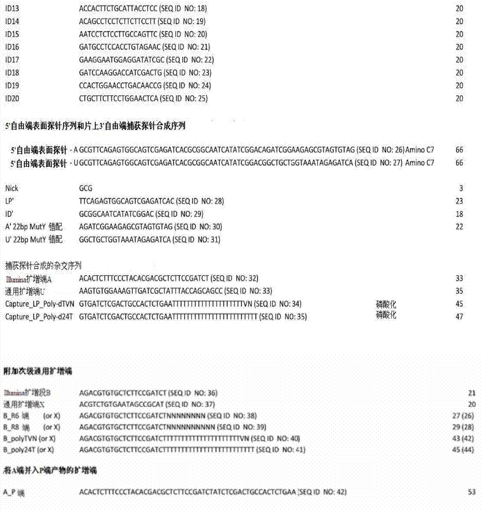
制备含3′至5′端方向的探针的自制喷点微阵列及合成5′至3′端方向的探针Preparation of self-made spray-dot microarrays containing probes in the 3' to 5' direction and synthesis of probes in the 5' to 3' direction
表面探针寡核苷酸的喷点方法与上文的5′至3′端方向探针的情形相同,且其3’端有一个氨基-C7连接子,如表1所示。The spraying method of the surface probe oligonucleotide is the same as that of the 5' to 3' end direction probe above, and there is an amino-C7 linker at the 3' end, as shown in Table 1.
为杂交制取用于捕获探针合成的引物,将含有0.1%SDS的4xSSC、2μM延长引物(通用域寡核苷酸)和2μM连接引物(捕获域寡核苷酸)的杂交溶液在50℃下培养4分钟。同时,将自制阵列附着于ChipClip片基夹(Whatman)上。然后,向阵列每孔加入50μL杂交溶液,在50℃和300rpm下培养30分钟。To prepare primers for capture probe synthesis for hybridization, mix a hybridization solution containing 0.1% SDS in 4xSSC, 2 μM extension primer (general domain oligonucleotide) and 2 μM linker primer (capture domain oligonucleotide) at 50 °C Incubate for 4 minutes. At the same time, the self-made array was attached to the ChipClip substrate holder (Whatman). Then, 50 μL of hybridization solution was added to each well of the array and incubated at 50° C. and 300 rpm for 30 minutes.
培养完成后,将阵列从ChipClip片基夹上拿下来,按照以下3个步骤洗涤:1)50℃下,以含0.1%SDS的2xSSC溶液在300rpm摇速下洗涤6分钟;2)以0.2xSSC溶液在300rpm摇速下洗涤1分钟;以及3)以0.1xSSC溶液在300rpm摇速下洗涤1分钟。然后,将所述阵列甩干,并放回ChipClip片基夹。After the culture is complete, remove the array from the ChipClip base holder and wash it in the following 3 steps: 1) at 50°C, wash with 2xSSC solution containing 0.1% SDS at a shaking speed of 300rpm for 6 minutes; 2) wash with 0.2xSSC The solution was washed at a shaking speed of 300 rpm for 1 minute; and 3) 0.1xSSC solution was washed at a shaking speed of 300 rpm for 1 minute. The array is then spun dry and placed back into the ChipClip holder.
为进行延长和连接反应(以生成所述捕获探针的定位域),向每孔加入50μL含有10xAmpligase缓冲液、2.5U扩增型耐热DNA聚合酶Stoffel片段(AmpliTaq DNA Polymerase Stoffel Fragment)(Applied Biosystems)、10U Ampligase(Epicentre Biotechnologies)、dNTPs(Fermentas)各2mM和水的酶混合物。然后,将该阵列在55℃下培养30分钟。培养完成后,按照前述阵列洗涤方法对阵列进行洗涤,但步骤1)的持续时间从6分钟改变为10分钟。For elongation and ligation reactions (to generate localization domains for the capture probes), 50 μL of 10xAmpligase buffer, 2.5U AmpliTaq DNA Polymerase Stoffel Fragment (Applied Biosystems), 10 U Ampligase (Epicentre Biotechnologies), dNTPs (Fermentas) 2 mM each, and water. Then, the array was incubated at 55°C for 30 minutes. After the incubation was complete, the arrays were washed as described previously for array washing, but the duration of step 1) was changed from 6 min to 10 min.
所述方法如图3所示。The method is shown in FIG. 3 .
组织的制备tissue preparation
以下实验展示了如何制备用于本发明方法的组织样本切片。The following experiment demonstrates how to prepare tissue sample sections for use in the methods of the invention.
制备新鲜冷冻组织并切片置于捕获探针阵列上Preparation of fresh frozen tissue and sectioning onto capture probe arrays
取新鲜未固定的小鼠脑组织,根据需要切边,以-40℃异戊烷降温冷冻,然后在低温箱中切成10μm薄片。在每个待用捕获指针阵列上放置一片组织切片。Fresh unfixed mouse brain tissue was taken, trimmed as needed, frozen in isopentane at -40°C, and then cut into 10 μm thin slices in a low temperature box. Place a slice of tissue on each array of capture pointers to be used.
制备福尔马林固定石蜡包埋(FFPE)的组织Preparation of formalin-fixed paraffin-embedded (FFPE) tissue
取小鼠脑组织,以4%福尔马林在4℃下固定24小时,然后按照如下步骤进行培养:70%乙醇中培养1小时3次;80%乙醇中培养1小时1次;96%乙醇中培养1小时1次;100%乙醇中培养1小时3次;以及二甲苯中室温培养1小时2次。Take mouse brain tissue, fix with 4% formalin at 4°C for 24 hours, and then culture according to the following steps: culture in 70% ethanol three times for 1 hour; culture in 80% ethanol once for 1 hour; culture in 96% ethanol Incubate once in ethanol for 1 hour; incubate in 100% ethanol three times for 1 hour; and incubate in xylene twice for 1 hour at room temperature.
然后,将脱水样本在低熔点液态石蜡中以52-54℃培养至多3小时,其间更换一次石蜡以洗除残余二甲苯。将完成的组织块在室温下保藏。然后,用超薄切片机在石蜡中将组织切成4μm薄片,置于每个待用的捕获探针阵列上。Then, the dehydrated samples were incubated in low-melting liquid paraffin at 52-54°C for up to 3 hours with one change of paraffin to wash away residual xylene. Store the completed tissue blocks at room temperature. Tissues were then sectioned in paraffin with an ultramicrotome into 4 μm slices and placed on each capture probe array to be used.
所述切片在阵列载片上以37℃干燥24小时,并在室温下保藏。The sections were dried on array slides at 37°C for 24 hours and stored at room temperature.
FFPE组织的脱蜡Deparaffinization of FFPE tissue
附着于CodeLink载片上的福尔马林固定石蜡包埋的小鼠脑组织10μm切片在二甲苯中脱蜡2次,各10分钟;99.5%乙醇中2分钟;96%乙醇中2分钟;70%乙醇中2分钟;然后风干。Formalin-fixed, paraffin-embedded
cDNA合成cDNA synthesis
以下实验展示了将捕获在阵列上的组织样本切片mRNA用作cDNA合成模版。The following experiments demonstrate the use of mRNA from tissue sample sections captured on an array as a template for cDNA synthesis.
芯片上的cDNA合成On-Chip cDNA Synthesis
将购自Whatman的16孔遮光框和ChipClip片基夹与CodeLink载片连接。cDNA的合成采用了购自Invitrogen的带含耐热性DNA聚合酶的SuperScriptTM II一步RT-PCR体系(SuperScriptTMIII One-step RT-PCR System with 

移除孔中的反应混合物,以如下试剂洗涤载片:2xSSC,0.1%SDS:50℃下10分钟;0.2xSSC:室温下1分钟;以及0.1xSSC:室温下1分钟。然后将芯片甩干。The reaction mixture in the wells was removed and the slides were washed with the following reagents: 2xSSC, 0.1% SDS: 10 minutes at 50°C; 0.2xSSC: 1 minute at room temperature; and 0.1xSSC: 1 minute at room temperature. Then spin the chip dry.
对于FFPE组织切片,可在移除组织前对切片进行染色及可视化,见下文关于可视化的部分。For FFPE tissue sections, sections can be stained and visualized prior to tissue removal, see section on visualization below.
可视化visualization
染色前的荧光标记探针杂交Fluorescently labeled probe hybridization prior to staining
在放置组织切片前,将荧光标记探针与含有喷至捕获探针阵列上的标记寡核苷酸的特征相杂交。组织可视化后,所述荧光标记探针有助于所得图像的定向,允许所得图像与测序所得的各个捕获探针“标记”(定位域)序列表达谱的合并。为完成荧光探针的杂交,取含0.1%SDS的4xSSC和2μM检测探针(P)的杂交溶液,在50℃下培养4分钟。同时,将自制序列附着于ChipClip片基夹(Whatman)。然后,向序列每孔加入50μL杂交溶液,在50℃和300rpm摇速下培养30分钟。Fluorescently labeled probes are hybridized to features containing labeled oligonucleotides sprayed onto the capture probe array prior to placement of the tissue sections. After tissue visualization, the fluorescently labeled probes facilitate the orientation of the resulting image, allowing the resulting image to be merged with the sequenced expression profile of the individual capture probe "tag" (localization domain) from sequencing. To complete the hybridization of fluorescent probes, take a hybridization solution containing 0.1% SDS in 4xSSC and 2 μM detection probe (P), and incubate at 50°C for 4 minutes. Simultaneously, the homemade sequence was attached to the ChipClip substrate holder (Whatman). Then, 50 μL of hybridization solution was added to each well of the sequence, and incubated at 50° C. and a shaking speed of 300 rpm for 30 minutes.
培养完成后,从ChipClip片基夹上取下序列,按照下列步骤洗涤:1)2xSSC,0.1%SDS:50℃和300rpm摇速下6分钟;2)0.2xSSC:300rpm摇速下1分钟;以及3)0.1xSSC:300rpm摇速下1分钟。然后,将序列甩干。After incubation, remove the sequence from the ChipClip holder and wash as follows: 1) 2xSSC, 0.1% SDS: 6 minutes at 50°C and 300rpm shaking speed; 2) 0.2xSSC: 1 minute at 300rpm shaking speed; and 3) 0.1xSSC: shake at 300rpm for 1 minute. Then, spin the sequence dry.
cDNA合成前或合成后对FFPE组织切片的整体组织学染色Whole-mount histological staining of FFPE tissue sections before or after cDNA synthesis
对固定在捕获探针序列上的FFPE组织切片,在脱蜡之后、cDNA合成之前,如前述步骤进行洗涤和补水,或在cDNA合成之后,如前述步骤进行洗涤。然后,按如下步骤处理组织切片:在苏木精中培养3分钟;以去离子水淋洗;在自来水中培养5分钟;快速地浸入酸化乙醇中8-12次;以自来水洗涤2次各1分钟;以去离子水洗涤2分钟;在曙红中培养30秒;在95%乙醇中洗涤3次各5分钟;以100%乙醇洗涤3次各5分钟;以二甲苯冲洗3次各10分钟(可过夜);将盖片覆盖于载片上,以DPX封片;令载片在通风橱中干燥过夜。For the FFPE tissue section immobilized on the capture probe sequence, after dewaxing and before cDNA synthesis, wash and rehydrate as in the previous steps, or after cDNA synthesis, wash as in the previous steps. Then, the tissue sections were processed as follows: incubate in hematoxylin for 3 minutes; rinse with deionized water; incubate in tap water for 5 minutes; quickly immerse in acidified ethanol 8-12 times; minutes; wash with deionized water for 2 minutes; incubate in eosin for 30 seconds; wash 3 times in 95% ethanol for 5 minutes; wash 3 times with 100% ethanol for 5 minutes; rinse with
cDNA合成前或合成后对FFPE组织切片的目标蛋白的整体免疫组织化学染色Whole-mount immunohistochemical staining of target proteins on FFPE tissue sections before or after cDNA synthesis
对固定在捕获探针序列上的FFPE组织切片,在脱蜡之后、cDNA合成之前,如前述步骤进行洗涤和补水,或在cDNA合成之后,如前述步骤进行洗涤。然后,按如下步骤处理组织切片,且在整个染色过程中保持切片湿润:以一级抗体(以含1x三羟甲基氨基甲烷缓冲盐水(Tris buffered saline)(50mM Tris,150mM NaCl,pH7.6)、4%驴血清和0.1%triton-x的封闭液稀释一级抗体)在湿室中对切片进行室温过夜培养;以1xTBS淋洗3次;以接合荧光素(FITC、Cy3或Cy5)的配对二级抗体在湿室中对切片进行室温培养1小时。以1xTBS淋洗3次,尽可能地除去TBS后,以ProLong Gold+DAPI(苯基吲哚)试剂(Invitrogen)封藏切片,然后以荧光显微镜和匹配滤光片组加以分析。For the FFPE tissue section immobilized on the capture probe sequence, after dewaxing and before cDNA synthesis, wash and rehydrate as in the previous steps, or after cDNA synthesis, wash as in the previous steps. Then, the tissue sections were processed as follows, and the sections were kept wet throughout the staining process: primary antibody (Tris buffered saline containing 1x Tris buffered saline) (50mM Tris, 150mM NaCl, pH7.6 ), 4% donkey serum and 0.1% triton-x blocking solution to dilute the primary antibody) incubate the slices overnight at room temperature in a wet chamber; wash 3 times with 1xTBS; Sections were incubated with paired secondary antibodies for 1 hr at room temperature in a humid chamber. Rinse three times with 1xTBS, remove TBS as much as possible, mount sections with ProLong Gold+DAPI (phenylindole) reagent (Invitrogen), and then analyze with fluorescence microscope and matched filter set.
去除残余组织Removal of residual tissue
冷冻组织frozen tissue
对于新鲜冷冻的小鼠脑组织,cDNA合成后的洗涤步骤足以完全去除组织。For fresh-frozen mouse brain tissue, a washing step after cDNA synthesis is sufficient to completely remove the tissue.
FFPE组织FFPE organization
取附有福尔马林固定石蜡包埋的小鼠脑组织切片的载片,装上ChipClip片基夹及16孔遮光框(Whatman)。取RNeasy FFPE试剂盒(Qiagen)的蛋白酶K消化缓冲液(Proteinase K DigestBuffer),每150μl加入10μl蛋白酶K溶液(Proteinase K Solution)(Qiagen)。向每孔加入50μl最终混合物,并在56℃下培养载片30分钟。Take the slides with the formalin-fixed paraffin-embedded mouse brain tissue slices, and install them with ChipClip base holders and 16-hole light-shielding frames (Whatman). Take the proteinase K digestion buffer (Proteinase K DigestBuffer) of RNeasy FFPE kit (Qiagen), and add 10 μl proteinase K solution (Proteinase K Solution) (Qiagen) per 150 μl. 50 μl of the final mix was added to each well and the slides were incubated at 56° C. for 30 minutes.
捕获探针(cDNA)的释放Release of capture probe (cDNA)
以能切除尿嘧啶的USER酶混合物的PCR缓冲液释放捕获探针(共价附着的探针)Release of capture probes (covalently attached probes) in PCR buffer with uracil-cleaving USER enzyme mix
将16孔遮光框和CodeLink载片装上ChipClip片基夹(Whatman)。取50μl含有含1.8mM MgCl2的1x快速启动高保真反应缓冲液(FastStart High Fidelity Reaction Buffer with1.8mM MgCl2)(Roche)、200μM dNTPs(New England Biolabs)和0.1U/1μl USER Enzyme(New EnglandBiolabs)的混合物,加热至37℃后,加入每孔中,并在37℃下进行30分钟培养并混合(300rpm摇速3秒,静止6秒)(Thermomixer comfort;Eppendorf)。然后,用移液枪从孔中回收含有已释放的cDNA和探针的反应混合物。The 16-well mask and CodeLink slides were loaded into the ChipClip holder (Whatman). Take 50 μl of 1x FastStart High Fidelity Reaction Buffer with 1.8mM MgCl 2 (Roche), 200 μM dNTPs (New England Biolabs) and 0.1U/1 μl USER Enzyme (New England Biolabs) containing 1.8mM MgCl 2 ) mixture, heated to 37°C, added to each well, and incubated and mixed at 37°C for 30 minutes (300rpm shaking speed for 3 seconds, resting for 6 seconds) (Thermomixer comfort; Eppendorf). Then, the reaction mixture containing the released cDNA and probe was recovered from the wells with a pipette.
以能切除尿嘧啶的USER酶混合物的TdT(末端转移酶)缓冲液释放捕获探针(共价附着的探The capture probes (covalently attached probes 针)Needle)
取50μl含有1x TdT缓冲液(20mM三羟甲基氨基甲烷乙酸盐(Tris-acetate)(pH7.9)、50mM乙酸钾和10mM乙酸镁)(New England Biolabs,www.neb.com)、0.1μg/μl BSA(New EnglandBiolabs)和0.1U/μl USER酶(New England Biolabs)的混合物,加热至37℃,加入每孔中,并在37℃下进行30分钟培养并混合(300rpm摇速3秒,静止6秒)(Thermomixer comfort;Eppendorf)。然后,用移液枪从孔中回收含有已释放的cDNA和探针的反应混合物。Take 50 μl containing 1x TdT buffer (20mM Tris-acetate (Tris-acetate) (pH7.9), 50mM potassium acetate and 10mM magnesium acetate) (New England Biolabs, www.neb.com), 0.1 A mixture of μg/μl BSA (New England Biolabs) and 0.1U/μl USER enzyme (New England Biolabs), heated to 37°C, was added to each well, and incubated at 37°C for 30 minutes and mixed (300rpm shaking speed for 3 seconds , stand still for 6 seconds) (Thermomixer comfort; Eppendorf). Then, the reaction mixture containing the released cDNA and probe was recovered from the wells with a pipette.
以烫水释放捕获探针(共价附着的探针)Release of capture probes (covalently attached probes) with scalding water
将16孔遮光框和CodeLink载片装上ChipClip片基夹(Whatman)。用移液枪向每孔注入50μl99℃热水,并反应30分钟。然后,用移液枪从孔中回收含有释放的cDNA和探针的反应混合物。The 16-well mask and CodeLink slides were loaded into the ChipClip holder (Whatman). Inject 50 μl of 99°C hot water into each well with a pipette gun and react for 30 minutes. Then, the reaction mixture containing the released cDNA and probe was recovered from the wells with a pipette.
以加热的PCR缓冲液释放捕获探针(原位杂交的合成捕获探针,即与表面探针杂交的捕获探针)Release of capture probes with heated PCR buffer (synthetic capture probes for in situ hybridization, i.e. capture probes that hybridize to surface probes)
取50μl含有1x TdT缓冲液(20mM三羟甲基氨基甲烷乙酸盐(Tris-acetate)(pH7.9)、50mM乙酸钾和10mM乙酸镁)(New England Biolabs,www.neb.com)、0.1μg/μl BSA(New EnglandBiolabs)和0.1U/μl USER酶(New England Biolabs)的混合物,预热至95℃后加入每孔中,并在95℃下进行5分钟培养并混合(300rpm摇速3秒,静止6秒)(Thermomixer comfort;Eppendorf)。然后,从孔中回收含有释放的探针的反应混合物。Take 50 μl containing 1x TdT buffer (20mM Tris-acetate (Tris-acetate) (pH7.9), 50mM potassium acetate and 10mM magnesium acetate) (New England Biolabs, www.neb.com), 0.1 A mixture of μg/μl BSA (New England Biolabs) and 0.1U/μl USER enzyme (New England Biolabs) was preheated to 95°C and added to each well, and incubated at 95°C for 5 minutes and mixed (
以加热的TdT(末端转移酶)缓冲液释放捕获探针(原位杂交的合成捕获探针,即与表面探针Release the capture probes with heated TdT (terminal transferase) buffer (synthetic capture probes for in situ hybridization, i.e. with surface probes 杂交的捕获探针)hybridized capture probes)
取50μl含有1x TdT缓冲液(20mM三羟甲基氨基甲烷乙酸盐(Tris-acetate)(pH7.9)、50mM乙酸钾和10mM乙酸镁)(New England Biolabs,www.neb.com)、0.1μg/μl BSA(New EnglandBiolabs)和0.1U/μl USER酶(New England Biolabs)的混合物,预热至95℃后加入每孔中,并在95℃下进行5分钟培养并混合(300rpm摇速3秒,静止6秒)(Thermomixer comfort;Eppendorf)。然后,从孔中回收含有释放的探针的反应混合物。Take 50 μl containing 1x TdT buffer (20mM Tris-acetate (Tris-acetate) (pH7.9), 50mM potassium acetate and 10mM magnesium acetate) (New England Biolabs, www.neb.com), 0.1 A mixture of μg/μl BSA (New England Biolabs) and 0.1U/μl USER enzyme (New England Biolabs) was preheated to 95°C and added to each well, and incubated at 95°C for 5 minutes and mixed (
用USER酶和加热到99℃的热水处理序列的效率如图4所示。用USER酶和Rsal酶在前述自制序列上实施酶切(图4)。用热水在Agilent制造的商品序列上实施DNA表面探针的释放(见图5)。The efficiency of the treatment sequence with USER enzyme and hot water heated to 99 °C is shown in Fig. 4. Use USER enzyme and Rsal enzyme to carry out enzyme digestion on the aforementioned self-made sequence (Figure 4). The release of DNA surface probes was carried out using hot water on commercial sequences manufactured by Agilent (see Figure 5).
探针的收集和连接子的引入Collection of probes and introduction of linkers
下列实验显示,可以对从阵列表面释放的cDNA第一链进行修饰,来生产双链DNA并进行扩增。The following experiments show that the first strand of cDNA released from the array surface can be modified to produce double-stranded DNA and amplified.
以Picoplex全基因组扩增试剂盒来进行全转录组扩增(含定位域(标记)序列的捕获探针序列Whole transcriptome amplification (capture probe sequences with localization domain (marker) sequences) using the Picoplex Whole Genome Amplification Kit 未保留在所得dsDNA末端)not retained at the resulting dsDNA ends)
以含能切除尿嘧啶的USER酶混合物的PCR缓冲液(对共价附着的探针)或加热的PCR缓冲液(对原位杂交的合成捕获探针,即与表面探针杂交的捕获探针)来释放捕获探针.PCR buffer (for covalently attached probes) or heated PCR buffer (for in situ hybridization synthetic capture probes, i.e., capture probes that hybridize to surface probes) containing USER enzyme mix capable of cleaving uracil ) to release the capture probe.
按照厂家说明,以Picoplex(Rubicon Genomics)随机引物全基因组扩增方法对释放的cDNA进行扩增。The released cDNA was amplified with the Picoplex (Rubicon Genomics) random primer whole genome amplification method according to the manufacturer's instructions.
以末端转移酶(TdT)dA加尾来进行全转录组扩增(含定位域(标记)序列的捕获探针序列保Whole-transcriptome amplification with terminal transferase (TdT) dA tailing (capture probe sequence preservation with localization domain (tag) sequence 留在所得dsDNA末端)remain at the end of the resulting dsDNA)
以含能切除尿嘧啶的USER酶混合物的TdT(末端转移酶)缓冲液(对共价附着的探针)或加热的PCR缓冲液(对原位杂交的合成捕获探针,即与表面探针杂交的捕获探针)来释放捕获探针。TdT (terminal transferase) buffer (for covalently attached probes) or heated PCR buffer (for synthetic capture probes for in situ hybridization, i.e. hybridized capture probe) to release the capture probe.
取38μl剪切混合物,放入干净的0.2mlPCR管中。所述混合物含有:1x TdT缓冲液(20mM三羟甲基氨基甲烷乙酸盐(Tris-acetate)(pH7.9)、50mM乙酸钾和10mM乙酸镁)(New EnglandBiolabs,www.neb.com)、0.1μg/μl BSA(New England Biolabs)、0.1U/μl USER酶(New EnglandBiolabs)(不可用于加热释放);已释放的cDNA(从表面探针伸出)和已释放的表面探针。向PCR管中加入0.5μl RNase H(5U/μl,终浓度为0.06U/μl)、1μl TdT(20U/μl,终浓度为0.5U/μl)和0.5μl dATPs(100mM,终浓度为1.25mM)。为进行dA加尾,在热循环仪(Applied Biosystems)中以37℃培养PCR管15分钟,再以70℃培养10分钟以令TdT失活。dA加尾完成后,制备PCR预混液。所述预混液含有:含1.8mM MgCl2的1x快速启动高保真PCR缓冲液(Faststart HiFi PCRBuffer)(Roche)、每种dNTP各0.2mM(Fermentas)、A(与捕获探针的扩增域互补)和B_(dT)24(Eurofins MWG Operon)(与待连接至cDNA第一链3’端的poly-A尾互补)每种引物各0.2μM和0.1U/μl快速启动高保真DNA聚合酶(Faststart HiFi DNA polymerase)(Roche)。取23μl PCR预混液,放入9只干净的0.2ml PCR管中,向其中8只管中加入2μl dA加尾混合物,而向最后1只管中加入2μl水(不含RNase/DNase)(阴性对照)。按照以下程序实施PCR扩增:95℃热启动2分钟,50℃2分钟和72℃3分钟的第二链合成,30个PCR扩增循环,每个循环中依序为95℃30秒,65℃1分钟,72℃3分钟,以及最后延长72℃10分钟。Take 38 μl of shearing mixture and put into a clean 0.2ml PCR tube. The mixture contained: 1x TdT buffer (20 mM Tris-acetate (pH 7.9), 50 mM potassium acetate and 10 mM magnesium acetate) (New England Biolabs, www.neb.com), 0.1 μg/μl BSA (New England Biolabs), 0.1 U/μl USER enzyme (New England Biolabs) (not available for heat release); released cDNA (protruding from surface probe) and released surface probe. Add 0.5 μl RNase H (5 U/μl, final concentration 0.06 U/μl), 1 μl TdT (20 U/μl, final concentration 0.5 U/μl), and 0.5 μl dATPs (100 mM, final concentration 1.25 mM) to the PCR tube ). For dA tailing, PCR tubes were incubated in a thermal cycler (Applied Biosystems) at 37°C for 15 minutes and then at 70°C for 10 minutes to inactivate TdT. After dA tailing is completed, prepare the PCR master mix. The master mix contains: 1x Faststart HiFi PCR Buffer (Roche) with 1.8mM MgCl2 , 0.2mM each of dNTPs (Fermentas), A (complementary to the amplification domain of the capture probe ) and B_(dT)24 (Eurofins MWG Operon) (complementary to the poly-A tail to be ligated to the 3' end of the first strand of cDNA) each primer 0.2μM and 0.1U/μl fast start high-fidelity DNA polymerase (Faststart HiFi DNA polymerase) (Roche). Take 23μl PCR master mix and put it into 9 clean 0.2ml PCR tubes, add 2μl dA tailing mix to 8 of them, and add 2μl water (RNase/DNase free) to the last tube (negative control) . Perform PCR amplification according to the following program: 95°C hot start for 2 minutes, second-strand synthesis at 50°C for 2 minutes and 72°C for 3 minutes, 30 cycles of PCR amplification, in each cycle sequentially 95°C for 30 seconds, 65 °C for 1 minute, 72°C for 3 minutes, and finally 72°C for 10 minutes.
反应后的清理和分析Post-reaction cleanup and analysis
将4种扩增子合并,并流经Qiaquick PCR净化柱(Qiagen)处理,以30μl EB(10mM Tris-Cl,pH8.5)洗脱。以生物分析仪(Agilent)对产物进行分析,按照厂家说明书使用DNA1000试剂盒。The 4 amplicons were pooled and passed through a Qiaquick PCR cleanup column (Qiagen) and eluted with 30 μl EB (10 mM Tris-Cl, pH 8.5). The product was analyzed with a bioanalyzer (Agilent), and the DNA1000 kit was used according to the manufacturer's instructions.
测序sequencing
Illumina测序Illumina sequencing
按照厂家说明书,用样品索引建立用于Illumina测序的dsDNA文库。用HiSeq2000平台(Illumina)进行测序。Following the manufacturer's instructions, sample indexing was used to create a dsDNA library for Illumina sequencing. Sequencing was performed with the HiSeq2000 platform (Illumina).
生物信息学研究Bioinformatics Research
从以dA加尾末端转移酶方法扩增的全转录组文库的测序数据中获取数码转录组信息Obtaining digital transcriptome information from sequencing data of a whole-transcriptome library amplified by the dA-tailed terminal transferase method
以FastX工具盒中的FASTQ识别码分离工具,将测序数据按各自的捕获探针定位域(标记)序列分类整理为各个文件。然后,以Tophat映射工具将各个被标记的测序数据映射到小鼠基因组上加以分析。以HTseq-count软件处理获取所得SAM文件中转录体计数。Using the FASTQ identification code separation tool in the FastX toolbox, the sequencing data are sorted into individual files according to their respective capture probe localization domain (marker) sequences. Then, use the Tophat mapping tool to map each labeled sequencing data to the mouse genome for analysis. Transcript counts in the obtained SAM files were obtained by processing with HTseq-count software.
从以Picoplex全基因组扩增试剂盒方法扩增的全转录组文库的测序数据中获取数码转录组信息Obtaining digital transcriptome information from sequencing data of a whole-transcriptome library amplified by the Picoplex Whole Genome Amplification Kit method
用FastX工具盒中的FASTQ-to-FASTA转换工具,将测序数据从FASTQ格式转换为FASTA格式。用Blastn工具将测序Read与捕获探针的定位域(标记)序列对齐,并将每个标记序列中匹配率优于1e-6的Read挑选出来,分别归入各序列标记的单独文件中。然后,用Blastn工具将标记序列Read文件与小鼠转录组对齐,获取匹配记录。Use the FASTQ-to-FASTA conversion tool in the FastX toolbox to convert the sequencing data from the FASTQ format to the FASTA format. Use the Blastn tool to align the sequencing reads with the localization domain (marker) sequence of the capture probe, and select the reads with a matching rate better than 1e -6 in each marker sequence, and classify them into separate files for each sequence marker. Then, the marker sequence Read files were aligned with the mouse transcriptome using the Blastn tool to obtain matching records.
合并可视化数据和表达谱Merge visualization data and expression profiles
利用染色,将各个捕获探针定位域(标记)序列的表达谱与从组织切片获得的空间信息组合起来。如此一来,即可用直接对比的方式来分析从组织切片的细胞区室中获得的转录组信息,还能在预定的结构环境中分辨不同细胞亚型的区别表达特征。Using staining, expression profiles of individual capture probe localization domain (label) sequences were combined with spatial information obtained from tissue sections. In this way, the transcriptome information obtained from the cellular compartments of tissue sections can be analyzed in a direct comparative manner, and the differential expression characteristics of different cell subtypes can be resolved in a predetermined structural environment.
实施例2Example 2
图8至图12展示了置于以识别码标示的转录组捕获阵列上的染色FFPE小鼠脑组织(嗅球)切片的成功可视化,操作参照实施例中所述的通用流程。与实施例1中的新鲜冷冻组织的实验相比,图8所示的FFPE组织形态更佳。图9和图10展示了如何将组织定位于不同探针密度类型的阵列上。Figures 8 to 12 show the successful visualization of stained FFPE mouse brain tissue (olfactory bulb) slices placed on transcriptome capture arrays marked with identifiers, following the general procedure described in the Examples. Compared with the fresh frozen tissue experiment in Example 1, the FFPE tissue morphology shown in Figure 8 is better. Figures 9 and 10 demonstrate how tissue can be positioned on arrays of different probe density types.
实施例3Example 3
以随机引物第二链合成和通用端扩增反应进行全转录组扩增(含标记序列的捕获探针序列保留Whole-transcriptome amplification with random-primed second-strand synthesis and universal-end amplification (capture probes containing tag sequences retain 在所得dsDNA的末端)at the end of the resulting dsDNA)
在以含能切除尿嘧啶的USER酶混合物的PCR缓冲液释放捕获探针之后(共价附着的探针)After release of capture probes with PCR buffer containing USER enzyme mix capable of cleaving uracil (covalently attached probes)
或or
在以加热的PCR缓冲液释放捕获探针之后(原位杂交的合成捕获探针)After release of capture probes with heated PCR buffer (synthetic capture probes for in situ hybridization)
向2只管中各加入40μl1x含1.8mM MgCl2的快速启动高保真PCR缓冲液(Faststart HiFi PCRBuffer with1.8mM MgCl2)(pH8.3)(Roche,www.roche-applied-science.com)、dNTP各0.2mM(Fermentas,www.fermentas.com)、0.1μg/μl BSA(New England Biolabs,www.neb.com)、0.1U/μlUSER酶(New England Biolabs)、已释放的cDNA(从表面探针延长)和已释放的表面探针,再加入1μl RNaseH(5U/μl)。然后,将2只管在热循环仪(Applied Biosystems,www.appliedbiosystems.com)中以37℃培养30分钟,再以70℃处理20分钟。向2只管中加入1μl克列诺片段(Klenow Fragment)(3’to5’exo minus)(Illumina,www.illumina.com)和1μl连接通用端的随机引物(10μM)(Eurofins MWG Operon,www.eurofinsdna.com)(其中一只加入B_R8(八聚体),另一只加入B_R6(六聚体)),终浓度为0.23μM。将2只管在热循环仪(AppliedBiosystems)中依次以15℃培养15分钟,25℃培养15分钟,37℃培养15分钟,最后以75℃培养20分钟。培养完成后,向两管中各加入引物A_P和B(10μM)各1μl(Eurofins MWG Operon),终浓度各为0.22μM;再向管中各加入1μl快速启动高保真DNA聚合酶(Faststart HiFi DNApolymerase)(5U/μl)(Roche),终浓度为0.11U/μl。按照下列程序在热循环仪(Applied Biosystems)中进行PCR扩增:94℃热启动2min后,进行50个如下循环:94℃15秒,55℃30秒,68℃1分钟,以及最终延长68℃5分钟。扩增完成后,从每管中各取40μl,以Qiaquick PCR净化柱(Qiagen,www.qiagen.com)纯化,并以30μl EB(10mM Tris-Cl,pH8.5)洗脱。用生物分析仪(Agilent,www.home.agilent.com)和DNA7500试剂盒分析纯化产物。所得结果如图13和图14所示。Add 40 μl 1x Faststart HiFi PCRBuffer with 1.8mM MgCl 2 (pH8.3) (Roche, www.roche-applied-science.com), dNTP to each of the 2 tubes Each 0.2mM (Fermentas, www.fermentas.com), 0.1μg/μl BSA (New England Biolabs, www.neb.com), 0.1U/μl USER enzyme (New England Biolabs), released cDNA (from the surface probe extension) and the released surface probe, then add 1 μl RNaseH (5U/μl). Then, 2 tubes were incubated in a thermal cycler (Applied Biosystems, www.appliedbiosystems.com) at 37°C for 30 minutes, and then treated at 70°C for 20 minutes. Add 1 μl of Klenow Fragment (3'to5'exo minus) (Illumina, www.illumina.com) and 1 μl of random primers (10 μM) connected to the universal end (Eurofins MWG Operon, www.eurofinsdna. com) (one with B_R8 (octamer) and the other with B_R6 (hexamer)) at a final concentration of 0.23 μM. Two tubes were incubated sequentially in a thermal cycler (Applied Biosystems) at 15°C for 15 minutes, at 25°C for 15 minutes, at 37°C for 15 minutes, and finally at 75°C for 20 minutes. After the culture is completed, add 1 μl of primers A_P and B (10 μM) (Eurofins MWG Operon) to each of the two tubes, and the final concentration is 0.22 μM; then add 1 μl of Faststart HiFi DNA polymerase (Faststart HiFi DNA polymerase) to each tube ) (5U/μl) (Roche), the final concentration was 0.11U/μl. PCR amplification was performed in a thermal cycler (Applied Biosystems) according to the following program: After a 94°C hot start for 2 min, 50 cycles of the following were performed: 94°C for 15 seconds, 55°C for 30 seconds, 68°C for 1 minute, and a final extension at 68°
本实施例展示了以随机六聚体和随机八聚体的第二链合成及其后的扩增反应来生成大群已释放的cDNA分子的方法。This example demonstrates the generation of large populations of released cDNA molecules by second-strand synthesis of random hexamers and random octamers followed by amplification reactions.
实施例4Example 4
cDNA合成和探针的收集后对ID-特异性及基因特异性产物的扩增Amplification of ID-specific and gene-specific products after cDNA synthesis and probe collection
在以含能切除尿嘧啶的USER酶混合物的PCR缓冲液对捕捉探针(共价附着的探针)进行释放后:After release of capture probes (covalently attached probes) with PCR buffer containing USER enzyme mix capable of cleaving uracil:
对剪切后的cDNA进行扩增,反应混合液最终容量为10μl。7μl切除后的模版、1μl ID-特异性正向引物(2μM)、1μl基因特异性反向引物(2μM)和1μl含有快速启动高保真混合酶(FastStartHigh Fidelity Enzyme Blend)的1.4x含1.8mM MgCl2的快速启动高保真反应缓冲液(FastStart HighFidelity Reaction Buffer with1.8mM MgCl2)的混合液,共计10μl,并与1x含1.8mM MgCl2的快速启动高保真反应缓冲液(FastStart High Fidelity Reaction Buffer with1.8mM MgCl2)和1U快速启动高保真混合酶(FastStart High Fidelity Enzyme Blend)进行终反应.PCR扩增反应在热循环仪(Applied Biosystems)中按照如下程序进行:94℃热启动2分钟,然后进行50个如下循环:94℃下15秒,55℃下30秒,68℃下1分钟,以及最终延长68℃下5分钟。The sheared cDNA was amplified, and the final volume of the reaction mixture was 10 μl. 7 μl excised template, 1 μl ID-specific forward primer (2 μM), 1 μl gene-specific reverse primer (2 μM), and 1 μl FastStart High Fidelity Enzyme Blend containing 1.4x 1.8mM MgCl 2 of Fast Start High Fidelity Reaction Buffer (FastStart High Fidelity Reaction Buffer with 1.8mM MgCl 2 ), a total of 10μl, and 1x Fast Start High Fidelity Reaction Buffer with 1.8mM MgCl 2 (FastStart High Fidelity Reaction Buffer with1 .8mM MgCl 2 ) and 1U FastStart High Fidelity Enzyme Blend (FastStart High Fidelity Enzyme Blend) for the final reaction. The PCR amplification reaction was carried out in a thermal cycler (Applied Biosystems) according to the following procedure: 94°C hot start for 2 minutes, then Fifty cycles of 15 seconds at 94°C, 30 seconds at 55°C, 1 minute at 68°C, and a final extension of 5 minutes at 68°C were performed.
引物序列,所得产物约为250bpPrimer sequence, the resulting product is about 250bp
Beta-2w微球蛋白(B2M)引物Beta-2w microglobulin (B2M) primer
5’-TGGGGGTGAGAATTGCTAAG-3’ (SEQ ID NO:43)5'-TGGGGGTGAGAATTGCTAAG-3' (SEQ ID NO: 43)
ID-1引物ID-1 primer
5′-CCTTCTCCTTCTCCTTCACC-3′ (SEQ ID NO:44)5'-CCTTTCTCTTCTCCTTCACC-3' (SEQ ID NO: 44)
ID-5引物ID-5 primer
5′-GTCCTCTATTCCGTCACCAT-3′ (SEQ ID NO:45)5'-GTCCTTATTCCGTCACCAT-3' (SEQ ID NO: 45)
ID-20引物ID-20 primer
5′-CTGCTTCTTCCTGGAACTCA-3′ (SEQ ID NO:46)5'-CTGCTTCTTCCTGGAACTCA-3' (SEQ ID NO: 46)
所得结果如图15所示,显示了用两种不同ID引物(即:对定位于微阵列上不同位置的ID标记具有特异性)和源于脑组织且通用于所有探针的同一基因特异性引物,对ID特异性和基因特异性产物的成功扩增。相应地,本实验证明,可以用ID标记特异性或目标核酸特异性的扩增反应对产物加以鉴定,且进一步证明,不同ID标记可以被区分开来。第二次实验中,仅以组织覆盖阵列中一半的ID探针(即捕获探针),则从得到组织覆盖的微点上得到了阳性结果。The results obtained are shown in Figure 15, showing the use of two different ID primers (i.e., specific for ID markers located at different locations on the microarray) and the same gene specificity derived from brain tissue and common to all probes. Primers for successful amplification of ID-specific and gene-specific products. Correspondingly, this experiment proves that the product can be identified by ID marker-specific or target nucleic acid-specific amplification reaction, and further proves that different ID markers can be distinguished. In the second experiment, only half of the ID probes (i.e., capture probes) in the array were covered with tissue, and positive results were obtained from the microspots covered by tissue.
实施例5Example 5
空间基因组学研究Space Genomics Research
背景:本方法的目的在于从保有空间信息的组织样本中捕获DNA分子,以便能够确定特定的DNA片段起源于组织的哪个部分。 Background : The purpose of this method is to capture DNA molecules from tissue samples that retain spatial information, so that it can be determined from which part of the tissue a specific DNA fragment originated.
方法:本方法的原理在于利用固定了带空间标记标签序列(定位域)的DNA寡核苷酸(捕获探针)的微阵列。所述微阵列的寡核苷酸的每一特征都载有1)独一无二的标记标签(定位域)以及2)捕获序列(捕获域)。追踪某一标记标签在阵列表面上的地域位置,便可能从每个标记标签上提取二维定位信息。可以通过例如添加FFPE处理组织的薄切片,来向微阵列添加基因组DNA片段。该组织切片中的基因组DNA已通过固定处理而预先片段化。 METHODS : The principle of this method is the use of microarrays immobilized with DNA oligonucleotides (capture probes) with spatially labeled tag sequences (localization domains). Each feature of the oligonucleotides of the microarray carries 1) a unique labeling tag (localization domain) and 2) a capture sequence (capture domain). By tracking the geographic position of a marker tag on the array surface, it is possible to extract two-dimensional positioning information from each marker tag. Genomic DNA fragments can be added to the microarray by, for example, adding thin sections of FFPE-treated tissue. The genomic DNA in this tissue section has been pre-fragmented by fixation.
组织薄片放上阵列后,利用末端转移酶进行通用加尾反应。该加尾反应为组织中的基因组DNA片段的伸出3’端加上polydA尾。利用末端转移酶进行加尾反应,形成杂交且3’端封闭的polydA探针,从而对表面寡核苷酸实施了封闭。After the tissue slices are arrayed, a universal tailing reaction is performed using terminal transferase. The tailing reaction adds a polydA tail to the protruding 3' end of the genomic DNA fragment in the tissue. The surface oligonucleotides were blocked by terminal transferase tailing reaction to form hybridized and 3'-blocked polydA probes.
末端转移酶加尾反应后,基因组DNA片段便能通过polydA尾与表面寡核苷酸的polydT捕获序列的接触,从而与附近带有空间标记的寡核苷酸杂交。杂交反应完成后,Klenow exo-等链置换聚合酶能利用表面寡核苷酸作为引物,制造一条与所述杂交基因组DNA片段互补的DNA新链。此时,该DNA新链亦带有表面寡核苷酸标记标签所含的定位信息。After the terminal transferase tailing reaction, the genomic DNA fragments can hybridize with nearby space-labeled oligonucleotides through the contact of the polydA tail with the polydT capture sequence of the surface oligonucleotide. After the hybridization reaction is complete, Klenow exo-isostrand displacement polymerase can use the surface oligonucleotide as a primer to create a new DNA strand complementary to the hybridized genomic DNA fragment. At this time, the new strand of DNA also carries the positioning information contained in the surface oligonucleotide label.
作为最后一步,以酶促、变性或物理方法将新合成且带标记的DNA链从表面切下,收集起来,并通过引入通用端、特定扩增子的扩增反应、和/或测序,来对整个链集进行下游扩增。As a final step, the newly synthesized and labeled DNA strands are enzymatically, denatured, or physically excised from the surface, collected, and analyzed by introduction of universal ends, amplification reactions of specific amplicons, and/or sequencing. Downstream amplification is performed on the entire strand set.
图16是该过程的原理示意图。Figure 16 is a schematic diagram of the principle of the process.
材料和方法Materials and methods
以5’到3’方向的探针制备自制喷点微阵列Preparation of self-made spray-dot microarray with probes in 5' to 3' direction
将20个各带标签序列的DNA捕获寡核苷酸(表1)点样到载玻片上,充当捕获探针。所述探针是以5’-端氨基连接子和C6间隔子合成所得。所有的探针均由Sigma-Aldrich(St.Louis,MO,USA)合成。将浓度为20μM的DNA捕获探针以150mM磷酸钠制成pH8.5的悬浮液,并用NanoplotterNP2.1/E喷点系统(Gesim,Grosserkmannsdorf,Germany)点样在CodeLinkTM活化微阵列载片(7.5cm x2.5cm;Surmodics,Eden Prairie,MN,USA)上。喷点完成后,按照厂商说明实施表面封闭。探针在载片上喷点为16个相同的阵列,每个阵列都含有预设的喷点格局。所述16个子阵列在杂交反应时,以16版遮光框(ChipClipTM Schleicher&Schuell BioScience,Keene,NH,USA)分隔。Twenty DNA capture oligonucleotides (Table 1) of each tagged sequence were spotted onto a slide to serve as capture probes. The probe is synthesized with a 5'-terminal amino linker and a C6 spacer. All probes were synthesized by Sigma-Aldrich (St. Louis, MO, USA). The DNA capture probe with a concentration of 20 μM was made into a suspension of pH 8.5 in 150 mM sodium phosphate, and was spotted on a CodeLinkTM activated microarray slide (7.5 cm x2.5cm; Surmodics, Eden Prairie, MN, USA). After dispensing is complete, apply surface sealer according to manufacturer's instructions. The probes are sprayed in 16 identical arrays on the slide, and each array contains a preset pattern of spraying spots. The 16 sub-arrays were separated by a 16-plate light-shielding frame (ChipClip TM Schleicher & Schuell BioScience, Keene, NH, USA) during the hybridization reaction.
以3’到5’方向的探针制备自制喷点微阵列并合成5’到3’方向的捕获探针Prepare self-made spray-dot microarray with probes in 3' to 5' direction and synthesize capture probes in 5' to 3' direction
寡核苷酸的喷点操作与上文所述5’到3’方向的探针的操作方法相同。The oligonucleotide spraying operation is the same as that of the probe in the 5' to 3' direction described above.
为杂交制取用于捕获探针合成的引物,将含有带0.1%SDS的4xSSC、2μM延长引物(A_primer)和2μM连接引物(p_poly_dT)的杂交溶液在50℃下培养4分钟。与此同时,将自制阵列附着于ChipClip片基夹(Whatman)上。然后,在所述阵列每孔加入50μL杂交溶液,在50℃及300rpm摇速下培养30分钟。To prepare primers for capture probe synthesis for hybridization, a hybridization solution containing 4xSSC with 0.1% SDS, 2 μM extension primer (A_primer) and 2 μM linker primer (p_poly_dT) was incubated at 50°C for 4 min. At the same time, the self-made array was attached to the ChipClip substrate holder (Whatman). Then, 50 μL of hybridization solution was added to each well of the array, and incubated at 50° C. and at a shaking speed of 300 rpm for 30 minutes.
培养完成后,将阵列从ChipClip片基夹上取下,并按照下列3个步骤洗涤:1)带0.1%SDS的2xSSC溶液,50℃、300rpm摇速下洗涤6分钟;2)0.2xSSC溶液,300rpm摇速下洗涤1分钟;以及3)0.1xSSC溶液,300rpm摇速下洗涤1分钟。然后,将阵列甩干,放回ChipClip片基夹。After the incubation is complete, remove the array from the ChipClip holder and wash it in the following three steps: 1) 2xSSC solution with 0.1% SDS, 50°C, 300rpm shaking speed for 6 minutes; 2) 0.2xSSC solution, Washing at 300rpm shaking speed for 1 minute; and 3) 0.1xSSC solution, washing at 300rpm shaking speed for 1 minute. Then, spin the array dry and place it back in the ChipClip holder.
为进行延长和连接反应,向每孔加入50μL含有10xAmpligase缓冲液、2.5U扩增型耐热DNA聚合酶Stoffel片段(AmpliTaq DNA Polymerase Stoffel Fragment)(Applied Biosystems)、10UAmpligase(Epicentre Biotechnologies)、dNTPs各2mM(Fermentas)和水的酶混合物。然后,将阵列在55℃下培养30分钟,随后按前述阵列洗涤方法进行洗涤,但第一步中的持续时间由6分钟改为10分钟。For extension and ligation reactions, add 50 μL containing 10xAmpligase buffer, 2.5U amplified thermostable DNA polymerase Stoffel fragment (AmpliTaq DNA Polymerase Stoffel Fragment) (Applied Biosystems), 10UAmpligase (Epicentre Biotechnologies), dNTPs 2mM each to each well (Fermentas) and water enzyme mixture. Then, the array was incubated at 55°C for 30 minutes, followed by washing as described above for the array wash, but the duration in the first step was changed from 6 minutes to 10 minutes.
对polydA探针进行杂交以保护表面寡核苷酸不受dA尾影响Hybridization of polydA probes to protect surface oligonucleotides from dA tails
为保护表面寡核苷酸捕获序列,需要杂交形成3’-生物素封闭的polydA探针,将含有含0.1%SDS的4xSSC和2μM3’bio-polydA的杂交溶液在50℃下培养4分钟。与此同时,将自制阵列附着于ChipClip片基夹(Whatman)上。然后,在所述阵列每孔加入50μL杂交溶液,在50℃及300rpm摇速下培养30分钟。To protect the surface oligonucleotide capture sequences that need to hybridize to form 3’-biotin-blocked polydA probes, a hybridization solution containing 4xSSC containing 0.1% SDS and 2 μM 3’bio-polydA was incubated at 50°C for 4 min. At the same time, the self-made array was attached to the ChipClip substrate holder (Whatman). Then, 50 μL of hybridization solution was added to each well of the array, and incubated at 50° C. and at a shaking speed of 300 rpm for 30 minutes.
培养完成后,将阵列从ChipClip片基夹上取下,并按照下列3个步骤洗涤:1)带0.1%SDS的2xSSC溶液,50℃、300rpm摇速下洗涤6分钟;2)0.2xSSC溶液,300rpm摇速下洗涤1分钟;以及3)0.1xSSC溶液,300rpm摇速下洗涤1分钟。然后,将阵列甩干,放回ChipClip片基夹。After the incubation is complete, remove the array from the ChipClip holder and wash it in the following three steps: 1) 2xSSC solution with 0.1% SDS, 50°C, 300rpm shaking speed for 6 minutes; 2) 0.2xSSC solution, Washing at 300rpm shaking speed for 1 minute; and 3) 0.1xSSC solution, washing at 300rpm shaking speed for 1 minute. Then, spin the array dry and place it back in the ChipClip holder.
制备福尔马林固定石蜡包埋(FFPE)的组织Preparation of formalin-fixed paraffin-embedded (FFPE) tissue
以4%福尔马林在4℃下对小鼠脑组织进行24小时固定。然后,按照如下步骤对其进行培养:70%乙醇培养1小时3次,80%乙醇培养1小时1次,96%乙醇培养1小时1次,100%乙醇1培养1小时1次,二甲苯室温下培养1小时2次。Mouse brain tissues were fixed in 4% formalin for 24 hours at 4°C. Then, it was cultivated according to the following steps: 70% ethanol for 1 hour three times, 80% ethanol for 1 hour for 1 hour, 96% ethanol for 1 hour for 1 hour, 100% ethanol for 1 hour for 1 hour, xylene at room temperature Incubate twice for 1 hour.
然后,将脱水样本在液态低熔点石蜡中以52-54℃培养至多3小时,期间置换石蜡一次,以洗除残余二甲苯。完成后的组织块在室温下保藏,使用时,以超薄切片机切成4μm切片,置于每个待用的捕获探针序列上。The dehydrated samples were then incubated in liquid low-melting paraffin at 52-54°C for up to 3 hours, during which time the paraffin was replaced once to wash away residual xylene. The completed tissue block was stored at room temperature, and when used, it was cut into 4 μm slices with an ultramicrotome and placed on each capture probe sequence to be used.
切片在阵列载片上37℃干燥24小时,室温保藏。Sections were dried on array slides at 37°C for 24 hours and stored at room temperature.
FFPE组织的脱蜡Deparaffinization of FFPE tissue
附着于CodeLink载片上的福尔马林固定石蜡包埋的小鼠脑组织10μm切片在二甲苯中脱蜡2次,各10分钟;99.5%乙醇中2分钟;96%乙醇中2分钟;70%乙醇中2分钟;然后空气中干燥。Formalin-fixed, paraffin-embedded
基因组DNA的通用加尾反应Universal tailing reaction for genomic DNA
为实施dA加尾,制备50μl含有1x TdT缓冲液(20mM三羟甲基氨基甲烷乙酸盐(Tris-acetate)(pH7.9),50mM乙酸钾和10mM乙酸镁)(New England Biolabs,www.neb.com)、0.1μg/μl BSA(New England Biolabs)、1μl TdT(20U/μl)和0.5μl dATPs(100mM)的反应混合物。将所述混合物加至阵列表面,并在热循环仪(Apllied Biosystems)中以37℃培养15分钟,再以70℃培养10分钟以令TdT失活。然后,将温度再次降至50℃,以允许带dA尾的基因组片段与表面寡核苷酸捕获序列相杂交。For dA tailing, prepare 50 μl of 1x TdT buffer (20 mM Tris-acetate (pH 7.9), 50 mM potassium acetate and 10 mM magnesium acetate) (New England Biolabs, www. neb.com ), 0.1 μg/μl BSA (New England Biolabs), 1 μl TdT (20 U/μl) and 0.5 μl dATPs (100 mM). The mixture was added to the array surface and incubated in a thermal cycler (Apllied Biosystems) at 37°C for 15 minutes and then at 70°C for 10 minutes to inactivate TdT. Then, the temperature was lowered again to 50°C to allow hybridization of the dA-tailed genomic fragments to the surface oligonucleotide capture sequences.
培养完成后,将阵列从ChipClip片基夹上取下,并按照下列3个步骤洗涤:1)带0.1%SDS的2xSSC溶液,50℃、300rpm摇速下洗涤6分钟;2)0.2xSSC溶液,300rpm摇速下洗涤1分钟;以及3)0.1xSSC溶液,300rpm摇速下洗涤1分钟。然后,将阵列甩干。After the incubation is complete, remove the array from the ChipClip holder and wash it in the following three steps: 1) 2xSSC solution with 0.1% SDS, 50°C, 300rpm shaking speed for 6 minutes; 2) 0.2xSSC solution, Washing at 300rpm shaking speed for 1 minute; and 3) 0.1xSSC solution, washing at 300rpm shaking speed for 1 minute. Then, spin the array to dry.
标记DNA的延长elongation of labeled DNA
取50μl含有1x克列诺缓冲液(Klenow buffer)、200μM dNTPs(New England Biolabs)和1μl克列诺片段(Klenow Fragment)(3’to5’exo minus)的混合物的反应混合物,加热至37℃,加入每孔中,并在37℃下进行30分钟培养并混合(300rpm摇速3秒,静止6秒)(Thermomixercomfort;Eppendorf)。Take 50 μl of the reaction mixture containing 1x Klenow buffer, 200 μM dNTPs (New England Biolabs) and 1 μl of Klenow Fragment (3'to5'exo minus), heat to 37°C, Add to each well and incubate for 30 minutes at 37°C with mixing (3 s shaking at 300 rpm, 6 s resting) (Thermomixer comfort; Eppendorf).
培养完成后,将阵列从ChipClip片基夹上取下,并按照下列3个步骤洗涤:1)带0.1%SDS的2xSSC溶液,50℃、300rpm摇速下洗涤6分钟;2)0.2xSSC溶液,300rpm摇速下洗涤1分钟;以及3)0.1xSSC溶液,300rpm摇速下洗涤1分钟。然后,将阵列甩干。After the incubation is complete, remove the array from the ChipClip holder and wash it in the following three steps: 1) 2xSSC solution with 0.1% SDS, 50°C, 300rpm shaking speed for 6 minutes; 2) 0.2xSSC solution, Washing at 300rpm shaking speed for 1 minute; and 3) 0.1xSSC solution, washing at 300rpm shaking speed for 1 minute. Then, spin the array to dry.
去除残余组织Removal of residual tissue
取附有福尔马林固定石蜡包埋的小鼠脑组织切片的载片,装上ChipClip片基夹及16孔遮光框(Whatman)。对每150μl RNeasy FFPE试剂盒(Qiagen)的蛋白酶K消化缓冲液(Proteinase KDigest Buffer),加入10μl蛋白酶K溶液(Proteinase K Solution)(Qiagen)。向每孔加入50μl最终混合物,并在56℃下培养载片30分钟。Take the slides with the formalin-fixed paraffin-embedded mouse brain tissue slices, and install them with ChipClip base holders and 16-hole light-shielding frames (Whatman). For every 150 μl of Proteinase K Digest Buffer from RNeasy FFPE Kit (Qiagen), 10 μl of Proteinase K Solution (Qiagen) was added. 50 μl of the final mix was added to each well and the slides were incubated at 56° C. for 30 minutes.
以能切除尿嘧啶的USER酶混合物的PCR缓冲液释放捕获探针(共价附着的探针)Release of capture probes (covalently attached probes) in PCR buffer with uracil-cleaving USER enzyme mix
将16孔遮光框和CodeLink载片装上ChipClip片基夹(Whatman)。取50μl含有:含1.8mMMgCl2的1x快速启动高保真反应缓冲液(FastStart High Fidelity Reaction Buffer with1.8mM MgCl2)(Roche)、200μM dNTPs(New England Biolabs)和0.1U/1μl USER酶(New England Biolabs)的混合物,加热至37℃后,加入每孔中,并在37℃下进行30分钟培养并混合(300rpm摇速3秒,静止6秒)(Thermomixer comfort;Eppendorf)。然后,用移液枪从孔中回收含有释放的cDNA和探针的反应混合物。The 16-well mask and CodeLink slides were loaded into the ChipClip holder (Whatman). Take 50 μl containing: 1x FastStart High Fidelity Reaction Buffer with 1.8mM MgCl 2 ( Roche), 200 μM dNTPs (New England Biolabs) and 0.1U/1 μl USER enzyme (New England Biolabs) mixture was heated to 37°C, added to each well, and incubated and mixed at 37°C for 30 minutes (3 seconds at 300 rpm, 6 seconds at rest) (Thermomixer comfort; Eppendorf). Then, the reaction mixture containing the released cDNA and probe was recovered from the wells with a pipette.
已标记DNA的合成及探针收集后对ID-特异性及基因特异性产物的扩增Synthesis of labeled DNA and amplification of ID-specific and gene-specific products after probe collection
在以含能切除尿嘧啶的USER酶混合物的PCR缓冲液释放捕捉探针(共价附着的探针)后:After release of capture probes (covalently attached probes) with PCR buffer containing USER enzyme mix capable of cleaving uracil:
对已剪切的cDNA进行扩增,最终反应容量为10μl。7μl已剪切的模版,1μl ID特异性正向引物(2μM),1μl基因特异性反向引物(2μM)和1μl快速启动高保真混合酶(FastStart High FidelityEnzyme Blend)和1.4x含1.8mM MgCl2的快速启动高保真反应缓冲液(FastStart High FidelityReaction Buffer with1.8mM MgCl2)的混合液,共计10μl,并与1x含1.8mM MgCl2的快速启动高保真反应缓冲液(FastStart High Fidelity Reaction Buffer with1.8mM MgCl2)和1U快速启动高保真混合酶(FastStart High Fidelity Enzyme Blend)进行终反应.The sheared cDNA was amplified with a final reaction volume of 10 μl. 7 μl sheared template, 1 μl ID-specific forward primer (2 μM), 1 μl gene-specific reverse primer (2 μM) and 1 μl FastStart High Fidelity Enzyme Blend (FastStart High Fidelity Enzyme Blend) and 1.4x containing 1.8mM MgCl 2 Fast Start High Fidelity Reaction Buffer (FastStart High Fidelity Reaction Buffer with1.8mM MgCl 2 ), a total of 10μl, and 1x Fast Start High Fidelity Reaction Buffer with 1.8mM MgCl 2 (FastStart High Fidelity Reaction Buffer with1. 8mM MgCl 2 ) and 1U FastStart High Fidelity Enzyme Blend (FastStart High Fidelity Enzyme Blend) for the final reaction.
PCR扩增反应在热循环仪(Applied Biosystems)中按照如下程序进行:94℃热启动2分钟,然后进行50次如下循环:94℃下15秒,55℃下30秒,68℃下1分钟,以及最终延长68℃下5分钟。The PCR amplification reaction was carried out in a thermal cycler (Applied Biosystems) according to the following program: 94°C hot start for 2 minutes, and then 50 cycles as follows: 94°C for 15 seconds, 55°C for 30 seconds, 68°C for 1 minute, And a final extension at 68 °C for 5 min.
以随机引物第二链合成和通用端扩增反应进行全基因组扩增(含标记序列的捕获探针序列保留Whole-genome amplification with random-primed second-strand synthesis and universal-end amplification (capture probe sequences containing tag sequences retain 在所得dsDNA的末端)at the end of the resulting dsDNA)
在以含能剪切尿嘧啶的USER酶混合物的PCR缓冲液释放捕获探针之后(共价附着的探针)After release of capture probes with PCR buffer containing USER enzyme mix capable of cleaving uracil (covalently attached probes)
含有40μl1x含1.8mM MgCl2的快速启动高保真PCR缓冲液(Faststart HiFi PCR Buffer)(pH8.3)(Roche,www.roche-applied-science.com)、dNTP各0.2mM(Fermentas,www.fermentas.com)、0.1μg/μl BSA(New England Biolabs,www.neb.com)、0.1U/μl USER酶(NewEngland Biolabs)、已释放的DNA(从表面探针上延伸)和已释放的表面探针的反应混合物。然后,将PCR管在热循环仪(Applied Biosystems,www.appliedbiosystems.com)中以37℃培养30分钟,再以70℃处理20分钟。向管中加入1μl克列诺片段(Klenow Fragment)(3’to5’exo minus)(Illumina,www.illumina.com)和1μl连接通用端的随机引物(10μM)(Eurofins MWG Operon,www.eurofinsdna.com)。然后,将PCR管在热循环仪(Applied Biosystems,www.appliedbiosystems.com)中以15℃培养15分钟,以25℃培养15分钟,,37℃培养15分钟,最后以75℃培养20分钟。培养完成后,向管中加入A_P and B(10μM)引物各1μl(Eurofins MWGOperon)及1μl快速启动高保真DNA聚合酶(Faststart HiFi DNA polymerase)(5U/μl)(Roche)。按照下列程序在热循环仪(Applied Biosystems)中进行PCR扩增:94℃热启动2min后,进行50个循环,所述循环为:94℃15秒,55℃30秒,68℃1分钟,以及最终延长68℃5分钟。扩增完成后,从每管中各取40μl,以Qiaquick PCR净化柱(Qiagen,www.qiagen.com)纯化,并以30μl EB(10mM Tris-Cl,pH8.5)洗脱。用生物分析仪(Agilent,www.home.agilent.com)和DNA7500试剂盒分析纯化产物。Contain 40 μl 1x Faststart HiFi PCR Buffer (Faststart HiFi PCR Buffer) (pH8.3) (Roche, www.roche-applied-science.com) containing 1.8mM MgCl 2 , 0.2mM each of dNTP (Fermentas, www.fermentas .com), 0.1μg/μl BSA (New England Biolabs, www.neb.com), 0.1U/μl USER enzyme (NewEngland Biolabs), released DNA (extended from surface probe) and released surface probe Needle reaction mixture. Then, the PCR tubes were incubated in a thermal cycler (Applied Biosystems, www.appliedbiosystems.com) at 37°C for 30 minutes, and then treated at 70°C for 20 minutes. Add 1 μl of Klenow Fragment (3'to5'exo minus) (Illumina, www.illumina.com) and 1 μl of random primers (10 μM) connected to the universal end (Eurofins MWG Operon, www.eurofinsdna.com) to the tube ). Then, the PCR tubes were incubated in a thermal cycler (Applied Biosystems, www.appliedbiosystems.com) at 15°C for 15 minutes, at 25°C for 15 minutes, at 37°C for 15 minutes, and finally at 75°C for 20 minutes. After the culture was completed, 1 μl of A_P and B (10 μM) primers (Eurofins MWGOperon) and 1 μl of Faststart HiFi DNA polymerase (5 U/μl) (Roche) were added to the tube. PCR amplification was carried out in a thermal cycler (Applied Biosystems) according to the following program: 50 cycles were performed after a 94°C hot start for 2 min: 94°C for 15 seconds, 55°C for 30 seconds, 68°C for 1 minute, and Final extension at 68°C for 5 minutes. After amplification, 40 μl was taken from each tube, purified with Qiaquick PCR purification column (Qiagen, www.qiagen.com), and eluted with 30 μl EB (10 mM Tris-Cl, pH 8.5). The purified product was analyzed with a bioanalyzer (Agilent, www.home.agilent.com) and a DNA7500 kit.
可视化visualization
染色前的荧光标记探针杂交Fluorescently labeled probe hybridization prior to staining
在放置组织切片前,将荧光标记探针与含有喷至捕获探针阵列上的指定标记序列相杂交。组织可视化后,所述荧光标记探针有助于为所得图像定向,允许所得图像与测序后所得的各个捕获探针标记序列的序列表达谱合并。为完成荧光探针的杂交,取含0.1%SDS的4xSSC和2μM检测探针(P)的杂交溶液,在50℃下培养4分钟。同时,将自制序列附着于ChipClip片基夹(Whatman)。然后,向序列每孔加入50μL杂交溶液,在50℃和300rpm摇速下培养30分钟。Fluorescently labeled probes are hybridized to sequences containing designated labels sprayed onto a capture probe array prior to placement of tissue sections. After tissue visualization, the fluorescently labeled probes help orient the resulting image, allowing the resulting image to be merged with the sequence expression profile of each capture probe-labeled sequence obtained after sequencing. To complete the hybridization of fluorescent probes, take a hybridization solution containing 0.1% SDS in 4xSSC and 2 μM detection probe (P), and incubate at 50°C for 4 minutes. Simultaneously, the homemade sequence was attached to the ChipClip substrate holder (Whatman). Then, 50 μL of hybridization solution was added to each well of the sequence, and incubated at 50° C. and a shaking speed of 300 rpm for 30 minutes.
培养完成后,从ChipClip片基夹上取下序列,按照下列步骤洗涤:1)2xSSC,0.1%SDS:50℃和300rpm摇速下6分钟;2)0.2xSSC:300rpm摇速下1分钟;以及3)0.1xSSC:300rpm摇速下1分钟。然后,将序列甩干。After incubation, remove the sequence from the ChipClip holder and wash as follows: 1) 2xSSC, 0.1% SDS: 6 minutes at 50°C and 300rpm shaking speed; 2) 0.2xSSC: 1 minute at 300rpm shaking speed; and 3) 0.1xSSC: shake at 300rpm for 1 minute. Then, spin the sequence dry.
标记DNA合成前或合成后对FFPE组织切片的整体组织学染色Whole-mount histological staining of FFPE tissue sections before or after labeling DNA synthesis
对固定在捕获探针序列上的FFPE组织切片,在脱蜡之后、标记DNA合成之前,如前述步骤进行洗涤和补水,或在标记DNA合成之后,如前述步骤进行洗涤。然后,按如下步骤处理组织切片:在苏木精中培养3分钟;以去离子水淋洗;在自来水中培养5分钟;快速地浸入酸化乙醇中8-12次;以自来水洗涤2次各1分钟;以去离子水洗涤2分钟;在曙红中培养30秒;在95%乙醇中洗涤3次各5分钟;以100%乙醇洗涤3次各5分钟;以二甲苯冲洗3次各10分钟(可过夜);将盖片覆盖于载片上,以DPX封片;令载片在通风橱中干燥过夜。For the FFPE tissue section immobilized on the capture probe sequence, after dewaxing and before labeled DNA synthesis, wash and rehydrate as described above, or after labeled DNA synthesis, perform washing as described above. Then, the tissue sections were processed as follows: incubate in hematoxylin for 3 minutes; rinse with deionized water; incubate in tap water for 5 minutes; quickly immerse in acidified ethanol 8-12 times; minutes; wash with deionized water for 2 minutes; incubate in eosin for 30 seconds; wash 3 times in 95% ethanol for 5 minutes; wash 3 times with 100% ethanol for 5 minutes; rinse with
标记DNA合成前或合成后对FFPE组织切片的目标蛋白的整体免疫组织化学染色Whole-mount immunohistochemical staining of target proteins on FFPE tissue sections before or after labeling DNA synthesis
对固定在捕获探针序列上的FFPE组织切片,在脱蜡之后、标记DNA合成之前,如前述步骤进行洗涤和补水,或在DNA合成之后,如前述步骤进行洗涤。然后,按如下步骤处理组织切片,且在整个染色过程中保持切片湿润:以封闭液(含1x三羟甲基氨基甲烷缓冲盐水(Tris buffered saline)(50mM Tris,150mM NaCl,pH7.6)、4%驴血清和0.1%triton-x)稀释一级抗体,并以一级抗体在湿室中对切片进行室温过夜培养;以1xTBS淋洗3次;以接合荧光素(FITC、Cy3或Cy5)的配对二级抗体在湿室中对切片进行室温培养1小时。以1xTBS淋洗3次,尽可能地除去TBS后,以ProLongGold+DAPI(苯基吲哚)试剂(Invitrogen)封藏切片,然后以荧光显微镜和匹配滤光片组加以分析。For the FFPE tissue section immobilized on the capture probe sequence, after dewaxing and before labeled DNA synthesis, wash and rehydrate as in the previous steps, or after DNA synthesis, wash as in the previous steps. Then, the tissue sections were processed as follows, and the sections were kept wet throughout the staining process: blocking solution (containing 1x Tris buffered saline (Tris buffered saline) (50mM Tris, 150mM NaCl, pH7.6), 4% donkey serum and 0.1% triton-x) to dilute the primary antibody and incubate the sections overnight at room temperature in a wet chamber with the primary antibody; rinse 3 times with 1xTBS; to conjugate fluorescein (FITC, Cy3 or Cy5) Sections were incubated with paired secondary antibodies for 1 hr at room temperature in a humid chamber. Rinse three times with 1xTBS, remove TBS as much as possible, mount sections with ProLongGold+DAPI (phenylindole) reagent (Invitrogen), and then analyze with fluorescence microscope and matched filter set.
实施例6Example 6
本实验按照实施例5的原理进行,但在阵列上使用了基因组DNA片段而非组织。所述基因组DNA分别预先片段化至200bp和700bp的平均大小。本实验表明,所述原理能够实现。片段化的基因组DNA与FFPE组织非常相似。This experiment was performed according to the principle of Example 5, but using genomic DNA fragments instead of tissue on the array. The genomic DNA was pre-fragmented to an average size of 200bp and 700bp, respectively. This experiment shows that the principle can be realized. Fragmented genomic DNA closely resembles FFPE tissue.
标记DNA合成及探针收集后对内部基因特异性产物的扩增Amplification of internal gene-specific products after labeled DNA synthesis and probe collection
以含能切除尿嘧啶的USER酶混合物的PCR缓冲液释放捕捉探针(共价附着的探针),所述缓冲液含有1x含1.8mM MgCl2的快速启动高保真反应缓冲液(FastStart High Fidelity Reaction Bufferwith1.8mM MgCl2)(Roche)、200μM dNTPs(New England Biolabs)和0.1U/1μl USER Enzyme(New England Biolabs)。Capture probes (covalently attached probes) are released with a PCR buffer containing USER enzyme mix capable of cleaving uracil, which contains 1x FastStart High Fidelity Reaction Buffer (FastStart High Fidelity Buffer) containing 1.8mM MgCl2 Reaction Buffer with 1.8 mM MgCl 2 ) (Roche), 200 μM dNTPs (New England Biolabs) and 0.1 U/1 μl USER Enzyme (New England Biolabs).
对经过剪切的DNA进行扩增,最终反应容量为50μl。向47μl经剪切的模版中加入1μl ID-特异性正向引物(10μM),1μl基因特异性反向引物(10μM)和1μl快速启动高保真酶混合物(FastStart High Fidelity Enzyme Blend)。PCR扩增反应在热循环仪(Applied Biosystems)中按照如下程序进行:94℃热启动2分钟,然后进行50个循环,所述循环包括:94℃下15秒,55℃下30秒,68℃下1分钟,以及最终延长68℃下5分钟。The sheared DNA was amplified in a final reaction volume of 50 μl. Add 1 μl ID-specific forward primer (10 μM), 1 μl gene-specific reverse primer (10 μM) and 1 μl FastStart High Fidelity Enzyme Blend to 47 μl sheared template. The PCR amplification reaction was carried out in a thermal cycler (Applied Biosystems) according to the following program: 94°C hot start for 2 minutes, and then 50 cycles, the cycle including: 94°C for 15 seconds, 55°C for 30 seconds, 68°C for 1 min, and a final extension of 5 min at 68 °C.
标记DNA合成及探针收集后对标记特异性及基因特异性产物的扩增Amplification of marker-specific and gene-specific products after marker DNA synthesis and probe collection
以含能切除尿嘧啶的USER酶混合物的PCR缓冲液释放捕捉探针(共价附着的探针),所述缓冲液含有1x含1.8mM MgCl2的快速启动高保真反应缓冲液(FastStart High Fidelity Reaction Bufferwith1.8mM MgCl2)(Roche)、200μM dNTPs(New England Biolabs)和0.1U/1μl USER Enzyme(New England Biolabs),然后:Capture probes (covalently attached probes) are released with a PCR buffer containing USER enzyme mix capable of cleaving uracil, which contains 1x FastStart High Fidelity Reaction Buffer (FastStart High Fidelity Buffer) containing 1.8mM MgCl2 Reaction Bufferwith1.8mM MgCl 2 ) (Roche), 200μM dNTPs (New England Biolabs) and 0.1U/1μl USER Enzyme (New England Biolabs), then:
对经过剪切的DNA进行扩增,最终反应容量为50μl。向47μl经剪切的模版中加入1μl标记特异性正向引物(10μM),1μl基因特异性反向引物(10μM)和1μl快速启动高保真酶混合物(FastStart High Fidelity Enzyme Blend)。PCR扩增反应在热循环仪(Applied Biosystems)中按照如下程序进行:94℃热启动2分钟,然后进行50个循环,所述循环包括:94℃下15秒,55℃下30秒,68℃下1分钟,以及最终延长68℃下5分钟。The sheared DNA was amplified in a final reaction volume of 50 μl. Add 1 μl marker-specific forward primer (10 μM), 1 μl gene-specific reverse primer (10 μM) and 1 μl FastStart High Fidelity Enzyme Blend to 47 μl of the sheared template. The PCR amplification reaction was carried out in a thermal cycler (Applied Biosystems) according to the following program: 94°C hot start for 2 minutes, and then 50 cycles, the cycle including: 94°C for 15 seconds, 55°C for 30 seconds, 68°C for 1 min, and a final extension of 5 min at 68 °C.
正向-人类基因组DNA引物Forward - Human Genomic DNA Primer
5’-GACTGCTCTTTTCACCCATC-3’ (SEQ ID NO:47)5'-GACTGCTTTTTCACCCATC-3' (SEQ ID NO: 47)
反向-人类基因组DNA引物Reverse - Human Genomic DNA Primer
5’-GGAGCTGCTGGTGCAGGG-3’ (SEQ ID NO:48)5'-GGAGCTGCTGGTGCAGGG-3' (SEQ ID NO: 48)
P–标记特异引物P–label specific primer
5’-ATCTCGACTGCCACTCTGAA-3’ (SEQ ID NO:49)5'-ATCTCGACTGCCACTCTGAA-3' (SEQ ID NO: 49)
实验结果如图17和图20所示。图中显示了阵列上扩增的内部产物——图17和图18中测得的峰值大小符合期望值。因此,这表明基因组DNA可以被捕获和扩增。在图19和图20中,鉴于基因组DNA的随机片段化和末端转移酶会产生多样性极高的样本池,因此,标记期望产物具有拖尾。The experimental results are shown in Figure 17 and Figure 20. The graph shows internal products amplified on the array - the peak sizes measured in Figure 17 and Figure 18 are as expected. Therefore, this suggests that genomic DNA can be captured and amplified. In Figures 19 and 20, the desired product is marked with tailing, given that random fragmentation of genomic DNA and terminal transferase can generate extremely diverse sample pools.
实施例7Example 7
利用聚合酶延长反应和末端转移酶加尾反应对5’至3’方向的捕获探针进行别种合成Alternative synthesis of capture probes in the 5' to 3' orientation using polymerase elongation and terminal transferase tailing
为杂交制备用于捕获探针合成中的引物,将含有4xSSC和0.1%SDS和2μM延长引物(A_primer)的杂交溶液在50℃下培养4分钟。同时,将自制阵列(见实施例1)附着于ChipClip片基夹(Whatman)上。然后,向阵列每孔加入50μL杂交溶液,在50℃和300rpm下培养30分钟。To prepare primers for use in capture probe synthesis for hybridization, a hybridization solution containing 4xSSC and 0.1% SDS and 2 μM extension primer (A_primer) was incubated at 50°C for 4 min. At the same time, a self-made array (see Example 1) was attached to the ChipClip base holder (Whatman). Then, 50 μL of hybridization solution was added to each well of the array and incubated at 50° C. and 300 rpm for 30 minutes.
培养完成后,将阵列从ChipClip片基夹上取下,按照以下3个步骤洗涤:1)50℃下,以含0.1%SDS的2xSSC溶液在300rpm摇速下洗涤6分钟;2)以0.2xSSC溶液在300rpm摇速下洗涤1分钟;以及3)以0.1xSSC溶液在300rpm摇速下洗涤1分钟。然后,将所述阵列甩干,并放回ChipClip片基夹。After the culture is complete, remove the array from the ChipClip substrate holder and wash it in the following 3 steps: 1) at 50°C, wash with 2xSSC solution containing 0.1% SDS at 300rpm for 6 minutes; 2) wash with 0.2xSSC The solution was washed at a shaking speed of 300 rpm for 1 minute; and 3) 0.1xSSC solution was washed at a shaking speed of 300 rpm for 1 minute. The array is then spun dry and placed back into the ChipClip holder.
取1μl克列诺片段(Klenow Fragment)(3’to5’exo minus)(Illumina,www.illumina.com)和10x克列诺缓冲液(Klenow buffer)、dNTPs各2mM(Fermentas)和水,混合成50μl反应物,用移液枪注入各孔。Take 1 μl of Klenow Fragment (3'to5'exo minus) (Illumina, www.illumina.com), 10x Klenow buffer (Klenow buffer), 2mM each of dNTPs (Fermentas) and water, and mix into 50 μl of the reaction was pipetted into each well.
在Eppendorf Thermomixer热循环仪中培养阵列:15℃15分钟,25℃15分钟,37℃15分钟,最后是75℃20分钟。Arrays were incubated in an Eppendorf Thermomixer thermal cycler: 15°C for 15 minutes, 25°C for 15 minutes, 37°C for 15 minutes, and finally 75°C for 20 minutes.
培养完成后,将阵列从ChipClip片基夹上拿下来,按照以下3个步骤洗涤:1)50℃下,以含0.1%SDS的2xSSC溶液在300rpm摇速下洗涤6分钟;2)以0.2xSSC溶液在300rpm摇速下洗涤1分钟;以及3)以0.1xSSC溶液在300rpm摇速下洗涤1分钟。然后,将所述阵列甩干,并放回ChipClip片基夹。After the culture is complete, remove the array from the ChipClip base holder and wash it in the following 3 steps: 1) at 50°C, wash with 2xSSC solution containing 0.1% SDS at a shaking speed of 300rpm for 6 minutes; 2) wash with 0.2xSSC The solution was washed at a shaking speed of 300 rpm for 1 minute; and 3) 0.1xSSC solution was washed at a shaking speed of 300 rpm for 1 minute. The array is then spun dry and placed back into the ChipClip holder.
为进行dT加尾,制备50μl反应混合物,含1x TdT buffer(20mM三羟甲基氨基甲烷乙酸盐(Tris-acetate)(pH7.9)、50mM乙酸钙和10mM醋酸镁)(New England Biolabs,www.neb.com)、0.1μg/μl BSA(New England Biolabs)、0.5μl RNase H(5U/μl)、1μl TdT(20U/μl)和0.5μl dTTPs(100mM)。将混合物添加至阵列表面,并将阵列在热循环仪(Applied Biosystems)中以37℃培养15分钟,再以70℃培养10分钟以令TdT失活。For dT tailing, prepare a 50 μl reaction mixture containing 1x TdT buffer (20 mM Tris-acetate (pH 7.9), 50 mM calcium acetate, and 10 mM magnesium acetate) (New England Biolabs, www.neb.com), 0.1 μg/μl BSA (New England Biolabs), 0.5 μl RNase H (5 U/μl), 1 μl TdT (20 U/μl) and 0.5 μl dTTPs (100 mM). The mixture was added to the array surface, and the array was incubated in a thermal cycler (Applied Biosystems) at 37°C for 15 minutes and then at 70°C for 10 minutes to inactivate TdT.
实施例8Example 8
利用5’至3’高探针密度阵列和福尔马林固定冷冻(FF-frozen)的组织、USER体系剪切和末Utilizing 5’ to 3’ high probe density arrays and formalin-fixed frozen (FF-frozen) tissues, USER system sheared and finalized 端转移酶催化的扩增反应进行空间转录组学研究Terminal transferase-catalyzed amplification reactions for spatial transcriptomics
制备阵列Prepare the array
从Roche-Nimblegen(Madison,WI,USA)订购预制高密度微阵列芯片。每个捕获探针阵列含有135,000个特征,其中132,460个特征载有含独有ID-标签序列(定位域)和捕获区域(捕获域)的捕获探针。每个特征大小为13x13μm。每个捕获探针在5’至3’方向上的构成包括含有5个dUTP碱基(剪切域)和常规扩增域的通用域、ID标签(定位域)和捕获区域(捕获域)(图22和表2)。每个阵列还配备了标记探针的框架(图23),该框架载有30bp通用序列(表2),以启动荧光探针的杂交,有助于阵列可视化中的定向。Prefabricated high-density microarray chips were ordered from Roche-Nimblegen (Madison, WI, USA). Each capture probe array contained 135,000 features, of which 132,460 features loaded capture probes with unique ID-tag sequences (localization domains) and capture regions (capture domains). Each feature size is 13x13 μm. The composition of each capture probe in the 5' to 3' direction includes a general domain containing 5 dUTP bases (slicing domain) and a regular amplification domain, an ID tag (localization domain) and a capture region (capture domain) ( Figure 22 and Table 2). Each array is also equipped with a frame of labeled probes (Figure 23) loaded with a 30bp universal sequence (Table 2) to initiate hybridization of the fluorescent probes, facilitating orientation in array visualization.
组织制备——福尔马林固定冷冻组织的制备Tissue Preparation - Preparation of Formalin-Fixed Frozen Tissue
向动物(小鼠)灌注50ml PBS和100ml4%福尔马林溶液。切除嗅球后,将其放入4%福尔马林浴,进行24小时后固定。然后,以溶于PBS的30%蔗糖对组织进行24小时的蔗糖处理,以稳定形态及移除多余的福尔马林。以控制速度降温,将组织冷冻至-40℃,并在实验间以-20℃保存。平行标本的制备方法亦同,但后固定时间为3小时,或省略后固定步骤。用2%福尔马林灌注并省略后固定步骤也可以进行成功制作。类似地,还可以省略蔗糖处理步骤。将组织封藏在低温箱中,切成10μm切片。向待用的每个捕获探针序列上放置一片组织切片。可选地,为达到更好的组织粘附,可以将阵列芯片50℃处理15分钟。Perfuse the animal (mouse) with 50ml PBS and 100ml 4% formalin solution. After excising the olfactory bulbs, they were placed in a 4% formalin bath for 24 hours post-fixation. Tissues were then sucrose-treated with 30% sucrose in PBS for 24 hours to stabilize morphology and remove excess formalin. The temperature was lowered at a controlled rate, and the tissue was frozen to -40°C and stored at -20°C between experiments. The preparation method of parallel specimens is the same, but the post-fixation time is 3 hours, or the post-fixation step is omitted. Perfusion with 2% formalin and omitting the post-fixation step can also be successfully fabricated. Similarly, the sucrose treatment step can also be omitted. The tissue was sealed in a low temperature box and cut into 10 μm slices. Place a tissue section on top of each capture probe sequence to be used. Optionally, to achieve better tissue adhesion, the array chip can be treated at 50° C. for 15 minutes.
可选对照-切片组织的总RNA制备Optional control - total RNA preparation from sectioned tissue
按照厂商说明用RNeasy FFPE试剂盒(Qiagen)提取单个组织切片(10μm)的总RNA。将从组织切片获得的总RNA用于对照试验,同将组织切片上的RNA直接捕获在阵列上的实验相比较。相应地,在向阵列实施总RNA操作的情况下,染色、可视化和组织降解步骤皆省略。Total RNA was extracted from a single tissue section (10 μm) using the RNeasy FFPE kit (Qiagen) following the manufacturer's instructions. Total RNA obtained from tissue sections was used in control experiments compared to experiments in which RNA from tissue sections was captured directly onto the array. Accordingly, in the case of total RNA manipulations to arrays, the steps of staining, visualization and tissue degradation are omitted.
片上反应on-chip response
标记探针与框架探针的杂交、反转录、核染色、组织消化和探针剪切反应都在16孔硅胶衬垫(ArrayIt,Sunnyvale,CA,USA)上进行,反应容量为每孔50μl。为避免蒸发,以封板(In Vitro AB,Stockholm,Sweden)覆盖杂交盒。Hybridization of labeled probes to frame probes, reverse transcription, nuclear staining, tissue digestion, and probe cleavage reactions were all performed on 16-well silica gel mats (ArrayIt, Sunnyvale, CA, USA) with a reaction volume of 50 μl per well . To avoid evaporation, the hybridization cassette was covered with a sealing plate (In Vitro AB, Stockholm, Sweden).
可选-cDNA合成前的组织透化Optional - tissue permeabilization prior to cDNA synthesis
用Proteinase K(Qiagen,Hilden,Germany)实施透化,用PBS将Proteinase K稀释为1μg/ml。将溶液加入孔中,将载片在室温下培养5分钟,随后在10分钟内将温度逐渐提高到80℃。反转录反应前,以PBS对载片进行简短洗涤。Permeabilization was performed with Proteinase K (Qiagen, Hilden, Germany), diluted to 1 μg/ml with PBS. The solution was added to the wells and the slide was incubated at room temperature for 5 minutes, followed by a gradual increase in temperature to 80°C over 10 minutes. Before the reverse transcription reaction, slides were briefly washed with PBS.
可选地,组织附着后,可以用微波实施透化,将载片放入装有50ml0.2xSSC(Sigma-Aldrich)的玻璃罐底,在微波炉中以800W功率加热1分钟。微波处理之后,紧接着将载片放在纸巾上,在隔绝不必要空气接触的容器中干燥30分钟。干燥后,将载片简短浸入水(不含RNase/DNase)中,最后在开始cDNA合成前,以离心机甩干。Optionally, after tissue attachment, permeabilization can be performed using microwaves, placing the slides in the bottom of a glass jar filled with 50 ml 0.2xSSC (Sigma-Aldrich) and heating in a microwave oven at 800W for 1 min. Immediately after microwave treatment, slides were placed on paper towels and dried for 30 minutes in a container with no unnecessary exposure to air. After drying, slides were briefly immersed in water (RNase/DNase-free) and finally spun dry in a centrifuge before starting cDNA synthesis.
cDNA合成cDNA synthesis
采用SuperScript III One-Step RT-PCR System with Platinum Taq(LifeTechnologies/Invitrogen,Carlsbad,CA,USA)实施反转录反应。反转录反应终容量为50μl,含有1x反应混合物、1x BSA(New England Biolabs,Ipswich,MA,USA)和2μl SuperScript IIIRT/Platinum Taq mix。该溶液先加热至50℃,再施于组织切片上,以50℃反应30分钟。随后,将反转录溶液从孔中移除,令载片在空气中干燥2小时。The reverse transcription reaction was performed using SuperScript III One-Step RT-PCR System with Platinum Taq (LifeTechnologies/Invitrogen, Carlsbad, CA, USA). The final volume of the reverse transcription reaction was 50 μl, containing 1x reaction mix, 1x BSA (New England Biolabs, Ipswich, MA, USA) and 2 μl SuperScript IIIRT/Platinum Taq mix. The solution was first heated to 50°C, then applied to the tissue section, and reacted at 50°C for 30 minutes. Subsequently, the reverse transcription solution was removed from the wells and the slides were allowed to air dry for 2 hours.
组织可视化organizational visualization
cDNA合成完成后,同时实施核染色及标记探针与框架探针(附着于阵列基板上,用于对阵列上的组织样本进行定向的探针)的杂交。制备含300nM DAPI和170nM标记探针的PBS溶液。将该溶液加入孔中,并将载片在室温下培养5分钟,然后以PBS简短洗涤,并甩干。After cDNA synthesis is complete, nuclear staining and hybridization of labeled probes to frame probes (probes attached to the array substrate to orient the tissue samples on the array) are performed simultaneously. Prepare a PBS solution containing 300 nM DAPI and 170 nM labeled probe. This solution was added to the wells and the slides were incubated at room temperature for 5 minutes, followed by a brief wash with PBS and spin dry.
可选地,在将组织放在阵列上之前,可令标记探针与框架探针杂交。然后,用杂交缓冲液(4xSSC,0.1%SDS)将标记探针稀释到170nM。将该溶液加热到50℃,置于芯片上,以50℃和300rpm进行30分钟杂交反应。杂交完成后,洗涤载片:先以2xSSC,0.1%SDS在50℃和300rpm下对载片洗涤10分钟,然后以0.2xSSC在300rpm下洗涤1分钟,最后以0.1xSSC在300rpm下洗涤1分钟。在此情况下,cDNA合成后的染色溶液仅含有以PBS稀释到300nM的DAPI核染色剂。将溶液加入孔中,将载片在室温下培养5分钟,然后以PBS简短洗涤并甩干。Alternatively, label probes can be hybridized to frame probes prior to placing the tissue on the array. Then, the labeled probe was diluted to 170 nM with hybridization buffer (4xSSC, 0.1% SDS). The solution was heated to 50° C., placed on a chip, and hybridization reaction was performed at 50° C. and 300 rpm for 30 minutes. After the hybridization is complete, wash the slide: first wash the slide with 2xSSC, 0.1% SDS at 50°C and 300rpm for 10 minutes, then wash with 0.2xSSC at 300rpm for 1 minute, and finally wash with 0.1xSSC at 300rpm for 1 minute. In this case, the staining solution after cDNA synthesis contained only DAPI nuclear stain diluted to 300 nM in PBS. The solution was added to the wells, and the slides were incubated at room temperature for 5 minutes, followed by a brief wash with PBS and spin drying.
以Zeiss Axio Imager Z2显微镜对切片进行检查,并以MetaSystems进行处理。Sections were examined with a Zeiss Axio Imager Z2 microscope and processed with MetaSystems.
移除组织remove organization
为消化组织切片,用RNeasy FFPE试剂盒中的PKD缓冲液将Proteinase K(均来自Qiagen)稀释到1.25μg/μl,然后以该溶液在56℃下培养组织切片30分钟,期间按先300rpm3秒再静止6秒进行间歇混合。然后,洗涤载片:先以2xSSC,0.1%SDS在50℃和300rpm下洗涤10分钟,然后以0.2xSSC在300rpm下洗涤1分钟,最后以0.1xSSC在300rpm下洗涤1分钟。To digest the tissue slices, dilute Proteinase K (both from Qiagen) to 1.25 μg/μl with the PKD buffer in the RNeasy FFPE kit, and then incubate the tissue slices with this solution at 56°C for 30 minutes, at 300 rpm for 3 seconds and then Rest for 6 seconds for intermittent mixing. Then, wash the slides: first wash with 2xSSC, 0.1% SDS at 50°C and 300rpm for 10 minutes, then wash with 0.2xSSC at 300rpm for 1 minute, and finally wash with 0.1xSSC at 300rpm for 1 minute.
释放探针release probe
将带硅胶衬垫的16孔杂交盒(ArrayIt)预热至37℃,并附着于Nimblegen载片上。将50μl含有未知浓度裂解缓冲液(Takara)、0.1U/μl USER酶(NEB)和0.1μg/μl BSA的剪切混合物预热到37℃,并注入含有固定于表面的cDNA的各孔中。除去气泡,将载片封闭,并在Thermomixer舒适型恒温混匀仪中以37℃培养30分钟,期间按先300rpm3秒再静止6秒进行间歇混合。培养完成后,从每个经反应的孔中收集45μl剪切混合物,置入0.2ml PCR管中(图24)。A 16-well hybridization cassette (ArrayIt) with a silicone liner was preheated to 37 °C and attached to a Nimblegen slide. 50 μl of shear mix containing unknown concentration lysis buffer (Takara), 0.1 U/μl USER enzyme (NEB) and 0.1 μg/μl BSA was prewarmed to 37°C and injected into each well containing surface-immobilized cDNA. Remove air bubbles, seal the slide, and incubate at 37°C for 30 minutes in a Thermomixer comfort-type constant temperature mixer, during which intermittent mixing is performed at 300 rpm for 3 seconds and then resting for 6 seconds. After incubation, collect 45 μl of shear mixture from each reacted well and place into a 0.2ml PCR tube (Figure 24).
准备文库Prepare library
核酸外切酶处理Exonuclease treatment
令各溶液在冰上冷却2分钟后,加入核酸外切酶I(NEB),以去除未延长的cDNA探针,反应终容量为46.2μl,终浓度为0.52U/μl。以热循环仪(Applied Biosystems)在37℃下培养PCR管30分钟,接着在80℃处理25分钟,令核酸外切酶失活。After each solution was cooled on ice for 2 minutes, exonuclease I (NEB) was added to remove unextended cDNA probes. The final volume of the reaction was 46.2 μl, and the final concentration was 0.52 U/μl. The PCR tubes were incubated at 37°C for 30 minutes in a thermal cycler (Applied Biosystems), followed by treatment at 80°C for 25 minutes to inactivate exonucleases.
以末端转移酶进行dA-加尾dA-tailing with terminal transferase
核酸外切酶处理步骤后,按照厂商说明配制含TdT缓冲液(Takara)、3mM dATP(Takara)和厂商TdT酶混合物(TdT及RNase H)(Takara)的polyA加尾反应混合物,向各样本加入45μl。将所得混合物在热循环仪中以37℃培养15分钟,接着在70℃处理10分钟,令核酸外切酶失活。After the exonuclease treatment step, a polyA tailing reaction mix containing TdT buffer (Takara), 3mM dATP (Takara), and the manufacturer's TdT enzyme mix (TdT and RNase H) (Takara) was prepared according to the manufacturer's instructions and added to each sample. 45 μl. The resulting mixture was incubated in a thermal cycler at 37°C for 15 minutes, followed by treatment at 70°C for 10 minutes to inactivate the exonuclease.
第二链的合成及PCR扩增Second strand synthesis and PCR amplification
dA加尾反应后,每个样本取4个新的0.2ml PCR管,加入23μl PCR预混液,并向每管中加入2μl样本作为模版。最终PCR反应液中含有1x Ex Taq缓冲液(Takara)、dNTP各200μM(Takara)、600nM A_primer(MWG)、600nM B_dT20VN_primer(MWG)以及0.025U/μl Ex Taq聚合酶(Takara)(表2)。在热循环仪中进行一次如下循环来制造cDNA第二链:95℃3分钟,50℃2分钟,72℃3分钟。然后,按如下程序进行20(准备文库)或30(确认cDNA的存在)次循环,来扩增样本:95℃30秒,67℃1分钟,72℃3分钟,然后是72℃10分钟的最后延长。After the dA tailing reaction, take 4 new 0.2ml PCR tubes for each sample, add 23μl PCR master mix, and add 2μl sample to each tube as a template. The final PCR reaction solution contained 1x Ex Taq buffer (Takara), 200 μM each of dNTPs (Takara), 600 nM A_primer (MWG), 600 nM B_dT20VN_primer (MWG) and 0.025 U/μl Ex Taq polymerase (Takara) (Table 2). The cDNA second strand was produced by performing one cycle in a thermal cycler as follows: 95°C for 3 minutes, 50°C for 2 minutes, and 72°C for 3 minutes. Then, amplify the sample for 20 (to prepare the library) or 30 (to confirm the presence of cDNA) cycles as follows: 95°C for 30 seconds, 67°C for 1 minute, 72°C for 3 minutes, then 72°C for 10 minutes for the final extend.
文库清理Library Cleanup
扩增完成后,向所述4管PCR反应液(100μl)中混入500μl结合缓冲液(Qiagen),放入QiaquickPCR净化柱(Qiagen)中,17,900x g甩1分钟以将扩增的cDNA结合在膜上。然后,以含有乙醇的洗涤液(Qiagen)洗涤该膜,最后以50μl10mM、pH8.5的Tris-Cl洗脱。After the amplification was completed, 500 μl of binding buffer (Qiagen) was mixed into the 4 tubes of PCR reaction solution (100 μl), put into a Qiaquick PCR purification column (Qiagen), and shaken at 17,900 x g for 1 minute to bind the amplified cDNA in the film. The membrane was then washed with ethanol-containing wash solution (Qiagen) and finally eluted with 50 µl of 10 mM Tris-Cl, pH 8.5.
以MBS机器人(Magnetic Biosolutions)通过CA-纯化(即以与羧酸结合的超顺磁微珠进行纯化)对样本进行进一步纯化和浓缩。最后,以10%的PEG溶液来移除150-200bp以下的片段。令扩增的cDNA与CA-微珠(Invitrogen)进行10分钟的结合反应,然后以15μl10mM、pH8.5的Tris-Cl洗脱。Samples were further purified and concentrated by CA-purification (i.e., purification with superparamagnetic microbeads conjugated to carboxylic acids) with an MBS robot (Magnetic Biosolutions). Finally, use 10% PEG solution to remove fragments below 150-200bp. The amplified cDNA was subjected to a binding reaction with CA-microbeads (Invitrogen) for 10 minutes and then eluted with 15 µl of 10 mM Tris-Cl, pH 8.5.
文库质量分析Library Quality Analysis
以Agilent生物分析仪(Agilent)对经过30个循环扩增的样本加以分析,依照样本材料的多少选用DNA高灵敏度(DNA High Sensitivity)试剂盒或DNA1000试剂盒,以确认扩增后的cDNA文库的存在。The samples amplified after 30 cycles were analyzed with an Agilent bioanalyzer (Agilent), and the DNA High Sensitivity (DNA High Sensitivity) kit or DNA1000 kit was selected according to the amount of sample material to confirm the identity of the amplified cDNA library. exist.
准备测序文库Prepare the sequencing library
建立文库索引Build library index
使用经20个循环扩增的样本来准备测序文库。为每个样本制备一种索引PCR预混液,取23μl所述预混液加入6个0.2ml PCR管中,并向每管中加入2μl已扩增并纯化的cDNA作为模版,使得PCR反应液含1x Phusion预混液(Fermentas)、500nM InPE1.0(Illumina)、500nM Index1-12(Illumina)和0.4nM InPE2.0(Illumina)。将所述样本在热循环仪中按照下列程序经18个循环扩增:98℃30秒,65℃30秒,72℃1分钟,接着是最后延长72℃5分钟。Samples amplified through 20 cycles were used to prepare sequencing libraries. Prepare an index PCR master mix for each sample, add 23 μl of the master mix to six 0.2ml PCR tubes, and add 2 μl of amplified and purified cDNA as a template to each tube, so that the PCR reaction solution contains 1x Phusion master mix (Fermentas), 500nM InPE1.0 (Illumina), 500nM Index1-12 (Illumina) and 0.4nM InPE2.0 (Illumina). The samples were amplified in a thermal cycler for 18 cycles according to the following program: 98°C for 30 seconds, 65°C for 30 seconds, 72°C for 1 minute, followed by a final extension at 72°C for 5 minutes.
测序文库清理Sequencing library cleanup
扩增完成后,向所述6管PCR反应液(150μl)中混入750μl结合缓冲液(Qiagen),放入QiaquickPCR净化柱(Qiagen)中,17,900x g甩1分钟以将扩增的cDNA结合在膜上(由于样本体积大(900μl),因此将样本分作两份(每份450μl),在两个步骤中分别结合)。然后,以含有乙醇的洗涤液(Qiagen)洗涤该膜,最后以50μl10mM、pH8.5的Tris-Cl洗脱。After the amplification was completed, 750 μl of binding buffer (Qiagen) was mixed into the 6 tubes of PCR reaction solution (150 μl), put into a Qiaquick PCR purification column (Qiagen), and shaken at 17,900 x g for 1 minute to bind the amplified cDNA in the On the membrane (due to the large sample volume (900 μl), the sample was split in two (450 μl each) and combined in two separate steps). The membrane was then washed with ethanol-containing wash solution (Qiagen) and finally eluted with 50 µl of 10 mM Tris-Cl, pH 8.5.
以MBS机器人(Magnetic Biosolutions)通过CA-纯化对样本进行进一步的纯化和浓缩。最后,以7.8%的PEG溶液来移除300-350bp以下的片段。令已扩增的cDNA与CA-微珠(Invitrogen)进行10分钟的结合反应,然后以15μl10mM、pH8.5的Tris-Cl洗脱。以Agilent生物分析仪(Agilent)对样本加以分析,按照厂商说明,依照样本材料的多少选用DNA高灵敏度(DNA High Sensitivity)试剂盒或DNA1000试剂盒,以确认扩增后的完成后的文库的存在和大小。Samples were further purified and concentrated by CA-purification with an MBS robot (Magnetic Biosolutions). Finally, use 7.8% PEG solution to remove fragments below 300-350bp. The amplified cDNA was bound to CA-microbeads (Invitrogen) for 10 minutes and then eluted with 15 µl of 10 mM Tris-Cl, pH 8.5. Analyze the sample with an Agilent bioanalyzer (Agilent), follow the manufacturer's instructions, and select the DNA High Sensitivity (DNA High Sensitivity) kit or DNA1000 kit according to the amount of sample material to confirm the existence of the completed library after amplification and size.
测序sequencing
按照所需数据通量和厂商说明,选用Illumina Hiseq2000或Miseq对文库进行测序。对Read2,可选地,采用引物B_r2进行定制测序,以免测序经过20T均聚物延展。According to the required data throughput and manufacturer's instructions, Illumina Hiseq2000 or Miseq was used to sequence the library. For Read2, optionally, custom sequencing was performed using primer B_r2 to avoid sequencing through 20T homopolymer extension.
数据分析data analysis
修剪Read1在5’端的42个碱基和Read2在5’端的25个碱基(可选地,如果采用了定制引物,则无需修剪Read2的碱基)。然后,用bowtie软件将read数据映射到屏蔽重复序列的Mus musculus9基因组集合,并将输出数据整理为SAM文件格式。提取映射后的read数据并以UCSC refGene基因注释加以注释。用“indexFinder”(一种用于索引检索的自制软件)检索数据。至此,便建立了包含所有已捕获的转录体及其各自的芯片索引位置信息的mongo DB数据库。Trim 42 bases from the 5' end of Read1 and 25 bases from the 5' end of Read2 (optionally, if custom primers are used, the bases of Read2 do not need to be trimmed). Then, use the bowtie software to map the read data to the Mus musculus9 genome set that shields repetitive sequences, and organize the output data into a SAM file format. The mapped read data were extracted and annotated with UCSC refGene gene annotations. Data were retrieved with "indexFinder", a homebrew software for index retrieval. So far, a mongo DB database containing all captured transcripts and their respective microarray index position information has been established.
将matlab实现软件与数据库相连接,既能允许对数据的空间可视化与分析(图26)。Connecting the matlab implementation software with the database can not only allow the spatial visualization and analysis of the data (Figure 26).
可选地,利用荧光标记的框架探针令可视化数据与显微图像相重叠,得到准确对齐,并得以实现空间转录组数据的提取。Optionally, fluorescently labeled framework probes are used to overlay visualization data with microscopic images, resulting in accurate alignment and enabling extraction of spatial transcriptome data.
实施例9Example 9
利用3’至5’高探针密度阵列和FFPE组织以MutY体系剪切反应和TdT催化的扩增反应进行空Utilizing 3' to 5' high probe density arrays and FFPE tissues with MutY system shearing reaction and TdT-catalyzed amplification reaction for empty 间转录组学研究Intertranscriptomics research
制备阵列Prepare the array
从Roche-Nimblegen(Madison,WI,USA)订购预制型高密度微阵列芯片。每个捕获探针阵列含有72,000个特征,其中66,022个特征载有含独有ID-标签互补序列。每个特征大小为16x16μm。每个捕获探针在3’至5’方向上的构成与自制喷点3’至5’阵列中所用探针相同,但在探针的上游通用端(P’)上增加了3个额外的碱基,将其变为P’的加长版本,即LP’(表2)。每个阵列还配备了标记探针的框架,该框架载有30bp通用序列,以启动荧光探针的杂交,帮助阵列可视化中的定向。Prefabricated high-density microarray chips were ordered from Roche-Nimblegen (Madison, WI, USA). Each capture probe array contained 72,000 features, of which 66,022 features carried complementary sequences with unique ID-tags. Each feature size is 16x16 μm. The composition of each capture probe in the 3' to 5' direction is the same as that used in the self-sprayed 3' to 5' array, but with the addition of 3 additional probes on the upstream universal end (P') of the probe base, turning it into a lengthened version of P', LP' (Table 2). Each array is also equipped with a frame for labeled probes that carries a 30bp universal sequence to initiate hybridization of the fluorescent probes, aiding orientation in array visualization.
5’至3’方向的捕获探针的合成Synthesis of capture probes in the 5' to 3' direction
高密度阵列上5’至3’方向的捕获探针的合成步骤与自制喷点阵列相同,但区别在于其延长和连接步骤是先55℃下进行15分钟,再在72℃下进行15分钟。A-handle探针(表2)中包含了一个A/G错配,以允许通过MutY酶促系统进行的后续探针释放,如下所述。P-探针被较长的LP版本所取代,来与表面的较长探针相匹配。The synthesis steps of the capture probes in the 5' to 3' direction on the high-density array are the same as the self-made spray-dot array, but the difference is that the elongation and ligation steps are performed at 55°C for 15 minutes, and then at 72°C for 15 minutes. An A/G mismatch was included in the A-handle probe (Table 2) to allow subsequent probe release by the MutY enzymatic system, as described below. The P-probe is replaced by a longer LP version to match the longer probe on the surface.
福尔马林固定石蜡包埋组织的制备和脱蜡Preparation and dewaxing of formalin-fixed paraffin-embedded tissues
该步骤实施方法与上述自制序列的操作流程相同。The implementation method of this step is the same as the operation flow of the self-made sequence mentioned above.
cDNA的合成和染色cDNA synthesis and staining
cDNA的合成和染色按照5’至3’方向的高密度Nimblegen阵列的操作流程进行,但cDNA合成中加入了生物素标记的dCTPs和dATP以及4种常规dNTPs(每种比生物素标记的核苷三磷酸多25倍)。Synthesis and staining of cDNA were performed according to the high-density Nimblegen array protocol in the 5' to 3' direction, but biotin-labeled dCTPs and dATP and 4 conventional dNTPs (each more than biotin-labeled nucleosides) were added to the cDNA synthesis. 25 times more triphosphate).
组织的移除organization removal
组织的移除按照如实施例8所述的5’至3’方向的高密度Nimblegen阵列的操作流程进行。Tissue removal was performed according to the high-density Nimblegen array protocol in the 5' to 3' direction as described in Example 8.
以MutY进行探针剪切Probe cleavage with MutY
将带硅胶衬垫的16孔杂交盒(ArrayIt)预热至37℃,并附着于Codelink载片上。将50μl含有1x核酸内切酶VIII缓冲液(Endonuclease VIII Buffer)(NEB)、10U/μl MutY(Trevigen)、10U/μl核酸内切酶VIII(Endonuclease VIII)(NEB)和0.1μg/μl BSA的剪切混合物预热到37℃,并注入含有固定于表面的cDNA的各孔中。除去气泡,将载片封闭并在Thermomixer舒适型恒温混匀仪(Thermomixer comfort)中,以37℃下培养30分钟,期间按先300rpm3秒再静止6秒进行间歇混合。培养完成后,移除封闭液,从每个经反应的孔中收集40μl剪切混合物,置入PCR管中。A 16-well hybridization cassette (ArrayIt) with a silicone liner was preheated to 37 °C and attached to a Codelink slide.
准备文库Prepare library
生物素-链亲和素介导的文库清理Biotin-streptavidin-mediated library cleanup
为移除未延长的cDNA探针并改换缓冲液,对样本进行纯化,具体是通过将生物素标记的cDNA与涂有链亲和素的C1微珠(Invitrogen)结合,并以0.1M NaOH(现配现用)洗涤微珠。纯化以MBS机器人(Magnetic Biosolutions)实施,令生物素标记的cDNA与C1微珠(Invitrogen)进行10分钟的结合反应,然后以20μl水洗脱,具体洗脱方法是将微珠-水溶液加热到80℃,以破坏生物素-链亲和素的链接。To remove unextended cDNA probes and change buffer, samples were purified by binding biotin-labeled cDNA to streptavidin-coated C1 microbeads (Invitrogen) in 0.1M NaOH (prepared current) to wash the beads. The purification was carried out by MBS robot (Magnetic Biosolutions), and the biotin-labeled cDNA was combined with C1 microbeads (Invitrogen) for 10 minutes, and then eluted with 20 μl of water. The specific elution method was to heat the microbeads-water solution to 80 °C to break the biotin-streptavidin link.
以末端转移酶进行dA-加尾dA-tailing with terminal transferase
纯化步骤后,按照厂商说明配制含裂解缓冲液(Takara,Cellamp Whole TranscriptomeAmplification kit)\TdT缓冲液(Takara)、1.5mM dATP(Takara)和TdT酶混合物(TdT及RNaseH)(Takara)的polyA加尾反应混合物;每种样本各取18μl,加入新的0.2ml PCR管中,并加入22μl polyA加尾预混液予以混合,形成40μl的反应混合物。将所得混合物在热循环仪中以37℃培养15分钟,接着以70℃处理10分钟,令TdT失活。After the purification step, polyA tailing containing lysis buffer (Takara, Cellamp Whole Transcriptome Amplification kit)\TdT buffer (Takara), 1.5mM dATP (Takara) and TdT enzyme mixture (TdT and RNaseH) (Takara) was prepared according to the manufacturer's instructions Reaction mixture: 18μl of each sample was added to a new 0.2ml PCR tube, and 22μl of polyA tailing master mix was added to form a 40μl reaction mixture. The resulting mixture was incubated in a thermal cycler at 37°C for 15 minutes, followed by treatment at 70°C for 10 minutes to inactivate TdT.
第二链的合成及PCR扩增Second strand synthesis and PCR amplification
dA加尾反应后,每个样本取4个新的0.2ml PCR管,加入23μl PCR预混液,并向每管中加入2μl样本作为模版。最终PCR反应液中含有1x Ex Taq缓冲液(Takara)、dNTP各200μM(Takara)、600nM A_primer(MWG)、600nM B_dT20VN_primer(MWG)以及0.025U/μl Ex Taq聚合酶(Takara)。在热循环仪中进行如下循环1次,来制造cDNA第二链:95℃3分钟,50℃2分钟,72℃3分钟。然后,按如下程序进行20(准备文库)或30(确认cDNA的存在)次循环,来扩增样本:95℃30秒,67℃1分钟,72℃3分钟,然后是72℃10分钟的最后延长。After the dA tailing reaction, take 4 new 0.2ml PCR tubes for each sample, add 23μl PCR master mix, and add 2μl sample to each tube as a template. The final PCR reaction solution contained 1x Ex Taq buffer (Takara), 200 μM each of dNTPs (Takara), 600 nM A_primer (MWG), 600 nM B_dT20VN_primer (MWG) and 0.025 U/μl Ex Taq polymerase (Takara). The second strand of cDNA was produced by performing the following cycle once in a thermal cycler: 95° C. for 3 minutes, 50° C. for 2 minutes, and 72° C. for 3 minutes. Then, amplify the sample for 20 (to prepare the library) or 30 (to confirm the presence of cDNA) cycles as follows: 95°C for 30 seconds, 67°C for 1 minute, 72°C for 3 minutes, then 72°C for 10 minutes for the final extend.
文库清理Library Cleanup
扩增完成后,向所述4管PCR反应液(100μl)中混入500μl结合缓冲液(Qiagen),放入QiaquickPCR净化柱(Qiagen)中,17,900x g甩1分钟以将扩增的cDNA结合在膜上。然后,以含有乙醇的洗涤液(Qiagen)洗涤该膜,最后以50μl10mM、pH8.5的Tris-Cl洗脱。After the amplification was completed, 500 μl of binding buffer (Qiagen) was mixed into the 4 tubes of PCR reaction solution (100 μl), put into a Qiaquick PCR purification column (Qiagen), and shaken at 17,900 x g for 1 minute to bind the amplified cDNA in the film. The membrane was then washed with ethanol-containing wash solution (Qiagen) and finally eluted with 50 µl of 10 mM Tris-Cl, pH 8.5.
以MBS机器人(Magnetic Biosolutions)通过CA-纯化(即以与羧酸结合的超顺磁微珠进行纯化)对样本进行进一步的纯化和浓缩。最后,以10%的PEG溶液来移除150-200bp以下的片段。令扩增的cDNA与CA-微珠(Invitrogen)进行10分钟的结合反应,然后以15μl10mM、pH8.5的Tris-Cl洗脱。Samples were further purified and concentrated by CA-purification (i.e., purification with superparamagnetic microbeads conjugated to carboxylic acids) with an MBS robot (Magnetic Biosolutions). Finally, use 10% PEG solution to remove fragments below 150-200bp. The amplified cDNA was subjected to a binding reaction with CA-microbeads (Invitrogen) for 10 minutes and then eluted with 15 µl of 10 mM Tris-Cl, pH 8.5.
第二次PCR扩增Second PCR amplification
最终PCR反应液含有1x Ex Taq缓冲液(Takara)、dNTP各200μM(Takara)、600nM A_primer(MWG)、600nM B_primer(MWG)和0.025U/μl Ex Taq聚合酶(Takara);将该样本加热到95℃维持3分钟,然后按照如下程序进行30次循环:95℃30秒,65℃1分钟,72℃3分钟,接着是最后延长:72℃10分钟。The final PCR reaction contains 1x Ex Taq buffer (Takara), 200 μM each of dNTPs (Takara), 600 nM A_primer (MWG), 600 nM B_primer (MWG) and 0.025 U/μl Ex Taq polymerase (Takara); heat the sample to 95°C was maintained for 3 minutes, followed by 30 cycles as follows: 95°C for 30 seconds, 65°C for 1 minute, 72°C for 3 minutes, followed by a final extension: 72°C for 10 minutes.
第二次文库清理Second library cleanup
扩增完成后,向所述4管PCR反应液(100μl)中混入500μl结合缓冲液(Qiagen),放入QiaquickPCR净化柱(Qiagen)中,17,900x g甩1分钟以将扩增的cDNA结合在膜上。然后,以含有乙醇的洗涤液(Qiagen)洗涤该膜,最后以50μl10mM、pH8.5的Tris-Cl洗脱。After the amplification was completed, 500 μl of binding buffer (Qiagen) was mixed into the 4 tubes of PCR reaction solution (100 μl), put into a Qiaquick PCR purification column (Qiagen), and shaken at 17,900 x g for 1 minute to bind the amplified cDNA in the film. The membrane was then washed with ethanol-containing wash solution (Qiagen) and finally eluted with 50 µl of 10 mM Tris-Cl, pH 8.5.
以MBS机器人(Magnetic Biosolutions)通过CA-纯化(即以与羧酸结合的超顺磁微珠进行纯化)对样本进行进一步纯化和浓缩。最后,以10%的PEG溶液来移除150-200bp的片段。令扩增的cDNA与CA-微珠(Invitrogen)进行10分钟的结合反应,然后以15μl10mM、pH8.5的Tris-Cl洗脱。Samples were further purified and concentrated by CA-purification (i.e., purification with superparamagnetic microbeads conjugated to carboxylic acids) with an MBS robot (Magnetic Biosolutions). Finally, 150-200bp fragments were removed with 10% PEG solution. The amplified cDNA was subjected to a binding reaction with CA-microbeads (Invitrogen) for 10 minutes and then eluted with 15 µl of 10 mM Tris-Cl, pH 8.5.
准备测序文库Prepare the sequencing library
建立文库索引Build library index
使用经20个循环扩增的样本来准备测序文库。为每个样本制备一种索引PCR预混液,取23μl加入6个0.2ml PCR管中,并向每管中加入2μl经扩增和纯化的cDNA作为模版,使得PCR反应液含1x Phusion预混液(Fermentas)、500nM InPE1.0(Illumina)、500nM Index1-12(Illumina)和0.4nM InPE2.0(Illumina)。将所述样本在热循环仪中按照下列程序经18个循环扩增:98℃30秒,65℃30秒,72℃1分钟,接着是最后延长72℃5分钟。Samples amplified through 20 cycles were used to prepare sequencing libraries. Prepare an index PCR master mix for each sample, add 23 μl to six 0.2ml PCR tubes, and add 2 μl of amplified and purified cDNA as a template to each tube, so that the PCR reaction solution contains 1x Phusion master mix ( Fermentas), 500nM InPE1.0 (Illumina), 500nM Index1-12 (Illumina) and 0.4nM InPE2.0 (Illumina). The samples were amplified in a thermal cycler for 18 cycles according to the following program: 98°C for 30 seconds, 65°C for 30 seconds, 72°C for 1 minute, followed by a final extension at 72°C for 5 minutes.
测序文库清理Sequencing library cleanup
扩增反应后,以连接MBS robot(Magnetic Biosolutions)的CA-纯化反应对样本进行纯化和浓缩。最后,以7.8%的PEG溶液来移除300-350bp以下的片段。令扩增的cDNA与CA-微珠(Invitrogen)进行10分钟的结合反应,然后以15μl10mM、pH8.5的Tris-Cl洗脱。After the amplification reaction, the sample was purified and concentrated with a CA-purification reaction connected to an MBS robot (Magnetic Biosolutions). Finally, use 7.8% PEG solution to remove fragments below 300-350bp. The amplified cDNA was subjected to a binding reaction with CA-microbeads (Invitrogen) for 10 minutes and then eluted with 15 µl of 10 mM Tris-Cl, pH 8.5.
取10μl扩增和纯化后的样本,置于Caliper XT芯片上,并以Caliper XT(Caliper)切除长度在480bp和720bp间的片段。使用DNA高灵敏度(DNA High Sensitivity)试剂盒,以Agilent生物分析仪(Agilent)对样本加以分析,以确认扩增后的完成后的文库的存在和大小。Take 10 μl of the amplified and purified sample, place it on the Caliper XT chip, and use Caliper XT (Caliper) to excise fragments between 480bp and 720bp in length. Samples were analyzed with an Agilent Bioanalyzer (Agilent) using the DNA High Sensitivity (DNA High Sensitivity) kit to confirm the presence and size of the amplified library after completion.
测序及数据分析Sequencing and Data Analysis
测序及生物信息学分析按照实施例8中所述的5’至3’方向的高密度Nimblegen阵列的操作流程实施。但是,在数据分析中,未用read1进行对转录体的映射。可用Matlab可视化工具选出特定的Olfr转录体(图27)。Sequencing and bioinformatics analysis were carried out according to the operating procedure of the high-density Nimblegen array in the 5' to 3' direction described in Example 8. However, mapping to transcripts was not performed with read1 in data analysis. Specific Olfr transcripts can be selected using the Matlab visualization tool (Figure 27).
实施例10Example 10
利用5’至3’方向探针的自制喷点41-标签微阵列、以ProteinaseK或以User体系剪切反应微波Self-made spray-point 41-label microarray using probes in the 5' to 3' direction, shear reaction microwave with ProteinaseK or User system 处理进行透化的福尔马林固定冷冻(FF-frozen)组织和以TdT进行的扩增反应进行空间转录组学研Processing of formalin-fixed frozen (FF-frozen) tissues for permeabilization and amplification reactions with TdT for spatial transcriptomics 究study
阵列的制备Array preparation
自制阵列的喷点方法如前所述,但采用的格局含有41个带独有ID标签的探针,所述探针的构成与实施例8中的5’至3’方向的高密度阵列相同。The spraying method of the self-made array is as described above, but the pattern used contains 41 probes with unique ID tags, and the composition of the probes is the same as that of the high-density array in the 5' to 3' direction in Example 8 .
所有其他步骤的实施方式均与实施例8中所述相同。All other steps were carried out as described in Example 8.
实施例11Example 11
实施cDNA合成步骤的替代方法Alternative methods for implementing cDNA synthesis steps
前述的片上cDNA合成还可以结合模版转换,通过向cDNA合成反应中加入模板转换引物来制造第二链(表2)。通过反转录酶向cDNA第一链的3’端添加末端碱基,所述碱基可以结合并引入第二扩增域,所述第二扩增域引导第二链的合成。双链合成产物一旦从阵列表面释放,便可方便地直接扩增成文库。The aforementioned on-chip cDNA synthesis can also be combined with template switching by adding template-switching primers to the cDNA synthesis reaction to make the second strand (Table 2). Terminal bases are added to the 3' end of the first strand of cDNA by reverse transcriptase, which bases can bind to and introduce a second amplification domain that directs the synthesis of the second strand. Once released from the array surface, double-stranded synthesis products can be conveniently amplified directly into libraries.
实施例12Example 12
利用5’至3’方向探针的自制喷点41-标签微阵列、以User体系剪切的poly-A尾gDNA片段和Self-made dotted 41-labeled microarray using probes in the 5’ to 3’ direction, poly-A tailed gDNA fragments sheared with the User system and 以TdT加尾引物或易位特定引物引导的扩增反应来进行空间基因组学研究Spatial genomics studies with amplification reactions guided by TdT-tailed primers or translocation-specific primers
阵列的制备Array preparation
如前述方法,用Codelink载片(Surmodics)自制喷点阵列,但所用格局含有41个各带独有ID标签的探针,所述探针的构成与实施例8中的5’至3’方向的高密度阵列相同。As in the previous method, the Codelink slide (Surmodics) was used to make a dot array, but the pattern used contained 41 probes with unique ID tags. The high-density array of the same.
细胞的总DNA制备Total DNA preparation from cells
DNA的碎裂Fragmentation of DNA
以DNeasy kit(Qiagen)按照厂商说明提取A431和U2OS细胞系的基因组DNA(gDNA)。用Covaris超声波样本震碎机(Covaris)按照厂商说明将DNA碎裂成500bp的片段。Genomic DNA (gDNA) of A431 and U2OS cell lines was extracted with DNeasy kit (Qiagen) according to the manufacturer's instructions. DNA was fragmented into 500 bp fragments using a Covaris Ultrasonic Sample Shredder (Covaris) according to the manufacturer's instructions.
以MBS机器人(Magnetic Biosolutions)通过CA-纯化(即以与羧酸结合的超顺磁微珠进行纯化)对样本进行纯化和浓缩。最后,以10%的PEG溶液来移除150-200bp以下的片段。令扩增的cDNA与CA-微珠(Invitrogen)进行10分钟的结合反应,然后以15μl10mM、pH8.5的Tris-Cl洗脱。Samples were purified and concentrated by CA-purification (i.e., purification with superparamagnetic microbeads conjugated to carboxylic acids) with an MBS robot (Magnetic Biosolutions). Finally, use 10% PEG solution to remove fragments below 150-200bp. The amplified cDNA was subjected to a binding reaction with CA-microbeads (Invitrogen) for 10 minutes and then eluted with 15 µl of 10 mM Tris-Cl, pH 8.5.
可选对照-不同细胞系的峰值Optional controls - peaks from different cell lines
通过A431DNA转化U2OS DNA的峰值,测定不同的捕获灵敏度水平,例如峰值为A431DNA的1%、10%或50%。Different capture sensitivity levels were determined by converting the peak of U2OS DNA by A431DNA, e.g. the peak was 1%, 10% or 50% of A431DNA.
以末端转移酶进行dA-加尾dA-tailing with terminal transferase
按照厂商说明配制含TdT Buffer(Takara)、3mM dATP(Takara)和TdT酶混合物(TdT及RNase H)(Takara)的polyA加尾反应混合物,取45μl所述混合物与0.5μg DNA片段混合。将所得混合物在热循环仪中以37℃培养30分钟,接着在80℃处理20分钟令TdT失活。然后,以Qiaquick(Qiagen)柱按照厂商说明对含dA尾的片段进行清洗,并以Qubit体系(Invitrogen)按照厂商说明测定浓度。Prepare a polyA tailing reaction mixture containing TdT Buffer (Takara), 3mM dATP (Takara) and TdT enzyme mixture (TdT and RNase H) (Takara) according to the manufacturer's instructions, and mix 45 μl of the mixture with 0.5 μg of DNA fragments. The resulting mixture was incubated in a thermal cycler at 37°C for 30 minutes, followed by treatment at 80°C for 20 minutes to inactivate TdT. Then, the dA-tailed fragments were washed with a Qiaquick (Qiagen) column according to the manufacturer's instructions, and the concentration was determined with the Qubit system (Invitrogen) according to the manufacturer's instructions.
片上实验Experiment on chip
杂交、第二链合成和剪切反应都在16孔硅胶衬垫(ArrayIt,Sunnyvale,CA,USA)上进行。为避免蒸发,以封板覆盖杂交盒。Hybridization, second-strand synthesis, and cleavage reactions were all performed on 16-well silica gel mats (ArrayIt, Sunnyvale, CA, USA). To avoid evaporation, cover the hybridization cassette with a sealing plate.
杂交hybridize
将117ng DNA沉淀于预热阵列(50℃)的孔中,加入1x NEB buffer(New England Biolabs)和1x BSA至总体积45μl。在配有MTP微孔板的舒适型恒温混匀器(Thermomixer Comfort)(Eppendorf)中以37℃、300rpm摇速培养该混合物30分钟。117ng of DNA was precipitated into the wells of the preheated array (50°C), and 1x NEB buffer (New England Biolabs) and 1x BSA were added to a total volume of 45μl. The mixture was incubated in a Thermomixer Comfort (Eppendorf) equipped with MTP microplates at 37°C with a shaking speed of 300 rpm for 30 minutes.
第二链合成second strand synthesis
无需移除杂交混合物,向孔中加入15μl含有1x NEB buffer、1.5μl Klenow聚合酶和3.75μldNTPs(各2mM)的Klenow延长反应混合物。在舒适型恒温混匀器(Thermomixer Comfort)(Eppendorf)中以37℃培养该混合物30分钟,不加摇动。Without removing the hybridization mixture, add 15 μl Klenow extension reaction mix containing 1x NEB buffer, 1.5 μl Klenow polymerase and 3.75 μl ldNTPs (2 mM each) to the wells. The mixture was incubated for 30 minutes at 37°C in a Thermomixer Comfort (Eppendorf) without shaking.
然后,洗涤载片:0.1%SDS的2xSSC溶液,50℃、300rpm摇速下洗涤10分钟;0.2xSSC溶液,300rpm摇速下洗涤1分钟;以及0.1xSSC溶液,300rpm摇速下洗涤1分钟。Then, wash the slides: 0.1% SDS in 2xSSC solution, 50°C, 300rpm shaking speed for 10 minutes; 0.2xSSC solution, 300rpm shaking speed for 1 minute; and 0.1xSSC solution, 300rpm shaking speed for 1 minute.
释放探针release probe
取50μl含有1x含1.8mM MgCl2的快速启动高保真反应缓冲液(FastStart High FidelityReaction Buffer with1.8mM MgCl2)(Roche)、200μM dNTPs(New England Biolabs)和0.1U/1μl USER Enzyme(New England Biolabs)的混合物,加热至37℃后,加入每孔中,并在37℃下进行30分钟培养并混合(300rpm摇速3秒,静止6秒)(Thermomixer comfort;Eppendorf)。然后,用移液枪从孔中回收含有已释放的DNA的反应混合物。Take 50 μl of 1x FastStart High Fidelity Reaction Buffer with 1.8mM MgCl 2 (Roche), 200 μM dNTPs (New England Biolabs) and 0.1U/1 μl USER Enzyme (New England Biolabs ) mixture, heated to 37°C, added to each well, and incubated and mixed at 37°C for 30 minutes (300rpm shaking speed for 3 seconds, resting for 6 seconds) (Thermomixer comfort; Eppendorf). Then, the reaction mixture containing the released DNA was recovered from the wells with a pipette.
准备文库Prepare library
扩增反应amplification reaction
以含有7.5μl释放的样本、每种引物各1μl和0.5μl enzyme(Roche,FastStart HiFi PCR体系)的10μl反应液进行扩增反应。按照如下程序进行反应:94℃2分钟;以下循环1次:94℃15秒,55℃2分钟,72℃2分钟;以下循环30次:94℃15秒,65℃30秒,72℃90秒,最后延长:72℃5分钟。The amplification reaction was carried out with 10 μl reaction solution containing 7.5 μl released sample, 1 μl each primer and 0.5 μl enzyme (Roche, FastStart HiFi PCR system). Perform the reaction according to the following program: 94°C for 2 minutes; the following cycle once: 94°C for 15 seconds, 55°C for 2 minutes, and 72°C for 2 minutes; the following cycle for 30 times: 94°C for 15 seconds, 65°C for 30 seconds, and 72°C for 90 seconds , the final extension: 72 ° C for 5 minutes.
在测序文库的准备中,两种引物包括表面探针A-handle和与B-handle相连的特定易位引物(A431)或特定SNP引物之一(表2)。In the preparation of the sequencing library, two primers included the surface probe A-handle and one of the specific translocation primer (A431) or the specific SNP primer attached to the B-handle (Table 2).
以连接MBS机器人(Magnetic Biosolutions)的CA-纯化反应(即以与羧酸结合的超顺磁微珠进行纯化)对样本进行进一步的纯化和浓缩。最后,以10%的PEG溶液来移除150-200bp以下的片段。令扩增的cDNA与CA-微珠(Invitrogen)进行10分钟的结合反应,然后以15μl的10mM、pH8.5的Tris-Cl洗脱。Samples were further purified and concentrated with a CA-purification reaction (i.e. purification with carboxylic acid-conjugated superparamagnetic microbeads) connected to an MBS robot (Magnetic Biosolutions). Finally, use 10% PEG solution to remove fragments below 150-200bp. The amplified cDNA was subjected to a binding reaction with CA-microbeads (Invitrogen) for 10 minutes, and then eluted with 15 µl of 10 mM Tris-Cl, pH 8.5.
文库质量分析Library Quality Analysis
以Agilent生物分析仪(Agilent)对样本加以分析,依照样本材料的多少选用DNA高灵敏度(DNAHigh Sensitivity)试剂盒或DNA1000试剂盒,以确认扩增后的cDNA文库的存在。The samples were analyzed with an Agilent bioanalyzer (Agilent), and the DNA High Sensitivity (DNA High Sensitivity) kit or DNA1000 kit was selected according to the amount of sample material to confirm the existence of the amplified cDNA library.
建立文库索引Build library index
使用经20个循环扩增的样本来准备测序文库。为每个样本制备一种索引PCR预混液,取其中23μl加入6个0.2ml PCR管中,并向每管中加入2μl扩增并纯化的cDNA作为模版,制得的PCR反应液中含有1x Phusion预混液(Fermentas)、500nM InPE1.0(Illumina)、500nM Index1-12(Illumina)和0.4nM InPE2.0(Illumina)。将所述样本在热循环仪中按照下列程序经18个循环扩增:98℃30秒、65℃30秒、72℃1分钟;接着是最后延长:72℃5分钟。Samples amplified through 20 cycles were used to prepare sequencing libraries. Prepare an index PCR master mix for each sample, add 23 μl of it to six 0.2ml PCR tubes, and add 2 μl of amplified and purified cDNA as a template to each tube, and the prepared PCR reaction solution contains 1x Phusion Master mix (Fermentas), 500nM InPE1.0 (Illumina), 500nM Index1-12 (Illumina) and 0.4nM InPE2.0 (Illumina). The samples were amplified in a thermal cycler for 18 cycles according to the following program: 98°C for 30 seconds, 65°C for 30 seconds, 72°C for 1 minute; followed by a final extension: 72°C for 5 minutes.
测序文库清理Sequencing library cleanup
以连接MBS robot(Magnetic Biosolutions)的CA-纯化反应对样本进行进一步的纯化和浓缩。最后,以7.8%的PEG溶液来移除300-350bp以下的片段。令扩增的DNA与CA-微珠(Invitrogen)进行10分钟的结合反应,然后以15μl10mM、pH8.5的Tris-Cl洗脱。以Agilent生物分析仪(Agilent)对样本加以分析,按照厂商说明,依照样本材料的多少选用DNA高灵敏度(DNA High Sensitivity)试剂盒或DNA1000试剂盒,以确认扩增后的完成后的文库的存在和大小(图29)。Samples were further purified and concentrated with a CA-purification reaction connected to an MBS robot (Magnetic Biosolutions). Finally, use 7.8% PEG solution to remove fragments below 300-350bp. The amplified DNA was subjected to a binding reaction with CA-microbeads (Invitrogen) for 10 minutes and then eluted with 15 μl of 10 mM Tris-Cl, pH 8.5. Analyze the sample with an Agilent bioanalyzer (Agilent), follow the manufacturer's instructions, and select the DNA High Sensitivity (DNA High Sensitivity) kit or DNA1000 kit according to the amount of sample material to confirm the existence of the completed library after amplification and size (Figure 29).
测序sequencing
按照实施例8中所述的5’至3’方向的高密度Nimblegen阵列的操作流程进行测序。Sequencing was performed according to the operating procedure of the high-density Nimblegen array in the 5' to 3' direction described in Example 8.
数据分析data analysis
通过数据分析,确定阵列中的ID-捕获探针的捕获灵敏度。以read2所含的易位引物或SNP引物对其进行整理分类,然后按照Read1中所含的此类read的ID对这些read进行整理。Through data analysis, the capture sensitivity of the ID-capture probes in the array was determined. Sort them by the translocation primers or SNP primers contained in read2, and then sort these reads according to the ID of such reads contained in Read1.
可选对照-细胞系特定易位的直接扩增Optional Control - Direct Amplification of Cell Line Specific Translocations
本步骤用于直接以PCR测定具有峰值的细胞系的捕获灵敏度。利用A431易位的正向和反向引物(表2),尝试并检测复制并释放的第二链产物中是否存在易位(图30)。This step is used to determine the capture sensitivity of cell lines with peaks directly by PCR. Using the forward and reverse primers for the A431 translocation (Table 2), try and detect the presence of the translocation in the replicated and released second-strand product (Figure 30).
表2.用于空间转录组学和空间基因组学研究的寡核苷酸Table 2. Oligonucleotides Used for Spatial Transcriptomics and Spatial Genomics Studies
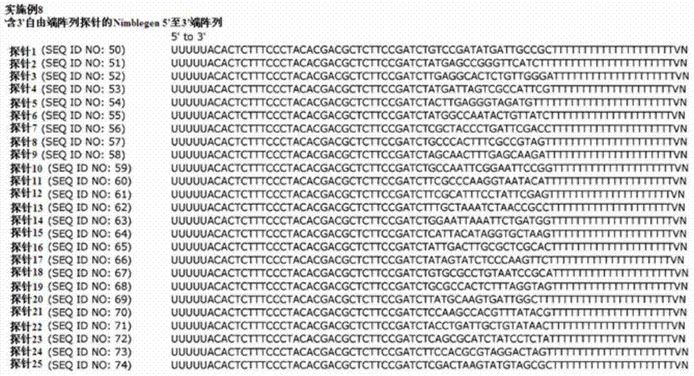


Claims (43)
Priority Applications (2)
| Application Number | Priority Date | Filing Date | Title |
|---|---|---|---|
| CN202211677865.2A CN115896252A (en) | 2011-04-13 | 2012-04-13 | Methods and products for local or spatial detection of nucleic acids in tissue samples |
| CN201810451060.3A CN108796058B (en) | 2011-04-13 | 2012-04-13 | Methods and products for local or spatial detection of nucleic acids in tissue samples |
Applications Claiming Priority (3)
| Application Number | Priority Date | Filing Date | Title |
|---|---|---|---|
| GB1106254.4 | 2011-04-13 | ||
| GBGB1106254.4A GB201106254D0 (en) | 2011-04-13 | 2011-04-13 | Method and product |
| PCT/EP2012/056823 WO2012140224A1 (en) | 2011-04-13 | 2012-04-13 | Method and product for localised or spatial detection of nucleic acid in a tissue sample |
Related Child Applications (2)
| Application Number | Title | Priority Date | Filing Date |
|---|---|---|---|
| CN202211677865.2A Division CN115896252A (en) | 2011-04-13 | 2012-04-13 | Methods and products for local or spatial detection of nucleic acids in tissue samples |
| CN201810451060.3A Division CN108796058B (en) | 2011-04-13 | 2012-04-13 | Methods and products for local or spatial detection of nucleic acids in tissue samples |
Publications (2)
| Publication Number | Publication Date |
|---|---|
| CN103781918A true CN103781918A (en) | 2014-05-07 |
| CN103781918B CN103781918B (en) | 2018-06-01 |
Family
ID=44123034
Family Applications (3)
| Application Number | Title | Priority Date | Filing Date |
|---|---|---|---|
| CN201280029001.8A Active CN103781918B (en) | 2011-04-13 | 2012-04-13 | Methods and products for local or spatial detection of nucleic acids in tissue samples |
| CN202211677865.2A Pending CN115896252A (en) | 2011-04-13 | 2012-04-13 | Methods and products for local or spatial detection of nucleic acids in tissue samples |
| CN201810451060.3A Active CN108796058B (en) | 2011-04-13 | 2012-04-13 | Methods and products for local or spatial detection of nucleic acids in tissue samples |
Family Applications After (2)
| Application Number | Title | Priority Date | Filing Date |
|---|---|---|---|
| CN202211677865.2A Pending CN115896252A (en) | 2011-04-13 | 2012-04-13 | Methods and products for local or spatial detection of nucleic acids in tissue samples |
| CN201810451060.3A Active CN108796058B (en) | 2011-04-13 | 2012-04-13 | Methods and products for local or spatial detection of nucleic acids in tissue samples |
Country Status (13)
| Country | Link |
|---|---|
| US (15) | US10030261B2 (en) |
| EP (4) | EP3677692A1 (en) |
| JP (1) | JP5916166B2 (en) |
| KR (1) | KR101994494B1 (en) |
| CN (3) | CN103781918B (en) |
| AU (1) | AU2012241730B2 (en) |
| BR (1) | BR112013026502A2 (en) |
| CA (1) | CA2832678C (en) |
| GB (1) | GB201106254D0 (en) |
| MX (1) | MX340330B (en) |
| NZ (1) | NZ616407A (en) |
| RU (1) | RU2603074C2 (en) |
| WO (1) | WO2012140224A1 (en) |
Cited By (22)
| Publication number | Priority date | Publication date | Assignee | Title |
|---|---|---|---|---|
| CN105505755A (en) * | 2015-12-23 | 2016-04-20 | 杭州谷禾信息技术有限公司 | Space transcriptome database building and sequencing method and device adopted for same |
| CN106661637A (en) * | 2014-09-05 | 2017-05-10 | 郑直 | Methods of detecting nucleic acids and applications thereof |
| CN107636169A (en) * | 2015-04-17 | 2018-01-26 | 生捷科技控股公司 | The method that profile space analysis is carried out to biomolecule |
| CN109576357A (en) * | 2018-11-20 | 2019-04-05 | 上海欧易生物医学科技有限公司 | A kind of method that overall length transcript degree high throughput detects unicellular middle gene mutation |
| CN110249057A (en) * | 2016-11-17 | 2019-09-17 | 空间转录公司 | Methods for spatially labeling and analyzing nucleic acids in biological samples |
| CN110804654A (en) * | 2019-10-30 | 2020-02-18 | 东南大学 | A method for spatial transcriptome sequencing |
| CN111051526A (en) * | 2017-05-23 | 2020-04-21 | 生捷科技控股公司 | Method for performing analysis of spatial distribution of biomolecules |
| WO2020228788A1 (en) * | 2019-05-15 | 2020-11-19 | 深圳华大生命科学研究院 | Array and method for detecting spatial information of nucleic acids |
| CN112041459A (en) * | 2018-01-29 | 2020-12-04 | 圣祖德儿童研究医院 | Nucleic acid amplification method |
| CN112840023A (en) * | 2018-10-25 | 2021-05-25 | Illumina公司 | Methods and compositions for identifying ligands on arrays using indexing and barcoding |
| CN113186256A (en) * | 2015-04-10 | 2021-07-30 | 空间转录公司 | Spatially differentiated, multiplexed nucleic acid analysis of biological samples |
| CN113439124A (en) * | 2018-12-10 | 2021-09-24 | 10X基因组学有限公司 | Method for spatial detection using master/replica arrays |
| CN113604547A (en) * | 2021-08-06 | 2021-11-05 | 吉林大学 | High-resolution space omics detection method for tissue sample |
| CN113767178A (en) * | 2019-03-11 | 2021-12-07 | 10X基因组学有限公司 | Systems and methods for processing optically labeled beads |
| CN113782103A (en) * | 2021-10-26 | 2021-12-10 | 大连大学 | A DNA matrix processing method based on combinatorial enzyme digestion mechanism |
| CN113930847A (en) * | 2021-09-26 | 2022-01-14 | 东南大学 | Space transcriptome position information coding chip and preparation method and application thereof |
| CN114885610A (en) * | 2019-12-23 | 2022-08-09 | 10X基因组学有限公司 | Methods for spatial analysis using RNA templated ligation |
| CN114934110A (en) * | 2022-06-15 | 2022-08-23 | 江西烈冰生物科技有限公司 | Biochip, kit and method for obtaining original position of gene expression |
| WO2023115536A1 (en) * | 2021-12-24 | 2023-06-29 | 深圳华大生命科学研究院 | Method for generating labeled nucleic acid molecular population and kit thereof |
| WO2023116373A1 (en) * | 2021-12-24 | 2023-06-29 | 深圳华大生命科学研究院 | Method for generating population of labeled nucleic acid molecules and kit for the method |
| WO2024138672A1 (en) * | 2022-12-30 | 2024-07-04 | 深圳华大生命科学研究院 | Improved nucleic acid capture method |
| WO2025138284A1 (en) * | 2023-12-29 | 2025-07-03 | 深圳华大生命科学研究院 | Array and method for detecting spatial information of nucleic acids |
Families Citing this family (388)
| Publication number | Priority date | Publication date | Assignee | Title |
|---|---|---|---|---|
| US9315857B2 (en) | 2009-12-15 | 2016-04-19 | Cellular Research, Inc. | Digital counting of individual molecules by stochastic attachment of diverse label-tags |
| US8835358B2 (en) | 2009-12-15 | 2014-09-16 | Cellular Research, Inc. | Digital counting of individual molecules by stochastic attachment of diverse labels |
| SI2556171T1 (en) | 2010-04-05 | 2016-03-31 | Prognosys Biosciences, Inc. | Spatially encoded biological assays |
| US20190300945A1 (en) | 2010-04-05 | 2019-10-03 | Prognosys Biosciences, Inc. | Spatially Encoded Biological Assays |
| US10787701B2 (en) | 2010-04-05 | 2020-09-29 | Prognosys Biosciences, Inc. | Spatially encoded biological assays |
| GB201106254D0 (en) | 2011-04-13 | 2011-05-25 | Frisen Jonas | Method and product |
| US10501791B2 (en) | 2011-10-14 | 2019-12-10 | President And Fellows Of Harvard College | Sequencing by structure assembly |
| ES2991004T3 (en) | 2011-12-22 | 2024-12-02 | Harvard College | Methods for the detection of analytes |
| CA2859761C (en) | 2011-12-22 | 2023-06-20 | President And Fellows Of Harvard College | Compositions and methods for analyte detection |
| WO2014163886A1 (en) | 2013-03-12 | 2014-10-09 | President And Fellows Of Harvard College | Method of generating a three-dimensional nucleic acid containing matrix |
| EP2820174B1 (en) | 2012-02-27 | 2019-12-25 | The University of North Carolina at Chapel Hill | Methods and uses for molecular tags |
| JP6375230B2 (en) | 2012-02-27 | 2018-08-15 | セルラー リサーチ, インコーポレイテッド | Compositions and kits for molecular counting |
| EP2647426A1 (en) * | 2012-04-03 | 2013-10-09 | Max-Planck-Gesellschaft zur Förderung der Wissenschaften e.V. | Replication of distributed nucleic acid molecules with preservation of their relative distribution through hybridization-based binding |
| WO2013184754A2 (en) | 2012-06-05 | 2013-12-12 | President And Fellows Of Harvard College | Spatial sequencing of nucleic acids using dna origami probes |
| JP5997278B2 (en) * | 2012-07-30 | 2016-09-28 | 株式会社日立製作所 | Two-dimensional cDNA library device with tag sequence, gene expression analysis method and gene expression analysis apparatus using the same |
| CA2881685C (en) | 2012-08-14 | 2023-12-05 | 10X Genomics, Inc. | Microcapsule compositions and methods |
| US9701998B2 (en) | 2012-12-14 | 2017-07-11 | 10X Genomics, Inc. | Methods and systems for processing polynucleotides |
| US10323279B2 (en) | 2012-08-14 | 2019-06-18 | 10X Genomics, Inc. | Methods and systems for processing polynucleotides |
| US11591637B2 (en) | 2012-08-14 | 2023-02-28 | 10X Genomics, Inc. | Compositions and methods for sample processing |
| US10400280B2 (en) | 2012-08-14 | 2019-09-03 | 10X Genomics, Inc. | Methods and systems for processing polynucleotides |
| EP3901280B1 (en) * | 2012-10-17 | 2025-03-12 | 10x Genomics Sweden AB | Methods and product for optimising localised or spatial detection of gene expression in a tissue sample |
| US9410173B2 (en) | 2012-10-24 | 2016-08-09 | Clontech Laboratories, Inc. | Template switch-based methods for producing a product nucleic acid |
| US10533221B2 (en) | 2012-12-14 | 2020-01-14 | 10X Genomics, Inc. | Methods and systems for processing polynucleotides |
| ES2703764T3 (en) | 2012-12-14 | 2019-03-12 | Chronix Biomedical | Custom biomarkers for cancer |
| US9751069B2 (en) | 2013-05-14 | 2017-09-05 | Genomics Usa, Inc. | Compositions and methods for entrapping protein on a surface |
| CN105849275B (en) | 2013-06-25 | 2020-03-17 | 普罗格诺西斯生物科学公司 | Method and system for detecting spatial distribution of biological targets in a sample |
| BR112015032700B1 (en) * | 2013-06-29 | 2022-12-20 | Firmenich Sa | IDENTIFICATION METHODS, ISOLATION AND USE OF AROMA AND ODORANT RECEPTORS |
| EP3039158B1 (en) | 2013-08-28 | 2018-11-14 | Cellular Research, Inc. | Massively parallel single cell analysis |
| US10395758B2 (en) | 2013-08-30 | 2019-08-27 | 10X Genomics, Inc. | Sequencing methods |
| EP3055676A1 (en) | 2013-10-07 | 2016-08-17 | Cellular Research, Inc. | Methods and systems for digitally counting features on arrays |
| EP3760733A1 (en) * | 2013-10-17 | 2021-01-06 | Takara Bio USA, Inc. | Methods for adding adapters to nucleic acids and compositions for practicing the same |
| US10655173B2 (en) | 2013-10-18 | 2020-05-19 | The Broad Institute, Inc. | Spatial and cellular mapping of biomolecules in situ by high-throughput sequencing |
| US9834814B2 (en) | 2013-11-22 | 2017-12-05 | Agilent Technologies, Inc. | Spatial molecular barcoding of in situ nucleic acids |
| WO2015085268A1 (en) | 2013-12-05 | 2015-06-11 | Centrillion Technology Holdings Corporation | Modified surfaces |
| CN106460032B (en) * | 2013-12-05 | 2019-12-24 | 生捷科技控股公司 | Preparation of patterned arrays |
| EP3077545B1 (en) * | 2013-12-05 | 2020-09-16 | Centrillion Technology Holdings Corporation | Methods for sequencing nucleic acids |
| US9824068B2 (en) | 2013-12-16 | 2017-11-21 | 10X Genomics, Inc. | Methods and apparatus for sorting data |
| EP2883963A1 (en) * | 2013-12-16 | 2015-06-17 | Alacris Theranostics GmbH | Method for generating of oligonucleotide arrays using in situ block synthesis approach |
| WO2015094861A1 (en) * | 2013-12-17 | 2015-06-25 | Clontech Laboratories, Inc. | Methods for adding adapters to nucleic acids and compositions for practicing the same |
| US10370630B2 (en) | 2014-02-10 | 2019-08-06 | Technion Research & Development Foundation Limited | Method and apparatus for cell isolation, growth, replication, manipulation, and analysis |
| US11060139B2 (en) | 2014-03-28 | 2021-07-13 | Centrillion Technology Holdings Corporation | Methods for sequencing nucleic acids |
| CN106460052B (en) * | 2014-05-14 | 2021-07-16 | 海德堡鲁普雷希特卡尔斯大学 | Synthesis of double-stranded nucleic acid |
| US12312640B2 (en) | 2014-06-26 | 2025-05-27 | 10X Genomics, Inc. | Analysis of nucleic acid sequences |
| JP2017522866A (en) | 2014-06-26 | 2017-08-17 | 10エックス ジェノミクス, インコーポレイテッド | Nucleic acid sequence analysis |
| EP4574974A3 (en) | 2014-06-26 | 2025-10-08 | 10x Genomics, Inc. | Methods of analyzing nucleic acids from individual cells or cell populations |
| GB201411624D0 (en) | 2014-06-30 | 2014-08-13 | Ge Healthcare Uk Ltd | Spatial molecular profiling of solid biological masses and profile storage |
| US10179932B2 (en) | 2014-07-11 | 2019-01-15 | President And Fellows Of Harvard College | Methods for high-throughput labelling and detection of biological features in situ using microscopy |
| KR101634553B1 (en) * | 2014-08-06 | 2016-06-30 | 가천대학교 산학협력단 | Integrated polycarbonate microdevice, manufacturing method thereof and method of seamless purification and amplification of dna using thereof |
| US9897791B2 (en) | 2014-10-16 | 2018-02-20 | Illumina, Inc. | Optical scanning systems for in situ genetic analysis |
| WO2016090148A1 (en) | 2014-12-03 | 2016-06-09 | IsoPlexis Corporation | Analysis and screening of cell secretion profiles |
| BR112017014902A2 (en) | 2015-01-12 | 2018-03-13 | 10X Genomics Inc | processes and systems for the preparation of nucleic acid sequencing libraries and libraries prepared using them |
| GB201501907D0 (en) | 2015-02-05 | 2015-03-25 | Technion Res & Dev Foundation | System and method for single cell genetic analysis |
| US10697010B2 (en) | 2015-02-19 | 2020-06-30 | Becton, Dickinson And Company | High-throughput single-cell analysis combining proteomic and genomic information |
| ES2836802T3 (en) | 2015-02-27 | 2021-06-28 | Becton Dickinson Co | Spatially addressable molecular barcodes |
| JP6963505B2 (en) | 2015-03-25 | 2021-11-10 | アングル ヨーロッパ リミテッド | Solid phase nucleic acid target capture and replication using strand-substituted polymerases |
| CN107406888A (en) | 2015-03-30 | 2017-11-28 | 赛卢拉研究公司 | Methods and compositions for combining barcoding |
| US10876106B2 (en) | 2015-04-09 | 2020-12-29 | Hitachi, Ltd. | Gene analysis system |
| WO2016166128A1 (en) | 2015-04-14 | 2016-10-20 | Koninklijke Philips N.V. | Spatial mapping of molecular profiles of biological tissue samples |
| EP4582556A3 (en) | 2015-04-23 | 2025-10-08 | Becton, Dickinson and Company | Methods and compositions for whole transcriptome amplification |
| US11124823B2 (en) | 2015-06-01 | 2021-09-21 | Becton, Dickinson And Company | Methods for RNA quantification |
| AU2016295158B2 (en) | 2015-07-17 | 2021-02-25 | Bruker Spatial Biology, Inc. | Simultaneous quantification of gene expression in a user-defined region of a cross-sectioned tissue |
| WO2017015097A1 (en) | 2015-07-17 | 2017-01-26 | Nanostring Technologies, Inc. | Simultaneous quantification of a plurality of proteins in a user-defined region of a cross-sectioned tissue |
| EP3329012B1 (en) | 2015-07-27 | 2021-07-21 | Illumina, Inc. | Spatial mapping of nucleic acid sequence information |
| WO2017044574A1 (en) | 2015-09-11 | 2017-03-16 | Cellular Research, Inc. | Methods and compositions for nucleic acid library normalization |
| EP3368668B1 (en) | 2015-10-28 | 2023-11-29 | Silicon Valley Scientific, Inc. | Method and apparatus for encoding cellular spatial position information |
| CA3004285A1 (en) | 2015-11-03 | 2017-05-11 | President And Fellows Of Harvard College | Method and apparatus for volumetric imaging of a three-dimensional nucleic acid containing matrix |
| US11371094B2 (en) | 2015-11-19 | 2022-06-28 | 10X Genomics, Inc. | Systems and methods for nucleic acid processing using degenerate nucleotides |
| KR20240161696A (en) * | 2015-12-04 | 2024-11-12 | 10엑스 제노믹스, 인크. | Methods and compositions for nucleic acid analysis |
| US11542498B2 (en) * | 2015-12-28 | 2023-01-03 | Pathogendx, Inc. | Microarray based multiplex pathogen analysis and uses thereof |
| WO2017189525A1 (en) | 2016-04-25 | 2017-11-02 | President And Fellows Of Harvard College | Hybridization chain reaction methods for in situ molecular detection |
| US10822643B2 (en) | 2016-05-02 | 2020-11-03 | Cellular Research, Inc. | Accurate molecular barcoding |
| US10301677B2 (en) | 2016-05-25 | 2019-05-28 | Cellular Research, Inc. | Normalization of nucleic acid libraries |
| ES2979395T3 (en) | 2016-05-26 | 2024-09-25 | Becton Dickinson Co | Molecular tag count adjustment methods |
| US10202641B2 (en) | 2016-05-31 | 2019-02-12 | Cellular Research, Inc. | Error correction in amplification of samples |
| US10640763B2 (en) | 2016-05-31 | 2020-05-05 | Cellular Research, Inc. | Molecular indexing of internal sequences |
| WO2017222453A1 (en) | 2016-06-21 | 2017-12-28 | Hauling Thomas | Nucleic acid sequencing |
| JP7239465B2 (en) * | 2016-08-31 | 2023-03-14 | プレジデント アンド フェローズ オブ ハーバード カレッジ | Methods for preparing nucleic acid sequence libraries for detection by fluorescence in situ sequencing |
| CN109923216B (en) | 2016-08-31 | 2024-08-02 | 哈佛学院董事及会员团体 | Methods for combining detection of biomolecules into a single assay using fluorescent in situ sequencing |
| EP3510398A1 (en) | 2016-09-12 | 2019-07-17 | Isoplexis Corporation | System and methods for multiplexed analysis of cellular and other immunotherapeutics |
| EP4300099A3 (en) | 2016-09-26 | 2024-03-27 | Becton, Dickinson and Company | Measurement of protein expression using reagents with barcoded oligonucleotide sequences |
| WO2018075436A1 (en) * | 2016-10-17 | 2018-04-26 | West Jason A A | High resolution spatial genomic analysis of tissues and cell aggregates |
| CN117594126A (en) | 2016-11-08 | 2024-02-23 | 贝克顿迪金森公司 | Method for classifying expression profiles |
| CN117056774B (en) | 2016-11-08 | 2026-01-27 | 贝克顿迪金森公司 | Methods for cell marker classification |
| JP7348066B2 (en) | 2016-11-11 | 2023-09-20 | アイソプレキシス コーポレイション | Compositions and methods for simultaneous analysis of single cell genome, transcriptome and proteome |
| EP3545284A4 (en) | 2016-11-22 | 2020-07-01 | Isoplexis Corporation | SYSTEMS, DEVICES AND METHODS FOR CELL DETECTION AND METHODS FOR THE PRODUCTION THEREOF |
| EP3513346B1 (en) * | 2016-11-30 | 2020-04-15 | Microsoft Technology Licensing, LLC | Dna random access storage system via ligation |
| US10815525B2 (en) | 2016-12-22 | 2020-10-27 | 10X Genomics, Inc. | Methods and systems for processing polynucleotides |
| US10550429B2 (en) | 2016-12-22 | 2020-02-04 | 10X Genomics, Inc. | Methods and systems for processing polynucleotides |
| ES2961580T3 (en) | 2017-01-13 | 2024-03-12 | Cellular Res Inc | Hydrophilic coating of fluid channels |
| RU2665631C2 (en) * | 2017-01-25 | 2018-09-03 | Общество с ограниченной ответственностью "Секвойя Дженетикс" | Method of specific identification of dna sequences |
| CN110214186B (en) | 2017-01-30 | 2023-11-24 | 10X基因组学有限公司 | Methods and systems for droplet-based single cell barcoding |
| US12264411B2 (en) | 2017-01-30 | 2025-04-01 | 10X Genomics, Inc. | Methods and systems for analysis |
| EP3577232A1 (en) | 2017-02-01 | 2019-12-11 | Cellular Research, Inc. | Selective amplification using blocking oligonucleotides |
| US10995333B2 (en) | 2017-02-06 | 2021-05-04 | 10X Genomics, Inc. | Systems and methods for nucleic acid preparation |
| EP4345172A3 (en) | 2017-06-05 | 2024-07-03 | Becton, Dickinson and Company | Sample indexing for single cells |
| US10837047B2 (en) | 2017-10-04 | 2020-11-17 | 10X Genomics, Inc. | Compositions, methods, and systems for bead formation using improved polymers |
| CN111263819B (en) | 2017-10-06 | 2025-04-15 | 10X基因组学有限公司 | RNA-templated ligation |
| EP3700672B1 (en) | 2017-10-27 | 2022-12-28 | 10X Genomics, Inc. | Methods for sample preparation and analysis |
| WO2019099751A1 (en) | 2017-11-15 | 2019-05-23 | 10X Genomics, Inc. | Functionalized gel beads |
| US10829815B2 (en) | 2017-11-17 | 2020-11-10 | 10X Genomics, Inc. | Methods and systems for associating physical and genetic properties of biological particles |
| CN118818037A (en) | 2017-12-12 | 2024-10-22 | 10X基因组学有限公司 | Systems and methods for single cell processing |
| US11946095B2 (en) | 2017-12-19 | 2024-04-02 | Becton, Dickinson And Company | Particles associated with oligonucleotides |
| WO2019126789A1 (en) | 2017-12-22 | 2019-06-27 | 10X Genomics, Inc. | Systems and methods for processing nucleic acid molecules from one or more cells |
| EP3752832A1 (en) | 2018-02-12 | 2020-12-23 | 10X Genomics, Inc. | Methods characterizing multiple analytes from individual cells or cell populations |
| WO2019157445A1 (en) | 2018-02-12 | 2019-08-15 | Nanostring Technologies, Inc. | Biomolecular probes and methods of detecting gene and protein expression |
| US11639928B2 (en) | 2018-02-22 | 2023-05-02 | 10X Genomics, Inc. | Methods and systems for characterizing analytes from individual cells or cell populations |
| WO2019169028A1 (en) | 2018-02-28 | 2019-09-06 | 10X Genomics, Inc. | Transcriptome sequencing through random ligation |
| US12103004B2 (en) | 2018-03-12 | 2024-10-01 | Silicon Valley Scientific, Inc. | Method and apparatus for processing tissue and other samples encoding cellular spatial position information |
| EP3788146A4 (en) | 2018-05-02 | 2022-06-01 | The General Hospital Corporation | High-resolution spatial macromolecule abundance assessment |
| US11773441B2 (en) | 2018-05-03 | 2023-10-03 | Becton, Dickinson And Company | High throughput multiomics sample analysis |
| US11365409B2 (en) | 2018-05-03 | 2022-06-21 | Becton, Dickinson And Company | Molecular barcoding on opposite transcript ends |
| WO2019217758A1 (en) | 2018-05-10 | 2019-11-14 | 10X Genomics, Inc. | Methods and systems for molecular library generation |
| CN112752849A (en) * | 2018-05-18 | 2021-05-04 | 迈克必斯生物系统公司 | Quality control composition and whole organism control material for nucleic acid detection |
| KR102103719B1 (en) * | 2018-05-18 | 2020-04-23 | 주식회사 바이나리 | Method of 3-dimensional nucleic acid imaging analysis of biological tissue using isothermal nucleic acid amplification |
| AU2019274469A1 (en) * | 2018-05-21 | 2020-11-26 | The Regents Of The University Of California | Single cell mapping and transcriptome analysis |
| US11932899B2 (en) | 2018-06-07 | 2024-03-19 | 10X Genomics, Inc. | Methods and systems for characterizing nucleic acid molecules |
| US11703427B2 (en) | 2018-06-25 | 2023-07-18 | 10X Genomics, Inc. | Methods and systems for cell and bead processing |
| EP3824098A1 (en) | 2018-07-18 | 2021-05-26 | Max-Delbrück-Centrum für Molekulare Medizin in der Helmholtz-Gemeinschaft | Method for analyzing cell sample heterogeneity |
| WO2020023484A1 (en) * | 2018-07-23 | 2020-01-30 | Genomics Usa, Inc. | Blood typing using dna |
| US12188014B1 (en) | 2018-07-25 | 2025-01-07 | 10X Genomics, Inc. | Compositions and methods for nucleic acid processing using blocking agents |
| US20200032335A1 (en) | 2018-07-27 | 2020-01-30 | 10X Genomics, Inc. | Systems and methods for metabolome analysis |
| CN112703252B (en) | 2018-08-03 | 2024-09-10 | 10X基因组学有限公司 | Method and system for minimizing barcode exchange |
| CN109136372A (en) * | 2018-08-08 | 2019-01-04 | 江苏苏博生物医学科技南京有限公司 | It is a kind of based on illumina platform breast cancer parting detection build library kit |
| CN109097467A (en) * | 2018-08-08 | 2018-12-28 | 江苏苏博生物医学科技南京有限公司 | Based on the breast cancer parting detecting reagent of illumina platform and application |
| WO2020041148A1 (en) | 2018-08-20 | 2020-02-27 | 10X Genomics, Inc. | Methods and systems for detection of protein-dna interactions using proximity ligation |
| US12065688B2 (en) | 2018-08-20 | 2024-08-20 | 10X Genomics, Inc. | Compositions and methods for cellular processing |
| US20210317524A1 (en) | 2018-08-28 | 2021-10-14 | 10X Genomics, Inc. | Resolving spatial arrays |
| WO2020047010A2 (en) | 2018-08-28 | 2020-03-05 | 10X Genomics, Inc. | Increasing spatial array resolution |
| US11519033B2 (en) | 2018-08-28 | 2022-12-06 | 10X Genomics, Inc. | Method for transposase-mediated spatial tagging and analyzing genomic DNA in a biological sample |
| JP7670607B2 (en) | 2018-10-01 | 2025-04-30 | ベクトン・ディキンソン・アンド・カンパニー | Determination of 5' transcript sequence |
| WO2020076976A1 (en) | 2018-10-10 | 2020-04-16 | Readcoor, Inc. | Three-dimensional spatial molecular indexing |
| EP3864173B1 (en) * | 2018-10-10 | 2025-08-27 | 10x Genomics, Inc. | Surface capture of target nucleic acid molecules |
| EP3877520B1 (en) | 2018-11-08 | 2025-03-19 | Becton, Dickson And Company | Whole transcriptome analysis of single cells using random priming |
| WO2020117914A1 (en) * | 2018-12-04 | 2020-06-11 | Roche Sequencing Solutions, Inc. | Spatially oriented quantum barcoding of cellular targets |
| US12529094B2 (en) | 2018-12-10 | 2026-01-20 | 10X Genomics, Inc. | Imaging system hardware |
| US11459607B1 (en) | 2018-12-10 | 2022-10-04 | 10X Genomics, Inc. | Systems and methods for processing-nucleic acid molecules from a single cell using sequential co-partitioning and composite barcodes |
| US20230242976A1 (en) * | 2018-12-10 | 2023-08-03 | 10X Genomics, Inc. | Imaging system hardware |
| US11492660B2 (en) | 2018-12-13 | 2022-11-08 | Becton, Dickinson And Company | Selective extension in single cell whole transcriptome analysis |
| WO2020120442A2 (en) | 2018-12-13 | 2020-06-18 | Dna Script | Direct oligonucleotide synthesis on cells and biomolecules |
| GB201820300D0 (en) | 2018-12-13 | 2019-01-30 | 10X Genomics Inc | Method for spatial tagging and analysing genomic DNA in a biological specimen |
| GB201820341D0 (en) | 2018-12-13 | 2019-01-30 | 10X Genomics Inc | Method for transposase-mediated spatial tagging and analysing genomic DNA in a biological specimen |
| MY205579A (en) | 2018-12-21 | 2024-10-28 | Illumina Inc | Nuclease-based rna depletion |
| US11649485B2 (en) | 2019-01-06 | 2023-05-16 | 10X Genomics, Inc. | Generating capture probes for spatial analysis |
| US11926867B2 (en) | 2019-01-06 | 2024-03-12 | 10X Genomics, Inc. | Generating capture probes for spatial analysis |
| CN109762728A (en) * | 2019-01-07 | 2019-05-17 | 东南大学 | A kind of spatial transcriptome detection chip and method |
| US12169198B2 (en) | 2019-01-08 | 2024-12-17 | 10X Genomics, Inc. | Systems and methods for sample analysis |
| US11845983B1 (en) | 2019-01-09 | 2023-12-19 | 10X Genomics, Inc. | Methods and systems for multiplexing of droplet based assays |
| WO2020150356A1 (en) | 2019-01-16 | 2020-07-23 | Becton, Dickinson And Company | Polymerase chain reaction normalization through primer titration |
| WO2020154247A1 (en) | 2019-01-23 | 2020-07-30 | Cellular Research, Inc. | Oligonucleotides associated with antibodies |
| US12529092B2 (en) | 2019-01-28 | 2026-01-20 | The Broad Institute, Inc. | In-situ spatial transcriptomics |
| US11467153B2 (en) | 2019-02-12 | 2022-10-11 | 10X Genomics, Inc. | Methods for processing nucleic acid molecules |
| WO2020167866A1 (en) | 2019-02-12 | 2020-08-20 | 10X Genomics, Inc. | Systems and methods for transposon loading |
| US12305239B2 (en) | 2019-02-12 | 2025-05-20 | 10X Genomics, Inc. | Analysis of nucleic acid sequences |
| US11584953B2 (en) | 2019-02-12 | 2023-02-21 | 10X Genomics, Inc. | Methods for processing nucleic acid molecules |
| US11851683B1 (en) | 2019-02-12 | 2023-12-26 | 10X Genomics, Inc. | Methods and systems for selective analysis of cellular samples |
| US12275993B2 (en) | 2019-02-12 | 2025-04-15 | 10X Genomics, Inc. | Analysis of nucleic acid sequences |
| WO2020167862A1 (en) | 2019-02-12 | 2020-08-20 | 10X Genomics, Inc. | Systems and methods for transfer of reagents between droplets |
| EP3924506A1 (en) | 2019-02-14 | 2021-12-22 | Becton Dickinson and Company | Hybrid targeted and whole transcriptome amplification |
| US11655499B1 (en) | 2019-02-25 | 2023-05-23 | 10X Genomics, Inc. | Detection of sequence elements in nucleic acid molecules |
| CN114174531A (en) * | 2019-02-28 | 2022-03-11 | 10X基因组学有限公司 | Profiling of biological analytes with spatially barcoded oligonucleotide arrays |
| US20230143569A1 (en) * | 2019-02-28 | 2023-05-11 | 10X Genomics, Inc. | Profiling of biological analytes with spatially barcoded oligonucleotide arrays |
| CN114127309A (en) * | 2019-03-15 | 2022-03-01 | 10X基因组学有限公司 | Method for single cell sequencing using spatial arrays |
| WO2020198071A1 (en) | 2019-03-22 | 2020-10-01 | 10X Genomics, Inc. | Three-dimensional spatial analysis |
| WO2020206285A1 (en) * | 2019-04-05 | 2020-10-08 | Board Of Regents, The University Of Texas System | Methods and applications for cell barcoding |
| WO2020214642A1 (en) | 2019-04-19 | 2020-10-22 | Becton, Dickinson And Company | Methods of associating phenotypical data and single cell sequencing data |
| EP3976820A1 (en) | 2019-05-30 | 2022-04-06 | 10X Genomics, Inc. | Methods of detecting spatial heterogeneity of a biological sample |
| WO2020240025A1 (en) | 2019-05-31 | 2020-12-03 | Cartana Ab | Method of detecting target nucleic acid molecules |
| KR102811825B1 (en) | 2019-06-21 | 2025-05-26 | 써모 피셔 사이언티픽 발틱스 유에이비 | Oligonucleotide-tethered triphosphate nucleotides useful for nucleic acid labeling for preparing next-generation sequencing libraries |
| CN114051534B (en) | 2019-07-22 | 2025-02-21 | 贝克顿迪金森公司 | Single-cell chromatin immunoprecipitation sequencing assay |
| US20220251632A1 (en) * | 2019-07-23 | 2022-08-11 | University Of Washington | Method for spatially barcoding cells in tissue slices |
| EP4010494A1 (en) * | 2019-08-08 | 2022-06-15 | INSERM (Institut National de la Santé et de la Recherche Médicale) | Rna sequencing method for the analysis of b and t cell transcriptome in phenotypically defined b and t cell subsets |
| US20210062272A1 (en) * | 2019-08-13 | 2021-03-04 | 10X Genomics, Inc. | Systems and methods for using the spatial distribution of haplotypes to determine a biological condition |
| US12235262B1 (en) | 2019-09-09 | 2025-02-25 | 10X Genomics, Inc. | Methods and systems for single cell protein analysis |
| WO2021066465A1 (en) * | 2019-10-01 | 2021-04-08 | (주)컨투어젠 | Method and apparatus for extracting nucleic acid from nucleic acid-containing sample while retaining 2-dimensional position information, and method for analyzing genome including position information using same |
| CN117036248A (en) | 2019-10-01 | 2023-11-10 | 10X基因组学有限公司 | Systems and methods for identifying morphological patterns in tissue samples |
| US12157124B2 (en) | 2019-11-06 | 2024-12-03 | 10X Genomics, Inc. | Imaging system hardware |
| US11773436B2 (en) | 2019-11-08 | 2023-10-03 | Becton, Dickinson And Company | Using random priming to obtain full-length V(D)J information for immune repertoire sequencing |
| WO2021092433A2 (en) | 2019-11-08 | 2021-05-14 | 10X Genomics, Inc. | Enhancing specificity of analyte binding |
| WO2021091611A1 (en) | 2019-11-08 | 2021-05-14 | 10X Genomics, Inc. | Spatially-tagged analyte capture agents for analyte multiplexing |
| EP4589016A3 (en) | 2019-11-13 | 2025-10-08 | 10x Genomics, Inc. | Generating capture probes for spatial analysis |
| US12154266B2 (en) | 2019-11-18 | 2024-11-26 | 10X Genomics, Inc. | Systems and methods for binary tissue classification |
| CA3158888A1 (en) | 2019-11-21 | 2021-05-27 | Yifeng YIN | Spatial analysis of analytes |
| EP4062372B1 (en) | 2019-11-22 | 2024-05-08 | 10X Genomics, Inc. | Systems and methods for spatial analysis of analytes using fiducial alignment |
| WO2021119320A2 (en) | 2019-12-11 | 2021-06-17 | 10X Genomics, Inc. | Reverse transcriptase variants |
| GB201919032D0 (en) | 2019-12-20 | 2020-02-05 | Cartana Ab | Method of detecting an analyte |
| WO2021133842A1 (en) | 2019-12-23 | 2021-07-01 | 10X Genomics, Inc. | Compositions and methods for using fixed biological samples in partition-based assays |
| EP4339299B1 (en) * | 2020-01-10 | 2025-08-27 | 10X Genomics, Inc. | Methods for determining a location of a target nucleic acid in a biological sample |
| WO2021146207A1 (en) | 2020-01-13 | 2021-07-22 | Becton, Dickinson And Company | Methods and compositions for quantitation of proteins and rna |
| US12365942B2 (en) | 2020-01-13 | 2025-07-22 | 10X Genomics, Inc. | Methods of decreasing background on a spatial array |
| US12405264B2 (en) | 2020-01-17 | 2025-09-02 | 10X Genomics, Inc. | Electrophoretic system and method for analyte capture |
| US11732299B2 (en) | 2020-01-21 | 2023-08-22 | 10X Genomics, Inc. | Spatial assays with perturbed cells |
| US11702693B2 (en) | 2020-01-21 | 2023-07-18 | 10X Genomics, Inc. | Methods for printing cells and generating arrays of barcoded cells |
| US20210230681A1 (en) | 2020-01-24 | 2021-07-29 | 10X Genomics, Inc. | Methods for spatial analysis using proximity ligation |
| US11821035B1 (en) | 2020-01-29 | 2023-11-21 | 10X Genomics, Inc. | Compositions and methods of making gene expression libraries |
| WO2021155057A1 (en) | 2020-01-29 | 2021-08-05 | Becton, Dickinson And Company | Barcoded wells for spatial mapping of single cells through sequencing |
| US12076701B2 (en) | 2020-01-31 | 2024-09-03 | 10X Genomics, Inc. | Capturing oligonucleotides in spatial transcriptomics |
| US11898205B2 (en) | 2020-02-03 | 2024-02-13 | 10X Genomics, Inc. | Increasing capture efficiency of spatial assays |
| US12110548B2 (en) | 2020-02-03 | 2024-10-08 | 10X Genomics, Inc. | Bi-directional in situ analysis |
| US12110541B2 (en) | 2020-02-03 | 2024-10-08 | 10X Genomics, Inc. | Methods for preparing high-resolution spatial arrays |
| US12112833B2 (en) | 2020-02-04 | 2024-10-08 | 10X Genomics, Inc. | Systems and methods for index hopping filtering |
| US11732300B2 (en) * | 2020-02-05 | 2023-08-22 | 10X Genomics, Inc. | Increasing efficiency of spatial analysis in a biological sample |
| US12129516B2 (en) | 2020-02-07 | 2024-10-29 | 10X Genomics, Inc. | Quantitative and automated permeabilization performance evaluation for spatial transcriptomics |
| US11835462B2 (en) | 2020-02-11 | 2023-12-05 | 10X Genomics, Inc. | Methods and compositions for partitioning a biological sample |
| US20230081062A1 (en) * | 2020-02-12 | 2023-03-16 | Universal Sequencing Technology Corporation | Methods for intracellular barcoding and spatial barcoding |
| EP4104179B1 (en) | 2020-02-13 | 2026-01-28 | 10X Genomics, Inc. | Systems and methods for joint interactive visualization of gene expression and dna chromatin accessibility |
| US12281357B1 (en) | 2020-02-14 | 2025-04-22 | 10X Genomics, Inc. | In situ spatial barcoding |
| US12399123B1 (en) | 2020-02-14 | 2025-08-26 | 10X Genomics, Inc. | Spatial targeting of analytes |
| EP4107284A1 (en) | 2020-02-17 | 2022-12-28 | 10X Genomics, Inc. | In situ analysis of chromatin interaction |
| WO2021168278A1 (en) | 2020-02-20 | 2021-08-26 | 10X Genomics, Inc. | METHODS TO COMBINE FIRST AND SECOND STRAND cDNA SYNTHESIS FOR SPATIAL ANALYSIS |
| CN116157533A (en) | 2020-02-21 | 2023-05-23 | 10X基因组学有限公司 | Capture Genetic Targets Using Hybridization |
| US12173360B2 (en) | 2020-02-21 | 2024-12-24 | 10X Genomics, Inc. | Methods and compositions for integrated in situ spatial assay |
| US11891654B2 (en) | 2020-02-24 | 2024-02-06 | 10X Genomics, Inc. | Methods of making gene expression libraries |
| WO2021173719A1 (en) | 2020-02-25 | 2021-09-02 | Becton, Dickinson And Company | Bi-specific probes to enable the use of single-cell samples as single color compensation control |
| US11926863B1 (en) | 2020-02-27 | 2024-03-12 | 10X Genomics, Inc. | Solid state single cell method for analyzing fixed biological cells |
| US11768175B1 (en) | 2020-03-04 | 2023-09-26 | 10X Genomics, Inc. | Electrophoretic methods for spatial analysis |
| US12392435B1 (en) | 2020-03-05 | 2025-08-19 | Clay OWENS | Toolless disassembly rotational vacuum coupling |
| US12188085B2 (en) * | 2020-03-05 | 2025-01-07 | 10X Genomics, Inc. | Three-dimensional spatial transcriptomics with sequencing readout |
| KR102482668B1 (en) * | 2020-03-10 | 2022-12-29 | 사회복지법인 삼성생명공익재단 | A method for improving the labeling accuracy of Unique Molecular Identifiers |
| WO2021216708A1 (en) | 2020-04-22 | 2021-10-28 | 10X Genomics, Inc. | Methods for spatial analysis using targeted rna depletion |
| US20230265491A1 (en) | 2020-05-04 | 2023-08-24 | 10X Genomics, Inc. | Spatial transcriptomic transfer modes |
| US11851700B1 (en) | 2020-05-13 | 2023-12-26 | 10X Genomics, Inc. | Methods, kits, and compositions for processing extracellular molecules |
| WO2021231779A1 (en) | 2020-05-14 | 2021-11-18 | Becton, Dickinson And Company | Primers for immune repertoire profiling |
| US12416603B2 (en) | 2020-05-19 | 2025-09-16 | 10X Genomics, Inc. | Electrophoresis cassettes and instrumentation |
| WO2021237087A1 (en) | 2020-05-22 | 2021-11-25 | 10X Genomics, Inc. | Spatial analysis to detect sequence variants |
| WO2021237056A1 (en) * | 2020-05-22 | 2021-11-25 | 10X Genomics, Inc. | Rna integrity analysis in a biological sample |
| WO2021236929A1 (en) | 2020-05-22 | 2021-11-25 | 10X Genomics, Inc. | Simultaneous spatio-temporal measurement of gene expression and cellular activity |
| WO2021242834A1 (en) | 2020-05-26 | 2021-12-02 | 10X Genomics, Inc. | Method for resetting an array |
| US12265079B1 (en) | 2020-06-02 | 2025-04-01 | 10X Genomics, Inc. | Systems and methods for detecting analytes from captured single biological particles |
| EP4025692A2 (en) | 2020-06-02 | 2022-07-13 | 10X Genomics, Inc. | Nucleic acid library methods |
| CN116249785A (en) | 2020-06-02 | 2023-06-09 | 10X基因组学有限公司 | Spatial Transcriptomics for Antigen-Receptors |
| WO2021247593A1 (en) | 2020-06-02 | 2021-12-09 | Becton, Dickinson And Company | Oligonucleotides and beads for 5 prime gene expression assay |
| US12031177B1 (en) | 2020-06-04 | 2024-07-09 | 10X Genomics, Inc. | Methods of enhancing spatial resolution of transcripts |
| ES2981265T3 (en) | 2020-06-08 | 2024-10-08 | 10X Genomics Inc | Methods for determining a surgical margin and methods of using it |
| WO2021252576A1 (en) | 2020-06-10 | 2021-12-16 | 10X Genomics, Inc. | Methods for spatial analysis using blocker oligonucleotides |
| EP4446430A3 (en) | 2020-06-10 | 2024-12-18 | 10X Genomics, Inc. | Methods for determining a location of an analyte in a biological sample |
| AU2021288090A1 (en) | 2020-06-10 | 2023-01-19 | 10X Genomics, Inc. | Fluid delivery methods |
| US12435363B1 (en) | 2020-06-10 | 2025-10-07 | 10X Genomics, Inc. | Materials and methods for spatial transcriptomics |
| US12209273B2 (en) | 2020-06-12 | 2025-01-28 | 10X Genomics, Inc. | Nucleic acid assays using click chemistry bioconjugation |
| AU2021293238A1 (en) * | 2020-06-18 | 2023-02-09 | Bruker Spatial Biology, Inc. | Compositions and methods for in situ single cell analysis using enzymatic nucleic acid extension |
| EP4450639B1 (en) | 2020-06-25 | 2025-10-15 | 10X Genomics, Inc. | Spatial analysis of dna methylation |
| US12168801B1 (en) | 2020-07-02 | 2024-12-17 | 10X Genomics, Inc. | Hybrid/capture probe designs for full-length cDNA |
| US12209280B1 (en) | 2020-07-06 | 2025-01-28 | 10X Genomics, Inc. | Methods of identifying abundance and location of an analyte in a biological sample using second strand synthesis |
| US11761038B1 (en) | 2020-07-06 | 2023-09-19 | 10X Genomics, Inc. | Methods for identifying a location of an RNA in a biological sample |
| US11981960B1 (en) | 2020-07-06 | 2024-05-14 | 10X Genomics, Inc. | Spatial analysis utilizing degradable hydrogels |
| US11932901B2 (en) | 2020-07-13 | 2024-03-19 | Becton, Dickinson And Company | Target enrichment using nucleic acid probes for scRNAseq |
| WO2022013094A1 (en) | 2020-07-15 | 2022-01-20 | Dna Script | Massively parallel enzymatic synthesis of polynucleotides |
| US20240043913A1 (en) * | 2020-07-17 | 2024-02-08 | The Regents Of The University Of Michigan | Materials and methods for localized detection of nucleic acids in a tissue sample |
| CN116323968A (en) | 2020-07-31 | 2023-06-23 | 10X基因组学有限公司 | Decrosslinking compounds for spatial analysis and methods of use |
| US12391940B2 (en) | 2020-07-31 | 2025-08-19 | Becton, Dickinson And Company | Single cell assay for transposase-accessible chromatin |
| US11434525B2 (en) | 2020-08-06 | 2022-09-06 | Singular Genomics Systems, Inc. | Spatial sequencing |
| WO2022032194A1 (en) | 2020-08-06 | 2022-02-10 | Singular Genomics Systems, Inc. | Methods for in situ transcriptomics and proteomics |
| US12297499B2 (en) | 2020-08-17 | 2025-05-13 | 10X Genomics, Inc. | Multicomponent nucleic acid probes for sample analysis |
| US20220049303A1 (en) | 2020-08-17 | 2022-02-17 | Readcoor, Llc | Methods and systems for spatial mapping of genetic variants |
| US11981958B1 (en) | 2020-08-20 | 2024-05-14 | 10X Genomics, Inc. | Methods for spatial analysis using DNA capture |
| US20220064718A1 (en) * | 2020-08-31 | 2022-03-03 | Applied Materials, Inc. | Alignment beads for mfish |
| EP4211508B1 (en) | 2020-09-14 | 2025-11-26 | Singular Genomics Systems, Inc. | Methods and systems for multidimensional imaging |
| EP4530358A3 (en) | 2020-09-15 | 2025-06-18 | 10x Genomics, Inc. | Methods of releasing an extended capture probe from a substrate and uses of the same |
| US20230313279A1 (en) | 2020-09-16 | 2023-10-05 | 10X Genomics, Inc. | Methods of determining the location of an analyte in a biological sample using a plurality of wells |
| WO2022061152A2 (en) | 2020-09-18 | 2022-03-24 | 10X Genomics, Inc. | Sample handling apparatus and fluid delivery methods |
| AU2021345283B2 (en) | 2020-09-18 | 2024-12-19 | 10X Genomics, Inc. | Sample handling apparatus and image registration methods |
| US11926822B1 (en) | 2020-09-23 | 2024-03-12 | 10X Genomics, Inc. | Three-dimensional spatial analysis |
| WO2022081643A2 (en) | 2020-10-13 | 2022-04-21 | 10X Genomics, Inc. | Compositions and methods for generating recombinant antigen binding molecules from single cells |
| AU2021366701A1 (en) | 2020-10-22 | 2023-05-04 | 10X Genomics, Inc. | Methods for spatial analysis using rolling circle amplification |
| US12071667B2 (en) | 2020-11-04 | 2024-08-27 | 10X Genomics, Inc. | Sequence analysis using meta-stable nucleic acid molecules |
| US12480158B1 (en) | 2020-11-05 | 2025-11-25 | 10X Genomics, Inc. | Methods and systems for processing polynucleotides |
| US12084715B1 (en) | 2020-11-05 | 2024-09-10 | 10X Genomics, Inc. | Methods and systems for reducing artifactual antisense products |
| CN116829733A (en) * | 2020-11-06 | 2023-09-29 | 10X基因组学有限公司 | Compositions and methods for binding analytes to capture probes |
| WO2022109181A1 (en) | 2020-11-18 | 2022-05-27 | 10X Genomics, Inc. | Methods and compositions for analyzing immune infiltration in cancer stroma to predict clinical outcome |
| US11827935B1 (en) | 2020-11-19 | 2023-11-28 | 10X Genomics, Inc. | Methods for spatial analysis using rolling circle amplification and detection probes |
| EP4247967A1 (en) | 2020-11-20 | 2023-09-27 | Becton, Dickinson and Company | Profiling of highly expressed and lowly expressed proteins |
| CN116782827A (en) * | 2020-11-30 | 2023-09-19 | 明德拉公司 | Microneedle devices and methods and skin condition determination |
| KR102702774B1 (en) * | 2020-12-09 | 2024-09-05 | 고려대학교 산학협력단 | Position-based single cell gene expression analysis method and device |
| EP4012046A1 (en) | 2020-12-11 | 2022-06-15 | 10X Genomics, Inc. | Methods and compositions for multimodal in situ analysis |
| WO2022132206A1 (en) | 2020-12-15 | 2022-06-23 | Becton, Dickinson And Company | Single cell secretome analysis |
| AU2021409136A1 (en) | 2020-12-21 | 2023-06-29 | 10X Genomics, Inc. | Methods, compositions, and systems for capturing probes and/or barcodes |
| CA3205944C (en) | 2020-12-21 | 2025-02-11 | Singular Genomics Systems, Inc. | Systems and methods for multicolor imaging |
| WO2022147296A1 (en) | 2020-12-30 | 2022-07-07 | 10X Genomics, Inc. | Cleavage of capture probes for spatial analysis |
| US20240068017A1 (en) * | 2020-12-30 | 2024-02-29 | 10X Genomics, Inc. | Methods for analyte capture determination |
| US12060603B2 (en) | 2021-01-19 | 2024-08-13 | 10X Genomics, Inc. | Methods for internally controlled in situ assays using padlock probes |
| AU2022209365A1 (en) * | 2021-01-21 | 2023-07-20 | Nautilus Subsidiary, Inc. | Systems and methods for biomolecule preparation |
| US12398262B1 (en) | 2021-01-22 | 2025-08-26 | 10X Genomics, Inc. | Triblock copolymer-based cell stabilization and fixation system and methods of use thereof |
| AU2022212231A1 (en) | 2021-01-29 | 2023-08-03 | 10X Genomics, Inc. | Method for transposase mediated spatial tagging and analyzing genomic dna in a biological sample |
| EP4294571B8 (en) | 2021-02-19 | 2024-07-10 | 10X Genomics, Inc. | Method of using a modular assay support device |
| CN117015617B (en) | 2021-02-23 | 2025-04-04 | 10X基因组学有限公司 | Probe-based nucleic acid and protein analysis |
| US12275984B2 (en) | 2021-03-02 | 2025-04-15 | 10X Genomics, Inc. | Sequential hybridization and quenching |
| WO2022198068A1 (en) | 2021-03-18 | 2022-09-22 | 10X Genomics, Inc. | Multiplex capture of gene and protein expression from a biological sample |
| EP4319792B1 (en) | 2021-04-05 | 2025-09-03 | 10X Genomics, Inc. | Recombinant ligase composition and uses thereof |
| EP4428246B1 (en) | 2021-04-14 | 2025-12-24 | 10X Genomics, Inc. | Methods of measuring mislocalization of an analyte |
| WO2022226057A1 (en) | 2021-04-20 | 2022-10-27 | 10X Genomics, Inc. | Methods for assessing sample quality prior to spatial analysis using templated ligation |
| WO2022232571A1 (en) | 2021-04-30 | 2022-11-03 | 10X Genomics, Inc. | Fusion rt variants for improved performance |
| EP4334706A4 (en) | 2021-05-05 | 2025-08-06 | Singular Genomics Systems Inc | Multiomic analysis device and methods of use thereof |
| EP4320271B1 (en) * | 2021-05-06 | 2025-03-19 | 10X Genomics, Inc. | Methods for increasing resolution of spatial analysis |
| US20240257914A1 (en) | 2021-05-19 | 2024-08-01 | Max-Delbrück-Centrum Für Molekulare Medizin In Der Helmholtz-Gemeinschaft | Method and system for 3d reconstruction of tissue gene expression data |
| EP4582555A3 (en) | 2021-06-03 | 2025-10-22 | 10X Genomics, Inc. | Methods, compositions, kits, and systems for enhancing analyte capture for spatial analysis |
| WO2022265965A1 (en) | 2021-06-14 | 2022-12-22 | 10X Genomics, Inc. | Reverse transcriptase variants for improved performance |
| US20220403462A1 (en) | 2021-06-18 | 2022-12-22 | Miltenyi Biotec B.V. & Co. KG | Method of spatial sequencing of genes from tissue using padlocks with gaps on substrate |
| US12416039B2 (en) * | 2021-06-22 | 2025-09-16 | WellSIM Biomedical Technologies, Inc. | Spatially barcoded microarray |
| WO2022271820A1 (en) | 2021-06-22 | 2022-12-29 | 10X Genomics, Inc. | Spatial detection of sars-cov-2 using templated ligation |
| WO2022269543A2 (en) * | 2021-06-24 | 2022-12-29 | Moleculent Ab | Spatial analysis of a planar biological sample |
| US20240338955A1 (en) * | 2021-06-25 | 2024-10-10 | Portrai Inc. | Exploration apparatus, system, and computer program for exploring molecular marker or physiological activity information in tissue relating to distribution or physiological activity of material under exploration |
| US20230026886A1 (en) | 2021-07-13 | 2023-01-26 | 10X Genomics, Inc. | Methods for preparing polymerized matrix with controllable thickness |
| WO2023287765A1 (en) | 2021-07-13 | 2023-01-19 | 10X Genomics, Inc. | Methods for spatial analysis using targeted probe silencing |
| US12139751B2 (en) | 2021-07-30 | 2024-11-12 | 10X Genomics, Inc. | Circularizable probes for in situ analysis |
| ES2988400T3 (en) | 2021-07-30 | 2024-11-20 | 10X Genomics Inc | Methods and compositions for synchronizing reactions in situ |
| US12391984B2 (en) | 2021-08-03 | 2025-08-19 | 10X Genomics, Inc. | Compositions and methods for rolling circle amplification |
| US12529096B2 (en) | 2021-08-03 | 2026-01-20 | 10X Genomics, Inc. | Stabilization and/or compaction of nucleic acid structures |
| US12460251B2 (en) | 2021-08-03 | 2025-11-04 | 10X Genomics, Inc. | Stabilization and/or compaction of nucleic acid molecules |
| WO2023018799A1 (en) | 2021-08-12 | 2023-02-16 | 10X Genomics, Inc. | Methods, compositions and systems for identifying antigen-binding molecules |
| EP4446426A3 (en) | 2021-08-16 | 2024-11-13 | 10x Genomics, Inc. | Probes comprising a split barcode region and methods of use |
| WO2023034489A1 (en) | 2021-09-01 | 2023-03-09 | 10X Genomics, Inc. | Methods, compositions, and kits for blocking a capture probe on a spatial array |
| US20240378734A1 (en) | 2021-09-17 | 2024-11-14 | 10X Genomics, Inc. | Systems and methods for image registration or alignment |
| EP4155416B1 (en) | 2021-09-23 | 2024-01-10 | Miltenyi Biotec B.V. & Co. KG | Method for obtaining spatial and sequencing information of m-rna from tissue |
| WO2023076345A1 (en) | 2021-10-26 | 2023-05-04 | 10X Genomics, Inc. | Methods for spatial analysis using targeted rna capture |
| US20250147036A1 (en) | 2021-11-10 | 2025-05-08 | 10X Genomics, Inc. | Methods for identification of antigen-binding molecules |
| EP4419707A1 (en) | 2021-11-10 | 2024-08-28 | 10X Genomics, Inc. | Methods, compositions, and kits for determining the location of an analyte in a biological sample |
| EP4441741A1 (en) | 2021-11-30 | 2024-10-09 | 10X Genomics, Inc. | Systems and methods for identifying regions of aneuploidy in a tissue |
| EP4305195A2 (en) | 2021-12-01 | 2024-01-17 | 10X Genomics, Inc. | Methods, compositions, and systems for improved in situ detection of analytes and spatial analysis |
| WO2023114473A2 (en) | 2021-12-16 | 2023-06-22 | 10X Genomics, Inc. | Recombinant reverse transcriptase variants for improved performance |
| WO2023122033A1 (en) | 2021-12-20 | 2023-06-29 | 10X Genomics, Inc. | Self-test for pathology/histology slide imaging device |
| EP4453254A2 (en) * | 2021-12-22 | 2024-10-30 | The General Hospital Corporation | Compositions and methods for end to end capture of messenger rnas |
| WO2023150163A1 (en) | 2022-02-01 | 2023-08-10 | 10X Genomics, Inc. | Methods, compositions, and systems for capturing analytes from lymphatic tissue |
| US20250163501A1 (en) | 2022-02-01 | 2025-05-22 | 10X Genomics, Inc. | Methods, kits, compositions, and systems for spatial analysis |
| WO2023150171A1 (en) | 2022-02-01 | 2023-08-10 | 10X Genomics, Inc. | Methods, compositions, and systems for capturing analytes from glioblastoma samples |
| EP4479933A1 (en) | 2022-02-15 | 2024-12-25 | 10x Genomics, Inc. | Systems and methods for spatial analysis of analytes using fiducial alignment |
| WO2023172915A1 (en) | 2022-03-08 | 2023-09-14 | 10X Genomics, Inc. | In situ code design methods for minimizing optical crowding |
| WO2023172670A2 (en) | 2022-03-11 | 2023-09-14 | 10X Genomics, Inc. | Sample handling apparatus and fluid delivery methods |
| CN114369650B (en) * | 2022-03-21 | 2022-06-17 | 深圳市仙湖植物园(深圳市园林研究中心) | Design method of capture probe, capture probe and application thereof |
| CN119032271A (en) | 2022-04-01 | 2024-11-26 | 10X基因组学有限公司 | Compositions and methods for targeted masking of autofluorescence |
| US20250251403A1 (en) | 2022-04-12 | 2025-08-07 | 10X Genomics, Inc. | Compositions and methods for generating and characterizing recombinant antigen binding molecules |
| EP4490319A1 (en) | 2022-05-06 | 2025-01-15 | 10X Genomics, Inc. | Molecular barcode readers for analyte detection |
| US20240002902A1 (en) | 2022-05-06 | 2024-01-04 | 10X Genomics, Inc. | Analysis of antigen and antigen receptor interactions |
| JP2025516589A (en) | 2022-05-11 | 2025-05-30 | 10エックス ジェノミクス インコーポレイテッド | Compositions and methods for in situ sequencing |
| WO2023225519A1 (en) | 2022-05-17 | 2023-11-23 | 10X Genomics, Inc. | Modified transposons, compositions and uses thereof |
| WO2023229988A1 (en) | 2022-05-23 | 2023-11-30 | 10X Genomics, Inc. | Tissue sample mold |
| WO2023229982A2 (en) | 2022-05-24 | 2023-11-30 | 10X Genomics, Inc. | Porous structure confinement for convection suppression |
| WO2024006832A1 (en) | 2022-06-29 | 2024-01-04 | 10X Genomics, Inc. | Click chemistry-based dna photo-ligation for manufacturing of high-resolution dna arrays |
| WO2023245190A1 (en) | 2022-06-17 | 2023-12-21 | 10X Genomics, Inc. | Catalytic de-crosslinking of samples for in situ analysis |
| WO2023250077A1 (en) | 2022-06-22 | 2023-12-28 | 10X Genomics, Inc. | Methods, compositions, and systems for capturing probes and/or barcodes |
| EP4659854A1 (en) | 2022-06-29 | 2025-12-10 | 10X Genomics, Inc. | Method of generating arrays using microfluidics and photolithography |
| US20240076722A1 (en) * | 2022-06-29 | 2024-03-07 | 10X Genomics, Inc. | Compositions and methods for oligonucleotide inversion on arrays |
| EP4501444A3 (en) | 2022-06-29 | 2025-04-09 | 10x Genomics, Inc. | Methods and compositions for refining feature boundaries in molecular arrays |
| WO2024006799A1 (en) | 2022-06-29 | 2024-01-04 | 10X Genomics, Inc. | Covalent attachment of splint oligonucleotides for molecular array generation using ligation |
| US20240076656A1 (en) | 2022-06-29 | 2024-03-07 | 10X Genomics, Inc. | High definition molecular array feature generation using photoresist |
| US20240084359A1 (en) | 2022-06-29 | 2024-03-14 | 10X Genomics, Inc. | Methods and compositions for patterned molecular array generation by directed bead delivery |
| WO2024006826A1 (en) | 2022-06-29 | 2024-01-04 | 10X Genomics, Inc. | Compositions and methods for generating molecular arrays using oligonucleotide printing and photolithography |
| WO2024006827A1 (en) | 2022-06-29 | 2024-01-04 | 10X Genomics, Inc. | Methods and systems for light-controlled surface patterning using photomasks |
| WO2024015862A1 (en) | 2022-07-13 | 2024-01-18 | 10X Genomics, Inc. | Methods for characterization of antigen-binding molecules from biological samples |
| WO2024015578A1 (en) | 2022-07-15 | 2024-01-18 | 10X Genomics, Inc. | Methods for determining a location of a target nucleic acid in a biological sample |
| EP4565870A1 (en) | 2022-08-05 | 2025-06-11 | 10x Genomics, Inc. | Systems and methods for immunofluorescence quantification |
| WO2024036191A1 (en) | 2022-08-10 | 2024-02-15 | 10X Genomics, Inc. | Systems and methods for colocalization |
| WO2024035844A1 (en) | 2022-08-12 | 2024-02-15 | 10X Genomics, Inc. | Methods for reducing capture of analytes |
| CN119677873A (en) | 2022-08-16 | 2025-03-21 | 10X基因组学有限公司 | AP50 polymerase and its use |
| EP4577670A1 (en) | 2022-08-24 | 2025-07-02 | 10X Genomics, Inc. | Compositions and methods for antigenic epitope mapping in biological samples |
| EP4573208A1 (en) | 2022-10-10 | 2025-06-25 | 10X Genomics, Inc. | In vitro transcription of spatially captured nucleic acids |
| CN120129825A (en) | 2022-10-14 | 2025-06-10 | 10X基因组学有限公司 | Methods, compositions and systems for assessing the quality of biological samples |
| WO2024086167A2 (en) | 2022-10-17 | 2024-04-25 | 10X Genomics, Inc. | Methods, compositions, and kits for determining the location of an analyte in a biological sample |
| WO2024102809A1 (en) | 2022-11-09 | 2024-05-16 | 10X Genomics, Inc. | Methods, compositions, and kits for determining the location of multiple analytes in a biological sample |
| EP4638788A1 (en) | 2022-12-21 | 2025-10-29 | 10X Genomics, Inc. | Analysis of analytes and spatial gene expression |
| WO2024145441A1 (en) | 2022-12-29 | 2024-07-04 | 10X Genomics, Inc. | Methods, compositions, and kits for determining a location of a target nucleic acid in a fixed biological sample |
| WO2024145224A1 (en) | 2022-12-29 | 2024-07-04 | 10X Genomics, Inc. | Compositions, methods, and systems for high resolution spatial analysis |
| EP4624590A3 (en) | 2022-12-30 | 2025-12-24 | 10X Genomics, Inc. | Methods, compositions, and kits for multiple barcoding and/or high-density spatial barcoding |
| WO2024145445A1 (en) | 2022-12-30 | 2024-07-04 | 10X Genomics, Inc. | Methods of capturing target analytes |
| EP4652292A1 (en) | 2023-01-16 | 2025-11-26 | DNA Script | Inkjet-assisted enzymatic nucleic acid synthesis |
| CN117385477B (en) * | 2023-03-10 | 2024-09-20 | 深圳赛陆医疗科技有限公司 | Chip for space transcriptome sequencing, preparation method thereof and space transcriptome sequencing method |
| WO2024206603A1 (en) * | 2023-03-28 | 2024-10-03 | 10X Genomics, Inc. | Methods, compositions, and kits for reducing analyte mislocalization |
| WO2024220882A1 (en) | 2023-04-21 | 2024-10-24 | 10X Genomics, Inc. | Methods and compositions for detecting nucleic acids in a fixed biological sample |
| WO2024238625A1 (en) | 2023-05-15 | 2024-11-21 | 10X Genomics, Inc. | Spatial antibody data normalization |
| WO2024238900A1 (en) | 2023-05-17 | 2024-11-21 | 10X Genomics, Inc. | Methods of reducing mislocalization of an analyte |
| WO2024238992A1 (en) | 2023-05-18 | 2024-11-21 | 10X Genomics, Inc. | Engineered non-strand displacing family b polymerases for reverse transcription and gap-fill applications |
| WO2024249961A1 (en) | 2023-06-01 | 2024-12-05 | Singular Genomics Systems, Inc. | Methods and probes for detecting polynucleotide sequences in cells and tissues |
| WO2024254316A1 (en) | 2023-06-07 | 2024-12-12 | 10X Genomics, Inc. | Compositions and methods for identifying blood clots in biological samples |
| WO2024258911A1 (en) | 2023-06-13 | 2024-12-19 | 10X Genomics, Inc. | Sequence specific dna binding proteins |
| WO2025029605A1 (en) | 2023-07-28 | 2025-02-06 | 10X Genomics, Inc. | Methods and compositions for detecting genetic variants using capture probes and primer extension |
| US12319956B2 (en) | 2023-07-31 | 2025-06-03 | 10X Genomics, Inc. | Methods and systems for targeted RNA cleavage and target RNA-primed rolling circle amplification |
| WO2025029627A1 (en) | 2023-07-31 | 2025-02-06 | 10X Genomics, Inc. | Methods, compositions, and kits for spatial detection of genetic variants |
| WO2025043076A1 (en) | 2023-08-24 | 2025-02-27 | 10X Genomics, Inc. | Methods, kits, and compositions for spatial detection of genetic variants |
| WO2025072119A1 (en) | 2023-09-26 | 2025-04-03 | 10X Genomics, Inc. | Methods, compositions, and kits for detecting spatial chromosomal interactions |
| WO2025090912A1 (en) | 2023-10-26 | 2025-05-01 | 10X Genomics, Inc. | Spatial arrays containing reusable master probes |
| WO2025096581A1 (en) | 2023-10-31 | 2025-05-08 | 10X Genomics, Inc. | Methods of spatial analysis and single nuclei sequencing |
| WO2025101377A1 (en) | 2023-11-09 | 2025-05-15 | 10X Genomics, Inc. | Matrix-assisted spatial analysis of biological samples |
| WO2025101864A1 (en) * | 2023-11-09 | 2025-05-15 | 10X Genomics, Inc. | Methods, compositions, and kits for reducing mislocalization of analytes in spatial analysis assays |
| WO2025240905A1 (en) | 2024-05-17 | 2025-11-20 | The Broad Institute, Inc. | Imaging-free high-resolution spatial macromolecule abundance reconstruction |
| WO2025255457A1 (en) | 2024-06-07 | 2025-12-11 | 10X Genomics, Inc. | Compositions and methods for improved on-target spatial profiling |
| WO2026024934A1 (en) | 2024-07-25 | 2026-01-29 | 10X Genomics, Inc. | Methods, compositions, and kits for spatial analysis by tagmentation |
| CN119776506B (en) * | 2025-01-21 | 2026-01-09 | 西安交通大学 | Bar code renewable microsphere and preparation method, regeneration method and application thereof |
| CN119776497B (en) * | 2025-01-21 | 2026-01-06 | 西安交通大学 | A repeatable barcode array chip, its fabrication method and application |
| CN119876341B (en) * | 2025-01-21 | 2026-01-06 | 西安交通大学 | A barcode repeatable microsphere, its preparation method and application |
| CN119824072B (en) * | 2025-01-21 | 2026-01-06 | 西安交通大学 | A barcode array regenerable chip, its fabrication method and application |
Citations (4)
| Publication number | Priority date | Publication date | Assignee | Title |
|---|---|---|---|---|
| WO2002024952A1 (en) * | 2000-09-22 | 2002-03-28 | Clinomics Biosciences, Inc. | Profile array substrates |
| US20060211001A1 (en) * | 2005-03-18 | 2006-09-21 | Wisconsin Alumni Research Foundation | Microdissection-based methods for determining genomic features of single chromosomes |
| CN101221182A (en) * | 2007-01-08 | 2008-07-16 | 山东司马特生物芯片有限公司 | Novel method for blood serum tumor series diagnosis by fluorescent protein chip |
| US20090117573A1 (en) * | 2007-09-14 | 2009-05-07 | Affymetrix, Inc. | Locus specific amplification using array probes |
Family Cites Families (816)
| Publication number | Priority date | Publication date | Assignee | Title |
|---|---|---|---|---|
| US4683A (en) | 1846-08-08 | waring and richard e | ||
| US468319A (en) | 1892-02-09 | Reuben m | ||
| US4514388A (en) | 1983-03-22 | 1985-04-30 | Psaledakis Nicholas G | Proteolytic enzymes in the zymogen form to treat sarcoma cells |
| US4574729A (en) | 1984-08-06 | 1986-03-11 | E. I. Du Pont De Nemours & Co. | Chamber block for a cytocentrifuge having centrifugal force responsive supernatant withdrawal means |
| US5061049A (en) | 1984-08-31 | 1991-10-29 | Texas Instruments Incorporated | Spatial light modulator and method |
| US4965188A (en) | 1986-08-22 | 1990-10-23 | Cetus Corporation | Process for amplifying, detecting, and/or cloning nucleic acid sequences using a thermostable enzyme |
| US4683195A (en) | 1986-01-30 | 1987-07-28 | Cetus Corporation | Process for amplifying, detecting, and/or-cloning nucleic acid sequences |
| US4683202A (en) | 1985-03-28 | 1987-07-28 | Cetus Corporation | Process for amplifying nucleic acid sequences |
| US4883867A (en) | 1985-11-01 | 1989-11-28 | Becton, Dickinson And Company | Detection of reticulocytes, RNA or DNA |
| US4800159A (en) | 1986-02-07 | 1989-01-24 | Cetus Corporation | Process for amplifying, detecting, and/or cloning nucleic acid sequences |
| US5589173A (en) | 1986-11-04 | 1996-12-31 | Genentech, Inc. | Method and therapeutic compositions for the treatment of myocardial infarction |
| US5525464A (en) | 1987-04-01 | 1996-06-11 | Hyseq, Inc. | Method of sequencing by hybridization of oligonucleotide probes |
| US4968601A (en) | 1988-02-09 | 1990-11-06 | The United States Of America As Represented By The Dept. Of Health & Human Services | Method for diagnosing latent viral infection |
| GB8810400D0 (en) | 1988-05-03 | 1988-06-08 | Southern E | Analysing polynucleotide sequences |
| US4988617A (en) | 1988-03-25 | 1991-01-29 | California Institute Of Technology | Method of detecting a nucleotide change in nucleic acids |
| US5130238A (en) | 1988-06-24 | 1992-07-14 | Cangene Corporation | Enhanced nucleic acid amplification process |
| AU2684488A (en) | 1988-06-27 | 1990-01-04 | Carter-Wallace, Inc. | Test device and method for colored particle immunoassay |
| US5512439A (en) | 1988-11-21 | 1996-04-30 | Dynal As | Oligonucleotide-linked magnetic particles and uses thereof |
| US5002882A (en) | 1989-04-27 | 1991-03-26 | New England Biolabs, Inc. | Method for producing the XmaI restriction endonuclease and methylase |
| RU2145635C1 (en) | 1989-05-18 | 2000-02-20 | Чирон Корпорейшн | Oligomer (variants), method of detection of hcv sequence (variants), set for detection, method of blood preparing |
| CA2044616A1 (en) | 1989-10-26 | 1991-04-27 | Roger Y. Tsien | Dna sequencing |
| US5494810A (en) | 1990-05-03 | 1996-02-27 | Cornell Research Foundation, Inc. | Thermostable ligase-mediated DNA amplifications system for the detection of genetic disease |
| US5559032A (en) | 1990-06-29 | 1996-09-24 | Pomeroy; Patrick C. | Method and apparatus for post-transfer assaying of material on solid support |
| US5455166A (en) | 1991-01-31 | 1995-10-03 | Becton, Dickinson And Company | Strand displacement amplification |
| US5183053A (en) | 1991-04-12 | 1993-02-02 | Acuderm, Inc. | Elliptical biopsy punch |
| WO1993004199A2 (en) | 1991-08-20 | 1993-03-04 | Scientific Generics Limited | Methods of detecting or quantitating nucleic acids and of producing labelled immobilised nucleic acids |
| US5474796A (en) | 1991-09-04 | 1995-12-12 | Protogene Laboratories, Inc. | Method and apparatus for conducting an array of chemical reactions on a support surface |
| US6872816B1 (en) | 1996-01-24 | 2005-03-29 | Third Wave Technologies, Inc. | Nucleic acid detection kits |
| US6759226B1 (en) | 2000-05-24 | 2004-07-06 | Third Wave Technologies, Inc. | Enzymes for the detection of specific nucleic acid sequences |
| WO1993006482A1 (en) | 1991-09-16 | 1993-04-01 | Molecular Probes, Inc. | Dimers of unsymmetrical cyanine dyes |
| US5321130A (en) | 1992-02-10 | 1994-06-14 | Molecular Probes, Inc. | Unsymmetrical cyanine dyes with a cationic side chain |
| US5308751A (en) | 1992-03-23 | 1994-05-03 | General Atomics | Method for sequencing double-stranded DNA |
| US5503980A (en) | 1992-11-06 | 1996-04-02 | Trustees Of Boston University | Positional sequencing by hybridization |
| US5472881A (en) | 1992-11-12 | 1995-12-05 | University Of Utah Research Foundation | Thiol labeling of DNA for attachment to gold surfaces |
| US5410030A (en) | 1993-04-05 | 1995-04-25 | Molecular Probes, Inc. | Dimers of unsymmetrical cyanine dyes containing pyridinium moieties |
| US5658751A (en) | 1993-04-13 | 1997-08-19 | Molecular Probes, Inc. | Substituted unsymmetrical cyanine dyes with selected permeability |
| US5436134A (en) | 1993-04-13 | 1995-07-25 | Molecular Probes, Inc. | Cyclic-substituted unsymmetrical cyanine dyes |
| US5837832A (en) | 1993-06-25 | 1998-11-17 | Affymetrix, Inc. | Arrays of nucleic acid probes on biological chips |
| US5874239A (en) | 1993-07-30 | 1999-02-23 | Affymax Technologies N.V. | Biotinylation of proteins |
| US6401267B1 (en) | 1993-09-27 | 2002-06-11 | Radoje Drmanac | Methods and compositions for efficient nucleic acid sequencing |
| US5610287A (en) | 1993-12-06 | 1997-03-11 | Molecular Tool, Inc. | Method for immobilizing nucleic acid molecules |
| SE9400522D0 (en) | 1994-02-16 | 1994-02-16 | Ulf Landegren | Method and reagent for detecting specific nucleotide sequences |
| US5512462A (en) | 1994-02-25 | 1996-04-30 | Hoffmann-La Roche Inc. | Methods and reagents for the polymerase chain reaction amplification of long DNA sequences |
| US5677170A (en) | 1994-03-02 | 1997-10-14 | The Johns Hopkins University | In vitro transposition of artificial transposons |
| AU687535B2 (en) | 1994-03-16 | 1998-02-26 | Gen-Probe Incorporated | Isothermal strand displacement nucleic acid amplification |
| US6015880A (en) | 1994-03-16 | 2000-01-18 | California Institute Of Technology | Method and substrate for performing multiple sequential reactions on a matrix |
| US5552278A (en) | 1994-04-04 | 1996-09-03 | Spectragen, Inc. | DNA sequencing by stepwise ligation and cleavage |
| US5807522A (en) | 1994-06-17 | 1998-09-15 | The Board Of Trustees Of The Leland Stanford Junior University | Methods for fabricating microarrays of biological samples |
| US5641658A (en) | 1994-08-03 | 1997-06-24 | Mosaic Technologies, Inc. | Method for performing amplification of nucleic acid with two primers bound to a single solid support |
| JP3102800B2 (en) | 1994-08-19 | 2000-10-23 | パーキン−エルマー コーポレイション | Conjugation methods for amplification and ligation |
| US5846719A (en) | 1994-10-13 | 1998-12-08 | Lynx Therapeutics, Inc. | Oligonucleotide tags for sorting and identification |
| KR960022566A (en) | 1994-12-30 | 1996-07-18 | 김충환 | Novel aminooligosaccharide derivatives and preparation method thereof |
| US5736351A (en) | 1995-01-09 | 1998-04-07 | New Horizons Diagnostics Corporation | Method for detection of contaminants |
| US5959098A (en) * | 1996-04-17 | 1999-09-28 | Affymetrix, Inc. | Substrate preparation process |
| US5750341A (en) | 1995-04-17 | 1998-05-12 | Lynx Therapeutics, Inc. | DNA sequencing by parallel oligonucleotide extensions |
| US5648245A (en) | 1995-05-09 | 1997-07-15 | Carnegie Institution Of Washington | Method for constructing an oligonucleotide concatamer library by rolling circle replication |
| US5928928A (en) | 1995-06-07 | 1999-07-27 | Universiteit Van Amsterdam | Human chitinase, its recombinant production, its use for decomposing chitin, its use in therapy or prophylaxis against infection diseases |
| SE504798C2 (en) | 1995-06-16 | 1997-04-28 | Ulf Landegren | Immunoassay and test kits with two reagents that can be cross-linked if adsorbed to the analyte |
| WO1997013845A2 (en) | 1995-10-13 | 1997-04-17 | President And Fellows Of Harvard College | Phosphopantetheinyl transferases and uses thereof |
| US5716825A (en) | 1995-11-01 | 1998-02-10 | Hewlett Packard Company | Integrated nucleic acid analysis system for MALDI-TOF MS |
| US5763175A (en) * | 1995-11-17 | 1998-06-09 | Lynx Therapeutics, Inc. | Simultaneous sequencing of tagged polynucleotides |
| US5854033A (en) | 1995-11-21 | 1998-12-29 | Yale University | Rolling circle replication reporter systems |
| US6300063B1 (en) | 1995-11-29 | 2001-10-09 | Affymetrix, Inc. | Polymorphism detection |
| US5962271A (en) * | 1996-01-03 | 1999-10-05 | Cloutech Laboratories, Inc. | Methods and compositions for generating full-length cDNA having arbitrary nucleotide sequence at the 3'-end |
| EP0880598A4 (en) | 1996-01-23 | 2005-02-23 | Affymetrix Inc | Nucleic acid analysis techniques |
| US6913881B1 (en) | 1996-01-24 | 2005-07-05 | Third Wave Technologies, Inc. | Methods and compositions for detecting target sequences |
| US6875572B2 (en) | 1996-01-24 | 2005-04-05 | Third Wave Technologies, Inc. | Nucleic acid detection assays |
| US5985557A (en) | 1996-01-24 | 1999-11-16 | Third Wave Technologies, Inc. | Invasive cleavage of nucleic acids |
| EP2574617B1 (en) | 1996-02-09 | 2016-04-20 | Cornell Research Foundation, Inc. | Detection of nucleic acid sequence differences using the ligase detection reaction with addressable arrays |
| US6852487B1 (en) | 1996-02-09 | 2005-02-08 | Cornell Research Foundation, Inc. | Detection of nucleic acid sequence differences using the ligase detection reaction with addressable arrays |
| US6013440A (en) | 1996-03-11 | 2000-01-11 | Affymetrix, Inc. | Nucleic acid affinity columns |
| US5928906A (en) | 1996-05-09 | 1999-07-27 | Sequenom, Inc. | Process for direct sequencing during template amplification |
| AU730633B2 (en) | 1996-05-29 | 2001-03-08 | Phillip Belgrader | Detection of nucleic acid sequence differences using coupled ligase detection and polymerase chain reactions |
| US20050003431A1 (en) | 1996-08-16 | 2005-01-06 | Wucherpfennig Kai W. | Monovalent, multivalent, and multimeric MHC binding domain fusion proteins and conjugates, and uses therefor |
| US5965443A (en) | 1996-09-09 | 1999-10-12 | Wisconsin Alumni Research Foundation | System for in vitro transposition |
| US5925545A (en) | 1996-09-09 | 1999-07-20 | Wisconsin Alumni Research Foundation | System for in vitro transposition |
| US6221654B1 (en) | 1996-09-25 | 2001-04-24 | California Institute Of Technology | Method and apparatus for analysis and sorting of polynucleotides based on size |
| DE19639673A1 (en) | 1996-09-27 | 1998-04-09 | Daimler Benz Ag | Display arranged in a motor vehicle in the region of the windscreen |
| GB9620209D0 (en) | 1996-09-27 | 1996-11-13 | Cemu Bioteknik Ab | Method of sequencing DNA |
| US6083761A (en) | 1996-12-02 | 2000-07-04 | Glaxo Wellcome Inc. | Method and apparatus for transferring and combining reagents |
| US6060240A (en) | 1996-12-13 | 2000-05-09 | Arcaris, Inc. | Methods for measuring relative amounts of nucleic acids in a complex mixture and retrieval of specific sequences therefrom |
| US5837466A (en) | 1996-12-16 | 1998-11-17 | Vysis, Inc. | Devices and methods for detecting nucleic acid analytes in samples |
| GB9626815D0 (en) | 1996-12-23 | 1997-02-12 | Cemu Bioteknik Ab | Method of sequencing DNA |
| CA2277159A1 (en) | 1997-01-08 | 1998-07-16 | Wolfgang Pieken | Bioconjugation of macromolecules |
| US6309824B1 (en) | 1997-01-16 | 2001-10-30 | Hyseq, Inc. | Methods for analyzing a target nucleic acid using immobilized heterogeneous mixtures of oligonucleotide probes |
| RU2233878C2 (en) | 1997-01-21 | 2004-08-10 | Дзе Дженерал Хоспитал Корпорейшн | Method for selection of desirable protein and nucleic acid, agents for its realization |
| US8207093B2 (en) | 1997-01-21 | 2012-06-26 | The General Hospital Corporation | Selection of proteins using RNA-protein fusions |
| US6261804B1 (en) | 1997-01-21 | 2001-07-17 | The General Hospital Corporation | Selection of proteins using RNA-protein fusions |
| US5837860A (en) | 1997-03-05 | 1998-11-17 | Molecular Tool, Inc. | Covalent attachment of nucleic acid molecules onto solid-phases via disulfide bonds |
| US6327410B1 (en) | 1997-03-14 | 2001-12-04 | The Trustees Of Tufts College | Target analyte sensors utilizing Microspheres |
| US7622294B2 (en) | 1997-03-14 | 2009-11-24 | Trustees Of Tufts College | Methods for detecting target analytes and enzymatic reactions |
| US6023540A (en) | 1997-03-14 | 2000-02-08 | Trustees Of Tufts College | Fiber optic sensor with encoded microspheres |
| GB2338553B (en) | 1997-03-21 | 2001-08-15 | Greg Firth | Extraction and utilisation of VNTR alleles |
| EP3034626A1 (en) | 1997-04-01 | 2016-06-22 | Illumina Cambridge Limited | Method of nucleic acid sequencing |
| DE69824716D1 (en) | 1997-04-01 | 2004-07-29 | Manteia S A | METHOD FOR SEQUENCING NUCLEIC ACIDS |
| US6143496A (en) | 1997-04-17 | 2000-11-07 | Cytonix Corporation | Method of sampling, amplifying and quantifying segment of nucleic acid, polymerase chain reaction assembly having nanoliter-sized sample chambers, and method of filling assembly |
| US6969488B2 (en) | 1998-05-22 | 2005-11-29 | Solexa, Inc. | System and apparatus for sequential processing of analytes |
| US5919626A (en) | 1997-06-06 | 1999-07-06 | Orchid Bio Computer, Inc. | Attachment of unmodified nucleic acids to silanized solid phase surfaces |
| US5958775A (en) | 1997-07-25 | 1999-09-28 | Thomas Jefferson University | Composition and method for targeted integration into cells |
| AU8908198A (en) | 1997-08-15 | 1999-03-08 | Hyseq, Inc. | Methods and compositions for detection or quantification of nucleic acid species |
| GB9718455D0 (en) | 1997-09-02 | 1997-11-05 | Mcgregor Duncan P | Chimeric binding peptide library screening method |
| US20020127552A1 (en) * | 1997-10-10 | 2002-09-12 | Church George M | Replica amplification of nucleic acid arrays |
| WO1999019341A1 (en) * | 1997-10-10 | 1999-04-22 | President & Fellows Of Harvard College | Replica amplification of nucleic acid arrays |
| US7456012B2 (en) | 1997-11-06 | 2008-11-25 | Cellectricon Ab | Method and apparatus for spatially confined electroporation |
| US6054274A (en) | 1997-11-12 | 2000-04-25 | Hewlett-Packard Company | Method of amplifying the signal of target nucleic acid sequence analyte |
| AU2003200718B2 (en) | 1997-12-15 | 2006-10-19 | Somalogic, Inc. | Nucleic acid ligand diagnostic biochip |
| US6242246B1 (en) | 1997-12-15 | 2001-06-05 | Somalogic, Inc. | Nucleic acid ligand diagnostic Biochip |
| US6844158B1 (en) | 1997-12-22 | 2005-01-18 | Hitachi Chemical Co., Ltd. | Direct RT-PCR on oligonucleotide-immobilized PCR microplates |
| EP1600512B1 (en) | 1997-12-22 | 2008-05-28 | Hitachi Chemical Co., Ltd. | mRNA purification on microplates |
| US7427678B2 (en) | 1998-01-08 | 2008-09-23 | Sigma-Aldrich Co. | Method for immobilizing oligonucleotides employing the cycloaddition bioconjugation method |
| WO1999042813A1 (en) | 1998-02-23 | 1999-08-26 | Wisconsin Alumni Research Foundation | Method and apparatus for synthesis of arrays of dna probes |
| US6699710B1 (en) | 1998-02-25 | 2004-03-02 | The United States Of America As Represented By The Department Of Health And Human Services | Tumor tissue microarrays for rapid molecular profiling |
| JP2002505431A (en) | 1998-02-25 | 2002-02-19 | アメリカ合衆国 | Cellular assays for rapid molecular profiling |
| WO1999044063A2 (en) | 1998-02-25 | 1999-09-02 | The United States Of America, Represented By The Secretary, Department Of Health And Human Services | Tumor tissue microarrays for rapid molecular profiling |
| WO1999053102A1 (en) | 1998-04-16 | 1999-10-21 | Packard Bioscience Company | Analysis of polynucleotide sequence |
| US6906245B1 (en) | 1998-04-30 | 2005-06-14 | Sumitomo Chemical Company, Limited | Method for producing transgenic plants resistant to weed control compounds which disrupt the porphyrin pathways of plants |
| US6358475B1 (en) | 1998-05-27 | 2002-03-19 | Becton, Dickinson And Company | Device for preparing thin liquid for microscopic analysis |
| AU4333799A (en) | 1998-06-04 | 1999-12-20 | Board Of Regents, The University Of Texas System | Digital optical chemistry micromirror imager |
| JP2000010058A (en) | 1998-06-18 | 2000-01-14 | Hamamatsu Photonics Kk | Spatial light modulating device |
| EP2045334A1 (en) | 1998-06-24 | 2009-04-08 | Illumina, Inc. | Decoding of array sensors with microspheres |
| EP1088103A2 (en) | 1998-06-26 | 2001-04-04 | Visible Genetics Inc. | Method for sequencing nucleic acids with reduced errors |
| US20030022207A1 (en) | 1998-10-16 | 2003-01-30 | Solexa, Ltd. | Arrayed polynucleotides and their use in genome analysis |
| US20040106110A1 (en) | 1998-07-30 | 2004-06-03 | Solexa, Ltd. | Preparation of polynucleotide arrays |
| US6787308B2 (en) | 1998-07-30 | 2004-09-07 | Solexa Ltd. | Arrayed biomolecules and their use in sequencing |
| DK1109938T3 (en) | 1998-09-18 | 2002-05-27 | Micromet Ag | DNA amplification of a single cell |
| US6159736A (en) | 1998-09-23 | 2000-12-12 | Wisconsin Alumni Research Foundation | Method for making insertional mutations using a Tn5 synaptic complex |
| AR021833A1 (en) | 1998-09-30 | 2002-08-07 | Applied Research Systems | METHODS OF AMPLIFICATION AND SEQUENCING OF NUCLEIC ACID |
| US6573043B1 (en) | 1998-10-07 | 2003-06-03 | Genentech, Inc. | Tissue analysis and kits therefor |
| US6337472B1 (en) | 1998-10-19 | 2002-01-08 | The University Of Texas System Board Of Regents | Light imaging microscope having spatially resolved images |
| JP5213202B2 (en) | 1998-10-28 | 2013-06-19 | バイシス・インコーポレーテツド | Detection and use of cell arrays and markers of genetic disorders |
| US6391937B1 (en) | 1998-11-25 | 2002-05-21 | Motorola, Inc. | Polyacrylamide hydrogels and hydrogel arrays made from polyacrylamide reactive prepolymers |
| WO2000032823A1 (en) | 1998-12-02 | 2000-06-08 | Phylos, Inc. | Dna-protein fusions and uses thereof |
| US6818418B1 (en) | 1998-12-10 | 2004-11-16 | Compound Therapeutics, Inc. | Protein scaffolds for antibody mimics and other binding proteins |
| US6245518B1 (en) * | 1998-12-11 | 2001-06-12 | Hyseq, Inc. | Polynucleotide arrays and methods of making and using the same |
| AU772281B2 (en) | 1998-12-14 | 2004-04-22 | Li-Cor Inc. | A system and methods for nucleic acid sequencing of single molecules by polymerase synthesis |
| US6830884B1 (en) | 1998-12-15 | 2004-12-14 | Molecular Staging Inc. | Method of amplification |
| ATE440148T1 (en) | 1999-01-06 | 2009-09-15 | Callida Genomics Inc | IMPROVED SEQUENCING VIA HYBRIDIZATION BY USING PROBE MIXTURES |
| GB9901475D0 (en) | 1999-01-22 | 1999-03-17 | Pyrosequencing Ab | A method of DNA sequencing |
| US6565727B1 (en) | 1999-01-25 | 2003-05-20 | Nanolytics, Inc. | Actuators for microfluidics without moving parts |
| DE69913092T2 (en) | 1999-01-27 | 2004-09-09 | Commissariat à l'Energie Atomique | Microassay for serial analysis of gene expression and applications thereof |
| US6157432A (en) | 1999-01-29 | 2000-12-05 | Hewlett-Packard Company | Heated ferroelectric liquid crystal spatial light modulator with improved contrast, improved grayscale resolution, and decreased pixel sticking when operated in a non-DC balanced mode |
| US20020150909A1 (en) | 1999-02-09 | 2002-10-17 | Stuelpnagel John R. | Automated information processing in randomly ordered arrays |
| US6294063B1 (en) | 1999-02-12 | 2001-09-25 | Board Of Regents, The University Of Texas System | Method and apparatus for programmable fluidic processing |
| US6153389A (en) | 1999-02-22 | 2000-11-28 | Haarer; Brian K. | DNA additives as a mechanism for unambiguously marking biological samples |
| US6673620B1 (en) | 1999-04-20 | 2004-01-06 | Cytologix Corporation | Fluid exchange in a chamber on a microscope slide |
| US20050244870A1 (en) | 1999-04-20 | 2005-11-03 | Illumina, Inc. | Nucleic acid sequencing using microsphere arrays |
| US20060275782A1 (en) | 1999-04-20 | 2006-12-07 | Illumina, Inc. | Detection of nucleic acid reactions on bead arrays |
| EP1923471B1 (en) | 1999-04-20 | 2012-12-19 | Illumina, Inc. | Detection of nucleic acid reactions on bead arrays |
| US6355431B1 (en) | 1999-04-20 | 2002-03-12 | Illumina, Inc. | Detection of nucleic acid amplification reactions using bead arrays |
| JP2002542793A (en) | 1999-04-22 | 2002-12-17 | ザ アルバート アインシュタイン カレッジ オブ メディシン オブ イエシバ ユニバーシティ | Assay of gene expression pattern by multi-fluorescent FISH |
| US7276336B1 (en) | 1999-07-22 | 2007-10-02 | Agilent Technologies, Inc. | Methods of fabricating an addressable array of biopolymer probes |
| US20010055764A1 (en) | 1999-05-07 | 2001-12-27 | Empedocles Stephen A. | Microarray methods utilizing semiconductor nanocrystals |
| EP1190100B1 (en) | 1999-05-20 | 2012-07-25 | Illumina, Inc. | Combinatorial decoding of random nucleic acid arrays |
| US6544732B1 (en) | 1999-05-20 | 2003-04-08 | Illumina, Inc. | Encoding and decoding of array sensors utilizing nanocrystals |
| US8080380B2 (en) * | 1999-05-21 | 2011-12-20 | Illumina, Inc. | Use of microfluidic systems in the detection of target analytes using microsphere arrays |
| US6136592A (en) | 1999-06-25 | 2000-10-24 | Leighton; Stephen B. | Multiple micro-arrays |
| US7501245B2 (en) | 1999-06-28 | 2009-03-10 | Helicos Biosciences Corp. | Methods and apparatuses for analyzing polynucleotide sequences |
| US6818395B1 (en) | 1999-06-28 | 2004-11-16 | California Institute Of Technology | Methods and apparatus for analyzing polynucleotide sequences |
| US6465183B2 (en) | 1999-07-01 | 2002-10-15 | Agilent Technologies, Inc. | Multidentate arrays |
| AU6345700A (en) | 1999-07-14 | 2001-02-05 | Packard Bioscience Company | Derivative nucleic acids and uses thereof |
| ES2269171T3 (en) * | 1999-07-26 | 2007-04-01 | The Government Of The United States Of America, As Represented By The Secretary, Department Of Heal | LAYER DEVICE WITH CAPTURE REGIONS FOR CELL ANALYSIS. |
| CN1367840A (en) | 1999-08-02 | 2002-09-04 | 威斯康星校友研究基金会 | Mutant TN5 transposase enzymes and method for their use |
| AU6638000A (en) | 1999-08-13 | 2001-03-13 | Yale University | Binary encoded sequence tags |
| US7604996B1 (en) * | 1999-08-18 | 2009-10-20 | Illumina, Inc. | Compositions and methods for preparing oligonucleotide solutions |
| CA2382436C (en) | 1999-08-30 | 2011-05-17 | Illumina, Inc. | Methods for improving signal detection from an array |
| AU783873B2 (en) | 1999-09-13 | 2005-12-15 | Nugen Technologies, Inc. | Methods and compositions for linear isothermal amplification of polynucleotide sequences |
| US6274320B1 (en) | 1999-09-16 | 2001-08-14 | Curagen Corporation | Method of sequencing a nucleic acid |
| US7244559B2 (en) | 1999-09-16 | 2007-07-17 | 454 Life Sciences Corporation | Method of sequencing a nucleic acid |
| EP1218529B1 (en) | 1999-09-17 | 2006-11-22 | Whitehead Institute For Biomedical Research | Reverse transfection method |
| EP1218543A2 (en) | 1999-09-29 | 2002-07-03 | Solexa Ltd. | Polynucleotide sequencing |
| US6677160B1 (en) | 1999-09-29 | 2004-01-13 | Pharmacia & Upjohn Company | Methods for creating a compound library and identifying lead chemical templates and ligands for target molecules |
| US6291180B1 (en) | 1999-09-29 | 2001-09-18 | American Registry Of Pathology | Ultrasound-mediated high-speed biological reaction and tissue processing |
| EP1226115A4 (en) | 1999-10-04 | 2006-03-15 | Univ New Jersey Med | NEW CARBAMATES AND UREES |
| CA2386791A1 (en) | 1999-10-08 | 2001-04-19 | Protogene Laboratories, Inc. | Method and apparatus for performing large numbers of reactions using array assembly |
| WO2001027610A2 (en) | 1999-10-13 | 2001-04-19 | Signature Bioscience, Inc. | System and method for detecting and identifying molecular events in a test sample |
| US7585632B2 (en) | 1999-10-29 | 2009-09-08 | Hologic, Inc. | Compositions and methods for the detection of a nucleic acid using a cleavage reaction |
| US6569674B1 (en) | 1999-12-15 | 2003-05-27 | Amersham Biosciences Ab | Method and apparatus for performing biological reactions on a substrate surface |
| AU782452B2 (en) | 1999-12-13 | 2005-07-28 | Government of The United States of America, as represented by The Secretary Department of Health & Human Services, The National Institutes of Health, The | High-throughput tissue microarray technology and applications |
| US6248535B1 (en) | 1999-12-20 | 2001-06-19 | University Of Southern California | Method for isolation of RNA from formalin-fixed paraffin-embedded tissue specimens |
| US6485926B2 (en) | 1999-12-22 | 2002-11-26 | Fuji Photo Film Co., Ltd. | Method for measuring protease activity |
| WO2001055702A1 (en) | 2000-01-31 | 2001-08-02 | Board Of Regents, The University Of Texas System | Portable sensor array system |
| GB0002389D0 (en) | 2000-02-02 | 2000-03-22 | Solexa Ltd | Molecular arrays |
| US7955794B2 (en) | 2000-09-21 | 2011-06-07 | Illumina, Inc. | Multiplex nucleic acid reactions |
| CA2399733C (en) | 2000-02-07 | 2011-09-20 | Illumina, Inc. | Nucleic acid detection methods using universal priming |
| US7361488B2 (en) | 2000-02-07 | 2008-04-22 | Illumina, Inc. | Nucleic acid detection methods using universal priming |
| US7611869B2 (en) | 2000-02-07 | 2009-11-03 | Illumina, Inc. | Multiplexed methylation detection methods |
| US7582420B2 (en) | 2001-07-12 | 2009-09-01 | Illumina, Inc. | Multiplex nucleic acid reactions |
| US8076063B2 (en) | 2000-02-07 | 2011-12-13 | Illumina, Inc. | Multiplexed methylation detection methods |
| AU3806701A (en) | 2000-02-07 | 2001-08-14 | Illumina Inc | Nucleic acid detection methods using universal priming |
| US20010031468A1 (en) | 2000-02-08 | 2001-10-18 | Alex Chenchik | Analyte assays employing universal arrays |
| US6770441B2 (en) | 2000-02-10 | 2004-08-03 | Illumina, Inc. | Array compositions and methods of making same |
| AU3839101A (en) | 2000-02-15 | 2001-08-27 | Lynx Therapeutics, Inc. | Data analysis and display system for ligation-based dna sequencing |
| US7306904B2 (en) | 2000-02-18 | 2007-12-11 | Olink Ab | Methods and kits for proximity probing |
| JP2001284267A (en) | 2000-04-03 | 2001-10-12 | Canon Inc | Exhaust processing method, plasma processing method and plasma processing apparatus |
| WO2001077684A2 (en) | 2000-04-10 | 2001-10-18 | The Scripps Research Institute | Proteomic analysis using activity-based probe libraries |
| US6368801B1 (en) | 2000-04-12 | 2002-04-09 | Molecular Staging, Inc. | Detection and amplification of RNA using target-mediated ligation of DNA by RNA ligase |
| US7001792B2 (en) | 2000-04-24 | 2006-02-21 | Eagle Research & Development, Llc | Ultra-fast nucleic acid sequencing device and a method for making and using the same |
| US6291187B1 (en) | 2000-05-12 | 2001-09-18 | Molecular Staging, Inc. | Poly-primed amplification of nucleic acid sequences |
| AU2001274869A1 (en) | 2000-05-20 | 2001-12-03 | The Regents Of The University Of Michigan | Method of producing a dna library using positional amplification |
| US6511809B2 (en) | 2000-06-13 | 2003-01-28 | E. I. Du Pont De Nemours And Company | Method for the detection of an analyte by means of a nucleic acid reporter |
| US7439016B1 (en) | 2000-06-15 | 2008-10-21 | Digene Corporation | Detection of nucleic acids by type-specific hybrid capture method |
| CA2635452A1 (en) | 2000-06-21 | 2001-12-27 | Bioarray Solutions, Ltd. | Looped probe design to control hybridization stringency |
| AU2001270111A1 (en) | 2000-06-22 | 2002-01-02 | Clinomics Laboratories, Inc. | Frozen tissue microarrays and methods for using the same |
| US6323009B1 (en) | 2000-06-28 | 2001-11-27 | Molecular Staging, Inc. | Multiply-primed amplification of nucleic acid sequences |
| DE60131194T2 (en) | 2000-07-07 | 2008-08-07 | Visigen Biotechnologies, Inc., Bellaire | SEQUENCE PROVISION IN REAL TIME |
| GB0018120D0 (en) | 2000-07-24 | 2000-09-13 | Fermentas Ab | Nuclease |
| WO2002007503A1 (en) | 2000-07-25 | 2002-01-31 | The Regents Of The University Of California | Electrowetting-driven micropumping |
| US7613571B2 (en) | 2000-07-28 | 2009-11-03 | Doyle Michael D | Method and system for the multidimensional morphological reconstruction of genome expression activity |
| DE60120713T2 (en) | 2000-08-15 | 2007-06-06 | Discerna Ltd. | FUNCTIONAL PROTEIN ARRAY |
| CN102784385B (en) | 2000-08-21 | 2015-11-25 | 阿皮托普技术(布里斯托尔)有限公司 | Peptide selection method |
| US6713257B2 (en) | 2000-08-25 | 2004-03-30 | Rosetta Inpharmatics Llc | Gene discovery using microarrays |
| US6773566B2 (en) | 2000-08-31 | 2004-08-10 | Nanolytics, Inc. | Electrostatic actuators for microfluidics and methods for using same |
| US6942970B2 (en) | 2000-09-14 | 2005-09-13 | Zymed Laboratories, Inc. | Identifying subjects suitable for topoisomerase II inhibitor treatment |
| WO2002027029A2 (en) | 2000-09-27 | 2002-04-04 | Lynx Therapeutics, Inc. | Method for determining relative abundance of nucleic acid sequences |
| AT411066B (en) | 2000-10-24 | 2003-09-25 | Steiner Georg E | METHOD AND ARRANGEMENT FOR THE INVESTIGATION OF CELLS |
| CA2428732C (en) | 2000-11-15 | 2018-07-31 | Minerva Biotechnologies Corporation | Methods and kit for simultaneous screening of interactions between a plurality of chemical or biological species and uses thereof |
| EP2381116A1 (en) | 2000-11-16 | 2011-10-26 | California Institute of Technology | Apparatus and methods for conducting assays and high throughput screening |
| WO2002044425A2 (en) | 2000-12-01 | 2002-06-06 | Visigen Biotechnologies, Inc. | Enzymatic nucleic acid synthesis: compositions and methods for altering monomer incorporation fidelity |
| AR031640A1 (en) | 2000-12-08 | 2003-09-24 | Applied Research Systems | ISOTHERMAL AMPLIFICATION OF NUCLEIC ACIDS IN A SOLID SUPPORT |
| US20030215936A1 (en) | 2000-12-13 | 2003-11-20 | Olli Kallioniemi | High-throughput tissue microarray technology and applications |
| US20030017451A1 (en) | 2000-12-21 | 2003-01-23 | Hui Wang | Methods for detecting transcripts |
| US7135296B2 (en) | 2000-12-28 | 2006-11-14 | Mds Inc. | Elemental analysis of tagged biologically active materials |
| JP4061043B2 (en) | 2000-12-28 | 2008-03-12 | 株式会社ポストゲノム研究所 | Method for producing peptide etc. by in vitro transcription / translation system |
| ATE538380T1 (en) | 2001-01-23 | 2012-01-15 | Harvard College | NUCLEIC ACID PROGRAMMABLE PROTEIN ARRAYS |
| US7645868B2 (en) | 2001-01-25 | 2010-01-12 | Luminex Molecular Diagnostics, Inc. | Families of non-cross-hybridizing polynucleotides for use as tags and tag complements, manufacture and use thereof |
| US20030087232A1 (en) | 2001-01-25 | 2003-05-08 | Fred Christians | Methods for screening polypeptides |
| KR20020063359A (en) * | 2001-01-27 | 2002-08-03 | 일렉트론 바이오 (주) | nucleic hybridization assay method and device using a cleavage technique responsive to the specific sequences of the complementary double strand of nucleic acids or oligonucleotides |
| AU2002339119A1 (en) | 2001-02-07 | 2002-12-03 | Centre National De La Recherche Scientifique (Cnrs) | Sequence of the photorhabdus luminescens strain tt01 genome and uses |
| US6573051B2 (en) | 2001-03-09 | 2003-06-03 | Molecular Staging, Inc. | Open circle probes with intramolecular stem structures |
| JP2004523243A (en) | 2001-03-12 | 2004-08-05 | カリフォルニア インスティチュート オブ テクノロジー | Method and apparatus for analyzing polynucleotide sequences by asynchronous base extension |
| AU785425B2 (en) | 2001-03-30 | 2007-05-17 | Genetic Technologies Limited | Methods of genomic analysis |
| EP1385999B1 (en) | 2001-04-30 | 2012-12-12 | Ventana Medical Systems, Inc. | Reagents and methods for automated in situ or microarray hybridization |
| AU2002322457A1 (en) * | 2001-06-28 | 2003-03-03 | Illumina, Inc. | Multiplex decoding of array sensors with microspheres |
| US7473767B2 (en) | 2001-07-03 | 2009-01-06 | The Institute For Systems Biology | Methods for detection and quantification of analytes in complex mixtures |
| AU2002319613A1 (en) | 2001-07-19 | 2003-03-03 | Signet Laboratories, Inc. | Human tissue specific drug screening procedure |
| US20040091857A1 (en) | 2001-07-20 | 2004-05-13 | Nallur Girish N. | Gene expression profiling |
| GB0118031D0 (en) | 2001-07-24 | 2001-09-19 | Oxford Glycosciences Uk Ltd | Self assembled protein nucleic acid complexes and self assembled protein arrays |
| US7297778B2 (en) | 2001-07-25 | 2007-11-20 | Affymetrix, Inc. | Complexity management of genomic DNA |
| ATE321147T1 (en) | 2001-07-31 | 2006-04-15 | Pfizer Prod Inc | CELL-BASED PHOSPHODIESTERASE-10A ASSAY AND SEQUENCES |
| US6696271B2 (en) | 2001-08-23 | 2004-02-24 | The Regents Of The University Of California | Frozen tissue microarray technology for analysis of RNA, DNA, and proteins |
| WO2003023360A2 (en) | 2001-09-10 | 2003-03-20 | Meso Scale Technologies, Llc | Methods and apparatus for conducting multiple measurements on a sample |
| US20060188875A1 (en) | 2001-09-18 | 2006-08-24 | Perlegen Sciences, Inc. | Human genomic polymorphisms |
| US20040019005A1 (en) | 2001-10-01 | 2004-01-29 | Jeffrey Van Ness | Methods for parallel measurement of genetic variations |
| US7195913B2 (en) | 2001-10-05 | 2007-03-27 | Surmodics, Inc. | Randomly ordered arrays and methods of making and using |
| AU2002360272A1 (en) | 2001-10-10 | 2003-04-22 | Superarray Bioscience Corporation | Detecting targets by unique identifier nucleotide tags |
| US6942972B2 (en) | 2001-10-24 | 2005-09-13 | Beckman Coulter, Inc. | Efficient synthesis of protein-oligonucleotide conjugates |
| US20030175947A1 (en) | 2001-11-05 | 2003-09-18 | Liu Robin Hui | Enhanced mixing in microfluidic devices |
| EP1451588A4 (en) | 2001-11-06 | 2005-03-09 | Agilix Corp | Sensitive coded detection systems |
| JP2005535283A (en) | 2001-11-13 | 2005-11-24 | ルビコン ゲノミクス インコーポレイテッド | DNA amplification and sequencing using DNA molecules generated by random fragmentation |
| GB0127564D0 (en) | 2001-11-16 | 2002-01-09 | Medical Res Council | Emulsion compositions |
| WO2003045556A2 (en) | 2001-11-26 | 2003-06-05 | Keck Graduate Institute | Method, apparatus and article for microfluidic control via electrowetting, for chemical, biochemical and biological assays and the like |
| US7057026B2 (en) | 2001-12-04 | 2006-06-06 | Solexa Limited | Labelled nucleotides |
| US7098041B2 (en) | 2001-12-11 | 2006-08-29 | Kimberly-Clark Worldwide, Inc. | Methods to view and analyze the results from diffraction-based diagnostics |
| US20030153850A1 (en) | 2002-01-16 | 2003-08-14 | Davis Brian J. | Method and apparatus for image-guided therapy |
| US7731909B1 (en) | 2002-01-22 | 2010-06-08 | Grace Bio-Labs, Inc. | Reaction surface array diagnostic apparatus |
| US7499806B2 (en) * | 2002-02-14 | 2009-03-03 | Illumina, Inc. | Image processing in microsphere arrays |
| US20040002090A1 (en) | 2002-03-05 | 2004-01-01 | Pascal Mayer | Methods for detecting genome-wide sequence variations associated with a phenotype |
| US20030170637A1 (en) | 2002-03-06 | 2003-09-11 | Pirrung Michael C. | Method of analyzing mRNA splice variants |
| US7166441B2 (en) | 2002-03-12 | 2007-01-23 | Perseptive Biosystems Inc. | Method and apparatus for the identification and quantification of biomolecules |
| US7223371B2 (en) | 2002-03-14 | 2007-05-29 | Micronics, Inc. | Microfluidic channel network device |
| US7598048B2 (en) | 2002-04-15 | 2009-10-06 | The Regents Of The University Of California | Screening methods for identifying agents that modulate output of a circadian pacemaker |
| US20030235852A1 (en) | 2002-04-19 | 2003-12-25 | Roberts Richard W. | Nucleic acid-peptide display libraries containing peptides with unnatural amino acid residues, and methods of making same using peptide modifying agents |
| NZ536467A (en) | 2002-05-06 | 2006-11-30 | Endocyte Inc | Vitamin-targeted imaging agents |
| JP2005537030A (en) | 2002-05-09 | 2005-12-08 | ユー.エス. ジェノミクス, インコーポレイテッド | Methods for analyzing nucleic acids |
| EP2226316B1 (en) | 2002-05-30 | 2016-01-13 | The Scripps Research Institute | Copper-catalysed ligation of azides and acetylenes |
| JP2006501817A (en) * | 2002-06-03 | 2006-01-19 | パムジーン ビー.ブイ. | New high-density array and sample analysis method |
| US7108976B2 (en) | 2002-06-17 | 2006-09-19 | Affymetrix, Inc. | Complexity management of genomic DNA by locus specific amplification |
| CN1675538A (en) | 2002-06-18 | 2005-09-28 | 茵维特罗根公司 | Methods and apparatus for low resistance electrophoresis of prior-cast, hydratable separation media |
| FR2841063B1 (en) | 2002-06-18 | 2004-09-17 | Commissariat Energie Atomique | DEVICE FOR DISPLACING SMALL VOLUMES OF LIQUID ALONG A MICRO-CATENARY BY ELECTROSTATIC FORCES |
| US7223592B2 (en) | 2002-06-21 | 2007-05-29 | Agilent Technologies, Inc. | Devices and methods for performing array based assays |
| US20050019776A1 (en) | 2002-06-28 | 2005-01-27 | Callow Matthew James | Universal selective genome amplification and universal genotyping system |
| US20050118616A1 (en) | 2002-08-16 | 2005-06-02 | Kawashima Tadashi R. | Amplification of target nucleotide sequence without polymerase chain reaction |
| US7205128B2 (en) | 2002-08-16 | 2007-04-17 | Agilent Technologies, Inc. | Method for synthesis of the second strand of cDNA |
| EP2607369B1 (en) | 2002-08-23 | 2015-09-23 | Illumina Cambridge Limited | Modified nucleotides for polynucleotide sequencing |
| US20040038385A1 (en) | 2002-08-26 | 2004-02-26 | Langlois Richard G. | System for autonomous monitoring of bioagents |
| US20070166725A1 (en) | 2006-01-18 | 2007-07-19 | The Regents Of The University Of California | Multiplexed diagnostic platform for point-of care pathogen detection |
| JP4554365B2 (en) | 2002-09-11 | 2010-09-29 | テンプル・ユニバーシティ−オブ・ザ・コモンウェルス・システム・オブ・ハイアー・エデュケイション | An automated system for high-throughput electrophoretic separations. |
| US7595883B1 (en) | 2002-09-16 | 2009-09-29 | The Board Of Trustees Of The Leland Stanford Junior University | Biological analysis arrangement and approach therefor |
| US7662594B2 (en) | 2002-09-20 | 2010-02-16 | New England Biolabs, Inc. | Helicase-dependent amplification of RNA |
| AU2003272438B2 (en) | 2002-09-20 | 2009-04-02 | New England Biolabs, Inc. | Helicase dependent amplification of nucleic acids |
| US6911132B2 (en) | 2002-09-24 | 2005-06-28 | Duke University | Apparatus for manipulating droplets by electrowetting-based techniques |
| WO2004028955A2 (en) | 2002-09-25 | 2004-04-08 | California Institute Of Technology | Microfluidic large scale integration |
| AU2003291632A1 (en) | 2002-10-03 | 2004-04-23 | Epimmune Inc | Hla binding peptides and their uses |
| US20040259105A1 (en) | 2002-10-03 | 2004-12-23 | Jian-Bing Fan | Multiplex nucleic acid analysis using archived or fixed samples |
| US20040067492A1 (en) | 2002-10-04 | 2004-04-08 | Allan Peng | Reverse transcription on microarrays |
| EP2237013B1 (en) | 2002-10-31 | 2012-06-06 | University of Massachusetts | Rapid cell block embedding method and apparatus |
| US7122384B2 (en) | 2002-11-06 | 2006-10-17 | E. I. Du Pont De Nemours And Company | Resonant light scattering microparticle methods |
| WO2004055159A2 (en) | 2002-12-12 | 2004-07-01 | Chiron Corporation | Identification of oligonucleotides for the capture, detection and quantitation of west nile virus |
| AU2003301061A1 (en) | 2002-12-18 | 2004-07-22 | West Virginia University Research Corporation | Apparatus and method for edman degradation using a microfluidic system |
| KR20040062847A (en) | 2003-01-03 | 2004-07-09 | 삼성전자주식회사 | Method for replicating nucleic acid array |
| US7547380B2 (en) | 2003-01-13 | 2009-06-16 | North Carolina State University | Droplet transportation devices and methods having a fluid surface |
| CA2513535C (en) | 2003-01-29 | 2012-06-12 | 454 Corporation | Bead emulsion nucleic acid amplification |
| GB0302058D0 (en) | 2003-01-29 | 2003-02-26 | Univ Cranfield | Replication of nucleic acid arrays |
| CN103396933B (en) | 2003-02-26 | 2016-04-20 | 凯利达基因组股份有限公司 | By hybridizing the random array DNA analysis carried out |
| DE602004029560D1 (en) | 2003-03-07 | 2010-11-25 | Rubicon Genomics Inc | AMPLIFICATION AND ANALYSIS OF TOTAL GENOME AND TOTAL TRANSCRIPTIBLE LIBRARIES PRODUCED BY A DNA POLYMERIZATION PROCEDURE |
| JP4579234B2 (en) | 2003-03-10 | 2010-11-10 | エクスプレッション、パソロジー、インコーポレイテッド | Liquid tissue preparation from histopathologically processed biological samples, tissues and cells |
| FR2852317B1 (en) | 2003-03-13 | 2006-08-04 | PROBE BIOPUCES AND METHODS OF USE | |
| WO2004093645A2 (en) | 2003-04-17 | 2004-11-04 | Wisconsin Alumni Research Foundation | Tn5 transposase mutants and the use thereof |
| CN1300333C (en) | 2003-04-17 | 2007-02-14 | 中国人民解放军军事医学科学院放射与辐射医学研究所 | Preparation of gene chip for digagnosingantrax baiuus and its application |
| US7267994B2 (en) | 2003-04-28 | 2007-09-11 | Regents Of The University Of California | Element-coded affinity tags |
| US20040219588A1 (en) | 2003-04-30 | 2004-11-04 | Masaru Furuta | Method for dispensing reagents onto biological samples and method for analyzing biological samples |
| EP1627226A1 (en) | 2003-05-23 | 2006-02-22 | Ecole Polytechnique Federale De Lausanne | Methods for protein labeling based on acyl carrier protein |
| US20050026188A1 (en) | 2003-05-30 | 2005-02-03 | Van Kessel Andrew G. | Methods of identifying, characterizing and comparing organism communities |
| EP1628753A1 (en) | 2003-05-30 | 2006-03-01 | Applera Corporation | Apparatus and method for hybridization and spr detection |
| WO2005047543A2 (en) | 2003-06-10 | 2005-05-26 | Applera Corporation | Ligation assay |
| WO2005003307A2 (en) | 2003-06-17 | 2005-01-13 | The Regents Of The University Of California | Compositions and methods for analysis and manipulation of enzymes in biosynthetic proteomes |
| CA2528577C (en) | 2003-06-20 | 2012-08-07 | Illumina, Inc. | Methods and compositions for whole genome amplification and genotyping |
| US20070128656A1 (en) | 2003-06-26 | 2007-06-07 | University Of South Florida | Direct Fluorescent Label Incorporation Via 1st Strand cDNA Synthesis |
| WO2005007814A2 (en) | 2003-07-03 | 2005-01-27 | The Regents Of The University Of California | Genome mapping of functional dna elements and cellular proteins |
| US7627171B2 (en) | 2003-07-03 | 2009-12-01 | Videoiq, Inc. | Methods and systems for detecting objects of interest in spatio-temporal signals |
| US8048627B2 (en) | 2003-07-05 | 2011-11-01 | The Johns Hopkins University | Method and compositions for detection and enumeration of genetic variations |
| EP1660858A4 (en) | 2003-07-21 | 2007-10-24 | Amplified Proteomics Inc | Multiplexed analyte detection |
| US20050031176A1 (en) | 2003-08-08 | 2005-02-10 | Hertel Sarah R. | Method and apparatus of multi-modality image fusion |
| WO2005017186A2 (en) | 2003-08-08 | 2005-02-24 | Thomas Jefferson University | Method for rapid identification of alternative splicing |
| US20050037362A1 (en) | 2003-08-11 | 2005-02-17 | Eppendorf Array Technologies, S.A. | Detection and quantification of siRNA on microarrays |
| JP5117722B2 (en) | 2003-09-02 | 2013-01-16 | キージーン ナムローゼ フェンノートシャップ | OLA-based method for detection of target nucleic acid sequences |
| US20050095627A1 (en) | 2003-09-03 | 2005-05-05 | The Salk Institute For Biological Studies | Multiple antigen detection assays and reagents |
| EP1660674B1 (en) | 2003-09-10 | 2010-03-17 | Althea Technologies, Inc. | Expression profiling using microarrays |
| GB0321306D0 (en) | 2003-09-11 | 2003-10-15 | Solexa Ltd | Modified polymerases for improved incorporation of nucleotide analogues |
| US20050266417A1 (en) | 2003-09-12 | 2005-12-01 | Francis Barany | Methods for identifying target nucleic acid molecules |
| DK1670939T3 (en) | 2003-09-18 | 2010-03-01 | Nuevolution As | Method for obtaining structural information on a coded molecule and method for selecting compounds |
| US7541166B2 (en) | 2003-09-19 | 2009-06-02 | Microfluidic Systems, Inc. | Sonication to selectively lyse different cell types |
| US20050226780A1 (en) | 2003-09-19 | 2005-10-13 | Donald Sandell | Manual seal applicator |
| US20050064435A1 (en) * | 2003-09-24 | 2005-03-24 | Xing Su | Programmable molecular barcodes |
| CA2541536A1 (en) | 2003-10-10 | 2005-04-21 | Protein Discovery, Inc. | Methods and devices for concentration and purification of analytes for chemical analysis including matrix-assisted laser desorption/ionization (maldi) mass spectrometry (ms) |
| ATE434131T1 (en) | 2003-11-17 | 2009-07-15 | Koninkl Philips Electronics Nv | SYSTEM FOR HANDLING A QUANTITY OF FLUID |
| EP1694844A2 (en) | 2003-11-21 | 2006-08-30 | Arena Pharmaceuticals, Inc. | Methods for producing olfactory gpcrs |
| EP1692486B1 (en) | 2003-12-12 | 2015-10-28 | Saint Louis University | Biosensors for detecting macromolecules and other analytes |
| US7259258B2 (en) | 2003-12-17 | 2007-08-21 | Illumina, Inc. | Methods of attaching biological compounds to solid supports using triazine |
| US20050136414A1 (en) | 2003-12-23 | 2005-06-23 | Kevin Gunderson | Methods and compositions for making locus-specific arrays |
| US20050147976A1 (en) | 2003-12-29 | 2005-07-07 | Xing Su | Methods for determining nucleotide sequence information |
| AU2005222776A1 (en) | 2003-12-31 | 2005-09-29 | Genimmune N.V. | Inducing cellular immune responses to human papillomavirus using peptide and nucleic acid compositions |
| JP2007525571A (en) | 2004-01-07 | 2007-09-06 | ソレクサ リミテッド | Modified molecular array |
| WO2005067648A2 (en) | 2004-01-08 | 2005-07-28 | Vanderbilt University | Multiplex spatial profiling of gene expression |
| US7569392B2 (en) | 2004-01-08 | 2009-08-04 | Vanderbilt University | Multiplex spatial profiling of gene expression |
| WO2005071110A2 (en) | 2004-01-23 | 2005-08-04 | Lingvitae As | Improving polynucleotide ligation reactions |
| FR2866493B1 (en) | 2004-02-16 | 2010-08-20 | Commissariat Energie Atomique | DEVICE FOR CONTROLLING THE DISPLACEMENT OF A DROP BETWEEN TWO OR MORE SOLID SUBSTRATES |
| US7378242B2 (en) | 2004-03-18 | 2008-05-27 | Atom Sciences, Inc. | DNA sequence detection by limited primer extension |
| FR2868638B1 (en) | 2004-03-30 | 2006-05-19 | Sagem | METHOD OF EXCHANGING INFORMATION BETWEEN TWO NETWORKS OPERATING UNDER DIFFERENT ROUTING PROTOCOLS |
| KR100624420B1 (en) | 2004-04-10 | 2006-09-19 | 삼성전자주식회사 | A microarray in which information about the microarray is stored in the form of spots, a method of manufacturing the same, and a method of using the same. |
| CA2563168A1 (en) | 2004-04-14 | 2005-11-17 | President And Fellows Of Harvard College | Nucleic-acid programmable protein arrays |
| JP4592060B2 (en) | 2004-04-26 | 2010-12-01 | キヤノン株式会社 | PCR amplification reaction apparatus and PCR amplification reaction method using the apparatus |
| RU2270254C2 (en) * | 2004-04-30 | 2006-02-20 | Институт Молекулярной Биологии Им. В.А. Энгельгардта Российской Академии Наук | Identification of transgenic dna sequences in plant material and products made of the same, oligonucleotide kit and bioarray therefor |
| DE102004022263A1 (en) | 2004-05-06 | 2005-12-15 | Clondiag Chip Technologies Gmbh | Apparatus and method for detecting molecular interactions |
| WO2005113804A1 (en) | 2004-05-20 | 2005-12-01 | Trillion Genomics Limited | Use of mass labelled probes to detect target nucleic acids using mass spectrometry |
| JP4751650B2 (en) | 2004-06-11 | 2011-08-17 | 株式会社リコー | Micro optical element, spatial light modulation device and projector apparatus using the micro optical element |
| US7906276B2 (en) | 2004-06-30 | 2011-03-15 | Kimberly-Clark Worldwide, Inc. | Enzymatic detection techniques |
| FR2872715B1 (en) | 2004-07-08 | 2006-11-17 | Commissariat Energie Atomique | MICROREACTOR DROP |
| FR2872809B1 (en) | 2004-07-09 | 2006-09-15 | Commissariat Energie Atomique | METHOD OF ADDRESSING ELECTRODES |
| WO2006073504A2 (en) | 2004-08-04 | 2006-07-13 | President And Fellows Of Harvard College | Wobble sequencing |
| US7608434B2 (en) | 2004-08-04 | 2009-10-27 | Wisconsin Alumni Research Foundation | Mutated Tn5 transposase proteins and the use thereof |
| JP2006058031A (en) | 2004-08-17 | 2006-03-02 | Hitachi High-Technologies Corp | Chemical analyzer |
| US7776547B2 (en) | 2004-08-26 | 2010-08-17 | Intel Corporation | Cellular analysis using Raman surface scanning |
| US20060228758A1 (en) | 2004-09-13 | 2006-10-12 | Xencor, Inc. | Analysis of MHC-peptide binding interactions |
| CN100396790C (en) | 2004-09-17 | 2008-06-25 | 北京大学 | Solution identification, surface addressing protein chip and its preparation and detection method |
| EP1647600A3 (en) * | 2004-09-17 | 2006-06-28 | Affymetrix, Inc. (A US Entity) | Methods for identifying biological samples by addition of nucleic acid bar-code tags |
| JP2008513782A (en) | 2004-09-17 | 2008-05-01 | パシフィック バイオサイエンシーズ オブ カリフォルニア, インコーポレイテッド | Apparatus and method for molecular analysis |
| US7524672B2 (en) | 2004-09-22 | 2009-04-28 | Sandia Corporation | Microfluidic microarray systems and methods thereof |
| US7527970B2 (en) | 2004-10-15 | 2009-05-05 | The United States Of America As Represented By The Department Of Health And Human Services | Method of identifying active chromatin domains |
| JP4318721B2 (en) | 2004-10-15 | 2009-08-26 | 独立行政法人科学技術振興機構 | Linker for preparing mRNA-puromycin-protein conjugate |
| ES2503739T3 (en) | 2004-11-12 | 2014-10-07 | Asuragen, Inc. | Procedures and compositions involving miRNA and miRNA inhibitor molecules |
| US7183119B2 (en) | 2004-11-15 | 2007-02-27 | Eastman Kodak Company | Method for sensitive detection of multiple biological analytes |
| US7745143B2 (en) | 2004-11-19 | 2010-06-29 | Plexera, Llc | Plasmon resonance biosensor and method |
| WO2006053571A2 (en) | 2004-11-22 | 2006-05-26 | Peter Birk Rasmussen | Template directed split and mix systhesis of small molecule libraries |
| ITPD20040301A1 (en) | 2004-11-26 | 2005-02-26 | Dimensional Srl P | METHOD AND APPARATUS FOR THE SIMULTANEOUS SEPARATION OF BIOLOGICAL MOLECULES BY BIDIMENSIONAL ELECTROPHORESIS |
| WO2006064199A1 (en) | 2004-12-13 | 2006-06-22 | Solexa Limited | Improved method of nucleotide detection |
| GB0427236D0 (en) | 2004-12-13 | 2005-01-12 | Solexa Ltd | Improved method of nucleotide detection |
| US20060292586A1 (en) | 2004-12-17 | 2006-12-28 | Schroth Gary P | ID-tag complexes, arrays, and methods of use thereof |
| EP1879906A4 (en) | 2005-01-05 | 2009-07-22 | Advanced Genetic Analysis Corp | Reversible nucleotide terminators and uses thereof |
| KR100682920B1 (en) | 2005-01-20 | 2007-02-15 | 삼성전자주식회사 | Multi-bioassay microfluidic chip and its manufacturing method |
| US7458661B2 (en) | 2005-01-25 | 2008-12-02 | The Regents Of The University Of California | Method and apparatus for promoting the complete transfer of liquid drops from a nozzle |
| EP1841879A4 (en) | 2005-01-25 | 2009-05-27 | Population Genetics Technologi | Isothermal dna amplification |
| EP2237189B1 (en) | 2005-01-27 | 2018-08-01 | Cambridge Research & Instrumentation, Inc. | Classifying image features |
| DK1859330T3 (en) | 2005-01-28 | 2012-10-15 | Univ Duke | DEVICES AND METHODS OF HANDLING SMALL DROPS ON A PRINTED CIRCUIT CARD |
| EP2316977A1 (en) | 2005-02-01 | 2011-05-04 | AB Advanced Genetic Analysis Corporation | Reagents, methods and libraries for bead-based amflication |
| EP1851551A2 (en) | 2005-02-03 | 2007-11-07 | Perkinelmer Las, Inc. | Ultra-sensitive detection systems using multidimension signals |
| US7393665B2 (en) | 2005-02-10 | 2008-07-01 | Population Genetics Technologies Ltd | Methods and compositions for tagging and identifying polynucleotides |
| US7407757B2 (en) | 2005-02-10 | 2008-08-05 | Population Genetics Technologies | Genetic analysis by sequence-specific sorting |
| US20060199207A1 (en) | 2005-02-24 | 2006-09-07 | Matysiak Stefan M | Self-assembly of molecules using combinatorial hybridization |
| GB0504774D0 (en) | 2005-03-08 | 2005-04-13 | Lingvitae As | Method |
| US7727721B2 (en) | 2005-03-08 | 2010-06-01 | California Institute Of Technology | Hybridization chain reaction amplification for in situ imaging |
| US7601498B2 (en) | 2005-03-17 | 2009-10-13 | Biotium, Inc. | Methods of using dyes in association with nucleic acid staining or detection and associated technology |
| US7776567B2 (en) | 2005-03-17 | 2010-08-17 | Biotium, Inc. | Dimeric and trimeric nucleic acid dyes, and associated systems and methods |
| US7229769B2 (en) | 2005-03-25 | 2007-06-12 | Illumina, Inc. | Compositions and methods for detecting protease activity |
| PL1712639T3 (en) | 2005-04-06 | 2009-02-27 | Maurice Stroun | Method for the diagnosis of cancer by detecting circulating DNA and RNA |
| GB0508983D0 (en) | 2005-05-03 | 2005-06-08 | Oxford Gene Tech Ip Ltd | Cell analyser |
| JP4990886B2 (en) | 2005-05-10 | 2012-08-01 | ソレックサ リミテッド | Improved polymerase |
| DE602006020715D1 (en) | 2005-05-12 | 2011-04-28 | Affymetrix Inc | MULTIPLEX ASSAYS FOR BRANCHED DNA |
| WO2006124644A2 (en) | 2005-05-12 | 2006-11-23 | Board Of Regents, The University Of Texas System | Protein and antibody profiling using small molecule microarrays |
| GB0509833D0 (en) | 2005-05-16 | 2005-06-22 | Isis Innovation | Cell analysis |
| EP1894012A2 (en) | 2005-05-18 | 2008-03-05 | Novartis AG | Methods for diagnosis and treatment of proliferative disorders mediated by cd40 signaling |
| US20060263789A1 (en) | 2005-05-19 | 2006-11-23 | Robert Kincaid | Unique identifiers for indicating properties associated with entities to which they are attached, and methods for using |
| CN100526453C (en) | 2005-05-20 | 2009-08-12 | 麦克奥迪实业集团有限公司 | Cell collection method after laser microdissection |
| WO2006128010A2 (en) | 2005-05-26 | 2006-11-30 | The Trustees Of Boston University | Quantification of nucleic acids and proteins using oligonucleotide mass tags |
| US8486629B2 (en) | 2005-06-01 | 2013-07-16 | Bioarray Solutions, Ltd. | Creation of functionalized microparticle libraries by oligonucleotide ligation or elongation |
| CA2615323A1 (en) | 2005-06-06 | 2007-12-21 | 454 Life Sciences Corporation | Paired end sequencing |
| US8148676B2 (en) | 2005-06-07 | 2012-04-03 | Centre National De La Recherche Scientifique (Cnrs) | Use of ionic matrices for maldi mass spectrometry analysis of tissue sections |
| GB0511717D0 (en) | 2005-06-09 | 2005-07-13 | Babraham Inst | Repeatable protein arrays |
| CN101466847B (en) | 2005-06-15 | 2014-02-19 | 考利达基因组股份有限公司 | Single-molecule arrays for genetic and chemical analysis |
| US20090264299A1 (en) | 2006-02-24 | 2009-10-22 | Complete Genomics, Inc. | High throughput genome sequencing on DNA arrays |
| US20070087360A1 (en) | 2005-06-20 | 2007-04-19 | Boyd Victoria L | Methods and compositions for detecting nucleotides |
| EP2460893B1 (en) | 2005-06-20 | 2013-08-28 | Advanced Cell Diagnostics, Inc. | Multiplex detection of nucleic acids |
| US7873193B2 (en) | 2005-06-21 | 2011-01-18 | Carl Zeiss Microimaging Gmbh | Serial section analysis for computer-controlled microscopic imaging |
| DK2302070T3 (en) | 2005-06-23 | 2012-11-26 | Keygene Nv | Strategies for the identification and detection of high-throughput polymorphisms |
| WO2007005649A2 (en) | 2005-06-30 | 2007-01-11 | Applera Corporation | Proximity probing of target proteins comprising restriction and/or extension field |
| US7883848B2 (en) | 2005-07-08 | 2011-02-08 | Olink Ab | Regulation analysis by cis reactivity, RACR |
| JP4822753B2 (en) | 2005-07-11 | 2011-11-24 | 一般社団法人オンチップ・セロミクス・コンソーシアム | Cell component sorting chip, cell component analysis system, and cell component analysis method using them |
| GB0514936D0 (en) | 2005-07-20 | 2005-08-24 | Solexa Ltd | Preparation of templates for nucleic acid sequencing |
| US20070020640A1 (en) | 2005-07-21 | 2007-01-25 | Mccloskey Megan L | Molecular encoding of nucleic acid templates for PCR and other forms of sequence analysis |
| US20070023292A1 (en) | 2005-07-26 | 2007-02-01 | The Regents Of The University Of California | Small object moving on printed circuit board |
| US7805081B2 (en) | 2005-08-11 | 2010-09-28 | Pacific Biosciences Of California, Inc. | Methods and systems for monitoring multiple optical signals from a single source |
| WO2007027653A1 (en) | 2005-09-01 | 2007-03-08 | Promega Corporation | Cell-based luminogenic and nonluminogenic proteasome assays |
| WO2007030373A2 (en) * | 2005-09-07 | 2007-03-15 | St. Jude Children's Research Hospital | Method for in situ hybridization analysis |
| JP2007074967A (en) | 2005-09-13 | 2007-03-29 | Canon Inc | Identifier probe and nucleic acid amplification method using the same |
| US7741106B2 (en) | 2005-09-21 | 2010-06-22 | Moyle William R | Sensors for biomolecular detection and cell classification |
| ATE453728T1 (en) | 2005-09-29 | 2010-01-15 | Keygene Nv | HIGH-THROUGHPUT SCREENING OF MUTAGENIZED POPULATIONS |
| US7405281B2 (en) | 2005-09-29 | 2008-07-29 | Pacific Biosciences Of California, Inc. | Fluorescent nucleotide analogs and uses therefor |
| WO2007041689A2 (en) | 2005-10-04 | 2007-04-12 | President And Fellows Of Harvard College | Methods of site-specific labeling of molecules and molecules produced thereby |
| GB0522310D0 (en) | 2005-11-01 | 2005-12-07 | Solexa Ltd | Methods of preparing libraries of template polynucleotides |
| US20070116612A1 (en) | 2005-11-02 | 2007-05-24 | Biopath Automation, L.L.C. | Prefix tissue cassette |
| ATE553211T1 (en) | 2005-11-10 | 2012-04-15 | Affymetrix Inc | DETECTION OF NUCLEIC ACIDS VIA AMPLIFICATION OF DEPUTY NUCLEIC ACIDS |
| WO2007120208A2 (en) | 2005-11-14 | 2007-10-25 | President And Fellows Of Harvard College | Nanogrid rolling circle dna sequencing |
| WO2007061284A1 (en) | 2005-11-22 | 2007-05-31 | Plant Research International B.V. | Multiplex nucleic acid detection |
| CN101313218A (en) | 2005-11-25 | 2008-11-26 | 皇家飞利浦电子股份有限公司 | Magnetic biosensors for enzyme activity determination |
| WO2007100392A2 (en) | 2005-11-30 | 2007-09-07 | Biotium, Inc. | Enzyme substrate comprising a functional dye and associated technology and methods |
| US20070161029A1 (en) | 2005-12-05 | 2007-07-12 | Panomics, Inc. | High throughput profiling of methylation status of promoter regions of genes |
| US7803751B2 (en) | 2005-12-09 | 2010-09-28 | Illumina, Inc. | Compositions and methods for detecting phosphomonoester |
| DE102005060738A1 (en) | 2005-12-16 | 2007-06-21 | Qiagen Gmbh | Method for extraction of biomolecules from fixed tissues |
| US20070141718A1 (en) | 2005-12-19 | 2007-06-21 | Bui Huy A | Reduction of scan time in imaging mass spectrometry |
| US20070178503A1 (en) | 2005-12-19 | 2007-08-02 | Feng Jiang | In-situ genomic DNA chip for detection of cancer |
| EP3431994B1 (en) | 2005-12-21 | 2024-03-06 | Meso Scale Technologies, LLC. | Assay modules having assay reagents and methods of making and using same |
| CN101365803B (en) | 2005-12-22 | 2013-03-20 | 关键基因股份有限公司 | Improved strategies for transcript profiling using high throughput sequencing technologies |
| DK2789696T3 (en) | 2005-12-22 | 2016-02-29 | Keygene Nv | A method for high-throughput AFLP-based polymorphism |
| ATE525482T1 (en) | 2005-12-23 | 2011-10-15 | Nanostring Technologies Inc | NANOREPORTERS AND METHOD FOR THE PRODUCTION AND USE THEREOF |
| WO2007076132A2 (en) | 2005-12-23 | 2007-07-05 | Nanostring Technologies, Inc. | Compositions comprising oriented, immobilized macromolecules and methods for their preparation |
| EP1985092B1 (en) | 2005-12-23 | 2011-07-06 | Telefonaktiebolaget LM Ericsson (publ) | Method and apparatus for solving data packet traffic congestion. |
| EP1969146B1 (en) | 2006-01-04 | 2010-10-06 | Si Lok | Methods for nucleic acid mapping and identification of fine-structural-variations in nucleic acids and utilities |
| EP1987162A4 (en) | 2006-01-23 | 2009-11-25 | Population Genetics Technologi | Nucleic acid analysis using sequence tokens |
| US7537897B2 (en) | 2006-01-23 | 2009-05-26 | Population Genetics Technologies, Ltd. | Molecular counting |
| WO2007087339A2 (en) | 2006-01-24 | 2007-08-02 | Perkinelmer Las, Inc. | Multiplexed analyte quantitation by two-dimensional planar electrochromatography |
| WO2007092538A2 (en) | 2006-02-07 | 2007-08-16 | President And Fellows Of Harvard College | Methods for making nucleotide probes for sequencing and synthesis |
| JP5180845B2 (en) | 2006-02-24 | 2013-04-10 | カリダ・ジェノミックス・インコーポレイテッド | High-throughput genomic sequencing on DNA arrays |
| ES2446927T3 (en) | 2006-03-01 | 2014-03-10 | Keygene N.V. | Rapid sequence-based SNP detection using ligation assays |
| US20080009420A1 (en) | 2006-03-17 | 2008-01-10 | Schroth Gary P | Isothermal methods for creating clonal single molecule arrays |
| JP2007248396A (en) | 2006-03-17 | 2007-09-27 | Toshiba Corp | Nucleic acid detection device and nucleic acid detection apparatus |
| GB0605584D0 (en) | 2006-03-20 | 2006-04-26 | Olink Ab | Method for analyte detection using proximity probes |
| WO2007111937A1 (en) | 2006-03-23 | 2007-10-04 | Applera Corporation | Directed enrichment of genomic dna for high-throughput sequencing |
| US8975216B2 (en) | 2006-03-30 | 2015-03-10 | Pacific Biosciences Of California | Articles having localized molecules disposed thereon and methods of producing same |
| EP3722409A1 (en) | 2006-03-31 | 2020-10-14 | Illumina, Inc. | Systems and devices for sequence by synthesis analysis |
| US20090253581A1 (en) | 2006-04-04 | 2009-10-08 | Keygene N.V. | High Throughput Detection of Molecular Markers Based on AFLP and High Throughput Sequencing |
| US7439014B2 (en) | 2006-04-18 | 2008-10-21 | Advanced Liquid Logic, Inc. | Droplet-based surface modification and washing |
| CA2680532C (en) | 2006-04-18 | 2017-03-21 | Advanced Liquid Logic, Inc. | Droplet-based pyrosequencing |
| US8383338B2 (en) | 2006-04-24 | 2013-02-26 | Roche Nimblegen, Inc. | Methods and systems for uniform enrichment of genomic regions |
| US20070254305A1 (en) | 2006-04-28 | 2007-11-01 | Nsabp Foundation, Inc. | Methods of whole genome or microarray expression profiling using nucleic acids prepared from formalin fixed paraffin embedded tissue |
| EP2677039B8 (en) | 2006-05-10 | 2022-10-05 | DxTerity Diagnostics Incorporated | Detection of nucleic acid targets using chemically reactive oligonucleotide probes |
| JP2007304043A (en) | 2006-05-15 | 2007-11-22 | Canon Inc | Manufacturing method of probe fixing carrier |
| AU2007268027B2 (en) | 2006-05-22 | 2012-08-09 | Bruker Spatial Biology, Inc. | Systems and methods for analyzing nanoreporters |
| US20080132429A1 (en) | 2006-05-23 | 2008-06-05 | Uchicago Argonne | Biological microarrays with enhanced signal yield |
| WO2007137418A1 (en) | 2006-05-27 | 2007-12-06 | Winnik Mitchell A | Polymer backbone element tags |
| US9149564B2 (en) | 2006-06-23 | 2015-10-06 | The Regents Of The University Of California | Articles comprising large-surface-area bio-compatible materials and methods for making and using them |
| EP2032686B1 (en) | 2006-06-23 | 2022-01-12 | Illumina, Inc. | System and method for creation of dna cluster arrays |
| US20090253582A1 (en) | 2006-06-29 | 2009-10-08 | Ge Healthcare Bio-Sciences Ab | Chamber apparatus |
| US8178316B2 (en) | 2006-06-29 | 2012-05-15 | President And Fellows Of Harvard College | Evaluating proteins |
| US8362242B2 (en) | 2006-06-30 | 2013-01-29 | Dh Technologies Development Pte. Ltd. | Analyte determination utilizing mass tagging reagents comprising a non-encoded detectable label |
| US7858305B2 (en) | 2006-06-30 | 2010-12-28 | The Invention Science Fund I, Llc | Method of combing a nucleic acid |
| AT503862B1 (en) | 2006-07-05 | 2010-11-15 | Arc Austrian Res Centers Gmbh | PATHOGENIC IDENTIFICATION DUE TO A 16S OR 18S RRNA MICROARRAY |
| WO2008097257A2 (en) | 2006-07-10 | 2008-08-14 | California Institute Of Technology | Method for selectively anchoring large numbers of nanoscale structures |
| JP4881950B2 (en) | 2006-07-10 | 2012-02-22 | 株式会社日立ハイテクノロジーズ | Liquid transport device |
| EP1878502A1 (en) | 2006-07-14 | 2008-01-16 | Roche Diagnostics GmbH | Instrument for heating and cooling |
| CN101522915A (en) | 2006-08-02 | 2009-09-02 | 加州理工学院 | Methods and systems for detecting and/or sorting targets |
| EP3536396B1 (en) | 2006-08-07 | 2022-03-30 | The President and Fellows of Harvard College | Fluorocarbon emulsion stabilizing surfactants |
| WO2008022332A2 (en) | 2006-08-18 | 2008-02-21 | Board Of Regents, The University Of Texas System | System, method and kit for replicating a dna array |
| US20080047835A1 (en) | 2006-08-22 | 2008-02-28 | Macconnell William P | Genomic DNA Purifier |
| AU2007289057C1 (en) | 2006-09-01 | 2014-01-16 | Pacific Biosciences Of California, Inc. | Substrates, systems and methods for analyzing materials |
| US7754429B2 (en) | 2006-10-06 | 2010-07-13 | Illumina Cambridge Limited | Method for pair-wise sequencing a plurity of target polynucleotides |
| US8568979B2 (en) | 2006-10-10 | 2013-10-29 | Illumina, Inc. | Compositions and methods for representational selection of nucleic acids from complex mixtures using hybridization |
| US8343746B2 (en) | 2006-10-23 | 2013-01-01 | Pacific Biosciences Of California, Inc. | Polymerase enzymes and reagents for enhanced nucleic acid sequencing |
| WO2008055256A2 (en) | 2006-11-02 | 2008-05-08 | The Regents Of The University Of California | Method and apparatus for real-time feedback control of electrical manipulation of droplets on chip |
| US20080108804A1 (en) | 2006-11-02 | 2008-05-08 | Kabushiki Kaisha Dnaform | Method for modifying RNAS and preparing DNAS from RNAS |
| US20110045462A1 (en) | 2006-11-14 | 2011-02-24 | The Regents Of The University Of California | Digital analysis of gene expression |
| US9201063B2 (en) | 2006-11-16 | 2015-12-01 | General Electric Company | Sequential analysis of biological samples |
| US7655898B2 (en) | 2006-11-30 | 2010-02-02 | Cambridge Research & Instrumentation, Inc. | Optical filter assembly with selectable bandwidth and rejection |
| BRPI0721095B1 (en) | 2006-12-13 | 2015-09-29 | Luminex Corporation | SYSTEMS AND METHODS FOR MULTIPLEX REAL-TIME PCR ANALYSIS |
| EP3285067B1 (en) | 2006-12-14 | 2022-06-22 | Life Technologies Corporation | Apparatus for measuring analytes using fet arrays |
| US8349167B2 (en) | 2006-12-14 | 2013-01-08 | Life Technologies Corporation | Methods and apparatus for detecting molecular interactions using FET arrays |
| US8262900B2 (en) | 2006-12-14 | 2012-09-11 | Life Technologies Corporation | Methods and apparatus for measuring analytes using large scale FET arrays |
| WO2008093098A2 (en) | 2007-02-02 | 2008-08-07 | Illumina Cambridge Limited | Methods for indexing samples and sequencing multiple nucleotide templates |
| EP2109689A4 (en) | 2007-02-07 | 2010-02-10 | Perscitus Biosciences Llc | Detection of molecule proximity |
| EP2118344B1 (en) | 2007-02-12 | 2014-07-23 | Proteonova, Inc. | GENERATION OF LIBRARY OF SOLUBLE RANDOM POLYPEPTIDES LINKED TO mRNA |
| KR100819006B1 (en) | 2007-02-13 | 2008-04-03 | 삼성전자주식회사 | Mask set for microarray, manufacturing method thereof, and manufacturing method of microarray using mask set |
| US7919631B2 (en) | 2007-02-14 | 2011-04-05 | Eastman Chemical Company | Production of ionic liquids |
| KR101017808B1 (en) | 2007-04-04 | 2011-02-28 | 엔에이치엔(주) | Automatic file creation method and device |
| EP2155899A2 (en) | 2007-04-10 | 2010-02-24 | Nanostring Technologies, Inc. | Methods and computer systems for identifying target-specific sequences for use in nanoreporters |
| AU2008240143B2 (en) | 2007-04-13 | 2013-10-03 | Agena Bioscience, Inc. | Comparative sequence analysis processes and systems |
| EP2162548A1 (en) | 2007-05-23 | 2010-03-17 | Oregon Health and Science University | Microarray systems and methods for identifying dna-binding proteins |
| CN101720359A (en) | 2007-06-01 | 2010-06-02 | 454生命科学公司 | System and method for identifying individual samples from a multiplex mixture |
| WO2008151127A1 (en) | 2007-06-04 | 2008-12-11 | President And Fellows Of Harvard College | Compounds and methods for chemical ligation |
| CN103157400B (en) | 2007-06-21 | 2014-12-24 | 简·探针公司 | Instrument and method for exposing container to multiple areas |
| US8926811B2 (en) | 2007-06-27 | 2015-01-06 | Digital Biosystems | Digital microfluidics based apparatus for heat-exchanging chemical processes |
| EP2395113A1 (en) | 2007-06-29 | 2011-12-14 | Population Genetics Technologies Ltd. | Methods and compositions for isolating nucleic acid sequence variants |
| US7534991B2 (en) | 2007-07-10 | 2009-05-19 | Cambridge Research & Instrumentation, Inc. | Athermalized birefringent filter apparatus and method |
| US12180549B2 (en) | 2007-07-23 | 2024-12-31 | The Chinese University Of Hong Kong | Diagnosing fetal chromosomal aneuploidy using genomic sequencing |
| US20100159446A1 (en) | 2007-07-27 | 2010-06-24 | Haff Lawrence A | Detection Assays and Use Thereof |
| JP2009036694A (en) | 2007-08-03 | 2009-02-19 | Tokyo Medical & Dental Univ | Method for analyzing intracellular biological material with spatial distribution |
| US20090093378A1 (en) | 2007-08-29 | 2009-04-09 | Helen Bignell | Method for sequencing a polynucleotide template |
| ITBO20070627A1 (en) | 2007-09-14 | 2009-03-15 | Twof Inc | METHOD FOR THE PREPARATION OF MICROARRAY DNA WITH HIGH LINEAR DENSITY PROBES |
| EP2201143B2 (en) | 2007-09-21 | 2016-08-24 | Katholieke Universiteit Leuven | Tools and methods for genetic tests using next generation sequencing |
| US8330087B2 (en) | 2007-10-16 | 2012-12-11 | Cambridge Research & Instrumentation, Inc. | Spectral imaging system with dynamic optical correction |
| EP2053132A1 (en) | 2007-10-23 | 2009-04-29 | Roche Diagnostics GmbH | Enrichment and sequence analysis of geomic regions |
| US8518640B2 (en) | 2007-10-29 | 2013-08-27 | Complete Genomics, Inc. | Nucleic acid sequencing and process |
| US8592150B2 (en) | 2007-12-05 | 2013-11-26 | Complete Genomics, Inc. | Methods and compositions for long fragment read sequencing |
| US20090181375A1 (en) * | 2008-01-11 | 2009-07-16 | Peter Brian J | Method for detection of nucleic acid barcodes |
| EP4450642A3 (en) | 2008-01-17 | 2025-01-08 | Sequenom, Inc. | Single molecule nucleic acid sequence analysis processes and compositions |
| KR20090081260A (en) | 2008-01-23 | 2009-07-28 | 삼성전자주식회사 | Micro array hybridization detection method |
| WO2009102924A1 (en) | 2008-02-15 | 2009-08-20 | Bio-Rad Laboratories, Inc. | Thermal cycler with self-adjusting lid |
| DE102008014687A1 (en) | 2008-03-18 | 2009-09-24 | Smartrac Ip B.V. | Layer assembly for a card body and method for producing the layer composite |
| US7846666B2 (en) * | 2008-03-21 | 2010-12-07 | Nugen Technologies, Inc. | Methods of RNA amplification in the presence of DNA |
| US20090253163A1 (en) | 2008-04-02 | 2009-10-08 | General Electric Company | Iterative staining of biological samples |
| US8309317B2 (en) | 2008-04-05 | 2012-11-13 | Single Cell Technology, Inc. | Method of screening single cells for the production of biologically active agents |
| DE102008023438B4 (en) | 2008-05-14 | 2011-06-30 | Bruker Daltonik GmbH, 28359 | Method for analyzing tissue sections |
| US8093064B2 (en) | 2008-05-15 | 2012-01-10 | The Regents Of The University Of California | Method for using magnetic particles in droplet microfluidics |
| DE102008025656B4 (en) | 2008-05-28 | 2016-07-28 | Genxpro Gmbh | Method for the quantitative analysis of nucleic acids, markers therefor and their use |
| WO2009148560A2 (en) | 2008-05-30 | 2009-12-10 | Board Of Regents, The Universtiy Of Texas System | Methods and compositions for nucleic acid sequencing |
| US8199999B2 (en) | 2008-06-17 | 2012-06-12 | Cambridge Research & Instrumentation, Inc. | Image classifier training |
| GB0811574D0 (en) | 2008-06-24 | 2008-07-30 | Trillion Genomics Ltd | Characterising planar samples by mass spectrometry |
| WO2010003132A1 (en) | 2008-07-02 | 2010-01-07 | Illumina Cambridge Ltd. | Using populations of beads for the fabrication of arrays on surfaces |
| US20100035249A1 (en) | 2008-08-05 | 2010-02-11 | Kabushiki Kaisha Dnaform | Rna sequencing and analysis using solid support |
| US8519115B2 (en) | 2008-08-14 | 2013-08-27 | Nanostring Technologies, Inc. | Stable nanoreporters |
| CN102203287B (en) | 2008-08-26 | 2017-09-19 | 弗卢迪格姆公司 | Assay methods to increase sample and/or target throughput |
| EP2163900A1 (en) | 2008-09-04 | 2010-03-17 | Commissariat A L'energie Atomique | New method of imaging by mass spectrometry and new mass tag associated trityl derivatives |
| US20100055733A1 (en) | 2008-09-04 | 2010-03-04 | Lutolf Matthias P | Manufacture and uses of reactive microcontact printing of biomolecules on soft hydrogels |
| US8586310B2 (en) | 2008-09-05 | 2013-11-19 | Washington University | Method for multiplexed nucleic acid patch polymerase chain reaction |
| WO2010026950A1 (en) | 2008-09-08 | 2010-03-11 | 株式会社日立製作所 | Dna detection apparatus, dna detection device and dna detection method |
| US8383345B2 (en) | 2008-09-12 | 2013-02-26 | University Of Washington | Sequence tag directed subassembly of short sequencing reads into long sequencing reads |
| EP2350648B8 (en) | 2008-09-22 | 2017-07-19 | Ventana Medical Systems, Inc. | Selective processing of biological material on a microarray substrate |
| US8404445B2 (en) | 2008-09-30 | 2013-03-26 | Abbvie Inc. | Antibody libraries |
| EP2347252B1 (en) | 2008-10-08 | 2014-07-16 | Sage Science, Inc. | Multichannel preparative electrophoresis system |
| US20100137143A1 (en) | 2008-10-22 | 2010-06-03 | Ion Torrent Systems Incorporated | Methods and apparatus for measuring analytes |
| US9080211B2 (en) | 2008-10-24 | 2015-07-14 | Epicentre Technologies Corporation | Transposon end compositions and methods for modifying nucleic acids |
| WO2010062310A1 (en) | 2008-10-28 | 2010-06-03 | Millipore Corporation | Biological culture assembly |
| EP2356259A4 (en) | 2008-10-30 | 2012-11-14 | Sequenom Inc | PRODUCTS AND METHODS FOR IDENTIFICATION OF MULTIPLEXED NUCLEIC ACID |
| EP2362882B1 (en) | 2008-11-03 | 2016-01-27 | Stichting Sanquin Bloedvoorziening | Detecting antigen responsive cells in a sample |
| US8748103B2 (en) | 2008-11-07 | 2014-06-10 | Sequenta, Inc. | Monitoring health and disease status using clonotype profiles |
| US9365901B2 (en) | 2008-11-07 | 2016-06-14 | Adaptive Biotechnologies Corp. | Monitoring immunoglobulin heavy chain evolution in B-cell acute lymphoblastic leukemia |
| US9394567B2 (en) | 2008-11-07 | 2016-07-19 | Adaptive Biotechnologies Corporation | Detection and quantification of sample contamination in immune repertoire analysis |
| EP4335932A3 (en) | 2008-11-07 | 2024-06-26 | Adaptive Biotechnologies Corporation | Methods of monitoring conditions by sequence analysis |
| CA2745431A1 (en) | 2008-12-03 | 2010-06-10 | Timothy J. O'leary | Pressure-assisted molecular recovery (pamr) of biomolecules, pressure-assisted antigen retrieval (paar), and pressure-assisted tissue histology (path) |
| US20110275077A1 (en) | 2009-01-12 | 2011-11-10 | William James | Oligonucleotide-Coated Affinity Membranes and Uses Thereof |
| US8790873B2 (en) | 2009-01-16 | 2014-07-29 | Affymetrix, Inc. | DNA ligation on RNA template |
| KR101059565B1 (en) | 2009-02-11 | 2011-08-26 | 어플라이드 프레시젼, 인코포레이티드 | Microarrays with bright reference point labels and methods of collecting optical data therefrom |
| US8481698B2 (en) | 2009-03-19 | 2013-07-09 | The President And Fellows Of Harvard College | Parallel proximity ligation event analysis |
| ES2706227T3 (en) * | 2009-03-30 | 2019-03-27 | Illumina Inc | Analysis of gene expression in individual cells |
| US8691509B2 (en) | 2009-04-02 | 2014-04-08 | Fluidigm Corporation | Multi-primer amplification method for barcoding of target nucleic acids |
| CN102625850B (en) | 2009-04-03 | 2014-11-26 | 蒂莫西·Z·刘 | Multiplex nucleic acid detection methods and systems |
| WO2010127186A1 (en) | 2009-04-30 | 2010-11-04 | Prognosys Biosciences, Inc. | Nucleic acid constructs and methods of use |
| AU2010242073C1 (en) | 2009-04-30 | 2015-12-24 | Good Start Genetics, Inc. | Methods and compositions for evaluating genetic markers |
| WO2011008502A2 (en) | 2009-06-29 | 2011-01-20 | California Institute Of Technology | Isolation of unknown rearranged t-cell receptors from single cells |
| RU2410439C1 (en) | 2009-07-06 | 2011-01-27 | Российская Федерация, От Имени Которой Выступает Министерство Образования И Науки Российской Федерации | Method for ablation of target dna from surface of dna biochips |
| WO2011008831A2 (en) | 2009-07-14 | 2011-01-20 | University Of Florida Research Foundation, Inc. | Mass tags for spectrometric analysis of immunoglobulins |
| GB0912909D0 (en) | 2009-07-23 | 2009-08-26 | Olink Genomics Ab | Probes for specific analysis of nucleic acids |
| KR101029343B1 (en) | 2009-07-30 | 2011-04-13 | 한국과학기술연구원 | Immunoassay-based Antigen Detection Kit and Antigen Detection Method |
| WO2011014879A2 (en) | 2009-07-31 | 2011-02-03 | Prognosys Biosciences, Inc. | Assay tools and methods of use |
| US9416409B2 (en) | 2009-07-31 | 2016-08-16 | Ibis Biosciences, Inc. | Capture primers and capture sequence linked solid supports for molecular diagnostic tests |
| ES2562159T3 (en) | 2009-08-20 | 2016-03-02 | Population Genetics Technologies Ltd. | Compositions and methods for the rearrangement of molecular nucleic acid |
| CA2772606A1 (en) | 2009-09-01 | 2011-03-10 | Oregon Health & Science University | Reversible current gel electrophoresis device for separating biological macromolecules |
| SG169918A1 (en) | 2009-10-02 | 2011-04-29 | Fluidigm Corp | Microfluidic devices with removable cover and methods of fabrication and application |
| CA2777061C (en) | 2009-10-09 | 2018-06-19 | Invisible Sentinel, Inc. | Device for detection of analytes and uses thereof |
| US9714937B2 (en) | 2009-10-13 | 2017-07-25 | Nanostring Technologies, Inc. | Protein detection via nanoreporters |
| US9005891B2 (en) | 2009-11-10 | 2015-04-14 | Genomic Health, Inc. | Methods for depleting RNA from nucleic acid samples |
| CA2778837C (en) | 2009-11-13 | 2016-01-19 | Ventana Medical Systems, Inc. | Thin film processing apparatuses for adjustable volume accommodation |
| CN102549170B (en) | 2009-11-18 | 2013-07-17 | 广州瑞博奥生物科技有限公司 | Chip-based proximity ligation technnologys |
| US20120245053A1 (en) | 2009-12-04 | 2012-09-27 | Hitachi, Ltd. | GENE EXPRESSION ANALYSIS METHOD USING TWO DIMENSIONAL cDNA LIBRARY |
| CN102648294A (en) | 2009-12-07 | 2012-08-22 | 普罗格诺西斯生物科学公司 | Peptide Display Array |
| WO2011071923A2 (en) | 2009-12-07 | 2011-06-16 | Illumina, Inc. | Multi-sample indexing for multiplex genotyping |
| US8835358B2 (en) | 2009-12-15 | 2014-09-16 | Cellular Research, Inc. | Digital counting of individual molecules by stochastic attachment of diverse labels |
| SG181543A1 (en) | 2009-12-15 | 2012-07-30 | Agency Science Tech & Res | Processing of amplified dna fragments for sequencing |
| US8889416B2 (en) | 2010-01-21 | 2014-11-18 | California Institute Of Technology | Methods and devices for micro-isolation, extraction, and/or analysis of microscale components |
| WO2011094669A1 (en) | 2010-01-29 | 2011-08-04 | Advanced Cell Diagnostics, Inc. | Methods of in situ detection of nucleic acids |
| EP2354242A1 (en) | 2010-02-03 | 2011-08-10 | Epiontis GmbH | Assay for determining the type and/or status of a cell based on the epigenetic pattern and the chromatin structure |
| JP5914362B2 (en) | 2010-02-11 | 2016-05-11 | ナノストリング テクノロジーズ, インコーポレイテッド | Compositions and methods for the detection of small RNA molecules |
| WO2011102903A1 (en) | 2010-02-18 | 2011-08-25 | Bima Limited | Immobilised-bead immunomultiplex assay |
| JP5665021B2 (en) | 2010-03-08 | 2015-02-04 | 国立大学法人東京農工大学 | Fusion MHC molecule-linked magnetic fine particles, antigen peptide screening method, recombinant vector, and transformant of magnetic bacteria |
| US10267808B2 (en) | 2010-03-08 | 2019-04-23 | California Institute Of Technology | Molecular indicia of cellular constituents and resolving the same by super-resolution technologies in single cells |
| US10266876B2 (en) | 2010-03-08 | 2019-04-23 | California Institute Of Technology | Multiplex detection of molecular species in cells by super-resolution imaging and combinatorial labeling |
| US20190300945A1 (en) | 2010-04-05 | 2019-10-03 | Prognosys Biosciences, Inc. | Spatially Encoded Biological Assays |
| US10787701B2 (en) | 2010-04-05 | 2020-09-29 | Prognosys Biosciences, Inc. | Spatially encoded biological assays |
| WO2011127006A1 (en) | 2010-04-05 | 2011-10-13 | Prognosys Biosciences, Inc. | Co-localization affinity assays |
| SI2556171T1 (en) | 2010-04-05 | 2016-03-31 | Prognosys Biosciences, Inc. | Spatially encoded biological assays |
| US8462981B2 (en) | 2010-04-07 | 2013-06-11 | Cambridge Research & Instrumentation, Inc. | Spectral unmixing for visualization of samples |
| US20130059741A1 (en) | 2010-05-13 | 2013-03-07 | Illumina, Inc. | Binding assays for markers |
| WO2011143556A1 (en) | 2010-05-13 | 2011-11-17 | Gen9, Inc. | Methods for nucleotide sequencing and high fidelity polynucleotide synthesis |
| US8828688B2 (en) | 2010-05-27 | 2014-09-09 | Affymetrix, Inc. | Multiplex amplification methods |
| ES2960184T3 (en) | 2010-06-09 | 2024-03-01 | Keygene Nv | Combinatorial Sequence Barcoding for High-Throughput Screening |
| WO2012003478A2 (en) | 2010-07-02 | 2012-01-05 | Ventana Medical Systems, Inc. | Detecting targets using mass tags and mass spectrometry |
| US20130211249A1 (en) | 2010-07-22 | 2013-08-15 | The Johns Hopkins University | Drug eluting hydrogels for catheter delivery |
| EP2601513B1 (en) | 2010-08-05 | 2014-05-14 | Cambridge Research & Instrumentation, Inc. | Enhancing visual assessment of samples |
| US11203786B2 (en) | 2010-08-06 | 2021-12-21 | Ariosa Diagnostics, Inc. | Detection of target nucleic acids using hybridization |
| GB201013767D0 (en) | 2010-08-17 | 2010-09-29 | Isis Innovation | Identification of ligands and their use |
| PT2623613T (en) | 2010-09-21 | 2016-10-11 | Population Genetics Tech Ltd | Increasing confidence of allele calls with molecular counting |
| US9309556B2 (en) | 2010-09-24 | 2016-04-12 | The Board Of Trustees Of The Leland Stanford Junior University | Direct capture, amplification and sequencing of target DNA using immobilized primers |
| US9110079B2 (en) | 2010-09-29 | 2015-08-18 | Biomerieux | Method and kit for establishing an in vitro prognosis on a patient exhibiting SIRS |
| GB2512213B (en) | 2010-10-08 | 2015-02-11 | Harvard College | High-throughput single cell barcoding |
| WO2012049316A1 (en) | 2010-10-15 | 2012-04-19 | Olink Ab | Dynamic range methods |
| EP3034625B1 (en) | 2010-10-21 | 2017-10-04 | Advanced Cell Diagnostics, Inc. | An ultra sensitive method for in situ detection of nucleic acids |
| EP3461914A1 (en) | 2010-10-22 | 2019-04-03 | Cold Spring Harbor Laboratory | Varietal counting of nucleic acids for obtaining genomic copy number information |
| EP2632593B1 (en) | 2010-10-27 | 2021-09-29 | Illumina, Inc. | Flow cells for biological or chemical analysis |
| WO2012058638A2 (en) * | 2010-10-29 | 2012-05-03 | President And Fellows Of Harvard College | Nucleic acid nanostructure barcode probes |
| EP2635679B1 (en) | 2010-11-05 | 2017-04-19 | Illumina, Inc. | Linking sequence reads using paired code tags |
| US20120258870A1 (en) | 2010-11-22 | 2012-10-11 | The University Of Chicago | Methods, Systems, and/or Use of Oligonucleotide Conjugates to Develop Panels for Use in Assays and Detections |
| US20140121118A1 (en) | 2010-11-23 | 2014-05-01 | Opx Biotechnologies, Inc. | Methods, systems and compositions regarding multiplex construction protein amino-acid substitutions and identification of sequence-activity relationships, to provide gene replacement such as with tagged mutant genes, such as via efficient homologous recombination |
| US20140057799A1 (en) | 2010-12-16 | 2014-02-27 | Gigagen | System and Methods for Massively Parallel Analysis of Nucleic Acids in Single Cells |
| US9163281B2 (en) | 2010-12-23 | 2015-10-20 | Good Start Genetics, Inc. | Methods for maintaining the integrity and identification of a nucleic acid template in a multiplex sequencing reaction |
| KR20140024270A (en) | 2010-12-30 | 2014-02-28 | 파운데이션 메디신 인코포레이티드 | Optimization of multigene analysis of tumor samples |
| US8951781B2 (en) | 2011-01-10 | 2015-02-10 | Illumina, Inc. | Systems, methods, and apparatuses to image a sample for biological or chemical analysis |
| JP2014509189A (en) | 2011-01-25 | 2014-04-17 | アルマック ダイアグノスティックス リミテッド | Colon cancer gene expression signature and methods of use |
| AU2012219132B2 (en) | 2011-02-15 | 2016-05-12 | Mats Nilsson Bernitz | Method for localized in situ detection of mRNA |
| EP2689028B1 (en) | 2011-03-23 | 2017-08-30 | Pacific Biosciences Of California, Inc. | Isolation of polymerase-nucleic acid complexes and loading onto substrates |
| WO2012129363A2 (en) | 2011-03-24 | 2012-09-27 | President And Fellows Of Harvard College | Single cell nucleic acid detection and analysis |
| WO2012139110A2 (en) | 2011-04-08 | 2012-10-11 | Prognosys Biosciences, Inc. | Peptide constructs and assay systems |
| GB201106254D0 (en) | 2011-04-13 | 2011-05-25 | Frisen Jonas | Method and product |
| HUE068153T2 (en) | 2011-04-15 | 2024-12-28 | Univ Johns Hopkins | Safe sequencing system |
| US8946389B2 (en) | 2011-04-25 | 2015-02-03 | University Of Washington | Compositions and methods for multiplex biomarker profiling |
| JP5951755B2 (en) | 2011-05-04 | 2016-07-13 | エイチティージー モレキュラー ダイアグノスティクス, インコーポレイテッド | Improved quantitative nuclease protection assay (qNPA) and quantitative nuclease protection sequencing (qNPS) methods |
| EP2707507B1 (en) | 2011-05-09 | 2017-11-01 | Fluidigm Corporation | Probe based nucleic acid detection |
| KR102085242B1 (en) | 2011-05-17 | 2020-03-05 | 디엑스테리티 다이아그노스틱스 인코포레이티드 | Methods and compositions for detecting target nucleic acids |
| WO2012159089A1 (en) | 2011-05-19 | 2012-11-22 | Sequenom, Inc. | Products and processes for multiplex nucleic acid identification |
| US9005935B2 (en) | 2011-05-23 | 2015-04-14 | Agilent Technologies, Inc. | Methods and compositions for DNA fragmentation and tagging by transposases |
| GB201108678D0 (en) | 2011-05-24 | 2011-07-06 | Olink Ab | Multiplexed proximity ligation assay |
| EP2718713B1 (en) | 2011-06-06 | 2019-07-17 | Koninklijke Philips N.V. | Selective lysis of cells by ionic surfactants |
| US8445284B2 (en) | 2011-06-17 | 2013-05-21 | Constitution Medical, Inc. | Fixative and staining solutions |
| CN107400711A (en) | 2011-08-03 | 2017-11-28 | 伯乐实验室公司 | Use the cell filtration small nucleic acids being permeabilized |
| WO2013033271A2 (en) | 2011-08-29 | 2013-03-07 | Derren Barken | Method to augment immune system in response to disease or injury |
| DE112011100879A5 (en) | 2011-08-30 | 2013-11-14 | Jacobs University Bremen Ggmbh | Gene codes for an MHC class I molecule, plasmid, expression system, protein, multimer, reagent and kit for analyzing a T-cell frequency |
| US10385475B2 (en) | 2011-09-12 | 2019-08-20 | Adaptive Biotechnologies Corp. | Random array sequencing of low-complexity libraries |
| CA3104322C (en) | 2011-09-23 | 2023-06-13 | Illumina, Inc. | Methods and compositions for nucleic acid sequencing |
| US10501791B2 (en) | 2011-10-14 | 2019-12-10 | President And Fellows Of Harvard College | Sequencing by structure assembly |
| EP3305400A3 (en) | 2011-10-28 | 2018-06-06 | Illumina, Inc. | Microarray fabrication system and method |
| US8987174B2 (en) | 2011-10-28 | 2015-03-24 | Prognosys Biosciences, Inc. | Methods for manufacturing molecular arrays |
| WO2013070627A2 (en) | 2011-11-07 | 2013-05-16 | Illumina, Inc. | Integrated sequencing apparatuses and methods of use |
| EP2788499B1 (en) | 2011-12-09 | 2016-01-13 | Illumina, Inc. | Expanded radix for polymeric tags |
| WO2013090390A2 (en) | 2011-12-13 | 2013-06-20 | Sequenta, Inc. | Method of measuring immune activation |
| CA2859761C (en) | 2011-12-22 | 2023-06-20 | President And Fellows Of Harvard College | Compositions and methods for analyte detection |
| ES2991004T3 (en) | 2011-12-22 | 2024-12-02 | Harvard College | Methods for the detection of analytes |
| WO2014163886A1 (en) | 2013-03-12 | 2014-10-09 | President And Fellows Of Harvard College | Method of generating a three-dimensional nucleic acid containing matrix |
| WO2013096838A2 (en) | 2011-12-22 | 2013-06-27 | Ibis Biosciences, Inc. | Systems and methods for isolating nucleic acids |
| ES2655509T3 (en) | 2012-02-14 | 2018-02-20 | Cornell University | Method for the relative quantification of the sequence, expression or copy changes of nucleic acids, using combined polymerase, ligation and nuclease reactions |
| SMT202100154T1 (en) | 2012-02-17 | 2021-05-07 | Hutchinson Fred Cancer Res | COMPOSITIONS AND METHODS TO PRECISELY IDENTIFY MUTATIONS |
| NO2694769T3 (en) | 2012-03-06 | 2018-03-03 | ||
| EP2825672B1 (en) | 2012-03-13 | 2019-02-13 | Swift Biosciences, Inc. | Methods and compositions for size-controlled homopolymer tailing of substrate polynucleotides by a nucleic acid polymerase |
| EP2825675B1 (en) | 2012-03-13 | 2017-12-27 | Patel, Abhijit Ajit | Measurement of nucleic acid variants using highly-multiplexed error-suppressed deep sequencing |
| EP2828218B9 (en) | 2012-03-20 | 2021-04-07 | University Of Washington Through Its Center For Commercialization | Methods of lowering the error rate of massively parallel dna sequencing using duplex consensus sequencing |
| FI2831279T3 (en) | 2012-03-26 | 2023-05-23 | Univ Johns Hopkins | Rapid detection of aneuploidy |
| EP2647426A1 (en) | 2012-04-03 | 2013-10-09 | Max-Planck-Gesellschaft zur Förderung der Wissenschaften e.V. | Replication of distributed nucleic acid molecules with preservation of their relative distribution through hybridization-based binding |
| KR102118211B1 (en) | 2012-04-03 | 2020-06-02 | 일루미나, 인코포레이티드 | Integrated optoelectronic read head and fluidic cartridge useful for nucleic acid sequencing |
| JP2015512655A (en) | 2012-04-13 | 2015-04-30 | シーケンタ インコーポレイテッド | Detection and quantification of sample contamination in immune repertoire analysis |
| WO2013158936A1 (en) | 2012-04-20 | 2013-10-24 | Sequenta, Inc | Monitoring immunoglobulin heavy chain evolution in b-cell acute lymphoblastic leukemia |
| EP2659977B1 (en) | 2012-05-02 | 2019-04-24 | IMEC vzw | Microfluidics system for sequencing |
| MX369628B (en) | 2012-05-22 | 2019-11-14 | Nanostring Technologies Inc | Nano46 genes and methods to predict breast cancer outcome. |
| WO2013184754A2 (en) | 2012-06-05 | 2013-12-12 | President And Fellows Of Harvard College | Spatial sequencing of nucleic acids using dna origami probes |
| US9012022B2 (en) | 2012-06-08 | 2015-04-21 | Illumina, Inc. | Polymer coatings |
| US8895249B2 (en) | 2012-06-15 | 2014-11-25 | Illumina, Inc. | Kinetic exclusion amplification of nucleic acid libraries |
| US20150219618A1 (en) | 2012-07-18 | 2015-08-06 | Biological Dynamics, Inc. | Manipulation of microparticles in low field dielectrophoretic regions |
| JP5997278B2 (en) | 2012-07-30 | 2016-09-28 | 株式会社日立製作所 | Two-dimensional cDNA library device with tag sequence, gene expression analysis method and gene expression analysis apparatus using the same |
| EP4163617A1 (en) | 2012-08-09 | 2023-04-12 | The Board of Trustees of the Leland Stanford Junior University | Methods and compositions for preparing biological specimens for microscopic analysis |
| US10323279B2 (en) | 2012-08-14 | 2019-06-18 | 10X Genomics, Inc. | Methods and systems for processing polynucleotides |
| US20140378345A1 (en) | 2012-08-14 | 2014-12-25 | 10X Technologies, Inc. | Compositions and methods for sample processing |
| CA2881685C (en) | 2012-08-14 | 2023-12-05 | 10X Genomics, Inc. | Microcapsule compositions and methods |
| US10273541B2 (en) | 2012-08-14 | 2019-04-30 | 10X Genomics, Inc. | Methods and systems for processing polynucleotides |
| US10221442B2 (en) | 2012-08-14 | 2019-03-05 | 10X Genomics, Inc. | Compositions and methods for sample processing |
| US10032064B2 (en) | 2012-08-21 | 2018-07-24 | Cambridge Research & Instrumentation, Inc. | Visualization and measurement of cell compartments |
| CN104685071A (en) | 2012-09-18 | 2015-06-03 | 凯杰有限公司 | Method and kit for preparing a target RNA depleted sample |
| US9732390B2 (en) | 2012-09-20 | 2017-08-15 | The Chinese University Of Hong Kong | Non-invasive determination of methylome of fetus or tumor from plasma |
| US20160002731A1 (en) | 2012-10-01 | 2016-01-07 | Adaptive Biotechnologies Corporation | Immunocompetence assessment by adaptive immune receptor diversity and clonality characterization |
| US9518980B2 (en) | 2012-10-10 | 2016-12-13 | Howard Hughes Medical Institute | Genetically encoded calcium indicators |
| EP3901280B1 (en) | 2012-10-17 | 2025-03-12 | 10x Genomics Sweden AB | Methods and product for optimising localised or spatial detection of gene expression in a tissue sample |
| EP2722105A1 (en) | 2012-10-22 | 2014-04-23 | Universität Wien | Method of in situ synthesizing microarrays |
| US10174366B2 (en) | 2012-11-14 | 2019-01-08 | Olink Bioscience Ab | Localised RCA-based amplification method |
| WO2014085725A1 (en) | 2012-11-27 | 2014-06-05 | Tufts University | Biopolymer-based inks and use thereof |
| SG10202009015SA (en) | 2012-12-10 | 2020-10-29 | Resolution Bioscience Inc | Methods for targeted genomic analysis |
| EP2943761B1 (en) | 2013-01-10 | 2023-11-29 | Akoya Biosciences, Inc. | Multispectral imaging system and methods |
| CN105103190B (en) | 2013-01-10 | 2018-07-17 | 加利珀生命科学股份有限公司 | Full visual field glass slide multi-optical spectrum imaging system and method |
| US9758828B2 (en) | 2013-01-31 | 2017-09-12 | Cornell University | Methods to detect, treat and prevent acute cellular rejection in kidney allografts |
| EP2954104B1 (en) | 2013-02-08 | 2020-09-16 | 10X Genomics, Inc. | Polynucleotide barcode generation |
| WO2014130576A1 (en) | 2013-02-19 | 2014-08-28 | Biodot, Inc. | Automated fish analysis of tissue and cell samples using an isolating barrier for precise dispensing of probe and other reagents on regions of interest |
| US10196673B2 (en) | 2013-02-25 | 2019-02-05 | Biocartis N.V. | Isolation of nucleic acids |
| US9512422B2 (en) | 2013-02-26 | 2016-12-06 | Illumina, Inc. | Gel patterned surfaces |
| EP2969479B1 (en) | 2013-03-13 | 2021-05-05 | Illumina, Inc. | Multilayer fluidic devices and methods for their fabrication |
| US9273349B2 (en) | 2013-03-14 | 2016-03-01 | Affymetrix, Inc. | Detection of nucleic acids |
| WO2014152397A2 (en) | 2013-03-14 | 2014-09-25 | The Broad Institute, Inc. | Selective purification of rna and rna-bound molecular complexes |
| US20160019337A1 (en) | 2013-03-15 | 2016-01-21 | Htg Molecular Diagnostics, Inc. | Subtyping lung cancers |
| US11441196B2 (en) | 2013-03-15 | 2022-09-13 | The Broad Institute, Inc. | Ribosomal ribonucleic acid hybridization for organism identification |
| EP2970958B1 (en) | 2013-03-15 | 2017-12-06 | Lineage Biosciences, Inc. | Methods of sequencing the immune repertoire |
| EP3795696B1 (en) | 2013-03-15 | 2023-04-26 | The Board of Trustees of the Leland Stanford Junior University | Identification and use of circulating nucleic acid tumor markers |
| US10656149B2 (en) | 2013-03-15 | 2020-05-19 | The Trustees Of Princeton University | Analyte detection enhancement by targeted immobilization, surface amplification, and pixelated reading and analysis |
| US9816088B2 (en) | 2013-03-15 | 2017-11-14 | Abvitro Llc | Single cell bar-coding for antibody discovery |
| CN105849275B (en) | 2013-06-25 | 2020-03-17 | 普罗格诺西斯生物科学公司 | Method and system for detecting spatial distribution of biological targets in a sample |
| EP3467160A1 (en) | 2013-06-27 | 2019-04-10 | 10X Genomics, Inc. | Compositions and methods for sample processing |
| HUE041318T2 (en) | 2013-07-01 | 2019-05-28 | Illumina Inc | Catalyst-free surface functionalization and polymer grafting |
| US9834814B2 (en) | 2013-11-22 | 2017-12-05 | Agilent Technologies, Inc. | Spatial molecular barcoding of in situ nucleic acids |
| DE202015009609U1 (en) | 2014-04-10 | 2018-08-06 | 10X Genomics, Inc. | Microfluidic system for the production of emulsions |
| WO2015184386A1 (en) | 2014-05-30 | 2015-12-03 | The Regents Of The University Of California | Subcellular western blotting of single cells |
| US20170343545A1 (en) | 2014-06-06 | 2017-11-30 | Herlev Hospital | Determining Antigen Recognition through Barcoding of MHC Multimers |
| EP4270006A3 (en) | 2014-06-13 | 2024-01-10 | Immudex ApS | General detection and isolation of specific cells by binding of labeled molecules |
| EP4574974A3 (en) | 2014-06-26 | 2025-10-08 | 10x Genomics, Inc. | Methods of analyzing nucleic acids from individual cells or cell populations |
| US10179932B2 (en) | 2014-07-11 | 2019-01-15 | President And Fellows Of Harvard College | Methods for high-throughput labelling and detection of biological features in situ using microscopy |
| ES2836802T3 (en) | 2015-02-27 | 2021-06-28 | Becton Dickinson Co | Spatially addressable molecular barcodes |
| CA2982146A1 (en) | 2015-04-10 | 2016-10-13 | Spatial Transcriptomics Ab | Spatially distinguished, multiplex nucleic acid analysis of biological specimens |
| US10059990B2 (en) | 2015-04-14 | 2018-08-28 | Massachusetts Institute Of Technology | In situ nucleic acid sequencing of expanded biological samples |
| WO2017013170A1 (en) | 2015-07-22 | 2017-01-26 | F. Hoffmann-La Roche Ag | Identification of antigen epitopes and immune sequences recognizing the antigens |
| WO2017044614A1 (en) | 2015-09-11 | 2017-03-16 | The Regents Of The University Of California | Isoelectric focusing arrays and methods of use thereof |
| CN109923216B (en) | 2016-08-31 | 2024-08-02 | 哈佛学院董事及会员团体 | Methods for combining detection of biomolecules into a single assay using fluorescent in situ sequencing |
| JP7239465B2 (en) | 2016-08-31 | 2023-03-14 | プレジデント アンド フェローズ オブ ハーバード カレッジ | Methods for preparing nucleic acid sequence libraries for detection by fluorescence in situ sequencing |
| SG11201903519UA (en) | 2016-10-19 | 2019-05-30 | 10X Genomics Inc | Methods and systems for barcoding nucleic acid molecules from individual cells or cell populations |
| US10815525B2 (en) | 2016-12-22 | 2020-10-27 | 10X Genomics, Inc. | Methods and systems for processing polynucleotides |
| US20190177800A1 (en) | 2017-12-08 | 2019-06-13 | 10X Genomics, Inc. | Methods and compositions for labeling cells |
| US10011872B1 (en) | 2016-12-22 | 2018-07-03 | 10X Genomics, Inc. | Methods and systems for processing polynucleotides |
| CN110214186B (en) | 2017-01-30 | 2023-11-24 | 10X基因组学有限公司 | Methods and systems for droplet-based single cell barcoding |
| US10549279B2 (en) | 2017-08-22 | 2020-02-04 | 10X Genomics, Inc. | Devices having a plurality of droplet formation regions |
| EP3752832A1 (en) | 2018-02-12 | 2020-12-23 | 10X Genomics, Inc. | Methods characterizing multiple analytes from individual cells or cell populations |
| CN112262218B (en) | 2018-04-06 | 2024-11-08 | 10X基因组学有限公司 | Systems and methods for quality control in single cell processing |
| US11519033B2 (en) | 2018-08-28 | 2022-12-06 | 10X Genomics, Inc. | Method for transposase-mediated spatial tagging and analyzing genomic DNA in a biological sample |
| US20210324457A1 (en) | 2018-08-28 | 2021-10-21 | Eswar Prasad Ramachandran Iyer | Methods for Generating Spatially Barcoded Arrays |
| US20210317524A1 (en) | 2018-08-28 | 2021-10-14 | 10X Genomics, Inc. | Resolving spatial arrays |
| WO2020047010A2 (en) | 2018-08-28 | 2020-03-05 | 10X Genomics, Inc. | Increasing spatial array resolution |
| EP4345453B1 (en) | 2018-09-28 | 2025-11-12 | 10X Genomics, Inc. | High throughput epitope identification and t cell receptor specificity determination using loadable detection molecules |
| EP3864173B1 (en) | 2018-10-10 | 2025-08-27 | 10x Genomics, Inc. | Surface capture of target nucleic acid molecules |
| US20220049293A1 (en) | 2018-12-10 | 2022-02-17 | 10X Genomics, Inc. | Methods for determining a location of a biological analyte in a biological sample |
| US20210189475A1 (en) | 2018-12-10 | 2021-06-24 | 10X Genomics, Inc. | Imaging system hardware |
| WO2020123309A1 (en) | 2018-12-10 | 2020-06-18 | 10X Genomics, Inc. | Resolving spatial arrays by proximity-based deconvolution |
| US20230242976A1 (en) | 2018-12-10 | 2023-08-03 | 10X Genomics, Inc. | Imaging system hardware |
| DE102018132378A1 (en) | 2018-12-17 | 2020-06-18 | Hamm Ag | Tillage machine |
| US11649485B2 (en) | 2019-01-06 | 2023-05-16 | 10X Genomics, Inc. | Generating capture probes for spatial analysis |
| US20220267844A1 (en) | 2019-11-27 | 2022-08-25 | 10X Genomics, Inc. | Methods for determining a location of a biological analyte in a biological sample |
| WO2020167862A1 (en) | 2019-02-12 | 2020-08-20 | 10X Genomics, Inc. | Systems and methods for transfer of reagents between droplets |
| CN113747974A (en) | 2019-02-28 | 2021-12-03 | 10X基因组学有限公司 | Apparatus, system, and method for improving droplet formation efficiency |
| US20230143569A1 (en) | 2019-02-28 | 2023-05-11 | 10X Genomics, Inc. | Profiling of biological analytes with spatially barcoded oligonucleotide arrays |
| CN114174531A (en) | 2019-02-28 | 2022-03-11 | 10X基因组学有限公司 | Profiling of biological analytes with spatially barcoded oligonucleotide arrays |
| CN114127309A (en) | 2019-03-15 | 2022-03-01 | 10X基因组学有限公司 | Method for single cell sequencing using spatial arrays |
| US20220145361A1 (en) | 2019-03-15 | 2022-05-12 | 10X Genomics, Inc. | Methods for using spatial arrays for single cell sequencing |
| WO2020198071A1 (en) | 2019-03-22 | 2020-10-01 | 10X Genomics, Inc. | Three-dimensional spatial analysis |
| US20220017951A1 (en) | 2019-03-22 | 2022-01-20 | 10X Genomics, Inc. | Three-dimensional spatial analysis |
| WO2020219901A1 (en) | 2019-04-26 | 2020-10-29 | 10X Genomics, Inc. | Imaging support devices |
| EP3976820A1 (en) | 2019-05-30 | 2022-04-06 | 10X Genomics, Inc. | Methods of detecting spatial heterogeneity of a biological sample |
| US20210140982A1 (en) | 2019-10-18 | 2021-05-13 | 10X Genomics, Inc. | Identification of spatial biomarkers of brain disorders and methods of using the same |
| WO2021091611A1 (en) | 2019-11-08 | 2021-05-14 | 10X Genomics, Inc. | Spatially-tagged analyte capture agents for analyte multiplexing |
| WO2021092433A2 (en) | 2019-11-08 | 2021-05-14 | 10X Genomics, Inc. | Enhancing specificity of analyte binding |
| EP4589016A3 (en) | 2019-11-13 | 2025-10-08 | 10x Genomics, Inc. | Generating capture probes for spatial analysis |
| US20210199660A1 (en) | 2019-11-22 | 2021-07-01 | 10X Genomics, Inc. | Biomarkers of breast cancer |
| WO2021133842A1 (en) | 2019-12-23 | 2021-07-01 | 10X Genomics, Inc. | Compositions and methods for using fixed biological samples in partition-based assays |
| EP3891300B1 (en) | 2019-12-23 | 2023-03-29 | 10X Genomics, Inc. | Methods for spatial analysis using rna-templated ligation |
| EP4081777A1 (en) | 2019-12-23 | 2022-11-02 | 10X Genomics, Inc. | Reversible fixing reagents and methods of use thereof |
| US20210198741A1 (en) | 2019-12-30 | 2021-07-01 | 10X Genomics, Inc. | Identification of spatial biomarkers of heart disorders and methods of using the same |
| US20220348992A1 (en) | 2020-01-10 | 2022-11-03 | 10X Genomics, Inc. | Methods for determining a location of a target nucleic acid in a biological sample |
| EP4339299B1 (en) | 2020-01-10 | 2025-08-27 | 10X Genomics, Inc. | Methods for determining a location of a target nucleic acid in a biological sample |
| US12365942B2 (en) | 2020-01-13 | 2025-07-22 | 10X Genomics, Inc. | Methods of decreasing background on a spatial array |
| US12405264B2 (en) | 2020-01-17 | 2025-09-02 | 10X Genomics, Inc. | Electrophoretic system and method for analyte capture |
| US20210222253A1 (en) | 2020-01-21 | 2021-07-22 | 10X Genomics, Inc. | Identification of biomarkers of glioblastoma and methods of using the same |
| US11702693B2 (en) | 2020-01-21 | 2023-07-18 | 10X Genomics, Inc. | Methods for printing cells and generating arrays of barcoded cells |
| US11732299B2 (en) | 2020-01-21 | 2023-08-22 | 10X Genomics, Inc. | Spatial assays with perturbed cells |
| US20210230681A1 (en) | 2020-01-24 | 2021-07-29 | 10X Genomics, Inc. | Methods for spatial analysis using proximity ligation |
| US12076701B2 (en) | 2020-01-31 | 2024-09-03 | 10X Genomics, Inc. | Capturing oligonucleotides in spatial transcriptomics |
| US12110541B2 (en) | 2020-02-03 | 2024-10-08 | 10X Genomics, Inc. | Methods for preparing high-resolution spatial arrays |
| US11898205B2 (en) | 2020-02-03 | 2024-02-13 | 10X Genomics, Inc. | Increasing capture efficiency of spatial assays |
| US11732300B2 (en) | 2020-02-05 | 2023-08-22 | 10X Genomics, Inc. | Increasing efficiency of spatial analysis in a biological sample |
| US12129516B2 (en) | 2020-02-07 | 2024-10-29 | 10X Genomics, Inc. | Quantitative and automated permeabilization performance evaluation for spatial transcriptomics |
| US11835462B2 (en) | 2020-02-11 | 2023-12-05 | 10X Genomics, Inc. | Methods and compositions for partitioning a biological sample |
| WO2021168278A1 (en) | 2020-02-20 | 2021-08-26 | 10X Genomics, Inc. | METHODS TO COMBINE FIRST AND SECOND STRAND cDNA SYNTHESIS FOR SPATIAL ANALYSIS |
| CN116157533A (en) | 2020-02-21 | 2023-05-23 | 10X基因组学有限公司 | Capture Genetic Targets Using Hybridization |
| US11891654B2 (en) | 2020-02-24 | 2024-02-06 | 10X Genomics, Inc. | Methods of making gene expression libraries |
| US11768175B1 (en) | 2020-03-04 | 2023-09-26 | 10X Genomics, Inc. | Electrophoretic methods for spatial analysis |
| WO2021207610A1 (en) | 2020-04-10 | 2021-10-14 | 10X Genomics, Inc. | Cold protease treatment method for preparing biological samples |
| WO2021216708A1 (en) | 2020-04-22 | 2021-10-28 | 10X Genomics, Inc. | Methods for spatial analysis using targeted rna depletion |
| US20230265491A1 (en) | 2020-05-04 | 2023-08-24 | 10X Genomics, Inc. | Spatial transcriptomic transfer modes |
| US12416603B2 (en) | 2020-05-19 | 2025-09-16 | 10X Genomics, Inc. | Electrophoresis cassettes and instrumentation |
| WO2021237056A1 (en) | 2020-05-22 | 2021-11-25 | 10X Genomics, Inc. | Rna integrity analysis in a biological sample |
| WO2021237087A1 (en) | 2020-05-22 | 2021-11-25 | 10X Genomics, Inc. | Spatial analysis to detect sequence variants |
| WO2021236929A1 (en) | 2020-05-22 | 2021-11-25 | 10X Genomics, Inc. | Simultaneous spatio-temporal measurement of gene expression and cellular activity |
| WO2021242834A1 (en) | 2020-05-26 | 2021-12-02 | 10X Genomics, Inc. | Method for resetting an array |
| CN116249785A (en) | 2020-06-02 | 2023-06-09 | 10X基因组学有限公司 | Spatial Transcriptomics for Antigen-Receptors |
| EP4025692A2 (en) | 2020-06-02 | 2022-07-13 | 10X Genomics, Inc. | Nucleic acid library methods |
| ES2981265T3 (en) | 2020-06-08 | 2024-10-08 | 10X Genomics Inc | Methods for determining a surgical margin and methods of using it |
| EP4446430A3 (en) | 2020-06-10 | 2024-12-18 | 10X Genomics, Inc. | Methods for determining a location of an analyte in a biological sample |
| WO2021252576A1 (en) | 2020-06-10 | 2021-12-16 | 10X Genomics, Inc. | Methods for spatial analysis using blocker oligonucleotides |
| EP4450639B1 (en) | 2020-06-25 | 2025-10-15 | 10X Genomics, Inc. | Spatial analysis of dna methylation |
| CN116323968A (en) | 2020-07-31 | 2023-06-23 | 10X基因组学有限公司 | Decrosslinking compounds for spatial analysis and methods of use |
| EP4530358A3 (en) | 2020-09-15 | 2025-06-18 | 10x Genomics, Inc. | Methods of releasing an extended capture probe from a substrate and uses of the same |
| US20230313279A1 (en) | 2020-09-16 | 2023-10-05 | 10X Genomics, Inc. | Methods of determining the location of an analyte in a biological sample using a plurality of wells |
| AU2021366701A1 (en) | 2020-10-22 | 2023-05-04 | 10X Genomics, Inc. | Methods for spatial analysis using rolling circle amplification |
| CN116829733A (en) | 2020-11-06 | 2023-09-29 | 10X基因组学有限公司 | Compositions and methods for binding analytes to capture probes |
| WO2022098810A1 (en) | 2020-11-06 | 2022-05-12 | 10X Genomics, Inc. | Assay support devices |
| EP4244379A1 (en) | 2020-11-13 | 2023-09-20 | 10X Genomics, Inc. | Nano-partitions (encapsulated nucleic acid processing enzymes) for cell-lysis and multiple reactions in partition-based assays |
| WO2022109181A1 (en) | 2020-11-18 | 2022-05-27 | 10X Genomics, Inc. | Methods and compositions for analyzing immune infiltration in cancer stroma to predict clinical outcome |
| AU2021409136A1 (en) | 2020-12-21 | 2023-06-29 | 10X Genomics, Inc. | Methods, compositions, and systems for capturing probes and/or barcodes |
| WO2022147296A1 (en) | 2020-12-30 | 2022-07-07 | 10X Genomics, Inc. | Cleavage of capture probes for spatial analysis |
| US20240068017A1 (en) | 2020-12-30 | 2024-02-29 | 10X Genomics, Inc. | Methods for analyte capture determination |
| AU2022212231A1 (en) | 2021-01-29 | 2023-08-03 | 10X Genomics, Inc. | Method for transposase mediated spatial tagging and analyzing genomic dna in a biological sample |
| EP4294571B8 (en) | 2021-02-19 | 2024-07-10 | 10X Genomics, Inc. | Method of using a modular assay support device |
| WO2022198068A1 (en) | 2021-03-18 | 2022-09-22 | 10X Genomics, Inc. | Multiplex capture of gene and protein expression from a biological sample |
| EP4428246B1 (en) | 2021-04-14 | 2025-12-24 | 10X Genomics, Inc. | Methods of measuring mislocalization of an analyte |
| US20220333192A1 (en) | 2021-04-20 | 2022-10-20 | 10X Genomics, Inc. | Methods and devices for spatial assessment of rna quality |
| WO2022226057A1 (en) | 2021-04-20 | 2022-10-27 | 10X Genomics, Inc. | Methods for assessing sample quality prior to spatial analysis using templated ligation |
| EP4320271B1 (en) | 2021-05-06 | 2025-03-19 | 10X Genomics, Inc. | Methods for increasing resolution of spatial analysis |
| EP4582555A3 (en) | 2021-06-03 | 2025-10-22 | 10X Genomics, Inc. | Methods, compositions, kits, and systems for enhancing analyte capture for spatial analysis |
| WO2022271820A1 (en) | 2021-06-22 | 2022-12-29 | 10X Genomics, Inc. | Spatial detection of sars-cov-2 using templated ligation |
| WO2023287765A1 (en) | 2021-07-13 | 2023-01-19 | 10X Genomics, Inc. | Methods for spatial analysis using targeted probe silencing |
| US20230014008A1 (en) | 2021-07-13 | 2023-01-19 | 10X Genomics, Inc. | Methods for improving spatial performance |
| US20230034216A1 (en) | 2021-07-28 | 2023-02-02 | 10X Genomics, Inc. | Multiplexed spatial capture of analytes |
| US20230034039A1 (en) | 2021-08-02 | 2023-02-02 | 10X Genomics, Inc. | Methods of preserving a biological sample |
| US20230042817A1 (en) | 2021-08-04 | 2023-02-09 | 10X Genomics, Inc. | Analyte capture from an embedded biological sample |
| WO2023018799A1 (en) | 2021-08-12 | 2023-02-16 | 10X Genomics, Inc. | Methods, compositions and systems for identifying antigen-binding molecules |
| WO2023034489A1 (en) | 2021-09-01 | 2023-03-09 | 10X Genomics, Inc. | Methods, compositions, and kits for blocking a capture probe on a spatial array |
| WO2023076345A1 (en) | 2021-10-26 | 2023-05-04 | 10X Genomics, Inc. | Methods for spatial analysis using targeted rna capture |
| US20230135010A1 (en) | 2021-11-03 | 2023-05-04 | 10X Genomics, Inc. | Sequential analyte capture |
| EP4419707A1 (en) | 2021-11-10 | 2024-08-28 | 10X Genomics, Inc. | Methods, compositions, and kits for determining the location of an analyte in a biological sample |
| EP4305195A2 (en) | 2021-12-01 | 2024-01-17 | 10X Genomics, Inc. | Methods, compositions, and systems for improved in situ detection of analytes and spatial analysis |
| US20230175045A1 (en) | 2021-12-03 | 2023-06-08 | 10X Genomics, Inc. | Method for transposase mediated spatial tagging and analyzing genomic dna in a biological sample |
-
2011
- 2011-04-13 GB GBGB1106254.4A patent/GB201106254D0/en not_active Ceased
-
2012
- 2012-04-13 EP EP19204655.5A patent/EP3677692A1/en active Pending
- 2012-04-13 KR KR1020137029925A patent/KR101994494B1/en active Active
- 2012-04-13 RU RU2013148909/10A patent/RU2603074C2/en not_active IP Right Cessation
- 2012-04-13 AU AU2012241730A patent/AU2012241730B2/en active Active
- 2012-04-13 EP EP22200443.4A patent/EP4183887A1/en active Pending
- 2012-04-13 CN CN201280029001.8A patent/CN103781918B/en active Active
- 2012-04-13 US US14/111,482 patent/US10030261B2/en active Active
- 2012-04-13 MX MX2013011737A patent/MX340330B/en active IP Right Grant
- 2012-04-13 JP JP2014504349A patent/JP5916166B2/en active Active
- 2012-04-13 NZ NZ616407A patent/NZ616407A/en not_active IP Right Cessation
- 2012-04-13 BR BR112013026502A patent/BR112013026502A2/en not_active Application Discontinuation
- 2012-04-13 EP EP12715934.1A patent/EP2697391B1/en active Active
- 2012-04-13 WO PCT/EP2012/056823 patent/WO2012140224A1/en not_active Ceased
- 2012-04-13 CN CN202211677865.2A patent/CN115896252A/en active Pending
- 2012-04-13 CN CN201810451060.3A patent/CN108796058B/en active Active
- 2012-04-13 EP EP22208440.2A patent/EP4206333A1/en active Pending
- 2012-04-13 CA CA2832678A patent/CA2832678C/en active Active
-
2018
- 2018-06-20 US US16/013,654 patent/US20190017106A1/en not_active Abandoned
- 2018-07-23 US US16/043,038 patent/US20190024154A1/en not_active Abandoned
- 2018-07-23 US US16/042,950 patent/US20190024153A1/en not_active Abandoned
-
2019
- 2019-01-22 US US16/254,443 patent/US20190264268A1/en not_active Abandoned
-
2021
- 2021-09-14 US US17/474,922 patent/US11352659B2/en active Active
- 2021-09-14 US US17/474,899 patent/US20220090058A1/en not_active Abandoned
-
2022
- 2022-03-25 US US17/704,830 patent/US11479809B2/en active Active
- 2022-10-17 US US18/047,092 patent/US11788122B2/en active Active
-
2023
- 2023-02-16 US US18/170,285 patent/US11795498B2/en active Active
- 2023-09-07 US US18/243,457 patent/US20240084365A1/en not_active Abandoned
- 2023-09-07 US US18/462,936 patent/US20240093274A1/en not_active Abandoned
-
2024
- 2024-05-23 US US18/672,671 patent/US20240392349A1/en active Pending
-
2025
- 2025-01-15 US US19/022,051 patent/US20250171833A1/en active Pending
- 2025-01-15 US US19/022,146 patent/US20250154568A1/en active Pending
Patent Citations (4)
| Publication number | Priority date | Publication date | Assignee | Title |
|---|---|---|---|---|
| WO2002024952A1 (en) * | 2000-09-22 | 2002-03-28 | Clinomics Biosciences, Inc. | Profile array substrates |
| US20060211001A1 (en) * | 2005-03-18 | 2006-09-21 | Wisconsin Alumni Research Foundation | Microdissection-based methods for determining genomic features of single chromosomes |
| CN101221182A (en) * | 2007-01-08 | 2008-07-16 | 山东司马特生物芯片有限公司 | Novel method for blood serum tumor series diagnosis by fluorescent protein chip |
| US20090117573A1 (en) * | 2007-09-14 | 2009-05-07 | Affymetrix, Inc. | Locus specific amplification using array probes |
Non-Patent Citations (2)
| Title |
|---|
| CONSTANTINE L等: "Use of genechip high-density oligonucleotide arrays for gene expression monitoring", 《LIFE SCIENCE NEWS, AMERSHAM LIFE SCIENCE》 * |
| WOO Y等: "A Comparison of cDNA, Oligonucleotide, and Affymetrix GeneChip Gene Expression Microarray Platforms", 《JOURNAL OF BIOMOLECULAR TECHNIQUES》 * |
Cited By (32)
| Publication number | Priority date | Publication date | Assignee | Title |
|---|---|---|---|---|
| CN106661637A (en) * | 2014-09-05 | 2017-05-10 | 郑直 | Methods of detecting nucleic acids and applications thereof |
| CN113186256A (en) * | 2015-04-10 | 2021-07-30 | 空间转录公司 | Spatially differentiated, multiplexed nucleic acid analysis of biological samples |
| CN107636169A (en) * | 2015-04-17 | 2018-01-26 | 生捷科技控股公司 | The method that profile space analysis is carried out to biomolecule |
| CN105505755A (en) * | 2015-12-23 | 2016-04-20 | 杭州谷禾信息技术有限公司 | Space transcriptome database building and sequencing method and device adopted for same |
| CN110249057A (en) * | 2016-11-17 | 2019-09-17 | 空间转录公司 | Methods for spatially labeling and analyzing nucleic acids in biological samples |
| CN110249057B (en) * | 2016-11-17 | 2024-01-30 | 10X基因组学瑞典公司 | Method for spatially labelling and analysing nucleic acids in a biological sample |
| CN111051526A (en) * | 2017-05-23 | 2020-04-21 | 生捷科技控股公司 | Method for performing analysis of spatial distribution of biomolecules |
| CN112041459A (en) * | 2018-01-29 | 2020-12-04 | 圣祖德儿童研究医院 | Nucleic acid amplification method |
| CN112840023B (en) * | 2018-10-25 | 2024-03-01 | Illumina公司 | Methods and compositions for identifying ligands on arrays using indexes and barcodes |
| CN112840023A (en) * | 2018-10-25 | 2021-05-25 | Illumina公司 | Methods and compositions for identifying ligands on arrays using indexing and barcoding |
| CN109576357A (en) * | 2018-11-20 | 2019-04-05 | 上海欧易生物医学科技有限公司 | A kind of method that overall length transcript degree high throughput detects unicellular middle gene mutation |
| CN113439124A (en) * | 2018-12-10 | 2021-09-24 | 10X基因组学有限公司 | Method for spatial detection using master/replica arrays |
| US11920183B2 (en) | 2019-03-11 | 2024-03-05 | 10X Genomics, Inc. | Systems and methods for processing optically tagged beads |
| CN113767178A (en) * | 2019-03-11 | 2021-12-07 | 10X基因组学有限公司 | Systems and methods for processing optically labeled beads |
| JP2022540744A (en) * | 2019-05-15 | 2022-09-20 | ビージーアイ シェンチェン | Arrays and methods for detecting spatial information in nucleic acids |
| WO2020228788A1 (en) * | 2019-05-15 | 2020-11-19 | 深圳华大生命科学研究院 | Array and method for detecting spatial information of nucleic acids |
| CN114207105A (en) * | 2019-05-15 | 2022-03-18 | 深圳华大生命科学研究院 | Array for detecting nucleic acid spatial information and detection method |
| US12516446B2 (en) | 2019-05-15 | 2026-01-06 | Bgi Shenzhen | Array and method for detecting spatial information of nucleic acids |
| CN114207105B (en) * | 2019-05-15 | 2024-12-20 | 深圳华大生命科学研究院 | Array and detection method for detecting nucleic acid spatial information |
| CN110804654A (en) * | 2019-10-30 | 2020-02-18 | 东南大学 | A method for spatial transcriptome sequencing |
| CN114885610A (en) * | 2019-12-23 | 2022-08-09 | 10X基因组学有限公司 | Methods for spatial analysis using RNA templated ligation |
| WO2023011628A1 (en) * | 2021-08-06 | 2023-02-09 | 吉林大学 | High-resolution spatial omics detection method for tissue sample |
| CN113604547A (en) * | 2021-08-06 | 2021-11-05 | 吉林大学 | High-resolution space omics detection method for tissue sample |
| CN113930847A (en) * | 2021-09-26 | 2022-01-14 | 东南大学 | Space transcriptome position information coding chip and preparation method and application thereof |
| CN113930847B (en) * | 2021-09-26 | 2024-05-28 | 东南大学 | Space transcriptome position information coding chip and preparation method and application thereof |
| CN113782103A (en) * | 2021-10-26 | 2021-12-10 | 大连大学 | A DNA matrix processing method based on combinatorial enzyme digestion mechanism |
| CN113782103B (en) * | 2021-10-26 | 2024-07-09 | 大连大学 | DNA matrix processing method based on combined enzyme digestion mechanism |
| WO2023116373A1 (en) * | 2021-12-24 | 2023-06-29 | 深圳华大生命科学研究院 | Method for generating population of labeled nucleic acid molecules and kit for the method |
| WO2023115536A1 (en) * | 2021-12-24 | 2023-06-29 | 深圳华大生命科学研究院 | Method for generating labeled nucleic acid molecular population and kit thereof |
| CN114934110A (en) * | 2022-06-15 | 2022-08-23 | 江西烈冰生物科技有限公司 | Biochip, kit and method for obtaining original position of gene expression |
| WO2024138672A1 (en) * | 2022-12-30 | 2024-07-04 | 深圳华大生命科学研究院 | Improved nucleic acid capture method |
| WO2025138284A1 (en) * | 2023-12-29 | 2025-07-03 | 深圳华大生命科学研究院 | Array and method for detecting spatial information of nucleic acids |
Also Published As
Similar Documents
| Publication | Publication Date | Title |
|---|---|---|
| US11788122B2 (en) | Methods of detecting analytes | |
| CA2886974C (en) | Methods and product for optimising localised or spatial detection of gene expression in a tissue sample | |
| EP3541956A1 (en) | Method for spatial tagging and analysing nucleic acids in a biological specimen |
Legal Events
| Date | Code | Title | Description |
|---|---|---|---|
| C06 | Publication | ||
| PB01 | Publication | ||
| C10 | Entry into substantive examination | ||
| SE01 | Entry into force of request for substantive examination | ||
| GR01 | Patent grant | ||
| GR01 | Patent grant | ||
| CP03 | Change of name, title or address | ||
| CP03 | Change of name, title or address |
Address after: Solnavagen, Stockholm, Sweden Patentee after: Shicheng Gene Technology Sweden Address before: Stockholm, SWE Patentee before: SPATIAL TRANSCRIPTOMICS AB |