US10179932B2 - Methods for high-throughput labelling and detection of biological features in situ using microscopy - Google Patents
Methods for high-throughput labelling and detection of biological features in situ using microscopy Download PDFInfo
- Publication number
- US10179932B2 US10179932B2 US15/325,577 US201515325577A US10179932B2 US 10179932 B2 US10179932 B2 US 10179932B2 US 201515325577 A US201515325577 A US 201515325577A US 10179932 B2 US10179932 B2 US 10179932B2
- Authority
- US
- United States
- Prior art keywords
- rna
- cell
- protein
- barcode
- matrix
- Prior art date
- Legal status (The legal status is an assumption and is not a legal conclusion. Google has not performed a legal analysis and makes no representation as to the accuracy of the status listed.)
- Active
Links
- 238000000034 method Methods 0.000 title claims abstract description 77
- 238000011065 in-situ storage Methods 0.000 title abstract description 13
- 238000002372 labelling Methods 0.000 title abstract description 8
- 238000001514 detection method Methods 0.000 title description 7
- 238000000386 microscopy Methods 0.000 title 1
- 150000007523 nucleic acids Chemical group 0.000 claims abstract description 64
- 108090000623 proteins and genes Proteins 0.000 claims abstract description 50
- 102000004169 proteins and genes Human genes 0.000 claims abstract description 39
- 108091028043 Nucleic acid sequence Proteins 0.000 claims abstract description 18
- 210000004027 cell Anatomy 0.000 claims description 112
- 108091093088 Amplicon Proteins 0.000 claims description 49
- 239000011159 matrix material Substances 0.000 claims description 49
- 108020004414 DNA Proteins 0.000 claims description 43
- 238000012163 sequencing technique Methods 0.000 claims description 28
- 108090000765 processed proteins & peptides Proteins 0.000 claims description 25
- 230000014509 gene expression Effects 0.000 claims description 22
- 239000000463 material Substances 0.000 claims description 17
- 230000003321 amplification Effects 0.000 claims description 15
- 238000003199 nucleic acid amplification method Methods 0.000 claims description 15
- 108020004638 Circular DNA Proteins 0.000 claims description 14
- 102000053602 DNA Human genes 0.000 claims description 14
- 230000004570 RNA-binding Effects 0.000 claims description 14
- 229920002477 rna polymer Polymers 0.000 claims description 12
- 230000002441 reversible effect Effects 0.000 claims description 9
- 210000000225 synapse Anatomy 0.000 claims description 9
- 102000044126 RNA-Binding Proteins Human genes 0.000 claims description 8
- 230000001939 inductive effect Effects 0.000 claims description 7
- 238000005096 rolling process Methods 0.000 claims description 7
- 210000000170 cell membrane Anatomy 0.000 claims description 5
- YBJHBAHKTGYVGT-ZKWXMUAHSA-N (+)-Biotin Chemical compound N1C(=O)N[C@@H]2[C@H](CCCCC(=O)O)SC[C@@H]21 YBJHBAHKTGYVGT-ZKWXMUAHSA-N 0.000 claims description 4
- 229920002401 polyacrylamide Polymers 0.000 claims description 4
- 229920001223 polyethylene glycol Polymers 0.000 claims description 4
- 108091008146 restriction endonucleases Proteins 0.000 claims description 4
- 229920002307 Dextran Polymers 0.000 claims description 3
- 239000002202 Polyethylene glycol Substances 0.000 claims description 3
- 108700020471 RNA-Binding Proteins Proteins 0.000 claims description 3
- FHVDTGUDJYJELY-UHFFFAOYSA-N 6-{[2-carboxy-4,5-dihydroxy-6-(phosphanyloxy)oxan-3-yl]oxy}-4,5-dihydroxy-3-phosphanyloxane-2-carboxylic acid Chemical compound O1C(C(O)=O)C(P)C(O)C(O)C1OC1C(C(O)=O)OC(OP)C(O)C1O FHVDTGUDJYJELY-UHFFFAOYSA-N 0.000 claims description 2
- 229920000936 Agarose Polymers 0.000 claims description 2
- 241000701959 Escherichia virus Lambda Species 0.000 claims description 2
- 101710163270 Nuclease Proteins 0.000 claims description 2
- 239000004952 Polyamide Substances 0.000 claims description 2
- HCHKCACWOHOZIP-UHFFFAOYSA-N Zinc Chemical compound [Zn] HCHKCACWOHOZIP-UHFFFAOYSA-N 0.000 claims description 2
- 229940072056 alginate Drugs 0.000 claims description 2
- 235000010443 alginic acid Nutrition 0.000 claims description 2
- 229920000615 alginic acid Polymers 0.000 claims description 2
- 150000001345 alkine derivatives Chemical class 0.000 claims description 2
- 150000001412 amines Chemical class 0.000 claims description 2
- 150000001540 azides Chemical class 0.000 claims description 2
- 229960002685 biotin Drugs 0.000 claims description 2
- 235000020958 biotin Nutrition 0.000 claims description 2
- 239000011616 biotin Substances 0.000 claims description 2
- 239000001913 cellulose Substances 0.000 claims description 2
- 229920002678 cellulose Polymers 0.000 claims description 2
- 239000012466 permeate Substances 0.000 claims description 2
- 229920002647 polyamide Polymers 0.000 claims description 2
- 150000003573 thiols Chemical group 0.000 claims description 2
- 239000011701 zinc Substances 0.000 claims description 2
- 229910052725 zinc Inorganic materials 0.000 claims description 2
- 210000003463 organelle Anatomy 0.000 abstract description 10
- 238000001727 in vivo Methods 0.000 abstract description 6
- 108091032973 (ribonucleotides)n+m Proteins 0.000 description 71
- 102000039446 nucleic acids Human genes 0.000 description 45
- 108020004707 nucleic acids Proteins 0.000 description 45
- 239000002773 nucleotide Substances 0.000 description 29
- 125000003729 nucleotide group Chemical group 0.000 description 29
- 239000013598 vector Substances 0.000 description 24
- 238000009396 hybridization Methods 0.000 description 21
- 108091034117 Oligonucleotide Proteins 0.000 description 18
- 210000001519 tissue Anatomy 0.000 description 18
- 230000001413 cellular effect Effects 0.000 description 14
- 239000013604 expression vector Substances 0.000 description 13
- 102000004196 processed proteins & peptides Human genes 0.000 description 13
- 102000037865 fusion proteins Human genes 0.000 description 12
- 108020001507 fusion proteins Proteins 0.000 description 12
- 239000013612 plasmid Substances 0.000 description 12
- 125000006850 spacer group Chemical group 0.000 description 12
- 239000004971 Cross linker Substances 0.000 description 10
- 230000003993 interaction Effects 0.000 description 10
- JLCPHMBAVCMARE-UHFFFAOYSA-N [3-[[3-[[3-[[3-[[3-[[3-[[3-[[3-[[3-[[3-[[3-[[5-(2-amino-6-oxo-1H-purin-9-yl)-3-[[3-[[3-[[3-[[3-[[3-[[5-(2-amino-6-oxo-1H-purin-9-yl)-3-[[5-(2-amino-6-oxo-1H-purin-9-yl)-3-hydroxyoxolan-2-yl]methoxy-hydroxyphosphoryl]oxyoxolan-2-yl]methoxy-hydroxyphosphoryl]oxy-5-(5-methyl-2,4-dioxopyrimidin-1-yl)oxolan-2-yl]methoxy-hydroxyphosphoryl]oxy-5-(6-aminopurin-9-yl)oxolan-2-yl]methoxy-hydroxyphosphoryl]oxy-5-(6-aminopurin-9-yl)oxolan-2-yl]methoxy-hydroxyphosphoryl]oxy-5-(6-aminopurin-9-yl)oxolan-2-yl]methoxy-hydroxyphosphoryl]oxy-5-(6-aminopurin-9-yl)oxolan-2-yl]methoxy-hydroxyphosphoryl]oxyoxolan-2-yl]methoxy-hydroxyphosphoryl]oxy-5-(5-methyl-2,4-dioxopyrimidin-1-yl)oxolan-2-yl]methoxy-hydroxyphosphoryl]oxy-5-(4-amino-2-oxopyrimidin-1-yl)oxolan-2-yl]methoxy-hydroxyphosphoryl]oxy-5-(5-methyl-2,4-dioxopyrimidin-1-yl)oxolan-2-yl]methoxy-hydroxyphosphoryl]oxy-5-(5-methyl-2,4-dioxopyrimidin-1-yl)oxolan-2-yl]methoxy-hydroxyphosphoryl]oxy-5-(6-aminopurin-9-yl)oxolan-2-yl]methoxy-hydroxyphosphoryl]oxy-5-(6-aminopurin-9-yl)oxolan-2-yl]methoxy-hydroxyphosphoryl]oxy-5-(4-amino-2-oxopyrimidin-1-yl)oxolan-2-yl]methoxy-hydroxyphosphoryl]oxy-5-(4-amino-2-oxopyrimidin-1-yl)oxolan-2-yl]methoxy-hydroxyphosphoryl]oxy-5-(4-amino-2-oxopyrimidin-1-yl)oxolan-2-yl]methoxy-hydroxyphosphoryl]oxy-5-(6-aminopurin-9-yl)oxolan-2-yl]methoxy-hydroxyphosphoryl]oxy-5-(4-amino-2-oxopyrimidin-1-yl)oxolan-2-yl]methyl [5-(6-aminopurin-9-yl)-2-(hydroxymethyl)oxolan-3-yl] hydrogen phosphate Polymers Cc1cn(C2CC(OP(O)(=O)OCC3OC(CC3OP(O)(=O)OCC3OC(CC3O)n3cnc4c3nc(N)[nH]c4=O)n3cnc4c3nc(N)[nH]c4=O)C(COP(O)(=O)OC3CC(OC3COP(O)(=O)OC3CC(OC3COP(O)(=O)OC3CC(OC3COP(O)(=O)OC3CC(OC3COP(O)(=O)OC3CC(OC3COP(O)(=O)OC3CC(OC3COP(O)(=O)OC3CC(OC3COP(O)(=O)OC3CC(OC3COP(O)(=O)OC3CC(OC3COP(O)(=O)OC3CC(OC3COP(O)(=O)OC3CC(OC3COP(O)(=O)OC3CC(OC3COP(O)(=O)OC3CC(OC3COP(O)(=O)OC3CC(OC3COP(O)(=O)OC3CC(OC3COP(O)(=O)OC3CC(OC3COP(O)(=O)OC3CC(OC3CO)n3cnc4c(N)ncnc34)n3ccc(N)nc3=O)n3cnc4c(N)ncnc34)n3ccc(N)nc3=O)n3ccc(N)nc3=O)n3ccc(N)nc3=O)n3cnc4c(N)ncnc34)n3cnc4c(N)ncnc34)n3cc(C)c(=O)[nH]c3=O)n3cc(C)c(=O)[nH]c3=O)n3ccc(N)nc3=O)n3cc(C)c(=O)[nH]c3=O)n3cnc4c3nc(N)[nH]c4=O)n3cnc4c(N)ncnc34)n3cnc4c(N)ncnc34)n3cnc4c(N)ncnc34)n3cnc4c(N)ncnc34)O2)c(=O)[nH]c1=O JLCPHMBAVCMARE-UHFFFAOYSA-N 0.000 description 8
- 229920001184 polypeptide Polymers 0.000 description 8
- 239000000523 sample Substances 0.000 description 8
- -1 antibodies Proteins 0.000 description 7
- 230000001580 bacterial effect Effects 0.000 description 7
- 239000003153 chemical reaction reagent Substances 0.000 description 7
- 230000000295 complement effect Effects 0.000 description 7
- 238000004132 cross linking Methods 0.000 description 7
- 238000005516 engineering process Methods 0.000 description 7
- 230000006870 function Effects 0.000 description 7
- 239000012528 membrane Substances 0.000 description 7
- 239000000203 mixture Substances 0.000 description 7
- 238000010839 reverse transcription Methods 0.000 description 7
- LFQSCWFLJHTTHZ-UHFFFAOYSA-N Ethanol Chemical compound CCO LFQSCWFLJHTTHZ-UHFFFAOYSA-N 0.000 description 6
- 238000004458 analytical method Methods 0.000 description 6
- 230000015572 biosynthetic process Effects 0.000 description 6
- 239000002131 composite material Substances 0.000 description 6
- 108091033319 polynucleotide Proteins 0.000 description 6
- 102000040430 polynucleotide Human genes 0.000 description 6
- 239000002157 polynucleotide Substances 0.000 description 6
- 238000001890 transfection Methods 0.000 description 6
- 101710159080 Aconitate hydratase A Proteins 0.000 description 5
- 101710159078 Aconitate hydratase B Proteins 0.000 description 5
- 241000894006 Bacteria Species 0.000 description 5
- 101710105008 RNA-binding protein Proteins 0.000 description 5
- 230000027455 binding Effects 0.000 description 5
- 210000003527 eukaryotic cell Anatomy 0.000 description 5
- 230000004807 localization Effects 0.000 description 5
- 210000004940 nucleus Anatomy 0.000 description 5
- 238000003786 synthesis reaction Methods 0.000 description 5
- 108091006146 Channels Proteins 0.000 description 4
- 102000004190 Enzymes Human genes 0.000 description 4
- 108090000790 Enzymes Proteins 0.000 description 4
- 241000588724 Escherichia coli Species 0.000 description 4
- ISAKRJDGNUQOIC-UHFFFAOYSA-N Uracil Chemical group O=C1C=CNC(=O)N1 ISAKRJDGNUQOIC-UHFFFAOYSA-N 0.000 description 4
- 239000003054 catalyst Substances 0.000 description 4
- 238000006243 chemical reaction Methods 0.000 description 4
- 150000001875 compounds Chemical class 0.000 description 4
- 230000000875 corresponding effect Effects 0.000 description 4
- 238000013461 design Methods 0.000 description 4
- 238000007481 next generation sequencing Methods 0.000 description 4
- 210000000056 organ Anatomy 0.000 description 4
- 238000006116 polymerization reaction Methods 0.000 description 4
- 210000001236 prokaryotic cell Anatomy 0.000 description 4
- 238000003259 recombinant expression Methods 0.000 description 4
- 230000001105 regulatory effect Effects 0.000 description 4
- 230000010076 replication Effects 0.000 description 4
- 239000000126 substance Substances 0.000 description 4
- 230000009466 transformation Effects 0.000 description 4
- 210000003934 vacuole Anatomy 0.000 description 4
- AHCYMLUZIRLXAA-SHYZEUOFSA-N Deoxyuridine 5'-triphosphate Chemical compound O1[C@H](COP(O)(=O)OP(O)(=O)OP(O)(O)=O)[C@@H](O)C[C@@H]1N1C(=O)NC(=O)C=C1 AHCYMLUZIRLXAA-SHYZEUOFSA-N 0.000 description 3
- WSFSSNUMVMOOMR-UHFFFAOYSA-N Formaldehyde Chemical compound O=C WSFSSNUMVMOOMR-UHFFFAOYSA-N 0.000 description 3
- 241000238631 Hexapoda Species 0.000 description 3
- 150000001413 amino acids Chemical class 0.000 description 3
- 210000005056 cell body Anatomy 0.000 description 3
- 210000002421 cell wall Anatomy 0.000 description 3
- 210000003763 chloroplast Anatomy 0.000 description 3
- 238000003776 cleavage reaction Methods 0.000 description 3
- 239000002299 complementary DNA Substances 0.000 description 3
- 210000004292 cytoskeleton Anatomy 0.000 description 3
- 239000000975 dye Substances 0.000 description 3
- 238000004520 electroporation Methods 0.000 description 3
- 210000002472 endoplasmic reticulum Anatomy 0.000 description 3
- 239000012634 fragment Substances 0.000 description 3
- 210000002288 golgi apparatus Anatomy 0.000 description 3
- 238000010191 image analysis Methods 0.000 description 3
- 238000003384 imaging method Methods 0.000 description 3
- 210000003712 lysosome Anatomy 0.000 description 3
- 230000001868 lysosomic effect Effects 0.000 description 3
- 210000003470 mitochondria Anatomy 0.000 description 3
- 230000035772 mutation Effects 0.000 description 3
- 210000002569 neuron Anatomy 0.000 description 3
- 238000012175 pyrosequencing Methods 0.000 description 3
- 210000004708 ribosome subunit Anatomy 0.000 description 3
- 230000007017 scission Effects 0.000 description 3
- 230000011218 segmentation Effects 0.000 description 3
- 230000002123 temporal effect Effects 0.000 description 3
- 210000003956 transport vesicle Anatomy 0.000 description 3
- 230000003612 virological effect Effects 0.000 description 3
- 238000012800 visualization Methods 0.000 description 3
- 0 *C1=CN(C2CC(O)C(COP(=O)([O-])OP(=O)([O-])OP(=O)([O-])O)O2)C(=O)NC1=O Chemical compound *C1=CN(C2CC(O)C(COP(=O)([O-])OP(=O)([O-])OP(=O)([O-])O)O2)C(=O)NC1=O 0.000 description 2
- HRPVXLWXLXDGHG-UHFFFAOYSA-N Acrylamide Chemical compound NC(=O)C=C HRPVXLWXLXDGHG-UHFFFAOYSA-N 0.000 description 2
- 241001156002 Anthonomus pomorum Species 0.000 description 2
- 102000012410 DNA Ligases Human genes 0.000 description 2
- 108010061982 DNA Ligases Proteins 0.000 description 2
- 102000016928 DNA-directed DNA polymerase Human genes 0.000 description 2
- 108010014303 DNA-directed DNA polymerase Proteins 0.000 description 2
- LYCAIKOWRPUZTN-UHFFFAOYSA-N Ethylene glycol Chemical compound OCCO LYCAIKOWRPUZTN-UHFFFAOYSA-N 0.000 description 2
- 241000192125 Firmicutes Species 0.000 description 2
- ZHNUHDYFZUAESO-UHFFFAOYSA-N Formamide Chemical compound NC=O ZHNUHDYFZUAESO-UHFFFAOYSA-N 0.000 description 2
- 108010043121 Green Fluorescent Proteins Proteins 0.000 description 2
- 102000004144 Green Fluorescent Proteins Human genes 0.000 description 2
- NYHBQMYGNKIUIF-UUOKFMHZSA-N Guanosine Chemical compound C1=NC=2C(=O)NC(N)=NC=2N1[C@@H]1O[C@H](CO)[C@@H](O)[C@H]1O NYHBQMYGNKIUIF-UUOKFMHZSA-N 0.000 description 2
- 241000699666 Mus <mouse, genus> Species 0.000 description 2
- 102000001435 Synapsin Human genes 0.000 description 2
- 108050009621 Synapsin Proteins 0.000 description 2
- IQFYYKKMVGJFEH-XLPZGREQSA-N Thymidine Chemical compound O=C1NC(=O)C(C)=CN1[C@@H]1O[C@H](CO)[C@@H](O)C1 IQFYYKKMVGJFEH-XLPZGREQSA-N 0.000 description 2
- DRTQHJPVMGBUCF-XVFCMESISA-N Uridine Chemical compound O[C@@H]1[C@H](O)[C@@H](CO)O[C@H]1N1C(=O)NC(=O)C=C1 DRTQHJPVMGBUCF-XVFCMESISA-N 0.000 description 2
- OIRDTQYFTABQOQ-KQYNXXCUSA-N adenosine Chemical compound C1=NC=2C(N)=NC=NC=2N1[C@@H]1O[C@H](CO)[C@@H](O)[C@H]1O OIRDTQYFTABQOQ-KQYNXXCUSA-N 0.000 description 2
- 210000004102 animal cell Anatomy 0.000 description 2
- 210000004436 artificial bacterial chromosome Anatomy 0.000 description 2
- 210000001106 artificial yeast chromosome Anatomy 0.000 description 2
- 239000012472 biological sample Substances 0.000 description 2
- 229910052799 carbon Inorganic materials 0.000 description 2
- 238000012512 characterization method Methods 0.000 description 2
- 230000002759 chromosomal effect Effects 0.000 description 2
- 230000021615 conjugation Effects 0.000 description 2
- 238000005859 coupling reaction Methods 0.000 description 2
- 230000001086 cytosolic effect Effects 0.000 description 2
- 230000001419 dependent effect Effects 0.000 description 2
- 210000002257 embryonic structure Anatomy 0.000 description 2
- 230000007613 environmental effect Effects 0.000 description 2
- 230000002255 enzymatic effect Effects 0.000 description 2
- 210000001808 exosome Anatomy 0.000 description 2
- 108091006047 fluorescent proteins Proteins 0.000 description 2
- 102000034287 fluorescent proteins Human genes 0.000 description 2
- 230000002068 genetic effect Effects 0.000 description 2
- 239000005090 green fluorescent protein Substances 0.000 description 2
- 210000005260 human cell Anatomy 0.000 description 2
- 229910052739 hydrogen Inorganic materials 0.000 description 2
- 239000001257 hydrogen Substances 0.000 description 2
- 238000010348 incorporation Methods 0.000 description 2
- 208000015181 infectious disease Diseases 0.000 description 2
- 230000000977 initiatory effect Effects 0.000 description 2
- 150000002632 lipids Chemical class 0.000 description 2
- 238000004519 manufacturing process Methods 0.000 description 2
- 238000012986 modification Methods 0.000 description 2
- 230000004048 modification Effects 0.000 description 2
- 238000010369 molecular cloning Methods 0.000 description 2
- ZIUHHBKFKCYYJD-UHFFFAOYSA-N n,n'-methylenebisacrylamide Chemical compound C=CC(=O)NCNC(=O)C=C ZIUHHBKFKCYYJD-UHFFFAOYSA-N 0.000 description 2
- 239000003960 organic solvent Substances 0.000 description 2
- 230000037361 pathway Effects 0.000 description 2
- 230000001242 postsynaptic effect Effects 0.000 description 2
- 230000003518 presynaptic effect Effects 0.000 description 2
- 238000012545 processing Methods 0.000 description 2
- 230000026447 protein localization Effects 0.000 description 2
- 230000006798 recombination Effects 0.000 description 2
- 238000005215 recombination Methods 0.000 description 2
- 238000012552 review Methods 0.000 description 2
- 150000003839 salts Chemical class 0.000 description 2
- 238000007841 sequencing by ligation Methods 0.000 description 2
- 239000007787 solid Substances 0.000 description 2
- 210000000130 stem cell Anatomy 0.000 description 2
- 238000013518 transcription Methods 0.000 description 2
- 230000035897 transcription Effects 0.000 description 2
- 230000002103 transcriptional effect Effects 0.000 description 2
- 238000010361 transduction Methods 0.000 description 2
- 230000026683 transduction Effects 0.000 description 2
- 102000035160 transmembrane proteins Human genes 0.000 description 2
- 108091005703 transmembrane proteins Proteins 0.000 description 2
- 229940035893 uracil Drugs 0.000 description 2
- 239000013603 viral vector Substances 0.000 description 2
- 102000040650 (ribonucleotides)n+m Human genes 0.000 description 1
- UHDGCWIWMRVCDJ-UHFFFAOYSA-N 1-beta-D-Xylofuranosyl-NH-Cytosine Natural products O=C1N=C(N)C=CN1C1C(O)C(O)C(CO)O1 UHDGCWIWMRVCDJ-UHFFFAOYSA-N 0.000 description 1
- 108091029845 Aminoallyl nucleotide Proteins 0.000 description 1
- 241000194108 Bacillus licheniformis Species 0.000 description 1
- 244000063299 Bacillus subtilis Species 0.000 description 1
- 235000014469 Bacillus subtilis Nutrition 0.000 description 1
- DWRXFEITVBNRMK-UHFFFAOYSA-N Beta-D-1-Arabinofuranosylthymine Natural products O=C1NC(=O)C(C)=CN1C1C(O)C(O)C(CO)O1 DWRXFEITVBNRMK-UHFFFAOYSA-N 0.000 description 1
- 239000002126 C01EB10 - Adenosine Substances 0.000 description 1
- ZIWDVJPPVMGJGR-UHFFFAOYSA-N C=C(C)C(=O)NCC Chemical compound C=C(C)C(=O)NCC ZIWDVJPPVMGJGR-UHFFFAOYSA-N 0.000 description 1
- 102000000905 Cadherin Human genes 0.000 description 1
- 108050007957 Cadherin Proteins 0.000 description 1
- UXVMQQNJUSDDNG-UHFFFAOYSA-L Calcium chloride Chemical compound [Cl-].[Cl-].[Ca+2] UXVMQQNJUSDDNG-UHFFFAOYSA-L 0.000 description 1
- OKTJSMMVPCPJKN-UHFFFAOYSA-N Carbon Chemical group [C] OKTJSMMVPCPJKN-UHFFFAOYSA-N 0.000 description 1
- 241001112695 Clostridiales Species 0.000 description 1
- 108091026890 Coding region Proteins 0.000 description 1
- 108020004394 Complementary RNA Proteins 0.000 description 1
- MIKUYHXYGGJMLM-GIMIYPNGSA-N Crotonoside Natural products C1=NC2=C(N)NC(=O)N=C2N1[C@H]1O[C@@H](CO)[C@H](O)[C@@H]1O MIKUYHXYGGJMLM-GIMIYPNGSA-N 0.000 description 1
- UHDGCWIWMRVCDJ-PSQAKQOGSA-N Cytidine Natural products O=C1N=C(N)C=CN1[C@@H]1[C@@H](O)[C@@H](O)[C@H](CO)O1 UHDGCWIWMRVCDJ-PSQAKQOGSA-N 0.000 description 1
- NYHBQMYGNKIUIF-UHFFFAOYSA-N D-guanosine Natural products C1=2NC(N)=NC(=O)C=2N=CN1C1OC(CO)C(O)C1O NYHBQMYGNKIUIF-UHFFFAOYSA-N 0.000 description 1
- 238000001712 DNA sequencing Methods 0.000 description 1
- 230000004568 DNA-binding Effects 0.000 description 1
- 241000702421 Dependoparvovirus Species 0.000 description 1
- 108010067770 Endopeptidase K Proteins 0.000 description 1
- 108010033040 Histones Proteins 0.000 description 1
- 102100034343 Integrase Human genes 0.000 description 1
- 101710203526 Integrase Proteins 0.000 description 1
- 240000006024 Lactobacillus plantarum Species 0.000 description 1
- 235000013965 Lactobacillus plantarum Nutrition 0.000 description 1
- 108060001084 Luciferase Proteins 0.000 description 1
- 239000005089 Luciferase Substances 0.000 description 1
- PEEHTFAAVSWFBL-UHFFFAOYSA-N Maleimide Chemical compound O=C1NC(=O)C=C1 PEEHTFAAVSWFBL-UHFFFAOYSA-N 0.000 description 1
- 108090001041 N-Methyl-D-Aspartate Receptors Proteins 0.000 description 1
- 102000004868 N-Methyl-D-Aspartate Receptors Human genes 0.000 description 1
- 102000010196 Neuroligin Human genes 0.000 description 1
- 108050001755 Neuroligin Proteins 0.000 description 1
- 108020004711 Nucleic Acid Probes Proteins 0.000 description 1
- CTQNGGLPUBDAKN-UHFFFAOYSA-N O-Xylene Chemical compound CC1=CC=CC=C1C CTQNGGLPUBDAKN-UHFFFAOYSA-N 0.000 description 1
- 108020005187 Oligonucleotide Probes Proteins 0.000 description 1
- 108091081548 Palindromic sequence Proteins 0.000 description 1
- 108091005804 Peptidases Proteins 0.000 description 1
- 102000035195 Peptidases Human genes 0.000 description 1
- 239000004365 Protease Substances 0.000 description 1
- 241000589516 Pseudomonas Species 0.000 description 1
- 230000014632 RNA localization Effects 0.000 description 1
- 108020004511 Recombinant DNA Proteins 0.000 description 1
- 108020004682 Single-Stranded DNA Proteins 0.000 description 1
- 108010090804 Streptavidin Proteins 0.000 description 1
- 102000004523 Sulfate Adenylyltransferase Human genes 0.000 description 1
- 108010022348 Sulfate adenylyltransferase Proteins 0.000 description 1
- 108091023040 Transcription factor Proteins 0.000 description 1
- 102000040945 Transcription factor Human genes 0.000 description 1
- 208000036142 Viral infection Diseases 0.000 description 1
- HMNZFMSWFCAGGW-XPWSMXQVSA-N [3-[hydroxy(2-hydroxyethoxy)phosphoryl]oxy-2-[(e)-octadec-9-enoyl]oxypropyl] (e)-octadec-9-enoate Chemical compound CCCCCCCC\C=C\CCCCCCCC(=O)OCC(COP(O)(=O)OCCO)OC(=O)CCCCCCC\C=C\CCCCCCCC HMNZFMSWFCAGGW-XPWSMXQVSA-N 0.000 description 1
- 229960005305 adenosine Drugs 0.000 description 1
- 125000000539 amino acid group Chemical group 0.000 description 1
- 125000003277 amino group Chemical group 0.000 description 1
- 238000000137 annealing Methods 0.000 description 1
- 238000013459 approach Methods 0.000 description 1
- 239000011324 bead Substances 0.000 description 1
- 230000008901 benefit Effects 0.000 description 1
- IQFYYKKMVGJFEH-UHFFFAOYSA-N beta-L-thymidine Natural products O=C1NC(=O)C(C)=CN1C1OC(CO)C(O)C1 IQFYYKKMVGJFEH-UHFFFAOYSA-N 0.000 description 1
- DRTQHJPVMGBUCF-PSQAKQOGSA-N beta-L-uridine Natural products O[C@H]1[C@@H](O)[C@H](CO)O[C@@H]1N1C(=O)NC(=O)C=C1 DRTQHJPVMGBUCF-PSQAKQOGSA-N 0.000 description 1
- 230000006696 biosynthetic metabolic pathway Effects 0.000 description 1
- 210000004556 brain Anatomy 0.000 description 1
- 210000005013 brain tissue Anatomy 0.000 description 1
- 238000010804 cDNA synthesis Methods 0.000 description 1
- 239000001110 calcium chloride Substances 0.000 description 1
- 229910001628 calcium chloride Inorganic materials 0.000 description 1
- 239000001506 calcium phosphate Substances 0.000 description 1
- 229910000389 calcium phosphate Inorganic materials 0.000 description 1
- 235000011010 calcium phosphates Nutrition 0.000 description 1
- 150000001718 carbodiimides Chemical class 0.000 description 1
- 125000003178 carboxy group Chemical group [H]OC(*)=O 0.000 description 1
- 230000015556 catabolic process Effects 0.000 description 1
- 238000006555 catalytic reaction Methods 0.000 description 1
- 238000004113 cell culture Methods 0.000 description 1
- 238000002144 chemical decomposition reaction Methods 0.000 description 1
- 238000010367 cloning Methods 0.000 description 1
- 239000013599 cloning vector Substances 0.000 description 1
- 230000008045 co-localization Effects 0.000 description 1
- 238000000975 co-precipitation Methods 0.000 description 1
- 239000003086 colorant Substances 0.000 description 1
- 239000003184 complementary RNA Substances 0.000 description 1
- 238000004590 computer program Methods 0.000 description 1
- 238000007796 conventional method Methods 0.000 description 1
- 238000007334 copolymerization reaction Methods 0.000 description 1
- 230000002596 correlated effect Effects 0.000 description 1
- 230000008878 coupling Effects 0.000 description 1
- 238000010168 coupling process Methods 0.000 description 1
- 239000006071 cream Substances 0.000 description 1
- 229920003020 cross-linked polyethylene Polymers 0.000 description 1
- 239000004703 cross-linked polyethylene Substances 0.000 description 1
- 238000005520 cutting process Methods 0.000 description 1
- UHDGCWIWMRVCDJ-ZAKLUEHWSA-N cytidine Chemical compound O=C1N=C(N)C=CN1[C@H]1[C@H](O)[C@@H](O)[C@H](CO)O1 UHDGCWIWMRVCDJ-ZAKLUEHWSA-N 0.000 description 1
- 230000002950 deficient Effects 0.000 description 1
- 238000006731 degradation reaction Methods 0.000 description 1
- 239000003398 denaturant Substances 0.000 description 1
- 239000003599 detergent Substances 0.000 description 1
- 230000029087 digestion Effects 0.000 description 1
- KZNICNPSHKQLFF-UHFFFAOYSA-N dihydromaleimide Natural products O=C1CCC(=O)N1 KZNICNPSHKQLFF-UHFFFAOYSA-N 0.000 description 1
- XPPKVPWEQAFLFU-UHFFFAOYSA-J diphosphate(4-) Chemical compound [O-]P([O-])(=O)OP([O-])([O-])=O XPPKVPWEQAFLFU-UHFFFAOYSA-J 0.000 description 1
- 235000011180 diphosphates Nutrition 0.000 description 1
- 238000009826 distribution Methods 0.000 description 1
- 229960003722 doxycycline Drugs 0.000 description 1
- XQTWDDCIUJNLTR-CVHRZJFOSA-N doxycycline monohydrate Chemical compound O.O=C1C2=C(O)C=CC=C2[C@H](C)[C@@H]2C1=C(O)[C@]1(O)C(=O)C(C(N)=O)=C(O)[C@@H](N(C)C)[C@@H]1[C@H]2O XQTWDDCIUJNLTR-CVHRZJFOSA-N 0.000 description 1
- 230000000694 effects Effects 0.000 description 1
- 239000003623 enhancer Substances 0.000 description 1
- 150000002148 esters Chemical class 0.000 description 1
- 238000011156 evaluation Methods 0.000 description 1
- 210000002950 fibroblast Anatomy 0.000 description 1
- 238000001914 filtration Methods 0.000 description 1
- MHMNJMPURVTYEJ-UHFFFAOYSA-N fluorescein-5-isothiocyanate Chemical compound O1C(=O)C2=CC(N=C=S)=CC=C2C21C1=CC=C(O)C=C1OC1=CC(O)=CC=C21 MHMNJMPURVTYEJ-UHFFFAOYSA-N 0.000 description 1
- 238000002073 fluorescence micrograph Methods 0.000 description 1
- 238000000799 fluorescence microscopy Methods 0.000 description 1
- 125000000524 functional group Chemical group 0.000 description 1
- 230000002538 fungal effect Effects 0.000 description 1
- 230000004927 fusion Effects 0.000 description 1
- 238000010353 genetic engineering Methods 0.000 description 1
- 239000011521 glass Substances 0.000 description 1
- 229940029575 guanosine Drugs 0.000 description 1
- 238000012165 high-throughput sequencing Methods 0.000 description 1
- 230000002209 hydrophobic effect Effects 0.000 description 1
- WGCNASOHLSPBMP-UHFFFAOYSA-N hydroxyacetaldehyde Natural products OCC=O WGCNASOHLSPBMP-UHFFFAOYSA-N 0.000 description 1
- 150000002463 imidates Chemical class 0.000 description 1
- 238000003365 immunocytochemistry Methods 0.000 description 1
- 238000000338 in vitro Methods 0.000 description 1
- 230000002458 infectious effect Effects 0.000 description 1
- 238000002347 injection Methods 0.000 description 1
- 239000007924 injection Substances 0.000 description 1
- 230000010354 integration Effects 0.000 description 1
- 230000035992 intercellular communication Effects 0.000 description 1
- 230000010262 intracellular communication Effects 0.000 description 1
- 230000003834 intracellular effect Effects 0.000 description 1
- 229940072205 lactobacillus plantarum Drugs 0.000 description 1
- 239000003446 ligand Substances 0.000 description 1
- 238000001638 lipofection Methods 0.000 description 1
- 230000033001 locomotion Effects 0.000 description 1
- 230000005923 long-lasting effect Effects 0.000 description 1
- 210000001161 mammalian embryo Anatomy 0.000 description 1
- 238000013507 mapping Methods 0.000 description 1
- 239000003550 marker Substances 0.000 description 1
- 238000004949 mass spectrometry Methods 0.000 description 1
- 230000001404 mediated effect Effects 0.000 description 1
- 238000002844 melting Methods 0.000 description 1
- 230000008018 melting Effects 0.000 description 1
- 239000011325 microbead Substances 0.000 description 1
- 238000000520 microinjection Methods 0.000 description 1
- 238000013508 migration Methods 0.000 description 1
- 230000005012 migration Effects 0.000 description 1
- 239000002808 molecular sieve Substances 0.000 description 1
- 238000012544 monitoring process Methods 0.000 description 1
- 239000000178 monomer Substances 0.000 description 1
- 210000003205 muscle Anatomy 0.000 description 1
- 239000002105 nanoparticle Substances 0.000 description 1
- 229920000847 nonoxynol Polymers 0.000 description 1
- 238000010606 normalization Methods 0.000 description 1
- 230000030147 nuclear export Effects 0.000 description 1
- 239000002853 nucleic acid probe Substances 0.000 description 1
- 238000001668 nucleic acid synthesis Methods 0.000 description 1
- 239000002751 oligonucleotide probe Substances 0.000 description 1
- 230000003287 optical effect Effects 0.000 description 1
- 238000005457 optimization Methods 0.000 description 1
- 230000002018 overexpression Effects 0.000 description 1
- 230000003071 parasitic effect Effects 0.000 description 1
- LCCNCVORNKJIRZ-UHFFFAOYSA-N parathion Chemical compound CCOP(=S)(OCC)OC1=CC=C([N+]([O-])=O)C=C1 LCCNCVORNKJIRZ-UHFFFAOYSA-N 0.000 description 1
- 239000002245 particle Substances 0.000 description 1
- 238000005192 partition Methods 0.000 description 1
- 239000000816 peptidomimetic Substances 0.000 description 1
- CTRLRINCMYICJO-UHFFFAOYSA-N phenyl azide Chemical compound [N-]=[N+]=NC1=CC=CC=C1 CTRLRINCMYICJO-UHFFFAOYSA-N 0.000 description 1
- 150000008300 phosphoramidites Chemical class 0.000 description 1
- 230000008488 polyadenylation Effects 0.000 description 1
- 230000037452 priming Effects 0.000 description 1
- 230000008569 process Effects 0.000 description 1
- 238000003672 processing method Methods 0.000 description 1
- 239000000047 product Substances 0.000 description 1
- 230000002035 prolonged effect Effects 0.000 description 1
- 239000011541 reaction mixture Substances 0.000 description 1
- 230000009467 reduction Effects 0.000 description 1
- 230000003362 replicative effect Effects 0.000 description 1
- 230000035945 sensitivity Effects 0.000 description 1
- 239000013605 shuttle vector Substances 0.000 description 1
- URGAHOPLAPQHLN-UHFFFAOYSA-N sodium aluminosilicate Chemical compound [Na+].[Al+3].[O-][Si]([O-])=O.[O-][Si]([O-])=O URGAHOPLAPQHLN-UHFFFAOYSA-N 0.000 description 1
- VUFNRPJNRFOTGK-UHFFFAOYSA-M sodium;1-[4-[(2,5-dioxopyrrol-1-yl)methyl]cyclohexanecarbonyl]oxy-2,5-dioxopyrrolidine-3-sulfonate Chemical compound [Na+].O=C1C(S(=O)(=O)[O-])CC(=O)N1OC(=O)C1CCC(CN2C(C=CC2=O)=O)CC1 VUFNRPJNRFOTGK-UHFFFAOYSA-M 0.000 description 1
- 239000000243 solution Substances 0.000 description 1
- 241000894007 species Species 0.000 description 1
- 230000003595 spectral effect Effects 0.000 description 1
- 230000003068 static effect Effects 0.000 description 1
- 230000004960 subcellular localization Effects 0.000 description 1
- 210000004895 subcellular structure Anatomy 0.000 description 1
- 229960002317 succinimide Drugs 0.000 description 1
- 230000002459 sustained effect Effects 0.000 description 1
- MPLHNVLQVRSVEE-UHFFFAOYSA-N texas red Chemical compound [O-]S(=O)(=O)C1=CC(S(Cl)(=O)=O)=CC=C1C(C1=CC=2CCCN3CCCC(C=23)=C1O1)=C2C1=C(CCC1)C3=[N+]1CCCC3=C2 MPLHNVLQVRSVEE-UHFFFAOYSA-N 0.000 description 1
- 229940104230 thymidine Drugs 0.000 description 1
- 231100000331 toxic Toxicity 0.000 description 1
- 230000002588 toxic effect Effects 0.000 description 1
- 230000005030 transcription termination Effects 0.000 description 1
- 238000003151 transfection method Methods 0.000 description 1
- 239000012096 transfection reagent Substances 0.000 description 1
- 238000000844 transformation Methods 0.000 description 1
- 230000001131 transforming effect Effects 0.000 description 1
- 238000013519 translation Methods 0.000 description 1
- 230000032258 transport Effects 0.000 description 1
- QORWJWZARLRLPR-UHFFFAOYSA-H tricalcium bis(phosphate) Chemical compound [Ca+2].[Ca+2].[Ca+2].[O-]P([O-])([O-])=O.[O-]P([O-])([O-])=O QORWJWZARLRLPR-UHFFFAOYSA-H 0.000 description 1
- 150000005691 triesters Chemical class 0.000 description 1
- 239000001226 triphosphate Substances 0.000 description 1
- 235000011178 triphosphate Nutrition 0.000 description 1
- UNXRWKVEANCORM-UHFFFAOYSA-N triphosphoric acid Chemical compound OP(O)(=O)OP(O)(=O)OP(O)(O)=O UNXRWKVEANCORM-UHFFFAOYSA-N 0.000 description 1
- 241000701161 unidentified adenovirus Species 0.000 description 1
- 241001515965 unidentified phage Species 0.000 description 1
- 241001430294 unidentified retrovirus Species 0.000 description 1
- DRTQHJPVMGBUCF-UHFFFAOYSA-N uracil arabinoside Natural products OC1C(O)C(CO)OC1N1C(=O)NC(=O)C=C1 DRTQHJPVMGBUCF-UHFFFAOYSA-N 0.000 description 1
- 229940045145 uridine Drugs 0.000 description 1
- 230000028973 vesicle-mediated transport Effects 0.000 description 1
- 108700026220 vif Genes Proteins 0.000 description 1
- 230000009385 viral infection Effects 0.000 description 1
- 210000002845 virion Anatomy 0.000 description 1
- 239000000277 virosome Substances 0.000 description 1
- 238000005406 washing Methods 0.000 description 1
- 239000008096 xylene Substances 0.000 description 1
- 210000005253 yeast cell Anatomy 0.000 description 1
Images


Classifications
-
- C—CHEMISTRY; METALLURGY
- C12—BIOCHEMISTRY; BEER; SPIRITS; WINE; VINEGAR; MICROBIOLOGY; ENZYMOLOGY; MUTATION OR GENETIC ENGINEERING
- C12Q—MEASURING OR TESTING PROCESSES INVOLVING ENZYMES, NUCLEIC ACIDS OR MICROORGANISMS; COMPOSITIONS OR TEST PAPERS THEREFOR; PROCESSES OF PREPARING SUCH COMPOSITIONS; CONDITION-RESPONSIVE CONTROL IN MICROBIOLOGICAL OR ENZYMOLOGICAL PROCESSES
- C12Q1/00—Measuring or testing processes involving enzymes, nucleic acids or microorganisms; Compositions therefor; Processes of preparing such compositions
- C12Q1/68—Measuring or testing processes involving enzymes, nucleic acids or microorganisms; Compositions therefor; Processes of preparing such compositions involving nucleic acids
- C12Q1/6813—Hybridisation assays
- C12Q1/6841—In situ hybridisation
-
- C—CHEMISTRY; METALLURGY
- C07—ORGANIC CHEMISTRY
- C07H—SUGARS; DERIVATIVES THEREOF; NUCLEOSIDES; NUCLEOTIDES; NUCLEIC ACIDS
- C07H21/00—Compounds containing two or more mononucleotide units having separate phosphate or polyphosphate groups linked by saccharide radicals of nucleoside groups, e.g. nucleic acids
- C07H21/02—Compounds containing two or more mononucleotide units having separate phosphate or polyphosphate groups linked by saccharide radicals of nucleoside groups, e.g. nucleic acids with ribosyl as saccharide radical
-
- C—CHEMISTRY; METALLURGY
- C07—ORGANIC CHEMISTRY
- C07H—SUGARS; DERIVATIVES THEREOF; NUCLEOSIDES; NUCLEOTIDES; NUCLEIC ACIDS
- C07H21/00—Compounds containing two or more mononucleotide units having separate phosphate or polyphosphate groups linked by saccharide radicals of nucleoside groups, e.g. nucleic acids
- C07H21/04—Compounds containing two or more mononucleotide units having separate phosphate or polyphosphate groups linked by saccharide radicals of nucleoside groups, e.g. nucleic acids with deoxyribosyl as saccharide radical
-
- C—CHEMISTRY; METALLURGY
- C12—BIOCHEMISTRY; BEER; SPIRITS; WINE; VINEGAR; MICROBIOLOGY; ENZYMOLOGY; MUTATION OR GENETIC ENGINEERING
- C12Q—MEASURING OR TESTING PROCESSES INVOLVING ENZYMES, NUCLEIC ACIDS OR MICROORGANISMS; COMPOSITIONS OR TEST PAPERS THEREFOR; PROCESSES OF PREPARING SUCH COMPOSITIONS; CONDITION-RESPONSIVE CONTROL IN MICROBIOLOGICAL OR ENZYMOLOGICAL PROCESSES
- C12Q1/00—Measuring or testing processes involving enzymes, nucleic acids or microorganisms; Compositions therefor; Processes of preparing such compositions
- C12Q1/68—Measuring or testing processes involving enzymes, nucleic acids or microorganisms; Compositions therefor; Processes of preparing such compositions involving nucleic acids
- C12Q1/6844—Nucleic acid amplification reactions
-
- C—CHEMISTRY; METALLURGY
- C12—BIOCHEMISTRY; BEER; SPIRITS; WINE; VINEGAR; MICROBIOLOGY; ENZYMOLOGY; MUTATION OR GENETIC ENGINEERING
- C12Q—MEASURING OR TESTING PROCESSES INVOLVING ENZYMES, NUCLEIC ACIDS OR MICROORGANISMS; COMPOSITIONS OR TEST PAPERS THEREFOR; PROCESSES OF PREPARING SUCH COMPOSITIONS; CONDITION-RESPONSIVE CONTROL IN MICROBIOLOGICAL OR ENZYMOLOGICAL PROCESSES
- C12Q2522/00—Reaction characterised by the use of non-enzymatic proteins
- C12Q2522/10—Nucleic acid binding proteins
- C12Q2522/101—Single or double stranded nucleic acid binding proteins
-
- C—CHEMISTRY; METALLURGY
- C12—BIOCHEMISTRY; BEER; SPIRITS; WINE; VINEGAR; MICROBIOLOGY; ENZYMOLOGY; MUTATION OR GENETIC ENGINEERING
- C12Q—MEASURING OR TESTING PROCESSES INVOLVING ENZYMES, NUCLEIC ACIDS OR MICROORGANISMS; COMPOSITIONS OR TEST PAPERS THEREFOR; PROCESSES OF PREPARING SUCH COMPOSITIONS; CONDITION-RESPONSIVE CONTROL IN MICROBIOLOGICAL OR ENZYMOLOGICAL PROCESSES
- C12Q2531/00—Reactions of nucleic acids characterised by
- C12Q2531/10—Reactions of nucleic acids characterised by the purpose being amplify/increase the copy number of target nucleic acid
- C12Q2531/125—Rolling circle
-
- C—CHEMISTRY; METALLURGY
- C12—BIOCHEMISTRY; BEER; SPIRITS; WINE; VINEGAR; MICROBIOLOGY; ENZYMOLOGY; MUTATION OR GENETIC ENGINEERING
- C12Q—MEASURING OR TESTING PROCESSES INVOLVING ENZYMES, NUCLEIC ACIDS OR MICROORGANISMS; COMPOSITIONS OR TEST PAPERS THEREFOR; PROCESSES OF PREPARING SUCH COMPOSITIONS; CONDITION-RESPONSIVE CONTROL IN MICROBIOLOGICAL OR ENZYMOLOGICAL PROCESSES
- C12Q2543/00—Reactions characterised by the reaction site, e.g. cell or chromosome
- C12Q2543/10—Reactions characterised by the reaction site, e.g. cell or chromosome the purpose being "in situ" analysis
- C12Q2543/101—Reactions characterised by the reaction site, e.g. cell or chromosome the purpose being "in situ" analysis in situ amplification
Definitions
- the present invention relates to methods and compositions for detecting, identifying, measuring, counting, and/or segmenting biological features in cells.
- Embodiments of the present invention are directed to methods that are broadly applicable to highly specific multiplex visualization and localization of biological features. Unlike technologies known by others in the art at the time of filing, such as e.g., the use of fluorescent proteins, antibodies, nucleic acid probes, and the like, the methods of the present invention provide a subset of possible sequences that can be used to identify individual features. By applying a sequence pattern identification and matching approach to object-based image analysis, the methods described herein enable very high multiplexing capacity, while effectively eliminating false positives due to autofluorescence and background noise.
- Bio features e.g., proteins and nucleic acids, macromolecular complexes, subcellular structures, cells, cell projections, extracellular structures, cell populations, tissue regions, organs, and other biological structures of interest
- Biological features can be easily identified without relying on low-throughput, manual annotation or traditional automated image processing methods having limited sensitivity and/or accuracy.
- a method of labelling a subcellular component in vivo includes the steps of providing a cell expressing an RNA comprising a barcode, reverse transcribing the RNA to produce DNA, circularizing the DNA, and performing rolling circle amplification (RCA) to produce an amplicon.
- the method optionally includes the step of detecting the amplicon.
- the RNA comprises a localization sequence that targets the RNA to the subcellular component.
- the subcellular component is an organelle (e.g., one or any combination of a nucleus, a nucleolus, a mitochondria, a Golgi apparatus, an endoplasmic reticulum, a ribosome, a lysosome, a vacuole, an endocytic vesicle, an exocytic vesicle, a cytoskeleton and a chloroplast) or a subcellular region (e.g., of one or any combination of a plasma membrane, a cell wall and a ribosomal subunit).
- organelle e.g., one or any combination of a nucleus, a nucleolus, a mitochondria, a Golgi apparatus, an endoplasmic reticulum, a ribosome, a lysosome, a vacuole, an endocytic ves
- RNA expression of the RNA is controlled by a promoter selected from the group consisting of one or any combination of an inducible promoter, a cell type-specific promoter and a signal-specific promoter.
- a promoter is an endogenous promoter. In other aspects, a promoter is an exogenous promoter.
- a method of labelling a protein in vivo includes the steps of providing a cell that expresses an RNA comprising a barcode and that expresses a protein comprising an RNA binding domain, allowing the RNA and the protein to interact, reverse transcribing the RNA to produce DNA, circularizing the DNA, and performing RCA to produce an amplicon.
- the method optionally includes the step of detecting the amplicon.
- the protein further comprises a domain that localizes it to a subcellular component.
- the subcellular component can be an organelle (e.g., one or any combination of a nucleus, a nucleolus, a mitochondria, a Golgi apparatus, an endoplasmic reticulum, a ribosome, a lysosome, a vacuole, an endocytic vesicle, an exocytic vesicle, a cytoskeleton and a chloroplast) or a subcellular region (e.g., of one or any combination of a plasma membrane, a cell wall and a ribosomal subunit).
- organelle e.g., one or any combination of a nucleus, a nucleolus, a mitochondria, a Golgi apparatus, an endoplasmic reticulum, a ribosome, a lysosome, a vacuole, an endocytic ves
- RNA expression of the RNA is controlled by a promoter selected from the group consisting of one or any combination of an inducible promoter, a cell type-specific promoter and a signal-specific promoter.
- a promoter is an endogenous promoter.
- a promoter is an exogenous promoter.
- a method of determining a nucleic acid sequence in situ includes the steps of providing a cell expressing an RNA comprising a barcode, reverse transcribing the RNA to produce DNA, circularizing the DNA, performing RCA to produce an amplicon, and sequencing the amplicon.
- the cell further expresses a protein comprising an RNA binding domain.
- FIGS. 1A-1B schematically depict sequencing-compatible rolling circle amplification (RCA) amplicons crosslinked to a cell matrix and/or protein.
- a protein of interest is fused to a specific RNA binding protein (e.g., MS2, phage N peptides or the like) either at the N-terminus, the C-terminus or internally.
- a barcode-bearing RNA molecule with a stem-loop sequence that imparts high specificity binding is co-expressed in the cell.
- Cells are fixed and reverse transcription from internally primed stem loop RNA structures is used to convert RNA to DNA.
- FIG. 2 schematically depicts a method for efficiently generating DNA amplicons from bar code-bearing RNA molecules according to certain aspects of the invention.
- Synthesis of DNA from complementary RNA in situ is improved by using the end of the stem-loop structure, which also serves as the recognition site for the RNA binding protein.
- RT reverse transcription
- RNases are used to remove much of the RNA, while an additional cleavage step is performed using a guide oligo and a restriction enzyme that processes the 5′ end of the DNA for efficient circularization.
- RCA is then used to generate tandem copies of the DNA, enabling molecular sequencing in situ with a high signal-to-noise ratio.
- transcript sequences as SEQ ID NOS 1 and 2; RT sequences as SEQ ID NOS 1, 3, and 4; RNase H sequences as SEQ ID NOS 5 and 4; Guide oligo hybridization sequences as SEQ ID NOS 3 and 4; Dpn II digestion sequences as SEQ ID NOS 6 and 4; and circularization sequence as SEQ ID NO: 7, all respectively, in order of appearance, identified in the 5′ to 3′ orientation.
- FIG. 3 schematically depicts digital images generated by fluorescent sequencing of barcode labels that are combined to create a composite image in which all channels and images over time are spatially registered.
- the composite image contains potential signals at each pixel.
- Real signals corresponding to nucleic acid sequences are distinguishable from objects not of interest (e.g., dirt, autofluorescence and the like) by the nature and/or content of the sequence signals.
- the nature of sequencing reactions can be programmed to give k signals per time point over N time points.
- Biological features can be labelled with kN unique barcodes.
- FIG. 4 schematically depicts the identification of two objects among the pixels of the image by the nature of their sequence patterns, i.e., they have signal at each sequencing base in only one channel, sustained over all sequencing reactions.
- the pixels constituting object A do not match each other perfectly, but a custom distance function clusters these as sufficiently similar to belong to the same object, and a composite sequence is generated.
- the pixels constituting object B each share identical sequences.
- FIG. 5 schematically depicts the identification of objects by matching the sequence patterns in all pixels to a reference sequence database.
- Connected components (pixels) with shared sequences (or with shared matches to sequence patterns) are clustered to identify objects.
- Pixels without sequences in the reference sequence database are filtered out of the final image (e.g., background, noise, dirt, autofluorescence and the like).
- the attributes of each object such as size, shape and genetic content, can be computed and used in downstream analyses.
- FIG. 6 schematically depicts neurons that are reconstructed using the methods described herein in which RNA barcodes are expressed in the nuclei or cell bodies, as well as in the synapse. Distant synapses are uniquely linked to the projecting cell body through the RNA barcode.
- the nuclear barcode is expressed but not polyadenylated, and is therefore localized to the nucleus without coupling to RNA-binding protein.
- the synapse is labelled with RNA barcode coupled to RNA-binding protein domain fused to a synapse-localizing proteins such as, e.g., neurexin.
- a cell expresses an exogenous nucleic acid sequence, e.g., an RNA sequence, that comprises a barcode.
- the barcode can serve as a label for the cell itself, and/or as a label for a subcellular component, e.g., an organelle or subcellular region of the cell.
- the RNA sequence further comprises one or more localization sequences that direct RNA to one or more processing pathways (e.g., endogenous and/or exogenous) to localize the RNA sequence such that it can function as a barcode label for subcellular or extracellular features.
- barcode refers to a unique oligonucleotide sequence that allows a corresponding nucleic acid sequence (e.g., an oligonucleotide fragment) to be identified, retrieved and/or amplified.
- barcodes can each have a length within a range of from 4 to 36 nucleotides, or from 6 to 30 nucleotides, or from 8 to 20 nucleotides.
- a barcode has a length of 4 nucleotides.
- the melting temperatures of barcodes within a set are within 10° C. of one another, within 5° C. of one another, or within 2° C. of one another.
- barcodes are members of a minimally cross-hybridizing set. That is, the nucleotide sequence of each member of such a set is sufficiently different from that of every other member of the set that no member can form a stable duplex with the complement of any other member under stringent hybridization conditions. In one aspect, the nucleotide sequence of each member of a minimally cross-hybridizing set differs from those of every other member by at least two nucleotides.
- Barcode technologies are known in the art and are described in Winzeler et al. (1999) Science 285:901; Brenner (2000) Genome Biol. 1:1 Kumar et al. (2001) Nature Rev. 2:302; Giaever et al. (2004) Proc. Natl. Acad. Sci. USA 101:793; Eason et al. (2004) Proc. Natl. Acad. Sci. USA 101:11046; and Brenner (2004) Genome Biol. 5:240.
- nucleic acid includes the term “oligonucleotide” or “polynucleotide” which includes a plurality of nucleotides.
- nucleic acid is intended to include naturally occurring nucleic acids and synthetic nucleic acids.
- nucleic acid is intended to include single stranded nucleic acids and double stranded nucleic acids.
- nucleic acid is intended to include DNA and RNA, whether single stranded or double stranded.
- Nucleotides of the present invention will typically be the naturally-occurring nucleotides such as nucleotides derived from adenosine, guanosine, uridine, cytidine and thymidine.
- double-stranded it is understood by those of skill in the art that a pair of oligonucleotides exists in a hydrogen-bonded, helical array typically associated with, for example, DNA.
- double-stranded as used herein is also meant to include those form which include such structural features as bulges and loops (see Stryer, Biochemistry, Third Ed.
- polynucleotide refers to a strand of nucleic acids that can be a variety of different sizes. Polynucleotides may be the same size as an oligonucleotide, or may be two-times, three-times, four-times, five-times, ten-times, or greater than the size of an oligonucleotide.
- Oligonucleotides and/or polynucleotides may be isolated from natural sources or purchased from commercial sources. Oligonucleotide and/or polynucleotide sequences may be prepared by any suitable method, e.g., the phosphoramidite method described by Beaucage and Carruthers ((1981) Tetrahedron Lett. 22: 1859) or the triester method according to Matteucci et al. (1981) J. Am. Chem. Soc.
- oligonucleotide synthesizer and high-throughput, high-density array methods described herein and known in the art (see U.S. Pat. Nos. 5,602,244, 5,574,146, 5,554,744, 5,428,148, 5,264,566, 5,141,813, 5,959,463, 4,861,571 and 4,659,774, incorporated herein by reference in its entirety for all purposes).
- Pre-synthesized oligonucleotides may also be obtained commercially from a variety of vendors.
- cellular component refers to a portion of a prokaryotic or eukaryotic cell.
- a cellular component includes, for example, a cellular organelle, including, but not limited to, a nucleus, a nucleolus, a mitochondria, a Golgi apparatus, an endoplasmic reticulum, a ribosome, a lysosome, a vacuole, an endocytic vesicle, an exocytic vesicle, a vacuole, a cytoskeleton, a chloroplast, and the like.
- a cellular component can also include a subcellular region, including, but not limited to, a plasma membrane, cell wall, a ribosomal subunit, transcriptional machinery, cell projections, and the like.
- RNA binding domains include four main families: RNA recognition motifs (RRMs), zinc fingers, KH domains and double-stranded RNA binding motifs (dsRBMs).
- RRMs RNA recognition motifs
- dsRBMs double-stranded RNA binding motifs
- Exemplary RNA binding domains include, but are not limited to, MS2, phage N peptides (such as, e.g., lambda phage or P22 phage N-peptides), and the like.
- a database of DNA binding domains suitable for use in the present invention can be found at the website rbpdb[dot]ccbr[dot]utoronto[dot]ca.
- the polypeptide is a nuclear, cytosolic or transmembrane protein or a portion thereof (e.g., a polypeptide), fused to one or more RNA binding domains, such that the RNA sequence can function as a barcode label for the fusion protein, allowing for highly parallel detection of proteins.
- the cellular origin of each RNA-barcode-bound fusion protein can be identified by sequencing the associated RNA barcode.
- peptide and polypeptide include compounds that consist of two or more amino acids that are linked by means of a peptide bond. Peptides and polypeptides may have a molecular weight of less than 10,000 Daltons, less than 5,000 Daltons, or less than 2,500 Daltons.
- the terms “peptide” and “polypeptide” also include compounds containing both peptide and non-peptide components, such as pseudopeptide or peptidomimetic residues or other non-amino acid components. Such compounds containing both peptide and non-peptide components may also be referred to as a “peptide analogue” or a “polypeptide analogue.”
- protein includes compounds that consist of amino acids arranged in a linear chain and joined together by peptide bonds between the carboxyl and amino groups of adjacent amino acid residues.
- a covalent interaction is a chemical linkage between two atoms or radicals formed by the sharing of a pair of electrons (i.e., a single bond), two pairs of electrons (i.e., a double bond) or three pairs of electrons (i.e., a triple bond).
- Covalent interactions are also known in the art as electron pair interactions or electron pair bonds.
- Noncovalent interactions include, but are not limited to, van der Waals interactions, hydrogen bonds, weak chemical bonds (i.e., via short-range noncovalent forces), hydrophobic interactions, ionic bonds and the like.
- biological features can be labelled as described herein using 4N unique RNA barcodes, wherein N is sequence length.
- Cellular components labelled as described herein can be identified by sequencing one or more associated RNA barcode labels.
- the membrane borders of 4N (wherein N is sequence length) cells can uniquely be identified using the RNA barcode for highly multiplexed membrane segmentation.
- one or more components involved with intracellular or intercellular communication can be labelled by expressing a fusion protein encoding a localization domain specific to both the component and to an RNA binding domain in a cell.
- the expressed RNA barcode label can bind the fusion protein and be subsequently transported to a cellular component (e.g., organelle or subcellular region) of interest.
- methods of sequencing barcodes in situ within an organism are provided.
- General sequencing methods known in the art such as sequencing by extension with reversible terminators, fluorescent in situ sequencing (FISSEQ), pyrosequencing, massively parallel signature sequencing (MPSS) and the like (described in Shendure et al. (2004) Nat. Rev. 5:335, incorporated herein by reference in its entirety), are suitable for use with the matrix in which the nucleic acids are present.
- Reversible termination methods use step-wise sequencing-by-synthesis biochemistry that coupled with reversible termination and removable fluorescence (Shendure et al. supra and U.S. Pat. Nos. 5,750,341 and 6,306,597, incorporated herein by reference.
- FISSEQ is a method whereby DNA is extended by adding a single type of fluorescently-labelled nucleotide triphosphate to the reaction, washing away unincorporated nucleotide, detecting incorporation of the nucleotide by measuring fluorescence, and repeating the cycle. At each cycle, the fluorescence from previous cycles is bleached or digitally subtracted or the fluorophore is cleaved from the nucleotide and washed away.
- FISSEQ is described further in Mitra et al. (2003) Anal. Biochem. 320:55, incorporated herein by reference in its entirety for all purposes.
- Pyrosequencing is a method in which the pyrophosphate (PPi) released during each nucleotide incorporation event (i.e., when a nucleotide is added to a growing polynucleotide sequence).
- the PPi released in the DNA polymerase-catalyzed reaction is detected by ATP sulfurylase and luciferase in a coupled reaction which can be visibly detected.
- the added nucleotides are continuously degraded by a nucleotide-degrading enzyme. After the first added nucleotide has been degraded, the next nucleotide can be added. As this procedure is repeated, longer stretches of the template sequence are deduced. Pyrosequencing is described further in Ronaghi et al. (1998) Science 281:363, incorporated herein by reference in its entirety for all purposes.
- MPSS utilizes ligation-based DNA sequencing simultaneously on microbeads.
- a mixture of labelled adaptors comprising all possible overhangs is annealed to a target sequence of four nucleotides.
- the label is detected upon successful ligation of an adaptor.
- a restriction enzyme is then used to cleave the DNA template to expose the next four bases.
- MPSS is described further in Brenner et al. (2000) Nat. Biotech. 18:630, incorporated herein by reference in its entirety for all purposes.
- the barcodes within the organism or portion thereof can be interrogated in situ using methods known to those of skill in the art including fluorescently labelled oligonucleotide/DNA/RNA hybridization, primer extension with labelled ddNTP, sequencing by ligation and sequencing by synthesis.
- Ligated circular padlock probes described in Larsson, et al., (2004), Nat. Methods 1:227-232 can be used to detect multiple sequence targets in parallel, followed by either sequencing-by-ligation, -synthesis or -hybridization of the barcode sequences in the padlock probe to identify individual targets.
- methods described herein produce a three dimensional nucleic acid amplicon within an organism or portion thereof which is stable, long-lasting and resistant, substantially resistant or partially resistant to enzymatic or chemical degradation.
- the three dimensional nucleic acid amplicon can be repeatedly interrogated using standard probe hybridization and/or fluorescence based sequencing.
- the three dimensional nucleic acid amplicon can be repeatedly interrogated with little or no signal degradation, such as after more than 50 cycles, and with little position shift, such as less than 1 ⁇ m per amplicon.
- the fusion protein substitutes for traditional reporter proteins, such as fluorescent reporter proteins (e.g., green fluorescent protein (GFP), mCherry, and the like) in fixed cells to perform multiplexed protein localization studies, in which barcode sequences, rather than a specific fluorescent signal, define the label.
- fluorescent reporter proteins e.g., green fluorescent protein (GFP), mCherry, and the like
- the fusion protein can substitute or complement immunocytochemistry, in which barcode sequences, rather than a limited range of colors from secondary antibodies, are used to define the label.
- digital images are generated by fluorescent sequencing of barcode labels that are combined to create a composite image, in which all channels and images over time are spatially registered.
- the composite image would then contain potential signals at each pixel, with real signals corresponding to nucleic acid sequences, which are distinguishable from objects not of interest (e.g. dirt, autofluorescence, and the like) by the nature and/or content of the sequence signals.
- expected sequence patterns and the space of potential sequence patterns encompassing the barcode labels serve as a priori information in object-based image analysis algorithms to identify objects and measure object attributes.
- Object identification does not rely on algorithms utilizing intensity-based thresholds, high signal-to-noise ratio, or other object features such as shape. Thus, it is much more sensitive for quantitative detection of molecular analytes or cellular features.
- variable region of an RNA comprising a barcode sequence may be generated randomly or may be designed. Variable regions can be constructed using nucleic acid synthesis methods or in vivo by recombination. An RNA comprising a barcode sequence can contain ‘error-correcting’ sequences to compensate for a possible sequencing error. An RNA comprising a barcode sequence may contain on or more RNA localization signals to the direct the cell to localize the RNA barcode molecules to specific subcellular and/or extracellular regions. An RNA comprising a barcode sequence can be polyadenylated to promote efficient nuclear export.
- RNA-binding proteins as described further herein e.g., MS2, lambda N peptide, P22 N peptide, and the like
- a protein of interest at the N-terminus or the C-terminus end.
- These peptides are capable of binding their cognate sequence (e.g., a conserved RNA hairpin stem sequences) with high affinity.
- a protein of interest can be cytosolic, nuclear, or membrane-spanning, bearing a protein localization signal (i.e. cadherin, synapsin, histone, transcription factors).
- a protein of interest can be expressed by integrating or epi-chromosomal expression vectors delivered, e.g., by transfection or viral infection.
- RNA comprising a barcode sequence may be converted into cDNA by endogenous or exogenous biochemical means.
- the 3′ end of an RNA comprising a barcode sequence can contain an RNA stem loop structure enabling efficient self-primed cDNA synthesis when cells are fixed and treated with a reverse transcription reaction mixture.
- the RNA:DNA hybrid formed after reverse transcription can be enzymatically processed using a combination nucleases and/or restriction enzymes, leaving single stranded cDNA of a fixed length, which can then be circularized and amplified by rolling circle amplification.
- the 3′ an RNA comprising a barcode sequence end of the transcript can contain a RNA stem loop structure necessary for binding to e.g., MS2, phage N peptides, or any other sequence specific peptide domains.
- an RNA:DNA complex is degraded and/or processed to yield a 5′ phosphorylated single-stranded DNA molecule, allowing the cDNA barcode to be circularized, such as by enzymes like special DNA ligase sold under the trademark CircLigase.
- Rolling circle amplification can then be used to generate multiple tandem copies of the barcode in situ.
- Aminoallyl dUTP and crosslinkers can be to immobilize the amplicons, e.g., within an organism (e.g., in a cell or cellular component (e.g., an organelle or a subcellular region)).
- a primer complementary to the constant region of the barcode may be used to prime rolling circle amplification.
- vectors such as, for example, expression vectors.
- vector refers to a nucleic acid sequence capable of transporting another nucleic acid to which it has been linked.
- plasmid refers to a circular double stranded DNA loop into which additional DNA segments can be ligated.
- viral vector Another type of vector is a viral vector, wherein additional DNA segments can be ligated into the viral genome.
- a vector of the invention can be a single-copy or multi-copy vector, including, but not limited to, a BAC (bacterial artificial chromosome), a fosmid, a cosmid, a plasmid, a suicide plasmid, a shuttle vector, a P1 vector, an episome, YAC (yeast artificial chromosome), a bacteriophage or viral genome, or any other suitable vector.
- the host cells can be any cells, including prokaryotic or eukaryotic cells, in which the vector is able to replicate.
- vectors are capable of autonomous replication in a host cell into which they are introduced (e.g., bacterial vectors having a bacterial origin of replication and episomal mammalian vectors). Other vectors (e.g., non-episomal mammalian vectors) are integrated into the genome of a host cell upon introduction into the host cell, and thereby are replicated along with the host genome. Moreover, certain vectors are capable of directing the expression of genes to which they are operatively linked. Such vectors are referred to herein as “expression vectors.” In general, expression vectors of utility in recombinant DNA techniques are often in the form of plasmids. In the present specification, “plasmid” and “vector” can be used interchangeably. However, the invention is intended to include such other forms of expression vectors, such as viral vectors (e.g., replication defective retroviruses, adenoviruses and adeno-associated viruses), which serve equivalent functions.
- viral vectors e.g., replication defective
- an exogenous nucleic acid described herein e.g., a nucleic acid sequence encoding an RNA having a barcode sequence and/or a nucleic acid sequence encoding a polypeptide (e.g., a fusion protein)
- a bacterial expression vector such as, e.g., a fosmid.
- a fosmid is a cloning vector that is based on the bacterial F-plasmid.
- the host bacteria will typically only contain one fosmid molecule, although an inducible high-copy ori can be included such that a higher copy number can be obtained (e.g., pCC1FOSTM, pCC2FOSTM).
- Fosmid libraries are particularly useful for constructing stable libraries from complex genomes.
- Fosmids and fosmid library production kits are commercially available (EPICENTRE® Biotechnologies, Madison, Wis.).
- EPICENTRE® Biotechnologies Madison, Wis.
- For other suitable expression systems for both prokaryotic and eukaryotic cells see chapters 16 and 17 of Sambrook, J., Fritsh, E. F., and Maniatis, T. Molecular Cloning: A Laboratory Manual. 2nd, ed., Cold Spring Harbor Laboratory, Cold Spring Harbor Laboratory Press, Cold Spring Harbor, N.Y., 1989.
- the recombinant expression vectors comprise a nucleic acid sequence in a form suitable for expression of the nucleic acid sequence in a host cell, which means that the recombinant expression vectors include one or more regulatory sequences, selected on the basis of the host cells to be used for expression, which is operatively linked to the nucleic acid sequence to be expressed.
- “operably linked” is intended to mean that the foreign nucleic acid sequence encoding a plurality of ribonucleic acid sequences described herein is linked to the regulatory sequence(s) in a manner which allows for expression of the nucleic acid sequence.
- operably linked nucleic acid sequences are physically linked, using e.g., fusion RNAs and/or fusion proteins without splicing and/or cleavage of the endogenous product and recombinant nucleic acid sequences.
- regulatory sequence is intended to include promoters, enhancers and other expression control elements (e.g., polyadenylation signals). Such regulatory sequences are described, for example, in Goeddel; Gene Expression Technology: Methods in Enzymology 185, Academic Press, San Diego, Calif. (1990). It will be appreciated by those skilled in the art that the design of the expression vector can depend on such factors as the choice of the host cell to be transformed, the level of expression of protein desired, and the like.
- host cell and “recombinant host cell” are used interchangeably herein. It is understood that such terms refer not only to the particular subject cell but to the progeny or potential progeny of such a cell. Because certain modifications may occur in succeeding generations due to either mutation or environmental influences, such progeny may not, in fact, be identical to the parent cell, but are still included within the scope of the term as used herein.
- Cells according to the present disclosure include any cell into which foreign nucleic acids can be introduced and expressed as described herein. It is to be understood that the basic concepts of the present disclosure described herein are not limited by cell type.
- Cells according to the present disclosure include eukaryotic cells, prokaryotic cells, animal cells, plant cells, insect cells, fungal cells, archaeal cells, eubacterial cells, a virion, a virosome, a virus-like particle, a parasitic microbe, an infectious protein and the like.
- Cells include eukaryotic cells such as yeast cells, plant cells, and animal cells. Particular cells include bacterial cells. Other suitable cells are known to those skilled in the art.
- Foreign nucleic acids may be introduced into a cell using any method known to those skilled in the art for such introduction. Such methods include transfection, transduction, infection (e.g., viral transduction), injection, microinjection, gene gun, nucleofection, nanoparticle bombardment, transformation, conjugation, by application of the nucleic acid in a gel, oil, or cream, by electroporation, using lipid-based transfection reagents, or by any other suitable transfection method.
- transformation and “transfection” are intended to refer to a variety of art-recognized techniques for introducing foreign nucleic acid into a host cell, including calcium phosphate or calcium chloride co-precipitation, DEAE-dextran-mediated transfection, lipofection (e.g., using commercially available reagents such as, for example, LIPOFECTIN® (Invitrogen Corp., San Diego, Calif.), LIPOFECTAMINE® (Invitrogen), FUGENE® (Roche Applied Science, Basel, Switzerland), JETPEITM (Polyplus-transfection Inc., New York, N.Y.), EFFECTENE® (Qiagen, Valencia, Calif.), DREAMFECTTM (OZ Biosciences, France) and the like), or electroporation (e.g., in vivo electroporation).
- LIPOFECTIN® Invitrogen Corp., San Diego, Calif.
- LIPOFECTAMINE® Invitrogen
- FUGENE® Roche
- Suitable methods for transforming or transfecting host cells can be found in Sambrook, et al. (Molecular Cloning: A Laboratory Manual. 2nd, ed., Cold Spring harbor Laboratory, Cold Spring Harbor Laboratory Press, Cold Spring Harbor, N.Y., 1989), and other laboratory manuals.
- the vector or plasmid contains sequences directing transcription and translation of a relevant gene or genes, a selectable marker, and sequences allowing autonomous replication or chromosomal integration.
- Suitable vectors comprise a region 5′ of the gene which harbors transcriptional initiation controls and a region 3′ of the DNA fragment which controls transcription termination. Both control regions may be derived from genes homologous to the transformed host cell, although it is to be understood that such control regions may also be derived from genes that are not native to the species chosen as a production host.
- Initiation control regions or promoters which are useful to drive expression of the relevant pathway coding regions in the desired host cell are numerous and familiar to those skilled in the art. Virtually any promoter capable of driving these genetic elements is suitable for the present invention including, but not limited to, lac, ara, tet, trp, IPL, IPR, T7, tac, and trc (useful for expression in Escherichia coli and Pseudomonas ); the amy, apr, npr promoters and various phage promoters useful for expression in Bacillus subtilis , and Bacillus licheniformis ; nisA (useful for expression in gram positive bacteria, Eichenbaum et al. Appl.
- Termination control regions may also be derived from various genes native to the preferred hosts.
- an RNA comprising a barcode sequence can be expressed through transcription. Endogenous or exogenous promoters, such as U6 or H1, can drive expression of the RNA comprising a barcode sequence.
- the RNA comprising a barcode sequence may contain a common region for primer-based amplification and/or sequencing.
- the term RNA barcode may refer to a variable region alone or to both a variable and a common region, since in some instances the common region is used to provide a read-out of the variable region.
- an RNA comprising a barcode sequence can be encoded by a genomic locus. In other exemplary embodiments, an RNA comprising a barcode sequence can be encoded by a vector. In certain aspects, an expression module is present in a fusion protein expression vector. In other exemplary embodiments, an RNA comprising a barcode sequence is delivered directly to a cell by transfection, in which a single RNA barcode oligonucleotide or a library of RNA barcode oligonucleotides is added exogenously.
- RNA comprising a barcode sequence can be signal-dependent and/or context-specific.
- cell type-specific or signal-specific promoters can be used to express an RNA comprising a barcode sequence in a desired population of the cells so that only cellular components and/or proteins in responsive cells are labelled with the RNA comprising a barcode sequence.
- Expression of an RNA comprising a barcode sequence can be inducible (e.g., with doxycycline) in order to avoid toxic effects of prolonged single stranded RNA overexpression.
- Certain vectors are capable of replicating in a broad range of host bacteria and can be transferred by conjugation.
- the complete and annotated sequence of pRK404 and three related vectors-pRK437, pRK442, and pRK442(H) are available. These derivatives have proven to be valuable tools for genetic manipulation in gram negative bacteria (Scott et al., Plasmid 50(1):74-79 (2003)).
- Several plasmid derivatives of broad-host-range Inc P4 plasmid RSF1010 are also available with promoters that can function in a range of gram negative bacteria. Plasmid pAYC36 and pAYC37, have active promoters along with multiple cloning sites to allow for the heterologous gene expression in gram negative bacteria.
- Chromosomal gene replacement tools are also widely available.
- a thermosensitive variant of the broad-host-range replicon pWV101 has been modified to construct a plasmid pVE6002 which can be used to create gene replacement in a range of gram positive bacteria (Maguin et al., J. Bacteriol. 174(17):5633-5638 (1992)).
- in vitro transposomes are available to create random mutations in a variety of genomes from commercial sources such as EPICENTRE® (Madison, Wis.).
- Vectors useful for the transformation of E. coli are common and commercially available.
- the desired genes may be isolated from various sources, cloned onto a modified pUC19 vector and transformed into E. coli host cells.
- the genes encoding a desired biosynthetic pathway may be divided into multiple operons, cloned into expression vectors, and transformed into various E. coli strains.
- Bio features or objects may be of a biological nature, such as molecules, subcellular compartments, projections, cells, groups of cells, regions of tissue, tissues, or organs.
- Biological features may be made to have the characteristics described above by sequencing synthetic or natural, endogenous or exogenous, nucleic acid molecules spatially organized by any method, familiar to those with skill in the art.
- Analysis of objects using methods described herein may be combined with or compared to other images of the sample that have been stained with membrane- and organelle-specific dyes, antibodies, or reporter proteins.
- nucleic acids are those found naturally in a biological sample, such as a cell or tissue.
- Embodiments of the present invention are directed to methods of amplifying nucleic acids in situ within an organism or portion thereof (e.g., cell (e.g., cellular component, e.g., organelle and/or subcellular region), tissue, organ or the like) by contacting the barcode with reagents and under suitable reaction conditions sufficient to amplify the barcode.
- the organism or portion thereof is rendered porous or permeable to allow migration of reagents into the matrix to contact the barcode.
- barcodes are amplified by selectively hybridizing an amplification primer to an amplification site at the 3′ end of the barcode using conventional methods.
- Amplification primers are 6 to 100, and even up to 1,000, nucleotides in length, but typically from 10 to 40 nucleotides, although oligonucleotides of different length are of use.
- selective hybridization occurs when two nucleic acid sequences are substantially complementary, i.e., at least about 65% 75%, 80%, 85%, 90%, 91%, 92%, 93%, 94%, 95%, 96%, 97%, 98% 99%, 99.1%, 99.2%, 99.3%, 99.4%, 99.5%, 99.6%, 99.7%, 99.8%, 99.9% or 100% complementary over a stretch of at least 14 to 25 nucleotides. See Kanehisa, M., 1984, Nucleic Acids Res. 12: 203, incorporated herein by reference in its entirety for all purposes.
- Primer sequences with a high G-C content or that comprise palindromic sequences tend to self-hybridize, as do their intended target sites, since unimolecular, rather than bimolecular, hybridization kinetics are generally favored in solution; at the same time, it is important to design a primer containing sufficient numbers of G-C nucleotide pairings to bind the target sequence tightly, since each such pair is bound by three hydrogen bonds, rather than the two that are found when A and T bases pair.
- Hybridization temperature varies inversely with primer annealing efficiency, as does the concentration of organic solvents, e.g., formamide, that might be included in a hybridization mixture, while increases in salt concentration facilitate binding.
- Stringent hybridization conditions typically include salt concentrations of less than about 1M, more usually less than about 500 mM and preferably less than about 200 mM.
- Hybridization temperatures range from as low as 0° C. to greater than 22° C., greater than about 30° C., and (most often) in excess of about 37° C. Longer fragments may require higher hybridization temperatures for specific hybridization. As several factors affect the stringency of hybridization, the combination of parameters is more important than the absolute measure of any one alone. Hybridization conditions are known to those skilled in the art and can be found in Current Protocols in Molecular Biology, John Wiley & Sons, N.Y. (1989), 6.3.1-6.3.6, incorporated herein by reference in its entirety for all purposes.
- Primers are designed with the above first four considerations in mind. While estimates of the relative merits of numerous sequences are made mentally, computer programs have been designed to assist in the evaluation of these several parameters and the optimization of primer sequences (see, e.g., Hoover et al. (2002) Nucleic Acids Res. 30:e43, and Rouillard et al. (2004) Nucleic Acids Res. 32:W176, incorporated by reference herein in their entirety for all purposes).
- kits are provided.
- the kits comprise a cell described herein, and optionally, instructions for use.
- nucleic acids are modified to incorporate a functional moiety for attachment to a matrix.
- the functional moiety can be covalently crosslinked, copolymerize with or otherwise non-covalently bound to the matrix.
- the functional moiety can react with a crosslinker.
- the functional moiety can be part of a ligand-ligand binding pair.
- DNTP or dUTP can be modified with the functional group, so that the function moiety is introduced into the DNA during amplification.
- a suitable exemplary functional moiety includes an amine, acrydite, alkyne, biotin, azide, and thiol. In the case of crosslinking, the functional moiety is crosslinked to modified dNTP or dUTP or both.
- Suitable exemplary crosslinker reactive groups include imidoester (DMP), succinimide ester (NHS), maleimide (Sulfo-SMCC), carbodiimide (DCC, EDC) and phenyl azide.
- Crosslinkers within the scope of the present disclosure may include a spacer moiety. Such spacer moieties may be functionalized. Such spacer moieties may be chemically stable. Such spacer moieties may be of sufficient length to allow amplification of the nucleic acid bound to the matrix.
- Suitable exemplary spacer moieties include polyethylene glycol, carbon spacers, photo-cleavable spacers and other spacers known to those of skill in the art and the like.
- a matrix-forming material is contacted to a plurality of nucleic acids spatially arrange in three-dimensions relative to one another.
- Matrix forming materials include polyacrylamide, cellulose, alginate, polyamide, crosslinked agarose, crosslinked dextran or crosslinked polyethylene glycol.
- the matrix forming materials can form a matrix by polymerization and/or crosslinking of the matrix forming materials using methods specific for the matrix forming materials and methods, reagents and conditions known to those of skill in the art.
- the structure of a matrix is static, e.g., the matrix has a stable three-dimensional state.
- the matrix is flexible, e.g., one or more of matrix size, shape, etc. can be altered or modified such that higher spatial resolution is achieved and/or additional downstream analyses cab be performed, e.g., mass spectroscopy and the like.
- a matrix-forming material can be introduced into a cell.
- the cells are fixed with formaldehyde and then immersed in ethanol to disrupt the lipid membrane.
- the matrix forming reagents are added to the sample and are allowed to permeate throughout the cell.
- a polymerization inducing catalyst, UV or functional crosslinkers are then added to allow the formation of a gel matrix.
- the unincorporated material is washed out and any remaining functionally reactive group is quenched.
- Exemplary cells include any cell, human or otherwise, including diseased cells or healthy cells. Certain cells include human cells, non-human cells, human stem cells, mouse stem cells, primary cell lines, immortalized cell lines, primary and immortalized fibroblasts, HeLa cells and neurons.
- a matrix-forming material can be used to encapsulate a biological sample, such as a tissue sample.
- a biological sample such as a tissue sample.
- the formalin-fixed embedded tissues on glass slides are incubated with xylene and washed using ethanol to remove the embedding wax. They are then treated with Proteinase K to permeabilized the tissue.
- a polymerization inducing catalyst, UV or functional crosslinkers are then added to allow the formation of a gel matrix.
- the un-incorporated material is washed out and any remaining functionally reactive group is quenched.
- Exemplary tissue samples include any tissue samples of interest whether human or non-human. Such tissue samples include those from skin tissue, muscle tissue, bone tissue, organ tissue and the like.
- Exemplary tissues include human and mouse brain tissue sections, embryo sections, tissue array sections, and whole insect and worm embryos.
- the matrix-forming material forms a three dimensional matrix including the plurality of nucleic acids.
- the matrix-forming material forms a three dimensional matrix including the plurality of nucleic acids while maintaining the spatial relationship of the nucleic acids.
- the plurality of nucleic acids are immobilized within the matrix material.
- the plurality of nucleic acids may be immobilized within the matrix material by co-polymerization of the nucleic acids with the matrix-forming material.
- the plurality of nucleic acids may also be immobilized within the matrix material by crosslinking of the nucleic acids to the matrix material or otherwise crosslinking with the matrix-forming material.
- the plurality of nucleic acids may also be immobilized within the matrix by covalent attachment or through ligand-protein interaction to the matrix.
- the matrix is porous thereby allowing the introduction of reagents into the matrix at the site of a nucleic acid for amplification of the nucleic acid.
- a porous matrix may be made according to methods known to those of skill in the art.
- a polyacrylamide gel matrix is co-polymerized with acrydite-modified streptavidin monomers and biotinylated DNA molecules, using a suitable acrylamide:bis-acrylamide ratio to control the crosslinking density. Additional control over the molecular sieve size and density is achieved by adding additional crosslinkers such as functionalized polyethylene glycols.
- the nucleic acids which may represent individual bits of information, are readily accessed by oligonucleotides, such as labelled oligonucleotide probes, primers, enzymes and other reagents with rapid kinetics.
- oligonucleotides such as labelled oligonucleotide probes, primers, enzymes and other reagents with rapid kinetics.
- the matrix is sufficiently optically transparent or otherwise has optical properties suitable for standard Next Generation sequencing chemistries and deep three dimensional imaging for high throughput information readout.
- the Next Generation sequencing chemistries that utilize fluorescence imaging include a next-generation DNA sequencing technology sold under the trademark SoLiD (ABI Life Technologies), in which a sequencing primer on a template is ligated to a library of fluorescently labelled nonamers with a cleavable terminator. After ligation, the beads are then imaged using four color channels (FITC, Cy3, Texas Red and Cy5). The terminator is then cleaved off leaving a free-end to engage in the next ligation-extension cycle.
- SoLiD next-generation DNA sequencing technology sold under the trademark SoLiD (ABI Life Technologies)
- the images are mapped to the color code space to determine the specific base calls per template.
- the overflow is achieved using an automated fluidics and imaging device (i.e. include a next-generation DNA sequencing technology sold under the trademark SoLiD 5500 W Genome Analyzer, ABI Life Technologies).
- Another sequencing platform uses sequencing by synthesis, in which a pool of single nucleotide with a cleavable terminator is incorporated using DNA polymerase. After imaging, the terminator is cleaved and the cycle is repeated. The fluorescence images are then analyzed to call bases for each DNA amplicons within the flow cell (a high-throughput sequencing system sold under the trademark HiSeq, Illumia).
- the plurality of nucleic acids may be amplified to produce amplicons by methods known to those of skill in the art.
- the amplicons may be immobilized within the matrix generally at the location of the nucleic acid being amplified, thereby creating a localized colony of amplicons.
- the amplicons may be immobilized within the matrix by steric factors.
- the amplicons may also be immobilized within the matrix by covalent or noncovalent bonding. In this manner, the amplicons may be considered to be attached to the matrix.
- the amplicons are resistant to movement or unraveling under mechanical stress.
- the amplicons such as DNA amplicons
- the amplicons are then copolymerized and/or covalently attached to the surrounding matrix thereby preserving their spatial relationship and any information inherent thereto.
- the amplicons are those generated from DNA or RNA within a cell embedded in the matrix, the amplicons can also be functionalized to form covalent attachment to the matrix preserving their spatial information within the cell thereby providing a subcellular localization distribution pattern.
- a plurality of circular DNA molecules are covalently linked to one another.
- the circular DNA molecules are then amplified using methods known to those of skill in the art, such as isothermal enzymatic amplification one example of which is RCA.
- the amplicons are localized near the circular DNA.
- the amplicons form a shell around the circular DNA or otherwise assemble around the circular DNA.
- Each circular DNA may have more than 1000 amplicons surrounding or otherwise associated therewith.
- the amplicons surrounding a particular circular DNA provide a high signal intensity, due in part to the number of amplicons and/or detectable labels associated with the amplicons.
- the amplicons may be functionalized and crosslinked or otherwise covalently bound together around their associate circular DNA to form a series or network of tightly bound DNA amplicon shells around each circular DNA.
- the series or network of tightly bound DNA amplicon shells around each circular DNA may be assembled onto a three-dimensional support.
- the series or network of tightly bound DNA amplicon shells around each circular DNA may be assembled onto a three-dimensional support producing a three dimensional DNA polymer with defined overall shape, size and amplicon position.
- amplicons are covalently linked without the need for separate crosslinkers, such as bis-N-succinimidyl-(nonaethylene glycol) ester.
- An acrydite moiety such as a catalyst activated acrydite moiety is introduced at the end of a long carbon spacer (i.e., about C6 to about C12) at position 5 of a uracil base a representative formula of which is shown below.
- R represents the acrydite spacer moiety attached to the 5 position of the uracil base.
- the spacer can be a carbon chain of between about 2 carbons to about 200 carbons.
- the spacer can be polyethylene glycol.
- the length of the spacer can vary from about 30 angstroms to about 100 angstroms and can be of various molecular weights.
- the spacer can be permanent or reversible, such as by using UV light, enzymes, chemical cleavage, etc.
- a three dimensional matrix such as a polyacrylamide gel matrix, can be used to embed a variety of biological structures containing enzymatically or chemically modified DNA or RNA molecules containing an acrydite functional moiety.
- the non-nucleic acid component is selectively dissolved using detergents, proteases, organic solvents or denaturants to create a three dimensional matrix that preserves individual DNA or RNA molecules and their relative spatial location. Examples include embedding cells, healthy and diseased tissues and tissue sections, small model organisms such as worms and insects, bacterial colonies or biofilms, environmental samples containing other DNA or RNA containing materials or organisms.
- an object-based image analysis (OBIA) algorithm is used to analyze barcode sequences.
- OBIA object-based image analysis
- the OBIA algorithm applies pattern identification and matching sequences to partition images into objects and measure object properties, given the objects are properly labelled with sufficiently long DNA or RNA barcode sequences.
- the actual sequence profile of an object is a subset of the total potential sequence space. Objects are identified through a priori information about the expected sequence patterns and the space of potential sequence patterns.
- a “digital image data” refers to a numeric representation of values corresponding to measured signals distributed in two- or three-dimensional space over time.
- the map may be stored using raster or vector format.
- the signals measured are generated using sequencing methods described above. Sequencing signals are characterized as a temporal pattern within the digital image data, such that the total signal profile is a subset of the total possible signal space.
- Digital image data can be processed using methods such as deconvolution, registration, normalization, projection, and/or any other appropriate mathematical transformations known in the art. Images are registered over time.
- pattern identification OBIA refers to the identification and characterization of an object within the image data by identifying the temporal pattern using prior information about the nature of expected patterns.
- pixels are identified as objects or spatially clustered into objects by identifying pixels with the characteristics listed above.
- objects are identified using the expectation that they consist of one or more spatially correlated pixels with a particular temporal sequence of signals.
- the term “pattern matching OBIA” refers to the identification and characterization of an object within the image data by matching the sequence patterns of individual pixels or composite patterns of groups of pixels to a reference set of expected patterns.
- the patterns compared to the reference may be a subset of all patterns present in the image.
- all patterns in the data may be compared to the reference.
- patterns in the data may be compared and matched to the expected reference patterns by search methods and/or computation of distance metrics or probability functions familiar to those with skill in the art.
- a reference characteristic may consist of nucleic acid sequences, including genomic or transcriptomic sequences as well as synthetic, artificial, or programmed sequences of nucleic acids.
- the reference characteristic may consist of any previously known set of patterns with the characteristics listed above.
- Computational tasks related to OBIA are executed using the pattern identification and/or pattern matching methods, including feature recognition, segmentation, object tracking, object counting, object disambiguation, object reconstruction, and spatial classification.
- Sequence pattern identification and matching described above may be used for computational image processing tasks, such as image stitching, registration, filtering, colorization, parameterization, and noise reduction.
- objects in the digital image data with patterns not matched in the reference may be excluded from visualization and subsequent analysis.
- Remaining pixels may be false colored, filtered, or otherwise represented as a high-dynamic range image; with dynamic range sufficient to represent the space of identified sequences. This reduces the impact of autofluorescence and background noise from cellular debris in visualization and downstream analysis.
- Image registration and stitching algorithms can be designed to maximize the number of objects identified using methods described above.
- Certain exemplary embodiments are directed to the use of computer software to automate design and/or interpretation of genomic sequences, mutations, oligonucleotide sequences and the like. Such software may be used in conjunction with individuals performing interpretation by hand or in a semi-automated fashion or combined with an automated system.
- the design and/or interpretation software is implemented in a program written in the JAVA programming language. The program may be compiled into an executable that may then be run from a command prompt in the WINDOWS XP operating system. Unless specifically set forth in the claims, the invention is not limited to implementation using a specific programming language, operating system environment or hardware platform.
- a protein of interest is fused to a specific RNA binding protein and a barcode bearing RNA molecule is co-expressed in the cell ( FIG. 1A ).
- Cells are then fixed, and reverse transcribed from internally primed stem loop RNA structures are used to convert RNA to DNA ( FIG. 1B ).
- the DNA is circularized using a special DNA ligase sold under the trademark CircLigase and amplified using Phi29 DNA polymerase.
- Crosslinker compatible nucleotides are incorporated during reverse transcription and rolling circle amplification.
- Crosslinkers can then be used to attach nucleotides to a subcellular component (e.g., the cell matrix and/or one or more proteins and/or attached to a synthetic three-dimensional support matrix (e.g., co-polymerized in an acrylamide gel).
- the single molecule amplicons are sequenced using direct DNA ligation, extension, or hybridization using fluorescently labelled probes.
- the sequential images from multiple sequencing or hybridization cycles are used to generate sequencing reads from each protein-RNA complex.
- the barcode sequence is then used to identify individual proteins and where the RNA is transcribed.
- RNA barcode Cells expressing an RNA barcode widely throughout the cell body are labelled and segmented by using the barcode sequence to identify the space occupied by each cell.
- RNA barcodes localized to the cell membrane inner surface.
- This information coupled with the use of complementary membrane dyes or proteins, enables a large number of cellular membranes to be uniquely identified and segmented. This allows one of ordinary skill in the art the ability to accurately assess single cell biology using, e.g., cell culture, tissue sections, and/or developing embryos.
- RNA binding domain By fusing the RNA binding domain to one or more pre-synaptic or post-synaptic proteins (e.g., neurexin, neuroligin, synapsin, NMDA receptor and the like) along with a cell-specific RNA barcode, the physical location of individual synapses and their cellular origins are imaged in a high-throughput manner. (See FIG. 6 .)
- Each barcode also contains information regarding the identity of fusion proteins, such that a proper pairing of pre-synaptic and post-synaptic proteins can be identified using a co-localization matrix.
- expression of the fusion protein and/or RNA barcode is activity-dependent, such that only those neurons and their synapses that are functionally active are imaged selectively. Synapses are then uniquely associated with the cells that generate them.
- RNA binding domains are specifically fused to vesicle-specific and/or exosome-specific proteins to track multiple vesicles and/or exosomes to their originating cells.
Landscapes
- Chemical & Material Sciences (AREA)
- Organic Chemistry (AREA)
- Life Sciences & Earth Sciences (AREA)
- Health & Medical Sciences (AREA)
- Molecular Biology (AREA)
- Biochemistry (AREA)
- Engineering & Computer Science (AREA)
- Genetics & Genomics (AREA)
- General Health & Medical Sciences (AREA)
- Biotechnology (AREA)
- Zoology (AREA)
- Wood Science & Technology (AREA)
- Proteomics, Peptides & Aminoacids (AREA)
- Microbiology (AREA)
- Immunology (AREA)
- Bioinformatics & Cheminformatics (AREA)
- General Engineering & Computer Science (AREA)
- Analytical Chemistry (AREA)
- Physics & Mathematics (AREA)
- Biophysics (AREA)
- Measuring Or Testing Involving Enzymes Or Micro-Organisms (AREA)
- Chemical Kinetics & Catalysis (AREA)
- Cell Biology (AREA)
Abstract
Description

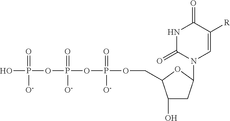
Claims (19)
Priority Applications (1)
| Application Number | Priority Date | Filing Date | Title |
|---|---|---|---|
| US15/325,577 US10179932B2 (en) | 2014-07-11 | 2015-07-10 | Methods for high-throughput labelling and detection of biological features in situ using microscopy |
Applications Claiming Priority (3)
| Application Number | Priority Date | Filing Date | Title |
|---|---|---|---|
| US201462023226P | 2014-07-11 | 2014-07-11 | |
| US15/325,577 US10179932B2 (en) | 2014-07-11 | 2015-07-10 | Methods for high-throughput labelling and detection of biological features in situ using microscopy |
| PCT/US2015/039914 WO2016007839A1 (en) | 2014-07-11 | 2015-07-10 | Methods for high-throughput labelling and detection of biological features in situ using microscopy |
Related Parent Applications (1)
| Application Number | Title | Priority Date | Filing Date |
|---|---|---|---|
| PCT/US2015/039914 A-371-Of-International WO2016007839A1 (en) | 2014-07-11 | 2015-07-10 | Methods for high-throughput labelling and detection of biological features in situ using microscopy |
Related Child Applications (1)
| Application Number | Title | Priority Date | Filing Date |
|---|---|---|---|
| US16/200,831 Continuation US20190085383A1 (en) | 2014-07-11 | 2018-11-27 | Methods for High-Throughput Labelling and Detection of Biological Features In Situ Using Microscopy |
Publications (2)
| Publication Number | Publication Date |
|---|---|
| US20170159136A1 US20170159136A1 (en) | 2017-06-08 |
| US10179932B2 true US10179932B2 (en) | 2019-01-15 |
Family
ID=55064951
Family Applications (4)
| Application Number | Title | Priority Date | Filing Date |
|---|---|---|---|
| US15/325,577 Active US10179932B2 (en) | 2014-07-11 | 2015-07-10 | Methods for high-throughput labelling and detection of biological features in situ using microscopy |
| US16/200,831 Abandoned US20190085383A1 (en) | 2014-07-11 | 2018-11-27 | Methods for High-Throughput Labelling and Detection of Biological Features In Situ Using Microscopy |
| US18/157,108 Active 2036-01-14 US12331347B2 (en) | 2014-07-11 | 2023-01-20 | Methods for high-throughput labelling and detection of biological features in situ using microscopy |
| US19/206,134 Pending US20250270633A1 (en) | 2014-07-11 | 2025-05-13 | Methods for High-Throughput Labelling and Detection of Biological Features in Situ Using Microscopy |
Family Applications After (3)
| Application Number | Title | Priority Date | Filing Date |
|---|---|---|---|
| US16/200,831 Abandoned US20190085383A1 (en) | 2014-07-11 | 2018-11-27 | Methods for High-Throughput Labelling and Detection of Biological Features In Situ Using Microscopy |
| US18/157,108 Active 2036-01-14 US12331347B2 (en) | 2014-07-11 | 2023-01-20 | Methods for high-throughput labelling and detection of biological features in situ using microscopy |
| US19/206,134 Pending US20250270633A1 (en) | 2014-07-11 | 2025-05-13 | Methods for High-Throughput Labelling and Detection of Biological Features in Situ Using Microscopy |
Country Status (2)
| Country | Link |
|---|---|
| US (4) | US10179932B2 (en) |
| WO (1) | WO2016007839A1 (en) |
Cited By (79)
| Publication number | Priority date | Publication date | Assignee | Title |
|---|---|---|---|---|
| US10480022B2 (en) | 2010-04-05 | 2019-11-19 | Prognosys Biosciences, Inc. | Spatially encoded biological assays |
| WO2020047010A2 (en) | 2018-08-28 | 2020-03-05 | 10X Genomics, Inc. | Increasing spatial array resolution |
| WO2020047005A1 (en) | 2018-08-28 | 2020-03-05 | 10X Genomics, Inc. | Resolving spatial arrays |
| WO2020123305A2 (en) | 2018-12-10 | 2020-06-18 | 10X Genomics, Inc. | Generating capture probes for spatial analysis |
| WO2020176788A1 (en) | 2019-02-28 | 2020-09-03 | 10X Genomics, Inc. | Profiling of biological analytes with spatially barcoded oligonucleotide arrays |
| US10774374B2 (en) | 2015-04-10 | 2020-09-15 | Spatial Transcriptomics AB and Illumina, Inc. | Spatially distinguished, multiplex nucleic acid analysis of biological specimens |
| US10774372B2 (en) | 2013-06-25 | 2020-09-15 | Prognosy s Biosciences, Inc. | Methods and systems for determining spatial patterns of biological targets in a sample |
| WO2020190509A1 (en) | 2019-03-15 | 2020-09-24 | 10X Genomics, Inc. | Methods for using spatial arrays for single cell sequencing |
| WO2020198071A1 (en) | 2019-03-22 | 2020-10-01 | 10X Genomics, Inc. | Three-dimensional spatial analysis |
| WO2020243579A1 (en) | 2019-05-30 | 2020-12-03 | 10X Genomics, Inc. | Methods of detecting spatial heterogeneity of a biological sample |
| US11001878B1 (en) | 2010-04-05 | 2021-05-11 | Prognosys Biosciences, Inc. | Spatially encoded biological assays |
| WO2021091611A1 (en) | 2019-11-08 | 2021-05-14 | 10X Genomics, Inc. | Spatially-tagged analyte capture agents for analyte multiplexing |
| WO2021102039A1 (en) | 2019-11-21 | 2021-05-27 | 10X Genomics, Inc, | Spatial analysis of analytes |
| WO2021168287A1 (en) | 2020-02-21 | 2021-08-26 | 10X Genomics, Inc. | Methods and compositions for integrated in situ spatial assay |
| US11193163B2 (en) | 2018-07-30 | 2021-12-07 | Readcoor, Llc | Methods and systems for sample processing or analysis |
| WO2022109181A1 (en) | 2020-11-18 | 2022-05-27 | 10X Genomics, Inc. | Methods and compositions for analyzing immune infiltration in cancer stroma to predict clinical outcome |
| US11352667B2 (en) | 2016-06-21 | 2022-06-07 | 10X Genomics, Inc. | Nucleic acid sequencing |
| US11352659B2 (en) | 2011-04-13 | 2022-06-07 | Spatial Transcriptomics Ab | Methods of detecting analytes |
| EP4012046A1 (en) | 2020-12-11 | 2022-06-15 | 10X Genomics, Inc. | Methods and compositions for multimodal in situ analysis |
| US20220228196A1 (en) * | 2013-03-12 | 2022-07-21 | President And Fellows Of Harvard College | Method for Generating A Three-Dimensional Nucleic Acid Containing Matrix |
| WO2022159474A1 (en) | 2021-01-19 | 2022-07-28 | 10X Genomics, Inc. | Methods and compositions for internally controlled in situ assays |
| WO2022187366A1 (en) | 2021-03-03 | 2022-09-09 | 10X Genomics, Inc. | Analyte detection in situ using nucleic acid origami |
| WO2022216688A1 (en) | 2021-04-05 | 2022-10-13 | 10X Genomics, Inc. | Recombinant ligase composition and uses thereof |
| US11514575B2 (en) | 2019-10-01 | 2022-11-29 | 10X Genomics, Inc. | Systems and methods for identifying morphological patterns in tissue samples |
| US11519033B2 (en) | 2018-08-28 | 2022-12-06 | 10X Genomics, Inc. | Method for transposase-mediated spatial tagging and analyzing genomic DNA in a biological sample |
| WO2022256422A1 (en) | 2021-06-02 | 2022-12-08 | 10X Genomics, Inc. | Sample analysis using asymmetric circularizable probes |
| WO2022256324A1 (en) | 2021-06-01 | 2022-12-08 | 10X Genomics, Inc. | Methods and compositions for analyte detection and probe resolution |
| US11555219B2 (en) | 2019-05-31 | 2023-01-17 | 10X Genomics, Inc. | Method of detecting target nucleic acid molecules |
| WO2023288225A1 (en) | 2021-07-13 | 2023-01-19 | 10X Genomics, Inc. | Methods for preparing polymerized matrix with controllable thickness |
| WO2023015192A1 (en) | 2021-08-03 | 2023-02-09 | 10X Genomics, Inc. | Nucleic acid concatemers and methods for stabilizing and/or compacting the same |
| WO2023023484A1 (en) | 2021-08-16 | 2023-02-23 | 10X Genomics, Inc. | Probes comprising a split barcode region and methods of use |
| US11597965B2 (en) | 2017-10-06 | 2023-03-07 | 10X Genomics, Inc. | RNA templated ligation |
| US11649485B2 (en) | 2019-01-06 | 2023-05-16 | 10X Genomics, Inc. | Generating capture probes for spatial analysis |
| WO2023102118A2 (en) | 2021-12-01 | 2023-06-08 | 10X Genomics, Inc. | Methods, compositions, and systems for improved in situ detection of analytes and spatial analysis |
| WO2023129898A2 (en) | 2021-12-27 | 2023-07-06 | 10X Genomics, Inc. | Methods and compositions for rolling circle amplification |
| WO2023141588A1 (en) | 2022-01-21 | 2023-07-27 | 10X Genomics, Inc. | Multiple readout signals for analyzing a sample |
| WO2023164570A1 (en) | 2022-02-23 | 2023-08-31 | Insitro, Inc. | Pooled optical screening and transcriptional measurements of cells comprising barcoded genetic perturbations |
| WO2023196526A1 (en) | 2022-04-06 | 2023-10-12 | 10X Genomics, Inc. | Methods for multiplex cell analysis |
| WO2023215612A1 (en) | 2022-05-06 | 2023-11-09 | 10X Genomics, Inc. | Analysis of antigen and antigen receptor interactions |
| WO2023220300A1 (en) | 2022-05-11 | 2023-11-16 | 10X Genomics, Inc. | Compositions and methods for in situ sequencing |
| WO2023229988A1 (en) | 2022-05-23 | 2023-11-30 | 10X Genomics, Inc. | Tissue sample mold |
| WO2023245190A1 (en) | 2022-06-17 | 2023-12-21 | 10X Genomics, Inc. | Catalytic de-crosslinking of samples for in situ analysis |
| WO2024036304A1 (en) | 2022-08-12 | 2024-02-15 | 10X Genomics, Inc. | Puma1 polymerases and uses thereof |
| WO2024040060A1 (en) | 2022-08-16 | 2024-02-22 | 10X Genomics, Inc. | Ap50 polymerases and uses thereof |
| US11926867B2 (en) | 2019-01-06 | 2024-03-12 | 10X Genomics, Inc. | Generating capture probes for spatial analysis |
| WO2024081869A1 (en) | 2022-10-14 | 2024-04-18 | 10X Genomics, Inc. | Methods for analysis of biological samples |
| WO2024102736A1 (en) | 2022-11-08 | 2024-05-16 | 10X Genomics, Inc. | Immobilization methods and compositions for in situ detection |
| WO2024107887A1 (en) | 2022-11-16 | 2024-05-23 | 10X Genomics, Inc. | Methods and compositions for assessing performance of in situ assays |
| WO2024130203A1 (en) | 2022-12-16 | 2024-06-20 | 10X Genomics, Inc. | Methods and compositions for assessing performance |
| US12071667B2 (en) | 2020-11-04 | 2024-08-27 | 10X Genomics, Inc. | Sequence analysis using meta-stable nucleic acid molecules |
| US12076701B2 (en) | 2020-01-31 | 2024-09-03 | 10X Genomics, Inc. | Capturing oligonucleotides in spatial transcriptomics |
| WO2024192058A1 (en) * | 2023-03-13 | 2024-09-19 | California Institute Of Technology | Methods and compositions for quantitative, high-throughput protein binding measurement |
| US12098425B2 (en) | 2018-10-10 | 2024-09-24 | Readcoor, Llc | Three-dimensional spatial molecular indexing |
| US12110548B2 (en) | 2020-02-03 | 2024-10-08 | 10X Genomics, Inc. | Bi-directional in situ analysis |
| US12112833B2 (en) | 2020-02-04 | 2024-10-08 | 10X Genomics, Inc. | Systems and methods for index hopping filtering |
| EP4450640A2 (en) | 2021-07-30 | 2024-10-23 | 10x Genomics, Inc. | Methods and compositions for synchronizing reactions in situ |
| US12139751B2 (en) | 2021-07-30 | 2024-11-12 | 10X Genomics, Inc. | Circularizable probes for in situ analysis |
| US12154266B2 (en) | 2019-11-18 | 2024-11-26 | 10X Genomics, Inc. | Systems and methods for binary tissue classification |
| US12168801B1 (en) | 2020-07-02 | 2024-12-17 | 10X Genomics, Inc. | Hybrid/capture probe designs for full-length cDNA |
| US12188085B2 (en) | 2020-03-05 | 2025-01-07 | 10X Genomics, Inc. | Three-dimensional spatial transcriptomics with sequencing readout |
| US12203136B2 (en) | 2020-08-17 | 2025-01-21 | Readcoor, Llc | Methods and systems for spatial mapping of genetic variants |
| US12209273B2 (en) | 2020-06-12 | 2025-01-28 | 10X Genomics, Inc. | Nucleic acid assays using click chemistry bioconjugation |
| US12215379B2 (en) | 2020-02-17 | 2025-02-04 | 10X Genomics, Inc. | In situ analysis of chromatin interaction |
| WO2025029831A1 (en) | 2023-07-31 | 2025-02-06 | 10X Genomics, Inc. | Methods and systems for targeted rna cleavage and target rna-primed rolling circle amplification |
| US12234507B2 (en) | 2022-04-01 | 2025-02-25 | 10X Genomics, Inc. | Compositions and methods for targeted masking of autofluorescence |
| US12270071B2 (en) | 2019-12-20 | 2025-04-08 | 10X Genomics, Inc. | Methods of detecting an analyte |
| US12275984B2 (en) | 2021-03-02 | 2025-04-15 | 10X Genomics, Inc. | Sequential hybridization and quenching |
| US12297499B2 (en) | 2020-08-17 | 2025-05-13 | 10X Genomics, Inc. | Multicomponent nucleic acid probes for sample analysis |
| EP4578960A1 (en) | 2023-12-20 | 2025-07-02 | 10x Genomics, Inc. | In situ detection of copy number variations in biological samples |
| US12372771B2 (en) | 2022-11-18 | 2025-07-29 | 10X Genomics, Inc. | Systems and methods for actively mitigating vibrations |
| US12391984B2 (en) | 2021-08-03 | 2025-08-19 | 10X Genomics, Inc. | Compositions and methods for rolling circle amplification |
| US12400733B2 (en) | 2022-03-08 | 2025-08-26 | 10X Genomics, Inc. | In situ code design methods for minimizing optical crowding |
| WO2025194033A1 (en) | 2024-03-15 | 2025-09-18 | 10X Genomics, Inc. | Systems and methods for covering and sealing an open well |
| US12421558B2 (en) | 2020-02-13 | 2025-09-23 | 10X Genomics, Inc. | Systems and methods for joint interactive visualization of gene expression and DNA chromatin accessibility |
| US12460251B2 (en) | 2021-08-03 | 2025-11-04 | 10X Genomics, Inc. | Stabilization and/or compaction of nucleic acid molecules |
| WO2025255457A1 (en) | 2024-06-07 | 2025-12-11 | 10X Genomics, Inc. | Compositions and methods for improved on-target spatial profiling |
| US12529096B2 (en) | 2021-08-03 | 2026-01-20 | 10X Genomics, Inc. | Stabilization and/or compaction of nucleic acid structures |
| US12529094B2 (en) | 2018-12-10 | 2026-01-20 | 10X Genomics, Inc. | Imaging system hardware |
| US12545949B2 (en) | 2019-12-06 | 2026-02-10 | 10X Genomics, Inc. | Resolving spatial arrays using deconvolution |
Families Citing this family (137)
| Publication number | Priority date | Publication date | Assignee | Title |
|---|---|---|---|---|
| US10501791B2 (en) | 2011-10-14 | 2019-12-10 | President And Fellows Of Harvard College | Sequencing by structure assembly |
| ES2991004T3 (en) | 2011-12-22 | 2024-12-02 | Harvard College | Methods for the detection of analytes |
| EP3901280B1 (en) | 2012-10-17 | 2025-03-12 | 10x Genomics Sweden AB | Methods and product for optimising localised or spatial detection of gene expression in a tissue sample |
| US10179932B2 (en) | 2014-07-11 | 2019-01-15 | President And Fellows Of Harvard College | Methods for high-throughput labelling and detection of biological features in situ using microscopy |
| CA3004285A1 (en) | 2015-11-03 | 2017-05-11 | President And Fellows Of Harvard College | Method and apparatus for volumetric imaging of a three-dimensional nucleic acid containing matrix |
| WO2017189525A1 (en) | 2016-04-25 | 2017-11-02 | President And Fellows Of Harvard College | Hybridization chain reaction methods for in situ molecular detection |
| US10697974B2 (en) * | 2016-07-22 | 2020-06-30 | President And Fellows Of Harvard College | Methods and compositions for protein identification |
| CN109923216B (en) | 2016-08-31 | 2024-08-02 | 哈佛学院董事及会员团体 | Methods for combining detection of biomolecules into a single assay using fluorescent in situ sequencing |
| CA3043639A1 (en) | 2016-12-09 | 2018-06-14 | Ultivue, Inc. | Improved methods for multiplex imaging using labeled nucleic acid imaging agents |
| CN110446719B (en) | 2017-03-31 | 2023-10-03 | 乌尔蒂维尤股份有限公司 | DNA-antigen exchange and amplification |
| US10350307B2 (en) * | 2017-09-18 | 2019-07-16 | General Electric Company | In vivo RNA or protein expression using double-stranded concatemeric DNA including phosphorothioated nucleotides |
| US12122996B2 (en) | 2017-09-25 | 2024-10-22 | California Institute Of Technology | Device for additive delivery of reagents and related methods and systems |
| US11299789B2 (en) * | 2018-01-02 | 2022-04-12 | California Institute Of Technology | Loop-mediated isothermal amplification (LAMP) based assay for detecting microbes |
| KR102216965B1 (en) * | 2019-05-07 | 2021-02-18 | 주식회사 힐세리온 | Noise removal apparatus and remote medical-diagnosis system using the same for removing noise from noisy image using discrete wavelet transform |
| US11287422B2 (en) | 2019-09-23 | 2022-03-29 | Element Biosciences, Inc. | Multivalent binding composition for nucleic acid analysis |
| WO2021061841A1 (en) * | 2019-09-23 | 2021-04-01 | Element Biosciences, Inc. | Methods for cellularly addressable nucleic acid sequencing |
| US12157124B2 (en) | 2019-11-06 | 2024-12-03 | 10X Genomics, Inc. | Imaging system hardware |
| WO2021092433A2 (en) | 2019-11-08 | 2021-05-14 | 10X Genomics, Inc. | Enhancing specificity of analyte binding |
| EP4589016A3 (en) | 2019-11-13 | 2025-10-08 | 10x Genomics, Inc. | Generating capture probes for spatial analysis |
| JP7691113B2 (en) * | 2019-11-15 | 2025-06-11 | 国立大学法人 東京大学 | Barcoded extracellular vesicle library |
| EP4062372B1 (en) | 2019-11-22 | 2024-05-08 | 10X Genomics, Inc. | Systems and methods for spatial analysis of analytes using fiducial alignment |
| WO2021133842A1 (en) | 2019-12-23 | 2021-07-01 | 10X Genomics, Inc. | Compositions and methods for using fixed biological samples in partition-based assays |
| EP3891300B1 (en) | 2019-12-23 | 2023-03-29 | 10X Genomics, Inc. | Methods for spatial analysis using rna-templated ligation |
| EP4339299B1 (en) | 2020-01-10 | 2025-08-27 | 10X Genomics, Inc. | Methods for determining a location of a target nucleic acid in a biological sample |
| US12365942B2 (en) | 2020-01-13 | 2025-07-22 | 10X Genomics, Inc. | Methods of decreasing background on a spatial array |
| US12405264B2 (en) | 2020-01-17 | 2025-09-02 | 10X Genomics, Inc. | Electrophoretic system and method for analyte capture |
| US11732299B2 (en) | 2020-01-21 | 2023-08-22 | 10X Genomics, Inc. | Spatial assays with perturbed cells |
| US11702693B2 (en) | 2020-01-21 | 2023-07-18 | 10X Genomics, Inc. | Methods for printing cells and generating arrays of barcoded cells |
| US20210230681A1 (en) | 2020-01-24 | 2021-07-29 | 10X Genomics, Inc. | Methods for spatial analysis using proximity ligation |
| US11821035B1 (en) | 2020-01-29 | 2023-11-21 | 10X Genomics, Inc. | Compositions and methods of making gene expression libraries |
| US12110541B2 (en) | 2020-02-03 | 2024-10-08 | 10X Genomics, Inc. | Methods for preparing high-resolution spatial arrays |
| US11898205B2 (en) | 2020-02-03 | 2024-02-13 | 10X Genomics, Inc. | Increasing capture efficiency of spatial assays |
| US11732300B2 (en) | 2020-02-05 | 2023-08-22 | 10X Genomics, Inc. | Increasing efficiency of spatial analysis in a biological sample |
| US12129516B2 (en) | 2020-02-07 | 2024-10-29 | 10X Genomics, Inc. | Quantitative and automated permeabilization performance evaluation for spatial transcriptomics |
| US11835462B2 (en) | 2020-02-11 | 2023-12-05 | 10X Genomics, Inc. | Methods and compositions for partitioning a biological sample |
| US12281357B1 (en) | 2020-02-14 | 2025-04-22 | 10X Genomics, Inc. | In situ spatial barcoding |
| US12399123B1 (en) | 2020-02-14 | 2025-08-26 | 10X Genomics, Inc. | Spatial targeting of analytes |
| WO2021168278A1 (en) | 2020-02-20 | 2021-08-26 | 10X Genomics, Inc. | METHODS TO COMBINE FIRST AND SECOND STRAND cDNA SYNTHESIS FOR SPATIAL ANALYSIS |
| CN116157533A (en) | 2020-02-21 | 2023-05-23 | 10X基因组学有限公司 | Capture Genetic Targets Using Hybridization |
| US11891654B2 (en) | 2020-02-24 | 2024-02-06 | 10X Genomics, Inc. | Methods of making gene expression libraries |
| US11768175B1 (en) | 2020-03-04 | 2023-09-26 | 10X Genomics, Inc. | Electrophoretic methods for spatial analysis |
| EP4114943A4 (en) * | 2020-03-05 | 2024-04-24 | The Trustees Of Columbia University In The City Of New York | In vivo mrna display: large-scale proteomics by next generation sequencing |
| WO2021216708A1 (en) | 2020-04-22 | 2021-10-28 | 10X Genomics, Inc. | Methods for spatial analysis using targeted rna depletion |
| US20230265491A1 (en) | 2020-05-04 | 2023-08-24 | 10X Genomics, Inc. | Spatial transcriptomic transfer modes |
| WO2021231263A2 (en) * | 2020-05-12 | 2021-11-18 | Singular Genomics Systems, Inc. | Nucleic acid amplification methods |
| US12416603B2 (en) | 2020-05-19 | 2025-09-16 | 10X Genomics, Inc. | Electrophoresis cassettes and instrumentation |
| WO2021237056A1 (en) | 2020-05-22 | 2021-11-25 | 10X Genomics, Inc. | Rna integrity analysis in a biological sample |
| WO2021237087A1 (en) | 2020-05-22 | 2021-11-25 | 10X Genomics, Inc. | Spatial analysis to detect sequence variants |
| WO2021236929A1 (en) | 2020-05-22 | 2021-11-25 | 10X Genomics, Inc. | Simultaneous spatio-temporal measurement of gene expression and cellular activity |
| WO2021242834A1 (en) | 2020-05-26 | 2021-12-02 | 10X Genomics, Inc. | Method for resetting an array |
| CN116249785A (en) | 2020-06-02 | 2023-06-09 | 10X基因组学有限公司 | Spatial Transcriptomics for Antigen-Receptors |
| EP4025692A2 (en) | 2020-06-02 | 2022-07-13 | 10X Genomics, Inc. | Nucleic acid library methods |
| US12031177B1 (en) | 2020-06-04 | 2024-07-09 | 10X Genomics, Inc. | Methods of enhancing spatial resolution of transcripts |
| ES2981265T3 (en) | 2020-06-08 | 2024-10-08 | 10X Genomics Inc | Methods for determining a surgical margin and methods of using it |
| WO2021252576A1 (en) | 2020-06-10 | 2021-12-16 | 10X Genomics, Inc. | Methods for spatial analysis using blocker oligonucleotides |
| US12435363B1 (en) | 2020-06-10 | 2025-10-07 | 10X Genomics, Inc. | Materials and methods for spatial transcriptomics |
| AU2021288090A1 (en) | 2020-06-10 | 2023-01-19 | 10X Genomics, Inc. | Fluid delivery methods |
| EP4446430A3 (en) | 2020-06-10 | 2024-12-18 | 10X Genomics, Inc. | Methods for determining a location of an analyte in a biological sample |
| EP4450639B1 (en) | 2020-06-25 | 2025-10-15 | 10X Genomics, Inc. | Spatial analysis of dna methylation |
| US12209280B1 (en) | 2020-07-06 | 2025-01-28 | 10X Genomics, Inc. | Methods of identifying abundance and location of an analyte in a biological sample using second strand synthesis |
| US11761038B1 (en) | 2020-07-06 | 2023-09-19 | 10X Genomics, Inc. | Methods for identifying a location of an RNA in a biological sample |
| US11981960B1 (en) | 2020-07-06 | 2024-05-14 | 10X Genomics, Inc. | Spatial analysis utilizing degradable hydrogels |
| EP4153606A4 (en) | 2020-07-13 | 2024-10-02 | Singular Genomics Systems, Inc. | Methods of sequencing complementary polynucleotides |
| CN116323968A (en) | 2020-07-31 | 2023-06-23 | 10X基因组学有限公司 | Decrosslinking compounds for spatial analysis and methods of use |
| US11981958B1 (en) | 2020-08-20 | 2024-05-14 | 10X Genomics, Inc. | Methods for spatial analysis using DNA capture |
| EP4530358A3 (en) | 2020-09-15 | 2025-06-18 | 10x Genomics, Inc. | Methods of releasing an extended capture probe from a substrate and uses of the same |
| US20230313279A1 (en) | 2020-09-16 | 2023-10-05 | 10X Genomics, Inc. | Methods of determining the location of an analyte in a biological sample using a plurality of wells |
| WO2022061152A2 (en) | 2020-09-18 | 2022-03-24 | 10X Genomics, Inc. | Sample handling apparatus and fluid delivery methods |
| AU2021345283B2 (en) | 2020-09-18 | 2024-12-19 | 10X Genomics, Inc. | Sample handling apparatus and image registration methods |
| US11926822B1 (en) | 2020-09-23 | 2024-03-12 | 10X Genomics, Inc. | Three-dimensional spatial analysis |
| WO2022081643A2 (en) | 2020-10-13 | 2022-04-21 | 10X Genomics, Inc. | Compositions and methods for generating recombinant antigen binding molecules from single cells |
| AU2021366701A1 (en) | 2020-10-22 | 2023-05-04 | 10X Genomics, Inc. | Methods for spatial analysis using rolling circle amplification |
| CN116829733A (en) | 2020-11-06 | 2023-09-29 | 10X基因组学有限公司 | Compositions and methods for binding analytes to capture probes |
| US11827935B1 (en) | 2020-11-19 | 2023-11-28 | 10X Genomics, Inc. | Methods for spatial analysis using rolling circle amplification and detection probes |
| AU2021409136A1 (en) | 2020-12-21 | 2023-06-29 | 10X Genomics, Inc. | Methods, compositions, and systems for capturing probes and/or barcodes |
| WO2022147296A1 (en) | 2020-12-30 | 2022-07-07 | 10X Genomics, Inc. | Cleavage of capture probes for spatial analysis |
| US20240068017A1 (en) | 2020-12-30 | 2024-02-29 | 10X Genomics, Inc. | Methods for analyte capture determination |
| US12398262B1 (en) | 2021-01-22 | 2025-08-26 | 10X Genomics, Inc. | Triblock copolymer-based cell stabilization and fixation system and methods of use thereof |
| AU2022212231A1 (en) | 2021-01-29 | 2023-08-03 | 10X Genomics, Inc. | Method for transposase mediated spatial tagging and analyzing genomic dna in a biological sample |
| WO2022198068A1 (en) | 2021-03-18 | 2022-09-22 | 10X Genomics, Inc. | Multiplex capture of gene and protein expression from a biological sample |
| EP4428246B1 (en) | 2021-04-14 | 2025-12-24 | 10X Genomics, Inc. | Methods of measuring mislocalization of an analyte |
| WO2022226057A1 (en) | 2021-04-20 | 2022-10-27 | 10X Genomics, Inc. | Methods for assessing sample quality prior to spatial analysis using templated ligation |
| EP4320271B1 (en) | 2021-05-06 | 2025-03-19 | 10X Genomics, Inc. | Methods for increasing resolution of spatial analysis |
| EP4582555A3 (en) | 2021-06-03 | 2025-10-22 | 10X Genomics, Inc. | Methods, compositions, kits, and systems for enhancing analyte capture for spatial analysis |
| WO2022271820A1 (en) | 2021-06-22 | 2022-12-29 | 10X Genomics, Inc. | Spatial detection of sars-cov-2 using templated ligation |
| WO2023287765A1 (en) | 2021-07-13 | 2023-01-19 | 10X Genomics, Inc. | Methods for spatial analysis using targeted probe silencing |
| WO2023018799A1 (en) | 2021-08-12 | 2023-02-16 | 10X Genomics, Inc. | Methods, compositions and systems for identifying antigen-binding molecules |
| WO2023034489A1 (en) | 2021-09-01 | 2023-03-09 | 10X Genomics, Inc. | Methods, compositions, and kits for blocking a capture probe on a spatial array |
| US20240378734A1 (en) | 2021-09-17 | 2024-11-14 | 10X Genomics, Inc. | Systems and methods for image registration or alignment |
| WO2023076345A1 (en) | 2021-10-26 | 2023-05-04 | 10X Genomics, Inc. | Methods for spatial analysis using targeted rna capture |
| EP4419707A1 (en) | 2021-11-10 | 2024-08-28 | 10X Genomics, Inc. | Methods, compositions, and kits for determining the location of an analyte in a biological sample |
| US20250147036A1 (en) | 2021-11-10 | 2025-05-08 | 10X Genomics, Inc. | Methods for identification of antigen-binding molecules |
| EP4441741A1 (en) | 2021-11-30 | 2024-10-09 | 10X Genomics, Inc. | Systems and methods for identifying regions of aneuploidy in a tissue |
| WO2023122033A1 (en) | 2021-12-20 | 2023-06-29 | 10X Genomics, Inc. | Self-test for pathology/histology slide imaging device |
| US20250163501A1 (en) | 2022-02-01 | 2025-05-22 | 10X Genomics, Inc. | Methods, kits, compositions, and systems for spatial analysis |
| WO2023150163A1 (en) | 2022-02-01 | 2023-08-10 | 10X Genomics, Inc. | Methods, compositions, and systems for capturing analytes from lymphatic tissue |
| WO2023150171A1 (en) | 2022-02-01 | 2023-08-10 | 10X Genomics, Inc. | Methods, compositions, and systems for capturing analytes from glioblastoma samples |
| EP4479933A1 (en) | 2022-02-15 | 2024-12-25 | 10x Genomics, Inc. | Systems and methods for spatial analysis of analytes using fiducial alignment |
| WO2023172670A2 (en) | 2022-03-11 | 2023-09-14 | 10X Genomics, Inc. | Sample handling apparatus and fluid delivery methods |
| US20250251403A1 (en) | 2022-04-12 | 2025-08-07 | 10X Genomics, Inc. | Compositions and methods for generating and characterizing recombinant antigen binding molecules |
| EP4490319A1 (en) | 2022-05-06 | 2025-01-15 | 10X Genomics, Inc. | Molecular barcode readers for analyte detection |
| WO2023225519A1 (en) | 2022-05-17 | 2023-11-23 | 10X Genomics, Inc. | Modified transposons, compositions and uses thereof |
| WO2023229982A2 (en) | 2022-05-24 | 2023-11-30 | 10X Genomics, Inc. | Porous structure confinement for convection suppression |
| WO2023250077A1 (en) | 2022-06-22 | 2023-12-28 | 10X Genomics, Inc. | Methods, compositions, and systems for capturing probes and/or barcodes |
| WO2024015862A1 (en) | 2022-07-13 | 2024-01-18 | 10X Genomics, Inc. | Methods for characterization of antigen-binding molecules from biological samples |
| WO2024015578A1 (en) | 2022-07-15 | 2024-01-18 | 10X Genomics, Inc. | Methods for determining a location of a target nucleic acid in a biological sample |
| EP4565870A1 (en) | 2022-08-05 | 2025-06-11 | 10x Genomics, Inc. | Systems and methods for immunofluorescence quantification |
| WO2024036191A1 (en) | 2022-08-10 | 2024-02-15 | 10X Genomics, Inc. | Systems and methods for colocalization |
| WO2024035844A1 (en) | 2022-08-12 | 2024-02-15 | 10X Genomics, Inc. | Methods for reducing capture of analytes |
| AU2023325064A1 (en) | 2022-08-15 | 2025-03-06 | Element Biosciences, Inc. | Spatially resolved surface capture of nucleic acids |
| EP4577670A1 (en) | 2022-08-24 | 2025-07-02 | 10X Genomics, Inc. | Compositions and methods for antigenic epitope mapping in biological samples |
| EP4573208A1 (en) | 2022-10-10 | 2025-06-25 | 10X Genomics, Inc. | In vitro transcription of spatially captured nucleic acids |
| WO2024086167A2 (en) | 2022-10-17 | 2024-04-25 | 10X Genomics, Inc. | Methods, compositions, and kits for determining the location of an analyte in a biological sample |
| WO2024102809A1 (en) | 2022-11-09 | 2024-05-16 | 10X Genomics, Inc. | Methods, compositions, and kits for determining the location of multiple analytes in a biological sample |
| WO2024129672A1 (en) * | 2022-12-12 | 2024-06-20 | The Broad Institute, Inc. | Trafficked rnas for assessment of cell-cell connectivity and neuroanatomy |
| EP4638788A1 (en) | 2022-12-21 | 2025-10-29 | 10X Genomics, Inc. | Analysis of analytes and spatial gene expression |
| WO2024145224A1 (en) | 2022-12-29 | 2024-07-04 | 10X Genomics, Inc. | Compositions, methods, and systems for high resolution spatial analysis |
| WO2024145441A1 (en) | 2022-12-29 | 2024-07-04 | 10X Genomics, Inc. | Methods, compositions, and kits for determining a location of a target nucleic acid in a fixed biological sample |
| WO2024145445A1 (en) | 2022-12-30 | 2024-07-04 | 10X Genomics, Inc. | Methods of capturing target analytes |
| EP4624590A3 (en) | 2022-12-30 | 2025-12-24 | 10X Genomics, Inc. | Methods, compositions, and kits for multiple barcoding and/or high-density spatial barcoding |
| WO2024206603A1 (en) | 2023-03-28 | 2024-10-03 | 10X Genomics, Inc. | Methods, compositions, and kits for reducing analyte mislocalization |
| WO2024220882A1 (en) | 2023-04-21 | 2024-10-24 | 10X Genomics, Inc. | Methods and compositions for detecting nucleic acids in a fixed biological sample |
| WO2024238625A1 (en) | 2023-05-15 | 2024-11-21 | 10X Genomics, Inc. | Spatial antibody data normalization |
| WO2024238900A1 (en) | 2023-05-17 | 2024-11-21 | 10X Genomics, Inc. | Methods of reducing mislocalization of an analyte |
| WO2024238992A1 (en) | 2023-05-18 | 2024-11-21 | 10X Genomics, Inc. | Engineered non-strand displacing family b polymerases for reverse transcription and gap-fill applications |
| WO2024254316A1 (en) | 2023-06-07 | 2024-12-12 | 10X Genomics, Inc. | Compositions and methods for identifying blood clots in biological samples |
| WO2024258911A1 (en) | 2023-06-13 | 2024-12-19 | 10X Genomics, Inc. | Sequence specific dna binding proteins |
| WO2025029605A1 (en) | 2023-07-28 | 2025-02-06 | 10X Genomics, Inc. | Methods and compositions for detecting genetic variants using capture probes and primer extension |
| WO2025029627A1 (en) | 2023-07-31 | 2025-02-06 | 10X Genomics, Inc. | Methods, compositions, and kits for spatial detection of genetic variants |
| WO2025043076A1 (en) | 2023-08-24 | 2025-02-27 | 10X Genomics, Inc. | Methods, kits, and compositions for spatial detection of genetic variants |
| WO2025072119A1 (en) | 2023-09-26 | 2025-04-03 | 10X Genomics, Inc. | Methods, compositions, and kits for detecting spatial chromosomal interactions |
| WO2025090912A1 (en) | 2023-10-26 | 2025-05-01 | 10X Genomics, Inc. | Spatial arrays containing reusable master probes |
| WO2025096581A1 (en) | 2023-10-31 | 2025-05-08 | 10X Genomics, Inc. | Methods of spatial analysis and single nuclei sequencing |
| WO2025101377A1 (en) | 2023-11-09 | 2025-05-15 | 10X Genomics, Inc. | Matrix-assisted spatial analysis of biological samples |
| WO2025101864A1 (en) | 2023-11-09 | 2025-05-15 | 10X Genomics, Inc. | Methods, compositions, and kits for reducing mislocalization of analytes in spatial analysis assays |
| WO2025189030A1 (en) | 2024-03-07 | 2025-09-12 | Stellaromics, Inc. | Methods and systems for volumetric imaging |
| WO2026024934A1 (en) | 2024-07-25 | 2026-01-29 | 10X Genomics, Inc. | Methods, compositions, and kits for spatial analysis by tagmentation |
Citations (52)
| Publication number | Priority date | Publication date | Assignee | Title |
|---|---|---|---|---|
| US5563056A (en) | 1992-02-13 | 1996-10-08 | Bsi Corporation | Preparation of crosslinked matrices containing covalently immobilized chemical species and unbound releasable chemical species |
| WO1997046704A1 (en) | 1996-06-06 | 1997-12-11 | Lynx Therapeutics, Inc. | Sequencing by ligation of encoded adaptors |
| US20020015952A1 (en) | 1999-07-30 | 2002-02-07 | Anderson Norman G. | Microarrays and their manufacture by slicing |
| US20030148335A1 (en) | 2001-10-10 | 2003-08-07 | Li Shen | Detecting targets by unique identifier nucleotide tags |
| US20040248144A1 (en) | 2001-03-16 | 2004-12-09 | Kalim Mir | Arrays and methods of use |
| US20050147981A1 (en) | 2003-12-31 | 2005-07-07 | Intel Corporation | Methods and compositions for detecting nucleic acids using scanning probe microscopy and nanocodes |
| US20050233318A1 (en) | 1998-06-24 | 2005-10-20 | Chee Mark S | Decoding of array sensors with microspheres |
| US20060024711A1 (en) | 2004-07-02 | 2006-02-02 | Helicos Biosciences Corporation | Methods for nucleic acid amplification and sequence determination |
| US20060177833A1 (en) | 2005-02-10 | 2006-08-10 | Sydney Brenner | Methods and compositions for tagging and identifying polynucleotides |
| US20060234261A1 (en) | 2005-03-08 | 2006-10-19 | Pierce Niles A | Colorimetric readout of hybridization chain reaction |
| US20060248349A1 (en) | 2001-09-24 | 2006-11-02 | Rathjen Alicemarie G | Method for preparing and using personal and genetic profiles |
| US20060292611A1 (en) | 2005-06-06 | 2006-12-28 | Jan Berka | Paired end sequencing |
| US20070020650A1 (en) | 2005-04-01 | 2007-01-25 | Avak Kahvejian | Methods for detecting proteins |
| US20070087362A1 (en) | 2004-02-27 | 2007-04-19 | President And Fellows Of Harvard College | Polony fluorescent in situ sequencing beads |
| US20070117177A1 (en) | 2005-08-11 | 2007-05-24 | Dan Luo | Nucleic Acid-Based Matrixes for Protein Production |
| US20070117109A1 (en) | 2005-06-14 | 2007-05-24 | California Institute Of Technology | Nanostructures, methods of making and using the same |
| WO2007086900A2 (en) | 2005-04-08 | 2007-08-02 | Mycosol, Inc. | Stilbazium research assays |
| WO2007121489A2 (en) | 2006-04-19 | 2007-10-25 | Applera Corporation | Reagents, methods, and libraries for gel-free bead-based sequencing |
| US7323305B2 (en) | 2003-01-29 | 2008-01-29 | 454 Life Sciences Corporation | Methods of amplifying and sequencing nucleic acids |
| WO2008069973A2 (en) | 2006-12-01 | 2008-06-12 | The Trustees Of Columbia University In The City Of New York | Four-color dna sequencing by synthesis using cleavable fluorescent nucleotide reversible terminators |
| US20090005259A1 (en) | 2003-02-26 | 2009-01-01 | Complete Genomics, Inc. | Random array DNA analysis by hybridization |
| US7473767B2 (en) | 2001-07-03 | 2009-01-06 | The Institute For Systems Biology | Methods for detection and quantification of analytes in complex mixtures |
| WO2009046149A1 (en) | 2007-10-01 | 2009-04-09 | Applied Biosystems Inc. | Chase ligation sequencing |
| US20090208965A1 (en) | 2006-10-25 | 2009-08-20 | Ikonisys, Inc. | Automated method for detecting cancers and high grade hyperplasias |
| US20090246879A1 (en) | 2002-12-20 | 2009-10-01 | Callida Genomics | Materials and Methods Relating to Nano-Tags and Nano-Brands |
| US20100009868A1 (en) | 2006-09-11 | 2010-01-14 | The Arizona Board of Regents a body corporate acti ng for and behalf of Arizona State university | Self-Assembled Combinatorial Encoding Nanoarrays for Multiplexed Biosensing |
| US20100047924A1 (en) | 2008-08-14 | 2010-02-25 | Nanostring Technologies, Inc. | Stable nanoreporters |
| WO2010080134A1 (en) | 2008-12-19 | 2010-07-15 | President And Fellows Of Harvard College | Particle-assisted nucleic acid sequencing |
| US20100223276A1 (en) | 2007-03-27 | 2010-09-02 | Faleh Jassem Al-Shameri | Automated Generation of Metadata for Mining Image and Text Data |
| US20100268478A1 (en) | 2007-10-04 | 2010-10-21 | William Andregg | Sequencing Nucleic Acid Polymers with Electron Microscopy |
| US20110086774A1 (en) | 2009-10-13 | 2011-04-14 | Dunaway Dwayne L | Protein Detection Via Nanoreporters |
| US20110092376A1 (en) | 2008-09-23 | 2011-04-21 | Quantalife, Inc. | System for droplet-based assays using an array of emulsions |
| US20110245111A1 (en) | 2010-04-05 | 2011-10-06 | Prognosys Biosciences, Inc. | Spatially encoded biological assays |
| US20110257031A1 (en) | 2010-02-12 | 2011-10-20 | Life Technologies Corporation | Nucleic acid, biomolecule and polymer identifier codes |
| WO2011143583A1 (en) | 2010-05-13 | 2011-11-17 | Illumina, Inc. | Binding assays for markers |
| US20110294135A1 (en) | 2007-08-02 | 2011-12-01 | BioDesic, LLC | Compositions and methods for analyte detection and quantitation |
| WO2012005595A2 (en) | 2010-07-09 | 2012-01-12 | Wouter Leonard De Laat | V3-d genomic region of interest sequencing strategies |
| US20120040397A1 (en) | 2008-08-05 | 2012-02-16 | Dan Luo | Photo-Crosslinked Nucleic Acid Hydrogels |
| WO2012058638A2 (en) | 2010-10-29 | 2012-05-03 | President And Fellows Of Harvard College | Nucleic acid nanostructure barcode probes |
| US20120122712A1 (en) | 2010-11-16 | 2012-05-17 | Goldstein Peter H | Methods for Sequencing a Biomolecule by Detecting Relative Positions of Hybridized Probes |
| WO2012150035A1 (en) | 2011-05-04 | 2012-11-08 | Genovoxx Gmbh | Nucleoside-triphosphate conjugate and methods for the use thereof |
| WO2013055995A2 (en) | 2011-10-14 | 2013-04-18 | President And Fellows Of Harvard College | Sequencing by structure assembly |
| US20130245096A1 (en) | 2010-10-28 | 2013-09-19 | Nanodoc Ltd. | COMPOSITIONS AND METHODS FOR ACTIVATING EXPRESSION BY A SPECIFIC ENDOGENOUS miRNA |
| US20140087378A1 (en) | 2011-03-17 | 2014-03-27 | Centre National De La Recherche Scientifique | Method, probe and kit for dna in situ hybridation and use thereof |
| US20140220587A1 (en) | 2013-02-01 | 2014-08-07 | The Regents Of The University Of California | Methods for genome assembly and haplotype phasing |
| WO2014163886A1 (en) | 2013-03-12 | 2014-10-09 | President And Fellows Of Harvard College | Method of generating a three-dimensional nucleic acid containing matrix |
| WO2014182528A2 (en) | 2013-04-30 | 2014-11-13 | California Institute Of Technology | Multiplex labeling of molecules by sequential hybridization barcoding |
| US20150133319A1 (en) | 2012-02-27 | 2015-05-14 | Cellular Research, Inc. | Compositions and kits for molecular counting |
| WO2015118029A1 (en) | 2014-02-04 | 2015-08-13 | Olink Ab | Proximity assay with detection based on hybridisation chain reaction (hcr) |
| US9217151B2 (en) | 2007-05-16 | 2015-12-22 | California Institute Of Technology | Versatile nucleic acid hairpin motif for programming biomolecular self-assembly pathways |
| US20160265046A1 (en) | 2013-10-18 | 2016-09-15 | The Broad Institute Inc. | Spatial and cellular mapping of biomolecules in situ by high-throughput sequencing |
| US20170212983A1 (en) | 2013-04-30 | 2017-07-27 | California Institute Of Technology | Error correction of multiplex imaging analysis by sequential hybridization |
Family Cites Families (217)
| Publication number | Priority date | Publication date | Assignee | Title |
|---|---|---|---|---|
| US4123610A (en) | 1977-03-09 | 1978-10-31 | The United States Government | Nucleic acid crosslinking agent and affinity inactivation of nucleic acids therewith |
| US5525464A (en) | 1987-04-01 | 1996-06-11 | Hyseq, Inc. | Method of sequencing by hybridization of oligonucleotide probes |
| US4981985A (en) | 1987-10-20 | 1991-01-01 | Trustees Of The University Of Pennsylvania | Synthesis of photolabile chelators for multivalent cations |
| US4886741A (en) | 1987-12-09 | 1989-12-12 | Microprobe Corporation | Use of volume exclusion agents for the enhancement of in situ hybridization |
| US4844617A (en) | 1988-01-20 | 1989-07-04 | Tencor Instruments | Confocal measuring microscope with automatic focusing |
| US5151189A (en) | 1990-09-17 | 1992-09-29 | Gelman Sciences, Inc. | Cationic charge modified microporous membrane |
| JPH04268359A (en) | 1991-02-22 | 1992-09-24 | Taiho Yakuhin Kogyo Kk | Stimulus-responsive hydrogel and production thereof |
| US5424413A (en) | 1992-01-22 | 1995-06-13 | Gen-Probe Incorporated | Branched nucleic acid probes |
| US5594235A (en) | 1993-06-17 | 1997-01-14 | Ultrapointe Corporation | Automated surface acquisition for a confocal microscope |
| US5681697A (en) | 1993-12-08 | 1997-10-28 | Chiron Corporation | Solution phase nucleic acid sandwich assays having reduced background noise and kits therefor |
| US5578832A (en) | 1994-09-02 | 1996-11-26 | Affymetrix, Inc. | Method and apparatus for imaging a sample on a device |
| SE9400522D0 (en) | 1994-02-16 | 1994-02-16 | Ulf Landegren | Method and reagent for detecting specific nucleotide sequences |
| US5750341A (en) | 1995-04-17 | 1998-05-12 | Lynx Therapeutics, Inc. | DNA sequencing by parallel oligonucleotide extensions |
| US6284284B1 (en) | 1995-06-06 | 2001-09-04 | Advanced Tissue Sciences, Inc. | Compositions and methods for production and use of an injectable naturally secreted extracellular matrix |
| US5830708A (en) | 1995-06-06 | 1998-11-03 | Advanced Tissue Sciences, Inc. | Methods for production of a naturally secreted extracellular matrix |
| JP4532608B2 (en) | 1997-02-07 | 2010-08-25 | オリンパス株式会社 | Nucleic acid detection method using scanning probe microscope |
| US5948617A (en) | 1997-06-13 | 1999-09-07 | Biospeparations, Inc. | Methods of in situ hybridization |
| US6083726A (en) | 1998-02-03 | 2000-07-04 | Lucent Technologies, Inc. | Methods for polynucleotide synthesis and articles for polynucleotide hybridization |
| US6068979A (en) | 1998-04-22 | 2000-05-30 | Lumigen, Inc. | Simplified sequential chemiluminescent detection |
| WO2000008212A1 (en) | 1998-08-07 | 2000-02-17 | Cellay, Llc | Gel microdrops in genetic analysis |
| US6232067B1 (en) | 1998-08-17 | 2001-05-15 | The Perkin-Elmer Corporation | Adapter directed expression analysis |
| ATE556149T1 (en) | 1999-02-23 | 2012-05-15 | Caliper Life Sciences Inc | MANIPULATION OF MICROPARTICLES IN MICROFLUIDIC SYSTEMS |
| JP2002542793A (en) | 1999-04-22 | 2002-12-17 | ザ アルバート アインシュタイン カレッジ オブ メディシン オブ イエシバ ユニバーシティ | Assay of gene expression pattern by multi-fluorescent FISH |
| US6969615B2 (en) | 1999-07-26 | 2005-11-29 | 20/20 Genesystems, Inc. | Methods, devices, arrays and kits for detecting and analyzing biomolecules |
| CA2386151A1 (en) | 1999-10-12 | 2001-04-19 | Marc Madou | Reactive polymeric valve, dispensing devices and methods using same |
| WO2001037266A1 (en) | 1999-11-17 | 2001-05-25 | The Research Foundation Of State University Of New York | Three dimensional data storage device and method for reading |
| US7115738B2 (en) | 2000-03-14 | 2006-10-03 | Active Motif | Hydroxyproline/phosphono oligonucleotide analogues, methods of synthesis and methods of use |
| DE60128908T2 (en) | 2000-06-02 | 2008-02-28 | Bayer Corp. | Method for the detection and localization of genes in situ by hybridization of a branched DNA |
| US8548745B2 (en) | 2000-06-08 | 2013-10-01 | The Regents Of The University Of California | Visual-servoing optical microscopy |
| US7613571B2 (en) | 2000-07-28 | 2009-11-03 | Doyle Michael D | Method and system for the multidimensional morphological reconstruction of genome expression activity |
| US6797152B2 (en) | 2000-07-31 | 2004-09-28 | California Institute Of Technology | Sensors and sensing methods for detecting analytes based on changes in pKa of a sensing polymer |
| CA2428757A1 (en) | 2000-11-15 | 2002-07-18 | Roche Diagnostics Corporation | Methods and reagents for identifying rare fetal cells in the maternal circulation |
| WO2002048404A2 (en) | 2000-12-14 | 2002-06-20 | Gen-Probe Incorporated | Method and kit for enhancing the association rates of polynucleotides |
| GB2382137A (en) | 2001-11-20 | 2003-05-21 | Mats Gullberg | Nucleic acid enrichment |
| JP2005509444A (en) | 2001-11-23 | 2005-04-14 | シモン・フレデリックソン | Methods and kits for proximity probing with multivalent proximity probes |
| US7385043B1 (en) | 2003-04-30 | 2008-06-10 | The Public Health Research Institute Of The City Of New York, Inc. | Homogeneous multiplex screening assays and kits |
| EP2278375B1 (en) | 2003-05-20 | 2016-03-30 | Lucid, Inc. | Confocal microscope for imaging of selected locations of the body of a patient |
| WO2005047543A2 (en) | 2003-06-10 | 2005-05-26 | Applera Corporation | Ligation assay |
| CN1580283A (en) | 2003-08-13 | 2005-02-16 | 清华大学 | Method for detecting nucleic acid molecule |
| US7193054B2 (en) | 2003-08-26 | 2007-03-20 | University Of Rochester | Nanofabrication using actin filaments |
| US20050064435A1 (en) | 2003-09-24 | 2005-03-24 | Xing Su | Programmable molecular barcodes |
| US20080050718A1 (en) | 2003-11-14 | 2008-02-28 | Gesteland Raymond F | Methods, Articles, and Compositions for Identifying Oligonucleotides |
| US20050191687A1 (en) | 2004-02-27 | 2005-09-01 | Tianxin Wang | Method for multiplexed analyte detection |
| WO2006076017A2 (en) | 2004-04-30 | 2006-07-20 | Applera Corporation | Methods and kits for identifying target nucleotides in mixed populations |
| US7253947B2 (en) | 2004-10-07 | 2007-08-07 | New York University | Portable automated confocal microscope |
| EP2237189B1 (en) | 2005-01-27 | 2018-08-01 | Cambridge Research & Instrumentation, Inc. | Classifying image features |
| EP2316977A1 (en) | 2005-02-01 | 2011-05-04 | AB Advanced Genetic Analysis Corporation | Reagents, methods and libraries for bead-based amflication |
| ITRM20050068A1 (en) | 2005-02-17 | 2006-08-18 | Istituto Naz Per Le Malattie I | METHOD FOR THE DETECTION OF NUCLEIC ACIDS OF BACTERIAL OR PATENT PATOGEN AGENTS IN URINE. |
| US7727721B2 (en) | 2005-03-08 | 2010-06-01 | California Institute Of Technology | Hybridization chain reaction amplification for in situ imaging |
| ES2696598T3 (en) | 2005-03-21 | 2019-01-17 | Univ Columbia | Balancer compounds for use in the treatment of pain |
| CN101238221B (en) | 2005-04-12 | 2011-11-16 | Novia公司名下的现场Rcp公司 | Ring probe and its uses in identifying biomolecule |
| US11561951B2 (en) | 2005-05-16 | 2023-01-24 | Panvia Future Technologies, Inc. | Multidimensional associative memory and data searching |
| CN101466847B (en) | 2005-06-15 | 2014-02-19 | 考利达基因组股份有限公司 | Single-molecule arrays for genetic and chemical analysis |
| EP2460893B1 (en) | 2005-06-20 | 2013-08-28 | Advanced Cell Diagnostics, Inc. | Multiplex detection of nucleic acids |
| US9017992B2 (en) | 2005-07-01 | 2015-04-28 | Dako Denmark A/S | In situ hybridization detection method |
| CN1959384A (en) | 2005-11-01 | 2007-05-09 | 河南省生物工程技术研究中心 | 3D gridding detection technique |
| EP1954838B1 (en) | 2005-11-14 | 2014-02-26 | Life Technologies Corporation | Coded molecules for detecting target analytes |
| WO2007076132A2 (en) | 2005-12-23 | 2007-07-05 | Nanostring Technologies, Inc. | Compositions comprising oriented, immobilized macromolecules and methods for their preparation |
| ATE525482T1 (en) | 2005-12-23 | 2011-10-15 | Nanostring Technologies Inc | NANOREPORTERS AND METHOD FOR THE PRODUCTION AND USE THEREOF |
| US7864996B2 (en) | 2006-02-17 | 2011-01-04 | Lucid, Inc. | System for macroscopic and confocal imaging of tissue |
| CA2647382A1 (en) | 2006-03-10 | 2007-09-20 | President And Fellows Of Harvard College | Methods and apparatus for near field irradiation |
| GB0605584D0 (en) | 2006-03-20 | 2006-04-26 | Olink Ab | Method for analyte detection using proximity probes |
| WO2007111937A1 (en) | 2006-03-23 | 2007-10-04 | Applera Corporation | Directed enrichment of genomic dna for high-throughput sequencing |
| RU2394915C2 (en) | 2006-03-24 | 2010-07-20 | Александр Борисович Четверин | Non-contact methods of detecting molecular colonies, sets of reagents and device for realising said methods |
| US8975216B2 (en) | 2006-03-30 | 2015-03-10 | Pacific Biosciences Of California | Articles having localized molecules disposed thereon and methods of producing same |
| EP3722409A1 (en) | 2006-03-31 | 2020-10-14 | Illumina, Inc. | Systems and devices for sequence by synthesis analysis |
| AU2007268027B2 (en) | 2006-05-22 | 2012-08-09 | Bruker Spatial Biology, Inc. | Systems and methods for analyzing nanoreporters |
| US7964347B2 (en) | 2006-06-15 | 2011-06-21 | Krassen Dimitrov | Labels for electronic detection of individual molecules and methods for their detection |
| WO2007149696A1 (en) | 2006-06-23 | 2007-12-27 | Applera Corporation | Systems and methods for cooling in biological analysis instruments |
| US7745129B1 (en) | 2006-07-12 | 2010-06-29 | Kenneth David Schatz | Methods for sequencing of a necleic acid |
| JP2010500617A (en) | 2006-08-04 | 2010-01-07 | イコニシス インコーポレーテッド | Automatic microscope and method for dynamic scanning |
| CN103497991A (en) | 2006-11-06 | 2014-01-08 | 科隆迪亚戈有限公司 | Device and method for analysis using binding members |
| US9201063B2 (en) | 2006-11-16 | 2015-12-01 | General Electric Company | Sequential analysis of biological samples |
| US7655898B2 (en) | 2006-11-30 | 2010-02-02 | Cambridge Research & Instrumentation, Inc. | Optical filter assembly with selectable bandwidth and rejection |
| US20080269068A1 (en) | 2007-02-06 | 2008-10-30 | President And Fellows Of Harvard College | Multiplex decoding of sequence tags in barcodes |
| DK2126130T3 (en) | 2007-03-02 | 2015-06-29 | Dupont Nutrition Biosci Aps | CULTURES WITH IMPROVED phage resistance |
| EP2155899A2 (en) | 2007-04-10 | 2010-02-24 | Nanostring Technologies, Inc. | Methods and computer systems for identifying target-specific sequences for use in nanoreporters |
| CN102083998B (en) | 2007-06-19 | 2016-08-03 | 斯特拉托斯基因公司 | High throughput nucleic acid sequencing is carried out by expansion |
| US7534991B2 (en) | 2007-07-10 | 2009-05-19 | Cambridge Research & Instrumentation, Inc. | Athermalized birefringent filter apparatus and method |
| US20090139311A1 (en) | 2007-10-05 | 2009-06-04 | Applied Biosystems Inc. | Biological Analysis Systems, Devices, and Methods |
| US8330087B2 (en) | 2007-10-16 | 2012-12-11 | Cambridge Research & Instrumentation, Inc. | Spectral imaging system with dynamic optical correction |
| US20110020291A1 (en) | 2007-11-17 | 2011-01-27 | Debrabrata Banerjee | Use of stem cells for wound healing |
| CN101970876A (en) | 2008-05-06 | 2011-02-09 | 科尔摩根公司 | Gene Sequencer Incorporating a Fluorescent Microscope |
| CN101586150B (en) | 2008-05-23 | 2016-09-28 | 陕西佰美基因股份有限公司 | Detection probe, general oligonucleotide chip and nucleic acid detection method and application thereof |
| US8199999B2 (en) | 2008-06-17 | 2012-06-12 | Cambridge Research & Instrumentation, Inc. | Image classifier training |
| US8546553B2 (en) | 2008-07-25 | 2013-10-01 | University Of Georgia Research Foundation, Inc. | Prokaryotic RNAi-like system and methods of use |
| WO2010030818A2 (en) | 2008-09-10 | 2010-03-18 | University Of Medicine And Dentistry Of New Jersey | IMAGING INDIVIDUAL mRNA MOLECULES USING MULTIPLE SINGLY LABELED PROBES |
| US20100076057A1 (en) | 2008-09-23 | 2010-03-25 | Northwestern University | TARGET DNA INTERFERENCE WITH crRNA |
| US20100087325A1 (en) | 2008-10-07 | 2010-04-08 | Illumina, Inc. | Biological sample temperature control system and method |
| JP5492901B2 (en) | 2008-10-23 | 2014-05-14 | コーニンクレッカ フィリップス エヌ ヴェ | Molecular imaging |
| WO2010054108A2 (en) | 2008-11-06 | 2010-05-14 | University Of Georgia Research Foundation, Inc. | Cas6 polypeptides and methods of use |
| US8309306B2 (en) | 2008-11-12 | 2012-11-13 | Nodality, Inc. | Detection composition |
| JP4528345B2 (en) | 2009-01-28 | 2010-08-18 | シャープ株式会社 | Movie reproduction apparatus, movie reproduction method, program for realizing movie reproduction method by computer, and recording medium recording the program |
| JP5277082B2 (en) | 2009-06-15 | 2013-08-28 | 株式会社日立ハイテクノロジーズ | Fluorescence analysis method |
| FR2948475A1 (en) | 2009-07-24 | 2011-01-28 | Bionext | METHOD FOR CHARACTERIZING THREE DIMENSIONAL OBJECTS |
| US8431151B2 (en) | 2009-08-06 | 2013-04-30 | Syracuse University | Antimicrobial nanostructured hydrogel web containing silver |
| CA2778249C (en) | 2009-11-03 | 2018-12-04 | Htg Molecular Diagnostics, Inc. | Quantitative nuclease protection sequencing (qnps) |
| EP2531220B1 (en) | 2009-12-18 | 2018-05-02 | President and Fellows of Harvard College | Active scaffolds for on-demand drug and cell delivery |
| US8632976B2 (en) | 2010-01-19 | 2014-01-21 | Agdia | Amplification of TRP1 for specific detection of Phytophthora ramorum |
| JP5914362B2 (en) | 2010-02-11 | 2016-05-11 | ナノストリング テクノロジーズ, インコーポレイテッド | Compositions and methods for the detection of small RNA molecules |
| US10267808B2 (en) | 2010-03-08 | 2019-04-23 | California Institute Of Technology | Molecular indicia of cellular constituents and resolving the same by super-resolution technologies in single cells |
| US10087431B2 (en) | 2010-03-10 | 2018-10-02 | The Regents Of The University Of California | Methods of generating nucleic acid fragments |
| GB201004292D0 (en) | 2010-03-15 | 2010-04-28 | Olink Ab | Assay for localised detection of analytes |
| US8462981B2 (en) | 2010-04-07 | 2013-06-11 | Cambridge Research & Instrumentation, Inc. | Spectral unmixing for visualization of samples |
| BR112012028805A2 (en) | 2010-05-10 | 2019-09-24 | The Regents Of The Univ Of California E Nereus Pharmaceuticals Inc | endoribonuclease compositions and methods of use thereof. |
| EP2601513B1 (en) | 2010-08-05 | 2014-05-14 | Cambridge Research & Instrumentation, Inc. | Enhancing visual assessment of samples |
| WO2012049316A1 (en) | 2010-10-15 | 2012-04-19 | Olink Ab | Dynamic range methods |
| EP3034625B1 (en) | 2010-10-21 | 2017-10-04 | Advanced Cell Diagnostics, Inc. | An ultra sensitive method for in situ detection of nucleic acids |
| US20130323729A1 (en) | 2010-10-29 | 2013-12-05 | Olink Ab | Proximity Ligation Technology for Western Blot Applications |
| AU2012219132B2 (en) | 2011-02-15 | 2016-05-12 | Mats Nilsson Bernitz | Method for localized in situ detection of mRNA |
| JP5687514B2 (en) | 2011-02-17 | 2015-03-18 | 株式会社日立ハイテクノロジーズ | Nucleic acid sequence analyzer |
| US20120252686A1 (en) | 2011-03-31 | 2012-10-04 | Good Start Genetics | Methods for maintaining the integrity and identification of a nucleic acid template in a multiplex sequencing reaction |
| GB201106254D0 (en) | 2011-04-13 | 2011-05-25 | Frisen Jonas | Method and product |
| US8946389B2 (en) | 2011-04-25 | 2015-02-03 | University Of Washington | Compositions and methods for multiplex biomarker profiling |
| GB201107863D0 (en) | 2011-05-11 | 2011-06-22 | Olink Ab | Method and product |
| GB201108678D0 (en) | 2011-05-24 | 2011-07-06 | Olink Ab | Multiplexed proximity ligation assay |
| US9617598B2 (en) | 2011-05-27 | 2017-04-11 | President And Fellows Of Harvard College | Methods of amplifying whole genome of a single cell |
| US20140113376A1 (en) | 2011-06-01 | 2014-04-24 | Rotem Sorek | Compositions and methods for downregulating prokaryotic genes |
| PL222511B1 (en) | 2011-06-08 | 2016-08-31 | Międzynarodowy Inst Biologii Molekularnej I Komórkowej W Warszawie | dsRNA endoribonucleaes |
| ES2991004T3 (en) | 2011-12-22 | 2024-12-02 | Harvard College | Methods for the detection of analytes |
| CA2859761C (en) | 2011-12-22 | 2023-06-20 | President And Fellows Of Harvard College | Compositions and methods for analyte detection |
| GB201122458D0 (en) | 2011-12-30 | 2012-02-08 | Univ Wageningen | Modified cascade ribonucleoproteins and uses thereof |
| WO2013119827A1 (en) | 2012-02-07 | 2013-08-15 | Pathogenica, Inc. | Direct rna capture with molecular inversion probes |
| SG10201606959PA (en) | 2012-02-24 | 2016-09-29 | Hutchinson Fred Cancer Res | Compositions and methods for the treatment of hemoglobinopathies |
| RU2639277C2 (en) | 2012-02-29 | 2017-12-20 | Сангамо Байосайенсиз, Инк. | Methods and compositions for huntington disease treatment |
| WO2013141680A1 (en) | 2012-03-20 | 2013-09-26 | Vilnius University | RNA-DIRECTED DNA CLEAVAGE BY THE Cas9-crRNA COMPLEX |
| US9637739B2 (en) | 2012-03-20 | 2017-05-02 | Vilnius University | RNA-directed DNA cleavage by the Cas9-crRNA complex |
| PT3241902T (en) | 2012-05-25 | 2018-05-28 | Univ California | METHODS AND COMPOSITIONS FOR MODIFICATION OF TARGETED TARGET DNA BY RNA AND FOR MODULATION DIRECTED BY TRANSCRIPTION RNA |
| SG196730A1 (en) | 2012-07-16 | 2014-02-13 | Agency Science Tech & Res | Methods for reading data from a storage medium using a reader and storage devices |
| US11225683B2 (en) | 2012-07-20 | 2022-01-18 | Lsi Medience Corporation | Photocoupling method using probe containing photoresponsive nucleic acids |
| WO2014022702A2 (en) | 2012-08-03 | 2014-02-06 | The Regents Of The University Of California | Methods and compositions for controlling gene expression by rna processing |
| EP4163617A1 (en) | 2012-08-09 | 2023-04-12 | The Board of Trustees of the Leland Stanford Junior University | Methods and compositions for preparing biological specimens for microscopic analysis |
| US10150988B2 (en) | 2012-08-13 | 2018-12-11 | William Marsh Rice University | Multiplexed in situ molecular analyses and programmable molecular probes for regulated single amplification |
| ES2889917T3 (en) | 2012-08-15 | 2022-01-14 | Lucid Inc | Tissue Imaging Systems and Methods |
| US10032064B2 (en) | 2012-08-21 | 2018-07-24 | Cambridge Research & Instrumentation, Inc. | Visualization and measurement of cell compartments |
| CN102908119A (en) | 2012-09-26 | 2013-02-06 | 温州医学院眼视光研究院 | Confocal scanning and imaging system and astigmation control method |
| US9783841B2 (en) | 2012-10-04 | 2017-10-10 | The Board Of Trustees Of The Leland Stanford Junior University | Detection of target nucleic acids in a cellular sample |
| WO2014065596A1 (en) | 2012-10-23 | 2014-05-01 | Toolgen Incorporated | Composition for cleaving a target dna comprising a guide rna specific for the target dna and cas protein-encoding nucleic acid or cas protein, and use thereof |
| US10174366B2 (en) | 2012-11-14 | 2019-01-08 | Olink Bioscience Ab | Localised RCA-based amplification method |
| CN108715602A (en) | 2012-12-06 | 2018-10-30 | 西格马-奥尔德里奇有限责任公司 | Genomic modification based on CRISPR and regulation and control |
| EP4234696A3 (en) | 2012-12-12 | 2023-09-06 | The Broad Institute Inc. | Crispr-cas component systems, methods and compositions for sequence manipulation |
| ES2553782T3 (en) | 2012-12-12 | 2015-12-11 | The Broad Institute, Inc. | Systems engineering, methods and guide compositions optimized for sequence manipulation |
| WO2014093694A1 (en) | 2012-12-12 | 2014-06-19 | The Broad Institute, Inc. | Crispr-cas nickase systems, methods and compositions for sequence manipulation in eukaryotes |
| US8697359B1 (en) | 2012-12-12 | 2014-04-15 | The Broad Institute, Inc. | CRISPR-Cas systems and methods for altering expression of gene products |
| WO2014093655A2 (en) | 2012-12-12 | 2014-06-19 | The Broad Institute, Inc. | Engineering and optimization of systems, methods and compositions for sequence manipulation with functional domains |
| SG10201707569YA (en) | 2012-12-12 | 2017-10-30 | Broad Inst Inc | Delivery, Engineering and Optimization of Systems, Methods and Compositions for Sequence Manipulation and Therapeutic Applications |
| DK3553174T3 (en) | 2012-12-17 | 2025-08-04 | Harvard College | RNA-GUIDED MODIFICATION OF THE HUMAN GENOME |
| EP2943761B1 (en) | 2013-01-10 | 2023-11-29 | Akoya Biosciences, Inc. | Multispectral imaging system and methods |
| CN105103190B (en) | 2013-01-10 | 2018-07-17 | 加利珀生命科学股份有限公司 | Full visual field glass slide multi-optical spectrum imaging system and method |
| CN104995302B (en) | 2013-01-16 | 2021-08-31 | 爱默蕾大学 | CAS9-nucleic acid complexes and related uses |
| NZ712727A (en) | 2013-03-14 | 2017-05-26 | Caribou Biosciences Inc | Compositions and methods of nucleic acid-targeting nucleic acids |
| US9273349B2 (en) | 2013-03-14 | 2016-03-01 | Affymetrix, Inc. | Detection of nucleic acids |
| US9234213B2 (en) | 2013-03-15 | 2016-01-12 | System Biosciences, Llc | Compositions and methods directed to CRISPR/Cas genomic engineering systems |
| US9330295B2 (en) | 2013-03-15 | 2016-05-03 | Brown University | Spatial sequencing/gene expression camera |
| EP3744842A1 (en) | 2013-03-15 | 2020-12-02 | The General Hospital Corporation | Using truncated guide rnas (tru-grnas) to increase specificity for rna-guided genome editing |
| US20140273230A1 (en) | 2013-03-15 | 2014-09-18 | Sigma-Aldrich Co., Llc | Crispr-based genome modification and regulation |
| WO2014191518A1 (en) | 2013-05-29 | 2014-12-04 | Cellectis | A method for producing precise dna cleavage using cas9 nickase activity |
| MX385330B (en) | 2013-06-04 | 2025-03-18 | Harvard College | RIBONUCLEIC ACID-GUIDED TRANSCRIPTIONAL REGULATION. |
| US9267135B2 (en) | 2013-06-04 | 2016-02-23 | President And Fellows Of Harvard College | RNA-guided transcriptional regulation |
| WO2014204725A1 (en) | 2013-06-17 | 2014-12-24 | The Broad Institute Inc. | Optimized crispr-cas double nickase systems, methods and compositions for sequence manipulation |
| CN105849275B (en) | 2013-06-25 | 2020-03-17 | 普罗格诺西斯生物科学公司 | Method and system for detecting spatial distribution of biological targets in a sample |
| JP2016538569A (en) | 2013-09-20 | 2016-12-08 | カリフォルニア インスティチュート オブ テクノロジー | A method for phenotyping of intact whole tissue |
| US10794802B2 (en) | 2013-09-20 | 2020-10-06 | California Institute Of Technology | Whole-body tissue stabilization and selective extractions via tissue-hydrogel hybrids for high resolution intact circuit mapping and phenotyping |
| US10539772B2 (en) | 2013-10-09 | 2020-01-21 | Howard Hughes Medical Institute | Multiview light-sheet microscopy |
| JP6286183B2 (en) | 2013-11-07 | 2018-02-28 | 株式会社日立ハイテクノロジーズ | Analysis equipment |
| WO2015089473A1 (en) | 2013-12-12 | 2015-06-18 | The Broad Institute Inc. | Engineering of systems, methods and optimized guide compositions with new architectures for sequence manipulation |
| WO2015127183A2 (en) | 2014-02-21 | 2015-08-27 | Massachusetts Institute Of Technology | Expansion microscopy |
| WO2015127067A1 (en) | 2014-02-24 | 2015-08-27 | Sony Corporation | Smart wearable devices and methods for automatically configuring capabilities with biology and environment capture sensors |
| EP3149534A4 (en) | 2014-05-30 | 2018-01-31 | The Board of Trustees of The Leland Stanford Junior University | Methods and devices for imaging large intact tissue samples |
| US9909167B2 (en) | 2014-06-23 | 2018-03-06 | The Board Of Trustees Of The Leland Stanford Junior University | On-slide staining by primer extension |
| WO2016007739A1 (en) | 2014-07-09 | 2016-01-14 | Caliper Life Sciences, Inc. | Pure spectrum extraction from biological samples |
| US10179932B2 (en) | 2014-07-11 | 2019-01-15 | President And Fellows Of Harvard College | Methods for high-throughput labelling and detection of biological features in situ using microscopy |
| WO2016011429A1 (en) | 2014-07-17 | 2016-01-21 | California Institute Of Technology | Multiplex analysis of molecules in single cells by image correlation |
| EP4328322A3 (en) | 2014-07-30 | 2024-05-22 | President and Fellows of Harvard College | Probe library construction |
| US11091810B2 (en) | 2015-01-27 | 2021-08-17 | BioSpyder Technologies, Inc. | Focal gene expression profiling of stained FFPE tissues with spatial correlation to morphology |
| US20160108458A1 (en) | 2014-10-06 | 2016-04-21 | The Board Of Trustees Of The Leland Stanford Junior University | Multiplexed detection and quantification of nucleic acids in single-cells |
| CN107208144B (en) | 2014-11-21 | 2021-06-08 | 纳米线科技公司 | Enzyme-free and amplification-free sequencing |
| KR102602507B1 (en) | 2014-11-24 | 2023-11-14 | 나노스트링 테크놀로지스, 인크. | Methods and apparatuses for gene purification and imaging |
| CA2969038C (en) | 2014-12-03 | 2023-07-04 | Ventana Medical Systems, Inc. | Methods, systems, and apparatuses for quantitative analysis of heterogeneous biomarker distribution |
| ES2836802T3 (en) | 2015-02-27 | 2021-06-28 | Becton Dickinson Co | Spatially addressable molecular barcodes |
| CN107406888A (en) | 2015-03-30 | 2017-11-28 | 赛卢拉研究公司 | Methods and compositions for combining barcoding |
| CA2982146A1 (en) | 2015-04-10 | 2016-10-13 | Spatial Transcriptomics Ab | Spatially distinguished, multiplex nucleic acid analysis of biological specimens |
| US11408890B2 (en) | 2015-04-14 | 2022-08-09 | Massachusetts Institute Of Technology | Iterative expansion microscopy |
| US10526649B2 (en) | 2015-04-14 | 2020-01-07 | Massachusetts Institute Of Technology | Augmenting in situ nucleic acid sequencing of expanded biological samples with in vitro sequence information |
| US9607375B2 (en) | 2015-06-03 | 2017-03-28 | Eileen B. Gallagher | Biological data annotation and visualization |
| AU2016295158B2 (en) | 2015-07-17 | 2021-02-25 | Bruker Spatial Biology, Inc. | Simultaneous quantification of gene expression in a user-defined region of a cross-sectioned tissue |
| WO2017015097A1 (en) | 2015-07-17 | 2017-01-26 | Nanostring Technologies, Inc. | Simultaneous quantification of a plurality of proteins in a user-defined region of a cross-sectioned tissue |
| PT3325669T (en) | 2015-07-24 | 2023-10-13 | Univ Johns Hopkins | Compositions and methods of rna analysis |
| AU2016301354B2 (en) | 2015-08-06 | 2022-06-30 | Arc Bio, Llc | Systems and methods for genomic analysis |
| CA2994957A1 (en) | 2015-08-07 | 2017-02-16 | Massachusetts Institute Of Technology | Protein retention expansion microscopy |
| CN108474029B (en) | 2015-08-07 | 2021-07-23 | 麻省理工学院 | Nanoscale Imaging of Proteins and Nucleic Acids by Extended Microscopy |
| US11214661B2 (en) | 2015-09-17 | 2022-01-04 | Massachusetts Institute Of Technology | Three-dimensional nanofabrication by patterning of hydrogels |
| WO2017075275A1 (en) | 2015-10-29 | 2017-05-04 | The Board Of Trustees Of The Leland Stanford Junior University | Methods and systems for imaging a biological sample |
| WO2017079382A1 (en) | 2015-11-03 | 2017-05-11 | President And Fellows Of Harvard College | Systems and methods for processing spatially related sequence data received from a sequencing device |
| CA3004285A1 (en) | 2015-11-03 | 2017-05-11 | President And Fellows Of Harvard College | Method and apparatus for volumetric imaging of a three-dimensional nucleic acid containing matrix |
| US10386300B2 (en) | 2015-12-21 | 2019-08-20 | Verily Life Sciences Llc | Spectrally and spatially multiplexed fluorescent probes for in situ cell labeling |
| WO2017139501A1 (en) | 2016-02-10 | 2017-08-17 | The Board Of Trustees Of The Leland Stanford Junior University | Rna fixation and detection in clarity-based hydrogel tissue |
| WO2017143155A2 (en) | 2016-02-18 | 2017-08-24 | President And Fellows Of Harvard College | Multiplex alteration of cells using a pooled nucleic acid library and analysis thereof |
| US11360025B2 (en) | 2016-02-22 | 2022-06-14 | Miltenyi Biotec B.V. & Co. KG | Automated analysis tool for biological specimens |
| US20170253918A1 (en) | 2016-03-01 | 2017-09-07 | Expansion Technologies | Combining protein barcoding with expansion microscopy for in-situ, spatially-resolved proteomics |
| WO2017161251A1 (en) | 2016-03-17 | 2017-09-21 | President And Fellows Of Harvard College | Methods for detecting and identifying genomic nucleic acids |
| WO2017189525A1 (en) | 2016-04-25 | 2017-11-02 | President And Fellows Of Harvard College | Hybridization chain reaction methods for in situ molecular detection |
| US20180052081A1 (en) | 2016-05-11 | 2018-02-22 | Expansion Technologies | Combining modified antibodies with expansion microscopy for in-situ, spatially-resolved proteomics |
| SG11201809913PA (en) | 2016-05-16 | 2018-12-28 | Nanostring Technologies Inc | Methods for detecting target nucleic acids in a sample |
| EP3252452A1 (en) | 2016-05-25 | 2017-12-06 | The Board of Trustees of the Leland Stanford Junior University | Method for imaging and analysis of a biological specimen |
| ES2908919T3 (en) | 2016-07-05 | 2022-05-04 | California Inst Of Techn | Fractional primer hybridization chain reaction |
| AU2017302300B2 (en) | 2016-07-27 | 2023-08-17 | The Board Of Trustees Of The Leland Stanford Junior University | Highly-multiplexed fluorescent imaging |
| US12421540B2 (en) | 2016-08-01 | 2025-09-23 | California Institute Of Technology | Sequential probing of molecular targets based on pseudo-color barcodes with embedded error correction mechanism |
| IL264831B (en) | 2016-08-30 | 2022-09-01 | California Inst Of Techn | Immunohistochemistry by hybridization chain reaction |
| WO2018089438A1 (en) | 2016-11-08 | 2018-05-17 | President And Fellows Of Harvard College | Multiplexed imaging using merfish, expansion microscopy, and related technologies |
| US20190264270A1 (en) | 2016-11-08 | 2019-08-29 | President And Fellows Of Harvard College | Matrix imprinting and clearing |
| CN110225980B (en) | 2016-11-21 | 2023-01-06 | 纳米线科技公司 | Chemical compositions and methods of use thereof |
| CN110446719B (en) | 2017-03-31 | 2023-10-03 | 乌尔蒂维尤股份有限公司 | DNA-antigen exchange and amplification |
| WO2019023214A1 (en) | 2017-07-25 | 2019-01-31 | Massachusetts Institute Of Technology | In situ atac sequencing |
| WO2019075091A1 (en) | 2017-10-11 | 2019-04-18 | Expansion Technologies | Multiplexed in situ hybridization of tissue sections for spatially resolved transcriptomics with expansion microscopy |
| US11479811B2 (en) | 2017-11-21 | 2022-10-25 | Expansion Technologies | Expansion microscopy compatible and multiplexed in situ hybridization of formalin fixed paraffin embedded tissue sections for spatially resolved transcriptomics |
| US20210115504A1 (en) | 2017-12-08 | 2021-04-22 | California Institute Of Technology | Multiplex labeling of molecules by sequential hybridization barcoding with rapid switching and rehybridization of probes |
-
2015
- 2015-07-10 US US15/325,577 patent/US10179932B2/en active Active
- 2015-07-10 WO PCT/US2015/039914 patent/WO2016007839A1/en not_active Ceased
-
2018
- 2018-11-27 US US16/200,831 patent/US20190085383A1/en not_active Abandoned
-
2023
- 2023-01-20 US US18/157,108 patent/US12331347B2/en active Active
-
2025
- 2025-05-13 US US19/206,134 patent/US20250270633A1/en active Pending
Patent Citations (53)
| Publication number | Priority date | Publication date | Assignee | Title |
|---|---|---|---|---|
| US5563056A (en) | 1992-02-13 | 1996-10-08 | Bsi Corporation | Preparation of crosslinked matrices containing covalently immobilized chemical species and unbound releasable chemical species |
| WO1997046704A1 (en) | 1996-06-06 | 1997-12-11 | Lynx Therapeutics, Inc. | Sequencing by ligation of encoded adaptors |
| US20050233318A1 (en) | 1998-06-24 | 2005-10-20 | Chee Mark S | Decoding of array sensors with microspheres |
| US20020015952A1 (en) | 1999-07-30 | 2002-02-07 | Anderson Norman G. | Microarrays and their manufacture by slicing |
| US20040248144A1 (en) | 2001-03-16 | 2004-12-09 | Kalim Mir | Arrays and methods of use |
| US7473767B2 (en) | 2001-07-03 | 2009-01-06 | The Institute For Systems Biology | Methods for detection and quantification of analytes in complex mixtures |
| US20060248349A1 (en) | 2001-09-24 | 2006-11-02 | Rathjen Alicemarie G | Method for preparing and using personal and genetic profiles |
| US20030148335A1 (en) | 2001-10-10 | 2003-08-07 | Li Shen | Detecting targets by unique identifier nucleotide tags |
| US20090246879A1 (en) | 2002-12-20 | 2009-10-01 | Callida Genomics | Materials and Methods Relating to Nano-Tags and Nano-Brands |
| US7323305B2 (en) | 2003-01-29 | 2008-01-29 | 454 Life Sciences Corporation | Methods of amplifying and sequencing nucleic acids |
| US20090005259A1 (en) | 2003-02-26 | 2009-01-01 | Complete Genomics, Inc. | Random array DNA analysis by hybridization |
| US20050147981A1 (en) | 2003-12-31 | 2005-07-07 | Intel Corporation | Methods and compositions for detecting nucleic acids using scanning probe microscopy and nanocodes |
| US20070087362A1 (en) | 2004-02-27 | 2007-04-19 | President And Fellows Of Harvard College | Polony fluorescent in situ sequencing beads |
| US20060024711A1 (en) | 2004-07-02 | 2006-02-02 | Helicos Biosciences Corporation | Methods for nucleic acid amplification and sequence determination |
| US20060177833A1 (en) | 2005-02-10 | 2006-08-10 | Sydney Brenner | Methods and compositions for tagging and identifying polynucleotides |
| US20060234261A1 (en) | 2005-03-08 | 2006-10-19 | Pierce Niles A | Colorimetric readout of hybridization chain reaction |
| US20070020650A1 (en) | 2005-04-01 | 2007-01-25 | Avak Kahvejian | Methods for detecting proteins |
| WO2007086900A2 (en) | 2005-04-08 | 2007-08-02 | Mycosol, Inc. | Stilbazium research assays |
| KR20080003402A (en) | 2005-04-08 | 2008-01-07 | 마이코솔 인코포레이티드 | Steelbart Research Method |
| US20060292611A1 (en) | 2005-06-06 | 2006-12-28 | Jan Berka | Paired end sequencing |
| US20070117109A1 (en) | 2005-06-14 | 2007-05-24 | California Institute Of Technology | Nanostructures, methods of making and using the same |
| US20070117177A1 (en) | 2005-08-11 | 2007-05-24 | Dan Luo | Nucleic Acid-Based Matrixes for Protein Production |
| WO2007121489A2 (en) | 2006-04-19 | 2007-10-25 | Applera Corporation | Reagents, methods, and libraries for gel-free bead-based sequencing |
| US20100009868A1 (en) | 2006-09-11 | 2010-01-14 | The Arizona Board of Regents a body corporate acti ng for and behalf of Arizona State university | Self-Assembled Combinatorial Encoding Nanoarrays for Multiplexed Biosensing |
| US20090208965A1 (en) | 2006-10-25 | 2009-08-20 | Ikonisys, Inc. | Automated method for detecting cancers and high grade hyperplasias |
| WO2008069973A2 (en) | 2006-12-01 | 2008-06-12 | The Trustees Of Columbia University In The City Of New York | Four-color dna sequencing by synthesis using cleavable fluorescent nucleotide reversible terminators |
| US20100223276A1 (en) | 2007-03-27 | 2010-09-02 | Faleh Jassem Al-Shameri | Automated Generation of Metadata for Mining Image and Text Data |
| US9217151B2 (en) | 2007-05-16 | 2015-12-22 | California Institute Of Technology | Versatile nucleic acid hairpin motif for programming biomolecular self-assembly pathways |
| US20110294135A1 (en) | 2007-08-02 | 2011-12-01 | BioDesic, LLC | Compositions and methods for analyte detection and quantitation |
| WO2009046149A1 (en) | 2007-10-01 | 2009-04-09 | Applied Biosystems Inc. | Chase ligation sequencing |
| US20100268478A1 (en) | 2007-10-04 | 2010-10-21 | William Andregg | Sequencing Nucleic Acid Polymers with Electron Microscopy |
| US20120040397A1 (en) | 2008-08-05 | 2012-02-16 | Dan Luo | Photo-Crosslinked Nucleic Acid Hydrogels |
| US20100047924A1 (en) | 2008-08-14 | 2010-02-25 | Nanostring Technologies, Inc. | Stable nanoreporters |
| US20110092376A1 (en) | 2008-09-23 | 2011-04-21 | Quantalife, Inc. | System for droplet-based assays using an array of emulsions |
| WO2010080134A1 (en) | 2008-12-19 | 2010-07-15 | President And Fellows Of Harvard College | Particle-assisted nucleic acid sequencing |
| US20110086774A1 (en) | 2009-10-13 | 2011-04-14 | Dunaway Dwayne L | Protein Detection Via Nanoreporters |
| US20110257031A1 (en) | 2010-02-12 | 2011-10-20 | Life Technologies Corporation | Nucleic acid, biomolecule and polymer identifier codes |
| US20110245111A1 (en) | 2010-04-05 | 2011-10-06 | Prognosys Biosciences, Inc. | Spatially encoded biological assays |
| WO2011143583A1 (en) | 2010-05-13 | 2011-11-17 | Illumina, Inc. | Binding assays for markers |
| WO2012005595A2 (en) | 2010-07-09 | 2012-01-12 | Wouter Leonard De Laat | V3-d genomic region of interest sequencing strategies |
| US20130245096A1 (en) | 2010-10-28 | 2013-09-19 | Nanodoc Ltd. | COMPOSITIONS AND METHODS FOR ACTIVATING EXPRESSION BY A SPECIFIC ENDOGENOUS miRNA |
| WO2012058638A2 (en) | 2010-10-29 | 2012-05-03 | President And Fellows Of Harvard College | Nucleic acid nanostructure barcode probes |
| US20120122712A1 (en) | 2010-11-16 | 2012-05-17 | Goldstein Peter H | Methods for Sequencing a Biomolecule by Detecting Relative Positions of Hybridized Probes |
| US20140087378A1 (en) | 2011-03-17 | 2014-03-27 | Centre National De La Recherche Scientifique | Method, probe and kit for dna in situ hybridation and use thereof |
| WO2012150035A1 (en) | 2011-05-04 | 2012-11-08 | Genovoxx Gmbh | Nucleoside-triphosphate conjugate and methods for the use thereof |
| WO2013055995A2 (en) | 2011-10-14 | 2013-04-18 | President And Fellows Of Harvard College | Sequencing by structure assembly |
| US20150133319A1 (en) | 2012-02-27 | 2015-05-14 | Cellular Research, Inc. | Compositions and kits for molecular counting |
| US20140220587A1 (en) | 2013-02-01 | 2014-08-07 | The Regents Of The University Of California | Methods for genome assembly and haplotype phasing |
| WO2014163886A1 (en) | 2013-03-12 | 2014-10-09 | President And Fellows Of Harvard College | Method of generating a three-dimensional nucleic acid containing matrix |
| WO2014182528A2 (en) | 2013-04-30 | 2014-11-13 | California Institute Of Technology | Multiplex labeling of molecules by sequential hybridization barcoding |
| US20170212983A1 (en) | 2013-04-30 | 2017-07-27 | California Institute Of Technology | Error correction of multiplex imaging analysis by sequential hybridization |
| US20160265046A1 (en) | 2013-10-18 | 2016-09-15 | The Broad Institute Inc. | Spatial and cellular mapping of biomolecules in situ by high-throughput sequencing |
| WO2015118029A1 (en) | 2014-02-04 | 2015-08-13 | Olink Ab | Proximity assay with detection based on hybridisation chain reaction (hcr) |
Non-Patent Citations (26)
| Title |
|---|
| Ascano, M et al. Identification of RNA-Protein Interaction Networks Using PAR-CLIP. Wiley Interdiscip Rev RNA. Mar. 2012, vol. 3, No. 2; pp. 159-177; p. 3, third paragraph; p. 16, figure 1; p. 25, figure 6; DOI: 10.1002/wrna.1103. |
| Benner et al. "Gene Expression analysis by massively parallel signature sequencing (MPSS) on microbead arrays". Nature Biotechnology, vol. 18, pp. 630-634 (Jun. 31, 2000). |
| Brenner, et al. "Gene expression analysis by massively parallel signature sequencing (MPSS) on microbead arrays". Nature Biotechnology vol. 18, pp. 630-634 (2000) doi:10.1038/76469. |
| Dec. 18, 2014 (PCT) International Preliminary Report—App PCT/US2013/044241. |
| Eliscovich et al. mRNA on the move: The road to its biological destiny. Journal of Biological Chemistry, vol. 288, No. 28, pp. 20361-20368, Jul. 2013, in press May 2013 (Year: 2013). * |
| Extended European Seach Report issued in corresponding European Application No. 12860433.7, dated Aug. 13, 2015. |
| Ginart, P et al. RNA Sequencing In Situ. Nat Biotechnol. Jun. 2014, vol. 32, No. 6; pp. 543-544; DOI: 10.1038/nbt.2921. |
| Grompe (1993) Nature Genetics DOI: 10.1038/ng1093-111. |
| Han et al. "Quantum-dot-tagged microbeads for multiplexed optical coding of biomolecules". Nature Biotechnology, vol. 19, 99. 631-635 (Jul. 31, 2001). |
| International Search Report and Written Opinion issued in corresponding International Application No. PCT/US2012/071398, dated Apr. 8, 2013. |
| Jambhekar et al. Cis-acting determinants of asymmetric, cytoplasmic RNA transport. RNA, vol. 13, pp. 625-642, 2007 (Year: 2007). * |
| Kalivas et al. famRCA-RACE: A rolling circle amplification RACE for isolating a family of homologous cDNAs in one reaction . . . Preparative Biochemistry and Biotechnology, vol. 40, No. 3, pp. 177-187, Jul. 2010. (Year: 2010). * |
| Lee, JH et al. Highly Multiplexed Subcellular RNA Sequencing In Situ. Science. Mar. 21, 2014, vol. 343, No. 6177; pp. 1360-1363; abstract; p. 1360, second column, second paragraph to third column, first paragraph; p. 1361, first column, first paragraph; p. 1363, first column, second paragraph to second column, first paragraph; DOI: 10.1126/science.1250212, and pp. 1/33-33/33 of Supplementary Materials. |
| Matlin et al. Spatial expression of the genome: the signal hypothesis at forty. Nature Reviews. Molecular Cell Biology, vol. 12, No. 5, pp. 333-340, May 2011, Epub Apr. 2011. (Year: 2011). * |
| Meeks et al. Characterization of genes encoding poly(A) polymerases in plants: Evidence for duplication and functional specialization. PLoS ONE, vol. 4, No. 11, e8082, Nov. 2009, printed as pp. 1/10-10/10. (Year: 2009). * |
| Office Action issued for corresponding European Patent Application No. 12780609.9, dated Sep. 23, 2015. |
| Polidoros et al. Rolling circle amplification—RACE: a method for simultaneous isolation of 5′ and 3′ cDNA ends from amplified cDNA templates. BioTechniques, vol. 41, No. 1, pp. 35, 36, 38 and 40, Jul. 2006, including p. 1/1 of Supplementary Material. (Year: 2006). * |
| Saliba, AE et al. Single-Cell RNA-Seq: Advances and Future Challenges. Nucleic Acids Res. Jul. 22, 2014, vol. 42, No. 14; pp. 8845-8860; DOI: 10.1093/nar/gku555. |
| Seo, et al. Four-color DNA sequencing by synthesis on a chip using photocleavable fluorescent nucleotides. Proceeding of the National Academy of Sciences, Apr. 2005, 102 (17) 5926-5931. |
| Shendure Jay et al., "Accurate multiplex polony sequencing of an evolved bacterial genome," Science, American Association for the Advancement of Science, Washington, DC; US, vol. 309, No. 5741, Sep. 1, 2005, pp. 1728-1732, XP002427180, ISSN: 0036-8075, DOI: 10.1126/SCIENCE.1117839. |
| SHENDURE JAY ET AL: "Accurate multiplex polony sequencing of an evolved bacterial genome", SCIENCE, AAAS, AMERICAN ASSOC. FOR THE ADVANCEMENT OF SCIENCE, US, vol. 309, no. 5741, 1 September 2005 (2005-09-01), US, pages 1728 - 1732, XP002427180, ISSN: 0036-8075, DOI: 10.1126/science.1117389 |
| Singer-Kruger et al. Here, there, everywhere. RNA Biology, vol. 11, No. 8, pp. 1031-1039, Aug. 2014. (Year: 2014). * |
| Thisse et al. "High-Resolution in situ hybridization to whole-mount zebrafish embryos" 2008. Nature Protocols. vol. 3 No. 1 pp. 59-69. Doi:10.1038/nprot.2007.514. |
| Thisse et al. 2008 Nature protocols vol. 3 No. 1 pp. 59-69. Doi:10.1038/nprot.2007.514. |
| Tsaftaris et al. Isolation of three homologous AP1-like MADS-box genes in crocus (Crocus sativus L.) and characterization of their expression. Plant Science, vol. 166, No. 5, pp. 1235-1243, May 2004. (Year: 2004). * |
| Weiss et al. Protein targeting to subcellular organelles via mRNA localization. Biochimica et Biophysica Acta, vol. 1833, pp. 260-273, 2013, available online Apr. 2012 (Year: 2012). * |
Cited By (146)
| Publication number | Priority date | Publication date | Assignee | Title |
|---|---|---|---|---|
| US11067567B2 (en) | 2010-04-05 | 2021-07-20 | Prognosys Biosciences, Inc. | Spatially encoded biological assays |
| US10480022B2 (en) | 2010-04-05 | 2019-11-19 | Prognosys Biosciences, Inc. | Spatially encoded biological assays |
| US11008607B2 (en) | 2010-04-05 | 2021-05-18 | Prognosys Biosciences, Inc. | Spatially encoded biological assays |
| US10619196B1 (en) | 2010-04-05 | 2020-04-14 | Prognosys Biosciences, Inc. | Spatially encoded biological assays |
| US10662467B2 (en) | 2010-04-05 | 2020-05-26 | Prognosys Biosciences, Inc. | Spatially encoded biological assays |
| US11001879B1 (en) | 2010-04-05 | 2021-05-11 | Prognosys Biosciences, Inc. | Spatially encoded biological assays |
| US11001878B1 (en) | 2010-04-05 | 2021-05-11 | Prognosys Biosciences, Inc. | Spatially encoded biological assays |
| US11479809B2 (en) | 2011-04-13 | 2022-10-25 | Spatial Transcriptomics Ab | Methods of detecting analytes |
| US11795498B2 (en) | 2011-04-13 | 2023-10-24 | 10X Genomics Sweden Ab | Methods of detecting analytes |
| US11788122B2 (en) | 2011-04-13 | 2023-10-17 | 10X Genomics Sweden Ab | Methods of detecting analytes |
| US11352659B2 (en) | 2011-04-13 | 2022-06-07 | Spatial Transcriptomics Ab | Methods of detecting analytes |
| US20220228196A1 (en) * | 2013-03-12 | 2022-07-21 | President And Fellows Of Harvard College | Method for Generating A Three-Dimensional Nucleic Acid Containing Matrix |
| US11286515B2 (en) | 2013-06-25 | 2022-03-29 | Prognosys Biosciences, Inc. | Methods and systems for determining spatial patterns of biological targets in a sample |
| US11618918B2 (en) | 2013-06-25 | 2023-04-04 | Prognosys Biosciences, Inc. | Methods and systems for determining spatial patterns of biological targets in a sample |
| US11821024B2 (en) | 2013-06-25 | 2023-11-21 | Prognosys Biosciences, Inc. | Methods and systems for determining spatial patterns of biological targets in a sample |
| US11046996B1 (en) | 2013-06-25 | 2021-06-29 | Prognosys Biosciences, Inc. | Methods and systems for determining spatial patterns of biological targets in a sample |
| US11359228B2 (en) | 2013-06-25 | 2022-06-14 | Prognosys Biosciences, Inc. | Methods and systems for determining spatial patterns of biological targets in a sample |
| US11753674B2 (en) | 2013-06-25 | 2023-09-12 | Prognosys Biosciences, Inc. | Methods and systems for determining spatial patterns of biological targets in a sample |
| US10774372B2 (en) | 2013-06-25 | 2020-09-15 | Prognosy s Biosciences, Inc. | Methods and systems for determining spatial patterns of biological targets in a sample |
| US10774374B2 (en) | 2015-04-10 | 2020-09-15 | Spatial Transcriptomics AB and Illumina, Inc. | Spatially distinguished, multiplex nucleic acid analysis of biological specimens |
| US11613773B2 (en) | 2015-04-10 | 2023-03-28 | Spatial Transcriptomics Ab | Spatially distinguished, multiplex nucleic acid analysis of biological specimens |
| US11299774B2 (en) | 2015-04-10 | 2022-04-12 | Spatial Transcriptomics Ab | Spatially distinguished, multiplex nucleic acid analysis of biological specimens |
| US11739372B2 (en) | 2015-04-10 | 2023-08-29 | Spatial Transcriptomics Ab | Spatially distinguished, multiplex nucleic acid analysis of biological specimens |
| US11390912B2 (en) | 2015-04-10 | 2022-07-19 | Spatial Transcriptomics Ab | Spatially distinguished, multiplex nucleic acid analysis of biological specimens |
| US11162132B2 (en) | 2015-04-10 | 2021-11-02 | Spatial Transcriptomics Ab | Spatially distinguished, multiplex nucleic acid analysis of biological specimens |
| US11352667B2 (en) | 2016-06-21 | 2022-06-07 | 10X Genomics, Inc. | Nucleic acid sequencing |
| US12227796B2 (en) | 2017-10-06 | 2025-02-18 | 10X Genomics, Inc. | RNA templated ligation |
| US11597965B2 (en) | 2017-10-06 | 2023-03-07 | 10X Genomics, Inc. | RNA templated ligation |
| US11193163B2 (en) | 2018-07-30 | 2021-12-07 | Readcoor, Llc | Methods and systems for sample processing or analysis |
| US12270077B2 (en) | 2018-08-28 | 2025-04-08 | 10X Genomics, Inc. | Method for transposase-mediated spatial tagging and analyzing genomic DNA in a biological sample |
| US11519033B2 (en) | 2018-08-28 | 2022-12-06 | 10X Genomics, Inc. | Method for transposase-mediated spatial tagging and analyzing genomic DNA in a biological sample |
| EP4464793A2 (en) | 2018-08-28 | 2024-11-20 | 10X Genomics, Inc. | Methods for generating spatially barcoded arrays |
| WO2020047010A2 (en) | 2018-08-28 | 2020-03-05 | 10X Genomics, Inc. | Increasing spatial array resolution |
| WO2020047005A1 (en) | 2018-08-28 | 2020-03-05 | 10X Genomics, Inc. | Resolving spatial arrays |
| WO2020047004A2 (en) | 2018-08-28 | 2020-03-05 | 10X Genomics, Inc. | Methods of generating an array |
| WO2020047002A1 (en) | 2018-08-28 | 2020-03-05 | 10X Genomics, Inc. | Method for transposase-mediated spatial tagging and analyzing genomic dna in a biological sample |
| WO2020047007A2 (en) | 2018-08-28 | 2020-03-05 | 10X Genomics, Inc. | Methods for generating spatially barcoded arrays |
| US12344892B2 (en) | 2018-08-28 | 2025-07-01 | 10X Genomics, Inc. | Method for transposase-mediated spatial tagging and analyzing genomic DNA in a biological sample |
| US12378607B2 (en) | 2018-08-28 | 2025-08-05 | 10X Genomics, Inc. | Method for transposase-mediated spatial tagging and analyzing genomic DNA in a biological sample |
| EP4603594A2 (en) | 2018-08-28 | 2025-08-20 | 10x Genomics, Inc. | Increasing spatial array resolution |
| US12098425B2 (en) | 2018-10-10 | 2024-09-24 | Readcoor, Llc | Three-dimensional spatial molecular indexing |
| WO2020123319A2 (en) | 2018-12-10 | 2020-06-18 | 10X Genomics, Inc. | Methods of using master / copy arrays for spatial detection |
| WO2020123305A2 (en) | 2018-12-10 | 2020-06-18 | 10X Genomics, Inc. | Generating capture probes for spatial analysis |
| US12404544B2 (en) | 2018-12-10 | 2025-09-02 | 10X Genomics, Inc. | Methods of capturing multiple analytes on a spatial array |
| WO2020123311A2 (en) | 2018-12-10 | 2020-06-18 | 10X Genomics, Inc. | Resolving spatial arrays using deconvolution |
| US11933957B1 (en) | 2018-12-10 | 2024-03-19 | 10X Genomics, Inc. | Imaging system hardware |
| US12180543B2 (en) | 2018-12-10 | 2024-12-31 | 10X Genomics, Inc. | Imaging system hardware |
| WO2020123317A2 (en) | 2018-12-10 | 2020-06-18 | 10X Genomics, Inc | Three-dimensional spatial analysis |
| US12385083B2 (en) | 2018-12-10 | 2025-08-12 | 10X Genomics, Inc. | Methods of using master / copy arrays for spatial detection |
| US12529094B2 (en) | 2018-12-10 | 2026-01-20 | 10X Genomics, Inc. | Imaging system hardware |
| WO2020123309A1 (en) | 2018-12-10 | 2020-06-18 | 10X Genomics, Inc. | Resolving spatial arrays by proximity-based deconvolution |
| US12497654B2 (en) | 2018-12-10 | 2025-12-16 | 10X Genomics, Inc. | Resolving spatial arrays by proximity-based deconvolution |
| EP4567127A2 (en) | 2018-12-10 | 2025-06-11 | 10x Genomics, Inc. | Methods for determining a location of a biological analyte in a biological sample |
| WO2020123316A2 (en) | 2018-12-10 | 2020-06-18 | 10X Genomics, Inc. | Methods for determining a location of a biological analyte in a biological sample |
| WO2020123318A1 (en) | 2018-12-10 | 2020-06-18 | 10X Genomics, Inc. | Resolving spatial arrays using deconvolution |
| WO2020123301A2 (en) | 2018-12-10 | 2020-06-18 | 10X Genomics, Inc. | Generating spatial arrays with gradients |
| US12024741B2 (en) | 2018-12-10 | 2024-07-02 | 10X Genomics, Inc. | Imaging system hardware |
| US11926867B2 (en) | 2019-01-06 | 2024-03-12 | 10X Genomics, Inc. | Generating capture probes for spatial analysis |
| US11649485B2 (en) | 2019-01-06 | 2023-05-16 | 10X Genomics, Inc. | Generating capture probes for spatial analysis |
| US11753675B2 (en) | 2019-01-06 | 2023-09-12 | 10X Genomics, Inc. | Generating capture probes for spatial analysis |
| WO2020176788A1 (en) | 2019-02-28 | 2020-09-03 | 10X Genomics, Inc. | Profiling of biological analytes with spatially barcoded oligonucleotide arrays |
| WO2020190509A1 (en) | 2019-03-15 | 2020-09-24 | 10X Genomics, Inc. | Methods for using spatial arrays for single cell sequencing |
| WO2020198071A1 (en) | 2019-03-22 | 2020-10-01 | 10X Genomics, Inc. | Three-dimensional spatial analysis |
| US11965213B2 (en) | 2019-05-30 | 2024-04-23 | 10X Genomics, Inc. | Methods of detecting spatial heterogeneity of a biological sample |
| WO2020243579A1 (en) | 2019-05-30 | 2020-12-03 | 10X Genomics, Inc. | Methods of detecting spatial heterogeneity of a biological sample |
| US12442045B2 (en) | 2019-05-30 | 2025-10-14 | 10X Genomics, Inc. | Methods of detecting spatial heterogeneity of a biological sample |
| US12188087B2 (en) | 2019-05-31 | 2025-01-07 | 10X Genomics, Inc. | Sequential decoding of nucleic acids |
| US11555219B2 (en) | 2019-05-31 | 2023-01-17 | 10X Genomics, Inc. | Method of detecting target nucleic acid molecules |
| US11514575B2 (en) | 2019-10-01 | 2022-11-29 | 10X Genomics, Inc. | Systems and methods for identifying morphological patterns in tissue samples |
| US12125260B2 (en) | 2019-10-01 | 2024-10-22 | 10X Genomics, Inc. | Systems and methods for identifying morphological patterns in tissue samples |
| WO2021091611A1 (en) | 2019-11-08 | 2021-05-14 | 10X Genomics, Inc. | Spatially-tagged analyte capture agents for analyte multiplexing |
| US11592447B2 (en) | 2019-11-08 | 2023-02-28 | 10X Genomics, Inc. | Spatially-tagged analyte capture agents for analyte multiplexing |
| US11808769B2 (en) | 2019-11-08 | 2023-11-07 | 10X Genomics, Inc. | Spatially-tagged analyte capture agents for analyte multiplexing |
| US12154266B2 (en) | 2019-11-18 | 2024-11-26 | 10X Genomics, Inc. | Systems and methods for binary tissue classification |
| EP4538971A2 (en) | 2019-11-21 | 2025-04-16 | 10x Genomics, Inc. | Spatial analysis of analytes |
| WO2021102039A1 (en) | 2019-11-21 | 2021-05-27 | 10X Genomics, Inc, | Spatial analysis of analytes |
| US12545949B2 (en) | 2019-12-06 | 2026-02-10 | 10X Genomics, Inc. | Resolving spatial arrays using deconvolution |
| US12270071B2 (en) | 2019-12-20 | 2025-04-08 | 10X Genomics, Inc. | Methods of detecting an analyte |
| US12076701B2 (en) | 2020-01-31 | 2024-09-03 | 10X Genomics, Inc. | Capturing oligonucleotides in spatial transcriptomics |
| US12110548B2 (en) | 2020-02-03 | 2024-10-08 | 10X Genomics, Inc. | Bi-directional in situ analysis |
| US12112833B2 (en) | 2020-02-04 | 2024-10-08 | 10X Genomics, Inc. | Systems and methods for index hopping filtering |
| US12421558B2 (en) | 2020-02-13 | 2025-09-23 | 10X Genomics, Inc. | Systems and methods for joint interactive visualization of gene expression and DNA chromatin accessibility |
| US12215379B2 (en) | 2020-02-17 | 2025-02-04 | 10X Genomics, Inc. | In situ analysis of chromatin interaction |
| EP4484571A2 (en) | 2020-02-21 | 2025-01-01 | 10X Genomics, Inc. | Methods and compositions for integrated in situ spatial assay |
| WO2021168287A1 (en) | 2020-02-21 | 2021-08-26 | 10X Genomics, Inc. | Methods and compositions for integrated in situ spatial assay |
| US12173360B2 (en) | 2020-02-21 | 2024-12-24 | 10X Genomics, Inc. | Methods and compositions for integrated in situ spatial assay |
| US12188085B2 (en) | 2020-03-05 | 2025-01-07 | 10X Genomics, Inc. | Three-dimensional spatial transcriptomics with sequencing readout |
| US12209273B2 (en) | 2020-06-12 | 2025-01-28 | 10X Genomics, Inc. | Nucleic acid assays using click chemistry bioconjugation |
| US12168801B1 (en) | 2020-07-02 | 2024-12-17 | 10X Genomics, Inc. | Hybrid/capture probe designs for full-length cDNA |
| US12297499B2 (en) | 2020-08-17 | 2025-05-13 | 10X Genomics, Inc. | Multicomponent nucleic acid probes for sample analysis |
| US12203136B2 (en) | 2020-08-17 | 2025-01-21 | Readcoor, Llc | Methods and systems for spatial mapping of genetic variants |
| US12071667B2 (en) | 2020-11-04 | 2024-08-27 | 10X Genomics, Inc. | Sequence analysis using meta-stable nucleic acid molecules |
| WO2022109181A1 (en) | 2020-11-18 | 2022-05-27 | 10X Genomics, Inc. | Methods and compositions for analyzing immune infiltration in cancer stroma to predict clinical outcome |
| EP4012046A1 (en) | 2020-12-11 | 2022-06-15 | 10X Genomics, Inc. | Methods and compositions for multimodal in situ analysis |
| US12509717B2 (en) | 2020-12-11 | 2025-12-30 | 10X Genomics, Inc. | Methods and compositions for multimodal in situ analysis |
| WO2022159474A1 (en) | 2021-01-19 | 2022-07-28 | 10X Genomics, Inc. | Methods and compositions for internally controlled in situ assays |
| US12060603B2 (en) | 2021-01-19 | 2024-08-13 | 10X Genomics, Inc. | Methods for internally controlled in situ assays using padlock probes |
| US12275984B2 (en) | 2021-03-02 | 2025-04-15 | 10X Genomics, Inc. | Sequential hybridization and quenching |
| WO2022187366A1 (en) | 2021-03-03 | 2022-09-09 | 10X Genomics, Inc. | Analyte detection in situ using nucleic acid origami |
| WO2022216688A1 (en) | 2021-04-05 | 2022-10-13 | 10X Genomics, Inc. | Recombinant ligase composition and uses thereof |
| EP4620481A2 (en) | 2021-04-05 | 2025-09-24 | 10X Genomics, Inc. | Recombinant ligase composition and uses thereof |
| WO2022256324A1 (en) | 2021-06-01 | 2022-12-08 | 10X Genomics, Inc. | Methods and compositions for analyte detection and probe resolution |
| WO2022256422A1 (en) | 2021-06-02 | 2022-12-08 | 10X Genomics, Inc. | Sample analysis using asymmetric circularizable probes |
| WO2023288225A1 (en) | 2021-07-13 | 2023-01-19 | 10X Genomics, Inc. | Methods for preparing polymerized matrix with controllable thickness |
| US12139751B2 (en) | 2021-07-30 | 2024-11-12 | 10X Genomics, Inc. | Circularizable probes for in situ analysis |
| EP4450640A2 (en) | 2021-07-30 | 2024-10-23 | 10x Genomics, Inc. | Methods and compositions for synchronizing reactions in situ |
| US12360105B2 (en) | 2021-07-30 | 2025-07-15 | 10X Genomics, Inc. | Methods and compositions for synchronizing reactions in situ |
| US12529096B2 (en) | 2021-08-03 | 2026-01-20 | 10X Genomics, Inc. | Stabilization and/or compaction of nucleic acid structures |
| US12460251B2 (en) | 2021-08-03 | 2025-11-04 | 10X Genomics, Inc. | Stabilization and/or compaction of nucleic acid molecules |
| US12391984B2 (en) | 2021-08-03 | 2025-08-19 | 10X Genomics, Inc. | Compositions and methods for rolling circle amplification |
| WO2023015192A1 (en) | 2021-08-03 | 2023-02-09 | 10X Genomics, Inc. | Nucleic acid concatemers and methods for stabilizing and/or compacting the same |
| EP4446426A2 (en) | 2021-08-16 | 2024-10-16 | 10x Genomics, Inc. | Probes comprising a split barcode region and methods of use |
| WO2023023484A1 (en) | 2021-08-16 | 2023-02-23 | 10X Genomics, Inc. | Probes comprising a split barcode region and methods of use |
| US12435364B2 (en) | 2021-08-16 | 2025-10-07 | 10X Genomics, Inc. | Probes comprising a split barcode region and methods of use |
| WO2023102118A2 (en) | 2021-12-01 | 2023-06-08 | 10X Genomics, Inc. | Methods, compositions, and systems for improved in situ detection of analytes and spatial analysis |
| US12195790B2 (en) | 2021-12-01 | 2025-01-14 | 10X Genomics, Inc. | Methods for improved in situ detection of nucleic acids and spatial analysis |
| WO2023129898A2 (en) | 2021-12-27 | 2023-07-06 | 10X Genomics, Inc. | Methods and compositions for rolling circle amplification |
| WO2023141588A1 (en) | 2022-01-21 | 2023-07-27 | 10X Genomics, Inc. | Multiple readout signals for analyzing a sample |
| US12497653B2 (en) | 2022-01-21 | 2025-12-16 | 10X Genomics, Inc. | Multiple readout signals for analyzing a sample |
| WO2023164570A1 (en) | 2022-02-23 | 2023-08-31 | Insitro, Inc. | Pooled optical screening and transcriptional measurements of cells comprising barcoded genetic perturbations |
| US12400733B2 (en) | 2022-03-08 | 2025-08-26 | 10X Genomics, Inc. | In situ code design methods for minimizing optical crowding |
| US12234507B2 (en) | 2022-04-01 | 2025-02-25 | 10X Genomics, Inc. | Compositions and methods for targeted masking of autofluorescence |
| WO2023196526A1 (en) | 2022-04-06 | 2023-10-12 | 10X Genomics, Inc. | Methods for multiplex cell analysis |
| WO2023215612A1 (en) | 2022-05-06 | 2023-11-09 | 10X Genomics, Inc. | Analysis of antigen and antigen receptor interactions |
| WO2023220300A1 (en) | 2022-05-11 | 2023-11-16 | 10X Genomics, Inc. | Compositions and methods for in situ sequencing |
| US12365944B1 (en) | 2022-05-11 | 2025-07-22 | 10X Genomics, Inc. | Compositions and methods for amplification and sequencing |
| US12270074B1 (en) | 2022-05-11 | 2025-04-08 | 10X Genomics, Inc. | Compositions and methods for gene expression library analysis |
| US12325877B2 (en) | 2022-05-11 | 2025-06-10 | 10X Genomics, Inc. | Compositions and methods for gene expression library analysis |
| WO2023229988A1 (en) | 2022-05-23 | 2023-11-30 | 10X Genomics, Inc. | Tissue sample mold |
| US12258624B2 (en) | 2022-06-17 | 2025-03-25 | 10X Genomics, Inc. | Catalytic de-crosslinking of samples for in situ analysis |
| WO2023245190A1 (en) | 2022-06-17 | 2023-12-21 | 10X Genomics, Inc. | Catalytic de-crosslinking of samples for in situ analysis |
| US12516369B1 (en) | 2022-06-17 | 2026-01-06 | 10X Genomics, Inc. | Catalytic de-crosslinking of samples for in situ analysis |
| WO2024036304A1 (en) | 2022-08-12 | 2024-02-15 | 10X Genomics, Inc. | Puma1 polymerases and uses thereof |
| US12116626B2 (en) | 2022-08-16 | 2024-10-15 | 10X Genomics, Inc. | AP50 polymerases and uses thereof |
| WO2024040060A1 (en) | 2022-08-16 | 2024-02-22 | 10X Genomics, Inc. | Ap50 polymerases and uses thereof |
| WO2024081869A1 (en) | 2022-10-14 | 2024-04-18 | 10X Genomics, Inc. | Methods for analysis of biological samples |
| WO2024102736A1 (en) | 2022-11-08 | 2024-05-16 | 10X Genomics, Inc. | Immobilization methods and compositions for in situ detection |
| WO2024107887A1 (en) | 2022-11-16 | 2024-05-23 | 10X Genomics, Inc. | Methods and compositions for assessing performance of in situ assays |
| US12372771B2 (en) | 2022-11-18 | 2025-07-29 | 10X Genomics, Inc. | Systems and methods for actively mitigating vibrations |
| WO2024130203A1 (en) | 2022-12-16 | 2024-06-20 | 10X Genomics, Inc. | Methods and compositions for assessing performance |
| WO2024192058A1 (en) * | 2023-03-13 | 2024-09-19 | California Institute Of Technology | Methods and compositions for quantitative, high-throughput protein binding measurement |
| US12319956B2 (en) | 2023-07-31 | 2025-06-03 | 10X Genomics, Inc. | Methods and systems for targeted RNA cleavage and target RNA-primed rolling circle amplification |
| WO2025029831A1 (en) | 2023-07-31 | 2025-02-06 | 10X Genomics, Inc. | Methods and systems for targeted rna cleavage and target rna-primed rolling circle amplification |
| EP4578960A1 (en) | 2023-12-20 | 2025-07-02 | 10x Genomics, Inc. | In situ detection of copy number variations in biological samples |
| WO2025194033A1 (en) | 2024-03-15 | 2025-09-18 | 10X Genomics, Inc. | Systems and methods for covering and sealing an open well |
| WO2025255457A1 (en) | 2024-06-07 | 2025-12-11 | 10X Genomics, Inc. | Compositions and methods for improved on-target spatial profiling |
Also Published As
| Publication number | Publication date |
|---|---|
| WO2016007839A1 (en) | 2016-01-14 |
| US20250270633A1 (en) | 2025-08-28 |
| US12331347B2 (en) | 2025-06-17 |
| US20170159136A1 (en) | 2017-06-08 |
| US20190085383A1 (en) | 2019-03-21 |
| US20230227895A1 (en) | 2023-07-20 |
Similar Documents
| Publication | Publication Date | Title |
|---|---|---|
| US12331347B2 (en) | Methods for high-throughput labelling and detection of biological features in situ using microscopy | |
| US12264358B2 (en) | Method of selectively sequencing amplicons in a biological sample | |
| Le et al. | Illuminating RNA biology through imaging | |
| US20210207131A1 (en) | Multiplex Alteration of Cells Using a Pooled Nucleic Acid Library and Analysis Thereof | |
| EP3921417A1 (en) | Adenine dna base editor variants with reduced off-target rna editing | |
| US20180080019A1 (en) | Encoding of dna vector identity via iterative hybridization detection of a barcode transcript | |
| US20240084311A1 (en) | Constructs for continuous monitoring of live cells | |
| KR20220147616A (en) | RNA detection and transcription-dependent editing using reprogrammed tracrRNA | |
| JP2023542976A (en) | Systems and methods for transposing cargo nucleotide sequences | |
| US20240352433A1 (en) | Enzymes with hepn domains | |
| EP2689018B1 (en) | Constructs and method for regulating gene expression or for detecting and controlling a dna locus in eukaryotes | |
| CN121002184A (en) | Systems and methods for transposable cargo nucleotide sequences | |
| JP2025510006A (en) | Systems and methods for translocating cargo nucleotide sequences | |
| Tingey et al. | Technologies enabling single-molecule super-resolution imaging of mRNA. Cells. 2022; 11: 3079 | |
| US20240360477A1 (en) | Systems and methods for transposing cargo nucleotide sequences | |
| EP3617325B1 (en) | Rna-guided endonuclease dna labeling | |
| KR20250163962A (en) | Improved methods and compositions for CRISPR interference and activation | |
| KR20220169916A (en) | Method for multiplex multiple displacement amplification | |
| WO2021158708A1 (en) | Heterologous proteins with axonemal proteins | |
| Toran | A method for detection of endogenous unmodified RNA in live bacterial cells | |
| HK1220494B (en) | Method of generating a three-dimensional nucleic acid containing matrix |
Legal Events
| Date | Code | Title | Description |
|---|---|---|---|
| AS | Assignment |
Owner name: NATIONAL INSTITUTES OF HEALTH (NIH), U.S. DEPT. OF Free format text: CONFIRMATORY LICENSE;ASSIGNOR:HARVARD UNIVERSITY;REEL/FRAME:041062/0653 Effective date: 20170119 |
|
| AS | Assignment |
Owner name: PRESIDENT AND FELLOWS OF HARVARD COLLEGE, MASSACHU Free format text: ASSIGNMENT OF ASSIGNORS INTEREST;ASSIGNORS:CHURCH, GEORGE M.;DAUGHARTHY, EVAN R.;LEE, JEHYUK;SIGNING DATES FROM 20150929 TO 20160311;REEL/FRAME:041181/0250 |
|
| STCF | Information on status: patent grant |
Free format text: PATENTED CASE |
|
| CC | Certificate of correction | ||
| FEPP | Fee payment procedure |
Free format text: ENTITY STATUS SET TO UNDISCOUNTED (ORIGINAL EVENT CODE: BIG.); ENTITY STATUS OF PATENT OWNER: LARGE ENTITY |
|
| MAFP | Maintenance fee payment |
Free format text: PAYMENT OF MAINTENANCE FEE, 4TH YEAR, LARGE ENTITY (ORIGINAL EVENT CODE: M1551); ENTITY STATUS OF PATENT OWNER: LARGE ENTITY Year of fee payment: 4 |