WO2011140441A2 - Methods and systems for converting precursor cells into intestinal tissues through directed differentiation - Google Patents
Methods and systems for converting precursor cells into intestinal tissues through directed differentiation Download PDFInfo
- Publication number
- WO2011140441A2 WO2011140441A2 PCT/US2011/035518 US2011035518W WO2011140441A2 WO 2011140441 A2 WO2011140441 A2 WO 2011140441A2 US 2011035518 W US2011035518 W US 2011035518W WO 2011140441 A2 WO2011140441 A2 WO 2011140441A2
- Authority
- WO
- WIPO (PCT)
- Prior art keywords
- cell
- cells
- intestinal
- signaling pathway
- pluripotent stem
- Prior art date
- Legal status (The legal status is an assumption and is not a legal conclusion. Google has not performed a legal analysis and makes no representation as to the accuracy of the status listed.)
- Ceased
Links
Classifications
-
- C—CHEMISTRY; METALLURGY
- C12—BIOCHEMISTRY; BEER; SPIRITS; WINE; VINEGAR; MICROBIOLOGY; ENZYMOLOGY; MUTATION OR GENETIC ENGINEERING
- C12N—MICROORGANISMS OR ENZYMES; COMPOSITIONS THEREOF; PROPAGATING, PRESERVING, OR MAINTAINING MICROORGANISMS; MUTATION OR GENETIC ENGINEERING; CULTURE MEDIA
- C12N5/00—Undifferentiated human, animal or plant cells, e.g. cell lines; Tissues; Cultivation or maintenance thereof; Culture media therefor
- C12N5/06—Animal cells or tissues; Human cells or tissues
- C12N5/0602—Vertebrate cells
- C12N5/0679—Cells of the gastro-intestinal tract
-
- C—CHEMISTRY; METALLURGY
- C12—BIOCHEMISTRY; BEER; SPIRITS; WINE; VINEGAR; MICROBIOLOGY; ENZYMOLOGY; MUTATION OR GENETIC ENGINEERING
- C12N—MICROORGANISMS OR ENZYMES; COMPOSITIONS THEREOF; PROPAGATING, PRESERVING, OR MAINTAINING MICROORGANISMS; MUTATION OR GENETIC ENGINEERING; CULTURE MEDIA
- C12N5/00—Undifferentiated human, animal or plant cells, e.g. cell lines; Tissues; Cultivation or maintenance thereof; Culture media therefor
- C12N5/06—Animal cells or tissues; Human cells or tissues
- C12N5/0602—Vertebrate cells
- C12N5/0652—Cells of skeletal and connective tissues; Mesenchyme
- C12N5/0661—Smooth muscle cells
-
- C—CHEMISTRY; METALLURGY
- C12—BIOCHEMISTRY; BEER; SPIRITS; WINE; VINEGAR; MICROBIOLOGY; ENZYMOLOGY; MUTATION OR GENETIC ENGINEERING
- C12N—MICROORGANISMS OR ENZYMES; COMPOSITIONS THEREOF; PROPAGATING, PRESERVING, OR MAINTAINING MICROORGANISMS; MUTATION OR GENETIC ENGINEERING; CULTURE MEDIA
- C12N2501/00—Active agents used in cell culture processes, e.g. differentation
- C12N2501/10—Growth factors
- C12N2501/119—Other fibroblast growth factors, e.g. FGF-4, FGF-8, FGF-10
-
- C—CHEMISTRY; METALLURGY
- C12—BIOCHEMISTRY; BEER; SPIRITS; WINE; VINEGAR; MICROBIOLOGY; ENZYMOLOGY; MUTATION OR GENETIC ENGINEERING
- C12N—MICROORGANISMS OR ENZYMES; COMPOSITIONS THEREOF; PROPAGATING, PRESERVING, OR MAINTAINING MICROORGANISMS; MUTATION OR GENETIC ENGINEERING; CULTURE MEDIA
- C12N2501/00—Active agents used in cell culture processes, e.g. differentation
- C12N2501/10—Growth factors
- C12N2501/155—Bone morphogenic proteins [BMP]; Osteogenins; Osteogenic factor; Bone inducing factor
-
- C—CHEMISTRY; METALLURGY
- C12—BIOCHEMISTRY; BEER; SPIRITS; WINE; VINEGAR; MICROBIOLOGY; ENZYMOLOGY; MUTATION OR GENETIC ENGINEERING
- C12N—MICROORGANISMS OR ENZYMES; COMPOSITIONS THEREOF; PROPAGATING, PRESERVING, OR MAINTAINING MICROORGANISMS; MUTATION OR GENETIC ENGINEERING; CULTURE MEDIA
- C12N2501/00—Active agents used in cell culture processes, e.g. differentation
- C12N2501/10—Growth factors
- C12N2501/16—Activin; Inhibin; Mullerian inhibiting substance
-
- C—CHEMISTRY; METALLURGY
- C12—BIOCHEMISTRY; BEER; SPIRITS; WINE; VINEGAR; MICROBIOLOGY; ENZYMOLOGY; MUTATION OR GENETIC ENGINEERING
- C12N—MICROORGANISMS OR ENZYMES; COMPOSITIONS THEREOF; PROPAGATING, PRESERVING, OR MAINTAINING MICROORGANISMS; MUTATION OR GENETIC ENGINEERING; CULTURE MEDIA
- C12N2501/00—Active agents used in cell culture processes, e.g. differentation
- C12N2501/30—Hormones
- C12N2501/38—Hormones with nuclear receptors
- C12N2501/385—Hormones with nuclear receptors of the family of the retinoic acid recptor, e.g. RAR, RXR; Peroxisome proliferator-activated receptor [PPAR]
-
- C—CHEMISTRY; METALLURGY
- C12—BIOCHEMISTRY; BEER; SPIRITS; WINE; VINEGAR; MICROBIOLOGY; ENZYMOLOGY; MUTATION OR GENETIC ENGINEERING
- C12N—MICROORGANISMS OR ENZYMES; COMPOSITIONS THEREOF; PROPAGATING, PRESERVING, OR MAINTAINING MICROORGANISMS; MUTATION OR GENETIC ENGINEERING; CULTURE MEDIA
- C12N2501/00—Active agents used in cell culture processes, e.g. differentation
- C12N2501/40—Regulators of development
- C12N2501/415—Wnt; Frizzeled
-
- C—CHEMISTRY; METALLURGY
- C12—BIOCHEMISTRY; BEER; SPIRITS; WINE; VINEGAR; MICROBIOLOGY; ENZYMOLOGY; MUTATION OR GENETIC ENGINEERING
- C12N—MICROORGANISMS OR ENZYMES; COMPOSITIONS THEREOF; PROPAGATING, PRESERVING, OR MAINTAINING MICROORGANISMS; MUTATION OR GENETIC ENGINEERING; CULTURE MEDIA
- C12N2502/00—Coculture with; Conditioned medium produced by
- C12N2502/02—Coculture with; Conditioned medium produced by embryonic cells
-
- C—CHEMISTRY; METALLURGY
- C12—BIOCHEMISTRY; BEER; SPIRITS; WINE; VINEGAR; MICROBIOLOGY; ENZYMOLOGY; MUTATION OR GENETIC ENGINEERING
- C12N—MICROORGANISMS OR ENZYMES; COMPOSITIONS THEREOF; PROPAGATING, PRESERVING, OR MAINTAINING MICROORGANISMS; MUTATION OR GENETIC ENGINEERING; CULTURE MEDIA
- C12N2502/00—Coculture with; Conditioned medium produced by
- C12N2502/45—Artificially induced pluripotent stem cells
Definitions
- the invention disclosed herein generally relates to methods and systems for converting stem cells into specific tissue(s) or organ(s) through directed differentiation.
- the invention disclosed herein relates to methods and systems for promoting definitive endoderm formation from pluripotent stem cells.
- the invention disclosed herein further relates to methods and systems for promoting intestinal organoids or tissue formations from differentiated definitive endoderm.
- Stem cells are found in all multi cellular organisms. They are characterized by the ability to renew themselves through mitotic cell division and differentiate into a diverse range of specialized cell types.
- the two broad types of mammalian stem cells are: embryonic stem cells that are isolated from the inner cell mass of blastocysts, and adult stem cells that are found in adult tissues. In a developing embryo, stem cells can differentiate into all of the specialized embryonic tissues. In adult organisms, stem cells and progenitor cells act as a repair system for the body, replenishing specialized cells, but also maintain the normal turnover of regenerative organs, such as blood, skin, or intestinal tissues.
- Stem cells can now be grown and transformed into specialized cells with characteristics consistent with cells of various tissues such as muscles or nerves through cell culture.
- Highly plastic adult stem cells from a variety of sources, including umbilical cord blood and bone marrow, are routinely used in medical therapies.
- Embryonic cell lines and autologous embryonic stem cells generated through therapeutic cloning have also been proposed as promising candidates for future therapies.
- stem cells are either totipotent or pluripotent, i.e. they are able to give rise to any mature cell type, although multipotent or unipotent progenitor cells are sometimes referred to as stem cells.
- Potency specifies the differentiation potential (the potential to differentiate into different cell types) of the stem cell:
- Totipotent stem cells also known as omnipotent stem cells
- omnipotent stem cells can differentiate into embryonic and extraembryonic cell types. These cells can construct a complete, viable, organism. The cells are produced from the fusion of an egg and sperm cell. Cells produced by the first few divisions of the fertilized egg are also totipotent.
- Pluripotent stem cells are the descendants of totipotent cells and can differentiate into nearly all cells, i.e., cells derived from any of the three germ layers, including endoderm (interior stomach lining, gastrointestinal tract, the lungs), mesoderm (muscle, bone, blood, urogenital), and ectoderm (epidermal tissues and nervous system).
- Multipotent stem cells can differentiate into a number of cells, but only those of a closely related family of cells.
- Oligopotent stem cells can differentiate into only a few cells, such as lymphoid or myeloid stem cells.
- Unipotent cells can produce only one cell type, their own, but have the property of self-renewal which distinguishes them from non-stem cells (e.g., muscle stem cells).
- Embryonic and induced pluripotent stem cells have had an unprecedented impact on our ability to study human diseases and to generate replacement tissues that are therapeutically effective in animal models.
- pluripotent stem cell has the potential to differentiate into any of the three germ layers: endoderm (interior stomach lining, gastrointestinal tract, the lungs), mesoderm (muscle, bone, blood, urogenital), and ectoderm (epidermal tissues and nervous system).
- endoderm internal stomach lining, gastrointestinal tract, the lungs
- mesoderm muscle, bone, blood, urogenital
- ectoderm epidermal tissues and nervous system.
- pluripotent stem cells can give rise to any fetal or adult cell type.
- the fate of the particular pluripotent stem cells is controlled by numerous cellular signaling pathway and numerous factors.
- the pluripotent stem cells alone cannot develop into a fetal or adult animal because they lack the potential to contribute to extraembryonic tissue, such as the placenta.
- a method of inducing formation of an intestinal tissue comprising: activating one or more signaling pathways within a precursor cell.
- the one or more signaling pathways are selected from the group consisting of the Wnt signaling pathway, Wnt/APC signaling pathway, FGF signaling pathway, TGF-beta signaling pathway, shh signaling pathway, BMP signaling pathway, Notch signaling pathway, Hedgehog signaling pathway, LKB signaling pathway, and Par polarity signaling pathway; and obtaining an intestinal tissue descended from said precursor cell.
- the method further comprises: providing said precursor cell. In some embodiments, the method further comprises: culturing, after said activating step, said activated precursor cell in vitro to form a 3-dimensional tissue structure.
- the activating and obtaining steps are conducted in vitro.
- the one or more signaling pathways comprise the Wnt signaling pathway and FGF signaling pathway.
- the Wnt signaling pathway is activated by contacting the precursor cell with one or more molecules selected from the group consisting of Wntl, Wnt2, Wnt2b, Wnt3, Wnt3a, Wnt4, Wnt5a, Wnt5b, Wnt6, Wnt7a, Wnt7b, Wnt8a, Wnt8b, Wnt9a, Wnt9b, WntlOa, Wntl Ob, Wntl 1, and Wntl 6.
- the FGF signaling pathway is activated by contacting the precursor cell with one or more molecules selected from the group consisting of FGF1, FGF2, FGF3, FGF4, FGF 10, FGF11, FGF12, FGF13, FGF14, FGF15, FGF16, FGF 17, FGF18, FGF 19, FGF20, FGF21, FGF22, and FGF23.
- the activating step comprises contacting said precursor cell with both Wnt3a and FGF4 over a specified activation period.
- the precursor cell is contacted by Wnt3a during a first activation period and by FGF4 during a second activation period.
- the first activation period and the second activation period overlap.
- the first activation period and said second activation period do not overlap.
- the specified activation period is between 24 and 120 hours.
- the precursor cell is contacted with Wnt3a at a concentration between 50-1500 ng/ml.
- the said precursor cell is elected from the group consisting of an embryonic stem cell, an embryonic germ cell, an induced pluripotent stem cell, a mesoderm cell, a definitive endoderm cell, a posterior endoderm cell, and a hindgut cell.
- the definitive endoderm cell is derived from a pluripotent stem cell.
- the pluripotent stem cell is an embryonic stem cell, an embryonic stem cell, an adult stem cell, or an induced pluripotent stem cell.
- the definitive endoderm cell is derived by contacting the pluripotent stem cell with one or more molecules selected from the group consisting of Activin, the BMP subgroups of the TGF-beta superfamily of growth factors; Nodal, Activin A, Activin B, BMP4, Wnt3a, and a combinations thereof.
- the pluripotent stem cell is a mammalian pluripotent stem cell, including but not limited to human pluripotent stem cell or a mouse pluripotent stem cell.
- the human pluripotent stem cell is selected from the group consisting of a human embryonic stem cell, a human embryonic germ cell, and an induced human pluripotent stem cell.
- an intestinal tissue produced in vitro from one or more precursor cells is provided.
- the one or more precursor cells are selected from the group consisting of an embryonic stem cell, an embryonic germ cell, an induced pluripotent stem cell, a mesoderm cell, a definitive endoderm cell, a posterior endoderm cell, and a hindgut cell.
- kits comprising an intestinal tissue produced in vitro from one or more precursor cells is provided.
- a method for identifying the absorption effect of intestinal cells or tissues comprising: contacting intestinal cells or tissues with a compound, wherein said intestinal cells or tissues are produced in vitro from one or more precursor cells; and detecting a level of absorption of said compound by said intestinal cells or tissues.
- Figure 1 illustrates exemplary embodiments of the present invention.
- Figure la includes bar charts that illustrate that FGF4 and Wnt3a act synergistically in a temporal and dose-dependent manner to specify stable posterior endoderm fate.
- Figures lb through Id illustrate immunofluorescence images showing the same.
- Figure 2 illustrates exemplary embodiments in accordance with the present invention.
- Figure 2a includes bright field images of definitive endoderm (DE) treated with FGF4 and Wnt3a.
- Figure 2b shows immuno fluorescent images of the same DE cultures illustrated in Figure 2a.
- Figure 2c includes bright field images of hindgut- like spheroids.
- Figures 2d through 2f shows immunofluorescent images of CDX2, basal-lateral lamina ("laminin”) and E-Cadherin expression in hindgut-like spheroids.
- Figure 2g is an immunofluorescent image of CDX2 expression in an e8.5 mouse embryo (sagittal section).
- Figure 2h includes bar charts that illustrate RT-qPCR analysis of hindgut-like spheroids for Pdxl, Albumin and CDX2 expression.
- Figure 3 illustrates exemplary embodiments in accordance with the present invention.
- Figure 3 a includes images that illustrate the time course of organoid growth for 13 days.
- Figures 3b through 3e are immuno fluorescent images of characteristic intestinal transcription factor expression and cell proliferation in organoids after 14 and 28 days of culture.
- Figures 3f and 3g are immunofluorescent images of KLF5, CDX2 and SOX9 expression in mouse fetal intestine at el4.5.
- Figures 3h and 3i are whole mount immunofluorescent z-stack images of two different organoids for BrdU, CDX3 and SOX9 expression.
- Figure 3j is an immunofluorescent image of human induced pluripotent stem cells ("iPSCs") in which KLF5, CDX2 and localized SOX9 expression is detected.
- iPSCs human induced pluripotent stem cells
- Figure 4 illustrates exemplary embodiments in accordance with the present invention.
- Figures 4a through 4c are immunofluorescent images of 28 day iPSC-derived and 38 day H9 HES-derived organoids analyzed for villin (VIL), mucin (MUC2), lysozyme (LYSO) and chromogranin A (CGA).
- Figure 4d is an electron micrograph image showing an enterocyte cell with a characteristic brush border with microvilli.
- Figures 4e and 4f are immunofluorescent images of endocrine cell ineage development through adenoviral- mediated expression of Neurogenin 3 (NEUROG3).
- NEUROG3 Neurogenin 3
- Figure 5 illustrates exemplary embodiments in accordance with the present invention.
- Figures 5 a and 5b are schematic illustrations of human intestinal development and directed differentiation of PSCs into intestinal tissue, respectively.
- Figure 5c includes microscopic and immunofluorescent images of mouse embryonic intestinal development (top) and human intestinal organoid development (bottom) in a side-by-side comparison.
- Figure 6 illustrates exemplary embodiments in accordance with the present invention.
- Figure 6a includes immunofluorescent images depicting characterization of DE formation from hESC and iPSC lines.
- Figure 6b is a microarray analysis of the transcriptional profile of DE induction in hESC-H9 and iPSC lines before and after DE formation.
- Figure 7 includes bar charts illustrating exemplary embodiments in accordance with the present invention.
- the bar charts depict time and concentration dependent induction of CDX2 by FGF4 and Wnt3a.
- Figure 8 includes immunofluorescent images illustrating exemplary embodiments in accordance with the present invention.
- the images depict molecular marker expression during mouse intestinal development at embryonic stages include el2.5, el4.5, el6.5 and el8.5.
- Figure 9 illustrates exemplary embodiments in accordance with the present invention.
- Figure 9a includes both bright field and immunofluorescent images which illustrate the characterization of induced pluripotent stem cell lines.
- Figure 9b includes examples of karyotypic analysis of iPSC lines 3.5, 3.6 and 16.5.
- Figure 10 illustrates exemplary embodiments in accordance with the present invention.
- Figures 10 a through lOg are microscopic images showing the morphologic comparison of hESC and iPSC organoid formation.
- Figure 11 illustrates exemplary embodiments in accordance with the present invention.
- Figures 11a through l lf are immunofluorescent images showing the molecular analysis of stages of epithelial growth, maturation and cytodifferentiation.
- Figures l lg through 11m are bar charts of RT-qPCR results illustrating quantitative analysis of intestinal markers SOX9, Villin (enterocytes), Lysozyme (Paneth cells), HOXA13, IFABP (enterocytes) and MMP7 (Paneth cells) during intestinal organoid development by RT-qPCR.
- Figure 12 illustrates exemplary embodiments in accordance with the present invention.
- Figures 12a and 12b are immunofluorescent images showing GATA factor expression in H9 hESC derived organoids and human iPSC derived organoids, respectively.
- Figure 13 illustrates exemplary embodiments in accordance with the present invention.
- Figures 13a through 13f are immunofluorescent images showing mesenchymal development, in particular expression of the pan-mesenchymal markers Collagen IV (ColIV, red) and Vimentin (Vim, green) and the mesenchymal differentiation marker smooth muscle actin (SMA) during organoid development.
- ColIV pan-mesenchymal markers Collagen IV
- Vim Vimentin
- SMA smooth muscle actin
- Figure 14 illustrates exemplary embodiments in accordance with the present invention.
- Figure 14A is a schematic illustration depicting the signaling network that regulates hindgut and intestinal development.
- Figures 14B through 14D are bar charts depicting the effects of FGF, WNT, and BMP signaling on differentiation of definitive endoderm into foregut and hindgut.
- Figure 15 illustrates exemplary embodiments in accordance with the present invention.
- Figures 15 A, 15C and 15D are bar charts that illustrate hindgut differentiation in a BMP dependent manner as a result of retinoic acid administration.
- Figure 15B is an immuno fluorescent image illustrating the effects of Retinoic Acid and inhibition of BMP on differentiation of definitive endoderm into foregut and hindgut.
- Figure 16 illustrates exemplary embodiments in accordance with the present invention.
- Figures 16A and 16B are bar charts that depict BMP signaling in regulating formation of proximal and distal intestine formation from human embryonic and induced pluripotent stem cells.
- Figure 16C includes immuno fluorescent images of Noggin/EGF/Pvspondidl -treated organoids that express CCK in the epithelium, thus indicating a proximal small bowel fate.
- totipotent stem cells are stem cells that can differentiate into embryonic and extra-embryonic cell types. Such cells can construct a complete, viable, organism. These cells are produced from the fusion of an egg and sperm cell. Cells produced by the first few divisions of the fertilized egg are also totipotent.
- pluripotent stem cells also commonly known as PS cells, encompasses any cells that can differentiate into nearly all cells, i.e., cells derived from any of the three germ layers (germinal epithelium), including endoderm (interior stomach lining, gastrointestinal tract, the lungs), mesoderm (muscle, bone, blood, urogenital), and ectoderm (epidermal tissues and nervous system).
- PSCs can be the descendants of totipotent cells, derived from embryonic stem cells (including embryonic germ cells) or obtained through induction of a non-pluripotent cell, such as an adult somatic cell, by forcing the expression of certain genes.
- iPSCs induced pluripotent stem cells
- iPS cells also commonly abbreviated as iPS cells
- iPS cells refers to a type of pluripotent stem cells artificially derived from a normally non-pluripotent cell, such as an adult somatic cell, by inducing a "forced" expression of certain genes.
- embryonic stem cells also commonly abbreviated as ES cells, refers to cells that are pluripotent and derived from the inner cell mass of the blastocyst, an early-stage embryo.
- ESCs embryonic stem cells
- the term “ESCs” is used broadly sometimes to encompass the embryonic germ cells as well.
- a precursor cell encompasses any cells that can be used in methods described herein, through which one or more precursor cells acquire the ability to renew itself or differentiate into one or more specialized cell types.
- a precursor cell is pluripotent or has the capacity to becoming pluripotent.
- the precursor cells are subjected to the treatment of external factors (e.g., growth factors) to acquire pluripotency.
- a precursor cell can be a totipotent (or omnipotent) stem cell; a pluripotent stem cell (induced or non-induced); a multipotent stem cell; an oligopotent stem cells and a unipotent stem cell.
- a precursor cell can be from an embryo, an infant, a child, or an adult. In some embodiments, a precursor cell can be a somatic cell subject to treatment such that pluripotency is conferred via genetic manipulation or protein/peptide treatment.
- cellular differentiation is the process by which a less specialized cell becomes a more specialized cell type.
- directed differentiation describes a process through which a less specialized cell becomes a particular specialized target cell type.
- the particularity of the specialized target cell type can be determined by any applicable methods that can be used to define or alter the destiny of the initial cell. Exemplary methods include but are not limited to genetic manipulation, chemical treatment, protein treatment, and nucleic acid treatment.
- cellular constituents are individual genes, proteins, mRNA expressing genes, and/or any other variable cellular component or protein activities such as the degree of protein modification (e.g., phosphorylation), for example, that is typically measured in biological experiments (e.g., by microarray or immunohistochemistry) by those skilled in the art.
- Significant discoveries relating to the complex networks of biochemical processes underlying living systems, common human diseases, and gene discovery and structure determination can now be attributed to the application of cellular constituent abundance data as part of the research process.
- Cellular constituent abundance data can help to identify biomarkers, discriminate disease subtypes and identify mechanisms of toxicity.
- hESC human embryonic stem cells
- iPSC induced pluripotent stem cells
- pluripotent stem sells are derived from embryonic stem cells, which are in turn derived from totipotent cells of the early mammalian embryo and are capable of unlimited, undifferentiated proliferation in vitro.
- Embryonic stem cells are pluripotent stem cells derived from the inner cell mass of the blastocyst, an early-stage embryo. Methods for deriving embryonic stem cells from blastocytes are well known in the art.
- H9-hESCs Human embryonic stem cells H9 are used in the exemplary embodiments described in the present application, but it would be understood by one of skill in the art that the methods and systems described herein are applicable to any stem cells.
- Additional stem cells that can be used in embodiments in accordance with the present invention include but are not limited to those provided by or described in the database hosted by the National Stem Cell Bank (NSCB), Human Embryonic Stem Cell Research Center at the University of California, San Francisco (UCSF); WISC cell Bank at the Wi Cell Research Institute; the University of Wisconsin Stem Cell and Regenerative Medicine Center (UW-SCRMC); Novocell, Inc. (San Diego, California); Cellartis AB (Goteborg, Sweden); ES Cell International Pte Ltd (Singapore); Technion at the Israel Institute of Technology (Haifa, Israel); and the Stem Cell Database hosted by Princeton University and the University of Pennsylvania.
- NSCB National Stem Cell Bank
- UW-SCRMC University of Wisconsin Stem Cell and Regenerative Medicine Center
- UW-SCRMC University of Wisconsin Stem Cell and Regenerative Medicine Center
- Novocell, Inc. San Diego, California
- Cellartis AB Goteborg, Sweden
- ES Cell International Pte Ltd
- Exemplary embryonic stem cells that can be used in embodiments in accordance with the present invention include but are not limited to SA01 (SA001); SA02 (SA002); ES01 (HES-1); ES02 (HES-2); ES03 (HES-3); ES04 (HES- 4); ES05 (HES-5); ES06 (HES-6); BG01 (BGN-01); BG02 (BGN-02); BG03 (BGN-03); TE03 (13); TE04 (14); TE06 (16); UCOl (HSF1); UC06 (HSF6); WA01 (HI); WA07 (H7); WA09 (H9); WA13 (H13); WA14 (H14).
- the stem cells are further modified to incorporate additional properties.
- exemplary modified cell lines include but not limited to HI OCT4- EGFP; H9 Cre-LoxP; H9 hNanog-pGZ; H9 hOct4-pGZ; H9 inGFPhES; and H9 Syn-GFP.
- embryonic stem cells More details on embryonic stem cells can be found in, for example, Thomson et al., 1998, "Embryonic Stem Cell Lines Derived from Human Blastocysts," Science 282 (5391): 1145-1147; Andrews et al, 2005, “Embryonic stem (ES) cells and embryonal carcinoma (EC) cells: opposite sides of the same coin,” Biochem Soc Trans 33: 1526-1530; Martin 1980, “Teratocarcinomas and mammalian embryogenesis,”.
- ES Embryonic Stem Cell Lines Derived from Human Blastocysts
- EC embryonal carcinoma
- pluripotent stem cells can be derived from embryonic germ cells (EGCs), which are the cells that give rise to the gametes of organisms that reproduce sexually.
- EGCs embryonic germ cells
- primordial germ cells in an embryo develop into stem cells that in an adult generate the reproductive gametes (sperm or eggs).
- sperm or eggs the reproductive gametes
- Both EGCs and ESCs are pluripotent.
- the term "ESCs" is used broadly sometimes to encompass EGCs.
- iPSCs Induced Pluripotent Stem Cells
- iPSCs are derived by transfection of certain stem cell-associated genes into non-pluripotent cells, such as adult fibroblasts. Transfection is typically achieved through viral vectors, such as retroviruses. Transfected genes include the master transcriptional regulators Oct-3/4 (Pouf51) and Sox2, although it is suggested that other genes enhance the efficiency of induction. After 3-4 weeks, small numbers of transfected cells begin to become morphologically and biochemically similar to pluripotent stem cells, and are typically isolated through morphological selection, doubling time, or through a reporter gene and antibiotic selection.
- iPSCs include but are not limited to first generation iPSCs, second generation iPSCs in mice, and human induced pluripotent stem cells.
- a retroviral system is used to transform human fibroblasts into pluripotent stem cells using four pivotal genes: Oct3/4, Sox2, Klf , and c- Myc.
- a lentiviral system is used to transform somatic cells with OCT4, SOX2, NANOG, and LIN28.
- Genes whose expression are induced in iPSCs include but are not limited to Oct-3/4 (e.g., Pou5fl); certain members of the Sox gene family (e.g., Soxl, Sox2, Sox3, and Sox 15); certain members of the Klf family (e.g., Klfl, Klf2, Klf4, and Klf5), certain members of the Myc family (e.g., C-myc, L-myc, and N-myc), Nanog, and LIN28.
- Oct-3/4 e.g., Pou5fl
- Sox gene family e.g., Soxl, Sox2, Sox3, and Sox 15
- Klf family e.g., Klfl, Klf2, Klf4, and Klf5
- Myc family e.g., C-myc, L-myc, and N-myc
- Nanog LIN28.
- non-viral based technologies are employed to generate iPSCs.
- an adenovirus can be used to transport the requisite four genes into the DNA of skin and liver cells of mice, resulting in cells identical to embryonic stem cells. Since the adenovirus does not combine any of its own genes with the targeted host, the danger of creating tumors is eliminated.
- reprogramming can be accomplished via plasmid without any virus transfection system at all, although at very low efficiencies.
- direct delivery of proteins is used to generate iPSCs, thus eliminating the need for viruses or genetic modification.
- generation of mouse iPSCs is possible using a similar methodology: a repeated treatment of the cells with certain proteins channeled into the cells via poly-arginine anchors was sufficient to induce pluripotency.
- the expression of pluripotency induction genes can also be increased by treating somatic cells with FGF2 under low oxygen conditions.
- embryonic stem cells More details on embryonic stem cells can be found in, for example, Kaji et al, 2009, "Virus free induction of pluripotency and subsequent excision of reprogramming factors," Nature 458:771-775; Woltjen et al., 2009, "piggyBac transposition reprograms fibroblasts to induced pluripotent stem cells," Nature 458:766-770; Okita et al, 2008, “Generation of Mouse Induced Pluripotent Stem Cells Without Viral Vectors," Science 322(5903):949-953; Stadtfeld et al, 2008, “Induced Pluripotent Stem Cells Generated without Viral Integration,” Science 322(5903):945-949; and Zhou et al, 2009, “Generation of Induced Pluripotent Stem Cells Using Recombinant Proteins," Cell Stem Cell 4(5):381-384; each of which is hereby incorporated herein in its entirety.
- exemplary iPS cell lines include but not limited to iPS-DF19-9; iPS-DF19-9; iPS-DF4-3; iPS-DF6-9; iPS (Foreskin); iPS(IMR90); and iPS(IMR90).
- iPSCs were capable of differentiation in a fashion similar to ESCs into fully differentiated tissues. For example, iPSCs were differentiated into neurons, expressing ⁇ -tubulin, tyrosine hydroxylase, AADC, DAT, ChAT, LMX1B, and MAP2.
- iPSCs like hESCs, may be differentiable into dopaminergic neurons.
- Stem cell-associated genes were shown to be down-regulated after differentiation. It was also shown that iPSCs were differentiated into cardiomyocytes that spontaneously began beating. Cardiomyocytes expressed TnTc, MEF2C, MYL2A, MYHCp, and NKX2.5. Stem cell-associated genes were down-regulated after differentiation.
- the intestine (or bowel) is the segment of the alimentary canal extending from the stomach to the anus and, in humans and other mammals, consists of two segments, the small intestine and the large intestine. In humans, the small intestine is further subdivided into the duodenum, jejunum and ileum while the large intestine is subdivided into the cecum and colon.
- the structure of an intestinal organ is described herein using the human organ as an example. It will be understood by one of ordinary skill in the art that the methods and systems described herein are applicable to the intestinal systems of all mammals.
- the intestinal tract can be broadly divided into two different parts, the small and large intestine. Grayish-purple in color and about 35 millimeters (1.5 inches) in diameter, the small intestine is the first and longer, measuring 6 to 7 meters (20-23 feet) long average in an adult man. Shorter and relatively stockier, the large intestine is a dark reddish color, measuring roughly 1.5 meters (5 feet) long on average.
- the lumen is the cavity where digested food passes through and from where nutrients are absorbed.
- Both intestines share a general structure with the whole gut, and are composed of several layers.
- mucosa Glandular epithelium and muscularis mucosa
- submucosa muscularis externa (made up of inner circular and outer longitudinal), and lastly serosa.
- goblet cells are goblet cells. These secrete mucus which lubricates the passage of food and protects the gut from digestive enzymes.
- Villi are vaginations of the mucosa and increase the overall surface area of the intestine while also containing a lacteal, which is connected to the lymph system and aids in the removal of lipids and tissue fluid from the blood supply.
- Microvilli are present on the epithelium of a villus and further increase the surface area over which absorption can take place.
- the muscularis mucosa is a layer of smooth muscle that aids in the action of continued peristalsis and catastalsis along the gut.
- the submucosa contains nerves (e.g., Meissner's plexus), blood vessels and elastic fibre with collagen that stretches with increased capacity but maintains the shape of the intestine.
- the muscularis externa comprises longitudinal and smooth muscle that again helps with continued peristalsis and the movement of digested material out of and along the gut. In between the two layers of muscle lies Auerbach's plexus.
- the serosa is made up of loose connective tissue and coated in mucus so as to prevent friction damage from the intestine rubbing against other tissue. Holding all this in place are the mesenteries which suspend the intestine in the abdominal cavity and stop it from being disturbed when a person is physically active.
- PSCs such as ESCs and iPSCs
- ESCs and iPSCs undergo directed differentiation in a step-wise manner first into definitive endoderm (DE) then into posterior/hindgut epithelium (e.g., hindgut spheroids), and then into intestinal tissue.
- DE definitive endoderm
- posterior/hindgut epithelium e.g., hindgut spheroids
- PSCs such as ESCs and iPSCs
- molecules e.g., growth factors, ligands
- the epithelium of the intestine is derived from a simple sheet of cells called the definitive endoderm (DE).
- the anterior DE forms the foregut and its associated organs including the liver and pancreas and the posterior DE forms the midgut and hindgut, which forms the small and large intestines and parts of the genitourinary system.
- Studies using mouse, chick and frog embryos suggest that establishing the anterior-posterior pattern in DE at the gastrula stage is a prerequisite for subsequent foregut and hindgut development.
- the Wnt and FGF signaling pathways are critical for this process and act to promote posterior endoderm and hindgut fate and suppress anterior endoderm and foregut fate.
- the simple cuboidal epithelium of the hindgut first develops into a pseudostratified columnar epithelium, then into villi containing a polarized columnar epithelium and a proliferative zone at the base of the villi, which corresponds with the presumptive progenitor domain.
- a robust and efficient process is established to direct the differentiation of DE into intestinal tissue in vitro.
- directed differentiation is achieved by selectively activating certain signaling pathways in the iPSCs and/or DE cells.
- the signaling pathways are those active in intestinal development, including but not limited to the Wnt signaling pathway; Wnt/APC signaling pathway; FGF signaling pathway; TGF-beta signaling pathway; BMP signaling pathway; Notch signaling pathway; Hedgehog signaling pathway; LKB signaling pathway; and Par polarity signaling pathway.
- pluripotent cells are derived from a morula.
- pluripotent stem cells are stem cells.
- Stem cells used in these methods can include, but are not limited to, embryonic stem cells.
- Embryonic stem cells can be derived from the embryonic inner cell mass or from the embryonic gonadal ridges.
- Embryonic stem cells or germ cells can originate from a variety of animal species including, but not limited to, various mammalian species including humans.
- human embryonic stem cells are used to produce definitive endoderm.
- human embryonic germ cells are used to produce definitive endoderm.
- iPSCs are used to produce definitive endoderm.
- one or more growth factors are used in the differentiation process from pluripotent stem cells to DE cells.
- the one or more growth factors used in the differentiation process can include growth factors from the TGF-beta superfamily.
- the one or more growth factors comprise the Nodal/ Activin and/or the BMP subgroups of the TGF-beta superfamily of growth factors.
- the one or more growth factors are selected from the group consisting of Nodal, Activin A, Activin B, BMP4, Wnt3a or combinations of any of these growth factors.
- the embryonic stem cells or germ cells and iPSCs are treated with the one or more growth factors for 6 or more hours; 12 or more hours; 18 or more hours; 24 or more hours; 36 or more hours; 48 or more hours; 60 or more hours; 72 or more hours; 84 or more hours; 96 or more hours; 120 or more hours; 150 or more hours; 180 or more hours; or 240 or more hours.
- the embryonic stem cells or germ cells and iPSCs are treated with the one or more growth factors at a concentration of 10 ng/ml or higher; 20 ng/ml or higher; 50 ng/ml or higher; 75 ng/ml or higher; 100 ng/ml or higher; 120 ng/ml or higher; 150 ng/ml or higher; 200 ng/ml or higher; 500 ng/ml or higher; 1,000 ng/ml or higher; 1,200 ng/ml or higher; 1,500 ng/ml or higher; 2,000 ng/ml or higher; 5,000 ng/ml or higher; 7,000 ng/ml or higher; 10,000 ng/ml or higher; or 15,000 ng/ml or higher.
- concentration of the growth factor is maintained at a constant level throughout the treatment. In other embodiments, concentration of the growth factor is varied during the course of the treatment. In some embodiments, the growth factor is suspended in media that include fetal bovine serine (FBS) with varying HyClone concentrations.
- FBS fetal bovine serine
- concentration of each growth factor may be varied independently.
- populations of cells enriched in definitive endoderm cells are used.
- the definitive endoderm cells are isolated or substantially purified.
- the isolated or substantially purified definitive endoderm cells express the SOX17, FOXA2, and/or the CXRC4 marker to a greater extent than the OCT4, AFP, TM, SPARC and/or SOX7 markers.
- definitive endoderm cells can be isolated or substantially purified from a mixed cell population by contacting the cells with a reagent that binds to a molecule that is present on the surface of definitive endoderm cells but which is not present on the surface of other cells in the mixed cell population, and then isolating the cells bound to the reagent.
- the cellular constituent that is present on the surface of definitive endoderm cells is CXCR4.
- CXCR4 antibodies, SDF-1 ligands or other ligands for CXCR4 can be used to obtain definitive endoderm cells in an enriched, isolated or substantially purified form.
- a CXCR4 antibody, an SDF-1 ligand or another ligand for CXCR4 can be used as a reagent in a method, such as affinity-based separation or magnetic-based separation, to enrich, isolate or substantially purify preparations of definitive endoderm cells that bind to the reagent.
- definitive endoderm cells and hESCs are treated with one or more growth factors.
- growth factors can include growth factors from the TGF-beta superfamily.
- the one or more growth factors comprise the Nodal/Activin and/or the BMP subgroups of the TGF-beta superfamily of growth factors.
- the one or more growth factors are selected from the group consisting of Nodal, Activin A, Activin B, BMP4, Wnt3a or combinations of any of these growth factors.
- Additional methods for obtaining or creating DE cells that can be used in the present invention include but are not limited to those described in United States Patent No. 7,510,876 to D'Amour et al.; United States Patent No. 7,326,572 to Fisk et al; Kubol et al., 2004, "Development of definitive endoderm from embryonic stem cells in culture," Development 131 : 1651-1662; D'Amour et al., 2005, "Efficient differentiation of human embryonic stem cells to definitive endoderm,” Nature Biotechnology 23: 1534-1541; and Ang et al., 1993, "The formation and maintenance of the definitive endoderm lineage in the mouse: involvement of FINF3/forkhead proteins," Development 119: 1301-1315; each of which is hereby incorporated by reference herein in its entirety. Directed Differentiation of Posteriorized DE
- activin-induced definitive endoderm can further undergo FGF/Wnt induced posterior endoderm pattering, hindgut specification and morphogenesis, and finally a pro-intestinal culture system that promoted intestinal growth, morphogenesis and cytodifferentiation into functional intestinal cell types including enterocytes, goblet, Paneth and enteroendocrine cells.
- human PSCs are efficiently directed to differentiate in vitro into intestinal epithelium that includes secretory, endocrine and absorptive cell types. It will be understood that molecules such as growth factors can be added to any stage of the development to promote a particular type of intestinal tissue formation.
- posteriorized endoderm cells of the DE are further developed into one or more specialized cell types.
- soluble FGF and Wnt ligands are used to mimic early hindgut specification in culture to convert, through directed differentiation, DE developed from iPSCs or ESCs into hindgut epithelium that efficiently gives rise to all the major intestinal cell types.
- directed differentiation of DE is achieved through selective activating certain signaling pathways that are important to intestinal development.
- genes that encode Wnt signaling proteins include but are not limited to Wntl, Wnt2, Wnt2b, Wnt3, Wnt3a, Wnt4, Wnt5a, Wnt5b, Wnt6, Wnt7a, Wnt7b, Wnt8a, Wnt8b, Wnt9a, Wnt9b, WntlOa, Wntl 0b, Wntl 1, and Wntl 6.
- altering the expression of any Wnt signaling protein in combination with any FGF ligand can give rise to directed differentiation in accordance of the present invention.
- the alteration is over-expression of Wnt3, in particular Wnt3a.
- the alternation is over- expression of Wntl .
- altering the signaling activity of the Wnt signaling pathway in combination with altering the signaling activity of the FGF signaling pathway can give rise to directed differentiation in accordance of the present invention.
- the alteration is through the use of small molecule modulators that activate the aforementioned pathways.
- Small molecule modulators of the Wnt pathway included, but is not limited to Lithium Chloride; 2-amino- 4,6-disubstituted pyrimidine (hetero) arylpyrimidines; IQ1 ; QS11; NSC668036; DCA beta- catenin; 2-amino-4-[3,4-(methylenedioxy)-benzyl-amino]-6-(3-methoxyphenyl) pyrimidine.
- cellular constituents associated with the Wnt and/or FGF signaling pathways for example, natural inhibitors or antagonist of the pathways can be inhibited to result in activation of the Wnt and/or FGF signaling pathways.
- the cellular constituents are inhibited by other cellular constituents or extrinsic molecules.
- exemplary natural inhibitors of Wnt signaling include but are not limited to Dkkl, SFRP proteins and FrzB.
- the extrinsic molecules includes but are not limited to small molecules such as WAY-316606; SB-216763; or BIO (6-bromoindirubin-3'-oxime).
- siR A and/or shR A targeting cellular constituents associated with the Wnt and/or FGF signaling pathways are used to activate these pathways.
- the target cellular constituents include but are not limited to SFRP proteins; GSK3, Dkkl, and FrzB.
- More details about RNAi based technologies can be found, for example, inCouzin, 2002, Science 298:2296-2297; McManus et al, 2002, Nat. Rev. Genet. 3, 737-747; Hannon, G. J., 2002, Nature 418, 244-251; Paddison et al, 2002, Cancer Cell 2, 17-23; Elbashir et al, 2001.
- Fibroblast growth factors are a family of growth factors involved in angiogenesis, wound healing, and embryonic development.
- the FGFs are heparin-binding proteins and interactions with cell-surface associated heparan sulfate proteoglycans have been shown to be essential for FGF signal transduction.
- FGFs are key players in the processes of proliferation and differentiation of wide variety of cells and tissues. In humans, 22 members of the FGF family have been identified, all of which are structurally related signaling molecules.
- Members FGF1 through FGF 10 all bind fibroblast growth factor receptors (FGFRs).
- FGF1 is also known as acidic
- FGF2 is also known as basic fibroblast growth factor.
- FGF11, FGF 12, FGF 13, and FGF 14 also known as FGF homologous factors 1-4 (FHF1-FHF4), have been shown to have distinct functional differences compared to the FGFs. Although these factors possess remarkably similar sequence homology, they do not bind FGFRs and are involved in intracellular processes unrelated to the FGFs. This group is also known as "iFGF.”
- Members FGF 16 through FGF23 are newer and not as well characterized.
- FGF 15 is the mouse ortholog of human FGF 19 (hence there is no human FGF 15).
- Human FGF20 was identified based on its homology to Xenopus FGF-20 (XFGF- 20). In contrast to the local activity of the other FGFs, FGF15/FGF19, FGF21 and FGF23 have more systemic effects.
- soluble FGFs include and but are not limited to FGF4, FGF2, and FGF3.
- the cellular constituents of the FGF signaling pathway are inhibited by other cellular constituents or extrinsic molecules.
- Exemplary natural inhibitors of FGF signaling include but are not limited to the Sprouty family of proteins and the Spred family of proteins.
- proteins, small molecules, nucleic acids can be used to activating the FGF signaling pathway.
- DE culture is treated with the one or more molecules of a signaling pathway described herein for 6 or more hours; 12 or more hours; 18 or more hours; 24 or more hours; 36 or more hours; 48 or more hours; 60 or more hours; 72 or more hours; 84 or more hours; 96 or more hours; 120 or more hours; 150 or more hours; 180 or more hours; 200 or more hours, 240 or more hours; 270 or more hours; 300 or more hours; 350 or more hours; 400 or more hours; 500 or more hours; 600 or more hours; 700 or more hours; 800 or more hours; 900 or more hours; 1,000 or more hours; 1,200 or more hours; or 1,500 or more hours.
- DE culture is treated with the one or more molecules of a signaling pathway described herein at a concentration of 10 ng/ml or higher; 20 ng/ml or higher; 50 ng/ml or higher; 75 ng/ml or higher; 100 ng/ml or higher; 120 ng/ml or higher; 150 ng/ml or higher; 200 ng/ml or higher; 500 ng/ml or higher; 1,000 ng/ml or higher; 1,200 ng/ml or higher; 1,500 ng/ml or higher; 2,000 ng/ml or higher; 5,000 ng/ml or higher; 7,000 ng/ml or higher; 10,000 ng/ml or higher; or 15,000 ng/ml or higher.
- concentration of signaling molecule is maintained at a constant throughout the treatment. In other embodiments, concentration of the molecules of a signaling pathway is varied during the course of the treatment.
- a signaling molecule in accordance with the present invention is suspended in media comprising DMEM and fetal bovine serine (FBS).
- the FBS can be at a concentration of 2% and more; 5% and more; 10% or more; 15% or more; 20% or more; 30% or more; or 50% or more.
- concentration of signaling molecule in accordance with the present invention is suspended in media comprising DMEM and fetal bovine serine (FBS).
- the FBS can be at a concentration of 2% and more; 5% and more; 10% or more; 15% or more; 20% or more; 30% or more; or 50% or more.
- the regiment described herein is applicable to any known molecules of the signaling pathways described herein, alone or in combination, including but not limited to any molecules in the Wnt and FGF signaling pathways.
- the signaling molecules can be added simultaneously or separately.
- the concentration of each may be varied independently.
- expression of CDX2 is used to reveal tendency of hindgut formation after DE have been incubated with FGF4 and Wnt3a for a period of time, for example, for 12 hours or longer; 18 hours or longer; 24 hours or longer; 36 hours or longer; 48 hours or longer; 60 hours or longer; or 90 hours or longer.
- longer periods of incubation are needed to achieve a stable posterior endoderm phenotype as measured by prolonged expressed of CDX2.
- the periods of incubation can be for 60 hours or longer; 72 hours or longer; 84 hours or longer; 96 hours or longer; 108 hours or longer; 120 hours or longer; 140 hours or longer; 160 hours or longer; 180 hours or longer; 200 hours or longer; 240 hours or longer; or 300 hours or longer.
- the absence of cellular constituents such as foregut markers Pdxl and Albumin, can be used to reveal directed hindgut formation.
- intestinal transcription factors CDX2, KLF5 and SOX9 can be used to represent intestinal development.
- GATA4 and/or GATA6 protein expression can be used to represent intestinal development.
- the periods of incubation can be for 12 hours or longer; 18 hours or longer; 24 hours or longer; 36 hours or longer; 48 hours or longer; 60 hours or longer; or 90 hours or longer.
- the periods of incubation can be for 60 hours or longer; 72 hours or longer; 84 hours or longer; 96 hours or longer; 108 hours or longer; 120 hours or longer; 140 hours or longer; 160 hours or longer; 180 hours or longer; 200 hours or longer; 240 hours or longer; or 300 hours or longer.
- abundance data of cellular constituents are determined by immunohistochemistry using primary and/or secondary antibodies targeting molecules in the relevant signaling pathways.
- abundance data of cellular constituents are determined by microarray analyses.
- morphological changes can be used to represent the progress of directed differentiation.
- hindgut spheroids are further subject to 3-dimensional culture conditions for further maturation.
- a highly convoluted epithelium surrounded by mesenchymal cells can be observed following hindgut spheroids formation.
- intestinal organoids; polarized columnar epithelium; goblet cells; or smooth muscle cells can be observed in 6 days or longer; 7 days or longer; 9 days or longer; 10 days or longer; 12 days or longer; 15 days or longer; 20 days or longer; 25 days or longer; 28 days or longer; 32 days or longer; 36 days or longer; 40 days or longer; 45 days or longer; 50 days or longer; or 60 days or longer.
- pluripotent stem cells are converted into intestinal cell types via a "one step" process.
- one or more molecules that can differentiate pluripotent stem cells into DE culture e.g., ActivinA
- additional molecules that can promote directed differentiation of DE culture e.g., Wnt3a and FGF4 to directly treat pluripotent stem cells.
- intestinal tissue or related cell types described herein can be used to screen drugs for intestinal uptake and mechanisms of transport. For example, this can be done in a high throughput manner to screen for the most readily absorbed drugs, and can augment Phase 1 clinical trials that are done to study drug intestinal uptake and intestinal toxicity. This includes pericellular and intracellular transport mechanisms of small molecules, peptides, metabolites, salts.
- intestinal tissue or related cell types described herein can be used to identify the molecular basis of normal human intestinal development.
- intestinal tissue or related cell types described herein can be used to identify the molecular basis of congenital defects affecting human intestinal development.
- intestinal tissue or related cell types described herein can be used to correct intestinal congenital defects caused by genetic mutations.
- mutation affecting human intestinal development can be corrected using iPSC technology and genetically normal Intestinal tissue or related cell types described herein.
- intestinal tissue or related cell types described herein can be used to generate replacement tissue.
- genetic diseases include but are not limited to Neurog3 mutations and Enteric anendocrinosis, PTFla mutations and neonatal diabetes, PDX1 mutations that effect enteroendocrine cells of the intestine.
- intestinal tissue or related cell types described herein can be used to generate replacement intestinal tissue for Inflamatory Bowel Disease (IBD), Crohn's Disease, Short Gut syndrome, intestinal cancer patients.
- IBD Inflamatory Bowel Disease
- Crohn's Disease Crohn's Disease
- Short Gut syndrome intestinal cancer patients.
- intestinal tissue or related cell types described herein can be used to study microbiotic interactions with the human host epithelium and host immunity.
- intestinal tissue or related cell types described herein, in particular the enteroendocrine cells can be used to study hormonal regulation of feeding behavior, metabolism, mediated by intestinal endocrine hormones, for example the incretin response.
- intestinal tissue or related cell types described herein, in particular the enteroendocrine cells that produce the hormone GLP-1 can be used to study and improve pancreatic beta-cell mass and function and for treatment of diabetes.
- intestinal tissue or related cell types described herein can be used to replace any damaged or removed intestinal tissue such as that removed from colon cancer.
- intestinal tissue or related cell types described herein can be used to screen for toxicity and efficacy of any drug that acts on the intestine, for example, for diarrhea drugs, drugs that regulate secretion and absorption of the intestinal epithelium.
- the compound will be contacted with the intestinal cells or tissues with a compound; and a level of absorption of the compound by the intestinal cells or tissues detecting can be quantified.
- the compound is labeled with a radio-isotope, a fluorescent label and or a primary or secondary visible marker.
- a diagnostic kit or package is developed to include the intestinal tissue or related cell types described herein and based on one or more of the aforementioned utilities.
- a reverse-engineering type of approach is taken to achieve directed differentiation of pluripotent stem cells.
- microarray analyses of human ESCs, iPSCs and DE cultures, in both differentiated and undifferentiated states are performed to identify cellular constituents that are differentially expressed in these different cell types.
- only cellular constituents that are differentially expressed above a pre-determined level are identified as target cellular constituents.
- genes that are significantly differentially expressed are identified as targets.
- n is equal or greater than 2; equal or greater than 3; equal or greater than 5; equal or greater than 7; equal or greater than 10; equal or greater than 15; equal or greater than 18; equal or greater than 20; equal or greater than 23; or equal or greater than 28.
- selected cellular constituents from Table 2 are used as the target cellular constituents.
- one or more cellular constituents that are differentially expressed above a pre-determined level are identified as target cellular constituents.
- molecules capable of modulating the abundance levels of the target cellular constituents are used to treat cells at a certain development stage in order to achieve the desired directed differentiation results.
- the target cellular constituents comprise 3 or more cellular constituents from Table 2; 5 or more cellular constituents from Table 2; 6 or more cellular constituents from Table 2; 8 or more cellular constituents from Table 2; 10 or more cellular constituents from Table 2; 12 or more cellular constituents from Table 2; 15 or more cellular constituents from Table 2; 18 or more cellular constituents from Table 2; 20 or more cellular constituents from Table 2; or 25 or more cellular constituents from Table 2.
- FGF4+Wnt3a treated H9 endoderm generated an average of 4.5 fold more spheroids than that treated with FGF4 alone.
- FGF4+Wnt3a treated iPSC endoderm generated an average of 7.25 fold more spheroids than that treated with FGF4 alone.
- Table 1A Growth factor treatment of hESCs: frequency of spheroid formation from hESC- H9.
- FGF4+Wnt3a treated H9 endoderm generated an average of 4.5 fold more spheroids than FGF4 treated alone.
- FGF4+Wnt3a treated iPSC endoderm generated an average of 7.25 fold more spheroids than FGF4 treated alone.
- FGF signaling is necessary for establishing gut tube domains along the anterior- posterior axis in vivo.
- Early mouse endoderm is patterned by soluble factors from adjacent germ layers. Development 127, 1563-1572. (2000); each of which is incorporated herein in its entirety.
- FIG. 1 Figures la through Id illustrate exemplary embodiments of the present invention.
- FGF4 and Wnt3a act synergistically in a temporal and dose-dependent manner to specify stable posterior endoderm fate.
- ActivinA 100 ng/ml was used to differentiate H9-
- FGF4 50, 500 ng
- Wnt3a 50, 500 ng
- FGF4/Wnt-mediated changes in marker expression in (a) is relative to 3 -day activin treated DE cultures that were grown for identical lengths of time in the absence of FGF4 or Wnt3a (control). Only high levels of FGF4 + Wnt3a for 96 hours gave cultures with stable CDX2 expression that lack foregut marker expression. Error bars denote standard deviation of triplicates. Significance is shown by; * (p ⁇ 0.05), ⁇ (p ⁇ 0.001), # (pO.0001).
- FIGS 2a through 2h illustrate exemplary embodiments in accordance with the present invention in which posterior endoderm is shown developing into 3- dimensional, hindgut-like organoids. Morphogenesis of posterior endoderm into three- dimensional, hindgut-like organoids is depicted, (a) Bright field images of DE that was treated with FGF4+Wnt3a 96 hours formed numerous 3D epithelial structures including tubes and free-floating spheres (black arrows) relative to control DE, Wnt3a or FGF4 cultures (see Table 1A and IB), (b) CDX2 immunostaining (Green) and nuclear stain (Draq5 - blue) on cultures shown in (a).
- RT-qPCR analysis of hindgut-like spheroids did not detect foregut markers (PDX1, Albumin) but detected robust expression of hindgut markers (CDX2).
- Figures 7a and 7b illustrate exemplary embodiments in accordance with the present invention, depicting time and concentration dependent induction of CDX2 by
- FGF4 and Wnt3a up-regulate CDX2 in a concentration dependant manner.
- 3-day ActivinA treated hESCs were treated for 48 hours with Wnt3a at 50 ng/ml or
- FGF4 500 ng/ml or FGF4 at 50 ng/ml or 500 ng/ml, or increasing concentrations of FGF4+Wnt3a.
- FGF4 or Wnt3a alone caused modest changes in CDX2 expression at different doses.
- FGF4+Wnt3a at the highest dose (500 ng/ml each) induced robust CDX2 expression.
- CDX2 expression was normalized to the internal control beta-tubulin, and is shown relative to a 48 hour control cultured in the absence of growth factors, (b) FGF4 and Wnt3a up-regulate CDX2 in a time dependant manner. 48 hours of exposure to FGF4+Wnt3a was required for the most robust induction of CDX2. All time points shown are set relative to a 0 hour no growth factor control.
- spheroids were embedded in Matrigel (BD Bioscience # 356237) containing 500 ng/ml R-Spondinl (R&D Systems), 100 ng/ml Noggin (R&D Systems) and 50 ng/ml EGF (R&D Systems).
- media Advanced DMEM/F12 (Invitrogen) supplemented with L-Glutamine, 10 ⁇ Hepes, N2 supplement (R&D Systems), B27 supplement (Invitrogen), and Pen/Strep containing growth factors was overlaid and replaced every 4 days.
- hindgut spheroids Directing hindgut spheroids into intestinal tissue in vitro. While in vivo engraftment of PSC-derived cell types, such as pancreatic endocrine cells, has been used to promote maturation, maturation in vivo is a poorly defined process and is experimentally intractable. Primitive hindgut spheroids were sjpwm matured into intestine in vitro using the recently described 3-dimensional culture conditions that support growth and renewal of the adult intestinal epithelia. When placed into this culture system, hindgut spheroids developed into intestinal organoids in a staged manner that was strikingly similar to fetal gut development (Fig. 3 and Fig. 8).
- the simple cuboidal epithelium of the spheroid expanded and formed a highly convoluted pseudostratified epithelium surrounded by mesenchymal cells (Fig. 3a-c).
- the epithelium matured into a columnar epithelium with villus-like involutions that protrude into the lumen of the organoid (Fig. 3d, e). Comparable transitions were observed during mouse fetal intestinal development (Fig. 3f, g and Fig. 8).
- the spheroids expanded up to 40 fold in mass as they formed organoids (data not shown).
- 28-day organoids were split and passaged up to 5 additional times and cultured for over 100 days. The cellular gain during that time was up to 1,000 fold (data not shown), resulting in a total cellular expansion of 40,000 fold per hindgut spheroid.
- this method for directed differentiation into intestine should be broadly applicable to other PSC lines as intestinal tissues were generated from 2 hESC and 6 iPSC lines.
- the kinetics of differentiation and the formation of a patterned intestinal epithelium were indistinguishable between iPSCs and hESCs (Fig. 3j, Figs. 6, 9, 10 and Table IB). Additional data for information on generating and analyzing iPSC lines and for DNA microarray data comparing differentiation between H9 and iPSC lines can be found in Table 2.
- R&D systems differentiation protocol was used.
- Cells were treated with ActivinA (100 ng/ml) for three consecutive days in RPMI 1640 media (Invitrogen) with increasing concentrations of 0%, 0.2%, 2% HyClone defined FBS (dFBS) (Thermo Scientific).
- dFBS HyClone defined FBS
- DE cells were incubated in 2% dFBS-DMEM/F12 with 500 ng/ml FGF4 and 500 ng/ml Wnt3a (R&D Systems) for 2-4 days. After 2 days with treatment of growth factors, 3 -dimensional floating spheroids were present and then transferred into three- dimensional cultures previously shown to promote intestinal growth and differentiation.
- spheroids were embedded in Matrigel (BD Bioscience) containing 500 ng/mL R- Spondinl (R&D Systems), 100 ng/ml Noggin (R&D Systems) and 50 ng/ml EGF (R&D Systems).
- media Advanced DMEM/F12 (Invitrogen) supplemented with L-Glutamine, 10 ⁇ Hepes, N2 supplement (R&D Systems), B27 supplement (Invitrogen), and Pen/Strep containing growth factors was overlaid and replaced every 4 days.
- Figures 3a through 3j illustrate exemplary embodiments in accordance with the present invention, showing the formation of intestine-like organoids from hESCs and hiPSCs.
- a Time course of organoid growth for 13 days,
- Organoids underwent epithelial growth and budding, forming highly convoluted epithelial structures by day 9.
- b-e Analysis of characteristic intestinal transcription factor expression (KLF5, CDX2, SOX9) and cell proliferation on serial sections of organoids after 14 and 28 days of culture (serial sections are b and c, d and e).
- FIGS 6a and 6b illustrate exemplary embodiments in accordance with the present invention, depicting characterization of DE formation from hESC and iPSC lines by immuno-fluorescence (IF) and Microarray analysis,
- IF immuno-fluorescence
- OCT4 pluripotency marker
- This DE induction protocol routinely results in 80-90% SOX17 (green)/FOXA2 (red) double positive cells in both hESCs and iPSCs.
- hESC-H9 and iPSC lines 3.5, 3.6 and 3.12 were analyzed before and after DE formation (activin differentiation) by Affymetrix DNA microarray analysis.
- iPSC lines 3.5 and 3.12 differentiate in manner that is highly similar to hESC-H9 cells (see Tables 1A and IB for gene list and fold expression changes).
- iPSC line 3.6 had a more divergent transcriptional profile and was therefore not used for subsequent experiments.
- FIG. 8 illustrates exemplary embodiments in accordance with the present invention, depicting molecular marker expression during mouse intestinal development.
- Embryonic stages include el2.5, el4.5, el6.5 and el8.5. Transcription factors detected were CDX2, KLF5, and SOX9.
- Epithelial markers used were E-cadherin (Ecad), Villin and DPP4. Vimentin (Vim) and Smooth Muscle Actin (SMA) were used as mesenchymal markers. Differentiation markers used were Lysozyme (Lyso) for paneth cells, Mucin (Muc2) and UEA-1 for goblet cells, Chromogranin A (CgA) for enteroendocrine cells. Phosphohistone H3 (PHH3) shows mitotic cells.
- Figures 9a and 9b illustrate exemplary embodiments in accordance with the present invention, showing the characterization of induced pluripotent stem cell lines. All cell lines were compared to either hESC-H9 or hESC-Hl for morphology, pluripotency marker expression and karyotype, (a) Example of hESC and iPSC morphology and expression of pluripotency markers NANOG, DNMT3b, TRA 1-60 and TRA 1-81. (b) Examples of karyotypic analysis of iPSC lines 3.5, 3.6 and 16.5.
- Figures 10a through 10g4 illustrate exemplary embodiments in accordance with the present invention, showing the morphologic comparison of hESC and iPSC organoid formation, (a)-(f) Hindgut spheroid formation from H9 human ESCs (a)-(c) or iPSCs (d)-(f) that were differentiated into endoderm and cultured without growth factors, see (a) and (d); or with 500ng/mL FGF4+Wnt3a, see (b), (c), (e), and (f), for 96 hours.
- Control cultures contained little evidence of three dimensional structures (a,d) whereas FGF4+Wnt3a treated cultures contained tube like structures (yellow arrowheads; (c), (e), (f)) and free floating spheroids (black arrowheads; (b), (c), (e), (f)).
- RT-qPCR confirmed presence of additional markers of differentiated enterocytes (iFABP) and Paneth cells (MMP7).
- GATA4 and GATA6 and HOX factors suggested that individual organoids are a mix of proximal (GATA4+/ GATA6+) and distal (GATA4-/GATA6+)(HOXA13-expressing) intestine (Fig. 12).
- NEUROG3 Neurogenin 3
- a NEUROG3-GFP fusion protein or a GFP-only control was expressed in 28 day human organoids using Adenoviral-mediated transduction.
- Ad-NEUROG3 infected organoids contained 5 -fold more chromograninA+ endocrine cells than control organoids (Ad-EGFP) (Fig. 4e, f), demonstrating that NEUROG3 expression was sufficient to promote an enteroendocrine cell fate.
- the fact that cells that maintained NEUROG3-GFP expression did not differentiate into chromograninA+ endocrine cells is consistent with need to down regulate NEUROG3 prior to terminal differentiation.
- Neurogenin 3 is essential for the proper specification of gastric enteroendocrine cells and the maintenance of gastric epithelial cell identity. Genes Dev 16, 1488-1497 (2002); Lopez- Diaz, L et al. Intestinal Neurogenin 3 directs differentiation of a bipotential secretory progenitor to endocrine cell rather than goblet cell fate. Dev Biol 309, 298-305 (2007); Ootani, A et al. Sustained in vitro intestinal epithelial culture within a Wnt-dependent stem cell niche. Nat Med 15, 701-706 (2009); Zhou, Q., Brown, J., Kanarek, A., Rajagopal, J. & Melton, D.A. In vivo reprogramming of adult pancreatic exocrine cells to beta-cells. Nature 455, 627-632 (2008); each of which is incorporated herein in its entirety.
- FIGs 4a through 4f illustrate exemplary embodiments in accordance with the present invention, showing the formation of all major intestinal cell types and directed differentiation of the endocrine lineage with Neurogenin 3 (NEUROG3).
- NEUROG3 Neurogenin 3
- 28 day iPSC-derived or 48 day H9 HES-derived organoids were analyzed for villin (VIL) (a) and (a'), the goblet cell marker mucin (MUC2); (b) and (b'), the paneth cell marker lysozyme (LYSO); or (c) and (c'), the endocrine cell marker chromogranin A (CGA).
- VIL villin
- MUC2 goblet cell marker mucin
- LYSO paneth cell marker lysozyme
- CGA endocrine cell marker chromogranin A
- FIGs 5a-5c illustrate exemplary embodiments, showing a model comparing embryonic intestinal development versus directed differentiation of human PSCs into intestinal tissue in vitro
- the inner cell mass (ICM) gives rise to the entire embryo.
- the ICM is also the source of embryonic stem cells.
- the embryo contains the three germ layers including the embryonic/definitive endoderm (yellow).
- the definitive endoderm forms a primitive gut tube, with the hindgut forming in the posterior region of the embryo.
- the hindgut undergoes intestinal morphogenesis forming the small and large intestines
- PSCs cultured for 3 days in ActivinA form definitive endoderm (DE) co-expressing SOX17 and FOXA2.
- DE cultured for 4 days in FGF4 and Wnt3a form three-dimensional hindgut spheroids expressing the posterior marker CDX2.
- Spheroids formed intestinal organoids when grown in three dimensional conditions that favor expansion and differentiation of intestinal precursors (matrigel with 500 ng/ml R-Spondinl, 100 ng/ml Noggin and 50 ng/ml EGF.
- (c) Side-by- side comparison of mouse embryonic intestinal development (top) and human intestinal organoid development (bottom).
- PSCs underwent staged differentiation in a manner that was highly pronounced of embryonic intestinal development and formed intestinal tissue. Stages of development in c are the same as schematically shown in (a) and (b).
- FIGS 11a through 1 lm illustrate exemplary embodiments in accordance with the present invention, showing the molecular analysis of stages of epithelial growth, maturation and cytodifferentiation.
- hindgut spheroids contained a highly proliferative cuboidal epithelium that expressed CDX2.
- iPSC-derived organoids contained a pseudostratified epithelium that broadly expressed CDX2, KLF5 and SOX9 (b), had weak apical villin staining (c), and had begun expressing markers of cytodifferentiation including lysozyme (Lyso) (d).
- f The number of cells that expressed cytodifferentiation markers ChromograninA (ChA), lysozyme (Lyso) or Mucin (Muc2) was quantified and represented as a percent of total CDX2+ epithelial cells in 28d hESC organoids,
- Figures 12a and 12b illustrate exemplary embodiments in accordance with the present invention, showing GATA factor expression
- H9 hESC derived organoids show that most Cdx2 (blue) positive nuclei express Gata6 (red), whereas only a few nuclei express Gata4 (green, white arrowheads).
- Gata4/6 double positive cells are indicative of proximal intestine, where as Gata6+/Gata4- cells are indicative of distal intestine
- human iPSC derived organoids show that almost all Cdx2 positive cells (blue) are Gata6 positive (red).
- the organoid did not express Gata4 (green) in this section of tissue, indicating that this intestinal tissue is distal intestine.
- FIGS 13a and 13f illustrate exemplary embodiments in accordance with the present invention, showing mesenchymal development, (a)-(f) Expression of the pan- mesenchymal markers Collagen IV (ColIV, red) and Vimentin (Vim, green) and the mesenchymal differentiation marker smooth muscle actin (SMA) during organoid development, a, 96 hour H9 spheroid showed Collagen IV staining (red) in the basal layer under the epithelium and weak expression of vimentin (green), b and d, By 14 to 18 days Vimentin and Collagen IV was broadly expressed in the mesenchyme surrounding the organoid epithelium, (b), (c), (f) Smooth muscle actin (SMA) was broadly expressed in 14 day organoids (b) but was restricted to a ring of cells in the 18 day organoid (f). SMA progressively became restricted to a thin layer of mesenchyme surrounding the epithelium at 48 days (c). Nu
- iPSC lines were expanded and passaged and analyzed for hESC-like morphology, expression of pluripotency markers (SSEA3 and Tral-81), and karyotype. iPSC lines were maintained on MEFs or in feeder-free, defined conditions.
- NHSK Normal human skin keratinocytes
- the virus mix was replaced with fresh F media and cells were incubated for an additional three days. Cells were then trypsinized and seeded into 6 well dishes containing 1.875xl0 5 irradiated mouse fibroblasts per well and Epilife medium. On the following day, media was replaced with DMEM/F12 50:50 media supplemented with 20% knockout serum replacement, ImM L-glutamine, O. lmM ⁇ -mercaptoethanol, lx non-essential amino acids, 4ng/mL basic fibroblast growth factor, and 0.5mM valproic acid.
- iPSC colonies arose after 2-3 weeks and were picked manually, expanded and analyzed for expression of human pluripotent stem cell markers Nanog, DNMT3b, Tral-60 and Tral-81.
- Early passage iPSC lines were adapted to feeder-free culture conditions consisting of maintenance in mTeSRl (Stem Cell Technologies) in culture dishes coated with matrigel (BD Biosciences) and lines were karyotyped.
- olfactory receptor family 5, subfamily
- integrin alpha 5 (fibronectin receptor
- RLBP1L2 7.31 6.87 6.65 retinaldehyde binding protein 1-like 2 vesicle-associated membrane protein 8
- cadherin 2 type 1, N-cadherin
- GPR177 6.42 5.21 4.84 G protein-coupled receptor 177
- olfactory receptor family 5, subfamily
- arylsulfatase E (chondrodysplasia
- mannosidase alpha, class 1A, member
- H2AFY2 5.05 7.17 6.81 H2A histone family, member Y2
- amylo-1, 6-glucosidase, 4-alpha- glucanotransferase glycogen debranching enzyme, glycogen
- solute carrier family 22 extraneuronal
- GPSM2 4.21 5.06 4.09 AGS3-like, C. elegans
- FAM20A 3.84 4.69 5.21 member A DIFF'D- [DIFF'D- [Diffd- H9] vs IPSC3.12] vs iPSC3.4] vs
- thrombospondin repeats type 1 and type 1-like
- TM transmembrane domain
- short cytoplasmic domain TM
- SERPINE nexin, plasminogen activator
- nudix (nucleoside diphosphate linked
- NRP neuropilin
- TLL tolloid
- VANGL 1 3.48 2.94 2.79 vang-like 1 (van gogh, Drosophila)
- APOBEC3 enzyme catalytic polypeptide-like 3G G APOBE apolipoprotein B mRNA editing
- fibrillin 2 (congenital contractural
- acyl- Coenzyme A cholesterol
- solute carrier family 35 UDP-N- acetylglucosamine (UDP-GlcNAc)
- HNF IB 2.95 2.25 2.64 HNF1 homeobox B
- APOBEC3 apolipoprotein B mRNA editing D 2.89 2.21 2.03 enzyme, catalytic polypeptide-like 3D
- MTUS l 2.81 2.72 2.99 mitochondrial tumor suppressor 1 myosin, light chain 4, alkali; atrial,
- APOBEC3 enzyme catalytic polypeptide-like 3C C APOBE apolipoprotein B mRNA editing
- ZNF702 2.52 2.41 2.01 zinc finger protein 702
- APOBEC3 enzyme catalytic polypeptide-like 3F F APOBE apolipoprotein B mRNA editing C3G 2.48 2.48 2.29 enzyme, catalytic polypeptide-like 3G v-ets erythroblastosis virus E26
- G protein guanine nucleotide binding protein
- ATPase aminophospholipid transporter (APLT)
- APLT aminophospholipid transporter
- class I aminophospholipid transporter
- type 8A aminophospholipid transporter
- lymphocyte antigen 6 complex locus
- COL4A1 2.33 2.20 2.15 collagen, type IV, alpha 1
- HEBP2 2.32 2.58 2.45 heme binding protein 2 DIFF'D- [DIFF'D- [Diffd- H9] vs IPSC3.12] vs iPSC3.4] vs
- LOC2732 trinucleotide repeat containing 18 0 2.28 2.09 2.17 hypothetical protein LOC27320
- GPR151 2.24 5.25 4.07 G protein-coupled receptor 151
- nuclear receptor subfamily 3 group C
- NR3C1 2.24 2.58 2.64 member 1 (glucocorticoid receptor) family with sequence similarity 89,
- SHISA3 2.21 3.03 2.52 shisa homolog 3 (Xenopus laevis) glycosyltransferase 1 domain
- AKAP 13 2.20 2.05 2.08 A kinase (PRKA) anchor protein 13
- GPR37 2.20 2.36 2.53 (endothelin receptor type B-like)
- TRAF5 2.10 2.09 2.02 TNF receptor-associated factor 5
- Tissues were fixed for 1 hour to overnight in 4% paraformaldehyde or 3% glutaraldehyde for transmission electron microscopy (TEM).
- Cultured PSCs and DE cells were stained directly.
- Hindgut and intestinal organoids were, embedded in paraffin, epoxy resin LX-112 (Ladd Research, Burlington, VT), or frozen in OCT. Sections were cut 6-10 micrometers for standard microscopy and 0.1 micrometers for TEM.
- TEM sections were stained with uranyl acetate.
- Parrafin sections were deparaffinized, subjected to antigen retrieval, blocked in the appropriate serum (5% serum in lx PBS + 0.5% triton-X) for 30 minutes, and incubated with primary antibody overnight at 4 degrees Celsius.
- Slides were washed and incubated in secondary antibody in blocking buffer for 2 hours at room temperature. For a list of antibodies used and dilutions, see Table 3A and 3B. Slides were washed and mounted using Fluormount-G. Confocal images were captured on a Zeiss LSM510 and Z-stacks were analyzed and assembled using Axio Vision software. A Hitachi H7600 transmission electron microscope was used to capture images.
- Adenoviral plasmids were obtained from Addgene and particles were generated according to the manufacturers protocol (Invitrogen - ViraPower Adenoviral Gateway Expression System) as previously described. 28 day organoids were removed from Matrigel and manually bisected with a scalpal. One half of each organoid was then incubated in Ad-GFP or Ad-Neurog3 viral supernatant and media at a 1 : 1 ratio for approximately 4 hours. Organoids were then re-embedded in Matrigel and incubated overnight with viral supernatant and media at a 1 : 1 ratio. The next day, fresh organoid media was placed on the cultures and was changed as described until the end of the experiment.
- Adenoviral plasmids were obtained from Addgene and particles were generated as previously described. Transduction was done on 28 day organoids that were removed from Matrigel, manually bisected then incubated in Ad-GFP or Ad-Neurog3 viral supernatant and media at a 1 : 1 ratio for approximately 4 hours. Organoids were then re-embedded in Matrigel and incubated overnight with viral supernatant and media at a 1 : 1 ratio, then transferred to fresh media until the end of the experiment.
- hindgut spheroids were plated in matrigel and BMP or noggin were added to EGF/Rspondidl containing media. Intestinal organoids were analyzed after 28 days.
- BMP and RA signaling are capable of promoting a posterior/hindgut fate and repressing foregut fate. Additionally, BMP signaling regulates formation of distinct regional types of intestine. Inhibition of BMP with noggin after the hindgut stage promotes a proximal intestinal fate (duodenum/jejunum). Activation of BMP signaling after the hindgut stage promotes a more distal intestinal cell fate (cecum/colon).
- Figures 14B-14D demonstrate the effects of FGF, WNT, and BMP signaling on differentiation of Definitive endoderm into foregut and hindgut.
- Figure 14B and 14D depict the activation of FGF/WNT/BMP signaling promotes posterior/hindgut fate as indicated by expression of Cdx2.
- BMP signaling with noggin suppresses hindgut fate and promotes foregut fate.
- Activation of FGF/WNT/BMP signaling represses foregut fate as indicated by expression of Sox2 (Figure 14C).
- Repression of WNT signaling with Dkk promotes anterior gene expression (HHex and Cerberus) and represses posterior/hindgut fate (Cdx2) ( Figure 14D).
- Figures 16A through 16C demonstrate that BMP signaling regulates formation of proximal and distal intestine formation from human embryonic and induced pluripotent stem cells.
- Figure 16A shows that BMP2 promotes a posterior fate for monolayers when added during 96-hour WNT/FGF (W/F) treatment as shown by decreased expression of GATA4 compared to W/F alone and by increased expression of HOXC9 (distal small bowel and proximal colon), HOXD13, and HOXA13 compared to W/F alone.
- W/F WNT/FGF
- Figure 16B shows that, after formation of hindgut sphereoids, addition of BMP2 to 3D cultures promotes patterning of developing intestinal organoids to a distal fate after 28 days compared to the Nog/EGF/Rspol cocktail without BMP2 as shown by decreased relative expression of PDX1 and increased expression of HOXD13 and CA1 (carbonic anhydrase-colonocyte marker).
- Figure 16C shows that Day 138 Noggin/EGF/Rspondidl treated organoids express CCK in the epithelium by immunostaining thus indicating a proximal small bowel fate.
- Figures 14A-14D depict formation of foregut and hindgut from human embryonic and induced pluripotent stem cells is regulated by WNT, FGF, BMP and Retinoic acid (RA) signaling.
- Activation of FGF/WNT/BMP signaling promotes posterior/hindgut fate as indicated by expression of Cdx2. Repression of BMP signaling with noggin suppresses hindgut fate and promotes foregut fate.
- Figures 15A-15D depict retinoic acid promotes hindgut differentiation in a BMP dependent manner. Comparing the effects of Retinoic Acid and inhibition of BMP on differentiation of Definitive endoderm into foregut and hindgut by quantitative realtime PCR A) and B) or by immunostaining C) and D). Activation of RA signaling promotes posterior/hindgut fate as indicated by expression of Cdx2 and represses foregut fate as indicated by expression of Sox2. Immunostaining in C is quantified in D using an automated cell counting program.
- Figures 16A-16C depict BMP signaling regulates formation of proximal and distal intestine formation from human embryonic and induced pluripotent stem cells.
- BMP2 promotes a posterior fate for monolayers when added during 96-hour WNT/FGF
- BMP2 to 3D cultures promotes patterning of developing intestinal organoids to a distal fate after 28 days compared to the Nog/EGF/Rspol cocktail without BMP2 as shown by decreased relative expression of PDX1 and increased expression of HOXD13 and CA1 (carbonic anhydrase - colonocyte marker).
- the numbers expressing quantities of ingredients, properties such as molecular weight, reaction conditions, and so forth, used to describe and claim certain embodiments of the invention are to be understood as being modified in some instances by the term "about.” Accordingly, in some embodiments, the numerical parameters set forth in the written description and attached claims are approximations that can vary depending upon the desired properties sought to be obtained by a particular embodiment. In some embodiments, the numerical parameters should be construed in light of the number of reported significant digits and by applying ordinary rounding techniques. Notwithstanding that the numerical ranges and parameters setting forth the broad scope of some embodiments of the invention are approximations, the numerical values set forth in the specific examples are reported as precisely as practicable. The numerical values presented in some embodiments of the invention may contain certain errors necessarily resulting from the standard deviation found in their respective testing measurements.
Landscapes
- Health & Medical Sciences (AREA)
- Engineering & Computer Science (AREA)
- Life Sciences & Earth Sciences (AREA)
- Biomedical Technology (AREA)
- Genetics & Genomics (AREA)
- Zoology (AREA)
- Organic Chemistry (AREA)
- Bioinformatics & Cheminformatics (AREA)
- Biotechnology (AREA)
- Chemical & Material Sciences (AREA)
- Wood Science & Technology (AREA)
- General Engineering & Computer Science (AREA)
- Microbiology (AREA)
- Biochemistry (AREA)
- General Health & Medical Sciences (AREA)
- Cell Biology (AREA)
- Gastroenterology & Hepatology (AREA)
- Rheumatology (AREA)
- Micro-Organisms Or Cultivation Processes Thereof (AREA)
Abstract
The generation of complex organ tissues from human embryonic and pluripotent stem cells (PSCs) remains a major challenge for translational studies. It is shown that PSCs can be directed to differentiate into intestinal tissue in vitro by modulating the combinatorial activities of several signaling pathways in a step-wise fashion, effectively recapitulating in vivo fetal intestinal development. The resulting intestinal "organoids" were three-dimensional structures consisting of a polarized, columnar epithelium surrounded by mesenchyme that included a smooth muscle-like layer. The epithelium was patterned into crypt-like SOX9-positive proliferative zones and villus-like structures with all of the major functional cell types of the intestine. The culture system is used to demonstrate that expression of NEUROG3, a pro-endocrine transcription factor mutated in enteric anendocrinosis is sufficient to promote differentiation towards the enteroendocrine cell lineage. In conclusion, PSC-derived human intestinal tissue should allow for unprecedented studies of human intestinal development, homeostasis and disease.
Description
METHODS AND SYSTEMS FOR CONVERTING PRECURSOR CELLS INTO INTESTINAL TISSUES THROUGH DIRECTED DIFFERENTIATION
STATEMENT REGARDING FEDERALLY-SPONSORED RESEARCH
[0001] This invention was made with U.S. government support via National Institute of Health grants R01GM072915; R01DK080823A1 and SI; F32 DK83202-01; and training grant T32 HD07463. This invention was also made with U.S. government support via National Science Foundation grants R03 DK084167 and R01 CA142826. The U.S. government has certain rights in this invention.
FIELD OF THE INVENTION
[0002] The invention disclosed herein generally relates to methods and systems for converting stem cells into specific tissue(s) or organ(s) through directed differentiation. In particular, the invention disclosed herein relates to methods and systems for promoting definitive endoderm formation from pluripotent stem cells. The invention disclosed herein further relates to methods and systems for promoting intestinal organoids or tissue formations from differentiated definitive endoderm.
BACKGROUND
[0003] Stem cells are found in all multi cellular organisms. They are characterized by the ability to renew themselves through mitotic cell division and differentiate into a diverse range of specialized cell types. The two broad types of mammalian stem cells are: embryonic stem cells that are isolated from the inner cell mass of blastocysts, and adult stem cells that are found in adult tissues. In a developing embryo, stem cells can differentiate into all of the specialized embryonic tissues. In adult organisms, stem cells and progenitor cells act as a repair system for the body, replenishing specialized cells, but also maintain the normal turnover of regenerative organs, such as blood, skin, or intestinal tissues.
[0004] Stem cells can now be grown and transformed into specialized cells with characteristics consistent with cells of various tissues such as muscles or nerves through cell culture. Highly plastic adult stem cells from a variety of sources, including umbilical cord blood and bone marrow, are routinely used in medical therapies. Embryonic cell lines and
autologous embryonic stem cells generated through therapeutic cloning have also been proposed as promising candidates for future therapies.
[0005] The classical definition of a stem cell is typically indicative of two properties: self-renewal, the ability to go through numerous cycles of cell division while maintaining the undifferentiated state, and potency, the capacity to differentiate into specialized cell types. In some embodiments, stem cells are either totipotent or pluripotent, i.e. they are able to give rise to any mature cell type, although multipotent or unipotent progenitor cells are sometimes referred to as stem cells.
[0006] Potency specifies the differentiation potential (the potential to differentiate into different cell types) of the stem cell:
[0007] Totipotent stem cells (also known as omnipotent stem cells) can differentiate into embryonic and extraembryonic cell types. These cells can construct a complete, viable, organism. The cells are produced from the fusion of an egg and sperm cell. Cells produced by the first few divisions of the fertilized egg are also totipotent.
[0008] Pluripotent stem cells (PSCs) are the descendants of totipotent cells and can differentiate into nearly all cells, i.e., cells derived from any of the three germ layers, including endoderm (interior stomach lining, gastrointestinal tract, the lungs), mesoderm (muscle, bone, blood, urogenital), and ectoderm (epidermal tissues and nervous system).
[0009] Multipotent stem cells can differentiate into a number of cells, but only those of a closely related family of cells.
[0010] Oligopotent stem cells can differentiate into only a few cells, such as lymphoid or myeloid stem cells.
[0011] Unipotent cells can produce only one cell type, their own, but have the property of self-renewal which distinguishes them from non-stem cells (e.g., muscle stem cells).
[0012] Embryonic and induced pluripotent stem cells have had an unprecedented impact on our ability to study human diseases and to generate replacement tissues that are therapeutically effective in animal models.
[0013] In developmental biology, cellular differentiation is the process by which a less specialized cell becomes a more specialized cell type. Most successful efforts to direct the differentiation of human PSCs into therapeutic cell types have been based on studies of embryonic organ development. Examples include the generation of liver hepatocytes and pancreatic endocrine cells, which have shown functional potential in animal models of liver
disease and diabetes. Similarly, differentiation of PSCs into intestine may provide therapeutic benefit for diseases such as necrotizing enterocolitis, inflammatory bowel diseases and short gut syndromes.
[0014] As discussed above, a pluripotent stem cell has the potential to differentiate into any of the three germ layers: endoderm (interior stomach lining, gastrointestinal tract, the lungs), mesoderm (muscle, bone, blood, urogenital), and ectoderm (epidermal tissues and nervous system). As such, pluripotent stem cells can give rise to any fetal or adult cell type. However, the fate of the particular pluripotent stem cells is controlled by numerous cellular signaling pathway and numerous factors. Further, the pluripotent stem cells alone cannot develop into a fetal or adult animal because they lack the potential to contribute to extraembryonic tissue, such as the placenta.
[0015] What is needed in the art are methods and systems for accurately controlling the destination of the pluripotent stem cells in order to create the specific type of tissue or organism of desire.
SUMMARY OF THE INVENTION
[0016] In some embodiments, a method of inducing formation of an intestinal tissue is provided, comprising: activating one or more signaling pathways within a precursor cell.
[0017] In some embodiments, the one or more signaling pathways are selected from the group consisting of the Wnt signaling pathway, Wnt/APC signaling pathway, FGF signaling pathway, TGF-beta signaling pathway, shh signaling pathway, BMP signaling pathway, Notch signaling pathway, Hedgehog signaling pathway, LKB signaling pathway, and Par polarity signaling pathway; and obtaining an intestinal tissue descended from said precursor cell.
[0018] In some embodiments, the method further comprises: providing said precursor cell. In some embodiments, the method further comprises: culturing, after said activating step, said activated precursor cell in vitro to form a 3-dimensional tissue structure.
[0019] In some embodiments, the activating and obtaining steps are conducted in vitro.
[0020] In some embodiments, the one or more signaling pathways comprise the Wnt signaling pathway and FGF signaling pathway.
[0021] In some embodiments, the Wnt signaling pathway is activated by contacting the precursor cell with one or more molecules selected from the group consisting of Wntl, Wnt2, Wnt2b, Wnt3, Wnt3a, Wnt4, Wnt5a, Wnt5b, Wnt6, Wnt7a, Wnt7b, Wnt8a, Wnt8b, Wnt9a, Wnt9b, WntlOa, Wntl Ob, Wntl 1, and Wntl 6.
[0022] In some embodiments, the FGF signaling pathway is activated by contacting the precursor cell with one or more molecules selected from the group consisting of FGF1, FGF2, FGF3, FGF4, FGF 10, FGF11, FGF12, FGF13, FGF14, FGF15, FGF16, FGF 17, FGF18, FGF 19, FGF20, FGF21, FGF22, and FGF23.
[0023] In some embodiments, the activating step comprises contacting said precursor cell with both Wnt3a and FGF4 over a specified activation period.
[0024] In some embodiments, the precursor cell is contacted by Wnt3a during a first activation period and by FGF4 during a second activation period. In some embodiments, the first activation period and the second activation period overlap. In some embodiments, the first activation period and said second activation period do not overlap.
[0025] In some embodiments, the specified activation period is between 24 and 120 hours.
[0026] In some embodiments, the precursor cell is contacted with Wnt3a at a concentration between 50-1500 ng/ml.
[0027] In some embodiments, the said precursor cell is elected from the group consisting of an embryonic stem cell, an embryonic germ cell, an induced pluripotent stem cell, a mesoderm cell, a definitive endoderm cell, a posterior endoderm cell, and a hindgut cell.
[0028] In some embodiments, the definitive endoderm cell is derived from a pluripotent stem cell.
[0029] In some embodiments, the pluripotent stem cell is an embryonic stem cell, an embryonic stem cell, an adult stem cell, or an induced pluripotent stem cell.
[0030] In some embodiments, the definitive endoderm cell is derived by contacting the pluripotent stem cell with one or more molecules selected from the group consisting of Activin, the BMP subgroups of the TGF-beta superfamily of growth factors; Nodal, Activin A, Activin B, BMP4, Wnt3a, and a combinations thereof.
[0031] In some embodiments, the pluripotent stem cell is a mammalian pluripotent stem cell, including but not limited to human pluripotent stem cell or a mouse pluripotent stem cell.
[0032] In some embodiments, the human pluripotent stem cell is selected from the group consisting of a human embryonic stem cell, a human embryonic germ cell, and an induced human pluripotent stem cell.
[0033] In some embodiments, an intestinal tissue produced in vitro from one or more precursor cells is provided.
[0034] In some embodiments, the one or more precursor cells are selected from the group consisting of an embryonic stem cell, an embryonic germ cell, an induced pluripotent stem cell, a mesoderm cell, a definitive endoderm cell, a posterior endoderm cell, and a hindgut cell.
[0035] In some embodiments, a kit comprising an intestinal tissue produced in vitro from one or more precursor cells is provided.
[0036] In some embodiments, a method for identifying the absorption effect of intestinal cells or tissues is provided, comprising: contacting intestinal cells or tissues with a compound, wherein said intestinal cells or tissues are produced in vitro from one or more precursor cells; and detecting a level of absorption of said compound by said intestinal cells or tissues.
BRIEF DESCRIPTION OF THE DRAWINGS
[0037] Those of skill in the art will understand that the drawings, described below, are for illustrative purposes only. The drawings are not intended to limit the scope of the present teachings in any way.
[0038] Figure 1 illustrates exemplary embodiments of the present invention. Figure la includes bar charts that illustrate that FGF4 and Wnt3a act synergistically in a temporal and dose-dependent manner to specify stable posterior endoderm fate. Figures lb through Id illustrate immunofluorescence images showing the same.
[0039] Figure 2 illustrates exemplary embodiments in accordance with the present invention. Figure 2a includes bright field images of definitive endoderm (DE) treated with FGF4 and Wnt3a. Figure 2b shows immuno fluorescent images of the same DE cultures illustrated in Figure 2a. Figure 2c includes bright field images of hindgut- like spheroids. Figures 2d through 2f shows immunofluorescent images of CDX2, basal-lateral lamina ("laminin") and E-Cadherin expression in hindgut-like spheroids. Figure 2g is an immunofluorescent image of CDX2 expression in an e8.5 mouse embryo (sagittal section).
Figure 2h includes bar charts that illustrate RT-qPCR analysis of hindgut-like spheroids for Pdxl, Albumin and CDX2 expression.
[0040] Figure 3 illustrates exemplary embodiments in accordance with the present invention. Figure 3 a includes images that illustrate the time course of organoid growth for 13 days. Figures 3b through 3e are immuno fluorescent images of characteristic intestinal transcription factor expression and cell proliferation in organoids after 14 and 28 days of culture. Figures 3f and 3g are immunofluorescent images of KLF5, CDX2 and SOX9 expression in mouse fetal intestine at el4.5. Figures 3h and 3i are whole mount immunofluorescent z-stack images of two different organoids for BrdU, CDX3 and SOX9 expression. Figure 3j is an immunofluorescent image of human induced pluripotent stem cells ("iPSCs") in which KLF5, CDX2 and localized SOX9 expression is detected.
[0041] Figure 4 illustrates exemplary embodiments in accordance with the present invention. Figures 4a through 4c are immunofluorescent images of 28 day iPSC-derived and 38 day H9 HES-derived organoids analyzed for villin (VIL), mucin (MUC2), lysozyme (LYSO) and chromogranin A (CGA). Figure 4d is an electron micrograph image showing an enterocyte cell with a characteristic brush border with microvilli. Figures 4e and 4f are immunofluorescent images of endocrine cell ineage development through adenoviral- mediated expression of Neurogenin 3 (NEUROG3).
[0042] Figure 5 illustrates exemplary embodiments in accordance with the present invention. Figures 5 a and 5b are schematic illustrations of human intestinal development and directed differentiation of PSCs into intestinal tissue, respectively. Figure 5c includes microscopic and immunofluorescent images of mouse embryonic intestinal development (top) and human intestinal organoid development (bottom) in a side-by-side comparison.
[0043] Figure 6 illustrates exemplary embodiments in accordance with the present invention. Figure 6a includes immunofluorescent images depicting characterization of DE formation from hESC and iPSC lines. Figure 6b is a microarray analysis of the transcriptional profile of DE induction in hESC-H9 and iPSC lines before and after DE formation.
[0044] Figure 7 includes bar charts illustrating exemplary embodiments in accordance with the present invention. The bar charts depict time and concentration dependent induction of CDX2 by FGF4 and Wnt3a.
[0045] Figure 8 includes immunofluorescent images illustrating exemplary embodiments in accordance with the present invention. The images depict molecular marker
expression during mouse intestinal development at embryonic stages include el2.5, el4.5, el6.5 and el8.5.
[0046] Figure 9 illustrates exemplary embodiments in accordance with the present invention. Figure 9a includes both bright field and immunofluorescent images which illustrate the characterization of induced pluripotent stem cell lines. Figure 9b includes examples of karyotypic analysis of iPSC lines 3.5, 3.6 and 16.5.
[0047] Figure 10 illustrates exemplary embodiments in accordance with the present invention. Figures 10 a through lOg are microscopic images showing the morphologic comparison of hESC and iPSC organoid formation.
[0048] Figure 11 illustrates exemplary embodiments in accordance with the present invention. Figures 11a through l lf are immunofluorescent images showing the molecular analysis of stages of epithelial growth, maturation and cytodifferentiation. Figures l lg through 11m are bar charts of RT-qPCR results illustrating quantitative analysis of intestinal markers SOX9, Villin (enterocytes), Lysozyme (Paneth cells), HOXA13, IFABP (enterocytes) and MMP7 (Paneth cells) during intestinal organoid development by RT-qPCR.
[0049] Figure 12 illustrates exemplary embodiments in accordance with the present invention. Figures 12a and 12b are immunofluorescent images showing GATA factor expression in H9 hESC derived organoids and human iPSC derived organoids, respectively.
[0050] Figure 13 illustrates exemplary embodiments in accordance with the present invention. Figures 13a through 13f are immunofluorescent images showing mesenchymal development, in particular expression of the pan-mesenchymal markers Collagen IV (ColIV, red) and Vimentin (Vim, green) and the mesenchymal differentiation marker smooth muscle actin (SMA) during organoid development.
[0051] Figure 14 illustrates exemplary embodiments in accordance with the present invention. Figure 14A is a schematic illustration depicting the signaling network that regulates hindgut and intestinal development. Figures 14B through 14D are bar charts depicting the effects of FGF, WNT, and BMP signaling on differentiation of definitive endoderm into foregut and hindgut.
[0052] Figure 15 illustrates exemplary embodiments in accordance with the present invention. Figures 15 A, 15C and 15D are bar charts that illustrate hindgut differentiation in a BMP dependent manner as a result of retinoic acid administration. Figure
15B is an immuno fluorescent image illustrating the effects of Retinoic Acid and inhibition of BMP on differentiation of definitive endoderm into foregut and hindgut.
[0053] Figure 16 illustrates exemplary embodiments in accordance with the present invention. Figures 16A and 16B are bar charts that depict BMP signaling in regulating formation of proximal and distal intestine formation from human embryonic and induced pluripotent stem cells. Figure 16C includes immuno fluorescent images of Noggin/EGF/Pvspondidl -treated organoids that express CCK in the epithelium, thus indicating a proximal small bowel fate.
DETAILED DESCRIPTION OF THE INVENTION
Definitions
[0054] Unless otherwise noted, terms are to be understood according to conventional usage by those of ordinary skill in the relevant art.
[0055] As used herein, the term "totipotent stem cells" (also known as omnipotent stem cells) are stem cells that can differentiate into embryonic and extra-embryonic cell types. Such cells can construct a complete, viable, organism. These cells are produced from the fusion of an egg and sperm cell. Cells produced by the first few divisions of the fertilized egg are also totipotent.
[0056] As used herein, the term "pluripotent stem cells (PSCs)," also commonly known as PS cells, encompasses any cells that can differentiate into nearly all cells, i.e., cells derived from any of the three germ layers (germinal epithelium), including endoderm (interior stomach lining, gastrointestinal tract, the lungs), mesoderm (muscle, bone, blood, urogenital), and ectoderm (epidermal tissues and nervous system). PSCs can be the descendants of totipotent cells, derived from embryonic stem cells (including embryonic germ cells) or obtained through induction of a non-pluripotent cell, such as an adult somatic cell, by forcing the expression of certain genes.
[0057] As used herein, the term "induced pluripotent stem cells (iPSCs)," also commonly abbreviated as iPS cells, refers to a type of pluripotent stem cells artificially derived from a normally non-pluripotent cell, such as an adult somatic cell, by inducing a "forced" expression of certain genes.
[0058] As used herein, the term "embryonic stem cells (ESCs)," also commonly abbreviated as ES cells, refers to cells that are pluripotent and derived from the inner cell
mass of the blastocyst, an early-stage embryo. For purpose of the present invention, the term "ESCs" is used broadly sometimes to encompass the embryonic germ cells as well.
[0059] As used herein, the term "precursor cell" encompasses any cells that can be used in methods described herein, through which one or more precursor cells acquire the ability to renew itself or differentiate into one or more specialized cell types. In some embodiments, a precursor cell is pluripotent or has the capacity to becoming pluripotent. In some embodiments, the precursor cells are subjected to the treatment of external factors (e.g., growth factors) to acquire pluripotency. In some embodiments, a precursor cell can be a totipotent (or omnipotent) stem cell; a pluripotent stem cell (induced or non-induced); a multipotent stem cell; an oligopotent stem cells and a unipotent stem cell. In some embodiments, a precursor cell can be from an embryo, an infant, a child, or an adult. In some embodiments, a precursor cell can be a somatic cell subject to treatment such that pluripotency is conferred via genetic manipulation or protein/peptide treatment.
[0060] In developmental biology, cellular differentiation is the process by which a less specialized cell becomes a more specialized cell type. As used herein, the term "directed differentiation" describes a process through which a less specialized cell becomes a particular specialized target cell type. The particularity of the specialized target cell type can be determined by any applicable methods that can be used to define or alter the destiny of the initial cell. Exemplary methods include but are not limited to genetic manipulation, chemical treatment, protein treatment, and nucleic acid treatment.
[0061] As used herein, the term "cellular constituents" are individual genes, proteins, mRNA expressing genes, and/or any other variable cellular component or protein activities such as the degree of protein modification (e.g., phosphorylation), for example, that is typically measured in biological experiments (e.g., by microarray or immunohistochemistry) by those skilled in the art. Significant discoveries relating to the complex networks of biochemical processes underlying living systems, common human diseases, and gene discovery and structure determination can now be attributed to the application of cellular constituent abundance data as part of the research process. Cellular constituent abundance data can help to identify biomarkers, discriminate disease subtypes and identify mechanisms of toxicity.
[0062] As described herein, methods and systems are established using a temporal series of growth factor manipulations to mimic embryonic intestinal development in culture.
In particular, methods and systems are established to direct in vitro the differentiation of
PSCs, both human embryonic stem cells (hESC) and induced pluripotent stem cells (iPSC), into intestinal tissue (for example, as depicted in Figures 5 a and 5b). These factors directed human intestinal development in vitro in stages that approximate fetal gut development: activin-induced definitive endoderm (DE) formation; FGF/Wnt induced posterior endoderm pattering, hindgut specification and morphogenesis; and finally a pro-intestinal culture system that promoted intestinal growth, morphogenesis and cytodifferentiation into functional intestinal cell types including enterocytes, goblet, Paneth and enteroendocrine cells.
Pluripotent Stem Cells Derived from Embryonic Cells
[0063] In some embodiments, an important step is to obtain stem cells that are pluripotent or can be induced to become pluripotent. In some embodiments, pluripotent stem sells are derived from embryonic stem cells, which are in turn derived from totipotent cells of the early mammalian embryo and are capable of unlimited, undifferentiated proliferation in vitro. Embryonic stem cells are pluripotent stem cells derived from the inner cell mass of the blastocyst, an early-stage embryo. Methods for deriving embryonic stem cells from blastocytes are well known in the art. For example, three cell lines (HI, HI 3, and HI 4) had a normal XY karyotype, and two cell lines (H7 and H9) had a normal XX karyotype. Human embryonic stem cells H9 (H9-hESCs) are used in the exemplary embodiments described in the present application, but it would be understood by one of skill in the art that the methods and systems described herein are applicable to any stem cells.
[0064] Additional stem cells that can be used in embodiments in accordance with the present invention include but are not limited to those provided by or described in the database hosted by the National Stem Cell Bank (NSCB), Human Embryonic Stem Cell Research Center at the University of California, San Francisco (UCSF); WISC cell Bank at the Wi Cell Research Institute; the University of Wisconsin Stem Cell and Regenerative Medicine Center (UW-SCRMC); Novocell, Inc. (San Diego, California); Cellartis AB (Goteborg, Sweden); ES Cell International Pte Ltd (Singapore); Technion at the Israel Institute of Technology (Haifa, Israel); and the Stem Cell Database hosted by Princeton University and the University of Pennsylvania. Exemplary embryonic stem cells that can be used in embodiments in accordance with the present invention include but are not limited to SA01 (SA001); SA02 (SA002); ES01 (HES-1); ES02 (HES-2); ES03 (HES-3); ES04 (HES- 4); ES05 (HES-5); ES06 (HES-6); BG01 (BGN-01); BG02 (BGN-02); BG03 (BGN-03);
TE03 (13); TE04 (14); TE06 (16); UCOl (HSF1); UC06 (HSF6); WA01 (HI); WA07 (H7); WA09 (H9); WA13 (H13); WA14 (H14).
[0065] In some embodiments, the stem cells are further modified to incorporate additional properties. Exemplary modified cell lines include but not limited to HI OCT4- EGFP; H9 Cre-LoxP; H9 hNanog-pGZ; H9 hOct4-pGZ; H9 inGFPhES; and H9 Syn-GFP.
[0066] More details on embryonic stem cells can be found in, for example, Thomson et al., 1998, "Embryonic Stem Cell Lines Derived from Human Blastocysts," Science 282 (5391): 1145-1147; Andrews et al, 2005, "Embryonic stem (ES) cells and embryonal carcinoma (EC) cells: opposite sides of the same coin," Biochem Soc Trans 33: 1526-1530; Martin 1980, "Teratocarcinomas and mammalian embryogenesis,". Science 209 (4458):768-776; Evans and Kaufman, 1981, "Establishment in culture of pluripotent cells from mouse embryos," Nature 292(5819): 154-156; Klimanskaya et al, 2005, "Human embryonic stem cells derived without feeder cells," Lancet 365 (9471): 1636-1641; each of which is hereby incorporated herein in its entirety.
[0067] Alternative, pluripotent stem cells can be derived from embryonic germ cells (EGCs), which are the cells that give rise to the gametes of organisms that reproduce sexually. EGCs are derived from primordial germ cells found in the gonadal ridge of a late embryo, have many of the properties of embryonic stem cells. The primordial germ cells in an embryo develop into stem cells that in an adult generate the reproductive gametes (sperm or eggs). In mice and humans it is possible to grow embryonic germ cells in tissue culture under appropriate conditions. Both EGCs and ESCs are pluripotent. For purpose of the present invention, the term "ESCs" is used broadly sometimes to encompass EGCs.
Induced Pluripotent Stem Cells (iPSCs)
[0068] In some embodiments, iPSCs are derived by transfection of certain stem cell-associated genes into non-pluripotent cells, such as adult fibroblasts. Transfection is typically achieved through viral vectors, such as retroviruses. Transfected genes include the master transcriptional regulators Oct-3/4 (Pouf51) and Sox2, although it is suggested that other genes enhance the efficiency of induction. After 3-4 weeks, small numbers of transfected cells begin to become morphologically and biochemically similar to pluripotent stem cells, and are typically isolated through morphological selection, doubling time, or through a reporter gene and antibiotic selection. As used herein, iPSCs include but are not limited to first generation iPSCs, second generation iPSCs in mice, and human induced
pluripotent stem cells. In some embodiments, a retroviral system is used to transform human fibroblasts into pluripotent stem cells using four pivotal genes: Oct3/4, Sox2, Klf , and c- Myc. In alternative embodiments, a lentiviral system is used to transform somatic cells with OCT4, SOX2, NANOG, and LIN28. Genes whose expression are induced in iPSCs include but are not limited to Oct-3/4 (e.g., Pou5fl); certain members of the Sox gene family (e.g., Soxl, Sox2, Sox3, and Sox 15); certain members of the Klf family (e.g., Klfl, Klf2, Klf4, and Klf5), certain members of the Myc family (e.g., C-myc, L-myc, and N-myc), Nanog, and LIN28.
[0069] In some embodiments, non-viral based technologies are employed to generate iPSCs. In some embodiments, an adenovirus can be used to transport the requisite four genes into the DNA of skin and liver cells of mice, resulting in cells identical to embryonic stem cells. Since the adenovirus does not combine any of its own genes with the targeted host, the danger of creating tumors is eliminated. In some embodiments, reprogramming can be accomplished via plasmid without any virus transfection system at all, although at very low efficiencies. In other embodiments, direct delivery of proteins is used to generate iPSCs, thus eliminating the need for viruses or genetic modification. In some embodiment, generation of mouse iPSCs is possible using a similar methodology: a repeated treatment of the cells with certain proteins channeled into the cells via poly-arginine anchors was sufficient to induce pluripotency. In some embodiments, the expression of pluripotency induction genes can also be increased by treating somatic cells with FGF2 under low oxygen conditions.
[0070] More details on embryonic stem cells can be found in, for example, Kaji et al, 2009, "Virus free induction of pluripotency and subsequent excision of reprogramming factors," Nature 458:771-775; Woltjen et al., 2009, "piggyBac transposition reprograms fibroblasts to induced pluripotent stem cells," Nature 458:766-770; Okita et al, 2008, "Generation of Mouse Induced Pluripotent Stem Cells Without Viral Vectors," Science 322(5903):949-953; Stadtfeld et al, 2008, "Induced Pluripotent Stem Cells Generated without Viral Integration," Science 322(5903):945-949; and Zhou et al, 2009, "Generation of Induced Pluripotent Stem Cells Using Recombinant Proteins," Cell Stem Cell 4(5):381-384; each of which is hereby incorporated herein in its entirety.
[0071] In some embodiments, exemplary iPS cell lines include but not limited to iPS-DF19-9; iPS-DF19-9; iPS-DF4-3; iPS-DF6-9; iPS (Foreskin); iPS(IMR90); and iPS(IMR90).
[0072] It has been shown that iPSCs were capable of differentiation in a fashion similar to ESCs into fully differentiated tissues. For example, iPSCs were differentiated into neurons, expressing βΙΙΙ-tubulin, tyrosine hydroxylase, AADC, DAT, ChAT, LMX1B, and MAP2. The presence of catecholamine-associated enzymes may indicate that iPSCs, like hESCs, may be differentiable into dopaminergic neurons. Stem cell-associated genes were shown to be down-regulated after differentiation. It was also shown that iPSCs were differentiated into cardiomyocytes that spontaneously began beating. Cardiomyocytes expressed TnTc, MEF2C, MYL2A, MYHCp, and NKX2.5. Stem cell-associated genes were down-regulated after differentiation.
Intestinal Organ and Development
[0073] No systems were available prior to the present invention for converting embryonic stem cells and/or iPSCs into intestinal tissues. In anatomy, the intestine (or bowel) is the segment of the alimentary canal extending from the stomach to the anus and, in humans and other mammals, consists of two segments, the small intestine and the large intestine. In humans, the small intestine is further subdivided into the duodenum, jejunum and ileum while the large intestine is subdivided into the cecum and colon. The structure of an intestinal organ is described herein using the human organ as an example. It will be understood by one of ordinary skill in the art that the methods and systems described herein are applicable to the intestinal systems of all mammals.
[0074] The intestinal tract can be broadly divided into two different parts, the small and large intestine. Grayish-purple in color and about 35 millimeters (1.5 inches) in diameter, the small intestine is the first and longer, measuring 6 to 7 meters (20-23 feet) long average in an adult man. Shorter and relatively stockier, the large intestine is a dark reddish color, measuring roughly 1.5 meters (5 feet) long on average.
[0075] The lumen is the cavity where digested food passes through and from where nutrients are absorbed. Both intestines share a general structure with the whole gut, and are composed of several layers.
[0076] Going from inside the lumen radially outwards, one passes the mucosa (glandular epithelium and muscularis mucosa), submucosa, muscularis externa (made up of inner circular and outer longitudinal), and lastly serosa. Along the whole length of the gut in the glandular epithelium are goblet cells. These secrete mucus which lubricates the passage of food and protects the gut from digestive enzymes. Villi are vaginations of the mucosa and
increase the overall surface area of the intestine while also containing a lacteal, which is connected to the lymph system and aids in the removal of lipids and tissue fluid from the blood supply. Microvilli are present on the epithelium of a villus and further increase the surface area over which absorption can take place. The muscularis mucosa is a layer of smooth muscle that aids in the action of continued peristalsis and catastalsis along the gut. The submucosa contains nerves (e.g., Meissner's plexus), blood vessels and elastic fibre with collagen that stretches with increased capacity but maintains the shape of the intestine. The muscularis externa comprises longitudinal and smooth muscle that again helps with continued peristalsis and the movement of digested material out of and along the gut. In between the two layers of muscle lies Auerbach's plexus. The serosa is made up of loose connective tissue and coated in mucus so as to prevent friction damage from the intestine rubbing against other tissue. Holding all this in place are the mesenteries which suspend the intestine in the abdominal cavity and stop it from being disturbed when a person is physically active.
[0077] In some embodiments, PSCs, such as ESCs and iPSCs, undergo directed differentiation in a step-wise manner first into definitive endoderm (DE) then into posterior/hindgut epithelium (e.g., hindgut spheroids), and then into intestinal tissue.
[0078] In some embodiments, PSCs, such as ESCs and iPSCs, undergo directed differentiation in a non step-wise manner where molecules (e.g., growth factors, ligands) for promoting DE formation and those for subsequent tissue formation are added at the same time.
Definitive Endoderm
[0079] The epithelium of the intestine is derived from a simple sheet of cells called the definitive endoderm (DE). The anterior DE forms the foregut and its associated organs including the liver and pancreas and the posterior DE forms the midgut and hindgut, which forms the small and large intestines and parts of the genitourinary system. Studies using mouse, chick and frog embryos suggest that establishing the anterior-posterior pattern in DE at the gastrula stage is a prerequisite for subsequent foregut and hindgut development. The Wnt and FGF signaling pathways are critical for this process and act to promote posterior endoderm and hindgut fate and suppress anterior endoderm and foregut fate. The simple cuboidal epithelium of the hindgut first develops into a pseudostratified columnar
epithelium, then into villi containing a polarized columnar epithelium and a proliferative zone at the base of the villi, which corresponds with the presumptive progenitor domain.
[0080] A robust and efficient process is established to direct the differentiation of DE into intestinal tissue in vitro. In some embodiments, directed differentiation is achieved by selectively activating certain signaling pathways in the iPSCs and/or DE cells. In some embodiments, the signaling pathways are those active in intestinal development, including but not limited to the Wnt signaling pathway; Wnt/APC signaling pathway; FGF signaling pathway; TGF-beta signaling pathway; BMP signaling pathway; Notch signaling pathway; Hedgehog signaling pathway; LKB signaling pathway; and Par polarity signaling pathway.
[0081] Additional details of pathways relating to intestinal development in general are found in, for example, Sancho et al., 2004, "Signaling Pathways in Intestinal Development and Cancer," Annual Review of Cell and Developmental Biology 20:695-723; Logan and Nusse, 2004, "The Wnt Signaling Pathway in Development and Disease," Annual Review of Cell and Developmental Biology 20:781-810; Taipalel and Beachyl, 2001, "The Hedgehog and Wnt signalling pathways in cancer," Nature 411 :349-354; Gregorieff and Clevers, 2005, "Wnt signaling in the intestinal epithelium: from endoderm to cancer," Genes & Dev. 19: 877-890; each of which is hereby incorporated by reference herein in its entirety.
[0082] More details on the functions of signaling pathways relating to DE development can be found in, for example, Zorn and Wells, 2009, "Vertebrate endoderm development and organ formation," Annu Rev Cell Dev Biol 25:221-251; Dessimoz et al., 2006, "FGF signaling is necessary for establishing gut tube domains along the anterior- posterior axis in vivo " Mech Dev 123:42-55; McLin et al, 2007, "Repression of Wnt/ {beta}- catenin signaling in the anterior endoderm is essential for liver and pancreas development. Development," 134:2207-2217; Wells and Melton, 2000, Development 127: 1563-1572; de Santa Barbara et al., 2003, "Development and differentiation of the intestinal epithelium," Cell Mol Life Sci 60(7): 1322-1332; each of which is hereby incorporated herein in its entirety.
[0083] Any methods for producing definitive endoderm from pluripotent cells
(e.g., iPSCs or ESCs) are applicable to the methods described herein. In some embodiments, pluripotent cells are derived from a morula. In some embodiments, pluripotent stem cells are stem cells. Stem cells used in these methods can include, but are not limited to, embryonic stem cells. Embryonic stem cells can be derived from the embryonic inner cell mass or from the embryonic gonadal ridges. Embryonic stem cells or germ cells can originate from a
variety of animal species including, but not limited to, various mammalian species including humans. In some embodiments, human embryonic stem cells are used to produce definitive endoderm. In some embodiments, human embryonic germ cells are used to produce definitive endoderm. In some embodiments, iPSCs are used to produce definitive endoderm.
[0084] In some embodiments, one or more growth factors are used in the differentiation process from pluripotent stem cells to DE cells. The one or more growth factors used in the differentiation process can include growth factors from the TGF-beta superfamily. In such embodiments, the one or more growth factors comprise the Nodal/ Activin and/or the BMP subgroups of the TGF-beta superfamily of growth factors. In some embodiments, the one or more growth factors are selected from the group consisting of Nodal, Activin A, Activin B, BMP4, Wnt3a or combinations of any of these growth factors.
[0085] In some embodiments, the embryonic stem cells or germ cells and iPSCs are treated with the one or more growth factors for 6 or more hours; 12 or more hours; 18 or more hours; 24 or more hours; 36 or more hours; 48 or more hours; 60 or more hours; 72 or more hours; 84 or more hours; 96 or more hours; 120 or more hours; 150 or more hours; 180 or more hours; or 240 or more hours.
[0086] In some embodiments, the embryonic stem cells or germ cells and iPSCs are treated with the one or more growth factors at a concentration of 10 ng/ml or higher; 20 ng/ml or higher; 50 ng/ml or higher; 75 ng/ml or higher; 100 ng/ml or higher; 120 ng/ml or higher; 150 ng/ml or higher; 200 ng/ml or higher; 500 ng/ml or higher; 1,000 ng/ml or higher; 1,200 ng/ml or higher; 1,500 ng/ml or higher; 2,000 ng/ml or higher; 5,000 ng/ml or higher; 7,000 ng/ml or higher; 10,000 ng/ml or higher; or 15,000 ng/ml or higher. In some embodiments, concentration of the growth factor is maintained at a constant level throughout the treatment. In other embodiments, concentration of the growth factor is varied during the course of the treatment. In some embodiments, the growth factor is suspended in media that include fetal bovine serine (FBS) with varying HyClone concentrations. One of skill in the art would understand that the regimen described herein is applicable to any known growth factors, alone or in combination. When two or more growth factors are used, the concentration of each growth factor may be varied independently.
[0087] In some embodiments, populations of cells enriched in definitive endoderm cells are used. In some embodiments, the definitive endoderm cells are isolated or substantially purified. In some embodiments, the isolated or substantially purified definitive
endoderm cells express the SOX17, FOXA2, and/or the CXRC4 marker to a greater extent than the OCT4, AFP, TM, SPARC and/or SOX7 markers.
[0088] Methods for enriching a cell population with definitive endoderm are also contemplated. In some embodiments, definitive endoderm cells can be isolated or substantially purified from a mixed cell population by contacting the cells with a reagent that binds to a molecule that is present on the surface of definitive endoderm cells but which is not present on the surface of other cells in the mixed cell population, and then isolating the cells bound to the reagent. In certain embodiments, the cellular constituent that is present on the surface of definitive endoderm cells is CXCR4.
[0089] Still other embodiments of the present invention relate to CXCR4 antibodies, SDF-1 ligands or other ligands for CXCR4 can be used to obtain definitive endoderm cells in an enriched, isolated or substantially purified form. For example, a CXCR4 antibody, an SDF-1 ligand or another ligand for CXCR4 can be used as a reagent in a method, such as affinity-based separation or magnetic-based separation, to enrich, isolate or substantially purify preparations of definitive endoderm cells that bind to the reagent.
[0090] In some embodiments of the present invention, definitive endoderm cells and hESCs are treated with one or more growth factors. Such growth factors can include growth factors from the TGF-beta superfamily. In such embodiments, the one or more growth factors comprise the Nodal/Activin and/or the BMP subgroups of the TGF-beta superfamily of growth factors. In some embodiments, the one or more growth factors are selected from the group consisting of Nodal, Activin A, Activin B, BMP4, Wnt3a or combinations of any of these growth factors.
[0091] Additional methods for obtaining or creating DE cells that can be used in the present invention include but are not limited to those described in United States Patent No. 7,510,876 to D'Amour et al.; United States Patent No. 7,326,572 to Fisk et al; Kubol et al., 2004, "Development of definitive endoderm from embryonic stem cells in culture," Development 131 : 1651-1662; D'Amour et al., 2005, "Efficient differentiation of human embryonic stem cells to definitive endoderm," Nature Biotechnology 23: 1534-1541; and Ang et al., 1993, "The formation and maintenance of the definitive endoderm lineage in the mouse: involvement of FINF3/forkhead proteins," Development 119: 1301-1315; each of which is hereby incorporated by reference herein in its entirety.
Directed Differentiation of Posteriorized DE
[0092] In some embodiments, activin-induced definitive endoderm (DE) can further undergo FGF/Wnt induced posterior endoderm pattering, hindgut specification and morphogenesis, and finally a pro-intestinal culture system that promoted intestinal growth, morphogenesis and cytodifferentiation into functional intestinal cell types including enterocytes, goblet, Paneth and enteroendocrine cells. In some embodiments, human PSCs are efficiently directed to differentiate in vitro into intestinal epithelium that includes secretory, endocrine and absorptive cell types. It will be understood that molecules such as growth factors can be added to any stage of the development to promote a particular type of intestinal tissue formation.
[0093] In some embodiments, posteriorized endoderm cells of the DE are further developed into one or more specialized cell types.
[0094] In some embodiments, soluble FGF and Wnt ligands are used to mimic early hindgut specification in culture to convert, through directed differentiation, DE developed from iPSCs or ESCs into hindgut epithelium that efficiently gives rise to all the major intestinal cell types. In human, directed differentiation of DE is achieved through selective activating certain signaling pathways that are important to intestinal development.
[0095] Human intestinal development in vitro occurs in stages that approximate fetal gut development; endoderm formation, posterior endoderm patterning, hindgut morphogenesis, fetal gut development, epithelial morphogenesis, formation of a presumptive progenitor domain, and differentiation into functional cell types of the intestine. For example, in human, genes that encode Wnt signaling proteins include but are not limited to Wntl, Wnt2, Wnt2b, Wnt3, Wnt3a, Wnt4, Wnt5a, Wnt5b, Wnt6, Wnt7a, Wnt7b, Wnt8a, Wnt8b, Wnt9a, Wnt9b, WntlOa, Wntl 0b, Wntl 1, and Wntl 6.
[0096] It will be understood by one of skill in the art that altering the expression of any Wnt signaling protein in combination with any FGF ligand can give rise to directed differentiation in accordance of the present invention. In some embodiments, the alteration is over-expression of Wnt3, in particular Wnt3a. In some embodiments, the alternation is over- expression of Wntl .
[0097] It will be understood by one of skill in the art that altering the signaling activity of the Wnt signaling pathway in combination with altering the signaling activity of the FGF signaling pathway can give rise to directed differentiation in accordance of the
present invention. In some embodiments, the alteration is through the use of small molecule modulators that activate the aforementioned pathways. For example, Small molecule modulators of the Wnt pathway included, but is not limited to Lithium Chloride; 2-amino- 4,6-disubstituted pyrimidine (hetero) arylpyrimidines; IQ1 ; QS11; NSC668036; DCA beta- catenin; 2-amino-4-[3,4-(methylenedioxy)-benzyl-amino]-6-(3-methoxyphenyl) pyrimidine.
[0098] In alternative embodiments, cellular constituents associated with the Wnt and/or FGF signaling pathways, for example, natural inhibitors or antagonist of the pathways can be inhibited to result in activation of the Wnt and/or FGF signaling pathways.
[0099] In some embodiment, the cellular constituents are inhibited by other cellular constituents or extrinsic molecules. Exemplary natural inhibitors of Wnt signaling include but are not limited to Dkkl, SFRP proteins and FrzB. In some embodiments, the extrinsic molecules includes but are not limited to small molecules such as WAY-316606; SB-216763; or BIO (6-bromoindirubin-3'-oxime).
[0100] More details are found, for example, in Liu et al, "A small-molecule agonist of the Wnt signaling pathway," Angew Chem Int Ed Engl. 44(13): 1987- 1990 (2005); Miyabayashi et al., "Wnt/beta-catenin/CBP signaling maintains long-term murine embryonic stem cell pluripotency," Proc Natl Acad Sci U S A. 104(13):5668-5673 (2007); Zhang et al, "Small-molecule synergist of the Wnt/beta-catenin signaling pathway," Proc Natl Acad Sci U SA. 104(18):7444-7448 (2007); Neiiendam et al, "An NCAM-derived FGF-receptor agonist, the FGL-peptide, induces neurite outgrowth and neuronal survival in primary rat neurons," J Neurochem. 91(4):920-935 (2004); Shan et al, "Identification of a specific inhibitor of the dishevelled PDZ domain," Biochemistry 44(47): 15495-15503 (2005); Coghlan et al, "Selective small molecule inhibitors of glycogen synthase kinase-3 modulate glycogen metabolism and gene transcription," Chem Biol. 7(10):793-803 (2000); Coghlan et al, "Selective small molecule inhibitors of glycogen synthase kinase-3 modulate glycogen metabolism and gene transcription," Chemistry & Biology 7(10):793-803; and Pai et al, "Deoxycholic acid activates beta-catenin signaling pathway and increases colon cell cancer growth and invasiveness," Mol Biol Cell. 15(5):2156-2163 (2004); each of which is hereby incorporated by reference in its entirety.
[0101] In some embodiments, siR A and/or shR A targeting cellular constituents associated with the Wnt and/or FGF signaling pathways are used to activate these pathways. It would be understood by one of skill in the art that the target cellular constituents include but are not limited to SFRP proteins; GSK3, Dkkl, and FrzB.
[0102] More details about RNAi based technologies can be found, for example, inCouzin, 2002, Science 298:2296-2297; McManus et al, 2002, Nat. Rev. Genet. 3, 737-747; Hannon, G. J., 2002, Nature 418, 244-251; Paddison et al, 2002, Cancer Cell 2, 17-23; Elbashir et al, 2001. EMBO J. 20:6877-6888; Tuschl et al, 1999, Genes Dev. 13:3191-3197; Hutvagner et al, Sciencexpress 297:2056-2060; each of which is hereby incorporated by reference in its entirety.
[0103] Fibroblast growth factors (FGFs) are a family of growth factors involved in angiogenesis, wound healing, and embryonic development. The FGFs are heparin-binding proteins and interactions with cell-surface associated heparan sulfate proteoglycans have been shown to be essential for FGF signal transduction. FGFs are key players in the processes of proliferation and differentiation of wide variety of cells and tissues. In humans, 22 members of the FGF family have been identified, all of which are structurally related signaling molecules. Members FGF1 through FGF 10 all bind fibroblast growth factor receptors (FGFRs). FGF1 is also known as acidic, and FGF2 is also known as basic fibroblast growth factor. Members FGF11, FGF 12, FGF 13, and FGF 14, also known as FGF homologous factors 1-4 (FHF1-FHF4), have been shown to have distinct functional differences compared to the FGFs. Although these factors possess remarkably similar sequence homology, they do not bind FGFRs and are involved in intracellular processes unrelated to the FGFs. This group is also known as "iFGF." Members FGF 16 through FGF23 are newer and not as well characterized. FGF 15 is the mouse ortholog of human FGF 19 (hence there is no human FGF 15). Human FGF20 was identified based on its homology to Xenopus FGF-20 (XFGF- 20). In contrast to the local activity of the other FGFs, FGF15/FGF19, FGF21 and FGF23 have more systemic effects.
[0104] In some embodiments, it will be understood by one of skill in the art that any of the FGFs can be used in conjunction with a protein from the Wnt signaling pathway. In some embodiments, soluble FGFs include and but are not limited to FGF4, FGF2, and FGF3.
[0105] In some embodiment, the cellular constituents of the FGF signaling pathway are inhibited by other cellular constituents or extrinsic molecules. Exemplary natural inhibitors of FGF signaling include but are not limited to the Sprouty family of proteins and the Spred family of proteins. As discussed above, proteins, small molecules, nucleic acids can be used to activating the FGF signaling pathway.
[0106] It will be understood by one of skill in the art that the methods and compositions described herein in connection with the Wnt and FGF signaling pathways are provided by way of examples. Similar methods and compositions are applicable to other signaling pathways disclosed herein.
[0107] In some embodiments, DE culture is treated with the one or more molecules of a signaling pathway described herein for 6 or more hours; 12 or more hours; 18 or more hours; 24 or more hours; 36 or more hours; 48 or more hours; 60 or more hours; 72 or more hours; 84 or more hours; 96 or more hours; 120 or more hours; 150 or more hours; 180 or more hours; 200 or more hours, 240 or more hours; 270 or more hours; 300 or more hours; 350 or more hours; 400 or more hours; 500 or more hours; 600 or more hours; 700 or more hours; 800 or more hours; 900 or more hours; 1,000 or more hours; 1,200 or more hours; or 1,500 or more hours.
[0108] In some embodiments, DE culture is treated with the one or more molecules of a signaling pathway described herein at a concentration of 10 ng/ml or higher; 20 ng/ml or higher; 50 ng/ml or higher; 75 ng/ml or higher; 100 ng/ml or higher; 120 ng/ml or higher; 150 ng/ml or higher; 200 ng/ml or higher; 500 ng/ml or higher; 1,000 ng/ml or higher; 1,200 ng/ml or higher; 1,500 ng/ml or higher; 2,000 ng/ml or higher; 5,000 ng/ml or higher; 7,000 ng/ml or higher; 10,000 ng/ml or higher; or 15,000 ng/ml or higher. In some embodiments, concentration of signaling molecule is maintained at a constant throughout the treatment. In other embodiments, concentration of the molecules of a signaling pathway is varied during the course of the treatment. In some embodiments, a signaling molecule in accordance with the present invention is suspended in media comprising DMEM and fetal bovine serine (FBS). The FBS can be at a concentration of 2% and more; 5% and more; 10% or more; 15% or more; 20% or more; 30% or more; or 50% or more. One of skill in the art would understand that the regiment described herein is applicable to any known molecules of the signaling pathways described herein, alone or in combination, including but not limited to any molecules in the Wnt and FGF signaling pathways.
[0109] In embodiments where two or more signaling molecules are used to treat the DE culture, the signaling molecules can be added simultaneously or separately. When two or more molecules are use, the concentration of each may be varied independently.
[0110] Differentiation of PSCs into DE culture and subsequently into various intermediate mature intestinal cell types can be determined by the presence of stage-specific cell markers. In some embodiments, expression of representative cellular constituents is used
to determine DE formation. The representative cellular constituents include but are not limited to CMKOR1, CXCR4, GPR37, RTN4RL1, SLC5A9, SLC40A1, TRPA1, AGPAT3, APOA2, C20orf56, C21orfl29, CALCR, CCL2, CER1, CMKOR1, CRIPl, CXCR4, CXorfl, DI03, DI030S, EB-1, EHHADH, ELOVL2, EPSTI1, FGF17, FLJ10970, FLJ21195, FLJ22471, FLJ23514, FOXA2, FOXQ1, GATA4, GPR37, GSC, LOC283537, MYL7, NPPB, NTN4, PRSS2, RTN4RL1, SEMA3E, SIAT8D, SLC5A9, SLC40A1, SOX17, SPOCK3, TMOD1, TRPA1, TTN, AW166727, AI821586, BF941609, AI916532, BC034407, N63706 and AW772192.
[0111] Additional cellular constituents suitable for detecting DE formation can be found in, for example, in US Pat. App. No. 11/165,305, filed Jun 23, 2005; US Pat. App. No. 11/317,387, filed Dec 22, 2005; US Pat. 11/021,618, filed Dec 23, 2004; U.S. Pat. App. No. 11/021,618, 11/115,868 filed on April 26, 2005; U.S. Pat. App. No. 11/317,387, filed on December 22, 2005; U.S. Pat. App. No. 11/474,211, filed on June 23, 2006; U.S. Pat. App. No. 11/165,305, filed on June 23, 2005; U.S. Pat. App. No. 11/587,735 filed on August 29, 2008; U.S. Pat. App. No. 12/039,701, filed on February 28, 2008; U.S. Pat. App. No. 12/414,482, filed on March 30, 2009; U.S. Pat. App. No. 12/476,570, filed on 06-02-2009; U.S. Pat. App. No. 12/093,590 filed on July 21, 2008; U.S. Pat. App. No. 12/582,600 filed on October 20, 2009; each of which is hereby incorporated by reference herein in its entirety.
[0112] In some embodiments, expression of CDX2 is used to reveal tendency of hindgut formation after DE have been incubated with FGF4 and Wnt3a for a period of time, for example, for 12 hours or longer; 18 hours or longer; 24 hours or longer; 36 hours or longer; 48 hours or longer; 60 hours or longer; or 90 hours or longer. In some embodiments, longer periods of incubation are needed to achieve a stable posterior endoderm phenotype as measured by prolonged expressed of CDX2. In such embodiments, the periods of incubation can be for 60 hours or longer; 72 hours or longer; 84 hours or longer; 96 hours or longer; 108 hours or longer; 120 hours or longer; 140 hours or longer; 160 hours or longer; 180 hours or longer; 200 hours or longer; 240 hours or longer; or 300 hours or longer.
[0113] Alternatively, in some embodiments, the absence of cellular constituents, such as foregut markers Pdxl and Albumin, can be used to reveal directed hindgut formation.
In some embodiments, intestinal transcription factors CDX2, KLF5 and SOX9 can be used to represent intestinal development. In some embodiments, GATA4 and/or GATA6 protein expression can be used to represent intestinal development. In these embodiments, the periods of incubation can be for 12 hours or longer; 18 hours or longer; 24 hours or longer;
36 hours or longer; 48 hours or longer; 60 hours or longer; or 90 hours or longer. Alternatively, the periods of incubation can be for 60 hours or longer; 72 hours or longer; 84 hours or longer; 96 hours or longer; 108 hours or longer; 120 hours or longer; 140 hours or longer; 160 hours or longer; 180 hours or longer; 200 hours or longer; 240 hours or longer; or 300 hours or longer.
[0114] In some embodiments, abundance data of cellular constituents, for example, protein and/or gene expression levels, are determined by immunohistochemistry using primary and/or secondary antibodies targeting molecules in the relevant signaling pathways. In other embodiments, abundance data of cellular constituents, for example, protein and/or gene expression levels, are determined by microarray analyses.
[0115] Still alternatively, morphological changes can be used to represent the progress of directed differentiation. In some embodiments, hindgut spheroids are further subject to 3-dimensional culture conditions for further maturation. In other embodiments, a highly convoluted epithelium surrounded by mesenchymal cells can be observed following hindgut spheroids formation. Additionally, intestinal organoids; polarized columnar epithelium; goblet cells; or smooth muscle cells can be observed in 6 days or longer; 7 days or longer; 9 days or longer; 10 days or longer; 12 days or longer; 15 days or longer; 20 days or longer; 25 days or longer; 28 days or longer; 32 days or longer; 36 days or longer; 40 days or longer; 45 days or longer; 50 days or longer; or 60 days or longer.
Directed Differentiation of Pluripotent Stem Cells
[0116] In some embodiments, pluripotent stem cells are converted into intestinal cell types via a "one step" process. For example, one or more molecules that can differentiate pluripotent stem cells into DE culture (e.g., ActivinA) are combined with additional molecules that can promote directed differentiation of DE culture (e.g., Wnt3a and FGF4) to directly treat pluripotent stem cells.
Utilities and Kits Embodiments
[0117] In some embodiments, intestinal tissue or related cell types described herein can be used to screen drugs for intestinal uptake and mechanisms of transport. For example, this can be done in a high throughput manner to screen for the most readily absorbed drugs, and can augment Phase 1 clinical trials that are done to study drug intestinal
uptake and intestinal toxicity. This includes pericellular and intracellular transport mechanisms of small molecules, peptides, metabolites, salts.
[0118] In some embodiments, intestinal tissue or related cell types described herein can be used to identify the molecular basis of normal human intestinal development.
[0119] In some embodiments, intestinal tissue or related cell types described herein can be used to identify the molecular basis of congenital defects affecting human intestinal development.
[0120] In some embodiments, intestinal tissue or related cell types described herein can be used to correct intestinal congenital defects caused by genetic mutations. In particular, mutation affecting human intestinal development can be corrected using iPSC technology and genetically normal Intestinal tissue or related cell types described herein. In some embodiments, intestinal tissue or related cell types described herein can be used to generate replacement tissue. Examples of genetic diseases include but are not limited to Neurog3 mutations and Enteric anendocrinosis, PTFla mutations and neonatal diabetes, PDX1 mutations that effect enteroendocrine cells of the intestine.
[0121] In some embodiments, intestinal tissue or related cell types described herein can be used to generate replacement intestinal tissue for Inflamatory Bowel Disease (IBD), Crohn's Disease, Short Gut syndrome, intestinal cancer patients.
[0122] In some embodiments, intestinal tissue or related cell types described herein can be used to study microbiotic interactions with the human host epithelium and host immunity.
[0123] In some embodiments, intestinal tissue or related cell types described herein, in particular the enteroendocrine cells can be used to study hormonal regulation of feeding behavior, metabolism, mediated by intestinal endocrine hormones, for example the incretin response.
[0124] In some embodiments, intestinal tissue or related cell types described herein, in particular the enteroendocrine cells that produce the hormone GLP-1 can be used to study and improve pancreatic beta-cell mass and function and for treatment of diabetes.
[0125] In some embodiments, intestinal tissue or related cell types described herein can be used to replace any damaged or removed intestinal tissue such as that removed from colon cancer.
[0126] In some embodiments, intestinal tissue or related cell types described herein can be used to screen for toxicity and efficacy of any drug that acts on the intestine, for
example, for diarrhea drugs, drugs that regulate secretion and absorption of the intestinal epithelium.
[0127] In some embodiments where intestinal tissue or related cell types described herein are used to determine the absorption level of a compound, the compound will be contacted with the intestinal cells or tissues with a compound; and a level of absorption of the compound by the intestinal cells or tissues detecting can be quantified. In some embodiments, the compound is labeled with a radio-isotope, a fluorescent label and or a primary or secondary visible marker.
[0128] In some embodiments, a diagnostic kit or package is developed to include the intestinal tissue or related cell types described herein and based on one or more of the aforementioned utilities.
Additional Embodiments based on Microarray Analysis
[0129] In some embodiments, a reverse-engineering type of approach is taken to achieve directed differentiation of pluripotent stem cells. For example, microarray analyses of human ESCs, iPSCs and DE cultures, in both differentiated and undifferentiated states, are performed to identify cellular constituents that are differentially expressed in these different cell types. In some embodiments, only cellular constituents that are differentially expressed above a pre-determined level are identified as target cellular constituents. In particular, genes that are significantly differentially expressed are identified as targets. In some embodiments, significant differential expression occurs when a cellular constituent in a differentiated state in a particular cell type (e.g., as ESCs, iPSCs and DE) is expressed more than n folds than the expression level of the same cellular constituent in an undifferentiated state in the same cell type. In some embodiments, n is equal or greater than 2; equal or greater than 3; equal or greater than 5; equal or greater than 7; equal or greater than 10; equal or greater than 15; equal or greater than 18; equal or greater than 20; equal or greater than 23; or equal or greater than 28.
[0130] In some embodiments, selected cellular constituents from Table 2 are used as the target cellular constituents. For example, one or more cellular constituents that are differentially expressed above a pre-determined level are identified as target cellular constituents. In some embodiments, molecules capable of modulating the abundance levels of the target cellular constituents are used to treat cells at a certain development stage in order to achieve the desired directed differentiation results. In some embodiments, the target
cellular constituents comprise 3 or more cellular constituents from Table 2; 5 or more cellular constituents from Table 2; 6 or more cellular constituents from Table 2; 8 or more cellular constituents from Table 2; 10 or more cellular constituents from Table 2; 12 or more cellular constituents from Table 2; 15 or more cellular constituents from Table 2; 18 or more cellular constituents from Table 2; 20 or more cellular constituents from Table 2; or 25 or more cellular constituents from Table 2.
[0131] Having described the invention in detail, it will be apparent that modifications, variations, and equivalent embodiments are possible without departing the scope of the invention defined in the appended claims. Furthermore, it should be appreciated that all examples in the present disclosure are provided as non-limiting examples.
EXAMPLES
[0132] The following non-limiting examples are provided to further illustrate embodiments of the invention disclosed herein. It should be appreciated by those of skill in the art that the techniques disclosed in the examples that follow represent approaches that have been found to function well in the practice of the invention, and thus can be considered to constitute examples of modes for its practice. However, those of skill in the art should, in light of the present disclosure, appreciate that many changes can be made in the specific embodiments that are disclosed and still obtain a like or similar result without departing from the spirit and scope of the invention.
EXAMPLE 1
Directing Hindgut Development of PSCs
[0133] Maintenance of PSCs. Human embryonic stem cells and induced pluripotent stem cells were maintained on Matrigel (BD Biosciences) in mTesRl media. Cells were passaged approximately every 5 days, depending on colony density. To passage PSCs, they were washed with DMEM/F12 media (no serum)(Invitrogen) and incubated in DMEM/F12 with lmg/mL dispase (Invitrogen) until colony edges started to detach from the dish. The dish was then washed 3 times with DMEM/F12 media. After the final wash, DMEM/F12 was replaced with mTesRl . Colonies were scraped off of the dish with a cell scraper and gently triturated into small clumps and passaged onto fresh Matrigel-coated plates.
[0134] Differentiation of PSCs into Definitive Endoderm (DE). Differentiation into Definitive Endoderm was carried out as previously described. Briefly, a 3 day ActivinA (R&D systems) differentiation protocol was used. Cells were treated with ActivinA (100 ng/ml) for three consecutive days in RPMI 1640 media (Invitrogen) with increasing concentrations of 0%, 0.2%, 2% HyClone defined FBS (dFBS) (Thermo Scientific).
[0135] Differentiation of DE in permissive media (Differentiation protocol for Figure 1). After differentiation into definitive endoderm, cells were incubated in DMEM/F12 plus 2% defined fetal bovine serum (dFBS) with either 0, 50, or 500 ng/ml of FGF4 and/or 0, 50, or 500 ng/ml of Wnt3a (R&D Systems) for 6, 48, or 96 hours. Cultures were then grown in permissive media consisting of DMEM plus 10% fetal bovine serum (FBS) for an additional 7 days.
[0136] Directing hindgut development of PSCs. As a first step to generating intestinal tissue, nodal-related TGF molecule activinA was used to promote differentiation of PSCs into DE as previously published. Activin-mediated differentiation routinely resulted in up to 90% of the cells co-expressing the DE markers SOX17 and FOXA2 (Fig. 6a). A robust activation of the DE transcriptional program (Fig. 6b and Table 2) was observed by using microarray analysis. It was also observed that cultures treated with activinA for only 3- days were competent to develop into both foregut (Albumin+ and Pdxl+) and hindgut (Cdx2) lineages when cultured for seven days in permissive conditions (Fig. lb, control). In contrast, prolonged activin treatment for 4-5 days (common in many protocols) resulted in DE cultures that were intrinsically anterior in character and not as competent to form posterior lineages.
[0137] After the window of time when DE fate was plastic was identified, growth factors that are known to posteriorize endoderm, Wnt3a and FGF4, were used to direct the
DE into a hindgut lineage. While neither factor alone was sufficient to robustly promote a posterior fate we determined that high concentrations of both FGF4+Wnt3a (500 ng/ml each) were able to induce robust expression of the hindgut marker CDX2 in the DE after 24-48 hours (Fig. 7). However 48 hours of FGF4+Wnt3a treatment was not sufficient to induce stable posterior, hindgut identity since CDX2 expression was not maintained and anterior fates, as measured by Pdxl and Albumin expression, persisted following growth factor removal (Fig. la, c). In contrast, 96 hours of exposure to FGF4+Wnt3a conferred a stable posterior endoderm phenotype following growth factor removal with maintained CDX2 expression and complete absence of anterior markers (Fig. la and d). Thus prolonged
activity of FGF4 and Wnt3a resulted in a robust posteriorization of DE into CDX2+ hindgut endoderm.
[0138] Not only were the molecular features of hindgut formation in vitro similar to hindgut development in vivo, FGF4+Wnt3a treated cultures underwent a morphogenesis similar to embryonic gut tube formation. Between 2 and 5 days of FGF4+Wnt3a treatment, flat cell sheets condensed into CDX2+ epithelial tubes, many of which budded off to form floating hindgut spheroids (Fig. 2a-c, Fig. lOa-lOf) (Table 2a). Spheroids (Fig. 2c-f) were similar to e8.5 mouse hindgut (Fig. 2g) containing CDX2+ polarized epithelium (E) surrounded by CDX2+ mesenchyme (M). Spheroids were completely devoid of Alb- and Pdxl -expressing foregut cells (Fig. 2h). This in vitro gut-tube morphogenesis was never observed in control or Wnt3a-only treated cultures and FGF4 treated cultures generated 4-10 fold fewer spheroids (Table 1A), which were weakly CDX2+ and did not undergo further expansion. The similarity of the hindgut spheroids to mouse embryonic hindgut suggests that the morphological differentiation induced by FGF4+Wnt3 is a key event in the specification of the intestinal lineage.
[0139] The following tables illustrate the effects of growth factor treatment of hESCs and iPSCs. Generation of hindgut spheroids was tracked for (Table 1A) days 2-5 following growth factor treatment for H9 hESCs or (Table IB) days 2-4 of growth factor treatment for iPSCs. Definitive endoderm was treated with either 500 ng/ml of FGF4 alone or 500 ng/ml of FGF4+Wnt3a. For both HESCs and iPSCs, hindgut spheroids formed much more robustly under FGF4+Wnt3a conditions. Control cultures or ones treated with Wnt3a alone never formed spheroids. Over the course of 4 days, FGF4+Wnt3a treated H9 endoderm generated an average of 4.5 fold more spheroids than that treated with FGF4 alone. Similarly, FGF4+Wnt3a treated iPSC endoderm generated an average of 7.25 fold more spheroids than that treated with FGF4 alone.
Table 1A. Growth factor treatment of hESCs: frequency of spheroid formation from hESC- H9.

Table IB. Growth factor treatment of iPSCs: frequency of spheroid formation from iPSC-

[0140] Frequencies of spheroid formation in response to FGF4 and Wnt3a were studied, as shown in Tables 1A and IB. Generation of hindgut spheroids was tracked for days 2-5 of growth factor treatment (H9 hESCs, Table 1A) or days 2-4 of growth factor treatment (iPSCs, Table IB) for endoderm being given either 500 ng/ml FGF4 alone or 500 ng/ml FGF4+Wnt3a. In both cell lines, hindgut spheroids were much more robustly generated in FGF4+Wnt3a conditions. Over the course of 4 days, FGF4+Wnt3a treated H9 endoderm generated an average of 4.5 fold more spheroids than FGF4 treated alone.
Similarly, FGF4+Wnt3a treated iPSC endoderm generated an average of 7.25 fold more spheroids than FGF4 treated alone.
[0141] Not only were the molecular features of hindgut formation in vitro similar to hindgut development in vivo, FGF4+Wnt3a treated cultures underwent a morphogenesis similar to embryonic gut tube formation. Between 2 and 5 days of FGF4+Wnt3a treatment, flat cell sheets condensed into CDX2+ epithelial tubes, many of which budded off to form floating hindgut spheroids (Fig. 2a-c, Fig. 10a- f) (table 1A). Spheroids (Fig. 2c-f) were similar to e8.5 mouse hindgut (Fig. 2g) containing CDX2+ polarized epithelium (E) surrounded by CDX2+ mesenchyme (M). Spheroids were completely devoid of Alb- and Pdxl -expressing foregut cells (Fig. 2h). This in vitro gut-tube morphogenesis was not observed in control or Wnt3a-only treated cultures and FGF4 treated cultures generated 4-10 fold fewer spheroids (Table 1A), which were weakly CDX2+ and did not undergo further expansion (not shown). The similarity of the hindgut spheroids to mouse embryonic hindgut suggests that the morphological differentiation induced by FGF4+Wnt3 is a key event in the specification of the intestinal lineage.
[0142] More details can be found in, for example, DAmour, K.A., et al. Efficient differentiation of human embryonic stem cells to definitive endoderm. Nat Biotechnol 23, 1534-1541 (2005); Beck, F., Erler, T., Russell, A. & James, R. Expression of Cdx-2 in the mouse embryo and placenta: possible role in patterning of the extra-embryonic membranes. Dev Dyn 204, 219-227 (1995); Dessimoz, J., Opoka, R., Kordich, J.J., Grapin-Botton, A. & Wells, J.M. FGF signaling is necessary for establishing gut tube domains along the anterior- posterior axis in vivo. Mech Dev 123, 42-55 (2006); McLin, V.A., Rankin, S.A. & Zorn, A.M. Repression of Wnt/{beta}-catenin signaling in the anterior endoderm is essential for liver and pancreas development. Development 134, 2207-2217 (2007); Wells, J.M. & Melton, D.A. Early mouse endoderm is patterned by soluble factors from adjacent germ layers. Development 127, 1563-1572. (2000); each of which is incorporated herein in its entirety.
[0143] Figures la through Id illustrate exemplary embodiments of the present invention. FGF4 and Wnt3a act synergistically in a temporal and dose-dependent manner to specify stable posterior endoderm fate. ActivinA (100 ng/ml) was used to differentiate H9-
HES cells into definitive endoderm (DE). DE was treated with the posteriorizing factors
FGF4 (50, 500 ng), Wnt3a (50, 500 ng) or both for 6, 48 or 96 hours. Cells were then cultured in a permissive medium without growth factors for an additional seven days and analyzed for expression of foregut markers (ALB, PDX1) and the hindgut marker (CDX2) by
RT-qPCR (a) and immunofluorescence (b-d). FGF4/Wnt-mediated changes in marker expression in (a) is relative to 3 -day activin treated DE cultures that were grown for identical lengths of time in the absence of FGF4 or Wnt3a (control). Only high levels of FGF4 + Wnt3a for 96 hours gave cultures with stable CDX2 expression that lack foregut marker expression. Error bars denote standard deviation of triplicates. Significance is shown by; * (p<0.05), Λ (p<0.001), # (pO.0001).
[0144] Figures 2a through 2h illustrate exemplary embodiments in accordance with the present invention in which posterior endoderm is shown developing into 3- dimensional, hindgut-like organoids. Morphogenesis of posterior endoderm into three- dimensional, hindgut-like organoids is depicted, (a) Bright field images of DE that was treated with FGF4+Wnt3a 96 hours formed numerous 3D epithelial structures including tubes and free-floating spheres (black arrows) relative to control DE, Wnt3a or FGF4 cultures (see Table 1A and IB), (b) CDX2 immunostaining (Green) and nuclear stain (Draq5 - blue) on cultures shown in (a). 3D structures in FGF4+Wnt3a treated cultures were largely CDX2 positive. Insets - green channel only showing CDX2 staining, (c) Bright field image of hindgut-like spheroids, (d-f) Analysis of CDX2, basal-lateral lamina and E-Cadherin expression demonstrate that spheroids contain an inner layer of polarized, cuboidal, CDX2 positive epithelium surrounded by non-polarized mesenchyme-like CDX2 cells, (g) CDX2 expression in an e8.5 mouse embryo (sagittal section) shows that both hindgut endoderm (E) and adjacent mesenchyme (M) are CDX2 positive (green), similar to hindgut spheroids (Inset shows a magnified view of CDX2 staining in the hindgut endoderm and mesoderm; FIG- foregut, HG-hindgut. (h) RT-qPCR analysis of hindgut-like spheroids did not detect foregut markers (PDX1, Albumin) but detected robust expression of hindgut markers (CDX2). Expression levels shown in (h) is relative to 3 -day activin treated DE cultures that were grown for identical lengths of time in the absence of FGF4 or Wnt3a (C = control; 48h = spheroids generated after 48 hours; 96h = spheroids generated after 96 hours). Error bars denote standard deviation of triplicates.
[0145] Figures 7a and 7b illustrate exemplary embodiments in accordance with the present invention, depicting time and concentration dependent induction of CDX2 by
FGF4 and Wnt3a. (a) FGF4 and Wnt3a up-regulate CDX2 in a concentration dependant manner. 3-day ActivinA treated hESCs were treated for 48 hours with Wnt3a at 50 ng/ml or
500 ng/ml or FGF4 at 50 ng/ml or 500 ng/ml, or increasing concentrations of FGF4+Wnt3a.
Cells were analyzed after 48 hours of treatment. FGF4 or Wnt3a alone caused modest
changes in CDX2 expression at different doses. FGF4+Wnt3a at the highest dose (500 ng/ml each) induced robust CDX2 expression. CDX2 expression was normalized to the internal control beta-tubulin, and is shown relative to a 48 hour control cultured in the absence of growth factors, (b) FGF4 and Wnt3a up-regulate CDX2 in a time dependant manner. 48 hours of exposure to FGF4+Wnt3a was required for the most robust induction of CDX2. All time points shown are set relative to a 0 hour no growth factor control. 500 ng/ml of FGF4, Wnt3a or FGF4+Wnt3a was used for all time points. Note that 24 hour and 48 controls, in the absence of growth factors, show a significant and spontaneous up-regulation of CDX2. Error bars denote standard deviation of triplicates. Significance is shown by; * (p<0.05)
EXAMPLE 2
Directing Hindgut Spheroids into Intestinal Tissue in vitro
[0146] Directed differentiation into hindgut and intestinal organoids. After differentiation into definitive endoderm, cells were incubated in 2% dFBS-DMEM/F12 with either 50 or 500 ng/ml FGF4 and/or 50 or 500 ng/ml Wnt3a (R&D Systems) for 2-4 days. After 2 days with treatment of growth factors, 3 -dimensional floating spheroids were present in the culture. 3-dimensional spheroids were transferred into an in vitro system previously described to support intestinal growth and differentiation. Briefly, spheroids were embedded in Matrigel (BD Bioscience # 356237) containing 500 ng/ml R-Spondinl (R&D Systems), 100 ng/ml Noggin (R&D Systems) and 50 ng/ml EGF (R&D Systems). After the Matrigel solidified, media (Advanced DMEM/F12 (Invitrogen) supplemented with L-Glutamine, 10 μΜ Hepes, N2 supplement (R&D Systems), B27 supplement (Invitrogen), and Pen/Strep containing growth factors was overlaid and replaced every 4 days.
[0147] Directing hindgut spheroids into intestinal tissue in vitro. While in vivo engraftment of PSC-derived cell types, such as pancreatic endocrine cells, has been used to promote maturation, maturation in vivo is a poorly defined process and is experimentally intractable. Primitive hindgut spheroids were sjpwm matured into intestine in vitro using the recently described 3-dimensional culture conditions that support growth and renewal of the adult intestinal epithelia. When placed into this culture system, hindgut spheroids developed into intestinal organoids in a staged manner that was strikingly similar to fetal gut development (Fig. 3 and Fig. 8). In the first 14 days the simple cuboidal epithelium of the spheroid expanded and formed a highly convoluted pseudostratified epithelium surrounded by mesenchymal cells (Fig. 3a-c). After 28 days, the epithelium matured into a columnar
epithelium with villus-like involutions that protrude into the lumen of the organoid (Fig. 3d, e). Comparable transitions were observed during mouse fetal intestinal development (Fig. 3f, g and Fig. 8). The spheroids expanded up to 40 fold in mass as they formed organoids (data not shown). Moreover, 28-day organoids were split and passaged up to 5 additional times and cultured for over 100 days. The cellular gain during that time was up to 1,000 fold (data not shown), resulting in a total cellular expansion of 40,000 fold per hindgut spheroid.
[0148] Marker analysis showed that after 14 days in culture, virtually all of the epithelium expressed the intestinal transcription factors CDX2, KLF5 and SOX9 broadly and was highly proliferative (Fig. 3b, c). By 28 days CDX2 and KLF5 remained broadly expressed, while SOX9 became localized to pockets of proliferating cells at the base of the villus-like protrusions (Fig. 3d, e, h, i). Three-dimensional rendering of a series of confocal microscopic images further revealed that the proliferative zone was in a crypt-like structure that penetrated into the underlying mesenchyme (Fig. 3 h, i). The dynamic spatial expression of CDX2, KLF5 and SOX9 in maturing, 14-28 day intestinal organoids was similar to that of the developing fetal mouse intestines between el2.5 and el6.5 (Fig. 3i, j and Fig. 8). In particular, the restriction of SOX9 to the inter-villus proliferative zone is characteristic of the developing progenitor domain, which ultimately gives rise to the intestinal stem cell niche in the crypt of Lieberkuhn.
[0149] Importantly, this method for directed differentiation into intestine should be broadly applicable to other PSC lines as intestinal tissues were generated from 2 hESC and 6 iPSC lines. The kinetics of differentiation and the formation of a patterned intestinal epithelium were indistinguishable between iPSCs and hESCs (Fig. 3j, Figs. 6, 9, 10 and Table IB). Additional data for information on generating and analyzing iPSC lines and for DNA microarray data comparing differentiation between H9 and iPSC lines can be found in Table 2.
[0150] Maintenance and directed differentiation of human ESCs and iPSCs into intestinal tissue. Human embryonic stem cells and induced pluripotent stem cells were maintained on Matrigel (BD Biosciences) in mTesRl media without feeders. Differentiation into Definitive Endoderm was carried out as previously described. Briefly, a 3 day ActivinA
(R&D systems) differentiation protocol was used. Cells were treated with ActivinA (100 ng/ml) for three consecutive days in RPMI 1640 media (Invitrogen) with increasing concentrations of 0%, 0.2%, 2% HyClone defined FBS (dFBS) (Thermo Scientific). For hindgut differentiation, DE cells were incubated in 2% dFBS-DMEM/F12 with 500 ng/ml
FGF4 and 500 ng/ml Wnt3a (R&D Systems) for 2-4 days. After 2 days with treatment of growth factors, 3 -dimensional floating spheroids were present and then transferred into three- dimensional cultures previously shown to promote intestinal growth and differentiation. Briefly, spheroids were embedded in Matrigel (BD Bioscience) containing 500 ng/mL R- Spondinl (R&D Systems), 100 ng/ml Noggin (R&D Systems) and 50 ng/ml EGF (R&D Systems). After the Matrigel solidified, media (Advanced DMEM/F12 (Invitrogen) supplemented with L-Glutamine, 10 μΜ Hepes, N2 supplement (R&D Systems), B27 supplement (Invitrogen), and Pen/Strep containing growth factors was overlaid and replaced every 4 days.
[0151] More details can be found in, for example, Gracz, A.D., Ramalingam, S. & Magness, S.T. Sox9-Expression Marks a Subset of CD24-expressing Small Intestine Epithelial Stem Cells that Form Organoids in vitro. Am J Physiol Gastrointest Liver Physiol 298, G590-600 (2010); Sato, T., et al. Single Lgr5 stem cells build crypt- villus structures in vitro without a mesenchymal niche. Nature 459, 262-265 (2009); Kroon, E et al. Pancreatic endoderm derived from human embryonic stem cells generates glucose-responsive insulin- secreting cells in vivo. Nat Biotechnol (2008); Ludwig, T.E., et al. Feeder-independent culture of human embryonic stem cells. Nat Methods 3, 637-646 (2006); Ludwig, T.E., et al. Derivation of human embryonic stem cells in defined conditions. Nat Biotechnol 24, 185-187 (2006); DAmour, K.A., et al. Efficient differentiation of human embryonic stem cells to definitive endoderm. Nat Biotechnol 23, 1534-1541 (2005); each of which is incorporated herein in its entirety.
[0152] Figures 3a through 3j illustrate exemplary embodiments in accordance with the present invention, showing the formation of intestine-like organoids from hESCs and hiPSCs. a, Time course of organoid growth for 13 days, (a) Organoids underwent epithelial growth and budding, forming highly convoluted epithelial structures by day 9. (b-e) Analysis of characteristic intestinal transcription factor expression (KLF5, CDX2, SOX9) and cell proliferation on serial sections of organoids after 14 and 28 days of culture (serial sections are b and c, d and e). (f) and (g) Expression of KLF5, CDX2, and SOX9 in mouse fetal intestine at el4.5 (f) and el6.5 (g) is similar to developing intestinal organoids, (h) and (i), whole mount immunofluorescence z-stacks of two different organoids for BrDU, CDX2, and SOX9 showing proliferative zones in crypt-like structures associated with the mesenchyme, (j) human iPSCs derived from keratinocytes form intestinal organoids in an identical manner to
hESCs as measured by KLF5, CDX2, and localized SOX9 expression. The insets to the right in (d), (e), (g) and j) show separated color channels.
[0153] Figures 6a and 6b illustrate exemplary embodiments in accordance with the present invention, depicting characterization of DE formation from hESC and iPSC lines by immuno-fluorescence (IF) and Microarray analysis, (a) Undifferentiated hESCs stained with the pluripotency marker OCT4 (green) were treated for 3 days with ActivinA. This DE induction protocol routinely results in 80-90% SOX17 (green)/FOXA2 (red) double positive cells in both hESCs and iPSCs. (b) Transcriptional profile of DE induction. hESC-H9 and iPSC lines 3.5, 3.6 and 3.12 were analyzed before and after DE formation (activin differentiation) by Affymetrix DNA microarray analysis. Clustering analysis of transcripts that were differentially regulated during DE formation indicated that iPSC lines 3.5 and 3.12 differentiate in manner that is highly similar to hESC-H9 cells (see Tables 1A and IB for gene list and fold expression changes). iPSC line 3.6 had a more divergent transcriptional profile and was therefore not used for subsequent experiments.
[0154] Figure 8 illustrates exemplary embodiments in accordance with the present invention, depicting molecular marker expression during mouse intestinal development. Embryonic stages include el2.5, el4.5, el6.5 and el8.5. Transcription factors detected were CDX2, KLF5, and SOX9. Epithelial markers used were E-cadherin (Ecad), Villin and DPP4. Vimentin (Vim) and Smooth Muscle Actin (SMA) were used as mesenchymal markers. Differentiation markers used were Lysozyme (Lyso) for paneth cells, Mucin (Muc2) and UEA-1 for goblet cells, Chromogranin A (CgA) for enteroendocrine cells. Phosphohistone H3 (PHH3) shows mitotic cells.
[0155] Figures 9a and 9b illustrate exemplary embodiments in accordance with the present invention, showing the characterization of induced pluripotent stem cell lines. All cell lines were compared to either hESC-H9 or hESC-Hl for morphology, pluripotency marker expression and karyotype, (a) Example of hESC and iPSC morphology and expression of pluripotency markers NANOG, DNMT3b, TRA 1-60 and TRA 1-81. (b) Examples of karyotypic analysis of iPSC lines 3.5, 3.6 and 16.5.
[0156] Figures 10a through 10g4 illustrate exemplary embodiments in accordance with the present invention, showing the morphologic comparison of hESC and iPSC organoid formation, (a)-(f) Hindgut spheroid formation from H9 human ESCs (a)-(c) or iPSCs (d)-(f) that were differentiated into endoderm and cultured without growth factors, see (a) and (d); or with 500ng/mL FGF4+Wnt3a, see (b), (c), (e), and (f), for 96 hours. Control cultures
contained little evidence of three dimensional structures (a,d) whereas FGF4+Wnt3a treated cultures contained tube like structures (yellow arrowheads; (c), (e), (f)) and free floating spheroids (black arrowheads; (b), (c), (e), (f)). (g) Examples of four different iPSC spheroids that were expanded in matrigel for 18 days (gl)-(g4). As with fiESC-derived organoids, iPSC organoids contain an internal epithelium surrounded by mesenchyme.
EXAMPLE 3
Cytodifferentiation of PSCs into mature intestinal cell types
[0157] Between 18 and 28 days in vitro, it was observed that cytodifferentiation of the stratified epithelium into a columnar epithelium containing brush borders and all of the major cell lineages of the gut as determined by immunofluorescence and RT-qPCR (Fig. 4 and Fig. 11). By 28 days of culture Villin (Fig. 4a, a') and DPPIV were localized to the apical surface of the polarized columnar epithelium and transmission electron microscopy revealed a brush border of apical microvilli indistinguishable from those found in mature intestine (Fig. 4d and Fig. 5). Cell counting revealed that the epithelium contained approximately 15% MUC2+ goblet cells (Fig. 4a, a'), which secrete mucin into the lumen of the organoid (Fig. l ie), 18% lysozyme positive cells that are indicative of Paneth cells (Fig. 4b, b') and about 1% chromogranin A-expressing enteroendocrine cells (Fig. 4 c, c'; and Fig. 11). RT-qPCR confirmed presence of additional markers of differentiated enterocytes (iFABP) and Paneth cells (MMP7). The analysis of GATA4 and GATA6 and HOX factors suggested that individual organoids are a mix of proximal (GATA4+/ GATA6+) and distal (GATA4-/GATA6+)(HOXA13-expressing) intestine (Fig. 12).
[0158] The molecular basis of congenital malformations in humans is often inferred from studies in model organisms. For example, Neurogenin 3 (NEUROG3) was investigated as a candidate gene responsible for congenital loss of intestinal enteroendocrine cells in humans because of its known role in enteroendocrine cell development in mouse. Since it has not yet been possible to directly determine if NEUROG3 regulates cytodifferentiation during human intestinal development, a NEUROG3-GFP fusion protein or a GFP-only control was expressed in 28 day human organoids using Adenoviral-mediated transduction. After six days, Ad-NEUROG3 infected organoids contained 5 -fold more chromograninA+ endocrine cells than control organoids (Ad-EGFP) (Fig. 4e, f), demonstrating that NEUROG3 expression was sufficient to promote an enteroendocrine cell fate. The fact that cells that maintained NEUROG3-GFP expression did not differentiate into
chromograninA+ endocrine cells is consistent with need to down regulate NEUROG3 prior to terminal differentiation.
[0159] More details can be found in, for example, Haveri, FL, et al. Transcription factors GATA-4 and GATA-6 in normal and neoplastic human gastrointestinal mucosa. BMC Gastroenterology 8, 9 (2008); Wang, J., et al. Mutant neurogenin-3 in congenital malabsorptive diarrhea.[see comment]. New England Journal of Medicine 355, 270-280 (2006); Jenny, M., et al. Neurogenin3 is differentially required for endocrine cell fate specification in the intestinal and gastric epithelium. Embo J 21, 6338-6347 (2002); Lee et al. , Neurogenin 3 is essential for the proper specification of gastric enteroendocrine cells and the maintenance of gastric epithelial cell identity. Genes Dev 16, 1488-1497 (2002); Lopez- Diaz, L et al. Intestinal Neurogenin 3 directs differentiation of a bipotential secretory progenitor to endocrine cell rather than goblet cell fate. Dev Biol 309, 298-305 (2007); Ootani, A et al. Sustained in vitro intestinal epithelial culture within a Wnt-dependent stem cell niche. Nat Med 15, 701-706 (2009); Zhou, Q., Brown, J., Kanarek, A., Rajagopal, J. & Melton, D.A. In vivo reprogramming of adult pancreatic exocrine cells to beta-cells. Nature 455, 627-632 (2008); each of which is incorporated herein in its entirety.
[0160] Figures 4a through 4f illustrate exemplary embodiments in accordance with the present invention, showing the formation of all major intestinal cell types and directed differentiation of the endocrine lineage with Neurogenin 3 (NEUROG3). 28 day iPSC-derived or 48 day H9 HES-derived organoids were analyzed for villin (VIL) (a) and (a'), the goblet cell marker mucin (MUC2); (b) and (b'), the paneth cell marker lysozyme (LYSO); or (c) and (c'), the endocrine cell marker chromogranin A (CGA). (d) Electron micrograph showing an enterocyte cell with a characteristic brush border with microvilli (inset), (e) and (f) Promoting endocrine cell lineage development using adeno viral-mediated expression of Neurogenin 3 (NEUROG3). pAd-NEUROG3 causes a 5-fold increase in the percent of CGA+ cells compared to a control adenovirus (pAd-GFP). Error bars denote standard error mean. Significance is shown by;*(p=0.005)
[0161] Figures 5a-5c illustrate exemplary embodiments, showing a model comparing embryonic intestinal development versus directed differentiation of human PSCs into intestinal tissue in vitro, (a) Schematic of human intestinal development. At the blastocyst stage, the inner cell mass (ICM) gives rise to the entire embryo. The ICM is also the source of embryonic stem cells. At the gastrula stage, the embryo contains the three germ layers including the embryonic/definitive endoderm (yellow). The definitive endoderm
forms a primitive gut tube, with the hindgut forming in the posterior region of the embryo. The hindgut undergoes intestinal morphogenesis forming the small and large intestines, (b) Schematic of directed differentiation of PSCs into intestinal tissue. PSCs cultured for 3 days in ActivinA form definitive endoderm (DE) co-expressing SOX17 and FOXA2. DE cultured for 4 days in FGF4 and Wnt3a (500 ng/ml each) form three-dimensional hindgut spheroids expressing the posterior marker CDX2. Spheroids formed intestinal organoids when grown in three dimensional conditions that favor expansion and differentiation of intestinal precursors (matrigel with 500 ng/ml R-Spondinl, 100 ng/ml Noggin and 50 ng/ml EGF. (c) Side-by- side comparison of mouse embryonic intestinal development (top) and human intestinal organoid development (bottom). PSCs underwent staged differentiation in a manner that was highly reminiscent of embryonic intestinal development and formed intestinal tissue. Stages of development in c are the same as schematically shown in (a) and (b).
[0162] Figures 11a through 1 lm illustrate exemplary embodiments in accordance with the present invention, showing the molecular analysis of stages of epithelial growth, maturation and cytodifferentiation. (a) 96 hours after FGF4+Wnt3a exposure, hindgut spheroids contained a highly proliferative cuboidal epithelium that expressed CDX2. (b)-(d) 18 day iPSC-derived organoids contained a pseudostratified epithelium that broadly expressed CDX2, KLF5 and SOX9 (b), had weak apical villin staining (c), and had begun expressing markers of cytodifferentiation including lysozyme (Lyso) (d). (e) and (f) At 28 days, organoids secreted mucin into the lumen (e-green), broadly expressed CDX2 and KLF5 and showed restricted expression of SOX9 (f). (g) The number of cells that expressed cytodifferentiation markers ChromograninA (ChA), lysozyme (Lyso) or Mucin (Muc2) was quantified and represented as a percent of total CDX2+ epithelial cells in 28d hESC organoids, (h)-(m) Quantitative analysis of intestinal markers SOX9, Villin (enterocytes), Lysozyme (Paneth cells), HOXA13, IFABP (enterocytes) and MMP7 (Paneth cells) during intestinal organoid development by RT-qPCR. Error bars denote standard deviation of triplicates.
[0163] Figures 12a and 12b illustrate exemplary embodiments in accordance with the present invention, showing GATA factor expression, (a) H9 hESC derived organoids show that most Cdx2 (blue) positive nuclei express Gata6 (red), whereas only a few nuclei express Gata4 (green, white arrowheads). Gata4/6 double positive cells (white arrowheads) are indicative of proximal intestine, where as Gata6+/Gata4- cells are indicative of distal intestine, (b) human iPSC derived organoids show that almost all Cdx2 positive cells (blue)
are Gata6 positive (red). In this example, the organoid did not express Gata4 (green) in this section of tissue, indicating that this intestinal tissue is distal intestine.
EXAMPLE 4
Mesenchymal differentiation into smooth muscle
[0164] During intestinal development, epithelial and mesenchymal differentiation is regulated via a series of reciprocal signaling events. In PSC-derived cultures, the gut mesenchyme, which likely arises from the few mesoderm cells found in DE cultures, underwent stereotypic differentiation similar to developing mesenchyme in vivo. In the early stages of culture the mesenchyme underwent extensive proliferation (Fig. 3) and formed a homogeneous vimentin+/collagenIV+ layer around the epithelium (Fig. 13) similar to an el2.5 embryonic intestine (Fig. 8). By 18 days there was evidence of regional expression collagen IV, vimentin, or smooth muscle actin (SMA) in different mesenchymal layers (Fig. 13d and 13f). By 28 days SMA+ cells had further expanded around the epithelium and by 48 days became one of several thin layers of cells adjacent to the epithelium (Fig. 13c). The fact that intestinal mesenchyme differentiation coincided with overlying epithelium suggests that epithelial-mesenchymal crosstalk may be important in the development of PSC-derived intestinal organoids.
[0165] In conclusion, this is the first report demonstrating that human PSCs can be efficiently directed to differentiate in vitro into intestinal tissue that includes multiple secretory and absorptive cell types. These findings establish an accessible and genetically tractable system to investigate the molecular basis of human congenital gut defects in vitro and to generate intestinal tissue for transplantation. Moreover human intestinal cultures are a potentially powerful tool for mechanistic studies of drug transport and absorption.
[0166] More details can be found in, for example, Zorn, A.M. & Wells, J.M. Vertebrate Endoderm Development and Organ Formation. Annu Rev Cell Dev Biol 25, 1-31 (2009); McLin, V.A., Henning, S.J. & Jamrich, M. The role of the visceral mesoderm in the development of the gastrointestinal tract. Gastroenterology 136, 2074-2091 (2009); Spence, J.R. & Wells, J.M. Translational embryology: Using embryonic principles to generate pancreatic endocrine cells from embryonic stem cells. Developmental Dynamics 236, 3218- 3227. (2007); each of which is incorporated herein in its entirety.
[0167] Figures 13a and 13f illustrate exemplary embodiments in accordance with the present invention, showing mesenchymal development, (a)-(f) Expression of the pan-
mesenchymal markers Collagen IV (ColIV, red) and Vimentin (Vim, green) and the mesenchymal differentiation marker smooth muscle actin (SMA) during organoid development, a, 96 hour H9 spheroid showed Collagen IV staining (red) in the basal layer under the epithelium and weak expression of vimentin (green), b and d, By 14 to 18 days Vimentin and Collagen IV was broadly expressed in the mesenchyme surrounding the organoid epithelium, (b), (c), (f) Smooth muscle actin (SMA) was broadly expressed in 14 day organoids (b) but was restricted to a ring of cells in the 18 day organoid (f). SMA progressively became restricted to a thin layer of mesenchyme surrounding the epithelium at 48 days (c). Nuclei are stained with Draq5 and pseudo-colored blue where indicated.
EXAMPLE 5
Generation and Characterization of Human iPSC Lines
[0168] For preparation of primary keratinocytes from human foreskins, tissues were cultured in dispase to remove the dermis from the epidermis, then trypsinized and cultured in serum-free low calcium medium (Epilife medium, Cascade Biologies, Portland, OR) and antibiotics. For generating iPSC lines, human keratinocytes were transduced with recombinant retroviruses expressing Oct4, Sox2, Klf and c-Myc and plated onto mouse embryonic fibroblast (MEF) feeders in the presence of the HDAC inhibitor valproic acid. After 2-4 weeks, iPSC colonies were picked and expanded into cell lines. The iPSC lines were expanded and passaged and analyzed for hESC-like morphology, expression of pluripotency markers (SSEA3 and Tral-81), and karyotype. iPSC lines were maintained on MEFs or in feeder-free, defined conditions.
EXAMPLE 6
Generation and Characterization of Induced Pluripotent Stem Cell Lines
[0148] Normal human skin keratinocytes (NHSK) were obtained from donors with informed consent (CCHMC IRB protocol CR1 2008-1331). NHSKs were isolated from punch biopsies following trypsinization and subsequent culture on irradiated NIH3T3 feeder cells in F media. For iPSC generation, NHSKs were transduced on two consecutive days with a 1 : 1 : 1 : 1 mix of recombinant RDl 14-pseudotyped retroviruses expressing Oct4, Sox2, Klf4 and cMyc in the presence of 8 μg/mL polybrene. Twenty- four hours after the second transduction the virus mix was replaced with fresh F media and cells were incubated for an additional three days. Cells were then trypsinized and seeded into 6 well dishes containing
1.875xl05 irradiated mouse fibroblasts per well and Epilife medium. On the following day, media was replaced with DMEM/F12 50:50 media supplemented with 20% knockout serum replacement, ImM L-glutamine, O. lmM β-mercaptoethanol, lx non-essential amino acids, 4ng/mL basic fibroblast growth factor, and 0.5mM valproic acid. Morphologically identifiable iPSC colonies arose after 2-3 weeks and were picked manually, expanded and analyzed for expression of human pluripotent stem cell markers Nanog, DNMT3b, Tral-60 and Tral-81. Early passage iPSC lines were adapted to feeder-free culture conditions consisting of maintenance in mTeSRl (Stem Cell Technologies) in culture dishes coated with matrigel (BD Biosciences) and lines were karyotyped.
[0149] More details can be found in, for example, Lambert, P.F., et al. Using an immortalized cell line to study the HPV life cycle in organotypic "raft" cultures. Methods in molecular medicine 119, 141-155 (2005); Takahashi et al. Induction of pluripotent stem cells from adult human fibroblasts by defined factors. Cell 131, 861-872 (2007); Takahashi, K. & Yamanaka, S. Induction of pluripotent stem cells from mouse embryonic and adult fibroblast cultures by defined factors. Cell 126, 663-676 (2006); D'Amour et al., Production of pancreatic hormone-expressing endocrine cells from human embryonic stem cells. Nat Biotechnol 24, 1392-1401 (2006); Richards et al., The transcriptome profile of human embryonic stem cells as defined by SAGE. Stem Cells 22, 51-64 (2004); Thomson, J.A., et al. Embryonic stem cell lines derived from human blastocysts. Science 282, 1145-1147 (1998); each of which is incorporated herein in its entirety.
EXAMPLE 7
Microarray Analysis of Human ESCs, iPSCs and DE Cultures.
[0169] For microarray analysis, RNA was isolated from undifferentiated and 3- day activin treated hESC and iPSC cultures and used create target DNA for hybridization to Affymetrix Human 1.0 Gene ST Arrays using standard procedures (Affymetrix, Santa Clara, CA). Independent biological triplicates were performed for each cell line and condition. Affymetrix microarray Cel files were subjected to Robust Multichip Average (RMA) normalization in GeneSpring 10.1. Probe sets were first filtered for those that are overexpressed or underexpressed and then subjected to statistical analysis for differential expression by 3 fold or more between undifferentiated and differentiated cultures with p <0.05 using the Students T-test. This procedure generated a list of 530 probe sets, as shown
in Table 2. Log2 gene expression ratios were then subjected to hierachical clustering using the standard correlation distance metric as implemented in GeneSpring.
Table 2. Variations of Transcript levels (2-fold or more) during DE formation in hESC-H9 and iPSC lines 3.12, 3.5, and 3.6

GENE [UNDIFF' [UNDIFF 'D- [Undiff d- SYMBOL D-H9] IPSC3.12] iPSC3.4] GENE DESCRIPTION
(folds) (folds) (folds)
ST8 alpha-N-acetyl-neuraminide
ST8SIA4 14.68 10.83 9.75 alpha-2,8-sialyltransferase 4
COL5A2 14.57 13.25 15.28 collagen, type V, alpha 2
COLEC12 13.69 11.08 12.98 collectin sub-family member 12
fibronectin leucine rich
FL T3 12.96 15.64 11.19 transmembrane protein 3
cell adhesion molecule with homology
CHL l 12.67 2.89 3.63 to LI CAM (close homolog of LI) elongation of very long chain fatty acids (FENl/Elo2, SUR4/Elo3, yeast)-
ELOVL2 12.67 6.73 6.61 like 2
CCL2 12.64 18.30 14.69 chemokine (C-C motif) ligand 2
Mixl homeobox-like 1 (Xenopus
MIXL l 12.51 7.17 8.06 laevis)
microsomal glutathione S-transferase
MGST2 11.94 15.71 13.68 2
enoyl-Coenzyme A, hydratase/3- hydroxyacyl Coenzyme A
EHHADH 11.32 9.12 8.12 dehydrogenase
PLXNA2 11.05 9.37 9.50 plexin A2
DI03 10.97 9.31 7.22 deiodinase, iodothyronine, type III
KLF8 10.94 6.17 5.97 Kruppel-like factor 8
PEG 10 10.92 3.35 3.97 paternally expressed 10
TDRD7 10.91 9.40 9.32 tudor domain containing 7
MANEA 10.90 8.67 8.97 mannosidase, endo-alpha
UP lB 10.83 5.46 5.74 uroplakin IB
receptor tyrosine kinase-like orphan
ROR2 10.22 8.35 8.35 receptor 2
CC BR 9.79 12.68 9.37 cholecystokinin B receptor
DKKl 9.66 6.27 7.27 dickkopf homolog 1 (Xenopus laevis)
SERPINB serpin peptidase inhibitor, clade B 9 9.32 10.27 10.52 (ovalbumin), member 9
olfactory receptor, family 5, subfamily
OR5P2 9.18 5.08 6.24 P, member 2
OVCH2 9.06 6.77 7.92 ovochymase 2
FRZB 8.79 5.92 5.20 frizzled-related protein
DIFF'D- [DIFF'D- [Diffd- H9] vs IPSC3.12] vs iPSC3.4] vs
GENE [UNDIFF' [UNDIFF 'D- [Undiff d- SYMBOL D-H9] IPSC3.12] iPSC3.4] GENE DESCRIPTION
(folds) (folds) (folds)
sterile alpha motif domain containing
SAMD3 8.40 8.88 7.82 3
hematopoietically expressed
HHEX 8.37 15.27 10.62 homeobox
PPAPDC l phosphatidic acid phosphatase type 2
A 8.00 4.50 3.94 domain containing 1A
MYL7 7.96 6.41 7.32 myosin, light chain 7, regulatory
PLSC 4 7.87 7.11 8.62 phospholipid scramblase 4
integrin, alpha 5 (fibronectin receptor,
ITGA5 7.82 4.31 4.38 alpha polypeptide)
ectodermal-neural cortex (with BTB-
ENC l 7.76 3.37 3.08 like domain)
TNC 7.73 6.30 9.01 tenascin C (hexabrachion)
C5 7.59 14.18 14.56 complement component 5
SRY (sex determining region Y)-box
SOX 17 7.34 7.29 7.58 17
RLBP1L2 7.31 6.87 6.65 retinaldehyde binding protein 1-like 2 vesicle-associated membrane protein 8
VAMP 8 7.30 3.74 4.09 (endobrevin)
PLCEl 7.11 7.55 6.70 phospho lipase C, epsilon 1
NTN4 7.09 6.11 5.15 netrin 4
PROS l 7.01 4.02 4.07 protein S (alpha)
leucine-rich repeats and
LRIG3 6.97 9.55 9.18 immunoglobulin-like domains 3
cadherin 2, type 1, N-cadherin
CDH2 6.89 8.08 7.38 (neuronal)
CASP8 and FADD-like apoptosis
CFLAR 6.84 6.90 6.63 regulator
ARHGAP
24 6.74 5.33 5.92 Rho GTPase activating protein 24
C6ORF60 6.67 7.85 6.40 chromosome 6 open reading frame 60
MCC 6.48 3.48 3.37 mutated in colorectal cancers
GPR177 6.42 5.21 4.84 G protein-coupled receptor 177
CPE 6.36 7.33 6.89 carboxypeptidase E
C9ORF19 6.14 5.15 5.52 chromosome 9 open reading frame 19
PLSCRl 5.99 4.35 3.70 phospholipid scramblase 1
BMP2 5.95 7.43 6.96 bone morphogenetic protein 2
DIFF'D- [DIFF'D- [Diffd- H9] vs IPSC3.12] vs iPSC3.4] vs
GENE [UNDIFF' [UNDIFF 'D- [Undiff d- SYMBOL D-H9] IPSC3.12] iPSC3.4] GENE DESCRIPTION
(folds) (folds) (folds)
olfactory receptor, family 5, subfamily
OR5P3 5.80 3.50 4.65 P, member 3
FNl 5.77 3.80 3.73 fibronectin 1
TBC 1 domain family, member 9 (with
TBC1D9 5.72 5.94 5.1 1 GRAM domain)
VWF 5.69 5.27 5.12 von Willebrand factor
NODAL 5.66 5.77 5.00 nodal homolog (mouse)
GSC 5.57 5.37 5.22 goosecoid homeobox
SMAD6 5.53 2.54 2.97 SMAD family member 6
SlOOZ 5.52 4.68 4.36 SI 00 calcium binding protein Z
ARHGAP
29 5.52 4.47 4.19 Rho GTPase activating protein 29
LHXl 5.51 4.72 4.60 LIM homeobox 1
arylsulfatase E (chondrodysplasia
ARSE 5.42 5.47 5.25 punctata 1)
cyclic nucleotide gated channel alpha
CNGA4 5.39 4.06 4.78 4
AHNA 5.34 4.93 4.71
SEPP l 5.28 5.27 4.42 selenoprotein P, plasma, 1
PROS l 5.23 3.60 3.95 protein S (alpha)
CALCR 5.20 3.44 3.13 calcitonin receptor
IER3 5.14 6.06 5.38 immediate early response 3
mannosidase, alpha, class 1A, member
MANlAl 5.12 4.60 4.23 1
potassium voltage-gated channel, CNGl 5.09 3.70 4.1 1 subfamily G, member 1
BCL2/adeno virus E1B 19kDa
BNIP3 5.08 3.56 3.35 interacting protein 3
H2AFY2 5.05 7.17 6.81 H2A histone family, member Y2
FAM122
C 5.03 4.61 4.20 family with sequence similarity 122C
FMN2 5.03 3.77 4.77 formin 2
PTPRF interacting protein, binding
PPFIBP2 5.03 4.02 4.10 protein 2 (liprin beta 2)
ARRDC3 4.99 4.14 3.33 arrestin domain containing 3
glycine amidinotransferase (L-
GATM 4.99 4.95 3.94 arginine : glycine amidinotransferase)
C2 l 0RF l chromosome 21 open reading frame
29 4.97 10.33 8.83 129
DIFF'D- [DIFF'D- [Diffd- H9] vs IPSC3.12] vs iPSC3.4] vs
GENE [UNDIFF' [UNDIFF 'D- [Undiff d- SYMBOL D-H9] IPSC3.12] iPSC3.4] GENE DESCRIPTION
(folds) (folds) (folds)
KRT8 4.96 2.41 2.50 keratin 8
ADAM metallopeptidase domain 19
ADAM 19 4.96 4.37 4.36 (meltrin beta)
BTG2 4.90 3.29 3.20 BTG family, member 2
AR B l 4.90 2.66 3.03 arrestin, beta 1
amylo-1, 6-glucosidase, 4-alpha- glucanotransferase (glycogen debranching enzyme, glycogen
AGL 4.90 3.65 3.09 storage disease type III)
intermediate filament tail domain
IFLTD 1 4.86 2.44 2.79 containing 1
TCDD-inducible poly(ADP-ribose)
TIPA P 4.84 4.13 3.77 polymerase
nuclear factor of kappa light polypeptide gene enhancer in B-cells
NF BIA 4.83 4.56 4.41 inhibitor, alpha
RNF 19A 4.79 5.77 4.63 ring finger protein 19A
PDZ l 4.77 3.94 3.57 PDZ domain containing 1
RNF 152 4.77 4.66 4.52 ring finger protein 152
reprimo, TP53 dependent G2 arrest
RPRM 4.76 4.83 4.58 mediator candidate
TGFB l 4.74 2.98 3.54 transforming growth factor, beta 1 calcium/calmodulin-dependent protein
CAM 2D 4.64 4.00 3.77 kinase (CaM kinase) II delta
ARL4D 4.62 4.07 3.95 ADP-ribosylation factor-like 4D
ARHGAP
28 4.60 3.54 3.27 Rho GTPase activating protein 28
C80RF49 4.59 4.09 3.45 chromosome 8 open reading frame 49
MATN3 4.57 4.36 5. 19 matrilin 3
DUSP l O 4.54 4.45 4.76 dual specificity phosphatase 10
protein tyrosine phosphatase, receptor
PTPRM 4.5 1 5.04 5.21 type, M
RNF 125 4.49 3.53 3.28 ring finger protein 125
acyl-Coenzyme A oxidase 3,
ACOX3 4.48 5.42 5.45 pristanoyl
solute carrier family 22 (extraneuronal
SLC22A3 4.40 4.27 5.11 monoamine transporter), member 3
DIFF'D- [DIFF'D- [Diffd- H9] vs IPSC3.12] vs iPSC3.4] vs
GENE [UNDIFF' [UNDIFF 'D- [Undiff d- SYMBOL D-H9] IPSC3.12] iPSC3.4] GENE DESCRIPTION
(folds) (folds) (folds)
IE 3 4.39 4.79 4.34 immediate early response 3
nuclear receptor subfamily 0, group B,
NROB I 4.39 1 1.17 9.41 member 1
S 1PR3|C9 sphingosine-1 -phosphate receptor 3
ORF47 4.39 3.21 4.05 chromosome 9 open reading frame 47
IER3 4.39 4.80 4.34 immediate early response 3
C80RF79 4.32 3.02 2.86 chromosome 8 open reading frame 79
EPSTll 4.32 4.91 4.62 epithelial stromal interaction 1 (breast)
KRT19 4.27 2.48 2.56 keratin 19
USP53 4.26 3.53 3.00 ubiquitin specific peptidase 53
G-protein signaling modulator 2
GPSM2 4.21 5.06 4.09 (AGS3-like, C. elegans)
PRSS35 4.19 5.55 5.36 protease, serine, 35
RELN 4.13 3.02 3.30 reelin
RBM24 4.12 4.75 3.60 RNA binding motif protein 24
RASGEF l
B 4.05 3.15 3.59 RasGEF domain family, member IB
MERT 4.01 3.13 3.67 c-mer proto-oncogene tyrosine kinase
OTX2 4.01 5.22 5.32 orthodenticle homeobox 2
MAML3 4.00 3.41 3.52 mastermind-like 3 (Drosophila)
PDEI OA 3.98 4.60 4.58 phosphodiesterase 10A
phosphatidylinositol-specific phospholipase C, X domain containing
PLCXD3 3.98 3.34 2.49 3
gremlin 2, cysteine knot superfamily,
GREM2 3.97 3.14 3.50 homolog (Xenopus laevis)
MY03A 3.93 4.17 4.62 myosin IIIA
NIMA (never in mitosis gene a)-
NE 7 3.92 3.53 3.02 related kinase 7
LEPRELl 3.92 6.42 6.24 leprecan-like 1
myelin-associated oligodendrocyte
MOBP 3.92 2.68 2.52 basic protein
potassium voltage-gated channel, CNH8 3.87 4.09 3.76 subfamily H (eag-related), member 8 family with sequence similarity 20,
FAM20A 3.84 4.69 5.21 member A
DIFF'D- [DIFF'D- [Diffd- H9] vs IPSC3.12] vs iPSC3.4] vs
GENE [UNDIFF' [UNDIFF 'D- [Undiff d- SYMBOL D-H9] IPSC3.12] iPSC3.4] GENE DESCRIPTION
(folds) (folds) (folds)
MID2 3.83 2.46 2.43 midline 2
SET domain containing (lysine
SETD7 3.82 3.65 3.68 methyltransferase) 7
MYCT l 3.79 6.63 6.07 myc target 1
KIAA082
5 3.75 4.30 4.73
fibronectin leucine rich
FL T2 3.74 2.88 3.40 transmembrane protein 2
phosphatidylinositol 3,4,5- trisphosphate-dependent RAC
PREXl 3.73 2.87 3.03 exchanger 1
ASAM 3.73 3.68 3.58 adipocyte-specific adhesion molecule cytochrome P450, family 1, subfamily
CYP I B I 3.71 2.20 2. 15 B, polypeptide 1
YPEL5 3.70 2.91 2.95 yippee-like 5 (Drosophila)
sema domain, seven thrombospondin repeats (type 1 and type 1-like), transmembrane domain (TM) and short cytoplasmic domain,
SEMA5A 3.69 5.53 5.37 (semaphorin) 5A
LEFTY2 3.68 8.55 6.11 left-right determination factor 2
C90RF52 3.62 3.58 3.12 chromosome 9 open reading frame 52
SLITR 2
LOC l OO l SLIT and NTRK-like family, member
29095 3.62 3.66 3.73 2 similar to CXorf2 protein
serpin peptidase inhibitor, clade E
SERPINE (nexin, plasminogen activator
2 3.61 3.78 3.80 inhibitor type 1), member 2
UDP-GlcNAc:betaGal beta-l,3-N-
B3GNT5 3.57 2.85 2.91 acetylglucosaminyltransferase 5
solute carrier organic anion transporter
SLC02A1 3.57 2.32 2. 14 family, member 2A1
SLC35F3 3.55 3.21 3.38 solute carrier family 35, member F3
SRY (sex determining region Y)-box
SOX5 3.55 4.09 3.99 5
DIFF'D- [DIFF'D- [Diffd- H9] vs IPSC3.12] vs iPSC3.4] vs
GENE [UNDIFF' [UNDIFF 'D- [Undiff d- SYMBOL D-H9] IPSC3.12] iPSC3.4] GENE DESCRIPTION
(folds) (folds) (folds)
nudix (nucleoside diphosphate linked
NUDT4P1 3.54 2.88 2.42 moiety X)-type motif 4 pseudogene 1
ANGPT2 3.54 5.23 3.90 angiopoietin 2
CAP, adenylate cyclase-associated
CAP2 3.53 2.97 3.00 protein, 2 (yeast)
neuropilin (NRP) and tolloid (TLL)-
NET02 3.50 2.25 2.21 like 2
TRY6 PR
SS2|PRSS trypsinogen C protease, serine, 2 1 |PRSS3| (trypsin 2) protease, serine, 1 (trypsin
LOClOOl 1) protease, serine, 3 hypothetical 34294 3.50 3. 12 3.59 protein LOCI 00134294
ANGPT 1 3.49 3.56 3.37 angiopoietin 1
VANGL 1 3.48 2.94 2.79 vang-like 1 (van gogh, Drosophila)
CDA 3.48 4. 14 3.63 cytidine deaminase
MCF.2 cell line derived transforming
MCF2L2 3.46 3.33 3.11 sequence-like 2
C90RF95 3.44 2.28 2. 13 chromosome 9 open reading frame 95
GATA4 3.42 2.89 2.77 GATA binding protein 4
membrane associated guanylate kinase, WW and PDZ domain
MAGI3 3.39 3.20 3. 13 containing 3
wingless-type MMTV integration site
WNT3 3.39 2.97 3.36 family, member 3
apolipoprotein B mRNA editing
APOBEC3 enzyme, catalytic polypeptide-like 3G G APOBE apolipoprotein B mRNA editing
C3F 3.36 2.79 2.90 enzyme, catalytic polypeptide-like 3F
FOXA2 3.36 3.08 3.93 forkhead box A2
BRDT 3.34 2.82 2.33 bromodomain, testis-specific
TCAG7.1 opposite strand transcription unit to 177 3.33 2.60 3.10 STAG3
LOC3895
23 |LOC7
29438 |L
OC73032 similar to opposite strand transcription
2 3.33 2.59 3.10 unit to Stag3
DIFF'D- [DIFF'D- [Diffd- H9] vs IPSC3.12] vs iPSC3.4] vs
GENE [UNDIFF' [UNDIFF 'D- [Undiff d- SYMBOL D-H9] IPSC3.12] iPSC3.4] GENE DESCRIPTION
(folds) (folds) (folds)
Z SWIM 5 3.32 2.79 2.75 zinc finger, SWIM-type containing 5 coagulation factor C homolog, cochlin
COCH 3.32 2.08 2.53 (Limulus polyphemus)
EPHA4 3.31 4.54 4.66 EPH receptor A4
C1ORF61 3.30 3.51 3.49 chromosome 1 open reading frame 61
Kell blood group, metallo-
KEL 3.29 4.02 4.07 endopeptidase
protein phosphatase IK (PP2C domain
PPM IK 3.29 3.71 3.74 containing)
sortilin-related VPS 10 domain
SORCS l 3.29 3.64 4.01 containing receptor 1
SLC46A3 3.28 2.01 2.00 solute carrier family 46, member 3 basic helix-loop-helix domain
BHLHB2 3.23 2.26 2.43 containing, class B, 2
bone morphogenetic protein receptor,
BMPR2 3.23 3.64 3.57 type II (serine/threonine kinase)
calcium/calmodulin-dependent protein
CAMKK2 3.21 2.94 3.14 kinase kinase 2, beta
disabled homolog 2, mitogen- responsive phosphoprotein
DAB2 3.21 2.38 2.30 (Drosophila)
ELMO l 3.20 5.68 4.76 engulfment and cell motility 1
sema domain, transmembrane domain (TM), and cytoplasmic domain,
SEMA6D 3.20 6.82 6.05 (semaphorin) 6D
CXCR7 3.20 3.18 3.21 chemokine (C-X-C motif) receptor 7 procollagen-proline, 2-oxoglutarate 4- dioxygenase (proline 4-hydroxylase),
P4HA1 3.20 2.63 2.45 alpha polypeptide I
YAF2 3.17 2.48 2.87 YY1 associated factor 2
TMOD l 3.16 2.61 2.62 tropomodulin 1
v-ral simian leukemia viral oncogene homolog B (ras related; GTP binding
RALB 3.16 2.41 2.11 protein)
fibrillin 2 (congenital contractural
FBN2 3.13 3.58 4.30 arachnodactyly)
KIAAI 16 3.10 2.84 3.74
DIFF'D- [DIFF'D- [Diffd- H9] vs IPSC3.12] vs iPSC3.4] vs
GENE [UNDIFF' [UNDIFF 'D- [Undiff d- SYMBOL D-H9] IPSC3.12] iPSC3.4] GENE DESCRIPTION
(folds) (folds) (folds)
1
leukotriene B4 12-
LTB4DH 3.10 3.08 2.77 hydroxydehydrogenase
DUSP4 3.10 3.14 2.43 dual specificity phosphatase 4
GP 39 3.09 5.74 6.56 G protein-coupled receptor 39
CNTN4 3.08 2.58 2.54 contactin 4
FRRS l 3.06 2.21 2.07 ferric-chelate reductase 1
PGMl 3.03 2.67 2.67 phosphoglucomutase 1
pyruvate dehydrogenase kinase,
PD l 3.03 4.84 5.36 isozyme 1
sterol O-acyltransferase (acyl- Coenzyme A: cholesterol
SOAT l 3.03 3.19 2.88 acyltransferase) 1
CCDC92 3.00 2.79 2.97 coiled-coil domain containing 92
ZNF792 3.00 2.44 2.22 zinc finger protein 792
solute carrier family 35 (UDP-N- acetylglucosamine (UDP-GlcNAc)
SLC35A3 3.00 3.55 2.83 transporter), member A3
SMAD7 3.00 2.23 2.07 SMAD family member 7
CEP 55 2.99 2.02 2.08 centrosomal protein 55kDa
dimethylarginine
DDAH2 2.99 2.14 2.44 dimethylaminohydrolase 2
dimethylarginine
DDAH2 2.98 2.15 2.44 dimethylaminohydrolase 2
APOC l 2.97 2.73 2.14 apolipoprotein C-I
TMEM 13
3 2.95 3.63 3.12 transmembrane protein 133
HNF IB 2.95 2.25 2.64 HNF1 homeobox B
FLJ3281
0 2.94 3.55 2.92
RAP 1 GDS RAP1, GTP-GDP dissociation 1 2.91 2.27 2.38 stimulator 1
dimethylarginine
DDAH2 2.90 2.08 2.29 dimethylaminohydrolase 2
C50RF36 2.90 2.59 2.45 chromosome 5 open reading frame 36 glucosaminyl (N-acetyl) transferase 1 , core 2 (beta-l,6-N-
GCNTl 2.89 4.78 5.38 acetylglucosaminyltransferase)
DIFF'D- [DIFF'D- [Diffd- H9] vs IPSC3.12] vs iPSC3.4] vs
GENE [UNDIFF' [UNDIFF 'D- [Undiff d- SYMBOL D-H9] IPSC3.12] iPSC3.4] GENE DESCRIPTION
(folds) (folds) (folds)
APOBEC3 apolipoprotein B mRNA editing D 2.89 2.21 2.03 enzyme, catalytic polypeptide-like 3D
NPPB 2.88 3.62 3.57 natriuretic peptide precursor B
MLYCD 2.87 2.89 2.78 malonyl-CoA decarboxylase
AADAT 2.86 2.49 2.25 aminoadipate aminotransferase
STMN2 2.85 5.88 5.04 stathmin-like 2
SULF2 2.85 3.19 2.99 sulfatase 2
AN RD6 2.84 3.01 3.06 ankyrin repeat domain 6
TBX3 2.84 2.18 2.16 T-box 3 (ulnar mammary syndrome)
APOA2 2.83 3.62 3.42 apolipoprotein A-II
PTPRF interacting protein, binding
PPFIBP l 2.83 2.60 2.45 protein 1 (liprin beta 1)
ALDHlA aldehyde dehydrogenase 1 family, 1 2.82 2.46 2.18 member Al
leukemia inhibitory factor receptor
LIF 2.82 2.31 2.51 alpha
inhibitor of DNA binding 1, dominant
IDl 2.81 2.49 2.73 negative helix-loop-helix protein
MTUS l 2.81 2.72 2.99 mitochondrial tumor suppressor 1 myosin, light chain 4, alkali; atrial,
MYL4 2.80 2.45 2.05 embryonic
YPEL2 2.80 2.68 2.30 yippee-like 2 (Drosophila)
FZD5 2.80 6.24 5.60 frizzled homolog 5 (Drosophila)
TNNCl 2.80 2.31 2.03 troponin C type 1 (slow)
TMPRSS l
lE TMPR transmembrane protease, serine 1 IE
SS11E2 2.79 3.23 3.04 transmembrane protease, serine 11E2
CCDC75 2.78 2.47 2.15 coiled-coil domain containing 75 epidermal growth factor (beta-
EOF 2.78 4.20 3.94 urogastrone)
v-kit Hardy-Zuckerman 4 feline
KIT 2.78 4.03 3.60 sarcoma viral oncogene homolog
TMPRSS l
lE TMPR transmembrane protease, serine 1 IE
SS11E2 2.76 3.23 2.97 transmembrane protease, serine 11E2
DIFF'D- [DIFF'D- [Diffd- H9] vs IPSC3.12] vs iPSC3.4] vs
GENE [UNDIFF' [UNDIFF 'D- [Undiff d- SYMBOL D-H9] IPSC3.12] iPSC3.4] GENE DESCRIPTION
(folds) (folds) (folds)
potassium voltage-gated channel, CNGl 2.72 2.33 2.68 subfamily G, member 1
CUG triplet repeat, R A binding
CUGBP2 2.72 2.54 2.11 protein 2
CDHl O 2.71 3.23 4.02 cadherin 10, type 2 (T2-cadherin)
LEFTY 1 2.70 4.86 5.04 left-right determination factor 1
C20O F9 chromosome 20 open reading frame
5 2.68 3.26 3.35 95
acyl-CoA synthetase short-chain
ACSS3 2.67 2.12 2.10 family member 3
FAM126 family with sequence similarity 126, B 2.67 2.27 2.00 member B
PERP 2.66 2.40 2.73
GATA6 2.65 3.93 4.05 GATA binding protein 6
ankyrin repeat and sterile alpha motif
AN S lB 2.64 2.39 2.26 domain containing IB
CA2 2.61 2.48 2.36 carbonic anhydrase II
TMEM 13
5 2.58 2.71 2.72 transmembrane protein 135
CCDC3 2.58 2.68 2.84 coiled-coil domain containing 3
janus kinase and microtubule
JA MIP l 2.57 2.02 2.07 interacting protein 1
apolipoprotein B mRNA editing
APOBEC3 enzyme, catalytic polypeptide-like 3C C APOBE apolipoprotein B mRNA editing
C3D 2.55 2.40 2.44 enzyme, catalytic polypeptide-like 3D
FZD8 2.54 3.57 3.26 frizzled homolog 8 (Drosophila)
SYNJI 2.54 2.43 2.33 synaptojanin 1
GATA3 2.54 2.04 2.15 GATA binding protein 3
glutaminyl-peptide cyclotransferase
QPCT 2.53 3.59 3.29 (glutaminyl cyclase)
C80RF79 2.52 2.17 2.90 chromosome 8 open reading frame 79
ZNF702 2.52 2.41 2.01 zinc finger protein 702
EDNRA 2.52 2.69 2.27 endothelin receptor type A
MAGED 1 2.51 2.89 2.99 melanoma antigen family D, 1
DTWD2 2.50 2.53 2.32 DTW domain containing 2
DIFF'D- [DIFF'D- [Diffd- H9] vs IPSC3.12] vs iPSC3.4] vs
GENE [UNDIFF' [UNDIFF 'D- [Undiff d- SYMBOL D-H9] IPSC3.12] iPSC3.4] GENE DESCRIPTION
(folds) (folds) (folds)
KITLG 2.48 2. 12 2.82 KIT ligand
apolipoprotein B mRNA editing
APOBEC3 enzyme, catalytic polypeptide-like 3F F APOBE apolipoprotein B mRNA editing C3G 2.48 2.48 2.29 enzyme, catalytic polypeptide-like 3G v-ets erythroblastosis virus E26
ETS2 2.47 2.17 2.05 oncogene homolog 2 (avian)
guanine nucleotide binding protein (G protein), alpha activating activity
GNAL 2.46 3.70 3.72 polypeptide, olfactory type
ZNF5 1 8B 2.45 3.00 2.39 zinc finger protein 518B
heparan-alpha-glucosaminide N-
HGSNAT 2.45 2.95 2.90 acetyltransferase
UDP-Gal:betaGlcNAc beta 1 ,4-
B4GALT4 2.42 2. 12 2.05 galactosyltransferase, polypeptide 4
ATPase, aminophospholipid transporter (APLT), class I, type 8A,
ATP8A1 2.42 2.91 2.77 member 1
SYTI O 2.41 2. 13 2.26 synaptotagmin X
EFNA5 2.41 2.62 2.83 ephrin-A5
SWI/SNF related, matrix associated,
SMA CD actin dependent regulator of
3 2.40 2.70 2.39 chromatin, subfamily d, member 3
WDR44 2.40 2.48 2.26 WD repeat domain 44
EPHA2 2.39 2.03 2. 12 EPH receptor A2
breast cancer anti-estrogen resistance
BCAR3 2.38 2.41 2.3 1 3
UNC50 2.36 3.44 3.34 unc-50 homolog (C. elegans)
lymphocyte antigen 6 complex, locus
LY6E 2.36 2.35 2. 14 E
solute carrier family 5
(sodium/glucose cotransporter),
SLC5A9 2.34 7.86 6. 1 8 member 9
COL4A1 2.33 2.20 2.15 collagen, type IV, alpha 1
KIAA082
5 2.32 2.72 2.49
NOLl/NOP2/Sun domain family,
NSUN3 2.32 2.41 2.15 member 3
HEBP2 2.32 2.58 2.45 heme binding protein 2
DIFF'D- [DIFF'D- [Diffd- H9] vs IPSC3.12] vs iPSC3.4] vs
GENE [UNDIFF' [UNDIFF 'D- [Undiff d- SYMBOL D-H9] IPSC3.12] iPSC3.4] GENE DESCRIPTION
(folds) (folds) (folds)
COL6A1 2.32 2.20 2.52 collagen, type VI, alpha 1
prostate transmembrane protein,
PMEPAl 2.31 2.35 2.24 androgen induced 1
STCl 2.30 2.94 3.30 stanniocalcin 1
MBNL3 2.29 2.65 2.47 muscleblind-like 3 (Drosophila)
FST 2.29 2.59 2.99 follistatin
TNRCl 8|
LOC2732 trinucleotide repeat containing 18 0 2.28 2.09 2.17 hypothetical protein LOC27320
LRRC3 2.25 2.18 2.53 leucine rich repeat containing 3
inositol polyphosphate-4-phosphatase,
INPP4A 2.25 2.50 2.55 type I, 107kDa
RRAGB 2.25 2.26 2.21 Ras-related GTP binding B
solute carrier family 9
(sodium/hydrogen exchanger),
SLC9A9 2.25 2.70 3.10 member 9
TMEM 12
3 2.25 2.26 2.27 transmembrane protein 123
GPR151 2.24 5.25 4.07 G protein-coupled receptor 151
nuclear receptor subfamily 3, group C,
NR3C1 2.24 2.58 2.64 member 1 (glucocorticoid receptor) family with sequence similarity 89,
FAM89A 2.22 2.14 2.32 member A
SHISA3 2.21 3.03 2.52 shisa homolog 3 (Xenopus laevis) glycosyltransferase 1 domain
GLT IDI 2.21 2.71 2.37 containing 1
NRIP l 2.21 2.18 2.02 nuclear receptor interacting protein 1 wingless-type MMTV integration site
WNT8A 2.21 3.19 2.99 family, member 8A
AKAP 13 2.20 2.05 2.08 A kinase (PRKA) anchor protein 13
G protein-coupled receptor 37
GPR37 2.20 2.36 2.53 (endothelin receptor type B-like)
COL4A6 2.19 2.76 3.25 collagen, type IV, alpha 6
DMN 2.19 2.05 2.34 desmuslin
PHF l O 2.17 2.02 2.05 PHD finger protein 10
DIFF'D- [DIFF'D- [Diffd- H9] vs IPSC3.12] vs iPSC3.4] vs
GENE [UNDIFF' [UNDIFF 'D- [Undiff d- SYMBOL D-H9] IPSC3.12] iPSC3.4] GENE DESCRIPTION
(folds) (folds) (folds)
CCDC46 2.17 2.06 2.09 coiled-coil domain containing 46
TBX20 2.15 2.06 2.21 T-box 20
CAN3 2.15 2.42 2.48 RCAN family member 3
ATPase, Ca++ transporting, plasma
ATP2B4 2.15 2.96 2.97 membrane 4
FBX034 2.15 2.05 2.19 F-box protein 34
C10RF97 2.15 2.13 2.08 chromosome 1 open reading frame 97
MAP l O 2.14 2.59 2.41 mitogen-activated protein kinase 10
CCNG2 2.13 2.17 2.08 cyclin G2
cytochrome P450, family 27,
CYP27A1 2.12 3.89 3.49 subfamily A, polypeptide 1
fucosyltransferase 8 (alpha (1,6)
FUT8 2.12 3.09 2.80 fuco sy ltransferase)
CTBS 2.11 2.58 2.41 chitobiase, di-N-acetyl- odz, odd Oz/ten-m homolog 4
ODZ4 2.10 2.46 2.76 (Drosophila)
TRAF5 2.10 2.09 2.02 TNF receptor-associated factor 5
FZD4 2.09 2.40 2.63 frizzled homolog 4 (Drosophila)
PCDH7 2.09 3.85 4.19 protocadherin 7
IL18R1 2.09 2.89 2.88 interleukin 18 receptor 1
PLXNA4 2.06 2.09 2.27 plexin A4
potassium channel, subfamily K, CN 12 2.06 2.44 2.20 member 12
GPM6A 2.04 5.17 5.20 glycoprotein M6A
MAGED2 2.04 2.12 2.28 melanoma antigen family D, 2
PDGFC 2.04 2.14 2.30 platelet derived growth factor C
interferon, gamma-inducible protein
IFI16 2.03 4.18 3.33 16
ATP -binding cassette, sub-family C
ABCC4 2.03 3.31 2.93 (CFTR/MRP), member 4
C40RF35 2.02 2.19 2.01 chromosome 4 open reading frame 35
ELMOD2 2.01 2.42 2.07 ELMO/CED- 12 domain containing 2
SH3RF1 2.01 2.35 2.34 SH3 domain containing ring finger 1
EXAMPLE 8
Tissue Processing, Immunohistochemistry and Microscopy
[0170] Tissues were fixed for 1 hour to overnight in 4% paraformaldehyde or 3% glutaraldehyde for transmission electron microscopy (TEM). Cultured PSCs and DE cells were stained directly. Hindgut and intestinal organoids were, embedded in paraffin, epoxy resin LX-112 (Ladd Research, Burlington, VT), or frozen in OCT. Sections were cut 6-10 micrometers for standard microscopy and 0.1 micrometers for TEM. TEM sections were stained with uranyl acetate. Parrafin sections were deparaffinized, subjected to antigen retrieval, blocked in the appropriate serum (5% serum in lx PBS + 0.5% triton-X) for 30 minutes, and incubated with primary antibody overnight at 4 degrees Celsius. Slides were washed and incubated in secondary antibody in blocking buffer for 2 hours at room temperature. For a list of antibodies used and dilutions, see Table 3A and 3B. Slides were washed and mounted using Fluormount-G. Confocal images were captured on a Zeiss LSM510 and Z-stacks were analyzed and assembled using Axio Vision software. A Hitachi H7600 transmission electron microscope was used to capture images.
Table 3 A. Primary Antibodies.
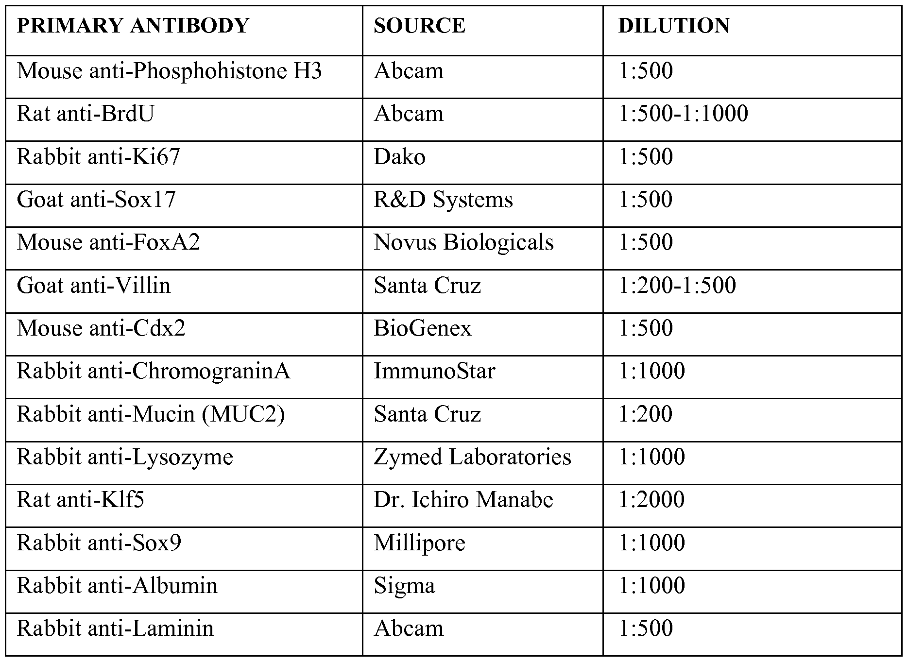
Mouse anti-E-Cadherin BD Biosciences 1:500
Mouse anti-Smooth Muscle Actin Millipore 1:500
Mouse anti-Neurogenin 3 DSHB 1:100
Goat anti-Vimentin Santa Cruz 1:1000
Goat anti-Pdxl Abeam 1:5000
Goat anti-Dpp4 R&D Systems 1:500
Rabbit anti-Phosphohistone H3 Cell Signaling 1:500
Goat anti-Gata4 Santa Cruz 1:200
Rabbit anti-Gata6 Santa Cruz 1:200
Rabbit anti-Nanog Cosmo Bio. Co. 1:2500
Chicken anti-DNMT3b Millipore 1:1000
Mouse anti-Tra 1-60 Millipore 1:500
Mouse anti-Tra 1-81 Millipore 1:500
Table 3B. Secondary Antibodies.
SECONDARY ANTIBODY SOURCE DILUTION
Goat anti-guinea pig Cy5 Jackson Immuno 1:500
Goat anti-rabbit Cy5 Jackson Immuno 1:500
Goat anti-rabbit Cy3 Jackson Immuno 1:500
Goat anti-mouse Cy3 Jackson Immuno 1:500
Goat anti-mouse 488 Invitrogen 1:500
Goat anti-rabbit 488 Invitrogen 1:500
Donkey anti-guinea pig Cy5 Jackson Immuno 1:500
Donkey anti-rabbit Cy5 Jackson Immuno 1:500
Donkey anti-rabbit Cy3 Jackson Immuno 1:500
Donkey anti-mouse Cy3 Jackson Immuno 1:500
Donkey anti-rabbit 488 Invitrogen 1:500
Donkey anti-mouse 488 Invitrogen 1:500
EXAMPLE 9
Adenovirus Production and Transduction
[0171] Adenoviral plasmids were obtained from Addgene and particles were generated according to the manufacturers protocol (Invitrogen - ViraPower Adenoviral Gateway Expression System) as previously described. 28 day organoids were removed from Matrigel and manually bisected with a scalpal. One half of each organoid was then incubated in Ad-GFP or Ad-Neurog3 viral supernatant and media at a 1 : 1 ratio for approximately 4 hours. Organoids were then re-embedded in Matrigel and incubated overnight with viral supernatant and media at a 1 : 1 ratio. The next day, fresh organoid media was placed on the cultures and was changed as described until the end of the experiment.
[0172] Adeno viral-mediated expression of NEUROG3. Adenoviral plasmids were obtained from Addgene and particles were generated as previously described. Transduction was done on 28 day organoids that were removed from Matrigel, manually bisected then incubated in Ad-GFP or Ad-Neurog3 viral supernatant and media at a 1 : 1 ratio for approximately 4 hours. Organoids were then re-embedded in Matrigel and incubated overnight with viral supernatant and media at a 1 : 1 ratio, then transferred to fresh media until the end of the experiment.
[0173] More details can be found, for example, in Zhou et ah, "In vivo reprogramming of adult pancreatic exocrine cells to beta-cells," Nature 455, 627-632 (2008); which is incorporated herein in its entirety.
EXAMPLE 10
R A Isolation, Reverse Transcription and Quantitative PCR (qPCR)
[0174] RNA was isolated using the Nucleospin II RNA isolation kit (Clonetech). Reverse Transcription was carried out using the SuperScriptlll Supermix (Invitrogen) according to manufacturers protocol. Finally, qPCR was carried out using Quantitect SybrGreen MasterMix (Qiagen) on a Chromo4 Real-Time PCR (BioRad). PCR primers sequences were typically obtained from qPrimerDepot
(http://primerdepot<dot>nci<dot>nih<dot>gov/). Exemplary primers (SEQ ID NO.: 1-16 used can be found in the following Table 4.
Table 4. Exemplary primers used.

EXAMPLE 11
Regulation of Formation of Foregut and Hindgut from Human Embryonic and
Induced Pluripotent Stem Cells
[0175] Results and mechanisms for signaling network that regulates foregut, hindgut and intestinal development are shown in Figures 14-16. A summary of the pathways involved is shown in Figure 14 A.
[0176] Following definitive endoderm (DE) induction with activin (100 ng/ml) for 3 days, a 2-4 day treatment was performed with the indicated combination of factors: Wnt3a (500 ng/ml) and Fgf4 (500 ng/ml), Retinoic acid (RA) in a range of 0.2-20 μΜ the BMP antagonist Noggin (50 ng/ml), the WNT antagonist Dkk (1-500 ng/ml).
[0177] The Noggin completely abolishes the Wnt/Fgf-induced Cdx2 expression (Figure 14B). Retinoic acid (2 μΜ) can posteriorize newly-formed definitive endoderm, especially in the first two days following DE induction. The Retinoic acid (RA)-induced
posteriorization is also dependent on endogenous BMP signaling, as the addition of Noggin potently inhibits Cdx2 expression.
[0178] For investigating the impact of BMP signaling on intestinal regionalization after hindgut formation, hindgut spheroids were plated in matrigel and BMP or noggin were added to EGF/Rspondidl containing media. Intestinal organoids were analyzed after 28 days.
[0179] It has been identified that, in addition to FGF and WNT signaling, BMP and RA signaling are capable of promoting a posterior/hindgut fate and repressing foregut fate. Additionally, BMP signaling regulates formation of distinct regional types of intestine. Inhibition of BMP with noggin after the hindgut stage promotes a proximal intestinal fate (duodenum/jejunum). Activation of BMP signaling after the hindgut stage promotes a more distal intestinal cell fate (cecum/colon).
[0180] Figures 14B-14D demonstrate the effects of FGF, WNT, and BMP signaling on differentiation of Definitive endoderm into foregut and hindgut. In particular, Figure 14B and 14D depict the activation of FGF/WNT/BMP signaling promotes posterior/hindgut fate as indicated by expression of Cdx2. Repression of BMP signaling with noggin suppresses hindgut fate and promotes foregut fate. Activation of FGF/WNT/BMP signaling represses foregut fate as indicated by expression of Sox2 (Figure 14C). Repression of WNT signaling with Dkk promotes anterior gene expression (HHex and Cerberus) and represses posterior/hindgut fate (Cdx2) (Figure 14D).
[0181] The effects of Retinoic Acid in promoting hindgut differentiation in a BMP dependent manner (Figures 15A-15D). Quantitative realtime PCR was used to compare the effects of Retinoic Acid and inhibition of BMP on differentiation of Definitive endoderm into foregut and hindgut (Figures 15A and 15B). Similar comparison was also performed by immunostaining (Figures 15C and 15D). Activation of RA signaling promotes posterior/hindgut fate as indicated by expression of Cdx2 and represses foregut fate as indicated by expression of Sox2. Immunostaining in Figure 15C is quantified in Figure 15D using an automated cell counting program.
[0182] Figures 16A through 16C demonstrate that BMP signaling regulates formation of proximal and distal intestine formation from human embryonic and induced pluripotent stem cells. Figure 16A shows that BMP2 promotes a posterior fate for monolayers when added during 96-hour WNT/FGF (W/F) treatment as shown by decreased expression of GATA4 compared to W/F alone and by increased expression of HOXC9 (distal small bowel and proximal colon), HOXD13, and HOXA13 compared to W/F alone.
[0183] Figure 16B shows that, after formation of hindgut sphereoids, addition of BMP2 to 3D cultures promotes patterning of developing intestinal organoids to a distal fate after 28 days compared to the Nog/EGF/Rspol cocktail without BMP2 as shown by decreased relative expression of PDX1 and increased expression of HOXD13 and CA1 (carbonic anhydrase-colonocyte marker).
[0184] Figure 16C shows that Day 138 Noggin/EGF/Rspondidl treated organoids express CCK in the epithelium by immunostaining thus indicating a proximal small bowel fate.
[0185] Figures 14A-14D depict formation of foregut and hindgut from human embryonic and induced pluripotent stem cells is regulated by WNT, FGF, BMP and Retinoic acid (RA) signaling. A) Summary of the signaling network that regulates hindgut and intestinal development. B-D) Effects of FGF, WNT, and BMP signaling on differentiation of definitive endoderm into foregut and hindgut. B) and D). Activation of FGF/WNT/BMP signaling promotes posterior/hindgut fate as indicated by expression of Cdx2. Repression of BMP signaling with noggin suppresses hindgut fate and promotes foregut fate. C) Activation of FGF/WNT/BMP signaling represses foregut fate as indicated by expression of Sox2. D) Repression of WNT signaling with Dkk promotes anterior gene expression (HHex and Cerberus) and represses posterior/hindgut fate (Cdx2).
[0186] Figures 15A-15D depict retinoic acid promotes hindgut differentiation in a BMP dependent manner. Comparing the effects of Retinoic Acid and inhibition of BMP on differentiation of Definitive endoderm into foregut and hindgut by quantitative realtime PCR A) and B) or by immunostaining C) and D). Activation of RA signaling promotes posterior/hindgut fate as indicated by expression of Cdx2 and represses foregut fate as indicated by expression of Sox2. Immunostaining in C is quantified in D using an automated cell counting program.
[0187] Figures 16A-16C depict BMP signaling regulates formation of proximal and distal intestine formation from human embryonic and induced pluripotent stem cells. A)
BMP2 promotes a posterior fate for monolayers when added during 96-hour WNT/FGF
(W/F) treatment as shown by decreased expression of GATA4 compared to W/F alone and by increased expression of HOXC9 (distal small bowel and proximal colon), HOXD13, and
HOXA13 compared to W/F alone. B) After formation of hindgut sphereoids, addition of
BMP2 to 3D cultures promotes patterning of developing intestinal organoids to a distal fate after 28 days compared to the Nog/EGF/Rspol cocktail without BMP2 as shown by
decreased relative expression of PDX1 and increased expression of HOXD13 and CA1 (carbonic anhydrase - colonocyte marker). C) Day 138 Noggin/EGF/Rspondidl treated organoids express CCK in the epithelium by immunostaining thus indicating a proximal small bowel fate.
[0188] The various methods and techniques described above provide a number of exemplary ways to carry out the invention. Of course, it is to be understood that not necessarily all objectives or advantages described may be achieved in accordance with any particular embodiment described herein. Thus, for example, those skilled in the art will recognize that the methods can be performed in a manner that achieves or optimizes one advantage or group of advantages as taught herein without necessarily achieving other objectives or advantages as may be taught or suggested herein. A variety of advantageous and disadvantageous alternatives are mentioned herein. It is to be understood that some preferred embodiments specifically include one, another, or several advantageous features, while others specifically exclude one, another, or several disadvantageous features, while still others specifically mitigate a present disadvantageous feature by inclusion of one, another, or several advantageous features.
[0189] Furthermore, the skilled artisan will recognize the applicability of various features from different embodiments. Similarly, the various elements, features and steps discussed above, as well as other known equivalents for each such element, feature or step, can be mixed and matched by one of ordinary skill in this art to perform methods in accordance with principles described herein. Among the various elements, features, and steps some will be specifically included and others specifically excluded in diverse embodiments.
[0190] Although the invention has been disclosed in the context of certain embodiments and examples, it will be understood by those skilled in the art that the embodiments of the invention extend beyond the specifically disclosed embodiments to other alternative embodiments and/or uses and modifications and equivalents thereof. Many variations and alternative elements have been disclosed in embodiments of the present invention. Still further variations and alternate elements will be apparent to one of skill in the art.
[0191] In some embodiments, the numbers expressing quantities of ingredients, properties such as molecular weight, reaction conditions, and so forth, used to describe and
claim certain embodiments of the invention are to be understood as being modified in some instances by the term "about." Accordingly, in some embodiments, the numerical parameters set forth in the written description and attached claims are approximations that can vary depending upon the desired properties sought to be obtained by a particular embodiment. In some embodiments, the numerical parameters should be construed in light of the number of reported significant digits and by applying ordinary rounding techniques. Notwithstanding that the numerical ranges and parameters setting forth the broad scope of some embodiments of the invention are approximations, the numerical values set forth in the specific examples are reported as precisely as practicable. The numerical values presented in some embodiments of the invention may contain certain errors necessarily resulting from the standard deviation found in their respective testing measurements.
[0192] In some embodiments, the terms "a" and "an" and "the" and similar references used in the context of describing a particular embodiment of the invention (especially in the context of certain of the following claims) can be construed to cover both the singular and the plural. The recitation of ranges of values herein is merely intended to serve as a shorthand method of referring individually to each separate value falling within the range. Unless otherwise indicated herein, each individual value is incorporated into the specification as if it were individually recited herein. All methods described herein can be performed in any suitable order unless otherwise indicated herein or otherwise clearly contradicted by context. The use of any and all examples, or exemplary language (e.g. "such as") provided with respect to certain embodiments herein is intended merely to better illuminate the invention and does not pose a limitation on the scope of the invention otherwise claimed. No language in the specification should be construed as indicating any non-claimed element essential to the practice of the invention.
[0193] Groupings of alternative elements or embodiments of the invention disclosed herein are not to be construed as limitations. Each group member can be referred to and claimed individually or in any combination with other members of the group or other elements found herein. One or more members of a group can be included in, or deleted from, a group for reasons of convenience and/or patentability. When any such inclusion or deletion occurs, the specification is herein deemed to contain the group as modified thus fulfilling the written description of all Markush groups used in the appended claims.
[0194] Preferred embodiments of this invention are described herein, including the best mode known to the inventors for carrying out the invention. Variations on those
preferred embodiments will become apparent to those of ordinary skill in the art upon reading the foregoing description. It is contemplated that skilled artisans can employ such variations as appropriate, and the invention can be practiced otherwise than specifically described herein. Accordingly, many embodiments of this invention include all modifications and equivalents of the subject matter recited in the claims appended hereto as permitted by applicable law. Moreover, any combination of the above-described elements in all possible variations thereof is encompassed by the invention unless otherwise indicated herein or otherwise clearly contradicted by context.
[0195] Many modifications and variations of the present invention can be made without departing from its spirit and scope, as will be apparent to those skilled in the art. The specific embodiments described herein are offered by way of example only, and the invention is to be limited only by the terms of the appended claims along with the full scope of equivalents to which such claims are entitled.
[0196] In closing, it is to be understood that the embodiments of the invention disclosed herein are illustrative of the principles of the present invention. Other modifications that can be employed can be within the scope of the invention. Thus, by way of example, but not of limitation, alternative configurations of the present invention can be utilized in accordance with the teachings herein. Accordingly, embodiments of the present invention are not limited to that precisely as shown and described.
REFERENCES CITED
[0197] All references cited are incorporated herein by reference in their entireties and for all purposes to the same extent as if each individual publication or patent or patent application was specifically and individually indicated to be incorporated by reference in its entirety for all purposes.
Claims
1. A method of inducing formation of an intestinal tissue, comprising:
activating one or more signaling pathways within a precursor cell, wherein said one or more signaling pathways are selected from the group consisting of the Wnt signaling pathway, Wnt/APC signaling pathway, FGF signaling pathway, TGF- beta signaling pathway, shh signaling pathway, BMP signaling pathway, Notch signaling pathway, Hedgehog signaling pathway, LKB signaling pathway, and Par polarity signaling pathway; and
obtaining an intestinal tissue descended from said precursor cell.
2. The method of claim 1, further comprising providing said precursor cell.
3. The method of claim 1, further comprising culturing, after said activating step, said activated precursor cell in vitro to form a 3-dimensional tissue structure.
4. The method of claim 1, wherein said activating and obtaining steps are conducted in vitro.
5. The method of claim 1, wherein said one or more signaling pathways comprise the Wnt signaling pathway and FGF signaling pathway.
6. The method of claim 5, wherein said Wnt signaling pathway is activated by contacting said precursor cell with one or more molecules selected from the group consisting of Wntl, Wnt2, Wnt2b, Wnt3, Wnt3a, Wnt4, Wnt5a, Wnt5b, Wnt6, Wnt7a, Wnt7b, Wnt8a, Wnt8b, Wnt9a, Wnt9b, WntlOa, Wntl Ob, Wntl 1, and Wntl 6.
7. The method of claim 5, wherein said FGF signaling pathway is activated by contacting said precursor cell with one or more molecules selected from the group consisting of FGF1, FGF2, FGF3, FGF4, FGF 10, FGF11, FGF12, FGF13, FGF14, FGF15, FGF16, FGF 17, FGF18, FGF 19, FGF20, FGF21, FGF22, and FGF23.
8. The method of claim 1, wherein said activating step comprises contacting said precursor cell with both Wnt3a and FGF4 over a specified activation period.
9. The method of claim 8, wherein said precursor cell is contacted by Wnt3a during a first activation period and by FGF4 during a second activation period.
10. The method of claim 9, wherein said first activation period and said second activation period overlap.
11. The method of claim 9, wherein said first activation period and said second activation period do not overlap.
12. The method of claim 1, wherein said specified activation period is between 24 and 120 hours.
13. The method of claim 1, wherein said precursor cell is contacted with Wnt3a at a concentration between 50-1500 ng/ml.
14. The method of claim 1, wherein said precursor cell is elected from the group consisting of an embryonic stem cell, an embryonic germ cell, an induced pluripotent stem cell, a mesoderm cell, a definitive endoderm cell, a posterior endoderm cell, and a hindgut cell.
15. The method of claim 14, wherein the definitive endoderm cell is derived from a pluripotent stem cell.
16. The method of claim 15, wherein said pluripotent stem cell is an embryonic stem cell, an embryonic stem cell, an adult stem cell, or an induced pluripotent stem cell.
17. The method of claim 14, wherein said definitive endoderm cell is derived by contacting the pluripotent stem cell with one or more molecules selected from the group consisting of Activin, the BMP subgroups of the TGF-beta superfamily of growth factors; Nodal, Activin A, Activin B, BMP4, Wnt3a, and a combinations thereof.
18. The method of claim 15, wherein said pluripotent stem cell is a human pluripotent stem cell.
19. The method of claim 18, wherein said human pluripotent stem cell is selected from the group consisting of a human embryonic stem cell, a human embryonic germ cell, and an induced human pluripotent stem cell.
20. A composition comprising intestinal tissue produced in vitro from one or more precursor cells.
21. The composition of claim 20, wherein the one or more precursor cells are selected from the group consisting of an embryonic stem cell, an embryonic germ cell, an induced pluripotent stem cell, a mesoderm cell, a definitive endoderm cell, a posterior endoderm cell, and a hindgut cell.
22. A kit comprising an intestinal tissue produced in vitro from one or more precursor cells.
23. A method for identifying the absorption effect of intestinal cells or tissues, comprising:
contacting intestinal cells or tissues with a compound, wherein said intestinal cells or tissues are produced in vitro from one or more precursor cells; and
detecting a level of absorption of said compound by said intestinal cells or tissues.
Priority Applications (3)
| Application Number | Priority Date | Filing Date | Title |
|---|---|---|---|
| US13/695,887 US9719068B2 (en) | 2010-05-06 | 2011-05-06 | Methods and systems for converting precursor cells into intestinal tissues through directed differentiation |
| US15/627,588 US10781425B2 (en) | 2010-05-06 | 2017-06-20 | Methods and systems for converting precursor cells into intestinal tissues through directed differentiation |
| US16/599,620 US12258584B2 (en) | 2010-05-06 | 2019-10-11 | Methods and systems for converting precursor cells into intestinal tissues through directed differentiation |
Applications Claiming Priority (2)
| Application Number | Priority Date | Filing Date | Title |
|---|---|---|---|
| US33217810P | 2010-05-06 | 2010-05-06 | |
| US61/332,178 | 2010-05-06 |
Related Child Applications (2)
| Application Number | Title | Priority Date | Filing Date |
|---|---|---|---|
| US13/695,887 A-371-Of-International US9719068B2 (en) | 2010-05-06 | 2011-05-06 | Methods and systems for converting precursor cells into intestinal tissues through directed differentiation |
| US15/627,588 Continuation US10781425B2 (en) | 2010-05-06 | 2017-06-20 | Methods and systems for converting precursor cells into intestinal tissues through directed differentiation |
Publications (2)
| Publication Number | Publication Date |
|---|---|
| WO2011140441A2 true WO2011140441A2 (en) | 2011-11-10 |
| WO2011140441A3 WO2011140441A3 (en) | 2012-04-05 |
Family
ID=44904500
Family Applications (1)
| Application Number | Title | Priority Date | Filing Date |
|---|---|---|---|
| PCT/US2011/035518 Ceased WO2011140441A2 (en) | 2010-05-06 | 2011-05-06 | Methods and systems for converting precursor cells into intestinal tissues through directed differentiation |
Country Status (2)
| Country | Link |
|---|---|
| US (3) | US9719068B2 (en) |
| WO (1) | WO2011140441A2 (en) |
Cited By (19)
| Publication number | Priority date | Publication date | Assignee | Title |
|---|---|---|---|---|
| EP2636731A4 (en) * | 2010-11-02 | 2014-04-30 | Univ Kumamoto Nat Univ Corp | PROCESS FOR PRODUCING INTESTINAL CELLS |
| WO2015021358A2 (en) | 2013-08-09 | 2015-02-12 | Dominique Charmot | Compounds and methods for inhibiting phosphate transport |
| WO2015183920A3 (en) * | 2014-05-28 | 2016-01-28 | Children's Hospital Medical Center | Methods and systems for converting precursor cells into gastric tissues through directed differentiation |
| JPWO2014132933A1 (en) * | 2013-02-26 | 2017-02-02 | 公立大学法人名古屋市立大学 | Methods for inducing differentiation of induced pluripotent stem cells into intestinal epithelial cells |
| US9719068B2 (en) | 2010-05-06 | 2017-08-01 | Children's Hospital Medical Center | Methods and systems for converting precursor cells into intestinal tissues through directed differentiation |
| US10487313B2 (en) | 2011-06-21 | 2019-11-26 | Novo Nordisk A/S | Efficient induction of definitive endoderm from pluripotent stem cells |
| US11066650B2 (en) | 2016-05-05 | 2021-07-20 | Children's Hospital Medical Center | Methods for the in vitro manufacture of gastric fundus tissue and compositions related to same |
| US11584916B2 (en) | 2014-10-17 | 2023-02-21 | Children's Hospital Medical Center | Method of making in vivo human small intestine organoids from pluripotent stem cells |
| US11767515B2 (en) | 2016-12-05 | 2023-09-26 | Children's Hospital Medical Center | Colonic organoids and methods of making and using same |
| WO2024263961A2 (en) | 2023-06-23 | 2024-12-26 | Children's Hospital Medical Center | Methods of matrix-free suspension culture |
| WO2025072803A1 (en) | 2023-09-29 | 2025-04-03 | Children's Hospital Medical Center | Ntrk2 signaling-mediated alveolar capillary injury and repair |
| US12281334B2 (en) | 2017-04-14 | 2025-04-22 | Children's Hospital Medical Center | Multi donor stem cell compositions and methods of making same |
| US12297457B2 (en) | 2017-10-10 | 2025-05-13 | Children's Hospital Medical Center | Esophageal tissue and/or organoid compositions and methods of making same |
| US12379372B2 (en) | 2017-12-21 | 2025-08-05 | Children's Hospital Medical Center | Digitalized human organoids and methods of using same |
| US12414967B2 (en) | 2016-11-04 | 2025-09-16 | Children's Hospital Medical Center | Compositions and methods of treating liver disease |
| US12421500B2 (en) | 2018-07-26 | 2025-09-23 | Children's Hospital Medical Center | Hepato-biliary-pancreatic tissues and methods of making same |
| US12428622B2 (en) | 2018-09-12 | 2025-09-30 | Children's Hospital Medical Center | Organoid compositions for the production of hematopoietic stem cells and derivatives thereof |
| US12497597B2 (en) | 2019-05-31 | 2025-12-16 | Children's Hospital Medical Center | Methods of generating and expanding hematopoietic stem cells |
| US12534709B2 (en) | 2019-05-31 | 2026-01-27 | Children's Hospital Medical Center | Shaped organoid compositions and methods of making same |
Families Citing this family (18)
| Publication number | Priority date | Publication date | Assignee | Title |
|---|---|---|---|---|
| PT2173379E (en) | 2007-07-02 | 2015-11-24 | Oncomed Pharm Inc | Compositions and methods for treating and diagnosing cancer |
| CN106687137A (en) * | 2014-09-16 | 2017-05-17 | 昂考梅德药品有限公司 | Treatment of fibrotic diseases |
| US20190018000A1 (en) | 2016-01-12 | 2019-01-17 | Cedars-Sinai Medical Center | A method of non destructive monitoring of biological processes in microfluidic tissue culture systems |
| EP3411472A4 (en) * | 2016-02-01 | 2020-01-22 | Cedars-Sinai Medical Center | Systems and methods for growth of intestinal cells in microfluidic devices |
| US11913022B2 (en) | 2017-01-25 | 2024-02-27 | Cedars-Sinai Medical Center | In vitro induction of mammary-like differentiation from human pluripotent stem cells |
| EP3587560A4 (en) * | 2017-01-27 | 2020-12-16 | Kaneka Corporation | ENDODERMAL CELL MASS AND METHOD OF PREPARING ANY OF THREE PRIMARY LAYER CELL MASS FROM PLURIPOTENT CELLS |
| US11767513B2 (en) | 2017-03-14 | 2023-09-26 | Cedars-Sinai Medical Center | Neuromuscular junction |
| WO2018176001A2 (en) | 2017-03-24 | 2018-09-27 | Cedars-Sinai Medical Center | Methods and compositions for production of fallopian tube epithelium |
| JP7174426B2 (en) * | 2017-05-09 | 2022-11-17 | 公立大学法人名古屋市立大学 | Method for producing pluripotent stem cell-derived intestinal organoids |
| AU2018270270A1 (en) * | 2017-05-19 | 2019-12-05 | Cedars-Sinai Medical Center | Systems and methods for growth of intestinal cells |
| EP3768823B1 (en) | 2018-03-23 | 2025-12-31 | Cedars-Sinai Medical Center | METHOD FOR THE USE OF ISLAND CELLS |
| US11981918B2 (en) | 2018-04-06 | 2024-05-14 | Cedars-Sinai Medical Center | Differentiation technique to generate dopaminergic neurons from induced pluripotent stem cells |
| JP7464532B2 (en) | 2018-04-06 | 2024-04-09 | シーダーズ-サイナイ メディカル センター | Neurodegenerative disease models derived from human pluripotent stem cells on a microfluidic chip |
| US20220041991A1 (en) * | 2019-01-30 | 2022-02-10 | Dai Nippon Printing Co., Ltd. | Cell structure including small intestinal epithelial cells, method for producing same, and substrate holding same |
| KR102146274B1 (en) * | 2019-05-21 | 2020-08-24 | 주식회사 마이크로바이오틱스 | Method for preparing intestinal organoid and use thereof |
| CN112522405B (en) * | 2020-12-10 | 2023-03-21 | 首都医科大学 | Application of MAGI3 in prediction of prognosis or chemotherapy sensitivity of colorectal cancer patient |
| JP2025502241A (en) * | 2022-01-14 | 2025-01-24 | チルドレンズ ホスピタル メディカル センター | Methods for repairing intestinal injury using organoid compositions |
| WO2025198284A1 (en) * | 2024-03-20 | 2025-09-25 | 한국생명공학연구원 | Use of pluripotent stem cell-derived intestinal stromal cells as multipotent differentiation intermediate |
Family Cites Families (379)
| Publication number | Priority date | Publication date | Assignee | Title |
|---|---|---|---|---|
| AU9031591A (en) | 1990-10-29 | 1992-05-26 | Regents Of The University Of Minnesota | A bioartificial liver |
| US5523226A (en) * | 1993-05-14 | 1996-06-04 | Biotechnology Research And Development Corp. | Transgenic swine compositions and methods |
| US5912227A (en) | 1995-01-27 | 1999-06-15 | North Carolina State University | Method of enhancing nutrient uptake |
| WO1998021312A1 (en) | 1996-11-08 | 1998-05-22 | Rpms Technology Limited | Human hepatocytes in three-dimensional support systems |
| HK1045171A1 (en) | 1997-01-16 | 2002-11-15 | Yissum Research Development Company Of The Hebrew University Of Jerusalem | A device and method for performing a biological modification of a fluid |
| EP1063289A1 (en) | 1998-03-03 | 2000-12-27 | JMS Co., Ltd. | Liver cell clones for artifical liver and extracorporeal liver assist device |
| EP1060243A4 (en) | 1998-03-02 | 2005-03-09 | Univ Massachusetts | EMBRYONIC CELL LINES OR STEM CELL-SIMILAR CELL LINES PRODUCED BY CORE TRANSPLANT BETWEEN DIFFERENT SPECIES |
| US7291626B1 (en) | 1998-04-09 | 2007-11-06 | John Hopkins University School Of Medicine | Inhibitors of hedgehog signaling pathways, compositions and uses related thereto |
| US20010041964A1 (en) | 1998-09-14 | 2001-11-15 | George M. Grass | Pharmacokinetic-based drug design tool and method |
| US7759113B2 (en) | 1999-04-30 | 2010-07-20 | The General Hospital Corporation | Fabrication of tissue lamina using microfabricated two-dimensional molds |
| JP3725147B2 (en) | 1999-08-25 | 2005-12-07 | 東洋紡績株式会社 | Cell culture module having a vascular network-like structure |
| US6607501B2 (en) | 2001-05-14 | 2003-08-19 | Reynolds G. Gorsuch | Process and apparatus for utilization of in vivo extracted plasma with tissue engineering devices, bioreactors, artificial organs, and cell therapy applications |
| US20030129751A1 (en) | 2001-05-16 | 2003-07-10 | Grikscheit Tracy C. | Tissue-engineered organs |
| EP1456374A4 (en) | 2001-11-26 | 2005-08-17 | Advanced Cell Tech Inc | METHODS FOR THE PRODUCTION AND USE OF REPROGRAMME HUMAN SOMATIC CELL CORES AND AUTOLOGOUS AND ISOGENIC HUMAN STEM CELLS |
| GB2399823B (en) | 2001-12-07 | 2006-02-15 | Geron Corp | Islet cells from primate pluripotent stem cells |
| US20050170506A1 (en) | 2002-01-16 | 2005-08-04 | Primegen Biotech Llc | Therapeutic reprogramming, hybrid stem cells and maturation |
| US20030187515A1 (en) | 2002-03-26 | 2003-10-02 | Hariri Robert J. | Collagen biofabric and methods of preparing and using the collagen biofabric |
| US7160719B2 (en) | 2002-06-07 | 2007-01-09 | Mayo Foundation For Medical Education And Research | Bioartificial liver system |
| AU2003261798A1 (en) | 2002-08-28 | 2004-03-19 | Asahi Medical Co., Ltd. | Cell-filled device of modified cross-section hollow fiber membrane type |
| TW571101B (en) | 2003-01-21 | 2004-01-11 | Ind Tech Res Inst | Fluid analysis apparatus |
| WO2004071573A2 (en) | 2003-02-07 | 2004-08-26 | The Johns Hopkins University | Hypoxia induced mitogenic factor |
| US7727998B2 (en) | 2003-02-10 | 2010-06-01 | Banyu Pharmaceutical Co., Ltd. | Melanin-concentrating hormone receptor antagonists containing piperidine derivatives as the active ingredient |
| DE10362002B4 (en) | 2003-06-23 | 2006-10-12 | Fraunhofer-Gesellschaft zur Förderung der angewandten Forschung e.V. | Adult pluripotent stem cells |
| US8647873B2 (en) | 2004-04-27 | 2014-02-11 | Viacyte, Inc. | PDX1 expressing endoderm |
| US7625753B2 (en) | 2003-12-23 | 2009-12-01 | Cythera, Inc. | Expansion of definitive endoderm cells |
| US7541185B2 (en) | 2003-12-23 | 2009-06-02 | Cythera, Inc. | Methods for identifying factors for differentiating definitive endoderm |
| US20050266554A1 (en) | 2004-04-27 | 2005-12-01 | D Amour Kevin A | PDX1 expressing endoderm |
| US8586357B2 (en) | 2003-12-23 | 2013-11-19 | Viacyte, Inc. | Markers of definitive endoderm |
| KR101221302B1 (en) | 2003-12-23 | 2013-01-11 | 비아싸이트, 인크. | Definitive endoderm |
| US7985585B2 (en) | 2004-07-09 | 2011-07-26 | Viacyte, Inc. | Preprimitive streak and mesendoderm cells |
| WO2005081970A2 (en) | 2004-02-24 | 2005-09-09 | The Curators Of The University Of Missouri | Self-assembling cell aggregates and methods of making engineered tissue using the same |
| DE102004017476B4 (en) | 2004-04-08 | 2009-03-26 | Fraunhofer-Gesellschaft zur Förderung der angewandten Forschung e.V. | Process for the preparation of a cell composition containing epithelial cells |
| ES2801001T3 (en) | 2004-04-27 | 2021-01-07 | Viacyte Inc | Endoderm expressing pdx1 |
| JP4650608B2 (en) | 2004-05-18 | 2011-03-16 | 信越化学工業株式会社 | Photomask blank and photomask manufacturing method |
| WO2005113747A2 (en) | 2004-05-21 | 2005-12-01 | Fraunhofer Gesellschaft zur Förderung der angewandten Forschung e.V. | Multicellular tissue and organ culture systems |
| US8410246B2 (en) | 2004-06-17 | 2013-04-02 | Thrasos, Inc. | TDF-related compounds and analogs thereof |
| WO2006016999A1 (en) | 2004-07-09 | 2006-02-16 | Cythera, Inc. | Methods for identifying factors for differentiating definitive endoderm |
| CN105708787A (en) | 2004-07-12 | 2016-06-29 | 爱密斯菲尔科技公司 | Compositions for delivering peptide yy and pyy agonists |
| WO2006091231A2 (en) | 2004-07-21 | 2006-08-31 | Ambrx, Inc. | Biosynthetic polypeptides utilizing non-naturally encoded amino acids |
| EP1791952A4 (en) | 2004-08-13 | 2008-06-11 | Univ Georgia Res Found | Compositions and methods for self-renewal and differentiation in human embryonic stem cells |
| US20060236415A1 (en) | 2005-03-09 | 2006-10-19 | Silversides David W | Neural crest cells specific promoters; isolated neural crest cells; and methods of isolating and of using same |
| US7604929B2 (en) | 2005-04-21 | 2009-10-20 | In Vitro Technologies, Inc. | Cellular compositions and methods for their preparation |
| WO2006122147A2 (en) | 2005-05-10 | 2006-11-16 | Rutgers, The State University Of New Jersey | Alginate poly-l-lysine encapsulation as a technology for controlled differentiation of embryonic stem cells |
| WO2006126219A1 (en) | 2005-05-26 | 2006-11-30 | Fresenius Medical Care Deutschland G.M.B.H. | Liver progenitor cells |
| GB0517382D0 (en) | 2005-08-26 | 2005-10-05 | Plasticell Ltd | Cell culture |
| WO2007027905A2 (en) | 2005-08-31 | 2007-03-08 | Science And Technology Corporation @ Unm | Human renal stem cells |
| DK2674485T3 (en) | 2005-10-27 | 2019-08-26 | Viacyte Inc | PDX-1 EXPRESSING DORSAL AND VENTRAL FORTARM ENDODERM |
| US7927869B2 (en) | 2005-11-29 | 2011-04-19 | Spencer Z Rosero | System and method for supporting a biological chip device |
| US20070239083A1 (en) | 2006-01-18 | 2007-10-11 | Axel Voss | Shock wave generators |
| US20070238169A1 (en) | 2006-04-11 | 2007-10-11 | The Board Of Trustees Of The Leland Stanford Junior University | Cell sorter and culture system |
| US8685730B2 (en) | 2006-05-02 | 2014-04-01 | Wisconsin Alumni Research Foundation | Methods and devices for differentiating pluripotent stem cells into cells of the pancreatic lineage |
| US9012419B2 (en) | 2006-05-17 | 2015-04-21 | University Of Utah Research Foundation | Methods and compositions related to eosinophil regulation |
| DK2040713T3 (en) | 2006-06-27 | 2014-09-29 | Intercept Pharmaceuticals Inc | BILIC ACID DERIVATIVES AS FXR LIGANDS FOR THE PREVENTION OR TREATMENT OF FXR-MEDIATED DISEASES OR CONDITIONS |
| WO2008011561A2 (en) | 2006-07-21 | 2008-01-24 | Massachusetts Institute Of Technology | End-modified poly(beta-amino esters) and uses thereof |
| US8497240B2 (en) | 2006-08-17 | 2013-07-30 | Amylin Pharmaceuticals, Llc | DPP-IV resistant GIP hybrid polypeptides with selectable properties |
| ATE496976T1 (en) | 2006-12-08 | 2011-02-15 | Du Pont | WATER BASED COATING COMPOSITION |
| CA2671843A1 (en) | 2006-12-08 | 2008-06-19 | Yin-Xiong Li | Formation and rejuvenation of organs and alcohol damaged organ regeneration through stem cell nutrients |
| US9127254B2 (en) | 2006-12-18 | 2015-09-08 | Ben-Gurion University Of The Negev Research And Development Authority | Scaffolding for tissue regeneration or repair |
| US8512697B2 (en) | 2007-05-16 | 2013-08-20 | The University Of North Carolina At Chapel Hill | Delivery of micro- and nanoparticles with blood platelets |
| FR2917425B1 (en) | 2007-06-18 | 2010-11-19 | Univ Nancy 1 Henri Poincare | METHOD FOR THE PROLIFERATION OF CELLS ON POLYELECTROLYTE MULTILAYERS AND ITS APPLICATION, IN PARTICULAR TO THE PREPARATION OF CELLULARIZED BIOMATERIALS |
| GB2467645B (en) | 2007-07-06 | 2011-12-21 | Univ London | A method of proliferating human hepatocyte cells |
| EP2022848A1 (en) | 2007-08-10 | 2009-02-11 | Hubrecht Institut | A method for identifying, expanding, and removing adult stem cells and cancer stem cells |
| EP2185931B1 (en) | 2007-09-07 | 2013-07-10 | Children's Hospital Medical Center | Use of secretor, lewis and sialyl antigen levels as predictors for disease |
| US7695963B2 (en) | 2007-09-24 | 2010-04-13 | Cythera, Inc. | Methods for increasing definitive endoderm production |
| US8609413B2 (en) | 2007-12-11 | 2013-12-17 | Research Development Foundation | Neurons, astrocytes and oligodendrocytes differentiated from a mammalian pluripotent or neural stem cells exposed to a pyridine deriviative |
| AU2009203893B2 (en) | 2008-01-08 | 2014-10-02 | The University Of Queensland | Method of producing a population of cells |
| US20110218512A1 (en) | 2008-06-03 | 2011-09-08 | Aethlon Medical, Inc. | Enhanced antiviral therapy methods and devices |
| AU2009254177B2 (en) | 2008-06-04 | 2014-09-11 | Tissuse Gmbh | Organ-on-a-chip-device |
| KR101711354B1 (en) | 2008-06-24 | 2017-02-28 | 더 큐레이터스 오브 더 유니버시티 오브 미주리 | Self-assembling multicellular bodies and methods of producing a three-dimensional biological structure using the same |
| ITRM20080342A1 (en) | 2008-06-26 | 2009-12-27 | Univ Degli Studi Udine | DOLLS-SIMILAR PULP CELLS, INSULATION AND USE METHODS. |
| US20130115673A1 (en) | 2008-07-16 | 2013-05-09 | Biotime, Inc. | Methods of Screening Embryonic Progenitor Cell Lines |
| US20110305672A1 (en) | 2008-07-25 | 2011-12-15 | University Of Georgia Research Foundation, Inc. | COMPOSITIONS FOR MESODERM DERIVED ISL1+ MULTIPOTENT CELLS (IMPs), EPICARDIAL PROGENITOR CELLS (EPCs) AND MULTIPOTENT CD56C CELLS (C56Cs) AND METHODS OF PRODUCING AND USING SAME |
| US20110300543A1 (en) | 2008-10-31 | 2011-12-08 | The Trustees Of Columbia University In The City Of New York | Methods for making induced pluripotent stem cells from mesenchymal stem cells |
| US8895300B2 (en) | 2008-11-04 | 2014-11-25 | Viacyte, Inc. | Scalable primate pluripotent stem cell aggregate suspension culture and differentiation thereof |
| US20110294735A1 (en) | 2008-11-05 | 2011-12-01 | Merck Sharp & Dohme Corp. | Mechanism of neuromedin u action and uses thereof |
| CA2745742A1 (en) | 2008-12-03 | 2010-06-10 | International Stem Cell Corporation | Methods of deriving differentiated cells from stem cells |
| JP5351601B2 (en) | 2008-12-26 | 2013-11-27 | 矢崎総業株式会社 | Insulating cap manufacturing method and insulating cap manufacturing apparatus |
| US9752124B2 (en) | 2009-02-03 | 2017-09-05 | Koninklijke Nederlandse Akademie Van Wetenschappen | Culture medium for epithelial stem cells and organoids comprising the stem cells |
| GB201111244D0 (en) | 2011-06-30 | 2011-08-17 | Konink Nl Akademie Van Wetenschappen Knaw | Culture media for stem cells |
| MX379945B (en) | 2009-02-03 | 2025-03-11 | Koninklijke Nederlandse Akademie Van Wetenschappen | CULTURE MEDIUM FOR EPITHELIAL STEM CELLS AND ORGANOIDS THAT INCLUDE STEM CELLS. |
| EP2412800A1 (en) | 2010-07-29 | 2012-02-01 | Koninklijke Nederlandse Akademie van Wetenschappen | Liver organoid, uses thereof and culture method for obtaining them |
| WO2010094694A1 (en) | 2009-02-23 | 2010-08-26 | F. Hoffmann-La Roche Ag | Assays to predict cardiotoxicity |
| EP2405957B1 (en) | 2009-03-13 | 2020-12-23 | Mayo Foundation For Medical Education And Research | Bioartificial liver |
| EP2408457A1 (en) | 2009-03-17 | 2012-01-25 | Axcan Pharma Inc. | Method of treating nonalcoholic steatohepatitis with elevated doses of ursodeoxycholic acid |
| WO2010127399A1 (en) | 2009-05-06 | 2010-11-11 | Walter And Eliza Hall Institute Of Medical Research | Gene expression profiles and uses thereof |
| CA2762584A1 (en) | 2009-05-20 | 2010-11-25 | Cardio3 Biosciences S.A. | Pharmaceutical composition for the treatment of heart diseases |
| JP2012527880A (en) | 2009-05-29 | 2012-11-12 | ノヴォ ノルディスク アー/エス | Derivation of specific endoderm from hPS cell-derived definitive endoderm |
| WO2010143747A1 (en) | 2009-06-10 | 2010-12-16 | 公立大学法人奈良県立医科大学 | Method for production of artificial intestinal tract |
| CN102459574B (en) | 2009-06-18 | 2016-06-29 | 塞拉提斯股份公司 | Growth and the 3D culture systems of differentiation for dry (hPS) cell of people's pluripotency |
| US8685386B2 (en) | 2009-07-16 | 2014-04-01 | Biotime, Inc | Methods and compositions for in vitro and in vivo chondrogenesis |
| US20110152770A1 (en) | 2009-07-30 | 2011-06-23 | Tandem Diabetes Care, Inc. | Infusion pump system with disposable cartridge having pressure venting and pressure feedback |
| US8501476B2 (en) | 2009-10-07 | 2013-08-06 | Brown University | Assays and methods for fusing cell aggregates to form proto-tissues |
| US9506035B2 (en) | 2009-10-13 | 2016-11-29 | University Of Louisville Research Foundation, Inc | Methods and compositions to support transplanted tissue integration and innosculation with adipose stromal cells |
| GB0918564D0 (en) | 2009-10-22 | 2009-12-09 | Plasticell Ltd | Nested cell encapsulation |
| CN101696396A (en) | 2009-10-27 | 2010-04-21 | 中国人民解放军军事医学科学院卫生学环境医学研究所 | Construction method and use of model of hepatitis B virus infection in vivo |
| WO2011066374A2 (en) | 2009-11-24 | 2011-06-03 | Alder Biopharmaceuticals, Inc. | Antagonists of il-6 to prevent or treat cachexia, weakness, fatigue, and/or fever |
| US20130031645A1 (en) | 2009-11-25 | 2013-01-31 | Thomas Touboul | Method for hepatic differentiation of definitive endoderm cells |
| AU2010333840B2 (en) | 2009-12-23 | 2016-01-07 | Janssen Biotech, Inc. | Differentiation of human embryonic stem cells |
| US20110231942A1 (en) | 2010-03-17 | 2011-09-22 | Xi He | Tiki1 and tiki2, wnt inhibitors |
| EP2550355B1 (en) | 2010-03-22 | 2016-11-16 | Takara Bio Europe AB | Directed differentiation and maturation of pluripotent cells into hepatocyte like cells by modulation of wnt-signalling pathway |
| WO2011119507A2 (en) | 2010-03-25 | 2011-09-29 | International Stem Cell Corporation | Method of altering the differentiative state of a cell and compositions thereof |
| EP2380920A1 (en) | 2010-04-22 | 2011-10-26 | QGel SA | Hydrogel precursor formulation and production process thereof |
| CN103068970A (en) | 2010-04-25 | 2013-04-24 | 西奈山医学院 | Generation of anterior foregut endoderm from pluripotent cells |
| US9719068B2 (en) | 2010-05-06 | 2017-08-01 | Children's Hospital Medical Center | Methods and systems for converting precursor cells into intestinal tissues through directed differentiation |
| JP5984217B2 (en) | 2010-06-15 | 2016-09-06 | セルラー ダイナミクス インターナショナル, インコーポレイテッド | Preparation of induced pluripotent stem cells from a small amount of peripheral blood |
| US8785192B2 (en) | 2010-07-07 | 2014-07-22 | Cellular Dynamics International, Inc. | Endothelial cell production by programming |
| WO2012027474A1 (en) | 2010-08-24 | 2012-03-01 | Regents Of The University Of Minnesota | Non-static suspension culture of cell aggregates |
| AU2011296383B2 (en) | 2010-08-31 | 2016-03-10 | Janssen Biotech, Inc. | Differentiation of pluripotent stem cells |
| AU2011329002C1 (en) | 2010-11-15 | 2017-05-25 | Accelerated Biosciences Corp. | Generation of neural stem cells from human trophoblast stem cells |
| EA028902B1 (en) | 2010-12-31 | 2018-01-31 | Университет Фюр Боденкультур Вена | Method of generating induced pluripotent stem cells and differentiated cells |
| US8951781B2 (en) | 2011-01-10 | 2015-02-10 | Illumina, Inc. | Systems, methods, and apparatuses to image a sample for biological or chemical analysis |
| US9200258B2 (en) | 2011-01-27 | 2015-12-01 | University Of Maryland, Baltimore | Multicellular organotypic model of human intestinal mucosa |
| EP2484750A1 (en) | 2011-02-07 | 2012-08-08 | Ecole Polytechnique Fédérale de Lausanne (EPFL) | Monitoring system for cell culture |
| KR102117921B1 (en) | 2011-02-28 | 2020-06-03 | 프레지던트 앤드 펠로우즈 오브 하바드 칼리지 | Cell culture system |
| GB201106395D0 (en) | 2011-04-14 | 2011-06-01 | Hubrecht Inst | Compounds |
| US20140158233A1 (en) | 2011-05-09 | 2014-06-12 | President And Fellows Of Harvard College | Aerosol delivery to a microfluidic device |
| US10260039B2 (en) | 2011-05-11 | 2019-04-16 | Massachusetts Institute Of Technology | Microgels and microtissues for use in tissue engineering |
| US20120291096A1 (en) | 2011-05-12 | 2012-11-15 | Nokia Corporation | Method and apparatus for secure signing and utilization of distributed computations |
| KR20190042747A (en) | 2011-06-02 | 2019-04-24 | 프레지던트 앤드 펠로우즈 오브 하바드 칼리지 | Methods and uses for ex vivo tissue culture systems |
| JP2014519854A (en) | 2011-06-23 | 2014-08-21 | ザ チルドレンズ ホスピタル オブ フィラデルフィア | Self-replicating endoderm progenitor cell lines derived from human pluripotent stem cells and methods of use thereof |
| CA2848044C (en) | 2011-09-12 | 2021-01-19 | Organovo, Inc. | Platform for engineered implantable tissues and organs and methods of making the same |
| JP2013066414A (en) | 2011-09-22 | 2013-04-18 | National Institute Of Advanced Industrial Science & Technology | Surface marker of stomach precursor cell |
| WO2014066811A1 (en) | 2012-10-25 | 2014-05-01 | The Johns Hopkins University | Bioreducible poly (b-amino ester)s for sirna delivery |
| WO2013061608A1 (en) | 2011-10-27 | 2013-05-02 | 国立大学法人東京医科歯科大学 | Technique for isolating/culturing colorectal epithelial stem cell, and technique for transplanting colorectal epithelium employing said technique |
| US20140302491A1 (en) | 2011-10-28 | 2014-10-09 | The Board Of Trustees Of The Leland Stanford Junior University | Ex Vivo Culture, Proliferation and Expansion of Primary Tissue Organoids |
| US20140308695A1 (en) | 2011-11-04 | 2014-10-16 | Tengion, Inc. | Drug screening and potency assays |
| EP2788476B1 (en) | 2011-12-08 | 2019-05-22 | Fred Hutchinson Cancer Research Center | Compositions and methods for enhanced generation of hematopoietic stem/progenitor cells |
| US9725687B2 (en) | 2011-12-09 | 2017-08-08 | President And Fellows Of Harvard College | Integrated human organ-on-chip microphysiological systems |
| US10087422B2 (en) | 2011-12-09 | 2018-10-02 | President And Fellows Of Harvard College | Organ chips and uses thereof |
| EP3045912B1 (en) | 2011-12-19 | 2018-09-19 | UMC Utrecht Holding B.V. | A rapid quantitative assay to measure cftr function in a primary intestinal culture model |
| CN113699105A (en) | 2011-12-23 | 2021-11-26 | 人类起源公司 | Organoids comprising decellularized and regrouped placental vascular scaffolds |
| WO2013106677A1 (en) | 2012-01-13 | 2013-07-18 | The General Hospital Corporation | Isolated human lung progenitor cells and uses thereof |
| WO2013116446A1 (en) | 2012-01-31 | 2013-08-08 | Wake Forest University Health Sciences | Tubular bioengineered smooth muscle structures |
| WO2013126813A1 (en) | 2012-02-22 | 2013-08-29 | Amgen Inc. | Autologous mammalian models derived from induced pluripotent stem cells and related methods |
| EP2634251A1 (en) | 2012-02-29 | 2013-09-04 | Technische Universität Berlin | 3D in vitro bi-phasic cartilage-bone construct |
| EP2836585A4 (en) | 2012-04-09 | 2015-12-23 | Thomas J Goodwin | Alternating ionic magnetic resonance (aimr) multiple-chambered culture apparatus and methods of use |
| KR20160027217A (en) | 2012-05-23 | 2016-03-09 | 에프. 호프만-라 로슈 아게 | Compositions and methods of obtaining and using endoderm and hepatocyte cells |
| PT3241902T (en) | 2012-05-25 | 2018-05-28 | Univ California | METHODS AND COMPOSITIONS FOR MODIFICATION OF TARGETED TARGET DNA BY RNA AND FOR MODULATION DIRECTED BY TRANSCRIPTION RNA |
| CN108103005A (en) | 2012-06-08 | 2018-06-01 | 詹森生物科技公司 | Differentiation of the human embryo stem cell to pancreatic endocrine cell |
| US20140099709A1 (en) | 2012-06-19 | 2014-04-10 | Organovo, Inc. | Engineered three-dimensional connective tissue constructs and methods of making the same |
| CN111228278A (en) | 2012-06-19 | 2020-06-05 | 英特塞普特医药品公司 | Preparation, use and solid forms of obeticholic acid |
| DE102012105540A1 (en) | 2012-06-26 | 2014-04-24 | Karlsruher Institut für Technologie | Vascular model, process for its preparation and its use |
| EP2875360B1 (en) | 2012-07-20 | 2018-08-08 | Agency For Science, Technology And Research | In vitro assay for predicting renal proximal tubular cell toxicity |
| US9988606B2 (en) | 2012-07-24 | 2018-06-05 | The Trustees Of Columbia University In The City Of New York | Generation of airway and lung progenitors and epithelial cells and three-dimensional anterior foregut spheres |
| GB201216796D0 (en) | 2012-09-20 | 2012-11-07 | Cambridge Entpr Ltd | In vitro pancreatic differentiation |
| DK2712918T3 (en) | 2012-09-28 | 2015-02-16 | Tissuse Gmbh | Multi-organ-chip with improved lifetime and homeostasis |
| EP2716298A1 (en) | 2012-10-03 | 2014-04-09 | Institut Pasteur | A nod2-dependant pathway of cytoprotection of stem cells |
| CN104995294A (en) | 2012-10-19 | 2015-10-21 | 新加坡科技研究局 | Methods of differentiating stem cells into one or more cell lineages |
| WO2014082096A1 (en) | 2012-11-26 | 2014-05-30 | The Trustees Of Columbia University In The City Of New York | Method for culture of human and mouse prostate organoids and uses thereof |
| ES2625510T3 (en) | 2012-11-26 | 2017-07-19 | Gambro Lundia Ab | Liver support system |
| EP2925859B1 (en) | 2012-11-29 | 2018-05-02 | Takara Bio Europe AB | Maturation of hepatocyte-like cells derived from human pluripotent stem cells |
| SG10201707569YA (en) | 2012-12-12 | 2017-10-30 | Broad Inst Inc | Delivery, Engineering and Optimization of Systems, Methods and Compositions for Sequence Manipulation and Therapeutic Applications |
| ES2553782T3 (en) | 2012-12-12 | 2015-12-11 | The Broad Institute, Inc. | Systems engineering, methods and guide compositions optimized for sequence manipulation |
| WO2014093655A2 (en) | 2012-12-12 | 2014-06-19 | The Broad Institute, Inc. | Engineering and optimization of systems, methods and compositions for sequence manipulation with functional domains |
| EP4234696A3 (en) | 2012-12-12 | 2023-09-06 | The Broad Institute Inc. | Crispr-cas component systems, methods and compositions for sequence manipulation |
| US8697359B1 (en) | 2012-12-12 | 2014-04-15 | The Broad Institute, Inc. | CRISPR-Cas systems and methods for altering expression of gene products |
| EP2743345A1 (en) | 2012-12-13 | 2014-06-18 | IMBA-Institut für Molekulare Biotechnologie GmbH | Three dimensional heterogeneously differentiated tissue culture |
| US20150359849A1 (en) | 2013-01-31 | 2015-12-17 | President And Fellows Of Harvard College | Methods of increasing neuronal connectivity and/or treating a neurodegenerative condition |
| EP2956538B1 (en) | 2013-02-13 | 2018-11-14 | Wake Forest University Health Sciences | Bioengineered liver constructs and methods relating thereto |
| GB201304245D0 (en) | 2013-03-08 | 2013-04-24 | Inst Quimic De Sarria | Chemical compounds |
| WO2014165056A1 (en) | 2013-03-12 | 2014-10-09 | Board Of Regents, The University Of Texas System | High throughput mechanical strain generating system for cell cultures and applications thereof |
| US9938499B2 (en) | 2013-03-13 | 2018-04-10 | Wisconsin Alumni Research Foundation | Methods and materials for hematoendothelial differentiation of human pluripotent stem cells under defined conditions |
| US20160022873A1 (en) | 2013-03-14 | 2016-01-28 | Research Institute At Nationwide Children's Hospital, Inc. | Tissue engineered intestine |
| DK2970890T3 (en) | 2013-03-14 | 2020-05-04 | Brigham & Womens Hospital Inc | COMPOSITIONS AND PROCEDURES FOR THE PROMOTION AND CULTIVATION OF EPITHEL STEM CELLS |
| CA2899507A1 (en) | 2013-03-14 | 2014-09-25 | The Regents Of The University Of California | In vitro production of medial ganglionic eminence precursor cells |
| US8859286B2 (en) | 2013-03-14 | 2014-10-14 | Viacyte, Inc. | In vitro differentiation of pluripotent stem cells to pancreatic endoderm cells (PEC) and endocrine cells |
| US9442105B2 (en) | 2013-03-15 | 2016-09-13 | Organovo, Inc. | Engineered liver tissues, arrays thereof, and methods of making the same |
| US20160237400A1 (en) | 2013-03-15 | 2016-08-18 | The Jackson Laboratory | Isolation of non-embryonic stem cells and uses thereof |
| WO2014153294A1 (en) | 2013-03-17 | 2014-09-25 | The Regents Of The University Of California | Method to expand and transduce cultured human small and large intestinal stem cells |
| WO2014153346A1 (en) | 2013-03-18 | 2014-09-25 | Massachusetts Institute Of Technology | Engineering a heterogeneous tissue pluripotent stem cells |
| US10519421B2 (en) | 2013-03-21 | 2019-12-31 | Kyoto University | Induction of motor neurons from pluripotent stem cells |
| EP2796873A1 (en) | 2013-04-25 | 2014-10-29 | QGel SA | Method for a cell-based drug screening assay and the use thereof |
| US20160245653A1 (en) | 2013-04-30 | 2016-08-25 | Sangtae Park | Cylindrical resonator gyroscope |
| EP4299721A3 (en) | 2013-05-08 | 2024-04-24 | ProKidney | Organoids comprising isolated renal cells and uses thereof |
| US10545133B2 (en) | 2013-05-13 | 2020-01-28 | The Johns Hopkins University | Molecular signatures of invasive cancer subpopulations |
| WO2014197622A2 (en) | 2013-06-04 | 2014-12-11 | The Regents Of The University Of California | Liver-mimetic device and method for simulation of hepatic function using such device |
| JP6253265B2 (en) | 2013-06-05 | 2017-12-27 | 学校法人関西医科大学 | Method for isolating esophageal epithelial stem cells |
| CN105283545B (en) | 2013-06-10 | 2019-12-10 | 株式会社可乐丽 | Tissue structure and method for producing same |
| NZ715302A (en) | 2013-06-14 | 2021-12-24 | Univ Queensland | Renal progenitor cells |
| BR112015031611A2 (en) | 2013-06-17 | 2017-12-12 | Massachusetts Inst Technology | application, manipulation and optimization of systems, methods and compositions for targeting and modeling post-mitotic cell diseases and disorders |
| AU2014281031B2 (en) | 2013-06-17 | 2020-05-21 | Massachusetts Institute Of Technology | Delivery, use and therapeutic applications of the CRISPR-Cas systems and compositions for targeting disorders and diseases using viral components |
| EP3026110B1 (en) | 2013-07-23 | 2020-09-16 | Public University Corporation Yokohama City University | Method for providing vascular system in biological tissue |
| WO2015021358A2 (en) | 2013-08-09 | 2015-02-12 | Dominique Charmot | Compounds and methods for inhibiting phosphate transport |
| PL3039123T3 (en) | 2013-08-28 | 2021-07-19 | Promethera Biosciences S.A. / N.V. | Method for producing adult liver progenitor cells |
| GB201317869D0 (en) | 2013-10-09 | 2013-11-20 | Cambridge Entpr Ltd | In vitro production of foregut stem cells |
| US10087416B2 (en) | 2013-10-25 | 2018-10-02 | Agency For Science, Technology And Research | Culturing pluripotent stem cells |
| US20160298096A1 (en) | 2013-11-18 | 2016-10-13 | Crispr Therapeutics Ag | Crispr-cas system materials and methods |
| CN111269885A (en) | 2013-11-22 | 2020-06-12 | 国立研究开发法人理化学研究所 | Method for preparing telencephalon or precursor tissue thereof |
| EP3071686B1 (en) | 2013-11-22 | 2020-07-22 | Cellectis SA | Method for generating batches of allogeneic t-cells with averaged potency |
| EP2876441B1 (en) | 2013-11-26 | 2017-10-25 | Bergen Teknologioverforing AS | Quantitative analysis of contact-depending cell-to-cell transfer and disease transmission |
| CA2944220C (en) | 2013-12-20 | 2024-01-02 | President And Fellows Of Harvard College | Organomimetic devices and methods of use and manufacturing thereof |
| US9568897B2 (en) | 2014-01-02 | 2017-02-14 | General Electric Company | Controller system for variable parameter and related program product |
| ES2891134T3 (en) | 2014-01-14 | 2022-01-26 | Univ Yale | Compositions and Methods of Airway Cell Preparation |
| US11648335B2 (en) | 2014-01-31 | 2023-05-16 | Wake Forest University Health Sciences | Organ/tissue decellularization, framework maintenance and recellularization |
| AU2015217406A1 (en) | 2014-02-11 | 2016-08-25 | Celularity Inc. | Micro-organoids, and methods of making and using the same |
| US10369254B2 (en) | 2014-02-26 | 2019-08-06 | The Regents Of The University Of California | Method and apparatus for in vitro kidney organogenesis |
| BR112016019677A8 (en) | 2014-02-27 | 2021-07-13 | Public Univ Corp Yokohama City Univ | method of preparing an in vitro cell condensate, cell condensate, method of preparing a three-dimensional tissue structure, and gel-like culture support |
| DE102014003465A1 (en) | 2014-03-11 | 2015-09-17 | NeuroProof GmbH | Obtaining brain region-specific neuronal cultures from three-dimensional tissue cultures of stem cells |
| US11066649B2 (en) | 2014-03-19 | 2021-07-20 | Institut National De La Sante Et De La Recherche Medicale (Inserm) | Method for inducing human cholangiocyte differentiation |
| RU2724339C2 (en) | 2014-03-21 | 2020-06-23 | Тобира Терапьютикс, Инк. | Cenicriviroc for treating fibrosis |
| CA2944723C (en) | 2014-04-04 | 2023-03-14 | Organovo, Inc. | Engineered three-dimensional breast tissue, adipose tissue, and tumor disease model |
| WO2015156929A1 (en) | 2014-04-07 | 2015-10-15 | The Trustees Of Columbia University In The City Of New York | Method for culture of human bladder cell lines and organoids and uses thereof |
| WO2015157163A1 (en) | 2014-04-07 | 2015-10-15 | Rush University Medical Center | Screening assay for choice of prebiotic to prevent/treat gastrointestinal and systemic diseases |
| KR102374499B1 (en) | 2014-04-11 | 2022-03-14 | 사이머베이 쎄라퓨틱스, 인코퍼레이티드 | Treatment of nafld and nash |
| CN106456673B (en) | 2014-04-27 | 2021-04-16 | 纽约州立大学研究基金会 | Enamel products and methods of use |
| AU2015261380B2 (en) | 2014-05-16 | 2021-04-15 | Koninklijke Nederlandse Akademie Van Wetenschappen | Improved culture method for organoids |
| US10174289B2 (en) | 2014-05-28 | 2019-01-08 | Children's Hospital Medical Center | Methods and systems for converting precursor cells into gastric tissues through directed differentiation |
| WO2015184273A1 (en) | 2014-05-29 | 2015-12-03 | Icahn School Of Medicine At Mount Sinai | Method and apparatus to prepare cardiac organoids in a bioreactor system |
| WO2015184375A2 (en) | 2014-05-29 | 2015-12-03 | Whitehead Institute For Biomedical Research | Compositions and methods for promoting intestinal stem cell and/or non-stem progenitor cell function |
| EP3786285A1 (en) | 2014-06-05 | 2021-03-03 | Cedars-Sinai Medical Center | A novel and efficient method for reprogramming immortalized lymphoblastoid cell lines to induced pluripotent stem cells |
| WO2015185714A1 (en) | 2014-06-06 | 2015-12-10 | Vrije Universiteit Brussel | Human hepatic 3d co-culture model and uses thereof |
| FR3022110B1 (en) | 2014-06-13 | 2016-07-01 | Liphatech Inc | RODONTICIDE APPAT AND METHOD FOR CONTROLLING HARMFUL TARGET RODENTS USING SUCH APPAT |
| US10028503B2 (en) | 2014-06-18 | 2018-07-24 | Children's Hospital Medical Center | Platelet storage methods and compositions for same |
| JP2017524377A (en) | 2014-06-20 | 2017-08-31 | ラトガース,ザ ステート ユニバーシティ オブ ニュー ジャージー | Single cell-derived organoid |
| WO2015200901A1 (en) | 2014-06-26 | 2015-12-30 | The Trustees Of Columbia University In The City Of New York | Inhibition of serotonin expression in gut enteroendocrine cells results in conversion to insulin-positive cells |
| SG11201700303UA (en) | 2014-07-17 | 2017-02-27 | Celmatix Inc | Methods and systems for assessing infertility and related pathologies |
| BR112017001623B1 (en) | 2014-07-29 | 2022-11-29 | Shenzhen Hightide Biopharmaceutical, Ltd | ACID-BASE ADDITION SALT IN SUBSTANTIALLY PURE FORM, PHARMACEUTICAL COMPOSITION COMPRISING AN AMOUNT OF SAID SALT AND ITS USES |
| WO2016015158A1 (en) | 2014-07-30 | 2016-02-04 | University Health Network | Organoids for drug screening and personalized medicine |
| US10894948B2 (en) | 2014-08-22 | 2021-01-19 | Cambridge Enterprise Limited | Resetting pluripotent stem cells |
| LT3140393T (en) | 2014-08-28 | 2023-09-11 | Cellaïon SA | Method for producing adult liver progenitor cells |
| WO2016033163A1 (en) | 2014-08-29 | 2016-03-03 | Immunomedics, Inc. | Identification of cancer genes by in-vivo fusion of human cancer cells and animal cells |
| KR20170066418A (en) | 2014-09-12 | 2017-06-14 | 토비라 쎄라퓨틱스, 인크. | Cenicriviroc combination therapy for the treatment of fibrosis |
| KR20170064547A (en) | 2014-10-06 | 2017-06-09 | 오가노보, 인크. | Engineered renal tissues, arrays thereof, and methods of making the same |
| CN107075471A (en) | 2014-10-08 | 2017-08-18 | 新加坡科技研究局 | Stem cell is divided into the method for hepatocyte lineage |
| EP3207122B1 (en) | 2014-10-14 | 2019-05-08 | FUJIFILM Cellular Dynamics, Inc. | Generation of keratinocytes from pluripotent stem cells and maintenance of keratinocyte cultures |
| US11584916B2 (en) | 2014-10-17 | 2023-02-21 | Children's Hospital Medical Center | Method of making in vivo human small intestine organoids from pluripotent stem cells |
| WO2016069897A1 (en) | 2014-10-30 | 2016-05-06 | Massachusetts Institute Of Technology | Materials and methods for rescue of ischemic tissue and regeneration of tissue integrity during restriction, engraftment and transplantation |
| US10479977B2 (en) | 2014-11-07 | 2019-11-19 | The Trustees Of Columbia University In The City Of New York | Osteochondroreticular stem cells for bone and cartilage regeneration |
| CN104387451B (en) | 2014-11-07 | 2020-12-18 | 浙江海洋学院 | A kind of clam enzymolysis oligopeptide with repairing effect on non-alcoholic fatty liver disease cell model and preparation method thereof |
| WO2016081742A1 (en) | 2014-11-19 | 2016-05-26 | The Trustees Of Columbia University In The City Of New York | Systems, methods, and devices for cell cycle synchronization of stem cells |
| AU2015353853B2 (en) | 2014-11-25 | 2020-10-15 | President And Fellows Of Harvard College | Methods for generation of podocytes from pluripotent stem cells and cells produced by the same |
| WO2016086052A1 (en) | 2014-11-25 | 2016-06-02 | International Stem Cell Corporation | Derivation of nueral crest stem cells and uses thereof |
| GB2548059B (en) | 2014-11-26 | 2020-09-02 | Accelerated Biosciences Corp | Induced hepatocytes and uses thereof |
| GB201421094D0 (en) | 2014-11-27 | 2015-01-14 | Koninklijke Nederlandse Akademie Van Wetenschappen | Culture medium |
| GB201421092D0 (en) | 2014-11-27 | 2015-01-14 | Koninklijke Nederlandse Akademie Van Wetenschappen | Culture medium |
| AU2014277667B2 (en) | 2014-12-15 | 2022-07-14 | The University Of Queensland | Differentiation of pluripotent stem cells to form renal organoids |
| PT3237597T (en) | 2014-12-22 | 2021-03-12 | Ecole Polytechnique Fed Lausanne Epfl | Devices for high-throughput aggregation and manipulation of mammalian cells |
| WO2016103269A1 (en) | 2014-12-23 | 2016-06-30 | Ramot At Tel-Aviv University Ltd. | Populations of neural progenitor cells and methods of producing and using same |
| HK1243369A1 (en) | 2015-01-09 | 2018-07-13 | 吉利德阿波罗公司 | Acc inhibitor combination therapy for the treatment of non-alcoholic fatty liver disease |
| CN107429263A (en) | 2015-01-15 | 2017-12-01 | 斯坦福大学托管董事会 | The method of controlling gene group editor |
| EP3252144A4 (en) | 2015-01-28 | 2018-08-08 | Public University Corporation Yokohama City University | Method for preparing bone marrow cell aggregate |
| US20160256672A1 (en) | 2015-02-10 | 2016-09-08 | Cedars-Sinai Medical Center | Induced pluripotent stem cell-derived hepatocyte based bioartificial liver device |
| CN105985395A (en) | 2015-02-13 | 2016-10-05 | 江苏奥赛康药业股份有限公司 | Obeticholic acid compound, and medicinal composition containing compound |
| WO2016140716A1 (en) | 2015-03-02 | 2016-09-09 | The Trustees Of Columbia University In The City Of New York | Injectable microtissue systems, devices, and methods |
| US10474313B2 (en) | 2015-03-03 | 2019-11-12 | Soroco Private Limited | Software robots for programmatically controlling computer programs to perform tasks |
| JP6807853B2 (en) | 2015-03-03 | 2021-01-06 | プレジデント アンド フェローズ オブ ハーバード カレッジ | Method for producing functional human tissue |
| WO2016141084A1 (en) | 2015-03-03 | 2016-09-09 | The Board Of Trustees Of The Leland Stanford Junior University | Producing mesodermal cell types and methods of using the same |
| WO2016141224A1 (en) | 2015-03-03 | 2016-09-09 | The General Hospital Corporation | Engineered crispr-cas9 nucleases with altered pam specificity |
| CA2978729A1 (en) | 2015-03-06 | 2016-09-15 | Tsunekazu Oikawa | Human fibrolamellar hepatocellular carcinomas (hfl-hccs) |
| GB2554181B (en) | 2015-03-06 | 2021-09-08 | Micromass Ltd | Inlet instrumentation for ion analyser coupled to rapid evaporative ionisation mass spectrometry ("REIMS") device |
| EP3267994A4 (en) | 2015-03-09 | 2018-10-31 | Intekrin Therapeutics, Inc. | Methods for the treatment of nonalcoholic fatty liver disease and/or lipodystrophy |
| WO2016154330A1 (en) | 2015-03-23 | 2016-09-29 | Whitehead Institute For Biomedical Research | Reporter of genomic methylation and uses thereof |
| WO2016164413A1 (en) | 2015-04-07 | 2016-10-13 | Intercept Pharmaceuticals, Inc. | Pharmaceutical compositions for combination therapy |
| US10557124B2 (en) | 2015-04-22 | 2020-02-11 | The Regents Of The University Of Michigan | Compositions and methods for obtaining stem cell derived lung tissue, and related uses thereof |
| US10087417B2 (en) | 2015-04-22 | 2018-10-02 | William J. Freed | Three-dimensional model of human cortex |
| CN104877964A (en) | 2015-04-24 | 2015-09-02 | 赵振民 | In vitro construction method for salivary glands organs and acinus |
| PE20180690A1 (en) | 2015-04-27 | 2018-04-23 | Intercept Pharmaceuticals Inc | OBETICOLIC ACID COMPOSITIONS AND METHODS OF USE |
| WO2016174604A1 (en) | 2015-04-30 | 2016-11-03 | Helmholtz Zentrum München - Deutsches Forschungszentrum für Gesundheit und Umwelt (GmbH) | Means and methods for generation of breast stem cells |
| US11299714B2 (en) | 2015-05-11 | 2022-04-12 | The Trustees Of Columbia University In The City Of New York | Engineered adult-like human heart tissue |
| EP3303566B1 (en) | 2015-06-03 | 2020-10-14 | Takara Bio Europe AB | Maturation of mammalian hepatocytes |
| GB201510950D0 (en) | 2015-06-22 | 2015-08-05 | Cambridge Entpr Ltd | In vitro Production of Cholangiocytes |
| WO2016210313A1 (en) | 2015-06-24 | 2016-12-29 | Whitehead Institute For Biomedical Research | Culture medium for generating microglia from pluripotent stem cells and related methods |
| WO2016210416A2 (en) | 2015-06-26 | 2016-12-29 | The Trustees Of Columbia University In The City Of New York | Genetically modified ips cells that carry a marker to report expression of neurogenin3, tph2, foxo1 and/or insulin genes |
| US12065673B2 (en) | 2015-07-10 | 2024-08-20 | Etablissement Francais Du Sang | Method for obtaining human brown/beige adipocytes |
| US10449221B2 (en) | 2015-07-29 | 2019-10-22 | Trustees Of Boston University | Differentiation of stem cells into thyroid tissue |
| CN108472314A (en) | 2015-07-31 | 2018-08-31 | 明尼苏达大学董事会 | Modified Cells and Therapeutics |
| EP3344264B1 (en) | 2015-09-03 | 2023-03-15 | The Brigham and Women's Hospital, Inc. | Three-dimensional differentiation of epiblast spheroids to kidney organoids models stage-specific epithelial physiology, morphogenesis, and disease |
| WO2017036533A1 (en) | 2015-09-03 | 2017-03-09 | Ecole Polytechnique Federale De Lausanne (Epfl) | Three-dimensional hydrogels for culturing adult epithelial stem cells and organoids |
| EP3350313B1 (en) | 2015-09-15 | 2025-11-26 | Agency For Science, Technology And Research (A*star) | Derivation of liver organoids from human pluripotent stem cells |
| AU2016323468A1 (en) | 2015-09-16 | 2018-04-26 | Tobira Therapeutics, Inc. | Cenicriviroc combination therapy for the treatment of fibrosis |
| JP2018527007A (en) | 2015-09-17 | 2018-09-20 | ザ ブリガム アンド ウィメンズ ホスピタル インコーポレイテッドThe Brigham and Women’s Hospital, Inc. | Methods for generating nephrons from human pluripotent stem cells |
| KR20180055894A (en) | 2015-10-02 | 2018-05-25 | 웨이크 포리스트 유니버시티 헬스 사이언시즈 | A spontaneously beating heart organogenide structure and an integrated body-on-chip device containing the same |
| LU92845B1 (en) | 2015-10-08 | 2017-05-02 | Univ Du Luxembourg Campus Belval | Means and methods for generating midbrain organoids |
| US10993433B2 (en) | 2015-10-15 | 2021-05-04 | Wake Forest University Health Sciences | Method of producing in vitro testicular constructs and uses thereof |
| WO2017070007A2 (en) | 2015-10-15 | 2017-04-27 | Wake Forest University Health Sciences | Methods of producing in vitro liver constructs and uses thereof |
| JP7011828B2 (en) | 2015-10-16 | 2022-01-27 | ウェイク・フォレスト・ユニヴァーシティ・ヘルス・サイエンシズ | Multilayer airway organoids and how to prepare and use them |
| US10801068B2 (en) | 2015-10-16 | 2020-10-13 | The Trustees Of Columbia University In The City Of New York | JAG1 expression predicts therapeutic response in NASH |
| US12209253B2 (en) | 2016-08-29 | 2025-01-28 | EMULATE, Inc. | Development of spinal cord on a microfluidic chip |
| EP3365424B1 (en) | 2015-10-19 | 2025-01-01 | Emulate, Inc. | Microfluidic model of the blood brain barrier |
| CA3002157A1 (en) | 2015-10-20 | 2017-04-27 | FUJIFILM Cellular Dynamics, Inc. | Methods for directed differentiation of pluripotent stem cells to immune cells |
| CN108367099B (en) | 2015-10-21 | 2021-11-23 | 印第安纳大学研究与技术公司 | Derivation of human skin organoids from pluripotent stem cells |
| CN108291197B (en) | 2015-10-21 | 2022-09-16 | 印第安纳大学研究与技术公司 | Method for generating sensory epithelium and sensory neurons of human inner ear |
| US10167457B2 (en) | 2015-10-23 | 2019-01-01 | President And Fellows Of Harvard College | Nucleobase editors and uses thereof |
| AU2016343682A1 (en) | 2015-10-30 | 2018-06-14 | The Regents Of The University Of California | Methods of generating T-cells from stem cells and immunotherapeutic methods using the T-cells |
| WO2017077535A1 (en) | 2015-11-02 | 2017-05-11 | Carmel-Haifa University Economic Corporation Ltd. | Apoptosis related protein in the tgf-beta signaling pathway (arts) mimetic compounds, compositions, methods and uses thereof in induction of differentiation and/or apoptosis of premalignant and malignant cells, thereby restoring their normal-like phenotype |
| WO2017079632A1 (en) | 2015-11-04 | 2017-05-11 | Cedars-Sinai Medical Center | Patient-derived ctc-xenograft models |
| JP2018536710A (en) | 2015-11-06 | 2018-12-13 | ジェンファイア・セラピューティクス・インコーポレイテッドGemphire Therapeutics Inc. | Treatment of mixed dyslipidemia |
| WO2017083696A1 (en) | 2015-11-11 | 2017-05-18 | The Regents Of The University Of California | Degradable intestinal anchor |
| US10265153B2 (en) | 2015-11-12 | 2019-04-23 | Biostage, Inc. | Systems and methods for producing gastrointestinal tissues |
| WO2017083705A1 (en) | 2015-11-13 | 2017-05-18 | The Johns Hopkins University | Cell culture system and method of use thereof |
| AU2016364929B2 (en) | 2015-12-04 | 2020-04-30 | EMULATE, Inc. | Devices and methods for simulating a function of a liver tissue |
| AU2016362491B2 (en) | 2015-12-04 | 2021-04-29 | President And Fellows Of Harvard College | Devices for simulating a function of a liver tissue and methods of use and manufacturing thereof |
| AU2016377701B2 (en) | 2015-12-23 | 2023-04-06 | Memorial Sloan-Kettering Cancer Center | Cell-based treatment and drug discovery in Hirschsprung's disease enabled by pluripotent stem cell-derived human enteric neural crest lineages |
| KR101733137B1 (en) | 2015-12-30 | 2017-05-08 | (주)엑셀세라퓨틱스 | Method of production for 3D cartilage organoid block |
| WO2017117333A1 (en) | 2015-12-30 | 2017-07-06 | Cellular Dynamics International, Inc. | Microtissue formation using stem cell-derived human hepatocytes |
| WO2017117547A1 (en) | 2015-12-31 | 2017-07-06 | President And Fellows Of Harvard College | Methods for generating neural tissue and uses thereof |
| US20190367868A1 (en) | 2015-12-31 | 2019-12-05 | President And Fellows Of Harvard College | Neurons and compositions and methods for producing the same |
| AU2017205194A1 (en) | 2016-01-08 | 2018-07-19 | Massachusetts Institute Of Technology | Production of differentiated enteroendocrine cells and insulin producing cells |
| EP3190176A1 (en) | 2016-01-11 | 2017-07-12 | IMBA-Institut für Molekulare Biotechnologie GmbH | Method for tissue culture development on scaffold and differentiated tissue culture |
| CN108883137A (en) | 2016-01-14 | 2018-11-23 | 俄亥俄州国家创新基金会 | Neural organoid composition and application method |
| US20170205396A1 (en) | 2016-01-15 | 2017-07-20 | Salk Institute For Biological Studies | Systems and methods for culturing nephron progenitor cells |
| EP3411472A4 (en) | 2016-02-01 | 2020-01-22 | Cedars-Sinai Medical Center | Systems and methods for growth of intestinal cells in microfluidic devices |
| CA3013630A1 (en) | 2016-02-10 | 2017-08-17 | Wake Forest University Health Sciences | Model system of liver fibrosis and method of making and using the same |
| WO2017139638A1 (en) | 2016-02-11 | 2017-08-17 | The Johns Hopkins University | Compositions and methods for neuralgenesis |
| AU2017219865B2 (en) | 2016-02-16 | 2023-04-13 | The Board Of Trustees Of The Leland Stanford Junior University | Novel recombinant adeno-associated virus capsids resistant to pre-existing human neutralizing antibodies |
| JP6376578B2 (en) | 2016-02-18 | 2018-08-22 | 学校法人慶應義塾 | Cell culture medium, culture method, and organoid |
| EP3417073B1 (en) | 2016-02-19 | 2023-09-20 | Procella Therapeutics AB | Genetic markers for engraftment of human cardiac ventricular progenitor cells |
| US20170267970A1 (en) | 2016-02-29 | 2017-09-21 | Whitehead Institute For Biomedical Research | Three-Dimensional Hydrogels that Support Growth of Physiologically Relevant Tissue and Methods of Use Thereof |
| GB201603569D0 (en) | 2016-03-01 | 2016-04-13 | Koninklijke Nederlandse Akademie Van Wetenschappen | Improved differentiation method |
| CN109153962A (en) | 2016-03-08 | 2019-01-04 | 耶路撒冷希伯来大学的益生研究开发有限公司 | Methods and systems for continuous biosensing |
| CN109996870B (en) | 2016-03-14 | 2024-08-13 | 新加坡科技研究局 | Generation of midbrain-specific organoids from human pluripotent stem cells |
| US10772863B2 (en) | 2016-03-15 | 2020-09-15 | Yissum Research Development Company Of The Hebrew University Of Jerusalem Ltd | Methods of inducing metabolic maturation of human pluripotent stem cells— derived hepatocytes |
| US20190076482A1 (en) | 2016-03-15 | 2019-03-14 | The Johns Hopkins University | Enhanced organoid formation and intestinal stem cell renewal |
| JP6960140B2 (en) | 2016-03-16 | 2021-11-05 | 公立大学法人横浜市立大学 | Tumor tissue reproduction method |
| WO2017172638A1 (en) | 2016-03-29 | 2017-10-05 | Smsbiotech, Inc. | Compositions and methods for using small mobile stem cells |
| WO2017176810A1 (en) | 2016-04-04 | 2017-10-12 | Biotime, Inc. | Pluripotent stem cell-derived 3d retinal tissue and uses thereof |
| JP2019513418A (en) | 2016-04-04 | 2019-05-30 | フメルティス | Diagnostic methods for patient-specific treatment decisions in cancer treatment |
| EP3228306A1 (en) | 2016-04-04 | 2017-10-11 | ratiopharm GmbH | Complex compound comprising obeticholic acid and cyclodextrin and pharmaceutical formulation comprising the complex compound |
| JP6935101B2 (en) | 2016-04-05 | 2021-09-15 | 学校法人自治医科大学 | How to reestablish stem cells |
| US12285464B2 (en) | 2016-04-18 | 2025-04-29 | The Trustees Of Columbia University In The City Of New York | Therapeutic targets involved in the progression of nonalcoholic steatohepatitis (NASH) |
| WO2017184819A1 (en) | 2016-04-22 | 2017-10-26 | Taiwanj Pharmaceuticals Co., Ltd. | Nalmefene, naltrexone or derivatives thereof for use in treating (non)-alcoholic steatohepatitis or non-alcoholic fatty liver disease |
| US20170321188A1 (en) | 2016-05-04 | 2017-11-09 | The Research Foundation For The State University Of New York | Methods of generating retinal progenitor cell preparations and uses thereof |
| CA3016641A1 (en) | 2016-05-05 | 2017-11-09 | Children's Hospital Medical Center | Methods for the in vitro manufacture of gastric fundus tissue and compositions related to same |
| US11760977B2 (en) | 2016-05-25 | 2023-09-19 | Salk Institute For Biological Studies | Compositions and methods for organoid generation and disease modeling |
| US20170349659A1 (en) | 2016-06-03 | 2017-12-07 | The Board Of Trustees Of The Leland Stanford Junior University | Wnt signaling agonist molecules |
| BR112018076095A2 (en) | 2016-06-14 | 2019-03-26 | Cellply S.R.L. | screening kit and method |
| WO2017218287A1 (en) | 2016-06-15 | 2017-12-21 | Children's Medical Center Corporation | Methods and compositions relating to lung cell differentiation |
| US10221395B2 (en) | 2016-06-16 | 2019-03-05 | Cedars-Sinai Medical Center | Efficient method for reprogramming blood to induced pluripotent stem cells |
| GB201610748D0 (en) | 2016-06-20 | 2016-08-03 | Koninklijke Nederlandse Akademie Van Wetenschappen | Improved diffrentation method |
| GB201611982D0 (en) | 2016-07-11 | 2016-08-24 | Cellesce Ltd | Cell culture |
| EP3275997A1 (en) | 2016-07-28 | 2018-01-31 | QGel SA | Hydrogel precursor formulation and the use thereof |
| KR20190036553A (en) | 2016-08-02 | 2019-04-04 | 메모리얼 슬로안 케터링 캔서 센터 | Treatment of metastatic cancer and model system for metastatic disease |
| EP3494207B1 (en) | 2016-08-03 | 2024-11-27 | Wake Forest University Health Sciences | Cancer modeling platforms and methods of using the same |
| JP7075132B2 (en) | 2016-08-04 | 2022-05-25 | ウェイク・フォレスト・ユニヴァーシティ・ヘルス・サイエンシズ | Blood-brain barrier model and how to make and use it |
| EP3505620B1 (en) | 2016-08-24 | 2023-07-26 | Keio University | 2d organoid for infection and culture of human diarrhea virus, and use of said 2d organoid |
| EP3504321A4 (en) | 2016-08-26 | 2020-03-11 | The University of Queensland | MATURATION OF CARDIOMYOCYTES |
| EP3503902A4 (en) | 2016-08-28 | 2020-04-22 | Baylor College of Medicine | NEW MODEL OF METASTASIS USING CHICKEN EGG FOR CANCER |
| US20210277102A1 (en) | 2016-08-30 | 2021-09-09 | Beth Israel Deaconess Medical Center, Inc. | Compositions and methods for treating a tumor suppressor deficient cancer |
| JP2019532096A (en) | 2016-08-30 | 2019-11-07 | ベス イスラエル デアコネス メディカル センター インコーポレイティッド | Compositions and methods for treating tumor suppressor deficient cancer |
| CA3038690A1 (en) | 2016-10-05 | 2018-04-12 | FUJIFILM Cellular Dynamics, Inc. | Generating mature lineages from induced pluripotent stem cells with mecp2 disruption |
| KR102546194B1 (en) | 2016-11-04 | 2023-06-21 | 칠드런즈 호스피탈 메디칼 센터 | Liver-Like Organ Compositions and Methods of Making and Using The Same |
| EP3323882A1 (en) | 2016-11-18 | 2018-05-23 | Ecole Polytechnique Fédérale de Lausanne | Organoid tissue engineering |
| US11492595B2 (en) | 2016-11-23 | 2022-11-08 | Morphocell Technologies Inc. | Encapsulated liver tissue |
| WO2018106628A1 (en) | 2016-12-05 | 2018-06-14 | Children's Hospital Medical Center | Colonic organoids and methods of making and using same |
| GB201622222D0 (en) | 2016-12-23 | 2017-02-08 | Cs Genetics Ltd | Reagents and methods for molecular barcoding of nucleic acids of single cells |
| WO2018122278A1 (en) | 2016-12-27 | 2018-07-05 | Adocia | Compositions in the form of an injectable aqueous solution comprising amylin, an amylin receptor agonist or an amylin analogue and a copolyamino acid |
| CA3051870A1 (en) | 2017-03-15 | 2018-09-20 | University Of Washington | Methods and compositions for enhancing cardiomyocyte maturation and engraftment |
| WO2018191673A1 (en) | 2017-04-14 | 2018-10-18 | Children's Hospital Medical Center | Multi donor stem cell compositions and methods of making same |
| WO2018200481A1 (en) | 2017-04-24 | 2018-11-01 | Children's Hospital Medical Center | Methods of making improved human intestinal organoid compositions via application of strain and human intestinal organoid compositions thereof |
| EP3395942A1 (en) | 2017-04-25 | 2018-10-31 | IMBA-Institut für Molekulare Biotechnologie GmbH | Bi- or multi-differentiated organoid |
| US11857697B2 (en) | 2017-05-31 | 2024-01-02 | The Regents Of The University Of Michigan | Compositions and methods for obtaining 3-dimensional lung-like epithelium and related uses thereof |
| KR102625361B1 (en) | 2017-06-09 | 2024-01-18 | 칠드런즈 호스피탈 메디칼 센터 | Liver-like organ compositions and methods of making and using the same |
| JP7008294B2 (en) | 2017-06-13 | 2022-02-10 | 大日本印刷株式会社 | Intestinal organoids and their preparation methods |
| CA3066959A1 (en) | 2017-06-16 | 2018-12-20 | Imba - Institut Fur Molekulare Biotechnologie Gmbh | Blood vessel organoid, methods of producing and using said organoids |
| EP3684920A1 (en) | 2017-09-20 | 2020-07-29 | The U.S.A. as represented by the Secretary, Department of Health and Human Services | In vitro generation of thymic organoid from human pluripotent stem cells |
| EP3694603A4 (en) | 2017-10-10 | 2021-07-14 | Children's Hospital Medical Center | COMPOSITIONS OF ESOPHAGIAN TISSUES AND / OR ORGANOIDS AND THEIR MANUFACTURING PROCESSES |
| WO2019126626A1 (en) | 2017-12-21 | 2019-06-27 | Children's Hospital Medical Center | Digitalized human organoids and methods of using same |
| US11279915B2 (en) | 2018-01-12 | 2022-03-22 | Maine Medical Center Research Institute | Methods, compositions, and kits for producing beige adipocytes and treating metabolic disorders |
| WO2020023245A1 (en) | 2018-07-26 | 2020-01-30 | Children's Hospital Medical Center | Hepato-biliary-pancreatic tissues and methods of making same |
| SG11202102431YA (en) | 2018-09-12 | 2021-04-29 | Childrens Hospital Med Ct | Organoid compositions for the production of hematopoietic stem cells and derivatives thereof |
| WO2020069285A1 (en) | 2018-09-27 | 2020-04-02 | Children's Hospital Medical Center | Liver support system comprising liver organoids and methods of making and using same |
| CN113474002A (en) | 2018-10-12 | 2021-10-01 | 索尔克生物学研究所 | Cells, islets and organoids evading immunodetection and autoimmunity, method for producing same and use thereof |
| US11649434B2 (en) | 2018-11-08 | 2023-05-16 | The Regents Of The University Of Michigan | Compositions and methods for obtaining functional basal-like cells |
| JP7410518B2 (en) | 2018-11-15 | 2024-01-10 | Jsr株式会社 | Method for producing brain organoids |
| KR102221230B1 (en) | 2018-12-20 | 2021-03-02 | 한국화학연구원 | Manufacturing method of 3D lung organoids containing alveolar macrophage-like cells derived from human stem cells |
| MX2021008800A (en) | 2019-01-22 | 2021-11-12 | Massachusetts Inst Technology | In vitro human blood brain barrier. |
| WO2020160371A1 (en) | 2019-02-01 | 2020-08-06 | The University Of Hong Kong | Innervated organoid compositions and methods of making same |
| US12492380B2 (en) | 2019-05-09 | 2025-12-09 | FUJIFILM Cellular Dynamics, Inc. | Methods for the production of hepatocytes |
| CA3141814A1 (en) | 2019-05-31 | 2020-12-03 | Children's Hospital Medical Center | Shaped organoid compositions and methods of making same |
| WO2020247528A1 (en) | 2019-06-03 | 2020-12-10 | The Board Of Trustees Of The Leland Stanford Junior University | Uses of patient-derived intestinal organoids for celiac disease diagnosis, screening and treatment |
| CN110371967A (en) | 2019-07-26 | 2019-10-25 | 苏州中材非金属矿工业设计研究院有限公司 | A kind of graphite flakes low-quality low concentration protection extraction process |
| WO2021021503A1 (en) | 2019-07-26 | 2021-02-04 | The Children's Medical Center Corporation | Use of alveolar or airway organoids for the treatment of lung diseases and disorders |
| WO2021030373A1 (en) | 2019-08-13 | 2021-02-18 | Children's Hospital Medical Center | Improved methods for making organoid compositions |
| CA3150015A1 (en) | 2019-08-28 | 2021-03-04 | Children's Hospital Medical Center | Organoid mesoderm lineage diversification |
| GB201918586D0 (en) | 2019-12-17 | 2020-01-29 | Patterson James | Engineered platelets for targeted delivery of a therapeutic agent |
| US12188051B2 (en) | 2020-05-27 | 2025-01-07 | The Regents Of The University Of Michigan | Compositions and methods for obtaining vascularized human intestinal organoid tissue, and related uses thereof |
| US20230235316A1 (en) | 2020-06-23 | 2023-07-27 | Children's Hospital Medical Center | Model for insulin resistance |
| EP4352206A4 (en) | 2021-06-11 | 2025-12-17 | Childrens Hospital Med Ct | A liver organoid model for hyperbilirubinemia and its methods of manufacture and use |
| WO2023030158A1 (en) | 2021-08-30 | 2023-03-09 | Versitech Limited | Alveolar organoids, methods of making and uses thereof |
-
2011
- 2011-05-06 US US13/695,887 patent/US9719068B2/en active Active
- 2011-05-06 WO PCT/US2011/035518 patent/WO2011140441A2/en not_active Ceased
-
2017
- 2017-06-20 US US15/627,588 patent/US10781425B2/en active Active
-
2019
- 2019-10-11 US US16/599,620 patent/US12258584B2/en active Active
Cited By (39)
| Publication number | Priority date | Publication date | Assignee | Title |
|---|---|---|---|---|
| US12258584B2 (en) | 2010-05-06 | 2025-03-25 | Children's Hospital Medical Center | Methods and systems for converting precursor cells into intestinal tissues through directed differentiation |
| US9719068B2 (en) | 2010-05-06 | 2017-08-01 | Children's Hospital Medical Center | Methods and systems for converting precursor cells into intestinal tissues through directed differentiation |
| US10781425B2 (en) | 2010-05-06 | 2020-09-22 | Children's Hospital Medical Center | Methods and systems for converting precursor cells into intestinal tissues through directed differentiation |
| US9376665B2 (en) | 2010-11-02 | 2016-06-28 | National University Corporation Kumamoto University | Method for producing intestinal cells |
| EP2636731A4 (en) * | 2010-11-02 | 2014-04-30 | Univ Kumamoto Nat Univ Corp | PROCESS FOR PRODUCING INTESTINAL CELLS |
| US10487313B2 (en) | 2011-06-21 | 2019-11-26 | Novo Nordisk A/S | Efficient induction of definitive endoderm from pluripotent stem cells |
| JPWO2014132933A1 (en) * | 2013-02-26 | 2017-02-02 | 公立大学法人名古屋市立大学 | Methods for inducing differentiation of induced pluripotent stem cells into intestinal epithelial cells |
| WO2015021358A2 (en) | 2013-08-09 | 2015-02-12 | Dominique Charmot | Compounds and methods for inhibiting phosphate transport |
| EP3884935A1 (en) | 2013-08-09 | 2021-09-29 | Ardelyx, Inc. | Compounds and methods for inhibiting phosphate transport |
| EP3492106A1 (en) | 2013-08-09 | 2019-06-05 | Ardelyx, Inc. | Compounds and methods for inhibiting phosphate transport |
| CN113249297A (en) * | 2014-05-28 | 2021-08-13 | 儿童医院医疗中心 | Methods and systems for converting precursor cells to stomach tissue via directed differentiation |
| JP2024045206A (en) * | 2014-05-28 | 2024-04-02 | チルドレンズ ホスピタル メディカル センター | Methods and systems for converting progenitor cells into gastric tissue by directed differentiation |
| US10174289B2 (en) | 2014-05-28 | 2019-01-08 | Children's Hospital Medical Center | Methods and systems for converting precursor cells into gastric tissues through directed differentiation |
| EP3712254A1 (en) * | 2014-05-28 | 2020-09-23 | Children's Hospital Medical Center | Methods and systems for converting precursor cells into gastric tissues through directed differentiation |
| CN106661548B (en) * | 2014-05-28 | 2020-12-11 | 儿童医院医疗中心 | Methods and systems for converting precursor cells to gastric tissue via directed differentiation |
| US11053477B2 (en) | 2014-05-28 | 2021-07-06 | Children's Hospital Medical Center | Methods and systems for converting precursor cells into gastric tissues through directed differentiation |
| JP7698080B2 (en) | 2014-05-28 | 2025-06-24 | チルドレンズ ホスピタル メディカル センター | Methods and systems for converting progenitor cells into gastric tissue by directed differentiation |
| AU2015267148B2 (en) * | 2014-05-28 | 2021-07-29 | Children's Hospital Medical Center | Methods and systems for converting precursor cells into gastric tissues through directed differentiation |
| JP2017516476A (en) * | 2014-05-28 | 2017-06-22 | チルドレンズ ホスピタル メディカル センター | Method and system for converting progenitor cells to gastric tissue by directional differentiation |
| CN106661548A (en) * | 2014-05-28 | 2017-05-10 | 儿童医院医疗中心 | Methods and systems for converting precursor cells into gastric tissue via directed differentiation |
| JP7059317B2 (en) | 2014-05-28 | 2022-04-25 | チルドレンズ ホスピタル メディカル センター | Methods and systems for converting progenitor cells into gastric tissue by directional differentiation |
| JP2022095886A (en) * | 2014-05-28 | 2022-06-28 | チルドレンズ ホスピタル メディカル センター | Methods and systems for converting progenitor cells into gastric tissue by directional differentiation |
| WO2015183920A3 (en) * | 2014-05-28 | 2016-01-28 | Children's Hospital Medical Center | Methods and systems for converting precursor cells into gastric tissues through directed differentiation |
| US12241090B2 (en) | 2014-05-28 | 2025-03-04 | Children's Hospital Medical Center | Methods and systems for converting precursor cells into gastric tissues through directed differentiation |
| JP7420860B2 (en) | 2014-05-28 | 2024-01-23 | チルドレンズ ホスピタル メディカル センター | Methods and systems for converting progenitor cells into gastric tissue by directed differentiation |
| JP2020115872A (en) * | 2014-05-28 | 2020-08-06 | チルドレンズ ホスピタル メディカル センター | Methods and systems for converting precursor cells into gastric tissues through directed differentiation |
| US11584916B2 (en) | 2014-10-17 | 2023-02-21 | Children's Hospital Medical Center | Method of making in vivo human small intestine organoids from pluripotent stem cells |
| US11066650B2 (en) | 2016-05-05 | 2021-07-20 | Children's Hospital Medical Center | Methods for the in vitro manufacture of gastric fundus tissue and compositions related to same |
| US12414967B2 (en) | 2016-11-04 | 2025-09-16 | Children's Hospital Medical Center | Compositions and methods of treating liver disease |
| US11767515B2 (en) | 2016-12-05 | 2023-09-26 | Children's Hospital Medical Center | Colonic organoids and methods of making and using same |
| US12281334B2 (en) | 2017-04-14 | 2025-04-22 | Children's Hospital Medical Center | Multi donor stem cell compositions and methods of making same |
| US12297457B2 (en) | 2017-10-10 | 2025-05-13 | Children's Hospital Medical Center | Esophageal tissue and/or organoid compositions and methods of making same |
| US12379372B2 (en) | 2017-12-21 | 2025-08-05 | Children's Hospital Medical Center | Digitalized human organoids and methods of using same |
| US12421500B2 (en) | 2018-07-26 | 2025-09-23 | Children's Hospital Medical Center | Hepato-biliary-pancreatic tissues and methods of making same |
| US12428622B2 (en) | 2018-09-12 | 2025-09-30 | Children's Hospital Medical Center | Organoid compositions for the production of hematopoietic stem cells and derivatives thereof |
| US12497597B2 (en) | 2019-05-31 | 2025-12-16 | Children's Hospital Medical Center | Methods of generating and expanding hematopoietic stem cells |
| US12534709B2 (en) | 2019-05-31 | 2026-01-27 | Children's Hospital Medical Center | Shaped organoid compositions and methods of making same |
| WO2024263961A2 (en) | 2023-06-23 | 2024-12-26 | Children's Hospital Medical Center | Methods of matrix-free suspension culture |
| WO2025072803A1 (en) | 2023-09-29 | 2025-04-03 | Children's Hospital Medical Center | Ntrk2 signaling-mediated alveolar capillary injury and repair |
Also Published As
| Publication number | Publication date |
|---|---|
| US20130137130A1 (en) | 2013-05-30 |
| US12258584B2 (en) | 2025-03-25 |
| US20170362573A1 (en) | 2017-12-21 |
| US20200190478A1 (en) | 2020-06-18 |
| WO2011140441A3 (en) | 2012-04-05 |
| US9719068B2 (en) | 2017-08-01 |
| US10781425B2 (en) | 2020-09-22 |
Similar Documents
| Publication | Publication Date | Title |
|---|---|---|
| US12258584B2 (en) | Methods and systems for converting precursor cells into intestinal tissues through directed differentiation | |
| JP7698080B2 (en) | Methods and systems for converting progenitor cells into gastric tissue by directed differentiation | |
| US20260022346A1 (en) | Hepato-biliary-pancreatic tissues and methods of making same | |
| US10557124B2 (en) | Compositions and methods for obtaining stem cell derived lung tissue, and related uses thereof | |
| MX2010011739A (en) | Pluripotent cells. | |
| HK40057032B (en) | Methods and systems for converting precursor cells into gastric tissues through directed differentiation | |
| HK40037443A (en) | Methods and systems for converting precursor cells into gastric tissues through directed differentiation | |
| HK40057032A (en) | Methods and systems for converting precursor cells into gastric tissues through directed differentiation |
Legal Events
| Date | Code | Title | Description |
|---|---|---|---|
| 121 | Ep: the epo has been informed by wipo that ep was designated in this application |
Ref document number: 11778418 Country of ref document: EP Kind code of ref document: A2 |
|
| WWE | Wipo information: entry into national phase |
Ref document number: 13695887 Country of ref document: US |
|
| NENP | Non-entry into the national phase |
Ref country code: DE |
|
| 122 | Ep: pct application non-entry in european phase |
Ref document number: 11778418 Country of ref document: EP Kind code of ref document: A2 |