WO2018200481A1 - Methods of making improved human intestinal organoid compositions via application of strain and human intestinal organoid compositions thereof - Google Patents
Methods of making improved human intestinal organoid compositions via application of strain and human intestinal organoid compositions thereof Download PDFInfo
- Publication number
- WO2018200481A1 WO2018200481A1 PCT/US2018/029083 US2018029083W WO2018200481A1 WO 2018200481 A1 WO2018200481 A1 WO 2018200481A1 US 2018029083 W US2018029083 W US 2018029083W WO 2018200481 A1 WO2018200481 A1 WO 2018200481A1
- Authority
- WO
- WIPO (PCT)
- Prior art keywords
- thio
- hio
- lengthening device
- cell
- period
- Prior art date
- Legal status (The legal status is an assumption and is not a legal conclusion. Google has not performed a legal analysis and makes no representation as to the accuracy of the status listed.)
- Ceased
Links
Classifications
-
- C—CHEMISTRY; METALLURGY
- C12—BIOCHEMISTRY; BEER; SPIRITS; WINE; VINEGAR; MICROBIOLOGY; ENZYMOLOGY; MUTATION OR GENETIC ENGINEERING
- C12M—APPARATUS FOR ENZYMOLOGY OR MICROBIOLOGY; APPARATUS FOR CULTURING MICROORGANISMS FOR PRODUCING BIOMASS, FOR GROWING CELLS OR FOR OBTAINING FERMENTATION OR METABOLIC PRODUCTS, i.e. BIOREACTORS OR FERMENTERS
- C12M21/00—Bioreactors or fermenters specially adapted for specific uses
- C12M21/08—Bioreactors or fermenters specially adapted for specific uses for producing artificial tissue or for ex-vivo cultivation of tissue
-
- A—HUMAN NECESSITIES
- A61—MEDICAL OR VETERINARY SCIENCE; HYGIENE
- A61P—SPECIFIC THERAPEUTIC ACTIVITY OF CHEMICAL COMPOUNDS OR MEDICINAL PREPARATIONS
- A61P1/00—Drugs for disorders of the alimentary tract or the digestive system
-
- C—CHEMISTRY; METALLURGY
- C12—BIOCHEMISTRY; BEER; SPIRITS; WINE; VINEGAR; MICROBIOLOGY; ENZYMOLOGY; MUTATION OR GENETIC ENGINEERING
- C12M—APPARATUS FOR ENZYMOLOGY OR MICROBIOLOGY; APPARATUS FOR CULTURING MICROORGANISMS FOR PRODUCING BIOMASS, FOR GROWING CELLS OR FOR OBTAINING FERMENTATION OR METABOLIC PRODUCTS, i.e. BIOREACTORS OR FERMENTERS
- C12M35/00—Means for application of stress for stimulating the growth of microorganisms or the generation of fermentation or metabolic products; Means for electroporation or cell fusion
- C12M35/04—Mechanical means, e.g. sonic waves, stretching forces, pressure or shear stimuli
-
- C—CHEMISTRY; METALLURGY
- C12—BIOCHEMISTRY; BEER; SPIRITS; WINE; VINEGAR; MICROBIOLOGY; ENZYMOLOGY; MUTATION OR GENETIC ENGINEERING
- C12N—MICROORGANISMS OR ENZYMES; COMPOSITIONS THEREOF; PROPAGATING, PRESERVING, OR MAINTAINING MICROORGANISMS; MUTATION OR GENETIC ENGINEERING; CULTURE MEDIA
- C12N5/00—Undifferentiated human, animal or plant cells, e.g. cell lines; Tissues; Cultivation or maintenance thereof; Culture media therefor
- C12N5/06—Animal cells or tissues; Human cells or tissues
- C12N5/0602—Vertebrate cells
- C12N5/067—Hepatocytes
- C12N5/0671—Three-dimensional culture, tissue culture or organ culture; Encapsulated cells
-
- C—CHEMISTRY; METALLURGY
- C12—BIOCHEMISTRY; BEER; SPIRITS; WINE; VINEGAR; MICROBIOLOGY; ENZYMOLOGY; MUTATION OR GENETIC ENGINEERING
- C12N—MICROORGANISMS OR ENZYMES; COMPOSITIONS THEREOF; PROPAGATING, PRESERVING, OR MAINTAINING MICROORGANISMS; MUTATION OR GENETIC ENGINEERING; CULTURE MEDIA
- C12N5/00—Undifferentiated human, animal or plant cells, e.g. cell lines; Tissues; Cultivation or maintenance thereof; Culture media therefor
- C12N5/06—Animal cells or tissues; Human cells or tissues
- C12N5/0602—Vertebrate cells
- C12N5/0679—Cells of the gastro-intestinal tract
-
- C—CHEMISTRY; METALLURGY
- C12—BIOCHEMISTRY; BEER; SPIRITS; WINE; VINEGAR; MICROBIOLOGY; ENZYMOLOGY; MUTATION OR GENETIC ENGINEERING
- C12N—MICROORGANISMS OR ENZYMES; COMPOSITIONS THEREOF; PROPAGATING, PRESERVING, OR MAINTAINING MICROORGANISMS; MUTATION OR GENETIC ENGINEERING; CULTURE MEDIA
- C12N5/00—Undifferentiated human, animal or plant cells, e.g. cell lines; Tissues; Cultivation or maintenance thereof; Culture media therefor
- C12N5/06—Animal cells or tissues; Human cells or tissues
- C12N5/0697—Artificial constructs associating cells of different lineages, e.g. tissue equivalents
-
- C—CHEMISTRY; METALLURGY
- C12—BIOCHEMISTRY; BEER; SPIRITS; WINE; VINEGAR; MICROBIOLOGY; ENZYMOLOGY; MUTATION OR GENETIC ENGINEERING
- C12N—MICROORGANISMS OR ENZYMES; COMPOSITIONS THEREOF; PROPAGATING, PRESERVING, OR MAINTAINING MICROORGANISMS; MUTATION OR GENETIC ENGINEERING; CULTURE MEDIA
- C12N2513/00—3D culture
-
- C—CHEMISTRY; METALLURGY
- C12—BIOCHEMISTRY; BEER; SPIRITS; WINE; VINEGAR; MICROBIOLOGY; ENZYMOLOGY; MUTATION OR GENETIC ENGINEERING
- C12N—MICROORGANISMS OR ENZYMES; COMPOSITIONS THEREOF; PROPAGATING, PRESERVING, OR MAINTAINING MICROORGANISMS; MUTATION OR GENETIC ENGINEERING; CULTURE MEDIA
- C12N2527/00—Culture process characterised by the use of mechanical forces, e.g. strain, vibration
-
- C—CHEMISTRY; METALLURGY
- C12—BIOCHEMISTRY; BEER; SPIRITS; WINE; VINEGAR; MICROBIOLOGY; ENZYMOLOGY; MUTATION OR GENETIC ENGINEERING
- C12N—MICROORGANISMS OR ENZYMES; COMPOSITIONS THEREOF; PROPAGATING, PRESERVING, OR MAINTAINING MICROORGANISMS; MUTATION OR GENETIC ENGINEERING; CULTURE MEDIA
- C12N2535/00—Supports or coatings for cell culture characterised by topography
Definitions
- Organoids are stem cell-derived structures generated in vitro that mimic the three-dimensional architecture and physiology of an intact organ. Organoid development may allow for new approaches to modeling and studying normal development and disease processes, and introduce new approaches to medical research, drug discovery, and toxicology testing. In a typical organoid protocol, precursor cells, such as stem cells, are driven toward particular lineages through combinations of growth factors in culture media. Cells differentiate in a controlled manner to reiterate organ development starting from pluripotent stem cells in vitro.
- organoid research holds considerable potential for investigating human development and disease and for advancing precision and regenerative medicine, in addition to usefulness for transplant into patients, the development of organoids is still in its infancy, and none of the currently available organoid models to date recapitulate the complete physiology of a human organ.
- organoids has been described, advanced differentiation, increased size, maturity and function, particularly for use in transplantation, is an unmet need in the art.
- the instant disclosure seeks to address one or more of the aforementioned needs in the art.
- HIO intestinal tissue
- the precursor cell may be, for example, an embryonic stem cell, an induced pluripotent stem cell (iPSC), or the like.
- iPSC induced pluripotent stem cell
- the in vitro HIO model may be characterized in that the HIO has a lumen, in which a lengthening device may be inserted to promote development of the HIO.
- Compositions derived from the disclosed methods are also described.
- FIGS 1A-1F Transplantation of springs into tHIOs.
- IB Procedural images of the spring insertion into a tHIO. Dashed line indicates perimeter of tHIO.
- ID MicroCT of a linearly deployed spring in vivo 2 days after implantation.
- ID Schematic of springs used in experiments.
- FIGS 2A-2E tHIO+S samples exhibit increased morphological characteristics.
- (2A) Representative H&E sections of tHIO, tHIO+S, infant jejunum and adult jejunum. Scale bar 50 ⁇ .
- FIGS 3A-3G Transcriptionally tHIO+S are matured beyond tHIO.
- (3C) Fold-change in MAPK signaling protein array. n 4 for all groups, data are represented as the mean + SD.
- (3D) Fold-change in ERBB receptor protein expression. n 3 for all groups, data are represented as the mean + SD.
- (3E) (Left panel) Fold-change in TGF signaling protein array. n 4 for all groups, data are represented as the mean + SD. (Right panel)
- FIGS 4A-4E tHIO+S samples display a shift in proliferation and expansion of the stem compartment.
- (4A) Double chromogenic staining for Marker of Proliferation KI67 (MKI67), red, and Cadherin 1 (CDH1), brown, on sections of tHIO, tHIO+S and adult jejunum. Scale bar 50 ⁇ .
- MCM2 Minichromosome Maintenance Complex Component 2
- 4E Normalized FPKMs were plotted for tHIO, tHIO+S and adult jejunum for stem cell compartment related genes OLFM4, Leucine-rich repeat-containing G-protein coupled receptor 5 (LGR5), and BMI1 Proto-Oncogene, Polycomb Ring Finger (BMI1).
- OLFM4 was significantly elevated in tHIO+S compared to tHIO, while LGR5 and BMI remained similar between the engineered tissues.
- FIGS 5A-5D Strain's impact on secretory lineages.
- 5B Quantification of cell types in (5A). No significant differences are observed in goblet cells, though the intensity of staining visually increases toward that of adult jejunum. The number of Paneth cells is reduced in HIOs compared to adult jejunum. The number of enteroendocrine cells followed a decreasing trend and was significantly less in tHIO+S than tHIO.
- 5C Normalized FPKMs were plotted for tHIO, tHIO+S and adult jejunum for the Paneth cell produced antimicrobial peptides Lysozyme (LYZ) and Alpha-Defensin 5 (DEFA5). For both, expression follows an increasing trend in tHIO+S over tHIO, while much lower than that of adult jejunum, though not significant.
- 5D Normalized FPKMs were plotted for tHIO, tHIO+S and adult jejunum for enteroendocrine cell produced Serotonin (SCT) and Cholecystokinin (CCK). n>3 for all groups, each dot represents a biological repeat and data are represented mean + SD.
- FIGS 6A-6I Epithelial integrity is retained and function improved in tHIO+S.
- (6A) Scanning electron micrographs of tHIO, tHIO+S, and adult jejunum epithelial surfaces. Scale bar 100 ⁇ .
- (6B) Transmission electron micrographs of tHIO, tHIO+S and adult jejunum microvilli. Scale bar 1 ⁇ .
- (6D) Sections with immunohistochemistry for Sucrase-Isomaltase (SI) and Dipeptidyl Peptidase 4 (DPP4) in tHIO, tHIO+S and adult jejunum. All samples displayed positivity for the brush border markers. Scale 25 ⁇ .
- FIGS 7A-7F Muscle function is improved in tHIO+S.
- ICCs Immunohistochemistry for ICCs using Anoctamin 1 (ANOl) in tHIO, tHIO+S and adult jejunum.
- ANOl Anoctamin 1
- ICCs are observed in all sample types. Arrow heads indicate AN01+ cells. LM and CM denote longitudinal and circular muscle layers respectively. The lower right panel depicts representative recordings of spontaneous (not stimulated) muscle contractions in tHIO, tHIO+S and adult jejunum after an equilibration period. Phasic contractions related to ICC presence are measurable in all tissue types.
- FIGS 8A-8E Transplantation outcomes and the sham surgery.
- Scale bar 50 ⁇
- FIGS 9A-D Transcriptionally tHIO+S resemble human infant tissues.
- FIGS lOA-lOC Transcriptionally tHIO+S resemble human infant tissues.
- IOC 1402 genes were uniquely upregulated in tHIO when compared to human jejunum tissues. The functional enrichment highlighted biological processes involving but not limited to intracellular transports, catabolic and proteolytic processes.
- FIGS 11A-11C Transcriptionally tHIO+S resemble human infant tissues.
- 11A Scaled Centered Principal Component Analysis of the HIOs, tHIOs, tHIO+S, human infant, child and adult jejunum was performed using the 500, 2500 and 5000 genes with the highest variance across samples. tHIO+S clustered closer to human fetal and infant jejunum tissues.
- 11B - 11D BarPlots of the top and bottom 10 genes with the highest loadings in PCI, PC2 and PC3 related to FIG 3G.
- 11E-11F Functional enrichment analysis of the principal component loadings accounting for the biological processes seen in the PCA (FIG 3G).
- FIGS 12A-12E MAPK, EGFR and TGF signaling in tHIO and tHIO+S.
- FIGS 13A-13C Sham epithelial proliferation is similar to tHIO.
- 13A Total proliferation within the crypt as a percentage is similar between experimental groups.
- 13C Proliferation quantified by MKI67 and CDH1 positivity and cell position in sham tissue was plotted.
- 13C Gaussian curve fits for proliferation by position in sham and tHIO groups were plotted and similar.
- FIGS 14A-14E Using chamber correction factor and uncorrected data.
- 14A Density of microvilli in tHIO, tHIO+S and adult jejunum was plotted.
- 14B Correction factors for Ussing chamber experiments were calculated based upon the combined surface area of a tube and hemisphere which approximate the surface area of a villus. Calculated correction factors based upon morphometric measurements for the tHIO, tHIO+S and adult jejunum groups.
- 14C Short circuit current of tHIO, tHIO+S and adult jejunum was plotted.
- 14D Calculated FITC dextran flux for tHIO, tHIO+S and adult jejunum was plotted.
- FIGS 15A-15D Muscle contraction in tHIO+S compared to human tissues.
- FIGS 16A-16B Microdissection set up and resulting tissues.
- (16B) Toluidine Blue stained thick sections of tHIOs after demounting from hemi-chambers of the Ussing apparatus upon completion of the permeability assay. Scale bar 100 ⁇ .
- “about” can mean a range of up to 20%, or up to 10%, or up to 5%, or up to 1% of a given value.
- the term can mean within an order of magnitude, for example within 5- fold, or within 2-fold, of a value.
- the term refers to a human patient, but the methods and compositions may be equally applicable to non-human subjects such as other mammals. In some embodiments, the terms refer to humans. In further embodiments, the terms may refer to children.
- pluripotent stem cells encompasses any cells that can differentiate into nearly all cell types of the body, i.e., cells derived from any of the three germ layers (germinal epithelium), including endoderm (interior stomach lining, gastrointestinal tract, the lungs), mesoderm (muscle, bone, blood, urogenital), and ectoderm (epidermal tissues and nervous system).
- PSCs can be the descendants of inner cell mass cells of the preimplantation blastocyst or obtained through induction of a non-pluripotent cell, such as an adult somatic cell, by forcing the expression of certain genes.
- Pluripotent stem cells can be derived from any suitable source. Examples of sources of pluripotent stem cells include mammalian sources, including human, rodent, porcine, and bovine.
- iPSCs induced pluripotent stem cells
- hiPSC refers to human iPSCs.
- iPSCs may be derived by transfection of certain stem cell-associated genes into non- pluripotent cells, such as adult fibroblasts. Transfection may be achieved through viral vectors, such as retroviruses. Transfected genes may include the master transcriptional regulators Oct- 3/4 (Pouf51) and Sox2, although other genes may enhance the efficiency of induction. After 3-4 weeks, small numbers of transfected cells begin to become
- iPSCs include first generation iPSCs, second generation iPSCs in mice, and human induced pluripotent stem cells.
- a retroviral system is used to transform human fibroblasts intopluripotent stem cells using four pivotal genes: Oct3/4, Sox2, Klf4, and c-Myc.
- a lentiviral system is used to transform somatic cells with OCT4, SOX2, NANOG, and LIN28.
- Genes whose expression are induced in iPSCs include but are not limited to Oct- 3/4 (e.g., Pou5fl); certain members of the Sox gene family (e.g., Soxl, Sox2, Sox3, and Soxl5); certain members of the Klf family (e.g., Klfl, Klf2, Klf4, and Klf5), certain members of the Myc family (e.g., C-myc, L-myc, and N-myc), Nanog, and LIN28.
- Sox gene family e.g., Sox2, Sox3, and Soxl5
- Klf family e.g., Klfl, Klf2, Klf4, and Klf5
- Myc family e.g., C-myc, L-myc, and N-myc
- Nanog LIN28.
- embryonic stem cells also commonly abbreviated as ES cells, refers to cells that are pluripotent and derived from the inner cell mass of the blastocyst, an early-stage embryo.
- ESCs embryonic stem cells
- the term “ESCs” is used broadly sometimes to encompass the embryonic germ cells as well.
- a precursor cell encompasses any cells that can be used in methods described herein, through which one or more precursor cells acquire the ability to renew itself or differentiate into one or more specialized cell types.
- a precursor cell is pluripotent or has the capacity to becoming pluripotent.
- the precursor cells are subjected to the treatment of external factors (e.g., growth factors) to acquire pluripotency.
- a precursor cell can be a totipotent (or omnipotent) stem cell; a pluripotent stem cell (induced or non-induced); a multipotent stem cell; an oligopotent stem cells and a unipotent stem cell.
- a precursor cell can be from an embryo, an infant, a child, or an adult.
- a precursor cell can be a somatic cell subject to treatment such that pluripotency is conferred via genetic manipulation or protein/peptide treatment.
- Precursor cells include embryonic stem cells (ESC), embryonic carcinoma cells (ECs), and epiblast stem cells (EpiSC).
- HIOs human intestinal organoids
- tHIOs transplanted HIOs
- Current methods have yielded a powerful tool for use in both basic science and clinical applications.
- HIOs are an avenue to study intricate physiological interactions and personalize medicine for patients across the globe 7, 8 .
- tissues with increased size, maturity and function are generated, because creating tissue for transplantation remains an unmet clinical need.
- the spring In order to incorporate strain into the current protocol of HIO generation, Applicant combined in vivo transplantation with the repurposing of a lengthening device designed for the treatment of short bowel syndrome: the spring.
- the spring Much like other endoluminal lengthening devices, the spring has been shown to stimulate an adaptive morphometric response in the setting of mature tissues, though additional effects have not been thoroughly characterized 14 18 .
- the use of a spring may be advantageous because its geometry and applied force may be scaled and may allow for the accumulation of mucous as the tHIO is a closed system 14 17 .
- Applicant found that the application of strain in a fetal setting is capable of eliciting tissue maturation and enhancing overall growth of the tHIO. Further, Applicant found that, by including a lengthening device, for example, a spring, into the lumen of an organoid, improved development of the organoid can be achieved. In particular, Applicant found that linear deployment of a spring having a suitable spring constant k was an important consideration in achieving this effect. In particular, Applicant has found that a spring constant that is too high will deploy in a nonlinear fashion and will cause obstructions resulting in the death of the host.
- a lengthening device for example, a spring
- a method of enhancing development of a three-dimensional in vitro model of an intestinal tissue (HIO) derived from a precursor cell is disclosed.
- the precursor cell may be, for example, an embryonic stem cell (HI line), an induced pluripotent stem cell (iPSC), or the like.
- the in vitro HIO model may be characterized in that the HIO has a lumen, which method further comprises the step of inserting a lengthening device into the lumen, which applies strain to the lumen.
- the lengthening device may comprise a spring, for example, in one aspect, a nickel titanium (nitinol, NiTi) spring.
- the lengthening device may have a relaxed length of about 10 to about 15 mm, and may have a compressed length of about 4 to about 8 mm.
- the lengthening device may have a diameter of about 1 to about 3 mm, or about 2 mm.
- the lengthening device in some aspects, may a spring constant of from about 0.5 N/m to about 2 N/m, or from about 0.7 to about 1.7, or from about 0.9 to about 1.2 or about 1 N/m.
- the lengthening device may be contained in a degradable coating, for example, for a degradable coating that maintains the compressed length until deployment.
- An exemplary coating includes a gelatin capsule, which may further comprise a polymer in an amount and of a type sufficient to delay deployment.
- the lengthening device may comprise an enteric coating, for example, an enteric coating of cellulose acetate phthalate (C-A-P).
- C-A-P cellulose acetate phthalate
- the lengthening device may be one which provides a cavity sufficient to allow flow of lumen secretions.
- Other suitable devices will be readily appreciated by one of skill in the art.
- the method may comprise a first and a second engraftment period.
- the first engraftment period may comprise the step of transplanting the HIO into an immune compromised animal model prior to implantation of the lengthening device, for example, in the mesentery of the animal model.
- the first engraftment period may be carried out for a period of time sufficient for said develop crypt regions, villi, and smooth muscle layers.
- the method may comprise the step of a second engraftment period occurs, which occurs after the first engraftment period.
- the second engraftment period may comprise the steps of inserting the lengthening device after the first engraftment period for a period of time sufficient to allow increased villus height and crypt depth as compared to a control HIO not subjected to said lengthening device.
- the first and second engraftment period may be for a period of time sufficient to allow the HIO to have increased villus height and crypt depth and crypt fission, and increased longitudinal and circular muscle thickness, as compared to a control HIO which does not contain a lengthening device.
- the first engraftment period may be of a period of time of from about 6 to about 14 weeks, or from about 10 to about 12 weeks, or for a period of time sufficient to allow said HIO to obtain a blood supply and/or grow to a size sufficient for implantation of the lengthening device, for example, from about 5 mm to about 2 cm in length, or from about 10 mm to about 1.5 cm in length.
- the precursor cell is a pluripotent stem cell or an induced pluripotent stem cell, for example, an embryonic stem cell.
- the HIO may be derived from a pluripotent stem cell, wherein said pluripotent stem cell is derived from a fetal tissue stem cell.
- HIO human intestinal organoid
- HIO human intestinal organoid
- Nitinol Spring as an Endoluminal Lengthening Device for tHIOs.
- NiTi nickel-titanium
- Another advantage in utilizing the NiTi spring is that a large amount of negative space was retained within the tHIO after insertion, which permitted mucous accumulation.
- the spring's geometry was based upon the constraint of a commercially available gelatin capsule used to maintain the spring in a compressed state during transplantation.
- C-A-P cellulose acetate phthalate
- the springs had a relaxed length of 12-13 mm, outer diameter of 2 mm and when encapsulated were 5-6 mm in length (FIG 1D,E).
- the pitch of the spring was designed to achieve a spring constant of 1.05 ⁇ 0.11 N/m (FIG IF). Springs with higher constants deployed in a nonlinear fashion causing bowel obstructions and total mortality (FIG 8d).
- tHIO+S had a more tubular appearance verses the saccular shape of sham or tHIO and grew to about 10-14 mm (FIG 8A).
- FIG 2A To examine if the application of strain produced an adaptive response, architectural features were quantified in tHIO, tHIO+S, and human surgical samples of infant and adult jejunum from hematoxylin and eosin stained sections (FIG 2A).
- villus height, crypt depth and crypt fission were observed to be increased significantly over that of tHIO (FIG 2B).
- Applicant examined the differential gene expression in tHIO+S compared to tHIO and human jejunum (all full thickness samples) using RNA sequencing. Out of 23,366 genes annotated in the genome, 4,537 genes were significantly differentially regulated amongst the samples. Samples were analyzed using a scaled centered principle component analysis (PCA) to visualize the multi-dimensional variation between samples. Principal component 1 (36.14%), discriminated the samples among their types and suggested a higher degree of similarity between tHIO+S and human infant tissue (FIG 3A).
- PCA scaled centered principle component analysis
- FIG 3C mesenchymal cell functions and cell cycle (FIG 3C) that were not seen in tHIO when compared to human tissues (FIG IOC).
- a scaled centered PCA was performed to dimensionally cluster the samples.
- the HIO clustered with fetal tissues.
- the tHIO+S were clustering toward human infant tissues thereby confirming the hypothesis (FIG 3D; FIG 11 A).
- PCI segregated PSCs-derived tissues from Patient-derived tissues encompassing biological processes involving developmental processes and morphogenesis (negative) and an immune signature (positive).
- PC2 segregated fetal intestinal tissues from intestinal matured tissues encompassing biological processes involving system development processes (positive) and digestion and metabolic processes (negative) (FIG 12B-12E).
- FIG 3F Pan ERBB receptor expression was also measured and quantified (FIG 3F). ERBB3 was found to be significantly increased in tHIO+S over tHIO. Finally, TGF signaling was investigated; a protein phosphorylation array was performed and quantified (FIG 3G, left panel). While, statistical significance was not found due to high variability, there did appear to be an overall increasing trend in TGF pathway activation as measured by phosphorylation. The most striking difference was found to be in phosphorylated Jun Proto- Oncogene, AP-1 Transcription Factor Subunit (pJUN), which was further validated through immunohistochemistry (FIG 3G, right panel). In staining for pJUN, an obvious visual increase was observed. With strain, pJUN expression expanded along the entirety of the villus rather than being predominantly expressed in the cry pt with low level expression towards the tips of the villi without strain exposure. This expansion of pJUN provides support for the earlier pathway analysis.
- pJUN phosphorylated Jun Pro
- transcripts for the cell cycle related genes MKI67, Proliferating Cell Nuclear Antigen (PCNA) and Minichromosome Maintenance Complex Component 2 (MCM2) were observed as similar between tHIO+S and tHIO, while both were elevated compared to adult jejunum.
- OFM4 Olfactomedin 4
- An Ussing chamber was used to measure the epithelial characteristics of short circuit current, fluorescein isothiocyanate (FITC)-dextran flux and transepithelial resistance to observe any functional changes related to strain. All values were corrected using a correction factor calculated from morphometric observations (FIG 10B). Due to the differences in villus height between sample types a correction factor was applied to further normalize the available surface area of the samples while in the Ussing chamber. High variability was observed in short circuit measurements and there was not a significant difference between tHIO+S and tHIO (FIG 6F; uncorrected in FIG 14C). However, a trend toward adult jejunum was observed.
- FITC fluorescein isothiocyanate
- transepithelial resistance an indicator of tight junction establishment 23 , displayed moderate variability and was similar between groups (FIG 6H; uncorrected in FIG 14E).
- Transcripts for tight junction components Tight Junction Protein 1 (TJPl/ZO-1), Fl l Receptor (Fl lR/JAM-1), and Metadherin (MTDH) were all measured (FIG 61).
- Significant increases in transcripts for FUR and MTDH were observed in tHIO+S compared to tHIO, while there was no difference in TJP1 expression levels between groups. Together these data suggest a positive impact of strain on barrier function. While in some cases the effect of strain on the epithelium was slight, some improvements were seen and there was no evidence of epithelial disruption in association with strain.
- FIG 7B Representative tensile response recordings are plotted for tHIO, tHIO+S and adult jejunum (FIG 7B). From these data the effective concentration to achieve half of the maximal response (EC50) per tissue type was calculated (FIG 7C, left panel). To better visualize the differences between tHIO and tHIO+S the data was also plotted sans adult jejunum (FIG 7C, right panel). Due to such a low overall tensile response of tHIO to bethanechol, an EC50 with 95% confidence could not be determined. In tHIO+S, the response was more robust and an EC50 of 14.67 ⁇ was calculated, which was much higher than that of adult human jejunum at 3.28 ⁇ .
- Muscarinic receptor expression was confirmed transcriptionally in tHIO and tHIO+S, indicating that the decrease in reactivity was functional and not a reflection of poor receptor expression in PSC derived tissues (FIG 11B).
- Applicant observed the tHIO+S to have a significantly higher tensile capability (FIG 7D). With the application of scopolamine, a nonspecific
- Applicant has combined the principles of embryonic intestinal development with the mechanics of development to successfully engineer human pluripotent stem cell- derived intestinal tissue with maturity and function, which exceeds that of those produced with a strictly biological approach.
- Applicant has shown the application of strain induced gross, microscopic, and ultrastructural changes in the morphology of transplanted organoids making them more similar to native human samples. These structural changes were reflected transcriptionally, where a closer correlation to human tissue was observed with the application of strain.
- Applicant also saw a positive effect of strain in the tHIO. Increasing trends in TER coupled with increases in tight junction protein transcripts suggest that barrier function increased as a result of strain. Applicant also observed an increase in overall muscle activity and tone indicating that strain promoted not only muscle growth, but strength as well. This dual approach to HIO generation seemed an improvement over the singular approach that is common in the field.
- Vertebrate gut development and organogenesis involves not only specification and growth, which have been well described, but also mechanical processes 26 30 .
- the gastrointestinal tract and its adjacent organs initially develop from an endodermal sheet, which folds to form a midline tube along the anterior-posterior axis of the embryo which can be divided into the foregut, midgut and hindgut regions 28 .
- Following tube formation in humans is a significant elongation process which results in a hairpin fold, subsequent rotations and looping of the gut in the human embryo 31 .
- Evidence in animal models suggests that strain between the gut tube and mesentery, caused by differential growth rates, influences loop formation 13 . When this interaction is disrupted by detaching a portion of the mesentery from the gut tube looping fails to occur in that area of the gut 13 .
- NSG mice Adult immune-deficient NOD-SCID IL-2Rynull mice with ages between day of life 56 and 84 were used in all experiments (Comprehensive Mouse and Cancer Core Facility, Cincinnati, Ohio). Mice were housed in CCHMC 's pathogen-free animal vivarium and handled humanely in accordance with the NIH Guide for the Care and Use of Laboratory Animals. NSG mice were fed antibiotic chow (275 p. p.m.
- HIOs Human Intestinal Organoids
- line HI embryonic stem cells (WiCell Research Institute, Inc.) were grown in feeder-free conditions in Matrigel (BD Biosciences) coated six-well Nunclon surface plates (Nunc) and maintained in mTESRl media (Stem Cell Technologies).
- mTESRl media Stem Cell Technologies
- DE definitive endoderm
- cells were passaged with Accutase (Stem Cell Technologies) and plated at a density of 65,000 cells per well in 24-well Nunc plates. Cells were allowed to grow in mTESRl media for two days before treatment with 100 ng/ml of Activin A for three days as previously described.
- HIOs human intestinal organoids
- Nickel titanium (nitinol, NiTi) springs were formed as previously described 20 . Briefly, NiTi wires of diameter 0.152 mm were wrapped around a mandrel and heat set in order to impart the spring's desired geometry (Nitinol Devices & Components). The resulting springs were then cut down to have a relaxed length of approximately 12 mm. Springs were compressed to half their relaxed length prior to implantation and placed within a gelatin capsule (Torpac, Inc.) which was subsequently double coated with C-A-P (Eastman Chemical Company) as previously described 14 .
- HIOs were prepared for transplantation as previously described 4, 5 42 . Briefly, single matrigel embedded HIOs were transplanted into the mesentery of the mice at the most distal arcade before the ileocecal junction. Mice were anesthetized with 2% inhaled isoflurane (Butler Schein), and the abdomen shaved and prepped in sterile fashion using isopropyl alcohol and povidine-iodine. A 2 cm midline incision was made and approximately 4 cm of the intestines pulled out. A small pocket was created in the mesentery and the HIO placed within.
- the abdominal cavity was irrigated with normal saline with Zosyn (2 mg/ml; Pfizer Inc.) and the intestine placed back within the abdominal cavity.
- the abdominal wall muscles and skin were then closed in a double layer fashion and the mice were given a subcutaneous injection of Buprenex (0.05 mg/kg; Midwest Veterinary Supply) for pain management.
- Buprenex 0.05 mg/kg; Midwest Veterinary Supply
- the mice then underwent a secondary surgery with similar preparations.
- the tHIO was incised along a length of approximately 3 mm using Vannas scissors to gain access to the lumen.
- Directionality of the incision varied between tHIOs based on their shape, vascularization and proximity to the mouse's bowel. Any mucous plugs were manually removed.
- the tHIO was irrigated with normal saline. Care was taken during this process to avoid damaging the epithelium. With the luminal space cleared, the gelatin capsule was placed within the lumen of the tHIO to allow for spring deployment parallel to the adjacent mouse intestine. This was done to reduce the risk of obstruction. The tHIO was then closed using a 9-0 silk suture in a simple interrupted fashion. Sham operated mice underwent the same aforementioned procedure except that the C-A-P coated, gelatin capsule was empty, without spring. Mice were sacrificed and tissue harvested 14d postoperatively.
- kits were used for signal detection: diaminobenzidine substrate kit, Immpact SG substrate kit, and Vector Red substrate kit (Vector Laboratories). Lillie-Mayer's Hematoxylin (Dako North America, Inc.) or Nuclear Fast Red (PolySciences, Inc.) was used as a counterstain. Images were acquired using a Nikon Eclipse Ti microscope and analyzed using Nikon Elements Imaging Software (Nikon).
- Specimens were then critical point dried in a Leica EM CPD300, stub-mounted and sputter-coated 10 nm thick with 60/40 gold palladium using a Leica EM ACE600.
- a Hitachi SU8010 transmission electron microscope was used to image samples.
- Morphometric analysis was performed on hematoxylin and eosin stained tissue sections. Crypt depth, crypt width, villus height, villus width, and mucosal thickness were measured for a minimum of 20 well-oriented crypt- villus units per tissue sample and then averaged using Nikon NIS imaging software (Nikon). Microvilli were measured using ImageJ; at least 150 microvilli per sample across three different grids of at least 10 ⁇ apart were quantified. Profiles of proliferation by position within the crypt were determined using dual MKI67 and CDH1 immunostaining. A minimum of 10 intact crypts were analyzed per sample and averaged.
- the seromusculature layers are relatively thick in both tHIOs and human jejunum. These layers are then micro-dissected as one unit from the epithelium using Dumont #5 and #7 forceps along with Vannas scissors taking care to only handle the edges of the tissue (Fine Science Tools, Inc.). The layers are gently separated and cut in small increments. After separation, gross tissue integrity was assessed using the stereoscope's bottom lighting for evenness and uniformity in appearance. The edges of the tissue, experiencing the majority of manipulation, were discarded along with any portions of the epithelium that appeared damaged. The dissection set up can be found in FIG 6A.
- FIG 6B Two examples of tissues dissected in this fashion can be found in FIG 6B after completion of the Ussing assay. Some remnant subepithelial mucosa remained after dissection. Epithelial segments were mounted between the hemi-chambers of an Ussing apparatus (Physiologic Instruments), and 0.008 cm2 of tissue was exposed to 3 mL of oxygenated Krebs buffer at 37 °C. The transepithelial potential difference was detected with two paired electrodes containing 4% agar in 3 M KC1. The electrodes were connected to a VVC MC8 voltage clamp amplifier (Physiologic Instruments, San Diego). Electrode potential difference and fluid resistance values were offset before tissue segments were mounted in the chamber. A 30 min monitoring period was allowed for the establishment of equilibrium in the chamber.
- tissues were voltage-clamped at 0 mV while continuously measuring the short circuit current (Isc).
- Isc short circuit current
- FITC-dextran permeability 2.2 mg/ml FITC- dextran was added into apical side, and a sample was taken from the basolateral side every 30 minutes for 3 hours, replacing the same amount of fresh modified Kreb's buffer in the basolateral side.
- plates were read with a plate -reader (Synergy 2, BioTek).
- tHIO and tHIO+S grafts were harvested and placed in ice-cold Hank's Balanced Salt Solution (HBSS). Human surgical samples were maintained overnight at 4°C in HBSS prior to assaying in order to minimize the effects of anesthetics. Muscle strips (4-6 mm in length and 1-2 mm in width) were dissected from the samples. Strips were then suspended vertically in an organ bath chamber (Radnoti) filled with freshly prepared Krebs- Ringer solution (Sigma; supplemented with 2.5 mM CaCh and 15 mM NaHCC ; pH 7.4), warmed to 37°C and gassed with 95% O2 + 5% CO2.
- organ bath chamber Rosnoti
- IlluminaHiSeq2000 Illumina
- each sample was independently processed with Cufflinks 44 in order to generate an initial transcriptome.
- Applicant used the Cuffmerge tool to merge the private transcriptomes into a single reference, and at the same time annotated known genes and extended partial transcripts 45 .
- This common transcriptome was used in a second pass with Cufflinks to quantify each transcript and gene (known or novel) in each sample 46 .
- the reference annotation used was based on the UCSC knownGenes table 47 .
- RNA-seq analysis was performed using Strand NGS 2.9 software (Strand Life Sciences). Count tables generated in Galaxy were processed and normalized using the DESeq2 package 48 within the Strand NGS 2.9 R console. Applicant performed a PCA using Strand NGS 2.9 software. From the PCA, the gene with the highest loadings for each principal component were extracted and plotted using the standalone hi_loadings function in pcaExplorer package . A functional enrichment analysis using the
- limmaquickpca2go routine provided by the limma package was performed on the 10000 genes with the highest loadings for each principal component. ANOVA and moderated t-Test were used to perform statistical analysis amongst samples and between groups. Functional enrichment analysis was performed in the ToppGene suite (https://toppgene.cchmc.org). Plots were generated using ggplot2 v2.2.1 and GOplot vl.02 in Rstudio vl.0.14.
- Count tables were generated in Galaxy using feature Counts using the Illumina iGenomes annotation file.
- the count matrix was processed in R 3.4.1 using the functions exported by the pcaExplorer package50 for a standalone usage. Briefly, the count matrix was transformed and normalized using a variance stabilizing transformation (VST) to the count data. Applicant visualized the sample PCA using the pcaplot function. The genes with the highest variance were selected to compute the PCA i.e. from 500 to 10000 genes. From the PCA, the gene with the highest loadings for each principal component were extracted and plotted using the hijoadings function.
- VST variance stabilizing transformation
- Antigen D ilution Host Company Catalog Number
- RNAseq Datasets Downloaded from Public Databases. Source for all Tissue samples fresh.
- Fetal SI 2 1 Fetal Intestine, Small; RNA.RS18147 Fetal tissue
- Fetal SI 3 1 Fetal Intestine, Small; RNA.RS18148 Fetal tissue
- C-Series phosphorylation arrays were performed according to manufacturer recommendations (RayBiotech, Inc). Briefly, protein was extracted from flash frozen full thickness tHIO and tHIO+S tissues, quantified, and normalized between samples. Protein was incubated on antibody array nitrocellulose membranes, followed by horseradish peroxidase based amplification and detection. Chemiluminescent readings were taken using a ChemiDoc MP imaging system (Bio-Rad Laboratories, Inc.) and densitometry data extracted using ImageJ software. Readings were normalized to the positive loading controls and membrane background signal subtracted.
Landscapes
- Health & Medical Sciences (AREA)
- Engineering & Computer Science (AREA)
- Life Sciences & Earth Sciences (AREA)
- Biomedical Technology (AREA)
- Chemical & Material Sciences (AREA)
- Organic Chemistry (AREA)
- Bioinformatics & Cheminformatics (AREA)
- Genetics & Genomics (AREA)
- Wood Science & Technology (AREA)
- Zoology (AREA)
- Biotechnology (AREA)
- General Health & Medical Sciences (AREA)
- General Engineering & Computer Science (AREA)
- Biochemistry (AREA)
- Microbiology (AREA)
- Cell Biology (AREA)
- Sustainable Development (AREA)
- Gastroenterology & Hepatology (AREA)
- Mechanical Engineering (AREA)
- Molecular Biology (AREA)
- Medicinal Chemistry (AREA)
- General Chemical & Material Sciences (AREA)
- Nuclear Medicine, Radiotherapy & Molecular Imaging (AREA)
- Pharmacology & Pharmacy (AREA)
- Animal Behavior & Ethology (AREA)
- Public Health (AREA)
- Veterinary Medicine (AREA)
- Chemical Kinetics & Catalysis (AREA)
- Micro-Organisms Or Cultivation Processes Thereof (AREA)
- Medicines Containing Material From Animals Or Micro-Organisms (AREA)
Abstract
Disclosed herein are methods of enhancing development of a three-dimensional in vitro model of an intestinal tissue (HIO), which may be derived from a precursor cell. The precursor cell may be, for example, an embryonic stem cell, an induced pluripotent stem cell (iPSC), or the like. The in vitro HIO model may be characterized in that the HIO has a lumen, in which a lengthening device may be inserted to promote development of the HIO. Compositions derived from the disclosed methods are also described.
Description
METHODS OF MAKING IMPROVED HUMAN INTESTINAL ORGANOID COMPOSITIONS VIA APPLICATION OF STRAIN AND HUMAN INTESTINAL ORGANOID COMPOSITIONS THEREOF
[0001] This application claims priority to and benefit of 62/488,984, filed 4/24/18, entitled "Mechanically Induced Enterogenesis of Human Intestinal Organoids," the contents of which are incorporated in its entirety for all purposes.
STATEMENT REGARDING FEDERALLY-SPONSORED RESEARCH
[0002] This invention was made with government support under P30 DK078392 and 1K99DK110414-02 awarded by the National Institutes of Health. The government has certain rights in the invention.
BACKGROUND
[0003] Organoids are stem cell-derived structures generated in vitro that mimic the three-dimensional architecture and physiology of an intact organ. Organoid development may allow for new approaches to modeling and studying normal development and disease processes, and introduce new approaches to medical research, drug discovery, and toxicology testing. In a typical organoid protocol, precursor cells, such as stem cells, are driven toward particular lineages through combinations of growth factors in culture media. Cells differentiate in a controlled manner to reiterate organ development starting from pluripotent stem cells in vitro. While organoid research holds considerable potential for investigating human development and disease and for advancing precision and regenerative medicine, in addition to usefulness for transplant into patients, the development of organoids is still in its infancy, and none of the currently available organoid models to date recapitulate the complete physiology of a human organ. In particular, while development of organoids has been described, advanced differentiation, increased size, maturity and function, particularly for use
in transplantation, is an unmet need in the art. The instant disclosure seeks to address one or more of the aforementioned needs in the art.
BRIEF SUMMARY
[0004] Disclosed herein are methods of enhancing development of a three- dimensional in vitro model of an intestinal tissue (HIO), which may be derived from a precursor cell. The precursor cell may be, for example, an embryonic stem cell, an induced pluripotent stem cell (iPSC), or the like. The in vitro HIO model may be characterized in that the HIO has a lumen, in which a lengthening device may be inserted to promote development of the HIO. Compositions derived from the disclosed methods are also described.
BRIEF DESCRIPTION OF THE DRAWINGS
[0005] This application file contains at least one drawing executed in color. Copies of this patent or patent application publication with color drawing(s) will be provided by the Office upon request and payment of the necessary fee.
[0006] Those of skill in the art will understand that the drawings, described below, are for illustrative purposes only. The drawings are not intended to limit the scope of the present teachings in any way.
[0007] FIGS 1A-1F. Transplantation of springs into tHIOs. (1A) 28-34 day old HIOs were transplanted into the mesentery of NSG mice and allowed to grow for 8-10 weeks. Then, a second procedure was performed wherein a compressed NiTi spring was implanted inside the tHIO. Harvest occurred 14 days post spring implantation. (IB) Procedural images of the spring insertion into a tHIO. Dashed line indicates perimeter of tHIO. (ID) MicroCT of a linearly deployed spring in vivo 2 days after implantation. (ID) Schematic of springs used in experiments. Springs utilized for transplantation had a relaxed length of 12-13 mm, compressed length of 5-6 mm and an outside diameter of 2 mm. Compression of springs was maintained through use of a gelatin capsule subsequently coated with a polymer to delay deployment. (IE) Photographs of springs used in relaxed (top) and compressed/encapsulated (bottom) forms. (IF) The spring constant of those used was 1.05 + 0.11 N/m (n=13).
[0008] FIGS 2A-2E. tHIO+S samples exhibit increased morphological characteristics. (2A) Representative H&E sections of tHIO, tHIO+S, infant jejunum and adult jejunum. Scale bar = 50 μιη. (2B) Morphometric quantification of tissue sections was plotted. Villus height, crypt depth and crypt fission were increased in tHIO+S compared to tHIO and better approximate human tissue in the cases of villus height and crypt depth. (2C) Representative pentachrome sections of tHIO, tHIO+S, infant jejunum and adult jejunum. Scale bar = 100 μιη. (2D) Transmission electron micrograph displaying perpendicular orientation of muscle fibers in tHIO+S, similar orientation was observed in tHIO. (2E) Quantification of muscle in pentachrome sections was plotted. Layers of circular and longitudinal muscle (CM; LM respectively) were thicker in tHIO+S compared to tHIO and trend toward that of human jejunum. n>3 for all groups, each dot represents a biological repeat and data are represented as the mean + SD.
[0009] FIGS 3A-3G. Transcriptionally tHIO+S are matured beyond tHIO. (3A) Scaled Centered Principal Component Analysis of the tHIOs, tHIO+S, human infant and adult jejunum was performed. tHIO+S clustered closer to human jejunum tissues. (3B) Functional enrichment of the pathways upregulated in tHIO+S compared to tHIOs. (3C) Fold-change in MAPK signaling protein array. n=4 for all groups, data are represented as the mean + SD. (3D) Fold-change in ERBB receptor protein expression. n=3 for all groups, data are represented as the mean + SD. (3E) (Left panel) Fold-change in TGF signaling protein array. n=4 for all groups, data are represented as the mean + SD. (Right panel)
Immunostaining of phosphorylated JUN in tHIO and tHIO+S. Data are representative of n=4 for all groups. (3F) Differential expression gene analysis between tHIO+S and human tissues demonstrated upregulated biological processes in the GO categories concerning intestinal development. (3G) Scaled Centered Principal Component Analysis of samples retrieved from our study and several publicly available databases.
[0010] FIGS 4A-4E. tHIO+S samples display a shift in proliferation and expansion of the stem compartment. (4A) Double chromogenic staining for Marker of Proliferation KI67 (MKI67), red, and Cadherin 1 (CDH1), brown, on sections of tHIO, tHIO+S and adult jejunum. Scale bar = 50μιη. (4B) Proliferation quantified by MKI67 and CDH1 positivity and cell position in tHIO, tHIO+S, and adult jejunum was plotted. An upward shift in
proliferation is observed in tHIO+S versus tHIO. (4Β') Gaussian curve fits were plotted for each group. (4C) Normalized FPKMs were plotted for tHIO, tHIO+S and adult jejunum for cell cycle related genes MKI67, Proliferating Cell Nuclear Antigen (PCNA) and
Minichromosome Maintenance Complex Component 2 (MCM2). In all cases, HIO's levels were elevated above adult jejunum. (4D) Positive expression of Olfactomedin 4 (OLFM4, gray) in immunohistochemistry sections of tHIO, tHIO+S, and adult jejunum. Scale bar = 100 μιη. (4E) Normalized FPKMs were plotted for tHIO, tHIO+S and adult jejunum for stem cell compartment related genes OLFM4, Leucine-rich repeat-containing G-protein coupled receptor 5 (LGR5), and BMI1 Proto-Oncogene, Polycomb Ring Finger (BMI1). OLFM4 was significantly elevated in tHIO+S compared to tHIO, while LGR5 and BMI remained similar between the engineered tissues. n>3 for all groups, each dot represents a biological repeat, proliferation data are represented mean+SD and FPKM mean+SD.
[0011] FIGS 5A-5D. Strain's impact on secretory lineages. (5A) Sections with staining for Goblet cells (alcian blue), Paneth cells (HuLYZ), and enteroendocrine cells (CHGA) in tHIO, tHIO+S and adult jejunum. Scale bar = 50 μιη. (5B) Quantification of cell types in (5A). No significant differences are observed in goblet cells, though the intensity of staining visually increases toward that of adult jejunum. The number of Paneth cells is reduced in HIOs compared to adult jejunum. The number of enteroendocrine cells followed a decreasing trend and was significantly less in tHIO+S than tHIO. (5C) Normalized FPKMs were plotted for tHIO, tHIO+S and adult jejunum for the Paneth cell produced antimicrobial peptides Lysozyme (LYZ) and Alpha-Defensin 5 (DEFA5). For both, expression follows an increasing trend in tHIO+S over tHIO, while much lower than that of adult jejunum, though not significant. (5D) Normalized FPKMs were plotted for tHIO, tHIO+S and adult jejunum for enteroendocrine cell produced Serotonin (SCT) and Cholecystokinin (CCK). n>3 for all groups, each dot represents a biological repeat and data are represented mean + SD.
[0012] FIGS 6A-6I. Epithelial integrity is retained and function improved in tHIO+S. (6A) Scanning electron micrographs of tHIO, tHIO+S, and adult jejunum epithelial surfaces. Scale bar = 100 μιη. (6B) Transmission electron micrographs of tHIO, tHIO+S and adult jejunum microvilli. Scale bar = 1 μιη. (6C) Quantification of microvilli in (6B). While both are much lower than adult jejunum, microvilli in tHIO+S are longer than tHIO. (6D) Sections
with immunohistochemistry for Sucrase-Isomaltase (SI) and Dipeptidyl Peptidase 4 (DPP4) in tHIO, tHIO+S and adult jejunum. All samples displayed positivity for the brush border markers. Scale = 25 μιη. (6D) Normalized FPKMs were plotted for tHIO, tHIO+S and adult jejunum for SI and DPP4. Significant increases in transcripts were found in tHIO+S when compared to tHIO. (6F) Corrected short circuit current of tHIO, tHIO+S and adult jejunum was plotted. A decreasing trend is observed, but changes are not statistically significant. (6G) Corrected calculated FITC dextran flux for tHIO, tHIO+S and adult jejunum was plotted. Flux was significantly decreased in tHIO+S compared to tHIO and trended toward the level of adult jejunum. (6H) Corrected transepithelial resistance of tHIO, tHIO+S and adult jejunum was plotted. Observations across groups were similar. (61) Normalized FPKMs were plotted for tHIO, tHIO+S and adult jejunum for tight junction components Tight Junction Protein 1 (TJP1), Junctional Adhesion Molecule 1 (FUR) and Metadherin (MTDH). For FUR and MTDH, the expression level in tHIO+S was significantly increased above that in the tHIO. n>3 for all groups, each dot represents a biological repeat and data are represented mean + SD.
[0013] FIGS 7A-7F. Muscle function is improved in tHIO+S. (7 A)
Immunohistochemistry for ICCs using Anoctamin 1 (ANOl) in tHIO, tHIO+S and adult jejunum. ICCs are observed in all sample types. Arrow heads indicate AN01+ cells. LM and CM denote longitudinal and circular muscle layers respectively. The lower right panel depicts representative recordings of spontaneous (not stimulated) muscle contractions in tHIO, tHIO+S and adult jejunum after an equilibration period. Phasic contractions related to ICC presence are measurable in all tissue types. (7B) Plotted dose response curves of tHIO, tHIO+S and adult jejunum to bethanechol. Colored arrows indicate logarithmic dose administration. (7C) The effective concentration for half the maximal response (EC50) to bethanechol was calculated for tHIO, tHIO+S and adult jejunum. For the tHIO, the EC50 could not be accurately calculated with a 95% confidence interval. For tHIO+S and adult jejunum the EC50 dosages were found to be 14.67 μΜ and 3.28 μΜ respectively. (7D) Upon removal of the adult jejunum data, the difference between the tHIO and tHIO+S EC50 curves is better observed. (7E) The maximal tension normalized per tissue mass was plotted and observed to be significantly increased in tHIO+S compared to tHIO. (7F) Plotted tension before and after administration of scopolamine in tHIO and tHIO+S. Relaxation was
successfully induced in both sample types. n>3 for all groups, each dot represents a biological repeat and data are represented mean + SD.
[0014] FIGS 8A-8E. Transplantation outcomes and the sham surgery. (8A) Time of harvest pictures of sham, tHIO and tHIO+S grafts at dl4. (8B) Human specific KU80 protein expression confirms tissue origins in sham, tHIO, tHIO+S and adult jejunum. tHIO in mouse kidney panel to serve as negative control; dotted line indicates tHIO graft and mouse kidney boundary below which no positive staining was observed. Scale bar = 50 μιη (8C) Procedural images of the sham surgery. An empty C-A-P coated capsule is inserted within the tHIO lumen and closed. Dashed line indicates perimeter of tHIO. (8D) Kaplan-Meier curve of sham and spring surgeries. Springs with a k- value of 2.7 N/m resulted in total mortality, while those with a k- value of 1 N/m had a similar survival rate as sham operated mice. (8E) Sham operated grafts displayed morphology similar to the tHIO. No significant differences in villus height, crypt depth or crypt fission were observed.
[0015] FIGS 9A-D. Transcriptionally tHIO+S resemble human infant tissues. (9 A) Unsupervised clustering based on Cosine distance and Spearman rank correlation were performed. The clustering demonstrated a higher similarity between tHIO+S and the Infant tissue. (9B) BarPlots of the top and bottom 10 genes with the highest loadings in PCI, PC2 and PC3 related to FIG 3A. (9C-9D).
[0016] FIGS lOA-lOC. Transcriptionally tHIO+S resemble human infant tissues. (10A) Differential expression gene analysis between tHIO+S and tHIOs demonstrated upregulated biological processes in the GO categories concerning protein catabolic, biogenesis and assembly processes. Cell cycle processes were also upregulated. (10B) 254 genes were uniquely upregulated in tHIO+S compared to Human jejunum. Differential expression gene analysis between tHIO+S and human tissues demonstrated upregulated biological processes in the GO categories concerning intestinal development. (IOC) 1402 genes were uniquely upregulated in tHIO when compared to human jejunum tissues. The functional enrichment highlighted biological processes involving but not limited to intracellular transports, catabolic and proteolytic processes.
[0017] FIGS 11A-11C. Transcriptionally tHIO+S resemble human infant tissues. (11A) Scaled Centered Principal Component Analysis of the HIOs, tHIOs, tHIO+S, human infant, child and adult jejunum was performed using the 500, 2500 and 5000 genes with the highest variance across samples. tHIO+S clustered closer to human fetal and infant jejunum tissues. (11B - 11D) BarPlots of the top and bottom 10 genes with the highest loadings in PCI, PC2 and PC3 related to FIG 3G. (11E-11F) Functional enrichment analysis of the principal component loadings accounting for the biological processes seen in the PCA (FIG 3G).
[0018] FIGS 12A-12E. MAPK, EGFR and TGF signaling in tHIO and tHIO+S. (12A) MAPK signaling membrane map and developed chemiluminescent membranes for tHIO and tHIO+S. (12B) Quantification of proteins in the MAPK array. (12C) EGFR signaling membrane map and developed chemiluminescent membranes for tHIO and tHIO+S. (12D) Quantification of proteins in the ERBB array. (12E) TGF signaling membrane map and developed chemiluminescent membranes for tHIO and tHIO+S. (12F) Quantification of proteins in the TGF array.
[0019] FIGS 13A-13C. Sham epithelial proliferation is similar to tHIO. (13A) Total proliferation within the crypt as a percentage is similar between experimental groups. (13B) Double chromogenic staining for Marker of Proliferation KI67 (MKI67), red, and Cadherin 1 (CDH1), brown, on section sham tissue. Scale bar = 50μιη. (13C) Proliferation quantified by MKI67 and CDH1 positivity and cell position in sham tissue was plotted. (13C) Gaussian curve fits for proliferation by position in sham and tHIO groups were plotted and similar.
[0020] FIGS 14A-14E. Using chamber correction factor and uncorrected data. (14A) Density of microvilli in tHIO, tHIO+S and adult jejunum was plotted. (14B) Correction factors for Ussing chamber experiments were calculated based upon the combined surface area of a tube and hemisphere which approximate the surface area of a villus. Calculated correction factors based upon morphometric measurements for the tHIO, tHIO+S and adult jejunum groups. (14C) Short circuit current of tHIO, tHIO+S and adult jejunum was plotted. (14D) Calculated FITC dextran flux for tHIO, tHIO+S and adult jejunum was plotted. (14E) Transepithelial resistance of tHIO, tHIO+S and adult jejunum was plotted.
[0021] FIGS 15A-15D. Muscle contraction in tHIO+S compared to human tissues. (a5A) Representative recordings of spontaneous (not stimulated) muscle contractions in tHIO, tHIO+S, infant and adult jejunum after an equilibration period. (15B) Muscarinic receptors mRNA expression in tHIO, tHIO+S, infant and adult jejunum. (15C) Maximal tension after bethanecol stimulation normalized per tissue mass in tHIO, tHIO+S compared to infant and adult jejunum. (15D) Plotted tension before and after administration of scopolamine in tHIO, tHIO+S, infant and adult jejunum. Relaxation was successfully induced in all sample types. n>2 for all groups, each dot represents a biological repeat and data are represented mean + SD.
[0022] FIGS 16A-16B. Microdissection set up and resulting tissues. (16A) The set up for microdissection of epithelium is pictured, with critical supplies labeled. (16B) Toluidine Blue stained thick sections of tHIOs after demounting from hemi-chambers of the Ussing apparatus upon completion of the permeability assay. Scale bar = 100 μιη.
DETAILED DESCRIPTION
[0023] DEFINITIONS
[0024] Unless otherwise noted, terms are to be understood according to conventional usage by those of ordinary skill in the relevant art. In case of conflict, the present document, including definitions, will control. Preferred methods and materials are described below, although methods and materials similar or equivalent to those described herein can be used in practice or testing of the present invention. All publications, patent applications, patents and other references mentioned herein are incorporated by reference in their entirety. The materials, methods, and examples disclosed herein are illustrative only and not intended to be limiting.
[0025] As used herein and in the appended claims, the singular forms "a," "and," and "the" include plural referents unless the context clearly dictates otherwise. Thus, for example, reference to "a method" includes a plurality of such methods and reference to "a dose" includes reference to one or more doses and equivalents thereof known to those skilled in the art, and so forth.
[0026] The term "about" or "approximately" means within an acceptable error range for the particular value as determined by one of ordinary skill in the art, which will depend in part on how the value is measured or determined, e.g., the limitations of the measurement system. For example, "about" can mean within 1 or more than 1 standard deviation, per the practice in the art. Alternatively, "about" can mean a range of up to 20%, or up to 10%, or up to 5%, or up to 1% of a given value. Alternatively, particularly with respect to biological systems or processes, the term can mean within an order of magnitude, for example within 5- fold, or within 2-fold, of a value. Where particular values are described in the application and claims, unless otherwise stated the term "about" meaning within an acceptable error range for the particular value should be assumed.
[0027] The terms "individual," "host," "subject," and "patient" are used
interchangeably to refer to an animal that is the object of treatment, observation and/or experiment. Generally, the term refers to a human patient, but the methods and compositions may be equally applicable to non-human subjects such as other mammals. In some embodiments, the terms refer to humans. In further embodiments, the terms may refer to children.
[0028] As used herein, the term "pluripotent stem cells (PSCs)" encompasses any cells that can differentiate into nearly all cell types of the body, i.e., cells derived from any of the three germ layers (germinal epithelium), including endoderm (interior stomach lining, gastrointestinal tract, the lungs), mesoderm (muscle, bone, blood, urogenital), and ectoderm (epidermal tissues and nervous system). PSCs can be the descendants of inner cell mass cells of the preimplantation blastocyst or obtained through induction of a non-pluripotent cell, such as an adult somatic cell, by forcing the expression of certain genes. Pluripotent stem cells can be derived from any suitable source. Examples of sources of pluripotent stem cells include mammalian sources, including human, rodent, porcine, and bovine.
[0029] As used herein, the term "induced pluripotent stem cells (iPSCs)," also commonly abbreviated as iPS cells, refers to a type of pluripotent stem cells artificially derived from a normally non-pluripotent cell, such as an adult somatic cell, by inducing a "forced" expression of certain genes. hiPSC refers to human iPSCs. In some embodiments, iPSCs may be derived by transfection of certain stem cell-associated genes into non-
pluripotent cells, such as adult fibroblasts. Transfection may be achieved through viral vectors, such as retroviruses. Transfected genes may include the master transcriptional regulators Oct- 3/4 (Pouf51) and Sox2, although other genes may enhance the efficiency of induction. After 3-4 weeks, small numbers of transfected cells begin to become
morphologically and biochemically similar to pluripotent stem cells, and are typically isolated through morphological selection, doubling time, or through a reporter gene and antibiotic selection. As used herein, iPSCs include first generation iPSCs, second generation iPSCs in mice, and human induced pluripotent stem cells. In some embodiments, a retroviral system is used to transform human fibroblasts intopluripotent stem cells using four pivotal genes: Oct3/4, Sox2, Klf4, and c-Myc. In alternative embodiments, a lentiviral system is used to transform somatic cells with OCT4, SOX2, NANOG, and LIN28. Genes whose expression are induced in iPSCs include but are not limited to Oct- 3/4 (e.g., Pou5fl); certain members of the Sox gene family (e.g., Soxl, Sox2, Sox3, and Soxl5); certain members of the Klf family (e.g., Klfl, Klf2, Klf4, and Klf5), certain members of the Myc family (e.g., C-myc, L-myc, and N-myc), Nanog, and LIN28.
[0030] As used herein, the term "embryonic stem cells (ESCs)," also commonly abbreviated as ES cells, refers to cells that are pluripotent and derived from the inner cell mass of the blastocyst, an early-stage embryo. For purpose of the present invention, the term "ESCs" is used broadly sometimes to encompass the embryonic germ cells as well.
[0031] As used herein, the term "precursor cell" encompasses any cells that can be used in methods described herein, through which one or more precursor cells acquire the ability to renew itself or differentiate into one or more specialized cell types. In some embodiments, a precursor cell is pluripotent or has the capacity to becoming pluripotent. In some embodiments, the precursor cells are subjected to the treatment of external factors (e.g., growth factors) to acquire pluripotency. In some embodiments, a precursor cell can be a totipotent (or omnipotent) stem cell; a pluripotent stem cell (induced or non-induced); a multipotent stem cell; an oligopotent stem cells and a unipotent stem cell. In some embodiments, a precursor cell can be from an embryo, an infant, a child, or an adult. In some embodiments, a precursor cell can be a somatic cell subject to treatment such that pluripotency is conferred via genetic manipulation or protein/peptide treatment. Precursor
cells include embryonic stem cells (ESC), embryonic carcinoma cells (ECs), and epiblast stem cells (EpiSC).
[0032] In both directing and fostering the natural ability of stem cells to self-organize, significant advances have been made in the generation of functional human intestinal organoids. However, conventional methods for their generation are solely biological, when indeed intestinal development and morphogenesis are impacted by dynamic mechanical forces. As understanding of these mechanical forces during development deepens, the opportunity to include them in tissue engineering strategies arises. Here, Applicant has generated intestinal tissue and incorporated uniaxial strain serving to induce growth and maturation of the tissue. Using a variety of outcome measures including morphometric quantification, transcriptome profiling, and functional assays, Applicant found the newly generated tissue to be more similar to native human intestine after strain exposure. The size and complexity of the tissue was significantly improved, as was muscle tone. The novel methods incorporate a developmentally relevant mechanical cue in the development of human intestinal tissue and results in enhanced maturation and enterogenesis.
[0033] The establishment of a three-dimensional in vitro model of the human intestine has required a deep understanding of endoderm and intestinal development.1 These complex structures are created from human embryonic stem cells and/or induced pluripotent stem cells (PSCs) by the perturbation of signaling pathways through a temporal series of growth factor manipulations2 3. This exclusively biological and mechanically static methodology to intestinal tissue generation has been successful in creating both functional intestinal lineages (e.g., Paneth, Goblet, enteroendocrine, and enterocyte) and architecture similar to that of native intestine (e.g. crypts, villi, and smooth muscle layers)2 4. These tissues, termed human intestinal organoids (HIOs) have been shown to be functional and possess the ability to engraft in vivo4, 5. However, the maturation status of transplanted HIOs (tHIOs) best approximates that of human fetal intestinal tissue, which ultimately limits their utility6. Current methods have yielded a powerful tool for use in both basic science and clinical applications. HIOs are an avenue to study intricate physiological interactions and personalize medicine for patients across the globe7, 8. However, in order to build upon these
uses it is imperative that tissues with increased size, maturity and function are generated, because creating tissue for transplantation remains an unmet clinical need.
[0034] Recently, there has been a renewed interest in not only the biological cues impacting development and morphogenesis, but the mechanics of development as well9. In particular, Shyer and Savin have elegantly demonstrated that strain plays a role in intestinal development. Gut looping, villification, and the localization of intestinal stem cells to the crypt have all been associated with mechanical strain10 13. As strain is a common contributing factor between several architectural features of the intestine, it was discovered by Applicant that incorporating this pulling or tension force within the generation of HIOs could advance differentiation, and that both mechanical and biological cues could prove advantageous in the generation of larger scale tissues with a maturation status beyond that of human fetal intestine.
[0035] In order to incorporate strain into the current protocol of HIO generation, Applicant combined in vivo transplantation with the repurposing of a lengthening device designed for the treatment of short bowel syndrome: the spring. Much like other endoluminal lengthening devices, the spring has been shown to stimulate an adaptive morphometric response in the setting of mature tissues, though additional effects have not been thoroughly characterized14 18. In one aspect, the use of a spring may be advantageous because its geometry and applied force may be scaled and may allow for the accumulation of mucous as the tHIO is a closed system14 17. In combining these transplantation strategies, Applicant found that the application of strain in a fetal setting is capable of eliciting tissue maturation and enhancing overall growth of the tHIO. Further, Applicant found that, by including a lengthening device, for example, a spring, into the lumen of an organoid, improved development of the organoid can be achieved. In particular, Applicant found that linear deployment of a spring having a suitable spring constant k was an important consideration in achieving this effect. In particular, Applicant has found that a spring constant that is too high will deploy in a nonlinear fashion and will cause obstructions resulting in the death of the host. If the spring constant k is too low, the spring/lengthening device will be too soft and the tissue will not experience sufficient strain.
[0036] Applicant has combined a common mechanic of development, uniaxial strain, with the generation of HIOs. Grafts that had undergone strain (tHIO+S) were found to have increased intestinal and maturation features compared to those that did not experience applied strain, including transcriptional, morphological, and functional shifts toward postnatal human intestine. Disclosed hereinis the first description of mechanically manipulating tHIOs in vivo to result in the successful induction of maturation and enterogenesis.
[0037] In one aspect, a method of enhancing development of a three-dimensional in vitro model of an intestinal tissue (HIO) derived from a precursor cell is disclosed. The precursor cell may be, for example, an embryonic stem cell (HI line), an induced pluripotent stem cell (iPSC), or the like. The in vitro HIO model may be characterized in that the HIO has a lumen, which method further comprises the step of inserting a lengthening device into the lumen, which applies strain to the lumen.
[0038] In one aspect, the lengthening device may comprise a spring, for example, in one aspect, a nickel titanium (nitinol, NiTi) spring. In one aspect, the lengthening device may have a relaxed length of about 10 to about 15 mm, and may have a compressed length of about 4 to about 8 mm. In certain aspects, the lengthening device may have a diameter of about 1 to about 3 mm, or about 2 mm. The lengthening device, in some aspects, may a spring constant of from about 0.5 N/m to about 2 N/m, or from about 0.7 to about 1.7, or from about 0.9 to about 1.2 or about 1 N/m. The lengthening device may be contained in a degradable coating, for example, for a degradable coating that maintains the compressed length until deployment. An exemplary coating includes a gelatin capsule, which may further comprise a polymer in an amount and of a type sufficient to delay deployment. In one aspect, the lengthening device may comprise an enteric coating, for example, an enteric coating of cellulose acetate phthalate (C-A-P). The lengthening device may be one which provides a cavity sufficient to allow flow of lumen secretions. Other suitable devices will be readily appreciated by one of skill in the art.
[0039] In one aspect, the method may comprise a first and a second engraftment period. The first engraftment period may comprise the step of transplanting the HIO into an immune compromised animal model prior to implantation of the lengthening device, for example, in the mesentery of the animal model. The first engraftment period may be carried
out for a period of time sufficient for said develop crypt regions, villi, and smooth muscle layers. In one aspect, the method may comprise the step of a second engraftment period occurs, which occurs after the first engraftment period. The second engraftment period may comprise the steps of inserting the lengthening device after the first engraftment period for a period of time sufficient to allow increased villus height and crypt depth as compared to a control HIO not subjected to said lengthening device. In one aspect, the first and second engraftment period may be for a period of time sufficient to allow the HIO to have increased villus height and crypt depth and crypt fission, and increased longitudinal and circular muscle thickness, as compared to a control HIO which does not contain a lengthening device.
[0040] In one aspect, the first engraftment period may be of a period of time of from about 6 to about 14 weeks, or from about 10 to about 12 weeks, or for a period of time sufficient to allow said HIO to obtain a blood supply and/or grow to a size sufficient for implantation of the lengthening device, for example, from about 5 mm to about 2 cm in length, or from about 10 mm to about 1.5 cm in length.
[0041] In one aspect, the precursor cell is a pluripotent stem cell or an induced pluripotent stem cell, for example, an embryonic stem cell. In one aspect, the HIO may be derived from a pluripotent stem cell, wherein said pluripotent stem cell is derived from a fetal tissue stem cell.
[0042] In one aspect, three-dimensional human intestinal organoid (HIO) composition derived from a pre-cursor cell in vitro are disclosed. The HIOs may comprise a lengthening device as described above.
[0043] In one aspect, three-dimensional human intestinal organoid (HIO) composition derived from a pre-cursor cell in vitro is disclosed, wherein said HIO is lacking one or more features native to a fully developed organ, for example, wherein said HIO is devoid of a vascular system.
EXAMPLES
[0044] The following non-limiting examples are provided to further illustrate embodiments of the invention disclosed herein. It should be appreciated by those of skill in
the art that the techniques disclosed in the examples that follow represent approaches that have been found to function well in the practice of the invention, and thus can be considered to constitute examples of modes for its practice. However, those of skill in the art should, in light of the present disclosure, appreciate that many changes can be made in the specific embodiments that are disclosed and still obtain a like or similar result without departing from the spirit and scope of the invention.
[0045] Nitinol Spring as an Endoluminal Lengthening Device for tHIOs.
[0046] Applicant previously developed a method of stepwise growth factor manipulations to differentiate human pluripotent stem cells into intestinal organoids2 5. Upon transplantation into the mesentery of NOD-SCID IL-2RYnull (NSG) mice these organoids indeed engraft and go on to closely resemble native intestine with well-defined crypt regions, villi, and smooth muscle layers4. After ten weeks, the tHIO has drastically grown in size. At this time, a secondary procedure was performed wherein a compressed spring was implanted inside the tHIO (FIG 1A). 14 days (14d) post implantation of the springs, the grafts were harvested and their human origins confirmed (FIG 8A, 8B)19. The spring implantation first involves opening the tHIO, briefly flushing any accumulated mucous and inserting the encapsulated compressed spring into the luminal space of the tHIO before it is closed (FIG
IB) . Sham experiments of implanting empty capsules were also performed (FIG 8C). The survival rates between sham and spring implanted tHIOs were similar (FIG 8D). The spring's deployment could be monitored in vivo through microCT and was observed to be linear (FIG
IC) .
[0047] Use of the spring was based upon previous studies describing endoluminal lengthening devices14 17' 20. Applicant found the nickel-titanium (nitinol, NiTi) spring as an ideal device, because it had been successfully used in rats and did not require manipulation post implantation as hydraulic devices do14, 11. Another advantage in utilizing the NiTi spring is that a large amount of negative space was retained within the tHIO after insertion, which permitted mucous accumulation. The spring's geometry was based upon the constraint of a commercially available gelatin capsule used to maintain the spring in a compressed state during transplantation. An enteric coating of cellulose acetate phthalate (C-A-P) was used in order to delay the degradation of the gelatin capsule during implantation. The springs had a
relaxed length of 12-13 mm, outer diameter of 2 mm and when encapsulated were 5-6 mm in length (FIG 1D,E). The pitch of the spring was designed to achieve a spring constant of 1.05 ± 0.11 N/m (FIG IF). Springs with higher constants deployed in a nonlinear fashion causing bowel obstructions and total mortality (FIG 8d).
[0048] Morphological Maturation as a Result of Strain in the tHIO
[0049] At the time of harvest, tHIO+S had a more tubular appearance verses the saccular shape of sham or tHIO and grew to about 10-14 mm (FIG 8A). To examine if the application of strain produced an adaptive response, architectural features were quantified in tHIO, tHIO+S, and human surgical samples of infant and adult jejunum from hematoxylin and eosin stained sections (FIG 2A). In tHIO+S, villus height, crypt depth and crypt fission were observed to be increased significantly over that of tHIO (FIG 2B). The increase in both villus height and crypt depth approximated that of adult jejunum, while the level of crypt fission was higher in tHIO+S (FIG 2B). Upon quantifying these features in sham operated grafts, no statistically significant differences were found when compared to tHIO indicating that the changes observed in tHIO+S were in association with strain and not surgically induced (FIG 8D). All subsequent assays used the tHIO as a developmental control, as its attributes represent the current state of the field. Smooth muscle layers were also quantified across sample types using pentachrome stained sections (FIG 2C). Perpendicular orientation of muscle fibers was observed in micrographs obtained using a transmission electron microscope (TEM) in both tHIO and tHIO+S (FIG 2D, only tHIO+S shown). This observation confirmed the presence of circular and longitudinal muscle layers. The thicknesses of which were found to be increased significantly in tHIO+S over tHIO (FIG 2E). While increased muscle thickness was observed in the tHIO+S, it did not yet approximate that of human adult jejunum. These data all suggest that a regulated tensile force improves the overall structure and geometry of tHIOs.
[0050] Strain's Impact on Transcriptional Changes in tHIOs.
[0051] Applicant examined the differential gene expression in tHIO+S compared to tHIO and human jejunum (all full thickness samples) using RNA sequencing. Out of 23,366 genes annotated in the genome, 4,537 genes were significantly differentially regulated
amongst the samples. Samples were analyzed using a scaled centered principle component analysis (PCA) to visualize the multi-dimensional variation between samples. Principal component 1 (36.14%), discriminated the samples among their types and suggested a higher degree of similarity between tHIO+S and human infant tissue (FIG 3A). Unsupervised hierarchical clustering based on the Cosine distance and Spearman rank correlation, revealed similarities between tHIO+S and human infant tissues suggesting a pro-maturation effect of the applied strain (r2>0.75) (FIG 9A). Applicant then performed a functional enrichment analysis on the PC loadings. Applicant found that PCI segregated PSCs-derived tissues from Patient-derived tissues encompassing biological processes involving development (negative) and an immune signature (positive). PC2 segregated an overall intestinal signature from immune-competent intestinal tissues, encompassing immune and defense processes
(negative) and digestion and metabolic processes (positive) (FIG9B, Table 1).
[0052] Table 1. Functional enrichment analysis of the principal component loadings accounting for the biological processes seen in the PCA (FIG 3A). Functional enrichment for bottom loadings - PCI (negative)
Biological Process Term DE P.DE
GO:0030198 extracellular matrix organization 59 4.38E-34
GO:0043062 extracellular structure organization 59 5.29E-34
GO:0048731 system development 215 1.02E-25
GO:0048856 anatomical structure development 243 3.85E-25
GO:0032502 developmental process 254 5.24E-25
GO:0007275 multicellular organism development 230 6.10E-25
GO:0044767 single-organism developmental process 250 2.60E-24
GO:0009653 anatomical structure morphogenesis 141 5.01 E-23
GO:0044707 single-multicellular organism process 256 6.63E-23
GO:0030574 collagen catabolic process 24 1.04E-21
[0053] Table 2. Functional enrichment for top loadings - PCI (positive)
Biological Process Term DE P.DE
GO:0006955 immune response 197 4.10E-74
GO:0002376 immune system process 232 4.58E-73
GO:0002682 regulation of immune system process 151 2.65E-58
GO:0002684 positive regulation of immune system process 123 1.70E-54
GO:0045321 leukocyte activation 136 7.30E-54
GO:0001775 cell activation 143 7.63E-53
GO:0006952 defense response 151 3.48E-52
GO:0046649 lymphocyte activation 101 5.82E-52
GO:0050776 regulation of immune response 115 8.93E-50
GO:0042110 T cell activation 78 6.74E-42
[0054] Table 3. Functional enrichment for bottom loadings - PC2 (negative)
Biological Process Term DE P.DE
GO:0002376 immune system process 156 5.12E-24
GO:0006955 immune response 123 7.1 1 E-23
GO:0007155 cell adhesion 101 9.85E-23
GO:0022610 biological adhesion 101 1.48E-22
GO:0006952 defense response 102 4.85E-20
GO:0044707 single-multicellular organism process 253 1.12E-19
GO:0045321 leukocyte activation 87 1.85E-19
GO:0001775 cell activation 92 1.31 E-18
GO:0002682 regulation of immune system process 91 1.36E-17
GO:0050896 response to stimulus 305 1.71 E-17
[0055] Table 4. Functional enrichment for top loadings - PC2 (positive)
Biological Process Term DE P.DE
GO:0007586 digestion 38 2.58E-25
GO:0006811 ion transport 95 8.16E-17
GO:0006629 lipid metabolic process 87 1.21E-16
GO:0015711 organic anion transport 46 2.15E-16
GO:0044281 small molecule metabolic process 110 9.75E-16
GO:0006820 anion transport 51 6.11E-15
GO:0022600 digestive system process 21 1.16E-14
GO:0006641 triglyceride metabolic process 22 1.88E-14
GO:0044710 single-organism metabolic process 172 2.18E-13
GO:0006639 acylglycerol metabolic process 22 3.51E-13
[0056] Table 5. Functional enrichment for bottom loadings - - PC3 (negative) Biological Process Term DE P.DE
GO:0006614 SRP-dependent cotranslation prot. targeting to membrane 20 5.78E-14
GO:0006613 cotranslation protein targeting to membrane 20 1.71 E-13
GO:0045047 protein targeting to ER 20 3.87E-13
GO:0070972 protein localization to endoplasmic reticulum 22 4.74E-13
GO:0072599 protein localization to endoplasmic reticulum 20 8.42E-13
GO:0000184 nuclear-transcribed mRNA catabolic process 20 1.15E-11
GO:0006413 translation initiation 22 1.23E-09
GO:0019083 viral transcription 21 1.33E-09
GO:0044270 cellular nitrogen compound catabolic process 33 1.49E-09
GO:0006612 protein targeting to membrane 21 2.26E-09
[0057] Table 6. Functional enrichment for top loadings - PC3 (positive)
Biological Process Term DE P.DE
GO:0044707 single-multicellular organism process 256 2.29E-22
GO:0032501 multicellular organismal process 273 8.34E-19
GO:0044767 single-organism developmental process 226 7.29E-15
GO:0032502 developmental process 228 9.51E-15
GO:0048731 system development 188 9.78E-15
GO:0048856 anatomical structure development 215 3.57E-14
GO:0007275 multicellular organism development 202 5.84E-14
GO:0051179 localization 230 8.16E-14
GO:0045055 regulated exocytosis 57 1.18E-13
GO:0051239 regulation of multicellular organismal process 126 7.64E-13
[0058] In order to examine the biological processes induced by strain in tHIO, Applicant compared the differentially expressed genes (DEGs) between the tHIO+S and tHIO groups. In tHIO+S compared to tHIOs, 454 and 243 genes were respectively up- and down-regulated. Next, a functional enrichment analysis was performed to highlight biological processes within these gene sets. 14d after the initial application of strain implantation, cell cycle and overall catabolic and protein assembly processes were captured, suggesting an impact of strain upon cell proliferation and tissue formation (FIG 10A). When further examining the gene ontology analysis of pathways enriched in tHIO+S compared to tHIO, significant pathways concerning the cell cycle, MAPK, ERBB and TGF signaling were observed. (FIG 3B). Since the tHIO+S displayed a higher similarity with human jejunum tissues, Applicant compared their profiles. 254 genes were uniquely up-regulated in tHIO+S when compared to human jejunum tissues (FIG 10B). Interestingly, the functional enrichment highlighted biological processes involving tube and digestive tract development,
mesenchymal cell functions and cell cycle (FIG 3C) that were not seen in tHIO when compared to human tissues (FIG IOC). Applicant hypothesized that strain would enhance maturation of the tHIO. Applicant re-processed a collection of public RNAseq databases including fetal, newborn, human small intestinal tissues and HIO, tHIO and tHIO+S. A scaled centered PCA was performed to dimensionally cluster the samples. As previously demonstrated, the HIO clustered with fetal tissues. Interestingly, the tHIO+S were clustering toward human infant tissues thereby confirming the hypothesis (FIG 3D; FIG 11 A). To gain
additional insight from the principal components (PCs), Applicant performed a functional enrichment analysis on the PC loadings. Applicant found that PCI segregated PSCs-derived tissues from Patient-derived tissues encompassing biological processes involving developmental processes and morphogenesis (negative) and an immune signature (positive). PC2 segregated fetal intestinal tissues from intestinal matured tissues, encompassing biological processes involving system development processes (positive) and digestion and metabolic processes (negative) (FIG 12B-12E).
[0059] To broadly investigate some of the pathways enriched with exposure to strain beyond the level of transcription, protein analysis was performed. A protein phosphorylation array for MAPK signaling was executed and quantified (FIG 3E). The fold changes in protein phosphory lation levels related to MAPK signaling were not consistently up or down across the panel, making it difficult to draw a conclusion as to its potential role. (For this and all subsequent protein analysis, array maps, developed membranes and normalized protein expression levels can be found in FIG 11A-11F and in the following Tables).
[0060] Table 7. Functional enrichment for bottom loadings - PCI (negative) 2
Biological Process Term DE P.DE
GO:0032502 developmental process 236 9.43E-18
GO:0048856 anatomical structure development 222 9.04E-17
GO:0044767 single-organism developmental process 229 3.95E-16
GO:0044707 single-multicellular organism process 235 5.04E-15
GO:0048731 system development 188 5.93E-15
GO:0007275 multicellular organism development 204 7.65E-15
GO:0009653 anatomical structure morphogenesis 121 3.09E-14
GO:0030198 extracellular matrix organization 36 5.69E-14
GO:0043062 extracellular structure organization 36 6.29E-14
GO:0001501 skeletal system development 43 1.48E-12
[0061] Table 8. Functional enrichment for top loadings - PCI (positive)
Biological Process Term DE P.DE
GO:0006955 immune response 190 1.62E-72
GO:0002376 immune system process 222 2.44E-70
GO:0002682 regulation of immune system process 142 5.44E-54
GO:0002684 positive regulation of immune system process 116 7.65E-51
GO:0045321 leukocyte activation 129 8.1 1 E-51
GO:0050776 regulation of immune response 111 1.24E-48
GO:0001775 cell activation 134 1.47E-48
GO:0006952 defense response 140 8.90E-47
GO:0046649 lymphocyte activation 89 9.65E-43
GO:0050778 positive regulation of immune response 84 6.64E-37
[0062] Table 9. Functional enrichment for bottom loadings - PC2 (negative)
Biological Process Term DE P.DE
GO:0007586 digestion 38 1.23E-25
GO:0044281 small molecule metabolic process 124 4.29E-23
GO:0044710 single-organism metabolic process 181 1.47E-17
GO:0006629 lipid metabolic process 87 3.28E-17
GO:0006805 xenobiotic metabolic process 24 5.57E-17
GO:0006082 organic acid metabolic process 75 5.69E-17
GO:0071466 cellular response to xenobiotic stimulus 24 1.44E-16
GO:0043436 oxoacid metabolic process 73 3.07E-16
GO:0019752 carboxylic acid metabolic process 68 5.51 E-16
GO:0009410 response to xenobiotic stimulus 24 5.53E-16
[0063] Table 10. Functional enrichment for top loadings - PC2 (positive) Biological Process Term DE P.DE 1
GO:0044707 single-multicellular organism process 200 2.23E-11
GO:0048856 anatomical structure development 180 3.66E-10
GO:0007275 multicellular organism development 168 8.67E-10
GO:0044767 single-organism developmental process 186 1.07E-09
GO:0032502 developmental process 188 1.10E-09
GO:0048731 system development 152 3.01 E-09
GO:0032501 multicellular organismal process 213 1.13E-08
GO:0051960 regulation of nervous system development 43 1.39E-08
GO:0050919 negative chemotaxis 8 1.89E-08
GO:0007399 nervous system development 88 3.17E-08
[0064] Table 11. Functional enrichment for bottom loadings - PC3 (negative)
Biological Process Term DE P.DE
GO:0006334 nucleosome assembly 30 4.00E-20
GO:0031497 chromatin assembly 30 1.18E-18
GO:0006342 chromatin silencing 26 8.39E-18
GO:0006335 DNA replication-dependent nucleosome assembly 16 9.16E-18
GO:0034723 DNA replication-dependent nucleosome organization 16 9.16E-18
GO:0034728 nucleosome organization 30 9.43E-18
GO:0006333 chromatin assembly or disassembly 31 1.19E-17
GO:0045814 negative regulation of gene expression, epigenetic 26 2.56E-16
GO:0000183 chromatin silencing at rDNA 16 4.88E-16
GO:0006323 DNA packaging 30 5.86E-16
[0065] Table 12. Functional enrichment for top loadings - PC3 (positive)
Biological Process Term DE P.DE
GO:0007586 digestion 24 1.04E-11
GO:0051179 localization 215 1.63E-10
GO:0044710 single-organism metabolic process 163 2.89E-10
GO:0044281 small molecule metabolic process 96 4.63E-10
GO:0050896 response to stimulus 270 9.19E-10
GO:0044283 small molecule biosynthetic process 39 9.56E-10
GO:0002376 immune system process 115 2.04E-09
GO:1901615 organic hydroxy compound metabolic process 36 3.94E-09
GO:0045047 protein targeting to ER 16 6.19E-09
GO:0061621 canonical glycolysis 9 7.16E-09
[0066] Pan ERBB receptor expression was also measured and quantified (FIG 3F). ERBB3 was found to be significantly increased in tHIO+S over tHIO. Finally, TGF signaling was investigated; a protein phosphorylation array was performed and quantified (FIG 3G, left panel). While, statistical significance was not found due to high variability, there did appear to be an overall increasing trend in TGF pathway activation as measured by phosphorylation. The most striking difference was found to be in phosphorylated Jun Proto- Oncogene, AP-1 Transcription Factor Subunit (pJUN), which was further validated through immunohistochemistry (FIG 3G, right panel). In staining for pJUN, an obvious visual increase was observed. With strain, pJUN expression expanded along the entirety of the villus rather than being predominantly expressed in the cry pt with low level expression towards the tips of the villi without strain exposure. This expansion of pJUN provides support for the earlier pathway analysis.
[0067] Strain's Impact on Proliferation and the Stem Compartment in tHIOs
[0068] To examine whether strain could impact the intestinal stem cell/transit- amplifying compartment, Applicant analyzed proliferation in the crypts of tHIOs and tHIO+S and human adult jejunum. Dual chromogenic staining for Marker of Proliferation KI67 (MKI67) and Cadherin-1 (CDHl) was performed for the purpose of quantification (FIG 4A). Total epithelial proliferation within the crypt was similar between groups (FIG 6A).
However, when comparing the means of Gaussian curve fits of the proliferation profiles by position, the tHIO+S exhibited an upward shift within the crypt compartment, which followed a pattern that was similar to that observed in adult jejunum (FIG 4A-B'). This
suggests the development of a transient amplifying zone within tHIO+S. The amount of proliferation in sham operated tissues was also quantified and found to follow a similar pattern to that of the tHIO (FIG 13B-13C). In total tissue, transcripts for the cell cycle related genes MKI67, Proliferating Cell Nuclear Antigen (PCNA) and Minichromosome Maintenance Complex Component 2 (MCM2) were observed as similar between tHIO+S and tHIO, while both were elevated compared to adult jejunum. To further examine the intestinal stem cell compartment, staining for Olfactomedin 4 (OLFM4) was performed and all sample types displayed strong positivity exclusive to the crypt (FIG 4D). Transcripts for OLFM4 were found to be significantly increased in tHIO+S compared to tHIO, while additional intestinal stem cell markers Leucine-rich repeat-containing G-protein coupled receptor 5 (LGR5) and BMI1 Proto-Oncogene, Polycomb Ring Finger (BMI1) were observed to be similar between the two groups.
[0069] Epithelial Response of tHIOs to Strain
[0070] To evaluate if strain impacted cell fate, secretory epithelial cell types were quantified. Alcian blue staining was performed for the identification of Goblet cells, human specific Lysozyme (LYZ) for Paneth cells and Chromogranin-a (CHGA) for enteroendocrine cells (EECs) which were all subsequently quantified (FIG 5A, 5B). No difference in goblet cell counts were observed across sample types. The number of Paneth cells per crypt was also found similar between tHIO+S and tHIO, yet significantly lower than that of adult jejunum. However, when looking at antimicrobial peptide alpha-Defensin 5 (DEFA5) transcription levels, an increasing trend was observed while those for LYZ remained unchanged (FIG 5C). The number of CHGA+ EECs was significantly reduced in tHIO+S compared to tHIO and approximated that of adult jejunum. Interestingly, the transcripts for EEC secreted hormones Serotonin (SCT) and Cholecystokinin (CCK) were not reduced. Therefore, this data may suggest strain can negatively regulate EEC specification or that they serve as a progenitor pool. Taken together, the overall impact of strain on the developing graft's epithelium appears to be one of functional promotion and cell type normalization.
[0071] Epithelial topography of tHIO, tHIO+S, and adult jejunum samples was observed to see if strain impacted features of the surface ultrastructure (FIG 6A). These scanning electron micrographs depicted ridge-like protrusions in the tHIO, while long finger-
like villus projections were found in tHIO+S. This increase in structuration suggests that strain indeed promoted the villification process, as previously described11. Deep corrugations in PSC generated tissues were quite rare, but found to be a prominent feature of the adult jejunum's large finger- like villi. This additional epithelial folding drastically increases the tissue's surface area increasing the gut's efficiency in digestive processes21. These observations suggest strain improved the architectural features of the tHIO, while further complexities, like corrugations, remain largely underdeveloped.
[0072] In addition to larger scale structural features, Applicant also wanted to examine aspects related to epithelial function to see if strain had modulated them. On top of overall villus length, the brush border' s size impacts the available surface area for nutrient exchange within the gut22. TEM micrographs depict the brush border in tHIO, tHIO+S and adult jejunum (FIG 6B). Individual microvilli were quantified and a significant increase was seen in their length in tHIO+S verses tHIO (FIG 6C). While an increase was observed, the microvilli in adult jejunum were much longer than those in tHIO+S (FIG 6C). When quantifying the density of microvilli per unit length, an increasing trend was observed; however differences were not significant between groups (FIG 14A). When performing immunohistochemistry for the brush border enzymes Sucrase-isomaltase (SI) and Dipeptidyl Peptidase-4 (DPP4), a strong presence was observed across all sample types (FIG 6D). Transcriptionally, a significant increase in expression levels of SI and DPP4 was observed in tHIO+S compared to tHIO indicating the capacity for carbohydrate digestion of the tHIO may be increased with exposure to strain (FIG 6E).
[0073] An Ussing chamber was used to measure the epithelial characteristics of short circuit current, fluorescein isothiocyanate (FITC)-dextran flux and transepithelial resistance to observe any functional changes related to strain. All values were corrected using a correction factor calculated from morphometric observations (FIG 10B). Due to the differences in villus height between sample types a correction factor was applied to further normalize the available surface area of the samples while in the Ussing chamber. High variability was observed in short circuit measurements and there was not a significant difference between tHIO+S and tHIO (FIG 6F; uncorrected in FIG 14C). However, a trend toward adult jejunum was observed. This measure indicated that the level of active ion
transport may not be heavily influenced by strain23. FITC-dextran flux or permeability was also observed and found to be significantly decreased in tHIO+S compared to tHIO, again moving toward the levels observed in adult jejunum (FIG 6G; uncorrected in FIG 14D). This indicates less paracellular permeability and increased barrier function. Levels of
transepithelial resistance (TER), an indicator of tight junction establishment23, displayed moderate variability and was similar between groups (FIG 6H; uncorrected in FIG 14E). Transcripts for tight junction components Tight Junction Protein 1 (TJPl/ZO-1), Fl l Receptor (Fl lR/JAM-1), and Metadherin (MTDH) were all measured (FIG 61). Significant increases in transcripts for FUR and MTDH were observed in tHIO+S compared to tHIO, while there was no difference in TJP1 expression levels between groups. Together these data suggest a positive impact of strain on barrier function. While in some cases the effect of strain on the epithelium was slight, some improvements were seen and there was no evidence of epithelial disruption in association with strain.
[0074] Ex Vivo Muscle Function in Response to Strain in tHIO
[0075] Applicant next tested ex vivo muscle properties to see if the increased muscle thickness further correlated to improved muscle function. In a previous study, the presence of intestinal cells of Cajal (ICCs) was confirmed in tHIOs5. Here, with Anoctaminl (ANOl), a marker more specific for ICCs than CD117 which also stains mast cells, Applicant observed their presence in all sample types24. In both tHIO and tHIO+S, ICCs were localized primarily within the smooth muscle layers, though not always at the interface of the longitudinal and circular muscle layers as was typically observed in adult jejunum (FIG 7A). Spontaneous isometric contractions related to ICC activity in tHIO and tHIO+S were recorded and generally found to have a higher amplitude and duration in tHIO+S compared to tHIO (FIG 7A, lower right). When observing isometric contractions in human jejunum this same association holds true, however due to the presence of the enteric nervous system they are not solely relatable to ICC activity. Thus, it was not surprising that they were observed to have a higher amplitude and frequency in comparison to PSC generated samples (FIG 7A, lower right panel). A larger sampling of isometric force contractions were plotted to describe variability between biological repeats (FIG 15A).
[0076] After an equilibration period, muscle force contraction was assayed using a logarithmic series of bethanechol doses ranging in concentration from 1 nM to 10 mM.
Representative tensile response recordings are plotted for tHIO, tHIO+S and adult jejunum (FIG 7B). From these data the effective concentration to achieve half of the maximal response (EC50) per tissue type was calculated (FIG 7C, left panel). To better visualize the differences between tHIO and tHIO+S the data was also plotted sans adult jejunum (FIG 7C, right panel). Due to such a low overall tensile response of tHIO to bethanechol, an EC50 with 95% confidence could not be determined. In tHIO+S, the response was more robust and an EC50 of 14.67 μΜ was calculated, which was much higher than that of adult human jejunum at 3.28 μΜ. Muscarinic receptor expression was confirmed transcriptionally in tHIO and tHIO+S, indicating that the decrease in reactivity was functional and not a reflection of poor receptor expression in PSC derived tissues (FIG 11B). In calculating the maximal tension produced in tHIO and tHIO+S, Applicant observed the tHIO+S to have a significantly higher tensile capability (FIG 7D). With the application of scopolamine, a nonspecific
antimuscarinic, relaxation is observed in both tissues (FIG 7E). These values were also measured in adult jejunum, which had a much greater tensile capacity than the PSC derived samples (FIG 15C, 15D). Interestingly, when assaying infant jejunum, the responses were within the same order of magnitude as PSC derived samples (FIG 15C, 15D). This suggests that significant muscle development occurs postnatally and supports that the maturation status of the tHIO+S is most similar to infant jejunum.
[0077] DISCUSSION
[0078] Applicant has combined the principles of embryonic intestinal development with the mechanics of development to successfully engineer human pluripotent stem cell- derived intestinal tissue with maturity and function, which exceeds that of those produced with a strictly biological approach. Applicant has shown the application of strain induced gross, microscopic, and ultrastructural changes in the morphology of transplanted organoids making them more similar to native human samples. These structural changes were reflected transcriptionally, where a closer correlation to human tissue was observed with the application of strain. When testing function, Applicant also saw a positive effect of strain in the tHIO. Increasing trends in TER coupled with increases in tight junction protein transcripts
suggest that barrier function increased as a result of strain. Applicant also observed an increase in overall muscle activity and tone indicating that strain promoted not only muscle growth, but strength as well. This dual approach to HIO generation seemed an improvement over the singular approach that is common in the field.
[0079] While the present study has not clearly elucidated the specific mechanism of mechanotransduction in the tHIO+S system that results in maturation, there is evidence that the TGF and MAPK pathways contribute as they were both enriched for transcriptionally. Protein analysis revealed an increase in TGF signaling activity as marked by an overall elevation in protein phosphorylation across the board, while the data was less convincing for MAPK signaling. However, with further investigation of pJUN, which is a transcription factor downstream in both MAPK and TGF signaling, expression was observed to be visually increased with strain by immunohistochemistry. Furthermore, pJUN has been described as one of the transcription factors controlling responses to mechanical strain in mechanotransduction pathways25, making these pathways worthy candidates for
investigation.
[0080] Vertebrate gut development and organogenesis involves not only specification and growth, which have been well described, but also mechanical processes26 30. Briefly, the gastrointestinal tract and its adjacent organs initially develop from an endodermal sheet, which folds to form a midline tube along the anterior-posterior axis of the embryo which can be divided into the foregut, midgut and hindgut regions28. Following tube formation in humans is a significant elongation process which results in a hairpin fold, subsequent rotations and looping of the gut in the human embryo31. Evidence in animal models suggests that strain between the gut tube and mesentery, caused by differential growth rates, influences loop formation13. When this interaction is disrupted by detaching a portion of the mesentery from the gut tube looping fails to occur in that area of the gut13. Furthermore, the
development and growth of the gut's smooth muscle layers have been shown to influence the epithelial architecture within the gut tube. Prior to the presence of smooth muscle, the luminal surface of the gut is smooth. It is with the development of the underlying circular muscle layer that longitudinal ridges and buckling of the epithelium occurs9, n. These ridges go on to form zigzags, in chick, which ultimately resolve into villi with further growth of the
longitudinal muscle layer9, n. The growth of these muscle layers provides a compressive force limiting any outward development driving buckling of the luminal surface11. These changes in geometry have been successfully modeled mathematically and found to hold true across several vertebrate species with slight variations in progression11.
[0081] Not only does the gut experience an array of mechanical cues and forces during embryonic and fetal development, but is also undergoing mechanical manipulations postnatally. On a daily basis during the digestive process, the gut is experiencing internal and external forces. Both segmentation and peristalsis result in physical forces. Segmentation, attributed to focused coordinated contractions of the surrounding circular muscle, results in compressive force32. Peristaltic contractions proximal to the food bolus coupled with distal relaxation are then responsible for its forward movement, of which generates an internal sheer force and radial pressure on the epithelium32' 33. Furthermore, it has been shown that an absence of mechano-luminal forces impact the gut homeostasis in humans34. It is logical to believe the mechanical forces of digestion generated by the smooth muscle in coordination with the enteric nervous system play a role in the homeostasis of gut architecture, though this interplay has not been well described34 36.
[0082] With the advent of microfluidic devices, like the various "on-a-chip" bioreactors, Applicant is able to both model and modulate specific mechanical forces present in native tissues37, 38· This new category of devices may serve as a basis for a more developmental approach in constructing what Applicant might term as organogenesis chips, as they grow to include the biological, mechanical, and electrical facets required. Perhaps the well-described patterning protocols for deriving various organoids can be translated within a context of dynamic forces3, 39-41. Applicant would speculate that as the understanding of mimicry of natural developmental processes improves, the tissues yielded will better approximate native ones.
[0083] These experiments support the importance of creating interdisciplinary marriages between the biological and mechanical developmental processes in tissue engineering. The present study provides a platform which may be translated to bigger animal models to engineer more mature, larger scale human intestinal tissue segments, with the hopeful application of transplantation.
[0084] METHODS [0085] Human Tissue
[0086] Human tissue collection was performed with the prior approval of Cincinnati Children's Hospital Medical Center's (CCHMC) Institutional Review Board (Tissue Characterization, Study No. 2014-0427). Surgical samples of pathologically normal adult human jejunum were obtained from patients undergoing bariatric procedures between the ages of 16 and 25 years old. Informed consent or assent was obtained from all patients and/or parent/legal guardians as appropriate. Pathologically normal infant jejunum surgical samples and/or paraffin sections of ages three months and younger were obtained as de-identified samples from the Cincinnati Biobank, part of Cincinnati Children's Research Foundation as well as the Better Outcomes for Children Biorepository.
[0087] Animals
[0088] Adult immune-deficient NOD-SCID IL-2Rynull (NSG) mice with ages between day of life 56 and 84 were used in all experiments (Comprehensive Mouse and Cancer Core Facility, Cincinnati, Ohio). Mice were housed in CCHMC 's pathogen-free animal vivarium and handled humanely in accordance with the NIH Guide for the Care and Use of Laboratory Animals. NSG mice were fed antibiotic chow (275 p. p.m.
Sulfamethoxazole and 1,365 p.p.m. Trimethoprim; Test Diet). Both food and water were provided ad libitum before and after surgeries. All experiments were performed with the prior approval of CCHMC 's Institutional Animal Care and Use Committee (Signaling Pathways associated with Intestinal Stem Cell Expansion, Protocol No. 2016-0014).
[0089] Generation of Human Intestinal Organoids
[0090] Human Intestinal Organoids (HIOs) were generated and maintained as previously described2-4. Briefly, line HI embryonic stem cells (WiCell Research Institute, Inc.) were grown in feeder-free conditions in Matrigel (BD Biosciences) coated six-well Nunclon surface plates (Nunc) and maintained in mTESRl media (Stem Cell Technologies). For induction of definitive endoderm (DE), cells were passaged with Accutase (Stem Cell Technologies) and plated at a density of 65,000 cells per well in 24-well Nunc plates. Cells were allowed to grow in mTESRl media for two days before treatment with 100 ng/ml of
Activin A for three days as previously described. DE was then treated with hindgut induction medium (RPMI 1640, lOOx NEAA, 2% dFCS), for four days with 100 ng/ml FGF4 (R&D) and 3 μΜ Chiron 99021 (Tocris) to induce formation of mid-hindgut spheroids. Spheroids were then plated in Growth Factor Reduced (GFR) Matrigel and maintained in intestinal growth medium (Advanced DMEM/F-12, N2 supplement, B27 supplement, 15 mM HEPES, 2 mM L-glutamine, penicillin-streptomycin) supplemented with 100 ng/ml EGF (R&D) to generate human intestinal organoids (HIOs). Media was changed twice weekly thereafter. HIOs were replated in fresh Matrigel every 14 days. HIOs were utilized for surgical transplantation between days 28 and 36.
[0091] Generation of Nitinol Springs
[0092] Nickel titanium (nitinol, NiTi) springs were formed as previously described20. Briefly, NiTi wires of diameter 0.152 mm were wrapped around a mandrel and heat set in order to impart the spring's desired geometry (Nitinol Devices & Components). The resulting springs were then cut down to have a relaxed length of approximately 12 mm. Springs were compressed to half their relaxed length prior to implantation and placed within a gelatin capsule (Torpac, Inc.) which was subsequently double coated with C-A-P (Eastman Chemical Company) as previously described14.
[0093] Transplantation of Human Intestinal Organoids and Implantation of Springs
[0094] HIOs were prepared for transplantation as previously described4, 5 42. Briefly, single matrigel embedded HIOs were transplanted into the mesentery of the mice at the most distal arcade before the ileocecal junction. Mice were anesthetized with 2% inhaled isoflurane (Butler Schein), and the abdomen shaved and prepped in sterile fashion using isopropyl alcohol and povidine-iodine. A 2 cm midline incision was made and approximately 4 cm of the intestines pulled out. A small pocket was created in the mesentery and the HIO placed within. The abdominal cavity was irrigated with normal saline with Zosyn (2 mg/ml; Pfizer Inc.) and the intestine placed back within the abdominal cavity. The abdominal wall muscles and skin were then closed in a double layer fashion and the mice were given a subcutaneous injection of Buprenex (0.05 mg/kg; Midwest Veterinary Supply) for pain management. Ten to twelve weeks following engraftment, the mice then underwent a
secondary surgery with similar preparations. During this procedure, the tHIO was incised along a length of approximately 3 mm using Vannas scissors to gain access to the lumen. Directionality of the incision varied between tHIOs based on their shape, vascularization and proximity to the mouse's bowel. Any mucous plugs were manually removed. Then, using an 18 G blunt tip fill needle positioned at the incision of the lumen, the tHIO was irrigated with normal saline. Care was taken during this process to avoid damaging the epithelium. With the luminal space cleared, the gelatin capsule was placed within the lumen of the tHIO to allow for spring deployment parallel to the adjacent mouse intestine. This was done to reduce the risk of obstruction. The tHIO was then closed using a 9-0 silk suture in a simple interrupted fashion. Sham operated mice underwent the same aforementioned procedure except that the C-A-P coated, gelatin capsule was empty, without spring. Mice were sacrificed and tissue harvested 14d postoperatively.
[0095] MicroCT Imaging System
[0096] An IVIS Spectrum (PerkinElmer) coupled to a computer with Living Image software (Caliper Life Sciences) was used in the acquisition of microCT images. Briefly, live mice were anesthetized with 2% inhaled isoflurane prior to and during imaging which was used to monitor post-operative spring deployment.
[0097] Tissue Processing, Immunohistochemistry, and Light Microscopy
[0098] Segments of human jejunum, tHIO and tHIO+S grafts were fixed overnight in 4% paraformaldehyde (PFA), processed and embedded in paraffin. Sections of 5μιη thickness were deparaffinized and either stained immediately with a histological kit (Pentachrome, Newcomer Supply) or subjected to antigen retrieval, and stained. For both primary and secondary antibodies, incubations took place at 4°C overnight in 1% bovine serum albumin in phosphate buffered saline (PBS). Antibodies, their respective dilutions are listed in Table 1. The Vectastain ABC system (Vector Laboratories) was used for amplification. The following kits were used for signal detection: diaminobenzidine substrate kit, Immpact SG substrate kit, and Vector Red substrate kit (Vector Laboratories). Lillie-Mayer's Hematoxylin (Dako North America, Inc.) or Nuclear Fast Red (PolySciences, Inc.) was used as a counterstain. Images
were acquired using a Nikon Eclipse Ti microscope and analyzed using Nikon Elements Imaging Software (Nikon).
[0099] Table 1.
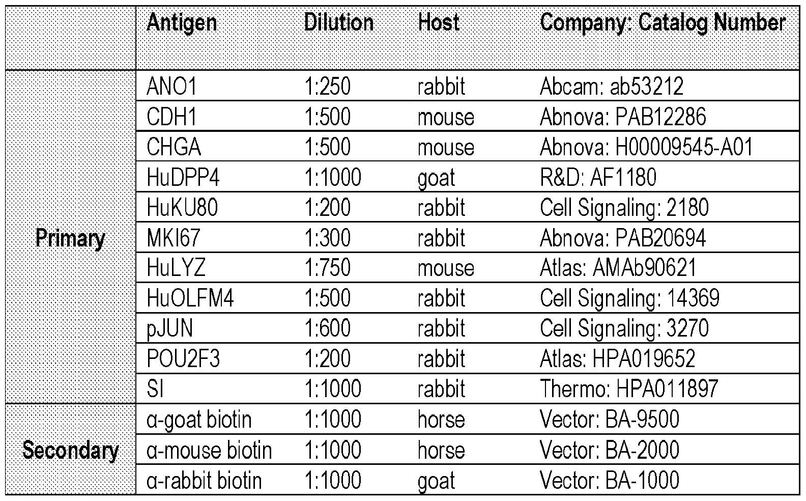
[00100] Electron Microscopy
[00101] Scanning Electron Microscopy. Segments of human jejunum, tHIO and tHIO+S grafts were fixed overnight in 3% gluteraldehyde in 0.175 M sodium cacodylate buffer with pH 7.4. Samples were then buffer rinsed and post fixed in 1% osmium tetroxide in 0.175 M cacodylate buffer for 1 hour at 4°C. After another buffer rinse, samples were put through a graded ethanol series (25, 50, 75, 95, 3x 100%) for dehydration. Specimens were then critical point dried in a Leica EM CPD300, stub-mounted and sputter-coated 10 nm thick with 60/40 gold palladium using a Leica EM ACE600. A Hitachi SU8010 transmission electron microscope was used to image samples.
[00102] Transmission Electron Microscopy. Segments of human jejunum, tHIO and tHIO+S grafts were fixed and dehydrated in the same fashion as those for scanning electron microscopy. Infiltration was performed with 2X propylene oxide followed by a graded infiltration with LX-112. Samples were polymerized into blocks overnight at 37°C and subsequently kept at 60°C for 3 days. Thicks were sectioned and stained with Toluidine
Blue to verify tissue orientation. Blocks were then sectioned at 10 nm, skipping 10 μηι between grids. Sections were stained with urinyl acetate and lead citrate before imaging with a Hitachi H7600 transmission electron microscope.
[00103] Morphometric Analysis, Cell Type Quantification and Statistical Analysis
[00104] Morphometric analysis was performed on hematoxylin and eosin stained tissue sections. Crypt depth, crypt width, villus height, villus width, and mucosal thickness were measured for a minimum of 20 well-oriented crypt- villus units per tissue sample and then averaged using Nikon NIS imaging software (Nikon). Microvilli were measured using ImageJ; at least 150 microvilli per sample across three different grids of at least 10 μιη apart were quantified. Profiles of proliferation by position within the crypt were determined using dual MKI67 and CDH1 immunostaining. A minimum of 10 intact crypts were analyzed per sample and averaged. For quantification of cell types along the crypt- villus axis the following were utilized: CHGA immunostaining for quantification of endoendocrine cells, Alcian Blue pH 2.5 staining for goblet cells, LYZ for Paneth cells. For semiquantitative analysis of the stem cell compartment OLFM4 immunostaining was used. In each case a minimum of 20 well-oriented crypt-villus units per tissue sample were quantified and averaged. All data are represented as mean + standard deviation. For analysis of proliferative profiles, Gaussian curves were fit to each data set and a comparison of fits was performed using Prism software (GraphPad). Two-way ANOVAs and post-hoc Tukey' s tests were completed using Prism software as appropriate. The statistical significance cutoff was p < 0.05 and is further denoted as follows: *= 0.01<p<0.05, **=0.001<p<0.01, ***=p<0.001, and ****=p<0.0001.
[00105] Ex vivo Epithelial Permeability
[00106] The epitheliums of freshly harvested tHIO, tHIO+S grafts and human surgical samples were carefully dissected through a technique similar to seromuscular stripping23, 43. tHIOs and fresh surgical samples were opened longitudinally. All further dissection was done in ice cold Kreb's buffer (NaCl, 117 mM; KCl, 4.7 mM; MgCk, 1.2mM; NaH2P04, 1.2 mM; NaHC03, 25 mM; CaCh, 2.5 mM and glucose, 11 mM). Tissue segments
were then pinned in a dish containing 0.5 cm thick cured Sylgard (Electron Microscopy Sciences). Unlike mouse tissue, the seromusculature layers (serosa, longitudinal, and circular smooth muscle) are relatively thick in both tHIOs and human jejunum. These layers are then micro-dissected as one unit from the epithelium using Dumont #5 and #7 forceps along with Vannas scissors taking care to only handle the edges of the tissue (Fine Science Tools, Inc.). The layers are gently separated and cut in small increments. After separation, gross tissue integrity was assessed using the stereoscope's bottom lighting for evenness and uniformity in appearance. The edges of the tissue, experiencing the majority of manipulation, were discarded along with any portions of the epithelium that appeared damaged. The dissection set up can be found in FIG 6A. Two examples of tissues dissected in this fashion can be found in FIG 6B after completion of the Ussing assay. Some remnant subepithelial mucosa remained after dissection. Epithelial segments were mounted between the hemi-chambers of an Ussing apparatus (Physiologic Instruments), and 0.008 cm2 of tissue was exposed to 3 mL of oxygenated Krebs buffer at 37 °C. The transepithelial potential difference was detected with two paired electrodes containing 4% agar in 3 M KC1. The electrodes were connected to a VVC MC8 voltage clamp amplifier (Physiologic Instruments, San Diego). Electrode potential difference and fluid resistance values were offset before tissue segments were mounted in the chamber. A 30 min monitoring period was allowed for the establishment of equilibrium in the chamber. Then, tissues were voltage-clamped at 0 mV while continuously measuring the short circuit current (Isc). For FITC-dextran permeability, 2.2 mg/ml FITC- dextran was added into apical side, and a sample was taken from the basolateral side every 30 minutes for 3 hours, replacing the same amount of fresh modified Kreb's buffer in the basolateral side. Finally, samples were read with a plate -reader (Synergy 2, BioTek).
[00107] Ex vivo Muscle Strength
[00108] tHIO and tHIO+S grafts were harvested and placed in ice-cold Hank's Balanced Salt Solution (HBSS). Human surgical samples were maintained overnight at 4°C in HBSS prior to assaying in order to minimize the effects of anesthetics. Muscle strips (4-6 mm in length and 1-2 mm in width) were dissected from the samples. Strips were then suspended vertically in an organ bath chamber (Radnoti) filled with freshly prepared Krebs- Ringer solution (Sigma; supplemented with 2.5 mM CaCh and 15 mM NaHCC ; pH 7.4),
warmed to 37°C and gassed with 95% O2 + 5% CO2. Segments were allowed to equilibrate for an hour at an initial tension of 0.5 g. The contractile response of the muscle was continuously recorded, using 4-chamber tissue-organ bath chambers with isometric force transducers (0-25g; AD Instruments) coupled to a multi-channel bridge amplifiers and data recorder (AD Instruments; PowerLab 4/35) linked to a computer equipped with LabChart Pro software (AD Instruments). A logarithmic dose response to Carbamyl- -methylcholine chloride (Bethanechol; Sigma- Aldrich) was obtained through the administration of exponential doses with concentrations of 1 nM to 10 mM at 2 minute intervals before the administration of 10 μΜ scopolamine (Tocris Bioscience).
[00109] RNA Isolation and Sequencing
[00110] RNA was extracted using an RNeasy Plus Micro Kit (Qiagen) according to manufacturer guidelines. Samples were then quantified and submitted to CCHMC's DNA Sequencing and Genotyping core for Next Generation Sequencing. All samples were assayed to have RNA integrity numbers greater than eight. After quality control, a cDNA library was created and sequenced using an IlluminaHiSeq2000 (Illumina) with 20 million paired-end reads per sample.
[00111] RNA sequencing bioinformatics
[00112] Pre-processing
[00113] Data pre-processing was performed in Galaxy public server
(https://usegalaxy.org). fastq datasets were encoded in Phred+33 to fastqsanger under the attributes for each dataset (FASTQ Groomer, Galaxy vl.0.4). Read quality was assessed using FastQC (Galaxy, vO.67). Adapter and low quality (< 20) sequences were removed using Trimmomatic (SLIDINGWINDOW) (Galaxy vO.32.3). Reads were mapped against the human genome GRCh37\Hgl9 using Tophat2/Bowtie2 with default settings (Galaxy v2.1.0). The number of reads uniquely mapping to each gene was counted using feature Counts (Galaxy vl.4.6.p5) using the Illumina iGenomes
(http://support.illumina.com/sequencing/sequencing_software/igenome.ilmn) annotation file. Alternatively, each sample was independently processed with Cufflinks44 in order to generate an initial transcriptome. Applicant used the Cuffmerge tool to merge the private
transcriptomes into a single reference, and at the same time annotated known genes and extended partial transcripts45. This common transcriptome was used in a second pass with Cufflinks to quantify each transcript and gene (known or novel) in each sample46. The reference annotation used was based on the UCSC knownGenes table47.
[00114] Analysis of RNA-seq data
[00115] RNA-seq analysis was performed using Strand NGS 2.9 software (Strand Life Sciences). Count tables generated in Galaxy were processed and normalized using the DESeq2 package48 within the Strand NGS 2.9 R console. Applicant performed a PCA using Strand NGS 2.9 software. From the PCA, the gene with the highest loadings for each principal component were extracted and plotted using the standalone hi_loadings function in pcaExplorer package . A functional enrichment analysis using the
limmaquickpca2go routine provided by the limma package was performed on the 10000 genes with the highest loadings for each principal component. ANOVA and moderated t-Test were used to perform statistical analysis amongst samples and between groups. Functional enrichment analysis was performed in the ToppGene suite (https://toppgene.cchmc.org). Plots were generated using ggplot2 v2.2.1 and GOplot vl.02 in Rstudio vl.0.14.
[00116] Statistics on FPKM Data
[00117] For statistics on normalized FPKMs, either a Student T test or Mann- Whitney test was used according to data distribution. The statistical significance cutoff was p < 0.05 and is further denoted as follows: *= 0.01<p<0.05, **=0.001<p<0.01, ***=p<0.001, and ****=p<0.0001.
[00118] Analysis of RNA-seq data with publicly available datasets
[00119] Publicly available datasets of fetal and adult full thickness intestinal tissues were downloaded from EBI-AE and NCBI-GEO databases using the Galaxy public server (Table 2). Datasets were selected upon RNA extraction and library preparation methods and sequencing platform (Illumina HiSeq). Only forward reads were used from paired-end files. Datasets were encoded to fastqsanger and read quality was assessed using FastQC (Galaxy, v0.67). Adapter and low quality (< 20) sequences were removed using Trimmomatic (SLIDINGWINDOW). Reads were mapped against the human genome
GRChb37\Hgl9 using HISAT2 with default settings (Galaxy v2.0.5.1)49. Count tables were generated in Galaxy using feature Counts using the Illumina iGenomes annotation file. The count matrix was processed in R 3.4.1 using the functions exported by the pcaExplorer package50 for a standalone usage. Briefly, the count matrix was transformed and normalized using a variance stabilizing transformation (VST) to the count data. Applicant visualized the sample PCA using the pcaplot function. The genes with the highest variance were selected to compute the PCA i.e. from 500 to 10000 genes. From the PCA, the gene with the highest loadings for each principal component were extracted and plotted using the hijoadings function. A functional enrichment analysis using the limmaquickpca2go routine provided by the limma package was performed on the 10000 genes with the highest loadings for each principal component. The detailed analysis and R scripts are provided in the following Github repository (www.github.com\maximemahe\Poling2017). The accession number for data generated for this paper is ArrayExpress: E-MTAB-6017 and includes the transplanted HIO, with or without lengthening device, the human adult and infant data shown in FIG 3 and FIG 9. The additional RNA-seq datasets including HIOs derived from HI and H9 ES cell lines, transplanted HIOs, Fetal, newborn and adult small intestine were downloaded from public databases (Table 3)
[00120] Table 2. Antibodies and dilutions for immunohistochemistry.
Antigen D ilution Host Company: Catalog Number
AN01 1 250 rabbit Abeam: ab53212
CDH1 1 500 mouse Abnova: PAB12286
CHGA 1 500 mouse Abnova: H00009545-A01 HuDPP4 1 1000 goat R&D: AF1 180 HuKU80 1 200 rabbit Cell Signaling: 2180
Primary MKI67 1 300 rabbit Abnova: PAB20694
HuLYZ 1 750 mouse Atlas: AMAb90621
HuOLFM4 1 500 rabbit Cell Signaling: 14369 pJUN 1 600 rabbit Cell Signaling: 3270
POU2F3 1 200 rabbit Atlas: HPA019652
SI 1 1000 rabbit Thermo: HPA01 1897 a-goat biotin 1 1000 horse Vector: BA-9500
Secondary a-mouse biotin 1 1000 horse Vector: BA-2000
a-rabbit biotin 1 1000 goat Vector: BA-1000
Table 3. RNAseq Datasets Downloaded from Public Databases. Source for all Tissue samples = fresh.
Sample Label Sample Description Tissue
Fetal SI 2 1 Fetal Intestine, Small; RNA.RS18147 Fetal tissue Fetal SI 3 1 Fetal Intestine, Small; RNA.RS18148 Fetal tissue
Fetal SI 4 Fetal Intestine, Small; RNA.RS18763 Fetal tissue Fetal SI 5 Fetal Intestine, Small; RNA.RS18765 Fetal tissue
P reetiaali o1i fi o 55 Petal Intoctino Qmal ΡΜΔ RQ1 R7R7
Infant SI 1 ¾ Control patient 1 Infant
Infant SI 2 1 Control patient 2 Infant
Infant SI 3 1 Control patient 3 Infant
Infant SI 4 1 Control patient 4 Infant
Infant SI 5 ¾ Control patient 5 Infant
Child SI 1 Small Bowel Cells; polyA-RNA-seq STL001 SB _r1 a Child
Adult SI 2 Small Bowel Cells; polyA-RNA-seq STL002SE _r1 a Adult
Adult SI 3 smallintestine 4a Adult
Adult SI 4 smallintestine 4b Adult
Adult SI 5 s smallintestine 4c Adult
Adult DUO 1 1 duodenum 4b Adult
Adult DUO 2 1 duodenum 4c Adult
Table 3, continued. RNAseq Datasets Downloaded from Public Databases
Sample Label Tissue Region Age Gender Repository Database # Accession # Fetal SI 2 Small Intestine GW 15.4 Female NCBI-GE 0 GSE 18927 GSM 1059507
Fetal SI 3 Small Intestine GW 13 Male NCBI-GE 0 GSE 18927 GSM 1059508
Fetal SI 4 Small Intestine GW 16.4 Male NCBI-GE 0 GSE 18927 GSM1059517
Fetal SI 5 Small Intestine GW 17.1 Female NCBI-GE 0 GSE 18927 GSM1059519
Fetal SI 6 Small Intestine GW 14 Female NCBI-GE 0 GSE 18927 GSM1059521
Infant SI 1 Ileum GW 33 Female NCBI-GE 0 GSE 64801 GSM 1580784
Infant SI 2 Ileum GW 26 Female NCBI-GE 0 GSE 64801 GSM 1580785
Infant SI 3 Ileum GW 26 Male NCBI-GE 0 GSE 64801 GSM 1580786
Infant SI 4 Ileum GW 33 Male NCBI-GE 0 GSE 64801 GSM 1580787
Infant SI 5 Ileum GW 39 Female NCBI-GE 0 GSE 64801 GSM 1580788
Child SI 1 Small Intestine 3 yrs Male NCBI-GE 0 GSE 16256 GSM1010940
Adult SI 2 Small Intestine 30 yrs Female NCBI-GE 0 GSE 16256 GSM1120313
Adult SI 3 Small Intestine N/A N/A E BI-AE E-MTAB-1733 ERS326961
Adult SI 4 Small Intestine N/A N/A E BI-AE E-MTAB-1733 ERS326971
Adult SI 5 Small Intestine N/A N/A E BI-AE E-MTAB-1733 ERS326958
Adult DUO 1 Duodenum N/A N/A E BI-AE E-MTAB-1733 ERS326992
Adult DUO 2 Duodenum N/A N/A E BI-AE E-MTAB-1733 ERS326976
[00121] Protein Phosphorylation Arrays
[00122] C-Series phosphorylation arrays were performed according to manufacturer recommendations (RayBiotech, Inc). Briefly, protein was extracted from flash frozen full thickness tHIO and tHIO+S tissues, quantified, and normalized between samples. Protein was incubated on antibody array nitrocellulose membranes, followed by horseradish peroxidase based amplification and detection. Chemiluminescent readings were taken using a ChemiDoc MP imaging system (Bio-Rad Laboratories, Inc.) and densitometry data extracted using ImageJ software. Readings were normalized to the positive loading controls and membrane background signal subtracted.
[00123] REFERENCES
[00124] 1. Wells, J.M. & Spence, J.R. How to make an intestine.
Development 141, 752-760 (2014).
[00125] 2. Spence, J.R. et al. Directed differentiation of human pluripotent stem cells into intestinal tissue in vitro. Nature 470, 105-109 (2011).
[00126] 3. McCracken, K.W., Howell, J.C., Wells, J.M. & Spence, J.R.
Generating human intestinal tissue from pluripotent stem cells in vitro. Nature protocols 6, 1920-1928 (2011).
[00127] 4. Watson, C.L. et al. An in vivo model of human small intestine using pluripotent stem cells. Nature medicine 20, 1310-1314 (2014).
[00128] 5. Workman, M.J. et al. Engineered human pluripotent-stem-cell- derived intestinal tissues with a functional enteric nervous system. Nature medicine 23, 49-59 (2017).
[00129] 6. Finkbeiner, S.R. et al. Transcriptome-wide Analysis Reveals Hallmarks of Human Intestine Development and Maturation In Vitro and In Vivo. Stem cell reports (2015).
[00130] 7. Dedhia, P.H., Bertaux-Skeirik, N., Zavros, Y. & Spence, J.R. Organoid Models of Human Gastrointestinal Development and Disease. Gastroenterology 150, 1098-1112 (2016).
[00131] 8. Yu, H. et al. The Contributions of Human Mini-Intestines to the
Study of Intestinal Physiology and Pathophysiology. Annu Rev Physiol 79, 291-312 (2017).
[00132] 9. Nelson, CM. On Buckling Morphogenesis. J Biomech Eng 138, 021005 (2016).
[00133] 10. Kurpios, N.A. et al. The direction of gut looping is established by changes in the extracellular matrix and in cell: cell adhesion. Proceedings of the National Academy of Sciences of the United States of America 105, 8499-8506 (2008).
[00134] 11. Shyer, A.E. et al. Villification: how the gut gets its villi.
Science 342, 212-218 (2013).
[00135] 12. Shyer, A.E., Huycke, T.R., Lee, C, Mahadevan, L. & Tabin,
C.J. Bending gradients: how the intestinal stem cell gets its home. Cell 161, 569-580 (2015).
[00136] 13. Savin, T. et al. On the growth and form of the gut. Nature 476,
57-62 (2011).
[00137] 14. Stark, R., Panduranga, M., Carman, G. & Dunn, J.C.
Development of an endoluminal intestinal lengthening capsule. Journal of pediatric surgery 47, 136-141 (2012).
[00138] 15. Rouch, J.D. et al. Scalability of an endoluminal spring for distraction enterogenesis. Journal of pediatric surgery 51, 1988-1992 (2016).
[00139] 16. Demehri, F.R. et al. Development of an endoluminal intestinal attachment for a clinically applicable distraction enterogenesis device. Journal of pediatric surgery 51, 101-106 (2016).
[00140] 17. Demehri, F.R., Freeman, J.J., Fukatsu, Y., Luntz, J. &
Teitelbaum, D.H. Development of an endoluminal intestinal lengthening device using a geometric intestinal attachment approach. Surgery 158, 802-811 (2015).
[00141] 18. Luntz, J., Brei, D., Teitelbaum, D. & Spencer, A. Mechanical
Extension Implants for Short-Bowel Syndrome. Proc SPIE Int Soc Opt Eng 6173, 617309 (2006).
[00142] 19. Allard, J. et al. Immunohistochemical toolkit for tracking and quantifying xenotransplanted human stem cells. Regenerative medicine 9, 437-452 (2014).
[00143] 20. Shekherdimian, S., Panduranga, M.K., Carman, G.P. & Dunn, J.C. The feasibility of using an endoluminal device for intestinal lengthening. Journal of pediatric surgery 45, 1575-1580 (2010).
[00144] 21. Marsh, M.N. & Swift, J.A. A study of the small intestinal mucosa using the scanning electron microscope. Gut 10, 940-949 (1969).
[00145] 22. Hooton, D., Lentle, R., Monro, J., Wickham, M. & Simpson, R. The Secretion and Action of Brush Border Enzymes in the Mammalian Small Intestine. Rev Physiol Biochem Pharmacol 168, 59-118 (2015).
[00146] 23. Clarke, L.L. A guide to Ussing chamber studies of mouse intestine. American journal of physiology. Gastrointestinal and liver physiology 296, G1151- 1166 (2009).
[00147] 24. Gomez-Pinilla, P.J. et al. Anol is a selective marker of interstitial cells of Cajal in the human and mouse gastrointestinal tract. Am J Physiol Gastrointest Liver Physiol 296, G1370-1381 (2009).
[00148] 25. Mammoto, A., Mammoto, T. & Ingber, D.E. Mechanosensitive mechanisms in transcriptional regulation. Journal of cell science 125, 3061-3073 (2012).
[00149] 26. Burn, S.F. & Hill, R.E. Left-right asymmetry in gut development: what happens next? Bioessays 31, 1026-1037 (2009).
[00150] 27. Sherwood, R.I., Chen, T.Y. & Melton, D.A. Transcriptional dynamics of endodermal organ formation. Developmental dynamics : an official publication of the American Association of Anatomists 238, 29-42 (2009).
[00151] 28. Zorn, A.M. & Wells, J.M. Vertebrate endoderm development and organ formation. Annu Rev Cell Dev Biol 25, 221-251 (2009).
[00152] 29. Spence, J.R., Lauf, R. & Shroyer, N.F. Vertebrate intestinal endoderm development. Developmental dynamics: an official publication of the American Association of Anatomists 240, 501-520 (2011).
[00153] 30. Shahbazi, M.N. et al. Self-organization of the human embryo in the absence of maternal tissues. Nature cell biology 18, 700-708 (2016).
[00154] 31. Soffers, J.H.M., Hikspoors, J.P.J.M., Mekonen, H.K., Koehler, S.E. & Lamers, W.H. The growth pattern of the human intestine and its mesentery. Bmc Dev Biol 15 (2015).
[00155] 32. Tran, K., Brun, R. & Kuo, B. Evaluation of regional and whole gut motility using the wireless motility capsule: relevance in clinical practice. Therap Adv Gastroenterol 5, 249-260 (2012).
[00156] 33. Engmann, J. & Burbidge, A.S. Fluid mechanics of eating, swallowing and digestion - overview and perspectives. Food & function 4, 443-447 (2013).
[00157] 34. Wieck, M.M. et al. Prolonged Absence of Mechanoluminal
Stimulation in Human Intestine Alters the Transcriptome and Intestinal Stem Cell Niche. Cell Mol Gastroenterol Hepatol 3, 367-388 e361 (2017).
[00158] 35. Terry, B.S., Lyle, A.B., Schoen, J.A. & Rentschler, M.E.
Preliminary mechanical characterization of the small bowel for in vivo robotic mobility. J Biomech Eng 133, 091010 (2011).
[00159] 36. Gregersen, H., Kassab, G.S. & Fung, Y.C. The zero-stress state of the gastrointestinal tract: biomechanical and functional implications. Digestive diseases and sciences 45, 2271-2281 (2000).
[00160] 37. Low, L.A. & Tagle, D.A. Organs-on-chips: Progress, challenges, and future directions. Exp Biol Med (Maywood), 1535370217700523 (2017).
[00161] 38. Christoffersson, J., van Noort, D.V. & Mandenius, C.F.
Developing organ-on-a-chip concepts using bio-mechatronic design methodology.
Biofabrication (2017).
[00162] 39. Sato, T. & Clevers, H. SnapShot: Growing Organoids from Stem Cells. Cell 161 (2015).
[00163] 40. McCracken, K.W. et al. Modelling human development and disease in pluripotent stem-cell-derived gastric organoids. Nature 516, 400-+ (2014).
[00164] 41. Grapin-Botton, A. Three-dimensional pancreas organogenesis models. Diabetes Obes Metab 18, 33-40 (2016).
[00165] 42. Mahe, M.M., Brown, N.E., Poling, H.M. & Helmrath, M.A. In vivo Model of Small Intestine. Methods Mol Biol 1597, 229-245 (2017).
[00166] 43. Giles, D.A. et al. Thermoneutral housing exacerbates nonalcoholic fatty liver disease in mice and allows for sex-independent disease modeling. Nature medicine 23, 829-838 (2017).
[00167] 44. Trapnell, C. et al. Transcript assembly and quantification by RNA-Seq reveals unannotated transcripts and isoform switching during cell differentiation. Nat Biotechnol 28, 511-515 (2010).
[00168] 45. Roberts, A., Pimentel, H., Trapnell, C. & Pachter, L.
Identification of novel transcripts in annotated genomes using RNA-Seq. Bioinformatics 27, 2325-2329 (2011).
[00169] 46. Roberts, A., Trapnell, C, Donaghey, J., Rinn, J.L. & Pachter, L. Improving RNA-Seq expression estimates by correcting for fragment bias. Genome Biol 12, R22 (2011).
[00170] 47. Hsu, F. et al. The UCSC Known Genes. Bioinformatics 22, 1036-1046 (2006).
[00171] 48. Love, M.I., Huber, W. & Anders, S. Moderated estimation of fold change and dispersion for RNA-seq data with DESeq2. Genome Biol 15, 550 (2014).
[00172] 49. Kim, D., Langmead, B. & Salzberg, S.L. HISAT: a fast spliced aligner with low memory requirements. Nat Methods 12, 357-360 (2015).
[00173] 50. Marini, F. pcaExplorer: Interactive Visualization of RNA-seq
Data Using a Principal Components Approach. R package version 2.3.0. (2017).
[00174] All percentages and ratios are calculated by weight unless otherwise indicated.
[00175] All percentages and ratios are calculated based on the total composition unless otherwise indicated.
[00176] It should be understood that every maximum numerical limitation given throughout this specification includes every lower numerical limitation, as if such lower numerical limitations were expressly written herein. Every minimum numerical limitation given throughout this specification will include every higher numerical limitation, as if such higher numerical limitations were expressly written herein. Every numerical range given throughout this specification will include every narrower numerical range that falls within such broader numerical range, as if such narrower numerical ranges were all expressly written herein.
[00177] The dimensions and values disclosed herein are not to be understood as being strictly limited to the exact numerical values recited. Instead, unless otherwise specified, each such dimension is intended to mean both the recited value and a functionally equivalent range surrounding that value. For example, a dimension disclosed as "20 mm" is intended to mean "about 20 mm."
[00178] Every document cited herein, including any cross referenced or related patent or application, is hereby incorporated herein by reference in its entirety unless expressly excluded or otherwise limited. The citation of any document is not an admission that it is prior art with respect to any invention disclosed or claimed herein or that it alone, or in any combination with any other reference or references, teaches, suggests or discloses any such invention. Further, to the extent that any meaning or definition of a term in this document conflicts with any meaning or definition of the same term in a document
incorporated by reference, the meaning or definition assigned to that term in this document shall govern.
[00179] While particular embodiments of the present invention have been illustrated and described, it would be obvious to those skilled in the art that various other changes and modifications can be made without departing from the spirit and scope of the invention. It is therefore intended to cover in the appended claims all such changes and modifications that are within the scope of this invention.
Claims
1. A method of enhancing development of a three-dimensional in vitro model of an intestinal tissue (HIO) derived from a precursor cell, for example, an embryonic stem cell (HI line), an induced pluripotent stem cell (iPSC), or the like, wherein said in vitro model comprises a lumen, comprising the step of inserting a lengthening device into said lumen, wherein said lengthening device applies strain to said lumen, preferably wherein said lengthening device comprises a spring, more preferably a nickel titanium (nitinol, NiTi) spring.
2. The method of claim 1, wherein said lengthening device comprises a cavity sufficient to allow flow of lumen secretions.
3. The method of claim 1 or 2, wherein said method comprises a first and a second engraftment period, wherein said first engraftment period comprises the step of transplanting said HIO into an immune compromised animal model prior to implantation of said lengthening device, preferably the mesentery of said animal model, wherein said first engraftment period is carried out for a period of time sufficient for said develop crypt regions, villi, and smooth muscle layers.
4. The method of claim 3, wherein said second engraftment period occurs after said first engraftment period, wherein said second engraftment period comprises the steps of inserting said lengthening device after said first engraftment period for a period of time sufficient to allow increased villus height and crypt depth as compared to a control HIO not subjected to said lengthening device.
5. The method of claim 3 or 4 wherein said first engraftment period is of a period of time of from about 6 to about 14 weeks, or from about 10 to about 12 weeks, or for a period of time sufficient to allow said HIO to obtain a blood supply and grow to a size sufficient for implantation of said lengthening device, for example, from about 5 mm to about 2 cm in length, or from about 10 mm to about 1.5 cm in length.
6. The method of any one of claims 3 through 5 wherein said first and second
engraftment period is of a period of time sufficient to allow said HIO to have increased villus height and crypt depth and crypt fission, and increased longitudinal and circular muscle thickness, as compared to a control HIO which does not contain a lengthening device.
7. The method of any one of claims 3 through 6, wherein said lengthening device has a relaxed length of about 10 to about 15 mm.
8. The method of any one of claims 3 through 7, wherein said lengthening device has a compressed length of about 4 to about 8 mm.
9. The method of any one of claims 3 through 8, wherein said lengthening device has a diameter of about 1 to about 3 mm, or about 2 mm.
10. The method of any one of claims 3 through 9, wherein said lengthening device has a spring constant of from about 0.5 N/m to about 2 N/m, or from about 0.7 to about 1.7, or from about 0.9 to about 1.2 or about 1 N/m. The method of claim 1, wherein said lengthening device is contained in a degradable coating, preferably wherein said degradable coating maintains the compressed length until deployment, preferably a gelatin capsule, preferably further comprising a polymer in an amount and of a type
sufficient to delay deployment, more preferably an enteric coating, more preferably an enteric coating of cellulose acetate phthalate (C-A-P).
11. The method of any one of claims 3 through 10, wherein said precursor cell is a
pluripotent stem cell or an induced pluripotent stem cell, preferably an embryonic stem cell.
12. The method of any one of claims 3 through 11, wherein said HIO is derived from a pluripotent stem cell, wherein said pluripotent stem cell is derived from a fetal tissue stem cell.
13. A three-dimensional human intestinal organoid (HIO) composition derived from a pre-cursor cell in vitro, wherein said HIO comprises a lengthening device, preferably wherein said lengthening device is a spring.
14. A three-dimensional human intestinal organoid (HIO) composition derived from a pre-cursor cell in vitro, wherein said HIO is lacking one or more features native to a fully developed organ, for example, wherein said HIO is devoid of a vascular system.
15. The three-dimensional human intestinal organoid (HIO) of claim 14, derived from the method of any one of claims 1 through 12.
Priority Applications (1)
| Application Number | Priority Date | Filing Date | Title |
|---|---|---|---|
| US16/603,611 US20210115366A1 (en) | 2017-04-24 | 2018-04-24 | Methods of making improved human intestinal organoid compositions via application of strain and human intestinal organoid compositions thereof |
Applications Claiming Priority (2)
| Application Number | Priority Date | Filing Date | Title |
|---|---|---|---|
| US201762488984P | 2017-04-24 | 2017-04-24 | |
| US62/488,984 | 2017-04-24 |
Publications (2)
| Publication Number | Publication Date |
|---|---|
| WO2018200481A1 true WO2018200481A1 (en) | 2018-11-01 |
| WO2018200481A8 WO2018200481A8 (en) | 2018-12-06 |
Family
ID=63918655
Family Applications (1)
| Application Number | Title | Priority Date | Filing Date |
|---|---|---|---|
| PCT/US2018/029083 Ceased WO2018200481A1 (en) | 2017-04-24 | 2018-04-24 | Methods of making improved human intestinal organoid compositions via application of strain and human intestinal organoid compositions thereof |
Country Status (2)
| Country | Link |
|---|---|
| US (1) | US20210115366A1 (en) |
| WO (1) | WO2018200481A1 (en) |
Cited By (14)
| Publication number | Priority date | Publication date | Assignee | Title |
|---|---|---|---|---|
| US11584916B2 (en) | 2014-10-17 | 2023-02-21 | Children's Hospital Medical Center | Method of making in vivo human small intestine organoids from pluripotent stem cells |
| EP3976070A4 (en) * | 2019-05-31 | 2023-06-14 | Children's Hospital Medical Center | SHAPED ORGANOID COMPOSITIONS AND PROCESSES FOR THEIR PRODUCTION |
| US11767515B2 (en) | 2016-12-05 | 2023-09-26 | Children's Hospital Medical Center | Colonic organoids and methods of making and using same |
| WO2024263961A2 (en) | 2023-06-23 | 2024-12-26 | Children's Hospital Medical Center | Methods of matrix-free suspension culture |
| US12241090B2 (en) | 2014-05-28 | 2025-03-04 | Children's Hospital Medical Center | Methods and systems for converting precursor cells into gastric tissues through directed differentiation |
| US12258584B2 (en) | 2010-05-06 | 2025-03-25 | Children's Hospital Medical Center | Methods and systems for converting precursor cells into intestinal tissues through directed differentiation |
| WO2025072803A1 (en) | 2023-09-29 | 2025-04-03 | Children's Hospital Medical Center | Ntrk2 signaling-mediated alveolar capillary injury and repair |
| US12281334B2 (en) | 2017-04-14 | 2025-04-22 | Children's Hospital Medical Center | Multi donor stem cell compositions and methods of making same |
| US12297457B2 (en) | 2017-10-10 | 2025-05-13 | Children's Hospital Medical Center | Esophageal tissue and/or organoid compositions and methods of making same |
| US12379372B2 (en) | 2017-12-21 | 2025-08-05 | Children's Hospital Medical Center | Digitalized human organoids and methods of using same |
| US12414967B2 (en) | 2016-11-04 | 2025-09-16 | Children's Hospital Medical Center | Compositions and methods of treating liver disease |
| US12421500B2 (en) | 2018-07-26 | 2025-09-23 | Children's Hospital Medical Center | Hepato-biliary-pancreatic tissues and methods of making same |
| US12428622B2 (en) | 2018-09-12 | 2025-09-30 | Children's Hospital Medical Center | Organoid compositions for the production of hematopoietic stem cells and derivatives thereof |
| US12497597B2 (en) | 2019-05-31 | 2025-12-16 | Children's Hospital Medical Center | Methods of generating and expanding hematopoietic stem cells |
Family Cites Families (2)
| Publication number | Priority date | Publication date | Assignee | Title |
|---|---|---|---|---|
| US7722560B2 (en) * | 2007-02-09 | 2010-05-25 | The Regents Of The University Of Michigan | Mechanical extension implants for short bowel syndrome |
| WO2017083696A1 (en) * | 2015-11-11 | 2017-05-18 | The Regents Of The University Of California | Degradable intestinal anchor |
-
2018
- 2018-04-24 WO PCT/US2018/029083 patent/WO2018200481A1/en not_active Ceased
- 2018-04-24 US US16/603,611 patent/US20210115366A1/en not_active Abandoned
Non-Patent Citations (2)
| Title |
|---|
| SINAGOGA ET AL.: "Generating human intestinal tissues from pluripotent stem cells to study development and disease", EMBO JOURNAL, vol. 34, no. 9, 5 May 2015 (2015-05-05), pages 1149 - 1164, XP055528462 * |
| STARK ET AL.: "Development of an endoluminal intestinal lengthening capsule", JOURNAL OF PEDIATRIC SURGERY, vol. 47, no. 1, January 2012 (2012-01-01), pages 136 - 141, XP028441140 * |
Cited By (15)
| Publication number | Priority date | Publication date | Assignee | Title |
|---|---|---|---|---|
| US12258584B2 (en) | 2010-05-06 | 2025-03-25 | Children's Hospital Medical Center | Methods and systems for converting precursor cells into intestinal tissues through directed differentiation |
| US12241090B2 (en) | 2014-05-28 | 2025-03-04 | Children's Hospital Medical Center | Methods and systems for converting precursor cells into gastric tissues through directed differentiation |
| US11584916B2 (en) | 2014-10-17 | 2023-02-21 | Children's Hospital Medical Center | Method of making in vivo human small intestine organoids from pluripotent stem cells |
| US12414967B2 (en) | 2016-11-04 | 2025-09-16 | Children's Hospital Medical Center | Compositions and methods of treating liver disease |
| US11767515B2 (en) | 2016-12-05 | 2023-09-26 | Children's Hospital Medical Center | Colonic organoids and methods of making and using same |
| US12281334B2 (en) | 2017-04-14 | 2025-04-22 | Children's Hospital Medical Center | Multi donor stem cell compositions and methods of making same |
| US12297457B2 (en) | 2017-10-10 | 2025-05-13 | Children's Hospital Medical Center | Esophageal tissue and/or organoid compositions and methods of making same |
| US12379372B2 (en) | 2017-12-21 | 2025-08-05 | Children's Hospital Medical Center | Digitalized human organoids and methods of using same |
| US12421500B2 (en) | 2018-07-26 | 2025-09-23 | Children's Hospital Medical Center | Hepato-biliary-pancreatic tissues and methods of making same |
| US12428622B2 (en) | 2018-09-12 | 2025-09-30 | Children's Hospital Medical Center | Organoid compositions for the production of hematopoietic stem cells and derivatives thereof |
| US12534709B2 (en) | 2019-05-31 | 2026-01-27 | Children's Hospital Medical Center | Shaped organoid compositions and methods of making same |
| US12497597B2 (en) | 2019-05-31 | 2025-12-16 | Children's Hospital Medical Center | Methods of generating and expanding hematopoietic stem cells |
| EP3976070A4 (en) * | 2019-05-31 | 2023-06-14 | Children's Hospital Medical Center | SHAPED ORGANOID COMPOSITIONS AND PROCESSES FOR THEIR PRODUCTION |
| WO2024263961A2 (en) | 2023-06-23 | 2024-12-26 | Children's Hospital Medical Center | Methods of matrix-free suspension culture |
| WO2025072803A1 (en) | 2023-09-29 | 2025-04-03 | Children's Hospital Medical Center | Ntrk2 signaling-mediated alveolar capillary injury and repair |
Also Published As
| Publication number | Publication date |
|---|---|
| WO2018200481A8 (en) | 2018-12-06 |
| US20210115366A1 (en) | 2021-04-22 |
Similar Documents
| Publication | Publication Date | Title |
|---|---|---|
| US20210115366A1 (en) | Methods of making improved human intestinal organoid compositions via application of strain and human intestinal organoid compositions thereof | |
| Poling et al. | Mechanically induced development and maturation of human intestinal organoids in vivo | |
| US12109242B2 (en) | Use of alveolar or airway organoids for the treatment of lung diseases and disorders | |
| AU2018285579B2 (en) | Blood vessel organoid, methods of producing and using said organoids | |
| Gamez et al. | Supplementing defect in club cell secretory protein attenuates airway inflammation in COPD | |
| Gonzalez et al. | Cell lineage identification and stem cell culture in a porcine model for the study of intestinal epithelial regeneration | |
| Houston et al. | The lymph nodes draining the small intestine and colon are anatomically separate and immunologically distinct | |
| Williams et al. | Intestinal preparation techniques for histological analysis in the mouse | |
| JP2022048143A (en) | Production of differentiated enteroendocrine cells and insulin producing cells | |
| US12203099B2 (en) | Methods of expanding cholangiocytes | |
| Depasquale | Actin microridges | |
| Rivière et al. | Results of the Eighth Scientific Workshop of ECCO: pathophysiology and risk factors of postoperative Crohn’s disease recurrence after an ileocolonic resection | |
| Poling et al. | Human pluripotent stem cell-derived organoids repair damaged bowel in vivo | |
| Straub et al. | Glyco-conjugated bile acids drive the initial metaplastic gland formation from multi-layered glands through crypt-fission in a murine model | |
| Tang et al. | An optimized method to visualize the goblet cell-associated antigen passages and identify goblet cells in the intestine, conjunctiva, and airway | |
| Zhao et al. | Cloned airway basal progenitor cells to repair fibrotic lung through re-epithelialization | |
| Ishibashi et al. | Efficient embryo transfer in the common marmoset monkey (Callithrix jacchus) with a reduced transfer volume: a non-surgical approach with cryopreserved late-stage embryos | |
| Kumosa et al. | Profound alterations in brain tissue linked to hypoxic episode after device implantation | |
| KR102050223B1 (en) | Method for preparing intestinal organoid from embryonic stem cells | |
| KR20190001297A (en) | Method for preparing intestinal organoid from embryonic stem cells | |
| Richards et al. | A syngeneic murine model of endometriosis using naturally cycling mice. | |
| Willacy et al. | Functional assessment of autologous tissue expansion grafts for vaginal reconstruction in a rabbit model | |
| Crisanti et al. | Novel methods for delivery of cell-based therapies | |
| Poling et al. | Engineering Large-Scale and Innervated Functional Human Gut for Transplantation | |
| US11718831B2 (en) | Reagent for differentiating somatic cells into alveolar epithelial cells, and use of said reagent |
Legal Events
| Date | Code | Title | Description |
|---|---|---|---|
| 121 | Ep: the epo has been informed by wipo that ep was designated in this application |
Ref document number: 18791025 Country of ref document: EP Kind code of ref document: A1 |
|
| NENP | Non-entry into the national phase |
Ref country code: DE |
|
| 122 | Ep: pct application non-entry in european phase |
Ref document number: 18791025 Country of ref document: EP Kind code of ref document: A1 |