WO2018106628A1 - Colonic organoids and methods of making and using same - Google Patents
Colonic organoids and methods of making and using same Download PDFInfo
- Publication number
- WO2018106628A1 WO2018106628A1 PCT/US2017/064600 US2017064600W WO2018106628A1 WO 2018106628 A1 WO2018106628 A1 WO 2018106628A1 US 2017064600 W US2017064600 W US 2017064600W WO 2018106628 A1 WO2018106628 A1 WO 2018106628A1
- Authority
- WO
- WIPO (PCT)
- Prior art keywords
- cells
- cell
- bmp
- satb2
- organoids
- Prior art date
- Legal status (The legal status is an assumption and is not a legal conclusion. Google has not performed a legal analysis and makes no representation as to the accuracy of the status listed.)
- Ceased
Links
Classifications
-
- C—CHEMISTRY; METALLURGY
- C12—BIOCHEMISTRY; BEER; SPIRITS; WINE; VINEGAR; MICROBIOLOGY; ENZYMOLOGY; MUTATION OR GENETIC ENGINEERING
- C12N—MICROORGANISMS OR ENZYMES; COMPOSITIONS THEREOF; PROPAGATING, PRESERVING, OR MAINTAINING MICROORGANISMS; MUTATION OR GENETIC ENGINEERING; CULTURE MEDIA
- C12N5/00—Undifferentiated human, animal or plant cells, e.g. cell lines; Tissues; Cultivation or maintenance thereof; Culture media therefor
- C12N5/06—Animal cells or tissues; Human cells or tissues
- C12N5/0602—Vertebrate cells
- C12N5/0679—Cells of the gastro-intestinal tract
-
- C—CHEMISTRY; METALLURGY
- C12—BIOCHEMISTRY; BEER; SPIRITS; WINE; VINEGAR; MICROBIOLOGY; ENZYMOLOGY; MUTATION OR GENETIC ENGINEERING
- C12N—MICROORGANISMS OR ENZYMES; COMPOSITIONS THEREOF; PROPAGATING, PRESERVING, OR MAINTAINING MICROORGANISMS; MUTATION OR GENETIC ENGINEERING; CULTURE MEDIA
- C12N5/00—Undifferentiated human, animal or plant cells, e.g. cell lines; Tissues; Cultivation or maintenance thereof; Culture media therefor
- C12N5/06—Animal cells or tissues; Human cells or tissues
- C12N5/0697—Artificial constructs associating cells of different lineages, e.g. tissue equivalents
-
- G—PHYSICS
- G01—MEASURING; TESTING
- G01N—INVESTIGATING OR ANALYSING MATERIALS BY DETERMINING THEIR CHEMICAL OR PHYSICAL PROPERTIES
- G01N33/00—Investigating or analysing materials by specific methods not covered by groups G01N1/00 - G01N31/00
- G01N33/48—Biological material, e.g. blood, urine; Haemocytometers
- G01N33/50—Chemical analysis of biological material, e.g. blood, urine; Testing involving biospecific ligand binding methods; Immunological testing
- G01N33/5005—Chemical analysis of biological material, e.g. blood, urine; Testing involving biospecific ligand binding methods; Immunological testing involving human or animal cells
- G01N33/5008—Chemical analysis of biological material, e.g. blood, urine; Testing involving biospecific ligand binding methods; Immunological testing involving human or animal cells for testing or evaluating the effect of chemical or biological compounds, e.g. drugs, cosmetics
- G01N33/5082—Supracellular entities, e.g. tissue, organisms
- G01N33/5088—Supracellular entities, e.g. tissue, organisms of vertebrates
-
- C—CHEMISTRY; METALLURGY
- C07—ORGANIC CHEMISTRY
- C07K—PEPTIDES
- C07K14/00—Peptides having more than 20 amino acids; Gastrins; Somatostatins; Melanotropins; Derivatives thereof
- C07K14/435—Peptides having more than 20 amino acids; Gastrins; Somatostatins; Melanotropins; Derivatives thereof from animals; from humans
- C07K14/475—Growth factors; Growth regulators
- C07K14/485—Epidermal growth factor [EGF], i.e. urogastrone
-
- C—CHEMISTRY; METALLURGY
- C07—ORGANIC CHEMISTRY
- C07K—PEPTIDES
- C07K14/00—Peptides having more than 20 amino acids; Gastrins; Somatostatins; Melanotropins; Derivatives thereof
- C07K14/435—Peptides having more than 20 amino acids; Gastrins; Somatostatins; Melanotropins; Derivatives thereof from animals; from humans
- C07K14/475—Growth factors; Growth regulators
- C07K14/50—Fibroblast growth factor [FGF]
-
- C—CHEMISTRY; METALLURGY
- C07—ORGANIC CHEMISTRY
- C07K—PEPTIDES
- C07K14/00—Peptides having more than 20 amino acids; Gastrins; Somatostatins; Melanotropins; Derivatives thereof
- C07K14/435—Peptides having more than 20 amino acids; Gastrins; Somatostatins; Melanotropins; Derivatives thereof from animals; from humans
- C07K14/475—Growth factors; Growth regulators
- C07K14/51—Bone morphogenetic factor; Osteogenins; Osteogenic factor; Bone-inducing factor
-
- C—CHEMISTRY; METALLURGY
- C12—BIOCHEMISTRY; BEER; SPIRITS; WINE; VINEGAR; MICROBIOLOGY; ENZYMOLOGY; MUTATION OR GENETIC ENGINEERING
- C12N—MICROORGANISMS OR ENZYMES; COMPOSITIONS THEREOF; PROPAGATING, PRESERVING, OR MAINTAINING MICROORGANISMS; MUTATION OR GENETIC ENGINEERING; CULTURE MEDIA
- C12N2501/00—Active agents used in cell culture processes, e.g. differentation
- C12N2501/10—Growth factors
- C12N2501/11—Epidermal growth factor [EGF]
-
- C—CHEMISTRY; METALLURGY
- C12—BIOCHEMISTRY; BEER; SPIRITS; WINE; VINEGAR; MICROBIOLOGY; ENZYMOLOGY; MUTATION OR GENETIC ENGINEERING
- C12N—MICROORGANISMS OR ENZYMES; COMPOSITIONS THEREOF; PROPAGATING, PRESERVING, OR MAINTAINING MICROORGANISMS; MUTATION OR GENETIC ENGINEERING; CULTURE MEDIA
- C12N2501/00—Active agents used in cell culture processes, e.g. differentation
- C12N2501/10—Growth factors
- C12N2501/113—Acidic fibroblast growth factor (aFGF, FGF-1)
-
- C—CHEMISTRY; METALLURGY
- C12—BIOCHEMISTRY; BEER; SPIRITS; WINE; VINEGAR; MICROBIOLOGY; ENZYMOLOGY; MUTATION OR GENETIC ENGINEERING
- C12N—MICROORGANISMS OR ENZYMES; COMPOSITIONS THEREOF; PROPAGATING, PRESERVING, OR MAINTAINING MICROORGANISMS; MUTATION OR GENETIC ENGINEERING; CULTURE MEDIA
- C12N2501/00—Active agents used in cell culture processes, e.g. differentation
- C12N2501/10—Growth factors
- C12N2501/115—Basic fibroblast growth factor (bFGF, FGF-2)
-
- C—CHEMISTRY; METALLURGY
- C12—BIOCHEMISTRY; BEER; SPIRITS; WINE; VINEGAR; MICROBIOLOGY; ENZYMOLOGY; MUTATION OR GENETIC ENGINEERING
- C12N—MICROORGANISMS OR ENZYMES; COMPOSITIONS THEREOF; PROPAGATING, PRESERVING, OR MAINTAINING MICROORGANISMS; MUTATION OR GENETIC ENGINEERING; CULTURE MEDIA
- C12N2501/00—Active agents used in cell culture processes, e.g. differentation
- C12N2501/10—Growth factors
- C12N2501/117—Keratinocyte growth factors (KGF-1, i.e. FGF-7; KGF-2, i.e. FGF-12)
-
- C—CHEMISTRY; METALLURGY
- C12—BIOCHEMISTRY; BEER; SPIRITS; WINE; VINEGAR; MICROBIOLOGY; ENZYMOLOGY; MUTATION OR GENETIC ENGINEERING
- C12N—MICROORGANISMS OR ENZYMES; COMPOSITIONS THEREOF; PROPAGATING, PRESERVING, OR MAINTAINING MICROORGANISMS; MUTATION OR GENETIC ENGINEERING; CULTURE MEDIA
- C12N2501/00—Active agents used in cell culture processes, e.g. differentation
- C12N2501/10—Growth factors
- C12N2501/119—Other fibroblast growth factors, e.g. FGF-4, FGF-8, FGF-10
-
- C—CHEMISTRY; METALLURGY
- C12—BIOCHEMISTRY; BEER; SPIRITS; WINE; VINEGAR; MICROBIOLOGY; ENZYMOLOGY; MUTATION OR GENETIC ENGINEERING
- C12N—MICROORGANISMS OR ENZYMES; COMPOSITIONS THEREOF; PROPAGATING, PRESERVING, OR MAINTAINING MICROORGANISMS; MUTATION OR GENETIC ENGINEERING; CULTURE MEDIA
- C12N2501/00—Active agents used in cell culture processes, e.g. differentation
- C12N2501/10—Growth factors
- C12N2501/155—Bone morphogenic proteins [BMP]; Osteogenins; Osteogenic factor; Bone inducing factor
-
- C—CHEMISTRY; METALLURGY
- C12—BIOCHEMISTRY; BEER; SPIRITS; WINE; VINEGAR; MICROBIOLOGY; ENZYMOLOGY; MUTATION OR GENETIC ENGINEERING
- C12N—MICROORGANISMS OR ENZYMES; COMPOSITIONS THEREOF; PROPAGATING, PRESERVING, OR MAINTAINING MICROORGANISMS; MUTATION OR GENETIC ENGINEERING; CULTURE MEDIA
- C12N2501/00—Active agents used in cell culture processes, e.g. differentation
- C12N2501/10—Growth factors
- C12N2501/16—Activin; Inhibin; Mullerian inhibiting substance
-
- C—CHEMISTRY; METALLURGY
- C12—BIOCHEMISTRY; BEER; SPIRITS; WINE; VINEGAR; MICROBIOLOGY; ENZYMOLOGY; MUTATION OR GENETIC ENGINEERING
- C12N—MICROORGANISMS OR ENZYMES; COMPOSITIONS THEREOF; PROPAGATING, PRESERVING, OR MAINTAINING MICROORGANISMS; MUTATION OR GENETIC ENGINEERING; CULTURE MEDIA
- C12N2501/00—Active agents used in cell culture processes, e.g. differentation
- C12N2501/40—Regulators of development
- C12N2501/41—Hedgehog proteins; Cyclopamine (inhibitor)
-
- C—CHEMISTRY; METALLURGY
- C12—BIOCHEMISTRY; BEER; SPIRITS; WINE; VINEGAR; MICROBIOLOGY; ENZYMOLOGY; MUTATION OR GENETIC ENGINEERING
- C12N—MICROORGANISMS OR ENZYMES; COMPOSITIONS THEREOF; PROPAGATING, PRESERVING, OR MAINTAINING MICROORGANISMS; MUTATION OR GENETIC ENGINEERING; CULTURE MEDIA
- C12N2501/00—Active agents used in cell culture processes, e.g. differentation
- C12N2501/40—Regulators of development
- C12N2501/415—Wnt; Frizzeled
-
- C—CHEMISTRY; METALLURGY
- C12—BIOCHEMISTRY; BEER; SPIRITS; WINE; VINEGAR; MICROBIOLOGY; ENZYMOLOGY; MUTATION OR GENETIC ENGINEERING
- C12N—MICROORGANISMS OR ENZYMES; COMPOSITIONS THEREOF; PROPAGATING, PRESERVING, OR MAINTAINING MICROORGANISMS; MUTATION OR GENETIC ENGINEERING; CULTURE MEDIA
- C12N2501/00—Active agents used in cell culture processes, e.g. differentation
- C12N2501/70—Enzymes
- C12N2501/72—Transferases [EC 2.]
- C12N2501/727—Kinases (EC 2.7.)
-
- C—CHEMISTRY; METALLURGY
- C12—BIOCHEMISTRY; BEER; SPIRITS; WINE; VINEGAR; MICROBIOLOGY; ENZYMOLOGY; MUTATION OR GENETIC ENGINEERING
- C12N—MICROORGANISMS OR ENZYMES; COMPOSITIONS THEREOF; PROPAGATING, PRESERVING, OR MAINTAINING MICROORGANISMS; MUTATION OR GENETIC ENGINEERING; CULTURE MEDIA
- C12N2503/00—Use of cells in diagnostics
- C12N2503/02—Drug screening
-
- C—CHEMISTRY; METALLURGY
- C12—BIOCHEMISTRY; BEER; SPIRITS; WINE; VINEGAR; MICROBIOLOGY; ENZYMOLOGY; MUTATION OR GENETIC ENGINEERING
- C12N—MICROORGANISMS OR ENZYMES; COMPOSITIONS THEREOF; PROPAGATING, PRESERVING, OR MAINTAINING MICROORGANISMS; MUTATION OR GENETIC ENGINEERING; CULTURE MEDIA
- C12N2506/00—Differentiation of animal cells from one lineage to another; Differentiation of pluripotent cells
- C12N2506/02—Differentiation of animal cells from one lineage to another; Differentiation of pluripotent cells from embryonic cells
-
- C—CHEMISTRY; METALLURGY
- C12—BIOCHEMISTRY; BEER; SPIRITS; WINE; VINEGAR; MICROBIOLOGY; ENZYMOLOGY; MUTATION OR GENETIC ENGINEERING
- C12N—MICROORGANISMS OR ENZYMES; COMPOSITIONS THEREOF; PROPAGATING, PRESERVING, OR MAINTAINING MICROORGANISMS; MUTATION OR GENETIC ENGINEERING; CULTURE MEDIA
- C12N2506/00—Differentiation of animal cells from one lineage to another; Differentiation of pluripotent cells
- C12N2506/45—Differentiation of animal cells from one lineage to another; Differentiation of pluripotent cells from artificially induced pluripotent stem cells
-
- C—CHEMISTRY; METALLURGY
- C12—BIOCHEMISTRY; BEER; SPIRITS; WINE; VINEGAR; MICROBIOLOGY; ENZYMOLOGY; MUTATION OR GENETIC ENGINEERING
- C12N—MICROORGANISMS OR ENZYMES; COMPOSITIONS THEREOF; PROPAGATING, PRESERVING, OR MAINTAINING MICROORGANISMS; MUTATION OR GENETIC ENGINEERING; CULTURE MEDIA
- C12N2513/00—3D culture
-
- C—CHEMISTRY; METALLURGY
- C12—BIOCHEMISTRY; BEER; SPIRITS; WINE; VINEGAR; MICROBIOLOGY; ENZYMOLOGY; MUTATION OR GENETIC ENGINEERING
- C12N—MICROORGANISMS OR ENZYMES; COMPOSITIONS THEREOF; PROPAGATING, PRESERVING, OR MAINTAINING MICROORGANISMS; MUTATION OR GENETIC ENGINEERING; CULTURE MEDIA
- C12N2533/00—Supports or coatings for cell culture, characterised by material
- C12N2533/90—Substrates of biological origin, e.g. extracellular matrix, decellularised tissue
Definitions
- HCO human colonic organoid
- FIG. Bmp signaling regulates Satb2 expression in mouse and frog embryos.
- B Inset of optical slices from boxed region in (A) showing pSmad 1/5/8 staining in the hindgut mesoderm and endoderm (D, dorsal; V, ventral).
- C Schematic of mouse embryo isolated at the headfold stage and cultured for 2 days +/- Bmp inhibition with DMH-1.
- FIG. BMP2 induces SATB2 and a posterior HOX code in human gut tube spheroids.
- A Schematic of gut tube spheroid patterning protocol.
- B-D BMP signaling levels as measured by pSMAD 1/5/8 (red) staining of spheroids treated with NOGGIN (B), no treatment (C) and BMP2 (D) for 12 hours.
- E pSmadl/5/8 staining of adult mouse colon showing increased BMP signaling at to the top of crypts.
- F-H SATB2 expression in spheroids treated with NOGGIN (F), no treatment (G) and BMP2 (H) for 72 hours.
- FIG 3. Regional patterning is maintained in human intestinal organoids following prolonged in vitro culture.
- A-D Whole-mount immunofluorescence and QPCR analysis with the proximal marker ONECUTI (green) of 28-day old organoids that resulted from the initial 3 day treatment of spheroids with NOGGIN, control, or BMP2. Staining with CDX2 (red) and DAPI (blue) were also used to detect the epithelium and mesenchyme.
- E-H Expression of the posterior marker SATB2 (red) detected by IF and by QPCR.
- I-L Analysis of the pan-goblet cell marker MUC2 (red) by IF and by QPCR.
- M-P Analysis of the colon-specific goblet cell marker MUC5B (red) by IF.
- the number of MUC5B+ cells was quantified in (P).
- Q-S Analysis of patterning markers in isolated mesenchyme cultures relative to whole organoids.
- Q Q
- Q Q
- R proximal HOX gene
- S distal HOX gene HOXA13
- CDH1 was only observed in whole organoids that contained epithelial cells. Error bars represent SEM. For IF minimum of 10 organoids from at least 3 different experiments were examined for each condition.
- FIG. HCOs but not HIOs gave rise to colon- specific enteroendocrine cells in response to expression of the proendocrine transcription factor NEUROGENIN 3.
- A-B Schematic of the doxycycline inducible NEUROG3 lentiviral construct used to generate the IPSC72.3 inducible NEUROG3 line, and the doxycycline induction protocol.
- FIG. HIOs and HCOs maintained regional identity following transplantation in vivo.
- A-E H&E staining of biopsies from human jejunum and colon and of NOGGIN-derived HIOs, control HIOs, and BMP2-derived HCOs that were transplanted underneath the mouse kidney capsule and grown for 8-10 weeks in vivo.
- the samples of the same conditions were stained with the proximal intestinal marker GATA4 (F-J), the distal intestinal marker SATB2 (K-0), the Paneth cell marker DEFAS (P-T), and the colon- specific goblet cell marker MUC5B (U-Y).
- FIG 6. In vivo grown organoids express region-specific hormones. Analysis of expression of the regionally expressed hormones (A-D) Ghrelin (GHRL), Motilin (MLN), (E-H) GIP, (I-L) GLP-1, (M-P) PYY and (Q-T) INSL5 in HIOs and HCOs grown for 8-10 weeks underneath the mouse kidney capsule.
- the proximally enriched hormones GHRL, GIP and MLN were enriched in NOGGIN and control HIOs (A-H).
- the distally enriched hormones GLP-1 and PYY were enriched in BMP2- derived HCOs (1-0).
- the colon specific hormone INSL5 was only present in HCO (Q- T).
- Data is representative of a minimum of 5 transplanted organoids per condition.
- Insets in (A) and (B) show GHRL and MLN double positive cells.
- FIG. Global transcriptional analysis of HIOs and HCOs and comparison with human small intestine and colon.
- A Principal component analysis human adult and fetal small intestine and colon compared with transplanted HIOs and HCOs.
- B Hypergeometric means test comparing human adult small intestine with HIOs and human adult colon with HCOs.
- C 4-way scatter plot comparing transcripts that were differentially expressed in human small intestine and colon compared to HIOs and HCOs.
- B Model depicting Gata4 and Satb2 expression domains el 1.5 intestine showing a transitional zone of low Gata4 and low Satb2 expression.
- FIG 10. BMP mediates SHH activation of posterior HOX genes.
- A Previous model of SHH-mediated activation of posterior HOX genes.
- B New model of SHH mediated activation of posterior HOX genes and BMP-mediated activation of endoderm HOX genes.
- C QPCR analysis of HOX factors following treatment with NOGGIN, control, Smoothened agonist (SAG), or BMP2.
- D Model of BMP4 dependent activation of HOX 13 genes induced by SAG.
- E QPCR analysis of HOXA13 in control, 5 ⁇ SAG, 5 ⁇ SAG + NOG and BMP2 treated organoids after 3 days.
- FIG 11. Extended in vitro culture allows maturation of goblet cells.
- A Quantitation of the percentage of CDX2+ SATB2+ cells in organoids which were patterned and were then re-patterned. QPCR analysis of HOXB13 (B) and HOXD13 (C) in 28-day old organoids.
- D-F Whole-mount and (G-l) cross section staining with CDH1 (green), CDX2 (red), and MUC2 (white) from 44-day old NOGGIN, Control, and BMP treated organoids.
- FIG 12.BMP patterning of organoids is stable in vitro and in vivo.
- A Efficiency of organoid engraftment of NOGGIN, Control, and BMP patterned organoids. Quantitation of the percentage of GATA4+ CDX2+ cells (B) and SATB2+ CDX2+ cells (C) in transplanted patterned organoids. FPKM values from RNA-seq data for GATA4 (D) SATB2 (E) DEFAS (F) and MUCSB (G) in transplanted organoids.
- MUC2 red
- FIG 13. In vitro and in vivo grown organoids contain intestinal progenitors. Representative whole-mount (A,F,K) and slice section (B,G,L) images of CDH1 and GFP from H9-LGR5-GFP derived organoids treated with NOGGIN, control, or BMP. CDX2 (red) and SOX9 (green) staining on sections from (C-E) NOGGIN, (H-J) control, or (M-O) BMP2 treated organoids.
- CDX2 and LGR5-GFP P, S,V
- CDX2 and SOX9 Q,T, W
- CDH1 and KI67 R,U,X stained in vivo organoids derived from H9-LGR5-GFP organoids treated with NOGGIN, control, or BMP.
- ⁇ - ⁇ ' Stereomicrographs showing enteroids derived from NOGGIN, control or BMP transplants respectively.
- FIG 14. Ribosome and immune cell signatures are differentially expressed between transplanted organoids and primary human tissues.
- A Principal component analysis of patterned transplanted organoids and human adult and fetal small intestine and colon.
- B Gene ontology analysis of genes upregulated in transplants versus human primary tissues.
- C Gene ontology analysis of genes upregulated in human primary tissues versus transplants.
- FIG 15. (A) Wholemount immunofluorescence staining of HCOs after 15 days of growth in Matrigel. HCO cultures were stained for the endothelial marker CD31 (green) and the hindgut epithelium marker CDX2 (red). Cultures were also stained for the hematopoietic cell marker PU.l (red right panel).
- FIG. 1 Schematic of hematopoietic progenitor assays.
- Cells were collected from HCOs, centrifuged and either stained using Giemsa Wright Stain or plated in Methocult media to assay for hematopoietic cell differentiation.
- C Representative images of Giemsa Wright stained cells with morphologies consistent with differentiation into Macrophages, Neutrophils, Basophils and Eosinophils.
- D Representative images of colonies formed after 14 days in Methocult. Erythrocyte, macrophage and granulocyte colonies were present in cells derived from HCOs but not those derived from NOGGIN treated HIOs.
- FIG 16. (A) Immunofluorescence staining of a human colon biopsy or an HCO grown for 28 days in Matrigel. Staining was done for CD68 a marker of macrophages. (B) Plots of CYTOF analysis of CD14 and CD16 in HIOs and HCOs. A small percentage of CD14+/CD16+ cells are present in HCOs (blue square) but not HIOs. Additionally, CD 16 single positive cells were present in HCOs suggesting monocytes are present within the culture. (C) Luminex array analysis of supernatant collected from 14 and 28-day old HIOs and HCOs. IL6 and IL8 were detected in 28- day old HCOs (BMP) but not HIOs.
- Luminex array analysis of supernatant collected from 14 and 28-day old HIOs and HCOs The macrophage specific cytokines MIP1A and MIP1B were detected in 14 and 28-day old HCOs (BMP) but not in 14 or 28-day old HIOs.
- the term can mean within an order of magnitude, preferably within 5-fold, and more preferably within 2-fold, of a value.
- totipotent stem cells are stem cells that can differentiate into embryonic and extraembryonic cell types. Such cells can construct a complete, viable, organism. These cells are produced from the fusion of an egg and sperm cell. Cells produced by the first few divisions of the fertilized egg are also totipotent.
- pluripotent stem cells encompasses any cells that can differentiate into nearly all cells, i.e., cells derived from any of the three germ layers (germinal epithelium), including endoderm (interior stomach lining, gastrointestinal tract, the lungs), mesoderm (muscle, bone, blood, urogenital), and ectoderm (epidermal tissues and nervous system).
- PSCs can be the descendants of totipotent cells, derived from embryonic stem cells (including embryonic germ cells) or obtained through induction of a non-pluripotent cell, such as an adult somatic cell, by forcing the expression of certain genes.
- iPSCs induced pluripotent stem cells
- iPS cells induced pluripotent stem cells
- ESCs embryonic stem cells
- ES cells ES cells
- a precursor cell encompasses any cells that can be used in methods described herein, through which one or more precursor cells acquire the ability to renew itself or differentiate into one or more specialized cell types.
- a precursor cell is pluripotent or has the capacity to becoming pluripotent.
- the precursor cells are subjected to the treatment of external factors (e.g., growth factors) to acquire pluripotency.
- a precursor cell can be a totipotent (or omnipotent) stem cell; a pluripotent stem cell (induced or non-induced); a multipotent stem cell; an oligopotent stem cells and a unipotent stem cell.
- a precursor cell can be from an embryo, an infant, a child, or an adult.
- a precursor cell can be a somatic cell subject to treatment such that pluripotency is conferred via genetic manipulation or protein/peptide treatment.
- cellular differentiation is the process by which a less specialized cell becomes a more specialized cell type.
- directed differentiation describes a process through which a less specialized cell becomes a particular specialized target cell type.
- the particularity of the specialized target cell type can be determined by any applicable methods that can be used to define or alter the destiny of the initial cell. Exemplary methods include but are not limited to genetic manipulation, chemical treatment, protein treatment, and nucleic acid treatment.
- cellular constituents are individual genes, proteins, mRNA expressing genes, and/or any other variable cellular component or protein activities such as the degree of protein modification (e.g., phosphorylation), for example, that is typically measured in biological experiments (e.g., by microarray or immunohistochemistry) by those skilled in the art.
- Significant discoveries relating to the complex networks of biochemical processes underlying living systems, common human diseases, and gene discovery and structure determination can now be attributed to the application of cellular constituent abundance data as part of the research process.
- Cellular constituent abundance data can help to identify biomarkers, discriminate disease subtypes and identify mechanisms of toxicity.
- methods and systems are established using a temporal series of growth factor manipulations to mimic embryonic intestinal development in culture.
- methods and systems are established to direct in vitro differentiation of PSCs, both human embryonic stem cells (hESC) and induced pluripotent stem cells (iPSC), into intestinal tissue
- HCOs grown in vitro had a marker profile and unique cell types consistent with colonic identity. Following transplantation into mice, HCOs underwent morphogenesis and maturation forming tissue with molecular, cellular and morphologic properties of the human colon. The disclosed colonic organoids may be used in future studies of colitis and colon cancer.
- a method of inducing formation of a human colon organoid may comprise the steps of (a) contacting a definitive endoderm (DE) with an FGF signaling pathway activator and a WNT signaling pathway activator (for example, CHIRON/GSK2 inhibitor) for a period of time sufficient for said DE to form a mid-hindgut spheroid, and (b) contacting the mid-hindgut spheroid of step (a) with a BMP activator and an EGF signaling pathway activator for a period of time sufficient to form said human colon organoid, wherein said human colon organoid expresses SATB2.
- DE definitive endoderm
- WNT signaling pathway activator for example, CHIRON/GSK2 inhibitor
- the DE may be derived from a precursor cell selected from an embryonic stem cell, an embryonic germ cell, an induced pluripotent stem cell, a mesoderm cell, a definitive endoderm cell, a posterior endoderm cell, a hindgut cell or combinations thereof.
- the FGF signaling pathway activator may be selected from a small molecule or protein FGF signaling pathway activator, FGF1, FGF2, FGF3, FGF4, FGF10, FGF11, FGF12, FGF13, FGF14, FGF15, FGF16, FGF17, FGF18, FGF19, FGF20, FGF21, FGF22, FGF23, or combinations thereof.
- the WNT signaling pathway activator may be selected from a small molecule or protein Wnt signaling pathway activator, preferably Lithium Chloride; 2-amino-4,6-disubstituted pyrimidine (hetero) arylpyrimidines; IQ1; QS11 ; NSC668036; DCA beta-catenin; 2-amino-4-[3,4- (methylenedioxy)-benzyl-amino]-6-(3-methoxyphenyl) pyrimidine, Wntl, Wnt2, Wnt2b, Wnt3, Wnt3a, Wnt4, Wnt5a, Wnt5b, Wnt6, Wnt7a, Wnt7b, Wnt8a, Wnt8b, Wnt9a, Wnt9b, WntlOa, WntlOb, Wntl l, Wntl 6, a GSK3 inhibitor, preferably CHIRON,
- the BMP activator may be selected from BMP2, BMP4, BMP7, BMP9, small molecules that activates the BMP pathway, proteins that activate the BMP pathway, and may include the following: Noggin, Dorsomorphin, LDN189, DMH-1, ventromophins, and combinations thereof.
- the period of time sufficient for said DE to form a mid- hindgut spheroid may be determined by expression of CDX2 by said mid-hindgut spheroid of step (a). Such measurement is within the ability of one of ordinary skill in the art using routine methods.
- the period of time sufficient for the mid-hindgut spheroid to form a human colon organoid is determined by expression of SATB2 and CDX2 by a cell of said human colon organoid, wherein when SATB2 and CDX2 is expressed, the mid-hindgut spheroid has formed a human colon organoid.
- SATB2 and CDX2 is expressed, the mid-hindgut spheroid has formed a human colon organoid.
- an HCO obtained according to the methods described herein are disclosed.
- the HCOs of the instant invention may be characterized in a variety of different ways.
- the HCO may be characterized by the presence of colonic enteroendocrine cells (EEC).
- the HCO may be characterized by the presence of crypts and is substantially free of villi.
- the HCO may be characterized by the presence of colon-specific goblet cells.
- the HCO may be characterized by being substantially free of Paneth cells.
- the HCO may be characterized by the ability to secrete colon- specific hormone INSL5.
- the intestinal organoid may be free of one or more of an immune function, innervation, blood vessels, villi, and Paneth cells.
- a method of forming colonic tissue wherein the HCO of the described invention may be engrafted under a kidney capsule of a mammal, preferably a rodent, preferably an immunocompromised rodent, preferably an immunocompromised mouse.
- the HCOs disclosed herein may be used to determine the efficacy and/or toxicity of a potential therapeutic agent for a disease selected from colitis, colon cancer, polyposis syndromes, and/or irritable bowel syndrome.
- the method may comprise the step of contacting a potential therapeutic agent with an HCO as described herein, for a period of time sufficient to determine the efficacy and/or toxicity of said potential therapeutic agent.
- an intestinal colonoid derived from the HCO of any preceding claim is contemplated.
- pluripotent stem cells that are pluripotent or can be induced to become pluripotent may be used.
- pluripotent stem cells are derived from embryonic stem cells, which are in turn derived from totipotent cells of the early mammalian embryo and are capable of unlimited, undifferentiated proliferation in vitro.
- Embryonic stem cells are pluripotent stem cells derived from the inner cell mass of the blastocyst, an early- stage embryo. Methods for deriving embryonic stem cells from blastocytes are well known in the art. For example, three cell lines (HI, H13, and H14) had a normal XY karyotype, and two cell lines (H7 and H9) had a normal XX karyotype.
- Human embryonic stem cells H9 (H9-hESCs) are used in the exemplary aspects described in the present application, but it would be understood by one of skill in the art that the methods and systems described herein are applicable to any stem cells.
- Additional stem cells that can be used in aspects in accordance with the present invention include but are not limited to those provided by or described in the database hosted by the National Stem Cell Bank (NSCB), Human Embryonic Stem Cell Research Center at the University of California, San Francisco (UCSF); WISC cell Bank at the Wi Cell Research Institute; the University of Wisconsin Stem Cell and Regenerative Medicine Center (UW-SCRMC); Novocell, Inc. (San Diego, California); Cellartis AB (Goteborg, Sweden); ES Cell International Pte Ltd (Singapore); Technion at the Israel Institute of Technology (Haifa, Israel); and the Stem Cell Database hosted by Princeton University and the University of Pennsylvania.
- NSCB National Stem Cell Bank
- UW-SCRMC University of Wisconsin Stem Cell and Regenerative Medicine Center
- UW-SCRMC University of Wisconsin Stem Cell and Regenerative Medicine Center
- Novocell, Inc. San Diego, California
- Cellartis AB Goteborg, Sweden
- ES Cell International Pte Ltd Ste
- Exemplary embryonic stem cells that can be used in aspects in accordance with the present invention include but are not limited to SA01 (SA001); SA02 (SA002); ES01 (HES-1); ES02 (HES-2); ES03 (HES-3); ES04 (HES-4); ES05 (HES-5); ES06 (HES-6); BG01 (BGN-01); BG02 (BGN-02); BG03 (BGN-03); TE03 (13); TE04 (14); TE06 (16); UCOl (HSF1); UC06 (HSF6); WA01 (HI); WA07 (H7); WA09 (H9); WA13 (H13); WA14 (H14).
- the stem cells are further modified to incorporate additional properties.
- exemplary modified cell lines include but not limited to HI OCT4-EGFP; H9 Cre-LoxP; H9 hNanog-pGZ; H9 hOct4-pGZ; H9 inGFPhES; and H9 Syn-GFP.
- embryonic stem cells More details on embryonic stem cells can be found in, for example, Thomson et al. , 1998, "Embryonic Stem Cell Lines Derived from Human Blastocysts," Science 282 (5391): 1145-1147; Andrews et al , 2005, “Embryonic stem (ES) cells and embryonal carcinoma (EC) cells: opposite sides of the same coin,” Biochem Soc Trans 33: 1526-1530; Martin 1980, “Teratocarcinomas and mammalian embryogenesis,”.
- ES Embryonic Stem Cell Lines Derived from Human Blastocysts
- EC embryonal carcinoma
- pluripotent stem cells can be derived from embryonic germ cells (EGCs), which are the cells that give rise to the gametes of organisms that reproduce sexually.
- EGCs embryonic germ cells
- primordial germ cells in an embryo develop into stem cells that in an adult generate the reproductive gametes (sperm or eggs).
- sperm or eggs the reproductive gametes
- both EGCs and ESCs are pluripotent.
- the term "ESCs" is used broadly sometimes to encompass EGCs.
- iPSCs Induced Pluripotent Stem Cells
- iPSCs are derived by transfection of certain stem cell-associated genes into non-pluripotent cells, such as adult fibroblasts. Transfection may be achieved through viral vectors, such as retroviruses. Transfected genes include the master transcriptional regulators Oct-3/4 (Pouf51) and Sox2, although it is suggested that other genes enhance the efficiency of induction. After 3-4 weeks, small numbers of transfected cells begin to become morphologically and biochemically similar to pluripotent stem cells, and are typically isolated through morphological selection, doubling time, or through a reporter gene and antibiotic selection.
- iPSCs include but are not limited to first generation iPSCs, second generation iPSCs in mice, and human induced pluripotent stem cells.
- non-viral based technologies may be employed to generate iPSCs.
- an adenovirus can be used to transport the requisite four genes into the DNA of skin and liver cells of mice, resulting in cells identical to embryonic stem cells. Since the adenovirus does not combine any of its own genes with the targeted host, the danger of creating tumors is eliminated.
- reprogramming can be accomplished via plasmid without any virus transfection system at all, although at very low efficiencies.
- direct delivery of proteins is used to generate iPSCs, thus eliminating the need for viruses or genetic modification.
- generation of mouse iPSCs is possible using a similar methodology: a repeated treatment of the cells with certain proteins channeled into the cells via poly-arginine anchors was sufficient to induce pluripotency.
- the expression of pluripotency induction genes can also be increased by treating somatic cells with FGF2 under low oxygen conditions.
- embryonic stem cells More details on embryonic stem cells can be found in, for example, Kaji et al , 2009, "Virus free induction of pluripotency and subsequent excision of reprogramming factors," Nature 458:771-775; Woltjen et al , 2009, "piggyBac transposition reprograms fibroblasts to induced pluripotent stem cells," Nature 458:766-770; Okita et al., 2008, "Generation of Mouse Induced Pluripotent Stem Cells Without Viral Vectors," Science 322(5903):949-953; Stadtfeld et al.
- exemplary iPS cell lines include but not limited to iPS-DF19-9; iPS-DF19-9; iPS-DF4-3; iPS-DF6-9; iPS (Foreskin); iPS(IMR90); and iPS(IMR90).
- DEFINITIVE ENDODERM iPS-DF19-9; iPS-DF19-9; iPS-DF4-3; iPS-DF6-9; iPS (Foreskin); iPS(IMR90); and iPS(IMR90).
- the HCOs of the instant disclosure may be derived from a simple sheet of cells called the definitive endoderm (DE).
- DE definitive endoderm
- Methods for deriving definitive endoderm from precursor cells are well known in the art, as taught by D' Armour et al. 2005 and Spence et al.
- the anterior DE forms the foregut and its associated organs including the liver and pancreas and the posterior DE forms the midgut and hindgut, which forms the small and large intestines and parts of the genitourinary system.
- Studies using mouse, chick and frog embryos suggest that establishing the anterior- posterior pattern in DE at the gastrula stage is a prerequisite for subsequent foregut and hindgut development.
- the Wnt and FGF signaling pathways are believed to be critical for this process and act to promote posterior endoderm and hindgut fate and suppress anterior endoderm and foregut fate.
- the simple cuboidal epithelium of the hindgut first develops into a pseudostratified columnar epithelium, then into villi containing a polarized columnar epithelium and a proliferative zone at the base of the villi, which corresponds with the presumptive progenitor domain.
- Applicant describes herein a robust and efficient process to direct the differentiation of DE into intestinal tissue, in particular human colon tissue, in vitro. Directed differentiation may be achieved by selectively activating certain signaling pathways in the iPSCs and/or DE cells.
- pluripotent cells are derived from a morula.
- pluripotent stem cells are stem cells.
- Stem cells used in these methods can include, but are not limited to, embryonic stem cells.
- Embryonic stem cells can be derived from the embryonic inner cell mass or from the embryonic gonadal ridges.
- Embryonic stem cells or germ cells can originate from a variety of animal species including, but not limited to, various mammalian species including humans.
- human embryonic stem cells are used to produce definitive endoderm.
- human embryonic germ cells are used to produce definitive endoderm.
- iPSCs are used to produce definitive endoderm.
- one or more growth factors are used in the differentiation process from pluripotent stem cells to DE cells.
- the one or more growth factors used in the differentiation process can include growth factors from the TGF- beta superfamily.
- the one or more growth factors may comprise the Nodal/Activin and/or the BMP subgroups of the TGF-beta superfamily of growth factors.
- the one or more growth factors are selected from the group consisting of Nodal, Activin A, Activin B, BMP4, Wnt3a or combinations of any of these growth factors.
- the embryonic stem cells or germ cells and iPSCs are treated with the one or more growth factors for 6 or more hours; 12 or more hours; 18 or more hours; 24 or more hours; 36 or more hours; 48 or more hours; 60 or more hours; 72 or more hours; 84 or more hours; 96 or more hours; 120 or more hours; 150 or more hours; 180 or more hours; or 240 or more hours.
- the embryonic stem cells or germ cells and iPSCs are treated with the one or more growth factors at a concentration of 10 ng/ml or higher; 20 ng/ml or higher; 50 ng/ml or higher; 75 ng/ml or higher; 100 ng/ml or higher; 120 ng/ml or higher; 150 ng/ml or higher; 200 ng/ml or higher; 500 ng/ml or higher; 1,000 ng/ml or higher; 1,200 ng/ml or higher; 1,500 ng/ml or higher; 2,000 ng/ml or higher; 5,000 ng/ml or higher; 7,000 ng/ml or higher; 10,000 ng/ml or higher; or 15 ,000 ng/ml or higher.
- concentration of the growth factor is maintained at a constant level throughout the treatment. In other aspects, concentration of the growth factor is varied during the course of the treatment. In some aspects, the growth factor is suspended in media that include fetal bovine serine (FBS) with varying HyClone concentrations.
- FBS fetal bovine serine
- concentration of each growth factor may be varied independently.
- populations of cells enriched in definitive endoderm cells are used.
- the definitive endoderm cells are isolated or substantially purified.
- the isolated or substantially purified definitive endoderm cells express the SOX17, FOXA2, and/or the CXRC4 marker to a greater extent than the OCT4, AFP, TM, SPARC and/or SOX7 markers. Methods for enriching a cell population with definitive endoderm are also contemplated.
- definitive endoderm cells can be isolated or substantially purified from a mixed cell population by contacting the cells with a reagent that binds to a molecule that is present on the surface of definitive endoderm cells but which is not present on the surface of other cells in the mixed cell population, and then isolating the cells bound to the reagent.
- the cellular constituent that is present on the surface of definitive endoderm cells is CXCR4.
- posteriorized endoderm cells of the DE are further developed into one or more specialized cell types.
- Activin- induced definitive endoderm (DE) can further undergo FGF/Wnt induced posterior endoderm pattering, hindgut specification and morphogenesis, and finally a pro-intestinal culture system that promoted intestinal growth, morphogenesis and cytodifferentiation into functional intestinal cell types including enterocytes, goblet, Paneth and enteroendocrine cells.
- human PSCs are efficiently directed to differentiate in vitro into intestinal epithelium that may include secretory, endocrine and absorptive cell types. It will be understood that molecules such as growth factors may be added to any stage of the development to promote a particular type of intestinal tissue formation.
- PSCs such as ESCs and iPSCs
- ESCs and iPSCs undergo directed differentiation in a step-wise or non-step-wise manner first into definitive endoderm (DE) then into mid/hindgut epithelium and mesenchyme (e.g., hindgut spheroids), and then into intestinal tissue.
- definitive endoderm cells and hESCs are treated with one or more growth factors.
- soluble FGF and Wnt ligands are used to mimic early hindgut specification in culture to convert, through directed differentiation, DE developed from iPSCs or ESCs into hindgut epithelium that efficiently gives rise to all the major intestinal cell types.
- directed differentiation of DE is achieved through selective activating certain signaling pathways that are important to intestinal development. It will be understood by one of skill in the art that altering the expression of any Wnt signaling protein in combination with any FGF ligand can give rise to directed differentiation as described herein.
- Neiiendam ei /. "An NCAM-derived FGF-receptor agonist, the FGL-peptide, induces neurite outgrowth and neuronal survival in primary rat neurons," J Neurochem. 91(4):920-935 (2004); Shan et al , “Identification of a specific inhibitor of the dishevelled PDZ domain,” Biochemistry 44(47): 15495-15503 (2005); Coghlan et al , "Selective small molecule inhibitors of glycogen synthase kinase-3 modulate glycogen metabolism and gene transcription,” Chem Biol.
- siRNA and/or shRNA targeting cellular constituents associated with the Wnt and/or FGF signaling pathways are used to activate these pathways.
- Modulators/activators of the Wnt signaling pathway include Wntl, Wnt2, Wnt2b, Wnt3, Wnt3a, Wnt4, Wnt5a, Wnt5b, Wnt6, Wnt7a, Wnt7b, Wnt8a, Wnt8b, Wnt9a, Wnt9b, WntlOa, WntlOb, Wntl l, and Wntl6.
- the modulation of the pathway may be through the use of small molecule modulators or protein modulators that activate the aforementioned pathways or proteins that activate the aforementioned pathways.
- Small molecule modulators of the Wnt pathway included, but is not limited to Lithium Chloride; 2-amino-4,6-disubstituted pyrimidine (hetero) arylpyrimidines; IQ1; QS11; NSC668036; DCA beta-catenin; 2- amino-4-[3,4-(methylenedioxy)-benzyl-amino]-6-(3-methoxyphenyl) pyrimidine.
- Exemplary natural inhibitors of Wnt signaling include but are not limited to Dkkl, SFRP proteins and FrzB.
- the extrinsic molecules include but are not limited to small molecules such as WAY-316606; SB-216763; or BIO (6- bromoindirubin-3 ' -oxime).
- siRNA and/or shRNA targeting cellular constituents associated with the Wnt and/or FGF signaling pathways may be used to activate these pathways.
- the target cellular constituents include but are not limited to SFRP proteins; GSK3, Dkkl, and FrzB.
- Additional modulators include molecules or proteins that inhibit GSK3, which activates the Wnt signaling pathway.
- Exemplary GSK3 inhibitors include, but are not limited to: Chiron/ CHIR99021, for example, which inhibits GSK3 .
- GSK3 inhibitors suitable for carrying out the disclosed methods.
- the GSK3 inhibitor may be administered in an amount of from about 1 uM to about 100 uM, or from about 2 uM to about 50 uM, or from about 3 uM to about 25 uM.
- One of ordinary skill in the art will readily appreciate the appropriate amount and duration.
- Fibroblast growth factors are a family of growth factors involved in angiogenesis, wound healing, and embryonic development. In some aspects, it will be understood by one of skill in the art that any of the FGFs can be used in conjunction with a protein from the Wnt signaling pathway. In some aspects, soluble FGFs include and but are not limited to FGF4, FGF2, and FGF3.
- the FGF signaling pathway is activated by contacting the precursor cell with one or more molecules selected from the group consisting of FGF1, FGF2, FGF3, FGF4, FGF10, FGF11, FGF12, FGF13, FGF14, FGF15, FGF16, FGF17, FGF18, FGF19, FGF20, FGF21, FGF22, and FGF23.
- FGF1, FGF2, FGF3, FGF4, FGF10, FGF11, FGF12, FGF13, FGF14, FGF15, FGF16, FGF17, FGF18, FGF19, FGF20, FGF21, FGF22, and FGF23 may be used to activate these pathways.
- DE culture is treated with the one or more modulators of a signaling pathway described herein for 6 or more hours; 12 or more hours; 18 or more hours; 24 or more hours; 36 or more hours; 48 or more hours; 60 or more hours; 72 or more hours; 84 or more hours; 96 or more hours; 120 or more hours; 150 or more hours; 180 or more hours; 200 or more hours, 240 or more hours; 270 or more hours; 300 or more hours; 350 or more hours; 400 or more hours; 500 or more hours; 600 or more hours; 700 or more hours; 800 or more hours; 900 or more hours; 1,000 or more hours; 1,200 or more hours; or 1,500 or more hours.
- DE culture is treated with the one or more modulators of a signaling pathway described herein at a concentration of 10 ng/ml or higher; 20 ng/ml or higher; 50 ng/ml or higher; 75 ng/ml or higher; 100 ng/ml or higher; 120 ng/ml or higher; 150 ng/ml or higher; 200 ng/ml or higher; 500 ng/ml or higher; 1,000 ng/ml or higher; 1,200 ng/ml or higher; 1,500 ng/ml or higher; 2,000 ng/ml or higher; 5,000 ng/ml or higher; 7,000 ng/ml or higher; 10,000 ng/ml or higher; or 15,000 ng/ml or higher.
- concentration of signaling molecule is maintained at a constant throughout the treatment.
- concentration of the modulators of a signaling pathway is varied during the course of the treatment.
- a signaling molecule in accordance with the present invention is suspended in media comprising DMEM and fetal bovine serine (FBS).
- the FBS can be at a concentration of 2% and more; 5% and more; 10% or more; 15% or more; 20% or more; 30% or more; or 50% or more.
- concentration of signaling molecule in accordance with the present invention is suspended in media comprising DMEM and fetal bovine serine (FBS).
- the FBS can be at a concentration of 2% and more; 5% and more; 10% or more; 15% or more; 20% or more; 30% or more; or 50% or more.
- the regiment described herein is applicable to any known modulators of the signaling pathways described herein, alone or in combination, including but not limited to any molecules in the Wnt and FGF signaling pathways.
- the signaling molecules can be added simultaneously or separately.
- the concentration of each may be varied independently.
- Expression of CDX2 may be used to reveal tendency of hindgut formation after DE have been incubated with an FGF signaling activator and a Wnt signaling activator, for example, FGF4 and Wnt3a, for a period of time, for example, for 12 hours or longer; 18 hours or longer; 24 hours or longer; 36 hours or longer; 48 hours or longer; 60 hours or longer; or 90 hours or longer.
- FGF signaling activator and Wnt signaling activator for example, FGF4 and Wnt3a
- longer periods of incubation are needed to achieve a stable posterior endoderm phenotype as measured by prolonged expressed of CDX2.
- the periods of incubation can be for 60 hours or longer; 72 hours or longer; 84 hours or longer; 96 hours or longer; 108 hours or longer; 120 hours or longer; 140 hours or longer; 160 hours or longer; 180 hours or longer; 200 hours or longer; 240 hours or longer; or 300 hours or longer.
- the absence of cellular constituents can be used to reveal directed hindgut formation.
- intestinal transcription factors CDX2, KLF5 and SOX9 can be used to represent intestinal development.
- GATA6 protein expression can be used to represent intestinal development.
- the periods of incubation can be for 12 hours or longer; 18 hours or longer; 24 hours or longer; 36 hours or longer; 48 hours or longer; 60 hours or longer; or 90 hours or longer.
- the periods of incubation can be for 60 hours or longer; 72 hours or longer; 84 hours or longer; 96 hours or longer; 108 hours or longer; 120 hours or longer; 140 hours or longer; 160 hours or longer; 180 hours or longer; 200 hours or longer; 240 hours or longer; or 300 hours or longer.
- abundance data of cellular constituents for example, protein and/or gene expression levels, are determined by immunohistochemistry using primary and/or secondary antibodies targeting molecules in the relevant signaling pathways.
- abundance data of cellular constituents, for example, protein and/or gene expression levels are determined by microarray analyses.
- morphological changes can be used to represent the progress of directed differentiation.
- hindgut spheroids are further subject to 3-dimensional culture conditions for further maturation.
- a highly convoluted epithelium surrounded by mesenchymal cells can be observed following hindgut spheroids formation.
- intestinal organoids; polarized columnar epithelium; goblet cells; or smooth muscle cells can be observed in 6 days or longer; 7 days or longer; 9 days or longer; 10 days or longer; 12 days or longer; 15 days or longer; 20 days or longer; 25 days or longer; 28 days or longer; 32 days or longer; 36 days or longer; 40 days or longer; 45 days or longer; 50 days or longer; or 60 days or longer.
- BMP Bone Morphogenetic Proteins
- BMP2 and BMP4 are capable of promoting a posterior/hindgut fate and repressing foregut fate.
- BMP signaling regulates formation of distinct regional types of intestine. Inhibition of BMP with noggin after the hindgut stage promotes a proximal intestinal fate (duodenum/jejunum). Activation of BMP signaling after the hindgut stage promotes a more distal intestinal cell fate (cecum/colon).
- Activation of BMP can be carried out by contacting the mid/hindgut spheroids with a BMP activator and an EGF signaling pathway activator for a period of time sufficient to form said human colon organoid.
- the demarcation of the incubation period may be defined by the point in time in which the human colon organoid expresses SATB2.
- Suitable BMP activators and EGF signaling pathway activators will be readily appreciated by one of ordinary skill in the art.
- Suitable BMP activators may include, for example BMP2, BMP4, BMP7, BMP9 and protein or small molecule agonists such as ventromorphins (Genthe et al. 2017) or proteins that serve as agonists.
- the BMP activator and EGF signaling pathway activator may be contacted with the mid-/hindgut spheroids for from about 1 day to about 3 days. BMP signaling may be activated within the first three days. In one aspect, the contacting step of the BMP activator and EGF signaling pathway activator is from 24 hours to about 10 days, or from about 48 hours to about 9 days, or from about 3 days to about 8 days, or from about 4 days to about 8 days, or from about 5 days to about 7 days.
- Suitable EGF activators may include, for example TGF alpha, HB-EGF, Amphiregulin, Epigen, Betacellulin and small molecules such as db-cAMP.
- the EGF activator may be contacted with the mid-/hindgut spheroids at a concentration of from about lOng/mL to 10,000 ng/ML, for a time period of from about 24 hours to about 10 days, or from about 48 hours to about 9 days, or from about 3 days to about 8 days, or from about 4 days to about 8 days, or from about 5 days to about 7 days.
- the mid/hindgut spheroids may be contacted with a BMP activator and/or EGF activator at a concentration of 5 ng/ml or higher; 20 ng/ml or higher; 50 ng/ml or higher; 75 ng/ml or higher; 100 ng/ml or higher; 120 ng/ml or higher; 150 ng/ml or higher; 200 ng/ml or higher; 500 ng/ml or higher; 1 ,000 ng/ml or higher; 1 ,200 ng/ml or higher; 1,500 ng/ml or higher; 2,000 ng/ml or higher; 5,000 ng/ml or higher; 7,000 ng/ml or higher; 10,000 ng/ml or higher; or 15,000 ng/ml or higher, alone or combined.
- a BMP activator and/or EGF activator at a concentration of 5 ng/ml or higher; 20 ng/ml or higher; 50 ng/ml or higher; 75 ng/
- concentration of signaling molecule is maintained at a constant throughout the treatment. In other embodiments, concentration of the molecules of a signaling pathway is varied during the course of the treatment.
- a signaling molecule in accordance with the present invention is suspended in media comprising DMEM and fetal bovine serine (FBS).
- the FBS can be at a concentration of 2% and more; 5% and more; 10% or more; 15% or more; 20% or more; 30% or more; or 50% or more.
- the epithelium of the gastrointestinal tract is derived from the definitive endoderm, one of the primary germ layers that are established during gastrulation.
- the process of gut tube morphogenesis transforms the definitive endoderm into a primitive gut tube with a foregut, midgut and hindgut.
- the midgut gives rise to the small and proximal large intestine and the hindgut gives rise to the distal large intestine and rectum (Zorn and Wells, 2009).
- the small intestine is further subdivided into 3 segments: The duodenum which is involved in absorption of nutrients and uptake of iron, the jejunum which is involved in the digestion and absorption of nutrients and the ileum which is involved in the absorption of bile acids and vitamin-B12 (Jeejeebhoy, 2002).
- the large intestine is subdivided in to the cecum, colon and rectum which are all involved in absorption of water and electrolytes (Jeejeebhoy, 2002).
- pluripotent stem cells can be differentiated into intestinal tissue through steps of directed differentiation that approximate embryonic development of the small intestine.
- pluripotent stem cells are differentiated into definitive endoderm by treatment with Activin A. Exposure of definitive endoderm to high levels of Wnt and FGF induces morphogenesis into mid/hindgut tube spheroids. Once formed, these midgut/hindgut spheroids, when grown in 3 -dimensional culture under conditions that favor intestinal growth, transition through stages that approximate small intestinal development in vivo and form human intestinal organoids (HIOs) (Spence et al., 2011).
- HIOs human intestinal organoids
- HIOs have a small intestinal identity and have proven extremely useful for modeling small intestinal biology (Bouchi et al., 2014; Finkbeiner et al., 2015; Watson et al., 2014; Xue et al., 2013).
- PSC-derived large intestinal organoids have not been developed, and given the prevalence of disease in the large intestine, such a system would allow for interrogation of development and disease mechanisms in this region of the GI tract.
- Satb2 As a definitive marker of the presumptive large intestinal epithelium in frogs, mice, and humans. Using Satb2 as a marker, Applicant has shown that BMP signaling is required for specification of posterior gut endoderm of frogs and mice, consistent with the known role of BMP in posterior- ventral development (Kumar et al., 2003; Roberts et al., 1995; Sherwood et al., 2011 ; Tiso et al., 2002; Wills et al., 2008).
- HCOs Human colonic organoids
- EEC colonic enteroendocrine cells
- Intestinal enteroids and colonoids that were derived from in vivo grown organoids maintained regional identify.
- RNA-seq analysis demonstrated that HIOs and HCOs underwent substantial maturation and express regional markers consistent with a small and large intestinal identity respectively.
- Applicant identified an evolutionarily conserved BMP-HOX pathway in frogs and mice and used this to direct hindgut patterning and formation of human colonic organoids.
- SATB2 expression marks the gut endoderm of posterior embryonic and adult intestine.
- Gata4 continued to distinctly mark the anterior but not the posterior intestine (FIG.
- Satb2 protein was first detected in the posterior endoderm of mouse embryos at e9-9.5 and formed a discreet expression boundary with Gata4 (FIG. 8A) at the yolk stalk, suggesting that the Satb2+ domain marks the posterior intestine, a broader expression domain than previously identified (Dobreva et al., 2006). Satb2 expression continued to mark the posterior intestinal endoderm throughout development (el 1.5- 16.5) (FIG. 8 B, C, E, F, H, J) and in the postnatal colon in mice (not shown) and humans (FIG. 8L).
- GATA4 and SATB2 differentially mark proximal and distal regions of the human fetal and adult intestinal tract respectively (Bernstein et al., 2010; Fagerberg et al., 2014) (Wang et al., 2015) (FIG. 9A-C). These data demonstrate that the Gata4 and Satb2 expression boundaries are established early during development of mouse and marks future boundaries of the developing small and large intestine in mice and humans.
- BMP signaling is required for Satb2 expression in the embryonic hindgut endoderm.
- BMP signaling promotes posterior fate in human gut tube cultures.
- Applicant next investigated if BMP signaling could be used to promote a posterior gut tube fate in humans using nascent CDX2+ gut tube spheroids derived from human PSCs as previously described (Spence et al., 2011). Applicant either inhibited or activated BMP signaling using the BMP inhibitor NOGGIN or BMP2 respectively (FIG. 2A) and monitored BMP signaling levels by accumulation of nuclear pSMADl/5/8. Control cultures had low levels of pSMADl/5/8 protein and addition of NOGGIN abolished this staining (FIG. 2B-D).
- BMP signaling is known to regulate anterior-posterior patterning of the endoderm, little is known about the transcriptional networks that ultimately confer positional identity along the A-P axis in mammals.
- Applicant used human gut tube spheroids and RNA-seq to identify how BMP signaling establishes posterior domains in the developing human gut.
- Principal component analysis revealed that gut tube spheroids treated with BMP for 3 days clustered separately from NOGGIN and control treated organoids (FIG. 2J).
- Examination of gene ontology terms (GO terms) revealed that modulation of BMP signaling affects multiple biological processes including organ morphogenesis, cell-cell signaling, pattern specification and cellular response to BMP signaling (FIG. 2K).
- HOX genes The most definitive regulators of A-P patterning are HOX genes, and Applicant found that BMP activation resulted in down regulation of anterior HOX genes and up regulation of posterior HOX genes (FIG. 2L). In particular Applicant saw BMP-mediated increases in multiple paralogs of HOX10, 11, 12 and 13 groups. These results demonstrate that BMP signaling broadly regulates A- P hox code during patterning of the human gut tube and suggest a mechanism by which the distal GI tract is initially specified.
- BMP signaling acts downstream of SHH to induce a posterior HOX code.
- BMP-derived organoids cultured in vitro maintain a distal identity.
- Applicant next investigated if 3 days of BMP treatment is sufficient to confer stable regional identity following extended culture of organoids for 25 days (FIG. 3).
- Levels of ONECUT1 (a marker of proximal small intestine) were highest in NOGGIN and control treated organoids and absent in BMP2 treated organoids (FIG. 3A-D).
- SATB2 was absent in the epithelium of NOGGIN and control treated organoids but broadly expressed in nearly all of the CDX2+ epithelial cells of BMP2 treated organoids (FIG. 3 E-H, FIG. 11 A).
- Goblet cells are distributed in a low-to-high gradient from proximal small intestine to distal large intestine (Rodriguez-Pineiro et al., 2013), and Applicant investigated if goblet cell numbers were lower in proximal and higher in distal organoids.
- Analysis of MUC2 staining at 28 days revealed that BMP2 treated organoids had high numbers of goblet cells as visualized by intracellular MUC2 (FIG. 3I-L) as compared to more proximal NOGGIN treated and control organoids, which only had rare intracellular MUC2 staining.
- Applicant further confirmed the regional identity of goblet cells using the marker MUC5B, which is expressed by a subset of goblet cells in the colon but not in the small intestine (van Klinken et al., 1998).
- MUC5B staining was absent in Noggin and control treated 28-day organoids but was present in BMP2 treated organoids (FIG. 4M-P).
- Goblet cell morphology became more mature in older organoids (FIG. 11D-I), where in 44-day old BMP treated organoids
- Applicant observed goblet cells in the process of secreting mucus into the lumen of the organoids (FIG. 11J-L).
- the ability to observe mucus secretion in BMP treated organoids suggests that this organoid system would be useful to study mucus secretion and the roles of mucus in intestinal pathophysiology.
- HIOs and HCOs developed into mature tissues that morphologically resembled either small or large intestine, respectively (FIG. 5A-E).
- the epithelium of NOGGIN and control organoids formed well-defined crypts and tall villi, comparable to human small intestine.
- BMP2-treated organoids contained crypts but lacked villi, similar to colon.
- transplanted HIOs and HCOs expressed distinct regional markers and contained regionally enriched cell types.
- the majority of the epithelium of NOGGIN and control HIOs expressed the proximal marker GATA4 and did not express the large intestinal marker SATB2 (FIG. 5F-I, K-N, FIG. 12B-E).
- HCO epithelia were uniformly SATB2+ but did not express GATA4 (FIG. 5J,0, FIG. 12B- E).
- Paneth cells expressing DEFA5 were present in the crypts of NOGGIN and control HIOs, but were absent HCOs (FIG. 5P-T, FIG.
- Applicant then examined presence of distally enriched EECs in HCOs by analyzing for expression of GLP-1 and PYY, which are more abundant in the colon. Applicant observed higher numbers of GLP- 1 and PYY cells and higher expression of preproglucagon and PYY in HCOs than in HIOs (FIG. 61-P). In addition, Applicant found expression of the colon specific hormone INSL5 (Burnicka-Turek et al., 2012; Thanasupawat et al., 2013), only in HCOs (FIG. 6Q-T).
- H9-BAC-LGR5-eGFP transgenic line that has been described previously (McCracken et al., 2014; Watson et al., 2014).
- Examination of LGR5-eGFP expression in organoids revealed expression in broad epithelial domains similar to the expression patterns in Lgr5-eGFP mice as early as el3.5 (Shyer et al., 2015) (FIG. 13 A, B, F, G, K, L).
- GFP expression was also evident outside the epithelium of organoids as determined by histology and FACS analysis which revealed a population of GFP+ EPCAM- cells (data not shown).
- Applicant examined the expression of SOX9, which is a marker of progenitor cells in the fetal and adult intestine and found it expressed in the epithelium of both HIOs and HCOs (FIG. 13C-E, H-J, M-0). These data suggest that embryonic/fetal intestinal progenitor cells, marked by LGR5-eGFP and SOX9, are present in HIOs and HCOs in vitro.
- progenitor cells become restricted to the base of developing villi, where they will eventually contribute to the intestinal stem cells (ISCs) of the crypts of Lieberkuhn.
- ISCs intestinal stem cells
- Applicant observed LGR5-eGFP, SOX9, and KI67 restricted to the base presumptive crypts FIG. 13P-X).
- HCO- derived epithelial cultures expressed the colonic markers CKB, FXYD3, SATB2, and HOXB13 but did not express the proximal small intestine markers PDX1 or GATA4 suggesting that regional identity was maintained (FIG. 13B'-D').
- the second principle component (PC2) accounts for 17.7% of cumulative variation and separates the samples according to maturity (FIG. 7A). This component revealed that transplanted organoids are more mature than human fetal intestine and fetal colon but not as mature as adult colon and intestine.
- the third principle component (PC3) accounts for 6.7% of cumulative variation and separates the samples according to regional identity, and shows that HCOs are more similar to colon whereas HIOs cluster with small intestine (FIG. 7A).
- human fetal samples did not cluster based on regional identity (small intestine vs colon) suggesting that these samples may not have been cleanly isolated from the indicated region of the GI tract.
- Applicant next used hypergeometric means test to determine the probability that HIOs and HCOs share similar patterns of region-specific gene expression small intestine and colon (FIG. 7B).
- HIO patterning is most similar to human small intestine and HCO patterning is colonic.
- Applicant conducted differential expression analysis (adult small intestine vs. adult colon; HIOs vs. HCOs).
- Applicant generated 4-way scatter plot, which also demonstrated that a high proportion of genes up-regulated in the colon were also up- regulated in HCOs and the majority of genes up-regulated in the small intestine were also up-regulated in HIOs (FIG. 7C, Table 1). Lastly, analysis of biological processes that were enriched revealed that adult colon and transplanted HCOs have highly active Wnt signaling and a similar HOX code (FIG. 7D). Taken together, these data suggest Applicant has developed a robust method to differentiate PSCs into human colonic tissue.
- Table 1 Genes upregulated in adult small intestine and colon which are also upregulated in HIOs and HCOs respectively. Column 1, Commonly upregulated in NOG HIOs vs HCOs & adult small intestine vs adult colon, Column 2, Commonly upregulated in Control HIOs vs HCOs & adult small intestine vs adult colon, Column 3, Commonly upregulated in HCOs vs NOG HIOs & adult colon vs adult small intestine, Column 4, Commonly upregulated in HCOs vs Control HIOs & adult colon vs adult small intestine
- NCSTN PLXNA2 NCSTN PLXNA2
- DISCUSSION Historically, the classification of foregut, midgut, and hindgut are based on the development of the anterior and posterior intestinal portals and the source of mesenteric blood supply (Uppal et al., 2011). An alternative definition of midgut and hindgut have been proposed, in which the midgut is the portion of the intestine derived from the portion anterior to the umbilicus and the hindgut derives posterior to the umbilicus (Johnston, 1913; Savin et al., 2011).
- Applicant used a combination of CDX2, GATA4, ONECUT1 and SATB2 to identify that distinct molecular boundaries are established at early stages of mid and hindgut development in Xenopus, mouse and humans.
- GATA4 and SATB2 expression domains form a boundary at the yolk stalk/presumptive umbilical cord in mice, and this boundary is maintained throughout development and in the adult intestine.
- GATA4 was not as robustly expressed in proximal HIOs in vitro as would be expected given its embryonic expression (data not shown). In contrast, GATA4 was robustly expressed following in vivo maturation of HIOs and in enteroids generated from patient biopsies (data not shown). This could suggest that factors involved in expression of GATA4 are absent in culture conditions or that maturation in vivo is required for epithelial expression of GATA4.
- SATB2 is expressed throughout development of the distal ileum and large intestine, however it is not known if SATB2 is required for development of the distal intestine.
- Mouse knockout studies have focused on craniofacial and cortical neuronal development since mutations in SATB2 has been implicated in Cleft Palate associated with 2q32-q33 deletions and Glass Syndrome (FitzPatrick et al., 2003).
- SATB2 may play a role human colonic physiology.
- SATB2 has been identified in Genome Wide Association Studies as an ulcerative colitis susceptibility gene (McGovern et al., 2010).
- BMP signaling functions to establish the crypt-villus axis in the intestine and colon (Li, 2005).
- a temporal requirement for patterning allows the embryo to use the same signaling pathway for multiple purposes gut development, as has been reported in Drosophila midgut (Driver and Ohlstein, 2014; Guo et al., 2013).
- mutations in BMPRIA are associated with a subset of patients with Juvenile Polyposis Syndrome.
- the HCO system was highly amenable for identifying the HOX code that is downstream of BMP during early development and it could be interesting to determine if hamartomatous polyps with BMPR1A mutations have altered HOX gene expression.
- HIOs and HCOs develop unique cell types, such as Paneth cells in the HIOs and colon-specific goblet cells in HCOs.
- HIOs and HCOs have a distinct set of EECs that are normally enriched in the small and large intestine, respectively.
- Regionalized organoids should provide a platform for future studies of how different regions of the intestine give rise to regionalized stem cells.
- generation of HCOs will allow for modeling of diseases that affect the colon such as ulcerative colitis and colorectal cancer.
- DIG-labeled antisense RNA probes were generated using linearized full- length cDNA plasmid templates (X.tropicalis satb2 was purchased from ATCC, clone 7720194; HinDIII, T7 for probe; X.laevis satb2 was a gift for Tyler Square and Daniel Medeiros, University of Colorado-Boulder; Xbal, Sp6 for probe) .
- X.tropicalis satb2 was purchased from ATCC, clone 7720194; HinDIII, T7 for probe; X.laevis satb2 was a gift for Tyler Square and Daniel Medeiros, University of Colorado-Boulder; Xbal, Sp6 for probe
- Xenbase hftp ://wiki.xenbase.orq/xenwiki/index.php/Protocols
- mice For mouse whole embryo cultures, e7.5 embryos were cultured in a 1:1 mixture of Ham's F12 medium and whole embryo culture rat serum (Harlan Labs) containing N-2 Supplement (Invitrogen). Vessels were placed on a roller culture apparatus (BTC Engineering, Cambridge, UK) and maintained for 2 days at 37° C and gassed with 20% 02 and 5% C02. BMP signaling was inhibited by treatment with 5 pM DMH-1, with DMSO serving as a vehicle control.
- 10 pM Y27632 compound (Sigma) was added to the media for the first day. After the first day, media was changed to mTESRl and cells were grown for an additional 24 hours. Cells were then treated with 100 ng/niL of Activin A for 3 days as previously described (Spence et al., 2011).
- Spheroids were collected from 24 well plate and plated in Matrigel (BD).
- spheroids were overlay ed with intestinal growth medium (Advanced DMEM/F-12, N2, B27, 15 mM HEPES, 2 mM L-glutamine, penicillin- streptomycin) supplemented with 100 ng/mL EGF (R&D) alone, or 100 ng/mL EGF with 100 ng/ml NOGGIN (R&D).
- intestinal growth medium Advanced DMEM/F-12, N2, B27, 15 mM HEPES, 2 mM L-glutamine, penicillin- streptomycin
- R&D intestinal growth medium
- spheroids were overlayed with 100 ng/mL EGF plus 100 ng/mL BMP (R&D).
- mice were anesthetized with 2% inhaled isoflurane (Butler Schein), and the left side of the mouse was then prepped in sterile fashion with isopropyl alcohol and povidine-iodine. A small left-posterior subcostal incision was made to expose the kidney. A subcapsular pocket was created and the collagen-embedded HIO was then placed into the pocket. The kidney was then returned to the peritoneal cavity and the mice were given an IP flush of Zosyn (100 mg/kg; Pfizer Inc.). The skin was closed in a double layer and the mice were given a subcutaneous injection with Buprenex (0.05 mg/kg; Midwest Veterinary Supply). At 8-10 weeks following engraftment, the mice were then humanely euthanized or subjected to further experimentation.
- Buprenex 0.05 mg/kg; Midwest Veterinary Supply
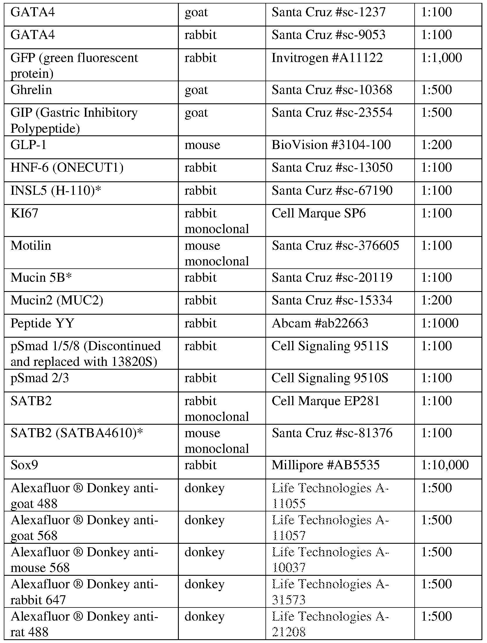
- Tissues were fixed for 1-3 hours in 4% paraformaldehyde (PFA) on ice depending on the size of the tissue. Organoids and transplant engraftments were frozen in OCT. OCT sections were blocked using donkey serum (5% serum in IX PBS plus 0.5% Triton-X) for 30 min and incubated with primary antibody overnight at 4 °C. Slides were then washed 3X with IX PBS plus 0.5% Triton-X and incubated in secondary antibody with DAPI in blocking buffer for 2 h at room temperature. See Table 2 for a list of antibodies and respective dilutions. Slides were then washed 2X with IX PBS plus 0.5% Triton- X followed by a final wash in IX PBS.
- PFA paraformaldehyde
- PPIA (CPHA) REV GGACCCGTATGCTTTAGGATGA
- RNA isolation and QPCR RNA was extracted using Nucleospin® RNA extraction kit (Macharey-Nagel) and reverse transcribed into cDNA using Superscript VILO (Invitrogen) according to manufacturer's protocols.
- QPCR primers were designed using the qPrimerDepot webased tool (primerdepot.nci.nih.gov). Primer sequences are listed in Table 3.
- QPCR was performed using Quantitect SYBR® Green PCR kit (Qiagen) and a QuantStudio TM 6 Flex Real-Time PCR System (Applied Biosystems).
- RNA-seq accession numbers Adult small intestine and large intestine RNA-seq data were downloaded from the public database E-MTAB- 1733. These data sets represent whole organ tissue which includes the epithelium and muscle layers. Accession numbers for the small intestine samples: ERR315344, ERR315381, ERR315409, ERR315442, ERR315461. Accession numbers for the large intestine samples: ERR315348, ERR315357, ERR315484. For FIG. 9B, processed FPKM data was downloaded from https://qithub.com/hilldr/Finkbeiner StemCellReports2015.
- RNA-seq sequence assembly abundance estimation RNA library construction and RNA sequencing was performed by the Cincinnati Children's Hospital DNA Sequencing Core, using an Illumina HiSeq2500 platform.
- the quality of the Illumina sequencing run was evaluated by analyzing FASTQ data for each sample using FastQC version 0.10.1 http://www.bioinformatics.babraham.ac.uk/projects/fastqc to identify features of the data that may indicate quality problems (e.g. low-quality scores, over-represented sequences, inappropriate GC content, etc.). No major issues were identified by the QC analysis.
- Applicant used the software package Tuxedo Suite for alignment, differential expression analysis, and post-analysis diagnostics. Briefly, Applicant aligned reads to the reference transcriptome (UCSC hgl9) using TopHat version 2.0.13 and Bowtie version 2.2.5 (Langmead et al., 2009).
- Applicant used default parameter settings for alignment, with the exception of: “— b2-very-sensitive” to maximize the accuracy of the read alignment, as well as “— no-coverage-search” and “— no-no vel-juncs” limiting the read mapping to known transcripts.
- Cufflinks version 2.2.1 (Trapnell et al., 2012) was used for RNA abundance estimation.
- UCSC hgl9.fa was used as the reference genome sequence and UCSC hgl9.gtf was used for transcriptome annotation.
- Applicant applied the following parameters in Cufflinks: "— multi-read-correct” to adjust expression calculations for reads that map in more than one locus, and "— compatible-hits-norm” and “— upper-quartile — norm” for normalization of expression values. Normalized FPKM tables were generated using the CuffNorm function. RNA sequence assembly and transcriptional analysis was conducted using the 64-bit Debian Linux stable version 7.10 (“Whez") platform.
- GATA4 represses an ileal program of gene expression in the proximal small intestine by inhibiting the acetylation of histone H3, lysine 27. Bba-Gene Regul Mech 1839, 1273-1282.
- GATA4 is essential for jejuna! function in mice. Gastroenterology 135, 1676- ⁇ 686 el671.
- Bone morphogenetic protein signaling pathway plays multiple roles during gastrointestinal tract development. Developmental dynamics : an official publication of the American Association of Anatomists 234, 312-322. [00156] Dobreva, G., Chahrour, M., Dautzenberg, M., Chirivella, L., Kanzler, B., Farinas, I., Karsenty, G., and Grosschedl, R. (2006). SATB2 is a multifunctional determinant of craniofacial patterning and osteoblast differentiation. Cell 125, 971-986.
- Fetal endoderm primarily holds the temporal and positional information required for mammalian intestinal development. J Cell Biol 126, 211-221.
- Bone morphogenetic protein 2 is expressed by, and acts upon, mature epithelial cells in the colon. Gastroenterology 126, 111-121.
- Sonic Hedgehog Is an Endodermal Signal Inducing Bmp- 4 and Hox Genes during Induction and Regionalization of the Chick Hindgut. Development 121, 3163-3174.
- INSL5 is a novel marker for human enteroendocrine cells of the large intestine and neuroendocrine tumours. Oncol Rep 29, 149- ⁇ 54.
- MUCSB is the prominent mucin in human gallbladder and is also expressed in a subset of colonic goblet cells.
Landscapes
- Health & Medical Sciences (AREA)
- Engineering & Computer Science (AREA)
- Life Sciences & Earth Sciences (AREA)
- Biomedical Technology (AREA)
- Chemical & Material Sciences (AREA)
- Organic Chemistry (AREA)
- Zoology (AREA)
- Genetics & Genomics (AREA)
- Bioinformatics & Cheminformatics (AREA)
- Biotechnology (AREA)
- Wood Science & Technology (AREA)
- General Health & Medical Sciences (AREA)
- Biochemistry (AREA)
- Cell Biology (AREA)
- Microbiology (AREA)
- Gastroenterology & Hepatology (AREA)
- Molecular Biology (AREA)
- Immunology (AREA)
- General Engineering & Computer Science (AREA)
- Medicinal Chemistry (AREA)
- Toxicology (AREA)
- Urology & Nephrology (AREA)
- Hematology (AREA)
- Biophysics (AREA)
- Proteomics, Peptides & Aminoacids (AREA)
- Tropical Medicine & Parasitology (AREA)
- Pathology (AREA)
- General Physics & Mathematics (AREA)
- Analytical Chemistry (AREA)
- Physics & Mathematics (AREA)
- Food Science & Technology (AREA)
- Orthopedic Medicine & Surgery (AREA)
- Micro-Organisms Or Cultivation Processes Thereof (AREA)
- Measuring Or Testing Involving Enzymes Or Micro-Organisms (AREA)
- Medicines Containing Material From Animals Or Micro-Organisms (AREA)
- Medicines That Contain Protein Lipid Enzymes And Other Medicines (AREA)
- Environmental Sciences (AREA)
- Pharmaceuticals Containing Other Organic And Inorganic Compounds (AREA)
- Animal Behavior & Ethology (AREA)
- Animal Husbandry (AREA)
Abstract
Description
Claims
Priority Applications (17)
| Application Number | Priority Date | Filing Date | Title |
|---|---|---|---|
| EP17877781.9A EP3548507A4 (en) | 2016-12-05 | 2017-12-05 | COLON ORGANOIDS AND METHOD FOR THE PRODUCTION AND USE THEREOF |
| NZ753873A NZ753873A (en) | 2016-12-05 | 2017-12-05 | Colonic organoids and methods of making and using same |
| AU2017373767A AU2017373767B2 (en) | 2016-12-05 | 2017-12-05 | Colonic organoids and methods of making and using same |
| KR1020237024207A KR102807995B1 (en) | 2016-12-05 | 2017-12-05 | Colonic organoids and methods of making and using same |
| CA3045145A CA3045145A1 (en) | 2016-12-05 | 2017-12-05 | Colonic organoids and methods of making and using same |
| CN201780073347.0A CN110062764B (en) | 2016-12-05 | 2017-12-05 | Colon organoids and methods of making and using the same |
| KR1020257015203A KR20250068799A (en) | 2016-12-05 | 2017-12-05 | Colonic organoids and methods of making and using same |
| JP2019527862A JP7068305B2 (en) | 2016-12-05 | 2017-12-05 | Colon organoids and how to make and use them |
| KR1020197018724A KR102558606B1 (en) | 2016-12-05 | 2017-12-05 | Colon organ organ and methods of making and using the same |
| CN202410839175.5A CN118909916A (en) | 2016-12-05 | 2017-12-05 | Colonocytes and methods of making and using the same |
| US16/461,147 US11767515B2 (en) | 2016-12-05 | 2017-12-05 | Colonic organoids and methods of making and using same |
| IL267109A IL267109A (en) | 2016-12-05 | 2019-06-05 | Colonic organoids and methods of making and using same |
| AU2021286289A AU2021286289B2 (en) | 2016-12-05 | 2021-12-14 | Colonic organoids and methods of making and using same |
| JP2022074275A JP7464652B2 (en) | 2016-12-05 | 2022-04-28 | Colon organoids and methods for producing and using same |
| US18/473,619 US20240240154A1 (en) | 2016-12-05 | 2023-09-25 | Colonic organoids and methods of making and using same |
| AU2024201465A AU2024201465A1 (en) | 2016-12-05 | 2024-03-06 | Colonic organoids and methods of making and using same |
| JP2024052990A JP2024096732A (en) | 2016-12-05 | 2024-03-28 | Colon organoids and methods for producing and using same |
Applications Claiming Priority (4)
| Application Number | Priority Date | Filing Date | Title |
|---|---|---|---|
| US201662429948P | 2016-12-05 | 2016-12-05 | |
| US62/429,948 | 2016-12-05 | ||
| US201762478962P | 2017-03-30 | 2017-03-30 | |
| US62/478,962 | 2017-03-30 |
Related Child Applications (2)
| Application Number | Title | Priority Date | Filing Date |
|---|---|---|---|
| US16/461,147 A-371-Of-International US11767515B2 (en) | 2016-12-05 | 2017-12-05 | Colonic organoids and methods of making and using same |
| US18/473,619 Continuation US20240240154A1 (en) | 2016-12-05 | 2023-09-25 | Colonic organoids and methods of making and using same |
Publications (1)
| Publication Number | Publication Date |
|---|---|
| WO2018106628A1 true WO2018106628A1 (en) | 2018-06-14 |
Family
ID=62492104
Family Applications (1)
| Application Number | Title | Priority Date | Filing Date |
|---|---|---|---|
| PCT/US2017/064600 Ceased WO2018106628A1 (en) | 2016-12-05 | 2017-12-05 | Colonic organoids and methods of making and using same |
Country Status (11)
| Country | Link |
|---|---|
| US (2) | US11767515B2 (en) |
| EP (1) | EP3548507A4 (en) |
| JP (3) | JP7068305B2 (en) |
| KR (3) | KR102807995B1 (en) |
| CN (2) | CN118909916A (en) |
| AU (3) | AU2017373767B2 (en) |
| CA (1) | CA3045145A1 (en) |
| IL (1) | IL267109A (en) |
| NZ (1) | NZ753873A (en) |
| SG (1) | SG10202105768WA (en) |
| WO (1) | WO2018106628A1 (en) |
Cited By (19)
| Publication number | Priority date | Publication date | Assignee | Title |
|---|---|---|---|---|
| WO2018213773A1 (en) | 2017-05-19 | 2018-11-22 | Cedars-Sinai Medical Center | Systems and methods for growth of intestinal cells |
| WO2020056158A1 (en) * | 2018-09-12 | 2020-03-19 | Children's Hospital Medical Center | Organoid compositions for the production of hematopoietic stem cells and derivatives thereof |
| US10781425B2 (en) | 2010-05-06 | 2020-09-22 | Children's Hospital Medical Center | Methods and systems for converting precursor cells into intestinal tissues through directed differentiation |
| WO2020232086A1 (en) * | 2019-05-15 | 2020-11-19 | The Cleveland Clinic Foundation | Induced human colitic organoids |
| US11053477B2 (en) | 2014-05-28 | 2021-07-06 | Children's Hospital Medical Center | Methods and systems for converting precursor cells into gastric tissues through directed differentiation |
| US11066650B2 (en) | 2016-05-05 | 2021-07-20 | Children's Hospital Medical Center | Methods for the in vitro manufacture of gastric fundus tissue and compositions related to same |
| US11584916B2 (en) | 2014-10-17 | 2023-02-21 | Children's Hospital Medical Center | Method of making in vivo human small intestine organoids from pluripotent stem cells |
| US11767515B2 (en) | 2016-12-05 | 2023-09-26 | Children's Hospital Medical Center | Colonic organoids and methods of making and using same |
| EP4051782A4 (en) * | 2019-10-31 | 2023-12-06 | University Health Network | ORGANOID CULTURE MEDIUM AND METHODS |
| WO2024263961A2 (en) | 2023-06-23 | 2024-12-26 | Children's Hospital Medical Center | Methods of matrix-free suspension culture |
| WO2025072803A1 (en) | 2023-09-29 | 2025-04-03 | Children's Hospital Medical Center | Ntrk2 signaling-mediated alveolar capillary injury and repair |
| US12281334B2 (en) | 2017-04-14 | 2025-04-22 | Children's Hospital Medical Center | Multi donor stem cell compositions and methods of making same |
| US12297457B2 (en) | 2017-10-10 | 2025-05-13 | Children's Hospital Medical Center | Esophageal tissue and/or organoid compositions and methods of making same |
| EP4388083A4 (en) * | 2021-08-19 | 2025-07-09 | Childrens Hospital Med Ct | VASCULARIZED ORGANOIDS |
| US12379372B2 (en) | 2017-12-21 | 2025-08-05 | Children's Hospital Medical Center | Digitalized human organoids and methods of using same |
| US12414967B2 (en) | 2016-11-04 | 2025-09-16 | Children's Hospital Medical Center | Compositions and methods of treating liver disease |
| US12421500B2 (en) | 2018-07-26 | 2025-09-23 | Children's Hospital Medical Center | Hepato-biliary-pancreatic tissues and methods of making same |
| US12497597B2 (en) | 2019-05-31 | 2025-12-16 | Children's Hospital Medical Center | Methods of generating and expanding hematopoietic stem cells |
| US12534709B2 (en) | 2019-05-31 | 2026-01-27 | Children's Hospital Medical Center | Shaped organoid compositions and methods of making same |
Families Citing this family (14)
| Publication number | Priority date | Publication date | Assignee | Title |
|---|---|---|---|---|
| CN111849860B (en) * | 2020-06-01 | 2021-12-21 | 浙江大学 | Method for regulating and controlling intestinal stem cell differentiation by using iron element and application |
| CN116234924A (en) * | 2020-07-30 | 2023-06-06 | 国立研究开发法人理化学研究所 | Bladder organoids and methods of making the same |
| CN113009155B (en) * | 2021-03-05 | 2022-03-25 | 温州医科大学慈溪生物医药研究院 | Application of FGF20 in preparation of reagent for diagnosing ulcerative colitis |
| IL281561A (en) | 2021-03-16 | 2022-10-01 | Yeda Res & Dev | Methods and devices for growing mouse embryos outside the uterus |
| CN113388572B (en) * | 2021-08-17 | 2021-11-19 | 天九再生医学(天津)科技有限公司 | Method for inducing and differentiating high-efficiency human pluripotent stem cells into multi-lineage small intestine organoids in vitro |
| CN114107208A (en) * | 2021-11-12 | 2022-03-01 | 复旦大学附属中山医院 | A kind of preparation method of human-derived organoid bank |
| US12435335B2 (en) | 2022-06-06 | 2025-10-07 | Cornell University | Promoting nutrient absorption through the colon |
| KR102843172B1 (en) * | 2023-01-04 | 2025-08-05 | 연세대학교 산학협력단 | Estimation of individual DNA damage response and prediction of the risk of cancer using colon organoids derived from patients with deficiency of DNA mismatch repair function |
| WO2024192567A1 (en) * | 2023-03-17 | 2024-09-26 | 北京大学 | Novel human intestinal tract and colorectal tumor organoid culture system |
| WO2025035155A1 (en) * | 2023-08-10 | 2025-02-13 | The Regents Of The University Of California | Foxo inhibition to augment gastrointestinal function and paneth cell function |
| CN118501457B (en) * | 2024-04-29 | 2025-06-17 | 南通大学附属医院 | PPP6R1 gene and its RNAi lentiviral interference system and application |
| CN118240750B (en) * | 2024-05-28 | 2024-08-13 | 淇嘉科技(苏州)有限公司 | Method for constructing colon organoid with immune cells |
| CN118374446B (en) * | 2024-06-26 | 2024-10-01 | 淇嘉科技(苏州)有限公司 | Batch preparation method of macrophage-containing colon organoids |
| CN118374432B (en) * | 2024-06-26 | 2024-10-01 | 淇嘉科技(苏州)有限公司 | Method for preparing homogenized intestinal organoids |
Citations (3)
| Publication number | Priority date | Publication date | Assignee | Title |
|---|---|---|---|---|
| US20130137130A1 (en) * | 2010-05-06 | 2013-05-30 | Children's Hospital Medical Center | Methods and systems for converting precursor cells into intestinal tissues through directed differentiation |
| WO2015173425A1 (en) * | 2014-05-16 | 2015-11-19 | Koninklijke Nederlandse Akademie Van Wetenschappen | Improved culture method for organoids |
| WO2015183920A2 (en) * | 2014-05-28 | 2015-12-03 | Children's Hospital Medical Center | Methods and systems for converting precursor cells into gastric tissues through directed differentiation |
Family Cites Families (310)
| Publication number | Priority date | Publication date | Assignee | Title |
|---|---|---|---|---|
| AU9031591A (en) | 1990-10-29 | 1992-05-26 | Regents Of The University Of Minnesota | A bioartificial liver |
| US5523226A (en) | 1993-05-14 | 1996-06-04 | Biotechnology Research And Development Corp. | Transgenic swine compositions and methods |
| US5912227A (en) | 1995-01-27 | 1999-06-15 | North Carolina State University | Method of enhancing nutrient uptake |
| WO1998021312A1 (en) | 1996-11-08 | 1998-05-22 | Rpms Technology Limited | Human hepatocytes in three-dimensional support systems |
| HK1045171A1 (en) | 1997-01-16 | 2002-11-15 | Yissum Research Development Company Of The Hebrew University Of Jerusalem | A device and method for performing a biological modification of a fluid |
| US7291626B1 (en) | 1998-04-09 | 2007-11-06 | John Hopkins University School Of Medicine | Inhibitors of hedgehog signaling pathways, compositions and uses related thereto |
| US20010041964A1 (en) | 1998-09-14 | 2001-11-15 | George M. Grass | Pharmacokinetic-based drug design tool and method |
| US7759113B2 (en) | 1999-04-30 | 2010-07-20 | The General Hospital Corporation | Fabrication of tissue lamina using microfabricated two-dimensional molds |
| US6607501B2 (en) | 2001-05-14 | 2003-08-19 | Reynolds G. Gorsuch | Process and apparatus for utilization of in vivo extracted plasma with tissue engineering devices, bioreactors, artificial organs, and cell therapy applications |
| US20030129751A1 (en) | 2001-05-16 | 2003-07-10 | Grikscheit Tracy C. | Tissue-engineered organs |
| GB2399823B (en) | 2001-12-07 | 2006-02-15 | Geron Corp | Islet cells from primate pluripotent stem cells |
| US20050170506A1 (en) | 2002-01-16 | 2005-08-04 | Primegen Biotech Llc | Therapeutic reprogramming, hybrid stem cells and maturation |
| US20030187515A1 (en) | 2002-03-26 | 2003-10-02 | Hariri Robert J. | Collagen biofabric and methods of preparing and using the collagen biofabric |
| US7160719B2 (en) | 2002-06-07 | 2007-01-09 | Mayo Foundation For Medical Education And Research | Bioartificial liver system |
| AU2003261798A1 (en) | 2002-08-28 | 2004-03-19 | Asahi Medical Co., Ltd. | Cell-filled device of modified cross-section hollow fiber membrane type |
| TW571101B (en) | 2003-01-21 | 2004-01-11 | Ind Tech Res Inst | Fluid analysis apparatus |
| WO2004071573A2 (en) | 2003-02-07 | 2004-08-26 | The Johns Hopkins University | Hypoxia induced mitogenic factor |
| US7727998B2 (en) | 2003-02-10 | 2010-06-01 | Banyu Pharmaceutical Co., Ltd. | Melanin-concentrating hormone receptor antagonists containing piperidine derivatives as the active ingredient |
| DE10362002B4 (en) | 2003-06-23 | 2006-10-12 | Fraunhofer-Gesellschaft zur Förderung der angewandten Forschung e.V. | Adult pluripotent stem cells |
| US8586357B2 (en) | 2003-12-23 | 2013-11-19 | Viacyte, Inc. | Markers of definitive endoderm |
| US20050266554A1 (en) | 2004-04-27 | 2005-12-01 | D Amour Kevin A | PDX1 expressing endoderm |
| US7985585B2 (en) | 2004-07-09 | 2011-07-26 | Viacyte, Inc. | Preprimitive streak and mesendoderm cells |
| KR101221302B1 (en) | 2003-12-23 | 2013-01-11 | 비아싸이트, 인크. | Definitive endoderm |
| US7625753B2 (en) | 2003-12-23 | 2009-12-01 | Cythera, Inc. | Expansion of definitive endoderm cells |
| US7541185B2 (en) | 2003-12-23 | 2009-06-02 | Cythera, Inc. | Methods for identifying factors for differentiating definitive endoderm |
| US8647873B2 (en) | 2004-04-27 | 2014-02-11 | Viacyte, Inc. | PDX1 expressing endoderm |
| WO2005081970A2 (en) | 2004-02-24 | 2005-09-09 | The Curators Of The University Of Missouri | Self-assembling cell aggregates and methods of making engineered tissue using the same |
| DE102004017476B4 (en) | 2004-04-08 | 2009-03-26 | Fraunhofer-Gesellschaft zur Förderung der angewandten Forschung e.V. | Process for the preparation of a cell composition containing epithelial cells |
| ES2801001T3 (en) | 2004-04-27 | 2021-01-07 | Viacyte Inc | Endoderm expressing pdx1 |
| JP4650608B2 (en) | 2004-05-18 | 2011-03-16 | 信越化学工業株式会社 | Photomask blank and photomask manufacturing method |
| WO2005113747A2 (en) | 2004-05-21 | 2005-12-01 | Fraunhofer Gesellschaft zur Förderung der angewandten Forschung e.V. | Multicellular tissue and organ culture systems |
| US8410246B2 (en) | 2004-06-17 | 2013-04-02 | Thrasos, Inc. | TDF-related compounds and analogs thereof |
| WO2006016999A1 (en) | 2004-07-09 | 2006-02-16 | Cythera, Inc. | Methods for identifying factors for differentiating definitive endoderm |
| CN105708787A (en) | 2004-07-12 | 2016-06-29 | 爱密斯菲尔科技公司 | Compositions for delivering peptide yy and pyy agonists |
| WO2006091231A2 (en) | 2004-07-21 | 2006-08-31 | Ambrx, Inc. | Biosynthetic polypeptides utilizing non-naturally encoded amino acids |
| EP1791952A4 (en) | 2004-08-13 | 2008-06-11 | Univ Georgia Res Found | Compositions and methods for self-renewal and differentiation in human embryonic stem cells |
| US20060236415A1 (en) | 2005-03-09 | 2006-10-19 | Silversides David W | Neural crest cells specific promoters; isolated neural crest cells; and methods of isolating and of using same |
| US7604929B2 (en) | 2005-04-21 | 2009-10-20 | In Vitro Technologies, Inc. | Cellular compositions and methods for their preparation |
| WO2006122147A2 (en) | 2005-05-10 | 2006-11-16 | Rutgers, The State University Of New Jersey | Alginate poly-l-lysine encapsulation as a technology for controlled differentiation of embryonic stem cells |
| WO2006126219A1 (en) | 2005-05-26 | 2006-11-30 | Fresenius Medical Care Deutschland G.M.B.H. | Liver progenitor cells |
| GB0517382D0 (en) | 2005-08-26 | 2005-10-05 | Plasticell Ltd | Cell culture |
| WO2007027905A2 (en) | 2005-08-31 | 2007-03-08 | Science And Technology Corporation @ Unm | Human renal stem cells |
| DK2674485T3 (en) | 2005-10-27 | 2019-08-26 | Viacyte Inc | PDX-1 EXPRESSING DORSAL AND VENTRAL FORTARM ENDODERM |
| US7927869B2 (en) | 2005-11-29 | 2011-04-19 | Spencer Z Rosero | System and method for supporting a biological chip device |
| US20070239083A1 (en) | 2006-01-18 | 2007-10-11 | Axel Voss | Shock wave generators |
| US20070238169A1 (en) | 2006-04-11 | 2007-10-11 | The Board Of Trustees Of The Leland Stanford Junior University | Cell sorter and culture system |
| US8685730B2 (en) | 2006-05-02 | 2014-04-01 | Wisconsin Alumni Research Foundation | Methods and devices for differentiating pluripotent stem cells into cells of the pancreatic lineage |
| US9012419B2 (en) | 2006-05-17 | 2015-04-21 | University Of Utah Research Foundation | Methods and compositions related to eosinophil regulation |
| DK2040713T3 (en) | 2006-06-27 | 2014-09-29 | Intercept Pharmaceuticals Inc | BILIC ACID DERIVATIVES AS FXR LIGANDS FOR THE PREVENTION OR TREATMENT OF FXR-MEDIATED DISEASES OR CONDITIONS |
| WO2008011561A2 (en) | 2006-07-21 | 2008-01-24 | Massachusetts Institute Of Technology | End-modified poly(beta-amino esters) and uses thereof |
| US8497240B2 (en) | 2006-08-17 | 2013-07-30 | Amylin Pharmaceuticals, Llc | DPP-IV resistant GIP hybrid polypeptides with selectable properties |
| US9127254B2 (en) | 2006-12-18 | 2015-09-08 | Ben-Gurion University Of The Negev Research And Development Authority | Scaffolding for tissue regeneration or repair |
| FR2917425B1 (en) | 2007-06-18 | 2010-11-19 | Univ Nancy 1 Henri Poincare | METHOD FOR THE PROLIFERATION OF CELLS ON POLYELECTROLYTE MULTILAYERS AND ITS APPLICATION, IN PARTICULAR TO THE PREPARATION OF CELLULARIZED BIOMATERIALS |
| GB2467645B (en) | 2007-07-06 | 2011-12-21 | Univ London | A method of proliferating human hepatocyte cells |
| EP2022848A1 (en) | 2007-08-10 | 2009-02-11 | Hubrecht Institut | A method for identifying, expanding, and removing adult stem cells and cancer stem cells |
| EP2185931B1 (en) | 2007-09-07 | 2013-07-10 | Children's Hospital Medical Center | Use of secretor, lewis and sialyl antigen levels as predictors for disease |
| US7695963B2 (en) | 2007-09-24 | 2010-04-13 | Cythera, Inc. | Methods for increasing definitive endoderm production |
| US8609413B2 (en) | 2007-12-11 | 2013-12-17 | Research Development Foundation | Neurons, astrocytes and oligodendrocytes differentiated from a mammalian pluripotent or neural stem cells exposed to a pyridine deriviative |
| AU2009203893B2 (en) | 2008-01-08 | 2014-10-02 | The University Of Queensland | Method of producing a population of cells |
| US20110218512A1 (en) | 2008-06-03 | 2011-09-08 | Aethlon Medical, Inc. | Enhanced antiviral therapy methods and devices |
| AU2009254177B2 (en) | 2008-06-04 | 2014-09-11 | Tissuse Gmbh | Organ-on-a-chip-device |
| KR101711354B1 (en) | 2008-06-24 | 2017-02-28 | 더 큐레이터스 오브 더 유니버시티 오브 미주리 | Self-assembling multicellular bodies and methods of producing a three-dimensional biological structure using the same |
| ITRM20080342A1 (en) | 2008-06-26 | 2009-12-27 | Univ Degli Studi Udine | DOLLS-SIMILAR PULP CELLS, INSULATION AND USE METHODS. |
| US20130115673A1 (en) | 2008-07-16 | 2013-05-09 | Biotime, Inc. | Methods of Screening Embryonic Progenitor Cell Lines |
| US20110305672A1 (en) | 2008-07-25 | 2011-12-15 | University Of Georgia Research Foundation, Inc. | COMPOSITIONS FOR MESODERM DERIVED ISL1+ MULTIPOTENT CELLS (IMPs), EPICARDIAL PROGENITOR CELLS (EPCs) AND MULTIPOTENT CD56C CELLS (C56Cs) AND METHODS OF PRODUCING AND USING SAME |
| US8895300B2 (en) | 2008-11-04 | 2014-11-25 | Viacyte, Inc. | Scalable primate pluripotent stem cell aggregate suspension culture and differentiation thereof |
| US20110294735A1 (en) | 2008-11-05 | 2011-12-01 | Merck Sharp & Dohme Corp. | Mechanism of neuromedin u action and uses thereof |
| JP5351601B2 (en) | 2008-12-26 | 2013-11-27 | 矢崎総業株式会社 | Insulating cap manufacturing method and insulating cap manufacturing apparatus |
| GB201111244D0 (en) * | 2011-06-30 | 2011-08-17 | Konink Nl Akademie Van Wetenschappen Knaw | Culture media for stem cells |
| MX379945B (en) | 2009-02-03 | 2025-03-11 | Koninklijke Nederlandse Akademie Van Wetenschappen | CULTURE MEDIUM FOR EPITHELIAL STEM CELLS AND ORGANOIDS THAT INCLUDE STEM CELLS. |
| US9752124B2 (en) | 2009-02-03 | 2017-09-05 | Koninklijke Nederlandse Akademie Van Wetenschappen | Culture medium for epithelial stem cells and organoids comprising the stem cells |
| EP2412800A1 (en) | 2010-07-29 | 2012-02-01 | Koninklijke Nederlandse Akademie van Wetenschappen | Liver organoid, uses thereof and culture method for obtaining them |
| WO2010094694A1 (en) | 2009-02-23 | 2010-08-26 | F. Hoffmann-La Roche Ag | Assays to predict cardiotoxicity |
| EP2405957B1 (en) | 2009-03-13 | 2020-12-23 | Mayo Foundation For Medical Education And Research | Bioartificial liver |
| EP2408457A1 (en) | 2009-03-17 | 2012-01-25 | Axcan Pharma Inc. | Method of treating nonalcoholic steatohepatitis with elevated doses of ursodeoxycholic acid |
| WO2010127399A1 (en) | 2009-05-06 | 2010-11-11 | Walter And Eliza Hall Institute Of Medical Research | Gene expression profiles and uses thereof |
| CA2762584A1 (en) | 2009-05-20 | 2010-11-25 | Cardio3 Biosciences S.A. | Pharmaceutical composition for the treatment of heart diseases |
| JP2012527880A (en) | 2009-05-29 | 2012-11-12 | ノヴォ ノルディスク アー/エス | Derivation of specific endoderm from hPS cell-derived definitive endoderm |
| WO2010143747A1 (en) | 2009-06-10 | 2010-12-16 | 公立大学法人奈良県立医科大学 | Method for production of artificial intestinal tract |
| CN102459574B (en) | 2009-06-18 | 2016-06-29 | 塞拉提斯股份公司 | Growth and the 3D culture systems of differentiation for dry (hPS) cell of people's pluripotency |
| US8685386B2 (en) | 2009-07-16 | 2014-04-01 | Biotime, Inc | Methods and compositions for in vitro and in vivo chondrogenesis |
| US20110152770A1 (en) | 2009-07-30 | 2011-06-23 | Tandem Diabetes Care, Inc. | Infusion pump system with disposable cartridge having pressure venting and pressure feedback |
| US8501476B2 (en) | 2009-10-07 | 2013-08-06 | Brown University | Assays and methods for fusing cell aggregates to form proto-tissues |
| CN101696396A (en) | 2009-10-27 | 2010-04-21 | 中国人民解放军军事医学科学院卫生学环境医学研究所 | Construction method and use of model of hepatitis B virus infection in vivo |
| WO2011066374A2 (en) | 2009-11-24 | 2011-06-03 | Alder Biopharmaceuticals, Inc. | Antagonists of il-6 to prevent or treat cachexia, weakness, fatigue, and/or fever |
| US20130031645A1 (en) | 2009-11-25 | 2013-01-31 | Thomas Touboul | Method for hepatic differentiation of definitive endoderm cells |
| AU2010333840B2 (en) | 2009-12-23 | 2016-01-07 | Janssen Biotech, Inc. | Differentiation of human embryonic stem cells |
| US20110231942A1 (en) * | 2010-03-17 | 2011-09-22 | Xi He | Tiki1 and tiki2, wnt inhibitors |
| EP2550355B1 (en) | 2010-03-22 | 2016-11-16 | Takara Bio Europe AB | Directed differentiation and maturation of pluripotent cells into hepatocyte like cells by modulation of wnt-signalling pathway |
| EP2380920A1 (en) | 2010-04-22 | 2011-10-26 | QGel SA | Hydrogel precursor formulation and production process thereof |
| CN103068970A (en) | 2010-04-25 | 2013-04-24 | 西奈山医学院 | Generation of anterior foregut endoderm from pluripotent cells |
| JP5984217B2 (en) | 2010-06-15 | 2016-09-06 | セルラー ダイナミクス インターナショナル, インコーポレイテッド | Preparation of induced pluripotent stem cells from a small amount of peripheral blood |
| WO2012027474A1 (en) | 2010-08-24 | 2012-03-01 | Regents Of The University Of Minnesota | Non-static suspension culture of cell aggregates |
| AU2011296383B2 (en) | 2010-08-31 | 2016-03-10 | Janssen Biotech, Inc. | Differentiation of pluripotent stem cells |
| AU2011329002C1 (en) | 2010-11-15 | 2017-05-25 | Accelerated Biosciences Corp. | Generation of neural stem cells from human trophoblast stem cells |
| EA028902B1 (en) | 2010-12-31 | 2018-01-31 | Университет Фюр Боденкультур Вена | Method of generating induced pluripotent stem cells and differentiated cells |
| US8951781B2 (en) | 2011-01-10 | 2015-02-10 | Illumina, Inc. | Systems, methods, and apparatuses to image a sample for biological or chemical analysis |
| US9200258B2 (en) | 2011-01-27 | 2015-12-01 | University Of Maryland, Baltimore | Multicellular organotypic model of human intestinal mucosa |
| EP2484750A1 (en) | 2011-02-07 | 2012-08-08 | Ecole Polytechnique Fédérale de Lausanne (EPFL) | Monitoring system for cell culture |
| KR102117921B1 (en) | 2011-02-28 | 2020-06-03 | 프레지던트 앤드 펠로우즈 오브 하바드 칼리지 | Cell culture system |
| GB201106395D0 (en) | 2011-04-14 | 2011-06-01 | Hubrecht Inst | Compounds |
| US20140158233A1 (en) | 2011-05-09 | 2014-06-12 | President And Fellows Of Harvard College | Aerosol delivery to a microfluidic device |
| US10260039B2 (en) | 2011-05-11 | 2019-04-16 | Massachusetts Institute Of Technology | Microgels and microtissues for use in tissue engineering |
| KR20190042747A (en) | 2011-06-02 | 2019-04-24 | 프레지던트 앤드 펠로우즈 오브 하바드 칼리지 | Methods and uses for ex vivo tissue culture systems |
| JP2014519854A (en) | 2011-06-23 | 2014-08-21 | ザ チルドレンズ ホスピタル オブ フィラデルフィア | Self-replicating endoderm progenitor cell lines derived from human pluripotent stem cells and methods of use thereof |
| CA2848044C (en) | 2011-09-12 | 2021-01-19 | Organovo, Inc. | Platform for engineered implantable tissues and organs and methods of making the same |
| JP2013066414A (en) | 2011-09-22 | 2013-04-18 | National Institute Of Advanced Industrial Science & Technology | Surface marker of stomach precursor cell |
| WO2014066811A1 (en) | 2012-10-25 | 2014-05-01 | The Johns Hopkins University | Bioreducible poly (b-amino ester)s for sirna delivery |
| US20140302491A1 (en) | 2011-10-28 | 2014-10-09 | The Board Of Trustees Of The Leland Stanford Junior University | Ex Vivo Culture, Proliferation and Expansion of Primary Tissue Organoids |
| US20140308695A1 (en) | 2011-11-04 | 2014-10-16 | Tengion, Inc. | Drug screening and potency assays |
| EP2788476B1 (en) | 2011-12-08 | 2019-05-22 | Fred Hutchinson Cancer Research Center | Compositions and methods for enhanced generation of hematopoietic stem/progenitor cells |
| US9725687B2 (en) | 2011-12-09 | 2017-08-08 | President And Fellows Of Harvard College | Integrated human organ-on-chip microphysiological systems |
| US10087422B2 (en) | 2011-12-09 | 2018-10-02 | President And Fellows Of Harvard College | Organ chips and uses thereof |
| EP3045912B1 (en) | 2011-12-19 | 2018-09-19 | UMC Utrecht Holding B.V. | A rapid quantitative assay to measure cftr function in a primary intestinal culture model |
| CN113699105A (en) | 2011-12-23 | 2021-11-26 | 人类起源公司 | Organoids comprising decellularized and regrouped placental vascular scaffolds |
| WO2013106677A1 (en) | 2012-01-13 | 2013-07-18 | The General Hospital Corporation | Isolated human lung progenitor cells and uses thereof |
| WO2013116446A1 (en) | 2012-01-31 | 2013-08-08 | Wake Forest University Health Sciences | Tubular bioengineered smooth muscle structures |
| WO2013126813A1 (en) | 2012-02-22 | 2013-08-29 | Amgen Inc. | Autologous mammalian models derived from induced pluripotent stem cells and related methods |
| EP2634251A1 (en) | 2012-02-29 | 2013-09-04 | Technische Universität Berlin | 3D in vitro bi-phasic cartilage-bone construct |
| EP2836585A4 (en) | 2012-04-09 | 2015-12-23 | Thomas J Goodwin | Alternating ionic magnetic resonance (aimr) multiple-chambered culture apparatus and methods of use |
| KR20160027217A (en) | 2012-05-23 | 2016-03-09 | 에프. 호프만-라 로슈 아게 | Compositions and methods of obtaining and using endoderm and hepatocyte cells |
| US20140099709A1 (en) | 2012-06-19 | 2014-04-10 | Organovo, Inc. | Engineered three-dimensional connective tissue constructs and methods of making the same |
| CN111228278A (en) | 2012-06-19 | 2020-06-05 | 英特塞普特医药品公司 | Preparation, use and solid forms of obeticholic acid |
| DE102012105540A1 (en) | 2012-06-26 | 2014-04-24 | Karlsruher Institut für Technologie | Vascular model, process for its preparation and its use |
| EP2875360B1 (en) | 2012-07-20 | 2018-08-08 | Agency For Science, Technology And Research | In vitro assay for predicting renal proximal tubular cell toxicity |
| US9988606B2 (en) | 2012-07-24 | 2018-06-05 | The Trustees Of Columbia University In The City Of New York | Generation of airway and lung progenitors and epithelial cells and three-dimensional anterior foregut spheres |
| GB201216796D0 (en) | 2012-09-20 | 2012-11-07 | Cambridge Entpr Ltd | In vitro pancreatic differentiation |
| DK2712918T3 (en) | 2012-09-28 | 2015-02-16 | Tissuse Gmbh | Multi-organ-chip with improved lifetime and homeostasis |
| EP2716298A1 (en) | 2012-10-03 | 2014-04-09 | Institut Pasteur | A nod2-dependant pathway of cytoprotection of stem cells |
| CN104995294A (en) * | 2012-10-19 | 2015-10-21 | 新加坡科技研究局 | Methods of differentiating stem cells into one or more cell lineages |
| WO2014082096A1 (en) | 2012-11-26 | 2014-05-30 | The Trustees Of Columbia University In The City Of New York | Method for culture of human and mouse prostate organoids and uses thereof |
| ES2625510T3 (en) | 2012-11-26 | 2017-07-19 | Gambro Lundia Ab | Liver support system |
| SG10201707569YA (en) | 2012-12-12 | 2017-10-30 | Broad Inst Inc | Delivery, Engineering and Optimization of Systems, Methods and Compositions for Sequence Manipulation and Therapeutic Applications |
| EP2743345A1 (en) | 2012-12-13 | 2014-06-18 | IMBA-Institut für Molekulare Biotechnologie GmbH | Three dimensional heterogeneously differentiated tissue culture |
| US20150359849A1 (en) | 2013-01-31 | 2015-12-17 | President And Fellows Of Harvard College | Methods of increasing neuronal connectivity and/or treating a neurodegenerative condition |
| EP2956538B1 (en) | 2013-02-13 | 2018-11-14 | Wake Forest University Health Sciences | Bioengineered liver constructs and methods relating thereto |
| GB201304245D0 (en) | 2013-03-08 | 2013-04-24 | Inst Quimic De Sarria | Chemical compounds |
| WO2014165056A1 (en) | 2013-03-12 | 2014-10-09 | Board Of Regents, The University Of Texas System | High throughput mechanical strain generating system for cell cultures and applications thereof |
| US9938499B2 (en) | 2013-03-13 | 2018-04-10 | Wisconsin Alumni Research Foundation | Methods and materials for hematoendothelial differentiation of human pluripotent stem cells under defined conditions |
| DK2970890T3 (en) | 2013-03-14 | 2020-05-04 | Brigham & Womens Hospital Inc | COMPOSITIONS AND PROCEDURES FOR THE PROMOTION AND CULTIVATION OF EPITHEL STEM CELLS |
| US20160022873A1 (en) | 2013-03-14 | 2016-01-28 | Research Institute At Nationwide Children's Hospital, Inc. | Tissue engineered intestine |
| US8859286B2 (en) | 2013-03-14 | 2014-10-14 | Viacyte, Inc. | In vitro differentiation of pluripotent stem cells to pancreatic endoderm cells (PEC) and endocrine cells |
| CA2899507A1 (en) | 2013-03-14 | 2014-09-25 | The Regents Of The University Of California | In vitro production of medial ganglionic eminence precursor cells |
| US9442105B2 (en) | 2013-03-15 | 2016-09-13 | Organovo, Inc. | Engineered liver tissues, arrays thereof, and methods of making the same |
| US20160237400A1 (en) | 2013-03-15 | 2016-08-18 | The Jackson Laboratory | Isolation of non-embryonic stem cells and uses thereof |
| WO2014153294A1 (en) | 2013-03-17 | 2014-09-25 | The Regents Of The University Of California | Method to expand and transduce cultured human small and large intestinal stem cells |
| WO2014153346A1 (en) | 2013-03-18 | 2014-09-25 | Massachusetts Institute Of Technology | Engineering a heterogeneous tissue pluripotent stem cells |
| EP2796873A1 (en) | 2013-04-25 | 2014-10-29 | QGel SA | Method for a cell-based drug screening assay and the use thereof |
| US20160245653A1 (en) | 2013-04-30 | 2016-08-25 | Sangtae Park | Cylindrical resonator gyroscope |
| EP4299721A3 (en) | 2013-05-08 | 2024-04-24 | ProKidney | Organoids comprising isolated renal cells and uses thereof |
| US10545133B2 (en) | 2013-05-13 | 2020-01-28 | The Johns Hopkins University | Molecular signatures of invasive cancer subpopulations |
| WO2014197622A2 (en) | 2013-06-04 | 2014-12-11 | The Regents Of The University Of California | Liver-mimetic device and method for simulation of hepatic function using such device |
| JP6253265B2 (en) | 2013-06-05 | 2017-12-27 | 学校法人関西医科大学 | Method for isolating esophageal epithelial stem cells |
| CN105283545B (en) | 2013-06-10 | 2019-12-10 | 株式会社可乐丽 | Tissue structure and method for producing same |
| NZ715302A (en) | 2013-06-14 | 2021-12-24 | Univ Queensland | Renal progenitor cells |
| EP3026110B1 (en) | 2013-07-23 | 2020-09-16 | Public University Corporation Yokohama City University | Method for providing vascular system in biological tissue |
| WO2015021358A2 (en) | 2013-08-09 | 2015-02-12 | Dominique Charmot | Compounds and methods for inhibiting phosphate transport |
| PL3039123T3 (en) | 2013-08-28 | 2021-07-19 | Promethera Biosciences S.A. / N.V. | Method for producing adult liver progenitor cells |
| GB201317869D0 (en) | 2013-10-09 | 2013-11-20 | Cambridge Entpr Ltd | In vitro production of foregut stem cells |
| US10087416B2 (en) | 2013-10-25 | 2018-10-02 | Agency For Science, Technology And Research | Culturing pluripotent stem cells |
| CN111269885A (en) | 2013-11-22 | 2020-06-12 | 国立研究开发法人理化学研究所 | Method for preparing telencephalon or precursor tissue thereof |
| EP3071686B1 (en) | 2013-11-22 | 2020-07-22 | Cellectis SA | Method for generating batches of allogeneic t-cells with averaged potency |
| EP2876441B1 (en) | 2013-11-26 | 2017-10-25 | Bergen Teknologioverforing AS | Quantitative analysis of contact-depending cell-to-cell transfer and disease transmission |
| CA2944220C (en) | 2013-12-20 | 2024-01-02 | President And Fellows Of Harvard College | Organomimetic devices and methods of use and manufacturing thereof |
| ES2891134T3 (en) | 2014-01-14 | 2022-01-26 | Univ Yale | Compositions and Methods of Airway Cell Preparation |
| US11648335B2 (en) | 2014-01-31 | 2023-05-16 | Wake Forest University Health Sciences | Organ/tissue decellularization, framework maintenance and recellularization |
| AU2015217406A1 (en) | 2014-02-11 | 2016-08-25 | Celularity Inc. | Micro-organoids, and methods of making and using the same |
| US10369254B2 (en) | 2014-02-26 | 2019-08-06 | The Regents Of The University Of California | Method and apparatus for in vitro kidney organogenesis |
| BR112016019677A8 (en) | 2014-02-27 | 2021-07-13 | Public Univ Corp Yokohama City Univ | method of preparing an in vitro cell condensate, cell condensate, method of preparing a three-dimensional tissue structure, and gel-like culture support |
| DE102014003465A1 (en) | 2014-03-11 | 2015-09-17 | NeuroProof GmbH | Obtaining brain region-specific neuronal cultures from three-dimensional tissue cultures of stem cells |
| US11066649B2 (en) | 2014-03-19 | 2021-07-20 | Institut National De La Sante Et De La Recherche Medicale (Inserm) | Method for inducing human cholangiocyte differentiation |
| RU2724339C2 (en) | 2014-03-21 | 2020-06-23 | Тобира Терапьютикс, Инк. | Cenicriviroc for treating fibrosis |
| CA2944723C (en) | 2014-04-04 | 2023-03-14 | Organovo, Inc. | Engineered three-dimensional breast tissue, adipose tissue, and tumor disease model |
| WO2015156929A1 (en) | 2014-04-07 | 2015-10-15 | The Trustees Of Columbia University In The City Of New York | Method for culture of human bladder cell lines and organoids and uses thereof |
| WO2015157163A1 (en) | 2014-04-07 | 2015-10-15 | Rush University Medical Center | Screening assay for choice of prebiotic to prevent/treat gastrointestinal and systemic diseases |
| KR102374499B1 (en) | 2014-04-11 | 2022-03-14 | 사이머베이 쎄라퓨틱스, 인코퍼레이티드 | Treatment of nafld and nash |
| CN106456673B (en) | 2014-04-27 | 2021-04-16 | 纽约州立大学研究基金会 | Enamel products and methods of use |
| WO2015184273A1 (en) | 2014-05-29 | 2015-12-03 | Icahn School Of Medicine At Mount Sinai | Method and apparatus to prepare cardiac organoids in a bioreactor system |
| WO2015184375A2 (en) | 2014-05-29 | 2015-12-03 | Whitehead Institute For Biomedical Research | Compositions and methods for promoting intestinal stem cell and/or non-stem progenitor cell function |
| EP3786285A1 (en) | 2014-06-05 | 2021-03-03 | Cedars-Sinai Medical Center | A novel and efficient method for reprogramming immortalized lymphoblastoid cell lines to induced pluripotent stem cells |
| WO2015185714A1 (en) | 2014-06-06 | 2015-12-10 | Vrije Universiteit Brussel | Human hepatic 3d co-culture model and uses thereof |
| JP2017524377A (en) | 2014-06-20 | 2017-08-31 | ラトガース,ザ ステート ユニバーシティ オブ ニュー ジャージー | Single cell-derived organoid |
| WO2015200901A1 (en) | 2014-06-26 | 2015-12-30 | The Trustees Of Columbia University In The City Of New York | Inhibition of serotonin expression in gut enteroendocrine cells results in conversion to insulin-positive cells |
| SG11201700303UA (en) | 2014-07-17 | 2017-02-27 | Celmatix Inc | Methods and systems for assessing infertility and related pathologies |
| BR112017001623B1 (en) | 2014-07-29 | 2022-11-29 | Shenzhen Hightide Biopharmaceutical, Ltd | ACID-BASE ADDITION SALT IN SUBSTANTIALLY PURE FORM, PHARMACEUTICAL COMPOSITION COMPRISING AN AMOUNT OF SAID SALT AND ITS USES |
| WO2016015158A1 (en) | 2014-07-30 | 2016-02-04 | University Health Network | Organoids for drug screening and personalized medicine |
| US10894948B2 (en) | 2014-08-22 | 2021-01-19 | Cambridge Enterprise Limited | Resetting pluripotent stem cells |
| LT3140393T (en) | 2014-08-28 | 2023-09-11 | Cellaïon SA | Method for producing adult liver progenitor cells |
| WO2016033163A1 (en) | 2014-08-29 | 2016-03-03 | Immunomedics, Inc. | Identification of cancer genes by in-vivo fusion of human cancer cells and animal cells |
| KR20170066418A (en) | 2014-09-12 | 2017-06-14 | 토비라 쎄라퓨틱스, 인크. | Cenicriviroc combination therapy for the treatment of fibrosis |
| KR20170064547A (en) | 2014-10-06 | 2017-06-09 | 오가노보, 인크. | Engineered renal tissues, arrays thereof, and methods of making the same |
| CN107075471A (en) | 2014-10-08 | 2017-08-18 | 新加坡科技研究局 | Stem cell is divided into the method for hepatocyte lineage |
| EP3207122B1 (en) | 2014-10-14 | 2019-05-08 | FUJIFILM Cellular Dynamics, Inc. | Generation of keratinocytes from pluripotent stem cells and maintenance of keratinocyte cultures |
| US11584916B2 (en) | 2014-10-17 | 2023-02-21 | Children's Hospital Medical Center | Method of making in vivo human small intestine organoids from pluripotent stem cells |
| WO2016069897A1 (en) | 2014-10-30 | 2016-05-06 | Massachusetts Institute Of Technology | Materials and methods for rescue of ischemic tissue and regeneration of tissue integrity during restriction, engraftment and transplantation |
| US10479977B2 (en) | 2014-11-07 | 2019-11-19 | The Trustees Of Columbia University In The City Of New York | Osteochondroreticular stem cells for bone and cartilage regeneration |
| AU2015353853B2 (en) | 2014-11-25 | 2020-10-15 | President And Fellows Of Harvard College | Methods for generation of podocytes from pluripotent stem cells and cells produced by the same |
| WO2016086052A1 (en) | 2014-11-25 | 2016-06-02 | International Stem Cell Corporation | Derivation of nueral crest stem cells and uses thereof |
| GB201421092D0 (en) | 2014-11-27 | 2015-01-14 | Koninklijke Nederlandse Akademie Van Wetenschappen | Culture medium |
| GB201421094D0 (en) | 2014-11-27 | 2015-01-14 | Koninklijke Nederlandse Akademie Van Wetenschappen | Culture medium |
| AU2014277667B2 (en) | 2014-12-15 | 2022-07-14 | The University Of Queensland | Differentiation of pluripotent stem cells to form renal organoids |
| PT3237597T (en) | 2014-12-22 | 2021-03-12 | Ecole Polytechnique Fed Lausanne Epfl | Devices for high-throughput aggregation and manipulation of mammalian cells |
| WO2016103269A1 (en) | 2014-12-23 | 2016-06-30 | Ramot At Tel-Aviv University Ltd. | Populations of neural progenitor cells and methods of producing and using same |
| HK1243369A1 (en) | 2015-01-09 | 2018-07-13 | 吉利德阿波罗公司 | Acc inhibitor combination therapy for the treatment of non-alcoholic fatty liver disease |
| EP3252144A4 (en) | 2015-01-28 | 2018-08-08 | Public University Corporation Yokohama City University | Method for preparing bone marrow cell aggregate |
| US20160256672A1 (en) | 2015-02-10 | 2016-09-08 | Cedars-Sinai Medical Center | Induced pluripotent stem cell-derived hepatocyte based bioartificial liver device |
| CN105985395A (en) | 2015-02-13 | 2016-10-05 | 江苏奥赛康药业股份有限公司 | Obeticholic acid compound, and medicinal composition containing compound |
| WO2016140716A1 (en) | 2015-03-02 | 2016-09-09 | The Trustees Of Columbia University In The City Of New York | Injectable microtissue systems, devices, and methods |
| JP6807853B2 (en) | 2015-03-03 | 2021-01-06 | プレジデント アンド フェローズ オブ ハーバード カレッジ | Method for producing functional human tissue |
| CA2978729A1 (en) | 2015-03-06 | 2016-09-15 | Tsunekazu Oikawa | Human fibrolamellar hepatocellular carcinomas (hfl-hccs) |
| GB2554181B (en) | 2015-03-06 | 2021-09-08 | Micromass Ltd | Inlet instrumentation for ion analyser coupled to rapid evaporative ionisation mass spectrometry ("REIMS") device |
| EP3267994A4 (en) | 2015-03-09 | 2018-10-31 | Intekrin Therapeutics, Inc. | Methods for the treatment of nonalcoholic fatty liver disease and/or lipodystrophy |
| WO2016154330A1 (en) | 2015-03-23 | 2016-09-29 | Whitehead Institute For Biomedical Research | Reporter of genomic methylation and uses thereof |
| WO2016164413A1 (en) | 2015-04-07 | 2016-10-13 | Intercept Pharmaceuticals, Inc. | Pharmaceutical compositions for combination therapy |
| US10087417B2 (en) | 2015-04-22 | 2018-10-02 | William J. Freed | Three-dimensional model of human cortex |
| US10557124B2 (en) | 2015-04-22 | 2020-02-11 | The Regents Of The University Of Michigan | Compositions and methods for obtaining stem cell derived lung tissue, and related uses thereof |
| CN104877964A (en) | 2015-04-24 | 2015-09-02 | 赵振民 | In vitro construction method for salivary glands organs and acinus |
| PE20180690A1 (en) | 2015-04-27 | 2018-04-23 | Intercept Pharmaceuticals Inc | OBETICOLIC ACID COMPOSITIONS AND METHODS OF USE |
| WO2016174604A1 (en) | 2015-04-30 | 2016-11-03 | Helmholtz Zentrum München - Deutsches Forschungszentrum für Gesundheit und Umwelt (GmbH) | Means and methods for generation of breast stem cells |
| US11299714B2 (en) | 2015-05-11 | 2022-04-12 | The Trustees Of Columbia University In The City Of New York | Engineered adult-like human heart tissue |
| EP3303566B1 (en) | 2015-06-03 | 2020-10-14 | Takara Bio Europe AB | Maturation of mammalian hepatocytes |
| GB201510950D0 (en) | 2015-06-22 | 2015-08-05 | Cambridge Entpr Ltd | In vitro Production of Cholangiocytes |
| WO2016210313A1 (en) | 2015-06-24 | 2016-12-29 | Whitehead Institute For Biomedical Research | Culture medium for generating microglia from pluripotent stem cells and related methods |
| WO2016210416A2 (en) | 2015-06-26 | 2016-12-29 | The Trustees Of Columbia University In The City Of New York | Genetically modified ips cells that carry a marker to report expression of neurogenin3, tph2, foxo1 and/or insulin genes |
| US12065673B2 (en) | 2015-07-10 | 2024-08-20 | Etablissement Francais Du Sang | Method for obtaining human brown/beige adipocytes |
| US10449221B2 (en) | 2015-07-29 | 2019-10-22 | Trustees Of Boston University | Differentiation of stem cells into thyroid tissue |
| EP3344264B1 (en) | 2015-09-03 | 2023-03-15 | The Brigham and Women's Hospital, Inc. | Three-dimensional differentiation of epiblast spheroids to kidney organoids models stage-specific epithelial physiology, morphogenesis, and disease |
| WO2017036533A1 (en) | 2015-09-03 | 2017-03-09 | Ecole Polytechnique Federale De Lausanne (Epfl) | Three-dimensional hydrogels for culturing adult epithelial stem cells and organoids |
| EP3350313B1 (en) | 2015-09-15 | 2025-11-26 | Agency For Science, Technology And Research (A*star) | Derivation of liver organoids from human pluripotent stem cells |
| AU2016323468A1 (en) | 2015-09-16 | 2018-04-26 | Tobira Therapeutics, Inc. | Cenicriviroc combination therapy for the treatment of fibrosis |
| JP2018527007A (en) | 2015-09-17 | 2018-09-20 | ザ ブリガム アンド ウィメンズ ホスピタル インコーポレイテッドThe Brigham and Women’s Hospital, Inc. | Methods for generating nephrons from human pluripotent stem cells |
| KR20180055894A (en) | 2015-10-02 | 2018-05-25 | 웨이크 포리스트 유니버시티 헬스 사이언시즈 | A spontaneously beating heart organogenide structure and an integrated body-on-chip device containing the same |
| LU92845B1 (en) | 2015-10-08 | 2017-05-02 | Univ Du Luxembourg Campus Belval | Means and methods for generating midbrain organoids |
| US10993433B2 (en) | 2015-10-15 | 2021-05-04 | Wake Forest University Health Sciences | Method of producing in vitro testicular constructs and uses thereof |
| WO2017070007A2 (en) | 2015-10-15 | 2017-04-27 | Wake Forest University Health Sciences | Methods of producing in vitro liver constructs and uses thereof |
| JP7011828B2 (en) | 2015-10-16 | 2022-01-27 | ウェイク・フォレスト・ユニヴァーシティ・ヘルス・サイエンシズ | Multilayer airway organoids and how to prepare and use them |
| US10801068B2 (en) | 2015-10-16 | 2020-10-13 | The Trustees Of Columbia University In The City Of New York | JAG1 expression predicts therapeutic response in NASH |
| EP3365424B1 (en) | 2015-10-19 | 2025-01-01 | Emulate, Inc. | Microfluidic model of the blood brain barrier |
| US12209253B2 (en) | 2016-08-29 | 2025-01-28 | EMULATE, Inc. | Development of spinal cord on a microfluidic chip |
| CA3002157A1 (en) | 2015-10-20 | 2017-04-27 | FUJIFILM Cellular Dynamics, Inc. | Methods for directed differentiation of pluripotent stem cells to immune cells |
| CN108367099B (en) | 2015-10-21 | 2021-11-23 | 印第安纳大学研究与技术公司 | Derivation of human skin organoids from pluripotent stem cells |
| CN108291197B (en) | 2015-10-21 | 2022-09-16 | 印第安纳大学研究与技术公司 | Method for generating sensory epithelium and sensory neurons of human inner ear |
| AU2016343682A1 (en) | 2015-10-30 | 2018-06-14 | The Regents Of The University Of California | Methods of generating T-cells from stem cells and immunotherapeutic methods using the T-cells |
| WO2017077535A1 (en) | 2015-11-02 | 2017-05-11 | Carmel-Haifa University Economic Corporation Ltd. | Apoptosis related protein in the tgf-beta signaling pathway (arts) mimetic compounds, compositions, methods and uses thereof in induction of differentiation and/or apoptosis of premalignant and malignant cells, thereby restoring their normal-like phenotype |
| WO2017079632A1 (en) | 2015-11-04 | 2017-05-11 | Cedars-Sinai Medical Center | Patient-derived ctc-xenograft models |
| JP2018536710A (en) | 2015-11-06 | 2018-12-13 | ジェンファイア・セラピューティクス・インコーポレイテッドGemphire Therapeutics Inc. | Treatment of mixed dyslipidemia |
| US10265153B2 (en) | 2015-11-12 | 2019-04-23 | Biostage, Inc. | Systems and methods for producing gastrointestinal tissues |
| WO2017083705A1 (en) | 2015-11-13 | 2017-05-18 | The Johns Hopkins University | Cell culture system and method of use thereof |
| AU2016362491B2 (en) | 2015-12-04 | 2021-04-29 | President And Fellows Of Harvard College | Devices for simulating a function of a liver tissue and methods of use and manufacturing thereof |
| AU2016377701B2 (en) | 2015-12-23 | 2023-04-06 | Memorial Sloan-Kettering Cancer Center | Cell-based treatment and drug discovery in Hirschsprung's disease enabled by pluripotent stem cell-derived human enteric neural crest lineages |
| WO2017117333A1 (en) | 2015-12-30 | 2017-07-06 | Cellular Dynamics International, Inc. | Microtissue formation using stem cell-derived human hepatocytes |
| KR101733137B1 (en) | 2015-12-30 | 2017-05-08 | (주)엑셀세라퓨틱스 | Method of production for 3D cartilage organoid block |
| US20190367868A1 (en) | 2015-12-31 | 2019-12-05 | President And Fellows Of Harvard College | Neurons and compositions and methods for producing the same |
| WO2017117547A1 (en) | 2015-12-31 | 2017-07-06 | President And Fellows Of Harvard College | Methods for generating neural tissue and uses thereof |
| AU2017205194A1 (en) | 2016-01-08 | 2018-07-19 | Massachusetts Institute Of Technology | Production of differentiated enteroendocrine cells and insulin producing cells |
| EP3190176A1 (en) | 2016-01-11 | 2017-07-12 | IMBA-Institut für Molekulare Biotechnologie GmbH | Method for tissue culture development on scaffold and differentiated tissue culture |
| CN108883137A (en) | 2016-01-14 | 2018-11-23 | 俄亥俄州国家创新基金会 | Neural organoid composition and application method |
| US20170205396A1 (en) | 2016-01-15 | 2017-07-20 | Salk Institute For Biological Studies | Systems and methods for culturing nephron progenitor cells |
| EP3411472A4 (en) | 2016-02-01 | 2020-01-22 | Cedars-Sinai Medical Center | Systems and methods for growth of intestinal cells in microfluidic devices |
| CA3013630A1 (en) | 2016-02-10 | 2017-08-17 | Wake Forest University Health Sciences | Model system of liver fibrosis and method of making and using the same |
| WO2017139638A1 (en) | 2016-02-11 | 2017-08-17 | The Johns Hopkins University | Compositions and methods for neuralgenesis |
| AU2017219865B2 (en) | 2016-02-16 | 2023-04-13 | The Board Of Trustees Of The Leland Stanford Junior University | Novel recombinant adeno-associated virus capsids resistant to pre-existing human neutralizing antibodies |
| JP6376578B2 (en) | 2016-02-18 | 2018-08-22 | 学校法人慶應義塾 | Cell culture medium, culture method, and organoid |
| EP3417073B1 (en) | 2016-02-19 | 2023-09-20 | Procella Therapeutics AB | Genetic markers for engraftment of human cardiac ventricular progenitor cells |
| US20170267970A1 (en) | 2016-02-29 | 2017-09-21 | Whitehead Institute For Biomedical Research | Three-Dimensional Hydrogels that Support Growth of Physiologically Relevant Tissue and Methods of Use Thereof |
| GB201603569D0 (en) | 2016-03-01 | 2016-04-13 | Koninklijke Nederlandse Akademie Van Wetenschappen | Improved differentiation method |
| CN109153962A (en) | 2016-03-08 | 2019-01-04 | 耶路撒冷希伯来大学的益生研究开发有限公司 | Methods and systems for continuous biosensing |
| CN109996870B (en) | 2016-03-14 | 2024-08-13 | 新加坡科技研究局 | Generation of midbrain-specific organoids from human pluripotent stem cells |
| US20190076482A1 (en) | 2016-03-15 | 2019-03-14 | The Johns Hopkins University | Enhanced organoid formation and intestinal stem cell renewal |
| US10772863B2 (en) | 2016-03-15 | 2020-09-15 | Yissum Research Development Company Of The Hebrew University Of Jerusalem Ltd | Methods of inducing metabolic maturation of human pluripotent stem cells— derived hepatocytes |
| JP6960140B2 (en) | 2016-03-16 | 2021-11-05 | 公立大学法人横浜市立大学 | Tumor tissue reproduction method |
| WO2017172638A1 (en) | 2016-03-29 | 2017-10-05 | Smsbiotech, Inc. | Compositions and methods for using small mobile stem cells |
| JP2019513418A (en) | 2016-04-04 | 2019-05-30 | フメルティス | Diagnostic methods for patient-specific treatment decisions in cancer treatment |
| EP3228306A1 (en) | 2016-04-04 | 2017-10-11 | ratiopharm GmbH | Complex compound comprising obeticholic acid and cyclodextrin and pharmaceutical formulation comprising the complex compound |
| WO2017176810A1 (en) | 2016-04-04 | 2017-10-12 | Biotime, Inc. | Pluripotent stem cell-derived 3d retinal tissue and uses thereof |
| JP6935101B2 (en) | 2016-04-05 | 2021-09-15 | 学校法人自治医科大学 | How to reestablish stem cells |
| US12285464B2 (en) | 2016-04-18 | 2025-04-29 | The Trustees Of Columbia University In The City Of New York | Therapeutic targets involved in the progression of nonalcoholic steatohepatitis (NASH) |
| WO2017184819A1 (en) | 2016-04-22 | 2017-10-26 | Taiwanj Pharmaceuticals Co., Ltd. | Nalmefene, naltrexone or derivatives thereof for use in treating (non)-alcoholic steatohepatitis or non-alcoholic fatty liver disease |
| US20170321188A1 (en) | 2016-05-04 | 2017-11-09 | The Research Foundation For The State University Of New York | Methods of generating retinal progenitor cell preparations and uses thereof |
| CA3016641A1 (en) | 2016-05-05 | 2017-11-09 | Children's Hospital Medical Center | Methods for the in vitro manufacture of gastric fundus tissue and compositions related to same |
| US11760977B2 (en) | 2016-05-25 | 2023-09-19 | Salk Institute For Biological Studies | Compositions and methods for organoid generation and disease modeling |
| US20170349659A1 (en) | 2016-06-03 | 2017-12-07 | The Board Of Trustees Of The Leland Stanford Junior University | Wnt signaling agonist molecules |
| BR112018076095A2 (en) | 2016-06-14 | 2019-03-26 | Cellply S.R.L. | screening kit and method |
| WO2017218287A1 (en) | 2016-06-15 | 2017-12-21 | Children's Medical Center Corporation | Methods and compositions relating to lung cell differentiation |
| US10221395B2 (en) | 2016-06-16 | 2019-03-05 | Cedars-Sinai Medical Center | Efficient method for reprogramming blood to induced pluripotent stem cells |
| GB201610748D0 (en) | 2016-06-20 | 2016-08-03 | Koninklijke Nederlandse Akademie Van Wetenschappen | Improved diffrentation method |
| GB201611982D0 (en) | 2016-07-11 | 2016-08-24 | Cellesce Ltd | Cell culture |
| EP3275997A1 (en) | 2016-07-28 | 2018-01-31 | QGel SA | Hydrogel precursor formulation and the use thereof |
| KR20190036553A (en) | 2016-08-02 | 2019-04-04 | 메모리얼 슬로안 케터링 캔서 센터 | Treatment of metastatic cancer and model system for metastatic disease |
| EP3494207B1 (en) | 2016-08-03 | 2024-11-27 | Wake Forest University Health Sciences | Cancer modeling platforms and methods of using the same |
| JP7075132B2 (en) | 2016-08-04 | 2022-05-25 | ウェイク・フォレスト・ユニヴァーシティ・ヘルス・サイエンシズ | Blood-brain barrier model and how to make and use it |
| EP3505620B1 (en) | 2016-08-24 | 2023-07-26 | Keio University | 2d organoid for infection and culture of human diarrhea virus, and use of said 2d organoid |
| EP3504321A4 (en) | 2016-08-26 | 2020-03-11 | The University of Queensland | MATURATION OF CARDIOMYOCYTES |
| EP3503902A4 (en) | 2016-08-28 | 2020-04-22 | Baylor College of Medicine | NEW MODEL OF METASTASIS USING CHICKEN EGG FOR CANCER |
| JP2019532096A (en) | 2016-08-30 | 2019-11-07 | ベス イスラエル デアコネス メディカル センター インコーポレイティッド | Compositions and methods for treating tumor suppressor deficient cancer |
| US20210277102A1 (en) | 2016-08-30 | 2021-09-09 | Beth Israel Deaconess Medical Center, Inc. | Compositions and methods for treating a tumor suppressor deficient cancer |
| KR102546194B1 (en) | 2016-11-04 | 2023-06-21 | 칠드런즈 호스피탈 메디칼 센터 | Liver-Like Organ Compositions and Methods of Making and Using The Same |
| US11492595B2 (en) | 2016-11-23 | 2022-11-08 | Morphocell Technologies Inc. | Encapsulated liver tissue |
| WO2018106628A1 (en) | 2016-12-05 | 2018-06-14 | Children's Hospital Medical Center | Colonic organoids and methods of making and using same |
| GB201622222D0 (en) | 2016-12-23 | 2017-02-08 | Cs Genetics Ltd | Reagents and methods for molecular barcoding of nucleic acids of single cells |
| WO2018191673A1 (en) | 2017-04-14 | 2018-10-18 | Children's Hospital Medical Center | Multi donor stem cell compositions and methods of making same |
| WO2018200481A1 (en) | 2017-04-24 | 2018-11-01 | Children's Hospital Medical Center | Methods of making improved human intestinal organoid compositions via application of strain and human intestinal organoid compositions thereof |
| EP3395942A1 (en) | 2017-04-25 | 2018-10-31 | IMBA-Institut für Molekulare Biotechnologie GmbH | Bi- or multi-differentiated organoid |
| KR102625361B1 (en) | 2017-06-09 | 2024-01-18 | 칠드런즈 호스피탈 메디칼 센터 | Liver-like organ compositions and methods of making and using the same |
| EP3694603A4 (en) | 2017-10-10 | 2021-07-14 | Children's Hospital Medical Center | COMPOSITIONS OF ESOPHAGIAN TISSUES AND / OR ORGANOIDS AND THEIR MANUFACTURING PROCESSES |
| WO2019126626A1 (en) | 2017-12-21 | 2019-06-27 | Children's Hospital Medical Center | Digitalized human organoids and methods of using same |
| WO2020023245A1 (en) | 2018-07-26 | 2020-01-30 | Children's Hospital Medical Center | Hepato-biliary-pancreatic tissues and methods of making same |
| SG11202102431YA (en) | 2018-09-12 | 2021-04-29 | Childrens Hospital Med Ct | Organoid compositions for the production of hematopoietic stem cells and derivatives thereof |
| WO2020069285A1 (en) | 2018-09-27 | 2020-04-02 | Children's Hospital Medical Center | Liver support system comprising liver organoids and methods of making and using same |
| WO2020160371A1 (en) | 2019-02-01 | 2020-08-06 | The University Of Hong Kong | Innervated organoid compositions and methods of making same |
-
2017
- 2017-12-05 WO PCT/US2017/064600 patent/WO2018106628A1/en not_active Ceased
- 2017-12-05 JP JP2019527862A patent/JP7068305B2/en active Active
- 2017-12-05 AU AU2017373767A patent/AU2017373767B2/en active Active
- 2017-12-05 EP EP17877781.9A patent/EP3548507A4/en active Pending
- 2017-12-05 KR KR1020237024207A patent/KR102807995B1/en active Active
- 2017-12-05 US US16/461,147 patent/US11767515B2/en active Active
- 2017-12-05 SG SG10202105768WA patent/SG10202105768WA/en unknown
- 2017-12-05 NZ NZ753873A patent/NZ753873A/en unknown
- 2017-12-05 KR KR1020257015203A patent/KR20250068799A/en active Pending
- 2017-12-05 CA CA3045145A patent/CA3045145A1/en active Pending
- 2017-12-05 CN CN202410839175.5A patent/CN118909916A/en active Pending
- 2017-12-05 CN CN201780073347.0A patent/CN110062764B/en active Active
- 2017-12-05 KR KR1020197018724A patent/KR102558606B1/en active Active
-
2019
- 2019-06-05 IL IL267109A patent/IL267109A/en unknown
-
2021
- 2021-12-14 AU AU2021286289A patent/AU2021286289B2/en active Active
-
2022
- 2022-04-28 JP JP2022074275A patent/JP7464652B2/en active Active
-
2023
- 2023-09-25 US US18/473,619 patent/US20240240154A1/en active Pending
-
2024
- 2024-03-06 AU AU2024201465A patent/AU2024201465A1/en active Pending
- 2024-03-28 JP JP2024052990A patent/JP2024096732A/en active Pending
Patent Citations (3)
| Publication number | Priority date | Publication date | Assignee | Title |
|---|---|---|---|---|
| US20130137130A1 (en) * | 2010-05-06 | 2013-05-30 | Children's Hospital Medical Center | Methods and systems for converting precursor cells into intestinal tissues through directed differentiation |
| WO2015173425A1 (en) * | 2014-05-16 | 2015-11-19 | Koninklijke Nederlandse Akademie Van Wetenschappen | Improved culture method for organoids |
| WO2015183920A2 (en) * | 2014-05-28 | 2015-12-03 | Children's Hospital Medical Center | Methods and systems for converting precursor cells into gastric tissues through directed differentiation |
Non-Patent Citations (3)
| Title |
|---|
| EBERHARD ET AL.: "A cohort study of the prognostic and treatment predictive value of SATB2 expression in colorectal cancer", BRITISH JOURNAL OF CANCER, vol. 106, no. 5, 28 February 2012 (2012-02-28), pages 931 - 938, XP055492150 * |
| LI ET AL.: "SATB2 is a sensitive marker for lower gastrointestinal well-differentiated neuroendocrine tumors", INTERNATIONAL JOURNAL OF CLINICAL & EXPERIMENTAL PATHOLOGY, vol. 8, no. 6, 1 June 2015 (2015-06-01), pages 7072 - 7082, XP055492146 * |
| ZACHOS ET AL.: "Human Enteroids/Colonoids and Intestinal Organoids Functionally Recapitulate Normal Intestinal Physiology and Pathophysiology", JOURNAL OF BIOLOGICAL CHEMISTRY, vol. 291, no. 8, 16 December 2015 (2015-12-16), pages 3759 - 3766, XP055492152 * |
Cited By (24)
| Publication number | Priority date | Publication date | Assignee | Title |
|---|---|---|---|---|
| US10781425B2 (en) | 2010-05-06 | 2020-09-22 | Children's Hospital Medical Center | Methods and systems for converting precursor cells into intestinal tissues through directed differentiation |
| US12258584B2 (en) | 2010-05-06 | 2025-03-25 | Children's Hospital Medical Center | Methods and systems for converting precursor cells into intestinal tissues through directed differentiation |
| US12241090B2 (en) | 2014-05-28 | 2025-03-04 | Children's Hospital Medical Center | Methods and systems for converting precursor cells into gastric tissues through directed differentiation |
| US11053477B2 (en) | 2014-05-28 | 2021-07-06 | Children's Hospital Medical Center | Methods and systems for converting precursor cells into gastric tissues through directed differentiation |
| US11584916B2 (en) | 2014-10-17 | 2023-02-21 | Children's Hospital Medical Center | Method of making in vivo human small intestine organoids from pluripotent stem cells |
| US11066650B2 (en) | 2016-05-05 | 2021-07-20 | Children's Hospital Medical Center | Methods for the in vitro manufacture of gastric fundus tissue and compositions related to same |
| US12414967B2 (en) | 2016-11-04 | 2025-09-16 | Children's Hospital Medical Center | Compositions and methods of treating liver disease |
| US11767515B2 (en) | 2016-12-05 | 2023-09-26 | Children's Hospital Medical Center | Colonic organoids and methods of making and using same |
| US12281334B2 (en) | 2017-04-14 | 2025-04-22 | Children's Hospital Medical Center | Multi donor stem cell compositions and methods of making same |
| WO2018213773A1 (en) | 2017-05-19 | 2018-11-22 | Cedars-Sinai Medical Center | Systems and methods for growth of intestinal cells |
| EP3625331B1 (en) * | 2017-05-19 | 2024-07-10 | Cedars-Sinai Medical Center | Systems and methods for growth of intestinal cells |
| US12297457B2 (en) | 2017-10-10 | 2025-05-13 | Children's Hospital Medical Center | Esophageal tissue and/or organoid compositions and methods of making same |
| US12379372B2 (en) | 2017-12-21 | 2025-08-05 | Children's Hospital Medical Center | Digitalized human organoids and methods of using same |
| US12421500B2 (en) | 2018-07-26 | 2025-09-23 | Children's Hospital Medical Center | Hepato-biliary-pancreatic tissues and methods of making same |
| US20210324334A1 (en) * | 2018-09-12 | 2021-10-21 | Children's Hospital Medical Center | Organoid compositions for the production of hematopoietic stem cells and derivatives thereof |
| WO2020056158A1 (en) * | 2018-09-12 | 2020-03-19 | Children's Hospital Medical Center | Organoid compositions for the production of hematopoietic stem cells and derivatives thereof |
| US12428622B2 (en) * | 2018-09-12 | 2025-09-30 | Children's Hospital Medical Center | Organoid compositions for the production of hematopoietic stem cells and derivatives thereof |
| WO2020232086A1 (en) * | 2019-05-15 | 2020-11-19 | The Cleveland Clinic Foundation | Induced human colitic organoids |
| US12497597B2 (en) | 2019-05-31 | 2025-12-16 | Children's Hospital Medical Center | Methods of generating and expanding hematopoietic stem cells |
| US12534709B2 (en) | 2019-05-31 | 2026-01-27 | Children's Hospital Medical Center | Shaped organoid compositions and methods of making same |
| EP4051782A4 (en) * | 2019-10-31 | 2023-12-06 | University Health Network | ORGANOID CULTURE MEDIUM AND METHODS |
| EP4388083A4 (en) * | 2021-08-19 | 2025-07-09 | Childrens Hospital Med Ct | VASCULARIZED ORGANOIDS |
| WO2024263961A2 (en) | 2023-06-23 | 2024-12-26 | Children's Hospital Medical Center | Methods of matrix-free suspension culture |
| WO2025072803A1 (en) | 2023-09-29 | 2025-04-03 | Children's Hospital Medical Center | Ntrk2 signaling-mediated alveolar capillary injury and repair |
Also Published As
| Publication number | Publication date |
|---|---|
| AU2021286289B2 (en) | 2023-12-14 |
| KR20230110839A (en) | 2023-07-25 |
| KR20190088527A (en) | 2019-07-26 |
| SG10202105768WA (en) | 2021-06-29 |
| JP7464652B2 (en) | 2024-04-09 |
| KR20250068799A (en) | 2025-05-16 |
| JP7068305B2 (en) | 2022-05-16 |
| AU2021286289A1 (en) | 2022-01-06 |
| IL267109A (en) | 2019-08-29 |
| US20240240154A1 (en) | 2024-07-18 |
| JP2020501534A (en) | 2020-01-23 |
| CN110062764A (en) | 2019-07-26 |
| AU2024201465A1 (en) | 2024-03-28 |
| CA3045145A1 (en) | 2018-06-14 |
| EP3548507A1 (en) | 2019-10-09 |
| CN110062764B (en) | 2024-07-02 |
| US20190367882A1 (en) | 2019-12-05 |
| EP3548507A4 (en) | 2020-07-15 |
| JP2022115925A (en) | 2022-08-09 |
| KR102807995B1 (en) | 2025-05-16 |
| NZ753873A (en) | 2023-01-27 |
| CN118909916A (en) | 2024-11-08 |
| KR102558606B1 (en) | 2023-07-26 |
| AU2017373767A1 (en) | 2019-06-13 |
| AU2017373767B2 (en) | 2021-09-16 |
| JP2024096732A (en) | 2024-07-17 |
| US11767515B2 (en) | 2023-09-26 |
Similar Documents
| Publication | Publication Date | Title |
|---|---|---|
| US20240240154A1 (en) | Colonic organoids and methods of making and using same | |
| Múnera et al. | Differentiation of human pluripotent stem cells into colonic organoids via transient activation of BMP signaling | |
| Kiyokawa et al. | Airway basal stem cells reutilize the embryonic proliferation regulator, Tgfβ-Id2 axis, for tissue regeneration | |
| Niu et al. | JNK signaling regulates E-cadherin junctions in germline cysts and determines primordial follicle formation in mice | |
| WO2010007031A2 (en) | Methods for improving cardiac differentiation of human embryonic stem cells | |
| US20210246428A1 (en) | Cell populations and gene expression associated with in vitro beta cell differentiation | |
| CN107002031B (en) | Method for selecting differentiated cells | |
| KR20230004690A (en) | How to generate thymocytes in vitro | |
| US20210340499A1 (en) | Production and Enrichment of Pancreatic Endocrine Progenitor Cells | |
| Gu et al. | A novel TGFβ modulator that uncouples R-Smad/I-Smad-mediated negative feedback from R-Smad/ligand-driven positive feedback | |
| US20220073883A1 (en) | Compositions and methods for generation of heart field-specific progenitor cells | |
| HK40117876A (en) | Colonic organoids and methods of making and using same | |
| HK40011804B (en) | Colonic organoids and methods of making and using same | |
| Warren et al. | Foxa1 and Foxa2 Together Control Developmental Gene Regulatory Networks, and Differentiation Genes, in Both Human Stem-Cell Derived Liver Progenitors and in a Human Liver Cell Line: Evidence of a Collapse of Human Liver Differentiation | |
| Wei et al. | GRP94 is indispensable for definitive endoderm specification of human induced pluripotent stem cells | |
| Lohiya | Identifying key genes in the formation of mammalian heart valves | |
| HK40011804A (en) | Colonic organoids and methods of making and using same | |
| Kunitomi et al. | eIF4G2-Mediated Translation Initiation of Histone Modifiers Is Essential for Intestinal Stem Cell Maintenance and Differentiation | |
| Zuo et al. | BMP4 Signaling Activates the Wnt-Lin28A-Blimp1-Wnt Positive Feedback Pathway to Promote Primordial Germ Cell Formation via Altering H3K4me2 | |
| Novák | Signalizace Wnt v buněčném jádře | |
| Rahkonen | Regulation of self-renewal and detection of karyotypic changes of pluripotent human embryonic stem cells | |
| Cambuli | Comparative analysis of ES cell transdifferentiation to TS-like cells | |
| Mournetas | Defining stemness of human embryonic stem cells: a systems biology approach |
Legal Events
| Date | Code | Title | Description |
|---|---|---|---|
| 121 | Ep: the epo has been informed by wipo that ep was designated in this application |
Ref document number: 17877781 Country of ref document: EP Kind code of ref document: A1 |
|
| ENP | Entry into the national phase |
Ref document number: 2019527862 Country of ref document: JP Kind code of ref document: A |
|
| ENP | Entry into the national phase |
Ref document number: 3045145 Country of ref document: CA |
|
| NENP | Non-entry into the national phase |
Ref country code: DE |
|
| ENP | Entry into the national phase |
Ref document number: 2017373767 Country of ref document: AU Date of ref document: 20171205 Kind code of ref document: A |
|
| ENP | Entry into the national phase |
Ref document number: 20197018724 Country of ref document: KR Kind code of ref document: A |
|
| WWE | Wipo information: entry into national phase |
Ref document number: 2017877781 Country of ref document: EP |