JP3957765B2 - Anti-VEGF antibody - Google Patents
Anti-VEGF antibody Download PDFInfo
- Publication number
- JP3957765B2 JP3957765B2 JP54292298A JP54292298A JP3957765B2 JP 3957765 B2 JP3957765 B2 JP 3957765B2 JP 54292298 A JP54292298 A JP 54292298A JP 54292298 A JP54292298 A JP 54292298A JP 3957765 B2 JP3957765 B2 JP 3957765B2
- Authority
- JP
- Japan
- Prior art keywords
- antibody
- vegf
- seq
- amino acid
- vegf antibody
- Prior art date
- Legal status (The legal status is an assumption and is not a legal conclusion. Google has not performed a legal analysis and makes no representation as to the accuracy of the status listed.)
- Expired - Lifetime
Links
- 102000005789 Vascular Endothelial Growth Factors Human genes 0.000 claims abstract description 133
- 108010019530 Vascular Endothelial Growth Factors Proteins 0.000 claims abstract description 133
- 210000002889 endothelial cell Anatomy 0.000 claims abstract description 18
- 238000000338 in vitro Methods 0.000 claims abstract description 13
- 230000035755 proliferation Effects 0.000 claims abstract description 12
- 230000004614 tumor growth Effects 0.000 claims abstract description 5
- 108010073929 Vascular Endothelial Growth Factor A Proteins 0.000 claims description 129
- 210000004027 cell Anatomy 0.000 claims description 100
- 238000000034 method Methods 0.000 claims description 79
- 125000003275 alpha amino acid group Chemical group 0.000 claims description 61
- 206010028980 Neoplasm Diseases 0.000 claims description 49
- 239000012634 fragment Substances 0.000 claims description 38
- 102000008394 Immunoglobulin Fragments Human genes 0.000 claims description 30
- 108010021625 Immunoglobulin Fragments Proteins 0.000 claims description 30
- 238000004519 manufacturing process Methods 0.000 claims description 29
- 150000007523 nucleic acids Chemical class 0.000 claims description 23
- 239000000203 mixture Substances 0.000 claims description 22
- 108020004707 nucleic acids Proteins 0.000 claims description 22
- 102000039446 nucleic acids Human genes 0.000 claims description 22
- 239000013598 vector Substances 0.000 claims description 21
- 101000808011 Homo sapiens Vascular endothelial growth factor A Proteins 0.000 claims description 19
- 102000058223 human VEGFA Human genes 0.000 claims description 19
- 108091007491 NSP3 Papain-like protease domains Proteins 0.000 claims description 16
- 230000033115 angiogenesis Effects 0.000 claims description 16
- 238000001727 in vivo Methods 0.000 claims description 15
- 241000124008 Mammalia Species 0.000 claims description 11
- 229910052739 hydrogen Inorganic materials 0.000 claims description 10
- 238000004113 cell culture Methods 0.000 claims description 9
- 229910052757 nitrogen Inorganic materials 0.000 claims description 8
- 239000008194 pharmaceutical composition Substances 0.000 claims description 7
- 230000002401 inhibitory effect Effects 0.000 claims description 6
- 229910052727 yttrium Inorganic materials 0.000 claims description 6
- 208000017442 Retinal disease Diseases 0.000 claims description 5
- 238000012258 culturing Methods 0.000 claims description 4
- FWMNVWWHGCHHJJ-SKKKGAJSSA-N 4-amino-1-[(2r)-6-amino-2-[[(2r)-2-[[(2r)-2-[[(2r)-2-amino-3-phenylpropanoyl]amino]-3-phenylpropanoyl]amino]-4-methylpentanoyl]amino]hexanoyl]piperidine-4-carboxylic acid Chemical compound C([C@H](C(=O)N[C@H](CC(C)C)C(=O)N[C@H](CCCCN)C(=O)N1CCC(N)(CC1)C(O)=O)NC(=O)[C@H](N)CC=1C=CC=CC=1)C1=CC=CC=C1 FWMNVWWHGCHHJJ-SKKKGAJSSA-N 0.000 claims description 3
- 239000003937 drug carrier Substances 0.000 claims description 2
- 230000027455 binding Effects 0.000 abstract description 102
- 108090000623 proteins and genes Proteins 0.000 description 51
- 238000006467 substitution reaction Methods 0.000 description 43
- 239000000427 antigen Substances 0.000 description 41
- 108091007433 antigens Proteins 0.000 description 41
- 102000036639 antigens Human genes 0.000 description 41
- 241001529936 Murinae Species 0.000 description 39
- 102000004169 proteins and genes Human genes 0.000 description 37
- 235000018102 proteins Nutrition 0.000 description 36
- 108091034117 Oligonucleotide Proteins 0.000 description 32
- 102100024952 Protein CBFA2T1 Human genes 0.000 description 29
- 108060003951 Immunoglobulin Proteins 0.000 description 25
- 102000018358 immunoglobulin Human genes 0.000 description 25
- 241000588724 Escherichia coli Species 0.000 description 24
- 239000000872 buffer Substances 0.000 description 24
- 229940002612 prodrug Drugs 0.000 description 24
- 239000000651 prodrug Substances 0.000 description 24
- 235000001014 amino acid Nutrition 0.000 description 23
- 230000000694 effects Effects 0.000 description 23
- 241001465754 Metazoa Species 0.000 description 22
- 108090000765 processed proteins & peptides Proteins 0.000 description 22
- 102000004190 Enzymes Human genes 0.000 description 21
- 108090000790 Enzymes Proteins 0.000 description 21
- 229940088598 enzyme Drugs 0.000 description 21
- 230000035772 mutation Effects 0.000 description 21
- 229940024606 amino acid Drugs 0.000 description 20
- 108020004414 DNA Proteins 0.000 description 19
- 102000004196 processed proteins & peptides Human genes 0.000 description 19
- JLCPHMBAVCMARE-UHFFFAOYSA-N [3-[[3-[[3-[[3-[[3-[[3-[[3-[[3-[[3-[[3-[[3-[[5-(2-amino-6-oxo-1H-purin-9-yl)-3-[[3-[[3-[[3-[[3-[[3-[[5-(2-amino-6-oxo-1H-purin-9-yl)-3-[[5-(2-amino-6-oxo-1H-purin-9-yl)-3-hydroxyoxolan-2-yl]methoxy-hydroxyphosphoryl]oxyoxolan-2-yl]methoxy-hydroxyphosphoryl]oxy-5-(5-methyl-2,4-dioxopyrimidin-1-yl)oxolan-2-yl]methoxy-hydroxyphosphoryl]oxy-5-(6-aminopurin-9-yl)oxolan-2-yl]methoxy-hydroxyphosphoryl]oxy-5-(6-aminopurin-9-yl)oxolan-2-yl]methoxy-hydroxyphosphoryl]oxy-5-(6-aminopurin-9-yl)oxolan-2-yl]methoxy-hydroxyphosphoryl]oxy-5-(6-aminopurin-9-yl)oxolan-2-yl]methoxy-hydroxyphosphoryl]oxyoxolan-2-yl]methoxy-hydroxyphosphoryl]oxy-5-(5-methyl-2,4-dioxopyrimidin-1-yl)oxolan-2-yl]methoxy-hydroxyphosphoryl]oxy-5-(4-amino-2-oxopyrimidin-1-yl)oxolan-2-yl]methoxy-hydroxyphosphoryl]oxy-5-(5-methyl-2,4-dioxopyrimidin-1-yl)oxolan-2-yl]methoxy-hydroxyphosphoryl]oxy-5-(5-methyl-2,4-dioxopyrimidin-1-yl)oxolan-2-yl]methoxy-hydroxyphosphoryl]oxy-5-(6-aminopurin-9-yl)oxolan-2-yl]methoxy-hydroxyphosphoryl]oxy-5-(6-aminopurin-9-yl)oxolan-2-yl]methoxy-hydroxyphosphoryl]oxy-5-(4-amino-2-oxopyrimidin-1-yl)oxolan-2-yl]methoxy-hydroxyphosphoryl]oxy-5-(4-amino-2-oxopyrimidin-1-yl)oxolan-2-yl]methoxy-hydroxyphosphoryl]oxy-5-(4-amino-2-oxopyrimidin-1-yl)oxolan-2-yl]methoxy-hydroxyphosphoryl]oxy-5-(6-aminopurin-9-yl)oxolan-2-yl]methoxy-hydroxyphosphoryl]oxy-5-(4-amino-2-oxopyrimidin-1-yl)oxolan-2-yl]methyl [5-(6-aminopurin-9-yl)-2-(hydroxymethyl)oxolan-3-yl] hydrogen phosphate Polymers Cc1cn(C2CC(OP(O)(=O)OCC3OC(CC3OP(O)(=O)OCC3OC(CC3O)n3cnc4c3nc(N)[nH]c4=O)n3cnc4c3nc(N)[nH]c4=O)C(COP(O)(=O)OC3CC(OC3COP(O)(=O)OC3CC(OC3COP(O)(=O)OC3CC(OC3COP(O)(=O)OC3CC(OC3COP(O)(=O)OC3CC(OC3COP(O)(=O)OC3CC(OC3COP(O)(=O)OC3CC(OC3COP(O)(=O)OC3CC(OC3COP(O)(=O)OC3CC(OC3COP(O)(=O)OC3CC(OC3COP(O)(=O)OC3CC(OC3COP(O)(=O)OC3CC(OC3COP(O)(=O)OC3CC(OC3COP(O)(=O)OC3CC(OC3COP(O)(=O)OC3CC(OC3COP(O)(=O)OC3CC(OC3COP(O)(=O)OC3CC(OC3CO)n3cnc4c(N)ncnc34)n3ccc(N)nc3=O)n3cnc4c(N)ncnc34)n3ccc(N)nc3=O)n3ccc(N)nc3=O)n3ccc(N)nc3=O)n3cnc4c(N)ncnc34)n3cnc4c(N)ncnc34)n3cc(C)c(=O)[nH]c3=O)n3cc(C)c(=O)[nH]c3=O)n3ccc(N)nc3=O)n3cc(C)c(=O)[nH]c3=O)n3cnc4c3nc(N)[nH]c4=O)n3cnc4c(N)ncnc34)n3cnc4c(N)ncnc34)n3cnc4c(N)ncnc34)n3cnc4c(N)ncnc34)O2)c(=O)[nH]c1=O JLCPHMBAVCMARE-UHFFFAOYSA-N 0.000 description 18
- 150000001413 amino acids Chemical class 0.000 description 18
- 230000014509 gene expression Effects 0.000 description 18
- WHUUTDBJXJRKMK-VKHMYHEASA-N L-glutamic acid Chemical compound OC(=O)[C@@H](N)CCC(O)=O WHUUTDBJXJRKMK-VKHMYHEASA-N 0.000 description 17
- 229920001184 polypeptide Polymers 0.000 description 17
- 108010054477 Immunoglobulin Fab Fragments Proteins 0.000 description 16
- 102000001706 Immunoglobulin Fab Fragments Human genes 0.000 description 16
- 238000003556 assay Methods 0.000 description 16
- 239000002953 phosphate buffered saline Substances 0.000 description 16
- 238000011282 treatment Methods 0.000 description 16
- 229940127089 cytotoxic agent Drugs 0.000 description 15
- 208000037265 diseases, disorders, signs and symptoms Diseases 0.000 description 15
- 239000002609 medium Substances 0.000 description 15
- 125000003295 alanine group Chemical group N[C@@H](C)C(=O)* 0.000 description 14
- 239000003814 drug Substances 0.000 description 14
- 108020004705 Codon Proteins 0.000 description 13
- 238000010276 construction Methods 0.000 description 13
- 238000002703 mutagenesis Methods 0.000 description 13
- 231100000350 mutagenesis Toxicity 0.000 description 13
- 239000000256 polyoxyethylene sorbitan monolaurate Substances 0.000 description 13
- 239000000243 solution Substances 0.000 description 13
- 230000001225 therapeutic effect Effects 0.000 description 13
- 239000002246 antineoplastic agent Substances 0.000 description 12
- 201000011510 cancer Diseases 0.000 description 12
- 229940079593 drug Drugs 0.000 description 12
- 210000004408 hybridoma Anatomy 0.000 description 12
- 238000000746 purification Methods 0.000 description 12
- 239000000758 substrate Substances 0.000 description 12
- 238000012360 testing method Methods 0.000 description 12
- 210000004881 tumor cell Anatomy 0.000 description 12
- 125000000539 amino acid group Chemical group 0.000 description 11
- 230000006872 improvement Effects 0.000 description 11
- 239000000463 material Substances 0.000 description 11
- 230000004048 modification Effects 0.000 description 11
- 238000012986 modification Methods 0.000 description 11
- 102000005962 receptors Human genes 0.000 description 11
- 108020003175 receptors Proteins 0.000 description 11
- 239000000523 sample Substances 0.000 description 11
- 239000000126 substance Substances 0.000 description 11
- DHMQDGOQFOQNFH-UHFFFAOYSA-N Glycine Chemical compound NCC(O)=O DHMQDGOQFOQNFH-UHFFFAOYSA-N 0.000 description 10
- 229920001213 Polysorbate 20 Polymers 0.000 description 10
- 230000004071 biological effect Effects 0.000 description 10
- -1 cytoxin Chemical compound 0.000 description 10
- 201000010099 disease Diseases 0.000 description 10
- 239000002502 liposome Substances 0.000 description 10
- 239000013612 plasmid Substances 0.000 description 10
- 235000010486 polyoxyethylene sorbitan monolaurate Nutrition 0.000 description 10
- 108091035707 Consensus sequence Proteins 0.000 description 9
- 206010035226 Plasma cell myeloma Diseases 0.000 description 9
- 239000003795 chemical substances by application Substances 0.000 description 9
- 230000004927 fusion Effects 0.000 description 9
- 230000012010 growth Effects 0.000 description 9
- 239000001963 growth medium Substances 0.000 description 9
- 230000001976 improved effect Effects 0.000 description 9
- 230000001965 increasing effect Effects 0.000 description 9
- 201000000050 myeloid neoplasm Diseases 0.000 description 9
- 210000002966 serum Anatomy 0.000 description 9
- 239000006144 Dulbecco’s modified Eagle's medium Substances 0.000 description 8
- 238000002965 ELISA Methods 0.000 description 8
- 238000012286 ELISA Assay Methods 0.000 description 8
- 108010001336 Horseradish Peroxidase Proteins 0.000 description 8
- 206010064930 age-related macular degeneration Diseases 0.000 description 8
- FPPNZSSZRUTDAP-UWFZAAFLSA-N carbenicillin Chemical compound N([C@H]1[C@H]2SC([C@@H](N2C1=O)C(O)=O)(C)C)C(=O)C(C(O)=O)C1=CC=CC=C1 FPPNZSSZRUTDAP-UWFZAAFLSA-N 0.000 description 8
- 229960003669 carbenicillin Drugs 0.000 description 8
- 229940072221 immunoglobulins Drugs 0.000 description 8
- 230000005764 inhibitory process Effects 0.000 description 8
- 238000003780 insertion Methods 0.000 description 8
- 230000037431 insertion Effects 0.000 description 8
- 208000002780 macular degeneration Diseases 0.000 description 8
- 241000283690 Bos taurus Species 0.000 description 7
- ROHFNLRQFUQHCH-YFKPBYRVSA-N L-leucine Chemical compound CC(C)C[C@H](N)C(O)=O ROHFNLRQFUQHCH-YFKPBYRVSA-N 0.000 description 7
- 108091028043 Nucleic acid sequence Proteins 0.000 description 7
- 210000001043 capillary endothelial cell Anatomy 0.000 description 7
- 238000010367 cloning Methods 0.000 description 7
- 238000010494 dissociation reaction Methods 0.000 description 7
- 230000005593 dissociations Effects 0.000 description 7
- 238000005259 measurement Methods 0.000 description 7
- 239000002245 particle Substances 0.000 description 7
- 238000002823 phage display Methods 0.000 description 7
- 230000009467 reduction Effects 0.000 description 7
- 239000007790 solid phase Substances 0.000 description 7
- YBJHBAHKTGYVGT-ZKWXMUAHSA-N (+)-Biotin Chemical compound N1C(=O)N[C@@H]2[C@H](CCCCC(=O)O)SC[C@@H]21 YBJHBAHKTGYVGT-ZKWXMUAHSA-N 0.000 description 6
- 108091003079 Bovine Serum Albumin Proteins 0.000 description 6
- 206010006187 Breast cancer Diseases 0.000 description 6
- 208000026310 Breast neoplasm Diseases 0.000 description 6
- AOJJSUZBOXZQNB-TZSSRYMLSA-N Doxorubicin Chemical compound O([C@H]1C[C@@](O)(CC=2C(O)=C3C(=O)C=4C=CC=C(C=4C(=O)C3=C(O)C=21)OC)C(=O)CO)[C@H]1C[C@H](N)[C@H](O)[C@H](C)O1 AOJJSUZBOXZQNB-TZSSRYMLSA-N 0.000 description 6
- KCXVZYZYPLLWCC-UHFFFAOYSA-N EDTA Chemical compound OC(=O)CN(CC(O)=O)CCN(CC(O)=O)CC(O)=O KCXVZYZYPLLWCC-UHFFFAOYSA-N 0.000 description 6
- WQZGKKKJIJFFOK-GASJEMHNSA-N Glucose Natural products OC[C@H]1OC(O)[C@H](O)[C@@H](O)[C@@H]1O WQZGKKKJIJFFOK-GASJEMHNSA-N 0.000 description 6
- KDXKERNSBIXSRK-YFKPBYRVSA-N L-lysine Chemical compound NCCCC[C@H](N)C(O)=O KDXKERNSBIXSRK-YFKPBYRVSA-N 0.000 description 6
- AYFVYJQAPQTCCC-GBXIJSLDSA-N L-threonine Chemical compound C[C@@H](O)[C@H](N)C(O)=O AYFVYJQAPQTCCC-GBXIJSLDSA-N 0.000 description 6
- KDXKERNSBIXSRK-UHFFFAOYSA-N Lysine Natural products NCCCCC(N)C(O)=O KDXKERNSBIXSRK-UHFFFAOYSA-N 0.000 description 6
- 241000699666 Mus <mouse, genus> Species 0.000 description 6
- 241000699670 Mus sp. Species 0.000 description 6
- 229920002684 Sepharose Polymers 0.000 description 6
- 108060008682 Tumor Necrosis Factor Proteins 0.000 description 6
- 102000000852 Tumor Necrosis Factor-alpha Human genes 0.000 description 6
- 239000002671 adjuvant Substances 0.000 description 6
- 238000004458 analytical method Methods 0.000 description 6
- 239000003242 anti bacterial agent Substances 0.000 description 6
- 229940088710 antibiotic agent Drugs 0.000 description 6
- 230000004663 cell proliferation Effects 0.000 description 6
- 239000003153 chemical reaction reagent Substances 0.000 description 6
- 230000000295 complement effect Effects 0.000 description 6
- 150000001875 compounds Chemical class 0.000 description 6
- 125000000151 cysteine group Chemical class N[C@@H](CS)C(=O)* 0.000 description 6
- 239000002254 cytotoxic agent Substances 0.000 description 6
- 231100000599 cytotoxic agent Toxicity 0.000 description 6
- 238000009472 formulation Methods 0.000 description 6
- 230000013595 glycosylation Effects 0.000 description 6
- 238000006206 glycosylation reaction Methods 0.000 description 6
- YBYRMVIVWMBXKQ-UHFFFAOYSA-N phenylmethanesulfonyl fluoride Chemical compound FS(=O)(=O)CC1=CC=CC=C1 YBYRMVIVWMBXKQ-UHFFFAOYSA-N 0.000 description 6
- 239000000047 product Substances 0.000 description 6
- 230000004044 response Effects 0.000 description 6
- 201000009410 rhabdomyosarcoma Diseases 0.000 description 6
- 238000002741 site-directed mutagenesis Methods 0.000 description 6
- 239000006228 supernatant Substances 0.000 description 6
- MTCFGRXMJLQNBG-REOHCLBHSA-N (2S)-2-Amino-3-hydroxypropansäure Chemical compound OC[C@H](N)C(O)=O MTCFGRXMJLQNBG-REOHCLBHSA-N 0.000 description 5
- NFGXHKASABOEEW-UHFFFAOYSA-N 1-methylethyl 11-methoxy-3,7,11-trimethyl-2,4-dodecadienoate Chemical compound COC(C)(C)CCCC(C)CC=CC(C)=CC(=O)OC(C)C NFGXHKASABOEEW-UHFFFAOYSA-N 0.000 description 5
- 241000894006 Bacteria Species 0.000 description 5
- 241000283707 Capra Species 0.000 description 5
- ZDXPYRJPNDTMRX-VKHMYHEASA-N L-glutamine Chemical compound OC(=O)[C@@H](N)CCC(N)=O ZDXPYRJPNDTMRX-VKHMYHEASA-N 0.000 description 5
- 239000002202 Polyethylene glycol Substances 0.000 description 5
- FAPWRFPIFSIZLT-UHFFFAOYSA-M Sodium chloride Chemical compound [Na+].[Cl-] FAPWRFPIFSIZLT-UHFFFAOYSA-M 0.000 description 5
- IQFYYKKMVGJFEH-XLPZGREQSA-N Thymidine Chemical compound O=C1NC(=O)C(C)=CN1[C@@H]1O[C@H](CO)[C@@H](O)C1 IQFYYKKMVGJFEH-XLPZGREQSA-N 0.000 description 5
- KZSNJWFQEVHDMF-UHFFFAOYSA-N Valine Chemical compound CC(C)C(N)C(O)=O KZSNJWFQEVHDMF-UHFFFAOYSA-N 0.000 description 5
- 238000001042 affinity chromatography Methods 0.000 description 5
- 239000012491 analyte Substances 0.000 description 5
- 238000006243 chemical reaction Methods 0.000 description 5
- 238000012217 deletion Methods 0.000 description 5
- 230000037430 deletion Effects 0.000 description 5
- 208000035475 disorder Diseases 0.000 description 5
- 230000002255 enzymatic effect Effects 0.000 description 5
- 239000007850 fluorescent dye Substances 0.000 description 5
- 239000008103 glucose Substances 0.000 description 5
- 229940127121 immunoconjugate Drugs 0.000 description 5
- 230000016784 immunoglobulin production Effects 0.000 description 5
- 238000011081 inoculation Methods 0.000 description 5
- 230000007246 mechanism Effects 0.000 description 5
- 229920001223 polyethylene glycol Polymers 0.000 description 5
- 230000002829 reductive effect Effects 0.000 description 5
- 241000894007 species Species 0.000 description 5
- 238000002198 surface plasmon resonance spectroscopy Methods 0.000 description 5
- 239000003053 toxin Substances 0.000 description 5
- 231100000765 toxin Toxicity 0.000 description 5
- 108700012359 toxins Proteins 0.000 description 5
- 238000012546 transfer Methods 0.000 description 5
- 102000002260 Alkaline Phosphatase Human genes 0.000 description 4
- 108020004774 Alkaline Phosphatase Proteins 0.000 description 4
- IJGRMHOSHXDMSA-UHFFFAOYSA-N Atomic nitrogen Chemical compound N#N IJGRMHOSHXDMSA-UHFFFAOYSA-N 0.000 description 4
- 108090001008 Avidin Proteins 0.000 description 4
- 108091026890 Coding region Proteins 0.000 description 4
- 241000196324 Embryophyta Species 0.000 description 4
- 102100024785 Fibroblast growth factor 2 Human genes 0.000 description 4
- 108090000379 Fibroblast growth factor 2 Proteins 0.000 description 4
- 108010091358 Hypoxanthine Phosphoribosyltransferase Proteins 0.000 description 4
- AGPKZVBTJJNPAG-WHFBIAKZSA-N L-isoleucine Chemical compound CC[C@H](C)[C@H](N)C(O)=O AGPKZVBTJJNPAG-WHFBIAKZSA-N 0.000 description 4
- FBOZXECLQNJBKD-ZDUSSCGKSA-N L-methotrexate Chemical compound C=1N=C2N=C(N)N=C(N)C2=NC=1CN(C)C1=CC=C(C(=O)N[C@@H](CCC(O)=O)C(O)=O)C=C1 FBOZXECLQNJBKD-ZDUSSCGKSA-N 0.000 description 4
- ROHFNLRQFUQHCH-UHFFFAOYSA-N Leucine Natural products CC(C)CC(N)C(O)=O ROHFNLRQFUQHCH-UHFFFAOYSA-N 0.000 description 4
- 241000283973 Oryctolagus cuniculus Species 0.000 description 4
- 229910019142 PO4 Inorganic materials 0.000 description 4
- 240000004808 Saccharomyces cerevisiae Species 0.000 description 4
- 235000014680 Saccharomyces cerevisiae Nutrition 0.000 description 4
- 101710120037 Toxin CcdB Proteins 0.000 description 4
- 239000007983 Tris buffer Substances 0.000 description 4
- 235000004279 alanine Nutrition 0.000 description 4
- 239000005557 antagonist Substances 0.000 description 4
- 230000000903 blocking effect Effects 0.000 description 4
- 229940098773 bovine serum albumin Drugs 0.000 description 4
- 230000008859 change Effects 0.000 description 4
- 238000004587 chromatography analysis Methods 0.000 description 4
- 238000004132 cross linking Methods 0.000 description 4
- 239000013078 crystal Substances 0.000 description 4
- 238000002405 diagnostic procedure Methods 0.000 description 4
- 238000010790 dilution Methods 0.000 description 4
- 239000012895 dilution Substances 0.000 description 4
- 239000000539 dimer Substances 0.000 description 4
- 239000003623 enhancer Substances 0.000 description 4
- ZDXPYRJPNDTMRX-UHFFFAOYSA-N glutamine Natural products OC(=O)C(N)CCC(N)=O ZDXPYRJPNDTMRX-UHFFFAOYSA-N 0.000 description 4
- 239000003102 growth factor Substances 0.000 description 4
- 230000036541 health Effects 0.000 description 4
- 230000001900 immune effect Effects 0.000 description 4
- 238000007912 intraperitoneal administration Methods 0.000 description 4
- 210000003292 kidney cell Anatomy 0.000 description 4
- 239000003446 ligand Substances 0.000 description 4
- 229960000485 methotrexate Drugs 0.000 description 4
- 239000002773 nucleotide Substances 0.000 description 4
- 125000003729 nucleotide group Chemical group 0.000 description 4
- NBIIXXVUZAFLBC-UHFFFAOYSA-K phosphate Chemical compound [O-]P([O-])([O-])=O NBIIXXVUZAFLBC-UHFFFAOYSA-K 0.000 description 4
- 239000010452 phosphate Substances 0.000 description 4
- 238000002360 preparation method Methods 0.000 description 4
- 230000005180 public health Effects 0.000 description 4
- 238000010998 test method Methods 0.000 description 4
- 210000001519 tissue Anatomy 0.000 description 4
- LENZDBCJOHFCAS-UHFFFAOYSA-N tris Chemical compound OCC(N)(CO)CO LENZDBCJOHFCAS-UHFFFAOYSA-N 0.000 description 4
- 230000003442 weekly effect Effects 0.000 description 4
- GEYOCULIXLDCMW-UHFFFAOYSA-N 1,2-phenylenediamine Chemical compound NC1=CC=CC=C1N GEYOCULIXLDCMW-UHFFFAOYSA-N 0.000 description 3
- MXHRCPNRJAMMIM-SHYZEUOFSA-N 2'-deoxyuridine Chemical compound C1[C@H](O)[C@@H](CO)O[C@H]1N1C(=O)NC(=O)C=C1 MXHRCPNRJAMMIM-SHYZEUOFSA-N 0.000 description 3
- QKNYBSVHEMOAJP-UHFFFAOYSA-N 2-amino-2-(hydroxymethyl)propane-1,3-diol;hydron;chloride Chemical compound Cl.OCC(N)(CO)CO QKNYBSVHEMOAJP-UHFFFAOYSA-N 0.000 description 3
- FPQQSJJWHUJYPU-UHFFFAOYSA-N 3-(dimethylamino)propyliminomethylidene-ethylazanium;chloride Chemical compound Cl.CCN=C=NCCCN(C)C FPQQSJJWHUJYPU-UHFFFAOYSA-N 0.000 description 3
- QTBSBXVTEAMEQO-UHFFFAOYSA-N Acetic acid Chemical compound CC(O)=O QTBSBXVTEAMEQO-UHFFFAOYSA-N 0.000 description 3
- 229920000936 Agarose Polymers 0.000 description 3
- 206010003445 Ascites Diseases 0.000 description 3
- 238000011729 BALB/c nude mouse Methods 0.000 description 3
- UHOVQNZJYSORNB-UHFFFAOYSA-N Benzene Chemical compound C1=CC=CC=C1 UHOVQNZJYSORNB-UHFFFAOYSA-N 0.000 description 3
- 101710132601 Capsid protein Proteins 0.000 description 3
- BVKZGUZCCUSVTD-UHFFFAOYSA-L Carbonate Chemical compound [O-]C([O-])=O BVKZGUZCCUSVTD-UHFFFAOYSA-L 0.000 description 3
- 101710094648 Coat protein Proteins 0.000 description 3
- 241000699802 Cricetulus griseus Species 0.000 description 3
- 102000053602 DNA Human genes 0.000 description 3
- LFQSCWFLJHTTHZ-UHFFFAOYSA-N Ethanol Chemical compound CCO LFQSCWFLJHTTHZ-UHFFFAOYSA-N 0.000 description 3
- 102000018233 Fibroblast Growth Factor Human genes 0.000 description 3
- 108050007372 Fibroblast Growth Factor Proteins 0.000 description 3
- 241000724791 Filamentous phage Species 0.000 description 3
- GHASVSINZRGABV-UHFFFAOYSA-N Fluorouracil Chemical compound FC1=CNC(=O)NC1=O GHASVSINZRGABV-UHFFFAOYSA-N 0.000 description 3
- 239000004471 Glycine Substances 0.000 description 3
- 102100021181 Golgi phosphoprotein 3 Human genes 0.000 description 3
- 108090000100 Hepatocyte Growth Factor Proteins 0.000 description 3
- 102000003745 Hepatocyte Growth Factor Human genes 0.000 description 3
- 241000282412 Homo Species 0.000 description 3
- 102100029098 Hypoxanthine-guanine phosphoribosyltransferase Human genes 0.000 description 3
- 108010067060 Immunoglobulin Variable Region Proteins 0.000 description 3
- 102000017727 Immunoglobulin Variable Region Human genes 0.000 description 3
- QNAYBMKLOCPYGJ-REOHCLBHSA-N L-alanine Chemical compound C[C@H](N)C(O)=O QNAYBMKLOCPYGJ-REOHCLBHSA-N 0.000 description 3
- CKLJMWTZIZZHCS-REOHCLBHSA-N L-aspartic acid Chemical compound OC(=O)[C@@H](N)CC(O)=O CKLJMWTZIZZHCS-REOHCLBHSA-N 0.000 description 3
- 101710125418 Major capsid protein Proteins 0.000 description 3
- 241000699660 Mus musculus Species 0.000 description 3
- NWIBSHFKIJFRCO-WUDYKRTCSA-N Mytomycin Chemical compound C1N2C(C(C(C)=C(N)C3=O)=O)=C3[C@@H](COC(N)=O)[C@@]2(OC)[C@@H]2[C@H]1N2 NWIBSHFKIJFRCO-WUDYKRTCSA-N 0.000 description 3
- 101710141454 Nucleoprotein Proteins 0.000 description 3
- 108091005804 Peptidases Proteins 0.000 description 3
- 102000035195 Peptidases Human genes 0.000 description 3
- 101710083689 Probable capsid protein Proteins 0.000 description 3
- 239000004365 Protease Substances 0.000 description 3
- 108020004511 Recombinant DNA Proteins 0.000 description 3
- 108010039491 Ricin Proteins 0.000 description 3
- MTCFGRXMJLQNBG-UHFFFAOYSA-N Serine Natural products OCC(N)C(O)=O MTCFGRXMJLQNBG-UHFFFAOYSA-N 0.000 description 3
- 241000607720 Serratia Species 0.000 description 3
- 239000004473 Threonine Substances 0.000 description 3
- 108091008605 VEGF receptors Proteins 0.000 description 3
- 108010053099 Vascular Endothelial Growth Factor Receptor-2 Proteins 0.000 description 3
- 102000009484 Vascular Endothelial Growth Factor Receptors Human genes 0.000 description 3
- 241000251539 Vertebrata <Metazoa> Species 0.000 description 3
- 239000002253 acid Substances 0.000 description 3
- 239000013543 active substance Substances 0.000 description 3
- 210000004404 adrenal cortex Anatomy 0.000 description 3
- 230000009824 affinity maturation Effects 0.000 description 3
- 238000012867 alanine scanning Methods 0.000 description 3
- 125000000217 alkyl group Chemical group 0.000 description 3
- 230000004075 alteration Effects 0.000 description 3
- 230000003321 amplification Effects 0.000 description 3
- 238000000540 analysis of variance Methods 0.000 description 3
- 230000002491 angiogenic effect Effects 0.000 description 3
- 230000010056 antibody-dependent cellular cytotoxicity Effects 0.000 description 3
- 230000009286 beneficial effect Effects 0.000 description 3
- 230000008901 benefit Effects 0.000 description 3
- 108010005774 beta-Galactosidase Proteins 0.000 description 3
- IQFYYKKMVGJFEH-UHFFFAOYSA-N beta-L-thymidine Natural products O=C1NC(=O)C(C)=CN1C1OC(CO)C(O)C1 IQFYYKKMVGJFEH-UHFFFAOYSA-N 0.000 description 3
- 230000001588 bifunctional effect Effects 0.000 description 3
- 230000015572 biosynthetic process Effects 0.000 description 3
- 229960002685 biotin Drugs 0.000 description 3
- 235000020958 biotin Nutrition 0.000 description 3
- 239000011616 biotin Substances 0.000 description 3
- 210000004899 c-terminal region Anatomy 0.000 description 3
- 150000001720 carbohydrates Chemical group 0.000 description 3
- 125000003178 carboxy group Chemical group [H]OC(*)=O 0.000 description 3
- 239000002738 chelating agent Substances 0.000 description 3
- 239000003593 chromogenic compound Substances 0.000 description 3
- 239000000356 contaminant Substances 0.000 description 3
- 229920001577 copolymer Polymers 0.000 description 3
- 239000003431 cross linking reagent Substances 0.000 description 3
- 235000018417 cysteine Nutrition 0.000 description 3
- 230000007423 decrease Effects 0.000 description 3
- MXHRCPNRJAMMIM-UHFFFAOYSA-N desoxyuridine Natural products C1C(O)C(CO)OC1N1C(=O)NC(=O)C=C1 MXHRCPNRJAMMIM-UHFFFAOYSA-N 0.000 description 3
- 238000011161 development Methods 0.000 description 3
- 230000018109 developmental process Effects 0.000 description 3
- 238000012377 drug delivery Methods 0.000 description 3
- 238000005516 engineering process Methods 0.000 description 3
- 238000002474 experimental method Methods 0.000 description 3
- 229940126864 fibroblast growth factor Drugs 0.000 description 3
- 239000012530 fluid Substances 0.000 description 3
- 229910052731 fluorine Inorganic materials 0.000 description 3
- 229960002949 fluorouracil Drugs 0.000 description 3
- 230000006870 function Effects 0.000 description 3
- 239000011521 glass Substances 0.000 description 3
- 229940088597 hormone Drugs 0.000 description 3
- 239000005556 hormone Substances 0.000 description 3
- 230000002163 immunogen Effects 0.000 description 3
- 238000011534 incubation Methods 0.000 description 3
- 230000001939 inductive effect Effects 0.000 description 3
- 239000003112 inhibitor Substances 0.000 description 3
- 229910052740 iodine Inorganic materials 0.000 description 3
- 150000002632 lipids Chemical class 0.000 description 3
- MYWUZJCMWCOHBA-VIFPVBQESA-N methamphetamine Chemical compound CN[C@@H](C)CC1=CC=CC=C1 MYWUZJCMWCOHBA-VIFPVBQESA-N 0.000 description 3
- 239000003094 microcapsule Substances 0.000 description 3
- 231100000219 mutagenic Toxicity 0.000 description 3
- 230000003505 mutagenic effect Effects 0.000 description 3
- 231100000252 nontoxic Toxicity 0.000 description 3
- 230000003000 nontoxic effect Effects 0.000 description 3
- 238000003199 nucleic acid amplification method Methods 0.000 description 3
- 238000011580 nude mouse model Methods 0.000 description 3
- 210000001672 ovary Anatomy 0.000 description 3
- 210000001322 periplasm Anatomy 0.000 description 3
- 102000013415 peroxidase activity proteins Human genes 0.000 description 3
- 108040007629 peroxidase activity proteins Proteins 0.000 description 3
- 239000000546 pharmaceutical excipient Substances 0.000 description 3
- 229920002401 polyacrylamide Polymers 0.000 description 3
- 238000003752 polymerase chain reaction Methods 0.000 description 3
- 230000003389 potentiating effect Effects 0.000 description 3
- 239000002243 precursor Substances 0.000 description 3
- 125000002924 primary amino group Chemical group [H]N([H])* 0.000 description 3
- 238000000159 protein binding assay Methods 0.000 description 3
- 230000017854 proteolysis Effects 0.000 description 3
- 230000002441 reversible effect Effects 0.000 description 3
- 238000012552 review Methods 0.000 description 3
- 150000003839 salts Chemical class 0.000 description 3
- 230000028327 secretion Effects 0.000 description 3
- 239000011780 sodium chloride Substances 0.000 description 3
- 238000002415 sodium dodecyl sulfate polyacrylamide gel electrophoresis Methods 0.000 description 3
- 239000007787 solid Substances 0.000 description 3
- 239000002904 solvent Substances 0.000 description 3
- 239000003381 stabilizer Substances 0.000 description 3
- 238000013268 sustained release Methods 0.000 description 3
- 239000012730 sustained-release form Substances 0.000 description 3
- 238000000954 titration curve Methods 0.000 description 3
- 229910052721 tungsten Inorganic materials 0.000 description 3
- 125000002987 valine group Chemical group [H]N([H])C([H])(C(*)=O)C([H])(C([H])([H])[H])C([H])([H])[H] 0.000 description 3
- 229910052720 vanadium Inorganic materials 0.000 description 3
- XLYOFNOQVPJJNP-UHFFFAOYSA-N water Substances O XLYOFNOQVPJJNP-UHFFFAOYSA-N 0.000 description 3
- LSBDFXRDZJMBSC-UHFFFAOYSA-N 2-phenylacetamide Chemical class NC(=O)CC1=CC=CC=C1 LSBDFXRDZJMBSC-UHFFFAOYSA-N 0.000 description 2
- VPFUWHKTPYPNGT-UHFFFAOYSA-N 3-(3,4-dihydroxyphenyl)-1-(5-hydroxy-2,2-dimethylchromen-6-yl)propan-1-one Chemical compound OC1=C2C=CC(C)(C)OC2=CC=C1C(=O)CCC1=CC=C(O)C(O)=C1 VPFUWHKTPYPNGT-UHFFFAOYSA-N 0.000 description 2
- TVZGACDUOSZQKY-LBPRGKRZSA-N 4-aminofolic acid Chemical compound C1=NC2=NC(N)=NC(N)=C2N=C1CNC1=CC=C(C(=O)N[C@@H](CCC(O)=O)C(O)=O)C=C1 TVZGACDUOSZQKY-LBPRGKRZSA-N 0.000 description 2
- GANZODCWZFAEGN-UHFFFAOYSA-N 5-mercapto-2-nitro-benzoic acid Chemical compound OC(=O)C1=CC(S)=CC=C1[N+]([O-])=O GANZODCWZFAEGN-UHFFFAOYSA-N 0.000 description 2
- LRFVTYWOQMYALW-UHFFFAOYSA-N 9H-xanthine Chemical compound O=C1NC(=O)NC2=C1NC=N2 LRFVTYWOQMYALW-UHFFFAOYSA-N 0.000 description 2
- CIWBSHSKHKDKBQ-JLAZNSOCSA-N Ascorbic acid Chemical compound OC[C@H](O)[C@H]1OC(=O)C(O)=C1O CIWBSHSKHKDKBQ-JLAZNSOCSA-N 0.000 description 2
- 241000194108 Bacillus licheniformis Species 0.000 description 2
- DWRXFEITVBNRMK-UHFFFAOYSA-N Beta-D-1-Arabinofuranosylthymine Natural products O=C1NC(=O)C(C)=CN1C1C(O)C(O)C(CO)O1 DWRXFEITVBNRMK-UHFFFAOYSA-N 0.000 description 2
- 102100026189 Beta-galactosidase Human genes 0.000 description 2
- 108010006303 Carboxypeptidases Proteins 0.000 description 2
- 102000005367 Carboxypeptidases Human genes 0.000 description 2
- 108010084457 Cathepsins Proteins 0.000 description 2
- 102000005600 Cathepsins Human genes 0.000 description 2
- 241000282693 Cercopithecidae Species 0.000 description 2
- 206010008342 Cervix carcinoma Diseases 0.000 description 2
- 206010009944 Colon cancer Diseases 0.000 description 2
- 108010047041 Complementarity Determining Regions Proteins 0.000 description 2
- 241000699800 Cricetinae Species 0.000 description 2
- 102000036364 Cullin Ring E3 Ligases Human genes 0.000 description 2
- UHDGCWIWMRVCDJ-CCXZUQQUSA-N Cytarabine Chemical compound O=C1N=C(N)C=CN1[C@H]1[C@@H](O)[C@H](O)[C@@H](CO)O1 UHDGCWIWMRVCDJ-CCXZUQQUSA-N 0.000 description 2
- 150000008574 D-amino acids Chemical class 0.000 description 2
- SRBFZHDQGSBBOR-IOVATXLUSA-N D-xylopyranose Chemical compound O[C@@H]1COC(O)[C@H](O)[C@H]1O SRBFZHDQGSBBOR-IOVATXLUSA-N 0.000 description 2
- 230000006820 DNA synthesis Effects 0.000 description 2
- LTMHDMANZUZIPE-AMTYYWEZSA-N Digoxin Natural products O([C@H]1[C@H](C)O[C@H](O[C@@H]2C[C@@H]3[C@@](C)([C@@H]4[C@H]([C@]5(O)[C@](C)([C@H](O)C4)[C@H](C4=CC(=O)OC4)CC5)CC3)CC2)C[C@@H]1O)[C@H]1O[C@H](C)[C@@H](O[C@H]2O[C@@H](C)[C@H](O)[C@@H](O)C2)[C@@H](O)C1 LTMHDMANZUZIPE-AMTYYWEZSA-N 0.000 description 2
- WSFSSNUMVMOOMR-UHFFFAOYSA-N Formaldehyde Chemical compound O=C WSFSSNUMVMOOMR-UHFFFAOYSA-N 0.000 description 2
- 241000233866 Fungi Species 0.000 description 2
- WHUUTDBJXJRKMK-UHFFFAOYSA-N Glutamic acid Natural products OC(=O)C(N)CCC(O)=O WHUUTDBJXJRKMK-UHFFFAOYSA-N 0.000 description 2
- SXRSQZLOMIGNAQ-UHFFFAOYSA-N Glutaraldehyde Chemical compound O=CCCCC=O SXRSQZLOMIGNAQ-UHFFFAOYSA-N 0.000 description 2
- 244000068988 Glycine max Species 0.000 description 2
- 235000010469 Glycine max Nutrition 0.000 description 2
- 102000003886 Glycoproteins Human genes 0.000 description 2
- 108090000288 Glycoproteins Proteins 0.000 description 2
- 206010018498 Goitre Diseases 0.000 description 2
- 108010017213 Granulocyte-Macrophage Colony-Stimulating Factor Proteins 0.000 description 2
- 102100039620 Granulocyte-macrophage colony-stimulating factor Human genes 0.000 description 2
- 241000238631 Hexapoda Species 0.000 description 2
- 101000935587 Homo sapiens Flavin reductase (NADPH) Proteins 0.000 description 2
- 102000000589 Interleukin-1 Human genes 0.000 description 2
- 108010002352 Interleukin-1 Proteins 0.000 description 2
- 108010002350 Interleukin-2 Proteins 0.000 description 2
- 102000000588 Interleukin-2 Human genes 0.000 description 2
- DCXYFEDJOCDNAF-REOHCLBHSA-N L-asparagine Chemical compound OC(=O)[C@@H](N)CC(N)=O DCXYFEDJOCDNAF-REOHCLBHSA-N 0.000 description 2
- OUYCCCASQSFEME-QMMMGPOBSA-N L-tyrosine Chemical compound OC(=O)[C@@H](N)CC1=CC=C(O)C=C1 OUYCCCASQSFEME-QMMMGPOBSA-N 0.000 description 2
- 102000003960 Ligases Human genes 0.000 description 2
- 108090000364 Ligases Proteins 0.000 description 2
- 102100029185 Low affinity immunoglobulin gamma Fc region receptor III-B Human genes 0.000 description 2
- 239000004472 Lysine Substances 0.000 description 2
- 206010027476 Metastases Diseases 0.000 description 2
- NQTADLQHYWFPDB-UHFFFAOYSA-N N-Hydroxysuccinimide Chemical compound ON1C(=O)CCC1=O NQTADLQHYWFPDB-UHFFFAOYSA-N 0.000 description 2
- 206010028851 Necrosis Diseases 0.000 description 2
- 206010029113 Neovascularisation Diseases 0.000 description 2
- 102400000058 Neuregulin-1 Human genes 0.000 description 2
- 108090000556 Neuregulin-1 Proteins 0.000 description 2
- 230000004989 O-glycosylation Effects 0.000 description 2
- 108700020796 Oncogene Proteins 0.000 description 2
- 206010061535 Ovarian neoplasm Diseases 0.000 description 2
- 102000004316 Oxidoreductases Human genes 0.000 description 2
- 108090000854 Oxidoreductases Proteins 0.000 description 2
- ISWSIDIOOBJBQZ-UHFFFAOYSA-N Phenol Natural products OC1=CC=CC=C1 ISWSIDIOOBJBQZ-UHFFFAOYSA-N 0.000 description 2
- 108010038512 Platelet-Derived Growth Factor Proteins 0.000 description 2
- 102000010780 Platelet-Derived Growth Factor Human genes 0.000 description 2
- 229940096437 Protein S Drugs 0.000 description 2
- 108010076504 Protein Sorting Signals Proteins 0.000 description 2
- 102000004022 Protein-Tyrosine Kinases Human genes 0.000 description 2
- 108090000412 Protein-Tyrosine Kinases Proteins 0.000 description 2
- 201000004681 Psoriasis Diseases 0.000 description 2
- 239000012980 RPMI-1640 medium Substances 0.000 description 2
- 206010038923 Retinopathy Diseases 0.000 description 2
- 206010038934 Retinopathy proliferative Diseases 0.000 description 2
- VYPSYNLAJGMNEJ-UHFFFAOYSA-N Silicium dioxide Chemical compound O=[Si]=O VYPSYNLAJGMNEJ-UHFFFAOYSA-N 0.000 description 2
- 108020004682 Single-Stranded DNA Proteins 0.000 description 2
- VMHLLURERBWHNL-UHFFFAOYSA-M Sodium acetate Chemical compound [Na+].CC([O-])=O VMHLLURERBWHNL-UHFFFAOYSA-M 0.000 description 2
- 241000256251 Spodoptera frugiperda Species 0.000 description 2
- 108010090804 Streptavidin Proteins 0.000 description 2
- 229930006000 Sucrose Natural products 0.000 description 2
- CZMRCDWAGMRECN-UGDNZRGBSA-N Sucrose Chemical compound O[C@H]1[C@H](O)[C@@H](CO)O[C@@]1(CO)O[C@@H]1[C@H](O)[C@@H](O)[C@H](O)[C@@H](CO)O1 CZMRCDWAGMRECN-UGDNZRGBSA-N 0.000 description 2
- QAOWNCQODCNURD-UHFFFAOYSA-L Sulfate Chemical compound [O-]S([O-])(=O)=O QAOWNCQODCNURD-UHFFFAOYSA-L 0.000 description 2
- 108020005038 Terminator Codon Proteins 0.000 description 2
- AYFVYJQAPQTCCC-UHFFFAOYSA-N Threonine Natural products CC(O)C(N)C(O)=O AYFVYJQAPQTCCC-UHFFFAOYSA-N 0.000 description 2
- 102000004142 Trypsin Human genes 0.000 description 2
- 108090000631 Trypsin Proteins 0.000 description 2
- XSQUKJJJFZCRTK-UHFFFAOYSA-N Urea Chemical compound NC(N)=O XSQUKJJJFZCRTK-UHFFFAOYSA-N 0.000 description 2
- 208000006105 Uterine Cervical Neoplasms Diseases 0.000 description 2
- 241000700605 Viruses Species 0.000 description 2
- 230000002159 abnormal effect Effects 0.000 description 2
- 238000010521 absorption reaction Methods 0.000 description 2
- RJURFGZVJUQBHK-UHFFFAOYSA-N actinomycin D Natural products CC1OC(=O)C(C(C)C)N(C)C(=O)CN(C)C(=O)C2CCCN2C(=O)C(C(C)C)NC(=O)C1NC(=O)C1=C(N)C(=O)C(C)=C2OC(C(C)=CC=C3C(=O)NC4C(=O)NC(C(N5CCCC5C(=O)N(C)CC(=O)N(C)C(C(C)C)C(=O)OC4C)=O)C(C)C)=C3N=C21 RJURFGZVJUQBHK-UHFFFAOYSA-N 0.000 description 2
- 239000000654 additive Substances 0.000 description 2
- OIRDTQYFTABQOQ-KQYNXXCUSA-N adenosine Chemical compound C1=NC=2C(N)=NC=NC=2N1[C@@H]1O[C@H](CO)[C@@H](O)[C@H]1O OIRDTQYFTABQOQ-KQYNXXCUSA-N 0.000 description 2
- 238000003314 affinity selection Methods 0.000 description 2
- 230000002776 aggregation Effects 0.000 description 2
- 238000004220 aggregation Methods 0.000 description 2
- 150000001412 amines Chemical class 0.000 description 2
- 229960003896 aminopterin Drugs 0.000 description 2
- 230000003466 anti-cipated effect Effects 0.000 description 2
- 238000010913 antigen-directed enzyme pro-drug therapy Methods 0.000 description 2
- 230000000890 antigenic effect Effects 0.000 description 2
- 238000013459 approach Methods 0.000 description 2
- 239000007864 aqueous solution Substances 0.000 description 2
- 239000012131 assay buffer Substances 0.000 description 2
- 239000012911 assay medium Substances 0.000 description 2
- 230000001580 bacterial effect Effects 0.000 description 2
- 239000008280 blood Substances 0.000 description 2
- 238000006664 bond formation reaction Methods 0.000 description 2
- DQXBYHZEEUGOBF-UHFFFAOYSA-N but-3-enoic acid;ethene Chemical compound C=C.OC(=O)CC=C DQXBYHZEEUGOBF-UHFFFAOYSA-N 0.000 description 2
- 235000014633 carbohydrates Nutrition 0.000 description 2
- 230000003197 catalytic effect Effects 0.000 description 2
- YCIMNLLNPGFGHC-UHFFFAOYSA-N catechol Chemical compound OC1=CC=CC=C1O YCIMNLLNPGFGHC-UHFFFAOYSA-N 0.000 description 2
- 238000001516 cell proliferation assay Methods 0.000 description 2
- 230000001413 cellular effect Effects 0.000 description 2
- 238000005119 centrifugation Methods 0.000 description 2
- 201000010881 cervical cancer Diseases 0.000 description 2
- HVYWMOMLDIMFJA-DPAQBDIFSA-N cholesterol Chemical compound C1C=C2C[C@@H](O)CC[C@]2(C)[C@@H]2[C@@H]1[C@@H]1CC[C@H]([C@H](C)CCCC(C)C)[C@@]1(C)CC2 HVYWMOMLDIMFJA-DPAQBDIFSA-N 0.000 description 2
- 210000002987 choroid plexus Anatomy 0.000 description 2
- 230000004087 circulation Effects 0.000 description 2
- DQLATGHUWYMOKM-UHFFFAOYSA-L cisplatin Chemical compound N[Pt](N)(Cl)Cl DQLATGHUWYMOKM-UHFFFAOYSA-L 0.000 description 2
- 229960004316 cisplatin Drugs 0.000 description 2
- 230000001268 conjugating effect Effects 0.000 description 2
- 238000011109 contamination Methods 0.000 description 2
- 238000007796 conventional method Methods 0.000 description 2
- 230000008878 coupling Effects 0.000 description 2
- 238000010168 coupling process Methods 0.000 description 2
- 238000005859 coupling reaction Methods 0.000 description 2
- 231100000433 cytotoxic Toxicity 0.000 description 2
- 230000001472 cytotoxic effect Effects 0.000 description 2
- 230000003013 cytotoxicity Effects 0.000 description 2
- 231100000135 cytotoxicity Toxicity 0.000 description 2
- 238000013461 design Methods 0.000 description 2
- 238000001514 detection method Methods 0.000 description 2
- 238000003745 diagnosis Methods 0.000 description 2
- 238000000502 dialysis Methods 0.000 description 2
- 239000005546 dideoxynucleotide Substances 0.000 description 2
- 230000029087 digestion Effects 0.000 description 2
- LTMHDMANZUZIPE-PUGKRICDSA-N digoxin Chemical compound C1[C@H](O)[C@H](O)[C@@H](C)O[C@H]1O[C@@H]1[C@@H](C)O[C@@H](O[C@@H]2[C@H](O[C@@H](O[C@@H]3C[C@@H]4[C@]([C@@H]5[C@H]([C@]6(CC[C@@H]([C@@]6(C)[C@H](O)C5)C=5COC(=O)C=5)O)CC4)(C)CC3)C[C@@H]2O)C)C[C@@H]1O LTMHDMANZUZIPE-PUGKRICDSA-N 0.000 description 2
- 229960005156 digoxin Drugs 0.000 description 2
- LTMHDMANZUZIPE-UHFFFAOYSA-N digoxine Natural products C1C(O)C(O)C(C)OC1OC1C(C)OC(OC2C(OC(OC3CC4C(C5C(C6(CCC(C6(C)C(O)C5)C=5COC(=O)C=5)O)CC4)(C)CC3)CC2O)C)CC1O LTMHDMANZUZIPE-UHFFFAOYSA-N 0.000 description 2
- 239000003085 diluting agent Substances 0.000 description 2
- LOKCTEFSRHRXRJ-UHFFFAOYSA-I dipotassium trisodium dihydrogen phosphate hydrogen phosphate dichloride Chemical compound P(=O)(O)(O)[O-].[K+].P(=O)(O)([O-])[O-].[Na+].[Na+].[Cl-].[K+].[Cl-].[Na+] LOKCTEFSRHRXRJ-UHFFFAOYSA-I 0.000 description 2
- 150000004662 dithiols Chemical class 0.000 description 2
- 229960004679 doxorubicin Drugs 0.000 description 2
- 239000012636 effector Substances 0.000 description 2
- 238000002330 electrospray ionisation mass spectrometry Methods 0.000 description 2
- 239000002158 endotoxin Substances 0.000 description 2
- 150000002148 esters Chemical class 0.000 description 2
- 239000005038 ethylene vinyl acetate Substances 0.000 description 2
- 210000003527 eukaryotic cell Anatomy 0.000 description 2
- 230000001605 fetal effect Effects 0.000 description 2
- XRECTZIEBJDKEO-UHFFFAOYSA-N flucytosine Chemical compound NC1=NC(=O)NC=C1F XRECTZIEBJDKEO-UHFFFAOYSA-N 0.000 description 2
- 229960004413 flucytosine Drugs 0.000 description 2
- 230000002538 fungal effect Effects 0.000 description 2
- 108020001507 fusion proteins Proteins 0.000 description 2
- 102000037865 fusion proteins Human genes 0.000 description 2
- 239000000499 gel Substances 0.000 description 2
- 238000001502 gel electrophoresis Methods 0.000 description 2
- 210000004602 germ cell Anatomy 0.000 description 2
- 238000010438 heat treatment Methods 0.000 description 2
- 150000002430 hydrocarbons Chemical group 0.000 description 2
- 239000000017 hydrogel Substances 0.000 description 2
- 239000001257 hydrogen Substances 0.000 description 2
- 125000004435 hydrogen atom Chemical class [H]* 0.000 description 2
- 238000003018 immunoassay Methods 0.000 description 2
- 230000009851 immunogenic response Effects 0.000 description 2
- 238000001114 immunoprecipitation Methods 0.000 description 2
- 238000010348 incorporation Methods 0.000 description 2
- 238000001802 infusion Methods 0.000 description 2
- 238000002347 injection Methods 0.000 description 2
- 239000007924 injection Substances 0.000 description 2
- NOESYZHRGYRDHS-UHFFFAOYSA-N insulin Chemical compound N1C(=O)C(NC(=O)C(CCC(N)=O)NC(=O)C(CCC(O)=O)NC(=O)C(C(C)C)NC(=O)C(NC(=O)CN)C(C)CC)CSSCC(C(NC(CO)C(=O)NC(CC(C)C)C(=O)NC(CC=2C=CC(O)=CC=2)C(=O)NC(CCC(N)=O)C(=O)NC(CC(C)C)C(=O)NC(CCC(O)=O)C(=O)NC(CC(N)=O)C(=O)NC(CC=2C=CC(O)=CC=2)C(=O)NC(CSSCC(NC(=O)C(C(C)C)NC(=O)C(CC(C)C)NC(=O)C(CC=2C=CC(O)=CC=2)NC(=O)C(CC(C)C)NC(=O)C(C)NC(=O)C(CCC(O)=O)NC(=O)C(C(C)C)NC(=O)C(CC(C)C)NC(=O)C(CC=2NC=NC=2)NC(=O)C(CO)NC(=O)CNC2=O)C(=O)NCC(=O)NC(CCC(O)=O)C(=O)NC(CCCNC(N)=N)C(=O)NCC(=O)NC(CC=3C=CC=CC=3)C(=O)NC(CC=3C=CC=CC=3)C(=O)NC(CC=3C=CC(O)=CC=3)C(=O)NC(C(C)O)C(=O)N3C(CCC3)C(=O)NC(CCCCN)C(=O)NC(C)C(O)=O)C(=O)NC(CC(N)=O)C(O)=O)=O)NC(=O)C(C(C)CC)NC(=O)C(CO)NC(=O)C(C(C)O)NC(=O)C1CSSCC2NC(=O)C(CC(C)C)NC(=O)C(NC(=O)C(CCC(N)=O)NC(=O)C(CC(N)=O)NC(=O)C(NC(=O)C(N)CC=1C=CC=CC=1)C(C)C)CC1=CN=CN1 NOESYZHRGYRDHS-UHFFFAOYSA-N 0.000 description 2
- 230000003834 intracellular effect Effects 0.000 description 2
- 230000000302 ischemic effect Effects 0.000 description 2
- 210000003734 kidney Anatomy 0.000 description 2
- 230000000670 limiting effect Effects 0.000 description 2
- 201000007270 liver cancer Diseases 0.000 description 2
- 210000005229 liver cell Anatomy 0.000 description 2
- 208000014018 liver neoplasm Diseases 0.000 description 2
- 210000004698 lymphocyte Anatomy 0.000 description 2
- 239000003550 marker Substances 0.000 description 2
- 239000011159 matrix material Substances 0.000 description 2
- 230000001404 mediated effect Effects 0.000 description 2
- SGDBTWWWUNNDEQ-LBPRGKRZSA-N melphalan Chemical compound OC(=O)[C@@H](N)CC1=CC=C(N(CCCl)CCCl)C=C1 SGDBTWWWUNNDEQ-LBPRGKRZSA-N 0.000 description 2
- 229960001924 melphalan Drugs 0.000 description 2
- 239000012528 membrane Substances 0.000 description 2
- 125000001360 methionine group Chemical group N[C@@H](CCSC)C(=O)* 0.000 description 2
- 125000002496 methyl group Chemical group [H]C([H])([H])* 0.000 description 2
- 244000005700 microbiome Species 0.000 description 2
- 239000004005 microsphere Substances 0.000 description 2
- 235000013336 milk Nutrition 0.000 description 2
- 239000008267 milk Substances 0.000 description 2
- 210000004080 milk Anatomy 0.000 description 2
- 239000007758 minimum essential medium Substances 0.000 description 2
- 230000002297 mitogenic effect Effects 0.000 description 2
- 229960004857 mitomycin Drugs 0.000 description 2
- 238000002156 mixing Methods 0.000 description 2
- 230000017074 necrotic cell death Effects 0.000 description 2
- 230000001613 neoplastic effect Effects 0.000 description 2
- 230000007935 neutral effect Effects 0.000 description 2
- 230000003472 neutralizing effect Effects 0.000 description 2
- 239000008188 pellet Substances 0.000 description 2
- 238000010647 peptide synthesis reaction Methods 0.000 description 2
- 229920001200 poly(ethylene-vinyl acetate) Polymers 0.000 description 2
- 229920000642 polymer Polymers 0.000 description 2
- 229940068977 polysorbate 20 Drugs 0.000 description 2
- 229920002451 polyvinyl alcohol Polymers 0.000 description 2
- 239000011148 porous material Substances 0.000 description 2
- 239000003755 preservative agent Substances 0.000 description 2
- 230000002265 prevention Effects 0.000 description 2
- 230000002062 proliferating effect Effects 0.000 description 2
- 230000000069 prophylactic effect Effects 0.000 description 2
- QELSKZZBTMNZEB-UHFFFAOYSA-N propylparaben Chemical compound CCCOC(=O)C1=CC=C(O)C=C1 QELSKZZBTMNZEB-UHFFFAOYSA-N 0.000 description 2
- 229960000856 protein c Drugs 0.000 description 2
- 238000003127 radioimmunoassay Methods 0.000 description 2
- 238000002708 random mutagenesis Methods 0.000 description 2
- 238000010188 recombinant method Methods 0.000 description 2
- 238000011160 research Methods 0.000 description 2
- 239000011347 resin Substances 0.000 description 2
- GHMLBKRAJCXXBS-UHFFFAOYSA-N resorcinol Chemical compound OC1=CC=CC(O)=C1 GHMLBKRAJCXXBS-UHFFFAOYSA-N 0.000 description 2
- 206010039073 rheumatoid arthritis Diseases 0.000 description 2
- 238000012216 screening Methods 0.000 description 2
- 238000012163 sequencing technique Methods 0.000 description 2
- 239000012679 serum free medium Substances 0.000 description 2
- 239000001632 sodium acetate Substances 0.000 description 2
- 235000017281 sodium acetate Nutrition 0.000 description 2
- 230000006641 stabilisation Effects 0.000 description 2
- 238000011105 stabilization Methods 0.000 description 2
- 239000007858 starting material Substances 0.000 description 2
- 238000007619 statistical method Methods 0.000 description 2
- 238000007920 subcutaneous administration Methods 0.000 description 2
- 239000005720 sucrose Substances 0.000 description 2
- 235000000346 sugar Nutrition 0.000 description 2
- 150000008163 sugars Chemical class 0.000 description 2
- 230000001629 suppression Effects 0.000 description 2
- 230000004083 survival effect Effects 0.000 description 2
- 208000024891 symptom Diseases 0.000 description 2
- 230000008685 targeting Effects 0.000 description 2
- 229940124597 therapeutic agent Drugs 0.000 description 2
- 125000003396 thiol group Chemical group [H]S* 0.000 description 2
- 229940104230 thymidine Drugs 0.000 description 2
- 230000009466 transformation Effects 0.000 description 2
- 239000012588 trypsin Substances 0.000 description 2
- 238000000108 ultra-filtration Methods 0.000 description 2
- 239000004474 valine Substances 0.000 description 2
- 210000003556 vascular endothelial cell Anatomy 0.000 description 2
- 238000005406 washing Methods 0.000 description 2
- 150000003952 β-lactams Chemical class 0.000 description 2
- HDTRYLNUVZCQOY-UHFFFAOYSA-N α-D-glucopyranosyl-α-D-glucopyranoside Natural products OC1C(O)C(O)C(CO)OC1OC1C(O)C(O)C(O)C(CO)O1 HDTRYLNUVZCQOY-UHFFFAOYSA-N 0.000 description 1
- KYBXNPIASYUWLN-WUCPZUCCSA-N (2s)-5-hydroxypyrrolidine-2-carboxylic acid Chemical compound OC1CC[C@@H](C(O)=O)N1 KYBXNPIASYUWLN-WUCPZUCCSA-N 0.000 description 1
- MZOFCQQQCNRIBI-VMXHOPILSA-N (3s)-4-[[(2s)-1-[[(2s)-1-[[(1s)-1-carboxy-2-hydroxyethyl]amino]-4-methyl-1-oxopentan-2-yl]amino]-5-(diaminomethylideneamino)-1-oxopentan-2-yl]amino]-3-[[2-[[(2s)-2,6-diaminohexanoyl]amino]acetyl]amino]-4-oxobutanoic acid Chemical compound OC[C@@H](C(O)=O)NC(=O)[C@H](CC(C)C)NC(=O)[C@H](CCCN=C(N)N)NC(=O)[C@H](CC(O)=O)NC(=O)CNC(=O)[C@@H](N)CCCCN MZOFCQQQCNRIBI-VMXHOPILSA-N 0.000 description 1
- IEUUDEWWMRQUDS-UHFFFAOYSA-N (6-azaniumylidene-1,6-dimethoxyhexylidene)azanium;dichloride Chemical compound Cl.Cl.COC(=N)CCCCC(=N)OC IEUUDEWWMRQUDS-UHFFFAOYSA-N 0.000 description 1
- 108091032973 (ribonucleotides)n+m Proteins 0.000 description 1
- NWUYHJFMYQTDRP-UHFFFAOYSA-N 1,2-bis(ethenyl)benzene;1-ethenyl-2-ethylbenzene;styrene Chemical compound C=CC1=CC=CC=C1.CCC1=CC=CC=C1C=C.C=CC1=CC=CC=C1C=C NWUYHJFMYQTDRP-UHFFFAOYSA-N 0.000 description 1
- VILFTWLXLYIEMV-UHFFFAOYSA-N 1,5-difluoro-2,4-dinitrobenzene Chemical compound [O-][N+](=O)C1=CC([N+]([O-])=O)=C(F)C=C1F VILFTWLXLYIEMV-UHFFFAOYSA-N 0.000 description 1
- YBBNVCVOACOHIG-UHFFFAOYSA-N 2,2-diamino-1,4-bis(4-azidophenyl)-3-butylbutane-1,4-dione Chemical compound C=1C=C(N=[N+]=[N-])C=CC=1C(=O)C(N)(N)C(CCCC)C(=O)C1=CC=C(N=[N+]=[N-])C=C1 YBBNVCVOACOHIG-UHFFFAOYSA-N 0.000 description 1
- KGLPWQKSKUVKMJ-UHFFFAOYSA-N 2,3-dihydrophthalazine-1,4-dione Chemical compound C1=CC=C2C(=O)NNC(=O)C2=C1 KGLPWQKSKUVKMJ-UHFFFAOYSA-N 0.000 description 1
- FALRKNHUBBKYCC-UHFFFAOYSA-N 2-(chloromethyl)pyridine-3-carbonitrile Chemical compound ClCC1=NC=CC=C1C#N FALRKNHUBBKYCC-UHFFFAOYSA-N 0.000 description 1
- JKMHFZQWWAIEOD-UHFFFAOYSA-N 2-[4-(2-hydroxyethyl)piperazin-1-yl]ethanesulfonic acid Chemical class OCC[NH+]1CCN(CCS([O-])(=O)=O)CC1 JKMHFZQWWAIEOD-UHFFFAOYSA-N 0.000 description 1
- FZDFGHZZPBUTGP-UHFFFAOYSA-N 2-[[2-[bis(carboxymethyl)amino]-3-(4-isothiocyanatophenyl)propyl]-[2-[bis(carboxymethyl)amino]propyl]amino]acetic acid Chemical compound OC(=O)CN(CC(O)=O)C(C)CN(CC(O)=O)CC(N(CC(O)=O)CC(O)=O)CC1=CC=C(N=C=S)C=C1 FZDFGHZZPBUTGP-UHFFFAOYSA-N 0.000 description 1
- FBUTXZSKZCQABC-UHFFFAOYSA-N 2-amino-1-methyl-7h-purine-6-thione Chemical compound S=C1N(C)C(N)=NC2=C1NC=N2 FBUTXZSKZCQABC-UHFFFAOYSA-N 0.000 description 1
- 125000000979 2-amino-2-oxoethyl group Chemical group [H]C([*])([H])C(=O)N([H])[H] 0.000 description 1
- XBBVURRQGJPTHH-UHFFFAOYSA-N 2-hydroxyacetic acid;2-hydroxypropanoic acid Chemical compound OCC(O)=O.CC(O)C(O)=O XBBVURRQGJPTHH-UHFFFAOYSA-N 0.000 description 1
- AOPRXJXHLWYPQR-UHFFFAOYSA-N 2-phenoxyacetamide Chemical class NC(=O)COC1=CC=CC=C1 AOPRXJXHLWYPQR-UHFFFAOYSA-N 0.000 description 1
- UAIUNKRWKOVEES-UHFFFAOYSA-N 3,3',5,5'-tetramethylbenzidine Chemical compound CC1=C(N)C(C)=CC(C=2C=C(C)C(N)=C(C)C=2)=C1 UAIUNKRWKOVEES-UHFFFAOYSA-N 0.000 description 1
- 238000010600 3H thymidine incorporation assay Methods 0.000 description 1
- OBWSOTREAMFOCQ-UHFFFAOYSA-N 4-(4-amino-3,5-dimethylphenyl)-2,6-dimethylaniline;hydrochloride Chemical compound Cl.CC1=C(N)C(C)=CC(C=2C=C(C)C(N)=C(C)C=2)=C1 OBWSOTREAMFOCQ-UHFFFAOYSA-N 0.000 description 1
- XZKIHKMTEMTJQX-UHFFFAOYSA-N 4-Nitrophenyl Phosphate Chemical compound OP(O)(=O)OC1=CC=C([N+]([O-])=O)C=C1 XZKIHKMTEMTJQX-UHFFFAOYSA-N 0.000 description 1
- QFVHZQCOUORWEI-UHFFFAOYSA-N 4-[(4-anilino-5-sulfonaphthalen-1-yl)diazenyl]-5-hydroxynaphthalene-2,7-disulfonic acid Chemical compound C=12C(O)=CC(S(O)(=O)=O)=CC2=CC(S(O)(=O)=O)=CC=1N=NC(C1=CC=CC(=C11)S(O)(=O)=O)=CC=C1NC1=CC=CC=C1 QFVHZQCOUORWEI-UHFFFAOYSA-N 0.000 description 1
- YRNWIFYIFSBPAU-UHFFFAOYSA-N 4-[4-(dimethylamino)phenyl]-n,n-dimethylaniline Chemical compound C1=CC(N(C)C)=CC=C1C1=CC=C(N(C)C)C=C1 YRNWIFYIFSBPAU-UHFFFAOYSA-N 0.000 description 1
- FHIDNBAQOFJWCA-UAKXSSHOSA-N 5-fluorouridine Chemical compound O[C@@H]1[C@H](O)[C@@H](CO)O[C@H]1N1C(=O)NC(=O)C(F)=C1 FHIDNBAQOFJWCA-UAKXSSHOSA-N 0.000 description 1
- 229940117976 5-hydroxylysine Drugs 0.000 description 1
- 102100031126 6-phosphogluconolactonase Human genes 0.000 description 1
- 108010029731 6-phosphogluconolactonase Proteins 0.000 description 1
- STQGQHZAVUOBTE-UHFFFAOYSA-N 7-Cyan-hept-2t-en-4,6-diinsaeure Natural products C1=2C(O)=C3C(=O)C=4C(OC)=CC=CC=4C(=O)C3=C(O)C=2CC(O)(C(C)=O)CC1OC1CC(N)C(O)C(C)O1 STQGQHZAVUOBTE-UHFFFAOYSA-N 0.000 description 1
- MYLBTCQBKAKUTJ-UHFFFAOYSA-N 7-methyl-6,8-bis(methylsulfanyl)pyrrolo[1,2-a]pyrazine Chemical compound C1=CN=CC2=C(SC)C(C)=C(SC)N21 MYLBTCQBKAKUTJ-UHFFFAOYSA-N 0.000 description 1
- 102220488785 ADP-ribosylation factor 1_N52A_mutation Human genes 0.000 description 1
- 108010066676 Abrin Proteins 0.000 description 1
- QTBSBXVTEAMEQO-UHFFFAOYSA-M Acetate Chemical compound CC([O-])=O QTBSBXVTEAMEQO-UHFFFAOYSA-M 0.000 description 1
- 241000256111 Aedes <genus> Species 0.000 description 1
- 241000256118 Aedes aegypti Species 0.000 description 1
- 108010088751 Albumins Proteins 0.000 description 1
- 102000009027 Albumins Human genes 0.000 description 1
- 108700028369 Alleles Proteins 0.000 description 1
- 108700023418 Amidases Proteins 0.000 description 1
- 102100022987 Angiogenin Human genes 0.000 description 1
- 102400000068 Angiostatin Human genes 0.000 description 1
- 108010079709 Angiostatins Proteins 0.000 description 1
- 101100107610 Arabidopsis thaliana ABCF4 gene Proteins 0.000 description 1
- 239000004475 Arginine Substances 0.000 description 1
- 208000002109 Argyria Diseases 0.000 description 1
- 102000009133 Arylsulfatases Human genes 0.000 description 1
- DCXYFEDJOCDNAF-UHFFFAOYSA-N Asparagine Natural products OC(=O)C(N)CC(N)=O DCXYFEDJOCDNAF-UHFFFAOYSA-N 0.000 description 1
- 241000228212 Aspergillus Species 0.000 description 1
- 241000351920 Aspergillus nidulans Species 0.000 description 1
- 241000228245 Aspergillus niger Species 0.000 description 1
- 101000669426 Aspergillus restrictus Ribonuclease mitogillin Proteins 0.000 description 1
- 206010003571 Astrocytoma Diseases 0.000 description 1
- 201000001320 Atherosclerosis Diseases 0.000 description 1
- 102100022717 Atypical chemokine receptor 1 Human genes 0.000 description 1
- 241001203868 Autographa californica Species 0.000 description 1
- 235000014469 Bacillus subtilis Nutrition 0.000 description 1
- 108090000363 Bacterial Luciferases Proteins 0.000 description 1
- 208000023328 Basedow disease Diseases 0.000 description 1
- 102220477610 Bis(5'-nucleosyl)-tetraphosphatase [asymmetrical]_Y27A_mutation Human genes 0.000 description 1
- 241000255789 Bombyx mori Species 0.000 description 1
- 241000409811 Bombyx mori nucleopolyhedrovirus Species 0.000 description 1
- 208000003174 Brain Neoplasms Diseases 0.000 description 1
- COVZYZSDYWQREU-UHFFFAOYSA-N Busulfan Chemical compound CS(=O)(=O)OCCCCOS(C)(=O)=O COVZYZSDYWQREU-UHFFFAOYSA-N 0.000 description 1
- 125000001433 C-terminal amino-acid group Chemical group 0.000 description 1
- 239000002126 C01EB10 - Adenosine Substances 0.000 description 1
- OYPRJOBELJOOCE-UHFFFAOYSA-N Calcium Chemical compound [Ca] OYPRJOBELJOOCE-UHFFFAOYSA-N 0.000 description 1
- 241000282465 Canis Species 0.000 description 1
- 241000282472 Canis lupus familiaris Species 0.000 description 1
- 101710158575 Cap-specific mRNA (nucleoside-2'-O-)-methyltransferase Proteins 0.000 description 1
- OKTJSMMVPCPJKN-NJFSPNSNSA-N Carbon-14 Chemical compound [14C] OKTJSMMVPCPJKN-NJFSPNSNSA-N 0.000 description 1
- 208000003163 Cavernous Hemangioma Diseases 0.000 description 1
- 102000000844 Cell Surface Receptors Human genes 0.000 description 1
- 108010001857 Cell Surface Receptors Proteins 0.000 description 1
- 241000282552 Chlorocebus aethiops Species 0.000 description 1
- 208000006332 Choriocarcinoma Diseases 0.000 description 1
- 102220465972 Cilium assembly protein DZIP1_S24R_mutation Human genes 0.000 description 1
- KRKNYBCHXYNGOX-UHFFFAOYSA-K Citrate Chemical compound [O-]C(=O)CC(O)(CC([O-])=O)C([O-])=O KRKNYBCHXYNGOX-UHFFFAOYSA-K 0.000 description 1
- 208000001333 Colorectal Neoplasms Diseases 0.000 description 1
- 102000006912 Complement C4b-Binding Protein Human genes 0.000 description 1
- 108010047548 Complement C4b-Binding Protein Proteins 0.000 description 1
- 229920000742 Cotton Polymers 0.000 description 1
- 108700032819 Croton tiglium crotin II Proteins 0.000 description 1
- CMSMOCZEIVJLDB-UHFFFAOYSA-N Cyclophosphamide Chemical compound ClCCN(CCCl)P1(=O)NCCCO1 CMSMOCZEIVJLDB-UHFFFAOYSA-N 0.000 description 1
- 102000000311 Cytosine Deaminase Human genes 0.000 description 1
- 108010080611 Cytosine Deaminase Proteins 0.000 description 1
- 102100037579 D-3-phosphoglycerate dehydrogenase Human genes 0.000 description 1
- FBPFZTCFMRRESA-FSIIMWSLSA-N D-Glucitol Natural products OC[C@H](O)[C@H](O)[C@@H](O)[C@H](O)CO FBPFZTCFMRRESA-FSIIMWSLSA-N 0.000 description 1
- IGXWBGJHJZYPQS-SSDOTTSWSA-N D-Luciferin Chemical compound OC(=O)[C@H]1CSC(C=2SC3=CC=C(O)C=C3N=2)=N1 IGXWBGJHJZYPQS-SSDOTTSWSA-N 0.000 description 1
- FBPFZTCFMRRESA-KVTDHHQDSA-N D-Mannitol Chemical compound OC[C@@H](O)[C@@H](O)[C@H](O)[C@H](O)CO FBPFZTCFMRRESA-KVTDHHQDSA-N 0.000 description 1
- 125000000030 D-alanine group Chemical group [H]N([H])[C@](C([H])([H])[H])(C(=O)[*])[H] 0.000 description 1
- FBPFZTCFMRRESA-JGWLITMVSA-N D-glucitol Chemical compound OC[C@H](O)[C@@H](O)[C@H](O)[C@H](O)CO FBPFZTCFMRRESA-JGWLITMVSA-N 0.000 description 1
- WQZGKKKJIJFFOK-QTVWNMPRSA-N D-mannopyranose Chemical compound OC[C@H]1OC(O)[C@@H](O)[C@@H](O)[C@@H]1O WQZGKKKJIJFFOK-QTVWNMPRSA-N 0.000 description 1
- 230000004544 DNA amplification Effects 0.000 description 1
- 102220490279 DNA mismatch repair protein Mlh3_N35A_mutation Human genes 0.000 description 1
- 108010092160 Dactinomycin Proteins 0.000 description 1
- WEAHRLBPCANXCN-UHFFFAOYSA-N Daunomycin Natural products CCC1(O)CC(OC2CC(N)C(O)C(C)O2)c3cc4C(=O)c5c(OC)cccc5C(=O)c4c(O)c3C1 WEAHRLBPCANXCN-UHFFFAOYSA-N 0.000 description 1
- CYCGRDQQIOGCKX-UHFFFAOYSA-N Dehydro-luciferin Natural products OC(=O)C1=CSC(C=2SC3=CC(O)=CC=C3N=2)=N1 CYCGRDQQIOGCKX-UHFFFAOYSA-N 0.000 description 1
- 239000004375 Dextrin Substances 0.000 description 1
- 229920001353 Dextrin Polymers 0.000 description 1
- 206010012689 Diabetic retinopathy Diseases 0.000 description 1
- 238000009007 Diagnostic Kit Methods 0.000 description 1
- BWGNESOTFCXPMA-UHFFFAOYSA-N Dihydrogen disulfide Chemical compound SS BWGNESOTFCXPMA-UHFFFAOYSA-N 0.000 description 1
- 102000016607 Diphtheria Toxin Human genes 0.000 description 1
- 108010053187 Diphtheria Toxin Proteins 0.000 description 1
- 241000255601 Drosophila melanogaster Species 0.000 description 1
- 206010014733 Endometrial cancer Diseases 0.000 description 1
- 206010014759 Endometrial neoplasm Diseases 0.000 description 1
- 201000009273 Endometriosis Diseases 0.000 description 1
- 102400001047 Endostatin Human genes 0.000 description 1
- 108010079505 Endostatins Proteins 0.000 description 1
- 241000588914 Enterobacter Species 0.000 description 1
- 241000588921 Enterobacteriaceae Species 0.000 description 1
- 102400001368 Epidermal growth factor Human genes 0.000 description 1
- 101800003838 Epidermal growth factor Proteins 0.000 description 1
- 241000283086 Equidae Species 0.000 description 1
- 241000588698 Erwinia Species 0.000 description 1
- 241000588722 Escherichia Species 0.000 description 1
- 241001522878 Escherichia coli B Species 0.000 description 1
- 241001302584 Escherichia coli str. K-12 substr. W3110 Species 0.000 description 1
- 208000000461 Esophageal Neoplasms Diseases 0.000 description 1
- 229930189413 Esperamicin Natural products 0.000 description 1
- PIICEJLVQHRZGT-UHFFFAOYSA-N Ethylenediamine Chemical compound NCCN PIICEJLVQHRZGT-UHFFFAOYSA-N 0.000 description 1
- 229910052693 Europium Inorganic materials 0.000 description 1
- 108010087819 Fc receptors Proteins 0.000 description 1
- 102000009109 Fc receptors Human genes 0.000 description 1
- 241000282326 Felis catus Species 0.000 description 1
- 108090000386 Fibroblast Growth Factor 1 Proteins 0.000 description 1
- 102100031706 Fibroblast growth factor 1 Human genes 0.000 description 1
- 201000008808 Fibrosarcoma Diseases 0.000 description 1
- 108090000331 Firefly luciferases Proteins 0.000 description 1
- BJGNCJDXODQBOB-UHFFFAOYSA-N Fivefly Luciferin Natural products OC(=O)C1CSC(C=2SC3=CC(O)=CC=C3N=2)=N1 BJGNCJDXODQBOB-UHFFFAOYSA-N 0.000 description 1
- 108010015133 Galactose oxidase Proteins 0.000 description 1
- 102220466539 Gastrokine-1_S106A_mutation Human genes 0.000 description 1
- 108010010803 Gelatin Proteins 0.000 description 1
- 108700004714 Gelonium multiflorum GEL Proteins 0.000 description 1
- 208000010412 Glaucoma Diseases 0.000 description 1
- 108010073178 Glucan 1,4-alpha-Glucosidase Proteins 0.000 description 1
- 102100022624 Glucoamylase Human genes 0.000 description 1
- 239000004366 Glucose oxidase Substances 0.000 description 1
- 108010015776 Glucose oxidase Proteins 0.000 description 1
- 108010018962 Glucosephosphate Dehydrogenase Proteins 0.000 description 1
- JZNWSCPGTDBMEW-UHFFFAOYSA-N Glycerophosphorylethanolamin Natural products NCCOP(O)(=O)OCC(O)CO JZNWSCPGTDBMEW-UHFFFAOYSA-N 0.000 description 1
- 241000219146 Gossypium Species 0.000 description 1
- 208000015023 Graves' disease Diseases 0.000 description 1
- HVLSXIKZNLPZJJ-TXZCQADKSA-N HA peptide Chemical compound C([C@@H](C(=O)N[C@@H](CC(O)=O)C(=O)N[C@@H](C(C)C)C(=O)N1[C@@H](CCC1)C(=O)N[C@@H](CC(O)=O)C(=O)N[C@@H](CC=1C=CC(O)=CC=1)C(=O)N[C@@H](C)C(O)=O)NC(=O)[C@H]1N(CCC1)C(=O)[C@@H](N)CC=1C=CC(O)=CC=1)C1=CC=C(O)C=C1 HVLSXIKZNLPZJJ-TXZCQADKSA-N 0.000 description 1
- HTTJABKRGRZYRN-UHFFFAOYSA-N Heparin Chemical compound OC1C(NC(=O)C)C(O)OC(COS(O)(=O)=O)C1OC1C(OS(O)(=O)=O)C(O)C(OC2C(C(OS(O)(=O)=O)C(OC3C(C(O)C(O)C(O3)C(O)=O)OS(O)(=O)=O)C(CO)O2)NS(O)(=O)=O)C(C(O)=O)O1 HTTJABKRGRZYRN-UHFFFAOYSA-N 0.000 description 1
- 101000678879 Homo sapiens Atypical chemokine receptor 1 Proteins 0.000 description 1
- 101000917826 Homo sapiens Low affinity immunoglobulin gamma Fc region receptor II-a Proteins 0.000 description 1
- 101000917824 Homo sapiens Low affinity immunoglobulin gamma Fc region receptor II-b Proteins 0.000 description 1
- 101000917858 Homo sapiens Low affinity immunoglobulin gamma Fc region receptor III-A Proteins 0.000 description 1
- 101000917839 Homo sapiens Low affinity immunoglobulin gamma Fc region receptor III-B Proteins 0.000 description 1
- 101001012157 Homo sapiens Receptor tyrosine-protein kinase erbB-2 Proteins 0.000 description 1
- 101000691459 Homo sapiens Serine/threonine-protein kinase N2 Proteins 0.000 description 1
- 108010073807 IgG Receptors Proteins 0.000 description 1
- DGAQECJNVWCQMB-PUAWFVPOSA-M Ilexoside XXIX Chemical class C[C@@H]1CC[C@@]2(CC[C@@]3(C(=CC[C@H]4[C@]3(CC[C@@H]5[C@@]4(CC[C@@H](C5(C)C)OS(=O)(=O)[O-])C)C)[C@@H]2[C@]1(C)O)C)C(=O)O[C@H]6[C@@H]([C@H]([C@@H]([C@H](O6)CO)O)O)O.[Na+] DGAQECJNVWCQMB-PUAWFVPOSA-M 0.000 description 1
- 102000009786 Immunoglobulin Constant Regions Human genes 0.000 description 1
- 108010009817 Immunoglobulin Constant Regions Proteins 0.000 description 1
- 108700005091 Immunoglobulin Genes Proteins 0.000 description 1
- 108090001061 Insulin Proteins 0.000 description 1
- 102000004877 Insulin Human genes 0.000 description 1
- 238000012695 Interfacial polymerization Methods 0.000 description 1
- 102000008070 Interferon-gamma Human genes 0.000 description 1
- 108010074328 Interferon-gamma Proteins 0.000 description 1
- 102220478979 Interleukin-4 receptor subunit alpha_Y99A_mutation Human genes 0.000 description 1
- 108090001007 Interleukin-8 Proteins 0.000 description 1
- 102000004890 Interleukin-8 Human genes 0.000 description 1
- 208000007766 Kaposi sarcoma Diseases 0.000 description 1
- 241000588748 Klebsiella Species 0.000 description 1
- 241000235649 Kluyveromyces Species 0.000 description 1
- 244000285963 Kluyveromyces fragilis Species 0.000 description 1
- 241001138401 Kluyveromyces lactis Species 0.000 description 1
- 241000235058 Komagataella pastoris Species 0.000 description 1
- ODKSFYDXXFIFQN-BYPYZUCNSA-P L-argininium(2+) Chemical compound NC(=[NH2+])NCCC[C@H]([NH3+])C(O)=O ODKSFYDXXFIFQN-BYPYZUCNSA-P 0.000 description 1
- HNDVDQJCIGZPNO-YFKPBYRVSA-N L-histidine Chemical compound OC(=O)[C@@H](N)CC1=CN=CN1 HNDVDQJCIGZPNO-YFKPBYRVSA-N 0.000 description 1
- FFEARJCKVFRZRR-BYPYZUCNSA-N L-methionine Chemical compound CSCC[C@H](N)C(O)=O FFEARJCKVFRZRR-BYPYZUCNSA-N 0.000 description 1
- LRQKBLKVPFOOQJ-YFKPBYRVSA-N L-norleucine Chemical compound CCCC[C@H]([NH3+])C([O-])=O LRQKBLKVPFOOQJ-YFKPBYRVSA-N 0.000 description 1
- QIVBCDIJIAJPQS-VIFPVBQESA-N L-tryptophane Chemical compound C1=CC=C2C(C[C@H](N)C(O)=O)=CNC2=C1 QIVBCDIJIAJPQS-VIFPVBQESA-N 0.000 description 1
- KZSNJWFQEVHDMF-BYPYZUCNSA-N L-valine Chemical compound CC(C)[C@H](N)C(O)=O KZSNJWFQEVHDMF-BYPYZUCNSA-N 0.000 description 1
- 241000235651 Lachancea waltii Species 0.000 description 1
- 102100038609 Lactoperoxidase Human genes 0.000 description 1
- 108010023244 Lactoperoxidase Proteins 0.000 description 1
- 206010023825 Laryngeal cancer Diseases 0.000 description 1
- 208000018142 Leiomyosarcoma Diseases 0.000 description 1
- 102100032352 Leukemia inhibitory factor Human genes 0.000 description 1
- 108090000581 Leukemia inhibitory factor Proteins 0.000 description 1
- 108010000817 Leuprolide Proteins 0.000 description 1
- 102100029204 Low affinity immunoglobulin gamma Fc region receptor II-a Human genes 0.000 description 1
- 108060001084 Luciferase Proteins 0.000 description 1
- 239000005089 Luciferase Substances 0.000 description 1
- DDWFXDSYGUXRAY-UHFFFAOYSA-N Luciferin Natural products CCc1c(C)c(CC2NC(=O)C(=C2C=C)C)[nH]c1Cc3[nH]c4C(=C5/NC(CC(=O)O)C(C)C5CC(=O)O)CC(=O)c4c3C DDWFXDSYGUXRAY-UHFFFAOYSA-N 0.000 description 1
- 206010058467 Lung neoplasm malignant Diseases 0.000 description 1
- 241001625930 Luria Species 0.000 description 1
- 239000006137 Luria-Bertani broth Substances 0.000 description 1
- 235000007688 Lycopersicon esculentum Nutrition 0.000 description 1
- 101001133631 Lysinibacillus sphaericus Penicillin acylase Proteins 0.000 description 1
- 241000282553 Macaca Species 0.000 description 1
- 239000004907 Macro-emulsion Substances 0.000 description 1
- FYYHWMGAXLPEAU-UHFFFAOYSA-N Magnesium Chemical compound [Mg] FYYHWMGAXLPEAU-UHFFFAOYSA-N 0.000 description 1
- 108010026217 Malate Dehydrogenase Proteins 0.000 description 1
- 229930195725 Mannitol Natural products 0.000 description 1
- 208000000172 Medulloblastoma Diseases 0.000 description 1
- 208000006395 Meigs Syndrome Diseases 0.000 description 1
- 206010027145 Melanocytic naevus Diseases 0.000 description 1
- 229930192392 Mitomycin Natural products 0.000 description 1
- 244000302512 Momordica charantia Species 0.000 description 1
- 235000009811 Momordica charantia Nutrition 0.000 description 1
- 102000016943 Muramidase Human genes 0.000 description 1
- 108010014251 Muramidase Proteins 0.000 description 1
- 102100038895 Myc proto-oncogene protein Human genes 0.000 description 1
- 101710135898 Myc proto-oncogene protein Proteins 0.000 description 1
- OVRNDRQMDRJTHS-CBQIKETKSA-N N-Acetyl-D-Galactosamine Chemical compound CC(=O)N[C@H]1[C@@H](O)O[C@H](CO)[C@H](O)[C@@H]1O OVRNDRQMDRJTHS-CBQIKETKSA-N 0.000 description 1
- 108010062010 N-Acetylmuramoyl-L-alanine Amidase Proteins 0.000 description 1
- MBLBDJOUHNCFQT-UHFFFAOYSA-N N-acetyl-D-galactosamine Natural products CC(=O)NC(C=O)C(O)C(O)C(O)CO MBLBDJOUHNCFQT-UHFFFAOYSA-N 0.000 description 1
- ZDZOTLJHXYCWBA-VCVYQWHSSA-N N-debenzoyl-N-(tert-butoxycarbonyl)-10-deacetyltaxol Chemical compound O([C@H]1[C@H]2[C@@](C([C@H](O)C3=C(C)[C@@H](OC(=O)[C@H](O)[C@@H](NC(=O)OC(C)(C)C)C=4C=CC=CC=4)C[C@]1(O)C3(C)C)=O)(C)[C@@H](O)C[C@H]1OC[C@]12OC(=O)C)C(=O)C1=CC=CC=C1 ZDZOTLJHXYCWBA-VCVYQWHSSA-N 0.000 description 1
- 230000004988 N-glycosylation Effects 0.000 description 1
- 125000001429 N-terminal alpha-amino-acid group Chemical group 0.000 description 1
- 208000001894 Nasopharyngeal Neoplasms Diseases 0.000 description 1
- 206010061306 Nasopharyngeal cancer Diseases 0.000 description 1
- 206010029164 Nephrotic syndrome Diseases 0.000 description 1
- 206010029260 Neuroblastoma Diseases 0.000 description 1
- 241000221960 Neurospora Species 0.000 description 1
- 241000221961 Neurospora crassa Species 0.000 description 1
- 208000007256 Nevus Diseases 0.000 description 1
- 244000061176 Nicotiana tabacum Species 0.000 description 1
- 235000002637 Nicotiana tabacum Nutrition 0.000 description 1
- 208000022873 Ocular disease Diseases 0.000 description 1
- 206010030113 Oedema Diseases 0.000 description 1
- 206010030155 Oesophageal carcinoma Diseases 0.000 description 1
- 102220488187 Olfactory receptor 2A12_I29L_mutation Human genes 0.000 description 1
- 108020005187 Oligonucleotide Probes Proteins 0.000 description 1
- 102220466736 Oligoribonuclease, mitochondrial_W96A_mutation Human genes 0.000 description 1
- 206010033128 Ovarian cancer Diseases 0.000 description 1
- 229930012538 Paclitaxel Natural products 0.000 description 1
- 206010061902 Pancreatic neoplasm Diseases 0.000 description 1
- 108090000526 Papain Proteins 0.000 description 1
- 229930182555 Penicillin Natural products 0.000 description 1
- 101710123388 Penicillin G acylase Proteins 0.000 description 1
- 241000228143 Penicillium Species 0.000 description 1
- 102000057297 Pepsin A Human genes 0.000 description 1
- 108090000284 Pepsin A Proteins 0.000 description 1
- 208000005228 Pericardial Effusion Diseases 0.000 description 1
- 240000007377 Petunia x hybrida Species 0.000 description 1
- 102000004160 Phosphoric Monoester Hydrolases Human genes 0.000 description 1
- 108090000608 Phosphoric Monoester Hydrolases Proteins 0.000 description 1
- 108010004729 Phycoerythrin Proteins 0.000 description 1
- 208000002151 Pleural effusion Diseases 0.000 description 1
- 206010035664 Pneumonia Diseases 0.000 description 1
- 239000004793 Polystyrene Substances 0.000 description 1
- 239000004372 Polyvinyl alcohol Substances 0.000 description 1
- 102000003946 Prolactin Human genes 0.000 description 1
- 108010057464 Prolactin Proteins 0.000 description 1
- ONIBWKKTOPOVIA-UHFFFAOYSA-N Proline Natural products OC(=O)C1CCCN1 ONIBWKKTOPOVIA-UHFFFAOYSA-N 0.000 description 1
- 101800004937 Protein C Proteins 0.000 description 1
- 102000017975 Protein C Human genes 0.000 description 1
- 108010009736 Protein Hydrolysates Proteins 0.000 description 1
- 108010066124 Protein S Proteins 0.000 description 1
- 102000029301 Protein S Human genes 0.000 description 1
- 241000588769 Proteus <enterobacteria> Species 0.000 description 1
- 241000589516 Pseudomonas Species 0.000 description 1
- 241000589517 Pseudomonas aeruginosa Species 0.000 description 1
- 101100084022 Pseudomonas aeruginosa (strain ATCC 15692 / DSM 22644 / CIP 104116 / JCM 14847 / LMG 12228 / 1C / PRS 101 / PAO1) lapA gene Proteins 0.000 description 1
- 102220468763 Radial spoke head protein 4 homolog A_W50A_mutation Human genes 0.000 description 1
- 241000700159 Rattus Species 0.000 description 1
- 102100030086 Receptor tyrosine-protein kinase erbB-2 Human genes 0.000 description 1
- 206010057430 Retinal injury Diseases 0.000 description 1
- 201000000582 Retinoblastoma Diseases 0.000 description 1
- 229910006124 SOCl2 Inorganic materials 0.000 description 1
- 101100068078 Saccharomyces cerevisiae (strain ATCC 204508 / S288c) GCN4 gene Proteins 0.000 description 1
- 241000607142 Salmonella Species 0.000 description 1
- 241000293869 Salmonella enterica subsp. enterica serovar Typhimurium Species 0.000 description 1
- 108010084592 Saporins Proteins 0.000 description 1
- 101800001700 Saposin-D Proteins 0.000 description 1
- 241000235347 Schizosaccharomyces pombe Species 0.000 description 1
- 241001123650 Schwanniomyces occidentalis Species 0.000 description 1
- 229920005654 Sephadex Polymers 0.000 description 1
- 239000012507 Sephadex™ Substances 0.000 description 1
- 102100026180 Serine/threonine-protein kinase N2 Human genes 0.000 description 1
- 108010071390 Serum Albumin Proteins 0.000 description 1
- 102000007562 Serum Albumin Human genes 0.000 description 1
- 241000607768 Shigella Species 0.000 description 1
- 241000700584 Simplexvirus Species 0.000 description 1
- 208000000453 Skin Neoplasms Diseases 0.000 description 1
- 240000003768 Solanum lycopersicum Species 0.000 description 1
- 244000061456 Solanum tuberosum Species 0.000 description 1
- 235000002595 Solanum tuberosum Nutrition 0.000 description 1
- 208000005718 Stomach Neoplasms Diseases 0.000 description 1
- 241000187747 Streptomyces Species 0.000 description 1
- 108090000787 Subtilisin Proteins 0.000 description 1
- 206010042566 Superinfection Diseases 0.000 description 1
- 108091008874 T cell receptors Proteins 0.000 description 1
- 102000016266 T-Cell Antigen Receptors Human genes 0.000 description 1
- 108090001109 Thermolysin Proteins 0.000 description 1
- FOCVUCIESVLUNU-UHFFFAOYSA-N Thiotepa Chemical compound C1CN1P(N1CC1)(=S)N1CC1 FOCVUCIESVLUNU-UHFFFAOYSA-N 0.000 description 1
- 108010000499 Thromboplastin Proteins 0.000 description 1
- 102000002262 Thromboplastin Human genes 0.000 description 1
- 108060008245 Thrombospondin Proteins 0.000 description 1
- 102000002938 Thrombospondin Human genes 0.000 description 1
- 108010034949 Thyroglobulin Proteins 0.000 description 1
- 102000009843 Thyroglobulin Human genes 0.000 description 1
- 241001149964 Tolypocladium Species 0.000 description 1
- 102220535930 Transcription factor JunD_W108A_mutation Human genes 0.000 description 1
- 101710150448 Transcriptional regulator Myc Proteins 0.000 description 1
- 102000004338 Transferrin Human genes 0.000 description 1
- 108090000901 Transferrin Proteins 0.000 description 1
- 102000004887 Transforming Growth Factor beta Human genes 0.000 description 1
- 108090001012 Transforming Growth Factor beta Proteins 0.000 description 1
- 101800004564 Transforming growth factor alpha Proteins 0.000 description 1
- 102400001320 Transforming growth factor alpha Human genes 0.000 description 1
- HDTRYLNUVZCQOY-WSWWMNSNSA-N Trehalose Natural products O[C@@H]1[C@@H](O)[C@@H](O)[C@@H](CO)O[C@@H]1O[C@@H]1[C@H](O)[C@@H](O)[C@@H](O)[C@@H](CO)O1 HDTRYLNUVZCQOY-WSWWMNSNSA-N 0.000 description 1
- 241000223259 Trichoderma Species 0.000 description 1
- GSEJCLTVZPLZKY-UHFFFAOYSA-N Triethanolamine Chemical compound OCCN(CCO)CCO GSEJCLTVZPLZKY-UHFFFAOYSA-N 0.000 description 1
- 101710162629 Trypsin inhibitor Proteins 0.000 description 1
- 229940122618 Trypsin inhibitor Drugs 0.000 description 1
- 102220469971 Tryptase delta_Y32A_mutation Human genes 0.000 description 1
- QIVBCDIJIAJPQS-UHFFFAOYSA-N Tryptophan Natural products C1=CC=C2C(CC(N)C(O)=O)=CNC2=C1 QIVBCDIJIAJPQS-UHFFFAOYSA-N 0.000 description 1
- 108010040002 Tumor Suppressor Proteins Proteins 0.000 description 1
- 102000001742 Tumor Suppressor Proteins Human genes 0.000 description 1
- 108010092464 Urate Oxidase Proteins 0.000 description 1
- 108010046334 Urease Proteins 0.000 description 1
- 238000011167 VEGF-binding assay Methods 0.000 description 1
- 102220475439 Vacuolar protein sorting-associated protein 33A_N31A_mutation Human genes 0.000 description 1
- 102000016548 Vascular Endothelial Growth Factor Receptor-1 Human genes 0.000 description 1
- 108010053096 Vascular Endothelial Growth Factor Receptor-1 Proteins 0.000 description 1
- 102000016549 Vascular Endothelial Growth Factor Receptor-2 Human genes 0.000 description 1
- 240000001866 Vernicia fordii Species 0.000 description 1
- 241001672648 Vieira Species 0.000 description 1
- JXLYSJRDGCGARV-WWYNWVTFSA-N Vinblastine Natural products O=C(O[C@H]1[C@](O)(C(=O)OC)[C@@H]2N(C)c3c(cc(c(OC)c3)[C@]3(C(=O)OC)c4[nH]c5c(c4CCN4C[C@](O)(CC)C[C@H](C3)C4)cccc5)[C@@]32[C@H]2[C@@]1(CC)C=CCN2CC3)C JXLYSJRDGCGARV-WWYNWVTFSA-N 0.000 description 1
- 229940122803 Vinca alkaloid Drugs 0.000 description 1
- 208000008383 Wilms tumor Diseases 0.000 description 1
- 108010093894 Xanthine oxidase Proteins 0.000 description 1
- 102100033220 Xanthine oxidase Human genes 0.000 description 1
- 240000008042 Zea mays Species 0.000 description 1
- 235000005824 Zea mays ssp. parviglumis Nutrition 0.000 description 1
- 235000002017 Zea mays subsp mays Nutrition 0.000 description 1
- 230000005856 abnormality Effects 0.000 description 1
- 238000002835 absorbance Methods 0.000 description 1
- 239000003929 acidic solution Substances 0.000 description 1
- 230000002378 acidificating effect Effects 0.000 description 1
- RJURFGZVJUQBHK-IIXSONLDSA-N actinomycin D Chemical compound C[C@H]1OC(=O)[C@H](C(C)C)N(C)C(=O)CN(C)C(=O)[C@@H]2CCCN2C(=O)[C@@H](C(C)C)NC(=O)[C@H]1NC(=O)C1=C(N)C(=O)C(C)=C2OC(C(C)=CC=C3C(=O)N[C@@H]4C(=O)N[C@@H](C(N5CCC[C@H]5C(=O)N(C)CC(=O)N(C)[C@@H](C(C)C)C(=O)O[C@@H]4C)=O)C(C)C)=C3N=C21 RJURFGZVJUQBHK-IIXSONLDSA-N 0.000 description 1
- 230000009471 action Effects 0.000 description 1
- 230000003213 activating effect Effects 0.000 description 1
- 230000004913 activation Effects 0.000 description 1
- 239000004480 active ingredient Substances 0.000 description 1
- 229960005305 adenosine Drugs 0.000 description 1
- 229940009456 adriamycin Drugs 0.000 description 1
- 230000002411 adverse Effects 0.000 description 1
- 238000001261 affinity purification Methods 0.000 description 1
- 230000004931 aggregating effect Effects 0.000 description 1
- 150000001299 aldehydes Chemical class 0.000 description 1
- 229940100198 alkylating agent Drugs 0.000 description 1
- 239000002168 alkylating agent Substances 0.000 description 1
- HDTRYLNUVZCQOY-LIZSDCNHSA-N alpha,alpha-trehalose Chemical compound O[C@@H]1[C@@H](O)[C@H](O)[C@@H](CO)O[C@@H]1O[C@@H]1[C@H](O)[C@@H](O)[C@H](O)[C@@H](CO)O1 HDTRYLNUVZCQOY-LIZSDCNHSA-N 0.000 description 1
- WQZGKKKJIJFFOK-PHYPRBDBSA-N alpha-D-galactose Chemical compound OC[C@H]1O[C@H](O)[C@H](O)[C@@H](O)[C@H]1O WQZGKKKJIJFFOK-PHYPRBDBSA-N 0.000 description 1
- 108010001818 alpha-sarcin Proteins 0.000 description 1
- 229940037003 alum Drugs 0.000 description 1
- 102000005922 amidase Human genes 0.000 description 1
- 238000012870 ammonium sulfate precipitation Methods 0.000 description 1
- 108010072788 angiogenin Proteins 0.000 description 1
- 239000003957 anion exchange resin Substances 0.000 description 1
- 150000001450 anions Chemical class 0.000 description 1
- 230000001064 anti-interferon Effects 0.000 description 1
- 230000000340 anti-metabolite Effects 0.000 description 1
- 230000001188 anti-phage Effects 0.000 description 1
- 230000000259 anti-tumor effect Effects 0.000 description 1
- 210000000628 antibody-producing cell Anatomy 0.000 description 1
- 229940100197 antimetabolite Drugs 0.000 description 1
- 239000002256 antimetabolite Substances 0.000 description 1
- 229940041181 antineoplastic drug Drugs 0.000 description 1
- 239000003963 antioxidant agent Substances 0.000 description 1
- 235000006708 antioxidants Nutrition 0.000 description 1
- PYMYPHUHKUWMLA-UHFFFAOYSA-N arabinose Natural products OCC(O)C(O)C(O)C=O PYMYPHUHKUWMLA-UHFFFAOYSA-N 0.000 description 1
- ODKSFYDXXFIFQN-UHFFFAOYSA-N arginine Natural products OC(=O)C(N)CCCNC(N)=N ODKSFYDXXFIFQN-UHFFFAOYSA-N 0.000 description 1
- 125000003118 aryl group Chemical group 0.000 description 1
- 229960005070 ascorbic acid Drugs 0.000 description 1
- 235000010323 ascorbic acid Nutrition 0.000 description 1
- 239000011668 ascorbic acid Substances 0.000 description 1
- 229960001230 asparagine Drugs 0.000 description 1
- 235000009582 asparagine Nutrition 0.000 description 1
- 125000000613 asparagine group Chemical group N[C@@H](CC(N)=O)C(=O)* 0.000 description 1
- 229940009098 aspartate Drugs 0.000 description 1
- FZCSTZYAHCUGEM-UHFFFAOYSA-N aspergillomarasmine B Natural products OC(=O)CNC(C(O)=O)CNC(C(O)=O)CC(O)=O FZCSTZYAHCUGEM-UHFFFAOYSA-N 0.000 description 1
- VSRXQHXAPYXROS-UHFFFAOYSA-N azanide;cyclobutane-1,1-dicarboxylic acid;platinum(2+) Chemical compound [NH2-].[NH2-].[Pt+2].OC(=O)C1(C(O)=O)CCC1 VSRXQHXAPYXROS-UHFFFAOYSA-N 0.000 description 1
- 210000003719 b-lymphocyte Anatomy 0.000 description 1
- 244000052616 bacterial pathogen Species 0.000 description 1
- 108010058966 bacteriophage T7 induced DNA polymerase Proteins 0.000 description 1
- 238000005452 bending Methods 0.000 description 1
- 229960000686 benzalkonium chloride Drugs 0.000 description 1
- PXXJHWLDUBFPOL-UHFFFAOYSA-N benzamidine Chemical compound NC(=N)C1=CC=CC=C1 PXXJHWLDUBFPOL-UHFFFAOYSA-N 0.000 description 1
- UREZNYTWGJKWBI-UHFFFAOYSA-M benzethonium chloride Chemical compound [Cl-].C1=CC(C(C)(C)CC(C)(C)C)=CC=C1OCCOCC[N+](C)(C)CC1=CC=CC=C1 UREZNYTWGJKWBI-UHFFFAOYSA-M 0.000 description 1
- 229960001950 benzethonium chloride Drugs 0.000 description 1
- CADWTSSKOVRVJC-UHFFFAOYSA-N benzyl(dimethyl)azanium;chloride Chemical compound [Cl-].C[NH+](C)CC1=CC=CC=C1 CADWTSSKOVRVJC-UHFFFAOYSA-N 0.000 description 1
- SRBFZHDQGSBBOR-UHFFFAOYSA-N beta-D-Pyranose-Lyxose Natural products OC1COC(O)C(O)C1O SRBFZHDQGSBBOR-UHFFFAOYSA-N 0.000 description 1
- WQZGKKKJIJFFOK-VFUOTHLCSA-N beta-D-glucose Chemical compound OC[C@H]1O[C@@H](O)[C@H](O)[C@@H](O)[C@@H]1O WQZGKKKJIJFFOK-VFUOTHLCSA-N 0.000 description 1
- 102000005936 beta-Galactosidase Human genes 0.000 description 1
- 230000008275 binding mechanism Effects 0.000 description 1
- 238000002306 biochemical method Methods 0.000 description 1
- 230000033228 biological regulation Effects 0.000 description 1
- 238000005460 biophysical method Methods 0.000 description 1
- HUTDDBSSHVOYJR-UHFFFAOYSA-H bis[(2-oxo-1,3,2$l^{5},4$l^{2}-dioxaphosphaplumbetan-2-yl)oxy]lead Chemical compound [Pb+2].[Pb+2].[Pb+2].[O-]P([O-])([O-])=O.[O-]P([O-])([O-])=O HUTDDBSSHVOYJR-UHFFFAOYSA-H 0.000 description 1
- 239000002981 blocking agent Substances 0.000 description 1
- 210000004369 blood Anatomy 0.000 description 1
- 230000023555 blood coagulation Effects 0.000 description 1
- 239000012888 bovine serum Substances 0.000 description 1
- 239000007853 buffer solution Substances 0.000 description 1
- 229960002092 busulfan Drugs 0.000 description 1
- LRHPLDYGYMQRHN-UHFFFAOYSA-N butyl alcohol Substances CCCCO LRHPLDYGYMQRHN-UHFFFAOYSA-N 0.000 description 1
- 125000000484 butyl group Chemical group [H]C([*])([H])C([H])([H])C([H])([H])C([H])([H])[H] 0.000 description 1
- 229910052791 calcium Inorganic materials 0.000 description 1
- 239000011575 calcium Substances 0.000 description 1
- 238000004364 calculation method Methods 0.000 description 1
- 244000309466 calf Species 0.000 description 1
- 229940041514 candida albicans extract Drugs 0.000 description 1
- 239000004202 carbamide Substances 0.000 description 1
- 229960004562 carboplatin Drugs 0.000 description 1
- XREUEWVEMYWFFA-CSKJXFQVSA-N carminomycin Chemical compound C1[C@H](N)[C@H](O)[C@H](C)O[C@H]1O[C@@H]1C2=C(O)C(C(=O)C3=C(O)C=CC=C3C3=O)=C3C(O)=C2C[C@@](O)(C(C)=O)C1 XREUEWVEMYWFFA-CSKJXFQVSA-N 0.000 description 1
- 229930188550 carminomycin Natural products 0.000 description 1
- XREUEWVEMYWFFA-UHFFFAOYSA-N carminomycin I Natural products C1C(N)C(O)C(C)OC1OC1C2=C(O)C(C(=O)C3=C(O)C=CC=C3C3=O)=C3C(O)=C2CC(O)(C(C)=O)C1 XREUEWVEMYWFFA-UHFFFAOYSA-N 0.000 description 1
- 239000000969 carrier Substances 0.000 description 1
- 229950001725 carubicin Drugs 0.000 description 1
- 238000012219 cassette mutagenesis Methods 0.000 description 1
- 239000003729 cation exchange resin Substances 0.000 description 1
- 238000000423 cell based assay Methods 0.000 description 1
- 239000006143 cell culture medium Substances 0.000 description 1
- 230000002032 cellular defenses Effects 0.000 description 1
- 238000012412 chemical coupling Methods 0.000 description 1
- 239000007795 chemical reaction product Substances 0.000 description 1
- 238000002512 chemotherapy Methods 0.000 description 1
- 235000012000 cholesterol Nutrition 0.000 description 1
- 210000003161 choroid Anatomy 0.000 description 1
- 230000002759 chromosomal effect Effects 0.000 description 1
- 208000037976 chronic inflammation Diseases 0.000 description 1
- 230000006020 chronic inflammation Effects 0.000 description 1
- 238000003776 cleavage reaction Methods 0.000 description 1
- 230000007012 clinical effect Effects 0.000 description 1
- 239000013599 cloning vector Substances 0.000 description 1
- 238000005354 coacervation Methods 0.000 description 1
- 239000011248 coating agent Substances 0.000 description 1
- 238000000576 coating method Methods 0.000 description 1
- 239000000084 colloidal system Substances 0.000 description 1
- 238000011284 combination treatment Methods 0.000 description 1
- 230000001447 compensatory effect Effects 0.000 description 1
- 238000012875 competitive assay Methods 0.000 description 1
- 230000009137 competitive binding Effects 0.000 description 1
- 230000002860 competitive effect Effects 0.000 description 1
- 239000002299 complementary DNA Substances 0.000 description 1
- 239000008139 complexing agent Substances 0.000 description 1
- 230000001143 conditioned effect Effects 0.000 description 1
- 230000021615 conjugation Effects 0.000 description 1
- 239000005289 controlled pore glass Substances 0.000 description 1
- 235000005822 corn Nutrition 0.000 description 1
- 210000004087 cornea Anatomy 0.000 description 1
- 239000003246 corticosteroid Substances 0.000 description 1
- 229960001334 corticosteroids Drugs 0.000 description 1
- 238000002425 crystallisation Methods 0.000 description 1
- 230000008025 crystallization Effects 0.000 description 1
- 210000004748 cultured cell Anatomy 0.000 description 1
- HPXRVTGHNJAIIH-UHFFFAOYSA-N cyclohexanol Chemical compound OC1CCCCC1 HPXRVTGHNJAIIH-UHFFFAOYSA-N 0.000 description 1
- 229960004397 cyclophosphamide Drugs 0.000 description 1
- UFULAYFCSOUIOV-UHFFFAOYSA-N cysteamine Chemical compound NCCS UFULAYFCSOUIOV-UHFFFAOYSA-N 0.000 description 1
- XUJNEKJLAYXESH-UHFFFAOYSA-N cysteine Natural products SCC(N)C(O)=O XUJNEKJLAYXESH-UHFFFAOYSA-N 0.000 description 1
- 230000009089 cytolysis Effects 0.000 description 1
- 229960000640 dactinomycin Drugs 0.000 description 1
- 235000013365 dairy product Nutrition 0.000 description 1
- 230000006378 damage Effects 0.000 description 1
- 125000001295 dansyl group Chemical group [H]C1=C([H])C(N(C([H])([H])[H])C([H])([H])[H])=C2C([H])=C([H])C([H])=C(C2=C1[H])S(*)(=O)=O 0.000 description 1
- STQGQHZAVUOBTE-VGBVRHCVSA-N daunorubicin Chemical compound O([C@H]1C[C@@](O)(CC=2C(O)=C3C(=O)C=4C=CC=C(C=4C(=O)C3=C(O)C=21)OC)C(C)=O)[C@H]1C[C@H](N)[C@H](O)[C@H](C)O1 STQGQHZAVUOBTE-VGBVRHCVSA-N 0.000 description 1
- 230000002950 deficient Effects 0.000 description 1
- YSMODUONRAFBET-UHFFFAOYSA-N delta-DL-hydroxylysine Natural products NCC(O)CCC(N)C(O)=O YSMODUONRAFBET-UHFFFAOYSA-N 0.000 description 1
- 230000001419 dependent effect Effects 0.000 description 1
- 239000003599 detergent Substances 0.000 description 1
- 235000019425 dextrin Nutrition 0.000 description 1
- 206010012601 diabetes mellitus Diseases 0.000 description 1
- 238000010586 diagram Methods 0.000 description 1
- 229930191339 dianthin Natural products 0.000 description 1
- 230000004069 differentiation Effects 0.000 description 1
- 125000005442 diisocyanate group Chemical group 0.000 description 1
- 206010013023 diphtheria Diseases 0.000 description 1
- 150000002016 disaccharides Chemical class 0.000 description 1
- 238000004090 dissolution Methods 0.000 description 1
- ZWIBGKZDAWNIFC-UHFFFAOYSA-N disuccinimidyl suberate Chemical compound O=C1CCC(=O)N1OC(=O)CCCCCCC(=O)ON1C(=O)CCC1=O ZWIBGKZDAWNIFC-UHFFFAOYSA-N 0.000 description 1
- 125000002228 disulfide group Chemical group 0.000 description 1
- 239000002552 dosage form Substances 0.000 description 1
- 238000009510 drug design Methods 0.000 description 1
- 239000000975 dye Substances 0.000 description 1
- 238000004520 electroporation Methods 0.000 description 1
- 239000012149 elution buffer Substances 0.000 description 1
- 201000006828 endometrial hyperplasia Diseases 0.000 description 1
- 230000002708 enhancing effect Effects 0.000 description 1
- 108010028531 enomycin Proteins 0.000 description 1
- 239000002532 enzyme inhibitor Substances 0.000 description 1
- 229940116977 epidermal growth factor Drugs 0.000 description 1
- YSMODUONRAFBET-UHNVWZDZSA-N erythro-5-hydroxy-L-lysine Chemical compound NC[C@H](O)CC[C@H](N)C(O)=O YSMODUONRAFBET-UHNVWZDZSA-N 0.000 description 1
- 201000004101 esophageal cancer Diseases 0.000 description 1
- LJQQFQHBKUKHIS-WJHRIEJJSA-N esperamicin Chemical compound O1CC(NC(C)C)C(OC)CC1OC1C(O)C(NOC2OC(C)C(SC)C(O)C2)C(C)OC1OC1C(\C2=C/CSSSC)=C(NC(=O)OC)C(=O)C(OC3OC(C)C(O)C(OC(=O)C=4C(=CC(OC)=C(OC)C=4)NC(=O)C(=C)OC)C3)C2(O)C#C\C=C/C#C1 LJQQFQHBKUKHIS-WJHRIEJJSA-N 0.000 description 1
- 238000012869 ethanol precipitation Methods 0.000 description 1
- VJJPUSNTGOMMGY-MRVIYFEKSA-N etoposide Chemical compound COC1=C(O)C(OC)=CC([C@@H]2C3=CC=4OCOC=4C=C3[C@@H](O[C@H]3[C@@H]([C@@H](O)[C@@H]4O[C@H](C)OC[C@H]4O3)O)[C@@H]3[C@@H]2C(OC3)=O)=C1 VJJPUSNTGOMMGY-MRVIYFEKSA-N 0.000 description 1
- 229960005420 etoposide Drugs 0.000 description 1
- OGPBJKLSAFTDLK-UHFFFAOYSA-N europium atom Chemical compound [Eu] OGPBJKLSAFTDLK-UHFFFAOYSA-N 0.000 description 1
- 238000001704 evaporation Methods 0.000 description 1
- 239000013604 expression vector Substances 0.000 description 1
- 239000000284 extract Substances 0.000 description 1
- 239000012091 fetal bovine serum Substances 0.000 description 1
- 239000012894 fetal calf serum Substances 0.000 description 1
- 210000003754 fetus Anatomy 0.000 description 1
- 238000001914 filtration Methods 0.000 description 1
- GNBHRKFJIUUOQI-UHFFFAOYSA-N fluorescein Chemical compound O1C(=O)C2=CC=CC=C2C21C1=CC=C(O)C=C1OC1=CC(O)=CC=C21 GNBHRKFJIUUOQI-UHFFFAOYSA-N 0.000 description 1
- 238000001215 fluorescent labelling Methods 0.000 description 1
- 150000002222 fluorine compounds Chemical class 0.000 description 1
- 239000004052 folic acid antagonist Substances 0.000 description 1
- 230000037406 food intake Effects 0.000 description 1
- 238000005194 fractionation Methods 0.000 description 1
- 238000004108 freeze drying Methods 0.000 description 1
- 230000008014 freezing Effects 0.000 description 1
- 238000007710 freezing Methods 0.000 description 1
- 230000005714 functional activity Effects 0.000 description 1
- 229930182830 galactose Natural products 0.000 description 1
- 229940044627 gamma-interferon Drugs 0.000 description 1
- 206010017758 gastric cancer Diseases 0.000 description 1
- 239000008273 gelatin Substances 0.000 description 1
- 229920000159 gelatin Polymers 0.000 description 1
- 235000019322 gelatine Nutrition 0.000 description 1
- 235000011852 gelatine desserts Nutrition 0.000 description 1
- 238000003500 gene array Methods 0.000 description 1
- 208000005017 glioblastoma Diseases 0.000 description 1
- 229940116332 glucose oxidase Drugs 0.000 description 1
- 235000019420 glucose oxidase Nutrition 0.000 description 1
- 229960002989 glutamic acid Drugs 0.000 description 1
- 125000000404 glutamine group Chemical group N[C@@H](CCC(N)=O)C(=O)* 0.000 description 1
- 150000004676 glycans Chemical class 0.000 description 1
- 201000003872 goiter Diseases 0.000 description 1
- 229940093915 gynecological organic acid Drugs 0.000 description 1
- 201000010536 head and neck cancer Diseases 0.000 description 1
- 208000014829 head and neck neoplasm Diseases 0.000 description 1
- 201000002222 hemangioblastoma Diseases 0.000 description 1
- 201000011066 hemangioma Diseases 0.000 description 1
- 229960002897 heparin Drugs 0.000 description 1
- 229920000669 heparin Polymers 0.000 description 1
- 208000006359 hepatoblastoma Diseases 0.000 description 1
- 125000000623 heterocyclic group Chemical group 0.000 description 1
- 239000000833 heterodimer Substances 0.000 description 1
- 238000004128 high performance liquid chromatography Methods 0.000 description 1
- HNDVDQJCIGZPNO-UHFFFAOYSA-N histidine Natural products OC(=O)C(N)CC1=CN=CN1 HNDVDQJCIGZPNO-UHFFFAOYSA-N 0.000 description 1
- 239000000710 homodimer Substances 0.000 description 1
- 238000002744 homologous recombination Methods 0.000 description 1
- 230000006801 homologous recombination Effects 0.000 description 1
- 102000053391 human F Human genes 0.000 description 1
- 108700031895 human F Proteins 0.000 description 1
- 229920001477 hydrophilic polymer Polymers 0.000 description 1
- 238000004191 hydrophobic interaction chromatography Methods 0.000 description 1
- 229920001600 hydrophobic polymer Polymers 0.000 description 1
- 125000002887 hydroxy group Chemical group [H]O* 0.000 description 1
- 125000002349 hydroxyamino group Chemical group [H]ON([H])[*] 0.000 description 1
- 229910052588 hydroxylapatite Inorganic materials 0.000 description 1
- 229920003063 hydroxymethyl cellulose Polymers 0.000 description 1
- 229940031574 hydroxymethyl cellulose Drugs 0.000 description 1
- HOMGKSMUEGBAAB-UHFFFAOYSA-N ifosfamide Chemical compound ClCCNP1(=O)OCCCN1CCCl HOMGKSMUEGBAAB-UHFFFAOYSA-N 0.000 description 1
- 229960001101 ifosfamide Drugs 0.000 description 1
- 230000028993 immune response Effects 0.000 description 1
- 230000003053 immunization Effects 0.000 description 1
- 238000002649 immunization Methods 0.000 description 1
- 238000003364 immunohistochemistry Methods 0.000 description 1
- 230000002637 immunotoxin Effects 0.000 description 1
- 239000002596 immunotoxin Substances 0.000 description 1
- 229940051026 immunotoxin Drugs 0.000 description 1
- 231100000608 immunotoxin Toxicity 0.000 description 1
- 238000011065 in-situ storage Methods 0.000 description 1
- 230000006698 induction Effects 0.000 description 1
- 150000002484 inorganic compounds Chemical class 0.000 description 1
- 229910010272 inorganic material Inorganic materials 0.000 description 1
- 229940125396 insulin Drugs 0.000 description 1
- 230000003993 interaction Effects 0.000 description 1
- 230000002452 interceptive effect Effects 0.000 description 1
- 238000007918 intramuscular administration Methods 0.000 description 1
- 238000007919 intrasynovial administration Methods 0.000 description 1
- 238000007913 intrathecal administration Methods 0.000 description 1
- 230000002601 intratumoral effect Effects 0.000 description 1
- 238000001990 intravenous administration Methods 0.000 description 1
- 238000005342 ion exchange Methods 0.000 description 1
- 238000004255 ion exchange chromatography Methods 0.000 description 1
- 238000001155 isoelectric focusing Methods 0.000 description 1
- 238000002955 isolation Methods 0.000 description 1
- 238000005304 joining Methods 0.000 description 1
- 230000000366 juvenile effect Effects 0.000 description 1
- 108010045069 keyhole-limpet hemocyanin Proteins 0.000 description 1
- 238000012933 kinetic analysis Methods 0.000 description 1
- 229940057428 lactoperoxidase Drugs 0.000 description 1
- 206010023841 laryngeal neoplasm Diseases 0.000 description 1
- 231100001231 less toxic Toxicity 0.000 description 1
- 231100000225 lethality Toxicity 0.000 description 1
- 210000000265 leukocyte Anatomy 0.000 description 1
- RGLRXNKKBLIBQS-XNHQSDQCSA-N leuprolide acetate Chemical compound CC(O)=O.CCNC(=O)[C@@H]1CCCN1C(=O)[C@H](CCCNC(N)=N)NC(=O)[C@H](CC(C)C)NC(=O)[C@@H](CC(C)C)NC(=O)[C@@H](NC(=O)[C@H](CO)NC(=O)[C@H](CC=1C2=CC=CC=C2NC=1)NC(=O)[C@H](CC=1N=CNC=1)NC(=O)[C@H]1NC(=O)CC1)CC1=CC=C(O)C=C1 RGLRXNKKBLIBQS-XNHQSDQCSA-N 0.000 description 1
- 238000002898 library design Methods 0.000 description 1
- 238000012417 linear regression Methods 0.000 description 1
- 238000001638 lipofection Methods 0.000 description 1
- 201000005202 lung cancer Diseases 0.000 description 1
- 210000005265 lung cell Anatomy 0.000 description 1
- 208000020816 lung neoplasm Diseases 0.000 description 1
- 229940087857 lupron Drugs 0.000 description 1
- 210000004324 lymphatic system Anatomy 0.000 description 1
- 125000003588 lysine group Chemical group [H]N([H])C([H])([H])C([H])([H])C([H])([H])C([H])([H])C([H])(N([H])[H])C(*)=O 0.000 description 1
- 239000012139 lysis buffer Substances 0.000 description 1
- 239000004325 lysozyme Substances 0.000 description 1
- 229960000274 lysozyme Drugs 0.000 description 1
- 235000010335 lysozyme Nutrition 0.000 description 1
- 229910052749 magnesium Inorganic materials 0.000 description 1
- 239000011777 magnesium Substances 0.000 description 1
- 208000015486 malignant pancreatic neoplasm Diseases 0.000 description 1
- 210000001161 mammalian embryo Anatomy 0.000 description 1
- 239000000594 mannitol Substances 0.000 description 1
- 235000010355 mannitol Nutrition 0.000 description 1
- 238000013507 mapping Methods 0.000 description 1
- 230000035800 maturation Effects 0.000 description 1
- 201000001441 melanoma Diseases 0.000 description 1
- 229960003151 mercaptamine Drugs 0.000 description 1
- 108020004999 messenger RNA Proteins 0.000 description 1
- 230000004060 metabolic process Effects 0.000 description 1
- 229910052751 metal Inorganic materials 0.000 description 1
- 239000002184 metal Chemical class 0.000 description 1
- 229930182817 methionine Natural products 0.000 description 1
- 239000004292 methyl p-hydroxybenzoate Substances 0.000 description 1
- 235000010270 methyl p-hydroxybenzoate Nutrition 0.000 description 1
- 229960002216 methylparaben Drugs 0.000 description 1
- 239000004530 micro-emulsion Substances 0.000 description 1
- 108010029942 microperoxidase Proteins 0.000 description 1
- 210000004088 microvessel Anatomy 0.000 description 1
- 229960001156 mitoxantrone Drugs 0.000 description 1
- KKZJGLLVHKMTCM-UHFFFAOYSA-N mitoxantrone Chemical compound O=C1C2=C(O)C=CC(O)=C2C(=O)C2=C1C(NCCNCCO)=CC=C2NCCNCCO KKZJGLLVHKMTCM-UHFFFAOYSA-N 0.000 description 1
- 108010010621 modeccin Proteins 0.000 description 1
- 239000000178 monomer Substances 0.000 description 1
- 150000002772 monosaccharides Chemical class 0.000 description 1
- UPSFMJHZUCSEHU-JYGUBCOQSA-N n-[(2s,3r,4r,5s,6r)-2-[(2r,3s,4r,5r,6s)-5-acetamido-4-hydroxy-2-(hydroxymethyl)-6-(4-methyl-2-oxochromen-7-yl)oxyoxan-3-yl]oxy-4,5-dihydroxy-6-(hydroxymethyl)oxan-3-yl]acetamide Chemical compound CC(=O)N[C@@H]1[C@@H](O)[C@H](O)[C@@H](CO)O[C@H]1O[C@H]1[C@H](O)[C@@H](NC(C)=O)[C@H](OC=2C=C3OC(=O)C=C(C)C3=CC=2)O[C@@H]1CO UPSFMJHZUCSEHU-JYGUBCOQSA-N 0.000 description 1
- 210000004898 n-terminal fragment Anatomy 0.000 description 1
- 239000002088 nanocapsule Substances 0.000 description 1
- 239000002105 nanoparticle Substances 0.000 description 1
- 201000003142 neovascular glaucoma Diseases 0.000 description 1
- 201000008026 nephroblastoma Diseases 0.000 description 1
- 239000002736 nonionic surfactant Substances 0.000 description 1
- 239000002777 nucleoside Substances 0.000 description 1
- 235000015097 nutrients Nutrition 0.000 description 1
- 239000002751 oligonucleotide probe Substances 0.000 description 1
- 238000005457 optimization Methods 0.000 description 1
- 150000007524 organic acids Chemical class 0.000 description 1
- 235000005985 organic acids Nutrition 0.000 description 1
- 201000008968 osteosarcoma Diseases 0.000 description 1
- 230000001590 oxidative effect Effects 0.000 description 1
- LXCFILQKKLGQFO-UHFFFAOYSA-N p-hydroxybenzoic acid methyl ester Natural products COC(=O)C1=CC=C(O)C=C1 LXCFILQKKLGQFO-UHFFFAOYSA-N 0.000 description 1
- 238000004806 packaging method and process Methods 0.000 description 1
- 229960001592 paclitaxel Drugs 0.000 description 1
- 201000002528 pancreatic cancer Diseases 0.000 description 1
- 208000008443 pancreatic carcinoma Diseases 0.000 description 1
- 229940055729 papain Drugs 0.000 description 1
- 235000019834 papain Nutrition 0.000 description 1
- 239000012188 paraffin wax Substances 0.000 description 1
- 230000008506 pathogenesis Effects 0.000 description 1
- 230000035515 penetration Effects 0.000 description 1
- 150000002960 penicillins Chemical class 0.000 description 1
- XYJRXVWERLGGKC-UHFFFAOYSA-D pentacalcium;hydroxide;triphosphate Chemical compound [OH-].[Ca+2].[Ca+2].[Ca+2].[Ca+2].[Ca+2].[O-]P([O-])([O-])=O.[O-]P([O-])([O-])=O.[O-]P([O-])([O-])=O XYJRXVWERLGGKC-UHFFFAOYSA-D 0.000 description 1
- 229940111202 pepsin Drugs 0.000 description 1
- 125000001151 peptidyl group Chemical group 0.000 description 1
- 235000020030 perry Nutrition 0.000 description 1
- 239000012071 phase Substances 0.000 description 1
- 229960003742 phenol Drugs 0.000 description 1
- 108010076042 phenomycin Proteins 0.000 description 1
- WVDDGKGOMKODPV-ZQBYOMGUSA-N phenyl(114C)methanol Chemical compound O[14CH2]C1=CC=CC=C1 WVDDGKGOMKODPV-ZQBYOMGUSA-N 0.000 description 1
- 101150009573 phoA gene Proteins 0.000 description 1
- WTJKGGKOPKCXLL-RRHRGVEJSA-N phosphatidylcholine Chemical compound CCCCCCCCCCCCCCCC(=O)OC[C@H](COP([O-])(=O)OCC[N+](C)(C)C)OC(=O)CCCCCCCC=CCCCCCCCC WTJKGGKOPKCXLL-RRHRGVEJSA-N 0.000 description 1
- 150000008104 phosphatidylethanolamines Chemical class 0.000 description 1
- 150000003904 phospholipids Chemical class 0.000 description 1
- 239000000049 pigment Substances 0.000 description 1
- 229920003023 plastic Polymers 0.000 description 1
- 239000004033 plastic Substances 0.000 description 1
- 238000007747 plating Methods 0.000 description 1
- 229920000747 poly(lactic acid) Polymers 0.000 description 1
- 229920003229 poly(methyl methacrylate) Polymers 0.000 description 1
- 230000008488 polyadenylation Effects 0.000 description 1
- 108010054442 polyalanine Proteins 0.000 description 1
- 229920000728 polyester Polymers 0.000 description 1
- 229920002338 polyhydroxyethylmethacrylate Polymers 0.000 description 1
- 239000002861 polymer material Substances 0.000 description 1
- 239000004926 polymethyl methacrylate Substances 0.000 description 1
- 229920001451 polypropylene glycol Polymers 0.000 description 1
- 229920001282 polysaccharide Polymers 0.000 description 1
- 239000005017 polysaccharide Substances 0.000 description 1
- 229920002223 polystyrene Polymers 0.000 description 1
- 229920000036 polyvinylpyrrolidone Polymers 0.000 description 1
- 239000001267 polyvinylpyrrolidone Substances 0.000 description 1
- 235000013855 polyvinylpyrrolidone Nutrition 0.000 description 1
- 230000004481 post-translational protein modification Effects 0.000 description 1
- 239000000843 powder Substances 0.000 description 1
- 201000011461 pre-eclampsia Diseases 0.000 description 1
- 238000001556 precipitation Methods 0.000 description 1
- 230000002335 preservative effect Effects 0.000 description 1
- 150000003141 primary amines Chemical group 0.000 description 1
- 230000008569 process Effects 0.000 description 1
- 238000012545 processing Methods 0.000 description 1
- 239000013587 production medium Substances 0.000 description 1
- 238000004393 prognosis Methods 0.000 description 1
- 210000001236 prokaryotic cell Anatomy 0.000 description 1
- 229940097325 prolactin Drugs 0.000 description 1
- 235000010232 propyl p-hydroxybenzoate Nutrition 0.000 description 1
- 239000004405 propyl p-hydroxybenzoate Substances 0.000 description 1
- 229960003415 propylparaben Drugs 0.000 description 1
- 208000023958 prostate neoplasm Diseases 0.000 description 1
- 235000019419 proteases Nutrition 0.000 description 1
- 239000003531 protein hydrolysate Substances 0.000 description 1
- 238000001742 protein purification Methods 0.000 description 1
- 230000012743 protein tagging Effects 0.000 description 1
- 230000002797 proteolythic effect Effects 0.000 description 1
- 239000002212 purine nucleoside Substances 0.000 description 1
- 150000003230 pyrimidines Chemical class 0.000 description 1
- 238000011002 quantification Methods 0.000 description 1
- 230000005855 radiation Effects 0.000 description 1
- 239000012857 radioactive material Substances 0.000 description 1
- 229910052761 rare earth metal Inorganic materials 0.000 description 1
- 150000002910 rare earth metals Chemical class 0.000 description 1
- 238000013102 re-test Methods 0.000 description 1
- 239000011541 reaction mixture Substances 0.000 description 1
- 230000006798 recombination Effects 0.000 description 1
- 238000005215 recombination Methods 0.000 description 1
- 230000001105 regulatory effect Effects 0.000 description 1
- 230000010076 replication Effects 0.000 description 1
- 229920005989 resin Polymers 0.000 description 1
- 210000000844 retinal pigment epithelial cell Anatomy 0.000 description 1
- PYWVYCXTNDRMGF-UHFFFAOYSA-N rhodamine B Chemical compound [Cl-].C=12C=CC(=[N+](CC)CC)C=C2OC2=CC(N(CC)CC)=CC=C2C=1C1=CC=CC=C1C(O)=O PYWVYCXTNDRMGF-UHFFFAOYSA-N 0.000 description 1
- 102220006124 rs1042714 Human genes 0.000 description 1
- 102200153431 rs1060501000 Human genes 0.000 description 1
- 102200001737 rs772578415 Human genes 0.000 description 1
- 239000012898 sample dilution Substances 0.000 description 1
- 238000005070 sampling Methods 0.000 description 1
- 238000013391 scatchard analysis Methods 0.000 description 1
- 230000007017 scission Effects 0.000 description 1
- 230000009291 secondary effect Effects 0.000 description 1
- 230000003248 secreting effect Effects 0.000 description 1
- 230000035945 sensitivity Effects 0.000 description 1
- 125000003607 serino group Chemical group [H]N([H])[C@]([H])(C(=O)[*])C(O[H])([H])[H] 0.000 description 1
- 210000000717 sertoli cell Anatomy 0.000 description 1
- 230000019491 signal transduction Effects 0.000 description 1
- 239000010703 silicon Substances 0.000 description 1
- 229910052710 silicon Inorganic materials 0.000 description 1
- 239000000377 silicon dioxide Substances 0.000 description 1
- 235000020183 skimmed milk Nutrition 0.000 description 1
- 201000000849 skin cancer Diseases 0.000 description 1
- 229910052708 sodium Inorganic materials 0.000 description 1
- 239000011734 sodium Substances 0.000 description 1
- PTLRDCMBXHILCL-UHFFFAOYSA-M sodium arsenite Chemical compound [Na+].[O-][As]=O PTLRDCMBXHILCL-UHFFFAOYSA-M 0.000 description 1
- 238000000638 solvent extraction Methods 0.000 description 1
- 239000000600 sorbitol Substances 0.000 description 1
- 238000009987 spinning Methods 0.000 description 1
- 230000010473 stable expression Effects 0.000 description 1
- 238000010561 standard procedure Methods 0.000 description 1
- SFVFIFLLYFPGHH-UHFFFAOYSA-M stearalkonium chloride Chemical compound [Cl-].CCCCCCCCCCCCCCCCCC[N+](C)(C)CC1=CC=CC=C1 SFVFIFLLYFPGHH-UHFFFAOYSA-M 0.000 description 1
- 238000011146 sterile filtration Methods 0.000 description 1
- 238000003756 stirring Methods 0.000 description 1
- 201000011549 stomach cancer Diseases 0.000 description 1
- 238000003860 storage Methods 0.000 description 1
- 238000010254 subcutaneous injection Methods 0.000 description 1
- 239000007929 subcutaneous injection Substances 0.000 description 1
- 229940014800 succinic anhydride Drugs 0.000 description 1
- 108060007951 sulfatase Proteins 0.000 description 1
- 239000013589 supplement Substances 0.000 description 1
- 239000000725 suspension Substances 0.000 description 1
- 238000004114 suspension culture Methods 0.000 description 1
- 208000011580 syndromic disease Diseases 0.000 description 1
- 238000003786 synthesis reaction Methods 0.000 description 1
- 230000001839 systemic circulation Effects 0.000 description 1
- 230000009885 systemic effect Effects 0.000 description 1
- RCINICONZNJXQF-MZXODVADSA-N taxol Chemical compound O([C@@H]1[C@@]2(C[C@@H](C(C)=C(C2(C)C)[C@H](C([C@]2(C)[C@@H](O)C[C@H]3OC[C@]3([C@H]21)OC(C)=O)=O)OC(=O)C)OC(=O)[C@H](O)[C@@H](NC(=O)C=1C=CC=CC=1)C=1C=CC=CC=1)O)C(=O)C1=CC=CC=C1 RCINICONZNJXQF-MZXODVADSA-N 0.000 description 1
- 229940063683 taxotere Drugs 0.000 description 1
- NRUKOCRGYNPUPR-QBPJDGROSA-N teniposide Chemical compound COC1=C(O)C(OC)=CC([C@@H]2C3=CC=4OCOC=4C=C3[C@@H](O[C@H]3[C@@H]([C@@H](O)[C@@H]4O[C@@H](OC[C@H]4O3)C=3SC=CC=3)O)[C@@H]3[C@@H]2C(OC3)=O)=C1 NRUKOCRGYNPUPR-QBPJDGROSA-N 0.000 description 1
- 229960001278 teniposide Drugs 0.000 description 1
- MPLHNVLQVRSVEE-UHFFFAOYSA-N texas red Chemical compound [O-]S(=O)(=O)C1=CC(S(Cl)(=O)=O)=CC=C1C(C1=CC=2CCCN3CCCC(C=23)=C1O1)=C2C1=C(CCC1)C3=[N+]1CCCC3=C2 MPLHNVLQVRSVEE-UHFFFAOYSA-N 0.000 description 1
- ZRKFYGHZFMAOKI-QMGMOQQFSA-N tgfbeta Chemical compound C([C@H](NC(=O)[C@H](C(C)C)NC(=O)CNC(=O)[C@H](CCC(O)=O)NC(=O)[C@H](CCCNC(N)=N)NC(=O)[C@H](CC(N)=O)NC(=O)[C@H](CC(C)C)NC(=O)[C@H]([C@@H](C)O)NC(=O)[C@H](CCC(O)=O)NC(=O)[C@H]([C@@H](C)O)NC(=O)[C@H](CC(C)C)NC(=O)CNC(=O)[C@H](C)NC(=O)[C@H](CO)NC(=O)[C@H](CCC(N)=O)NC(=O)[C@@H](NC(=O)[C@H](C)NC(=O)[C@H](C)NC(=O)[C@@H](NC(=O)[C@H](CC(C)C)NC(=O)[C@@H](N)CCSC)C(C)C)[C@@H](C)CC)C(=O)N[C@@H]([C@@H](C)O)C(=O)N[C@@H](C(C)C)C(=O)N[C@@H](CC=1C=CC=CC=1)C(=O)N[C@@H](C)C(=O)N1[C@@H](CCC1)C(=O)N[C@@H]([C@@H](C)O)C(=O)N[C@@H](CC(N)=O)C(=O)N[C@@H](CCC(O)=O)C(=O)N[C@@H](C)C(=O)N[C@@H](CC=1C=CC=CC=1)C(=O)N[C@@H](CCCNC(N)=N)C(=O)N[C@@H](C)C(=O)N[C@@H](CC(C)C)C(=O)N1[C@@H](CCC1)C(=O)N1[C@@H](CCC1)C(=O)N[C@@H](CCCNC(N)=N)C(=O)N[C@@H](CCC(O)=O)C(=O)N[C@@H](CCCNC(N)=N)C(=O)N[C@@H](CO)C(=O)N[C@@H](CCCNC(N)=N)C(=O)N[C@@H](CC(C)C)C(=O)N[C@@H](CC(C)C)C(O)=O)C1=CC=C(O)C=C1 ZRKFYGHZFMAOKI-QMGMOQQFSA-N 0.000 description 1
- 238000002560 therapeutic procedure Methods 0.000 description 1
- CNHYKKNIIGEXAY-UHFFFAOYSA-N thiolan-2-imine Chemical compound N=C1CCCS1 CNHYKKNIIGEXAY-UHFFFAOYSA-N 0.000 description 1
- FYSNRJHAOHDILO-UHFFFAOYSA-N thionyl chloride Substances ClS(Cl)=O FYSNRJHAOHDILO-UHFFFAOYSA-N 0.000 description 1
- RYYWUUFWQRZTIU-UHFFFAOYSA-K thiophosphate Chemical compound [O-]P([O-])([O-])=S RYYWUUFWQRZTIU-UHFFFAOYSA-K 0.000 description 1
- 229960001196 thiotepa Drugs 0.000 description 1
- 125000000341 threoninyl group Chemical group [H]OC([H])(C([H])([H])[H])C([H])(N([H])[H])C(*)=O 0.000 description 1
- 229960002175 thyroglobulin Drugs 0.000 description 1
- 238000004448 titration Methods 0.000 description 1
- 230000000699 topical effect Effects 0.000 description 1
- 238000013518 transcription Methods 0.000 description 1
- 230000035897 transcription Effects 0.000 description 1
- 230000005030 transcription termination Effects 0.000 description 1
- 239000012581 transferrin Substances 0.000 description 1
- 230000009261 transgenic effect Effects 0.000 description 1
- 230000010474 transient expression Effects 0.000 description 1
- 238000013519 translation Methods 0.000 description 1
- 102000027257 transmembrane receptors Human genes 0.000 description 1
- 108091008578 transmembrane receptors Proteins 0.000 description 1
- 229930013292 trichothecene Natural products 0.000 description 1
- 150000003327 trichothecene derivatives Chemical class 0.000 description 1
- 150000005671 trienes Chemical class 0.000 description 1
- 239000002753 trypsin inhibitor Substances 0.000 description 1
- 230000005747 tumor angiogenesis Effects 0.000 description 1
- OUYCCCASQSFEME-UHFFFAOYSA-N tyrosine Natural products OC(=O)C(N)CC1=CC=C(O)C=C1 OUYCCCASQSFEME-UHFFFAOYSA-N 0.000 description 1
- 210000003606 umbilical vein Anatomy 0.000 description 1
- 241000701447 unidentified baculovirus Species 0.000 description 1
- 241001515965 unidentified phage Species 0.000 description 1
- 238000011144 upstream manufacturing Methods 0.000 description 1
- 208000029584 urinary system neoplasm Diseases 0.000 description 1
- VBEQCZHXXJYVRD-GACYYNSASA-N uroanthelone Chemical compound C([C@@H](C(=O)N[C@H](C(=O)N[C@@H](CS)C(=O)N[C@@H](CC(N)=O)C(=O)N[C@@H](CS)C(=O)N[C@H](C(=O)N[C@@H]([C@@H](C)CC)C(=O)NCC(=O)N[C@@H](CC=1C=CC(O)=CC=1)C(=O)N[C@@H](CO)C(=O)NCC(=O)N[C@@H](CC(O)=O)C(=O)N[C@@H](CCCNC(N)=N)C(=O)N[C@@H](CS)C(=O)N[C@@H](CCC(N)=O)C(=O)N[C@@H]([C@@H](C)O)C(=O)N[C@@H](CCCNC(N)=N)C(=O)N[C@@H](CC(O)=O)C(=O)N[C@@H](CC(C)C)C(=O)N[C@@H](CCCNC(N)=N)C(=O)N[C@@H](CC=1C2=CC=CC=C2NC=1)C(=O)N[C@@H](CC=1C2=CC=CC=C2NC=1)C(=O)N[C@@H](CCC(O)=O)C(=O)N[C@@H](CC(C)C)C(=O)N[C@@H](CCCNC(N)=N)C(O)=O)C(C)C)[C@@H](C)O)NC(=O)[C@H](CO)NC(=O)[C@H](CC(O)=O)NC(=O)[C@H](CC(C)C)NC(=O)[C@H](CO)NC(=O)[C@H](CCC(O)=O)NC(=O)[C@@H](NC(=O)[C@H](CC=1NC=NC=1)NC(=O)[C@H](CCSC)NC(=O)[C@H](CS)NC(=O)[C@@H](NC(=O)CNC(=O)CNC(=O)[C@H](CC(N)=O)NC(=O)[C@H](CC(C)C)NC(=O)[C@H](CS)NC(=O)[C@H](CC=1C=CC(O)=CC=1)NC(=O)CNC(=O)[C@H](CC(O)=O)NC(=O)[C@H](CC=1C=CC(O)=CC=1)NC(=O)[C@H](CO)NC(=O)[C@H](CO)NC(=O)[C@H]1N(CCC1)C(=O)[C@H](CS)NC(=O)CNC(=O)[C@H]1N(CCC1)C(=O)[C@H](CC=1C=CC(O)=CC=1)NC(=O)[C@H](CO)NC(=O)[C@@H](N)CC(N)=O)C(C)C)[C@@H](C)CC)C1=CC=C(O)C=C1 VBEQCZHXXJYVRD-GACYYNSASA-N 0.000 description 1
- 230000002792 vascular Effects 0.000 description 1
- 230000006459 vascular development Effects 0.000 description 1
- 208000019553 vascular disease Diseases 0.000 description 1
- 229960003048 vinblastine Drugs 0.000 description 1
- JXLYSJRDGCGARV-XQKSVPLYSA-N vincaleukoblastine Chemical compound C([C@@H](C[C@]1(C(=O)OC)C=2C(=CC3=C([C@]45[C@H]([C@@]([C@H](OC(C)=O)[C@]6(CC)C=CCN([C@H]56)CC4)(O)C(=O)OC)N3C)C=2)OC)C[C@@](C2)(O)CC)N2CCC2=C1NC1=CC=CC=C21 JXLYSJRDGCGARV-XQKSVPLYSA-N 0.000 description 1
- GBABOYUKABKIAF-GHYRFKGUSA-N vinorelbine Chemical compound C1N(CC=2C3=CC=CC=C3NC=22)CC(CC)=C[C@H]1C[C@]2(C(=O)OC)C1=CC([C@]23[C@H]([C@]([C@H](OC(C)=O)[C@]4(CC)C=CCN([C@H]34)CC2)(O)C(=O)OC)N2C)=C2C=C1OC GBABOYUKABKIAF-GHYRFKGUSA-N 0.000 description 1
- 229960002066 vinorelbine Drugs 0.000 description 1
- 230000003612 virological effect Effects 0.000 description 1
- 230000000007 visual effect Effects 0.000 description 1
- 230000029663 wound healing Effects 0.000 description 1
- 229940075420 xanthine Drugs 0.000 description 1
- 239000012138 yeast extract Substances 0.000 description 1
Images


Classifications
-
- C—CHEMISTRY; METALLURGY
- C07—ORGANIC CHEMISTRY
- C07K—PEPTIDES
- C07K16/00—Immunoglobulins [IGs], e.g. monoclonal or polyclonal antibodies
- C07K16/18—Immunoglobulins [IGs], e.g. monoclonal or polyclonal antibodies against material from animals or humans
- C07K16/22—Immunoglobulins [IGs], e.g. monoclonal or polyclonal antibodies against material from animals or humans against growth factors ; against growth regulators
-
- A—HUMAN NECESSITIES
- A61—MEDICAL OR VETERINARY SCIENCE; HYGIENE
- A61K—PREPARATIONS FOR MEDICAL, DENTAL OR TOILETRY PURPOSES
- A61K40/00—Cellular immunotherapy
-
- A—HUMAN NECESSITIES
- A61—MEDICAL OR VETERINARY SCIENCE; HYGIENE
- A61P—SPECIFIC THERAPEUTIC ACTIVITY OF CHEMICAL COMPOUNDS OR MEDICINAL PREPARATIONS
- A61P25/00—Drugs for disorders of the nervous system
-
- A—HUMAN NECESSITIES
- A61—MEDICAL OR VETERINARY SCIENCE; HYGIENE
- A61P—SPECIFIC THERAPEUTIC ACTIVITY OF CHEMICAL COMPOUNDS OR MEDICINAL PREPARATIONS
- A61P27/00—Drugs for disorders of the senses
- A61P27/02—Ophthalmic agents
-
- A—HUMAN NECESSITIES
- A61—MEDICAL OR VETERINARY SCIENCE; HYGIENE
- A61P—SPECIFIC THERAPEUTIC ACTIVITY OF CHEMICAL COMPOUNDS OR MEDICINAL PREPARATIONS
- A61P35/00—Antineoplastic agents
-
- C—CHEMISTRY; METALLURGY
- C07—ORGANIC CHEMISTRY
- C07K—PEPTIDES
- C07K16/00—Immunoglobulins [IGs], e.g. monoclonal or polyclonal antibodies
- C07K16/005—Immunoglobulins [IGs], e.g. monoclonal or polyclonal antibodies constructed by phage libraries
-
- A—HUMAN NECESSITIES
- A61—MEDICAL OR VETERINARY SCIENCE; HYGIENE
- A61K—PREPARATIONS FOR MEDICAL, DENTAL OR TOILETRY PURPOSES
- A61K38/00—Medicinal preparations containing peptides
-
- C—CHEMISTRY; METALLURGY
- C07—ORGANIC CHEMISTRY
- C07K—PEPTIDES
- C07K2317/00—Immunoglobulins specific features
- C07K2317/20—Immunoglobulins specific features characterized by taxonomic origin
- C07K2317/24—Immunoglobulins specific features characterized by taxonomic origin containing regions, domains or residues from different species, e.g. chimeric, humanized or veneered
-
- C—CHEMISTRY; METALLURGY
- C07—ORGANIC CHEMISTRY
- C07K—PEPTIDES
- C07K2317/00—Immunoglobulins specific features
- C07K2317/50—Immunoglobulins specific features characterized by immunoglobulin fragments
- C07K2317/55—Fab or Fab'
-
- C—CHEMISTRY; METALLURGY
- C07—ORGANIC CHEMISTRY
- C07K—PEPTIDES
- C07K2317/00—Immunoglobulins specific features
- C07K2317/50—Immunoglobulins specific features characterized by immunoglobulin fragments
- C07K2317/56—Immunoglobulins specific features characterized by immunoglobulin fragments variable (Fv) region, i.e. VH and/or VL
- C07K2317/565—Complementarity determining region [CDR]
-
- C—CHEMISTRY; METALLURGY
- C07—ORGANIC CHEMISTRY
- C07K—PEPTIDES
- C07K2317/00—Immunoglobulins specific features
- C07K2317/50—Immunoglobulins specific features characterized by immunoglobulin fragments
- C07K2317/56—Immunoglobulins specific features characterized by immunoglobulin fragments variable (Fv) region, i.e. VH and/or VL
- C07K2317/569—Single domain, e.g. dAb, sdAb, VHH, VNAR or nanobody®
-
- C—CHEMISTRY; METALLURGY
- C07—ORGANIC CHEMISTRY
- C07K—PEPTIDES
- C07K2317/00—Immunoglobulins specific features
- C07K2317/70—Immunoglobulins specific features characterized by effect upon binding to a cell or to an antigen
- C07K2317/73—Inducing cell death, e.g. apoptosis, necrosis or inhibition of cell proliferation
-
- C—CHEMISTRY; METALLURGY
- C07—ORGANIC CHEMISTRY
- C07K—PEPTIDES
- C07K2317/00—Immunoglobulins specific features
- C07K2317/90—Immunoglobulins specific features characterized by (pharmaco)kinetic aspects or by stability of the immunoglobulin
- C07K2317/92—Affinity (KD), association rate (Ka), dissociation rate (Kd) or EC50 value
Landscapes
- Health & Medical Sciences (AREA)
- Chemical & Material Sciences (AREA)
- Organic Chemistry (AREA)
- Life Sciences & Earth Sciences (AREA)
- General Health & Medical Sciences (AREA)
- Medicinal Chemistry (AREA)
- Veterinary Medicine (AREA)
- Animal Behavior & Ethology (AREA)
- Public Health (AREA)
- Immunology (AREA)
- Proteomics, Peptides & Aminoacids (AREA)
- Pharmacology & Pharmacy (AREA)
- Nuclear Medicine, Radiotherapy & Molecular Imaging (AREA)
- Molecular Biology (AREA)
- General Chemical & Material Sciences (AREA)
- Chemical Kinetics & Catalysis (AREA)
- Biochemistry (AREA)
- Biophysics (AREA)
- Genetics & Genomics (AREA)
- Engineering & Computer Science (AREA)
- Bioinformatics & Cheminformatics (AREA)
- Virology (AREA)
- Neurology (AREA)
- Neurosurgery (AREA)
- Ophthalmology & Optometry (AREA)
- Biomedical Technology (AREA)
- Epidemiology (AREA)
- Peptides Or Proteins (AREA)
- Medicines Containing Antibodies Or Antigens For Use As Internal Diagnostic Agents (AREA)
- Preparation Of Compounds By Using Micro-Organisms (AREA)
- Medicines That Contain Protein Lipid Enzymes And Other Medicines (AREA)
- Micro-Organisms Or Cultivation Processes Thereof (AREA)
- Air Bags (AREA)
- Steering Control In Accordance With Driving Conditions (AREA)
Abstract
Description
クロスリファレンス
本出願は、同時係属中の米国特許出願第08/833504号(1997年、4月7日出願)の一部継続出願であり(この内容は本明細書の一部を構成する)、これに対し、35U.S.C.§120のもとで優先権を主張するものである。
発明の背景
発明の分野
本発明は一般的に抗VEGF抗体、特に、ヒト化抗VEGF抗体および変異体抗VEGF抗体に関する。
関連先行技術の説明
種々の障害の病因に血管(脈管)形成が関与することは現在よく確立されている。これには固形癌、増殖性網膜症や加齢性斑紋変性(age-related macular degeneration, AMD)のような眼内血管形成症候群、リューマチ性関節炎、および乾癬が含まれる(Folkmanら、J. Biol. Chem. 267:10931-10934(1992), Klagsbrunら、Annu. Rev. Physiol. 53:217-239(1991)、およびGarner A., Vascular diseases. In Pathobiology of ocular diseases. A dynamic approach. Garner A. Klintworth GK編、第2版、Marcel Dekker, NY, 1625-1710(1994))。固形癌の場合は、血管新生により腫瘍細胞は正常細胞に比べて増殖が有利となり、増殖的自立性を得ることができる。したがって、腫瘍切片における微小血管密度と乳癌およびいくつかの他の腫瘍における患者の生存性との間に相関性が観察されている(Weidnerら、N Engl J Med 324:1-6(1991), Horakら、Lancet 340: 1120-1124(1992),およびMacchiariniら、Lancet 340:145-146(1992))。
血管形成の正の調節因子(positive regulator)の探求により、aFGF、bFGF、TGF−α、TGF−β、HGF、TNF−α、アンギオゲニン、IL−8などを含む多くの候補が得られてきた(FolkmanらおよびKlagsbrunら)。今までに確認された負の調節因子には、トロンボスポンジン(Goodら、Proc. Natl. Acad. Sci. USA. 87:6624-6628(1990)、16キロダルトンの、プロラクチンのN末端断片(Clappら、Endocrinology, 133:1292-1299(1993))、アンギオスタチン(O'Reillyら、Cell, 79:315-328(1994))、およびエンドスタチン(O'Reillyら、Cell, 88:277-285(1996))が含まれる。
最近数年間に行われた研究により、正常および異常な血管形成の調節における血管内皮成長因子(VEGF)の重要な役割が確認された(Ferraraら、Endocr. Rev. 18:4-25(1997))。単一VEGFアレルが損失するだけで胎児に致死性をもたらすという事実は、この因子が血管系の発生および分化に代用できない役割を演じることを指摘するものである(Ferraraら)。さらに、VEGFは腫瘍および眼内障害と関連した血管形成における重要なメディエーターであることがわかった(Ferraraら)。VEGF mRNAは、試験したヒト腫瘍の大多数に過剰発現する(Berkmanら、J Clin Invest 91:153-159(1993), Brownら, Human Pathol. 26:86-91(1995), Brownら, Cancer Res. 53:4727-4735(1993), Matternら、Brit. J. Cancer. 73:931-934(1996)、およびDvorakら、Am. J. Pathol. 146:1029-1039(1995))。眼液中のVEGF濃度も、糖尿病および他の虚血性網膜症の患者における血管の活発な増殖の存在との関連性が高い(Aielloら、N. Engl. J. Med. 331:1480-1487(1994))。さらに、最近の研究は、AMD患者の脈絡膜の血管形成膜にVEGFが局在することを証明した(Lopezら、Invest. Ophtalmo, Vis. Sci. 37:855-868(1996))。抗VEGF中和抗体は、ヌードマウスにおける種々のヒト腫瘍細胞系の増殖を抑制し(Kimら、Nature 362:841-844(1993), Warrenら、J. Clin. Invest. 95:1789-1979(1995), Borgstroemら、Cancer Res. 56:4032-4039(1996)、およびMelnykら、Cancer Res. 56:921-924(1996))、虚血性網膜障害モデルにおける眼内血管形成も阻害する(Adamisら、Arch. Ophthalmol. 114:66-71(1996))。抗体のヒト化については、Bending, Methods: Comp. Meth. Enzy., 8:83-93(1995)に概説されている。したがって、抗VEGFモノクローナル抗体または他のVEGF作用のインヒビターは固形腫瘍および種々の眼内血管形成障害を治療するための有望な候補である。
発明の要約
本出願は、VEGFとの強い結合親和性、in vitroにおける内皮細胞のVEGF誘導性増殖の阻害能、およびin vivoにおけるVEGF誘導性血管形成の阻害能を含む治療的に望ましい特徴を有するヒト化抗VEGF抗体および抗VEGF抗体変異体について記載している。
本明細書において好ましいヒト化抗VEGF抗体または種々の変異体抗VEGF抗体は、約1x10⁻⁸Mを超えない、好ましくは約5x10⁻⁹Mを超えないKd値でヒトVEGFと結合する。さらに、ヒト化または変異体VEGF抗体は、in vitroで内皮細胞のVEGF誘導性増殖を阻害するためのED50値が約5mMを超えない値であってよい。本明細書において目的とする特定のヒト化または変異体抗VEGF抗体は、抗体用量5mg/kgで、A673 in vivo腫瘍モデルにおける腫瘍増殖を少なくとも約50%阻害するものである。
ある態様において、抗VEGF抗体は、重鎖可変ドメインが以下のアミノ酸配列を有する超可変領域を含む重および軽鎖可変ドメインを有する:CDRH1(GYX1FTX2YGMN(ここで、X1はTまたはDであり、X2はNまたはHである):配列番号130)、CDRH2(WINTYTGEPTYAADFKR:配列番号2)、およびCDRH3(YPX1YYGX2SHWYFDV(ここで、X1はYまたはHであり、X2はSまたはTである):配列番号131)。例えば、重鎖可変ドメインはCDRH1(GYTFTNYGMN:配列番号1)、CDRH2(WINTYTGEPTYAADFKR:配列番号2)、およびCDRH3(YPHYYGSSHWYFDV:配列番号3)のアミノ酸配列を含んでいてよい。好ましくは、3つの重鎖超可変行域は、ヒトフレームワーク領域中に、例えば、以下の式:FR1−CDRH1−FR2−CDRH2−FR3−CDRH3−FR4で示される隣接配列として提供される。
さらに、本発明はアミノ酸配列:
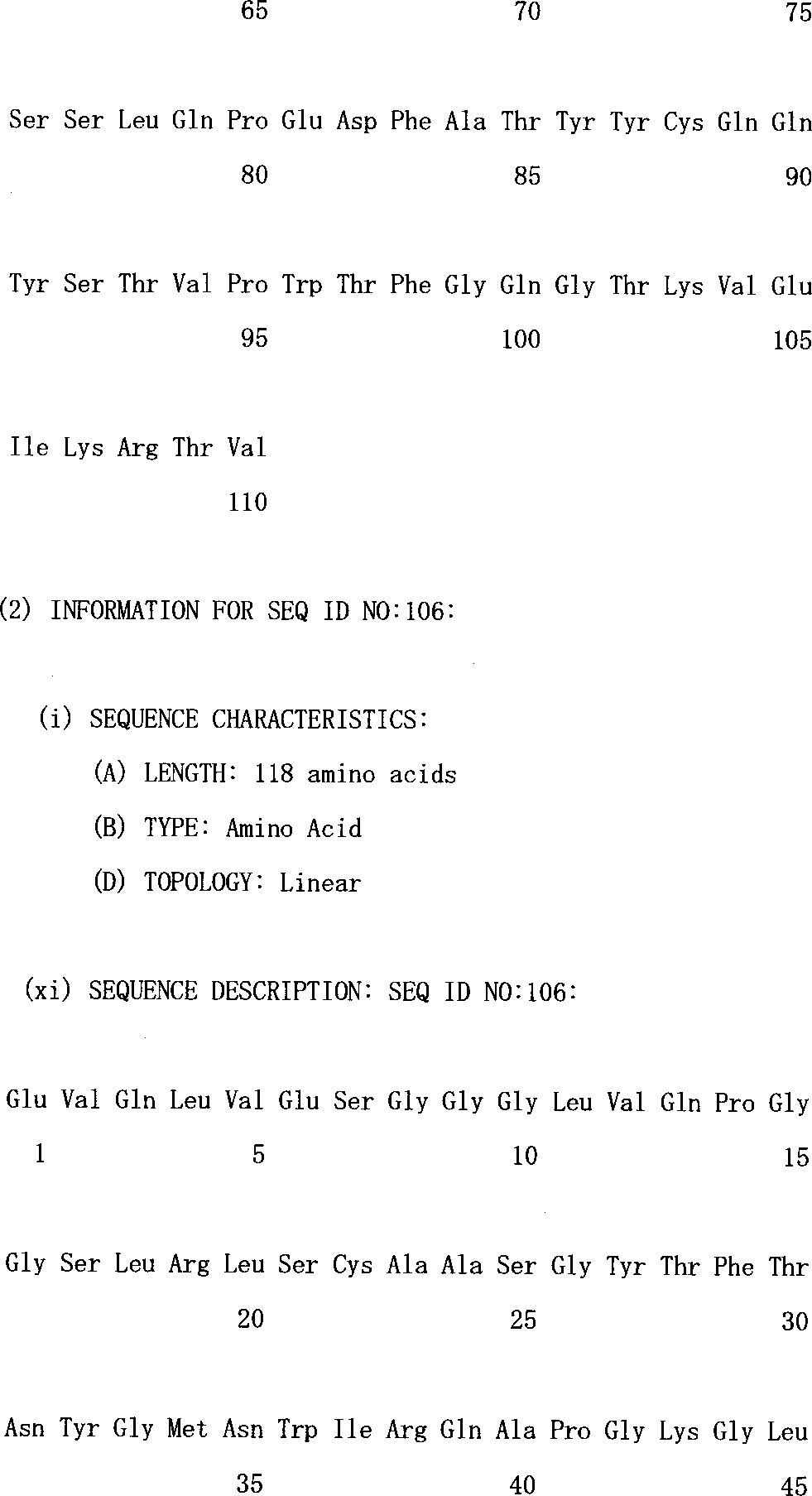
本発明は上記重鎖可変ドメイン配列、または他の重鎖可変ドメイン配列と結合してよい好ましい軽鎖可変ドメイン配列も提供する(ただし、該抗体はヒトVEGFとの結合能を保持するように製造される)。例えば、該軽鎖可変ドメインは以下のアミノ酸配列を有する超可変領域を含んでいてよい:CDRL1(SASQDISNYLN:配列番号4)、CDRL2(FTSSLHS:配列番号5)、およびCDRL3(QQYSTVPWT:配列番号6)。好ましくは、3つの軽鎖超可変領域は、ヒトフレームワーク領域、例えば、以下の式:FR1−CDRL1−FR2−CDRL2−FR3−CDRL3−FR4で示される隣接配列として提供される。
ある態様において、本発明は、下記のアミノ酸配列を含むヒト化抗VEGF抗体軽鎖可変ドメインを提供する:
本発明は親抗VEGF抗体の変異体も提供し(該親抗体は好ましくはヒト化またはヒト抗VEGF抗体である)(ここで、変異体はヒトVEGFと結合する)、親抗VEGF抗体の重鎖または軽鎖可変ドメインの超可変領域にアミノ酸置換を含む。該変異体は、好ましくは抗VEGF抗体の1またはそれ以上の超可変領域中に1またはそれ以上の置換を有する。好ましくは該置換は親抗体の重鎖可変ドメイン中にある。例えば、該アミノ酸置換は、重鎖可変ドメインのCDRH1および/またはCDRH3中にあってよい。好ましくは、これら両超可変領域に置換が存在する。そのような「親和性が成熟した(matured)」変異体は、本明細書において、それが生じた親抗VEGF抗体より強くヒトVEGFと結合することがわかっている(すなわち、該変異体は親抗VEGF抗体より有意に低いKd値を有する)。好ましくは、該変異体は、in vitroにおける内皮細胞のVEGF誘導性増殖を阻害するED50値が、抗VEGF抗体より少なくとも約10倍低く、好ましくは少なくとも約20倍低く、最も好ましくは約50倍低い。ある特に好ましい変異体は、アミノ酸配列:GYDFTHYGMN(配列番号128)を含むCDRH1、およびアミノ酸配列:YPYYYGTSHWYFDV(配列番号129)を含むCDRH3を有する、実施例3のY0317変異体である。これら超可変領域およびCDRH2は一般的にヒトフレームワーク領域中に提供され、例えば、配列番号118のアミノ酸配列を含む重鎖可変ドメインを生じる。そのような重鎖可変ドメイン配列は所望により、配列番号126のアミノ酸配列を含む軽鎖可変ドメイン、好ましくは、配列番号117の軽鎖可変ドメインアミノ酸配列と結合する。
該抗体の種々の形が本明細書から予期される。例えば、抗VEGF抗体は完全長の抗体(例えば、完全ヒトFc領域を有する)、または抗体断片(例えば、Fab’またはF(ab’)2)であってよい。さらに、該抗体は、検出可能な標識で標識するか、固相上に固定化するか、そして/またはヘテロローガスな化合物(細胞毒性物質のような)と結合させてよい。
該抗体の診断的および治療的使用が予期される。ある診断的応用において、本発明は、VEGFタンパク質を含むと思われる試料を抗VEGF抗体に曝露し、該抗体の該試料への結合を測定することを含むVEGFタンパク質の存在を測定する方法を提供する。この使用のために、本発明はVEGFタンパク質を検出するための抗体と該抗体を使用するための指示書を含むキットを提供する。
さらに本発明は、該抗体をコードする単離された核酸、所望により、該ベクターで形質転換された宿主細胞によって認識される制御配列と操作可能に結合した該核酸を含むベクター、該ベクターを含む宿主細胞、該核酸が発現するように宿主細胞を培養し、所望により該宿主細胞培養から(例えば、宿主細胞培養培地から)該抗体を回収することを含む該抗体の製造方法を提供する。本発明は抗VEGF抗体と医薬的に許容される担体または希釈剤を含む組成物も提供する。治療に用いる組成物は無菌であり、凍結乾燥してもよい。さらに、本発明は、腫瘍または網膜障害に罹患した哺乳動物の治療方法であって、該哺乳動物に対し治療的有効量の抗VEGF抗体を投与することを含む方法を提供する。
【図面の簡単な説明】
図1Aおよび1Bは、muMabVEGF A.4.6.1の可変重ドメイン(配列番号9)および軽ドメイン(配列番号10)、ヒト化F(ab)(F(ab)−12)の可変重ドメイン(配列番号7)および軽ドメイン(配列番号8)、およびヒトコンセンサスフレームワーク(重サブグループIIIのhumIII(配列番号11、軽κサブグループ1のhumκ1(配列番号12)を示す。図1Aは可変重ドメイン配列を一列に並べ、図1Bは可変軽鎖ドメイン配列を一列に並べている。アスタリスクはヒト化F(ab)−12とネズミMab、またはF(ab)−12とヒトフレームワークの相違を示す。相補性決定領域CDR)には下線を付している。
図2は、ヒト化F(ab)−12VLおよびVHドメインのモデルの帯状ダイアグラムを示す。VLドメインは茶色で示し、CDRは黄褐色で示す。残基L46の側鎖は黄色で示す。VHドメインは紫色で、CDRは桃色で示す。ヒトからネズミに変化したVH残基の側鎖は黄色で示す。
図3は、実施例1から得られるヒト化抗VEGF F(ab)−12によるVEGF誘導性有糸分裂誘発の阻害を示す。ウシ副腎皮質由来毛細内皮細胞を、実施例1に記載のごとく6ウェルプレートに6x10³個/ウェルの密度で摂取した。muMAb VEGF A.4.6.1またはrhuMAb VEGF(IgG1;F(ab)−12)のいずれかを、示した濃度に加える。2〜3時間後、rhVEGF165を最終濃度3ng/mLに加える。5または6日後、細胞をトリプシン処理し、算定する。示した値はデュプリケートで測定した平均である。平均からの変動は10%以下である。
図4は、実施例1から得られるヒト化抗VEGF F(ab)−12によるin vivoにおける腫瘍増殖の阻害を示す。A673横紋筋肉腫細胞をマウス1匹につき2x10⁶個の密度でBALB/cヌードマウスに注射した。腫瘍細胞を摂取して24時間後から、動物の腹腔内に、1週間に2回、コントロールMAb、muMAbVEGF A4.6.1、またはrhuVEGF MAb(IgG1;F(ab)−12)の注射を開始した。コントロールMabの用量は5mg/kgであり、抗VEGF MAbは示したごとく0.5または5mg/kg投与した(n=10)。腫瘍細胞を注射して4週後に、動物を安楽死させ、腫瘍を取り出し、計量した。*ANOVAによるコントロール群と比較したときの有意差(P<0.05)。
図5Aおよび5Bは、それぞれ、実施例2から得られるネズミ抗体A4.6.1(VLは配列番号10、VHは配列番号9)、ヒト化A4.6.1変異体hu2.0(VLは配列番号13、VHは配列番号14)、およびhu2.10(VLは配列番号15、VHは配列番号16)の軽および重可変ドメインのアミノ酸配列を示す。配列番号付けは、Kabatら、Sequences of Proteins of Immunological Interest, 第5版、Public Health Service, National Institutes of Health, Bethesda, MD(1991)に従い、ミスマッチはアスタリスク(ネズミA4.6.1対hu2.0)またはビュレット(hu2.0対hu2.10)で示す。変異体hu2.0は、ヒト軽鎖κサブグループIコンセンサスフレームワーク(配列番号12)および重鎖サブグループIIIコンセンサスフレームワーク(配列番号11)上に組み込んだネズミ抗体由来のCDR配列(太字)のみを含む。hu2.10は、本明細書に記載のファージソーティング試験から得られたコンセンサスヒト化クローンであった。
図6は、実施例2における無作為化の標的となるフレームワーク残基を示す。
図7は、ファージ上のFab−pIII融合物の表面を表現するファージミッド構築物を示す。ファージミッドはM13遺伝子IIIコートタンパク質の部分と融合した抗体A4.6.1のFab断片のヒト化バージョンをコードする。融合タンパク質は重鎖のカルボキシ末端で1グルタミン残基、次いで遺伝子IIIタンパク質のC末端領域(残基249−406)と連結したFab(supE E.coliのアンバーコドンのサプレッションから)からなる。F+ E.coliへの形質転換、次いでM13KO7ヘルパーファージによる重感染は、これらの少ない割合が融合タンパク質の1コピーを表現するファージミッド粒子を生じる。
図8A−Eは、実施例3のファージ−表現抗体ベクターphMB4−19−1.6のヌクレオチド配列(配列番号99)、およびそれによりコードされるアミノ酸配列(配列番号100〜102)を示す。
図9Aおよび9Bは、それぞれ、実施例1のF(ab)−12と比較した、実施例3の親和性が成熟した抗VEG変異体の軽および重可変ドメインのアミノ酸配列の配置を示す(軽および重可変ドメイン、それぞれ配列番号8および7)。CDRには下線を付しており、L、軽鎖、またはH、重鎖および番号1−3で示す。残基には、Kabat番号付け法とは反対にVLおよびVHドメインに連続番号をつける。鋳型分子、MB1.6(軽および重可変ドメイン、それぞれ配列番号103および104)は変異体:H2305.6(軽および重可変ドメイン、それぞれ配列番号105および106)、Y0101(軽および重可変ドメイン、それぞれ配列番号107および108)、およびY0192(軽および重可変ドメイン、それぞれ配列番号109および110)と共に示される。F(ab)−12との相違は影付ボックスで示す。
図10Aおよび10Bは、それぞれ、実施例1のF(ab)−12と比較した、実施例3から得られる親和性が成熟した抗VEGF変異体の軽および重可変ドメインのアミノ酸配列の配置を示す(軽および重可変ドメイン、それぞれ配列番号8および7)。CDRには下線を付しており、L、軽鎖、またはH、重鎖および番号1−3で示す。変異体は、Y0243−1(軽および重可変ドメイン、それぞれ配列番号111および112)、Y0238−3(軽および重可変ドメイン、それぞれ配列番号113および114)、Y0313−1(軽および重可変ドメイン、それぞれ配列番号115および116)、およびY0317(軽および重可変ドメイン、それぞれ配列番号117および118)で示される。F(ab)−12との相違は影付ボックスで示す。
図11は、実施例1から得られる完全長F(ab)−12、および変異体Y0238−3、Y0192、およびY0313−1に対する実施例3のHuVEC活性アッセイの結果を示す。
図12は、実施例1からの完全長F(ab)−12(rhuMAb VEGF)、実施例1からのF(ab)−12のFab断片(rhuFab VEGF)、および実施例3からの親和性が成熟したY0317のFab断片(rhuFab VEGF(親和性成熟))によるVEGF誘導性有糸分裂誘発の阻害を示す。
好ましい態様の詳細な説明
I.定義
本明細書で用いている用語「ヒトVEGF」は、165アミノ酸のヒト血管内皮細胞成長因子、および関連121−、189−、および206−アミノ酸血管内皮細胞成長因子(Leungら、Science 246:1306(1989)、およびHouckら、Mol. Endocrin.5:1806(1991)に記載)、およびこれら成長因子の天然のアレル形およびプロセス形を表す。
本発明は、1またはそれ以上のVEGFの生物活性、例えば、有糸分裂誘発または血管形成活性を阻害することができる抗VEGFアンタゴニスト抗体を提供する。VEGFのアンタゴニストは、VEGFの細胞レセプターへの結合に干渉するか、VEGFにより活性化された細胞を無能力にするかもしくは殺すか、またはVEGFが細胞レセプターに結合した後の血管内皮細胞の活性化に干渉することにより作用する。VEGFアンタゴニストによる介入のそのようなすべての点は本発明の目的と等価であると考えられよう。
本明細書で用いている用語「VEGFレセプター」または「VEGFr」は、通常血管内皮細胞にみいだされる細胞表面レセプターであるVEGFの細胞レセプター、およびhVEGFとの結合能を保持しているその変異体を表す。VEGFレセプターの1例は、チロシンキナーゼファミリーの貫膜レセプターであるfms様チロシンキナーゼ(flt)である。DeVriesら、Science 255:989(1992), Shibuyaら、Oncogene 5:519(1990)。fltレセプターは、チロシンキナーゼ活性を有する細胞外ドメイン、貫膜ドメイン、および細胞内ドメインを含む。細胞外ドメインはVEGFの結合に関与するが、細胞内ドメインはシグナル変換に関与する。VEGFレセプターの別の例は、flk−1レセプター(KDRとも呼ぶ)である。Matthewsら、Proc.Nat.Acad.Sci.88:9026(1991), Termanら、Oncogene 6:1677(1991),Termanら、Biochem.Biophys.Res.Commun.187:1579(1992)。VEGFのfltレセプターへの結合は、見かけの分子量が205000および300000ダルトンの少なくとも2つの高分子量コンプレックスの形成をもたらす。300000ダルトンのコンプレックスはVEGFの1分子と結合したレセプター2分子を含む二量体であると考えられる。
本明細書で用いている用語「エピトープA4.6.1」は、特記しない限り、Kimら、Growth Factors 7:53(1992)およびKimら、Nature 362:841(1993)に開示のA4.6.1抗体と結合するヒトVEGFの領域を表す。
「治療(処置)」は、治療的処置および予防的もしくは抑制的方法の両方を表す。治療を要するもの(対象)には、すでに障害を有するものおよび障害を予防すべきものが含まれる。
治療を目的とする「哺乳動物」は、ヒト、およびイヌ、ウマ、ネコ、乳牛などのような家畜(domestic and farm animals)、および動物園の、競技用、もしくはペット動物を含む哺乳動物として分類されるあらゆる動物を表す。
「抗体」(Ab)および「免疫グロブリン」(Ig)は同じ構造特性を有する糖タンパク質である。抗体は特異抗原との結合特異性を有するが、免疫グロブリンは抗体と抗原特異性を欠く他の抗体様分子の両方を含む。後者のポリペプチドは、例えば、リンパ系により低レベルで、またミエローマにより高レベルで産生される。
「天然抗体」および「天然免疫グロブリン」は、通常、2つの同じ軽(L)鎖と2つの同じ重(H)鎖からなる、約150000ダルトンのヘテロ四量体糖タンパク質である。各軽鎖は、1共有ジスルフィド結合により重鎖と結合しているが、ジスルフィド結合の数は異なる免疫グロブリンアイソタイプの重鎖間で異なる。各重および軽鎖は一定の間隔で鎖内ジスルフィド架橋も有する。各重鎖は1末端に可変ドメイン(VH)、次いで多くの定常ドメインを有する。各軽鎖は1末端に可変ドメイン(VL)と他の末端に定常ドメインを有し、軽鎖の定常ドメインは重鎖の第一定常ドメインと一列に並んでおり、軽鎖可変ドメインは重鎖の可変ドメインと一列に並んでいる。特定のアミノ酸残基は、軽および重鎖可変ドメイン間にインターフェースを形成すると考えられている。
用語「可変(の)」は、可変ドメインのある部分の配列が抗体間で広範囲に異なる事実を表し、各特定抗体のその特定抗原に対する結合および特性において用いられる。しかしながら、可変性は抗体の可変ドメイン全体に均一に分布するわけではない。可変性は、軽鎖および重鎖可変ドメイン両方の超可変領域と呼ばれる3部分に集中している。可変ドメインのより高度に保存された部分はフレームワーク領域(FR)と呼ばれる。天然の重および軽鎖の可変ドメインはそれぞれ、大部分がβ−シート配置をとり、3つの超可変領域によって結合し、β−シート構造を繋ぎ、ある場合にはその一部を形成するループを形成する4つのFR(それぞれ、FR1、FR2、FR3、およびFR4)を含む。各鎖中の超可変領域はFRによって密接に隣接して結合し、その他の鎖の超可変領域とともに抗体の抗原結合部位の形成に関与する(Kabatら、Sequences of Proteins of Immunological Interest,第5版、Public Health Service,National Institutes of Health,Bethesda,MD(1991),647-669頁参照)。定常ドメインは抗体と抗原との結合に直接関与しないが、抗体依存性細胞毒性における抗体の関与といった種々のエフェクター機能を有する。
本明細書において用語「超可変領域」は、抗原との結合をもたらす抗体のアミノ酸残基を表す。超可変領域は「相補性決定領域」または「CDR」からのアミノ酸残基を含む(すなわち、軽鎖可変ドメインの残基24−34(L1)、50−56(L2)、および89−97(L3)、および重鎖可変ドメインの31−35(H1)、50−65(H2)、および95−102(H3);Kabatら、Sequences of Proteins of Immunological Interest.第5版、Public Health Service,National Institutes of Health,Bethesda,MD(1991))、および/または「超可変ループ」からのアミノ酸残基(すなわち、軽鎖可変ドメインの残基26−32(L1)、50−52(L2)、および91−96(L3)、および重鎖可変ドメインの26−32(H1)、53−55(H2)、および96−101(H3);ChothiaおよびLesk J.Mol.Biol.196:901-917(1987))を含む。「フレームワーク」または「FR」残基は、本明細書で定義した超可変領域残基以外の可変ドメイン残基である。
抗体のパパイン消化により「Fab」断片と呼ばれる、それぞれが1個の抗原結合部位を持った2つの同じ抗原結合断片と、容易に結晶することから名付けられた、残る「Fc」断片が生じる。ペプシン処理すると、2つの抗原結合部位を持ち、まだ抗原を架橋することができるF(ab’)2断片が生じる。
「Fv」は完全な抗原認識と抗原結合部位を含む最小抗体断片である。この領域は、1重鎖および1軽鎖可変ドメインが強く非共有結合した二量体からなる。この配置において、各可変ドメインの3つの超可変領域はVH−VL二量体表面の抗原結合部位を限定するように相互作用する。6つの超可変領域がまとまって抗体に抗原結合特異性をもたらす。しかしながら、1つの可変ドメイン(すなわち、抗原特異的な3つの超可変領域のみを含むFvの半分)でも、完全な結合部位より親和性は低いものの抗原を認識し、結合する能力を持っている。
Fab断片も軽鎖の定常ドメインと重鎖の第一定常ドメイン(CH1)を含む。Fab’断片は、抗体のヒンジ領域からの1またはそれ以上のシステインを含む重鎖CH1ドメインのカルボキシル末端に数個の残基が加わっていることがFab断片とは異なる。本明細書において、Fab’−SHは、定常ドメインのシステイン残基が遊離チオール基を保持するFab’を意味する。F(ab’)2抗体断片は、間にヒンジシステインを有するFab’断片のペアとして本来生じる。抗体断片の他の化学カップリングも知られている。
あらゆる脊椎動物種由来の抗体(免疫グロブリン)の「軽鎖」も、定常ドメインのアミノ酸配列に基づいてカッパ(κ)およびラムダ(λ)と呼ばれる明らかに異なる2タイプの1つに分類することができる。
重鎖の定常ドメインのアミノ酸配列に応じて、免疫グロブリンは異なるクラスに分類することができる。免疫グロブリンにはIgA、IgD、IgE、IgG、およびIgMの5つの主なクラスがあり、これらのいくつかはさらにサブクラス(アイソタイプ)、例えば、IgG1、IgG2、IgG3、IgG4、IgA、およびIgA2に分けることができよう。異なるクラスの免疫グロブリンに対応する重鎖定常ドメインは、それぞれα、δ、ε、γ、およびμと呼ばれる。異なるクラスの免疫グロブリンのサブユニット構造および三次元配置はよく知られている。
本明細書において用語「抗体」は、最も広い意味で用いられ、特に、モノクローナル抗体(完全長モノクローナル抗体を含む)、ポリクローナル抗体、多特異性抗体(例えば、二特異性抗体)、および所望の生物活性を有する抗体断片を含む。
「抗体断片」は、完全長抗体の部分、一般的にその抗原結合もしくは可変ドメインを含む。抗体断片の例には、Fab、Fab’、F(ab’)2、およびFv断片、ディアボディ(diabody)、線状抗体、一本鎖抗体分子、および抗体断片から形成された多特異性抗体を含む。
本明細書において用語「モノクローナル抗体」は、実質的にホモローガスな抗体、すなわち、ポピュレーションを構成する個々の抗体が少量存在するかも知れない天然に生じ得る突然変異を除いて同一なポピュレーションから得られる抗体を表す。モノクローナル抗体は特異性が高く、単一抗原部位に対して誘導される。さらに、典型的には、異なる決定基(エピトープ)に対して誘導された異なる抗体を含む通常の(ポリクローナル)抗体調製物とは異なり、各モノクローナル抗体は抗原の単一決定基に対して誘導される。用語「モノクローナル」は、実質的にホモローガスな抗体のポピュレーションから得られる抗体の特性を表し、該抗体を何らかの特定の方法により製造する必要があるものと解釈してはならない。例えば、本発明にしたがって用いるモノクローナル抗体は、Kohlerら、Nature 256:495(1975)が最初に記載したハイブリドーマ法により製造するか、組換えDNA法(例えば、米国特許第4816567号参照)により製造することができよう。「モノクローナル抗体」は、例えば、Clacksonら、Nature 352:624-628(1991)およびMarksら、J.Mol.Biol.222:581-597(1991)に記載の技術を用いてファージ抗体ライブラリーから単離することもできよう。
本明細書においてモノクローナル抗体は、特に、重および/または軽鎖の部分が特定の種由来の抗体の対応する配列と同じかもしくはホモローガスであるか、または特定の抗体のクラスもしくはサブクラスに属するが、残りの鎖が別の種由来の抗体の対応する配列と同じかもしくはホモローガスであるかまたは別の抗体のクラスもしくはサブクラスに属する「キメラ」抗体、および所望の生物活性を有する、そのような抗体の断片(米国特許第4816567号、およびMorrisonら、Proc.Natl.Acad.Sci.USA 81:6851-6855(1984)を含む。
非ヒト(例えばネズミ)抗体の「ヒト化」形は、非ヒト免疫グロブリン由来の配列を最小限に含むキメラ抗体である。主としてヒト化抗体は、レシピエントの超可変領域残基が所望の特異性、親和性、および能力を有する、マウス、ラット、ウサギ、またはヒト以外の霊長類といったヒト以外の種(ドナー抗体)由来の超可変領域残基により置換されているヒト免疫グロブリン(レシピエント抗体)である。場合によっては、ヒト免疫グロブリンのフレームワーク領域(FR)残基が対応する非ヒト残基で置換される。さらに、ヒト化抗体はレシピエント抗体やドナー抗体中にみられない残基を含んでいてよい。これらの修飾は抗体の能力をさらに洗練させるためになされる。一般的に、ヒト化抗体は、超可変領域のすべてもしくは実質的にすべてが非ヒト免疫グロブリンのそれに対応しており、FRのすべてもしくは実質的にすべてがヒト免疫グロブリン配列のそれである、少なくとも一つ、典型的には2つの可変ドメインを実質的にすべて含むであろう。所望により、ヒト化抗体は免疫グロブリン定常領域(Fc)の部分、典型的にはヒト免疫グロブリンの部分も含むであろう。さらなる詳細は、Jonesら、Nature 321:522-525(1986),Reichmannら、Nature 332:323-329(1988)、およびPresta, Curr.Op.Struct.Biol.2:593-596(1992)参照。
「一本鎖Fv」または「sFv」抗体断片は、一本のポリペプチド鎖中に存在する、抗体のVHおよびVLドメインを含む。一般的に、FvポリペプチドはさらにVHおよびVLドメイン間にポリペプチドリンカーを含み、これによりsFvは抗原と結合するための所望の構造を形成することができる。sFvの総説については、Pluckthun in The Pharmacology of Monoclonal Antibodies,vol.113,RosenburgおよびMoore編、Springer-Verlag,New York, 269-315(1994)参照。
用語「ディアボディ」は、同じポリペプチド鎖(HV−VL)において軽鎖可変ドメイン(VL)と結合した重鎖可変ドメイン(VH)を含む、2つの抗原結合部位を有する小抗体断片を表す。同じ鎖上の2つのドメインをペアリングさせるには短すぎるリンカーを用いることにより、該ドメインを別の鎖と相補的なドメインと強制的にペアリングさせ、2つの抗原結合部位を生じさせる。ディアボディについては、例えば、EP404097、WO93/11161、およびHollingerら、Proc.Natl. Acad.Sci.USA 90:6444-6448(1993)により完全に記載されている。
本明細書において用語「線状抗体」は、Zapataら、Protein Eng.8(10): 1057-1062(1995)に記載の抗体を表す。簡単には、該抗体は抗原結合領域のペアを形成するタンデムFd部分(VH−CH1−VH−CH1)のペアを含む。
本明細書において「変異体」抗VEGF抗体は、親抗体配列中の1またはそれ以上の残基の付加、欠失、および/または置換により、「親」抗VEGF抗体アミノ酸配列とはアミノ酸配列が異なる分子を表す。好ましい態様において、該変異体は親抗体の1またはそれ以上の超可変領域に1またはそれ以上のアミノ酸置換を含む。例えば、該変異体は親抗体の1またはそれ以上の超可変領域に少なくとも1個、例えば、約1〜約10、好ましくは約2〜約5個の置換を含んでいてよい。通常、該変異体は親抗体重または軽鎖可変ドメイン配列(例えば、配列番号7または8中の)と少なくとも75%、より好ましくは少なくとも80%、より好ましくは少なくとも85%、より好ましくは少なくとも90%、最も好ましくは少なくとも95%のアミノ酸配列の同一性を有するアミノ酸配列を有するであろう。本明細書において、この配列の同一性または相同性は、必要であれば配列同一性パーセントが最大となるように配列を一列に並べ、ギャップを挿入した後の、親抗体残基と同一である候補配列中のアミノ酸残基のパーセントで定義する。抗体配列へのN末端、C末端、または内部伸長、欠失、もしくは挿入はいずれも配列の同一性または相同性に影響しないと解釈すべきである。該変異体はヒトVEGFとの結合能を保持し、好ましくは親抗体より優れた特性を有する。例えば、該変異体は、より強い結合親和性を有し、内皮細胞のVEGF誘導性増殖の阻害能が増大し、そして/またはin vivoにおけるVEGF誘導性血管形成の阻害能が増大していてよい。そのような特徴を分析するには、例えば、抗VEGF抗体の構成は本明細書に開示した生物活性アッセイにおけるその活性に影響することがわかっているので、変異体のFab形を親抗体のFab形と比較するか、または変異体の完全長形と親抗体の完全長形を比較すべきである。本明細書において特に目的とする変異体抗体は、親抗体と比べて少なくとも約10倍、好ましくは少なくとも約20倍、最も好ましくは少なくとも約50倍生物活性が増大している変異抗体である。
本明細書において「親」抗体は、該変異体を製造するのに用いるアミノ酸配列によってコードされる。好ましくは、該親抗体はヒトフレームワーク領域と、もしあれば、ヒト抗体定常領域を有する。例えば、親抗体はヒト化またはヒト抗体であってよい。
「単離された」抗体は、天然環境の成分から同定され、分離および/または回収されたものである。天然環境の夾雑成分は、抗体の診断的または治療的使用と干渉する物質であり、これには酵素、ホルモン、および多のタンパク質または非タンパク質性溶質が含まれよう。好ましい態様において、該抗体は、(1)Lowry法で測定して抗体の95重量%以上まで、好ましくは、99重量%以上まで、(2)スピニングカップシーケネターを用いてN末端または内部アミノ酸配列の少なくとも15残基を得るのに十分な程度に、または(3)クマシーブルーまたは好ましくは銀染色を用い、還元もしくは非還元条件下でSDS−PAGEにより均質となるまで精製されるであろう。単離された抗体は、該抗体の天然環境の少なくとも1要素は存在しないであろうから、組換え細胞中のその場の抗体を含む。しかしながら、通常、単離された抗血合いは少なくとも1つの精製工程により製造されよう。
本明細書において用語「タグ付エピトープ」は「エピトープタグ」と融合した抗VEGF抗体を表す。エピトープタグポリペプチドは、それに対する抗体を作ることができるエピトープを提供するのに十分な残基を有するが、VEGF抗体の活性と干渉しないように十分短い。該エピトープタグは、好ましくは、それに対する抗体が他のエピトープと実質的に交差反応しないほど十分にユニークである。一般的に、適切なタグポリペプチドは少なくとも6アミノ酸残基、通常約8〜50アミノ酸残基(好ましくは約9〜30残基)を有する。例にはflu HAタグポリペプチドおよびその抗体12CA5(Fieldら、Mol.Cell.Biol.8:2159-2165(1988))、c−mycタグ、およびそれに対する8F9、3C7、6E10、G4、B7、および9E10抗体(Evanら、Mol.Cell.Biol.5(12):3610-3616(1985)、およびHerpes Simplexウイルス糖タンパク質D(gD)タグ、およびその抗体(Paborskyら、Protein Engineering 3(6):547-553(1990)が含まれる。ある態様において、エピトープタグは「サルベージレセプター結合エピトープ」である。本明細書において、用語「サルベージレセプター結合エピトープ」は、IgG分子のin vivoにおける血清半減期の増大をもたらすIgG分子(例えば、IgG1、IgG2、IgG3、またはIgG4)のFc領域のエピトープを表す。
本明細書において、用語「細胞毒性物質」は、細胞の機能を阻害するか、抑制し、そして/または細胞を破壊する物質を表す。該用語は、放射性同位元素(例えば、I131、I125、Y90、およびRe186)、化学療法剤、および細菌、真菌、植物もしくは植物起源の酵素活性を有する毒素のような毒素、またはその断片を含むことを意図している。
「化学療法剤」は癌の治療に有用な化合物である。化学療法剤の例には、アドリアマイシン、ドキソルビシン、5−フルオロウラシル、シトシンアラビノシド(「Ara−C」)、シクロホスファミド、チオテパ、タキソテレ(Taxotere、ドセタキセル)、ブスルファン、サイトキシン、タキソール、メトトレキセート、シスプラチン、メルファラン、ビンブラスチン、ブレオマシン、エトポシド、イフォスファミド、マイトマイシンC、マイトキサントロン、ビンクレイスチン、ビノレルビン、カルボプラチン、テニポシド、ダウノマイシン、カルミノマイシン、アミノプテリン、ダクチノマイシン、マイトマイシン、エスペラマイシン(米国特許第4675187号参照)、メルファラン、および他の関連ナイトロジェンマスタードが含まれる。
本明細書において用語「プロドラッグ」は、親薬剤に比べて腫瘍細胞に対する毒性が低い医薬的に活性な物質の前駆体または誘導体を表し、酵素的に活性化または変換されてより活性な親形になることができる。例えば、Wilman,「Prodrugs in Cancer Chemotherapy」Biochemical Society Transactions, 14,375-382頁、第615回会議、Belfast(1986)、およびStellaら、「Prodrugs;A Chemical Approach to Targeted Drug Delivery」,Directed Drug Delivery,Borchardtら編、247-267頁,Humama Press(1985)参照。本発明のプロドラッグには、限定されるモノではないが、ホスフェート含有プロドラッグ、チオホスフェート含有プロドラッグ、サルフェート含有プロドラッグ、ペプチド含有プロドラッグ、D−アミノ酸修飾プロドラッグ、グリコシル化プロドラッグ、β−ラクタム含有プロドラッグ、所望により置換されたフェノキシアセトアミド含有プロドラッグ、または所望により置換されたフェニルアセトアミド含有プロドラッグ、5−フルオロシトシン、およびより活性な無毒性薬剤に変換することができる他の5−フルオロウリジンプロドラッグを含む。本発明に用いるプロドラッグ形に誘導体化することができる細胞毒性薬剤の例には、限定されるものではないが、上記化学療法剤が含まれる。
本明細書において用語「標識」は抗体と直接または間接的に結合する検出可能な化合物または組成物を表す。標識は、それ自身が検出可能であるか(例えば、放射性同位元素標識、または蛍光標識)、または酵素標識の場合には検出可能な基質化合物または組成物の化学変化を触媒することができよう。
「固相」は、本発明の抗体を吸着することができる非水性マトリックスを意味する。本明細書に含まれる固相の例には、部分的または完全にガラス(例えば孔制御ガラス)、ポリサッカライド(例えばアガロース)、ポリアクリルアミド、ポリスチレン、ポリビニルアルコールおよびシリコンで形成されたものがある。ある態様では文脈に応じて、固相はアッセイプレートのウェルを、他では精製用カラム(例えばアフィニティクロマトグラフィカラム)を含むことができる。この用語には、米国特許第4275149号に記載のような分離した粒子の不連続な固相も含まれる。
「リポソーム」は、種々のタイプの脂質、リン脂質、および/または薬剤(本明細書に開示の抗VEGF抗体、および所望により化学療法剤のような)を哺乳動物に供給するのに有用な界面活性剤からなる小胞である。リポソームの成分は、通常、生物膜の脂質配置と同様に二重層を形成するよう配列する。「単離された」核酸分子は、同定され、抗体核酸の天然の供給源と通常結合している少なくとも1つの夾雑核酸分子から分離された核酸分子である。単離された核酸分子は天然にみいだされる形または配列(setting)以外のものである。したがって、単離された核酸分子は天然細胞に存在する核酸分子とは区別される。しかしながら、単離された核酸分子は、例えば天然の細胞とは異なる染色体位置にある、抗体を通常発現する細胞に含まれる核酸分子を含む。
用語「制御配列」は、特定の宿主生物中で操作可能に連結されたコード配列を発現するのに必要なDNA配列を表す。例えば、原核生物に適した制御配列にはプロモーター、所望によりオペレーター配列、およびリボソーム結合部位が含まれる。真核細胞はプロモーター、ポリアデニル化シグナル、およびエンハンサーを利用することが知られている。
核酸は、別の核酸配列と機能的関係に置かれているとき「操作可能に連結している」という。例えば、プレ配列のDNAまたは分泌リーダーは、それがポリペプチドの分泌に関与するプレタンパク質として発現する場合、ポリペプチドのDNAと操作可能に連結しているか、プロモーターまたはエンハンサーは、それらが配列の転写に影響を及ぼす場合にコード配列と操作可能に連結しているか、またはリボソーム結合部位は、それが翻訳を促すように位置している場合にコード配列と操作可能に連結している。一般的に、「操作可能に連結している」とは、連結しているDNA配列が隣接しており、分泌リーダーの場合には隣接し、読み取り段階にあることを意味する。しかしながら、エンハンサーは隣接していてはならない。連結は好都合な制限部位で結合することにより達成される。そのような部位が存在しない場合は、合成オリゴヌクレオチドアダプターまたはリンカーを通常の方法に従って用いる。
本明細書において、用語「細胞」、「細胞系」、および「細胞培養」は置き換え可能に用いられ、すべてのそのような名称には子孫が含まれる。このように、用語「形質転換体」および「形質転換細胞」には、一次対象細胞とトランスファー数に関わらずそれらから誘導された培養を含む。すべての子孫は、意図的または偶然の突然変異によりDNA含量が正確に同じではないことがあることも理解される。最初の形質転換細胞についてスクリーニングされたのと同じ機能または生物活性を有する突然変異子孫も含まれる。異なる意味を意図する場合は文脈から明らかであろう。
II.本発明の実施方法
下記の実施例において、(a)VEGF抗原に対する強い結合親和性、(b)in vitroにおける内皮細胞のVEGF誘導性増殖を阻害する能力、および(c)in vivoにおけるVEGF誘導性血管形成の阻害能を含む治療的観点から望ましい特性を有するヒト化および変異体抗VEGF抗体の製造方法を説明する。
抗体の親和性は、下記実施例に記載のごとく測定することができよう。好ましいヒト化または変異体抗体は、約1x10⁻⁷Mを超えない、好ましくは約1x10⁻⁸Mを超えない、最も好ましくは約5x10⁻⁹Mを超えないKd値でヒトVEGFと結合する抗体である。
ヒトVEGFに対する強い結合親和性を有する抗体のほかに、治療的観点から他の有益な特性を有するヒト化または変異体抗体を選ぶことも好ましい。例えば、該抗体はVEGFに応じた内皮細胞増殖を阻害するものであってよい。ある態様において、該抗体は、VEGFのほぼ最大有効濃度(3ng/mL)に応じたウシ毛細血管内皮細胞増殖を阻害することができよう。好ましくは、該抗体の「内皮細胞増殖アッセイ」において内皮細胞のVEGF誘導性増殖を阻害するための有効用量50(ED50)値は、約5nMを超えず、好ましくは約1nMを超えず、最も好ましくは約0.5nMを超えず、すなわち、これらの濃度で該抗体はin vitroにおけるVEGF誘導性内皮細胞増殖を50%阻害することができる。好ましい「内皮細胞増殖アッセイ」には、下記実施例1に実質的に記載のごとく、10%子ウシ血清、2mMグルタミン、および抗生物質を添加した低グルコースDulbecco改良Eagle培地(DMEM)(GIBCO)(増殖培地)の存在下でウシ副腎皮質由来毛細血管内皮細胞を培養することを含む。この内皮細胞を、増殖培地を入れた6ウェルプレートに6x10³個の密度で接種する。次に、親抗VEGF抗体(コントロール)、ヒト化または変異体抗VEGF抗体を1〜5000ng/mLの範囲の濃度で加える。2〜3時間後、精製VEGFを最終濃度3ng/mLに加える。特異性のコントロールとして、各抗体を単独かまたは2ng/mLのbFGFの存在下で、濃度5000ng/mLで内皮細胞に加えてよい。5または6日後、細胞をトリプシンに曝露して分離し、Coulterカウンター(Coulter Electronics,Hialeah,FL)でカウントする。データは4パラメーター適合プログラム(KaleidaGraph)により分析することができよう。
好ましいヒト化または変異体抗VEGF抗体は、in vitro腫瘍抑制活性を有するものでもよい。例えば、該抗体は、ヌードマウスにおいてヒトA673横紋筋肉腫細胞または乳癌MDA−MB−435細胞の増殖を抑制することができよう。in vivo腫瘍実験において、ヒトA673横紋筋肉腫細胞(ACTT;CRL1598)または乳癌MDA−MB−435細胞(ACTTから入手可能)を、下記実施例1に記載のごとく10%ウシ胎児血清、2mMグルタミン、および抗生物質を添加したDMEM/F12中で培養する。雌BALB/cヌードマウス(6〜10週令)の背部に腫瘍細胞2x106個(容量200μL)を皮下注射する。次に、動物をヒト化または変異体抗体、およびこのアッセイにおいて活性のないコントロール抗体で処理する。ヒト化または変異体抗VEGF MAbは0.5および/または5mg/kgの用量で投与する。腫瘍細胞を接種して24時間後から各MAb(容量100μL)の1週間に2回の腹腔内投与を開始する。腫瘍のサイズを1週間間隔で測定する。腫瘍細胞を接種して4週間後に動物を安楽死させ、腫瘍を取り出し、重さを量る。統計分析はANOVAにより行うことができよう。好ましくは、この「in vivo腫瘍アッセイ」において該抗体は用量5mg/kgで、ヒトA673腫瘍細胞の増殖を約50〜100%、好ましくは約70〜100%、最も好ましくは約80〜100%阻害する。
好ましい態様において、ヒト化または変異体抗体は、ヒト患者に治療的有効量の抗体を投与しても免疫原性反応を誘導しない。免疫原性反応を誘導する場合は、好ましくは該反応は、該抗体がまだそれで治療される患者に治療的恩恵を与えるほどのものであろう。
ヒト化または変異体抗体は、ヒトにおけるVEGF誘導性血管形成を阻害する、例えば、ヒト腫瘍増殖を阻害し、そして/または網膜障害において眼内血管形成を阻害することができるものも好ましい。
好ましい抗体は、本明細書で定義した「エピトープA4.6.1」を結合する。目的とする抗体と結合するヒトVEGFのエピトープと結合する抗体(例えば、A4.6.1抗体のヒトVEGFへの結合をブロックするもの)をスクリーニングするには、Antibodies, A Laboratory Manual,Cold Spring Harbor Laboratory,HarlowおよびDavid Lane編(1988)に記載されているような常套的交差ブロッキングアッセイを実施することができる。あるいはまた、例えば、Champeら、J.Biol.Chem.270:1388-1394(1995)に記載のエピトープマッピングを実施し、抗体が目的とするエピトープと結合するかどうかを測定する。
本明細書において、好ましい態様の抗体は、式:FR1−CDRH1−FR2−CDRH2−FR3−CDRH3−FR4(ここで、「FR1−4」は4つのフレームワーク領域を表し、「CDRH1−3」は抗VEGF抗体可変重ドメインの3つの超可変領域を表す)によって示されるアミノ酸配列を含む重鎖可変ドメインを有する。FR1−4は、下記実施例に記載のごとく「コンセンサス配列」(すなわち、ヒト免疫グロブリンの重または軽鎖のクラス、サブクラス、またはサブグループの最も共通したアミノ酸)から誘導されるか、または個々のヒト抗体フレームワーク領域もしくは異なるフレームワーク領域配列の結合物から誘導することができよう。多くのヒト抗体フレームワーク領域配列は、例えばKabatら(上記)に記載されている。ある好ましい態様において、可変重FRは、Kabatら(上記)に記載のごとくヒト免疫グロブリンサブグループのコンセンサス配列により提供される。好ましくは、ヒト免疫グロブリンサブグループはヒト重鎖サブグループIII(例えば配列番号11にあるような)である。好ましくは、ヒト可変重FR配列はその中に置換を有し、例えば、ヒトFR残基は対応する非ヒト残基で置換されているが(「対応する非ヒト残基」は、ヒトおよび非ヒト配列を一列に並べる時に目的とするヒト残基と同じKabat位置番号を有する非ヒト残基を意味する)、非ヒト残基による置換は必要でない。例えば、対応する非ヒト残基以外の置換FR残基はファージ表示により選ぶことができよう(下記実施例2参照)。置換されていてよい可変重FR残基に例には、FR残基番号:37H、49H、67H、69H、71H、73H、75H、76H、78H、94H(ここではKabat残基番号付け法を用いた)のいずれか1つまたはそれ以上を含む。好ましくは、これら残基の少なくとも2つ、もしくは少なくとも3つ、または少なくとも4つが置換されている。FR置換の特に好ましい組合せは、49H、69H、71H、73H、76H、78H、および94Hである。
重鎖超可変領域に関して、これらは以下のアミノ酸配列を有することが好ましい:
CDRH1
GYX1X2X3X4YGX5N(配列番号119)(ここで、X1はD、T、またはEであるが好ましくはDまたはTであり、X2はF、W、またはYであるが好ましくはFであり、X3はT、Q、G、またはSであるが好ましくはTであり、X4はHまたはNであり、X5はMまたはIであるが好ましくはMである)。
CDRH2
WINTX1TGEPTYAADFKR(配列番号120)(ここで、X1はYまたはWであるが好ましくはYである)。
CDRH3
YPX1YX2X3X4X5HWYFDV(配列番号121)(ここで、X1はHまたはYであり、X2はY、R、K、I、T、E、またはWであるが好ましくはYであり、X3はG、N、A、D、Q、E、T、K、またはSであるが好ましくはGであり、X4はS、T、K、Q、N、R、A、E、またはGであるが好ましくはSまたはTであり、X5はSまたはGであるが好ましくはSである)。
所望により、重鎖可変ドメインは本明細書においてFR3中の(すなわち、の一部を構成する)「CDR7」で示される(図9Bおよび10B参照)(ここで、CDR7は以下のアミノ酸配列を有してよい:
CDR7
X1SX2DX3X4X5X6TX7(配列番号122)(ここで、X1はF、I、V、L、またはAであるが好ましくはFであり、X2はA、L、V、またはIであるが好ましくはLであり、X3はT、V、またはKであるが好ましくはTであり、X4はSまたはWであるが好ましくはSであり、X5はSまたはKであるが好ましくはKであり、X6はNまたはSであるが好ましくはSであり、X7はV、A、L、またはIであるが好ましくはAである))。
本明細書において好ましい態様の抗体は、式:FR1−CDRL1−FR2−CDRL2−FR3−CDRL3−FR4(ここで、「FR1−4」は4つのフレームワーク領域を表し、「CDRL1−3」は抗VEGF抗体可変重ドメインの3つの超可変領域を表す)で示されるアミノ酸配列を含む軽鎖可変ドメインを有する。FR1−4は、下記の実施例に記載のごとく「コンセンサス配列」(すなわち、ヒト免疫グロブリンの重または軽鎖のクラス、サブクラス、またはサブグループの最も共通したアミノ酸)から誘導するか、または個々のヒト抗体フレームワーク領域もしくは異なるフレームワーク領域配列の組合せから誘導することができよう。ある好ましい態様において、可変軽FRはKabat(上記)に記載のごとくヒト免疫グロブリンサブグループのコンセンサス配列により提供される。好ましくは、ヒト免疫グロブリンサブグループはヒトκ軽鎖サブグループI(例えば、配列番号12中)。
好ましくは、ヒト可変軽FR配列はその中に置換を有するが(例えば、ここで、ヒトFR残基は対応するマウス残基で置換されている)、非ヒト残基による置換は必要でない。置換されていてよい可変FR残基の例には、FR残基番号:4L、46L、および71L(ここではKabat残基番号付け法を用いた)のいずれか1つまたはそれ以上を含む。好ましくは、46Lのみが置換されている。別の態様において、4Lおよび46Lがともに置換されている。
CDRに関して、これらは以下のアミノ酸配列を有することが好ましい:
CDRL1
X1AX2X3X4X5SNYLN(配列番号123)(ここで、X1はRまたはSであるが好ましくはSであり、X2はSまたはNであるが好ましくはSであり、X3はQまたはEであるが好ましくはQであり、X4はQまたはDであるが好ましくはDであり、X5はIまたはLであるが好ましくはIである)。
CDRL2
FTSSLHS(配列番号124)。
CDRL3
QQYSX1X2PWT(配列番号125)(ここで、X1はT、A、またはNであるが好ましくはTであり、X2はVまたはTであるが好ましくはVである)。
好ましいヒト化抗VEGF抗体は、実施例1のF(ab)−12の重および/または軽可変ドメイン配列を有する抗体、および実施例3の変異体Y0317、Y0313−1、およびY0238−3を含む親和性成熟形のような該抗体の変異体であり、Y0317が好ましい変異体である。本明細書において目的とするヒト化抗VEGF抗体を製造する方法については以下に詳述する。
A.抗体の製造
非ヒトVEGF抗体のヒト化方法、および抗VEGF抗体の変異体の製造方法は下記実施例に記載している。抗VEGF抗体をヒト化するには、非ヒト抗体出発物質を調製する。変異体を作製する親抗体を調製する。そのような非ヒト抗体出発物質および親抗体の作製方法の例は以下の項に記載する。
(i)抗原の製造
抗体の製造に用いるVEGF抗原は、例えば完全VEGFまたはVEGF断片(例えば、「エピトープA4.6.1」を含むVEGF断片)であってよい。抗体を製造するのに有用な他のVEGFは当業者に明らかであろう。抗体を製造するのに用いるVEGF抗原は、好ましくは、例えばLeungら、Science 246: 1306(1989)、およびHouckら、Mol.Endocrin.5:1806(1991)に記載のヒトVEGFである。
(ii)ポリクローナル抗体
ポリクローナル抗体は、関連抗原とアジュバントを複数回皮下(sc)または腹腔内(ip)注射することにより動物で生じさせるのが好ましい。二機能性または誘導化剤、例えば、マレイミドベンゾイルスルホスクシンイミドエステル(システイン残基を介して結合)、N−ヒドロキシスクシンイミド(リジン残基を介する)、グルタールアルデヒド、コハク酸無水物、SOCl2、またはR1 N=C=NR(ここで、RおよびR1は異なるアルキル基である)を用い、関連抗原を、免疫する種に対して免疫原性であるタンパク質、例えば、キーホールリンペットヘモシアニン、血清アルブミン、ウシサイログロブリン、またはダイズトリプシンインヒビターと結合させるのに有用かも知れない。
例えば、タンパク質またはコンジュゲート100μgまたは5μg(それぞれウサギまたはマウスに対し)をフロインドの完全アジュバント3容量と混合し、該溶液を複数部位に皮内注射することにより、動物を抗原、免疫原性コンジュゲート、または誘導体で免疫する。1ヶ月後に、フロインドの完全アジュバント中の、最初の量の1/5〜1/10のペプチドまたはコンジュゲートを複数箇所に皮下注射することにより動物にブースターをかける。7〜14日後に動物から採血し、血清中の抗体価を測定する。抗体価がプラトーに達するまで動物にブースターをかける。好ましくは、別の架橋試薬を用いておよび/または異なるタンパク質と結合させた同じ抗原とのコンジュゲートを用いて動物にブースターをかけるのが好ましい。コンジュゲートは組換え細胞培養中でタンパク質融合物として作製することもできる。アラムのような凝集剤も免疫応答の増強に使用するのに適している。
(iii)モノクローナル抗体
モノクローナル抗体は、Kohlerら、Nature,256:495(1975)に最初に記載のハイブリドーマ法を用いて製造するか、または組換えDNA法(米国特許第4816567号)により製造することができよう。
ハイブリドーマ法において、マウス、またはハムスターもしくはマカークザルのような他の適切な宿主動物を既述のごとく免疫し、免疫に用いたタンパク質と特異的に結合する抗体を産生するか、もしくは産生することができるリンパ球を誘導する。次に、ポリエチレングリコールのような適切な融合剤を用いてリンパ球をミエローマ細胞と融合させ、ハイブリドーマ細胞を形成する(Goding, Monoclonal Antibodies: Principles and Practice,59-103(Academic Press,1986)。
このように作製したハイブリドーマ細胞を、好ましくは非融合の親ミエローマ細胞の成長または生存を阻害する1またはそれ以上の物質を含む、適切な培養液に接種し、増殖させる。例えば、親ミエローマ細胞が酵素ヒポキサンチングアニンホスホリボシルトランスフェラーゼ(HGPRTまたはHPRT)を欠く場合は、典型的には、該ハイブリドーマ用の培養液には、HGPRT−欠損細胞の増殖を抑制する物質であるヒポキサンチン、アミノプテリン、およびチミジンを含むであろう(HAT培地)。
好ましいミエローマ細胞は、効率的に融合し、選択した抗体産生細胞による抗体の安定した高レベルの産生を支持し、HAT培地のような培地に感受性である。これらのうち、好ましいミエローマ細胞系は、Salk Institute Cell Distribution Center,San Diego,California USAから入手可能なMOP−21およびM.C.−11マウス腫瘍、およびAmerican Type Culture Collection,Rockville,Maryland USAから入手可能なSP−2またはX63−Ag8−653細胞から誘導される細胞のようなネズミミエローマ系である。ヒトミエローマおよびマウス−ヒトヘテロミエローマ細胞系によるヒトモノクローナル抗体の製造も記載されている(Kozbor,J.Immunol.,133:3001(1984), Brodeurら、Monoclonal Antibody Production Techniques and Applications、51-63(Marcel Dekker,Inc.,New York,1987))。
ハイブリドーマ細胞が増殖している培養液について、該抗原に対するモノクローナル抗体の産生をアッセイする。好ましくは、ハイブリドーマ細胞により産生されたモノクローナル抗体の結合特異性を、免疫沈降法、またはラジオイムノアッセイ(RIA)もしくは酵素免疫測定法(ELISA)のようなin vitro結合アッセイにより測定する。
モノクローナル抗体の結合親和性は、例えばMunsonら、Anal.Biochem.,107:220(1980)のScatchard分析により測定することができる。
所望の特異性、親和性、および/または活性の抗体を産生するハイブリドーマ細胞を同定し、次いで、該クローンを限界希釈法によりサブクローンし、標準的方法により増殖させることができよう(Goding,Monoclonal Antibodies: Principles and Practice,59-103(Academic Press,1986)。この目的に適した培養液は、例えば、D−MEMまたはRPMI−1640培地を含む。さらに、ハイブリドーマ細胞はin vivoにおいて動物の腹水腫瘍として増殖することができよう。
サブクローンにより分泌されたモノクローナル抗体は、例えば、プロテインA−セファロース、ヒドロキシアパタイトクロマトグラフィ、ゲル電気泳動、透析、またはアフィニティクロマトグラフィのような通常の免疫グロブリン精製法により培養液、腹水、または血清から適切に分離される。
モノクローナル抗体は、コードするDNAは通常の手順を用いて容易に単離し、配列決定することができる(例えば、モノクローナル抗体の重および軽鎖をコードする遺伝子と特異的に結合することができるオリゴヌクレオチドプローブを用いることにより)。ハイブリドーマ細胞はDNAのような好ましい供給源として供給される。単離したら、DNAを発現ベクターに組み込み、次いでこれをこれをE.coli細胞、シミアンCOS細胞、チャイニーズハムスター卵巣(CHO)細胞、または別の方法では免疫グロブリンタンパク質を産生しないミエローマ細胞といった宿主細胞にトランスフェクションし、組換え宿主細胞中でモノクローナル抗体を合成させる。抗体の組換えによる製造については以下でより詳細に説明する。
(iv)ヒト化およびアミノ酸配列変異体
以下の実施例1−2は、抗VEGF抗体のヒト化方法を記載する。ある態様において、これらのヒト化抗体のアミノ酸配列変異体を作成するのが望ましくあり得、特に、これらは、ヒト化抗体の結合親和性または他の生物学的性質を改善する。実施例3は、親抗体に比べて親和性が高められた抗VEGF抗体のアミノ酸配列変異体を作成する方法を記載する。
抗VEGF抗体のアミノ酸配列変異体は、適当なヌクレオチド変化を抗VEGF抗体のDNAに導入することにより、またはペプチド合成により製造する。そのような変異体には、例えば、本明細書中での実施例の抗VEGF抗体のアミノ酸配列内の残基からの欠失、および/またはそれらの残基への挿入、および/またはそれらの残基の置換が含まれる。最終構築物が所望の特性を有するならば、欠失、挿入、および置換のいずれかの組み合わせを最終構築物で達成させる。アミノ酸変化はまた、グリコシル化部位の数または位置を変化させるような、ヒト化または変異抗VEGF抗体の翻訳後プロセスも変え得る。
変異誘発に好ましい位置である抗VEGF抗体のある残基または領域の同定に有用な方法は、CunninghamおよびWells, Science, 244:1081−1085(1989)により記載されているように、「アラニン走査型変異誘発」と呼ばれる。ここでは、標的残基の残基または基を同定して(例えば、arg、asp、his、lys、およびgluといったような荷電残基)、中性または陰性に荷電アミノ酸(最も好ましくは、アラニンまたはポリアラニン)で置換して、アミノ酸とVEGF抗原との相互作用に影響を与える。次いで、さらなる変異体または他の変異体を置換部位に導入することにより、またはそれらを置換部位に代えて導入することにより、置換に対する機能的感受性を実証するそれらのアミノ酸の位置を正確にする。従って、アミノ酸配列の変異を導入する部位は予め決定しておくが、その変異の種類を本来は予め決定しておく必要はない。例えば、ある一定部位での変異の成果を分析するためには、ala走査またはランダム変異誘発を標的コドンまたは領域で行って、発現した抗VEGF抗体の変異体を所望の活性に関してスクリーニングする。アラニン走査型変異誘発を実施例3に記載する。
アミノ酸配列挿入には、1つの残基から100またはそれ以上の残基を含むポリペプチドの範囲にわたるアミノおよび/またはカルボキシ末端融合、さらにはまた、単一または複数のアミノ酸残基の配列内挿入が含まれる。末端挿入の例には、N末端メチオニル残基をもつ抗VEGF抗体、またはエピトープタグに融合した抗体が含まれる。抗VEGF抗体分子の他の挿入変異体には、抗体の血清半減期を増大させる酵素またはポリペプチドの抗VEGF抗体のNまたはC末端への融合が含まれる(以下を参照)。
別のタイプの変異体は、アミノ酸置換変異体である。これらの変異体は、抗VEGF抗体分子中の少なくとも1つのアミノ酸残基が除去されていて、違う残基がその場所に挿入されている。置換変異誘発に最も重要な部位には超可変領域が含まれるが、FR変更もまた意図する。同型置換を「好ましい置換」という表題で表1に示す。そのような置換が生物学的活性に変化を及ぼすなら、表1において「例示的な置換」と称する、またはアミノ酸クラスに関して以下にさらに記載する、より多くの実質的変化を導入して、生成物をスクリーニングするのがよい。
(1)疎水性:ノルロイシン、met、ala、val、leu、ile;
(2)中性の疎水性:cys、ser、thr;
(3)酸性:asp、glu;
(4)塩基性:asn、gln、his、lys、arg;
(5)鎖の方向に影響を及ぼす残基:gly、pro;および
(6)芳香族:trp、tyr、phe。
非同型置換は、これらのクラスのうちの1つのメンバーを別のクラスに交換することを必然的に伴う。
ヒト化または変異抗VEGF抗体の適切な配座を維持するのに関与しないシステイン残基をいずれもまた、一般的には、セリンで置換して、その分子の酸化安定性を改善して、異常な架橋を防ぐこともできる。逆に、システイン結合を抗体に付加して、その安定性を改善することができる(特に、その抗体がFvフラグメントのような抗体フラグメントである場合)。
特に好ましいタイプの置換変異体は、親抗体(例えば、ヒト化またはヒト抗体)の1つまたはそれ以上の超可変領域の残基を含む。一般的には、さらなる開発のために選択される、その結果得られた変異体は、それらを作成する親抗体に比べて改善された生物学的性質を有するであろう。そのような置換変異体を作成するための便利な方法は、ファージ表示を使用する親和性成熟である(本明細書中の実施例3を参照)。簡単に言えば、幾つかの超可変領域の部位(例えば、6−7の部位)を変異させて、各々の部位で全ての可能なアミノ置換を生じさせる。このようにして作成した抗体の変異体を、各々の粒子内に詰め込まれたM13の遺伝子III産物への融合のような、繊維状ファージ粒子に由来する一価方法で表示する。次いで、そのファージで表示した変異体を、本明細書中に開示するそれらの生物学的活性(例えば、結合親和性)に関してスクリーニングする。改変に関して候補となる超可変領域の部位を同定するために、抗原結合に著しく寄与する同定された超可変領域の残基に対して、アラニン走査型変異誘発(実施例3を参照)を行うことができる。あるいはまた、または加えて、抗原−抗体複合体の結晶構造を分析して、抗体とヒトVEGFとの間の接触点を同定するのが有利となり得る。そのような接触残基および隣接残基は、本明細書中で詳細に述べる技術による置換に関しての候補である。そのような変異体を作成したら、パネルの変異体を本明細書中に記載するスクリーニングにかけて、1つまたはそれ以上の関連したアッセイにおいて優れた性質をもつ抗体をさらなる開発のために選択するのがよい。
抗体の別のタイプのアミノ酸変異体は、その抗体の元のグリコシル化パターンを変える。変えるという語は、抗体中に見出される1つまたはそれ以上の炭水化物部分を欠失させること、および/または抗体中には存在しない1つまたはそれ以上のグリコシル化部位を付加することを意味する。
抗体のグリコシル化は、典型的には、N結合型またはO結合型である。N結合型は、アスパラギン残基の側鎖への炭化水素部分の結合をいう。トリペプチド配列のアスパラギン−X−セリンおよびアスパラギン−X−トレオニン(Xは、プロリンを除き、いずれかのアミノ酸である。)は、アスパラギン側鎖への炭化水素部分の酵素結合に対する認識配列である。従って、ポリペプチド中のこれらのトリペプチド配列のいずれかの存在は、可能性のあるグリコシル化部位を作り出す。O結合型グリコシル化は、ヒドロキシアミノ酸、最も一般的には、セリンまたはトレオニンへの、糖であるN−アセチルガラクトサミン、ガラクトース、またはキシロースのうちの1つの結合をいうが、5−ヒドロキシプロリンまたは5−ヒドロキシリジンもまた使用することができる。
抗体へのグリコシル化部位の付加は、便利には、1つまたはそれ以上の上記の(N結合型グリコシル化部位に対する)トリペプチド配列を含むよう、アミノ酸配列を変えることにより成し遂げられる。その変更はまた、元の抗体の配列への(O結合型グリコシル化部位に対する)1つもしくはそれ以上のセリンもしくはトレオニン残基の付加、またはそれらでの置換によっても成され得る。
抗VEGF抗体のアミノ酸配列変異体をコードする核酸分子は、当業界で知られている様々な方法により製造する。これらの方法には、限定されるものではないが、天然源からの単離(天然に存在するアミノ酸配列変異体の場合)、または抗VEGF抗体の初期に作成した変異体または非変異体種のオリゴヌクレオチドが媒介する(もしくは部位特異的)変異誘発、PCR変異誘発、およびカセット式変異誘発による製造が含まれる。
(v)ヒト抗体
ヒト化の他の方法として、ヒト抗体を作成することができる。例えば、現在、免疫化すると、内因性免疫グロブリン産生の不存在下に全レパトアのヒト抗体を産生することができるトランスジェニック動物(例えば、マウス)を作ることが可能である。例えば、キメラおよび生殖系変異マウスにおける抗体の重鎖接続領域(JH)遺伝子のホモ接合欠失は、内因性抗体産生の完全な阻害を引き起こすことが記載されている。そのような生殖系変異マウスにおけるヒト生殖系免疫グロブリン遺伝子アレイの転移は、抗原攻撃に対してヒト抗体の産生を引き起こすであろう。例えば、Jakobovitsら,Proc. Natl. Acad. Sci. USA, 90:2551(1993);Jakobovitsら, Nature, 362:255−258(1993);Bruggermannら, Year in Immuno., 7:33(1993);並びに米国特許第5,591,669号、同第5,589,369号、および同第5,545,807号を参照。ヒト抗体はまた、ファージ表示ライブラリーから誘導することもできる(Hoogenboomら, J. Mol. Biol,, 227:381(1991);Marksら, J. Mol. Biol., 222:581−597(1991);並びに米国特許第5,565,332号および同第5,573,905号)。上に論じたように、ヒト抗体はまた、インビトロにおける活性化B細胞により作成することもできる(米国特許第5,567,610号および同第5,229,275号を参照)。
(vi)抗体フラグメント
ある態様において、ヒト化または変異抗VEGF抗体は、抗体フラグメントである。様々な技術が抗体フラグメントの産生に関して開発されている。伝統的には、これらのフラグメントを完全な抗体のタンパク質分解消化によって誘導した(例えば、Morimotoら, Journal of Biochemical and Biophysical Methods 24:107−117(1992)およびBrennanら, Science 229:81(1985)を参照)。しかし、これらのフラグメントは、現在、組換え宿主細胞により直接産生することができる。例えば、Fab’−SHフラグメントをE. coliから直接回収し、化学的に結合させて、F(ab’)2フラグメントを形成することができる(Carterら, Bio/Technology 10:163−167(1992))。別の態様において、ロイシンジッパーGCN4を使用して、F(ab’)2を形成し、F(ab’)2分子の集合を促進する。別の方法により、Fv、Fab、またはF(ab’)2フラグメントを組換え宿主細胞培養から直接単離することができる。抗体フラグメントの産生のための他の技術は、当業者に明らかであろう。
(vii)多重特異性抗体
幾つかの態様において、少なくとも2つの違うエピトープに対して結合特異性を有する、多重特異性(例えば、二重特異性)ヒト化または変異抗VEGF抗体を作成するのが望ましくあり得る。例示的な二重特異性抗体は、VEGFタンパク質の2つの違うエピトープに結合し得る。あるいはまた、VEGF発現細胞に対する細胞防御機構に焦点を合わせるよう、抗VEGFアームを、T細胞受容体分子(例えば、CD2もしくはCD3)のような白血球上のトリガーリング(triggering)分子、またはFcγRI(CD64)、FcγRII(CD32)、およびFcγRIIIに(CD16)といったような、IgGに対するFc受容体(FcγR)に結合するアームと組み合わせてもよい。二重特異性抗体をまた使用して、VEGFを発現する細胞に細胞傷害性物質を局在化することもできる。これらの抗体は、VEGF結合アーム、および細胞傷害性物質(例えば、サポリン、抗インターフェロンα、ビンカアルカロイド、リシンA鎖、メトトレキセート、または放射性同位体ハプテン)を結合するアームを有する。二重特異性抗体は、全長の抗体または抗体フラグメント(例えば、F(ab’)2二重特異性抗体)として製造することができる。
二重特異性抗体を製造するための別の方法により、一対の抗体分子間の界面を操作して、組換え細胞培養から回収されるヘテロダイマーのパーセンテージを最大にすることができる。好ましい界面は、抗体の定常ドメインのCH3ドメインの少なくとも部分を含んでなる。この方法では、一次抗体分子の界面に由来する1つまたはそれ以上の小さなアミノ酸側鎖をより大きな側鎖(例えば、チロシンまたはトリプトファン)で置換する。大きなアミノ酸側鎖をより小さな側鎖(例えば、アラニンまたはトレオニン)で置換することにより、その大きな側鎖と同一または同様の大きさの代償的な「空洞」が二次抗体分子の界面上に作り出される。このことは、ホモダイマーのような他の所望されていない最終生成物に対するヘテロダイマーの収率を増大させる機構を与える。1996年9月6日に発行されたWO96/27011を参照。
二重特異性抗体には、架橋または「ヘテロコンジュゲート」抗体が含まれる。例えば、ヘテロコンジュゲートでの抗体のうちの1つをアビジンに結合して、他方をビオチンに結合することができる。いずれかの便利な架橋方法を使用して、ヘテロコンジュゲート抗体を製造することができる。適当な架橋剤は当業界で周知であって、多くの架橋技術と共に、米国特許第4,676,980号に開示されている。
抗体フラグメントから二重特異性抗体を作成する技術もまた文献に記載されている。例えば、化学結合を使用して、二重特異性抗体を製造することができる。Brennanら, Science 229:81(1985)は、完全な抗体をタンパク質分解により切断して、F(ab’)2フラグメントを作成するという方法を記載している。これらのフラグメントをジチオール錯化剤である亜ヒ酸ナトリウムの存在下に還元し、隣接のジチオールを安定化して、分子間ジスルフィド形成を防ぐ。次いで、作成したFab’ブラグメントをチオニトロベンゾエート(TNB)誘導体に転換する。次いで、そのFab’−TNB誘導体のうちの1つをメルカプトエチルアミンでの還元によりFab’−チオールに再び転換し、等モル量の他のFab’−TNB誘導体と混合して、二重特異性抗体を形成する。産生した二重特異性抗体は、酵素の選択的固定化のための物質として使用することができる。またさらなる態様において、E. coliから直接回収したFab’−SHフラグメントをインビトロにおいて化学的に結合させて、二重特異性抗体を形成することができる。Shalabyら, J. Exp. Med. 175:217−225(1992)。
組換え細胞培養から直接、二重特異性抗体フラグメントを製造して単離するための様々な技術もまた記載されている。例えば、ロイシンジッパーを使用して、二重特異性抗体を産生している。Kostelnyら, J. Immunol. 148(5):1547−1553(1992)。FosおよびJunタンパク質に由来するロイシンジッパーペプチドを遺伝子融合により2つの違う抗体のFab’部分に結合させた。その抗体ホモダイマーをヒンジ領域で還元して、モノマーを形成した後、再び酸化して、抗体ヘテロダイマーを形成した。この方法はまた、抗体ホモダイマーの産生にも利用することができる。Hollingerら, Proc. Natl. Acad. Sci. USA 90:6444−6448(1993)により記載されている「ジアボディ(diabody)」技術は、二重特異性抗体フラグメントを製造するための他の機構を与えている。そのフラグメントは、短すぎて同じ鎖上の2つのドメインの間を対にすることができないリンカーにより軽鎖可変ドメイン(VL)に結合した重鎖可変ドメイン(VH)を含んでなる。従って、1つのフラグメントのVHおよびVLドメインを別のフラグメントの相補的VLおよびVHドメインと対にするようにし、それによって、2つの抗原結合部位を形成する。一本鎖Fv(sFv)ダイマーの使用により二重特異性抗体フラグメントを製造するための別の方法もまた報告されている。Gruberら, J. Immunol. 152:5368(1994)を参照。あるいはまた、二重特異性抗体は、Zapataら, Protein Eng. 8(10):1057−1062(1995)に記載されているように製造した「線状抗体」であってもよい。
2価以上の抗体を意図する。例えば、三重特異性抗体を製造することができる。Tuttら, J. Immunol. 147:60(1991)を参照。
(viii)他の改変
ヒト化または変異抗VEGF抗体の他の改変を意図する。例えば、癌を処置する際の抗体の有効性を高めるよう、エフェクター機能に関して本発明の抗体を改変するのが、例えば、望ましくあり得る。例えば、システイン残基をFc領域に導入し、それによって、この領域での鎖間ジスルフィド結合形成を可能とし得る。このようにして作成したホモダイマー抗体は、改善されたインターナリゼーション能力および/または増大された補体が媒介する細胞壊死および抗体依存性細胞傷害性(ADCC)を有し得る。Caronら, J. Exp. Med. 176:1191−1195(1992)およびShopes, B., J. Immunol. 148:2918−2922(1992)を参照。Wolffら, Cancer Research 53:2560−2565(1993)に記載されているヘテロ二官能性架橋物質を使用して、高められた抗腫瘍活性をもつホモダイマー抗体をまた製造することもできる。あるいはまた、2つのFc領域を有する抗体を操作することができ、それによって、高められた補体溶解およびADCC能力を有し得る。Stevensonら, Anti-Cancer Drug Design 3:219−230(1989)を参照。
本発明はまた、化学療法剤、毒素(例えば、細菌、真菌、植物もしくは動物起源の酵素的に活性な毒素、またはそのフラグメント)、または放射性同位体(すなわち、放射性コンジュゲート)といったような細胞傷害性物質にコンジュゲートした本明細書中に記載する抗体を含んでなるイムノコンジュゲートに関する。
そのようなイムノコンジュゲートの作成に有用な化学療法剤を上に記載している。使用することができる酵素的に活性な毒素およびそのフラグメントには、ジフテリアA鎖、ジフテリア毒素の非結合活性フラグメント、(Pseudomonas aeruginosaに由来する)エンドトキシンA鎖、リシンA鎖、アブリンA鎖、モデシン(modeccin)A鎖、α−サルシン(sarcin)、Aleurites fordiiタンパク質、ジアンチン(dianthin)タンパク質、Phytolaca americanaタンパク質(PAPI、PAII、およびPAP−S)、モモルディカ・キャランティア(momordica charantia)阻害剤、クルシン(curcin)、クロテン(crotin)、サパオナリア・オフィシナリス(sapaonaria officinalis)阻害剤、ゲロニン(gelonin)、マイトゲリン(mitogellin)、レストリクトシン(restrictocin)、フェノマイシン(phenomycin)、エノマイシン(enomycin)、およびトリコテセンズ(tricothecenes)が含まれる。様々な放射性核種を放射性コンジュゲートした抗VEGF抗体の産生に利用することができる。例には、212Bi、131I、131In、90Y、および186Reが含まれる。
N−スクシンイミジル−3−(2−ピリジルジチオール)プロピオネート(SPDP)、イミノチオラン(iminothiolane)(IT)、イミドエステルの二官能性誘導体(例えば、ジメチル・アジピミデート(dimethyl adipimidate)HCL)、活性エステル(例えば、スベリン酸ジスクシンイミジル)、アルデヒド(例えば、グルタルアルデヒド)、ビス−アジド化合物(例えば、ビス(p−アジドベンゾイル)ヘキサンジアミン)、ビス−ジアゾニウム誘導体(例えば、ビス(p−ジアゾニウムベンゾイル)エチレンジアミン)、ジイソシアネート(例えば、トリエン2,6−ジイソシアネート)、およびビス−活性フッ素化合物(例えば、1,5−ジフルオロ−2,4−ジニトロベンゼン)といったような、様々な二官能性タンパク質カップリング剤を使用して、抗体および細胞傷害性物質のコンジュゲートを製造する。例えば、リシンイムノトキシンは、Vitettaら, Science 238:1098(1987)に記載されているように製造することができる。炭素−14で標識化した1−イソチオシアナトベンジル−3−メチルジエチレントリアミンペンタ酢酸(MX−DTPA)は、抗体への放射性核種のコンジュゲーションに関する例示的なキレート化剤である。WO94/11026を参照。
別の態様において、抗体を腫瘍の前標的化における利用のための「受容体」(例えば、ストレプトアビジン)にコンジュゲートさせ、ここで、その抗体−受容体コンジュゲートを患者に投与し、続いて、キレート化剤を使用して、結合していないコンジュゲートを循環から除去した後、細胞傷害性物質(例えば、放射性核種)にコンジュゲートしている「リガンド」(例えば、アビジン)を投与するのがよい。
本明細書中に開示する抗VEGF抗体はまた、イムノリポソームとして製剤化することもできる。Epsteinら, Proc. Natl. Acad. Sci. USA, 82:3688(1985);Hwangら, Proc. Natl. Acad. Sci. USA, 77:4030(1980);並びに米国特許第4,485,045号および同第4,544,545号に記載されているような、当業界で知られている方法により、その抗体を含むリポソームを製造する。高められた循環時間をもつリポソームは、米国特許第5,013,556号に開示されている。
特に有用なリポソームは、ホスファチジルコリン、コレステロール、およびPEGで誘導体化したホスファチジルエタノールアミン(PEG−PE)を含んでなる脂質組成物での逆相蒸発方法により作成することができる。リポソームを、定められた細孔径のフィルターを通して押出し、所望の直径をもつリポソームを得る。本発明の抗体のFab’フラグメントは、Martinら, J. Biol. Chem. 257:286−288(1982)に記載されているように、ジスルフィド交換反応によってリポソームにコンジュゲートすることができる。場合により、化学療法剤(例えば、Doxorubicin)をリポソーム内に含む。Gabizonら, J. National Cancer Inst. 81(19):1484(1989)を参照。
本発明の抗体はまた、その抗体を、プロドラッグ(例えば、ペプチジル化学療法剤、WO81/01145を参照)を活性抗癌剤に転換するプロドラッグ活性化酵素にコンジュゲートさせることにより、ADEPTにおいて使用することもできる。例えば、WO88/07378および米国特許第4,975,278号を参照。
ADEPTに有用なイムノコンジュゲートの酵素成分には、プロドラッグをそのより活性な細胞傷害性形態に転換するような方法で、それに作用することができるいずれかの酵素が含まれる。
本発明の方法において有用である酵素には、限定されるものではないが、ホスフェートを含むプロドラッグを遊離の薬物へと転換するのに有用なアルカリ性ホスファターゼ;スルフェートを含むプロドラッグを遊離の薬物へと転換するのに有用なアリールスルファターゼ;無毒の5−フルオロシトシンを抗癌剤である5−フルオロウラシルへと転換するのに有用なシトシンデアミナーゼ;ペプチドを含むプロドラッグを遊離の薬物へと転換するのに有用な、セラチア(serratia)プロテアーゼ、サーモリシン、ズブチリシン、カルボキシペプチダーゼ、およびカテプシン(例えば、カテプシンBおよびL)といったようなプロテアーゼ;D−アミノ酸置換基を含むプロドラッグを転換するのに有用なD−アラニルカルボキシペプチダーゼ;グリコシル化プロドラッグを遊離の薬物へと転換するのに有用な、β−ガラクトシダーゼおよびノイラミダーゼといったような炭水化物を切断する酵素;β−ラクタムで誘導体化した薬物を遊離の薬物へと転換するのに有用なβ−ラクタマーゼ;並びに各々、それらのアミン窒素のところをフェノキシアセチルまたはフェニルアセチル基で誘導体化した薬物を遊離の薬物へと転換するのに有用な、ペニシリンVアミダーゼまたはペニシリンGアミダーゼといったようなペニシリンアミダーゼが含まれる。あるいはまた、当業界ではまた「触媒抗体」としても知られている酵素活性をもつ抗体を使用して、本発明のプロドラッグを遊離の活性薬物に転換することができる(例えば、Massey, Nature 328:457−458(1987)を参照)。抗体−触媒抗体コンジュゲートは、腫瘍細胞集団への触媒抗体の送達に関して本明細書中に記載するように製造することができる。
上に論じたヘテロ二官能性架橋試薬の使用のような、当業界でよく知られている技術により、本発明の酵素を抗VEGF抗体に共有結合させることができる。あるいはまた、当業界でよく知られている組換えDNA技術を使用して、本発明の酵素の少なくとも機能的に活性な部分に結合した、本発明の抗原の少なくとも抗原結合領域を含んでなる融合タンパク質を構築することができる(例えば、Neubergerら, Nature 312:604−608(1984)を参照)。
本発明のある態様において、完全な抗体よりはむしろ、抗体フラグメントを使用して、例えば、腫瘍浸透を増大させるのが望ましくあり得る。この場合には、抗体フラグメントを改変して、その血清半減期を増大させるのが望ましくあり得る。このことは、例えば、抗体フラグメントへのサルベージ受容体結合エピトープの取込みにより(例えば、抗体フラグメント中の適当な領域の変異により、またはペプチドタグにエピトープを取込んだ後、例えば、DNAまたはペプチド合成によって、抗体フラグメントに端または真中のところで融合させることにより)達成することができる。1996年10月17日に発行されたWO96/32478を参照。
そのサルベージ受容体結合エピトープは、一般的には、Fcドメインの1つまたは2つのループに由来する、いずれかの1つまたはそれ以上のアミノ酸残基を抗体フラグメントの類似(analogous)領域に転移させるという領域を構成する。さらにより好ましくは、Fcドメインの1つまたは2つのループに由来する、3つまたはそれ以上の残基を転移させる。またより好ましくは、そのエピトープを(例えば、IgGの)Fc領域のCH2ドメインから取って、その抗体のCH1、CH3、またはVH領域、または1つ以上のそのような領域に転移させる。あるいはまた、そのエピトープをFc領域のCH2ドメインから取って、その抗体フラグメントのCL領域もしくはVL領域、または両方に転移させる。
ある最も好ましい態様において、そのサルベージ受容体結合エピトープは、特に、その抗体フラグメントがFabまたはF(ab’)2である場合、配列:PKNS SMISNTP(配列番号17)を含んでなり、場合により、HQSLGTQ(配列番号18)、HQNLSDGK(配列番号19)、HQNISDGK(配列番号20)、またはVISSHLGQ(配列番号21)よりなる群から選択される配列をさらに含んでなる。別の最も好ましい態様において、そのサルベージ受容体結合エピトープは、配列:HQNLSDGK(配列番号19)、HQNISDGK(配列番号20)、またはVISSHLGQ(配列番号21)、および配列:PKNSSMISNTP(配列番号17)を含むポリペプチドである。
ヒト化または変異抗VEGF抗体の共有結合による改変もまた、本発明の範囲内に含まれる。それらは、化学合成により、または適用できるならば、抗体の酵素もしくは化学切断により成され得る。他のタイプの抗体の改変は、抗体の標的化アミノ酸残基を、選択された側鎖またはNもしくはC末端残基と反応することできる有機誘導体化剤と反応させることにより分子に導入される。ポリペプチドの例示的な共有結合による改変は、米国特許第5,534,615号に記載されている(この米国特許に記載されている内容は、本発明の一部を具体的に構成する)。抗体の好ましいタイプの共有結合による改変は、米国特許第4,640,835号、同第4,496,689号、同第4,301,144号、同第4,670,417号、同第4,791,192号、または同第4,179,337号に示されている方法で、抗体を様々な非タンパク質性(nonproteinaceous)ポリマー(例えば、ポリエチレングリコール、ポリプロピレングリコール、またはポリオキシアルキレン)のうちの1つに結合することを含んでなる。
B.ベクター、宿主細胞および組換方法
本発明はまた、ひと化または変異抗−VEGF抗体をコードする分離された核酸、ベクターおよび核酸を含む宿主細胞、および抗体産生のための組換技術を提供する。
抗体の組換体産生のために、これをコードする核酸を分離し、さらにクローニング(DNAの増幅)または発現のための複製可能なベクターに挿入する。別の態様において、この抗体は相同組換によって産生することができる。例えば、それは米国特許第5,204,244号に記載されており、ここに引用してこの明細書の記載とする。モノクローナル抗体をコードする抗体は容易に分離され、常套的手段を用いて配列決定することができる(例えば、この抗体の重鎖および軽鎖をコードする遺伝子に特異的に結合し得るオリゴヌクレオチドプローブを用いることによって)。多くのベクターを利用することができる。一般的に、ベクター成分は下記のもの1つまたはそれ以上を含むが、これに限定されるものではない:シグナル配列、複製開始点、1つまたはそれ以上のマーカー遺伝子、エンハンサー成分、プロモーター、および転写終結点配列、例えば、1996年7月9日発行の米国特許第5,534,615号に記載されており、ここに引用してこの明細書の記載とする。
ここに記載のベクターのDNAをクローニングまたは発現するのに適した宿主細胞は原核生物、酵母、上記の高等真核細胞である。この目的のために適した原核細胞はグラム陰性またはグラム陽性微生物などの真生細菌、例えば、EscherichiaなどのEnterobacteriaceae、例えば、E. coli、Enterobacter、Erwinia、Klebsiella、Proteus、Salmonella、例えば、Salmonella typhimurium、Serratia、例えばSerratia marcescansおよびShigella、およびBacilli、例えばB. subtilisおよびB. licheniformis(例えば、1989年4月12日刊行のDD266,710に開示されたB. licheniformis 41P)、Pseudomonas、例えば、P. aeruginosalおよびStreptomycesなどである。好ましいE. coliクローニング宿主はE. coli294(ATCC 31,446)であるが、E. coli B, E. coli X1776(ATCC 31,537)、およびE. coli W3110(ATCC 27,325)などの他の株も適している。これらの例は説明のためのものであり限定ではない。
原核生物に加えて、糸状菌または酵母などの真核細菌は抗−VEGF抗体コードベクターの適切なクローニングまたは発現宿主である。
Saccharomyces cerevisiaeまたは他の普通のパン酵母は下等真核宿主微生物のうち最も普通に用いられる。しかしながら、多数の他の属、種および株はここでは通常用いられ、有用である。例えば、Schizosaccharomyces pombe;K. lactis、K. fragilis(ATCC 12,424)、K. bulgaricus(ATCC 16,045)、K. wickeramii(ATCC 24,178)、K. waltii(ATCC 56,500)、K. drosophilarum(ATCC 36,906)、K. theromotoleransおよびk. marxiamisなどのKluyveromyces宿主;Pichia pastoris(EP 183,070);Candid;Trichoderma reesia(EP 244,234);Neurospora crassa;Schwanniomyces occidentalisなどのSchwanniomyces;および、例えば、Neurospora、Penicillium、Tolypocladiumなどの糸状菌およびA.nidulansおよびA.nigerなどのAspergillus宿主である。
グコシル化抗−VEGF抗体の発現に適した宿主細胞は多細胞生物から誘導される。無脊椎動物細胞の例には、植物および昆虫細胞が含まれる。多数のバキュロウイルス株および変異体およびSpodoptera frugiperda(イモムシ)、Aedes aegypti(カ)、Aedes albopitus(カ)、Drosophila melanogaster(ミバエ)およびBombyx moriなどの宿主からの対応許容昆虫宿主細胞が確認されている。転写のための種々のウイルス株が一般に用いられており、例えば、Autographa californica NPVのL−1変異体およびBombyx mori NPVのBm−5株であり、また、そのようなウイルスは本発明のここに記載のウイルスとして用いられ、特に、Spodoptera frugiperda細胞の軽質移入のために用いられる。綿、トウモロコシ、ジャガイモ、大豆、ペチュニア、トマトおよびタバコの植物細胞培養物は宿主として用いることができる。
しかしながら、最も興味深いのは脊椎動物細胞であり、培養(組織培養)での脊椎動物細胞の増殖は常套的方法になっている。有用な哺乳動物宿主細胞株の例は、SV40(COS−7、ATCC CRL 1651)によって形質転換されたサル腎臓CV1系;ひと胎児性腎臓系(Graham et al., J. Gen Virol. 36:59(1977)、293または懸濁培養物中生長のためにサブクローニングされた293細胞;幼若ハムスター腎臓細胞(BHK,ATCC CCL 10);チャイニーズハムスター卵巣細胞−DHFR(CHO,Urlaub et al., Proc. Natl. Acad. Sci. USA 77:4216(1980));マウスセルトリ細胞(TM4, Mathgr, Biol. Reprod. 23:243-251(1980));サル腎臓細胞(CVl ATCC CCL 70);アフリカミドリザル腎臓細胞(VERO−76,ATCC CRL−1587);ひと頚部癌細胞(HELA,ATCC CCL 2);イヌ腎臓細胞(MDCK,ATCC CCL 34);牡牛ネズミ肝臓細胞(BRL 3A,ATCC CRL 1442);ひと肺細胞(W138,ATCC CCL 75);ひと肝臓細胞(Hep G2,HB8065);マウス乳腫瘍(MMT 060562,ATCC CCL51);TRI細胞(Matherら、Annals N.Y.Acad. Sci.383:44-68(1982));MRC 5細胞;FS4細胞;およびひと肝臓癌系(Hep G2)である。
宿主細胞は抗−VEGF抗体産生のための上記の発現またはクローニングベクターで形質転換され、プロモーター誘導、形質転換体選択、所望の配列をコードする遺伝子増幅に適するように修正された通常の栄養培地中で培養される。
本発明の抗−VEGF抗体産生のために用いられる宿主細胞は種々の培地中で培養される。Ham'sF10(Sigma)、最小必須培地(MEM),(Sigma)、RPMI−1640(Sigma)およびダルベコ修正イーグル培地((DMEM)、Sigma)などの市販の培地は宿主細胞の培養に適している。さらに、Ham et al., Meth. Enz.58:44(1979)、Barnes et al., Anal.Biochem. 102:255(1980)、米国特許第4,767,704号;4,657,866号;4,927,762号;4,560,655号;または5,122,469号;WO90/03430;WO87/00195;または米国特許Re.第30,985号記載の培地は宿主細胞の培養培地として用いることができる。これらの培地のいくつかには、必要ならば、ホルモンおよび/または他の成長因子(インシュリン、トランスフェリンまたは上皮成長因子など)、塩類(塩化ナトリウム、カルシウム、マグネシウムおよびリン酸塩など)、緩衝液類(HEPESなど)ヌクレオチド類(アデノシンおよびチミジンなど)抗生物質(GENTAMYCINTM薬剤など)、微量成分(通常μモルの範囲の最終濃度で存在する無機化合物として定義)、およびグルコースまたは同等のエネルギー源が補充される。他の必要な補充物が当業者に知られている適当な濃度で含まれる。温度、pHなどの培養条件は発現のために用いられた宿主細胞で既に用いられた条件であり、当業者に明らかである。
組換技術を用いるとき、抗体は細胞内、細胞膜周辺腔中に産生されるか、または直接培地中に放出される。抗体が細胞内に産生されるならば、第1段階として、宿主細胞または融解フラグメントのいずれかの粒子状の細片が、例えば、遠心分離または限外濾過にて除去される。Carter et al., Bio/Technology 10:163-167(1992)はE.coliの細胞膜周辺腔に分泌された抗体の分離方法を開示している。要約すれば、細胞ペーストを酢酸ナトリウム(pH3.5)、EDTAおよびフェニルメチルスルホニルフルオリド(PMSF)の存在下約30分以上浸漬する。細胞片は遠心分離で除去できる。抗体が培地中に分泌される場合は、発現系からの上清は一般に、最初市販の蛋白濃縮フィルター、例えば、アミコンまたはミルポアペリコン限外濾過ユニットによって濃縮される。PMSFなどの蛋白質分解酵素阻害剤を蛋白質分解阻害のために前の段階のどこかで添加してもよく、また、外来性の混入物の成長を阻害するために抗生物質を添加してもよい。
細胞から産生された抗体組成物は例えば、ヒドロキシルアバタイトクロマトグラフィー、ゲル電気泳動、透析およびアフィニティ・クロマトグラフィーを用いて精製することができ、アフィニティ・クロマトグラフィーが精製技術として好ましい。アフイニティリガンドとしてのプロテインAの適合性は、抗体中に存在するイムノグロブリンFcの種類および同位元素によって変化する。プロテインAはひとγ1、γ2またはγ4重鎖に基づく抗体の精製に用いることができる(Lindmark et al., J. Immunol. Meth. 62:I-13(1983))。プロテインGはすべてのマウス同位元素およびひとγ3に好ましい(Guss et al., EMBO J. 5:15671575(1986))。アフィ二ティ・リガンドが結合するマトリックスは多くの場合アガロースであるが、他のマトリックスも使用することができる。調整された細孔ガラスまたはポリ(スチレンジビニル)ベンゼンなどの機械的に安定なマトリックスでは、アガロースがなし得るより、早い流速および短い処理時間が可能である。抗体がCH3ドメインを含む場合は、Bakerbond ABXTM樹脂(J.T. Baker, Phillipsburg, NJ)が精製に好適である。イオン交換カラムでの分留、エタノール沈殿、逆層HPLC、シリカクロマトグラフィー、アニオンまたはカチオン交換樹脂(アスバラギン酸カラム)上のヘパリンセファローズTMクロマトグラフィー、等電点電気泳動、SDS−PAGE、硫酸アンモニウム沈殿などの蛋白質精製のための他の技術もまた、回収する抗体に応じて利用することができる。
何らかの予備的精製段階(複数あり)の後、当該抗体および混入物を、好ましくは低塩濃度(例えば、0−0.25M塩から)で、pH約2.5−4.5の溶出緩衝液を用いて、低pH疎水的相互作用クロマトグラフィーにかける。
C.医薬製剤
抗体の治療製剤は、任意の生理学的に許容され得る担体、賦形剤または安定剤とともに所望の精製度を有する抗体を混合して貯蔵のために、凍結乾燥製剤または水溶液の形態に製造される(Remington's Pharmaceutical Sciences 16th edition, Osol, A. Ed.(1980))。許容され得る担体、賦形剤または安定剤は、レシピエントにとって採用される投与量および濃度で無毒であり、リン酸塩、クエン酸塩および他の有機酸などの緩衝液;アスコルビン酸およびメチオニンを含む抗酸化剤;保存剤(オクタデシルジメチルベンジルアンモニウムクロリド、ヘキサメトニウムクロリド、ベンザルコニウムクロリド、ベンゼトニウムクロリド、フェノール、ブチルまたはベンジルアルコールなど;メチルまたはプロピルパラベンなどのアルキルパラベン;カテコール、レゾルシノール、シクロヘキサノール;3−ペンタノールおよびm−クレゾールなど);低モル量(約10残基以下)ポリペプチド;血清アルブミン、ゼラチン、またはイムノグロブリンなどの蛋白質;ポリビニルピロリドンなどの親水性ポリマー;グリシン、グルタミン、アスパラギン、ヒスチジン、アルギニンまたはリジンなどのアミノ酸;グルコース、マンノース、またはデキストリンを含む単糖類、二糖類、および他の炭水化物;EDTAなどのキレート剤;スクロース、マンニトール、トレハロースまたはソルビトールなどの砂糖類;ナトリウムなどの塩形成対イオン;金属複合体(例えば、Zn−蛋白質複合体);および/またはTWEENTM、PLURONICSTMまたはポリエチレングリコール(PEG)などの非イオン界面活性剤を含む。
本発明の製剤は、治療される具体的な指示の必要性に応じて、1種以上の活性化合物を含むことができ、それらは好ましくは互いに悪影響を与えない補足的な活性を有するものである(下記F項参照)。そのような化合物は意図する目的に効果的な量で適切に組合されて含まれる。
有効成分は、例えば、コアセルベーション技術および界面重合などによって、例えば、それぞれヒドロキシメチルセルロースまたはゼラチン−マイクロカプセルおよびポリ−(メチルメタアクリレート)マイクロカプセル中に封入され、コロイド薬剤送達システム(例えば、リポソーム、アルブミンマイクロスフィア、マイクロエマルジョン、ナノ−パーティクルズおよびナノカプセル)またはマクロエマルジョン中封入することができる。このような技術はRemington's Pharmaceutical Sciences 16th edition, Osol, A. Ed.(1980)に記載されている。
インビボ投与に用いられる製剤は滅菌する必要がある。これは滅菌濾過膜による濾過によって行うことができる。
徐放性製剤も製造することができる。徐放性製剤の適当な例としては、抗体を含む固体疎水性ポリマーの半透過性材料を含み、この材料は成形粒子、例えば、フィルムまたはマイクロカプセルの形態である。徐放性材料の例としては、ポリエステル、ヒドロゲル(例えば、ポリ(2−ヒドロキシエチル−メタアクリレート)またはポリ(ビニルアルコール))、ポリラクチド(米国特許第3,773,919号)、L−グルタミン酸およびγエチル−L−グルタマートの共重合体、非分解性エチレン−ビニルアセテート、Lupron DepotTM(米国特許第5534615号に記載の乳酸−グリコール酸コポリマーおよびリウプロリドアセテートから構成される注射可能なマイクロスフィア)およびポリ−D−(−)−3−ヒドロキシラク酸などの分解性乳酸−グリコール酸コポリマーを挙げることができる。エチレン−ビニルアセテートおよび乳酸−グリコール酸などのポリマーは100日間にわたって分子を放出できるが、ある種のヒドロゲルはより短い時間で蛋白質を放出する。カプセル化された抗体は長時間体内にとどまるが、37℃で水分にさらされると、変性したり凝集したりすることがあり、生物学的活性を損失する。関連するメカニズムに従って安定化のための合理的な方法を案出することができる。例えば、凝集のメカニズムが、チオ−ジスルフィド相互交換による分子間S−S結合形成であることがわかれば、スルフヒドリル残基の修飾、酸性溶液からの凍結乾燥、水分含有量の調節、適当な添加剤の使用、特異的なポリマー材料組成物の開発によって、安定化を図ることができる。
D.抗体の非治療的用途
本発明の抗体は、アフィニティ精製試剤として用いることができる。この方法では、抗体は当業者に公知の方法を用いてセファデックス樹脂またはろ紙などの固体相に固定化される。固定化された抗体を、精製すべきVEGF蛋白質(またはそのフラグメント)を含む試料と接触させ、ついで、固定化抗体に結合しているVEGF蛋白質以外の試料中のすべての物質を実質的に除去する適当な溶媒で、支持体を洗浄する。最後に、抗体からVEGF蛋白質を分離させる別の適当な溶媒、例えば、グリシン緩衝液、pH5.0、で支持体を洗浄する。
抗−VEGF抗体はVEGF蛋白質の診断試験にも有用であり、例えば、具体的な細胞、組織、または血清中のVEGF蛋白質の発現を検出することができる。このような診断方法は癌診断に有用である。
診断に適用するために、具体的には抗体を検出可能な分子成分で標識する。一般に下記の群に分類できる多くの標識を用いる:
(a)放射性同位元素:35S、14C、125I、3Hおよび131Iなど、抗体は、例えばCurrent Protocols in Immunology, Volumes 1 and 2, Coligen et al., Ed. Wiley-Interscience, New York, New York, Pubs.(1991)に記載の技術を用いて放射性同位元素で標識することができ、放射活性はシンチレーション計数器を用いて測定することができる。
(b)蛍光標識:希土類キレート(ユーロピウムキレート)またはフルオレセインおよぴその誘導体、ローダミンおよびその誘導体、ダンシル、リサミン、フィコエリスリンおよびテキサスレッドなど。蛍光標識は、例えば、Current Protocols in Immunology,supraに記載の技術を用いて抗体に結合させることができる。蛍光度は蛍光光度計を用いて定量することができる。
(c)種々の酵素−基質標識を用いることができ、米国特許第4,275,149号はこれらのいくつかの概説を記載している。酵素は一般に、色素生産性基質の化学変化を触媒し、これは種々の技術を用いて測定することができる。例えば、酵素は基質の色素変化を触媒し、これは、分光光度的に測定することができる。別法として、酵素は基質の蛍光または化学発光を変化させることができる。蛍光の変化を定量する技術は上記の通りである。化学発光基質は化学反応によって電気的に励起させられ、ついで(例えば、化学発光測定計を用いて)測定可能な光を放出するか、または蛍光受容体にエネルギーを与える。酵素標識の例としてはルシフェラーゼ(例えば、ホタルルシフェラーゼおよび細菌性ルシフェラーゼ;米国特許第4,737,456号)、ルシフェリン、2,3−ジヒドロフタラジンジオン、マレートデヒドロゲナーゼ、ウレアーゼ、西洋わさびペルオキシダーゼ(HRPO)などのペロキシダーゼ、アルカリホスファターゼ、β−ガラクトシダーゼ、グルコアミラーゼ、リゾチーム、サッカリドオキシダーゼ(例えば、グルコースオキシダーゼ、ガラクトースオキシダーゼおよびグルコース−6−ホスファートデヒドロゲナーゼ)、複素環オキシダーゼ(ウリカーゼおよびキサンチンオキシダーゼなど)、ラクトペロキシダーゼ、マイクロペロキシダーゼなどが含まれる。酵素を抗体に結合させる技術はO'Sullivan et al., Methods for the Preparation of Enzyme-Antibody Conjugates for use in Enzyme Immunoassay, in Methods in Enzym.(ed J. Langone & H. Van Vunakis), Academic press, New York, 73:147-166(1981)に記載されている。
酵素−基質の組合せの例は、例えば、
(i)基質としてヒドロゲンペルオキシダーゼと西洋わさびペルオキシダーゼ(HRPO)、ここではヒドロゲンペルオキシダーゼは色素前駆体(例えば、オルトフェニレンジアミン(OPD)または3,3’,5,5’−テトラメチルベンジジンヒドロクロリド(TMB))を酸化し;
(ii)色素生産性基質としてパラ−ニトロフェニルホスファートとアルカリホスファターゼ(AP);および
(iii)β−D−ガラクトシダーゼと、色素生産性基質(例えば、p−ニトロフェニル−β−D−ガラクトシダーゼ)または蛍光発生的基質、4−メチルウンベリフェリル−β−D−ガラクトシダーゼ
が挙げられる。
多くの他の酵素−基質の組み合わせを当業者は利用することができる。これらの一般的な概説についは、米国特許第4,275,149号および第4,318,980号参照。
標識は時には抗体と間接的に結合される。当業者はこれを実現するための種々の技術を知っている。例えば、抗体はビオチンと結合させることができ、上記の3種の広範なカテゴリーの標識物のいくつかはアビジンと結合させることができ、またその逆も可能である。ビオチンは選択的にアビジンに結合し、かくして、標識物は抗体と間接的に結合することができる。標識物と抗体の間接的結合を行うために、別法として、抗体は小さいハプテン(例えば、ジゴキシン)と結合し、上記の別の種類の標識物の1つが抗−ハプテン抗体(例えば、抗−ジゴキシン抗体)と結合する。かくして、標識物と抗体の間接的結合が実現される。
本発明の別の態様では、抗−VEGF抗体は標識される必要がなく、その存在はVEGF抗体に結合している標識抗体を用いて検出され得る。
本発明の抗体は公知の試験方法、競合的結合試験法、直接および間接サンドイッチ法および免疫沈降試験法に用いられ得る。Zola, Monoclonal Antibodies:A Manual of Techniques, pp.147-158(CRC Press, Inc. 1987)
競合的結合試験法は、限定量の抗体に対する結合について、被験試料分析物と競争する標識標準物の能力に依存するものである。被験試料中のVEGF蛋白質の量は抗体に結合している標準物の量に逆比例する。結合している標準物の量の測定を容易にするために、抗体は一般に、競合前に不溶化され、抗体に結合している標準物と分析物は都合よく、未結合のままの標準物と分析物から分離され得る。
サンドイッチ法は2個の抗体の利用を含み、それぞれは、検出されるべき蛋白質の異なる免疫原性部分、またはエピトープに結合し得る。サンドイッチ法では、被験試料分析物は、固体支持体に固定化された第1の抗体に結合し、その後、第2の抗体が分析物に結合し、かくして、不溶性の3個の複合体を形成する。米国特許第4,376,110号参照。第2の抗体はそれ自体検出可能な分子成分で標識され得るか(直接サンドイッチ法)、または、検出可能な分子成分で標識されている抗−イムノグロブリン抗体を用いて測定され得る(間接サンドイッチ法)。例えば、サンドイッチ法の1種はELISA法であり、ここでは検出可能な分子成分は酵素である。
免疫組織化学については、腫瘍試料は新鮮か、冷凍されているか、またはパラフィン中に包埋されており、例えば、ホルマリンなどの保存剤で固定することができる。
抗体はインビボ診断試験にも用いられる。一般に、抗体は放射性核種(111In、99Tc、14C、131I、125I、3H、32Pまたは35Sなど)で標識されており、免疫シンチノグラフィーにより、癌部位を特定することができる。
E.診断キット
便利さからいえば、本発明の抗体はキット、すなわち、診断試験を行うための指示書とともに予め定められた量の試剤の包装された組合せ、で提供することができる。抗体が酵素で標識されている場合は、キットは基質と酵素に必要な共同因子(例えば、検出可能な発色団または蛍光団を提供する基質前駆体)を含む。さらに、安定化剤、緩衝液(遮断緩衝液または溶解緩衝液)などの他の添加物を含んでいてもよい。種々の試剤の相対的な量は実質的に試験法を最適化する試剤の溶液濃度を提供するために大きく変化する。具体的には、試剤は、通常は凍結乾燥されている乾燥粉末として提供され、溶解時に試剤に適当な濃度になるように賦形剤を含む。
F.抗体の治療的用途
治療的用途については、本発明の抗−VEGF抗体は、上記で検討した形態などの医薬的に許容され得る投与形態で、哺乳動物、好ましくはひと、に投与される。それらには、大量瞬時投与としてひとに静脈内、ある期間にわたる連続的点滴、筋肉内、腹腔内、脳脊髄内、皮下、関節内、滑膜内、くも膜下腔内、経口的、局所的、または吸入経路により投与されるものを含む。抗体はまた、腫瘍内、腫瘍周囲、病巣内、または病巣周囲経路より適切に投与され、局所および全身の治療効果を発揮する。例えば、卵巣腫瘍治療には、腹腔内経路は特に有効である。
疾患の予防および治療のために、抗体の適当な投与量は、上記で定義された治療されるべき疾患の種類、疾患の重篤性および疾患、抗体が予防的または治療的目的で投与されるかどうか、以前の治療、患者の病歴、抗体への応答および担当医の処方などにより変化する。抗体は一度にまたは一連の治療中に患者に適切に投与される。
抗−VEGF抗体は種々の腫瘍性または非腫瘍性疾患または障害の治療に有用である。治療可能な腫瘍および関連症状には、乳癌、肺癌、胃癌、食道癌、結腸直腸癌、肝臓癌、卵巣癌、きょう膜癌、男性胚腫、子宮頚癌、子宮体癌、子宮体過形成、子宮内膜症、線維肉腫、絨毛癌、頭部および頚部癌、上咽頭癌、喉頭癌、肝芽腫、カポジ肉腫、メラノーマ、皮膚癌、血管腫、空洞性血管腫、血管芽腫、膵臓癌、網膜芽細胞腫、星細胞腫、グリア芽細胞腫、シュワン細胞腫、乏突起細胞腫、髄芽腫、神経芽腫、横紋筋肉腫、骨原性肉腫、平滑筋肉腫、尿路腫、甲状腺腫、ウイルムス腫瘍、腎細胞腫、前立腺腫、母斑症関連異常性血管増殖症、浮腫(脳腫瘍と関連など)およびメイグ症候群が含まれる。
治療可能な非腫瘍症状には、関節リウマチ、乾癬、アテローム性動脈硬化症、未熟網膜症を含む糖尿病性および増殖性網膜症、水晶体後線維増殖症、新生血管緑内障、年齢関連性黄斑変性症、甲状腺過形成(グレイブス病を含む)、角膜および他の組織移植、慢性炎症、肺炎症、ネフローゼ症候群、子癇前症、腹水、心嚢貯留液(心外膜炎と関連する)、および胸水などが含まれる。
年齢関連性黄斑変性症(AMD)は老年世代の重篤な視覚損傷の主たる原因である。AMDの浸出性態様は脈絡叢の新血管新生および網膜色素上皮細胞剥離に特徴がある。脈絡叢の新血管新生は予後の劇的な悪化に関係する。本発明のVEGF抗体はAMDの重篤性の軽減に特に有効なことが期待される。
疾患の種類および重篤性に従って、抗体の約1μg/kgから約50μg/kg(例えば、0.1−20mg/kg)が患者に対する最初の候補投与量であり、例えば、1回または数回に分けて、または連続的注入のいずれかにより投与される。典型的な1日または1週の投与量は約1μg/kgから約20mg/kgまたはそれ以上であり、上記因子により変化する。数日またはそれより長くにわたる反復投与については、症状により、所望の疾患抑制の兆候が生じるまで治療がくり返される。しかし、他の投与計画も有効である。治療の経過は、通常の技術および試験、例えば、X線撮影腫瘍画像により容易に観察される。
本発明の他の態様では、疾患の予防および治療における抗体の有効性は連続的に抗体を投与するか、それらの目的に有効な他の試剤、例えば、腫瘍壊死因子(TNF)、酸性または塩基性線維芽細胞成長因子(FGF)または肝細胞成長因子(HGF)の血管形成活性を阻害または中和し得る抗体、組織因子の血液凝固活性を阻害または中和し得る抗体、プロテインC、またはプロテインS(Esmon et al., 1991年2月21日に刊行されたPCT特許出願公開番号WO 91/01753)、HER2受容体に結合し得る抗体(Hudziak et al., 1989年7月27日に刊行されたPCT特許出願公開番号WO 89/06692参照)、または1またはそれ以上の常套的治療剤、例えば、アルキル化剤、葉酸アンタゴニスト、核酸代謝の代謝拮抗剤、抗生物質、ピリミジン類似体、5−フルオロウラシル、シスプラチン、プリンヌクレオシド、アミン、アミノ酸、トリアゾールヌクレオシド、またはコルチコステロイドなどと組合せて投与される。このような他の試剤は投与される組成物中に含まれていてもよいし、別に投与することもできる。また、抗体は連続的に、または放射線照射または放射性物質などの何であれ放射線医学治療と組合せて適当に投与することもできる。
1つの態様において、腫瘍の血管新生は組み合わせ処置で治療する。抗体および1またはそれ以上の他の抗−VEGF抗体を、例えば、腫瘍の壊死またはもしあれば、その転移巣が診られることによって定められた治療的有効量を腫瘍患者に投与する。この治療はそれ以上の有利な効果が観察されなくなるか、臨床試験が腫瘍またはその転移巣の痕跡を示さなくなるまで継続される。TNFが投与されるとき、単独または、α−、β−、γ−インターフェロン、抗−HER2抗体、ヘレグリン(heregulin)、抗−ヘレグリン抗体、D−因子、インターロイキン−1(IL−1)、インターロイキン−2(IL−2)、顆粒球マクロファージコロニー刺激因子(GM−CSF)または、例えば、抗−プロテインC抗体、抗−プロテインS抗体、またはC4b結合蛋白質(Esmon et al., 1991年2月21日に刊行されたPCT特許出願公開番号WO 91/01753)などの腫瘍の微小血管凝集を促進する薬剤などの補助剤、または加熱または照射と組合せて投与される。
補助剤はその有効性か変化するから、常套手段で材料スクリーニングによって腫瘍に対する影響を比較するのが好ましい。抗−VEGF抗体およびTNFの投与は所望の臨床的効果が達成されるまで繰り返す。別法として、抗−VEGF抗体はTNFおよび、所望により、補助剤とともに投与される。固形腫瘍が四肢または他の全身循環から分離しやすい部位に発見された場合、ここに記載の治療剤が隔離された腫瘍または組織に投与される。他の態様において、抗−FGFまたは抗−PDGF中和抗体などのFGFまたは血小板由来成長因子(PDGF)アンタゴニストを、抗−VEGF抗体と組み合わせて患者に投与する。抗−VEGF抗体での治療は所望により、傷の治癒または望ましい血管新生の期間中継続される。
G.製造部材
本発明の他の態様は、上記の疾患の処置に有用な材料を含む製造部材を提供する。製造部材は容器およびラベルを含む。適切な容器には、例えば、ビン、バイアル、シリンジ、および試験管が含まれる。容器は、ガラスまたはプラスチックなどの種々の材料から製造される。容器には、症状を処置するのに有効な組成物が入っており、無菌の出入口を有する(例えば、容器は静脈溶液袋であるか、または皮下注射針によって突き刺し可能な栓のあるバイアルであってもよい)。組成物中の活性薬剤は抗−VEGF抗体である。容器上またはそれに関連して、ラベルは、組成物が特定の症状の治療のために用いられることを示す。製造部材はさらに、リン酸緩衝食塩水、リンゲル液およびブドウ糖溶液などの医薬的に許容され得る緩衝溶液を含む第2の容器を含んでいてもよい。さらに、他の緩衝液、希釈液、フィルター、針、注射筒および使用説明書とともに包装挿入物を含む、市場および使用者の立場から望ましい他の材料を含んでいてもよい。
実施例1
この実施例は、治療上の観点から望ましい特性を有するヒト化抗VEGFの製造を記載する。
材料および方法
ネズミA4.6.1MAbのクローニングおよびマウス−ヒトキメラFabの構築:
ネズミ抗VEGF MAb A4.6.1は、Kimら, Growth Factors 7:53(1992)およびKimら, Nature 362:841(1993)に以前記載された。全RNAを、RNAsol(TEL−TEST)を用いて、抗VEGF MAb A4.6.1を産生するハイブリドーマ細胞から単離し、オリゴ−dTプライマーおよびSuperScriptIIシステム(GIBCO BRL, Gaithersburg, MD)を用いてcDNAへ逆転写した。抗体の軽鎖および重鎖のN末端アミノ酸配列に基づいて縮重オリゴヌクレオチドプライマープールを合成し、フォワードプライマーとして使用した。リバースプライマーは、ネズミ軽鎖サブグループkVおよび重鎖サブグループIIから得られたフレームワーク4配列に基づいた(Kabatら, Sequences of Proteins of Immunological Interest. 第5版, Public Health Service, National Institutes of Health, Bethesda, MD(1991))。ポリメラーゼ連鎖反応(PCR)増幅の後、DNA断片をTAクローニングベクター(Invitrogen, San Diego, CA)にライゲーションした。軽鎖および重鎖それぞれの8個のクローンを配列決定した。軽鎖VLドメインのコンセンサス配列を有する1つのクローンおよび重鎖VHドメインのコンセンサス配列を有する1つのクローンを、ヒトCLおよびCH1ドメインを含むpEMX1ベクター(Wether, J. Immunol. 157;4986-4995(1996))にそれぞれサブクローニングし、マウス-ヒトキメラを作成した。このキメラF(ab)は、アミノ酸SerH113でヒトCH1ドメインに融合した全ネズミA4.6.1VHドメインとアミノ酸LysL107でヒトCLドメインに融合した全ネズミA4.6.1VLドメインから構成された。キメラF(ab)の発現と精製は、ヒト化F(ab)の発現と精製と同じであった。キメラF(ab)を結合アッセイの標準として用いた。
ネズミおよびヒト化F(ab)のコンピューターグラフィックスモデル:VLおよびVHドメインの配列(図1Aおよび図1B)を、ネズミA4.6.1VL−VHドメインのコンピュータグラフィックスモデルの構築に使用した。このモデルは、どのフレームワーク残基をヒト化抗体に導入すべきかを決めるために使用した。ヒト化F(ab)のモデルも、ネズミフレームワーク残基の正確な選択を確かめるために構築した。モデルの構築は、以前に記載されたように行った(Carterら, Proc. Natl. Acad. Sci. USA 89:4285-4289(1992)およびEigenbrotら, J. Mol. Biol. 229:969-995(1993))。
ヒト化F(ab)の構築:突然変異誘発およびE. coliにおけるF(ab)の発現に使用したプラスミドpEXM1は、以前に記載された(Wertherら,前掲)。簡潔にいえば、このプラスミドには、コンセンサスヒトkサブグループI軽鎖(VLkI−CL)およびコンセンサスヒトサブグループIII重鎖(VHIII−CH1)をコードするDNAフラグメントとアルキルホスファターゼプロモーターが含まれている。VLおよびVHのコンセンサス配列の使用は、以前に記載されている(Carterら,前掲)。
ヒト化A4.6.1の第1F(ab)変異体、F(ab)−1を構築するために、pEMX1のデオキシウリジン含有テンプレートで部位特異的突然変異誘発を行った(Knkelら, Proc. Natl. Acad. Sci. USA 82:488-492(1985))。Kabatら,前掲にしたがって6個のCDRをネズミA4.6.1配列に変えた。したがって、F(ab)−1は6つの完全なネズミCDR配列を有する完全なヒトフレームワーク(VLkサブグループIおよびVHサブグループIII)から構成された。他のすべてのF(ab)変異体のためのプラスミドは、F(ab)−1のプラスミドテンプレートから構築した。2本鎖および1本鎖DNAを調製するために、プラスミドをE. coli XL−1Blue株(Stratagene, San Diego, CA)へ形質転換した。各変異体について、軽鎖および重鎖をコードするDNAは、ジデオキシヌクレオチド法(Sequenase, U.S. Biochemical Corp. Cleveland, OH)を用いて完全に配列決定した。プラスミドを、MM294の誘導体である、E.coli 16C9株に形質転換し、50μg/mLのカルベニシリンを含むLuriaブロスプレートに蒔き、タンパク質の発現について単一のコロニーを選択した。単一のコロニーは、5mLのLuriaブロス−100mg/mLカルベニシリン中で、5〜8時間37℃にて培養した。この培養液5mLを、500mLのAP5−50μg/mLカルベニシリンに加え、4LのBaffled振盪フラスコ中30℃にて20時間培養した。AP5培地は、以下よりなる:1.5gグルコース、11.0gHycase SF、0.6g酵母抽出物(保証)、0.19gMgSO4(無水)、1.07gNH4Cl、3.73gKCl、1.2gNaCl、120mLの1Mトリエタノールアミン(pH7.4)を1Lの水に溶解し、0.1mmのSealkeenフィルターで滅菌濾過する。1Lの遠心管中で細胞を3000×gで遠心し、上清を除いて細胞を回収した。1時間凍結した後、ペレットを冷却した10mM Tris−1mM EDTA−20%スクロース(pH8.0)25mLに再懸濁した。タンパク質分解を阻害するために250mLの0.1Mベンズアミジン(Sigma, St.Louis,MO)を加えた。氷上で穏やかに3時間撹拌した後、試料を40,000×gで15分間遠心した。次いで,上清を、10mM Tris−1mM EDTA(pH7.5)で平衡化したプロテインG−セファロースCL−4B(Pharmacia, Uppsala, Sweden)カラム(ベッド体積0.5mL)に注いだ。カラムを10mLの10mM Tris−1mM EDTA(pH7.5)で洗浄し、3mLの0.3mMグリシン(pH3.0)で溶出し、1.25mLの1M Tris(pH8.0)にとった。次いで、F(ab)を、Centricon−30(Amicon, Beverly, MA)を使用してPBSに緩衝液交換し、最終体積0.5mLに濃縮した。すべてのF(ab)のSDS−PAGEゲルを行って純度を確かめ、各変異体の分子量をエレクトロスプレー質量分析により確かめた。
キメラおよびヒト化IgGの構築および発現:キメラおよびヒト化されたA4.6.1のヒトIgG1変異体(chIgG1およびhuIgG1)の作製のために、適当なネズミまたはヒト化されたVLおよびVH(F(ab)−12、表2)ドメインを、別個の、以前に記載されたpRKベクター(Eatonら, Biochemistry 25:8343-8347(1986))にサブクローニングした。各変異体の全軽鎖および全重鎖をコードするDNAをジデオキシヌクレオチド配列決定によって確かめた。
変異体の一過性発現のために、重鎖および軽鎖プラスミドを、高効率法(Gormanら, DNA Prot. Eng. Tech. 2:3-10(1990))を用いて、ヒト293細胞(Grahamら, J. Gen. Virol. 36: 59-74(1977))に同時トランスフェクトした。培地を血清不含培地に変え、5日目まで毎日回収した。プロテインA−セファロースCL−4B(Pharmacia)を用いて、プールした上清から抗体を精製した。溶出した抗体を、Centricon−30(Amicon)を使用してPBSに緩衝液交換し、0.5mLに濃縮し、Millex-GV(Millipore, Bedford, MA)を用いて滅菌濾過し、4℃にて保存した。
最終のヒト化IgG1変異体(rhuMAbVEGF)の安定な発現のために、チャイニーズハムスター卵巣(CHO)細胞を、重鎖および軽鎖の両方を同時発現させるために設計したジシストロニック(dicisronic)ベクター(Lucasら, Nucleic Acid Res. 24:1774-79(1996))によりトランスフェクトした。プラスミドを、L. Chasin(コロンビア大学)により開発されたCHO−K1 DUXB11細胞系の固有誘導体であるDP12細胞に、リポフェクションにより導入し、GHT不含培地での生育について選択した(Chisholm,V. High efficiency gene transfer in mammalian cells. In: Glover, DM, Hames, BD. DNA Cloning 4.Mammalinan systems. Oxford Univ. Press. Oxford pp 1-41(1996))。約20個の未増幅クローンをランダムに選択し、96ウェルプレートに再び加えた。各コロニーの比特異的産生能力をELISAを用いてモニターし、3日後に各ウェルに蓄積した完全長のヒトIgGおよびウェルあたりの生存細胞数の代理マーカーとしての蛍光染料Calcien AMを定量した。これらのデータに基づいて、いくつかの未増幅クローンを、増加する濃度のメトトレキセートの存在下でさらに増幅するために選択した。10、50および100nMメトトレキセートで生存した個々のクローンを選択して、産生能力スクリーニングのために96ウェルプレートに移した。再現的に高い特異的産生能力を示した1つのクローンをT−フラスコ中で増殖させ、撹拌培地に接種するために使用した。数回継代した後、懸濁液に順応させた細胞を、種々のホルモンとタンパク質加水分解物を添加した、GHT含有、血清不含培地中で製造培地に接種するために使用した。rhuMAbVEGFを含む回収した培養細胞液を、プロテインA−セファロースCL−4Bを用いて精製した。この工程後の純度は約99%であった。次いで、単一にするためイオン交換クロマトグラフィー工程を用いて精製を行った。最終の精製された抗体の内毒素含量は、<0.10eu/mgであった。
F(ab)およびIgG定量:F(ab)分子を定量するため、ELISAプレートを50mMの炭酸塩緩衝液(pH9.6)中、2μg/mLヤギ抗ヒトIgGFab(Organon Teknika, Durham, NC)で1晩4℃にてコーティングし、PBS−0.5%ウシ血清アルブミン(ブロッキング緩衝液)で室温にて1時間ブロックした。標準(0.78〜50ng/mLヒトF(ab))は、Chemicon(Temecula,CA)より購入した。PBS−0.5%ウシ血清アルブミン−0.05%ポリソルベート20(アッセイ緩衝液)中の試料の希釈列を、プレート上で2時間インキュベーションした。結合したF(ab)を、西洋ワサビペルオキシダーゼ標識したヤギ抗ヒトIgGF(ab)(Organon Teknika)を使用し、次いで、3,3’,5,5’−テトラメチルベンジジン(Kirkegaard & Perry Laboratories, Geithersburg, MD)を基質として検出した。プレートは工程の間で洗浄した。450nmでの吸収を、Vmaxプレートリーダー(Molecular Devices, Menlo Park, CA)で読み取った。標準曲線を4パラメーター非線形回帰曲線適合プログラムを使用して適合させた。標準曲線の範囲に入ったデータ点を、試料のF(ab)濃度の計算に使用した。完全長抗体の濃度は、捕捉用のヤギ抗ヒトIgGFc(Cappei, Westchester,PA)および検出用の西洋ワサビペルオキシダーゼ標識したヤギ抗ヒトFc(Cappei)を使用して測定した。ヒトIgG1(Chemicon)を標準として使用した。
VEGF結合アッセイ:F(ab)のVEGF結合活性を測定するため、ELISAプレートを、ヒトIgGFcに対するウサギF(ab’)2(Jacson Immuno Research, Wesat Grove, PA)2μg/mLでコーティングし、ブロッキング緩衝液(上記)でブロックした。ブロッキング緩衝液中に3ng/mLのKDR−IgG(Parkら,J. Biol. Chem. 269:25646-25645(1994))を含む希釈状態の培地を、1時間プレート上でインキュベーションした。標準(6.9〜440ng/mLキメラF(ab))および試料の2倍希釈列を、2nMのビオチン化VEGFと試験管中1時間インキュベーションした。次いで、試験管から溶液をELISAプレートに移し、1時間インキュベーションした。洗浄後、KDRに結合したビオチン化VEGFを西洋ワサビペルオキシダーゼ標識したストレプタビジン(Zymed, South San Francisco, CAまたはSigma, St. Louis, MO)を使用し、次いで、3,3’,5,5’−テトラメチルベンジジンを基質として検出した。滴定曲線を4パラメーター非線形回帰曲線適合プログラム(KaleidaGraph, Synergy Software, Reading PA)を使用して適合させた。標準の滴定曲線の吸収の中点に対応するF(ab)変異体の濃度を計算した後、標準の滴定曲線の吸収の中点に対応する標準の濃度で割る。完完全長IgGについてのアッセイは、アッセイ緩衝液が10%ヒト血清を含むこと以外はF(ab)についてのアッセイと同じであった。
BIAcoreTMバイオセンサーアッセイ:ヒト化およびキメラF(ab)のVEGF結合を、BIAcoreTMバイオセンサー(Karlssonら, Methods: A Comparison to Methods in Enzymology 6:97-108(1994))を用いて比較した。F(ab)の濃度を、定量的アミノ酸解析により測定した。製造元(Pharmacia)の説明書にしたがって、CM−5バイオセンサーチップにVEGFを1級アミン基を介してカップリングした。オフレート(off-rate)キネティックスは、F(ab)によりチップを飽和した後(2μMF(ab)35μLを流速20μL/mLで)、緩衝液(PBS−0.05%ポリソルベート20)に交換することにより測定した。0〜4500秒のデータ点を、オフレートキネッティック解析に使用した。解離速度定数(koff)を時間に対する1n(R0/R)のプロットの傾きから求めた。ここでR0は、t=0におけるシグナルであり、Rは各時間でのシグナルである。
オンレート(on-rate)キネティックスは、F(ab)の2倍希釈系列(0.0625〜2mM)を使用して測定した。傾きKsは、ファルマシアバイオセンサーマニュアルに記載されたBIAcoreTMキネティックス評価ソフトウウエアを使用して、各F(ab)濃度についての時間に対する1n(−dR/dt)のプロットから求めた。Rは、時間tにおけるシグナルである。80秒と、168、148、128、114、102、および92秒との間のデータを、それぞれ0.0625、0.125、0.25、0.5、1および2mMのF(ab)について使用した。会合速度定数(kon)を、Ks対F(ab)濃度プロットの傾きから求めた。各サイクルの終わりで、50mM HCl 5μLを、20μL/分の流速で注入してチップを再生することにより、結合したF(ab)を取り除いた。
内皮細胞増殖活性:ウシ副腎皮質由来毛細内皮細胞を、10%胎児血清、2mMグルタミンおよび抗生物質を加えた低グルコースDulbecco修飾Eagle培地(DMEM)(GIBCO)(生育培地)の存在下に、本質的に以前に記載されたように(Leungら, Science 246:1306-1309(1989))培養した。マイトジェニックアッセイのために、内皮細胞を、6ウェルプレートに、生育培地中ウェルあたり6×10³細胞の密度で加えた。次いで、muMAbVEGF A4.6.1またはrhuMAbVEGFを、1〜5000ng/mLの範囲の濃度で加えた。2〜3時間後、精製した、E. coli発現させたrhVEGF165を加えて最終濃度3ng/mLした。特異性の対照として、各抗体を、内皮細胞に5000ng/mLの濃度で、単独でまたは2ng/mLbFGFの存在下に加えた。5または6日後、細胞をトリプシンにさらすことにより解離し、Coluterカウンター(Coulter Electronics, Hialeah,FL)により計数した。平均からの変動は、10%を越えなかった。4パラメーター曲線適合プログラム(KaleidaGraph)によってデータを解析した。
インビボ腫瘍試験:ヒトA673横紋筋肉腫細胞
結果
ヒト化:ヒト重鎖サブグループIIIおよび軽鎖サブグループkIのコンセンサス配列をヒト化のためのフレームワークとして使用した(Kabatら,前掲)(図1Aおよび図1B)。このフレームワークは、他のネズミ抗体のヒト化に都合良く使用されてきた(Wertherら,前掲、Carterら,前掲、Prestaら, J. Immunol. 151:2623-2632(1993);およびEigenbrotら, Proteins 18:49-62(1994))。CDR−H1は、残基H26−H35を含んでいた。他のCDRは、Kabatら,前掲にしたがった。すべてのヒト化変異体を最初に作成し、E. coli内で発現したF(ab)として結合についてスクリーニングした。500mL振盪フラスコからの典型的な収量は、0.1〜0.4mgF(ab)であった。
キメラF(ab)を結合アッセイの標準として使用した。初代変異体、F(ab)−1では、CDR残基はネズミ抗体からヒトフレームワークに転換されており、ネズミおよびヒト化されたF(ab)のモデルに基づけば、H49番の残基(ヒトではAla)はネズミのGlyに変わっていた。さらに、キメラ重鎖/F(ab)−1軽鎖からなるF(ab)(F(ab)−2)、およびF(ab)−1重鎖/キメラ軽鎖からなるF(ab)(F(ab)−3)を作成し、結合について試験した。F(ab)−1は、キメラF(ab)から換算して1,000倍よりも大きな結合親和性を示した(表2)。F(ab)−2とF(ab)−3の結合親和性の比較は、結合を増強させるためにF(ab)−1VHドメインのフレームワーク残基を変える必要があることを示唆した。
F(ab)−10およびF(ab)−11では、フレームワークループ3、即ちFR−3内の2つの残基をそれらのネズミの対応残基に変えた:AsnH76をネズミSerへ(F(ab)−10)およびLysH75をネズミAlaへ(F(ab)−11)。両者とも結合における改善は比較的小さかった(表2)。最後に、H94の位置でヒートおよびネズミの配列は、Argを有していることが最も多い(Kabatら,前掲)。F(ab)−12では、このArgを、ネズミ抗体にみられるまれなLysに変更したところ(図1A)、結合について得られた結果は、キメラF(ab)の2倍よりも小さかった(表2)。F(ab)−12もまた、BIAcoreTMシステム(Phamacia)を使用してキメラF(ab)と比較した。この技術を用いて、ヒト化F(ab)−12のKdは、より遅いkonとより速いkoffのために、キメラF(ab)のKdと比較すると2倍弱かった(表3)。
そのような同等性がインビボ系にもあてはまるのかどうかを決定するために、2つの抗体を、ヌードマウスにおいてヒトA673横紋筋肉腫細胞の増殖を抑制する能力について比較した。以前の試験では、muMAbVEGF A4.6.1は、この腫瘍モデルにおいて劇的な阻害効果を有することが示されている
実施例2
この実施例では、アフィニティーベースでの選択による高アフィニティーフレームワーク配列を同定するため、小セットのフレームワーク残基をランダム化し、線状ファージの表面において抗体分子の得られたライブラリーの1価ディスプレイにより、上記のネズミ抗VEGF抗体A4.6.1をヒト化した。
材料および方法
抗VEGFファージミドベクターpMB4−19の構築:ネズミ抗VEGF MAb A4.6.1は実施例1に記載されている。ヒト化したA4.6.1の初代Fab変異体、hu2.0は、ヒトVLκI−Cκ1軽鎖およびヒトVHIII−CH1γ1重鎖FdフラグメントをコードするプラスミドpAK2(Carterら, Proc. Natl. Acad. Sci. USA 89:4285-4289(1992))のデオキシウリジン含有テンプレートを用いて部位特異的突然変異誘発によって構築した。残基26〜35を含むCDR−H1以外は、移植したA4.6.1CDR配列は、Kabatら(前掲)の配列の定義にしたがって選択した。Fabコード配列をファージミドベクターphGHamg3(Bassら, Proteins 8: 309-314(1990)およびLowmanら, Biochemistry 30: 10832-10838(1991))にサブクローンした。この構築物pMB4−19は、M13遺伝子IIIコートタンパク質のカルボキシル部分に精密に融合した重鎖のC末端を有する、初代のヒト化されたA4.6.1Fab、hu2.0をコードする。pMB4−19は、Fabフラグメントの1価ディスプレイのための以前に記載されたプラスミドpDH188(Garrardら,Biochemistry 9: 1373-1377(1991))と構造が類似している。pMB4−19とpDH188との間の顕著な違いには、より短かいM13遺伝子IIIセグメント(コドン249−406)および抗体重鎖Fdフラグメントの直後のアンバー終止コドンの使用が含まれる。これは、E. coliのsupEサプレッサー株における、分泌重鎖または重鎖−遺伝子III融合体両方の発現を可能する。
ヒト化A4.6.1Fabフラグメントの発現および精製:非サプレッサーのE. coli株34B8を、ファージミドpMB4−19またはその変異体で形質転換した。単一のコロニーを37℃で、50μg/mLのカルベニシリンを含む5mLの2YT中で一晩培養した。培養液を、20μg/mLのカルベニシリンを含むAP5培地(Changら, Gene 55:189-196(1987))200mL中で希釈し、30℃で26時間インキュベーションした。細胞を4000×gでペレットにし、−20℃で少なくとも2時間凍結した。次いで、細胞ペレットを、1mM EDTAを含む10mM Tris−HCl(pH7.6)5mLに再懸濁し、4℃で90分間振盪し、10,000×gで15分間遠心した。上清を1mLのstreptococcalプロテインG−セファロースカラム(Pharmacia)に注ぎ、10mM MES(pH5.5)10mLで洗浄した。結合したFabフラグメントを100mM酢酸2.5mLで溶出し、素早く1mM Tris−HCl(pH8.0)0.75mLで中和した。Fah調製物をPBSへ緩衝液交換し、Centricon-30濃縮器(Amicon)を用いて濃縮した。プロテインG精製後の典型的なFabの収量は、約1mg/mL培養液であった。精製したFab試料をエレクトロスプレー質量分析により特徴付け、濃度をアミノ酸解析により決定した。
抗VEGFFabファージミドライブラリーの構築:ヒト化A4.6.1ファージミドライブラリーを、Kunkelら, Method Enzymol. 204:125-139(1991)の方法にしたがって部位特異的突然変異誘発により構築した。VHコドン24、37、67および93でTAA終止トリプレットを含むpMB4−19の誘導体を、突然変異誘発テンプレートとして使用するために調製した(すべての配列の番号付けはKabatら,前掲にしたがった)。この修飾は、野生型配列によるその後のバックグラウンド汚染を阻止するためのものであった。ランダム化のための標的とするコドンは4および71(軽鎖)および24、37、67、69、71、73、75、76、78、93および94(重鎖)であった。
重鎖コドン67、69、71、73、75、76、78、93および94を、1つの突然変異誘発オリゴヌクレオチドでランダム化するために、2つの126マーのオリゴヌクレオチドを、テンプレートでアシストされた酵素的ライゲーションにより、60マーと66マーフラグメントから最初に前構築した。具体的には、1.5nmolの5’リン酸化オリゴヌクレオチド503−1
![]()
選択フレームワークコドン(VL4、71;VH24、37、67、69、71、73、75、76、93、94)のランダム化を2工程で行った。最初に、VLランダム化を、修飾されたpMB4−19テンプレートの3つのさらなる誘導体を調製することにより達成した。軽鎖のフレームワークコドン4および71を、2つの突然変異誘発オリゴヌクレオチド
2つの126マーのオリゴヌクレオチド(上記参照)の混合物である、オリゴヌクレオチド504−1および
線状ファージの表面における抗体フラグメントの機能的ディスプレイのために様々な系が開発されている。Winterら, Ann. Rev. Immunol. 12, 433(1994)。これらは、M13バクテリオファージの遺伝子IIIまたは遺伝子VIIIコートタンパク質のいずれか一方との融合物としての、Fabまたは1本鎖Fv(scFv)フラグメントのディスプレイを含む。ここで選択される系は、Fabフラグメントが遺伝子III融合物として1価ディスプレイされている、Garrardら, Biochem. 9, 1373(1991)に記載されたものと同様である(図7)。この系は、2つの顕著な特徴を有している。具体的には、scFvとは異なり、Fabフラグメントは、その存在が、アビディティー効果によって、最も強固な結合の選択が阻害し得る二量体種を形成する傾向を持たない。さらに、ディスプレイされたタンパク質の1価性によって、それがなければ各ファージミド粒子におけるタンパク質のマルチプルコピーの存在から生じ得るアビディティー効果の第2の潜在的供給源が排除される。BassおよびWells, Proteins 8:309(1990)およびLowmanら, Biochemistry 30: 10832-10838(1991)。
ヒト化A4.6.1Fabフラグメントをディスプレイしているファージミド粒子を、E. coli XL−1Blue細胞中で増殖させた。簡潔にいえば、ランダム化されたpMB4−19構築物を含む細胞を50μg/mLのカルベニシリンおよび約1010M13KO7ヘルパーファージ(Vieira & Messing,ら, Method Enzymol. 153:3-11(1987)を含む25mLの2YT培地中で37℃に一晩培養した。ファージミドストックを、食塩水ポリエチレングリコール溶液による沈殿によって培養上清から精製し、100μLのPBS中で再懸濁した(約1014ファージミド/mL)。
ヒト化A4.6.1Fab変異体の選択:精製したVEGF121(PBS中10μg/mLを100μL)を、マイクロタイタープレートウェル上に4℃にて一晩コーティングした。コーティング溶液を捨て、このウェルを、コーティングされていないウェルも一緒に6%スキムミルクで1時間ブロックし、0.05%TWEEN20TM(洗剤)を含むPBSで洗浄した。次いで、0.1%BSAおよび0.05%TWEEN20TMを含む20mM Tris(pH7.5)で100μLに希釈したファージミドストック10μLを、各ウェルに加えた。2時間後ウェルを洗浄し、結合したファージを100μLの0.1Mグリシン(pH2.0)で溶出し、25μLの1M Tris(pH8.0)で中和した。このアリコートを、溶出したファージ数の滴定に使用した。VEGFコートされたウェルから溶出した残りのファージを、次の選択サイクルに使用するために増殖させた。合計8回の選択を行った後、20個の独立したクローンを選択し配列決定した(Sangerら, Proc. Natl. Acad. Sci. U.S.A. 74:5463-5467(1977))。
VEGF結合アフィニティーの測定:ヒト化A4.6.1Fab変異体のVEGF121への結合に関する、会合(kon)および解離(koff)速度定数を、Pharmacia BIAcore装置で表面プラズモン共鳴(Karlssonら, J. Immun. Methods 145:229-240(1991))によって測定した。VEGF121を1級アミノ基を介してバイオセンサーチップに共有結合的に固定化した。ヒト化A4.6.1Fab変異体の結合は、PBS/0.05%TWEEN20TM中のFab溶液を、流速20μL/分の流速でチップに流すことによって測定した。各結合測定の後、残留したFabは、3μL/分にて5μLの50mM HCl水溶液で洗浄することにより、固定化したリガンドから取り除いた。単純な1価結合モデルを用いて非線形回帰式(BIA評価ソフトウエアv2.0;ファルマシア)により結合プロファイルを解析した。
結果
ヒト化A4.6.1の構築:A4.6.1由来のCDRがヒトVLκI−VHIIIフレームワークに移植されている初代ヒト化A4.6.1Fabフラグメントを構築した(hu2.0、図5Aおよび5B)。hu2.0における他のすべての残基は、ヒト配列として維持された。この変異体のVEGFに対する結合は弱すぎて検出できなかった。他の弱く結合しているヒト化A4.6.1変異体の相対的アフィニティーに基づいて、hu2.0の結合についてのKDは>7μMであると評価した。これはネズミA4.6.1からの未変化なVLおよびVHドメインとヒトの定常ドメインとからなるキメラFab構築物のアフィニティーである1.6nMとは対照的である。このようにhu2.0のVEGFへの結合はキメラ体と比べて少なくとも4000倍弱かった。
抗体ライブラリーの設計:本明細書に記載のヒトフレームワーク配列へのフレームワーク変更の群を表5および表6に示す。
ヒト化A4.6.1Fabの強固な結合の選択:ヒト化A4.6.1Fabファージミドライブラリーから得られた変異体をそれでVEGFとの結合に基づいて選択した。VEGF被覆マイクロタイタープレートウェルと非被覆マイクロタイタープレートウェルとから溶離したファージの力価を比較することによって測定される機能的ファージミドの豊富化は7回目の親和性選択まで進行した。さらに1回区分した後、20個のクローンを配列決定してランダム化した各位置で選択された好適なフレームワーク残基を確認した。表6に示すこれらの結果は選択したクローンの中の強固な共通点を示した。クローン20個の中で10個はhu2.10と命名した同一のDNA配列を持っていた。hu2.10ではランダム化されたフレームワーク位置13個の中で8個の置換が選択されていた(VL:71;VH:37、71、73、75、76、78および94)。興味深いことに、VH37(Ile)残基および78(Val)残基はヒトのVHIII配列またはネズミA4.6.1配列のいずれにおいても選択されなかった。この結果はあるフレームワーク位置が標的とするヒトのおよび元のネズミのフレームワーク配列を超えて多様性を拡大することから利益が得られるかもしれないことを示唆する。
ヒト化A4.6.1変異体の発現および結合アフィニティー:ファージ選択変異体hu2.1、hu2.2、hu2.6、hu2.7およびhu2.10を、振盪フラスコを使用してE. coliにおいて発現させ、Fabフラグメントを、プロテインGアフィニティーカラムクロマトグラフィーによりペリプラズム抽出物から精製した。これら5つのクローンのFabの回収された収率は、0.2(hu2.6)〜1.7mg/L(hu2.1)に分布した。これら変異体のそれぞれの抗原(VEGF)に対するアフィニティーを、BIAcore装置(実施例7)で表面プラズモン共鳴によって測定した。この結合データの解析により、コンセンサスクローンhu2.10は、試験した5つの変異体のうちVEGFに対して最も高いアフィニティーを有することが判明した。こうして、われわれのFabファージミドライブラリーは最も強固に結合するクローンを選択的に豊富化した。hu2.10の算出されたKDは55nMであり、フレームワーク変化を含まないhu2.0(KD>7μM)よりも少なくとも125倍強固であった。その他の選択した変異体4種は全てVEGFに対して最低KD360nMまでの範囲の弱い結合を示した。hu2.6のKD、67nMはhu2.10のものよりも僅かに弱かったが、このクローンは配列決定された20クローンの中で1コピーが見出されたのみであったことは興味深い。これはこの変異体の可溶性変異体を発現する場合と同様に発現およびディスプレイのレベルが低いためかも知れない。しかしながら、発現速度が小さいがこの変異体はヒト化抗体として有用である。
実施例3
この実施例では、CDRランダム化、1価のFabファージディスプレイによるアフィニティー成熟、および成熟の追加的組合せを、ヒト化抗VEGF抗体のアフィニティーを増強させるために使用した。
ヒト化抗体pY0101の構築:ファージディスプレイ抗体ベクターphMB4−19−1.6(図8A−E参照)を親として使用した。この構築物において、抗VEGFは、先端が切り取られたg3pのN末端に融合したその重鎖を有するFabフラグメントとして発現する。軽鎖および重鎖の両方が、ペリプラズムへの分泌のための上流のstIIシグナル配列を有するphoAプロモーターの制御下にある。以下の表8に示したオリゴヌクレオチドHL−242、HL−243、HL−245、HL−246、HL−254、HL−256、HL−257を用い、部位突然変異誘発によりCDRの外側の点突然変異を作製して、VEGFに対するアフィニティーを改善した。
抗体ファージ−ライブラリーの第1世代の構築:野生型配列による汚染を阻止するために、ランダム化のために標的化した部位でTAA終止コドンを有するテンプレートを、調製し、飽和突然変異誘発のための縮重NNSコドン(ここでNはA、G、CおよびTの混合に等しく、SはGおよびCの混合に等しい)を使用してオリゴヌクレオチドによる部位特異的突然変異誘発によりライブラリーの構築のために使用した。アフィニティー増強の可能な候補としてVL1およびVH3を選択した(図9Aおよび9B)。CDRのうち、2つのライブラリーをpY0101テンプレートから構築した。VL1は、終止テンプレートオリゴヌクレオチドHL−248およびHL−249(表9)並びにライブラリーオリゴヌクレオチドHL−258およびHL−259(表10)を使用して変異させた。同様に、3つのライブラリーを終止テンプレートオリゴヌクレオチドHL−250、HL−251およびHL−252(表9)、およびライブラリーオリゴヌクレオチドHL−260、HL−261およびHL−262(表10)を使用して、VH3について構築した。ライブラリーの構造を以下の表9および10にまとめる。
イニシャルアフィニティー選択:選択の各ラウンドのために、約109〜1010のファージを、50mMの炭酸塩緩衝液(pH9.6)中の2μg/mLのVEGF(組換え;9〜109残基バージョン)でコートし、50mMの炭酸塩緩衝液(pH9.6)中の5%インスタントミルクでブロックしたプレート(Nunc Maxsorp 96ウェル)に対する結合についてスクリーニングした。PBS中、0.5%のウシ血清アルブミンおよび0.5%TWEEN20TMの存在下で室温で1〜2時間結合させた後、ファージ溶液を除き、プレートをPBS/TWEEN20TM(PBS緩衝液中の0.05%TWEEN20TM)で10回洗浄した。典型的には、低い解離速度を有するアフィニティー変異体を選択するために、プレートをPBS/TWEEN20TM緩衝液と、選択の各ラウンドを徐々に長くする時間(選択の第1ラウンドの0分から第9ラウンドの3時間まで)でインキュベーションした。PBS/TWEEN20TM緩衝液を除いた後、残ったファージを0.1MHClで溶出し、素早く1/3体積の1MのTris(pH8.0)で中和した。XL−1BlueE. coli細胞(Stratagene)を次の選択サイクルのために感染させることにより溶出したファージを増殖させた。
配列のデータは、両方のVL1ライブラリーが、第8/第9ラウンドのソーティングの後でさえ、ランダム化した部位において、異なった耐性のある様々なタイプの残基が残っていることが判明した。対照的に、VH3ライブラリーは、野生型の残基のみを維持するかまたは非常に保存的な置換を有していた。このことは、VL1がより溶媒にさらされており、結合界面の外側に位置していることを示唆するものである。対照的に、VH3は異なる側鎖の置換を劇的には示さなかったので、抗原結合に対して関係がより深いのであろう。
結合アフィニティーのファージ−ELISAアッセイ:これらライブラリーのそれぞれから,代表的なクローン(豊富(abundant)配列によって示されるクローン)を、ファージ−ELISAアッセイにおいて、親のクローンpY0101のアフィニティーと比較したそれらのアフィニティーについて分析した。このようなアッセイでは、ファージを最初に連続希釈して、後で一定に維持する機能的な飽和力価を測定し、VEGFの種々の濃度(初め200nM〜0nM)とインキュベーションするために使用した。次いで、混合物を、VEGF(2μg/mL)で予めコートしたプレートに移し、5%のインスタントミルクでブロックし、室温で1時間平衡化した。次いで、ファージ溶液を除き、残った結合したファージを、西洋ワサビペルオキシダーゼをコンジュゲートしたヤギ抗ウサギと混合したウサギ抗ファージ抗体の溶液で検出した。1時間室温でインキュベーションした後、プレートを発色物質、o−フェニレンジアミン(シグマ)で展開した。1/2体積の2.5M H2SO4を加えて反応を止めた。492nmにおける工学密度を分光光度プレートリーダーで測定した。
これら5つのライブラリーから選択したクローンのすべてが、ファージ−ELISAアッセイにおいて、野生型pY0101よりも弱いかまたは同様のアフィニティーを示したが、ライブラリーHL−258に由来する1つの特定の変異体(pY0192)は、pY0101に対して発現レベルまたはファージディスプレイにおいて明らかな有用性を示した(約10倍)。このクローンは、VLドメインにおいて突然変異S24R、S26N、Q27E、D28QおよびI29Lを含んでいた(図9A)。さらに、この変異体は、擬似の突然変異M34IをVHに有することが分かった。この変異体は、pY0101変異体と比較してVEGFに対する結合アフィニティーにおける有意な差異は示さなかった。ファージにおけるFab−ディスプレイおよびファージ−ELISAアッセイのシグナル対ノイズ比を改善するために、VL1でのpY0192における対応する置換を、CDRAla変異体および第2世代の抗VEGFライブラリーの両方の構成のためのテンプレートバックグラウンドに導入した。
抗VEGFのCDRのAlaスキャンニング:CDRドメインにおける各アミノ酸が寄与するエネルギー特性を決定し、ランダム化のための選択標的残基を改善するために、各残基をアラニンで置換することによりCDRドメインをスクリーニングした。各Ala変異体を、特異的なアラニン置換をコードする合成オリゴヌクレオチドをによる部位特異的突然変異誘発を使用して構築した。Alaが野生型残基である場合は、Serを置換して側鎖置換の効果を試験した。1つのAla変異を有するファージクローンを、精製し、ファージ−ELISAにおいて上記のとおり分析した。Alaスキャンの結果は、様々な位置におけるAla置換が、pY0192と比較して抗原結合アフィニティーにおいて、2〜>150倍の減少に分布する効果を有することを示した。さらに、VL1ではなくVH3が抗原結合に関与しているという以前の観察結果を確認した。CDRAlaスキャンの結果を以下の表11にまとめる。
Ala置換の最も大きい効果は、CDRH1、H2およびH3においてみられ、Y27A(34倍のアフィニティーの減少)、N31A、Y32A、W50A、N52A、Y99A、S106A、およびW108A(それぞれ>150倍の減少)、N35A(66倍の減少)、P100A(38倍の減少)およびF110A(25倍の減少)を含んでいる。対照的に、1つのVL置換、W96A(>150倍の減少)のみが、結合アフィニティーに対し大きな影響を与えた。これらの結果は、3つのVH CDRが、VEGFに対するFab結合の主要な大きな決定因子であり、VL3からの寄与が若干あることを示している。
CDR変異体ライイブラリーの第2世代の設計:抗VEGFバージョンY0192に存在する残基をランダム化する2つのさらなるライブラリーを結晶構造の観察に基づいて設計した。VH2では、VEGFとの結合界面内に位置しているため、残基52〜55をランダム化した。“CDR7”と呼ばれるFabのさらなるドメイン(図10B参照)も、このループにおけるいくつかの残基が、VEGFと接触しないがその抗体のVHループと接触してしまうので、ランダム化の標的とした。これらは、界面残基に対する2次効果によるアフィニティー改善のための可能性を秘めた残基を示した。残基L72、T74およびS77を、このCDRライブラリーにおいてランダム化した。
さらに、結晶構造に基づいて、元のCDRライブラリーの1つを再構築して、VH1 CDRにおけるアフィニティー変異に関する可能性を再試験した。新たなY0192バックグラウンドを用いて、残基27、28、30〜32をランダム化した。
抗VEGFライブラリーの第2世代選択:Alaスキャンの結果並びに抗原−抗体(F(ab)−12)複合体の結晶構造に基づいて、17個のライブラリーの総数をpY0192テンプレートおよび終止テンプレートオリゴヌクレオチド(ランダム化について標的化された部位での終止コドンをコードする)YC−80、YC−100、YC−102、HL−263、およびHL−264(上記図9)を用いて構築した。対応するランダム化オリゴヌクレオチド(ランダム化について標的化した部位でNNSを用いる)は、YC81、YC−101、YC−103、HL−265、およびHL−266であった(上記表10)。得られた形質転換体によって、6×10⁷〜5×10⁸の範囲に分布する複雑度のライブラリーが得られ、これはそのライブラリーが可能な変異体のすべてをカバーすることにおいて包括的なものであることを示唆した。ファージライブラリーを、以下の表12に記載した条件を用いて7〜8回ソーティングした。
第2世代クローンのファージ−ELISAアッセイ:8回の選択の後、各ライブリーから10〜12個のクローンを、溶出したファージプールで感染させたE. coli(XL1)コロニーを含むカルベニシリン含有プレートから単離した。コロニーを単離してヘルパーファージで生育させてバックグラウンド決定のための1本鎖DNAを得た。より好ましいVEGFへの結合のために選択したCDR置換を、ファージミドクローンのDNA配列から推定した。選択したクローンのサンプリングを以下の表13に示す。
クローンの数を、親クローンpY0192とともに、ファージ−ELISAアッセイにおいて試験し、親クローンと比べるとはっきりとした改善はみられなかった。これは、分析を行った時間の長さによって説明されるだろう(<3時間)。
抗原結合における親クローンに対する改善を定量するために、いくつかの抗VEGF変異体のDNAをE. coli 34B8株に形質転換し、Fabとして発現させ、上記実施例2に記載のとおり、ペリプラズムショッケート(shockate)をプロテインGカラム(Pharmacia)に通した。
CDR複合変異体:VEGF結合アフィニティーを改善するために、異なるCDRにおいて、ファージディスプレイによって見い出された変異体を組み合わせて、多重CDR突然変異体を作製した。具体的には、VH1、VH2およびVH3ライブラリーに由来する、最もアフィニティーが改善されたファージ変異体において同定された突然変異を、結合アフィニティーに対するそれらの寄与の能力を試験するために組み合わせた(表14)。
バージョンY0317は、VL1におけるバックグラウンド突然変異が除かれており、その配列がpY0101のものに復帰していることを除いては、Y0312−1と等しい。H101YおよびS105Tの突然変異の効果を、Y0238−3由来の復帰突然変異体を構築することによって試験した。
BIAcore解析:FabフラグメントのVEGF結合アフィニティーを、BIAcore-2000TM表面プラズモン共鳴システム(BIAcore,Inc.,Piscataway, NJ)を用いて計算した。バイオセンサーチップを、供給元(BIAcore,Inc.,Piscataway, NJ)の指針にしたがって、N−エチル−N’−(3−ジメチルアミノプロピル)−カルボジイミド塩酸塩(EDC)およびN−ヒドロキシサクシンイミド(NHS)を用いて、VEGFの共有結合カップリングのために活性化した。VEGFを20mM酢酸ナトリウム(pH4.8)中で緩衝液交換し、約50μg/mLに希釈した。アリコート(35μL)を、2μL/分の低流速で注入し、カップリングしたタンパク質の約700〜1400応答単位(RU)を達成した。最後に、1Mのエタンールアミンをブロッキング剤として注入した。
キネティックスの測定については、Fabの2倍希釈列を、25℃、流速10μL/分でPBS/TWEEN20TM緩衝液(リン酸緩衝食塩水中の0.05%TWEEN20TM)に注入した。会合速度および解離速度を標準的なプロトコル(Karlssonら, J. Immun. Methods 145:229-240(1991))を用いて計算した。表面プラズモン共鳴(SPR)測定から,平衡解離定数Kdを、koff/konとして計算した。データを以下の表15に示す。
表15におけるBIAcoreTMデータは、いくつかの変異によってアフィニティーがY0192のよりも改善されたことを示している。例えば、突然変異T28DおよびN31Hの結果生じたCDRH1変異体、Y0243−1は、4.1倍増強されたアフィニティーを示した。変異体Y0238−3は、Y0192に対して少なくとも14倍の結合アフィニティーの改善を示した。両方のCDRH3突然変異体は、T105のSへの復帰(変異体Y0313−3)がY0238−3のアフィニティーを0.15nMから0.65nMに減少させたので、Y0238−3のアフィニティーの改善に寄与している(表15参照)。Y0192と比べてより大きなアフィニティーの増加は、CDRH1突然変異と組み合わせたCDRH3突然変異を含んでいたY0313−1にみられた。
VEGF阻害の細胞ベースアッセイ:A4.6.1抗VEGF抗体のいくつかのバージョンを、HuVECs(ヒト臍静脈内皮細胞)の増殖の誘発においてVEGF(組換えバージョン1−165)を拮抗するその能力について試験した。96ウェルプレートにウェルあたり1000HuVECsを加え、アッセイ培地(1.5%透析濾過したウシ胎児血清を加えたF12:DMEM50:50)中24時間固定した。細胞を誘発するために使用したVEGFの濃度は、最大のDNA合成の80%を刺激し得るVEGFの量を最初に滴定することにより決定した。固定した量(最終濃度0.2nM)のVEGF、および増加する濃度の抗VEGFFabまたはMAbを含む新しいアッセイ培地を加えた。40時間インキュベーションした後、DNA合成を三重水素化チミジンの取りこみによって測定した。細胞をウェルあたり[3H]−チミジン0.5μCiで24時間パルスし、TopCountガンマカウンターを使用する計測のために回収した。
結果(図11)は、F(ab)−12由来の完全長のIgGが、Fab形態よりもVEGF活性の阻害が有意に強いことを示している(ここではY0192を使用した)。しかし、両方の変異体Y0238−3およびY0313−1はいずれも、Y0192FabまたはF(ab)−12MAbのいずれかよりもさらに強いVEGF活性の阻害を示した。Fab形態を比較すると変異体Y0313−1は、野生型Fabよりも>30倍もより強力であるようであった。このアッセイで使用したVEGFの量(0.2nM)は、突然変異体の正確なIC50の測定を制限するかもしれない。例えば、突然変異体の結合アフィニティー(Kd)は、実際は<0.2nMであり、この実験におけるIC50は、低いVEGF濃度の条件下では、見かけ上高くなるようである。したがって結果は、アフィニティーが改善された変異体は、VEGFに対するアフィニティーが少なくとも30倍改善され、インビトロでVEGF活性を効果的にブロックするという結論を支持している。Y0317変異体はVL1配列の野生型への復帰においてのみ、Y0313−1と異なっているので(図10A)、Y0317はY313−1と同様の活性を有すると予想される。
変異体Y0317(Fab)および実施例1のヒト化変異体F(ab)−12(完全長およびFab)を、実施例1に記載したアッセイを使用して、VEGFの近最大有効濃度に応答したウシ毛細内皮細胞増殖を阻害する能力について比較した。図12に示したように、このアッセイにおいて、Y0317は、F(ab)−12の完全長およびFab形態よりも著しく効果的であった。Y0317アフィニティー成熟したFabは、このアッセイにおいて、F(ab)−12よりも少なくとも約20倍低いED50値を示した。
This application is a continuation-in-part of co-pending US patent application Ser. No. 08 / 833,504 (filed Apr. 7, 1997), the contents of which are incorporated herein by reference. In contrast, 35U. S. C. Claims priority under §120.
Background of the Invention
Field of Invention
The present invention relates generally to anti-VEGF antibodies, particularly humanized anti-VEGF antibodies and mutant anti-VEGF antibodies.
Description of related prior art
It is now well established that angiogenesis is involved in the pathogenesis of various disorders. This includes solid tumors, intraocular angiogenesis syndromes such as proliferative retinopathy and age-related macular degeneration (AMD), rheumatoid arthritis, and psoriasis (Folkman et al., J. Biol Chem. 267: 10931-10934 (1992), Klagsbrun et al., Annu. Rev. Physiol. 53: 217-239 (1991), and Garner A., Vascular diseases. In Pathobiology of ocular diseases. A dynamic approach. Garner A Klintworth GK, 2nd edition, Marcel Dekker, NY, 1625-1710 (1994)). In the case of solid cancer, tumor cells have an advantage in proliferation compared to normal cells due to angiogenesis, and proliferative independence can be obtained. Thus, a correlation has been observed between microvessel density in tumor sections and patient survival in breast cancer and some other tumors (Weidner et al., N Engl J Med 324: 1-6 (1991), Horak et al., Lancet 340: 1120-1124 (1992), and Macchiarini et al., Lancet 340: 145-146 (1992)).
The search for positive regulators of angiogenesis has yielded many candidates including aFGF, bFGF, TGF-α, TGF-β, HGF, TNF-α, angiogenin, IL-8, and others. (Folkman et al. And Klagsbrun et al.). Negative regulators identified to date include thrombospondin (Good et al., Proc. Natl. Acad. Sci. USA. 87: 6624-6628 (1990), 16 kilodalton N-terminal fragment of prolactin ( Clapp et al., Endocrinology, 133: 1292-1299 (1993)), Angiostatin (O'Reilly et al., Cell, 79: 315-328 (1994)), and Endostatin (O'Reilly et al., Cell, 88: 277- 285 (1996)).
Studies conducted in recent years have confirmed the important role of vascular endothelial growth factor (VEGF) in the regulation of normal and abnormal angiogenesis (Ferrara et al., Endocr. Rev. 18: 4-25 (1997) ). The fact that the loss of a single VEGF allele results in lethality in the fetus points out that this factor plays an irreplaceable role in vascular development and differentiation (Ferrara et al.). Furthermore, VEGF has been found to be an important mediator in angiogenesis associated with tumors and intraocular disorders (Ferrara et al.). VEGF mRNA is overexpressed in the majority of human tumors tested (Berkman et al., J Clin Invest 91: 153-159 (1993), Brown et al., Human Pathol. 26: 86-91 (1995), Brown et al., Cancer. Res. 53: 4727-4735 (1993), Mattern et al., Brit. J. Cancer. 73: 931-934 (1996), and Dvorak et al., Am. J. Pathol. 146: 1029-1039 (1995)). VEGF concentrations in ocular fluid are also highly associated with the presence of active vascular proliferation in patients with diabetes and other ischemic retinopathy (Aiello et al., N. Engl. J. Med. 331: 1480-1487 ( 1994)). Furthermore, recent studies have demonstrated that VEGF is localized in the angiogenic membrane of the choroid of AMD patients (Lopez et al., Invest. Ophtalmo, Vis. Sci. 37: 855-868 (1996)). Anti-VEGF neutralizing antibodies inhibit the growth of various human tumor cell lines in nude mice (Kim et al., Nature 362: 841-844 (1993), Warren et al., J. Clin. Invest. 95: 1789-1979 ( 1995), Borgstroem et al., Cancer Res. 56: 4032-4039 (1996), and Melnyk et al., Cancer Res. 56: 921-924 (1996)), also inhibit intraocular angiogenesis in an ischemic retinal injury model (Adamis Arch. Ophthalmol. 114: 66-71 (1996)). Antibody humanization is reviewed in Bending, Methods: Comp. Meth. Enzy., 8: 83-93 (1995). Therefore, anti-VEGF monoclonal antibodies or other inhibitors of VEGF action are promising candidates for treating solid tumors and various intraocular angiogenesis disorders.
Summary of invention
The present application provides a humanized anti-human agent with therapeutically desirable characteristics including strong binding affinity for VEGF, ability to inhibit VEGF-induced proliferation of endothelial cells in vitro, and ability to inhibit VEGF-induced angiogenesis in vivo. VEGF antibodies and anti-VEGF antibody variants are described.
Preferred humanized anti-VEGF antibodies or various mutant anti-VEGF antibodies herein are about 1 × 10⁻⁸Not exceed M, preferably about 5 × 10⁻⁹K not exceeding MdBinds to human VEGF by value. In addition, the humanized or variant VEGF antibody may have a value that does not exceed about 5 mM ED50 value for inhibiting VEGF-induced proliferation of endothelial cells in vitro. A particular humanized or variant anti-VEGF antibody of interest herein is one that inhibits tumor growth in an A673 in vivo tumor model by at least about 50% at an antibody dose of 5 mg / kg.
In certain embodiments, the anti-VEGF antibody has heavy and light chain variable domains comprising a hypervariable region where the heavy chain variable domain has the following amino acid sequence: CDRH1 (GYX1FTX2YGMN (where X1Is T or D and X2Is N or H): SEQ ID NO: 130), CDRH2 (WINTYTGEPTYYAADFKR: SEQ ID NO: 2), and CDRH3 (YPX1YYGX2SHWYFDV (where X1Is Y or H and X2Is S or T): SEQ ID NO: 131). For example, the heavy chain variable domain may comprise the amino acid sequence of CDRH1 (GYTFTNYGMN: SEQ ID NO: 1), CDRH2 (WINTYTGEPTYAADFKR: SEQ ID NO: 2), and CDRH3 (YPHIYGSSHWYFDV: SEQ ID NO: 3). Preferably, the three heavy chain hypervariable regions are provided in the human framework region as, for example, flanking sequences represented by the following formula: FR1-CDRH1-FR2-CDRH2-FR3-CDRH3-FR4.
Furthermore, the present invention provides amino acid sequences:
The present invention also provides a preferred light chain variable domain sequence that may bind to the above heavy chain variable domain sequence or other heavy chain variable domain sequences (provided that the antibody retains the ability to bind human VEGF). ) For example, the light chain variable domain may comprise a hypervariable region having the following amino acid sequences: CDRL1 (SASQDISNYLN: SEQ ID NO: 4), CDRL2 (FTSSLHS: SEQ ID NO: 5), and CDRL3 (QQYSTVPWT: SEQ ID NO: 6) . Preferably, the three light chain hypervariable regions are provided as human framework regions, eg, flanking sequences represented by the following formula: FR1-CDRL1-FR2-CDRL2-FR3-CDRL3-FR4.
In certain embodiments, the present invention provides a humanized anti-VEGF antibody light chain variable domain comprising the following amino acid sequence:
The present invention also provides variants of a parent anti-VEGF antibody, which parent antibody is preferably a humanized or human anti-VEGF antibody (where the variant binds to human VEGF), Amino acid substitutions are included in the hypervariable region of the chain or light chain variable domain. The variant preferably has one or more substitutions in one or more hypervariable regions of the anti-VEGF antibody. Preferably the substitution is in the heavy chain variable domain of the parent antibody. For example, the amino acid substitution may be in CDRH1 and / or CDRH3 of the heavy chain variable domain. Preferably, substitutions are present in both these hypervariable regions. Such “matured” variants have been found herein to bind to human VEGF more strongly than the parent anti-VEGF antibody from which it was generated (ie, the variant is a parent K significantly lower than anti-VEGF antibodydValue). Preferably, the variant has an ED50 value that inhibits VEGF-induced proliferation of endothelial cells in vitro at least about 10-fold, preferably at least about 20-fold, most preferably about 50-fold lower than anti-VEGF antibodies. . One particularly preferred variant is the Y0317 variant of Example 3 having CDRH1 comprising the amino acid sequence: GYDFTHYGMN (SEQ ID NO: 128) and CDRH3 comprising the amino acid sequence: YPYYYGTSHWYFDV (SEQ ID NO: 129). These hypervariable regions and CDRH2 are generally provided in a human framework region, resulting in a heavy chain variable domain comprising, for example, the amino acid sequence of SEQ ID NO: 118. Such a heavy chain variable domain sequence optionally binds to a light chain variable domain comprising the amino acid sequence of SEQ ID NO: 126, preferably the light chain variable domain amino acid sequence of SEQ ID NO: 117.
Various forms of the antibody are anticipated from the specification. For example, an anti-VEGF antibody can be a full length antibody (eg, having a fully human Fc region), or an antibody fragment (eg, Fab 'or F (ab')).2). In addition, the antibody may be labeled with a detectable label, immobilized on a solid phase, and / or conjugated with a heterologous compound (such as a cytotoxic agent).
Diagnostic and therapeutic uses of the antibody are anticipated. In certain diagnostic applications, the present invention provides a method for measuring the presence of VEGF protein comprising exposing a sample suspected of containing VEGF protein to an anti-VEGF antibody and measuring binding of the antibody to the sample. To do. For this use, the present invention provides a kit comprising an antibody for detecting VEGF protein and instructions for using the antibody.
The invention further includes an isolated nucleic acid encoding the antibody, optionally a vector comprising the nucleic acid operably linked to a regulatory sequence recognized by a host cell transformed with the vector, the vector A method for producing the antibody comprising culturing the host cell so that the nucleic acid is expressed, and optionally recovering the antibody from the host cell culture (eg, from the host cell culture medium) is provided. The invention also provides a composition comprising an anti-VEGF antibody and a pharmaceutically acceptable carrier or diluent. The composition used for treatment is sterile and may be lyophilized. The present invention further provides a method of treating a mammal suffering from a tumor or retinal disorder comprising administering to the mammal a therapeutically effective amount of an anti-VEGF antibody.
[Brief description of the drawings]
1A and 1B show the muMabVEGF A.I. 4.6.1 variable heavy domain (SEQ ID NO: 9) and light domain (SEQ ID NO: 10), humanized F (ab) (F (ab) -12) variable heavy domain (SEQ ID NO: 7) and light domain ( SEQ ID NO: 8), and human consensus framework (heavy subgroup III humIII (SEQ ID NO: 11,
FIG. 2 shows a zonal diagram of a model of humanized F (ab) -12VL and VH domains. The VL domain is shown in brown and the CDR is shown in tan. The side chain of residue L46 is shown in yellow. The VH domain is purple and the CDR is pink. The side chain of the VH residue changed from human to murine is shown in yellow.
FIG. 3 shows inhibition of VEGF-induced mitogenesis by humanized anti-VEGF F (ab) -12 obtained from Example 1. Bovine adrenal cortex-derived capillary endothelial cells were transferred to 6 × 10 6 well plates as described in Example 1.ThreeIngested at a density of individuals / well. muMAb VEGF A. 4.6.1 or rhuMAb VEGF (IgG1; F (ab) -12) is added to the indicated concentrations. After 2-3 hours, rhVEGF165 is added to a final concentration of 3 ng / mL. After 5 or 6 days, the cells are trypsinized and counted. The values shown are averages measured in duplicate. The variation from the average is 10% or less.
FIG. 4 shows inhibition of tumor growth in vivo by humanized anti-VEGF F (ab) -12 obtained from Example 1. 2 x 10 A673 rhabdomyosarcoma cells per mouse6BALB / c nude mice were injected at individual densities. Twenty-four hours after tumor cell ingestion, animals are injected twice weekly with control MAb, muMAb VEGF A4.6.1, or rhuVEGF MAb (IgG1; F (ab) -12) intraperitoneally did. The dose of control Mab was 5 mg / kg and anti-VEGF MAb was administered at 0.5 or 5 mg / kg as indicated (n = 10). Four weeks after tumor cell injection, animals were euthanized, tumors were removed and weighed. * Significant difference when compared with ANOVA control group (P <0.05).
5A and 5B show the murine antibody A4.6.1 obtained from Example 2 (VL is SEQ ID NO: 10, VH is SEQ ID NO: 9), humanized A4.6.1 mutant hu2.0 (VL is SEQ ID NO: 13, VH is SEQ ID NO: 14), and hu2.10 (VL is SEQ ID NO: 15, VH is SEQ ID NO: 16) and shows the amino acid sequences of the light and heavy variable domains. Sequence numbering is according to Kabat et al., Sequences of Proteins of Immunological Interest, 5th edition, Public Health Service, National Institutes of Health, Bethesda, MD (1991), and mismatch is an asterisk (murine A4.6.1 vs hu2.0). ) Or burette (hu2.0 vs. hu2.10). Mutant hu2.0 is the only CDR sequence (bold) derived from the murine antibody incorporated on the human light chain κ subgroup I consensus framework (SEQ ID NO: 12) and heavy chain subgroup III consensus framework (SEQ ID NO: 11). including. hu2.10 was a consensus humanized clone obtained from the phage sorting test described herein.
FIG. 6 shows framework residues targeted for randomization in Example 2.
FIG. 7 shows the phagemid construct representing the surface of the Fab-pIII fusion on the phage. The phagemid encodes a humanized version of the Fab fragment of antibody A4.6.1 fused to a portion of the M13 gene III coat protein. The fusion protein consists of one glutamine residue at the carboxy terminus of the heavy chain followed by a Fab (from suppression of the amber codon of supE E. coli) linked to the C-terminal region of the gene III protein (residues 249-406). F+ E. Transformation into E. coli followed by superinfection with M13KO7 helper phage results in phage mid particles, with a small percentage of these expressing one copy of the fusion protein.
8A-E show the nucleotide sequence (SEQ ID NO: 99) of the phage-expressing antibody vector phMB4-19-1.6 of Example 3 and the amino acid sequence encoded thereby (SEQ ID NOs: 100-102).
FIGS. 9A and 9B show the amino acid sequence arrangement of the light and heavy variable domains of the anti-VEG mutant with mature affinity of Example 3 compared to F (ab) -12 of Example 1, respectively (light And heavy variable domains, SEQ ID NOs: 8 and 7, respectively). CDRs are underlined and are indicated by L, light chain, or H, heavy chain and numbers 1-3. Residues are numbered consecutively in the VL and VH domains as opposed to Kabat numbering. The template molecule, MB1.6 (light and heavy variable domains, SEQ ID NOs: 103 and 104, respectively) is a variant: H2305.6 (light and heavy variable domains, SEQ ID NOs: 105 and 106, respectively), Y0101 (light and heavy variable domains, SEQ ID NOs: 107 and 108), and Y0192 (light and heavy variable domains, SEQ ID NOs: 109 and 110, respectively). Differences from F (ab) -12 are indicated by shaded boxes.
FIGS. 10A and 10B show the amino acid sequence arrangement of the light and heavy variable domains of the anti-VEGF mutant with mature affinity obtained from Example 3 compared to F (ab) -12 of Example 1, respectively. (Light and heavy variable domains, SEQ ID NOs: 8 and 7, respectively). CDRs are underlined and are indicated by L, light chain, or H, heavy chain and numbers 1-3. Variants are Y0243-1 (light and heavy variable domains, SEQ ID NOS: 111 and 112, respectively), Y0238-3 (light and heavy variable domains, SEQ ID NOS: 113 and 114, respectively), Y0313-1 (light and heavy variable domains, SEQ ID NOs: 115 and 116), respectively, and Y0317 (light and heavy variable domains, SEQ ID NOs: 117 and 118, respectively). Differences from F (ab) -12 are indicated by shaded boxes.
FIG. 11 shows the results of the HuVEC activity assay of Example 3 for full length F (ab) -12 obtained from Example 1 and mutants Y0238-3, Y0192, and Y0313-1.
FIG. 12 shows the full length F (ab) -12 from Example 1 (rhuMAb VEGF), the Fab fragment of F (ab) -12 from Example 1 (rhuFab VEGF), and the affinity from Example 3. FIG. 5 shows inhibition of VEGF-induced mitogenesis by mature Y0317 Fab fragment (rhuFab VEGF (affinity maturation)).
Detailed Description of the Preferred Embodiment
I. Definition
As used herein, the term “human VEGF” refers to the 165 amino acid human vascular endothelial growth factor and related 121-, 189-, and 206-amino acid vascular endothelial growth factors (Leung et al., Science 246: 1306 ( 1989), and Houck et al., Mol. Endocrin. 5: 1806 (1991)), and the natural allelic and process forms of these growth factors.
The present invention provides anti-VEGF antagonist antibodies that can inhibit the biological activity of one or more VEGF, such as mitogenic or angiogenic activity. VEGF antagonists interfere with the binding of VEGF to cellular receptors, disable or kill cells activated by VEGF, or activation of vascular endothelial cells after VEGF binds to cellular receptors It works by interfering with. All such points of intervention with a VEGF antagonist will be considered equivalent to the purpose of the present invention.
As used herein, the terms “VEGF receptor” or “VEGFr” refer to the cell receptor of VEGF, a cell surface receptor normally found in vascular endothelial cells, and its mutations that retain the ability to bind hVEGF. Represents the body. One example of a VEGF receptor is the fms-like tyrosine kinase (flt), a transmembrane receptor of the tyrosine kinase family. DeVries et al., Science 255: 989 (1992), Shibuya et al., Oncogene 5: 519 (1990). The flt receptor includes an extracellular domain having tyrosine kinase activity, a transmembrane domain, and an intracellular domain. The extracellular domain is involved in VEGF binding, while the intracellular domain is involved in signal transduction. Another example of a VEGF receptor is the flk-1 receptor (also called KDR). Matthews et al., Proc. Nat. Acad. Sci. 88: 9026 (1991), Terman et al., Oncogene 6: 1677 (1991), Terman et al., Biochem. Biophys. Res. Commun. 187: 1579 (1992). Binding of VEGF to the flt receptor results in the formation of at least two high molecular weight complexes with apparent molecular weights of 205000 and 300,000 daltons. The 300,000 dalton complex is thought to be a dimer containing two receptor molecules bound to one molecule of VEGF.
As used herein, the term “epitope A4.6.1” is A4.6 as disclosed in Kim et al., Growth Factors 7:53 (1992) and Kim et al., Nature 362: 841 (1993), unless otherwise specified. .1 represents the region of human VEGF that binds to the antibody.
“Treatment” refers to both therapeutic treatment and prophylactic or inhibitory methods. Those in need of treatment (subjects) include those that already have a disorder and those that should be prevented.
“Mammals” for therapeutic purposes are classified as humans and mammals including domestic and farm animals such as dogs, horses, cats, dairy cows, and zoos, including competition or pet animals. Represents any animal.
“Antibodies” (Abs) and “immunoglobulins” (Igs) are glycoproteins having the same structural characteristics. While antibodies have binding specificity for specific antigens, immunoglobulins include both antibodies and other antibody-like molecules that lack antigen specificity. The latter polypeptide is produced, for example, at low levels by the lymphatic system and at high levels by myeloma.
“Natural antibodies” and “natural immunoglobulins” are heterotetrameric glycoproteins of approximately 150,000 daltons, usually composed of two identical light (L) chains and two identical heavy (H) chains. Each light chain is linked to the heavy chain by one covalent disulfide bond, but the number of disulfide bonds varies between heavy chains of different immunoglobulin isotypes. Each heavy and light chain also has intrachain disulfide bridges at regular intervals. Each heavy chain has a variable domain (VH), And then has many constant domains. Each light chain has a variable domain (VL) And at the other end, the light chain constant domain is aligned with the first constant domain of the heavy chain, and the light chain variable domain is aligned with the variable domain of the heavy chain. Certain amino acid residues are thought to form an interface between light and heavy chain variable domains.
The term “variable” refers to the fact that the sequence of certain portions of the variable domain varies widely between antibodies and is used in the binding and properties of each particular antibody to its particular antigen. However, variability is not evenly distributed throughout the variable domains of antibodies. The variability is concentrated in three parts called hypervariable regions of both the light and heavy chain variable domains. The more highly conserved portions of variable domains are called the framework region (FR). Each of the natural heavy and light chain variable domains is predominantly in a β-sheet configuration, joined by three hypervariable regions, linking β-sheet structures, and in some cases loops that form part of it. Includes four FRs to form (FR1, FR2, FR3, and FR4, respectively). The hypervariable regions in each chain are closely adjacent by FR and are involved in the formation of the antigen binding site of the antibody together with the hypervariable regions of the other chains (Kabat et al., Sequences of Proteins of Immunological Interest, 5th edition. Public Health Service, National Institutes of Health, Bethesda, MD (1991), pages 647-669). The constant domain does not directly participate in the binding of the antibody to the antigen, but has various effector functions such as antibody involvement in antibody-dependent cytotoxicity.
As used herein, the term “hypervariable region” refers to the amino acid residues of an antibody that provide for binding to an antigen. The hypervariable region comprises amino acid residues from a “complementarity determining region” or “CDR” (ie, residues 24-34 (L1), 50-56 (L2), and 89-97 of the light chain variable domain). L3), and heavy chain variable domains 31-35 (H1), 50-65 (H2), and 95-102 (H3); Kabat et al., Sequences of Proteins of Immunological Interest. Fifth Edition, Public Health Service, National Institutes of Health, Bethesda, MD (1991)), and / or amino acid residues from the “hypervariable loop” (ie, residues 26-32 (L1), 50-52 (L2) of the light chain variable domain, and 91-96 (L3), and heavy chain variable domains 26-32 (H1), 53-55 (H2), and 96-101 (H3); Chothia and Lesk J. Mol. Biol. 196: 901-917 ( 1987)). “Framework” or “FR” residues are those variable domain residues other than the hypervariable region residues as herein defined.
Papain digestion of the antibody yields two identical antigen-binding fragments, each called a “Fab” fragment, each with one antigen-binding site, and a remaining “Fc” fragment, named for its easy crystallization. When treated with pepsin, F (ab ') has two antigen-binding sites and is still capable of cross-linking antigen.2Fragments are produced.
“Fv” is the minimum antibody fragment which contains a complete antigen recognition and antigen binding site. This region consists of a dimer with a strong, non-covalent association of single and light chain variable domains. In this arrangement, the three hypervariable regions of each variable domain are VH-VLIt interacts to limit the antigen binding site on the surface of the dimer. The six hypervariable regions collectively bring antigen binding specificity to the antibody. However, even one variable domain (ie, half of Fv containing only three hypervariable regions that are antigen-specific) has the ability to recognize and bind to an antigen with a lower affinity than the complete binding site.
The Fab fragment also contains the constant domain of the light chain and the first constant domain (CH1) of the heavy chain. Fab 'fragments differ from Fab fragments by the addition of a few residues at the carboxyl terminus of the heavy chain CH1 domain including one or more cysteines from the antibody hinge region. As used herein, Fab'-SH means Fab 'in which the cysteine residue of the constant domain retains a free thiol group. F (ab ')2Antibody fragments naturally occur as pairs of Fab 'fragments that have hinge cysteines between them. Other chemical couplings of antibody fragments are also known.
“Light chains” of antibodies (immunoglobulins) from any vertebrate species can also be classified into one of two distinct types called kappa (κ) and lambda (λ) based on the amino acid sequence of the constant domain. it can.
Depending on the amino acid sequence of the heavy chain constant domain, immunoglobulins can be classified into different classes. There are five main classes of immunoglobulins: IgA, IgD, IgE, IgG, and IgM, some of which are further divided into subclasses (isotypes), eg, IgG1, IgG2, IgG3, IgG4, IgA, and IgA2. I can do it. The heavy chain constant domains that correspond to the different classes of immunoglobulins are called α, δ, ε, γ, and μ, respectively. The subunit structures and three-dimensional configurations of different classes of immunoglobulins are well known.
As used herein, the term “antibody” is used in the broadest sense and specifically includes monoclonal antibodies (including full-length monoclonal antibodies), polyclonal antibodies, multispecific antibodies (eg, bispecific antibodies), and the desired organism. Antibody fragments with activity are included.
“Antibody fragments” comprise a portion of a full length antibody, generally the antigen binding or variable domain thereof. Examples of antibody fragments include Fab, Fab ', F (ab')2And multispecific antibodies formed from Fv fragments, diabodies, linear antibodies, single chain antibody molecules, and antibody fragments.
The term “monoclonal antibody” as used herein refers to a substantially homologous antibody, ie, derived from the same population except for naturally occurring mutations in which small amounts of the individual antibodies that make up the population may be present. Antibody. Monoclonal antibodies are highly specific and are directed against a single antigenic site. Furthermore, unlike conventional (polyclonal) antibody preparations that typically contain different antibodies directed against different determinants (epitopes), each monoclonal antibody is directed against a single determinant on the antigen. The The term “monoclonal” describes the properties of an antibody that results from a population of substantially homologous antibodies and should not be construed as requiring production of the antibody by any particular method. For example, monoclonal antibodies for use in accordance with the present invention are produced by the hybridoma method first described by Kohler et al., Nature 256: 495 (1975) or by recombinant DNA methods (see, eg, US Pat. No. 4,816,567). I can do it. “Monoclonal antibodies” can be obtained from phage antibody libraries using, for example, the techniques described in Clackson et al., Nature 352: 624-628 (1991) and Marks et al., J. Mol. Biol. 222: 581-597 (1991). It can also be isolated.
Monoclonal antibodies herein are particularly those whose heavy and / or light chain portions are the same or homologous to the corresponding sequences of antibodies from a particular species, or belong to a particular antibody class or subclass, “Chimeric” antibodies whose remaining chains are the same or homologous to the corresponding sequences of antibodies from another species or belong to another antibody class or subclass, and of such antibodies having the desired biological activity Fragments (US Pat. No. 4,816,567 and Morrison et al., Proc. Natl. Acad. Sci. USA 81: 6851-6855 (1984).
“Humanized” forms of non-human (eg, murine) antibodies are chimeric antibodies that contain minimal sequence derived from non-human immunoglobulin. Mainly humanized antibodies are derived from non-human species (donor antibodies), such as mice, rats, rabbits, or non-human primates, where the recipient's hypervariable region residues have the desired specificity, affinity, and ability. Human immunoglobulin (recipient antibody) substituted by the hypervariable region residues of In some cases, framework region (FR) residues of the human immunoglobulin are replaced with corresponding non-human residues. Furthermore, humanized antibodies may contain residues that are not found in the recipient antibody or donor antibody. These modifications are made to further refine antibody performance. In general, a humanized antibody has at least one, wherein all or substantially all of the hypervariable region corresponds to that of a non-human immunoglobulin, and all or substantially all of the FR is that of a human immunoglobulin sequence. One will typically contain substantially all two variable domains. If desired, the humanized antibody will also comprise a portion of an immunoglobulin constant region (Fc), typically that of a human immunoglobulin. For further details see Jones et al., Nature 321: 522-525 (1986), Reichmann et al., Nature 332: 323-329 (1988), and Presta, Curr. Op. Struct. Biol. 2: 593-596 (1992). .
A “single chain Fv” or “sFv” antibody fragment is an antibody VV present in a single polypeptide chain.HAnd VLIncludes domain. In general, the Fv polypeptide further comprises VHAnd VLA polypeptide linker is included between the domains so that the sFv can form the desired structure for binding to the antigen. For a review of sFv, see Pluckthun in The Pharmacology of Monoclonal Antibodies, vol. 113, edited by Rosenburg and Moore, Springer-Verlag, New York, 269-315 (1994).
The term “diabody” refers to a small antibody fragment having two antigen binding sites comprising a heavy chain variable domain (VH) linked to a light chain variable domain (VL) in the same polypeptide chain (HV-VL). By using a linker that is too short to pair two domains on the same chain, the domain is forced to pair with a domain that is complementary to another chain, resulting in two antigen binding sites. Diabodies are fully described, for example, by EP404097, WO93 / 11161, and Hollinger et al., Proc. Natl. Acad. Sci. USA 90: 6444-6448 (1993).
As used herein, the term “linear antibody” refers to the antibody described in Zapata et al., Protein Eng. 8 (10): 1057-1062 (1995). Briefly, the antibody is a tandem Fd moiety (VH-CH1-VH-CH1) including the pair.
As used herein, a “variant” anti-VEGF antibody has an amino acid sequence that differs from the “parent” anti-VEGF antibody amino acid sequence by the addition, deletion, and / or substitution of one or more residues in the parent antibody sequence. Represents a different molecule. In a preferred embodiment, the variant comprises one or more amino acid substitutions in one or more hypervariable regions of the parent antibody. For example, the variant may contain at least one, such as about 1 to about 10, preferably about 2 to about 5, substitutions in one or more hypervariable regions of the parent antibody. Typically, the variant has at least 75%, more preferably at least 80%, more preferably at least 85%, more preferably at least 90% with the parent antibody heavy or light chain variable domain sequence (eg, in SEQ ID NO: 7 or 8). %, And most preferably will have an amino acid sequence with at least 95% amino acid sequence identity. As used herein, this sequence identity or homology is identical to the parent antibody residue after alignment of sequences and insertion of gaps, if necessary, to maximize the percent sequence identity. It is defined as the percentage of amino acid residues in the candidate sequence. None of N-terminal, C-terminal, or internal extensions, deletions, or insertions into the antibody sequence should be construed as affecting sequence identity or homology. The mutant retains the ability to bind human VEGF and preferably has properties superior to the parent antibody. For example, the variant may have a stronger binding affinity, increased ability to inhibit VEGF-induced proliferation of endothelial cells, and / or increased ability to inhibit VEGF-induced angiogenesis in vivo. . To analyze such characteristics, for example, the construction of the anti-VEGF antibody has been shown to affect its activity in the bioactivity assays disclosed herein, so that the variant Fab form can be compared to the parent antibody Fab. The form should be compared, or the full length form of the variant should be compared to the full length form of the parent antibody. A variant antibody of particular interest herein is a variant antibody that has at least about 10-fold, preferably at least about 20-fold, and most preferably at least about 50-fold increased biological activity compared to the parent antibody.
As used herein, the “parent” antibody is encoded by the amino acid sequence used to produce the variant. Preferably, the parent antibody has a human framework region and a human antibody constant region, if any. For example, the parent antibody may be a humanized or human antibody.
An “isolated” antibody is one that has been identified, separated and / or recovered from a component of its natural environment. Contaminant components of the natural environment are substances that interfere with the diagnostic or therapeutic use of antibodies, which may include enzymes, hormones, and many proteins or non-proteinaceous solutes. In a preferred embodiment, the antibody comprises (1) 95% by weight or more, preferably 99% by weight or more of the antibody as measured by Lowry method, and (2) N-terminal or internal amino acid sequence using a spinning cup sequencer. Sufficient to obtain at least 15 residues of (3) or (3) using Coomassie blue or preferably silver staining and purified to homogeneity by SDS-PAGE under reducing or non-reducing conditions. Isolated antibody includes the antibody in situ in recombinant cells since at least one element of the antibody's natural environment will not be present. Ordinarily, however, isolated anti-blood will be produced by at least one purification step.
As used herein, the term “tagged epitope” refers to an anti-VEGF antibody fused to an “epitope tag”. The epitope tag polypeptide has sufficient residues to provide an epitope against which antibodies can be made, but short enough so as not to interfere with the activity of the VEGF antibody. The epitope tag is preferably unique enough that antibodies to it do not substantially cross-react with other epitopes. In general, a suitable tag polypeptide has at least 6 amino acid residues, usually about 8-50 amino acid residues (preferably about 9-30 residues). Examples include the flu HA tag polypeptide and its antibody 12CA5 (Field et al., Mol. Cell. Biol. 8: 2159-2165 (1988)), c-myc tag, and 8F9, 3C7, 6E10, G4, B7 against it. And 9E10 antibody (Evan et al., Mol. Cell. Biol. 5 (12): 3610-3616 (1985), and Herpes Simplex virus glycoprotein D (gD) tag, and its antibody (Paborsky et al., Protein Engineering 3 (6) : 547-553 (1990) In some embodiments, the epitope tag is a “salvage receptor binding epitope.” As used herein, the term “salvage receptor binding epitope” refers to the serum half-life of an IgG molecule in vivo. IgG molecules that cause an increase in1, IgG2, IgGThreeOr IgGFour) Represents the epitope of the Fc region.
As used herein, the term “cytotoxic agent” refers to a substance that inhibits or suppresses the function of cells and / or destroys cells. The term includes radioisotopes (eg, I131, I125, Y90, And Re186), Chemotherapeutic agents, and toxins, such as toxins having enzymatic activity of bacterial, fungal, plant or plant origin, or fragments thereof.
A “chemotherapeutic agent” is a compound useful in the treatment of cancer. Examples of chemotherapeutic agents include adriamycin, doxorubicin, 5-fluorouracil, cytosine arabinoside (“Ara-C”), cyclophosphamide, thiotepa, taxotere, busulfan, cytoxin, taxol, methotrexate Cisplatin, melphalan, vinblastine, bleomasin, etoposide, ifosfamide, mitomycin C, mitoxantrone, vinclastine, vinorelbine, carboplatin, teniposide, daunomycin, carminomycin, aminopterin, dactinomycin, mitomycin, esperamicin (USA) No. 4675187), melphalan, and other related nitrogen mustards.
As used herein, the term “prodrug” refers to a precursor or derivative of a pharmaceutically active substance that is less toxic to tumor cells than the parent drug, and is enzymatically activated or converted to a more active parent form. Can be. See, for example, Wilman, “Prodrugs in Cancer Chemotherapy” Biochemical Society Transactions, pages 14,375-382, 615th meeting, Belfast (1986), and Stella et al., “Prodrugs; A Chemical Approach to Targeted Drug Delivery”, Directed Drug Delivery, Borchardt. Et al., Pp. 247-267, Humama Press (1985). Prodrugs of the present invention include, but are not limited to, phosphate-containing prodrugs, thiophosphate-containing prodrugs, sulfate-containing prodrugs, peptide-containing prodrugs, D-amino acid modified prodrugs, glycosylated prodrugs, β -Lactam-containing prodrugs, optionally substituted phenoxyacetamide-containing prodrugs, or optionally substituted phenylacetamide-containing prodrugs, 5-fluorocytosine, and other 5 that can be converted to more active non-toxic drugs -Includes fluorouridine prodrugs. Examples of cytotoxic drugs that can be derivatized to the prodrug form used in the present invention include, but are not limited to, the chemotherapeutic agents described above.
As used herein, the term “label” refers to a detectable compound or composition that binds directly or indirectly to the antibody. The label may be detectable by itself (eg, a radioisotope label, or a fluorescent label), or in the case of an enzyme label, can catalyze a chemical change in the detectable substrate compound or composition.
“Solid phase” means a non-aqueous matrix capable of adsorbing the antibody of the present invention. Examples of solid phases included herein include those that are partially or fully formed of glass (eg, controlled pore glass), polysaccharides (eg, agarose), polyacrylamide, polystyrene, polyvinyl alcohol, and silicon. In some embodiments, depending on the context, the solid phase can include the wells of an assay plate and others can include a purification column (eg, an affinity chromatography column). This term also includes a discontinuous solid phase of discrete particles as described in US Pat. No. 4,275,149.
A “liposome” is an interface useful for delivering various types of lipids, phospholipids, and / or agents (such as the anti-VEGF antibodies disclosed herein and, optionally, chemotherapeutic agents) to a mammal. A vesicle composed of an active agent. The components of the liposome are usually arranged to form a bilayer, similar to the lipid arrangement of a biofilm. An “isolated” nucleic acid molecule is a nucleic acid molecule that has been identified and separated from at least one contaminating nucleic acid molecule that is normally associated with the natural source of the antibody nucleic acid. An isolated nucleic acid molecule is other than in the form or setting in which it is found in nature. Isolated nucleic acid molecules are therefore distinguished from nucleic acid molecules present in natural cells. However, an isolated nucleic acid molecule includes a nucleic acid molecule contained in cells that ordinarily express the antibody, eg, at a chromosomal location different from that of natural cells.
The term “control sequence” refers to a DNA sequence that is necessary to express a coding sequence operably linked in a particular host organism. For example, suitable control sequences for prokaryotes include a promoter, optionally an operator sequence, and a ribosome binding site. Eukaryotic cells are known to utilize promoters, polyadenylation signals, and enhancers.
A nucleic acid is “operably linked” when it is placed into a functional relationship with another nucleic acid sequence. For example, a presequence DNA or secretion leader is operably linked to the polypeptide DNA if it is expressed as a preprotein involved in the secretion of the polypeptide, or a promoter or enhancer is used to transcribe the sequence. The ribosome binding site is operably linked to the coding sequence if it is positioned so as to facilitate translation. In general, “operably linked” means that the linked DNA sequences are contiguous, in the case of a secretory leader, contiguous and in the reading stage. However, enhancers must not be adjacent. Ligation is accomplished by linking at convenient restriction sites. If such sites do not exist, synthetic oligonucleotide adapters or linkers are used according to conventional methods.
As used herein, the terms “cell”, “cell line”, and “cell culture” are used interchangeably, and all such names include progeny. Thus, the terms “transformants” and “transformed cells” include primary subject cells and cultures derived from them regardless of the number of transfers. It is also understood that all progeny may not have exactly the same DNA content due to intentional or accidental mutations. Also included are mutant progeny that have the same function or biological activity as screened for for the first transformed cell. It will be clear from the context if different meanings are intended.
II. Implementation method of the present invention
In the examples below, (a) strong binding affinity for VEGF antigen, (b) ability to inhibit VEGF-induced proliferation of endothelial cells in vitro, and (c) ability to inhibit VEGF-induced angiogenesis in vivo. A method for producing humanized and mutant anti-VEGF antibodies having desirable properties from a therapeutic point of view is described.
Antibody affinity may be measured as described in the Examples below. Preferred humanized or variant antibodies are about 1 × 10⁻⁷Not exceed M, preferably about 1 × 10⁻⁸Not exceed M, most preferably about 5 × 10⁻⁹It is an antibody that binds to human VEGF with a Kd value not exceeding M.
In addition to antibodies with strong binding affinity for human VEGF, it is also preferred to select humanized or variant antibodies with other beneficial properties from a therapeutic point of view. For example, the antibody may inhibit endothelial cell proliferation in response to VEGF. In certain embodiments, the antibody will be able to inhibit bovine capillary endothelial cell proliferation in response to approximately the maximum effective concentration of VEGF (3 ng / mL). Preferably, the effective dose 50 (ED50) value for inhibiting VEGF-induced proliferation of endothelial cells in the “endothelial cell proliferation assay” of the antibody does not exceed about 5 nM, preferably does not exceed about 1 nM, most preferably Does not exceed about 0.5 nM, ie, at these concentrations, the antibody can inhibit VEGF-induced endothelial cell proliferation in vitro by 50%. A preferred “endothelial cell proliferation assay” is a low glucose Dulbecco modified Eagle medium (DMEM) (GIBCO) (GIBCO) supplemented with 10% calf serum, 2 mM glutamine, and antibiotics, substantially as described in Example 1 below. Culturing bovine adrenal cortex-derived capillary endothelial cells in the presence of a growth medium). The endothelial cells were placed in a 6 well plate containing growth medium at 6 × 10ThreeInoculate at a density of pieces. Next, parental anti-VEGF antibody (control), humanized or mutant anti-VEGF antibody is added at concentrations ranging from 1 to 5000 ng / mL. After 2-3 hours, purified VEGF is added to a final concentration of 3 ng / mL. As a control for specificity, each antibody may be added to endothelial cells at a concentration of 5000 ng / mL, either alone or in the presence of 2 ng / mL bFGF. After 5 or 6 days, cells are detached by exposure to trypsin and counted in a Coulter counter (Coulter Electronics, Hialeah, FL). Data could be analyzed by a four parameter fitting program (KaleidaGraph).
Preferred humanized or variant anti-VEGF antibodies may have in vitro tumor suppressor activity. For example, the antibody could inhibit the growth of human A673 rhabdomyosarcoma cells or breast cancer MDA-MB-435 cells in nude mice. In in vivo tumor experiments, human A673 rhabdomyosarcoma cells (ACTT; CRL1598) or breast cancer MDA-MB-435 cells (available from ACTT) were treated with 10% fetal calf serum, 2 mM glutamine as described in Example 1 below. And in DMEM / F12 supplemented with antibiotics. Tumor cells 2 × 10 on the back of female BALB / c nude mice (6-10 weeks old)6Individuals (
In preferred embodiments, the humanized or variant antibody does not induce an immunogenic response upon administration of a therapeutically effective amount of the antibody to a human patient. When inducing an immunogenic response, preferably the response will be such that the antibody still provides a therapeutic benefit to the patient being treated therewith.
Also preferred are humanized or variant antibodies that inhibit VEGF-induced angiogenesis in humans, eg, inhibit human tumor growth and / or can inhibit intraocular angiogenesis in retinal disorders.
Preferred antibodies bind “Epitope A4.6.1” as defined herein. To screen for antibodies that bind to an epitope of human VEGF that binds to the antibody of interest (eg, those that block the binding of the A4.6.1 antibody to human VEGF), see Antibodies, A Laboratory Manual, Cold Spring Harbor. Routine cross-blocking assays such as those described in Laboratory, Harlow and David Lane (1988) can be performed. Alternatively, epitope mapping as described, for example, in Champe et al., J. Biol. Chem. 270: 1388-1394 (1995) is performed to determine whether the antibody binds to the epitope of interest.
In the present specification, an antibody of a preferred embodiment has the formula: FR1-CDRH1-FR2-CDRH2-FR3-CDRH3-FR4 (where “FR1-4” represents four framework regions and “CDRH1-3” The heavy chain variable domain comprising the amino acid sequence shown by the three hypervariable regions of the anti-VEGF antibody variable heavy domain). FR1-4 can be derived from a “consensus sequence” (ie, the most common amino acid of a human immunoglobulin heavy or light chain class, subclass, or subgroup), as described in the Examples below, or individual It could be derived from a human antibody framework region or a conjugate of different framework region sequences. Many human antibody framework region sequences are described, for example, in Kabat et al. (Supra). In certain preferred embodiments, the variable heavy FR is provided by a consensus sequence of human immunoglobulin subgroups as described in Kabat et al. (Supra). Preferably, the human immunoglobulin subgroup is human heavy chain subgroup III (eg as in SEQ ID NO: 11). Preferably, the human variable heavy FR sequence has substitutions therein, eg, human FR residues are substituted with corresponding non-human residues (“corresponding non-human residues” are human and non-human Substitution with non-human residues is not necessary, meaning non-human residues having the same Kabat position number as the desired human residue when aligning human sequences. For example, substituted FR residues other than the corresponding non-human residues could be selected by phage display (see Example 2 below). Examples of variable heavy FR residues that may be substituted include FR residue numbers: 37H, 49H, 67H, 69H, 71H, 73H, 75H, 76H, 78H, 94H (here Kabat residue numbering method is used) 1) or more. Preferably, at least 2, or at least 3, or at least 4 of these residues are substituted. Particularly preferred combinations of FR substitutions are 49H, 69H, 71H, 73H, 76H, 78H, and 94H.
With respect to the heavy chain hypervariable regions, they preferably have the following amino acid sequences:
CDRH1
GYX1X2XThreeXFourYGXFiveN (SEQ ID NO: 119) (where X1Is D, T or E, preferably D or T, X2Is F, W or Y, but is preferably F and XThreeIs T, Q, G or S, preferably T, XFourIs H or N and XFiveIs M or I, preferably M).
CDRH2
WINTX1TGEPTYAADFKR (SEQ ID NO: 120) (where X1Is Y or W, preferably Y).
CDRH3
YPX1YX2XThreeXFourXFiveHWYFDV (SEQ ID NO: 121) (where X1Is H or Y and X2Is Y, R, K, I, T, E, or W but is preferably Y and XThreeIs G, N, A, D, Q, E, T, K, or S, preferably G, XFourIs S, T, K, Q, N, R, A, E, or G, preferably S or T, and XFiveIs S or G, preferably S).
Optionally, the heavy chain variable domain is designated herein as “CDR7” in FR3 (ie, forms part of) (see FIGS. 9B and 10B), where CDR7 has the following amino acid sequence: May be:
CDR7
X1SX2DXThreeXFourXFiveX6TX7(SEQ ID NO: 122) (where X1Is F, I, V, L, or A, preferably F, X2Is A, L, V or I but is preferably L and XThreeIs T, V, or K, preferably T, XFourIs S or W but is preferably S and XFiveIs S or K, preferably K, X6Is N or S, preferably S, X7Is V, A, L or I but is preferably A)).
Preferred embodiments of the antibodies herein have the formula FR1-CDRL1-FR2-CDRL2-FR3-CDRL3-FR4 (where "FR1-4" represents four framework regions and "CDRL1-3" The VEGF antibody variable heavy domain, which represents the three hypervariable regions). FR1-4 are derived from a “consensus sequence” (ie, the most common amino acid of a human immunoglobulin heavy or light chain class, subclass, or subgroup), as described in the Examples below, or individually. It could be derived from a human antibody framework region or a combination of different framework region sequences. In certain preferred embodiments, the variable light FR is provided by a consensus sequence of human immunoglobulin subgroups as described in Kabat (supra). Preferably, the human immunoglobulin subgroup is human kappa light chain subgroup I (eg, in SEQ ID NO: 12).
Preferably, the human variable light FR sequence has substitutions therein (eg, where human FR residues are substituted with corresponding mouse residues), but substitution with non-human residues is not necessary. Examples of variable FR residues that may be substituted include any one or more of FR residue numbers: 4L, 46L, and 71L (where Kabat residue numbering is used). Preferably only 46L is substituted. In another embodiment, 4L and 46L are both substituted.
For CDRs, these preferably have the following amino acid sequence:
CDRL1
X1AX2XThreeXFourXFiveSNYLN (SEQ ID NO: 123) (where X1Is R or S, preferably S, X2Is S or N, preferably S, XThreeIs Q or E, but is preferably Q and XFourIs Q or D, preferably D and XFiveIs I or L, preferably I).
CDRL2
FTSSLHS (SEQ ID NO: 124).
CDRL3
QQYSX1X2PWT (SEQ ID NO: 125) (where X1Is T, A, or N, preferably T, X2Is V or T, but is preferably V).
Preferred humanized anti-VEGF antibodies include those having the F (ab) -12 heavy and / or light variable domain sequences of Example 1 and variants Y0317, Y0313-1, and Y0238-3 of Example 3. A variant of the antibody, such as the affinity matured form, with Y0317 being the preferred variant. The method for producing the target humanized anti-VEGF antibody in the present specification is described in detail below.
A. Antibody production
Methods for humanizing non-human VEGF antibodies and methods for producing anti-VEGF antibody variants are described in the Examples below. To humanize an anti-VEGF antibody, a non-human antibody starting material is prepared. A parent antibody that produces the variant is prepared. Examples of such non-human antibody starting materials and methods of making the parent antibody are described in the following section.
(I) Production of antigen
The VEGF antigen used to produce the antibody may be, for example, complete VEGF or a VEGF fragment (eg, a VEGF fragment comprising “Epitope A4.6.1”). Other VEGFs useful for producing antibodies will be apparent to those skilled in the art. The VEGF antigen used to produce the antibodies is preferably human VEGF as described, for example, in Leung et al., Science 246: 1306 (1989), and Houck et al., Mol. Endocrin. 5: 1806 (1991).
(Ii) Polyclonal antibody
Polyclonal antibodies are preferably raised in animals by multiple subcutaneous (sc) or intraperitoneal (ip) injections of the relevant antigen and an adjuvant. Bifunctional or derivatizing agents such as maleimidobenzoylsulfosuccinimide ester (bonded through cysteine residues), N-hydroxysuccinimide (via lysine residues), glutaraldehyde, succinic anhydride, SOCl2Or R1 N = C = NR (where R and R1Is a different alkyl group) and is used to bind related antigens to proteins that are immunogenic to the species to be immunized, such as keyhole limpet hemocyanin, serum albumin, bovine thyroglobulin, or soybean trypsin inhibitor May be useful.
For example, by mixing 100 μg or 5 μg of protein or conjugate (for rabbits or mice, respectively) with 3 volumes of Freund's complete adjuvant and injecting the solution intradermally at multiple sites, the animal is treated with the antigen, immunogenic conjugate. Or immunize with derivatives. One month later, the animals are boosted by subcutaneous injection at multiple sites of the initial amount of 1/5 to 1/10 peptide or conjugate in Freund's complete adjuvant. After 7 to 14 days, blood is collected from the animal and the antibody titer in the serum is measured. Boost the animals until the antibody titer reaches a plateau. Preferably, the animal is boosted with another cross-linking reagent and / or with a conjugate with the same antigen conjugated to a different protein. Conjugates can also be made as protein fusions in recombinant cell culture. Aggregating agents such as alum are also suitable for use in enhancing the immune response.
(Iii) Monoclonal antibody
Monoclonal antibodies could be produced using the hybridoma method first described in Kohler et al., Nature, 256: 495 (1975), or by recombinant DNA methods (US Pat. No. 4,816,567).
In the hybridoma method, mice or other suitable host animals such as hamsters or macaque monkeys are immunized as described above, and antibodies that specifically bind to the proteins used for immunization are produced or can be produced. Inducing lymphocytes. The lymphocytes are then fused with myeloma cells using a suitable fusing agent such as polyethylene glycol to form hybridoma cells (Goding, Monoclonal Antibodies: Principles and Practice, 59-103 (Academic Press, 1986)).
The hybridoma cells so produced are inoculated and grown in a suitable culture medium that preferably contains one or more substances that inhibit the growth or survival of the unfused, parental myeloma cells. For example, if the parent myeloma cell lacks the enzyme hypoxanthine guanine phosphoribosyltransferase (HGPRT or HPRT), typically the culture medium for the hybridoma contains a hypopox that is a substance that inhibits the growth of HGPRT-deficient cells. Will contain xanthine, aminopterin, and thymidine (HAT medium).
Preferred myeloma cells fuse efficiently, support stable high level production of antibodies by selected antibody-producing cells, and are sensitive to a medium such as HAT medium. Of these, preferred myeloma cell lines are MOP-21 and M.P. available from the Salk Institute Cell Distribution Center, San Diego, California USA. C. -11 murine tumors and murine myeloma lines such as cells derived from SP-2 or X63-Ag8-653 cells available from American Type Culture Collection, Rockville, Maryland USA. The production of human monoclonal antibodies using human myeloma and mouse-human heteromyeloma cell lines has also been described (Kozbor, J. Immunol., 133: 3001 (1984), Brodeur et al., Monoclonal Antibody Production Techniques and Applications, 51-63 ( Marcel Dekker, Inc., New York, 1987)).
The culture solution in which hybridoma cells are growing is assayed for production of monoclonal antibodies against the antigen. Preferably, the binding specificity of monoclonal antibodies produced by hybridoma cells is measured by immunoprecipitation or in vitro binding assays such as radioimmunoassay (RIA) or enzyme immunoassay (ELISA).
The binding affinity of a monoclonal antibody can be determined, for example, by Scatchard analysis of Munson et al., Anal. Biochem., 107: 220 (1980).
Hybridoma cells that produce antibodies of the desired specificity, affinity, and / or activity can be identified, and then the clones can be subcloned by limiting dilution and grown by standard methods (Goding, Monoclinal Antibodies: Principles and Practice, 59-103 (Academic Press, 1986) Suitable culture media for this purpose include, for example, D-MEM or RPMI-1640 medium, and hybridoma cells in vivo ascites tumors of animals. Could grow as.
Monoclonal antibodies secreted by the subclone are suitably obtained from the culture medium, ascites fluid or serum by conventional immunoglobulin purification methods such as protein A-Sepharose, hydroxyapatite chromatography, gel electrophoresis, dialysis, or affinity chromatography. To be separated.
For monoclonal antibodies, the encoding DNA can be readily isolated and sequenced using conventional procedures (eg, oligonucleotides that can specifically bind to the genes encoding the heavy and light chains of the monoclonal antibody). By using a probe). Hybridoma cells are supplied as a preferred source such as DNA. Once isolated, the DNA is incorporated into an expression vector which is then transferred to host cells such as E. coli cells, simian COS cells, Chinese hamster ovary (CHO) cells, or myeloma cells that otherwise do not produce immunoglobulin proteins. Transfect and synthesize monoclonal antibodies in recombinant host cells. The recombinant production of antibodies is described in more detail below.
(Iv) Humanized and amino acid sequence variants
Examples 1-2 below describe a method for humanizing anti-VEGF antibodies. In certain embodiments, it may be desirable to create amino acid sequence variants of these humanized antibodies, in particular they improve the binding affinity or other biological properties of the humanized antibody. Example 3 describes a method for making amino acid sequence variants of anti-VEGF antibodies with increased affinity compared to the parent antibody.
Amino acid sequence variants of the anti-VEGF antibody are produced by introducing appropriate nucleotide changes into the DNA of the anti-VEGF antibody or by peptide synthesis. Such variants include, for example, deletions from and / or insertions into and / or insertions into residues within the amino acid sequences of the anti-VEGF antibodies of the Examples herein. Residue substitution is included. If the final construct has the desired properties, any combination of deletion, insertion, and substitution is achieved in the final construct. Amino acid changes can also alter the post-translational process of humanized or mutated anti-VEGF antibodies, such as changing the number or position of glycosylation sites.
A useful method for identifying certain residues or regions of anti-VEGF antibodies that are preferred positions for mutagenesis is described in “Alanine Scanning” as described by Cunningham and Wells, Science, 244: 1081-1085 (1989). It is called “mutagenesis”. Here, the residue or group of the target residue is identified (eg, charged residues such as arg, asp, his, lys, and glu) and neutral or negatively charged amino acids (most preferably alanine or Polyalanine) to affect the interaction between the amino acid and the VEGF antigen. Then, by introducing additional variants or other variants at the substitution site, or by introducing them in place of the substitution site, the positions of those amino acids that demonstrate functional sensitivity to the substitution are refined. Therefore, although the site for introducing an amino acid sequence mutation is determined in advance, the type of the mutation need not be determined in advance. For example, to analyze the outcome of a mutation at a certain site, ala scanning or random mutagenesis is performed at the target codon or region and the expressed anti-VEGF antibody variants are screened for the desired activity. Alanine scanning mutagenesis is described in Example 3.
Amino acid sequence insertions include amino and / or carboxy terminal fusions ranging from a single residue to a polypeptide comprising 100 or more residues, as well as intrasequence insertions of single or multiple amino acid residues. included. Examples of terminal insertions include anti-VEGF antibodies with an N-terminal methionyl residue, or antibodies fused to an epitope tag. Other insertional variants of the anti-VEGF antibody molecule include the fusion of an enzyme or polypeptide that increases the serum half-life of the antibody to the N- or C-terminus of the anti-VEGF antibody (see below).
Another type of variant is an amino acid substitution variant. These variants have at least one amino acid residue in the anti-VEGF antibody molecule removed and a different residue inserted in its place. The most important site for substitution mutagenesis includes the hypervariable region, but FR alterations are also contemplated. Isomorphic substitutions are shown in Table 1 under the heading “preferred substitutions”. If such substitutions change biological activity, introduce more substantial changes, referred to in Table 1 as “exemplary substitutions”, or further described below with respect to amino acid classes, and the product Should be screened.
(1) Hydrophobicity: norleucine, met, ala, val, leu, ile;
(2) Neutral hydrophobicity: cys, ser, thr;
(3) Acidity: asp, glu;
(4) Basicity: asn, gln, his, lys, arg;
(5) Residues that affect chain orientation: gly, pro; and
(6) Aromatic: trp, tyr, phe.
Non-isomorphic substitution entails exchanging a member of one of these classes for another class.
Any cysteine residue that is not involved in maintaining the proper conformation of the humanized or mutated anti-VEGF antibody will also generally be replaced with serine to improve the oxidative stability of the molecule, resulting in abnormalities. Can also be prevented. Conversely, cysteine bonds can be added to an antibody to improve its stability (particularly where the antibody is an antibody fragment such as an Fv fragment).
A particularly preferred type of substitutional variant comprises one or more hypervariable region residues of a parent antibody (eg a humanized or human antibody). In general, the resulting variants selected for further development will have improved biological properties compared to the parent antibody from which they are made. A convenient method for generating such substitutional variants is affinity maturation using phage display (see Example 3 herein). Briefly, several hypervariable region sites (eg, 6-7 sites) are mutated to generate all possible amino substitutions at each site. The antibody variants thus created are displayed by a monovalent method derived from filamentous phage particles, such as fusion to the gene III product of M13 packed into each particle. The phage-displayed variants are then screened for their biological activity (eg, binding affinity) disclosed herein. Perform alanine scanning mutagenesis (see Example 3) on identified hypervariable region residues that contribute significantly to antigen binding to identify candidate hypervariable region sites for modification. Can do. Alternatively, or additionally, it may be advantageous to analyze the crystal structure of the antigen-antibody complex to identify contact points between the antibody and human VEGF. Such contact residues and neighboring residues are candidates for substitution according to the techniques detailed herein. Once such variants are made, the panel variants can be screened as described herein to select antibodies for superior development in one or more related assays. Good.
Another type of amino acid variant of the antibody alters the original glycosylation pattern of the antibody. The term altering means deleting one or more carbohydrate moieties found in the antibody and / or adding one or more glycosylation sites that are not present in the antibody.
Antibody glycosylation is typically N-linked or O-linked. N-linked refers to the attachment of a hydrocarbon moiety to the side chain of an asparagine residue. The tripeptide sequences asparagine-X-serine and asparagine-X-threonine (X is any amino acid except proline) are recognition sequences for the enzymatic binding of the hydrocarbon moiety to the asparagine side chain. Thus, the presence of any of these tripeptide sequences in the polypeptide creates a potential glycosylation site. O-linked glycosylation refers to the linkage of one of the sugars N-acetylgalactosamine, galactose, or xylose to a hydroxy amino acid, most commonly serine or threonine, but 5-hydroxyproline or 5 -Hydroxylysine can also be used.
Addition of glycosylation sites to the antibody is conveniently accomplished by altering the amino acid sequence to include one or more of the above-described tripeptide sequences (for N-linked glycosylation sites). The alteration can also be made by the addition or substitution of one or more serine or threonine residues (to the O-linked glycosylation site) to the original antibody sequence.
Nucleic acid molecules encoding amino acid sequence variants of the anti-VEGF antibody are produced by a variety of methods known in the art. These methods include, but are not limited to, isolation from natural sources (in the case of naturally occurring amino acid sequence variants), or of early or early variants of anti-VEGF antibodies. Production by oligonucleotide-mediated (or site-specific) mutagenesis, PCR mutagenesis, and cassette mutagenesis are included.
(V) human antibody
As another method of humanization, human antibodies can be generated. For example, currently immunized, it is possible to create transgenic animals (eg, mice) that can produce whole repertoire human antibodies in the absence of endogenous immunoglobulin production. For example, antibody heavy chain junction regions (JH) Homozygous deletion of the gene has been described to cause complete inhibition of endogenous antibody production. Transfer of the human germline immunoglobulin gene array in such germline mutant mice will cause the production of human antibodies against antigenic attack. For example, Jakobovits et al., Proc. Natl. Acad. Sci. USA, 90: 2551 (1993); Jakobovits et al., Nature, 362: 255-258 (1993); Bruggermann et al., Year in Immuno., 7:33 (1993). And U.S. Pat. Nos. 5,591,669, 5,589,369, and 5,545,807. Human antibodies can also be derived from phage display libraries (Hoogenboom et al., J. Mol. Biol, 227: 381 (1991); Marks et al., J. Mol. Biol., 222: 581-597 (1991). And U.S. Pat. Nos. 5,565,332 and 5,573,905). As discussed above, human antibodies can also be generated by activated B cells in vitro (see US Pat. Nos. 5,567,610 and 5,229,275).
(Vi) Antibody fragment
In certain embodiments, the humanized or mutant anti-VEGF antibody is an antibody fragment. Various techniques have been developed for the production of antibody fragments. Traditionally, these fragments were derived by proteolytic digestion of intact antibodies (eg Morimoto et al., Journal of Biochemical and Biophysical Methods 24: 107-117 (1992) and Brennan et al., Science 229: 81 (1985)). See). However, these fragments can now be produced directly by recombinant host cells. For example, Fab'-SH fragments can be recovered directly from E. coli and chemically coupled to produce F (ab ')2Fragments can be formed (Carter et al., Bio / Technology 10: 163-167 (1992)). In another embodiment, leucine zipper GCN4 is used to provide F (ab ')2And F (ab ')2Promotes molecular assembly. Alternatively, Fv, Fab, or F (ab ')2Fragments can be isolated directly from recombinant host cell culture. Other techniques for the production of antibody fragments will be apparent to those skilled in the art.
(Vii) Multispecific antibodies
In some embodiments, it may be desirable to generate multispecific (eg, bispecific) humanized or mutated anti-VEGF antibodies that have binding specificities for at least two different epitopes. Exemplary bispecific antibodies can bind to two different epitopes of the VEGF protein. Alternatively, an anti-VEGF arm can be linked to a triggering molecule on a leukocyte such as a T cell receptor molecule (eg, CD2 or CD3) or FcγRI (CD64) to focus on the cellular defense mechanism against VEGF-expressing cells. ), FcγRII (CD32), and FcγRIII (CD16), etc. may be combined with an arm that binds to an Fc receptor for IgG (FcγR). Bispecific antibodies can also be used to localize cytotoxic agents to cells expressing VEGF. These antibodies have a VEGF binding arm and an arm that binds a cytotoxic agent (eg, saporin, anti-interferon alpha, vinca alkaloid, ricin A chain, methotrexate, or a radioisotope hapten). Bispecific antibodies are full length antibodies or antibody fragments (eg F (ab ')2Bispecific antibodies).
Another method for producing bispecific antibodies allows manipulation of the interface between a pair of antibody molecules to maximize the percentage of heterodimers recovered from recombinant cell culture. The preferred interface is the C of the constant domain of the antibody.HIt comprises at least part of 3 domains. In this method, one or more small amino acid side chains from the interface of the primary antibody molecule are replaced with larger side chains (eg tyrosine or tryptophan). Replacing a large amino acid side chain with a smaller side chain (eg, alanine or threonine) creates a compensatory “cavity” on the interface of the secondary antibody molecule that is the same or similar in size to the large side chain. It is. This provides a mechanism to increase the yield of heterodimers relative to other undesired end products such as homodimers. See WO96 / 27011 issued September 6, 1996.
Bispecific antibodies include cross-linked or “heteroconjugate” antibodies. For example, one of the antibodies in the heteroconjugate can be attached to avidin and the other to biotin. Any convenient crosslinking method can be used to produce heteroconjugate antibodies. Suitable crosslinking agents are well known in the art and are disclosed in US Pat. No. 4,676,980, along with a number of crosslinking techniques.
Techniques for making bispecific antibodies from antibody fragments have also been described in the literature. For example, bispecific antibodies can be produced using chemical linkage. Brennan et al., Science 229: 81 (1985) cleaved intact antibodies by proteolysis to produce F (ab ')2Describes how to create fragments. These fragments are reduced in the presence of the dithiol complexing agent sodium arsenite to stabilize vicinal dithiols and prevent intermolecular disulfide formation. The prepared Fab 'fragment is then converted to a thionitrobenzoate (TNB) derivative. One of the Fab′-TNB derivatives is then converted back to Fab′-thiol by reduction with mercaptoethylamine and mixed with an equimolar amount of another Fab′-TNB derivative to obtain a bispecific antibody Form. The produced bispecific antibody can be used as a substance for selective immobilization of an enzyme. In yet further embodiments, Fab'-SH fragments recovered directly from E. coli can be chemically coupled in vitro to form bispecific antibodies. Shalaby et al., J. Exp. Med. 175: 217-225 (1992).
Various techniques for making and isolating bispecific antibody fragments directly from recombinant cell culture have also been described. For example, leucine zippers are used to produce bispecific antibodies. Kostelny et al., J. Immunol. 148 (5): 1547-1553 (1992). Leucine zipper peptides derived from Fos and Jun proteins were linked to the Fab 'portion of two different antibodies by gene fusion. The antibody homodimer was reduced at the hinge region to form a monomer and then oxidized again to form an antibody heterodimer. This method can also be utilized for the production of antibody homodimers. The “diabody” technology described by Hollinger et al., Proc. Natl. Acad. Sci. USA 90: 6444-6448 (1993) provides another mechanism for producing bispecific antibody fragments. ing. The fragment is too short to allow a light chain variable domain (VL) Bound heavy chain variable domain (VH). Thus, one fragment's VHAnd VLThe domain is the complementary V of another fragmentLAnd VHPair with the domain, thereby forming two antigen-binding sites. Another method for producing bispecific antibody fragments by the use of single chain Fv (sFv) dimers has also been reported. See Gruber et al., J. Immunol. 152: 5368 (1994). Alternatively, the bispecific antibody may be a “linear antibody” produced as described in Zapata et al., Protein Eng. 8 (10): 1057-1062 (1995).
A bivalent or higher antibody is contemplated. For example, trispecific antibodies can be produced. See Tutt et al., J. Immunol. 147: 60 (1991).
(Viii) Other modifications
Other modifications of humanized or mutated anti-VEGF antibodies are contemplated. For example, it may be desirable, for example, to modify an antibody of the invention with respect to effector function to increase the effectiveness of the antibody in treating cancer. For example, cysteine residues may be introduced into the Fc region, thereby allowing interchain disulfide bond formation in this region. The homodimeric antibody thus generated may have improved internalization ability and / or increased complement-mediated cell necrosis and antibody-dependent cellular cytotoxicity (ADCC). See Caron et al., J. Exp. Med. 176: 1191-1195 (1992) and Shopes, B., J. Immunol. 148: 2918-2922 (1992). Homodimeric antibodies with enhanced anti-tumor activity can also be produced using heterobifunctional cross-linking materials as described in Wolff et al., Cancer Research 53: 2560-2565 (1993). Alternatively, antibodies with two Fc regions can be engineered thereby having enhanced complement lysis and ADCC capabilities. See Stevenson et al., Anti-Cancer Drug Design 3: 219-230 (1989).
The invention also includes cytotoxicity such as chemotherapeutic agents, toxins (eg, enzymatically active toxins of bacterial, fungal, plant or animal origin, or fragments thereof), or radioisotopes (ie, radioconjugates). It relates to an immunoconjugate comprising an antibody as described herein conjugated to a sex substance.
Chemotherapeutic agents useful for making such immunoconjugates are described above. Enzymatically active toxins and fragments thereof that can be used include diphtheria A chain, non-binding active fragment of diphtheria toxin, endotoxin A chain (derived from Pseudomonas aeruginosa), ricin A chain, abrin A chain, modesin ( modeccin) A chain, α-sarcin, Aleurites fordii protein, dianthin protein, Phytolaca americana protein (PAPI, PAII, and PAP-S), momordica charantia inhibitor, curcin ), Crotin, sapaonaria officinalis inhibitor, gelonin, mitogellin, restrictocin, phenomycin, enomycin, and trichothecenes ( tricothecenes) . A variety of radionuclides can be utilized for the production of radioconjugated anti-VEGF antibodies. Examples include212Bi,131I,131In,90Y, and186Re is included.
N-succinimidyl-3- (2-pyridyldithiol) propionate (SPDP), iminothiolane (IT), bifunctional derivatives of imide esters (eg dimethyl adipimidate HCL), active esters (eg Disuccinimidyl suberate), aldehyde (eg, glutaraldehyde), bis-azide compound (eg, bis (p-azidobenzoyl) hexanediamine), bis-diazonium derivative (eg, bis (p-diazoniumbenzoyl) ethylenediamine) Various bifunctional protein couplings such as diisocyanates (eg triene 2,6-diisocyanate) and bis-active fluorine compounds (
In another embodiment, the antibody is conjugated to a “receptor” (eg, streptavidin) for use in tumor pre-targeting, wherein the antibody-receptor conjugate is administered to a patient, followed by Using a chelating agent to remove unbound conjugate from the circulation and then administering a “ligand” (eg, avidin) conjugated to a cytotoxic agent (eg, a radionuclide) Is good.
The anti-VEGF antibodies disclosed herein can also be formulated as immunoliposomes. Epstein et al., Proc. Natl. Acad. Sci. USA, 82: 3688 (1985); Hwang et al., Proc. Natl. Acad. Sci. USA, 77: 4030 (1980); and U.S. Patent No. 4,485,045. And liposomes containing the antibody are produced by methods known in the art, such as described in US Pat. No. 4,544,545. Liposomes with enhanced circulation time are disclosed in US Pat. No. 5,013,556.
Particularly useful liposomes can be made by the reverse phase evaporation method with a lipid composition comprising phosphatidylcholine, cholesterol, and PEG-derivatized phosphatidylethanolamine (PEG-PE). Liposomes are extruded through a filter with a defined pore size to obtain liposomes having a desired diameter. Fab 'fragments of the antibodies of the invention can be conjugated to liposomes by a disulfide exchange reaction as described in Martin et al., J. Biol. Chem. 257: 286-288 (1982). Optionally, a chemotherapeutic agent (eg, Doxorubicin) is included in the liposome. See Gabizon et al., J. National Cancer Inst. 81 (19): 1484 (1989).
The antibodies of the invention can also be used in ADEPT by conjugating the antibody to a prodrug activating enzyme that converts a prodrug (eg, a peptidyl chemotherapeutic agent, see WO81 / 01145) to an active anticancer agent. You can also. See, for example, WO 88/07378 and US Pat. No. 4,975,278.
The enzyme component of an immunoconjugate useful for ADEPT includes any enzyme that can act on it in such a way as to convert the prodrug to its more active cytotoxic form.
Enzymes that are useful in the methods of the present invention include, but are not limited to, alkaline phosphatases useful for converting phosphate-containing prodrugs to free drugs; sulfate-containing prodrugs to free drugs. Aryl sulfatase useful for conversion to: Cytosine deaminase useful for converting non-toxic 5-fluorocytosine to the anticancer agent 5-fluorouracil; useful for converting prodrugs containing peptides to free drugs Proteases such as serratia protease, thermolysin, subtilisin, carboxypeptidase, and cathepsins (eg, cathepsins B and L); D-alanyl useful for converting prodrugs containing D-amino acid substituents Carboxypeptidase; Gly Enzymes that cleave carbohydrates, such as β-galactosidase and neuramidase, useful to convert silylated prodrugs to free drugs; useful to convert drugs derivatized with β-lactams to free drugs As well as penicillins, such as penicillin V amidase or penicillin G amidase, useful for converting drugs derivatized at their amine nitrogen with phenoxyacetyl or phenylacetyl groups to free drugs, respectively. Amidase is included. Alternatively, prodrugs of the invention can be converted to free active drugs using antibodies with enzymatic activity also known in the art as “catalytic antibodies” (see, eg, Massey, Nature 328). : 457-458 (1987)). Antibody-catalytic antibody conjugates can be prepared as described herein for delivery of catalytic antibodies to a tumor cell population.
The enzymes of the present invention can be covalently linked to anti-VEGF antibodies by techniques well known in the art, such as the use of heterobifunctional cross-linking reagents discussed above. Alternatively, a fusion comprising at least the antigen binding region of an antigen of the present invention bound to at least a functionally active portion of the enzyme of the present invention using recombinant DNA techniques well known in the art. Proteins can be constructed (see, eg, Neuberger et al., Nature 312: 604-608 (1984)).
In certain embodiments of the invention, it may be desirable to use antibody fragments rather than intact antibodies, for example, to increase tumor penetration. In this case, it may be desirable to modify the antibody fragment to increase its serum half-life. This may be due to, for example, incorporation of a salvage receptor binding epitope into the antibody fragment (eg, by mutation of the appropriate region in the antibody fragment, or after incorporation of the epitope into the peptide tag, eg, by DNA or peptide synthesis). , By fusing to the antibody fragment at the end or in the middle). See WO96 / 32478 issued on October 17, 1996.
The salvage receptor binding epitope generally transfers any one or more amino acid residues from one or two loops of the Fc domain to an analog region of the antibody fragment. Configure the area. Even more preferably, three or more residues from one or two loops of the Fc domain are transferred. Even more preferably, the epitope is taken from the CH2 domain of the Fc region (eg of IgG) and the antibody CH1, CH3, or VHTransition to a region, or one or more such regions. Alternatively, the epitope is taken from the CH2 domain of the Fc region and the antibody fragment CLRegion or VLTransfer to region or both.
In certain most preferred embodiments, the salvage receptor binding epitope is in particular an antibody fragment of Fab or F (ab ')2The sequence: PKNS SMISNTP (SEQ ID NO: 17), optionally from HQSLGTQ (SEQ ID NO: 18), HQNLSDGGK (SEQ ID NO: 19), HQNISDGK (SEQ ID NO: 20), or VISSHLGQ (SEQ ID NO: 21) It further comprises a sequence selected from the group consisting of: In another most preferred embodiment, the salvage receptor binding epitope comprises the sequence: HQNLSDGK (SEQ ID NO: 19), HQNISDGK (SEQ ID NO: 20), or VISSHLGQ (SEQ ID NO: 21), and the sequence: PKNSSSMISNTP (SEQ ID NO: 17). It is a polypeptide.
Covalent modifications of humanized or mutated anti-VEGF antibodies are also included within the scope of the present invention. They can be made by chemical synthesis or, if applicable, by enzymatic or chemical cleavage of the antibody. Other types of antibody modifications are introduced into the molecule by reacting the targeted amino acid residues of the antibody with an organic derivatizing agent capable of reacting with a selected side chain or N- or C-terminal residue. Exemplary covalent modifications of polypeptides are described in US Pat. No. 5,534,615 (the contents of which are specifically part of this invention). . Preferred types of covalent modification of antibodies are described in U.S. Pat. Nos. 4,640,835, 4,496,689, 4,301,144, 4,670,417, The antibodies can be made from various nonproteinaceous polymers (eg, polyethylene glycol, polypropylene glycol, or polyoxyalkylene) in the manner shown in US Pat. No. 4,791,192 or US Pat. No. 4,179,337. Joining to one of them.
B. Vector, host cell and recombination method
The present invention also provides isolated nucleic acids encoding humanized or mutant anti-VEGF antibodies, host cells comprising vectors and nucleic acids, and recombinant techniques for antibody production.
For recombinant production of the antibody, the nucleic acid encoding it is isolated and further inserted into a replicable vector for cloning (amplification of DNA) or expression. In another embodiment, the antibody can be produced by homologous recombination. For example, it is described in US Pat. No. 5,204,244, which is incorporated herein by reference. The antibody encoding the monoclonal antibody can be readily isolated and sequenced using conventional means (eg, oligonucleotide probes capable of specifically binding to the genes encoding the heavy and light chains of the antibody). By using). Many vectors are available. In general, vector components include, but are not limited to, one or more of the following: a signal sequence, an origin of replication, one or more marker genes, an enhancer component, a promoter, and Transcription termination sequences are described, for example, in US Pat. No. 5,534,615 issued July 9, 1996, which is incorporated herein by reference.
Suitable host cells for cloning or expressing the DNA of the vectors described herein are prokaryotes, yeast, and higher eukaryotic cells as described above. Prokaryotic cells suitable for this purpose are eubacteria such as gram negative or gram positive microorganisms, e.g. Enterobacteriaceae such as Escherichia, e.g. E. coli, Enterobacter, Erwinia, Klebsiella, Proteus, Salmonella, e.g. Salmonella typhimurium, Serratia For example, Serratia marcescans and Shigella, and Bacilli, such as B. subtilis and B. licheniformis (for example, B. licheniformis 41P disclosed in DD 266,710 published April 12, 1989), Pseudomonas, for example, P. aeruginosal and Streptomyces. A preferred E. coli cloning host is E. coli 294 (ATCC 31,446), but others such as E. coli B, E. coli X1776 (ATCC 31,537), and E. coli W3110 (ATCC 27,325). Is also suitable. These examples are illustrative and not limiting.
In addition to prokaryotes, eukaryotic bacteria such as filamentous fungi or yeast are suitable cloning or expression hosts for anti-VEGF antibody-encoding vectors.
Saccharomyces cerevisiae or other common baker's yeast is most commonly used among lower eukaryotic host microorganisms. However, many other genera, species and strains are commonly used and useful herein. For example, Schizosaccharomyces pombe; K. lactis, K. fragilis (ATCC 12,424), K. bulgaricus (ATCC 16,045), K. wickeramii (ATCC 24,178), K. waltii (ATCC 56,500), K drosophilarum (ATCC 36,906), Kluyveromyces hosts such as K. theromotolerans and k. marxiamis; Pichia pastoris (EP 183,070); Candid; Trichoderma reesia (EP 244,234); Neurospora crassa; Schwanniomyces occidentalis, For example, filamentous fungi such as Neurospora, Penicillium, Tolypocladium and Aspergillus hosts such as A. nidulans and A. niger.
Suitable host cells for the expression of glucosylated anti-VEGF antibody are derived from multicellular organisms. Examples of invertebrate cells include plant and insect cells. Numerous baculovirus strains and variants and corresponding permissive insect host cells from hosts such as Spodoptera frugiperda, Aedes aegypti, Aedes albopitus, Drosophila melanogaster and Bombyx mori have been identified . Various viral strains for transcription are commonly used, such as the L-1 mutant of Autographa californica NPV and the Bm-5 strain of Bombyx mori NPV, and such viruses are also here of the present invention. Used as described viruses, in particular for light transfer of Spodoptera frugiperda cells. Cotton, corn, potato, soybean, petunia, tomato and tobacco plant cell cultures can be used as hosts.
However, the most interesting are vertebrate cells, and propagation of vertebrate cells in culture (tissue culture) has become a routine method. Examples of useful mammalian host cell lines are the monkey kidney CV1 system transformed with SV40 (COS-7, ATCC CRL 1651); the human fetal kidney system (Graham et al., J. Gen Virol. 36:59 (1977) 293 or 293 cells subcloned for growth in suspension culture; juvenile hamster kidney cells (BHK, ATCC CCL 10); Chinese hamster ovary cells-DHFR (CHO, Urlaub et al., Proc. Natl. Acad. Sci. USA 77: 4216 (1980)); mouse Sertoli cells (TM4, Mathgr, Biol. Reprod. 23: 243-251 (1980)); monkey kidney cells (CVl ATCC CCL 70); African green monkey kidney Cells (VERO-76, ATCC CRL-1587); human cervical cancer cells (HELA, ATCC CCL 2); canine kidney cells (MDCK, ATCC CCL 34); Liver cells (BRL 3A, ATCC CRL 1442); human lung cells (W138, ATCC CCL 75); human liver cells (Hep G2, HB8065); mouse breast tumor (MMT 060562, ATCC CCL51); TRI cells (Mather et al., Annals) NYAcad. Sci. 383: 44-68 (1982));
Host cells are transformed with the above expression or cloning vectors for production of anti-VEGF antibodies and in a normal nutrient medium modified to be suitable for promoter induction, transformant selection, and gene amplification encoding the desired sequence. Incubated in
Host cells used for production of the anti-VEGF antibodies of the invention are cultured in various media. Commercially available media such as Ham's F10 (Sigma), Minimum Essential Medium (MEM), (Sigma), RPMI-1640 (Sigma) and Dulbecco's Modified Eagle Medium ((DMEM), Sigma) are suitable for host cell culture. . Furthermore, Ham et al., Meth. Enz. 58:44 (1979), Barnes et al., Anal. Biochem. 102: 255 (1980), US Pat. Nos. 4,767,704; 4,657,866. 4,927,762; 4,560,655; or 5,122,469; WO90 / 03430; WO87 / 00195; or US Patent Re. The medium described in No. 30,985 can be used as a culture medium for host cells. Some of these media contain hormones and / or other growth factors (such as insulin, transferrin or epidermal growth factor), salts (such as sodium chloride, calcium, magnesium and phosphate), buffers, if necessary. Nucleotides (such as HEPES) Nucleotides (such as adenosine and thymidine) Antibiotics (GENTAMYCINTMSupplemented with a minor component (defined as an inorganic compound, usually present at a final concentration in the μmolar range), and glucose or an equivalent energy source. Other necessary supplements are included at appropriate concentrations known to those skilled in the art. Culture conditions such as temperature, pH, etc. are those already used in the host cell used for expression and will be apparent to those skilled in the art.
When using recombinant techniques, the antibody is produced intracellularly, in the periplasmic space, or directly released into the medium. If the antibody is produced intracellularly, as a first step, particulate debris of either the host cell or the molten fragment is removed, for example, by centrifugation or ultrafiltration. Carter et al., Bio / Technology 10: 163-167 (1992) discloses a method for separating antibodies secreted into the periplasmic space of E. coli. In summary, the cell paste is immersed for about 30 minutes or longer in the presence of sodium acetate (pH 3.5), EDTA and phenylmethylsulfonyl fluoride (PMSF). Cell debris can be removed by centrifugation. If the antibody is secreted into the medium, the supernatant from the expression system is generally concentrated first by a commercially available protein concentration filter, such as an Amicon or Milpore pericone ultrafiltration unit. Proteolytic enzyme inhibitors such as PMSF may be added somewhere in the previous stage to inhibit proteolysis, and antibiotics may be added to inhibit the growth of foreign contaminants .
The antibody composition produced from the cells can be purified using, for example, hydroxyl abatite chromatography, gel electrophoresis, dialysis and affinity chromatography, with affinity chromatography being the preferred purification technique. The suitability of protein A as an affinity ligand varies depending on the type and isotope of immunoglobulin Fc present in the antibody. Protein A can be used to purify antibodies based on human γ1, γ2, or γ4 heavy chains (Lindmark et al., J. Immunol. Meth. 62: I-13 (1983)). Protein G is preferred for all mouse isotopes and human γ3 (Guss et al., EMBO J. 5: 15671575 (1986)). The matrix to which the affinity ligand is attached is often agarose, but other matrices can be used. Mechanically stable matrices such as conditioned pore glass or poly (styrenedivinyl) benzene allow faster flow rates and shorter processing times than can agarose. Antibody is CHIf including 3 domains, Bakerbond ABXTMResins (J. T. Baker, Phillipsburg, NJ) are suitable for purification. Fractionation on ion exchange column, ethanol precipitation, reverse layer HPLC, silica chromatography, heparin sepharose on anion or cation exchange resin (aspartate column)TMOther techniques for protein purification such as chromatography, isoelectric focusing, SDS-PAGE, ammonium sulfate precipitation may also be utilized depending on the antibody to be recovered.
After any preliminary purification step (s), the antibody and contaminants are eluted with an elution buffer, preferably at a low salt concentration (eg, from 0-0.25M salt), pH about 2.5-4.5. To low pH hydrophobic interaction chromatography.
C. Pharmaceutical formulation
Antibody therapeutic formulations are prepared in the form of lyophilized formulations or aqueous solutions for storage by mixing the antibody having the desired degree of purification with any physiologically acceptable carrier, excipient or stabilizer. (Remington's Pharmaceutical Sciences 16th edition, Osol, A. Ed. (1980)). Acceptable carriers, excipients or stabilizers are non-toxic at the dosages and concentrations employed by the recipient and include buffers such as phosphate, citrate and other organic acids; ascorbic acid and methionine. Antioxidants including: preservatives (octadecyldimethylbenzylammonium chloride, hexamethonium chloride, benzalkonium chloride, benzethonium chloride, phenol, butyl or benzyl alcohol, etc .; alkyl parabens such as methyl or propylparaben; catechol, resorcinol, cyclohexanol Low molecular weight (about 10 residues or less) polypeptide; protein such as serum albumin, gelatin, or immunoglobulin; hydrophilic polymer such as polyvinylpyrrolidone; Amino acids such as lysine, glutamine, asparagine, histidine, arginine or lysine; monosaccharides, disaccharides and other carbohydrates including glucose, mannose, or dextrin; chelating agents such as EDTA; sugars such as sucrose, mannitol, trehalose or sorbitol Salt-forming counterions such as sodium; metal complexes (eg, Zn-protein complexes); and / or TWEENTM, PLURONICSTMOr a nonionic surfactant such as polyethylene glycol (PEG).
The formulations of the present invention may contain one or more active compounds, depending on the needs of the specific instructions being treated, which preferably have complementary activities that do not adversely affect each other. (See section F below). Such compounds are included in appropriate combinations in amounts that are effective for the purpose intended.
The active ingredients are encapsulated in, for example, hydroxymethylcellulose or gelatin-microcapsule and poly- (methylmethacrylate) microcapsules, respectively, for example by coacervation techniques and interfacial polymerization, and colloid drug delivery systems (eg, liposomes, Albumin microspheres, microemulsions, nano-particles and nanocapsules) or macroemulsions. Such techniques are described in Remington's Pharmaceutical Sciences 16th edition, Osol, A. Ed. (1980).
Formulations used for in vivo administration need to be sterilized. This can be done by filtration through a sterile filtration membrane.
Sustained release formulations can also be produced. Suitable examples of sustained release formulations include solid hydrophobic polymer semipermeable materials containing antibodies, which are in the form of shaped particles, such as films or microcapsules. Examples of sustained release materials include polyesters, hydrogels (eg, poly (2-hydroxyethyl-methacrylate) or poly (vinyl alcohol)), polylactides (US Pat. No. 3,773,919), L-glutamic acid and γ-ethyl-L-glutamate copolymer, non-degradable ethylene-vinyl acetate, Lupron DepotTMDegradable lactic acid-glycols such as poly-D-(-)-3-hydroxylacric acid (injectable microspheres composed of lactic acid-glycolic acid copolymer and liuprolide acetate described in US Pat. No. 5,534,615) Mention may be made of acid copolymers. While polymers such as ethylene-vinyl acetate and lactic acid-glycolic acid can release molecules for 100 days, certain hydrogels release proteins in a shorter time. Encapsulated antibodies remain in the body for extended periods of time, but when exposed to moisture at 37 ° C, they may denature or aggregate and lose biological activity. Reasonable methods for stabilization can be devised according to the relevant mechanism. For example, if it is known that the aggregation mechanism is intermolecular S—S bond formation by thio-disulfide interchange, modification of sulfhydryl residues, lyophilization from acidic solution, adjustment of water content, appropriate additives Stabilization can be achieved by the use of the above and the development of a specific polymer material composition.
D. Non-therapeutic uses of antibodies
The antibody of the present invention can be used as an affinity purification reagent. In this method, the antibody is immobilized on a solid phase such as Sephadex resin or filter paper using methods known to those skilled in the art. The immobilized antibody is contacted with a sample containing the VEGF protein (or fragment thereof) to be purified, and then substantially all material in the sample other than the VEGF protein bound to the immobilized antibody is removed. Wash the support with a suitable solvent. Finally, the support is washed with another suitable solvent that separates the VEGF protein from the antibody, eg, glycine buffer, pH 5.0.
Anti-VEGF antibodies are also useful in VEGF protein diagnostic tests, and can detect, for example, the expression of VEGF protein in specific cells, tissues, or serum. Such a diagnostic method is useful for cancer diagnosis.
For application in diagnosis, specifically the antibody is labeled with a detectable molecular component. In general, use many labels that can be classified into the following groups:
(A) Radioisotopes:35S,14C,125I,ThreeH and131Antibodies such as I are radioisotopes using techniques described in, for example, Current Protocols in Immunology,
(B) Fluorescent labeling: rare earth chelates (europium chelates) or fluorescein and its derivatives, rhodamine and its derivatives, dansyl, lissamine, phycoerythrin and Texas Red. The fluorescent label can be bound to the antibody using the technique described in Current Protocols in Immunology, supra, for example. Fluorescence can be quantified using a fluorimeter.
(C) A variety of enzyme-substrate labels can be used, US Pat. No. 4,275,149 provides some reviews of these. Enzymes generally catalyze chemical changes in chromogenic substrates, which can be measured using various techniques. For example, the enzyme catalyzes a pigment change in the substrate, which can be measured spectrophotometrically. Alternatively, the enzyme can alter the fluorescence or chemiluminescence of the substrate. The technique for quantifying the change in fluorescence is as described above. A chemiluminescent substrate is electrically excited by a chemical reaction and then emits measurable light (eg, using a chemiluminescence meter) or energizes a fluorescent acceptor. Examples of enzyme labels include luciferase (eg firefly luciferase and bacterial luciferase; US Pat. No. 4,737,456), luciferin, 2,3-dihydrophthalazinedione, malate dehydrogenase, urease, horseradish peroxidase (HRPO Peroxidase, alkaline phosphatase, β-galactosidase, glucoamylase, lysozyme, saccharide oxidase (eg, glucose oxidase, galactose oxidase and glucose-6-phosphate dehydrogenase), heterocyclic oxidase (such as uricase and xanthine oxidase), lacto Peroxidase, microperoxidase and the like are included. Techniques for conjugating enzymes to antibodies are described in O'Sullivan et al., Methods for the Preparation of Enzyme-Antibody Conjugates for use in Enzyme Immunoassay, in Methods in Enzym. (Ed J. Langone & H. Van Vunakis), Academic press, New York, 73: 147-166 (1981).
Examples of enzyme-substrate combinations are, for example:
(I) Hydrogen peroxidase and horseradish peroxidase (HRPO) as substrates, where hydrogen peroxidase is a dye precursor (eg orthophenylenediamine (OPD) or 3,3 ′, 5,5′-tetramethylbenzidine hydrochloride) (TMB)) is oxidized;
(Ii) para-nitrophenyl phosphate and alkaline phosphatase (AP) as chromogenic substrates; and
(Iii) β-D-galactosidase and a chromogenic substrate (eg, p-nitrophenyl-β-D-galactosidase) or fluorogenic substrate, 4-methylumbelliferyl-β-D-galactosidase
Is mentioned.
Many other enzyme-substrate combinations are available to those skilled in the art. For a general review of these, see U.S. Pat. Nos. 4,275,149 and 4,318,980.
The label is sometimes indirectly bound to the antibody. Those skilled in the art know various techniques for accomplishing this. For example, antibodies can be conjugated with biotin, and some of the three broad categories of labels described above can be conjugated with avidin, and vice versa. Biotin selectively binds to avidin, and thus the label can indirectly bind to the antibody. To effect indirect binding of the label to the antibody, alternatively, the antibody binds to a small hapten (eg, digoxin) and one of the other types of labels described above is an anti-hapten antibody (eg, anti-hapten). Digoxin antibody). Thus, indirect binding between the label and the antibody is realized.
In another aspect of the invention, the anti-VEGF antibody need not be labeled and its presence can be detected using a labeled antibody bound to the VEGF antibody.
The antibodies of the present invention can be used in known test methods, competitive binding test methods, direct and indirect sandwich methods and immunoprecipitation test methods. Zola, Monoclonal Antibodies: A Manual of Techniques, pp.147-158 (CRC Press, Inc. 1987)
Competitive binding assays rely on the ability of a labeled standard to compete with a test sample analyte for binding to a limited amount of antibody. The amount of VEGF protein in the test sample is inversely proportional to the amount of standard bound to the antibody. In order to facilitate measurement of the amount of standard bound, the antibody is generally insolubilized prior to competition, and the standard and analyte bound to the antibody are conveniently separated from the unbound standard. It can be separated from the analyte.
Sandwich methods involve the use of two antibodies, each capable of binding to a different immunogenic portion, or epitope, of the protein to be detected. In the sandwich method, the test sample analyte binds to the first antibody immobilized on the solid support, and then the second antibody binds to the analyte, thus forming three insoluble complexes. To do. See U.S. Pat. No. 4,376,110. The second antibody can itself be labeled with a detectable molecular component (direct sandwich method) or can be measured using an anti-immunoglobulin antibody labeled with a detectable molecular component (indirect sandwich method). ). For example, one type of sandwich method is an ELISA method, where the detectable molecular component is an enzyme.
For immunohistochemistry, tumor samples are fresh, frozen, or embedded in paraffin and can be fixed with a preservative such as, for example, formalin.
Antibodies are also used in in vivo diagnostic tests. In general, antibodies are radionuclides (111In,99Tc,14C,131I,125I,ThreeH,32P or35S) and the like, and the cancer site can be identified by immunoscinnography.
E. Diagnostic kit
For convenience, the antibodies of the invention can be provided in a kit, ie, a packaged combination of a predetermined amount of a reagent along with instructions for performing a diagnostic test. If the antibody is labeled with an enzyme, the kit includes the substrate and cofactors required for the enzyme (eg, a substrate precursor that provides a detectable chromophore or fluorophore). In addition, other additives such as stabilizers, buffers (blocking buffers or lysis buffers) may be included. The relative amounts of the various reagents vary greatly to provide a solution concentration of the reagent that substantially optimizes the test method. Specifically, the reagent is typically provided as a dry powder that is lyophilized and includes excipients so that upon dissolution, the reagent is at a suitable concentration.
F. Therapeutic uses of antibodies
For therapeutic use, the anti-VEGF antibodies of the invention are administered to a mammal, preferably a human, in a pharmaceutically acceptable dosage form, such as the forms discussed above. These include intravenous as a bolus, continuous infusion over a period of time, intramuscular, intraperitoneal, intracerebral spinal, subcutaneous, intraarticular, intrasynovial, intrathecal, oral, topical, Or include those administered by inhalation route. The antibody is also suitably administered by intratumoral, peritumoral, intralesional or perilesional route to exert local and systemic therapeutic effects. For example, the intraperitoneal route is particularly effective for the treatment of ovarian tumors.
For the prevention and treatment of disease, the appropriate dose of antibody is determined according to the type of disease to be treated, the severity of the disease and the disease as defined above, the antibody being administered for prophylactic or therapeutic purposes. Whether it depends on previous treatment, patient medical history, response to antibodies and prescribing physicians. The antibody is suitably administered to the patient at one time or during a series of treatments.
Anti-VEGF antibodies are useful for the treatment of various neoplastic or non-neoplastic diseases or disorders. Treatable tumors and related conditions include breast cancer, lung cancer, gastric cancer, esophageal cancer, colorectal cancer, liver cancer, ovarian cancer, capsular cancer, male embryo, cervical cancer, endometrial cancer, endometrial hyperplasia, Endometriosis, fibrosarcoma, choriocarcinoma, head and neck cancer, nasopharyngeal cancer, laryngeal cancer, hepatoblastoma, Kaposi sarcoma, melanoma, skin cancer, hemangioma, cavernous hemangioma, hemangioblastoma, pancreatic cancer Retinoblastoma, astrocytoma, glioblastoma, Schwann celloma, oligodendroma, medulloblastoma, neuroblastoma, rhabdomyosarcoma, osteogenic sarcoma, leiomyosarcoma, urinary tract tumor, Includes goiter, Wilms tumor, renal cell tumor, prostate tumor, nevus-related abnormal angioproliferative disease, edema (such as associated with brain tumors) and Meig syndrome.
Non-neoplastic conditions that can be treated include rheumatoid arthritis, psoriasis, atherosclerosis, diabetic and proliferative retinopathy including immature retinopathy, post-lens fibroplasia, neovascular glaucoma, age-related macular degeneration, Thyroid hyperplasia (including Graves' disease), cornea and other tissue transplants, chronic inflammation, lung inflammation, nephrotic syndrome, preeclampsia, ascites, pericardial effusion (associated with epicarditis), and pleural effusion included.
Age-related macular degeneration (AMD) is a leading cause of severe visual damage in the older generation. The exudative aspect of AMD is characterized by neovascularization of the choroid plexus and retinal pigment epithelial cell detachment. Neovascularization of the choroid plexus is associated with a dramatic worsening of prognosis. The VEGF antibody of the present invention is expected to be particularly effective in reducing the severity of AMD.
Depending on the type and severity of the disease, about 1 μg / kg to about 50 μg / kg (eg, 0.1-20 mg / kg) of antibody is the initial candidate dose for a patient, eg, once or several times It is administered either separately or by continuous infusion. Typical daily or weekly dosages are from about 1 μg / kg to about 20 mg / kg or more, depending on the above factors. For repeated administrations over several days or longer, the symptoms are repeated until the desired symptoms of disease suppression occur. However, other dosing schedules are also effective. The progress of therapy is easily observed by conventional techniques and tests, eg, radiographic tumor images.
In other aspects of the invention, the effectiveness of the antibody in the prevention and treatment of disease is continually administered by the antibody or other agents effective for those purposes, such as tumor necrosis factor (TNF), acidic or base. Antibodies that can inhibit or neutralize the angiogenic activity of human fibroblast growth factor (FGF) or hepatocyte growth factor (HGF), antibodies that can inhibit or neutralize the blood clotting activity of tissue factor, protein C, or protein S (Esmon et al., PCT patent application publication number WO 91/01753 published on February 21, 1991), an antibody capable of binding to the HER2 receptor (Hudziak et al., Published on July 27, 1989) PCT patent application publication number WO 89/06692), or one or more conventional therapeutic agents such as alkylating agents, folic acid antagonists, antimetabolites of nucleic acid metabolism Antibiotics, pyrimidine analogs, 5-fluorouracil, cisplatin, purine nucleosides, amines, amino acids, is administered in such a combination triazole nucleoside or corticosteroids. Such other agents may be included in the composition to be administered, or may be administered separately. The antibody can also be suitably administered continuously or in combination with radiological treatments, whether radiation or radioactive materials.
In one embodiment, tumor angiogenesis is treated with a combination treatment. The antibody and one or more other anti-VEGF antibodies are administered to the tumor patient, for example, a therapeutically effective amount determined by examining the necrosis of the tumor or its metastases, if any. This treatment is continued until no further beneficial effects are observed or clinical trials show no trace of the tumor or its metastases. When TNF is administered, alone or α-, β-, γ-interferon, anti-HER2 antibody, heregulin, anti- heregulin antibody, D-factor, interleukin-1 (IL-1), inter Leukine-2 (IL-2), granulocyte macrophage colony stimulating factor (GM-CSF) or, for example, anti-protein C antibody, anti-protein S antibody, or C4b binding protein (Esmon et al., February 1991) It is administered in combination with adjuvants such as agents that promote microvascular aggregation of tumors, such as PCT patent application publication number WO 91/01753) published 21 days, or with heating or irradiation.
Since adjuvants vary in their effectiveness, it is preferable to compare their effects on tumors by material screening by routine means. Administration of anti-VEGF antibody and TNF is repeated until the desired clinical effect is achieved. Alternatively, anti-VEGF antibody is administered with TNF and, optionally, adjuvants. If a solid tumor is found at a site that is easily separated from the limbs or other systemic circulation, the therapeutic agent described herein is administered to the isolated tumor or tissue. In other embodiments, an FGF or platelet derived growth factor (PDGF) antagonist, such as an anti-FGF or anti-PDGF neutralizing antibody, is administered to the patient in combination with an anti-VEGF antibody. Treatment with anti-VEGF antibody is optionally continued for the duration of wound healing or desired angiogenesis.
G. Manufacturing parts
Another aspect of the present invention provides a manufacturing member comprising materials useful for the treatment of the above-mentioned diseases. The manufacturing member includes a container and a label. Suitable containers include, for example, bottles, vials, syringes, and test tubes. The containers are made from a variety of materials such as glass or plastic. The container contains a composition effective to treat the condition and has a sterile doorway (eg, the container is a venous solution bag or a vial with a stopper that can be pierced by a hypodermic needle. May be) The active agent in the composition is an anti-VEGF antibody. On or in connection with the container, the label indicates that the composition is used for the treatment of a particular condition. The manufacturing member may further include a second container containing a pharmaceutically acceptable buffer solution, such as phosphate buffered saline, Ringer's solution and dextrose solution. It may further include other materials desirable from the market and user standpoint, including packaging inserts along with other buffers, diluents, filters, needles, syringes and instructions for use.
Example 1
This example describes the production of humanized anti-VEGF having desirable properties from a therapeutic point of view.
Materials and methods
Cloning of murine A4.6. 1 MAb and construction of mouse-human chimeric Fab:
Murine anti-VEGF MAb A4.6.1 was previously described in Kim et al., Growth Factors 7:53 (1992) and Kim et al., Nature 362: 841 (1993). Total RNA was isolated from hybridoma cells producing anti-VEGF MAb A4.6.1 using RNAsol (TEL-TEST) and cDNA using oligo-dT primer and SuperScript II system (GIBCO BRL, Gaithersburg, MD). Reverse-transcribed to. A degenerate oligonucleotide primer pool was synthesized based on the N-terminal amino acid sequences of the antibody light and heavy chains and used as a forward primer. The reverse primer was based on the
Murine and humanized F (ab) computer graphics models: The sequences of the VL and VH domains (FIGS. 1A and 1B) were used to construct a computer graphics model of the murine A4.6.1 VL-VH domain. This model was used to determine which framework residues should be introduced into the humanized antibody. A model of humanized F (ab) was also constructed to confirm the correct selection of murine framework residues. Model construction was performed as previously described (Carter et al., Proc. Natl. Acad. Sci. USA 89: 4285-4289 (1992) and Eigenbrot et al., J. Mol. Biol. 229: 969-995. (1993)).
Construction of humanized F (ab): The plasmid pEXM1 used for mutagenesis and expression of F (ab) in E. coli was previously described (Werther et al., Supra). Briefly, this plasmid contains a DNA fragment encoding a consensus human k subgroup I light chain (VLkI-CL) and a consensus human subgroup III heavy chain (VHIII-CH1) and an alkyl phosphatase promoter. . The use of VL and VH consensus sequences has been previously described (Carter et al., Supra).
To construct the first F (ab) variant of humanized A4.6.1, F (ab) -1, site-directed mutagenesis was performed with a deoxyuridine-containing template of pEMX1 (Knkel et al., Proc. Natl. Acad. Sci. USA 82: 488-492 (1985)). Six CDRs were changed to the murine A4.6.1 sequence according to Kabat et al., Supra. Thus, F (ab) -1 was composed of a complete human framework (VLk subgroup I and VH subgroup III) with six complete murine CDR sequences. Plasmids for all other F (ab) variants were constructed from the F (ab) -1 plasmid template. To prepare double-stranded and single-stranded DNA, the plasmid was transformed into E. coli XL-1 Blue strain (Stratagene, San Diego, CA). For each variant, the DNA encoding the light and heavy chains was fully sequenced using the dideoxynucleotide method (Sequenase, U.S. Biochemical Corp. Cleveland, OH). The plasmid was transformed into E. coli strain 16C9, a derivative of MM294, and plated on Luria broth plates containing 50 μg / mL carbenicillin and single colonies were selected for protein expression. Single colonies were cultured in 5 mL Luria broth-100 mg / mL carbenicillin for 5-8 hours at 37 ° C. 5 mL of this culture solution was added to 500 mL of AP5-50 μg / mL carbenicillin and cultured at 30 ° C. for 20 hours in a 4 L Baffled shake flask. AP5 medium consists of: 1.5 g glucose, 11.0 g Hycase SF, 0.6 g yeast extract (guaranteed), 0.19 g MgSOFour(Anhydrous), 1.07 g NHFourCl, 3.73 g KCl, 1.2 g NaCl, 120 mL of 1 M triethanolamine (pH 7.4) are dissolved in 1 L of water and sterile filtered through a 0.1 mm Sealkeen filter. The cells were centrifuged at 3000 × g in a 1 L centrifuge tube, and the supernatant was removed to collect the cells. After freezing for 1 hour, the pellet was resuspended in 25 mL of chilled 10 mM Tris-1 mM EDTA-20% sucrose (pH 8.0). 250 mL of 0.1 M benzamidine (Sigma, St. Louis, MO) was added to inhibit proteolysis. After gently stirring on ice for 3 hours, the sample was centrifuged at 40,000 × g for 15 minutes. The supernatant was then poured onto a protein G-Sepharose CL-4B (Pharmacia, Uppsala, Sweden) column (bed volume 0.5 mL) equilibrated with 10 mM Tris-1 mM EDTA (pH 7.5). The column was washed with 10 mL of 10 mM Tris-1 mM EDTA (pH 7.5), eluted with 3 mL of 0.3 mM glycine (pH 3.0), and taken up in 1.25 mL of 1 M Tris (pH 8.0). F (ab) was then buffer exchanged into PBS using Centricon-30 (Amicon, Beverly, Mass.) And concentrated to a final volume of 0.5 mL. All F (ab) SDS-PAGE gels were run to confirm purity and the molecular weight of each mutant was confirmed by electrospray mass spectrometry.
Construction and expression of chimeric and humanized IgG: For generation of chimeric and humanized A4.6.1 human IgG1 variants (chIgG1 and huIgG1), appropriate murine or humanized VL and VH (F (Ab) -12, Table 2) Domains were subcloned into a separate, previously described pRK vector (Eaton et al., Biochemistry 25: 8343-8347 (1986)). The DNA encoding the total light and heavy chains of each mutant was verified by dideoxynucleotide sequencing.
For transient expression of mutants, heavy and light chain plasmids were transformed into human 293 cells (Gorman et al., DNA Prot. Eng. Tech. 2: 3-10 (1990)) using human 293 cells ( Graham et al., J. Gen. Virol. 36: 59-74 (1977)). The medium was changed to serum-free medium and collected daily until
For stable expression of the final humanized IgG1 variant (rhuMAbVEGF), Chinese hamster ovary (CHO) cells were designed to co-express both heavy and light chains (dicisronic vectors (Lucas) Et al., Nucleic Acid Res. 24: 1774-79 (1996)). Plasmids were introduced by lipofection into DP12 cells, an intrinsic derivative of the CHO-K1 DUXB11 cell line developed by L. Chasin (Columbia University) and selected for growth in GHT-free media (Chisholm, V. High In: Glover, DM, Hames, BD.
F (ab) and IgG quantification: ELISA plates were quantified with 2 μg / mL goat anti-human IgG Fab (Organon Teknika, Durham, NC) in 50 mM carbonate buffer (pH 9.6) to quantify F (ab) molecules. It was coated at 4 ° C. overnight and blocked with PBS-0.5% bovine serum albumin (blocking buffer) for 1 hour at room temperature. Standards (0.78-50 ng / mL human F (ab)) were purchased from Chemicon (Temecula, CA). A dilution series of samples in PBS-0.5% bovine serum albumin-0.05% polysorbate 20 (assay buffer) was incubated on the plate for 2 hours. Bound F (ab) was used using horseradish peroxidase labeled goat anti-human IgG F (ab) (Organon Teknika) and then 3,3 ′, 5,5′-tetramethylbenzidine (Kirkegaard & Perry Laboratories, Geithersburg , MD) was detected as a substrate. The plate was washed between steps. Absorbance at 450 nm was read with a Vmax plate reader (Molecular Devices, Menlo Park, CA). A standard curve was fitted using a four parameter nonlinear regression curve fitting program. Data points falling within the standard curve range were used to calculate the F (ab) concentration of the sample. Full-length antibody concentrations were measured using goat anti-human IgG Fc for capture (Cappei, Westchester, PA) and horseradish peroxidase-labeled goat anti-human Fc (Cappei) for detection. Human IgG1 (Chemicon) was used as a standard.
VEGF binding assay: To measure the VEGF binding activity of F (ab), ELISA plates were analyzed using rabbit F (ab ') against human IgG Fc.2(Jacson Immuno Research, Wesat Grove, PA) Coated with 2 μg / mL and blocked with blocking buffer (above). Diluted medium containing 3 ng / mL KDR-IgG (Park et al., J. Biol. Chem. 269: 25646-25645 (1994)) in blocking buffer was incubated on the plate for 1 hour. Standards (6.9-440 ng / mL chimera F (ab)) and 2-fold dilutions of samples were incubated with 2 nM biotinylated VEGF for 1 hour in tubes. The solution was then transferred from the test tube to an ELISA plate and incubated for 1 hour. After washing, streptavidin (Zymed, South San Francisco, CA or Sigma, St. Louis, MO) labeled with horseradish peroxidase from biotinylated VEGF bound to KDR is used, and then 3,3 ′, 5,5′−. Tetramethylbenzidine was detected as a substrate. Titration curves were fitted using a four parameter nonlinear regression curve fitting program (KaleidaGraph, Synergy Software, Reading PA). After calculating the concentration of the F (ab) variant corresponding to the midpoint of absorption of the standard titration curve, divide by the standard concentration corresponding to the midpoint of absorption of the standard titration curve. The assay for full-length IgG was the same as the assay for F (ab) except that the assay buffer contained 10% human serum.
BIAcoreTMBiosensor assay: VEGF binding of humanized and chimeric F (ab), BIAcoreTMComparison was made using a biosensor (Karlsson et al., Methods: A Comparison to Methods in Enzymology 6: 97-108 (1994)). The concentration of F (ab) was measured by quantitative amino acid analysis. VEGF was coupled to the CM-5 biosensor chip via a primary amine group according to the manufacturer's instructions (Pharmacia). Off-rate kinetics saturates the chip with F (ab) (35 μL of 2 μMF (ab) at a flow rate of 20 μL / mL) and then replaces the buffer (PBS-0.05% polysorbate 20) Was measured. Data points from 0-4500 seconds were used for off-rate kinetic analysis. Dissociation rate constant (koff) Was determined from the slope of the plot of 1n (R0 / R) versus time. Here, R0 is a signal at t = 0, and R is a signal at each time.
On-rate kinetics were measured using a 2-fold dilution series (0.0625-2 mM) of F (ab). Tilt KsThe BIAcore described in the Pharmacia Biosensor ManualTMUsing kinetics evaluation software, it was determined from a plot of 1n (-dR / dt) versus time for each F (ab) concentration. R is the signal at time t. Data between 80 seconds and 168, 148, 128, 114, 102, and 92 seconds are for 0.0625, 0.125, 0.25, 0.5, 1 and 2 mM F (ab), respectively. used. Association rate constant (kon), KsIt was determined from the slope of the vs. F (ab) concentration plot. At the end of each cycle, 5 μL of 50 mM HCl was injected at a flow rate of 20 μL / min to regenerate the chip, thereby removing bound F (ab).
Endothelial cell proliferative activity: bovine adrenal cortex-derived capillary endothelial cells are essential in the presence of low glucose Dulbecco modified Eagle medium (DMEM) (GIBCO) (growth medium) supplemented with 10% fetal serum, 2 mM glutamine and antibiotics. (Leung et al., Science 246: 1306-1309 (1989)). For mitogenic assays, endothelial cells are placed in 6-well plates at 6 x 10 per well in growth medium.ThreeAdded at cell density. MuMAb VEGF A4.6.1 or rhuMAb VEGF was then added at concentrations ranging from 1 to 5000 ng / mL. After 2-3 hours, purified, E. coli expressed rhVEGF165 was added to a final concentration of 3 ng / mL. As a specificity control, each antibody was added to endothelial cells at a concentration of 5000 ng / mL, alone or in the presence of 2 ng / mL bFGF. After 5 or 6 days, cells were dissociated by exposure to trypsin and counted with a Coulter counter (Coulter Electronics, Hialeah, FL). The variation from the average did not exceed 10%. Data was analyzed by a four parameter curve fitting program (KaleidaGraph).
In vivo tumor test: human A673 rhabdomyosarcoma cells
result
Humanization: The consensus sequence of human heavy chain subgroup III and light chain subgroup kI was used as a framework for humanization (Kabat et al., Supra) (FIGS. 1A and 1B). This framework has been conveniently used for humanization of other murine antibodies (Werther et al., Supra, Carter et al., Supra, Presta et al., J. Immunol. 151: 2623-2632 (1993); and Eigenbrot et al., Proteins 18: 49-62 (1994)). CDR-H1 contained residues H26-H35. Other CDRs were according to Kabat et al., Supra. All humanized variants were first generated and screened for binding as F (ab) expressed in E. coli. Typical yield from a 500 mL shake flask was 0.1-0.4 mg F (ab).
Chimera F (ab) was used as a standard for binding assays. In the first variant, F (ab) -1, the CDR residues have been converted from murine antibodies to the human framework, and based on the murine and humanized F (ab) model, residue H49 ( In humans, Ala) was changed to murine Gly. Furthermore, F (ab) (F (ab) -2) consisting of a chimeric heavy chain / F (ab) -1 light chain, and F (ab) (F (ab) -2) consisting of F (ab) -1 heavy chain / chimeric light chain (Ab) -3) was made and tested for binding. F (ab) -1 showed a binding affinity greater than 1,000 times in terms of chimera F (ab) (Table 2). Comparison of the binding affinity of F (ab) -2 and F (ab) -3 suggested that the framework residues of the F (ab) -1 VH domain need to be changed to enhance binding.
In F (ab) -10 and F (ab) -11, two residues in
To determine whether such equivalence applies to in vivo systems, the two antibodies were compared for their ability to inhibit the growth of human A673 rhabdomyosarcoma cells in nude mice. Previous studies have shown that muMAb VEGF A4.6.1 has a dramatic inhibitory effect in this tumor model.
Example 2
In this example, a small set of framework residues was randomized to identify high affinity framework sequences by affinity-based selection and a monovalent display of the resulting library of antibody molecules on the surface of the filamentous phage The above-described murine anti-VEGF antibody A4.6.1 was humanized.
Materials and methods
Construction of anti-VEGF phagemid vector pMB4-19: Murine anti-VEGF MAb A4.6.1 is described in Example 1. The humanized A4.6.1 primary Fab variant, hu2.0, is human VLκI-Cκ1Light chain and human VHIII-CH1γ1It was constructed by site-directed mutagenesis using the deoxyuridine-containing template of plasmid pAK2 (Carter et al., Proc. Natl. Acad. Sci. USA 89: 4285-4289 (1992)) encoding the heavy chain Fd fragment. Except for CDR-H1, which includes residues 26-35, the transplanted A4.6.1 CDR sequence was selected according to the sequence definition of Kabat et al. (Supra). The Fab coding sequence was subcloned into the phagemid vector phGHmg3 (Bass et al., Proteins 8: 309-314 (1990) and Lowman et al., Biochemistry 30: 10832-10838 (1991)). This construct, pMB4-19, encodes the first humanized A4.6.1 Fab, hu2.0, with the heavy chain C-terminus precisely fused to the carboxyl portion of the M13 gene III coat protein. pMB4-19 is similar in structure to the previously described plasmid pDH188 (Garrard et al., Biochemistry 9: 1373-1377 (1991)) for monovalent display of Fab fragments. Notable differences between pMB4-19 and pDH188 include the use of a shorter M13 gene III segment (codons 249-406) and an amber stop codon immediately following the antibody heavy chain Fd fragment. This allows expression of both secreted heavy chain or heavy chain-gene III fusions in the E. coli supE suppressor strain.
Expression and purification of humanized A4.6.1 Fab fragment: Non-suppressor E. coli strain 34B8 was transformed with phagemid pMB4-19 or a variant thereof. Single colonies were cultured overnight at 37 ° C. in 5 mL 2YT containing 50 μg / mL carbenicillin. The culture was diluted in 200 mL of AP5 medium (Chang et al., Gene 55: 189-196 (1987)) containing 20 μg / mL carbenicillin and incubated at 30 ° C. for 26 hours. Cells were pelleted at 4000 × g and frozen at −20 ° C. for at least 2 hours. The cell pellet was then resuspended in 5 mL of 10 mM Tris-HCl (pH 7.6) containing 1 mM EDTA, shaken at 4 ° C. for 90 minutes, and centrifuged at 10,000 × g for 15 minutes. The supernatant was poured onto a 1 mL streptococcal protein G-Sepharose column (Pharmacia) and washed with 10 mL of 10 mM MES (pH 5.5). The bound Fab fragment was eluted with 2.5 mL of 100 mM acetic acid and quickly neutralized with 0.75 mL of 1 mM Tris-HCl (pH 8.0). The Fah preparation was buffer exchanged into PBS and concentrated using a Centricon-30 concentrator (Amicon). The typical Fab yield after protein G purification was about 1 mg / mL culture. The purified Fab sample was characterized by electrospray mass spectrometry and the concentration was determined by amino acid analysis.
Construction of anti-VEGF Fabb phagemid library: The humanized A4.6.1 phagemid library was constructed by site-directed mutagenesis according to the method of Kunkel et al., Method Enzymol. 204: 125-139 (1991). VHDerivatives of pMB4-19 containing TAA stop triplets at codons 24, 37, 67 and 93 were prepared for use as mutagenesis templates (all sequence numbering according to Kabat et al., Supra). This modification was to prevent subsequent background contamination by the wild type sequence. The targeted codons for randomization were 4 and 71 (light chain) and 24, 37, 67, 69, 71, 73, 75, 76, 78, 93 and 94 (heavy chain).
In order to randomize
![]()
Selection framework codon (
Oligonucleotide 504-1, which is a mixture of two 126mer oligonucleotides (see above)
Various systems have been developed for the functional display of antibody fragments on the surface of filamentous phage. Winter et al., Ann. Rev. Immunol. 12, 433 (1994). These include display of Fab or single chain Fv (scFv) fragments as fusions with either MIII bacteriophage gene III or gene VIII coat protein. The system selected here is similar to that described in Garrard et al., Biochem. 9, 1373 (1991), where the Fab fragment is monovalently displayed as a gene III fusion (FIG. 7). This system has two distinct features. Specifically, unlike scFv, Fab fragments do not tend to form dimeric species whose presence can inhibit the selection of the strongest binding due to avidity effects. Furthermore, the monovalent nature of the displayed protein eliminates a second potential source of avidity effects that would otherwise result from the presence of multiple copies of the protein in each phagemid particle. Bass and Wells, Proteins 8: 309 (1990) and Lowman et al., Biochemistry 30: 10832-10838 (1991).
Phagemid particles displaying humanized A4.6.1 Fab fragments were grown in E. coli XL-1 Blue cells. Briefly, cells containing randomized pMB4-19 constructs were treated with 50 μg / mL carbenicillin and about 10TenCultured overnight in 25 mL 2YT medium containing M13KO7 helper phage (Vieira & Messing, et al., Method Enzymol. 153: 3-11 (1987) at 37 ° C. Phagemid stock was cultured by precipitation with saline polyethylene glycol solution. Purified from the supernatant and resuspended in 100 μL PBS (approximately 1014(Phagemid / mL).
Selection of humanized A4.6.1 Fab variants: purified VEGF121(100 μL of 10 μg / mL in PBS) was coated overnight at 4 ° C. on microtiter plate wells. Discard the coating solution and block the wells with uncoated wells with 6% skim milk for 1 hour and add 0.05
Measurement of VEGF binding affinity: VEGF of humanized A4.6.1 Fab mutant121Meeting (kon) And dissociation (koff) Rate constants were measured by surface plasmon resonance (Karlsson et al., J. Immun. Methods 145: 229-240 (1991)) on a Pharmacia BIAcore instrument. VEGF121Was covalently immobilized on the biosensor chip via a primary amino group. The binding of the humanized A4.6.1 Fab variant is PBS / 0.05% TWEEN20.TMThe Fab solution in the medium was measured by flowing through the chip at a flow rate of 20 μL / min. After each binding measurement, the remaining Fab was removed from the immobilized ligand by washing with 5 μL of 50 mM HCl aqueous solution at 3 μL / min. The binding profile was analyzed by a non-linear regression equation (BIA evaluation software v2.0; Pharmacia) using a simple monovalent binding model.
result
Construction of humanized A4.6.1: CDR from A4.6.1 is human VLκI-VHA primary humanized A4.6. 1 Fab fragment that was grafted into the III framework was constructed (hu2.0, FIGS. 5A and 5B). All other residues in hu2.0 were maintained as human sequences. The binding of this mutant to VEGF was too weak to be detected. Based on the relative affinity of other weakly bound humanized A4.6.1 variants, the K for binding of hu2.0DWas evaluated to be> 7 μM. This is unchanged V from mouse A4.6.1LAnd VHIn contrast to 1.6 nM, which is the affinity of a chimeric Fab construct consisting of a domain and a human constant domain. Thus, the binding of hu2.0 to VEGF was at least 4000 times weaker than that of the chimera.
Antibody library design: Groups of framework modifications to the human framework sequences described herein are shown in Tables 5 and 6.
Selection for strong binding of humanized A4.6.1 Fab: Mutants obtained from the humanized A4.6.1 Fab phagemid library were then selected based on binding to VEGF. Functional phagemid enrichment, as measured by comparing the titers of phage eluted from VEGF-coated and non-coated microtiter plate wells, progressed to the seventh affinity selection. After further partitioning, 20 clones were sequenced to confirm the preferred framework residues selected at each randomized position. These results shown in Table 6 showed a strong commonality among the selected clones. Ten of the 20 clones had the same DNA sequence designated hu2.10. In hu 2.10, 8 substitutions were selected among 13 randomized framework positions (VL: 71; VH: 37, 71, 73, 75, 76, 78 and 94). Interestingly, VH37 (Ile) and 78 (Val) residues were not selected in either the human VHIII sequence or the murine A4.6.1 sequence. This result suggests that certain framework positions may benefit from expanding diversity beyond the targeted human and original murine framework sequences.
Expression and binding affinity of humanized A4.6.1 mutants: The phage selection mutants hu2.1, hu2.2, hu2.6, hu2.7 and hu2.10 were used in E. coli using shake flasks. Expressed and Fab fragments were purified from periplasmic extracts by Protein G affinity column chromatography. The recovered yields of Fab of these 5 clones ranged from 0.2 (hu 2.6) to 1.7 mg / L (hu 2.1). The affinity of each of these mutants for each antigen (VEGF) was measured by surface plasmon resonance with a BIAcore apparatus (Example 7). Analysis of this binding data revealed that the consensus clone hu2.10 had the highest affinity for VEGF among the five mutants tested. Thus, our Fab phagemid library selectively enriched for the most tightly binding clones. The calculated KD for hu 2.10 was 55 nM, which was at least 125 times stronger than hu 2.0 (KD> 7 μM) without framework changes. All four other selected mutants showed weak binding to VEGF ranging up to a minimum of KD 360 nM. It is interesting that the KD of hu2.6, 67 nM, was slightly weaker than that of hu2.10, but this clone only found one copy of the 20 sequenced clones. This may be due to the low level of expression and display as in the case of expressing a soluble variant of this variant. However, although the expression rate is low, this variant is useful as a humanized antibody.
Example 3
In this example, CDR randomization, affinity maturation with monovalent Fab phage display, and additional combinations of maturation were used to enhance the affinity of the humanized anti-VEGF antibody.
Construction of humanized antibody pY0101: Phage display antibody vector phMB4-19-1.6 (see FIGS. 8A-E) was used as a parent. In this construct, anti-VEGF is expressed as a Fab fragment having its heavy chain fused to the N-terminus of truncated g3p. Both the light and heavy chains are under the control of the phoA promoter with an upstream stII signal sequence for secretion into the periplasm. Using the oligonucleotides HL-242, HL-243, HL-245, HL-246, HL-254, HL-256, HL-257 shown in Table 8 below, point mutation outside the CDR by site mutagenesis Mutations were made to improve affinity for VEGF.
Construction of first generation of antibody phage-library: To prevent contamination by wild type sequences, a template with a TAA stop codon at the site targeted for randomization is prepared for saturation mutagenesis Library construction by site-directed mutagenesis with oligonucleotides using the degenerate NNS codon of N, where N is equal to a mixture of A, G, C and T and S is equal to a mixture of G and C Used for. VL1 and VH3 were selected as possible candidates for enhanced affinity (FIGS. 9A and 9B). Of the CDRs, two libraries were constructed from the pY0101 template. VL1 was mutated using termination template oligonucleotides HL-248 and HL-249 (Table 9) and library oligonucleotides HL-258 and HL-259 (Table 10). Similarly, three libraries are used, terminating template oligonucleotides HL-250, HL-251 and HL-252 (Table 9), and library oligonucleotides HL-260, HL-261 and HL-262 (Table 10). And constructed for VH3. The structure of the library is summarized in Tables 9 and 10 below.
Initial affinity selection: about 10 for each round of selection9-10TenOf phage were coated with 2 μg / mL VEGF (recombinant; 9-109 residue version) in 50 mM carbonate buffer (pH 9.6) and in 50 mM carbonate buffer (pH 9.6). Screened for binding to 5% instant milk blocked plate (Nunc Maxsorb 96 well). 0.5% bovine serum albumin and 0.5% TWEEN20 in PBSTMAfter binding at room temperature in the presence of 1-2 hours, the phage solution is removed and the plate is washed with PBS / TWEEN20.TM(0.05
Sequencing data revealed that both VL1 libraries had different types of residues with different resistance remaining in the randomized sites even after the 8th / 9th round of sorting. . In contrast, the VH3 library maintained only wild type residues or had very conservative substitutions. This suggests that VL1 is more exposed to the solvent and located outside the binding interface. In contrast, VH3 did not show dramatically different side chain substitutions and would be more relevant for antigen binding.
Binding affinity phage-ELISA assay: From each of these libraries, representative clones (clones represented by abundant sequences) were compared to their affinity in the phage-ELISA assay compared to the affinity of the parent clone pY0101. Was analyzed. In such an assay, the phage was first serially diluted to determine a functional saturation titer that later remains constant and used to incubate with various concentrations of VEGF (initially 200 nM to 0 nM). The mixture was then transferred to a plate pre-coated with VEGF (2 μg / mL), blocked with 5% instant milk and allowed to equilibrate for 1 hour at room temperature. The phage solution was then removed and the remaining bound phage was detected with a solution of rabbit anti-phage antibody mixed with horseradish peroxidase conjugated goat anti-rabbit. After incubation for 1 hour at room temperature, the plate was developed with a chromogenic material, o-phenylenediamine (Sigma). 1/2 volume of 2.5MH2SOFourTo stop the reaction. The engineering density at 492 nm was measured with a spectrophotometric plate reader.
All of the clones selected from these five libraries showed weaker or similar affinity in the phage-ELISA assay than wild-type pY0101, but one specific mutant from library HL-258 ( pY0192) showed obvious utility in expression levels or phage display over pY0101 (approximately 10 times). This clone contained mutations S24R, S26N, Q27E, D28Q and I29L in the VL domain (FIG. 9A). Furthermore, this variant was found to have a pseudo mutation M34I in VH. This mutant showed no significant difference in binding affinity for VEGF compared to the pY0101 mutant. To improve the signal-to-noise ratio of Fab-display and phage-ELISA assays in phage, the corresponding substitution in pY0192 in VL1 can be used for the construction of both CDRRAla mutants and second generation anti-VEGF libraries. Introduced into the template background.
Ala scanning of CDRs of anti-VEGF: CDR domains by substituting each residue with alanine to determine the energy characteristics contributed by each amino acid in the CDR domain and improve the selected target residues for randomization Were screened. Each Ala variant was constructed using site-directed mutagenesis with a synthetic oligonucleotide encoding a specific alanine substitution. When Ala was a wild type residue, Ser was substituted to test the effect of side chain substitution. Phage clones with one Ala mutation were purified and analyzed as above in a phage-ELISA. Ala scan results showed that Ala substitutions at various positions had the effect of distributing a 2-> 150-fold decrease in antigen binding affinity compared to pY0192. Furthermore, previous observations confirmed that VH3 but not VL1 is involved in antigen binding. The results of the CDRAla scan are summarized in Table 11 below.
The greatest effect of Ala substitution is seen in CDRH1, H2 and H3, Y27A (34-fold affinity reduction), N31A, Y32A, W50A, N52A, Y99A, S106A, and W108A (> 150-fold reduction each), Includes N35A (66x reduction), P100A (38x reduction) and F110A (25x reduction). In contrast, only one VL substitution, W96A (> 150-fold reduction) had a major impact on binding affinity. These results indicate that the three VH CDRs are the major major determinants of Fab binding to VEGF, with some contribution from VL3.
Second generation design of CDR variant libraries: Two additional libraries were designed based on crystal structure observations that randomize residues present in anti-VEGF version Y0192. Since VH2 is located within the binding interface with VEGF, residues 52-55 were randomized. An additional domain of the Fab called “CDR7” (see FIG. 10B) was also targeted for randomization because some residues in this loop do not contact VEGF but contact the VH loop of the antibody. These showed residues with potential for improved affinity due to secondary effects on interface residues. Residues L72, T74 and S77 were randomized in this CDR library.
In addition, based on the crystal structure, one of the original CDR libraries was reconstructed to retest the possibility for affinity mutations in the VH1 CDRs. Residues 27, 28, 30-32 were randomized using a new Y0192 background.
Second generation selection of anti-VEGF libraries: Based on the results of the Ala scan and the crystal structure of the antigen-antibody (F (ab) -12) complex, the total number of 17 libraries was determined as pY0192 template and termination template oligonucleotide. It was constructed using YC-80, YC-100, YC-102, HL-263, and HL-264 (FIG. 9 above) (encoding a stop codon at the site targeted for randomization). The corresponding randomized oligonucleotides (using NNS at the sites targeted for randomization) were YC81, YC-101, YC-103, HL-265, and HL-266 (Table 10 above). Depending on the transformant obtained, 6 × 10⁷~ 5x10⁸A library of complexity distributed in a range of was obtained, suggesting that the library is comprehensive in covering all possible variants. The phage library was sorted 7-8 times using the conditions described in Table 12 below.
Phage-ELISA assay of second generation clones: After 8 selections, 10-12 clones from each library were obtained from a carbenicillin-containing plate containing E. coli (XL1) colonies infected with the eluted phage pool. Isolated. Colonies were isolated and grown on helper phage to obtain single stranded DNA for background determination. CDR substitutions selected for more preferred binding to VEGF were deduced from the DNA sequence of the phagemid clones. Sampling of selected clones is shown in Table 13 below.
The number of clones was tested in the phage-ELISA assay with the parent clone pY0192 and no clear improvement was seen compared to the parent clone. This will be explained by the length of time the analysis was performed (<3 hours).
In order to quantify the improvement over the parental clone in antigen binding, several anti-VEGF mutant DNAs were transformed into E. coli 34B8 strain and expressed as Fab and periplasmic shot as described in Example 2 above. (Shockate) was passed through a protein G column (Pharmacia).
CDR complex variants: To improve VEGF binding affinity, variants found by phage display in different CDRs were combined to create multiple CDR mutants. Specifically, mutations identified in phage variants with the most improved affinity from VH1, VH2 and VH3 libraries were combined to test the ability of their contribution to binding affinity (Table 14).
Version Y0317 is identical to Y0312-1, except that the background mutation in VL1 has been removed and the sequence has reverted to that of pY0101. The effect of the H101Y and S105T mutations was tested by constructing a revertant from Y0238-3.
BIAcore analysis: VEGF binding affinity of Fab fragment was measured using BIAcore-2000TMCalculations were made using a surface plasmon resonance system (BIAcore, Inc., Piscataway, NJ). Biosensor chips were prepared using N-ethyl-N ′-(3-dimethylaminopropyl) -carbodiimide hydrochloride (EDC) and N-hydroxysuccinimide (EDC) according to the guidelines of the supplier (BIAcore, Inc., Piscataway, NJ). NHS) was used for covalent coupling of VEGF. VEGF was buffer exchanged in 20 mM sodium acetate (pH 4.8) and diluted to approximately 50 μg / mL. An aliquot (35 μL) was injected at a low flow rate of 2 μL / min to achieve approximately 700-1400 response units (RU) of coupled protein. Finally, 1M etanolamine was injected as a blocking agent.
For kinetic measurements, a two-fold dilution series of Fab was added at 25 ° C. with a flow rate of 10 μL / min in PBS / TWEEN20.TMBuffer (0.05
BIAcore in Table 15TMThe data shows that several mutations have improved affinity over Y0192. For example, the CDRH1 variant, Y0243-1, resulting from mutations T28D and N31H, showed a 4.1-fold enhanced affinity. Mutant Y0238-3 showed at least a 14-fold improvement in binding affinity over Y0192. Both CDRH3 mutants contributed to improved affinity of Y0238-3 because the reversion of T105 to S (variant Y0313-3) reduced the affinity of Y0238-3 from 0.15 nM to 0.65 nM. (See Table 15). A greater affinity increase compared to Y0192 was seen in Y0313-1, which contained a CDRH3 mutation in combination with a CDRH1 mutation.
Cell-based assay for VEGF inhibition: Several versions of A4.6.1 anti-VEGF antibody for its ability to antagonize VEGF (recombinant version 1-165) in inducing proliferation of HuVECs (human umbilical vein endothelial cells) Tested. 1000 HuVECs were added per well to a 96-well plate and fixed in assay medium (F12: DMEM 50:50 with 1.5% diafiltered fetal bovine serum) for 24 hours. The concentration of VEGF used to induce cells was determined by first titrating the amount of VEGF that could stimulate 80% of maximal DNA synthesis. Fresh assay medium containing a fixed amount of VEGF (final concentration 0.2 nM) and increasing concentrations of anti-VEGF Fab or MAb was added. After 40 hours of incubation, DNA synthesis was measured by tritiated thymidine incorporation. Cells were pulsed with [3H] -thymidine 0.5 μCi per well for 24 hours and harvested for counting using a TopCount gamma counter.
The results (FIG. 11) show that full-length IgG from F (ab) -12 is significantly more potent in inhibiting VEGF activity than the Fab form (here Y0192 was used). However, both mutants Y0238-3 and Y0313-1 both showed more potent inhibition of VEGF activity than either Y0192 Fab or F (ab) -12 MAb. When comparing Fab forms, mutant Y0313-1 appeared to be> 30 times more potent than wild-type Fab. The amount of VEGF used in this assay (0.2 nM) may limit the accurate IC50 measurement of the mutant. For example, the binding affinity (Kd) of the mutant is actually <0.2 nM, and the IC50 in this experiment appears to be apparently higher under conditions of low VEGF concentration. The results therefore support the conclusion that variants with improved affinity have at least a 30-fold improvement in affinity for VEGF and effectively block VEGF activity in vitro. Since the Y0317 mutant differs from Y0313-1 only in returning the VL1 sequence to the wild type (FIG. 10A), Y0317 is expected to have the same activity as Y313-1.
Mutant Y0317 (Fab) and humanized variant F (ab) -12 of Example 1 (full length and Fab) responded to near-maximal effective concentrations of VEGF using the assay described in Example 1. The ability to inhibit bovine capillary endothelial cell proliferation was compared. As shown in FIG. 12, Y0317 was significantly more effective than the full length and Fab form of F (ab) -12 in this assay. Y0317 affinity matured Fab showed an ED50 value at least about 20-fold lower than F (ab) -12 in this assay.
Claims (32)
Applications Claiming Priority (5)
| Application Number | Priority Date | Filing Date | Title |
|---|---|---|---|
| US83350497A | 1997-04-07 | 1997-04-07 | |
| US08/833,504 | 1997-04-07 | ||
| US08/908,469 | 1997-08-06 | ||
| US08/908,469 US6884879B1 (en) | 1997-04-07 | 1997-08-06 | Anti-VEGF antibodies |
| PCT/US1998/006604 WO1998045331A2 (en) | 1997-04-07 | 1998-04-03 | Anti-vegf antibodies |
Publications (2)
| Publication Number | Publication Date |
|---|---|
| JP2001509817A JP2001509817A (en) | 2001-07-24 |
| JP3957765B2 true JP3957765B2 (en) | 2007-08-15 |
Family
ID=27125626
Family Applications (1)
| Application Number | Title | Priority Date | Filing Date |
|---|---|---|---|
| JP54292298A Expired - Lifetime JP3957765B2 (en) | 1997-04-07 | 1998-04-03 | Anti-VEGF antibody |
Country Status (25)
| Country | Link |
|---|---|
| US (2) | US7060269B1 (en) |
| EP (4) | EP1650220B1 (en) |
| JP (1) | JP3957765B2 (en) |
| KR (2) | KR100870353B1 (en) |
| CN (1) | CN100480269C (en) |
| AT (2) | ATE349470T1 (en) |
| AU (1) | AU743758B2 (en) |
| BR (1) | BRPI9809387B8 (en) |
| CA (1) | CA2286330C (en) |
| CY (3) | CY2005008I2 (en) |
| DE (5) | DE122005000026I1 (en) |
| DK (2) | DK0973804T3 (en) |
| ES (2) | ES2273415T3 (en) |
| FR (2) | FR05C0020I2 (en) |
| GE (1) | GEP20105118B (en) |
| IL (1) | IL132240A0 (en) |
| LT (1) | LTPA2005005I1 (en) |
| LU (2) | LU91167I2 (en) |
| NL (2) | NL300193I2 (en) |
| NO (3) | NO324264B1 (en) |
| NZ (1) | NZ500078A (en) |
| PT (1) | PT1325932E (en) |
| SI (1) | SI1325932T1 (en) |
| TR (1) | TR199903123T2 (en) |
| WO (1) | WO1998045331A2 (en) |
Families Citing this family (509)
| Publication number | Priority date | Publication date | Assignee | Title |
|---|---|---|---|---|
| US6582959B2 (en) | 1991-03-29 | 2003-06-24 | Genentech, Inc. | Antibodies to vascular endothelial cell growth factor |
| US5830696A (en) | 1996-12-05 | 1998-11-03 | Diversa Corporation | Directed evolution of thermophilic enzymes |
| US6171820B1 (en) | 1995-12-07 | 2001-01-09 | Diversa Corporation | Saturation mutagenesis in directed evolution |
| US6352842B1 (en) | 1995-12-07 | 2002-03-05 | Diversa Corporation | Exonucease-mediated gene assembly in directed evolution |
| US6238884B1 (en) | 1995-12-07 | 2001-05-29 | Diversa Corporation | End selection in directed evolution |
| US6939689B2 (en) | 1995-12-07 | 2005-09-06 | Diversa Corporation | Exonuclease-mediated nucleic acid reassembly in directed evolution |
| US6537776B1 (en) | 1999-06-14 | 2003-03-25 | Diversa Corporation | Synthetic ligation reassembly in directed evolution |
| US6562594B1 (en) | 1999-09-29 | 2003-05-13 | Diversa Corporation | Saturation mutagenesis in directed evolution |
| US6361974B1 (en) | 1995-12-07 | 2002-03-26 | Diversa Corporation | Exonuclease-mediated nucleic acid reassembly in directed evolution |
| US6740506B2 (en) | 1995-12-07 | 2004-05-25 | Diversa Corporation | End selection in directed evolution |
| US6713279B1 (en) | 1995-12-07 | 2004-03-30 | Diversa Corporation | Non-stochastic generation of genetic vaccines and enzymes |
| US6358709B1 (en) | 1995-12-07 | 2002-03-19 | Diversa Corporation | End selection in directed evolution |
| US7018793B1 (en) | 1995-12-07 | 2006-03-28 | Diversa Corporation | Combinatorial screening of mixed populations of organisms |
| US7371376B1 (en) | 1996-10-18 | 2008-05-13 | Genentech, Inc. | Anti-ErbB2 antibodies |
| US7122636B1 (en) | 1997-02-21 | 2006-10-17 | Genentech, Inc. | Antibody fragment-polymer conjugates and uses of same |
| US7112655B1 (en) | 1997-02-27 | 2006-09-26 | Japan Tobacco, Inc. | JTT-1 protein and methods of inhibiting lymphocyte activation |
| JP3521382B2 (en) | 1997-02-27 | 2004-04-19 | 日本たばこ産業株式会社 | Cell surface molecules that mediate cell-cell adhesion and signal transduction |
| US20020032315A1 (en) * | 1997-08-06 | 2002-03-14 | Manuel Baca | Anti-vegf antibodies |
| US6884879B1 (en) | 1997-04-07 | 2005-04-26 | Genentech, Inc. | Anti-VEGF antibodies |
| NZ500078A (en) * | 1997-04-07 | 2001-10-26 | Genentech Inc | Humanized anti-VEGF antibodies and their use in inhibiting VEGF-induced angiogenesis in mammals |
| US7365166B2 (en) | 1997-04-07 | 2008-04-29 | Genentech, Inc. | Anti-VEGF antibodies |
| US6887674B1 (en) | 1998-04-13 | 2005-05-03 | California Institute Of Technology | Artery- and vein-specific proteins and uses therefor |
| US6864227B1 (en) | 1998-04-13 | 2005-03-08 | California Institute Of Technology | Artery-and vein-specific proteins and uses therefor |
| EP1135498B1 (en) * | 1998-11-18 | 2008-01-23 | Genentech, Inc. | Antibody variants with higher binding affinity compared to parent antibodies |
| SI1135498T1 (en) | 1998-11-18 | 2008-06-30 | Genentech Inc | Antibody variants with higher binding affinity compared to parent antibodies |
| WO2000034337A1 (en) * | 1998-12-10 | 2000-06-15 | Tsukuba Research Laboratory, Toagosei Co., Ltd. | Humanized monoclonal antibodies against vascular endothelial cell growth factor |
| EP1140173B2 (en) | 1998-12-22 | 2013-04-03 | Genentech, Inc. | Vascular endothelial cell growth factor antagonists and uses thereof |
| US7183387B1 (en) | 1999-01-15 | 2007-02-27 | Genentech, Inc. | Polypeptide variants with altered effector function |
| HK1045700B (en) * | 1999-04-28 | 2007-07-27 | 德克萨斯大学董事会 | Compositions an methods for cancer treatment by selectively inhibiting vegf |
| US6703020B1 (en) | 1999-04-28 | 2004-03-09 | Board Of Regents, The University Of Texas System | Antibody conjugate methods for selectively inhibiting VEGF |
| AU768859B2 (en) * | 1999-07-29 | 2004-01-08 | Dyax Corp. | Binding moieties for fibrin |
| JP3871503B2 (en) | 1999-08-30 | 2007-01-24 | 日本たばこ産業株式会社 | Immune disease treatment |
| JP4210454B2 (en) | 2001-03-27 | 2009-01-21 | 日本たばこ産業株式会社 | Inflammatory bowel disease treatment |
| AU784285B2 (en) | 1999-12-24 | 2006-03-02 | Genentech Inc. | Methods and compositions for prolonging elimination half-times of bioactive compounds |
| EP1757701A1 (en) | 1999-12-24 | 2007-02-28 | Genentech, Inc. | Methods and compositions for prolonging elimination half-times of bioactive compounds |
| US20040002068A1 (en) | 2000-03-01 | 2004-01-01 | Corixa Corporation | Compositions and methods for the detection, diagnosis and therapy of hematological malignancies |
| JP3597140B2 (en) | 2000-05-18 | 2004-12-02 | 日本たばこ産業株式会社 | Human monoclonal antibody against costimulatory molecule AILIM and pharmaceutical use thereof |
| EP2042597B1 (en) | 2000-06-23 | 2014-05-07 | Genentech, Inc. | Compositions and methods for the diagnosis and treatment of disorders involving angiogenesis |
| EP2168980A1 (en) | 2000-06-23 | 2010-03-31 | Genentech, Inc. | Compositions and methods for the diagnosis and treatment of disorders involving angiogensis |
| US6902718B2 (en) | 2000-10-24 | 2005-06-07 | Diatide, Inc. | Stabilization of radiopharmaceutical compositions using hydrophilic thioethers |
| US6989138B2 (en) | 2000-10-24 | 2006-01-24 | Diatide, Inc. | Stabilization of radiopharmaceutical compositions using hydrophilic thioethers and hydrophilic 6-hydroxy chromans |
| JP4212278B2 (en) | 2001-03-01 | 2009-01-21 | 日本たばこ産業株式会社 | Graft rejection inhibitor |
| US7667004B2 (en) | 2001-04-17 | 2010-02-23 | Abmaxis, Inc. | Humanized antibodies against vascular endothelial growth factor |
| US7117096B2 (en) | 2001-04-17 | 2006-10-03 | Abmaxis, Inc. | Structure-based selection and affinity maturation of antibody library |
| TWI240632B (en) | 2001-07-30 | 2005-10-01 | Epix Medical Inc | Purified peptides for peptide-based multimeric targeted contrast agents |
| US20050123925A1 (en) | 2002-11-15 | 2005-06-09 | Genentech, Inc. | Compositions and methods for the diagnosis and treatment of tumor |
| HUP0600342A3 (en) * | 2001-10-25 | 2011-03-28 | Genentech Inc | Glycoprotein compositions |
| US20030190705A1 (en) * | 2001-10-29 | 2003-10-09 | Sunol Molecular Corporation | Method of humanizing immune system molecules |
| DE60325334D1 (en) | 2002-02-05 | 2009-01-29 | Genentech Inc | PROTEIN PURIFICATION |
| US10022078B2 (en) | 2004-07-13 | 2018-07-17 | Dexcom, Inc. | Analyte sensor |
| US20060234226A1 (en) | 2002-04-26 | 2006-10-19 | Fahner Robert L | Non-affinity purification of proteins |
| NZ554740A (en) * | 2002-05-24 | 2009-01-31 | Schering Corp | Neutralizing human anti-IGFR antibody |
| US7696320B2 (en) | 2004-08-24 | 2010-04-13 | Domantis Limited | Ligands that have binding specificity for VEGF and/or EGFR and methods of use therefor |
| US9028822B2 (en) | 2002-06-28 | 2015-05-12 | Domantis Limited | Antagonists against TNFR1 and methods of use therefor |
| EP2283868B1 (en) | 2002-07-15 | 2016-03-30 | Board of Regents, The University of Texas System | Peptides binding to phosphatidylethanolamine and their use in treating viral infections and cancer |
| USRE47770E1 (en) | 2002-07-18 | 2019-12-17 | Merus N.V. | Recombinant production of mixtures of antibodies |
| CA2872136C (en) | 2002-07-18 | 2017-06-20 | Merus B.V. | Recombinant production of mixtures of antibodies |
| US7575893B2 (en) | 2003-01-23 | 2009-08-18 | Genentech, Inc. | Methods for producing humanized antibodies and improving yield of antibodies or antigen binding fragments in cell culture |
| AU2004209660A1 (en) | 2003-02-03 | 2004-08-19 | Fraunhofer Usa, Inc. | System for expression of genes in plants |
| EP1605961A4 (en) | 2003-03-12 | 2009-11-11 | Vasgene Therapeutics Inc | Polypeptide compounds for inhibiting angiogenesis and tumor growth |
| US7381410B2 (en) | 2003-03-12 | 2008-06-03 | Vasgene Therapeutics, Inc. | Polypeptide compounds for inhibiting angiogenesis and tumor growth |
| WO2008109433A2 (en) | 2007-03-02 | 2008-09-12 | The Cleveland Clinic Foundation | Anti-angiogenic peptides |
| ES2427641T3 (en) | 2003-05-22 | 2013-10-31 | Ibio, Inc. | Recombinant vehicle molecule for expression, delivery and purification of target polypeptides |
| US20050186208A1 (en) * | 2003-05-30 | 2005-08-25 | Genentech, Inc. | Treatment with anti-VEGF antibodies |
| US20100069614A1 (en) | 2008-06-27 | 2010-03-18 | Merus B.V. | Antibody producing non-human mammals |
| AU2004242614B2 (en) | 2003-05-30 | 2011-09-22 | Merus N.V. | Fab library for the preparation of anti vegf and anti rabies virus fabs |
| ES2786568T3 (en) | 2003-07-28 | 2020-10-13 | Genentech Inc | Reduction of protein A leaching during protein A affinity chromatography |
| KR20060069825A (en) * | 2003-08-01 | 2006-06-22 | 제넨테크, 인크. | Antibody CDR Polypeptide Sequences with Limited Diversity |
| US7758859B2 (en) | 2003-08-01 | 2010-07-20 | Genentech, Inc. | Anti-VEGF antibodies |
| US20050106667A1 (en) | 2003-08-01 | 2005-05-19 | Genentech, Inc | Binding polypeptides with restricted diversity sequences |
| US7920906B2 (en) | 2005-03-10 | 2011-04-05 | Dexcom, Inc. | System and methods for processing analyte sensor data for sensor calibration |
| EP3168304A1 (en) | 2003-08-27 | 2017-05-17 | Ophthotech Corporation | Combination therapy for the treatment of ocular neovascular disorders |
| FR2859725B1 (en) * | 2003-09-16 | 2006-03-10 | Neovacs | HIGH EFFICIENCY PROCESS FOR OBTAINING HUMAN ANTIBODIES THAT NEUTRALIZE THE BIOLOGICAL ACTIVITY OF A HUMAN CYTOKINE |
| US9247900B2 (en) | 2004-07-13 | 2016-02-02 | Dexcom, Inc. | Analyte sensor |
| ES2646560T3 (en) | 2004-01-20 | 2017-12-14 | Merus N.V. | Binding Protein Blends |
| WO2005080432A2 (en) | 2004-02-19 | 2005-09-01 | Genentech, Inc. | Cdr-repaired antibodies |
| ATE492564T1 (en) | 2004-03-12 | 2011-01-15 | Vasgene Therapeutics Inc | EPHB4-BINDING ANTIBODIES FOR INHIBITING ANGIOGENESIS AND TUMOR GROWTH |
| EP2275445A3 (en) | 2004-03-12 | 2012-02-29 | Vasgene Therapeutics, Inc. | Antibodies binding to ephb4 for inhibiting angiogenesis and tumor growth |
| JP2008500830A (en) | 2004-06-01 | 2008-01-17 | ドマンティス リミテッド | Bispecific fusion antibodies with increased serum half-life |
| US9044199B2 (en) | 2004-07-13 | 2015-06-02 | Dexcom, Inc. | Transcutaneous analyte sensor |
| ZA200701183B (en) * | 2004-07-20 | 2008-05-28 | Genentech Inc | Inhibitors of angiopoietin-like 4 protein, combinations, an their use |
| US8604185B2 (en) | 2004-07-20 | 2013-12-10 | Genentech, Inc. | Inhibitors of angiopoietin-like 4 protein, combinations, and their use |
| US8008447B2 (en) | 2004-07-23 | 2011-08-30 | Genentech, Inc. | Crystallization of antibody or fragment thereof |
| US7655229B2 (en) | 2004-09-02 | 2010-02-02 | Chan Andrew C | Anti-FC-gamma RIIB receptor antibody and uses therefor |
| US7662926B2 (en) | 2004-09-02 | 2010-02-16 | Genentech, Inc. | Anti-Fc-gamma receptor antibodies, bispecific variants and uses therefor |
| EP1799713B1 (en) | 2004-09-23 | 2014-11-05 | VasGene Therapeutics, Inc. | Polypeptide compounds for inhibiting angiogenesis and tumor growth |
| PT1802334E (en) | 2004-10-21 | 2012-11-28 | Genentech Inc | Method for treating intraocular neovascular diseases |
| EP1833970A2 (en) | 2004-12-22 | 2007-09-19 | Genentech, Inc. | Methods for producing soluble multi-membrane-spanning proteins |
| CN101272808B (en) * | 2005-01-06 | 2012-03-21 | 通用电气医疗集团股份有限公司 | Optical imaging |
| US8735394B2 (en) | 2005-02-18 | 2014-05-27 | Abraxis Bioscience, Llc | Combinations and modes of administration of therapeutic agents and combination therapy |
| US20070166388A1 (en) | 2005-02-18 | 2007-07-19 | Desai Neil P | Combinations and modes of administration of therapeutic agents and combination therapy |
| US20160355591A1 (en) | 2011-05-02 | 2016-12-08 | Immunomedics, Inc. | Subcutaneous anti-hla-dr monoclonal antibody for treatment of hematologic malignancies |
| HUE027096T2 (en) | 2005-03-25 | 2016-08-29 | Regeneron Pharma | Vegf antagonist formulations |
| US20060233810A1 (en) * | 2005-04-15 | 2006-10-19 | Yaolin Wang | Methods and compositions for treating or preventing cancer |
| AU2006261357A1 (en) * | 2005-06-17 | 2006-12-28 | Zimmer Spine Austin, Inc. | Improved method of treating degenerative spinal disorders |
| USRE47223E1 (en) | 2005-06-20 | 2019-02-05 | Genentech, Inc. | Compositions and methods for the diagnosis and treatment of tumor |
| ES2373080T3 (en) * | 2005-06-20 | 2012-01-31 | Genentech, Inc. | ANTIBODIES THAT JOIN THE ANTIGEN TAT10772 ASSOCIATED WITH TUMORS FOR THE DIAGNOSIS AND TREATMENT OF A TUMOR. |
| CA2616859C (en) | 2005-08-03 | 2015-04-14 | Fraunhofer Usa, Inc. | Compositions and methods for production of immunoglobulins |
| SI3311805T1 (en) | 2005-08-31 | 2020-07-31 | Abraxis Bioscience, Llc | Compositions comprising poorly water soluble pharmaceutical agents and antimicrobial agents |
| UA96139C2 (en) | 2005-11-08 | 2011-10-10 | Дженентек, Інк. | Anti-neuropilin-1 (nrp1) antibody |
| KR100877824B1 (en) | 2005-11-11 | 2009-01-12 | 한국생명공학연구원 | E2EPF UCP-VHL interaction and its uses |
| US7928113B2 (en) * | 2005-11-14 | 2011-04-19 | University Of Southern California | Integrin-binding small molecules |
| EP1966242A1 (en) * | 2005-12-06 | 2008-09-10 | Domantis Limited | Ligands that have binding specificity for egfr and/or vegf and methods of use therefor |
| NZ568739A (en) † | 2005-12-16 | 2010-09-30 | Regeneron Pharma | Therapeutic methods for inhibiting tumor growth with delta-like ligand 4 antagonists |
| EP3156418A1 (en) | 2006-01-05 | 2017-04-19 | Genentech, Inc. | Anti-ephb4 antibodies and methods using same |
| RU2451031C2 (en) | 2006-01-20 | 2012-05-20 | Дженентек, Инк. | Ephrine b2 antibodies and methods for using them |
| US8124103B2 (en) | 2006-02-13 | 2012-02-28 | Fraunhofer Usa, Inc | Influenza antigens, vaccine compositions, and related methods |
| AR059851A1 (en) | 2006-03-16 | 2008-04-30 | Genentech Inc | ANTIBODIES OF EGFL7 AND METHODS OF USE |
| CA2646611A1 (en) | 2006-03-21 | 2008-05-22 | Genentech, Inc. | Combinatorial therapy |
| TW200812615A (en) | 2006-03-22 | 2008-03-16 | Hoffmann La Roche | Tumor therapy with an antibody for vascular endothelial growth factor and an antibody for human epithelial growth factor receptor type 2 |
| UA95797C2 (en) * | 2006-03-22 | 2011-09-12 | Ф. Хоффманн-Ля Рош Аг | Tumor therapy with an antibody for vascular endothelial growth factor and an antibody for human epithelial growth factor receptor type 2 |
| CA2647430A1 (en) * | 2006-03-29 | 2007-10-11 | Genentech, Inc. | Diagnostics and treatments for tumors |
| EP2004697A2 (en) | 2006-04-07 | 2008-12-24 | The Procter & Gamble Company | Antibodies that bind human protein tyrosine phosphatase beta (hptpbeta) and uses thereof |
| WO2008060705A2 (en) | 2006-06-06 | 2008-05-22 | Genentech, Inc. | Anti-dll4 antibodies and methods using same |
| RU2008152435A (en) * | 2006-06-06 | 2010-07-20 | Дженентек, Инк. (Us) | COMPOSITIONS AND METHODS OF REGULATING VESSEL DEVELOPMENT |
| KR101406811B1 (en) | 2006-06-16 | 2014-06-12 | 리제너론 파마슈티칼스 인코포레이티드 | Vegf antagonist formulations suitable for intravitreal administration |
| FR2902799B1 (en) | 2006-06-27 | 2012-10-26 | Millipore Corp | METHOD AND UNIT FOR PREPARING A SAMPLE FOR THE MICROBIOLOGICAL ANALYSIS OF A LIQUID |
| WO2008011513A2 (en) | 2006-07-19 | 2008-01-24 | The Cleveland Clinic Foundation | Compounds and methods of modulating angiogenesis |
| CN100448892C (en) * | 2006-08-02 | 2009-01-07 | 中国人民解放军军事医学科学院基础医学研究所 | Antitumour vascular endothelial growth factor receptor VEGF-R antigen and its coding gene and application |
| MX2009001715A (en) | 2006-08-21 | 2009-02-25 | Hoffmann La Roche | Tumor therapy with an anti-vegf antibody. |
| AU2007305443B2 (en) | 2006-09-29 | 2012-08-23 | Oncomed Pharmaceuticals, Inc. | Compositions and methods for diagnosing and treating cancer |
| AR063125A1 (en) | 2006-10-04 | 2008-12-30 | Genentech Inc | ELISA TEST FOR VEGF DETECTION |
| US20110076279A1 (en) * | 2006-10-20 | 2011-03-31 | Schering Corporation | Fully human anti-vegf antibodies and methods of using |
| WO2008077077A2 (en) * | 2006-12-19 | 2008-06-26 | Genentech, Inc. | Vegf-specific antagonists for adjuvant and neoadjuvant therapy and the treatment of early stage tumors |
| US8362217B2 (en) | 2006-12-21 | 2013-01-29 | Emd Millipore Corporation | Purification of proteins |
| US8569464B2 (en) | 2006-12-21 | 2013-10-29 | Emd Millipore Corporation | Purification of proteins |
| US8163886B2 (en) | 2006-12-21 | 2012-04-24 | Emd Millipore Corporation | Purification of proteins |
| WO2008091740A2 (en) | 2007-01-22 | 2008-07-31 | Genentech, Inc. | Polyelectrolyte precipitation and purification of antibodies |
| WO2008091222A1 (en) * | 2007-01-26 | 2008-07-31 | Bioinvent International Ab | Dll4 signaling inhibitors and uses thereof |
| AU2008216495A1 (en) | 2007-02-09 | 2008-08-21 | Genentech, Inc. | Anti-Robo4 antibodies and uses therefor |
| CL2008001334A1 (en) | 2007-05-08 | 2008-09-22 | Genentech Inc | ANTI-MUC16 ANTIBODY DESIGNED WITH CISTEINE; CONJUGADO THAT UNDERSTANDS IT; METHOD OF PRODUCTION; PHARMACEUTICAL FORMULATION THAT UNDERSTANDS IT; AND ITS USE TO TREAT CANCER. |
| CA2687247A1 (en) | 2007-05-17 | 2008-11-27 | Genentech, Inc. | Inhibition of tumor metastasis by anti neuropilin 2 antibodies |
| PE20090321A1 (en) | 2007-06-04 | 2009-04-20 | Genentech Inc | ANTI-NOTCH1 NRR ANTIBODIES, METHOD OF PREPARATION AND PHARMACEUTICAL COMPOSITION |
| SI4365189T1 (en) | 2007-07-09 | 2025-06-30 | F. Hoffmann-La Roche Ag | Prevention of disulfide bond reduction during recombinant production of polypeptides |
| US8404252B2 (en) | 2007-07-11 | 2013-03-26 | Fraunhofer Usa, Inc. | Yersinia pestis antigens, vaccine compositions, and related methods |
| CA2696164C (en) | 2007-08-13 | 2018-06-12 | Vasgene Therapeutics, Inc. | Cancer treatment using humanized antibodies that bind to ephb4 |
| CA3128656A1 (en) | 2007-08-22 | 2009-02-26 | The Regents Of The University Of California | Activatable binding polypeptides and methods of identification and use thereof |
| HRP20150282T4 (en) | 2007-10-30 | 2018-08-10 | Genentech, Inc. | Antibody purification by cation exchange chromatography |
| PE20090983A1 (en) | 2007-11-09 | 2009-08-13 | Genentech Inc | COMPOSITIONS AND METHODS OF USE OF KINASE-1 SIMILAR TO THE ACTIVIN RECEPTOR |
| DK2219672T3 (en) | 2007-11-09 | 2016-05-17 | Peregrine Pharmaceuticals Inc | The anti-VEGF antibody compositions and methods |
| AR069501A1 (en) | 2007-11-30 | 2010-01-27 | Genentech Inc | ANTI-VEGF ANTIBODIES (VASCULAR ENDOTELIAL GROWTH FACTOR) |
| US9266967B2 (en) | 2007-12-21 | 2016-02-23 | Hoffmann-La Roche, Inc. | Bivalent, bispecific antibodies |
| US20090162359A1 (en) | 2007-12-21 | 2009-06-25 | Christian Klein | Bivalent, bispecific antibodies |
| MY166446A (en) | 2008-03-18 | 2018-06-27 | Genentech Inc | Combinations of an anti-her2 antibody-drug conjugate and chemotherapeutic agents,and methods of use |
| AU2009228158B2 (en) * | 2008-03-27 | 2014-02-27 | Zymogenetics, Inc. | Compositions and methods for inhibiting PDGFRbeta and VEGF-A |
| WO2009129538A2 (en) * | 2008-04-18 | 2009-10-22 | Xencor, Inc. | Human equivalent monoclonal antibodies engineered from nonhuman variable regions |
| CN102076355B (en) | 2008-04-29 | 2014-05-07 | Abbvie公司 | Dual varistructure domain immunoglobulins and uses thereof |
| TW201008580A (en) | 2008-06-03 | 2010-03-01 | Abbott Lab | Dual variable domain immunoglobulin and uses thereof |
| US9109026B2 (en) | 2008-06-03 | 2015-08-18 | Abbvie, Inc. | Dual variable domain immunoglobulins and uses thereof |
| US8999702B2 (en) | 2008-06-11 | 2015-04-07 | Emd Millipore Corporation | Stirred tank bioreactor |
| DK2307454T3 (en) | 2008-06-25 | 2017-04-24 | Esbatech Alcon Biomed Res Unit | Stable and soluble antibodies that inhibit VEGF |
| JP5674654B2 (en) * | 2008-07-08 | 2015-02-25 | アッヴィ・インコーポレイテッド | Prostaglandin E2 double variable domain immunoglobulin and use thereof |
| US20100029491A1 (en) * | 2008-07-11 | 2010-02-04 | Maike Schmidt | Methods and compositions for diagnostic use for tumor treatment |
| ES2644723T3 (en) | 2008-07-23 | 2017-11-30 | F. Hoffmann-La Roche Ag | Identification of subjects susceptible to antiangiogenic treatment |
| KR101843915B1 (en) | 2008-08-14 | 2018-04-02 | 제넨테크, 인크. | Methods for removing a contaminant using indigenous protein displacement ion exchange membrane chromatography |
| AR073231A1 (en) * | 2008-08-29 | 2010-10-20 | Genentech Inc | DIAGNOSTIC METHODS AND TREATMENTS FOR VEGF INDEPENDENT TUMORS (VASCULAR ENDOTELIAL GROWTH FACTOR) |
| CA2736029A1 (en) | 2008-09-05 | 2010-03-11 | Duke University | Anti-lipid antibodies |
| CN102209554A (en) | 2008-09-10 | 2011-10-05 | 弗·哈夫曼-拉罗切有限公司 | Compositions and methods for preventing oxidative degradation of proteins |
| US8734803B2 (en) | 2008-09-28 | 2014-05-27 | Ibio Inc. | Humanized neuraminidase antibody and methods of use thereof |
| US8268314B2 (en) | 2008-10-08 | 2012-09-18 | Hoffmann-La Roche Inc. | Bispecific anti-VEGF/anti-ANG-2 antibodies |
| CN110317272A (en) | 2008-10-14 | 2019-10-11 | 霍夫曼-拉罗奇有限公司 | Immunoglobulin variants and application thereof |
| ES2836948T3 (en) | 2008-11-03 | 2021-06-28 | Molecular Partners Ag | Binding proteins that inhibit VEGF-A receptor interaction |
| DK2540843T3 (en) | 2008-11-05 | 2014-09-01 | Genentech Inc | Genetic polymorphisms in age-related macular degeneration |
| EP2752189B1 (en) | 2008-11-22 | 2016-10-26 | F. Hoffmann-La Roche AG | Use of anti-vegf antibody in combination with chemotherapy for treating breast cancer |
| WO2010074702A1 (en) | 2008-12-16 | 2010-07-01 | Millipore Corporation | Purification of proteins |
| CN105037535A (en) | 2008-12-16 | 2015-11-11 | Emd密理博公司 | Stirred tank reactor and method |
| CA2746120A1 (en) | 2008-12-23 | 2010-07-01 | Genentech, Inc. | Methods and compositions for diagnostic use in cancer patients |
| CN106995495A (en) | 2009-01-12 | 2017-08-01 | 希托马克斯医疗有限责任公司 | Modified antibodies composition and its preparation and application |
| WO2010081838A2 (en) | 2009-01-14 | 2010-07-22 | Novartis Ag | Sterile prefilled container |
| WO2010091234A2 (en) | 2009-02-06 | 2010-08-12 | The General Hospital Corporation | Methods of treating vascular lesions |
| CN102481341B (en) * | 2009-02-23 | 2017-05-17 | 希托马克斯医疗有限公司 | Proproteins and methods of use thereof |
| US8435488B2 (en) | 2009-02-27 | 2013-05-07 | Genentech, Inc. | Methods and compositions for protein labelling |
| KR101739598B1 (en) | 2009-03-13 | 2017-06-08 | 아브락시스 바이오사이언스, 엘엘씨 | Combination therapy with thiocolchicine derivatives |
| CN102365297B (en) | 2009-03-25 | 2014-10-29 | 霍夫曼-拉罗奇有限公司 | Novel anti-α5β1 antibody and its application |
| KR101431318B1 (en) | 2009-04-02 | 2014-08-20 | 로슈 글리카트 아게 | Multispecific antibodies comprising full length antibodies and single chain fab fragments |
| JP5616428B2 (en) | 2009-04-07 | 2014-10-29 | ロシュ グリクアート アクチェンゲゼルシャフト | Trivalent bispecific antibody |
| US8722860B2 (en) * | 2009-04-16 | 2014-05-13 | Abbvie Biotherapeutics Inc. | Anti-TNF-α antibodies and their uses |
| SG175289A1 (en) * | 2009-04-20 | 2011-11-28 | Genentech Inc | Adjuvant cancer therapy |
| WO2010129304A2 (en) | 2009-04-27 | 2010-11-11 | Oncomed Pharmaceuticals, Inc. | Method for making heteromultimeric molecules |
| CA2665956A1 (en) * | 2009-05-07 | 2010-11-07 | Samir Patel | Combination treatment for ocular diseases |
| US9309325B2 (en) | 2009-05-07 | 2016-04-12 | The Regents Of The University Of California | Antibodies and methods of use thereof |
| BRPI1007706A2 (en) | 2009-05-08 | 2019-04-02 | Genentech, Inc. | anti-egfl7 antibody, bispecific antibody, nucleic acid, vector, host cell, composition, method for producing an anti-egfl7 antibody, method of reducing or inhibiting angiogenesis in an individual, method of enhancing the effectiveness of an antiangiogenesis agent in an individual and method of reducing or inhibiting perfusion and permeability of a tumor in an individual |
| AU2010245668B2 (en) | 2009-05-08 | 2016-07-14 | Vaccinex, Inc. | Anti-CD100 antibodies and methods for using the same |
| US8933297B2 (en) | 2009-06-15 | 2015-01-13 | Icon Genetics Gmbh | Nicotiana benthamiana plants deficient in xylosyltransferase activity |
| US9676845B2 (en) | 2009-06-16 | 2017-06-13 | Hoffmann-La Roche, Inc. | Bispecific antigen binding proteins |
| US20110076271A1 (en) | 2009-07-13 | 2011-03-31 | Genentech, Inc. | Diagnostic methods and compositions for treatment of cancer |
| AR078060A1 (en) | 2009-07-14 | 2011-10-12 | Novartis Ag | DECONTAMINATION OF CONTAINER SURFACE PREVIOUSLY FILLED IN SECONDARY PACKAGING |
| WO2011014457A1 (en) | 2009-07-27 | 2011-02-03 | Genentech, Inc. | Combination treatments |
| WO2011014750A1 (en) * | 2009-07-31 | 2011-02-03 | Genentech, Inc. | Inhibition of tumor metastasis using bv8- or g-csf-antagonists |
| CA2770690C (en) | 2009-08-11 | 2021-03-09 | Genentech, Inc. | Production of proteins in glutamine-free cell culture media |
| US20110117083A1 (en) | 2009-08-14 | 2011-05-19 | Genentech, Inc. | Biological markers for monitoring patient response to vegf antagonists |
| AU2010284446A1 (en) * | 2009-08-15 | 2012-03-08 | Genentech, Inc. | Anti-angiogenesis therapy for the treatment of previously treated breast cancer |
| RU2571929C2 (en) | 2009-09-01 | 2015-12-27 | Дженентек, Инк. | Improved protein purification by means of modified eluating of protein a |
| US8642515B2 (en) | 2009-09-04 | 2014-02-04 | University Of Louisville Research Foundation, Inc. | Genetic determinants of prostate cancer risk |
| JP2013504595A (en) | 2009-09-11 | 2013-02-07 | ジェネンテック, インコーポレイテッド | Methods for identifying patients with increased likelihood of response to anticancer drugs |
| CA2781519A1 (en) | 2009-09-16 | 2011-03-24 | Genentech, Inc. | Coiled coil and/or tether containing protein complexes and uses thereof |
| MX2012002909A (en) | 2009-09-17 | 2012-04-19 | Hoffmann La Roche | METHODS AND COMPOSITIONS FOR USE IN DIAGNOSIS OF PATIENTS WITH CANCER. |
| CA2776144C (en) | 2009-09-29 | 2020-10-27 | Fraunhofer Usa, Inc. | Influenza hemagglutinin antibodies, compositions, and related methods |
| EP3072526B1 (en) | 2009-10-16 | 2018-09-26 | Oncomed Pharmaceuticals, Inc. | Therapeutic combination and use of dll4 antagonist antibodies and anti-hypertensive agents |
| KR101566064B1 (en) | 2009-10-21 | 2015-11-04 | 제넨테크, 인크. | Genetic polymorphisms in age-related macular degeneration |
| WO2011056502A1 (en) | 2009-10-26 | 2011-05-12 | Genentech, Inc. | Bone morphogenetic protein receptor type ii compositions and methods of use |
| WO2011056497A1 (en) | 2009-10-26 | 2011-05-12 | Genentech, Inc. | Activin receptor type iib compositions and methods of use |
| AR078377A1 (en) | 2009-12-11 | 2011-11-02 | Genentech Inc | ANTI-VEGF-C ANTIBODIES (ISOLATED ANTI-VASCULAR ENDOTELIAL GROWTH FACTOR C) AND ITS METHODS OF USE |
| US8765432B2 (en) | 2009-12-18 | 2014-07-01 | Oligasis, Llc | Targeted drug phosphorylcholine polymer conjugates |
| KR20190067275A (en) | 2009-12-21 | 2019-06-14 | 제넨테크, 인크. | Antibody formulation |
| EP2516465B1 (en) | 2009-12-23 | 2016-05-18 | F.Hoffmann-La Roche Ag | Anti-bv8 antibodies and uses thereof |
| WO2011090719A2 (en) | 2009-12-29 | 2011-07-28 | Dr. Reddy's Laboratories Ltd. | Protein purification by ion exchange |
| WO2011095596A1 (en) | 2010-02-04 | 2011-08-11 | Vivalis | Fed-batch process using concentrated cell culture medium for the efficient production of biologics in eb66 cells |
| RU2012140447A (en) * | 2010-02-23 | 2014-03-27 | Дженентек, Инк. | ANTIANGIOGENIC THERAPY FOR TREATMENT OF OVARIAN CANCER |
| PL2550018T3 (en) | 2010-03-22 | 2019-08-30 | F.Hoffmann-La Roche Ag | Compositions and methods useful for stabilizing protein-containing formulations |
| TW201138821A (en) | 2010-03-26 | 2011-11-16 | Roche Glycart Ag | Bispecific antibodies |
| TWI426920B (en) | 2010-03-26 | 2014-02-21 | Hoffmann La Roche | Bispecific, bivalent anti-VEGF/anti-ANG-2 antibody |
| MX364637B (en) | 2010-03-29 | 2019-05-03 | Abraxis Bioscience Llc Star | Methods of treating cancer. |
| MX369728B (en) | 2010-03-29 | 2019-11-20 | Abraxis Bioscience Llc | Methods of enhancing drug delivery and effectiveness of therapeutic agents. |
| WO2011133521A2 (en) | 2010-04-19 | 2011-10-27 | Synta Pharmaceuticals Corp. | Cancer therapy using a combination of a hsp90 inhibitory compounds and a vegf inhibitor |
| AR081361A1 (en) | 2010-04-30 | 2012-08-29 | Molecular Partners Ag | MODIFIED UNION PROTEINS THAT INHIBIT THE RECEPTOR INTERACTION OF THE VASCULAR ENDOTELIAL GROWTH FACTOR FROM GLICOPROTEIN TO VEGF-A |
| WO2011139718A1 (en) | 2010-05-03 | 2011-11-10 | Genentech, Inc. | Compositions and methods useful for reducing the viscosity of protein-containing formulations |
| KR101551295B1 (en) | 2010-05-17 | 2015-09-08 | 이엠디 밀리포어 코포레이션 | Stimulus responsive polymers for the purification of biomolecules |
| CA2800728C (en) | 2010-05-28 | 2020-10-27 | Genentech, Inc. | Decreasing lactate level and increasing polypeptide production by downregulating the expression of lactate dehydrogenase and pyruvate dehydrogenase kinase |
| WO2011153224A2 (en) | 2010-06-02 | 2011-12-08 | Genentech, Inc. | Diagnostic methods and compositions for treatment of cancer |
| WO2011153243A2 (en) | 2010-06-02 | 2011-12-08 | Genentech, Inc. | Anti-angiogenesis therapy for treating gastric cancer |
| CA2801645A1 (en) | 2010-06-04 | 2011-12-08 | Abraxis Bioscience, Llc | Use of nanoparticules comprising a taxane and an albumin in the treatment of pancreatic cancer |
| MX339666B (en) | 2010-06-24 | 2016-06-03 | Genentech Inc * | Compositions and methods containing alkylgycosides for stabilizing protein- containing formulations. |
| CA2799472A1 (en) | 2010-07-02 | 2012-01-05 | Genentech, Inc. | Treatment of vascularized pigment epithelial detachment with anti-vegf therapy |
| JP2013539962A (en) | 2010-07-09 | 2013-10-31 | ジェネンテック, インコーポレイテッド | Anti-neuropilin antibodies and methods of use |
| WO2012010549A1 (en) | 2010-07-19 | 2012-01-26 | F. Hoffmann-La Roche Ag | Method to identify a patient with an increased likelihood of responding to an anti-cancer therapy |
| CN103180737A (en) | 2010-07-19 | 2013-06-26 | 霍夫曼-拉罗奇有限公司 | Method of identifying patients with increased likelihood of responding to anticancer therapy |
| PE20131412A1 (en) | 2010-08-03 | 2014-01-19 | Abbvie Inc | IMMUNOGLOBULINS WITH DUAL VARIABLE DOMAIN AND USES OF THE SAME |
| JP2013537539A (en) | 2010-08-13 | 2013-10-03 | ジェネンテック, インコーポレイテッド | Antibodies against IL-1β and IL-18 for the treatment of disease |
| AR082693A1 (en) | 2010-08-17 | 2012-12-26 | Roche Glycart Ag | COMBINATION THERAPY OF AN ANTI-CD20 AFUCOSILATED ANTIBODY WITH AN ANTI-VEGF ANTIBODY |
| EP2609111B1 (en) | 2010-08-24 | 2017-11-01 | F. Hoffmann-La Roche AG | Bispecific antibodies comprising a disulfide stabilized-fv fragment |
| EP2608803A4 (en) | 2010-08-26 | 2014-01-15 | Abbvie Inc | Dual variable domain immunoglobulins and uses thereof |
| WO2012030512A1 (en) | 2010-09-03 | 2012-03-08 | Percivia Llc. | Flow-through protein purification process |
| US8551479B2 (en) | 2010-09-10 | 2013-10-08 | Oncomed Pharmaceuticals, Inc. | Methods for treating melanoma |
| JP6178240B2 (en) | 2010-11-15 | 2017-08-09 | ファイブ プライム セラピューティックス インコーポレイテッド | Treatment of cancer with high doses of soluble FGFR1 fusion protein |
| JP5766296B2 (en) | 2010-12-23 | 2015-08-19 | エフ.ホフマン−ラ ロシュ アーゲーF. Hoffmann−La Roche Aktiengesellschaft | Polypeptide-polynucleotide complexes and their use in targeted delivery of effector components |
| JOP20210044A1 (en) | 2010-12-30 | 2017-06-16 | Takeda Pharmaceuticals Co | Anti-CD38 . antibody |
| WO2012092539A2 (en) | 2010-12-31 | 2012-07-05 | Takeda Pharmaceutical Company Limited | Antibodies to dll4 and uses thereof |
| WO2012095514A1 (en) | 2011-01-14 | 2012-07-19 | Vivalis | Recombinant protein production system |
| MX341921B (en) | 2011-02-28 | 2016-09-07 | Hoffmann La Roche | Antigen binding proteins. |
| WO2012116927A1 (en) | 2011-02-28 | 2012-09-07 | F. Hoffmann-La Roche Ag | Monovalent antigen binding proteins |
| WO2012123755A1 (en) | 2011-03-17 | 2012-09-20 | The University Of Birmingham | Re-directed immunotherapy |
| KR101614195B1 (en) | 2011-03-29 | 2016-04-20 | 로슈 글리카트 아게 | Antibody fc variants |
| US9527925B2 (en) | 2011-04-01 | 2016-12-27 | Boehringer Ingelheim International Gmbh | Bispecific binding molecules binding to VEGF and ANG2 |
| WO2012135753A1 (en) | 2011-04-01 | 2012-10-04 | Genentech, Inc. | Combinations of akt inhibitor compounds and erlotinib, and methods of use |
| US9932386B2 (en) | 2011-04-20 | 2018-04-03 | Acceleron Pharma, Inc. | Endoglin polypeptides and uses thereof |
| WO2012151317A1 (en) | 2011-05-03 | 2012-11-08 | Genentech, Inc. | Vascular disruption agents and uses thereof |
| WO2012172054A1 (en) | 2011-06-16 | 2012-12-20 | Scil Proteins Gmbh | Modified multimeric ubiquitin proteins binding vegf-a |
| KR20140045440A (en) | 2011-06-30 | 2014-04-16 | 제넨테크, 인크. | Anti-c-met antibody formulations |
| WO2013009767A2 (en) | 2011-07-12 | 2013-01-17 | Epitomics, Inc. | Facs-based method for obtaining an antibody sequence |
| US9181532B2 (en) | 2011-07-29 | 2015-11-10 | Icon Genetics Gmbh | Production of galactosylated N-glycans in plants |
| EP2744825A1 (en) | 2011-08-17 | 2014-06-25 | F.Hoffmann-La Roche Ag | Inhibition of angiogenesis in refractory tumors |
| EP2758073B1 (en) | 2011-09-23 | 2018-10-31 | OncoMed Pharmaceuticals, Inc. | Vegf/dll4 binding agents and uses thereof |
| CA2850571C (en) | 2011-10-04 | 2021-01-05 | Icon Genetics Gmbh | Nicotiana benthamiana plants deficient in fucosyltransferase activity |
| US20130095118A1 (en) | 2011-10-11 | 2013-04-18 | Vaccinex, Inc. | Use of Semaphorin-4D Binding Molecules for Modulation of Blood Brain Barrier Permeability |
| CA2850830A1 (en) | 2011-10-13 | 2013-04-18 | Aerpio Therapeutics, Inc. | Methods for treating vascular leak syndrome and cancer |
| CA2850824C (en) * | 2011-10-13 | 2023-01-03 | Aerpio Therapeutics, Inc. | Treatment of ocular disease |
| WO2013082511A1 (en) | 2011-12-02 | 2013-06-06 | Genentech, Inc. | Methods for overcoming tumor resistance to vegf antagonists |
| CA2858347C (en) | 2011-12-05 | 2020-07-07 | Duke University | V1v2 immunogens |
| US8790652B2 (en) | 2011-12-06 | 2014-07-29 | Vaccinex, Inc. | Use of the combination of semaphorin-4D inhibitory molecules and VEGF inhibitory molecules to inhibit angiogenesis |
| WO2013091903A1 (en) | 2011-12-22 | 2013-06-27 | Novo Nordisk A/S | Anti-crac channel antibodies |
| KR102067075B1 (en) | 2011-12-22 | 2020-01-17 | 제넨테크, 인크. | Ion exchange membrane chromatography |
| UY34558A (en) | 2011-12-30 | 2013-07-31 | Abbvie Inc | DUAL SPECIFIC UNION PROTEINS DIRECTED AGAINST IL-13 AND / OR IL-17 |
| AU2013207778B2 (en) | 2012-01-13 | 2017-10-12 | Genentech, Inc. | Biological markers for identifying patients for treatment with VEGF antagonists |
| EP2812357B1 (en) | 2012-02-10 | 2020-11-04 | F.Hoffmann-La Roche Ag | Single-chain antibodies and other heteromultimers |
| WO2013127829A1 (en) | 2012-02-27 | 2013-09-06 | Universitat De Barcelona | Protease-resistant compounds useful as shuttles through the blood-brain barrier and shuttle-cargo constructs |
| SI2825558T1 (en) | 2012-03-13 | 2019-08-30 | F. Hoffmann-La Roche Ag | Combination therapy for the treatment of ovarian cancer |
| AR090340A1 (en) | 2012-03-27 | 2014-11-05 | Genentech Inc | IMPROVED HARVEST OPERATIONS FOR RECOMBINED PROTEINS |
| MX2014011582A (en) | 2012-03-30 | 2014-11-21 | Genentech Inc | METHODS AND COMPOSITIONS OF DIAGNOSIS FOR THE TREATMENT OF CANCER. |
| CN114163530B (en) | 2012-04-20 | 2025-04-29 | 美勒斯公司 | Methods and means for producing immunoglobulin-like molecules |
| US10494440B2 (en) | 2012-05-11 | 2019-12-03 | Vaccinex, Inc. | Use of semaphorin-4D binding molecules to promote neurogenesis following stroke |
| WO2013170182A1 (en) | 2012-05-11 | 2013-11-14 | Synta Pharmaceuticals Corp. | Treating cancer with an hsp90 inhibitory compound |
| MX363872B (en) | 2012-05-31 | 2019-04-05 | Genentech Inc | Methods of treating cancer using pd-l1 axis binding antagonists and vegf antagonists. |
| DK3536310T3 (en) | 2012-06-01 | 2021-07-19 | Novartis Ag | Syringe |
| KR101915942B1 (en) | 2012-06-08 | 2018-11-06 | 에프. 호프만-라 로슈 아게 | Mutant selectivity and combinations of a phosphoinositide 3 kinase inhibitor compound and chemotherapeutic agents for the treatment of cancer |
| RU2639287C2 (en) | 2012-06-27 | 2017-12-20 | Ф. Хоффманн-Ля Рош Аг | Method for selection and obtaining of highly selective and multispecific targeting groups with specified properties, including at least two different binding groups, and their applications |
| KR20150030744A (en) | 2012-06-27 | 2015-03-20 | 에프. 호프만-라 로슈 아게 | Method for making antibody fc-region conjugates comprising at least one binding entity that specifically binds to a target and uses thereof |
| JOP20200175A1 (en) | 2012-07-03 | 2017-06-16 | Novartis Ag | Syringe |
| SMT202100621T1 (en) | 2012-07-13 | 2022-01-10 | Roche Glycart Ag | Bispecific anti-vegf/anti-ang-2 antibodies and their use in the treatment of ocular vascular diseases |
| KR20190088571A (en) | 2012-08-07 | 2019-07-26 | 제넨테크, 인크. | Combination therapy for the treatment of glioblastoma |
| SI2898086T1 (en) | 2012-09-19 | 2019-03-29 | F. Hoffmann-La Roche Ag | Methods and compositions for preventing norleucine misincorporation into proteins |
| EP2914961A4 (en) | 2012-10-31 | 2016-04-20 | Oncomed Pharm Inc | METHODS AND MONITORING A TREATMENT BY AN ANTAGONIST OF DLL4 |
| KR20190096459A (en) | 2012-11-01 | 2019-08-19 | 애브비 인코포레이티드 | Anti-vegf/dll4 dual variable domain immunoglobulins and uses thereof |
| AU2013341711A1 (en) | 2012-11-12 | 2015-05-21 | Redwood Bioscience, Inc. | Compounds and methods for producing a conjugate |
| EP2920148B1 (en) | 2012-11-16 | 2019-06-12 | The Regents of the University of California | Pictet-spengler ligation for protein chemical modification |
| US9310374B2 (en) | 2012-11-16 | 2016-04-12 | Redwood Bioscience, Inc. | Hydrazinyl-indole compounds and methods for producing a conjugate |
| KR20150095684A (en) | 2012-12-18 | 2015-08-21 | 노파르티스 아게 | Compositions and methods that utilize a peptide tag that binds to hyaluronan |
| CA2898146C (en) | 2012-12-19 | 2020-12-01 | Charles Andrew BOSWELL | Methods and compositions for radiohalogen protein labeling |
| JP6526570B2 (en) | 2013-01-11 | 2019-06-12 | マサチューセッツ アイ アンド イヤー インファーマリー | CYP450 lipid metabolites that reduce inflammation and angiogenesis |
| ES2774964T3 (en) | 2013-02-04 | 2020-07-23 | Vascular Biogenics Ltd | Methods to induce responsiveness to an antiangiogenic agent |
| SI2956476T1 (en) | 2013-02-18 | 2020-03-31 | Vegenics Pty Limited | Molecules for binding to ligands and using them |
| CN110538322A (en) | 2013-03-13 | 2019-12-06 | 豪夫迈·罗氏有限公司 | Antibody formulations |
| BR112015023797A2 (en) | 2013-03-15 | 2017-10-24 | Abbvie Inc | dual specificity binding proteins directed against il-1b and / or il-17 |
| KR102202476B1 (en) | 2013-03-15 | 2021-01-12 | 제넨테크, 인크. | Cell culture media and methods of antibody production |
| US9707154B2 (en) | 2013-04-24 | 2017-07-18 | Corning Incorporated | Delamination resistant pharmaceutical glass containers containing active pharmaceutical ingredients |
| HK1213578A1 (en) | 2013-04-29 | 2016-07-08 | 豪夫迈‧罗氏有限公司 | Human fcrn-binding modified antibodies and methods of use |
| CN118561989A (en) | 2013-04-29 | 2024-08-30 | 豪夫迈·罗氏有限公司 | FC-receptor binding modified asymmetric antibodies and methods of use |
| BR112015029145A2 (en) | 2013-05-23 | 2017-09-19 | Glaxosmithkline Ip No 2 Ltd | USES OF A FIBROBLAST GROWTH FACTOR 1 (FGFR1) RECEPTOR EXTRACELLULAR DOMAIN (ECD) AND PATIENT IDENTIFICATION METHODS |
| KR101541478B1 (en) | 2013-05-31 | 2015-08-05 | 동아쏘시오홀딩스 주식회사 | Anti-vegf antibodies and pharmaceutical composition comprising same for preventing, diagnosing or treating cancer or angiogenesis-related disease |
| JP6611709B2 (en) | 2013-06-25 | 2019-11-27 | バクシネックス インコーポレーティッド | Use of a semaphorin-4D inhibitor molecule in combination with immunomodulatory therapy to inhibit tumor growth and metastasis |
| KR20160030396A (en) | 2013-07-12 | 2016-03-17 | 옵쏘테크 코포레이션 | Methods for treating or preventing ophthalmological conditions |
| CR20160132A (en) | 2013-08-12 | 2016-08-25 | Genentech Inc | COMPOSITIONS AND METHOD TO TREAT CONDITIONS ASSOCIATED WITH THE COMPLEMENT |
| US10617755B2 (en) | 2013-08-30 | 2020-04-14 | Genentech, Inc. | Combination therapy for the treatment of glioblastoma |
| US10456470B2 (en) | 2013-08-30 | 2019-10-29 | Genentech, Inc. | Diagnostic methods and compositions for treatment of glioblastoma |
| WO2015035342A2 (en) | 2013-09-08 | 2015-03-12 | Oligasis Llc | Factor viii zwitterionic polymer conjugates |
| NZ630881A (en) | 2013-10-10 | 2016-03-31 | Vaccinex Inc | Use of semaphorin-4d binding molecules for treatment of atherosclerosis |
| KR20160044060A (en) | 2013-10-11 | 2016-04-22 | 에프. 호프만-라 로슈 아게 | Multispecific domain exchanged common variable light chain antibodies |
| NZ630892A (en) | 2013-10-21 | 2016-03-31 | Vaccinex Inc | Use of semaphorin-4d binding molecules for treating neurodegenerative disorders |
| EP3060235B1 (en) | 2013-10-25 | 2020-12-02 | Acceleron Pharma Inc. | Endoglin peptides to treat fibrotic diseases |
| US10568951B2 (en) | 2013-11-18 | 2020-02-25 | Formycon Ag | Pharmaceutical composition of an anti-VEGF antibody |
| AU2014354643B2 (en) | 2013-11-27 | 2020-03-05 | Redwood Bioscience, Inc. | Hydrazinyl-pyrrolo compounds and methods for producing a conjugate |
| JP2016530244A (en) * | 2013-12-31 | 2016-09-29 | ディベロップメント センター フォー バイオテクノロジーDevelopment Center For Biotechnology | Anti-VEGF antibody and use thereof |
| CN105916880B (en) | 2014-01-15 | 2020-01-17 | 豪夫迈·罗氏有限公司 | Fc region variants with improved protein A binding |
| JOP20200094A1 (en) | 2014-01-24 | 2017-06-16 | Dana Farber Cancer Inst Inc | Antibody molecules to pd-1 and uses thereof |
| JOP20200096A1 (en) | 2014-01-31 | 2017-06-16 | Children’S Medical Center Corp | TIM-3 Antibody Molecules and Their Uses |
| JP6533534B2 (en) | 2014-02-14 | 2019-06-19 | マクロジェニクス,インコーポレーテッド | Composition for use in the treatment of glioblastoma and use thereof |
| JP6666848B2 (en) | 2014-02-18 | 2020-03-18 | アンスティチュ ナショナル ドゥ ラ サンテ エ ドゥ ラ ルシェルシュ メディカル | Methods and pharmaceutical compositions for the treatment of diseases mediated by the NRP-1 / OBR complex signaling pathway |
| US10590455B2 (en) | 2014-02-25 | 2020-03-17 | Dr. Reddy's Laboratories Limited | Process for modifying galactosylation and G0F content of a glycoprotein composition by glutamine supplementation |
| EP3674310A1 (en) | 2014-03-10 | 2020-07-01 | Richter Gedeon Nyrt. | Immunoglobulin purification using pre-cleaning steps |
| LT3116909T (en) | 2014-03-14 | 2020-02-10 | Novartis Ag | ANTI-BODY MOLECULES AGAINST LAG-3 AND THEIR USE |
| WO2015148531A1 (en) | 2014-03-24 | 2015-10-01 | Genentech, Inc. | Cancer treatment with c-met antagonists and correlation of the latter with hgf expression |
| KR20160146747A (en) | 2014-03-31 | 2016-12-21 | 제넨테크, 인크. | Combination therapy comprising anti-angiogenesis agents and ox40 binding agonists |
| MX2016014160A (en) | 2014-05-01 | 2017-02-16 | Genentech Inc | Anti-factor d antibody variants and uses thereof. |
| CN106456757A (en) | 2014-05-12 | 2017-02-22 | 福尔密孔股份公司 | Pre-filled plastic syringe containing VEGF antagonist |
| RU2695332C2 (en) | 2014-05-15 | 2019-07-23 | Бристол-Маерс Сквибб Компани | Treating lung cancer by combination of anti-pd-1 and other anti-cancer agents |
| CN106456608B (en) | 2014-06-06 | 2020-08-28 | 雷德伍德生物科技股份有限公司 | Anti-HER2 antibody-maytansinoid conjugates and methods of using the same |
| EP3160991A2 (en) | 2014-06-25 | 2017-05-03 | Novartis AG | Compositions and methods for long acting proteins |
| US20170290876A1 (en) | 2014-06-25 | 2017-10-12 | Novartis Ag | Compositions and methods for long acting proteins |
| US9840553B2 (en) | 2014-06-28 | 2017-12-12 | Kodiak Sciences Inc. | Dual PDGF/VEGF antagonists |
| ES2825574T3 (en) | 2014-07-09 | 2021-05-17 | Hoffmann La Roche | PH adjustment to improve thaw recovery of cell banks |
| WO2016011052A1 (en) | 2014-07-14 | 2016-01-21 | Genentech, Inc. | Diagnostic methods and compositions for treatment of glioblastoma |
| US20170224777A1 (en) | 2014-08-12 | 2017-08-10 | Massachusetts Institute Of Technology | Synergistic tumor treatment with il-2, a therapeutic antibody, and a cancer vaccine |
| EP3180018B1 (en) | 2014-08-12 | 2019-07-24 | Massachusetts Institute Of Technology | Synergistic tumor treatment with il-2 and integrin-binding-fc-fusion protein |
| CN105330739B (en) * | 2014-08-12 | 2020-12-25 | 中美华世通生物医药科技(武汉)有限公司 | Antibody or antigen binding fragment thereof specifically bound with human vascular endothelial growth factor and application thereof |
| CA2960824A1 (en) | 2014-09-13 | 2016-03-17 | Novartis Ag | Combination therapies of alk inhibitors |
| EP3193932B1 (en) | 2014-09-15 | 2023-04-26 | F. Hoffmann-La Roche AG | Antibody formulations |
| ES2774448T3 (en) | 2014-10-03 | 2020-07-21 | Novartis Ag | Combination therapies |
| MA41044A (en) | 2014-10-08 | 2017-08-15 | Novartis Ag | COMPOSITIONS AND METHODS OF USE FOR INCREASED IMMUNE RESPONSE AND CANCER TREATMENT |
| JP6877339B2 (en) | 2014-10-14 | 2021-05-26 | ノバルティス アーゲー | Antibody molecule against PD-L1 and its use |
| JP6849590B2 (en) | 2014-10-17 | 2021-03-24 | コディアック サイエンシーズ インコーポレイテッドKodiak Sciences Inc. | Butyrylcholinesterase amphoteric ionic polymer conjugate |
| US10316094B2 (en) | 2014-10-24 | 2019-06-11 | The Board Of Trustees Of The Leland Stanford Junior University | Compositions and methods for inducing phagocytosis of MHC class I positive cells and countering anti-CD47/SIRPA resistance |
| CN107530419B (en) | 2014-10-31 | 2021-05-18 | 昂考梅德药品有限公司 | Combination therapy to treat disease |
| BR112017009728A2 (en) | 2014-11-10 | 2018-02-06 | Genentech, Inc. | isolated antibodies, isolated nucleic acid, vector, host cell, method for producing antibody, composition, uses of an antibody and methods of treating a disorder |
| JP6721590B2 (en) | 2014-12-03 | 2020-07-15 | エフ.ホフマン−ラ ロシュ アーゲーF. Hoffmann−La Roche Aktiengesellschaft | Multispecific antibody |
| DK3230316T3 (en) | 2014-12-11 | 2022-03-28 | Bayer Healthcare Llc | TREATMENT OF AGE-RELATED MACULADE DENERATION WITH A SMALL ACTIVE CHOROIDAL NEOVASCULARIZATION LESSION |
| US10093733B2 (en) | 2014-12-11 | 2018-10-09 | Abbvie Inc. | LRP-8 binding dual variable domain immunoglobulin proteins |
| WO2016100882A1 (en) | 2014-12-19 | 2016-06-23 | Novartis Ag | Combination therapies |
| AR103173A1 (en) | 2014-12-22 | 2017-04-19 | Novarits Ag | PHARMACEUTICAL PRODUCTS AND STABLE LIQUID COMPOSITIONS OF ANTIBODIES IL-17 |
| SG11201704707PA (en) | 2014-12-23 | 2017-07-28 | Genentech Inc | Compositions and methods for treating and diagnosing chemotherapy-resistant cancers |
| WO2016120753A1 (en) * | 2015-01-28 | 2016-08-04 | Pfizer Inc. | Stable aqueous anti- vascular endothelial growth factor (vegf) antibody formulation |
| EP3267994A4 (en) | 2015-03-09 | 2018-10-31 | Intekrin Therapeutics, Inc. | Methods for the treatment of nonalcoholic fatty liver disease and/or lipodystrophy |
| SI3286315T1 (en) | 2015-04-24 | 2021-09-30 | F. Hoffmann-La Roche Ag | Methods of identifying bacteria comprising binding polypeptides |
| US20180155429A1 (en) | 2015-05-28 | 2018-06-07 | Bristol-Myers Squibb Company | Treatment of pd-l1 positive lung cancer using an anti-pd-1 antibody |
| WO2016196389A1 (en) | 2015-05-29 | 2016-12-08 | Bristol-Myers Squibb Company | Treatment of renal cell carcinoma |
| US20170000885A1 (en) | 2015-06-08 | 2017-01-05 | Genentech, Inc. | Methods of treating cancer using anti-ox40 antibodies and pd-1 axis binding antagonists |
| TW201710286A (en) | 2015-06-15 | 2017-03-16 | 艾伯維有限公司 | Binding proteins against VEGF, PDGF, and/or their receptors |
| FI3858859T3 (en) | 2015-07-14 | 2025-12-04 | Bristol Myers Squibb Co | Method of treating cancer using immune checkpoint inhibitor; antibody that binds to programmed death-1 receptor (pd-1) or programmed death ligand 1 (pd-l1) |
| EP3878465A1 (en) | 2015-07-29 | 2021-09-15 | Novartis AG | Combination therapies comprising antibody molecules to tim-3 |
| PL3328886T3 (en) | 2015-07-29 | 2021-02-08 | Allergan, Inc. | Heavy chain only antibodies to ang-2 |
| WO2017019896A1 (en) | 2015-07-29 | 2017-02-02 | Novartis Ag | Combination therapies comprising antibody molecules to pd-1 |
| EP3964528A1 (en) | 2015-07-29 | 2022-03-09 | Novartis AG | Combination therapies comprising antibody molecules to lag-3 |
| HU231463B1 (en) | 2015-08-04 | 2024-01-28 | Richter Gedeon Nyrt. | Method for increasing the galactose content of recombinant proteins |
| HK1254093A1 (en) | 2015-08-14 | 2019-07-12 | Allergan, Inc. | Heavy chain only antibodies to pdgf |
| WO2017034906A1 (en) * | 2015-08-21 | 2017-03-02 | Immunomedics, Inc. | Subcutaneous anti-hla-dr monoclonal antibody for treatment of hematologic malignancies |
| CN108601828B (en) | 2015-09-17 | 2023-04-28 | 伊缪诺金公司 | Therapeutic Combinations Comprising Anti-FOLR1 Immunoconjugates |
| WO2017046140A1 (en) | 2015-09-18 | 2017-03-23 | Bayer Pharma Aktiengesellschaft | Treatment regimens for dr and rvo in dependence of the extent of retinal ischemia |
| EP3353196B1 (en) | 2015-09-22 | 2022-12-21 | INSERM (Institut National de la Santé et de la Recherche Médicale) | Polypeptides capable of inhibiting the binding between leptin and neuropilin-1 |
| JP6967003B2 (en) | 2015-09-23 | 2021-11-17 | メレオ バイオファーマ 5 インコーポレイテッド | Methods and compositions for the treatment of cancer |
| CA2992602A1 (en) | 2015-09-23 | 2017-03-30 | Genentech, Inc. | Optimized variants of anti-vegf antibodies |
| WO2017055322A1 (en) | 2015-09-29 | 2017-04-06 | INSERM (Institut National de la Santé et de la Recherche Médicale) | Methods for quantifying the population of neutrophils in a tissue sample |
| WO2017055319A1 (en) | 2015-09-29 | 2017-04-06 | INSERM (Institut National de la Santé et de la Recherche Médicale) | Methods for quantifying the population of b cells in a tissue sample |
| WO2017055320A1 (en) | 2015-09-29 | 2017-04-06 | INSERM (Institut National de la Santé et de la Recherche Médicale) | Methods for quantifying the population of cytotoxic lymphocytes in a tissue sample |
| WO2017055327A1 (en) | 2015-09-29 | 2017-04-06 | INSERM (Institut National de la Santé et de la Recherche Médicale) | Methods for quantifying the population of endothelial cells in a tissue sample |
| WO2017055324A1 (en) | 2015-09-29 | 2017-04-06 | INSERM (Institut National de la Santé et de la Recherche Médicale) | Methods for quantifying the population of cells of monocytic origin in a tissue sample |
| WO2017055326A1 (en) | 2015-09-29 | 2017-04-06 | INSERM (Institut National de la Santé et de la Recherche Médicale) | Methods for quantifying the population of myeloid dendritic cells in a tissue sample |
| WO2017055321A1 (en) | 2015-09-29 | 2017-04-06 | INSERM (Institut National de la Santé et de la Recherche Médicale) | Methods for quantifying the population of fibroblasts in a tissue sample |
| WO2017055325A1 (en) | 2015-09-29 | 2017-04-06 | INSERM (Institut National de la Santé et de la Recherche Médicale) | Methods for quantifying the population of nk cells in a tissue sample |
| ES2846835T3 (en) | 2015-10-13 | 2021-07-29 | Inst Nat Sante Rech Med | Methods and pharmaceutical compositions for the treatment of choroidal neovascularization |
| CN108472382A (en) | 2015-10-30 | 2018-08-31 | 豪夫迈·罗氏有限公司 | Anti- factor D antibody variants conjugate and application thereof |
| WO2017075173A2 (en) | 2015-10-30 | 2017-05-04 | Genentech, Inc. | Anti-factor d antibodies and conjugates |
| EP3368578B1 (en) | 2015-10-30 | 2021-03-17 | H. Hoffnabb-La Roche Ag | Anti-htra1 antibodies and methods of use thereof |
| WO2017075259A1 (en) | 2015-10-30 | 2017-05-04 | Genentech, Inc. | Anti-factor d antibody formulations |
| LT3370768T (en) | 2015-11-03 | 2022-05-25 | Janssen Biotech, Inc. | ANTIBODIES SPECIFICLY BINDING PD-1 AND THEIR USE |
| EP3370764A4 (en) | 2015-11-05 | 2019-07-17 | The Regents of The University of California | CELLS MARKED BY LIPID CONJUGATES AND METHODS OF USING THE SAME |
| KR102783230B1 (en) | 2015-11-09 | 2025-03-19 | 알.피.쉐러 테크놀러지즈 엘엘씨 | Anti-CD22 antibody-maytansine conjugate and method of use thereof |
| MX2018006171A (en) | 2015-11-18 | 2018-12-19 | Formycon Ag | Pre-filled plastic syringe containing a vegf antagonist. |
| RU2734958C2 (en) | 2015-11-18 | 2020-10-26 | Формикон Аг | Pre-filled pharmaceutical package containing a liquid formulation based on a vegf antagonist |
| CA3005132A1 (en) | 2015-11-18 | 2017-05-26 | Sio2 Medical Products, Inc. | Coated syringe for ophthalmic formulations containing a vegf antagonist |
| IL295808B2 (en) | 2015-12-03 | 2023-10-01 | Regeneron Pharma | Methods of associating genetic variants with a clinical outcome in patients suffering from age-related macular degeneration treated with anti-vegf |
| EP4424322A3 (en) | 2015-12-17 | 2025-04-16 | Novartis AG | Antibody molecules to pd-1 and uses thereof |
| EP3397276A4 (en) | 2015-12-30 | 2019-12-18 | Kodiak Sciences Inc. | ANTIBODIES AND CONJUGATES THEREOF |
| EP3407868A1 (en) | 2016-01-26 | 2018-12-05 | Formycon AG | Liquid formulation of a vegf antagonist |
| CN109069638B (en) | 2016-03-24 | 2022-03-29 | 璟尚生物制药公司 | Trispecific inhibitors for cancer therapy |
| AU2017242031B2 (en) | 2016-03-29 | 2023-07-27 | Geltor, Inc. | Expression of proteins in gram-negative bacteria wherein the ratio of periplasmic volume to cytoplasmic volume is between 0.5:1 and 10:1 |
| CN109154613A (en) | 2016-04-15 | 2019-01-04 | 豪夫迈·罗氏有限公司 | For monitoring and the method for the treatment of cancer |
| EP3443120A2 (en) | 2016-04-15 | 2019-02-20 | H. Hoffnabb-La Roche Ag | Methods for monitoring and treating cancer |
| CA3025019A1 (en) | 2016-05-20 | 2017-11-23 | Biohaven Pharmaceutical Holding Company Ltd. | Use of glutamate modulating agents with immunotherapies to treat cancer |
| TW202434291A (en) | 2016-07-08 | 2024-09-01 | 美商建南德克公司 | Use of human epididymis protein 4 (he4) for assessing responsiveness of cancer treatment |
| CN109890982B (en) | 2016-07-08 | 2023-07-07 | 基因泰克公司 | Method for diagnosing and treating cancer by expression status and mutation status of NRF2 and target genes downstream thereof |
| WO2018011166A2 (en) | 2016-07-12 | 2018-01-18 | INSERM (Institut National de la Santé et de la Recherche Médicale) | Methods for quantifying the population of myeloid dendritic cells in a tissue sample |
| CN117330747A (en) | 2016-07-15 | 2024-01-02 | 武田药品工业株式会社 | Methods and Materials for Assessing Response to Plasmablast and Plasma Cell-Depleting Therapies |
| KR20190031246A (en) | 2016-07-20 | 2019-03-25 | 에르피오 세러퓨틱스 인코포레이티드 | Humanized monoclonal antibody targeting VE-PTP (HPTP-beta) |
| CN106222129A (en) * | 2016-07-29 | 2016-12-14 | 广东东阳光药业有限公司 | A cell culture medium and culture method for improving antibody purity |
| WO2018018613A1 (en) | 2016-07-29 | 2018-02-01 | 广东东阳光药业有限公司 | Cell culture medium and culture method for increasing purity of antibody |
| BR112019002036A2 (en) | 2016-08-12 | 2019-05-14 | Genentech Inc | methods of treatment of a colorectal cancer subject, colorectal cancer treatment kit in a human subject, and drug combination for colorectal cancer therapy |
| IL264961B2 (en) | 2016-08-23 | 2025-02-01 | Medimmune Ltd | Anti-vegf-a antibodies and uses thereof |
| JPWO2018070390A1 (en) | 2016-10-12 | 2019-08-22 | 第一三共株式会社 | Composition comprising anti-ROBO4 antibody and other agent |
| AU2017345470B2 (en) | 2016-10-19 | 2023-08-03 | Adverum Biotechnologies, Inc. | Modified AAV capsids and uses thereof |
| US10537637B2 (en) | 2017-01-05 | 2020-01-21 | Gensun Biopharma Inc. | Checkpoint regulator antagonists |
| US10350266B2 (en) | 2017-01-10 | 2019-07-16 | Nodus Therapeutics, Inc. | Method of treating cancer with a multiple integrin binding Fc fusion protein |
| EP3568150A4 (en) | 2017-01-10 | 2020-12-02 | Xcella Biosciences, Inc. | Combination tumor treatment with an integrin-binding-fc fusion protein and immune modulator |
| EP3589754B1 (en) | 2017-03-01 | 2023-06-28 | F. Hoffmann-La Roche AG | Diagnostic and therapeutic methods for cancer |
| EP3601332A1 (en) * | 2017-03-24 | 2020-02-05 | Council of Scientific and Industrial Research | A process for the purification of recombinant antibody fragments |
| SG11201909046XA (en) | 2017-04-03 | 2019-10-30 | Coherus Biosciences Inc | PPARγ AGONIST FOR TREATMENT OF PROGRESSIVE SUPRANUCLEAR PALSY |
| JOP20190245A1 (en) | 2017-04-20 | 2019-10-15 | Novartis Ag | Sustained release delivery systems comprising traceless linkers |
| JP7282689B2 (en) | 2017-05-19 | 2023-05-29 | カウンスィル オブ サイエンティフィック アンド インダストリアル リサーチ | Methods for Producing Refolded Recombinant Humanized Ranibizumab |
| WO2018215580A1 (en) | 2017-05-24 | 2018-11-29 | Formycon Ag | Method for sterilizing prefilled plastic syringes containing a vegf antagonist |
| EP3630043A1 (en) | 2017-05-24 | 2020-04-08 | Formycon AG | Sterilizable pre-filled pharmaceutical packages comprising a liquid formulation of a vegf-antagonist |
| US20200171244A1 (en) | 2017-05-24 | 2020-06-04 | Sio2 Medical Products, Inc. | Sterilizable pharmaceutical package for ophthalmic formulations |
| TW201904610A (en) | 2017-06-14 | 2019-02-01 | 德商拜耳製藥公司 | Non-antibody vegf antagonists for the treatment of neovascular glaucoma |
| EP3642240A1 (en) | 2017-06-22 | 2020-04-29 | Novartis AG | Antibody molecules to cd73 and uses thereof |
| US11312783B2 (en) | 2017-06-22 | 2022-04-26 | Novartis Ag | Antibody molecules to CD73 and uses thereof |
| EP3650045B1 (en) | 2017-07-04 | 2024-11-13 | Daiichi Sankyo Company, Limited | Drug for retinal degenerative disease associated with photoreceptor degeneration |
| WO2019020777A1 (en) | 2017-07-26 | 2019-01-31 | Formycon Ag | Liquid formulation of a vegf antagonist |
| WO2019057946A1 (en) | 2017-09-25 | 2019-03-28 | F. Hoffmann-La Roche Ag | Multi-cyclic aromatic compounds as factor d inhibitors |
| US11180541B2 (en) | 2017-09-28 | 2021-11-23 | Geltor, Inc. | Recombinant collagen and elastin molecules and uses thereof |
| EP3697451A1 (en) | 2017-10-20 | 2020-08-26 | Vascular Biogenics Ltd. | Diagnostic methods for anti-angiogenic agent therapy |
| MX2020006435A (en) | 2017-12-19 | 2021-02-09 | Akouos Inc | ADMINISTRATION OF THERAPEUTIC ANTIBODIES MEDIATED BY ADENO ASSOCIATED VIRUS (AAV) VECTOR IN THE INNER EAR. |
| WO2019129677A1 (en) | 2017-12-29 | 2019-07-04 | F. Hoffmann-La Roche Ag | Anti-vegf antibodies and methods of use |
| CN110283248B (en) * | 2018-01-05 | 2020-07-28 | 百奥泰生物制药股份有限公司 | Long-acting low-toxicity recombinant anti-VEGF humanized monoclonal antibody and production method thereof |
| MX2020007429A (en) | 2018-01-12 | 2020-10-15 | Takeda Pharmaceuticals Co | Subcutaneous dosing of anti-cd38 antibodies. |
| US12398209B2 (en) | 2018-01-22 | 2025-08-26 | Janssen Biotech, Inc. | Methods of treating cancers with antagonistic anti-PD-1 antibodies |
| CN119733046A (en) | 2018-02-06 | 2025-04-01 | 豪夫迈·罗氏有限公司 | Treatment of eye diseases |
| KR20200140817A (en) | 2018-03-02 | 2020-12-16 | 코디악 사이언시스 인코포레이티드 | IL-6 antibodies and fusion constructs and conjugates thereof |
| AR114275A1 (en) | 2018-03-09 | 2020-08-12 | Agenus Inc | ANTI-CD73 ANTIBODIES AND METHODS OF USING THEM |
| JP7520366B2 (en) | 2018-04-16 | 2024-07-23 | オンクオリティー ファーマシューティカルズ チャイナ リミテッド | Methods for preventing or treating side effects of oncology therapy |
| MX421310B (en) | 2018-05-10 | 2025-03-14 | Regeneron Pharma | High concentration vegf receptor fusion protein containing formulations |
| CA3101272A1 (en) | 2018-05-24 | 2019-11-28 | Janssen Biotech, Inc. | Psma binding agents and uses thereof |
| US11519020B2 (en) | 2018-05-25 | 2022-12-06 | Regeneron Pharmaceuticals, Inc. | Methods of associating genetic variants with a clinical outcome in patients suffering from age-related macular degeneration treated with anti-VEGF |
| TWI869346B (en) | 2018-05-30 | 2025-01-11 | 瑞士商諾華公司 | Entpd2 antibodies, combination therapies, and methods of using the antibodies and combination therapies |
| WO2019229116A1 (en) | 2018-05-31 | 2019-12-05 | INSERM (Institut National de la Santé et de la Recherche Médicale) | Intravitreal delivery of a decorin polypeptide for the treatment of choroidal neovascularisation |
| WO2019232244A2 (en) | 2018-05-31 | 2019-12-05 | Novartis Ag | Antibody molecules to cd73 and uses thereof |
| JP2021529556A (en) | 2018-06-29 | 2021-11-04 | ジェンサン バイオファーマ、 インコーポレイテッドGensun Biopharma, Inc. | Antitumor antagonist |
| ES2976708T3 (en) | 2018-07-03 | 2024-08-07 | Bristol Myers Squibb Co | Methods for producing recombinant proteins |
| GB201811410D0 (en) | 2018-07-12 | 2018-08-29 | F Star Beta Ltd | OX40 Binding molecules |
| CN109053895B (en) | 2018-08-30 | 2020-06-09 | 中山康方生物医药有限公司 | Anti-PD-1-anti-VEGFA bifunctional antibody, pharmaceutical composition and use thereof |
| CN120058958A (en) | 2018-09-24 | 2025-05-30 | 视点制药公司 | Multispecific antibodies targeting HPTP-beta (VE-PTP) and VEGF |
| CN113166695A (en) | 2018-10-10 | 2021-07-23 | 勃林格殷格翰国际公司 | Method for membrane gas transfer in high density bioreactor culture |
| AU2019361983A1 (en) | 2018-10-18 | 2021-05-20 | Genentech, Inc. | Diagnostic and therapeutic methods for sarcomatoid kidney cancer |
| MX2021004774A (en) | 2018-10-29 | 2021-08-24 | Hoffmann La Roche | Antibody formulation. |
| HU231514B1 (en) | 2018-11-07 | 2024-07-28 | Richter Gedeon Nyrt. | A method for changing the glycosylation pattern of a recombinant glycoprotein produced in cell culture |
| WO2020109343A1 (en) | 2018-11-29 | 2020-06-04 | F. Hoffmann-La Roche Ag | Combination therapy for treatment of macular degeneration |
| CN109134651B (en) * | 2018-12-03 | 2019-02-22 | 上海复宏汉霖生物技术股份有限公司 | A kind of monoclonal antibody of anti-vegf and its preparation method and application |
| WO2020118043A1 (en) | 2018-12-07 | 2020-06-11 | Coherus Biosciences, Inc. | Methods for producing recombinant proteins |
| AU2019402097A1 (en) | 2018-12-17 | 2021-06-10 | Revitope Limited | Twin immune cell engager |
| EP3937959A1 (en) | 2019-03-13 | 2022-01-19 | Vascular Biogenics Ltd. | Methods of anti-tumor therapy |
| EP3946457A1 (en) | 2019-04-01 | 2022-02-09 | Genentech, Inc. | Compositions and methods for stabilizing protein-containing formulations |
| HU231498B1 (en) | 2019-04-04 | 2024-05-28 | Richter Gedeon Nyrt | Development of affinity chromatography of immunoglobulins using pre-binding flocculation |
| EP3953380A4 (en) | 2019-04-12 | 2023-01-25 | Geltor, Inc. | Recombinant elastin and production thereof |
| EP3956022A1 (en) | 2019-04-19 | 2022-02-23 | Janssen Biotech, Inc. | Methods of treating prostate cancer with an anti- psma/cd3 antibody |
| TWI862583B (en) | 2019-04-26 | 2024-11-21 | 美商愛德維仁生物科技公司 | Variant aav capsids for intravitreal delivery |
| MX2021013222A (en) | 2019-05-03 | 2022-01-06 | Genentech Inc | Methods of treating cancer with an anti-pd-l1 antibody. |
| US20220211847A1 (en) | 2019-05-06 | 2022-07-07 | Medimmune Limited | Combination of monalizumab, durvalumab, chemotherapy and bevacizumab or cetuximab for the treatment of colorectal cancer |
| CN114340735B (en) | 2019-06-28 | 2024-11-12 | 璟尚生物制药公司 | Antitumor antagonists composed of mutant TGFβ1-RII extracellular domain and immunoglobulin scaffold |
| CN114514311A (en) | 2019-08-01 | 2022-05-17 | 百时美施贵宝公司 | Method for improving protein yield in fed-batch cell cultures |
| CN114502590A (en) | 2019-09-18 | 2022-05-13 | 诺华股份有限公司 | ENTPD2 antibodies, combination therapies, and methods of using these antibodies and combination therapies |
| CN114786731A (en) | 2019-10-10 | 2022-07-22 | 科达制药股份有限公司 | Methods of treating ocular disorders |
| WO2021072182A1 (en) | 2019-10-11 | 2021-04-15 | Coherus Biosciences, Inc. | Methods for producing ranibizumab |
| US20240092883A1 (en) | 2019-10-11 | 2024-03-21 | Coherus Biosciences, Inc. | Methods of purifying ranibizumab or a ranibizumab variant |
| US12385054B2 (en) | 2019-11-19 | 2025-08-12 | Protalix Ltd. | Removal of constructs from transformed cells |
| KR20220104033A (en) | 2019-11-25 | 2022-07-25 | 아케소 바이오파마, 인크. | Anti-PD-1-anti-VEGFA bispecific antibody, pharmaceutical composition and use thereof |
| WO2021180818A1 (en) | 2020-03-11 | 2021-09-16 | INSERM (Institut National de la Santé et de la Recherche Médicale) | Methods of determining whether a subject has or is at risk of having a central serous chorioretinopathy |
| PE20230252A1 (en) | 2020-03-13 | 2023-02-07 | Genentech Inc | ANTI-INTERLEUKIN-33 ANTIBODIES AND ITS USES FOR THEM |
| AU2021243864A1 (en) | 2020-03-27 | 2022-11-03 | Mendus B.V. | Ex vivo use of modified cells of leukemic origin for enhancing the efficacy of adoptive cell therapy |
| JP2023520515A (en) | 2020-04-03 | 2023-05-17 | ジェネンテック, インコーポレイテッド | Therapeutic and diagnostic methods for cancer |
| CA3182607A1 (en) | 2020-06-30 | 2022-01-06 | Erik Hans MANTING | Use of leukemia-derived cells in ovarian cancer vaccines |
| MX2023000547A (en) | 2020-07-16 | 2023-02-13 | Novartis Ag | ANTI-BETACELLULIN ANTIBODIES, FRAGMENTS THEREOF, AND MULTI-SPECIFIC BINDING MOLECULES. |
| CN114106190B (en) * | 2020-08-31 | 2025-04-08 | 普米斯生物技术(珠海)有限公司 | An anti-VEGF/PD-L1 bispecific antibody and its use |
| JP7808097B2 (en) | 2020-09-22 | 2026-01-28 | ブリストル-マイヤーズ スクイブ カンパニー | Methods for producing therapeutic proteins |
| US20230374447A1 (en) | 2020-10-05 | 2023-11-23 | Protalix Ltd. | Dicer-like knock-out plant cells |
| EP4228702A1 (en) | 2020-10-15 | 2023-08-23 | F. Hoffmann-La Roche AG | Non-covalent protein-hyaluronan conjugates for long-acting ocular delivery |
| TW202535399A (en) | 2020-11-13 | 2025-09-16 | 美商建南德克公司 | Methods and compositions comprising a krasg12c inhibitor and a vegf inhibitor for treating solid tumors |
| JP2023551203A (en) | 2020-11-20 | 2023-12-07 | アール.ピー.シェーラー テクノロジーズ、エルエルシー | Glycosidic double cleavage linkers for antibody-drug conjugates |
| PE20240115A1 (en) | 2020-12-01 | 2024-01-22 | Akouos Inc | CONSTRUCTIONS OF ANTI-VEGF ANTIBODIES AND RELATED METHODS FOR THE TREATMENT OF SYMPTOMS ASSOCIATED WITH VESTIBULAR SCHWANNOMA |
| AU2022211682A1 (en) | 2021-01-22 | 2023-08-03 | Mendus B.V. | Methods of tumor vaccination |
| KR20230137393A (en) | 2021-01-28 | 2023-10-04 | 얀센 바이오테크 인코포레이티드 | PSMA binding protein and its uses |
| WO2022190058A1 (en) | 2021-03-12 | 2022-09-15 | Dcprime B.V. | Methods of vaccination and use of cd47 blockade |
| KR20230156910A (en) | 2021-03-12 | 2023-11-15 | 아케소 바이오파마, 인크. | Pharmaceutical combinations containing anti-PD-1-anti-VEGFA bispecific antibodies and uses thereof |
| EP4329734A4 (en) | 2021-04-26 | 2025-04-02 | Celanese EVA Performance Polymers LLC | IMPLANTABLE DEVICE FOR THE SUSTAINED RELEASE OF A MACROMOLECULAR DRUG COMPOUND |
| JP2024516230A (en) | 2021-04-30 | 2024-04-12 | ジェネンテック, インコーポレイテッド | Therapeutic and diagnostic methods and compositions for cancer |
| CA3219898A1 (en) | 2021-05-28 | 2023-11-21 | Wentao Zhang | Recombinant adeno-associated virus having variant capsid, and application thereof |
| WO2022256820A1 (en) | 2021-06-03 | 2022-12-08 | Gensun Biopharma Inc. | Multispecific antagonists |
| TW202309078A (en) | 2021-07-02 | 2023-03-01 | 美商建南德克公司 | Methods and compositions for treating cancer |
| EP4377351A1 (en) | 2021-07-28 | 2024-06-05 | F. Hoffmann-La Roche AG | Methods and compositions for treating cancer |
| US20250115666A1 (en) | 2021-08-05 | 2025-04-10 | Bristol-Myers Squibb Company | Cell culture methods for producing therapeutic proteins |
| EP4386004A4 (en) | 2021-08-13 | 2025-08-13 | Innovent Biologics Suzhou Co Ltd | BISPECIFIC ANTI-VEGF-A AND -VEGF-C ANTIBODY AND USE THEREOF |
| EP4145456A1 (en) | 2021-09-06 | 2023-03-08 | Bayer AG | Prediction of a change related to a macular fluid |
| US20240390869A1 (en) | 2021-09-21 | 2024-11-28 | Bristol-Myers Squibb Company | Methods of controlling the level of dissolved oxygen (do) in a solution comprising a recombinant protein in a storage container |
| WO2023080900A1 (en) | 2021-11-05 | 2023-05-11 | Genentech, Inc. | Methods and compositions for classifying and treating kidney cancer |
| WO2023144973A1 (en) | 2022-01-27 | 2023-08-03 | 中外製薬株式会社 | Pharmaceutical composition containing anti-pd-l1 antibody to be used in combination with anti-vegf antibody and paclitaxel |
| KR20240144944A (en) | 2022-01-28 | 2024-10-04 | 온퀄리티 파마슈티컬스 차이나 리미티드 | Methods for preventing or treating diseases or symptoms associated with antineoplastic agents |
| TW202342525A (en) | 2022-02-02 | 2023-11-01 | 美商阿科奧斯公司 | Anti-vegf antibody constructs and related methods for treating vestibular schwannoma associated symptoms |
| EP4484429A1 (en) | 2022-02-21 | 2025-01-01 | OnQuality Pharmaceuticals China Ltd. | Compound and use thereof |
| WO2024040175A1 (en) | 2022-08-18 | 2024-02-22 | Pulmatrix Operating Company, Inc. | Methods for treating cancer using inhaled angiogenesis inhibitor |
| CN117679505A (en) | 2022-09-09 | 2024-03-12 | 中山康方生物医药有限公司 | Drug combinations and uses |
| AU2024233763A1 (en) | 2023-03-03 | 2025-10-16 | Celldex Therapeutics, Inc. | Anti-stem cell factor (scf) and anti-thymic stromal lymphopoietin (tslp) antibodies and bispecific constructs |
| WO2024263195A1 (en) | 2023-06-23 | 2024-12-26 | Genentech, Inc. | Methods for treatment of liver cancer |
| WO2024263904A1 (en) | 2023-06-23 | 2024-12-26 | Genentech, Inc. | Methods for treatment of liver cancer |
| WO2025157267A1 (en) * | 2024-01-25 | 2025-07-31 | 中山康方生物医药有限公司 | Pharmaceutical use of anti-pd-1-anti-vegfa bispecific antibody |
Family Cites Families (64)
| Publication number | Priority date | Publication date | Assignee | Title |
|---|---|---|---|---|
| US3773919A (en) | 1969-10-23 | 1973-11-20 | Du Pont | Polylactide-drug mixtures |
| US4179337A (en) | 1973-07-20 | 1979-12-18 | Davis Frank F | Non-immunogenic polypeptides |
| USRE30985E (en) | 1978-01-01 | 1982-06-29 | Serum-free cell culture media | |
| US4275149A (en) | 1978-11-24 | 1981-06-23 | Syva Company | Macromolecular environment control in specific receptor assays |
| US4318980A (en) | 1978-04-10 | 1982-03-09 | Miles Laboratories, Inc. | Heterogenous specific binding assay employing a cycling reactant as label |
| JPS6023084B2 (en) | 1979-07-11 | 1985-06-05 | 味の素株式会社 | blood substitute |
| WO1981001145A1 (en) | 1979-10-18 | 1981-04-30 | Univ Illinois | Hydrolytic enzyme-activatible pro-drugs |
| US4376110A (en) | 1980-08-04 | 1983-03-08 | Hybritech, Incorporated | Immunometric assays using monoclonal antibodies |
| US4485045A (en) | 1981-07-06 | 1984-11-27 | Research Corporation | Synthetic phosphatidyl cholines useful in forming liposomes |
| US4640835A (en) | 1981-10-30 | 1987-02-03 | Nippon Chemiphar Company, Ltd. | Plasminogen activator derivatives |
| US4560655A (en) | 1982-12-16 | 1985-12-24 | Immunex Corporation | Serum-free cell culture medium and process for making same |
| US4657866A (en) | 1982-12-21 | 1987-04-14 | Sudhir Kumar | Serum-free, synthetic, completely chemically defined tissue culture media |
| US4816567A (en) | 1983-04-08 | 1989-03-28 | Genentech, Inc. | Recombinant immunoglobin preparations |
| US4675187A (en) | 1983-05-16 | 1987-06-23 | Bristol-Myers Company | BBM-1675, a new antibiotic complex |
| DD266710A3 (en) | 1983-06-06 | 1989-04-12 | Ve Forschungszentrum Biotechnologie | Process for the biotechnical production of alkaline phosphatase |
| US4544545A (en) | 1983-06-20 | 1985-10-01 | Trustees University Of Massachusetts | Liposomes containing modified cholesterol for organ targeting |
| US4767704A (en) | 1983-10-07 | 1988-08-30 | Columbia University In The City Of New York | Protein-free culture medium |
| US4496689A (en) | 1983-12-27 | 1985-01-29 | Miles Laboratories, Inc. | Covalently attached complex of alpha-1-proteinase inhibitor with a water soluble polymer |
| US4879231A (en) | 1984-10-30 | 1989-11-07 | Phillips Petroleum Company | Transformation of yeasts of the genus pichia |
| US4737456A (en) | 1985-05-09 | 1988-04-12 | Syntex (U.S.A.) Inc. | Reducing interference in ligand-receptor binding assays |
| JPS6289971A (en) * | 1985-06-13 | 1987-04-24 | Toray Ind Inc | Liquid developer for electrostatic latent image |
| DE3675588D1 (en) | 1985-06-19 | 1990-12-20 | Ajinomoto Kk | HAEMOGLOBIN TIED TO A POLY (ALKENYLENE OXIDE). |
| GB8516415D0 (en) | 1985-06-28 | 1985-07-31 | Celltech Ltd | Culture of animal cells |
| US4676980A (en) | 1985-09-23 | 1987-06-30 | The United States Of America As Represented By The Secretary Of The Department Of Health And Human Services | Target specific cross-linked heteroantibodies |
| GB8607679D0 (en) | 1986-03-27 | 1986-04-30 | Winter G P | Recombinant dna product |
| US4927762A (en) | 1986-04-01 | 1990-05-22 | Cell Enterprises, Inc. | Cell culture medium with antioxidant |
| GB8610600D0 (en) | 1986-04-30 | 1986-06-04 | Novo Industri As | Transformation of trichoderma |
| US4791192A (en) | 1986-06-26 | 1988-12-13 | Takeda Chemical Industries, Ltd. | Chemically modified protein with polyethyleneglycol |
| US5567610A (en) | 1986-09-04 | 1996-10-22 | Bioinvent International Ab | Method of producing human monoclonal antibodies and kit therefor |
| GB8705477D0 (en) | 1987-03-09 | 1987-04-15 | Carlton Med Prod | Drug delivery systems |
| US4975278A (en) | 1988-02-26 | 1990-12-04 | Bristol-Myers Company | Antibody-enzyme conjugates in combination with prodrugs for the delivery of cytotoxic agents to tumor cells |
| US5204244A (en) | 1987-10-27 | 1993-04-20 | Oncogen | Production of chimeric antibodies by homologous recombination |
| WO1989006692A1 (en) | 1988-01-12 | 1989-07-27 | Genentech, Inc. | Method of treating tumor cells by inhibiting growth factor receptor function |
| AU632065B2 (en) | 1988-09-23 | 1992-12-17 | Novartis Vaccines And Diagnostics, Inc. | Cell culture medium for enhanced cell growth, culture longevity and product expression |
| GB8823869D0 (en) | 1988-10-12 | 1988-11-16 | Medical Res Council | Production of antibodies |
| CA2345497A1 (en) * | 1988-10-28 | 1990-04-28 | Genentech, Inc. | Growth hormone variants and method for forming growth hormone variants |
| US5175384A (en) | 1988-12-05 | 1992-12-29 | Genpharm International | Transgenic mice depleted in mature t-cells and methods for making transgenic mice |
| US5530101A (en) | 1988-12-28 | 1996-06-25 | Protein Design Labs, Inc. | Humanized immunoglobulins |
| IL162181A (en) | 1988-12-28 | 2006-04-10 | Pdl Biopharma Inc | A method of producing humanized immunoglubulin, and polynucleotides encoding the same |
| US5147638A (en) | 1988-12-30 | 1992-09-15 | Oklahoma Medical Research Foundation | Inhibition of tumor growth by blockade of the protein C system |
| EP0402226A1 (en) | 1989-06-06 | 1990-12-12 | Institut National De La Recherche Agronomique | Transformation vectors for yeast yarrowia |
| DE3920358A1 (en) | 1989-06-22 | 1991-01-17 | Behringwerke Ag | BISPECIFIC AND OLIGO-SPECIFIC, MONO- AND OLIGOVALENT ANTI-BODY CONSTRUCTS, THEIR PRODUCTION AND USE |
| US5013556A (en) | 1989-10-20 | 1991-05-07 | Liposome Technology, Inc. | Liposomes with enhanced circulation time |
| GB8928874D0 (en) * | 1989-12-21 | 1990-02-28 | Celltech Ltd | Humanised antibodies |
| US5229275A (en) | 1990-04-26 | 1993-07-20 | Akzo N.V. | In-vitro method for producing antigen-specific human monoclonal antibodies |
| US5122469A (en) | 1990-10-03 | 1992-06-16 | Genentech, Inc. | Method for culturing Chinese hamster ovary cells to improve production of recombinant proteins |
| DE69233153T2 (en) * | 1991-03-06 | 2004-05-27 | Merck Patent Gmbh | HUMANIZED MONOCLONAL ANTIBODIES |
| ATE269401T1 (en) | 1991-04-10 | 2004-07-15 | Scripps Research Inst | LIBRARIES OF HETERODIMERIC RECEPTORS USING PHAGEMIDS |
| WO1994004679A1 (en) * | 1991-06-14 | 1994-03-03 | Genentech, Inc. | Method for making humanized antibodies |
| JP4124480B2 (en) * | 1991-06-14 | 2008-07-23 | ジェネンテック・インコーポレーテッド | Immunoglobulin variants |
| ES2136092T3 (en) | 1991-09-23 | 1999-11-16 | Medical Res Council | PROCEDURES FOR THE PRODUCTION OF HUMANIZED ANTIBODIES. |
| WO1993011161A1 (en) | 1991-11-25 | 1993-06-10 | Enzon, Inc. | Multivalent antigen-binding proteins |
| CA2128862C (en) | 1992-02-11 | 2008-05-20 | Jonathan G. Seidman | Homogenotization of gene-targeting events |
| US5573905A (en) | 1992-03-30 | 1996-11-12 | The Scripps Research Institute | Encoded combinatorial chemical libraries |
| DE69233803D1 (en) * | 1992-10-28 | 2011-03-31 | Genentech Inc | Use of vascular endothelial growth factor antagonists |
| ATE139900T1 (en) | 1992-11-13 | 1996-07-15 | Idec Pharma Corp | THERAPEUTIC USE OF CHIMERIC AND LABELED ANTIBODIES AGAINST HUMAN B LYMPHOCYTE RESTRICTED DIFFERENTIATION ANTIGEN FOR THE TREATMENT OF B CELL LYMPHOMA |
| US5534615A (en) | 1994-04-25 | 1996-07-09 | Genentech, Inc. | Cardiac hypertrophy factor and uses therefor |
| US5731168A (en) | 1995-03-01 | 1998-03-24 | Genentech, Inc. | Method for making heteromultimeric polypeptides |
| IL117645A (en) | 1995-03-30 | 2005-08-31 | Genentech Inc | Vascular endothelial cell growth factor antagonists for use as medicaments in the treatment of age-related macular degeneration |
| US6096871A (en) | 1995-04-14 | 2000-08-01 | Genentech, Inc. | Polypeptides altered to contain an epitope from the Fc region of an IgG molecule for increased half-life |
| US6037454A (en) * | 1996-11-27 | 2000-03-14 | Genentech, Inc. | Humanized anti-CD11a antibodies |
| US20020032315A1 (en) * | 1997-08-06 | 2002-03-14 | Manuel Baca | Anti-vegf antibodies |
| NZ500078A (en) * | 1997-04-07 | 2001-10-26 | Genentech Inc | Humanized anti-VEGF antibodies and their use in inhibiting VEGF-induced angiogenesis in mammals |
| ATE501170T1 (en) * | 1997-04-07 | 2011-03-15 | Genentech Inc | METHOD FOR PRODUCING HUMANIZED ANTIBODIES BY RANDOMIZED MUtagenesis |
-
1998
- 1998-04-03 NZ NZ500078A patent/NZ500078A/en not_active IP Right Cessation
- 1998-04-03 SI SI9830758T patent/SI1325932T1/xx unknown
- 1998-04-03 BR BRPI9809387A patent/BRPI9809387B8/en not_active IP Right Cessation
- 1998-04-03 DE DE200512000026 patent/DE122005000026I1/en active Pending
- 1998-04-03 ES ES98917991T patent/ES2273415T3/en not_active Expired - Lifetime
- 1998-04-03 DE DE69829891T patent/DE69829891T2/en not_active Expired - Lifetime
- 1998-04-03 CA CA002286330A patent/CA2286330C/en not_active Expired - Lifetime
- 1998-04-03 EP EP06075078A patent/EP1650220B1/en not_active Expired - Lifetime
- 1998-04-03 AU AU71007/98A patent/AU743758B2/en not_active Expired
- 1998-04-03 IL IL13224098A patent/IL132240A0/en active IP Right Grant
- 1998-04-03 EP EP06024703A patent/EP1787999B1/en not_active Expired - Lifetime
- 1998-04-03 DK DK98917991T patent/DK0973804T3/en active
- 1998-04-03 PT PT03004199T patent/PT1325932E/en unknown
- 1998-04-03 AT AT98917991T patent/ATE349470T1/en active
- 1998-04-03 TR TR1999/03123T patent/TR199903123T2/en unknown
- 1998-04-03 EP EP98917991A patent/EP0973804B1/en not_active Expired - Lifetime
- 1998-04-03 ES ES03004199T patent/ES2236634T3/en not_active Expired - Lifetime
- 1998-04-03 DE DE69836729T patent/DE69836729T2/en not_active Expired - Lifetime
- 1998-04-03 DK DK03004199T patent/DK1325932T5/en active
- 1998-04-03 EP EP03004199A patent/EP1325932B9/en not_active Expired - Lifetime
- 1998-04-03 DE DE200712000021 patent/DE122007000021I1/en active Pending
- 1998-04-03 CN CNB988059142A patent/CN100480269C/en not_active Expired - Lifetime
- 1998-04-03 WO PCT/US1998/006604 patent/WO1998045331A2/en not_active Ceased
- 1998-04-03 KR KR1020077027779A patent/KR100870353B1/en not_active Expired - Lifetime
- 1998-04-03 KR KR1020077027778A patent/KR100816621B1/en not_active Expired - Lifetime
- 1998-04-03 JP JP54292298A patent/JP3957765B2/en not_active Expired - Lifetime
- 1998-04-03 AT AT03004199T patent/ATE293640T1/en active
-
1999
- 1999-10-06 NO NO19994869A patent/NO324264B1/en not_active IP Right Cessation
-
2000
- 2000-11-27 US US09/723,752 patent/US7060269B1/en not_active Expired - Lifetime
-
2004
- 2004-10-26 US US10/974,591 patent/US7297334B2/en not_active Expired - Fee Related
-
2005
- 2005-04-20 FR FR05C0020C patent/FR05C0020I2/en active Active
- 2005-04-20 LU LU91167C patent/LU91167I2/en unknown
- 2005-04-20 LT LTPA2005005C patent/LTPA2005005I1/en unknown
- 2005-04-20 NL NL300193C patent/NL300193I2/en unknown
- 2005-04-21 CY CY2005008C patent/CY2005008I2/en unknown
- 2005-05-25 DE DE200512000050 patent/DE122005000050I1/en active Pending
-
2007
- 2007-02-28 LU LU91320C patent/LU91320I2/en unknown
- 2007-03-07 FR FR07C0017C patent/FR07C0017I2/en active Active
- 2007-03-14 CY CY20071100358T patent/CY1106382T1/en unknown
- 2007-03-26 NL NL300278C patent/NL300278I2/en unknown
- 2007-04-05 CY CY200700012C patent/CY2007012I2/en unknown
- 2007-12-20 NO NO2007015C patent/NO2007015I1/en not_active IP Right Cessation
- 2007-12-20 NO NO2007014C patent/NO2007014I2/en unknown
-
2009
- 2009-12-24 GE GEAP200911617A patent/GEP20105118B/en unknown
Also Published As
Similar Documents
| Publication | Publication Date | Title |
|---|---|---|
| JP3957765B2 (en) | Anti-VEGF antibody | |
| CN101210050B (en) | Anti-vascular endothelial growth factor antibody | |
| US7365166B2 (en) | Anti-VEGF antibodies | |
| US7169901B2 (en) | Anti-VEGF antibodies | |
| US6884879B1 (en) | Anti-VEGF antibodies | |
| US20170369564A1 (en) | Anti-vegf antibodies | |
| HK1145331A (en) | Anti-vegf antibodies | |
| HK1114622B (en) | Anti-vegf antibodies | |
| HK1114621B (en) | Anti-vegf antibodies | |
| HK1139425B (en) | Anti-vegf antibodies | |
| HK1023577B (en) | Anti-vegf antibodies | |
| HK1152715A (en) | Anti-vegf antibodies | |
| HK1153483A (en) | Anti-vegf antibodies | |
| HK1149501B (en) | Container holding anti-vegf antibodies |
Legal Events
| Date | Code | Title | Description |
|---|---|---|---|
| A131 | Notification of reasons for refusal |
Free format text: JAPANESE INTERMEDIATE CODE: A131 Effective date: 20060822 |
|
| A601 | Written request for extension of time |
Free format text: JAPANESE INTERMEDIATE CODE: A601 Effective date: 20061121 |
|
| A602 | Written permission of extension of time |
Free format text: JAPANESE INTERMEDIATE CODE: A602 Effective date: 20070122 |
|
| A521 | Request for written amendment filed |
Free format text: JAPANESE INTERMEDIATE CODE: A523 Effective date: 20070222 |
|
| TRDD | Decision of grant or rejection written | ||
| A01 | Written decision to grant a patent or to grant a registration (utility model) |
Free format text: JAPANESE INTERMEDIATE CODE: A01 Effective date: 20070410 |
|
| A61 | First payment of annual fees (during grant procedure) |
Free format text: JAPANESE INTERMEDIATE CODE: A61 Effective date: 20070509 |
|
| R150 | Certificate of patent or registration of utility model |
Ref document number: 3957765 Country of ref document: JP Free format text: JAPANESE INTERMEDIATE CODE: R150 Free format text: JAPANESE INTERMEDIATE CODE: R150 |
|
| FPAY | Renewal fee payment (event date is renewal date of database) |
Free format text: PAYMENT UNTIL: 20100518 Year of fee payment: 3 |
|
| FPAY | Renewal fee payment (event date is renewal date of database) |
Free format text: PAYMENT UNTIL: 20100518 Year of fee payment: 3 |
|
| FPAY | Renewal fee payment (event date is renewal date of database) |
Free format text: PAYMENT UNTIL: 20100518 Year of fee payment: 3 |
|
| FPAY | Renewal fee payment (event date is renewal date of database) |
Free format text: PAYMENT UNTIL: 20110518 Year of fee payment: 4 |
|
| R250 | Receipt of annual fees |
Free format text: JAPANESE INTERMEDIATE CODE: R250 |
|
| FPAY | Renewal fee payment (event date is renewal date of database) |
Free format text: PAYMENT UNTIL: 20110518 Year of fee payment: 4 |
|
| S202 | Request for registration of non-exclusive licence |
Free format text: JAPANESE INTERMEDIATE CODE: R315201 |
|
| FPAY | Renewal fee payment (event date is renewal date of database) |
Free format text: PAYMENT UNTIL: 20110518 Year of fee payment: 4 |
|
| R360 | Written notification for declining of transfer of rights |
Free format text: JAPANESE INTERMEDIATE CODE: R360 |
|
| FPAY | Renewal fee payment (event date is renewal date of database) |
Free format text: PAYMENT UNTIL: 20110518 Year of fee payment: 4 |
|
| R370 | Written measure of declining of transfer procedure |
Free format text: JAPANESE INTERMEDIATE CODE: R370 |
|
| FPAY | Renewal fee payment (event date is renewal date of database) |
Free format text: PAYMENT UNTIL: 20110518 Year of fee payment: 4 |
|
| S202 | Request for registration of non-exclusive licence |
Free format text: JAPANESE INTERMEDIATE CODE: R315201 |
|
| S201 | Request for registration of exclusive licence |
Free format text: JAPANESE INTERMEDIATE CODE: R314201 |
|
| S202 | Request for registration of non-exclusive licence |
Free format text: JAPANESE INTERMEDIATE CODE: R315201 |
|
| FPAY | Renewal fee payment (event date is renewal date of database) |
Free format text: PAYMENT UNTIL: 20110518 Year of fee payment: 4 |
|
| R350 | Written notification of registration of transfer |
Free format text: JAPANESE INTERMEDIATE CODE: R350 |
|
| R350 | Written notification of registration of transfer |
Free format text: JAPANESE INTERMEDIATE CODE: R350 |
|
| R350 | Written notification of registration of transfer |
Free format text: JAPANESE INTERMEDIATE CODE: R350 |
|
| FPAY | Renewal fee payment (event date is renewal date of database) |
Free format text: PAYMENT UNTIL: 20110518 Year of fee payment: 4 |
|
| A01 | Written decision to grant a patent or to grant a registration (utility model) |
Free format text: JAPANESE INTERMEDIATE CODE: A01 |
|
| R153 | Grant of patent term extension |
Free format text: JAPANESE INTERMEDIATE CODE: R153 |
|
| FPAY | Renewal fee payment (event date is renewal date of database) |
Free format text: PAYMENT UNTIL: 20110518 Year of fee payment: 4 |
|
| R153 | Grant of patent term extension |
Free format text: JAPANESE INTERMEDIATE CODE: R153 |
|
| FPAY | Renewal fee payment (event date is renewal date of database) |
Free format text: PAYMENT UNTIL: 20110518 Year of fee payment: 4 |
|
| FPAY | Renewal fee payment (event date is renewal date of database) |
Free format text: PAYMENT UNTIL: 20120518 Year of fee payment: 5 |
|
| R250 | Receipt of annual fees |
Free format text: JAPANESE INTERMEDIATE CODE: R250 |
|
| FPAY | Renewal fee payment (event date is renewal date of database) |
Free format text: PAYMENT UNTIL: 20120518 Year of fee payment: 5 |
|
| FPAY | Renewal fee payment (event date is renewal date of database) |
Free format text: PAYMENT UNTIL: 20130518 Year of fee payment: 6 |
|
| R250 | Receipt of annual fees |
Free format text: JAPANESE INTERMEDIATE CODE: R250 |
|
| FPAY | Renewal fee payment (event date is renewal date of database) |
Free format text: PAYMENT UNTIL: 20130518 Year of fee payment: 6 |
|
| FPAY | Renewal fee payment (event date is renewal date of database) |
Free format text: PAYMENT UNTIL: 20130518 Year of fee payment: 6 |
|
| R153 | Grant of patent term extension |
Free format text: JAPANESE INTERMEDIATE CODE: R153 |
|
| FPAY | Renewal fee payment (event date is renewal date of database) |
Free format text: PAYMENT UNTIL: 20130518 Year of fee payment: 6 |
|
| R250 | Receipt of annual fees |
Free format text: JAPANESE INTERMEDIATE CODE: R250 |
|
| R153 | Grant of patent term extension |
Free format text: JAPANESE INTERMEDIATE CODE: R153 |
|
| R250 | Receipt of annual fees |
Free format text: JAPANESE INTERMEDIATE CODE: R250 |
|
| R153 | Grant of patent term extension |
Free format text: JAPANESE INTERMEDIATE CODE: R153 |
|
| R153 | Grant of patent term extension |
Free format text: JAPANESE INTERMEDIATE CODE: R153 |
|
| R153 | Grant of patent term extension |
Free format text: JAPANESE INTERMEDIATE CODE: R153 |
|
| R250 | Receipt of annual fees |
Free format text: JAPANESE INTERMEDIATE CODE: R250 |
|
| R153 | Grant of patent term extension |
Free format text: JAPANESE INTERMEDIATE CODE: R153 |
|
| R153 | Grant of patent term extension |
Free format text: JAPANESE INTERMEDIATE CODE: R153 |
|
| R250 | Receipt of annual fees |
Free format text: JAPANESE INTERMEDIATE CODE: R250 |
|
| R250 | Receipt of annual fees |
Free format text: JAPANESE INTERMEDIATE CODE: R250 |
|
| R153 | Grant of patent term extension |
Free format text: JAPANESE INTERMEDIATE CODE: R153 |
|
| R250 | Receipt of annual fees |
Free format text: JAPANESE INTERMEDIATE CODE: R250 |
|
| R250 | Receipt of annual fees |
Free format text: JAPANESE INTERMEDIATE CODE: R250 |
|
| R250 | Receipt of annual fees |
Free format text: JAPANESE INTERMEDIATE CODE: R250 |
|
| R250 | Receipt of annual fees |
Free format text: JAPANESE INTERMEDIATE CODE: R250 |
|
| EXPY | Cancellation because of completion of term |