CN114786731A - Methods of treating ocular disorders - Google Patents
Methods of treating ocular disorders Download PDFInfo
- Publication number
- CN114786731A CN114786731A CN202080085937.7A CN202080085937A CN114786731A CN 114786731 A CN114786731 A CN 114786731A CN 202080085937 A CN202080085937 A CN 202080085937A CN 114786731 A CN114786731 A CN 114786731A
- Authority
- CN
- China
- Prior art keywords
- subject
- antibody conjugate
- vegf antibody
- dose
- vegf
- Prior art date
- Legal status (The legal status is an assumption and is not a legal conclusion. Google has not performed a legal analysis and makes no representation as to the accuracy of the status listed.)
- Pending
Links
Images


Classifications
-
- C—CHEMISTRY; METALLURGY
- C07—ORGANIC CHEMISTRY
- C07K—PEPTIDES
- C07K17/00—Carrier-bound or immobilised peptides; Preparation thereof
- C07K17/02—Peptides being immobilised on, or in, an organic carrier
- C07K17/08—Peptides being immobilised on, or in, an organic carrier the carrier being a synthetic polymer
-
- A—HUMAN NECESSITIES
- A61—MEDICAL OR VETERINARY SCIENCE; HYGIENE
- A61K—PREPARATIONS FOR MEDICAL, DENTAL OR TOILETRY PURPOSES
- A61K47/00—Medicinal preparations characterised by the non-active ingredients used, e.g. carriers or inert additives; Targeting or modifying agents chemically bound to the active ingredient
- A61K47/02—Inorganic compounds
-
- A—HUMAN NECESSITIES
- A61—MEDICAL OR VETERINARY SCIENCE; HYGIENE
- A61K—PREPARATIONS FOR MEDICAL, DENTAL OR TOILETRY PURPOSES
- A61K47/00—Medicinal preparations characterised by the non-active ingredients used, e.g. carriers or inert additives; Targeting or modifying agents chemically bound to the active ingredient
- A61K47/06—Organic compounds, e.g. natural or synthetic hydrocarbons, polyolefins, mineral oil, petrolatum or ozokerite
- A61K47/26—Carbohydrates, e.g. sugar alcohols, amino sugars, nucleic acids, mono-, di- or oligo-saccharides; Derivatives thereof, e.g. polysorbates, sorbitan fatty acid esters or glycyrrhizin
-
- A—HUMAN NECESSITIES
- A61—MEDICAL OR VETERINARY SCIENCE; HYGIENE
- A61K—PREPARATIONS FOR MEDICAL, DENTAL OR TOILETRY PURPOSES
- A61K47/00—Medicinal preparations characterised by the non-active ingredients used, e.g. carriers or inert additives; Targeting or modifying agents chemically bound to the active ingredient
- A61K47/50—Medicinal preparations characterised by the non-active ingredients used, e.g. carriers or inert additives; Targeting or modifying agents chemically bound to the active ingredient the non-active ingredient being chemically bound to the active ingredient, e.g. polymer-drug conjugates
- A61K47/51—Medicinal preparations characterised by the non-active ingredients used, e.g. carriers or inert additives; Targeting or modifying agents chemically bound to the active ingredient the non-active ingredient being chemically bound to the active ingredient, e.g. polymer-drug conjugates the non-active ingredient being a modifying agent
- A61K47/56—Medicinal preparations characterised by the non-active ingredients used, e.g. carriers or inert additives; Targeting or modifying agents chemically bound to the active ingredient the non-active ingredient being chemically bound to the active ingredient, e.g. polymer-drug conjugates the non-active ingredient being a modifying agent the modifying agent being an organic macromolecular compound, e.g. an oligomeric, polymeric or dendrimeric molecule
- A61K47/58—Medicinal preparations characterised by the non-active ingredients used, e.g. carriers or inert additives; Targeting or modifying agents chemically bound to the active ingredient the non-active ingredient being chemically bound to the active ingredient, e.g. polymer-drug conjugates the non-active ingredient being a modifying agent the modifying agent being an organic macromolecular compound, e.g. an oligomeric, polymeric or dendrimeric molecule obtained by reactions only involving carbon-to-carbon unsaturated bonds, e.g. poly[meth]acrylate, polyacrylamide, polystyrene, polyvinylpyrrolidone, polyvinylalcohol or polystyrene sulfonic acid resin
-
- A—HUMAN NECESSITIES
- A61—MEDICAL OR VETERINARY SCIENCE; HYGIENE
- A61K—PREPARATIONS FOR MEDICAL, DENTAL OR TOILETRY PURPOSES
- A61K47/00—Medicinal preparations characterised by the non-active ingredients used, e.g. carriers or inert additives; Targeting or modifying agents chemically bound to the active ingredient
- A61K47/50—Medicinal preparations characterised by the non-active ingredients used, e.g. carriers or inert additives; Targeting or modifying agents chemically bound to the active ingredient the non-active ingredient being chemically bound to the active ingredient, e.g. polymer-drug conjugates
- A61K47/51—Medicinal preparations characterised by the non-active ingredients used, e.g. carriers or inert additives; Targeting or modifying agents chemically bound to the active ingredient the non-active ingredient being chemically bound to the active ingredient, e.g. polymer-drug conjugates the non-active ingredient being a modifying agent
- A61K47/56—Medicinal preparations characterised by the non-active ingredients used, e.g. carriers or inert additives; Targeting or modifying agents chemically bound to the active ingredient the non-active ingredient being chemically bound to the active ingredient, e.g. polymer-drug conjugates the non-active ingredient being a modifying agent the modifying agent being an organic macromolecular compound, e.g. an oligomeric, polymeric or dendrimeric molecule
- A61K47/59—Medicinal preparations characterised by the non-active ingredients used, e.g. carriers or inert additives; Targeting or modifying agents chemically bound to the active ingredient the non-active ingredient being chemically bound to the active ingredient, e.g. polymer-drug conjugates the non-active ingredient being a modifying agent the modifying agent being an organic macromolecular compound, e.g. an oligomeric, polymeric or dendrimeric molecule obtained otherwise than by reactions only involving carbon-to-carbon unsaturated bonds, e.g. polyureas or polyurethanes
- A61K47/595—Polyamides, e.g. nylon
-
- A—HUMAN NECESSITIES
- A61—MEDICAL OR VETERINARY SCIENCE; HYGIENE
- A61K—PREPARATIONS FOR MEDICAL, DENTAL OR TOILETRY PURPOSES
- A61K47/00—Medicinal preparations characterised by the non-active ingredients used, e.g. carriers or inert additives; Targeting or modifying agents chemically bound to the active ingredient
- A61K47/50—Medicinal preparations characterised by the non-active ingredients used, e.g. carriers or inert additives; Targeting or modifying agents chemically bound to the active ingredient the non-active ingredient being chemically bound to the active ingredient, e.g. polymer-drug conjugates
- A61K47/51—Medicinal preparations characterised by the non-active ingredients used, e.g. carriers or inert additives; Targeting or modifying agents chemically bound to the active ingredient the non-active ingredient being chemically bound to the active ingredient, e.g. polymer-drug conjugates the non-active ingredient being a modifying agent
- A61K47/56—Medicinal preparations characterised by the non-active ingredients used, e.g. carriers or inert additives; Targeting or modifying agents chemically bound to the active ingredient the non-active ingredient being chemically bound to the active ingredient, e.g. polymer-drug conjugates the non-active ingredient being a modifying agent the modifying agent being an organic macromolecular compound, e.g. an oligomeric, polymeric or dendrimeric molecule
- A61K47/59—Medicinal preparations characterised by the non-active ingredients used, e.g. carriers or inert additives; Targeting or modifying agents chemically bound to the active ingredient the non-active ingredient being chemically bound to the active ingredient, e.g. polymer-drug conjugates the non-active ingredient being a modifying agent the modifying agent being an organic macromolecular compound, e.g. an oligomeric, polymeric or dendrimeric molecule obtained otherwise than by reactions only involving carbon-to-carbon unsaturated bonds, e.g. polyureas or polyurethanes
- A61K47/605—Medicinal preparations characterised by the non-active ingredients used, e.g. carriers or inert additives; Targeting or modifying agents chemically bound to the active ingredient the non-active ingredient being chemically bound to the active ingredient, e.g. polymer-drug conjugates the non-active ingredient being a modifying agent the modifying agent being an organic macromolecular compound, e.g. an oligomeric, polymeric or dendrimeric molecule obtained otherwise than by reactions only involving carbon-to-carbon unsaturated bonds, e.g. polyureas or polyurethanes the macromolecule containing phosphorus in the main chain, e.g. poly-phosphazene
-
- A—HUMAN NECESSITIES
- A61—MEDICAL OR VETERINARY SCIENCE; HYGIENE
- A61K—PREPARATIONS FOR MEDICAL, DENTAL OR TOILETRY PURPOSES
- A61K47/00—Medicinal preparations characterised by the non-active ingredients used, e.g. carriers or inert additives; Targeting or modifying agents chemically bound to the active ingredient
- A61K47/50—Medicinal preparations characterised by the non-active ingredients used, e.g. carriers or inert additives; Targeting or modifying agents chemically bound to the active ingredient the non-active ingredient being chemically bound to the active ingredient, e.g. polymer-drug conjugates
- A61K47/51—Medicinal preparations characterised by the non-active ingredients used, e.g. carriers or inert additives; Targeting or modifying agents chemically bound to the active ingredient the non-active ingredient being chemically bound to the active ingredient, e.g. polymer-drug conjugates the non-active ingredient being a modifying agent
- A61K47/68—Medicinal preparations characterised by the non-active ingredients used, e.g. carriers or inert additives; Targeting or modifying agents chemically bound to the active ingredient the non-active ingredient being chemically bound to the active ingredient, e.g. polymer-drug conjugates the non-active ingredient being a modifying agent the modifying agent being an antibody, an immunoglobulin or a fragment thereof, e.g. an Fc-fragment
- A61K47/6835—Medicinal preparations characterised by the non-active ingredients used, e.g. carriers or inert additives; Targeting or modifying agents chemically bound to the active ingredient the non-active ingredient being chemically bound to the active ingredient, e.g. polymer-drug conjugates the non-active ingredient being a modifying agent the modifying agent being an antibody, an immunoglobulin or a fragment thereof, e.g. an Fc-fragment the modifying agent being an antibody or an immunoglobulin bearing at least one antigen-binding site
- A61K47/6845—Medicinal preparations characterised by the non-active ingredients used, e.g. carriers or inert additives; Targeting or modifying agents chemically bound to the active ingredient the non-active ingredient being chemically bound to the active ingredient, e.g. polymer-drug conjugates the non-active ingredient being a modifying agent the modifying agent being an antibody, an immunoglobulin or a fragment thereof, e.g. an Fc-fragment the modifying agent being an antibody or an immunoglobulin bearing at least one antigen-binding site the antibody targeting a cytokine, e.g. growth factors, VEGF, TNF, a lymphokine or an interferon
-
- A—HUMAN NECESSITIES
- A61—MEDICAL OR VETERINARY SCIENCE; HYGIENE
- A61K—PREPARATIONS FOR MEDICAL, DENTAL OR TOILETRY PURPOSES
- A61K9/00—Medicinal preparations characterised by special physical form
- A61K9/0012—Galenical forms characterised by the site of application
- A61K9/0019—Injectable compositions; Intramuscular, intravenous, arterial, subcutaneous administration; Compositions to be administered through the skin in an invasive manner
-
- A—HUMAN NECESSITIES
- A61—MEDICAL OR VETERINARY SCIENCE; HYGIENE
- A61K—PREPARATIONS FOR MEDICAL, DENTAL OR TOILETRY PURPOSES
- A61K9/00—Medicinal preparations characterised by special physical form
- A61K9/0012—Galenical forms characterised by the site of application
- A61K9/0048—Eye, e.g. artificial tears
- A61K9/0051—Ocular inserts, ocular implants
-
- A—HUMAN NECESSITIES
- A61—MEDICAL OR VETERINARY SCIENCE; HYGIENE
- A61K—PREPARATIONS FOR MEDICAL, DENTAL OR TOILETRY PURPOSES
- A61K9/00—Medicinal preparations characterised by special physical form
- A61K9/08—Solutions
-
- A—HUMAN NECESSITIES
- A61—MEDICAL OR VETERINARY SCIENCE; HYGIENE
- A61P—SPECIFIC THERAPEUTIC ACTIVITY OF CHEMICAL COMPOUNDS OR MEDICINAL PREPARATIONS
- A61P27/00—Drugs for disorders of the senses
-
- A—HUMAN NECESSITIES
- A61—MEDICAL OR VETERINARY SCIENCE; HYGIENE
- A61P—SPECIFIC THERAPEUTIC ACTIVITY OF CHEMICAL COMPOUNDS OR MEDICINAL PREPARATIONS
- A61P27/00—Drugs for disorders of the senses
- A61P27/02—Ophthalmic agents
-
- A—HUMAN NECESSITIES
- A61—MEDICAL OR VETERINARY SCIENCE; HYGIENE
- A61K—PREPARATIONS FOR MEDICAL, DENTAL OR TOILETRY PURPOSES
- A61K39/00—Medicinal preparations containing antigens or antibodies
- A61K2039/505—Medicinal preparations containing antigens or antibodies comprising antibodies
-
- A—HUMAN NECESSITIES
- A61—MEDICAL OR VETERINARY SCIENCE; HYGIENE
- A61K—PREPARATIONS FOR MEDICAL, DENTAL OR TOILETRY PURPOSES
- A61K39/00—Medicinal preparations containing antigens or antibodies
- A61K2039/54—Medicinal preparations containing antigens or antibodies characterised by the route of administration
-
- A—HUMAN NECESSITIES
- A61—MEDICAL OR VETERINARY SCIENCE; HYGIENE
- A61K—PREPARATIONS FOR MEDICAL, DENTAL OR TOILETRY PURPOSES
- A61K39/00—Medicinal preparations containing antigens or antibodies
- A61K2039/545—Medicinal preparations containing antigens or antibodies characterised by the dose, timing or administration schedule
Landscapes
- Health & Medical Sciences (AREA)
- Chemical & Material Sciences (AREA)
- Life Sciences & Earth Sciences (AREA)
- General Health & Medical Sciences (AREA)
- Medicinal Chemistry (AREA)
- Animal Behavior & Ethology (AREA)
- Public Health (AREA)
- Veterinary Medicine (AREA)
- Pharmacology & Pharmacy (AREA)
- Epidemiology (AREA)
- Engineering & Computer Science (AREA)
- Bioinformatics & Cheminformatics (AREA)
- Organic Chemistry (AREA)
- Ophthalmology & Optometry (AREA)
- Chemical Kinetics & Catalysis (AREA)
- General Chemical & Material Sciences (AREA)
- Nuclear Medicine, Radiotherapy & Molecular Imaging (AREA)
- Biochemistry (AREA)
- Proteomics, Peptides & Aminoacids (AREA)
- Molecular Biology (AREA)
- Immunology (AREA)
- Genetics & Genomics (AREA)
- Biophysics (AREA)
- Dermatology (AREA)
- Oil, Petroleum & Natural Gas (AREA)
- Inorganic Chemistry (AREA)
- Peptides Or Proteins (AREA)
- Medicines Containing Antibodies Or Antigens For Use As Internal Diagnostic Agents (AREA)
- Medicinal Preparation (AREA)
Abstract
Provided herein are methods of treating an ocular disorder by administering an anti-VEGF antibody and/or conjugate to a subject having an ocular disorder. The anti-VEGF antibody of the present disclosure can be an anti-VEGF antibody conjugate comprising a polymer moiety that extends the half-life/effectiveness/property of the antibody when administered to a subject. The methods of the present disclosure include administering one or more doses of an anti-VEGF antibody conjugate to a subject (e.g., a human or other mammalian subject) in need of treatment of an ocular disorder, wherein the frequency of administration of the anti-VEGF antibody conjugate can be less than standard anti-VEGF therapy for treating an ocular disorder.
Description
Cross Reference to Related Applications
This application claims priority to the following U.S. provisional application numbers: 62/913567, filed 2019, 10 months and 10 days; 62/935434, filed 2019 on 11/14/month; and 62/971738, filed on 2/7/2020, each of which is incorporated herein by reference in its entirety.
Sequence listing
This application is filed with a sequence listing in electronic format. The sequence listing is provided in a file named seq list _ kdiak102wo. txt created on 9/10/2020 and having a size of 20,609 bytes. The information in the sequence listing in electronic format is incorporated by reference herein in its entirety.
Technical Field
The present disclosure relates to antibodies and conjugates thereof, and methods of using and making the antibodies, conjugates thereof, and other protein conjugates.
Background
Vascular Endothelial Growth Factor (VEGF) stimulates vascular endothelial cell growth and induces vascular permeability. These biological activities make it play an important role in angiogenesis in normal and pathological conditions. Inappropriate overexpression of VEGF plays a key role in retinal vascular diseases such as Diabetic Retinopathy (DR), Diabetic Macular Edema (DME), wet age-related macular degeneration (wAMD), and Retinal Vein Occlusion (RVO). In addition, retinal VEGF expression is increased in patients with retinal ischemic diseases. Inhibition of VEGF inappropriate activity is an "anti-angiogenic" method of treating these diseases, and is an effective method of maintaining and improving visual acuity in patients with these retinal vascular diseases.
Currently, intravitreal anti-angiogenesis therapy is the primary treatment for DME, wAMD and macular edema caused by RVO. However, depending on the ocular disorder, standard treatments for these ocular disorders with therapeutic VEGF-a inhibitors (e.g., aflibercept (aflibercept) and ranibizumab (ranibizumab) intravitreally) include monthly or every 8 weeks (after the initial monthly loading dose) dosing. Thus, real world results are not expected due to the burden of visiting a retina specialist monthly for assessment and treatment. There is a medical need to achieve therapeutic results with fewer and/or less frequent intravitreal injections.
Disclosure of Invention
Provided herein are methods of treating an ocular disorder by administering an anti-VEGF antibody or an anti-VEGF protein to a subject having an ocular disorder. The anti-VEGF antibodies of the present disclosure can be anti-VEGF antibody conjugates (e.g., KSI-301) or anti-VEGF protein conjugates that include a polymer moiety that, when administered to a subject, extends the half-life of the antibody or protein (e.g., ocular half-life, etc.). The methods of the present disclosure can provide a course of treatment for an ocular disorder that includes a lower dose (e.g., less frequent administration) of an anti-VEGF antibody conjugate or an anti-VEGF protein conjugate as compared to conventional anti-VEGF therapy to achieve a therapeutic effect of the anti-VEGF therapy on a subject.
Provided herein is a method of treating an ocular disorder, wherein the method comprises: administering to a subject in need of treatment of an ocular disorder an anti-VEGF antibody conjugate (e.g., KSI-301) or an anti-VEGF protein conjugate (e.g., aflibercept biopolymer conjugate) at a first loading dose; and repeating the loading dose at least once, but no more than twice, whereby the subject retains therapeutic results of anti-VEGF antibody conjugate (e.g., KSI-301) therapy for at least 12 weeks after the final loading dose. Optionally, the ocular disorder is at least one of Diabetic Macular Edema (DME), Retinal Vein Occlusion (RVO), wet age-related macular degeneration (AMD), and Diabetic Retinopathy (DR). In some embodiments, the ocular disorder is DME or RVO.
Also provided herein is a method of treating Retinal Vein Occlusion (RVO), wherein the method comprises: administering to a subject having RVO an anti-VEGF antibody conjugate (e.g., KSI-301) or an anti-VEGF protein conjugate (e.g., aflibercept biopolymer conjugate) at a first loading dose; and repeating the loading dose once; whereby the subject retains therapeutic results of anti-VEGF antibody conjugate (e.g., KSI-301) therapy for at least 8 weeks after the final loading dose.
In some embodiments, the therapeutic outcome of anti-VEGF antibody conjugate (e.g., KSI-301) therapy continues for at least 12, at least 14 (including at least 16) weeks after the final loading dose. In some embodiments, the therapeutic outcome of the anti-VEGF antibody conjugate therapy continues for at least 20 weeks after the final loading dose.
Optionally, the subject is not provided with further administration of an anti-VEGF antibody conjugate (e.g., KSI-301) within 4 weeks of the final loading dose. In some embodiments, the subject is not provided with further administration of an anti-VEGF antibody conjugate (e.g., KSI-301) within 10 weeks, within 12 weeks, or within 16 weeks of the final loading dose. In some embodiments, the subject is not provided with further administration of an anti-VEGF antibody conjugate (e.g., KSI-301) within 14 weeks of the final loading dose. In some embodiments, the subject is not provided with further administration of an anti-VEGF antibody conjugate (e.g., KSI-301) within 20 weeks of the final loading dose.
Optionally, loading doses are administered, wherein a month interval exists between each loading dose. In some embodiments, loading doses are administered, wherein each loading dose is separated by about one to two months. In some embodiments, loading doses are administered, wherein each loading dose is separated by about two months.
Optionally, the methods of the present disclosure include administering one or more subsequent doses of the anti-VEGF antibody conjugate (e.g., KSI-301) to the subject after the final loading dose. In some embodiments, any subsequent dose of the anti-VEGF antibody conjugate (e.g., KSI-301) is administered no more frequently than once every 12 weeks. In some embodiments, any subsequent dose of the anti-VEGF antibody conjugate (e.g., KSI-301) is administered no more frequently than once every 20 weeks. In some embodiments, the frequency of administration of one or more subsequent doses of the anti-VEGF antibody conjugate does not exceed once every 24 weeks on average. Optionally, the method comprises: administering a first subsequent dose of an anti-VEGF antibody conjugate (e.g., KSI-301) for a first period of time after the final loading dose; and administering a second subsequent dose a second time period after the first subsequent dose, wherein no anti-VEGF antibody conjugate (e.g., KSI-301) is administered between the first subsequent dose and the second subsequent dose, wherein the first time period is shorter than the second time period. In some embodiments, the first period of time is 8 weeks or more. In some embodiments, the second time interval is at least 4 weeks longer than the first time period.
Optionally, about 1.25mg antibody per loading dose is administered to the subject in the form of an anti-VEGF antibody conjugate (e.g., KSI-301). In some embodiments, about 5mg of antibody per loading dose is administered to a subject in the form of an anti-VEGF antibody conjugate (e.g., KSI-301).
In some embodiments, no dose is administered after the loading dose until at least 20 weeks after the final loading dose.
Optionally, the treatment outcome includes one or more of: improved visual acuity, reduced retinal thickness, improved perfusion in at least one eye (e.g., at least one eye to which an anti-VEGF antibody conjugate (e.g., KSI-301) has been administered), improved Diabetic Retinopathy Severity Score (DRSS), or reduced disease activity of an ocular disorder as compared to pre-treatment levels.
Also provided herein is a method of improving ocular perfusion, the method comprising: identifying a subject with DME, DR or RVO; and administering to the subject at least 2 loading doses of an anti-VEGF antibody conjugate (e.g., KSI-301) or an anti-VEGF protein conjugate (e.g., aflibercept biopolymer conjugate); providing one or more further doses of an anti-VEGF antibody conjugate (e.g., KSI-301) or an anti-VEGF protein conjugate (e.g., aflibercept biopolymer conjugate) to the subject until the subject exhibits improved perfusion in at least one eye. Optionally, each loading dose of the anti-VEGF antibody conjugate (e.g., KSI-301) comprises at least 1.25mg of antibody protein. Optionally, no dose is administered after the loading dose until at least 20 weeks after the final loading dose. In some embodiments, no dose is administered after the loading dose until at least 24 weeks after the final loading dose.
The present disclosure also provides a method of improving eye perfusion, the method comprising: determining a subject having nonproliferative DR; and providing an initial dose of an anti-VEGF antibody conjugate (e.g., KSI-301) or an anti-VEGF protein conjugate (e.g., aflibercept biopolymer conjugate) to the subject to provide improved perfusion in at least one eye. Optionally, the method includes providing one or more further doses of the anti-VEGF antibody conjugate (e.g., KSI-301) to the subject after the initial dose. In some embodiments, no dose is administered until at least 20 weeks after the initial dose. In some embodiments, a loading dose of an anti-VEGF antibody conjugate (e.g., KSI-301) is not administered to the subject. Optionally, each dose of the anti-VEGF antibody conjugate (e.g., KSI-301) comprises at least 1.25mg of the antibody protein. Optionally, the improved perfusion comprises at least a reduction in the progressive non-perfusion rate in at least one eye. In some embodiments, the improved perfusion comprises a reduction in non-perfused area of at least 10% over pre-treatment.
Also provided herein is a method of treating a subject with DME, DR or RVO, the method comprising: administering 1-3 loading doses of an anti-VEGF antibody conjugate (e.g., KSI-301) or an anti-VEGF protein conjugate (e.g., aflibercept biopolymer conjugate) to a subject with DME, DR, or RVO; no more than 3 loading doses administered to the subject; subsequent applications of the anti-VEGF antibody conjugate (e.g., KSI-301) or the anti-VEGF protein conjugate (e.g., aflibercept biopolymer conjugate) are provided at a time point no earlier than 12 weeks after the final loading dose or the last subsequent application of the anti-VEGF antibody conjugate (e.g., KSI-301) or the anti-VEGF protein conjugate (e.g., aflibercept biopolymer conjugate), wherein the loading dose is administered to the subject on a monthly basis. Optionally, the subject has proliferative DR, and wherein the method comprises administering 3 loading doses of an anti-VEGF antibody conjugate (e.g., KSI-301) to the subject.
The invention also provides a method of treating a subject having non-proliferative DR, the method comprising: administering 1 or 2 loading doses of an anti-VEGF antibody conjugate (e.g., KSI-301) or an anti-VEGF protein conjugate (e.g., aflibercept biopolymer conjugate) to a subject having nonproliferative DR; no more than 2 loading doses administered to the subject; and providing for subsequent administration of the anti-VEGF antibody conjugate (e.g., KSI-301) or the anti-VEGF protein conjugate (e.g., aflibercept biopolymer conjugate) at a time point no earlier than 12 weeks after the final loading dose, wherein the loading dose is administered to the subject on a monthly basis.
The present disclosure also provides a method of treating a subject having RVO, the method comprising: administering 1 or 2 loading doses of an anti-VEGF antibody conjugate (e.g., KSI-301) or an anti-VEGF protein conjugate (e.g., aflibercept biopolymer conjugate) to a subject having RVO; no more than 2 loading doses administered to the subject; subsequent administrations of the anti-VEGF antibody conjugate (e.g., KSI-301) or the anti-VEGF protein conjugate (e.g., aflibercept biopolymer conjugate) are provided at a time point no earlier than 8 weeks after the final loading dose, wherein the loading dose is administered to the subject on a monthly basis.
Optionally, each loading dose of the anti-VEGF antibody conjugate (e.g., KSI-301) comprises at least 1.25mg of antibody protein.
Optionally, the anti-VEGF antibody conjugate, e.g., KSI-301, is administered by intravitreal injection. Optionally, an anti-VEGF antibody conjugate, e.g., KSI-301, is administered in an amount of 5 mg.
Optionally, the anti-VEGF antibody conjugate (e.g., KSI-301) comprises: an antibody conjugate comprising an anti-VEGF-a immunoglobulin g (igg) bonded to a polymer, the polymer comprising an MPC monomer, wherein the sequence of the anti-VEGF-a antibody heavy chain is SEQ ID No. 1 and the sequence of the anti-VEGF-a antibody light chain is SEQ ID No. 2, and wherein the antibody is bonded to the polymer at C449 in SEQ ID No. 1. In some embodiments, an anti-VEGF antibody conjugate (e.g., KSI-301) comprises: antibody conjugates comprising a light chain and a heavy chain, wherein the heavy chain of an anti-VEGF-A antibody comprises CDRsH1:GYDFTHYGMN(SEQ ID NO:9)、CDRH2: WINTYTGEPTYAADFKR (SEQ ID NO:10) and CDRsH3: YPYYYGTSHWYFDV (SEQ ID NO:11), and the anti-VEGF-A antibody light chain comprises CDRsL1:SASQDISNYLN(SEQ ID NO:12)、CDRL2: FTSSLHS (SEQ ID NO:13) and CDRsL3: QQYSTVPWT (SEQ ID NO: 14). In some embodiments, the antibody conjugate has the following structure:
wherein each heavy chain of the anti-VEGF-A antibody is represented by the letter H and each light chain of the anti-VEGF-A antibody is represented by the letter L; the polymer is bound to the anti-VEGF-a antibody via the thiol group of C443(EU numbering), the bond being depicted on one of the heavy chains; PC is  Wherein the curve represents the point of attachment to the remainder of the polymer; wherein X is: a) -OR, wherein R is H, methyl, ethyl, propyl, OR isopropyl; b) -H; c) any halogen, including-Br, -Cl, or-I; d) -SCN, or e) -NCS; and n1, n2, n3, n4, n5, n6, n7, n8 and n9 are the same or different such that the sum of n1, n2, n3, n4, n5, n6, n6, n7, n8 and n9 is 2500 ± 15%.
Also provided herein is a method of treating RVO, wherein the method comprises: administering to a subject in need of treatment for RVO an anti-VEGF antibody conjugate (e.g., KSI-301) or an anti-VEGF protein conjugate (e.g., aflibercept biopolymer conjugate) at 1-3 loading doses; and whereby the subject retains therapeutic outcomes of anti-VEGF antibody conjugate (e.g., KSI-301) therapy or anti-VEGF protein conjugate (e.g., aflibercept biopolymer conjugate) therapy against RVO for at least 8 weeks and/or one or more subsequent dosing intervals of at least 8 weeks after the final loading dose. Optionally, the frequency of re-treatment of the subject with the anti-VEGF antibody conjugate (e.g., KSI-301) is no more than once every 10 weeks. Optionally, the frequency of re-treatment of the subject with the anti-VEGF antibody conjugate (e.g., KSI-301) is no more than once every 12 weeks.
The present disclosure also provides a method of disease amelioration of an ocular disorder, wherein the method comprises: an anti-VEGF antibody conjugate (e.g., KSI-301) or an anti-VEGF protein conjugate (e.g., aflibercept biopolymer conjugate) is administered to a subject having an ocular disorder at a first loading dose, thereby ameliorating the ocular disorder in a manner that is beneficial to the subject.
Also provided herein is a method of treating an ocular disorder, the method comprising: identifying a subject with DME, DR or RVO; and administering to the subject 1-6 loading doses of an anti-VEGF antibody conjugate (e.g., KSI-301) or an anti-VEGF protein conjugate (e.g., aflibercept biopolymer conjugate); providing a first retreatment dose of the anti-VEGF antibody conjugate or the anti-VEGF protein conjugate to the subject after a first amount of time from the final loading dose; and providing a second retreatment dose of the anti-VEGF antibody conjugate (e.g., KSI-301) or the anti-VEGF protein conjugate (e.g., aflibercept biopolymer conjugate) to the subject after a second amount of time from the first retreatment dose of the anti-VEGF antibody conjugate (e.g., KSI-301) or the anti-VEGF protein conjugate (e.g., aflibercept biopolymer conjugate), wherein the second amount of time is equal to or greater than the first amount of time.
Also provided herein is a method of treating an ocular disorder, comprising: administering to a subject in need of treatment for an ocular disorder an anti-VEGF antibody conjugate (e.g., KSI-301) or an anti-VEGF protein conjugate (e.g., aflibercept biopolymer conjugate) at a first loading dose, wherein the ocular disorder is Diabetic Macular Edema (DME); and repeating the loading dose at least once, but not more than twice, whereby the subject retains the therapeutic outcome of the anti-VEGF antibody conjugate therapy or anti-VEGF protein conjugate therapy for at least 8 weeks after the final loading dose. Optionally, the method further comprises: administering one or more subsequent doses of the anti-VEGF antibody conjugate to the subject after the final loading dose. In some embodiments, the method comprises: one or more subsequent doses of the anti-VEGF antibody conjugate are administered at a dosing regimen of Q8W or greater. In some embodiments, the dosing regimen is between Q8W and Q24W. In some embodiments, no subsequent doses of the anti-VEGF antibody conjugate are administered to the subject for at least about one year after the first loading dose.
Also provided herein is a method of treating an ocular disorder, comprising: administering to a subject in need of treatment of an ocular disorder an anti-VEGF antibody conjugate (e.g., KSI-301) or an anti-VEGF protein conjugate (e.g., aflibercept biopolymer conjugate) at a first loading dose, wherein the ocular disorder is wet age-related macular degeneration (wAMD); and repeating the loading dose at least once, but not more than twice, whereby the subject retains the therapeutic outcome of the anti-VEGF antibody conjugate therapy or anti-VEGF protein conjugate therapy for at least 12 weeks after the final loading dose. Optionally, the method further comprises: administering one or more subsequent doses of the anti-VEGF antibody conjugate to the subject at a dosing regimen of Q12W or greater after the final loading dose. Optionally, the dosing regimen is between Q12W and Q20W. In some embodiments, no more than one subsequent dose of the anti-VEGF antibody conjugate is administered to the subject within about one year of the first loading dose.
Also provided herein is a method of treating an ocular disorder, comprising: administering to a subject in need of treatment of an ocular disorder an anti-VEGF antibody conjugate (e.g., KSI-301) or an anti-VEGF protein conjugate (e.g., aflibercept biopolymer conjugate) at a first loading dose, wherein the ocular disorder is Retinal Vein Occlusion (RVO); and repeating the loading dose at least once, but not more than twice, whereby the subject retains therapeutic results of the anti-VEGF antibody conjugate therapy or the anti-VEGF protein conjugate therapy for at least 8 weeks after the final loading dose. In some embodiments, the method further comprises: administering one or more subsequent doses of the anti-VEGF antibody conjugate to the subject after the final loading dose. Optionally, the method comprises: one or more subsequent doses of the anti-VEGF antibody conjugate are administered at a dosing regimen of Q8W or greater.
Provided herein is a method of treating an ocular disorder comprising: administering to a subject in need of treatment of an ocular disorder a therapeutically effective amount of an anti-VEGF antibody conjugate (e.g., KSI-301) or an anti-VEGF protein conjugate (e.g., aflibercept biopolymer conjugate) at a dosing regimen of Q12W or greater, wherein the ocular disorder is Diabetic Retinopathy (DR), thereby treating the ocular disorder. In some embodiments, the dosing regimen is between Q12W and Q24W. In some embodiments, the method further comprises administering to the subject a loading dose of no more than two anti-VEGF antibody conjugates. Optionally, the time between any two consecutive loading doses is about 8 weeks.
Also provided is a method of treating an ocular disorder comprising administering to a subject in need of treatment of an ocular disorder a first dose of a plurality of doses of an anti-VEGF antibody conjugate (e.g., KSI-301) or an anti-VEGF protein conjugate (e.g., aflibercept biopolymer conjugate) in a dosing regimen comprising: a loading dose regimen comprising 1-3 loading doses of an anti-VEGF antibody conjugate or an anti-VEGF protein conjugate, wherein the first dose is a loading dose; followed by a maintenance dosing regimen comprising one or more subsequent doses of the anti-VEGF antibody conjugate or anti-VEGF protein conjugate after the final loading dose, wherein the maintenance dosing regimen comprises a predetermined dosing regimen of Q8W or greater. Optionally, the method further comprises: assessing a treatment outcome of the anti-VEGF antibody conjugate therapy in the subject at one or more time points after the first dose; and administering to the subject a subsequent dose of the anti-VEGF antibody conjugate at a subsequent time point specified by the predetermined dosing regimen, unless the subject retains the therapeutic result, in which case the time interval is extended until the subsequent dose is administered. In some embodiments, the ocular disorder is wAMD and the predetermined dosing regimen is Q12W or longer. In some embodiments, the ocular disorder is DME, DR or RVO.
Also provided herein is a method of treating an ocular disorder, comprising: identifying a subject in need of treatment for an ocular disorder, wherein the ocular disorder is presumed to be ocular histoplasmosis syndrome; and intravitreally administering to the subject a therapeutically effective amount of an anti-VEGF antibody conjugate (e.g., KSI-301) or an anti-VEGF protein conjugate (e.g., aflibercept biopolymer conjugate), thereby treating the ocular disorder. Optionally, the therapeutically effective amount comprises about 1mg to about 5mg of the anti-VEGF antibody conjugate.
Brief description of the drawings
FIG. 1 is a graph showing the prolonged half-life of KSI-301 in vivo.
FIG. 2 is a graph showing in vivo retinal bioavailability of KSI-301.
FIG. 3 is a graph showing rapid systemic clearance of intravenously administered KSI-301.
FIG. 4 is a graph showing the therapeutic effect of a single intravitreal administration of KSI-301 in patients with Diabetic Macular Edema (DME) according to some embodiments of the present disclosure.
Fig. 5 is a schematic representation of a KSI-301 intravitreal administration regimen in age-related macular degeneration (wAMD), Diabetic Macular Edema (DME), and Retinal Vein Occlusion (RVO), according to some embodiments of the present disclosure.
FIG. 6 is a set of graphs showing the sustained therapeutic effect of KSI-301 following intravitreal administration of a loading dose of KSI-301 to a patient having wet age-related macular degeneration (wAMD) according to some embodiments of the present disclosure.
FIG. 7 is a set of graphs showing the protocol for intravitreal administration of KSI-301 received by individual patients receiving treatment for wAMD in accordance with some embodiments of the disclosure. 4% (1/25) received retreatment within 3 months; 5% (1/20) received retreatment at 3 months; 90% (19/21) more than 3 months after the final loading dose; and 80% (11/14) for 4 months or more until first retreatment.
FIG. 8 is a set of graphs showing the sustained therapeutic effect of KSI-301 following intravitreal administration of a loading dose of KSI-301 to a patient suffering from Diabetic Macular Edema (DME), according to some embodiments of the present disclosure.
FIG. 9 is a set of graphs showing the regimen for intravitreal administration of KSI-301 received by an individual patient receiving DME treatment, according to some embodiments of the present disclosure.
FIG. 10 is a set of graphs showing the sustained therapeutic effect of KSI-301 following intravitreal administration of a loading dose of KSI-301 to a patient suffering from Retinal Vein Occlusion (RVO) according to some embodiments of the present disclosure.
Fig. 11 is a graph showing a regimen for intravitreal administration of KSI-301 received by an individual patient receiving RVO treatment according to some embodiments of the disclosure.
Fig. 12 is a collection of images showing sustained improvement in retinal health following intravitreal administration of a loading dose of KSI-301 to a patient with wAMD (left column), DME (middle column), and RVO (right column), according to some embodiments of the disclosure.
FIG. 13 is a schematic structural diagram of KSI-301, in which each heavy chain of the anti-VEGF-A antibody is represented by the letter H and each light chain of the anti-VEGF-A antibody is represented by the letter L, according to some embodiments of the present disclosure; the polymer is bound to the anti-VEGF-a antibody via the thiol group on C443 according to EU numbering, this bond being depicted on one of the above heavy chains; PC is Wherein the curve chartPoints of attachment to the rest of the polymer; and n1, n2, n3, n4, n5, n6, n7, n8 and n9 are the same or different such that the sum of n1, n2, n3, n4, n5, n6, n6, n7, n8 and n9 is 2500 ± 15%.
FIG. 14 depicts the amino acid sequences of the heavy and light chains of KSI-301, according to some embodiments of the present disclosure.
FIG. 15 is the amino acid sequence of a panel of various antibodies.
FIG. 16 is a graph showing the proportion of patients with different levels of diabetic retinopathy severity, as measured by a standardized photographic reading scale.
FIGS. 17A and 17B show the efficacy of KSI-301 in wet AMD and the direct effect on choroidal neovascularization. FIG. 17A shows the efficacy of KSI-301 in wet AMD, as well as the change from baseline to week in median BCVA and OCT CST.
Figures 18A-18D show results in DME patients with disease improvement after 3 loading doses, where significant DRSS improvement and reperfusion represents disease improvement.
Figure 19 shows the results for RVO patients, who did not require additional doses for at least 5 months after 3 loading doses, representing possible disease improvement.
Figure 20 shows a set of OCT images of patients showing the effect of 3 loading doses for 8 weeks until disease recurs and the patient receives retreatment.
Figures 21A-21C depict the results of a single dose bioactivity study.
FIG. 22 is a set of graphs showing the protocol for intravitreal administration of KSI-301 received by an individual patient receiving wAMD treatment, according to some embodiments of the disclosure.
FIG. 23 is a set of graphs showing the sustained therapeutic effect of KSI-301 following intravitreal administration of a loading dose of KSI-301 to a patient having wet age-related macular degeneration (wAMD) according to some embodiments of the present disclosure.
FIG. 24 is a set of graphs showing the sustained therapeutic effect of KSI-301 following intravitreal administration of a loading dose of KSI-301 to a patient suffering from wet age-related macular degeneration (wAMD) but not having high pigment epithelium detachment, according to some embodiments of the present disclosure.
FIG. 25 is a set of graphs showing the sustained therapeutic effect of KSI-301 following intravitreal administration of a loading dose of KSI-301 to a patient having wet age-related macular degeneration (wAMD) according to some embodiments of the present disclosure.
FIG. 26 is a set of graphs showing the continued therapeutic effect of KSI-301 following intravitreal administration of a loading dose of KSI-301 to a patient suffering from wet age-related macular degeneration (wAMD) but not having high pigment epithelium detachment, according to some embodiments of the present disclosure.
FIG. 27 is a set of graphs showing the regimen for intravitreal administration of KSI-301 received by an individual patient receiving DME treatment, according to some embodiments of the present disclosure.
FIG. 28 is a set of graphs showing the sustained therapeutic effect of KSI-301 following intravitreal administration of a loading dose of KSI-301 to a patient having Diabetic Macular Edema (DME), according to some embodiments of the present disclosure.
FIG. 29 is a set of graphs showing the sustained therapeutic effect of KSI-301 following intravitreal administration of a loading dose of KSI-301 to a patient suffering from Diabetic Macular Edema (DME) in accordance with some embodiments of the present disclosure.
Figure 30 is a graph showing a regimen for intravitreal administration of KSI-301 received by an individual patient receiving RVO treatment according to some embodiments of the disclosure.
FIG. 31 is a set of graphs showing the sustained therapeutic effect of KSI-301 following intravitreal administration of a loading dose of KSI-301 to a patient suffering from Retinal Vein Occlusion (RVO) according to some embodiments of the present disclosure.
FIG. 32 is a set of graphs showing the continuing therapeutic effect of KSI-301 following intravitreal administration of a loading dose of KSI-301 to a patient suffering from Retinal Vein Occlusion (RVO) according to some embodiments of the present disclosure.
Figure 33 is a schematic representation of an antibody binding construct a intravitreal administration protocol in age-related macular degeneration (wAMD), Diabetic Macular Edema (DME), and Retinal Vein Occlusion (RVO), according to some embodiments of the present disclosure.
FIG. 34 is a set of graphs showing the protocol for intravitreal administration of KSI-301 received by individual patients receiving treatment for wAMD according to some embodiments of the disclosure.
FIG. 35 is a set of graphs showing the sustained therapeutic effect of KSI-301 after intravitreal administration of a loading dose of KSI-301 to a patient having wAMD according to some embodiments of the disclosure.
FIG. 36 is a set of graphs showing the regimen for intravitreal administration of KSI-301 received by an individual patient receiving DME treatment, according to some embodiments of the present disclosure.
FIG. 37 is a set of graphs showing the sustained therapeutic effect of KSI-301 following intravitreal administration of a loading dose of KSI-301 to a patient suffering from DME according to some embodiments of the present disclosure.
Figure 38 is a set of graphs showing a regimen for intravitreal administration of KSI-301 received by an individual patient receiving RVO treatment according to some embodiments of the disclosure.
FIG. 39 is a set of graphs showing the sustained therapeutic effect of KSI-301 following intravitreal administration of a loading dose of KSI-301 to a patient suffering from Retinal Vein Occlusion (RVO) according to some embodiments of the present disclosure.
FIG. 40 is a graph showing treatment being first received (treatment), according to some embodiments of the present disclosure ) Schematic representation of a
 phase 2 study design for treatment of KSI-301 and comparison to standard of care (standard of care) treatment in patients with wAMD.
Figures 41A and 41B are a set of graphs showing hypothetical treatment regimens and the probability of maintaining Q20W dosing based on data from patients in a phase 1B study but applying phase 2 re-treatment criteria, according to some embodiments of the present disclosure.
Fig. 42 is a flow chart depicting an embodiment of the method of the present disclosure.
Fig. 43 is a schematic representation of a KSI-301 intravitreal administration regimen in age-related macular degeneration (wAMD), Diabetic Macular Edema (DME), and Retinal Vein Occlusion (RVO), according to some embodiments of the present disclosure.
FIG. 44 is a set of graphs showing the sustained therapeutic effect of KSI-301 administered to a patient suffering from wet age-related macular degeneration (wAMD), according to some embodiments of the present disclosure.
FIG. 45A is a set of graphs showing the protocol for intravitreal administration of KSI-301 received by an individual patient receiving wAMD treatment, according to some embodiments of the disclosure.
FIG. 45B is a table summarizing the administration intervals of KSI-301 in the wAMD patients shown in FIG. 45A.
Fig. 46 is a collection of OCT images of wAMD patients treated with KSI-301 according to some embodiments of the present disclosure.
Fig. 47 is a graph showing KSI-301 versus baseline of care standard treatment in wAMD, according to some embodiments of the present disclosure.
Fig. 48 is a graph showing KSI-301 versus baseline of care standard treatment in wAMD according to some embodiments of the present disclosure.
FIG. 49 is a set of graphs showing the sustained therapeutic effect of KSI-301 administered to a patient having Diabetic Macular Edema (DME), according to some embodiments of the present disclosure.
FIG. 50A is a set of graphs showing the protocol for intravitreal administration of KSI-301 received by an individual patient receiving DME treatment according to some embodiments of the present disclosure.
FIG. 50B is a table summarizing the interval of KSI-301 administration in the DME patients shown in FIG. 50A.
FIG. 51 is a collection of OCT images of DME patients treated with KSI-301 according to embodiments of the present disclosure.
FIG. 52 is a collection of OCT images of DME patients treated with KSI-301 according to some embodiments of the present disclosure.
FIG. 53 is a set of graphs showing the sustained therapeutic effect of KSI-301 administered to patients suffering from Retinal Vein Occlusion (RVO) according to some embodiments of the present disclosure.
Figure 54A is a set of graphs showing regimens for intravitreal administration of KSI-301 received by individual patients receiving RVO treatment, according to some embodiments of the disclosure.
FIG. 54B is a table summarizing the interval between administration of KSI-301 in the RVO patients shown in FIG. 54A.
Figure 55 is a graph showing KSI-301 in an RVO relative to a baseline of standard of care treatment, according to some embodiments of the present disclosure.
Fig. 56 is a set of OCT images of CRVO patients treated with KSI-301 according to some embodiments of the present disclosure.
Figure 57 is a schematic representation of an anti-VEGF antibody conjugate intravitreal administration protocol in age-related macular degeneration (wAMD) according to some embodiments of the disclosure.
Fig. 58 is a schematic representation showing an anti-VEGF antibody conjugate intravitreal administration protocol in Diabetic Macular Edema (DME), according to some embodiments of the present disclosure.
Figure 59 is a schematic representation of an anti-VEGF antibody conjugate intravitreal administration protocol in Retinal Vein Occlusion (RVO), according to some embodiments of the disclosure.
Detailed Description
Provided herein are methods of treating an ocular disorder by administering an anti-VEGF antibody to a subject having an ocular disorder. The anti-VEGF antibodies of the present disclosure can be anti-VEGF antibody conjugates (e.g., KSI-301) that include a polymer moiety that extends the half-life of the antibody when administered to a subject. The antibody conjugates can retain therapeutic efficacy for longer periods of time after administration compared to antibodies without the polymer moiety. Accordingly, the methods of the present disclosure can provide a course of treatment for an ocular disorder that includes a lower dose (e.g., less frequent administration) of an anti-VEGF antibody conjugate compared to conventional anti-VEGF antibody therapy to achieve a therapeutic effect of the anti-VEGF therapy on the subject. The methods of the invention may encourage patients to better follow a course of treatment, particularly when the treatment of an ocular disorder involves intravitreal administration of a therapeutic agent.
Definition of
A "neovascular disorder" is a disorder or disease state characterized by altered, dysregulated, or unregulated angiogenesis. Examples of neovascular disorders include neoplastic transformations (e.g., cancer) and ocular neovascular disorders including diabetic retinopathy and age-related macular degeneration.
An "ocular neovascular" disorder is a disorder characterized by altered, dysregulated, or unregulated angiogenesis in the eye of a patient. These disorders include optic disc neovascularization, iris neovascularization, retinal neovascularization, choroidal neovascularization, corneal neovascularization, vitreous neovascularization, glaucoma, pannus, pterygium, macular edema, diabetic retinopathy, diabetic macular edema, vascular retinopathy, retinal degeneration, uveitis, retinal inflammatory diseases, and proliferative vitreoretinopathy.
The term antibody includes whole antibodies and binding fragments thereof. A binding fragment refers to a molecule, other than an intact antibody, that comprises a portion of an intact antibody that binds to the antigen to which the intact antibody binds. Examples of binding fragments include Fv, Fab '-SH, F (ab') 2; a diabody; a linear antibody; single chain antibody molecules (e.g., scFv); and multispecific antibodies formed from antibody fragments. scFv antibodies are described in Houston JS.1991.methods in Enzymol.203: 46-96. In addition, antibody fragments include single chain polypeptides having either the VH domain characteristics (i.e., capable of assembly with a VL domain to a functional antigen binding site, thereby providing the antigen binding properties of a full-length antibody) or the VL domain characteristics (i.e., capable of assembly with a VH domain to a functional antigen binding site, thereby providing the antigen binding properties of a full-length antibody).
Specific binding of an antibody to its target antigen means at least 106、107、108、109Or 1010M-1The affinity of (a). Specific binding may be detected at higher levels and may be distinguished from at least oneNon-specific binding of unrelated targets. Specific binding may be the result of bonding between particular functional groups or the result of a particular spatial fit (e.g., lock and key type), while non-specific binding is typically the result of van der waals forces. However, specific binding does not necessarily mean that the antibody or fusion protein binds to one and only one target.
The basic antibody building block is a tetramer of subunits. Each tetramer comprises two identical pairs of polypeptide chains, each pair having one "light" (about 25kDa) and one "heavy" chain (about 50-70 kDa). The amino-terminal portion of each chain includes a variable region of about 100 to 110 or more amino acids primarily responsible for antigen recognition. The variable region is initially expressed linked to a cleavable signal peptide. The variable region without the signal peptide is sometimes referred to as the mature variable region. Thus, for example, a light chain mature variable region refers to a light chain variable region without a light chain signal peptide. However, reference to a variable region does not mean that a signal sequence must be present; in fact, once the antibody or fusion protein is expressed and secreted, the signal sequence is cleaved. The heavy and light chain variable regions define the binding regions of the antibody. The carboxy-terminal portions of the light and heavy chains define the light chain constant region and the heavy chain constant region, respectively. The heavy chain constant region is primarily responsible for effector function. In IgG antibodies, the heavy chain constant region is divided into a CH1 region, a hinge region, a CH2 region, and a CH3 region. The CH1 region is bound to the light chain constant region by disulfide bonds and non-covalent bonds. The hinge region provides flexibility between the binding and effector regions of the antibody and also provides sites for intermolecular disulfide bond formation between the two heavy chain constant regions in the tetrameric subunit. The CH2 and CH3 regions are the primary sites for effector function and FcR binding.
Light chains are classified as either kappa or lambda. Heavy chains are classified as gamma, mu, alpha, delta, or epsilon, and define the antibody isotype as IgG, IgM, IgA, IgD, and IgE, respectively. Within the light and heavy chains, the variable and constant regions are connected by a "J" segment of about 12 or more amino acids, and the heavy chain also includes a "D" segment of about 10 or more amino acids. (see generally Fundamental Immunology (Paul, W., ed.,2nd ed. raven Press, N.Y.,1989), Ch.7) (which is incorporated by reference herein in its entirety for all purposes).
The mature variable region of each light/heavy chain pair forms the antibody binding site. Thus, an intact antibody has two binding sites, i.e., is bivalent. In a natural antibody, the two binding sites are identical. However, bispecific antibodies can be prepared in which the two binding sites differ (see, e.g., Songsivilai S, Lachmann PC.1990.Bispecific antibody: a tool for diagnosis and a molecular of disease. Clean Exp Immunol.79: 315-. The variable regions all show the same basic structure of relatively conserved Framework Regions (FRs) connected by three hypervariable regions, also known as complementarity determining regions or CDRs. The CDRs of the two chains of each pair are aligned by the framework regions, enabling binding to a particular epitope. From N-terminus to C-terminus, both light and heavy chains comprise FR1, CDR1, FR2, CDR2, FR3, CDR3 and FR4 domains. For convenience, the variable heavy chain CDRs may be referred to as CDRs H1、CDR H2 and CDR H3; the variable light chain CDRs may be referred to as CDRsL1、CDR L2 and CDR L3. The amino acid assignment for each domain is consistent with the following definitions: kabat EA, et al 1987 and 1991.Sequences of Proteins of Immunological Interest (National instruments of Health, Bethesda, MD) or Chothia C, Lesk AM 1987. physiological Structures for the hyper variable Regions of Immunological Interest J Mol Biol 196: 901-917; chothia C, et al, 1989. transformations of Immunoglobulin Hypervariable regions, Nature 342: 877-883. Kabat also provides a widely used numbering convention (Kabat numbering), wherein corresponding residues between different heavy chain variable regions or between different light chain variable regions are designated with the same numbering. While Kabat numbering can be used for antibody constant regions, EU numbering is more commonly used, as is the case in this application. While specific sequences of the exemplary antibodies disclosed herein are provided, it is understood that one to several amino acids at the amino-or carboxy-terminus of the light and/or heavy chains, particularly the lysine residue at the C-terminus of the heavy chain, may be deleted or derivatized in a proportion of the molecules or all of the molecules after protein chain expression.
The term "epitope" refers to the site on an antigen to which an antibody or extracellular capture segment binds. Epitopes on a protein may be formed of contiguous amino acids or noncontiguous amino acids juxtaposed by tertiary folding of one or more proteins. Epitopes formed by consecutive amino acids (also known as linear epitopes) are generally retained after exposure to denaturing solvents, while epitopes formed by tertiary folding (also known as conformational epitopes) are generally lost after treatment with denaturing solvents. Epitopes typically comprise at least 3, more typically at least 5 or 8-10 amino acids in a unique spatial conformation. Methods for determining the spatial conformation of an Epitope include, for example, x-ray crystallography and 2-dimensional nuclear magnetic resonance (see, e.g., epipope Mapping Protocols, in Methods in Molecular Biology, vol.66, Glenn E. Morris, Ed. (1996)).
Antibodies that recognize the same or overlapping epitopes can be identified in a simple immunoassay that shows the ability of one antibody to compete with another for binding to the target antigen. Epitopes of antibodies can also be defined by X-ray crystallography of the antibody (or Fab fragment) to which it binds to its antigen to identify contact residues.
Alternatively, two antibodies have the same epitope if all amino acid mutations in the antigen that reduce or eliminate binding of one antibody reduce or eliminate binding of the other antibody. Two antibodies have overlapping epitopes if some amino acid mutations that reduce or eliminate binding of one antibody reduce or eliminate binding of the other antibody.
Competition between antibodies is determined by an assay in which the antibodies tested inhibit specific binding of a reference antibody to a common antigen (see, e.g., Junghans et al, Cancer res.50:1495,1990). A test antibody competes with a reference antibody if an excess of the test antibody (e.g., at least 2x, 5x, 10x, 20x, or l00x) inhibits binding of at least 50% of the reference antibody. In some embodiments, the test antibody inhibits 75%, 90%, or 99% of the binding of the reference antibody as measured in a competitive binding assay. Antibodies identified by competitive assays (competitive antibodies) include antibodies that bind to the same epitope as the reference antibody and antibodies that bind to an adjacent epitope sufficiently close to the epitope bound by the reference antibody to create steric hindrance.
The term "patient" includes human and other mammalian subjects receiving prophylactic or therapeutic treatment.
To classify amino acid substitutions as conservative or non-conservative, amino acids are grouped as follows: group I (hydrophobic side chains): met, ala, val, leu, ile; group II (neutral hydrophilic side chains): cys, ser, thr; group III (acidic side chain): asp, glu; group IV (basic side chain): asn, gin, his, lys, arg; group V (residues affecting chain orientation): gly, pro; and group VI (aromatic side chains): trp, tyr, phe. Conservative substitutions involve substitutions between amino acids of the same class. Exchanging a member of one of these classes for a member of another constitutes a non-conservative substitution.
Percent sequence identity is determined by maximizing alignment of antibody sequences by Kabat numbering convention (for the variable regions) or by EU numbering (for the constant regions). After alignment, if the subject antibody region (e.g., the entire mature variable region of a heavy or light chain) is compared to the same region of a reference antibody, the percentage of sequence identity between the subject and reference antibody regions is the number of positions occupied by the same amino acid in the subject and reference antibody regions divided by the total number of aligned positions of the two regions, and multiplied by 100 to convert to a percentage in which no gaps (gaps) are counted. Sequence identity for other sequences can be determined by using algorithms such as BESTFIT, FASTA and TFASTA in Wisconsin Genetics Software Package Release 7.0, Genetics Computer Group,575Science Dr., Madison, WI (Dr. 575 scientific Ph. Madison, Wis., Madison, Wis. Madison, Inc., Min. TM. 575, Gen. Software Package, 7.0), aligning sequences using default gap parameters, or by inspection and optimal alignment (i.e., yielding the highest percentage of sequence similarity over the comparison window). Percent sequence identity is calculated by: the two optimally aligned sequences are compared over a comparison window, the number of positions at which the identical residue occurs in both sequences is determined to yield the number of matched positions, the number of matched positions is divided by the total number of positions in the comparison window (i.e., the window size), and the result is multiplied by 100 to yield the percentage of sequence identity.
A composition or method that "comprises" one or more recited elements may include other elements not specifically recited. For example, a composition comprising an antibody can comprise the antibody alone or in combination with other components.
The term "antibody-dependent cellular cytotoxicity" or ADCC is a mechanism of inducing cell death, which depends on the interaction of target cells coated with antibody (i.e., cells with bound antibody) with immune cells with cytolytic activity (also referred to as effector cells). Such effector cells include natural killer cells, monocytes/macrophages and neutrophils. ADCC is triggered by the interaction between the Fc region of a cell-bound antibody and Fcy receptors (particularly Fc γ RI and Fc γ RIII) on immune effector cells (e.g., neutrophils, macrophages, and natural killer cells). Depending on the type of effector cell mediated, the target cell is eliminated by phagocytosis or cytolysis. Death of the target cells coated with the antibody is a result of effector cell activity.
The term opsonization, also known as "antibody-dependent cellular phagocytosis" or ADCP, refers to the process by which antibody-coated cells are completely or partially internalized by phagocytic immune cells (e.g., macrophages, neutrophils, and dendritic cells) that bind to the Fc region of an immunoglobulin.
The term "complement-dependent cytotoxicity" or CDC refers to a mechanism of inducing cell death in which the Fc effector domain of a target-binding antibody activates a series of enzymatic reactions that ultimately form pores in the target cell membrane. Typically, antigen-antibody complexes (e.g., on target cells coated with antibodies) bind to and activate complement component Clq, which in turn activates the complement cascade leading to death of the target cells. Activation of complement may also result in deposition of complement components on the surface of target cells, promoting ADCC by binding to complement receptors on leukocytes (e.g., CR 3).
Humanized antibodies are genetically engineered antibodies in which the CDRs from a non-human "donor" antibody are grafted into human "acceptor" antibody sequences (see, e.g., Queen, US 5,530,101 and 5,585,089; Winter, US 5,225,539, Carter, US 6,407,213, Adair, US 5,859,2056,881,557, Foote, US 6,881,557). The acceptor antibody sequences can be, for example, mature human antibody sequences, complexes of these sequences, consensus sequences of human antibody sequences, or germline region (germline region) sequences. Thus, a humanized antibody is one in which some or all of the CDRs are derived completely or substantially from a donor antibody and the variable region framework sequences and constant regions (if present) are derived completely or substantially from human antibody sequences. Similarly, at least one, two, and usually all three CDRs of the humanized heavy chain are derived completely or substantially from the donor antibody heavy chain, and the heavy chain variable region framework sequence and the heavy chain constant region (if present) are derived substantially from the human heavy chain variable region framework sequence and the constant region sequence. Similarly, at least one, two, and typically all three CDRs of the humanized light chain are derived completely or substantially from the donor antibody light chain, and the light chain variable region framework sequences and light chain constant regions (if present) are derived substantially from human light chain variable region framework sequences and constant region sequences. Unlike nanobodies and dabs, humanized antibodies comprise a humanized heavy chain and a humanized light chain. A CDR in a humanized antibody is substantially derived from a corresponding CDR in a non-human antibody when at least 85%, 90%, 95%, or 100% of the corresponding residues (as defined by Kabat) between the CDRs are identical. The variable region framework sequence of an antibody chain or the constant region of an antibody chain is substantially from a human variable region framework sequence or a human constant region, respectively, when at least 85%, 90%, 95%, or 100% of the corresponding residues as defined by Kabat are identical.
Although Humanized antibodies typically incorporate all six CDRs from the mouse antibody (which may be defined according to Kabat), they may also be made from Less than all CDRs (e.g., at least 3, 4 or 5 CDRs from the mouse antibody) (e.g., De Pascal R, Iwahashi M, Tamura M, et al.2002. Grating "antibody complex-Determining Regions control Specificity-Determining reactions sensitive for expression of Ligand to Engineer a light Immunogenic reagent of Monoclonal antibody. J. management. 169: 3076. 20. Vajdos FF, Adams CW, CDRs LG, sta precursor, site, SS.2002. reagent of Monoclonal antibody. 12. M. comprehensive antibodies of molecular library of expression of protein from binding of protein to protein, D. 38. 20. III. M. 20. III. 20. III. D. M. 20. of Monoclonal antibody of expression of polypeptide of protein from mouse antibody of biological sample of protein of biological origin of protein of biological origin of protein from mouse antibody of biological origin of protein of biological origin of protein of biological origin of protein of biological origin of, milenic DE, Iwahashi M, et al 2000 structural coatings of an antigenic antibody, Identification of specific-determining regions (SDRs) and definition of a minor immunogenic antibody by definition of SDRs only, J immunological 164: 1432-.
Chimeric antibodies are antibodies in which the mature variable regions of the light and heavy chains of a non-human antibody (e.g., mouse) are combined with human light chain constant regions and heavy chain constant regions. Such antibodies substantially or completely retain the binding specificity of mouse antibodies and are about two-thirds human sequences.
Mosaic antibodies (humanized antibodies) are a class of humanized antibodies that retain some but usually all of the CDRs of a non-human antibody and some of the non-human variable domain framework residues of the non-human antibody, but replace other variable domain framework residues, such as exposed residues, that may constitute B cell epitopes or T cell epitopes (Padlan EA.1991.A porous procedure for reducing the immunogenic of antibody variable domains while preserving the ligand binding properties. mol Immunol.28:489-98) with residues from the corresponding positions of the human antibody sequence. As a result, an antibody was obtained: wherein the CDRs are derived wholly or substantially from a non-human antibody, and the variable region framework of the non-human antibody is made more human-like by substitution. Human antibodies can be isolated from humans or produced by expression of human immunoglobulin genes (e.g., in transgenic mice, in vitro, or by phage display). The method of producing human antibodies includes the following trioma (trioma) method:  L,Pursch E.1983.Human x(mouse x human)hybridomas stably producing human antibodies.Hybridoma 2:361-367;
 U.S. patent nos. 4,634,664; and Engleman et al, U.S. Pat. No. 4,634,666, using transgenic mice comprising human immunoglobulin genes (see, e.g., Lonberg et al, W093/12227 (1993); U.S. Pat. No. 5,877,397, U.S. Pat. No. 5,874,299, U.S. Pat. No. 5,814,318, U.S. Pat. No. 5,789,650, U.S. Pat. No. 5,770,429, U.S. Pat. No. 5,661,016, U.S. Pat. No. 5,633,425, U.S. Pat. No. 5,625,126, U.S. Pat. No. 5,569,825, U.S. Pat. No. 5,545,806, Nature 148,1547-1553(1994), Nature Biotechnology 14,826(1996), Kucherlapati, WO 91/10741 (1991)); and phage display methods (see, e.g., Dower et al, WO 91/17271 and McCafferty et al, WO 92/01047, US 5,877,218, US 5,871,907, US 5,858,657, US 5,837,242, US 5,733,743, and US 5,565,332).
"Polymer" refers to a series of monomer groups linked together. Polymers are composed of multiple units of a single monomer (homopolymer) or of different monomers (heteropolymers). High molecular weight polymers are prepared from monomers including, but not limited to, acrylates, methacrylates, acrylamides, methacrylamides, styrenes, vinylpyridines, vinylpyrrolidone, and vinyl esters (e.g., vinyl acetate). Additional monomers may be used for the high molecular weight polymer. When two different monomers are used, the two monomers are referred to as "comonomers," meaning that the different monomers copolymerize to form a single polymer. The polymer may be linear or branched. When the polymer is branched, each polymer chain is referred to as a "polymer arm". The end of the polymer arm to which the initiator moiety is attached is the proximal end and the propagating chain end of the polymer arm is the distal end. At the growing chain end of the polymer arm, the polymer arm end group may be a radical scavenger or another group.
"initiator" refers to a compound capable of initiating polymerization of a monomer or comonomer. The polymerization may be conventional free radical polymerization or controlled/"living" free radical polymerization, such as Atom Transfer Radical Polymerization (ATRP), reversible addition-fragmentation-termination (RAFT) polymerization, or Nitroxide Mediated Polymerization (NMP). The polymerization may be a "pseudo" controlled polymerization, such as a degenerative transfer. When the initiator is suitable for ATRP, it comprises labile bonds which can be homocleaved to form initiator fragments I (groups capable of initiating free radical polymerization), and a radical scavenger I' which reacts with the radicals of the propagating polymer chain to reversibly terminate the polymerization. The radical scavenger I' is typically a halogen, but may also be an organic moiety, such as a nitrile. In some embodiments, the initiator contains one or more 2-bromoisobutyrate ester groups as sites for ATRP polymerization.
"chemical linker" refers to a chemical moiety that links two groups (e.g., half-life extending moiety and protein) together. The linker may be cleavable or non-cleavable. The cleavable linker may be a hydrolyzable, enzymatically cleavable, pH sensitive, photolabile or disulfide linker, or the like. Other linkers include homobifunctional linkers and heterobifunctional linkers. A "linking group" is a functional group capable of forming a covalent linkage consisting of one or more bonds to a biologically active agent. Non-limiting examples include those shown in table 1 of WO2013059137 (incorporated herein by reference).
The term "reactive group" refers to a group that is capable of reacting with another chemical group under suitable reaction conditions to form a covalent bond (i.e., covalent reactivity), and generally indicates a point of attachment to another substance. A reactive group is a moiety such as maleimide or succinimidyl ester, which is capable of chemically reacting with a functional group on a different moiety to form a covalent bond. Reactive groups typically include nucleophiles, electrophiles, and photoactivatable groups.
"phosphocholine", also denoted "PC", refers to the following structure:

wherein denotes the connection point. Phosphorylcholine is a zwitterionic group and includes salts (e.g., inner salts) and protonated and deprotonated forms thereof.
A "phosphorylcholine-containing polymer" is a phosphorylcholine-containing polymer. "zwitterionic-containing polymer" refers to a polymer that contains zwitterions.
The poly (acryloyloxyethylphosphocholine) -containing polymer means a polymer containing 2- (acryloyloxy) ethyl-2- (trimethylammonium) ethyl phosphate (HEA-PC, shown in example 6 below) as a monomer.
By poly (methacryloyloxyethyl phosphorylcholine) containing polymer is meant a polymer containing 2- (methacryloyloxy) ethyl-2- (trimethylammonium) ethyl phosphate (HEMA-PC or MPC) as monomer (see below):

As used herein, "MPC" and "HEMA-PC" are interchangeable.
In the context of polymers, "molecular weight" may be expressed as a number average molecular weight or a weight average molecular weight or a peak molecular weight. All references to molecular weights herein refer to peak molecular weights unless otherwise indicated. These molecular weight determinations, i.e., determination of number average molecular weight (Mn), weight average molecular weight (Mw), and peak molecular weight (Mp), can be measured using size exclusion chromatography or other liquid chromatography techniques. Other methods of measuring molecular weight values may also be used, such as using end group analysis or measuring colligations (e.g., freezing point depression, boiling point elevation, or osmotic pressure) to determine number average molecular weight, or using light scattering techniques, ultracentrifugation, or viscometry to determine weight average molecular weight. In some embodiments, the molecular weight is measured by SEC-MALS (size exclusion chromatography-multi angle light scattering). In some embodiments, the polymeric agent is generally polydisperse (i.e., the number average molecular weight and the weight average molecular weight of the polymers are not equal), and may have a low polydispersity value (e.g., as judged by the PDI value deduced from SEC-MALS measurements), for example, of less than about 1.5. In some embodiments, the Polydispersity (PDI) is in the range of about 1.4 to about 1.2. In some embodiments, the PDI is less than about 1.15, 1.10, 1.05, or 1.03.
The phrase "an" entity refers to one or more of that entity; for example, a compound refers to one or more compounds or at least one compound. Thus, the terms "a", "an" or "a plurality" and "at least one" are used interchangeably herein.
"about" refers to the deviation that may be seen in measurements made between different instruments, samples, and sample preparations.
"protected", "protected form", "protecting group" and "protecting group" refer to the presence of a group (i.e., a protecting group) that prevents or blocks the reaction of a particular chemically reactive functional group in a molecule under certain reaction conditions. The protecting group will vary depending on the type of chemically reactive group being protected as well as the reaction conditions to be used and the presence of other reactive groups or protecting groups, if any, in the molecule. Suitable protecting Groups include, for example, Greene et al, "Protective Groups In Organic Synthesis," 3rdEdition, John Wiley and Sons, Inc., New York, 1999.
"alkyl" refers to a straight or branched chain saturated aliphatic group having the indicated number of carbon atoms. E.g. C 1-C6Alkyl groups include, but are not limited to, methyl, ethyl, propyl, isopropyl, butyl, isobutyl, sec-butyl, tert-butyl, pentyl, isopentyl, hexyl, and the like. Other alkyl groups include, but are not limited to, heptyl, octyl, nonyl, decyl, and the like. Alkyl groups may include any number of carbons, such as 1-2, 1-3, 1-4, 1-5, 1-6, 1-7, 1-8, 1-9, 1-10, 2-3, 2-4, 2-5, 2-6, 3-4, 3-5, 3-6, 4-5, 4-6, and 5-6. The alkyl group is typically monovalent, but may be divalent, for example, when the alkyl group connects two moieties together.
The term "lower" referred to above and below in connection with an organic group or compound, respectively, is defined as a compound or group, branched or unbranched, having up to and including 7 carbon atoms, or up to and including 4 carbon atoms and (e.g. unbranched) one or two carbon atoms.
"alkylene" refers to an alkyl group as defined above, i.e., a divalent hydrocarbon group, linking at least two other groups. The two moieties attached to the alkylene group may be attached to the same atom or to different atoms of the alkylene group. For example, straight chainAlkylene may be- (CH)2)nA divalent group, wherein n is 1, 2, 3, 4, 5 or 6. Alkylene groups include, but are not limited to, methylene, ethylene, propylene, isopropylene, butylene, isobutylene, sec-butylene, pentylene, and hexylene.
Substituents for alkyl and heteroalkyl groups (including those groups commonly referred to as alkylene, alkenyl, heteroalkylene, heteroalkenyl, alkynyl, cycloalkyl, heterocycloalkyl, cycloalkenyl, and heterocycloalkenyl) can be a variety of groups selected from: -OR ', - (O), (NR', - (N-OR ', - (NR' R '), - (SR'), - (halogen), -SiR 'R' ″, - (oc) (O) R ', - (c) (O) R', - (CO) CO2R’、-CONR’R”、-OC(O)NR’R”、-NR”C(O)R’、-NR’-C(O)NR”R”’、-NR”C(O)2R’、-NH-C(NH2)=NH、-NR’C(NH2)=NH、-NH-C(NH2)=NR’、-S(O)R’、-S(O)2R’、-S(O)2NR' R ", -CN and-NO2In an amount ranging from 0 to (2m '+ 1), where m' is the total number of carbon atoms in such group. R ', R "and R'" each independently mean hydrogen, unsubstituted (C)1-C8) Alkyl and heteroalkyl, unsubstituted aryl, 1-3 halogen-substituted aryl, unsubstituted alkyl, alkoxy or thioalkoxy, or aryl- (C)1-C4) An alkyl group. When R' and R "are attached to the same nitrogen atom, they may combine with the nitrogen atom to form a 5-, 6-, or 7-membered ring. For example, -NR' R "is intended to include 1-pyrrolidinyl and 4-morpholinyl. The term "alkyl" includes, for example, haloalkyl (e.g., -CF)3and-CH2CF3) And acyl (e.g., -C (O) CH)3、-C(O)CF3、-C(O)CH2OCH3Etc.). In some embodiments, substituted alkyl and heteroalkyl groups have from 1 to 4 substituents. In some embodiments, substituted alkyl and heteroalkyl groups have 1, 2, or 3 substituents. Exceptions are those perhaloalkyl groups (e.g., pentafluoroethyl, and the like).
Alkyl and heteroalkyl (including those groups commonly referred to as alkylene, alkenyl, heteroalkylene, heteroalkenyl, alkynyl, cycloalkyl, heterocycloalkyl, cycloalkenyl, and heterocycloalkenyl)Group) may be one or more of various groups selected from, but not limited to: -OR ', - (O), (NR ', - (N-OR ', - (NR ' R '), - (SR '), - (halogen), -SiR ' R ' ″ ' R ', - (oc) R ', - (c) R ', - (O ') CO2R’、-CONR’R”、-OC(O)NR’R”、-NR”C(O)R’、-NR’-C(O)NR”R”’、-NR”C(O)2R’、-NR-C(NR’R”R’”)=NR””、-NR-C(NR’R”)=NR’”、-S(O)R’、-S(O)2R’、-S(O)2NR’R”、-NRSO2R', -CN and-NO2In an amount ranging from 0 to (2m '+ 1), where m' is the total number of carbon atoms in such group. R ', R ", R'" and R "" each independently refer to hydrogen, substituted or unsubstituted heteroalkyl, substituted or unsubstituted aryl (e.g., 1-3 halo-substituted aryl), substituted or unsubstituted alkyl, alkoxy or thioalkoxy, or aralkyl. When the compound includes more than one R group, for example, when more than one of these groups is present, each R group is independently selected to be R ', R ", R'" and R "" groups. When R' and R "are attached to the same nitrogen atom, they may combine with the nitrogen atom to form a 5-, 6-or 7-membered ring. For example, -NR' R "is intended to include, but not limited to, 1-pyrrolidinyl and 4-morpholinyl. From the above discussion of substituents, those skilled in the art will appreciate that the term "alkyl" is intended to include groups containing carbon atoms bonded to groups other than hydrogen radicals, such as haloalkyl (e.g., -CF) 3and-CH2CF3) And acyl (e.g., -C (O) CH)3、-C(O)CF3、-C(O)CH2OCH3Etc.).
"alkoxy" refers to an alkyl group having an oxygen atom that connects the alkoxy group to the point of attachment or to both carbons of the alkoxy group. Alkoxy groups include, for example, methoxy, ethoxy, propoxy, isopropoxy, butoxy, 2-butoxy, isobutoxy, sec-butoxy, tert-butoxy, pentyloxy, hexyloxy, and the like. The alkoxy group may be further substituted with various substituents described herein. For example, an alkoxy group may be substituted with a halogen to form a "haloalkoxy".
"carboxyalkyl" refers to an alkyl group (as defined herein) substituted by a carboxy group. The term "carboxyalkyl" refers to an alkyl group (as defined herein) substituted with a carboxy group. The term alkoxyalkyl refers to an alkyl group (as defined herein) substituted with an alkoxy group. The term "carboxy" as used herein refers to carboxylic acids and esters thereof.
"haloalkyl" means an alkyl group as defined above in which some or all of the hydrogen atoms are replaced with halogen atoms. Halogen (halo) represents chloro or fluoro, but may also be bromo or iodo. For example, haloalkyl includes trifluoromethyl, fluoromethyl, 1,2,3,4, 5-pentafluorophenyl and the like. The term "perfluoro" defines a compound or group in which all available hydrogens are replaced with fluorine. For example, perfluorophenyl means 1,2,3,4, 5-pentafluorophenyl, perfluoromethyl means 1,1, 1-trifluoromethyl, perfluoromethoxy means 1,1, 1-trifluoromethoxy.
"fluoro-substituted alkyl" refers to an alkyl group in which one, some or all of the hydrogen atoms are replaced by fluoro.
"cytokines" are members of a group of protein signaling molecules that can participate in intercellular communication in immune and inflammatory responses. Cytokines are typically small water-soluble glycoproteins with a mass of about 8-35 kDa.
"cycloalkyl" refers to a cyclic hydrocarbon group containing about 3 to 12, 3 to 10, or 3 to 7 ring-internal carbon atoms. Cycloalkyl groups include fused ring structures, bridged ring structures, and spiro ring structures.
"Ring" refers to an atom or group of atoms comprising part of a cyclic ring structure.
"exocyclic" refers to an atom or group of atoms that is attached but does not define a cyclic ring structure.
"Cyclic alkyl ether" refers to a 4-or 5-membered cyclic alkyl group having 3 or 4 endocyclic carbon atoms and 1 endocyclic oxygen or sulfur atom (e.g., oxetane, thietane, tetrahydrofuran, tetrahydrothiophene); or a 6-to 7-membered cyclic alkyl group having 1 or 2 endocyclic oxygen or sulfur atoms (e.g., tetrahydropyran, 1, 3-dioxane, 1, 4-dioxane, tetrahydrothiopyran, 1, 3-dithiane, 1, 4-oxathiane).
"alkenyl" means a straight or branched chain hydrocarbon of 2 to 6 carbon atoms having at least one double bond. Examples of alkenyl groups include, but are not limited to, ethenyl, propenyl, isopropenyl, 1-butenyl, 2-butenyl, isobutenyl, butadienyl, 1-pentenyl, 2-pentenyl, isopentenyl, 1, 3-pentadienyl, 1, 4-pentadienyl, 1-hexenyl, 2-hexenyl, 3-hexenyl, 1, 3-hexadienyl, 1, 4-hexadienyl, 1, 5-hexadienyl, 2, 4-hexadienyl, or 1,3, 5-hexatrienyl. The alkenyl group can also have 2 to 3, 2 to 4, 2 to 5, 3 to 4, 3 to 5, 3 to 6, 4 to 5, 4 to 6, and 5 to 6 carbons. An alkenyl group is typically monovalent, but can be divalent, for example, when the alkenyl group connects two moieties together.
"alkenylene" refers to an alkenyl group as defined above linking at least two other groups, i.e., a divalent hydrocarbon group. The two moieties attached to the alkenylene group may be attached to the same atom or to different atoms of the alkenylene group. Alkenylene includes, but is not limited to, ethenylene, propenylene, isopropenyl, butenylene, isobutenylene, sec-butenyl, pentenylene, and hexenylene.
"alkynyl" refers to a straight or branched chain hydrocarbon of 2 to 6 carbon atoms having at least one triple bond. Examples of alkynyl groups include, but are not limited to, ethynyl, propynyl, 1-butynyl, 2-butynyl, isobutynyl, sec-butynyl, 1-pentynyl, 2-pentynyl, isopentynyl, 1, 3-pentynyl, 1, 4-pentynyl, 1-hexynyl, 2-hexynyl, 3-hexynyl, 1, 4-hexynyl, 1, 5-hexynyl, 2, 4-hexynyl or 1,3, 5-hexynyl. The alkynyl group can also have 2 to 3, 2 to 4, 2 to 5, 3 to 4, 3 to 5, 3 to 6, 4 to 5, 4 to 6, and 5 to 6 carbons. Alkynyl groups are typically monovalent, but can be divalent, for example when the alkynyl group connects two moieties together.
"alkynylene" means an alkynyl group as defined above to which at least two other groups are attached, i.e., a divalent hydrocarbon group. The two moieties attached to the alkynylene group may be attached to the same atom or different atoms of the alkynylene group. Alkynylene groups include, but are not limited to, ethynylene, propynyl, butynyl, sec-butynyl, pentynyl, and hexynyl.
"cycloalkyl" means containing from 3 to 12 ringsSaturated or partially unsaturated, monocyclic, fused bicyclic, or bridged polycyclic combinations of atoms or designated numbers of atoms. Monocyclic rings include, for example, cyclopropyl, cyclobutyl, cyclopentyl, cyclohexyl, and cyclooctyl. Bicyclic and polycyclic rings include, for example, norbornane (norbomane), decalin and adamantane. E.g. C3-8Cycloalkyl groups include cyclopropyl, cyclobutyl, cyclopentyl, cyclohexyl, cyclooctyl, and norbornane.
"cycloalkylene" refers to a cycloalkyl group as defined above, i.e., a divalent hydrocarbon group, linking at least two other groups. The two moieties attached to the cycloalkylene group may be attached to the same atom or to different atoms of the cycloalkylene group. Cycloalkylene groups include, but are not limited to, cyclopropylene, cyclobutylene, cyclopentylene, cyclohexylene, and cyclooctylene.
"heterocycloalkyl" refers to a ring system having from 3 ring members to about 20 ring members and from 1 to about 5 heteroatoms such as N, O and S. Other heteroatoms may also be used, including but not limited to B, Al, Si, and P. Heteroatoms may also be oxidized, such as, but not limited to, -S (O) -and-S (O)2-. For example, heterocycles include, but are not limited to, tetrahydrofuranyl, tetrahydrothienyl, morpholino, pyrrolidinyl, pyrrolinyl, imidazolidinyl, imidazolinyl, pyrazolidinyl, pyrazolinyl, piperazinyl, piperidinyl, indolinyl, quinuclidinyl, and 1, 4-dioxa-8-azaspiro [4.5 ] ]Decan-8-yl.
"Heterocycloalkylene" refers to a heterocycloalkyl group, as defined above, linking at least two other groups. The two moieties attached to the heterocycloalkylene group may be attached to the same atom or to different atoms of the heterocycloalkylene group.
"aryl" means a monocyclic or fused bicyclic, tricyclic, or larger aromatic ring assembly containing 6 to 16 ring carbon atoms. For example, aryl may be phenyl, benzyl or naphthyl. "arylene" refers to a divalent group derived from an aryl group. The aryl group may be substituted by one, two or three groups selected from alkyl, alkoxy, aryl, hydroxy, halo, cyano, amino, aminoalkyl, trifluoromethyl, alkylenedioxy and oxy-C2-C3-the radical alkylene is mono-, di-or tri-substituted; all of these groups are optionally further definedSubstituted, e.g. as defined above; or 1-naphthyl or 2-naphthyl; or 1-phenanthryl or 2-phenanthryl. Alkylenedioxy is a divalent substituent attached to two adjacent carbon atoms of a phenyl group, such as methylenedioxy or ethylenedioxy. oxygen-C2-C3Alkylene is also a divalent substituent attached to two adjacent carbon atoms of the phenyl group, for example oxyethylene or oxypropylene. oxygen-C 2-C3An example of an-alkylene-phenyl group is 2, 3-dihydrobenzofuran-5-yl.
In some embodiments aryl is naphthyl, phenyl or phenyl mono-or disubstituted with alkoxy, phenyl, halogen, alkyl or trifluoromethyl, especially phenyl or phenyl mono-or disubstituted with alkoxy, halogen or trifluoromethyl, especially phenyl.
Examples of substituted phenyl radicals as R are, for example, 4-chlorobenzen-1-yl, 3, 4-dichlorophen-1-yl, 4-methoxybenzen-1-yl, 4-methylbenzen-1-yl, 4-aminomethylpen-1-yl, 4-methoxyethylaminomethylphen-1-yl, 4-hydroxyethylaminomethylpen-1-yl, 4-hydroxyethyl- (methyl) -aminomethylpen-1-yl, 3-aminomethylpen-1-yl, 4-N-acetylaminomethylbenzen-1-yl, 4-aminophen-1-yl, 3-aminophen-1-yl, 2-aminophen-1-yl, 4-phenyl-phen-1-yl, 4- (imidazol-1-yl) phenyl, 4- (imidazol-1-ylmethyl) phen-1-yl, 4- (morpholin-1-yl) -phen-1-yl, 4- (morpholin-1-ylmethyl) -phen-1-yl, 4- (2-methoxyethylaminomethyl) -phen-1-yl and 4- (pyrrolidin-1-ylmethyl) -phen-1-yl, 4- (thienyl) -phen-1-yl, 4- (3-thienyl) -phen-1-yl, 4- (4-methylpiperazin-1-yl) -phen-1-yl and optionally substituted heterocycles 4- (piperidine-1-yl) Phenyl and 4- (pyridyl) -phenyl.
"arylene" refers to an aryl group as defined above linking at least two other groups. The two moieties attached to the arylene group are attached to different atoms of the arylene group. Arylene includes, but is not limited to, phenylene.
"arylene-oxy" refers to an arylene group as defined above in which one of the moieties attached to the arylene group is attached through an oxygen atom. Arylene-oxy includes, but is not limited to, phenylene-oxy.
Similarly, aryl radicalsAnd substituents of heteroaryl groups are various and selected from: -halogen, -OR ', -OC (O) R ', -NR ' R ", -SR ', -R ', -CN, -NO2、-CO2R’、-CONR’R”、-C(O)R’、-OC(O)NR’R”、-NR”C(O)R’、-NR”C(O)2R’、-NR’-C(O)NR”R”’、-NH-C(NH2)=NH、-NR’C(NH2)=NH、-NH-C(NH2)=NR’、-S(O)R’、-S(O)2R’、-S(O)2NR’R”、-N3、-CH(Ph)2Perfluoro (C)1-C4) Alkoxy and perfluoro (C)1-C4) Alkyl groups in an amount ranging from 0 to the total number of open valences on the aromatic ring system; and wherein R ', R "and R'" are independently selected from hydrogen, (C)1-C8) Alkyl and heteroalkyl, unsubstituted aryl and heteroaryl, (unsubstituted aryl) - (C)1-C4) Alkyl and (unsubstituted aryl) oxy- (C)1-C4) An alkyl group.
Two of the substituents on adjacent atoms of the aryl or heteroaryl ring may optionally be substituted by a group of formula-T-C (O) - (CH)2)q-U-wherein T and U are independently-NH-, -O-, -CH2-or a single bond, and q is an integer from 0 to 2. Alternatively, two of the substituents on adjacent atoms of the aryl or heteroaryl ring may be optionally substituted by a group of formula-A- (CH) 2)rSubstituent substitution of-B-, wherein A and B are independently-CH2-、-O-、-NH-、-S-、-S(O)-、-S(O)2-、-S(O)2NR' -or a single bond, and r is an integer of 1 to 3. One of the single bonds of the new ring so formed may optionally be replaced by a double bond. Alternatively, two of the substituents on adjacent atoms of the aryl or heteroaryl ring may be optionally substituted by a group of formula- (CH)2)s-X-(CH2)t-wherein S and t are independently integers from 0 to 3, X is-O-, -NR' -, -S- (O)2-, or-S (O)2NR' -. -NR' -and-S (O)2The substituents R 'in NR' are selected from hydrogen or unsubstituted (C)1-C6) An alkyl group.
"heteroaryl" means a monocyclic or fused bicyclic or tricyclic aromatic ring assembly containing 5 to 16 ring atoms, wherein 1 to 4 ring atoms are heteroatoms, each N, O or S. For example, heteroaryl includes pyridyl, indolyl, indazolyl, quinoxalinyl, quinolinyl, isoquinolinyl, benzothienyl, benzofuranyl, furanyl, pyrrolyl, thiazolyl, benzothiazolyl, oxazolyl, isoxazolyl, triazolyl, tetrazolyl, pyrazolyl, imidazolyl, thienyl, or any other group substituted (especially mono-or di-substituted) by, for example, alkyl, nitro or halogen. Pyridyl represents 2-pyridyl, 3-pyridyl or 4-pyridyl, advantageously 2-pyridyl or 3-pyridyl. Thienyl represents 2-thienyl or 3-thienyl. In some embodiments, quinolinyl represents 2-quinolinyl, 3-quinolinyl, or 4-quinolinyl. In some embodiments, isoquinolinyl represents 1-isoquinolinyl, 3-isoquinolinyl or 4-isoquinolinyl. In some embodiments, benzopyranyl, benzothiopyranyl may represent 3-benzopyranyl or 3-benzothiopyranyl, respectively. In some embodiments, thiazolyl may represent 2-thiazolyl or 4-thiazolyl. In some embodiments, the triazolyl group may be 1- (1,2, 4-triazolyl), 2- (1,2, 4-triazolyl), or 5- (1,2, 4-triazolyl). In some embodiments, tetrazolyl may be 5-tetrazolyl.
In some embodiments, heteroaryl is pyridyl, indolyl, quinolinyl, pyrrolyl, thiazolyl, isoxazolyl, triazolyl, tetrazolyl, pyrazolyl, imidazolyl, thienyl, furyl, benzothiazolyl, benzofuryl, isoquinolinyl, benzothienyl, oxazolyl, indazolyl, or any substituted (especially mono-or di-substituted) such group.
The term "heteroalkyl" refers to an alkyl having from 1 to 3 heteroatoms such as N, O and S. Other heteroatoms may also be used, including but not limited to B, Al, Si, and P. Heteroatoms may also be oxidized, such as, but not limited to, -S (O) -and-S (O)2-. For example, heteroalkyl groups may include ethers, thioethers, alkyl-amines, and alkyl-thiols.
The term "heteroalkylene" refers to a heteroalkyl group, as defined above, linked to at least two other groups. The two moieties attached to the heteroalkylene can be attached to the same atom or to different atoms of the heteroalkylene.
"electrophile" refers to an ion or atom or collection of atoms, which may be ionic, having an electrophilic center, i.e., a center that seeks electrons, capable of reacting with a nucleophile. An electrophile (or electrophile) is a reagent that forms a bond with its reaction partner (nucleophile) by accepting two bonding electrons from the reaction partner.
"nucleophile" refers to an ion or atom or collection of atoms that may be ionic, having a nucleophilic center, i.e., a center that seeks an electrophilic center or is capable of reacting with an electrophile. Nucleophiles (or nucleophiles) are reagents that form bonds with their reaction partners (electrophiles) by providing two bonding electrons. "nucleophilic group" refers to a nucleophile after reaction with a reactive group. Non-limiting examples include amino, hydroxyl, alkoxy, haloalkoxy, and the like.
"Maleimido" refers to pyrrole-2, 5-dione-1-yl having the structure:
which when reacted with a thiol (e.g., thioalkyl) forms an-S-maleimide group having the structure:

wherein ". cndot.represents the point of attachment of a maleimide group, denotes the point of attachment of the sulfur atom of the thiol to the remainder of the original thiol-containing group.
For the purposes of the present invention, "naturally occurring amino acids" which occur in proteins and polypeptides are L-alanine, L-arginine, L-asparagine, L-aspartic acid, L-cysteine, L-glutamine, L-glutamic acid, L-glycine, L-histidine, L-isoleucine, L-leucine, L-lysine, L-methionine, L-phenylalanine, L-proline, L-serine, L-threonine, L-tryptophan, L-tyrosine and/or L-valine. "non-naturally occurring amino acids" as found in proteins are any amino acid other than those described as naturally occurring amino acids. Non-naturally occurring amino acids include, but are not limited to, the D isomer of a naturally occurring amino acid, and mixtures of the D and L isomers of a naturally occurring amino acid. For other amino acids, such as N-alpha-methyl amino acids (e.g., sarcosine), 4-hydroxyproline, desmosine, isodesmosine, 5-hydroxylysine, epsilon-N-methyllysine, 3-methylhistidine, although found in naturally occurring proteins, for purposes of the present invention, they are considered non-naturally occurring amino acids found in proteins because they are typically introduced by means other than ribosomal translation of mRNA.
"linear" when referring to the geometry, architecture or overall structure of a polymer refers to a polymer having a single polymer arm.
"branched" when referring to the geometry, architecture or overall structure of a polymer refers to a polymer having 2 or more polymer "arms" extending from a core structure contained within an initiator. The initiator may be used in Atom Transfer Radical Polymerization (ATRP) reactions. The branched polymer can have 2 polymer chains (arms), 3 polymer arms, 4 polymer arms, 5 polymer arms, 6 polymer arms, 7 polymer arms, 8 polymer arms, 9 polymer arms, or more polymer arms. Each polymer arm extends from a polymer initiation site. Each polymer initiation site is a site at which a polymer chain can be extended by the addition of a monomer. For example, and without limitation, using ATRP, the polymer initiation site on the initiator is typically an organic halide that undergoes a reversible redox process catalyzed by a transition metal compound (e.g., cuprous halide). In some embodiments, the halide is bromine.
By "pharmaceutically acceptable excipient" is meant an excipient that may be included in a composition, that does not have a significant adverse toxicological effect on the patient, and that is FDA approved or FDA approved for therapeutic use, particularly in humans. Non-limiting examples of pharmaceutically acceptable excipients include water, NaCl, physiological saline solution, ringer's lactate, physiological sucrose, physiological glucose, and the like.
The therapeutic protein is administered in an effective regime, which refers to a dose, route of administration, and frequency of administration that delays onset of the disorder, reduces severity of the disorder, inhibits further worsening of the disorder, and/or ameliorates at least one sign or symptom of the disorder. If the patient already suffers from a condition, the regimen may be referred to as a therapeutically effective regimen. If the patient is at high risk of developing the disease relative to the general population but no symptoms have been found, the regimen may be referred to as a prophylactically effective regimen. In some cases, therapeutic or prophylactic efficacy may be observed in the individual patient relative to historical controls or past experiences of the same patient. In other instances, therapeutic efficacy or prophylactic efficacy may be demonstrated in a treated patient population in a preclinical or clinical trial relative to a control population of untreated patients.
The "biological half-life" of a substance is a pharmacokinetic parameter that describes the time required for half of the substance to be removed from a tissue or organism after introduction of the substance.
"OG 1786" is a 9-arm initiator used in polymer synthesis, describing the salt form OG1786 forms with trifluoroacetic acid. OG1786 can be used as other salts or as the free base.
"OG 1801" is an approximately (+/-15%) 750kDa polymer (Mn or Mp) prepared using OG1786 as an initiator for ATRP synthesis and using monomer HEMA-PC.
"OG 1802" is OG1801 that adds maleimide functionality (functionalization), where n1, n2, n3, n4, n5, n6, n7, n8, and n9 are each integers (positive) from 0 up to about 3000, such that the total molecular weight of the polymer is 750,000 ± 15% daltons (Mw).
"BCVA" means the best corrected vision.
"OCT-A" means OCT-angiography.
"SD-OCT" means spectral domain optical coherence tomography.
Multi-angle light scattering (MALS) is a technique for analyzing macromolecules in which a laser impinges on a molecule and the oscillating electric field of the light causes an oscillating dipole inside it. The oscillating dipole will re-radiate light and can be measured using a MALS detector such as Wyatt miniDawn TREOS. The intensity of the radiated light depends on the size of the dipoles induced in the macromolecule, which in turn is proportional to the polarizability of the macromolecule, the greater the induced dipoles and hence the greater the intensity of the scattered light. Therefore, in order to analyze the scattering from solutions of such macromolecules, one should know their polarizability with respect to the surrounding medium (e.g., solvent). This can be determined by measuring the change in solution refractive index n, Δ n, and the change in molecular concentration, Δ c, using a Wyatt Optilab T-rEX differential refractometer, by measuring the value of dn/dc (═ Δ n/Δ c). The two molar weight parameters used for MALS determination are number average molecular weight (Mn) and weight average molecular weight (Mw), where the polydispersity index (PDI) is equal to Mw divided by Mn. SEC can also determine another average molecular weight, peak-position molecular weight Mp, which is defined as the molecular weight of the highest peak of SEC.
PDI is used as a measure of the breadth of the molecular weight distribution of polymers and bioconjugates derived from the conjugation of discrete proteins (e.g., OG1950) to polydisperse biopolymers (e.g., OG 1802). For protein samples, the polydispersity is close to 1.0 because it is a translation product where each protein molecule in solution is expected to have nearly the same length and molar mass. In contrast, due to the polydisperse nature of biopolymers, where polymer chains of different lengths are synthesized during the polymerization process, it is very important to determine the PDI of a sample as one of its mass attributes for narrow molecular weight distribution.
Size Exclusion Chromatography (SEC) is a chromatographic technique that separates molecules in solution by their size. Typically, an aqueous solution is applied to transport the sample through columns packed with resins of various pore sizes. It is desirable that the resin be inert to the analyte as it passes through the column, and that the analytes be separated from each other based on their unique size and pore size characteristics of the selected column.
Coupling SEC to MALS or SEC/MALS can provide an accurate distribution of molar mass and size (root mean square radius) rather than relying on a set of SEC calibration standards. This type of arrangement has many advantages over conventional column calibration methods. Since the light scattering and concentration of each elution fraction was measured, the molar mass and size could be determined independently of the elution position. This is particularly relevant for species with non-spherical macromolecules, such as biopolymers (OG1802) or bioconjugates (e.g., KSI-301); these species generally do not elute in the manner that might be described by a set of column calibration standards.
In some embodiments, the SEC/MALS assay includes a Waters HPLC system with an Alliance 2695 solvent delivery module and a Waters 2996 photodiode array detector equipped with a Shodex SEC-HPLC column (7.8 × 300 mm). It was attached in-line to a Wyatt miniDawn TREOS and Wyatt Optilab T-rEX differential refractometer. Waters 'Empower software may be used to control the Waters HPLC system, Wyatt's ASTRA V6.1.7.16 software may be used to obtain MALS data from Wyatt miniDawn TREOS, dn/dc data from T-rEX detector, and mass recovery data (mass recovery data) from Waters 2996 photodiode array detector using A280 absorbance signal. SEC can be performed at 1ml/min after sample injection at 1xPBS pH 7.4, MALS and RI signals can be analyzed by ASTRA software to determine absolute molar mass (Mp, Mw, Mn) and polydispersity index (PDI). In addition, the calculations also involved input dn/dc values for polymer and protein, 0.142 and 0.183, respectively. For the KSI-301dn/dc value, dn/dc was calculated to be about 0.148 based on the weighted MW of the polymer and protein using the following formula:
conjugate dn/dc 0.142x [ MW polymer/(MW polymer + MW protein) ] +0.183x [ MW protein/(MW polymer + MW protein) ]
Wherein the MW polymer of OG1802 is 800kDa, and the MW protein of OG1950 is 146 kDa.
"KSI-301" is a bioconjugate of a recombinant, mammalian cell-expressed, full-length humanized anti-VEGF monoclonal antibody covalently conjugated to a branched high molecular weight phosphorylcholine-based biopolymer. In some embodiments, KSI-301 is provided as a sterile aqueous solution without preservatives in a disposable glass vial at a concentration of 50mg/mL (based on antibody mass). FIG. 14 shows the amino acid sequence of an antibody portion of KSI-301. KSI-301 is an anti-Vascular Endothelial Growth Factor (VEGF) biopharmaceutical with an extended ocular half-life. KSI-301 is a bioconjugate of two intermediates: (1) OG1950 antibody intermediate, a recombinant full-length humanized anti-huVEGF a monoclonal antibody, and (2) OG1802 biopolymer intermediate, a phosphorylcholine biopolymer. The addition of inert biopolymer OG1802 increased the size of the biologic, thereby making the ocular Pharmacokinetics (PK) of KSI-301 superior to currently approved anti-huVEGF-A therapeutic drugs. Non-clinical studies of KSI-301 showed that it bound properly to huVEGF-A with high affinity, and then binding of huVEGF-A to huVEGF receptors 1 and 2(huVEGFR) was inhibited. This in turn abrogates huVEGF-A mediated functions.
Pharmacokinetic studies in rabbits showed that KSI-301 extends the half-life of the eye, penetrating well into ocular tissues and distributing to the retina and choroid. In rabbits, the ocular half-life of KSI-301 was about 11 days, which is significantly longer than the reported rabbit half-life measured for aflibercept and ranibizumab (4 to 5 days and 3 to 4 days, respectively) (covance study 8376321, Park 2016). A series of non-clinical GLP repeat dose (4 week dosing interval) toxicology studies were performed in cynomolgus monkeys to test the ocular and systemic safety of KSI-301, which were performed for 26 weeks (7 intravitreal doses) and 10 weeks (3 intravenous doses), respectively. The results indicate that KSI-301 was well tolerated in the ocular and systemic studies at the maximum doses tested of 5 mg/eye (intravitreal) and 5mg/kg (intravenous), respectively. In summary, data extrapolated from non-clinical PK and toxicology studies indicate that KSI-301 can be safely and effectively used in human subjects. In some embodiments, the route of administration is by intravitreal injection. In some embodiments, the anti-VEGF antibody conjugate (e.g., KSI-301) may be administered every 3-4 months after completion of the loading dose, or even less frequently.
In some embodiments, the molecule administered in any one or more of the methods provided herein is any one of the molecules disclosed in U.S. patent publication No. 2017/0190766, which is incorporated herein by reference in its entirety.
Method of treatment
In general, the methods of the present disclosure include administering one or more doses of an anti-VEGF antibody conjugate (e.g., KSI-301, or the embodiment in fig. 14 conjugated to a phosphorylcholine polymer, as provided herein, or the construct described in fig. 13) or an anti-VEGF protein conjugate (e.g., an aflibercept biopolymer conjugate) to a subject (e.g., a human or other mammalian subject) in need of treatment of an ocular disorder, thereby treating the ocular disorder. The anti-VEGF antibody conjugate or anti-VEGF protein conjugate can provide a sustained therapeutic effect when administered to a subject, which allows for dosing regimens with longer dosing intervals than those previously used with anti-VEGF therapy. In some embodiments, once a therapeutic outcome of an anti-VEGF antibody conjugate therapy or anti-VEGF protein conjugate therapy is achieved by administering one or more doses (e.g., loading dose and/or maintenance dose) of an anti-VEGF antibody conjugate or anti-VEGF protein conjugate, the therapeutic outcome may be maintained by the subject without any additional doses thereafter, e.g., to the rest of the subject.
Referring to fig. 42, an embodiment of the method of the present disclosure is described. The method 4200 may include: 4210 an anti-VEGF antibody conjugate (e.g., KSI-301) is administered to a subject in need of treatment of an ocular disorder (e.g., wAMD, DME, or RVO) at a first loading dose. The loading dose can then be repeated 4220 at least once (e.g., once, twice, three times, etc.). After administration of the loading dose, the therapeutic outcome of the anti-VEGF antibody conjugate (e.g., KSI-301) therapy may be maintained by the subject for at least 8 weeks, e.g., at least 12 weeks, at least 16 weeks, at least 20 weeks, at least 24 weeks, or longer after the final loading dose. In some embodiments, the method comprises: one or more subsequent doses (e.g., maintenance doses) of 4230 anti-VEGF antibody conjugate (e.g., KSI-301) are administered to the subject at least 8 weeks (e.g., at least 12 weeks, at least 16 weeks, at least 20 weeks, at least 24 weeks) after the administration of the final loading dose. In some embodiments, the methods comprise administering an anti-VEGF protein (e.g., aflibercept) conjugate (in place of an anti-VEGF antibody conjugate) to a subject in need of treatment of an ocular disorder according to any of the methods disclosed herein.
In some embodiments, the method comprises: a first loading dose of an anti-VEGF antibody conjugate (e.g., KSI-301) or an anti-VEGF protein conjugate (e.g., aflibercept biopolymer conjugate) and then at least one more, but not more than two, loading doses are administered to a subject (e.g., a human or other mammalian subject) in need of treatment of an ocular disorder to achieve therapeutic results (e.g., improved vision, slowed disease progression, reduced symptoms, improved retinal health, etc.) for an extended period of time for anti-VEGF therapy. In some embodiments, after receiving the final loading dose, the patient may not need to be treated for an extended period of time for the ocular disorder. In some embodiments, administration of an anti-VEGF antibody conjugate (or an anti-VEGF protein conjugate (e.g., an aflibercept biopolymer conjugate)) may provide therapeutic results of anti-VEGF therapy without any loading dose to treat subjects having an ocular disorder (e.g., non-proliferative diabetic retinopathy).
As used herein, whenever "anti-VEGF antibody" or "anti-VEGF antibody conjugate" is mentioned, anti-VEGF proteins, e.g., anti-VEGF fusion proteins, e.g., aflibercept, are also contemplated. Thus, whenever reference is made to an "anti-VEGF antibody conjugate" as disclosed herein, an anti-VEGF protein (e.g., aflibercept) covalently bound to a phosphorylcholine-containing biopolymer (e.g., OG1802) as disclosed herein is also contemplated. In various embodiments disclosed herein, any reference to anti-VEGF antibody conjugate therapy also contemplates anti-VEGF protein (e.g., aflibercept) conjugate therapy. In various embodiments of the methods of treating ocular disorders disclosed herein, any reference to an anti-VEGF antibody conjugate also contemplates aflibercept biopolymer conjugates.
"loading dose" has the ordinary and customary meaning as understood by those of ordinary skill in the art in view of this disclosure. A loading dose can refer to the amount of a therapeutic agent administered to a subject before a therapeutic effect of the therapeutic agent is observed in the subject, or before a desired level of a therapeutic effect of the therapeutic agent is achieved in the subject. The loading dose is typically administered at the beginning of a course of therapy. In some embodiments, the loading dose is administered more frequently or at shorter intervals than the later dose used to maintain therapeutic outcome. The period of time that a subject receives one or more loading doses may be referred to as the loading phase. In some embodiments, the subject is not monitored for disease progression or status during the loading phase (e.g., visual acuity, retinal thickness, etc. are not assessed). In some embodiments, the therapeutic outcome (as disclosed herein) of an anti-VEGF antibody conjugate therapy (e.g., KSI-301 therapy) does not reach a desired or threshold level during the loading phase. The loading dose can be, for example, one of a series of loading doses administered to the subject during the loading phase. The "final loading dose" can refer to the last loading dose in a series of loading doses administered to a subject at and/or after which the desired level of therapeutic effect of the agent is achieved. Thus, when a subject is administered a loading dose, the final loading dose is the first loading dose. When the subject is administered two loading doses, the final loading dose is the second loading dose. Similarly, when a subject is administered three loading doses, the final loading dose is the third loading dose, and so on. The dose of the therapeutic agent administered to the subject after the loading phase may be referred to as a maintenance dose or a re-treatment dose. The "maintenance dose" and "retreatment dose" are used interchangeably herein. In some embodiments provided herein, a loading dose may be sufficient without frequent or subsequent re-treatment or maintenance doses. In some embodiments, a series of loading doses is administered to the subject at a higher frequency than a series of maintenance (or retreatment) doses is administered to the subject. In some embodiments, the loading dose administered may be sufficient to keep the disease activity of the subject under control without the need for a maintenance (or retreatment) dose.
In some embodiments, the therapeutic results of anti-VEGF therapy achieved by the methods disclosed herein are sufficiently preserved that a maintenance dose is not required at a predetermined point in time in a predetermined dosing regimen. In some embodiments, the subject has a chance of at least 30%, at least 35%, at least 40%, at least 45%, at least 50%, at least 55%, at least 60%, at least 65%, at least 70%, at least 75%, at least 80%, at least 85%, at least 90%, at least 95% or more, or a percentage between any two of the foregoing values, and does not require a subsequent dose (e.g., a maintenance dose) of an anti-VEGF antibody conjugate (e.g., KSI-301) or an anti-VEGF protein conjugate (e.g., an aflibercept biopolymer conjugate) until about 2 months or more after receiving a final loading dose or a final maintenance dose. In some embodiments, the subject has a chance of at least 30%, at least 35%, at least 40%, at least 45%, at least 50%, at least 55%, at least 60%, at least 65%, at least 70%, at least 75%, at least 80%, at least 85%, at least 90%, at least 95% or more, or a percentage between any two of the foregoing values, without requiring a subsequent dose (e.g., maintenance dose) of the anti-VEGF antibody conjugate until about 3 months or more after receiving the final loading dose or final maintenance dose. In some embodiments, the subject has a chance of at least 30%, at least 35%, at least 40%, at least 45%, at least 50%, at least 55%, at least 60%, at least 65%, at least 70%, at least 75%, at least 80%, at least 85%, at least 90%, at least 95% or more, or a percentage between any two of the foregoing values, without requiring a subsequent dose (e.g., maintenance dose) of the anti-VEGF antibody conjugate until about 4 months or more after receiving the final loading dose or final maintenance dose. In some embodiments, the subject has a chance of at least 30%, at least 35%, at least 40%, at least 45%, at least 50%, at least 55%, at least 60%, at least 65%, at least 70%, at least 75%, at least 80%, at least 85%, at least 90%, at least 95% or more, or a percentage between any two of the foregoing values, without requiring a subsequent dose (e.g., maintenance dose) of the anti-VEGF antibody conjugate until about 5 months or more after receiving the final loading dose or final maintenance dose. In some embodiments, the subject has a chance of at least 30%, at least 35%, at least 40%, at least 45%, at least 50%, at least 55%, at least 60%, at least 65%, at least 70%, at least 75%, at least 80%, at least 85%, at least 90%, at least 95% or more, or a percentage between any two of the foregoing values, without requiring a subsequent dose (e.g., maintenance dose) of the anti-VEGF antibody conjugate until about 6 months or more after receiving the final loading dose or final maintenance dose. In some embodiments, the subject has a chance of at least 30%, at least 35%, at least 40%, at least 45%, at least 50%, at least 55%, at least 60%, at least 65%, at least 70%, at least 75%, at least 80%, at least 85%, at least 90%, at least 95% or more, or a percentage between any two of the foregoing values, without requiring a subsequent dose (e.g., maintenance dose) of the anti-VEGF antibody conjugate until about 7 months or more after receiving the final loading dose or final maintenance dose. In some embodiments, the subject has a chance of at least 30%, at least 35%, at least 40%, at least 45%, at least 50%, at least 55%, at least 60%, at least 65%, at least 70%, at least 75%, at least 80%, at least 85%, at least 90%, at least 95% or more, or a percentage between any two of the foregoing values, without requiring a subsequent dose (e.g., maintenance dose) of the anti-VEGF antibody conjugate until about 8 months or more after receiving the final loading dose or final maintenance dose. In some embodiments, the subject has at least 30%, at least 35%, at least 40%, at least 45%, at least 50%, at least 55%, at least 60%, at least 65%, at least 70%, at least 75%, at least 80%, at least 85%, at least 90%, at least 95% or more chance, or a percentage between any two of the foregoing values, that does not require a subsequent dose (e.g., maintenance dose) of the anti-VEGF antibody conjugate until about 9 months or more after receiving the final loading dose or final maintenance dose. In some embodiments, the subject has at least 30%, at least 35%, at least 40%, at least 45%, at least 50%, at least 55%, at least 60%, at least 65%, at least 70%, at least 75%, at least 80%, at least 85%, at least 90%, at least 95% or more chance, or a percentage between any two of the foregoing values, that does not require a subsequent dose (e.g., maintenance dose) of the anti-VEGF antibody conjugate until about 10 months or more after receiving the final loading dose or final maintenance dose. In some embodiments, the subject has a chance of at least 30%, at least 35%, at least 40%, at least 45%, at least 50%, at least 55%, at least 60%, at least 65%, at least 70%, at least 75%, at least 80%, at least 85%, at least 90%, at least 95% or more, or a percentage between any two of the foregoing values, without requiring a subsequent dose (e.g., maintenance dose) of the anti-VEGF antibody conjugate until about 12 months or more after receiving the final loading dose or final maintenance dose. In some embodiments, the subject has at least 30%, at least 35%, at least 40%, at least 45%, at least 50%, at least 55%, at least 60%, at least 65%, at least 70%, at least 75%, at least 80%, at least 85%, at least 90%, at least 95% or more chance, or a percentage between any two of the foregoing values, that does not require a subsequent dose (e.g., maintenance dose) of the anti-VEGF antibody conjugate until about 14 months or more after receiving the final loading dose or final maintenance dose. In some embodiments, the subject has at least 30%, at least 35%, at least 40%, at least 45%, at least 50%, at least 55%, at least 60%, at least 65%, at least 70%, at least 75%, at least 80%, at least 85%, at least 90%, at least 95% or more chance, or a percentage between any two of the foregoing values, that does not require a subsequent dose (e.g., maintenance dose) of the anti-VEGF antibody conjugate until about 16 months or more after receiving the final loading dose or final maintenance dose. In some embodiments, the subject has at least 30%, at least 35%, at least 40%, at least 45%, at least 50%, at least 55%, at least 60%, at least 65%, at least 70%, at least 75%, at least 80%, at least 85%, at least 90%, at least 95% or more chance, or a percentage between any two of the foregoing values, that does not require a subsequent dose (e.g., maintenance dose) of the anti-VEGF antibody conjugate until about 18 months or more after receiving the final loading dose or final maintenance dose.
After receiving the final dose (e.g., the final loading dose, or any dose that occurs last), the subject can maintain the therapeutic results of the anti-VEGF therapy for a sustained period of time without the subject receiving a subsequent dose (e.g., a maintenance dose) of the antibody conjugate. The therapeutic outcome of the anti-VEGF therapy may include an improvement in one or more of visual acuity or retinal health (e.g., retinal thickness, degree of retinal perfusion, etc.) at or around the final loading dose as compared to before or at the first loading dose. Any suitable therapeutic outcome of anti-VEGF therapy may be used in accordance with the methods of the present disclosure. Suitable metrics for determining the outcome of treatment include, for example, visual acuity, retinal thickness, perfusion in at least one eye, Diabetic Retinopathy Severity Score (DRSS), disease activity of an ocular disorder, or any combination thereof. In some embodiments, the wAMD, DME, RVO or DR disease activity comprises one or more of increased retinal fluid, increased subretinal fluid, new intraretinal hemorrhages, new exudates.
In some embodiments, the treatment outcome comprises an improvement or at least a slowed decrease in visual acuity. Visual acuity may be monitored using any suitable method. In some embodiments, visual acuity is measured by Best Corrected Vision (BCVA), using, for example, ETDRS letters or Snellen charts, or the like. In some embodiments, the therapeutic result may include: the BCVA improvement as measured by ETDRS letters is 3 or more letters, such as 4 or more letters, 5 or more letters, 6 or more letters, 7 or more letters, 8 or more letters, 9 or more letters, 10 or more letters, 12 or more letters, 15 or more letters, 18 or more letters, 20 or more letters, 22 or more letters, including 25 or more letters, or a number within a range defined by any two of the foregoing values, as compared to pre-treatment. In some embodiments, the treatment outcome may include a reduction in the rate of worsening of BCVA of at least 10%, e.g., at least 15%, at least 25%, at least 50%, at least 75%, at least 90%, including about 100%, or any percentage within a range defined by any two of the foregoing values, as compared to prior to treatment.
In some embodiments, the treatment outcome comprises a slowing or at least a slowing of the decrease in retinal thickness (e.g., central sub-zone thickness). Retinal thickness can be measured using any suitable method, including but not limited to Optical Coherence Tomography (OCT). In some embodiments, a treatment outcome may include a decrease in retinal thickness of about 25 μm or more, such as about 50 μm or more, about 75 μm or more, about 100 μm or more, about 125 μm or more, about 150 μm or more, about 175 μm or more, about 200 μm or more, about 225 μm or more, about 250 μm or more, about 275 μm or more, about 300 μm or more, about 325 μm or more, about 350 μm or more, about 375 μm or more, about 400 μm or more, or within a range defined by any two of the foregoing values, as compared to prior to treatment. In some embodiments, a treatment outcome may include a decrease in the rate of increase in retinal thickness by at least 10%, e.g., at least 15%, at least 25%, at least 50%, at least 75%, at least 90%, including about 100%, or any percentage within a range defined by any two of the foregoing values, as compared to before treatment.
In some embodiments, the therapeutic outcome comprises an improvement in the perfusion of the retina, or at least a reduction in the rate of non-perfused dilation. Perfusion may be monitored using any suitable method. Suitable methods include, but are not limited to, OCT-angiography (OCT-a), fluorescein angiography, or ultrawide field fluorescein angiography. The degree of perfusion or non-perfusion can be measured using any suitable measurement method. In some embodiments, the non-perfused area or the area of capillary non-perfusion is measured. In some embodiments, the ischemic index is calculated by dividing the non-perfused area by the total retinal area. In some embodiments, the presence or absence of retinal nonperfusion in the retinal quadrant on the angiogram is measured. In some embodiments, a treatment outcome may include a reduction in non-perfused area of at least 10%, e.g., at least 15%, at least 25%, at least 50%, at least 75%, at least 90%, including about 100%, or any percentage within a range defined by any two of the foregoing values, as compared to prior to treatment. In some embodiments, a treatment outcome may include a reduction in the rate of progressive non-perfusion by at least 10%, such as at least 15%, at least 25%, at least 50%, at least 75%, at least 90%, including about 100%, or any percentage within a range defined by any two of the foregoing values, as compared to prior to treatment.
In some embodiments, the treatment outcome comprises an improvement in or prevention of worsening of the Diabetic Retinopathy Severity Score (DRSS). In some embodiments, the treatment outcome may include an improvement in DRSS of grade 2 or more, or grade 3 or more, as compared to before treatment. In some embodiments, the treatment outcome may include preventing DRSS from worsening by 2 or more, or 3 or more, levels as compared to before treatment.
If the visual acuity or retinal health level (e.g., retinal thickness, degree of non-perfusion, etc.) does not deteriorate by more than a predetermined amount as compared to the improved level, the treatment outcome is preserved. In some embodiments, the treatment result is retained if after the final dose (e.g., final loading dose), the visual acuity or retinal health level does not return to 30% or more, such as 50% or more, 75% or more, 90% or more, including 100% or more, of the pre-treatment level of visual acuity or retinal health. In some embodiments, the treatment outcome is maintained if after the final dose (e.g., final loading dose), the rate of change in visual acuity or retinal health does not return to 30% or more, such as 50% or more, 75% or more, 90% or more, including 100% or more, of the pre-treatment level of change in visual acuity or retinal health.
In some embodiments, the treatment outcome comprises an improvement in visual acuity. In some embodiments, the treatment outcome may be maintained if BCVA does not decline by 3 letters or more, 4 letters or more, 5 letters or more, 6 letters or more, 7 letters or more, 8 letters or more, 9 letters or more, or 10 letters or more from the BCVA score at the final loading dose (e.g., at week 12 after three monthly loading doses). In some embodiments, treatment results may be retained if BCVA does not decline by 3 letters or more, 4 letters or more, 5 letters or more, 6 letters or more, 7 letters or more, 8 letters or more, 9 letters or more, or 10 letters or more from the BCVA score measured the last time the assessment (e.g., 4 weeks ago). In some embodiments, the treatment outcome may be maintained if BCVA does not decrease by 3 letters or more, 4 letters or more, 5 letters or more, 6 letters or more, 7 letters or more, 8 letters or more, 9 letters or more, or 10 letters or more from the subject's best measured BCVA score or the average of 2 best measured BCVA scores.
In some embodiments, the treatment outcome comprises a reduction in retinal thickness (e.g., central sub-zone thickness). In some embodiments, the treatment result may be retained if the retinal thickness (e.g., the central sub-zone thickness) does not increase by 25 μm or more, 50 μm or more, 75 μm or more, 100 μm or more, 125 μm or more, or 150 μm or more from the retinal thickness at the final dose (e.g., the final loading dose) (e.g., at week 12 after the three monthly loading doses). In some embodiments, treatment results may be retained if retinal thickness (e.g., central subregion thickness) does not increase by 25 μm or more, 50 μm or more, 75 μm or more, 100 μm or more, 125 μm or more, or 150 μm or more from the retinal thickness measured at the last assessment (e.g., 4 weeks ago). In some embodiments, the maintained therapeutic results include: the retinal thickness is no greater than 150 μm, 125 μm, 100 μm, 75 μm, 50 μm, 40 μm, or 30 μm, as compared to the lowest measured retinal thickness of the subject.
In some embodiments, the therapeutic result comprises improved retinal perfusion. In DR, DME and RVO patients, the retina may have a zone of nonperfusion or a zone of no blood flow. In some embodiments, the non-perfusion is visualized on an angiogram. In some embodiments, the therapeutic outcome of the administration of an anti-VEGF antibody conjugate (e.g., KSI-301) or an anti-VEGF protein conjugate (e.g., aflibercept biopolymer conjugate) according to the methods of the present disclosure includes non-perfusion regression or reperfusion of the retina. In some embodiments, the treatment result may be retained if the non-perfused area is reduced by 10% or more, such as 15% or more, 25% or more, 50% or more, 75% or more, 90% or more, or about 100% relative to the non-perfused area at the final dose (e.g., final loading dose). In some embodiments, the treatment result may be retained if the non-perfused area is not increased by 10% or more, such as 15% or more, 25% or more, 50% or more, 75% or more, 90% or more, or about 100% relative to the non-perfused area at the final dose (e.g., final loading dose). In some embodiments, the treatment result may be retained if the non-perfused area is not increased by 10% or more, such as 15% or more, 25% or more, 50% or more, 75% or more, 90% or more, or about 100% relative to the non-perfused area measured at the time of the last assessment (e.g., 4 weeks ago). In some embodiments, the treatment result may be retained if the non-perfused area is not increased by 10% or more, e.g., 15% or more, 25% or more, 50% or more, 75% or more, 90% or more, or about 100% relative to the minimum non-perfused area measured in the subject.
According to the methods of the present disclosure, after administration of the final dose (e.g., final loading dose), the therapeutic outcome of the anti-VEGF therapy may be maintained for at least 8 weeks or more, e.g., at least 10 weeks or more, at least 12 weeks or more, at least 14 weeks or more, at least 16 weeks or more, at least 20 weeks or more, at least 24 weeks or more, at least 28 weeks or more, at least 32 weeks or more, at least 36 weeks or more, at least 40 weeks or more, at least 44 weeks or more, at least 48 weeks or more, including at least 52 weeks or more, after the final dose (e.g., final loading dose) is administered. In some embodiments, treatment outcomes may be maintained for a period of 8 weeks to 1 year after administration of the final loading dose, such as a period of 8 weeks to 40 weeks, 8 weeks to 32 weeks, including 12 weeks to 28 weeks. This may be for any one or more of the ocular disorders provided herein, including, for example, RVO, DME, DR, and/or wAMD.
In some embodiments, the method comprises: one, two, or three loading doses of an anti-VEGF antibody conjugate (e.g., KSI-301) or an anti-VEGF protein conjugate (e.g., aflibercept biopolymer conjugate), and one or more subsequent doses (e.g., maintenance doses) of an anti-VEGF antibody conjugate (e.g., KSI-301) (or an anti-VEGF protein conjugate (e.g., aflibercept biopolymer conjugate)) after the final loading dose are administered to the subject. Subsequent doses may be administered to the subject at a frequency of no more than once every 8 weeks, e.g., every 10 weeks, every 12 weeks, every 14 weeks, every 16 weeks, every 18 weeks, every 20 weeks, every 24 weeks, every 28 weeks, every 32 weeks, every 40 weeks, including every 52 weeks. Whether to administer subsequent doses of the anti-VEGF antibody conjugate can be determined based on any convenient reason. In some embodiments, subsequent doses may be administered based on a predetermined regimen (e.g., a regimen determined before the subject is administered any of the one or more loading doses). In some embodiments, the subsequent dose may be administered based on a predetermined regimen based on the severity of the ocular disorder, the subject's prior response or lack of response to other therapies for the ocular disorder, or any other clinically relevant factors associated with the subject. In some embodiments, subsequent doses may be administered based on the results of one or more assessment tests on eye health and/or function performed on the subject during treatment with the anti-VEGF antibody conjugate. In some embodiments, subsequent doses may be administered based on the results of one or more assessment trials performed on the subject every 4 or more weeks, e.g., every 6 or more weeks, every 8 or more weeks, every 10 or more weeks, every 12 or more weeks, every 16 or more weeks, every 20 or more weeks, every 24 or more weeks, every 28 or more weeks, every 32 or more weeks, every 36 or more weeks, including every 40 or more weeks. In some embodiments, if one or more of the evaluation trials indicate a decrease in the therapeutic outcome of the anti-VEGF therapy greater than a predetermined threshold, a subsequent dose may be administered.
As used herein, "Q4W", "Q8W", and the like refer to dosing regimens and have ordinary and customary meanings to those of ordinary skill in the art. The number may represent the number of time units specified by the subsequent letter. "W" means in weeks; "M" is specified at monthly intervals. Thus, Q4W refers to a dosing interval of 4 weeks, also including a 1 month dosing interval; Q8W refers to an 8 week dosing interval, also including a 2 month dosing interval; and so on. As used herein, the specification of a dosage regimen does not necessarily imply a number of doses in excess of two, unless otherwise specified. In some embodiments, a dosing regimen refers to a maintenance dose dosing regimen (including the interval between the final loading dose and the first maintenance dose). Reference to a dosage regimen being "longer" or "shorter" (e.g., "Q12W or longer") means that the time interval between doses is longer than the specified time interval (e.g., a 12 week or longer dosing interval).
In some embodiments, to preserve the therapeutic outcome of anti-VEGF antibody conjugate therapy, the total number of injections (including loading dose and maintenance dose) of anti-VEGF antibody conjugate (e.g., KSI-301) administered to the subject in the first year of treatment is 10 times or less, 9 times or less, 8 times or less, 7 times or less, 6 times or less, 5 times or less, 4 times or less, 3 times or less, 2 times or less, or 1 time. In some embodiments, to preserve the therapeutic outcome of anti-VEGF antibody conjugate therapy, the total number of injections (including loading dose and maintenance dose) of anti-VEGF antibody conjugate (e.g., KSI-301) administered to the subject over the first two years of treatment is 10 times or less, 9 times or less, 8 times or less, 7 times or less, 6 times or less, 5 times or less, 4 times or less, 3 times or less, 2 times or less, or 1 time. In some embodiments, to preserve the therapeutic outcome of anti-VEGF antibody conjugate therapy, the total number of injections (including loading dose and maintenance dose) of anti-VEGF antibody conjugate (e.g., KSI-301) administered to the subject three years prior to treatment is 10 times or less, 9 times or less, 8 times or less, 7 times or less, 6 times or less, 5 times or less, 4 times or less, 3 times or less, 2 times or less, or 1 time.
In some embodiments, to preserve the therapeutic outcome of anti-VEGF antibody conjugate therapy, the total number of maintenance doses of anti-VEGF antibody conjugate (e.g., KSI-301) administered to the subject over the treatment period of one year is 7 times or less, 6 times or less, 5 times or less, 4 times or less, 3 times or less, 2 times or less, 1 time or less, or zero. In some embodiments, to preserve the therapeutic outcome of the anti-VEGF antibody conjugate therapy, the total number of maintenance doses of the anti-VEGF antibody conjugate (e.g., KSI-301) administered to the subject over a two year treatment period is 7 times or less, 6 times or less, 5 times or less, 4 times or less, 3 times or less, 2 times or less, 1 time or less, or zero. In some embodiments, to preserve the therapeutic outcome of the anti-VEGF antibody conjugate therapy, the total number of maintenance doses of the anti-VEGF antibody conjugate (e.g., KSI-301) administered to the subject over a three year treatment period is 7 times or less, 6 times or less, 5 times or less, 4 times or less, 3 times or less, 2 times or less, 1 time or less, or zero.
In some embodiments, to preserve the therapeutic outcome of anti-VEGF antibody conjugate therapy, the total number of injections (including loading dose and maintenance dose) of an anti-VEGF antibody conjugate (e.g., KSI-301) administered to a wAMD subject in the first year of treatment is 8 or less, 7 or less, 6 or less, 5 or less, 4 or less, 3 or less, 2 or less, or 1. In some embodiments, to preserve the therapeutic outcome of anti-VEGF antibody conjugate therapy, the total number of injections (including loading dose and maintenance dose) of an anti-VEGF antibody conjugate (e.g., KSI-301) administered to a wAMD subject over the first two years of treatment is 8 or less, 7 or less, 6 or less, 5 or less, 4 or less, 3 or less, 2 or less, or 1. In some embodiments, to preserve the therapeutic outcome of anti-VEGF antibody conjugate therapy, the total number of injections (including loading dose and maintenance dose) of an anti-VEGF antibody conjugate (e.g., KSI-301) administered to a wAMD subject three years prior to treatment is 8 or less, 7 or less, 6 or less, 5 or less, 4 or less, 3 or less, 2 or less, or 1.
In some embodiments, to preserve the therapeutic outcome of the anti-VEGF antibody conjugate therapy, the total number of maintenance doses of the anti-VEGF antibody conjugate (e.g., KSI-301) administered to the wAMD subject over the treatment period of one year is 4 or less, 3 or less, 2 or less, 1 or less, or zero. In some embodiments, to preserve the therapeutic outcome of the anti-VEGF antibody conjugate therapy, the total number of maintenance doses of the anti-VEGF antibody conjugate (e.g., KSI-301) administered to the wAMD subject over a two year treatment period is 4 or less, 3 or less, 2 or less, 1 or less, or zero. In some embodiments, to preserve the therapeutic outcome of anti-VEGF antibody conjugate therapy, the total number of maintenance doses of anti-VEGF antibody conjugate (e.g., KSI-301) administered to wAMD subjects over a three year treatment period is 4 or fewer, 3 or fewer, 2 or fewer, 1 or fewer, or zero.
In some embodiments, to preserve the therapeutic outcome of the anti-VEGF antibody conjugate therapy, the total number of injections (including the loading dose and the maintenance dose) of the anti-VEGF antibody conjugate (e.g., KSI-301) administered to the DME subject in the first year of treatment is 8 or less, 7 or less, 6 or less, 5 or less, 4 or less, 3 or less, 2 or less, or 1. In some embodiments, to preserve the therapeutic outcome of the anti-VEGF antibody conjugate therapy, the total number of injections (including the loading dose and the maintenance dose) of the anti-VEGF antibody conjugate (e.g., KSI-301) administered to the DME subject over the first two years of treatment is 8 or less, 7 or less, 6 or less, 5 or less, 4 or less, 3 or less, 2 or less, or 1. In some embodiments, to preserve the therapeutic outcome of the anti-VEGF antibody conjugate therapy, the total number of injections (including loading dose and maintenance dose) of the anti-VEGF antibody conjugate (e.g., KSI-301) administered to the DME subject three years prior to treatment is 8 or less, 7 or less, 6 or less, 5 or less, 4 or less, 3 or less, 2 or less, or 1.
In some embodiments, to preserve the therapeutic outcome of the anti-VEGF antibody conjugate therapy, the total number of maintenance doses of the anti-VEGF antibody conjugate (e.g., KSI-301) administered to the DME subject over a period of one year during treatment is 4 or fewer, 3 or fewer, 2 or fewer, 1 or fewer, or zero. In some embodiments, to preserve the therapeutic outcome of the anti-VEGF antibody conjugate therapy, the total number of maintenance doses of the anti-VEGF antibody conjugate (e.g., KSI-301) administered to the DME subject over a period of two years during treatment is 4 or fewer, 3 or fewer, 2 or fewer, 1 or fewer, or zero. In some embodiments, to preserve the therapeutic outcome of the anti-VEGF antibody conjugate therapy, the total number of maintenance doses of the anti-VEGF antibody conjugate (e.g., KSI-301) administered to the DME subject over a period of three years during treatment is 4 or fewer, 3 or fewer, 2 or fewer, 1 or fewer, or zero.
In some embodiments, to preserve the therapeutic outcome of anti-VEGF antibody conjugate therapy, the total number of injections (including loading dose and maintenance dose) of anti-VEGF antibody conjugate (e.g., KSI-301) administered to an RVO subject in the first year of treatment is 8 or fewer, 7 or fewer, 6 or fewer, 5 or fewer, 4 or fewer, 3 or fewer, 2 or fewer, or 1. In some embodiments, to preserve the therapeutic outcome of anti-VEGF antibody conjugate therapy, the total number of injections (including loading dose and maintenance dose) of anti-VEGF antibody conjugate (e.g., KSI-301) administered to RVO subjects over the first two years of treatment is 8 or fewer, 7 or fewer, 6 or fewer, 5 or fewer, 4 or fewer, 3 or fewer, 2 or fewer, or 1. In some embodiments, to preserve the therapeutic outcome of anti-VEGF antibody conjugate therapy, the total number of injections (including loading dose and maintenance dose) of an anti-VEGF antibody conjugate (e.g., KSI-301) administered to an RVO subject three years prior to treatment is 8 or less, 7 or less, 6 or less, 5 or less, 4 or less, 3 or less, 2 or less, or 1.
In some embodiments, to preserve the therapeutic outcome of the anti-VEGF antibody conjugate therapy, the total number of maintenance doses of the anti-VEGF antibody conjugate (e.g., KSI-301) administered to the DME subject over a period of one year during treatment is 5 times or less, 4 times or less, 3 times or less, 2 times or less, 1 time or less, or zero. In some embodiments, to preserve the therapeutic outcome of the anti-VEGF antibody conjugate therapy, the total number of maintenance doses of the anti-VEGF antibody conjugate (e.g., KSI-301) administered to the DME subject over a period of two years during treatment is 5 times or less, 4 times or less, 3 times or less, 2 times or less, 1 time or less, or zero. In some embodiments, to preserve the therapeutic outcome of the anti-VEGF antibody conjugate therapy, the total number of maintenance doses of the anti-VEGF antibody conjugate (e.g., KSI-301) administered to the DME subject over a period of three years during treatment is 5 times or less, 4 times or less, 3 times or less, 2 times or less, 1 time or less, or zero.
In some embodiments, a first subsequent dose is administered at a first time after the final loading dose and a second subsequent dose is administered at a second time after the first subsequent dose, wherein no other dose is administered between the final loading dose and the first subsequent dose, or between the first subsequent dose and the second subsequent dose. The second time period between the first and second subsequent doses may be the same as or different from the first time period between the final loading dose and the first subsequent dose. In some embodiments, the first period of time is 8 weeks or more, e.g., 10 weeks or more, 12 weeks or more, 14 weeks or more, 16 weeks or more, 18 weeks or more, 20 weeks or more, 24 weeks or more, 28 weeks or more, 32 weeks or more, 36 weeks or more, 40 weeks or more, at least 44 weeks or more, at least 48 weeks or more, including 52 weeks or more. In some embodiments, the second time period is 0 weeks or more longer than the first time period, e.g., 4 weeks or more longer, 6 weeks or more longer, 8 weeks or more longer, 10 weeks or more longer, 12 weeks or more longer, 16 weeks or more longer, 20 weeks or more longer, including 24 weeks or more longer. The time at which the second subsequent dose is administered may depend on the outcome of one or more assessments of the subject's ocular health and/or function.
Any suitable amount of an anti-VEGF antibody conjugate or anti-VEGF protein conjugate (e.g., aflibercept biopolymer conjugate) can be administered to the subject at a subsequent dose (e.g., a maintenance dose). In some embodiments, subsequent doses include about 1mg or more, e.g., about 1.25mg or more, about 1.5mg or more, about 1.75mg or more, about 2mg or more, about 2.5mg or more, about 3mg or more, about 3.5mg or more, about 4mg or more, about 4.5m or more, including about 5mg or more (by weight of the anti-VEGF antibody moiety) of the anti-VEGF antibody conjugate. In some embodiments, the subsequent dose comprises about 1mg to about 10mg, e.g., about 1mg to about 7.5mg, about 1.25mg to about 5mg, including about 2mg to about 5mg (by weight of the anti-VEGF antibody moiety) of the anti-VEGF antibody conjugate.
Since the therapeutic outcome of the anti-VEGF therapy continues for a period of time after the final dose (e.g., final loading dose), the subject may not need to receive a dose of the anti-VEGF antibody conjugate or anti-VEGF protein conjugate (e.g., aflibercept biopolymer conjugate) while the therapeutic effect is maintained. In some embodiments, the anti-VEGF antibody conjugate or anti-VEGF protein conjugate (e.g., aflibercept biopolymer conjugate) is not further administered to the subject for 4 weeks or more, e.g., 6 weeks or more, 8 weeks or more, 10 weeks or more, 12 weeks or more, 14 weeks or more, 16 weeks or more, 20 weeks or more, 24 weeks or more, 28 weeks or more, 32 weeks or more, 36 weeks or more, 40 weeks or more, 44 weeks or more, 48 weeks or more, including 52 weeks or more, after the final dose (e.g., final loading dose).
The method of the present disclosure may include: following the final loading dose, fewer injections (of an anti-VEGF antibody conjugate (e.g., KSI-301) or an anti-VEGF protein conjugate (e.g., aflibercept biopolymer conjugate)) are administered than in standard-of-care treatment to maintain therapeutic results. In some embodiments, the average number of injections (e.g., of the anti-VEGF antibody conjugate) administered after the final loading dose is about 1 or less, about 0.9 or less, about 0.8 or less, about 0.7 or less, about 0.5 or less, about 0.45 or less, about 0.4 or less, about 0.35 or less, about 0.3 or less, about 0.25 or less, about 0.2 or less, about 0.18 or less, about 0.17 or less, or a number within a range defined by any two of the foregoing values over 16 weeks. In some embodiments, the average number of injections (e.g., of the anti-VEGF antibody conjugate) administered after the final loading dose is about 1 or less, about 0.9 or less, about 0.8 or less, about 0.7 or less, about 0.5 or less, about 0.45 or less, about 0.4 or less, about 0.35 or less, about 0.3 or less, about 0.25 or less, about 0.2 or less, about 0.18 or less, about 0.17 or less, or a number within a range defined by any two of the foregoing values over 12 weeks. In some embodiments, the average number of injections administered (anti-VEGF antibody conjugate, e.g., KSI-301) after the final loading dose is about 1 or less, about 0.9 or less, about 0.8 or less, about 0.7 or less, about 0.5 or less, about 0.45 or less, about 0.4 or less, about 0.35 or less, about 0.3 or less, about 0.25 or less, about 0.2 or less, about 0.18 or less, about 0.17 or less, or a number within a range defined by any two of the foregoing values over 8 weeks.
In some embodiments, the subject has DME and the average number of injections administered after the final loading dose (of the anti-VEGF antibody conjugate, e.g., KSI-301) is about 1.75 or less, about 1.5 or less, about 1.25 or less, about 1 or less, about 0.9 or less, about 0.8 or less, about 0.7 or less, about 0.5 or less, about 0.45 or less, about 0.4 or less, about 0.35 or less, about 0.3 or less, about 0.25 or less, about 0.2 or less, or a number within a range defined by any two of the foregoing values within 16 weeks. In some embodiments, the subject has DME and the average number of injections administered (anti-VEGF antibody conjugates, e.g., KSI-301) after the final loading dose is about 1.75 or less, about 1.5 or less, about 1.25 or less, about 1 or less, about 0.9 or less, about 0.8 or less, about 0.7 or less, about 0.5 or less, about 0.45 or less, about 0.4 or less, about 0.35 or less, about 0.3 or less, about 0.25 or less, about 0.2 or less, or a number within a range defined by any two of the foregoing values over 12 weeks. In some embodiments, the subject has DME and the average number of injections administered after the final loading dose (of the anti-VEGF antibody conjugate, e.g., KSI-301) is about 1.75 or less, about 1.5 or less, about 1.25 or less, about 1 or less, about 0.9 or less, about 0.8 or less, about 0.7 or less, about 0.5 or less, about 0.45 or less, about 0.4 or less, about 0.35 or less, about 0.3 or less, about 0.25 or less, about 0.2 or less, or a number within a range defined by any two of the foregoing values over 8 weeks.
In some embodiments, the subject has RVO and the average number of injections administered after the final loading dose (of the anti-VEGF antibody conjugate, e.g., KSI-301) is about 2.75 or less, about 2.5 or less, about 2.25 or less, about 2 or less, about 1.75 or less, about 1.5 or less, about 1.25 or less, about 1 or less, about 0.9 or less, about 0.8 or less, about 0.7 or less, about 0.5 or less, about 0.45 or less, or a number within a range defined by any two of the foregoing values over 16 weeks. In some embodiments, the subject has RVO, and the average number of injections administered (anti-VEGF antibody conjugates, e.g., KSI-301) after the final loading dose is about 2.75 or less, about 2.5 or less, about 2.25 or less, about 2 or less, about 1.75 or less, about 1.5 or less, about 1.25 or less, about 1 or less, about 0.9 or less, about 0.8 or less, about 0.7 or less, about 0.5 or less, about 0.45 or less, or a number within a range defined by any two of the foregoing values over 12 weeks. In some embodiments, the subject has RVO and the average number of injections administered after the final loading dose (of the anti-VEGF antibody conjugate, e.g., KSI-301) is about 2.75 or less, about 2.5 or less, about 2.25 or less, about 2 or less, about 1.75 or less, about 1.5 or less, about 1.25 or less, about 1 or less, about 0.9 or less, about 0.8 or less, about 0.7 or less, about 0.5 or less, about 0.45 or less, or a number within a range defined by any two of the foregoing values over 8 weeks.
In some embodiments, the ocular disorders treated by the methods of the present invention include one or more of: age-related macular degeneration (AMD), Diabetic Macular Edema (DME), Retinal Vein Occlusion (RVO) (e.g., Central Retinal Vein Occlusion (CRVO) and central Branch Retinal Vein Occlusion (BRVO)), Diabetic Retinopathy (DR) (e.g., nonproliferative DR and proliferative DR), and presumed ocular histoplasmosis syndrome. In some embodiments, the subject treated by the methods of the present disclosure has wet AMD. In some embodiments, the subject has wet AMD, but does not have high Pigment Epithelial Detachment (PED). In some embodiments, the subject has a high PED when the baseline central sub-area retinal thickness (CST) in the eye of the subject is 500 microns or greater.
In some embodiments, a subject having an ocular disorder has about 200 microns or more, about 250 microns or more, about 275 microns or more, about 300 microns or more, about 325 microns or more, about 350 microns or more, about 375 microns or more, about 400 microns or more, about 425 microns or more, about 450 microns or more, about 475 microns or more, about 500 microns or more, about 525 microns or more, about 550 microns or more, about 575 microns or more, about 600 microns or more, about 625 microns or more, about 650 microns or more, about 675 microns or more, about 700 microns or more, about 725 microns or more, about 750 microns or more, about 775 microns or more, about 800 microns or more, about 825 microns or more, about 850 microns or more, about 900 microns or more, about 875 microns or more, or a combination thereof, prior to being treated according to the methods of the present disclosure, Or CST, a distance within a range defined by any two of the foregoing values. The CST can be measured by, for example, Optical Coherence Tomography (OCT).
In some embodiments, a subject having an ocular disorder has a BCVA (in ETDRS letters) of about 80 or less, about 75 or less, about 70 or less, about 68 or less, about 66 or less, about 64 or less, about 62 or less, about 60 or less, about 58 or less, about 56 or less, about 54 or less, about 52 or less, about 50 or less, about 48 or less, about 46 or less, about 44 or less, about 42 or less, about 40 or less, or within a range defined by any two of the foregoing values prior to receiving treatment according to the methods of the present disclosure.
In some embodiments, the methods of the present disclosure comprise: administering to a subject (e.g., a human or other mammalian subject) in need of treatment for wAMD or DME a first loading dose of an anti-VEGF antibody conjugate (e.g., KSI-301) (or an anti-VEGF protein conjugate (e.g., an aflibercept biopolymer conjugate)) and subsequently administering at least once, but not more than twice, a loading dose to achieve a therapeutic outcome of the anti-VEGF therapy (e.g., improving vision, reducing symptoms, etc.), wherein the therapeutic outcome is maintained for at least 12 weeks after administration of the final loading dose. In some embodiments, the treatment outcome is maintained for at least 14 weeks, e.g., at least 16 weeks, at least 20 weeks, at least 24 weeks, at least 28 weeks, at least 32 weeks, at least 36 weeks, at least 40 weeks, at least 44 weeks, at least 48 weeks, including at least 52 weeks, after administration of the final loading dose.
In some embodiments, the ocular disorder is wAMD and the method comprises: after about 24 months from the final loading dose, subsequent doses of anti-VEGF antibody conjugate (e.g., KSI-301) or anti-VEGF protein conjugate (e.g., aflibercept biopolymer conjugate) are administered. In some embodiments, the ocular disorder is wet AMD without high Pigment Epithelial Detachment (PED). In some embodiments, the subject has a high PED when the baseline central subregion retinal thickness (CST) in the eye of the subject is 500 microns or greater.
In some embodiments, a subject treated by the methods of the invention has a RVO. In some embodiments, the methods of the present disclosure comprise: administering to a subject (e.g., a human patient) in need of treatment for RVO a first loading dose of an anti-VEGF antibody conjugate (e.g., KSI-301) or an anti-VEGF protein conjugate (e.g., aflibercept biopolymer conjugate), and then administering more than one loading dose to achieve a therapeutic outcome (e.g., improved vision, reduced symptoms, etc.) of the anti-VEGF therapy, wherein the therapeutic outcome is maintained for at least 8 weeks after administration of the final loading dose. In some embodiments, the treatment outcome is maintained for at least 10 weeks, e.g., at least 12 weeks, at least 14 weeks, at least 16 weeks, at least 20 weeks, at least 24 weeks, at least 28 weeks, at least 32 weeks, at least 36 weeks, at least 40 weeks, including at least 52 weeks, after administration of the final loading dose. In some embodiments, the methods further comprise administering one or more subsequent doses (e.g., maintenance doses) of the anti-VEGF antibody conjugate (e.g., KSI-301) after the final loading dose. Subsequent doses may be administered to the subject at a frequency of no more than once every 8 weeks, e.g., every 10 weeks, every 12 weeks, every 14 weeks, every 16 weeks, every 18 weeks, every 20 weeks, every 24 weeks, every 28 weeks, every 32 weeks, every 36 weeks, every 40 weeks, every 44 weeks, every 48 weeks, including every 52 weeks.
Also provided herein are methods of treating a subject having an ocular disorder, wherein the method comprises: administering to the subject 1 to 3 loading doses of an anti-VEGF antibody conjugate (e.g., KSI-301) or an anti-VEGF protein conjugate (e.g., aflibercept biopolymer conjugate), but no more than 3 loading doses, and providing for subsequent use of the anti-VEGF antibody conjugate or the anti-VEGF protein conjugate no earlier than 14 weeks after administration of the final loading dose. In some embodiments, the subsequent use of the anti-VEGF antibody conjugate or anti-VEGF protein conjugate is provided no earlier than 16 weeks, e.g., no earlier than 18 weeks, no earlier than 20 weeks, no earlier than 24 weeks, no earlier than 28 weeks, no earlier than 32 weeks, no earlier than 36 weeks, including no earlier than 40 weeks, after administration of the final loading dose. In some embodiments, the ocular disorder treated is DME.
Also provided are methods of treating a subject having an ocular disorder, wherein the method comprises: administering a monthly loading dose of an anti-VEGF antibody conjugate (e.g., KSI-301) or an anti-VEGF protein conjugate (e.g., aflibercept biopolymer conjugate) to a subject in need of treatment of an ocular disorder within one, two, or three months, and administering one or more subsequent doses of the anti-VEGF antibody conjugate not less than every 8 weeks after the final loading dose. In some embodiments, the ocular disorder is AMD (e.g., wAMD), DME, RVO, or DR. In some embodiments, the ocular disorder is RVO. In some embodiments, the ocular disorder wAMD, DME, or DR, and the subsequent dose of anti-VEGF antibody conjugate or anti-VEGF protein conjugate is not less than every 12 weeks, e.g., every 16 weeks, every 20 weeks, every 24 weeks, every 28 weeks, every 32 weeks, every 36 weeks, every 40 weeks, including every 52 weeks, after the final loading dose. In some embodiments, the ocular disorder is wAMD and the subsequent dose of the anti-VEGF antibody conjugate or anti-VEGF protein conjugate is administered no less than every 12 weeks. In some embodiments, the ocular disorder is wet AMD without high Pigment Epithelial Detachment (PED). In some embodiments, the subject has a high PED when the baseline central subregion retinal thickness (CST) in the eye of the subject is 500 microns or greater.
In some embodiments, the methods of the present disclosure comprise: administering to a subject in need of treatment for an ocular disorder at least one and up to three loading doses of an anti-VEGF antibody conjugate (e.g., KSI-301) or an anti-VEGF protein conjugate (e.g., aflibercept biopolymer conjugate) to achieve a therapeutic outcome of an anti-VEGF therapy, assessing the subject's ocular health after the final loading dose to determine whether the therapeutic outcome of the anti-VEGF therapy is maintained, and not administering any subsequent doses of the anti-VEGF antibody conjugate or the anti-VEGF protein conjugate for at least 8 weeks after the final loading dose until it is determined that the anti-VEGF therapy is no longer maintained. In some embodiments, the ocular disorder is AMD (e.g., wAMD), DME, or RVO. In some embodiments, the ocular disorder is RVO. In some embodiments, the ocular disorder is wAMD or DME, and the subsequent dose of the anti-VEGF antibody conjugate or anti-VEGF protein conjugate is administered at least 12 weeks after the final loading dose. In some embodiments, the ocular disorder is wAMD and the method comprises: after about 24 months from the final loading dose, subsequent doses of anti-VEGF antibody conjugate (e.g., KSI-301) or anti-VEGF protein conjugate (e.g., aflibercept biopolymer conjugate) are administered. In some embodiments, the ocular disorder is wet AMD, without high Pigment Epithelial Detachment (PED). In some embodiments, the subject has a high PED when the baseline central subregion retinal thickness (CST) in the eye of the subject is 500 microns or greater.
In some embodiments, the methods of the present disclosure comprise: administering to a subject in need of treatment of an ocular disorder at least one and up to three loading doses of an anti-VEGF antibody conjugate (e.g., KSI-301) or an anti-VEGF protein conjugate (e.g., aflibercept biopolymer conjugate) to achieve a therapeutic outcome of the anti-VEGF therapy (e.g., improving vision, reducing symptoms, etc.), and administering one or more subsequent doses of the anti-VEGF antibody conjugate or the anti-VEGF protein conjugate not less than every 8 weeks after the final loading dose, wherein the therapeutic outcome of the anti-VEGF therapy is as effective as administering the one or more subsequent doses of the anti-VEGF antibody conjugate or the anti-VEGF protein conjugate every 4 weeks after the final loading dose. In some embodiments, the ocular disorder is AMD (e.g., wAMD), DME, or RVO. In some embodiments, the ocular disorder is RVO. In some embodiments, the ocular disorder is wAMD or DME, and the subsequent dose of the anti-VEGF antibody conjugate is administered no less than once every 12 weeks after the final loading dose, wherein the therapeutic outcome of the anti-VEGF therapy is as effective as one or more subsequent doses of the anti-VEGF antibody conjugate administered every 4 weeks after the final loading dose. In some embodiments, the ocular disorder is wAMD and the method comprises: after about 24 months from the final loading dose, subsequent doses of anti-VEGF antibody conjugate (e.g., KSI-301) or anti-VEGF protein conjugate (e.g., aflibercept biopolymer conjugate) are administered. In some embodiments, the ocular disorder is wet AMD, without high Pigment Epithelial Detachment (PED). In some embodiments, the subject has a high PED when the baseline central sub-area retinal thickness (CST) in the eye of the subject is 500 microns or greater.
According to some embodiments, the method of the present disclosure comprises: administering to a subject in need of treatment for an ocular disorder at least one and up to three loading doses of an anti-VEGF antibody conjugate (e.g., KSI-301) or an anti-VEGF protein conjugate (e.g., aflibercept biopolymer conjugate) to achieve a therapeutic outcome of an anti-VEGF therapy (e.g., improving vision, alleviating symptoms, etc.), and administering one or more subsequent doses of the anti-VEGF antibody conjugate not less than every 12 weeks after the final loading dose, wherein the therapeutic outcome of the anti-VEGF therapy is as effective as a therapeutic outcome of a standard of care treatment for the ocular disorder, wherein the standard of care treatment comprises administering three monthly loading doses of a standard of care therapeutic and administering one or more subsequent doses of the standard of care therapeutic every 8 weeks or every 4 weeks after the final loading dose of the standard of care therapeutic. In some embodiments, the ocular disorder is wAMD. In some embodiments, the ocular disorder is wet AMD, without high Pigment Epithelial Detachment (PED). In some embodiments, the subject has a high PED when the baseline central subregion retinal thickness (CST) in the eye of the subject is 500 microns or greater. In some embodiments, the standard of care therapeutic is aflibercept. In some embodiments, subsequent doses of the anti-VEGF antibody conjugate after the final loading dose of the anti-VEGF antibody conjugate are administered no less than every 16 weeks, e.g., no less than every 20 weeks, no less than every 24 weeks, no less than every 28 weeks, including no less than every 32 weeks.
Also provided herein are methods of ocular reperfusion in a subject with DME, wherein the method comprises: determining a subject having DME, DR, or RVO, administering at least 2 loading doses of an anti-VEGF antibody conjugate (e.g., KSI-301) or an anti-VEGF protein conjugate (e.g., Abelscept biopolymer conjugate) to the subject, and providing one or more further doses of the anti-VEGF antibody conjugate or anti-VEGF protein conjugate to the subject until the subject exhibits reperfusion in at least one eye. Reperfusion of the eye can be monitored using any suitable means. In some embodiments, reperfusion of the eye is determined by an increase in blood vessel count (e.g., blood vessel count in retinal tissue) compared to the pre-treatment state. Reperfusion of the eye can be monitored using any suitable method, including but not limited to OCT angiography (OCT-a), fluorescein angiography, or ultrawide-field fluorescein angiography. In some embodiments, the methods provide improved eye perfusion, as measured by a reduction in non-perfused area by at least 10%, e.g., at least 20%, at least 30%, at least 50%, at least 75%, at least 90%, including about 100%, as compared to the non-perfused area prior to treatment. In some embodiments, the method provides reperfusion of the eye as measured by a reduction in non-perfused area compared to non-perfused area before treatment of 10% to 100%, e.g., 10% to 75%, 10% to 50%, including 10% to 30%. In some embodiments, the improved perfusion provides at least a 10% recovery, e.g., at least a 20% recovery, at least a 30% recovery, at least a 50% recovery, at least a 75% recovery, at least a 90% recovery, including about a 100% recovery, of the visual acuity of the subject compared to the pre-treatment level. In some embodiments, the improved perfusion provides between 10% and 100% recovery, e.g., between 10% and 90% recovery, between 10% and 75% recovery, between 10% and 50% recovery, including between 10% and 30% recovery, of the visual acuity of the subject compared to the pre-treatment level.
The loading dose can be administered to the subject at any suitable time interval to achieve the desired therapeutic result. In some embodiments, loading doses are administered with intervals of 3 weeks or more, e.g., 4 weeks or more, 1 month or more, 5 weeks or more, 6 weeks or more, 8 weeks or more, 12 weeks or more, including 16 weeks or more, between each loading dose. When there are more than two loading doses, the time period between each loading dose can be the same or different. In some embodiments, some loading doses may be administered at the same intervals, while some other loading doses may be administered at different intervals.
Any suitable amount of the anti-VEGF antibody conjugate or anti-VEGF protein conjugate can be administered to the subject at a loading dose. In some embodiments, the loading dose comprises about 1mg or more, e.g., about 1.25mg or more, about 1.5mg or more, about 1.75mg or more, about 2mg or more, about 2.5mg or more, about 3mg or more, about 3.5mg or more, about 4mg or more, about 4.5m or more, including about 5mg or more (by weight of the anti-VEGF antibody moiety) of the anti-VEGF antibody conjugate. In some embodiments, the loading dose comprises about 1mg to about 10mg, e.g., about 1mg to about 7.5mg, about 1.25mg to about 5mg, including about 2mg to about 5mg (by weight of the anti-VEGF antibody moiety) of the anti-VEGF antibody conjugate.
Also provided herein are methods of treating an ocular disorder by administering to a subject in need of treatment for the ocular disorder an anti-VEGF antibody conjugate (e.g., KSI-301) or an anti-VEGF protein conjugate (e.g., aflibercept biopolymer conjugate), wherein a single dose of the anti-VEGF antibody conjugate or anti-VEGF protein conjugate is sufficient to achieve a therapeutic effect of the anti-VEGF therapy. In some embodiments, a sustained therapeutic effect is obtained by a single dose of the anti-VEGF antibody conjugate or anti-VEGF protein conjugate without administration of any loading dose (e.g., a loading phase in a therapeutic regimen that does not have a monthly loading dose). In some embodiments, the ocular disorder is non-proliferative DR. In some embodiments, a VEGF antibody conjugate or an anti-VEGF protein is administered to a patient having non-proliferative DR to improve perfusion in at least one eye of the subject.
Any suitable amount of the anti-VEGF antibody conjugate or anti-VEGF protein conjugate can be administered to the subject at the dosage. In some embodiments, the dose comprises 1mg or more, e.g., 1.25mg or more, 1.5mg or more, 1.75mg or more, 2mg or more, 2.5mg or more, 3mg or more, 3.5mg or more, 4mg or more, 4.5mg or more, including 5mg or more (by weight of the anti-VEGF antibody moiety) of the anti-VEGF antibody conjugate. In some embodiments, the dose comprises 1mg to 10mg, e.g., 1mg to 7.5mg, 1.25mg to 5mg, including 2mg to 5mg (by weight of the anti-VEGF antibody moiety) of the anti-VEGF antibody conjugate.
In some embodiments, the methods of the present disclosure provide anti-VEGF therapy for ocular disorders in which the risk of intraocular inflammation (e.g., blepharitis, infectious conjunctivitis, keratitis, scleritis, endophthalmitis) is reduced. In some embodiments, intravitreal administration of an anti-VEGF antibody conjugate (e.g., KSI-301) or an anti-VEGF protein conjugate (e.g., aflibercept biopolymer conjugate) is associated with a reduced risk of intraocular inflammation, e.g., as compared to standard of care treatment of an ocular disorder. In some embodiments, intravitreal administration of an anti-VEGF antibody conjugate (e.g., KSI-301) or an anti-VEGF protein conjugate (e.g., aflibercept biopolymer conjugate) does not cause intraocular inflammation.
In some embodiments, retreating wAMD includes one or more of:
-an increase of CST of > 75 μm and a decrease of BCVA of >5 letters compared to week 12, or
-reduction of BCVA by >5 letters compared to day 1 due to deterioration of wAMD activity, or
-BCVA is reduced by ≧ 10 letters compared to the optimal past BCVA due to deterioration of wAMD activity.
In some embodiments, retreating wAMD includes one or more of:
-increase of CST by more than or equal to 50 μm and decrease of BCVA by more than or equal to 5 letters, compared to week 12, or
-reduction of BCVA by >10 letters compared to optimal past BCVA due to deterioration of wAMD activity, or
-increase by 75 μm or more compared to week 12, or
New macular hemorrhage
In some embodiments, retreating DME and RVO includes one or more of:
-an increase in CST of > 75 μm and a decrease in BCVA of > 5 letters compared to week 12 or past visit, or
-reduction of BCVA by ≥ 10 letters compared to optimal past BCVA due to worsening DME/RVO disease activity
In some embodiments, the subject requires no more than 0, 1, 2, 3, 4, 5, or 6 retreatment events over the duration of one year, including over a duration of 1, 2, 3, or 4 years.
In some embodiments, when one or more of the above criteria occurs, a retreatment is performed. In some embodiments, the retreatment is treatment of the subject following the final loading dose or treatment. A loading dose is a dose that is initially provided to bring the amount of drug in the patient or subject to a desired level with an initial therapeutic effect. In contrast, a retreatment dose or maintenance dose is a dose that provides for the subject to regain the therapeutic effect of the drug after the effectiveness of the previous dose (final loading dose or retreatment/maintenance dose) is reduced, or at predetermined intervals.
In some embodiments, the method involves: if at least one of the following re-treatment criteria is met, re-treatment is performed by intravitreal injection of the antibody or conjugate thereof (e.g., KSI-301). In some embodiments, the criteria are associated with signs of disease recurrence and/or vision loss due to disease recurrence. In some embodiments, the subject has wAMD and the criteria is one or more of: compared with week 12, the retina thickness (CST) of the OCT central subregion is increased by more than or equal to 75 μm, and the BCVA is reduced by more than or equal to 5 letters; due to worsening of wAMD disease activity (e.g., increased retinal fluid, increased subretinal fluid, new intraretinal hemorrhage, new subretinal hemorrhage), BCVA was decreased by > 10 letters compared to day 1; and/or a decrease in BCVA of greater than or equal to 10 letters compared to optimal past BCVA due to exacerbation of wAMD disease activity. In some embodiments, the subject has DME or RVO, and the criteria is one or more of: compared with the 12 th week or the past visit (4 weeks apart between two visits), the thickness of the retina film (CST) of the OCT central subregion is increased by more than or equal to 75 μm, and the BCVA is reduced by more than or equal to 5 letters; and/or due to worsening DME/RVO disease activity (e.g., increased intraretinal fluid, increased subretinal fluid, new intraretinal hemorrhages, new exudates) a reduction in BCVA of ≧ 10 letters compared to the optimal past BCVA.
In some embodiments, the method involves: if at least one of the following re-treatment criteria is met, re-treatment is performed by intravitreal injection of the antibody or conjugate thereof (e.g., KSI-301). In some embodiments, the criteria are associated with signs of disease recurrence and/or vision loss due to disease recurrence. In some embodiments, the subject has wAMD and the criteria is one or more of: compared with week 12, the retina thickness (CST) of the OCT central subregion is increased by more than or equal to 50 μm, and the BCVA is reduced by more than or equal to 5 letters; due to worsening wAMD disease activity (e.g., increased retinal fluid, increased subretinal fluid, new intraretinal hemorrhage, new subretinal hemorrhage), BCVA is reduced by ≧ 10 letters compared to the optimal past BCVA; the increase of the retina thickness (CST) of the central subregion of the OCT is more than or equal to 75 mu m; and/or new macular hemorrhage.
In some embodiments, no retreating dose is administered to the subject for at least 8, 9, 10, 11, 12, 13, 14, 15, 16, 17, 18, 19, 20, 21, 22, 23, 24, 25, 26, 27, 28, 29, 30, 31, 32, 33, 34, 35, 36, 37, 38, 39, 40, 41, 42, 43, 44, 45, 46, 47, 48, 49, 50, 51, 52 or more weeks after the final loading dose. In some embodiments, 1 loading dose is administered, and no retreating dose is administered to the subject for at least 8, 9, 10, 11, 12, 13, 14, 15, 16, 17, 18, 19, 20, 21, 22, 23, 24, 25, 26, 27, 28, 29, 30, 31, 32, 33, 34, 35, 36, 37, 38, 39, 40, 41, 42, 43, 44, 45, 46, 47, 48, 49, 50, 51, 52 or more weeks after the final loading dose. In some embodiments, the subject is administered 2 loading doses, and no retreating dose is administered within at least 8, 9, 10, 11, 12, 13, 14, 15, 16, 17, 18, 19, 20, 21, 22, 23, 24, 25, 26, 27, 28, 29, 30, 31, 32, 33, 34, 35, 36, 37, 38, 39, 40, 41, 42, 43, 44, 45, 46, 47, 48, 49, 50, 51, 52 or more weeks after the final loading dose. In some embodiments, the subject is administered 3 loading doses, and no retreating dose is administered within at least 8, 9, 10, 11, 12, 13, 14, 15, 16, 17, 18, 19, 20, 21, 22, 23, 24, 25, 26, 27, 28, 29, 30, 31, 32, 33, 34, 35, 36, 37, 38, 39, 40, 41, 42, 43, 44, 45, 46, 47, 48, 49, 50, 51, 52 or more weeks after the final loading dose. In some embodiments, the subject is administered 1-2 loading doses, and no further therapeutic doses are administered for at least 8, 9, 10, 11, 12, 13, 14, 15, 16, 17, 18, 19, 20, 21, 22, 23, 24, 25, 26, 27, 28, 29, 30, 31, 32, 33, 34, 35, 36, 37, 38, 39, 40, 41, 42, 43, 44, 45, 46, 47, 48, 49, 50, 51, 52 or more weeks after the final loading dose. In some embodiments, the subject is administered 1-3 loading doses, and 1, 2, 3, or 4 retreating doses are administered within at least 8, 9, 10, 11, 12, 13, 14, 15, 16, 17, 18, 19, 20, 21, 22, 23, 24, 25, 26, 27, 28, 29, 30, 31, 32, 33, 34, 35, 36, 37, 38, 39, 40, 41, 42, 43, 44, 45, 46, 47, 48, 49, 50, 51, 52, or more weeks after the final loading dose. In some embodiments, no more than 2, 3, 4, or 5 loading doses are administered to the subject. In some embodiments, the subject is not provided with the 4 th loading dose. In some embodiments, the above-described methods of treatment allow the subject's vision to remain improved at or near the level reached after the first, second, or third loading dose. In some embodiments, the above-described methods of treatment allow the vision of a subject to remain improved to the extent that no further treatment of the subject is required.
In some embodiments, the methods of the present disclosure can provide long dosing intervals and still maintain the therapeutic outcome of anti-VEGF antibody conjugate (e.g., KSI-301) therapy or anti-VEGF protein conjugate (e.g., aflibercept biopolymer conjugate) therapy. In some embodiments, at least 75% of patients treated according to the methods of the present disclosure may be based on the Q4M dosing interval. In some embodiments, at least 90% of patients treated according to the methods of the present disclosure may be based on the Q4M dosing interval. In some embodiments, at least 50%, at least 55%, at least 60%, at least 65%, at least 70%, at least 75%, at least 80%, at least 85%, at least 90% of patients treated according to the methods of the present disclosure may be based on the Q4M dosing interval. In some embodiments, about 40% to about 95%, e.g., about 45% to about 90%, about 50% to about 85%, about 55% to about 85%, about 60% to about 85%, about 65% to about 85%, including about 70% to about 80%, of patients treated according to the methods of the present disclosure may be based on the Q4M dosing interval. In some embodiments, at least 75% of patients treated according to the methods of the present disclosure may be based on the Q5M dosing interval. In some embodiments, at least 50%, at least 55%, at least 60%, at least 65%, at least 70%, at least 75%, at least 80%, at least 85%, at least 90% of patients treated according to the methods of the present disclosure may be based on the Q5M dosing interval. In some embodiments, about 40% to about 95%, e.g., about 45% to about 90%, about 50% to about 85%, about 55% to about 85%, about 60% to about 85%, about 65% to about 85%, including about 70% to about 80%, of patients treated according to the methods of the present disclosure may be based on the Q5M dosing interval. In some embodiments, at least 55% of patients treated according to the methods of the present disclosure may be based on the Q6M dosing interval. In some embodiments, at least 70% of patients treated according to the methods of the present disclosure may be based on the Q6M dosing interval. In some embodiments, at least 75% of patients treated according to the methods of the present disclosure may be based on the Q6M dosing interval. In some embodiments, at least 30%, at least 35%, at least 40%, at least 45%, at least 50%, at least 55%, at least 60%, at least 65%, at least 70%, at least 75%, or at least 80% of patients treated according to the methods of the present disclosure may be based on the Q6M dosing interval. In some embodiments, about 30% to about 85%, e.g., about 30% to about 80%, about 35% to about 75%, about 40% to about 70%, about 45% to about 65%, about 50% to about 65%, including about 50% to about 60%, of patients treated according to the methods of the present disclosure may be based on the Q6M dosing interval.
In some embodiments, patients treated according to the methods of the present disclosure (e.g., patients with an ocular disorder (such as, but not limited to, wAMD, RVO, or DME) have at least 75% chance to maintain therapeutic outcomes of anti-VEGF therapy based on the Q5M dosing interval. In some embodiments, patients treated according to the methods of the present disclosure (e.g., patients having an ocular disorder (such as, but not limited to, wAMD, RVO, or DME)) have at least 50%, at least 55%, at least 60%, at least 65%, at least 70%, at least 75%, or at least 80% chance to maintain a therapeutic outcome of anti-VEGF therapy based on the Q5M dosing interval. In some embodiments, a patient treated according to a method of the present disclosure (e.g., a patient having an ocular disorder (such as, but not limited to, wAMD, RVO, or DME)) has about 40% to about 95%, such as about 45% to about 90%, about 50% to about 85%, about 55% to about 85%, about 60% to about 85%, about 65% to about 85%, including about 70% to about 80%, chance of maintaining a therapeutic outcome of anti-VEGF therapy based on the Q5M dosing interval. In some embodiments, patients treated according to the methods of the present disclosure (e.g., patients with an ocular disorder (such as, but not limited to, wAMD, RVO, or DME) have at least a 55% chance to maintain therapeutic outcomes of anti-VEGF therapy based on the Q6M dosing interval. In some embodiments, patients treated according to the methods of the present disclosure (e.g., patients with an ocular disorder (such as, but not limited to, wAMD, RVO, or DME) have at least 70% chance to maintain a therapeutic outcome of an anti-VEGF therapy based on the Q6M dosing interval. In some embodiments, patients treated according to the methods of the present disclosure (e.g., patients with an ocular disorder (such as, but not limited to, wAMD, RVO, or DME) have at least 80% chance to maintain therapeutic outcomes of anti-VEGF therapy based on the Q6M dosing interval. In some embodiments, patients treated according to the methods of the present disclosure (e.g., patients having an ocular disorder (such as, but not limited to, wAMD, RVO, or DME)) have at least 30%, at least 35%, at least 40%, at least 45%, at least 50%, at least 55% of their chances to maintain a therapeutic outcome of anti-VEGF therapy based on the Q6M dosing interval. In some embodiments, a patient treated according to a method of the present disclosure (e.g., a patient having an ocular disorder (such as, but not limited to, wAMD, RVO, or DME)) has about 30% to about 85%, such as about 30% to about 80%, about 35% to about 75%, about 40% to about 70%, about 45% to about 65%, about 50% to about 65%, including about 50% to about 60%, of the chance to maintain a therapeutic outcome of anti-VEGF therapy based on the Q6M dosing interval.
In some embodiments, patients with RVO treated according to the methods of the present disclosure have at least a 75% chance of maintaining a therapeutic outcome of anti-VEGF therapy based on the Q4M dosing interval. In some embodiments, patients with RVO treated according to the methods of the present disclosure have at least 80% chance of maintaining a therapeutic outcome of anti-VEGF therapy based on the Q5M dosing interval. In some embodiments, patients with RVO treated according to the methods of the present disclosure have at least a 90% chance of maintaining a therapeutic outcome of anti-VEGF therapy based on the Q5M dosing interval. In some embodiments, patients with RVO treated according to the methods of the present disclosure have at least 50%, at least 55%, at least 60%, at least 65%, at least 70%, at least 75%, at least 80%, or at least 900% chance of maintaining a therapeutic outcome of anti-VEGF therapy based on the Q4M dosing interval. In some embodiments, patients with RVO treated according to the methods of the present disclosure have a chance of maintaining a therapeutic outcome of anti-VEGF therapy based on the Q4M dosing interval of about 40% to about 95%, e.g., about 45% to about 95%, about 50% to about 95%, about 55% to about 95%, about 60% to about 95%, about 65% to about 95%, including about 70% to about 90%.
In some embodiments, subjects (e.g., subjects having an ocular disorder (e.g., without limitation, wAMD, RVO, or DME)) who are 50% or more, 55% or more, 60% or more, 65% or more, 70% or more, 75% or more, 80% or more, 85% or more, 90% or more, 95% or more, or about 100%, or a percentage within a range defined by any of the foregoing values, administered 1, 2, 3, 4, 5, or 6 loading doses (e.g., of an anti-VEGF antibody conjugate (e.g., KSI-301)) according to some methods of the present disclosure do not receive a retreating dose (e.g., due to disease activity meeting one or more retreatment criteria) within at least about 28 weeks after the final loading dose or after the last retreating dose. In some embodiments, subjects (e.g., subjects having an ocular disorder (e.g., without limitation, wAMD, RVO, or DME)) who are administered 3 loading doses (loading doses of an anti-VEGF antibody conjugate (e.g., KSI-301)) according to some methods of the present disclosure (e.g., loading doses of an anti-VEGF antibody conjugate (e.g., KSI-301)) are not receiving a retreating dose (e.g., due to disease activity meeting one or more retreatment criteria) within at least about 28 weeks after the final loading dose or after the last retreating dose (e.g., due to disease activity meeting one or more retreatment criteria), 50% or more, 55% or more, 60% or more, 65% or more, 70% or more, 75% or more, 80% or more, 85% or more, 90% or 95% or more, 85% or more, or, by weight, of the range defined by any of the foregoing values.
In some embodiments, a subject who has been administered 1, 2, 3, 4, 5, or 6 loading doses (e.g., a subject with an ocular disorder (e.g., without limitation, wAMD, RVO, or DME)) according to some methods of the present disclosure has a 50% or more, 55% or more, 60% or more, 65% or more, 70% or more, 75% or more, 80% or more, 85% or more, 90% or more, 95% or more, or about 100%, or a percentage within a range defined by any two of the foregoing, chance to not receive a retreatment dose (e.g., due to disease activity meeting one or more retreatment criteria) for at least about 28 weeks after the final loading dose or after the last retreatment dose (or, may last for at least 0.5, 0.6, 0.7, 0.8, 0.9, 1, or 1, or more than one retreatment criteria) (or, may last for at least 0.5, 0.6, 0.7, 0.8, 0.9, 1, or 6, 1, or more than one, 11.1, 1.2, 1.3, 1.4, 1.5, 1.6, 1.7, 1.8, 1.9, 2, 2.1, 2.2, 2.3, 2.4, 2.5, 2.6, 2.7, 2.8, 2.9, 3, 3.5, 4, 4.5, 5, 5.5, 6, 6.5, 7, 7.5, 8, 8.5, 9, 9.5, 10, 11, 12, 13, 14, 15, 16, 17, 18, 19, 20 years or more without the need for subsequent re-injection of KSI-301 (or, an anti-VEGF protein therapeutic, such as an aflibercept bioconjugate)). In some embodiments, wAMD subjects who have been administered 3 loading doses (of an anti-VEGF antibody conjugate, e.g., KSI-301) according to some methods of the present disclosure have a chance of about 70% or more not receiving a retreatment dose (e.g., due to disease activity meeting one or more retreatment criteria) within at least about 28 weeks after the final loading dose or after the last retreatment dose. In some embodiments, subjects who have been administered 3 loading doses (of an anti-VEGF antibody conjugate, e.g., KSI-301) according to some methods of the present disclosure (e.g., subjects with an ocular disorder, such as, but not limited to, wAMD, RVO, or DME) have a 50% or more, 55% or more, 60% or more, 65% or more, 70% or more, 75% or more, 80% or more, 85% or more, 90% or more, 95% or more, or about 100%, or a percentage within a range defined by any of the foregoing values, that do not receive a retreatment dose (e.g., due to disease activity meeting one or more retreatment criteria) within at least about 28 weeks after the final loading dose or after the last retreatment dose.
In some embodiments, subjects (e.g., subjects having an ocular disorder (e.g., without limitation, wAMD, RVO, or DME)) who are 50% or more, 55% or more, 60% or more, 65% or more, 70% or more, 75% or more, 80% or more, 85% or more, 90% or more, 95% or more, or about 100%, or a percentage within a range defined by any of the foregoing values, who are administered 1, 2, 3, 4, 5, or 6 loading doses (e.g., of an anti-VEGF antibody conjugate (e.g., KSI-301)) according to some methods of the present disclosure do not receive a retreating dose (e.g., due to disease activity meeting one or more retreatment criteria) at least about 24 weeks after the final loading dose or after the last retreating dose. In some embodiments, subjects (e.g., subjects having an ocular disorder (e.g., without limitation, wAMD, RVO, or DME)) who are administered 3 loading doses (loading doses of an anti-VEGF antibody conjugate (e.g., KSI-301)) according to some methods of the present disclosure (e.g., loading doses of an anti-VEGF antibody conjugate (e.g., KSI-301)) 50% or more, 55% or more, 60% or more, 65% or more, 70% or more, 75% or more, 80% or more, 85% or more, 90% or more, 95% or more, or about 100%, or a percentage within a range defined by any two of the foregoing values, do not receive a retreatment dose (e.g., due to disease activity meeting one or more retreatment criteria) within at least about 24 weeks after the final loading dose or after the last retreatment dose.
In some embodiments, a subject (e.g., a subject having an ocular disorder (e.g., without limitation, wAMD, RVO, or DME)) to which 1, 2, 3, 4, 5, or 6 loading doses (of an anti-VEGF antibody conjugate, e.g., KSI-301) have been administered according to some methods of the present disclosure has a 50% or more, 55% or more, 60% or more, 65% or more, 70% or more, 75% or more, 80% or more, 85% or more, 90% or more, 95% or more, or about 100%, or a percentage within a range defined by any two of the foregoing values, an opportunity to not receive a retreatment dose (e.g., due to disease activity meeting one or more retreatment criteria) within at least about 24 weeks after the final loading dose or after the last retreatment dose. In some embodiments, subjects who have been administered 3 loading doses (e.g., of KSI-301) according to some methods of the present disclosure (e.g., subjects with an ocular disorder (e.g., without limitation, wAMD, RVO, or DME) have a 50% or more chance of not receiving a retreatment dose (e.g., due to disease activity meeting one or more retreatment criteria) for 50%, 55% or more, 60% or more, 65% or more, 70% or more, 75% or more, 80% or more, 85% or more, 90% or more, 95% or more, or about 100%, or a percentage within a range defined by any two of the foregoing values, within at least about 24 weeks after the final loading dose or after the last retreatment dose.
In some embodiments, subjects (e.g., subjects having an ocular disorder (e.g., without limitation, wAMD, RVO, or DME)) who are 50% or more, 55% or more, 60% or more, 65% or more, 70% or more, 75% or more, 80% or more, 85% or more, 90% or more, 95% or more, or about 100%, or a percentage within a range defined by any of the foregoing values, administered 1, 2, 3, 4, 5, or 6 loading doses (e.g., of an anti-VEGF antibody conjugate (e.g., KSI-301)) according to some methods of the present disclosure do not receive a retreating dose (e.g., due to disease activity meeting one or more retreatment criteria) within at least about 20 weeks after the final loading dose or after the last retreating dose. In some embodiments, subjects (e.g., subjects having an ocular disorder (e.g., without limitation, wAMD, RVO, or DME)) who are administered 3 loading doses (e.g., loading doses of an anti-VEGF antibody conjugate (e.g., KSI-301)) according to some methods of the present disclosure (e.g., subjects having a loading dose of an anti-VEGF antibody conjugate (e.g., KSI-301)) do not receive a retreating dose (e.g., due to disease activity meeting one or more retreatment criteria) within at least about 20 weeks after the final loading dose or after the last retreating dose (e.g., due to disease activity meeting one or more retreatment criteria), 50% or more, 55% or more, 60% or more, 65% or more, 70% or more, 75% or more, 80% or more, 85% or 90% or more, 85% or all of the total weight of the total.
In some embodiments, a subject (e.g., a subject having an ocular disorder (e.g., without limitation, wAMD, RVO, or DME)) to which 1, 2, 3, 4, 5, or 6 loading doses (of an anti-VEGF antibody conjugate, e.g., KSI-301) have been administered according to some methods of the present disclosure has a 50% or more, 55% or more, 60% or more, 65% or more, 70% or more, 75% or more, 80% or more, 85% or more, 90% or more, 95% or more, or about 100%, or a percentage within a range defined by any two of the foregoing values, an opportunity to not receive a retreatment dose (e.g., due to disease activity meeting one or more retreatment criteria) within at least about 20 weeks after the final loading dose or after the last retreatment dose. In some embodiments, subjects who have been administered 3 loading doses (of an anti-VEGF antibody conjugate, e.g., KSI-301) according to some methods of the present disclosure (e.g., subjects with an ocular disorder, such as, but not limited to, wAMD, RVO, or DME) have a 50% or more, 55% or more, 60% or more, 65% or more, 70% or more, 75% or more, 80% or more, 85% or more, 90% or more, 95% or more, or about 100%, or a percentage within a range defined by any of the foregoing values, that do not receive a retreatment dose (e.g., due to disease activity meeting one or more retreatment criteria) within at least about 20 weeks after the final loading dose or after the last retreatment dose.
In some embodiments, subjects (e.g., subjects having an ocular disorder (e.g., without limitation, wAMD, RVO, or DME)) who are 50% or more, 55% or more, 60% or more, 65% or more, 70% or more, 75% or more, 80% or more, 85% or more, 90% or more, 95% or more, or about 100%, or a percentage within a range defined by any of the foregoing values, who are administered 1, 2, 3, 4, 5, or 6 loading doses (e.g., of an anti-VEGF antibody conjugate (e.g., KSI-301)) according to some methods of the present disclosure do not receive a retreating dose (e.g., due to disease activity meeting one or more retreatment criteria) at least about 16 weeks after the final loading dose or after the last retreating dose. In some embodiments, subjects (e.g., subjects having an ocular disorder (e.g., without limitation, wAMD, RVO, or DME)) who are administered 3 loading doses (loading doses of an anti-VEGF antibody conjugate (e.g., KSI-301)) according to some methods of the present disclosure (e.g., loading doses of an anti-VEGF antibody conjugate (e.g., KSI-301)) are not receiving a retreating dose (e.g., due to disease activity meeting one or more retreatment criteria) within at least about 16 weeks after the final loading dose or after the last retreating dose (e.g., due to disease activity meeting one or more retreatment criteria), 50% or more, 55% or more, 60% or more, 65% or more, 70% or more, 75% or more, 80% or more, 85% or more, 90% or more, 95% or more, or about 100% of the percentage within a range defined by any of the foregoing two values.
In some embodiments, a subject (e.g., a subject having an ocular disorder such as, but not limited to, wAMD, RVO, or DME) who has been administered 1, 2, 3, 4, 5, or 6 loading doses (of an anti-VEGF antibody conjugate, e.g., KSI-301) according to some methods of the present disclosure has a 50% or more, 55% or more, 60% or more, 65% or more, 70% or more, 75% or more, 80% or more, 85% or more, 90% or more, 95% or more, or about 100%, or a percentage within a range defined by any two of the foregoing values, an opportunity to not receive a retreating dose (e.g., due to disease activity meeting one or more retreatment criteria) within at least about 16 weeks after the final loading dose or after the last retreating dose. In some embodiments, a subject (e.g., a subject having an ocular disorder (e.g., without limitation, wAMD, RVO, or DME)) to which 3 loading doses (e.g., of KSI-301) have been administered according to some methods of the present disclosure has a 50% or more, 55% or more, 60% or more, 65% or more, 70% or more, 75% or more, 80% or more, 85% or more, 90% or more, 95% or more, or about 100%, or a percentage within a range defined by any two of the foregoing values, that does not receive a retreatment dose (e.g., due to disease activity meeting one or more retreatment criteria) within at least about 16 weeks after the final loading dose or after the last retreatment dose.
In some embodiments, for any of the ocular disorders provided herein (e.g., wAMD, RVO, and/or DME), 2 or 3 loading doses will provide a permanent regression of the disorder such that no further retreatment doses or maintenance doses are required. In some embodiments, for any of the ocular disorders provided herein (e.g., wAMD, RVO, and/or DME), 2 or 3 loading doses will provide effective inhibition of the disorder for 1, 2, 3, 4, 5, 6, 7, 8, 9, 10 years or more, such that no further retreatment of the maintenance dose is required during this period of time.
In some embodiments, any of the above dosing regimens is from any ocular disorder. In some embodiments, it is used to treat and/or prevent RVO, AMD, wAMD, and/or DME and/or any other disorder provided herein.
In some embodiments, wAMD patients achieve persistence for 3 to 6 months by using an anti-VEGF antibody conjugate (e.g., KSI-301) or an anti-VEGF protein conjugate (e.g., aflibercept biopolymer conjugate).
In some embodiments, DME patients achieve 3 to 5+ month persistence with only 3 loading doses by using an anti-VEGF antibody conjugate (e.g., KSI-301) or an anti-VEGF protein conjugate (e.g., aflibercept biopolymer conjugate). In some embodiments, DME patients achieve 3 to 6+ month persistence with only 3 loading doses by using an anti-VEGF antibody conjugate (e.g., KSI-301) or an anti-VEGF protein conjugate (e.g., Abbescept biopolymer conjugate).
In some embodiments, RVO patients can achieve persistence of 2 to 4+ months with only 3 loading doses by using an anti-VEGF antibody conjugate (e.g., KSI-301) or an anti-VEGF protein conjugate (e.g., aflibercept biopolymer conjugate). In some embodiments, RVO patients achieve persistence of 2 to 5+ months with only 3 loading doses by using an anti-VEGF antibody conjugate (e.g., KSI-301) or an anti-VEGF protein conjugate (e.g., aflibercept biopolymer conjugate).
In some embodiments, sustained improvement in PDR is achieved with 3 loading doses by using an anti-VEGF antibody conjugate (e.g., KSI-301) or an anti-VEGF protein conjugate (e.g., aflibercept biopolymer conjugate).
In some embodiments, a reduction in the number of loading doses in the DME and RVO is achieved by using an anti-VEGF antibody conjugate (e.g., KSI-301) or an anti-VEGF protein conjugate (e.g., Abeprister biopolymer conjugate). In some embodiments, the wet AMD dosing regimen is: every 3 to 5 months (although some wAMD patients may benefit from dosing once every 8 weeks). In some embodiments, the dosing regimen for DME is once every 3 to 6 months. In some embodiments, the RVO dosing regimen is once every 8 weeks or longer (e.g., 6 months). In some embodiments, the dosing regimen for diabetic retinopathy (without diabetic macular edema) is once every 3 months or longer (e.g., once every 4 months or once every 6 months). In some embodiments, the dosing regimen may be used to treat diabetic retinopathy (non-proliferative diabetic retinopathy and proliferative diabetic retinopathy).
In some embodiments, RVO patients receiving more than one re-treatment after the loading dose, the second re-treatment is longer than the first re-treatment. This is also surprising and unexpected.
In some embodiments, the amount of antibody administered to the subject will be between 1 and 5mg, e.g., 1.25(25 microliters), 2.5(50 microliters), or 5mg (100 microliters).
In some embodiments, only two loading doses are administered, followed by a retreating dose (e.g., for RVO) once every eight weeks. In the case of RVO, it was surprising and unexpected that only two or even three loading doses were used, with as good a result as once a month (as the data shows that Lucentis, eylie and Avastin all require monthly dosing to obtain a basic result). In some embodiments, it is possible to obtain good results with 3 loading doses for DME using an anti-VEGF antibody conjugate (e.g., KSI-301) or an anti-VEGF protein conjugate (e.g., aflibercept biopolymer conjugate), while other antibody systems require 4 or 5 monthly loading doses.
In some embodiments, for non-proliferative DR, any loading dose can be avoided or only a single loading dose can be included to achieve treatment.
In some embodiments, the severity of diabetic retinopathy may be reduced in a patient with non-proliferative or proliferative diabetic retinopathy. This result can be achieved using the same treatment regimen as DME (3 loading doses followed by a frequency of q12w or less), or using "no loading dose or 2 loading doses followed by every 3, 4 or 6 months". In some embodiments, the outcome measure is: 1) improvement of the severity status of diabetic retinopathy by 2 or more steps on a standard color photograph of the retina, or 2) prevention of DRSS exacerbation by 2 or more steps using the photograph. In some embodiments, the frequency of retreatment is less than once every 8 weeks.
In some embodiments, the dosing regimen may be for non-proliferative DR, using 1 to 2 loading doses, or only 1 loading dose and not more than this. Retreatment may occur no earlier than 12 or 16 or 24 weeks thereafter.
In some embodiments, for proliferative DR, 3 monthly loading doses can be administered followed by administration once every 12 weeks or more.
In some embodiments, methods of administering an anti-VEGF antibody conjugate or an anti-VEGF protein conjugate are provided in which a patient enters a loading phase (e.g., 2 loading doses q4 weeks) and then is retreated at 8 weeks, then the second retreatment is longer than the first retreatment (e.g., 16+ weeks). In some embodiments, this reflects "disease improvement.
In some embodiments, an anti-VEGF antibody conjugate (e.g., KSI-301) or an anti-VEGF protein conjugate (e.g., aflibercept biopolymer conjugate) can be administered to a subject with diabetes for "reperfusion" of an ischemic area: the dose of anti-VEGF antibody conjugate (e.g., KSI-301) (or the loading phase of anti-VEGF antibody conjugate (e.g., KSI-301), e.g., 3 loading doses every 4 weeks, thus day 0, week 4, week 8) is administered, and then re-treatment is performed as needed.
In some embodiments, a method of disease amelioration of an ocular disorder is provided. The method comprises the following steps: an anti-VEGF antibody conjugate (e.g., KSI-301) or an anti-VEGF protein conjugate (e.g., aflibercept biopolymer conjugate) is administered to a subject having an ocular disorder at a first loading dose, thereby ameliorating the ocular disorder in a manner that is beneficial to the subject.
In some embodiments, methods of treating ocular disorders are provided. The method comprises the following steps: identifying a subject with DME, DR or RVO; administering to the subject 1-6 loading doses of an anti-VEGF antibody conjugate (e.g., KSI-301) or an anti-VEGF protein conjugate (e.g., aflibercept biopolymer conjugate); providing a first retreating dose of an anti-VEGF antibody conjugate (e.g., KSI-301) or an anti-VEGF protein conjugate (e.g., aflibercept biopolymer conjugate) to the subject after a first amount of time from the final loading dose; and providing a second retreatment dose of the anti-VEGF antibody conjugate (e.g., KSI-301) or the anti-VEGF protein conjugate (e.g., aflibercept biopolymer conjugate) to the subject after a second amount of time from the first retreatment dose of the anti-VEGF antibody conjugate (e.g., KSI-301) or the anti-VEGF protein conjugate (e.g., aflibercept biopolymer conjugate), wherein the second amount of time is greater than the first amount of time. In some embodiments, the second amount of time is 10, 20, 30, 40, 50, 60, 70, 80, 90, 100% or more greater than the first amount of time. In some embodiments, the second amount of time is 1, 2, 3, 4, 5, 6, 7, 8, 9, 10, 11, 12, 13, 14, 15, 16 or more weeks longer than the first amount of time. In some embodiments, the loading dose comprises 3 doses of an anti-VEGF antibody conjugate (e.g., KSI-301) or an anti-VEGF protein conjugate (e.g., aflibercept biopolymer conjugate) administered to the subject.
In some embodiments, anti-VEGF antibody conjugates (e.g., KSI-301) or anti-VEGF protein conjugates (e.g., aflibercept biopolymer conjugates) may be used in methods of treating DME or proliferative diabetic retinopathy or non-proliferative diabetic retinopathy with anti-VEGF antibody conjugates (e.g., KSI-301) or anti-VEGF protein conjugates (e.g., aflibercept biopolymer conjugates), which result in reperfusion (of ischemic tissue) and thus represent an improvement in the underlying disease. The terms perfusion and reperfusion are used interchangeably herein.
In some embodiments, after the loading injection and the first retreatment injection, each subsequent retreatment injection will be less frequent than the first retreatment injection. That is, the amount of time between re-treatment injections may be increased, taking into account the nature of the method of the present invention. In particular, the amount of time for each subsequent retreating injection may be increased by 1, 2, 3, 4, 5, 6, 7, 8, 9, 10, 11, 12, 13, 14, 15, 16, 17, 18, 19, 20 or more weeks. In some embodiments, the amount of time between each retreatment is increased by 1, 2, 3, 4, 5, 6, 7, 8, 90, 10, 15, 20, 25, 30, 35, 40, 45, 50, 100, 500, 1000, 5000, 10000% or more.
In some embodiments, a method of treating an ocular disorder can comprise: administering to a subject in need of treatment for an ocular disorder an anti-VEGF antibody conjugate (e.g., KSI-301) or an anti-VEGF protein conjugate (e.g., aflibercept biopolymer conjugate) at a first loading dose, wherein the ocular disorder is Diabetic Macular Edema (DME); and repeating the loading dose at least once, but no more than twice, whereby the subject retains therapeutic results for at least 8 weeks of anti-VEGF antibody conjugate (e.g., KSI-301) or anti-VEGF protein conjugate (e.g., aflibercept biopolymer conjugate) therapy after the final loading dose. In some embodiments, the subject retains the therapeutic outcome of anti-VEGF antibody conjugate (e.g., KSI-301) therapy or anti-VEGF protein conjugate (e.g., aflibercept biopolymer conjugate) therapy for at least 8, 9, 10, 11, 12, 13, 14, 15, 16, 17, 18, 19, 20, 21, 22, 23, 24, 25, 26, 27, 28, 29, 30, 32, 34, 36, 38, 40, 44, 48, 52, 56, or 60 or more weeks after the final loading dose. In some embodiments, the loading dose is administered two or three times. In some embodiments, the loading dose is administered monthly or every other month.
In some embodiments, the methods further comprise administering to the subject one or more subsequent doses of the anti-VEGF antibody conjugate (e.g., KSI-301) or the anti-VEGF protein conjugate (e.g., aflibercept biopolymer conjugate) after the final loading dose. In some embodiments, subsequent doses of the anti-VEGF antibody conjugate are administered at a dosing regimen of Q8W, Q12W, Q16W, Q20W, or Q24W or longer. In some embodiments, the dosing regimen is between Q8W and Q24W. In some embodiments, no subsequent doses of the anti-VEGF antibody conjugate are administered to the subject for at least about one year, about two years, or about three years after the first loading dose. In some embodiments, no more than one subsequent dose of the anti-VEGF antibody conjugate is administered to the subject within at least about one year, about two years, or about three years after the first loading dose. In some embodiments, no more than two subsequent doses of the anti-VEGF antibody conjugate are administered to the subject within at least about one year, about two years, or about three years after the first loading dose. In some embodiments, no more than three subsequent doses of the anti-VEGF antibody conjugate are administered to the subject within at least about one year, about two years, or about three years after the first loading dose.
In some embodiments, a method of treating an ocular disorder comprises: administering to a subject in need of treatment of an ocular disorder an anti-VEGF antibody conjugate (e.g., KSI-301) or an anti-VEGF protein conjugate (e.g., aflibercept biopolymer conjugate) at a first loading dose, wherein the ocular disorder is wet age-related macular degeneration (wAMD); and repeating the loading dose at least once, but not more than twice, whereby the subject retains the therapeutic outcome of the anti-VEGF antibody conjugate therapy or anti-VEGF protein conjugate therapy for at least 12 weeks after the final loading dose. In some embodiments, the subject retains the therapeutic outcome of anti-VEGF antibody conjugate (e.g., KSI-301) therapy for at least 12, 13, 14, 15, 16, 17, 18, 19, 20, 21, 22, 23, 24, 25, 26, 27, 28, 29, 30, 32, 34, 36, 38, 40, 44, 48, 52, 56, or 60 or more weeks after the final loading dose. In some embodiments, the loading dose is administered two or three times. In some embodiments, the loading dose is administered monthly or every other month.
In some embodiments, the methods further comprise administering to the subject one or more subsequent doses of an anti-VEGF antibody conjugate (e.g., KSI-301) or an anti-VEGF protein conjugate (e.g., aflibercept biopolymer conjugate) after the final loading dose. In some embodiments, subsequent doses of the anti-VEGF antibody conjugate or anti-VEGF protein conjugate are administered at a dosing regimen of Q2W, Q4W, Q8W, Q12W, Q16W, Q20W, or Q24W or longer. In some embodiments, the dosing regimen is between Q12W and Q20W. In some embodiments, within about one year of the first loading dose, no more than one, two, three, or four subsequent doses of the anti-VEGF antibody conjugate or anti-VEGF protein conjugate are administered to the subject, or no subsequent doses are administered. In some embodiments, the dosing regimen is between Q12W and Q20W. In some embodiments, the subsequent dose of the anti-VEGF antibody conjugate or anti-VEGF protein conjugate administered to the subject is no more than one, two, three, or four, or no subsequent dose is administered within about two years of the first loading dose. In some embodiments, within about three years of the first loading dose, no more than one, two, three, or four subsequent doses of the anti-VEGF antibody conjugate or anti-VEGF protein conjugate are administered to the subject, or no subsequent doses are administered.
In some embodiments, a method of treating an ocular disorder comprises: administering to a subject in need of treatment of an ocular disorder an anti-VEGF antibody conjugate (e.g., KSI-301) or an anti-VEGF protein conjugate (e.g., aflibercept biopolymer conjugate) at a first loading dose, wherein the ocular disorder is Retinal Vein Occlusion (RVO), e.g., CRVO or BRVO; and repeating the loading dose at least once, but no more than twice, whereby the subject retains the therapeutic results of anti-VEGF antibody conjugate (e.g., KSI-301) therapy or anti-VEGF protein conjugate (e.g., aflibercept biopolymer conjugate) therapy for at least 8 weeks after the final loading dose. In some embodiments, the subject retains a therapeutic outcome of anti-VEGF antibody conjugate (e.g., KSI-301) therapy or anti-VEGF protein conjugate (e.g., aflibercept biopolymer conjugate) therapy for at least 8, 9, 10, 11, 12, 13, 14, 15, 16, 17, 18, 19, 20, 21, 22, 23, 24, 25, 26, 27, 28, 29, 30, 32, 34, 36, 38, 40, 44, 48, 52, 56, or 60 or more weeks after the final loading dose. In some embodiments, the loading dose is administered two or three times. In some embodiments, the loading dose is administered monthly or every other month.
In some embodiments, the methods further comprise administering to the subject one or more subsequent doses of the anti-VEGF antibody conjugate (e.g., KSI-301) or the anti-VEGF protein conjugate (e.g., aflibercept biopolymer conjugate) after the final loading dose. In some embodiments, subsequent doses of the anti-VEGF antibody conjugate are administered at a dosing regimen of Q8W, Q12W, Q16W, Q20W, or Q24W or longer. In some embodiments, the dosing regimen is Q8W or greater. In some embodiments, no subsequent doses of the anti-VEGF antibody conjugate are administered to the subject for at least about one year, about two years, or about three years after the first loading dose. In some embodiments, no more than one subsequent dose of the anti-VEGF antibody conjugate is administered to the subject within at least about one year, about two years, or about three years after the first loading dose. In some embodiments, no more than two subsequent doses of the anti-VEGF antibody conjugate are administered to the subject within at least about one year, about two years, or about three years after the first loading dose. In some embodiments, no more than three subsequent doses of the anti-VEGF antibody conjugate are administered to the subject within at least about one year, about two years, or about three years after the first loading dose.
In some embodiments, a method of treating an ocular disorder comprises: administering to a subject in need of treatment for an ocular disorder a therapeutically effective amount of an anti-VEGF antibody conjugate (e.g., KSI-301) or an anti-VEGF protein conjugate (e.g., aflibercept biopolymer conjugate), wherein the ocular disorder is Diabetic Retinopathy (DR), thereby treating the ocular disorder. In some embodiments, the anti-VEGF antibody conjugate is administered according to a dosing regimen of Q12W, Q16W, Q20W, or Q24W or longer. In some embodiments, the dosing regimen is between Q12W and Q24W. In some embodiments, the method further comprises administering to the subject at least one loading dose of the anti-VEGF antibody conjugate, but no more than two loading doses. In some embodiments, the time between any two consecutive loading doses is about 4 or 8 weeks (monthly or once every other month).
In some embodiments, a method of treating an ocular disorder comprises: administering to a subject in need of treatment of an ocular disorder a first dose of a plurality of doses of an anti-VEGF antibody conjugate (e.g., KSI-301) or an anti-VEGF protein conjugate (e.g., aflibercept biopolymer conjugate) with a dosing regimen comprising: a loading dosing regimen comprising 1-3 loading doses of the anti-VEGF antibody conjugate, wherein the first dose is a loading dose; followed by a maintenance dosing regimen comprising one or more subsequent doses of the anti-VEGF antibody conjugate after the final loading dose, wherein the maintenance dosing regimen comprises a scheduled dosing regimen of Q8W or longer. In some embodiments, the predetermined dosing regimen is Q8W, Q12W, Q16W, Q20W, or Q24W or longer. In some embodiments, the ocular disorder is wAMD and the predetermined dosing regimen is Q12W or longer, e.g., Q16W, Q20W, or Q24W or longer. In some embodiments, the ocular disorder is DME, DR, or RVO.
In some embodiments, the methods comprise a personalized dosing regimen. Methods of treating ocular disorders using personalized dosing regimens can comprise: assessing the therapeutic outcome of anti-VEGF antibody conjugate (e.g., KSI-301) therapy or anti-VEGF protein conjugate (e.g., aflibercept biopolymer conjugate) therapy in the subject at one or more time points after the first dose (e.g., loading dose or maintenance dose); and administering to the subject a subsequent dose of the anti-VEGF antibody conjugate or anti-VEGF protein conjugate at a subsequent time point specified by the predetermined dosing regimen, unless the subject retains the therapeutic result, in which case the time interval is extended until the subsequent dose is administered. In some embodiments, the eye health (e.g., retinal health) of a subject can be assessed or assessed at a follow-up visit at 4, 8, or 12 weeks or any time point within the range defined by the above values after the final dose (e.g., final loading dose or final maintenance dose). In some embodiments, the therapeutic outcome of anti-VEGF antibody conjugate therapy or anti-VEGF protein conjugate therapy may be assessed by any suitable option disclosed herein (e.g., visual acuity, retinal thickness, etc.). In some embodiments, as disclosed herein, if the therapeutic results of anti-VEGF antibody conjugate therapy or anti-VEGF protein conjugate therapy are maintained, no subsequent doses, such as maintenance doses, are required, and the actual dosing regimen may deviate from the intended dosing regimen. In some embodiments, the next subsequent dose, e.g., the maintenance dose, is delayed as long as the therapeutic outcome of the anti-VEGF antibody conjugate therapy or anti-VEGF protein conjugate therapy is maintained. In some embodiments, for example, if the treated eye of the subject retains the therapeutic results of anti-VEGF antibody conjugate therapy 8 weeks after the final loading dose or final maintenance dose, the actual dosing regimen may deviate from the predetermined Q8W dosing regimen without administration of the anti-VEGF antibody conjugate at the predetermined time. In some embodiments, the next maintenance dose may be delayed indefinitely as long as the therapeutic results of the anti-VEGF antibody conjugate therapy are maintained.
In some embodiments, a method of treating an ocular disorder comprises: identifying a subject in need of treatment for an ocular disorder, wherein the ocular disorder is presumed to be ocular histoplasmosis syndrome; and intravitreally administering to the subject a therapeutically effective amount of an anti-VEGF antibody conjugate (e.g., KSI-301) or an anti-VEGF protein conjugate (e.g., aflibercept biopolymer conjugate), thereby treating the ocular disorder. In some embodiments, the therapeutically effective amount comprises from about 1mg to about 5mg, from about 1.25mg to about 5mg, or from about 2.5mg to about 5mg (by weight of the anti-VEGF antibody moiety) of the anti-VEGF antibody conjugate. In some embodiments, no more than one injection of an anti-VEGF antibody conjugate or an anti-VEGF protein conjugate is required to treat an ocular disorder.
In some embodiments, administration of no more than three loading doses of an anti-VEGF antibody conjugate (e.g., KSI-301) to a subject in need of treatment of an ocular disorder provides a therapeutic result that lasts at least 24 weeks or longer. In some embodiments, administration of no more than two doses of an anti-VEGF antibody conjugate (e.g., KSI-301) to a subject in need of treatment of an ocular disorder provides a therapeutic outcome for at least 24 weeks or more. In some embodiments, administration of no more than one dose of an anti-VEGF antibody conjugate (e.g., KSI-301) to a subject in need of treatment of an ocular disorder provides a therapeutic result that lasts at least 24 weeks or longer. In some embodiments, the subject is not administered an additional dose (e.g., a maintenance dose) of the anti-VEGF antibody conjugate for at least 24 weeks after the final loading dose (i.e., is effective over the period of time, thus no additional maintenance or retreatment doses are required over the period of time). In some embodiments, after administration of one loading dose, the subject is not administered an additional dose (e.g., a maintenance dose) of the anti-VEGF antibody conjugate for at least 24 weeks. In some embodiments, after administering two loading doses, the subject is not administered an additional dose (e.g., a maintenance dose) of the anti-VEGF antibody conjugate for at least 24 weeks. In some embodiments, after administering three loading doses, the subject is not administered an additional dose (e.g., a maintenance dose) of the anti-VEGF antibody conjugate for at least 24 weeks. In some embodiments, the interval between loading doses is about one month to about two months. In some embodiments, the interval between loading doses is about one month or about two months. In some embodiments, an anti-VEGF antibody conjugate (e.g., KSI-301) is administered to a subject at a dosage regimen of Q24W or longer (e.g., a maintenance dosage regimen). In some embodiments, the ocular disorder is wAMD. In some embodiments, the ocular disorder is RVO. In some embodiments, the ocular disorder is DME. In some embodiments, the ocular disorder is DR.
Antibody conjugates
Provided herein are anti-VEGF antibodies (including anti-VEGF proteins, such as aflibercept) and conjugates thereof. In some embodiments, the antibody itself is different from and provides superior results over other anti-VEGF agents. In some embodiments, the anti-VEGF antibody conjugates exhibit expected surprising advantages over other antibodies and/or over activity against other antibody conjugates.
In some embodiments, the anti-VEGF antibody conjugate is KSI-301, which is an antibody conjugate comprising:
(1) an anti-VEGF-A antibody; and
(2) a phosphorylcholine-containing polymer, wherein the polymer is covalently bonded to an anti-VEGF-a antibody at a cysteine that is outside of the variable region of the anti-VEGF-a antibody, wherein the cysteine replaces a non-cysteine amino acid that occurs at the same position in the sequence, wherein the anti-VEGF-a antibody comprises a light chain and a heavy chain, the heavy chain comprising an Fc region, wherein the cysteine is located in the Fc region of the heavy chain, wherein the sequence of the heavy chain comprises SEQ ID NO 1, and wherein the sequence of the light chain comprises SEQ ID NO:2, wherein the antibody conjugate has the structure:

wherein:
each heavy chain of the anti-VEGF-a antibody is represented by the letter H and each light chain of the anti-VEGF-a antibody is represented by the letter L;
The polymer is bound to the anti-VEGF-a antibody via the thiol group on C443 according to EU numbering, this bond being depicted on one of the heavy chains above;
PC is Wherein the curve represents the point of attachment to the remainder of the polymer; and is provided with
n1, n2, n3, n4, n5, n6, n7, n8 and n9 are the same or different so that the sum of n1, n2, n3, n4, n5, n6, n6, n7, n8 and n9 is 2500 ± 15%.
Historically, conjugating molecules to proteins has often resulted in reduced binding interactions of the protein with its intended target. In some embodiments of the present disclosure, the same level of reduction as might be expected is not necessarily observed when conjugated to a location outside the active site. The evidence provided herein indicates an effect that is contrary to what might be expected. In some embodiments, and without intending to be limited by theory, the conjugate may be superior to the antibody alone. For example, the interaction of a ligand with its specific receptor is typically driven by stereospecific interactions of the ligand and the receptor, as guided by the interaction of hydrophilic amino acids on the ligand with hydrophilic amino acids on the receptor, and water molecules are the front and center of these interactions. At the same time, this hydrophilic stereospecificity is further enhanced by de-emphasizing (de-emphasise) and/or suppressing non-specific hydrophobic interactions that may normally be mediated/generated by hydrophobic-hydrophobic amino acids.
In some embodiments, methods are provided that are capable of blocking VEGF ligands ("VEGFLs") andan anti-VEGF antibody conjugate having at least 90% interaction between VEGF-receptors ("VEGFRs"). For example, it may block at least 90, 91, 92, 93, 94, 95, 96, 97, 98, 99 or virtually all interactions between VEGFR and VEGFL. In some embodiments, the blocking occurs at a saturating concentration. In some embodiments, anti-VEGF antibody conjugates that block at least 95% of the interaction between VEGF ligand and VEGF-receptor are provided. An example of such a blocking advantage is the anti-VEGF antibody bioconjugate (antibody conjugates provided herein, e.g., KSI-301) ratio (ranibizumab) or
 (bevacizumab) or even the antibody OG1950 (unconjugated) to a higher degree of blocking. In fact, this result is unexpected because, while it might be expected that the addition of a polymer to an antibody (to form an antibody conjugate) would have some or no adverse effect on the binding/activity of the antibody, it is unexpected that it would actually improve the blocking ability of the antibody in this way.
In some embodiments, the antibody or conjugate thereof inhibits at least 70, 80, 85, 86, 87, 88, 89, 90, 91, 92, 93, 94, 95, 96, 97, 98, 99, or 100% of the activity and/or interaction between VEGFR and VEGFL. In some embodiments, IC50 values may be 0.1, 1,2, 3, 4, 5, 6, 7, 8, 9, 10, 20, 30, 40, 50, 100nM, or less than any one or more of the foregoing values. In some embodiments, KD may be 2 ^ 10^ -13, 1 ^ 10^ -12, 1 ^ 10^ -11, 1 ^ 10^ -10M or less than any one of the preceding values. In some embodiments, IC50 values can be 1, 5, 10, 20, 30, 40, 50, 60, 7080, 90, 100, 200, 300, 400, 500, 600, 700, 800, 900, 1,000, 1,100, 1,200, 1,300, or less than any of the foregoing values.
In some embodiments, it provides blocking at least 90% between VEGF ligand and VEGF-receptorThe interacting anti-VEGF antibody of (1). For example, it may block at least 91, 92, 93, 94, 95, 96, 97, 98, 99 or virtually all interactions between VEGFR and VEGFL. An example of such a blocking advantage is the OG1950 (and antibodies provided herein) ratio (ranibizumab) or
 (bevacizumab) ability to block to a higher degree.
In some embodiments, additional antibodies, e.g., antibodies, can be attached by one or more of the methods described herein (ranibizumab) or
 (bevacizumab) is conjugated to one or more polymers described herein. In some embodiments, any antibody or fragment thereof can be conjugated to one or more polymers described herein by one or more of the methods described herein.
In some embodiments, the antibody comprises a heavy chain amino acid variable region comprising SEQ ID NO 1, and a light chain amino acid variable region comprising SEQ ID NO 2. In some embodiments, the antibody is conjugated to one or more polymers provided herein. In some embodiments, the conjugated antibody is at least 90% identical to SEQ ID No. 1 and/or SEQ ID No. 2. In some embodiments, the antibody contains the 6 CDRs of SEQ ID NO:1 and SEQ ID NO:2, as well as the point mutation of L443C (EU numbering, or 449C of SEQ ID NO: 1). In some embodiments, the conjugated antibody is at least 90% identical to SEQ ID No. 1 and/or SEQ ID No. 2 and includes the following mutations: L234A, L235A and G237A (EU numbering), and at least one of the following mutations: Q347C (EU numbering) or L443C (EU numbering).
In some embodiments, antibodies that bind VEGF-A are provided. The antibody comprises: CDR H1, namely SEQCDR in ID NO 1H1;CDR H2, i.e. the CDR of SEQ ID NO 1H2;CDR H3, i.e. the CDR of SEQ ID NO. 1H3;CDR L1, i.e., CDR of SEQ ID NO 2L1;CDR L2, i.e., CDR of SEQ ID NO. 2L2;CDR L3, i.e. CDR in SEQ ID NO 2L3; at least one of the following mutations: L234A, L235A and G237A (EU numbering); and at least one of the following mutations: Q347C (EU numbering) or L443C (EU numbering).
As will be understood by one of skill in the art, in light of the present description, any of the antibodies provided herein can be conjugated to any of the polymers provided herein, and/or any of the antibodies provided herein can have cysteine added to allow for site-specific conjugation to the polymer.
"VEGF" or "vascular endothelial growth factor" is a human vascular endothelial growth factor that affects the angiogenic or angiogenic process. In particular, the term VEGF means any member of the following growth factor classes: (i) binds to a VEGF receptor, such as VEGFR-1(Flt-1), VEGFR-2(KDR/Flk-1), or VEGFR-3 (FLT-4); (ii) activation of tyrosine kinase activity associated with VEGF receptors; and (iii) thereby affect angiogenesis or angiogenic processes.
The VEGF family of factors consists of five related glycoproteins: VEGF-A (also known as VPE), VEGF-B, VEGF-C, VEGF-D, and PGF (placental growth factor). Among them, VEGF-a is the most well studied and is the target of anti-angiogenic therapy. Ferrara et al, (2003) nat. Med.9: 669-. VEGF-a exists in many different isoforms, which are produced by both alternative splicing and proteolysis: VEGF-A206、VEGF-A189、VEGF-A165And VEGF-A121. Isoforms differ in their ability to bind heparin and a non-signaling binding protein called Neuropilins. The isoforms are all biologically active as dimers.
The various effects of VEGF are mediated by the binding of VEGF, such as VEGF-A (P15692), VEGF-B (P49766), VEGF-C (P49767) and VEGF-D (Q43915), to Receptor Tyrosine Kinases (RTKs). The VEGF family of receptors belongs to the class V RTKs, and each receptor carries seven Ig-like domains in the extracellular domain (ECD). In humans, VEGF binds to three types of RTKs: VEGFR-1(Flt-1) (P17948), VEGFR-2(KDR, Flk-1) (P935968) and VEGFR-3(Flt-4) (P35916). Unless otherwise apparent from the context, reference to VEGF means any native isoform or native variant of any of VEGF-A, VEGF-B, VEGF-C, VEGF-D and PGF or an induced variant having at least 90%, 95%, 98% or 99% or 100% sequence identity to the native form. In some embodiments, such VEGF is human VEGF. Likewise, reference to VEGFR refers to any of VEGFR-1, VEGFR-2, or VEGFR-3, including any native isoform or native variant, or induced variant having at least 90%, 95%, 98%, or 99% or 100% sequence identity to a native sequence.
VEGF antagonist therapy has been approved for the treatment of certain cancers and wet AMD. Bevacizumab (AVASTIN, Genentech/Roche) is a humanized mouse monoclonal antibody that binds and neutralizes human VEGF, particularly all isoforms of VEGF-a and biologically active proteolytic fragments of VEGF-a. See, for example, Ferrara N, Hillan KJ, Gerber HP, Novotny W.2004.discovery and maintenance of bevacizumab, an anti-VEGF antibody for treating cancer native Rev Drug discovery.3 (5): 391-400. Bevacizumab has been approved for the treatment of certain cancers. The protein sequences of the heavy and light chains of bevacizumab (drug bank DB00112) are listed in SEQ ID NO 3 (heavy) and SEQ ID NO 4 (light).
Bevacizumab variable light chain CDR as CDRL1:SASQDISNYLN(SEQ ID NO:12),CDRL2: FTSSLHS (SEQ ID NO:13) and CDRsL3: QQYSTVPWT (SEQ ID NO: 14). Bevacizumab variable heavy chain CDR is CDRH1:GYTFTNYGMN,CDRH2: WINTYTGEPTYAADFKR (SEQ ID NO:10), and CDRsH3: YPHYYGSSHWYFDV is added. The CDRs are defined by Kabat, except that CDRH1 is defined using Kabat/Chothia complexation. In some embodiments, a cysteine may be added to the bevacizumab sequence, and the antibody (and/or a variant comprising 6 CDRs of bevacizumab) may be conjugated to any one or more of the polymers provided herein.
Derivatisation ofAnother anti-VEGF molecule from the same mouse monoclonal antibody as bevacizumab has been approved for the treatment of wet AMD: ranibizumab ( (ranibizumab), Genentech/Roche). Ranibizumab is an antibody fragment or Fab. Ranibizumab results from affinity maturation of the variable heavy and light chains of bevacizumab. The sequences of the heavy and light chains of ranibizumab (as published by Novartis) are set forth in
 SEQ ID NO 5 and SEQ ID NO 6, respectively. In some embodiments, a cysteine can be added to the ranibizumab sequence, and the antibody (and/or variant comprising 6 CDRs of ranibizumab) can be conjugated to any one or more of the polymers provided herein.
Ranibizumab CDRs are identical to bevacizumab except for the addition of improvement after affinity maturation: the ranibizumab variable light chain CDRs are: CDRL1:SASQDISNYLN(SEQ ID NO:12)、CDRL2: FTSSLHS (SEQ ID NO:13) and CDRsL3: QQYSTVPWT (SEQ ID NO: 14). The variable heavy chain CDR of ranibizumab is: CDRH1:GYDFTHYGMN(SEQ ID NO:9)、CDRH2: WINTYTGEPTYAADFKR (SEQ ID NO:10) and CDRsH3:YPYYYGTSHWYFDV(SEQ ID NO:11)。
In some embodiments, antibody conjugates are provided having an anti-VEGF-a antibody bonded to a phosphorylcholine-containing polymer at a cysteine outside of the antibody variable region, wherein the cysteine has been added by recombinant DNA techniques. In some embodiments, the polymer is bonded to a single cysteine. In some embodiments, "added by recombinant DNA techniques" refers to substituting a cysteine residue for a non-cysteine amino acid present at the same position in a known or existing antibody or consensus antibody sequence. Thus, for example, when the antibody is IgG1 and the heavy chain has leucine at EU position 443, this leucine is replaced by cysteine by recombinant DNA techniques (L443C, EU numbering, or 449C in SEQ ID NO: 1). Accordingly, the native IgG1 sequence at EU position 347 is Q (glutamine) and Q is replaced by cysteine by recombinant DNA techniques to yield Q347C.
In some casesIn embodiments, the anti-VEGF-a antibody comprises a light chain and a heavy chain, wherein the heavy chain has an Fc region. In some embodiments, the cysteine is in the Fc region and the anti-VEGF-a antibody is immunoglobulin g (igg). In some embodiments, the anti-VEGF-A heavy chain has CDRsH1:GYDFTHYGMN(SEQ ID NO:9)、CDRH2: WINTYTGEPTYAADFKR (SEQ ID NO:10) and CDRsH3: YPYYYGTSHWYFDV (SEQ ID NO:11) and position 221 (by order count as in SEQ ID NO: 3) is T, the anti-VEGF-A light chain has CDRsL1:SASQDISNYLN(SEQ ID NO:12)、CDRL2: FTSSLHS (SEQ ID NO:13) and CDRsL3: QQYSTVPWT (SEQ ID NO:14), and Kabat position 4 is L.
In some embodiments, the anti-VEGF-a heavy chain isotype is IgG 1. In some embodiments, the IgG1 constant domain has one or more mutations relative to the IgG1 constant domain (e.g., the constant region of SEQ ID NO: 3) to modulate effector function. In some embodiments, the effector function mutation is one or more of the following (EU numbering): E233X, L234X, L235X, G236X, G237X, a327X, a330X and P331X, wherein X is any natural or unnatural amino acid. In some embodiments, the mutations are selected from the group consisting of (EU numbering): E233P, L234V, L234A, L235A, G237A, a327G, a330S and P331S. In some embodiments, the antibody conjugate has the following mutations (EU numbering): L234A, L235A and G237A.
In some embodiments, the cysteine residue is in the anti-VEGF-a heavy chain and is Q347C (EU numbering) or L443C (EU numbering). In some embodiments, the cysteine residue is L443C (EU numbering, or 449C in SEQ ID NO: 1). In some embodiments, the sequence of the anti-VEGF-A heavy chain is SEQ ID NO 1 and the sequence of the anti-VEGF-A light chain is SEQ ID NO 2.
In some embodiments, the choline phosphate-containing polymer comprises a 2- (methacryloyloxyethyl) -2' - (trimethylammonium) ethyl phosphate (MPC) monomer as shown below:

such that the polymer comprises the following repeating units:
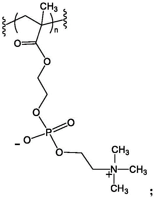
wherein n is an integer from 1 to 3000 and the wavy line indicates the point of attachment between the monomer units in the polymer.
In some embodiments, the polymer has three or more arms, or is synthesized with an initiator that comprises three or more polymer initiation sites. In some embodiments, the polymer has 2, 3, 4, 5, 6, 7, 8, 9, 10, 11, or 12 arms, or is synthesized with an initiator comprising 2, 3, 4, 5, 6, 7, 8, 9, 10, 11, or 12 polymer initiation sites. More preferably, the polymer has 3, 6 or 9 arms, or is synthesized with an initiator containing 3, 6 or 9 polymer initiation sites. In some embodiments, the polymer has 9 arms, or is synthesized with an initiator that contains 9 polymer initiation sites.
In some embodiments, the added polymer has a molecular weight between about 300,000 to about 1,750,000Da (SEC-MAL). In some embodiments, the molecular weight of the polymer is between about 500,000 to about 1,000,000 Da. In some embodiments, the molecular weight of the polymer is between about 600,000 to about 900,000 Da. In some embodiments, the molecular weight of the polymer is between about 750,000 to about 850,000 Da. In some embodiments, the molecular weight of the polymer is between about 800,000 to about 850,000 Da. In some embodiments, the molecular weight of the polymer is between about 750,000 to about 800,000 Da.
In some embodiments, any of the antibodies described herein can be further conjugated to a polymer to form a bioconjugate. The molecular weight of the bioconjugate (in total, SEC-MAL) can be between about 350,000 to 2,000,000 daltons, such as between about 450,000 to 1,900,000 daltons, between about 550,000 to 1,800,000 daltons, between about 650,000 to 1,700,000 daltons, between about 750,000 to 1,600,000 daltons, between about 850,000 to 1,500,000 daltons, between about 900,000 to 1,400,000 daltons, between about 950,000 to 1,300,000 daltons, between about 900,000 to 1,000,000 daltons, between about 1,000,000 to 1,300,000 daltons, between about 850,000 to 1,000,000 daltons, and between about 1,000,000 to 1,200,000 daltons.
In some embodiments, the antibody conjugate is purified. In some embodiments, the polymer is part of an antibody conjugate, is polydisperse, i.e., the polymer PDI is not 1.0. In some embodiments, the PDI is less than 1.5. In some embodiments, the PDI is less than 1.4. In some embodiments, the PDI is less than 1.3. In some embodiments, the PDI is less than 1.2. In some embodiments, the PDI is less than 1.1.
In some embodiments, the antibody conjugate has an anti-VEGF-a immunoglobulin g (igg) bound to a polymer comprising MPC monomers, wherein the sequence of the anti-VEGF-a heavy chain is SEQ ID No. 1 and the sequence of the anti-VEGF-a light chain is SEQ ID No. 2, and wherein the antibody is bound to the polymer only at C449 in SEQ ID No. 1. In some embodiments, the polymer has 9 arms and a molecular weight between about 600,000 to about 1,000,000 Da.
In some embodiments, the antibody conjugate has an anti-VEGF-a immunoglobulin g (igg) bound to a polymer comprising MPC monomers, wherein the sequence of the anti-VEGF-a heavy chain is SEQ ID NO:1 and the sequence of the anti-VEGF-a light chain is SEQ ID NO:2, and wherein the antibody is bound to the polymer only at C443(EU numbering, or 449C in SEQ ID NO: 1). In some embodiments, the polymer has 9 arms and a molecular weight between about 600,000 to about 1,000,000 Da.
In some embodiments, the antibody conjugate has the following structure:

wherein: each heavy chain of the anti-VEGF-a antibody is represented by the letter H and each light chain of the anti-VEGF-a antibody is represented by the letter L;
the polymer is bonded to the anti-VEGF-A antibody through the thiol group of C449 in SEQ ID NO. 1, the bond being depicted on one of the heavy chains; PC is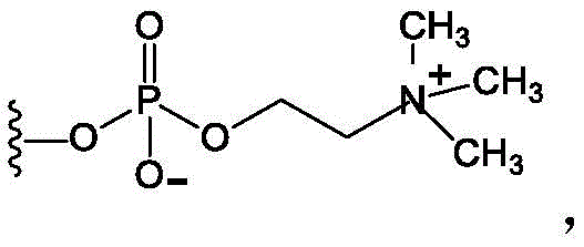 Wherein the curve represents the point of attachment to the remainder of the polymer; wherein X is: a) -OR, wherein R is H, methyl, ethyl, propyl, OR isopropyl; b) -H; c) any halogen, including-Br, -Cl, or-I; d) -SCN; or e) -NCS; and n1, n2, n3, n4, n5, n6, n7, n8 and n9 are the same or different so that the sum of n1, n2, n3, n4, n5, n6, n6, n7, n8 and n9 is 2500 ± 10%. In some embodiments, n1, n2, n3, n4, n5, n6, n7, n8, and n9 are the same or different and are integers from 0 to 3000. In some embodiments, n1, n2, n3, n4, n5, n6, n7, n8, and n9 are the same or different and are integers from 0 to 500. In some embodiments, X is-OR, wherein R is a sugar, an aminoalkyl, a mono-, poly-, OR unsubstituted variant of the following residue: saturated C 1-C24Alkyl, unsaturated C2-C24Alkenyl or C2-C24Alkynyl, acyl, acyloxy, alkoxycarbonyloxy, aryloxycarbonyloxy, cycloalkyl, cycloalkenyl, alkoxy, cycloalkoxy, aryl, heteroaryl, arylalkoxycarbonyl, alkoxycarbonylacyl, amino, aminocarbonyl, aminocarbonyloxy, nitro, azido, phenyl, hydroxy, alkylthio, arylthio, oxysulfonyl, carboxy, cyano and haloalkyl including polyhaloalkyl, - - -CO- -O- -R7carbonyl-CCO-R7、--CO--NR8R9、--(CH2)n--COOR7、--CO--(CH)n--COOR7、--(CH2)n--NR8R9Ester, alkoxycarbonyl, aryloxycarbonyl, wherein n is an integer of 1 to 6, wherein each R7、R8And R9Each selected from the group consisting of: hydrogen atoms, halogen atoms, mono-, poly-or unsubstituted variants of the following residues: saturated C1-C24Alkyl, unsaturated C2-C24Alkenyl or C2-C24Alkynyl, acyl, acyloxy, alkoxycarbonyloxy, aryloxycarbonyloxy, cycloalkyl, cycloalkenyl, alkoxy, cycloalkoxy, aryl, heteroaryl, arylalkoxycarbonyl, alkoxycarbonylacyl, amino, aminocarbonyl, aminocarbonyloxy, nitro, azido, phenyl, hydroxy, alkylthio, arylthio, oxysulfonyl, carboxy, cyano, and haloalkyl including polyhaloalkyl, 5-and 6-membered rings.
In some embodiments, the antibody conjugate has the following structure:

wherein: each heavy chain of the anti-VEGF-a antibody is represented by the letter H and each light chain of the anti-VEGF-a antibody is represented by the letter L;
the polymer is bonded to the anti-VEGF-A antibody via the thiol group of C443(EU numbering, or 449C in SEQ ID NO: 1), the bond being depicted on one of the heavy chains; PC is Wherein the curve represents the point of attachment to the remainder of the polymer; wherein X is: a) -OR, wherein R is H, methyl, ethyl, propyl, OR isopropyl; b) -H; c) any halogen, including Br, -Cl, or-I; d) -SCN; or e) -NCS; and n1, n2, n3, n4, n5, n6, n7, n8 and n9 are the same or different such that the sum of n1, n2, n3, n4, n5, n6, n6, n7, n8 and n9 is 2500 ± 10%. In some embodiments, n1, n2, n3, n4, n5, n6, n7, n8, and n9 are the same or different and are integers from 0 to 3000. In some embodiments, n1, n2, n3, n4, n5, n6, n7, n8, and n9 are the same or different and are integers from 0 to 500. In some embodiments, X is-OR, wherein R is a sugar, an aminoalkyl, a mono-, poly-, OR unsubstituted variant of the following residue: saturated C 1-C24Alkyl, unsaturated C2-C24Alkenyl or C2-C24Alkynyl, acylA group selected from the group consisting of acyloxy, alkoxycarbonyloxy, aryloxycarbonyloxy, cycloalkyl, cycloalkenyl, alkoxy, cycloalkoxy, aryl, heteroaryl, arylalkoxycarbonyl, alkoxycarbonylacyl, amino, aminocarbonyl, aminocarbonyloxy, nitro, azido, phenyl, hydroxy, alkylthio, arylthio, oxysulfonyl, carboxy, cyano and haloalkyl including polyhaloalkyl, - - -CO- -O- -R7carbonyl-CCO-R7、--CO--NR8R9、--(CH2)n--COOR7、--CO--(CH)n--COOR7、--(CH2)n--NR8R9Ester, alkoxycarbonyl, aryloxycarbonyl, wherein n is an integer of 1 to 6, wherein each R7、R8And R9Each selected from the group consisting of: hydrogen atoms, halogen atoms, mono-, poly-or unsubstituted variants of the following residues: saturated C1-C24Alkyl, unsaturated C2-C24Alkenyl or C2-C24Alkynyl, acyl, acyloxy, alkoxycarbonyloxy, aryloxycarbonyloxy, cycloalkyl, cycloalkenyl, alkoxy, cycloalkoxy, aryl, heteroaryl, arylalkoxycarbonyl, alkoxycarbonylacyl, amino, aminocarbonyl, aminocarbonyloxy, nitro, azido, phenyl, hydroxy, alkylthio, arylthio, oxysulfonyl, carboxy, cyano, and haloalkyl including polyhaloalkyl, 5-and 6-membered rings. In some embodiments, the construct is designated KSI-301.
In some embodiments, the antibody conjugate is present in a liquid formulation. In some embodiments, the antibody conjugate is combined with a pharmaceutically acceptable carrier.
In some embodiments, anti-VEGF-a antibodies are provided. The heavy chain of the anti-VEGF-a antibody has at least the following CDR sequences: CDRH1:GYDFTHYGMN(SEQ ID NO:9)、CDRH2: WINTYTGEPTYAADFKR (SEQ ID NO:10) and CDRsH3: YPYYYGTSHWYFDV (SEQ ID NO: 11). In some embodiments, the anti-VEGF-a heavy chain has those CDRs and additionally has threo at position 221 (as counted by the order as in SEQ ID No. 3)(ii) an amino acid (T). In some embodiments, the anti-VEGF-a light chain has at least the following CDRs: CDRL1:SASQDISNYLN(SEQ ID NO:12)、CDRL2: FTSSLHS (SEQ ID NO:13) and CDRsL3: QQYSTVPWT (SEQ ID NO: 14). In some embodiments, the anti-VEGF-a antibody has those CDRs and additionally has leucine (L) at Kabat position 4. In some embodiments, the isotype of the heavy chain of the anti-VEGF-a antibody is IgG1 and has CH1Hinge, CH2And CH3A domain. In some embodiments, the light chain isotype is κ. In some embodiments, an anti-VEGF antibody conjugate (e.g., KSI-301) construct will have one or more of these CDRs.
In some embodiments, the IgG1 domain of the anti-VEGF-a antibody has one or more mutations to modulate effector functions, such as ADCC, ADCP and CDC. In some embodiments, the IgG1 mutation reduces effector function. In some embodiments, the amino acids for effector function mutations include (EU numbering) E233X, L234X, L235X, G236X, G237X, G236X, D270X, K322X, a327X, P329X, a330X, a330X, P331X, and P331X, wherein X is any natural or non-natural amino acid. In some embodiments, the mutations include one or more of: E233P, L234V, L234A, L235A, G237A, a327G, a330S and P331S (EU numbering). In some embodiments, the anti-VEGF-a heavy chain has the following mutations (EU numbering): L234A, L235A and G237A. In some embodiments, the number of effector function mutations relative to the native human IgG1 sequence is no more than 10. In some embodiments, the number of effector function mutations relative to the native human IgG1 sequence is no more than 5, 4, 3, 2, or 1. In some embodiments, the Fc γ binding of the antibody and/or complement C1q binding is reduced such that the antibody has a reduced ability to produce effector function. This is particularly advantageous for ophthalmic indications/conditions.
In some embodiments, the anti-VEGF-a antibody comprises one or more of the following amino acid mutations: L234A, L235A, G237A (EU numbering) and L443C (EU numbering, or 449C in SEQ ID NO: 1).
In some embodiments, the anti-VEGF-a antibody is human immunoglobulin G (IgG1) or a portion thereof.
In some embodiments, the VEGF-a antibody comprises a heavy chain constant domain comprising one or more mutations that reduce immune-mediated effector function.
In some embodiments, anti-VEGF-A antibodies are provided. The anti-VEGF antibody comprises a heavy chain comprising: CDR H1 comprising sequence GYDFTHYGMN (SEQ ID NO: 9); CDR H2 comprising sequence WINTYTGEPTYAADFKR (SEQ ID NO: 10); CDR H3 comprising sequence YPYYYGTSHWYFDV (SEQ ID NO: 11); CDR L1 comprising sequence SASQDISNYLN (SEQ ID NO: 12); CDR L2 comprising the sequence FTSSLHS (SEQ ID NO: 13); and CDR L3 comprising sequence QQYSTVPWT (SEQ ID NO: 14).
Alternatively, the IgG domain may be IgG2, IgG3, or IgG4, or a complex in which the constant region is formed by more than one of these isotypes (e.g., the CH1 region from IgG2 or IgG4, the hinge, CH2, and CH3 regions from IgG 1). Such domains may contain mutations at one or more EU positions mentioned for IgG1 to reduce and/or modulate effector function. Human IgG2 and IgG4 have reduced effector function relative to human IgG1 and IgG 3.
The anti-VEGF-a heavy chain has a cysteine residue added as a mutation by recombinant DNA techniques, which can be used to conjugate a half-life extending moiety. In some embodiments, the mutation is (EU numbering) Q347C (EU numbering) and/or L443C (EU numbering, or 449C in SEQ ID NO: 1). In some embodiments, the mutation is L443C (EU numbering, or 449C in SEQ ID NO: 1). In some embodiments, the stoichiometric ratio of antibody to polymer is 1: 1; in other words, a conjugate is one antibody molecule conjugated to one polymer molecule.
The half-life of an anti-VEGF-a antibody can be extended by attaching a "half-life extending moiety" or "half-life extending group". Half-life extending moieties include peptides and proteins that can be expressed in-frame (or chemically conjugated as the case may be) with the biological agent in question, and various polymers that can be linked or conjugated to one or more amino acid side chains or terminal functional groups (e.g., -SH, -OH, -COOH, -CONH2, -NH2, or one or more N-and/or O-glycan structures). The half-life extending moiety generally functions to increase the in vivo circulatory half-life of the biological agent.
Examples of peptide/protein half-life extending moieties include Fc fusion (Capon DJ, Charow SM, Mordenti J, et al. Designation CD4 immunological interactions for AIDS therapy. Nature.1989.337:525-31), human serum albumin (HAS) fusion (Yeh P, Landais D, Lextre M, et al. Designation of year-secret album-CD 4 genetic control. Promai Natl Acad science USA.1992.89:1904-08), carboxy-terminal peptide (CTP) fusion (Fares FA, Suganu N.Nishimori K, Del of strain-nucleic acid et al. 78 genetic testing protein et al. 78: 1904-08), non-specific peptide of the sequence of the peptide/protein half-life extending moiety (Watertical protein) fusion (USA. CW. ID. K, Ser. K, design of family III. Sciagnosis-peptide of family II. 4304-08), geething NC, et al.A. recombinant polypeptide extensions of the in vivo polypeptides of peptides and proteins in a recombinant human being Nat Biotechnology.2009.27: 1186-90), elastin-like peptides (ELPs) (MCPhereson DT, Morrow C, Minehan DS, et al.Production and purification of a recombinant elastomeric polypeptide of G (VPGVG19-VPGV, M Escherichia coli Biotechnology.1992.8: 347-52), human transferrin fusion (Prior CP, Lai C-H, Sadhhi H. modified plasmid fusion proteins of proteins, WO2004/020405.2004), serine (PAS of the in vivo polypeptides of peptides of biological proteins of biological proteins of PAS P of biological proteins of Protein Eng Des Sel.2007.20:273-84) and gelatin-like protein (GLK) fusion (Huang Y-S, Wen X-F, Zaro JL, et al.
Examples of polymeric half-life extending moieties include polyethylene glycol (PEG), branched PEG, poly (ethylene glycol), (Warwick Effect Polymers; coventiy, UK), polysialic acid (PSA), starch, hydroxyethyl starch (HES), hydroxyalkyl starch (HAS), carbohydrates, polysaccharides, pullulan (pullulane), chitosan, hyaluronic acid, chondroitin sulfate, dermatan sulfate, dextran, carboxymethyl dextran, polyalkylene oxide (PAO), polyalkylene glycol (PAG), polypropylene glycol (PPG), polyoxazoline, polyacryloylmorpholine, polyvinyl alcohol (PVA), polycarboxylate, polyvinylpyrrolidone, polyphosphazene, polyoxazoline, polyethylene-co-maleic anhydride, polystyrene-co-maleic anhydride, poly (1-hydroxymethylethylidene hydroxymethylformal) (poly (1-hydroxymethy ethylene hydroxymethyl) formal), PHF), zwitterionic Polymers, choline-containing Polymers and compositions comprising MPC, poly (Glyx-Sery) Hyaluronic Acid (HA), Heparosan (hepparasan) polymers (HEP), Fleximer, dextran and polysialic acid (PSA).
In one embodiment, the half-life extending moiety may be conjugated to the antibody through a free amino group of the protein using N-hydroxysuccinimide (NHS) ester. The agents targeted for conjugation to amine groups can randomly react with the epsilon-amine group of lysine, the alpha-amine group of the N-terminal amino acid, and the delta-amine group of histidine.
However, the anti-VEGF-a antibodies disclosed herein have many amine groups available for polymer conjugation. Thus, conjugation of the polymer to the free amino group may negatively affect the ability of the antibody protein to bind to VEGF.
In some embodiments, the half-life extending moiety is coupled to one or more free SH groups using any suitable thiol-reactive chemistry, including but not limited to maleimide chemistry, or coupling of a polymeric hydrazide or polymeric amine to the carbohydrate moiety of the antibody after prior oxidation. In some embodiments, maleimide coupling is used. In some embodiments, conjugation occurs at a cysteine that is naturally occurring or introduced by genetic engineering.
In some embodiments, the polymer is covalently linked to a cysteine residue introduced into the anti-VEGF-a antibody by site-directed mutagenesis. In some embodiments, cysteine residues are used in the Fc portion of the antibody. In some embodiments, WO 2013/093809, US 7,521,541, WO 2008/020827, US 8,008,453, US 8,455,622, and US2012/0213705 (incorporated herein by reference for all purposes) provide sites for introducing cysteine residues into the Fc region. In some embodiments, the cysteine mutation is Q347C (EU numbering) and L443C, with reference to EU numbered human IgG heavy chain.
In some embodiments, conjugates of an antibody and a high MW polymer for use as a half-life extending moiety are provided. In some embodiments, the conjugates comprise an antibody coupled to a zwitterionic polymer, wherein the polymer is formed from one or more monomeric units, and wherein at least one monomeric unit has a zwitterionic group. In some embodiments, the zwitterionic group is phosphorylcholine.
In some embodiments, one of the monomeric units is HEMA-PC. In some embodiments, the polymer is synthesized from a single monomer that is HEMA-PC.
In some embodiments, some antibody conjugates have 2, 3, or more polymeric arms, wherein the monomer is HEMA-PC. In some embodiments, the conjugate has 2, 3, 4, 5, 6, 7, 8, 9, 10, 11, or 12 polymer arms, wherein the monomer is HEMA-PC. In some embodiments, the conjugate has 3, 6, or 9 arms. In some embodiments, the conjugate has 9 arms.
In some embodiments, the polymer-antibody conjugate has a polymer moiety with a molecular weight between 100,000 and 1,500,000 Da. In some embodiments, the molecular weight of the polymer portion of the conjugate is between 500,000 and 1,000,000 Da. In some embodiments, the molecular weight of the polymer portion of the conjugate is between 600,000 and 800,000 Da. In some embodiments, the polymer portion of the conjugate has a molecular weight between 600,000 and 850,000Da and has 9 arms. When considering the molecular weight of the antibody conjugated to the polymer, the molecular weight will be the sum of the molecular weight of the protein (including any carbohydrate moieties bound thereto) and the molecular weight of the polymer.
In some embodiments, the anti-VEGF-a antibody has a HEMA-PC polymer with a molecular weight between about 100kDa and 1650kDa as measured by Mw. In some embodiments, the molecular weight of the polymer, as measured by Mw, is between about 500kDa and 1000 kDa. In some embodiments, the molecular weight of the polymer, as measured by Mw, is between about 600kDa and about 900 kDa. In some embodiments, the molecular weight of the polymer, as measured by Mw, is 750kDa ± 15%.
In some embodiments, the polymer is prepared from an initiator suitable for ATRP having one or more polymer initiation sites. In some embodiments, the polymer initiation site has a 2-bromoisobutyrate site. In some embodiments, the initiator has 3 or more polymer initiation sites. In some embodiments, the initiator has 3, 4, 5, 6, 7, 8, 9, 10, 11, or 12 polymer initiation sites. In some embodiments, the initiator has 3, 6, or 9 polymer initiation sites. In some embodiments, the initiator has 9 polymer initiation sites. In some embodiments, the initiator is OG 1786.
The anti-VEGF-a antibody can be produced by recombinant expression, including (i) production of recombinant DNA by genetic engineering, (ii) introduction of the recombinant DNA into prokaryotic or eukaryotic cells by, for example, but not limited to, transfection, electroporation, or microinjection, (iii) culturing of the transformed cells, (iv) expression of the antibody, e.g., constitutive or inducible expression, and (v) isolation of the antibody, e.g., from the culture medium or by harvesting of the transformed cells, such that (vi) purified antibody is obtained.
anti-VEGF-a antibodies can be produced by expression in a suitable prokaryotic or eukaryotic host system characterized by the production of a pharmacologically acceptable antibody molecule. Examples of eukaryotic cells are mammalian cells, such as CHO, COS, HEK 293, BHK, SK-Hip and HepG 2. Other suitable expression systems are prokaryotic cells (e.g.E.coli with the expression system pET/BL 21), yeast cells (Saccharomyces cerevisiae and/or Pichia pastoris system) and insect cells.
A variety of vectors can be used to prepare the antibodies disclosed herein and are selected from eukaryotic expression vectors and prokaryotic expression vectors. Examples of vectors for prokaryotic expression include plasmids such as, but not limited to, preset, pet, and pad, where promoters for prokaryotic expression vectors include, but are not limited to, one or more of lac, trc, trp, recA, or araBAD. Examples of vectors for eukaryotic expression include: (i) vectors for expression in yeast, such as but not limited to pAO, pPIC, pYES or pMET, using promoters such as but not limited to AOX1, GAP, GAL1 or AUG 1; (ii) (ii) vectors for expression in insect cells, such as, but not limited to, pMT, pAc5, pIB, pMIB or pBAC, using promoters such as, but not limited to, PH, p10, MT, Ac5, OpIE2, gp64 or polh, and (iii) vectors for expression in mammalian cells, such as, but not limited to, pSVL, pCMV, pRc/RSV, pcDNA3 or pBPV, and vectors derived in one aspect from viral systems, such as, but not limited to, vaccinia virus, adeno-associated virus, herpes virus or retrovirus, using promoters such as, but not limited to, CMV, SV40, EF-1, UbC, RSV, ADV, BPV and beta-actin.
Method for conjugating protein and polymer
In some embodiments, a method of making a therapeutic protein-half-life extending moiety conjugate is provided having the steps of: conjugating a therapeutic protein having a cysteine residue added by recombinant DNA techniques with a half-life extending moiety having a thiol-specific reactive group selected from the group consisting of: maleimide, divinyl sulfone, ortho-dithiopyridine, and iodoacetamide.
In some embodiments, methods are provided for making anti-VEGF antibody conjugates, e.g., KSI-301, from OG 1950. The method involves reducing the OG1950 protein with a 50-fold molar excess of TCEP reducing agent. After reduction, the antibody is oxidized to produce a uncapped OG1950 antibody in which the light and heavy chain inter-and intra-chain disulfide bonds naturally present in the antibody are formed, but the engineered cysteine at heavy chain position L443C (EU numbering, or 449C in SEQ ID NO: 1) remains uncapped. OG1950 is then conjugated by adding excipients and adding a 5-10 fold molar excess of maleimide biopolymer. The biopolymer is linked to the OG1950 antibody by a covalent thioether bond. Following conjugation, the anti-VEGF antibody conjugate, e.g., KSI-301, is purified to remove unconjugated antibody and polymer.
The above proteins and methods may also vary. Thus, in some embodiments, methods of making conjugated proteins (which need not be antibodies or anti-VEGF antibodies) are provided. The method includes reducing one or more cysteines in the protein to form a uncapped protein in solution. After reduction of the one or more cysteines, the uncapped protein is reoxidized to restore at least one disulfide bond in the reduced protein, while ensuring that the engineered cysteine residues in the protein remain in the free thiol form to form the reoxidized uncapped protein in solution. At least one excipient is then added to the solution. Excipients reduce polymer-induced protein precipitation. After addition of the excipient, a polymer is added to the solution, which is conjugated to the reoxidized uncapped protein at the engineered cysteine residue to form a conjugated protein.
In some embodiments, the molar excess of reducing agent may be changed to any amount that works. In some embodiments, a 10, 20, 30, 40, 50, 60, 70, 80, 90 fold molar excess of reducing agent may be used (not necessarily all embodiments are TCEP). In some embodiments, any antibody (therapeutic or otherwise) may be used. In some embodiments, a 1, 2, 3, 4, 5, 6, 7, 8, 9, 10, 11, 12, 13, 14, 15 fold molar excess of maleimide biopolymer may be used. In some embodiments, there is an excess of uncapped protein to polymer. In some embodiments, the amount of protein reduced is less than the amount of polymer. In some embodiments, the amount of protein reduced is 90%, 80%, 70%, 60%, 50%, 40%, 30%, 20%, 10%, 5%, 4%, 3%, 2%, 1% of the amount of polymer. In some embodiments, the polymer used is 10-15 times that of the protein. In some embodiments, the amount of reduced antibody is greater than the amount of polymer. In some embodiments, the amount of polymer is greater than the amount of antibody that is reduced.
In some embodiments, the purification step is optional.
In some embodiments, the method of making an antibody conjugate comprises conjugating an anti-VEGF-a antibody to a choline phosphate-containing polymer. In some embodiments, the method comprises the step of conjugating an anti-VEGF-a antibody to a choline phosphate-containing polymer. The anti-VEGF-a antibody comprises amino residues added by recombinant DNA techniques. In some embodiments, the added amino acid residue is a cysteine residue. In some embodiments, cysteine residues are added outside the variable region of the antibody. Cysteine residues may be added to the heavy or light chain of the antibody.
In some embodiments, the polymer comprises or consists of a phosphorylcholine-containing polymer. In some embodiments, the choline phosphate-containing polymer comprises a thiol-specific reactive group selected from the group consisting of: maleimide, vinyl sulfone, ortho-dithiopyridine and iodoacetamide. In some embodiments, a thiol-specific reactive group on the choline phosphate-containing polymer reacts with a cysteine residue on the anti-VEGF-a antibody to make an antibody conjugate.
In some embodiments, the protein to be conjugated may be an antibody, an antibody protein fusion, or a binding fragment thereof. In some embodiments, the protein is not an antibody, but is an enzyme, ligand, receptor, or other protein or mutant or variant thereof. In some embodiments, the native protein contains at least one disulfide bond and at least one non-native cysteine.
In some embodiments, the excipient may be an acid or a base. In some embodiments, the excipient is a detergent, a sugar, or a charged amino acid. In some embodiments, the excipient helps to keep the protein in solution during conjugation with the polymer. In some embodiments, the excipient is added to the protein-containing solution prior to adding the polymer to the protein-containing solution.
In some embodiments, the reaction occurs under aqueous conditions between about pH 5 and about pH 9. In some embodiments, the reaction occurs between 6.0 and 8.5, between 6.5 and 8.0, or between 7.0 and 7.5.
In some embodiments, the polymer is conjugated to the protein at 2-37 degrees celsius. In some embodiments, conjugation occurs at 0-40 degrees celsius, 5-35 degrees celsius, 10-30 degrees celsius, and 15-25 degrees celsius.
In some embodiments, the conjugated proteins described herein can be contacted with an ion exchange medium or a hydrophobic interaction chromatography or affinity chromatography medium to perform purification (to remove the conjugated protein from unconjugated protein). In some embodiments, the ion exchange medium, hydrophobic interaction chromatography, or affinity/or chromatography medium separates the conjugated protein from the free polymer and the reoxidized uncapped protein.
In some embodiments, the methods described herein and outlined in fig. 18 relate to excipients capable of promoting and/or maintaining a solubility system. In some embodiments, the method allows the solution to maintain the solubility of the two components that are intended to interact. This may include the solubility of the protein and polymer, and then also the solubility of the terminal conjugate. In some embodiments, without the use of an excipient approach, a problem may be that while the protein is soluble, when the biopolymer is added, the solubility of the solution (e.g., protein) decreases and the protein precipitates out of solution (crash)/precipitates out. Of course, when the protein precipitates, it cannot be effectively conjugated to the biopolymer. Thus, excipients can be used to maintain the solubility of the protein in the presence of the biopolymer, so that the two can be coupled to form a protein conjugate (or, as shown in fig. 18, an antibody conjugate). This also allows the solubility of the conjugate to be maintained.
In some embodiments, the polymers disclosed herein may comprise one or more of the following: a zwitterionic, phosphorylcholine, or a PEG linker that bridges the center of the polymer branch point to the maleimide functional group. In some embodiments, any of the polymers provided herein can be added to a protein by a method provided herein.
In some embodiments, any protein provided herein can be conjugated to any polymer provided herein by one or more methods provided herein.
In some embodiments, the methods provided herein allow for larger scale processing to make and purify protein and/or antibody conjugates. In some embodiments, the volume used is at least 1 liter, e.g., 1, 10, 100, 1,000, 5,000, 10,000 liters or more. In some embodiments, the amount of antibody conjugate prepared and/or purified can be 0.1, 1, 10, 100, 1000, or more grams.
In some embodiments, the therapeutic protein may be any anti-VEGF-a antibody described herein having cysteine residues added by recombinant DNA techniques. In some embodiments, the anti-VEGF antibody heavy chain has the following CDRs: CDR H1:GYDFTHYGMN(SEQ ID NO:9)、CDRH2: WINTYTGEPTYAADFKR (SEQ ID NO:10) and CDRsH3: YPYYYGTSHWYFDV (SEQ ID NO: 11). The heavy chain may also have a threonine (T) at position 221 (as counted by the order as in SEQ ID No. 3). In some embodiments, the anti-VEGF light chain has the following CDRs: CDRL1:SASQDISNYLN(SEQ ID NO:12)、CDRL2: FTSSLHS (SEQ ID NO:13) and CDRsL3: QQYSTVPWT (SEQ ID NO: 14). The anti-VEGF-a light chain may also have leucine (L) at Kabat position 4.
In some embodiments, the anti-VEGF-a antibody is IgG 1. In some embodiments, the heavy chain has one or more mutations to modulate effector function. In some embodiments, the mutation is to one or more of the following amino acid positions (EU numbering): e233, L234, L235, G236, G237, a327, a330, and P331. In some embodiments, the mutation is selected from the group consisting of: E233P, L234V, L234A, L235A, G237A, a327G, a330S and P331S (EU numbering). In some embodiments, the mutations are (EU numbering) L234A, L235A, and G237A.
In some embodiments, the cysteine residues added to the therapeutic protein by recombinant DNA techniques should not participate in Cys-Cys disulfide bond pairing. In this aspect, the therapeutic protein can be a dimer. Thus, for example, an intact anti-VEGF-a antibody has two light chains and two heavy chains. If Cys residues are introduced into, for example, the heavy chain, the intact antibody will have two such introduced cysteines at the same position, and there is a possibility that these cysteine residues will form an intrachain disulfide bond. If the introduced cysteine residue forms a Cys-Cys disulfide bond or has a tendency to do so, the introduced Cys residue will not be available for conjugation. It is known in the art how to avoid positions in the heavy and light chains that would lead to intra-chain disulfide bond pairing. See, for example, U.S. patent application No. 2015/0158952.
In some embodiments, the cysteine residue introduced by recombinant DNA techniques is selected from the group consisting of (EU numbering) Q347C and L443C. In some embodiments, the cysteine residue is L443C (EU numbering, or 449C in SEQ ID NO: 1). In some embodiments, the heavy chain of the antibody has the amino acid sequence shown in SEQ ID No.1 and the light chain has the amino acid sequence of SEQ ID No. 2.
In some embodiments, the thiol-specific reactive group is maleimide.
In some embodiments, the half-life extending moiety is selected from the group consisting of: polyethylene glycol (PEG), branched PEG, (Warwick Effect Polymers; covery, UK), polysialic acid (PSA), starch, hydroxyethyl starch (HES), hydroxyalkyl starch (HAS), carbohydrates, polysaccharides, pullulan (pulullone), chitosan, hyaluronic acid, chondroitin sulfate, dermatan sulfate, dextran, carboxymethyl dextran, polyalkylene oxide (PAO), polyalkylene glycol (PAG), polypropylene glycol (PPG)Polyoxazoline, polyacryloylmorpholine, polyvinyl alcohol (PVA), polycarboxylates, polyvinylpyrrolidone, polyphosphazene, polyoxazoline, polyethylene-co-maleic anhydride, polystyrene-co-maleic anhydride, poly (1-hydroxymethylethylenehydroxymethylformal) (PHF), zwitterionic polymers, polymers containing choline phosphate, and polymers comprising 2-methacryloxy-2' -ethyltrimethylammonium phosphate (MPC).
In some embodiments, the half-life extending moiety is a zwitterionic polymer. In some embodiments, the zwitterion is choline phosphate, i.e., a choline phosphate-containing polymer. In some embodiments, the polymer consists of MPC units.
In some embodiments, the MPC polymer has three or more arms. In some embodiments, the MPC polymer has 2, 3, 4, 5, 6, 7, 8, 9, 10, 11, or 12 arms. In some embodiments, the MPC polymer has 3, 6, or 9 arms. In some embodiments, the MPC polymer has 9 arms. In some embodiments, the polymer is synthesized with an initiator comprising 2, 3, 4, 5, 6, 7, 8, 9, 10, 11, 12, or more polymer initiation sites.
In some embodiments, the MPC polymer has a molecular weight between about 300,000 and 1,750,000 Da. In some embodiments, the MPC polymer has a molecular weight between about 500,000 and 1,000,000Da or between about 600,000 and 900,000 Da.
In some embodiments, the method of making a therapeutic protein-half-life extending moiety conjugate has the additional steps of: contacting a therapeutic protein with a thiol reducing agent under conditions that produce a reduced cysteine thiol group. As mentioned above, preferably the cysteine residues added by recombinant DNA techniques are unpaired, i.e.do not participate in the Cys-Cys intra-chain disulfide bond or do not substantially participate in such a bond. However, Cys residues which are not involved in this Cys-Cys disulfide bond and which can be freely conjugated are known to react with free cysteines in the culture medium to form disulfide adducts. See, for example, WO 2009/052249. The cysteine so derivatized would not be useful for conjugation. To release the newly added cysteine from the disulfide adduct, the purified protein is treated with a reducing agent such as dithiothreitol. However, such treatment with a reducing agent will reduce all cysteine residues in the therapeutic protein, including native cysteines, many of which participate in interchain and intrachain Cys-Cys disulfide bonds. Native Cys-Cys disulfide is generally critical for protein stability and activity, and should be reconstituted. In some embodiments, all native (e.g., meta and intra) Cys-Cys disulfides are reconstituted.
To reconstitute the natural interchain and intrachain disulfide residues, the therapeutic protein is exposed to oxidizing conditions and/or oxidizing agents for a prescribed period of time, e.g., overnight, after reduction to remove the cysteine disulfide adduct. In some embodiments, overnight exposure to ambient air can be used to effect reconstitution of native disulfide bonds. In some embodiments, an oxidizing agent is used to restore the natural disulfide. In some embodiments, the oxidizing agent is selected from the group consisting of aqueous CuSO4 and dehydroascorbic acid (DHAA). In some embodiments, the oxidizing agent is DHAA. In some embodiments, the range of DHAA used is in the range of 5 to 30 equivalents. In some embodiments, the range is 10 to 20 equivalents. In some embodiments, the range is 15 equivalents.
In some embodiments, the thiol reducing agent is selected from the group consisting of: tris [ 2-carboxyethyl ] methyl]Phosphine hydrochloride (TCEP), Dithiothreitol (DTT), Dithioerythritol (DTE), sodium borohydride (NaBH)4) Sodium cyanoborohydride (NaCNBH3), beta-mercaptoethanol (BME), cysteine hydrochloride and cysteine. In some embodiments, the thiol reducing agent is TCEP.
In some embodiments, the thiol reducing agent concentration is 1 to 100-fold molar excess relative to the therapeutic protein concentration. In some embodiments, the thiol reducing agent concentration is 20 to 50-fold molar excess relative to the therapeutic protein concentration. In some embodiments, the thiol reducing agent is removed after incubation with the therapeutic protein prior to oxidation of the therapeutic protein.
In some embodiments, the method for conjugating a therapeutic protein to a half-life extending moiety further has the steps of: the therapeutic protein conjugate is purified after conjugation. In some embodiments, the therapeutic protein conjugate is purified using a technique selected from the group consisting of: ion exchange chromatography, hydrophobic interaction chromatography, size exclusion chromatography and affinity chromatography or combinations thereof.
In some embodiments, the therapeutic protein conjugate retains at least 20% biological activity relative to the unconjugated therapeutic protein. In some embodiments, the therapeutic protein conjugate retains at least 50% biological activity relative to the unconjugated therapeutic protein. In some embodiments, the therapeutic protein conjugate retains at least 90% of biological activity relative to the native therapeutic protein.
In some embodiments, the therapeutic protein conjugate has an increased half-life relative to the unconjugated therapeutic protein. In some embodiments, the therapeutic protein conjugate has a half-life that is increased by at least 1.5 fold relative to the unconjugated therapeutic protein. In some embodiments, the half-life of the therapeutic protein conjugate is increased by at least 5-fold relative to the unconjugated therapeutic protein.
In some embodiments, the zwitterionic polymer in the method of conjugating a therapeutic protein to a half-life extending moiety is a free-radically polymerizable monomer having a zwitterionic group, and the method further has the step of polymerizing the free-radically polymerizable zwitterionic monomer in a polymerization medium to provide a polymer, the medium comprising: a free-radically polymerizable zwitterionic monomer; transition metal catalyst Mt (q-1)+Wherein Mt is a transition metal, q is a higher oxidation state of the metal, q-1 is a lower oxidation state of the metal, wherein the metal catalyst is in Mt(q-1)+X’(q-1)Salts are provided in the form wherein X' is a counter ion (counter ion) or group, or transition metal catalysts by providing a non-active metal salt M in its higher oxidation statet q +X’qProvided in situ with a reducing agent capable of reducing the transition metal from an inactive oxidation state to an active reduction state; a ligand; and an initiator。
To function as an ATRP transition metal catalyst, the transition metal should have at least two readily accessible oxidation states, a higher oxidation state and a lower oxidation state, separated by one electron. In ATRP, a reversible redox reaction results in the transition metal catalyst cycling between a higher oxidation state and a lower oxidation state, while the polymer chains cycle between having growing chain ends and dormant chain ends. See, for example, U.S. patent No. 7,893,173.
In some embodiments, the free-radically polymerizable zwitterionic monomer is selected from the group consisting of:

wherein R1 is H or C1-6Alkyl, ZW is a zwitterion, and n is an integer from 1 to 6.
In some embodiments, the free radically polymerizable monomer is
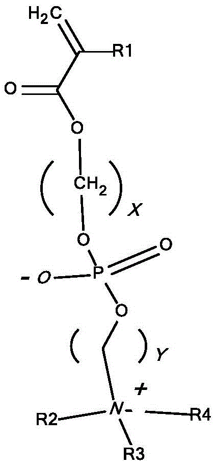
Wherein R1 is H or C1-6Alkyl, R2, R3, R4 are identical or different and are H or C1-4And X and Y are the same or different and are an integer of 1 to 6. In some embodiments, R1, R2, R3, and R4 are each methyl, and X and Y are each 2.
In some embodiments, the free radically polymerizable monomer is
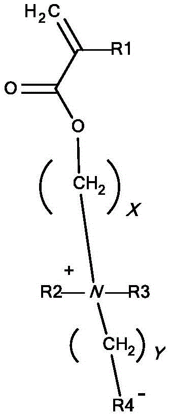
Wherein R1 is H or C1-6Alkyl, R2 and R3 are the same or different and are H or C1-4Alkyl, R4 is PO4-、SO3-or CO2And X and Y are the same or different and are an integer of 1 to 6. In some embodiments, R1, R2, and R3 are methyl, and R4 is PO4And X and Y are each 2.
In some embodiments, the monomer is
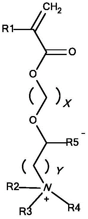
Wherein R1 is H or C1-6Alkyl, R2, R3 and R4 are identical or different and are H or C1-4Alkyl, R5 is PO4-、SO3-or CO2And X and Y are the same or different and are an integer of 1 to 6. In some embodiments, R1, R2, R3, and R4 are methyl, and R5 is PO4-and X and Y are 2.
In some embodiments, the transition metal Mt is selected from the group consisting of: cu, Fe, Ru, Cr, Mo, W, Mn, Rh, Re, Co, V, Zn, Au and Ag. In some embodiments, the metal catalyst is at Mt (q-1)+X’(q-1)A salt of the form provided. Mt (q-1)+Selected from the group consisting of: cu1+、Fe2+、Ru2+、Cr2+、Mo2+、W2+、Mn3+、Rh3+、Re2+、Co+、V2+、Zn+、Au+And Ag+And X' is selected from the group consisting of: halogen, C1-6Alkoxy group, (SO)4)1/2、(PO4)1/3、(R7PO4)1/2、(R72PO4) Triflate, hexafluorophosphate (hexaluminophorophosphate), mesylate, arylsulfonate, CN and R7CO2Wherein R7 is H or straight or branched C1-6Alkyl, which may be substituted 1 to 5 times by halogen. In some embodiments, Mt (q-1)+Is Cu1+And X' is Br.
In some embodiments, M is provided in situt (q-1)+. In some embodiments, Mt q+XqIs CuBr2. In some embodiments, the reducing agent is an inorganic compound. In some embodiments, the reducing agent is selected from the group consisting of: sulfur compounds, sulfur compounds of low oxidation levelSodium hydrogen carbonate, inorganic salts containing metal ions, metals, hydrazine hydrate and derivatives of these compounds. In some embodiments, the reducing agent is a metal. In some embodiments, the reducing agent is Cu0。
In some embodiments, the reducing agent is an organic compound. In some embodiments, the organic compound is selected from the group consisting of: alkyl thiols, mercaptoethanol, or enolizable carbonyl compounds, ascorbic acid, acetyl acetonate, camphorsulfonic acid, hydroxyacetone, reducing sugars, monosaccharides, glucose, aldehydes, and derivatives of these organic compounds.
In some embodiments, the ligand is selected from the group consisting of: 2,2' -bipyridine, 4' -di-5-nonyl-2, 2' -bipyridine, 4-dinonyl-2, 2' -bipyridine, 4',4 "-tris (5-nonyl) -2,2':6', 2" -terpyridine, N, N, N ', N ', N "-pentamethyldiethylenetriamine, 1,4,7,10, 10-hexamethyltriethylenetetramine, tris (2-dimethylaminoethyl) amine, N, N-bis (2-pyridylmethyl) octadecylamine, N, N, N ', N ' -tetrakis [ (2-pyridyl) methyl ] ethylenediamine, tris [ (2-pyridyl) methyl ] amine, tris (2-aminoethyl) amine, tris (2-bis (3-butoxy-3-oxopropyl) aminoethyl) amine, N, N ', N ' -tetrakis [ (2-pyridyl) methyl ] ethylenediamine, tris (2-pyridyl) methyl ] amine, tris (2-aminoethyl) amine, tris (3-oxopropyl) aminoethyl) amine, N, N, N ' -bis (2-methyl) aminoethyl) amine, N, N ' -pentamethyl) diethylenetriamine, N, N ' -pentamethyl (N, N, N, N ' -tetrakis (2-bis (2-amino-bis (2-amino) methyl) amine, N, N, or, Tris (2-bis (3- (2-ethylhexyloxy) -3-oxopropyl) aminoethyl) amine and tris (2-bis (3-dodecyloxy-3-oxopropyl) aminoethyl) amine. In some embodiments, the ligand is 2,2' -bipyridine.
In some embodiments, the initiator has the following structure:
wherein R1 is a nucleophilic reactive group, R2 comprises a linker, and R3 comprises a polymer synthesis initiator moiety having the structure:

wherein R4 and R5 are the same or different and are selected from the group consisting of: alkyl, substituted alkyl, alkylene, alkoxy, carboxyalkyl, haloalkyl, cycloalkyl, cycloalkylether, alkenyl, alkenylene, alkynyl, alkynylene, cycloalkylene, heterocycloalkyl, heterocycloalkylene, aryl, arylene-oxy, heteroaryl, amino, amide, or any combination thereof; z is halo, -OR (wherein R is-H, methyl, ethyl, propyl, OR isopropyl), -SCN, OR-NCS; and s is an integer between 1 and 20.
In some embodiments, Z is Br and R4 and R5 are each methyl. In some embodiments, R1 is selected from the group consisting of: -NH2-OH, and-SH.
In some embodiments, R2 is alkyl, substituted alkyl, alkylene, alkoxy, carboxyalkyl, haloalkyl, cycloalkyl ether, alkenyl, alkenylene, alkynyl, alkynylene, cycloalkylene, heterocycloalkyl, heterocycloalkylene, aryl, arylene-oxy, heteroaryl, amino, amide, or any combination thereof. In some embodiments, R2 is
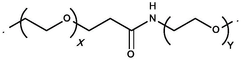
Wherein X and Y are the same or different and are integers of 1 to 20. In some embodiments, X and Y are each 4.
In some embodiments, R3 is
Wherein R6, R7 and R8 are the same or different and are selected from the group consisting of:


wherein Z is-OR (wherein R is-H, methyl, ethyl, propyl, OR isopropyl), -SCN, -NCS, -F, -Cl, -Br, OR-I. In some embodiments, Z is-Br and R6, R7, and R8 are each
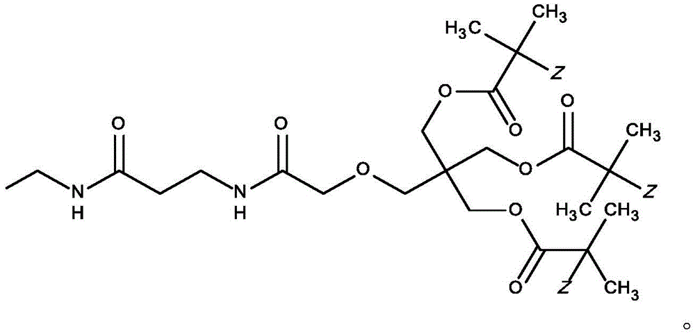
In some embodiments, the initiator has the following structure:
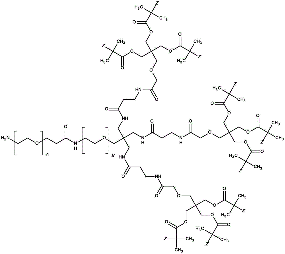
wherein A and B are the same or different and are integers from 2 to 12, and Z is any halide, such as Br. In some embodiments, a and B are each 4.
In some embodiments, the method also has the step of reacting the polymer with a maleimide reagent to provide a polymer having a terminal maleimide. In some embodiments, the maleimide compound is

Method of treatment
In some embodiments, methods for treating or preventing ocular diseases are provided having the steps of: administering a therapeutic protein selected from the group consisting of an anti-VEGF-a antibody (and conjugates thereof) and an anti-VEGF protein conjugate (e.g., an aflibercept biopolymer conjugate). In some embodiments, any one or more of the antibodies or antibody conjugates or protein conjugates provided herein can be used as a treatment and/or prevention of an ocular disease. The method comprises administering to the subject any one or more of the antibodies or antibody conjugates provided herein.
In some embodiments, methods for treating or preventing ocular diseases are provided. The method comprises administering to a subject in need thereof an effective dose of any of the antibodies and/or antibody conjugates and/or protein conjugates described herein. In some embodiments, the disease may be age-related macular degeneration (AMD) or Diabetic Macular Edema (DME). In some embodiments, the disease may be wet AMD. In some embodiments, the ocular disorder is wet AMD, without high Pigment Epithelial Detachment (PED). In some embodiments, the subject has a high PED when the baseline central subregion retinal thickness (CST) in the eye of the subject is 500 microns or greater.
In some embodiments, the ocular disease is selected from one or more of the group consisting of: diabetic retinopathy, Choroidal Neovascularization (CNV), age-related macular degeneration (AMD), Diabetic Macular Edema (DME), pathological myopia, von Hill-Lindi's disease, histoplasmosis of the eye, Central Retinal Vein Occlusion (CRVO), central retinal branch vein occlusion (BRVO), corneal neovascularization, retinal neovascularization, retinopathy of prematurity (ROP), subconjunctival hemorrhage, and hypertensive retinopathy. In some embodiments, the ocular disease is diabetic retinopathy.
In some embodiments, the administration frequency of the antibody or antibody conjugate or protein conjugate is no more than once per month. In some embodiments, the antibody or conjugate thereof is administered twice a month or once a week. In some embodiments, the antibody or conjugate thereof is administered once every two months, once every three months, once every four months, once every five months, once every six months, once every seven months, once every eight months, once every nine months, once every ten months, once every eleven months, or once every twelve months.
In some embodiments, one or more compositions provided herein can reduce the consequences of a high treatment burden resulting from the treatment of wet (proliferative) forms of age-related macular degeneration (AMD) with intravitreal injection of an anti-VEGF agent. Real world results for wet AMD patients lags behind those confirmed in phase 3 clinical studiesClinical results, e.g. use MARINA and ANCHOR study of (ranibizumab) and use
 (Abirapsis)
 VIEW 1 and VIEW 2. anti-VEGF therapeutics have longer ocular residence times so that they can be administered less frequently and therefore have more patient-tolerable characteristics that can bring the real-world outcome of more patients closer to phase 3 clinical outcome.
In some embodiments, the compounds described herein, including antibody conjugates and anti-VEGF-a antibodies, and anti-VEGF protein conjugates (e.g., aflibercept biopolymer conjugates), are used to treat patients with background or non-proliferative diabetic retinopathy but with little or no vision impairment. In some embodiments, these patients are administered less than once a month by intravitreal injection. In some embodiments, these patients are administered six times per year. In some embodiments, these patients are administered no more than four times per year. In some embodiments, the patient is administered no more than three times per year. In some embodiments, the patient is administered no more than twice a year. In some embodiments, the patient is administered no more than once a year. In some embodiments, the subject receives the antibody or antibody conjugate or protein conjugate by intravitreal injection.
Therapeutic proteins described herein (e.g., both antibodies and antibody conjugates) can be used by expressing such polypeptides in a patient (i.e., gene therapy).
There are two main methods for entering nucleic acids (optionally contained in a vector) into a patient's cells: in vivo and ex vivo. For in vivo delivery, the nucleic acid is injected directly into the patient, usually at the site where the therapeutic protein is desired, i.e., the site where the biological activity of the therapeutic protein is desired. For ex vivo treatment, cells of the patient are removed, nucleic acids are introduced into these isolated cells, and the modified cells are administered to the patient either directly or, for example, encapsulated in a porous membrane implanted in the patient (see, e.g., U.S. Pat. nos. 4,892,538 and 5,283,187). There are a variety of techniques available for introducing nucleic acids into living cells. These techniques vary depending on whether the nucleic acid is transferred to cultured cells in vitro or to cells of the intended host in vivo. Techniques suitable for transferring nucleic acids into mammalian cells in vitro include the use of liposomes, electroporation, microinjection, transduction, cell fusion, DEAE-dextran, calcium phosphate precipitation and the like. Transduction involves binding of replication-defective recombinant virus (including retrovirus) particles to cell receptors, followed by introduction of the nucleic acid contained in the particles into the cell. A commonly used vector for ex vivo delivery of genes is a retrovirus.
In some embodiments, in vivo nucleic acid transfer techniques include transfection with viral or non-viral vectors (e.g., adenovirus, lentivirus, herpes simplex I or adeno-associated virus (AAV)) and lipid-based systems (useful lipids for lipid-mediated gene transfer are, e.g., DOTMA, DOPE and DC-Chol; see, e.g., Tonkinison et al, Cancer invasion, 14(1):54-65 (1996)). In some embodiments, the vector for gene therapy is a virus, which includes an adenovirus, AAV, lentivirus, or retrovirus. Viral vectors, such as retroviral vectors, include at least one transcriptional promoter/enhancer or locus-defining element(s), or other elements that control gene expression by other means, such as alternative splicing, nuclear RNA export or post-translational modification of messengers. In addition, viral vectors, such as retroviral vectors, include nucleic acid molecules that are operably linked to and function as translation initiation sequences when transcribed in the presence of a gene encoding a therapeutic protein. Such vector constructs also include a packaging signal, Long Terminal Repeats (LTRs) or portions thereof, and plus and minus strand primer binding sites appropriate for the virus used (if they are not already present in the viral vector). In addition, such vectors typically include a signal sequence for secretion of the PRO polypeptide from the host cell in which it is located. In some embodiments, the signal sequence used for this purpose is a mammalian signal sequence. In some embodiments, the signal is the native signal sequence of the therapeutic protein. Optionally, the vector construct may also include a signal to direct polyadenylation as well as one or more restriction sites and translation termination sequences. For example, such vectors typically include a 5'LTR, a tRNA binding site, a packaging signal, an origin of second strand DNA synthesis, and a 3' LTR, or a portion thereof. Other carriers than viruses may be used, such as cationic lipids, polylysine and dendrimers.
In some cases, it is desirable to provide the nucleic acid source with an agent that targets the target cell, such as an antibody specific for a cell surface membrane protein or the target cell, a ligand for a receptor on the target cell, and the like. Where liposomes are used, proteins that bind to cell surface membrane proteins associated with endocytosis can be used to target and/or facilitate uptake, e.g., capsid proteins or fragments thereof that are tropic for a particular cell type, antibodies to proteins that undergo internalization during the cycle, and proteins that target intracellular localization and enhance intracellular half-life. Techniques for receptor-mediated endocytosis are described, for example, in Wu et al, J.biol.chem.,262:4429-4432 (1987); and Wagner et al, Proc.Natl.Acad.Sci.USA,87: 3410-. For a review of currently known gene markers and gene therapy protocols, see Anderson et al, Science,256: 808-. See also WO 93/25673 and references cited therein.
Suitable gene therapy and methods for preparing retroviral particles and structural proteins can be found, for example, in U.S. Pat. No. 5,681,746.
In some embodiments, methods are provided for treating or preventing ocular diseases in a mammal, wherein a nucleic acid molecule encoding a therapeutic protein selected from the group consisting of an anti-VEGF-a antibody is administered.
In some embodiments, the heavy chain is the heavy chain shown in SEQ ID No.1 and the light chain is the light chain shown in SEQ ID No. 2. In some embodiments, the nucleic acid molecule is administered by ex vivo gene therapy.
Methods of making antibody conjugates suitable for use in the methods of the present disclosure are found, for example, in PCT publication No. WO2017117464, which is incorporated herein by reference in its entirety.
Pharmaceutical composition
The therapeutic protein may be incorporated into a pharmaceutical composition together with pharmaceutically acceptable excipients. Pharmaceutical compositions suitable for oral administration may be provided as discrete units such as capsules, as solutions, syrups or suspensions (in aqueous or non-aqueous liquids; or as edible foams or pastes (wings); or as emulsions). Suitable excipients for tablets or hard gelatin capsules include lactose, corn starch or derivatives thereof, stearic acid or salts thereof. Excipients suitable for use with soft gelatin capsules include, for example, vegetable oils, waxes, fats, semi-solid or liquid polyols and the like. For the preparation of solutions and syrups, excipients which may be used include, for example, water, polyols and sugars. For the preparation of the suspension, an oil (e.g., vegetable oil) may be used to provide an oil-in-water or water-in-oil suspension.
The pharmaceutical composition may be adapted for intranasal administration wherein the excipient is a solid comprising a coarse powder having a particle size, for example in the range 20 to 500 microns, which is administered in the manner in which snuff is taken, i.e. by rapid inhalation through the nasal passage from a powder container adjacent the nose. Suitable compositions in which the vehicle is a liquid, for administration as a nasal spray or nasal drops, include aqueous or oily solutions of the active ingredient. Pharmaceutical compositions suitable for administration by inhalation include fine particle powders or mists, which may be produced by various types of metered dose pressurised aerosols, nebulizers or insufflators.
Pharmaceutical compositions suitable for parenteral administration include aqueous and non-aqueous sterile injection solutions which may contain anti-oxidants, buffers, bacteriostats and solutes which render the formulation substantially isotonic with the blood of the intended recipient; and aqueous and non-aqueous sterile suspensions which may contain suspending agents and thickening agents. Excipients that may be used in injectable solutions include, for example, water, alcohols, polyols, glycerol, and vegetable oils. The compositions may be presented in unit-dose or multi-dose containers, for example sealed ampoules and vials, and may be stored in a freeze-dried (lyophilized) condition requiring only the addition of the sterile liquid carried, for example water for injections, prior to use. Extemporaneous injection solutions and suspensions may be prepared from sterile powders, granules and tablets. The pharmaceutical composition may be substantially isotonic, meaning an osmotic pressure of about 250-400mOsm/kg water.
The pharmaceutical compositions may contain preservatives, solubilizers, stabilizers, wetting agents, emulsifiers, sweeteners, colorants, odorants, salts (which may themselves be provided in the form of pharmaceutically acceptable salts), buffers, coating agents or antioxidants. In addition to the substances, they may also contain therapeutically active agents. The pharmaceutical compositions may be used in combination with one or more pharmaceutically acceptable excipients. Such excipients may include, but are not limited to, saline, buffered saline (e.g., phosphate buffered saline), dextrose, liposomes, water, glycerol, ethanol, and combinations thereof.
The antibodies and pharmaceutical compositions containing them can be administered in an effective regimen for the treatment or prevention of disease in a patient, including, for example, by oral, intravitreal, intravenous, subcutaneous, intramuscular, intraosseous, intranasal, topical, intraperitoneal, and intralesional administration. Parenteral infusion includes intramuscular, intravenous, intraarterial, intraperitoneal or subcutaneous administration or other routes, and the like. In therapy or as a prophylactic agent, the active agent may be administered to the subject as an injectable composition, for example as a sterile aqueous dispersion. In some embodiments, the agent is isotonic or substantially isotonic.
For administration to mammals, particularly humans, it is contemplated that the dosage of the active agent is 0.01mg/kg body weight, typically about 1 mg/kg. The physician will determine the actual dosage which will be most suitable for an individual and it will depend on factors including the age, weight, sex and response of the individual, the disease or condition being treated and the age and condition of the individual being treated. The above doses are exemplary of the average case. Of course, there may be instances where higher or lower dosages are required. In some embodiments, the dose can be 0.5 to 20mg per eye, e.g., 1, 2, 3, 4, 5, 6, 7, 8, 9, 10, 11, 12, 13, 14, 15, 16, 17, 18, or 19 mg.
The dose may be repeated as appropriate (e.g., once a week, once every two weeks, once a month, once every two months, once a quarter, twice a year, once a year). If side effects occur, the amount and/or frequency of dosage can be reduced according to normal clinical practice. In one embodiment, the pharmaceutical composition may be administered once every one to thirty days. In one embodiment, the pharmaceutical composition may be administered twice every thirty days. In one embodiment, the pharmaceutical composition may be administered once weekly.
The antibodies and pharmaceutical compositions may be used alone or in combination with other compounds (e.g., therapeutic compounds or molecules, such as anti-inflammatory agents, analgesics, or antibiotics). This administration with the other compounds may be simultaneous, separate or sequential. The components may be prepared in the form of a kit, which may optionally include instructions.
The antibodies and pharmaceutical compositions disclosed herein are useful for treating or preventing diseases, particularly ocular diseases or conditions described herein.
The anti-VEGF antibody conjugates, or anti-VEGF protein conjugates, and pharmaceutical compositions containing them, may be formulated and administered by: ophthalmic, intraocular, and/or intravitreal injection, and/or juxtascleral (juxtascleral) injection, and/or subretinal injection, and/or subtenon (subtenon) injection, and/or suprachoroidal (suprachoroidal) injection and/or subconjunctival administration and/or topical administration in the form of eye drops and/or ointments. Such antibodies and compositions may be delivered by a variety of methods, for example intravitreal Delivery as a device and/or depot (depot) that allows slow release of the compound into the vitreous, including for example those described in the references by Intraocular Drug Delivery, Jaffe, Ashton, and Pearson, editors, Taylor & Francis (March 2006). In one example, the device may be in the form of a micropump and/or matrix (matrix) and/or passive diffusion system and/or encapsulated unit (encapsulated cell) that releases the compound over a prolonged period of time (Intraocular Drug Delivery, Jaffe, Ashton, and Pearson, editors, Taylor & Francis (March 2006)).
Formulations for ocular, intraocular, or intravitreal administration can be prepared by methods known in the art using ingredients known in the art. The main requirement for efficient treatment is proper penetration of the eye. Unlike diseases of the anterior part of the eye where drugs can be delivered locally, retinal diseases require more site-specific approaches. Eye drops and ointments rarely penetrate to the back of the eye and the blood-ocular barrier prevents systemically administered drugs from penetrating into the ocular tissues. Thus, the drug delivery method of choice for treating retinal diseases (e.g., AMD and CNV) is direct intravitreal injection. Intravitreal injections are typically repeated at intervals that depend on the condition of the patient and the nature and half-life of the drug being delivered.
Therapeutic antibodies and related conjugates are typically placed in a container having a sterile access port, such as an intravenous solution bag or vial having a stopper pierceable by a hypodermic injection needle. Such compositions may also be provided in the form of pre-filled syringes.
A "stable" formulation is one in which the protein or a protein conjugated to a polymer of another half-life extending moiety substantially retains its physical and/or chemical stability and/or biological activity upon storage. "stable" also refers to formulations that exhibit little or no evidence of instability (including aggregation and/or deamidation). For example, the provided formulations may remain stable for at least two years when stored at temperatures of 5-8 ℃ as described herein. Suitable formulations for anti-VEGF antibody conjugates of the present disclosure are described, for example, in PCT publication No. WO2017117464, which is incorporated herein by reference in its entirety.
Various analytical techniques for measuring Protein stability are available in the art and are reviewed, for example, in Peptide and Protein Drug Delivery,247- & 301(Vincent Lee ed., New York, N.Y.,1991) and Jones,1993Adv. Drug Delivery Rev.10: 29-90. Stability may be measured at a selected temperature for a selected period of time. In some embodiments, the formulation is storage stable for at least 6 months, 12-18 months, or 2 or more years.
A protein (e.g., an antibody or fragment thereof) "retains its physical stability" in a pharmaceutical formulation if it shows no signs of aggregation, precipitation, deamidation and/or denaturation when visually inspected for color and/or clarity or measured by ultraviolet light scattering or size exclusion chromatography.
A protein "retains its chemical stability" in a pharmaceutical formulation if the chemical stability at a given time is such that the protein is said to still retain its biological activity. Chemical stability can be assessed by detecting and quantifying chemically altered forms of the protein. Chemical changes may involve size changes (e.g., shearing), which may be evaluated using, for example, size exclusion chromatography, SDS-PAGE, and/or matrix-assisted laser desorption ionization/time of flight mass spectrometry (MALDI/TOF MS). Other types of chemical changes include charge changes (e.g., occurring as a result of deamidation), which can be evaluated by, for example, ion exchange chromatography. An antibody "retains its biological activity" in a pharmaceutical formulation if the biological activity of the antibody at a given time, as measured, for example, in an antigen binding assay, is within about 10% (within the error of the assay) of the biological activity exhibited at the time the pharmaceutical formulation was prepared.
A protein-polymer conjugate "retains its chemical stability", the chemical bond between the protein and the polymer remains intact, e.g., it is not hydrolyzed or otherwise disrupted. The protein portion of the conjugate retains its chemical stability as described above.
By "isotonic" is meant that the formulation of interest has substantially the same osmotic pressure as human blood or vitreous for intravitreal injection. Isotonic formulations typically have an osmotic pressure of about 250-400 mOsm. Isotonicity can be measured using, for example, a vapor pressure or freezing type osmometer.
As used herein, "buffer" refers to a buffered solution that resists changes in pH by the action of its acid-base conjugated components. In some embodiments, the pH of the buffer is from about 3.0 to about 8.0; for example, about 4.5 to 8; or about pH 6 to about 7.5; or from about 6.0 to about 7.0, or from about 6.5-7.0, or from about pH 7.0 to about 7.5; or from about 7.1 to about 7.4. Also contemplated is a pH at any point between the above ranges.
In some embodiments, "PBS" phosphate buffered saline, Tris-based buffers, and histidine-based buffers are used.
In some embodiments, the PBS buffer consists of at least Na2HPO4、KH2PO4And NaCl, adjusted to provide a suitable pH. In some embodiments, the buffer may contain other pharmaceutical excipients such as KCl and other salts, detergents and/or preservatives to provide a stable storage solution.
A "preservative" is a compound that can be included in a formulation to substantially reduce the bacterial action therein, thereby facilitating, for example, the production of a multi-purpose formulation. Examples of possible preservatives include octadecyl dimethyl benzyl ammonium chloride, hexamethonium chloride, benzalkonium chloride (a mixture of alkyl benzyl dimethyl ammonium chlorides, where the alkyl group is a long chain compound), and benzethonium chloride. Other types of preservatives include aromatic alcohols such as phenol, butyl and benzyl alcohol, alkyl parabens (such as methyl or propyl paraben), catechol, resorcinol, cyclohexanol, 3-pentanol and m-cresol.
In some embodiments, formulations safe for human use or animal testing should have sufficiently low levels of endotoxins. "endotoxin" is Lipopolysaccharide (LPS) from the cell membrane of gram-negative bacteria. Endotoxins consist of a hydrophilic polysaccharide moiety covalently linked to a hydrophobic lipid moiety (lipid a). Raetz CR, Ulevitch RJ, Wright SD, Sibley CH, Ding A, Nathan CF.1991.gram-negative endotoxin, an overexpression lipid with a proteolytic activity in FASEB J.5(12): 2652. lipid A is responsible for most of the biological activity of endotoxins, i.e.its toxicity. Endotoxin is shed in large quantities at the time of bacterial cell death and during growth and division. They are highly heat stable and are not destroyed under conventional sterilization conditions. Heat or pH limiting treatments must be used, for example 180 ℃ 250 ℃ and more than 0.1M acid or base (Petsch D, Anspach F.2000.endo removal from protein solutions. J Biotechnol.76: 97-119). Such conditions are of course very unfavorable for biopharmaceuticals.
In the biotechnology and pharmaceutical industry, endotoxins can be found in the production process and in the final product. Endotoxin is prevalent because bacteria can grow in malnourished media (including water, saline, and buffers) unless precautions are taken. Injection of endotoxin into animals or humans can cause a variety of pathophysiological effects, including endotoxic shock, tissue damage and even death. Ogikubo Y, Ogikubo Y, Norimatsu M, Noda K, Takahashi J, Inotsume M, Tsuchiya M, Tamura Y.2004.evaluation of the bacterial endoxin test for quantification of anoxin conjugation of biological variables 32: 88-93.
Pyrogen responses and shock were induced in mammals following intravenous injection of endotoxin at low concentrations (1ng/mL) (Fiske JM, Ross A, VanDerMeid RK, McMichael JC, Arumugham.2001.method for reducing endexoxin in Moraxella catarrhalis UspA2 protein preparations. J Chom B.753: 269-). The maximum level of endotoxin for intravenous application of drugs and biologicals was set at 5 Endotoxin Units (EU) per kg body weight per hour for all pharmacopoeias (Daneshiam M, Guenther A, Wendel A, Hartung T, Von interlock S.2006.in vitro virology test for toxin or immunomodulation drugs. J Immunol Method 313: 169-175). EU is a measure of the biological activity of endotoxins. For example, 100pg of standard endotoxins EC-5 and 120pg of endotoxins from E.coli O111: B4 have an activity of 1EU (Hirayama C, Sakata M.2002.chromatographic removal of endoxin from protein solutions by polymer particles J Chrom B781: 419-432). Achieving this threshold level has been a challenge in the biological research and Pharmaceutical industries (Berthold W, Walter J.1994.protein Purification: attributes of Processes for Pharmaceutical products. biologicals 22: 135-.
The presence of endotoxins in drugs to be delivered by intravitreal injection is of particular concern. In 1945, intravitreal injection of drug (penicillin) was first performed by Rycroft (Rycroft BW.1945. penicilin and the control of deep intra-ocular infection. British J Ophthalmol 29(2): 57-87). The vitreous is a chamber into which high levels of drugs can be introduced and maintained for a relatively long period of time. Drug concentrations achievable by intravitreal injection far exceed those that can be produced by local administration or by systemic administration (e.g., intravenous administration).
One of the most dangerous complications that may result from intravitreal injections is endophthalmitis. Endophthalmitis is divided into two categories: infectious endophthalmitis and aseptic endophthalmitis. Infectious endophthalmitis is usually caused by bacteria, fungi or parasites. Symptoms of infectious endophthalmitis include severe pain, loss of vision, and redness of the conjunctiva and underlying outer scleral layer. Infectious endophthalmitis requires urgent diagnosis and treatment. Possible treatments in some cases include intravitreal injections of antibiotics and a pars plana vitrectomy. Discectomy may be required to remove the blind and painful eye. See, for example, Christy NE, Sommer A.1979. antimicrobial phenolic inhibitors of sexual endemphalmitis. Ann Optimolmol 11(8): 1261-.
In contrast, sterile endophthalmitis does not involve infectious agents and can be defined as acute intraocular inflammation of the vitreous cavity that resolves without the need for intravitreal antibiotics and/or vitreoretinal surgery. If a vitreous microbiology study has been performed, a culture that proves to be negative is required to maintain a diagnosis of sterile endophthalmitis. Marticorena J, Romano V, Gomez-Ulla F.2012 "Sterile Endophyllis after intraviral Injections" Med Inflam.928123.
Intravitreal injections of biopharmaceuticals contaminated with endotoxin have been observed to result in aseptic endophthalmitis (Marticorena et al). Bevacizumab (avastin) is approved by the U.S. food and drug administration for the treatment of glioblastoma, metastatic colorectal cancer, advanced non-squamous non-small cell lung cancer, and metastatic renal cancer. Bevacizumab is also widely used for the treatment of wet AMD beyond approved labeling. Bevacizumab was supplied by the manufacturer at 100mg/4 ml. The solution cannot be used directly for intravitreal injection and must be prepared by a pharmacist. Some sterile endophthalmitis has been observed and is theoretically thought to be caused by contamination of bevacizumab with endotoxin unintentionally by the formulator.
Given the scare clinical consequences of intravitreal endotoxin injection, the total amount of endotoxin that can be administered to a patient by intravitreal administration is very limited. In some embodiments, a solution having an antibody or antibody conjugate is provided with an endotoxin level of no more than 5.0 EU/ml. In some embodiments, the endotoxin level does not exceed 1.0 EU/ml. In some embodiments, the endotoxin level does not exceed 0.5 EU/ml. In some embodiments, the endotoxin level does not exceed 0.2 EU/ml. In some embodiments, the endotoxin level is no more than 2, 1, 0.5, 0.2, 0.1, 0.09, 0.08, 0.07, 0.06, 0.05, 0.04, 0.03, 0.02 or 0.01 EU/ml.
Two commonly used FDA-approved tests for endotoxin presence are the rabbit pyrogen test and the Limulus Amebocyte Lysate (LAL) assay (Hoffman S, et al 2005.International validation of novel pyrogen tests based on human monoclonal cells J. immunol. methods 298: 161-281; Ding JL, Ho BA.2001.New era in pyrogen testing. Biotech.19: 277-281). The rabbit pyrogen test was developed in the 20 th century, and involves monitoring the temperature rise of rabbits injected with test solutions. However, the use of rabbit pyrogen testing has been greatly reduced for several years due to expense and long turnaround times. The LAL test is more common. LAL is derived from the blood of horseshoe crab and coagulates upon exposure to endotoxin.
One of the simplest LAL assays is the LAL gel-coagulation assay. Essentially, the LAL coagulation assay is combined with serial dilution of the sample in question. The gel formation is proportional to the amount of endotoxin in the sample. Serial dilutions were prepared from the samples and the ability of each dilution to form LAL gels was determined. At some point there was a negative reaction. The amount of endotoxin in the original sample can be estimated from the dilution assay.
Other LAL tests have also been developed, including turbidimetric LAL assays (Ong KG, Lelan JM, Zeng KF, Barrett G, Aourob M, Grimes CA.2006.A Rapid highlyn-sensitive end toxin detection system. biosensors and Bioelectronics 21: 2270-. Turbidimetric and chromogenic assays are more sensitive and quantitative than simple gel-clot dilution assays.
In some embodiments, methods of reducing the amount of endotoxin in a composition with an antibody disclosed herein are provided. The method comprises the following steps: contacting the composition with an affinity chromatography resin that binds the antibody; eluting the antibody from the affinity chromatography resin to form an affinity chromatography eluate with the antagonist; contacting the affinity chromatography eluate with an ion exchange resin that binds the antibody; and eluting the antibody from the ion exchange resin, wherein the antibody eluted from the ion exchange resin is substantially free of endotoxin.
The above-described method for reducing the amount of endotoxin or other methods or processes described herein may be performed in the order described in the above steps, or it may optionally be performed by changing the order of the steps or even repeating one or more steps. In one embodiment, the method of reducing the amount of endotoxin in a composition is performed in the order of steps described. In some embodiments, the affinity chromatography resin contacting step, the washing step, and the eluting step are repeated more than once in the same order before the affinity chromatography eluent is contacted with the ion exchange resin. The method may also include a filtration step using, for example, a 0.1 micron, 0.22 micron or 0.44 micron filter, which may be performed on one or more eluents removed after each resin binding step.
In some cases, the step of contacting the composition with the affinity chromatography resin, the washing step, and the step of eluting the antibody from the affinity chromatography resin may be repeated more than once prior to contacting the first eluent with the ion exchange resin. In one embodiment, the affinity chromatography resin comprises a recombinant protein a ("rProteinA") resin. An example of a suitable recombinant protein A resin is rProteinA Sepharose  Resin (Amersham, Piscataway, N.J.). In another embodiment, a suitable affinity chromatography resin will comprise a protein G chromatography resin. In other embodiments, suitable affinity chromatography resins comprise a mixed protein a/protein G resin. In other embodimentsSuitable affinity chromatography resins comprise hydrophobic charge-inducing resins containing 4-mercaptoethylpyridine ligands, e.g. MEP
 Resin (BioSepra, Cergy, Saint Christophe, France).
In some embodiments, the ion exchange resin comprises an anion exchange resin. As known to those skilled in the art, ion exchangers can be based on a variety of materials for the matrix and attached charged groups. For example, the following matrices may be used, wherein the materials mentioned may be more or less crosslinked: MacroCap Q (GE Healthcare Biosciences, Piscataway, NJ), agarose-based matrix (e.g., Sepharose Sepharose Fast
 And Sepharose High
 ) Cellulose-based substrates (e.g. DEAE)
 ) Dextran-based matrices (e.g.
 ) A silica-based matrix and a synthetic polymer-based matrix. For anion exchange resins, the charged groups covalently attached to the matrix can be, for example, diethylaminoethyl, quaternary aminoethyl, and/or quaternary ammonium. In some embodiments, the anion exchange resin comprises quaternary amine groups. An exemplary anion exchange resin with quaternary amine groups for binding anti-M-CSF antibodies is Q
  Resin (Amersham, Piscataway, N.J.).
In other aspects, if the endotoxin level is above the desired level after the composition is subjected to the anion exchange chromatography step described above, the composition may alternatively be subjected to a cation exchange resin. In some embodiments, any endotoxin in the composition should bind to the ion exchange resin differently than the protein in question, thereby purifying the protein from the endotoxin. In this regard, endotoxins are negatively charged and are typically bound to anion exchange resins. If both the protein and endotoxin are bound to the anion exchange resin, purification of one from the other can be achieved by eluting both into different fractions using a salt gradient. The relative binding of a protein to a particular resin can also be affected by changing the pH of the buffer relative to the pI of the protein. In some embodiments, the cation exchange chromatography is the only ion exchange chromatography employed.
In some embodiments, if the endotoxin level is too high after the anion exchange resin, the composition may be further subjected to a second ion exchange step, for example by contacting the composition with a cation exchange resin, followed by a washing step, and then eluted from the ion exchange resin. In some embodiments, the cation exchange resin comprises sulfonic acid groups for binding. An exemplary cation exchange resin is SP  Resin FF (Amersham, Piscataway, N.J.), Poros XS (CEX) (Life Technology, Grand Island, New York).
In some embodiments, after the antibody protein solution is produced with the specified levels of endotoxin, there are multiple steps prior to the final preparation of the protein. In some embodiments, the half-life extending moiety is conjugated to a protein. The conjugate is then formulated into the final pharmaceutical formulation for injection into a patient. In some embodiments, the conjugate is again purified on an ion exchange resin, which may be a cation exchange resin. In other embodiments, the protein is formulated. In all cases, normal laboratory procedures should be used to prevent the introduction of endotoxin contaminants into the protein sample or protein-polymer conjugate.
Examples
Example 1: retinal half-life, retinal bioavailability and systemic clearance of KSI-301
In the rabbit model, the half-life of intravitreally administered KSI-301 was extended in the retina and choroid/Retinal Pigment Epithelium (RPE) (FIG. 1). Comparison of the KSI-301 data with published results for aflibercept and ranibizumab indicates that KSI-301 exhibits better persistence compared to other anti-VEGF therapeutic agents. Data were from a rabbit model. Ranibizumab data: gaudrealt et al (2007) IOVS 46(2)726, Gaudrealt et al (2007) Retina 27(9)1260, Bakri et al (2007) Ophthalmol 114(12) 2179; aflibercept data: EVER Congress Portoroz Slovenia (2008) Struble (Coonce) Koehler-Stec (Regeneron). The aflibercept data were arithmetically adjusted to reflect an administered dose of 2,000 μ g (based on 500 μ g in vivo rabbit dosing); KSI-301 data were mathematically adjusted to reflect an administered dose of 5,000 μ g (based on 725 μ g in vivo rabbit dosing). Error bars reflect the standard error of the mean.
KSI-301 administered intravitreally showed excellent retinal bioavailability (FIG. 2). Comparison of the KSI-301 data with published results for aflibercept shows that KSI-301 has better bioavailability. The data in figure 2 are from the covance rabbit ADME (absorption, distribution, metabolism, elimination) model. Aflibercept data (2008): EVER Congress Portoroz Slovenia Struble (Covance), Koehler-Stec (Regeneron). Error bars reflect the standard error of the mean.
KSI-301 administered intravenously showed rapid systemic clearance (FIG. 3). Comparison of the KSI-301 data with published results for bevacizumab shows a faster systemic clearance of KSI-301. Bevacizumab data is from Yeung et al 2010Cancer Research.
Example 2: in patients with diabetic macular waterAgents for intravitreal administration of KSI-301 in subjects with tumor (DME)
Volume escalation study
Design of research
A dose escalation study was conducted to evaluate ocular and systemic safety, tolerability, and determine the Maximum Tolerated Dose (MTD) of KSI-301. One of three doses of 1.25mg, 2.5mg and 5mg (by weight of protein) was administered to each subject as a single injection. Three subjects were enrolled at each dose level. Subjects were followed up for a total of 12 weeks at the following time points; final follow-up on day 2, week 1, week 2, week 4, week 8 and week 12.
At the start of the study, the first 3 subjects were included in the first dosing group (1.25mg of KSI-301) and received a single intravitreal dose of KSI-301. The subjects continued to be evaluated with a waiting period of at least 24 hours between subjects to allow sufficient time for each subject to safely review before approving the next subject for injection.
Once the 3 rd subject at the previous dose level completed a one week safety period following its KSI-301 dose, the dose group was recruited in an incremental manner. Subject enrollment and dose escalation were based on review of safety information and the onset of dose-limiting toxicity (DLT).
Eight of the nine enrolled subjects had previously received treatment, with limited or no response, although had previously received multiple anti-VEGF treatments and had severe disease (median BCVA of 3 in the previous year, range 0-7).
Results
A single injection of KSI-301 resulted in a rapid, high-intensity response lasting 12 weeks (fig. 4). No intraocular inflammation and drug related adverse events were observed. FIG. 4: intravitreally administering to a subject with Diabetic Macular Edema (DME) 1.25mg, 2.5mg, or 5mg (by weight of protein) of KSI-301. BCVA and OCT CST were measured after intravitreal injection of KSI-301. Median change from baseline to week 12, 3 dose groups were aggregated (n-9 patients in total, 3 patients per dose group). Do DV, Angiogenesis 2019; patel et al, ARVO 2019.
This result shows the effectiveness of single injection forms of KSI-301 in patients most of whom had previously treated their DMEs with other anti-VEGF drugs (bevacizumab, aflibercept and/or ranibizumab). The results show that intravitreal administration of KSI-301 improves visual acuity and retinal thickness (measured by OCT) in a stable manner over 12 weeks. This experiment examined Diabetic Macular Edema (DME) patients with severe disease (n-9). Some patients had previously received treatment (8/9), with limited or no response despite having previously received multiple anti-VEGF treatments and had severe disease (median 3 in the previous year, range 0-7). The results indicate that a single injection of KSI-301 produces a rapid, high-intensity response lasting 12 weeks. No intraocular inflammation and drug related adverse events were observed.
These results are obtained by intravitreally administering 1.25mg, 2.5mg, or 5mg (by weight of protein) of KSI-301 to a subject having diabetic macular edema. This result shows the effectiveness of single injection forms of KSI-301 in patients most of whom had previously treated their DMEs with other anti-VEGF drugs (bevacizumab, aflibercept and/or ranibizumab). The results indicate that when KSI-301 is administered as described herein in a single dose, there is an unexpected therapeutic effect because visual acuity and retinal thickness (measured by OCT) improve in a stable manner over 12 weeks.
Example 3: investigation of multiple intravitreal KSI administration in Wet age-related macular degeneration (wAMD) patients
301 open label, multi-center exploratory study
The following study design and methodology was followed in examples 3, 4 and 5, unless otherwise noted. In general, patients with wAMD, DME or RVO who received anti-VEGF therapy for the first time were randomized to receive a 2.5mg or 5mg (by weight of antibody) KSI-301 monthly three loading dose and then followed up once a month or more frequently thereafter, and were retreated when the physician determined that retreatment was needed due to disease activity or the patient met any retreatment criteria.
Overall study design
In this study, patients with wmda, DME or RVO who received anti-VEGF therapy for the first time were randomized to receive a 2.5mg or 5mg (by weight of antibody) KSI-301 monthly loading dose of three, and then followed monthly or more frequently thereafter, and were retreated when the physician determined that retreatment was required due to disease activity or the patient met any retreatment criteria. These criteria are associated with signs of disease recurrence and/or vision loss due to disease recurrence.
Two dose levels of KSI-301, 2.5mg (50. mu.L) and 5mg (100. mu.L) (by weight of protein), were evaluated in a multiple dose study. Each subject received 3 initial intravitreal injections of KSI-301, the first on day 1, the second on week 4, and the third on week 8 (FIG. 5). All queues are queued as 1: 3 were randomly assigned to 2.5mg or 5mg doses.
FIG. 5 is a schematic view of: study design of a randomized open label study to evaluate the safety, efficacy, and persistence of multiple doses for intravitreal administration of KSI-301.
Subjects were evaluated every 4 weeks and, if certain re-treatment criteria were met, subjects could receive additional administrations of study drug starting at week 16. There were no forced injections at the assessment visit, except for the wet AMD cohort where forced intravitreal injections were given at the visit if 24 weeks had elapsed since the last injection.
Eligible subjects are selected based on predetermined inclusion and exclusion criteria. The subject's treatment for the ocular disorder being treated is the first to receive treatment and has no history of retinal disease other than the condition being studied.
KSI-301 was formulated in about 12.5mM sodium phosphate and 0.025% polysorbate 20 as a 50mg/mL (based on antibody mass) aqueous solution and filled into a disposable 2.0mL vial.
Multiple dose exposure of KSI-301 was well tolerated during the study and no endophthalmitis was observed. 113 subjects received administration for a total dose of 308 doses (104 doses on day 1, 96 doses at week 4, 84 doses at week 8). The following were observed:
■ to date, no study has been reported to investigate intraocular inflammation or ocular SAE in the eye;
■ to date, no drug-related Adverse Events (AEs) or drug-related Serious Adverse Events (SAE) were reported;
■ most AEs were assessed as mild, consistent with intravitreal anti-VEGF characteristics;
■ in 4 subjects 8 non-ocular SAEs of non-drug relevance were reported:
■ A92 year old RVO subject was hospitalized with an existing condition and died
■ A66-year-old RVO subject was hospitalized with dizziness
■ A43 year old DME subject is hospitalized for an existing condition
■ A56 year old DME subject is hospitalized with an existing condition
wAMD queue
Patients in the wAMD cohort: the age is more than or equal to 50 years old; the first treatment for wet age-related macular degeneration involving the fovea; area of damage of any type of damage<30mm2(12 disc (disc) areas); during screening, the BCVA ETDRS letter score of the eyes is not more than 78 and not less than 23 (about 20/25 to about 20/320Snellen equivalent values), and is confirmed on the 1 st day; and vision loss in the study eye, determined to be primarily a result of wAMD.
The average characteristics of the study population are shown in table 1.
Table 1: study population characteristics

Evaluation of the test
Following 3 initial intravitreal injections of KSI-301, the following assessments were made every 4 weeks: best Corrected Vision (BCVA), by the Early Treatment Diabetic Retinopathy Study (ETDRS) visual acuity test; spectral domain optical coherence tomography (SD-OCT); OCT angiography (OCT-A).
Standard of retreatment (non-loading dose)
Retreatment by intravitreal injection of KSI-301 is performed if at least one of the following retreatment criteria is met. These criteria are associated with signs of disease recurrence and/or vision loss due to disease recurrence.
-increase in retinal thickness (CST) in OCT central subregion by > 75 μm and decrease in BCVA by >5 letters compared to week 12;
due to worsening of wAMD disease activity (e.g., increased retinal fluid, increased subretinal fluid, new intraretinal hemorrhage, new subretinal hemorrhage), BCVA was reduced by >5 letters compared to day 1;
due to worsening wAMD disease activity (e.g., increased retinal fluid, increased subretinal fluid, new intraretinal hemorrhage, new subretinal hemorrhage), BCVA is reduced by ≧ 10 letters compared to the previous optimal BCVA; or
24 weeks/6 months since the last injection.
Results
After the first loading dose was administered on day 1, an improvement in BCVA and OCT CST was observed in the patient (FIG. 6; FIG. 12, left column). The improved BCVA and OCT CST values are comparable to standard of care anti-VEGF therapy. The therapeutic effect of KSI-301 persists during the loading phase and for at least 8 weeks after administration of the final loading dose at week 8 (FIG. 6). In one representative patient, CST was decreased for 7 months without retreatment (fig. 12, left panel).
FIG. 6: preliminary improvement in wet AMD patients Best Corrected Vision (BCVA) and retinal thickness (OCT). BCVA and CST evaluation of the wAMD cohort. N-25. Random patients included up to week 16 visit before data expiration date; the 2.5mg and 5mg doses were combined. BCVA — best corrected vision; OCT ═ optical coherence tomography; CST — center sub-region thickness.
FIG. 12: treatment persistence in wAMD cohort patients. OCT scans were performed on individual retinas on day 1 (line 1), week 1 (line 2), month 3 (line 3), and month 7 (line 4) of treatment with KSI-301. Left column: patients with wAMD; middle column: a DME patient; right column: RVO patients.
The results show the persistence of treatment to date in the cohort of wAMD patients (figure 7). The data indicate that KSI-301 provides unexpected therapeutic effects relative to prior art drugs and may provide better results because only 1 patient was retreated prior to week 20 (or about 3 months after the final loading dose), 1 patient was retreated 3 months after the final loading dose, and 11/14 (80%) patients had reached 4 months or more until the first retreatment. This indicates that a target dosing interval of 3 to 5 months is possible in wAMD.
In summary, only 1 (4%) of the 25 patients received retreatment within 3 months (fig. 7). Only 1 (5%) of the 20 patients received retreatment at 3 months. 86% (18/21) of the patients reached 3 months or more after the final loading dose without receiving retreatment, and 80% (11/14) of the patients reached 4 months or more after the final loading dose without receiving retreatment. This suggests that the target dosing interval for wAMD may be 3 to 5 months.
FIG. 7: persistence of treatment of KSI-301 in the wAMD cohort. Including randomized patients who reached the first re-treatment opportunity (visit week 12) before the data expiration date. Each bar represents one patient. Retreatment was denoted as (. diamond-solid.). All described patients received follow-up after the indicated last evaluation time point (indicated by the right arrow). The results can be summarized as follows:
● 18.5.5% received retreatment (5/27), and of those reaching week 20, 95% (20/21) did not receive retreatment beyond 12 weeks after the final loading dose
● 81.5.5% no further treatment is required (22/27)
● in patients up to week 20, 95% (20/21) did not receive retreatment at least 12 weeks after the 3 rd loading dose
● KSI-3015 mg dose achieves up to 6 months of retreatment after the loading phase.
Example 4: investigation of the opening of multiple intravitreal administrations of KSI-301 in Diabetic Macular Edema (DME) patients
Labelset, multicenter exploratory study
The overall study design and experimental evaluation are described in example 3.
DME queue
Patients in the DME cohort were: diabetic macular edema, first treated; during screening, the BCVA ETDRS letter score of the eyes is not more than 78 and not more than 23 (about 20/25 to about 20/320Snellen equivalent values), and is confirmed on day 1; the thickness (CST) of the central subregion on the SD-OCT is more than or equal to 300 micrometers (Heidelberg spectra or equivalent products); and vision loss in the study eye, determined to be primarily a result of DME.
The average characteristics of the study population are shown in table 1 above.
Standard of retreatment (non-loading dose)
Retreatment by intravitreal injection of KSI-301 is performed if at least one of the following retreatment criteria is met. These criteria are associated with signs of disease recurrence and/or vision loss due to disease recurrence.
Compared with the 12 th or previous visit (4 weeks apart between two visits), the thickness of the retina film (CST) of the OCT central subregion is increased by more than or equal to 75 μm, and the BCVA is reduced by more than or equal to 5 letters; or
Due to the worsening of DME/RVO disease activity (e.g., increased intraretinal fluid, increased subretinal fluid, new intraretinal hemorrhages, new exudates), BCVA is reduced by ≧ 10 letters compared to the previous best BCVA.
Results
After the first loading dose was administered on day 1, an improvement in BCVA and OCT CST was observed in the patient (fig. 8; fig. 12, middle panel). The therapeutic effect of KSI-301 persists during the loading phase and for at least 8 weeks after administration of the final loading dose at week 8 (FIG. 8). In one representative patient, CST was decreased for 7 months without retreatment (fig. 12, middle panel).
The data demonstrate the effect of KSI-301 after 3 monthly doses in DME patients (FIG. 8). The data is unexpected because current products require monthly treatment or 5 monthly loading doses, while KSI-301 provides a high level of improvement after only 3 loading doses.
FIG. 8: BCVA and CST evaluations of DME queues. N-12. Random patients included up to week 16 visit before data expiration date; the 2.5mg and 5mg doses were combined. BCVA — best corrected vision; OCT ═ optical coherence tomography; CST — center subregion thickness.
Within 3 months after the final loading dose, no patients needed retreatment and no patients needed either the 4 th or 5 th monthly loading dose (fig. 9). 18% (2/11) of the patients were retreated at 3 months. In patients reaching week 20, 81.8% (9/11) did not require further treatment beyond 12 weeks after the 3 rd loading dose. Some patients reached 4, 5 or 6 months without further treatment.
The data indicate that in DME patients, a dosing interval of 3+ months is likely to be reached after only 3 loading doses (figure 9). Furthermore, these data are unexpected because patients without DME received re-treatment within 3 months. In addition, more than 80% require retreatment beyond 3 months after the final loading dose. This is unexpected because conventional treatments can take as many as 9-10 injections within the first 12 months of treatment.
FIG. 9: the persistence of treatment of KSI-301 in the DME cohort. Including randomized patients who reached the first re-treatment opportunity (visit week 12) before the data expiration date. Each bar represents one patient. Retreatment was denoted as (. diamond-solid.). All described patients were followed up beyond the indicated last evaluation time point (indicated by the right arrow). The results can be summarized as follows:
● 11.8.8% received retreatment (2/17)
● among patients who reached week 20, 80% (8/10) did not receive retreatment beyond 12 weeks after the final loading dose
● 88.2.2% have not received retreatment (15/17)
● in patients reaching week 20, 81.8% (9/11) received no further treatment for more than 12 weeks after only 3 loading doses
● none of the patients required either the 4 th or 5 th monthly loading dose.
Fig. 15 depicts the severity of diabetic retinopathy results (DRSS). The figure shows the proportion of patients with different levels of diabetic retinopathy severity, as measured by the independent expert reading center using a standardized photographic reading scale. DR can be described as varying levels of severity, e.g., from mild to moderate to severe nonproliferative diabetic retinopathy, or from mild to high risk proliferative DR. In this case, most patients had grade 47 (level 47) disease (moderate NPDR) at baseline. After 12 weeks (3 loading doses followed by 1 month), DR severity improvement was graded 2 in 27% of patients, graded 13% and maintained the same level of DR severity in 60% of patients. (it is well known that after application of anti-VEGF therapy, the severity of DR improves, but 1 to 2 years is required to achieve peak effects with other drugs, and if anti-VEGF therapy is discontinued, the effect disappears in many patients and the disease worsens again). The image shows patients with proliferative (grade 65) disease with disease improvement at week 12 and improvement to non-proliferative (grade 53, grade 2 on the standard rating scale). Without the administration of additional doses, the impact on DR severity was maintained for an additional 14 weeks, which is the maximum follow-up time available for this patient.
In some embodiments, patients with non-proliferative DR may be treated with an anti-VEGF antibody conjugate (e.g., KSI-301) without or possibly with only a small loading dose (e.g., 1, 2, or 3 starting doses), and then treated again every 3 to 6 months (for treating non-proliferative DR).
Example 4.5
Patients with nonproliferative DR were identified. Patients do not receive a loading dose therapy, but receive a single injection therapy of an anti-VEGF antibody conjugate (e.g., KSI-301) every 3-6 months to treat non-proliferative DR. Alternatively, the patient is first treated with 1, 2 or 3 loading doses.
Example 5: investigating the opening of multiple intravitreal administrations of KSI-301 in Retinal Vein Occlusion (RVO) subjects
Label-releasing, multi-center exploratory studies
The overall study design and experimental evaluation are described in example 3.
RVO queue
Patients in the RVO cohort were: retinal vein occlusion with macular edema and secondary vision impairment, first treated; during screening, the BCVA ETDRS letter score of the eyes is not more than 78 and not less than 23 (about 20/25 to about 20/320Snellen equivalent values), and is confirmed on the 1 st day; the thickness (CST) of the central subregion on the SD-OCT is more than or equal to 300 micrometers (Heidelberg spectra or equivalent products); branch Retinal Vein Occlusion (BRVO) or Central Retinal Vein Occlusion (CRVO); and to study vision loss in the eye, which the investigator determined was primarily a result of macular edema secondary to RVO.
The average characteristics of the study population are shown in table 1.
Standard of retreatment
Retreatment by intravitreal injection of KSI-301 is performed if at least one of the following retreatment criteria is met. These criteria are associated with signs of disease recurrence and/or vision loss due to disease recurrence.
-increase in OCT central subregion retinal thickness (CST) by > 75 μm and decrease in BCVA by > 5 letters compared to week 12 or the previous visit (4 weeks between visits); or
Due to DME/RVO disease activity deterioration (e.g. increased retinal fluid, increased subretinal fluid, new intraretinal hemorrhages, new exudates), BCVA is reduced by ≧ 10 letters compared to the previous best BCVA.
As a result, the
After the first loading dose was administered on day 1, an improvement in BCVA and OCT CST was observed in the patient (figure 10; figure 12, right panel). The therapeutic effect of KSI-301 persists during the loading phase and for at least 8 weeks after administration of the final loading dose at week 8 (FIG. 10). In one representative patient, CST was decreased for 7 months without retreatment (fig. 12, right panel).
The data demonstrate the effect of KSI-301 after 3 monthly doses in RVO patients (FIG. 10). The data is unexpected because a sustained improvement in visual acuity was observed between 8 weeks and 16 cycles. In ranibizumab, aflibercept and bevacizumab studies by RVO, switching from monthly treatment to less than monthly treatment resulted in worsening visual acuity and OCT CST.
FIG. 10: BCVA and CST evaluations of RVO queues. N-14. Random patients included up to week 16 visit before data expiration date; the 2.5mg and 5mg doses were combined. BCVA — best corrected vision; OCT ═ optical coherence tomography; CST — center sub-region thickness.
The data show that RVO patients received a first re-treatment at or after week 8 in the 5mg dose group (figure 11). This result is unexpected because conventional therapy requires monthly administration to treat RVO. Furthermore, the time between the first and second re-treatment is extended in patients receiving more than one re-treatment, which is also unexpected because typical RVO patients have a particular pattern or regular intervals of disease recurrence. Finally, more than half of the patients (5/9) had been over 3 months since the final loading dose, which was also unexpected in the case of RVO high intraocular VEGF loading.
FIG. 11: persistence of treatment of KSI-301 in the RVO cohort. Including randomized patients who reached the first re-treatment opportunity (visit week 12) before the data expiration date. Each bar represents one patient. Retreatment was denoted as (. diamond-solid.). Patients who visited beyond the indicated last evaluation time point are indicated by the right arrow. The left arrow indicates termination. Further results shown in fig. 11 can be summarized as follows:
● 25% of patients received retreatment (6/24)
● in patients who reached week 20, 56% (5/9) did not receive retreatment beyond 12 weeks after the final loading dose
● 75% have not received retreatment (18/24)
● in patients who reached week 20, 56% (5/9) did not receive retreatment beyond 12 weeks after only 3 loading doses
● KSI-3015 mg cohorts 18/18 patients did not require the 4 th monthly loading dose.
Example 6
A subject in need of treatment (prophylactic or otherwise) for an ocular disorder is first identified. The subject is then administered 1 to 5mg of an anti-VEGF antibody conjugate of the antibody (e.g., KSI-301) at a first loading dose, an optional second loading dose, and an optional third loading dose (within about one month of each other). After the final loading dose, the subject will retain the therapeutic benefit of anti-VEGF therapy for at least 12 weeks. This may be useful for Diabetic Macular Edema (DME), Retinal Vein Occlusion (RVO), wet age-related macular degeneration (AMD), and/or alternative Diabetic Retinopathy (DR).
Example 6.5
A subject in need of treatment (prophylactic or otherwise) for an ocular disorder is first identified. The subject is then administered 1 to 5mg of an anti-VEGF antibody conjugate of the antibody (e.g., KSI-301) at a first loading dose, an optional second loading dose, and an optional third loading dose (within about one month of each other). After the final loading dose, the subject will retain the therapeutic benefit of the anti-VEGF antibody conjugate therapy for at least 16 weeks. Alternatively, after the final loading dose, the subject will retain the therapeutic benefit of the anti-VEGF antibody conjugate therapy for at least 20 weeks. Alternatively, after the final loading dose, the subject will retain the therapeutic benefit of the anti-VEGF antibody conjugate therapy for at least 24 weeks. Alternatively, after the final loading dose, the subject will retain the therapeutic benefit of the anti-VEGF antibody conjugate therapy for at least 30 weeks. Alternatively, after the final loading dose, the subject will retain the therapeutic benefit of the anti-VEGF antibody conjugate therapy for at least 36 weeks. Alternatively, after the final loading dose, the subject will retain the therapeutic benefit of the anti-VEGF antibody conjugate therapy for at least 42 weeks. Alternatively, after the final loading dose, the subject will retain the therapeutic benefit of anti-VEGF antibody conjugate therapy for at least 48 weeks. Alternatively, after the final loading dose, the subject will retain the therapeutic benefit of the anti-VEGF antibody conjugate therapy for at least 54 weeks. Alternatively, after the final loading dose, the subject will retain the therapeutic benefit of the anti-VEGF antibody conjugate therapy for at least 60 weeks. This may be useful for Diabetic Macular Edema (DME), Retinal Vein Occlusion (RVO), wet age-related macular degeneration (AMD), and/or alternative Diabetic Retinopathy (DR).
Example 7
The present embodiments provide a method of treating Retinal Vein Occlusion (RVO). The method includes administering an anti-VEGF antibody conjugate (e.g., KSI-301) to a subject having RVO at a first loading dose. The loading dose can then be repeated one or two times. This resulted in subjects maintaining the therapeutic outcome of anti-VEGF antibody conjugate therapy for at least 8 weeks after the final loading dose. In the alternative, this results in the subject maintaining the therapeutic outcome of anti-VEGF antibody bioconjugate (e.g., KSI-301) therapy for at least 12 weeks after the final loading dose. In the alternative, this results in the subject maintaining the therapeutic outcome of anti-VEGF antibody bioconjugate (e.g., KSI-301) therapy for at least 20 weeks after the final loading dose.
Example 7.5
The present embodiments provide a method of treating Retinal Vein Occlusion (RVO). The method includes administering an anti-VEGF antibody conjugate (e.g., KSI-301) to a subject having RVO at a first loading dose. The loading dose can then be repeated one or two times. This resulted in subjects maintaining the therapeutic outcome of anti-VEGF antibody conjugate therapy for at least 24 weeks after the final loading dose. In the alternative, this results in the subject retaining the therapeutic outcome of the anti-VEGF antibody conjugate therapy for at least 30 weeks after the final loading dose. In the alternative, this results in the subject retaining the therapeutic outcome of the anti-VEGF antibody conjugate therapy for at least 36 weeks after the final loading dose. In the alternative, this results in the subject retaining the therapeutic outcome of the anti-VEGF antibody conjugate therapy for at least 42 weeks after the final loading dose. In the alternative, this results in the subject retaining the therapeutic outcome of the anti-VEGF antibody conjugate therapy for at least 48 weeks after the final loading dose. In the alternative, this results in the subject retaining the therapeutic results of the anti-VEGF antibody conjugate therapy for at least 54 weeks after the final loading dose. In the alternative, this results in the subject retaining the therapeutic outcome of the anti-VEGF antibody conjugate therapy for at least 60 weeks after the final loading dose.
Example 8
The present example provides a method of improving ocular perfusion comprising identifying a subject with DME, DR or RVO. The subject is then administered at least 2 loading doses of an anti-VEGF antibody conjugate (e.g., KSI-301) at 1.25-5mg, with a one month interval between each injection. One person does not have more than 2 injections. The subject is then provided one or more further doses (retreatment) of the anti-VEGF antibody conjugate (e.g., KSI-301) until the subject exhibits improved perfusion in at least one eye.
Example 9
The present embodiments provide a method of improving eye perfusion. The method includes identifying a subject having nonproliferative DR and administering to the subject an initial dose (1 to 5mg of antibody) of an anti-VEGF antibody conjugate (e.g., KSI-301) to provide improved perfusion in at least one eye. This dose is repeated until perfusion is achieved within the treated eye of the subject.
Example 10
The present embodiments provide methods of treating a subject with DME, DR or RVO. The method includes administering 1-3 loading doses (once a month with 1-5mg of antibody) of an anti-VEGF antibody conjugate (e.g., KSI-301) to a subject with DME, DR, or RVO. No more than 3 loading doses were administered to the subject. Subsequent applications (re-treatments) of the anti-VEGF antibody conjugate were provided at time points no earlier than 12 weeks after the final loading dose, as needed (if any).
Example 10.5
The present example provides methods of treating a subject with DME, DR or RVO. The method includes administering 1-3 loading doses (once a month with 1-5mg of antibody) of an anti-VEGF antibody conjugate (e.g., KSI-301) to a subject with DME, DR, or RVO. No more than 3 loading doses were administered to the subject. Subsequent applications (re-treatments) of the anti-VEGF antibody conjugate were provided at time points not earlier than 16 weeks after the final loading dose, as needed (if any). Alternatively, subsequent applications (re-treatments) of the anti-VEGF antibody conjugate are provided at a time point not earlier than 20 weeks after the final loading dose. Alternatively, subsequent applications (re-treatments) of the anti-VEGF antibody conjugate are provided at time points not earlier than 24 weeks after the final loading dose. Alternatively, subsequent applications (re-treatments) of the anti-VEGF antibody conjugate are provided at a time point not earlier than 30 weeks after the final loading dose. Alternatively, subsequent applications (re-treatments) of the anti-VEGF antibody conjugate are provided at a time point no earlier than 36 weeks after the final loading dose. Alternatively, subsequent applications (re-treatments) of the anti-VEGF antibody conjugate are provided at a time point no earlier than 42 weeks after the final loading dose. Alternatively, subsequent applications (re-treatments) of the anti-VEGF antibody conjugate are provided at a time point no earlier than 48 weeks after the final loading dose. Alternatively, subsequent applications (re-treatments) of the anti-VEGF antibody conjugate are provided at a time point no earlier than 54 weeks after the final loading dose. Alternatively, subsequent applications (re-treatments) of the anti-VEGF antibody conjugate are provided at a time point no earlier than 60 weeks after the final loading dose.
Example 11
This example provides a method of treating a subject having nonproliferative DR. Patients in need of treatment are identified and 1 or 2 loading doses (1-5mg, once a month) of an anti-VEGF antibody conjugate (e.g., KSI-301) are administered to a subject with nonproliferative DR. No more than 2 loading doses were administered to the subject. Optionally, subsequent administrations (retreatment) of the anti-VEGF antibody conjugate (e.g., KSI-301) can be provided at a time point not earlier than 12 weeks after the final loading dose.
Example 11.5
The present example provides a method of treating a subject having nonproliferative DR. Patients in need of treatment are identified and 1 or 2 loading doses (1-5mg, once a month) of an anti-VEGF antibody conjugate (e.g., KSI-301) are administered to a subject with nonproliferative DR. No more than 2 loading doses were administered to the subject. Alternatively, subsequent administrations of the anti-VEGF antibody conjugate (retreatment) may be provided at a time point not earlier than 16 weeks after the final loading dose. Alternatively, subsequent administrations (re-treatments) of the anti-VEGF antibody conjugate can be provided at a time point not earlier than 20 weeks after the final loading dose. Alternatively, subsequent administrations of the anti-VEGF antibody conjugate (retreatment) may be provided at a time point not earlier than 24 weeks after the final loading dose. Alternatively, subsequent administrations (re-treatments) of the anti-VEGF antibody conjugate can be provided at a time point not earlier than 30 weeks after the final loading dose. Alternatively, subsequent administrations (re-treatments) of the anti-VEGF antibody conjugate can be provided at a time point not earlier than 36 weeks after the final loading dose. Alternatively, subsequent administrations (re-treatments) of the anti-VEGF antibody conjugate can be provided at a time point not earlier than 42 weeks after the final loading dose. Alternatively, subsequent administrations of the anti-VEGF antibody conjugate (retreatment) may be provided at a time point no earlier than 48 weeks after the final loading dose. Alternatively, subsequent administrations of the anti-VEGF antibody conjugate (retreatment) may be provided at a time point no earlier than 54 weeks after the final loading dose. Alternatively, subsequent administrations (re-treatments) of the anti-VEGF antibody conjugate can be provided at a time point not earlier than 60 weeks after the final loading dose.
Example 12
The present embodiments provide methods of treating a subject having RVO. The method includes administering 1 or 2 loading doses (1-5mg, once per month) of an anti-VEGF antibody conjugate (e.g., KSI-301) to a subject having RVO. No additional loading dose was administered to the 3 subjects. Subsequent administrations (re-treatments) of the anti-VEGF antibody conjugate are then optionally provided at a time point no earlier than 8 weeks after the final loading dose. Alternatively, retreatment occurred no earlier than 12 weeks. Alternatively, retreatment occurred no earlier than 16 weeks. Alternatively, retreatment occurred no earlier than 20 weeks. Alternatively, retreatment occurred no earlier than 24 weeks.
Example 12.5
The present embodiments provide methods of treating a subject having RVO. The method includes administering 1 or 2 loading doses (1-5mg, once per month) of an anti-VEGF antibody conjugate (e.g., KSI-301) to a subject having RVO. No additional loading dose was administered to the 3 subjects. Subsequent administrations (re-treatments) of the anti-VEGF antibody conjugate are then optionally provided at a time point not earlier than 30 weeks after the final loading dose. Alternatively, retreatment occurred no earlier than 36 weeks. Alternatively, retreatment occurred no earlier than 42 weeks. Alternatively, retreatment occurred no earlier than 48 weeks. Alternatively, retreatment occurred no earlier than 54 weeks. Alternatively, retreatment occurred no earlier than 60 weeks.
Example 13
The present embodiments provide a method of treating RVO. anti-VEGF antibody conjugates (e.g., KSI-301) (1-5mg, once a month) are administered to a subject in need of treatment for RVO at 1-3 loading doses. Thus, subjects retain the therapeutic outcome of anti-VEGF antibody conjugate therapy for RVO for at least 8 weeks after the final loading dose. During this time, the subject will retain at least one (if not most or all) of the therapeutic benefit, such that the subject will not substantially benefit from another re-treatment for at least 14 weeks after the last application of the anti-VEGF antibody conjugate.
Example 13.5
The present embodiments provide a method of treating RVO. anti-VEGF antibody conjugates (e.g., KSI-301) (1-5mg, once a month) are administered to a subject in need of treatment for RVO at 1-3 loading doses. Thus, subjects retain the therapeutic outcome of anti-VEGF antibody conjugate therapy for RVO for at least 12 weeks after the final loading dose. During this time, the subject will retain at least one (if not most or all) of the therapeutic benefit, such that the subject will not substantially benefit from another re-treatment for at least 18 weeks after the last application of the anti-VEGF antibody bioconjugate. Alternatively, during this time, the subject retains at least one (if not most or all) of the therapeutic benefit, such that the subject does not substantially benefit from another re-treatment for at least 24 weeks after the last application of the anti-VEGF antibody bioconjugate. Alternatively, during this time, the subject retains at least one (if not most or all) of the therapeutic benefit, such that the subject does not substantially benefit from another re-treatment for at least 30 weeks after the last application of the anti-VEGF antibody bioconjugate. Alternatively, during this time, the subject retains at least one (if not most or all) of the therapeutic benefit, such that the subject does not substantially benefit from another re-treatment for at least 36 weeks after the last application of the anti-VEGF antibody bioconjugate. Alternatively, during this time, the subject retains at least one (if not most or all) of the therapeutic benefit, such that the subject does not substantially benefit from another re-treatment for at least 42 weeks after the last application of the anti-VEGF antibody bioconjugate. Alternatively, during this time, the subject retains at least one (if not most or all) of the therapeutic benefit, such that the subject does not substantially benefit from another re-treatment for at least 48 weeks after the last application of the anti-VEGF antibody bioconjugate. Alternatively, during this time, the subject retains at least one (if not most or all) of the therapeutic benefit, such that the subject does not substantially benefit from another re-treatment for at least 54 weeks after the last application of the anti-VEGF antibody bioconjugate. Alternatively, during this time, the subject retains at least one (if not most or all) of the therapeutic benefit, such that the subject does not substantially benefit from another re-treatment for at least 60 weeks after the last application of the anti-VEGF antibody bioconjugate.
Example 14
A subject in need of treatment (prophylactic or otherwise) for an ocular disorder is first identified. The subject is then administered 1 to 5mg of an anti-VEGF antibody conjugate of the antibody (e.g., KSI-301) at a first loading dose, an optional second loading dose, and an optional third loading dose (within about one month of each other). After the final loading dose, the subject will retain the therapeutic benefit of the anti-VEGF antibody conjugate therapy for at least 12 weeks. This may be useful for Diabetic Macular Edema (DME), Retinal Vein Occlusion (RVO), wet age-related macular degeneration (AMD), and/or alternative Diabetic Retinopathy (DR). After any re-treatment application, the duration between any subsequent re-treatment events will increase as the subject requires less and less treatment for each re-treatment administered.
Example 14.5
A subject in need of treatment (prophylactic or otherwise) for an ocular disorder is first identified. The subject is then administered 1 to 5mg of an anti-VEGF antibody conjugate of the antibody (e.g., KSI-301) at a first loading dose, an optional second loading dose, and an optional third loading dose (within about one month of each other). After the final loading dose, the subject will retain the therapeutic benefit of the anti-VEGF antibody conjugate therapy for at least 18 weeks. Alternatively, after the final loading dose, the subject will retain the therapeutic benefit of the anti-VEGF antibody conjugate therapy for at least 24 weeks. Alternatively, after the final loading dose, the subject will retain the therapeutic benefit of the anti-VEGF antibody conjugate therapy for at least 30 weeks. Alternatively, after the final loading dose, the subject will retain the therapeutic benefit of the anti-VEGF antibody conjugate therapy for at least 36 weeks. Alternatively, after the final loading dose, the subject will retain the therapeutic benefit of the anti-VEGF antibody conjugate therapy for at least 42 weeks. Alternatively, after the final loading dose, the subject will retain the therapeutic benefit of anti-VEGF antibody conjugate therapy for at least 48 weeks. Alternatively, after the final loading dose, the subject will retain the therapeutic benefit of the anti-VEGF antibody conjugate therapy for at least 54 weeks. Alternatively, after the final loading dose, the subject will retain the therapeutic benefit of the anti-VEGF antibody conjugate therapy for at least 60 weeks.
This may be useful for Diabetic Macular Edema (DME), Retinal Vein Occlusion (RVO), wet age-related macular degeneration (AMD), and/or alternative Diabetic Retinopathy (DR). After any re-treatment application, the duration between any subsequent re-treatment events will increase as the subject requires less and less treatment for each re-treatment administered.
Example 15
This example summarizes the safety and features observed with the use of KSI-301 by repeated administration. 113 subjects were dosed at phases 1a and 1 b. A total of 308 doses were administered. In terms of safety, the following are observed:
■ to date, no report has been made on studying intraocular inflammation or ocular SAE in the eye;
■ to date, no drug-related AE or drug-related SAE has been reported
■ most AEs were assessed as mild, consistent with intravitreal anti-VEGF characteristics
■ in 4 subjects 8 non-ocular SAEs of non-drug relevance were reported:
a92-year-old RVO subject was hospitalized with an established condition leading to death
One 66-year-old RVO subject hospitalized for dizziness
A43-year-old DME subject hospitalized for an existing condition
A 56 year old DME subject was hospitalized for an existing condition.
The above examples demonstrate that Antibody Biopolymer Conjugate (ABC) constructs are a new design platform for long-lasting intravitreal drugs. KSI-301 has achieved important developments, including:
excellent safety: intraocular inflammation after 300+ dose was zero
The strong effect is that: wet AMD, DME/DR & RVO suitable for 3 primary phenotypically variable retinal diseases
Significant biological persistence: after 3 loading doses, most treated eyes were extended to 4 months or longer without retreatment. It is expected that the potential will be shown in the following areas:
interval of 3 to 5 months or more in wAMD
Interval of 3 to 5 months or more in DME
Interval of 2 to 3 months or more in RVO
Example 16
FIGS. 17A and 17B depict OCT and OCT angiography of wet AMD patients who had been treated with 3 loading doses of 5mg KSI-301 at baseline, week 4, and week 8. In addition to reducing flow on OCT images, there is a direct effect on the flow and size of choroidal neovascular membranes, as indicated by the reduction of the spots in the center of the graph. Choroidal neovascular membranes are a central feature of wet AMD, and direct effects on this membrane are considered a hallmark of disease improvement.
Example 17
Figures 18A-18D show examples of DMEs with disease improvement after 3 loading doses, where significant DRSS improvement and reperfusion represent disease improvement. FIG. 18A shows a time course revealing that only 3 loading doses (no further treatment is required for at least 5 months) can achieve a rapid and long-lasting effect in DME. FIG. 18B demonstrates the effectiveness of only three loading doses of KSI-301 in proliferative diabetic retinopathy. As shown, there was a rapid and significant (grade 2) improvement after 3 loading doses of KSI-301, lasting 14 weeks. In addition to the conversion from PDR to NPDR, subjects also showed signs of peripheral vascular reperfusion (fig. 18C and 18D).
Thus, use of KSI-301 in the manner provided herein may be useful for achieving rapid and prolonged results and reperfusion.
Example 18
Figure 19 shows the results in RVO patients, i.e. no additional doses are required for at least 5 months after 3 loading doses, which represents what is considered to be disease improvement. Thus, use of KSI-301 in the manner provided herein may be useful in achieving disease amelioration.
Example 19
Figure 20 shows a set of OCT images of patients showing the effect of 3 loading doses for 8 weeks until disease recurs and the patient receives retreatment. The effect of the 4 th dose lasted 16 weeks until the patient needed retreatment, effectively doubling the retreatment interval from 8 weeks to 16 weeks, which may be a sign of disease improvement. Thus, this indicates that additional effectiveness is also achieved by the re-treatment process, allowing more time between subsequent re-treatments as needed.
Example 20
FIGS. 21A-21C show the results of a single injection of KSI-301. Phase 1 single dose study-summary:
● a rapid, high-intensity and long-lasting therapeutic response was observed at all dose levels tested.
● at 12 weeks after the single dose, a median BCVA improvement of +9 ETDRS chart letters relative to baseline was observed, and a median retinal edema improvement of-121 microns (OCT CST).
● No dose-limiting toxicity, drug-related adverse events, or intraocular inflammation was observed during the last 12-week visit per patient.
Figure 21A shows a graph of median change from baseline to week 12. It demonstrates a long lasting, rapid, high response to KSI-301 administration. The results of case study 1 are shown in fig. 21B, which demonstrates the effectiveness of KSI-301 administration on chronic macular edema in subjects who previously had suboptimal responses. Fig. 21C shows results from case study 2, which shows that subretinal fluid resolved within 12 weeks in subjects with chronic edema and extensive foveal lipid exudation.
These results provide two cases in which a single injection of KSI-301 has long-lasting benefit in previously treated and failed patients after the single injection. In case 2, after injection of KSI-301, the effect slowly appeared and increased with time, demonstrating a form of disease improvement.
Example 21
Follow-up assessments were performed on patients participating in the study described in example 3. The results show that the persistence of KSI-301 treatment was extended to 3 to 5 months or longer in patients from the wAMD cohort (fig. 22). 83% (20/24) of the patients reached 4 months or more before the first re-treatment. 85% (22/26) of the patients received no further treatment for more than 3 months after the final loading dose. 6% (2/31) received retreatment within 3 months after the final loading dose, and 8% (2/25) received retreatment at 3 months after the final loading dose. These results indicate that the target dosing interval for wAMD may be 3 to 5 months or more after the loading dose.
FIG. 22: persistence of treatment of KSI-301 in the wAMD cohort. Including randomized patients who reached the first re-treatment opportunity (visit week 12) before the data expiration date. Each bar represents one patient. Retreatment was denoted by (. diamond-solid.). Patients who exceeded the indicated last evaluation time point were followed up (indicated by the right arrow). The left arrow shows a persistent patient.
At week 20, 12 weeks after the final loading dose, improvement in BCVA and OCT CST was continued to be observed in the patients (fig. 23). Improved BCVA and OCT CST values are comparable to standard of care anti-VEGF therapy. The therapeutic effect of KSI-301 persists during the loading phase and for at least 12 weeks after administration of the final loading dose at week 8 (FIG. 23). Improvement in BCVA and OCT CST was observed in the larger cohort of patients up to week 12 (figure 25).
FIG. 23: improvement in wet AMD patients Best Corrected Vision (BCVA) and retinal thickness (OCT). BCVA and CST evaluations of AMD cohorts. N-25. Included randomized patients who reached the 20 th visit before the data expiration date; the 2.5mg and 5mg doses were combined. BCVA — best corrected vision; OCT ═ optical coherence tomography; CST — center sub-region thickness. Error bars represent standard error of the mean.
FIG. 25: improvement in BCVA and OCT in wet AMD patients treated with KSI-301. N is 31. Included randomized patients who reached week 12 visit before data expiration date; the 2.5mg and 5mg doses were combined. BCVA ═ best corrected vision; OCT ═ optical coherence tomography; CST — center sub-region thickness. Error bars represent standard error of the mean.
The improvement in BCVA and OCT CST was analyzed for a subset of patients in the wAMD cohort who did not have high Pigment Epithelial Detachment (PED). The BCVA and OCT CST values of these patients also continued to improve, with the therapeutic effect of KSI-301 continuing during the loading phase and at least 12 weeks after the final loading dose was administered at week 8 (figure 24). Improvement in BCVA and OCT CST was observed in a larger cohort of wmda patients without high PED up to week 12 (figure 26).
FIG. 24: improvement of Best Corrected Vision (BCVA) and retinal thickness (OCT) in wet AMD patients without high PED. BCVA and CST evaluation of the wAMD cohort. N-23. High PED is defined as the presence of PED at a baseline CST of 500 microns or more. Included randomized patients who reached the 20 th visit before the data expiration date; the 2.5mg and 5mg doses were combined. BCVA — best corrected vision; OCT ═ optical coherence tomography; CST — center sub-region thickness. Error bars represent standard error of the mean.
FIG. 26: BCVA and OCT improvement in patients with high PED wet AMD treated with KSI-301. And N is 29. High PED is defined as the presence of PED at a baseline CST of 500 microns or more. Random patients including visits reaching week 12 before the date of data expiration; the 2.5mg and 5mg doses were combined. BCVA — best corrected vision; OCT ═ optical coherence tomography; CST — center subregion thickness. Error bars represent standard error of the mean.
Example 22
Follow-up assessments were performed on patients participating in the study described in example 4. The results show that the persistence of KSI-301 treatment was extended for more than 4 months in patients from the DME cohort (FIG. 27). 72% (8/11) of the patients achieved 4 months or longer without further treatment. 81% (13/16) did not require retreatment beyond 3 months after the final loading dose. 1 patient received retreatment within 3 months, and 13% (2/15) received retreatment at 3 months. These results indicate that the target interval for DME administration may be 3 months or more after the loading dose.
FIG. 27 is a schematic view of: the persistence of treatment of KSI-301 in the DME cohort. Including randomized patients who reached the first re-treatment opportunity (visit week 12) before the data expiration date. Each bar represents one patient. Retreatment was denoted by (. diamond-solid.). All described patients were followed up beyond the indicated last evaluation time point (indicated by the right arrow).
At week 20, 12 weeks after the final loading dose, improvement in BCVA and OCT CST was observed in DME patients (figure 28). The therapeutic effect of KSI-301 persists during the loading phase and for at least 12 weeks after administration of the final loading dose at week 8. Improvement in BCVA and OCT CST was observed in the larger DME patient cohort up to week 12 (figure 29).
FIG. 28: BCVA and CST evaluations of DME queues. N15. Random patients included up to week 20 visit before data expiration date; the 2.5mg and 5mg doses were combined. BCVA — best corrected vision; OCT ═ optical coherence tomography; CST — center sub-region thickness. Error bars represent standard error of the mean.
FIG. 29 is a schematic view of: BCVA and CST evaluations of DME queues. N-19. Random patients including visits reaching week 12 before the date of data expiration; the 2.5mg and 5mg doses were combined. BCVA — best corrected vision; OCT ═ optical coherence tomography; CST — center sub-region thickness. Error bars represent standard error of the mean.
Example 23
Follow-up assessments were performed on patients participating in the study described in example 5. The results show that the persistence of KSI-301 treatment extends for more than 3 months in patients from the RVO cohort (figure 30). Only 3 patients received more than 1 re-treatment, and in these 3 patients, the interval between each re-treatment was longer than the first interval until re-treatment. 50% (9/18) of the patients achieved no further treatment for 3 months or longer. Patients 6% (2/32), 30% (7/23) and 14% (2/14) received a first re-treatment at months 1, 2 and 3, respectively. These results indicate that the target dosing interval for RVO can be 2 to 3 months or longer.
FIG. 30: persistence of treatment of KSI-301 in the RVO cohort. Including randomized patients who reached the first re-treatment opportunity (visit week 12) before the data expiration date. Each bar represents one patient. Retreatment was denoted by (. diamond-solid.). Patients who followed beyond the indicated last evaluation time point are indicated by the right arrow. The left arrow indicates termination.
At week 20, 12 weeks after the final loading dose, improvement in BCVA and OCT CST was observed in RVO patients (figure 31). The therapeutic effect of KSI-301 persists during the loading phase and for at least 12 weeks after administration of the final loading dose at week 8 (FIG. 31). Improvement in BCVA and OCT CST was observed in the larger RVO patient cohort up to week 12 (figure 32).
FIG. 31: BCVA and CST evaluations of RVO queues. And N is 15. Included randomized patients who reached the 20 th visit before the data expiration date; the 2.5mg and 5mg doses were combined. BCVA — best corrected vision; OCT ═ optical coherence tomography; CST — center sub-region thickness. Error bars represent standard error of the mean.
FIG. 32: BCVA and CST evaluations of RVO queues. N-32. Included randomized patients who reached week 12 visit before data expiration date; the 2.5mg and 5mg doses were combined. BCVA ═ best corrected vision; OCT ═ optical coherence tomography; CST — center subregion thickness. Error bars represent standard error of the mean.
Example 24
Multiple dose exposure of KSI-301 was well tolerated during the study described in examples 22-23, and no intraocular inflammation was observed. 116 subjects received dosing for a total dose of 338 doses (107 doses on day 1, 103 doses at week 4, 96 doses at week 8). The following were observed:
■ to date, no study has been reported to investigate intraocular inflammation or ocular SAE in the eye;
■ to date, no drug-related Adverse Events (AEs) or drug-related Serious Adverse Events (SAE) were reported;
■ most AEs were assessed as mild, consistent with intravitreal anti-VEGF characteristics;
■ 12 non-ocular SAEs of non-drug relevance were reported in 7 subjects:
■ A92 year old RVO subject was hospitalized with an existing condition, resulting in death;
■ A66-year-old RVO subject was hospitalized for dizziness;
■ A leg fracture in a 43 year old RVO subject associated with a motorcycle accident;
■ an 85 year old RVO subject is hospitalized for the existing condition.
Example 24
The studies described in examples 3-5, 15-23 were further extended up to 72 weeks as shown in figure 33.
FIG. 33: a more recent study design of a randomized open label study to evaluate the safety, efficacy, and persistence of multiple doses of KSI-301 for intravitreal administration.
The average characteristics of the updated study population are shown in table 2.
Table 2: updated study population characteristics


SD-standard deviation; BCVA — best corrected vision; OCT ═ optical coherence tomography; CST ═ center sub-zone thickness
Further follow-up assessments were performed on patients participating in the study described in example 21. The results show that the persistence of KSI-301 treatment was extended to 3 to 6 months or longer in patients from the wAMD cohort (fig. 34). In 55% (16/29) patients, the first re-treatment was at 6 months after the final loading dose, which was a mandatory re-treatment in the study design of the wAMD cohort. 72% (21/29) of the patients received no further treatment for 5 months or more after the final loading dose. 84% (27/32) of the patients received no further treatment for 4 months or more after the final loading dose. 86% (30/35) of the patients received no further treatment for 3 months or more after the final loading dose. 14% (5/35) received retreatment at or before 3 months after the final loading dose. These results indicate that the target dosing interval for wAMD may be 3 to 6 months or more after the final loading dose.
FIG. 34: persistence of treatment of KSI-301 in the wAMD cohort. Including randomized patients who reached the first re-treatment opportunity (visit week 12) before the data expiration date. Each bar represents one patient. Retreatment was denoted by (. diamond-solid.). Patients who visited beyond the indicated last evaluation time point are indicated by the right arrow. The left arrow indicates a persistent patient.
Improvement in BCVA and OCT CST was maintained in patients at week 24, 16 weeks after the final loading dose (fig. 35). The baseline (pre-treatment) BCVA for this cohort was 64.1 ETDRS letters. The average improvement in BCVA at week 24 was +5.9 letters (corresponding to 20/40Snellen VA). The improved BCVA and OCT CST values were comparable to the improvement reported in standard of care anti-VEGF treatment (60.8 letters at baseline, increased by about 6 letters at week 20; or 61.5 at baseline, increased by about 5.2 letters). The mean improvement in OCT CST at week 24 was-58 microns. The therapeutic effect of KSI-301 persists during the loading phase and for at least 16 weeks after administration of the final loading dose at week 8 (FIG. 35).
Within 16 weeks after the final loading dose, patients received an average of 0.16 injections of KSI-301 for re-treatment (FIG. 35). This compares to standard of care treatment of aflibercept or brevizumab (brolizumab), which requires an average of 1.0 injections of aflibercept or brevizumab over the same period of time according to the respective label.
FIG. 35: improvement in wet AMD patients Best Corrected Vision (BCVA) and retinal thickness (OCT). BCVA and CST evaluations of AMD cohorts. N-31. Random patients included up to week 24 visit before data expiration date; the 2.5mg and 5mg doses were combined. BCVA — best corrected vision; OCT ═ optical coherence tomography; CST — center subregion thickness. Error bars represent standard error of the mean.
Example 25
Further follow-up assessments were performed on patients participating in the study described in example 22. DME queue population characteristics are described in table 2. The results show that the persistence of KSI-301 treatment was extended for more than 6 months in patients from the DME cohort (fig. 36). 69% (9/13) of the patients achieved no further treatment for 6 months or longer. 73% (11/15) no further treatment was required more than 5 months after the final loading dose. 80% (16/20) no further treatment was required for more than 4 months after the final loading dose. 83% (20/24) no further treatment was required more than 3 months after the final loading dose. 17% (4/24) of the patients received retreatment during the first 3 months. These results indicate that the target interval for DME administration may be 3 to 6 months or more after the final loading dose.
FIG. 36: the persistence of treatment of KSI-301 in the DME cohort. Including randomized patients who reached the first re-treatment opportunity (visit week 12) before the data expiration date. Each bar represents one patient. Retreatment was denoted by (. diamond-solid.). All described patients were followed up beyond the indicated last evaluation time point (indicated by the right arrow).
At week 24, i.e. 16 weeks after the final loading dose, improvement in BCVA and OCT CST was observed in DME patients (figure 37). The average improvement in BCVA at week 24 was +6.8 letters (corresponding to-20/32) +2Snellen VA). The mean improvement in OCT CST at week 24 was-133 microns. The therapeutic effect of KSI-301 persists during the loading phase and for at least 16 weeks after administration of the final loading dose at week 8.
Patients received an average of 0.21 KSI-301 injections within 16 weeks after the loading dose (fig. 37). This is in contrast to standard of care treatment for aflibercept or brevacizumab, which require an average of 2.0 injections over the same time period according to the label (aflibercept) or based on a key study design (brevacizumab).
FIG. 37: BCVA and CST evaluation of DME queue. N-19. Included randomized patients who reached the 24 th visit before the data expiration date; the 2.5mg and 5mg doses were combined. BCVA ═ best corrected vision; OCT ═ optical coherence tomography; CST — center subregion thickness. Error bars represent standard error of the mean.
Example 26
Follow-up assessments were performed on patients participating in the study described in example 23. RVO cohort population characteristics are described in table 2. The results show that the persistence of KSI-301 treatment was extended for more than 4 months in patients from the RVO cohort (figure 38). 53% (16/30) of the patients achieved 4 months or longer without further treatment. 55% (17/31) of the patients achieved no further treatment for 3 months or longer. 34% (11/32) of patients received retreatment within the first 2 months after the final loading dose, while 45% (14/31) received retreatment within the first 3 months after the final loading dose. These results indicate that the target dosing interval for RVO may be 2 to 4 months or longer.
FIG. 38: persistence of treatment of KSI-301 in the RVO cohort. Including randomized patients who reached the first re-treatment opportunity (visit week 12) before the data expiration date. Each bar represents one patient. Retreatment was denoted by (. diamond-solid.). Patients who followed beyond the indicated last evaluation time point are indicated by the right arrow. The left arrow indicates termination.
At week 24, i.e., 16 weeks after the final loading dose, improvements in BCVA and OCT CST were continuously observed in RVO patients (figure 39). The average improvement in BCVA at week 24 was +22.2 letters (corresponding to 20/32Snellen VA). The average improvement in OCT CST at week 24 was-350 microns. The therapeutic effect of KSI-301 persists during the loading phase and for at least 16 weeks after administration of the final loading dose at week 8 (FIG. 39).
Within 16 weeks after the final loading dose, patients received an average of 0.46 injections of KSI-301 for re-treatment (FIG. 39). This is in contrast to standard of care treatment for aflibercept or brevacizumab, which require an average of 3.0 injections over the same time period according to the label (aflibercept) or based on a key study design (brevacizumab).
FIG. 39: BCVA and CST evaluations of RVO queues. N is 30. Random patients included up to week 24 visit before data expiration date; the 2.5mg and 5mg doses were combined. BCVA ═ best corrected vision; OCT ═ optical coherence tomography; CST — center subregion thickness. Error bars represent standard error of the mean.
Example 27
Multiple dose exposure of KSI-301 was well tolerated during the studies described in examples 3-5 and 15-26, and no intraocular inflammation was observed. 130 subjects received dosing for a total dose of 420 doses (121 doses on day 1, 112 doses at week 4, 105 doses at week 8). The following is noted:
■ to date, no report has been made on studying intraocular inflammation or ocular SAE in the eye;
■ to date, no drug-related Adverse Events (AEs) or drug-related Serious Adverse Events (SAEs) were reported;
■ most AEs were assessed as mild, consistent with intravitreal anti-VEGF characteristics;
■ in 10 subjects, 16 non-drug related non-ocular SAEs were reported:
■ A92 year old RVO subject was hospitalized with an existing condition and died;
■ six (43, 56, 62, 66, 70 and 72 years old, respectively) DME subjects were hospitalized with an existing condition;
■ A66-year-old RVO subject was hospitalized for dizziness;
■ A leg fracture in a 43 year old RVO subject associated with a motorcycle accident;
■ an 85 year old RVO subject was hospitalized for an existing condition.
Example 28: phase 2 randomized study of KSI-301 and standard of care treatment in wAMD
Approximately 550 first-treated wAMD patients participated in a randomized study comparing KSI-301 treatment with standard of care therapy (Abirascip). Standard of care treatment included 3 loading doses of therapeutic drug (2 mg per administration) administered every 4 weeks, followed by a maintenance dose or sham injection administered every 4 weeks starting 16 weeks after the initial treatment instead (figure 40). Thus, the patient is administered a standard of care treatment every 8 weeks.
Another patient cohort was administered 3 loading doses of KSI-301 (5 mg each) at 4 week intervals. After the final loading dose (week 8), the patient received at least one sham injection every 4 weeks, starting at 16 weeks after the initial treatment (figure 40). After the final loading dose (week 8), a cohort of patients receiving KSI-301 treatment was assessed for disease activity every 12 weeks (Q12W). After the final loading dose (week 8), the cohort of another patient receiving KSI-301 treatment was assessed for disease activity every 16 weeks (Q16W). If the assessed disease activity meets the pre-specified re-treatment criteria, the patient is re-treated with KSI-301. The cohort of patients in the third group receiving KSI-301 treatment was given KSI-301 every 20 weeks (Q20W) after the final loading dose (week 8). All patients were administered KSI-301 20 weeks after the final loading dose (week 8).
FIG. 40: a randomized study was designed to evaluate the treatment of patients with wAMD who received treatment for the first time with KSI-301 and aflibercept. KSI-301 was administered as frequently as once every 20 weeks.
Retreatment criteria (non-loading dose)
Retreatment by intravitreal injection of KSI-301 is performed if at least one of the following retreatment criteria is met. These criteria are associated with signs of disease recurrence and/or vision loss due to disease recurrence.
-increase in retinal thickness (CST) in OCT central subregion by > 50 μm and decrease in BCVA by > 5 letters compared to week 12;
due to worsening wAMD disease activity (e.g., increased retinal fluid, increased subretinal fluid, new intraretinal hemorrhage, new subretinal hemorrhage), BCVA is reduced by ≧ 10 letters compared to the previous optimal BCVA;
-increase in retinal thickness (CST) of OCT central subregions by 75 μm or more;
new macular haemorrhage.
The disease activity observed during the evaluation of wAMD patients receiving KSI-301 treatment in the phase 1b study as described in examples 3, 15, 21 and 24 was used to construct a hypothetical re-treatment regimen for each patient according to the more stringent re-treatment criteria specified in the phase 2 clinical trial (figure 41A). Of the 32 patients in total, 24 patients (or 75%) will reach an upper limit of 5 months of re-treatment without prior re-treatment (fig. 41A and 41B). Only 12.5% of patients required a first re-treatment at 12 weeks, and another 12.5% of patients required a first re-treatment at 16 weeks (fig. 41A and 41B).
Fig. 41A and 41B: the persistence of treatment of KSI-301 in the cohort of wAMD under the criteria of hypothetical retreatment in the phase 2 clinical trial (FIG. 41), and the hypothetical probability that the patient remained on Q20W administration (FIG. 41B). Randomized patients who met the criteria for retreatment within 28 weeks or week 28 at the date of data expiration were included. Each bar represents one patient.
Example 29
The study described in examples 3-5, 15-24 was further extended to weeks 76 to 148 (months 19 to 36) (fig. 43). FIG. 43 shows a newer study design for a randomized open label study to evaluate the safety, efficacy, and persistence of multiple doses of an intravitreal administration of KSI-301. The number of patients in the wmda, DME and RVO groups of this study were 51, 35 and 35, respectively.
Example 30
This example shows a further follow-up assessment of a clinical study of KSI-301 treatment of wAMD as described in examples 21 and 24. The results show that at week 44, 36 weeks after the final loading dose, the patients maintained improved mean BCVA and OCT CST (figure 44). The therapeutic effect of the anti-VEGF antibody conjugate persists during the loading phase and for at least 36 weeks after administration of the final loading dose at week 8. 58% of patients received the first retreatment dose (Q6M administration) at week 32, which is the mandatory retreatment dose for patients who did not receive retreatment since the final loading dose. In addition, patients received an average of 1.32 injections of anti-VEGF antibody conjugate between weeks 12 and 40, with 44% of the enforced Q6M doses. In contrast, the average number of injections according to the conventional therapy (aflibercept) label was 4 within the same time period.
Figure 45A shows individual segmentation for follow-up assessment. The persistence of anti-VEGF antibody conjugate treatment in cohort patients with wAMD was extended to 3 to 6 months or longer. Fig. 45B summarizes the results shown in fig. 45A. In 72% of patients, the patients reached at least one treatment interval of 6 months (Q6M — mandatory upper limit for the interval between treatment doses) sometime after the final loading dose (fig. 45B). 49% (20/41) of the patients received no further treatment 6 months after the final loading dose. 66% (27/41) of the patients received no further treatment for 5 months or more after the final loading dose. 82% (40/49) of the patients received no further treatment for 4 months or more after the final loading dose. 92% (45/49) of the patients received no further treatment for 3 months or more after the final loading dose. Only 8% (4/49) received retreatment at or before 2 months after the final loading dose. These results indicate that the target dosing interval for the anti-VEGF antibody conjugate in wAMD can be 3 to 6 months or more after the final loading dose.
Thus, nearly half of the patients with wAMD were on schedule for a first re-treatment of 6 months, and > 60% of the patients with wAMD achieved at least one 6-month interval during the follow-up period. These results are unexpected only in view of the half-life of KSI-301.
FIG. 46 shows a case of wAMD patients in the phase 1b study described above who received KSI-301 treatment administered 6-months for a period of 1 year. OCT images were taken at the specified time points. BCVA (indicated by change in ETDRS letters under the "week" label) was also evaluated. OCT and BCVA improvements were observed as early as 1 month (week 12) after the final loading dose and continued for at least 6 months (week 32) after the final loading dose. At 6 months (week 32), a mandatory retreatment dose was administered. Patients maintained improvement in OCT and BCVA 6 months after re-treatment (week 56). The patient received a total of 4 intravitreal injections in the first year of treatment.
In some embodiments, an effective amount of an anti-VEGF antibody conjugate (e.g., KSI-301) is administered intravitreally to a wAMD patient at a dosing interval of about 3 months, about 4 months, about 5 months, or about 6 months, or any time interval between any two of the foregoing, for any period of time after the patient has been administered a final loading dose. In some embodiments, wAMD patients are not administered a maintenance dose of an anti-VEGF antibody conjugate until at least about 3 months, at least about 4 months, at least about 5 months, at least about 6 months or longer after the final loading dose, or until any time point between any two of the foregoing values. In some embodiments, wAMD patients have about 40%, about 50%, about 55%, about 60%, or about 65% or more chance of not requiring a maintenance dose of an anti-VEGF antibody conjugate until about 6 months or more after the final loading dose or final maintenance dose. In some embodiments, wAMD patients have a chance of not requiring a maintenance dose of an anti-VEGF antibody conjugate by about 50%, about 55%, about 60%, about 65%, or about 70% or more until about 5 months or more after the final loading dose or final maintenance dose. In some embodiments, wAMD patients have about 50%, about 60%, about 70%, or about 80% or more chance of not requiring a maintenance dose of an anti-VEGF antibody conjugate until about 4 months or more after the final loading dose or final maintenance dose. In some embodiments, wAMD patients have a chance of about 60%, about 70%, about 80%, or about 90% or more of not requiring a maintenance dose of an anti-VEGF antibody conjugate until about 3 months or more after the final loading dose or final maintenance dose. In some embodiments, the subject receives 8 or fewer, 7 or fewer, 6 or fewer, 5 or fewer, 4 or fewer intravitreal injections of the anti-VEGF antibody conjugate, including the loading dose, in the first year of wAMD treatment. In some embodiments, for wAMD treatment, the subject receives 4 or fewer, 3 or fewer, 2 or fewer, or 1 maintenance dose of the anti-VEGF antibody conjugate per year.
Example 31
This example shows aflibercept associated with first-time treatment of wAMD The long interval RCT data (fig. 47) of wAMD compared to the Eylea real world data (fig. 48) of wAMD. FIG. 47 shows a comparison of the treatment interval distribution in first-treated wAMD patients in the KSI-3011 b study described above with that in prolonged (clear-and-extended) randomized clinical trials of ranibizumab and aflibercept (a combination of two conventional anti-VEGF treatments). The treatment interval for almost 50% of patients receiving conventional anti-VEGF treatment is 4 weeks, and for about 15% of patients, the treatment interval is extended to a maximum interval of 12 weeks (3 months). In contrast, the treatment interval for approximately 50% of patients receiving KSI-301 treatment was 24 weeks (6 months) — the maximum interval allowed for this study. Treatment intervals in more than 80% of patients receiving KSI-301 treatment were 16 weeks or more, exceeding the maximum treatment intervals achieved with conventional anti-VEGF treatment.
FIG. 48 shows the mean treatment intervals for patients treated with KSI-301 in the phase 1b study described above with aflibercept Comparison of mean treatment intervals for treatment extended real world data. Patients receiving aflibercept treatment had a mean treatment interval of 8 weeks with a mean maximum treatment interval of 9.6 weeks. In contrast, the mean first time interval (mean of first re-treatment after final loading dose) for KSI-301 patients Interval) was 19.3 weeks with an average maximum interval of 20.6 weeks. These results indicate that the therapeutic effect of the anti-VEGF antibody conjugates is longer lasting compared to conventional anti-VEGF therapy for treating wAMD.
In some embodiments, the mean maximal interval for treatment of wAMD with an anti-VEGF antibody conjugate (e.g., KSI-301) is at least about 30%, at least about 40%, at least about 50%, at least about 60%, at least about 70%, at least about 80%, at least about 90%, at least about 100%, at least about 110%, at least about 120%, at least about 130%, at least about 140%, at least about 150%, at least about 170%, or at least about 200% longer than the mean maximal interval for conventional anti-VEGF treatment. In some embodiments, the anti-VEGF antibody conjugate is administered to the wAMD patients at a frequency of up to about 80%, up to about 70%, up to about 60%, up to about 50%, up to about 40%, up to about 30%, up to about 20%, up to about 10% or less than the frequency of administration of conventional anti-VEGF therapy to treat wAMD.
Example 32
This example shows a further follow-up assessment of a clinical study of KSI-301 for the treatment of DME as described in examples 22 and 25. FIG. 49 shows that the therapeutic effect of KSI-301 persists during the loading phase and for at least 36 weeks after administration of the final loading dose at week 8. Patients received an average of 0.61 injections of anti-VEGF antibody conjugate between weeks 12 to 40, and 67% of patients received no further treatment injections. In contrast, the average number of injections according to the conventional therapy (aflibercept) label was 5 within the same time period.
Figure 50A shows individual segmentation for follow-up assessment. The persistence of KSI-301 treatment in DME cohort patients was extended to 3 to 6 months or longer. Fig. 50B summarizes the results shown in fig. 50A. To date, 45% (15/33) patients (more than 6 months since the final loading dose) did not need a further therapeutic dose after the loading dose. In 79% of patients, patients reach at least one treatment interval of 6 months or more at some time after the final loading dose. The time period for which 67% (22/33) of the patients received retreatment was 6 months or more after the final loading dose. 70% (23/33) of the patients received no further treatment for 5 months or more after the final loading dose. 76% (25/33) of the patients received no further treatment for 4 months or more after the final loading dose. 97% (32/33) of the patients did not receive retreatment at 3 months or longer after the final loading dose. Only 3% (1/33) of the patients received retreatment at 2 months after the final loading dose. No patients received retreatment within 2 months after the final loading dose.
These results indicate that the targeted dosing interval for an anti-VEGF antibody conjugate (e.g., KSI-301) in DME can be 3 to 6 months or more after the final loading dose.
In conclusion, 2/3 DME patients no longer required additional treatment beyond 6 months after 3 loading doses. These results were unexpected based on the high therapeutic demand for marketed anti-VEGF therapy in DME (e.g. 9-10 median number of injections in the first year based on drcr.
FIG. 51 shows a case of DME patients in the phase 1b study described above who were treated with KSI-301 and who received no treatment after the loading phase. OCT images were taken at the specified time points. BCVA (indicated by change in ETDRS letter values under the "week" label) was also evaluated. OCT and BCVA improvement were observed as early as 1 month (week 12) after the final loading dose and continued for at least 12 months (week 56) after the final loading dose. No further therapeutic doses were required for more than 12 months after the final loading dose. The patient received a total of 3 intravitreal injections in the first year of treatment.
In some embodiments, an effective amount of an anti-VEGF antibody conjugate (e.g., KSI-301) is administered intravitreally to a DME patient at a dosing interval of about 3 months, about 4 months, about 5 months, about 6 months, about 7 months, about 8 months, about 9 months, or about 10 months, about 11 months, about 12 months, about 13 months, or about 14 months or longer, or any time interval between any two of the foregoing, at any time period after the patient is administered a final loading dose. In some embodiments, DME patients are not administered a maintenance dose of an anti-VEGF antibody conjugate until at least about 3 months, at least about 4 months, at least about 5 months, at least about 6 months, at least about 7 months, at least about 8 months, at least about 9 months, at least about 10 months, at least about 11 months, at least about 12 months, at least about 13 months, or at least about 14 months or longer after the final loading dose, or until any time point between any two of the above values. In some embodiments, DME patients have a chance of about 30%, about 35%, about 40%, about 45%, or about 50% or more of not requiring a maintenance dose of an anti-VEGF antibody conjugate until about 7 months or more after the final loading dose or final maintenance dose. In some embodiments, DME patients have at least about 40%, at least about 50%, at least about 55%, at least about 60%, or at least about 65% chance of not requiring a maintenance dose of an anti-VEGF antibody conjugate until about 6 months or more after the final loading dose or the final maintenance dose. In some embodiments, DME patients have at least about 50%, at least about 55%, at least about 60%, at least about 65%, or at least about 70% of the chance not to require a maintenance dose of an anti-VEGF antibody conjugate until about 5 months or more after the final loading dose or final maintenance dose. In some embodiments, DME patients have at least about 50%, at least about 60%, at least about 70%, or at least about 75% of the chance of not requiring a maintenance dose of an anti-VEGF antibody conjugate until about 4 months or more after the final loading dose or the final maintenance dose. In some embodiments, DME patients have at least about 60%, at least about 70%, at least about 80%, at least about 90%, or at least about 95% of the chance not to require a maintenance dose of an anti-VEGF antibody conjugate until about 3 months or more after the final loading dose or final maintenance dose. In some embodiments, the subject receives 8 or fewer, 7 or fewer, 6 or fewer, 5 or fewer, 4 or fewer, or 3 or fewer intravitreal injections of the anti-VEGF antibody conjugate, including the loading dose, in the first year of DME treatment. In some embodiments, for DME treatment, the subject receives a maintenance dose of the anti-VEGF antibody conjugate 4 or less times per year, 3 or less times per year, 2 or less times per year, or 1 or less times per year.
Example 33
This example shows the benchmark for persistence of KSI-301 in DME compared to Eylea. FIG. 52 shows the average number of injections required over a one-year interval for DME patients treated with KSI-301 (right) or Eylea (Abiracy) (left), a conventional anti-VEGF treatment, in the phase 1b study described above. Patients receiving conventional anti-VEGF treatment required an average of 6 monthly loading doses and 3.2 maintenance doses for a total of 9.2 injections in the first year of treatment. In contrast, patients treated with KSI-301 required only 3 monthly loading doses and 1 maintenance dose on average in the first year of treatment, for a total of 4 injections. Thus, KSI-301 allows for treatment of DME at half the loading and maintenance dose number compared to conventional anti-VEGF treatments. These results indicate that the therapeutic effect of the anti-VEGF antibody conjugate is longer lasting compared to conventional anti-VEGF therapy for the treatment of DME.
In some embodiments, the number of loading doses (e.g., monthly loading doses) required to treat DME with an anti-VEGF antibody conjugate (e.g., KSI-301) is at most about 75%, at most about 70%, at most about 65%, at most about 60%, at most about 55%, at most about 50%, at most about 45%, or at most about 40% of the number of loading doses required for conventional anti-VEGF treatment. In some embodiments, the number of maintenance doses required to treat DME with the anti-VEGF antibody conjugate is at most about 75%, at most about 70%, at most about 65%, at most about 60%, at most about 55%, at most about 50%, at most about 45%, or at most about 40% of the number of maintenance doses required for conventional anti-VEGF therapy. In some embodiments, the total number of annual doses required to treat DME with the anti-VEGF antibody conjugate is at most about 75%, at most about 70%, at most about 65%, at most about 60%, at most about 55%, at most about 50%, at most about 45%, or at most about 40% of the total number of doses required per year for conventional anti-VEGF treatment.
Example 34
This example shows a further follow-up assessment of a clinical study of KSI-301 treatment of RVO as described in examples 23 and 26. Figure 53 shows that the therapeutic effect of the anti-VEGF antibody conjugate persists during the loading phase and for at least 36 weeks after administration of the final loading dose at week 8. Patients received an average of 1.33 injections of anti-VEGF antibody conjugate from week 12 to week 40, and only 36% of patients required more than one re-treatment dose. In contrast, the average number of injections according to the conventional therapy (aflibercept) label was 8 within the same time period.
Figure 54A shows individual segmentation for follow-up assessment. The persistence of KSI-301 treatment in RVO cohort patients was extended to 2 to 4 months or longer after 3 monthly loading doses. Fig. 54B summarizes the results shown in fig. 54A. At least one treatment interval of 4 months or more is reached in 81% of patients at some time after the final loading dose. The 56% (18/32) patients received retreatment for 4 months or more after the final loading dose. 66% (21/32) of the patients received no further treatment for 3 months or more after the final loading dose. 94% (31/33) of the patients received no further treatment for 2 months or more after the final loading dose. Only 6% (2/34) of the patients received retreatment at 1 month after the final loading dose.
These results indicate that the target dosing interval for an anti-VEGF antibody conjugate (e.g., KSI-301) in RVO can be 2 to 4 months or more after the final loading dose.
FIG. 56 shows a case of one CRVO patient from the phase 1b study described above receiving KSI-301 treatment. OCT images were taken at the indicated time points and thickness measurements are shown under the left "week" label. BCVA (indicated by a change in the ETDRS letter values on the right) was also evaluated. Pre-treatment OCT measurements indicated that the patient had the most severe CRVO. OCT and BCVA improvement was observed as early as 1 week after the first loading dose and continued for the next 3 weeks until the next monthly loading dose. One month after the second loading dose, OCT continued to improve and BCVA was maintained. These results indicate that the most severe CRVO cases are likely to be controlled with only 2 loading doses of anti-VEGF antibody conjugate.
In some embodiments, an effective amount of an anti-VEGF antibody conjugate (e.g., KSI-301) is administered intravitreally to a RVO (e.g., BRVO, CRVO) patient at a dosing interval of about 2 months, about 3 months, about 4 months, about 5 months, about 6 months, about 7 months, about 8 months, about 9 months, about 10 months, about 11 months, about 12 months, about 13 months, about 14 months, or about 15 months or longer, or any time interval between any two of the foregoing, for any period of time after the final loading dose. In some embodiments, RVO patients are not administered a maintenance dose of an anti-VEGF antibody conjugate until at least about 2 months, at least about 3 months, at least about 4 months, at least about 5 months, at least about 6 months, at least about 7 months, at least about 8 months, at least about 9 months, at least about 10 months, at least about 11 months, at least about 12 months, at least about 13 months, at least about 14 months, or at least about 15 months or longer, or any time point between any two of the foregoing, after the final loading dose. In some embodiments, RVO patients have a chance of not requiring a maintenance dose of an anti-VEGF antibody conjugate for about 45%, about 50%, about 55%, about 60%, about 65%, or about 70% or more until about 4 months or more after the final loading dose or final maintenance dose. In some embodiments, RVO patients have a chance of about 50% or greater, about 55% or greater, about 60% or greater, about 65% or greater, or about 70% or greater not requiring a maintenance dose of an anti-VEGF antibody conjugate until about 3 months or more after the final loading dose or final maintenance dose. In some embodiments, RVO patients have a chance of not requiring a maintenance dose of an anti-VEGF antibody conjugate by about 60%, about 70%, about 80%, about 85%, about 90%, or about 95% or more until about 2 months or more after the final loading dose or final maintenance dose. In some embodiments, the subject receives 8 or fewer, 7 or fewer, 6 or fewer, 5 or fewer, 4 or fewer intravitreal injections of the anti-VEGF antibody conjugate, including the loading dose, in the first year of RVO treatment. In some embodiments, for RVO treatment, the subject receives 5 or fewer, 4 or fewer, 3 or fewer, 2 or fewer, or 1 maintenance dose of the anti-VEGF antibody conjugate per year. In some embodiments, RVO patients (e.g., CRVO patients) receive 2 or 3 loading doses of the anti-VEGF antibody conjugate.
Example 35
This example shows a baseline for the persistence of KSI-301 in RVO compared to Eylea. Figure 55 shows the average number of injections required over a one year interval for RVO patients treated with KSI-301 (right) or with eylene ea (aflibercept) (left), a conventional anti-VEGF treatment, in the phase 1b study described above. Patients receiving conventional anti-VEGF treatment required an average of 6 monthly loading doses and 2.6 maintenance doses for a total of 8.6 injections in the first year of treatment. In contrast, patients treated with KSI-301 required an average of only 3 monthly loading doses and 1.7 maintenance doses for a total of 4.7 injections in the first year of treatment. Thus, KSI-301 allows treatment of RVO with half the number of loading doses and about 2/3 the number of maintenance doses compared to conventional anti-VEGF therapy. These results indicate that the therapeutic effect of the anti-VEGF antibody conjugate is longer lasting compared to conventional anti-VEGF therapy for treatment of RVO.
In some embodiments, the number of loading doses (e.g., monthly loading doses) required to treat an RVO with an anti-VEGF antibody conjugate (e.g., KSI-301) is at most about 75%, at most about 70%, at most about 65%, at most about 60%, at most about 55%, at most about 50%, at most about 45%, or at most about 40% of the number of loading doses required for conventional anti-VEGF treatment. In some embodiments, the number of maintenance doses required to treat RVO with an anti-VEGF antibody conjugate is at most about 80%, at most about 75%, at most about 70%, at most about 65%, or at most about 60% of the number of maintenance doses required for conventional anti-VEGF treatment. In some embodiments, the total number of annual doses required to treat RVO with an anti-VEGF antibody conjugate is at most about 80%, at most about 75%, at most about 70%, at most about 65%, at most about 60%, at most about 55%, or at most about 50% of the total number of doses required per year for conventional anti-VEGF treatment.
Example 36
This example provides a study of wAMD 2b/3 with KSI-301, with a dosing frequency of KSI-301 as low as once every 20 weeks (after the loading dose). Figure 57 shows a schematic of the study design. Patients in the KSI-301 treatment group of this study were dosed on a dosing schedule of Q12W, Q16W, or Q20W, unless the disease activity assessment criteria (shown below) were met in any follow-up assessment at which time the patients were administered a maintenance dose. Disease Activity evaluation criteria (non-stress dose)
These criteria are associated with signs of disease recurrence and/or vision loss due to disease recurrence.
-increase in OCT central subregion retinal thickness (CST) by > 50 μm and decrease in BCVA by > 5 letters compared to week 12;
due to deterioration of wAMD activity (e.g., increased retinal fluid, increased subretinal fluid, new intraretinal hemorrhage, new subretinal hemorrhage), BCVA is reduced by ≧ 10 letters compared to the previous optimal BCVA;
-increase in retinal thickness (CST) in OCT central sub-area by > 75 μm compared to week 12;
new macular haemorrhage.
Example 37
This example provides a phase 3 DME study using KSI-301 with dosing frequency of KSI-301 as low as once every 24 weeks (after loading dose). Figure 58 shows a schematic of the study design. Patients in the KSI-301 treatment group of this study were dosed at dosing regimens Q8W, Q12W, Q16W, Q20W, Q24W, unless the disease activity assessment criteria (shown below) were met in any follow-up assessment at which time the patients were administered a maintenance dose.
Disease Activity evaluation criteria (non-stress dose)
These criteria are associated with signs of disease recurrence and/or vision loss due to disease recurrence.
-due to worsening DME disease activity, OCT central subregion retinal thickness (CST) increases by > 50 μm compared to the previous lowest measurement, BCVA decreases by > 5 letters compared to the average of the previous 2 best BCVA assessments;
due to worsening DME disease activity, OCT CST increases by > 75 μm compared to the lowest previous measurement;
-new or deteriorated proliferative dr (pdr).
Example 38
This example provides a phase 3 RVO study using KSI-301, administered once every week (after the loading dose) of KSI-3018. Figure 58 shows a schematic of the study design. Patients in the KSI-301 treatment group of this study received two monthly loading doses, followed by two administrations every 8 weeks, followed by administrations every 8 weeks, with disease activity assessments for individualized dosing. If the disease activity assessment criteria (below) are met in any follow-up assessment, a maintenance dose is administered to the patient.
Disease Activity assessment criteria (non-Loading dose)
These criteria are associated with signs of disease recurrence and/or vision loss due to disease recurrence.
-an increase in OCT central subregion retinal thickness (CST) of > 75 μm compared to the previous lowest measurement value due to worsening RVO disease activity;
Due to the worsening of RVO disease activity, the increase in OCT CST compared to the previous lowest measurement is > 75 μm.
Example 39
This example shows a clinical study of nonproliferative diabetic retinopathy (NPDR), where the frequency of KSI-301 administration is as low as once every 6 months (post-loading dose). Patients in the KSI-301 treatment group of this study received two loading doses, 8 weeks apart. Patients then receive maintenance doses every 4 or 6 months. The endpoint was not only perfusion, but also with ETDRS DRSS (diabetic retinopathy severity score)%, the patient had > grade 2 or 3 improvement and grade 2 or 3 exacerbation.
Example 40
Multiple dose exposure of KSI-301 was well tolerated during the studies described in examples 3-5, 15-26, and 29-35, and no intraocular inflammation was observed. 130 subjects were dosed with 546 total doses. 121 subjects completed the loading phase of phase 1b and 81 subjects received at least one additional re-treatment at week 12 or later. The following is noted:
most Adverse Events (AE) were assessed as mild, consistent with the characteristics of conventional intravitreal anti-VEGF therapy;
to date, 29 Serious Adverse Events (SAE) were reported in 16 subjects-all independent of drug;
Study of the occurrence of one ocular SAE in the eye (DME exacerbation secondary to systemic fluid overload, drug-independent);
only two cases of endophthalmitis AE, traceable to 1+ vitreocytes, and resolved completely
O. rate 0.37% on injection basis (2 out of 546) and 1.5% on patient basis (2 out of 130)
Neither patient had vasculitis or retinitis.
All patent applications, websites, other publications, accession numbers, and the like, cited above or below are incorporated by reference in their entirety for all purposes to the same extent as if each individual item was specifically and individually indicated to be incorporated by reference. If different versions of the sequence are associated with accession numbers at different times, then the version associated with the accession number at the time of the filing date of the present application is referred to. The effective filing date refers to the earlier of the actual filing date or filing date of the priority application (as applicable) in relation to the accession number. Likewise, if different versions of a publication, website, etc. are disclosed at different times, then the most recently disclosed version is referred to at the effective filing date of the present application, unless otherwise indicated. Any feature, step, element, embodiment, or aspect disclosed herein may be used in any other combination, unless expressly stated otherwise. Although some embodiments have been described in some detail by way of illustration and example for purposes of clarity and understanding, it will be apparent that certain changes and modifications may be practiced within the scope of the appended claims.
Sequence listing
<110> Kodak pharmaceutical Co., Ltd
<120> methods of treating ocular disorders
<130> KDIAK.102WO
<150> US 62/913,567
<151> 2019-10-10
<150> US 62/935,434
<151> 2019-11-14
<150> US 62/971,738
<151> 2020-02-07
<160> 14
<170> PatentIn version 3.5
<210> 1
<211> 453
<212> PRT
<213> Artificial sequence
<220>
<223> anti-VEGF-A heavy chain
<400> 1
Glu Val Gln Leu Val Glu Ser Gly Gly Gly Leu Val Gln Pro Gly Gly
1 5 10 15
Ser Leu Arg Leu Ser Cys Ala Ala Ser Gly Tyr Asp Phe Thr His Tyr
20 25 30
Gly Met Asn Trp Val Arg Gln Ala Pro Gly Lys Gly Leu Glu Trp Val
35 40 45
Gly Trp Ile Asn Thr Tyr Thr Gly Glu Pro Thr Tyr Ala Ala Asp Phe
50 55 60
Lys Arg Arg Phe Thr Phe Ser Leu Asp Thr Ser Lys Ser Thr Ala Tyr
65 70 75 80
Leu Gln Met Asn Ser Leu Arg Ala Glu Asp Thr Ala Val Tyr Tyr Cys
85 90 95
Ala Lys Tyr Pro Tyr Tyr Tyr Gly Thr Ser His Trp Tyr Phe Asp Val
100 105 110
Trp Gly Gln Gly Thr Leu Val Thr Val Ser Ser Ala Ser Thr Lys Gly
115 120 125
Pro Ser Val Phe Pro Leu Ala Pro Ser Ser Lys Ser Thr Ser Gly Gly
130 135 140
Thr Ala Ala Leu Gly Cys Leu Val Lys Asp Tyr Phe Pro Glu Pro Val
145 150 155 160
Thr Val Ser Trp Asn Ser Gly Ala Leu Thr Ser Gly Val His Thr Phe
165 170 175
Pro Ala Val Leu Gln Ser Ser Gly Leu Tyr Ser Leu Ser Ser Val Val
180 185 190
Thr Val Pro Ser Ser Ser Leu Gly Thr Gln Thr Tyr Ile Cys Asn Val
195 200 205
Asn His Lys Pro Ser Asn Thr Lys Val Asp Lys Lys Val Glu Pro Lys
210 215 220
Ser Cys Asp Lys Thr His Thr Cys Pro Pro Cys Pro Ala Pro Glu Ala
225 230 235 240
Ala Gly Ala Pro Ser Val Phe Leu Phe Pro Pro Lys Pro Lys Asp Thr
245 250 255
Leu Met Ile Ser Arg Thr Pro Glu Val Thr Cys Val Val Val Asp Val
260 265 270
Ser His Glu Asp Pro Glu Val Lys Phe Asn Trp Tyr Val Asp Gly Val
275 280 285
Glu Val His Asn Ala Lys Thr Lys Pro Arg Glu Glu Gln Tyr Asn Ser
290 295 300
Thr Tyr Arg Val Val Ser Val Leu Thr Val Leu His Gln Asp Trp Leu
305 310 315 320
Asn Gly Lys Glu Tyr Lys Cys Lys Val Ser Asn Lys Ala Leu Pro Ala
325 330 335
Pro Ile Glu Lys Thr Ile Ser Lys Ala Lys Gly Gln Pro Arg Glu Pro
340 345 350
Gln Val Tyr Thr Leu Pro Pro Ser Arg Glu Glu Met Thr Lys Asn Gln
355 360 365
Val Ser Leu Thr Cys Leu Val Lys Gly Phe Tyr Pro Ser Asp Ile Ala
370 375 380
Val Glu Trp Glu Ser Asn Gly Gln Pro Glu Asn Asn Tyr Lys Thr Thr
385 390 395 400
Pro Pro Val Leu Asp Ser Asp Gly Ser Phe Phe Leu Tyr Ser Lys Leu
405 410 415
Thr Val Asp Lys Ser Arg Trp Gln Gln Gly Asn Val Phe Ser Cys Ser
420 425 430
Val Met His Glu Ala Leu His Asn His Tyr Thr Gln Lys Ser Leu Ser
435 440 445
Cys Ser Pro Gly Lys
450
<210> 2
<211> 214
<212> PRT
<213> Artificial sequence
<220>
<223> anti-VEGF-A light chain
<400> 2
Asp Ile Gln Leu Thr Gln Ser Pro Ser Ser Leu Ser Ala Ser Val Gly
1 5 10 15
Asp Arg Val Thr Ile Thr Cys Ser Ala Ser Gln Asp Ile Ser Asn Tyr
20 25 30
Leu Asn Trp Tyr Gln Gln Lys Pro Gly Lys Ala Pro Lys Val Leu Ile
35 40 45
Tyr Phe Thr Ser Ser Leu His Ser Gly Val Pro Ser Arg Phe Ser Gly
50 55 60
Ser Gly Ser Gly Thr Asp Phe Thr Leu Thr Ile Ser Ser Leu Gln Pro
65 70 75 80
Glu Asp Phe Ala Thr Tyr Tyr Cys Gln Gln Tyr Ser Thr Val Pro Trp
85 90 95
Thr Phe Gly Gln Gly Thr Lys Val Glu Ile Lys Arg Thr Val Ala Ala
100 105 110
Pro Ser Val Phe Ile Phe Pro Pro Ser Asp Glu Gln Leu Lys Ser Gly
115 120 125
Thr Ala Ser Val Val Cys Leu Leu Asn Asn Phe Tyr Pro Arg Glu Ala
130 135 140
Lys Val Gln Trp Lys Val Asp Asn Ala Leu Gln Ser Gly Asn Ser Gln
145 150 155 160
Glu Ser Val Thr Glu Gln Asp Ser Lys Asp Ser Thr Tyr Ser Leu Ser
165 170 175
Ser Thr Leu Thr Leu Ser Lys Ala Asp Tyr Glu Lys His Lys Val Tyr
180 185 190
Ala Cys Glu Val Thr His Gln Gly Leu Ser Ser Pro Val Thr Lys Ser
195 200 205
Phe Asn Arg Gly Glu Cys
210
<210> 3
<211> 453
<212> PRT
<213> Artificial sequence
<220>
<223> antigen-binding protein component
<400> 3
Glu Val Gln Leu Val Glu Ser Gly Gly Gly Leu Val Gln Pro Gly Gly
1 5 10 15
Ser Leu Arg Leu Ser Cys Ala Ala Ser Gly Tyr Thr Phe Thr Asn Tyr
20 25 30
Gly Met Asn Trp Val Arg Gln Ala Pro Gly Lys Gly Leu Glu Trp Val
35 40 45
Gly Trp Ile Asn Thr Tyr Thr Gly Glu Pro Thr Tyr Ala Ala Asp Phe
50 55 60
Lys Arg Arg Phe Thr Phe Ser Leu Asp Thr Ser Lys Ser Thr Ala Tyr
65 70 75 80
Leu Gln Met Asn Ser Leu Arg Ala Glu Asp Thr Ala Val Tyr Tyr Cys
85 90 95
Ala Lys Tyr Pro His Tyr Tyr Gly Ser Ser His Trp Tyr Phe Asp Val
100 105 110
Trp Gly Gln Gly Thr Leu Val Thr Val Ser Ser Ala Ser Thr Lys Gly
115 120 125
Pro Ser Val Phe Pro Leu Ala Pro Ser Ser Lys Ser Thr Ser Gly Gly
130 135 140
Thr Ala Ala Leu Gly Cys Leu Val Lys Asp Tyr Phe Pro Glu Pro Val
145 150 155 160
Thr Val Ser Trp Asn Ser Gly Ala Leu Thr Ser Gly Val His Thr Phe
165 170 175
Pro Ala Val Leu Gln Ser Ser Gly Leu Tyr Ser Leu Ser Ser Val Val
180 185 190
Thr Val Pro Ser Ser Ser Leu Gly Thr Gln Thr Tyr Ile Cys Asn Val
195 200 205
Asn His Lys Pro Ser Asn Thr Lys Val Asp Lys Lys Val Glu Pro Lys
210 215 220
Ser Cys Asp Lys Thr His Thr Cys Pro Pro Cys Pro Ala Pro Glu Leu
225 230 235 240
Leu Gly Gly Pro Ser Val Phe Leu Phe Pro Pro Lys Pro Lys Asp Thr
245 250 255
Leu Met Ile Ser Arg Thr Pro Glu Val Thr Cys Val Val Val Asp Val
260 265 270
Ser His Glu Asp Pro Glu Val Lys Phe Asn Trp Tyr Val Asp Gly Val
275 280 285
Glu Val His Asn Ala Lys Thr Lys Pro Arg Glu Glu Gln Tyr Asn Ser
290 295 300
Thr Tyr Arg Val Val Ser Val Leu Thr Val Leu His Gln Asp Trp Leu
305 310 315 320
Asn Gly Lys Glu Tyr Lys Cys Lys Val Ser Asn Lys Ala Leu Pro Ala
325 330 335
Pro Ile Glu Lys Thr Ile Ser Lys Ala Lys Gly Gln Pro Arg Glu Pro
340 345 350
Gln Val Tyr Thr Leu Pro Pro Ser Arg Glu Glu Met Thr Lys Asn Gln
355 360 365
Val Ser Leu Thr Cys Leu Val Lys Gly Phe Tyr Pro Ser Asp Ile Ala
370 375 380
Val Glu Trp Glu Ser Asn Gly Gln Pro Glu Asn Asn Tyr Lys Thr Thr
385 390 395 400
Pro Pro Val Leu Asp Ser Asp Gly Ser Phe Phe Leu Tyr Ser Lys Leu
405 410 415
Thr Val Asp Lys Ser Arg Trp Gln Gln Gly Asn Val Phe Ser Cys Ser
420 425 430
Val Met His Glu Ala Leu His Asn His Tyr Thr Gln Lys Ser Leu Ser
435 440 445
Leu Ser Pro Gly Lys
450
<210> 4
<211> 214
<212> PRT
<213> Artificial sequence
<220>
<223> antigen-binding protein component
<400> 4
Asp Ile Gln Met Thr Gln Ser Pro Ser Ser Leu Ser Ala Ser Val Gly
1 5 10 15
Asp Arg Val Thr Ile Thr Cys Ser Ala Ser Gln Asp Ile Ser Asn Tyr
20 25 30
Leu Asn Trp Tyr Gln Gln Lys Pro Gly Lys Ala Pro Lys Val Leu Ile
35 40 45
Tyr Phe Thr Ser Ser Leu His Ser Gly Val Pro Ser Arg Phe Ser Gly
50 55 60
Ser Gly Ser Gly Thr Asp Phe Thr Leu Thr Ile Ser Ser Leu Gln Pro
65 70 75 80
Glu Asp Phe Ala Thr Tyr Tyr Cys Gln Gln Tyr Ser Thr Val Pro Trp
85 90 95
Thr Phe Gly Gln Gly Thr Lys Val Glu Ile Lys Arg Thr Val Ala Ala
100 105 110
Pro Ser Val Phe Ile Phe Pro Pro Ser Asp Glu Gln Leu Lys Ser Gly
115 120 125
Thr Ala Ser Val Val Cys Leu Leu Asn Asn Phe Tyr Pro Arg Glu Ala
130 135 140
Lys Val Gln Trp Lys Val Asp Asn Ala Leu Gln Ser Gly Asn Ser Gln
145 150 155 160
Glu Ser Val Thr Glu Gln Asp Ser Lys Asp Ser Thr Tyr Ser Leu Ser
165 170 175
Ser Thr Leu Thr Leu Ser Lys Ala Asp Tyr Glu Lys His Lys Val Tyr
180 185 190
Ala Cys Glu Val Thr His Gln Gly Leu Ser Ser Pro Val Thr Lys Ser
195 200 205
Phe Asn Arg Gly Glu Cys
210
<210> 5
<211> 231
<212> PRT
<213> Artificial sequence
<220>
<223> antigen-binding protein component
<400> 5
Glu Val Gln Leu Val Glu Ser Gly Gly Gly Leu Val Gln Pro Gly Gly
1 5 10 15
Ser Leu Arg Leu Ser Cys Ala Ala Ser Gly Tyr Asp Phe Thr His Tyr
20 25 30
Gly Met Asn Trp Val Arg Gln Ala Pro Gly Lys Gly Leu Glu Trp Val
35 40 45
Gly Trp Ile Asn Thr Tyr Thr Gly Glu Pro Thr Tyr Ala Ala Asp Phe
50 55 60
Lys Arg Arg Phe Thr Phe Ser Leu Asp Thr Ser Lys Ser Thr Ala Tyr
65 70 75 80
Leu Gln Met Asn Ser Leu Arg Ala Glu Asp Thr Ala Val Tyr Tyr Cys
85 90 95
Ala Lys Tyr Pro Tyr Tyr Tyr Gly Thr Ser His Trp Tyr Phe Asp Val
100 105 110
Trp Gly Gln Gly Thr Leu Val Thr Val Ser Ser Ala Ser Thr Lys Gly
115 120 125
Pro Ser Val Phe Pro Leu Ala Pro Ser Ser Lys Ser Thr Ser Gly Gly
130 135 140
Thr Ala Ala Leu Gly Cys Leu Val Lys Asp Tyr Phe Pro Glu Pro Val
145 150 155 160
Thr Val Ser Trp Asn Ser Gly Ala Leu Thr Ser Gly Val His Thr Phe
165 170 175
Pro Ala Val Leu Gln Ser Ser Gly Leu Tyr Ser Leu Ser Ser Val Val
180 185 190
Thr Val Pro Ser Ser Ser Leu Gly Thr Gln Thr Tyr Ile Cys Asn Val
195 200 205
Asn His Lys Pro Ser Asn Thr Lys Val Asp Lys Lys Val Glu Pro Lys
210 215 220
Ser Cys Asp Lys Thr His Leu
225 230
<210> 6
<211> 214
<212> PRT
<213> Artificial sequence
<220>
<223> antigen-binding protein component
<400> 6
Asp Ile Gln Leu Thr Gln Ser Pro Ser Ser Leu Ser Ala Ser Val Gly
1 5 10 15
Asp Arg Val Thr Ile Thr Cys Ser Ala Ser Gln Asp Ile Ser Asn Tyr
20 25 30
Leu Asn Trp Tyr Gln Gln Lys Pro Gly Lys Ala Pro Lys Val Leu Ile
35 40 45
Tyr Phe Thr Ser Ser Leu His Ser Gly Val Pro Ser Arg Phe Ser Gly
50 55 60
Ser Gly Ser Gly Thr Asp Phe Thr Leu Thr Ile Ser Ser Leu Gln Pro
65 70 75 80
Glu Asp Phe Ala Thr Tyr Tyr Cys Gln Gln Tyr Ser Thr Val Pro Trp
85 90 95
Thr Phe Gly Gln Gly Thr Lys Val Glu Ile Lys Arg Thr Val Ala Ala
100 105 110
Pro Ser Val Phe Ile Phe Pro Pro Ser Asp Glu Gln Leu Lys Ser Gly
115 120 125
Thr Ala Ser Val Val Cys Leu Leu Asn Asn Phe Tyr Pro Arg Glu Ala
130 135 140
Lys Val Gln Trp Lys Val Asp Asn Ala Leu Gln Ser Gly Asn Ser Gln
145 150 155 160
Glu Ser Val Thr Glu Gln Asp Ser Lys Asp Ser Thr Tyr Ser Leu Ser
165 170 175
Ser Thr Leu Thr Leu Ser Lys Ala Asp Tyr Glu Lys His Lys Val Tyr
180 185 190
Ala Cys Glu Val Thr His Gln Gly Leu Ser Ser Pro Val Thr Lys Ser
195 200 205
Phe Asn Arg Gly Glu Cys
210
<210> 7
<211> 1413
<212> DNA
<213> Artificial sequence
<220>
<223> heavy chain
<400> 7
atgaaagctg tggtgctggc cgtggctctg gtcttcctga cagggagcca ggctgaggtg 60
cagctggtgg aatccggcgg aggcctggtc cagcctggcg gatccctgag actgtcctgt 120
gccgcctccg gctacgactt cacccattac ggcatgaact gggtccgaca ggcccctggc 180
aagggcctgg aatgggtcgg atggatcaac acctacaccg gcgagcccac ctacgccgcc 240
gacttcaagc ggcggttcac cttctccctg gacacctcca agtccaccgc ctacctgcag 300
atgaactccc tgcgggccga ggacaccgcc gtgtactact gcgccaagta cccctactac 360
tacggcacct cccactggta cttcgacgtg tggggccagg gcaccctggt caccgtgtcc 420
tccgcctcta ccaagggccc ctccgtgttc cctctggccc cctccagcaa gtccacctct 480
ggcggcaccg ccgctctggg ctgcctggtc aaggactact tccccgagcc cgtgaccgtg 540
tcctggaact ctggcgccct gacctccggc gtgcacacct ttccagccgt gctgcagtcc 600
tccggcctgt actccctgtc ctccgtcgtg accgtgccct ccagctctct gggcacccag 660
acctacatct gcaacgtgaa ccacaagccc tccaacacca aggtggacaa gaaggtggaa 720
cccaagtcct gcgacaagac ccacacctgt cccccctgcc ctgcccctga agcagccggt 780
gcacccagcg tgttcctgtt ccccccaaag cccaaggaca ccctgatgat ctcccggacc 840
cccgaagtga cctgcgtggt ggtggacgtg tcccacgagg accctgaagt gaagttcaat 900
tggtacgtgg acggcgtgga agtgcacaat gccaagacca agcccagaga ggaacagtac 960
aactccacct accgggtggt gtccgtgctg accgtgctgc atcaggactg gctgaacggc 1020
aaagagtaca agtgcaaggt ctccaacaag gccctgcctg cccccatcga aaagaccatc 1080
tccaaggcca agggccagcc ccgcgagcct caggtgtaca cactgccacc cagccgggaa 1140
gagatgacca agaaccaggt ctccctgacc tgtctggtca agggcttcta cccctccgat 1200
atcgccgtcg aatgggagtc caacggccag cccgagaaca actacaagac caccccccct 1260
gtgctggact ccgacggctc attcttcctg tactccaagc tgaccgtgga caagtcccgg 1320
tggcagcagg gcaacgtgtt ctcctgctcc gtgatgcacg aggccctgca caaccactac 1380
acccagaagt ccctgtcctg cagccccggc aag 1413
<210> 8
<211> 702
<212> DNA
<213> Artificial sequence
<220>
<223> light chain
<400> 8
atgggatgga gctgtatcat cctcttcttg gtggcaacag ctacaggcgt gcactccgac 60
atccagctga cccagtcccc ctccagcctg tccgcctctg tgggcgacag agtgaccatc 120
acctgttccg ccagccagga catctccaac tacctgaact ggtatcagca gaagcccggc 180
aaggccccca aggtgctgat ctacttcacc tcctccctgc actccggcgt gccctccaga 240
ttctccggct ctggctccgg caccgacttt accctgacca tctccagcct gcagcccgag 300
gacttcgcca cctactactg ccagcagtac tccaccgtgc cctggacctt cggccagggc 360
accaaggtgg aaatcaagcg gaccgtggcc gctccctccg tgttcatctt cccaccctcc 420
gacgagcagc tgaagtccgg aaccgcctcc gtcgtgtgcc tgctgaacaa cttctacccc 480
cgcgaggcca aggtgcagtg gaaggtggac aacgccctgc agagcggcaa ctcccaggaa 540
tccgtcaccg agcaggactc caaggacagc acctactccc tgtccagcac cctgaccctg 600
tccaaggccg actacgagaa gcacaaggtg tacgcctgcg aagtgaccca ccagggcctc 660
agctccccag tgaccaagtc cttcaaccgg ggcgagtgct ag 702
<210> 9
<211> 10
<212> PRT
<213> Artificial sequence
<220>
<223> antigen-binding protein component
<400> 9
Gly Tyr Asp Phe Thr His Tyr Gly Met Asn
1 5 10
<210> 10
<211> 17
<212> PRT
<213> Artificial sequence
<220>
<223> antigen-binding protein component
<400> 10
Trp Ile Asn Thr Tyr Thr Gly Glu Pro Thr Tyr Ala Ala Asp Phe Lys
1 5 10 15
Arg
<210> 11
<211> 14
<212> PRT
<213> Artificial sequence
<220>
<223> antigen-binding protein component
<400> 11
Tyr Pro Tyr Tyr Tyr Gly Thr Ser His Trp Tyr Phe Asp Val
1 5 10
<210> 12
<211> 11
<212> PRT
<213> Artificial sequence
<220>
<223> antigen-binding protein component
<400> 12
Ser Ala Ser Gln Asp Ile Ser Asn Tyr Leu Asn
1 5 10
<210> 13
<211> 7
<212> PRT
<213> Artificial sequence
<220>
<223> antigen-binding protein component
<400> 13
Phe Thr Ser Ser Leu His Ser
1 5
<210> 14
<211> 9
<212> PRT
<213> Artificial sequence
<220>
<223> antigen-binding protein component
<400> 14
Gln Gln Tyr Ser Thr Val Pro Trp Thr
1 5
Claims (79)
1. A method of treating an ocular disorder, wherein the method comprises:
administering to a subject in need of treatment of an ocular disorder an anti-VEGF antibody conjugate at a first loading dose; and
repeating the loading dose at least once, but no more than twice,
whereby the subject retains therapeutic results of anti-VEGF antibody conjugate therapy for at least 12 weeks after the final loading dose.
2. The method of claim 1, wherein the ocular disorder is at least one of Diabetic Macular Edema (DME), Retinal Vein Occlusion (RVO), wet age-related macular degeneration (AMD), and Diabetic Retinopathy (DR).
3. The method of any one of claims 1-2, wherein the ocular disorder is DME or RVO.
4. A method of treating Retinal Vein Occlusion (RVO), wherein the method comprises:
administering to a subject with RVO an anti-VEGF antibody conjugate at a first loading dose; and
repeating the loading dose once;
whereby the subject retains the therapeutic outcome of the anti-VEGF antibody conjugate therapy for at least 8 weeks after the final loading dose.
5. The method of any one of claims 1-4, wherein the therapeutic outcome of the anti-VEGF antibody conjugate therapy continues for at least 14 weeks after the final loading dose.
6. The method of any one of claims 1-4, wherein the therapeutic outcome of the anti-VEGF antibody conjugate therapy continues for at least 20 weeks after the final loading dose.
7. The method of any one of claims 1-6, wherein the subject is not provided with further administration of the anti-VEGF antibody conjugate within 4 weeks of the final loading dose.
8. The method of any one of claims 1-7, wherein the subject is not provided with further administration of the anti-VEGF antibody conjugate within 10 weeks of the final loading dose.
9. The method of any one of claims 1-8, wherein the subject is not provided with further administration of the anti-VEGF antibody conjugate within 14 weeks of the final loading dose.
10. The method of any one of claims 1-8, wherein the subject is not provided with further administration of the anti-VEGF antibody conjugate within 20 weeks of the final loading dose.
11. The method of any one of claims 1-10, wherein the loading doses are administered at about one month intervals between each loading dose.
12. The method of any one of claims 1-10, wherein the loading doses are administered at about one to two months intervals between each loading dose.
13. The method of any one of claims 1-12, further comprising administering to the subject one or more subsequent doses of the anti-VEGF antibody conjugate after a final loading dose.
14. The method of claim 13, wherein the frequency of administration of any subsequent dose of the anti-VEGF antibody conjugate is no more than once every 12 weeks.
15. The method of claim 13 or 14, wherein the frequency of administration of any subsequent dose of the anti-VEGF antibody conjugate is no more than once every 20 weeks.
16. The method of any one of claims 13-15, wherein the frequency of administration of one or more subsequent doses of the anti-VEGF antibody conjugate does not exceed once every 24 weeks on average.
17. The method according to any one of claims 12-16, comprising:
administering a first subsequent dose of the anti-VEGF antibody conjugate for a first period of time after the final loading dose; and
administering a second subsequent dose at a second time period after the first subsequent dose, wherein the anti-VEGF antibody conjugate is not administered between the first subsequent dose and the second subsequent dose,
wherein the first time period is shorter than the second time period.
18. The method of claim 17, wherein the first period of time is 8 weeks or more.
19. The method of claim 17 or 18, wherein the second time interval is at least 4 weeks longer than the first time period.
20. The method of any one of claims 1-19, wherein about 1.25mg antibody per loading dose is administered to the subject in the form of an anti-VEGF antibody bioconjugate.
21. The method of any one of claims 1-19, wherein about 5mg antibody per loading dose is administered to the subject in the form of an anti-VEGF antibody bioconjugate.
22. The method of any one of claims 1-21, wherein no dose is administered after the loading dose until at least 20 weeks after the final loading dose.
23. The method of claim 22, wherein no dose is administered after the loading dose until at least 24 weeks after the final loading dose.
24. The method of any one of claims 1-23, wherein the treatment outcome comprises one or more of: improved visual acuity, reduced retinal thickness, improved perfusion in at least one eye, improved Diabetic Retinopathy Severity Score (DRSS), or reduced disease activity of an ocular disorder compared to pre-treatment levels.
25. A method of improving eye perfusion, the method comprising:
identifying a subject with DME, DR or RVO; and
administering at least 2 loading doses of an anti-VEGF antibody conjugate to the subject;
providing one or more further doses of the anti-VEGF antibody conjugate to the subject until the subject exhibits improved perfusion in at least one eye.
26. The method of claim 25, wherein each loading dose of the anti-VEGF antibody conjugate comprises at least 1.25mg of antibody protein.
27. The method of claim 25 or 26, wherein no dose is administered after the loading dose until at least 20 weeks after the final loading dose.
28. A method of improving eye perfusion, the method comprising:
identifying a subject having non-proliferative DR; and
administering an initial dose of an anti-VEGF antibody conjugate to the subject to provide improved perfusion in at least one eye.
29. The method of claim 28, further comprising: providing one or more further doses of the anti-VEGF antibody conjugate to the subject after the initial dose.
30. The method of claim 29, wherein no dose is administered until at least 20 weeks after the initial dose.
31. The method of any one of claims 28-30, wherein the subject is not administered a loading dose of the anti-VEGF antibody conjugate.
32. The method of any one of claims 28-31, wherein each dose of the anti-VEGF antibody conjugate comprises at least 1.25mg of antibody protein.
33. The method according to any of claims 25-32, wherein said improved perfusion comprises at least a reduction in the rate of progression of non-perfusion in said at least one eye.
34. The method according to any one of claims 25-32, wherein said improved perfusion comprises a reduction in non-perfused area of at least 10% over pre-treatment.
35. A method of treating a subject with DME, DR or RVO, the method comprising:
administering 1-3 loading doses of an anti-VEGF antibody conjugate to a subject with DME, DR, or RVO;
no more than 3 loading doses administered to the subject;
providing for subsequent use of the anti-VEGF antibody conjugate at a time point not earlier than 12 weeks after the final loading dose or after the last subsequent use of the anti-VEGF antibody conjugate, wherein the loading dose is administered to the subject on a monthly basis.
36. The method of claim 35, wherein the subject has proliferative DR, and wherein the method comprises administering 3 loading doses of the anti-VEGF antibody conjugate to the subject.
37. A method of treating a subject having non-proliferative DR, the method comprising:
administering 1 or 2 loading doses of an anti-VEGF antibody conjugate to a subject having non-proliferative DR;
No more than 2 loading doses administered to the subject; and
providing subsequent administration of the anti-VEGF antibody conjugate at a time point no earlier than 12 weeks after the final loading dose, wherein the loading dose is administered to the subject on a monthly basis.
38. A method of treating a subject having RVO, the method comprising:
administering 1 or 2 loading doses of an anti-VEGF antibody conjugate to a subject having RVO;
no more than 2 loading doses administered to the subject;
providing subsequent administrations of the anti-VEGF antibody conjugate at a time point no earlier than 8 weeks after the final loading dose, wherein the loading dose is administered to the subject on a monthly basis.
39. The method of any one of claims 35 to 38 or 45 to 77, wherein each loading dose of the anti-VEGF antibody conjugate comprises at least 1.25mg of antibody protein.
40. The method of any one of claims 1-39 or 45-77, wherein the anti-VEGF antibody conjugate is administered by intravitreal injection.
41. The method of any one of claims 1-37 or 45-77, wherein the anti-VEGF antibody conjugate is administered in an amount of 5 mg.
42. The method of any one of claims 1-38 or 45-77, wherein the anti-VEGF antibody conjugate comprises: an antibody conjugate comprising an anti-VEGF-a immunoglobulin g (igg) bonded to a polymer comprising MPC monomers, wherein the sequence of the anti-VEGF-a antibody heavy chain is SEQ ID NO:1 and the sequence of the anti-VEGF-a antibody light chain is SEQ ID NO:2, and wherein the antibody is bonded to the polymer at C449 in SEQ ID NO: 1.
43. The method of any one of claims 1-42 or 45-77, wherein the anti-VEGF antibody conjugate comprises: an antibody conjugate comprising a light chain and a heavy chain, wherein the heavy chain of an anti-VEGF-A antibody comprises CDRsH1:GYDFTHYGMN(SEQ ID NO:9)、CDRH2: WINTYTGEPTYAADFKR (SEQ ID NO:10) and CDRsH3: YPYYYGTSHWYFDV (SEQ ID NO:11), and the anti-VEGF-A antibody light chain comprises CDRsL1:SASQDISNYLN(SEQ ID NO:12)、CDRL2: FTSSLHS (SEQ ID NO:13) and CDRsL3:QQYSTVPWT(SEQ ID NO:14)。
44. The method of claim 42 or 43, wherein the antibody conjugate has the structure:

wherein:
each heavy chain of the anti-VEGF-a antibody is represented by the letter H and each light chain of the anti-VEGF-a antibody is represented by the letter L;
the polymer is bound to the anti-VEGF-a antibody via the thiol group of C443(EU numbering), the bond being depicted on one of the heavy chains;
PC is Wherein the curve represents the point of attachment to the remainder of the polymer, wherein X is: a) -OR, wherein R is-H, methyl, ethyl, propyl, isopropyl; b) -H; c) any halogen, including-Br, -Cl, or-I; d) -SCN; or e) -NCS; and is
n1, n2, n3, n4, n5, n6, n7, n8 and n9 are the same or different so that the sum of n1, n2, n3, n4, n5, n6, n6, n7, n8 and n9 is 2500 ± 15%.
45. A method of treating RVO, wherein the method comprises:
Administering to a subject in need of treatment for RVO an anti-VEGF antibody conjugate at 1-3 loading doses; and
whereby the subject retains therapeutic results of the anti-VEGF antibody conjugate therapy directed to RVO for at least 8 weeks and/or for one or more subsequent dosing intervals of at least 8 weeks after the final loading dose.
46. The method of claim 45, wherein the subject is retreated with said anti-VEGF antibody conjugate no more frequently than once every 10 weeks.
47. The method of claim 45, wherein the subject is retreated with said anti-VEGF antibody conjugate no more frequently than once every 12 weeks.
48. A method of disease amelioration of an ocular disorder, wherein the method comprises:
administering an anti-VEGF antibody conjugate to a subject having an ocular disorder at a first loading dose,
thereby ameliorating the ocular disorder in a beneficial manner to the subject.
49. A method of treating an ocular disorder, the method comprising:
identifying a subject with DME, DR or RVO; and
administering 1-6 loading doses of an anti-VEGF antibody conjugate to the subject;
providing a first retreating dose of the anti-VEGF antibody conjugate to the subject after a first amount of time from a final loading dose; and
Providing a second retreatment dose of the anti-VEGF antibody conjugate to the subject after a second amount of time from the first retreatment dose of the anti-VEGF antibody conjugate, wherein the second amount of time is equal to or greater than the first amount of time.
50. The method of claim 49, comprising: administering 1-3 loading doses of the anti-VEGF antibody conjugate to the subject.
51. The method of claim 49 or 50, wherein the second amount of time is at least 1 week greater than the first amount of time.
52. The method of claim 49 or 50, wherein said second amount of time is at least 2 weeks greater than said first amount of time.
53. The method of claim 49 or 50, wherein the second amount of time is at least 4 weeks greater than the first amount of time.
54. A method of treating an ocular disorder comprising:
administering to a subject in need of treatment for an ocular disorder an anti-VEGF antibody conjugate at a first loading dose, wherein the ocular disorder is Diabetic Macular Edema (DME); and
the loading dose is repeated at least once, but not more than twice,
whereby the subject retains the therapeutic outcome of the anti-VEGF antibody conjugate therapy for at least 8 weeks after the final loading dose.
55. The method of claim 54, further comprising: administering one or more subsequent doses of the anti-VEGF antibody conjugate to the subject after the final loading dose.
56. The method of claim 55, comprising: administering one or more subsequent doses of the anti-VEGF antibody conjugate at a dosing regimen of Q8W or greater.
57. The method of claim 56, wherein the dosing regimen is between Q8W and Q24W.
58. The method of claim 55, wherein no subsequent doses of the anti-VEGF antibody conjugate are administered to the subject for at least about one year after the first loading dose.
59. A method of treating an ocular disorder comprising:
administering an anti-VEGF antibody conjugate at a first loading dose to a subject in need of treatment for an ocular disorder, wherein the ocular disorder is wet age-related macular degeneration (wAMD); and
the loading dose is repeated at least once, but not more than twice,
whereby the subject retains the therapeutic outcome of the anti-VEGF antibody conjugate therapy for at least 12 weeks after the final loading dose.
60. The method of claim 59, further comprising: administering one or more subsequent doses of the anti-VEGF antibody conjugate to the subject at a dosing regimen of Q12W or greater after the final loading dose.
61. The method of claim 60, wherein the dosing regimen is Q24W or greater.
62. The method of claim 60, wherein the dosing regimen is between Q12W and Q20W.
63. The method of any one of claims 59-61, wherein no more than one subsequent dose of the anti-VEGF antibody conjugate is administered to the subject within about one year of the first loading dose.
64. A method of treating an ocular disorder comprising:
administering an anti-VEGF antibody conjugate at a first loading dose to a subject in need of treatment for an ocular disorder, wherein the ocular disorder is Retinal Vein Occlusion (RVO); and
the loading dose is repeated at least once, but not more than twice,
whereby the subject retains therapeutic results of anti-VEGF antibody conjugate therapy for at least 8 weeks after the final loading dose.
65. The method of claim 64, further comprising: administering one or more subsequent doses of the anti-VEGF antibody conjugate to the subject after the final loading dose.
66. The method of claim 65, comprising: administering one or more subsequent doses of the anti-VEGF antibody conjugate at a dosing regimen of Q8W or greater.
67. A method of treating an ocular disorder comprising: administering to a subject in need of treatment of an ocular disorder with a dosing regimen of Q12W or greater a therapeutically effective amount of an anti-VEGF antibody conjugate, wherein the ocular disorder is Diabetic Retinopathy (DR), thereby treating the ocular disorder.
68. The method of claim 67, wherein the dosing regimen is between Q12W and Q24W.
69. The method of claim 67 or 68, further comprising: administering to the subject a loading dose of no more than two of the anti-VEGF antibody conjugates.
70. The method of claim 69, wherein the time between any two consecutive loading doses is about 8 weeks.
71. A method of treating an ocular disorder comprising administering to a subject in need of treatment of an ocular disorder a first dose of an anti-VEGF antibody conjugate in a plurality of doses of an anti-VEGF antibody conjugate in a dosing regimen comprising:
a loading dosing regimen comprising 1-3 loading doses of the anti-VEGF antibody conjugate, wherein the first dose is a loading dose; then is
A maintenance dosing regimen comprising one or more subsequent doses of the anti-VEGF antibody conjugate after a final loading dose, wherein the maintenance dosing regimen comprises a predetermined dosing regimen of Q8W or greater.
72. The method of claim 71, further comprising:
assessing the outcome of treatment of the anti-VEGF antibody conjugate therapy in the subject at one or more time points after the first dose; and
administering to the subject a subsequent dose of the anti-VEGF antibody conjugate at a subsequent time point specified by the predetermined dosing regimen, unless the subject retains a therapeutic result, in which case the time interval is extended until administration of the subsequent dose.
73. The method of claim 71 or 72, wherein the ocular disorder is wAMD and the predetermined dosing regimen is Q12W or longer.
74. The method of claim 73, wherein the predetermined dosing regimen is Q24W or greater.
75. The method of claim 71 or 72, wherein the ocular disorder is DME, DR, or RVO.
76. A method of treating an ocular disorder comprising:
identifying a subject in need of treatment for an ocular disorder, wherein the ocular disorder is presumed to be ocular histoplasmosis syndrome; and
intravitreally administering to the subject a therapeutically effective amount of an anti-VEGF antibody conjugate, thereby treating the ocular disorder.
77. The method of claim 76, wherein the therapeutically effective amount comprises about 1mg to about 5mg of the anti-VEGF antibody conjugate.
78. The method of any one of claims 1-24, wherein the subject retains therapeutic outcomes of anti-VEGF antibody conjugate therapy for at least 24 weeks after a final loading dose.
79. The method of claim 78, wherein the ocular disorder is wAMD.
Applications Claiming Priority (7)
| Application Number | Priority Date | Filing Date | Title |
|---|---|---|---|
| US201962913567P | 2019-10-10 | 2019-10-10 | |
| US62/913,567 | 2019-10-10 | ||
| US201962935434P | 2019-11-14 | 2019-11-14 | |
| US62/935,434 | 2019-11-14 | ||
| US202062971738P | 2020-02-07 | 2020-02-07 | |
| US62/971,738 | 2020-02-07 | ||
| PCT/US2020/055074 WO2021072265A1 (en) | 2019-10-10 | 2020-10-09 | Methods of treating an eye disorder |
Publications (1)
| Publication Number | Publication Date |
|---|---|
| CN114786731A true CN114786731A (en) | 2022-07-22 |
Family
ID=75382711
Family Applications (1)
| Application Number | Title | Priority Date | Filing Date |
|---|---|---|---|
| CN202080085937.7A Pending CN114786731A (en) | 2019-10-10 | 2020-10-09 | Methods of treating ocular disorders |
Country Status (7)
| Country | Link |
|---|---|
| US (2) | US11912784B2 (en) |
| EP (1) | EP4041312A4 (en) |
| JP (2) | JP2022553640A (en) |
| CN (1) | CN114786731A (en) |
| AU (1) | AU2020364071A1 (en) |
| CA (1) | CA3157509A1 (en) |
| WO (1) | WO2021072265A1 (en) |
Families Citing this family (5)
| Publication number | Priority date | Publication date | Assignee | Title |
|---|---|---|---|---|
| US8765432B2 (en) | 2009-12-18 | 2014-07-01 | Oligasis, Llc | Targeted drug phosphorylcholine polymer conjugates |
| WO2015035342A2 (en) | 2013-09-08 | 2015-03-12 | Oligasis Llc | Factor viii zwitterionic polymer conjugates |
| AU2018250695A1 (en) | 2017-04-14 | 2019-11-07 | Kodiak Sciences Inc. | Complement factor D antagonist antibodies and conjugates thereof |
| KR20200140817A (en) | 2018-03-02 | 2020-12-16 | 코디악 사이언시스 인코포레이티드 | IL-6 antibodies and fusion constructs and conjugates thereof |
| WO2022186335A1 (en) * | 2021-03-03 | 2022-09-09 | 学校法人自治医科大学 | Side effect prediction method and device for same |
Family Cites Families (777)
| Publication number | Priority date | Publication date | Assignee | Title |
|---|---|---|---|---|
| US3773919A (en) | 1969-10-23 | 1973-11-20 | Du Pont | Polylactide-drug mixtures |
| DE2023169C2 (en) | 1970-05-12 | 1982-07-22 | Hannes 8100 Garmisch-Partenkirchen Marker | Safety ski bindings |
| JPS5745009Y2 (en) | 1979-01-31 | 1982-10-04 | ||
| JPS5846044Y2 (en) | 1979-06-19 | 1983-10-20 | 三洋電機株式会社 | pump packing equipment |
| JPS5760007Y2 (en) | 1980-08-08 | 1982-12-21 | ||
| US4485045A (en) | 1981-07-06 | 1984-11-27 | Research Corporation | Synthetic phosphatidyl cholines useful in forming liposomes |
| CH652145A5 (en) | 1982-01-22 | 1985-10-31 | Sandoz Ag | METHOD FOR IN VITRO PRODUCTION OF HYBRID OMEN WHAT human monoclonal antibodies GENERATE. |
| JPS58154591A (en) | 1982-03-09 | 1983-09-14 | Tadao Nakaya | Preparation of monomer analogous to phospholipid |
| US4816567A (en) | 1983-04-08 | 1989-03-28 | Genentech, Inc. | Recombinant immunoglobin preparations |
| US4544545A (en) | 1983-06-20 | 1985-10-01 | Trustees University Of Massachusetts | Liposomes containing modified cholesterol for organ targeting |
| US4757006A (en) | 1983-10-28 | 1988-07-12 | Genetics Institute, Inc. | Human factor VIII:C gene and recombinant methods for production |
| US4609707A (en) | 1983-11-10 | 1986-09-02 | Genetic Systems Corporation | Synthesis of polymers containing integral antibodies |
| US4634666A (en) | 1984-01-06 | 1987-01-06 | The Board Of Trustees Of The Leland Stanford Junior University | Human-murine hybridoma fusion partner |
| US4754065A (en) | 1984-12-18 | 1988-06-28 | Cetus Corporation | Precursor to nucleic acid probe |
| US4683195A (en) | 1986-01-30 | 1987-07-28 | Cetus Corporation | Process for amplifying, detecting, and/or-cloning nucleic acid sequences |
| US4683202A (en) | 1985-03-28 | 1987-07-28 | Cetus Corporation | Process for amplifying nucleic acid sequences |
| MX9203440A (en) | 1985-04-12 | 1992-07-01 | Genetics Inst | NOVEL PROCOAGULANT PROTEINS. |
| US4777127A (en) | 1985-09-30 | 1988-10-11 | Labsystems Oy | Human retrovirus-related products and methods of diagnosing and treating conditions associated with said retrovirus |
| US5250421A (en) | 1986-01-03 | 1993-10-05 | Genetics Institute, Inc. | Method for producing factor VIII:C-type proteins |
| US5198349A (en) | 1986-01-03 | 1993-03-30 | Genetics Institute, Inc. | Method for producing factor VIII:C and analogs |
| GB8601597D0 (en) | 1986-01-23 | 1986-02-26 | Wilson R H | Nucleotide sequences |
| US4800159A (en) | 1986-02-07 | 1989-01-24 | Cetus Corporation | Process for amplifying, detecting, and/or cloning nucleic acid sequences |
| US5225539A (en) | 1986-03-27 | 1993-07-06 | Medical Research Council | Recombinant altered antibodies and methods of making altered antibodies |
| GB8702816D0 (en) | 1987-02-07 | 1987-03-11 | Al Sumidaie A M K | Obtaining retrovirus-containing fraction |
| US5219740A (en) | 1987-02-13 | 1993-06-15 | Fred Hutchinson Cancer Research Center | Retroviral gene transfer into diploid fibroblasts for gene therapy |
| EP0282610B1 (en) | 1987-03-16 | 1990-06-06 | Dr.Ing.h.c. F. Porsche Aktiengesellschaft | Front wheel transmission for an all-wheel drive vehicle |
| WO1988007089A1 (en) | 1987-03-18 | 1988-09-22 | Medical Research Council | Altered antibodies |
| JPH0387173A (en) | 1987-09-10 | 1991-04-11 | Teijin Ltd | Preparation of human active natural type factor viii c and transformant using the same |
| US5336603A (en) | 1987-10-02 | 1994-08-09 | Genentech, Inc. | CD4 adheson variants |
| PT88641B (en) | 1987-10-02 | 1993-04-30 | Genentech Inc | METHOD FOR PREPARING A VARIETY OF ADHESION |
| US4892538A (en) | 1987-11-17 | 1990-01-09 | Brown University Research Foundation | In vivo delivery of neurotransmitters by implanted, encapsulated cells |
| US5283187A (en) | 1987-11-17 | 1994-02-01 | Brown University Research Foundation | Cell culture-containing tubular capsule produced by co-extrusion |
| US5422120A (en) | 1988-05-30 | 1995-06-06 | Depotech Corporation | Heterovesicular liposomes |
| AP129A (en) | 1988-06-03 | 1991-04-17 | Smithkline Biologicals S A | Expression of retrovirus gag protein eukaryotic cells |
| US5252713A (en) | 1988-09-23 | 1993-10-12 | Neorx Corporation | Polymeric carriers for non-covalent drug conjugation |
| US5162218A (en) | 1988-11-18 | 1992-11-10 | The Regents Of The University Of California | Conjugated polypeptides and methods for their preparation |
| US5047335A (en) | 1988-12-21 | 1991-09-10 | The Regents Of The University Of Calif. | Process for controlling intracellular glycosylation of proteins |
| US5530101A (en) | 1988-12-28 | 1996-06-25 | Protein Design Labs, Inc. | Humanized immunoglobulins |
| JP2752788B2 (en) | 1989-01-23 | 1998-05-18 | カイロン コーポレイション | Recombinant therapy for infection and hyperproliferative disorders |
| US6673776B1 (en) | 1989-03-21 | 2004-01-06 | Vical Incorporated | Expression of exogenous polynucleotide sequences in a vertebrate, mammal, fish, bird or human |
| US5703055A (en) | 1989-03-21 | 1997-12-30 | Wisconsin Alumni Research Foundation | Generation of antibodies through lipid mediated DNA delivery |
| EP1026253B2 (en) | 1989-03-21 | 2012-12-19 | Vical Incorporated | Expression of exogenous polynucleotide sequences in a vertebrate |
| AU648261B2 (en) | 1989-08-18 | 1994-04-21 | Novartis Vaccines And Diagnostics, Inc. | Recombinant retroviruses delivering vector constructs to target cells |
| US5585362A (en) | 1989-08-22 | 1996-12-17 | The Regents Of The University Of Michigan | Adenovirus vectors for gene therapy |
| US5013556A (en) | 1989-10-20 | 1991-05-07 | Liposome Technology, Inc. | Liposomes with enhanced circulation time |
| US5859205A (en) | 1989-12-21 | 1999-01-12 | Celltech Limited | Humanised antibodies |
| DK0463151T3 (en) | 1990-01-12 | 1996-07-01 | Cell Genesys Inc | Generation of xenogenic antibodies |
| NZ237464A (en) | 1990-03-21 | 1995-02-24 | Depotech Corp | Liposomes with at least two separate chambers encapsulating two separate biologically active substances |
| US5427908A (en) | 1990-05-01 | 1995-06-27 | Affymax Technologies N.V. | Recombinant library screening methods |
| WO1991017773A2 (en) | 1990-05-18 | 1991-11-28 | Boehringer Ingelheim International Gmbh | New protein-polycation conjugates |
| GB9015198D0 (en) | 1990-07-10 | 1990-08-29 | Brien Caroline J O | Binding substance |
| US5770429A (en) | 1990-08-29 | 1998-06-23 | Genpharm International, Inc. | Transgenic non-human animals capable of producing heterologous antibodies |
| US5545806A (en) | 1990-08-29 | 1996-08-13 | Genpharm International, Inc. | Ransgenic non-human animals for producing heterologous antibodies |
| WO1993012227A1 (en) | 1991-12-17 | 1993-06-24 | Genpharm International, Inc. | Transgenic non-human animals capable of producing heterologous antibodies |
| US5789650A (en) | 1990-08-29 | 1998-08-04 | Genpharm International, Inc. | Transgenic non-human animals for producing heterologous antibodies |
| US5661016A (en) | 1990-08-29 | 1997-08-26 | Genpharm International Inc. | Transgenic non-human animals capable of producing heterologous antibodies of various isotypes |
| US5633425A (en) | 1990-08-29 | 1997-05-27 | Genpharm International, Inc. | Transgenic non-human animals capable of producing heterologous antibodies |
| US5814318A (en) | 1990-08-29 | 1998-09-29 | Genpharm International Inc. | Transgenic non-human animals for producing heterologous antibodies |
| US5625126A (en) | 1990-08-29 | 1997-04-29 | Genpharm International, Inc. | Transgenic non-human animals for producing heterologous antibodies |
| US5877397A (en) | 1990-08-29 | 1999-03-02 | Genpharm International Inc. | Transgenic non-human animals capable of producing heterologous antibodies of various isotypes |
| US5874299A (en) | 1990-08-29 | 1999-02-23 | Genpharm International, Inc. | Transgenic non-human animals capable of producing heterologous antibodies |
| JP2938569B2 (en) | 1990-08-29 | 1999-08-23 | ジェンファーム インターナショナル,インコーポレイティド | Method for producing xenogeneic immunoglobulin and transgenic mouse |
| ATE193553T1 (en) | 1991-01-31 | 2000-06-15 | Cor Therapeutics Inc | DOMAIN OF EXTRACELLULAR AREAS OF HUMAN PLATELE-DERIVED GROWTH FACTOR RECEPTOR POLYPEPTIDES |
| US5278299A (en) | 1991-03-18 | 1994-01-11 | Scripps Clinic And Research Foundation | Method and composition for synthesizing sialylated glycosyl compounds |
| DE4110409C2 (en) | 1991-03-29 | 1999-05-27 | Boehringer Ingelheim Int | New protein-polycation conjugates |
| US5325525A (en) | 1991-04-04 | 1994-06-28 | Hewlett-Packard Company | Method of automatically controlling the allocation of resources of a parallel processor computer system by calculating a minimum execution time of a task and scheduling subtasks against resources to execute the task in the minimum time |
| US5858657A (en) | 1992-05-15 | 1999-01-12 | Medical Research Council | Methods for producing members of specific binding pairs |
| US5871907A (en) | 1991-05-15 | 1999-02-16 | Medical Research Council | Methods for producing members of specific binding pairs |
| JP4124480B2 (en) | 1991-06-14 | 2008-07-23 | ジェネンテック・インコーポレーテッド | Immunoglobulin variants |
| RU2167886C2 (en) | 1991-07-05 | 2001-05-27 | Биокомпэтиблз Лимитед. | Polymer, method of preparation thereof, coated surface, and method for coating surface |
| AU663725B2 (en) | 1991-08-20 | 1995-10-19 | United States Of America, Represented By The Secretary, Department Of Health And Human Services, The | Adenovirus mediated transfer of genes to the gastrointestinal tract |
| ES2136092T3 (en) | 1991-09-23 | 1999-11-16 | Medical Res Council | PROCEDURES FOR THE PRODUCTION OF HUMANIZED ANTIBODIES. |
| WO1993010218A1 (en) | 1991-11-14 | 1993-05-27 | The United States Government As Represented By The Secretary Of The Department Of Health And Human Services | Vectors including foreign genes and negative selective markers |
| WO1993010260A1 (en) | 1991-11-21 | 1993-05-27 | The Board Of Trustees Of The Leland Stanford Junior University | Controlling degradation of glycoprotein oligosaccharides by extracellular glycosisases |
| US5817310A (en) | 1991-12-02 | 1998-10-06 | Cor Therapeutics, Inc. | Inhibitory immunoglobulin polypeptides to human PDGF beta receptor |
| GB9125623D0 (en) | 1991-12-02 | 1992-01-29 | Dynal As | Cell modification |
| FR2688514A1 (en) | 1992-03-16 | 1993-09-17 | Centre Nat Rech Scient | Defective recombinant adenoviruses expressing cytokines and antitumour drugs containing them |
| US5733743A (en) | 1992-03-24 | 1998-03-31 | Cambridge Antibody Technology Limited | Methods for producing members of specific binding pairs |
| WO1993025673A1 (en) | 1992-06-04 | 1993-12-23 | The Regents Of The University Of California | In vivo gene therapy with intron-free sequence of interest |
| WO1993025234A1 (en) | 1992-06-08 | 1993-12-23 | The Regents Of The University Of California | Methods and compositions for targeting specific tissue |
| JPH09507741A (en) | 1992-06-10 | 1997-08-12 | アメリカ合衆国 | Vector particles resistant to inactivation by human serum |
| GB2269175A (en) | 1992-07-31 | 1994-02-02 | Imperial College | Retroviral vectors |
| WO1994007510A1 (en) | 1992-10-02 | 1994-04-14 | Kabi Pharmacia Ab | Composition comprising coagulation factor viii formulation, process for its preparation and use of a surfactant as stabilizer |
| EP1024198A3 (en) | 1992-12-03 | 2002-05-29 | Genzyme Corporation | Pseudo-adenoviral vectors for the gene therapy of haemophiliae |
| DK0672142T3 (en) | 1992-12-04 | 2001-06-18 | Medical Res Council | Multivalent and multi-specific binding proteins as well as their preparation and use |
| GB9301702D0 (en) | 1993-01-28 | 1993-03-17 | Biocompatibles Ltd | New materials |
| GB9301701D0 (en) | 1993-01-28 | 1993-03-17 | Biocompatibles Ltd | New zwitterionic materials |
| JP3734262B2 (en) | 1993-03-25 | 2006-01-11 | メルク エンド カンパニー インコーポレーテッド | Vascular endothelial growth factor inhibitor |
| WO1994023697A1 (en) | 1993-04-22 | 1994-10-27 | Depotech Corporation | Cyclodextrin liposomes encapsulating pharmacologic compounds and methods for their use |
| DE69434486T2 (en) | 1993-06-24 | 2006-07-06 | Advec Inc. | ADENOVIRUS VECTORS FOR GENE THERAPY |
| CA2523216C (en) | 1993-09-15 | 2010-11-16 | Chiron Corporation | Recombinant alphavirus vectors |
| US6015686A (en) | 1993-09-15 | 2000-01-18 | Chiron Viagene, Inc. | Eukaryotic layered vector initiation systems |
| ATE314482T1 (en) | 1993-10-25 | 2006-01-15 | Canji Inc | RECOMBINANT ADENOVIRUS VECTOR AND METHOD OF USE |
| US5643575A (en) | 1993-10-27 | 1997-07-01 | Enzon, Inc. | Non-antigenic branched polymer conjugates |
| RO116341B1 (en) | 1993-11-16 | 2001-01-30 | Depotech Corp La Jolia | Multivesicle liposome and process for producing the same |
| GB9324033D0 (en) | 1993-11-23 | 1994-01-12 | Biocompatibles Ltd | Ethylenically unsaturated compounds |
| US5877218A (en) | 1994-01-10 | 1999-03-02 | Teva Pharmaceutical Industries, Ltd. | Compositions containing and methods of using 1-aminoindan and derivatives thereof and process for preparing optically active 1-aminoindan derivatives |
| US6436908B1 (en) | 1995-05-30 | 2002-08-20 | Duke University | Use of exogenous β-adrenergic receptor and β-adrenergic receptor kinase gene constructs to enhance myocardial function |
| AU2585395A (en) | 1994-05-09 | 1995-11-29 | Chiron Corporation | Retroviral vectors having a reduced recombination rate |
| AU4594996A (en) | 1994-11-30 | 1996-06-19 | Chiron Viagene, Inc. | Recombinant alphavirus vectors |
| US5681746A (en) | 1994-12-30 | 1997-10-28 | Chiron Viagene, Inc. | Retroviral delivery of full length factor VIII |
| US6541580B1 (en) | 1995-03-31 | 2003-04-01 | Carnegie Mellon University | Atom or group transfer radical polymerization |
| US5763548A (en) | 1995-03-31 | 1998-06-09 | Carnegie-Mellon University | (Co)polymers and a novel polymerization process based on atom (or group) transfer radical polymerization |
| JPH09154588A (en) | 1995-10-07 | 1997-06-17 | Toagosei Co Ltd | Vegf-binding polypeptide |
| GB9521234D0 (en) | 1995-10-16 | 1995-12-20 | Biocompatibles Ltd | Synthesis of polymerisable phospho diesters |
| AU697710B2 (en) | 1995-10-16 | 1998-10-15 | Biocompatibles Limited | Oxidation of phosphorus compounds |
| US5807937A (en) | 1995-11-15 | 1998-09-15 | Carnegie Mellon University | Processes based on atom (or group) transfer radical polymerization and novel (co) polymers having useful structures and properties |
| US5663425A (en) | 1996-01-26 | 1997-09-02 | Lignotech Usa, Inc. | Production of acid soluble humates |
| US6441025B2 (en) | 1996-03-12 | 2002-08-27 | Pg-Txl Company, L.P. | Water soluble paclitaxel derivatives |
| US5882644A (en) | 1996-03-22 | 1999-03-16 | Protein Design Labs, Inc. | Monoclonal antibodies specific for the platelet derived growth factor β receptor and methods of use thereof |
| EP0953052B1 (en) | 1996-05-06 | 2009-03-04 | Oxford BioMedica (UK) Limited | Crossless retroviral vectors |
| US6100071A (en) | 1996-05-07 | 2000-08-08 | Genentech, Inc. | Receptors as novel inhibitors of vascular endothelial growth factor activity and processes for their production |
| US5834597A (en) | 1996-05-20 | 1998-11-10 | Protein Design Labs, Inc. | Mutated nonactivating IgG2 domains and anti CD3 antibodies incorporating the same |
| US5789487A (en) | 1996-07-10 | 1998-08-04 | Carnegie-Mellon University | Preparation of novel homo- and copolymers using atom transfer radical polymerization |
| US6156884A (en) | 1996-08-05 | 2000-12-05 | Prolinx, Inc. | Bifunctional boronic compound complexing reagents and complexes |
| GB9621522D0 (en) | 1996-10-16 | 1996-12-04 | Biocompatibles Ltd | Synthesis of phosphorus compounds |
| US5863551A (en) | 1996-10-16 | 1999-01-26 | Organogel Canada Ltee | Implantable polymer hydrogel for therapeutic uses |
| JPH10139832A (en) | 1996-11-08 | 1998-05-26 | Nof Corp | Copolymer and glucose sensor |
| JP4108760B2 (en) | 1997-02-21 | 2008-06-25 | ジェネンテック,インコーポレーテッド | Antibody fragment-polymer conjugate and humanized anti-IL-8 monoclonal antibody |
| US7122636B1 (en) | 1997-02-21 | 2006-10-17 | Genentech, Inc. | Antibody fragment-polymer conjugates and uses of same |
| US7125938B2 (en) | 1997-03-11 | 2006-10-24 | Carnegie Mellon University | Atom or group transfer radical polymerization |
| US7365166B2 (en) | 1997-04-07 | 2008-04-29 | Genentech, Inc. | Anti-VEGF antibodies |
| NZ500078A (en) | 1997-04-07 | 2001-10-26 | Genentech Inc | Humanized anti-VEGF antibodies and their use in inhibiting VEGF-induced angiogenesis in mammals |
| US6884879B1 (en) | 1997-04-07 | 2005-04-26 | Genentech, Inc. | Anti-VEGF antibodies |
| US20150023951A1 (en) | 1997-04-07 | 2015-01-22 | Genentech, Inc. | Anti-vegf antibodies |
| ATE501170T1 (en) | 1997-04-07 | 2011-03-15 | Genentech Inc | METHOD FOR PRODUCING HUMANIZED ANTIBODIES BY RANDOMIZED MUtagenesis |
| US20020032315A1 (en) | 1997-08-06 | 2002-03-14 | Manuel Baca | Anti-vegf antibodies |
| JP3052923B2 (en) | 1997-05-30 | 2000-06-19 | 日本油脂株式会社 | Method for producing (meth) acrylate derivative |
| WO1999018792A1 (en) | 1997-10-10 | 1999-04-22 | Johns Hopkins University | Gene delivery compositions and methods |
| ES2357495T3 (en) | 1997-10-16 | 2011-04-27 | University Of Georgia Research Foundation, Inc. | TRANSGENIC BIRDS AND PROTEIN PRODUCTION. |
| US8007798B2 (en) | 1997-11-21 | 2011-08-30 | Genentech, Inc. | Treatment of complement-associated disorders |
| US8088386B2 (en) | 1998-03-20 | 2012-01-03 | Genentech, Inc. | Treatment of complement-associated disorders |
| EP1051665B1 (en) | 1998-01-30 | 2006-07-12 | Albemarle Corporation | Photopolymerization compositions including maleimides and processes for using the same |
| JP4162284B2 (en) | 1998-02-04 | 2008-10-08 | 日油株式会社 | Cleaning composition |
| KR100425965B1 (en) | 1998-02-20 | 2004-04-06 | 타녹스 인코퍼레이티드 | Inhibitors of complement activation |
| US6121371A (en) | 1998-07-31 | 2000-09-19 | Carnegie Mellon University | Application of atom transfer radical polymerization to water-borne polymerization systems |
| US6153655A (en) | 1998-04-17 | 2000-11-28 | Enzon, Inc. | Terminally-branched polymeric linkers and polymeric conjugates containing the same |
| GB9812550D0 (en) | 1998-06-11 | 1998-08-05 | Aepact Ltd | Tumour therapy and imaging |
| US6159942A (en) | 1998-06-19 | 2000-12-12 | Bioenergy, Inc. | Compositions for increasing energy in vivo |
| AU770555B2 (en) | 1998-08-17 | 2004-02-26 | Abgenix, Inc. | Generation of modified molecules with increased serum half-lives |
| JP4145403B2 (en) | 1998-09-29 | 2008-09-03 | 日油株式会社 | Polymer / enzyme conjugate for immunologically active substance measurement |
| US6781030B1 (en) | 1998-11-02 | 2004-08-24 | Trustee Of Tufts College, Ballou Hall | Methods for cloning mammals using telophase oocytes |
| EP1135498B1 (en) | 1998-11-18 | 2008-01-23 | Genentech, Inc. | Antibody variants with higher binding affinity compared to parent antibodies |
| WO2000034337A1 (en) | 1998-12-10 | 2000-06-15 | Tsukuba Research Laboratory, Toagosei Co., Ltd. | Humanized monoclonal antibodies against vascular endothelial cell growth factor |
| US6344050B1 (en) | 1998-12-21 | 2002-02-05 | Light Sciences Corporation | Use of pegylated photosensitizer conjugated with an antibody for treating abnormal tissue |
| DK1141335T3 (en) | 1998-12-21 | 2009-11-09 | Genencor Int | Chemically modified enzymes with multiple charged variants |
| EP1140173B2 (en) | 1998-12-22 | 2013-04-03 | Genentech, Inc. | Vascular endothelial cell growth factor antagonists and uses thereof |
| DE60036082T2 (en) | 1999-02-05 | 2008-06-12 | Samsung Electronics Co., Ltd., Suwon | METHOD AND DEVICE FOR RECONSTRUCTING TEXTURE IMAGES |
| US6384146B1 (en) | 1999-04-05 | 2002-05-07 | The Research Foundation Of State University Of New York | Graft, graft-block, block-graft, and star-shaped copolymers and methods of making them |
| HK1045700B (en) | 1999-04-28 | 2007-07-27 | 德克萨斯大学董事会 | Compositions an methods for cancer treatment by selectively inhibiting vegf |
| FR2794459B1 (en) | 1999-05-19 | 2004-09-03 | Atofina | POLYALCOXYAMINES FROM BETA-SUBSTITUTED NITROXIDES |
| US8137695B2 (en) | 2006-08-18 | 2012-03-20 | Arrowhead Madison Inc. | Polyconjugates for in vivo delivery of polynucleotides |
| JP4723140B2 (en) | 1999-06-08 | 2011-07-13 | リジェネロン・ファーマシューティカルズ・インコーポレイテッド | Modified chimeric polypeptide with improved pharmacokinetic properties |
| US7396664B2 (en) | 1999-06-08 | 2008-07-08 | Regeneron Pharmaceuticals, Inc. | VEGF-binding fusion proteins and nucleic acids encoding the same |
| US7306799B2 (en) | 1999-06-08 | 2007-12-11 | Regeneron Pharmaceuticals, Inc. | Use of VEGF inhibitors for treatment of eye disorders |
| US7087411B2 (en) | 1999-06-08 | 2006-08-08 | Regeneron Pharmaceuticals, Inc. | Fusion protein capable of binding VEGF |
| US6833349B2 (en) | 1999-06-08 | 2004-12-21 | Regeneron Pharmaceuticals, Inc. | Methods of treating inflammatory skin diseases |
| US7070959B1 (en) | 1999-06-08 | 2006-07-04 | Regeneron Pharmaceuticals, Inc. | Modified chimeric polypeptides with improved pharmacokinetic properties |
| GB9914650D0 (en) | 1999-06-24 | 1999-08-25 | Angyogene Pharmaceuticals Ltd | Chimeric proteins mediating targeted apoptosis |
| CZ299516B6 (en) | 1999-07-02 | 2008-08-20 | F. Hoffmann-La Roche Ag | Erythropoietin glycoprotein conjugate, process for its preparation and use and pharmaceutical composition containing thereof |
| EP1200124B1 (en) | 1999-07-13 | 2008-09-10 | Bolder Biotechnology, Inc. | Erythropoietin-immunoglobulin fusion proteins |
| US7049373B2 (en) | 1999-08-06 | 2006-05-23 | Carnegie Mellon University | Process for preparation of graft polymers |
| AU7025600A (en) | 1999-09-08 | 2001-04-10 | School Of Pharmacy, University Of London, The | Uniform molecular weight polymers |
| EP1229934B1 (en) | 1999-10-01 | 2014-03-05 | Immunogen, Inc. | Compositions and methods for treating cancer using immunoconjugates and chemotherapeutic agents |
| US6348554B1 (en) | 1999-11-30 | 2002-02-19 | Rohmax Additives Gmbh | Method for preparation of a liquid polymer composition and use of this composition |
| GB9928956D0 (en) | 1999-12-07 | 2000-02-02 | Malik Navid | Internally supported biomimetic coating systems |
| US7045314B2 (en) | 2000-04-04 | 2006-05-16 | Baxter International Inc. | Method for the production of bacterial toxins |
| EP2042597B1 (en) | 2000-06-23 | 2014-05-07 | Genentech, Inc. | Compositions and methods for the diagnosis and treatment of disorders involving angiogenesis |
| IL144815A (en) | 2000-08-11 | 2005-07-25 | Sumitomo Chemical Co | Process for producing carbonyl or hydroxy compound |
| US20020091082A1 (en) | 2000-09-13 | 2002-07-11 | Aiello Lloyd P. | Methods of modulating symptoms of hypertension |
| AU2002211467A1 (en) | 2000-10-06 | 2002-04-15 | Carnegie-Mellon University | Preparation of nanocomposite structures by controlled polymerization |
| US6790919B2 (en) | 2000-10-06 | 2004-09-14 | Carnegie Mellon University | Catalyst system for controlled polymerization |
| JP5005155B2 (en) | 2000-10-06 | 2012-08-22 | カーネギー−メロン ユニバーシティ | Polymerization method of ionic monomer |
| DE60129148T2 (en) | 2000-10-06 | 2008-02-28 | Biocompatibles Uk Ltd., Farnham | TWITTER POLYMERS |
| US6979556B2 (en) | 2000-12-14 | 2005-12-27 | Genentech, Inc. | Separate-cistron contructs for secretion of aglycosylated antibodies from prokaryotes |
| US7754208B2 (en) | 2001-01-17 | 2010-07-13 | Trubion Pharmaceuticals, Inc. | Binding domain-immunoglobulin fusion proteins |
| US7829084B2 (en) | 2001-01-17 | 2010-11-09 | Trubion Pharmaceuticals, Inc. | Binding constructs and methods for use thereof |
| AU2002307151A1 (en) | 2001-04-06 | 2002-10-21 | Carnegie Mellon University | A process for the preparation of nanostructured materials |
| US20040010376A1 (en) | 2001-04-17 | 2004-01-15 | Peizhi Luo | Generation and selection of protein library in silico |
| US7667004B2 (en) | 2001-04-17 | 2010-02-23 | Abmaxis, Inc. | Humanized antibodies against vascular endothelial growth factor |
| US6858580B2 (en) | 2001-06-04 | 2005-02-22 | Nobex Corporation | Mixtures of drug-oligomer conjugates comprising polyalkylene glycol, uses thereof, and methods of making same |
| US6780428B2 (en) | 2001-06-08 | 2004-08-24 | Labopharm, Inc. | Unimolecular polymeric micelles with an ionizable inner core |
| EP1539233B1 (en) | 2001-07-12 | 2011-04-27 | FOOTE, Jefferson | Super humanized antibodies |
| AU2002361223A1 (en) | 2001-08-27 | 2003-03-18 | Zeptosens Ag | Bioanalytical recognition surface with optimised recognition element density |
| JP2003064132A (en) | 2001-08-28 | 2003-03-05 | Nof Corp | Polymer, method for producing the same and emulsifying/ dispersing agent |
| US7176278B2 (en) | 2001-08-30 | 2007-02-13 | Biorexis Technology, Inc. | Modified transferrin fusion proteins |
| WO2003020906A2 (en) | 2001-08-31 | 2003-03-13 | Abmaxis, Inc. | Multivalent protein conjugate with multiple ligand-binding domains of receptors |
| WO2003031480A2 (en) | 2001-10-12 | 2003-04-17 | Carnegie Mellon University | Process for monomer sequence control in polymerizations |
| WO2003031481A2 (en) | 2001-10-12 | 2003-04-17 | Carnegie Mellon University | Simultaneous reverse and normal initiation of atrp |
| US6964859B2 (en) | 2001-10-16 | 2005-11-15 | Massachusetts Institute Of Technology | Suppressor tRNA system |
| MXPA04004336A (en) | 2001-11-07 | 2005-05-16 | Nektar Therapeutics Al Corp | Branched polymers and their conjugates. |
| GEP20063755B (en) | 2001-11-09 | 2006-02-27 | Eyetech Pharmaceuticals | Use of VEGF Inhibiting Agents for Treating Ocular Neovascular Diseases |
| US7737260B2 (en) | 2003-11-13 | 2010-06-15 | Hanmi Pharm. Co., Ltd | Protein complex using an immunoglobulin fragment and method for the preparation thereof |
| ES2624547T3 (en) | 2001-11-14 | 2017-07-14 | Janssen Biotech, Inc. | Anti il 6 antibodies, compositions, methods and uses |
| EP1465933B1 (en) | 2002-01-16 | 2007-08-29 | Biocompatibles UK Limited | Polymer conjugates |
| GB0202494D0 (en) | 2002-02-02 | 2002-03-20 | Avecia Ltd | Process |
| US6992176B2 (en) | 2002-02-13 | 2006-01-31 | Technion Research & Development Foundation Ltd. | Antibody having a T-cell receptor-like specificity, yet higher affinity, and the use of same in the detection and treatment of cancer, viral infection and autoimmune disease |
| EP1480619B1 (en) | 2002-03-07 | 2013-08-07 | Biocompatibles UK Limited | Drug carriers comprising amphiphilic block copolymers |
| AU2003217009A1 (en) | 2002-03-07 | 2003-09-16 | Biocompatibles Uk Limited | Compositions of polymers |
| MY139983A (en) | 2002-03-12 | 2009-11-30 | Janssen Alzheimer Immunotherap | Humanized antibodies that recognize beta amyloid peptide |
| EP1487992A4 (en) | 2002-03-15 | 2007-10-31 | Brigham & Womens Hospital | CENTRAL AIRWAY DELIVERY FOR SYSTEMIC DRUG DELIVERY |
| CA2479167A1 (en) | 2002-03-20 | 2003-10-02 | University Of Florida Research Foundation, Inc. | Raav vector compositions and methods for the treatment of choroidal neovascularization |
| US7196142B2 (en) | 2002-04-04 | 2007-03-27 | The University Of Akron | Polyisobutylene-based block anionomers and cationomers and synthesis thereof |
| JP4248189B2 (en) | 2002-04-09 | 2009-04-02 | 株式会社資生堂 | Phosphorylcholine group-containing polysaccharide and method for producing the same |
| AU2003241531A1 (en) | 2002-05-21 | 2003-12-12 | Emory University | Multivalent polymers with chain-terminating binding groups |
| EP2226316B1 (en) | 2002-05-30 | 2016-01-13 | The Scripps Research Institute | Copper-catalysed ligation of azides and acetylenes |
| EP1539235A2 (en) | 2002-07-01 | 2005-06-15 | Human Genome Sciences, Inc. | Antibodies that specifically bind to reg iv |
| US7148342B2 (en) | 2002-07-24 | 2006-12-12 | The Trustees Of The University Of Pennyslvania | Compositions and methods for sirna inhibition of angiogenesis |
| EP2353611B1 (en) | 2002-07-31 | 2015-05-13 | Seattle Genetics, Inc. | Drug conjugates and their use for treating cancer, an autoimmune disease or an infectious disease |
| EP1534764B1 (en) | 2002-08-09 | 2014-07-16 | Carnegie Mellon University | Polymers, supersoft elastomers and methods for preparing the same |
| WO2004019861A2 (en) | 2002-08-28 | 2004-03-11 | Pharmacia Corporation | Stable ph optimized formulation of a modified antibody |
| AU2003276844A1 (en) | 2002-08-28 | 2004-03-19 | Pharmacia Corporation | Formulations of modified antibodies and methods of making the same |
| US8163726B2 (en) | 2002-09-18 | 2012-04-24 | University Of Pennsylvania | Method of inhibiting choroidal neovascularization |
| JP4431498B2 (en) | 2002-09-25 | 2010-03-17 | バイオコンパティブルズ ユーケー リミテッド | Polymer composition for animal administration |
| JP2006505686A (en) | 2002-11-07 | 2006-02-16 | ロディア・シミ | Copolymers of controlled structure containing amphoteric or zwitterionic moieties |
| US20070111279A1 (en) | 2002-11-13 | 2007-05-17 | Procell, Inc. | Production of recombinant therapeutic bioscavengers against chemical and biological agents |
| GB0228409D0 (en) | 2002-12-06 | 2003-01-08 | Thromb X Nv | Pharmacological vitreolysis |
| GB0301014D0 (en) | 2003-01-16 | 2003-02-19 | Biocompatibles Ltd | Conjugation reactions |
| US20060167230A1 (en) | 2003-01-20 | 2006-07-27 | Takaki Koga | Anti-pci neutralizing antibody |
| US7575893B2 (en) | 2003-01-23 | 2009-08-18 | Genentech, Inc. | Methods for producing humanized antibodies and improving yield of antibodies or antigen binding fragments in cell culture |
| US7820753B2 (en) | 2003-02-05 | 2010-10-26 | Biocompatibles Uk Limited | Block copolymers |
| WO2004071439A2 (en) | 2003-02-10 | 2004-08-26 | Elan Pharmaceuticals, Inc. | Immunoglobulin formulation and method of preparation thereof |
| CA2516455C (en) | 2003-02-20 | 2012-05-01 | Seattle Genetics, Inc. | Anti-cd70 antibody-drug conjugates and their use for the treatment of cancer and immune disorders |
| US20060231107A1 (en) | 2003-03-07 | 2006-10-19 | Glickman Randolph D | Antibody-targeted photodynamic therapy |
| US7052691B2 (en) | 2003-03-28 | 2006-05-30 | Regeneron Pharmaceuticals, Inc. | Methods of treating diabetes by blocking VEGF-mediated activity |
| US20120183593A1 (en) | 2003-04-09 | 2012-07-19 | Directcontact Llc | Hydrogels used to deliver medicaments to the eye for the treatment of posterior segment diseases |
| US20050074497A1 (en) | 2003-04-09 | 2005-04-07 | Schultz Clyde L. | Hydrogels used to deliver medicaments to the eye for the treatment of posterior segment diseases |
| EP1613272B1 (en) | 2003-04-11 | 2013-12-18 | Antriabio, Inc. | Method for preparation of site-specific protein conjugates |
| US20040266688A1 (en) | 2003-05-14 | 2004-12-30 | Nayak Nihar R | Methods for modulating endometrium |
| EP2281577B1 (en) | 2003-05-14 | 2016-11-16 | ImmunoGen, Inc. | Drug conjugate composition |
| AU2004265226A1 (en) | 2003-05-16 | 2005-02-24 | Receptor Biologix, Inc. | Intron fusion proteins, and methods of identifying and using same |
| HUE043911T2 (en) | 2003-05-23 | 2019-09-30 | Nektar Therapeutics | PEG derivatives containing two PEG chains |
| US7300653B2 (en) | 2003-05-28 | 2007-11-27 | Regeneron Pharmaceuticals, Inc. | Method of treating corneal transplant rejection |
| PE20050627A1 (en) | 2003-05-30 | 2005-08-10 | Wyeth Corp | HUMANIZED ANTIBODIES THAT RECOGNIZE THE BETA AMYLOID PEPTIDE |
| WO2004110490A2 (en) | 2003-06-06 | 2004-12-23 | Regeneron Pharmaceuticals, Inc. | Use of vegf inhibitors for tumor regression |
| GB0314472D0 (en) | 2003-06-20 | 2003-07-23 | Warwick Effect Polymers Ltd | Polymer |
| AR046510A1 (en) | 2003-07-25 | 2005-12-14 | Regeneron Pharma | COMPOSITION OF A VEGF ANTAGONIST AND AN ANTI-PROLIFERATIVE AGENT |
| CN1208358C (en) | 2003-07-31 | 2005-06-29 | 上海交通大学 | Multi-arm star-shaped hyperbranched polymer brush containing polyhydroxyl functional groups and preparation method thereof |
| US20050106667A1 (en) | 2003-08-01 | 2005-05-19 | Genentech, Inc | Binding polypeptides with restricted diversity sequences |
| US7758859B2 (en) | 2003-08-01 | 2010-07-20 | Genentech, Inc. | Anti-VEGF antibodies |
| CA2531228A1 (en) | 2003-08-04 | 2005-02-17 | Applied Research Systems Ars Holding N.V. | Novel therapeutic fusion proteins |
| EP1653992A1 (en) | 2003-08-06 | 2006-05-10 | Regeneron Pharmaceuticals, Inc. | Use of a vegf antagonist in combination with radiation therapy |
| EP3168304A1 (en) | 2003-08-27 | 2017-05-17 | Ophthotech Corporation | Combination therapy for the treatment of ocular neovascular disorders |
| JP5632119B2 (en) | 2003-09-17 | 2014-11-26 | ウェルズ ファーゴ バンク ナショナル アソシエイション | Prodrugs of hyperbranched polymers |
| EP1675878A2 (en) | 2003-10-24 | 2006-07-05 | Avidia, Inc. | Ldl receptor class a and egf domain monomers and multimers |
| PT2489364E (en) | 2003-11-06 | 2015-04-16 | Seattle Genetics Inc | Monomethylvaline compounds conjugated to antibodies |
| JP2005141865A (en) | 2003-11-07 | 2005-06-02 | Toyota Motor Corp | High density recording medium |
| US20050100550A1 (en) | 2003-11-10 | 2005-05-12 | Mohit Trikha | Anti-angiogenic uses of IL-6 antagonists |
| RS53279B (en) | 2003-12-16 | 2014-08-29 | Nektar Therapeutics | MONODISPERSE MIXTURES OF PEGYLATED NALOXOL |
| US8298532B2 (en) | 2004-01-16 | 2012-10-30 | Regeneron Pharmaceuticals, Inc. | Fusion polypeptides capable of activating receptors |
| US7127655B2 (en) | 2004-01-20 | 2006-10-24 | Qualcomm, Inc. | Methods and apparatus to optimize delivery of multicast content using probabilistic feedback |
| EP1708752B1 (en) | 2004-01-27 | 2012-02-22 | University Of Southern California | Polymer-bound antibody cancer therapeutic agent |
| KR100593443B1 (en) | 2004-02-11 | 2006-06-28 | 삼성전자주식회사 | Transistors and Manufacturing Methods |
| CA2556752C (en) | 2004-02-23 | 2016-02-02 | Genentech, Inc. | Heterocyclic self-immolative linkers and conjugates |
| JP4727938B2 (en) | 2004-02-24 | 2011-07-20 | 泰彦 岩崎 | Production method and use of polyphosphoric acid having living radical polymerization initiating group |
| WO2005087812A1 (en) | 2004-03-05 | 2005-09-22 | Ludwig Institute For Cancer Research | Multivalent antibody materials and methods for vegf/pdgf family of growth factors |
| ATE507240T1 (en) | 2004-03-05 | 2011-05-15 | Vegenics Pty Ltd | MATERIALS AND METHODS FOR GROWTH FACTOR BINDING CONSTRUCTS |
| JP4727941B2 (en) | 2004-03-12 | 2011-07-20 | 泰彦 岩崎 | Production method and use of biodegradable polymer |
| US20050208093A1 (en) | 2004-03-22 | 2005-09-22 | Thierry Glauser | Phosphoryl choline coating compositions |
| EP1732607B1 (en) | 2004-03-23 | 2019-05-15 | Ascendis Pharma GmbH | Polymeric prodrug with a self-immolative linker |
| US20050244469A1 (en) | 2004-04-30 | 2005-11-03 | Allergan, Inc. | Extended therapeutic effect ocular implant treatments |
| US20070059336A1 (en) | 2004-04-30 | 2007-03-15 | Allergan, Inc. | Anti-angiogenic sustained release intraocular implants and related methods |
| US20060182783A1 (en) | 2004-04-30 | 2006-08-17 | Allergan, Inc. | Sustained release intraocular drug delivery systems |
| US20050244472A1 (en) | 2004-04-30 | 2005-11-03 | Allergan, Inc. | Intraocular drug delivery systems containing excipients with reduced toxicity and related methods |
| GB0412181D0 (en) | 2004-06-01 | 2004-06-30 | Celltech R&D Ltd | Biological products |
| PT1767546E (en) | 2004-06-08 | 2012-03-20 | Chengdu Kanghong Biotechnologies Co Ltd | Angiogenesis-inhibiting chimeric protein and the use |
| JP2008501781A (en) | 2004-06-09 | 2008-01-24 | テクニオン リサーチ アンド ディベロップメント ファウンデーション リミテッド | Antibodies for selective apoptosis of cells |
| EP1753442A2 (en) | 2004-06-10 | 2007-02-21 | Regeneron Pharmaceuticals, Inc. | Method of administering and using vegf inhibitors for the treatment of human cancer |
| US7863238B2 (en) | 2004-06-10 | 2011-01-04 | Saint Louis University | Proteins with an attached short peptide of acidic amino acids |
| AU2005265071A1 (en) | 2004-06-18 | 2006-01-26 | Memorial Sloan-Kettering Cancer Center | VEGF inhibitors for the treatment of malignant pleural effusion |
| WO2006019851A1 (en) | 2004-07-23 | 2006-02-23 | Eli Lilly And Company | Methods for diagnosing and treating diabetic microvascular complications |
| CA2567795C (en) | 2004-07-30 | 2013-06-11 | Regeneron Pharmaceuticals, Inc. | Methods of treating type i diabetes by blocking vegf-mediated activity |
| EA012799B1 (en) | 2004-08-16 | 2009-12-30 | Кварк Фармасьютикалс, Инк. | Therapeutic uses of inhibitors of rtp801 |
| JP2008510466A (en) | 2004-08-19 | 2008-04-10 | ジェネンテック・インコーポレーテッド | Polypeptide variants with altered effector function |
| BRPI0515264B1 (en) | 2004-09-13 | 2018-12-18 | Genzyme Corp | fusion protein according to formula x-y-z, composition, nucleic acid molecule, its uses and multimerization method of a polypeptide x |
| NZ553500A (en) | 2004-09-23 | 2009-11-27 | Genentech Inc Genentech Inc | Cysteine engineered antibodies and conjugates withCysteine engineered antibodies and conjugates with a free cysteine amino acid in the heavy chain a free cysteine amino acid in the heavy chain |
| EP1802373B1 (en) | 2004-09-24 | 2011-07-06 | RFE Pharma LLC | Carboxy-amido-triazoles for the localized teatment of ocular diseases |
| RU2007117716A (en) | 2004-10-12 | 2008-11-20 | Ампротеин Корпорейшн (Us) | CHIMERIC PROTEIN |
| JP5222559B2 (en) | 2004-10-15 | 2013-06-26 | シアトル ジェネティックス, インコーポレイテッド | Anti-CD70 antibodies and uses thereof for the treatment and prevention of cancer and immune disorders |
| PT1802334E (en) | 2004-10-21 | 2012-11-28 | Genentech Inc | Method for treating intraocular neovascular diseases |
| US8048418B2 (en) | 2004-10-29 | 2011-11-01 | Regeneron Pharmaceuticals, Inc. | Therapeutic methods for inhibiting tumor growth with combination of Dll4 antagonists and VEGF antagonists |
| HRP20070268B1 (en) | 2004-11-12 | 2018-04-20 | Bayer Healthcare Llc | Site-directed modification of fviii |
| US7214759B2 (en) | 2004-11-24 | 2007-05-08 | Advanced Cardiovascular Systems, Inc. | Biologically absorbable coatings for implantable devices based on polyesters and methods for fabricating the same |
| FR2878749B1 (en) | 2004-12-03 | 2007-12-21 | Aventis Pharma Sa | ANTITUMOR COMBINATIONS CONTAINING IN VEGT INHIBITOR AGENT AND 5FU OR ONE OF ITS DERIVATIVES |
| WO2006063055A2 (en) | 2004-12-06 | 2006-06-15 | Bolder Biotechnology, Inc. | Enzyme conjugates for use as detoxifying agents |
| TW200635608A (en) | 2004-12-15 | 2006-10-16 | Neuralab Ltd | Aβ antibodies for use in improving cognition |
| ES2292042T3 (en) | 2004-12-18 | 2008-03-01 | Haldor Topsoe A/S | METHOD OF CONTROLLING THE INJECTION OF AN AGENT REDUCING THE EXHAUST GASES OF AN INTERNAL COMBUSTION ENGINE. |
| US8211864B2 (en) | 2005-01-26 | 2012-07-03 | Medical College Of Georgia Research Institute | Compositions and methods for the intracellular disruption of VEGF and VEGFR-2 by intraceptors |
| WO2006088650A2 (en) | 2005-02-02 | 2006-08-24 | Regeneron Pharmaceuticals, Inc. | Method of treating eye injury with local administration of a vegf inhibitor |
| US20080248030A1 (en) | 2005-02-02 | 2008-10-09 | Children's Medical Center Corporation | Method of Treating Angiogenic Diseases |
| US8663639B2 (en) | 2005-02-09 | 2014-03-04 | Santen Pharmaceutical Co., Ltd. | Formulations for treating ocular diseases and conditions |
| AU2006213856B2 (en) | 2005-02-11 | 2011-03-17 | Regeneron Pharmaceuticals, Inc. | Therapeutic combination of a VEGF antagonist (VEGF trap) and an anti-hypertensive agent |
| US7803375B2 (en) | 2005-02-23 | 2010-09-28 | Massachusetts Eye And Ear Infirmary | Methods and compositions for treating conditions of the eye |
| PT1855707E (en) | 2005-02-28 | 2012-05-25 | Oncotherapy Science Inc | Epitope peptides derived from vascular endothelial growth factor receptor 1 and vaccines containing these peptides |
| WO2006096737A2 (en) | 2005-03-07 | 2006-09-14 | The Trustees Of Boston University | Diagnostic and therapeutic target for macular degeneration |
| ES2714198T3 (en) | 2005-03-07 | 2019-05-27 | Univ Chicago | Use of opioid antagonists to attenuate the proliferation and migration of endothelial cells |
| AR054428A1 (en) | 2005-03-08 | 2007-06-27 | Pharmacia & Upjohn Co Llc | COMPOSITIONS OF ANTIBODIES ANTI FACTOR OF COLOGNE OF MACROFAGOS (ANTI-M-CSF) THAT HAVE LOWER LEVELS OF ENDOTOXIN |
| EP1855708A1 (en) | 2005-03-11 | 2007-11-21 | Regeneron Pharmaceuticals, Inc. | Treating anemia by inhibition of vegf |
| HUE027096T2 (en) | 2005-03-25 | 2016-08-29 | Regeneron Pharma | Vegf antagonist formulations |
| US20060234347A1 (en) | 2005-04-13 | 2006-10-19 | Harding Thomas C | Targeting multiple angiogenic pathways for cancer therapy using soluble tyrosine kinase receptors |
| KR101504622B1 (en) | 2005-04-29 | 2015-03-20 | 에이전시 포 사이언스, 테크놀로지 앤드 리서치 | Higher branch structure polymers and their applications |
| PA8672101A1 (en) | 2005-04-29 | 2006-12-07 | Centocor Inc | ANTI-IL-6 ANTIBODIES, COMPOSITIONS, METHODS AND USES |
| US20070005130A1 (en) | 2005-06-29 | 2007-01-04 | Thierry Glauser | Biodegradable polymer for coating |
| WO2007011873A2 (en) | 2005-07-15 | 2007-01-25 | Genentech, Inc. | Method for treating intraocular neovascular diseases |
| WO2008020827A2 (en) | 2005-08-01 | 2008-02-21 | Biogen Idec Ma Inc. | Altered polypeptides, immunoconjugates thereof, and methods related thereto |
| US20110076278A1 (en) | 2005-08-02 | 2011-03-31 | Mehran Khodadoust | Modulators of Hypoxia Inducible Factor-1 and Related Uses for the Treatment of Ocular Disorders |
| US8008453B2 (en) | 2005-08-12 | 2011-08-30 | Amgen Inc. | Modified Fc molecules |
| EP1917024A2 (en) | 2005-08-12 | 2008-05-07 | Regeneron Pharmaceuticals, Inc. | Methods of treating diseases with a vegf antagonist |
| US8324169B2 (en) | 2005-08-15 | 2012-12-04 | The Regents Of The University Of California | VEGF-activated ligands |
| WO2007022416A2 (en) | 2005-08-18 | 2007-02-22 | Ramot At Tel Aviv University Ltd. | Single chain antibodies against beta-amyloid peptide |
| CN101379091B (en) | 2005-08-26 | 2012-05-30 | 卡内基梅隆大学 | Polymerization process under catalyst regeneration |
| US20070071756A1 (en) | 2005-09-26 | 2007-03-29 | Peyman Gholam A | Delivery of an agent to ameliorate inflammation |
| US20080167600A1 (en) | 2005-09-26 | 2008-07-10 | Peyman Gholam A | Device for delivery of an agent to the eye and other sites |
| WO2007041614A2 (en) | 2005-10-03 | 2007-04-12 | Bolder Biotechnology, Inc. | Long acting vegf inhibitors and methods of use |
| US20070134244A1 (en) | 2005-10-14 | 2007-06-14 | Alcon, Inc. | Combination treatment for pathologic ocular angiogenesis |
| BRPI0619057A2 (en) | 2005-11-29 | 2011-09-20 | Smithkline Beecham Corp | topical pharmaceutical formulation, use of one or more compounds, and |
| JP5096363B2 (en) | 2005-12-16 | 2012-12-12 | ネクター セラピューティックス | GLP-1 polymer complex |
| WO2007075720A2 (en) | 2005-12-19 | 2007-07-05 | Comentis, Inc. | Topical mecamylamine formulations for ocular administration and uses thereof |
| US7906134B2 (en) | 2005-12-21 | 2011-03-15 | Abbott Laboratories | Room temperature-curable polymers |
| WO2007087457A2 (en) | 2006-01-30 | 2007-08-02 | (Osi) Eyetech, Inc. | Combination therapy for the treatment of neovascular disorders |
| US8658633B2 (en) | 2006-02-16 | 2014-02-25 | Massachusetts Eye And Ear Infirmary | Methods and compositions for treating conditions of the eye |
| DE102006009004A1 (en) | 2006-02-23 | 2007-09-06 | Sustech Gmbh & Co. Kg | Multifunctional star-shaped prepolymers, their preparation and use |
| SI1988910T1 (en) | 2006-02-28 | 2018-02-28 | Kodiak Sciences Inc. | Acryloyloxyethylphosphorylcholine containing polymer conjugates and their preparation |
| CA3148917A1 (en) | 2006-03-08 | 2007-09-13 | Archemix Llc | Complement binding aptamers and anti-c5 agents useful in the treatment of ocular disorders |
| US7354582B2 (en) | 2006-03-10 | 2008-04-08 | Regeneron Pharmaceuticals, Inc. | Use of VEGF antagonists for the treatment of malignant gliomas |
| JP2009530294A (en) | 2006-03-15 | 2009-08-27 | マリンクロット インコーポレイテッド | Chelated conjugates having substituted aromatic moieties and derivatives thereof |
| US8231907B2 (en) | 2006-03-21 | 2012-07-31 | Morehouse School Of Medicine | Nanoparticles for delivery of active agents |
| CA2646611A1 (en) | 2006-03-21 | 2008-05-22 | Genentech, Inc. | Combinatorial therapy |
| JP2007263935A (en) | 2006-03-30 | 2007-10-11 | Canon Inc | Magnetic marker and method for producing the same |
| US8216575B2 (en) | 2006-03-31 | 2012-07-10 | Chengdu Kanghong Biotechnologies Co., Ltd. | Inhibition of neovascularization with a soluble chimeric protein comprising VEGF FLT-1 and KDR domains |
| GB0620255D0 (en) | 2006-10-12 | 2006-11-22 | Fusion Antibodies Ltd | Antibody and uses thereof |
| EP2010584A4 (en) | 2006-04-14 | 2010-07-21 | Interface Biologics Inc | Grafted polymers and uses thereof |
| WO2007124308A2 (en) | 2006-04-17 | 2007-11-01 | Ludwig Institute For Cancer Research | Methods and compositions for modulation of blood-neural barrier |
| AU2007248680C1 (en) | 2006-05-02 | 2014-01-23 | Allozyne, Inc. | Non-natural amino acid substituted polypeptides |
| US20070258976A1 (en) | 2006-05-04 | 2007-11-08 | Ward Keith W | Combination Therapy for Diseases Involving Angiogenesis |
| CN101437543A (en) | 2006-05-04 | 2009-05-20 | 佛维雅制药股份有限公司 | Combination comprising a VEGF inhibitor and a serine protease for treating neovascular diseases |
| JP5030036B2 (en) | 2006-05-23 | 2012-09-19 | 一彦 石原 | Biocomponent-immobilized polymer composition and three-dimensional crosslinked body |
| US20080152654A1 (en) | 2006-06-12 | 2008-06-26 | Exegenics, Inc., D/B/A Opko Health, Inc. | COMPOSITIONS AND METHODS FOR siRNA INHIBITION OF ANGIOGENESIS |
| KR101406811B1 (en) | 2006-06-16 | 2014-06-12 | 리제너론 파마슈티칼스 인코포레이티드 | Vegf antagonist formulations suitable for intravitreal administration |
| WO2008003099A1 (en) | 2006-06-29 | 2008-01-03 | Invitrogen, Dynal As | Particles containing multi- block polymers |
| US20080008736A1 (en) | 2006-07-06 | 2008-01-10 | Thierry Glauser | Random copolymers of methacrylates and acrylates |
| JP5179487B2 (en) | 2006-07-14 | 2013-04-10 | バイオコンパティブルズ ユーケー リミテッド | polymer |
| KR100808116B1 (en) | 2006-08-22 | 2008-03-03 | 전남대학교산학협력단 | Copolymer resin coating material for preventing microbial adhesion |
| ES2531934T3 (en) | 2006-09-01 | 2015-03-20 | Novo Nordisk Health Care Ag | Modified glycoproteins |
| US20080070855A1 (en) | 2006-09-20 | 2008-03-20 | James Pitzer Gills | Treatment with anti-VEGF agents to prevent postoperative inflammation and angiogenesis in normal and diseased eyes |
| US20110076279A1 (en) | 2006-10-20 | 2011-03-31 | Schering Corporation | Fully human anti-vegf antibodies and methods of using |
| JP5111016B2 (en) | 2006-11-01 | 2012-12-26 | 三井化学株式会社 | Surface hydrophilic polyolefin molded body and method for producing the same |
| CU23636A1 (en) | 2006-11-01 | 2011-03-21 | Ct Ingenieria Genetica Biotech | RECOMBINANT ANTIBODIES AGAINST THE GROWTH FACTOR OF THE VASCULAR ENDOTELIUM (VEGF) |
| JP2010509234A (en) | 2006-11-02 | 2010-03-25 | シアトル ジェネティックス, インコーポレイテッド | Methods for treating neoplastic diseases, autoimmune diseases and inflammatory diseases |
| MY157948A (en) | 2006-11-02 | 2016-08-30 | Genentech Inc | Humanized anti-factor d antibodies |
| US8039010B2 (en) | 2006-11-03 | 2011-10-18 | Allergan, Inc. | Sustained release intraocular drug delivery systems comprising a water soluble therapeutic agent and a release modifier |
| AU2007323925A1 (en) | 2006-11-10 | 2008-05-29 | Genentech, Inc. | Method for treating age-related macular degeneration |
| US20080118500A1 (en) | 2006-11-16 | 2008-05-22 | Taiwan Liposome Company | Sustained releasing composition via local injection for treating eye diseases |
| JP5557373B2 (en) | 2006-11-21 | 2014-07-23 | アボット ラボラトリーズ | Use of terpolymers of tetrafluoroethylene, hexafluoropropylene, and vinylidene fluoride in drug-eluting coatings |
| PT2099823E (en) | 2006-12-01 | 2014-12-22 | Seattle Genetics Inc | Variant target binding agents and uses thereof |
| WO2008070479A2 (en) | 2006-12-01 | 2008-06-12 | Allergan, Inc. | Method for determining optimum intraocular locations for drug delivery systems |
| US7651527B2 (en) | 2006-12-15 | 2010-01-26 | Medtronic Vascular, Inc. | Bioresorbable stent |
| AR064826A1 (en) | 2007-01-09 | 2009-04-29 | Wyeth Corp | FORMULATIONS OF ANTI-IL-13 ANTIBODIES AND USES OF THE SAME. DEVICES, PATCH AND SYRINGE |
| EP2125016A2 (en) | 2007-02-01 | 2009-12-02 | Genetech, Inc. | Combination therapy with angiogenesis inhibitors |
| EP2114327A1 (en) | 2007-02-14 | 2009-11-11 | Opto Global Holdings Pty Ltd. | Methods and systems of treating age-related macular degeneration |
| US8575397B2 (en) | 2007-02-14 | 2013-11-05 | Biocompatibles Uk Limited | Derivatisation of biological molecules |
| US10259860B2 (en) | 2007-02-27 | 2019-04-16 | Aprogen Inc. | Fusion proteins binding to VEGF and angiopoietin |
| CN102816111B (en) | 2007-03-12 | 2014-08-06 | 尼克塔治疗公司 | Oligomer-protease inhibitor conjugates |
| WO2008112257A1 (en) | 2007-03-12 | 2008-09-18 | Nektar Therapeutics | Oligomer-antihistamine conjugates |
| CN103694350B (en) | 2007-04-03 | 2018-04-24 | 安进研发(慕尼黑)股份有限公司 | Cross-species-specific cd 3-epsilon binding domain |
| PT2520590T (en) | 2007-04-03 | 2018-11-14 | Amgen Res Munich Gmbh | CROSS-SPECIFIC CONNECTION DOMAIN |
| CN101053681B (en) | 2007-04-10 | 2010-05-19 | 天津大学 | Thermosensitive MPC-b-NIPAM diblock star copolymer for vascular embolism material and its preparation method and application |
| UA99608C2 (en) | 2007-04-17 | 2012-09-10 | Имклоун Ллк | PDGFRb-SPECIFIC INHIBITORS |
| US8003097B2 (en) | 2007-04-18 | 2011-08-23 | Janssen Alzheimer Immunotherapy | Treatment of cerebral amyloid angiopathy |
| US11078262B2 (en) | 2007-04-30 | 2021-08-03 | Allergan, Inc. | High viscosity macromolecular compositions for treating ocular conditions |
| CN101674824A (en) | 2007-04-30 | 2010-03-17 | 爱尔康研究有限公司 | Treatment of age-related macular degeneration using complement factor D inhibitors |
| EP2214710A1 (en) | 2007-05-07 | 2010-08-11 | Gammacan | Human anti-vegf polyclonal antibodies and uses thereof |
| CL2008001334A1 (en) | 2007-05-08 | 2008-09-22 | Genentech Inc | ANTI-MUC16 ANTIBODY DESIGNED WITH CISTEINE; CONJUGADO THAT UNDERSTANDS IT; METHOD OF PRODUCTION; PHARMACEUTICAL FORMULATION THAT UNDERSTANDS IT; AND ITS USE TO TREAT CANCER. |
| WO2008141155A1 (en) | 2007-05-11 | 2008-11-20 | The Board Of Regents Of The University Of Nebraska | Compositions for protein delivery and methods of use thereof |
| AU2008254252B2 (en) | 2007-05-14 | 2013-06-27 | Covidien Lp | Furanone copolymers |
| US7985557B2 (en) | 2007-05-23 | 2011-07-26 | Ventana Medical Systems, Inc. | Polymeric carriers for immunohistochemistry and in situ hybridization |
| JP5019524B2 (en) | 2007-06-01 | 2012-09-05 | 国立大学法人 東京大学 | Novel poly (meth) acrylate copolymer and delivery method to endoplasmic reticulum and Golgi apparatus |
| GB0724331D0 (en) | 2007-12-13 | 2008-01-23 | Domantis Ltd | Compositions for pulmonary delivery |
| MX2009013137A (en) | 2007-06-06 | 2010-04-30 | Domantis Ltd | Methods for selecting protease resistant polypeptides. |
| NZ580670A (en) | 2007-06-21 | 2011-09-30 | Univ Muenchen Tech | Biological active proteins having increased in vivo and/or vitro stability |
| SI4365189T1 (en) | 2007-07-09 | 2025-06-30 | F. Hoffmann-La Roche Ag | Prevention of disulfide bond reduction during recombinant production of polypeptides |
| CN101815504B (en) | 2007-07-10 | 2013-11-20 | 加利福尼亚大学董事会 | Materials and methods for delivering compositions to selected tissues |
| DK2176296T3 (en) | 2007-07-16 | 2012-05-21 | Genentech Inc | Anti-CD79B antibodies and immune conjugates and methods of use. |
| JP2009042617A (en) | 2007-08-10 | 2009-02-26 | Canon Inc | Toner for developing electrostatic image and method for producing the same |
| WO2009026158A2 (en) | 2007-08-16 | 2009-02-26 | Carnegie Mellon University | Inflammation-regulating compositions and methods |
| US7943370B2 (en) | 2007-08-23 | 2011-05-17 | Canon Kabushiki Kaisha | Structure, target substance detection element and target substance detection kit |
| HUE027911T2 (en) | 2007-09-26 | 2016-11-28 | Genentech Inc | Novel antibodies |
| US20090092664A1 (en) | 2007-10-08 | 2009-04-09 | University Of Kentucky Research Foundation | Polymer-metal chelator conjugates and uses thereof |
| JO3076B1 (en) | 2007-10-17 | 2017-03-15 | Janssen Alzheimer Immunotherap | Immunotherapy regimes dependent on apoe status |
| AU2008312457B2 (en) | 2007-10-19 | 2014-04-17 | Genentech, Inc. | Cysteine engineered anti-TENB2 antibodies and antibody drug conjugates |
| US8216571B2 (en) | 2007-10-22 | 2012-07-10 | Schering Corporation | Fully human anti-VEGF antibodies and methods of using |
| WO2009053368A1 (en) | 2007-10-22 | 2009-04-30 | Merck Serono S.A. | Single ifn-beta fused to a mutated igg fc fragment |
| JP2009114283A (en) | 2007-11-05 | 2009-05-28 | Asahi Kasei Chemicals Corp | Suction sheet and apparatus using the same |
| PE20090983A1 (en) | 2007-11-09 | 2009-08-13 | Genentech Inc | COMPOSITIONS AND METHODS OF USE OF KINASE-1 SIMILAR TO THE ACTIVIN RECEPTOR |
| DK2219672T3 (en) | 2007-11-09 | 2016-05-17 | Peregrine Pharmaceuticals Inc | The anti-VEGF antibody compositions and methods |
| GB0722484D0 (en) | 2007-11-15 | 2007-12-27 | Ucl Business Plc | Solid compositions |
| JPWO2009066746A1 (en) | 2007-11-22 | 2011-04-07 | 国立大学法人 東京大学 | Tissue adhesion prevention material and joint contracture prevention material |
| TWI397589B (en) | 2007-11-28 | 2013-06-01 | Nippon Steel & Sumitomo Metal Corp | Furnace bottom for continuous annealing furnace and method for manufacturing the same |
| BRPI0907046A2 (en) | 2008-01-18 | 2015-07-28 | Medimmune Llc | Engineered cysteine antibody, isolated nucleic acid, vector, host cell, antibody conjugate, pharmaceutical composition, methods of detecting cancer, autoimmune, inflammatory or infectious disorders in an individual and inhibiting proliferation of a target cell |
| DK2247620T3 (en) | 2008-01-31 | 2016-08-22 | Genentech Inc | ANTI-CD79b ANTIBODIES AND IMMUNOCONJUGATES AND METHODS OF USE |
| CN101951925A (en) | 2008-02-20 | 2011-01-19 | 建新公司 | Angiogenesis inhibition |
| WO2009105534A2 (en) | 2008-02-21 | 2009-08-27 | Ista Pharmaceuticals | Ophthalmic nsaids as adjuvants |
| US20090220434A1 (en) | 2008-02-29 | 2009-09-03 | Florida State University Research Foundation | Nanoparticles that facilitate imaging of biological tissue and methods of forming the same |
| US8557246B2 (en) | 2008-03-14 | 2013-10-15 | Instituto Nacional De Investigación Y Tecnología Agraria Y Alimentaria | Fusion protein that directs vaccine antigens to antigen-presenting cells, and applications thereof |
| PL2265283T3 (en) | 2008-03-18 | 2015-03-31 | Seattle Genetics Inc | Auristatin drug linker conjugates |
| CA3053675A1 (en) | 2008-03-26 | 2009-10-01 | Epitomics, Inc. | Anti-vegf antibody |
| AU2009228158B2 (en) | 2008-03-27 | 2014-02-27 | Zymogenetics, Inc. | Compositions and methods for inhibiting PDGFRbeta and VEGF-A |
| CR20170001A (en) | 2008-04-28 | 2017-08-10 | Genentech Inc | ANTI FACTOR D HUMANIZED ANTIBODIES |
| US20100260668A1 (en) | 2008-04-29 | 2010-10-14 | Abbott Laboratories | Dual Variable Domain Immunoglobulins and Uses Thereof |
| US7740844B2 (en) | 2008-04-29 | 2010-06-22 | Taiwan Liposome Co. Ltd | Anti-VEGF monoclonal antibody |
| CN102076355B (en) | 2008-04-29 | 2014-05-07 | Abbvie公司 | Dual varistructure domain immunoglobulins and uses thereof |
| CN104524590B (en) | 2008-04-30 | 2019-06-21 | 伊缪诺金公司 | The purposes of crosslinking agent and they |
| US9732142B2 (en) | 2008-05-15 | 2017-08-15 | Biocompatibles Uk Limited | Intracellular antibody delivery |
| JP5344420B2 (en) | 2008-05-15 | 2013-11-20 | 独立行政法人国立循環器病研究センター | GENE TRANSFER, ITS MANUFACTURING METHOD, AND NUCLEIC ACID COMPLEX |
| CN102036687A (en) | 2008-05-16 | 2011-04-27 | 尼克塔治疗公司 | Conjugates of a cholinesterase moiety and a polymer |
| TW201008580A (en) | 2008-06-03 | 2010-03-01 | Abbott Lab | Dual variable domain immunoglobulin and uses thereof |
| WO2009149205A2 (en) | 2008-06-03 | 2009-12-10 | Neurotech Usa, Inc. | Cell lines that secrete soluble vegf receptors and uses thereof |
| DK2307454T3 (en) | 2008-06-25 | 2017-04-24 | Esbatech Alcon Biomed Res Unit | Stable and soluble antibodies that inhibit VEGF |
| WO2009158461A1 (en) | 2008-06-25 | 2009-12-30 | Boston Scientific Scimed, Inc. | Medical copolymers |
| CA2729747A1 (en) | 2008-07-02 | 2010-01-07 | Emergent Product Development Seattle, Llc | Il6 immunotherapeutics |
| EP2309980A1 (en) | 2008-07-08 | 2011-04-20 | S.I.F.I. Societa' Industria Farmaceutica Italiana | Ophthalmic compositions for treating pathologies of the posterior segment of the eye |
| JP5674654B2 (en) | 2008-07-08 | 2015-02-25 | アッヴィ・インコーポレイテッド | Prostaglandin E2 double variable domain immunoglobulin and use thereof |
| WO2010007144A2 (en) | 2008-07-18 | 2010-01-21 | Centre National De La Recherche Scientifique | New mutated netrin 4 proteins, fragments thereof and their uses as drugs |
| US8821870B2 (en) | 2008-07-18 | 2014-09-02 | Allergan, Inc. | Method for treating atrophic age related macular degeneration |
| TW201010829A (en) | 2008-09-08 | 2010-03-16 | Mobiletron Electronics Co Ltd | Automatic screw feeding apparatus for electricity powered screwdriver |
| JP5859307B2 (en) | 2008-09-10 | 2016-02-10 | ジェネンテック, インコーポレイテッド | Methods for inhibiting ocular neovascularization |
| US8268314B2 (en) | 2008-10-08 | 2012-09-18 | Hoffmann-La Roche Inc. | Bispecific anti-VEGF/anti-ANG-2 antibodies |
| CN110317272A (en) | 2008-10-14 | 2019-10-11 | 霍夫曼-拉罗奇有限公司 | Immunoglobulin variants and application thereof |
| DK2540843T3 (en) | 2008-11-05 | 2014-09-01 | Genentech Inc | Genetic polymorphisms in age-related macular degeneration |
| EP2727996A1 (en) | 2008-11-06 | 2014-05-07 | The Johns-Hopkins University | Treatment of chronic inflammatory respiratory disorders with NP1 inhibitors |
| JP2010117189A (en) | 2008-11-12 | 2010-05-27 | Sumitomo Bakelite Co Ltd | Substrate for immobilizing physiological active substance |
| CA2742786A1 (en) | 2008-11-13 | 2010-05-20 | Femta Pharmaceuticals, Inc. | Humanized anti-il-6 antibodies |
| EP2752189B1 (en) | 2008-11-22 | 2016-10-26 | F. Hoffmann-La Roche AG | Use of anti-vegf antibody in combination with chemotherapy for treating breast cancer |
| KR101093717B1 (en) | 2008-11-26 | 2011-12-19 | 한국생명공학연구원 | VEUF-specific human antibodies |
| RU2011127198A (en) | 2008-12-04 | 2013-01-10 | Эбботт Лэборетриз | IMMUNOGLOBULINS WITH DOUBLE VARIABLE DOMAINS AND THEIR APPLICATION |
| AU2009324490A1 (en) | 2008-12-12 | 2011-06-23 | University Of Massachusetts | Zwitterionic polymers with therapeutic moieties |
| MX337922B (en) | 2008-12-16 | 2016-03-28 | Valeant Pharmaceuticals Int | Combination of photodynamic therapy and anti-vegf agents in the treatment of unwanted choroidal neovasculature. |
| US8822610B2 (en) | 2008-12-22 | 2014-09-02 | ATRP Solutions, Inc. | Control over controlled radical polymerization processes |
| WO2010075423A2 (en) | 2008-12-23 | 2010-07-01 | The Regents Of The University Of Michigan | Dendrimer based modular platforms |
| US8349325B2 (en) | 2008-12-23 | 2013-01-08 | Abbott Laboratories | Soluble FMS-like tyrosine kinase-1 (sFLT-1) antibody and related composition, kit, methods of using, and materials and method for making |
| WO2010085542A2 (en) | 2009-01-23 | 2010-07-29 | Novartis Ag | Biomarkers related to age-related macular degeneration (amd) |
| JP2010209068A (en) | 2009-03-11 | 2010-09-24 | Wyeth Llc | Method for purifying small modular immunopharmaceutical protein |
| US8883519B1 (en) | 2009-03-17 | 2014-11-11 | University Of Central Florida Research Foundation, Inc. | Oxidase activity of polymeric coated cerium oxide nanoparticles |
| CN101838329A (en) | 2009-03-18 | 2010-09-22 | 嘉和生物药业有限公司 | Anti-angiogenesis fusion protein |
| CN102365297B (en) | 2009-03-25 | 2014-10-29 | 霍夫曼-拉罗奇有限公司 | Novel anti-α5β1 antibody and its application |
| JP2012521768A (en) | 2009-03-27 | 2012-09-20 | ジモジェネティクス・インコーポレイテッド | Compositions and methods for using multispecific binding proteins comprising antibody-receptor combinations |
| GB0907251D0 (en) | 2009-04-28 | 2009-06-10 | Univ Leiden | Coplymers |
| WO2010126552A1 (en) | 2009-04-30 | 2010-11-04 | Immunogen, Inc. | Potent cell-binding agent drug conjugates |
| EP3028707A1 (en) | 2009-05-01 | 2016-06-08 | Ophthotech Corporation | Methods for treating or preventing ophthalmological diseases |
| JP2012525155A (en) | 2009-05-01 | 2012-10-22 | アボット・ラボラトリーズ | Dual variable domain immunoglobulin and use thereof |
| CA2665956A1 (en) | 2009-05-07 | 2010-11-07 | Samir Patel | Combination treatment for ocular diseases |
| BRPI1007706A2 (en) | 2009-05-08 | 2019-04-02 | Genentech, Inc. | anti-egfl7 antibody, bispecific antibody, nucleic acid, vector, host cell, composition, method for producing an anti-egfl7 antibody, method of reducing or inhibiting angiogenesis in an individual, method of enhancing the effectiveness of an antiangiogenesis agent in an individual and method of reducing or inhibiting perfusion and permeability of a tumor in an individual |
| PE20120540A1 (en) | 2009-05-27 | 2012-05-09 | Hoffmann La Roche | THREE-SPECIFIC OR TETRA-SPECIFIC ANTIBODIES |
| CN102458471A (en) | 2009-05-28 | 2012-05-16 | 葛兰素集团有限公司 | Combinations of TNFα antagonists and VEGF antagonists for use in the treatment or prevention of eye diseases |
| CN101575402B (en) | 2009-05-31 | 2012-06-27 | 中国科学院化学研究所 | Multi-arm star-type polymer and preparation method thereof |
| EP2260873B1 (en) | 2009-06-08 | 2019-07-24 | Biocompatibles UK Limited | Pcylation of proteins |
| CN102482349B (en) | 2009-06-17 | 2017-08-25 | 艾伯维生物医疗股份有限公司 | Anti-VEGF antibody and its purposes |
| US8703132B2 (en) | 2009-06-18 | 2014-04-22 | Hoffmann-La Roche, Inc. | Bispecific, tetravalent antigen binding proteins |
| AU2010270979B2 (en) | 2009-06-22 | 2015-04-23 | Medimmune, Llc | Engineered Fc regions for site-specific conjugation |
| US8956600B2 (en) | 2009-08-10 | 2015-02-17 | Taiwan Liposome Co. Ltd. | Ophthalmic drug delivery system containing phospholipid and cholesterol |
| US20190185555A1 (en) | 2017-12-19 | 2019-06-20 | Genentech, Inc. | Anti-angiogenesis therapy for the treatment of previously treated breast cancer |
| AU2010284446A1 (en) | 2009-08-15 | 2012-03-08 | Genentech, Inc. | Anti-angiogenesis therapy for the treatment of previously treated breast cancer |
| US20180057602A1 (en) | 2009-08-17 | 2018-03-01 | Tracon Pharmaceuticals, Inc. | Combination therapy of cancer with anti-endoglin antibodies and anti-vegf agents |
| KR20150089096A (en) | 2009-08-17 | 2015-08-04 | 트라콘 파마수티칼즈, 인코포레이티드 | Combination therapy of cancer with anti-endoglin antibodies and anti-vegf agents |
| CN102002104A (en) | 2009-08-28 | 2011-04-06 | 江苏先声药物研究有限公司 | Anti-VEGF monoclonal antibody and medicinal composition containing same |
| GB0915515D0 (en) | 2009-09-04 | 2009-10-07 | Ucl Business Plc | Treatment of vasculoproliferative conditions |
| US20110059080A1 (en) | 2009-09-08 | 2011-03-10 | Mark Cornfeld | Use of an anti-il6 antibody to decrease hepcidin in cancer patients |
| CN102025901A (en) | 2009-09-23 | 2011-04-20 | 国基电子(上海)有限公司 | Camera module and detection method thereof |
| KR101566064B1 (en) | 2009-10-21 | 2015-11-04 | 제넨테크, 인크. | Genetic polymorphisms in age-related macular degeneration |
| EP2493458A2 (en) | 2009-10-30 | 2012-09-05 | The Ohio State University | Multi-functional biodegradable particles for selectable targeting, imaging, and therapeutic delivery and use thereof for treating ocular disorders |
| US20110165648A1 (en) | 2009-11-04 | 2011-07-07 | Menno Van Lookeren Campagne | Co-crystal structure of factor D and anti-factor D antibody |
| WO2011066417A2 (en) | 2009-11-24 | 2011-06-03 | Carnegie Mellon University | Antibodies and conjugates for modulators of angiogenesis |
| US10053513B2 (en) | 2009-11-30 | 2018-08-21 | Janssen Biotech, Inc. | Antibody Fc mutants with ablated effector functions |
| AR078377A1 (en) | 2009-12-11 | 2011-11-02 | Genentech Inc | ANTI-VEGF-C ANTIBODIES (ISOLATED ANTI-VASCULAR ENDOTELIAL GROWTH FACTOR C) AND ITS METHODS OF USE |
| US8765432B2 (en) | 2009-12-18 | 2014-07-01 | Oligasis, Llc | Targeted drug phosphorylcholine polymer conjugates |
| EP2516465B1 (en) | 2009-12-23 | 2016-05-18 | F.Hoffmann-La Roche Ag | Anti-bv8 antibodies and uses thereof |
| EP2521562B1 (en) | 2010-01-08 | 2019-09-18 | Volution Immuno Pharmaceuticals SA | Ev576 for use in the treatment of viral infections of the respiratory tract |
| JP5212849B2 (en) | 2010-01-14 | 2013-06-19 | 株式会社三和化学研究所 | Pharmaceutical for prevention or treatment of diseases associated with intraocular neovascularization and / or increased intraocular vascular permeability |
| US20110189174A1 (en) | 2010-02-01 | 2011-08-04 | Afshin Shafiee | Compositions and methods for treating, reducing, ameliorating, alleviating, or inhibiting progression of, pathogenic ocular neovascularization |
| US9334324B2 (en) | 2010-02-04 | 2016-05-10 | Agency For Science, Technology And Research | Podocalyxin-like-protein-1 binding antibody molecule |
| EP2536753B1 (en) | 2010-02-16 | 2017-12-20 | Novo Nordisk A/S | Factor viii molecules with reduced vwf binding |
| CN105153313A (en) | 2010-02-16 | 2015-12-16 | 诺沃—诺迪斯克有限公司 | Factor viii fusion protein |
| RU2012140447A (en) | 2010-02-23 | 2014-03-27 | Дженентек, Инк. | ANTIANGIOGENIC THERAPY FOR TREATMENT OF OVARIAN CANCER |
| WO2011116387A1 (en) | 2010-03-19 | 2011-09-22 | Tetragenetics, Inc. | Production of aglycosylated monoclonal antibodies in ciliates |
| TWI426920B (en) | 2010-03-26 | 2014-02-21 | Hoffmann La Roche | Bispecific, bivalent anti-VEGF/anti-ANG-2 antibody |
| WO2011119656A1 (en) | 2010-03-26 | 2011-09-29 | Genentech, Inc. | Anti-angiogenesis therapy for treating abdominal aortic aneurysm |
| EP3549963B1 (en) | 2010-04-15 | 2022-01-12 | Kodiak Sciences Inc. | High molecular weight zwitterion-containing polymers |
| WO2011133512A1 (en) | 2010-04-19 | 2011-10-27 | Research Development Foundation | Rtef-1 variants and uses thereof |
| KR101847572B1 (en) | 2010-05-28 | 2018-05-24 | 인쎄름 (엥스띠뛰 나씨오날 드 라 쌍떼 에 드 라 흐쉐르슈 메디깔) | Anti-cd160 specific antibodies for the treatment of eye disorders based on neoangiogenesis |
| WO2011153243A2 (en) | 2010-06-02 | 2011-12-08 | Genentech, Inc. | Anti-angiogenesis therapy for treating gastric cancer |
| CA2799472A1 (en) | 2010-07-02 | 2012-01-05 | Genentech, Inc. | Treatment of vascularized pigment epithelial detachment with anti-vegf therapy |
| UY33492A (en) | 2010-07-09 | 2012-01-31 | Abbott Lab | IMMUNOGLOBULINS WITH DUAL VARIABLE DOMAIN AND USES OF THE SAME |
| CN102311502B (en) | 2010-07-10 | 2013-12-25 | 成都康弘生物科技有限公司 | Fusion protein used for inhibiting regeneration or growth of blood vessel and medical treatment application thereof |
| WO2012009705A1 (en) | 2010-07-15 | 2012-01-19 | Zyngenia, Inc. | Ang-2 binding complexes and uses thereof |
| JP2012025820A (en) | 2010-07-21 | 2012-02-09 | Kansai Paint Co Ltd | Hydrophilic treatment agent composition |
| JP5537355B2 (en) | 2010-09-13 | 2014-07-02 | 株式会社大武ルート工業 | Automatic screw tightening device |
| US8962804B2 (en) | 2010-10-08 | 2015-02-24 | City Of Hope | Meditopes and meditope-binding antibodies and uses thereof |
| TWI538918B (en) | 2010-10-20 | 2016-06-21 | 財團法人工業技術研究院 | Humanized monoclonal antibody, nucleotide sequence thereof, and use thereof |
| AU2011324871B2 (en) | 2010-11-01 | 2016-05-19 | Symphogen A/S | Anti-HER3 antibodies and compositions |
| JP2014500712A (en) | 2010-11-02 | 2014-01-16 | アッヴィ・インコーポレイテッド | Dual variable domain immunoglobulins and uses thereof |
| US20120263722A1 (en) | 2010-11-04 | 2012-10-18 | Abbott Laboratories | Dual Variable Domain Immunoglobulins and Uses Thereof |
| US20120156202A1 (en) | 2010-11-05 | 2012-06-21 | Shantha Totada R | Age related macular degeneration treatment |
| US9304134B2 (en) | 2010-11-23 | 2016-04-05 | Alderbio Holdings Llc | Anti-IL-6 antibodies for the treatment of anemia |
| ES2613302T3 (en) | 2010-12-02 | 2017-05-23 | Neurotech Usa, Inc. | Cell lines that secrete structures of anti-angiogenic antibodies and soluble receptors and uses thereof |
| CU23895B1 (en) | 2010-12-28 | 2013-05-31 | Biorec Sa | RECOMBINANT ANTIBODIES AGAINST THE GROWTH FACTOR OF THE VASCULAR ENDOTHELIUM (VEGF) OBTAINED BY MUTAGENESIS OF VARIABLE REGIONS |
| MX385629B (en) | 2011-01-13 | 2025-03-18 | Regeneron Pharma | USE OF A VASCULAR ENDOTHELIAL GROWTH FACTOR ANTAGONIST TO TREAT ANGIOGENIC OCULAR DISORDERS. |
| CN103403125B (en) | 2011-01-17 | 2016-03-23 | 株式会社Lg化学 | New compound and organic light-emitting device comprising the same |
| JPWO2012105610A1 (en) | 2011-02-02 | 2014-07-03 | 株式会社三和化学研究所 | Pharmaceutical for prevention or treatment of diseases associated with intraocular neovascularization and / or increased intraocular vascular permeability |
| US9125940B2 (en) | 2011-02-03 | 2015-09-08 | Zhuning Ma | Compositions and methods for treating macular edema |
| US20130330341A1 (en) | 2011-02-23 | 2013-12-12 | Sanofi | Single nucleotide polymorphisms in the promoter of vegfa gene and their use as predictive markers for anti-vegf treatments |
| KR101614195B1 (en) | 2011-03-29 | 2016-04-20 | 로슈 글리카트 아게 | Antibody fc variants |
| CA2833785C (en) | 2011-04-21 | 2022-06-07 | The Regents Of The University Of Colorado, A Body Corporate | Compositions and methods for the treatment of neuromyelitis optica |
| JO3283B1 (en) | 2011-04-26 | 2018-09-16 | Sanofi Sa | Composition including Afflipersept, Folinic Acid, 5- Fluorouracil (5- Fu) and Irenosetan (FOLFIRI) |
| EP2714738B1 (en) | 2011-05-24 | 2018-10-10 | Zyngenia, Inc. | Multivalent and monovalent multispecific complexes and their uses |
| EP2726510B1 (en) | 2011-05-27 | 2023-03-08 | F. Hoffmann-La Roche AG | Dual targeting |
| US10245178B1 (en) | 2011-06-07 | 2019-04-02 | Glaukos Corporation | Anterior chamber drug-eluting ocular implant |
| KR101397088B1 (en) | 2011-06-10 | 2014-05-19 | 강원대학교산학협력단 | A fusion protein for inhibiting both angiogenesis and tumor cell proliferation, and composition comprising the same |
| CN102250246A (en) | 2011-06-10 | 2011-11-23 | 常州亚当生物技术有限公司 | Bispecific antibody to VEGF/PDGFR beta and application thereof |
| US20130004511A1 (en) | 2011-06-30 | 2013-01-03 | Gene Signal International Sa | Composition comprising inhibitors of irs-1 and of vegf |
| US9682144B2 (en) | 2011-06-30 | 2017-06-20 | Gene Signal International, Sa | Composition comprising inhibitors of IRS-1 and of VEGF |
| CA2840143A1 (en) | 2011-06-30 | 2013-01-03 | Gene Signal International Sa | Composition comprising inhibitors of irs-1 and of vegf |
| EP2736925A2 (en) | 2011-07-27 | 2014-06-04 | Glaxo Group Limited | Anti-vegf single variable domains fused to fc domains |
| EP2744825A1 (en) | 2011-08-17 | 2014-06-25 | F.Hoffmann-La Roche Ag | Inhibition of angiogenesis in refractory tumors |
| ES2664224T3 (en) | 2011-08-19 | 2018-04-18 | Children's Medical Center Corporation | VEGF binding protein for angiogenesis blockade |
| US20130071394A1 (en) | 2011-09-16 | 2013-03-21 | John K. Troyer | Compositions and combinations of organophosphorus bioscavengers and hyaluronan-degrading enzymes, and methods of use |
| US20140249260A1 (en) | 2011-10-04 | 2014-09-04 | Ruma Products Holding B.V. | Swellable elastomeric polymer materials |
| WO2013059137A1 (en) | 2011-10-17 | 2013-04-25 | Oligasis | High molecular weight zwitterion-containing polymers |
| SG11201401797TA (en) | 2011-10-24 | 2014-09-26 | Halozyme Inc | Companion diagnostic for anti-hyaluronan agent therapy and methods of use thereof |
| JO3370B1 (en) | 2011-11-10 | 2019-03-13 | Regeneron Pharma | Methods of inhibiting tumor growth by antagonizing il-6 receptor |
| CA2855941A1 (en) | 2011-11-14 | 2013-05-23 | Advanced Cell Technology, Inc. | Pharmaceutical preparations of human rpe cells and uses thereof |
| SI3327027T1 (en) | 2011-11-17 | 2021-03-31 | Pfizer Inc. | Cytotoxic peptides and antibody drug conjugates thereof |
| IL277958B2 (en) | 2011-11-18 | 2023-11-01 | Regeneron Pharma | Polymer protein microparticles |
| CN103134874B (en) | 2011-12-01 | 2015-05-20 | 成都康弘生物科技有限公司 | Method of measuring glycosyl in protein |
| BR112014013205A2 (en) | 2011-12-01 | 2020-10-27 | Protevobio, Inc. | fusion protein, its use and production method, pharmaceutical composition, nucleic acid, and kit |
| WO2013085973A1 (en) | 2011-12-05 | 2013-06-13 | Subhransu Ray | Treatment for angiogenic disorders |
| BR112014012623A2 (en) | 2011-12-05 | 2017-06-13 | Hoffmann La Roche | methods for determining the appropriateness of anticancer therapy, composition, kit and method for enhancing the treatment effect of a chemotherapy regimen |
| CA2859755C (en) | 2011-12-23 | 2021-04-20 | Pfizer Inc. | Engineered antibody constant regions for site-specific conjugation and methods and uses therefor |
| CA2863986A1 (en) | 2012-02-06 | 2013-08-15 | The Regents Of The University Of California | Emp2 regulates angiogenesis in cancer cells through induction of vegf |
| HK1205461A1 (en) | 2012-02-22 | 2015-12-18 | Trustees Of Tufts College | Compositions and methods for ocular delivery of a therapeutic agent |
| WO2013134414A1 (en) | 2012-03-06 | 2013-09-12 | Galaxy Biotech, Llc | Bispecific antibodies with an fgf2 binding domain |
| SI2825558T1 (en) | 2012-03-13 | 2019-08-30 | F. Hoffmann-La Roche Ag | Combination therapy for the treatment of ovarian cancer |
| EP2834271B1 (en) | 2012-04-03 | 2019-01-16 | NovelMed Therapeutics, Inc. | Humanized and chimeric anti-factor bb antibodies and uses thereof |
| AU2013245630A1 (en) | 2012-04-13 | 2014-10-30 | The Johns Hopkins University | Treatment of ischemic retinopathies |
| ES2753135T3 (en) | 2012-05-07 | 2020-04-07 | Allergan Inc | Treatment method of AMD in patients resistant to anti-VEGF therapy |
| TWI698240B (en) | 2012-05-15 | 2020-07-11 | 澳大利亞商艾佛蘭屈澳洲私營有限公司 | Treatment of amd using aav sflt-1 |
| JP6234064B2 (en) | 2012-05-23 | 2017-11-22 | キヤノン株式会社 | Polymer, contrast agent or compound for nuclear magnetic resonance analysis or magnetic resonance imaging using the polymer, nuclear magnetic resonance analysis method and magnetic resonance imaging method using the polymer |
| JP5846044B2 (en) | 2012-05-23 | 2016-01-20 | 日油株式会社 | Method for producing (meth) acrylic acid derivative |
| MX363872B (en) | 2012-05-31 | 2019-04-05 | Genentech Inc | Methods of treating cancer using pd-l1 axis binding antagonists and vegf antagonists. |
| EP2861068A4 (en) | 2012-06-01 | 2016-04-13 | Momenta Pharmaceuticals Inc | Methods related to bevacizumab |
| DK3536310T3 (en) | 2012-06-01 | 2021-07-19 | Novartis Ag | Syringe |
| JP2015519373A (en) | 2012-06-01 | 2015-07-09 | オプソテック コーポレイションOphthotech Corporation | Composition comprising anti-PDGF aptamer and VEGF antagonist |
| US20130323242A1 (en) | 2012-06-01 | 2013-12-05 | Ophthotech Corp. | Compositions comprising an anti-pdgf aptamer and a vegf antagonist |
| WO2014008292A2 (en) | 2012-07-02 | 2014-01-09 | Skyworks Solutions, Inc. | Systems and methods for providing high and low enable modes for controlling radio-frequency amplifiers |
| JOP20200175A1 (en) | 2012-07-03 | 2017-06-16 | Novartis Ag | Syringe |
| WO2014006113A1 (en) | 2012-07-03 | 2014-01-09 | Sanofi | Method of treating cancer by effective amounts of aflibercept |
| SMT202100621T1 (en) | 2012-07-13 | 2022-01-10 | Roche Glycart Ag | Bispecific anti-vegf/anti-ang-2 antibodies and their use in the treatment of ocular vascular diseases |
| US20150203591A1 (en) | 2012-08-02 | 2015-07-23 | Regeneron Pharmaceuticals, Inc. | Mutivalent antigen-binding proteins |
| AR091967A1 (en) | 2012-08-02 | 2015-03-11 | Sanofi Sa | MANUFACTURING ARTICLE INCLUDING AFLIBERCEPT OR 2IV-AFLIBERCEPT |
| KR20190088571A (en) | 2012-08-07 | 2019-07-26 | 제넨테크, 인크. | Combination therapy for the treatment of glioblastoma |
| US11458199B2 (en) | 2012-08-21 | 2022-10-04 | Opko Pharmaceuticals, Llc | Liposome formulations |
| JP6280300B2 (en) | 2012-08-24 | 2018-02-14 | 中外製薬株式会社 | Brain disease treatment |
| US20150297675A1 (en) | 2012-08-28 | 2015-10-22 | Aaron Osborne | Use of a vegf antagonist in treating ocular vascular proliferative diseases |
| US12024568B2 (en) | 2012-09-13 | 2024-07-02 | Cornell University | Treatment of brain cancers using central nervous system mediated gene transfer of monoclonal antibodies |
| SI2898086T1 (en) | 2012-09-19 | 2019-03-29 | F. Hoffmann-La Roche Ag | Methods and compositions for preventing norleucine misincorporation into proteins |
| KR20150060686A (en) | 2012-09-28 | 2015-06-03 | 베링거 인겔하임 인터내셔날 게엠베하 | PHARMACEUTICAL COMBINATIONS COMPRISING DUAL ANGIOPOIETIN-2/Dll4 BINDERS AND ANTI-VEGF AGENTS |
| BR112015007778B1 (en) | 2012-10-11 | 2023-04-25 | Ascendis Pharma Ophthalmology Division A/S | PHARMACEUTICAL COMPOSITION OF VEGF NEUTRALIZING PRODRUGS OF VEGF FOR THE TREATMENT OF OCULAR CONDITIONS |
| EP2906295A4 (en) | 2012-10-15 | 2016-06-01 | Oncomed Pharm Inc | METHODS OF TREATING OCULAR DISEASES |
| US20150307865A1 (en) | 2012-10-15 | 2015-10-29 | Novo Nordisk Health Care Ag | Coagulation factor vii polypeptides |
| JP6469012B2 (en) | 2012-10-22 | 2019-02-13 | ファウンテン バイオファーマ インコーポレーテッドFountain Biopharma Inc. | Antibodies against interleukin-6 and use thereof |
| HK1213204A1 (en) | 2012-11-01 | 2016-06-30 | Abbvie Inc. | Stable dual variable domain immunoglobulin protein formulations |
| KR20190096459A (en) | 2012-11-01 | 2019-08-19 | 애브비 인코포레이티드 | Anti-vegf/dll4 dual variable domain immunoglobulins and uses thereof |
| ES2656298T3 (en) | 2012-11-05 | 2018-02-26 | Pfizer Inc. | Ayustoestatin analogues |
| CN104936621A (en) | 2012-11-07 | 2015-09-23 | 辉瑞公司 | Anti-iL-13 receptor alpha 2 antibodies and antibody-drug conjugates |
| MX2015005831A (en) | 2012-11-08 | 2015-09-24 | Eleven Biotherapeutics Inc | Il-6 antagonists and uses thereof. |
| EP4566672A3 (en) | 2012-11-08 | 2025-08-13 | Clearside Biomedical Inc. | Methods for the treatment of ocular disease in human subjects |
| AR093445A1 (en) | 2012-11-14 | 2015-06-10 | Regeneron Pharma | METHODS TO TREAT OVARY CANCER WITH DLL4 ANTAGONISTS |
| KR101569083B1 (en) | 2012-11-21 | 2015-11-16 | 주식회사 파멥신 | Bispecific Antibody Targeting VEGFR-2 and DLL4, and Pharmaceutical Composition Comprising Thereof |
| US20140154255A1 (en) | 2012-11-30 | 2014-06-05 | Abbvie Biotherapeutics Inc. | Anti-vegf antibodies and their uses |
| AU2013355414B2 (en) | 2012-12-05 | 2017-02-02 | Novartis Ag | Compositions and methods for antibodies targeting EPO |
| KR20150095684A (en) | 2012-12-18 | 2015-08-21 | 노파르티스 아게 | Compositions and methods that utilize a peptide tag that binds to hyaluronan |
| WO2014100913A1 (en) | 2012-12-24 | 2014-07-03 | Beijing Anxinhuaide Biotech. Co., Ltd | Improving the half life of a therapeutic polypeptide by fusing with a trimeric scaffold protein via a spacer |
| CN103898101B (en) | 2012-12-27 | 2017-08-08 | 上海桀蒙生物技术有限公司 | Utilize the method for the biological platform large-scale production restructuring hBCHE of galactophore of transgenic animal |
| US9428575B2 (en) | 2012-12-31 | 2016-08-30 | Development Center For Biotechnology | Anti-granulysin antibodies and methods of use thereof |
| JO3405B1 (en) | 2013-01-09 | 2019-10-20 | Regeneron Pharma | ANTI-PDGFR-beta ANTIBODIES AND USES THEREOF |
| ES2774964T3 (en) | 2013-02-04 | 2020-07-23 | Vascular Biogenics Ltd | Methods to induce responsiveness to an antiangiogenic agent |
| CN103193819A (en) | 2013-03-13 | 2013-07-10 | 苏州蔻美新材料有限公司 | Method for synthesizing MPC using one-pot method |
| WO2014165181A1 (en) | 2013-03-13 | 2014-10-09 | The Regents Of The University Of Michigan | Compositions for treatment of retinal detachment |
| UY35407A (en) | 2013-03-13 | 2014-10-31 | Genzyme Corp | ? FUSION PROTEINS UNDERSTANDING UNION PDGF and VEGF PORTIONS AND METHODS FOR USE ?. |
| CA2900363C (en) | 2013-03-15 | 2023-10-10 | Sloan-Kettering Institute For Cancer Research | Multimodal silica-based nanoparticles |
| US20150050277A1 (en) | 2013-03-15 | 2015-02-19 | Aerpio Therapeutics Inc. | Compositions and methods for treating ocular diseases |
| KR102049990B1 (en) | 2013-03-28 | 2019-12-03 | 삼성전자주식회사 | Fusion protein comprising anti-c-Met antibody and VEGF binding fragment |
| US9603775B2 (en) | 2013-04-24 | 2017-03-28 | Corning Incorporated | Delamination resistant pharmaceutical glass containers containing active pharmaceutical ingredients |
| CN118561989A (en) | 2013-04-29 | 2024-08-30 | 豪夫迈·罗氏有限公司 | FC-receptor binding modified asymmetric antibodies and methods of use |
| HK1213578A1 (en) | 2013-04-29 | 2016-07-08 | 豪夫迈‧罗氏有限公司 | Human fcrn-binding modified antibodies and methods of use |
| MX368142B (en) | 2013-04-30 | 2019-09-20 | Intas Pharmaceuticals Ltd | Novel cloning, expression & purification method for the preparation of ranibizumab. |
| KR101541478B1 (en) | 2013-05-31 | 2015-08-05 | 동아쏘시오홀딩스 주식회사 | Anti-vegf antibodies and pharmaceutical composition comprising same for preventing, diagnosing or treating cancer or angiogenesis-related disease |
| SG11201509982UA (en) | 2013-06-06 | 2016-04-28 | Igenica Biotherapeutics Inc | |
| EP3010542A1 (en) | 2013-06-20 | 2016-04-27 | Novartis AG | Treatment of polypoidal choroidal vasculopathy |
| JP2016522249A (en) | 2013-06-20 | 2016-07-28 | ノバルティス アーゲー | Use of VEGF antagonists in the treatment of choroidal neovascularization |
| US20160130321A1 (en) | 2013-06-20 | 2016-05-12 | Gabriela Burian | Use of a vegf antagonist in treating macular edema |
| ES2742855T3 (en) | 2013-07-09 | 2020-02-17 | Ablbio | New double target protein that specifically binds to DLL4 and CEGF, and use of it |
| EP3019527A2 (en) | 2013-07-11 | 2016-05-18 | Novartis AG | Use of a vegf antagonist in treating retinopathy of prematurity |
| RU2676274C2 (en) | 2013-07-11 | 2018-12-27 | Новартис Аг | Application of antagonist vegf in treatment of chorioretinal neovascular disturbances and disorders of penetration in pediatric patients |
| KR20160030396A (en) | 2013-07-12 | 2016-03-17 | 옵쏘테크 코포레이션 | Methods for treating or preventing ophthalmological conditions |
| US9252430B2 (en) | 2013-07-30 | 2016-02-02 | Spectrum Brands, Inc. | Alkaline cell with improved high rate capability |
| CN104341504B (en) | 2013-08-06 | 2017-10-24 | 百奥泰生物科技(广州)有限公司 | Bispecific antibody |
| US9962333B2 (en) | 2013-08-16 | 2018-05-08 | Board Of Trustees Of Northern Illinois University | Timed release of substances to treat ocular disorders |
| US20180221483A1 (en) | 2013-08-16 | 2018-08-09 | Board Of Trustees Of Northern Illinois University | Timed release of substances to treat ocular disorders |
| US20150056195A1 (en) | 2013-08-23 | 2015-02-26 | Inserm (Institut National De La Sante Et De La Recherche Medicale) | Compositions and methods for inihibiting tumorigenicity of senescent cancer cells induced by chemotherapy |
| US10456470B2 (en) | 2013-08-30 | 2019-10-29 | Genentech, Inc. | Diagnostic methods and compositions for treatment of glioblastoma |
| US10617755B2 (en) | 2013-08-30 | 2020-04-14 | Genentech, Inc. | Combination therapy for the treatment of glioblastoma |
| CN103421039B (en) | 2013-09-03 | 2015-12-23 | 重庆工商大学 | The synthetic method of 2-methacryloxyethyl Phosphorylcholine |
| WO2015035342A2 (en) | 2013-09-08 | 2015-03-12 | Oligasis Llc | Factor viii zwitterionic polymer conjugates |
| US9925219B2 (en) | 2013-09-11 | 2018-03-27 | Neurotech Usa, Inc. | Encapsulated cell therapy cartridge |
| CN105636986B (en) | 2013-10-18 | 2020-05-12 | 瑞泽恩制药公司 | Methods and compositions comprising combinations of VEGF antagonists and anti-CTLA-4 antibodies |
| WO2015058369A1 (en) | 2013-10-23 | 2015-04-30 | Sanofi (China) Investment Co., Ltd. | Use of aflibercept and docetaxel for the treatment of nasopharyngeal carcinoma |
| WO2015065987A1 (en) | 2013-11-01 | 2015-05-07 | Ibc Pharmaceuticals, Inc. | Bispecific antibodies that neutralize both tnf-alpha and il-6: novel therapeutic agent for autoimmune disease |
| US10568951B2 (en) | 2013-11-18 | 2020-02-25 | Formycon Ag | Pharmaceutical composition of an anti-VEGF antibody |
| US11104730B2 (en) | 2013-11-20 | 2021-08-31 | Regeneren Pharmaceuticals, Inc. | Methods of treating eye disorders with APLNR antagonists and VEGF inhibitors |
| US9914769B2 (en) | 2014-07-15 | 2018-03-13 | Kymab Limited | Precision medicine for cholesterol treatment |
| US8883157B1 (en) | 2013-12-17 | 2014-11-11 | Kymab Limited | Targeting rare human PCSK9 variants for cholesterol treatment |
| AU2014368695A1 (en) | 2013-12-20 | 2016-04-14 | F. Hoffmann-La Roche Ag | Combination therapy with an anti-ANG2 antibody and a CD40 agonist |
| JP2016530244A (en) | 2013-12-31 | 2016-09-29 | ディベロップメント センター フォー バイオテクノロジーDevelopment Center For Biotechnology | Anti-VEGF antibody and use thereof |
| US10561737B2 (en) | 2014-01-03 | 2020-02-18 | Hoffmann-La Roche Inc. | Bispecific anti-hapten/anti-blood brain barrier receptor antibodies, complexes thereof and their use as blood brain barrier shuttles |
| US20170002060A1 (en) | 2014-01-08 | 2017-01-05 | Moderna Therapeutics, Inc. | Polynucleotides for the in vivo production of antibodies |
| CN105916880B (en) | 2014-01-15 | 2020-01-17 | 豪夫迈·罗氏有限公司 | Fc region variants with improved protein A binding |
| WO2015109898A1 (en) | 2014-01-24 | 2015-07-30 | 上海恒瑞医药有限公司 | Vegf and pdgfrβ bispecific fusion protein and use thereof |
| EP3098241B1 (en) | 2014-01-25 | 2017-10-25 | Chengdu Kanghong Biotechnologies Co., Ltd. | Fusion protein inhibiting angiogenesis or growth and use thereof |
| JP6533534B2 (en) | 2014-02-14 | 2019-06-19 | マクロジェニクス,インコーポレーテッド | Composition for use in the treatment of glioblastoma and use thereof |
| WO2015132724A1 (en) | 2014-03-05 | 2015-09-11 | Pfizer Inc. | Improved muteins of clotting factor viii |
| WO2015135583A1 (en) | 2014-03-12 | 2015-09-17 | Universität Zu Köln | Method for identifying a candidate subject for anti-vascular endothelial growth factor (vegf) therapy |
| KR101819135B1 (en) | 2014-03-18 | 2018-01-18 | 한국과학기술원 | Glycosylated VEGF Decoy Receptor Fusion Protein |
| US20170143848A1 (en) | 2014-03-24 | 2017-05-25 | Shire Human Genetic Therapies, Inc. | Mrna therapy for the treatment of ocular diseases |
| WO2015148531A1 (en) | 2014-03-24 | 2015-10-01 | Genentech, Inc. | Cancer treatment with c-met antagonists and correlation of the latter with hgf expression |
| KR20160146747A (en) | 2014-03-31 | 2016-12-21 | 제넨테크, 인크. | Combination therapy comprising anti-angiogenesis agents and ox40 binding agonists |
| US20170189546A1 (en) | 2014-04-29 | 2017-07-06 | University Of Mississippi Medical Center | Ocular Compositions and Methods Thereof |
| MX2016014160A (en) | 2014-05-01 | 2017-02-16 | Genentech Inc | Anti-factor d antibody variants and uses thereof. |
| US9388239B2 (en) | 2014-05-01 | 2016-07-12 | Consejo Nacional De Investigation Cientifica | Anti-human VEGF antibodies with unusually strong binding affinity to human VEGF-A and cross reactivity to human VEGF-B |
| US20170056469A1 (en) | 2014-05-02 | 2017-03-02 | Mayo Foundation For Medical Education And Research | Individualized treatment of eye disease |
| CN106456757A (en) | 2014-05-12 | 2017-02-22 | 福尔密孔股份公司 | Pre-filled plastic syringe containing VEGF antagonist |
| US20170079955A1 (en) | 2014-05-15 | 2017-03-23 | Shelley Romayne Boyd | Compositions and methods for treating and diagnosing ocular disorders |
| MX2016016836A (en) | 2014-06-17 | 2017-07-27 | Clearside Biomedical Inc | Methods and devices for treating posterior ocular disorders. |
| US20170290876A1 (en) | 2014-06-25 | 2017-10-12 | Novartis Ag | Compositions and methods for long acting proteins |
| EP3160991A2 (en) | 2014-06-25 | 2017-05-03 | Novartis AG | Compositions and methods for long acting proteins |
| MX379491B (en) | 2014-06-28 | 2025-03-10 | Kodiak Sciences Inc | Platelet-derived growth factor (PDGF)/vascular endothelial growth factor (VEGF) dual antagonists |
| US9840553B2 (en) | 2014-06-28 | 2017-12-12 | Kodiak Sciences Inc. | Dual PDGF/VEGF antagonists |
| WO2016011052A1 (en) | 2014-07-14 | 2016-01-21 | Genentech, Inc. | Diagnostic methods and compositions for treatment of glioblastoma |
| RS58957B1 (en) | 2014-07-18 | 2019-08-30 | Sanofi Sa | Method for predicting the outcome of a treatment with aflibercept of a patient suspected to suffer from a cancer |
| JP6744292B2 (en) | 2014-07-29 | 2020-08-19 | エフ.ホフマン−ラ ロシュ アーゲーF. Hoffmann−La Roche Aktiengesellschaft | Multispecific antibody |
| JP2017524012A (en) | 2014-08-11 | 2017-08-24 | オプソテック コーポレイションOphthotech Corporation | Methods for treating or preventing ophthalmic symptoms |
| AU2015318207B2 (en) | 2014-09-16 | 2021-11-04 | Regeneron Pharmaceuticals, Inc. | Predictive and prognostic biomarkers related to anti-angiogenic therapy of metastatic colorectal cancer |
| CN105435222B (en) | 2014-09-25 | 2018-05-29 | 信达生物制药(苏州)有限公司 | Recombination fusion protein preparation |
| JP6849590B2 (en) | 2014-10-17 | 2021-03-24 | コディアック サイエンシーズ インコーポレイテッドKodiak Sciences Inc. | Butyrylcholinesterase amphoteric ionic polymer conjugate |
| WO2016073157A1 (en) | 2014-11-06 | 2016-05-12 | Genentech, Inc. | Anti-ang2 antibodies and methods of use thereof |
| TWI705827B (en) | 2014-11-07 | 2020-10-01 | 瑞士商諾華公司 | Methods for treating ocular diseases |
| WO2016073894A1 (en) | 2014-11-07 | 2016-05-12 | Eleven Biotherapeutics, Inc. | Therapeutic agents with increased ocular retention |
| WO2016075037A1 (en) | 2014-11-10 | 2016-05-19 | F. Hoffmann-La Roche Ag | Bispecific antibodies and methods of use in ophthalmology |
| US20160144025A1 (en) | 2014-11-25 | 2016-05-26 | Regeneron Pharmaceuticals, Inc. | Methods and formulations for treating vascular eye diseases |
| DK3230316T3 (en) | 2014-12-11 | 2022-03-28 | Bayer Healthcare Llc | TREATMENT OF AGE-RELATED MACULADE DENERATION WITH A SMALL ACTIVE CHOROIDAL NEOVASCULARIZATION LESSION |
| AU2015376558B9 (en) | 2015-01-06 | 2021-07-01 | Zhuhai Essex Bio-Pharmaceutical Co., Ltd. | Anti-VEGF antibody |
| BR112017013982A2 (en) | 2015-01-16 | 2018-01-02 | Chugai Seiyaku Kabushiki Kaisha | combination drug |
| WO2016120753A1 (en) | 2015-01-28 | 2016-08-04 | Pfizer Inc. | Stable aqueous anti- vascular endothelial growth factor (vegf) antibody formulation |
| BR112017015773A2 (en) | 2015-01-28 | 2018-03-27 | Pieris Pharmaceuticals Gmbh | lipocalin mutein, hngal mutein, nucleic acid molecule, host cell, method for producing hngal mutein, pharmaceutical composition, kit, use of hngal mutein, use of hngal mutein, method for binding ang-2 in an individual , method for inhibiting angiogenesis in an individual, method for treating, preventing or ameliorating a disease, and method for inhibiting or reducing angiogenesis in an individual |
| EP3054007A1 (en) | 2015-02-09 | 2016-08-10 | Institut National De La Sante Et De La Recherche Medicale (Inserm) | Recombinant adeno-associated virus particle purification comprising an immuno-affinity purification step |
| BR112017014615A2 (en) | 2015-03-11 | 2018-02-06 | Allgenesis Biotherapeutics Inc | multi-targeting fusion proteins and their uses |
| US10421812B2 (en) | 2015-03-31 | 2019-09-24 | University Of Massachusetts | Isoform specific soluble FMS-like tyrosine kinase (sFlt) binding agents and uses thereof |
| EP3973977A1 (en) | 2015-03-31 | 2022-03-30 | Ildong Pharm Co., Ltd. | Pharmaceutical composition for preventing and treating eye diseases, containing, as active ingredient, fusion protein in which tissue-penetrating peptide and anti-vascular endothelial growth factor preparation are fused |
| AU2016243616A1 (en) | 2015-03-31 | 2017-09-21 | Janssen Biotech, Inc | Antigen binding proteins |
| WO2016170040A1 (en) | 2015-04-23 | 2016-10-27 | F. Hoffmann-La Roche Ag | Combination therapy of antibody binding to angiopoietin 2 with antibody binding to programmed death ligand 1 |
| WO2016170039A1 (en) | 2015-04-23 | 2016-10-27 | F. Hoffmann-La Roche Ag | Combination therapy of antibody binding to angiopoietin 2 with antibody binding to programmed death 1 polypeptide |
| US10202429B2 (en) | 2015-06-08 | 2019-02-12 | Retinal Solutions Llc | Norrin regulation of cellular production of junction proteins and use to treat retinal vasculature edema |
| CA2989270A1 (en) | 2015-06-16 | 2016-12-22 | Translatum Medicus, Inc. | Compositions and methods for treating and diagnosing ocular disorders |
| KR20180034518A (en) | 2015-07-22 | 2018-04-04 | 아이코닉 테라퓨틱스, 인코포레이티드 | Methods for treating angiogenesis and neovascularization-related disorders |
| JP2018525389A (en) | 2015-08-12 | 2018-09-06 | ノバルティス アーゲー | How to treat eye disorders |
| CN107849122B (en) | 2015-08-21 | 2022-01-25 | 豪夫迈·罗氏有限公司 | Affinity chromatography purification with low conductivity wash buffer |
| CA2996937C (en) | 2015-09-01 | 2024-02-06 | Il Dong Pharmaceutical Co., Ltd. | Tumor-penetrating peptide and anti-angiogenesis agent fused medicament, for preventing and treating cancer or angiogenesis-related diseases |
| WO2017046140A1 (en) | 2015-09-18 | 2017-03-23 | Bayer Pharma Aktiengesellschaft | Treatment regimens for dr and rvo in dependence of the extent of retinal ischemia |
| CA2992602A1 (en) | 2015-09-23 | 2017-03-30 | Genentech, Inc. | Optimized variants of anti-vegf antibodies |
| WO2017055539A1 (en) | 2015-10-02 | 2017-04-06 | F. Hoffmann-La Roche Ag | Multispecific antibodies |
| CN117069841A (en) | 2015-10-06 | 2023-11-17 | 艾利妥 | anti-TREM 2 antibodies and methods of use thereof |
| US20170114127A1 (en) | 2015-10-22 | 2017-04-27 | Massachusetts Institute Of Technology | Vegf-a-binding proteins and her2-binding proteins with enhanced stability against aggregation |
| WO2017075173A2 (en) | 2015-10-30 | 2017-05-04 | Genentech, Inc. | Anti-factor d antibodies and conjugates |
| EP3165922B1 (en) | 2015-11-06 | 2020-06-17 | Promise Proteomics | A method for quantifying therapeutic antibodies |
| US20180355030A1 (en) | 2015-11-13 | 2018-12-13 | Iconic Therapeutics, Inc. | Methods and compositions for treating disorders associated with pathological neovascularization |
| RU2734958C2 (en) | 2015-11-18 | 2020-10-26 | Формикон Аг | Pre-filled pharmaceutical package containing a liquid formulation based on a vegf antagonist |
| MX2018006171A (en) | 2015-11-18 | 2018-12-19 | Formycon Ag | Pre-filled plastic syringe containing a vegf antagonist. |
| CN108699162A (en) | 2015-11-30 | 2018-10-23 | 皮里斯澳大利亚有限公司 | Novel anti-angiogenic fusion polypeptide |
| IL295808B2 (en) | 2015-12-03 | 2023-10-01 | Regeneron Pharma | Methods of associating genetic variants with a clinical outcome in patients suffering from age-related macular degeneration treated with anti-vegf |
| KR20180100569A (en) * | 2015-12-09 | 2018-09-11 | 더 리젠츠 오브 더 유니버시티 오브 캘리포니아 | Treatment of ocular disease or disorder |
| WO2017117202A1 (en) | 2015-12-29 | 2017-07-06 | Oncobiologics, Inc. | Buffered formulations of bevacizumab |
| EP3397276A4 (en) | 2015-12-30 | 2019-12-18 | Kodiak Sciences Inc. | ANTIBODIES AND CONJUGATES THEREOF |
| JP6910963B2 (en) | 2016-01-06 | 2021-07-28 | 株式会社オーダーメードメディカルリサーチ | High affinity anti-VEGF antibody |
| EP3401330A4 (en) | 2016-01-06 | 2019-09-18 | Order-Made Medical Research Inc. | ANTIBODIES PREVENTING VEGF BINDING - NRP1 |
| KR20180101488A (en) | 2016-01-08 | 2018-09-12 | 클리어사이드 바이오메디컬, 인코포레이드 | METHODS AND APPARATUS AND APPARATUS FOR THE TREATMENT OF POSTOPERGIC DISORDERS WITH APPLIABLEcept and other biological agents |
| WO2017120600A1 (en) | 2016-01-08 | 2017-07-13 | Clearside Biomedical, Inc. | Compositions and methods of treating wet age-related macular degeneration |
| CN107043422B (en) | 2016-01-28 | 2022-01-04 | 成都康弘生物科技有限公司 | Humanized Fab and humanized antibody against complement factor D and use thereof |
| WO2017136820A2 (en) | 2016-02-06 | 2017-08-10 | Epimab Biotherapeutics, Inc. | Fabs-in-tandem immunoglobulin and uses thereof |
| US20170224815A1 (en) | 2016-02-09 | 2017-08-10 | Nima Tirgan | Method of Preventing and Treating Retinal Microvasculature Inflammation Using C-Met Signaling Pathway Inhibition |
| EP3211422A1 (en) | 2016-02-23 | 2017-08-30 | Justus-Liebig-Universität Gießen | Method for measurement and control of intraocular vegf concentration |
| CN109069638B (en) | 2016-03-24 | 2022-03-29 | 璟尚生物制药公司 | Trispecific inhibitors for cancer therapy |
| US10662236B2 (en) | 2016-04-07 | 2020-05-26 | Industry-University Cooperation Foundation Hanyang University Erica Campus | Vascular endothelial growth factor receptor targeting peptide-elastin fusion polypeptides |
| JP2019515904A (en) | 2016-04-14 | 2019-06-13 | アイコニック セラピューティクス,インコーポレイテッド | Compositions and methods for treating disorders associated with neovascularization |
| CN109069602B (en) | 2016-04-14 | 2022-11-22 | 刘扶东 | Inhibition of SCUBE2, a novel VEGFR2 co-receptor, inhibition of tumor angiogenesis |
| JP7046828B2 (en) | 2016-04-15 | 2022-04-04 | ザ・トラステイーズ・オブ・ザ・ユニバーシテイ・オブ・ペンシルベニア | Composition for the treatment of exudative age-related macular degeneration |
| SG10202008378UA (en) | 2016-04-15 | 2020-10-29 | Regenxbio Inc | Treatment of ocular diseases with fully-human post-translationally modified anti-vegf fab |
| EP3448874A4 (en) | 2016-04-29 | 2020-04-22 | Voyager Therapeutics, Inc. | Compositions for the treatment of disease |
| CA3022600A1 (en) | 2016-04-29 | 2017-11-02 | Adverum Biotechnologies, Inc. | Evasion of neutralizing antibodies by a recombinant adeno-associated virus |
| CN109310756B (en) | 2016-05-13 | 2022-06-28 | 奥美药业有限公司 | Novel Angiopoietin 2, VEGF Bispecific Antagonist |
| CN109563479A (en) | 2016-05-25 | 2019-04-02 | 约翰霍普金斯大学 | The cytode of engineering as novel biological agent delivery platform and extracellular vesica |
| KR102373289B1 (en) | 2016-05-25 | 2022-03-10 | 산텐 세이야꾸 가부시키가이샤 | Use of sirolimus for the treatment of exudative age-related macular degeneration with persistent edema |
| LT3472317T (en) | 2016-06-16 | 2022-06-27 | Adverum Biotechnologies, Inc. | Compositions and methods for reducing ocular neovascularization |
| HRP20241323T1 (en) | 2016-06-17 | 2024-12-20 | F. Hoffmann - La Roche Ag | PURIFICATION OF MULTISPECIFIC ANTIBODIES |
| BR112019002036A2 (en) | 2016-08-12 | 2019-05-14 | Genentech Inc | methods of treatment of a colorectal cancer subject, colorectal cancer treatment kit in a human subject, and drug combination for colorectal cancer therapy |
| WO2018045018A1 (en) | 2016-08-30 | 2018-03-08 | Board Of Regents, The University Of Texas System | Production of seleno-biologics in genomically recoded organisms |
| KR102124286B1 (en) | 2016-09-07 | 2020-06-18 | 삭신 라이프사이언스 피브이티 엘티디 | Synthetic antibodies against vegf and their uses |
| WO2018051312A1 (en) | 2016-09-19 | 2018-03-22 | Lupin Limited | In-line filter for protein/peptide drug administration |
| JPWO2018070390A1 (en) | 2016-10-12 | 2019-08-22 | 第一三共株式会社 | Composition comprising anti-ROBO4 antibody and other agent |
| EP3525808A4 (en) | 2016-10-14 | 2020-06-24 | University of Utah Research Foundation | Antibody-polymer-drug conjugates |
| WO2018078158A1 (en) | 2016-10-31 | 2018-05-03 | Hexal Ag | Antibody preparation |
| US11123411B2 (en) | 2016-12-08 | 2021-09-21 | Gary E. Borodic | Method of treating macular degeneration using botulinum toxin-based pharmaceuticals |
| WO2018114728A1 (en) | 2016-12-20 | 2018-06-28 | F. Hoffmann-La Roche Ag | Combination therapy with a bispecific anti-ang2/vegf antibody and a bispecific anti-her2 antibody |
| WO2018122053A1 (en) | 2016-12-29 | 2018-07-05 | F. Hoffmann-La Roche Ag | Anti-angiopoietin-2 antibody formulation |
| US20190358335A1 (en) | 2017-01-12 | 2019-11-28 | Alan J. Russell | Stomach acid-stable and mucin-binding protein-polymer conjugates |
| KR20190124704A (en) | 2017-01-24 | 2019-11-05 | 마크레젠, 인크. | Treatment of age-related macular degeneration and other eye diseases using apolipoprotein mimetics |
| US20190388522A1 (en) | 2017-01-25 | 2019-12-26 | Iconic Therapeutics, Inc. | Methods for treating disorders associated with angiogenesis and neovascularization |
| KR102252450B1 (en) | 2017-02-09 | 2021-05-14 | 주식회사 아미코젠파마 | Composition for prevention or treatment of visual disorders comprising ursodeoxycholic acid |
| TW201840584A (en) | 2017-03-20 | 2018-11-16 | 美商歐樂根公司 | Heavy chain only antibodies to vegf |
| KR102715397B1 (en) | 2017-03-22 | 2024-10-10 | 아센디스 파마 에이에스 | Hydrogel cross-linked hyaluronic acid prodrug compositions and methods |
| MA49279A (en) | 2017-03-22 | 2020-02-05 | Hoffmann La Roche | ANTIBODY COMPOSITIONS OPTIMIZED FOR THE TREATMENT OF EYE DISORDERS |
| WO2018182527A1 (en) | 2017-03-30 | 2018-10-04 | National University Of Singapore | Method for treating inflammatory complications in eye diseases |
| WO2018185110A1 (en) | 2017-04-03 | 2018-10-11 | Fit Biotech Oy | Novel expression vectors and uses thereof |
| CN106905431B (en) | 2017-04-10 | 2020-01-17 | 旭华(上海)生物研发中心有限公司 | Monoclonal antibody against human complement factor D and use thereof |
| AU2018250695A1 (en) | 2017-04-14 | 2019-11-07 | Kodiak Sciences Inc. | Complement factor D antagonist antibodies and conjugates thereof |
| EP3630043A1 (en) | 2017-05-24 | 2020-04-08 | Formycon AG | Sterilizable pre-filled pharmaceutical packages comprising a liquid formulation of a vegf-antagonist |
| JP2020521759A (en) | 2017-05-26 | 2020-07-27 | ザ・ジョンズ・ホプキンス・ユニバーシティ | Multifunctional antibody-ligand trap for modulating immune tolerance |
| WO2019020777A1 (en) | 2017-07-26 | 2019-01-31 | Formycon Ag | Liquid formulation of a vegf antagonist |
| US10460482B2 (en) | 2017-07-26 | 2019-10-29 | Robert Bosch Gmbh | Method and system for automated generation of constrained curves in computer graphics |
| WO2019040397A1 (en) | 2017-08-21 | 2019-02-28 | Ophthotech Corporation | A method for treating or preventing neovascular age-related macular degeneration |
| GB201713724D0 (en) | 2017-08-25 | 2017-10-11 | Ucl Business Plc | Formulation |
| SG11202001595SA (en) | 2017-08-31 | 2020-03-30 | Singapore Health Serv Pte Ltd | Angio-3 for treatment of retinal angiogenic diseases |
| EP3457139A1 (en) | 2017-09-19 | 2019-03-20 | Promise Advanced Proteomics | Antibody-like peptides for quantifying therapeutic antibodies |
| CN107602702A (en) | 2017-09-22 | 2018-01-19 | 生标(上海)医疗器械科技有限公司 | Antibody that is a kind of while targetting people p185 and VEGF and its application |
| WO2019057946A1 (en) | 2017-09-25 | 2019-03-28 | F. Hoffmann-La Roche Ag | Multi-cyclic aromatic compounds as factor d inhibitors |
| KR20200060456A (en) | 2017-09-27 | 2020-05-29 | 리젠엑스바이오 인크. | Treatment of ocular diseases with modified anti-VEGF Fab after full-human translation |
| CN111670051B (en) | 2017-11-08 | 2024-06-25 | 亚飞(上海)生物医药科技有限公司 | Biomolecule conjugates and their uses |
| KR20200125926A (en) | 2017-11-16 | 2020-11-05 | 이베릭 바이오, 인크. | How to treat or prevent idiopathic nodular choroidal vasculopathy (IPCV) |
| CN111770999A (en) | 2017-11-27 | 2020-10-13 | 4D分子治疗有限公司 | Adeno-associated virus variant capsids and use for inhibiting angiogenesis |
| EP3720460A1 (en) | 2017-12-05 | 2020-10-14 | University of Washington | Compositions and methods for enhancing functional expression of therapeutic genes in photoreceptors |
| CN110283248B (en) | 2018-01-05 | 2020-07-28 | 百奥泰生物制药股份有限公司 | Long-acting low-toxicity recombinant anti-VEGF humanized monoclonal antibody and production method thereof |
| CN116059318A (en) | 2018-01-26 | 2023-05-05 | 加利福尼亚大学董事会 | Methods and compositions for treating angiogenic disorders using anti-VEGF agents |
| CN119733046A (en) | 2018-02-06 | 2025-04-01 | 豪夫迈·罗氏有限公司 | Treatment of eye diseases |
| JP2021512624A (en) | 2018-02-11 | 2021-05-20 | ベイジン・ハンミ・ファーマシューティカル・カンパニー・リミテッドBeijing Hanmi Pharmaceutical Co., Ltd. | Anti-PD-1 / anti-VEGF natural antibody structure-like heterodimer type bispecific antibody and its production |
| WO2019164219A1 (en) | 2018-02-20 | 2019-08-29 | Institute For Basic Science | Anti-angiopoietin-2 antibodies and uses thereof |
| KR20200140817A (en) | 2018-03-02 | 2020-12-16 | 코디악 사이언시스 인코포레이티드 | IL-6 antibodies and fusion constructs and conjugates thereof |
| WO2019173482A1 (en) | 2018-03-06 | 2019-09-12 | Sanford Burnham Prebys Medical Discovery Institute | 4-aminoquinoline compounds for the treatment of angiogenesis |
| TW202003553A (en) | 2018-03-15 | 2020-01-16 | 美商艾伯維有限公司 | ABBV-621 in combination with anti-cancer agents for the treatment of cancer |
| KR20200131839A (en) | 2018-03-16 | 2020-11-24 | 노파르티스 아게 | Eye disease treatment method |
| WO2019184909A1 (en) | 2018-03-27 | 2019-10-03 | 信达生物制药(苏州)有限公司 | Novel antibody molecule, and preparation method and use thereof |
| WO2019195313A1 (en) | 2018-04-04 | 2019-10-10 | Pollack Aryeh L | Anti-vegf antagonist and pedf agonist constructs and uses thereof |
| US20190307691A1 (en) | 2018-04-05 | 2019-10-10 | Northern Illinois Research Foundation | Hydrogels with liposomes for controlled release of drugs |
| KR20210009315A (en) | 2018-04-11 | 2021-01-26 | 오하이오 스테이트 이노베이션 파운데이션 | Method and composition using sustained-release microparticles for delivery of eye drops |
| WO2019201866A1 (en) | 2018-04-16 | 2019-10-24 | Swedish Orphan Biovitrum Ab (Publ) | Fusion protein |
| BR112020021255A2 (en) | 2018-04-17 | 2021-02-02 | Outlook Therapeutics, Inc. | buffered bevacizumab formulations for use in the treatment of diseases |
| MX421310B (en) | 2018-05-10 | 2025-03-14 | Regeneron Pharma | High concentration vegf receptor fusion protein containing formulations |
| US11519020B2 (en) | 2018-05-25 | 2022-12-06 | Regeneron Pharmaceuticals, Inc. | Methods of associating genetic variants with a clinical outcome in patients suffering from age-related macular degeneration treated with anti-VEGF |
| WO2019229116A1 (en) | 2018-05-31 | 2019-12-05 | INSERM (Institut National de la Santé et de la Recherche Médicale) | Intravitreal delivery of a decorin polypeptide for the treatment of choroidal neovascularisation |
| US20210261649A1 (en) | 2018-06-29 | 2021-08-26 | Krystal Biotech, Inc, | Compositions and methods for antibody delivery |
| JP2021529556A (en) | 2018-06-29 | 2021-11-04 | ジェンサン バイオファーマ、 インコーポレイテッドGensun Biopharma, Inc. | Antitumor antagonist |
| WO2020023841A1 (en) | 2018-07-27 | 2020-01-30 | Massachusetts Institute Of Technology | Oligomerized protein-polymer conjugates |
| EP3836959A4 (en) | 2018-08-17 | 2022-05-11 | Trican Biotechnology Co., Ltd | Anti-angiogenesis fusion protein and uses thereof |
| BR112021002747A2 (en) | 2018-08-17 | 2021-08-10 | Mersana Therapeutics, Inc. | antibody-drug and polymer conjugates targeting napi2b and methods of using them |
| CN109053895B (en) | 2018-08-30 | 2020-06-09 | 中山康方生物医药有限公司 | Anti-PD-1-anti-VEGFA bifunctional antibody, pharmaceutical composition and use thereof |
| CN120058958A (en) | 2018-09-24 | 2025-05-30 | 视点制药公司 | Multispecific antibodies targeting HPTP-beta (VE-PTP) and VEGF |
| US20210254073A1 (en) | 2020-02-04 | 2021-08-19 | Cornell University | Methods for capture and analysis of macromolecular complexes |
| US20240209076A1 (en) | 2020-05-08 | 2024-06-27 | Kodiak Sciences Inc. | Use of il-6 antibodies and vegf traps, and fusion constructs and conjugates thereof |
| US20240238427A1 (en) | 2021-04-14 | 2024-07-18 | Kodiak Sciences Inc. | Methods of treating an eye disorder |
| IL314472A (en) | 2022-02-10 | 2024-09-01 | Kodiak Sciences Inc | Methods of purifying a product |
-
2020
- 2020-10-09 CN CN202080085937.7A patent/CN114786731A/en active Pending
- 2020-10-09 AU AU2020364071A patent/AU2020364071A1/en active Pending
- 2020-10-09 US US17/066,856 patent/US11912784B2/en active Active
- 2020-10-09 JP JP2022521198A patent/JP2022553640A/en active Pending
- 2020-10-09 EP EP20874351.8A patent/EP4041312A4/en active Pending
- 2020-10-09 WO PCT/US2020/055074 patent/WO2021072265A1/en not_active Ceased
- 2020-10-09 CA CA3157509A patent/CA3157509A1/en active Pending
-
2023
- 2023-11-08 US US18/504,723 patent/US20240084043A1/en active Pending
-
2025
- 2025-08-01 JP JP2025129362A patent/JP2025160441A/en active Pending
Also Published As
| Publication number | Publication date |
|---|---|
| AU2020364071A1 (en) | 2022-05-26 |
| JP2025160441A (en) | 2025-10-22 |
| WO2021072265A1 (en) | 2021-04-15 |
| US20210107999A1 (en) | 2021-04-15 |
| US20240084043A1 (en) | 2024-03-14 |
| EP4041312A1 (en) | 2022-08-17 |
| CA3157509A1 (en) | 2021-04-15 |
| EP4041312A4 (en) | 2023-12-20 |
| US11912784B2 (en) | 2024-02-27 |
| JP2022553640A (en) | 2022-12-26 |
Similar Documents
| Publication | Publication Date | Title |
|---|---|---|
| JP7414371B2 (en) | Antibodies and antibody complexes | |
| US9840553B2 (en) | Dual PDGF/VEGF antagonists | |
| US20240084043A1 (en) | Methods of treating an eye disorder | |
| JP2021091700A (en) | Dual vegf/pdgf antagonists | |
| US20240238427A1 (en) | Methods of treating an eye disorder | |
| JP2025505696A (en) | How the product is refined | |
| WO2024254281A9 (en) | Compositions of conjugated and unconjugated proteins | |
| HK40078660A (en) | Methods of treating an eye disorder | |
| RU2843347C1 (en) | Antibodies and conjugates thereof | |
| HK1263005A1 (en) | Antibodies and conjugates thereof |
Legal Events
| Date | Code | Title | Description |
|---|---|---|---|
| PB01 | Publication | ||
| PB01 | Publication | ||
| SE01 | Entry into force of request for substantive examination | ||
| SE01 | Entry into force of request for substantive examination | ||
| REG | Reference to a national code |
Ref country code: HK Ref legal event code: DE Ref document number: 40078660 Country of ref document: HK |