KR101847572B1 - Anti-cd160 specific antibodies for the treatment of eye disorders based on neoangiogenesis - Google Patents
Anti-cd160 specific antibodies for the treatment of eye disorders based on neoangiogenesis Download PDFInfo
- Publication number
- KR101847572B1 KR101847572B1 KR1020127034345A KR20127034345A KR101847572B1 KR 101847572 B1 KR101847572 B1 KR 101847572B1 KR 1020127034345 A KR1020127034345 A KR 1020127034345A KR 20127034345 A KR20127034345 A KR 20127034345A KR 101847572 B1 KR101847572 B1 KR 101847572B1
- Authority
- KR
- South Korea
- Prior art keywords
- antibody
- scfv
- disease
- fab
- neovascularization
- Prior art date
- Legal status (The legal status is an assumption and is not a legal conclusion. Google has not performed a legal analysis and makes no representation as to the accuracy of the status listed.)
- Expired - Fee Related
Links
Images


Classifications
-
- A—HUMAN NECESSITIES
- A61—MEDICAL OR VETERINARY SCIENCE; HYGIENE
- A61K—PREPARATIONS FOR MEDICAL, DENTAL OR TOILETRY PURPOSES
- A61K39/00—Medicinal preparations containing antigens or antibodies
- A61K39/395—Antibodies; Immunoglobulins; Immune serum, e.g. antilymphocytic serum
-
- C—CHEMISTRY; METALLURGY
- C07—ORGANIC CHEMISTRY
- C07K—PEPTIDES
- C07K16/00—Immunoglobulins [IGs], e.g. monoclonal or polyclonal antibodies
- C07K16/18—Immunoglobulins [IGs], e.g. monoclonal or polyclonal antibodies against material from animals or humans
- C07K16/28—Immunoglobulins [IGs], e.g. monoclonal or polyclonal antibodies against material from animals or humans against receptors, cell surface antigens or cell surface determinants
- C07K16/2896—Immunoglobulins [IGs], e.g. monoclonal or polyclonal antibodies against material from animals or humans against receptors, cell surface antigens or cell surface determinants against molecules with a "CD"-designation, not provided for elsewhere
-
- A—HUMAN NECESSITIES
- A61—MEDICAL OR VETERINARY SCIENCE; HYGIENE
- A61K—PREPARATIONS FOR MEDICAL, DENTAL OR TOILETRY PURPOSES
- A61K39/00—Medicinal preparations containing antigens or antibodies
- A61K39/395—Antibodies; Immunoglobulins; Immune serum, e.g. antilymphocytic serum
- A61K39/39533—Antibodies; Immunoglobulins; Immune serum, e.g. antilymphocytic serum against materials from animals
- A61K39/3955—Antibodies; Immunoglobulins; Immune serum, e.g. antilymphocytic serum against materials from animals against proteinaceous materials, e.g. enzymes, hormones, lymphokines
-
- A—HUMAN NECESSITIES
- A61—MEDICAL OR VETERINARY SCIENCE; HYGIENE
- A61K—PREPARATIONS FOR MEDICAL, DENTAL OR TOILETRY PURPOSES
- A61K9/00—Medicinal preparations characterised by special physical form
- A61K9/0012—Galenical forms characterised by the site of application
- A61K9/0019—Injectable compositions; Intramuscular, intravenous, arterial, subcutaneous administration; Compositions to be administered through the skin in an invasive manner
-
- A—HUMAN NECESSITIES
- A61—MEDICAL OR VETERINARY SCIENCE; HYGIENE
- A61K—PREPARATIONS FOR MEDICAL, DENTAL OR TOILETRY PURPOSES
- A61K9/00—Medicinal preparations characterised by special physical form
- A61K9/0012—Galenical forms characterised by the site of application
- A61K9/0048—Eye, e.g. artificial tears
-
- A—HUMAN NECESSITIES
- A61—MEDICAL OR VETERINARY SCIENCE; HYGIENE
- A61P—SPECIFIC THERAPEUTIC ACTIVITY OF CHEMICAL COMPOUNDS OR MEDICINAL PREPARATIONS
- A61P27/00—Drugs for disorders of the senses
- A61P27/02—Ophthalmic agents
-
- A—HUMAN NECESSITIES
- A61—MEDICAL OR VETERINARY SCIENCE; HYGIENE
- A61P—SPECIFIC THERAPEUTIC ACTIVITY OF CHEMICAL COMPOUNDS OR MEDICINAL PREPARATIONS
- A61P27/00—Drugs for disorders of the senses
- A61P27/02—Ophthalmic agents
- A61P27/06—Antiglaucoma agents or miotics
-
- A—HUMAN NECESSITIES
- A61—MEDICAL OR VETERINARY SCIENCE; HYGIENE
- A61P—SPECIFIC THERAPEUTIC ACTIVITY OF CHEMICAL COMPOUNDS OR MEDICINAL PREPARATIONS
- A61P3/00—Drugs for disorders of the metabolism
- A61P3/08—Drugs for disorders of the metabolism for glucose homeostasis
- A61P3/10—Drugs for disorders of the metabolism for glucose homeostasis for hyperglycaemia, e.g. antidiabetics
-
- A—HUMAN NECESSITIES
- A61—MEDICAL OR VETERINARY SCIENCE; HYGIENE
- A61P—SPECIFIC THERAPEUTIC ACTIVITY OF CHEMICAL COMPOUNDS OR MEDICINAL PREPARATIONS
- A61P31/00—Antiinfectives, i.e. antibiotics, antiseptics, chemotherapeutics
-
- A—HUMAN NECESSITIES
- A61—MEDICAL OR VETERINARY SCIENCE; HYGIENE
- A61P—SPECIFIC THERAPEUTIC ACTIVITY OF CHEMICAL COMPOUNDS OR MEDICINAL PREPARATIONS
- A61P35/00—Antineoplastic agents
-
- A—HUMAN NECESSITIES
- A61—MEDICAL OR VETERINARY SCIENCE; HYGIENE
- A61P—SPECIFIC THERAPEUTIC ACTIVITY OF CHEMICAL COMPOUNDS OR MEDICINAL PREPARATIONS
- A61P43/00—Drugs for specific purposes, not provided for in groups A61P1/00-A61P41/00
-
- A—HUMAN NECESSITIES
- A61—MEDICAL OR VETERINARY SCIENCE; HYGIENE
- A61P—SPECIFIC THERAPEUTIC ACTIVITY OF CHEMICAL COMPOUNDS OR MEDICINAL PREPARATIONS
- A61P9/00—Drugs for disorders of the cardiovascular system
-
- A—HUMAN NECESSITIES
- A61—MEDICAL OR VETERINARY SCIENCE; HYGIENE
- A61P—SPECIFIC THERAPEUTIC ACTIVITY OF CHEMICAL COMPOUNDS OR MEDICINAL PREPARATIONS
- A61P9/00—Drugs for disorders of the cardiovascular system
- A61P9/10—Drugs for disorders of the cardiovascular system for treating ischaemic or atherosclerotic diseases, e.g. antianginal drugs, coronary vasodilators, drugs for myocardial infarction, retinopathy, cerebrovascula insufficiency, renal arteriosclerosis
-
- C—CHEMISTRY; METALLURGY
- C07—ORGANIC CHEMISTRY
- C07K—PEPTIDES
- C07K16/00—Immunoglobulins [IGs], e.g. monoclonal or polyclonal antibodies
- C07K16/18—Immunoglobulins [IGs], e.g. monoclonal or polyclonal antibodies against material from animals or humans
- C07K16/22—Immunoglobulins [IGs], e.g. monoclonal or polyclonal antibodies against material from animals or humans against growth factors ; against growth regulators
-
- C—CHEMISTRY; METALLURGY
- C07—ORGANIC CHEMISTRY
- C07K—PEPTIDES
- C07K16/00—Immunoglobulins [IGs], e.g. monoclonal or polyclonal antibodies
- C07K16/18—Immunoglobulins [IGs], e.g. monoclonal or polyclonal antibodies against material from animals or humans
- C07K16/28—Immunoglobulins [IGs], e.g. monoclonal or polyclonal antibodies against material from animals or humans against receptors, cell surface antigens or cell surface determinants
- C07K16/2803—Immunoglobulins [IGs], e.g. monoclonal or polyclonal antibodies against material from animals or humans against receptors, cell surface antigens or cell surface determinants against the immunoglobulin superfamily
-
- A—HUMAN NECESSITIES
- A61—MEDICAL OR VETERINARY SCIENCE; HYGIENE
- A61K—PREPARATIONS FOR MEDICAL, DENTAL OR TOILETRY PURPOSES
- A61K39/00—Medicinal preparations containing antigens or antibodies
- A61K2039/505—Medicinal preparations containing antigens or antibodies comprising antibodies
-
- A—HUMAN NECESSITIES
- A61—MEDICAL OR VETERINARY SCIENCE; HYGIENE
- A61K—PREPARATIONS FOR MEDICAL, DENTAL OR TOILETRY PURPOSES
- A61K39/00—Medicinal preparations containing antigens or antibodies
- A61K2039/505—Medicinal preparations containing antigens or antibodies comprising antibodies
- A61K2039/507—Comprising a combination of two or more separate antibodies
-
- C—CHEMISTRY; METALLURGY
- C07—ORGANIC CHEMISTRY
- C07K—PEPTIDES
- C07K2317/00—Immunoglobulins specific features
- C07K2317/60—Immunoglobulins specific features characterized by non-natural combinations of immunoglobulin fragments
- C07K2317/62—Immunoglobulins specific features characterized by non-natural combinations of immunoglobulin fragments comprising only variable region components
- C07K2317/622—Single chain antibody (scFv)
-
- C—CHEMISTRY; METALLURGY
- C07—ORGANIC CHEMISTRY
- C07K—PEPTIDES
- C07K2317/00—Immunoglobulins specific features
- C07K2317/70—Immunoglobulins specific features characterized by effect upon binding to a cell or to an antigen
- C07K2317/73—Inducing cell death, e.g. apoptosis, necrosis or inhibition of cell proliferation
Landscapes
- Health & Medical Sciences (AREA)
- Chemical & Material Sciences (AREA)
- Organic Chemistry (AREA)
- Life Sciences & Earth Sciences (AREA)
- Medicinal Chemistry (AREA)
- General Health & Medical Sciences (AREA)
- Immunology (AREA)
- Veterinary Medicine (AREA)
- Public Health (AREA)
- Animal Behavior & Ethology (AREA)
- Pharmacology & Pharmacy (AREA)
- Proteomics, Peptides & Aminoacids (AREA)
- Genetics & Genomics (AREA)
- Biochemistry (AREA)
- Biophysics (AREA)
- Molecular Biology (AREA)
- Engineering & Computer Science (AREA)
- Bioinformatics & Cheminformatics (AREA)
- General Chemical & Material Sciences (AREA)
- Chemical Kinetics & Catalysis (AREA)
- Nuclear Medicine, Radiotherapy & Molecular Imaging (AREA)
- Epidemiology (AREA)
- Ophthalmology & Optometry (AREA)
- Diabetes (AREA)
- Mycology (AREA)
- Microbiology (AREA)
- Endocrinology (AREA)
- Cardiology (AREA)
- Heart & Thoracic Surgery (AREA)
- Dermatology (AREA)
- Communicable Diseases (AREA)
- Oncology (AREA)
- Emergency Medicine (AREA)
- Hematology (AREA)
- Obesity (AREA)
- Urology & Nephrology (AREA)
- Vascular Medicine (AREA)
- Medicines Containing Antibodies Or Antigens For Use As Internal Diagnostic Agents (AREA)
- Peptides Or Proteins (AREA)
- Preparation Of Compounds By Using Micro-Organisms (AREA)
Abstract
본 발명은 신혈관생성성 안 질환용 약제를 제조하기 위한, 하나 이상의 항-CD160 항체, 바람직하게는 CL1-R2 단일클론 항체(이는 CNCM I-3204 하이브리도마에 의해 수득될 수 있음), 이의 보존 단편(conservative fragment) 및 이의 보존 유도체의 용도에 관한 것이다.The present invention relates to a pharmaceutical composition for the manufacture of a medicament for the treatment of neovascularogenic eye diseases comprising one or more anti-CD160 antibodies, preferably CL1-R2 monoclonal antibodies (which may be obtained by CNCM I-3204 hybridomas) Conservative fragments and the use of conserved derivatives thereof.
Description
본 발명은 신혈관생성성(neovascular) 안 질환을 치료 및/또는 예방하기 위한, 하나 이상의 항-CD160 항체, 바람직하게는 CL1-R2 단일클론 항체(이는 CNCM I-3204 하이브리도마에 의해 수득될 수 있음), 이의 보존 단편(conservative fragment) 및 이의 보존 유도체로부터 선택되는 화합물에 관한 것이다.The present invention relates to a pharmaceutical composition comprising one or more anti-CD160 antibodies, preferably CLl-R2 monoclonal antibodies, which are obtained by CNCM I-3204 hybridomas, for the treatment and / or prevention of neovascular ocular diseases , Conservative fragments thereof, and conserved derivatives thereof.
안구 혈관신생은 전세계적으로 시력 상실의 주요 원인이며, 눈의 두 가지 주요 구성인 망막과 각막에서 발생한다. Ocular neovascularization is a major cause of vision loss worldwide, and occurs in the two main components of the eye, the retina and the cornea.
비정상적 신혈관생성화(neovasculature)와 연관된 망막 질환은 선진국 및 후진국 모두에서 발생률이 증가하고 있다. 선진국에서, 비정상적 신혈관과 결부된 당뇨성 망막증, 조산 망막증(retinopathy of prematurity) 및 노인성 황반 변성(AMD: Age Related Macular Degeneration)은 엄청난 경제적 부담 뿐만 아니라 세계적으로 시력 저하 및 법적 실명(legal blindness)의 두 가지 주요 원인이다. Retinal diseases associated with abnormal neovasculature are increasing in both developed and developing countries. In developed countries, diabetic retinopathy, retinopathy of prematurity, and age-related macular degeneration (AMD) associated with abnormal renal blood vessels, as well as a tremendous economic burden, are associated with globally reduced vision and legal blindness There are two main causes.
각막 감염 또는 각막 이식(유전성 각막 영양장애(dystrophies) 또는 환경적 손상에 의해 고통받는 환자에서 수행됨)과 결부된 비정상적 각막 신혈관생성(neovascularizations)은 또한, 노동 인구 및 통상의 사회적 네트워크에서, 치료 비용의 관점 및 관련 환자의 적절한 통합적 관점상 심각한 공중 보건 부담으로 작용한다. 노인성 황반 변성 및 당뇨성 망막증의 시장 규모는 거대하다. 경쟁 시장의 측면에서 지속적 개발시의 연구 조사가 중요하며, 병리학적 각막 신혈관생성은 투명 각막 이식(transparent corneal graft)의 보존을 방해하거나 심지어 각막 이식 수술의 가능성을 막는 결정적 질병의 앙상블에 해당한다. Abnormal corneal neovascularizations associated with corneal infection or corneal transplantation (performed in patients suffering from hereditary corneal dystrophies or environmental impairment) are also associated with lower costs of treatment And from a proper integrated view of the patient concerned, it is a serious public health burden. The market for senile macular degeneration and diabetic retinopathy is huge. In terms of competitive markets, continuous development studies are important, and pathological corneal neovascularization is an ensemble of crucial diseases that interfere with the preservation of transparent corneal grafts or even prevent the possibility of corneal transplant surgery.
망막 신혈관생성(neovascularization)의 병인(pathogenesis)은 복잡하고 그 이해가 불완전한 상태이다. 현재 연구의 초점은 AMD 등의 혈관 질환에서의 저산소증(hypoxia), 염증 및 성숙(maturation)의 효과에 맞추어지고 있다. The pathogenesis of neovascularization of the retina is complicated and its understanding is incomplete. The focus of current research is on the effects of hypoxia, inflammation and maturation in vascular diseases such as AMD.
이러한 질환은 시력의 감소, 이미지 왜곡, 및 암점에 의해 단어를 읽을 수 없는 현상 등으로 나타난다. 이는 새로운 혈관 형성에 의해 일부 진단되는데, 그 형태(위축성 또는 습성(atrophic or wet))에는 관계가 없는 것으로 보인다. These diseases are caused by decreased vision, image distortion, and the inability to read words due to scars. It is partly diagnosed by new blood vessel formation, which seems to be irrelevant to its form (atrophic or wet).
현재 AMD 치료에서는 위축성 형태의 치료 방법 또는 예방 방법은 존재하지 않는다. 최근 몇 년 동안, 항-VEGF 인간화 단일클론 항체 요법(bevacizumab (Avastin®) 또는 ranibizumab(Lucentis®))은 이미 널리 신혈관생성 형태의 AMD 및 부종성(oedematous) 당뇨 막망증을 예방하거나 억제하는데 사용되었다. 그러나, 그 효율은 습성 AMD에 제한되며, 이러한 치료법은 다른 종류의 병리학적 각막 신혈관생성을 차단하는데는 사용된 적이 없다. 나아가, VEGF는 유일한 항-혈관신생(pro-angiogenic)의 요인이 아니므로, 저항이 예상된다.There is currently no treatment or prevention of atrophic forms in AMD treatment. In recent years, anti-VEGF humanized monoclonal antibody therapy (bevacizumab (Avastin®) or ranibizumab (Lucentis®) has already been widely used to prevent or inhibit AMD and oedematous diabetic retinopathy of neovascularization form . However, its efficiency is limited to habitual AMD, and this treatment has never been used to block other types of pathological corneal neovascularization. Furthermore, since VEGF is not the only factor of pro-angiogenic, resistance is expected.
더 나아가, 바람직하지 않은 다수의 부작용들이 치료하는 동안 나타나는데, 특히 ranibizumab로 치료시 나타난다. ranibizumab 치료는 실제로 결막(conjunctival) 출혈, 안구 통증, 안압 증가, 홍채 염증 또는 포도막염, 및 시력 흐림을 유발한다. 습성 AMD 형태의 약 10%는 Avastin® 또는 Lucentis® 치료에 수용적이 아니다(또는 단지 조금만 수용적임). 습성 AMD 형태로 고통받는 많은 환자에게서 안정적인 결과를 얻기 위해서, 이들 환자들은 2년 동안 최대 24번의 Avastin® 또는 Lucentis® 유리체강내(intravitreal) 주사로 치료될 수 있는데, 이는 유해한 사건의 위험성을 증가시킨다.Furthermore, a number of undesirable side effects occur during treatment, especially when treated with ranibizumab. ranibizumab therapy actually causes conjunctival bleeding, eye pain, increased intraocular pressure, iris irritation or uveitis, and blurred vision. Approximately 10% of the habitat AMD forms are not receptive to Avastin® or Lucentis® therapy (or are only slightly more receptive). In order to obtain stable results in many patients suffering from habitual AMD form, these patients can be treated with up to 24 Avastin® or Lucentis® intravitreal injections for 2 years, which increases the risk of adverse events.
따라서, 보다 적은 부작용을 갖는 신혈관생성성 안과 질환의 치료에 효과적인 치료제가 필요하다. Accordingly, there is a need for a therapeutic agent effective in the treatment of neovascular glaucomatous diseases with less side effects.
따라서, 본 발명은 신혈관생성성(neovascular) 안 질환을 치료 및/또는 예방하기 위한 약제 제조에 사용되는, 하나 이상의 항-CD160 항체, 바람직하게는 CL1-R2 단일클론 항체(이는 CNCM I-3204 하이브리도마에 의해 수득될 수 있음), 이의 보존 단편(conservative fragment) 및 이의 보존 유도체로부터 선택되는 화합물에 관한 것이다.Accordingly, the present invention provides a pharmaceutical composition comprising at least one anti-CD160 antibody, preferably a CL1-R2 monoclonal antibody, which is used in the manufacture of a medicament for the treatment and / or prevention of neovascular ocular disease, which comprises CNCM I-3204 , A conservative fragment thereof, and conserved derivatives thereof, which can be obtained by means of a hybridoma.
본 발명은 신혈관생성성(neovascular) 안 질환을 치료 및/또는 예방하기 위해 사용되는, 하나 이상의 항-CD160 항체, 바람직하게는 활성화된 증식 내피세포의 세포 사멸을 유도하는 하나 이상의 항-CD160에 관한 것이다.The present invention relates to one or more anti-CD160 antibodies, preferably one or more anti-CD160 antibodies that induce apoptosis of activated anti-proliferative endothelial cells, used to treat and / or prevent neovascular eye disease .
본 발명은 또한, 신혈관생성성(neovascular) 안 질환을 치료 및/또는 예방하는데 사용되는, CL1-R2 단일클론 항체(이는 CNCM I-3204 하이브리도마에 의해 수득될 수 있음), 이의 보존 단편(conservative fragment) 및 이의 보존 유도체로부터 선택되는 화합물에 관한 것이다. 바람직하게는, 상기 하나 이상의 항-CD160 항체는 활성화된 증식 내피세포의 세포 사멸을 유도한다. The invention also relates to a CL1-R2 monoclonal antibody, which may be obtained by CNCM I-3204 hybridoma, which is used for the treatment and / or prevention of neovascular ocular diseases, conservative fragments and conserved derivatives thereof. Preferably, said one or more anti-CD160 antibodies induce apoptosis of activated proliferating endothelial cells.
본 발명은 또한, 신혈관생성성 안 질환을 치료 및/또는 예방용으로 동시, 별개, 또는 연속으로 사용되는 조합 제제로서, 항-CD160 항체 및 항-VEGF 항체에 관한 것이다.The present invention also relates to anti-CD160 antibodies and anti-VEGF antibodies, as combination agents for simultaneous, separate or sequential use for the treatment and / or prevention of neovascularogenic eye disease.
항-CD160 항체 및 항-VEGF 항체의 조합은 항-CD160 항체 및 항-VEGF 항체가 상이한 표적 및 생물학적 경로에 작용하기 때문에, 신혈관생성성 안 질환을 치료하기 위한 치료 전략의 성공 가능성을 증가시킨다. The combination of an anti-CD160 antibody and an anti-VEGF antibody increases the likelihood of success of a therapeutic strategy for treating a renovascularogenic eye disease because the anti-CD160 antibody and the anti-VEGF antibody act on different target and biological pathways .
본 발명은 항-VEGF 치료에 내성을 갖는 환자에서 신혈관생성성 안 질환을 치료 및/또는 예방용으로 사용하는 하나 이상의 항-CD160 항체에 관한 것이다. The present invention relates to one or more anti-CD160 antibodies that are used for the treatment and / or prophylaxis of neovascularogenic ocular disease in a patient having resistance to anti-VEGF therapy.
본 발명은 추가적으로 개체, 바람직하게는 인간의 신혈관생성성 안 질환을 치료하는 방법에 관한 것인데, 이러한 치료 방법에서는 치료 유효량의 항-CD160, 바람직하게는 CL1-R2, 이의 보존 단편, 또는 보존 유도체를 상기 개체에 투여한다.The present invention further relates to a method of treating an individual, preferably a human neovascularogenic eye disease, comprising administering a therapeutically effective amount of an anti-CD160, preferably CL1-R2, a conserved fragment thereof, Is administered to the subject.
도1 : CL1 - R2 는 FGF2 -유래 래빗 각막 신혈관생성을 억제한다.
컨트롤 IgG1(Ctrl) 및 CL1-R2 처리된 각막에서의 신혈관생성의 정량 분석. 값은 4개의 개별 실험으로부터 수득한 평균±SEM 값이다. 각 실험에서 그룹당 n=5 마리의 래빗이다. *** P<0.0001(Mann - Whitney test). 신혈관생성은 FGF2-함유 이식체의 각막 이식 후 8일째 평가되었다. CL1-R2 또는 IgG1 Ctrl의 결막 하부(Subconjunctival) 주사는 이식 후 24시간 및 72시간째 수행되었고 림부스(limbus)에서부터 FGF2-함유 이식체까지의 새로 형성된 혈관의 길이에 기초하여 4단계의 스케일로 값을 정하였다.
도 2A 및 2B: CL1 - R2 는 산소-유도 망막증을 갖는 뮤린 모델에서 망막의 신혈관생성을 감소시킨다.
주사 없이 과산소(hyperoxic) 조건에 노출된 마우스("Mock", n=6), 또는 컨트롤 IgG1(Ctrl, 5㎍, n=9)로 유리체강내 주사된 마우스, 또는 CL1-R2(5㎍, n=11)로 유리체강내 주사된 마우스로부터, 망막 신혈관생성(조직학, 망막 영역 카운트)의 정량 평가. 내피세포(EC) 핵(A) 및 혈관 내강(lumen)(B)의 평균 수. ** P<0.001
도3 A 및 3B: CL1 - R2 및 Bevacizumab / Avastin ®의 산소-유도 망막증을 갖는 뮤린 모델에서의 망막 신혈관생성을 감소시키는 각각의 능력 비교.
망막 혈관생성의 정량적 평가. 내피세포핵(A) 및 혈관 내강(B)의 평균수는 Poisson regression model(포아선 회귀 모델)을 사용하여 클러스터 데이터에 대해 결정되었다. 평균 수 예측의 95% 신뢰구간은 오류 막대로 표시된다. P 값은 Bonferroni 방법에 의해 Post-hoc 그룹 비교를 통해 수정되었다. * P<0.05, ** P<0.001
도4 : CL1 - R2 및 항- VEGF 항체의 신혈관생성 억제에 대한 시너지 효과
하기 성분으로 투여된 몇몇의 래빗 그룹에서 수득된 결과:
- IgG1 단독 (25㎍ 주사);
- Avastin® 단독 (25㎍으로 두 번 주사);
- CL1-R2 단독 (25, 50 또는 100㎍으로 2번 주사);
- IgG1와 조합된 Avastin®; 및
- CL1-R2와 조합된 Avastin®.
등급은 신혈관의 길이에 대응된다.
도 5: Fab'2 형태의 CL1 - R2 및 완전체 형태의 CL1 - R2 의 효과
이 도면은 각막 신혈관생성의 경우 Fab'2 형태의 CL1-R2의 효과 및 완전체 형태의 CL1-R2 효과, 및 컨트롤 IgG1에 의해 제공된 효과를 보여준다.
등급은 신혈관의 길이에 대응된다.
도6 : AMD 의 뮤린 모델(레이저 유발 CNV )에서 마우스 및 접합( chimeric ) 항-CD160 mAb 의 효과
하기 성분으로 투여된 몇몇의 마우스 그룹에서 수득된 결과:
- 마우스 IgG1 이소타입 컨트롤;
- Kenacort retard 40®;
- CNX46.3 (래트 항-마우스 CD160 mAb - eBioscience사로부터 입수)
- 마우스 항-인간 CD160 Fab'2;
- 마우스 항-인간 CD160 IgG1; 및
- 접합 항-인간 CD160 IgG1.
평균 신혈관생성 표면적을 측정하였다. Figure 1: CL1 - R2 is FGF2 - inhibits derived rabbit cornea vascularization New.
Quantitative analysis of neovascularization in control IgG1 (Ctrl) and CL1-R2 treated corneas. The values are mean ± SEM values obtained from four separate experiments. For each experiment, n = 5 rabbits per group. *** P < 0.0001 (Mann-Whitney test). Neovascularization was assessed at day 8 post-implantation of the FGF2-containing graft. Subconjunctival injection of CL1-R2 or IgG1 Ctrl was performed at 24 and 72 hours post-transplantation and was performed on a 4-scale scale based on the newly formed blood vessel length from the limbus to the FGF2-containing graft Value.
Figures 2A and 2B: CL1 - R2 reduces neovascularization of the retina in a murine model with oxygen-induced retinopathy .
Mice injected intravitreally with or without control, mice exposed to hyperoxic conditions ("Mock", n = 6), or control IgG1 (Ctrl, 5 ug, n = 9), or CL1- Quantitative evaluation of retinal neovascularization (histology, retinal area count) from mice injected intravitreally with n = 11). Endothelial cells (EC) Average number of nuclei (A) and lumen (B). ** P < 0.001
Figures 3A and 3B: Comparison of the ability of CL1 - R2 and Bevacizumab / Avastin® to reduce retinal neovascularization in a murine model with oxygen-induced retinopathy .
Quantitative evaluation of retinal angiogenesis. The mean number of endothelial nucleus (A) and lumen (B) was determined for the cluster data using the Poisson regression model. The 95% confidence interval of the mean number prediction is indicated by an error bar. P values were modified by the Bonferroni method through post-hoc group comparisons. * P < 0.05, ** P < 0.001
Figure 4 : Synergistic effect of CL1 - R2 and anti- VEGF antibodies on inhibition of renal vascularization
Results obtained in several rabbit groups administered with the following ingredients:
- IgG1 alone (25 μg injection);
- Avastin® alone (injected twice with 25 μg);
- CL1-R2 alone (injected twice with 25, 50 or 100 μg);
- Avastin® in combination with IgG1; And
- Avastin® in combination with CL1-R2.
The grade corresponds to the length of the new blood vessel.
Figure 5: Effect of CL1 - R2 in Fab'2 form and CL1 - R2 in complete form
This figure shows the effect of CL1-R2 in the form of Fab'2 in the corneal neovascularization, the CL1-R2 effect in the complete form, and the effect provided by the control IgG1.
The grade corresponds to the length of the new blood vessel.
Figure 6: a murine model of AMD (laser-induced CNV) mouse and the junction (chimeric) anti-effect of the mAb in -CD160
Results obtained in several mouse groups administered with the following ingredients:
- mouse IgG1 isotype control;
- Kenacort retard 40®;
- CNX46.3 (rat anti-mouse CD160 mAb - obtained from eBioscience)
- mouse anti-human CD160 Fab'2;
- mouse anti-human CD160 IgG1; And
- conjugation term - human CD160 IgG1.
The mean renal vascular surface area was measured.
용어 “CD160 항체” 또는 “항-CD160 항체”는 인간 CD160에 결합하는 임의의 항체를 지칭한다. 이 용어는 CD160에 대한 면역글로불린 분자, 및 면역글로불린 분자의 면역학적 활성 부분, 예를 들어 펩타이드를 포함하는 분자를 모두 포함하는데, 이는 CD160에 면역특이적으로 결합하는 항원 결합 사이트를 보유한다. 그와 같은 것으로서, 용어 “항체”는 항체 분자 전부를 지칭할 뿐만 아니라, 항체 단편 및 항체의 변이체(유도체를 포함)도 지칭한다. 본 발명의 목적을 충족시키기 위해서, 몇몇의 항-CD160 항체, CD160의 상이한 에피토프에 대한 항체가 연속적으로 또는 동시에 사용될 수 있다. 본 발명에 따른 항-CD160 항체는 항-CD160 단일클론 항체, 이의 보존 단편 및 이의 보존 유도체로부터 선택되는 화합물일 수 있다. The term " CD160 antibody " or " anti-CD160 antibody " refers to any antibody that binds to human CD160. The term encompasses both immunoglobulin molecules for CD160 and molecules comprising an immunologically active portion of an immunoglobulin molecule, e. G., A peptide, which has an antigen binding site that immunospecifically binds to CD160. As such, the term " antibody " refers not only to all of the antibody molecules, but also to antibody fragments and variants (including derivatives) of antibodies. To meet the objectives of the present invention, several anti-CD160 antibodies, antibodies against different epitopes of CD160 may be used serially or simultaneously. An anti-CD160 antibody according to the present invention may be a compound selected from an anti-CD160 monoclonal antibody, a conserved fragment thereof and conserved derivatives thereof.
본 발명의 관점에서, 상기 항체는 활성화된 증식 내피세포의 세포 사멸을 유도하고, VEGF에 직접적으로 작용하지 않는다.In view of the present invention, the antibody induces apoptosis of activated proliferating endothelial cells and does not act directly on VEGF.
상기 항-CD160 항체는 CL1-R2 단일클론 항체, 이의 보존 단편 및 이의 보존 유도체로부터 선택될 수 있다.The anti-CD160 antibody may be selected from CL1-R2 monoclonal antibodies, conserved fragments thereof and conserved derivatives thereof.
CL1-R2 단일클론 항체는 2004년 4월 28일자 부다페스트 조약에 따라 National Collection of Cultures of Microorganism CNCM Institut Pasteur에 기탁된 하이브리도마 주(hybridoma line)에서 입수할 수 있다(파스퇴르 연구소 CNCM, 25 rue du Docteur Roux F-75724 Paris Cedex 15, 프랑스). 상기 기탁된 하이브리도마는 기탁번호 CNCM I-3204이다.The CL1-R2 monoclonal antibody is available from the hybridoma line deposited at the National Collection of Cultures of Microorganism CNCM Institut Pasteur under the Budapest Treaty of 28 April 2004 (Pasteur Institute CNCM, 25 rue du Docteur Roux F-75724 Paris Cedex 15, France). The deposited hybridomas are accession numbers CNCM I-3204.
CD160은 면역글로불린 패밀리에 속한다. 인간 CD160의 cDNA는 WO98/21240(DANA - FARBER CANCER INSTITUTE)에 기술된 SEQ ID NO: 1(1361 bp)이다.CD160 belongs to the immunoglobulin family. The cDNA of human CD160 is SEQ ID NO: 1 (1361 bp) described in WO98 / 21240 (DANA-FARBER CANCER INSTITUTE).
인간 CD160 mRNA는 GenBank 입수번호 AF060981로 이용가능하고, 뮤린 CD160 mRNA는 GenBank 입수번호 AF060982로 이용가능하다. 인간 CD160 단백질 서열은 WO98/21240에 기술된 SEQ ID NO: 2에 해당하고, Genbank 입수번호 AAC72302(181 aa)로 이용가능하다.Human CD160 mRNA is available as GenBank accession number AF060981, and murine CD160 mRNA is available as GenBank accession number AF060982. The human CD160 protein sequence corresponds to SEQ ID NO: 2 as described in WO98 / 21240 and is available as Genbank Accession No. AAC72302 (181 aa).
CD160은 27kDa의 당단백질로서, 특히 내피세포의 표면에서 존재한다. CD160 is a 27 kDa glycoprotein, particularly present on the surface of endothelial cells.
CL1-R2 표적, 즉 CD160 수용체는 활성화된 증식 내피세포에 의해 발현되며, 비활성(quiescent) 내피세포에 의해서는 발현되지 않는다. 활성화된 증식 내피세포는 신혈관의 형성을 담당하고, 특히 안구 질환에서 존재하는 신혈관의 형성을 담당한다. The CL1-R2 target, the CD160 receptor, is expressed by activated proliferating endothelial cells and is not expressed by quiescent endothelial cells. Activated proliferating endothelial cells are responsible for the formation of new blood vessels, especially for the formation of new blood vessels present in ocular diseases.
CL1-R2는 Avastin® 또는 Lucentis®과는 다른 작동 메커니즘을 갖는다: 이는 단지 활성화된 증식 내피세포의 세포 사멸을 유도할 뿐이며, VEGF에 직접 작용하지 않는다. 또한 혈관신생성 신혈관(angiogenic neovessels)에 대한 매우 높은 특이성을 갖고 있다. 따라서, 이 항체는 놀랍게도 항-VEGF 치료에 내성을 갖는 환자에게 양호한 치료 가능성을 제공한다. 따라서 본 발명자는 항-VEGF 치료에 의해 치유될 수 없는 신혈관생성성 안구 질환으로 고통받는 환자에서 신혈관생성성 안구 질환 치료를 위한 매우 유망한 전략 개발에 대한 부담을 충족시켰다. CL1-R2 has a different mechanism of action than Avastin® or Lucentis®: it only induces apoptosis of activated proliferating endothelial cells, and does not act directly on VEGF. It also has a very high specificity for angiogenic neovessels. Thus, this antibody surprisingly provides a good therapeutic potential for patients with resistance to anti-VEGF therapy. The present inventors therefore met the burden of developing a very promising strategy for treating neovascularogenic eye disease in patients suffering from neovascularogenic eye disease that could not be cured by anti-VEGF therapy.
따라서, 본 발명은 또한 항-VEGF 치료에 내성을 갖는 개체의 신혈관생성성 안 질환을 치료 및/또는 예방하기 위한 하나 이상의 항-CD160 항체에 관한 것이다.Accordingly, the present invention also relates to one or more anti-CD160 antibodies for treating and / or preventing a neovascularogenic ocular disease of an individual having resistance to anti-VEGF therapy.
바람직하게는, 상기 항-CD160 항체는 활성화된 증식 내피세포의 세포 사멸을 유도한다.Preferably, the anti-CD160 antibody induces apoptosis of activated proliferating endothelial cells.
바람직하게는, 상기 항체는 CL1-R2 단일클론 항체(이는 CNCM I-3204 하이브리도마에 의해 수득될 수 있음), 이의 보존 단편(conservative fragment) 및 이의 보존 유도체로부터 선택된다. Preferably, the antibody is selected from a CL1-R2 monoclonal antibody (which can be obtained by the CNCM I-3204 hybridoma), a conservative fragment thereof and conserved derivatives thereof.
본원에서 사용되는 “항-VEGF 치료에 내성을 갖는 개체”라는 표현은 상기 항-VEGF 항체 비반응체(non responder)에 적용된다. “비반응체”는 항-VEGF 항체로 그의 상태가 회복, 개선, 또는 안정화되지 않는 것을 의미한다. 예를 들어, 항-VEGF 치료에 내성인 개체는 항-VEGF 항체로 치료시 성공적이지 않는 개체, 또는 항-VEGF 항체 기반 치료에 성공적으로 반응하지 않는다고 알려진 개체이다. 항-VEGF 치료에 내성인, 신혈관생성성 안구 질환으로 고통받는 개체에 새로운 치료 전략을 제공함으로써, 본 발명은 오랜 기간 느껴왔던 필요를 충족시켰다. As used herein, the expression " an individual having resistance to anti-VEGF therapy " is applied to said anti-VEGF antibody nonresponders. By " non-reactant " is meant an anti-VEGF antibody whose state is not restored, improved, or stabilized. For example, an individual that is resistant to anti-VEGF therapy is an individual not successful in treating with anti-VEGF antibody, or an individual known not to respond successfully to anti-VEGF antibody-based therapy. By providing a new therapeutic strategy for individuals suffering from neovascularogenic eye disease that is resistant to anti-VEGF therapy, the present invention meets the long-felt need.
또한, 이러한 항체는 에피토프를 인식할 수 있는데, 에피토프는 인간, 래빗, 마우스 및 짧은 꼬리 원숭이(macaque monkes)와 같이 많은 종들간에 공통적이며; 이는 동물 실험을 용이하게 한다. In addition, such antibodies can recognize epitopes, which are common among many species such as humans, rabbits, mice and macaque monkeys; This facilitates animal testing.
본 발명에 따라, CL1-R2 또는 이의 보존 단편 또는 이의 보존 유도체는 안구 신혈관생성성 질환을 치료 또는 예방하기 위해 사용될 수 있다.According to the present invention, CL1-R2 or a conserved fragment thereof or a conserved derivative thereof can be used to treat or prevent an angiogenic neovascular disease.
“안구 신혈관생성성 질환” 또는 “신혈관생성성 안구 질환”은 모든 신혈관생성과 관련된 안구 질환들을 의미하며, 모든 신혈관생성성 각막, 망막 및 맥락막(choroid) 질환을 포함한다. 이들 질환은: The term " angiogenic nephropathy " or " angiogenic nephropathy " refers to all neovascularization-related ocular diseases including all neovascularization corneas, retinal and choroidal diseases. These diseases include:
● 병원체 감염(헤르페스 등) 및 화학적 화상 등을 포함하여 각막 이식 및/또는 각막 감염이나 각막 환경성 손상과 복합적으로 발생하는 신혈관생성을 포함하는, 그 원인이 무엇이든지 상관없는 모든 형태의 각막 신혈관생성화증; ● Any type of corneal neovascularization, regardless of the cause, including neovascularization that occurs in combination with corneal transplantation and / or corneal infection or corneal environmental injury, including pathogen infection (such as herpes) and chemical burns Production;
● 당뇨 허혈성 및 부종성(edematous), 조산 당뇨성 망막증(premature diabetic retinopathy), 비증식성 또는 증식성 형태, 황반 유낭포성(cystoid) 부종을 포함하는 모든 형태의 망막증, 모든 형태의 노인성 황반 변성(AMD), 망막 및/또는 맥락막 신혈관과 어느 시점에서 연관되든지간에, 베스트병(Best disease)을 포함하는, 모든 노른자모양 황반 변성(macular vitelliform degeneration), Von Hippel-Lindau 질병; Eale 질병, Coats 질병과 같은 안구 혈관종(angiomas);• Diabetic ischemic and all forms of retinopathy, including edematous, premature diabetic retinopathy, non-proliferative or proliferative forms, cystoid edema, all forms of AMD (AMD ), Macular vitelliform degeneration, Von Hippel-Lindau disease, including Best disease, at any point in time associated with retinal and / or choroidal neovascularization; Eale disease, angiomas such as Coats disease;
● 노리병(선천성 삼출성 유리망막증(congenital exsudative vitreoretinopathy);● Nori (congenital exsudative vitreoretinopathy;
● 질병의 임상적 현상이 무엇이든 간에, 근시와 관련된 역선와(retrofoveolar) 맥락막 신혈관증, 비정상적 맥락막 신혈관과 거의 항상 연관되는 소르스비(Sorsby) 영양 장애를 포함하는 모든 형태의 맥락막 신혈관생성증, 망막-맥락막 결절성 혈관병증(polypoidal vasculopathies); • Whatever the clinical manifestation of the disease, any form of choroidal neovascularization, including retrofoveolar choroidal neovascularization, Sorsby malnutrition, which is almost always associated with abnormal choroidal neovascularization Retinopathy, polypoidal vasculopathies;
● 맥락막 흑색종(choroidal melanomas) 및 이의 종양전이(metastases)를 포함하는 포도막(uveal) 흑색종; 및 Uveal melanoma including choroidal melanomas and their metastases; And
● 홍채 혈관신생증(iridal rbeosis) 및 신혈관생성성 녹내장.● iridal rheosis and neovascular glaucoma.
그러나, CL1-R2 단일클론 항체, 이의 보존 단편, 및 이의 보존 유도체의 표적이 되는 신혈관생성성 안구 질환의 범위는 더 넓을 수 있다. However, the range of neovascularogenic eye diseases targeted by CL1-R2 monoclonal antibodies, their conserved fragments, and conserved derivatives thereof may be broader.
바람직하게는, 안구 신혈관생성성 질환(또는 신혈관생성성 안구 질환)은 하기 질병으로 이루어진 그룹에서 선택된다: Preferably, the neovascularization disease (or neovascularizing ocular disease) is selected from the group consisting of the following diseases:
● 각막 이식, 및/또는 병원체 감염(헤르페스 등) 및 화학적 화상으로부터 선택되는 각막 감염 및/또는 각막 환경성 손상과 복합적으로 발생할 수 있는 신혈관생성을 포함하는, 모든 형태의 각막 신혈관생성; All forms of corneal neovascularization, including neovascularization that may occur in combination with corneal infection and / or corneal environmental damage selected from corneal transplantation, and / or pathogen infection (such as herpes) and chemical burns;
● 당뇨 허혈성 및 부종성(edematous), 조산 당뇨성 망막증(premature diabetic retinopathy), 비증식성 또는 증식성 형태, 황반 유낭포성(cystoid) 부종, 모든 형태의 노인성 황반 변성(AMD), 베스트병(Best disease)을 포함하는 모든 노른자모양 황반 변성(macular vitelliform degeneration)을 포함하는, 모든 형태의 망막증; Von Hippel-Lindau 질병; Eale 질병, Coats 질병과 같은 안구 혈관종(angiomas);● Diabetes mellitus is the most common form of diabetic retinopathy, including diabetic ischemic and edematous, premature diabetic retinopathy, non-proliferative or proliferative forms, cystoid edema, all forms of AMD (macular degeneration) All forms of retinopathy, including macular vitelliform degeneration, including macular degeneration; Von Hippel-Lindau disease; Eale disease, angiomas such as Coats disease;
● 노리병(Norrie disease);Norrie disease;
● 모든 형태의 맥락막 신혈관생성증, 망막-맥락막 결절성 혈관병증(polypoidal vasculopathies), 근시와 관련된 역선와(retrofoveolar) 맥락막 신혈관, 소르스비(Sorsby) 영양 장애; • all forms of choroidal neovascularization, polypoidal vasculopathies, retrofoveolar choroidal neovascularization associated with myopia, Sorsby malnutrition;
● 맥락막 흑색종(choroidal melanomas) 및 이의 종양전이(metastases)를 포함하는 포도막(uveal) 흑색종; 및 Uveal melanoma including choroidal melanomas and their metastases; And
● 홍채 혈관신생(iridal rbeosis) 및 신혈관생성성 녹내장.● iridal rheosis and neovascular glaucoma.
바람직하게는, 본 발명은 모든 형태의 노인성 황반 변성(AMD)을 치료 및/또는 예방하기 위한, 하나 이상의 항-CD160 항체, 바람직하게는 CL1-R2 단일클론 항체, 이의 보존 단편(conservative fragment) 및 이의 보존 유도체로부터 선택되는 항체에 관한 것이다.Preferably, the present invention provides a kit for the treatment and / or prophylaxis of senile AMD (AMD) comprising at least one anti-CD160 antibody, preferably a CL1-R2 monoclonal antibody, a conservative fragment thereof, And a preservative derivative thereof.
“질병 예방”은 개체, 특히 그 질병이 아직 진단되지 아니한 인간에서 질병의 발병을 막는 것을 의미한다. &Quot; Disease prevention " means preventing individuals, especially those whose disease has not yet been diagnosed, from developing the disease.
“질병 치료”는, 그 질병의 진행을 감소시키거나 그 질병을 억제하는 것을 의미하는데, 다시 말해, 그 진행을 정지시키거나, 질병의 증상 및 결과를 퇴화시키거나 사라지게 하는 것, 또는 질병의 원인을 중지시키는 것을 의미한다. &Quot; Disease treatment " means reducing the progression of the disease or inhibiting the disease, i.e., arresting its progress, degenerating or disappearing the symptoms and consequences of the disease, . ≪ / RTI >
본 발명에 따르면, CL1-R2 단일클론 항체뿐만 아니라 이의 보존 단편 및 이의 보존 유도체는 안구 신혈관생성성 질환을 예방 및/또는 치료하는데 사용될 수 있다.In accordance with the present invention, CL1-R2 monoclonal antibodies as well as their conserved fragments and conserved derivatives thereof can be used to prevent and / or treat ocular neovascular diseases.
항-CD160 항체의 “보존 단편(conservative fragments)” 및 “보존 유도체(conservative derivatives)”는 CD160에 대한 결합 친화성 및 상기 항체, 바람직하게는 CL1-R2의 특이성을 보유하는 단편 및 유도체를 각각 의미한다. 그러한, 보존 단편 및 보존 유도체는 상기 항체, 바람직하게는 CL1-R2의 기능적 동등체이다. 이들은 상기 항체, 바람직하게는 CL1-R2와 동일한 에피토프에서 결합하고/거나 상기 항체, 바람직하게는 CL1-R2와 CD160에 결합하기 위해 경쟁할 수 있고 CD160에 결합하는 특이성을 보유하기 때문에 “보존성”이다. 이러한 결합 특이성은 보존 단편 또는 보존 유도체가 CD160이 아닌 다른 HLA 수용체에 결합하지 않기에 충분하다. Conservative fragments " and " conservative derivatives " of an anti-CD160 antibody refer to fragments and derivatives, respectively, that retain the binding affinity for CD160 and the specificity of the antibody, preferably CLl- do. Such conserved fragments and conserved derivatives are functional equivalents of the antibody, preferably CLl-R2. They are "conserved" because they bind at the same epitope as the antibody, preferably CL1-R2, and / or can compete for binding to the antibody, preferably CL1-R2 and CD160, and retain the specificity of binding to CD160 . This binding specificity is sufficient so that conserved fragments or conserved derivatives do not bind to HLA receptors other than CD160.
항-CD160 항체의 "단편", 바람직하게는 CL1-R2는 중쇄, 경쇄, VL, VH, Fab, Fab', F(ab)2, F(ab')2, 또는 dAb와 같은 항체의 일부를 의미할 뿐만 아니라, 초가변영역(hypervariable region)과 유사한 아미노산 잔기로 이루어진 모든 최소 단위, 예를 들어 CDR(CDR1H, CDR2H, CDR3H, CDR1L, CDR2L, CDR3L)도 의미한다. 본 발명의 항-CD160 항체의 단편, 바람직하게는 CL1-R2는 보존성이다. A "fragment" of an anti-CD160 antibody, preferably CL1-R2, is a portion of an antibody, such as a heavy chain, light chain, VL, VH, Fab, Fab ', F (ab) As well as all minimal units consisting of amino acid residues similar to the hypervariable region, such as CDRs (CDR1H, CDR2H, CDR3H, CDR1L, CDR2L, CDR3L). A fragment of the anti-CD160 antibody of the invention, preferably CL1-R2, is conservative.
상기 항체의 단지 한 부분, 즉 가변 영역은 항체가 그 에피토프와 결합하는데 관계된다. 항체의 불변 영역(constant region)은 면역 활성자(immune effectors), 식세포(phagocyte), 또는 킬러 세포 뿐만 아니라 이의 보조인자를 활성화시키고, 이들 불변 영역은 항원과의 결합에는 관여하지 않는다. 힌지 영역(hinge region)을 보호하도록 효소에 의해 절단된 불변 영역(Fc)을 갖는 항체는 F(ab')2 단편으로 지칭되며, 이는 항원에 결합하는 두개의 결합 자리를 보유한다.Only a portion of the antibody, i. E. The variable region, is involved in binding the antibody to its epitope. The constant region of the antibody activates immune activators, phagocytes, or killer cells as well as cofactors, and these constant regions are not involved in binding to the antigen. An antibody having a constant region (Fc) cleaved by an enzyme to protect the hinge region is referred to as the F (ab ') 2 fragment, which has two binding sites for binding to the antigen.
유사하게, 힌지 영역을 포함하는 불변 영역이 효소에 의해 절단되거나 그러한 영역없이 생성된 항체는 Fab 단편으로 지칭되며, 항원에 결합하는 두 자리 중 하나만을 보유한다. Fab 단편은 Fd라 불리는 중쇄의 일부에 공유결합된 경쇄로 이루어진다. Similarly, an antibody in which a constant region comprising a hinge region is cleaved by an enzyme or produced without such a region is referred to as a Fab fragment, and retains only one of the two positions that bind to the antigen. The Fab fragment consists of a light chain covalently attached to a portion of the heavy chain termed Fd.
가변 영역에서는, 또한 초가변 영역으로 알려진 상보성 결정 영역(CDR, complementary determining region)이 있는데, 이는 항원과 직접적으로 상호작용한다. 따라서, CDR를 변형시키면 항체의 친화도를 변화시키는 것을 도울 수 있다. 가변 영역에서는, 프레임워크 영역(FRs, frameworks)라는 두 번째 유형의 영역이 있는데, 이는 CDR의 3차원 구조를 유지한다. 이들 프레임워크 영역은 항체가 생성된 종에 특히 특이적이다. 중쇄 및 경쇄의 Fd 단편에서는 4개의 프레임워크 영역(FR1-4)가 각각 3개의 CDRs(CDR1-3)에 의해 분리되어 존재한다. In the variable region, there is also a complementary determining region (CDR), also known as the hypervariable region, which interacts directly with the antigen. Thus, modifying the CDR can help to change the affinity of the antibody. In the variable region, there is a second type of region called framework regions (FRs, frameworks), which maintains the three-dimensional structure of the CDRs. These framework regions are particularly specific for the species from which the antibody is generated. In the heavy chain and light chain Fd fragments, four framework regions (FR1-4) are separated by three CDRs (CDR1-3), respectively.
또한, 본 발명의 보존 단편은 dAbs를 포함한다. dAbs(단일 도메인 항체)는 항체의 일반 구조의 두 영역 중 하나로부터 유도된 단지 하나의 단백질 쇄를 포함하는 항체이다. 사실상, 어떤 경우에서는, 항체의 반이 야생형 항체의 친화도에 필적할 만한 친화도로 그 표적 항원과 결합할 수 있다. Further, the conserved fragment of the present invention includes dAbs. dAbs (single domain antibody) is an antibody comprising only one protein chain derived from one of the two regions of the general structure of the antibody. In fact, in some cases, half of the antibody can bind its target antigen with affinity comparable to the affinity of the wild-type antibody.
본 발명에 따른 항-CD160 항체의 보존 단편, 바람직하게는 CL1-R2는 종래 기술에서 공지된 방법을 사용하여 제조될 수 있다. 그러한 단편은 단백질 분해(proteolytic digestion)(예를 들어, 펩신 분해로 F(ab')2를 생성하고, 파파인(papain) 분해로 Fab를 생성하는 것)와 같은 일반적인 방법에 의해 수득될 수 있다. A conserved fragment of an anti-CD160 antibody according to the present invention, preferably CL1-R2, can be prepared using methods known in the art. Such fragments may be obtained by conventional methods such as proteolytic digestion (e.g., by generating pepsin digestion to produce F (ab ') 2 and producing Fab by papain digestion).
바람직하게는, CL1-R2의 보존 단편은 CL1-R2의 Fab, Fab', F(ab)2, F(ab')2 및 dAb로부터 선택된다. Preferably, the conserved fragment of CL1-R2 is selected from Fab, Fab ', F (ab) 2, F (ab') 2 and dAb of CL1-R2.
CL1-R2의 “보존 유도체”는 CL1-R2의 단편, 바람직하게는 CL1-R2의 하나 이상의 CDR, 바람직하게는 CL1-R2의 CDR3를 포함하는 단편을 의미하는데, 이는 본래 서열과 다른 하나 이상의 서열(예를 들어, 다른 종의 링커 서열 등)과 융합된 것이며, CL1-R2의 친화도에 필적할 만한 CD160에 대한 결합 친화도 및 CL1-R2의 특이성과 유사한 CD160 결합 특이성을 갖는 유도체를 의미한다. By "conservative derivative" of CL1-R2 is meant a fragment comprising CL1-R2, preferably comprising one or more CDRs of CL1-R2, preferably CDR3 of CL1-R2, (E. G., A linker sequence of another species, and the like) and has a binding affinity for CD160 comparable to the affinity of CL1-R2 and a derivative with CD160 binding specificity similar to the specificity of CL1-R2 .
보존 유도체는 본원 기술분야의 통상의 기술자의 일반적 기술지식에 따라 합성 및/또는 유전 공학에 의해 수득될 수 있다. Conserved derivatives can be obtained by synthesis and / or genetic engineering according to the general knowledge of the art in the art.
본 발명의 보존 유도체는 1가(monovalent)(CD160에 대해 단일 결합 자리) 또는 다가(multivalent)(CD160에 대해 적어도 두 개의 결합 자리)일 수 있다. 바람직한 보존 다가 유도체는 4가 보존 유도체를 포함한다. The conserved derivatives of the present invention may be monovalent (a single binding site for CD160) or multivalent (at least two binding sites for CD160). Preferred conserved multivalent derivatives include tetravalent conservative derivatives.
보존 유도체는 CL1-R2의 하나 이상의 Fv 단편을 다른 항체로부터 유래한 Fc 단편에 이식하여 수득될 수 있는 접합 항체를 포함한다. Fc 단편은 바람직하게는 가능한한 투여될 개체에 대해 면역반응성이 적은 것으로 선택된다. 예를 들어, 항체를 인간에게 투여하려고 할 때, 상기 Fc 단편은 바람직하게는 인간 Fc 단편이다. Conserved derivatives include conjugated antibodies that can be obtained by grafting one or more Fv fragments of CL1-R2 to Fc fragments derived from another antibody. The Fc fragment is preferably selected to have as little immunoreactivity as possible to the individual to be administered. For example, when an antibody is to be administered to a human, the Fc fragment is preferably a human Fc fragment.
본 발명에 따른 보존 유도체는 또한 하나 이상의 CL1-R2 또는 그 일부를 인간 프레임워크 단편(hFR)상에 이식하여 수득될 수 있는 인간화 항체를 포함한다. 다시 한번, 목표는 투여될 몸체에 대해 가능한한 면역반응성이 적은 항체를 수득하는 것이다. Conserved derivatives according to the present invention also include humanized antibodies that can be obtained by grafting one or more CL1-R2 or a portion thereof onto a human framework fragment (hFR). Once again, the goal is to obtain antibodies with as little immunoreactivity as possible to the body to be administered.
본 발명에 따른 보존 유도체는 또한 단일쇄 가변 단편 Fv를 포함하는데, 이는 scFv로 불린다. 단일쇄 가변 단편 scFv는 약 25개 아미노산의 링커에 의해 연결된 경쇄 VL 및 중쇄 VH의 가변 영역을 포함하는 융합 단백질이다. 적당한 링커는 CL1-R2 전체 항체의 원래 구조와 동일하게 VH 및 VL 도메인을 구조적으로 일치하도록 하는 것이고, 따라서, 결합 특이성을 유지하도록 한다. 그러한 링커는 본원 기술분야에서 통상의 기술자에게 잘 알려져 있는데, 예를 들어, WO 88/01649(GENEX Corp.)에 기술되어 있다. scFv는 1가 또는 다가일 수 있다. The conserved derivatives according to the invention also comprise a single chain variable fragment Fv, referred to as scFv. Single chain variable fragment scFv is a fusion protein comprising a light chain VL linked by a linker of about 25 amino acids and a variable region of heavy chain VH. A suitable linker is to make the VH and VL domains structurally identical to the original structure of the CL1-R2 whole antibody and thus to maintain binding specificity. Such linkers are well known to those of ordinary skill in the art and are described, for example, in WO 88/01649 (GENEX Corp.). The scFv may be monovalent or multivalent.
본 발명에 따른 보존 유도체는 또한 (scFv)2를 포함하는데, 이는 scFv의 이량체(dimers)이다. Conserved derivatives according to the present invention also include (scFv) 2, which are dimers of scFv.
본 발명에 따른 보존 유도체는 또한 이중특이적(bispecific) 항체를 포함한다. 이중특이적 항체는 서로 다른 두 항원에 대한 두 결합 자리를 포함한다. 이는 하나 이상의 항원에 대한 VH 및 VL 도메인, 및 다른 항원에 대한 VH 및 VL 도메인을 포함한다.Conserved derivatives according to the present invention also include bispecific antibodies. Bispecific antibodies include two binding sites for two different antigens. This includes the VH and VL domains for one or more antigens, and the VH and VL domains for other antigens.
바람직하게는 본 발명에 따른 이중특이적 항체는 CD160에 대한 하나의 결합 자리 및 VEGF에 대한 하나의 결합 자리를 포함한다. Preferably the bispecific antibody according to the invention comprises one binding site for CDl60 and one binding site for VEGF.
본 발명에 따른 보존 유도체는 또한 디아바디(diabody)를 포함한다.Conservative derivatives according to the present invention also include diabodies.
디아바디는 새로운 분류의 작은 이가 및 이중특이성 항체 단편이다. 이는 동일한 쇄상의 두 도메인 간에 페어링을 형성하기에는 너무 짧은 펩타이드 링커에 의해 연결된 동일 폴리펩타이드 쇄(VH-VL)상의 VL 도메인에 연결된 VH 도메인을 포함한다. 이는 다른 쇄의 상보 영역과 페어링하도록 하며, 두 개의 기능성 항원 결합 자리를 갖는 이량체 분자 조합을 만들도록 한다.Diabodies are a new class of small bivalent and bispecific antibody fragments. This includes the VH domain linked to the VL domain on the same polypeptide chain (VH-VL) linked by a peptide linker that is too short to form a pairing between the two domains on the same chain. This allows pairing with the complementary region of the other chain and creating a dimeric molecule combination with two functional antigen binding sites.
이중특이성 디아바디를 생성하기 위해, 항체 A 및 항체 B의 V 도메인은 융합하여 두 개의 쇄, VHA-VLB, VHB-VLA를 생성한다. 각 쇄는 항원 결합에는 비활성이지만, 다른 쇄와 페어링시에 항체 A 및 B의 기능적 항원 결합 자리를 재구성한다. To generate bispecific diabodies, the V domains of antibody A and antibody B are fused to produce two chains, VHA-VLB and VHB-VLA. Each chain is inactive for antigen binding but reconstitutes the functional antigen binding sites of antibodies A and B upon pairing with another chain.
예를 들어, CL1-R2의 보존 유도체는 펩타이드 링커 L에 의해 CL1-R2의 하나 이상의 VL 영역과 연결된 CL1-R2의 하나 이상의 VH 영역을 포함하는 scFv를 포함한다. scFv는 VL-L-VH 또는 VH-VL-L의 특이적 배향을 가질 수 있다. For example, a conserved derivative of CL1-R2 comprises an scFv comprising at least one VH region of CL1-R2 linked to one or more VL regions of CL1-R2 by peptide linker L. The scFv may have a specific orientation of VL-L-VH or VH-VL-L.
CL1-R2의 다른 보존 유도체는 Fc 단편에 융합된 CL1-R2 유래의 scFv 다량체를 포함한다. Other conserved derivatives of CL1-R2 include scFv oligomers from CL1-R2 fused to Fc fragments.
CL1-R2의 다른 보존 유도체는 CL1-R2의 하나 이상의 Fab 유도체를 전체 CL1-R2의 각 중쇄 H의 C 말단에서 첨가하여 수득될 수 있다. Other conserved derivatives of CL1-R2 can be obtained by adding at least one Fab derivative of CL1-R2 at the C-terminus of each heavy chain H of the entire CL1-R2.
CL1-R2의 다른 보존 유도체는 전체 CL1-R2 항체들을 함께 공유결합하여 응집된 형태로 수득될 수 있다. Other conserved derivatives of CL1-R2 can be obtained in aggregated form by covalently linking whole CL1-R2 antibodies together.
CL1-R2의 다른 보존 유도체는 헤드-투-테일(head-to-tail) 식으로 두 개 이상의 Fab를 연결하여 수득될 수 있다. Other conserved derivatives of CL1-R2 can be obtained by coupling two or more Fabs in a head-to-tail fashion.
본 발명에 따른 다가 scFv는 적어도 두개의 scFv를 함께 연결하여(예를 들어, (scFv)2) 수득될 수 있다. 연결은 공유결합에 의할 수도, 비공유결합에 의할 수도 있다. 예로서, scFv 사량체(tetramer)(CD160에 대한 4개의 결합 자리)가 만들어 질 수 있다. 다가 scFv의 이점은 CD160에 대한 여러 결합 자리가 존재하는 것에 있는데, 이는 항원에 대한 결합능을 증가시킨다. A multivalent scFv according to the present invention can be obtained by linking at least two scFvs together (e.g., (scFv) 2). The connection may be by a covalent bond or by a non-covalent bond. As an example, scFv tetramers (four binding sites for CD160) can be made. The advantage of multivalent scFv lies in the presence of multiple binding sites for CD160, which increases binding capacity to the antigen.
다량체 scFv는 단일특이적(monospecific), 즉 그 모든 결합 자리의 표적이 CD160이 될 수 있다.The multimeric scFv is monospecific, i.e. the target of all its binding sites can be CD160.
선택적으로, 다량체 scFv는 CD160에 대한 하나 이상의 결합 자리, 및 CD160이 아닌 다른 항원에 대한 하나 이상의 결합 자리를 포함할 수 있다. 예를 들어, 그러한 항원은 VEGF일 수 있다.Alternatively, the multimeric scFv may comprise one or more binding sites for CD160 and one or more binding sites for an antigen other than CD160. For example, such an antigen may be VEGF.
다량체 scFv를 제조하는 방법은 종래 기술에 공지되어 있는데, 예를 들어, WO94/13806(The Dow Chemical Company) 또는 WO93/11161(Enzon Inc.)에 공지되어 있다.Methods for producing multimeric scFv are known in the art and are known, for example, from WO 94/13806 (The Dow Chemical Company) or WO 93/11161 (Enzon Inc.).
바람직하게는, 본 발명에 따른 CL1-R2의 보존 유도체는 CL1-R2에서 유래되고 Fc 단편에 융합된 다량체 scFv, scFv, (scFv)2, 디아바디, 함께 결합되어 응집체를 형성하는 전체 CL1-R2 항체들, 및 페이스-투-테일(face-to-tail) 형태로 결합된 적어도 두개의 Fab를 포함하는 항체 형태로부터 선택된다. Preferably, the conserved derivatives of CL1-R2 according to the present invention are derived from CL1-R2 and comprise multimer scFv, scFv, (scFv) 2, diabody fused to an Fc fragment, R2 antibodies, and antibody forms comprising at least two Fabs joined in face-to-tail form.
CL1-R2 항체, 이의 보존 단편 중 하나, 또는 이의 보존 유도체 중 하나는 약학 조성물 또는 약제로 존재할 수 있다. 약학 조성물은 바람직하게는 약학적 허용성 담체를 포함한다. 용어 “약학적 허용성”은 세포, 세포 배양물, 조직 또는 유기체 등의 생물학적 시스템과 상용성인 비독성 물질임을 말하며, 조성물의 다른 활성 성분의 생물학적 활성의 효과를 방해하지 않음을 말한다. 담체 특성은 투여의 형태에 의존한다. One of the CL1-R2 antibodies, one of its conserved fragments, or a conserved derivative thereof, may be present as a pharmaceutical composition or medicament. The pharmaceutical composition preferably comprises a pharmaceutically acceptable carrier. The term " pharmaceutically acceptable " refers to non-toxic materials compatible with biological systems such as cells, cell cultures, tissues or organisms, and does not interfere with the effectiveness of the biological activity of other active ingredients of the composition. The carrier properties depend on the mode of administration.
약학 조성물 또는 약제는 환자에게 투여가능한 어떠한 형태일 수도 있고, 용액, 현탁액, 동결건조 분말, 캡슐 및 태블릿 형태를 포함한다.The pharmaceutical composition or medicament may be in any form that can be administered to a patient and includes solutions, suspensions, lyophilized powders, capsules, and tablet forms.
약학 조성물 또는 약제는 주사 형태, 예를 들어 국소 주사, 점막을 통한 투여, 흡입, 구강 투여, 및 보다 일반적으로는 의도한 목적에 적합한 모든 포뮬레이션으로 제공될 수 있다. 바람직하게는, 약학 조성물 또는 약제는 결막 하부(sub-conjunctival), 테논 하부(sub-tenonal), 유리체내강(intra-vetreal), 망막 하부(sub-retinal), 안와격막내(intra-orbital), 정맥내(intraveinous), 근육내(intra-muscular), 피하, 및 안구내(intraocular) 투여와 같은 형태로 제공된다.The pharmaceutical compositions or medicaments may be provided in the form of injections, for example, topical injection, mucosal administration, inhalation, oral administration, and more generally all formulations suitable for the intended purpose. Preferably, the pharmaceutical composition or medicament is administered in a sub-conjunctival, sub-tenonal, intra-vetreal, sub-retinal, intra-orbital, For example, in the form of intraveinous, intra-muscular, subcutaneous, and intraocular administration.
나아가, 본 발명은 개체, 바람직하게는 인간에서, 치료학적 유효량의 CL1-R2, 이의 보존 단편, 또는 이의 보존 유도체가 개체에 투여되는 신혈관생성 안구 질환을 치료하는 방법에 관한 것이다. CL1-R2 항체, 이의 보존 단편, 또는 이의 보존 유도체는 치료학적 유효량으로 투여된다. 치료학적 유효량이란 표적 신혈관생성 안구 질환을 예방 및/또는 치료하기에 충분한 양을 나타낸다. 이 양은 개체의 나이, 성별, 및 질병의 단계에 따라 달라질 수 있고, 통상의 기술자에 의해 결정된다. 치료학적 유효량은 0.1mg/kg 내지 50mg/kg, 바람직하게는 0.1mg/kg 내지 20mg/kg, 보다 바람직하게는 0.1mg/kg 내지 2mg/kg이고, 한 달에 한번 투여하는 것이 바람직하다. Further, the present invention relates to a method of treating neovascularizing eye disease in which an individual, preferably a human, is administered a therapeutically effective amount of CL1-R2, a conserved fragment thereof, or a conserved derivative thereof, to the individual. The CL1-R2 antibody, a conserved fragment thereof, or a conserved derivative thereof is administered in a therapeutically effective amount. A therapeutically effective amount indicates an amount sufficient to prevent and / or treat a target renal angiogenic eye disease. This amount will vary depending on the age, sex, and stage of the disease, and is determined by the ordinary skilled artisan. The therapeutically effective amount is 0.1 mg / kg to 50 mg / kg, preferably 0.1 mg / kg to 20 mg / kg, more preferably 0.1 mg / kg to 2 mg / kg, and is preferably administered once a month.
투여의 형태는 주사에 의한 것일 수도, 점진적인 주입(gradual infusion)에 의한 것일 수도 잇다. 주사는 바람직하게는 유리체내강 주사이다.The mode of administration may be by injection or by gradual infusion. The injection is preferably a luminal cavity injection.
유리체내강 투여용 제제는 수성 또는 비수성 멸균 현탁액 또는 에멀젼을 포함할 수 있다. 비수성 용매의 예로서 프로필렌 글리콜, 폴리에틸렌 글리콜, 올리브 오일과 같은 식물성 오일, 또는 에틸 올리에이트와 같은 주사 가능한 유기 에스테르이다. 수성 담체는 물, 알콜/물 용액, 에멀젼 또는 현탁액을 포함한다.Preparations for administration to vitreous lumen may include aqueous or non-aqueous sterile suspensions or emulsions. Examples of non-aqueous solvents are propylene glycol, polyethylene glycol, vegetable oils such as olive oil, or injectable organic esters such as ethyl oleate. Aqueous carriers include water, alcohol / water solutions, emulsions or suspensions.
본 발명에 따른 CL1-R2, 이의 보존 단편 중 하나, 또는 이의 보존 유도체 중 하나는 또한 다른 성분을 함유할 수있다. 예를 들어, CL1-R2, 이의 보존 단편 또는 이의 보존 유도체는 라벨을 달 수 있다. 마커의 예로서, 효소, 방사성 동위원소, 형광 화합물, 콜로이드 금속, 화학발광성 화합물(chemiluminescent compound) 및 생물발광성 화합물을 들 수 있다. 마커를 결합하는 방법은 본원 기술분야에서 통상의 기술자에게 잘 공지되어 있다. One of CL1-R2, its conserved fragments, or a conserved derivative thereof according to the present invention may also contain other components. For example, CL1-R2, its conserved fragments or conserved derivatives thereof may be labeled. Examples of markers include enzymes, radioactive isotopes, fluorescent compounds, colloidal metals, chemiluminescent compounds and bioluminescent compounds. Methods of combining the markers are well known to those of ordinary skill in the art.
다른 라벨화 기술은 항체를 저분자량의 합텐(hapten)과 커플링하는 것이다. 이들 합텐은 2차 반응에 의해 특별히 변형될 수 있다. 합텐의 예로서, 아비딘과 반응하는 바이오틴, 또는 디니트로페놀, 피리독살(pyridoxal), 또는 플루오레신(fluorescein)을 들 수 있는데, 이는 특정 항-합텐과 반응할 수 있다.Another labeling technique is to couple the antibody with a low molecular weight hapten. These haptens can be particularly modified by secondary reactions. Examples of haptens include biotin that reacts with avidin, or dinitrophenol, pyridoxal, or fluorescein, which can react with certain anti-haptens.
바람직한 실시예에서, 본 발명의 항체는 형광성 분자, 방사활성 분자 또는 본원 기술분야에서 공지된 다른 라벨과 같은 탐지가능한 분자 또는 성분으로 라벨을 달 수 있다. 일반적으로 (직접 또는 간접적으로) 신호를 제공하는 라벨은 본원 기술분야에서 공지되어 있다.In a preferred embodiment, an antibody of the invention can be labeled with a detectable molecule or component, such as a fluorescent molecule, a radioactive molecule, or other label known in the art. Labels that generally provide signals (directly or indirectly) are known in the art.
본원에서 사용되는 용어 “라벨화(labelled)”는 항체와 관련하여, 탐지가능한 성분, 예를 들어 방사활성화제 또는 플루오로포어(fluorophore)(예를 들어, 플루오레신 이소티오시아네이트(FITC), 또는 피코에리쓰린(phycoerythrin)(PE), 또는 인도시아닌(Indocyanine)(Cy5))와 같은 성분을 항체에 커플링(즉, 물리적 연결)함으로써 항체에 직접 라벨링하는 것뿐 아니라, 탐지가능한 성분으로 반응성에 의해 항체를 간접 라벨링하는 것을 포함하고자 의도하는 것이다. The term " labeled ", as used herein, refers to a detectable moiety, such as a radiolabel or a fluorophore (e.g., fluorescein isothiocyanate (FITC) (I.e., physically linking) a component, such as a detectable moiety, such as a detectable moiety (e. G., Phycoerythrin (PE), or indocyanine (Cy5) RTI ID = 0.0 > indirect < / RTI > labeling of the antibody by reactivity.
본 발명의 항체는 본원기술분야의 공지된 모든 방법에 의해서 방사활성 분자로 라벨을 달 수 있다. 예를 들어, 방사활성 분자는 이에 제한되는 것은 아니지만 I123, I124, In111, Re186, Re188와 같은 신티그램 연구용(scintigraphic studies) 방사활성 원자를 포함한다. 본 발명의 항체는 또한 핵자기 공명(NMR) 영상용(또한 자기 공명 이미지 MRI로도 알려져 있다) 스핀 라벨, 예를 들어 요오드-123, 요오드-131, 인듐-I11, 불소-19, 탄소-13, 질소-15, 산소-17, 가돌리늄(gadolinium), 망간 또는 철로 라벨될 수 있다.
The antibodies of the present invention can be labeled with radioactive molecules by any method known in the art. For example, the radioactive molecule includes, but is not limited to, scintigraphic studies radioactive atoms such as I123, I124, In111, Re186, and Re188. The antibodies of the present invention can also be used for spin labeling for nuclear magnetic resonance (NMR) imaging (also known as magnetic resonance imaging MRI), such as iodine-123, iodine-131, indium- Nitrogen-15, oxygen-17, gadolinium, manganese or iron.
실시예Example 1 One
재료 및 방법Materials and methods
뮤린Murine 항- term- CD160CD160 CL1CL1 -- R2R2 mAbmAb ..
마우스 항-CD160 CL1-R2 mAb(IgG1)이 본 발명자의 실험실에서 개발되었고, 제7회 Human Leukocyte Differentiation Antigen Workshop(26)의 기간 동안 항-CD160 mAb로서 평가되었다. 본 발명자들은 고세포밀도 시스템 CeLLine(VALDEA Biosciences)을 사용하여 특정 분비성 하이브리도마 세포주로부터 CL1-R2 mAb를 생산하였다. 마우스 IgG1 이소타입 컨트롤 단일클론 항체 He6을 B형 간염 표면 항원(HBs)으로 마우스를 면역화함으로써 개발하였다. 두 개의 CL1-R2 및 IgG1 이소타입 컨트롤은 AKTATM 정화 시스템에서 HiTrapTM 단백질 G 칼럼(GE Healthcare)상에서 친화성 크로마토그래피에 의해 유사하게 정제되었고, PBS pH 7.0으로 투석하고, 0.22μm 필터를 통해 응집(concentrated) 및 여과되었다. Mouse anti-CD160 CL1-R2 mAb (IgG1) was developed in our laboratory and evaluated as an anti-CD160 mAb during the period of the 7th Human Leukocyte Differentiation Antigen Workshop (26). The present inventors produced CL1-R2 mAb from a specific secretory hybridoma cell line using a high cell density system CeLLine (VALDEA Biosciences). Mouse IgG1 isotype control monoclonal antibody He6 was developed by immunizing mice with hepatitis B surface antigen (HBs). Two CL1-R2 and IgG1 isotype controls were similarly purified by affinity chromatography on a HiTrap TM Protein G column (GE Healthcare) in an AKTA ™ purification system, dialyzed against PBS pH 7.0, concentrated and filtered.
동물(animal( AnimalsAnimals ))
본 발명자들은 BALB/c, C57BL/6J 및 NMRI-nu(nu/nu) Nude mice(Janvier Laboratories)를 사용하였다. 마우스는 7일령을 허혈성 망막증 실험용으로 사용한 것을 제외하고는 7-10주령이었다. 동물들은 통상 온도의 컨트롤 룸(21℃)에서 길러졌고, 매일 12시간의 빛 및 어둠에 노출되었으며, C57BL6/J 마우스 스트레인에 대해서는 잭슨 연구소에서 결정된 균형 식단으로 임의로(ad libitum ) 공급하였다. 본 발명자들은 Institut National de la Recherche Agronomique(Castanet-Tolosan)로부터 입수한 수컷 뉴질랜드 알비노 래빗을 사용하였다. 마우스 망막 실험에 대해서는, 인스티튜트 동물보호협회의 지침에 따라, 윤리 위원회 및 Animals in Ophthalmic and Vision Research의 ARVO 선언문에 의해 승인된 프로토콜을 사용하여 동물들을 취급하였다. 다른 모든 동물 실험은 EU 지침의 동의하에 지역 윤리 위원회(Midi-Pyrenees, France)의 승인하에 수행하였다.The present inventors used BALB / c, C57BL / 6J and NMRI-nu (nu / nu) Nude mice (Janvier Laboratories). Mice were 7-10 weeks of age except 7 days of age for ischemic retinopathy. The animals were kept in the control room (21 ℃) of the normal temperature, were exposed to light and 12 hours of darkness each day, C57BL6 / J mouse strain for a balanced diet arbitrarily determined by the Jackson Laboratory (ad libitum ) . We used a male New Zealand albino rabbit from the Institut National de la Recherche Agronomique (Castanet-Tolosan). For mouse retinal experiments, animals were treated according to the guidelines of the Institute of Animal Protection, using protocols approved by the Ethics Committee and the ARVO Statement of Animals in Ophthalmic and Vision Research. All other animal experiments were conducted with the consent of the EU directors and with the approval of the Regional Ethics Committee (Midi-Pyrenees, France).
생체내In vivo 래빗Rabbit 각막 혈관신생 분석( Corneal angiogenesis analysis InIn vivovivo rabbitrabbit cornealcorneal angiogenesisangiogenesis assayassay ))
본 연구에 사용된 각막 포켓 분석은 이전에 개시되어 있다(28). 본 발명자들은 각막의 상부면을 마취된 래빗의 림부스(limbus)로부터 2mm 절개하였다. FGF2-처리된 이식체(500ng, R&D 시스템)를 이 포켓에 삽입하였다. CL1-R2 mAb 또는 컨트롤 IgG1(30㎕ PBS 중 100㎍)의 결막 하부 주사를 각막 이식후 24시간 및 72시간에 림부스의 상부 면에 투여하였다. 이식 후 8일째 각막 신혈관생성이 측정되었고, 림부스로부터 FGF2-함유 이식체까지 새로 형성된 혈관의 길이에 기초하여 4 등급의 스케일로 점수를 부여하였다(28).The corneal pocket analysis used in this study has been previously described (28). The present inventors incised the upper surface of the
산소-유도 망막증의 Oxygen-induced retinopathy 뮤린Murine 모델 및 Model and 유리체강내Intravitreal 주사( injection( MurineMurine modelmodel ofof oxygenoxygen -induced -induced retinopathyretinopathy andand intravitrealintravitreal injectioninjection ))
마우스 C57BL/6J 퍼프(pups)에서 잘 정립되고 재생가능한 모델의 산소-유도 망막증을 사용하여, 망막 신혈관생성을 유도하였다(32). 간단히 말해, 마우스(7일령, P7) 및 이들의 간호 어미를 밀폐성 인큐베이터에 두고, 75±2% 산소 대기에 5일간 노출시켰다. 산소 수준을 PROOX 산소 분석기(model 110, BioSpherix)로 계속 모니터하였다. 마우스를 P12에서 이동시켜, 정상산소 조건(normoxic condition)(룸 에어)에서 P17까지 유지하였다. 마우스를 수술 현미경으로 유리체강내로 주사하였다. 비주사 그룹을 제외하고는, 각 퍼프에게 P12에서 그들의 왼쪽 및 오른쪽 안구에 유리체강내로 주사하였다. 간단히 말해, 마우스 퍼프는 케타민(100 mg/kg 체중) 및 사일라진(xylazine)(10 mg/kg 체중)을 근육내 주사하여 마취되었다. 미세가위로 눈꺼풀의 열구(palpebral fissures)를 개방하였고, 동공을 국소 10% 페닐에피린 및 0.5% 트로피카미드로 팽창시켰다. 10mm 33게이지 스틸 바늘의 팁을 5㎕ Hamilton 시린지 상에 장착하고, 공막(sclera)을 통해, 각공막 림부스(corneoscleral limbus)의 1mm 뒤까지, 유리체강내로 주입하였다. 대략 CL1-R2 mAb(5㎍/㎕), bevacizumab(25㎍/㎕, Roche) 또는 IgG1 이소타입 컨트롤 mAb(5㎍/㎕)를 유리 공동으로 주사하였다.Using a well-established and reproducible model of oxygen-induced retinopathy in mouse C57BL / 6J pups, retinal neovascularization was induced (32). Briefly, mice (7 days old, P7) and their nursing mother were placed in a hermetically sealed incubator and exposed to 75 ± 2% oxygen atmosphere for 5 days. Oxygen levels were continuously monitored with a PROOX oxygen analyzer (model 110, BioSpherix). Mice were moved at P12 and maintained at normoxic condition (room air) until P17. The mice were injected intravitreally with a surgical microscope. Except for the non-injected group, each puff was injected intravitreally into their left and right eyes at P12. Briefly, mouse puffs were anesthetized by intramuscular injection of ketamine (100 mg / kg body weight) and xylazine (10 mg / kg body weight). The palpebral fissures were opened up fine and the pupils were inflated with topical 10% phenyl pyridine and 0.5% tropicamide. A 10 mm 33 gauge steel needle tip was mounted on a 5 μl Hamilton syringe and injected through the sclera into the
망막 retina 신혈관생성의Neovascularization 정량 및 정성 평가( Quantitative and qualitative evaluation ( QualitativeQualitative andand quantitativequantitative assessment assessment ofof retinalretinal neovascularization네보ascularization ))
마우스를 P17에 죽여서 조직학적 및 정량적 측정에 의해 신혈관생성을 분석하였다. 일부 마우스에 대해 플루오레신-덱스트란으로 망막 혈관조영술(angiography)을 시행하였다. 이러한 정성 평가를 위해서, 본 발명자들은 마우스 퍼프를 이전에 기술한 바와 같이 마취하였고, 10,000rpm으로 5분간 원심분리로 정제된 50mg/ml 플루오레신-라벨된 덱스트란(2 x 10⁶ 평균 분자량, Sigma)를 함유하는 1ml의 PBS로 좌심실을 통해 심장으로 관류하였다. 안구를 적출하고 4% 파라포름알데히드로 3시간 동안 고정하였다. 각막 및 수정체(lens)를 제거하였고, 망막을 아이컵(eyecup)으로부터 해부하였다. 망막을 4개의 쿼드런트로 절개하였고, 형광 현미경으로 검사하기 위하여 커버스립 하에 Vectashield에서 평평하게 장착하였다(flat-mounted). 각 처리물로부터 12개 이상의 안구를 검사하였다. 조직학적 분석을 위해, 마우스 퍼프를 죽였고, 이들의 안구를 적출하여 4% 파라포름알데히드로 4℃에서 적어도 16시간 동안 고정하였고, 파라핀에 넣어 두었다. 본 발명자들은 HM355, MICROM MICROTEC 마이크로톰(microtome)으로 두개골의(sagittal) 5㎛ 섹션을 준비하였고, 주기성 산-쉬프(perodic acid-Schiff) 시약으로 각 섹션을 염색한 후, H & E로 반대염색(counterstain)하였다. 본 발명자들은 안구 신경의 각 면 상에서 5-8 섹션을 개수하였다. 두 명의 숙련된 관찰자가 100배 확대된 상태로 각 완전 섹션에서 무작위로 신혈관생성 내피세포 및 전체 망막 샘플을 가로지르는 혈관 루멘을 개수하였다. 내피세포 핵의 평균 개수 및 혈관 수를 클러스터 데이터에 대해 포아손 회귀 모델을 사용하여 결정하였다. 평균 수 예측에 대한 95% 신뢰 오차는 에러 바로 도시되었다. P 값은 Bonferroni 방법에 의해 포스트-혹(post-hoc) 그룹 비교를 위해 수정되었다. Mice were killed on P17 and neovascularization was analyzed by histological and quantitative measurements. Some mice underwent angiography with fluorescein-dextran. For this qualitative evaluation, we used anesthetized mouse puffs as previously described and used 50 mg / ml fluorescein-labeled dextran (2 x 10⁶ average molecular weight, Sigma ) ≪ / RTI > in 1 ml of PBS through the left ventricle. The eyeballs were excised and fixed with 4% paraformaldehyde for 3 hours. The cornea and lens were removed, and the retina was dissected from the eyecup. The retina was incised in 4 quadrants and flat mounted in a Vectashield under a cover slip for inspection by fluorescence microscopy. Twelve or more eyes were examined from each treatment. For histological analysis, mouse puffs were killed and their eyes were removed and fixed with 4% paraformaldehyde at 4 ° C for at least 16 hours and placed in paraffin. We prepared a sagittal 5 μm section of HM355, MICROM MICROTEC microtome, stained each section with a perodic acid-Schiff reagent, and then stained with H & E counterstain). The inventors have recalled 5-8 sections on each side of the ocular nerve. Two skilled observers randomly reconstructed the vascular lumen across the renal angiogenic endothelial cells and the entire retinal sample in each complete section with 100-fold magnification. The average number of endothelial cell nuclei and the number of blood vessels were determined using the Poisson regression model for cluster data. The 95% confidence error for the mean number prediction is shown directly in the error. P values were corrected for post-hoc group comparisons by the Bonferroni method.
통계statistics
마우스 및 래빗 모델 실험에 대해, 정량 데이터(평균±SEM으로 제공됨)를 GraphPad Prism 4 또는 Prism 5 프로그램을 이용하여 분석하였다. 각 혈관 가변성(인트라바이탈 현미경 및 조직학적 분석)에 대한 평균값이 각 동물에 대해 결정되었고, 이들 값을 각 실험 그룹에서 모든 동물에 대한 전체 평균을 계산하기 위해 사용하였다. 통계 시험을 수행하기에 앞서, 본 발명자들은 데이터가 정상적으로 분포되여 그 변화가 평가되었는지를 결정하였다. 이후, 본 발명자들은 언급된 바와 같은 적절한 테스트를 수행하였다. 생체내(in vivo) 타입-코스 실험에서, 본 발명자들은 이중 ANOVA 분석 또는 스튜던트 t-테스트를 사용하였다. 본 발명자들은 각 테스트에 대한 실제 P 값을 보고하였다. P < 0.05는 통계학적으로 유의적으로 생각되었다. 망막 개수에 대해서는, 동일한 안구 및 동일한 마우스에 속하는 조직학적 측면 간에 두 네스트 레벨(two nested level)이 존재하기에, 세포 핵 및 혈관 루멘의 개수가 Poisson generalized linear mixed model with proc GLIMMIX of the SAS statistical package v9.1.3(Sas Institute)에 의해 분석되었다(59). 본 발명자들은 실험 처리 그룹을 고정된 효과 요인으로 생각하였고, 개별 안구를 랜덤 효과로서 생각하였다. 상한에서, 고정 효과 예측의 로버스트 경험 분산(robust empirical variance)을 분석하였다(60). 클러스터로 마우스를 정의함으로써. P < 0.05는 통계학적으로 유의하게 생각되었다.For mouse and rabbit model experiments, quantitative data (provided as mean ± SEM) were analyzed using the
결과result
CL1CL1 -- R2R2 mAbmAb 는 The 래빗Rabbit 각막에서의 안구 Eyeballs in the cornea 신혈관생성Neovascularization 및 마우스 모델에서의 산소-유도 망막증을 억제한다. And oxygen-induced retinopathy in mouse models.
CL1-R2 mAb의 항-혈관신생 특성을 두 개의 서로 다른 안구 신혈관신생 동물 모델을 사용하여 생체내 평가하였다. 척추동물의 눈은 면역특화 자리(immunoprivileged site)로 생각된다는 점, 이에 따라 CL1-R2에 결합할 수 있는 면역 세포가 결여될 수 있다는 점에서 이러한 연구를 위해 이점을 가진다(31). 본 발명자들은 먼저 래빗 각막 포켓 분석을 사용하여 CL1-R2가 섬유아세포 성장인자 2(FGF2)-유도 각막 신혈관생성을 억제하는지를 결정하였다(28). 각막은 정상적으로는 혈관 및 림프관이 모두 결여되어 있고, 이러한 무혈관(avascularity)(31)을 적극적으로 유지한다. 이러한 모델에서, 신혈관은 림부스로부터 유인된다. FGF2를 함유하는 각막 이식체를 이식하고, CL1-R2 또는 컨트롤 IgG1 100㎍을 두 번 결막 하부로 주사한 후 8일째 신혈관생성을 평가하였다. CL1-R2로 처리된 것은 컨트롤 IgG1 처리된 래빗에 비해 각막 신혈관생성이 현저하게 감소하였다(도 1). 이러한 발견을 통해, CL1-R2 처리가 성장인자-유발 각막 신혈관생성을 저해한다는 점을 알 수 있다. 다음으로, 조산된 신생 마우스(P7-P12)를 고산소 수준에 노출시킴으로써, CL1-R2의 효과를 조산 인간 망막증의 마우스 모델을 통해 조사하였다(32, 33). 이러한 동물의 100%에서, 정상산소 조건으로 회귀시 망막 허혈증 및 VEGF 의존성 전망막(preretinal) 혈관생성증을 유발하였다(32). 망박 혈관생성의 정성 평가를 CL1-R2 또는 컨트롤 IgG1(데이터는 미제공)의 유리체강내 주사 후에 평평하게 장착된 FITC-덱스트란-관류된(perfused) 전체 망막 상에서 수행하였다. 정상적인 비처리 동물로부터의 망막은 정상적인 혈관생성을 나타냈는데, 즉 시신경으로부터 주변부에 이르는 표면상 및 깊은 혈관 층 모두에서 정상적인 혈관생성을 나타냈다. 혈관은 방사상 가지형 패턴으로 표면 망막층에서 형성되었고, 망막 심층에서는 다각형의 망상(polygonal reticular) 패턴을 형성하였다. 안구내 주사('Mock')없이 산소-처리되거나 컨트롤 IgG1('ctrl')로 안구내 주사된 산소-처리된 동물로부터의 망막은 신혈관성 혈관다발(tufts)을 나타냈는데, 이는 이러한 모델에 대해 이전에 기술한 것과 일치하는바, 플루오레신을 방출하였고, 왜곡된 방사상 혈관 및 중앙부의 비혈관(avascular) 영역을 가졌다(32, 33). CL1-R2의 안구내 주사 후, 비혈관성 영역은 그 크기가 극적으로 감소하였고, 망막은 신혈관성 혈관다발을 거의 보유하지 않게 되었으며, 방사형 혈관(데이터는 미제공)을 넓게 만들었는데, 이는 중심 혈관에서의 보다 나은 관류 효율(perfusion efficiency)를 제시하는 것이다. 다양한 미처리 또는 산소-처리된 동물로부터의 안구를 조직학적으로 추가 분석하였다. 일련의 안구 조직 섹션을 주기적 산-쉬프(periodic acid-Schiff) 시약으로 염색하여 내피세포의 핵을 시각화하였다(데이터 미제공). 정상산소 환경에서의 마우스로부터의 망막(정상 망막)과 달리, Mock-처리된 마우스의 망막은 내피세포의 핵뿐만 아니라 유리체 공간 및 내망막(inner retina)에서 전형적으로 풍부한 다양한 크기의 세로형 및 횡단형 비정상(aberrant) 미세혈관을 보유하였다. IgG-처리된 동물로부터의 망막은 풍부한 비정상 미세혈관 및 내피세포 핵을 갖는 유사한 신혈관생성을 보여주었다. 반대로, CL1-R2로 주사된 마우스로부터의 망막은 현저하게 비정상 혈관이 적었는데, 특히 유리체강내 공간 및 망막 내에서 혈관의 크기도 매우 감소하였고, 내피세포의 핵에서는 더 적었다. 망막 신혈관생성을 정량분석하기 위하여, 내피세포 핵 및 신혈관의 루멘을 항-혈관신생 또는 컨트롤 처리를 투여하기 전 및 후에 다수의 샘플에서 개수하였다(도 2A 및 2B). 이들은 어떠한 망막 항혈관신생 효과를 정확하게 평가하기 위한 결정적 파라미터들이다. CL1-R2의 유리체강내 주사는 컨트롤 IgG1으로 주사된 동물에 비하여, 신경절 세포(ganglion cell) 및 내부 핵층에서 각 섹션당 내피세포 핵의 평균 수를 현저하게 감소시켰다(P<0.001; 도2A). 나아가, CL1-R2의 안구내 주사는 mock-처리되거나(P<0.001) 또는 IgG1-처리된 컨트롤 마우스(P<0.001) 각각과 비교시 ~50% 및 ~35%까지 각 섹션당 혈관 루멘의 평균수를 감소시켰다(도 2B). CL1-R2 처리의 효과를 이후 동일한 마우스 모델에서 쥐 VEGF-A를 중화시키는 bevacizumab의 특이성에 대한 논쟁과 상관없이, 널리 사용되는 mAb bevacizumab의 효과와 비교하였다(34). 몇몇 보고서에 따르면, bevacizumab는 마우스, 래트, 기니아 피그, 및 래빗에 의해 생성된 VEGF-A에 대한 그 약한 친화성에도 불구하고, 실험적으로 유도된 각막 신혈관생성을 처리하는데 이들 동물에서 효과적이라는 사실이 사실상 결론적으로 입증되었다(35-39). 나아가, 최근의 연구에 따르면 bevacizumab는 산소-유도의 망막증 마우스 모델에서 망막 혈관신생에 매우 상당한 저해 효과를 가진다는 것이 명확하게 입증되었다(40). 이들 후자의 결과는 본 발명자들의 결과와 완전히 일치하는 것이다. bevacizumab의 안구 내 주사후, CL1-R2 처리(데이터 미제공) 후 수득되는 것과 비교해서, 정상화된 망막 혈관생성이 평평하게 장착된 망막에서 관찰되었다. 정량 분석에서는 섹션당 혈관 루멘의 평균 수는 IgG1-처리된 컨트롤 마우스에 비해 bevacizumab 주사된 마우스에서 더 적었고, CL1-R2 처리된 마우스와 bevacizumab-처리된 마우스 간의 유의적인 차이는 없었다(도3, P=0.93).The anti-angiogenic properties of CL1-R2 mAb were evaluated in vivo using two different ocular neovascularization models. The vertebrate eye has an advantage for this study in that it is thought to be an immunoprivileged site and thus may lack immune cells capable of binding to CL1-R2 (31). We first used rabbit corneal pocket analysis to determine whether CL1-R2 inhibits fibroblast growth factor 2 (FGF2) -induced corneal neovascularization (28). The cornea normally lacks both blood vessels and lymphatic vessels and maintains this avascularity 31 positively. In this model, new blood vessels are attracted from the limbus. FGF2-containing keratoplasties were transplanted and 100 μg of CL1-R2 or control IgG1 was injected into the lower conjunctiva twice and renal blood vessel formation was evaluated at 8 days. CL1-R2 significantly reduced corneal neovascularization compared to control IgG1-treated rabbits (Fig. 1). These findings indicate that CL1-R2 treatment inhibits growth factor-induced corneal neovascularization. Next, the effects of CL1-R2 were investigated through mouse models of retinopathy of prematurity by exposing premature neonatal mice (P7-P12) to high oxygen levels (32, 33). In 100% of these animals, regression to normal oxygen conditions resulted in retinal ischemia and VEGF-dependent preretinal angiogenesis (32). A qualitative assessment of venous angiogenesis was performed on the entire FITC-dextran-perfused retinas mounted flat after an intravitreal injection of CL1-R2 or control IgG1 (data not shown). Retinas from normal untreated animals exhibited normal angiogenesis, that is, normal angiogenesis in both the superficial and deep vein layers from the optic nerve to the periphery. The blood vessels were formed in the surface retina layer in a radial branching pattern and the polygonal reticular pattern in the retinal deep layer. Retinas from oxygenated animals injected with oxygen-treated or control IgG1 ('ctrl') without intraocular injection ('Mock') showed renal vascular tufts, Consistent with previously described, it released fluorescein, with distorted radial vessels and a central, non-avascular region (32, 33). After intraocular injection of CL1-R2, the size of the non-vascular area was dramatically reduced, the retina had fewer neovascular bundles, and radial blood vessels (data not shown) were widened, Of the perfusion efficiency. The ocular tissues from various untreated or oxygen-treated animals were histologically further analyzed. A series of ocular tissue sections were stained with a periodic acid-Schiff reagent to visualize the nuclei of endothelial cells (data not shown). Unlike the retina (normal retina) from the mouse in the normal oxygen environment, the retina of the Mock-treated mouse is not only in the nucleus of the endothelial cells but also in the vitreous space and in the inner retina, Type aberrant microvessels. Retinas from IgG-treated animals showed similar neovascularization with abundant abnormal microvessels and endothelial cell nuclei. Conversely, retinas from mice injected with CL1-R2 were markedly less abnormal vessels, especially in the vitreous space and in the retina, and in the nucleus of the endothelial cells. To quantitate retinal neovascularization, the endothelial cell nuclei and renal vascular lumens were reconstituted in multiple samples before and after administration of anti-angiogenic or control treatments (FIGS. 2A and 2B). These are critical parameters for accurately assessing any retinal angiogenic effects. Intravitreal injection of CL1-R2 markedly reduced the average number of endothelial cell nuclei per section in ganglion cells and inner nuclear layer compared to animals injected with control IgG1 ( P <0.001; FIG. 2A). Moreover, CL1-R2 intraocular injection of the mock- or treatment (P <0.001) or IgG1--treated control mice (P <0.001) compared to 50% and - the average number of blood vessel lumens for each section by 35%, respectively (Fig. 2B). The effect of CL1-R2 treatment was then compared to that of the widely used mAb bevacizumab, regardless of the debate on the specificity of bevacizumab neutralizing murine VEGF-A in the same mouse model (34). According to some reports, bevacizumab has been shown to be effective in treating these corneal neovascularization induced experimentally, despite its weak affinity for VEGF-A produced by mice, rats, guinea pigs, and rabbits This is in fact proven conclusively (35-39). Furthermore, recent studies have clearly demonstrated that bevacizumab has a very significant inhibitory effect on retinal neovascularization in an oxygen-induced retinopathy mouse model (40). These latter results are in complete agreement with the results of our inventors. Normalized retinal angiogenesis was observed in flat mounted retinas, as compared to that obtained after intra-ocular injection of bevacizumab, after CL1-R2 treatment (data not provided). In quantitative analysis, the average number of vessel lumens per section was less in bevacizumab injected mice than in IgG1-treated control mice, and there was no significant difference between CL1-R2 treated and bevacizumab-treated mice (Fig. 3, P = 0.93 ).
전체적으로, 이들 데이터는 CL1-R2 mAb 단일 치료가 래빗 각막 및 조산 망막증 모델의 마우스에서 병리학적 혈관신생을 효과적으로 억제하는 것을 보여준다.Overall, these data demonstrate that CLl-R2 mAb monotherapy effectively inhibits pathological angiogenesis in mice of the rabbit cornea and retinopathy of prematurity models.
실시예Example 2: 2: 신혈관생성의Neovascularization 저해에 대한 For inhibition CL1CL1 -- R2R2 및 항- And anti- VEGFVEGF 항체의 시너지 효과 Synergy of antibodies
본 발명자는 실시예 1에서와 같이 FGF2-처리된 이식체를 갖는 각막 신혈관신생 모델을 사용하여 CL1-R2 mAb의 항-혈관신생 특성을 항-VEGF 항체(Avastin®)와 조합하여 생체내 평가하였다. We used the corneal neovascularization model with FGF2-treated grafts as in Example 1 to determine the anti-angiogenic properties of CL1-R2 mAb in vivo with the anti-VEGF antibody (Avastin®) Respectively.
이러한 목적을 위해, 하기의 성분으로 투여된 몇몇 그룹의 래빗에서 수득된 결과를 비교하였다:For this purpose, the results obtained in several groups of rabbits administered with the following ingredients were compared:
- IgG1 단독 (25㎍ 주사); - IgG1 alone (25 μg injection);
- Avastin® 단독 (25㎍으로 두 번 주사);- Avastin® alone (injected twice with 25 μg);
- CL1-R2 단독 (25, 50 또는 100㎍으로 2번 주사);- CL1-R2 alone (injected twice with 25, 50 or 100 μg);
- IgG1와 Avastin®의 조합; 및 - a combination of IgG1 and Avastin®; And
- CL1-R2와 Avastin®의 조합.- Combination of CL1-R2 and Avastin®.
각 그룹은 4마리의 래빗을 포함한다. 등급은 신혈관의 길이에 대응된다.Each group contains 4 rabbits. The grade corresponds to the length of the new blood vessel.
그 결과는 도 4에서 볼 수 있다. 본 발명자는 CL1-R2를 항-VEGF 항체와 함께 사용하는 것이 안구 신혈관신생 모델에서 신혈관생성을 억제하는 보다 나은 결과를 제공한다는 사실을 입증하였다.The results can be seen in FIG. The present inventors have demonstrated that the use of CL1-R2 in combination with an anti-VEGF antibody provides better results in inhibiting neovascularization in the angiogenic neovascularization model.
이러한 결과는 CL1-R2 및 항-VEGF의 조합이 신혈관생성을 저해하는데 시너지 효과를 제공하기 때문에, 신혈관생성성 안구 질환을 치료하는데 매우 유용하다는 것을 나타낸다. These results indicate that the combination of CL1-R2 and anti-VEGF is very useful for treating neovascularogenic ocular diseases because it provides a synergistic effect in inhibiting neovascularization.
실시예Example 3: 완전체 형태의 3: Integral type CL1CL1 -- R2R2 및 And Fab'2Fab'2 형태의 Form CL1CL1 -- R2R2 의 효과Effect of
본 발명자는 각막 신혈관생성에 있어서 완전체 형태의 CL1-R2의 효과와 Fab'2 형태의 CL1-R2의 효과를 비교하였다. 이러한 목적을 위해, 컨트롤 IgG1을 사용하였다.The present inventors compared the effect of CL1-R2 in the complete form and CL1-R2 in the Fab'2 form in corneal neovascularization. For this purpose, control IgG1 was used.
그 결과는 도5에 도시되었다. 등급은 신혈관의 길이에 대응된다. 본 발명자는 완전체 형태의 CL1-R2가 컨트롤 IgG1에 의해 제공된 효과에 비하여, 신혈관생성의 저해에 보다 나은 결과를 제공한다는 사실을 보여주었다. The result is shown in Fig. The grade corresponds to the length of the new blood vessel. The present inventors have shown that CL1-R2 in its complete form provides better results in inhibiting neovascularization than the effect provided by control IgG1.
나아가, 본 발명자는 Fab'2 형태는 완전체 형태의 CL1-R2 및 컨트롤 IgG1과 비교하여 보다 나은 결과를 제공하였기에, 신혈관생성의 저해에 높은 적응력을 가진다는 것을 입증하였다. Furthermore, the inventors have demonstrated that the Fab'2 form provides better results as compared to the CL1-R2 and control IgGl in the intact form, and thus is highly adaptive to the inhibition of renal vascularization.
실시예Example 4: 4: AMD의AMD's 마우스 모델에서 마우스 및 접합(chimeric) 항- In mouse models, mouse and chimeric anti- CD160CD160 mAbmAb 의 효과Effect of
본 연구의 목적은 AMD의 많은 특징을 재생하는 맥락막성 신혈관생성(CNV)의 마우스 모델에서 병리학적 안구 신혈관생성을 저해하는데 있어, 마우스 및 접합 항-CD160 mAb 모두의 치료 효과를 평가하는데 있다. 맥락막 신혈관생성은 크립톤 레이저 임팩트 투여(krypton laser impacts administration)에 의해 유발되었다. CNV를 생체내 혈관조영술(angiography)로 정성 평가하였고, 평평하게 장착된 맥락막상에서 CNV의 면적을 직접 측정함으로써 정량 평가하였다.The aim of this study was to evaluate the therapeutic effect of both mouse and conjunctival anti-CD160 mAb in inhibiting pathologic ocular neovascularization in a mouse model of choroidal neovascularization (CNV) regenerating many of the features of AMD . Choroidal neovascularization was induced by krypton laser impacts administration. The CNV was evaluated qualitatively by in vivo angiography and quantitatively evaluated by directly measuring the area of CNV on the flat mounted choroid.
재료 및 방법Materials and methods
동물animal
수컷 C57Bl/6J 마우스(6주령)를 Janvier Laboratories (Lassalle, QC)로부터 입수하였다. 동물들은 통상 온도의 컨트롤 룸(21℃)에서 길러졌고, 매일 12시간의 빛에 노출되었다. C57BL6/J 마우스 스트레인에 대해서 잭슨 연구소에서 추천된 균형 식단으로 임의로(ad libitum ) 먹였다. 동물들은 인스티튜트 동물보호협회의 지침에 따라 취급되었고, 인스티튜트 리뷰 보드에 의해 승인되고 Animals in Ophthalmic and Vision Research의 ARVO 선언문에 따른 프로토콜을 사용하였다. 몸 무게, 음식 섭취 및 행동을 포함한 마우스의 일반적 상태를 생체내 실험을 통해 모니터하였다. Male C57Bl / 6J mice (6 weeks old) were obtained from Janvier Laboratories (Lassalle, QC). Animals were raised in a normal temperature control room (21 ° C) and exposed to light 12 hours daily. A balanced diet recommended by the Jackson Laboratory for C57BL6 / J mouse strains was randomly assigned to ( ad libitum ) . The animals were treated according to the guidelines of the Institute of Animal Protection, approved by the Institute Review Board, and used protocols according to the ARVO Declaration of Animals in Ophthalmic and Vision Research. The general state of the mice, including body weight, food intake and behavior, was monitored in vivo.
레이저 유발 Laser induced CNVCNV 의 of 뮤린Murine 모델 Model
이전에 기술된 바와 같이(Tobe et al., 1998, Edelman Jl 2000; Montezuma SR et al 2009), CNV를 크립톤 레이저 유발성 브루치 멤브레인(Bruch's membrane)의 파괴를 통해 생성하였다. 마우스에 케타민 100mg/kg(Ketamine 1000®, Virbac France, Carros, France) 및 사일라진 10mg/kg(Rompun 2%®, Bayer Pharma, Puteaux, France)의 혼합물을 근육내 주사하여 마취하였다. 마우스 각막을 0.4% 옥시부프로카인 하이드로클로라이드(oxybuprocaine hydrochloride)(Cebesine®, Chauvin Laboratory, Montpellier, France)로 마취하였고, 동공(pupils)을 10% 페닐에피린(Neosynephrine Faure 10%®, Pharmaster, Erstein, France) 및 0.5% 트로피카미드(Mydriaticum®, Farmila, Thea Farmaceutici, Settimo Milanese, Italy)로 아이드롭(eye drop)하여 팽창시켰다. As previously described (Tobe et al ., 1998, Edelman Jl 2000; Montezuma SR et al 2009), CNV was generated through the destruction of a krypton laser-induced bruch's membrane. Mice were anesthetized by intramuscular injection of a mixture of
브루치 멤브레인의 파괴 및 맥락막 모세혈관(choriocapillaris) 및 접촉 수정체(contact lens)로부터 발생되는 2차 맥락막 신혈관생성을 유발하기 위해, 크립톤 레이저를 조심스럽게 조정해가며, 마우스당 하나의 눈에서 3번의 레이저-유발 충격(통상적으로, 안구 디스크 주위의 9, 12 및 3시 방향 위치에서)을 가하였다(스폿 사이즈 50㎛; 전력 400mW, 노출시간 100ms, Ophthalas, Biophysics Medical, Clermont Ferrand, France). 본 연구에 포함된 모든 처리된 안구에서, 반응성, 외상성 버블이 레이저 처리 후 망막 표면에서 관찰되었고, 이는 브루치 멤브레인의 파괴에 대한 표시로서 적절한 초점의 증거를 제공하였다. To induce secondary choroidal neovascularization originating from destruction of the bronchial membrane and choriocapillaris and contact lens, the krypton laser is carefully adjusted and three times per eye from one eye (Spot size 50 [mu] m; power 400 mW,
유리체강내Intravitreal 주사 프로토콜 Scanning protocol
모든 절차는 수술용 현미경하에서 수행되었고, 각 동물은 크립톤 레이저 투여후 1일차에 안구 내로 유리체강내 주사 처리되었다. All procedures were performed under a surgical microscope, and each animal was injected intravitreally into the eye at
마우스(6주령)는 6개의 그룹으로 할당되었다:Mice (6 weeks old) were assigned to six groups:
그룹 1: 접합 항-인간 CD160 mAb 처리된 마우스(CL1-R2 chim, n=15);Group 1: conjugated anti-human CD160 mAb treated mice (CL1-R2 chim, n = 15);
그룹 2: 마우스 항-인간 CD160 mAb 처리된 마우스(CL1-R2, n=17);Group 2: mouse anti-human CD160 mAb treated mice (CL1-R2, n = 17);
그룹 3: 마우스 IgG1 이소타입 컨트롤 mAb 처리된 마우스(HE6, n=15);Group 3: mouse IgG1 isotype control mAb-treated mice (HE6, n = 15);
그룹 4: 래트 항-마우스 CD160 mAb 처리된 마우스(CNX46-3, n=14);Group 4: rat anti-mouse CD160 mAb treated mice (CNX46-3, n = 14);
그룹 5: 마우스 항-인간 CD160 mAb 처리된 마우스의 Fab'2 단편(Fab'2, n=15); 및Group 5: Fab'2 fragment of mouse anti-human CD160 mAb treated (Fab'2, n = 15); And
그룹 6: Kenacort retard 40® 처리된 마우스 mice (Kenacort, n =15).Group 6: Kenacort retard 40®-treated mouse mice (Kenacort, n = 15).
마우스에 케타민(100mg/kg) 및 사일라진(10mg/kg)의 혼합물을 근육내 주사하여 마취하였다. 동공(pupils)을 국소 10% 페닐에피린 및 0.5% 트로피카미드로 팽창시켰다. 10mm 33게이지 스틸 바늘의 팁을 5㎕ Hamilton 시린지 상에 장착하고, 공막(sclera)을 통해, 각공막 림부스(corneoscleral limbus)의 1mm 뒤까지, 유리체강 내로 주입하였다. 각 생성물에 대해, 1㎕씩 유리체강내 공동으로 주사되었다:Mice were anesthetized by intramuscular injection of a mixture of ketamine (100 mg / kg) and silazane (10 mg / kg). Pupils were inflated with topical 10% phenylpyridine and 0.5% tropicamide. A 10 mm 33 gauge steel needle tip was mounted on a 5 μl Hamilton syringe and injected through the sclera into the
* 접합 항-인간 CD160 mAb (10㎍/㎕, batch 29120-00, MAT biopharma), * Conjugated anti-human CD160 mAb (10 μg / μl, batch 29120-00, MAT biopharma),
* 마우스 항-인간 CD160 mAb (10㎍/㎕, batch 280910-00, MAT Biopharma), * Mouse anti-human CD160 mAb (10 μg / μl, batch 280910-00, MAT Biopharma),
* 마우스 IgG1 이소타입 컨트롤 mAb (10㎍/㎕, batch 191110-00, MAT Biopharma),* Mouse IgG1 isotype control mAb (10 μg / μl, batch 191110-00, MAT Biopharma),
* 래트 항-마우스 CD160 mAb (10㎍/㎕, eBioscience),* Rat anti-mouse CD160 mAb (10 μg / μl, eBioscience),
* 마우스 항-인간 CD160 mAb의 Fab'2 단편 (10㎍/㎕, batch 191110-00, MAT Biopharma) * Fab'2 fragment of mouse anti-human CD160 mAb (10 mu g / mu l, batch 191110-00, MAT Biopharma)
* Kenacort retard 40® (40mg/ml, Bristol Myers Squibb, France).* Kenacort retard 40 (40 mg / ml, Bristol Myers Squibb, France).
CNVCNV 의 정성 및 정량 평가Evaluation of qualitative and quantitative
맥락막 신혈관생성을 정성 평가하기 위하여, 크립톤 레이저 조사 후 14일 및 21일째 마우스에 플루오레신 형광조영술을 수행하였다. 맥락막 플랫-마운트의 정량 분석을 위해, 마우스는 크립톤 레이저 조사 후 21일째 희생되었다.To qualitatively evaluate choroidal neovascularization, fluorescein fluorography was performed on mice at 14 and 21 days after krypton laser irradiation. For quantitative analysis of choroidal flat-mount, mice were sacrificed 21 days after krypton laser irradiation.
생체내In vivo 혈관조영술 Angiography
레이저-유발 충격을 가한 후 14일 및 21일째, 플루오레신 혈관조영술을 실시하였다. 마우스를 이전에 기술한 바와 같이 마취하였고, 10% 플루오레신 소듐(10% Faure®, Novartis Pharma, Rueil-Malmaison, France) 0.5ml를 복막내(intraperitoneal) 주사한 후, 연속적 기저부 사진(serial fundus photographs)(Canon CF-60UVi, Haag-Streit, Chambery, France)을 촬영하였다. 새로 형성된 비정상적 투과성 혈관으로부터의 플루오레신 누수(leakage)는, 정상 망막 및 맥락막 혈관생성과 명확히 구별되는, 크립톤레이저 충격 유발성 손상(lesion) 부위에서 초형광(hyperfluorescent) 스팟을 전개시켰다. Fluorocyte angiography was performed on
맥락막 Choroid 플랫flat -마운트- Mount
레이저 처리 후 21일째, PBS 내에 10,000rpm(1110xg)으로 5분간 원심분리하여 정제된 50mg/ml 플루오레신-라벨된 덱스트란(fluorescein isothiocyanate-dextran, 2x10⁶ 평균분자량, Sigma, France) 300㎕로 좌심실을 통해 심장 관류를 수행하였다. 레이저 처리된 안구를 적출하여 4% 파라포름알데히드로 16시간동안 4℃에서 고정하였다. 각막 및 수정체(lens)를 제거하였고, 전체 망막을 아이컵(eyecup)으로부터 조심스럽게 해부하였다. 망막 피그먼트 상피 맥락 공막 아이컵(retinal pigment epithelium-choroid-sclera eyecups)을 4 내지 5개의 방사형 절개(radial incisions)를 통해 해부하였고, x10 대물의 형광 현미경(Axioplan 2, Zeiss, Le Pecq, France)으로 검사하기 위하여 커버슬립으로 덮고, Vectashield에서 평평하게 장착하였다(flat-mounted). 조정된 이미지를 또한 수득하였다. 맥락막 신혈관생성(㎛2)에 의해 영향을 받은 면적이 Image J 소프트웨어로 측정되었다. 다중 손상으로 수득된 신혈관생성 면적의 측정값은 개별 안구 및 개별 동물에 대해 평균하였다. On
통계적 분석Statistical analysis
데이터는 평균±스탠다드 에러로 제공된다. 파라미터 테스터(analysis of variance ANOVA, 및 PLSD Fisher test - Statview Software, version 5)를 사용하여 각 그룹간에 중요한 차이를 탐지하기 위해 통계적 분석을 수행하였다. P<0.05의 값은 통계적으로 유의한 것으로 생각된다.Data is provided as mean ± standard error. Statistical analysis was performed to detect significant differences between each group using a parameter tester (analysis of variance ANOVA, and PLSD Fisher test - Statview Software, version 5). A value of P < 0.05 is considered statistically significant.
결과result
사망률(mortality rate)은 실험하는 동안 그룹들간에 유사하였다: 그룹 2에서 3/17(5.8%), 그룹 3에서 2/15(13%), 그룹 4에서 1/14(7.1%), 그룹 5에서 2/15(13.3%), 및 그룹 1 및 6에서는 사망이 없었다. 레이저 광응고술(laser photocoagulation) 또는 생체내 혈관조영술을 위해 마취하는 동안 또는 마취후 사망이 일어났다. The mortality rates were similar between the groups during the experiment: 3/17 (5.8%) in
체중 증가에 있어서는 6개 그룹의 마우스간에 유의적인 차이가 관찰되지 않았다. There was no significant difference in weight gain between the 6 groups of mice.
이전에 기술된 바와 같이(Edelman and Castro, 2000), 플랫-마운트 맥락막상의 맥락막 신혈관생성을 보이는 면적을 직접 측정함으로써, 처리의 효율성을 정량하였다. As previously described (Edelman and Castro, 2000), the efficacy of the treatment was determined by directly measuring the area of choroidal neovascularization on the flat-mounted choroidal choroid.
CNV를 나타내는 면적 분석을 파라메타 테스트(analysis of variance ANOVA and PLSD Fisher test)를 사용하여 수행하였다. 그 결과는 도 6에 도시되었다. CNV 면적은 CL1-R2(그룹 2, P<0.0001) 및 Fab'2-처리된 마우스(그룹 5, P=0.0001)에서, IgG1 이소타입 컨트롤 처리된 마우스(그룹 3)에 비해 상당히 작았다. 양성 컨트롤로 사용된 Kenacort®(그룹 6, P<0.0001) 및 CNX46-3(그룹 4, P<0.0001)는 이소타입 컨트롤 그룹과 비교시 유사한 효과를 보여주었다. CL1-R2 접합 항체로 처리하는 것은 마우스 IgG1 이소타입 처리된 마우스(P<0.0001)에서 측정된 것과 비교시 CNV 면적을 상당히 감소시켰다. 유사한 효과가 CL1-R2 및 CL1-R2 접합 처리(P=0.9888)간에, 및 Fab'2 및 CL1-R2 또는 CL1-R2 접합 처리(각각, P=0.0951 및 P=0.0925)간에 관찰되었다는 점이 주목할 만하다.Area analysis representing CNV was performed using an analysis of variance ANOVA and PLSD Fisher test. The result is shown in Fig. The CNV area was significantly smaller in the CL1-R2 (
신혈관생성의 정량 분석은 CL1-R2, CL1-R2 접합체, Fab'2 처리 및 CNX46-3이, i) 마우스 IgG1 이소타입 컨트롤 처리에 비해 CNV에 의해 영향받는 면적을 감소시키고, ii) 양성 컨트롤(Kenarcort®)에서 뿐만 아니라 잘 정립된 크립톤 레이저-유발 CNV 마우스 모델에서 맥락막 신혈관생성을 억제한다는 것을 제시한다.
Quantitative analysis of neovascularization revealed that CL1-R2, CL1-R2 conjugate, Fab'2 treatment and CNX46-3 reduced i) the area affected by CNV compared to mouse IgG1 isotype control treatment, ii) (Kenarcort®) as well as choroidal neovascularization in a well-established krypton laser-induced CNV mouse model.
참고문헌references

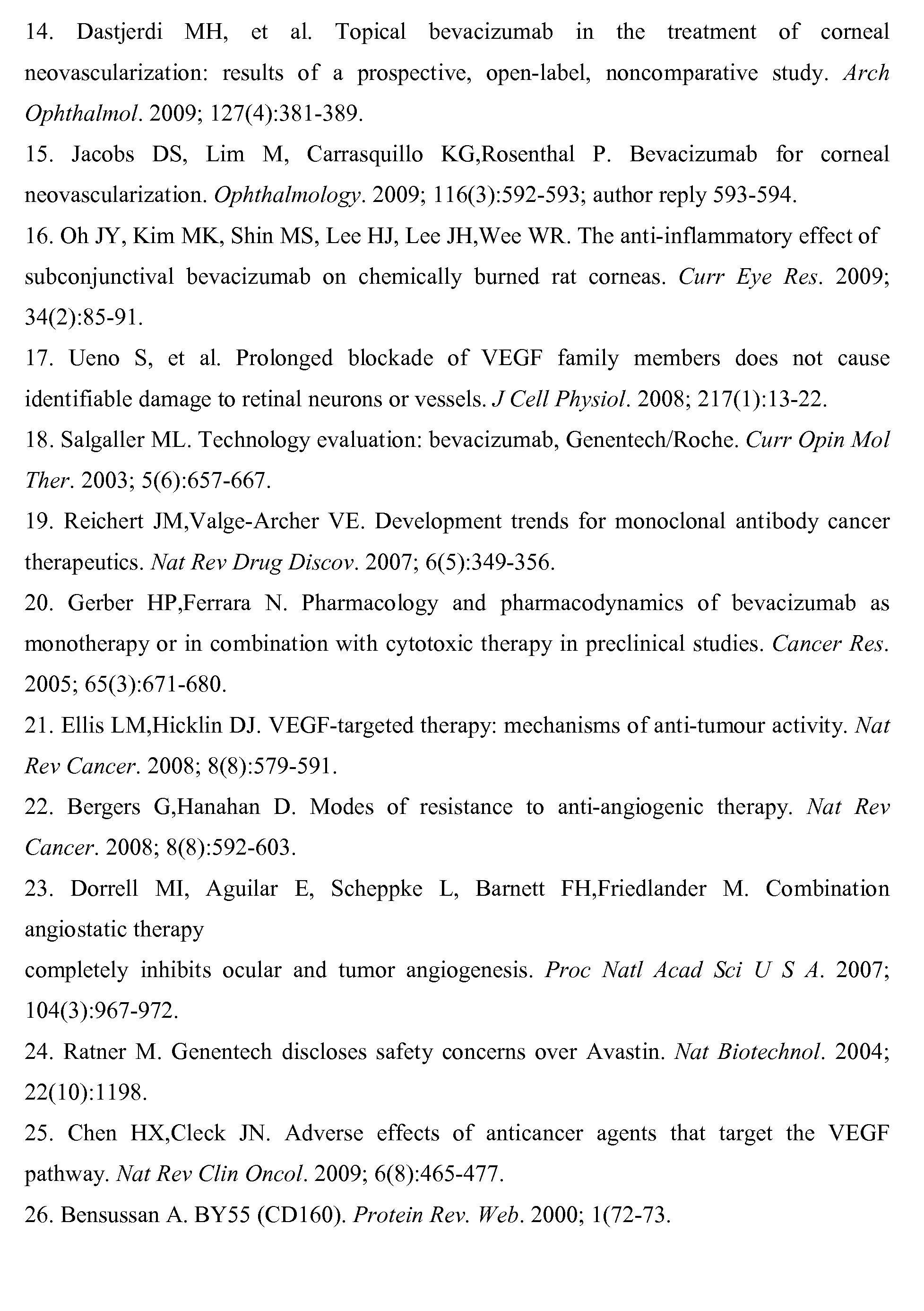
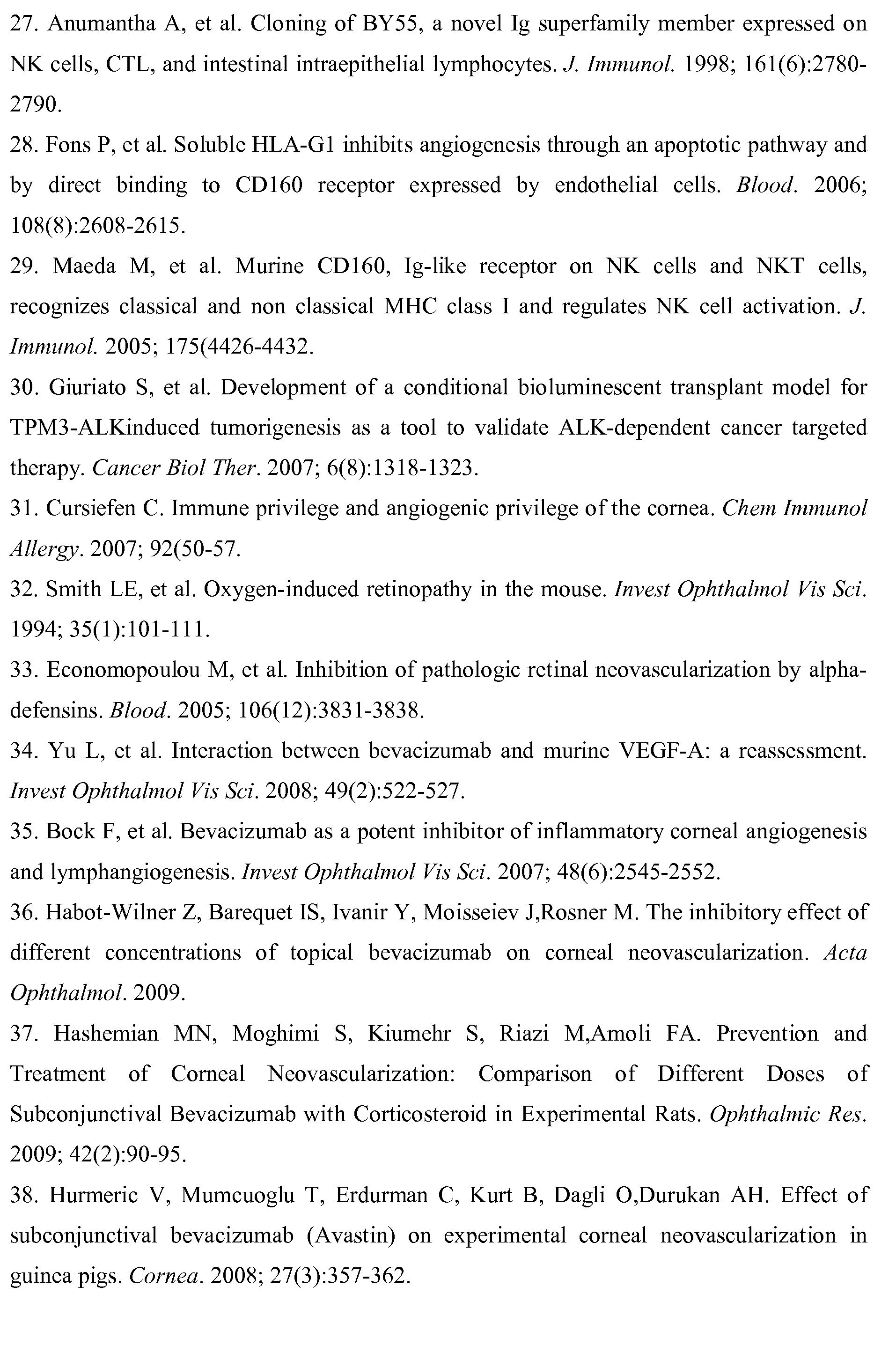
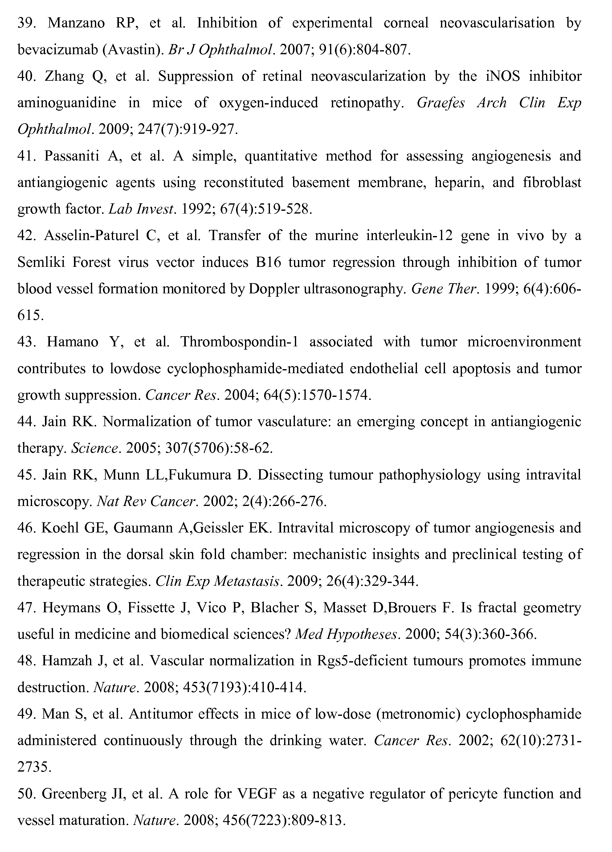

Claims (13)
상기 항-CD160 항체는 활성화된 증식 내피세포의 세포 사멸을 유도하는 것을 특징으로 하는, 조합된 제제.The method according to claim 1,
Wherein the anti-CD160 antibody induces apoptosis of activated proliferative endothelial cells.
상기 항-CD160 항체는, 항-CD160 단일클론 항체; CL1-R2의 Fab, Fab', F(ab)2, F(ab')2 및 dAb로부터 선택되는 보존(conservative) 단편; 및 Fc 단편에 융합되고 CL1-R2에서 유래된 scFv, (scFv)2, 디아바디, 다량체 scFv, 및 페이스-투-테일 형태로 결합된 둘 이상의 Fab를 포함하는 항체로부터 선택되는 CL1-R2의 보존 유도체로부터 선택되는 화합물인 것을 특징으로 하는, 조합된 제제.3. The method according to claim 1 or 2,
Wherein the anti-CD160 antibody comprises an anti-CD160 monoclonal antibody; A conservative fragment selected from Fab, Fab ', F (ab) 2, F (ab') 2 and dAb of CL1-R2; And an antibody comprising two or more Fabs fused to an Fc fragment and conjugated in the form of scFv, (scFv) 2, diabody, multimeric scFv, and face-to-tail derived from CL1-R2 ≪ / RTI > and a preservative derivative.
상기 항-CD160 항체는, CL1-R2 단일클론 항체; CL1-R2의 Fab, Fab', F(ab)2, F(ab')2 및 dAb로부터 선택되는 CL1-R2의 보존 단편; 및 Fc 단편에 융합되고 CL1-R2에서 유래된 scFv, (scFv)2, 디아바디, 다량체 scFv, 및 페이스-투-테일 형태로 결합된 둘 이상의 Fab를 포함하는 항체로부터 선택되는 CL1-R2의 보존 유도체로부터 선택되는 화합물인 것을 특징으로 하는, 조합된 제제.The method according to claim 1,
The anti-CD160 antibody may be a CL1-R2 monoclonal antibody; A conserved fragment of CL1-R2 selected from Fab, Fab ', F (ab) 2, F (ab') 2 and dAb of CL1-R2; And an antibody comprising two or more Fabs fused to an Fc fragment and conjugated in the form of scFv, (scFv) 2, diabody, multimeric scFv, and face-to-tail derived from CL1-R2 ≪ / RTI > and a preservative derivative.
상기 신혈관생성 안구 질환이 하기에서 선택되는 것을 특징으로 하는, 조합된 제제:
● 모든 형태의 각막 신혈관생성증;
● 모든 형태의 막망증;
● 노리병(Norrie disease);
● 모든 형태의 맥락막 신혈관생성증, 망막-맥락막 결절성 혈관병증(polypoidal vasculopathies), 근시와 연관된 역선와(retrofoveolar) 맥락막 신혈관증, 및 소르스비(Sorsby) 영양 장애;
● 포도막 흑색종; 및
● 홍채 혈관신생증(iridal rbeosis) 및 신혈관생성성 녹내장.The method according to claim 1,
Wherein said neovascularizing eye disease is selected from the following combinations:
• all forms of corneal neovascularization;
● all forms of membrane deterioration;
Norrie disease;
• all forms of choroidal neovascularization, polypoidal vasculopathies, retrofoveolar choroidal neovascularization associated with myopia, and Sorsby malnutrition;
● uveitic melanoma; And
● iridal rheosis and neovascular glaucoma.
상기 신혈관생성 안구 질환이 하기에서 선택되는 것을 특징으로 하는, 조합된 제제:
● 각막 이식, 각막 감염, 병원체 감염 및 화학적 화상으로부터 선택되는 각막 환경성 손상의 합병증으로 발생하는 신혈관생성증;
● 당뇨 허혈성 및 부종성 망막증, 조산 당뇨성 망막증(premature diabetic retinopathy), 비증식성 또는 증식성 형태의 망막증, 황반 유낭포성(cystoid) 부종, 모든 형태의 노인성 황반 변성(AMD: Age Related Macular Degeneration), 베스트병(Best disease)을 포함하는 모든 노른자모양 황반 변성(macular vitelliform degeneration), 폰 히펠-린다우(Von Hippel-Lindau) 질병; 알레(Eale) 질병, 코트(Coats) 질병과 같은 안구 혈관종(angiomas);
● 노리병(Norrie disease);
● 모든 형태의 맥락막 신혈관생성증, 망막-맥락막 결절성 혈관병증(polypoidal vasculopathies), 근시와 연관된 역선와(retrofoveolar) 맥락막 신혈관, 및 소르스비(Sorsby) 영양 장애;
● 맥락막 흑색종(choroidal melanomas) 및 이의 종양전이(metastases); 및
● 홍채 혈관신생(iridal rbeosis) 및 신혈관생성성 녹내장.The method according to claim 1,
Wherein said neovascularizing eye disease is selected from the following combinations:
Corneal transplantation, corneal infections, pathogen infection, and neovascularization resulting from complications of corneal environmental injury selected from chemical burns;
● Diabetic ischemic and edematous retinopathy, premature diabetic retinopathy, non-proliferative or proliferative forms of retinopathy, cystoid edema, all forms of AMD (Age Related Macular Degeneration) All yolk-shaped macular vitelliform degeneration including Best disease, Von Hippel-Lindau disease; Angiomas such as Eale disease, Coats disease;
Norrie disease;
• all forms of choroidal neovascularization, polypoidal vasculopathies, retrofoveolar choroidal neovascularization associated with myopia, and Sorsby malnutrition;
Choroidal melanomas and their metastases; And
● iridal rheosis and neovascular glaucoma.
항-VEGF 치료에 내성인 개체의 신혈관생성 안구 질환을 치료 및/또는 예방하기 위해 사용되는 것을 특징으로 하는, 조합된 제제.The method according to claim 1,
Characterized in that it is used for the treatment and / or prevention of a neovascularizing eye disease of an individual resistant to anti-VEGF therapy.
노인성 황반 변성(AMD: Age related Macular Degeneration)의 치료용 조합된 제제.The method according to claim 1,
Combined preparations for the treatment of AMD (Age related macular degeneration).
상기 화합물은 단일클론 항체 CL1-R2인 것을 특징으로 하는, 조합된 제제.The method of claim 3,
Wherein said compound is monoclonal antibody CL1-R2.
상기 화합물은 CL1-R2의 Fab, Fab', F(ab)2, F(ab')2 및 dAb로부터 선택되는 CL1-R2의 보존 단편인 것을 특징으로 하는, 조합된 제제.The method of claim 3,
Wherein said compound is a conserved fragment of CL1-R2 selected from Fab, Fab ', F (ab) 2, F (ab') 2 and dAb of CL1-R2.
상기 화합물은 Fc 단편에 융합되고 CL1-R2에서 유래된 scFv, (scFv)2, 디아바디, 다량체 scFv, 전체 CL1-R2 항체들이 함께 결합된 응집체, 및 페이스-투-테일 형태로 결합된 둘 이상의 Fab를 포함하는 항체로부터 선택되는 CL1-R2의 보존 유도체인 것을 특징으로 하는, 조합된 제제.The method of claim 3,
The compound is an agglutinin which is fused to an Fc fragment and derived from CL1-R2, scFv (scFv) 2, diabody, multimeric scFv, whole CL1-R2 antibodies, Lt; RTI ID = 0.0 > CL1-R2 < / RTI > selected from antibodies comprising the above Fab.
상기 보존 유도체는 1가 또는 다가 scFv인 것을 특징으로 하는, 조합된 제제.12. The method of claim 11,
Wherein the conserved derivative is a monovalent or polyvalent scFv.
Applications Claiming Priority (3)
| Application Number | Priority Date | Filing Date | Title |
|---|---|---|---|
| US34927110P | 2010-05-28 | 2010-05-28 | |
| US61/349,271 | 2010-05-28 | ||
| PCT/EP2011/058777 WO2011147984A1 (en) | 2010-05-28 | 2011-05-27 | Anti-cd160 specific antibodies for the treatment of eye disorders based on neoangiogenesis |
Publications (2)
| Publication Number | Publication Date |
|---|---|
| KR20130086548A KR20130086548A (en) | 2013-08-02 |
| KR101847572B1 true KR101847572B1 (en) | 2018-05-24 |
Family
ID=44276158
Family Applications (1)
| Application Number | Title | Priority Date | Filing Date |
|---|---|---|---|
| KR1020127034345A Expired - Fee Related KR101847572B1 (en) | 2010-05-28 | 2011-05-27 | Anti-cd160 specific antibodies for the treatment of eye disorders based on neoangiogenesis |
Country Status (11)
| Country | Link |
|---|---|
| US (2) | US9045544B2 (en) |
| EP (1) | EP2575881B1 (en) |
| JP (1) | JP5990511B2 (en) |
| KR (1) | KR101847572B1 (en) |
| CN (2) | CN102892432A (en) |
| BR (1) | BR112012030318A2 (en) |
| CA (1) | CA2796312C (en) |
| DK (1) | DK2575881T3 (en) |
| ES (1) | ES2606627T3 (en) |
| PL (1) | PL2575881T3 (en) |
| WO (1) | WO2011147984A1 (en) |
Families Citing this family (15)
| Publication number | Priority date | Publication date | Assignee | Title |
|---|---|---|---|---|
| US10561707B2 (en) * | 2013-09-08 | 2020-02-18 | Technion Research And Development Foundation Ltd. | Semaphorin 3C variants, compositions comprising said variants and methods of use thereof in treating eye diseases |
| US20170056469A1 (en) * | 2014-05-02 | 2017-03-02 | Mayo Foundation For Medical Education And Research | Individualized treatment of eye disease |
| US9840553B2 (en) | 2014-06-28 | 2017-12-12 | Kodiak Sciences Inc. | Dual PDGF/VEGF antagonists |
| EP3397276A4 (en) | 2015-12-30 | 2019-12-18 | Kodiak Sciences Inc. | ANTIBODIES AND CONJUGATES THEREOF |
| US11186635B2 (en) * | 2016-10-25 | 2021-11-30 | Institut National De La Santé Et De La Recherché Médical (Inserm) | Monoclonal antibodies binding to the CD160 transmembrane isoform |
| FR3061716B1 (en) | 2017-01-06 | 2019-05-17 | Elsalys Biotech | NOVEL COMPOUNDS TARGETING CD160 HUMAN |
| SG11202001595SA (en) * | 2017-08-31 | 2020-03-30 | Singapore Health Serv Pte Ltd | Angio-3 for treatment of retinal angiogenic diseases |
| KR20200140817A (en) | 2018-03-02 | 2020-12-16 | 코디악 사이언시스 인코포레이티드 | IL-6 antibodies and fusion constructs and conjugates thereof |
| CN114786731A (en) | 2019-10-10 | 2022-07-22 | 科达制药股份有限公司 | Methods of treating ocular disorders |
| US20230073132A1 (en) * | 2019-12-31 | 2023-03-09 | Medstar Health, Inc. | Anti-cd 160 binding molecules for treating diseases |
| IL300314A (en) | 2020-10-08 | 2023-04-01 | Affimed Gmbh | Trispecific binders |
| AU2022320948A1 (en) | 2021-07-30 | 2024-01-18 | Affimed Gmbh | Duplexbodies |
| KR20250067128A (en) | 2022-09-15 | 2025-05-14 | 아비디큐어 아이피 비.브이. | Multispecific antigen binding proteins for tumor-targeting of NK cells and their uses |
| WO2025191136A1 (en) | 2024-03-15 | 2025-09-18 | Avidicure Ip B.V. | Muteins of 4-1bb ligand extracellular domain, fusion proteins comprising the same and uses thereof |
| WO2025191133A1 (en) | 2024-03-15 | 2025-09-18 | Avidicure Ip B.V. | Il-21 muteins, fusion proteins comprising the same and uses thereof |
Citations (2)
| Publication number | Priority date | Publication date | Assignee | Title |
|---|---|---|---|---|
| US6884879B1 (en) | 1997-04-07 | 2005-04-26 | Genentech, Inc. | Anti-VEGF antibodies |
| US20090317401A1 (en) | 2004-08-09 | 2009-12-24 | Inserm | Angiogenic and immunoglobic applications of anti cd160 specific compounds obtainable from mab cl 1-r2 |
Family Cites Families (7)
| Publication number | Priority date | Publication date | Assignee | Title |
|---|---|---|---|---|
| WO1988001649A1 (en) | 1986-09-02 | 1988-03-10 | Genex Corporation | Single polypeptide chain binding molecules |
| WO1993011161A1 (en) | 1991-11-25 | 1993-06-10 | Enzon, Inc. | Multivalent antigen-binding proteins |
| JP3312357B2 (en) | 1992-12-11 | 2002-08-05 | ザ ダウ ケミカル カンパニー | Multivalent single chain antibody |
| AU5253898A (en) | 1996-11-12 | 1998-06-03 | Dana-Farber Cancer Institute | Recombinant by55 and nucleic acids encoding same |
| FR2794977B1 (en) * | 1999-06-18 | 2003-10-31 | Commissariat Energie Atomique | USE OF COMPOSITIONS CONTAINING SOLUBLE FORMS OF HLA-G IN THE TREATMENT OF INFLAMMATORY SKIN CONDITIONS AND THEIR METHOD OF OBTAINING |
| WO2007087457A2 (en) * | 2006-01-30 | 2007-08-02 | (Osi) Eyetech, Inc. | Combination therapy for the treatment of neovascular disorders |
| AR062239A1 (en) * | 2007-08-07 | 2008-10-22 | Civil De Estudios Superiores A | PHARMACEUTICAL SYNERGIC COMPOSITION USEFUL TO INHIBIT CORNEAL AND RETINAL NEOVASCULARIZATION (ANGIOGENESIS), AND OF OTHER ORGANS, IN A HUMAN OR ANIMAL |
-
2011
- 2011-05-27 KR KR1020127034345A patent/KR101847572B1/en not_active Expired - Fee Related
- 2011-05-27 PL PL11728594T patent/PL2575881T3/en unknown
- 2011-05-27 US US13/697,699 patent/US9045544B2/en active Active
- 2011-05-27 CN CN201180024138XA patent/CN102892432A/en active Pending
- 2011-05-27 WO PCT/EP2011/058777 patent/WO2011147984A1/en not_active Ceased
- 2011-05-27 DK DK11728594.0T patent/DK2575881T3/en active
- 2011-05-27 JP JP2013511702A patent/JP5990511B2/en not_active Expired - Fee Related
- 2011-05-27 CN CN201710912201.2A patent/CN107648605A/en active Pending
- 2011-05-27 EP EP11728594.0A patent/EP2575881B1/en active Active
- 2011-05-27 ES ES11728594.0T patent/ES2606627T3/en active Active
- 2011-05-27 BR BR112012030318A patent/BR112012030318A2/en not_active Application Discontinuation
- 2011-05-27 CA CA2796312A patent/CA2796312C/en active Active
-
2015
- 2015-06-02 US US14/728,253 patent/US10208124B2/en active Active
Patent Citations (2)
| Publication number | Priority date | Publication date | Assignee | Title |
|---|---|---|---|---|
| US6884879B1 (en) | 1997-04-07 | 2005-04-26 | Genentech, Inc. | Anti-VEGF antibodies |
| US20090317401A1 (en) | 2004-08-09 | 2009-12-24 | Inserm | Angiogenic and immunoglobic applications of anti cd160 specific compounds obtainable from mab cl 1-r2 |
Non-Patent Citations (1)
| Title |
|---|
| Blood. 2006,108:2608-2615.* |
Also Published As
| Publication number | Publication date |
|---|---|
| ES2606627T3 (en) | 2017-03-24 |
| EP2575881B1 (en) | 2016-09-14 |
| US10208124B2 (en) | 2019-02-19 |
| JP2013528607A (en) | 2013-07-11 |
| KR20130086548A (en) | 2013-08-02 |
| WO2011147984A1 (en) | 2011-12-01 |
| JP5990511B2 (en) | 2016-09-14 |
| US9045544B2 (en) | 2015-06-02 |
| BR112012030318A2 (en) | 2017-06-20 |
| CN102892432A (en) | 2013-01-23 |
| DK2575881T3 (en) | 2017-01-09 |
| PL2575881T3 (en) | 2018-06-29 |
| EP2575881A1 (en) | 2013-04-10 |
| CA2796312C (en) | 2020-11-03 |
| US20150344572A1 (en) | 2015-12-03 |
| ES2606627T8 (en) | 2017-06-09 |
| CN107648605A (en) | 2018-02-02 |
| CA2796312A1 (en) | 2011-12-01 |
| US20130122006A1 (en) | 2013-05-16 |
Similar Documents
| Publication | Publication Date | Title |
|---|---|---|
| KR101847572B1 (en) | Anti-cd160 specific antibodies for the treatment of eye disorders based on neoangiogenesis | |
| CA2850824C (en) | Treatment of ocular disease | |
| US9102718B2 (en) | Anti-S1P antibody treatment of patients with ocular disease | |
| US20110110932A1 (en) | Combination treatment for ocular diseases | |
| CN114728061A (en) | Methods and compositions for treating diabetic retinopathy | |
| JPWO2018070390A1 (en) | Composition comprising anti-ROBO4 antibody and other agent | |
| JP6863892B2 (en) | How to treat or prevent a stroke | |
| JP2016533318A (en) | Agents targeting RGMA / neogenin cis interaction or lipid rafts and use of the agents in therapeutic methods | |
| CN114901313B (en) | Preventive or therapeutic agent for acute neuromyelitis optica | |
| WO2022201084A1 (en) | Methods for treating ocular diseases | |
| CN113645994A (en) | Methods of treating diseases using Pigment Epithelium Derived Factor (PEDF) | |
| JP7709165B2 (en) | Preventive or therapeutic agent for dementia | |
| TW202532435A (en) | Methods of treating neovascular eye diseases | |
| US11072661B2 (en) | Agents that inhibit the binding of CFH to CD11 b/CD18 and uses thereof | |
| CN120659621A (en) | Pharmaceutical preparation containing anti-VEGFA and VEGFC bispecific binding proteins | |
| Raina | Role of intra-vitreal anti-vascular endothelial growth factor (anti-vegf) ranibizumab in management of choroidal neovascular membrane (cnv) in age related macular degeneration (amd) |
Legal Events
| Date | Code | Title | Description |
|---|---|---|---|
| PA0105 | International application |
St.27 status event code: A-0-1-A10-A15-nap-PA0105 |
|
| PG1501 | Laying open of application |
St.27 status event code: A-1-1-Q10-Q12-nap-PG1501 |
|
| PA0201 | Request for examination |
St.27 status event code: A-1-2-D10-D11-exm-PA0201 |
|
| PE0902 | Notice of grounds for rejection |
St.27 status event code: A-1-2-D10-D21-exm-PE0902 |
|
| T11-X000 | Administrative time limit extension requested |
St.27 status event code: U-3-3-T10-T11-oth-X000 |
|
| T11-X000 | Administrative time limit extension requested |
St.27 status event code: U-3-3-T10-T11-oth-X000 |
|
| T11-X000 | Administrative time limit extension requested |
St.27 status event code: U-3-3-T10-T11-oth-X000 |
|
| E13-X000 | Pre-grant limitation requested |
St.27 status event code: A-2-3-E10-E13-lim-X000 |
|
| P11-X000 | Amendment of application requested |
St.27 status event code: A-2-2-P10-P11-nap-X000 |
|
| P13-X000 | Application amended |
St.27 status event code: A-2-2-P10-P13-nap-X000 |
|
| E701 | Decision to grant or registration of patent right | ||
| PE0701 | Decision of registration |
St.27 status event code: A-1-2-D10-D22-exm-PE0701 |
|
| GRNT | Written decision to grant | ||
| PR0701 | Registration of establishment |
St.27 status event code: A-2-4-F10-F11-exm-PR0701 |
|
| PR1002 | Payment of registration fee |
St.27 status event code: A-2-2-U10-U12-oth-PR1002 Fee payment year number: 1 |
|
| PG1601 | Publication of registration |
St.27 status event code: A-4-4-Q10-Q13-nap-PG1601 |
|
| PR1001 | Payment of annual fee |
St.27 status event code: A-4-4-U10-U11-oth-PR1001 Fee payment year number: 4 |
|
| PN2301 | Change of applicant |
St.27 status event code: A-5-5-R10-R13-asn-PN2301 St.27 status event code: A-5-5-R10-R11-asn-PN2301 |
|
| PR1001 | Payment of annual fee |
St.27 status event code: A-4-4-U10-U11-oth-PR1001 Fee payment year number: 5 |
|
| PN2301 | Change of applicant |
St.27 status event code: A-5-5-R10-R13-asn-PN2301 St.27 status event code: A-5-5-R10-R11-asn-PN2301 |
|
| PN2301 | Change of applicant |
St.27 status event code: A-5-5-R10-R13-asn-PN2301 St.27 status event code: A-5-5-R10-R11-asn-PN2301 |
|
| PR1001 | Payment of annual fee |
St.27 status event code: A-4-4-U10-U11-oth-PR1001 Fee payment year number: 6 |
|
| PC1903 | Unpaid annual fee |
St.27 status event code: A-4-4-U10-U13-oth-PC1903 Not in force date: 20240405 Payment event data comment text: Termination Category : DEFAULT_OF_REGISTRATION_FEE |
|
| PC1903 | Unpaid annual fee |
St.27 status event code: N-4-6-H10-H13-oth-PC1903 Ip right cessation event data comment text: Termination Category : DEFAULT_OF_REGISTRATION_FEE Not in force date: 20240405 |