WO2016092419A1 - Anti-pd-1 antibodies and methods of use thereof - Google Patents
Anti-pd-1 antibodies and methods of use thereof Download PDFInfo
- Publication number
- WO2016092419A1 WO2016092419A1 PCT/IB2015/059268 IB2015059268W WO2016092419A1 WO 2016092419 A1 WO2016092419 A1 WO 2016092419A1 IB 2015059268 W IB2015059268 W IB 2015059268W WO 2016092419 A1 WO2016092419 A1 WO 2016092419A1
- Authority
- WO
- WIPO (PCT)
- Prior art keywords
- antibody
- cancer
- seq
- antibodies
- amino acid
- Prior art date
- Legal status (The legal status is an assumption and is not a legal conclusion. Google has not performed a legal analysis and makes no representation as to the accuracy of the status listed.)
- Ceased
Links
Classifications
-
- A—HUMAN NECESSITIES
- A61—MEDICAL OR VETERINARY SCIENCE; HYGIENE
- A61K—PREPARATIONS FOR MEDICAL, DENTAL OR TOILETRY PURPOSES
- A61K39/00—Medicinal preparations containing antigens or antibodies
- A61K39/395—Antibodies; Immunoglobulins; Immune serum, e.g. antilymphocytic serum
- A61K39/39533—Antibodies; Immunoglobulins; Immune serum, e.g. antilymphocytic serum against materials from animals
- A61K39/3955—Antibodies; Immunoglobulins; Immune serum, e.g. antilymphocytic serum against materials from animals against proteinaceous materials, e.g. enzymes, hormones, lymphokines
-
- A—HUMAN NECESSITIES
- A61—MEDICAL OR VETERINARY SCIENCE; HYGIENE
- A61K—PREPARATIONS FOR MEDICAL, DENTAL OR TOILETRY PURPOSES
- A61K39/00—Medicinal preparations containing antigens or antibodies
- A61K39/0005—Vertebrate antigens
- A61K39/0011—Cancer antigens
-
- A—HUMAN NECESSITIES
- A61—MEDICAL OR VETERINARY SCIENCE; HYGIENE
- A61K—PREPARATIONS FOR MEDICAL, DENTAL OR TOILETRY PURPOSES
- A61K39/00—Medicinal preparations containing antigens or antibodies
- A61K39/39—Medicinal preparations containing antigens or antibodies characterised by the immunostimulating additives, e.g. chemical adjuvants
-
- A—HUMAN NECESSITIES
- A61—MEDICAL OR VETERINARY SCIENCE; HYGIENE
- A61K—PREPARATIONS FOR MEDICAL, DENTAL OR TOILETRY PURPOSES
- A61K45/00—Medicinal preparations containing active ingredients not provided for in groups A61K31/00 - A61K41/00
- A61K45/06—Mixtures of active ingredients without chemical characterisation, e.g. antiphlogistics and cardiaca
-
- A—HUMAN NECESSITIES
- A61—MEDICAL OR VETERINARY SCIENCE; HYGIENE
- A61P—SPECIFIC THERAPEUTIC ACTIVITY OF CHEMICAL COMPOUNDS OR MEDICINAL PREPARATIONS
- A61P1/00—Drugs for disorders of the alimentary tract or the digestive system
- A61P1/02—Stomatological preparations, e.g. drugs for caries, aphtae, periodontitis
-
- A—HUMAN NECESSITIES
- A61—MEDICAL OR VETERINARY SCIENCE; HYGIENE
- A61P—SPECIFIC THERAPEUTIC ACTIVITY OF CHEMICAL COMPOUNDS OR MEDICINAL PREPARATIONS
- A61P1/00—Drugs for disorders of the alimentary tract or the digestive system
- A61P1/04—Drugs for disorders of the alimentary tract or the digestive system for ulcers, gastritis or reflux esophagitis, e.g. antacids, inhibitors of acid secretion, mucosal protectants
-
- A—HUMAN NECESSITIES
- A61—MEDICAL OR VETERINARY SCIENCE; HYGIENE
- A61P—SPECIFIC THERAPEUTIC ACTIVITY OF CHEMICAL COMPOUNDS OR MEDICINAL PREPARATIONS
- A61P1/00—Drugs for disorders of the alimentary tract or the digestive system
- A61P1/16—Drugs for disorders of the alimentary tract or the digestive system for liver or gallbladder disorders, e.g. hepatoprotective agents, cholagogues, litholytics
-
- A—HUMAN NECESSITIES
- A61—MEDICAL OR VETERINARY SCIENCE; HYGIENE
- A61P—SPECIFIC THERAPEUTIC ACTIVITY OF CHEMICAL COMPOUNDS OR MEDICINAL PREPARATIONS
- A61P1/00—Drugs for disorders of the alimentary tract or the digestive system
- A61P1/18—Drugs for disorders of the alimentary tract or the digestive system for pancreatic disorders, e.g. pancreatic enzymes
-
- A—HUMAN NECESSITIES
- A61—MEDICAL OR VETERINARY SCIENCE; HYGIENE
- A61P—SPECIFIC THERAPEUTIC ACTIVITY OF CHEMICAL COMPOUNDS OR MEDICINAL PREPARATIONS
- A61P11/00—Drugs for disorders of the respiratory system
-
- A—HUMAN NECESSITIES
- A61—MEDICAL OR VETERINARY SCIENCE; HYGIENE
- A61P—SPECIFIC THERAPEUTIC ACTIVITY OF CHEMICAL COMPOUNDS OR MEDICINAL PREPARATIONS
- A61P13/00—Drugs for disorders of the urinary system
- A61P13/08—Drugs for disorders of the urinary system of the prostate
-
- A—HUMAN NECESSITIES
- A61—MEDICAL OR VETERINARY SCIENCE; HYGIENE
- A61P—SPECIFIC THERAPEUTIC ACTIVITY OF CHEMICAL COMPOUNDS OR MEDICINAL PREPARATIONS
- A61P13/00—Drugs for disorders of the urinary system
- A61P13/10—Drugs for disorders of the urinary system of the bladder
-
- A—HUMAN NECESSITIES
- A61—MEDICAL OR VETERINARY SCIENCE; HYGIENE
- A61P—SPECIFIC THERAPEUTIC ACTIVITY OF CHEMICAL COMPOUNDS OR MEDICINAL PREPARATIONS
- A61P13/00—Drugs for disorders of the urinary system
- A61P13/12—Drugs for disorders of the urinary system of the kidneys
-
- A—HUMAN NECESSITIES
- A61—MEDICAL OR VETERINARY SCIENCE; HYGIENE
- A61P—SPECIFIC THERAPEUTIC ACTIVITY OF CHEMICAL COMPOUNDS OR MEDICINAL PREPARATIONS
- A61P15/00—Drugs for genital or sexual disorders; Contraceptives
-
- A—HUMAN NECESSITIES
- A61—MEDICAL OR VETERINARY SCIENCE; HYGIENE
- A61P—SPECIFIC THERAPEUTIC ACTIVITY OF CHEMICAL COMPOUNDS OR MEDICINAL PREPARATIONS
- A61P17/00—Drugs for dermatological disorders
-
- A—HUMAN NECESSITIES
- A61—MEDICAL OR VETERINARY SCIENCE; HYGIENE
- A61P—SPECIFIC THERAPEUTIC ACTIVITY OF CHEMICAL COMPOUNDS OR MEDICINAL PREPARATIONS
- A61P19/00—Drugs for skeletal disorders
-
- A—HUMAN NECESSITIES
- A61—MEDICAL OR VETERINARY SCIENCE; HYGIENE
- A61P—SPECIFIC THERAPEUTIC ACTIVITY OF CHEMICAL COMPOUNDS OR MEDICINAL PREPARATIONS
- A61P21/00—Drugs for disorders of the muscular or neuromuscular system
-
- A—HUMAN NECESSITIES
- A61—MEDICAL OR VETERINARY SCIENCE; HYGIENE
- A61P—SPECIFIC THERAPEUTIC ACTIVITY OF CHEMICAL COMPOUNDS OR MEDICINAL PREPARATIONS
- A61P25/00—Drugs for disorders of the nervous system
-
- A—HUMAN NECESSITIES
- A61—MEDICAL OR VETERINARY SCIENCE; HYGIENE
- A61P—SPECIFIC THERAPEUTIC ACTIVITY OF CHEMICAL COMPOUNDS OR MEDICINAL PREPARATIONS
- A61P35/00—Antineoplastic agents
-
- A—HUMAN NECESSITIES
- A61—MEDICAL OR VETERINARY SCIENCE; HYGIENE
- A61P—SPECIFIC THERAPEUTIC ACTIVITY OF CHEMICAL COMPOUNDS OR MEDICINAL PREPARATIONS
- A61P35/00—Antineoplastic agents
- A61P35/02—Antineoplastic agents specific for leukemia
-
- A—HUMAN NECESSITIES
- A61—MEDICAL OR VETERINARY SCIENCE; HYGIENE
- A61P—SPECIFIC THERAPEUTIC ACTIVITY OF CHEMICAL COMPOUNDS OR MEDICINAL PREPARATIONS
- A61P43/00—Drugs for specific purposes, not provided for in groups A61P1/00-A61P41/00
-
- A—HUMAN NECESSITIES
- A61—MEDICAL OR VETERINARY SCIENCE; HYGIENE
- A61P—SPECIFIC THERAPEUTIC ACTIVITY OF CHEMICAL COMPOUNDS OR MEDICINAL PREPARATIONS
- A61P5/00—Drugs for disorders of the endocrine system
-
- C—CHEMISTRY; METALLURGY
- C07—ORGANIC CHEMISTRY
- C07K—PEPTIDES
- C07K16/00—Immunoglobulins [IGs], e.g. monoclonal or polyclonal antibodies
- C07K16/18—Immunoglobulins [IGs], e.g. monoclonal or polyclonal antibodies against material from animals or humans
- C07K16/28—Immunoglobulins [IGs], e.g. monoclonal or polyclonal antibodies against material from animals or humans against receptors, cell surface antigens or cell surface determinants
- C07K16/2803—Immunoglobulins [IGs], e.g. monoclonal or polyclonal antibodies against material from animals or humans against receptors, cell surface antigens or cell surface determinants against the immunoglobulin superfamily
- C07K16/2818—Immunoglobulins [IGs], e.g. monoclonal or polyclonal antibodies against material from animals or humans against receptors, cell surface antigens or cell surface determinants against the immunoglobulin superfamily against CD28 or CD152
-
- C—CHEMISTRY; METALLURGY
- C07—ORGANIC CHEMISTRY
- C07K—PEPTIDES
- C07K16/00—Immunoglobulins [IGs], e.g. monoclonal or polyclonal antibodies
- C07K16/18—Immunoglobulins [IGs], e.g. monoclonal or polyclonal antibodies against material from animals or humans
- C07K16/28—Immunoglobulins [IGs], e.g. monoclonal or polyclonal antibodies against material from animals or humans against receptors, cell surface antigens or cell surface determinants
- C07K16/2878—Immunoglobulins [IGs], e.g. monoclonal or polyclonal antibodies against material from animals or humans against receptors, cell surface antigens or cell surface determinants against the NGF-receptor/TNF-receptor superfamily, e.g. CD27, CD30, CD40, CD95
-
- A—HUMAN NECESSITIES
- A61—MEDICAL OR VETERINARY SCIENCE; HYGIENE
- A61K—PREPARATIONS FOR MEDICAL, DENTAL OR TOILETRY PURPOSES
- A61K39/00—Medicinal preparations containing antigens or antibodies
- A61K2039/505—Medicinal preparations containing antigens or antibodies comprising antibodies
-
- A—HUMAN NECESSITIES
- A61—MEDICAL OR VETERINARY SCIENCE; HYGIENE
- A61K—PREPARATIONS FOR MEDICAL, DENTAL OR TOILETRY PURPOSES
- A61K39/00—Medicinal preparations containing antigens or antibodies
- A61K2039/505—Medicinal preparations containing antigens or antibodies comprising antibodies
- A61K2039/507—Comprising a combination of two or more separate antibodies
-
- A—HUMAN NECESSITIES
- A61—MEDICAL OR VETERINARY SCIENCE; HYGIENE
- A61K—PREPARATIONS FOR MEDICAL, DENTAL OR TOILETRY PURPOSES
- A61K39/00—Medicinal preparations containing antigens or antibodies
- A61K2039/51—Medicinal preparations containing antigens or antibodies comprising whole cells, viruses or DNA/RNA
- A61K2039/53—DNA (RNA) vaccination
-
- A—HUMAN NECESSITIES
- A61—MEDICAL OR VETERINARY SCIENCE; HYGIENE
- A61K—PREPARATIONS FOR MEDICAL, DENTAL OR TOILETRY PURPOSES
- A61K39/00—Medicinal preparations containing antigens or antibodies
- A61K2039/545—Medicinal preparations containing antigens or antibodies characterised by the dose, timing or administration schedule
-
- A—HUMAN NECESSITIES
- A61—MEDICAL OR VETERINARY SCIENCE; HYGIENE
- A61K—PREPARATIONS FOR MEDICAL, DENTAL OR TOILETRY PURPOSES
- A61K2300/00—Mixtures or combinations of active ingredients, wherein at least one active ingredient is fully defined in groups A61K31/00 - A61K41/00
-
- C—CHEMISTRY; METALLURGY
- C07—ORGANIC CHEMISTRY
- C07K—PEPTIDES
- C07K2317/00—Immunoglobulins specific features
- C07K2317/20—Immunoglobulins specific features characterized by taxonomic origin
- C07K2317/24—Immunoglobulins specific features characterized by taxonomic origin containing regions, domains or residues from different species, e.g. chimeric, humanized or veneered
-
- C—CHEMISTRY; METALLURGY
- C07—ORGANIC CHEMISTRY
- C07K—PEPTIDES
- C07K2317/00—Immunoglobulins specific features
- C07K2317/30—Immunoglobulins specific features characterized by aspects of specificity or valency
- C07K2317/33—Crossreactivity, e.g. for species or epitope, or lack of said crossreactivity
-
- C—CHEMISTRY; METALLURGY
- C07—ORGANIC CHEMISTRY
- C07K—PEPTIDES
- C07K2317/00—Immunoglobulins specific features
- C07K2317/50—Immunoglobulins specific features characterized by immunoglobulin fragments
- C07K2317/55—Fab or Fab'
-
- C—CHEMISTRY; METALLURGY
- C07—ORGANIC CHEMISTRY
- C07K—PEPTIDES
- C07K2317/00—Immunoglobulins specific features
- C07K2317/70—Immunoglobulins specific features characterized by effect upon binding to a cell or to an antigen
- C07K2317/74—Inducing cell proliferation
-
- C—CHEMISTRY; METALLURGY
- C07—ORGANIC CHEMISTRY
- C07K—PEPTIDES
- C07K2317/00—Immunoglobulins specific features
- C07K2317/70—Immunoglobulins specific features characterized by effect upon binding to a cell or to an antigen
- C07K2317/76—Antagonist effect on antigen, e.g. neutralization or inhibition of binding
-
- C—CHEMISTRY; METALLURGY
- C07—ORGANIC CHEMISTRY
- C07K—PEPTIDES
- C07K2317/00—Immunoglobulins specific features
- C07K2317/90—Immunoglobulins specific features characterized by (pharmaco)kinetic aspects or by stability of the immunoglobulin
- C07K2317/92—Affinity (KD), association rate (Ka), dissociation rate (Kd) or EC50 value
-
- C—CHEMISTRY; METALLURGY
- C07—ORGANIC CHEMISTRY
- C07K—PEPTIDES
- C07K2317/00—Immunoglobulins specific features
- C07K2317/90—Immunoglobulins specific features characterized by (pharmaco)kinetic aspects or by stability of the immunoglobulin
- C07K2317/94—Stability, e.g. half-life, pH, temperature or enzyme-resistance
Definitions
- the Ig V-like domain in its extracellular region contains two tyrosines, with the most membrane-proximal tyrosine (VAYEEL in mouse PD-1 ) located within an ITIM (immuno-receptor tyrosine-based inhibitory motif).
- ITIM immuno-receptor tyrosine-based inhibitory motif
- Human and murine PD-1 proteins share about 60% amino acid identity with conservation of four potential N- glycosylation sites, and residues that define the Ig-V domain.
- the ITIM in the cytoplasmic region and the ITIM-like motif surrounding the carboxy-terminal tyrosine are also conserved between human and murine orthologues.
- the invention provides an isolated antibody which specifically binds to PD-1 and competes with and/or binds to the same PD-1 epitope as the antibodies as described herein.
- the invention provides an isolated host cell that recombinantly produces a PD-1 antibody as described herein.
- the heavy and light chains are encoded on separate vectors. In other embodiments, heavy and light chains are encoded on the same vector.
- the present disclosure provides a method for treating a cancer in a mammal, particularly a human, which method comprises administering to the mammal (1 ) an effective amount of a vaccine capable of eliciting an immune response against cells of the cancer and (2) an effective amount of an anti-PD-1 antagonist antibody provided by the present disclosure.
- Figure 1 D depicts a graph summarizing body weight of mice treated with anti- PD-1 antagonist antibody.
- Figure 4 depicts a bar graph summarizing proliferation of cultured activated CD8 T cells treated as follows (a) no antibody; (b) isotype control; (c) EH12.1 ; (d) C1 ; (e) C2; (f) C3; (g) mAbl ; (h) mAbX; (i) mAb4; G) mAb5; (k) mAb6; (I) mAb7; (m) mAb9; (n) mAbl 0; (o) mAbl 1 ; (p) mAbl 4; (q) mAbl 5; (r) mAbl 6.
- the Kabat definition is a standard for numbering the residues in an antibody and is typically used to identify CDR regions. See, e.g., Johnson & Wu, 2000, Nucleic Acids Res., 28: 214-8.
- the Chothia definition is similar to the Kabat definition, but the Chothia definition takes into account positions of certain structural loop regions. See, e.g., Chothia et al., 1986, J. Mol. Biol., 196: 901 -17; Chothia et al., 1989, Nature, 342: 877- 83.
- the AbM definition uses an integrated suite of computer programs produced by Oxford Molecular Group that model antibody structure.
- chimeric antibody is intended to refer to antibodies in which the variable region sequences are derived from one species and the constant region sequences are derived from another species, such as an antibody in which the variable region sequences are derived from a mouse antibody and the constant region sequences are derived from a human antibody.
- the sequence of nucleotides may be interrupted by non-nucleotide components.
- a polynucleotide may be further modified after polymerization, such as by conjugation with a labeling component.
- Other types of modifications include, for example, "caps", substitution of one or more of the naturally occurring nucleotides with an analog, internucleotide modifications such as, for example, those with uncharged linkages (e.g., methyl phosphonates, phosphotriesters, phosphoamidates, carbamates, etc.) and with charged linkages (e.g., phosphorothioates, phosphorodithioates, etc.), those containing pendant moieties, such as, for example, proteins (e.g., nucleases, toxins, antibodies, signal peptides, poly-L-lysine, etc.), those with intercalators (e.g., acridine, psoralen, etc.), those containing chelators (e.g., metal
- any of the hydroxyl groups ordinarily present in the sugars may be replaced, for example, by phosphonate groups, phosphate groups, protected by standard protecting groups, or activated to prepare additional linkages to additional nucleotides, or may be conjugated to solid supports.
- the 5' and 3' terminal OH can be phosphorylated or substituted with amines or organic capping group moieties of from 1 to 20 carbon atoms.
- Other hydroxyls may also be derivatized to standard protecting groups.
- vector means a construct, which is capable of delivering, and, preferably, expressing, one or more gene(s) or sequence(s) of interest in a host cell.
- vectors include, but are not limited to, viral vectors, naked DNA or RNA expression vectors, plasm id, cosmid or phage vectors, DNA or RNA expression vectors associated with cationic condensing agents, DNA or RNA expression vectors encapsulated in liposomes, and certain eukaryotic cells, such as producer cells.
- k on refers to the rate constant for association of an antibody to an antigen. Specifically, the rate constants (k on and k ofT ) and equilibrium dissociation constants are measured using full-length antibodies and/or Fab antibody fragments (i.e. univalent) and PD-1 .
- anti-PD-1 antagonist antibodies that block, suppress or reduce (including significantly reduces) PD-1 biological activity, including downstream events mediated by PD-1.
- An anti-PD-1 antagonist antibody should exhibit any one or more of the following characteristics: (a) bind to PD-1 and block downstream signaling events; (b) block PD-L1 binding to PD-1 ; (c) upregulate a T cell-mediated immune response; (d) stimulate IFNy secretion; (e) stimulate TNF secretion; (f) increase T cell proliferation; and (g) reduce inhibitory signal transduction through PD-1 .
- anti-PD-1 antagonist antibodies may be made by any method known in the art. General techniques for production of human and mouse antibodies are known in the art and/or are described herein.
- Anti-PD-1 antagonist antibodies can be identified or characterized using methods known in the art, whereby reduction, amelioration, or neutralization of PD-1 biological activity is detected and/or measured.
- an anti-PD-1 antagonist antibody is identified by incubating a candidate agent with PD-1 and monitoring binding and/or attendant reduction or neutralization of a biological activity of PD-1 .
- the binding assay may be performed with, e.g., purified PD-1 polypeptide(s), or with cells naturally expressing (e.g., various strains), or transfected to express, PD-1 polypeptide(s).
- the binding assay is a competitive binding assay, where the ability of a candidate antibody to compete with a known anti-PD-1 antagonist antibody for PD-1 binding is evaluated.
- the assay may be performed in various formats, including the ELISA format.
- an anti-PD-1 antagonist antibody is identified by incubating a candidate antibody with PD-1 and monitoring binding.
- the epitope to which the anti-PD-1 antagonist antibody binds can be determined in a systematic screening by using overlapping peptides derived from the PD-1 sequence and determining binding by the anti-PD-1 antagonist antibody.
- the open reading frame encoding PD-1 is fragmented either randomly or by specific genetic constructions and the reactivity of the expressed fragments of PD-1 with the antibody to be tested is determined.
- the gene fragments may, for example, be produced by PCR and then transcribed and translated into protein in vitro, in the presence of radioactive amino acids. The binding of the antibody to the radioactively labeled PD-1 fragments is then determined by immunoprecipitation and gel electrophoresis.
- the binding affinity (K D ) of an anti-PD-1 antagonist antibody to PD-1 can be about 0.001 to about 200 nM.
- the binding affinity is any of about 200 nM, about 100 nM, about 50 nM, about 10 nM, about 1 nM, about 500 pM, about 100 pM, about 60 pM, about 50 pM, about 20 pM, about 15 pM, about 10 pM, about 5 pM, about 2 pM, or about 1 pM.
- the antibody comprises the full-length heavy chain, with or without the C-terminal lysine, and/or the full-length light chain of anti-PD-1 antagonist antibody mAb7 or mAb15.
- the amino acid sequence of mAb7 full-length heavy chain (SEQ ID NO: 29) is shown below:
- amino acid sequence of mAb7 full-length light chain (SEQ ID NO: 39) is shown below:
- the invention also provides methods of generating, selecting, and making anti- PD-1 antagonist antibodies.
- the antibodies of this invention can be made by procedures known in the art. In some embodiments, antibodies may be made recombinantly and expressed using any method known in the art.
- antibodies may be prepared and selected by phage display technology. See, for example, U.S. Patent Nos. 5,565,332; 5,580,717; 5,733,743; and 6,265, 150; and Winter et al., Annu. Rev. Immunol. 12:433-455, 1994.
- the phage display technology McCafferty et al., Nature 348:552-553, 1990
- V immunoglobulin variable
- antibody V domain genes are cloned in-frame into either a major or minor coat protein gene of a filamentous bacteriophage, such as M13 or fd, and displayed as functional antibody fragments on the surface of the phage particle.
- a filamentous bacteriophage such as M13 or fd
- the filamentous particle contains a single-stranded DNA copy of the phage genome
- selections based on the functional properties of the antibody also result in selection of the gene encoding the antibody exhibiting those properties.
- the phage mimics some of the properties of the B cell.
- Phage display can be performed in a variety of formats; for review see, e.g., Johnson, Kevin S. and Chiswell, David J., Current Opinion in Structural Biology 3:564-571 , 1993.
- Hybridomas that produce such antibodies may be grown in vitro or in vivo using known procedures.
- the monoclonal antibodies may be isolated from the culture media or body fluids, by conventional immunoglobulin purification procedures such as ammonium sulfate precipitation, gel electrophoresis, dialysis, chromatography, and ultrafiltration, if desired.
- Undesired activity, if present, can be removed, for example, by running the preparation over adsorbents made of the immunogen attached to a solid phase and eluting or releasing the desired antibodies off the immunogen.
- Immunoassays and flow cytometry sorting techniques such as fluorescence activated cell sorting (FACS) can also be employed to isolate antibodies that are specific for PD-1 .
- FACS fluorescence activated cell sorting
- affinity matured antibodies can be produced by procedures known in the art (Marks et al., 1992, Bio/Technology, 10:779-783; Barbas et al., 1994, Proc Nat. Acad. Sci, USA 91 :3809-3813; Schier et al., 1995, Gene, 169: 147-155; Yelton et al., 1995, J. Immunol., 155:1994-2004; Jackson et al., 1995, J. Immunol., 154(7):3310-9; Hawkins et al. , 1992, J. Mol. Biol., 226:889-896; and PCT Publication No. WO2004/058184).
- Candidates with improved binding may be sequenced, thereby identifying a CDR substitution mutant which results in improved affinity (also termed an "improved" substitution).
- Candidates that bind may also be sequenced, thereby identifying a CDR substitution which retains binding.
- Candidates with improved affinity may be combined in a second library, which includes the improved amino acid, the original amino acid at that position, and may further include additional substitutions at that position, depending on the complexity of the library that is desired, or permitted using the desired screening or selection method.
- adjacent amino acid position can be randomized to at least two or more amino acids. Randomization of adjacent amino acids may permit additional conformational flexibility in the mutant CDR, which may in turn, permit or facilitate the introduction of a larger number of improving mutations.
- the library may also comprise substitution at positions that did not show improved affinity in the first round of screening.
- the second library is screened or selected for library members with improved and/or altered binding affinity using any method known in the art, including screening using KinexaTM biosensor analysis, and selection using any method known in the art for selection, including phage display, yeast display, and ribosome display.
- DNA fragments encoding VH and VL regions can first be obtained using any of the methods described above.
- Various modifications, e.g. mutations, deletions, and/or additions can also be introduced into the DNA sequences using standard methods known to those of skill in the art.
- mutagenesis can be carried out using standard methods, such as PCR-mediated mutagenesis, in which the mutated nucleotides are incorporated into the PCR primers such that the PCR product contains the desired mutations or site-directed mutagenesis.
- the invention includes antibodies comprising functionally equivalent variable regions and CDRs which do not significantly affect their properties as well as variants which have enhanced or decreased activity and/or affinity.
- the amino acid sequence may be mutated to obtain an antibody with the desired binding affinity to PD-1 .
- Modification of polypeptides is routine practice in the art and need not be described in detail herein. Examples of modified polypeptides include polypeptides with conservative substitutions of amino acid residues, one or more deletions or additions of amino acids which do not significantly deleteriously change the functional activity, or which mature (enhance) the affinity of the polypeptide for its ligand, or use of chemical analogs.
- Substitution variants have at least one amino acid residue in the antibody molecule removed and a different residue inserted in its place.
- the sites of greatest interest for substitutional mutagenesis include the hypervariable regions, but framework alterations are also contemplated.
- Conservative substitutions are shown in Table 5 under the heading of "conservative substitutions.” If such substitutions result in a change in biological activity, then more substantial changes, denominated "exemplary substitutions" in Table 5, or as further described below in reference to amino acid classes, may be introduced and the products screened.
- Table 5 Amino Acid Substitutions
- the antibodies may also be modified, e.g. in the variable domains of the heavy and/or light chains, e.g., to alter a binding property of the antibody. Changes in the variable region can alter binding affinity and/or specificity. In some embodiments, no more than one to five conservative amino acid substitutions are made within a CDR domain. In other embodiments, no more than one to three conservative amino acid substitutions are made within a CDR domain. For example, a mutation may be made in one or more of the CDR regions to increase or decrease the K D of the antibody for PD- 1 , to increase or decrease k 0ff , or to alter the binding specificity of the antibody. Techniques in site-directed mutagenesis are well-known in the art. See, e.g., Sambrook et al. and Ausubel et al., supra.
- Modifications also include glycosylated and nonglycosylated polypeptides, as well as polypeptides with other post-translational modifications, such as, for example, glycosylation with different sugars, acetylation, and phosphorylation.
- Antibodies are glycosylated at conserved positions in their constant regions (Jefferis and Lund, 1997, Chem. Immunol. 65: 1 1 1 -128; Wright and Morrison, 1997, TibTECH 15:26-32).
- the oligosaccharide side chains of the immunoglobulins affect the protein's function (Boyd et al., 1996, Mol. Immunol. 32: 131 1 -1318; Wittwe and Howard, 1990, Biochem.
- Oligosaccharides may also serve to target a given glycoprotein to certain molecules based upon specific recognition structures. Glycosylation of antibodies has also been reported to affect antibody-dependent cellular cytotoxicity (ADCC).
- glycosylation sites to the antibody is conveniently accomplished by altering the amino acid sequence such that it contains one or more of the above- described tripeptide sequences (for N-linked glycosylation sites).
- the alteration may also be made by the addition of, or substitution by, one or more serine or threonine residues to the sequence of the original antibody (for O-linked glycosylation sites).
- factors that affect glycosylation during recombinant production of antibodies include growth mode, media formulation, culture density, oxygenation, pH, purification schemes and the like.
- Various methods have been proposed to alter the glycosylation pattern achieved in a particular host organism including introducing or overexpressing certain enzymes involved in oligosaccharide production (U.S. Patent Nos. 5,047,335; 5,510,261 and 5,278,299).
- Modifications include using coupling techniques known in the art, including, but not limited to, enzymatic means, oxidative substitution and chelation. Modifications can be used, for example, for attachment of labels for immunoassay. Modified polypeptides are made using established procedures in the art and can be screened using standard assays known in the art, some of which are described below and in the Examples.
- the Fc can be human lgG 2 or human lgG 4 .
- the antibody comprises a constant region of lgG 4 comprising the following mutations (Armour et al., 2003, Molecular Immunology 40 585-593): E233F234L235 to P233V234A235 (lgG 4 Ac), in which the numbering is with reference to wild type lgG 4 .
- the Fc is human lgG 4 E233F234L235 to P233V234A235 with deletion G236 (lgG 4A b).
- the Fc is any human lgG 4 Fc (lgG 4 , lgG 4 Ab or lgG 4 A C ) containing hinge stabilizing mutation S228 to P228 (Aalberse et al., 2002, Immunology 105, 9-19).
- the Fc can be human lgG 2 containing the mutation A330P331 to S330S331 (lgG 2A a), in which the amino acid residues are numbered with reference to the wild type lgG 2 sequence. Eur. J. Immunol., 1999, 29:2613-2624.
- the antibody comprises a modified constant region that has increased or decreased binding affinity to a human Fc gamma receptor, is immunologically inert or partially inert, e.g., does not trigger complement mediated lysis, does not stimulate antibody-dependent cell mediated cytotoxicity (ADCC), or does not activate microglia; or has reduced activities (compared to the unmodified antibody) in any one or more of the following: triggering complement mediated lysis, stimulating ADCC, or activating microglia.
- Different modifications of the constant region may be used to achieve optimal level and/or combination of effector functions. See, for example, Morgan et al., Immunology 86:319-324, 1995; Lund et al., J.
- the constant region is aglycosylated for N-linked glycosylation.
- the constant region is aglycosylated for N-linked glycosylation by mutating the oligosaccharide attachment residue and/or flanking residues that are part of the N-glycosylation recognition sequence in the constant region.
- N-glycosylation site N297 may be mutated to, e.g., A, Q, K, or H. See, Tao et al., J. Immunology 143: 2595-2601 , 1989; and Jefferis et al., Immunological Reviews 163:59-76, 1998.
- the constant region is aglycosylated for N-linked glycosylation.
- the constant region may be aglycosylated for N-linked glycosylation enzymatically (such as removing carbohydrate by enzyme PNGase), or by expression in a glycosylation deficient host cell.
- Other antibody modifications include antibodies that have been modified as described in PCT Publication No. WO 99/58572. These antibodies comprise, in addition to a binding domain directed at the target molecule, an effector domain having an amino acid sequence substantially homologous to all or part of a constant region of a human immunoglobulin heavy chain. These antibodies are capable of binding the target molecule without triggering significant complement dependent lysis, or cell-mediated destruction of the target.
- the effector domain is capable of specifically binding FcRn and/or FcYRIIb. These are typically based on chimeric domains derived from two or more human immunoglobulin heavy chain CH2 domains. Antibodies modified in this manner are particularly suitable for use in chronic antibody therapy, to avoid inflammatory and other adverse reactions to conventional antibody therapy.
- the antibody comprises a modified constant region that has increased binding affinity for FcRn and/or an increased serum half-life as compared with the unmodified antibody.
- VH and VL sequences can be mutated to match those found naturally in germline VH and VL sequences.
- the amino acid sequences of the framework regions in the VH and VL sequences can be mutated to match the germline sequences to reduce the risk of immunogenicity when the antibody is administered.
- Germline DNA sequences for human VH and VL genes are known in the art (see e.g., the "Vbase” human germline sequence database; see also Kabat, E. A., et al., 1991 , Sequences of Proteins of Immunological Interest, Fifth Edition, U.S. Department of Health and Human Services, NIH Publication No. 91 -3242; Tomlinson et al., 1992, J. Mol. Biol. 227:776-798; and Cox et al., 1994, Eur. J. Immunol. 24:827-836).
- Another type of amino acid substitution that may be made is to remove potential proteolytic sites in the antibody. Such sites may occur in a CDR or framework region of a variable domain or in the constant region of an antibody. Substitution of cysteine residues and removal of proteolytic sites may decrease the risk of heterogeneity in the antibody product and thus increase its homogeneity.
- Another type of amino acid substitution is to eliminate asparagine-glycine pairs, which form potential deamidation sites, by altering one or both of the residues.
- the C-terminal lysine of the heavy chain of an anti-PD-1 antibody of the invention can be cleaved.
- the heavy and light chains of the anti-PD-1 antibodies may optionally include a signal sequence.
- the isolated DNA encoding the VH region can be converted to a full-length heavy chain gene by operatively linking the VH-encoding DNA to another DNA molecule encoding heavy chain constant regions (CH1 , CH2 and CH3).
- the sequences of human heavy chain constant region genes are known in the art (see e.g. , Kabat, E. A., et al., 1991 , Sequences of Proteins of Immunological Interest, Fifth Edition, U.S. Department of Health and Human Services, NIH Publication No. 91 -3242) and DNA fragments encompassing these regions can be obtained by standard PCR amplification.
- the heavy chain constant region can be an IgG-i , lgG 2 , lgG 3 , lgG 4 , IgA, IgE, IgM or IgD constant region, but most preferably is an Igd or lgG 2 constant region.
- the IgG constant region sequence can be any of the various alleles or allotypes known to occur among different individuals, such as Gm(1 ), Gm(2), Gm(3), and Gm(17). These allotypes represent naturally occurring amino acid substitution in the lgG1 constant regions.
- the VH-encoding DNA can be operatively linked to another DNA molecule encoding only the heavy chain CH1 constant region.
- the CH1 heavy chain constant region may be derived from any of the heavy chain genes.
- the isolated DNA encoding the VL region can be converted to a full-length light chain gene (as well as a Fab light chain gene) by operatively linking the VL-encoding DNA to another DNA molecule encoding the light chain constant region, CL.
- the sequences of human light chain constant region genes are known in the art (see e.g., Kabat, E. A., et al., 1991 , Sequences of Proteins of Immunological Interest, Fifth Edition, U.S. Department of Health and Human Services, NIH Publication No. 91 -3242) and DNA fragments encompassing these regions can be obtained by standard PCR amplification.
- the light chain constant region can be a kappa or lambda constant region.
- the kappa constant region may be any of the various alleles known to occur among different individuals, such as lnv(1 ), lnv(2), and lnv(3).
- the lambda constant region may be derived from any of the three lambda genes.
- VH- and VL-encoding DNA fragments are operatively linked to another fragment encoding a flexible linker such that the VH and VL sequences can be expressed as a contiguous single-chain protein, with the VL and VH regions joined by the flexible linker (See e.g., Bird et al., 1988, Science 242:423-426; Huston et al., 1988, Proc. Natl. Acad. Sci. USA 85:5879-5883; McCafferty et al., 1990, Nature 348:552-554.
- linking peptide is (GGGGS) 3 (SEQ ID NO: 19), which bridges approximately 3.5 nm between the carboxy terminus of one variable region and the amino terminus of the other variable region.
- Linkers of other sequences have been designed and used (Bird et al., 1988, supra). Linkers can in turn be modified for additional functions, such as attachment of drugs or attachment to solid supports.
- the single chain antibody may be monovalent, if only a single VH and VL are used, bivalent, if two VH and VL are used, or polyvalent, if more than two VH and VL are used. Bispecific or polyvalent antibodies may be generated that bind specifically to PD- 1 and to another molecule.
- the single chain variants can be produced either recombinantly or synthetically.
- an automated synthesizer can be used for synthetic production of scFv.
- a suitable plasmid containing polynucleotide that encodes the scFv can be introduced into a suitable host cell, either eukaryotic, such as yeast, plant, insect or mammalian cells, or prokaryotic, such as E. coli.
- a suitable host cell either eukaryotic, such as yeast, plant, insect or mammalian cells, or prokaryotic, such as E. coli.
- Polynucleotides encoding the scFv of interest can be made by routine manipulations such as ligation of polynucleotides.
- the resultant scFv can be isolated using standard protein purification techniques known in the art.
- Diabodies are bivalent, bispecific antibodies in which VH and VL are expressed on a single polypeptide chain, but using a linker that is too short to allow for pairing between the two domains on the same chain, thereby forcing the domains to pair with complementary domains of another chain and creating two antigen binding sites (see e.g., Holliger, P., et al., 1993, Proc. Natl. Acad Sci. USA 90:6444-6448; Poljak, R. J., et al., 1994, Structure 2:1 121 -1 123).
- Heteroconjugate antibodies comprising two covalently joined antibodies, are also within the scope of the invention.
- Chimeric or hybrid antibodies also may be prepared in vitro using known methods of synthetic protein chemistry, including those involving cross-linking agents.
- immunotoxins may be constructed using a disulfide exchange reaction or by forming a thioether bond.
- suitable reagents for this purpose include iminothiolate and methyl-4-mercaptobutyrimidate.
- the invention also encompasses fusion proteins comprising one or more fragments or regions from the antibodies disclosed herein.
- a fusion antibody may be made that comprises all or a portion of an anti-PD-1 antibody of the invention linked to another polypeptide.
- only the variable domains of the anti-PD-1 antibody are linked to the polypeptide.
- the VH domain of an anti-PD-1 antibody is linked to a first polypeptide, while the VL domain of an anti-PD-1 antibody is linked to a second polypeptide that associates with the first polypeptide in a manner such that the VH and VL domains can interact with one another to form an antigen binding site.
- the VH domain is separated from the VL domain by a linker such that the VH and VL domains can interact with one another.
- the VH-linker- VL antibody is then linked to the polypeptide of interest.
- fusion antibodies can be created in which two (or more) single-chain antibodies are linked to one another. This is useful if one wants to create a divalent or polyvalent antibody on a single polypeptide chain, or if one wants to create a bispecific antibody.
- a fusion polypeptide that comprises at least 10 contiguous amino acids of the variable light chain region shown in SEQ ID NO: 2, 7, 8, or 9 and/or at least 10 amino acids of the variable heavy chain region shown in SEQ ID NO: 3, 4, 5, or 6 .
- a fusion polypeptide is provided that comprises at least about 10, at least about 15, at least about 20, at least about 25, or at least about 30 contiguous amino acids of the variable light chain region and/or at least about 10, at least about 15, at least about 20, at least about 25, or at least about 30 contiguous amino acids of the variable heavy chain region.
- other modified antibodies may be prepared using anti-PD- 1 antibody encoding nucleic acid molecules.
- “Kappa bodies” III et al., 1997, Protein Eng. 10:949-57
- “Minibodies” Martin et al., 1994, EMBO J. 13:5303-9
- “Diabodies” Holliger et al., supra
- “Janusins” Traunecker et al., 1991 , EMBO J. 10:3655-3659 and Traunecker et al., 1992, Int. J. Cancer (Suppl.) 7:51 -52) may be prepared using standard molecular biological techniques following the teachings of the specification.
- bispecific antibodies monoclonal antibodies that have binding specificities for at least two different antigens
- methods for making bispecific antibodies are known in the art (see, e.g., Suresh et al., 1986, Methods in Enzymology 121 :210).
- bispecific antibodies or antigen-binding fragments can be produced by fusion of hybridomas or linking of Fab' fragments. See, e.g., Songsivilai & Lachmann, 1990, Clin. Exp. Immunol. 79:315-321 , Kostelny et al., 1992, J. Immunol. 148: 1547-1553.
- bispecific antibodies Traditionally, the recombinant production of bispecific antibodies was based on the coexpression of two immunoglobulin heavy chain-light chain pairs, with the two heavy chains having different specificities (Millstein and Cuello, 1983, Nature 305, 537-539).
- bispecific antibodies may be formed as "diabodies" or "Janusins.”
- the bispecific antibody binds to two different epitopes of PD-1 .
- the modified antibodies described above are prepared using one or more of the variable domains or CDR regions from an anti-PD-1 antibody provided herein.
- the bispecific antibodies are composed of a hybrid immunoglobulin heavy chain with a first binding specificity in one arm, and a hybrid immunoglobulin heavy chain-light chain pair (providing a second binding specificity) in the other arm.
- This asymmetric structure with an immunoglobulin light chain in only one half of the bispecific molecule, facilitates the separation of the desired bispecific compound from unwanted immunoglobulin chain combinations. This approach is described in PCT Publication No. WO 94/04690.
- compositions comprising antibodies conjugated (for example, linked) to an agent that facilitate coupling to a solid support (such as biotin or avidin).
- a solid support such as biotin or avidin.
- Conjugation generally refers to linking these components as described herein.
- the linking (which is generally fixing these components in proximate association at least for administration) can be achieved in any number of ways. For example, a direct reaction between an agent and an antibody is possible when each possesses a substituent capable of reacting with the other.
- a nucleophilic group such as an amino or sulfhydryl group
- a carbonyl-containing group such as an anhydride or an acid halide, or with an alkyl group containing a good leaving group (e.g., a halide) on the other.
- the antibodies can be bound to many different carriers.
- Carriers can be active and/or inert. Examples of well-known carriers include polypropylene, polystyrene, polyethylene, dextran, nylon, amylases, glass, natural and modified celluloses, polyacrylamides, agaroses and magnetite. The nature of the carrier can be either soluble or insoluble for purposes of the invention. Those skilled in the art will know of other suitable carriers for binding antibodies, or will be able to ascertain such, using routine experimentation.
- the carrier comprises a moiety that targets the lung, heart, or heart valve.
- An antibody or polypeptide of this invention may be linked to a labeling agent such as a fluorescent molecule, a radioactive molecule or any others labels known in the art.
- Labels are known in the art which generally provide (either directly or indirectly) a signal.
- the invention also provides polynucleotides encoding any of the antibodies, including antibody fragments and modified antibodies described herein, such as, e.g. , antibodies having impaired effector function.
- the invention provides a method of making any of the polynucleotides described herein. Polynucleotides can be made and expressed by procedures known in the art.
- Polynucleotides may comprise a native sequence (i.e., an endogenous sequence that encodes an antibody or a fragment thereof) or may comprise a variant of such a sequence.
- Polynucleotide variants contain one or more substitutions, additions, deletions and/or insertions such that the immunoreactivity of the encoded polypeptide is not diminished, relative to a native immunoreactive molecule. The effect on the immunoreactivity of the encoded polypeptide may generally be assessed as described herein.
- Variants preferably exhibit at least about 70% identity, more preferably, at least about 80% identity, yet more preferably, at least about 90% identity, and most preferably, at least about 95% identity to a polynucleotide sequence that encodes a native antibody or a fragment thereof.
- Two polynucleotide or polypeptide sequences are said to be “identical” if the sequence of nucleotides or amino acids in the two sequences is the same when aligned for maximum correspondence as described below. Comparisons between two sequences are typically performed by comparing the sequences over a comparison window to identify and compare local regions of sequence similarity.
- a “comparison window” as used herein refers to a segment of at least about 20 contiguous positions, usually 30 to about 75, or 40 to about 50, in which a sequence may be compared to a reference sequence of the same number of contiguous positions after the two sequences are optimally aligned.
- Optimal alignment of sequences for comparison may be conducted using the MegAlign ® program in the Lasergene ® suite of bioinformatics software (DNASTAR ® , Inc., Madison, Wl), using default parameters.
- This program embodies several alignment schemes described in the following references: Dayhoff, M.O., 1978, A model of evolutionary change in proteins - Matrices for detecting distant relationships. In Dayhoff, M.O. (ed.) Atlas of Protein Sequence and Structure, National Biomedical Research Foundation, Washington DC Vol. 5, Suppl. 3, pp. 345-358; Hein J., 1990, Unified Approach to Alignment and Phylogenes pp. 626-645 Methods in Enzymology vol.
- the "percentage of sequence identity” is determined by comparing two optimally aligned sequences over a window of comparison of at least 20 positions, wherein the portion of the polynucleotide or polypeptide sequence in the comparison window may comprise additions or deletions (i.e., gaps) of 20 percent or less, usually 5 to 15 percent, or 10 to 12 percent, as compared to the reference sequences (which does not comprise additions or deletions) for optimal alignment of the two sequences.
- the percentage is calculated by determining the number of positions at which the identical nucleic acid bases or amino acid residue occurs in both sequences to yield the number of matched positions, dividing the number of matched positions by the total number of positions in the reference sequence (i.e. the window size) and multiplying the results by 100 to yield the percentage of sequence identity.
- Variants may also, or alternatively, be substantially homologous to a native gene, or a portion or complement thereof.
- Such polynucleotide variants are capable of hybridizing under moderately stringent conditions to a naturally occurring DNA sequence encoding a native antibody (or a complementary sequence).
- nucleotide sequences that encode a polypeptide as described herein. Some of these polynucleotides bear minimal homology to the nucleotide sequence of any native gene. Nonetheless, polynucleotides that vary due to differences in codon usage are specifically contemplated by the present invention. Further, alleles of the genes comprising the polynucleotide sequences provided herein are within the scope of the present invention. Alleles are endogenous genes that are altered as a result of one or more mutations, such as deletions, additions and/or substitutions of nucleotides. The resulting mRNA and protein may, but need not, have an altered structure or function. Alleles may be identified using standard techniques (such as hybridization, amplification and/or database sequence comparison).
- polynucleotides of this invention can be obtained using chemical synthesis, recombinant methods, or PCR. Methods of chemical polynucleotide synthesis are well known in the art and need not be described in detail herein. One of skill in the art can use the sequences provided herein and a commercial DNA synthesizer to produce a desired DNA sequence.
- a polynucleotide comprising a desired sequence can be inserted into a suitable vector, and the vector in turn can be introduced into a suitable host cell for replication and amplification, as further discussed herein.
- Polynucleotides may be inserted into host cells by any means known in the art. Cells are transformed by introducing an exogenous polynucleotide by direct uptake, endocytosis, transfection, F-mating or electroporation. Once introduced, the exogenous polynucleotide can be maintained within the cell as a non-integrated vector (such as a plasmid) or integrated into the host cell genome.
- the polynucleotide so amplified can be isolated from the host cell by methods well known within the art. See, e.g., Sambrook et al., 1989.
- PCR allows reproduction of DNA sequences.
- PCR technology is well known in the art and is described in U.S. Patent Nos. 4,683, 195, 4,800, 159, 4,754,065 and 4,683,202, as well as PCR: The Polymerase Chain Reaction, Mullis et al. eds., Birkauswer Press, Boston, 1994.
- RNA can be obtained by using the isolated DNA in an appropriate vector and inserting it into a suitable host cell. When the cell replicates and the DNA is transcribed into RNA, the RNA can then be isolated using methods well known to those of skill in the art, as set forth in Sambrook et al., 1989, supra, for example.
- Suitable cloning vectors may be constructed according to standard techniques, or may be selected from a large number of cloning vectors available in the art. While the cloning vector selected may vary according to the host cell intended to be used, useful cloning vectors will generally have the ability to self-replicate, may possess a single target for a particular restriction endonuclease, and/or may carry genes for a marker that can be used in selecting clones containing the vector.
- the vectors containing the polynucleotides of interest can be introduced into the host cell by any of a number of appropriate means, including electroporation, transfection employing calcium chloride, rubidium chloride, calcium phosphate, DEAE- dextran, or other substances; microprojectile bombardment; lipofection; and infection (e.g., where the vector is an infectious agent such as vaccinia virus).
- electroporation employing calcium chloride, rubidium chloride, calcium phosphate, DEAE- dextran, or other substances
- microprojectile bombardment e.g., where the vector is an infectious agent such as vaccinia virus.
- infection e.g., where the vector is an infectious agent such as vaccinia virus.
- the choice of introducing vectors or polynucleotides will often depend on features of the host cell.
- the invention also provides host cells comprising any of the polynucleotides described herein. Any host cells capable of over-expressing heterologous DNAs can be used for the purpose of isolating the genes encoding the antibody, polypeptide or protein of interest.
- mammalian host cells include but not limited to COS, HeLa, and CHO cells. See also PCT Publication No. WO 87/04462.
- Suitable non-mammalian host cells include prokaryotes (such as E. coli or B. subtillis) and yeast (such as S. cerevisae, S. pombe; or K. lactis).
- the host cells express the cDNAs at a level of about 5 fold higher, more preferably, 10 fold higher, even more preferably, 20 fold higher than that of the corresponding endogenous antibody or protein of interest, if present, in the host cells.
- Screening the host cells for a specific binding to PD-1 or a PD-1 domain is effected by an immunoassay or FACS.
- a cell overexpressing the antibody or protein of interest can be identified.
- An expression vector can be used to direct expression of an anti-PD-1 antagonist antibody.
- One skilled in the art is familiar with administration of expression vectors to obtain expression of an exogenous protein in vivo. See, e.g., U.S. Patent Nos. 6,436,908; 6,413,942; and 6,376,471 .
- Administration of expression vectors includes local or systemic administration, including injection, oral administration, particle gun or catheterized administration, and topical administration.
- the expression vector is administered directly to the sympathetic trunk or ganglion, or into a coronary artery, atrium, ventrical, or pericardium.
- Targeted delivery of therapeutic compositions containing an expression vector, or subgenomic polynucleotides can also be used.
- Receptor-mediated DNA delivery techniques are described in, for example, Findeis et al., Trends Biotechnol., 1993, 1 1 :202; Chiou et al., Gene Therapeutics: Methods And Applications Of Direct Gene Transfer, J.A. Wolff, ed., 1994; Wu et al., J. Biol. Chem., 1988, 263:621 ; Wu et al. , J. Biol. Chem., 1994, 269:542; Zenke et al., Proc. Natl. Acad. Sci.
- the gene delivery vehicle can be of viral or non-viral origin (see generally, Jolly, Cancer Gene Therapy, 1994, 1 :51 ; Kimura, Human Gene Therapy, 1994, 5:845; Connelly, Human Gene Therapy, 1995, 1 :185; and Kaplitt, Nature Genetics, 1994, 6: 148). Expression of such coding sequences can be induced using endogenous mammalian or heterologous promoters. Expression of the coding sequence can be either constitutive or regulated.
- Viral-based vectors for delivery of a desired polynucleotide and expression in a desired cell are well known in the art.
- Exemplary viral-based vehicles include, but are not limited to, recombinant retroviruses (see, e.g., PCT Publication Nos. WO 90/07936; WO 94/03622; WO 93/25698; WO 93/25234; WO 93/1 1230; WO 93/10218; WO 91/02805; U.S. Patent Nos. 5, 219,740 and 4,777, 127; GB Patent No. 2,200,651 ; and EP Patent No.
- Non-viral delivery vehicles and methods can also be employed, including, but not limited to, polycationic condensed DNA linked or unlinked to killed adenovirus alone (see, e.g., Curiel, Hum. Gene Then, 1992, 3:147); ligand-linked DNA (see, e.g., Wu, J. Biol. Chem., 1989, 264: 16985); eukaryotic cell delivery vehicles cells (see, e.g., U.S. Patent No. 5,814,482; PCT Publication Nos. WO 95/07994; WO 96/17072; WO 95/30763; and WO 97/42338) and nucleic charge neutralization or fusion with cell membranes. Naked DNA can also be employed.
- Exemplary naked DNA introduction methods are described in PCT Publication No. WO 90/1 1092 and U.S. Patent No. 5,580,859.
- Liposomes that can act as gene delivery vehicles are described in U.S. Patent No. 5,422,120; PCT Publication Nos. WO 95/13796; WO 94/23697; WO 91/14445; and EP 0524968. Additional approaches are described in Philip, Mol. Cell Biol., 1994, 14:241 1 , and in Woffendin, Proc. Natl. Acad. Sci., 1994, 91 : 1581 .
- the invention also provides pharmaceutical compositions comprising an effective amount of an anti-PD-1 antibody described herein. Examples of such compositions, as well as how to formulate, are also described herein.
- the composition comprises one or more PD-1 antibodies.
- the anti- PD-1 antibody recognizes PD-1 .
- the anti-PD-1 antibody is a human antibody.
- the anti-PD-1 antibody is a humanized antibody.
- the anti-PD-1 antibody comprises a constant region that is capable of triggering a desired immune response, such as antibody-mediated lysis or ADCC.
- the anti-PD-1 antibody comprises a constant region that does not trigger an unwanted or undesirable immune response, such as antibody-mediated lysis or ADCC.
- the anti-PD-1 antibody comprises one or more CDR(s) of the antibody (such as one, two, three, four, five, or, in some embodiments, all six CDRs).
- compositions can comprise more than one anti-PD-1 antibody (e.g., a mixture of PD-1 antibodies that recognize different epitopes of PD-1 ).
- Other exemplary compositions comprise more than one anti-PD-1 antibody that recognize the same epitope(s), or different species of anti-PD-1 antibodies that bind to different epitopes of PD-1 .
- the compositions comprise a mixture of anti-PD-1 antibodies that recognize different variants of PD-1 .
- Acceptable carriers, excipients, or stabilizers are nontoxic to recipients at the dosages and concentrations, and may comprise buffers such as phosphate, citrate, and other organic acids; antioxidants including ascorbic acid and methionine; preservatives (such as octadecyldimethylbenzyl ammonium chloride; hexamethonium chloride; benzalkonium chloride, benzethonium chloride; phenol, butyl or benzyl alcohol; alkyl parabens such as methyl or propyl paraben; catechol; resorcinol; cyclohexanol; 3-pentanol; and m-cresol); low molecular weight (less than about 10 residues) polypeptides; proteins, such as serum albumin, gelatin, or immunoglobulins; hydrophilic polymers such as polyvinylpyrrolidone; amino acids such as glycine, glutamine, asparagine, histidine,
- compositions comprising any of the polynucleotides of the invention.
- the composition comprises an expression vector comprising a polynucleotide encoding the antibody as described herein.
- the composition comprises an expression vector comprising a polynucleotide encoding any of the antibodies described herein.
- the antibodies and the antibody conjugates of the present invention are useful in various applications including, but are not limited to, therapeutic treatment methods and diagnostic treatment methods.
- cancers include, but are not limited to bladder cancer, breast cancer, cervical cancer, choriocarcinoma, colon cancer, esophageal cancer, gastric cancer, glioblastoma, glioma, brain tumor, head and neck cancer, kidney cancer, lung cancer, oral cancer, ovarian cancer, pancreatic cancer, prostate cancer, liver cancer, uterine cancer, bone cancer, leukemia, lymphoma, sacrcoma, blood cancer, thyroid cancer, thymic cancer, eye cancer, and skin cancer.
- the tumor is a PD-L1 expressing tumor.
- the tumor does not express PD-L1 .
- provided is a method of inducing regression of a tumor in a subject comprising administering to the subject in need thereof an effective amount of a composition comprising any of the PD-1 antibodies as described herein.
- the PD-1 antibodies as described herein can be labeled with a detectable moiety such as an imaging agent and an enzyme-substrate label.
- the antibodies as described herein can also be used for in vivo diagnostic assays, such as in vivo imaging (e.g., PET or SPECT), or a staining reagent.
- the methods described herein further comprise a step of treating a subject with an additional form of therapy.
- the additional form of therapy is an additional anti-cancer therapy including, but not limited to, chemotherapy, radiation, surgery, hormone therapy, and/or additional immunotherapy.
- compositions comprising one or more additional agents.
- These compositions may further comprise suitable excipients, such as pharmaceutically acceptable excipients including buffers, which are well known in the art.
- suitable excipients such as pharmaceutically acceptable excipients including buffers, which are well known in the art.
- the present invention can be used alone or in combination with other methods of treatment.
- the anti-PD-1 antagonist antibody can be administered to a subject via any suitable route. It should be apparent to a person skilled in the art that the examples described herein are not intended to be limiting but to be illustrative of the techniques available. Accordingly, in some embodiments, the anti-PD-1 antagonist antibody is administered to a subject in accord with known methods, such as intravenous administration, e.g., as a bolus or by continuous infusion over a period of time, by intramuscular, intraperitoneal, intracerebrospinal, transdermal, subcutaneous, intraarticular, sublingually, intrasynovial, via insufflation, intrathecal, oral, inhalation or topical routes.
- intravenous administration e.g., as a bolus or by continuous infusion over a period of time
- intramuscular, intraperitoneal, intracerebrospinal transdermal, subcutaneous, intraarticular, sublingually, intrasynovial, via insufflation, intrathecal,
- an anti-PD-1 antagonist antibody is administered via site- specific or targeted local delivery techniques.
- site-specific or targeted local delivery techniques include various implantable depot sources of the anti-PD-1 antagonist antibody or local delivery catheters, such as infusion catheters, indwelling catheters, or needle catheters, synthetic grafts, adventitial wraps, shunts and stents or other implantable devices, site specific carriers, direct injection, or direct application. See, e.g., PCT Publication No. WO 00/5321 1 and U.S. Patent No. 5,981 ,568.
- an anti-PD-1 antagonist antibody may be used for administration.
- the anti-PD-1 antagonist antibody may be administered neat.
- anti-PD-1 antagonist antibody and a pharmaceutically acceptable excipient may be in various formulations.
- Pharmaceutically acceptable excipients are known in the art, and are relatively inert substances that facilitate administration of a pharmacologically effective substance.
- an excipient can give form or consistency, or act as a diluent.
- Suitable excipients include but are not limited to stabilizing agents, wetting and emulsifying agents, salts for varying osmolarity, encapsulating agents, buffers, and skin penetration enhancers. Excipients as well as formulations for parenteral and nonparenteral drug delivery are set forth in Remington, The Science and Practice of Pharmacy 20th Ed. Mack Publishing, 2000.
- dosage of about 1 mg/kg, about 2.5 mg/kg, about 5 mg/kg, about 10 mg/kg, and about 25 mg/kg may be used.
- the treatment is sustained until a desired suppression of symptoms occurs or until sufficient therapeutic levels are achieved, for example, to reduce symptoms associated with cancer.
- the progress of this therapy is easily monitored by conventional techniques and assays.
- the dosing regimen (including the anti-PD-1 antagonist antibody used) can vary over time.
- an anti-PD-1 antagonist antibody will depend on the anti-PD-1 antagonist antibody (or compositions thereof) employed, the type and severity of symptoms to be treated, whether the agent is administered for preventive or therapeutic purposes, previous therapy, the patient's clinical history and response to the agent, the patient's clearance rate for the administered agent, and the discretion of the attending physician.
- the clinician will administer an anti-PD-1 antagonist antibody until a dosage is reached that achieves the desired result. Dose and/or frequency can vary over course of treatment. Empirical considerations, such as the half-life, generally will contribute to the determination of the dosage.
- antibodies that are compatible with the human immune system such as humanized antibodies or fully human antibodies, may be used to prolong half- life of the antibody and to prevent the antibody being attacked by the host's immune system.
- Frequency of administration may be determined and adjusted over the course of therapy, and is generally, but not necessarily, based on treatment and/or suppression and/or amelioration and/or delay of symptoms.
- sustained continuous release formulations of anti-PD-1 antagonist antibodies may be appropriate.
- formulations and devices for achieving sustained release are known in the art.
- dosages for an antagonist antibody may be determined empirically in individuals who have been given one or more administration(s) of an antagonist antibody. Individuals are given incremental dosages of an anti-PD-1 antagonist antibody. To assess efficacy, an indicator of the disease can be followed.
- Administration of an anti-PD-1 antagonist antibody in accordance with the method in the present invention can be continuous or intermittent, depending, for example, upon the recipient's physiological condition, whether the purpose of the administration is therapeutic or prophylactic, and other factors known to skilled practitioners.
- the administration of an anti-PD-1 antagonist antibody may be essentially continuous over a preselected period of time or may be in a series of spaced doses.
- more than one anti-PD-1 antagonist antibody may be present. At least one, at least two, at least three, at least four, at least five different, or more antagonist antibodies can be present. Generally, those anti-PD-1 antagonist antibodies may have complementary activities that do not adversely affect each other. An anti-PD-1 antagonist antibody can also be used in conjunction with other antibodies and/or other therapies. An anti-PD-1 antagonist antibody can also be used in conjunction with other agents that serve to enhance and/or complement the effectiveness of the agents.
- chemotherapeutic agents include alkylating agents such as thiotepa and cyclosphosphamide; alkyl sulfonates such as busulfan, improsulfan and piposulfan; aziridines such as benzodopa, carboquone, meturedopa, and uredopa; ethylenimines and methylamelamines including altretamine, triethylenemelamine, trietylenephosphoramide, triethylenethiophosphoramide and trimethylolomelamine; acetogenins (especially bullatacin and bullatacinone); a camptothecin (including the synthetic analogue topotecan); bryostatin; callystatin; CC- 1065 (including its adozelesin, carzelesin and bizelesin synthetic analogues); cryptophycins (particularly cryptophycin 1 and cryptophycin 8); dolastatin; duocarmycin (including the synthetic al
- paclitaxel and doxetaxel paclitaxel and doxetaxel; chlorambucil; gemcitabine; 6-thioguanine; mercaptopurine; methotrexate; platinum analogs such as cisplatin and carboplatin; vinblastine; platinum; etoposide (VP-16); ifosfamide; mitoxantrone; vincristine; vinorelbine; novantrone; teniposide; edatrexate; daunomycin; aminopterin; xeloda; ibandronate; CPT-1 1 ; topoisomerase inhibitor RFS 2000; difluoromethylornithine (DMFO); retinoids such as retinoic acid; capecitabine; and pharmaceutically acceptable salts, acids or derivatives of any of the above.
- platinum analogs such as cisplatin and carboplatin; vinblastine; platinum; etoposide (VP-16); ifo
- anti-hormonal agents that act to regulate or inhibit hormone action on tumors
- SERMs selective estrogen receptor modulators
- aromatase inhibitors that inhibit the enzyme aromatase, which regulates estrogen production in the adrenal glands, such as, for example, 4(5)- imidazoles, aminoglutethimide, megestrol acetate, exemestane, formestane, fadrozole, vorozole, letrozole, and anastrozole
- anti-androgens such as flutamide, nilutamide, bicalutamide, leuprolide, and goserelin
- pharmaceutically acceptable salts, acids or derivatives of any of the above such as anti-estrogens and selective estrogen receptor modulators
- an anti-PD-1 antagonist antibody is used in conjunction with one or more other therapeutic agents targeting an immune checkpoint modulator, such as, for example without limitation, an agent targeting PD-1 , PD-L1 , CTLA-4, LAG- 3, B7-H3, B7-H4, B7-DC (PD-L2), B7-H5, B7-H6, B7-H8, B7-H2, B7-1 , B7-2, ICOS, ICOS-L, TIGIT, CD2, CD47, CD80, CD86, CD48, CD58, CD226, CD155, CD1 12, LAIR1 , 2B4, BTLA, CD160, TIM1 , TIM-3, TIM4, VISTA (PD-H1 ), OX40, OX40L, GITR, GITRL , CD70, CD27 , 4-1 BB, 4-BBL, DR3, TL1A, CD40, CD40L, CD30, CD30L, LIGHT, HVEM, SLAM (PD-H1
- an anti-PD-1 antagonist antibody is used in conjunction with a 4-1 BB (CD137) agonist such as, for example, PF-05082566 or BMS-663513.
- an anti-PD-1 antagonist antibody is used in conjunction with an OX40 agonist such as, for example, an anti-OX-40 agonist antibody.
- an anti-PD-1 antagonist antibody is used in conjunction with a GITR agonist such as, for example, an-anti-GITR agonist antibody such as, for example without limitation, TRX518.
- an anti-PD-1 antagonist antibody is used in conjunction with an IDO inhibitor.
- an anti-PD-1 antagonist antibody is used in conjunction with a cytokine therapy such as, for example without limitation, IL-15, CSF-1 , MCSF-1 , etc.
- an anti-PD-1 antagonist antibody composition comprises a second agent selected from crizotinib, palbociclib, gemcitabine, cyclophosphamide, fluorouracil, FOLFOX, folinic acid, oxaliplatin, axitinib, sunitinib malate, tofacitinib, bevacizumab, rituximab, and traztuzumab.
- a second agent selected from crizotinib, palbociclib, gemcitabine, cyclophosphamide, fluorouracil, FOLFOX, folinic acid, oxaliplatin, axitinib, sunitinib malate, tofacitinib, bevacizumab, rituximab, and traztuzumab.
- an anti-PD-1 antibody composition is combined with a treatment regimen further comprising a traditional therapy selected from the group consisting of: surgery, radiation therapy, chemotherapy, targeted therapy, immunotherapy, hormonal therapy, angiogenesis inhibition and palliative care.
- the present disclosure provides a method for enhancing the immunogenicity or therapeutic effect of a vaccine for the treatment of a cancer in a mammal, particularly a human, which method comprises administering to the mammal receiving the vaccine an effective amount of anti-PD-1 antagonist antibody provided by the present disclosure.
- the present disclosure provides a method for treating a cancer in a mammal, particularly a human, which method comprises administering to the mammal (1 ) an effective amount of a vaccine capable of eliciting an immune response against cells of the cancer and (2) an effective amount of an anti-PD-1 antagonist antibody provided by the present disclosure.
- the vaccine may be in any form or formulations, such as (i) cell-based vaccines, (ii) subunit vaccines, (iii) protein-based vaccines, (iv) peptide- based vaccines, or (v) nucleic acid-based vaccines (such as DNA-based vaccines, RNA-based vaccines, plasm id-based vaccines, or viral vector-based vaccines).
- the VBIR for cancer may be applicable for any type of cancers.
- specific cancers include: small-cell lung cancer, non- small cell lung cancer, glioma, gastric cancer, gastrointestinal cancer, renal cancer, ovarian cancer, liver cancer, colorectal cancer, endometrial cancer, kidney cancer, prostate cancer, thyroid cancer, neuroblastoma, pancreatic cancer, glioblastoma multiforme, cervical cancer, bladder cancer, breast cancer, and head and neck cancer.
- TAAs examples include: PSA, PSCA, and PSMA for prostate cancer; CEA, MUC-1 , Ep-CAM, 5T4, hCG-b, K-ras, and TERT for colorectal cancer; CEA, Muc- 1 , p53, mesothelin, Survivin, and NY-ESO-1 for ovarian cancer; Muc-1 , 5T4, WT-1 , TERT, CEA, EGF-R and MAGE -A3 for non-small cell lung cancer; 5T4 for renal cell carcinoma; and Muc-1 , mesothelin, K-Ras, Annexin A2, TERT, and CEA for pancreatic cancer.
- the vaccine used in the VBIR for cancer provided by the present disclosure is selected from the group consisting of:
- CEA MUC-1 , TERT, mesothelin, EGF-R, or MAGE-A3.
- nucleic acid molecule comprising the nucleotide sequence of SEQ ID NO:
- nucleic acid molecule comprising the nucleotide sequence of SEQ ID NO:
- nucleic acid molecule comprising the nucleotide sequence of SEQ ID NO: 45, or a degenerate variant thereof;
- nucleic acid molecule comprising the nucleotide sequence of SEQ ID NO: 46, or a degenerate variant thereof;
- nucleic acid molecule encoding an immunogenic polypeptide derived from the human PSA of SEQ ID NO:47;
- nucleic acid molecule encoding an immunogenic polypeptide comprising amino acids 25-261 of SEQ ID NO:47;
- nucleic acid molecule encoding (i) an immunogenic polypeptide derived from the human PSMA of SEQ ID NO:42, (ii) ) an immunogenic polypeptide derived from the human PSA of SEQ ID NO:47, and (iii) an immunogenic polypeptide derived from the human PSCA of SEQ ID NO:48; and
- the nucleic acid molecules that encode one or more immunogenic polypeptides derived from prostate-associated antigens may be in the form of plasmids or vectors.
- An example of such a plasmid is the nucleic acid construct of SEQ ID NO:46 (also referred to as Plasmid 458).
- the nucleotide sequence of a vector that expresses an immunogenic polypeptide derived from human PSMA is set forth in SEQ ID NO:44 (also referred to as vector AdC68W).
- immunogenic polypeptides derived from human PSMA, PSA, and PSCA are disclosed in Internationals Application Publications WO2013/164754 and WO 2015/063647, each of which is incorporated herein by reference in its entirety.
- the invention provides an isolated antagonist antibody which specifically binds to PD-1 , wherein the antibody comprises a heavy chain variable region (VH) comprising a VH complementarity determining region one (CDR1 ), VH CDR2, and VH CDR3 of the VH having an amino acid sequence selected group the group consisting of SEQ ID NO: 3, SEQ ID NO: 4; SEQ ID NO: 5; and SEQ ID NO: 6; and a light chain variable region (VL) comprising a VL CDR1 , VL CDR2, and VL CDR3 of the VL having an amino acid sequence selected from the group consisting of SEQ ID NO: 2; SEQ ID NO:7; SEQ ID NO: 8; and SEQ ID NO: 9.
- VH heavy chain variable region
- CDR1 VH complementarity determining region one
- VL light chain variable region
- the antibody comprises a light chain comprising the sequence shown in SEQ ID NO: 39 and/or a heavy chain comprising the sequence shown in SEQ ID NO: 29 or 38.
- the antibody comprises a VH region produced by the expression vector with ATCC Accession No. PTA-121 183.
- the antibody comprises a VL region produced by the expression vector with ATCC Accession No. PTA-121 182.
- the VBIR for cancer provided by the present disclosure may further comprise one or more other immune modulators (in addition to the PD-1 antagonist antibody provided by the present disclosure).
- the other immune modulators may be an immune- effector-cell enhancer ("IEC enhancer") or an immune-suppressive-cell inhibitor ("ISC inhibitor").
- IEC enhancer immune- effector-cell enhancer
- ISC inhibitor immune-suppressive-cell inhibitor
- the additional IEC enhancer or additional ISC inhibitor may be used alone in combination with the VBIR for cancer.
- the additional IEC enhancer and additional ISC inhibitor may also be used together in combination with the VBIR for cancer.
- ISC inhibitors examples include protein kinase inhibitors, cyclooxygenase-2 (COX-2) inhibitors, phosphodiesterase type 5 (PDE5) inhibitors, and DNA crosslinkers.
- COX-2 inhibitors include celecoxib and rofecoxib.
- PDE5 inhibitors include avanafil, lodenafil, mirodenafil, sildenafil, tadalafil, vardenafil, udenafil, and zaprinast.
- DNA crosslinkers is cyclophosphamide.
- protein kinase inhibitor refers to any substance that acts as a selective or non-selective inhibitor of a protein kinase.
- gefitinib Iressa.RTM.
- sunitinib malate SUTENT; SU1 1248
- erlotinib TARCEVA; OSI-1774
- lapatinib GW572016; GW2016
- canertinib CI 1033
- semaxinib SU5416
- vatalanib PTK787/ZK222584
- sorafenib BAY 43-9006
- imatinib Gleevec.RTM.; STI571
- dasatinib BMS-354825
- leflunomide SU101
- vandetanib ZACTIMA; ZD6474
- nilotinib nilotinib.
- the tyrosine kinase inhibitor is sunitinib malate Sorafenib tosylate, or Axitinib.
- Sunitinib malate which is marketed by Pfizer Inc. under the trade name SUTENT, is described chemically as butanedioic acid, hydroxy- (2S)- , compound with /V-[2-(diethylamino)ethyl]-5-[(Z)-(5-fluoro-1 ,2-dihydro-2-oxo-3/-/-indol-3- ylidine)methyl]-2,4-dimethyl- , //-/-pyrrole-3-carboxamide (1 : 1 ).
- Sunitinib malate has been approved in the U.S. for the treatment of gastrointestinal stromal tumor, advanced renal cell carcinoma, and progressive, well-differentiated pancreatic neuroendocrine tumors in patients with unresectable locally advanced or metastatic disease.
- the recommended dose of sunitinib malate for gastrointestinal stromal tumor (GIST) and advanced renal cell carcinoma (RCC) for humans is 50 mg taken orally once daily, on a schedule of 4 weeks on treatment followed by 2 weeks off (Schedule 4/2).
- the recommended dose of sunitinib malate for pancreatic neuroendocrine tumors is 37.5 mg taken orally once daily.
- sunitinib malate may be administered orally in a single dose or multiple doses. Typically, sunitinib malate is delivered for two, three, four or more consecutive weekly doses followed by an "off" period of about 1 or 2 weeks, or more where no sunitinib malate is delivered. In one embodiment, the doses are delivered for about 4 weeks, with 2 weeks off.
- the effective amount of sunitinib malate administered orally to a human is typically below 40 mg per person per day, such as 37.5, 31 .25, 25, 18.75, 12.5, or 6.25 mg per person per day. In some embodiments, sunitinib malate is administered orally in the range of 1 - 25 mg per person per day. In some other embodiments, sunitinib malate is administered orally in the range of 6.25, 12.5, or 18.75 mg per person per dose. Other dosage regimens and variations are foreseeable, and will be determined through physician guidance.
- Sorafenib tosylate which is marketed under the trade name NEXAVAR, has the chemical name is 4-(4- ⁇ 3-[4-Chloro-3-(trifluoromethyl) phenyl]ureido ⁇ phenoxy)-N- methylpyrid-ine-2-carboxamide. It is approved in the U.S. for the treatment of primary kidney cancer (advanced renal cell carcinoma) and advanced primary liver cancer (hepatocellular carcinoma). The recommended daily dose is 400 mg taken orally twice daily. In the VBIR for cancer provided by the present disclosure, the effective amount of sorafenib tosylate administered orally is typically below 400 mg per person per day.
- the effective amount of sorafenib tosylate administered orally is in the range of 10 - 300 mg per person per day. In some other embodiments, the effective amount of sorafenib tosylate administered orally is between 10 - 200 mg per person per day, such as 10, 20, 60, 80, 100, 120, 140, 160, 180, or 200 mg per person per day.
- Axitinib which is marketed under the trade name INLYTA, has the chemical name is (/V-Methyl-2-[3-((E)-2-pyridin-2-yl-vinyl)-1 /-/-indazol-6-ylsulfanyl]-benzamide.
- the starting dose is 5 mg orally twice daily. Dose adjustments can be made based on individual safety and tolerability.
- the effective amount of axitinib administered orally is typically below 5 mg twice daily. In some other embodiments, the effective amount of axitinib administered orally is between 1 -5 mg twice daily. In some other embodiments, the effective amount of axitinib administered orally is between 1 , 2, 3, 4, or 5 mg twice daily.
- IEC enhancers examples include TNFR agonists, CTLA-4 antagonists, TLR agonists, other PD-1 antagonists (such as BMS-936558 and anti-PD-1 antibody CT-01 1 ), programmed cell death protein 1 ligand 1 (PD-L1 ) antagonists (such as BMS-936559), lymphocyte- activation gene 3 (LAG3) antagonists, and T cell Immunoglobulin- and mucin-domain- containing molecule -3 (TIM-3) antagonists.
- TNFR agonists include agonists of OX40, 4-1 BB (such as BMS-663513), GITR (such as TRX518), and CD40. Examples of specific CD40 agonists are described in details herein below.
- CP-870,893 is a fully human agonistic CD40 monoclonal antibody that has been investigated clinically as an anti-tumor therapy.
- the structure and preparation of CP870,893 is disclosed in WO2003040170, in which antibody CP870,893 is identified as antibody "21 .4.1 ".
- the amino acid sequences of the heavy chain and light chain of CP-870,893 are set forth in SEQ ID NO: 46 and SEQ ID NO: 48, respectively, as well as in Table 7, in WO2003040170.
- CP870,893 was administered by intravenous infusion at doses generally in the ranges of 0.05 - 0.25 mg/kg per infusion.
- CP-870,893 may be administered intradermal ⁇ , subcutaneously, or topically.
- the effective amount of CP870893 to be administered in the regimen is generally below 0.2 mg/kg, typically in the range of 0.01 mg - 0.15 mg/kg, or 0.05 - 0.1 mg/kg.
- Dacetuzumab also known as SGN-40 or huS2C6; CAS number 88-486-59-9 is another anti-CD40 agonist antibody that has been investigated in clinical trials for indolent lymphomas, diffuse large B cell lymphomas and Multiple Myeloma.
- the additional immune modulator is an anti CTLA-4 antagonist.
- suitable anti-CTLA-4 antagonist include anti-CTLA-4 antibodies (such as human anti-CTLA-4 antibodies, mouse anti-CTLA-4 antibodies, mammalian anti-CTLA-4 antibodies, humanized anti-CTLA-4 antibodies, monoclonal anti-CTLA-4 antibodies, polyclonal anti-CTLA-4 antibodies, chimeric anti-CTLA-4 antibodies, anti-CTLA-4 domain antibodies), and inhibitors of CTLA-4 that agonize the co-stimulatory pathway.
- the CTLA-4 inhibitor is Ipilimumab or Tremelimumab.
- Ipilimumab (marketed as YERVOY; also known as MEX-010, MDX-101 , or by its CAS Registry No. 477202-00-9) is disclosed as antibody 10DI in PCT Publication No. WO 01/14424, incorporated herein by reference in its entirety and for all purposes.
- Examples of pharmaceutical composition comprising Ipilimumab are provided in PCT Publication No. WO 2007/67959. Ipilimumab is approved in the U.S. for the treatment of unresectable or metastatic melanoma. In the methods provided by the present invention, Ipilimumab may be administered intradermally or subcutaneously.
- the effective amount of Ipilimumab administered locally is typically in the range of 5 - 200 mg/dose per person. In some embodiments, the effective amount of Ipilimumab is in the range of 10 - 150 mg/dose per person per dose. In some particular embodiments, the effective amount of Ipilimumab is about 10, 25, 50, 75, 100, 125, 150, 175, or 200 mg/dose per person.
- Tremelimumab (also known as CP-675,206) is a fully human lgG2 monoclonal antibody and has the CAS number 745013-59-6. Tremelimumab is disclosed as antibody 1 1 .2.1 in U.S. Patent No: 6,682,736, incorporated herein by reference in its entirety and for all purposes.
- Tremelimumab may be administered intravenously, intradermally, or subcutaneously.
- the effective amount of Tremelimumab administered intradermally or subcutaneously is typically in the range of 5 - 200 mg/dose per person.
- the effective amount of Tremelimumab is in the range of 10 - 150 mg/dose per person per dose.
- the effective amount of Tremelimumab is about 10, 25, 50, 75, 100, 125, 150, 175, or 200 mg/dose per person.
- the additional immune modulator is a Toll-like
- TLR Receptor Receptor
- TLR agonist refers to a compound that acts as an agonist of a toll-like receptor (TLR). This includes agonists of TLR1 , TLR2, TLR3, TLR4, TLR5, TLR6, TLR7, TLR8, TLR9, TLR10, and TLR1 1 or a combination thereof.
- TLR agonists useful in the method of the present invention include both small organic molecules and large biological molecules.
- small molecule TLR agonists include 4-amino-alpha, alpha, 2- trimethyl-IH-imidazo[4,5-c]qumolin-l-ethanol, N-(2- ⁇ 2-[4-amino-2-(2-methoxyethyl)-IH-imidazo[4,5-c]quinolin-l-yl]ethoxy- ⁇ ethyl)-N- methylmorpholine-4-carboxamide, l ⁇ (2 ⁇ amino-2-methylpropyl)-2-(ethoxymethyl-)-IH- imidazo[4,5-c]quinolin-4-arnine, N-[4-(4-amino-2-ethyl-IH-imidazo[4,5- c]quinolin-l-yl)b- utyl]methanesulfonamide, N-[4-(4-amino-2-propy
- CpG7909 a synthetic 24mer single stranded, has been extensively investigated for the treatment of cancer as a monotherapy and in combination with chemotherapeutic agents, as well as an adjuvant for vaccines against cancer and infectious diseases.
- CpG7909 may be administered by injection into the muscle or any other suitable methods.
- a CpG may be co- formulated with the vaccine in a single formulation and administered by intramuscular injection coupled with electroporation.
- the effective amount of CpG7909 by intramuscular, intradermal, or subcutaneous administration is typically in the range of 10 g/dose -10 mg/dose.
- the anti-PD-1 antagonist, the vaccine, and the additional immune modulators may be administered either simultaneously or sequentially.
- a vaccine is administered sequentially with respect to the anti-PD- 1 antagonist antibody, but simultaneously (e.g., in a mixture) with respect to one or more additional immune modulators.
- the vaccine and CpG may be contained in a single formulation and administered together by any suitable method.
- the nucleic acid vaccine and CpG in a co-formulation (mixture) is administered by intramuscular injection in combination with electroporation.
- Therapeutic formulations of the anti-PD-1 antagonist antibody used in accordance with the present invention are prepared for storage by mixing an antibody having the desired degree of purity with optional pharmaceutically acceptable carriers, excipients or stabilizers (Remington, The Science and Practice of Pharmacy 20th Ed. Mack Publishing, 2000), in the form of lyophilized formulations or aqueous solutions.
- Acceptable carriers, excipients, or stabilizers are nontoxic to recipients at the dosages and concentrations employed, and may comprise buffers such as phosphate, citrate, and other organic acids; salts such as sodium chloride; antioxidants including ascorbic acid and methionine; preservatives (such as octadecyldimethylbenzyl ammonium chloride; hexamethonium chloride; benzalkonium chloride, benzethonium chloride; phenol, butyl or benzyl alcohol; alkyl parabens, such as methyl or propyl paraben; catechol; resorcinol; cyclohexanol; 3-pentanol; and m-cresol); low molecular weight (less than about 10 residues) polypeptides; proteins, such as serum albumin, gelatin, or immunoglobulins; hydrophilic polymers such as polyvinylpyrrolidone; amino acids such as glycine, glutamine,
- Liposomes containing the anti-PD-1 antagonist antibody are prepared by methods known in the art, such as described in Epstein, et al., Proc. Natl. Acad. Sci. USA 82:3688 (1985); Hwang, et al., Proc. Natl Acad. Sci. USA 77:4030 (1980); and U.S. Pat. Nos. 4,485,045 and 4,544,545. Liposomes with enhanced circulation time are disclosed in U.S. Patent No. 5,013,556. Particularly useful liposomes can be generated by the reverse phase evaporation method with a lipid composition comprising phosphatidylcholine, cholesterol and PEG-derivatized phosphatidylethanolamine (PEG- PE). Liposomes are extruded through filters of defined pore size to yield liposomes with the desired diameter.
- PEG- PE PEG-derivatized phosphatidylethanolamine
- Sustained-release preparations may be prepared. Suitable examples of sustained-release preparations include semipermeable matrices of solid hydrophobic polymers containing the antibody, which matrices are in the form of shaped articles, e.g. films, or microcapsules. Examples of sustained-release matrices include polyesters, hydrogels (for example, poly(2-hydroxyethyl-methacrylate), or poly(vinylalcohol)), polylactides (U.S. Pat. No.
- copolymers of L-glutamic acid and 7 ethyl-L- glutamate copolymers of L-glutamic acid and 7 ethyl-L- glutamate, non-degradable ethylene-vinyl acetate, degradable lactic acid-glycolic acid copolymers such as the LUPRON DEPOTTM (injectable microspheres composed of lactic acid-glycolic acid copolymer and leuprolide acetate), sucrose acetate isobutyrate, and poly-D-(-)-3-hydroxybutyric acid.
- the formulations to be used for in vivo administration must be sterile. This is readily accomplished by, for example, filtration through sterile filtration membranes.
- Therapeutic anti-PD-1 antagonist antibody compositions are generally placed into a container having a sterile access port, for example, an intravenous solution bag or vial having a stopper pierceable by a hypodermic injection needle.
- compositions according to the present invention may be in unit dosage forms such as tablets, pills, capsules, powders, granules, solutions or suspensions, or suppositories, for oral, parenteral or rectal administration, or administration by inhalation or insufflation.
- the principal active ingredient is mixed with a pharmaceutical carrier, e.g. conventional tableting ingredients such as corn starch, lactose, sucrose, sorbitol, talc, stearic acid, magnesium stearate, dicalcium phosphate or gums, and other pharmaceutical diluents, e.g. water, to form a solid preformulation composition containing a homogeneous mixture of a compound of the present invention, or a non-toxic pharmaceutically acceptable salt thereof.
- a pharmaceutical carrier e.g. conventional tableting ingredients such as corn starch, lactose, sucrose, sorbitol, talc, stearic acid, magnesium stearate, dicalcium phosphate or gums, and other pharmaceutical diluents, e.g. water
- a pharmaceutical carrier e.g. conventional tableting ingredients such as corn starch, lactose, sucrose, sorbitol, talc, stearic acid, magnesium stearate, dicalcium
- This solid preformulation composition is then subdivided into unit dosage forms of the type described above containing from about 0.1 to about 500 mg of the active ingredient of the present invention.
- the tablets or pills of the novel composition can be coated or otherwise compounded to provide a dosage form affording the advantage of prolonged action.
- the tablet or pill can comprise an inner dosage and an outer dosage component, the latter being in the form of an envelope over the former.
- the two components can be separated by an enteric layer that serves to resist disintegration in the stomach and permits the inner component to pass intact into the duodenum or to be delayed in release.
- enteric layers or coatings such materials including a number of polymeric acids and mixtures of polymeric acids with such materials as shellac, cetyl alcohol and cellulose acetate.
- Suitable surface-active agents include, in particular, non-ionic agents, such as polyoxyethylenesorbitans (e.g. TweenTM 20, 40, 60, 80 or 85) and other sorbitans (e.g. SpanTM 20, 40, 60, 80 or 85).
- Compositions with a surface-active agent will conveniently comprise between 0.05 and 5% surface-active agent, and can be between 0.1 and 2.5%. It will be appreciated that other ingredients may be added, for example mannitol or other pharmaceutically acceptable vehicles, if necessary.
- Suitable emulsions may be prepared using commercially available fat emulsions, such as IntralipidTM, LiposynTM, InfonutrolTM, LipofundinTM and LipiphysanTM.
- the active ingredient may be either dissolved in a pre-mixed emulsion composition or alternatively it may be dissolved in an oil (e.g. soybean oil, safflower oil, cottonseed oil, sesame oil, corn oil or almond oil) and an emulsion formed upon mixing with a phospholipid (e.g. egg phospholipids, soybean phospholipids or soybean lecithin) and water.
- an oil e.g. soybean oil, safflower oil, cottonseed oil, sesame oil, corn oil or almond oil
- a phospholipid e.g. egg phospholipids, soybean phospholipids or soybean lecithin
- other ingredients may be added, for example glycerol or glucose, to adjust the tonicity of the emulsion.
- Suitable emulsions will typically contain up to 20% oil, for example, between 5 and 20%.
- the fat emulsion can comprise fat droplets between 0.1 and 1 .0 pm, particularly 0.1 and 0.5 pm, and have a pH in the range of 5.5 to 8.0.
- the emulsion compositions can be those prepared by mixing an anti-PD-1 antagonist antibody with IntralipidTM or the components thereof (soybean oil, egg phospholipids, glycerol and water).
- compositions for inhalation or insufflation include solutions and suspensions in pharmaceutically acceptable, aqueous or organic solvents, or mixtures thereof, and powders.
- the liquid or solid compositions may contain suitable pharmaceutically acceptable excipients as set out above.
- the compositions are administered by the oral or nasal respiratory route for local or systemic effect.
- Compositions in preferably sterile pharmaceutically acceptable solvents may be nebulised by use of gases. Nebulised solutions may be breathed directly from the nebulising device or the nebulising device may be attached to a face mask, tent or intermittent positive pressure breathing machine.
- Solution, suspension or powder compositions may be administered, preferably orally or nasally, from devices which deliver the formulation in an appropriate manner. Kits
- kits of the invention include one or more containers comprising an anti-PD-1 antagonist antibody described herein and instructions for use in accordance with any of the methods of the invention described herein. Generally, these instructions comprise a description of administration of the anti-PD-1 antagonist antibody for the above described therapeutic treatments.
- kits are provided for producing a single-dose administration unit.
- the kit can contain both a first container having a dried protein and a second container having an aqueous formulation.
- kits containing single and multi-chambered pre- filled syringes are included.
- the antibody is a human antibody. In some embodiments, the antibody is a humanized antibody. In some embodiments, the antibody is a monoclonal antibody.
- the instructions relating to the use of an anti-PD-1 antibody generally include information as to dosage, dosing schedule, and route of administration for the intended treatment.
- the containers may be unit doses, bulk packages (e.g., multi-dose packages) or sub-unit doses. Instructions supplied in the kits of the invention are typically written instructions on a label or package insert (e.g., a paper sheet included in the kit), but machine-readable instructions (e.g., instructions carried on a magnetic or optical storage disk) are also acceptable.
- kits of this invention are in suitable packaging.
- suitable packaging includes, but is not limited to, vials, bottles, jars, flexible packaging (e.g., sealed Mylar or plastic bags), and the like.
- packages for use in combination with a specific device such as an inhaler, nasal administration device (e.g., an atomizer) or an infusion device such as a minipump.
- a kit may have a sterile access port (for example the container may be an intravenous solution bag or a vial having a stopper pierceable by a hypodermic injection needle).
- Kits may optionally provide additional components such as buffers and interpretive information. Normally, the kit comprises a container and a label or package insert(s) on or associated with the container. Biological Deposit
- Vector msb7-LC having ATCC Accession No. PTA- 121 182 is a polynucleotide encoding the mAb7 light chain variable region
- vector mab7-HC having ATCC Accession No. PTA-121 183 is a polynucleotide encoding the mAb7 heavy chain variable region.
- the deposits were made under the provisions of the Budapest Treaty on the International Recognition of the Deposit of Microorganisms for the Purpose of Patent Procedure and Regulations thereunder (Budapest Treaty).
- Example 1 Effect of anti-PD-1 antibody on IFN- ⁇ and TNF secretion
- This example illustrates the effect of anti-PD-1 antibody on IFN- ⁇ and TNF secretion in a mixed lymphocyte reaction (MLR) assay.
- MLR mixed lymphocyte reaction
- MLR assay cultures were incubated with test or control antibody in 96 well plates in triplicates at ratios of 1 : 10 DC: T cells and incubated in humidified incubator at 37°C with 5% CO2. Supernatants were harvested at day 5 and cytokines were measured using Cytometric Bead Array (CBA) using Human Soluble Protein Flex Set System kit (BD Biosciences, cat #558265) according to the manufacturer's protocol with the following human analytes: IFNy (BD Biosciences, cat #558269), TNF (BD Biosciences, cat #558273).
- CBA Cytometric Bead Array
- 96 well filter plates (Millipore, cat #MSBVN1250) were washed with Wash Buffer (BD Biosciences proprietary formula) and aspirated by vacuum manifold. Standards provided by the kit and samples were diluted in Assay Diluent (BD Biosciences proprietary formula) and added to the plates with Capture Beads (capture bead are beads coated with antibodies for a specific soluble protein coated with a distinct fluorescence). Plates were mixed for 5 minutes at 500rpm using a plate shaker, and incubated for 1 hour at room temperature. Detection Reagent (phycoerythrin (PE)-conjugated antibodies, provided by the k) was added to the plates and plates were mixed for 5 minutes.
- PE phytoerythrin
- Table 6A shows IFNy levels (in pg/ml), and Table shows 6B TNF levels (in pg/ml). Data are presented as an average ⁇ S.E.M of biological triplicates. Samples are a representative of one MLR experiment. Table 6A: IFNy secretion
- Treatment of activated T cells with anti-PD-1 antagonist antibodies resulted in increased IFNy levels compared to isotype control (Table 6A).
- treatment with 0.1 ⁇ / ⁇ mAb15-G4 and mAb7-G4 resulted in an IFNy level of 4728.295 + 2.035 pg/ml and 8567.203 + 2085.826 pg/ml, respectively.
- treatment with 1 ⁇ / ⁇ mAb15-G4 and mAb7-G4 resulted in an IFNy level of 8893.595 + 365.125 pg/ml and 1 1876.86 + 1259.788 pg/ml, respectively.
- treatment with 0.1 , 1 , or 10 ⁇ g/ml isotype control resulted in IFNy levels of 3760 + 367.4262 pg/ml, 3693.972 + 1033.879 pg/ml, and 3525.655 + 744.676 pg/ml, respectively.
- Treatment of activated T cells with anti-PD-1 antagonist antibodies resulted in increased TNF levels compared to isotype control (Table 6B).
- treatment with 0.1 ⁇ g/ml mAb15-G4 or mAb7-G4 resulted in a TNF level of 743.1 167 + 56.75547 pg/ml and 730.47 + 33.35488 pg/ml, respectively.
- Treatment with 1 ⁇ g/ml mAb15-G4 and mAb7-G4 resulted in a TNF level of 686.32 + 45.63348 pg/ml and 793.05 + 21 .19019 pg/ml, respectively.
- treatment with 0.1 , 1 , or 10 ⁇ g/ml isotype control resulted in TNF levels of 407.4133 + 49.58195 pg/ml, 486.7167 + 4.4241 pg/ml, and 501 .17 + 5.033334 pg/ml, respectively.
- anti-PD-1 antibodies mAb7 and mAb15 stimulate IFNy and TNF secretion from T cells at least as well as or better than anti-PD-1 antibodies C1 , C2, and C3.
- a second MLR study was conducted to test the effect of lower antibody concentrations on T cell activation.
- Primary human T cell isolated from whole blood were activated as described above.
- the following antibodies were tested in the second study: mAb7 (G4), mAb15 (G4), C1 (G4), EH12.1 , and G4 isotype control.
- Antibodies were tested at the following concentrations: 0.0001 , 0.001 , 0.01 , 0.1 , 1 , and 10 ⁇ g/ml.
- the MLR assay was conducted as described above. Results are summarized in Tables 7A and 7B below.
- IFNy levels were 901 .453 + 216.472 pg/ml.
- IFNy levels were 558.9629 +, 489.4828 pg/ml, 753.3767 + 291 .6092 pg/ml, 1074.37 + 324.2031 pg/ml, 1667.96 + 144.7286 pg/ml, 1867.96 + 282.7461 pg/ml, 2501 .293 + 220.1829 pg/ml, respectively.
- the IFNy levels were 1678.27 + 233.82 pg/ml, 1410.758 + 439.9474 pg/ml, 4734.49 + 322.2087 pg/ml, 5416 + 1054.075 pg/ml, 9140.337 + 1320.499 pg/ml, and 10992.13 + 1008.533 pg/ml, respectively.
- TNF levels were 452.7133 + 62.85 pg/ml, 282.9287 + 56.77266 pg/ml, 310.9144 + 21 .781 1 pg/ml, 358.948 + 81 .09122 pg/ml, 338.1 107 + 46.88385 pg/ml, and 331 .008 + 31 .35559 pg/ml, respectively.
- TNF levels were 227.6 + 50.63436 pg/ml, 394.4233 + 30.47005, 452.65 + 30.64335 pg/ml, 1089.377 + 174.7824 pg/ml, 1583.52 + 267.2131 pg/ml, and 1419.88 + 108.71 1 pg/ml, respectively.
- TNF levels were 494.7967 + 48.1810 pg/ml, 489.2333 + 30.63302 pg/ml, 593.34 + 65.87622 pg/ml, 81 1.16 + 89.50238 pg/ml, 1 143.54 + 136.3954 pg/ml, and 1 109.063 + 57.70232 pg/ml, respectively.
- T cell proliferation was measured in an MLR assay in which T cells were cultured in the presence of anti-PD-1 antagonist or isotype control antibodies.
- MLR primary human T cell isolate from whole blood (obtained from Stanford University blood bank) were activated by allogeneic dendritic cells (DC) expressing high levels of PD-L1 and PD-L2, that were previously differentiated using IL-4 and GM-CSF from CD14+ myeloid cells. Two experiments were conducted.
- This example illustrates the effect of anti-PD-1 antibodies on T cell proliferation and body weight loss in a mouse model of graft versus host disease (GvHD).
- NSG mice NOD-scid-IL-2 receptor gamma chain null mice were used in this study to test the effects of anti-PD-1 antagonist antibodies on T cell proliferation in vivo. Because NSG mice lack T cells and B cells and have impaired NK cells, high engraftment of human cells is readily achieved. When human PBMCs are engrafted in these mice, human T cell proliferation occurs and induces GvHD.
- the GvHD involves the myeloid compartment of the host as well as the human cells.
- anti-PD-1 antagonist antibodies were used: mAb7 (human lgG4 hinge stabilized or AAA), mAb15 (human lgG4 hinge stabilized), C1 , C2, and C3.
- mAb7 human lgG4 hinge stabilized or AAA
- mAb15 human lgG4 hinge stabilized
- C1 , C2, and C3 an isotype control human lgG4 hinge stabilized antibody
- an isotype control human lgG4 hinge stabilized antibody was used.
- Primary human PBMCs were isolated from whole blood (Stanford University blood bank), using Ficoll gradient. 10 7 human PBMCs were injected into NSG mice (females 8 weeks old, Jackson Laboratories). At day 0 mice were randomized based on body weight and PBMCs were injected intravenously.
- mice were randomized based on body weight and PBMCs were injected intravenously.
- mice were randomized based on body weight and PBMCs were injected intravenously
- FIG. 1A- 1 E Bodyweight was measured periodically. Results are summarized in Figures 1A- 1 E. Mice were bled periodically to assess T cell proliferation. Treatment with anti-PD-1 antibody accelerated disease course as measured by rate of body weight loss. Compared to control mice, mice treated with anti-PD-1 antagonist antibody had more rapid body weight loss ( Figures 1A-1 E). Proliferation of human T cells was measured by flow cytometry using CD45 as a marker (clone HI30; BD Biosciences). Flow cytometry results are summarized below in Table 10. T cell proliferation was higher in mice treated with anti-PD-1 antagonist antibody than in mice treated with isotype control. Higher percentage CD45 indicates higher level of proliferation of the CD45 cells and therefore more severe GvHD.
- This example illustrates anti-PD-1 antibody binding on activated human T cells and cynomolgus monkey (cyno) T cells.
- Primary human T cells were isolated from PBMCs (Stanford University blood bank) using a human PAN T cell isolation kit according to the manufacturer protocol (Miltenyi Biotec; 130-096-353). Cyno PBMCs were purchased from (BioreclamationlVT), and PAN T cells were isolated using non- human primate PAN T cell isolation kit according to the manufacturer protocol (Miltenyi Biotec; 130-091 -993).
- Human T cells were activated for 3 days with DYNABEADSTM human T-Activator CD3/CD28 for cell expansion and activation (Life Technologies ; 1 1 131 D).
- Ratio of beads to cell used was 1 : 1 bead:T cell, respectively. Cyno T cells were activated for 3 days using T Cell Activation/Expansion Kit, non-human primate according to the manufacturer protocol (Miltenyi Biotec; 130-092-919). Ratio of beads to cells used was 1 : 1 bead: T cell; respectively. After 3 days cultures were harvested beads were separated form activated T cell using magnetic force. Cells were washed and incubated with FACS buffer (including 2% FBS ) and human Fc Receptor binding inhibito ( Affymetrix eBioscience cat. no. 16-9161 -73). For cyno cells were used Fc Block reagent (BD Biosciences cat. no. 564765).
- Figure 2A shows EC50 measured for anti-PD-1 antibody binding to human activated cells
- Figure 2B shows EC50 measured for anti-PD-1 antibody binding to cyno activated cells.
- Anti-PD-1 antibodies mAb7 and C1 bind activated T cells with similar EC50 ( Figures 2A and 2C).
- Example 5 Inhibition of PD-L1 binding by anti-PD-1 antibody This example illustrates inhibition of PD-1 ligand (PD-L1 ) binding by anti-PD-1 antibody.
- PBMCs Primary human T cells were isolated from PBMCs (Stanford University blood bank) using human PAN T cell isolation kit according to the manufacturer protocol (Miltenyi Biotec; 130-096-353). Cynomolgus Monkey PBMCs were purchased from (BioreclamationlVT), and PAN T cells were isolated using non- human primate PAN T cell isolation kit according to the manufacturer protocol (Miltenyi Biotec; 130-091 -993). Human T cells were activated for 3 days with DYNABEADSTM human T- Activator CD3/CD28 (for cell expansion and activation, Life Technologies; 1 1 131 D). Ratio of beads to cell used was 1 : 1 ; respectively.
- Cyno T cells were activated for 3 days using T Cell Activation/Expansion Kit, non-human primate according to the manufacturer protocol (Miltenyi Biotec; 130-092-919). Ratio of beads to cells used was 1 : 1 ; respectively. After 3 days cultures were harvested beads were separated form activated T cell using magnetic force. Cells were washed and incubated with FACS buffer (including 2% FBS) and human Fc Receptor binding inhibitor (Affymetrix eBioscience; cat. no. 16-9161 -73). For cyno cells were used Fc Block reagent (BD Biosciences; cat. no. 564765).
- This example illustrates binding of anti-PD-1 antibodies to human, cyno, or mouse PD-1 .
- Biacore experiments were performed in 10 mM Hepes pH 7.4, 150 mM NaCI, 0.05% Tween-20 (HBST+) and Octet experiments were performed in HBST+ with 1 g/l BSA.
- the ProteOn data were processed in the ProteOn Manager software, the Biacore data were processed in Biaevaluation, and the Octet data were simply aligned to zero in the control software.
- the active concentration of human PD-1 (hPD-1 ) monomer (Sino Biologicals, cat. no. 10377-H08H) for use as analyte in the kinetic experiments with immobilized IgGs was determined empirically using a CFCA assay on a Biacore T200TM equipped with CM5 sensor chip. To prepare the surfaces for these experiments, a high capacity (approximately 12,000 RU) of mAb15 hlgG4 (or in some experiments, competitor antibody C2-hlgG1 ) was amine-coupled onto flow cell 2, leaving flow cell 1 blank (just "activated and blocked", without any IgG) to provide a reference surface.
- the hPD-1 samples were injected at nominal concentrations of 0.1 , 1 , and 10 pg/ml for 36 sec at both low (5 ⁇ /min) and high (100 ⁇ /min) flow rates. Surfaces were regenerated with a cocktail of 2: 1 v/v Pierce IgG elution buffer (pH 2.8):4 M NaCI. Data were analyzed in the CFCA tool in the T200 software to derive an apparent activity value for the hPD-1 analyte, which was used to correct its "nominal" protein concentration, as determined by absorbance at 280 nm with appropriate extinction coefficient, into an "active" protein concentration. Some lots were found to be 32% active, while others were 100% active. Kinetic analysis of human PD-1 (hPD-1) binding to amine-coupled mAb7, mAb15, C1, C2, C3, and C4, mAbs
- a ProteOn-XPR36 equipped with GLC sensor chips was used to determine the kinetics and affinity of hPD-1 monomer binding to a panel of amine-coupled anti-hPD-1 mAbs (mAb7, mAb15, C1 , C2, C3, and C4) in PBST running buffer.
- the surfaces for these experiments were prepared in three steps; (1 ) the ligand channels were minimally activated for two min using a freshly prepared mixture of the activation reagents at final 0.8 mM EDC and 0.2 mM sulfo-NHS in water, (2) the IgGs were coupled for three min at 15 pg/ml in 10 mM sodium acetate pH 4.5, and (3) excess reactive esters were blocked for three min with 1 M ethanolamine HCI pH 8.5. Final levels of coupled IgG ranged from 400 RU to 1 157 RU.
- the hPD-1 monomer was injected in a one-shot kinetic mode (Bravman et al., 2006, Anal Biochem 358(2):281 - 288) along the "analyte" channels as a threefold dilution series with top "active" concentrations of 30, 44 or 36 nM, depending on the experiment. Association and dissociation times were 3 min and 20 min, respectively and all analytes were injected in duplicate binding cycles. Surfaces were regenerated with a cocktail of 2:1 v/v Pierce IgG elution buffer (pH 2.8):4 M NaCI.
- Recombinant purified Fabs were injected for two min as analyte at 0, 10, and 100 nM over freshly captured PD-1 -hFc1 fusion proteins, allowing a 15-min dissociation time. Capture surfaces were regenerated using 75 mM phosphoric acid and the mAb7 Fab samples were injected in duplicate binding cycles. All Fab/PD-1 complexes were very stable, such that none of the interactions showed any visible decay in their binding responses within the allowed dissociation time, so the "5% rule" (Katsamba et al, 2008) was applied to place an upper limit on their k d and K D values.
- Table 14 Affinity determination of mAb7 and mAb15 Fabs towards hPD-1 -hFc1 and cynoPD-1 -hFc1 fusion proteins
- the hPD-1 was injected at active concentrations of 0, 10, and 100 nM for three minutes allowing an 18-min dissociation phase.
- the capture surfaces were regenerated with 75 mM phosphoric acid after each binding cycle.
- Table 15 Kinetic analysis of hPD-1 monomer binding as analyte to anti-hFc-captured hlgG4 molecules
- An Octet-Red384 equipped with streptavidin sensor tips was used to determine whether mouse PD-1 binds to mAb7.
- An avidity-prone assay format was chosen to increase the detection sensitivity of the assay.
- Sensors were coated with biotinylated anti-human kappa polyclonal and used to capture a panel of hlgG4 anti-hPD-1 mAbs (mAb7, C1 , C2, and C3) at 10 pg/rnl; each mAb was captured on eight sensors.
- eight streptavidin sensors were coated with biotinylated J43 (eBioSciences), an anti-mouse PD-1 antibody.
- Each mAb-coated sensor was exposed to the following analytes; buffer, 1 ⁇ binding sites mouse PD-1 -hFc, 1 ⁇ binding sites hPD-1 -hFc (positive control) or 1 ⁇ binding sites hGHR-hFc (negative control). All recombinant Fc-fusion proteins were from R&D systems. Each analyte/mAb interaction was therefore tested on duplicate sensors.
- Anti-PD-1 antibodies C1 , C2, and C3 did not bind mouse-PD-1 -hFd (data not shown).
- Anti-PD-1 antibody mAb7 bound mouse-PD-1 -hFd weakly (data not shown). All anti-PD-1 antibodies tested bound to hPD-1 -hFc. These results demonstrate that mAb7 is weakly cross-reactive with mouse PD-1whereas C1 , C2, and C3 are not cross- reactive with mouse PD-1 .
- Example 8 Treatment of cancer with anti-PD-1 antibodies
- Patients with histologically confirmed, previously untreated, measurable metastatic colorectal cancer are selected for treatment with an anti-PD-1 antibody.
- Patients are assigned to one of two treatment groups: chemotherapy plus placebo or chemotherapy plus mAb7.
- a dynamic randomization algorithm is utilized to achieve balance overall and within each of the following categories: study center, baseline ECOG performance status (0 vs. ⁇ 1 ), site of primary disease (colon vs. rectum), and number of metastatic sites (1 vs. >1 ).
- the chemotherapy treatment is administered weekly for the first 6 weeks of each 8-week cycle. Chemotherapy is continued until study completion (96 weeks) or disease progression.
- mAb7 5 mg/kg or placebo is administered every 2 weeks.
- NCI-CTC National Cancer Institute Common Toxicity Criteria
- the primary outcome measure is duration of overall survival. Secondary outcome measures include: progression-free survival, objective response rate (complete and partial), response duration, and change in the FACT-C QOL score.
- Survival duration is defined as the time from randomization to death. For patients alive at the time of analysis, duration of survival will be censored at the date of last contact.
- Progression-free survival is defined as the time from randomization to the earlier of disease progression or death on study, defined as death from any cause within 30 days of the last dose of study drug or chemotherapy. For patients alive without disease progression at the time of analysis, progression-free survival will be censored at their last tumor assessment, or day 1 (the first day of study treatment) if no postbaseline assessment was performed. In the analysis of objective response, patients without tumor assessments are categorized as nonresponders.
- TDQ time to deterioration in QOL
- CCS colon-cancer specific FACT-C subscale score
- This example illustrates use of the anti-PD-1 antibodies of the present invention for treating cancer.
- the study in this example is a Phase 1 , open-label, multi center, multiple-dose, dose escalation, safety, PK, and PD study of anti-PD-1 monoclonal antibody mAb7 administered intravenously in previously treated adult patients with locally advanced or metastatic melanoma, squamous cell head and neck cancer (SCHNC), ovarian carcinoma, sarcoma, or relapsed or refractory classic Hodgkin's Lymphoma (cHL).
- the study protocol is summarized below in Table 16.
- Inclusion Criteria - Histological or cytological diagnosis of locally advanced or metastatic melanoma, SCCHN, ovarian cancer, sarcoma, or relapsed or refractory cHL: -Patient should have received at least 1 and no more than 5 prior lines of therapy for recurrent or metastatic disease, including both standards of care and investigational therapies. -At least one measurable lesion as defined by RECIST version 1 .1 , or (for cHL) at least 1 fluordeoxyglucose positron emission tomography (FDG PET) avid (Deauville 4/5) measurable lesion >1 .5 cm as defined by Response Criteria for Malignant Lymphoma that has not previously been irradiated.
- FDG PET fluordeoxyglucose positron emission tomography
- -For Part 1 B expansion and all Part 2 cohorts patient has consented to undergo a pre treatment and on treatment biopsy.
- Exclusion Criteria - Active brain or leptomeningeal metastases.
- Patients with vitiligo, type I diabetes mellitus, residual hypothyroidism due to autoimmune condition only requiring hormone replacement, psoriasis not requiring systemic treatment, or conditions not expected to recur in the absence of an external trigger are permitted to enroll.
- ORR Active Response Rate
- Example 9 Antagonistic activity of anti-PD-1 antibodies in human and cynomolgus monkey primary T cells
- This example illustrates the activity of anti-PD-1 antibodies in human and cynomolgus monkey primary T cells
- MLR mixed lymphocyte reaction
- PBMCs peripheral blood mononuclear cells
- SEB Staphylococcal enterotoxin B
- Fresh cynomolgus monkey-PBMCs were purchased from Bioreclamation IVT (New York, NY) and washed twice with PBS. T lymphocytes were isolated using a non-human primate specific for pan T-cell isolation kit with negative selection as described in the manufacturer's protocol (Miltenyi Biotec, San Diego, CA).
- Human buffy coat was purchased from Stanford Blood Center (Stanford, CA), diluted with PBS and layered over Ficoll for the isolation of hu-PBMCs.
- the hu-PBMCs were washed 4 times with PBS and cluster of differentiation 14 (CD14 + ) monocytes were isolated using a human specific CD14 cell isolation kit with positive selection, as described in the manufacturer's protocol (Miltenyi Biotec, San Diego, CA). Cells were then seeded at 5 x 10 5 cells/mL in complete Roswell Park Memorial Institute (RPMI) 1640 media supplemented with 10% fetal bovine serum (FBS) for 7 days.
- RPMI Roswell Park Memorial Institute
- rh-IL-4 1000 U/mL
- rh-GMCSF rh-granulocyte-macrophage colony-stimulating factor
- RPE r-phycoerythrin
- Fresh cynomolgus monkey-PBMCs were purchased from Bioreclamation IVT (New York, NY) and washed twice with PBS.
- CD14+ monocytes were isolated using a non-human primate specific CD14 cell isolation kit with positive selection as described in the manufacturer's protocol (Miltenyi Biotech, San Diego, CA). Cells were then seeded as 5 x 10 5 cells/ml_ in complete RPMI 1640 media supplemented with 10% FBS for 7 days. Cultures were supplemented with rhlL-4 (1000 U/mL) (R&D Systems, Minneapolis) and rh-GMCSF (500 U/mL) (R&D Systems, Minneapolis, MN) at Days 0, 2, and 5.
- the JeKo-1 cell line (a Mantle Cell Lymphoma) was purchased from American Type Culture Collection (ATCC, Manassas, VA).
- the JeKo-1 -luc-2A-GFP cell line was produced at Pfizer (South San Francisco, CA) by a transduction process that used lentiviral particles expressing individually firefly luciferase (luc2A) and green fluorescent protein (GFP) through a bicistronic system with a blasticidin marker (AMSBIO, LVP323, 1 x 10E 7 particles per 200 ⁇ _) according to the manufacturer's protocol.
- JeKo-1 cells were pelleted and diluted to 1 x 10 6 cells/mL in RPMI with 20% FBS medium.
- Lentiviral particles were added to the diluted cells at a ratio of 50 ⁇ virus per 0.5 mL cells.
- hu-PD-L1 cDNA was custom synthesized and cloned into generic expression vector (pcDNA3.1 ) by Life Technologies (San Diego, CA).
- pcDNA3.1 generic expression vector
- a JeKo-1 -Luc-GFP cell line stably expressing hu-PDL-1 was produced at Pfizer (South San Francisco, CA) by electroporation using the Amaxa® Nucleofector system (Lonza, Walkersville, MD) and Kit V used according to the manufacturer's protocol (Lonza, Walkersville, MD).
- Cells were then grown in the presence of 250 ⁇ g/mL hygromycin for 2 weeks and then selected by cell sorting using BD FACSAriaTM II cell sorter (BD Biosciences, San Jose, CA). The sorting of cells was conducted with the anti-hu-PD-L1 antibody clone MIH-1 (Affymetrix/eBioscience, San Diego, CA) labeled directly with allophycocyanin (APC) label. Positive clones were expanded and tested for high PD-L1 expression using flow cytometry (LSRFortessaTM analyzer, BD Biosciences, San Jose, CA). Clones that were generated from single sorted cells and contained high levels of PD-L1 expression were selected.
- BD FACSAriaTM II cell sorter BD Biosciences, San Jose, CA.
- the sorting of cells was conducted with the anti-hu-PD-L1 antibody clone MIH-1 (Affymetrix/eBioscience, San Diego, CA) labeled directly with all
- control antibody used in all in vitro assays was an anti-bovine herpes virus cloned into an lgG4-HG framework (the same framework as MAB7).
- the control antibody was generated at Pfizer (South San Francisco, CA), Lot No 4945, with ⁇ 0.3 EU/mg endotoxin.
- Two anti-hu-PD-1 antibodies were generated from sequences published in previous patents and expressed in the lgG4-HG framework resembling the one used for MAB7.
- the antibodies were generated at Pfizer (South San Francisco, CA) and were designated as positive control 1 and positive control 2 (Lot No 5053 and 4255, respectively). Endotoxin measured was ⁇ 0.13 EU/mg and ⁇ 0.056 EU/mg, respectively.
- T cells were plated with irradiated DCs at a ratio of 10: 1 (optimal assay conditions were determined as 2 x 10 5 T cells incubated with 2 x 10 4 DCs in 200 ⁇ _ cultures) in the presence of different concentrations of mAb7, negative and positive control antibodies, or media alone (to evaluate the baseline reaction). All conditions were plated in 96-well flat bottom tissue culture treated plates (Fisher Scientific Pittsburg, PA). Cells were cultured using serum free X-vivo15 media (Lonza, Walkersville, MD) to prevent human serum variability between experiments. Cultures were incubated at 37°C with 5% C02 for 5 days.
- cytometric beads array CBA
- CBA cytometric beads array
- Data were acquired using flow cytometry (LSRFortessaTM analyzer, BD Biosceinces, San Jose, CA) and data analysis was performed using BD FCAP Array Software Version 3.0 (BD Biosceinces, San Jose, CA).
- Proliferation was measured in parallel cultures by adding 1 ⁇ of 3H methyl-titrated thymidine (Perkin Elmer, Waltham, MA) to each well and further incubated for 16 to 18 hours.
- Cultures were then harvested on deoxyribonucleic acid (DNA) incorporation filters (Perkin Elmer, Waltham, MA), and tritiated thymidine incorporation provided an index of cell proliferation measured as counts per minutes (cpm) using the MicroBeta2 Machine (Perkin Elmer, Waltham, MA).
- DNA deoxyribonucleic acid
- tritiated thymidine incorporation provided an index of cell proliferation measured as counts per minutes (cpm) using the MicroBeta2 Machine (Perkin Elmer, Waltham, MA).
- This assay was performed using a 5:1 ratio of T cells to JeKo-1 -PD-L1 cell line was used since this ratio provided a more ideal approach to capture cytokine secretion on Day 5.
- each well was incubated with 2 x 10 5 T cells with 4 x 10 4 JeKo-1 -PD- L1 .
- supernatants were collected and cytokine concentrations were measured using CBA (BD Biosciences, San Jose, CA) according to the manufacturer's protocol.
- This cell line expresses a very modest amount of co-stimulatory molecules (CD80 and CD86), and thus, enhanced proliferation is very mild following antibody treatment.
- cytokine secretion, including IL-2 is robust, thereby allowing the measurement of cytokine secretion following mAb7 treatment and not T-cell proliferation.
- T cells were plated with irradiated DCs using a ratio of 10:1 T-cells to DCs (optimal assay when 2 x 10 5 T cells were incubated with 2 x 10 4 DCs in 200 ⁇ _ culture) in the presence of different concentrations of mAb7, negative and positive control antibodies, or media alone to evaluate the baseline reaction. All conditions were plated in 96-well flat-bottom- tissue culture-treated plates (Fisher Scientific, Pittsburg, PA). Cells were cultured using serum free X-vivo15 media (Lonza, Walkersville, MD) to prevent serum variability between experiments. Cultures were incubated at 37°C with 5% CO2 for 5 days.
- cytokine concentrations were measured using CBA (BD Biosciences, San Jose, CA) according to the manufacturer's protocol. Data were acquired using flow cytometry (LSRFortessaTM analyzer, BD Biosceinces San Jose, CA), and data analysis was performed using BD FCAP Array Software Version 3.0 (BD Biosciences, San Jose, CA). At the same time and in similar cultures, 1 of 3H methyl-titrated thymidine (Perkin Elmer, Waltham, MA) was added to each well; cells were further cultured for 16 to 18 hours to measure proliferation.
- CBA BD Biosciences, San Jose, CA
- T-cell proliferation measured by tritiated thymidine incorporation
- T-cell activation measured by pro-inflammatory cytokine secretion
- Treatment of T-cells with mAb7 (10 pg/mL) resulted in an increase in T-cell proliferation of up to 2.5-fold over treatment with a negative control antibody (10 pg/mL).
- IFN- ⁇ and TNF-a levels were increased up to 8- and 5-fold, respectively, when compared to negative control antibody. IFN- ⁇ increase was superior to the TNF-a increase. IL-2 expression was not detected in these cultures.
- mAb7 When primary human T cells were activated in MLR, using the tumor cell line JeKo 1 PD-L1 as allogenic antigen presenting cells, mAb7 induced a dose-dependent increase of IFN- Y (up to 2.5-fold), TNF-a (up to 2-fold), and IL-2 (up to 5-fold) secretion as compared to negative control antibody.
- the effect of mAb7 in this assay resembled the data obtained with both positive control antibodies. Increased T-cell proliferation was not observed under these conditions.
- Cell proliferation was minimal with a weak signal provided by CD80 and CD86 in comparison to MLR mediated by primary DCs.
- IL-10, IL-4, IL-17A and IL-6 were minimal to non-detected in all assays described above.
- mAb7 In a MLR functional assay that used T cells and DCs isolated from different cynomolgus monkeys, mAb7 induced T cell proliferation and activation in a dose dependent manner (measured by tritiated thymidine incorporation). This effect was also observed with positive control (antibody 1 and antibody 2) but not with the negative control antibody. mAb7 also enhanced cytokine secretion (ie, IFN- ⁇ and TNF-a, up to 5-fold and 3-fold, compared to treatment with negative control antibody. IL-2 expression was not detected in these cultures.
- mAb7 0.1 -100 pg/mL induced greater IFN- ⁇ , IL-2, and TNF-a secretion when compared with the negative control antibody.
- MLR studies were used to create an in vitro setting resembling tumor microenvironment where T cells are activated and expressing high levels of PD-1 in the presence of allogeneic DCs or tumor cells expressing PD-L1.
- PD-1/PD-L1 interaction inhibits further T-cell proliferation and cytokine release
- Addition of anti-PD-1 antibodies in these MLRs restored T-cell activation and increased proliferation and cytokine secretion, especially IFN- ⁇ , due to block of the PD-1/ PD-L1 axis.
- mAb7 accelerated the proliferation and secretion of IFN- ⁇ , TNF-a, and IL-2 by human T cells when cultured with allogenic cells expressing high levels of PD-L1 (DCs or JeKo 1 PD-L1 cell line). In contrast, the negative control antibody, which proved similar to media alone, did not enhance these effects. Similarly, mAb7 enhanced T cell proliferation, IFN- ⁇ and TNF-a secretion in the cynomolgus monkey-MLR system, as well as enhanced cytokine release from cynomolgus monkey whole blood using super antigen SEB.
- anti-PD-1 antibody mAb7 enhanced T-cell proliferation and pro-inflammatory cytokine secretion including interferon-gamma (IFN- ⁇ ), tumor necrosis factor alpha (TNF-a), and interleukin (IL)-2. These activities were observed both in primary human and cynomolgus monkey T cells.
- IFN- ⁇ interferon-gamma
- TNF-a tumor necrosis factor alpha
- IL-2 interleukin-2
- Example 10 Effect of anti-PD-1 antibodies in graft versus host disease This example illustrates the effect of anti-PD-1 antibodies on T-cell activation and expansion in vivo using a xeno-aGvHD model in NSG mice transferred with hu-PBMCs.
- Immunocompromised NSG female mice (5 per group) aged 8 to 10 weeks (formal name, NOD, Cg-Prkdc scid ll2rg tm1Wil /SzJ) were purchased from the Jackson Laboratory (Bar Harbor, ME). All animals were housed in a pathogen free facility at Pfizer (South San Francisco, CA) in accordance with the Institutional Animal Care and Use Committee (IACUC).
- IACUC Institutional Animal Care and Use Committee
- Human buffy coat was purchased from Stanford Blood Center (Standord, CA), diluted with phosphate buffered saline (PBS) and layered over Ficoll for the isolation of PBMCs.
- the PBMCs were washed 2 times with PBS.
- Red blood cells (RBCs) were lysed using ammonium-chloride-potassium (ACK) lysing buffer as indicated in the manufacturer's protocol (Life Technologies; San Diego, CA). After the RBC lysis, cells were washed once more and diluted in PBS at 5 x 10 7 cells per mL.
- Peripheral blood, spleens, and livers were harvested from mice transplanted with hu-PBMCs. Single-cell suspensions from each organ were prepared as follows: Peripheral blood was collected and RBCs were lysed using ACK-lysing buffer and washed with PBS. Spleens and livers were mechanically homogenized using the back of a syringe plunger to macerate the cells through a 70 ⁇ filter and washed once with PBS. RBCs were lysed and cells were washed 2 more times and macerated again through the 70 ⁇ filter. At this stage, spleen cells were ready.
- Intracellular staining was performed for 30 minutes at 4°C in the dark. At the end of staining, samples were washed and subjected to multicolor flow cytometry using BD LSR FortessaTM cell analyzer (BD Biosciences, San Jose, CA). Data analyses were done using FlowJo software (FLOWJO LLC, Ashland, OR).
- Antibodies used in different combinations for recognition of the cell surface molecules were: anti- hu-CD45 Pacific Blue (PB) or Amcyan, anti-mouse CD45-Brilliant Violet (BV)-71 1 , anti- hu-CD3- PerCPCy5.5, anti-hu-CD8 phycoerythrin-Cy7 (PE-Cy7) or BV-786, anti-hu-CD4- fluorescein isothiocyanate (FITC) or BV-650, anti-hu-PD-1 (clone EH 12.1 ) BV-786 or PE, anti-hu-PD-1 (clone MIH-4) PE or FITC, anti-hu-Ki67 allophycocyanin (APC) (all antibodies from BD Biosciences, San Jose, CA), anti-hu-PD-L1 (PE-Affymatrix- eBioscience, San Diego, CA) and blue fluorescent reactive dye for Live/Dead staining (Life Technologies, Grand Island,
- Single cell suspensions of human CD45 enriched population from spleens and livers were obtained from xeno-aGvHD mice (after mouse CD45 depletion).
- a total of 1 x 10 6 cells per sample was incubated in flat bottom tissue culture treated 96-well plates (Fisher Scientific; Pittsburg, PA) in 200 ⁇ X-vivo 15 media.
- Cells were then left unstimulated or stimulated with phorbol myristate acetate (PMA) 10 ng/mL and ionomycin (iono) 125 ng/mL, as the low stimulation condition or PMA 50 ng/mL and ionomycin 1 ⁇ g/mL as the high stimulation condition (both obtained from Sigma-Aldrich, Saint Louis, MO).
- PMA phorbol myristate acetate
- ionomycin ionomycin
- mice with anti-PD-1 antibody mAb7 accelerated body weight loss (Table 17) and induced other disease signs that are commonly seen in this model, such as hunched posture, ruffled fur, decreased mobility, and in some cases, diarrhea.
- body weight loss is expected to accelerate between Day 20 and Day 30 post transfer (see, e.g., Schroeder and DiPersio, 201 1 ). Because treatment with mAb7 and positive controls 1 and 2 accelerated xeno-aGvHD symptoms, mice had to be euthanized at earlier time points and a survival curve could not be obtained; therefore, body weight loss was the primary outcome measurement.
- mice were treated with 0.1 , 1 , and 10 mg/kg of mAb7 or negative control antibody at 10 mg/kg.
- body weight loss was calculated by normalizing the body weight differences between the treated groups at Day 23 relative to Day 0. Values indicate an average of 5 mice per group ⁇ sem.
- mice were treated with the indicated antibodies on Day 2 and Day 8 post-hu- PBMC transfer. While body weight loss became apparent in the control group on Day 23, body weight loss and disease progression were detected in individual mice from all anti-PD-1 antibody mAb7 treated groups starting Day 10-1 1 post hu-PBMCs transfer. In both the 1 mg/kg and 10 mg/kg treated groups body weight loss reached significance between Days 14 to 23, when compared to the negative control treated group (p ⁇ 0.0001 ). On Day 23, a 15% weight loss was detected in the 1 mg/kg and 10 mg/kg treated groups and a 7.9% weight loss was detected in the 0.1 mg/kg treated group (Table 17).
- FACS analyses of peripheral blood, liver, and spleen demonstrated an increase in the percentage of hu-CD45 positive cells and hu-CD3 positive T cells in the mAb7 treated group versus the control group.
- Representative data from spleens also showed increase in hu-CD8 positive T-cell counts, and hu-CD4 positive T- cell counts (but to a lesser extent) in mAb7 versus the negative control antibody treatment group. Similar increase was also noted in hu-T cell counts in the liver and blood.
- T cells were stained with a commercial antibody for PD-1 (clone EH12.1 ) that competes with binding of mAb7.
- PD-1 a commercial antibody for PD-1
- mAb7-treated lymphocytes from all organs showed no binding to EH12.1 .
- T cells were stained with a different anti-hu-PD-1 clone (clone MIH4) that partially competes with mAb7 for binding to T cells.
- results showed that T cells treated with mAb7 partially bound MIH4; thus, indicating that PD-1 was expressed on treated T cells and that PD-1 blockade by mAb7 was evident (data not shown, maintained in Pfizer internal records).
- No detectable changes were observed for hu- PD-L1 expression in the spleen, on T cells (hu-CD3 positive), or on non-hu-T cells (hu-CD3 negative).
- Ki67 a marker for lymphocyte proliferation, was elevated in hu-CD3 positive cells from the PF-0681591 -treated group versus the negative control treated group in the spleen middle panel). Similar results were seen in the blood and liver.
- hu- CD45 positive cells from livers and spleens were further isolated from mouse CD45 positive cells. These lymphocytes were then treated with a mixture of PMA and ionomycin (in 2 concentrations: low versus high) or left untreated for 8 hours at 37°C. Cytokine secretion was measured in the supernatants by CBA (Table 18). No detectable cytokines were obtained without stimulation (Table 18).
- hu-IFN- ⁇ , hu-IL-2, and hu-TNF-a were elevated in human lymphocytes isolated from both mouse spleen and liver compartments compared to the control groups.
- mAb7 treated T cells increased hu- IFN- ⁇ secretion to levels that were significantly higher than those of T cells isolated from the negative control group (p ⁇ 0.05).
- all cytokines were induced in all groups but further increased in mAb7 treated T cells compared to T cells isolated from negative controls. Table 18 shows data collected from 5 different mice in each group.
- Example 1 Characterization of anti-PD-1 antibodies
- This example illustrates binding affinity, specificity, and ligand blocking activity of anti-PD-1 antibody mAb7 on cells expressing surface receptor PD-1 .
- Anti-hu-PD-1 antibody clone EH12.1 primary labeled with phycoerythrin (PE) or Brilliant Violet (BV)-786 and isotype control antibodies labeled with the same dyes were purchased from BD Biosciences (San Jose, CA).
- PE- labeled anti-mouse PD-1 clone J43 and anti-hamster IgG isotype control antibody were purchased from Affymetrix/eBiosciense (San Diego, CA).
- Biotinylated hu-PD-L1 and biotinylated hu-PD-L2 were obtained from ACRO Biosystems (Newark, DE).
- Cynomolgus monkey PD-L1 and PD-L2 were purchased from Creative BioMart ® Recombinant Proteins (Shirley, NY), and both were labeled in-house with Alexa Fluor® 647dye using an Alexa Fluor® 647 protein labeling kit (LifeTechnologies, San Diego, CA) as instructed by the manufacturer's protocol. Cynomolgus monkey PD- L1 and PD-L2 were tested for binding to the cynomolgus monkey PD-1 expressing cell line. Rat- and mouse-PD-L1 -Fc-Tags (Fc region of human lgG1 at the C-terminus) were purchased from Creative BioMart ® Recombinant Proteins (Shirley, NY).
- Detection of biotinylated PD-L1 and PD-L2 was achieved using streptavidin PE or allophycocyanin (APC) (Affymetrix/eBiosciense, San Diego, CA). Detection of rat or mouse-PD-L1 was achieved using APC labeled Fc gamma (Fey) Fragment Specific donkey-anti-human affiniPure F(ab') 2 IgG (Jackson ImmunoResearch Laboratories Inc, West Grove, PA). Vectors for Transient Transfection
- Hu-PD-1 expression plasmid (in pCMV6-Entry vector) was purchased from OriGene Technologies, Inc (Rockville, MD), Catalog No RC210364/Accession No NM_005018.
- Mouse-PD-1 expression plasmid (in pCMV6-Entry vector) was purchased from
- Cynomolgus monkey-PD-1 was codon optimized and synthesized by Life Technologies (San Diego, CA), Lot No 1482149/Accession No EF443145. It was cloned into a proprietary Pfizer (San Francisco, CA) cytomegalovirus (CMV)-based expression plasmid, in frame with a mouse kappa secretory signal sequence which added a C- terminal FLAG tag to the C-terminus.
- CMV cytomegalovirus
- Hu-PD-1 DNA was custom synthesized according to the sequence from Accession No NM_005018 and cloned into BamHI-NotI sites of InVitroGen expression vector pEF1V5-His B; Lot No 513478.
- Cynomolgus monkey-PD-1 DNA was custom synthesized according to the Accession No EF443145 and cloned into BamHI-NotI sites of InVitroGen expression vector pEF1V5-His B, Lot No 1482149.
- Mouse-PD-1 DNA was custom synthesized according to the Accession No NM_008798 and cloned into BamHI-NotI sites of InVitroGen expression vector pEF1V5-His B, Lot No 1513476.
- HEK-293T cells were purchased from American Type Culture Collection (ATCC ® , Manassas, VA), and cells were maintained in Dulbecco's Modified Eagle's medium (DMEM) (Corning CellGro, Manassas, VA) supplemented with 10% fetal bovine serum (FBS) and 1 x Penicillin/Streptomycin (Pen/Strep) and grown in 6% carbon dioxide (CO2) at 37°C.
- DMEM Dulbecco's Modified Eagle's medium
- FBS fetal bovine serum
- Pen/Strep Penicillin/Streptomycin
- CO2 carbon dioxide
- Expression vector or empty vector (10 g) was added to 1 .5 ml_ OptiMEM media (Life Technologies, San Diego, CA).
- Lipofectamine 2000 Reagent (20 ⁇ ) (Life Technologies, San Diego, CA) was then added to another 1 .5 ml OptiMEM.
- the plasmid OptiMEM and Lipofectamine OptiMEM tubes were then mixed together and incubated at room temperature for 25 minutes. The OptiMEM mixture was then added drop-wise to the appropriate culture flask, and the flask was left overnight at 37°C in 6% CO2.
- the Jurkat cell line (clone E6-1 -TIB-152TM, ATCC®, Manassas, VA), was used to stably express hu- and cynomolgus monkey-PD-1 .
- Cell lines were generated using electroporation via Amaxa necluotransfector system (Lonza, Walkersville, MD). Transfections were performed at Pfizer (San Francisco, CA) using a Kit V as instructed by the manufacturer's protocol (Lonza, Walkersville, MD).
- Single cell clones were selected by culturing the cells in a dilution of 1 :200 (1 cell in 200 ⁇ _ complete media supplemented with G418 sulfate antibiotics).
- PE-labeled-anti-hu-PD-1 clone EH12.1 was used in flow cytometry assays. Samples were collected using BD LSRFortessaTM cell analyzer (BD Biosciences, San Jose, CA). Data analyses and mean fluorescence intensity (MFI) were calculated using Flowjo software (FLOWJO LLC, Ashland, OR). Cell lines with high MFI were chosen for assay development.
- HEK-293T cells transfected with hu-, cynomologus monkey-, mouse- or rat-PD-1 vectors, or empty vector were harvested at 48 hours post transfection.
- cells from each specific transfection were tested with commercial antibodies or ligands to ensure that the appropriate PD-1 receptor was highly expressed on the cell surface.
- cross reactive commercial anti-PD-1 antibody (clone EH12.1 ) was used.
- mouse a commercial anti-mouse-PD-1 antibody (clone J43) was used.
- rat-PD-L1 ligand was used (since there was no commercial rat cross-reactive anti-PD-1 antibody available).
- PD-1 Cell clones chosen for these assays from each species expressed similar amounts of PD-1 receptors ( ⁇ 400,000 receptors/cell; receptors were quantified previously during the generation of these cell lines).
- T cells expressing high levels of PD-1 receptors were generated. Appropriate PD-1 expression was confirmed for the T cells, obtained from each species, using commercial reagents (anti-hu-PD-1 clone EH12.1 for human and cynomolgus monkey, anti-mouse-PD-1 clone J43 for mouse-PD-1 , and rat-PD-L1 for rat-PD-1 ).
- Stable Jurkat cell clones expressing high levels of hu-PD-1 were chosen for the assay. Twenty (20) ⁇ g/mL of either biotinylated-hu-PD-L1 or biotinylated-hu-PD-L2 saturated all PD-1 receptors on this cell line (after previse binding titration assays). Therefore, this concentration was chosen as the optimal concentration in our studies.
- Hu-PD-1 expressing Jurkat cells (2 x 10 5 ) were incubated with hu-Fc Receptor Binding Inhibitor Functional Grade (Fc- ⁇ receptor block at ⁇ g/1 x 10 6 cells, Affymetrix/eBiosciense, San Diego, CA) for 20 minutes, and blue fluorescent reactive dye, used for Live/Dead staining, was added to the mixture as well (Life Technologies, San Diego, CA).
- Biotinylated-hu-PD-L1 or biotinylated-hu- PD-L2 were added to the cells at 20 ⁇ g/mL, immediately followed by addition of serial dilutions of mAb7, positive control 1 , positive control 2, or negative control antibodies (all in lgG4 backbone and in 1 :3 serial dilutions). Cells were allowed to incubate on ice for 1 hour. Cells were then washed and PE Streptavidin (1 : 100 dilution) (Affymetrix/eBiosciense, San Diego, CA) was added and the cells allowed to incubate on ice for 30 minutes.
- PE Streptavidin (1 : 100 dilution
- Jurkat cells were stably transfected with high levels of hu- or cynomolgus monkey-PD-1 receptors.
- Cells expressing cynomolgus monkey-PD-1 receptors ( ⁇ 400,000 receptors/cell) were chosen for this assay.
- Cynomolgus monkey-PD-L1 and PD-L2 binding was tested using these cells and results showed that 20 ⁇ g/mL of either cynomolgus monkey-PD-L1 or PD-L2 was enough to saturate all PD-1 receptors based on the geometrical mean tested for different concentrations (50 ng/mL-50 ⁇ g/mL) of these ligands when bound to this cell line.
- Alexa-Fluor®- 647-cynomolgus monkey PD-L1 or Alexa-Fluor®- 647-cynomolgus monkey PD-L2 were added to the cells at t 20 ⁇ g/mL immediately followed by different concentrations of mAb7, positive control 1 , positive control 2, or negative control antibodies (using the lgG4 backbone, in 1 :3 serial dilution). Cells were then allowed to incubate on ice for 1 hour. After washing, cells were acquired on the BD LSRFortessaTM cell analyzer (BD Biosciences, San Jose, CA). Data analyses were done using Flowjo software (FLOWJO LLC, Ashland, OR), and geometrical means were calculated. IC50 values were calculated with Graph Pad Prism (Log inhibitor versus response [binding]).
- Human buffy coat purchased from Stanford Blood Center (Stanford, CA), was diluted with PBS and layered over Ficoll for the isolation of PBMCs.
- the PBMCs were washed 4 times with PBS.
- RBCs were lysed using ACK lysing buffer as indicated in the manufacturer's protocol (Life Technologies, San Diego, CA). After the RBC lysis, cells were washed once more and diluted in PBS at 5 x 10 7 cells per mL.
- Half of the PBMCs were counted and frozen down in freezing media (90% FBS with 10% dimethyl sulfoxide [DMSO]) and the remaining cells were subjected to T-cell purification.
- T lymphocytes were isolated using a hu-specific Pan T-cell isolation kit with negative selection as described in the manufacturer's protocol (Miltenyi Biotec, San Diego, CA).
- T cells freshly-isolated T cells (target cells) were counted, and 1 x 10 6 T cells per mL were cultured in serum free X-vivo 15 media (Lonza, Walkersville, MD) and stimulated using beads coated with anti-CD3 and anti-CD28 (Dynabeads® Human T-Activator CD3/CD28 for T-Cell Expansion and Activation (Life Technologies, San Diego, CA) and 100 U/mL of recombinant-human (rh)-IL-2 (R&D Systems, Minneapolis, MN). Cultures were incubated at 37°C in 5% CO2 for 72 hours.
- PBMCs effector cells derived from the same donor
- rh-IL-2 50 U/mL to 5 x 10 6 cells/mL culture
- T cells For T cells, expression of high PD-1 was examined and for PBMCs expression level of receptors CD16, CD32, and CD64 (Fc-gamma receptor-[FcyR] III [a and b], FcyRlla, FcyRI, respectively) that mediate ADCC was examined.
- Cells were plated in 5: 1 effector to target ratio (as this was an optimal ratio) in flat-bottom 96-well tissue culture-treated plates (Fisher Scientific, Pittsburg, PA) following the addition of antibodies: mAb7(lgG4 and lgG1 frameworks), and positive control antibodies. All antibodies were tested at concentrations of 0.01 to 100 ⁇ g/mL (in 1 : 10 serial dilutions).
- LDH lactate dehydrogenase
- mAb7 The ability of mAb7 to bind to hu-, cynomolgus monkey-, mouse-, and rat-PD-1 was evaluated using flow cytometry (FACS) cell-based binding assays that included transiently transfected HEK-293T cell line, primary activated T cells, and Jurkat cells stably transfected with either hu-PD-1 or cynomolgus monkey- and mouse-PD-1 .
- mAb7 bound to both hu-PD-1 and cynomolgus monkey-PD-1 with high affinity and showed similar EC 50 values for the 2 species (the amino acid sequence for these isoforms are the most similar of the species analyzed). The data are summarized in Tables 19, 20, and 21 .
- Binding to mouse-PD-1 was only achieved at a high concentration of mAb7 that is biologically irrelevant and could be due to the affinity maturation process. No binding of mAb7 was detected for rat-PD-1 when using transfected cells or activated primary T cells (Table 19).
- each number represents EC50 values for mAb7 binding to a different donor.
- Table 21 mAb7 Binding to Stably-Transfected Jurkat Cell Lines with Human PD-1 or Cynomolgus Monkey PD-1 (EC50)
- each number represents EC 50 values for mAb7 binding in a single experiment, indicates the average + sem.
- mAb (lgG4) Mononclonal antibody with lgG4 framework;
- PD-1 Programmed death-1 ;
- sem Standard error mean.
- mAb7 binding to both hu- and cynomolgus monkey-PD-1 was examined using different cell based assay systems. In all systems the binding was found to be with high affinity and specificity in both species. Using HEK-293T transiently-transfected cell lines that expressed either hu- or cynomolgus monkey-PD-1 , mAb7 showed similar binding patterns as indicated by MFI. Minimal to no binding was observed in the parental cell line transfected with empty vector (vehicle).
- EC50 values of mAb7 binding to activated hu- and cynomolgus monkey-primary T cells were determined at 72 hours post activation, when PD-1 expression on cell surface and cell viability were optimal (Table 19). EC 50 values were calculated for 2 different donors of each species (Table 20). In both hu- and cynomolgus monkey- activated T cells, low EC50 values were obtained and EC50 values were found to be close between the 2 species, 51 .04 + 5.07 pM and 85.34 + 1 1 .73 pM, respectively (mean + standard error mean [sem]). No binding was observed with the negative control antibody above the baseline values.
- the Jurkat T cell line which minimally expresses hu-PD-1 receptors, was used to generate stably-transfected hu-PD-1 , cynomolgus monkey-PD-1 , and mouse-PD-1 cell lines.
- Cell lines were sub-cloned and clones expressing high levels of hu-PD-1 and cynomolgus monkey-PD-1 receptors were selected ( ⁇ 400,000 PD-1 receptors per cell was the highest expression obtained).
- mAb7 showed high affinity for hu- PD-1 and cynomolgus monkey-PD-1 receptors.
- mAb7 The ability of mAb7 to block PD-L1 and PD-L2 ligands from interacting with the PD-1 receptor was examined in a PD-1 - transfected Jurkat Cell line (expressing either hu-PD-1 or cynomolgus monkey-PD-1 ). In this assay, cells were incubated with labeled ligands at saturating concentrations following incubation with unlabeled mAb7 at different concentrations. As shown in Table 22, mAb7 inhibited PD-L1 and PD-L2 ligands from binding to the PD-1 receptor in a dose-dependent manner. Positive control antibodies for PD-1 showed comparable inhibition.
- the IC50 of mAb7 was comparable between hu- and cynomolgus monkey-PD-1 (880.15 + 289.85 and 1058 + 355.4 for hu- PD-L1 and hu- PD-L2, respectively; and 942.9+ 1 10.1 and 839 + 89.5 for cynomolgus monkey-PD-L1 and cynomolgus monkey-PD-L2, respectively).
- Table 22 A summary of IC50 values is provided in Table 22.
- Table 22 Inhibitory Concentrations (IC50; pM) for Blockade of Human PD-1 or Cynomolgus Monkey-PD-1 Binding to PD-L1 and PD-L2 Using Stably-Transfected
- C1 q and CD64 are considered to be potential surrogates for complement dependent cytotoxicity (CDC) and ADCC, respectively.
- CDC complement dependent cytotoxicity
- ADCC ADCC assay was performed with activated T cells (target cells) expressing high levels of PD-1 receptors and PBMCs (effector cells) that expressed high levels of Fcy receptors. Cells were selected from 2 healthy donors.
- mAb7 in its original lgG4 framework showed minimal ADCC activity in both donors.
- the lack of ADCC activity was in accordance with the activity exhibited by anti PD-1 positive control antibody in the lgG4 framework and both were similar to the negative control lgG4 antibody.
- anti-PD-1 mAb7 or positive control anti-PD-1 antibodies
- anti-PD-1 induced ADCC up to 4-fold higher than when the antibody is in the lgG4 framework.
- Maximum LDH release was calculated when target cells alone (T cells) were chemically lysed to obtain maximum killing (killing was estimated as 100% lysis for each donor according the assay calculation).
- T cell lysis using the lgG1 framework corresponds to the level of PD-1 on activated T cells (donor 1 PD-1 expression as well as lysis is higher than that of donor 2), confirming the accuracy of the assay.
- anti-PD-1 antibody mAb7 binds with high affinity to hu-and cynomolgus monkey-PD-1 receptors expressed on cells with EC50 values ranging between 46 to 270 pM, depending on the test system. mAb7 did not bind to cells expressing mouse-PD-1 or to rat-PD-1 in physiological concentrations. mAb7 blocked PD-L1 and PD-L2 ligands from interacting with cell surface PD-1 receptors; IC50 values ranged from 500 to 1000 pM indicating its high antagonistic function in blocking PD-1 function induced via ligand binding. mAb7, in its lgG4 framework, triggered minimal to no antibody-induced cytotoxicity which is consistent with lgG4 antibody properties.
- the active concentration of human Fc gamma receptor (hu-FCGR) I, human neonatal Fc receptor (hu-FcRn) (Lot No R3091 ), and cynomolgus monkey-neonatal Fc receptor (cynomolgus monkey-FcRn) (Lot No JCR) were determined using calibration-free concentration analysis (CFCA) experiments on a Biacore T200 equipped with CM5 sensor chips (GE Life Sciences, Marlborough, MA). All other analytes were used at their nominal concentrations as determined by light absorbance at A280 nm with an appropriate extinction coefficient.
- Solution affinities were determined at room temperature (approximately 23°C) using a KinExA instrument 3000 or 3200 equipped with autosampler (Sapidyne). Secondary detection antibodies were labeled with DyLight 650 (Pierce Biotechnology, Grand Island, NY.) according to the manufacturer's instructions. Immunoglobulin G (IgG) biotinylations were performed at an equimolar ratio of linker: IgG using EZ-LinkTM Sulfo-NHS-LC-LC-Biotin (Pierce Biotechnology Grand Island, NY) according to the manufacturer's instructions.
- IgG Immunoglobulin G
- immobilized IgGs were regenerated with a "Pierce/salt” cocktail comprising a 2: 1 v/v mixture of Pierce IgG elution buffer (pH 2.8)/4 M sodium chloride (NaCI).
- hu-PD-1 and cynomolgus monkey-PD-1 both His- tagged monomers
- mAb7 were determined in solution using the KinExA method in a running buffer of PBS + 0.01 % Tween 20 and a sample buffer of PBS + 0.01 % Tween 20 + 1 g/l bovine serum albumin (BSA).
- BSA bovine serum albumin
- Two different assay formats were employed. In the first assay format, mAb7 was titrated into a constant concentration of hu-PD-1 (nominal 200, 400, or 4000 pM) and samples were allowed to reach equilibrium.
- Free hu-PD-1 was captured on polymethylmethacrylate (PMMA) beads that had been coated (by adsorption) with anti-hu-PD-1 monoclonal antibody mAb7 (prepared in-house under non- Good Laboratory Practices (GLP) conditions [Lot No R5432]). Bead-captured hu-PD-1 was then detected with 0.5 pg/mL Dylight-labeled anti-His mAb (R&D Systems, Minneapolis, MN). In the second assay format, hu-PD-1 or cynomolgus monkey-PD-1 were titrated into a constant concentration of mAb7 (20, 50, 100, or 500 pM) and these mixtures were allowed to equilibrate.
- PMMA polymethylmethacrylate
- GLP Good Laboratory Practices
- Free mAb7 was captured on PMMA beads that had been adsorbed with a blocker anti-idiotypic mouse anti-mAb7 mAb 1699.1 H6 that binds specifically to free mAb7 but not PD-1 -saturated mAb7. Bead-captured mAb7 was then detected with Dylight-labeled goat anti-hu-lgG (H + L) (Jackson ImmunoResearch Inc, West Grove, PA). All titrations were prepared as a 12-membered 2-fold dilution series varying the top nominal binding site concentration to fall within the range 1 nM to 10 nM, depending on the experiment.
- the binding affinities of hu-PD-1 and cynomolgus monkey-PD-1 towards mAb7 in solution were indistinguishable from one another when studied using a KinExA assay; the apparent equilibrium dissociation constant (K D ) values at 23°C were determined to be 17 pM or 23 pM for hu-PD-1 (when studied in opposing assay orientations) and 28 pM for cynomolgus monkey-PD-1 ; these three values were statistically-indistinguishable from one another.
- Label-free biosensors were used to demonstrate that mAb7 blocks hu-PD-1 from binding to its natural ligands, human programmed death- ligand 1 (hu-PD-L1 ) and human programmed death-ligand 2 (hu-PD-L2). SPR was further used to confirm that mAb7 bound a range of fragment crystallizable (Fc) gamma receptors (FCGR) and the neonatal Fc receptor (FcRn) from both human and cynomolgus monkey species with the same kinetics and specificity as an isotype- matched control.
- Fc fragment crystallizable
- FCGR fragment crystallizable gamma receptors
- FcRn neonatal Fc receptor
- mAb7 By ELISA, mAb7 showed negligible binding to complement component 1 , q subcomponent (C1 q), consistent with the behavior of an isotype- matched control. Overall, mAb7 binds both hu-PD-1 and cynomolgus monkey-PD-1 with high binding affinity and specificity but has low affinity towards mouse-PD-1 and negligible binding towards rat-PD-1 .
- Table 23 summarizes the kinetic and affinity data obtained for the interaction analysis of mAb7 with PD-1 from various species.
- mAb7 bound hu-PD-1 and cynomolgus monkey-PD-1 with statistically indistinguishable apparent affinities when these interactions were measured in solution at 23°C using the KinExA method; apparent K D values were 17 pM and 23 pM for hu-PD-1 (when studied in two opposing assay orientations) and 28 pM for cynomolgus monkey-PD-1 .
- No binding was detected for rat-PD-1 when tested at 0.5 ⁇ at 25°C by SPR, although the rat-PD-1 proteins were confirmed active via their clear binding to positive controls, mouse-PD-L1 and mouse-PD-L2.
- the K D values for the KinExA measurements represent the best fit and 95% confidence interval of a global analysis of N independent experiments, while those for the SPR measurements represent the mean ⁇ standard deviation of n independent experiments on a ProteOn NLC sensor chip.
- Table 23 Summary of Kinetic and Affinity Constants Obtained for the Interactions of PF 06801591 with Human PD-1 , Cynomologus Monkey PD-1 , and Mouse PD-1
- Table 24 shows that binding kinetics and specificity for mAb7 over a range of Fc receptors, including FCGRs and FcRn from both human and cynomolgus monkey recapitulated those values expected for an isotype-matched isotype control, hu-lgG4.
- mAb7 barely bound C1 q by ELISA, consistent with the behavior of the isotype-matched control, hu-lgG4 (data not shown). Its binding was even lower than the low binding of human immunoglobulin G2 (hu-lgG2). In contrast, the positive controls, hu-lgG1 and human immunoglobulin G3 (hu-lgG3), gave high binding signals.
- SPR and Octet biosensors and different assay formats both premix and classical sandwich assay formats
- mAb7 binds with high affinity and high specificity towards hu-PD-1 .
- mAb7 bound hu-PD-1 and cynomolgus monkey-PD-1 with statistically- indistinguishable apparent K D values of 17 pM and 23 pM for hu-PD-1 (when studied in alternate assay orientations) and 28 pM for cynomolgus monkey-PD-1 .
- mAb7 showed a 20,000-fold weaker affinity to mouse-PD-1 (apparent K D of 0.9 ⁇ at 25°C by SPR) and no detectable binding to rat-PD-1 when tested at 0.5 ⁇ .
- mAb7 and an isotype- matched control hu-lgG4 bound with similar kinetics and specificity when tested against a panel of hu- and cynomolgus monkey-Fc receptors, when measured at 25°C by SPR.
- mAb7 bound hu-C1 q with negligible signal when tested by ELISA, consistent with hu- lgG4 isotype controls, and its binding was even lower than that of hu-lgG2. It was demonstrated that mAb7 blocks hu-PD-1 from binding to its natural ligands, hu-PD-L1 and hu-PD-L2.
- Example 13 Combination treatment with anti-PD-1 antibody and anti-OX40 antibody This example illustrates the effects of treatment with anti-PD-1 antibody in combination with anti-OX40 antibody in a mouse model for colon cancer.
- mice C57B6/J female mice (6-8 weeks old) were inoculated subcutaneously with the MC-38 murine colon tumour (a grade III adenocarcinomat) at 5x10 5 cells/mouse.
- mice were randomized and treatment began at 10 mg/kg anti-mouse-PD-1 and 1 mg/kg anti-mouse-OX-40.
- Treatments were administered i.p.at days 10, 12, and 14 for anti-OX40 antibody (1 mg/kg); and at days 10, 12, 14, 17, 23, and 25 for anti-PD-1 antibody (10 mg/kg).
- Tumor volume was measured twice a week. The study was terminated at day 34.
- Statistical analyses were performed using 2-way Anova to compare the avareges between each two groups at a single time point.
- CD3+ CD4 and CD8 T cell Phenotyping Leukocyte populations were phenotyped by performing multi-color flow cytometry analysis on freshly collected whole blood. Briefly, whole blood was stained for 20 min at room temperature with an antibody cocktail containing CD3-V500 (clone SP34-2), CD4-BV605 (clone L200), CD8-APC.H7 (Clone SK1 ), PD1 -AF488 (clone EH12.2H7, Biolegend), PDL1 -BV421 (clone MIH1 ), Lag3-PE.Cy7 (clone 1 1 E3, Novus), CD69-PE.
- CD3-V500 clone SP34-2
- CD4-BV605 clone L200
- CD8-APC.H7 CD8-APC.H7
- PD1 -AF488 clone EH12.2H7, Biolegend
- PDL1 -BV421 clone MIH
- Texas Red (clone TP1 .553, Beckman Coulter), TIM3-APC (clone 344823, R&D Systems), CD20-PE.Cy5.5 (clone 2H7, eBioscience), CD14-AF700 (clone M5E2), CD16-PE.Cy5 (clone 3G8), CD56- PerCP.Cy5.5 (clone B159), and CD1 1 c-PE (clone SHCL3). All antibodies were purchased from BD unless otherwise indicated. The samples were treated with RBC lysis buffer (BD), washed, mixed with 50ml of liquid counting beads (BD) and acquired on LSR Fortessa (BD).
- BD liquid counting beads
- results of PD-1 expression on CD3 + CD4 T cells are presented in Table 26 and results of PD-1 expression on CD3 + CD8 T cells are presented in Table 27.
- the induction of PD-1 was observed 9 days after vaccination and resolved to levels seen in naiive animals (Group 3) by day 29 post vaccination.
- This example illustrates the effect of anti-PD1 antibodies on the IFNy CD4 and CD8 T-cell responses induced by a vaccine containing an AdC68 adenovirus vector that expresses human PSMA in cynomolgus macagues.
- ICS Intracellular Cytokine Staining Assay. Briefly, PBMCs from individual animals were co-incubated with pools of peptides from a 15mer peptide library overlapping by 1 1 amino acids spanning the entire seguence of the respective antigen, each peptide at 2 pg/ml. The plates were incubated ⁇ 16 hours at 37 °C, 5% C0 2 . The cells were then stained to detect intracellular IFNy expression from CD8 + T cells and fixed. Cells were acguired on a flow cytometer.
- the freguency of response was normalized to the number of IFNy CD8 + T cells per million CD8 + T cells, with the responses in dimethyl sulfoxide control wells, which contained no peptide, subtracted.
- the antigen specific responses in the tables represent the sum of the responses to the corresponding antigen specific peptide pools.
- Results are presented in Tables 29, 30, and 31 .
Landscapes
- Health & Medical Sciences (AREA)
- Chemical & Material Sciences (AREA)
- Life Sciences & Earth Sciences (AREA)
- Medicinal Chemistry (AREA)
- General Health & Medical Sciences (AREA)
- Immunology (AREA)
- Organic Chemistry (AREA)
- Animal Behavior & Ethology (AREA)
- Veterinary Medicine (AREA)
- Public Health (AREA)
- Pharmacology & Pharmacy (AREA)
- Bioinformatics & Cheminformatics (AREA)
- Engineering & Computer Science (AREA)
- General Chemical & Material Sciences (AREA)
- Chemical Kinetics & Catalysis (AREA)
- Epidemiology (AREA)
- Nuclear Medicine, Radiotherapy & Molecular Imaging (AREA)
- Mycology (AREA)
- Microbiology (AREA)
- Proteomics, Peptides & Aminoacids (AREA)
- Genetics & Genomics (AREA)
- Biophysics (AREA)
- Biochemistry (AREA)
- Molecular Biology (AREA)
- Endocrinology (AREA)
- Oncology (AREA)
- Urology & Nephrology (AREA)
- Neurology (AREA)
- Physical Education & Sports Medicine (AREA)
- Gastroenterology & Hepatology (AREA)
- Hematology (AREA)
- Neurosurgery (AREA)
- Dermatology (AREA)
- Pulmonology (AREA)
- Diabetes (AREA)
- Orthopedic Medicine & Surgery (AREA)
- Biomedical Technology (AREA)
- Reproductive Health (AREA)
- Medicines Containing Antibodies Or Antigens For Use As Internal Diagnostic Agents (AREA)
- Peptides Or Proteins (AREA)
Abstract
Description


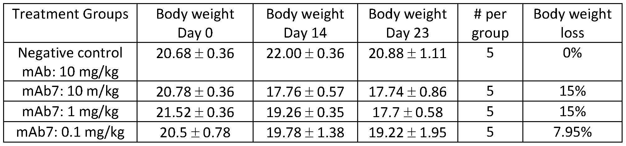


Claims
Priority Applications (21)
| Application Number | Priority Date | Filing Date | Title |
|---|---|---|---|
| HRP20221226TT HRP20221226T1 (en) | 2014-12-09 | 2015-12-02 | Anti-pd-1 antibodies and methods of use thereof |
| AU2015359003A AU2015359003B2 (en) | 2014-12-09 | 2015-12-02 | Anti-PD-1 antibodies and methods of use thereof |
| SG11201703950PA SG11201703950PA (en) | 2014-12-09 | 2015-12-02 | Anti-pd-1 antibodies and methods of use thereof |
| PL15808464.0T PL3230319T3 (en) | 2014-12-09 | 2015-12-02 | Anti-pd-1 antibodies and methods of use thereof |
| MYPI2017702000A MY193404A (en) | 2014-12-09 | 2015-12-02 | Anti-pd-1 antibodies and methods of use thereof |
| EP15808464.0A EP3230319B8 (en) | 2014-12-09 | 2015-12-02 | Anti-pd-1 antibodies and methods of use thereof |
| ES15808464T ES2929721T3 (en) | 2014-12-09 | 2015-12-02 | Anti-PD-1 antibodies and procedures for their use |
| KR1020177018639A KR102012113B1 (en) | 2014-12-09 | 2015-12-02 | Anti-pd-1 antibodies and methods of use thereof |
| MX2017007537A MX389945B (en) | 2014-12-09 | 2015-12-02 | ANTI-PD-1 ANTIBODIES AND THEIR METHODS OF USE. |
| RU2017118225A RU2701797C2 (en) | 2014-12-09 | 2015-12-02 | Antibodies to pd-1 and methods of their application |
| HK18103915.6A HK1244492B (en) | 2014-12-09 | 2015-12-02 | Anti-pd-1 antibodies and methods of use thereof |
| NZ731735A NZ731735A (en) | 2014-12-09 | 2015-12-02 | Anti-pd-1 antibodies and methods of use thereof |
| JP2017530259A JP6552621B2 (en) | 2014-12-09 | 2015-12-02 | Anti-PD-1 antibody and method of using the same |
| DK15808464.0T DK3230319T3 (en) | 2014-12-09 | 2015-12-02 | Anti-PD-1 antibodies and methods of use thereof |
| EP22194128.9A EP4166572A1 (en) | 2014-12-09 | 2015-12-02 | Anti-pd-1 antibodies and methods of use thereof |
| CN201580067208.8A CN107207593B (en) | 2014-12-09 | 2015-12-02 | Anti-PD-1 antibody and method of use |
| BR112017010762-7A BR112017010762B1 (en) | 2014-12-09 | 2015-12-02 | ANTI-PD-1 ANTAGONIST ANTIBODY, PHARMACEUTICAL COMPOSITION, KIT, ITS USES AND METHOD OF PRODUCING AN ANTI-PD-1 ANTAGONIST ANTIBODY |
| IL252280A IL252280B2 (en) | 2014-12-09 | 2017-05-15 | Anti-pd-1 antibodies and methods of use thereof |
| PH12017500994A PH12017500994A1 (en) | 2014-12-09 | 2017-05-29 | Anti-pd-1 antibodies and methods of use thereof |
| CONC2017/0005738A CO2017005738A2 (en) | 2014-12-09 | 2017-06-09 | Anti-pd-1 antibodies |
| AU2019200003A AU2019200003A1 (en) | 2014-12-09 | 2019-01-02 | Anti-PD-1 antibodies and methods of use thereof |
Applications Claiming Priority (6)
| Application Number | Priority Date | Filing Date | Title |
|---|---|---|---|
| US201462089658P | 2014-12-09 | 2014-12-09 | |
| US62/089,658 | 2014-12-09 | ||
| US201562242750P | 2015-10-16 | 2015-10-16 | |
| US62/242,750 | 2015-10-16 | ||
| US201562251973P | 2015-11-06 | 2015-11-06 | |
| US62/251,973 | 2015-11-06 |
Publications (1)
| Publication Number | Publication Date |
|---|---|
| WO2016092419A1 true WO2016092419A1 (en) | 2016-06-16 |
Family
ID=54849669
Family Applications (1)
| Application Number | Title | Priority Date | Filing Date |
|---|---|---|---|
| PCT/IB2015/059268 Ceased WO2016092419A1 (en) | 2014-12-09 | 2015-12-02 | Anti-pd-1 antibodies and methods of use thereof |
Country Status (25)
| Country | Link |
|---|---|
| US (4) | US10155037B2 (en) |
| EP (2) | EP3230319B8 (en) |
| JP (1) | JP6552621B2 (en) |
| KR (1) | KR102012113B1 (en) |
| CN (1) | CN107207593B (en) |
| AU (2) | AU2015359003B2 (en) |
| CA (1) | CA2914087C (en) |
| CO (1) | CO2017005738A2 (en) |
| DK (1) | DK3230319T3 (en) |
| ES (1) | ES2929721T3 (en) |
| HR (1) | HRP20221226T1 (en) |
| HU (1) | HUE060165T2 (en) |
| IL (1) | IL252280B2 (en) |
| MX (1) | MX389945B (en) |
| MY (1) | MY193404A (en) |
| NZ (1) | NZ731735A (en) |
| PE (1) | PE20171180A1 (en) |
| PH (1) | PH12017500994A1 (en) |
| PL (1) | PL3230319T3 (en) |
| PT (1) | PT3230319T (en) |
| RU (1) | RU2701797C2 (en) |
| SA (1) | SA517381676B1 (en) |
| SG (1) | SG11201703950PA (en) |
| TW (1) | TWI595006B (en) |
| WO (1) | WO2016092419A1 (en) |
Cited By (158)
| Publication number | Priority date | Publication date | Assignee | Title |
|---|---|---|---|---|
| WO2017141208A1 (en) | 2016-02-17 | 2017-08-24 | Novartis Ag | Tgfbeta 2 antibodies |
| WO2017165125A1 (en) * | 2016-03-24 | 2017-09-28 | Millennium Pharmaceuticals, Inc. | Use of a pd-1 antagonist and an anti-ccr2 antibody in the treatment of cancer |
| WO2017218533A1 (en) | 2016-06-13 | 2017-12-21 | Torque Therapeutics, Inc. | Methods and compositions for promoting immune cell function |
| WO2017220989A1 (en) | 2016-06-20 | 2017-12-28 | Kymab Limited | Anti-pd-l1 and il-2 cytokines |
| WO2018027524A1 (en) * | 2016-08-09 | 2018-02-15 | Innovent Biologics (Suzhou) Co., Ltd. | Pd-1 antibody formulation |
| US9975957B2 (en) | 2014-03-31 | 2018-05-22 | Genentech, Inc. | Anti-OX40 antibodies and methods of use |
| WO2018091661A1 (en) * | 2016-11-18 | 2018-05-24 | Symphogen A/S | Anti-pd-1 antibodies and compositions |
| TWI625336B (en) * | 2015-10-02 | 2018-06-01 | 赫孚孟拉羅股份公司 | Anti-PD1 antibody and method of use |
| WO2018134681A1 (en) | 2017-01-20 | 2018-07-26 | Sanofi | Anti-tgf-beta antibodies and their use |
| WO2018134784A1 (en) | 2017-01-20 | 2018-07-26 | Novartis Ag | Combination therapy for the treatment of cancer |
| WO2018146612A1 (en) | 2017-02-10 | 2018-08-16 | Novartis Ag | 1-(4-amino-5-bromo-6-(1 h-pyrazol-1-yl)pyrimidin-2-yl)-1 h-pyrazol-4-ol and use thereof in the treatment of cancer |
| WO2018160540A1 (en) | 2017-02-28 | 2018-09-07 | Sanofi | Therapeutic rna |
| WO2018162944A1 (en) * | 2017-03-04 | 2018-09-13 | Shenzhen Runshin Bioscience | Recombinant antibodies to programmed death 1 (pd-1) and uses therefor |
| WO2018181425A1 (en) | 2017-03-29 | 2018-10-04 | 塩野義製薬株式会社 | Medicinal composition for treating cancer |
| WO2018198091A1 (en) | 2017-04-28 | 2018-11-01 | Novartis Ag | Antibody conjugates comprising toll-like receptor agonist and combination therapies |
| WO2018211453A1 (en) | 2017-05-19 | 2018-11-22 | Novartis Ag | Compositions comprising naphthyridine derivatives and aluminium adjuvant for use in treating solid tumors |
| WO2018220546A1 (en) | 2017-05-31 | 2018-12-06 | Novartis Ag | Crystalline forms of 5-bromo-2,6-di(1 h-pyrazol-1-yl)pyrimidin-4-amine and new salts |
| WO2018229715A1 (en) | 2017-06-16 | 2018-12-20 | Novartis Ag | Compositions comprising anti-cd32b antibodies and methods of use thereof |
| US10160806B2 (en) | 2014-06-26 | 2018-12-25 | Macrogenics, Inc. | Covalently bonded diabodies having immunoreactivity with PD-1 and LAG-3, and methods of use thereof |
| WO2018237173A1 (en) | 2017-06-22 | 2018-12-27 | Novartis Ag | ANTIBODY MOLECULES DIRECTED AGAINST CD73 AND CORRESPONDING USES |
| WO2018235056A1 (en) | 2017-06-22 | 2018-12-27 | Novartis Ag | IL-1BETA BINDING ANTIBODIES FOR USE IN THE TREATMENT OF CANCER |
| WO2018237157A1 (en) | 2017-06-22 | 2018-12-27 | Novartis Ag | Antibody molecules to cd73 and uses thereof |
| WO2018234879A1 (en) | 2017-06-22 | 2018-12-27 | Novartis Ag | Il-1beta binding antibodies for use in treating cancer |
| EP3421494A1 (en) | 2017-06-29 | 2019-01-02 | Sanofi | Use of isatuximab in combination with an anti-pd-1 antibody |
| WO2019006007A1 (en) | 2017-06-27 | 2019-01-03 | Novartis Ag | Dosage regimens for anti-tim-3 antibodies and uses thereof |
| WO2019012371A1 (en) | 2017-07-11 | 2019-01-17 | Pfizer Inc. | Immunogenic compositions comprising cea muc1 and tert |
| WO2019018730A1 (en) | 2017-07-20 | 2019-01-24 | Novartis Ag | Dosage regimens of anti-lag-3 antibodies and uses thereof |
| WO2019032663A1 (en) * | 2017-08-09 | 2019-02-14 | Orionis Biosciences Inc. | Pd-1 and pd-l1 binding agents |
| WO2019075032A1 (en) | 2017-10-13 | 2019-04-18 | Merck Patent Gmbh | Combination of a parp inhibitor and a pd-1 axis binding antagonist |
| WO2019081983A1 (en) | 2017-10-25 | 2019-05-02 | Novartis Ag | Antibodies targeting cd32b and methods of use thereof |
| CN109715666A (en) * | 2016-07-20 | 2019-05-03 | 斯特库比股份有限公司 | Cancer treatment methods and therapies using combinations of antibodies that bind glycosylated PD-L1 |
| US10287352B2 (en) | 2015-10-02 | 2019-05-14 | Hoffman-La Roche Inc. | Bispecific antibodies specific for PD1 and TIM3 |
| WO2019097369A1 (en) | 2017-11-14 | 2019-05-23 | Pfizer Inc. | Ezh2 inhibitor combination therapies |
| WO2019099838A1 (en) | 2017-11-16 | 2019-05-23 | Novartis Ag | Combination therapies |
| WO2019108900A1 (en) | 2017-11-30 | 2019-06-06 | Novartis Ag | Bcma-targeting chimeric antigen receptor, and uses thereof |
| WO2019123207A1 (en) | 2017-12-18 | 2019-06-27 | Pfizer Inc. | Methods and combination therapy to treat cancer |
| US10344090B2 (en) | 2013-12-12 | 2019-07-09 | Shanghai Hangrui Pharmaceutical Co., Ltd. | PD-1 antibody, antigen-binding fragment thereof, and medical application thereof |
| WO2019136432A1 (en) | 2018-01-08 | 2019-07-11 | Novartis Ag | Immune-enhancing rnas for combination with chimeric antigen receptor therapy |
| WO2019152660A1 (en) | 2018-01-31 | 2019-08-08 | Novartis Ag | Combination therapy using a chimeric antigen receptor |
| WO2019160956A1 (en) | 2018-02-13 | 2019-08-22 | Novartis Ag | Chimeric antigen receptor therapy in combination with il-15r and il15 |
| WO2019171253A1 (en) | 2018-03-07 | 2019-09-12 | Pfizer Inc. | Anti-pd-1 antibody compositions |
| WO2019200229A1 (en) | 2018-04-13 | 2019-10-17 | Novartis Ag | Dosage regimens for anti-pd-l1 antibodies and uses thereof |
| WO2019211489A1 (en) | 2018-05-04 | 2019-11-07 | Merck Patent Gmbh | COMBINED INHIBITION OF PD-1/PD-L1, TGFβ AND DNA-PK FOR THE TREATMENT OF CANCER |
| WO2019224716A2 (en) | 2018-05-23 | 2019-11-28 | Pfizer Inc. | Antibodies specific for gucy2c and uses thereof |
| WO2019224715A1 (en) | 2018-05-23 | 2019-11-28 | Pfizer Inc. | Antibodies specific for cd3 and uses thereof |
| WO2019229701A2 (en) | 2018-06-01 | 2019-12-05 | Novartis Ag | Binding molecules against bcma and uses thereof |
| WO2019232244A2 (en) | 2018-05-31 | 2019-12-05 | Novartis Ag | Antibody molecules to cd73 and uses thereof |
| WO2019229658A1 (en) | 2018-05-30 | 2019-12-05 | Novartis Ag | Entpd2 antibodies, combination therapies, and methods of using the antibodies and combination therapies |
| US10526413B2 (en) | 2015-10-02 | 2020-01-07 | Hoffmann-La Roche Inc. | Bispecific antibodies specific for OX40 |
| CN110678482A (en) * | 2017-03-29 | 2020-01-10 | 细胞基因公司 | Preparation containing PD-1 binding protein and preparation method thereof |
| WO2020012334A1 (en) | 2018-07-10 | 2020-01-16 | Novartis Ag | 3-(5-hydroxy-1-oxoisoindolin-2-yl)piperidine-2,6-dione derivatives and their use in the treatment of ikaros family zinc finger 2 (ikzf2)-dependent diseases |
| WO2020012337A1 (en) | 2018-07-10 | 2020-01-16 | Novartis Ag | 3-(5-amino-1-oxoisoindolin-2-yl)piperidine-2,6-dione derivatives and their use in the treatment of i karos family zinc finger 2 (ikzf2)-dependent diseases |
| WO2020021465A1 (en) | 2018-07-25 | 2020-01-30 | Advanced Accelerator Applications (Italy) S.R.L. | Method of treatment of neuroendocrine tumors |
| WO2020041655A1 (en) | 2018-08-24 | 2020-02-27 | Sanofi | Therapeutic rna for solid tumor cancers |
| WO2020039321A2 (en) | 2018-08-20 | 2020-02-27 | Pfizer Inc. | Anti-gdf15 antibodies, compositions and methods of use |
| US10577422B2 (en) | 2015-07-30 | 2020-03-03 | Macrogenics, Inc. | PD-1-binding molecules and methods of use thereof |
| WO2020044252A1 (en) | 2018-08-31 | 2020-03-05 | Novartis Ag | Dosage regimes for anti-m-csf antibodies and uses thereof |
| WO2020051333A1 (en) | 2018-09-07 | 2020-03-12 | Pfizer Inc. | Anti-avb8 antibodies and compositions and uses thereof |
| WO2020049534A1 (en) | 2018-09-07 | 2020-03-12 | Novartis Ag | Sting agonist and combination therapy thereof for the treatment of cancer |
| WO2020064971A1 (en) | 2018-09-26 | 2020-04-02 | Merck Patent Gmbh | Combination of a pd-1 antagonist, an atr inhibitor and a platinating agent for the treatment of cancer |
| WO2020079581A1 (en) | 2018-10-16 | 2020-04-23 | Novartis Ag | Tumor mutation burden alone or in combination with immune markers as biomarkers for predicting response to targeted therapy |
| WO2020089811A1 (en) | 2018-10-31 | 2020-05-07 | Novartis Ag | Dc-sign antibody drug conjugates |
| WO2020095184A1 (en) | 2018-11-05 | 2020-05-14 | Pfizer Inc. | Combinations for treating cancer |
| WO2020095183A1 (en) | 2018-11-05 | 2020-05-14 | Pfizer Inc. | Combination for treating cancer |
| WO2020106695A1 (en) | 2018-11-19 | 2020-05-28 | Ariagen, Inc. | Methods of treating cancer |
| WO2020117988A1 (en) | 2018-12-04 | 2020-06-11 | Tolero Pharmaceuticals, Inc. | Cdk9 inhibitors and polymorphs thereof for use as agents for treatment of cancer |
| EP3666905A1 (en) | 2018-12-11 | 2020-06-17 | Sanofi | E. coli positive for pks island as marker of positive response to anti-pd1 therapy in colorectal cancer |
| WO2020128637A1 (en) | 2018-12-21 | 2020-06-25 | Novartis Ag | Use of il-1 binding antibodies in the treatment of a msi-h cancer |
| WO2020128972A1 (en) | 2018-12-20 | 2020-06-25 | Novartis Ag | Dosing regimen and pharmaceutical combination comprising 3-(1-oxoisoindolin-2-yl)piperidine-2,6-dione derivatives |
| WO2020128893A1 (en) | 2018-12-21 | 2020-06-25 | Pfizer Inc. | Combination treatments of cancer comprising a tlr agonist |
| WO2020127965A1 (en) | 2018-12-21 | 2020-06-25 | Onxeo | New conjugated nucleic acid molecules and their uses |
| WO2020128613A1 (en) | 2018-12-21 | 2020-06-25 | Novartis Ag | Use of il-1beta binding antibodies |
| WO2020128620A1 (en) | 2018-12-21 | 2020-06-25 | Novartis Ag | Use of il-1beta binding antibodies |
| WO2020128636A1 (en) | 2018-12-21 | 2020-06-25 | Novartis Ag | Use of il-1 beta antibodies in the treatment or prevention of myelodysplastic syndrome |
| WO2020154189A1 (en) | 2019-01-21 | 2020-07-30 | Sanofi | Therapeutic rna and anti-pd1 antibodies for advanced stage solid tumor cancers |
| WO2020165733A1 (en) | 2019-02-12 | 2020-08-20 | Novartis Ag | Pharmaceutical combination comprising tno155 and a pd-1 inhibitor |
| WO2020167990A1 (en) | 2019-02-12 | 2020-08-20 | Tolero Pharmaceuticals, Inc. | Formulations comprising heterocyclic protein kinase inhibitors |
| WO2020165834A1 (en) | 2019-02-15 | 2020-08-20 | Novartis Ag | Substituted 3-(1-oxoisoindolin-2-yl)piperidine-2,6-dione derivatives and uses thereof |
| WO2020165833A1 (en) | 2019-02-15 | 2020-08-20 | Novartis Ag | 3-(1-oxo-5-(piperidin-4-yl)isoindolin-2-yl)piperidine-2,6-dione derivatives and uses thereof |
| US10766955B2 (en) | 2017-01-20 | 2020-09-08 | Sanofi | Anti-TGF-β antibodies and their use |
| WO2020198077A1 (en) | 2019-03-22 | 2020-10-01 | Sumitomo Dainippon Pharma Oncology, Inc. | Compositions comprising pkm2 modulators and methods of treatment using the same |
| US10844115B2 (en) | 2017-01-20 | 2020-11-24 | Genzyme Corporation | Bone-targeting antibodies |
| WO2020239558A1 (en) | 2019-05-24 | 2020-12-03 | Pfizer Inc. | Combination therapies using cdk inhibitors |
| US10875864B2 (en) | 2011-07-21 | 2020-12-29 | Sumitomo Dainippon Pharma Oncology, Inc. | Substituted imidazo[1,2-B]pyridazines as protein kinase inhibitors |
| WO2021003417A1 (en) | 2019-07-03 | 2021-01-07 | Sumitomo Dainippon Pharma Oncology, Inc. | Tyrosine kinase non-receptor 1 (tnk1) inhibitors and uses thereof |
| WO2021006199A1 (en) | 2019-07-05 | 2021-01-14 | 小野薬品工業株式会社 | Treatment of hematologic cancer with pd-1/cd3 dual specificity protein |
| WO2021025140A1 (en) | 2019-08-08 | 2021-02-11 | 小野薬品工業株式会社 | Dual-specific protein |
| US10954301B2 (en) | 2015-12-14 | 2021-03-23 | Macrogenics, Inc. | Bispecific molecules having immunoreactivity with PD-1 and CTLA-4, and methods of use thereof |
| WO2021053559A1 (en) | 2019-09-18 | 2021-03-25 | Novartis Ag | Entpd2 antibodies, combination therapies, and methods of using the antibodies and combination therapies |
| WO2021053560A1 (en) | 2019-09-18 | 2021-03-25 | Novartis Ag | Combination therapy with entpd2 and cd73 antibodies |
| WO2021059136A1 (en) | 2019-09-25 | 2021-04-01 | Pfizer Inc. | Polyheterocyclic modulators of sting (stimulator of interferon genes) |
| WO2021079188A1 (en) | 2019-10-21 | 2021-04-29 | Novartis Ag | Combination therapies with venetoclax and tim-3 inhibitors |
| WO2021079195A1 (en) | 2019-10-21 | 2021-04-29 | Novartis Ag | Tim-3 inhibitors and uses thereof |
| WO2021102343A1 (en) | 2019-11-22 | 2021-05-27 | Sumitomo Dainippon Pharma Oncology, Inc. | Solid dose pharmaceutical composition |
| US11028169B2 (en) | 2016-05-18 | 2021-06-08 | Boehringer Ingelheim International Gmbh | Antibody molecules for cancer treatment |
| WO2021116943A1 (en) | 2019-12-12 | 2021-06-17 | Ignite Immunotherapy, Inc. | Variant oncolytic vaccinia virus and methods of use thereof |
| WO2021123996A1 (en) | 2019-12-20 | 2021-06-24 | Novartis Ag | Uses of anti-tgf-beta antibodies and checkpoint inhibitors for the treatment of proliferative diseases |
| WO2021124096A1 (en) | 2019-12-18 | 2021-06-24 | Pfizer Inc. | Once daily cancer treatment regimen with a prmt5 inhibitor |
| WO2021124073A1 (en) | 2019-12-17 | 2021-06-24 | Pfizer Inc. | Antibodies specific for cd47, pd-l1, and uses thereof |
| WO2021144657A1 (en) | 2020-01-17 | 2021-07-22 | Novartis Ag | Combination comprising a tim-3 inhibitor and a hypomethylating agent for use in treating myelodysplastic syndrome or chronic myelomonocytic leukemia |
| US11072653B2 (en) | 2015-06-08 | 2021-07-27 | Macrogenics, Inc. | LAG-3-binding molecules and methods of use thereof |
| EP3755716A4 (en) * | 2018-02-23 | 2021-08-04 | Eucure (Beijing) Biopharma Co., Ltd | Anti-pd-1 antibodies and uses thereof |
| WO2021171260A2 (en) | 2020-02-28 | 2021-09-02 | Novartis Ag | A triple pharmaceutical combination comprising dabrafenib, an erk inhibitor and a raf inhibitor or a pd-1 inhibitor |
| WO2021176330A1 (en) | 2020-03-03 | 2021-09-10 | Array Biopharma Inc. | Methods to treat cancer using (r)-n-(3-fluoro-4-((3-((1-hydroxypropan-2-yl)amino)-1h-pyrazolo[3,4-b]pyridin-4-yl)oxy)phenyl)-3-(4-fluorophenyl)-1-isopropyl-2,4-dioxo-1,2,3,4-tetrahydropyrimidine-5-carboxamide |
| WO2021181233A2 (en) | 2020-03-09 | 2021-09-16 | Pfizer Inc. | Fusion proteins and uses thereof |
| WO2021214623A1 (en) | 2020-04-21 | 2021-10-28 | Novartis Ag | Dosing regimen for treating a disease modulated by csf-1r |
| WO2021229507A2 (en) | 2020-05-13 | 2021-11-18 | Pfizer Inc. | Methods, therapies and uses for treating cancer |
| WO2021255223A1 (en) | 2020-06-19 | 2021-12-23 | Onxeo | New conjugated nucleic acid molecules and their uses |
| WO2021260528A1 (en) | 2020-06-23 | 2021-12-30 | Novartis Ag | Dosing regimen comprising 3-(1-oxoisoindolin-2-yl)piperidine-2,6-dione derivatives |
| WO2022003554A1 (en) | 2020-07-01 | 2022-01-06 | Pfizer Inc. | Biomarkers for pd-1 axis binding antagonist therapy |
| WO2022009157A1 (en) | 2020-07-10 | 2022-01-13 | Novartis Ag | Lhc165 and spartalizumab combinations for treating solid tumors |
| WO2022013775A1 (en) | 2020-07-17 | 2022-01-20 | Pfizer Inc. | Therapeutic antibodies and their uses |
| WO2022029573A1 (en) | 2020-08-03 | 2022-02-10 | Novartis Ag | Heteroaryl substituted 3-(1-oxoisoindolin-2-yl)piperidine-2,6-dione derivatives and uses thereof |
| US11248049B2 (en) | 2016-12-23 | 2022-02-15 | Remd Biotherapeutics, Inc. | Immunotherapy using antibodies that bind Programmed Death 1 (PD-1) |
| WO2022043557A1 (en) | 2020-08-31 | 2022-03-03 | Advanced Accelerator Applications International Sa | Method of treating psma-expressing cancers |
| WO2022043558A1 (en) | 2020-08-31 | 2022-03-03 | Advanced Accelerator Applications International Sa | Method of treating psma-expressing cancers |
| WO2022053990A1 (en) | 2020-09-14 | 2022-03-17 | Pfizer Inc. | Methods, therapies and uses for treating cancer |
| US11279694B2 (en) | 2016-11-18 | 2022-03-22 | Sumitomo Dainippon Pharma Oncology, Inc. | Alvocidib prodrugs and their use as protein kinase inhibitors |
| US11285207B2 (en) | 2017-04-05 | 2022-03-29 | Hoffmann-La Roche Inc. | Bispecific antibodies specifically binding to PD1 and LAG3 |
| WO2022097060A1 (en) | 2020-11-06 | 2022-05-12 | Novartis Ag | Cd19 binding molecules and uses thereof |
| WO2022118197A1 (en) | 2020-12-02 | 2022-06-09 | Pfizer Inc. | Time to resolution of axitinib-related adverse events |
| WO2022130206A1 (en) | 2020-12-16 | 2022-06-23 | Pfizer Inc. | TGFβr1 INHIBITOR COMBINATION THERAPIES |
| US11390621B2 (en) | 2019-04-15 | 2022-07-19 | Ariagen, Inc. | Chiral indole compounds and their use |
| WO2022153161A1 (en) | 2021-01-14 | 2022-07-21 | Pfizer Inc. | Treatment of cancer using a prmt5 inhibitor |
| WO2022162569A1 (en) | 2021-01-29 | 2022-08-04 | Novartis Ag | Dosage regimes for anti-cd73 and anti-entpd2 antibodies and uses thereof |
| US11413331B2 (en) | 2017-04-03 | 2022-08-16 | Hoffmann-La Roche Inc. | Immunoconjugates |
| US11427576B2 (en) | 2017-11-20 | 2022-08-30 | Ariagen, Inc. | Indole compounds and their use |
| WO2022195551A1 (en) | 2021-03-18 | 2022-09-22 | Novartis Ag | Biomarkers for cancer and methods of use thereof |
| WO2022195462A1 (en) | 2021-03-18 | 2022-09-22 | Pfizer Inc. | Modulators of sting (stimulator of interferon genes) |
| WO2022215011A1 (en) | 2021-04-07 | 2022-10-13 | Novartis Ag | USES OF ANTI-TGFβ ANTIBODIES AND OTHER THERAPEUTIC AGENTS FOR THE TREATMENT OF PROLIFERATIVE DISEASES |
| WO2022221227A1 (en) | 2021-04-13 | 2022-10-20 | Nuvalent, Inc. | Amino-substituted heterocycles for treating cancers with egfr mutations |
| WO2022219080A1 (en) | 2021-04-14 | 2022-10-20 | INSERM (Institut National de la Santé et de la Recherche Médicale) | New method to improve nk cells cytotoxicity |
| US11497756B2 (en) | 2017-09-12 | 2022-11-15 | Sumitomo Pharma Oncology, Inc. | Treatment regimen for cancers that are insensitive to BCL-2 inhibitors using the MCL-1 inhibitor alvocidib |
| WO2022243846A1 (en) | 2021-05-18 | 2022-11-24 | Novartis Ag | Combination therapies |
| WO2022254227A1 (en) | 2021-06-04 | 2022-12-08 | Kymab Limited | Treatment of pd-l1 negative or low expressing cancer with anti-icos antibodies |
| WO2022266510A1 (en) | 2021-06-18 | 2022-12-22 | Genzyme Corporation | Anti-tgf-beta antibody formulations and their use |
| US11547698B2 (en) | 2016-12-26 | 2023-01-10 | Ariagen, Inc. | Aryl hydrocarbon receptor modulators |
| WO2023057882A1 (en) | 2021-10-05 | 2023-04-13 | Pfizer Inc. | Combinations of azalactam compounds with a pd-1 axis binding antagonist for the treatment of cancer |
| WO2023079430A1 (en) | 2021-11-02 | 2023-05-11 | Pfizer Inc. | Methods of treating mitochondrial myopathies using anti-gdf15 antibodies |
| WO2023079428A1 (en) | 2021-11-03 | 2023-05-11 | Pfizer Inc. | Combination therapies using tlr7/8 agonist |
| WO2023084445A1 (en) | 2021-11-12 | 2023-05-19 | Novartis Ag | Combination therapy for treating lung cancer |
| WO2023111203A1 (en) | 2021-12-16 | 2023-06-22 | Onxeo | New conjugated nucleic acid molecules and their uses |
| US11746103B2 (en) | 2020-12-10 | 2023-09-05 | Sumitomo Pharma Oncology, Inc. | ALK-5 inhibitors and uses thereof |
| US11746161B2 (en) | 2017-06-05 | 2023-09-05 | Janssen Biotech, Inc. | Antibodies that specifically bind PD-1 and methods of use |
| WO2023166420A1 (en) | 2022-03-03 | 2023-09-07 | Pfizer Inc. | Multispecific antibodies and uses thereof |
| US11793802B2 (en) | 2019-03-20 | 2023-10-24 | Sumitomo Pharma Oncology, Inc. | Treatment of acute myeloid leukemia (AML) with venetoclax failure |
| WO2023214325A1 (en) | 2022-05-05 | 2023-11-09 | Novartis Ag | Pyrazolopyrimidine derivatives and uses thereof as tet2 inhibitors |
| WO2023218320A1 (en) | 2022-05-11 | 2023-11-16 | Pfizer Inc. | Anti-lymphotoxin beta receptor antibodies and methods of use thereof |
| WO2023242769A1 (en) | 2022-06-17 | 2023-12-21 | Pfizer Inc. | Il-12 variants, anti-pd1 antibodies, fusion proteins, and uses thereof |
| US11939380B2 (en) | 2017-04-05 | 2024-03-26 | Les Laboratoires Servier | Combination therapies targeting PD-1, TIM-3, and LAG-3 |
| US11981736B2 (en) | 2015-12-02 | 2024-05-14 | St Cube Inc. | Antibodies specific to glycosylated PD-1 and methods of use thereof |
| US12030942B2 (en) | 2015-10-02 | 2024-07-09 | Les Laboratoires Servier | Anti-PD-1 antibodies and compositions |
| US12037395B1 (en) | 2018-04-15 | 2024-07-16 | Immvira Co., Limited | Antibodies binding PD-1 and uses thereof |
| WO2024160721A1 (en) | 2023-01-30 | 2024-08-08 | Kymab Limited | Antibodies |
| WO2025032521A1 (en) | 2023-08-10 | 2025-02-13 | Pfizer Inc. | Methods, therapies and uses for treating cancer |
| US12240910B2 (en) | 2021-07-14 | 2025-03-04 | Genentech, Inc. | Anti-C-C motif chemokine receptor 8 (CCR8) antibodies and methods of use |
| US12275790B2 (en) | 2018-12-21 | 2025-04-15 | Sinocelltech Ltd. | Humanized anti-PD-1 antibody and the use thereof |
| US12338261B2 (en) | 2015-05-18 | 2025-06-24 | Sumitomo Pharma Oncology, Inc. | Alvocidib prodrugs having increased bioavailability |
Families Citing this family (120)
| Publication number | Priority date | Publication date | Assignee | Title |
|---|---|---|---|---|
| NZ702485A (en) | 2010-06-03 | 2016-04-29 | Pharmacyclics Llc | The use of inhibitors of bruton’s tyrosine kinase (btk) |
| MX2015001081A (en) | 2012-07-24 | 2015-10-14 | Pharmacyclics Inc | Mutations associated with resistance to inhibitors of bruton's tyrosine kinase (btk). |
| TWI664192B (en) | 2012-11-20 | 2019-07-01 | 法商賽諾菲公司 | anti-CEACAM5 antibody and use thereof |
| WO2015048312A1 (en) | 2013-09-26 | 2015-04-02 | Costim Pharmaceuticals Inc. | Methods for treating hematologic cancers |
| JOP20200094A1 (en) | 2014-01-24 | 2017-06-16 | Dana Farber Cancer Inst Inc | Antibody molecules to pd-1 and uses thereof |
| US10570202B2 (en) | 2014-02-04 | 2020-02-25 | Pfizer Inc. | Combination of a PD-1 antagonist and a VEGFR inhibitor for treating cancer |
| LT3116909T (en) | 2014-03-14 | 2020-02-10 | Novartis Ag | ANTI-BODY MOLECULES AGAINST LAG-3 AND THEIR USE |
| EP3119910A4 (en) | 2014-03-20 | 2018-02-21 | Pharmacyclics LLC | Phospholipase c gamma 2 and resistance associated mutations |
| SMT202100116T1 (en) | 2014-05-28 | 2021-05-07 | Agenus Inc | Anti-gitr antibodies and methods of use thereof |
| CA2960824A1 (en) | 2014-09-13 | 2016-03-17 | Novartis Ag | Combination therapies of alk inhibitors |
| TWI595006B (en) * | 2014-12-09 | 2017-08-11 | 禮納特神經系統科學公司 | Anti-PD-1 antibodies and methods of using same |
| EP3242940B1 (en) | 2015-01-09 | 2023-03-29 | Etubics Corporation | Methods and compositions for combination immunotherapy |
| MA41414A (en) * | 2015-01-28 | 2017-12-05 | Centre Nat Rech Scient | ICOS AGONIST BINDING PROTEINS |
| IL254039B2 (en) | 2015-02-19 | 2023-12-01 | Compugen Ltd | Anti-PVRIG antibodies and methods of use |
| US10800846B2 (en) | 2015-02-26 | 2020-10-13 | Merck Patent Gmbh | PD-1/PD-L1 inhibitors for the treatment of cancer |
| MA41866A (en) | 2015-03-31 | 2018-02-06 | Massachusetts Gen Hospital | SELF-ASSEMBLING MOLECULES FOR TARGETED DRUG DELIVERY |
| EP3286213B1 (en) * | 2015-04-20 | 2021-08-04 | Etubics Corporation | Methods and compositions for combination immunotherapy |
| EP4545082A3 (en) | 2015-05-06 | 2025-07-02 | SNIPR Technologies Limited | Altering microbial populations & modifying microbiota |
| CN115109158A (en) | 2015-05-07 | 2022-09-27 | 阿吉纳斯公司 | anti-OX 40 antibodies and methods of use thereof |
| US10869924B2 (en) | 2015-06-16 | 2020-12-22 | Merck Patent Gmbh | PD-L1 antagonist combination treatments |
| MA42447A (en) | 2015-07-13 | 2018-05-23 | Cytomx Therapeutics Inc | ANTI-PD-1 ANTIBODIES, ACTIVABLE ANTI-PD-1 ANTIBODIES, AND METHODS OF USE THEREOF |
| NZ739503A (en) | 2015-07-16 | 2023-06-30 | Bioxcel Therapeutics Inc | A novel approach for treatment of cancer using immunomodulation |
| WO2017040790A1 (en) | 2015-09-01 | 2017-03-09 | Agenus Inc. | Anti-pd-1 antibodies and methods of use thereof |
| WO2017058859A1 (en) | 2015-09-29 | 2017-04-06 | Celgene Corporation | Pd-1 binding proteins and methods of use thereof |
| LT3370768T (en) | 2015-11-03 | 2022-05-25 | Janssen Biotech, Inc. | ANTIBODIES SPECIFICLY BINDING PD-1 AND THEIR USE |
| TW202134282A (en) | 2015-12-02 | 2021-09-16 | 美商艾吉納斯公司 | Antibodies and methods of use thereof |
| CA3024508A1 (en) | 2016-05-27 | 2017-11-30 | Agenus Inc. | Anti-tim-3 antibodies and methods of use thereof |
| GB201609811D0 (en) * | 2016-06-05 | 2016-07-20 | Snipr Technologies Ltd | Methods, cells, systems, arrays, RNA and kits |
| RU2656181C1 (en) * | 2016-07-13 | 2018-05-31 | Закрытое Акционерное Общество "Биокад" | Anti-pd-1 antibodies, method for their production, and method of application |
| EP4650005A3 (en) * | 2016-07-15 | 2026-01-28 | Viracta Subsidiary, Inc. | Histone deacetylase inhibitors for use in immunotherapy |
| WO2018027223A1 (en) * | 2016-08-05 | 2018-02-08 | Duke University | Camkk2 inhibitor compositions and methods of using the same |
| WO2018039629A2 (en) | 2016-08-25 | 2018-03-01 | Northwestern University | Micellar spherical nucleic acids from thermoresponsive, traceless templates |
| WO2018049474A1 (en) * | 2016-09-16 | 2018-03-22 | Bionomics Limited | Antibody and checkpoint inhibitor combination therapy |
| RS67443B1 (en) * | 2016-09-16 | 2025-12-31 | Shanghai Henlius Biotech Inc | Anti-pd-1 antibodies for use in the treatment of cancer |
| CN109952317A (en) | 2016-09-19 | 2019-06-28 | 细胞基因公司 | Use the method for PD-1 binding protein treatment immune disorders |
| JP2019534859A (en) | 2016-09-19 | 2019-12-05 | セルジーン コーポレイション | Method for treating vitiligo using PD-1 binding protein |
| DK3515938T3 (en) | 2016-09-21 | 2025-04-07 | Cstone Pharmaceuticals | MONOCLONAL ANTIBODIES AGAINST PROGRAMMED DEATH 1 (PD-1) |
| BR112019006504A2 (en) | 2016-10-06 | 2019-06-25 | Pfizer Inc. | Avelumab Dosage Regimen For Cancer Treatment |
| TWI773694B (en) | 2016-10-11 | 2022-08-11 | 美商艾吉納斯公司 | Anti-lag-3 antibodies and methods of use thereof |
| CA3041340A1 (en) | 2016-11-09 | 2018-05-17 | Agenus Inc. | Anti-ox40 antibodies, anti-gitr antibodies, and methods of use thereof |
| MA50948A (en) | 2016-12-07 | 2020-10-14 | Agenus Inc | ANTIBODIES AND METHODS OF USING THE SAME |
| WO2018112407A1 (en) * | 2016-12-15 | 2018-06-21 | Duke University | Antibodies and methods for depleting regulatory b10 cells and use in combination with immune checkpoint inhibitors |
| CN110382536B (en) | 2017-01-20 | 2023-12-19 | 大有华夏生物医药集团有限公司 | Anti-PD-1 antibodies and their uses |
| CA3053239A1 (en) | 2017-02-17 | 2018-08-23 | George Todaro | Use of tgf alpha for the treatment of diseases and disorders |
| EP4389226A3 (en) | 2017-02-24 | 2024-12-18 | MacroGenics, Inc. | Bispecific binding molecules that are capable of binding cd137 and tumor antigens, and uses thereof |
| MY197497A (en) * | 2017-03-27 | 2023-06-19 | Univ Hokkaido Nat Univ Corp | Anti-pd-l1 antibody for detecting pd-l1 |
| US20200248183A1 (en) * | 2017-04-03 | 2020-08-06 | Subbarao Nallagatla | Tlr9-targeted spherical nucleic acids having potent antitumor activity |
| JOP20190222A1 (en) * | 2017-04-11 | 2019-09-24 | Zymeworks Inc | Anti-pd-l1-anti-tim-3 bispecific antibodies |
| WO2018201090A1 (en) | 2017-04-28 | 2018-11-01 | Exicure, Inc. | Synthesis of spherical nucleic acids using lipophilic moieties |
| US11433131B2 (en) | 2017-05-11 | 2022-09-06 | Northwestern University | Adoptive cell therapy using spherical nucleic acids (SNAs) |
| AU2018330180B2 (en) * | 2017-09-07 | 2025-07-17 | Augusta University Research Institute, Inc. | Antibodies to programmed cell death protein 1 |
| ES2932286T3 (en) | 2017-10-18 | 2023-01-17 | Forty Seven Inc | Anti-CD47 Agent-Based Ovarian Cancer Therapy |
| AU2018351007B2 (en) * | 2017-10-18 | 2024-05-16 | Forty Seven, LLC | Treatment of ovarian cancer with anti-CD47 and anti-PD-L1 |
| US11957739B2 (en) * | 2017-11-08 | 2024-04-16 | Dana-Farber Cancer Institute, Inc. | Compositions and methods for treating cancer using RHOA dominant negative forms |
| WO2019094265A1 (en) * | 2017-11-10 | 2019-05-16 | Armo Biosciences, Inc. | Pd1 polypeptide binding molecules |
| CN109793892B (en) * | 2017-11-16 | 2022-07-26 | 江苏恒瑞医药股份有限公司 | Application of anti-PD-1 antibody in preparation of medicine for treating esophageal cancer |
| WO2019109238A1 (en) * | 2017-12-05 | 2019-06-13 | Lyvgen Biopharma Co., Ltd. | Anti-cd137 antibodies and uses thereof |
| US12398209B2 (en) | 2018-01-22 | 2025-08-26 | Janssen Biotech, Inc. | Methods of treating cancers with antagonistic anti-PD-1 antibodies |
| KR20200128116A (en) | 2018-02-28 | 2020-11-11 | 화이자 인코포레이티드 | IL-15 variants and uses thereof |
| RU2756899C1 (en) | 2018-02-28 | 2021-10-06 | ЭйПи БАЙОСАЙЕНСИЗ, ИНК. | Bifunctional proteins combining checkpoint blockade for targeted therapy |
| CN108434452A (en) * | 2018-03-13 | 2018-08-24 | 安徽瀚海博兴生物技术有限公司 | It is a kind of that PD-1 antibody and JMJD6 are combined to the application for being used to prepare anticancer drug |
| US10760075B2 (en) | 2018-04-30 | 2020-09-01 | Snipr Biome Aps | Treating and preventing microbial infections |
| JP7403479B2 (en) * | 2018-06-03 | 2023-12-22 | ランカプ バイオ ベータ リミテッド | Bispecific antibody against CEACAM5 and CD47 |
| WO2019246110A1 (en) | 2018-06-20 | 2019-12-26 | Incyte Corporation | Anti-pd-1 antibodies and uses thereof |
| US12258407B2 (en) | 2018-07-19 | 2025-03-25 | Tayu Huaxia Biotech Medical Group Co., Ltd. | Anti-PD-1 antibodies, dosages and uses thereof |
| AU2019324170A1 (en) | 2018-08-23 | 2021-02-18 | Seagen, Inc. | Anti-TIGIT antibodies |
| US12331320B2 (en) | 2018-10-10 | 2025-06-17 | The Research Foundation For The State University Of New York | Genome edited cancer cell vaccines |
| CA3117371A1 (en) * | 2018-11-01 | 2020-05-07 | Merck Patent Gmbh | Methods of administering anti-tim-3 antibodies |
| WO2020103885A1 (en) | 2018-11-19 | 2020-05-28 | Beijing Biocytogen Co., Ltd | Anti-pd-1 antibodies and uses thereof |
| TW202039562A (en) * | 2018-12-12 | 2020-11-01 | 大陸商上海藥明生物技術有限公司 | Anti-TIM-3 antibodies and uses thereof |
| CN113164599B (en) * | 2018-12-24 | 2023-08-01 | 正大天晴药业集团股份有限公司 | Use of Anti-PD-L1 Monoclonal Antibody to Treat Cancer |
| JP2022516073A (en) * | 2018-12-27 | 2022-02-24 | ギガジェン,インコーポレイティッド | Anti-PD-1 binding protein and how to use it |
| TW202038994A (en) | 2019-01-14 | 2020-11-01 | 美商醫格耐免疫治療公司 | Recombinant vaccinia virus and methods of use thereof |
| EP3923967A1 (en) | 2019-02-14 | 2021-12-22 | Ignite Immunotherapy, Inc. | Recombinant vaccinia virus and methods of use thereof |
| WO2020209645A1 (en) | 2019-04-11 | 2020-10-15 | (재) 스크립스코리아항체연구원 | Antibody to programmed cell death protein ligand-1 (pd-l1), and use thereof |
| EP3966247B1 (en) | 2019-05-06 | 2026-01-07 | Brown University | Bispecific antibodies against chi3l1 and pd1 with enhanced t cell-mediated cytotoxic effects on tumor cells |
| CN111973739B (en) * | 2019-05-23 | 2024-02-13 | 正大天晴药业集团股份有限公司 | The use of anti-PD-L1 monoclonal antibodies in cancer treatment |
| BR112021024997A2 (en) * | 2019-07-03 | 2022-01-25 | Oxford Biotherapeutics Ltd | Antibodies, polynucleotide, expression vector, host cell, pharmaceutical composition, methods and use |
| JP2022547650A (en) | 2019-07-09 | 2022-11-15 | カディラ・ヘルスケア・リミテッド | Antibodies against human programmed cell death receptor PD-1 |
| AU2020315163B2 (en) * | 2019-07-17 | 2025-11-20 | Bioinvent International Ab | Antibody combinations for treatment of cancer in specific patients |
| WO2021024020A1 (en) | 2019-08-06 | 2021-02-11 | Astellas Pharma Inc. | Combination therapy involving antibodies against claudin 18.2 and immune checkpoint inhibitors for treatment of cancer |
| CN114450028A (en) | 2019-09-22 | 2022-05-06 | 百时美施贵宝公司 | Quantitative spatial profiling for LAG-3antagonist therapy |
| JP2022553851A (en) | 2019-11-08 | 2022-12-26 | ブリストル-マイヤーズ スクイブ カンパニー | LAG-3 antagonists for the treatment of melanoma |
| US20230201283A1 (en) | 2020-01-09 | 2023-06-29 | Pfizer Inc. | Recombinant vaccinia virus |
| PL4157876T3 (en) * | 2020-05-26 | 2025-02-03 | Boehringer Ingelheim International Gmbh | Anti-pd-1 antibodies |
| CN111607565A (en) * | 2020-06-04 | 2020-09-01 | 河南大学 | An in vitro expansion method of Beiping monkey T cells |
| US20250262293A1 (en) | 2020-07-07 | 2025-08-21 | BioNTech SE | Therapeutic rna for hpv-positive cancer |
| CA3189291A1 (en) | 2020-07-14 | 2022-01-20 | Pfizer Inc. | Recombinant vaccinia virus |
| AU2021331476A1 (en) | 2020-08-28 | 2023-05-04 | Bristol-Myers Squibb Company | Lag-3 antagonist therapy for hepatocellular carcinoma |
| MX2023004493A (en) | 2020-10-23 | 2023-05-10 | Bristol Myers Squibb Co | Lag-3 antagonist therapy for lung cancer. |
| WO2022133169A1 (en) | 2020-12-18 | 2022-06-23 | Century Therapeutics, Inc. | Chimeric antigen receptor systems with adaptable receptor specificity |
| WO2022135666A1 (en) | 2020-12-21 | 2022-06-30 | BioNTech SE | Treatment schedule for cytokine proteins |
| TW202245808A (en) | 2020-12-21 | 2022-12-01 | 德商拜恩迪克公司 | Therapeutic rna for treating cancer |
| WO2022135667A1 (en) | 2020-12-21 | 2022-06-30 | BioNTech SE | Therapeutic rna for treating cancer |
| TW202304506A (en) | 2021-03-25 | 2023-02-01 | 日商安斯泰來製藥公司 | Combination therapy involving antibodies against claudin 18.2 for treatment of cancer |
| WO2022212400A1 (en) | 2021-03-29 | 2022-10-06 | Juno Therapeutics, Inc. | Methods for dosing and treatment with a combination of a checkpoint inhibitor therapy and a car t cell therapy |
| WO2022242757A1 (en) * | 2021-05-21 | 2022-11-24 | 百奥泰生物制药股份有限公司 | Application of anti-pd-1 antibody |
| CN117881419A (en) * | 2021-07-01 | 2024-04-12 | 天津立博美华基因科技有限责任公司 | Drug combinations and their uses |
| CA3225254A1 (en) | 2021-07-13 | 2023-01-19 | BioNTech SE | Multispecific binding agents against cd40 and cd137 in combination therapy for cancer |
| WO2023051926A1 (en) | 2021-09-30 | 2023-04-06 | BioNTech SE | Treatment involving non-immunogenic rna for antigen vaccination and pd-1 axis binding antagonists |
| TW202333802A (en) | 2021-10-11 | 2023-09-01 | 德商拜恩迪克公司 | Therapeutic rna for lung cancer |
| AU2022375806A1 (en) | 2021-10-29 | 2023-12-14 | Bristol-Myers Squibb Company | Lag-3 antagonist therapy for hematological cancer |
| WO2023083439A1 (en) | 2021-11-09 | 2023-05-19 | BioNTech SE | Tlr7 agonist and combinations for cancer treatment |
| JP2025503962A (en) | 2022-01-26 | 2025-02-06 | ブリストル-マイヤーズ スクイブ カンパニー | Combination Therapy for Hepatocellular Carcinoma |
| CN118984837A (en) | 2022-01-28 | 2024-11-19 | 乔治穆内公司 | Antibodies to programmed cell death protein 1 as PD-1 agonists |
| AU2023226078A1 (en) | 2022-02-25 | 2024-08-22 | Bristol-Myers Squibb Company | Combination therapy for colorectal carcinoma. |
| CA3258064A1 (en) | 2022-06-02 | 2023-12-07 | Bristol-Myers Squibb Company | Antibody compositions and methods of use thereof |
| GB202209518D0 (en) | 2022-06-29 | 2022-08-10 | Snipr Biome Aps | Treating & preventing E coli infections |
| JP2025541738A (en) | 2022-12-01 | 2025-12-23 | ビオンテック・ソシエタス・エウロパエア | Multispecific antibodies against CD40 and CD137 in combination with anti-PD1AB and chemotherapy |
| IL321098A (en) | 2022-12-14 | 2025-07-01 | Astellas Pharma Europe Bv | Combination therapy involving bispecific binding agents binding to cldn18.2 and cd3 and immune checkpoint inhibitors |
| KR20250123912A (en) | 2022-12-21 | 2025-08-18 | 브리스톨-마이어스 스큅 컴퍼니 | Combination therapy for lung cancer |
| WO2025056180A1 (en) | 2023-09-15 | 2025-03-20 | BioNTech SE | Methods of treatment using agents binding to epcam and cd137 in combination with pd-1 axis binding antagonists |
| WO2025120867A1 (en) | 2023-12-08 | 2025-06-12 | Astellas Pharma Inc. | Combination therapy involving bispecific binding agents binding to cldn18.2 and cd3 and anti-vegfr2 antibodies |
| TW202541837A (en) | 2023-12-08 | 2025-11-01 | 日商安斯泰來製藥公司 | Combination therapy involving bispecific binding agents binding to cldn18.2 and cd3 and agents stabilizing or increasing expression of cldn18.2 |
| WO2025120866A1 (en) | 2023-12-08 | 2025-06-12 | Astellas Pharma Inc. | Combination therapy involving bispecific binding agents binding to cldn18.2 and cd3 and agents stabilizing or increasing expression of cldn18.2 |
| WO2025145207A1 (en) | 2023-12-29 | 2025-07-03 | Bristol-Myers Squibb Company | Combination therapy of kras inhibitor and treg-depleting agent |
| WO2025184208A1 (en) | 2024-02-27 | 2025-09-04 | Bristol-Myers Squibb Company | Anti-ceacam5 antibodies and uses thereof |
| US12521446B2 (en) | 2024-02-27 | 2026-01-13 | Bristol-Myers Squibb Company | Anti-CEACAM5 antibody drug conjugates |
| WO2025245489A1 (en) | 2024-05-24 | 2025-11-27 | Bristol-Myers Squibb Company | Treatment of tumors in subjects having fgl-1 positive samples |
| CN120405153B (en) * | 2025-07-04 | 2025-11-21 | 军科正源(北京)药物研究有限责任公司 | Methods for detecting neutralizing antibodies in bispecific antibody drugs |
Citations (72)
| Publication number | Priority date | Publication date | Assignee | Title |
|---|---|---|---|---|
| US3773919A (en) | 1969-10-23 | 1973-11-20 | Du Pont | Polylactide-drug mixtures |
| EP0003089A1 (en) | 1978-01-06 | 1979-07-25 | Bernard David | Drier for silkscreen printed sheets |
| US4485045A (en) | 1981-07-06 | 1984-11-27 | Research Corporation | Synthetic phosphatidyl cholines useful in forming liposomes |
| US4544545A (en) | 1983-06-20 | 1985-10-01 | Trustees University Of Massachusetts | Liposomes containing modified cholesterol for organ targeting |
| US4676980A (en) | 1985-09-23 | 1987-06-30 | The United States Of America As Represented By The Secretary Of The Department Of Health And Human Services | Target specific cross-linked heteroantibodies |
| US4683202A (en) | 1985-03-28 | 1987-07-28 | Cetus Corporation | Process for amplifying nucleic acid sequences |
| US4683195A (en) | 1986-01-30 | 1987-07-28 | Cetus Corporation | Process for amplifying, detecting, and/or-cloning nucleic acid sequences |
| WO1987004462A1 (en) | 1986-01-23 | 1987-07-30 | Celltech Limited | Recombinant dna sequences, vectors containing them and method for the use thereof |
| US4754065A (en) | 1984-12-18 | 1988-06-28 | Cetus Corporation | Precursor to nucleic acid probe |
| GB2200651A (en) | 1987-02-07 | 1988-08-10 | Al Sumidaie Ayad Mohamed Khala | A method of obtaining a retrovirus-containing fraction from retrovirus-containing cells |
| US4777127A (en) | 1985-09-30 | 1988-10-11 | Labsystems Oy | Human retrovirus-related products and methods of diagnosing and treating conditions associated with said retrovirus |
| US4800159A (en) | 1986-02-07 | 1989-01-24 | Cetus Corporation | Process for amplifying, detecting, and/or cloning nucleic acid sequences |
| US4816567A (en) | 1983-04-08 | 1989-03-28 | Genentech, Inc. | Recombinant immunoglobin preparations |
| EP0345242A2 (en) | 1988-06-03 | 1989-12-06 | Smithkline Biologicals S.A. | Expression of gag proteins from retroviruses in eucaryotic cells |
| WO1990007936A1 (en) | 1989-01-23 | 1990-07-26 | Chiron Corporation | Recombinant therapies for infection and hyperproliferative disorders |
| WO1990011092A1 (en) | 1989-03-21 | 1990-10-04 | Vical, Inc. | Expression of exogenous polynucleotide sequences in a vertebrate |
| WO1991000360A1 (en) | 1989-06-29 | 1991-01-10 | Medarex, Inc. | Bispecific reagents for aids therapy |
| WO1991002805A2 (en) | 1989-08-18 | 1991-03-07 | Viagene, Inc. | Recombinant retroviruses delivering vector constructs to target cells |
| US5013556A (en) | 1989-10-20 | 1991-05-07 | Liposome Technology, Inc. | Liposomes with enhanced circulation time |
| US5047335A (en) | 1988-12-21 | 1991-09-10 | The Regents Of The University Of Calif. | Process for controlling intracellular glycosylation of proteins |
| WO1991014445A1 (en) | 1990-03-21 | 1991-10-03 | Research Development Foundation | Heterovesicular liposomes |
| WO1992020373A1 (en) | 1991-05-14 | 1992-11-26 | Repligen Corporation | Heteroconjugate antibodies for treatment of hiv infection |
| WO1993003769A1 (en) | 1991-08-20 | 1993-03-04 | THE UNITED STATES OF AMERICA, represented by THE SECRETARY, DEPARTEMENT OF HEALTH AND HUMAN SERVICES | Adenovirus mediated transfer of genes to the gastrointestinal tract |
| WO1993006213A1 (en) | 1991-09-23 | 1993-04-01 | Medical Research Council | Production of chimeric antibodies - a combinatorial approach |
| WO1993010218A1 (en) | 1991-11-14 | 1993-05-27 | The United States Government As Represented By The Secretary Of The Department Of Health And Human Services | Vectors including foreign genes and negative selective markers |
| WO1993011230A1 (en) | 1991-12-02 | 1993-06-10 | Dynal As | Modified mammalian stem cell blocking viral replication |
| US5219740A (en) | 1987-02-13 | 1993-06-15 | Fred Hutchinson Cancer Research Center | Retroviral gene transfer into diploid fibroblasts for gene therapy |
| WO1993019191A1 (en) | 1992-03-16 | 1993-09-30 | Centre National De La Recherche Scientifique | Defective recombinant adenoviruses expressing cytokines for use in antitumoral treatment |
| WO1993025698A1 (en) | 1992-06-10 | 1993-12-23 | The United States Government As Represented By The | Vector particles resistant to inactivation by human serum |
| WO1993025234A1 (en) | 1992-06-08 | 1993-12-23 | The Regents Of The University Of California | Methods and compositions for targeting specific tissue |
| US5278299A (en) | 1991-03-18 | 1994-01-11 | Scripps Clinic And Research Foundation | Method and composition for synthesizing sialylated glycosyl compounds |
| WO1994003622A1 (en) | 1992-07-31 | 1994-02-17 | Imperial College Of Science, Technology & Medicine | D-type retroviral vectors, based on mpmv |
| WO1994004690A1 (en) | 1992-08-17 | 1994-03-03 | Genentech, Inc. | Bispecific immunoadhesins |
| WO1994012649A2 (en) | 1992-12-03 | 1994-06-09 | Genzyme Corporation | Gene therapy for cystic fibrosis |
| WO1994023697A1 (en) | 1993-04-22 | 1994-10-27 | Depotech Corporation | Cyclodextrin liposomes encapsulating pharmacologic compounds and methods for their use |
| WO1994028938A1 (en) | 1993-06-07 | 1994-12-22 | The Regents Of The University Of Michigan | Adenovirus vectors for gene therapy sponsorship |
| WO1995000655A1 (en) | 1993-06-24 | 1995-01-05 | Mc Master University | Adenovirus vectors for gene therapy |
| WO1995007994A2 (en) | 1993-09-15 | 1995-03-23 | Viagene, Inc. | Recombinant alphavirus vectors |
| WO1995011984A2 (en) | 1993-10-25 | 1995-05-04 | Canji, Inc. | Recombinant adenoviral vector and methods of use |
| WO1995013796A1 (en) | 1993-11-16 | 1995-05-26 | Depotech Corporation | Vesicles with controlled release of actives |
| US5422120A (en) | 1988-05-30 | 1995-06-06 | Depotech Corporation | Heterovesicular liposomes |
| WO1995030763A2 (en) | 1994-05-09 | 1995-11-16 | Chiron Viagene, Inc. | Retroviral vectors having a reduced recombination rate |
| US5510261A (en) | 1991-11-21 | 1996-04-23 | The Board Of Trustees Of The Leland Stanford Juniot University | Method of controlling the degradation of glycoprotein oligosaccharides produced by cultured Chinese hamster ovary cells |
| WO1996017072A2 (en) | 1994-11-30 | 1996-06-06 | Chiron Viagene, Inc. | Recombinant alphavirus vectors |
| US5565332A (en) | 1991-09-23 | 1996-10-15 | Medical Research Council | Production of chimeric antibodies - a combinatorial approach |
| US5580717A (en) | 1990-05-01 | 1996-12-03 | Affymax Technologies N.V. | Recombinant library screening methods |
| US5580859A (en) | 1989-03-21 | 1996-12-03 | Vical Incorporated | Delivery of exogenous DNA sequences in a mammal |
| WO1997042338A1 (en) | 1996-05-06 | 1997-11-13 | Chiron Corporation | Crossless retroviral vectors |
| US5733743A (en) | 1992-03-24 | 1998-03-31 | Cambridge Antibody Technology Limited | Methods for producing members of specific binding pairs |
| US5807715A (en) | 1984-08-27 | 1998-09-15 | The Board Of Trustees Of The Leland Stanford Junior University | Methods and transformed mammalian lymphocyte cells for producing functional antigen-binding protein including chimeric immunoglobulin |
| US5814482A (en) | 1993-09-15 | 1998-09-29 | Dubensky, Jr.; Thomas W. | Eukaryotic layered vector initiation systems |
| US5866692A (en) | 1991-09-18 | 1999-02-02 | Kyowa Hakko Kogyo Co., Ltd. | Process for producing humanized chimera antibody |
| US5981568A (en) | 1993-01-28 | 1999-11-09 | Neorx Corporation | Therapeutic inhibitor of vascular smooth muscle cells |
| WO1999058572A1 (en) | 1998-05-08 | 1999-11-18 | Cambridge University Technical Services Limited | Binding molecules derived from immunoglobulins which do not trigger complement mediated lysis |
| US5997867A (en) | 1991-07-16 | 1999-12-07 | Waldmann; Herman | Method of using humanized antibody against CD18 |
| WO2000009560A2 (en) | 1998-08-17 | 2000-02-24 | Abgenix, Inc. | Generation of modified molecules with increased serum half-lives |
| WO2000053211A2 (en) | 1999-03-09 | 2000-09-14 | University Of Southern California | Method of promoting myocyte proliferation and myocardial tissue repair |
| WO2001014424A2 (en) | 1999-08-24 | 2001-03-01 | Medarex, Inc. | Human ctla-4 antibodies and their uses |
| US6265150B1 (en) | 1995-06-07 | 2001-07-24 | Becton Dickinson & Company | Phage antibodies |
| US6376471B1 (en) | 1997-10-10 | 2002-04-23 | Johns Hopkins University | Gene delivery compositions and methods |
| US6413942B1 (en) | 1989-03-21 | 2002-07-02 | Vical, Inc. | Methods of delivering a physiologically active polypeptide to a mammal |
| US6436908B1 (en) | 1995-05-30 | 2002-08-20 | Duke University | Use of exogenous β-adrenergic receptor and β-adrenergic receptor kinase gene constructs to enhance myocardial function |
| WO2003040170A2 (en) | 2001-11-09 | 2003-05-15 | Pfizer Products Inc. | Antibodies to cd40 |
| US6573293B2 (en) | 2000-02-15 | 2003-06-03 | Sugen, Inc. | Pyrrole substituted 2-indolinone protein kinase inhibitors |
| US6682736B1 (en) | 1998-12-23 | 2004-01-27 | Abgenix, Inc. | Human monoclonal antibodies to CTLA-4 |
| WO2004058184A2 (en) | 2002-12-24 | 2004-07-15 | Rinat Neuroscience Corp. | Anti-ngf antibodies and methods using same |
| WO2007067959A2 (en) | 2005-12-07 | 2007-06-14 | Medarex, Inc. | Ctla-4 antibody dosage escalation regimens |
| US7314622B2 (en) | 2005-04-15 | 2008-01-01 | Neogenix Oncology, Inc. | Recombinant monoclonal antibodies and corresponding antigens for colon and pancreatic cancers |
| WO2013164754A2 (en) | 2012-05-04 | 2013-11-07 | Pfizer Inc. | Prostate-associated antigens and vaccine-based immunotherapy regimens |
| WO2014159562A1 (en) * | 2013-03-14 | 2014-10-02 | Bristol-Myers Squibb Company | Combination of dr5 agonist and anti-pd-1 antagonist and methods of use |
| WO2014179664A2 (en) * | 2013-05-02 | 2014-11-06 | Anaptysbio, Inc. | Antibodies directed against programmed death-1 (pd-1) |
| WO2015063647A1 (en) | 2013-11-01 | 2015-05-07 | Pfizer Inc. | Vectors for expression of prostate-associated antigens |
Family Cites Families (26)
| Publication number | Priority date | Publication date | Assignee | Title |
|---|---|---|---|---|
| ATE204902T1 (en) | 1990-06-29 | 2001-09-15 | Large Scale Biology Corp | MELANIN PRODUCTION BY TRANSFORMED MICROORGANISMS |
| CA2143491C (en) | 1994-03-01 | 2011-02-22 | Yasumasa Ishida | A novel peptide related to human programmed cell death and dna encoding it |
| US6808710B1 (en) | 1999-08-23 | 2004-10-26 | Genetics Institute, Inc. | Downmodulating an immune response with multivalent antibodies to PD-1 |
| EP2206517B1 (en) | 2002-07-03 | 2023-08-02 | Ono Pharmaceutical Co., Ltd. | Immunopotentiating compositions comprising anti-PD-L1 antibodies |
| JP4511943B2 (en) | 2002-12-23 | 2010-07-28 | ワイス エルエルシー | Antibody against PD-1 and use thereof |
| EP2270051B1 (en) | 2003-01-23 | 2019-05-15 | Ono Pharmaceutical Co., Ltd. | Antibody specific for human PD-1 and CD3 |
| PL2161336T5 (en) | 2005-05-09 | 2017-10-31 | Ono Pharmaceutical Co | Human monoclonal antibodies to programmed death 1(PD-1) and methods for treating cancer using anti-PD-1 antibodies alone or in combination with other immunotherapeutics |
| CA2630483C (en) * | 2005-12-08 | 2015-05-19 | Medarex, Inc. | Human monoclonal antibodies to o8e |
| AU2007339897B2 (en) * | 2006-12-27 | 2013-02-14 | Dana-Farber Cancer Institute, Inc. | Compositions and methods for the treatment of infections and tumors |
| CA2691357C (en) | 2007-06-18 | 2014-09-23 | N.V. Organon | Antibodies to human programmed death receptor pd-1 |
| JP2010540527A (en) * | 2007-09-25 | 2010-12-24 | アボット・ラボラトリーズ | Octahydropentalene compounds as chemokine receptor antagonists |
| PL2350129T3 (en) | 2008-08-25 | 2015-12-31 | Amplimmune Inc | Compositions of pd-1 antagonists and methods of use |
| CA2998281C (en) * | 2008-09-26 | 2022-08-16 | Dana-Farber Cancer Institute, Inc. | Human anti-pd-1 antobodies and uses therefor |
| US8993731B2 (en) * | 2010-03-11 | 2015-03-31 | Ucb Biopharma Sprl | PD-1 antibody |
| EP2552965A2 (en) * | 2010-03-31 | 2013-02-06 | Boehringer Ingelheim International GmbH | Anti-cd40 antibodies |
| JO3330B1 (en) * | 2010-06-10 | 2019-03-13 | Lilly Co Eli | Cgrp antibodies |
| CN103261217B (en) * | 2010-11-11 | 2017-04-26 | 港大科桥有限公司 | Soluble pd-1 variants, fusion constructs, and uses thereof |
| MX341076B (en) | 2011-03-31 | 2016-08-04 | Merck Sharp & Dohme | Stable formulations of antibodies to human programmed death receptor pd-1 and related treatments. |
| PT2699264T (en) | 2011-04-20 | 2018-05-23 | Medimmune Llc | ANTIBODIES AND OTHER MOLECULES CONNECTING B7-H1 AND PD-1 |
| AR097306A1 (en) | 2013-08-20 | 2016-03-02 | Merck Sharp & Dohme | MODULATION OF TUMOR IMMUNITY |
| US20160237159A1 (en) | 2013-10-11 | 2016-08-18 | Sloan-Kettering Institute For Cancer Research | Methods and compositions for regulatory t-cell ablation |
| BR112016013963A2 (en) | 2013-12-17 | 2017-10-10 | Genentech Inc | combination therapy comprising ox40 binding agonists and pd-1 axis binding antagonists |
| TWI681969B (en) | 2014-01-23 | 2020-01-11 | 美商再生元醫藥公司 | Human antibodies to pd-1 |
| JOP20200094A1 (en) | 2014-01-24 | 2017-06-16 | Dana Farber Cancer Inst Inc | Antibody molecules to pd-1 and uses thereof |
| TWI595006B (en) * | 2014-12-09 | 2017-08-11 | 禮納特神經系統科學公司 | Anti-PD-1 antibodies and methods of using same |
| MA42447A (en) * | 2015-07-13 | 2018-05-23 | Cytomx Therapeutics Inc | ANTI-PD-1 ANTIBODIES, ACTIVABLE ANTI-PD-1 ANTIBODIES, AND METHODS OF USE THEREOF |
-
2015
- 2015-12-01 TW TW104140158A patent/TWI595006B/en active
- 2015-12-02 RU RU2017118225A patent/RU2701797C2/en active
- 2015-12-02 DK DK15808464.0T patent/DK3230319T3/en active
- 2015-12-02 SG SG11201703950PA patent/SG11201703950PA/en unknown
- 2015-12-02 AU AU2015359003A patent/AU2015359003B2/en active Active
- 2015-12-02 KR KR1020177018639A patent/KR102012113B1/en active Active
- 2015-12-02 MX MX2017007537A patent/MX389945B/en unknown
- 2015-12-02 EP EP15808464.0A patent/EP3230319B8/en active Active
- 2015-12-02 WO PCT/IB2015/059268 patent/WO2016092419A1/en not_active Ceased
- 2015-12-02 EP EP22194128.9A patent/EP4166572A1/en not_active Withdrawn
- 2015-12-02 HU HUE15808464A patent/HUE060165T2/en unknown
- 2015-12-02 CN CN201580067208.8A patent/CN107207593B/en active Active
- 2015-12-02 MY MYPI2017702000A patent/MY193404A/en unknown
- 2015-12-02 JP JP2017530259A patent/JP6552621B2/en active Active
- 2015-12-02 PL PL15808464.0T patent/PL3230319T3/en unknown
- 2015-12-02 PT PT158084640T patent/PT3230319T/en unknown
- 2015-12-02 ES ES15808464T patent/ES2929721T3/en active Active
- 2015-12-02 HR HRP20221226TT patent/HRP20221226T1/en unknown
- 2015-12-02 PE PE2017000975A patent/PE20171180A1/en unknown
- 2015-12-02 NZ NZ731735A patent/NZ731735A/en unknown
- 2015-12-03 US US14/958,053 patent/US10155037B2/en active Active
- 2015-12-04 CA CA2914087A patent/CA2914087C/en active Active
-
2017
- 2017-05-15 IL IL252280A patent/IL252280B2/en unknown
- 2017-05-29 PH PH12017500994A patent/PH12017500994A1/en unknown
- 2017-06-06 SA SA517381676A patent/SA517381676B1/en unknown
- 2017-06-09 CO CONC2017/0005738A patent/CO2017005738A2/en unknown
-
2018
- 2018-11-12 US US16/188,120 patent/US10660953B2/en active Active
-
2019
- 2019-01-02 AU AU2019200003A patent/AU2019200003A1/en not_active Abandoned
-
2020
- 2020-04-13 US US16/847,229 patent/US11338035B2/en active Active
-
2022
- 2022-04-20 US US17/725,199 patent/US20220249657A1/en not_active Abandoned
Patent Citations (76)
| Publication number | Priority date | Publication date | Assignee | Title |
|---|---|---|---|---|
| US3773919A (en) | 1969-10-23 | 1973-11-20 | Du Pont | Polylactide-drug mixtures |
| EP0003089A1 (en) | 1978-01-06 | 1979-07-25 | Bernard David | Drier for silkscreen printed sheets |
| US4485045A (en) | 1981-07-06 | 1984-11-27 | Research Corporation | Synthetic phosphatidyl cholines useful in forming liposomes |
| US6331415B1 (en) | 1983-04-08 | 2001-12-18 | Genentech, Inc. | Methods of producing immunoglobulins, vectors and transformed host cells for use therein |
| US4816567A (en) | 1983-04-08 | 1989-03-28 | Genentech, Inc. | Recombinant immunoglobin preparations |
| US4544545A (en) | 1983-06-20 | 1985-10-01 | Trustees University Of Massachusetts | Liposomes containing modified cholesterol for organ targeting |
| US5807715A (en) | 1984-08-27 | 1998-09-15 | The Board Of Trustees Of The Leland Stanford Junior University | Methods and transformed mammalian lymphocyte cells for producing functional antigen-binding protein including chimeric immunoglobulin |
| US4754065A (en) | 1984-12-18 | 1988-06-28 | Cetus Corporation | Precursor to nucleic acid probe |
| US4683202A (en) | 1985-03-28 | 1987-07-28 | Cetus Corporation | Process for amplifying nucleic acid sequences |
| US4683202B1 (en) | 1985-03-28 | 1990-11-27 | Cetus Corp | |
| US4676980A (en) | 1985-09-23 | 1987-06-30 | The United States Of America As Represented By The Secretary Of The Department Of Health And Human Services | Target specific cross-linked heteroantibodies |
| US4777127A (en) | 1985-09-30 | 1988-10-11 | Labsystems Oy | Human retrovirus-related products and methods of diagnosing and treating conditions associated with said retrovirus |
| WO1987004462A1 (en) | 1986-01-23 | 1987-07-30 | Celltech Limited | Recombinant dna sequences, vectors containing them and method for the use thereof |
| US4683195B1 (en) | 1986-01-30 | 1990-11-27 | Cetus Corp | |
| US4683195A (en) | 1986-01-30 | 1987-07-28 | Cetus Corporation | Process for amplifying, detecting, and/or-cloning nucleic acid sequences |
| US4800159A (en) | 1986-02-07 | 1989-01-24 | Cetus Corporation | Process for amplifying, detecting, and/or cloning nucleic acid sequences |
| GB2200651A (en) | 1987-02-07 | 1988-08-10 | Al Sumidaie Ayad Mohamed Khala | A method of obtaining a retrovirus-containing fraction from retrovirus-containing cells |
| US5219740A (en) | 1987-02-13 | 1993-06-15 | Fred Hutchinson Cancer Research Center | Retroviral gene transfer into diploid fibroblasts for gene therapy |
| US5422120A (en) | 1988-05-30 | 1995-06-06 | Depotech Corporation | Heterovesicular liposomes |
| EP0345242A2 (en) | 1988-06-03 | 1989-12-06 | Smithkline Biologicals S.A. | Expression of gag proteins from retroviruses in eucaryotic cells |
| US5047335A (en) | 1988-12-21 | 1991-09-10 | The Regents Of The University Of Calif. | Process for controlling intracellular glycosylation of proteins |
| WO1990007936A1 (en) | 1989-01-23 | 1990-07-26 | Chiron Corporation | Recombinant therapies for infection and hyperproliferative disorders |
| WO1990011092A1 (en) | 1989-03-21 | 1990-10-04 | Vical, Inc. | Expression of exogenous polynucleotide sequences in a vertebrate |
| US6413942B1 (en) | 1989-03-21 | 2002-07-02 | Vical, Inc. | Methods of delivering a physiologically active polypeptide to a mammal |
| US5580859A (en) | 1989-03-21 | 1996-12-03 | Vical Incorporated | Delivery of exogenous DNA sequences in a mammal |
| WO1991000360A1 (en) | 1989-06-29 | 1991-01-10 | Medarex, Inc. | Bispecific reagents for aids therapy |
| WO1991002805A2 (en) | 1989-08-18 | 1991-03-07 | Viagene, Inc. | Recombinant retroviruses delivering vector constructs to target cells |
| US5013556A (en) | 1989-10-20 | 1991-05-07 | Liposome Technology, Inc. | Liposomes with enhanced circulation time |
| EP0524968A1 (en) | 1990-03-21 | 1993-02-03 | Res Dev Foundation | LIPOSOMES CONTAINING A NON-UNIFORM BUBBLE. |
| WO1991014445A1 (en) | 1990-03-21 | 1991-10-03 | Research Development Foundation | Heterovesicular liposomes |
| US5580717A (en) | 1990-05-01 | 1996-12-03 | Affymax Technologies N.V. | Recombinant library screening methods |
| US5278299A (en) | 1991-03-18 | 1994-01-11 | Scripps Clinic And Research Foundation | Method and composition for synthesizing sialylated glycosyl compounds |
| WO1992020373A1 (en) | 1991-05-14 | 1992-11-26 | Repligen Corporation | Heteroconjugate antibodies for treatment of hiv infection |
| US5997867A (en) | 1991-07-16 | 1999-12-07 | Waldmann; Herman | Method of using humanized antibody against CD18 |
| WO1993003769A1 (en) | 1991-08-20 | 1993-03-04 | THE UNITED STATES OF AMERICA, represented by THE SECRETARY, DEPARTEMENT OF HEALTH AND HUMAN SERVICES | Adenovirus mediated transfer of genes to the gastrointestinal tract |
| US5866692A (en) | 1991-09-18 | 1999-02-02 | Kyowa Hakko Kogyo Co., Ltd. | Process for producing humanized chimera antibody |
| US5565332A (en) | 1991-09-23 | 1996-10-15 | Medical Research Council | Production of chimeric antibodies - a combinatorial approach |
| WO1993006213A1 (en) | 1991-09-23 | 1993-04-01 | Medical Research Council | Production of chimeric antibodies - a combinatorial approach |
| WO1993010218A1 (en) | 1991-11-14 | 1993-05-27 | The United States Government As Represented By The Secretary Of The Department Of Health And Human Services | Vectors including foreign genes and negative selective markers |
| US5510261A (en) | 1991-11-21 | 1996-04-23 | The Board Of Trustees Of The Leland Stanford Juniot University | Method of controlling the degradation of glycoprotein oligosaccharides produced by cultured Chinese hamster ovary cells |
| WO1993011230A1 (en) | 1991-12-02 | 1993-06-10 | Dynal As | Modified mammalian stem cell blocking viral replication |
| WO1993019191A1 (en) | 1992-03-16 | 1993-09-30 | Centre National De La Recherche Scientifique | Defective recombinant adenoviruses expressing cytokines for use in antitumoral treatment |
| US5733743A (en) | 1992-03-24 | 1998-03-31 | Cambridge Antibody Technology Limited | Methods for producing members of specific binding pairs |
| WO1993025234A1 (en) | 1992-06-08 | 1993-12-23 | The Regents Of The University Of California | Methods and compositions for targeting specific tissue |
| WO1993025698A1 (en) | 1992-06-10 | 1993-12-23 | The United States Government As Represented By The | Vector particles resistant to inactivation by human serum |
| WO1994003622A1 (en) | 1992-07-31 | 1994-02-17 | Imperial College Of Science, Technology & Medicine | D-type retroviral vectors, based on mpmv |
| WO1994004690A1 (en) | 1992-08-17 | 1994-03-03 | Genentech, Inc. | Bispecific immunoadhesins |
| WO1994012649A2 (en) | 1992-12-03 | 1994-06-09 | Genzyme Corporation | Gene therapy for cystic fibrosis |
| US5981568A (en) | 1993-01-28 | 1999-11-09 | Neorx Corporation | Therapeutic inhibitor of vascular smooth muscle cells |
| WO1994023697A1 (en) | 1993-04-22 | 1994-10-27 | Depotech Corporation | Cyclodextrin liposomes encapsulating pharmacologic compounds and methods for their use |
| WO1994028938A1 (en) | 1993-06-07 | 1994-12-22 | The Regents Of The University Of Michigan | Adenovirus vectors for gene therapy sponsorship |
| WO1995000655A1 (en) | 1993-06-24 | 1995-01-05 | Mc Master University | Adenovirus vectors for gene therapy |
| US5814482A (en) | 1993-09-15 | 1998-09-29 | Dubensky, Jr.; Thomas W. | Eukaryotic layered vector initiation systems |
| WO1995007994A2 (en) | 1993-09-15 | 1995-03-23 | Viagene, Inc. | Recombinant alphavirus vectors |
| WO1995011984A2 (en) | 1993-10-25 | 1995-05-04 | Canji, Inc. | Recombinant adenoviral vector and methods of use |
| WO1995013796A1 (en) | 1993-11-16 | 1995-05-26 | Depotech Corporation | Vesicles with controlled release of actives |
| WO1995030763A2 (en) | 1994-05-09 | 1995-11-16 | Chiron Viagene, Inc. | Retroviral vectors having a reduced recombination rate |
| WO1996017072A2 (en) | 1994-11-30 | 1996-06-06 | Chiron Viagene, Inc. | Recombinant alphavirus vectors |
| US6436908B1 (en) | 1995-05-30 | 2002-08-20 | Duke University | Use of exogenous β-adrenergic receptor and β-adrenergic receptor kinase gene constructs to enhance myocardial function |
| US6265150B1 (en) | 1995-06-07 | 2001-07-24 | Becton Dickinson & Company | Phage antibodies |
| WO1997042338A1 (en) | 1996-05-06 | 1997-11-13 | Chiron Corporation | Crossless retroviral vectors |
| US6376471B1 (en) | 1997-10-10 | 2002-04-23 | Johns Hopkins University | Gene delivery compositions and methods |
| WO1999058572A1 (en) | 1998-05-08 | 1999-11-18 | Cambridge University Technical Services Limited | Binding molecules derived from immunoglobulins which do not trigger complement mediated lysis |
| WO2000009560A2 (en) | 1998-08-17 | 2000-02-24 | Abgenix, Inc. | Generation of modified molecules with increased serum half-lives |
| US6682736B1 (en) | 1998-12-23 | 2004-01-27 | Abgenix, Inc. | Human monoclonal antibodies to CTLA-4 |
| WO2000053211A2 (en) | 1999-03-09 | 2000-09-14 | University Of Southern California | Method of promoting myocyte proliferation and myocardial tissue repair |
| WO2001014424A2 (en) | 1999-08-24 | 2001-03-01 | Medarex, Inc. | Human ctla-4 antibodies and their uses |
| US6573293B2 (en) | 2000-02-15 | 2003-06-03 | Sugen, Inc. | Pyrrole substituted 2-indolinone protein kinase inhibitors |
| WO2003040170A2 (en) | 2001-11-09 | 2003-05-15 | Pfizer Products Inc. | Antibodies to cd40 |
| WO2004058184A2 (en) | 2002-12-24 | 2004-07-15 | Rinat Neuroscience Corp. | Anti-ngf antibodies and methods using same |
| US7314622B2 (en) | 2005-04-15 | 2008-01-01 | Neogenix Oncology, Inc. | Recombinant monoclonal antibodies and corresponding antigens for colon and pancreatic cancers |
| WO2007067959A2 (en) | 2005-12-07 | 2007-06-14 | Medarex, Inc. | Ctla-4 antibody dosage escalation regimens |
| WO2013164754A2 (en) | 2012-05-04 | 2013-11-07 | Pfizer Inc. | Prostate-associated antigens and vaccine-based immunotherapy regimens |
| WO2014159562A1 (en) * | 2013-03-14 | 2014-10-02 | Bristol-Myers Squibb Company | Combination of dr5 agonist and anti-pd-1 antagonist and methods of use |
| WO2014179664A2 (en) * | 2013-05-02 | 2014-11-06 | Anaptysbio, Inc. | Antibodies directed against programmed death-1 (pd-1) |
| WO2015063647A1 (en) | 2013-11-01 | 2015-05-07 | Pfizer Inc. | Vectors for expression of prostate-associated antigens |
Non-Patent Citations (124)
| Title |
|---|
| "AbMTM, A Computer Program for Modeling Variable Regions of Antibodies", OXFORD MOLECULAR, LTD |
| "Animal Cell Culture", 1987 |
| "Antibodies: a practical approach", 1988, IRL PRESS |
| "Cell and Tissue Culture: Laboratory Procedures", 1993, J. WILEY AND SONS |
| "Cell Biology: A Laboratory Notebook", 1998, ACADEMIC PRESS |
| "Current Protocols in Immunology", 1991 |
| "Current Protocols in Molecular Biology", 1987 |
| "Gene Transfer Vectors for Mammalian Cells", 1987 |
| "Handbook of Experimental Immunology" |
| "Immunobiology", 1997 |
| "Introduction to Cell and Tissue Culture", 1998, PLENUM PRESS |
| "Methods in Enzymology", ACADEMIC PRESS, INC. |
| "Methods in Molecular Biology", HUMANA PRESS |
| "Monoclonal antibodies: a practical approach", 2000, OXFORD UNIVERSITY PRESS |
| "Oligonucleotide Synthesis", 1984 |
| "PCR: The Polymerase Chain Reaction", 1994 |
| "PCR: The Polymerase Chain Reaction", 1994, BIRKAUSWER PRESS |
| "Remington, The Science and Practice of Pharmacy", 2000, MACK PUBLISHING |
| "Remington's Pharmaceutical Sciences", 1990, MACK PUBLISHING CO. |
| "Short Protocols in Molecular Biology", 1999, WILEY AND SONS |
| "The Antibodies", 1995, HARWOOD ACADEMIC PUBLISHERS |
| "Using antibodies: a laboratory manual", 1999, COLD SPRING HARBOR LABORATORY PRESS |
| AALBERSE ET AL., IMMUNOLOGY, vol. 105, 2002, pages 9 - 19 |
| AGNEW, CHEM. INTL. ED. ENGL., vol. 33, 1994, pages 183 - 186 |
| ARMOUR ET AL., MOLECULAR IMMUNOLOGY, vol. 40, 2003, pages 585 - 593 |
| BALINT ET AL., GENE, vol. 137, no. 1, 1993, pages 109 - 18 |
| BARBAS ET AL., PROC NAT. ACAD. SCI, USA, vol. 91, 1994, pages 3809 - 3813 |
| BIRD ET AL., SCIENCE, vol. 242, 1988, pages 423 - 426 |
| BOYD ET AL., MOL. IMMUNOL., vol. 32, 1996, pages 1311 - 1318 |
| BRAVMAN ET AL., ANAL BIOCHEM, vol. 358, no. 2, 2006, pages 281 - 288 |
| BUCK, D. W. ET AL., IN VITRO, vol. 18, 1982, pages 377 - 381 |
| CAPEL ET AL., IMMUNOMETHODS, vol. 4, 1994, pages 25 - 34 |
| CHIOU ET AL.: "Gene Therapeutics: Methods And Applications Of Direct Gene Transfer", 1994 |
| CHOTHIA ET AL., J. MOL. BIOL., vol. 196, 1986, pages 901 - 17 |
| CHOTHIA ET AL., NATURE, vol. 342, 1989, pages 877 - 83 |
| CHOTHIA; LESK, J MOL BIOL, vol. 196, no. 4, 1987, pages 901 - 917 |
| CLACKSON ET AL., NATURE, vol. 352, 1991, pages 624 - 628 |
| CONNELLY, HUMAN GENE THERAPY, vol. 1, 1995, pages 185 |
| COX ET AL., EUR. J. IMMUNOL., vol. 24, 1994, pages 827 - 836 |
| CURIEL, HUM. GENE THER., vol. 3, 1992, pages 147 |
| DAYHOFF, M.O.: "Atlas of Protein Sequence and Structure", vol. 5, 1978, NATIONAL BIOMEDICAL RESEARCH FOUNDATION, article "A model of evolutionary change in proteins - Matrices for detecting distant relationships", pages: 345 - 358 |
| DE HAAS ET AL., J. LAB. CLIN. MED., vol. 126, 1995, pages 330 - 41 |
| EPSTEIN ET AL., PROC. NATL. ACAD. SCI. USA, vol. 82, 1985, pages 3688 |
| EUR. J. IMMUNOL., vol. 29, 1999, pages 2613 - 2624 |
| FINDEIS ET AL., TRENDS BIOTECHNOL., vol. 11, 1993, pages 202 |
| FOLKERT STEINHAGEN ET AL.: "TLR-based immune adjuvants", VACCINE, vol. 29, 2011, pages 3341 - 3355 |
| GRIFFITH ET AL., EMBO J., vol. 12, 1993, pages 725 - 734 |
| GUYER ET AL., J. IMMUNOL., vol. 117, 1976, pages 587 |
| HARLOW; LANE: "Using Antibodies, a Laboratory Manual", 1999, COLD SPRING HARBOR LABORATORY PRESS |
| HAWKINS ET AL., J. MOL. BIOL., vol. 226, 1992, pages 889 - 896 |
| HEIN J.: "Methods in Enzymology", vol. 183, 1990, ACADEMIC PRESS, INC., article "Unified Approach to Alignment and Phylogenes", pages: 626 - 645 |
| HIGGINS, D.G.; SHARP, P.M.: "CABIOS", vol. 5, 1989, pages: 151 - 153 |
| HOLLIGER, P. ET AL., PROC. NATL. ACAD SCI. USA, vol. 90, 1993, pages 6444 - 6448 |
| HSE ET AL., J. BIOL. CHEM., vol. 272, 1997, pages 9062 - 9070 |
| HUSTON ET AL., PROC. NATL. ACAD. SCI. USA, vol. 85, 1988, pages 5879 - 5883 |
| HWANG ET AL., PROC. NATL ACAD. SCI. USA, vol. 77, 1980, pages 4030 |
| IDUSOGIE ET AL., J. IMMUNOLOGY, vol. 164, 2000, pages 4178 - 4184 |
| III ET AL., PROTEIN ENG., vol. 10, 1997, pages 949 - 57 |
| JACKSON ET AL., J. IMMUNOL., vol. 154, no. 7, 1995, pages 3310 - 9 |
| JEFFERIS ET AL., IMMUNOLOGICAL REVIEWS, vol. 163, 1998, pages 59 - 76 |
| JEFFERIS; LUND, CHEM. IMMUNOL., vol. 65, 1997, pages 111 - 128 |
| JOHNSON, KEVIN S.; CHISWELL, DAVID J., CURRENT OPINION IN STRUCTURAL BIOLOGY, vol. 3, 1993, pages 564 - 571 |
| JOHNSON; WU, NUCLEIC ACIDS RES., vol. 28, 2000, pages 214 - 8 |
| JOLLY, CANCER GENE THERAPY, vol. 1, 1994, pages 51 |
| KABAT ET AL.: "Sequences of Proteins of Immunological Interest", 1991, NATIONAL INSTITUTES OF HEALTH |
| KABAT, E. A. ET AL.: "Sequences of Proteins of Immunological Interest", 1991, NIH PUBLICATION NO. 91-3242 |
| KAPLITT, NATURE GENETICS, vol. 6, 1994, pages 148 |
| KATSAMBA ET AL., ANAL BIOCHEM, vol. 352, no. 2, 2008, pages 208 - 221 |
| KIM ET AL., J. IMMUNOL., vol. 24, 1994, pages 249 |
| KIMURA, HUMAN GENE THERAPY, vol. 5, 1994, pages 845 |
| KOHLER, B.; MILSTEIN, C., NATURE, vol. 256, 1975, pages 495 - 497 |
| KOHLER; MILSTEIN, NATURE, vol. 256, 1975, pages 495 |
| KOSTELNY ET AL., J. IMMUNOL., vol. 148, 1992, pages 1547 - 1553 |
| LONBERG, N.; D. HUSZAR, INT. REV. IMMUNOL, vol. 13, 1995, pages 65 |
| LUND ET AL., J. IMMUNOLOGY, vol. 157, 1996, pages 4963 - 4969 |
| MACCALLUM ET AL., J. MOL. BIOL., vol. 5, 1996, pages 732 - 45 |
| MAKABE ET AL., JOURNAL OF BIOLOGICAL CHEMISTRY, vol. 283, 2008, pages 1156 - 1166 |
| MARK ET AL., J. MOL. BIOL., vol. 222, 1991, pages 581 - 597 |
| MARKS ET AL., BIO/TECHNOL., vol. 10, 1992, pages 779 - 783 |
| MARKS ET AL., BIO/TECHNOLOGY, vol. 10, 1992, pages 779 - 783 |
| MARTIN ET AL., EMBO J., vol. 13, 1994, pages 5303 - 9 |
| MARTIN ET AL., PROC NATL ACAD SCI (USA, vol. 86, 1989, pages 9268 - 9272 |
| MCCAFFERTY ET AL., NATURE, vol. 348, 1990, pages 552 - 553 |
| MCCAFFERTY ET AL., NATURE, vol. 348, 1990, pages 552 - 554 |
| MILLSTEIN; CUELLO, NATURE, vol. 305, 1983, pages 537 - 539 |
| MORGAN ET AL., IMMUNOLOGY, vol. 86, 1995, pages 319 - 324 |
| MORRISON ET AL., PROC. NAT. ACAD. SCI., vol. 81, 1984, pages 6851 |
| MYERS, E.W.; MULLER W., CABIOS, vol. 4, 1988, pages 11 - 17 |
| P. FINCH: "Antibodies", 1997 |
| PEETERS ET AL., VACCINE, vol. 19, 2001, pages 2756 |
| PHILIP, MOL. CELL BIOL., vol. 14, 1994, pages 2411 |
| POLJAK, R. J. ET AL., STRUCTURE, vol. 2, 1994, pages 1121 - 1123 |
| POLLOCK ET AL., J IMMUNOL METHODS, vol. 231, 1999, pages 147 |
| RAVETCH; KINET, ANN. REV. IMMUNOL., vol. 9, 1991, pages 457 - 92 |
| REMINGTON: "The Science and practice of Pharmacy", 2000, LIPPINCOTT WILLIAMS AND WILKINS |
| REMINGTON: "The Science and Practice of Pharmacy", 2000, MACK PUBLISHING |
| ROBINSON, E.D., COMB. THEOR., vol. 11, 1971, pages 105 |
| SAMBROOK ET AL.: "Molecular Cloning: A Laboratory Manual", 1989, COLD SPRING HARBOR PRESS |
| SAMUDRALA ET AL.: "Ab Initio Protein Structure Prediction Using a Combined Hierarchical Approach", PROTEINS, STRUCTURE, FUNCTION AND GENETICS SUPPL., vol. 3, 1999, pages 194 - 198 |
| SANTOU, N.; NES, M., MOL. BIOL. EVOL., vol. 4, 1987, pages 406 - 425 |
| SCHIER ET AL., GENE, vol. 169, 1995, pages 147 - 155 |
| SNEATH, P.H.A.; SOKAL, R.R.: "Numerical Taxonomy the Principles and Practice of Numerical Taxonomy", 1973, FREEMAN PRESS, article "Numerical Taxonomy the Principles and Practice of Numerical Taxonomy" |
| SONGSIVILAI; LACHMANN, CLIN. EXP. IMMUNOL., vol. 79, 1990, pages 315 - 321 |
| SURESH ET AL., METHODS IN ENZYMOLOGY, vol. 121, 1986, pages 210 |
| TAO ET AL., J. IMMUNOLOGY, vol. 143, 1989, pages 2595 - 2601 |
| TILLER ET AL., J. IMMUNOL. METHODS, vol. 329, 2008, pages 112 |
| TOMLINSON ET AL., J. MOL. BIOL., vol. 227, 1992, pages 776 - 798 |
| TRAUNECKER ET AL., EMBO J., vol. 10, 1991, pages 3655 - 3659 |
| TRAUNECKER ET AL., INT. J. CANCER, vol. 7, 1992, pages 51 - 52 |
| UMANA ET AL., NATURE BIOTECH., vol. 17, 1999, pages 176 - 180 |
| WARD ET AL., QUAL. LIFE RES., vol. 8, 1999, pages 181 - 195 |
| WATERHOUSE ET AL., NUCL. ACIDS RES., vol. 21, 1993, pages 2265 - 2266 |
| WILBUR, W.J.; LIPMAN, D.J., PROC. NATL. ACAD. SCI. USA, vol. 80, 1983, pages 726 - 730 |
| WINTER ET AL., ANNU. REV. IMMUNOL., vol. 12, 1994, pages 433 - 455 |
| WITTWE; HOWARD, BIOCHEM., vol. 29, 1990, pages 4175 - 4180 |
| WOFFENDIN, PROC. NATL. ACAD. SCI., vol. 91, 1994, pages 1581 |
| WRIGHT; MORRISON, TIBTECH, vol. 15, 1997, pages 26 - 32 |
| WU ET AL., J. BIOL. CHEM., vol. 263, 1988, pages 621 |
| WU ET AL., J. BIOL. CHEM., vol. 266, 1991, pages 338 |
| WU ET AL., J. BIOL. CHEM., vol. 269, 1994, pages 542 |
| WU, J. BIOL. CHEM., vol. 264, 1989, pages 16985 |
| WYSS; WAGNER, CURRENT OPIN. BIOTECH., vol. 7, 1996, pages 409 - 416 |
| YELTON ET AL., J. IMMUNOL., vol. 155, 1995, pages 1994 - 2004 |
| ZENKE ET AL., PROC. NATL. ACAD. SCI. USA, vol. 87, 1990, pages 3655 |
Cited By (216)
| Publication number | Priority date | Publication date | Assignee | Title |
|---|---|---|---|---|
| US10875864B2 (en) | 2011-07-21 | 2020-12-29 | Sumitomo Dainippon Pharma Oncology, Inc. | Substituted imidazo[1,2-B]pyridazines as protein kinase inhibitors |
| US10344090B2 (en) | 2013-12-12 | 2019-07-09 | Shanghai Hangrui Pharmaceutical Co., Ltd. | PD-1 antibody, antigen-binding fragment thereof, and medical application thereof |
| US11365255B2 (en) | 2013-12-12 | 2022-06-21 | Suzhou Suncadia Biopharmaceuticals Co., Ltd. | PD-1 antibody, antigen-binding fragment thereof, and medical application thereof |
| US10730951B2 (en) | 2014-03-31 | 2020-08-04 | Genentech, Inc. | Anti-OX40 antibodies and methods of use |
| US9975957B2 (en) | 2014-03-31 | 2018-05-22 | Genentech, Inc. | Anti-OX40 antibodies and methods of use |
| US11098119B2 (en) | 2014-06-26 | 2021-08-24 | Macrogenics, Inc. | Covalently bonded diabodies having immunoreactivity with PD-1 and LAG-3, and methods of use thereof |
| US10160806B2 (en) | 2014-06-26 | 2018-12-25 | Macrogenics, Inc. | Covalently bonded diabodies having immunoreactivity with PD-1 and LAG-3, and methods of use thereof |
| US12338261B2 (en) | 2015-05-18 | 2025-06-24 | Sumitomo Pharma Oncology, Inc. | Alvocidib prodrugs having increased bioavailability |
| US11072653B2 (en) | 2015-06-08 | 2021-07-27 | Macrogenics, Inc. | LAG-3-binding molecules and methods of use thereof |
| US11858991B2 (en) | 2015-06-08 | 2024-01-02 | Macrogenics, Inc. | LAG-3-binding molecules and methods of use thereof |
| US12534531B2 (en) | 2015-07-30 | 2026-01-27 | Macrogenics, Inc. | PD-1-binding molecules and methods of use thereof |
| US11623959B2 (en) | 2015-07-30 | 2023-04-11 | Macrogenics, Inc. | PD-1-binding molecules and methods of use thereof |
| US10577422B2 (en) | 2015-07-30 | 2020-03-03 | Macrogenics, Inc. | PD-1-binding molecules and methods of use thereof |
| US12391757B2 (en) | 2015-10-02 | 2025-08-19 | Hoffmann-La Roche Inc. | Bispecific antibodies specific for PD1 and TIM3 |
| US10287352B2 (en) | 2015-10-02 | 2019-05-14 | Hoffman-La Roche Inc. | Bispecific antibodies specific for PD1 and TIM3 |
| US12240911B2 (en) | 2015-10-02 | 2025-03-04 | Hoffmann-La Roche Inc. | Bispecific antibodies specific for OX40 |
| US10526413B2 (en) | 2015-10-02 | 2020-01-07 | Hoffmann-La Roche Inc. | Bispecific antibodies specific for OX40 |
| TWI625336B (en) * | 2015-10-02 | 2018-06-01 | 赫孚孟拉羅股份公司 | Anti-PD1 antibody and method of use |
| US12030942B2 (en) | 2015-10-02 | 2024-07-09 | Les Laboratoires Servier | Anti-PD-1 antibodies and compositions |
| US11130810B2 (en) | 2015-10-02 | 2021-09-28 | Hoffmann-La Roche Inc. | Bispecific antibodies specific for PD1 and TIM3 |
| US11981736B2 (en) | 2015-12-02 | 2024-05-14 | St Cube Inc. | Antibodies specific to glycosylated PD-1 and methods of use thereof |
| US11840571B2 (en) | 2015-12-14 | 2023-12-12 | Macrogenics, Inc. | Methods of using bispecific molecules having immunoreactivity with PD-1 and CTLA-4 |
| US10954301B2 (en) | 2015-12-14 | 2021-03-23 | Macrogenics, Inc. | Bispecific molecules having immunoreactivity with PD-1 and CTLA-4, and methods of use thereof |
| WO2017141208A1 (en) | 2016-02-17 | 2017-08-24 | Novartis Ag | Tgfbeta 2 antibodies |
| WO2017165125A1 (en) * | 2016-03-24 | 2017-09-28 | Millennium Pharmaceuticals, Inc. | Use of a pd-1 antagonist and an anti-ccr2 antibody in the treatment of cancer |
| US11795219B2 (en) | 2016-05-18 | 2023-10-24 | Boehringer Ingelheim International Gmbh | Antibody molecules for cancer treatment |
| US11028169B2 (en) | 2016-05-18 | 2021-06-08 | Boehringer Ingelheim International Gmbh | Antibody molecules for cancer treatment |
| WO2017218533A1 (en) | 2016-06-13 | 2017-12-21 | Torque Therapeutics, Inc. | Methods and compositions for promoting immune cell function |
| WO2017220990A1 (en) | 2016-06-20 | 2017-12-28 | Kymab Limited | Anti-pd-l1 antibodies |
| WO2017220988A1 (en) | 2016-06-20 | 2017-12-28 | Kymab Limited | Multispecific antibodies for immuno-oncology |
| WO2017220989A1 (en) | 2016-06-20 | 2017-12-28 | Kymab Limited | Anti-pd-l1 and il-2 cytokines |
| CN109715666B (en) * | 2016-07-20 | 2023-02-21 | 斯特库比股份有限公司 | Methods of cancer treatment and therapy using combinations of antibodies that bind glycosylated PD-L1 |
| CN109715666A (en) * | 2016-07-20 | 2019-05-03 | 斯特库比股份有限公司 | Cancer treatment methods and therapies using combinations of antibodies that bind glycosylated PD-L1 |
| US11746152B2 (en) | 2016-07-20 | 2023-09-05 | Stcube, Inc. | Methods of cancer treatment and therapy using a combination of antibodies that bind glycosylated PD-L1 |
| US11130809B2 (en) | 2016-08-09 | 2021-09-28 | Innovent Biologics (Suzhou) Co., Ltd. | PD-1 antibody formulation |
| WO2018028383A1 (en) | 2016-08-09 | 2018-02-15 | Innovent Biologics (Suzhou) Co., Ltd. | Pd-1 antibody formulation |
| WO2018027524A1 (en) * | 2016-08-09 | 2018-02-15 | Innovent Biologics (Suzhou) Co., Ltd. | Pd-1 antibody formulation |
| TWI780083B (en) * | 2016-11-18 | 2022-10-11 | 丹麥商賽門弗鎮公司 | Anti-pd-1 antibodies and compositions |
| US11359018B2 (en) | 2016-11-18 | 2022-06-14 | Symphogen A/S | Anti-PD-1 antibodies and compositions |
| JP2020503001A (en) * | 2016-11-18 | 2020-01-30 | シムフォゲン・アクティーゼルスカブSymphogen A/S | Anti-PD-1 antibodies and compositions |
| CN110214153B (en) * | 2016-11-18 | 2024-03-29 | 法国施维雅药厂 | anti-PD-1 antibodies and compositions |
| WO2018091661A1 (en) * | 2016-11-18 | 2018-05-24 | Symphogen A/S | Anti-pd-1 antibodies and compositions |
| US11279694B2 (en) | 2016-11-18 | 2022-03-22 | Sumitomo Dainippon Pharma Oncology, Inc. | Alvocidib prodrugs and their use as protein kinase inhibitors |
| CN110214153A (en) * | 2016-11-18 | 2019-09-06 | 西福根有限公司 | Anti- PD-1 antibody and composition |
| JP7178999B2 (en) | 2016-11-18 | 2022-11-28 | シムフォゲン・アクティーゼルスカブ | Anti-PD-1 Antibodies and Compositions |
| RU2768404C2 (en) * | 2016-12-23 | 2022-03-24 | Ремд Биотерапьютикс, Инк. | Immunotherapy using antibodies binding protein 1 of programmed cell death (pd-1) |
| US11248049B2 (en) | 2016-12-23 | 2022-02-15 | Remd Biotherapeutics, Inc. | Immunotherapy using antibodies that bind Programmed Death 1 (PD-1) |
| US11547698B2 (en) | 2016-12-26 | 2023-01-10 | Ariagen, Inc. | Aryl hydrocarbon receptor modulators |
| US10844115B2 (en) | 2017-01-20 | 2020-11-24 | Genzyme Corporation | Bone-targeting antibodies |
| US11242384B2 (en) | 2017-01-20 | 2022-02-08 | Sanofi | Anti-TGF-beta antibodies and their use |
| US10766955B2 (en) | 2017-01-20 | 2020-09-08 | Sanofi | Anti-TGF-β antibodies and their use |
| WO2018134681A1 (en) | 2017-01-20 | 2018-07-26 | Sanofi | Anti-tgf-beta antibodies and their use |
| US12049496B2 (en) | 2017-01-20 | 2024-07-30 | Sanofi | Anti-TGF-beta antibodies and their use |
| WO2018134784A1 (en) | 2017-01-20 | 2018-07-26 | Novartis Ag | Combination therapy for the treatment of cancer |
| US12098194B2 (en) | 2017-01-20 | 2024-09-24 | Genzyme Corporation | Bone-targeting antibodies |
| WO2018146612A1 (en) | 2017-02-10 | 2018-08-16 | Novartis Ag | 1-(4-amino-5-bromo-6-(1 h-pyrazol-1-yl)pyrimidin-2-yl)-1 h-pyrazol-4-ol and use thereof in the treatment of cancer |
| WO2018160540A1 (en) | 2017-02-28 | 2018-09-07 | Sanofi | Therapeutic rna |
| WO2018162944A1 (en) * | 2017-03-04 | 2018-09-13 | Shenzhen Runshin Bioscience | Recombinant antibodies to programmed death 1 (pd-1) and uses therefor |
| US11161905B2 (en) | 2017-03-04 | 2021-11-02 | Xiangtan Tenghua Bioscience | Recombinant antibodies to programmed death 1 (PD-1) and uses thereof |
| EP3589652A4 (en) * | 2017-03-04 | 2021-05-05 | Xiangtan Tenghua Bioscience | RECOMBINANT ANTIBODIES DIRECTED AGAINST PROGRAMMED DEATH 1 (PD-1) AND THEIR USES |
| CN110678482A (en) * | 2017-03-29 | 2020-01-10 | 细胞基因公司 | Preparation containing PD-1 binding protein and preparation method thereof |
| EP3431105B1 (en) | 2017-03-29 | 2020-01-01 | Shionogi&Co., Ltd. | Medicinal composition for treating cancer |
| EP3616720B1 (en) | 2017-03-29 | 2021-02-17 | Shionogi&Co., Ltd. | Pharmaceutical composition for cancer treatment |
| US11932696B2 (en) | 2017-03-29 | 2024-03-19 | Shionogi & Co., Ltd. | Method of treating cancer with an anti-CCR8 that binds tumor infiltrating cells |
| WO2018181425A1 (en) | 2017-03-29 | 2018-10-04 | 塩野義製薬株式会社 | Medicinal composition for treating cancer |
| US11413331B2 (en) | 2017-04-03 | 2022-08-16 | Hoffmann-La Roche Inc. | Immunoconjugates |
| US12023368B2 (en) | 2017-04-03 | 2024-07-02 | Hoffmann-La Roche Inc. | Immunoconjugates |
| US11285207B2 (en) | 2017-04-05 | 2022-03-29 | Hoffmann-La Roche Inc. | Bispecific antibodies specifically binding to PD1 and LAG3 |
| US11939380B2 (en) | 2017-04-05 | 2024-03-26 | Les Laboratoires Servier | Combination therapies targeting PD-1, TIM-3, and LAG-3 |
| WO2018198091A1 (en) | 2017-04-28 | 2018-11-01 | Novartis Ag | Antibody conjugates comprising toll-like receptor agonist and combination therapies |
| WO2018211453A1 (en) | 2017-05-19 | 2018-11-22 | Novartis Ag | Compositions comprising naphthyridine derivatives and aluminium adjuvant for use in treating solid tumors |
| WO2018220546A1 (en) | 2017-05-31 | 2018-12-06 | Novartis Ag | Crystalline forms of 5-bromo-2,6-di(1 h-pyrazol-1-yl)pyrimidin-4-amine and new salts |
| US11746161B2 (en) | 2017-06-05 | 2023-09-05 | Janssen Biotech, Inc. | Antibodies that specifically bind PD-1 and methods of use |
| WO2018229715A1 (en) | 2017-06-16 | 2018-12-20 | Novartis Ag | Compositions comprising anti-cd32b antibodies and methods of use thereof |
| WO2018237173A1 (en) | 2017-06-22 | 2018-12-27 | Novartis Ag | ANTIBODY MOLECULES DIRECTED AGAINST CD73 AND CORRESPONDING USES |
| WO2018235056A1 (en) | 2017-06-22 | 2018-12-27 | Novartis Ag | IL-1BETA BINDING ANTIBODIES FOR USE IN THE TREATMENT OF CANCER |
| WO2018237157A1 (en) | 2017-06-22 | 2018-12-27 | Novartis Ag | Antibody molecules to cd73 and uses thereof |
| WO2018234879A1 (en) | 2017-06-22 | 2018-12-27 | Novartis Ag | Il-1beta binding antibodies for use in treating cancer |
| WO2019006007A1 (en) | 2017-06-27 | 2019-01-03 | Novartis Ag | Dosage regimens for anti-tim-3 antibodies and uses thereof |
| EP3421494A1 (en) | 2017-06-29 | 2019-01-02 | Sanofi | Use of isatuximab in combination with an anti-pd-1 antibody |
| WO2019012371A1 (en) | 2017-07-11 | 2019-01-17 | Pfizer Inc. | Immunogenic compositions comprising cea muc1 and tert |
| WO2019018730A1 (en) | 2017-07-20 | 2019-01-24 | Novartis Ag | Dosage regimens of anti-lag-3 antibodies and uses thereof |
| US11498966B2 (en) | 2017-08-09 | 2022-11-15 | Orionis Biosciences Inc. | PD-1 and PD-L1 binding agents |
| WO2019032663A1 (en) * | 2017-08-09 | 2019-02-14 | Orionis Biosciences Inc. | Pd-1 and pd-l1 binding agents |
| US11497756B2 (en) | 2017-09-12 | 2022-11-15 | Sumitomo Pharma Oncology, Inc. | Treatment regimen for cancers that are insensitive to BCL-2 inhibitors using the MCL-1 inhibitor alvocidib |
| WO2019075032A1 (en) | 2017-10-13 | 2019-04-18 | Merck Patent Gmbh | Combination of a parp inhibitor and a pd-1 axis binding antagonist |
| WO2019081983A1 (en) | 2017-10-25 | 2019-05-02 | Novartis Ag | Antibodies targeting cd32b and methods of use thereof |
| WO2019097369A1 (en) | 2017-11-14 | 2019-05-23 | Pfizer Inc. | Ezh2 inhibitor combination therapies |
| WO2019099838A1 (en) | 2017-11-16 | 2019-05-23 | Novartis Ag | Combination therapies |
| US11459322B2 (en) | 2017-11-20 | 2022-10-04 | Ariagen, Inc. | Indole compounds and their use |
| US11427576B2 (en) | 2017-11-20 | 2022-08-30 | Ariagen, Inc. | Indole compounds and their use |
| US11891386B2 (en) | 2017-11-20 | 2024-02-06 | Ariagen, Inc. | Indole compounds and their use |
| WO2019108900A1 (en) | 2017-11-30 | 2019-06-06 | Novartis Ag | Bcma-targeting chimeric antigen receptor, and uses thereof |
| WO2019123207A1 (en) | 2017-12-18 | 2019-06-27 | Pfizer Inc. | Methods and combination therapy to treat cancer |
| WO2019136432A1 (en) | 2018-01-08 | 2019-07-11 | Novartis Ag | Immune-enhancing rnas for combination with chimeric antigen receptor therapy |
| WO2019152660A1 (en) | 2018-01-31 | 2019-08-08 | Novartis Ag | Combination therapy using a chimeric antigen receptor |
| WO2019160956A1 (en) | 2018-02-13 | 2019-08-22 | Novartis Ag | Chimeric antigen receptor therapy in combination with il-15r and il15 |
| US11155625B2 (en) | 2018-02-23 | 2021-10-26 | Eucure (Beijing) Biopharma Co., Ltd | Anti-PD-1 antibodies and uses thereof |
| EP3755716A4 (en) * | 2018-02-23 | 2021-08-04 | Eucure (Beijing) Biopharma Co., Ltd | Anti-pd-1 antibodies and uses thereof |
| WO2019171253A1 (en) | 2018-03-07 | 2019-09-12 | Pfizer Inc. | Anti-pd-1 antibody compositions |
| WO2019200229A1 (en) | 2018-04-13 | 2019-10-17 | Novartis Ag | Dosage regimens for anti-pd-l1 antibodies and uses thereof |
| US12304955B2 (en) | 2018-04-15 | 2025-05-20 | Immvira Co., Limited | Antibodies binding PD-1 and uses thereof |
| US12037395B1 (en) | 2018-04-15 | 2024-07-16 | Immvira Co., Limited | Antibodies binding PD-1 and uses thereof |
| WO2019211489A1 (en) | 2018-05-04 | 2019-11-07 | Merck Patent Gmbh | COMBINED INHIBITION OF PD-1/PD-L1, TGFβ AND DNA-PK FOR THE TREATMENT OF CANCER |
| WO2019224715A1 (en) | 2018-05-23 | 2019-11-28 | Pfizer Inc. | Antibodies specific for cd3 and uses thereof |
| EP4414034A2 (en) | 2018-05-23 | 2024-08-14 | Pfizer Inc. | Antibodies specific for cd3 and uses thereof |
| WO2019224716A2 (en) | 2018-05-23 | 2019-11-28 | Pfizer Inc. | Antibodies specific for gucy2c and uses thereof |
| US11434292B2 (en) | 2018-05-23 | 2022-09-06 | Pfizer Inc. | Antibodies specific for CD3 and uses thereof |
| WO2019229658A1 (en) | 2018-05-30 | 2019-12-05 | Novartis Ag | Entpd2 antibodies, combination therapies, and methods of using the antibodies and combination therapies |
| WO2019232244A2 (en) | 2018-05-31 | 2019-12-05 | Novartis Ag | Antibody molecules to cd73 and uses thereof |
| WO2019229701A2 (en) | 2018-06-01 | 2019-12-05 | Novartis Ag | Binding molecules against bcma and uses thereof |
| WO2020012334A1 (en) | 2018-07-10 | 2020-01-16 | Novartis Ag | 3-(5-hydroxy-1-oxoisoindolin-2-yl)piperidine-2,6-dione derivatives and their use in the treatment of ikaros family zinc finger 2 (ikzf2)-dependent diseases |
| EP4306111A2 (en) | 2018-07-10 | 2024-01-17 | Novartis AG | 3-(5-hydroxy-1-oxoisoindolin-2-yl)piperidine-2,6-dione derivatives and uses thereof |
| WO2020012337A1 (en) | 2018-07-10 | 2020-01-16 | Novartis Ag | 3-(5-amino-1-oxoisoindolin-2-yl)piperidine-2,6-dione derivatives and their use in the treatment of i karos family zinc finger 2 (ikzf2)-dependent diseases |
| WO2020021465A1 (en) | 2018-07-25 | 2020-01-30 | Advanced Accelerator Applications (Italy) S.R.L. | Method of treatment of neuroendocrine tumors |
| US11566066B2 (en) | 2018-08-20 | 2023-01-31 | Pfizer Inc. | Anti-GDF15 antibodies, compositions and methods of use |
| US12227564B2 (en) | 2018-08-20 | 2025-02-18 | Pfizer Inc. | Anti-GDF15 antibodies, compositions and methods of use |
| WO2020039321A2 (en) | 2018-08-20 | 2020-02-27 | Pfizer Inc. | Anti-gdf15 antibodies, compositions and methods of use |
| WO2020041655A1 (en) | 2018-08-24 | 2020-02-27 | Sanofi | Therapeutic rna for solid tumor cancers |
| WO2020044252A1 (en) | 2018-08-31 | 2020-03-05 | Novartis Ag | Dosage regimes for anti-m-csf antibodies and uses thereof |
| US11359024B2 (en) | 2018-09-07 | 2022-06-14 | Pfizer Inc. | Anti-AVB8 antibodies and compositions and uses thereof |
| WO2020051333A1 (en) | 2018-09-07 | 2020-03-12 | Pfizer Inc. | Anti-avb8 antibodies and compositions and uses thereof |
| US12371499B2 (en) | 2018-09-07 | 2025-07-29 | The Regents Of The University Of California | Method of treating cancer using anti-AV8 antibodies |
| WO2020049534A1 (en) | 2018-09-07 | 2020-03-12 | Novartis Ag | Sting agonist and combination therapy thereof for the treatment of cancer |
| WO2020064971A1 (en) | 2018-09-26 | 2020-04-02 | Merck Patent Gmbh | Combination of a pd-1 antagonist, an atr inhibitor and a platinating agent for the treatment of cancer |
| WO2020079581A1 (en) | 2018-10-16 | 2020-04-23 | Novartis Ag | Tumor mutation burden alone or in combination with immune markers as biomarkers for predicting response to targeted therapy |
| WO2020089811A1 (en) | 2018-10-31 | 2020-05-07 | Novartis Ag | Dc-sign antibody drug conjugates |
| WO2020095184A1 (en) | 2018-11-05 | 2020-05-14 | Pfizer Inc. | Combinations for treating cancer |
| WO2020095183A1 (en) | 2018-11-05 | 2020-05-14 | Pfizer Inc. | Combination for treating cancer |
| WO2020106695A1 (en) | 2018-11-19 | 2020-05-28 | Ariagen, Inc. | Methods of treating cancer |
| US11530231B2 (en) | 2018-12-04 | 2022-12-20 | Sumitomo Pharma Oncology, Inc. | CDK9 inhibitors and polymorphs thereof for use as agents for treatment of cancer |
| WO2020117988A1 (en) | 2018-12-04 | 2020-06-11 | Tolero Pharmaceuticals, Inc. | Cdk9 inhibitors and polymorphs thereof for use as agents for treatment of cancer |
| US11034710B2 (en) | 2018-12-04 | 2021-06-15 | Sumitomo Dainippon Pharma Oncology, Inc. | CDK9 inhibitors and polymorphs thereof for use as agents for treatment of cancer |
| US12077554B2 (en) | 2018-12-04 | 2024-09-03 | Sumitomo Pharma Oncology, Inc. | CDK9 inhibitors and polymorphs thereof for use as agents for treatment of cancer |
| WO2020120501A1 (en) | 2018-12-11 | 2020-06-18 | Sanofi | Pks-island positive e. coli as marker of negative response to anti-pd1 therapy in colorectal cancer |
| EP3666905A1 (en) | 2018-12-11 | 2020-06-17 | Sanofi | E. coli positive for pks island as marker of positive response to anti-pd1 therapy in colorectal cancer |
| WO2020128972A1 (en) | 2018-12-20 | 2020-06-25 | Novartis Ag | Dosing regimen and pharmaceutical combination comprising 3-(1-oxoisoindolin-2-yl)piperidine-2,6-dione derivatives |
| US12275790B2 (en) | 2018-12-21 | 2025-04-15 | Sinocelltech Ltd. | Humanized anti-PD-1 antibody and the use thereof |
| WO2020128637A1 (en) | 2018-12-21 | 2020-06-25 | Novartis Ag | Use of il-1 binding antibodies in the treatment of a msi-h cancer |
| WO2020128893A1 (en) | 2018-12-21 | 2020-06-25 | Pfizer Inc. | Combination treatments of cancer comprising a tlr agonist |
| WO2020127965A1 (en) | 2018-12-21 | 2020-06-25 | Onxeo | New conjugated nucleic acid molecules and their uses |
| WO2020128636A1 (en) | 2018-12-21 | 2020-06-25 | Novartis Ag | Use of il-1 beta antibodies in the treatment or prevention of myelodysplastic syndrome |
| WO2020128620A1 (en) | 2018-12-21 | 2020-06-25 | Novartis Ag | Use of il-1beta binding antibodies |
| WO2020128613A1 (en) | 2018-12-21 | 2020-06-25 | Novartis Ag | Use of il-1beta binding antibodies |
| WO2020154189A1 (en) | 2019-01-21 | 2020-07-30 | Sanofi | Therapeutic rna and anti-pd1 antibodies for advanced stage solid tumor cancers |
| WO2020165733A1 (en) | 2019-02-12 | 2020-08-20 | Novartis Ag | Pharmaceutical combination comprising tno155 and a pd-1 inhibitor |
| EP4606433A2 (en) | 2019-02-12 | 2025-08-27 | Sumitomo Pharma America, Inc. | Formulations comprising heterocyclic protein kinase inhibitors |
| WO2020167990A1 (en) | 2019-02-12 | 2020-08-20 | Tolero Pharmaceuticals, Inc. | Formulations comprising heterocyclic protein kinase inhibitors |
| US11471456B2 (en) | 2019-02-12 | 2022-10-18 | Sumitomo Pharma Oncology, Inc. | Formulations comprising heterocyclic protein kinase inhibitors |
| WO2020165834A1 (en) | 2019-02-15 | 2020-08-20 | Novartis Ag | Substituted 3-(1-oxoisoindolin-2-yl)piperidine-2,6-dione derivatives and uses thereof |
| WO2020165833A1 (en) | 2019-02-15 | 2020-08-20 | Novartis Ag | 3-(1-oxo-5-(piperidin-4-yl)isoindolin-2-yl)piperidine-2,6-dione derivatives and uses thereof |
| US11793802B2 (en) | 2019-03-20 | 2023-10-24 | Sumitomo Pharma Oncology, Inc. | Treatment of acute myeloid leukemia (AML) with venetoclax failure |
| US11712433B2 (en) | 2019-03-22 | 2023-08-01 | Sumitomo Pharma Oncology, Inc. | Compositions comprising PKM2 modulators and methods of treatment using the same |
| WO2020198077A1 (en) | 2019-03-22 | 2020-10-01 | Sumitomo Dainippon Pharma Oncology, Inc. | Compositions comprising pkm2 modulators and methods of treatment using the same |
| US11390621B2 (en) | 2019-04-15 | 2022-07-19 | Ariagen, Inc. | Chiral indole compounds and their use |
| WO2020239558A1 (en) | 2019-05-24 | 2020-12-03 | Pfizer Inc. | Combination therapies using cdk inhibitors |
| US11529350B2 (en) | 2019-07-03 | 2022-12-20 | Sumitomo Pharma Oncology, Inc. | Tyrosine kinase non-receptor 1 (TNK1) inhibitors and uses thereof |
| WO2021003417A1 (en) | 2019-07-03 | 2021-01-07 | Sumitomo Dainippon Pharma Oncology, Inc. | Tyrosine kinase non-receptor 1 (tnk1) inhibitors and uses thereof |
| WO2021006199A1 (en) | 2019-07-05 | 2021-01-14 | 小野薬品工業株式会社 | Treatment of hematologic cancer with pd-1/cd3 dual specificity protein |
| WO2021025140A1 (en) | 2019-08-08 | 2021-02-11 | 小野薬品工業株式会社 | Dual-specific protein |
| WO2021053560A1 (en) | 2019-09-18 | 2021-03-25 | Novartis Ag | Combination therapy with entpd2 and cd73 antibodies |
| WO2021053559A1 (en) | 2019-09-18 | 2021-03-25 | Novartis Ag | Entpd2 antibodies, combination therapies, and methods of using the antibodies and combination therapies |
| WO2021059136A1 (en) | 2019-09-25 | 2021-04-01 | Pfizer Inc. | Polyheterocyclic modulators of sting (stimulator of interferon genes) |
| US12297190B2 (en) | 2019-09-25 | 2025-05-13 | Pfizer Inc. | Modulators of STING (stimulator of interferon genes) |
| WO2021079195A1 (en) | 2019-10-21 | 2021-04-29 | Novartis Ag | Tim-3 inhibitors and uses thereof |
| WO2021079188A1 (en) | 2019-10-21 | 2021-04-29 | Novartis Ag | Combination therapies with venetoclax and tim-3 inhibitors |
| WO2021102343A1 (en) | 2019-11-22 | 2021-05-27 | Sumitomo Dainippon Pharma Oncology, Inc. | Solid dose pharmaceutical composition |
| WO2021116943A1 (en) | 2019-12-12 | 2021-06-17 | Ignite Immunotherapy, Inc. | Variant oncolytic vaccinia virus and methods of use thereof |
| WO2021124073A1 (en) | 2019-12-17 | 2021-06-24 | Pfizer Inc. | Antibodies specific for cd47, pd-l1, and uses thereof |
| WO2021124096A1 (en) | 2019-12-18 | 2021-06-24 | Pfizer Inc. | Once daily cancer treatment regimen with a prmt5 inhibitor |
| WO2021123902A1 (en) | 2019-12-20 | 2021-06-24 | Novartis Ag | Combination of anti tim-3 antibody mbg453 and anti tgf-beta antibody nis793, with or without decitabine or the anti pd-1 antibody spartalizumab, for treating myelofibrosis and myelodysplastic syndrome |
| WO2021123996A1 (en) | 2019-12-20 | 2021-06-24 | Novartis Ag | Uses of anti-tgf-beta antibodies and checkpoint inhibitors for the treatment of proliferative diseases |
| WO2021144657A1 (en) | 2020-01-17 | 2021-07-22 | Novartis Ag | Combination comprising a tim-3 inhibitor and a hypomethylating agent for use in treating myelodysplastic syndrome or chronic myelomonocytic leukemia |
| WO2021171260A2 (en) | 2020-02-28 | 2021-09-02 | Novartis Ag | A triple pharmaceutical combination comprising dabrafenib, an erk inhibitor and a raf inhibitor or a pd-1 inhibitor |
| WO2021176330A1 (en) | 2020-03-03 | 2021-09-10 | Array Biopharma Inc. | Methods to treat cancer using (r)-n-(3-fluoro-4-((3-((1-hydroxypropan-2-yl)amino)-1h-pyrazolo[3,4-b]pyridin-4-yl)oxy)phenyl)-3-(4-fluorophenyl)-1-isopropyl-2,4-dioxo-1,2,3,4-tetrahydropyrimidine-5-carboxamide |
| WO2021181233A2 (en) | 2020-03-09 | 2021-09-16 | Pfizer Inc. | Fusion proteins and uses thereof |
| WO2021214623A1 (en) | 2020-04-21 | 2021-10-28 | Novartis Ag | Dosing regimen for treating a disease modulated by csf-1r |
| WO2021229507A2 (en) | 2020-05-13 | 2021-11-18 | Pfizer Inc. | Methods, therapies and uses for treating cancer |
| WO2021255223A1 (en) | 2020-06-19 | 2021-12-23 | Onxeo | New conjugated nucleic acid molecules and their uses |
| WO2021260528A1 (en) | 2020-06-23 | 2021-12-30 | Novartis Ag | Dosing regimen comprising 3-(1-oxoisoindolin-2-yl)piperidine-2,6-dione derivatives |
| WO2022003554A1 (en) | 2020-07-01 | 2022-01-06 | Pfizer Inc. | Biomarkers for pd-1 axis binding antagonist therapy |
| WO2022009157A1 (en) | 2020-07-10 | 2022-01-13 | Novartis Ag | Lhc165 and spartalizumab combinations for treating solid tumors |
| WO2022013775A1 (en) | 2020-07-17 | 2022-01-20 | Pfizer Inc. | Therapeutic antibodies and their uses |
| WO2022029573A1 (en) | 2020-08-03 | 2022-02-10 | Novartis Ag | Heteroaryl substituted 3-(1-oxoisoindolin-2-yl)piperidine-2,6-dione derivatives and uses thereof |
| WO2022043557A1 (en) | 2020-08-31 | 2022-03-03 | Advanced Accelerator Applications International Sa | Method of treating psma-expressing cancers |
| WO2022043558A1 (en) | 2020-08-31 | 2022-03-03 | Advanced Accelerator Applications International Sa | Method of treating psma-expressing cancers |
| WO2022053990A1 (en) | 2020-09-14 | 2022-03-17 | Pfizer Inc. | Methods, therapies and uses for treating cancer |
| WO2022097060A1 (en) | 2020-11-06 | 2022-05-12 | Novartis Ag | Cd19 binding molecules and uses thereof |
| WO2022118197A1 (en) | 2020-12-02 | 2022-06-09 | Pfizer Inc. | Time to resolution of axitinib-related adverse events |
| US11746103B2 (en) | 2020-12-10 | 2023-09-05 | Sumitomo Pharma Oncology, Inc. | ALK-5 inhibitors and uses thereof |
| WO2022130206A1 (en) | 2020-12-16 | 2022-06-23 | Pfizer Inc. | TGFβr1 INHIBITOR COMBINATION THERAPIES |
| WO2022153161A1 (en) | 2021-01-14 | 2022-07-21 | Pfizer Inc. | Treatment of cancer using a prmt5 inhibitor |
| WO2022162569A1 (en) | 2021-01-29 | 2022-08-04 | Novartis Ag | Dosage regimes for anti-cd73 and anti-entpd2 antibodies and uses thereof |
| US11964978B2 (en) | 2021-03-18 | 2024-04-23 | Pfizer Inc. | Modulators of STING (stimulator of interferon genes) |
| WO2022195551A1 (en) | 2021-03-18 | 2022-09-22 | Novartis Ag | Biomarkers for cancer and methods of use thereof |
| WO2022195462A1 (en) | 2021-03-18 | 2022-09-22 | Pfizer Inc. | Modulators of sting (stimulator of interferon genes) |
| WO2022215011A1 (en) | 2021-04-07 | 2022-10-13 | Novartis Ag | USES OF ANTI-TGFβ ANTIBODIES AND OTHER THERAPEUTIC AGENTS FOR THE TREATMENT OF PROLIFERATIVE DISEASES |
| US12037346B2 (en) | 2021-04-13 | 2024-07-16 | Nuvalent, Inc. | Amino-substituted heteroaryls for treating cancers with EGFR mutations |
| WO2022221227A1 (en) | 2021-04-13 | 2022-10-20 | Nuvalent, Inc. | Amino-substituted heterocycles for treating cancers with egfr mutations |
| WO2022219080A1 (en) | 2021-04-14 | 2022-10-20 | INSERM (Institut National de la Santé et de la Recherche Médicale) | New method to improve nk cells cytotoxicity |
| WO2022243846A1 (en) | 2021-05-18 | 2022-11-24 | Novartis Ag | Combination therapies |
| WO2022254227A1 (en) | 2021-06-04 | 2022-12-08 | Kymab Limited | Treatment of pd-l1 negative or low expressing cancer with anti-icos antibodies |
| WO2022266510A1 (en) | 2021-06-18 | 2022-12-22 | Genzyme Corporation | Anti-tgf-beta antibody formulations and their use |
| US12240910B2 (en) | 2021-07-14 | 2025-03-04 | Genentech, Inc. | Anti-C-C motif chemokine receptor 8 (CCR8) antibodies and methods of use |
| WO2023057882A1 (en) | 2021-10-05 | 2023-04-13 | Pfizer Inc. | Combinations of azalactam compounds with a pd-1 axis binding antagonist for the treatment of cancer |
| WO2023079430A1 (en) | 2021-11-02 | 2023-05-11 | Pfizer Inc. | Methods of treating mitochondrial myopathies using anti-gdf15 antibodies |
| WO2023079428A1 (en) | 2021-11-03 | 2023-05-11 | Pfizer Inc. | Combination therapies using tlr7/8 agonist |
| WO2023084445A1 (en) | 2021-11-12 | 2023-05-19 | Novartis Ag | Combination therapy for treating lung cancer |
| WO2023111203A1 (en) | 2021-12-16 | 2023-06-22 | Onxeo | New conjugated nucleic acid molecules and their uses |
| WO2023166418A2 (en) | 2022-03-03 | 2023-09-07 | Pfizer Inc. | Multispecific antibodies and uses thereof |
| WO2023166420A1 (en) | 2022-03-03 | 2023-09-07 | Pfizer Inc. | Multispecific antibodies and uses thereof |
| WO2023214325A1 (en) | 2022-05-05 | 2023-11-09 | Novartis Ag | Pyrazolopyrimidine derivatives and uses thereof as tet2 inhibitors |
| WO2023218320A1 (en) | 2022-05-11 | 2023-11-16 | Pfizer Inc. | Anti-lymphotoxin beta receptor antibodies and methods of use thereof |
| WO2023242769A1 (en) | 2022-06-17 | 2023-12-21 | Pfizer Inc. | Il-12 variants, anti-pd1 antibodies, fusion proteins, and uses thereof |
| WO2024160721A1 (en) | 2023-01-30 | 2024-08-08 | Kymab Limited | Antibodies |
| WO2025032521A1 (en) | 2023-08-10 | 2025-02-13 | Pfizer Inc. | Methods, therapies and uses for treating cancer |
Also Published As
Similar Documents
| Publication | Publication Date | Title |
|---|---|---|
| US20220249657A1 (en) | Anti-pd-1 antibodies and methods of use thereof | |
| US11198735B2 (en) | Anti-GITR antibodies and methods of use thereof | |
| US11434292B2 (en) | Antibodies specific for CD3 and uses thereof | |
| TW202003041A (en) | Antibodies specific for GUCY2C and uses thereof | |
| EP3609915A1 (en) | Antibodies having conditional affinity and methods of use thereof | |
| BR112017010762B1 (en) | ANTI-PD-1 ANTAGONIST ANTIBODY, PHARMACEUTICAL COMPOSITION, KIT, ITS USES AND METHOD OF PRODUCING AN ANTI-PD-1 ANTAGONIST ANTIBODY | |
| HK40017145A (en) | Anti-gitr antibodies and methods of use thereof |
Legal Events
| Date | Code | Title | Description |
|---|---|---|---|
| 121 | Ep: the epo has been informed by wipo that ep was designated in this application |
Ref document number: 15808464 Country of ref document: EP Kind code of ref document: A1 |
|
| WWE | Wipo information: entry into national phase |
Ref document number: 252280 Country of ref document: IL |
|
| WWE | Wipo information: entry into national phase |
Ref document number: 11201703950P Country of ref document: SG |
|
| REG | Reference to national code |
Ref country code: BR Ref legal event code: B01A Ref document number: 112017010762 Country of ref document: BR |
|
| ENP | Entry into the national phase |
Ref document number: 2017530259 Country of ref document: JP Kind code of ref document: A |
|
| ENP | Entry into the national phase |
Ref document number: 2015359003 Country of ref document: AU Date of ref document: 20151202 Kind code of ref document: A |
|
| WWE | Wipo information: entry into national phase |
Ref document number: 000975-2017 Country of ref document: PE Ref document number: MX/A/2017/007537 Country of ref document: MX |
|
| NENP | Non-entry into the national phase |
Ref country code: DE |
|
| WWE | Wipo information: entry into national phase |
Ref document number: NC2017/0005738 Country of ref document: CO |
|
| ENP | Entry into the national phase |
Ref document number: 20177018639 Country of ref document: KR Kind code of ref document: A |
|
| REEP | Request for entry into the european phase |
Ref document number: 2015808464 Country of ref document: EP |
|
| ENP | Entry into the national phase |
Ref document number: 2017118225 Country of ref document: RU Kind code of ref document: A |
|
| ENP | Entry into the national phase |
Ref document number: 112017010762 Country of ref document: BR Kind code of ref document: A2 Effective date: 20170523 |