US20060286066A1 - Pegylated immunoglobulin variable region polypeptides - Google Patents
Pegylated immunoglobulin variable region polypeptides Download PDFInfo
- Publication number
- US20060286066A1 US20060286066A1 US11/315,848 US31584805A US2006286066A1 US 20060286066 A1 US20060286066 A1 US 20060286066A1 US 31584805 A US31584805 A US 31584805A US 2006286066 A1 US2006286066 A1 US 2006286066A1
- Authority
- US
- United States
- Prior art keywords
- peg
- variable domain
- linked
- single variable
- antibody single
- Prior art date
- Legal status (The legal status is an assumption and is not a legal conclusion. Google has not performed a legal analysis and makes no representation as to the accuracy of the status listed.)
- Abandoned
Links
- 108010067060 Immunoglobulin Variable Region Proteins 0.000 title description 2
- 102000017727 Immunoglobulin Variable Region Human genes 0.000 title description 2
- 108090000765 processed proteins & peptides Proteins 0.000 claims abstract description 368
- 102000004196 processed proteins & peptides Human genes 0.000 claims abstract description 333
- 229920001184 polypeptide Polymers 0.000 claims abstract description 320
- 229920001223 polyethylene glycol Polymers 0.000 claims description 496
- 108091007433 antigens Proteins 0.000 claims description 208
- 102000036639 antigens Human genes 0.000 claims description 208
- 239000000427 antigen Substances 0.000 claims description 206
- 230000027455 binding Effects 0.000 claims description 158
- 229920000642 polymer Polymers 0.000 claims description 138
- 239000000178 monomer Substances 0.000 claims description 129
- XUJNEKJLAYXESH-REOHCLBHSA-N L-Cysteine Chemical compound SC[C@H](N)C(O)=O XUJNEKJLAYXESH-REOHCLBHSA-N 0.000 claims description 116
- 239000000539 dimer Substances 0.000 claims description 101
- 230000000694 effects Effects 0.000 claims description 97
- 108060008682 Tumor Necrosis Factor Proteins 0.000 claims description 94
- 102000000852 Tumor Necrosis Factor-alpha Human genes 0.000 claims description 94
- 235000018417 cysteine Nutrition 0.000 claims description 92
- XUJNEKJLAYXESH-UHFFFAOYSA-N cysteine Natural products SCC(N)C(O)=O XUJNEKJLAYXESH-UHFFFAOYSA-N 0.000 claims description 89
- 125000003588 lysine group Chemical group [H]N([H])C([H])([H])C([H])([H])C([H])([H])C([H])([H])C([H])(N([H])[H])C(*)=O 0.000 claims description 75
- 238000000034 method Methods 0.000 claims description 74
- 238000003556 assay Methods 0.000 claims description 45
- 101000611183 Homo sapiens Tumor necrosis factor Proteins 0.000 claims description 43
- 102000057041 human TNF Human genes 0.000 claims description 43
- 239000000203 mixture Substances 0.000 claims description 41
- 239000003446 ligand Substances 0.000 claims description 40
- 239000013638 trimer Substances 0.000 claims description 40
- NQTADLQHYWFPDB-UHFFFAOYSA-N N-Hydroxysuccinimide Chemical compound ON1C(=O)CCC1=O NQTADLQHYWFPDB-UHFFFAOYSA-N 0.000 claims description 37
- 239000000710 homodimer Substances 0.000 claims description 32
- 108091005804 Peptidases Proteins 0.000 claims description 30
- 238000010494 dissociation reaction Methods 0.000 claims description 29
- 230000005593 dissociations Effects 0.000 claims description 29
- PEEHTFAAVSWFBL-UHFFFAOYSA-N Maleimide Chemical compound O=C1NC(=O)C=C1 PEEHTFAAVSWFBL-UHFFFAOYSA-N 0.000 claims description 28
- 239000004365 Protease Substances 0.000 claims description 28
- 102000057297 Pepsin A Human genes 0.000 claims description 27
- 108090000284 Pepsin A Proteins 0.000 claims description 27
- 230000015556 catabolic process Effects 0.000 claims description 27
- 238000006731 degradation reaction Methods 0.000 claims description 27
- 229940111202 pepsin Drugs 0.000 claims description 27
- 238000002198 surface plasmon resonance spectroscopy Methods 0.000 claims description 26
- 239000008194 pharmaceutical composition Substances 0.000 claims description 25
- 108060008683 Tumor Necrosis Factor Receptor Proteins 0.000 claims description 23
- 102000003298 tumor necrosis factor receptor Human genes 0.000 claims description 23
- KDXKERNSBIXSRK-UHFFFAOYSA-N Lysine Natural products NCCCCC(N)C(O)=O KDXKERNSBIXSRK-UHFFFAOYSA-N 0.000 claims description 20
- 239000004472 Lysine Substances 0.000 claims description 17
- 230000002829 reductive effect Effects 0.000 claims description 15
- 150000003573 thiols Chemical class 0.000 claims description 15
- 108010067372 Pancreatic elastase Proteins 0.000 claims description 14
- 102000016387 Pancreatic elastase Human genes 0.000 claims description 14
- 108090000631 Trypsin Proteins 0.000 claims description 14
- 102000004142 Trypsin Human genes 0.000 claims description 14
- 239000012588 trypsin Substances 0.000 claims description 14
- 102000005367 Carboxypeptidases Human genes 0.000 claims description 13
- 108010006303 Carboxypeptidases Proteins 0.000 claims description 13
- 108090000317 Chymotrypsin Proteins 0.000 claims description 13
- 229960002376 chymotrypsin Drugs 0.000 claims description 13
- AFOSIXZFDONLBT-UHFFFAOYSA-N divinyl sulfone Chemical compound C=CS(=O)(=O)C=C AFOSIXZFDONLBT-UHFFFAOYSA-N 0.000 claims description 13
- 125000003396 thiol group Chemical group [H]S* 0.000 claims description 12
- 150000002148 esters Chemical class 0.000 claims description 11
- 239000003153 chemical reaction reagent Substances 0.000 claims description 10
- 241001465754 Metazoa Species 0.000 claims description 8
- 229920001427 mPEG Polymers 0.000 claims description 8
- 238000009472 formulation Methods 0.000 claims description 7
- 210000002784 stomach Anatomy 0.000 claims description 6
- 238000013265 extended release Methods 0.000 claims description 4
- 238000007918 intramuscular administration Methods 0.000 claims description 4
- 239000007927 intramuscular injection Substances 0.000 claims description 4
- 238000001990 intravenous administration Methods 0.000 claims description 4
- 238000007911 parenteral administration Methods 0.000 claims description 4
- 238000002513 implantation Methods 0.000 claims description 3
- 239000007928 intraperitoneal injection Substances 0.000 claims description 3
- 102100037486 Reverse transcriptase/ribonuclease H Human genes 0.000 claims 1
- 229920001059 synthetic polymer Polymers 0.000 abstract description 2
- 108090000623 proteins and genes Proteins 0.000 description 114
- 108060003951 Immunoglobulin Proteins 0.000 description 102
- 102000018358 immunoglobulin Human genes 0.000 description 102
- 229960002433 cysteine Drugs 0.000 description 82
- 210000004027 cell Anatomy 0.000 description 78
- 102000004169 proteins and genes Human genes 0.000 description 76
- 235000018102 proteins Nutrition 0.000 description 74
- 125000005647 linker group Chemical group 0.000 description 60
- 230000014509 gene expression Effects 0.000 description 53
- 125000000151 cysteine group Chemical group N[C@@H](CS)C(=O)* 0.000 description 52
- 235000001014 amino acid Nutrition 0.000 description 45
- BYXHQQCXAJARLQ-ZLUOBGJFSA-N Ala-Ala-Ala Chemical compound C[C@H](N)C(=O)N[C@@H](C)C(=O)N[C@@H](C)C(O)=O BYXHQQCXAJARLQ-ZLUOBGJFSA-N 0.000 description 44
- 229940024606 amino acid Drugs 0.000 description 43
- 239000013615 primer Substances 0.000 description 43
- 230000006320 pegylation Effects 0.000 description 42
- 150000001413 amino acids Chemical class 0.000 description 41
- -1 Poly(ethylene glycol) Polymers 0.000 description 39
- 239000013598 vector Substances 0.000 description 36
- 239000002904 solvent Substances 0.000 description 35
- 210000004899 c-terminal region Anatomy 0.000 description 33
- 208000037265 diseases, disorders, signs and symptoms Diseases 0.000 description 33
- 102000035195 Peptidases Human genes 0.000 description 29
- 230000001965 increasing effect Effects 0.000 description 28
- AASBXERNXVFUEJ-UHFFFAOYSA-N (2,5-dioxopyrrolidin-1-yl) propanoate Chemical compound CCC(=O)ON1C(=O)CCC1=O AASBXERNXVFUEJ-UHFFFAOYSA-N 0.000 description 27
- 150000007523 nucleic acids Chemical class 0.000 description 27
- 102000005962 receptors Human genes 0.000 description 27
- 108020003175 receptors Proteins 0.000 description 27
- 108020004705 Codon Proteins 0.000 description 26
- 238000000746 purification Methods 0.000 description 26
- 238000009396 hybridization Methods 0.000 description 25
- LOKCTEFSRHRXRJ-UHFFFAOYSA-I dipotassium trisodium dihydrogen phosphate hydrogen phosphate dichloride Chemical compound P(=O)(O)(O)[O-].[K+].P(=O)(O)([O-])[O-].[Na+].[Na+].[Cl-].[K+].[Cl-].[Na+] LOKCTEFSRHRXRJ-UHFFFAOYSA-I 0.000 description 23
- 239000002953 phosphate buffered saline Substances 0.000 description 23
- 102000000589 Interleukin-1 Human genes 0.000 description 22
- 108010002352 Interleukin-1 Proteins 0.000 description 22
- 201000010099 disease Diseases 0.000 description 22
- 238000006467 substitution reaction Methods 0.000 description 22
- 102000039446 nucleic acids Human genes 0.000 description 21
- 108020004707 nucleic acids Proteins 0.000 description 21
- 230000001225 therapeutic effect Effects 0.000 description 21
- DHMQDGOQFOQNFH-UHFFFAOYSA-N Glycine Chemical compound NCC(O)=O DHMQDGOQFOQNFH-UHFFFAOYSA-N 0.000 description 20
- 238000001727 in vivo Methods 0.000 description 20
- 210000002966 serum Anatomy 0.000 description 20
- 239000000243 solution Substances 0.000 description 20
- 102000004127 Cytokines Human genes 0.000 description 19
- 108090000695 Cytokines Proteins 0.000 description 19
- 235000018977 lysine Nutrition 0.000 description 19
- 235000019419 proteases Nutrition 0.000 description 19
- 125000000539 amino acid group Chemical group 0.000 description 18
- 210000004602 germ cell Anatomy 0.000 description 17
- 238000004458 analytical method Methods 0.000 description 16
- 239000012634 fragment Substances 0.000 description 16
- 239000002773 nucleotide Substances 0.000 description 16
- 102000004889 Interleukin-6 Human genes 0.000 description 15
- 108090001005 Interleukin-6 Proteins 0.000 description 15
- FAPWRFPIFSIZLT-UHFFFAOYSA-M Sodium chloride Chemical compound [Na+].[Cl-] FAPWRFPIFSIZLT-UHFFFAOYSA-M 0.000 description 15
- 230000003993 interaction Effects 0.000 description 15
- 102000000588 Interleukin-2 Human genes 0.000 description 14
- 108010002350 Interleukin-2 Proteins 0.000 description 14
- 108090000978 Interleukin-4 Proteins 0.000 description 14
- 102000004388 Interleukin-4 Human genes 0.000 description 14
- 239000008280 blood Substances 0.000 description 14
- 239000000872 buffer Substances 0.000 description 14
- 238000006243 chemical reaction Methods 0.000 description 14
- 238000010367 cloning Methods 0.000 description 14
- 125000003729 nucleotide group Chemical group 0.000 description 14
- PEDCQBHIVMGVHV-UHFFFAOYSA-N Glycerine Chemical compound OCC(O)CO PEDCQBHIVMGVHV-UHFFFAOYSA-N 0.000 description 13
- 102000004890 Interleukin-8 Human genes 0.000 description 13
- 108090001007 Interleukin-8 Proteins 0.000 description 13
- 238000007792 addition Methods 0.000 description 13
- 230000004048 modification Effects 0.000 description 13
- 238000012986 modification Methods 0.000 description 13
- 230000000295 complement effect Effects 0.000 description 12
- 150000001875 compounds Chemical class 0.000 description 12
- 230000009977 dual effect Effects 0.000 description 12
- 239000013604 expression vector Substances 0.000 description 12
- 239000000833 heterodimer Substances 0.000 description 12
- 238000002360 preparation method Methods 0.000 description 12
- 108020004414 DNA Proteins 0.000 description 11
- 108091028043 Nucleic acid sequence Proteins 0.000 description 11
- 230000009471 action Effects 0.000 description 11
- 230000007423 decrease Effects 0.000 description 11
- 208000035475 disorder Diseases 0.000 description 11
- 239000011159 matrix material Substances 0.000 description 11
- 238000002703 mutagenesis Methods 0.000 description 11
- 231100000350 mutagenesis Toxicity 0.000 description 11
- 230000035772 mutation Effects 0.000 description 11
- 239000000523 sample Substances 0.000 description 11
- 230000028327 secretion Effects 0.000 description 11
- 239000000126 substance Substances 0.000 description 11
- 102100037850 Interferon gamma Human genes 0.000 description 10
- 108010074328 Interferon-gamma Proteins 0.000 description 10
- 125000003275 alpha amino acid group Chemical group 0.000 description 10
- 238000010171 animal model Methods 0.000 description 10
- 239000003795 chemical substances by application Substances 0.000 description 10
- 238000000338 in vitro Methods 0.000 description 10
- 230000006698 induction Effects 0.000 description 10
- 239000012528 membrane Substances 0.000 description 10
- 150000003839 salts Chemical class 0.000 description 10
- MZOFCQQQCNRIBI-VMXHOPILSA-N (3s)-4-[[(2s)-1-[[(2s)-1-[[(1s)-1-carboxy-2-hydroxyethyl]amino]-4-methyl-1-oxopentan-2-yl]amino]-5-(diaminomethylideneamino)-1-oxopentan-2-yl]amino]-3-[[2-[[(2s)-2,6-diaminohexanoyl]amino]acetyl]amino]-4-oxobutanoic acid Chemical compound OC[C@@H](C(O)=O)NC(=O)[C@H](CC(C)C)NC(=O)[C@H](CCCN=C(N)N)NC(=O)[C@H](CC(O)=O)NC(=O)CNC(=O)[C@@H](N)CCCCN MZOFCQQQCNRIBI-VMXHOPILSA-N 0.000 description 9
- 108091034117 Oligonucleotide Proteins 0.000 description 9
- 238000002835 absorbance Methods 0.000 description 9
- 238000013459 approach Methods 0.000 description 9
- 239000000499 gel Substances 0.000 description 9
- 210000004379 membrane Anatomy 0.000 description 9
- 239000013612 plasmid Substances 0.000 description 9
- 238000001525 receptor binding assay Methods 0.000 description 9
- 230000010076 replication Effects 0.000 description 9
- 229920005989 resin Polymers 0.000 description 9
- 239000011347 resin Substances 0.000 description 9
- 238000002415 sodium dodecyl sulfate polyacrylamide gel electrophoresis Methods 0.000 description 9
- 239000006228 supernatant Substances 0.000 description 9
- 108091026890 Coding region Proteins 0.000 description 8
- 241000588724 Escherichia coli Species 0.000 description 8
- 239000004471 Glycine Substances 0.000 description 8
- 108010002386 Interleukin-3 Proteins 0.000 description 8
- 102000000646 Interleukin-3 Human genes 0.000 description 8
- 241000699666 Mus <mouse, genus> Species 0.000 description 8
- 206010028980 Neoplasm Diseases 0.000 description 8
- 210000004369 blood Anatomy 0.000 description 8
- 238000004422 calculation algorithm Methods 0.000 description 8
- 238000010276 construction Methods 0.000 description 8
- 238000002784 cytotoxicity assay Methods 0.000 description 8
- 231100000263 cytotoxicity test Toxicity 0.000 description 8
- 229960002449 glycine Drugs 0.000 description 8
- 230000006872 improvement Effects 0.000 description 8
- 230000004044 response Effects 0.000 description 8
- 239000011780 sodium chloride Substances 0.000 description 8
- 241000894007 species Species 0.000 description 8
- 238000011282 treatment Methods 0.000 description 8
- 102000004190 Enzymes Human genes 0.000 description 7
- 108090000790 Enzymes Proteins 0.000 description 7
- 206010061218 Inflammation Diseases 0.000 description 7
- 108010002616 Interleukin-5 Proteins 0.000 description 7
- 102000000743 Interleukin-5 Human genes 0.000 description 7
- CQGSYZCULZMEDE-UHFFFAOYSA-N Leu-Gln-Pro Natural products CC(C)CC(N)C(=O)NC(CCC(N)=O)C(=O)N1CCCC1C(O)=O CQGSYZCULZMEDE-UHFFFAOYSA-N 0.000 description 7
- 102000016943 Muramidase Human genes 0.000 description 7
- 108010014251 Muramidase Proteins 0.000 description 7
- 108010062010 N-Acetylmuramoyl-L-alanine Amidase Proteins 0.000 description 7
- YOPQYBJJNSIQGZ-JNPHEJMOSA-N Thr-Tyr-Tyr Chemical compound C([C@H](NC(=O)[C@@H](N)[C@H](O)C)C(=O)N[C@@H](CC=1C=CC(O)=CC=1)C(O)=O)C1=CC=C(O)C=C1 YOPQYBJJNSIQGZ-JNPHEJMOSA-N 0.000 description 7
- JLCPHMBAVCMARE-UHFFFAOYSA-N [3-[[3-[[3-[[3-[[3-[[3-[[3-[[3-[[3-[[3-[[3-[[5-(2-amino-6-oxo-1H-purin-9-yl)-3-[[3-[[3-[[3-[[3-[[3-[[5-(2-amino-6-oxo-1H-purin-9-yl)-3-[[5-(2-amino-6-oxo-1H-purin-9-yl)-3-hydroxyoxolan-2-yl]methoxy-hydroxyphosphoryl]oxyoxolan-2-yl]methoxy-hydroxyphosphoryl]oxy-5-(5-methyl-2,4-dioxopyrimidin-1-yl)oxolan-2-yl]methoxy-hydroxyphosphoryl]oxy-5-(6-aminopurin-9-yl)oxolan-2-yl]methoxy-hydroxyphosphoryl]oxy-5-(6-aminopurin-9-yl)oxolan-2-yl]methoxy-hydroxyphosphoryl]oxy-5-(6-aminopurin-9-yl)oxolan-2-yl]methoxy-hydroxyphosphoryl]oxy-5-(6-aminopurin-9-yl)oxolan-2-yl]methoxy-hydroxyphosphoryl]oxyoxolan-2-yl]methoxy-hydroxyphosphoryl]oxy-5-(5-methyl-2,4-dioxopyrimidin-1-yl)oxolan-2-yl]methoxy-hydroxyphosphoryl]oxy-5-(4-amino-2-oxopyrimidin-1-yl)oxolan-2-yl]methoxy-hydroxyphosphoryl]oxy-5-(5-methyl-2,4-dioxopyrimidin-1-yl)oxolan-2-yl]methoxy-hydroxyphosphoryl]oxy-5-(5-methyl-2,4-dioxopyrimidin-1-yl)oxolan-2-yl]methoxy-hydroxyphosphoryl]oxy-5-(6-aminopurin-9-yl)oxolan-2-yl]methoxy-hydroxyphosphoryl]oxy-5-(6-aminopurin-9-yl)oxolan-2-yl]methoxy-hydroxyphosphoryl]oxy-5-(4-amino-2-oxopyrimidin-1-yl)oxolan-2-yl]methoxy-hydroxyphosphoryl]oxy-5-(4-amino-2-oxopyrimidin-1-yl)oxolan-2-yl]methoxy-hydroxyphosphoryl]oxy-5-(4-amino-2-oxopyrimidin-1-yl)oxolan-2-yl]methoxy-hydroxyphosphoryl]oxy-5-(6-aminopurin-9-yl)oxolan-2-yl]methoxy-hydroxyphosphoryl]oxy-5-(4-amino-2-oxopyrimidin-1-yl)oxolan-2-yl]methyl [5-(6-aminopurin-9-yl)-2-(hydroxymethyl)oxolan-3-yl] hydrogen phosphate Polymers Cc1cn(C2CC(OP(O)(=O)OCC3OC(CC3OP(O)(=O)OCC3OC(CC3O)n3cnc4c3nc(N)[nH]c4=O)n3cnc4c3nc(N)[nH]c4=O)C(COP(O)(=O)OC3CC(OC3COP(O)(=O)OC3CC(OC3COP(O)(=O)OC3CC(OC3COP(O)(=O)OC3CC(OC3COP(O)(=O)OC3CC(OC3COP(O)(=O)OC3CC(OC3COP(O)(=O)OC3CC(OC3COP(O)(=O)OC3CC(OC3COP(O)(=O)OC3CC(OC3COP(O)(=O)OC3CC(OC3COP(O)(=O)OC3CC(OC3COP(O)(=O)OC3CC(OC3COP(O)(=O)OC3CC(OC3COP(O)(=O)OC3CC(OC3COP(O)(=O)OC3CC(OC3COP(O)(=O)OC3CC(OC3COP(O)(=O)OC3CC(OC3CO)n3cnc4c(N)ncnc34)n3ccc(N)nc3=O)n3cnc4c(N)ncnc34)n3ccc(N)nc3=O)n3ccc(N)nc3=O)n3ccc(N)nc3=O)n3cnc4c(N)ncnc34)n3cnc4c(N)ncnc34)n3cc(C)c(=O)[nH]c3=O)n3cc(C)c(=O)[nH]c3=O)n3ccc(N)nc3=O)n3cc(C)c(=O)[nH]c3=O)n3cnc4c3nc(N)[nH]c4=O)n3cnc4c(N)ncnc34)n3cnc4c(N)ncnc34)n3cnc4c(N)ncnc34)n3cnc4c(N)ncnc34)O2)c(=O)[nH]c1=O JLCPHMBAVCMARE-UHFFFAOYSA-N 0.000 description 7
- 108010081404 acein-2 Proteins 0.000 description 7
- 238000000137 annealing Methods 0.000 description 7
- 230000008901 benefit Effects 0.000 description 7
- 238000005277 cation exchange chromatography Methods 0.000 description 7
- 238000009295 crossflow filtration Methods 0.000 description 7
- 231100000135 cytotoxicity Toxicity 0.000 description 7
- 230000003013 cytotoxicity Effects 0.000 description 7
- 229940088598 enzyme Drugs 0.000 description 7
- 230000004054 inflammatory process Effects 0.000 description 7
- 239000004325 lysozyme Substances 0.000 description 7
- 229960000274 lysozyme Drugs 0.000 description 7
- 235000010335 lysozyme Nutrition 0.000 description 7
- 238000004519 manufacturing process Methods 0.000 description 7
- 230000003472 neutralizing effect Effects 0.000 description 7
- 238000012216 screening Methods 0.000 description 7
- 238000000108 ultra-filtration Methods 0.000 description 7
- MTCFGRXMJLQNBG-REOHCLBHSA-N (2S)-2-Amino-3-hydroxypropansäure Chemical group OC[C@H](N)C(O)=O MTCFGRXMJLQNBG-REOHCLBHSA-N 0.000 description 6
- HKZAAJSTFUZYTO-LURJTMIESA-N (2s)-2-[[2-[[2-[[2-[(2-aminoacetyl)amino]acetyl]amino]acetyl]amino]acetyl]amino]-3-hydroxypropanoic acid Chemical compound NCC(=O)NCC(=O)NCC(=O)NCC(=O)N[C@@H](CO)C(O)=O HKZAAJSTFUZYTO-LURJTMIESA-N 0.000 description 6
- 241000894006 Bacteria Species 0.000 description 6
- SOEGEPHNZOISMT-BYPYZUCNSA-N Gly-Ser-Gly Chemical compound NCC(=O)N[C@@H](CO)C(=O)NCC(O)=O SOEGEPHNZOISMT-BYPYZUCNSA-N 0.000 description 6
- 241000282412 Homo Species 0.000 description 6
- DCXYFEDJOCDNAF-REOHCLBHSA-N L-asparagine Chemical group OC(=O)[C@@H](N)CC(N)=O DCXYFEDJOCDNAF-REOHCLBHSA-N 0.000 description 6
- AYFVYJQAPQTCCC-GBXIJSLDSA-N L-threonine Chemical group C[C@@H](O)[C@H](N)C(O)=O AYFVYJQAPQTCCC-GBXIJSLDSA-N 0.000 description 6
- OUYCCCASQSFEME-QMMMGPOBSA-N L-tyrosine Chemical group OC(=O)[C@@H](N)CC1=CC=C(O)C=C1 OUYCCCASQSFEME-QMMMGPOBSA-N 0.000 description 6
- OKKJLVBELUTLKV-UHFFFAOYSA-N Methanol Chemical compound OC OKKJLVBELUTLKV-UHFFFAOYSA-N 0.000 description 6
- 108091006006 PEGylated Proteins Proteins 0.000 description 6
- UIGMAMGZOJVTDN-WHFBIAKZSA-N Ser-Gly-Ser Chemical compound OC[C@H](N)C(=O)NCC(=O)N[C@@H](CO)C(O)=O UIGMAMGZOJVTDN-WHFBIAKZSA-N 0.000 description 6
- ZMANZCXQSJIPKH-UHFFFAOYSA-N Triethylamine Chemical compound CCN(CC)CC ZMANZCXQSJIPKH-UHFFFAOYSA-N 0.000 description 6
- 239000007983 Tris buffer Substances 0.000 description 6
- 230000004913 activation Effects 0.000 description 6
- 108010069205 aspartyl-phenylalanine Proteins 0.000 description 6
- 210000003719 b-lymphocyte Anatomy 0.000 description 6
- 230000004888 barrier function Effects 0.000 description 6
- 238000002869 basic local alignment search tool Methods 0.000 description 6
- 230000001413 cellular effect Effects 0.000 description 6
- 239000003814 drug Substances 0.000 description 6
- 239000003937 drug carrier Substances 0.000 description 6
- 108020001507 fusion proteins Proteins 0.000 description 6
- 102000037865 fusion proteins Human genes 0.000 description 6
- 238000011534 incubation Methods 0.000 description 6
- 210000004072 lung Anatomy 0.000 description 6
- 238000011321 prophylaxis Methods 0.000 description 6
- 230000000717 retained effect Effects 0.000 description 6
- 230000002441 reversible effect Effects 0.000 description 6
- 229920000936 Agarose Polymers 0.000 description 5
- YYAVDNKUWLAFCV-ACZMJKKPSA-N Ala-Ser-Gln Chemical compound [H]N[C@@H](C)C(=O)N[C@@H](CO)C(=O)N[C@@H](CCC(N)=O)C(O)=O YYAVDNKUWLAFCV-ACZMJKKPSA-N 0.000 description 5
- XTZDZAXYPDISRR-MNXVOIDGSA-N Glu-Ile-Lys Chemical compound CC[C@H](C)[C@@H](C(=O)N[C@@H](CCCCN)C(=O)O)NC(=O)[C@H](CCC(=O)O)N XTZDZAXYPDISRR-MNXVOIDGSA-N 0.000 description 5
- IWAXHBCACVWNHT-BQBZGAKWSA-N Gly-Asp-Arg Chemical compound NCC(=O)N[C@@H](CC(O)=O)C(=O)N[C@H](C(O)=O)CCCN=C(N)N IWAXHBCACVWNHT-BQBZGAKWSA-N 0.000 description 5
- 101000962438 Homo sapiens Protein MAL2 Proteins 0.000 description 5
- 102000008394 Immunoglobulin Fragments Human genes 0.000 description 5
- CKLJMWTZIZZHCS-REOHCLBHSA-N L-aspartic acid Chemical compound OC(=O)[C@@H](N)CC(O)=O CKLJMWTZIZZHCS-REOHCLBHSA-N 0.000 description 5
- ROHFNLRQFUQHCH-YFKPBYRVSA-N L-leucine Chemical compound CC(C)C[C@H](N)C(O)=O ROHFNLRQFUQHCH-YFKPBYRVSA-N 0.000 description 5
- 241000880493 Leptailurus serval Species 0.000 description 5
- IRMLZWSRWSGTOP-CIUDSAMLSA-N Leu-Ser-Ala Chemical compound CC(C)C[C@H](N)C(=O)N[C@@H](CO)C(=O)N[C@@H](C)C(O)=O IRMLZWSRWSGTOP-CIUDSAMLSA-N 0.000 description 5
- AIRZWUMAHCDDHR-KKUMJFAQSA-N Lys-Leu-Leu Chemical compound [H]N[C@@H](CCCCN)C(=O)N[C@@H](CC(C)C)C(=O)N[C@@H](CC(C)C)C(O)=O AIRZWUMAHCDDHR-KKUMJFAQSA-N 0.000 description 5
- 241000699670 Mus sp. Species 0.000 description 5
- BPIMVBKDLSBKIJ-FCLVOEFKSA-N Phe-Thr-Phe Chemical compound C([C@H](N)C(=O)N[C@@H]([C@H](O)C)C(=O)N[C@@H](CC=1C=CC=CC=1)C(O)=O)C1=CC=CC=C1 BPIMVBKDLSBKIJ-FCLVOEFKSA-N 0.000 description 5
- 239000002202 Polyethylene glycol Substances 0.000 description 5
- MKGIILKDUGDRRO-FXQIFTODSA-N Pro-Ser-Ser Chemical compound OC[C@@H](C(O)=O)NC(=O)[C@H](CO)NC(=O)[C@@H]1CCCN1 MKGIILKDUGDRRO-FXQIFTODSA-N 0.000 description 5
- 102100039191 Protein MAL2 Human genes 0.000 description 5
- XXXAXOWMBOKTRN-XPUUQOCRSA-N Ser-Gly-Val Chemical compound [H]N[C@@H](CO)C(=O)NCC(=O)N[C@@H](C(C)C)C(O)=O XXXAXOWMBOKTRN-XPUUQOCRSA-N 0.000 description 5
- BMKNXTJLHFIAAH-CIUDSAMLSA-N Ser-Ser-Leu Chemical compound [H]N[C@@H](CO)C(=O)N[C@@H](CO)C(=O)N[C@@H](CC(C)C)C(O)=O BMKNXTJLHFIAAH-CIUDSAMLSA-N 0.000 description 5
- MTCFGRXMJLQNBG-UHFFFAOYSA-N Serine Chemical group OCC(N)C(O)=O MTCFGRXMJLQNBG-UHFFFAOYSA-N 0.000 description 5
- 108010003723 Single-Domain Antibodies Proteins 0.000 description 5
- LIXBDERDAGNVAV-XKBZYTNZSA-N Thr-Gln-Ser Chemical compound [H]N[C@@H]([C@@H](C)O)C(=O)N[C@@H](CCC(N)=O)C(=O)N[C@@H](CO)C(O)=O LIXBDERDAGNVAV-XKBZYTNZSA-N 0.000 description 5
- QUILOGWWLXMSAT-IHRRRGAJSA-N Tyr-Gln-Gln Chemical compound [H]N[C@@H](CC1=CC=C(O)C=C1)C(=O)N[C@@H](CCC(N)=O)C(=O)N[C@@H](CCC(N)=O)C(O)=O QUILOGWWLXMSAT-IHRRRGAJSA-N 0.000 description 5
- 239000007864 aqueous solution Substances 0.000 description 5
- 230000001580 bacterial effect Effects 0.000 description 5
- 230000015572 biosynthetic process Effects 0.000 description 5
- 238000011026 diafiltration Methods 0.000 description 5
- 238000009826 distribution Methods 0.000 description 5
- 150000002019 disulfides Chemical class 0.000 description 5
- 230000004927 fusion Effects 0.000 description 5
- 238000002523 gelfiltration Methods 0.000 description 5
- 108010010147 glycylglutamine Proteins 0.000 description 5
- 239000003102 growth factor Substances 0.000 description 5
- 239000001963 growth medium Substances 0.000 description 5
- 229910052739 hydrogen Inorganic materials 0.000 description 5
- 230000001900 immune effect Effects 0.000 description 5
- 230000000670 limiting effect Effects 0.000 description 5
- 238000012544 monitoring process Methods 0.000 description 5
- 230000008569 process Effects 0.000 description 5
- 230000000069 prophylactic effect Effects 0.000 description 5
- 108010026333 seryl-proline Proteins 0.000 description 5
- 238000001542 size-exclusion chromatography Methods 0.000 description 5
- 208000024891 symptom Diseases 0.000 description 5
- LENZDBCJOHFCAS-UHFFFAOYSA-N tris Chemical compound OCC(N)(CO)CO LENZDBCJOHFCAS-UHFFFAOYSA-N 0.000 description 5
- 238000005406 washing Methods 0.000 description 5
- XLYOFNOQVPJJNP-UHFFFAOYSA-N water Chemical compound O XLYOFNOQVPJJNP-UHFFFAOYSA-N 0.000 description 5
- NFGXHKASABOEEW-UHFFFAOYSA-N 1-methylethyl 11-methoxy-3,7,11-trimethyl-2,4-dodecadienoate Chemical compound COC(C)(C)CCCC(C)CC=CC(C)=CC(=O)OC(C)C NFGXHKASABOEEW-UHFFFAOYSA-N 0.000 description 4
- 230000003844 B-cell-activation Effects 0.000 description 4
- 108091003079 Bovine Serum Albumin Proteins 0.000 description 4
- 241000282836 Camelus dromedarius Species 0.000 description 4
- BPHKULHWEIUDOB-FXQIFTODSA-N Cys-Gln-Gln Chemical compound SC[C@H](N)C(=O)N[C@@H](CCC(N)=O)C(=O)N[C@@H](CCC(N)=O)C(O)=O BPHKULHWEIUDOB-FXQIFTODSA-N 0.000 description 4
- WQZGKKKJIJFFOK-GASJEMHNSA-N Glucose Natural products OC[C@H]1OC(O)[C@H](O)[C@@H](O)[C@@H]1O WQZGKKKJIJFFOK-GASJEMHNSA-N 0.000 description 4
- NVTPVQLIZCOJFK-FOHZUACHSA-N Gly-Thr-Asp Chemical compound [H]NCC(=O)N[C@@H]([C@@H](C)O)C(=O)N[C@@H](CC(O)=O)C(O)=O NVTPVQLIZCOJFK-FOHZUACHSA-N 0.000 description 4
- 108010021625 Immunoglobulin Fragments Proteins 0.000 description 4
- 108010065920 Insulin Lispro Proteins 0.000 description 4
- QNAYBMKLOCPYGJ-REOHCLBHSA-N L-alanine Chemical group C[C@H](N)C(O)=O QNAYBMKLOCPYGJ-REOHCLBHSA-N 0.000 description 4
- GUBGYTABKSRVRQ-QKKXKWKRSA-N Lactose Natural products OC[C@H]1O[C@@H](O[C@H]2[C@H](O)[C@@H](O)C(O)O[C@@H]2CO)[C@H](O)[C@@H](O)[C@H]1O GUBGYTABKSRVRQ-QKKXKWKRSA-N 0.000 description 4
- KLYYKKGCPOGDPE-OEAJRASXSA-N Phe-Thr-Leu Chemical compound [H]N[C@@H](CC1=CC=CC=C1)C(=O)N[C@@H]([C@@H](C)O)C(=O)N[C@@H](CC(C)C)C(O)=O KLYYKKGCPOGDPE-OEAJRASXSA-N 0.000 description 4
- YMTLKLXDFCSCNX-BYPYZUCNSA-N Ser-Gly-Gly Chemical compound OC[C@H](N)C(=O)NCC(=O)NCC(O)=O YMTLKLXDFCSCNX-BYPYZUCNSA-N 0.000 description 4
- AYFVYJQAPQTCCC-UHFFFAOYSA-N Threonine Chemical group CC(O)C(N)C(O)=O AYFVYJQAPQTCCC-UHFFFAOYSA-N 0.000 description 4
- 239000004473 Threonine Chemical group 0.000 description 4
- 102000004887 Transforming Growth Factor beta Human genes 0.000 description 4
- 108090001012 Transforming Growth Factor beta Proteins 0.000 description 4
- 206010067584 Type 1 diabetes mellitus Diseases 0.000 description 4
- 229960003767 alanine Drugs 0.000 description 4
- 235000004279 alanine Nutrition 0.000 description 4
- 108010087924 alanylproline Proteins 0.000 description 4
- 229940098773 bovine serum albumin Drugs 0.000 description 4
- 201000011510 cancer Diseases 0.000 description 4
- 229960003669 carbenicillin Drugs 0.000 description 4
- FPPNZSSZRUTDAP-UWFZAAFLSA-N carbenicillin Chemical compound N([C@H]1[C@H]2SC([C@@H](N2C1=O)C(O)=O)(C)C)C(=O)C(C(O)=O)C1=CC=CC=C1 FPPNZSSZRUTDAP-UWFZAAFLSA-N 0.000 description 4
- 230000020411 cell activation Effects 0.000 description 4
- 230000030833 cell death Effects 0.000 description 4
- 230000022534 cell killing Effects 0.000 description 4
- 238000013461 design Methods 0.000 description 4
- 238000009792 diffusion process Methods 0.000 description 4
- 239000006185 dispersion Substances 0.000 description 4
- 230000006334 disulfide bridging Effects 0.000 description 4
- 239000012636 effector Substances 0.000 description 4
- 210000002889 endothelial cell Anatomy 0.000 description 4
- 238000005516 engineering process Methods 0.000 description 4
- 238000002825 functional assay Methods 0.000 description 4
- 102000034238 globular proteins Human genes 0.000 description 4
- 108091005896 globular proteins Proteins 0.000 description 4
- 108010001064 glycyl-glycyl-glycyl-glycine Proteins 0.000 description 4
- 230000036541 health Effects 0.000 description 4
- 230000002209 hydrophobic effect Effects 0.000 description 4
- 239000004615 ingredient Substances 0.000 description 4
- 238000002955 isolation Methods 0.000 description 4
- 239000008101 lactose Substances 0.000 description 4
- 108010017391 lysylvaline Proteins 0.000 description 4
- 239000003550 marker Substances 0.000 description 4
- 238000005259 measurement Methods 0.000 description 4
- 230000007246 mechanism Effects 0.000 description 4
- 238000004091 panning Methods 0.000 description 4
- 210000001322 periplasm Anatomy 0.000 description 4
- 229920001451 polypropylene glycol Polymers 0.000 description 4
- 229920002451 polyvinyl alcohol Polymers 0.000 description 4
- 239000000843 powder Substances 0.000 description 4
- 239000000047 product Substances 0.000 description 4
- 230000002035 prolonged effect Effects 0.000 description 4
- 108010031719 prolyl-serine Proteins 0.000 description 4
- 230000017854 proteolysis Effects 0.000 description 4
- 230000009467 reduction Effects 0.000 description 4
- 239000001488 sodium phosphate Substances 0.000 description 4
- 229910000162 sodium phosphate Inorganic materials 0.000 description 4
- 230000009870 specific binding Effects 0.000 description 4
- 230000008685 targeting Effects 0.000 description 4
- 229960002898 threonine Drugs 0.000 description 4
- RYFMWSXOAZQYPI-UHFFFAOYSA-K trisodium phosphate Chemical compound [Na+].[Na+].[Na+].[O-]P([O-])([O-])=O RYFMWSXOAZQYPI-UHFFFAOYSA-K 0.000 description 4
- 208000035408 type 1 diabetes mellitus 1 Diseases 0.000 description 4
- 229960004441 tyrosine Drugs 0.000 description 4
- OUYCCCASQSFEME-UHFFFAOYSA-N tyrosine Chemical group OC(=O)C(N)CC1=CC=C(O)C=C1 OUYCCCASQSFEME-UHFFFAOYSA-N 0.000 description 4
- 241001515965 unidentified phage Species 0.000 description 4
- ASUGWWOMVNVWAW-UHFFFAOYSA-N 1-(2-methoxyethyl)pyrrole-2,5-dione Chemical compound COCCN1C(=O)C=CC1=O ASUGWWOMVNVWAW-UHFFFAOYSA-N 0.000 description 3
- 206010001052 Acute respiratory distress syndrome Diseases 0.000 description 3
- MSWSRLGNLKHDEI-ACZMJKKPSA-N Ala-Ser-Glu Chemical compound [H]N[C@@H](C)C(=O)N[C@@H](CO)C(=O)N[C@@H](CCC(O)=O)C(O)=O MSWSRLGNLKHDEI-ACZMJKKPSA-N 0.000 description 3
- ARHJJAAWNWOACN-FXQIFTODSA-N Ala-Ser-Val Chemical compound [H]N[C@@H](C)C(=O)N[C@@H](CO)C(=O)N[C@@H](C(C)C)C(O)=O ARHJJAAWNWOACN-FXQIFTODSA-N 0.000 description 3
- 229920000856 Amylose Polymers 0.000 description 3
- GYWQGGUCMDCUJE-DLOVCJGASA-N Asp-Phe-Ala Chemical compound [H]N[C@@H](CC(O)=O)C(=O)N[C@@H](CC1=CC=CC=C1)C(=O)N[C@@H](C)C(O)=O GYWQGGUCMDCUJE-DLOVCJGASA-N 0.000 description 3
- USNJAPJZSGTTPX-XVSYOHENSA-N Asp-Phe-Thr Chemical compound [H]N[C@@H](CC(O)=O)C(=O)N[C@@H](CC1=CC=CC=C1)C(=O)N[C@@H]([C@@H](C)O)C(O)=O USNJAPJZSGTTPX-XVSYOHENSA-N 0.000 description 3
- DCXYFEDJOCDNAF-UHFFFAOYSA-N Asparagine Chemical group OC(=O)C(N)CC(N)=O DCXYFEDJOCDNAF-UHFFFAOYSA-N 0.000 description 3
- 208000011231 Crohn disease Diseases 0.000 description 3
- 229920002307 Dextran Polymers 0.000 description 3
- LFQSCWFLJHTTHZ-UHFFFAOYSA-N Ethanol Chemical compound CCO LFQSCWFLJHTTHZ-UHFFFAOYSA-N 0.000 description 3
- 108090001047 Fibroblast growth factor 10 Proteins 0.000 description 3
- JXFLPKSDLDEOQK-JHEQGTHGSA-N Gln-Gly-Thr Chemical compound C[C@@H](O)[C@@H](C(O)=O)NC(=O)CNC(=O)[C@@H](N)CCC(N)=O JXFLPKSDLDEOQK-JHEQGTHGSA-N 0.000 description 3
- HMIXCETWRYDVMO-GUBZILKMSA-N Gln-Pro-Glu Chemical compound [H]N[C@@H](CCC(N)=O)C(=O)N1CCC[C@H]1C(=O)N[C@@H](CCC(O)=O)C(O)=O HMIXCETWRYDVMO-GUBZILKMSA-N 0.000 description 3
- BYYNJRSNDARRBX-YFKPBYRVSA-N Gly-Gln-Gly Chemical compound NCC(=O)N[C@@H](CCC(N)=O)C(=O)NCC(O)=O BYYNJRSNDARRBX-YFKPBYRVSA-N 0.000 description 3
- 229920002527 Glycogen Polymers 0.000 description 3
- WNQKUUQIVDDAFA-ZPFDUUQYSA-N Ile-Gln-Met Chemical compound CC[C@H](C)[C@@H](C(=O)N[C@@H](CCC(=O)N)C(=O)N[C@@H](CCSC)C(=O)O)N WNQKUUQIVDDAFA-ZPFDUUQYSA-N 0.000 description 3
- ZGKVPOSSTGHJAF-HJPIBITLSA-N Ile-Tyr-Ser Chemical compound CC[C@H](C)[C@@H](C(=O)N[C@@H](CC1=CC=C(C=C1)O)C(=O)N[C@@H](CO)C(=O)O)N ZGKVPOSSTGHJAF-HJPIBITLSA-N 0.000 description 3
- 102000003815 Interleukin-11 Human genes 0.000 description 3
- 102000003810 Interleukin-18 Human genes 0.000 description 3
- 108090000171 Interleukin-18 Proteins 0.000 description 3
- AGPKZVBTJJNPAG-WHFBIAKZSA-N L-isoleucine Chemical group CC[C@H](C)[C@H](N)C(O)=O AGPKZVBTJJNPAG-WHFBIAKZSA-N 0.000 description 3
- COLNVLDHVKWLRT-QMMMGPOBSA-N L-phenylalanine Chemical group OC(=O)[C@@H](N)CC1=CC=CC=C1 COLNVLDHVKWLRT-QMMMGPOBSA-N 0.000 description 3
- LJBVRCDPWOJOEK-PPCPHDFISA-N Leu-Thr-Ile Chemical compound [H]N[C@@H](CC(C)C)C(=O)N[C@@H]([C@@H](C)O)C(=O)N[C@@H]([C@@H](C)CC)C(O)=O LJBVRCDPWOJOEK-PPCPHDFISA-N 0.000 description 3
- IXHKPDJKKCUKHS-GARJFASQSA-N Lys-Ala-Pro Chemical compound C[C@@H](C(=O)N1CCC[C@@H]1C(=O)O)NC(=O)[C@H](CCCCN)N IXHKPDJKKCUKHS-GARJFASQSA-N 0.000 description 3
- PDIDTSZKKFEDMB-UWVGGRQHSA-N Lys-Pro-Gly Chemical compound [H]N[C@@H](CCCCN)C(=O)N1CCC[C@H]1C(=O)NCC(O)=O PDIDTSZKKFEDMB-UWVGGRQHSA-N 0.000 description 3
- 241000124008 Mammalia Species 0.000 description 3
- YBAFDPFAUTYYRW-UHFFFAOYSA-N N-L-alpha-glutamyl-L-leucine Natural products CC(C)CC(C(O)=O)NC(=O)C(N)CCC(O)=O YBAFDPFAUTYYRW-UHFFFAOYSA-N 0.000 description 3
- BPCLGWHVPVTTFM-QWRGUYRKSA-N Phe-Ser-Gly Chemical compound [H]N[C@@H](CC1=CC=CC=C1)C(=O)N[C@@H](CO)C(=O)NCC(O)=O BPCLGWHVPVTTFM-QWRGUYRKSA-N 0.000 description 3
- 229920001213 Polysorbate 20 Polymers 0.000 description 3
- UIMCLYYSUCIUJM-UWVGGRQHSA-N Pro-Gly-Lys Chemical compound NCCCC[C@@H](C(O)=O)NC(=O)CNC(=O)[C@@H]1CCCN1 UIMCLYYSUCIUJM-UWVGGRQHSA-N 0.000 description 3
- GOMUXSCOIWIJFP-GUBZILKMSA-N Pro-Ser-Arg Chemical compound [H]N1CCC[C@H]1C(=O)N[C@@H](CO)C(=O)N[C@@H](CCCNC(N)=N)C(O)=O GOMUXSCOIWIJFP-GUBZILKMSA-N 0.000 description 3
- 208000013616 Respiratory Distress Syndrome Diseases 0.000 description 3
- 206010040070 Septic Shock Diseases 0.000 description 3
- BKZYBLLIBOBOOW-GHCJXIJMSA-N Ser-Ile-Asp Chemical compound [H]N[C@@H](CO)C(=O)N[C@@H]([C@@H](C)CC)C(=O)N[C@@H](CC(O)=O)C(O)=O BKZYBLLIBOBOOW-GHCJXIJMSA-N 0.000 description 3
- WUXCHQZLUHBSDJ-LKXGYXEUSA-N Ser-Thr-Asp Chemical compound OC[C@H](N)C(=O)N[C@@H]([C@H](O)C)C(=O)N[C@@H](CC(O)=O)C(O)=O WUXCHQZLUHBSDJ-LKXGYXEUSA-N 0.000 description 3
- PLQWGQUNUPMNOD-KKUMJFAQSA-N Ser-Tyr-Leu Chemical compound [H]N[C@@H](CO)C(=O)N[C@@H](CC1=CC=C(O)C=C1)C(=O)N[C@@H](CC(C)C)C(O)=O PLQWGQUNUPMNOD-KKUMJFAQSA-N 0.000 description 3
- 108010090804 Streptavidin Proteins 0.000 description 3
- NRUPKQSXTJNQGD-XGEHTFHBSA-N Thr-Cys-Arg Chemical compound [H]N[C@@H]([C@@H](C)O)C(=O)N[C@@H](CS)C(=O)N[C@@H](CCCNC(N)=N)C(O)=O NRUPKQSXTJNQGD-XGEHTFHBSA-N 0.000 description 3
- DXPURPNJDFCKKO-RHYQMDGZSA-N Thr-Lys-Val Chemical compound CC(C)[C@H](NC(=O)[C@H](CCCCN)NC(=O)[C@@H](N)[C@@H](C)O)C(O)=O DXPURPNJDFCKKO-RHYQMDGZSA-N 0.000 description 3
- SUEGAFMNTXXNLR-WFBYXXMGSA-N Trp-Ser-Ala Chemical compound [H]N[C@@H](CC1=CNC2=C1C=CC=C2)C(=O)N[C@@H](CO)C(=O)N[C@@H](C)C(O)=O SUEGAFMNTXXNLR-WFBYXXMGSA-N 0.000 description 3
- QIVBCDIJIAJPQS-UHFFFAOYSA-N Tryptophan Chemical group C1=CC=C2C(CC(N)C(O)=O)=CNC2=C1 QIVBCDIJIAJPQS-UHFFFAOYSA-N 0.000 description 3
- 101150117115 V gene Proteins 0.000 description 3
- WUFHZIRMAZZWRS-OSUNSFLBSA-N Val-Thr-Ile Chemical compound CC[C@H](C)[C@@H](C(=O)O)NC(=O)[C@H]([C@@H](C)O)NC(=O)[C@H](C(C)C)N WUFHZIRMAZZWRS-OSUNSFLBSA-N 0.000 description 3
- WHNSHJJNWNSTSU-BZSNNMDCSA-N Val-Val-Trp Chemical compound C1=CC=C2C(C[C@H](NC(=O)[C@H](C(C)C)NC(=O)[C@@H](N)C(C)C)C(O)=O)=CNC2=C1 WHNSHJJNWNSTSU-BZSNNMDCSA-N 0.000 description 3
- 238000010521 absorption reaction Methods 0.000 description 3
- 108010060035 arginylproline Proteins 0.000 description 3
- 229960001230 asparagine Drugs 0.000 description 3
- 235000009582 asparagine Nutrition 0.000 description 3
- 229940009098 aspartate Drugs 0.000 description 3
- 239000011324 bead Substances 0.000 description 3
- 230000009286 beneficial effect Effects 0.000 description 3
- 238000013270 controlled release Methods 0.000 description 3
- 239000013078 crystal Substances 0.000 description 3
- 239000012228 culture supernatant Substances 0.000 description 3
- 102000003675 cytokine receptors Human genes 0.000 description 3
- 108010057085 cytokine receptors Proteins 0.000 description 3
- 230000003111 delayed effect Effects 0.000 description 3
- 238000004925 denaturation Methods 0.000 description 3
- 230000036425 denaturation Effects 0.000 description 3
- 238000001514 detection method Methods 0.000 description 3
- 206010012601 diabetes mellitus Diseases 0.000 description 3
- 238000010790 dilution Methods 0.000 description 3
- 239000012895 dilution Substances 0.000 description 3
- 230000002255 enzymatic effect Effects 0.000 description 3
- 239000008103 glucose Substances 0.000 description 3
- 229940096919 glycogen Drugs 0.000 description 3
- 108010089804 glycyl-threonine Proteins 0.000 description 3
- 108010015792 glycyllysine Proteins 0.000 description 3
- 230000012010 growth Effects 0.000 description 3
- 210000003630 histaminocyte Anatomy 0.000 description 3
- 239000001257 hydrogen Substances 0.000 description 3
- 208000015181 infectious disease Diseases 0.000 description 3
- 230000008595 infiltration Effects 0.000 description 3
- 238000001764 infiltration Methods 0.000 description 3
- 108010026228 mRNA guanylyltransferase Proteins 0.000 description 3
- 210000003622 mature neutrocyte Anatomy 0.000 description 3
- 239000002609 medium Substances 0.000 description 3
- 125000000956 methoxy group Chemical group [H]C([H])([H])O* 0.000 description 3
- 230000005012 migration Effects 0.000 description 3
- 238000013508 migration Methods 0.000 description 3
- 210000001616 monocyte Anatomy 0.000 description 3
- 201000006417 multiple sclerosis Diseases 0.000 description 3
- 231100000219 mutagenic Toxicity 0.000 description 3
- 230000003505 mutagenic effect Effects 0.000 description 3
- 239000002245 particle Substances 0.000 description 3
- 230000007170 pathology Effects 0.000 description 3
- 238000002823 phage display Methods 0.000 description 3
- 238000000053 physical method Methods 0.000 description 3
- 239000000256 polyoxyethylene sorbitan monolaurate Substances 0.000 description 3
- 235000010486 polyoxyethylene sorbitan monolaurate Nutrition 0.000 description 3
- 230000035755 proliferation Effects 0.000 description 3
- 238000000159 protein binding assay Methods 0.000 description 3
- 230000006798 recombination Effects 0.000 description 3
- 238000005215 recombination Methods 0.000 description 3
- 230000007781 signaling event Effects 0.000 description 3
- 230000000392 somatic effect Effects 0.000 description 3
- 238000010561 standard procedure Methods 0.000 description 3
- 238000003786 synthesis reaction Methods 0.000 description 3
- 239000003826 tablet Substances 0.000 description 3
- ZRKFYGHZFMAOKI-QMGMOQQFSA-N tgfbeta Chemical compound C([C@H](NC(=O)[C@H](C(C)C)NC(=O)CNC(=O)[C@H](CCC(O)=O)NC(=O)[C@H](CCCNC(N)=N)NC(=O)[C@H](CC(N)=O)NC(=O)[C@H](CC(C)C)NC(=O)[C@H]([C@@H](C)O)NC(=O)[C@H](CCC(O)=O)NC(=O)[C@H]([C@@H](C)O)NC(=O)[C@H](CC(C)C)NC(=O)CNC(=O)[C@H](C)NC(=O)[C@H](CO)NC(=O)[C@H](CCC(N)=O)NC(=O)[C@@H](NC(=O)[C@H](C)NC(=O)[C@H](C)NC(=O)[C@@H](NC(=O)[C@H](CC(C)C)NC(=O)[C@@H](N)CCSC)C(C)C)[C@@H](C)CC)C(=O)N[C@@H]([C@@H](C)O)C(=O)N[C@@H](C(C)C)C(=O)N[C@@H](CC=1C=CC=CC=1)C(=O)N[C@@H](C)C(=O)N1[C@@H](CCC1)C(=O)N[C@@H]([C@@H](C)O)C(=O)N[C@@H](CC(N)=O)C(=O)N[C@@H](CCC(O)=O)C(=O)N[C@@H](C)C(=O)N[C@@H](CC=1C=CC=CC=1)C(=O)N[C@@H](CCCNC(N)=N)C(=O)N[C@@H](C)C(=O)N[C@@H](CC(C)C)C(=O)N1[C@@H](CCC1)C(=O)N1[C@@H](CCC1)C(=O)N[C@@H](CCCNC(N)=N)C(=O)N[C@@H](CCC(O)=O)C(=O)N[C@@H](CCCNC(N)=N)C(=O)N[C@@H](CO)C(=O)N[C@@H](CCCNC(N)=N)C(=O)N[C@@H](CC(C)C)C(=O)N[C@@H](CC(C)C)C(O)=O)C1=CC=C(O)C=C1 ZRKFYGHZFMAOKI-QMGMOQQFSA-N 0.000 description 3
- 238000002560 therapeutic procedure Methods 0.000 description 3
- AZUYLZMQTIKGSC-UHFFFAOYSA-N 1-[6-[4-(5-chloro-6-methyl-1H-indazol-4-yl)-5-methyl-3-(1-methylindazol-5-yl)pyrazol-1-yl]-2-azaspiro[3.3]heptan-2-yl]prop-2-en-1-one Chemical compound ClC=1C(=C2C=NNC2=CC=1C)C=1C(=NN(C=1C)C1CC2(CN(C2)C(C=C)=O)C1)C=1C=C2C=NN(C2=CC=1)C AZUYLZMQTIKGSC-UHFFFAOYSA-N 0.000 description 2
- UAIUNKRWKOVEES-UHFFFAOYSA-N 3,3',5,5'-tetramethylbenzidine Chemical compound CC1=C(N)C(C)=CC(C=2C=C(C)C(N)=C(C)C=2)=C1 UAIUNKRWKOVEES-UHFFFAOYSA-N 0.000 description 2
- QTBSBXVTEAMEQO-UHFFFAOYSA-N Acetic acid Chemical compound CC(O)=O QTBSBXVTEAMEQO-UHFFFAOYSA-N 0.000 description 2
- BUDNAJYVCUHLSV-ZLUOBGJFSA-N Ala-Asp-Ser Chemical compound C[C@H](N)C(=O)N[C@@H](CC(O)=O)C(=O)N[C@@H](CO)C(O)=O BUDNAJYVCUHLSV-ZLUOBGJFSA-N 0.000 description 2
- WQKAQKZRDIZYNV-VZFHVOOUSA-N Ala-Ser-Thr Chemical compound [H]N[C@@H](C)C(=O)N[C@@H](CO)C(=O)N[C@@H]([C@@H](C)O)C(O)=O WQKAQKZRDIZYNV-VZFHVOOUSA-N 0.000 description 2
- CREYEAPXISDKSB-FQPOAREZSA-N Ala-Thr-Tyr Chemical compound [H]N[C@@H](C)C(=O)N[C@@H]([C@@H](C)O)C(=O)N[C@@H](CC1=CC=C(O)C=C1)C(O)=O CREYEAPXISDKSB-FQPOAREZSA-N 0.000 description 2
- 244000303258 Annona diversifolia Species 0.000 description 2
- 235000002198 Annona diversifolia Nutrition 0.000 description 2
- GIVATXIGCXFQQA-FXQIFTODSA-N Arg-Ala-Ser Chemical compound OC[C@@H](C(O)=O)NC(=O)[C@H](C)NC(=O)[C@@H](N)CCCN=C(N)N GIVATXIGCXFQQA-FXQIFTODSA-N 0.000 description 2
- COXMUHNBYCVVRG-DCAQKATOSA-N Arg-Leu-Ser Chemical compound [H]N[C@@H](CCCNC(N)=N)C(=O)N[C@@H](CC(C)C)C(=O)N[C@@H](CO)C(O)=O COXMUHNBYCVVRG-DCAQKATOSA-N 0.000 description 2
- AOHKLEBWKMKITA-IHRRRGAJSA-N Arg-Phe-Ser Chemical compound C1=CC=C(C=C1)C[C@@H](C(=O)N[C@@H](CO)C(=O)O)NC(=O)[C@H](CCCN=C(N)N)N AOHKLEBWKMKITA-IHRRRGAJSA-N 0.000 description 2
- PRLPSDIHSRITSF-UNQGMJICSA-N Arg-Phe-Thr Chemical compound [H]N[C@@H](CCCNC(N)=N)C(=O)N[C@@H](CC1=CC=CC=C1)C(=O)N[C@@H]([C@@H](C)O)C(O)=O PRLPSDIHSRITSF-UNQGMJICSA-N 0.000 description 2
- XYBJLTKSGFBLCS-QXEWZRGKSA-N Asp-Arg-Val Chemical compound NC(N)=NCCC[C@@H](C(=O)N[C@@H](C(C)C)C(O)=O)NC(=O)[C@@H](N)CC(O)=O XYBJLTKSGFBLCS-QXEWZRGKSA-N 0.000 description 2
- KNMRXHIAVXHCLW-ZLUOBGJFSA-N Asp-Asn-Ser Chemical compound C([C@@H](C(=O)N[C@@H](CC(=O)N)C(=O)N[C@@H](CO)C(=O)O)N)C(=O)O KNMRXHIAVXHCLW-ZLUOBGJFSA-N 0.000 description 2
- QNFRBNZGVVKBNJ-PEFMBERDSA-N Asp-Ile-Gln Chemical compound CC[C@H](C)[C@@H](C(=O)N[C@@H](CCC(=O)N)C(=O)O)NC(=O)[C@H](CC(=O)O)N QNFRBNZGVVKBNJ-PEFMBERDSA-N 0.000 description 2
- XYPJXLLXNSAWHZ-SRVKXCTJSA-N Asp-Ser-Tyr Chemical compound [H]N[C@@H](CC(O)=O)C(=O)N[C@@H](CO)C(=O)N[C@@H](CC1=CC=C(O)C=C1)C(O)=O XYPJXLLXNSAWHZ-SRVKXCTJSA-N 0.000 description 2
- 208000023275 Autoimmune disease Diseases 0.000 description 2
- 102100023995 Beta-nerve growth factor Human genes 0.000 description 2
- 241000283690 Bos taurus Species 0.000 description 2
- 102100021943 C-C motif chemokine 2 Human genes 0.000 description 2
- 125000001433 C-terminal amino-acid group Chemical group 0.000 description 2
- 108010078239 Chemokine CX3CL1 Proteins 0.000 description 2
- 102000008186 Collagen Human genes 0.000 description 2
- 108010035532 Collagen Proteins 0.000 description 2
- 108010047041 Complementarity Determining Regions Proteins 0.000 description 2
- TVYMKYUSZSVOAG-ZLUOBGJFSA-N Cys-Ala-Ala Chemical compound [H]N[C@@H](CS)C(=O)N[C@@H](C)C(=O)N[C@@H](C)C(O)=O TVYMKYUSZSVOAG-ZLUOBGJFSA-N 0.000 description 2
- 239000003155 DNA primer Substances 0.000 description 2
- 108090000204 Dipeptidase 1 Proteins 0.000 description 2
- 102100031107 Disintegrin and metalloproteinase domain-containing protein 11 Human genes 0.000 description 2
- 101710121366 Disintegrin and metalloproteinase domain-containing protein 11 Proteins 0.000 description 2
- 238000002965 ELISA Methods 0.000 description 2
- 108091006020 Fc-tagged proteins Proteins 0.000 description 2
- 102000004864 Fibroblast growth factor 10 Human genes 0.000 description 2
- ZHNUHDYFZUAESO-UHFFFAOYSA-N Formamide Chemical compound NC=O ZHNUHDYFZUAESO-UHFFFAOYSA-N 0.000 description 2
- 102000013818 Fractalkine Human genes 0.000 description 2
- MADFVRSKEIEZHZ-DCAQKATOSA-N Gln-Gln-Lys Chemical compound C(CCN)C[C@@H](C(=O)O)NC(=O)[C@H](CCC(=O)N)NC(=O)[C@H](CCC(=O)N)N MADFVRSKEIEZHZ-DCAQKATOSA-N 0.000 description 2
- FQCILXROGNOZON-YUMQZZPRSA-N Gln-Pro-Gly Chemical compound NC(=O)CC[C@H](N)C(=O)N1CCC[C@H]1C(=O)NCC(O)=O FQCILXROGNOZON-YUMQZZPRSA-N 0.000 description 2
- SXFPZRRVWSUYII-KBIXCLLPSA-N Gln-Ser-Ile Chemical compound CC[C@H](C)[C@@H](C(=O)O)NC(=O)[C@H](CO)NC(=O)[C@H](CCC(=O)N)N SXFPZRRVWSUYII-KBIXCLLPSA-N 0.000 description 2
- SOEXCCGNHQBFPV-DLOVCJGASA-N Gln-Val-Val Chemical compound [H]N[C@@H](CCC(N)=O)C(=O)N[C@@H](C(C)C)C(=O)N[C@@H](C(C)C)C(O)=O SOEXCCGNHQBFPV-DLOVCJGASA-N 0.000 description 2
- PAQUJCSYVIBPLC-AVGNSLFASA-N Glu-Asp-Phe Chemical compound OC(=O)CC[C@H](N)C(=O)N[C@@H](CC(O)=O)C(=O)N[C@H](C(O)=O)CC1=CC=CC=C1 PAQUJCSYVIBPLC-AVGNSLFASA-N 0.000 description 2
- DNPCBMNFQVTHMA-DCAQKATOSA-N Glu-Leu-Gln Chemical compound [H]N[C@@H](CCC(O)=O)C(=O)N[C@@H](CC(C)C)C(=O)N[C@@H](CCC(N)=O)C(O)=O DNPCBMNFQVTHMA-DCAQKATOSA-N 0.000 description 2
- YQPFCZVKMUVZIN-AUTRQRHGSA-N Glu-Val-Gln Chemical compound [H]N[C@@H](CCC(O)=O)C(=O)N[C@@H](C(C)C)C(=O)N[C@@H](CCC(N)=O)C(O)=O YQPFCZVKMUVZIN-AUTRQRHGSA-N 0.000 description 2
- MIIVFRCYJABHTQ-ONGXEEELSA-N Gly-Leu-Val Chemical compound [H]NCC(=O)N[C@@H](CC(C)C)C(=O)N[C@@H](C(C)C)C(O)=O MIIVFRCYJABHTQ-ONGXEEELSA-N 0.000 description 2
- VBOBNHSVQKKTOT-YUMQZZPRSA-N Gly-Lys-Ala Chemical compound [H]NCC(=O)N[C@@H](CCCCN)C(=O)N[C@@H](C)C(O)=O VBOBNHSVQKKTOT-YUMQZZPRSA-N 0.000 description 2
- BCCRXDTUTZHDEU-VKHMYHEASA-N Gly-Ser Chemical compound NCC(=O)N[C@@H](CO)C(O)=O BCCRXDTUTZHDEU-VKHMYHEASA-N 0.000 description 2
- XHVONGZZVUUORG-WEDXCCLWSA-N Gly-Thr-Lys Chemical compound NCC(=O)N[C@@H]([C@H](O)C)C(=O)N[C@H](C(O)=O)CCCCN XHVONGZZVUUORG-WEDXCCLWSA-N 0.000 description 2
- YGHSQRJSHKYUJY-SCZZXKLOSA-N Gly-Val-Pro Chemical compound CC(C)[C@@H](C(=O)N1CCC[C@@H]1C(=O)O)NC(=O)CN YGHSQRJSHKYUJY-SCZZXKLOSA-N 0.000 description 2
- 208000009329 Graft vs Host Disease Diseases 0.000 description 2
- 102100034221 Growth-regulated alpha protein Human genes 0.000 description 2
- 208000030836 Hashimoto thyroiditis Diseases 0.000 description 2
- NTYJJOPFIAHURM-UHFFFAOYSA-N Histamine Chemical compound NCCC1=CN=CN1 NTYJJOPFIAHURM-UHFFFAOYSA-N 0.000 description 2
- 101001077719 Homo sapiens Serine protease inhibitor Kazal-type 5 Proteins 0.000 description 2
- 108010001336 Horseradish Peroxidase Proteins 0.000 description 2
- 206010020751 Hypersensitivity Diseases 0.000 description 2
- JHNJNTMTZHEDLJ-NAKRPEOUSA-N Ile-Ser-Arg Chemical compound CC[C@H](C)[C@H](N)C(=O)N[C@@H](CO)C(=O)N[C@@H](CCCN=C(N)N)C(O)=O JHNJNTMTZHEDLJ-NAKRPEOUSA-N 0.000 description 2
- RKQAYOWLSFLJEE-SVSWQMSJSA-N Ile-Thr-Cys Chemical compound CC[C@H](C)[C@@H](C(=O)N[C@@H]([C@@H](C)O)C(=O)N[C@@H](CS)C(=O)O)N RKQAYOWLSFLJEE-SVSWQMSJSA-N 0.000 description 2
- 108010002586 Interleukin-7 Proteins 0.000 description 2
- 102000000704 Interleukin-7 Human genes 0.000 description 2
- WHUUTDBJXJRKMK-VKHMYHEASA-N L-glutamic acid Chemical compound OC(=O)[C@@H](N)CCC(O)=O WHUUTDBJXJRKMK-VKHMYHEASA-N 0.000 description 2
- TYYLDKGBCJGJGW-UHFFFAOYSA-N L-tryptophan-L-tyrosine Natural products C=1NC2=CC=CC=C2C=1CC(N)C(=O)NC(C(O)=O)CC1=CC=C(O)C=C1 TYYLDKGBCJGJGW-UHFFFAOYSA-N 0.000 description 2
- QIVBCDIJIAJPQS-VIFPVBQESA-N L-tryptophane Chemical group C1=CC=C2C(C[C@H](N)C(O)=O)=CNC2=C1 QIVBCDIJIAJPQS-VIFPVBQESA-N 0.000 description 2
- LZDNBBYBDGBADK-UHFFFAOYSA-N L-valyl-L-tryptophan Natural products C1=CC=C2C(CC(NC(=O)C(N)C(C)C)C(O)=O)=CNC2=C1 LZDNBBYBDGBADK-UHFFFAOYSA-N 0.000 description 2
- 241000282852 Lama guanicoe Species 0.000 description 2
- GPICTNQYKHHHTH-GUBZILKMSA-N Leu-Gln-Ser Chemical compound CC(C)C[C@H](N)C(=O)N[C@@H](CCC(N)=O)C(=O)N[C@@H](CO)C(O)=O GPICTNQYKHHHTH-GUBZILKMSA-N 0.000 description 2
- LKXANTUNFMVCNF-IHPCNDPISA-N Leu-His-Trp Chemical compound [H]N[C@@H](CC(C)C)C(=O)N[C@@H](CC1=CNC=N1)C(=O)N[C@@H](CC1=CNC2=C1C=CC=C2)C(O)=O LKXANTUNFMVCNF-IHPCNDPISA-N 0.000 description 2
- NRFGTHFONZYFNY-MGHWNKPDSA-N Leu-Ile-Tyr Chemical compound CC(C)C[C@H](N)C(=O)N[C@@H]([C@@H](C)CC)C(=O)N[C@H](C(O)=O)CC1=CC=C(O)C=C1 NRFGTHFONZYFNY-MGHWNKPDSA-N 0.000 description 2
- QNBVTHNJGCOVFA-AVGNSLFASA-N Leu-Leu-Glu Chemical compound CC(C)C[C@H](N)C(=O)N[C@@H](CC(C)C)C(=O)N[C@H](C(O)=O)CCC(O)=O QNBVTHNJGCOVFA-AVGNSLFASA-N 0.000 description 2
- NQCJGQHHYZNUDK-DCAQKATOSA-N Lys-Arg-Ser Chemical compound NCCCC[C@H](N)C(=O)N[C@H](C(=O)N[C@@H](CO)C(O)=O)CCCN=C(N)N NQCJGQHHYZNUDK-DCAQKATOSA-N 0.000 description 2
- MHQXIBRPDKXDGZ-ZFWWWQNUSA-N Met-Gly-Trp Chemical compound C1=CC=C2C(C[C@H](NC(=O)CNC(=O)[C@@H](N)CCSC)C(O)=O)=CNC2=C1 MHQXIBRPDKXDGZ-ZFWWWQNUSA-N 0.000 description 2
- SPSSJSICDYYTQN-HJGDQZAQSA-N Met-Thr-Gln Chemical compound CSCC[C@H](N)C(=O)N[C@@H]([C@@H](C)O)C(=O)N[C@H](C(O)=O)CCC(N)=O SPSSJSICDYYTQN-HJGDQZAQSA-N 0.000 description 2
- 108010066427 N-valyltryptophan Proteins 0.000 description 2
- BQVUABVGYYSDCJ-UHFFFAOYSA-N Nalpha-L-Leucyl-L-tryptophan Natural products C1=CC=C2C(CC(NC(=O)C(N)CC(C)C)C(O)=O)=CNC2=C1 BQVUABVGYYSDCJ-UHFFFAOYSA-N 0.000 description 2
- 206010029164 Nephrotic syndrome Diseases 0.000 description 2
- 241000283973 Oryctolagus cuniculus Species 0.000 description 2
- MGDFPGCFVJFITQ-CIUDSAMLSA-N Pro-Glu-Asp Chemical compound [H]N1CCC[C@H]1C(=O)N[C@@H](CCC(O)=O)C(=O)N[C@@H](CC(O)=O)C(O)=O MGDFPGCFVJFITQ-CIUDSAMLSA-N 0.000 description 2
- RMODQFBNDDENCP-IHRRRGAJSA-N Pro-Lys-Leu Chemical compound [H]N1CCC[C@H]1C(=O)N[C@@H](CCCCN)C(=O)N[C@@H](CC(C)C)C(O)=O RMODQFBNDDENCP-IHRRRGAJSA-N 0.000 description 2
- 241001415846 Procellariidae Species 0.000 description 2
- 240000004808 Saccharomyces cerevisiae Species 0.000 description 2
- 206010040047 Sepsis Diseases 0.000 description 2
- 238000012300 Sequence Analysis Methods 0.000 description 2
- YQHZVYJAGWMHES-ZLUOBGJFSA-N Ser-Ala-Ser Chemical compound OC[C@H](N)C(=O)N[C@@H](C)C(=O)N[C@@H](CO)C(O)=O YQHZVYJAGWMHES-ZLUOBGJFSA-N 0.000 description 2
- QGMLKFGTGXWAHF-IHRRRGAJSA-N Ser-Arg-Phe Chemical compound [H]N[C@@H](CO)C(=O)N[C@@H](CCCNC(N)=N)C(=O)N[C@@H](CC1=CC=CC=C1)C(O)=O QGMLKFGTGXWAHF-IHRRRGAJSA-N 0.000 description 2
- JFWDJFULOLKQFY-QWRGUYRKSA-N Ser-Gly-Phe Chemical compound [H]N[C@@H](CO)C(=O)NCC(=O)N[C@@H](CC1=CC=CC=C1)C(O)=O JFWDJFULOLKQFY-QWRGUYRKSA-N 0.000 description 2
- SFTZWNJFZYOLBD-ZDLURKLDSA-N Ser-Gly-Thr Chemical compound C[C@@H](O)[C@@H](C(O)=O)NC(=O)CNC(=O)[C@@H](N)CO SFTZWNJFZYOLBD-ZDLURKLDSA-N 0.000 description 2
- GJFYFGOEWLDQGW-GUBZILKMSA-N Ser-Leu-Gln Chemical compound CC(C)C[C@@H](C(=O)N[C@@H](CCC(=O)N)C(=O)O)NC(=O)[C@H](CO)N GJFYFGOEWLDQGW-GUBZILKMSA-N 0.000 description 2
- YUJLIIRMIAGMCQ-CIUDSAMLSA-N Ser-Leu-Ser Chemical compound [H]N[C@@H](CO)C(=O)N[C@@H](CC(C)C)C(=O)N[C@@H](CO)C(O)=O YUJLIIRMIAGMCQ-CIUDSAMLSA-N 0.000 description 2
- AZWNCEBQZXELEZ-FXQIFTODSA-N Ser-Pro-Ser Chemical compound OC[C@H](N)C(=O)N1CCC[C@H]1C(=O)N[C@@H](CO)C(O)=O AZWNCEBQZXELEZ-FXQIFTODSA-N 0.000 description 2
- JZRYFUGREMECBH-XPUUQOCRSA-N Ser-Val-Gly Chemical compound [H]N[C@@H](CO)C(=O)N[C@@H](C(C)C)C(=O)NCC(O)=O JZRYFUGREMECBH-XPUUQOCRSA-N 0.000 description 2
- 102100025420 Serine protease inhibitor Kazal-type 5 Human genes 0.000 description 2
- 108010071390 Serum Albumin Proteins 0.000 description 2
- 102000007562 Serum Albumin Human genes 0.000 description 2
- 230000006044 T cell activation Effects 0.000 description 2
- 230000006052 T cell proliferation Effects 0.000 description 2
- 210000001744 T-lymphocyte Anatomy 0.000 description 2
- GKMYGVQDGVYCPC-IUKAMOBKSA-N Thr-Asp-Ile Chemical compound CC[C@H](C)[C@@H](C(=O)O)NC(=O)[C@H](CC(=O)O)NC(=O)[C@H]([C@@H](C)O)N GKMYGVQDGVYCPC-IUKAMOBKSA-N 0.000 description 2
- FQPDRTDDEZXCEC-SVSWQMSJSA-N Thr-Ile-Ser Chemical compound [H]N[C@@H]([C@@H](C)O)C(=O)N[C@@H]([C@@H](C)CC)C(=O)N[C@@H](CO)C(O)=O FQPDRTDDEZXCEC-SVSWQMSJSA-N 0.000 description 2
- GXUWHVZYDAHFSV-FLBSBUHZSA-N Thr-Ile-Thr Chemical compound [H]N[C@@H]([C@@H](C)O)C(=O)N[C@@H]([C@@H](C)CC)C(=O)N[C@@H]([C@@H](C)O)C(O)=O GXUWHVZYDAHFSV-FLBSBUHZSA-N 0.000 description 2
- BIBYEFRASCNLAA-CDMKHQONSA-N Thr-Phe-Gly Chemical compound C[C@@H](O)[C@H](N)C(=O)N[C@H](C(=O)NCC(O)=O)CC1=CC=CC=C1 BIBYEFRASCNLAA-CDMKHQONSA-N 0.000 description 2
- VIWQOOBRKCGSDK-RYQLBKOJSA-N Trp-Arg-Pro Chemical compound C1C[C@@H](N(C1)C(=O)[C@H](CCCN=C(N)N)NC(=O)[C@H](CC2=CNC3=CC=CC=C32)N)C(=O)O VIWQOOBRKCGSDK-RYQLBKOJSA-N 0.000 description 2
- QOIKZODVIPOPDD-AVGNSLFASA-N Tyr-Cys-Gln Chemical compound [H]N[C@@H](CC1=CC=C(O)C=C1)C(=O)N[C@@H](CS)C(=O)N[C@@H](CCC(N)=O)C(O)=O QOIKZODVIPOPDD-AVGNSLFASA-N 0.000 description 2
- XSQUKJJJFZCRTK-UHFFFAOYSA-N Urea Chemical compound NC(N)=O XSQUKJJJFZCRTK-UHFFFAOYSA-N 0.000 description 2
- 108091008605 VEGF receptors Proteins 0.000 description 2
- DIOSYUIWOQCXNR-ONGXEEELSA-N Val-Lys-Gly Chemical compound CC(C)[C@H](N)C(=O)N[C@@H](CCCCN)C(=O)NCC(O)=O DIOSYUIWOQCXNR-ONGXEEELSA-N 0.000 description 2
- 102000009484 Vascular Endothelial Growth Factor Receptors Human genes 0.000 description 2
- 241001416177 Vicugna pacos Species 0.000 description 2
- 241000700605 Viruses Species 0.000 description 2
- 239000002253 acid Substances 0.000 description 2
- RJURFGZVJUQBHK-UHFFFAOYSA-N actinomycin D Natural products CC1OC(=O)C(C(C)C)N(C)C(=O)CN(C)C(=O)C2CCCN2C(=O)C(C(C)C)NC(=O)C1NC(=O)C1=C(N)C(=O)C(C)=C2OC(C(C)=CC=C3C(=O)NC4C(=O)NC(C(N5CCCC5C(=O)N(C)CC(=O)N(C)C(C(C)C)C(=O)OC4C)=O)C(C)C)=C3N=C21 RJURFGZVJUQBHK-UHFFFAOYSA-N 0.000 description 2
- 201000000028 adult respiratory distress syndrome Diseases 0.000 description 2
- 238000001042 affinity chromatography Methods 0.000 description 2
- 108010008685 alanyl-glutamyl-aspartic acid Proteins 0.000 description 2
- 108010069020 alanyl-prolyl-glycine Proteins 0.000 description 2
- 108010086434 alanyl-seryl-glycine Proteins 0.000 description 2
- 208000026935 allergic disease Diseases 0.000 description 2
- 229960000723 ampicillin Drugs 0.000 description 2
- AVKUERGKIZMTKX-NJBDSQKTSA-N ampicillin Chemical compound C1([C@@H](N)C(=O)N[C@H]2[C@H]3SC([C@@H](N3C2=O)C(O)=O)(C)C)=CC=CC=C1 AVKUERGKIZMTKX-NJBDSQKTSA-N 0.000 description 2
- 230000003321 amplification Effects 0.000 description 2
- 238000005571 anion exchange chromatography Methods 0.000 description 2
- 230000003042 antagnostic effect Effects 0.000 description 2
- 239000003242 anti bacterial agent Substances 0.000 description 2
- 206010003246 arthritis Diseases 0.000 description 2
- 125000003118 aryl group Chemical group 0.000 description 2
- 208000006673 asthma Diseases 0.000 description 2
- WQZGKKKJIJFFOK-VFUOTHLCSA-N beta-D-glucose Chemical compound OC[C@H]1O[C@@H](O)[C@H](O)[C@@H](O)[C@@H]1O WQZGKKKJIJFFOK-VFUOTHLCSA-N 0.000 description 2
- 238000004364 calculation method Methods 0.000 description 2
- 230000010261 cell growth Effects 0.000 description 2
- 230000004663 cell proliferation Effects 0.000 description 2
- 238000005119 centrifugation Methods 0.000 description 2
- 230000008859 change Effects 0.000 description 2
- 229960005091 chloramphenicol Drugs 0.000 description 2
- WIIZWVCIJKGZOK-RKDXNWHRSA-N chloramphenicol Chemical compound ClC(Cl)C(=O)N[C@H](CO)[C@H](O)C1=CC=C([N+]([O-])=O)C=C1 WIIZWVCIJKGZOK-RKDXNWHRSA-N 0.000 description 2
- 238000000576 coating method Methods 0.000 description 2
- 229920001436 collagen Polymers 0.000 description 2
- 238000005094 computer simulation Methods 0.000 description 2
- 238000011035 continuous diafiltration Methods 0.000 description 2
- 230000008878 coupling Effects 0.000 description 2
- 238000010168 coupling process Methods 0.000 description 2
- 238000005859 coupling reaction Methods 0.000 description 2
- 230000001472 cytotoxic effect Effects 0.000 description 2
- 230000003247 decreasing effect Effects 0.000 description 2
- 230000001419 dependent effect Effects 0.000 description 2
- 230000029087 digestion Effects 0.000 description 2
- 239000002612 dispersion medium Substances 0.000 description 2
- VHJLVAABSRFDPM-QWWZWVQMSA-N dithiothreitol Chemical compound SC[C@@H](O)[C@H](O)CS VHJLVAABSRFDPM-QWWZWVQMSA-N 0.000 description 2
- 229940079593 drug Drugs 0.000 description 2
- 238000012377 drug delivery Methods 0.000 description 2
- 238000010828 elution Methods 0.000 description 2
- 230000007717 exclusion Effects 0.000 description 2
- 230000002349 favourable effect Effects 0.000 description 2
- 230000001605 fetal effect Effects 0.000 description 2
- 210000002950 fibroblast Anatomy 0.000 description 2
- 239000012530 fluid Substances 0.000 description 2
- 238000005194 fractionation Methods 0.000 description 2
- 210000001035 gastrointestinal tract Anatomy 0.000 description 2
- 102000054766 genetic haplotypes Human genes 0.000 description 2
- 125000000404 glutamine group Chemical group N[C@@H](CCC(N)=O)C(=O)* 0.000 description 2
- 150000004676 glycans Chemical class 0.000 description 2
- 108010067216 glycyl-glycyl-glycine Proteins 0.000 description 2
- XKUKSGPZAADMRA-UHFFFAOYSA-N glycyl-glycyl-glycine Natural products NCC(=O)NCC(=O)NCC(O)=O XKUKSGPZAADMRA-UHFFFAOYSA-N 0.000 description 2
- 208000024908 graft versus host disease Diseases 0.000 description 2
- 238000003306 harvesting Methods 0.000 description 2
- 125000001165 hydrophobic group Chemical group 0.000 description 2
- 230000028993 immune response Effects 0.000 description 2
- 210000000987 immune system Anatomy 0.000 description 2
- 230000001939 inductive effect Effects 0.000 description 2
- 208000027866 inflammatory disease Diseases 0.000 description 2
- 230000002757 inflammatory effect Effects 0.000 description 2
- 239000000893 inhibin Substances 0.000 description 2
- 230000002401 inhibitory effect Effects 0.000 description 2
- ZPNFWUPYTFPOJU-LPYSRVMUSA-N iniprol Chemical compound C([C@H]1C(=O)NCC(=O)NCC(=O)N[C@H]2CSSC[C@H]3C(=O)N[C@@H](CCCCN)C(=O)N[C@@H](C)C(=O)N[C@@H](CCCNC(N)=N)C(=O)N[C@H](C(N[C@H](C(=O)N[C@@H](CCCNC(N)=N)C(=O)N[C@@H](CC=4C=CC(O)=CC=4)C(=O)N[C@@H](CC=4C=CC=CC=4)C(=O)N[C@@H](CC=4C=CC(O)=CC=4)C(=O)N[C@@H](CC(N)=O)C(=O)N[C@@H](C)C(=O)N[C@@H](CCCCN)C(=O)N[C@@H](C)C(=O)NCC(=O)N[C@@H](CC(C)C)C(=O)N[C@@H](CSSC[C@H](NC(=O)[C@H](CC(O)=O)NC(=O)[C@H](CCC(O)=O)NC(=O)[C@H](C)NC(=O)[C@H](CO)NC(=O)[C@H](CCCCN)NC(=O)[C@H](CC=4C=CC=CC=4)NC(=O)[C@H](CC(N)=O)NC(=O)[C@H](CC(N)=O)NC(=O)[C@H](CCCNC(N)=N)NC(=O)[C@H](CCCCN)NC(=O)[C@H](C)NC(=O)[C@H](CCCNC(N)=N)NC2=O)C(=O)N[C@@H](CCSC)C(=O)N[C@@H](CCCNC(N)=N)C(=O)N[C@@H]([C@@H](C)O)C(=O)N[C@@H](CSSC[C@H](NC(=O)[C@H](CC=2C=CC=CC=2)NC(=O)[C@H](CC(O)=O)NC(=O)[C@H]2N(CCC2)C(=O)[C@@H](N)CCCNC(N)=N)C(=O)N[C@@H](CC(C)C)C(=O)N[C@@H](CCC(O)=O)C(=O)N2[C@@H](CCC2)C(=O)N2[C@@H](CCC2)C(=O)N[C@@H](CC=2C=CC(O)=CC=2)C(=O)N[C@@H]([C@@H](C)O)C(=O)NCC(=O)N2[C@@H](CCC2)C(=O)N3)C(=O)NCC(=O)NCC(=O)N[C@@H](C)C(O)=O)C(=O)N[C@@H](CCC(N)=O)C(=O)N[C@H](C(=O)N[C@@H](CC=2C=CC=CC=2)C(=O)N[C@H](C(=O)N1)C(C)C)[C@@H](C)O)[C@@H](C)CC)=O)[C@@H](C)CC)C1=CC=C(O)C=C1 ZPNFWUPYTFPOJU-LPYSRVMUSA-N 0.000 description 2
- NOESYZHRGYRDHS-UHFFFAOYSA-N insulin Chemical compound N1C(=O)C(NC(=O)C(CCC(N)=O)NC(=O)C(CCC(O)=O)NC(=O)C(C(C)C)NC(=O)C(NC(=O)CN)C(C)CC)CSSCC(C(NC(CO)C(=O)NC(CC(C)C)C(=O)NC(CC=2C=CC(O)=CC=2)C(=O)NC(CCC(N)=O)C(=O)NC(CC(C)C)C(=O)NC(CCC(O)=O)C(=O)NC(CC(N)=O)C(=O)NC(CC=2C=CC(O)=CC=2)C(=O)NC(CSSCC(NC(=O)C(C(C)C)NC(=O)C(CC(C)C)NC(=O)C(CC=2C=CC(O)=CC=2)NC(=O)C(CC(C)C)NC(=O)C(C)NC(=O)C(CCC(O)=O)NC(=O)C(C(C)C)NC(=O)C(CC(C)C)NC(=O)C(CC=2NC=NC=2)NC(=O)C(CO)NC(=O)CNC2=O)C(=O)NCC(=O)NC(CCC(O)=O)C(=O)NC(CCCNC(N)=N)C(=O)NCC(=O)NC(CC=3C=CC=CC=3)C(=O)NC(CC=3C=CC=CC=3)C(=O)NC(CC=3C=CC(O)=CC=3)C(=O)NC(C(C)O)C(=O)N3C(CCC3)C(=O)NC(CCCCN)C(=O)NC(C)C(O)=O)C(=O)NC(CC(N)=O)C(O)=O)=O)NC(=O)C(C(C)CC)NC(=O)C(CO)NC(=O)C(C(C)O)NC(=O)C1CSSCC2NC(=O)C(CC(C)C)NC(=O)C(NC(=O)C(CCC(N)=O)NC(=O)C(CC(N)=O)NC(=O)C(NC(=O)C(N)CC=1C=CC=CC=1)C(C)C)CC1=CN=CN1 NOESYZHRGYRDHS-UHFFFAOYSA-N 0.000 description 2
- 230000017306 interleukin-6 production Effects 0.000 description 2
- 230000000968 intestinal effect Effects 0.000 description 2
- 239000007951 isotonicity adjuster Substances 0.000 description 2
- 238000005304 joining Methods 0.000 description 2
- 150000002632 lipids Chemical class 0.000 description 2
- 239000002502 liposome Substances 0.000 description 2
- 239000007788 liquid Substances 0.000 description 2
- 238000011068 loading method Methods 0.000 description 2
- 230000033001 locomotion Effects 0.000 description 2
- 206010025135 lupus erythematosus Diseases 0.000 description 2
- 210000004962 mammalian cell Anatomy 0.000 description 2
- 239000000463 material Substances 0.000 description 2
- 238000002844 melting Methods 0.000 description 2
- 230000008018 melting Effects 0.000 description 2
- MYWUZJCMWCOHBA-VIFPVBQESA-N methamphetamine Chemical compound CN[C@@H](C)CC1=CC=CC=C1 MYWUZJCMWCOHBA-VIFPVBQESA-N 0.000 description 2
- 230000003278 mimic effect Effects 0.000 description 2
- 238000002156 mixing Methods 0.000 description 2
- 102000035118 modified proteins Human genes 0.000 description 2
- 108091005573 modified proteins Proteins 0.000 description 2
- 238000010369 molecular cloning Methods 0.000 description 2
- 229940126619 mouse monoclonal antibody Drugs 0.000 description 2
- 238000003199 nucleic acid amplification method Methods 0.000 description 2
- 210000000056 organ Anatomy 0.000 description 2
- 230000003204 osmotic effect Effects 0.000 description 2
- 230000037361 pathway Effects 0.000 description 2
- 108010051242 phenylalanylserine Proteins 0.000 description 2
- 229920002859 polyalkenylene Polymers 0.000 description 2
- 229920001281 polyalkylene Polymers 0.000 description 2
- 210000004896 polypeptide structure Anatomy 0.000 description 2
- 229920001282 polysaccharide Polymers 0.000 description 2
- 239000005017 polysaccharide Substances 0.000 description 2
- 125000002924 primary amino group Chemical group [H]N([H])* 0.000 description 2
- 230000037452 priming Effects 0.000 description 2
- 108010020755 prolyl-glycyl-glycine Proteins 0.000 description 2
- 238000002731 protein assay Methods 0.000 description 2
- 201000008158 rapidly progressive glomerulonephritis Diseases 0.000 description 2
- 230000008707 rearrangement Effects 0.000 description 2
- 238000003118 sandwich ELISA Methods 0.000 description 2
- 230000009919 sequestration Effects 0.000 description 2
- 210000003491 skin Anatomy 0.000 description 2
- 238000007920 subcutaneous administration Methods 0.000 description 2
- 230000004083 survival effect Effects 0.000 description 2
- 239000000725 suspension Substances 0.000 description 2
- 210000001258 synovial membrane Anatomy 0.000 description 2
- 238000012360 testing method Methods 0.000 description 2
- 229940124597 therapeutic agent Drugs 0.000 description 2
- 210000001519 tissue Anatomy 0.000 description 2
- 230000000451 tissue damage Effects 0.000 description 2
- 231100000827 tissue damage Toxicity 0.000 description 2
- 239000003053 toxin Substances 0.000 description 2
- 231100000765 toxin Toxicity 0.000 description 2
- 108700012359 toxins Proteins 0.000 description 2
- 238000013519 translation Methods 0.000 description 2
- 230000032258 transport Effects 0.000 description 2
- 108010044292 tryptophyltyrosine Proteins 0.000 description 2
- 208000001072 type 2 diabetes mellitus Diseases 0.000 description 2
- 108010003137 tyrosyltyrosine Proteins 0.000 description 2
- 108010047303 von Willebrand Factor Proteins 0.000 description 2
- 102100036537 von Willebrand factor Human genes 0.000 description 2
- 229960001134 von willebrand factor Drugs 0.000 description 2
- 238000001262 western blot Methods 0.000 description 2
- ZJIFDEVVTPEXDL-UHFFFAOYSA-N (2,5-dioxopyrrolidin-1-yl) hydrogen carbonate Chemical compound OC(=O)ON1C(=O)CCC1=O ZJIFDEVVTPEXDL-UHFFFAOYSA-N 0.000 description 1
- IGXNPQWXIRIGBF-KEOOTSPTSA-N (2s)-2-[[(2s)-2-[[(2s)-2-[[(2s)-2-[[(2s)-2-amino-3-(1h-imidazol-5-yl)propanoyl]amino]-3-(1h-imidazol-5-yl)propanoyl]amino]-3-(1h-imidazol-5-yl)propanoyl]amino]-3-(1h-imidazol-5-yl)propanoyl]amino]-3-(1h-imidazol-5-yl)propanoic acid Chemical compound C([C@H](N)C(=O)N[C@@H](CC=1NC=NC=1)C(=O)N[C@@H](CC=1NC=NC=1)C(=O)N[C@@H](CC=1NC=NC=1)C(=O)N[C@@H](CC=1NC=NC=1)C(O)=O)C1=CN=CN1 IGXNPQWXIRIGBF-KEOOTSPTSA-N 0.000 description 1
- 108091032973 (ribonucleotides)n+m Proteins 0.000 description 1
- BGPJLYIFDLICMR-UHFFFAOYSA-N 1,4,2,3-dioxadithiolan-5-one Chemical class O=C1OSSO1 BGPJLYIFDLICMR-UHFFFAOYSA-N 0.000 description 1
- WHEOHCIKAJUSJC-UHFFFAOYSA-N 1-[2-[bis[2-(2,5-dioxopyrrol-1-yl)ethyl]amino]ethyl]pyrrole-2,5-dione Chemical compound O=C1C=CC(=O)N1CCN(CCN1C(C=CC1=O)=O)CCN1C(=O)C=CC1=O WHEOHCIKAJUSJC-UHFFFAOYSA-N 0.000 description 1
- IIZPXYDJLKNOIY-JXPKJXOSSA-N 1-palmitoyl-2-arachidonoyl-sn-glycero-3-phosphocholine Chemical compound CCCCCCCCCCCCCCCC(=O)OC[C@H](COP([O-])(=O)OCC[N+](C)(C)C)OC(=O)CCC\C=C/C\C=C/C\C=C/C\C=C/CCCCC IIZPXYDJLKNOIY-JXPKJXOSSA-N 0.000 description 1
- MFGALGYVFGDXIX-UHFFFAOYSA-N 2,3-Dimethylmaleic anhydride Chemical compound CC1=C(C)C(=O)OC1=O MFGALGYVFGDXIX-UHFFFAOYSA-N 0.000 description 1
- QKNYBSVHEMOAJP-UHFFFAOYSA-N 2-amino-2-(hydroxymethyl)propane-1,3-diol;hydron;chloride Chemical compound Cl.OCC(N)(CO)CO QKNYBSVHEMOAJP-UHFFFAOYSA-N 0.000 description 1
- FWMNVWWHGCHHJJ-SKKKGAJSSA-N 4-amino-1-[(2r)-6-amino-2-[[(2r)-2-[[(2r)-2-[[(2r)-2-amino-3-phenylpropanoyl]amino]-3-phenylpropanoyl]amino]-4-methylpentanoyl]amino]hexanoyl]piperidine-4-carboxylic acid Chemical compound C([C@H](C(=O)N[C@H](CC(C)C)C(=O)N[C@H](CCCCN)C(=O)N1CCC(N)(CC1)C(O)=O)NC(=O)[C@H](N)CC=1C=CC=CC=1)C1=CC=CC=C1 FWMNVWWHGCHHJJ-SKKKGAJSSA-N 0.000 description 1
- QCVGEOXPDFCNHA-UHFFFAOYSA-N 5,5-dimethyl-2,4-dioxo-1,3-oxazolidine-3-carboxamide Chemical compound CC1(C)OC(=O)N(C(N)=O)C1=O QCVGEOXPDFCNHA-UHFFFAOYSA-N 0.000 description 1
- 208000004998 Abdominal Pain Diseases 0.000 description 1
- 101100295756 Acinetobacter baumannii (strain ATCC 19606 / DSM 30007 / JCM 6841 / CCUG 19606 / CIP 70.34 / NBRC 109757 / NCIMB 12457 / NCTC 12156 / 81) omp38 gene Proteins 0.000 description 1
- 206010048998 Acute phase reaction Diseases 0.000 description 1
- 208000026872 Addison Disease Diseases 0.000 description 1
- WQVFQXXBNHHPLX-ZKWXMUAHSA-N Ala-Ala-His Chemical compound C[C@H](N)C(=O)N[C@@H](C)C(=O)N[C@@H](Cc1cnc[nH]1)C(O)=O WQVFQXXBNHHPLX-ZKWXMUAHSA-N 0.000 description 1
- KXEVYGKATAMXJJ-ACZMJKKPSA-N Ala-Glu-Asp Chemical compound C[C@H](N)C(=O)N[C@@H](CCC(O)=O)C(=O)N[C@@H](CC(O)=O)C(O)=O KXEVYGKATAMXJJ-ACZMJKKPSA-N 0.000 description 1
- 108010076441 Ala-His-His Proteins 0.000 description 1
- MDNAVFBZPROEHO-UHFFFAOYSA-N Ala-Lys-Val Natural products CC(C)C(C(O)=O)NC(=O)C(NC(=O)C(C)N)CCCCN MDNAVFBZPROEHO-UHFFFAOYSA-N 0.000 description 1
- KLALXKYLOMZDQT-ZLUOBGJFSA-N Ala-Ser-Asn Chemical compound C[C@H](N)C(=O)N[C@@H](CO)C(=O)N[C@H](C(O)=O)CC(N)=O KLALXKYLOMZDQT-ZLUOBGJFSA-N 0.000 description 1
- ZDILXFDENZVOTL-BPNCWPANSA-N Ala-Val-Tyr Chemical compound [H]N[C@@H](C)C(=O)N[C@@H](C(C)C)C(=O)N[C@@H](CC1=CC=C(O)C=C1)C(O)=O ZDILXFDENZVOTL-BPNCWPANSA-N 0.000 description 1
- 102000002260 Alkaline Phosphatase Human genes 0.000 description 1
- 108020004774 Alkaline Phosphatase Proteins 0.000 description 1
- 206010001766 Alopecia totalis Diseases 0.000 description 1
- 206010002556 Ankylosing Spondylitis Diseases 0.000 description 1
- 108010005853 Anti-Mullerian Hormone Proteins 0.000 description 1
- 108010025628 Apolipoproteins E Proteins 0.000 description 1
- 102000013918 Apolipoproteins E Human genes 0.000 description 1
- VKKYFICVTYKFIO-CIUDSAMLSA-N Arg-Ala-Glu Chemical compound OC(=O)CC[C@@H](C(O)=O)NC(=O)[C@H](C)NC(=O)[C@@H](N)CCCN=C(N)N VKKYFICVTYKFIO-CIUDSAMLSA-N 0.000 description 1
- FEZJJKXNPSEYEV-CIUDSAMLSA-N Arg-Gln-Ala Chemical compound [H]N[C@@H](CCCNC(N)=N)C(=O)N[C@@H](CCC(N)=O)C(=O)N[C@@H](C)C(O)=O FEZJJKXNPSEYEV-CIUDSAMLSA-N 0.000 description 1
- GXXWTNKNFFKTJB-NAKRPEOUSA-N Arg-Ile-Ser Chemical compound [H]N[C@@H](CCCNC(N)=N)C(=O)N[C@@H]([C@@H](C)CC)C(=O)N[C@@H](CO)C(O)=O GXXWTNKNFFKTJB-NAKRPEOUSA-N 0.000 description 1
- YFHATWYGAAXQCF-JYJNAYRXSA-N Arg-Pro-Phe Chemical compound NC(N)=NCCC[C@H](N)C(=O)N1CCC[C@H]1C(=O)N[C@H](C(O)=O)CC1=CC=CC=C1 YFHATWYGAAXQCF-JYJNAYRXSA-N 0.000 description 1
- 239000004475 Arginine Substances 0.000 description 1
- CTQIOCMSIJATNX-WHFBIAKZSA-N Asn-Gly-Ala Chemical compound [H]N[C@@H](CC(N)=O)C(=O)NCC(=O)N[C@@H](C)C(O)=O CTQIOCMSIJATNX-WHFBIAKZSA-N 0.000 description 1
- PPCORQFLAZWUNO-QWRGUYRKSA-N Asn-Phe-Gly Chemical compound C1=CC=C(C=C1)C[C@@H](C(=O)NCC(=O)O)NC(=O)[C@H](CC(=O)N)N PPCORQFLAZWUNO-QWRGUYRKSA-N 0.000 description 1
- MKJBPDLENBUHQU-CIUDSAMLSA-N Asn-Ser-Leu Chemical compound [H]N[C@@H](CC(N)=O)C(=O)N[C@@H](CO)C(=O)N[C@@H](CC(C)C)C(O)=O MKJBPDLENBUHQU-CIUDSAMLSA-N 0.000 description 1
- XAJRHVUUVUPFQL-ACZMJKKPSA-N Asp-Glu-Asp Chemical compound OC(=O)C[C@H](N)C(=O)N[C@@H](CCC(O)=O)C(=O)N[C@@H](CC(O)=O)C(O)=O XAJRHVUUVUPFQL-ACZMJKKPSA-N 0.000 description 1
- MNQMTYSEKZHIDF-GCJQMDKQSA-N Asp-Thr-Ala Chemical compound [H]N[C@@H](CC(O)=O)C(=O)N[C@@H]([C@@H](C)O)C(=O)N[C@@H](C)C(O)=O MNQMTYSEKZHIDF-GCJQMDKQSA-N 0.000 description 1
- 101100136076 Aspergillus oryzae (strain ATCC 42149 / RIB 40) pel1 gene Proteins 0.000 description 1
- 201000001320 Atherosclerosis Diseases 0.000 description 1
- 208000032116 Autoimmune Experimental Encephalomyelitis Diseases 0.000 description 1
- 206010071576 Autoimmune aplastic anaemia Diseases 0.000 description 1
- 208000031212 Autoimmune polyendocrinopathy Diseases 0.000 description 1
- 238000000035 BCA protein assay Methods 0.000 description 1
- 208000023328 Basedow disease Diseases 0.000 description 1
- 108010081589 Becaplermin Proteins 0.000 description 1
- 208000009137 Behcet syndrome Diseases 0.000 description 1
- 101710129634 Beta-nerve growth factor Proteins 0.000 description 1
- 208000020084 Bone disease Diseases 0.000 description 1
- 101001069913 Bos taurus Growth-regulated protein homolog beta Proteins 0.000 description 1
- 101001069912 Bos taurus Growth-regulated protein homolog gamma Proteins 0.000 description 1
- 102000004219 Brain-derived neurotrophic factor Human genes 0.000 description 1
- 108090000715 Brain-derived neurotrophic factor Proteins 0.000 description 1
- 102100023702 C-C motif chemokine 13 Human genes 0.000 description 1
- 101710112613 C-C motif chemokine 13 Proteins 0.000 description 1
- 102100023698 C-C motif chemokine 17 Human genes 0.000 description 1
- 102100023701 C-C motif chemokine 18 Human genes 0.000 description 1
- 101710155857 C-C motif chemokine 2 Proteins 0.000 description 1
- 102100036846 C-C motif chemokine 21 Human genes 0.000 description 1
- 102100036849 C-C motif chemokine 24 Human genes 0.000 description 1
- 102100032367 C-C motif chemokine 5 Human genes 0.000 description 1
- 102100032366 C-C motif chemokine 7 Human genes 0.000 description 1
- 101710155834 C-C motif chemokine 7 Proteins 0.000 description 1
- 102100034871 C-C motif chemokine 8 Human genes 0.000 description 1
- 101710155833 C-C motif chemokine 8 Proteins 0.000 description 1
- 102100025248 C-X-C motif chemokine 10 Human genes 0.000 description 1
- 102100039398 C-X-C motif chemokine 2 Human genes 0.000 description 1
- 102100036189 C-X-C motif chemokine 3 Human genes 0.000 description 1
- 102100036150 C-X-C motif chemokine 5 Human genes 0.000 description 1
- 102100036153 C-X-C motif chemokine 6 Human genes 0.000 description 1
- 101710085504 C-X-C motif chemokine 6 Proteins 0.000 description 1
- 102100032528 C-type lectin domain family 11 member A Human genes 0.000 description 1
- 210000001239 CD8-positive, alpha-beta cytotoxic T lymphocyte Anatomy 0.000 description 1
- 201000002829 CREST Syndrome Diseases 0.000 description 1
- 101150093802 CXCL1 gene Proteins 0.000 description 1
- 206010006895 Cachexia Diseases 0.000 description 1
- 101100123850 Caenorhabditis elegans her-1 gene Proteins 0.000 description 1
- 101100314454 Caenorhabditis elegans tra-1 gene Proteins 0.000 description 1
- 241000282832 Camelidae Species 0.000 description 1
- 101710132601 Capsid protein Proteins 0.000 description 1
- 102100025475 Carcinoembryonic antigen-related cell adhesion molecule 5 Human genes 0.000 description 1
- 208000020446 Cardiac disease Diseases 0.000 description 1
- 102100028892 Cardiotrophin-1 Human genes 0.000 description 1
- 108010076667 Caspases Proteins 0.000 description 1
- 102000011727 Caspases Human genes 0.000 description 1
- 241000700199 Cavia porcellus Species 0.000 description 1
- 102000000844 Cell Surface Receptors Human genes 0.000 description 1
- 108010001857 Cell Surface Receptors Proteins 0.000 description 1
- 108010082155 Chemokine CCL18 Proteins 0.000 description 1
- 108010082161 Chemokine CCL19 Proteins 0.000 description 1
- 102000003805 Chemokine CCL19 Human genes 0.000 description 1
- 108010083647 Chemokine CCL24 Proteins 0.000 description 1
- 108010055165 Chemokine CCL4 Proteins 0.000 description 1
- 102000001326 Chemokine CCL4 Human genes 0.000 description 1
- 108010055166 Chemokine CCL5 Proteins 0.000 description 1
- 206010008609 Cholangitis sclerosing Diseases 0.000 description 1
- 206010008909 Chronic Hepatitis Diseases 0.000 description 1
- 101710094648 Coat protein Proteins 0.000 description 1
- 102100024484 Codanin-1 Human genes 0.000 description 1
- 208000015943 Coeliac disease Diseases 0.000 description 1
- 206010009900 Colitis ulcerative Diseases 0.000 description 1
- 208000035473 Communicable disease Diseases 0.000 description 1
- 206010056370 Congestive cardiomyopathy Diseases 0.000 description 1
- 238000011537 Coomassie blue staining Methods 0.000 description 1
- 101150097493 D gene Proteins 0.000 description 1
- FBPFZTCFMRRESA-FSIIMWSLSA-N D-Glucitol Natural products OC[C@H](O)[C@H](O)[C@@H](O)[C@H](O)CO FBPFZTCFMRRESA-FSIIMWSLSA-N 0.000 description 1
- FBPFZTCFMRRESA-KVTDHHQDSA-N D-Mannitol Chemical compound OC[C@@H](O)[C@@H](O)[C@H](O)[C@H](O)CO FBPFZTCFMRRESA-KVTDHHQDSA-N 0.000 description 1
- FBPFZTCFMRRESA-JGWLITMVSA-N D-glucitol Chemical compound OC[C@H](O)[C@@H](O)[C@H](O)[C@H](O)CO FBPFZTCFMRRESA-JGWLITMVSA-N 0.000 description 1
- 102000053602 DNA Human genes 0.000 description 1
- 230000004543 DNA replication Effects 0.000 description 1
- 238000001712 DNA sequencing Methods 0.000 description 1
- 102000052510 DNA-Binding Proteins Human genes 0.000 description 1
- 108700020911 DNA-Binding Proteins Proteins 0.000 description 1
- 108010092160 Dactinomycin Proteins 0.000 description 1
- 206010012735 Diarrhoea Diseases 0.000 description 1
- 201000010046 Dilated cardiomyopathy Diseases 0.000 description 1
- 108010024212 E-Selectin Proteins 0.000 description 1
- 102100023471 E-selectin Human genes 0.000 description 1
- KCXVZYZYPLLWCC-UHFFFAOYSA-N EDTA Chemical compound OC(=O)CN(CC(O)=O)CCN(CC(O)=O)CC(O)=O KCXVZYZYPLLWCC-UHFFFAOYSA-N 0.000 description 1
- 102000001301 EGF receptor Human genes 0.000 description 1
- 108060006698 EGF receptor Proteins 0.000 description 1
- 102000002322 Egg Proteins Human genes 0.000 description 1
- 108010000912 Egg Proteins Proteins 0.000 description 1
- 241000196324 Embryophyta Species 0.000 description 1
- 208000037487 Endotoxemia Diseases 0.000 description 1
- 206010014824 Endotoxic shock Diseases 0.000 description 1
- 206010014952 Eosinophilia myalgia syndrome Diseases 0.000 description 1
- 102100023688 Eotaxin Human genes 0.000 description 1
- 101710139422 Eotaxin Proteins 0.000 description 1
- 102000009024 Epidermal Growth Factor Human genes 0.000 description 1
- 241000701959 Escherichia virus Lambda Species 0.000 description 1
- JOYRKODLDBILNP-UHFFFAOYSA-N Ethyl urethane Chemical compound CCOC(N)=O JOYRKODLDBILNP-UHFFFAOYSA-N 0.000 description 1
- 208000009386 Experimental Arthritis Diseases 0.000 description 1
- 108010087819 Fc receptors Proteins 0.000 description 1
- 102000009109 Fc receptors Human genes 0.000 description 1
- 102100028412 Fibroblast growth factor 10 Human genes 0.000 description 1
- 102100028071 Fibroblast growth factor 7 Human genes 0.000 description 1
- 108090000385 Fibroblast growth factor 7 Proteins 0.000 description 1
- 208000004262 Food Hypersensitivity Diseases 0.000 description 1
- 206010016946 Food allergy Diseases 0.000 description 1
- 108090000852 Forkhead Transcription Factors Proteins 0.000 description 1
- 102000004315 Forkhead Transcription Factors Human genes 0.000 description 1
- 101150099798 GSK1 gene Proteins 0.000 description 1
- 101710115997 Gamma-tubulin complex component 2 Proteins 0.000 description 1
- 208000007882 Gastritis Diseases 0.000 description 1
- 108010010803 Gelatin Proteins 0.000 description 1
- 108700028146 Genetic Enhancer Elements Proteins 0.000 description 1
- 208000007465 Giant cell arteritis Diseases 0.000 description 1
- 102000034615 Glial cell line-derived neurotrophic factor Human genes 0.000 description 1
- 108091010837 Glial cell line-derived neurotrophic factor Proteins 0.000 description 1
- UFNSPPFJOHNXRE-AUTRQRHGSA-N Gln-Gln-Val Chemical compound [H]N[C@@H](CCC(N)=O)C(=O)N[C@@H](CCC(N)=O)C(=O)N[C@@H](C(C)C)C(O)=O UFNSPPFJOHNXRE-AUTRQRHGSA-N 0.000 description 1
- MSHXWFKYXJTLEZ-CIUDSAMLSA-N Gln-Met-Asn Chemical compound CSCC[C@@H](C(=O)N[C@@H](CC(=O)N)C(=O)O)NC(=O)[C@H](CCC(=O)N)N MSHXWFKYXJTLEZ-CIUDSAMLSA-N 0.000 description 1
- 206010018366 Glomerulonephritis acute Diseases 0.000 description 1
- JVSBYEDSSRZQGV-GUBZILKMSA-N Glu-Asp-Leu Chemical compound CC(C)C[C@@H](C(O)=O)NC(=O)[C@H](CC(O)=O)NC(=O)[C@@H](N)CCC(O)=O JVSBYEDSSRZQGV-GUBZILKMSA-N 0.000 description 1
- LVCHEMOPBORRLB-DCAQKATOSA-N Glu-Gln-Lys Chemical compound NCCCC[C@H](NC(=O)[C@H](CCC(N)=O)NC(=O)[C@@H](N)CCC(O)=O)C(O)=O LVCHEMOPBORRLB-DCAQKATOSA-N 0.000 description 1
- QNJNPKSWAHPYGI-JYJNAYRXSA-N Glu-Phe-Leu Chemical compound OC(=O)CC[C@H](N)C(=O)N[C@H](C(=O)N[C@@H](CC(C)C)C(O)=O)CC1=CC=CC=C1 QNJNPKSWAHPYGI-JYJNAYRXSA-N 0.000 description 1
- SYWCGQOIIARSIX-SRVKXCTJSA-N Glu-Pro-Leu Chemical compound [H]N[C@@H](CCC(O)=O)C(=O)N1CCC[C@H]1C(=O)N[C@@H](CC(C)C)C(O)=O SYWCGQOIIARSIX-SRVKXCTJSA-N 0.000 description 1
- MIWJDJAMMKHUAR-ZVZYQTTQSA-N Glu-Trp-Val Chemical compound CC(C)[C@@H](C(=O)O)NC(=O)[C@H](CC1=CNC2=CC=CC=C21)NC(=O)[C@H](CCC(=O)O)N MIWJDJAMMKHUAR-ZVZYQTTQSA-N 0.000 description 1
- PYTZFYUXZZHOAD-WHFBIAKZSA-N Gly-Ala-Ala Chemical compound OC(=O)[C@H](C)NC(=O)[C@H](C)NC(=O)CN PYTZFYUXZZHOAD-WHFBIAKZSA-N 0.000 description 1
- BUEFQXUHTUZXHR-LURJTMIESA-N Gly-Gly-Pro zwitterion Chemical compound NCC(=O)NCC(=O)N1CCC[C@H]1C(O)=O BUEFQXUHTUZXHR-LURJTMIESA-N 0.000 description 1
- YWAQATDNEKZFFK-BYPYZUCNSA-N Gly-Gly-Ser Chemical compound NCC(=O)NCC(=O)N[C@@H](CO)C(O)=O YWAQATDNEKZFFK-BYPYZUCNSA-N 0.000 description 1
- ULZCYBYDTUMHNF-IUCAKERBSA-N Gly-Leu-Glu Chemical compound NCC(=O)N[C@@H](CC(C)C)C(=O)N[C@@H](CCC(O)=O)C(O)=O ULZCYBYDTUMHNF-IUCAKERBSA-N 0.000 description 1
- HAOUOFNNJJLVNS-BQBZGAKWSA-N Gly-Pro-Ser Chemical compound NCC(=O)N1CCC[C@H]1C(=O)N[C@@H](CO)C(O)=O HAOUOFNNJJLVNS-BQBZGAKWSA-N 0.000 description 1
- WNGHUXFWEWTKAO-YUMQZZPRSA-N Gly-Ser-Leu Chemical compound CC(C)C[C@@H](C(O)=O)NC(=O)[C@H](CO)NC(=O)CN WNGHUXFWEWTKAO-YUMQZZPRSA-N 0.000 description 1
- JSLVAHYTAJJEQH-QWRGUYRKSA-N Gly-Ser-Phe Chemical compound NCC(=O)N[C@@H](CO)C(=O)N[C@H](C(O)=O)CC1=CC=CC=C1 JSLVAHYTAJJEQH-QWRGUYRKSA-N 0.000 description 1
- ZZWUYQXMIFTIIY-WEDXCCLWSA-N Gly-Thr-Leu Chemical compound [H]NCC(=O)N[C@@H]([C@@H](C)O)C(=O)N[C@@H](CC(C)C)C(O)=O ZZWUYQXMIFTIIY-WEDXCCLWSA-N 0.000 description 1
- 102100021181 Golgi phosphoprotein 3 Human genes 0.000 description 1
- 208000024869 Goodpasture syndrome Diseases 0.000 description 1
- 201000005569 Gout Diseases 0.000 description 1
- 108010017080 Granulocyte Colony-Stimulating Factor Proteins 0.000 description 1
- 102100039619 Granulocyte colony-stimulating factor Human genes 0.000 description 1
- 108010017213 Granulocyte-Macrophage Colony-Stimulating Factor Proteins 0.000 description 1
- 102100039620 Granulocyte-macrophage colony-stimulating factor Human genes 0.000 description 1
- 208000015023 Graves' disease Diseases 0.000 description 1
- 208000035895 Guillain-Barré syndrome Diseases 0.000 description 1
- 208000001204 Hashimoto Disease Diseases 0.000 description 1
- 206010019280 Heart failures Diseases 0.000 description 1
- 208000018565 Hemochromatosis Diseases 0.000 description 1
- 208000035186 Hemolytic Autoimmune Anemia Diseases 0.000 description 1
- 206010019728 Hepatitis alcoholic Diseases 0.000 description 1
- 206010019755 Hepatitis chronic active Diseases 0.000 description 1
- 206010019799 Hepatitis viral Diseases 0.000 description 1
- FOCSWPCHUDVNLP-PMVMPFDFSA-N His-Trp-Tyr Chemical compound C1=CC=C2C(=C1)C(=CN2)C[C@@H](C(=O)N[C@@H](CC3=CC=C(C=C3)O)C(=O)O)NC(=O)[C@H](CC4=CN=CN4)N FOCSWPCHUDVNLP-PMVMPFDFSA-N 0.000 description 1
- 101000978362 Homo sapiens C-C motif chemokine 17 Proteins 0.000 description 1
- 101000897480 Homo sapiens C-C motif chemokine 2 Proteins 0.000 description 1
- 101000713085 Homo sapiens C-C motif chemokine 21 Proteins 0.000 description 1
- 101000947193 Homo sapiens C-X-C motif chemokine 3 Proteins 0.000 description 1
- 101000947186 Homo sapiens C-X-C motif chemokine 5 Proteins 0.000 description 1
- 101000942297 Homo sapiens C-type lectin domain family 11 member A Proteins 0.000 description 1
- 101000914324 Homo sapiens Carcinoembryonic antigen-related cell adhesion molecule 5 Proteins 0.000 description 1
- 101000914321 Homo sapiens Carcinoembryonic antigen-related cell adhesion molecule 7 Proteins 0.000 description 1
- 101000980888 Homo sapiens Codanin-1 Proteins 0.000 description 1
- 101001069921 Homo sapiens Growth-regulated alpha protein Proteins 0.000 description 1
- 101000960954 Homo sapiens Interleukin-18 Proteins 0.000 description 1
- 101000973997 Homo sapiens Nucleosome assembly protein 1-like 4 Proteins 0.000 description 1
- 101000947178 Homo sapiens Platelet basic protein Proteins 0.000 description 1
- 101000617725 Homo sapiens Pregnancy-specific beta-1-glycoprotein 2 Proteins 0.000 description 1
- 101000890887 Homo sapiens Trace amine-associated receptor 1 Proteins 0.000 description 1
- 101000851030 Homo sapiens Vascular endothelial growth factor receptor 3 Proteins 0.000 description 1
- 201000001431 Hyperuricemia Diseases 0.000 description 1
- 208000013016 Hypoglycemia Diseases 0.000 description 1
- 208000000038 Hypoparathyroidism Diseases 0.000 description 1
- 208000010159 IgA glomerulonephritis Diseases 0.000 description 1
- 206010021263 IgA nephropathy Diseases 0.000 description 1
- ZNOBVZFCHNHKHA-KBIXCLLPSA-N Ile-Ser-Glu Chemical compound CC[C@H](C)[C@@H](C(=O)N[C@@H](CO)C(=O)N[C@@H](CCC(=O)O)C(=O)O)N ZNOBVZFCHNHKHA-KBIXCLLPSA-N 0.000 description 1
- FXJLRZFMKGHYJP-CFMVVWHZSA-N Ile-Tyr-Asn Chemical compound CC[C@H](C)[C@@H](C(=O)N[C@@H](CC1=CC=C(C=C1)O)C(=O)N[C@@H](CC(=O)N)C(=O)O)N FXJLRZFMKGHYJP-CFMVVWHZSA-N 0.000 description 1
- HODVZHLJUUWPKY-STECZYCISA-N Ile-Tyr-Met Chemical compound CSCC[C@@H](C(O)=O)NC(=O)[C@@H](NC(=O)[C@@H](N)[C@@H](C)CC)CC1=CC=C(O)C=C1 HODVZHLJUUWPKY-STECZYCISA-N 0.000 description 1
- 108010054477 Immunoglobulin Fab Fragments Proteins 0.000 description 1
- 102000001706 Immunoglobulin Fab Fragments Human genes 0.000 description 1
- 108700005091 Immunoglobulin Genes Proteins 0.000 description 1
- 108090001061 Insulin Proteins 0.000 description 1
- 102000004877 Insulin Human genes 0.000 description 1
- 108090000723 Insulin-Like Growth Factor I Proteins 0.000 description 1
- 102000004218 Insulin-Like Growth Factor I Human genes 0.000 description 1
- 102000048143 Insulin-Like Growth Factor II Human genes 0.000 description 1
- 108090001117 Insulin-Like Growth Factor II Proteins 0.000 description 1
- 102000006992 Interferon-alpha Human genes 0.000 description 1
- 108010047761 Interferon-alpha Proteins 0.000 description 1
- 102000013691 Interleukin-17 Human genes 0.000 description 1
- 108050003558 Interleukin-17 Proteins 0.000 description 1
- 102100039898 Interleukin-18 Human genes 0.000 description 1
- 102000010789 Interleukin-2 Receptors Human genes 0.000 description 1
- 108010038453 Interleukin-2 Receptors Proteins 0.000 description 1
- 102100037792 Interleukin-6 receptor subunit alpha Human genes 0.000 description 1
- 108010002335 Interleukin-9 Proteins 0.000 description 1
- 101150008942 J gene Proteins 0.000 description 1
- 208000003456 Juvenile Arthritis Diseases 0.000 description 1
- 206010059176 Juvenile idiopathic arthritis Diseases 0.000 description 1
- ONIBWKKTOPOVIA-BYPYZUCNSA-N L-Proline Chemical group OC(=O)[C@@H]1CCCN1 ONIBWKKTOPOVIA-BYPYZUCNSA-N 0.000 description 1
- UGTHTQWIQKEDEH-BQBZGAKWSA-N L-alanyl-L-prolylglycine zwitterion Chemical compound C[C@H](N)C(=O)N1CCC[C@H]1C(=O)NCC(O)=O UGTHTQWIQKEDEH-BQBZGAKWSA-N 0.000 description 1
- ODKSFYDXXFIFQN-BYPYZUCNSA-P L-argininium(2+) Chemical compound NC(=[NH2+])NCCC[C@H]([NH3+])C(O)=O ODKSFYDXXFIFQN-BYPYZUCNSA-P 0.000 description 1
- FFEARJCKVFRZRR-BYPYZUCNSA-N L-methionine Chemical group CSCC[C@H](N)C(O)=O FFEARJCKVFRZRR-BYPYZUCNSA-N 0.000 description 1
- FBOZXECLQNJBKD-ZDUSSCGKSA-N L-methotrexate Chemical compound C=1N=C2N=C(N)N=C(N)C2=NC=1CN(C)C1=CC=C(C(=O)N[C@@H](CCC(O)=O)C(O)=O)C=C1 FBOZXECLQNJBKD-ZDUSSCGKSA-N 0.000 description 1
- KZSNJWFQEVHDMF-BYPYZUCNSA-N L-valine Chemical group CC(C)[C@H](N)C(O)=O KZSNJWFQEVHDMF-BYPYZUCNSA-N 0.000 description 1
- 201000010743 Lambert-Eaton myasthenic syndrome Diseases 0.000 description 1
- AXZGZMGRBDQTEY-SRVKXCTJSA-N Leu-Gln-Met Chemical compound [H]N[C@@H](CC(C)C)C(=O)N[C@@H](CCC(N)=O)C(=O)N[C@@H](CCSC)C(O)=O AXZGZMGRBDQTEY-SRVKXCTJSA-N 0.000 description 1
- KEVYYIMVELOXCT-KBPBESRZSA-N Leu-Gly-Phe Chemical compound CC(C)C[C@H]([NH3+])C(=O)NCC(=O)N[C@H](C([O-])=O)CC1=CC=CC=C1 KEVYYIMVELOXCT-KBPBESRZSA-N 0.000 description 1
- QMKFDEUJGYNFMC-AVGNSLFASA-N Leu-Pro-Arg Chemical compound CC(C)C[C@H](N)C(=O)N1CCC[C@H]1C(=O)N[C@@H](CCCN=C(N)N)C(O)=O QMKFDEUJGYNFMC-AVGNSLFASA-N 0.000 description 1
- JGKHAFUAPZCCDU-BZSNNMDCSA-N Leu-Tyr-Leu Chemical compound CC(C)C[C@H]([NH3+])C(=O)N[C@H](C(=O)N[C@@H](CC(C)C)C([O-])=O)CC1=CC=C(O)C=C1 JGKHAFUAPZCCDU-BZSNNMDCSA-N 0.000 description 1
- QESXLSQLQHHTIX-RHYQMDGZSA-N Leu-Val-Thr Chemical compound CC(C)C[C@H](N)C(=O)N[C@@H](C(C)C)C(=O)N[C@@H]([C@@H](C)O)C(O)=O QESXLSQLQHHTIX-RHYQMDGZSA-N 0.000 description 1
- ROHFNLRQFUQHCH-UHFFFAOYSA-N Leucine Chemical group CC(C)CC(N)C(O)=O ROHFNLRQFUQHCH-UHFFFAOYSA-N 0.000 description 1
- 208000001244 Linear IgA Bullous Dermatosis Diseases 0.000 description 1
- 208000019693 Lung disease Diseases 0.000 description 1
- 102100035304 Lymphotactin Human genes 0.000 description 1
- 102000004083 Lymphotoxin-alpha Human genes 0.000 description 1
- 108090000542 Lymphotoxin-alpha Proteins 0.000 description 1
- QUCDKEKDPYISNX-HJGDQZAQSA-N Lys-Asn-Thr Chemical compound [H]N[C@@H](CCCCN)C(=O)N[C@@H](CC(N)=O)C(=O)N[C@@H]([C@@H](C)O)C(O)=O QUCDKEKDPYISNX-HJGDQZAQSA-N 0.000 description 1
- GQFDWEDHOQRNLC-QWRGUYRKSA-N Lys-Gly-Leu Chemical compound CC(C)C[C@@H](C(O)=O)NC(=O)CNC(=O)[C@@H](N)CCCCN GQFDWEDHOQRNLC-QWRGUYRKSA-N 0.000 description 1
- AZOFEHCPMBRNFD-BZSNNMDCSA-N Lys-Phe-Lys Chemical compound NCCCC[C@H](N)C(=O)N[C@H](C(=O)N[C@@H](CCCCN)C(O)=O)CC1=CC=CC=C1 AZOFEHCPMBRNFD-BZSNNMDCSA-N 0.000 description 1
- NYTDJEZBAAFLLG-IHRRRGAJSA-N Lys-Val-Lys Chemical compound NCCCC[C@H](N)C(=O)N[C@@H](C(C)C)C(=O)N[C@@H](CCCCN)C(O)=O NYTDJEZBAAFLLG-IHRRRGAJSA-N 0.000 description 1
- 108010046938 Macrophage Colony-Stimulating Factor Proteins 0.000 description 1
- 102100028123 Macrophage colony-stimulating factor 1 Human genes 0.000 description 1
- 102100027998 Macrophage metalloelastase Human genes 0.000 description 1
- 101710187853 Macrophage metalloelastase Proteins 0.000 description 1
- 101710125418 Major capsid protein Proteins 0.000 description 1
- 229930195725 Mannitol Natural products 0.000 description 1
- YNOVBMBQSQTLFM-DCAQKATOSA-N Met-Asn-Leu Chemical compound [H]N[C@@H](CCSC)C(=O)N[C@@H](CC(N)=O)C(=O)N[C@@H](CC(C)C)C(O)=O YNOVBMBQSQTLFM-DCAQKATOSA-N 0.000 description 1
- 206010049567 Miller Fisher syndrome Diseases 0.000 description 1
- 241001529936 Murinae Species 0.000 description 1
- 241000238367 Mya arenaria Species 0.000 description 1
- 208000009525 Myocarditis Diseases 0.000 description 1
- 229930193140 Neomycin Natural products 0.000 description 1
- 108010025020 Nerve Growth Factor Proteins 0.000 description 1
- 108090000742 Neurotrophin 3 Proteins 0.000 description 1
- 102100029268 Neurotrophin-3 Human genes 0.000 description 1
- 102000003683 Neurotrophin-4 Human genes 0.000 description 1
- 108090000099 Neurotrophin-4 Proteins 0.000 description 1
- 102100021584 Neurturin Human genes 0.000 description 1
- 108010015406 Neurturin Proteins 0.000 description 1
- 108091005461 Nucleic proteins Proteins 0.000 description 1
- 101710141454 Nucleoprotein Proteins 0.000 description 1
- 208000008589 Obesity Diseases 0.000 description 1
- 102000004140 Oncostatin M Human genes 0.000 description 1
- 108090000630 Oncostatin M Proteins 0.000 description 1
- 208000002193 Pain Diseases 0.000 description 1
- 108091081548 Palindromic sequence Proteins 0.000 description 1
- 206010034277 Pemphigoid Diseases 0.000 description 1
- 241000721454 Pemphigus Species 0.000 description 1
- 102000004160 Phosphoric Monoester Hydrolases Human genes 0.000 description 1
- 108090000608 Phosphoric Monoester Hydrolases Proteins 0.000 description 1
- 102100036154 Platelet basic protein Human genes 0.000 description 1
- 108090000778 Platelet factor 4 Proteins 0.000 description 1
- 229920002732 Polyanhydride Polymers 0.000 description 1
- 229920000954 Polyglycolide Polymers 0.000 description 1
- 229920001710 Polyorthoester Polymers 0.000 description 1
- 101710098940 Pro-epidermal growth factor Proteins 0.000 description 1
- 101710083689 Probable capsid protein Proteins 0.000 description 1
- ONIBWKKTOPOVIA-UHFFFAOYSA-N Proline Chemical group OC(=O)C1CCCN1 ONIBWKKTOPOVIA-UHFFFAOYSA-N 0.000 description 1
- 102000001253 Protein Kinase Human genes 0.000 description 1
- 108010076504 Protein Sorting Signals Proteins 0.000 description 1
- 101100084022 Pseudomonas aeruginosa (strain ATCC 15692 / DSM 22644 / CIP 104116 / JCM 14847 / LMG 12228 / 1C / PRS 101 / PAO1) lapA gene Proteins 0.000 description 1
- 201000004681 Psoriasis Diseases 0.000 description 1
- 206010037660 Pyrexia Diseases 0.000 description 1
- 101100424383 Rattus norvegicus Taar4 gene Proteins 0.000 description 1
- 108010008281 Recombinant Fusion Proteins Proteins 0.000 description 1
- 102000007056 Recombinant Fusion Proteins Human genes 0.000 description 1
- 208000033464 Reiter syndrome Diseases 0.000 description 1
- 206010063837 Reperfusion injury Diseases 0.000 description 1
- 108700008625 Reporter Genes Proteins 0.000 description 1
- 241000219061 Rheum Species 0.000 description 1
- 241000283984 Rodentia Species 0.000 description 1
- 101001117144 Saccharomyces cerevisiae (strain ATCC 204508 / S288c) [Pyruvate dehydrogenase (acetyl-transferring)] kinase 1, mitochondrial Proteins 0.000 description 1
- 206010039710 Scleroderma Diseases 0.000 description 1
- 229920002684 Sepharose Polymers 0.000 description 1
- 206010053879 Sepsis syndrome Diseases 0.000 description 1
- WTWGOQRNRFHFQD-JBDRJPRFSA-N Ser-Ala-Ile Chemical compound [H]N[C@@H](CO)C(=O)N[C@@H](C)C(=O)N[C@@H]([C@@H](C)CC)C(O)=O WTWGOQRNRFHFQD-JBDRJPRFSA-N 0.000 description 1
- HRNQLKCLPVKZNE-CIUDSAMLSA-N Ser-Ala-Leu Chemical compound [H]N[C@@H](CO)C(=O)N[C@@H](C)C(=O)N[C@@H](CC(C)C)C(O)=O HRNQLKCLPVKZNE-CIUDSAMLSA-N 0.000 description 1
- UFKPDBLKLOBMRH-XHNCKOQMSA-N Ser-Glu-Pro Chemical compound C1C[C@@H](N(C1)C(=O)[C@H](CCC(=O)O)NC(=O)[C@H](CO)N)C(=O)O UFKPDBLKLOBMRH-XHNCKOQMSA-N 0.000 description 1
- LWMQRHDTXHQQOV-MXAVVETBSA-N Ser-Ile-Phe Chemical compound [H]N[C@@H](CO)C(=O)N[C@@H]([C@@H](C)CC)C(=O)N[C@@H](CC1=CC=CC=C1)C(O)=O LWMQRHDTXHQQOV-MXAVVETBSA-N 0.000 description 1
- MQQBBLVOUUJKLH-HJPIBITLSA-N Ser-Ile-Tyr Chemical compound [H]N[C@@H](CO)C(=O)N[C@@H]([C@@H](C)CC)C(=O)N[C@@H](CC1=CC=C(O)C=C1)C(O)=O MQQBBLVOUUJKLH-HJPIBITLSA-N 0.000 description 1
- QYSFWUIXDFJUDW-DCAQKATOSA-N Ser-Leu-Arg Chemical compound [H]N[C@@H](CO)C(=O)N[C@@H](CC(C)C)C(=O)N[C@@H](CCCNC(N)=N)C(O)=O QYSFWUIXDFJUDW-DCAQKATOSA-N 0.000 description 1
- LGIMRDKGABDMBN-DCAQKATOSA-N Ser-Val-Lys Chemical compound CC(C)[C@@H](C(=O)N[C@@H](CCCCN)C(=O)O)NC(=O)[C@H](CO)N LGIMRDKGABDMBN-DCAQKATOSA-N 0.000 description 1
- 201000010001 Silicosis Diseases 0.000 description 1
- 108020004682 Single-Stranded DNA Proteins 0.000 description 1
- 208000021386 Sjogren Syndrome Diseases 0.000 description 1
- VMHLLURERBWHNL-UHFFFAOYSA-M Sodium acetate Chemical compound [Na+].CC([O-])=O VMHLLURERBWHNL-UHFFFAOYSA-M 0.000 description 1
- 206010072148 Stiff-Person syndrome Diseases 0.000 description 1
- 206010051379 Systemic Inflammatory Response Syndrome Diseases 0.000 description 1
- 108091008874 T cell receptors Proteins 0.000 description 1
- 102000016266 T-Cell Antigen Receptors Human genes 0.000 description 1
- 101150084220 TAR2 gene Proteins 0.000 description 1
- 108010006785 Taq Polymerase Proteins 0.000 description 1
- 239000004098 Tetracycline Substances 0.000 description 1
- IJVNLNRVDUTWDD-MEYUZBJRSA-N Thr-Leu-Tyr Chemical compound [H]N[C@@H]([C@@H](C)O)C(=O)N[C@@H](CC(C)C)C(=O)N[C@@H](CC1=CC=C(O)C=C1)C(O)=O IJVNLNRVDUTWDD-MEYUZBJRSA-N 0.000 description 1
- NZRUWPIYECBYRK-HTUGSXCWSA-N Thr-Phe-Glu Chemical compound [H]N[C@@H]([C@@H](C)O)C(=O)N[C@@H](CC1=CC=CC=C1)C(=O)N[C@@H](CCC(O)=O)C(O)=O NZRUWPIYECBYRK-HTUGSXCWSA-N 0.000 description 1
- 102100027188 Thyroid peroxidase Human genes 0.000 description 1
- 101710113649 Thyroid peroxidase Proteins 0.000 description 1
- 206010044248 Toxic shock syndrome Diseases 0.000 description 1
- 231100000650 Toxic shock syndrome Toxicity 0.000 description 1
- 102100040114 Trace amine-associated receptor 1 Human genes 0.000 description 1
- 102000011117 Transforming Growth Factor beta2 Human genes 0.000 description 1
- 102400001320 Transforming growth factor alpha Human genes 0.000 description 1
- 101800004564 Transforming growth factor alpha Proteins 0.000 description 1
- 101800000304 Transforming growth factor beta-2 Proteins 0.000 description 1
- 102000056172 Transforming growth factor beta-3 Human genes 0.000 description 1
- 108090000097 Transforming growth factor beta-3 Proteins 0.000 description 1
- 206010052779 Transplant rejections Diseases 0.000 description 1
- SVGAWGVHFIYAEE-JSGCOSHPSA-N Trp-Gly-Gln Chemical compound C1=CC=C2C(C[C@H](N)C(=O)NCC(=O)N[C@@H](CCC(N)=O)C(O)=O)=CNC2=C1 SVGAWGVHFIYAEE-JSGCOSHPSA-N 0.000 description 1
- ZGTKIODEJMUQOT-WIRXVTQYSA-N Trp-Trp-Tyr Chemical compound C([C@H](NC(=O)[C@H](CC=1C2=CC=CC=C2NC=1)NC(=O)[C@H](CC=1C2=CC=CC=C2NC=1)N)C(O)=O)C1=CC=C(O)C=C1 ZGTKIODEJMUQOT-WIRXVTQYSA-N 0.000 description 1
- JYLWCVVMDGNZGD-WIRXVTQYSA-N Trp-Tyr-Trp Chemical compound [H]N[C@@H](CC1=CNC2=C1C=CC=C2)C(=O)N[C@@H](CC1=CC=C(O)C=C1)C(=O)N[C@@H](CC1=CNC2=C1C=CC=C2)C(O)=O JYLWCVVMDGNZGD-WIRXVTQYSA-N 0.000 description 1
- BABINGWMZBWXIX-BPUTZDHNSA-N Trp-Val-Ser Chemical compound CC(C)[C@@H](C(=O)N[C@@H](CO)C(=O)O)NC(=O)[C@H](CC1=CNC2=CC=CC=C21)N BABINGWMZBWXIX-BPUTZDHNSA-N 0.000 description 1
- AKXBNSZMYAOGLS-STQMWFEESA-N Tyr-Arg-Gly Chemical compound NC(N)=NCCC[C@@H](C(=O)NCC(O)=O)NC(=O)[C@@H](N)CC1=CC=C(O)C=C1 AKXBNSZMYAOGLS-STQMWFEESA-N 0.000 description 1
- SMLCYZYQFRTLCO-UWJYBYFXSA-N Tyr-Cys-Ala Chemical compound [H]N[C@@H](CC1=CC=C(O)C=C1)C(=O)N[C@@H](CS)C(=O)N[C@@H](C)C(O)=O SMLCYZYQFRTLCO-UWJYBYFXSA-N 0.000 description 1
- 201000006704 Ulcerative Colitis Diseases 0.000 description 1
- 206010046851 Uveitis Diseases 0.000 description 1
- JIODCDXKCJRMEH-NHCYSSNCSA-N Val-Arg-Gln Chemical compound CC(C)[C@@H](C(=O)N[C@@H](CCCN=C(N)N)C(=O)N[C@@H](CCC(=O)N)C(=O)O)N JIODCDXKCJRMEH-NHCYSSNCSA-N 0.000 description 1
- CPTQYHDSVGVGDZ-UKJIMTQDSA-N Val-Gln-Ile Chemical compound CC[C@H](C)[C@@H](C(=O)O)NC(=O)[C@H](CCC(=O)N)NC(=O)[C@H](C(C)C)N CPTQYHDSVGVGDZ-UKJIMTQDSA-N 0.000 description 1
- VCAWFLIWYNMHQP-UKJIMTQDSA-N Val-Glu-Ile Chemical compound CC[C@H](C)[C@@H](C(=O)O)NC(=O)[C@H](CCC(=O)O)NC(=O)[C@H](C(C)C)N VCAWFLIWYNMHQP-UKJIMTQDSA-N 0.000 description 1
- JQTYTBPCSOAZHI-FXQIFTODSA-N Val-Ser-Cys Chemical compound CC(C)[C@@H](C(=O)N[C@@H](CO)C(=O)N[C@@H](CS)C(=O)O)N JQTYTBPCSOAZHI-FXQIFTODSA-N 0.000 description 1
- PZTZYZUTCPZWJH-FXQIFTODSA-N Val-Ser-Ser Chemical compound CC(C)[C@@H](C(=O)N[C@@H](CO)C(=O)N[C@@H](CO)C(=O)O)N PZTZYZUTCPZWJH-FXQIFTODSA-N 0.000 description 1
- HVRRJRMULCPNRO-BZSNNMDCSA-N Val-Trp-Arg Chemical compound C1=CC=C2C(C[C@H](NC(=O)[C@@H](N)C(C)C)C(=O)N[C@@H](CCCN=C(N)N)C(O)=O)=CNC2=C1 HVRRJRMULCPNRO-BZSNNMDCSA-N 0.000 description 1
- OWFGFHQMSBTKLX-UFYCRDLUSA-N Val-Tyr-Tyr Chemical compound CC(C)[C@@H](C(=O)N[C@@H](CC1=CC=C(C=C1)O)C(=O)N[C@@H](CC2=CC=C(C=C2)O)C(=O)O)N OWFGFHQMSBTKLX-UFYCRDLUSA-N 0.000 description 1
- KZSNJWFQEVHDMF-UHFFFAOYSA-N Valine Chemical group CC(C)C(N)C(O)=O KZSNJWFQEVHDMF-UHFFFAOYSA-N 0.000 description 1
- 102000005789 Vascular Endothelial Growth Factors Human genes 0.000 description 1
- 108010019530 Vascular Endothelial Growth Factors Proteins 0.000 description 1
- 102100033179 Vascular endothelial growth factor receptor 3 Human genes 0.000 description 1
- 206010047124 Vasculitis necrotising Diseases 0.000 description 1
- 206010047642 Vitiligo Diseases 0.000 description 1
- 230000002159 abnormal effect Effects 0.000 description 1
- 239000003070 absorption delaying agent Substances 0.000 description 1
- 229960000583 acetic acid Drugs 0.000 description 1
- 230000002378 acidificating effect Effects 0.000 description 1
- 150000007513 acids Chemical class 0.000 description 1
- RJURFGZVJUQBHK-IIXSONLDSA-N actinomycin D Chemical compound C[C@H]1OC(=O)[C@H](C(C)C)N(C)C(=O)CN(C)C(=O)[C@@H]2CCCN2C(=O)[C@@H](C(C)C)NC(=O)[C@H]1NC(=O)C1=C(N)C(=O)C(C)=C2OC(C(C)=CC=C3C(=O)N[C@@H]4C(=O)N[C@@H](C(N5CCC[C@H]5C(=O)N(C)CC(=O)N(C)[C@@H](C(C)C)C(=O)O[C@@H]4C)=O)C(C)C)=C3N=C21 RJURFGZVJUQBHK-IIXSONLDSA-N 0.000 description 1
- 239000004480 active ingredient Substances 0.000 description 1
- 230000009056 active transport Effects 0.000 description 1
- 231100000851 acute glomerulonephritis Toxicity 0.000 description 1
- 230000004658 acute-phase response Effects 0.000 description 1
- 230000009824 affinity maturation Effects 0.000 description 1
- 239000000556 agonist Substances 0.000 description 1
- 230000001270 agonistic effect Effects 0.000 description 1
- 108010005233 alanylglutamic acid Proteins 0.000 description 1
- 208000002353 alcoholic hepatitis Diseases 0.000 description 1
- 201000009961 allergic asthma Diseases 0.000 description 1
- 230000000172 allergic effect Effects 0.000 description 1
- 230000007815 allergy Effects 0.000 description 1
- KOSRFJWDECSPRO-UHFFFAOYSA-N alpha-L-glutamyl-L-glutamic acid Natural products OC(=O)CCC(N)C(=O)NC(CCC(O)=O)C(O)=O KOSRFJWDECSPRO-UHFFFAOYSA-N 0.000 description 1
- 108010050025 alpha-glutamyltryptophan Proteins 0.000 description 1
- 206010002026 amyotrophic lateral sclerosis Diseases 0.000 description 1
- 239000005557 antagonist Substances 0.000 description 1
- 230000000844 anti-bacterial effect Effects 0.000 description 1
- 239000000868 anti-mullerian hormone Substances 0.000 description 1
- 230000000840 anti-viral effect Effects 0.000 description 1
- 229940088710 antibiotic agent Drugs 0.000 description 1
- 230000005875 antibody response Effects 0.000 description 1
- 229940121375 antifungal agent Drugs 0.000 description 1
- 239000003429 antifungal agent Substances 0.000 description 1
- 230000009831 antigen interaction Effects 0.000 description 1
- 230000000890 antigenic effect Effects 0.000 description 1
- 108010026054 apolipoprotein SAA Proteins 0.000 description 1
- 230000006907 apoptotic process Effects 0.000 description 1
- 101150042295 arfA gene Proteins 0.000 description 1
- ODKSFYDXXFIFQN-UHFFFAOYSA-N arginine Natural products OC(=O)C(N)CCCNC(N)=N ODKSFYDXXFIFQN-UHFFFAOYSA-N 0.000 description 1
- 108010059459 arginyl-threonyl-phenylalanine Proteins 0.000 description 1
- 230000002917 arthritic effect Effects 0.000 description 1
- 210000000544 articulatio talocruralis Anatomy 0.000 description 1
- 210000004507 artificial chromosome Anatomy 0.000 description 1
- 210000001130 astrocyte Anatomy 0.000 description 1
- 208000010668 atopic eczema Diseases 0.000 description 1
- 239000005667 attractant Substances 0.000 description 1
- 201000000448 autoimmune hemolytic anemia Diseases 0.000 description 1
- QRUDEWIWKLJBPS-UHFFFAOYSA-N benzotriazole Chemical compound C1=CC=C2N[N][N]C2=C1 QRUDEWIWKLJBPS-UHFFFAOYSA-N 0.000 description 1
- 239000012964 benzotriazole Substances 0.000 description 1
- 102000006635 beta-lactamase Human genes 0.000 description 1
- 238000003236 bicinchoninic acid assay Methods 0.000 description 1
- 239000011230 binding agent Substances 0.000 description 1
- 230000003115 biocidal effect Effects 0.000 description 1
- 229920000249 biocompatible polymer Polymers 0.000 description 1
- 239000012472 biological sample Substances 0.000 description 1
- 230000002051 biphasic effect Effects 0.000 description 1
- 230000008499 blood brain barrier function Effects 0.000 description 1
- 210000001218 blood-brain barrier Anatomy 0.000 description 1
- 208000019664 bone resorption disease Diseases 0.000 description 1
- 210000004556 brain Anatomy 0.000 description 1
- 210000000424 bronchial epithelial cell Anatomy 0.000 description 1
- 229960005203 brucella antigen Drugs 0.000 description 1
- 239000006189 buccal tablet Substances 0.000 description 1
- 239000007853 buffer solution Substances 0.000 description 1
- DQXBYHZEEUGOBF-UHFFFAOYSA-N but-3-enoic acid;ethene Chemical compound C=C.OC(=O)CC=C DQXBYHZEEUGOBF-UHFFFAOYSA-N 0.000 description 1
- 239000002775 capsule Substances 0.000 description 1
- 239000004202 carbamide Substances 0.000 description 1
- 150000001720 carbohydrates Chemical class 0.000 description 1
- 235000014633 carbohydrates Nutrition 0.000 description 1
- 108010041776 cardiotrophin 1 Proteins 0.000 description 1
- 210000000845 cartilage Anatomy 0.000 description 1
- 238000005341 cation exchange Methods 0.000 description 1
- 230000021164 cell adhesion Effects 0.000 description 1
- 238000000423 cell based assay Methods 0.000 description 1
- 230000003833 cell viability Effects 0.000 description 1
- 210000004671 cell-free system Anatomy 0.000 description 1
- 230000005754 cellular signaling Effects 0.000 description 1
- 229920003174 cellulose-based polymer Polymers 0.000 description 1
- 238000007385 chemical modification Methods 0.000 description 1
- 230000031902 chemoattractant activity Effects 0.000 description 1
- BFPSDSIWYFKGBC-UHFFFAOYSA-N chlorotrianisene Chemical compound C1=CC(OC)=CC=C1C(Cl)=C(C=1C=CC(OC)=CC=1)C1=CC=C(OC)C=C1 BFPSDSIWYFKGBC-UHFFFAOYSA-N 0.000 description 1
- 239000012504 chromatography matrix Substances 0.000 description 1
- 239000013611 chromosomal DNA Substances 0.000 description 1
- 208000019069 chronic childhood arthritis Diseases 0.000 description 1
- 230000001684 chronic effect Effects 0.000 description 1
- 208000025302 chronic primary adrenal insufficiency Diseases 0.000 description 1
- 239000013599 cloning vector Substances 0.000 description 1
- 230000015271 coagulation Effects 0.000 description 1
- 238000005345 coagulation Methods 0.000 description 1
- 239000011248 coating agent Substances 0.000 description 1
- 230000036569 collagen breakdown Effects 0.000 description 1
- 108010030175 colony inhibiting factor Proteins 0.000 description 1
- 238000002648 combination therapy Methods 0.000 description 1
- 238000004590 computer program Methods 0.000 description 1
- 239000012141 concentrate Substances 0.000 description 1
- 235000008504 concentrate Nutrition 0.000 description 1
- 238000007796 conventional method Methods 0.000 description 1
- 231100000433 cytotoxic Toxicity 0.000 description 1
- 210000001151 cytotoxic T lymphocyte Anatomy 0.000 description 1
- 229960000640 dactinomycin Drugs 0.000 description 1
- 230000007547 defect Effects 0.000 description 1
- 230000005860 defense response to virus Effects 0.000 description 1
- 230000007812 deficiency Effects 0.000 description 1
- 239000008367 deionised water Substances 0.000 description 1
- 229910021641 deionized water Inorganic materials 0.000 description 1
- 230000001934 delay Effects 0.000 description 1
- 238000012217 deletion Methods 0.000 description 1
- 230000037430 deletion Effects 0.000 description 1
- 239000003398 denaturant Substances 0.000 description 1
- 238000000326 densiometry Methods 0.000 description 1
- 230000000994 depressogenic effect Effects 0.000 description 1
- 201000001981 dermatomyositis Diseases 0.000 description 1
- 239000003599 detergent Substances 0.000 description 1
- 230000001627 detrimental effect Effects 0.000 description 1
- 238000011161 development Methods 0.000 description 1
- 239000008121 dextrose Substances 0.000 description 1
- 238000003745 diagnosis Methods 0.000 description 1
- 235000005911 diet Nutrition 0.000 description 1
- 230000037213 diet Effects 0.000 description 1
- ROORDVPLFPIABK-UHFFFAOYSA-N diphenyl carbonate Chemical compound C=1C=CC=CC=1OC(=O)OC1=CC=CC=C1 ROORDVPLFPIABK-UHFFFAOYSA-N 0.000 description 1
- 230000009266 disease activity Effects 0.000 description 1
- 125000002228 disulfide group Chemical group 0.000 description 1
- 238000009510 drug design Methods 0.000 description 1
- 238000002651 drug therapy Methods 0.000 description 1
- 235000014103 egg white Nutrition 0.000 description 1
- 210000000969 egg white Anatomy 0.000 description 1
- 239000012039 electrophile Substances 0.000 description 1
- 230000008030 elimination Effects 0.000 description 1
- 238000003379 elimination reaction Methods 0.000 description 1
- 239000003995 emulsifying agent Substances 0.000 description 1
- 201000002491 encephalomyelitis Diseases 0.000 description 1
- 239000002158 endotoxin Substances 0.000 description 1
- 102000052116 epidermal growth factor receptor activity proteins Human genes 0.000 description 1
- 108700015053 epidermal growth factor receptor activity proteins Proteins 0.000 description 1
- 239000005038 ethylene vinyl acetate Substances 0.000 description 1
- YOMFVLRTMZWACQ-UHFFFAOYSA-N ethyltrimethylammonium Chemical compound CC[N+](C)(C)C YOMFVLRTMZWACQ-UHFFFAOYSA-N 0.000 description 1
- 238000011156 evaluation Methods 0.000 description 1
- 208000012997 experimental autoimmune encephalomyelitis Diseases 0.000 description 1
- 230000027950 fever generation Effects 0.000 description 1
- 239000000706 filtrate Substances 0.000 description 1
- 108700014844 flt3 ligand Proteins 0.000 description 1
- 235000020932 food allergy Nutrition 0.000 description 1
- 239000012737 fresh medium Substances 0.000 description 1
- 230000005714 functional activity Effects 0.000 description 1
- 125000000524 functional group Chemical group 0.000 description 1
- 239000007789 gas Substances 0.000 description 1
- 238000001502 gel electrophoresis Methods 0.000 description 1
- 238000005227 gel permeation chromatography Methods 0.000 description 1
- 229920000159 gelatin Polymers 0.000 description 1
- 239000008273 gelatin Substances 0.000 description 1
- 239000007903 gelatin capsule Substances 0.000 description 1
- 235000019322 gelatine Nutrition 0.000 description 1
- 235000011852 gelatine desserts Nutrition 0.000 description 1
- 239000012362 glacial acetic acid Substances 0.000 description 1
- 230000001434 glomerular Effects 0.000 description 1
- ZDXPYRJPNDTMRX-UHFFFAOYSA-N glutamine Chemical group OC(=O)C(N)CCC(N)=O ZDXPYRJPNDTMRX-UHFFFAOYSA-N 0.000 description 1
- 108010055341 glutamyl-glutamic acid Proteins 0.000 description 1
- XBGGUPMXALFZOT-UHFFFAOYSA-N glycyl-L-tyrosine hemihydrate Natural products NCC(=O)NC(C(O)=O)CC1=CC=C(O)C=C1 XBGGUPMXALFZOT-UHFFFAOYSA-N 0.000 description 1
- 108010051307 glycyl-glycyl-proline Proteins 0.000 description 1
- 108010074027 glycyl-seryl-phenylalanine Proteins 0.000 description 1
- 108010050848 glycylleucine Proteins 0.000 description 1
- 108010087823 glycyltyrosine Proteins 0.000 description 1
- 230000005484 gravity Effects 0.000 description 1
- 238000011554 guinea pig model Methods 0.000 description 1
- 208000019622 heart disease Diseases 0.000 description 1
- 238000005534 hematocrit Methods 0.000 description 1
- 208000006454 hepatitis Diseases 0.000 description 1
- 231100000283 hepatitis Toxicity 0.000 description 1
- 238000004128 high performance liquid chromatography Methods 0.000 description 1
- 229960001340 histamine Drugs 0.000 description 1
- 230000007062 hydrolysis Effects 0.000 description 1
- 238000006460 hydrolysis reaction Methods 0.000 description 1
- 125000002887 hydroxy group Chemical group [H]O* 0.000 description 1
- 230000003463 hyperproliferative effect Effects 0.000 description 1
- 230000009610 hypersensitivity Effects 0.000 description 1
- 238000003384 imaging method Methods 0.000 description 1
- 230000003100 immobilizing effect Effects 0.000 description 1
- 239000012642 immune effector Substances 0.000 description 1
- 230000036039 immunity Effects 0.000 description 1
- 230000003053 immunization Effects 0.000 description 1
- 238000002649 immunization Methods 0.000 description 1
- 238000003018 immunoassay Methods 0.000 description 1
- 238000003365 immunocytochemistry Methods 0.000 description 1
- 230000005847 immunogenicity Effects 0.000 description 1
- 208000015446 immunoglobulin a vasculitis Diseases 0.000 description 1
- 229940072221 immunoglobulins Drugs 0.000 description 1
- 229940121354 immunomodulator Drugs 0.000 description 1
- 239000007943 implant Substances 0.000 description 1
- 238000007901 in situ hybridization Methods 0.000 description 1
- 238000011503 in vivo imaging Methods 0.000 description 1
- 230000002779 inactivation Effects 0.000 description 1
- 230000028709 inflammatory response Effects 0.000 description 1
- 206010022000 influenza Diseases 0.000 description 1
- 238000001802 infusion Methods 0.000 description 1
- 239000003112 inhibitor Substances 0.000 description 1
- 230000005764 inhibitory process Effects 0.000 description 1
- 239000007972 injectable composition Substances 0.000 description 1
- 239000007924 injection Substances 0.000 description 1
- 238000002347 injection Methods 0.000 description 1
- 229940125396 insulin Drugs 0.000 description 1
- 108040006732 interleukin-1 receptor activity proteins Proteins 0.000 description 1
- 102000014909 interleukin-1 receptor activity proteins Human genes 0.000 description 1
- 108040006870 interleukin-10 receptor activity proteins Proteins 0.000 description 1
- 108040002014 interleukin-18 receptor activity proteins Proteins 0.000 description 1
- 102000008625 interleukin-18 receptor activity proteins Human genes 0.000 description 1
- 108040006858 interleukin-6 receptor activity proteins Proteins 0.000 description 1
- 230000021995 interleukin-8 production Effects 0.000 description 1
- 210000002490 intestinal epithelial cell Anatomy 0.000 description 1
- 210000004347 intestinal mucosa Anatomy 0.000 description 1
- 238000010255 intramuscular injection Methods 0.000 description 1
- 238000007912 intraperitoneal administration Methods 0.000 description 1
- 238000010253 intravenous injection Methods 0.000 description 1
- 238000004255 ion exchange chromatography Methods 0.000 description 1
- 208000028867 ischemia Diseases 0.000 description 1
- 229960000310 isoleucine Drugs 0.000 description 1
- AGPKZVBTJJNPAG-UHFFFAOYSA-N isoleucine Chemical group CCC(C)C(N)C(O)=O AGPKZVBTJJNPAG-UHFFFAOYSA-N 0.000 description 1
- BPHPUYQFMNQIOC-NXRLNHOXSA-N isopropyl beta-D-thiogalactopyranoside Chemical compound CC(C)S[C@@H]1O[C@H](CO)[C@H](O)[C@H](O)[C@H]1O BPHPUYQFMNQIOC-NXRLNHOXSA-N 0.000 description 1
- 125000001449 isopropyl group Chemical group [H]C([H])([H])C([H])(*)C([H])([H])[H] 0.000 description 1
- 208000018937 joint inflammation Diseases 0.000 description 1
- 201000002215 juvenile rheumatoid arthritis Diseases 0.000 description 1
- 208000011379 keloid formation Diseases 0.000 description 1
- 210000002510 keratinocyte Anatomy 0.000 description 1
- 210000003734 kidney Anatomy 0.000 description 1
- 210000003127 knee Anatomy 0.000 description 1
- 239000000787 lecithin Substances 0.000 description 1
- 229940067606 lecithin Drugs 0.000 description 1
- 235000010445 lecithin Nutrition 0.000 description 1
- 231100000518 lethal Toxicity 0.000 description 1
- 230000001665 lethal effect Effects 0.000 description 1
- 238000011694 lewis rat Methods 0.000 description 1
- 208000029631 linear IgA Dermatosis Diseases 0.000 description 1
- 239000008297 liquid dosage form Substances 0.000 description 1
- 239000006193 liquid solution Substances 0.000 description 1
- 210000003810 lymphokine-activated killer cell Anatomy 0.000 description 1
- 108010019677 lymphotactin Proteins 0.000 description 1
- 108010057952 lysyl-phenylalanyl-lysine Proteins 0.000 description 1
- 108010064235 lysylglycine Proteins 0.000 description 1
- 229920002521 macromolecule Polymers 0.000 description 1
- 210000002540 macrophage Anatomy 0.000 description 1
- 238000012423 maintenance Methods 0.000 description 1
- 201000004792 malaria Diseases 0.000 description 1
- 125000005439 maleimidyl group Chemical group C1(C=CC(N1*)=O)=O 0.000 description 1
- 239000000594 mannitol Substances 0.000 description 1
- 235000010355 mannitol Nutrition 0.000 description 1
- AEUKDPKXTPNBNY-XEYRWQBLSA-N mcp 2 Chemical compound C([C@@H](C(=O)N[C@@H](CS)C(=O)N[C@@H](CCCNC(N)=N)C(=O)N[C@@H]([C@@H](C)CC)C(=O)N[C@@H](CCCNC(N)=N)C(=O)NCC(=O)N[C@@H](CCCNC(N)=N)C(=O)N[C@@H]([C@@H](C)CC)C(=O)N[C@@H](CC=1NC=NC=1)C(=O)N1[C@@H](CCC1)C(=O)N[C@@H](CC(C)C)C(=O)N[C@@H](CS)C(=O)N[C@@H](CS)C(=O)N[C@@H](CCCNC(N)=N)C(=O)N[C@@H](CCCNC(N)=N)C(O)=O)NC(=O)CNC(=O)[C@H](C)NC(=O)[C@H](CCCNC(N)=N)NC(=O)[C@H](CCCNC(N)=N)NC(=O)[C@H](CCC(O)=O)NC(=O)[C@H](CC(C)C)NC(=O)[C@H]1N(CCC1)C(=O)[C@H](CC(C)C)NC(=O)[C@H](CS)NC(=O)[C@H](CC(C)C)NC(=O)[C@H](C)NC(=O)[C@H](CCCNC(N)=N)NC(=O)[C@H](CCCNC(N)=N)NC(=O)[C@H](CS)NC(=O)[C@H](C)NC(=O)[C@H](CS)NC(=O)[C@@H](NC(=O)[C@@H](N)C(C)C)C(C)C)C1=CC=CC=C1 AEUKDPKXTPNBNY-XEYRWQBLSA-N 0.000 description 1
- 230000001404 mediated effect Effects 0.000 description 1
- 230000002503 metabolic effect Effects 0.000 description 1
- 239000002207 metabolite Substances 0.000 description 1
- 229930182817 methionine Chemical group 0.000 description 1
- 108010016686 methionyl-alanyl-serine Proteins 0.000 description 1
- 229960000485 methotrexate Drugs 0.000 description 1
- 239000004530 micro-emulsion Substances 0.000 description 1
- 239000002480 mineral oil Substances 0.000 description 1
- 235000010446 mineral oil Nutrition 0.000 description 1
- 238000003032 molecular docking Methods 0.000 description 1
- 230000004001 molecular interaction Effects 0.000 description 1
- 239000003471 mutagenic agent Substances 0.000 description 1
- 206010028417 myasthenia gravis Diseases 0.000 description 1
- 230000002107 myocardial effect Effects 0.000 description 1
- YOHYSYJDKVYCJI-UHFFFAOYSA-N n-[3-[[6-[3-(trifluoromethyl)anilino]pyrimidin-4-yl]amino]phenyl]cyclopropanecarboxamide Chemical compound FC(F)(F)C1=CC=CC(NC=2N=CN=C(NC=3C=C(NC(=O)C4CC4)C=CC=3)C=2)=C1 YOHYSYJDKVYCJI-UHFFFAOYSA-N 0.000 description 1
- OHDXDNUPVVYWOV-UHFFFAOYSA-N n-methyl-1-(2-naphthalen-1-ylsulfanylphenyl)methanamine Chemical compound CNCC1=CC=CC=C1SC1=CC=CC2=CC=CC=C12 OHDXDNUPVVYWOV-UHFFFAOYSA-N 0.000 description 1
- 201000003631 narcolepsy Diseases 0.000 description 1
- 229960004927 neomycin Drugs 0.000 description 1
- 229940053128 nerve growth factor Drugs 0.000 description 1
- 210000000440 neutrophil Anatomy 0.000 description 1
- 238000007899 nucleic acid hybridization Methods 0.000 description 1
- 235000015097 nutrients Nutrition 0.000 description 1
- 235000020824 obesity Nutrition 0.000 description 1
- 101150087557 omcB gene Proteins 0.000 description 1
- 101150115693 ompA gene Proteins 0.000 description 1
- 238000005457 optimization Methods 0.000 description 1
- 239000003960 organic solvent Substances 0.000 description 1
- 201000008482 osteoarthritis Diseases 0.000 description 1
- 210000000963 osteoblast Anatomy 0.000 description 1
- 230000001151 other effect Effects 0.000 description 1
- 238000012856 packing Methods 0.000 description 1
- 101150098295 pel1 gene Proteins 0.000 description 1
- 101150040383 pel2 gene Proteins 0.000 description 1
- 101150050446 pelB gene Proteins 0.000 description 1
- 230000035515 penetration Effects 0.000 description 1
- 230000002093 peripheral effect Effects 0.000 description 1
- 208000033808 peripheral neuropathy Diseases 0.000 description 1
- 210000004303 peritoneum Anatomy 0.000 description 1
- 230000002688 persistence Effects 0.000 description 1
- 108700010839 phage proteins Proteins 0.000 description 1
- 239000000546 pharmaceutical excipient Substances 0.000 description 1
- COLNVLDHVKWLRT-UHFFFAOYSA-N phenylalanine Chemical group OC(=O)C(N)CC1=CC=CC=C1 COLNVLDHVKWLRT-UHFFFAOYSA-N 0.000 description 1
- 108010018625 phenylalanylarginine Proteins 0.000 description 1
- 108010083476 phenylalanyltryptophan Proteins 0.000 description 1
- 101150009573 phoA gene Proteins 0.000 description 1
- 150000008300 phosphoramidites Chemical class 0.000 description 1
- 239000006187 pill Substances 0.000 description 1
- 239000013600 plasmid vector Substances 0.000 description 1
- 229920003023 plastic Polymers 0.000 description 1
- 239000004033 plastic Substances 0.000 description 1
- 108010017843 platelet-derived growth factor A Proteins 0.000 description 1
- 108010000685 platelet-derived growth factor AB Proteins 0.000 description 1
- 229920001200 poly(ethylene-vinyl acetate) Polymers 0.000 description 1
- 229920000747 poly(lactic acid) Polymers 0.000 description 1
- 239000004633 polyglycolic acid Substances 0.000 description 1
- 239000004626 polylactic acid Substances 0.000 description 1
- 208000005987 polymyositis Diseases 0.000 description 1
- 231100000683 possible toxicity Toxicity 0.000 description 1
- 230000003334 potential effect Effects 0.000 description 1
- 230000003389 potentiating effect Effects 0.000 description 1
- 238000001556 precipitation Methods 0.000 description 1
- 239000003755 preservative agent Substances 0.000 description 1
- 201000000742 primary sclerosing cholangitis Diseases 0.000 description 1
- 108700042769 prolyl-leucyl-glycine Proteins 0.000 description 1
- 230000000644 propagated effect Effects 0.000 description 1
- 208000017497 prostate disease Diseases 0.000 description 1
- 235000019833 protease Nutrition 0.000 description 1
- 108060006633 protein kinase Proteins 0.000 description 1
- 230000009145 protein modification Effects 0.000 description 1
- 230000029983 protein stabilization Effects 0.000 description 1
- 201000001474 proteinuria Diseases 0.000 description 1
- 230000002685 pulmonary effect Effects 0.000 description 1
- 208000005069 pulmonary fibrosis Diseases 0.000 description 1
- 201000003651 pulmonary sarcoidosis Diseases 0.000 description 1
- 239000011541 reaction mixture Substances 0.000 description 1
- 208000002574 reactive arthritis Diseases 0.000 description 1
- 210000003370 receptor cell Anatomy 0.000 description 1
- 238000010188 recombinant method Methods 0.000 description 1
- 230000013878 renal filtration Effects 0.000 description 1
- 230000003362 replicative effect Effects 0.000 description 1
- 238000011160 research Methods 0.000 description 1
- 230000019254 respiratory burst Effects 0.000 description 1
- 210000002345 respiratory system Anatomy 0.000 description 1
- 108091008146 restriction endonucleases Proteins 0.000 description 1
- 210000001525 retina Anatomy 0.000 description 1
- 238000012552 review Methods 0.000 description 1
- 206010039073 rheumatoid arthritis Diseases 0.000 description 1
- 239000012266 salt solution Substances 0.000 description 1
- 231100000241 scar Toxicity 0.000 description 1
- 208000010157 sclerosing cholangitis Diseases 0.000 description 1
- 239000008299 semisolid dosage form Substances 0.000 description 1
- 230000035945 sensitivity Effects 0.000 description 1
- 230000036303 septic shock Effects 0.000 description 1
- 238000002864 sequence alignment Methods 0.000 description 1
- 125000003607 serino group Chemical group [H]N([H])[C@]([H])(C(=O)[*])C(O[H])([H])[H] 0.000 description 1
- 238000009097 single-agent therapy Methods 0.000 description 1
- 235000020183 skimmed milk Nutrition 0.000 description 1
- 239000001632 sodium acetate Substances 0.000 description 1
- 235000017281 sodium acetate Nutrition 0.000 description 1
- 239000001509 sodium citrate Substances 0.000 description 1
- NLJMYIDDQXHKNR-UHFFFAOYSA-K sodium citrate Chemical compound O.O.[Na+].[Na+].[Na+].[O-]C(=O)CC(O)(CC([O-])=O)C([O-])=O NLJMYIDDQXHKNR-UHFFFAOYSA-K 0.000 description 1
- 239000007787 solid Substances 0.000 description 1
- 239000007909 solid dosage form Substances 0.000 description 1
- 239000000600 sorbitol Substances 0.000 description 1
- 210000000952 spleen Anatomy 0.000 description 1
- 210000000130 stem cell Anatomy 0.000 description 1
- 239000008223 sterile water Substances 0.000 description 1
- 230000001954 sterilising effect Effects 0.000 description 1
- 238000004659 sterilization and disinfection Methods 0.000 description 1
- 230000000638 stimulation Effects 0.000 description 1
- 238000003756 stirring Methods 0.000 description 1
- 238000003860 storage Methods 0.000 description 1
- 238000010254 subcutaneous injection Methods 0.000 description 1
- 239000007929 subcutaneous injection Substances 0.000 description 1
- KDYFGRWQOYBRFD-UHFFFAOYSA-L succinate(2-) Chemical compound [O-]C(=O)CCC([O-])=O KDYFGRWQOYBRFD-UHFFFAOYSA-L 0.000 description 1
- 235000000346 sugar Nutrition 0.000 description 1
- 150000005846 sugar alcohols Polymers 0.000 description 1
- 150000008163 sugars Chemical class 0.000 description 1
- 239000000829 suppository Substances 0.000 description 1
- 239000004094 surface-active agent Substances 0.000 description 1
- 230000002459 sustained effect Effects 0.000 description 1
- 238000013268 sustained release Methods 0.000 description 1
- 230000002195 synergetic effect Effects 0.000 description 1
- 238000010189 synthetic method Methods 0.000 description 1
- 235000020357 syrup Nutrition 0.000 description 1
- 239000006188 syrup Substances 0.000 description 1
- 201000000596 systemic lupus erythematosus Diseases 0.000 description 1
- 206010043207 temporal arteritis Diseases 0.000 description 1
- 229960000814 tetanus toxoid Drugs 0.000 description 1
- 229960002180 tetracycline Drugs 0.000 description 1
- 229930101283 tetracycline Natural products 0.000 description 1
- 235000019364 tetracycline Nutrition 0.000 description 1
- 150000003522 tetracyclines Chemical class 0.000 description 1
- 229940126585 therapeutic drug Drugs 0.000 description 1
- 206010043778 thyroiditis Diseases 0.000 description 1
- 239000003104 tissue culture media Substances 0.000 description 1
- 230000009772 tissue formation Effects 0.000 description 1
- 231100000331 toxic Toxicity 0.000 description 1
- 230000002588 toxic effect Effects 0.000 description 1
- 230000001988 toxicity Effects 0.000 description 1
- 231100000419 toxicity Toxicity 0.000 description 1
- 230000005030 transcription termination Effects 0.000 description 1
- 238000012546 transfer Methods 0.000 description 1
- 230000009466 transformation Effects 0.000 description 1
- 150000005691 triesters Chemical class 0.000 description 1
- 229960001322 trypsin Drugs 0.000 description 1
- 125000000430 tryptophan group Chemical group [H]N([H])C(C(=O)O*)C([H])([H])C1=C([H])N([H])C2=C([H])C([H])=C([H])C([H])=C12 0.000 description 1
- 210000004881 tumor cell Anatomy 0.000 description 1
- 241000701161 unidentified adenovirus Species 0.000 description 1
- 238000011144 upstream manufacturing Methods 0.000 description 1
- 238000001291 vacuum drying Methods 0.000 description 1
- 238000009777 vacuum freeze-drying Methods 0.000 description 1
- 239000004474 valine Chemical group 0.000 description 1
- 239000003981 vehicle Substances 0.000 description 1
- 201000001862 viral hepatitis Diseases 0.000 description 1
- 230000003612 virological effect Effects 0.000 description 1
- 235000012431 wafers Nutrition 0.000 description 1
- 230000036642 wellbeing Effects 0.000 description 1
- 238000009736 wetting Methods 0.000 description 1
- 239000000080 wetting agent Substances 0.000 description 1
- 238000013293 zucker diabetic fatty rat Methods 0.000 description 1
Images


Classifications
-
- C—CHEMISTRY; METALLURGY
- C07—ORGANIC CHEMISTRY
- C07K—PEPTIDES
- C07K16/00—Immunoglobulins [IGs], e.g. monoclonal or polyclonal antibodies
- C07K16/005—Immunoglobulins [IGs], e.g. monoclonal or polyclonal antibodies constructed by phage libraries
-
- A—HUMAN NECESSITIES
- A61—MEDICAL OR VETERINARY SCIENCE; HYGIENE
- A61K—PREPARATIONS FOR MEDICAL, DENTAL OR TOILETRY PURPOSES
- A61K47/00—Medicinal preparations characterised by the non-active ingredients used, e.g. carriers or inert additives; Targeting or modifying agents chemically bound to the active ingredient
- A61K47/50—Medicinal preparations characterised by the non-active ingredients used, e.g. carriers or inert additives; Targeting or modifying agents chemically bound to the active ingredient the non-active ingredient being chemically bound to the active ingredient, e.g. polymer-drug conjugates
- A61K47/51—Medicinal preparations characterised by the non-active ingredients used, e.g. carriers or inert additives; Targeting or modifying agents chemically bound to the active ingredient the non-active ingredient being chemically bound to the active ingredient, e.g. polymer-drug conjugates the non-active ingredient being a modifying agent
- A61K47/56—Medicinal preparations characterised by the non-active ingredients used, e.g. carriers or inert additives; Targeting or modifying agents chemically bound to the active ingredient the non-active ingredient being chemically bound to the active ingredient, e.g. polymer-drug conjugates the non-active ingredient being a modifying agent the modifying agent being an organic macromolecular compound, e.g. an oligomeric, polymeric or dendrimeric molecule
- A61K47/59—Medicinal preparations characterised by the non-active ingredients used, e.g. carriers or inert additives; Targeting or modifying agents chemically bound to the active ingredient the non-active ingredient being chemically bound to the active ingredient, e.g. polymer-drug conjugates the non-active ingredient being a modifying agent the modifying agent being an organic macromolecular compound, e.g. an oligomeric, polymeric or dendrimeric molecule obtained otherwise than by reactions only involving carbon-to-carbon unsaturated bonds, e.g. polyureas or polyurethanes
- A61K47/60—Medicinal preparations characterised by the non-active ingredients used, e.g. carriers or inert additives; Targeting or modifying agents chemically bound to the active ingredient the non-active ingredient being chemically bound to the active ingredient, e.g. polymer-drug conjugates the non-active ingredient being a modifying agent the modifying agent being an organic macromolecular compound, e.g. an oligomeric, polymeric or dendrimeric molecule obtained otherwise than by reactions only involving carbon-to-carbon unsaturated bonds, e.g. polyureas or polyurethanes the organic macromolecular compound being a polyoxyalkylene oligomer, polymer or dendrimer, e.g. PEG, PPG, PEO or polyglycerol
-
- A—HUMAN NECESSITIES
- A61—MEDICAL OR VETERINARY SCIENCE; HYGIENE
- A61P—SPECIFIC THERAPEUTIC ACTIVITY OF CHEMICAL COMPOUNDS OR MEDICINAL PREPARATIONS
- A61P1/00—Drugs for disorders of the alimentary tract or the digestive system
- A61P1/04—Drugs for disorders of the alimentary tract or the digestive system for ulcers, gastritis or reflux esophagitis, e.g. antacids, inhibitors of acid secretion, mucosal protectants
-
- A—HUMAN NECESSITIES
- A61—MEDICAL OR VETERINARY SCIENCE; HYGIENE
- A61P—SPECIFIC THERAPEUTIC ACTIVITY OF CHEMICAL COMPOUNDS OR MEDICINAL PREPARATIONS
- A61P1/00—Drugs for disorders of the alimentary tract or the digestive system
- A61P1/16—Drugs for disorders of the alimentary tract or the digestive system for liver or gallbladder disorders, e.g. hepatoprotective agents, cholagogues, litholytics
-
- A—HUMAN NECESSITIES
- A61—MEDICAL OR VETERINARY SCIENCE; HYGIENE
- A61P—SPECIFIC THERAPEUTIC ACTIVITY OF CHEMICAL COMPOUNDS OR MEDICINAL PREPARATIONS
- A61P11/00—Drugs for disorders of the respiratory system
-
- A—HUMAN NECESSITIES
- A61—MEDICAL OR VETERINARY SCIENCE; HYGIENE
- A61P—SPECIFIC THERAPEUTIC ACTIVITY OF CHEMICAL COMPOUNDS OR MEDICINAL PREPARATIONS
- A61P13/00—Drugs for disorders of the urinary system
- A61P13/12—Drugs for disorders of the urinary system of the kidneys
-
- A—HUMAN NECESSITIES
- A61—MEDICAL OR VETERINARY SCIENCE; HYGIENE
- A61P—SPECIFIC THERAPEUTIC ACTIVITY OF CHEMICAL COMPOUNDS OR MEDICINAL PREPARATIONS
- A61P17/00—Drugs for dermatological disorders
-
- A—HUMAN NECESSITIES
- A61—MEDICAL OR VETERINARY SCIENCE; HYGIENE
- A61P—SPECIFIC THERAPEUTIC ACTIVITY OF CHEMICAL COMPOUNDS OR MEDICINAL PREPARATIONS
- A61P17/00—Drugs for dermatological disorders
- A61P17/06—Antipsoriatics
-
- A—HUMAN NECESSITIES
- A61—MEDICAL OR VETERINARY SCIENCE; HYGIENE
- A61P—SPECIFIC THERAPEUTIC ACTIVITY OF CHEMICAL COMPOUNDS OR MEDICINAL PREPARATIONS
- A61P17/00—Drugs for dermatological disorders
- A61P17/14—Drugs for dermatological disorders for baldness or alopecia
-
- A—HUMAN NECESSITIES
- A61—MEDICAL OR VETERINARY SCIENCE; HYGIENE
- A61P—SPECIFIC THERAPEUTIC ACTIVITY OF CHEMICAL COMPOUNDS OR MEDICINAL PREPARATIONS
- A61P19/00—Drugs for skeletal disorders
- A61P19/02—Drugs for skeletal disorders for joint disorders, e.g. arthritis, arthrosis
-
- A—HUMAN NECESSITIES
- A61—MEDICAL OR VETERINARY SCIENCE; HYGIENE
- A61P—SPECIFIC THERAPEUTIC ACTIVITY OF CHEMICAL COMPOUNDS OR MEDICINAL PREPARATIONS
- A61P21/00—Drugs for disorders of the muscular or neuromuscular system
- A61P21/04—Drugs for disorders of the muscular or neuromuscular system for myasthenia gravis
-
- A—HUMAN NECESSITIES
- A61—MEDICAL OR VETERINARY SCIENCE; HYGIENE
- A61P—SPECIFIC THERAPEUTIC ACTIVITY OF CHEMICAL COMPOUNDS OR MEDICINAL PREPARATIONS
- A61P25/00—Drugs for disorders of the nervous system
-
- A—HUMAN NECESSITIES
- A61—MEDICAL OR VETERINARY SCIENCE; HYGIENE
- A61P—SPECIFIC THERAPEUTIC ACTIVITY OF CHEMICAL COMPOUNDS OR MEDICINAL PREPARATIONS
- A61P27/00—Drugs for disorders of the senses
- A61P27/02—Ophthalmic agents
-
- A—HUMAN NECESSITIES
- A61—MEDICAL OR VETERINARY SCIENCE; HYGIENE
- A61P—SPECIFIC THERAPEUTIC ACTIVITY OF CHEMICAL COMPOUNDS OR MEDICINAL PREPARATIONS
- A61P29/00—Non-central analgesic, antipyretic or antiinflammatory agents, e.g. antirheumatic agents; Non-steroidal antiinflammatory drugs [NSAID]
- A61P29/02—Non-central analgesic, antipyretic or antiinflammatory agents, e.g. antirheumatic agents; Non-steroidal antiinflammatory drugs [NSAID] without antiinflammatory effect
-
- A—HUMAN NECESSITIES
- A61—MEDICAL OR VETERINARY SCIENCE; HYGIENE
- A61P—SPECIFIC THERAPEUTIC ACTIVITY OF CHEMICAL COMPOUNDS OR MEDICINAL PREPARATIONS
- A61P3/00—Drugs for disorders of the metabolism
- A61P3/08—Drugs for disorders of the metabolism for glucose homeostasis
- A61P3/10—Drugs for disorders of the metabolism for glucose homeostasis for hyperglycaemia, e.g. antidiabetics
-
- A—HUMAN NECESSITIES
- A61—MEDICAL OR VETERINARY SCIENCE; HYGIENE
- A61P—SPECIFIC THERAPEUTIC ACTIVITY OF CHEMICAL COMPOUNDS OR MEDICINAL PREPARATIONS
- A61P31/00—Antiinfectives, i.e. antibiotics, antiseptics, chemotherapeutics
- A61P31/04—Antibacterial agents
-
- A—HUMAN NECESSITIES
- A61—MEDICAL OR VETERINARY SCIENCE; HYGIENE
- A61P—SPECIFIC THERAPEUTIC ACTIVITY OF CHEMICAL COMPOUNDS OR MEDICINAL PREPARATIONS
- A61P31/00—Antiinfectives, i.e. antibiotics, antiseptics, chemotherapeutics
- A61P31/12—Antivirals
-
- A—HUMAN NECESSITIES
- A61—MEDICAL OR VETERINARY SCIENCE; HYGIENE
- A61P—SPECIFIC THERAPEUTIC ACTIVITY OF CHEMICAL COMPOUNDS OR MEDICINAL PREPARATIONS
- A61P31/00—Antiinfectives, i.e. antibiotics, antiseptics, chemotherapeutics
- A61P31/12—Antivirals
- A61P31/14—Antivirals for RNA viruses
- A61P31/18—Antivirals for RNA viruses for HIV
-
- A—HUMAN NECESSITIES
- A61—MEDICAL OR VETERINARY SCIENCE; HYGIENE
- A61P—SPECIFIC THERAPEUTIC ACTIVITY OF CHEMICAL COMPOUNDS OR MEDICINAL PREPARATIONS
- A61P33/00—Antiparasitic agents
- A61P33/02—Antiprotozoals, e.g. for leishmaniasis, trichomoniasis, toxoplasmosis
- A61P33/06—Antimalarials
-
- A—HUMAN NECESSITIES
- A61—MEDICAL OR VETERINARY SCIENCE; HYGIENE
- A61P—SPECIFIC THERAPEUTIC ACTIVITY OF CHEMICAL COMPOUNDS OR MEDICINAL PREPARATIONS
- A61P35/00—Antineoplastic agents
-
- A—HUMAN NECESSITIES
- A61—MEDICAL OR VETERINARY SCIENCE; HYGIENE
- A61P—SPECIFIC THERAPEUTIC ACTIVITY OF CHEMICAL COMPOUNDS OR MEDICINAL PREPARATIONS
- A61P37/00—Drugs for immunological or allergic disorders
-
- A—HUMAN NECESSITIES
- A61—MEDICAL OR VETERINARY SCIENCE; HYGIENE
- A61P—SPECIFIC THERAPEUTIC ACTIVITY OF CHEMICAL COMPOUNDS OR MEDICINAL PREPARATIONS
- A61P37/00—Drugs for immunological or allergic disorders
- A61P37/02—Immunomodulators
-
- A—HUMAN NECESSITIES
- A61—MEDICAL OR VETERINARY SCIENCE; HYGIENE
- A61P—SPECIFIC THERAPEUTIC ACTIVITY OF CHEMICAL COMPOUNDS OR MEDICINAL PREPARATIONS
- A61P37/00—Drugs for immunological or allergic disorders
- A61P37/02—Immunomodulators
- A61P37/06—Immunosuppressants, e.g. drugs for graft rejection
-
- A—HUMAN NECESSITIES
- A61—MEDICAL OR VETERINARY SCIENCE; HYGIENE
- A61P—SPECIFIC THERAPEUTIC ACTIVITY OF CHEMICAL COMPOUNDS OR MEDICINAL PREPARATIONS
- A61P37/00—Drugs for immunological or allergic disorders
- A61P37/08—Antiallergic agents
-
- A—HUMAN NECESSITIES
- A61—MEDICAL OR VETERINARY SCIENCE; HYGIENE
- A61P—SPECIFIC THERAPEUTIC ACTIVITY OF CHEMICAL COMPOUNDS OR MEDICINAL PREPARATIONS
- A61P5/00—Drugs for disorders of the endocrine system
- A61P5/14—Drugs for disorders of the endocrine system of the thyroid hormones, e.g. T3, T4
-
- A—HUMAN NECESSITIES
- A61—MEDICAL OR VETERINARY SCIENCE; HYGIENE
- A61P—SPECIFIC THERAPEUTIC ACTIVITY OF CHEMICAL COMPOUNDS OR MEDICINAL PREPARATIONS
- A61P5/00—Drugs for disorders of the endocrine system
- A61P5/38—Drugs for disorders of the endocrine system of the suprarenal hormones
- A61P5/40—Mineralocorticosteroids, e.g. aldosterone; Drugs increasing or potentiating the activity of mineralocorticosteroids
-
- A—HUMAN NECESSITIES
- A61—MEDICAL OR VETERINARY SCIENCE; HYGIENE
- A61P—SPECIFIC THERAPEUTIC ACTIVITY OF CHEMICAL COMPOUNDS OR MEDICINAL PREPARATIONS
- A61P7/00—Drugs for disorders of the blood or the extracellular fluid
- A61P7/02—Antithrombotic agents; Anticoagulants; Platelet aggregation inhibitors
-
- A—HUMAN NECESSITIES
- A61—MEDICAL OR VETERINARY SCIENCE; HYGIENE
- A61P—SPECIFIC THERAPEUTIC ACTIVITY OF CHEMICAL COMPOUNDS OR MEDICINAL PREPARATIONS
- A61P7/00—Drugs for disorders of the blood or the extracellular fluid
- A61P7/04—Antihaemorrhagics; Procoagulants; Haemostatic agents; Antifibrinolytic agents
-
- A—HUMAN NECESSITIES
- A61—MEDICAL OR VETERINARY SCIENCE; HYGIENE
- A61P—SPECIFIC THERAPEUTIC ACTIVITY OF CHEMICAL COMPOUNDS OR MEDICINAL PREPARATIONS
- A61P7/00—Drugs for disorders of the blood or the extracellular fluid
- A61P7/06—Antianaemics
-
- A—HUMAN NECESSITIES
- A61—MEDICAL OR VETERINARY SCIENCE; HYGIENE
- A61P—SPECIFIC THERAPEUTIC ACTIVITY OF CHEMICAL COMPOUNDS OR MEDICINAL PREPARATIONS
- A61P7/00—Drugs for disorders of the blood or the extracellular fluid
- A61P7/08—Plasma substitutes; Perfusion solutions; Dialytics or haemodialytics; Drugs for electrolytic or acid-base disorders, e.g. hypovolemic shock
-
- A—HUMAN NECESSITIES
- A61—MEDICAL OR VETERINARY SCIENCE; HYGIENE
- A61P—SPECIFIC THERAPEUTIC ACTIVITY OF CHEMICAL COMPOUNDS OR MEDICINAL PREPARATIONS
- A61P9/00—Drugs for disorders of the cardiovascular system
-
- A—HUMAN NECESSITIES
- A61—MEDICAL OR VETERINARY SCIENCE; HYGIENE
- A61P—SPECIFIC THERAPEUTIC ACTIVITY OF CHEMICAL COMPOUNDS OR MEDICINAL PREPARATIONS
- A61P9/00—Drugs for disorders of the cardiovascular system
- A61P9/08—Vasodilators for multiple indications
-
- A—HUMAN NECESSITIES
- A61—MEDICAL OR VETERINARY SCIENCE; HYGIENE
- A61P—SPECIFIC THERAPEUTIC ACTIVITY OF CHEMICAL COMPOUNDS OR MEDICINAL PREPARATIONS
- A61P9/00—Drugs for disorders of the cardiovascular system
- A61P9/10—Drugs for disorders of the cardiovascular system for treating ischaemic or atherosclerotic diseases, e.g. antianginal drugs, coronary vasodilators, drugs for myocardial infarction, retinopathy, cerebrovascula insufficiency, renal arteriosclerosis
-
- C—CHEMISTRY; METALLURGY
- C07—ORGANIC CHEMISTRY
- C07K—PEPTIDES
- C07K16/00—Immunoglobulins [IGs], e.g. monoclonal or polyclonal antibodies
- C07K16/18—Immunoglobulins [IGs], e.g. monoclonal or polyclonal antibodies against material from animals or humans
-
- C—CHEMISTRY; METALLURGY
- C07—ORGANIC CHEMISTRY
- C07K—PEPTIDES
- C07K16/00—Immunoglobulins [IGs], e.g. monoclonal or polyclonal antibodies
- C07K16/18—Immunoglobulins [IGs], e.g. monoclonal or polyclonal antibodies against material from animals or humans
- C07K16/24—Immunoglobulins [IGs], e.g. monoclonal or polyclonal antibodies against material from animals or humans against cytokines, lymphokines or interferons
- C07K16/241—Tumor Necrosis Factors
-
- C—CHEMISTRY; METALLURGY
- C07—ORGANIC CHEMISTRY
- C07K—PEPTIDES
- C07K16/00—Immunoglobulins [IGs], e.g. monoclonal or polyclonal antibodies
- C07K16/18—Immunoglobulins [IGs], e.g. monoclonal or polyclonal antibodies against material from animals or humans
- C07K16/28—Immunoglobulins [IGs], e.g. monoclonal or polyclonal antibodies against material from animals or humans against receptors, cell surface antigens or cell surface determinants
- C07K16/2875—Immunoglobulins [IGs], e.g. monoclonal or polyclonal antibodies against material from animals or humans against receptors, cell surface antigens or cell surface determinants against the NGF/TNF superfamily, e.g. CD70, CD95L, CD153, CD154
-
- C—CHEMISTRY; METALLURGY
- C07—ORGANIC CHEMISTRY
- C07K—PEPTIDES
- C07K16/00—Immunoglobulins [IGs], e.g. monoclonal or polyclonal antibodies
- C07K16/18—Immunoglobulins [IGs], e.g. monoclonal or polyclonal antibodies against material from animals or humans
- C07K16/28—Immunoglobulins [IGs], e.g. monoclonal or polyclonal antibodies against material from animals or humans against receptors, cell surface antigens or cell surface determinants
- C07K16/2878—Immunoglobulins [IGs], e.g. monoclonal or polyclonal antibodies against material from animals or humans against receptors, cell surface antigens or cell surface determinants against the NGF-receptor/TNF-receptor superfamily, e.g. CD27, CD30, CD40, CD95
-
- C—CHEMISTRY; METALLURGY
- C07—ORGANIC CHEMISTRY
- C07K—PEPTIDES
- C07K16/00—Immunoglobulins [IGs], e.g. monoclonal or polyclonal antibodies
- C07K16/40—Immunoglobulins [IGs], e.g. monoclonal or polyclonal antibodies against enzymes
-
- C—CHEMISTRY; METALLURGY
- C07—ORGANIC CHEMISTRY
- C07K—PEPTIDES
- C07K16/00—Immunoglobulins [IGs], e.g. monoclonal or polyclonal antibodies
- C07K16/46—Hybrid immunoglobulins
- C07K16/468—Immunoglobulins having two or more different antigen binding sites, e.g. multifunctional antibodies
-
- A—HUMAN NECESSITIES
- A61—MEDICAL OR VETERINARY SCIENCE; HYGIENE
- A61K—PREPARATIONS FOR MEDICAL, DENTAL OR TOILETRY PURPOSES
- A61K39/00—Medicinal preparations containing antigens or antibodies
- A61K2039/505—Medicinal preparations containing antigens or antibodies comprising antibodies
-
- C—CHEMISTRY; METALLURGY
- C07—ORGANIC CHEMISTRY
- C07K—PEPTIDES
- C07K2317/00—Immunoglobulins specific features
- C07K2317/20—Immunoglobulins specific features characterized by taxonomic origin
- C07K2317/21—Immunoglobulins specific features characterized by taxonomic origin from primates, e.g. man
-
- C—CHEMISTRY; METALLURGY
- C07—ORGANIC CHEMISTRY
- C07K—PEPTIDES
- C07K2317/00—Immunoglobulins specific features
- C07K2317/30—Immunoglobulins specific features characterized by aspects of specificity or valency
- C07K2317/31—Immunoglobulins specific features characterized by aspects of specificity or valency multispecific
-
- C—CHEMISTRY; METALLURGY
- C07—ORGANIC CHEMISTRY
- C07K—PEPTIDES
- C07K2317/00—Immunoglobulins specific features
- C07K2317/30—Immunoglobulins specific features characterized by aspects of specificity or valency
- C07K2317/34—Identification of a linear epitope shorter than 20 amino acid residues or of a conformational epitope defined by amino acid residues
-
- C—CHEMISTRY; METALLURGY
- C07—ORGANIC CHEMISTRY
- C07K—PEPTIDES
- C07K2317/00—Immunoglobulins specific features
- C07K2317/50—Immunoglobulins specific features characterized by immunoglobulin fragments
- C07K2317/56—Immunoglobulins specific features characterized by immunoglobulin fragments variable (Fv) region, i.e. VH and/or VL
- C07K2317/567—Framework region [FR]
-
- C—CHEMISTRY; METALLURGY
- C07—ORGANIC CHEMISTRY
- C07K—PEPTIDES
- C07K2317/00—Immunoglobulins specific features
- C07K2317/50—Immunoglobulins specific features characterized by immunoglobulin fragments
- C07K2317/56—Immunoglobulins specific features characterized by immunoglobulin fragments variable (Fv) region, i.e. VH and/or VL
- C07K2317/569—Single domain, e.g. dAb, sdAb, VHH, VNAR or nanobody®
-
- C—CHEMISTRY; METALLURGY
- C07—ORGANIC CHEMISTRY
- C07K—PEPTIDES
- C07K2317/00—Immunoglobulins specific features
- C07K2317/70—Immunoglobulins specific features characterized by effect upon binding to a cell or to an antigen
-
- C—CHEMISTRY; METALLURGY
- C07—ORGANIC CHEMISTRY
- C07K—PEPTIDES
- C07K2317/00—Immunoglobulins specific features
- C07K2317/70—Immunoglobulins specific features characterized by effect upon binding to a cell or to an antigen
- C07K2317/76—Antagonist effect on antigen, e.g. neutralization or inhibition of binding
-
- C—CHEMISTRY; METALLURGY
- C07—ORGANIC CHEMISTRY
- C07K—PEPTIDES
- C07K2317/00—Immunoglobulins specific features
- C07K2317/90—Immunoglobulins specific features characterized by (pharmaco)kinetic aspects or by stability of the immunoglobulin
- C07K2317/92—Affinity (KD), association rate (Ka), dissociation rate (Kd) or EC50 value
-
- C—CHEMISTRY; METALLURGY
- C07—ORGANIC CHEMISTRY
- C07K—PEPTIDES
- C07K2317/00—Immunoglobulins specific features
- C07K2317/90—Immunoglobulins specific features characterized by (pharmaco)kinetic aspects or by stability of the immunoglobulin
- C07K2317/94—Stability, e.g. half-life, pH, temperature or enzyme-resistance
-
- C—CHEMISTRY; METALLURGY
- C07—ORGANIC CHEMISTRY
- C07K—PEPTIDES
- C07K2319/00—Fusion polypeptide
Definitions
- Conventional antibodies are large multi-subunit protein molecules comprising at least four polypeptide chains.
- human IgG has two heavy chains and two light chains that are disulfide bonded to form the functional antibody.
- the size of a conventional IgG is about 150 kD. Because of their relatively large size, complete antibodies (e.g., IgG, IgA, IgM, etc.) are limited in their therapeutic usefulness due to problems in, for example, tissue penetration. Considerable efforts have focused on identifying and producing smaller antibody fragments that retain antigen binding function and solubility.
- the heavy and light polypeptide chains of antibodies comprise variable (V) regions that directly participate in antigen interactions, and constant (C) regions that provide structural support and function in non-antigen-specific interactions with immune effectors.
- the antigen binding domain of a conventional antibody is comprised of two separate domains: a heavy chain variable domain (V H ) and a light chain variable domain (V L : which can be either V ⁇ or V ⁇ ).
- the antigen binding site itself is formed by six polypeptide loops: three from the V H domain (H1, H2 and H3) and three from the V L domain (L1, L2 and L3).
- C regions include the light chain C regions (referred to as C L regions) and the heavy chain C regions (referred to as C H 1, C H 2 and C H 3 regions).
- a number of smaller antigen binding fragments of naturally occurring antibodies have been identified following protease digestion. These include, for example, the “Fab fragment” (V L -C L -C H 1-V H ), “Fab′ fragment” (a Fab with the heavy chain hinge region) and “F(ab′) 2 fragment” (a dimer of Fab′ fragments joined by the heavy chain hinge region). Recombinant methods have been used to generate even smaller antigen-binding fragments, referred to as “single chain Fv” (variable fragment) or “scFv,” consisting of V L and V H joined by a synthetic peptide linker.
- the antigen binding unit of a naturally-occurring antibody (e.g., in humans and most other mammals) is generally known to be comprised of a pair of V regions (V L /V H )
- camelid species express a large proportion of fully functional, highly specific antibodies that are devoid of light chain sequences.
- the camelid heavy chain antibodies are found as homodimers of a single heavy chain, dimerized via their constant regions.
- variable domains of these camelid heavy chain antibodies are referred to as V H H domains and retain the ability, when isolated as fragments of the V H chain, to bind antigen with high specificity ((Hamers-Casterman et al., 1993, Nature 363: 446-448; Gahroudi et al., 1997, FEBS Lett. 414: 521-526).
- Antigen binding single V H domains have also been identified from, for example, a library of murine V H genes amplified from genomic DNA from the spleens of immunized mice and expressed in E. coli (Ward et al., 1989, Nature 341: 544-546). Ward et al.
- dAbs the isolated single V H domains “dAbs,” for “domain antibodies.”
- the term “dAb” will refer herein to an antibody single variable domain (V H or V L ) polypeptide that specifically binds antigen.
- V H or V L antibody single variable domain
- a “dAb” binds antigen independently of other V domains; however, as the term is used herein, a “dAb” can be present in a homo- or heteromultimer with other V H or V L domains where the other domains are not required for antigen binding by the dAb, i.e., where the dAb binds antigen independently of the additional V H or V L domains.
- Antibody single variable domains for example, V H H
- V H H are the smallest antigen-binding antibody unit known.
- human antibodies are preferred, primarily because they are not as likely to provoke an immune response when administered to a patient.
- isolated non-camelid V H domains tend to be relatively insoluble and are often poorly expressed.
- Comparisons of camelid V HH with the V H domains of human antibodies reveals several key differences in the framework regions of the camelid V HH domain corresponding to the V H /V L interface of the human V H domains.
- Trp 103 ⁇ Arg mutation improves the solubility of non-camelid V H domains.
- Davies & Riechmann (1995, Biotechnology N.Y. 13: 475-479) also report production of a phage-displayed repertoire of camelized human V H domains and selection of clones that bind hapten with affinities in the range of 100-400 nM, but clones selected for binding to protein antigen had weaker affinities.
- WO 00/29004 (Plaskin et al.) and Reiter et al. (1999, J. Mol. Biol. 290: 685-698) describe isolated V H domains of mouse antibodies expressed in E. coli that are very stable and bind protein antigens with affinity in the nanomolar range.
- WO 90/05144 (Winter et al.) describes a mouse V H domain antibody fragment that binds the experimental antigen lysozyme with a dissociation constant of 19 nM.
- WO 02/051870 (Entwistle et al.) describes human V H single domain antibody fragments that bind experimental antigens, including a V H domain that binds an scFv specific for a Brucella antigen with an affinity of 117 nM, and a V H domain that binds an anti-FLAG IgG.
- Tanha et al. (2001, J. Biol. Chem. 276: 24774-24780) describe the selection of camelized human V H domains that bind two monoclonal antibodies used as experimental antigens and have dissociation constants in the micromolar range.
- the present invention provides single domain variable region polypeptides that are linked to polymers which provide increased stability and half-life.
- polymer molecules e.g., polyethylene glycol; PEG
- PEG polyethylene glycol
- PEG modification of proteins has been shown to alter the in vivo circulating half-life, antigenicity, solubility, and resistance to proteolysis of the protein (Abuchowski et al., J. Biol. Chem.
- the present invention is based on the discovery that attachment of polymer moieties such as PEG to antibody single variable domain polypeptides (domain antibodies; dAb) provides a longer in vivo half-life and increased resistance to proteolysis without a loss in dAb activity or target binding affinity.
- dAb antibody single variable domain polypeptides
- the invention also provides dAb molecules in various formats including dimers, trimers, and tetramers, which are linked to one or more polymer molecules such as PEG.
- the present invention encompasses a PEG-linked polypeptide comprising one or two antibody single variable domain polypeptides, wherein the polypeptide has a hydrodynamic size of at least 24 kDa and a half life of at least 1.3 hours, and wherein each variable domain has an antigen binding site, and each variable domain binds antigen as a single antibody variable domain in the polypeptide.
- the invention encompasses a PEG-linked polypeptide comprising one or two antibody single variable domains, wherein the polypeptide has a hydrodynamic size of at least 200 kDa and a total PEG size of from 20 to 60 kDa, and wherein each variable domain has an antigen binding site, and each variable domain binds antigen as a single antibody variable domain in the polypeptide.
- the invention also encompasses a PEG-linked multimer of antibody single variable domains having a hydrodynamic size of at least 24 kDa, and wherein the total PEG size is from 20 to 60 kDa, and wherein each variable domain has an antigen binding site and each variable domain binds antigen as a single antibody variable domain in the polypeptide.
- the PEG-linked polypeptide retains at least 90% activity relative to the same polypeptide not linked to PEG, wherein activity is measured by affinity of the PEG-linked or non-PEG-linked polypeptide to a target ligand.
- each variable domain comprises a universal framework.
- the universal framework comprises a V H framework selected from the group consisting of DP47, DP45 and DP38; and/or the V L framework is DPK9.
- the present invention also encompasses a PEG-linked polypeptide comprising an antigen binding site specific for TNF ⁇ , the polypeptide having one or two antibody variable domains, each variable domain having a TNF ⁇ binding site, wherein the polypeptide has a hydrodynamic size of at least 200 kDa and a total PEG size of from 20 to 60 kDa.
- the polypeptide has specificity for TNF ⁇ .
- the polypeptide dissociates from human TNF ⁇ with a dissociation constant (K d ) of 50 nM to 20 pM, and a K off rate constant of 5 ⁇ 10 ⁇ 7 to 1 ⁇ 10 ⁇ 7 s ⁇ 1 , as determined by surface plasmon resonance.
- K d dissociation constant
- the polypeptide neutralizes human TNF ⁇ in a standard cell assay with an ND50 of 500 nM to 50 pM.
- the PEG-linked polypeptide comprises a universal framework, wherein the universal framework comprises a V H framework selected from the group consisting of DP47, DP45 and DP38; and/or the V L framework is DPK9.
- the invention also encompasses a polymer-linked antibody single variable domain having a half life of at least 1.3 hours, and wherein the polymer is directly or indirectly linked to the antibody single variable domain at a cysteine or lysine residue of the single antibody variable domain.
- the polymer linked antibody single variable domain has a hydrodynamic size of at least 24 kDa.
- the cysteine or lysine residue is at a predetermined location in the antibody single variable domain.
- the cysteine or lysine residue is present at the C-terminus of the antibody single variable domain.
- the polymer is linked to the antibody single variable domain at a cysteine or lysine residue not present at either the C-terminus or N-terminus of said antibody single variable domain.
- the polymer is linked to the antibody single variable domain at a cysteine or lysine residue spaced at least two residues away from the C- and/or N-terminus.
- the polymer is linked to a heavy chain variable domain comprising a cysteine or lysine residue substituted at a position selected from the group consisting of Gln13, Pro41, or Leu115.
- the antibody single variable domain comprises a C-terminal hinge region and wherein the polymer is attached to the hinge region.
- the polymer is selected from the group consisting of straight or branched chain poly(ethylene glycol) (PEG), poly(propylene glycol), poly(vinyl alcohol), methoxy(polyethylene glycol), lactose, amylose, dextran, and glycogen
- the polymer is PEG
- one or more predetermined residues of said antibody single variable domain are mutated to a cysteine or lysine residue, and wherein said PEG is linked to said mutated residue
- the antibody single variable domain is a heavy chain variable domain.
- the antibody single variable domain is a light chain variable domain (V L ).
- the half life is between 1.3 and 170 hours.
- the polymer-linked antibody single variable domain has a t 1 ⁇ 2 alpha of between 0.25 and 6 hours.
- the polymer-linked antibody single variable domain has a t 1 ⁇ 2 beta of between 2 and 40 hours.
- the invention also encompasses a PEG-linked multimer of antibody single variable domains having a half life of at least 1.3 hours, and wherein said PEG is linked to said multimer at a cysteine or lysine residue of said multimer, and wherein each variable domain has an antigen binding site, and each variable domain binds antigen as a single antibody variable domain in the polypeptide.
- the multimer is a dimer of antibody single variable domains.
- the multimer is a trimer of antibody single variable domains.
- the multimer is a tetramer of antibody single variable domains.
- cysteine or lysine residue is present at the C-terminus or N-terminus of a antibody single variable domain comprised by said multimer.
- one or more predetermined residues of at least one of said antibody single variable domains are mutated to a cysteine or lysine residue, and wherein said PEG is linked to said mutated residue.
- the mutated residue is not at the C-terminus or N-terminus of said antibody single variable domains.
- the antibody single variable domain polypeptide is a heavy chain variable domain, and said mutated residue is selected from the group consisting of Gln13, Pro41 or Leu115.
- the PEG is linked to said antibody single variable domains at a cysteine or lysine residue spaced at least two residues away from the C- and/or N-terminus.
- the half life is between 1.3 and 170 hours.
- the PEG-linked antibody single variable domain has a t 1 ⁇ 2 alpha of between 0.25 and 5.8 hours.
- the PEG-linked antibody single variable domain has a t 1 ⁇ 2 beta of between 2 and 40 hours.
- the invention also encompasses a PEG-linked multimer antibody single variable domains comprising three or more antibody single variable domains wherein the variable domain has an antigen binding site, and each variable domain binds antigen as a single antibody variable domain.
- the multimer has a hydrodynamic size of at least 24 kDa.
- the multimer has a hydrodynamic size of at least 200 kDa.
- the multimer has 3, 4, 5, 6, 7, or 8 antibody single variable domains.
- the PEG-linked multimer of claim 40 has a half life of at least 1.3 hours.
- the half life is between 1.3 and 170 hours.
- the PEG-linked antibody single variable domain has a t 1 ⁇ 2 alpha of between 0.55 and 6 hours.
- the PEG-linked antibody single variable domain has a t 1 ⁇ 2 beta of between 2 and 40 hours.
- the PEG is linked to said antibody single variable domain trimer or tetramer at a predetermined cysteine or lysine residue provided by a variable domain of the multimer.
- the cysteine or lysine residue is present at the C-terminus or N-terminus of an antibody single variable domain of said multimer.
- one or more predetermined residues of said antibody single variable domain are mutated to a cysteine or lysine residue, and wherein said PEG is linked to said mutated residue.
- the mutated residue is not at the C-terminus or N-terminus of said antibody single variable domains.
- the antibody single variable domain is a heavy chain variable domain and said mutated residue is selected from the group consisting of Gln13, Pro41 or Leu115.
- the PEG is linked to said antibody single variable domains at a cysteine or lysine residue which is spaced at least two residues away from the C- or N-terminus.
- the invention still further encompasses a polypeptide comprising an antigen binding site, the polypeptide comprising one or two antibody variable domains, wherein the polypeptide has a hydrodynamic size of at least 24 kDa and a half life of at least 1.3 hours, wherein each variable domain has an antigen binding site, and each variable domain binds antigen as a antibody single variable domain in the polypeptide.
- the invention also encompasses a polypeptide comprising a binding site specific for TNF- ⁇ , said polypeptide comprising one or two antibody variable domains, wherein the polypeptide has a hydrodynamic size of at least 24 kDa and a half life of at least 1.3 hours.
- each variable domain has an antigen binding site and each variable domain binds antigen as an antibody single variable domain in the polypeptide.
- the polypeptide is linked to a PEG polymer having a size of between 20 and 60 kDa.
- the polypeptide has a hydrodynamic size of at least 200 kDa.
- the half life is between 1.3 and 170 hours.
- the polypeptide has a t 1 ⁇ 2 alpha of between 0.25 and 6 hours.
- the polypeptide has a t 1 ⁇ 2 beta of between 2 and 40 hours.
- the polypeptide comprises a variable domain that is linked to a PEG moiety at a cysteine or lysine residue of said variable domain.
- the cysteine or lysine residue is present at the C-terminus or N-terminus of said antibody single variable domain.
- one or more predetermined residues of said variable domain are mutated to a cysteine or lysine residue, and wherein said PEG is linked to said mutated residue.
- the mutated residue is not at the C-terminus or N-terminus of said antibody single variable domains.
- variable domain is a heavy chain variable domain and said mutated residue is selected from the group consisting of Gln13, Pro4 or Leu115.
- the invention encompasses a homomultimer of antibody single variable domains, wherein said homomultimer has a hydrodynamic size of at least 24 kDa and a half life of at least 1.3 hours.
- each variable domain has an antigen binding site, and each variable domain binds antigen as a single antibody variable domain in the homomultimer.
- the homomultimer is linked to at least one PEG polymer.
- the half life is between 1.3 and 170 hours.
- the homomultimer has a t 1 ⁇ 2 alpha of between 0.25 and 6 hours.
- the homomultimer has a t 1 ⁇ 2 beta of between 1 and 40 hours.
- each antibody single variable domain of said homomultimer comprises either heavy chain variable domain or V L .
- each antibody single variable domain of said homomultimer is engineered to contain an additional cysteine residue at the C-terminus of said antibody single variable domain.
- the antibody single variable domains of said homomultimer are linked to each other by a peptide linker.
- the homomultimer comprises only a first and second antibody single variable domain, wherein said first antibody single variable domain of said homodimer comprises an antibody single variable domain and a heavy chain (CH1) constant region, and wherein said second antibody single variable domain of said homodimer comprises an antibody single variable domain and a light chain (CL) constant region.
- first antibody single variable domain of said homodimer comprises an antibody single variable domain and a heavy chain (CH1) constant region
- second antibody single variable domain of said homodimer comprises an antibody single variable domain and a light chain (CL) constant region.
- the homomultimer has specificity for TNF ⁇ .
- the homomultimer dissociates from human TNF ⁇ with a dissociation constant (K d ) of 50 nM to 20 pM, and a K off rate constant of 5 ⁇ 10 ⁇ 1 to 1 ⁇ 10 ⁇ 7 s ⁇ 1 , as determined by surface plasmon resonance.
- K d dissociation constant
- the homomultimer neutralizes human TNF ⁇ in a standard cell assay with an ND50 of 500 nM to 50 pM.
- the antibody single variable domain of said homomultimer binds TNF ⁇ .
- each antibody single variable domain of the homomultimer dissociates from human TNF ⁇ with a dissociation constant (K d ) of 50 mM to 20 pM, and a K off rate constant of 5 ⁇ 10 ⁇ 1 to 1 ⁇ 10 ⁇ 7 s ⁇ 1 , as determined by surface plasmon resonance.
- the homomultimer dissociates from human TNF ⁇ with a dissociation constant (K d ) of 50 nM to 20 pM, and a K off rate constant of 5 ⁇ 10 ⁇ 1 to 1 ⁇ 10 ⁇ 7 s ⁇ 1 .
- the antibody single variable domain of said homomultimer neutralizes human TNF ⁇ in a standard cell assay with an ND50 of 500 nM to 50 pM.
- the invention further encompasses a heteromultimer of antibody single variable domains, and wherein said heteromultimer has a hydrodynamic size of at least 24 kDa and a half life of at least 1.3 hours, and wherein each variable domain has an antigen binding site, and each antibody single variable domain binds antigen as a single antibody variable domain in the heteromultimer.
- the heteromultimer is linked to at least one PEG polymer.
- the half life of the homomultimer is between 1.3 and 170 hours.
- the heteromultimer has a t 1 ⁇ 2 alpha of between 0.25 and 6 hours.
- the heteromultimer has a t 1 ⁇ 2 beta of between 2 and 40 hours.
- each antibody single variable domain of said heteromultimer comprises either heavy chain variable domain or V L .
- each antibody single variable domain of said heteromultimer is engineered to contain an additional cysteine residue at the C-terminus or N-terminus of said antibody single variable domain.
- the antibody single variable domains of said heteromultimer are linked to each other by a peptide linker.
- the heteromultimer comprises only a first and second antibody single variable domain, wherein said first antibody single variable domain of said heteromultimer comprises an antibody single variable domain and a heavy chain (CH1) constant region, and wherein said second antibody single variable domain of said heteromultimer comprises an antibody single variable domain and a light chain (CL) constant region.
- first antibody single variable domain of said heteromultimer comprises an antibody single variable domain and a heavy chain (CH1) constant region
- second antibody single variable domain of said heteromultimer comprises an antibody single variable domain and a light chain (CL) constant region.
- the heteromultimer has specificity for TNF ⁇ .
- the heteromultimer dissociates from human TNF ⁇ with a dissociation constant (K d ) of 50 nM to 20 pM, and a K off rate constant of 5 ⁇ 10 ⁇ 1 to 1 ⁇ 10 ⁇ 7 s ⁇ 1 , as determined by surface plasmon resonance.
- K d dissociation constant
- the heteromultimer neutralizes human TNF ⁇ in a standard cell assay with an ND50 of 500 nM to 50 pM.
- each antibody single variable domain of said heteromultimer has specificity for TNF ⁇ .
- each antibody single variable domain of said heteromultimer dissociates from human TNF ⁇ with a dissociation constant (K d ) of 50 nM to 20 pM, and a K off rate constant of 5 ⁇ 10 ⁇ 1 to 1 ⁇ 10 ⁇ 7 s ⁇ 1 , as determined by surface plasmon resonance.
- K d dissociation constant
- each antibody single variable domain of said heteromultimer neutralizes human TNF ⁇ in a standard cell assay with an ND50 of 500 nM to 50 pM.
- the invention also encompasses a PEG-linked antibody single variable domain specific for a target ligand which retains activity relative to a non-PEG-linked antibody single variable domain having the same antibody single variable domain as said PEG-linked antibody single variable domain, wherein activity is measured by affinity of said PEG-linked or non-PEG-linked antibody single variable domain to the target ligand.
- the PEG-linked antibody single variable domain retains at least 90% of the activity of the same antibody single variable domain not linked to PEG.
- the activity is measured by surface plasmon resonance as the binding of said PEG-linked antibody single variable domain to TNF ⁇ .
- the PEG-linked antibody single variable domain dissociates from human TNF ⁇ with a dissociation constant (K d ) of 50 nM to 20 pM, and a K off rate constant of 5 ⁇ 10 ⁇ 1 to 1 ⁇ 10 ⁇ 7 s ⁇ 1 , as determined by surface plasmon resonance.
- K d dissociation constant
- the activity is measured as the ability of said PEG-linked antibody single variable domain to neutralize human TNF ⁇ or TNF receptor 1 in a standard cell assay.
- the PEG-linked antibody single variable domain neutralizes human TNF ⁇ or TNF receptor 1 in a standard cell assay with an ND50 of 500 nM to 50 pM
- the PEG-linked antibody single variable domain has an IC50 or ND50 which is no more than 10% greater than the IC50 or ND50 respectively of a non-PEG-linked antibody variable domain having the same antibody single variable domain as said PEG-linked antibody single variable domain.
- the invention also includes a PEG-linked antibody single variable domain specific for a target antigen which specifically binds to the target antigen with a K d of 80 nM to 30 pM.
- the invention also includes a PEG-linked antibody single variable domain which specifically binds to a target antigen with a K d of 3 nM to 30 pM.
- the invention also includes a PEG-linked antibody single variable domain which specifically binds to a target antigen with a K d of 100 pM to 30 pM.
- the PEG-linked antibody single variable domain of claim 105 wherein said PEG-linked antibody single variable domain binds to TNF ⁇ with a dissociation constant (K d ) of 50 nM to 20 pM, and a K off rate constant of 5 ⁇ 10 ⁇ 1 to 1 ⁇ 10 ⁇ 7 s ⁇ 1 , as determined by surface plasmon resonance.
- K d dissociation constant
- the binding is measured as the ability of said PEG-linked antibody single variable domain to neutralize human TNF ⁇ or TNF receptor 1 in a standard cell assay.
- the PEG-linked antibody single variable domain neutralizes human TNF ⁇ or TNF receptor 1 in a standard cell assay with an ND50 of 500 nM to 50 pM.
- the present invention still further includes a PEG-linked antibody single variable domain homomultimer which retains activity relative to a non-PEG-linked antibody single variable domain homomultimer having the same antibody single variable domain as said PEG-linked antibody single variable domain, wherein activity is measured by affinity of said PEG-linked or non-PEG-linked antibody single variable domain homomultimer to a target ligand.
- the PEG-linked antibody single variable domain retains 90% of the activity of the same antibody single variable domain homomultimer not linked to PEG.
- the activity is measured as the binding of said PEG-linked antibody single variable domain homomultimer to TNF ⁇ .
- the activity is measured as the ability of said PEG-linked antibody single variable domain homomultimer to inhibit cell cytotoxicity in response to TNF ⁇ .
- the PEG-linked antibody single variable domain has an IC50 which is no more than 10% greater than the IC50 of a non-PEG-linked antibody variable domain homomultimer.
- each member of said homomultimer comprises either heavy chain variable domain or V L .
- the homomultimer comprises an antibody single variable domain that is engineered to contain an additional cysteine residue at the C-terminus or N-terminus of said antibody single variable domain.
- the members of said homomultimer are linked to each other by a peptide linker.
- said first member of said homodimer comprises an antibody single variable domain and a heavy chain (CH1) constant region
- said second member of said homodimer comprises an antibody single variable domain and a light chain (CL) constant region
- the invention still further encompasses a PEG-linked antibody single variable domain heteromultimer which retains activity relative to the same antibody single variable domain heteromultimer not linked to PEG, wherein activity is measured by affinity of said PEG-linked antibody single variable domain heteromultimer or antibody single variable domain heteromultimer not linked to PEG to a target ligand.
- the PEG-linked antibody single variable domain retains 90% of the activity of the same antibody single variable domain heteromultimer not linked to PEG.
- the activity is measured as the binding of said PEG-linked antibody single variable domain heteromultimer to TNF ⁇ .
- the activity is measured as the ability of said PEG-linked antibody single variable domain heteromultimer to inhibit cell cytotoxicity in response to TNF ⁇ .
- the PEG-linked antibody single variable domain has an IC50 which is no more than 10% greater than the IC50 of a non-PEG-linked antibody variable domain heteromultimer having the same antibody single variable domain as the PEG-linked antibody single variable domain.
- each member of said heteromultimer comprises either heavy chain variable domain or V L .
- each of said antibody single variable domain is engineered to contain an additional cysteine residue at the C-terminus or N-terminus of said antibody single variable domain.
- the members of said heteromultimer are linked to each other by a peptide linker.
- the multimer comprises only a first and second member, said first member of said heteromultimer comprises an antibody single variable domain and a heavy chain (CH1) constant region, and said second member of said homodimer comprises an antibody single variable domain and a light chain (CL) constant region.
- first member of said heteromultimer comprises an antibody single variable domain and a heavy chain (CH1) constant region
- second member of said homodimer comprises an antibody single variable domain and a light chain (CL) constant region.
- the above homo- or heteromultimer is selected from the group consisting of a dimer, trimer, and tetramer.
- the PEG moiety of the above homo- or heteromultimer is a branched PEG.
- the invention also encompasses a PEG-linked homomultimer of antibody single variable domains which specifically binds to a target antigen with a K d of 80 nM to 30 pM.
- the PEG-linked homomultimer binds to TNF ⁇ with a dissociation constant (K d ) of 50 nM to 20 pM, and a K off rate constant of 5 ⁇ 10 ⁇ 1 to 1 ⁇ 10 ⁇ 7 s ⁇ 1 , as determined by surface plasmon resonance.
- K d dissociation constant
- K off rate constant 5 ⁇ 10 ⁇ 1 to 1 ⁇ 10 ⁇ 7 s ⁇ 1
- the binding is measured as the ability of said PEG-linked homomultimer to neutralize human TNF ⁇ or TNF receptor 1 in a standard cell assay.
- the PEG-linked homomultimer neutralizes human TNF ⁇ or TNF receptor 1 in a standard cell assay with an ND50 of 500 nM to 50 pM
- the invention encompasses a PEG-linked homomultimer of antibody single variable domains which specifically binds to a target antigen with a K d of 3 nM to 30 pM.
- the invention also encompasses a PEG-linked homomultimer of antibody single variable domains which specifically binds to a target antigen with a K d of 100 pM to 30 pM.
- the invention further encompasses a PEG-linked heteromultimer of antibody single variable domains which specifically binds to a target antigen with a K d of 80 nM to 30 pM.
- the invention still further encompasses a PEG-linked heteromultimer of antibody single variable domains which specifically binds to a target antigen with a K d of 3 nM to 30 pM.
- the invention also encompasses a PEG-linked heteromultimer of antibody single variable domains which specifically binds to a target antigen with a K d of 100 pM to 30 pM.
- the present invention encompasses an antibody single variable domain comprising at least one solvent-accessible lysine residue at a predetermined location in said antibody single variable domain which is linked to a PEG molecule.
- the PEG is linked to said solvent-accessible lysine in the form of a PEG linked N-hydroxylsuccinimide active ester.
- the N-hydroxylsuccinimide active ester is selected from the group consisting of PEG-O—CH 2 CH 2 CH 2 —CO 2 -NHS; PEG-O—CH 2 —NHS; PEG-O—CH 2 CH 2 —CO 2 -NHS; PEG-S—CH 2 CH 2 —CO—NHS; PEG-O 2 CNH—CH(R)—CO 2 —NHS; PEG-NHCO—CH 2 CH 2 —CO—NHS; and PEG-O—CH 2 —CO 2 —NHS; where R is (CH 2 ) 4 )NHCO 2 (mPEG).
- the PEG is a branched PEG
- the invention encompasses an antibody single variable domain multimer, each member of said multimer comprising at least one solvent accessible lysine residue which is linked to a PEG molecule.
- the solvent accessible lysine residue results from a mutation at one or more residues selected from the group consisting of Gln13, Pro41 or Leu115.
- the multimer is a homomultimer.
- the multimer is a heteromultimer.
- the multimer is a hetero- or homotrimer.
- the multimer is a hetero- or homotetramer.
- the invention also encompasses an antibody single variable domain homo- or heterotrimer or tetramer comprising at least one solvent-accessible cysteine residue which is linked to a PEG molecule.
- the PEG is linked to said solvent-accessible cysteine by a sulfhydryl-selective reagent selected from the group consisting of maleimide, vinyl sulfone, and thiol.
- the antibody single variable domain is a heavy chain variable domain and said solvent accessible cysteine residue results from a mutation at one or more residues selected from the group consisting of Gln13, Pro41 or Leu115.
- the invention also encompasses a PEG-linked antibody variable region polypeptide having a half life which is at least seven times greater than the half life of the same antibody variable region polypeptide not linked to PEG.
- the PEG-linked antibody variable region has a hydrodynamic size of at least 24 kDa.
- the PEG-linked antibody variable region has a hydrodynamic size of between 24 kDa and 500 kDa.
- the present invention encompasses a pharmaceutical formulation comprising a PEG-linked antibody single variable domain having a half life of at least 1.3 hours; and a carrier.
- the present invention also encompasses a pharmaceutical formulation comprising a PEG-linked antibody single variable domain dimer having a half life of at least 1.3 hours and having a hydrodynamic size of at least 24 kDa; and a carrier.
- the present invention still further encompasses a pharmaceutical formulation comprising a PEG-linked antibody single variable domain heterotrimer or homotrimer or heterotetramer or homotetramer, wherein each variable domain has an antigen binding site, and each variable domain binds antigen as a single variable domain.
- the present invention also encompasses a pharmaceutical formulation comprising a PEG-linked antibody single variable domain, wherein said PEG-linked antibody single variable domain is degraded by no more than 10% after administration of said pharmaceutical formulation to the stomach of an animal.
- the present invention includes a pharmaceutical formulation comprising a PEG-linked antibody single variable domain, wherein said PEG-linked antibody single variable domain is degraded by no more than 10% in vitro by exposure to a protease selected from the group consisting of pepsin, trypsin, elastase, chymotrypsin, and carboxypeptidase, wherein if said protease is pepsin, then said PEG-linked antibody single variable domain is degraded by no more than 10% in the presence of pepsin at pH 2.0 for 30 minutes, and wherein if said protease is trypsin, elastase, chymotrypsin, or carboxypeptidase, then said PEG-linked antibody single variable domain is degraded by no more than 10% in the presence of trypsin, elastase, chymotrypsin, and carboxypeptidase at pH 8.0 for 30 minutes.
- a protease selected from the group consisting of
- the pharmaceutical formulation is suitable for oral administration or is suitable for parenteral administration via a route selected from the group consisting of intravenous, intramuscular or intraperitoneal injection, implantation, rectal and transdermal administration.
- the pharmaceutical formulation is an extended release parenteral or oral dosage formulation.
- the present invention encompasses a method for reducing the degradation of an antibody single variable monomer or multimer domain by a protease selected from the group consisting of pepsin, trypsin, elastase, chymotrypsin, and carboxypeptidase comprising linking said single variable domain to at least one PEG polymer.
- a protease selected from the group consisting of pepsin, trypsin, elastase, chymotrypsin, and carboxypeptidase comprising linking said single variable domain to at least one PEG polymer.
- the degradation is reduced in the stomach of an animal.
- the degradation is reduced in vitro by at least 10% when said antibody single variable domain is exposed to pepsin at pH 2.0 for 30 minutes, and wherein said degradation is reduced in vitro by at least 10% when said antibody variable domain is exposed to trypsin, elastase, chymotrypsin, and carboxypeptidase at pH 8.0 for 30 minutes.
- the polymer is selected from the group consisting of straight or branched chain poly(ethylene glycol) (PEG), poly(propylene glycol), poly(vinyl alcohol), methoxy(polyethylene glycol), lactose, amylose, dextran, and glycogen
- the polymer is PEG.
- one or more predetermined residues of the antibody single variable domain are mutated to a cysteine or lysine residue, and wherein the PEG is linked to the mutated residue
- the antibody single variable domain is a heavy chain variable domain (V H ).
- the antibody single variable domain is a light chain variable domain (V L ).
- the half life is between 0.25 and 170 hours.
- the polymer-linked antibody single variable, domain has a t 1 ⁇ 2 alpha of between 0.25 and 6 hours.
- the polymer-linked antibody single variable domain has a t 1 ⁇ 2 beta of between 2 and 40 hours.
- the present invention also encompasses a PEG-linked multimer of antibody single variable domains having a half life of at least 0.25 hours, and wherein the PEG is linked to the multimer at a cysteine or lysine residue of the multimer, and wherein each variable domain has an antigen binding site and each variable domain binds antigen as a single antibody variable domain in the multimer.
- the multimer is a dimer of antibody single variable domains.
- the multimer is a trimer of antibody single variable domains.
- the multimer is a tetramer of antibody single variable domains.
- the cysteine or lysine residue is present at the C-terminus of a antibody single variable domain comprised by the multimer.
- one or more predetermined residues of at least one of the antibody single variable domains are mutated to a cysteine or lysine residue, and wherein the PEG is linked to the mutated residue.
- the half life is between 0.25 and 170 hours.
- the PEG-linked antibody single variable domain has a t 1 ⁇ 2 alpha of between 0.25 and 5.8 hours.
- the PEG-linked antibody single variable domain has a t 1 ⁇ 2 beta of between 2 and 40 hours.
- the present invention also encompasses a PEG-linked multimer antibody single variable domains comprising three or more antibody single variable domains wherein the variable domain has an antigen binding site, and each variable domain binds antigen as a single antibody variable domain.
- the PEG linked multimer has a hydrodynamic size of at least 24 kDa.
- the PEG-linked multimer has a hydrodynamic size of at least 200 kDa.
- the multimer has 3, 4, 5, 6, 7, or 8 antibody single variable domains.
- the PEG-linked multimer has a half life of at least 0.25 hours.
- the half life is between 0.25 and 170 hours.
- the PEG-linked antibody single variable domain has a t 1 ⁇ 2 alpha of between 0.55 and 6 hours.
- the PEG-linked antibody single variable domain has a t 1 ⁇ 2 beta of between 2 and 40 hours.
- the PEG is linked to the antibody single variable domain trimer or tetramer at a predetermined cysteine or lysine residue provided by a variable domain of the multimer.
- the cysteine or lysine residue is present at the C-terminus of an antibody single variable domain of the multimer.
- one or more predetermined residues of the antibody single variable domain are mutated to a cysteine or lysine residue, and wherein the PEG is linked to the mutated residue.
- the invention further encompasses a polypeptide comprising an antigen binding site, the polypeptide comprising one or two antibody variable domains, wherein the polypeptide has a hydrodynamic size of at least 24 kDa and a half life of at least 0.25 hours, wherein each variable domain has an antigen binding site, and each variable domain binds antigen as a antibody single variable domain in the polypeptide.
- the invention still further encompasses a polypeptide comprising a binding site specific for TNF- ⁇ , the polypeptide comprising one or two antibody variable domains, wherein the polypeptide has a hydrodynamic size of at least 24 kDa and a half life of at least 0.25 hours.
- each variable domain has an antigen binding site and each variable domain binds antigen as an antibody variable domain in the polypeptide.
- the polypeptide is linked to a PEG polymer having a size of between 20 and 60 kDa.
- the polypeptide has a hydrodynamic size of at least 200 kDa.
- the half life is between 0.25 and 170 hours.
- the polypeptide has a t 1 ⁇ 2 alpha of between 0.25 and 6 hours.
- the polypeptide domain has a t 1 ⁇ 2 beta of between 2 and 40 hours.
- the polypeptide comprises a variable domain that is linked to a PEG moiety at a cysteine or lysine residue of the variable domain.
- the cysteine or lysine residue is present at the C-terminus of the antibody single variable domain.
- one or more predetermined residues of the variable domain are mutated to a cysteine or lysine residue, and wherein the PEG is linked to the mutated residue.
- the invention also encompasses a homomultimer of antibody single variable domains, wherein the homomultimer has a hydrodynamic size of at least 24 kDa and a half life of at least 0.25 hours.
- each variable domain has an antigen binding site, and each variable domain binds antigen as a single antibody variable domain in the homomultimer.
- the homomultimer is linked to at least one PEG polymer.
- the half life is between 0.25 and 170 hours.
- the homomultimer has a t 1 ⁇ 2 alpha of between 0.25 and 6 hours.
- the homomultimer has a t 1 ⁇ 2 beta of between 2 and 40 hours.
- each antibody single variable domain of the homomultimer comprises either V H or V L .
- each antibody single variable domain of the homomultimer is engineered to contain an additional cysteine residue at the C-terminus of the antibody single variable domain.
- the antibody single variable domains of the homomultimer are linked to each other by a peptide linker.
- the homomultimer comprises only a first and second antibody single variable domain, wherein the first antibody single variable domain of the homodimer comprises an antibody single variable domain and a heavy chain (CH1) constant region, and wherein the second antibody single variable domain of the homodimer comprises an antibody single variable domain and a light chain (CL) constant region.
- first antibody single variable domain of the homodimer comprises an antibody single variable domain and a heavy chain (CH1) constant region
- second antibody single variable domain of the homodimer comprises an antibody single variable domain and a light chain (CL) constant region.
- the homomultimer has specificity for TNF ⁇ .
- the homomultimer dissociates from human TNF ⁇ with a dissociation constant (K d ) of 50 nM to 20 pM, and a K off rate constant of 5 ⁇ 10 ⁇ 1 to 1 ⁇ 10 ⁇ 7 s ⁇ 1 , as determined by surface plasmon resonance.
- K d dissociation constant
- the homomultimer neutralizes human TNF ⁇ in a standard cell assay with an ND50 of 500 nM to 50 pM.
- each antibody single variable domain of the homomultimer binds TNF ⁇ .
- each antibody single variable domain of the homomultimer dissociates from human TNF ⁇ with a dissociation constant (K d ) of 50 nM to 20 pM, and a K off rate constant of 5 ⁇ 10 ⁇ 1 to 1 ⁇ 10 ⁇ 7 s ⁇ 1 , as determined by surface plasmon resonance.
- the homomultimer dissociates from human TNF ⁇ with a dissociation constant (K d ) of 50 nM to 20 pM, and a K off rate constant of 5 ⁇ 10 ⁇ 1 to 1 ⁇ 10 7 s ⁇ 1 .
- each antibody single variable domain of the homomultimer neutralizes human TNF ⁇ in a standard cell assay with an ND50 of 500 nM to 50 pM.
- the invention further encompasses a heteromultimer of antibody single variable domains, and wherein the heteromultimer has a hydrodynamic size of at least 24 kDa and a half life of at least 0.25 hours, and wherein each variable domain has an antigen binding site, and each antibody single variable domain binds antigen as a single antibody variable domain in the heteromultimer.
- the heteromultimer is linked to at least one PEG polymer.
- the half life is between 0.25 and 170 hours.
- the heteromultimer has a t 1 ⁇ 2 alpha of between 0.25 and 6 hours.
- the heteromultimer has a t 1 ⁇ 2 beta of between 2 and 40 hours.
- each antibody single variable domain of the heteromultimer comprises either V H or V L .
- the antibody single variable domain of the heteromultimer is engineered to contain an additional cysteine residue at the C-terminus of the antibody single variable domain.
- the antibody single variable domains of the heteromultimer are linked to each other by a peptide linker.
- the heteromultimer comprises only a first and second antibody single variable domain, wherein the first antibody single variable domain of the heteromultimer comprises an antibody single variable domain and a heavy chain (CH1) constant region, and wherein the second antibody single variable domain of the heteromultimer comprises an antibody single variable domain and a light chain (CL) constant region.
- first antibody single variable domain of the heteromultimer comprises an antibody single variable domain and a heavy chain (CH1) constant region
- second antibody single variable domain of the heteromultimer comprises an antibody single variable domain and a light chain (CL) constant region.
- the heteromultimer has specificity for TNF ⁇ .
- the heteromultimer dissociates from human TNF ⁇ with a dissociation constant (K d ) of 50 nM to 20 pM, and a K off rate constant of 5 ⁇ 10 ⁇ 1 to 1 ⁇ 10 ⁇ 7 s ⁇ 1 , as determined by surface plasmon resonance.
- K d dissociation constant
- the heteromultimer neutralizes human TNF ⁇ in a standard cell assay with an ND50 of 500 nM to 50 pM.
- each antibody single variable domain of the heteromultimer has specificity for TNF ⁇ .
- each antibody single variable domain of the heteromultimer dissociates from human TNF ⁇ with a dissociation constant (K d ) of 50 nM to 20 pM, and a K off rate constant of 5 ⁇ 10 ⁇ 1 to 1 ⁇ 10 ⁇ 7 s ⁇ 1 , as determined by surface plasmon resonance.
- K d dissociation constant
- each antibody single variable domain of the heteromultimer neutralizes human TNF ⁇ in a standard cell assay with an ND50 of 500 nM to 50 pM.
- the invention also encompasses a PEG-linked antibody single variable domain specific for a target ligand which retains activity relative to a non-PEG-linked antibody single variable domain having the same antibody single variable domain as the PEG-linked antibody single variable domain, wherein activity is measured by affinity of the PEG-linked or non-PEG-linked antibody single variable domain to the target ligand.
- the PEG-linked antibody single variable domain retains at least 90% of the activity of a non-PEG-linked antibody single variable domain.
- the activity is measured by surface plasmon resonance as the binding of the PEG-linked antibody single variable domain to TNF ⁇ .
- the PEG-linked antibody single variable domain dissociates from human TNF ⁇ with a dissociation constant (K d ) of 50 nM to 20 pM, and a K off rate constant of 5 ⁇ 10 ⁇ 1 to 1 ⁇ 10 ⁇ 7 s ⁇ 1 , as determined by surface plasmon resonance.
- K d dissociation constant
- the activity is measured as the ability of the PEG-linked antibody single variable domain to neutralize human TNF ⁇ or TNF receptor 1 in a standard cell assay.
- the PEG-linked antibody single variable domain neutralizes human TNF ⁇ or TNF receptor 1 in a standard cell assay with an ND50 of 500 nM to 50 pM.
- the PEG-linked antibody single variable domain has an IC50 or ND50 which is no more than 10% greater than the IC50 or ND50 respectively of a non-PEG-linked antibody variable domain having the same antibody single variable domain as the PEG-linked antibody single variable domain.
- the invention also encompasses a PEG-linked antibody single variable domain specific for a target antigen which specifically binds to the target antigen with a K d of 80 nM to 30 pM.
- the invention further encompasses a PEG-linked antibody single variable domain which specifically binds to a target antigen with a K d of 3 nM to 30 pM.
- the invention still further encompasses a PEG-linked antibody single variable domain which specifically binds to a target antigen with a K d of 100 pM to 30 pM.
- the PEG-linked antibody single variable domain binds to TNF ⁇ with a dissociation constant (K d ) of 50 nM to 20 pM, and a K off rate constant of 5 ⁇ 10 ⁇ 1 to 1 ⁇ 10 ⁇ 7 s ⁇ 1 , as determined by surface plasmon resonance.
- K d dissociation constant
- K off rate constant 5 ⁇ 10 ⁇ 1 to 1 ⁇ 10 ⁇ 7 s ⁇ 1
- the binding is measured as the ability of the PEG-linked antibody single variable domain to neutralize human TNF ⁇ or TNF receptor 1 in a standard cell assay.
- the PEG-linked antibody single variable domain neutralizes human TNF ⁇ or TNF receptor 1 in a standard cell assay with an ND50 of 500 nM to 50 pM.
- the present invention also encompasses a PEG-linked antibody single variable domain homomultimer which retains activity relative to a non-PEG-linked antibody single variable domain homomultimer having the same antibody single variable domain as the PEG-linked antibody single variable domain, wherein activity is measured by affinity of the PEG-linked or non-PEG-linked antibody single variable domain homomultimer to a target ligand.
- the PEG-linked antibody single variable domain retains 90% of the activity of a non-PEG-linked antibody single variable domain homomultimer.
- the activity is measured as the binding of the PEG-linked antibody single variable domain homomultimer to TNF ⁇ .
- the activity is measured as the ability of the PEG-linked antibody single variable domain homomultimer to inhibit cell cytotoxicity in response to TNF ⁇ .
- the PEG-linked antibody single variable domain has an IC50 which is no more than 10% greater than the IC50 of a non-PEG-linked antibody variable domain homomultimer.
- each member of the homomultimer comprises either V H or V L .
- the homomultimer comprises an antibody single variable domain that is engineered to contain an additional cysteine residue at the C-terminus of the antibody single variable domain.
- the members of the homomultimer are linked to each other by a peptide linker.
- the multimer comprises only a first and second member, the first member of the homodimer comprises an antibody single variable domain and a heavy chain (CH1) constant region, and the second member of the homodimer comprises an antibody single variable domain and a light chain (CL) constant region.
- first member of the homodimer comprises an antibody single variable domain and a heavy chain (CH1) constant region
- second member of the homodimer comprises an antibody single variable domain and a light chain (CL) constant region.
- the invention still further encompasses a PEG-linked antibody single variable domain heteromultimer which retains activity relative to a non-PEG-linked antibody single variable domain heteromultimer having the same antibody single variable domain as the PEG-linked antibody single variable domain, wherein activity is measured by affinity of the PEG-linked or non-PEG-linked antibody single variable domain heteromultimer to a target ligand.
- the PEG-linked antibody single variable domain retains 90% of the activity of a non-PEG-linked antibody single variable domain heteromultimer.
- the activity is measured as the binding of the PEG-linked antibody single variable domain heteromultimer to TNF ⁇ .
- the activity is measured as the ability of the PEG-linked antibody single variable domain heteromultimer to inhibit cell cytotoxicity in response to TNF ⁇ .
- the PEG-linked antibody single variable domain has an IC50 which is no more than 10% greater than the IC50 of a non-PEG-linked antibody variable domain heteromultimer having the same antibody single variable domain as the PEG-linked antibody single variable domain.
- each member of the heteromultimer comprises either V H or V L .
- each of the antibody single variable domain is engineered to contain an additional cysteine residue at the C-terminus of the antibody single variable domain.
- the members of the heteromultimer are linked to each other by a peptide linker.
- the multimer comprises only a first and second member, the first member of the heteromultimer comprises an antibody single variable domain and a heavy chain (CH1) constant region, and the second member of the homodimer comprises an antibody single variable domain and a light chain (CL) constant region.
- first member of the heteromultimer comprises an antibody single variable domain and a heavy chain (CH1) constant region
- second member of the homodimer comprises an antibody single variable domain and a light chain (CL) constant region.
- the invention still further encompasses a PEG-linked homomultimer of antibody single variable domains which specifically binds to a target antigen with a K d of 80 nM to 30 pM.
- the PEG-linked homomultimer binds to TNF ⁇ with a dissociation constant (K d ) of 50 nM to 20 pM, and a K off rate constant of 5 ⁇ 10 ⁇ 1 to 1 ⁇ 10 ⁇ 7 s ⁇ 1 , as determined by surface plasmon resonance.
- K d dissociation constant
- K off rate constant 5 ⁇ 10 ⁇ 1 to 1 ⁇ 10 ⁇ 7 s ⁇ 1
- the binding is measured as the ability of the PEG-linked homomultimer to neutralize human TNF ⁇ or TNF receptor 1 in a standard cell assay.
- the PEG-linked homomultimer neutralizes human TNF ⁇ or TNF receptor 1 in a standard cell assay with an ND50 of 500 nM to 50 pM.
- the present invention also encompasses a PEG-linked homomultimer of antibody single variable domains which specifically binds to a target antigen with a K d of 3 nM to 30 pM.
- the present invention also encompasses a PEG-linked homomultimer of antibody single variable domains which specifically binds to a target antigen with a K d of 100 pM to 30 pM.
- the present invention also encompasses a PEG-linked heteromultimer of antibody single variable domains which specifically binds to a target antigen with a K d of 80 nM to 30 pM.
- the present invention also encompasses a PEG-linked heteromultimer of antibody single variable domains which specifically binds to a target antigen with a K d of 3 nM to 30 pM.
- the present invention also encompasses a PEG-linked heteromultimer of antibody single variable domains which specifically binds to a target antigen with a K d of 100 pM to 30 pM.
- the present invention also encompasses an antibody single variable domain comprising at least one solvent-accessible lysine residue at a predetermined location in the antibody single variable domain which is linked to a PEG molecule.
- the PEG is linked to the solvent-accessible lysine in the form of a PEG linked N-hydroxylsuccinimide active ester.
- the N-hydroxylsuccinimide active ester is selected from the group consisting of PEG-O—CH 2 CH 2 CH 2 —CO 2 -NHS; PEG-O—CH 2 —NHS; PEG-O—CH 2 CH 2 —CO 2 —NHS; PEG-S—CH 2 CH 2 —CO—NHS; PEG-O 2 CNH—CH(R)—CO 2 —NHS; PEG-NHCO—CH 2 CH 2 —CO—NHS; and PEG-O—CH 2 —CO 2 -NHS; where R is (CH 2 ) 4 )NHCO 2 (mPEG).
- the PEG is a branched PEG.
- the present invention also encompasses an antibody single variable domain multimer, each member of the multimer comprising at least one solvent accessible lysine residue which is linked to a PEG molecule.
- the multimer is a homomultimer.
- the multimer is a heteromultimer.
- the multimer is a hetero- or homotrimer.
- the multimer is a hetero- or homotetramer.
- the invention further encompasses an antibody single variable domain homo- or hetero-trimer or tetramer comprising at least one solvent-accessible cysteine residue which is linked to a PEG molecule.
- the PEG is linked to the solvent-accessible cysteine by a sulfhydryl-selective reagent selected from the group consisting of maleimide, vinyl sulfone, and thiol.
- the invention also encompasses a PEG-linked antibody variable region polypeptide having a half life which is at least seven times greater than the half life of the same antibody variable region polypeptide not linked to PEG.
- the PEG-linked antibody variable region has a hydrodynamic size of at least 24 kDa.
- the PEG-linked antibody variable region has a hydrodynamic size of between 24 kDa and 500 kDa.
- the invention still further encompasses a pharmaceutical formulation comprising a PEG-linked antibody single variable domain having a half life of at least 0.25 hours; and a carrier.
- the invention also encompasses a pharmaceutical formulation comprising a PEG-linked antibody single variable domain dimer having a half life of at least 0.25 hours and having a hydrodynamic size of at least 24 kDa; and a carrier.
- the invention also encompasses a pharmaceutical formulation comprising a PEG-linked antibody single variable domain heterotrimer or homotrimer or heterotetramer or homotetramer, wherein each variable domain has an antigen binding site, and each variable domain binds antigen as a single variable domain.
- the invention further encompasses a pharmaceutical formulation comprising a PEG-linked antibody single variable domain, wherein the PEG-linked antibody single variable domain is degraded by no more than 10% after administration of the pharmaceutical formulation to the stomach of an animal.
- the invention still further encompasses a pharmaceutical formulation comprising a PEG-linked antibody single variable domain, wherein the PEG-linked antibody single variable domain is degraded by no more than 10% in vitro by exposure to pepsin at pH 2.0 for 30 minutes.
- the pharmaceutical formulation is suitable for oral administration or is suitable for parenteral administration via a route selected from the group consisting of intravenous, intramuscular or intraperitoneal injection, orally, sublingually, topically, by inhalation, implantation, rectal, vaginal, subcutaneous, and transdermal administration.
- a still further aspect of the invention is to provide a method and molecules for delivery of therapeutic polypeptides and/or agents across natural barriers such as the blood-brain barrier, lung-blood barrier.
- the invention also encompasses a method for reducing the degradation of an antibody single variable domain monomer or multimer by pepsin comprising linking the single variable domain to at least one PEG polymer.
- degradation is reduced in the stomach of an animal.
- degradation is reduced in vitro by at least 10% when the antibody single variable domain is exposed to pepsin at pH 2.0 for 30 minutes.
- domain refers to a folded protein structure which retains its tertiary structure independently of the rest of the protein. Generally, domains are responsible for discrete functional properties of proteins, and in many cases may be added, removed or transferred to other proteins without loss of function of the remainder of the protein and/or of the domain.
- antibody single variable domain is meant a folded polypeptide domain which comprises sequences characteristic of immunoglobulin variable domains and which specifically binds an antigen (i.e., dissociation constant of 1 ⁇ M or less), and which binds antigen as a single variable domain; that is, without any complementary variable domain.
- an antigen i.e., dissociation constant of 1 ⁇ M or less
- a “antibody single variable domain” therefore includes complete antibody variable domains as well as modified variable domains, for example in which one or more loops have been replaced by sequences which are not characteristic of antibody variable domains or antibody variable domains which have been truncated or comprise N- or C-terminal extensions, as well as folded fragments of variable domains which retain a dissociation constant of 500 nM or less (e.g., 450 nM or less, 400 nM or less, 350 nM or less, 300 nM or less, 250 nM or less, 200 nM or less, 150 nM or less, 100 nM or less) and the target antigen specificity of the full-length domain.
- 500 nM or less e.g., 450 nM or less, 400 nM or less, 350 nM or less, 300 nM or less, 250 nM or less, 200 nM or less, 150 nM or less, 100 nM or less
- an antibody single variable domain useful in the invention is selected from the group of V H and V L , including Vkappa and Vlambda.
- V H and V L including Vkappa and Vlambda.
- methods and compositions described herein that utilize V H domains can also utilize camelid V HH domains
- Antibody single variable domains are known in the art, and are described in e.g., Ward et al., Nature. 1989 Oct. 12; 341(6242):544-6, the entirety of which is incorporated herein by reference.
- antibody single variable domain encompasses not only an isolated antibody single variable domain polypeptide, but also larger polypeptides that comprise one or more monomers of an antibody single variable domain polypeptide sequence.
- a “domain antibody” or “dAb” is equivalent to a “antibody single variable domain” polypeptide as the term is used herein.
- An antibody single variable domain polypeptide, as used herein refers to a mammalian single immunoglobulin variable domain polypeptide, preferably human, but also includes rodent (for example, as disclosed in WO00/29004, the contents of which are incorporated herein in their entirety) or camelid V HH dAbs.
- Camelid dAbs are antibody single variable domain polypeptides which are derived from species including camel, llama, alpaca, dromedary, and guanaco, and comprise heavy chain antibodies naturally devoid of light chain: V HH .
- V HH molecules are about 10 ⁇ smaller than IgG molecules, and as single polypeptides, they are very stable, resisting extreme pH and temperature conditions.
- camelid antibody single variable domain polypeptides are resistant to the action of proteases.
- Camelid antibodies are described in, for example, U.S. Pat. Nos.
- Camelid V HH antibody single variable domain polypeptides useful according to the invention include a class of camelid antibody single variable domain polypeptides having human-like sequences, wherein the class is characterized in that the V HH domains carry an amino acid from the group consisting of glycine, alanine, valine, leucine, isoleucine, proline, phenylalanine, tyrosine, tryptophan, methionine, serine, threonine, asparagine, or glutamine at position 45, such as for example L45, and further comprise a tryptophan at position 103 according to the Kabat numbering.
- Humanized camelid V HH polypeptides are taught, for example in WO04/041862, the teachings of which are incorporated herein in their entirety. It will be understood by one of skill in the art that naturally occurring camelid antibody single variable domain polypeptides may be modified according to the teachings of WO04/041862 (e.g., amino acid substitutions at positions 45 and 103) to generate humanized camelid V HH polypeptides.
- antibody single variable domain polypeptide As understood to be equivalent.
- sequence characteristic of immunoglobulin variable domains refers to an amino acid sequence that is homologous, over 20 or more, 25 or more, 30 or more, 35 or more, 40 or more, 45 or more, or even 50 or more contiguous amino acids, to a sequence comprised by an immunoglobulin variable domain sequence.
- “linked” refers to the attachment of a polymer moiety, such as PEG to an amino acid residue of an antibody single variable domain or polypeptide of the invention. Attachment of a PEG polymer to an amino acid residue of a dAb or polypeptide is referred to as “PEGylation” and may be achieved using several PEG attachment moieties including, but not limited to N-hydroxylsuccinimide (NHS) active ester, succinimidyl propionate (SPA), maleimide (MAL), vinyl sulfone (VS), or thiol.
- a PEG polymer, or other polymer can be linked to a dAb polypeptide at either a predetermined position, or may be randomly linked to the dAb molecule.
- the PEG polymer be linked to a dAb or polypeptide at a predetermined position.
- a PEG polymer may be linked to any residue in the dAb or polypeptide, however, it is preferable that the polymer is linked to either a lysine or cysteine, which is either naturally occurring in the dAb or polypeptide, or which has been engineered into the dAb or polypeptide, for example, by mutagenesis of a naturally occurring residue in the dAb to either a cysteine or lysine.
- “linked” can also refer to the association of two or more antibody single variable domain polypeptide monomers to form a dimer, trimer, tetramer, or other multimer.
- dAb monomers can be linked to form a multimer by several methods known in the art including, but not limited to expression of the dAb monomers as a fusion protein, linkage of two or more monomers via a peptide linker between monomers, or by chemically joining monomers after translation either to each other directly or through a linker by disulfide bonds, or by linkage to a di-, tri- or multivalent linking moiety (e.g., a multi-arm PEG).
- a fusion protein linkage of two or more monomers via a peptide linker between monomers, or by chemically joining monomers after translation either to each other directly or through a linker by disulfide bonds, or by linkage to a di-, tri- or multivalent linking moiety (e.g., a multi-arm PEG).
- the phrase “directly linked” with respect to a polymer “directly linked” to an antibody single variable domain polypeptide refers to a situation in which the polymer is attached to a residue (naturally occurring or engineered) which is part of the variable domain, e.g., not contained within a constant region, hinge region, or linker peptide.
- the phrase “indirectly linked” to an antibody single variable domain refers to a linkage of a polymer molecule to an antibody single variable domain wherein the polymer is not attached to an amino acid residue which is part of the variable region (e.g., can be attached to a hinge region).
- a polymer is “indirectly linked” if it is linked to the single variable domain via a linking peptide, that is the polymer is not attached to an amino acid residue which is a part of the antibody single variable domain itself
- a polymer is “indirectly linked” to an antibody single variable domain if it is linked to a C-terminal hinge region of the single variable domain, or attached to any residues of a constant region which may be present as part of the antibody single variable domain polypeptide.
- homologous sequence according to the invention may be a polypeptide modified by the addition, deletion or substitution of amino acids, said modification not substantially altering the functional characteristics compared with the unmodified polypeptide.
- an antibody single variable domain polypeptide of the invention is a camelid polypeptide
- a homologous sequence according to the invention may be a sequence which exists in other Camelidae species such as camel, dromedary, llama, alpaca, and guanaco.
- sequence “similarity” is a measure of the degree to which amino acid sequences share similar amino acid residues at corresponding positions in an alignment of the sequences. Amino acids are similar to each other where their side chains are similar. Specifically, “similarity” encompasses amino acids that are conservative substitutes for each other. A “conservative” substitution is any substitution that has a positive score in the blosum62 substitution matrix (Hentikoff and Hentikoff, 1992, Proc. Natl. Acad. Sci. USA 89: 10915-10919). By the statement “sequence A is n % similar to sequence B” is meant that n % of the positions of an optimal global alignment between sequences A and B consists of identical amino acids or conservative substitutions. Optimal global alignments can be performed using the following parameters in the Needleman-Wunsch alignment algorithm:
- two sequences are “homologous” or “similar” to each other where they have at least 85% sequence similarity to each other when aligned using either the Needleman-Wunsch algorithm or the “BLAST 2 sequences” algorithm described by Tatusova & Madden, 1999, FEMS Microbiol Lett. 174:247-250.
- the Blosum 62 matrix is the default matrix.
- the terms “low stringency,” “medium stringency,” “high stringency,” or “very high stringency conditions” describe conditions for nucleic acid hybridization and washing. Guidance for performing hybridization reactions can be found in Current Protocols in Molecular Biology, John Wiley & Sons, N.Y. (1989), 6.3.1-6.3.6, which is incorporated herein by reference in its entirety. Aqueous and nonaqueous methods are described in that reference and either can be used. Specific hybridization conditions referred to herein are as follows: (1) low stringency hybridization conditions in 6 ⁇ sodium chloride/sodium citrate (SSC) at about 45° C., followed by two washes in 0.2 ⁇ SSC, 0.1% SDS at least at 50° C.
- SSC sodium chloride/sodium citrate
- the temperature of the washes can be increased to 55° C. for low stringency conditions); (2) medium stringency hybridization conditions in 6 ⁇ SSC at about 45° C., followed by one or more washes in 0.2 ⁇ SSC, 0.1% SDS at 60° C.; (3) high stringency hybridization conditions in 6 ⁇ SSC at about 45° C., followed by one or more washes in 0.2 ⁇ SSC, 0.1% SDS at 65° C.; and preferably (4) very high stringency hybridization conditions are 0.5M sodium phosphate, 7% SDS at 65° C., followed by one or more washes at 0.2 ⁇ SSC, 1% SDS at 65° C.
- the phrase “specifically binds” refers to the binding of an antigen by an antibody single variable domain with a dissociation constant (K d ) of 1 ⁇ M or lower as measured by surface plasmon resonance analysis using, for example, a BIAcoreTM surface plasmon resonance system and BIAcoreTM kinetic evaluation software (e.g., version 2.1).
- K d dissociation constant
- the affinity or K d for a specific binding interaction is preferably about 500 nM or lower, preferably about 300 nM, preferably about 100 nM or lower, more preferably about 80 nM or lower, and preferably as low as 10 pM.
- high affinity binding refers to binding with a K d of less than or equal to 100 nM.
- the phrase “at a concentration of” means that a given polypeptide is dissolved in solution (preferably aqueous solution) at the recited mass or molar amount per unit volume and thus includes molar concentration and weight/volume percent.
- a polypeptide that is present “at a concentration of X” or “at a concentration of at least X” is therefore exclusive of both dried and crystallized preparations of a polypeptide.
- the term “repertoire” refers to a collection of diverse variants, for example nucleic acid variants which differ in nucleotide sequence or polypeptide variants which differ in amino acid sequence.
- a library according to the invention will encompass a repertoire of polypeptides or nucleic acids.
- a repertoire of polypeptides is designed to possess a binding site for a generic ligand and a binding site for a target ligand. The binding sites may overlap, or be located in the same region of the molecule, but their specificities will differ.
- a library used in the present invention will encompass a repertoire of polypeptides comprising at least 1000 members.
- library refers to a mixture of heterogeneous polypeptides or nucleic acids.
- the library is composed of members, each of which have a single polypeptide or nucleic acid sequence.
- library is synonymous with repertoire. Sequence differences between library members are responsible for the diversity present in the library.
- the library may take the form of a simple mixture of polypeptides or nucleic acids, or may be in the form of organisms or cells, for example bacteria, viruses, animal or plant cells and the like, transformed with a library of nucleic acids.
- each individual organism or cell contains only one or a limited number of library members.
- a library may take the form of a population of host organisms, each organism containing one or more copies of an expression vector containing a single member of the library in nucleic acid form which can be expressed to produce its corresponding polypeptide member.
- the population of host organisms has the potential to encode a large repertoire of genetically diverse polypeptide variants.
- polymer refers to a macromolecule made up of repeating monomeric units, and can refer to a synthetic or naturally occurring polymer such as an optionally substituted straight or branched chain polyalkylene, polyalkenylene, or polyoxyalkylene polymer or a branched or unbranched polysaccharide.
- PEG polyethylene glycol
- PEG polymer refers to polyethylene glycol, and more specifically can refer to a derivatized form of PEG, including, but not limited to N-hydroxylsuccinimide (NHS) active esters of PEG such as succinimidyl propionate, benzotriazole active esters, PEG derivatized with maleimide, vinyl sulfones, or thiol groups.
- NHS N-hydroxylsuccinimide
- Particular PEG formulations can include PEG-O—CH 2 CH 2 CH 2 —CO 2 —NHS; PEG-O—CH 2 —NHS; PEG-O—CH 2 CH 2 —CO 2 —NHS; PEG-S—CH 2 CH 2 —CO—NHS; PEG-O 2 CNH—CH(R)—CO 2 —NHS; PEG-NHCO—CH 2 CH 2 —CO—NHS; and PEG-O—CH 2 —CO 2 —NHS; where R is (CH 2 ) 4 )NHCO 2 (mPEG).
- PEG polymers useful in the invention may be linear molecules, or may be branched wherein multiple PEG moieties are present in a single polymer. Some particularly preferred PEG conformations that are useful in the invention include, but are not limited to the following:
- a “sulfhydryl-selective reagent” is a reagent which is useful for the attachment of a PEG polymer to a thiol-containing amino acid. Thiol groups on the amino acid residue cysteine are particularly useful for interaction with a sulfhydryl-selective reagent. Sulfhydryl-selective reagents which are useful in the invention include, but are not limited to maleimide, vinyl sulfone, and thiol.
- sulfhydryl-selective reagents for coupling to cysteine residues is known in the art and may be adapted as needed according to the present invention (See Eg., Zalipsky, 1995 , Bioconjug. Chem. 6:150; Greenwald et al., 2000 , Crit. Rev. Ther. Drug Carrier Syst. 17:101; Herman et al., 1994 , Macromol. Chem. Phys. 195:203).
- an “antigen” is bound by an antibody or a binding region (e.g., a variable domain) of an antibody.
- antigens are capable of raising an antibody response in vivo.
- An antigen can be a peptide, polypeptide, protein, nucleic acid, lipid, carbohydrate, or other molecule, and includes multisubunit molecules.
- an immunoglobulin variable domain is selected for target specificity against a particular antigen.
- epitope refers to a unit of structure conventionally bound by an antibody single variable domain V H /V L pair. Epitopes define the minimum binding site for an antibody, and thus represent the target of specificity of an antibody. In the case of a antibody single variable domain, an epitope represents the unit of structure bound by a variable domain in isolation.
- neutralizing when used in reference to an antibody single variable domain polypeptide as described herein, means that the polypeptide interferes (e.g., completely or at least partially suppresses or eradicates) with a measurable activity or function of the target antigen.
- a polypeptide is a “neutralizing” polypeptide if it reduces a measurable activity or function of the target antigen by at least 50%, and preferably at least 60%, 70%, 80%, 90%, 95% or more, up to and including 100% inhibition (i.e., no detectable effect or function of the target antigen). This reduction of a measurable activity or function of the target antigen can be assessed by one of skill in the art using standard methods of measuring one or more indicators of such activity or function.
- neutralizing activity can be assessed using a standard L929 cell killing assay or by measuring the ability of an antibody single variable domain polypeptide to inhibit TNF- ⁇ -induced expression of ELAM-1 on HUVEC, which measures TNF- ⁇ -induced cellular activation.
- inhibitor cell cytotoxicity refers to a decrease in cell death as measured, for example, using a standard L929 cell killing assay, wherein cell cytotoxicity is inhibited were cell death is reduced by at least 10% or more.
- a “measurable activity or function of a target antigen” includes, but is not limited to, for example, cell signaling, enzymatic activity, binding activity, ligand-dependent internalization, cell killing, cell activation, promotion of cell survival, and gene expression.
- activity is defined by (1) ND50 in a cell-based assay; (2) affinity for a target ligand, (3) ELISA binding, or (4) a receptor binding assay. Methods for performing these tests are known to those of skill in the art and are described in further detail below.
- dAb activity or “antibody single variable domain activity” refers to the ability of the antibody single variable domain or polypeptide to bind antigen.
- “retains activity” refers to a level of activity of the PEG-linked antibody single variable domain or polypeptide which is at least 10% of the level of activity of a non-PEG-linked antibody single variable domain or polypeptide, preferably at least 20%, 30%, 40%, 50%, 60%, 70%, 80% and up to 90%, preferably up to 95%, 98%, and up to 100% of the activity of a non-PEG-linked antibody single variable domain or polypeptide comprising the same variable domain as the PEG-linked antibody single variable domain or polypeptide, wherein activity is determined as described above.
- the activity of a PEG-linked antibody single variable domain or polypeptide compared to a non-PEG linked antibody variable domain or polypeptide should be determined on a single antibody variable domain or polypeptide molar basis; that is equivalent numbers of moles of each of the PEG-linked and non-PEG-linked antibody single variable domain should be used in each trial wherein all other conditions are equivalent between trials.
- determining whether a particular PEG-linked antibody single variable domain “retains activity” it is preferred that the activity of a PEG-linked antibody single variable domain be compared with the activity of the same antibody single variable domain in the absence of PEG.
- the phrase “specifically binds” refers to the binding of an antigen by an immunoglobulin variable domain or polypeptide with a dissociation constant (Kd) of 1 ⁇ M or lower as measured by surface plasmon resonance analysis using, for example, a BIAcoreTM surface plasmon resonance system and BIAcoreTM kinetic evaluation software (e.g., version 2.1).
- Kd dissociation constant
- the affinity or Kd for a specific binding interaction is preferably about 1 ⁇ M or lower, preferably 500 nM or lower, more preferably 100 nM or lower, more preferably about 80 nM or lower, and preferably as low as 10 pM.
- heterodimer refers to molecules comprising two, three or more (e.g., four, five, six, seven and up to eight or more) monomers of two or more different single immunoglobulin variable domain polypeptide sequence, respectively.
- a heterodimer would include two V H sequences, such as V H1 and V H2 , or V HH1 and V HH2 , or may alternatively include a combination of V H and V L .
- the monomers in a heterodimer, heterotrimer, heterotetramer, or heteromultimer can be linked either by expression as a fusion polypeptide, e.g., with a peptide linker between monomers, or, by chemically joining monomers after translation either to each other directly or through a linker by disulfide bonds, or by linkage to a di-, tri- or multivalent linking moiety.
- the monomers in a heterodimer, trimer, tetramer, or multimer can be linked by a multi-arm PEG polymer, wherein each monomer of the dimer, trimer, tetramer, or multimer is linked as described above to a PEG moiety of the multi-arm PEG.
- half-life refers to the time taken for the serum concentration of a ligand (e.g., a single immunoglobulin variable domain) to reduce by 50%, in vivo, for example due to degradation of the ligand and/or clearance or sequestration of the ligand by natural mechanisms.
- the antibody single variable domains of the invention are stabilized in vivo and their half-life increased by binding to molecules which are hypothesized to resist degradation and/or clearance or sequestration, such as PEG.
- the half-life of a dAb or polypeptide is increased if its functional activity persists (to a degree), in vivo, for a longer period than a similar dAb which is not linked to a PEG polymer.
- the half life of a PEGylated dAb or polypeptide is increased by 10%, 20%, 30%, 40%, 50% or more relative to a non-PEGylated dAb or polypeptide.
- Increases in the range of 2 ⁇ , 3 ⁇ , 4 ⁇ , 5 ⁇ , 10 ⁇ , 20 ⁇ , 30 ⁇ , 40 ⁇ , 50 ⁇ or more of the half life are possible.
- increases in the range of up to 30 ⁇ , 40 ⁇ , 50 ⁇ , 60 ⁇ , 70 ⁇ , 80 ⁇ , 90 ⁇ , 100 ⁇ , 150 ⁇ of the half life are possible.
- a PEG-linked antibody single variable domain or polypeptide has a half-life of between 0.25 and 170 hours, preferably between 1 and 100 hours, more preferably between 30 and 100 hours, and still more preferably between 50 and 100 hours, and up to 170, 180, 190, and 200 hours or more.
- a PEG- or other polymer-linked dAb multimer e.g., hetero- or homodimer, trimer, tetramer, etc
- such a multimer is degraded by less than 5%, and is preferably not degraded at all in the presence of pepsin at pH 2.0 for 30 minutes.
- hydrodynamic size refers to the apparent size of a molecule (e.g., a protein molecule) based on the diffusion of the molecule through an aqueous solution. The diffusion, or motion of a protein through solution can be processed to derive an apparent size of the protein, where the size is given by the “Stokes radius” or “hydrodynamic radius” of the protein particle.
- the “hydrodynamic size” of a protein depends on both mass and shape (conformation), such that two proteins having the same molecular mass may have differing hydrodynamic sizes based on the overall conformation of the protein.
- the hydrodynamic size of a PEG-linked antibody single variable domain can be in the range of 24 kDa to 500 kDa; 30 to 500 kDa; 40 to 500 kDa; 50 to 500 kDa; 100 to 500 kDa; 150 to 500 kDa; 200 to 500 kDa; 250 to 500 kDa; 300 to 500 kDa; 350 to 500 kDa; 400 to 500 kDa and 450 to 500 kDa.
- the hydrodynamic size of a PEGylated dAb of the invention is 30 to 40 kDa; 70 to 80 kDa or 200 to 300 kDa.
- the multimer should have a hydrodynamic size of between 50 and 100 kDa.
- the multimer should have a hydrodynamic size of greater than 200 kDa.
- TAR1 refers to a dAb whose target antigen is TNF ⁇ .
- TAR2 refers to a dAb whose target antigen is the human p55-TNF ⁇ receptor.
- FIG. 1 shows the results of a receptor binding assay showing the affinity of a range of PEGylated formats of TAR1-5-19.
- FIG. 2 shows the results of a cell cytotoxicity assay showing the affinity of a range of PEGylated formats of TAR1-5-19.
- FIG. 3 shows SDS page gels showing the results of affinity binding of various formats of PEGylated HEL4 dAb to lysozyme. Lane descriptions are provided in the Examples.
- FIG. 4 shows the results of a receptor binding assay showing the affinity of TAR2-10-27 and the 40K PEGylated monomer.
- FIG. 5 shows the protease stability of TAR1-5-19 and PEGylated variants against the action of pepsin at pH 2.0.
- FIG. 6 shows schematics of monomer PEGylation of dAbs.
- FIG. 6-1 shows an unmodified V H or V L dAb.
- FIG. 6-2 shows a V H or V L with surface PEGylation.
- FIG. 6-3 shows V H or V L dAb with a C-terminal cysteine which is linked to PEG.
- FIG. 7 shows schematics of PEGylated V H or V L hetero- or homodimeric dAbs.
- FIG. 7-4 shows V H or V L disulfide dimer formed by a C-terminal disulfide bond.
- FIG. 7-5 shows V H or V L disulfide dimer PEGylated on one subunit.
- FIG. 7-6 shows V H or V L disulfide dimer PEGylated on both subunits.
- FIG. 7-7 shows V H or V L dimer formed by a branched/forked/multi-arm PEG via a C-terminal cysteine.
- FIG. 7-8 shows a V H or V L disulfide dimer formed by a surface disulfide bond.
- FIG. 7-4 shows V H or V L disulfide dimer formed by a C-terminal disulfide bond.
- FIG. 7-5 shows V H or V L disulfide dimer PEGylated on one subunit.
- FIG. 7-10 shows a V H or V L dimer PEGylated on both subunits.
- FIG. 7-11 shows a V H or V L dimer formed by a branched/forked/multi-arm PEG via surface cysteine residues.
- FIG. 8 shows further schematics of PEGylated V H or V L hetero- or homodimers of the invention.
- FIG. 8-13 shows a V H or V L linker dimer PEGylated on one subunit.
- FIG. 8-14 shows a V H or V L linker dimer PEGylated on both subunits.
- FIG. 8-15 shows a V H or V L linker dimer PEGylated via the linker.
- FIG. 8-16 shows a V H or V L linker dimer with a C-terminal cysteine residue.
- FIG. 8-17 shows two V H or V L linker dimers dimerized by disulfide bonds.
- FIG. 9 shows schematics of PEGylated linker dAb dimers.
- FIG. 9-18 and 9 - 19 show V H or V L linker dimers PEGylated via a C-terminal cysteine residue on one subunit.
- FIG. 9-20 shows a V H or V L linker dimer PEGylated via a cysteine present in the linker.
- FIG. 9-21 shows a V H or V L or V L linker dimer PEGylated via a cysteine present on one subunit.
- FIG. 9-22 shows a V H or V L linker dimer PEGylated via cysteines present on both subunits.
- FIG. 10 shows schematic representations of PEGylation of V H or V L hetero- or homotrimeric dAbs.
- FIGS. 10-23 and 10 - 24 show PEGylation and formation of dAb trimers using 3-arm PEG to covalently trimerize via C-terminal amino acids.
- FIG. 10-25 shows surface PEGylation of one of the dAb monomers, wherein the dAb trimer is formed via linker peptides.
- FIG. 10-26 shows C-terminal PEGylation of one of the monomers of the dAb trimer.
- FIG. 10-27 shows a dual-specific dAb trimer in which two of the dAb monomers have binding affinity for TNF ⁇ and the third monomer has a binding specificity for serum albumin. This format can also be PEGylated as shown in either of FIGS. 10-25 or 10 - 26 .
- FIG. 11 shows a schematic representation of V H or V L hetero- or homotetrameric dAbs.
- FIG. 11-28 shows a dAb tetramer formed by linking a 4-arm PEG to C-terminal cysteines of each dAb monomer.
- FIGS. 11-29 and 11 - 31 show the formation of a dAb tetramer by linking two dAb linker dimers via a branched/multi-arm PEG where the PEG is linked either to a C-terminal cysteine (11-29) or to the linking peptide (11-31).
- FIG. 11 shows a schematic representation of V H or V L hetero- or homotetrameric dAbs.
- FIG. 11-28 shows a dAb tetramer formed by linking a 4-arm PEG to C-terminal cysteines of each dAb monomer.
- FIGS. 11-29 and 11 - 31 show the formation of a dAb tetramer by linking two dAb
- FIG. 11-30 shows a dAb tetramer in which each of the monomers of the tetramer are linked by a single branched PEG to C-terminal cysteine residues of each monomer.
- FIG. 11-32 shows a dAb tetramer in which each of the monomers is linked to the other by a linking peptide. This configuration may be PEGylated using any of the strategies shown in FIGS. 10-25 or 10 - 26 .
- FIG. 12 shows other multimeric PEG-linked dAb formats useful in the invention.
- FIG. 12-31 shows a tetramer of dAb linker dimers which are themselves linked to form the tetramer by a multi-arm PEG wherein the PEG is linked to C-terminal cysteine residues present in one of the monomers of each dimer.
- FIG. 32 shows a tetramer of dAb linker dimers which are themselves linked to form the tetramer by a multi-arm PEG wherein each PEG is linked to cysteine residue present in the linker of each dimer pair.
- FIG. 13 shows the sequence of the V H framework based on germline sequence DP47-JH4b (SEQ ID NO: 1, 2) HCDRs 1-3 are indicated by underlining.
- FIG. 14 shows the sequence of the V ⁇ framework based on germline sequence DP ⁇ 9-J ⁇ 1 (SEQ ID NO: 3, 4). LCDRs 1-3 are indicated by underlining.
- FIG. 15 shows a plot showing the relationship of native hydrodynamic size of the dAb vs the in vivo serum half-life in mouse. Data shown in Table 8 was used to generate the graph.
- FIG. 16 shows protease stability profile of monomeric and 40K PEGylated TAR1-5-19. The relative activity is as a percentage of the no protease control.
- the proteases used were pepsin, porcine intestinal mucosa peptidase, elastase, crude bovine pancreatic protease (CBP) and rat intestinal powder (Rat In).
- the present invention provides polymer linked dAbs and dAb homo- and heteromultimers with increased half-life and resistance to proteolytic degradation relative to non-polymer linked dAbs.
- the invention relates, in one embodiment, to PEG-linked dabs and dAb multimers, and still further to PEG-linked dAb monomers, dimers, trimers, and tetramers having a half-life of at least 0.25 hours, and further having a hydrodynamic size of at least 24 kDa.
- the invention also relates to a PEG-linked antibody single variable domain which retains its activity relative to a non-PEG-linked antibody single variable domain comprising the same antibody variable domain as the PEG-linked antibody variable domain. This provides dAb molecules with increased therapeutic efficacy due to their prolonged circulation time and potent and efficacious activity.
- the invention provides PEG-linked dAb multimers which comprise at least two non-complementary variable domains.
- the dAbs may comprise a pair of V H domains or a pair of V L domains.
- the domains are of non-camelid origin; preferably they are human domains or comprise human framework regions (FWs) and one or more heterologous CDRs.
- CDRs and framework regions are those regions of an immunoglobulin variable domain as defined in the Kabat database of Sequences of Proteins of Immunological Interest.
- the dAb domains are of camelid origin.
- Preferred human framework regions are those encoded by germline gene segments DP47 and DPK9.
- FW1, FW2 and FW3 of a V H or V L domain have the sequence of FW1, FW2 or FW3 from DP47 or DPK9.
- the human frameworks may optionally contain mutations, for example up to about 5 amino acid changes or up to about 10 amino acid changes collectively in the human frameworks used in the dAbs of the invention.
- the antibody single variable domains (or dabs) of the invention are a folded polypeptide domain which comprises sequences characteristic of immunoglobulin variable domains and which specifically binds an antigen (i.e., dissociation constant of 500 nM or less), and which binds antigen as a single variable domain; that is, without any complementary variable domain.
- an antigen i.e., dissociation constant of 500 nM or less
- An antibody single variable domain therefore includes complete antibody variable domains as well as modified variable domains, for example in which one or more loops have been replaced by sequences which are not characteristic of antibody variable domains or antibody variable domains which have been truncated or comprise N- or C-terminal extensions, as well as folded fragments of variable domains which retain a dissociation constant of 500 nM or less (e.g., 450 nM or less, 400 nM or less, 350 nM or less, 300 nM or less, 250 nM or less, 200 nM or less, 150 nM or less, 100 nM or less) and the target antigen specificity of the full-length domain.
- an antibody single variable domain useful in the invention is selected from the group of V HH , V H and V L , including V kappa and V lambda .
- Single immunoglobulin variable domains are prepared in a number of ways. For each of these approaches, well-known methods of preparing (e.g., amplifying, mutating, etc.) and manipulating nucleic acid sequences are applicable.
- V H and V L domains are set out by Kabat et al. (1991, supra).
- the information regarding the boundaries of the V H and V L domains of heavy and light chain genes is used to design PCR primers that amplify the V domain from a cloned heavy or light chain coding sequence encoding an antibody known to bind a given antigen.
- the amplified V domain is inserted into a suitable expression vector, e.g., pHEN-1 (Hoogenboom et al., 1991, Nucleic Acids Res.
- V H or V L domain is then screened for high affinity binding to the desired antigen in isolation from the remainder of the heavy or light chain polypeptide.
- screening for binding is performed as known in the art or as described herein below.
- a repertoire of V H or V L domains is screened by, for example, phage display, panning against the desired antigen.
- Methods for the construction of bacteriophage display libraries and lambda phage expression libraries are well known in the art, and taught, for example, by: McCafferty et al., 1990, Nature 348: 552; Kang et al., 1991, Proc. Natl. Acad. Sci. U.S.A., 88:4363; Clackson et al., 1991, Nature 352: 624; Lowman et al., 1991, Biochemistry 30: 10832; Burton et al., 1991, Proc. Natl. Acad. Sci U.S.A.
- scFv phage libraries are taught, for example, by Huston et al., 1988, Proc. Natl. Acad. Sci U.S.A. 85:5879-5883; Chaudhary et al., 1990, Proc. Natl. Acad. Sci U.S.A. 87: 1066-1070; McCafferty et al., 1990, supra; Clackson et al., 1991, supra; Marks et al., 1991, supra; Chiswell et al., 1992, Trends Biotech. 10: 80; and Marks et al., 1992, supra.
- Various embodiments of scFv libraries displayed on bacteriophage coat proteins have been described. Refinements of phage display approaches are also known, for example as described in WO96/06213 and WO92/01047 (Medical Research Council et al.) and WO97/08320 (Morphosys, supra).
- the repertoire of V H or V L domains can be a naturally-occurring repertoire of immunoglobulin sequences or a synthetic repertoire.
- a naturally-occurring repertoire is one prepared, for example, from immunoglobulin-expressing cells harvested from one or more animals, including humans.
- Such repertoires can be “na ⁇ ve,” i.e., prepared, for example, from human fetal or newborn immunoglobulin-expressing cells, or rearranged, i.e., prepared from, for example, adult human B cells.
- Natural repertoires are described, for example, by Marks et al., 1991, J. Mol. Biol. 222: 581 and Vaughan et al., 1996, Nature Biotech. 14: 309. If desired, clones identified from a natural repertoire, or any repertoire, for that matter, that bind the target antigen are then subjected to mutagenesis and further screening in order to produce and select variants with improved binding characteristics.
- Synthetic repertoires of single immunoglobulin variable domains are prepared by artificially introducing diversity into a cloned V domain. Synthetic repertoires are described, for example, by Hoogenboom & Winter, 1992, J. Mol. Biol. 227: 381; Barbas et al., 1992, Proc. Natl. Acad. Sci. U.S.A. 89: 4457; Nissim et al., 1994, EMBO J. 13: 692; Griffiths et al., 1994, EMBO J. 13: 3245; DeKriuf et al., 1995, J. Mol. Biol. 248: 97; and WO 99/20749.
- the antigen binding domain of a conventional antibody comprises two separate regions: a heavy chain variable domain (V H ) and a light chain variable domain (V L : which can be either V ⁇ or V ⁇ ).
- the antigen binding site of such an antibody is formed by six polypeptide loops: three from the V H domain (H1, H2 and H3) and three from the V L domain (L1, L2 and L3). The boundaries of these loops are described, for example, in Kabat et al. (1991, supra).
- a diverse primary repertoire of V genes that encode the V H and V L domains is produced in vivo by the combinatorial rearrangement of gene segments.
- the V H gene is produced by the recombination of three gene segments, V H , D and JH.
- V H segments In humans, there are approximately 51 functional V H segments (Cook and Tomlinson (1995) Immunol Today 16:237), 25 functional D segments (Corbett et al. (1997) J. Mol. Biol. 268: 69) and 6 functional JH segments (Ravetch et al. (1981) Cell 27: 583), depending on the haplotype.
- the V H segment encodes the region of the polypeptide chain which forms the first and second antigen binding loops of the V H domain (H1 and H2), while the V H , D and JH segments combine to form the third antigen binding loop of the V H domain (H3).
- V L gene is produced by the recombination of only two gene segments, V L and JL.
- V L and JL there are approximately 40 functional V ⁇ segments (Schäble and Zachau (1993) Biol. Chem. Hoppe-Seyler 374: 1001), 31 functional V ⁇ segments (Williams et al. (1996) J. Mol. Biol. 264: 220; Kawasaki et al. (1997) Genome Res. 7: 250), 5 functional J ⁇ segments (Hieter et al. (1982) J. Biol. Chem. 257: 1516) and 4 functional J ⁇ segments (Vasicek and Leder (1990) J. Exp. Med. 172: 609), depending on the haplotype.
- V L segment encodes the region of the polypeptide chain which forms the first and second antigen binding loops of the V L domain (L1 and L2), while the V L and J L segments combine to form the third antigen binding loop of the V L domain (L3).
- Antibodies selected from this primary repertoire are believed to be sufficiently diverse to bind almost all antigens with at least moderate affinity.
- High affinity antibodies are produced in vivo by “affinity maturation” of the rearranged genes, in which point mutations are generated and selected by the immune system on the basis of improved binding.
- the main-chain conformations are determined by (i) the length of the antigen binding loop, and (ii) particular residues, or types of residue, at certain key position in the antigen binding loop and the antibody framework.
- V H or V ⁇ backgrounds based on artificially diversified germline V H or V ⁇ sequences.
- the V H domain repertoire is based on cloned germline V H gene segments V3-23/DP47 (Tomlinson et al., 1992, J. Mol. Biol. 227: 7768) and JH4b.
- the V ⁇ domain repertoire is based, for example, on germline V ⁇ gene segments O2/O12/DPK9 (Cox et al., 1994, Eur. J. Immunol. 24: 827) and J ⁇ 1. Diversity is introduced into these or other gene segments by, for example, PCR mutagenesis.
- codons which achieve similar ends are also of use, including the NNN codon (which leads to the production of the additional stop codons TGA and TAA), DVT codon ((A/G/T) (A/G/C)T), DVC codon ((A/G/T)(A/G/C)C), and DVY codon ((A/G/T)(A/G/C)(C/T).
- the DVT codon encodes 22% serine and 11% tyrosine, aspargine, glycine, alanine, aspartate, threonine and cysteine, which most closely mimics the distribution of amino acid residues for the antigen binding sites of natural human antibodies.
- PCR mutagenesis is well known in the art; however, considerations for primer design and PCR mutagenesis useful in the methods of the invention are discussed below in the section titled “PCR Mutagenesis.”
- nucleic acid molecules and vector constructs required for the performance of the present invention are available in the art and are constructed and manipulated as set forth in standard laboratory manuals, such as Sambrook et al. (1989). Molecular Cloning: A Laboratory Manual , Cold Spring Harbor, USA.
- vector refers to a discrete element that is used to introduce heterologous DNA into cells for the expression and/or replication thereof. Methods by which to select or construct and, subsequently, use such vectors are well known to one of skill in the art. Numerous vectors are publicly available, including bacterial plasmids, bacteriophage, artificial chromosomes and episomal vectors. Such vectors may be used for simple cloning and mutagenesis; alternatively, as is typical of vectors in which repertoire (or pre-repertoire) members of the invention are carried, a gene expression vector is employed.
- a vector of use according to the invention is selected to accommodate a polypeptide coding sequence of a desired size, typically from 0.25 kilobase (kb) to 40 kb in length.
- a suitable host cell is transformed with the vector after in vitro cloning manipulations.
- Each vector contains various functional components, which generally include a cloning (or “polylinker”) site, an origin of replication and at least one selectable marker gene. If a given vector is an expression vector, it additionally possesses one or more of the following: enhancer element, promoter, transcription termination and signal sequences, each positioned in the vicinity of the cloning site, such that they are operatively linked to the gene encoding a polypeptide repertoire member according to the invention.
- Both cloning and expression vectors generally contain nucleic acid sequences that enable the vector to replicate in one or more selected host cells.
- this sequence is one that enables the vector to replicate independently of the host chromosomal DNA and includes origins of replication or autonomously replicating sequences.
- origins of replication or autonomously replicating sequences are well known for a variety of bacteria, yeast and viruses.
- the origin of replication from the plasmid pBR322 is suitable for most Gram-negative bacteria, the 2 micron plasmid origin is suitable for yeast, and various viral origins (e.g. SV 40, adenovirus) are useful for cloning vectors in mammalian cells.
- the origin of replication is not needed for mammalian expression vectors unless these are used in mammalian cells able to replicate high levels of DNA, such as COS cells.
- a cloning or expression vector also contains a selection gene also referred to as selectable marker.
- This gene encodes a protein necessary for the survival or growth of transformed host cells grown in a selective culture medium. Host cells not transformed with the vector containing the selection gene will therefore not survive in the culture medium.
- Typical selection genes encode proteins that confer resistance to antibiotics and other toxins, e.g. ampicillin, neomycin, methotrexate or tetracycline, complement auxotrophic deficiencies, or supply critical nutrients not available in the growth media.
- an E. coli -selectable marker for example, the ⁇ -lactamase gene that confers resistance to the antibiotic ampicillin, is of use.
- E. coli plasmids such as pBR322 or a pUC plasmid such as pUC18 or pUC19.
- Expression vectors usually contain a promoter that is recognized by the host organism and is operably linked to the coding sequence of interest. Such a promoter may be inducible or constitutive.
- operably linked refers to a juxtaposition wherein the components described are in a relationship permitting them to function in their intended manner.
- a control sequence “operably linked” to a coding sequence is ligated in such a way that expression of the coding sequence is achieved under conditions compatible with the control sequences.
- Promoters suitable for use with prokaryotic hosts include, for example, the ⁇ -lactamase and lactose promoter systems, alkaline phosphatase, the tryptophan (trp) promoter system and hybrid promoters such as the tac promoter. Promoters for use in bacterial systems will also generally contain a Shine-Dalgarno sequence operably linked to the coding sequence.
- the preferred vectors are expression vectors that enable the expression of a nucleotide sequence corresponding to a polypeptide library member. Thus, selection is performed by separate propagation and expression of a single clone expressing the polypeptide library member or by use of any selection display system. As described above, a preferred selection display system uses bacteriophage display. Thus, phage or phagemid vectors can be used. Preferred vectors are phagemid vectors, which have an E. coli origin of replication (for double stranded replication) and also a phage origin of replication (for production of single-stranded DNA).
- the vector contains a ⁇ -lactamase or other selectable marker gene to confer selectivity on the phagemid, and a lac promoter upstream of a expression cassette that consists (N to C terminal) of a leader sequence (which directs the expressed polypeptide to the periplasmic space), a multiple cloning site (for cloning the nucleotide version of the library member), optionally, one or more peptide tags (for detection), optionally, one or more TAG stop codons and the phage protein pIII.
- Leader sequences which may be used in bacterial expression and/or phage or phagemid display, include pelB, stII, ompA, phoA, bla, and pelA.
- pelB various suppressor and non-suppressor strains of E. coli and with the addition of glucose, iso-propyl thio- ⁇ -D-galactoside (IPTG) or a helper phage, such as VCS M13
- IPTG iso-propyl thio- ⁇ -D-galactoside
- VCS M13 helper phage
- the vector is able to replicate as a plasmid with no expression, produce large quantities of the polypeptide library member only, or produce phage, some of which contain at least one copy of the polypeptide-pIII fusion on their surface.
- a preferred vector is the pHEN1 phagemid vector (Hoogenboom et al., 1991, Nucl. Acids Res. 19: 4133-4137; sequence is available, e.g., as SEQ ID NO: 7 in WO 03/031611), in which the production of pIII fusion protein is under the control of the LacZ promoter, which is inhibited in the presence of glucose and induced with IPTG. When grown in suppressor strains of B.
- the gene III fusion protein is produced and packaged into phage, while growth in non-suppressor strains, e.g., HB2151, permits the secretion of soluble fusion protein into the bacterial periplasm and into the culture medium. Because the expression of gene III prevents later infection with helper phage, the bacteria harboring the phagemid vectors are propagated in the presence of glucose before infection with VCSM13 helper phage for phage rescue.
- vectors employs conventional ligation techniques. Isolated vectors or DNA fragments are cleaved, tailored, and re-ligated in the form desired to generate the required vector. If desired, sequence analysis to confirm that the correct sequences are present in the constructed vector is performed using standard methods. Suitable methods for constructing expression vectors, preparing in vitro transcripts, introducing DNA into host cells, and performing analyses for assessing expression and function are known to those skilled in the art. The presence of a gene sequence in a sample is detected, or its amplification and/or expression quantified by conventional methods, such as Southern or Northern analysis, Western blotting, dot blotting of DNA, RNA or protein, in situ hybridization, immunocytochemistry or sequence analysis of nucleic acid or protein molecules. Those skilled in the art will readily envisage how these methods may be modified, if desired.
- the members of the immunoglobulin superfamily all share a similar fold for their polypeptide chain.
- antibodies are highly diverse in terms of their primary sequence
- comparison of sequences and crystallographic structures has revealed that, contrary to expectation, five of the six antigen binding loops of antibodies (H1, H2, L1, L2, L3) adopt a limited number of main-chain conformations, or canonical structures (Chothia and Lesk (1987) J. Mol. Biol., 196: 901; Chothia et al. (1989) Nature, 342: 877).
- Analysis of loop lengths and key residues has therefore enabled prediction of the main-chain conformations of H1, H2, L1, L2 and L3 found in the majority of human antibodies (Chothia et al. (1992) J.
- H3 region is much more diverse in terms of sequence, length and structure (due to the use of D segments), it also forms a limited number of main-chain conformations for short loop lengths which depend on the length and the presence of particular residues, or types of residue, at key positions in the loop and the antibody framework (Martin et al. (1996) J. Mol. Biol., 263: 800; Shirai et al. (1996) FEBS Letters, 399: 1).
- the PEG-linked antibody single variable domain monomers and multimers of the present invention are advantageously assembled from libraries of domains, such as libraries of V H domains and/or libraries of V L domains. Moreover, the PEG-linked dAbs of the invention may themselves be provided in the form of libraries.
- libraries of antibody single variable domains are designed in which certain loop lengths and key residues have been chosen to ensure that the main-chain conformation of the members is known.
- these are real conformations of immunoglobulin superfamily molecules found in nature, to minimize the chances that they are non-functional, as discussed above.
- Germline V gene segments serve as one suitable basic framework for constructing antibody or T-cell receptor libraries; other sequences are also of use. Variations may occur at a low frequency, such that a small number of functional members may possess an altered main-chain conformation, which does not affect its function.
- Canonical structure theory is also of use to assess the number of different main-chain conformations encoded by ligands, to predict the main-chain conformation based on ligand sequences and to chose residues for diversification which do not affect the canonical structure. It is known that, in the human V ⁇ domain, the L1 loop can adopt one of four canonical structures, the L2 loop has a single canonical structure and that 90% of human V ⁇ domains adopt one of four or five canonical structures for the L3 loop (Tomlinson et al. (1995) supra); thus, in the V ⁇ domain alone, different canonical structures can combine to create a range of different main-chain conformations.
- V ⁇ domain encodes a different range of canonical structures for the L1, L2 and L3 loops and that V ⁇ and V ⁇ domains can pair with any V H domain which can encode several canonical structures for the H1 and H2 loops
- the number of canonical structure combinations observed for these five loops is very large. This implies that the generation of diversity in the main-chain conformation may be essential for the production of a wide range of binding specificities.
- by constructing an antibody library based on a single known main-chain conformation it has been found, contrary to expectation, that diversity in the main-chain conformation is not required to generate sufficient diversity to target substantially all antigens.
- the single main-chain conformation need not be a consensus structure—a single naturally occurring conformation can be used as the basis for an entire library.
- the polymer-linked antibody single variable domains of the invention possess a single known main-chain conformation.
- the single main-chain conformation that is chosen is preferably commonplace among molecules of the immunoglobulin superfamily type in question.
- a conformation is commonplace when a significant number of naturally occurring molecules are observed to adopt it.
- the natural occurrence of the different main-chain conformations for each binding loop of an immunoglobulin domain are considered separately and then a naturally occurring variable domain is chosen which possesses the desired combination of main-chain conformations for the different loops. If none is available, the nearest equivalent may be chosen.
- the desired combination of main-chain conformations for the different loops is created by selecting germline gene segments which encode the desired main-chain conformations. It is more preferable, that the selected germline gene segments are frequently expressed in nature, and most preferable that they are the most frequently expressed of all natural germline gene segments.
- the incidence of the different main-chain conformations for each of the six antigen binding loops may be considered separately.
- H1, H2, L1, L2 and L3 a given conformation that is adopted by between 20% and 100% of the antigen binding loops of naturally occurring molecules is chosen.
- its observed incidence is above 35% (i.e. between 35% and 100%) and, ideally, above 50% or even above 65%.
- the natural occurrence of combinations of main-chain conformations is used as the basis for choosing the single main-chain conformation.
- the natural occurrence of canonical structure combinations for any two, three, four, five or for all six of the antigen binding loops can be determined.
- the chosen conformation is commonplace in naturally occurring antibodies and most preferable that it observed most frequently in the natural repertoire.
- antibody single variable domains according to the invention or libraries for use in the invention can be constructed by varying the binding site of the molecule in order to generate a repertoire with structural and/or functional diversity. This means that variants are generated such that they possess sufficient diversity in their structure and/or in their function so that they are capable of providing a range of activities.
- the desired diversity is typically generated by varying the selected molecule at one or more positions.
- the positions to be changed can be chosen at random or are preferably selected.
- the variation can then be achieved either by randomization, during which the resident amino acid is replaced by any amino acid or analogue thereof, natural or synthetic, producing a very large number of variants or by replacing the resident amino acid with one or more of a defined subset of amino acids, producing a more limited number of variants.
- H3 region of a human tetanus toxoid-binding Fab has been randomised to create a range of new binding specificities (Barbas et al. (1992) Proc. Natl. Acad. Sci. USA, 89: 4457). Random or semi-random H3 and L3 regions have been appended to germline V gene segments to produce large libraries with unmutated framework regions (Hoogenboom & Winter (1992) J. Mol. Biol., 227: 381; Barbas et al. (1992) Proc. Natl. Acad. Sci. USA, 89: 4457; Nissim et al.
- loop randomization has the potential to create approximately more than 10 15 structures for H3 alone and a similarly large number of variants for the other five loops, it is not feasible using current transformation technology or even by using cell free systems to produce a library representing all possible combinations.
- 6 ⁇ 10 10 different antibodies which is only a fraction of the potential diversity for a library of this design, were generated (Griffiths et al. (1994) supra).
- the binding site for the target is most often the antigen binding site.
- the invention provides libraries of or for the assembly of antibody single variable domains in which only those residues in the antigen binding site are varied. These residues are extremely diverse in the human antibody repertoire and are known to make contacts in high-resolution antibody/antigen complexes. For example, in L2 it is known that positions 50 and 53 are diverse in naturally occurring antibodies and are observed to make contact with the antigen.
- the conventional approach would have been to diversify all the residues in the corresponding Complementarity Determining Region (CDR1) as defined by Kabat et al. (1991, supra), some seven residues compared to the two diversified in the library for use according to the invention. This represents a significant improvement in terms of the functional diversity required to create a range of antigen binding specificities.
- CDR1 Complementarity Determining Region
- antibody diversity is the result of two processes: somatic recombination of germline V, D and J gene segments to create a naive primary repertoire (so called germline and junctional diversity) and somatic hypermutation of the resulting rearranged V genes.
- somatic hypermutation spreads diversity to regions at the periphery of the antigen binding site that are highly conserved in the primary repertoire (see Tomlinson et al. (1996) J. Mol. Biol., 256: 813).
- This complementarity has probably evolved as an efficient strategy for searching sequence space and, although apparently unique to antibodies, it can easily be applied to other polypeptide repertoires.
- the residues which are varied are a subset of those that form the binding site for the target. Different (including overlapping) subsets of residues in the target binding site are diversified at different stages during selection, if desired.
- an initial ‘naive’ repertoire is created where some, but not all, of the residues in the antigen binding site are diversified.
- the term “naive” refers to antibody molecules that have no pre-determined target. These molecules resemble those which are encoded by the immunoglobulin genes of an individual who has not undergone immune diversification, as is the case with fetal and newborn individuals, whose immune systems have not yet been challenged by a wide variety of antigenic stimuli.
- This repertoire is then selected against a range of antigens or epitopes. If required, further diversity can then be introduced outside the region diversified in the initial repertoire. This matured repertoire can be selected for modified function, specificity or affinity.
- diversification of chosen positions is typically achieved at the nucleic acid level, by altering the coding sequence which specifies the sequence of the polypeptide such that a number of possible amino acids (all 20 or a subset thereof) can be incorporated at that position.
- the most versatile codon is NNK, which encodes all amino acids as well as the TAG stop codon.
- the NNK codon is preferably used in order to introduce the required diversity.
- Other codons which achieve the same ends are also of use, including the NNN codon, which leads to the production of the additional stop codons TGA and TAA.
- a feature of side-chain diversity in the antigen binding site of human antibodies is a pronounced bias which favors certain amino acid residues. If the amino acid composition of the ten most diverse positions in each of the V H , V ⁇ and V ⁇ regions are summed, more than 76% of the side-chain diversity comes from only seven different residues, these being, serine (24%), tyrosine (14%), asparagine (11%), glycine (9%), alanine (7%), aspartate (6%) and threonine (6%).
- This bias towards hydrophilic residues and small residues which can provide main-chain flexibility probably reflects the evolution of surfaces which are predisposed to binding a wide range of antigens or epitopes and may help to explain the required promiscuity of antibodies in the primary repertoire.
- the distribution of amino acids at the positions to be varied preferably mimics that seen in the antigen binding site of antibodies.
- Such bias in the substitution of amino acids that permits selection of certain polypeptides (not just antibody polypeptides) against a range of target antigens is easily applied to any polypeptide repertoire.
- There are various methods for biasing the amino acid distribution at the position to be varied including the use of tri-nucleotide mutagenesis, see WO97/08320), of which the preferred method, due to ease of synthesis, is the use of conventional degenerate codons.
- libraries are constructed using either the DVT, DVC or DVY codon at each of the diversified positions.
- the primer is complementary to a portion of a target molecule present in a pool of nucleic acid molecules used in the preparation of sets of nucleic acid repertoire members encoding polypeptide repertoire members. Most often, primers are prepared by synthetic methods, either chemical or enzymatic. Mutagenic oligonucleotide primers are generally 15 to 100 nucleotides in length, ideally from 20 to 40 nucleotides, although oligonucleotides of different length are of use.
- selective hybridization occurs when two nucleic acid sequences are substantially complementary (at least about 65% complementary over a stretch of at least 14 to 25 nucleotides, preferably at least about 75%, more preferably at least about 85% or 90% complementary). See Kanehisa, 1984, Nucleic Acids Res. 12: 203, incorporated herein by reference. As a result, it is expected that a certain degree of mismatch at the priming site is tolerated. Such mismatch may be small, such as a mono-, di- or tri-nucleotide. Alternatively, it may comprise nucleotide loops, which are defined herein as regions in which mismatch encompasses an uninterrupted series of four or more nucleotides.
- Primer sequences with a high G-C content or that comprise palindromic sequences tend to self-hybridize, as do their intended target sites, since unimolecular, rather than bimolecular, hybridization kinetics are generally favored in solution; at the same time, it is important to design a primer containing sufficient numbers of G-C nucleotide pairings to bind the target sequence tightly, since each such pair is bound by three hydrogen bonds, rather than the two that are found when A and T bases pair.
- Hybridization temperature varies inversely with primer annealing efficiency, as does the concentration of organic solvents, e.g. formamide, that might be included in a hybridization mixture, while increases in salt concentration facilitate binding.
- Stringent hybridization conditions for primers typically include salt concentrations of less than about 1M, more usually less than about 500 mM and preferably less than about 200 mM.
- Hybridization temperatures range from as low as 0° C. to greater than 22° C., greater than about 30° C., and (most often) in excess of about 37° C. Longer fragments may require higher hybridization temperatures for specific hybridization. As several factors affect the stringency of hybridization, the combination of parameters is more important than the absolute measure of any one alone.
- Primers are designed with these considerations in mind. While estimates of the relative merits of numerous sequences may be made mentally by one of skill in the art, computer programs have been designed to assist in the evaluation of these several parameters and the optimization of primer sequences. Examples of such programs are “PrimerSelect” of the DNAStarTM software package (DNAStar, Inc.; Madison, Wis.) and OLIGO 4.0 (National Biosciences, Inc.). Once designed, suitable oligonucleotides are prepared by a suitable method, e.g. the phosphoramidite method described by Beaucage and Carruthers, 1981, Tetrahedron Lett. 22: 1859) or the triester method according to Matteucci and Caruthers, 1981, J. Am. Chem. Soc. 103: 3185, both incorporated herein by reference, or by other chemical methods using either a commercial automated oligonucleotide synthesizer or, for example, VLSIPSTM technology.
- a suitable method e.g. the phosphorami
- PCR is performed using template DNA (at least 1 fg; more usefully, 1-1000 ng) and at least 25 pmol of oligonucleotide primers; it maybe advantageous to use a larger amount of primer when the primer pool is heavily heterogeneous, as each sequence is represented by only a small fraction of the molecules of the pool, and amounts become limiting in the later amplification cycles.
- a typical reaction mixture includes: 2 ⁇ l of DNA, 25 pmol of oligonucleotide primer, 2.5 ⁇ l of 10 ⁇ PCR buffer 1 (Perkin-Elmer), 0.4 ⁇ l of 1.25 ⁇ M dNTP, 0.15 ⁇ l (or 2.5 units) of Taq DNA polymerase (Perkin Elmer) and deionized water to a total volume of 25 ⁇ l.
- Mineral oil is overlaid and the PCR is performed using a programmable thermal cycler.
- the length and temperature of each step of a PCR cycle, as well as the number of cycles, is adjusted in accordance to the stringency requirements in effect.
- Annealing temperature and timing are determined both by the efficiency with which a primer is expected to anneal to a template and the degree of mismatch that is to be tolerated; obviously, when nucleic acid molecules are simultaneously amplified and mutagenized, mismatch is required, at least in the first round of synthesis.
- the loss, under stringent (high-temperature) annealing conditions, of potential mutant products that would only result from low melting temperatures is weighed against the promiscuous annealing of primers to sequences other than the target site.
- An annealing temperature of between 30° C. and 72° C. is used.
- Initial denaturation of the template molecules normally occurs at between 92° C. and 99° C. for 4 minutes, followed by 20-40 cycles consisting of denaturation (94-99° C. for 15 seconds to 1 minute), annealing (temperature determined as discussed above; 1-2 minutes), and extension (72° C. for 1-5 minutes, depending on the length of the amplified product).
- Final extension is generally for 4 minutes at 72° C., and may be followed by an indefinite (0-24 hour) step at 4° C.
- phage are pre-selected for the expression of properly folded member variants by panning against an immobilized generic ligand (e.g., protein A or protein L) that is only bound by folded members.
- an immobilized generic ligand e.g., protein A or protein L
- This has the advantage of reducing the proportion of non-functional members, thereby increasing the proportion of members likely to bind a target antigen.
- Pre-selection with generic ligands is taught in WO 99/20749. The screening of phage antibody libraries is generally described, for example, by Harrison et al., 1996, Meth. Enzymol. 267: 83-109.
- Screening is commonly performed using purified antigen immobilized on a solid support, for example, plastic tubes or wells, or on a chromatography matrix, for example SepharoseTM (Pharmacia). Screening or selection can also be performed on complex antigens, such as the surface of cells (Marks et al., 1993, BioTechnology 11: 1145; de Kruif et al., 1995, Proc. Natl. Acad. Sci. U.S.A. 92: 3938). Another alternative involves selection by binding biotinylated antigen in solution, followed by capture on streptavidin-coated beads.
- panning is performed by immobilizing antigen (generic or specific) on tubes or wells in a plate, e.g., Nunc MAXISORPTM immunotube 8 well strips.
- Wells are coated with 150 ⁇ l of antigen (100 ⁇ g/ml in PBS) and incubated overnight. The wells are then washed 3 times with PBS and blocked with 400 ⁇ l PBS-2% skim milk (2% MPBS) at 37° C. for 2 hr. The wells are rinsed 3 times with PBS and phage are added in 2% MPBS. The mixture is incubated at room temperature for 90 minutes and the liquid, containing unbound phage, is removed.
- antigen generic or specific
- Bound phage are eluted by adding 200 ⁇ l of freshly prepared 100 mM triethylamine, mixing well and incubating for 10 mm at room temperature. Eluted phage are transferred to a tube containing 100 ⁇ l of 1M Tris-HCl, pH 7.4 and vortexed to neutralize the triethylamine. Exponentially-growing E. coli host cells (e.g., TG1) are infected with, for example, 150 ml of the eluted phage by incubating for 30 min at 37° C.
- E. coli host cells e.g., TG1
- Infected cells are spun down, resuspended in fresh medium and plated in top agarose. Phage plaques are eluted or picked into fresh cultures of host cells to propagate for analysis or for further rounds of selection. One or more rounds of plaque purification are performed if necessary to ensure pure populations of selected phage. Other screening approaches are described by Harrison et al., 1996, supra.
- variable domain fusion protein are easily produced in soluble form by infecting non-suppressor strains of bacteria, e.g., HB2151 that permit the secretion of soluble gene III fusion protein.
- non-suppressor strains of bacteria e.g., HB2151 that permit the secretion of soluble gene III fusion protein.
- the V domain sequence can be sub-cloned into an appropriate expression vector to produce soluble protein according to methods known in the art.
- variable domain polypeptides secreted into the periplasmic space or into the medium of bacteria are harvested and purified according to known methods (Harrison et al., 1996, supra). Skerra & Pluckthun (1988, Science 240: 1038) and Breitling et al. (1991, Gene 104: 147) describe the harvest of antibody polypeptides from the periplasm, and Better et al. (1988, Science 240: 1041) describes harvest from the culture supernatant. Purification can also be achieved by binding to generic ligands, such as protein A or Protein L. Alternatively, the variable domains can be expressed with a peptide tag, e.g., the Myc, HA or 6 ⁇ -His tags, which facilitates purification by affinity chromatography.
- a peptide tag e.g., the Myc, HA or 6 ⁇ -His tags
- Polypeptides are concentrated by several methods well known in the art, including, for example, ultrafiltration, diafiltration and tangential flow filtration.
- the process of ultrafiltration uses semi-permeable membranes and pressure to separate molecular species on the basis of size and shape.
- the pressure is provided by gas pressure or by centrifugation.
- Commercial ultrafiltration products are widely available, e.g., from Millipore (Bedford, Mass.; examples include the CentriconTM and MicroconTM concentrators) and Vivascience (Hannover, Germany; examples include the VivaspinTM concentrators).
- a molecular weight cutoff smaller than the target polypeptide usually 1 ⁇ 3 to 1 ⁇ 6 the molecular weight of the target polypeptide, although differences of as little as 10 kD can be used successfully
- the polypeptide is retained when solvent and smaller solutes pass through the membrane.
- a molecular weight cutoff of about 5 kD is useful for concentration of single immunoglobulin variable domain polypeptides described herein.
- Diafiltration which uses ultrafiltration membranes with a “washing” process, is used where it is desired to remove or exchange the salt or buffer in a polypeptide preparation.
- the polypeptide is concentrated by the passage of solvent and small solutes through the membrane, and remaining salts or buffer are removed by dilution of the retained polypeptide with a new buffer or salt solution or water, as desired, accompanied by continued ultrafiltration.
- new buffer is added at the same rate that filtrate passes through the membrane.
- a diafiltration volume is the volume of polypeptide solution prior to the start of diafiltration—using continuous diafiltration, greater than 99.5% of a fully permeable solute can be removed by washing through six diafiltration volumes with the new buffer.
- the process can be performed in a discontinuous manner, wherein the sample is repeatedly diluted and then filtered back to its original volume to remove or exchange salt or buffer and ultimately concentrate the polypeptide.
- Equipment for diafiltration and detailed methodologies for its use are available, for example, from Pall Life Sciences (Ann Arbor, Mich.) and Sartorius AG/Vivascience (Hannover, Germany).
- Tangential flow filtration also known as “cross-flow filtration,” also uses ultrafiltration membrane. Fluid containing the target polypeptide is pumped tangentially along the surface of the membrane. The pressure causes a portion of the fluid to pass through the membrane while the target polypeptide is retained above the filter. In contrast to standard ultrafiltration, however, the retained molecules do not accumulate on the surface of the membrane, but are carried along by the tangential flow. The solution that does not pass through the filter (containing the target polypeptide) can be repeatedly circulated across the membrane to achieve the desired degree of concentration.
- Protein concentration is measured in a number of ways that are well known in the art. These include, for example, amino acid analysis, absorbance at 280 nm, the “Bradford” and “Lowry” methods, and SDS-PAGE. The most accurate method is total hydrolysis followed by amino acid analysis by HPLC, concentration is then determined then comparison with the known sequence of the single immunoglobulin variable domain polypeptide. While this method is the most accurate, it is expensive and time-consuming. Protein determination by measurement of UV absorbance at 280 nm faster and much less expensive, yet relatively accurate and is preferred as a compromise over amino acid analysis. Absorbance at 280 nm was used to determine protein concentrations reported in the Examples described herein.
- the SDS-PAGE method uses gel electrophoresis and Coomassie Blue staining in comparison to known concentration standards, e.g., known amounts of a single immunoglobulin variable domain polypeptide. Quantitation can be done by eye or by densitometry.
- Single human immunoglobulin variable domain antigen-binding polypeptides described herein retain solubility at high concentration (e.g., at least 4.8 mg ( ⁇ 400 ⁇ M) in aqueous solution (e.g., PBS), and preferably at least 5 mg/ml ( ⁇ 417 ⁇ M), 10 mg/ml ( ⁇ 833 ⁇ M), 20 mg/ml ( ⁇ 1.7 mM), 25 mg/ml ( ⁇ 2.1 mM), 30 mg/ml ( ⁇ 2.5 mM), 35 mg/ml ( ⁇ 2.9 mM), 40 mg/ml ( ⁇ 3.3 mM), 45 mg/ml ( ⁇ 3.75 mM), 50 mg/ml ( ⁇ 4.2 mM), 55 mg/ml ( ⁇ 4.6 mM), 60 mg/ml ( ⁇ 5.0 mM), 65 mg/ml ( ⁇ 5.4 mM), 70 mg/ml ( ⁇ 5.8 mM), 75 mg/ml ( ⁇ 6.3 mM), 100 mg
- single immunoglobulin variable domain polypeptides A full length conventional four chain antibody, e.g., IgG is about 150 kD in size.
- single immunoglobulin variable domains which all have a general structure comprising 4 framework (FW) regions and 3 CDRs, have a size of approximately 12 kD, or less than 1/10 the size of a conventional antibody.
- single immunoglobulin variable domains are approximately 1 ⁇ 2 the size of an scFv molecule ( ⁇ 26 kD), and approximately 1 ⁇ 5 the size of a Fab molecule ( ⁇ 60 kD).
- the size of a single immunoglobulin variable domain-containing structure disclosed herein is 100 kD or less, including structures of, for example, about 90 kD or less, 80 kD or less, 70 kD or less, 60 kD or less, 50 kD or less, 40 kD or less, 30 kD or less, 20 kD or less, down to and including about 12 kD, or a single immunoglobulin variable domain in isolation.
- the solubility of a polypeptide is primarily determined by the interactions of the amino acid side chains with the surrounding solvent. Hydrophobic side chains tend to be localized internally as a polypeptide folds, away from the solvent-interacting surfaces of the polypeptide. Conversely, hydrophilic residues tend to be localized at the solvent-interacting surfaces of a polypeptide. Generally, polypeptides having a primary sequence that permits the molecule to fold to expose more hydrophilic residues to the aqueous environment are more soluble than one that folds to expose fewer hydrophilic residues to the surface. Thus, the arrangement and number of hydrophobic and hydrophilic residues is an important determinant of solubility. Other parameters that determine polypeptide solubility include solvent pH, temperature, and ionic strength. In a common practice, the solubility of polypeptides can be maintained or enhanced by the addition of glycerol (e.g., ⁇ 10% v/v) to the solution.
- glycerol e.g.,
- amino acid residues have been identified in conserved residues of human V H domains that vary in the V H domains of camelid species, which are generally more soluble than human V H domains. These include, for example, Gly 44 (Glu in camelids), Leu 45 (Arg in camelids) and Trp 47 (Gly in camelids). Amino acid residue 103 of V H is also implicated in solubility, with mutation from Trp to Arg tending to confer increased V H solubility.
- single immunoglobulin variable domain polypeptides are based on the DP47 germline V H gene segment or the DPK9 germline V ⁇ gene segment.
- these germline gene segments are capable, particularly when diversified at selected structural locations described herein, of producing specific binding single immunoglobulin variable domain polypeptides that are highly soluble.
- the four framework regions which are preferably not diversified, can contribute to the high solubility of the resulting proteins.
- Molecular modeling software can also be used to predict the solubility of a polypeptide sequence relative to that of a polypeptide of known solubility. For example, the substitution or addition of a hydrophobic residue at the solvent-exposed surface, relative to a molecule of known solubility that has a less hydrophobic or even hydrophilic residue exposed in that position is expected to decrease the relative solubility of the polypeptide. Similarly, the substitution or addition of a more hydrophilic residue at such a location is expected to increase the relative solubility.
- variable domain polypeptide The antigen-binding affinity of a variable domain polypeptide can be conveniently measured by surface plasmon resonance (SPR) using the BIAcore system (Pharmacia Biosensor, Piscataway, N.J.).
- SPR surface plasmon resonance
- BIAcore surface plasmon resonance
- antigen is coupled to the BIAcore chip at known concentrations, and variable domain polypeptides are introduced.
- Specific binding between the variable domain polypeptide and the immobilized antigen results in increased protein concentration on the chip matrix and a change in the SPR signal. Changes in SPR signal are recorded as resonance units (RU) and displayed with respect to time along the Y axis of a sensorgram.
- Baseline signal is taken with solvent alone (e.g., PBS) passing over the chip.
- the net difference between baseline signal and signal after completion of variable domain polypeptide injection represents the binding value of a given sample.
- K off off rate
- K on on rate
- K d dissociation rate
- High affinity is dependent upon the complementarity between a surface of the antigen and the CDRs of the antibody or antibody fragment. Complementarity is determined by the type and strength of the molecular interactions possible between portions of the target and the CDR, for example, the potential ionic interactions, van der Waals attractions, hydrogen bonding or other interactions that can occur. CDR3 tends to contribute more to antigen binding interactions than CDRs 1 and 2, probably due to its generally larger size, which provides more opportunity for favorable surface interactions. (See, e.g., Padlan et al., 1994, Mol. Immunol. 31: 169-217; Chothia & Lesk, 1987, J. Mol. Biol. 196: 904-917; and Chothia et al., 1985, J. Mol. Biol. 186: 651-663.) High affinity indicates single immunoglobulin variable domain/antigen pairings that have a high degree of complementarity, which is directly related to the structures of the variable domain and the target.
- the structures conferring high affinity of a single immunoglobulin variable domain polypeptide for a given antigen can be highlighted using molecular modeling software that permits the docking of an antigen with the polypeptide structure.
- a computer model of the structure of a single immunoglobulin variable domain of known affinity can be docked with a computer model of a polypeptide or other target antigen of known structure to determine the interaction surfaces. Given the structure of the interaction surfaces for such a known interaction, one can then predict the impact, positive or negative, of conservative or less-conservative substitutions in the variable domain sequence on the strength of the interaction, thereby permitting the rational design of improved binding molecules.
- a single immunoglobulin variable domain as described herein is multimerized, as for example, hetero- or homodimers, hetero- or homotrimers, hetero- or homotetramers, or higher order hetero- or homomultimers (e.g., hetero- or homo-pentamer and up to octomers). Multimerization can increase the strength of antigen binding through the avidity effect, wherein the strength of binding is related to the sum of the binding affinities of the multiple binding sites.
- Hetero- and Homomultimers are prepared through expression of single immunoglobulin variable domains fused, for example, through a peptide linker, leading to the configuration dAb-linker-dAb or a higher multiple of that arrangement.
- the multimers can also be linked to additional moieties, e.g., a polypeptide sequence that increases serum half-life or another effector moiety, e.g., a toxin or targeting moiety; e.g., PEG.
- Any linker peptide sequence can be used to generate hetero- or homomultimers, e.g., a linker sequence as would be used in the art to generate an scFv.
- the linker can be (Gly 4 Ser) 3 , (Gly 4 Ser) 5 , (Gly 4 Ser) 7 or another multiple of the (Gly 4 Ser) sequence.
- An alternative to the expression of multimers as monomers linked by peptide sequences is linkage of the monomeric single immunoglobulin variable domains post-translationally through, for example, disulfide bonding or other chemical linkage.
- a free cysteine is engineered, e.g., at the C-terminus of the monomeric polypeptide, permits disulfide bonding between monomers.
- the cysteine is introduced by including a cysteine codon (TGT, TGC) into a PCR primer adjacent to the last codon of the dAb sequence (for a C-terminal cysteine, the sequence in the primer will actually be the reverse complement, i.e., ACA or GCA, because it will be incorporated into the downstream PCR primer) and immediately before one or more stop codons.
- a linker peptide sequence e.g., (Gly 4 Ser) n is placed between the dAb sequence and the free cysteine.
- an engineered free cysteine is used to couple monomers through thiol linkages to a multivalent chemical linker, such as a trimeric maleimide molecule (e.g., Tris[2-maleimidoethyl]amine, TMEA) or a bi-maleimide PEG molecule (available from, for example, Nektar (Shearwater).
- a multivalent chemical linker such as a trimeric maleimide molecule (e.g., Tris[2-maleimidoethyl]amine, TMEA) or a bi-maleimide PEG molecule (available from, for example, Nektar (Shearwater).
- a homodimer or heterodimer of the invention includes V H or V L domains which are covalently attached at a C-terminal amino acid to an immunoglobulin C H 1 domain or C ⁇ domain, respectively.
- the hetero- or homodimer may be a Fab-like molecule wherein the antigen binding domain contains associated V H and/or V L domains covalently linked at their C-termini to a C H1 and C ⁇ domain respectively.
- a dAb. multimer of the invention may be modeled on the camelid species which express a large proportion of fully functional, highly specific antibodies that are devoid of light chain sequences.
- the camelid heavy chain antibodies are found as homodimers of a single heavy chain, dimerized via their constant regions.
- the variable domains of these camelid heavy chain antibodies are referred to as V H H domains and retain the ability, when isolated as fragments of the V H chain, to bind antigen with high specificity ((Hamers-Casterman et al., 1993, Nature 363: 446-448; Gahroudi et al., 1997, FEBS Lett. 414: 521-526).
- an antibody single variable domain multimer of the invention may be constructed, using methods known in the art, and described above, to possess the V H H conformation of the camelid species heavy chain antibodies.
- Target antigens for antibody single variable domain polypeptides as described herein are human antigens related to a disease or disorder. That is, target antigens as described herein are therapeutically relevant targets.
- a “therapeutically relevant target” is one which, when bound by a single immunoglobulin variable domain or other antibody polypeptide that binds target antigen and acts as an antagonist or agonist of that target's activity, has a beneficial effect on the individual (preferably mammalian, preferably human) in which the target is bound.
- a “beneficial effect” is demonstrated by at least a 10% improvement in one or more clinical indicia of a disease or disorder, or, alternatively, where a prophylactic use of the single immunoglobulin variable domain polypeptide is desired, by an increase of at least 10% in the time before symptoms of the targeted disease or disorder are observed, relative to an individual not treated with the single immunoglobulin variable domain polypeptide preparation.
- antigens that are suitable targets for single immunoglobulin variable domain polypeptides as described herein include cytokines, cytokine receptors, enzymes, enzyme co-factors, or DNA binding proteins.
- Suitable cytokines and growth factors include but are not limited to: ApoE, Apo-SAA, BDNF, Cardiotrophin-1, EGF, EGF receptor, ENA-78, Eotaxin, Eotaxin-2, Exodus-2, FGF-acidic, FGF-basic, fibroblast growth factor-10, FLT3 ligand, Fractalkine (CX3C), GDNF, G-CSF, GM-CSF, GF- ⁇ 1, insulin, IFN-g, IGF-I, IGF-II, IL-1 ⁇ , IL-1 ⁇ , IL-2, IL-3, IL-4, IL-5, IL-6, IL-7, IL-8 (72 a.a.), IL-8 (77 a.a.), IL-9, IL-10, IL-11, IL-12, IL-13, IL-15, IL-16, IL-17, IL-18 (IGIF), Inhibin ⁇ , Inhibin ⁇ , IP-10, keratinocyte growth factor
- Cytokine receptors include receptors for each of the foregoing cytokines, e.g., IL-1R, IL-6R, IL-10R, IL-18R, etc. It will be appreciated that this list is by no means exhaustive.
- Preferred targets for antigen single variable domain polypeptides according to the invention are disclosed in WO04/041867 (the contents of which are incorporated herein in their entirety) and include, but are not limited to TNF ⁇ , IgE, IFN ⁇ , MMP-12, EGFR, CEA, H. pylori , TB, influenza, PDK-1, GSK1, Bad, caspase, Forkhead and VonWillebrand Factor (vWF).
- Targets may also be fragments of the above targets.
- a target is also a fragment of the above targets capable of eliciting an immune response.
- a target is also a fragment of the above targets, capable of binding to an antibody single variable domain polypeptide raised against the full length target.
- a single immunoglobulin variable domain is linked to another single immunoglobulin variable domain to form a homodimer or heterodimer in which each individual domain is capable of binding its cognate antigen.
- Fusing single immunoglobulin variable domains as homodimers can increase the efficiency of target binding. e.g., through the avidity effect.
- Fusing single immunoglobulin variable domains as heterodimers, wherein each monomer binds a different target antigen can produce a dual-specific ligand capable, for example, of bridging the respective target antigens.
- Such dual specific ligands may be used to target cytokines and other molecules which cooperate synergistically in therapeutic situations in the body of an organism.
- the dual specific ligand may be any dual specific ligand, including a ligand composed of complementary and/or non-complementary domains.
- this aspect relates to combinations of V H domains and V L domains, V H domains only and V L domains only.
- the cytokines bound by the dual specific single immunoglobulin variable domain heterodimer of this aspect of the invention are selected from the following list: Pairing Evidence for therapeutic impact TNF/TGF- ⁇ TGF- ⁇ and TNF when injected into the ankle joint of mouse collagen induced arthritis model significantly enhanced joint inflammation. In non-collagen challenged mice there was no effect. TNF/IL-1 TNF and IL-1 synergize in the pathology of uveitis. TNF and IL-1 synergize in the pathology of malaria (hypoglycaemia, NO). TNF and IL-1 synergize in the induction of polymorphonuclear (PMN) cells migration in inflammation.
- Pairing Evidence for therapeutic impact TNF/TGF- ⁇ TGF- ⁇ and TNF when injected into the ankle joint of mouse collagen induced arthritis model significantly enhanced joint inflammation. In non-collagen challenged mice there was no effect. TNF/IL-1 TNF and IL-1 synergize in the pathology of uveitis. TNF and IL-1 syner
- IL-1 and TNF synergize to induce PMN infiltration into the mouse peritoneum.
- IL-1 and TNF synergize to induce the secretion of IL-1 by endothelial cells.
- IL-1 or TNF alone induced some cellular infiltration into rabbit knee synovium.
- IL-1 induced PMNs, TNF - monocytes. Together they induced a more severe infiltration due to increased PMNs.
- Circulating myocardial depressant substance (present in sepsis) is low levels of IL-1 and TNF acting synergistically.
- TNF/IL-2 References relating to synergisitic activation of killer T-cells.
- TNF/IL-3 TNF/IL-4 IL-4 and TNF synergize to induce VCAM expression on endothelial cells. Implied to have a role in asthma. Same for synovium - implicated in RA. TNF and IL-4 synergize to induce IL-6 expression in keratinocytes. TNF/IL-6 TNF/IL-8 TNF and IL-8 synergized with PMNs to activate platelets. Implicated in Acute Respiratory Distress Syndrome. TNF/IL-10 IL-10 induces and synergizes with TNF in the induction of HIV expression in chronically infected T-cells. TNF/IL-12 TNF/IFN- ⁇ MHC induction in the brain.
- TGF- ⁇ /IL-1 Prostaglndin synthesis by osteoblasts IL-6 production by intestinal epithelial cells (inflammation model) Stimulates IL-11 and IL-6 in lung fibroblasts (inflammation model) IL-6 and IL-8 production in the retina
- TGF- ⁇ /IL-6 Chondrocarcoma proliferation IL-1/IL-2 B-cell activation
- LAK cell activation T-cell activation
- IL-1/IL-3 IL-1/IL-4
- B-cell activation IL-4 induces IL-1 expression in endothelial cell activation.
- IL-1 induces IL-6 expression C3 and serum amyloid expression (acute phase response) HIV expression Cartilage collagen breakdown.
- IL-1/IL-8 IL-1/IL-10 IL-1/IFN-g IL-2/IL-3 T-cell proliferation
- B cell proliferation IL-2/IL-4 B-cell proliferation
- T-cell proliferation IL-2/IL-5 B-cell proliferation/Ig secretion IL-5 induces IL-2 receptors on B-cells IL-2/IL-6
- Development of cytotoxic T-cells IL-2/IL-7 IL-2/IL-10
- B-cell activation IL-2/IL-12 IL-2/IL-15
- IL-2/IFN- ⁇ Ig secretion by B-cells IL-2 induces IFN-g expression by T-cells IL-2/IFN- ⁇ / ⁇ IL-3/IL-4 Synergize in mast cell growth IL-3/IL-5 IL-3/IL-6 IL-3/IFN- ⁇ IL-4/IL-5 Enhanced mast cell histamine etc.
- Mast cell proliferation IL-5/IL-6 IL-5/IFN- ⁇ IL-6/IL-10 IL-6/IL-11 IL-6/IFN- ⁇ IL-10/IL-12 IL-10/IFN- ⁇ IL-12/IL-18 IL-12/IFN- ⁇ IL-12 induces IFN-g expression by B and T- cells as part of immune stimulation.
- IL-18/IFN- ⁇ Anti-TNF/anti-CD4 Synergistic therapeutic effect in DBA/1 arthritic mice.
- amino acid and nucleotide sequences for the target antigens listed above and others are known and available to those of skill in the art. Standard methods of recombinant protein expression are used by one of skill in the art to express and purify these and other antigens where necessary, e.g., to pan for single immunoglobulin variable domains that bind the target antigen.
- antibody single variable domains (and single domain multimers) as described herein have neutralizing activity (e.g., antagonizing activity) or agonizing activity towards their target antigens.
- the activity (whether neutralizing or agonizing) of a single immunoglobulin variable domain polypeptide as described herein is measured relative to the activity of the target antigen in the absence of the polypeptide in any accepted assay for such activity.
- the target antigen is an enzyme
- an in vivo or in vitro functional assay that monitors the activity of that enzyme is used to monitor the activity or effect of an antibody single variable domain polypeptide.
- the target antigen is a receptor, e.g., a cytokine receptor
- activity is measured in terms of reduced or increased ligand binding to the receptor or in terms of reduced or increased signaling activity by the receptor in the presence of the single immunoglobulin variable domain polypeptide.
- Receptor signaling activity is measured by monitoring, for example, receptor conformation, co-factor or partner polypeptide binding, GDP for GTP exchange, a kinase, phosphatase or other enzymatic activity possessed by the activated receptor, or by monitoring a downstream result of such activity, such as expression of a gene (including a reporter gene) or other effect, including, for example, cell death, DNA replication, cell adhesion, or secretion of one or more molecules normally occurring as a result of receptor activation.
- a gene including a reporter gene
- the target antigen is, for example, a cytokine or growth factor
- activity is monitored by assaying binding of the cytokine to its receptor or by monitoring the activation of the receptor, e.g., by monitoring receptor signaling activity as discussed above.
- An example of a functional assay that measures a downstream effect of a cytokine is the L929 cell killing assay for TNF- ⁇ activity, which is well known in the art (see, for example, U.S. Pat. No. 6,090,382).
- the following L929 cytotoxicity assay is referred to herein as the “standard” L929 cytotoxicity assay.
- Anti-TNF single immunoglobulin variable domains (“anti-TNF dAbs”) are tested for the ability to neutralize the cytotoxic activity of TNF on mouse L929 fibroblasts (Evans, T. (2000) Molecular Biotechnology 15, 243-248). Briefly, L929 cells plated in microtiter plates are incubated overnight with anti-TNF dAbs, 100 pg/ml TNF and 1 mg/ml actinomycin D (Sigma, Poole, UK).
- a single immunoglobulin variable domain polypeptide described herein that is specific for TNF- ⁇ or TNF- ⁇ receptor has an IC 50 of 500 nM or less in this standard L929 cell assay, preferably 50 nM or less, 5 nM or less, 500 pM or less, 200 pM or less, 100 pM or less or even 50 pM.
- anti-TNF dabs can be tested for the ability to inhibit the binding of TNF to recombinant TNF receptor 1 (p55). Briefly, Maxisorp plates are incubated overnight with 30 mg/ml anti-human Fc mouse monoclonal antibody (Zymed, San Francisco, USA). The wells are washed with phosphate buffered saline (PBS) containing 0.05% Tween-20 and then blocked with 1% BSA in PBS before being incubated with 100 ng/ml TNF receptor 1 Fc fusion protein (R&D Systems, Minneapolis, USA).
- PBS phosphate buffered saline
- Anti-TNF dAb is mixed with TNF which is added to the washed wells at a final concentration of 10 ng/ml. TNF binding is detected with 0.2 mg/ml biotinylated anti-TNF antibody (HyCult biotechnology, Uben, Netherlands) followed by 1 in 500 dilution of horse radish peroxidase labeled streptavidin (Amersham Biosciences, UK) and incubation with TMB substrate (KPL, Gaithersburg, Md.). The reaction is stopped by the addition of HCl and the absorbance is read at 450 nm. Anti-TNF dAb inhibitory activity beads to a decrease in TNF binding and therefore to a decrease in absorbance compared with the TNF only control.
- anti-TNF receptor I dAbs can be tested for the ability to inhibit the binding of TNF to recombinant TNF receptor 1 (p55). Briefly, Maxisorp plates are incubated overnight with 30 mg/ml anti-human Fc mouse monoclonal antibody (Zymed, San Francisco, USA). The wells are washed with phosphate buffered saline (PBS) containing 0.05% Tween-20 and then blocked with 1% BSA in PBS before being incubated with 100 ng/ml TNF receptor 1 Fc fusion protein (R&D Systems, Minneapolis, USA).
- PBS phosphate buffered saline
- Anti-TNF receptor I dAb is incubated on the plate for 30 mins prior to the addition of TNF which is added to a final concentration of 3 ng/ml and left to incubate for a further 60 mins. The plate is washed to remove any unbound protein before the detection step. TNF binding is detected with 0.2 mg/ml biotinylated anti-TNF antibody (HyCult biotechnology, Uben, Netherlands) followed by 1 in 500 dilution of horse radish peroxidase labeled streptavidin (Amersham Biosciences, UK) and incubation with TMB substrate (KPL, Gaithersburg, Md.). The reaction is stopped by the addition of HCl and the absorbance is read at 450 nm. Anti-TNF dAb inhibitory activity beads to a decrease in TNF binding and therefore to a decrease in absorbance compared with the TNF only control.
- HeLa cell assay based on the induction of IL-8 secretion by TNF in HeLa cells can be used (method is adapted from that of Alceson, L. et al (1996) Journal of Biological Chemistry 271, 30517-30523, describing the induction of IL-8 by IL-1 in HUVEC; here we look at induction by human TNF- ⁇ and we use HeLa cells instead of the HUVEC cell line). Briefly, HeLa cells plated in microtitre plates are incubated overnight with dAb and 300 pg/ml TNF.
- the supernatant is aspirated off the cells and the IL-8 concentration is measured via a sandwich ELISA (R&D Systems).
- Anti-TNFR1 dAb activity leads to a decrease in IL-8 secretion into the supernatant compared with the TNF only control.
- MRC-5 cell assay based on the induction of IL-8 secretion by TNF in MRC-5 cells can be used (method is adapted from that of Alceson, L. et al (1996) Journal of Biological Chemistry 271, 30517-30523, describing the induction of IL-8 by IL-1 in HUVEC; here we look at induction by human TNF- ⁇ and we use MRC-5 cells instead of the HUVEC cell line).
- MRC-5 cells plated in microtitre plates are incubated overnight with dAb and 300 pg/ml TNF.
- the supernatant is aspirated off the cells and the IL-8 concentration is measured via a sandwich ELISA (R&D Systems).
- Anti-TNFR1 dAb activity leads to a decrease in IL-8 secretion into the supernatant compared with the TNF only control.
- the PEGylated dAb polypeptides retain activity relative to non-PEGylated dAb monomers or multimers, wherein activity is measured as described above; that is, measured by affinity of the PEGylated dAb to a target molecule.
- a PEGylated dAb monomer or multimer of the invention will retain a level of activity (e.g., target affinity) which is at least 10% of the level of activity of a non-PEG-linked antibody single variable domain, preferably at least 20%, 30%, 40%, 50%, 60%, 70%, 80% and up to 90% or more of the activity of a non-PEG-linked antibody single variable domain, wherein activity is determined as described above.
- a PEGylated dAb monomer or multimer retains at least 90% of the activity of a non-PEGylated dAb monomer or multimer, and still more preferably, retains all (100%) of the activity of a non-PEGylated dAb monomer or multimer.
- the invention encompasses antibody single variable domain clones and clones with substantial sequence similarity or homology to them that also bind target antigen with high affinity and are soluble at high concentration (as well as such antibody single variable domain clones incorporated into multimers).
- substantial sequence similarity or homology is at least 85% similarity or homology.
- sequence identity is calculated as follows.
- the sequences are aligned for optimal comparison purposes (e.g., gaps can be introduced in one or both of a first and a second amino acid or nucleic acid sequence for optimal alignment and non-homologous sequences can be disregarded for comparison purposes).
- the length of a reference sequence aligned for comparison purposes is at least 30%, preferably at least 40%, more preferably at least 50%, even more preferably at least 60%, and even more preferably at least 70%, 80%, 90%, 100% of the length of the reference sequence.
- the amino acid residues or nucleotides at corresponding amino acid positions or nucleotide positions are then compared.
- amino acid or nucleic acid “homology” is equivalent to amino acid or nucleic acid “identity”.
- the percent identity between the two sequences is a function of the number of identical positions shared by the sequences, taking into account the number of gaps, and the length of each gap, which need to be introduced for optimal alignment of the two sequences.
- sequence “similarity” is a measure of the degree to which amino acid sequences share similar amino acid residues at corresponding positions in an alignment of the sequences. Amino acids are similar to each other where their side chains are similar. Specifically, “similarity” encompasses amino acids that are conservative substitutes for each other. A “conservative” substitution is any substitution that has a positive score in the blosum62 substitution matrix (Hentikoff and Hentikoff, 1992, Proc. Natl. Acad. Sci. USA 89:10915-10919). By the statement “sequence A is n % similar to sequence B” is meant that n % of the positions of an optimal global alignment between sequences A and B consists of identical amino acids or conservative substitutions. Optimal global alignments can be performed using the following parameters in the Needleman-Wunsch alignment algorithm:
- Substitution matrix 10 for matches, 0 for mismatches.
- Typical conservative substitutions are among Met, Val, Leu and Ile; among Ser and Thr; among the residues Asp, Glu and Asn; among the residues Gin, Lys and Arg; or aromatic residues Phe and Tyr.
- degree most often as a percentage of similarity between two polypeptide sequences, one considers the number of positions at which identity or similarity is observed between corresponding amino acid residues in the two polypeptide sequences in relation to the entire lengths of the two molecules being compared.
- BLAST Basic Local Alignment Search Tool
- the BLAST algorithm “BLAST 2 Sequences” is available at the world wide web site (“www”) of the National Center for Biotechnology Information (“.ncbi”), of the National Library of Medicine (“.nlm”) of the National Institutes of Health (“nih”) of the U.S. government (“.gov”), in the “/blast/” directory, sub-directories “bl2seq/bl2.html.” This algorithm aligns two sequences for comparison and is described by Tatusova & Madden, 1999, FEMS Microbiol Lett. 174:247-250.
- a first sequence encoding a single immunoglobulin variable domain polypeptide is substantially similar to a second coding sequence if the first sequence hybridizes to the second sequence (or its complement) under highly stringent hybridization conditions (such as those described by Sambrook et al., Molecular Cloning, Laboratory Manual, Cold Spring, Harbor Laboratory Press, New York).
- Highly stringent hybridization conditions refer to hybridization in 6 ⁇ SSC at about 45° C., followed by one or more washes in 0.2 ⁇ SSC, 0.1% SDS at 65° C.
- “Very highly stringent hybridization conditions” refer to hybridization in 0.5M sodium phosphate, 7% SDS at 65° C., followed by one or more washes at 0.2 ⁇ SSC, 1% SDS at 65° C.
- the present invention provides PEGylated dAb monomers and multimers which provide increased half-life and resistance to degradation without a loss in activity (e.g., binding affinity) relative to non-PEGylated dAbs.
- dAb molecules of the invention may be coupled, using methods known in the art, to polymer molecules (preferably PEG) useful for achieving the increased half-life and degradation resistance properties encompassed by the present invention.
- Polymer moieties which may be utilized in the invention may be synthetic or naturally occurring and include, but are not limited to, straight or branched chain polyalkylene, polyalkenylene or polyoxyalkylene polymers, or a branched or unbranched polysaccharide such as a homo- or heteropolysaccharide.
- Preferred examples of synthetic polymers which may be used in the invention include straight or branched chain poly(ethylene glycol) (PEG), poly(propylene glycol), or poly(vinyl alcohol) and derivatives or substituted forms thereof.
- Particularly preferred substituted polymers useful in the invention include substituted PEG, including methoxy(polyethylene glycol).
- Naturally occurring polymer moieties which may be used according to the invention in addition to or in place of PEG include lactose, amylose, dextran, or glycogen, as well as derivatives thereof which would be recognized by one of skill in the mPEG-MAL art.
- Derivatized forms of polymer molecules of the invention include, for example, derivatives which have additional moieties or reactive groups present therein to permit interaction with amino acid residues of the dAb polypeptides described herein.
- Such derivatives include N-hydroxylsuccinimide (NHS) active esters, succinimidyl propionate polymers, and sulfhydryl-selective reactive agents such as maleimide, vinyl sulfone, and thiol.
- NHS N-hydroxylsuccinimide
- succinimidyl propionate polymers succinimidyl propionate polymers
- sulfhydryl-selective reactive agents such as maleimide, vinyl sulfone, and thiol.
- Particularly preferred derivatized polymers include, but are not limited to, PEG polymers having the formulae: PEG-O—CH 2 CH 2 CH 2 —CO 2 -NHS; PEG-O—CH 2 —NHS; PEG-O—CH 2 CH 2 —CO 2 -NHS; PEG-S—CH 2 CH 2 —CO—NHS; PEG-O 2 CNH—CH(R)—CO 2 —NHS; PEG-NHCO—CH 2 CH 2 —CO—NHS; and PEG-O—CH 2 —CO 2 -NHS; where R is (CH 2 ) 4 )NHCO 2 (mPEG).
- PEG polymers useful in the invention may be linear molecules, or may be branched wherein multiple PEG moieties are present in a single polymer.
- Some particularly preferred PEG derivatives which are useful in the invention include, but are not limited to the following:
- the reactive group e.g., MAL, NHS, SPA, VS, or Thiol
- the size of polymers useful in the invention may be in the range of between 500 Da to 60 kDa, for example, between 1000 Da and 60 kDa, 10 kDa and 60 kDa, 20 kDa and 60 kDa, 30 kDa and 60 kDa, 40 kDa and 60 kDa, and up to between 50 kDa and 60 kDa.
- the polymers used in the invention particularly PEG, may be straight chain polymers or may possess a branched conformation. Depending on the combination of molecular weight and conformation, the polymer molecules useful in the invention, when attached to a dAb monomer or multimer, will yield a molecule having an average hydrodynamic size of between 24 and 500 kDa.
- the hydrodynamic size of a polymer molecule used herein refers to the apparent size of a molecule (e.g., a protein molecule) based on the diffusion of the molecule through an aqueous solution.
- the diffusion, or motion of a protein through solution can be processed to derive an apparent size of the protein, where the size is given by the Stokes radius or hydrodynamic radius of the protein particle.
- the “hydrodynamic size” of a protein depends on both mass and shape (conformation), such that two proteins having the same molecular mass may have differing hydrodynamic sizes based on the overall conformation of the protein.
- the hydrodynamic size of a PEG-linked antibody single variable domain can be in the range of 24 kDa to 500 kDa; 30 to 500 kDa; 40 to 500 kDa; 50 to 500 kDa; 100 to 500 kDa; 150 to 500 kDa; 200 to 500 kDa; 250 to 500 kDa; 300 to 500 kDa; 350 to 500 kDa; 400 to 500 kDa and 450 to 500 kDa.
- the hydrodynamic size of a PEGylated dAb of the invention is 30 to 40 kDa; 70 to 80 kDa or 200 to 300 kDa.
- Hydrodynamic size of the PEG-linked dAb monomers and multimers of the invention may be determined using methods which are well known in the art. For example, gel filtration may be used to determine the hydrodynamic size of a PEG- or non-PEG-linked dAb monomer or multimer, wherein the size of the dAb band on the gel is compared to a molecular weight standard. Other methods for the determination of hydrodynamic size include, but are not limited to, SDS-PAGE size exclusion chromatography columns such as, for example, Superose 12HR (Amersham Pharmacia, Piscataway, N.J.).
- hydrodynamic size of PEG- or other polymer-linked antibody single variable domain polypeptides of the invention may be determined using gel filtration matricies.
- the gel filtration matrices used to determine the hydrodynamic sizes of various PEG-linked antibody single variable domain polypeptides may be based upon highly cross-linked agarose.
- the fractionation range of the two columns for globular proteins can be; Superose 12 HR 1000-3 ⁇ 105 Mr and Superose 6 HR 5000-5 ⁇ 106 Mr.
- the globular protein size exclusion limits are ⁇ 2 ⁇ 106 for the Superose 12 HR and ⁇ 4 ⁇ 107 Mr for the Superose 6HR.
- the size of a polymer molecule attached to a dAb or dAb multimer of the invention can be thus varied depending on the desired application. For example, where the PEGylated dAb is intended to leave the circulation and enter into peripheral tissues, it is desirable to keep the size of the attached polymer low to facilitate extravazation from the blood stream. Alternatively, where it is desired to have the PEGylated dAb remain in the circulation for a longer period of time, a higher molecular weight polymer can be used (e.g., a 30 to 60 kDa polymer).
- the polymer (PEG) molecules useful in the invention may be attached to dAb polypeptides (and polypeptide multimers) using methods which are well known in the art.
- the first step in the attachment of PEG or other polymer moieties to a dAb monomer or multimer of the invention is the substitution of the hydroxyl end-groups of the PEG polymer by electrophile-containing functional groups.
- PEG polymers are attached to either cysteine or lysine residues present in the dAb monomers or multimers of the invention.
- the cysteine and lysine residues may be naturally occurring, or may be engineered into the dAb molecule.
- cysteine residues may be recombinantly engineered at the C-terminus of dAb polypeptides, or residues at specific solvent accessible locations in the dAb may be substituted with cysteine or lysine.
- a PEG moiety is attached to a cysteine residue which is present in the hinge region at the C-terminus of a dAb monomer or multimer of the invention.
- a PEG moiety or other polymer is attached to a cysteine or lysine residue which is either naturally occurring at or engineered into the N-terminus of antibody single variable domain polypeptide of the invention.
- a PEG moiety or other polymer is attached to an antibody single variable domain according to the invention at a cysteine or lysine residue (either naturally occurring or engineered) which is at least 2 residues away from (e.g., internal to) the C- and/or N-terminus of the antibody single variable domain polypeptide.
- the PEG polymer(s) is attached to one or more cysteine or lysine residues present in a framework region (FWs) and one or more heterologous CDRs of an antibody single variable domain or the invention.
- CDRs and framework regions are those regions of an immunoglobulin variable domain as defined in the Kabat database of Sequences of Proteins of Immunological Interest (Kabat et al. (1991) Sequences of proteins of immunological interest , U.S. Department of Health and Human Services).
- a PEG polymer is linked to a cysteine or lysine residue in the V H framework segment DP47, or the V k framework segment DPK9.
- Cysteine and/or lysine residues of DP47 which may be linked to PEG according to the invention include the cysteine at positions 22, or 96 and the lysine at positions 43, 65, 76, or 98 of SEQ ID NO: 2 ( FIG. 13 ).
- Cysteine and/or lysine residues of DPK9 which may be linked to PEG according to the invention include the cysteine residues at positions 23, or 88 and the lysine residues at positions 39, 42, 45, 103, or 107 of SEQ ID NO: 4 ( FIG. 14 ).
- specific cysteine or lysine residues may be linked to PEG in the V H canonical framework region DP38, or DP45.
- solvent accessible sites in the dAb molecule which are not naturally occurring cysteine or lysine residues may be mutated to a cysteine or lysine for attachment of a PEG polymer.
- Solvent accessible residues in any given dAb monomer or multimer may be determined using methods known in the art such as analysis of the crystal structure of a given dAb.
- the residues Gln-13, Pro-14, Gly-15, Pro-41, Gly-42, Lys-43, Asp-62, Lys-65, Arg-87, Ala-88, Glu-89, Gln-112, Leu-115, Thr-117, Ser-119, and Ser-120 have been identified as being solvent accessible, and according to the present invention would be attractive candidates for mutation to cysteine or lysine residues for the attachment of a PEG polymer.
- the residues Val-15, Pro-40, Gly-41, Ser-56, Gly-57, Ser-60, Pro-80, Glu-81, Gln-100, Lys-107, and Arg-108 have been identified as being solvent accessible, and according to the present invention would be attractive candidates for mutation to cysteine or lysine residues for the attachment of a PEG polymer.
- a PEG moiety or other polymer is attached to a cysteine or lysine residue which is substituted into one or more of the positions Gln13, Pro41 or Leu115, or residues having similar solvent accessibility in other antibody single variable domain polypeptides according to the invention.
- a PEG polymer is linked to multiple solvent accessible cysteine, or lysine residues, or to solvent accessible residues which have been mutated to a cysteine or lysine residue.
- only one solvent accessible residue is linked to PEG, either where the particular dAb only possesses one solvent accessible cysteine or lysine (or residue modified to a cysteine or lysine) or where a particular solvent accessible residue is selected from along several such residues for PEGylation.
- attachment schemes which are useful in the invention are provided by the company Nektar (San Carlos, Calif.).
- active esters of PEG polymers which have been derivatized with N-hydroxylsuccinimide, such as succinimidyl propionate may be used.
- PEG polymers which have been derivatized with sulfhydryl-selective reagents such as maleimide, vinyl sulfone, or thiols may be used.
- PEG derivatives which may be used according to the invention to generate PEGylated dAbs may be found in the Nektar Catalog (available on the world wide web at nektar.com).
- derivatized forms of PEG may be used according to the invention to facilitate attachment of the PEG polymer to a dAb monomer or multimer of the invention.
- PEG derivatives useful in the invention include, but are not limited to PEG-succinimidyl succinate, urethane linked PEG, PEG phenylcarbonate, PEG succinimidyl carbonate, PEG-carboxymethyl azide, dimethylmaleic anhydride PEG, PEG dithiocarbonate derivatives, PEG-tresylates (2,2,2-trifluoroethanesolfonates), MPEG imidoesters, and other as described in Zalipsky and Lee, (1992) (“Use of functionalized poly(ethylene glycol)s for modification of peptides” in Poly ( Ethylene Glycol ) Chemistry: Biotechnical and Biomedical Applications , J. Milton Harris, Ed., Plenum Press, NY).
- FIGS. 6-11 show several embodiments of the PEGylated dAb and dAb multimers of the invention.
- X represents the chemical modification of an amino acid in a dAb with a chemically activated PEG, e.g., PEG-NHS, SPA, MAL, VS, thiol, etc.
- the modified amino acid may be any residue in the dAb, but is preferably a cysteine or lysine, and is more preferably a solvent accessible residue.
- PEG as used in the figures represents a linear, branched, forked, and/or multi-arm PEG.
- S represents the amino acid cysteine.
- FIG. 7 shows various dimerized dAb formats wherein the dimer is formed by a disulfide bond between cysteine residues internal to the dAb structure, or which are present at the C-terminus of the dAb.
- the C-terminal cysteine residue is present in the hinge region.
- the dAb dimers can be PEGylated at internal sites (see FIG. 7-5 ) on either one or both of the dAb monomers.
- the dAb dimers may be formed by the attachment of branched, forked, or multi-arm PEG polymers linked to C-terminal cysteines of each dAb monomer ( FIG. 7-7 ), or linked to amino acid residues internal to each dAb monomer ( FIG. 7-11 ).
- derivatized PEG polymer(s) may then be attached randomly to the dimers, either at cysteine or lysine residues, or may be specifically targeted to cysteine residues at the C-terminus or internal residues of one or both of the dAb monomers using any of the PEG linking moieties described herein (e.g., MAL, NHS, SPA, VS, or Thiol).
- the PEG polymer may be attached to thiol reactive residues present in the linker peptide which is used to link the dAb monomers ( FIG. 8-15 ).
- two or more dAb dimers, each formed using linker peptides can be coupled via C-terminal disulfide bonds.
- FIG. 9 specifically shows various embodiments of PEG attachment to specific cysteine residues present at internal sites, in the linker peptide, or at the C-terminus of one or both of the dAb monomers.
- homo- or heterotrimeric dAbs can be formed by linking three dAb monomers together using either a series of linker peptides (e.g., a Gly Ser linker; FIG. 10-25 , 26 , or 27 ), or using a multi-arm PEG polymer ( FIG. 10-23 , 24 ), such that each PEG polymer of the multi-arm polymer is linked (via cysteine, lysine, or other solvent accessible residue) to the C-terminus of each of the dAb monomers.
- linker peptides e.g., a Gly Ser linker; FIG. 10-25 , 26 , or 27
- a multi-arm PEG polymer FIG. 10-23 , 24
- the PEG polymers can be attached to the trimeric dAb at a C-terminal cysteine, or, as described above, may be attached via MAL, NETS, SPA, VS, or Thiol moieties to any other solvent accessible residue in one, two, or all three of the dAb monomers.
- FIG. 10-27 shows one embodiment of the invention comprising a dAb trimer with dual specificity. In this embodiment, one or more (but not all) of the dAb monomers has a binding specificity for a different antigen than the remaining dAb monomers.
- dAb monomers in this type of dual-specific embodiment may be linked via a linking peptide as shown in FIG.
- the trimer may be linked to one or more PEG polymers via any of the mechanisms described above. That is, PEG may be linked to a cysteine or lysine residue in one or more of the dAb monomers or present at the C-terminus of one or more of the monomers, or further still, engineered into one or more of the dAb monomers comprising the trimer.
- PEG may be linked to a cysteine or lysine residue in one or more of the dAb monomers or present at the C-terminus of one or more of the monomers, or further still, engineered into one or more of the dAb monomers comprising the trimer.
- a PEG polymer may be attached to an existing cysteine or lysine residue, or may be attached to a cysteine or lysine residue which has been engineered into one or more of the dAb monomers, or alternatively, engineered into one or more of the linking peptides.
- FIG. 11 shows various embodiments for the PEGylation of a dAb hetero- or homotetramer of the invention.
- dAb monomers can be linked via one or more linking peptides ( FIG. 11-29 ), or alternatively may be linked to form a tetramer using a multi-arm or branched PEG polymer ( FIG. 11-28 , 30 ).
- two or more dAb dimers, formed either by linking two monomers via a linker peptide may be subsequently linked to form a tetramer by a branched or multi-arm PEG polymer ( FIG.
- one or more PEG polymers can be attached at any desired position in a dAb tetramer of the invention.
- a PEG polymer may be attached at a C-terminal cysteine, or other residue, or may be attached to one or more residues in the linking peptides, or still further may be attached to any cysteine or lysine residue present or engineered into the dAb monomers comprising the tetramer.
- FIG. 12 shows examples of higher-order PEG-linked dAb multimers which may be generated according to the present invention.
- a plurality of dAb dimers linked either by means of a linker peptide (as shown in FIG. 12-31 ) or alternatively linked via disulfide bonds between the monomers of each dimer, are themselves linked to form a tetramer of dimers by a multi-arm PEG polymer wherein each PEG of the multi-arm PEG polymer is linked to a C-terminal cysteine residue present in one of the monomers of each dimer pair.
- FIG. 12 shows examples of higher-order PEG-linked dAb multimers which may be generated according to the present invention.
- the PEG polymers of the multi-arm PEG may be linked to cysteine residues which are present in the linker peptide of each dimer.
- the means of dimer, trimer and tetramer foriation (e.g., using linker peptides or disulfide bonding), as well as the location and means for PEG attachment as described above may be varied according to the invention to generate large PEG-linked dAb multimers, such as octamers, decamers, etc. For example, similar to the strategy shown in FIG.
- trimers or tetramers of dAbs may themselves be linked together by, for example, a multi-arm PEG, a linker peptide, or disulfide bonding, to generate a large PEG-linked dAb multimer.
- a plurality of dAb dimers, trimers, tetramers, etc. are linked together by means of linking peptides, or disulfide bonds (e.g., instead of using a multi-arm PEG as shown in FIG. 12 )
- PEG polymers can then be linked to the resulting multimer by any of the means described herein.
- the PEG polymer may be linked to a cysteine or lysine residue on the surface of one or more of the dAb monomers comprising the multimer, the PEG polymer may be linked to a C-terminal cysteine present in one or more of the dAb monomers comprising the multimer, or PEG polymer(s) may be linked to cysteine or lysine residues present in any of the linking peptides which link together the components of the multimer (i.e., the linking peptides which link the individual monomers, and/or the linking peptides which link the dimers, trimers, or tetramers together).
- the PEG polymers can be attached to any amenable residue present in the dAb peptides, or, preferably, one or more residues of the dAb may be modified or mutated to a cysteine or lysine residue which may then be used as an attachment point for a PEG polymer.
- a residue to be modified in this manner is a solvent accessible residue; that is, a residue, which when the dAb is in its natural folded configuration is accessible to an aqueous environment and to a derivatized PEG polymer.
- MAL-PEG linear or branched MAL derivatized PEG
- the invention provides an antibody single variable domain composition comprising an antibody single variable domain and PEG polymer wherein the ratio of PEG polymer to antibody single variable domain is a molar ratio of at least 0.25:1. In a further embodiment, the molar ratio of PEG polymer to antibody single variable domain is 0.33:1 or greater. In a still further embodiment, the molar ratio of PEG polymer to antibody single variable domain is 0.5:1 or greater.
- the PEGylated dAb monomers and multimers of the invention confer a distinct advantage over those dAb molecules taught in the art, in that the PEGylated dAb molecules of the invention have a greatly prolonged half-life. Without being bound to one particular theory, it is believed that the increased half-life of the dAb molecules of the invention is conferred by the increased hydrodynamic size of the dAb resulting from the attachment of PEG polymer(s). More specifically, it is believed that two parameters play an important role in determining the serum half-life of PEGylated dAbs and dAb multimers.
- the first criterion is the nature and size of the PEG attachment, i.e., if the polymer used is simply a linear chain or a branched/forked chain, wherein the branched/forked chain gives rise to a longer half-life.
- the second is the location of the dAb on the final format and how many “free” unmodified PEG arms the molecule has.
- the resulting hydrodynamic size of the PEGylated dAb as estimated, for example, by size exclusion chromatography, reflects the serum half-life of the molecule. Accordingly, the larger the hydrodynamic size of the PEGylated molecule, the greater the serum half-life.
- an antibody single variable domain polypeptide as described herein is stabilized in vivo by fusion with a moiety, such as PEG, that increases the hydrodynamic size of the dAb polypeptide.
- a moiety such as PEG
- Methods for pharmacokinetic analysis and determination of dAb half-life will be familiar to those skilled in the art. Details may be found in Kenneth, A et al., Chemical Stability of Pharmaceuticals: A Handbook for Pharmacists and in Peters et al., Pharmacokinetic analysis: A Practical Approach (1996). Reference is also made to “Pharmacokinetics”, M Gibaldi & D Perron, published by Marcel Dekker, 2 nd Rev. ex edition (1982), which describes pharmacokinetic parameters such as t alpha and t beta half lives and area under the curve (AUC).
- the half-life of a PEGylated dAb monomer or multimer of the invention is increased by 10%, 20%, 30%, 40%, 50% or more relative to a non-PEGylated dAb (wherein the dAb of the PEGylated dAb and non-PEGylated dAb are the same).
- Increases in the range of 2 ⁇ , 3 ⁇ , 4 ⁇ , 5 ⁇ , 7 ⁇ , 10 ⁇ , 20 ⁇ , 30 ⁇ , 40 ⁇ , and up to 50 ⁇ or more of the half-life are possible.
- increases in the range of up to 30 ⁇ , 40 ⁇ , 50 ⁇ , 60 ⁇ , 70 ⁇ , 80 ⁇ , 90 ⁇ , 100 ⁇ , 150 ⁇ of the half-life are possible.
- Half lives (t1 ⁇ 2 alpha and t1 ⁇ 2 beta) and AUC can be determined from a curve of serum concentration of ligand against time.
- the WinNonlin analysis package (available from Pharsight Corp., Mountain View, Calif. 94040, USA) can be used, for example, to model the curve.
- a first phase the alpha phase
- a second phase (beta phase) is the terminal phase when the ligand has been distributed and the serum concentration is decreasing as the ligand is cleared from the patient.
- the t ⁇ half-life is the half-life of the first phase and the t ⁇ half-life is the half-life of the second phase.
- a dAb-containing composition e.g., a dAb-effector group composition, having a t ⁇ half-life in the range of 0.25 hours to 6 hours or more.
- the lower end of the range is 30 minutes, 45 minutes, 1 hour, 1.3 hours, 2 hours, 3 hours, 4 hours, 5 hours, 6 hours, 7 hours, 10 hours, 11 hours or 12 hours.
- a dAb containing composition will have a t ⁇ half-life in the range of up to and including 12 hours.
- the upper end of the range is 11, 10, 9, 8, 7, 6, or 5 hours.
- An example of a suitable range is 1.3 to 6 hours, 2 to 5 hours or 3 to 4 hours.
- the present invention provides a dAb containing composition comprising a ligand according to the invention having a t ⁇ half-life in the range of 1-170 hours or more.
- the lower end of the range is 2.5 hours, 3 hours, 4 hours, 5 hours, 6 hours, 7 hours, 8 hours, 9 hours, 10 hours, 11 hours or 12 hours.
- a dAb containing composition e.g. a dAb-effector group composition has a t ⁇ half-life in the range of up to and including 21 days.
- the upper end of the range is 12 hours, 24 hours, 2 days, 3 days, 5 days, 10 days, 15 days, or 20 days.
- a dAb containing composition according to the invention will have a t ⁇ half-life in the range 2-100 hours, 4-80 hours, and 1040 hours. In a further embodiment, it will be in the range 1248 hours. In a further embodiment still, it will be in the range 12-26 hours.
- the present invention provides a dAb containing composition comprising a ligand according to the invention having a half-life in the range of 1-170 hours or more. In one embodiment, the lower end of the range is 1.3 hours, 2.5 hours, 3 hours, 4 hours, 5 hours, 6 hours, 7 hours, 8 hours, 9 hours, 10 hours, 11 hours or 12 hours.
- a dAb containing composition, e.g. a dAb-effector group composition has a half-life in the range of up to and including 21 days. In one embodiment, the upper end of the range is 12 hours, 24 hours, 2 days, 3 days, 5 days, 10 days, 15 days, or 20 days.
- the present invention provides a dAb containing composition
- a dAb containing composition comprising a ligand according to the invention having an AUC value (area under the curve) in the range of 1 mg.min/ml or more.
- the lower end of the range is 5, 10, 15, 20, 30, 100, 200 or 300 mg.min/ml.
- a ligand or composition according to the invention has an AUC in the range of up to 600 mg.min/ml.
- the upper end of the range is 500, 400, 300, 200, 150, 100, 75 or 50 mg.min/ml.
- a ligand according to the invention will have an AUC in the range selected from the group consisting of the following: 15 to 150 mg.min/ml, 15 to 100 mg.min/ml, 15 to 75 mg.min/ml, and 15 to 50 mg.min/ml.
- a further advantage of the present invention is that the PEGylated dAbs and dAb 5 multimers described herein possess increased stability to the action of proteases.
- dAbs are generally intrinsically stable to the action of proteases.
- pepsin In the presence of pepsin, however, many dAbs are totally degraded at pH 2 because the protein is unfolded under the acid conditions, thus making the protein more accessible to the protease enzyme.
- the present invention provides PEGylated dAb molecules, including dAb multimers, wherein it is believed that the PEG polymer provides protection of the polypeptide backbone due the physical coverage of the backbone by the PEG polymer, thereby preventing the protease from gaining access to the polypeptide backbone and cleaving it.
- a PEGylated dAb having a higher hydrodynamic size e.g., 200 to 500 kDa
- the larger hydrodynamic size will confirm a greater level of protection from protease degradation than a PEGylated dAb having a lower hydrodynamic size.
- a PEG- or other polymer-linked antibody single variable domain monomer or multimer is degraded by no more than 10% when exposed to one or more of pepsin, trypsin, elastase, chymotrypsin, or carboxypeptidase, wherein if the protease is pepsin then exposure is carried out at pH 2.0 for 30 minutes, and if the protease is one or more of trypsin, elastase, chymotrypsin, or carboxypeptidase, then exposure is carried out at pH 8.0 for 30 minutes.
- a PEG- or other polymer-linked dAb monomer or multimer is degraded by no more than 10% when exposed to pepsin at pH 2.0 for 30 minutes, preferably no more than 5%, and preferably not degraded at all.
- a PEG- or other polymer-linked dAb multimer e.g., hetero- or homodimer, trimer, tetramer, octamer, etc.
- a PEG- or other polymer-linked dAb multimer e.g., hetero- or homodimer, trimer, tetramer, octamer, etc.
- a PEG- or other polymer-linked dAb monomer or multimer is degraded by no more than 10% when exposed to trypsin, elastase, chymotrypsin, or carboxypeptidase at pH 8.0 for 30 minutes, preferably no more than 5%, and preferably not degraded at all.
- a PEG- or other polymer-linked dAb multimer e.g., hetero- or homodimer, trimer, tetramer, octamer, etc.
- a PEG- or other polymer-linked dAb multimer e.g., hetero- or homodimer, trimer, tetramer, octamer, etc.
- the relative ratios of protease:antibody single variable domain polypeptide may be altered according to the invention to achieve the desired level of degradation as described above.
- the ratio or protease to antibody single variable domain may be from about 1:30, to about 10:40, to about 20:50, to about 30:50, about 40:50, about 50:50, about 50:40, about 50:30, about 50:20, about 50:10, about 50:1, about 40:1, and about 30:1.
- the present invention provides a method for decreasing the degradation of an antibody single variable domain comprising linking an antibody single variable domain monomer or multimer to a PEG polymer according to any of the embodiments described herein.
- the antibody single variable domain is degraded by no more than 10% in the presence of pepsin at pH2.0 for 30 minutes.
- a PEG-linked dAb multimer is degraded by no more than 5%, and preferably not degraded at all in the presence of pepsin at pH 2.0 for 30 minutes.
- the antibody single variable domain is degraded by no more than 10% when exposed to trypsin, elastase, chymotrypsin, or carboxypeptidase at pH 8.0 for 30 minutes, preferably no more than 5%, and preferably not degraded at all.
- Degradation of PEG-linked dAb monomers and multimers according to the invention may be measured using methods which are well known to those of skill in the art. For example, following incubation of a PEG-linked dAb with pepsin at pH 2.0 for 30 minutes, or with trypsin, elastase, chymotrypsin, or carboxypeptidase at pH 8.0 for 30 minutes, the dAb samples may be analyzed by gel filtration, wherein degradation of the dAb monomer or multimer is evidenced by a gel band of a smaller molecular weight than an un-degraded (i.e., control dAb not treated with pepsin, trypsin, chymotrypsin, elastase, or carboxypeptidase) dAb.
- an un-degraded i.e., control dAb not treated with pepsin, trypsin, chymotrypsin, elastase, or carboxypeptida
- Molecular weight of the dAb bands on the gel may be determined by comparing the migration of the band with the migration of a molecular weight ladder (see FIG. 5 ). Other methods of measuring protein degradation are known in the art and may be adapted to evaluate the PEG-linked dAb monomers and multimers of the present invention.
- PEGylated Antibody single variable domain polypeptides as described herein are useful for a variety of in vivo and in vitro diagnostic, and therapeutic and prophylactic applications.
- the polypeptides can be incorporated into immunoassays (e.g., ELISAs, RIA, etc.) for the detection of their target antigens in biological samples.
- immunoassays e.g., ELISAs, RIA, etc.
- Single immunoglobulin variable domain polypeptides can also be of use in, for example, Western blotting applications and in affinity chromatography methods. Such techniques are well known to those of skill in the art.
- a very important field of use for single immunoglobulin variable domain polypeptides is the treatment or prophylaxis of diseases or disorders related to the target antigen.
- any disease or disorder that is a candidate for treatment or prophylaxis with an antibody preparation is a candidate for treatment or prophylaxis with a single immunoglobulin variable domain polypeptide as described herein.
- the high binding affinity, human sequence origin, small size and high solubility of the single immunoglobulin variable domain polypeptides described herein render them superior to, for example, full length antibodies or even, for example, scFv for the treatment or prophylaxis of human disease.
- diseases or disorders treatable or preventable using the single immunoglobulin variable domain polypeptides described herein are, for example, inflammation, sepsis (including, for example, septic shock, endotoxic shock, Gram negative sepsis and toxic shock syndrome), allergic hypersensitivity, cancer or other hyperproliferative disorders, autoimmune disorders (including, for example, diabetes, rheumatoid arthritis, multiple sclerosis, lupus erythematosis, myasthenia gravis, scleroderma, Crohn's disease, ulcerative colitis, Hashimoto's disease, Graves' disease, Sjögren's syndrome, polyendocrine failure, vitiligo, peripheral neuropathy, graft-versus-host disease, autoimmune polyglandular syndrome type I, acute glomerulonephritis, Addison's disease, adult-onset idiopathic hypoparathyroidism (AOIH), alopecia totalis, amyotrophic lateral sclerosis, anky
- Cancers can be treated, for example, by targeting one or more molecules, e.g., cytokines or growth factors, cell surface receptors or antigens, or enzymes, necessary for the growth and/or metabolic activity of the tumor, or, for example, by using a single immunoglobulin variable domain polypeptide specific for a tumor-specific or tumor-enriched antigen to target a liked cytotoxic or apoptosis-inducing agent to the tumor cells.
- Other diseases or disorders e.g., inflammatory or autoimmune disorders, can be treated in a similar manner, by targeting one or more mediators of the pathology with a neutralizing single immunoglobulin variable domain polypeptide as described herein. Most commonly, such mediators will be, for example, endogenous cytokines (e.g., TNF- ⁇ ) or their receptors that mediate inflammation or other tissue damage.
- the single immunoglobulin variable domain polypeptides of the invention can be incorporated into pharmaceutical compositions suitable for administration to a subject.
- the pharmaceutical composition comprises a single immunoglobulin variable domain polypeptide and a pharmaceutically acceptable carrier.
- pharmaceutically acceptable carrier or “carrier” includes any and all solvents, dispersion media, coatings, antibacterial and antifungal agents, isotonic and absorption delaying agents, and the like that are physiologically compatible.
- pharmaceutically acceptable carrier excludes tissue culture medium comprising bovine or horse serum. Examples of pharmaceutically acceptable carriers include one or more of water, saline, phosphate buffered saline, dextrose, glycerol, ethanol and the like, as well as combinations thereof.
- isotonic agents for example, sugars, polyalcohols such as mannitol, sorbitol, or sodium chloride in the composition.
- Pharmaceutically acceptable substances include minor amounts of auxiliary substances such as wetting or emulsifying agents, preservatives or buffers, which enhance the shelf life or effectiveness of the single immunoglobulin variable domain polypeptide.
- compositions as described herein may be in a variety of forms. These include, for example, liquid, semi-solid and solid dosage forms, such as liquid solutions (e.g., injectable and infusible solutions), dispersions or suspensions, tablets, pills, powders, liposomes and suppositories.
- liquid solutions e.g., injectable and infusible solutions
- dispersions or suspensions tablets, pills, powders, liposomes and suppositories.
- the preferred form depends on the intended mode of administration and therapeutic application.
- Typical preferred compositions are in the form of injectable or infusible solutions, such as compositions similar to those used for passive immunization of humans with other antibodies.
- the preferred mode of administration is parenteral (e.g., intravenous, subcutaneous, intraperitoneal, intramuscular).
- compositions typically must be sterile and stable under the conditions of manufacture and storage.
- the composition can be formulated as a solution, microemulsion, dispersion, liposome, or other ordered structure suitable to high drug concentration.
- Sterile injectable solutions can be prepared by incorporating the active compound in the required amount in an appropriate solvent with one or a combination of ingredients enumerated above, as required, followed by filter sterilization.
- dispersions are prepared by incorporating the active compound into a sterile vehicle that contains a basic dispersion medium and the required other ingredients from those enumerated above.
- the preferred methods of preparation are vacuum drying and freeze-drying that yields a powder of the active ingredient plus any additional desired ingredient from a previously sterile-filtered solution thereof.
- the proper fluidity of a solution can be maintained, for example, by the use of a coating such as lecithin, by the maintenance of the required particle size in the case of dispersion and by the use of surfactants.
- the single immunoglobulin variable domain polypeptides described herein can be administered by a variety of methods known in the art, although for many therapeutic applications, the preferred route/mode of administration is intravenous injection or infusion.
- the polypeptide can also be administered by intramuscular or subcutaneous injection.
- PEGylated immunoglobulin variable region polypeptides of the invention may alternatively or in addition to the foregoing, be administered in via a delayed release mechanism.
- the delayed release mechanism may include suitable cellulose based polymers which are known to those of skill in the art, and may further include osmotic pumps which may be implanted in an individual mammal and which will release the PEGylated variable region polypeptide slowly over time.
- Release rates for osmotic pumps according to the invention include from about 0.5 ml over a 6 week period, to 0.1 ml over a 2 week period. Release rates are preferably about 0.2 ml over a 4 week period. It will be understood by one of skill in the art that the specific release rate may be varied depending on the particular outcome desired, or PEGylated variable region polypeptide employed.
- Preparations according to the invention include concentrated solutions of the PEGylated antibody single variable domain (or PEGylated multimer), e.g., solutions of at least 5 mg/ml ( ⁇ 417 ⁇ M) in aqueous solution (e.g., PBS), and preferably at least 10 mg/ml ( ⁇ 833 ⁇ M), 20 mg/ml ( ⁇ 1.7 mM), 25 mg/ml ( ⁇ 2.1 mM), 30 mg/ml ( ⁇ 2.5 mM), 35 mg/ml ( ⁇ 2.9 mM), 40 mg/ml ( ⁇ 3.3 mM), 45 mg/ml ( ⁇ 3.75 mM), 50 mg/ml ( ⁇ 4.2 mM), 55 mg/ml ( ⁇ 4.6 mM) 60 mg/ml ( ⁇ 5.0 mM), 65 mg/ml ( ⁇ 5.4 mM), 70 mg/ml ( ⁇ 5.8 mM), 75 mg/ml ( ⁇ 6.3 mM), 100 mg/m ( ⁇
- preparations can be, for example, 250 mg/ml ( ⁇ 20.8 mM), 300 mg/ml ( ⁇ 25 mM), 350 mg/m (29.2 mM) or even higher, but be diluted down to 200 mg/ml or below prior to use.
- the active compound may be prepared with a carrier that will protect the compound against rapid release, such as a controlled release formulation, including implants, transdermal patches, and microencapsulated delivery systems.
- a controlled release formulation including implants, transdermal patches, and microencapsulated delivery systems.
- Single immunoglobulin variable domains are well suited for formulation as extended release preparations due, in part, to their small size—the number of moles per dose can be significantly higher than the dosage of, for example, full sized antibodies.
- Biodegradable, biocompatible polymers can be used, such as ethylene vinyl acetate, polyanhydrides, polyglycolic acid, collagen, polyorthoesters, and polylactic acid.
- Prolonged absorption of injectable compositions can be brought about by including in the composition an agent that delays absorption, for example, monostearate salts and gelatin.
- agents that delays absorption for example, monostearate salts and gelatin.
- Many methods for the preparation of such formulations are patented or generally known to those skilled in the art. See, e.g., Sustained and Controlled Release Drug Delivery Systems, J. R. Robinson, ed., Marcel Dekker, Inc., New York, 1978. Additional methods applicable to the controlled or extended release of polypeptide agents such as the single immunoglobulin variable domain polypeptides disclosed herein are described, for example, in U.S. Pat. Nos. 6,306,406 and 6,346,274, as well as, for example, in U.S. Patent Application Nos. US20020182254 and US20020051808, all of which are incorporated herein by reference.
- a single immunoglobulin variable domain polypeptide may be orally administered, for example, with an inert diligent or an assimilable edible carrier.
- the compound (and other ingredients, if desired) may also be enclosed in a hard or soft shell gelatin capsule, compressed into tablets, or incorporated directly into the individual's diet.
- the compounds may be incorporated with excipients and used in the form of ingestible tablets, buccal tablets, troches, capsules, elixirs, suspensions, syrups, wafers, and the like.
- a natural barrier includes, but is not limited to, the blood-brain, lung-blood, gut-blood, vaginal-blood, rectal-blood and nasal-blood barriers.
- a peptide construct delivered via the upper respiratory tract and lung can be used for transport of therapeutic polypeptides or agents from the lung lumen to the blood.
- the construct binds specifically to a receptor present on the mucosal surface (bronchial epithelial cells) resulting in transport, via cellular internalization, of the therapeutic polypeptides or agents specific for bloodstream targets from the lung lumen to the blood.
- a therapeutic polypeptide or agent is linked to a polypeptide construct comprising at least one single domain antibody directed against an internalizing cellular receptor present on the intestinal wall into the bloodstream. Said construct induces a transfer through the wall, via cellular internalization, of said therapeutic polypeptide or agent.
- the present invention also contemplates a method to determine which antibody single variable domain polypeptides (e.g., dAbs; VHH) cross a natural barrier into the bloodstream upon administration using, for example, oral, nasal, lung, skin.
- the method comprises administering a naive, synthetic or immune antibody single variable domain phage library to a small animal such as a mouse.
- a naive, synthetic or immune antibody single variable domain phage library to a small animal such as a mouse.
- blood is retrieved to rescue phages that have been actively transferred to the bloodstream.
- organs can be isolated and bound phages can be stripped off.
- a non-limiting example of a receptor for active transport from the lung lumen to the bloodstream is the Fc receptor N (FcRn).
- the method of the invention thus identifies single domain antibodies which are not only actively transported to the blood, but are also able to target specific organs.
- the method may identify which antibody single variable domain polypeptides are transported across the gut and into the blood; across the tongue (or beneath) and into the blood; across the skin and into the blood etc.
- One aspect of the invention is the single domain antibodies obtained by using said method. Methods for determining which antibody single variable domain polypeptides may be best suited for administration in a pharmaceutical formulation according to the invention are taught in WO04/041867, the contents of which are incorporated herein in their entirety.
- a single immunoglobulin variable domain polypeptide is coformulated with and/or coadministered with one or more additional therapeutic agents.
- a single immunoglobulin variable domain polypeptide may be coformulated and/or coadministered with one or more additional antibodies that bind other targets (e.g., antibodies that bind other cytokines or that bind cell surface molecules), or, for example, one or more cytokines.
- Such combination therapies may utilize lower dosages of the administered therapeutic agents, thus avoiding possible toxicities or complications associated with the various monotherapies.
- compositions of the invention may include a “therapeutically effective amount” or a “prophylactically effective amount” of a single immunoglobulin variable domain polypeptide.
- a “therapeutically effective amount” refers to an amount effective, at dosages and for periods of time necessary, to achieve the desired therapeutic result.
- a therapeutically effective amount of the single immunoglobulin variable domain polypeptide may vary according to factors such as the disease state, age, sex, and weight of the individual, and the ability of the single immunoglobulin variable domain polypeptide to elicit a desired response in the individual.
- a therapeutically effective amount is also one in which any toxic or detrimental effects of the antibody or antibody portion are outweighed by the therapeutically beneficial effects.
- prophylactically effective amount refers to an amount effective, at dosages and for periods of time necessary, to achieve the desired prophylactic result. Typically, because a prophylactic dose is used in subjects prior to or at an earlier stage of disease, the prophylactically effective amount will be less than the therapeutically effective amount.
- Dosage regimens may be adjusted to provide the optimum desired response (e.g., a therapeutic or prophylactic response). For example, a single bolus may be administered, several divided doses may be administered over time or the dose may be proportionally reduced or increased as indicated by the exigencies of the therapeutic situation. It is advantageous to formulate parenteral compositions in dosage unit form for ease of administration and uniformity of dosage.
- Dosage unit form as used herein refers to physically discrete units suited as unitary dosages for the mammalian subjects to be treated; each unit containing a predetermined quantity of active compound calculated to produce the desired therapeutic effect in association with the required pharmaceutical carrier.
- a non-limiting range for a therapeutically or prophylactically effective amount of a single immunoglobulin variable domain polypeptide is 0.1-20 mg/kg, more preferably 1-10 mg/kg. It is to be noted that dosage values may vary with the type and severity of the condition to be alleviated. It is to be further understood that for any particular subject, specific dosage regimens should be adjusted over time according to the individual need and the professional judgment of the administering clinician.
- the efficacy of treatment with a single immunoglobulin variable domain polypeptide as described herein is judged by the skilled clinician on the basis of improvement in one or more symptoms or indicators of the disease state or disorder being treated.
- An improvement of at least 10% (increase or decrease, depending upon the indicator being measured) in one or more clinical indicators is considered “effective treatment,” although greater improvements are preferred, such as 20%, 30%, 40%, 50%, 75%, 90%, or even 100%, or, depending upon the indicator being measured, more than 100% (e.g., two-fold, three-fold, ten-fold, etc., up to and including attainment of a disease-free state).
- Indicators can be physical measurements, e.g., enzyme, cytokine, growth factor or metabolite levels, rate of cell growth or cell death, or the presence or amount of abnormal cells.
- One can also measure, for example, differences in the amount of time between flare-ups of symptoms of the disease or disorder (e.g., for remitting/relapsing diseases, such as multiple sclerosis).
- non-physical measurements such as a reported reduction in pain or discomfort or other indicator of disease status can be relied upon to gauge the effectiveness of treatment.
- CDAI Crohn's Disease Activity Index
- prophylaxis performed using a composition as described herein is “effective” if the onset or severity of one or more symptoms is delayed or reduced by at least 10%, or abolished, relative to such symptoms in a similar individual (human or animal model) not treated with the composition.
- Accepted animal models of human disease can be used to assess the efficacy of a single immunoglobulin variable domain polypeptide as described herein for treatment or prophylaxis of a disease or disorder.
- diseases models include, for example: a guinea pig model for allergic asthma as described by Savoie et al., 1995, Am. J. Respir. Cell Biol. 13:133-143; an animal model for multiple sclerosis, experimental autoimmune encephalomyelitis (EAE), which can be induced in a number of species, e.g., guinea pig (Suckling et al., 1984, Lab. Anim. 18:36-39), Lewis rat (Feurer et al., 1985, J.
- EAE experimental autoimmune encephalomyelitis
- mice Zamvil et al., 1985, Nature 317:355-358; animal models known in the art for diabetes, including models for both insulin-dependent diabetes mellitus (IDDM) and non-insulin-dependent diabetes mellitus (NIDDM)—examples include the non-obese diabetic (NOD) mouse (e.g., Li et al., 1994, Proc. Natl. Acad. Sci. U.S.A.
- IDDM insulin-dependent diabetes mellitus
- NIDDM non-insulin-dependent diabetes mellitus
- NOD non-obese diabetic
- the single immunoglobulin variable domain polypeptides described herein must bind a human antigen with high affinity, where one is to evaluate its effect in an animal model system, the polypeptide must cross-react with one or more antigens in the animal model system, preferably at high affinity.
- the efficacy of the single immunoglobulin variable domain polypeptide can be examined by administering it to an animal model under conditions which mimic a disease state and monitoring one or more indicators of that disease state for at least a 10% improvement.
- Site specific maleimide-PEGylation of VH and Vk dAbs requires a solvent accessible cysteine to be provided on the surface of the protein, in this example at the C-terminus.
- the cysteine residue once reduced to give the free thiol, can then be used to specifically couple the protein to maleimide or thiol-PEG dAb.
- a wide range of chemical modified PEGs of different sizes and branched formats are available from Nektar (formally known as Shearwater Corp). This allows the basic dAb-cys monomer to be formatted in a variety of ways for example as a PEGylated monomer, dimer, trimer, tetramer etc.
- TAR1-5-19 will be used as an example involving the engineering of a C-terminal cys onto a Vk cLAb ( FIG. 6-3 ).
- the site of attachment for the PEG may be placed elsewhere on the surface of the dAb as long as the targeted amino acid is solvent accessible and the resultant PEGylated protein still maintains antigen binding.
- the following oligonucleotides were used to specifically PCR TAR1-5-19 with a SalI and BamHI sites for cloning and also to introduce a C-terminal cysteine residue.
- the PCR reaction (50 ⁇ L volume) was set up as follows: 200 ⁇ M dNTP's, 0.4 ⁇ M of each primer, 5 ⁇ L of 10 ⁇ Pfu Turbo buffer (Stratagene), 100 ng of template plasmid (TAR1-5-19), 1 ⁇ L of Pfu Turbo enzyme (Stratagene) and the volume adjusted to 50 ⁇ L using sterile water.
- the following PCR conditions were used: initial denaturing step 94° C. for 2 mins, then 25 cycles of 94° C. for 30 secs, 64° C. for 30 sec and 72° C. for 30 sec. A final extension step was also included of 72° C. for 5 mins.
- the PCR product was purified and digested with SalI and BamHI and ligated into the vector which had also been cut with the same restriction enzymes. Correct clones were verified by DNA sequencing.
- TAR1-5-19 cys vector was transformed into BL21 (DE3) pLysS chemically competent cells (Novagen) following the manufacturer's protocol.
- Cells carrying the dAb plasmid were selected for using 100 ⁇ g/mL carbenicillin and 37 ⁇ g/mL chloramphenicol. Cultures were set up in 2 L baffled flasks containing 500 mL of terrific broth (Sigma-Aldrich), 100 ⁇ g/mL carbenicillin and 37 ⁇ g/mL chloramphenicol. The cultures were grown at 30° C.
- the resin was then allowed to settle under gravity for a further hour before the supernatant was siphoned off.
- the agarose was then packed into a XK 50 column (Amersham Pharmacia) and was washed with 10 column volumes of 2 ⁇ PBS.
- the bound dAb was eluted with 100 mM glycine pH 2.0 and protein containing fractions were then neutralized by the addition of 1 ⁇ 5 volume of 1 M Tris pH 8.0.
- Per litre of culture supernatant 20-30 mg of pure protein was isolated, which contained a 50:50 ratio of monomer to TAR1-5-19 disulphide dimer [ FIG. 7 - 4 ].
- the cysteine residue which has been engineered onto the surface of the VH or Vk dAb may be specifically modified with a single linear or branched PEG-MAL to give monomeric modified protein.
- the PEGs may be of MW from 500 to 60,000 (e.g., from 2,000 to 40,000) in size. 2.5 ml of 500 ⁇ M TAR1-5-19 cys was reduced with 5 mM dithiothreitol and left at room temperature for 20 minutes. The sample was then buffer exchanged using a PD-10 column (Amersham Pharmacia).
- the column had been pre-equilibrated with 5 mM EDTA, 20 mM sodium phosphate pH 6.5, 10% glycerol, and the sample applied and eluted following the manufactures guidelines.
- the sample (3.5 ml of 360 ⁇ M dAb) was placed on ice until required.
- a four fold molar excess of 40K PEG-MAL ( ⁇ 200 mgs) was weighed out and solubilised in 100% methanol prior to mixing with the reduced dAb solution. The reaction was left to proceed at room temperature for 3 hours.
- Vk dAb was purified using cation exchange chromatography as the isoelectric point (pI) of the protein is ⁇ 8.8. If the pI of the protein was low, for example 4.0, then anion exchange chromatography would be used. 40 ⁇ L of 40% glacial acetic acid was added per mL of the 40K PEG TAR1-5-19 cys reaction to reduce the pH to 4. The sample was then applied to a 1 mL Resource 5 cation exchange column (Amersham Pharmacia), which had been pre-equilibrated with 50 mM sodium acetate pH 4.0.
- pI isoelectric point
- the PEGylated material was separated from the unmodified dAb by running a linear sodium chloride gradient from 0 to 500 mM over 20 column volumes. Fractions containing PEGylated dAb only were identified using SDS-PAGE and then pooled and the pH increased to 8 by the addition of 1 ⁇ 5 volume of 1M Tris pH 8.0.
- the multimerisation of dAbs can be achieved by using a wide range of forked/branched PEGs which have been modified with multiple reactive groups to which the dAb may be covalently attached [See, e.g., FIGS. 7 , and 10 ]. It has been shown that for multi-subunit targets such as TNF, multimerisation of the dAb (to dimers, trimers and tetramers) has a significant increase in the avidity for its antigen (see Table 1).
- the above structures show mPEG-MAL formats which may be used to multimerise VH and Vk dAbs.
- the mPEG MW range from 500 to 60,000 (e.g., from 2,000 to 40,000) in size.
- dAb dimers are produced using mPEG(MAL) 2 or mPEG2(MAL) 2 ; trimers and tetramers using 3 or 4-arm PEGs respectively.
- the multi-arm PEG shown would require to be modified with MAL to allow the attachment of the dAbs.
- the experimental methodology was identical to that used to produce the monomer except that the molar ratio of the PEG-MAL:dAb was varied depending upon the format of the PEG-MAL being used.
- the PEG-MAL:dAb molar ratio used was 0.5:1.
- To purify the PEGylated dimer again cation exchange chromatography was used as described for the PEGylated monomer with the following changes. The sodium chloride gradient used was 0 to 250 mM salt over 30 column volumes.
- TAR1-5-19 trimer shown in FIG.
- the sodium chloride gradient used was 0 to 250 mM salt over 30 column volumes.
- the samples were further purified using size exclusion chromatography.
- a Superose 6 HR column (Amersham Pharmacia) was equilibrated with phosphate buffered saline (PBS) prior to loading on the sample. The column was run at 0.5 ml/min and the protein elution monitored by following the absorption at 280 nm. Fractions containing PEGylated dAb only were identified using SDS-PAGE and then pooled.
- TAR2-10-27 will be used as an example the engineering of a C-terminal cys onto a VH dAb.
- Vk dAbs the site of attachment for the PEG may be placed elsewhere on the surface of the dAb as long as the targeted amino acid is solvent accessible and the resultant PEGylated protein still maintains antigen binding.
- the following oligonucleotides were used to specifically PCR TAR1-5-19 with a SalI and BamHI sites for cloning and also to introduce a C-terminal cysteine residue.
- the PCR reaction conditions and cloning was done as outlined in Section 1.1, with the only changes being that the template used was plasmid DNA containing TAR2-10-27.
- DOM1h-10-27 cys The expression and purification of DOM1h-10-27 cys was as described in Section 1.2 but with the following modifications.
- dAb is a VH
- Streamline Protein A was used to purify the protein from the culture supernatant.
- the protein was eluted from the resin using 100 mM glycine pH 3.0 instead of pH 2.0 buffer.
- the monomer PEGylation of TAR2-10-27cys was as outlined in Section 1.3.1. Again VH dAbs may be multimerised using PEG as outlined in Section 1.3.3.
- the purification of PEGylated TAR2-10-27 cys was done using cation exchange chromatography at pH 4.0 (as outlined in Section 1.3.2), as the pI of the protein is ⁇ 8.5.
- TAR1-5-19 will be used as an example of a Vk dAb which has been PEGylated using NHS and SPA-PEGs.
- TAR1-5-19 was PCR amplified and cloned using the primers below and as outlined in Section 1.1.
- TAR1-5-19 The expression and purification of TAR1-5-19 was as outlined in Section 1.2. The only modification was that once purified the protein was dialysed against PBS or buffer exchanged (PD10) to remove the tris/glycine buffer present.
- PEGylation reactions were carried out in either PBS buffer or 50 mM sodium phosphate pH 7.0. 400 ⁇ M of TAR1-5-19 monomer was mixed with a 5-10 molar excess of activated PEG (either NHS or SPA dissolved in 100% methanol). The reaction was allowed to proceed at room temperature for 24 hours and then stopped by the addition of 1M glycine pH 3.0 to a final concentration of 20 mM. It was found that the TAR1-5-19 disulphide dimer produced during expression (Section 1.2) could also be PEGylated using the above methodology. This allowed the disulphide dimer format to be PEGylated without the need for reduction with DTT followed by modification with the PEG2-(MAL)2. The only modification to the protocol was the addition of 10% glycerol to the dAb dimer to prevent precipitation during any concentration steps.
- the monomer and disulphide dimer PEGylated dAb was purified using cation exchange chromatography as outlined in Section 1.3.3.
- HEL4 will be used as an example of a VH dAb which has been PEGylated using NHS and SPA-PEGs. HEL4 was PCR amplified and cloned using the primers below and under the conditions as outlined in Section 1.1, with the only modification being that the template DNA was a plasmid vector containing the HEL4 DNA sequence.
- VH dAb was expressed and purified as outlined in Section 2.2.
- Purification of the PEGylated protein was carried out using anion exchange chromatography as the pI of the HEL4 is ⁇ 4.
- the column used was a 1 ml Resource Q column (Amersham Pharmacia), which had been equilibrated with 50 mM Tris pH 8.0. The pH of the sample was shifted to 8 prior to loading onto the column. A linear gradient from 0 to 500 mM sodium chloride in 50 mM Tris pH 8.0 was used to separate the PEGylated protein from unmodified dAb.
- a HEL4 affinity based resin (based on the antigen, hen egg white lysozyme) was produced to test the functionality of the PEGylated dAb. Lysozyme was coupled to NETS activated Sepharose 4B resin (Amersham Pharmacia) following the manufacturer's instructions. The PEGylated samples (5 ⁇ g) were then mixed with the affinity resin (100 ⁇ l) and allowed to bind in PBS at room temperature for 30 mins. The resin was then extensively washed with PBS (3 ⁇ 1 ml) to remove unbound protein. The affinity resin was then run on a SDS-PAGE to determine if the PEGylated HEL4 had bound to the matrix (see Section 8.3).
- VH and Vk dAbs may be used to make these larger formats, it is possible to make dual specific molecules.
- a dAb trimer with the ability to have an extended serum half-life and engage TNF ⁇ ; one dAb with the ability to bind to serum albumin linked to two TAR1-5-19 dabs that bind TNF as a dimer.
- the ultra affinity dimers have been PEGylated using NHS and SPA-PEGs [ FIG. 8-13 , 8 - 14 , and 8 - 15 ] as well as being specifically engineered to accept MAL-PEGs at the C-terminus of the protein (exa and 6b) [ FIG. 8 - 16 ].
- the ultra affinity dimer TAR1-5-19-Dimer 4 was modified with either 20K-SPA or 40K-NHS.
- the DNA sequence of the ultra affinity dimer TAR1-5-19 Dimer 4 is shown below.
- Asp Ile Gln Met Thr Gln Ser Pro Ser Ser Leu Ser Ala Ser Val Gly Asp Arg Val Thr (SEQ ID NO: 29) 1 GAC ATC CAG ATG ACC CAG TCT CCA TCC TCT CTG TCT GCA TCT GTA GGA GAC CGT GTC ACC (SEQ ID NO: 27) CTG TAG GTC TAC TGG GTC AGA GGT AGG AGA GAC AGA CGT AGA CAT CCT CTG GCA CAG TCC (SEQ ID NO: 28) KpnI Ile Thr Cys Arg Ala Ser Gln Ser Ile Asp Ser Tyr Leu His Trp Tyr Gln Gln Lys Pro 61 ATC ACT TGC CGG GCA AGT CAG AGC ATT G
- the expression of the dimer was as described in Section 1.2 with the only modification in the protocol being that the construct was transformed into TOP10 F′ cells (Invitrogen) and that carbenicillin was used at a concentration of 100 ⁇ g/ml.
- the dimer was PEGylated as outlined in Section 3.3 with either 20K SPA PEG or 40K NHS PEG. Both the 20K SPA and the 40K NHS PEGylated dimers were purified by cation exchange chromatography as described in Section 1.3.3 example 1a.
- ultra affinity dAb dimers can also be modified in a similar fashion [ FIG. 8 - 16 ].
- the cys-dimer can be used as the basic building block to create larger multi-dAb PEGylated formats [ FIG. 8-17 , and 11 - 29 , 11 - 31 ].
- TAR1-5-19 homodimer with a (Gly 4 Ser)s linker cys will be used as an example.
- the TAR1-5-19 cys monomer both a monomeric (i.e. dimer; FIG. 8-16 ) and dimeric (i.e. tetramer; FIG.
- the expression of the dimer was as described in Section 1.2 with the only modification in the protocol being that the construct was transformed into TOP10 F′ cells (Invitrogen) and that carbenicillin was used at a concentration of 100 ⁇ g/ml.
- TAR1-5-19 homodimer cys was PEGylated with 40K PEG2-(MAL) 2 to form the dimer (i.e. 2 ⁇ linker dimers with a total of 4 dAbs; FIG. 11 - 29 ]).
- the reaction and purification conditions were as described in Section 1.3.1 and modified as outlined in Example 1a.
- TAR1-5-19 homodimer cys was PEGylated with 4-arm 20K PEG MAL to form the dimer tetramer (i.e. 4 ⁇ linker dimers with a total of 8 dAbs; FIG. 11-31 ).
- the reaction and purification conditions were as described in Section 1.3.1 and modified as outlined in Example 1c.
- TAR1-5 will be used as an example of a Vk dAb which has a relatively low binding affinity as a monomer for TNF, but the dAb can be formatted to increase its affinity via multimerisation. Again a C-terminal cysteine was used to multimerised the dAb via 4-arm 20K PEG MAL.
- oligonucleotides were used to specifically PCR TAR1-5 with SalI and BamHI sites for cloning and also to introduce a C-terminal cysteine residue.
- the DNA sequence of TAR1-5 cys and the PCR primers used to amplify the engineered dAb are shown below.
- the expression and purification of the dAb was as outlined in Section 1.2. Again, a 50:50 mix of monomer and TAR1-5 disulphide dimer were formed.
- FIG. 1 shows the results from the RBA assay of a few selected PEGylated Vk dAbs, from the graphs the IC 50 was determined and summarized in Table 1. It can be seen that there is only a slight difference between the PEGylated and unmodified forms of the monomer and dimer in the RBA. Even with the larger 40K PEG-MAL the IC 50 is unaffected.
- FIG. 2 shows the same samples in the cell cytotoxicity assay. Again there is little difference between the PEGylated and unmodified samples, showing the potency of the dAb is maintained.
- FIG. 3 shows the results of the binding assay.
- the SDS PAGE gels showing affinity binding to lysozyme and the lanes are as follows: 1, 4, 7, 10, 13, 16 and 19 show protein remaining in the supernatant after batch binding for 30 mins; lanes 2, 5, 8, 11, 14, 17 and 20 show protein that may have eluted during PBS washing step; and lanes 3, 6, 9, 12, 15, 18 and 21 protein that was bound to the lysozyme affinity resin.
- the binding potency of TAR2-10-27 can be determined using a TNF receptor binding assay.
- the unmodified monomer had an IC 50 of ⁇ 3 nM in the assay, and as can be seen from FIG. 4 .
- the 40K PEGylated monomer has an IC 50 of ⁇ 20 nM.
- the renal filtration cut-off size is approximately 70 kDa, which means for a protein the size of an unmodified dAb (VH ⁇ 13-14 kDa and Vk ⁇ 12 kDa) the serum half-life will be relatively low (t 1/2 beta of ⁇ 10-30 mins).
- Work carried out on the PEGylation of dAbs has shown that the serum half-life of a dAb (either VH or Vk) can be modulated by the size and the branched nature of the PEG used. This has important applications for example in drug therapies where a prolonged half-life of tens of hours is desired (e.g. >30 hrs); whereas a significantly shorter residency time of a few hours (3-6 hrs) is required if the dAb were to be labeled and used as an in vivo imaging reagent for diagnostic purposes.
- the first is the nature and size of the PEG attachment, i.e. if the polymer used is simply a linear chain or branched/forked in nature.
- the second is the location of the dAb on the final format and how many “free” unmodified PEG arms the molecule has.
- the resultant hydrodynamic size of the PEGylated format as estimated by size exclusion chromatography, reflects the serum half-life of the molecule. Thus the larger the hydrodynamic size of the PEGylated molecule the greater the serum half-life, as shown in Table 2.
- the gel filtration matrices used to determine the hydrodynamic sizes of the various PEGylated proteins was based upon highly cross-linked agarose.
- the fractionation range of the two columns for globular proteins are; Superose 12 HR 1000-3 ⁇ 105 Mr and Superose 6 HR 5000-5 ⁇ 106 Mr.
- the globular protein size exclusion limits are ⁇ 2 ⁇ 10 6 for the Superose 12 HR and ⁇ 4 ⁇ 107 Mr for the Superose 6HR.
- the advantage in using the 4-arm format is the significant increase in affinity to TNF compared to the dimeric molecules (30 pM compared to 3 nM respectively).
- the molecule could be tailored to have both desired characteristics by varying the number of dAbs present and the size of PEG used.
- dAbs intrinsically are relatively stable to the action of proteases, depending upon the assay conditions. For example TAR1-5-19 is not degraded by trypsin even in the presence of 5 M urea. This is because the dAb does not unfold in the presence of the denaturant, whereas it is totally degraded by pepsin at pH 2 as the protein is unfolded under the acidic conditions.
- PEGylation has the advantage in that the polymer chain most likely covers the surface of the dAb, thus preventing the protease from gaining access to the polypeptide backbone and cleaving it. Even when the protein is partially denatured at low pH, the presence of the PEG on the dAb significantly increased its resistance to the action of pepsin ( FIG. 5 ).
- FIG. 5 shows the results of pepsin degradation of TAR1-5-19 and other PEGylated variants. Reactions were carried out at pH 2.0, at 37° C. for up to 30 minutes in the presence of 250 ⁇ g pepsin.
- the lane designations are as follows: 1: molecular weight ladder; 2: monomer (0 min); 3: monomer (15 min); 4: monomer (30 min); 5: 40K MAL2 dimer (0 min); 6: 40K MAL2 dimer (15 min); 7: 40K MAL2 dimer (30 min); 8: 20K MAL monomer (0 min); 9: 20K MAL monomer (15 min); 10: 20K MAL monomer.
- the gel shows that the unmodified dAb is degraded very quickly at low pH.
- the linear 20K PEG does protect the dAb to some degree, but even after 30 mins some degradation is seen (bands at 3 kDa, estimated ⁇ 15-20% degradation from the gel). Whereas with the 40K PEG MAL2 dimer, very little degradation is seen even after 30 mins (at most ⁇ 5% degradation from the gel). This is most likely due to the 2 ⁇ 20K format of PEG, which offers greater protection from the action of pepsin.
- PEGylated dAb would also be resistant to the action of a wide range of proteases found in the digestive tract of humans and also may maintain functionality through the low pH environment of the stomach. Therefore a PEGylated dAb could be administered orally as a therapeutic drug without the need of complex formulations to prevent degradation or loss of functionality.
- a PEGylated dAb offers a clear advantage over other formats such as IgG, scFv or Fabs which may be more susceptible to the action of proteases and denaturation (thus potentially loosing functionality) at the extreme pHs experienced during digestion.
- VkI has 3 lysine residues in framework 2. These residues could be changed to those found in VkII (DPK18) (arginine and glutamine residues). Substituting the residues with another already found in a human framework would also reduce the potential effects of immunogenicity.
- Vk dAbs also have a C-terminal lysine residue, which would be retained for site specific PEGylation. Specific lysine engineering could also be done for VH dabs. Again the surface lysines can be substituted to residues found in other corresponding human VH frameworks, but in this case the C-terminal serine would also require substituting to a lysine.
- VH dAb HEL4 SEQ ID NO: 5
- VH dAb HEL4 SEQ ID NO: 5
- EVQLLESGGG LVQPGGSLRL SCAASGFRIS DEDMGWVRQA
- IYGPSGSTYY ADSVKGRFTI SRDNSKNTLY LQMNSLRAED TAVYYCASAL 101 EPLSEPLGFW GQGTLVTVSS
- Vk dummy SEQ ID NO: 6
- SEQ ID NO: 6 The primary amino acid sequence of Vk dummy (SEQ ID NO: 6) 1 DIQMTQSPSS LSASVGDRVT ITCRASQSIS SYLNWYQQKP GKAPKLLIYA 51 ASSLQSGVPS RFSGSGSGTD FTLTISSLQP EDFATYYCQQ SYSTPNTFGQ 101 GTKVEIKR 12.1 Quick-Change Mutagenesis of TAR2-10-27 to Introduce Surface Cysteines for Site Specific PEGylation
- Quick-change mutagenesis (Stratagene) was used to substitute a surface residue with a single cysteine for site specific PEGylation. Quick-change mutagenesis was carried out following the manufacturer's protocol, using wild type TAR2-10-27 DNA as the template and the appropriate primer pairs (Table 4). Positive clones were identified by DNA sequencing, and each mutant dAb expressed and purified as described in Section 2.2.
- each mutant dAb does vary significantly depending on the position of the cysteine on the surface of the protein.
- the mutant Leu115cys has a ⁇ 5.3 times greater expression level than Ser120cys. Therefore the position of the PEGylation site can have a significant effect on the expression levels in vivo.
- each mutant was PEGylated with 30K-MAL PEG and purified by ion exchange chromatography. As a control, each mutant had the surface cysteine blocked with N-ethylmaleimide to produce monomeric protein.
- the native molecular mass of the various PEGylated VH and VL dAbs was determined using gel filtration chromatography.
- a Superose 6HR column (Amersham Biosciences) was equilibrated with 5 column volumes of PBS using an AKTA 100 system (Amersham Biosciences). The column was calibrated using high and low molecular weight protein calibration kits (Amersham Biosciences), following the manufactures instructions.
- 100 ⁇ l of PEGylated protein 1.5 mg/ml was applied to the column at a flow rate of 0.5 ml/min.
- the elution volume of the protein was determined by following the absorption at 280 nm. The process was repeated for each PEGylated dAb to determine their individual elution volumes.
- K av ( V e ⁇ V 0 )/( V t ⁇ V 0 )
- V e elution volume of protein (ml)
- V 0 void volume (blue dextran elution volume [7.7 ml])
- V t total column volume (acetone elution volume 22 ml)
- the native molecular mass of each PEGylated dAb was determined by converting the elution volume to a K av .
- the calibration curve plot of K av 's vs log of the molecular weight standards was used to extrapolate the mass of the PEGylated proteins.
- the hydrodynamic size of the dAb can be significantly increased by the addition of PEG.
- the structure of the polymer can greatly affect the hydrodynamic size.
- the 2 ⁇ 20 K and the 4 ⁇ 10 K have the same PEG content, but the 2 ⁇ 20 K is ⁇ 3 times the hydrodynamic size of the 4 ⁇ 10 K. This can be attributed to the fact that the PEG in the 4 ⁇ 10 K format is more densely packed at the core of the molecule compared to the 2 ⁇ 20 K PEG, thus significantly reducing its hydrodynamic size.
- there is a relationship between the native hydrodynamic mass of the protein and the in vivo serum half-life see FIG. 15 ).
- hydrodynamic size as determined by gel filtration chromatography can be used to give an estimated serum half-life. Irrespective of the format and size of the PEG used to modify the dAb (i.e. linear vs branched), it can be seen that the hydrodynamic size is a better indicator of serum half-life than total mass of polymer used to modify the protein.
- hydrodynamic size as determined by gel filtration chromatography can be used to give an estimated serum half-life using FIG. 15 .
- protease stability of unmodified and PEGylated protein was investigated by following antigen binding by ELISA. Due to the lower signal generated by PEGylated protein in ELISA, a significantly higher concentration of dAb had to be used.
- 0.4 ⁇ M (5 ⁇ g/ml) monomer and 25 ⁇ M (300 ⁇ g/ml) 40K PEGylated TAR1-5-19 were digested with 250 ⁇ g/ml of protease in a final volume of 100 ⁇ l for all proteases.
- proteases used were porcine pepsin, bovine crude mucosa peptidase, porcine pancreatic elastase, crude bovine pancreatic protease (type I) and rat intestinal powder (Sigma). All digests were carried out in 50 mM Tris buffer pH 8.0, with the exception of pepsin which was done in 20 mM HCl pH 2. Each digest was incubated at 37° C. for 30 mins and the reaction then terminated by the addition of 10 ⁇ l of a 10 ⁇ stock solution of Complete protease inhibitors (Roche). Samples were then kept on ice until required. A Maxisorb plate which had been previously coated overnight at 4° C.
- protease stability of the monomeric and 40K PEG TAR1-5-19 against the various proteases is shown in FIG. 16 . It can be seen that even the unmodified monomeric dAb does show a degree of protease resistance, only showing a ⁇ 50-70% loss in antigen binding with peptidase, elastase and rat intestinal powder, 90% loss with CBP and a total loss in activity with pepsin and CBP.
- the protease resistance may be due to the compact protein fold of the dAb, which in turn may reduce the ability of proteases from gaining access to the peptide backbone.
- the 40K PEGylated TAR1-5-19 does show a greater degree of stability to all the proteases except pepsin.
- the increase stability may be due to the surface modification of the dAb with such a large mobile polymer.
- the surface PEG may “coat” the protein, preventing the proteases from binding and cleaving the peptide backbone, thus enhancing the dAbs stability to these enzymes.
- a concentration series of each dAb or PEGylated dAb (2.6 ⁇ M, 770 nM, 260 nM, 77 nM and 2.6 nM) was sequentially injected (45 ⁇ l injection at a flow rate of 30 ⁇ l/min) over both the TNFRI coated flow cell and an uncoated flow cell (streptavidin), (a subtractive curve was generated by subtracting the uncoated flow cell curve from the TNFRI flow cell curve).
- the off-rate was analysed for five minutes after the injection of each sample after which the surface was regenerated by injection of 10 mM glycine pH 3.
- Tg197 mice are transgenic for the human TNF-globin hybrid gene and heterozygotes at 4-7 weeks of age develop a chronic, progressive polyarthritis with histological features in common with rheumatoid arthritis [Keffer, J., Probert, L., Cazlaris, H., Georgopoulos, S., Kaslaris, E., Kioussis, D., Kollias, G. (1991). Transgenic mice expressing human tumor necrosis factor: a predictive genetic model of arthritis. EMBO J., Vol. 10, pp. 4025-4031.]
- PEGylated dAb PEG format being 2 ⁇ 20k branched with 2 sites for attachment of the dAb [i.e. 40 K mPEG2 MAL2], the dAb being TAR1-5-19cys
- heterozygous transgenic mice were divided into groups of 10 animals with equal numbers of male and females. Treatment commenced at 3 weeks of age with weekly intraperitoneal injections of test items.
- the expression and PEGylation of TAR1-5-19cys monomer is outlined in Section 1.3.3, example 1. All protein preparations were in phosphate buffered saline and were tested for acceptable levels of endotoxins.
- heterozygous transgenic mice were divided into groups of 10 animals with equal numbers of male and females. Treatment commenced at 6 weeks of age when the animals had significant arthritic phenotypes. Treatment was twice weekly with 4.6 mg/kg intraperitoneal injections of test items. The sample preparation and disease scoring are as described above in example 17.
- a dAb with a small PEG molecule (where the PEG is 4 ⁇ 5k with four sites for attachment of a dAb with a C-terminal cys residue, the dAb being TAR1-5-19 [i.e. 20K PEG 4 arm MAL]) was loaded into a 0.2 ml osmotic pump.
- the pump had a release rate of 0.2 ml over a 4 week period was implanted subcutaneously into mice at week 6 in the therapeutic Tg197 model as described above.
- the arthritic scores of these animals increased at a clearly slower rate when compared to animals implanted with pumps loaded with saline. This demonstrates that dAbs are efficacious when delivered from a slow release format.
- Example 1.3 use dithiothreitol (DTT) to reduce the surface thiol on the dAb prior to MAL PEGylation.
- DTT dithiothreitol
- This method does require the removal of the reducing agent by gel filtration or dialysis before PEGylation as DTT will react rapidly with maleimide-PEG, even at low pH, preventing the formation of the polymer-dAb conjugate.
- An alternative method is to use a reducing agent such as TCEP. TCEP can be used at lower concentrations, due to its reduction potential, and also reacts relatively slowly with free maleimide, preferentially reducing thiols.
- the surface thiol present on the dAb may be reduced with TCEP and the MAL-PEG added directly to the reaction mixture. There is no need to remove the TCEP by gel filtration prior to PEGylation. It is also possible to carry out repeated cycles of TCEP reduction followed by MAL-PEG additions to significantly increase yields of P
- N-terminal PEGylation An alternative method of PEGylation of VH and VL dAbs is via N-terminal PEGylation.
- This can be achieved in two ways, firstly using PEG-aldehyde, which under the correct reaction conditions can be used to selectively modify the N-terminal amine of a protein. This can be achieved due to the fact that the N-terminus amine has a relatively low pK a of ⁇ 6.5.
- a cysteine residue may be engineered at the N-terminus of the protein for site specific PEGylation using MAL-PEG. Again the protein would be reduced using TCEP or DTT (see example 1.3), prior to coupling to PEG-MAL of the desired size.
Landscapes
- Health & Medical Sciences (AREA)
- Chemical & Material Sciences (AREA)
- Organic Chemistry (AREA)
- Life Sciences & Earth Sciences (AREA)
- Medicinal Chemistry (AREA)
- General Health & Medical Sciences (AREA)
- Veterinary Medicine (AREA)
- Animal Behavior & Ethology (AREA)
- Pharmacology & Pharmacy (AREA)
- Public Health (AREA)
- Chemical Kinetics & Catalysis (AREA)
- Nuclear Medicine, Radiotherapy & Molecular Imaging (AREA)
- General Chemical & Material Sciences (AREA)
- Immunology (AREA)
- Bioinformatics & Cheminformatics (AREA)
- Engineering & Computer Science (AREA)
- Molecular Biology (AREA)
- Biophysics (AREA)
- Proteomics, Peptides & Aminoacids (AREA)
- Biochemistry (AREA)
- Genetics & Genomics (AREA)
- Diabetes (AREA)
- Virology (AREA)
- Hematology (AREA)
- Rheumatology (AREA)
- Communicable Diseases (AREA)
- Oncology (AREA)
- Tropical Medicine & Parasitology (AREA)
- Cardiology (AREA)
- Heart & Thoracic Surgery (AREA)
- Endocrinology (AREA)
- Dermatology (AREA)
- Urology & Nephrology (AREA)
- Orthopedic Medicine & Surgery (AREA)
- Epidemiology (AREA)
- Pulmonology (AREA)
- Pain & Pain Management (AREA)
- Neurology (AREA)
- Physical Education & Sports Medicine (AREA)
- Transplantation (AREA)
Abstract
The present invention encompasses a naturally occurring, or synthetic polymer-linked polypeptide comprising one or more antibody domains.
Description
- This application is a continuation of International Application PCT/GB04/002829, filed Jun. 30, 2004, which claims priority to U.S. Provisional application Nos. 60/535,076, filed Jan. 8, 2004, 60/509,613, filed Oct. 8, 2003, and International Application number PCT/GB03/002804, filed Jun. 30, 2003, the contents of which are incorporated herein in their entirety.
- Conventional antibodies are large multi-subunit protein molecules comprising at least four polypeptide chains. For example, human IgG has two heavy chains and two light chains that are disulfide bonded to form the functional antibody. The size of a conventional IgG is about 150 kD. Because of their relatively large size, complete antibodies (e.g., IgG, IgA, IgM, etc.) are limited in their therapeutic usefulness due to problems in, for example, tissue penetration. Considerable efforts have focused on identifying and producing smaller antibody fragments that retain antigen binding function and solubility.
- The heavy and light polypeptide chains of antibodies comprise variable (V) regions that directly participate in antigen interactions, and constant (C) regions that provide structural support and function in non-antigen-specific interactions with immune effectors. The antigen binding domain of a conventional antibody is comprised of two separate domains: a heavy chain variable domain (VH) and a light chain variable domain (VL: which can be either Vκ or Vλ). The antigen binding site itself is formed by six polypeptide loops: three from the VH domain (H1, H2 and H3) and three from the VL domain (L1, L2 and L3). In vivo, a diverse primary repertoire of V genes that encode the VH and VL domains is produced by the combinatorial rearrangement of gene segments. C regions include the light chain C regions (referred to as CL regions) and the heavy chain C regions (referred to as
C H1,C H2 andC H3 regions). - A number of smaller antigen binding fragments of naturally occurring antibodies have been identified following protease digestion. These include, for example, the “Fab fragment” (VL-CL-CH1-VH), “Fab′ fragment” (a Fab with the heavy chain hinge region) and “F(ab′)2 fragment” (a dimer of Fab′ fragments joined by the heavy chain hinge region). Recombinant methods have been used to generate even smaller antigen-binding fragments, referred to as “single chain Fv” (variable fragment) or “scFv,” consisting of VL and VH joined by a synthetic peptide linker.
- While the antigen binding unit of a naturally-occurring antibody (e.g., in humans and most other mammals) is generally known to be comprised of a pair of V regions (VL/VH), camelid species express a large proportion of fully functional, highly specific antibodies that are devoid of light chain sequences. The camelid heavy chain antibodies are found as homodimers of a single heavy chain, dimerized via their constant regions. The variable domains of these camelid heavy chain antibodies are referred to as VHH domains and retain the ability, when isolated as fragments of the VH chain, to bind antigen with high specificity ((Hamers-Casterman et al., 1993, Nature 363: 446-448; Gahroudi et al., 1997, FEBS Lett. 414: 521-526). Antigen binding single VH domains have also been identified from, for example, a library of murine VH genes amplified from genomic DNA from the spleens of immunized mice and expressed in E. coli (Ward et al., 1989, Nature 341: 544-546). Ward et al. named the isolated single VH domains “dAbs,” for “domain antibodies.” The term “dAb” will refer herein to an antibody single variable domain (VH or VL) polypeptide that specifically binds antigen. A “dAb” binds antigen independently of other V domains; however, as the term is used herein, a “dAb” can be present in a homo- or heteromultimer with other VH or VL domains where the other domains are not required for antigen binding by the dAb, i.e., where the dAb binds antigen independently of the additional VH or VL domains.
- Antibody single variable domains, for example, VHH, are the smallest antigen-binding antibody unit known. For use in therapy, human antibodies are preferred, primarily because they are not as likely to provoke an immune response when administered to a patient. As noted above, isolated non-camelid VH domains tend to be relatively insoluble and are often poorly expressed. Comparisons of camelid VHH with the VH domains of human antibodies reveals several key differences in the framework regions of the camelid VHH domain corresponding to the VH/VL interface of the human VH domains. Mutation of these residues of
human V H3 to more closely resemble the VHH sequence (specifically Gly 44→Glu, Leu 45→Arg and Trp 47→Gly) has been performed to produce “camelized” human VH domains that retain antigen binding activity (Davies & Riechmann, 1994, FEBS Lett. 339: 285-290) yet have improved expression and solubility. (Variable domain amino acid numbering used herein is consistent with the Kabat numbering convention (Kabat et al., 1991, Sequences of Immunological Interest, 5th ed. U.S. Dept. Health & Human Services, Washington, D.C.)) WO 03/035694 (Muyldermans) reports that the Trp 103→Arg mutation improves the solubility of non-camelid VH domains. Davies & Riechmann (1995, Biotechnology N.Y. 13: 475-479) also report production of a phage-displayed repertoire of camelized human VH domains and selection of clones that bind hapten with affinities in the range of 100-400 nM, but clones selected for binding to protein antigen had weaker affinities. - WO 00/29004 (Plaskin et al.) and Reiter et al. (1999, J. Mol. Biol. 290: 685-698) describe isolated VH domains of mouse antibodies expressed in E. coli that are very stable and bind protein antigens with affinity in the nanomolar range.
WO 90/05144 (Winter et al.) describes a mouse VH domain antibody fragment that binds the experimental antigen lysozyme with a dissociation constant of 19 nM. - WO 02/051870 (Entwistle et al.) describes human VH single domain antibody fragments that bind experimental antigens, including a VH domain that binds an scFv specific for a Brucella antigen with an affinity of 117 nM, and a VH domain that binds an anti-FLAG IgG.
- Tanha et al. (2001, J. Biol. Chem. 276: 24774-24780) describe the selection of camelized human VH domains that bind two monoclonal antibodies used as experimental antigens and have dissociation constants in the micromolar range.
- U.S. Pat. No. 6,090,382 (Salfeld et al.) describe human antibodies that bind human TNF-α with affinities of 10−8 M or less, have an off-rate (Koff) for dissociation of human TNF-α of 10−3 sec−1 or less and neutralize human TNF-α activity in a standard L929 cell assay.
- While many antibodies and their derivatives are useful for diagnosis and therapy, the ideal pharmacokinetics of antibodies are often not achieved for a particular application. In order to provide improvement in the pharmacokinetics of antibody molecules, the present invention provides single domain variable region polypeptides that are linked to polymers which provide increased stability and half-life. The attachment of polymer molecules (e.g., polyethylene glycol; PEG) to proteins is well established and has been shown to modulate the pharmacokinetic properties of the modified proteins. For example, PEG modification of proteins has been shown to alter the in vivo circulating half-life, antigenicity, solubility, and resistance to proteolysis of the protein (Abuchowski et al., J. Biol. Chem. 1977, 252:3578; Nucci et al., Adv. Drug Delivery Reviews 1991, 6:133; Francis et al., Pharmaceutical Biotechnology Vol. 3 (Borchardt, R. T. ed.); and Stability of Protein Pharmaceuticals: in vivo Pathways of Degradation and Strategies for Protein Stabilization 1991 pp 235-263, Plenum, N.Y.).
- Both site-specific and random PEGylation of protein molecules is known in the art (See, for example, Zalipsky and Lee, Poly(ethylene glycol) Chemistry: Biotechnical and Biomedical Applications 1992, pp 347-370, Plenum, N.Y.; Goodson and Katre, 1990, Bio/Technology, 8:343; Hershfield et al., 1991, PNAS 88:7185). More specifically, random PEGylation of antibody molecules has been described at lysine residues and thiolated derivatives (Ling and Mattiasson, 1983, Immunol. Methods 59: 327; Wilkinson et al., 1987, Immunol. Letters, 15: 17; Kitamura et al., 1991, Cancer Res. 51:4310; Delgado et al., 1996 Br. J. Cancer, 73: 175; Pedley et al., 1994, Br. J. Cancer, 70:1126).
- The present invention is based on the discovery that attachment of polymer moieties such as PEG to antibody single variable domain polypeptides (domain antibodies; dAb) provides a longer in vivo half-life and increased resistance to proteolysis without a loss in dAb activity or target binding affinity. The invention also provides dAb molecules in various formats including dimers, trimers, and tetramers, which are linked to one or more polymer molecules such as PEG.
- In one embodiment the present invention encompasses a PEG-linked polypeptide comprising one or two antibody single variable domain polypeptides, wherein the polypeptide has a hydrodynamic size of at least 24 kDa and a half life of at least 1.3 hours, and wherein each variable domain has an antigen binding site, and each variable domain binds antigen as a single antibody variable domain in the polypeptide.
- In a further embodiment the invention encompasses a PEG-linked polypeptide comprising one or two antibody single variable domains, wherein the polypeptide has a hydrodynamic size of at least 200 kDa and a total PEG size of from 20 to 60 kDa, and wherein each variable domain has an antigen binding site, and each variable domain binds antigen as a single antibody variable domain in the polypeptide.
- The invention also encompasses a PEG-linked multimer of antibody single variable domains having a hydrodynamic size of at least 24 kDa, and wherein the total PEG size is from 20 to 60 kDa, and wherein each variable domain has an antigen binding site and each variable domain binds antigen as a single antibody variable domain in the polypeptide.
- In one embodiment, the PEG-linked polypeptide retains at least 90% activity relative to the same polypeptide not linked to PEG, wherein activity is measured by affinity of the PEG-linked or non-PEG-linked polypeptide to a target ligand.
- In one embodiment, each variable domain comprises a universal framework.
- In a further embodiment, the universal framework comprises a VH framework selected from the group consisting of DP47, DP45 and DP38; and/or the VL framework is DPK9.
- The present invention also encompasses a PEG-linked polypeptide comprising an antigen binding site specific for TNFα, the polypeptide having one or two antibody variable domains, each variable domain having a TNFα binding site, wherein the polypeptide has a hydrodynamic size of at least 200 kDa and a total PEG size of from 20 to 60 kDa.
- In one embodiment, the polypeptide has specificity for TNFα.
- In one embodiment, the polypeptide dissociates from human TNFα with a dissociation constant (Kd) of 50 nM to 20 pM, and a Koff rate constant of 5×10⁻⁷ to 1×10⁻⁷ s−1, as determined by surface plasmon resonance.
- In one embodiment, the polypeptide neutralizes human TNFα in a standard cell assay with an ND50 of 500 nM to 50 pM.
- In one embodiment, the PEG-linked polypeptide comprises a universal framework, wherein the universal framework comprises a VH framework selected from the group consisting of DP47, DP45 and DP38; and/or the VL framework is DPK9.
- The invention also encompasses a polymer-linked antibody single variable domain having a half life of at least 1.3 hours, and wherein the polymer is directly or indirectly linked to the antibody single variable domain at a cysteine or lysine residue of the single antibody variable domain.
- In one embodiment, the polymer linked antibody single variable domain has a hydrodynamic size of at least 24 kDa.
- In one embodiment, the cysteine or lysine residue is at a predetermined location in the antibody single variable domain.
- In one embodiment, the cysteine or lysine residue is present at the C-terminus of the antibody single variable domain.
- In one embodiment the polymer is linked to the antibody single variable domain at a cysteine or lysine residue not present at either the C-terminus or N-terminus of said antibody single variable domain.
- In one embodiment, the polymer is linked to the antibody single variable domain at a cysteine or lysine residue spaced at least two residues away from the C- and/or N-terminus.
- In one embodiment, the polymer is linked to a heavy chain variable domain comprising a cysteine or lysine residue substituted at a position selected from the group consisting of Gln13, Pro41, or Leu115.
- In one embodiment, the antibody single variable domain comprises a C-terminal hinge region and wherein the polymer is attached to the hinge region.
- In one embodiment, the polymer is selected from the group consisting of straight or branched chain poly(ethylene glycol) (PEG), poly(propylene glycol), poly(vinyl alcohol), methoxy(polyethylene glycol), lactose, amylose, dextran, and glycogen
- In one embodiment, the polymer is PEG
- In one embodiment, one or more predetermined residues of said antibody single variable domain are mutated to a cysteine or lysine residue, and wherein said PEG is linked to said mutated residue
- In one embodiment, the antibody single variable domain is a heavy chain variable domain.
- In one embodiment, the antibody single variable domain is a light chain variable domain (VL).
- In one embodiment, the half life is between 1.3 and 170 hours.
- In one embodiment, the polymer-linked antibody single variable domain has a t ½ alpha of between 0.25 and 6 hours.
- In one embodiment, the polymer-linked antibody single variable domain has a t ½ beta of between 2 and 40 hours.
- The invention also encompasses a PEG-linked multimer of antibody single variable domains having a half life of at least 1.3 hours, and wherein said PEG is linked to said multimer at a cysteine or lysine residue of said multimer, and wherein each variable domain has an antigen binding site, and each variable domain binds antigen as a single antibody variable domain in the polypeptide.
- In one embodiment, the multimer is a dimer of antibody single variable domains.
- In one embodiment, the multimer is a trimer of antibody single variable domains.
- In one embodiment, the multimer is a tetramer of antibody single variable domains.
- In one embodiment, the cysteine or lysine residue is present at the C-terminus or N-terminus of a antibody single variable domain comprised by said multimer.
- In one embodiment, one or more predetermined residues of at least one of said antibody single variable domains are mutated to a cysteine or lysine residue, and wherein said PEG is linked to said mutated residue.
- In one embodiment, the mutated residue is not at the C-terminus or N-terminus of said antibody single variable domains.
- In one embodiment, the antibody single variable domain polypeptide is a heavy chain variable domain, and said mutated residue is selected from the group consisting of Gln13, Pro41 or Leu115.
- In one embodiment, the PEG is linked to said antibody single variable domains at a cysteine or lysine residue spaced at least two residues away from the C- and/or N-terminus.
- In one embodiment, the half life is between 1.3 and 170 hours.
- In one embodiment, the PEG-linked antibody single variable domain has a t ½ alpha of between 0.25 and 5.8 hours.
- In one embodiment, the PEG-linked antibody single variable domain has a t ½ beta of between 2 and 40 hours.
- The invention also encompasses a PEG-linked multimer antibody single variable domains comprising three or more antibody single variable domains wherein the variable domain has an antigen binding site, and each variable domain binds antigen as a single antibody variable domain.
- In one embodiment, the multimer has a hydrodynamic size of at least 24 kDa.
- In one embodiment, the multimer has a hydrodynamic size of at least 200 kDa.
- In one embodiment, the multimer has 3, 4, 5, 6, 7, or 8 antibody single variable domains.
- In one embodiment, the PEG-linked multimer of
claim 40 has a half life of at least 1.3 hours. - In one embodiment, the half life is between 1.3 and 170 hours.
- In one embodiment, the PEG-linked antibody single variable domain has a t ½ alpha of between 0.55 and 6 hours.
- In one embodiment, the PEG-linked antibody single variable domain has a t ½ beta of between 2 and 40 hours.
- In one embodiment, the PEG is linked to said antibody single variable domain trimer or tetramer at a predetermined cysteine or lysine residue provided by a variable domain of the multimer.
- In one embodiment, the cysteine or lysine residue is present at the C-terminus or N-terminus of an antibody single variable domain of said multimer.
- In one embodiment, one or more predetermined residues of said antibody single variable domain are mutated to a cysteine or lysine residue, and wherein said PEG is linked to said mutated residue.
- In one embodiment, the mutated residue is not at the C-terminus or N-terminus of said antibody single variable domains.
- In one embodiment, the antibody single variable domain is a heavy chain variable domain and said mutated residue is selected from the group consisting of Gln13, Pro41 or Leu115.
- In one embodiment, the PEG is linked to said antibody single variable domains at a cysteine or lysine residue which is spaced at least two residues away from the C- or N-terminus.
- The invention still further encompasses a polypeptide comprising an antigen binding site, the polypeptide comprising one or two antibody variable domains, wherein the polypeptide has a hydrodynamic size of at least 24 kDa and a half life of at least 1.3 hours, wherein each variable domain has an antigen binding site, and each variable domain binds antigen as a antibody single variable domain in the polypeptide.
- The invention also encompasses a polypeptide comprising a binding site specific for TNF-α, said polypeptide comprising one or two antibody variable domains, wherein the polypeptide has a hydrodynamic size of at least 24 kDa and a half life of at least 1.3 hours.
- In one embodiment, each variable domain has an antigen binding site and each variable domain binds antigen as an antibody single variable domain in the polypeptide.
- In one embodiment, the polypeptide is linked to a PEG polymer having a size of between 20 and 60 kDa.
- In one embodiment, the polypeptide has a hydrodynamic size of at least 200 kDa.
- In one embodiment, the half life is between 1.3 and 170 hours.
- In one embodiment, the polypeptide has a t ½ alpha of between 0.25 and 6 hours.
- In one embodiment, the polypeptide has a t ½ beta of between 2 and 40 hours.
- In one embodiment, the polypeptide comprises a variable domain that is linked to a PEG moiety at a cysteine or lysine residue of said variable domain.
- In one embodiment, the cysteine or lysine residue is present at the C-terminus or N-terminus of said antibody single variable domain.
- In one embodiment, one or more predetermined residues of said variable domain are mutated to a cysteine or lysine residue, and wherein said PEG is linked to said mutated residue.
- In one embodiment, the mutated residue is not at the C-terminus or N-terminus of said antibody single variable domains.
- In one embodiment, the variable domain is a heavy chain variable domain and said mutated residue is selected from the group consisting of Gln13, Pro4 or Leu115.
- The invention encompasses a homomultimer of antibody single variable domains, wherein said homomultimer has a hydrodynamic size of at least 24 kDa and a half life of at least 1.3 hours.
- In one embodiment, each variable domain has an antigen binding site, and each variable domain binds antigen as a single antibody variable domain in the homomultimer.
- In one embodiment, the homomultimer is linked to at least one PEG polymer.
- In one embodiment, the half life is between 1.3 and 170 hours.
- In one embodiment, the homomultimer has a t ½ alpha of between 0.25 and 6 hours.
- In one embodiment, the homomultimer has a t ½ beta of between 1 and 40 hours.
- In one embodiment, each antibody single variable domain of said homomultimer comprises either heavy chain variable domain or VL.
- In one embodiment, each antibody single variable domain of said homomultimer is engineered to contain an additional cysteine residue at the C-terminus of said antibody single variable domain.
- In one embodiment, the antibody single variable domains of said homomultimer are linked to each other by a peptide linker.
- In one embodiment, the homomultimer comprises only a first and second antibody single variable domain, wherein said first antibody single variable domain of said homodimer comprises an antibody single variable domain and a heavy chain (CH1) constant region, and wherein said second antibody single variable domain of said homodimer comprises an antibody single variable domain and a light chain (CL) constant region.
- In one embodiment, the homomultimer has specificity for TNFα.
- In one embodiment, the homomultimer dissociates from human TNFα with a dissociation constant (Kd) of 50 nM to 20 pM, and a Koff rate constant of 5×10⁻¹ to 1×10⁻⁷ s−1, as determined by surface plasmon resonance.
- In one embodiment, the homomultimer neutralizes human TNFα in a standard cell assay with an ND50 of 500 nM to 50 pM.
- In one embodiment, the antibody single variable domain of said homomultimer binds TNFα.
- In one embodiment, each antibody single variable domain of the homomultimer dissociates from human TNFα with a dissociation constant (Kd) of 50 mM to 20 pM, and a Koff rate constant of 5×10⁻¹ to 1×10⁻⁷ s−1, as determined by surface plasmon resonance. In one embodiment, the homomultimer dissociates from human TNFα with a dissociation constant (Kd) of 50 nM to 20 pM, and a Koff rate constant of 5×10⁻¹ to 1×10⁻⁷ s−1.
- In one embodiment, the antibody single variable domain of said homomultimer neutralizes human TNFα in a standard cell assay with an ND50 of 500 nM to 50 pM.
- The invention further encompasses a heteromultimer of antibody single variable domains, and wherein said heteromultimer has a hydrodynamic size of at least 24 kDa and a half life of at least 1.3 hours, and wherein each variable domain has an antigen binding site, and each antibody single variable domain binds antigen as a single antibody variable domain in the heteromultimer.
- In one embodiment, the heteromultimer is linked to at least one PEG polymer.
- In one embodiment, the half life of the homomultimer is between 1.3 and 170 hours.
- In one embodiment, the heteromultimer has a t ½ alpha of between 0.25 and 6 hours.
- In one embodiment, the heteromultimer has a t ½ beta of between 2 and 40 hours.
- In one embodiment, each antibody single variable domain of said heteromultimer comprises either heavy chain variable domain or VL.
- In one embodiment, each antibody single variable domain of said heteromultimer is engineered to contain an additional cysteine residue at the C-terminus or N-terminus of said antibody single variable domain.
- In one embodiment, the antibody single variable domains of said heteromultimer are linked to each other by a peptide linker.
- In one embodiment, the heteromultimer comprises only a first and second antibody single variable domain, wherein said first antibody single variable domain of said heteromultimer comprises an antibody single variable domain and a heavy chain (CH1) constant region, and wherein said second antibody single variable domain of said heteromultimer comprises an antibody single variable domain and a light chain (CL) constant region.
- In one embodiment, the heteromultimer has specificity for TNFα.
- In one embodiment, the heteromultimer dissociates from human TNFα with a dissociation constant (Kd) of 50 nM to 20 pM, and a Koff rate constant of 5×10⁻¹ to 1×10⁻⁷ s−1, as determined by surface plasmon resonance.
- In one embodiment, the heteromultimer neutralizes human TNFα in a standard cell assay with an ND50 of 500 nM to 50 pM.
- In one embodiment, each antibody single variable domain of said heteromultimer has specificity for TNFα.
- In one embodiment, each antibody single variable domain of said heteromultimer dissociates from human TNFα with a dissociation constant (Kd) of 50 nM to 20 pM, and a Koff rate constant of 5×10⁻¹ to 1×10⁻⁷ s−1, as determined by surface plasmon resonance.
- In one embodiment, each antibody single variable domain of said heteromultimer neutralizes human TNFα in a standard cell assay with an ND50 of 500 nM to 50 pM.
- The invention also encompasses a PEG-linked antibody single variable domain specific for a target ligand which retains activity relative to a non-PEG-linked antibody single variable domain having the same antibody single variable domain as said PEG-linked antibody single variable domain, wherein activity is measured by affinity of said PEG-linked or non-PEG-linked antibody single variable domain to the target ligand.
- In one embodiment, the PEG-linked antibody single variable domain retains at least 90% of the activity of the same antibody single variable domain not linked to PEG.
- In one embodiment, the activity is measured by surface plasmon resonance as the binding of said PEG-linked antibody single variable domain to TNFα.
- In one embodiment, the PEG-linked antibody single variable domain dissociates from human TNFα with a dissociation constant (Kd) of 50 nM to 20 pM, and a Koff rate constant of 5×10⁻¹ to 1×10⁻⁷ s−1, as determined by surface plasmon resonance.
- In one embodiment, the activity is measured as the ability of said PEG-linked antibody single variable domain to neutralize human TNFα or
TNF receptor 1 in a standard cell assay. - In one embodiment, the PEG-linked antibody single variable domain neutralizes human TNFα or
TNF receptor 1 in a standard cell assay with an ND50 of 500 nM to 50 pM - In one embodiment, the PEG-linked antibody single variable domain has an IC50 or ND50 which is no more than 10% greater than the IC50 or ND50 respectively of a non-PEG-linked antibody variable domain having the same antibody single variable domain as said PEG-linked antibody single variable domain.
- The invention also includes a PEG-linked antibody single variable domain specific for a target antigen which specifically binds to the target antigen with a Kd of 80 nM to 30 pM.
- The invention also includes a PEG-linked antibody single variable domain which specifically binds to a target antigen with a Kd of 3 nM to 30 pM.
- The invention also includes a PEG-linked antibody single variable domain which specifically binds to a target antigen with a Kd of 100 pM to 30 pM.
- In one embodiment, the PEG-linked antibody single variable domain of claim 105, wherein said PEG-linked antibody single variable domain binds to TNFα with a dissociation constant (Kd) of 50 nM to 20 pM, and a Koff rate constant of 5×10⁻¹ to 1×10⁻⁷ s−1, as determined by surface plasmon resonance.
- In one embodiment, the binding is measured as the ability of said PEG-linked antibody single variable domain to neutralize human TNFα or
TNF receptor 1 in a standard cell assay. - In one embodiment, the PEG-linked antibody single variable domain neutralizes human TNFα or
TNF receptor 1 in a standard cell assay with an ND50 of 500 nM to 50 pM. - The present invention still further includes a PEG-linked antibody single variable domain homomultimer which retains activity relative to a non-PEG-linked antibody single variable domain homomultimer having the same antibody single variable domain as said PEG-linked antibody single variable domain, wherein activity is measured by affinity of said PEG-linked or non-PEG-linked antibody single variable domain homomultimer to a target ligand.
- In one embodiment, the PEG-linked antibody single variable domain retains 90% of the activity of the same antibody single variable domain homomultimer not linked to PEG.
- In one embodiment, the activity is measured as the binding of said PEG-linked antibody single variable domain homomultimer to TNFα.
- In one embodiment, the activity is measured as the ability of said PEG-linked antibody single variable domain homomultimer to inhibit cell cytotoxicity in response to TNFα.
- In one embodiment, the PEG-linked antibody single variable domain has an IC50 which is no more than 10% greater than the IC50 of a non-PEG-linked antibody variable domain homomultimer.
- In one embodiment, each member of said homomultimer comprises either heavy chain variable domain or VL.
- In one embodiment, the homomultimer comprises an antibody single variable domain that is engineered to contain an additional cysteine residue at the C-terminus or N-terminus of said antibody single variable domain.
- In one embodiment, the members of said homomultimer are linked to each other by a peptide linker.
- In one embodiment, where said multimer comprises only a first and second member, said first member of said homodimer comprises an antibody single variable domain and a heavy chain (CH1) constant region, and said second member of said homodimer comprises an antibody single variable domain and a light chain (CL) constant region.
- The invention still further encompasses a PEG-linked antibody single variable domain heteromultimer which retains activity relative to the same antibody single variable domain heteromultimer not linked to PEG, wherein activity is measured by affinity of said PEG-linked antibody single variable domain heteromultimer or antibody single variable domain heteromultimer not linked to PEG to a target ligand.
- In one embodiment, the PEG-linked antibody single variable domain retains 90% of the activity of the same antibody single variable domain heteromultimer not linked to PEG.
- In one embodiment, the activity is measured as the binding of said PEG-linked antibody single variable domain heteromultimer to TNFα.
- In one embodiment, the activity is measured as the ability of said PEG-linked antibody single variable domain heteromultimer to inhibit cell cytotoxicity in response to TNFα.
- In one embodiment, the PEG-linked antibody single variable domain has an IC50 which is no more than 10% greater than the IC50 of a non-PEG-linked antibody variable domain heteromultimer having the same antibody single variable domain as the PEG-linked antibody single variable domain.
- In one embodiment, each member of said heteromultimer comprises either heavy chain variable domain or VL.
- In one embodiment, each of said antibody single variable domain is engineered to contain an additional cysteine residue at the C-terminus or N-terminus of said antibody single variable domain.
- In one embodiment, the members of said heteromultimer are linked to each other by a peptide linker.
- In one embodiment, the multimer comprises only a first and second member, said first member of said heteromultimer comprises an antibody single variable domain and a heavy chain (CH1) constant region, and said second member of said homodimer comprises an antibody single variable domain and a light chain (CL) constant region.
- In one embodiment, the above homo- or heteromultimer is selected from the group consisting of a dimer, trimer, and tetramer.
- In one embodiment, the PEG moiety of the above homo- or heteromultimer is a branched PEG.
- The invention also encompasses a PEG-linked homomultimer of antibody single variable domains which specifically binds to a target antigen with a Kd of 80 nM to 30 pM.
- In one embodiment, the PEG-linked homomultimer binds to TNFα with a dissociation constant (Kd) of 50 nM to 20 pM, and a Koff rate constant of 5×10⁻¹ to 1×10⁻⁷ s−1, as determined by surface plasmon resonance.
- In one embodiment, the binding is measured as the ability of said PEG-linked homomultimer to neutralize human TNFα or
TNF receptor 1 in a standard cell assay. - In one embodiment, the PEG-linked homomultimer neutralizes human TNFα or
TNF receptor 1 in a standard cell assay with an ND50 of 500 nM to 50 pM The invention encompasses a PEG-linked homomultimer of antibody single variable domains which specifically binds to a target antigen with a Kd of 3 nM to 30 pM. - The invention also encompasses a PEG-linked homomultimer of antibody single variable domains which specifically binds to a target antigen with a Kd of 100 pM to 30 pM.
- The invention further encompasses a PEG-linked heteromultimer of antibody single variable domains which specifically binds to a target antigen with a Kd of 80 nM to 30 pM.
- The invention still further encompasses a PEG-linked heteromultimer of antibody single variable domains which specifically binds to a target antigen with a Kd of 3 nM to 30 pM.
- The invention also encompasses a PEG-linked heteromultimer of antibody single variable domains which specifically binds to a target antigen with a Kd of 100 pM to 30 pM.
- The present invention encompasses an antibody single variable domain comprising at least one solvent-accessible lysine residue at a predetermined location in said antibody single variable domain which is linked to a PEG molecule.
- In one embodiment, the PEG is linked to said solvent-accessible lysine in the form of a PEG linked N-hydroxylsuccinimide active ester.
- In one embodiment, the N-hydroxylsuccinimide active ester is selected from the group consisting of PEG-O—CH2CH2CH2—CO2-NHS; PEG-O—CH2—NHS; PEG-O—CH2CH2—CO2-NHS; PEG-S—CH2CH2—CO—NHS; PEG-O2CNH—CH(R)—CO2—NHS; PEG-NHCO—CH2CH2—CO—NHS; and PEG-O—CH2—CO2—NHS; where R is (CH2)4)NHCO2(mPEG).
- In one embodiment, the PEG is a branched PEG
- The invention encompasses an antibody single variable domain multimer, each member of said multimer comprising at least one solvent accessible lysine residue which is linked to a PEG molecule.
- In one embodiment, the solvent accessible lysine residue results from a mutation at one or more residues selected from the group consisting of Gln13, Pro41 or Leu115.
- In one embodiment, the multimer is a homomultimer.
- In one embodiment, the multimer is a heteromultimer.
- In one embodiment, the multimer is a hetero- or homotrimer.
- In one embodiment, the multimer is a hetero- or homotetramer.
- The invention also encompasses an antibody single variable domain homo- or heterotrimer or tetramer comprising at least one solvent-accessible cysteine residue which is linked to a PEG molecule.
- In one embodiment, the PEG is linked to said solvent-accessible cysteine by a sulfhydryl-selective reagent selected from the group consisting of maleimide, vinyl sulfone, and thiol.
- In one embodiment, the antibody single variable domain is a heavy chain variable domain and said solvent accessible cysteine residue results from a mutation at one or more residues selected from the group consisting of Gln13, Pro41 or Leu115.
- The invention also encompasses a PEG-linked antibody variable region polypeptide having a half life which is at least seven times greater than the half life of the same antibody variable region polypeptide not linked to PEG.
- In one embodiment, the PEG-linked antibody variable region has a hydrodynamic size of at least 24 kDa.
- In one embodiment, the PEG-linked antibody variable region has a hydrodynamic size of between 24 kDa and 500 kDa.
- The present invention encompasses a pharmaceutical formulation comprising a PEG-linked antibody single variable domain having a half life of at least 1.3 hours; and a carrier.
- The present invention also encompasses a pharmaceutical formulation comprising a PEG-linked antibody single variable domain dimer having a half life of at least 1.3 hours and having a hydrodynamic size of at least 24 kDa; and a carrier.
- The present invention still further encompasses a pharmaceutical formulation comprising a PEG-linked antibody single variable domain heterotrimer or homotrimer or heterotetramer or homotetramer, wherein each variable domain has an antigen binding site, and each variable domain binds antigen as a single variable domain.
- The present invention also encompasses a pharmaceutical formulation comprising a PEG-linked antibody single variable domain, wherein said PEG-linked antibody single variable domain is degraded by no more than 10% after administration of said pharmaceutical formulation to the stomach of an animal.
- The present invention includes a pharmaceutical formulation comprising a PEG-linked antibody single variable domain, wherein said PEG-linked antibody single variable domain is degraded by no more than 10% in vitro by exposure to a protease selected from the group consisting of pepsin, trypsin, elastase, chymotrypsin, and carboxypeptidase, wherein if said protease is pepsin, then said PEG-linked antibody single variable domain is degraded by no more than 10% in the presence of pepsin at pH 2.0 for 30 minutes, and wherein if said protease is trypsin, elastase, chymotrypsin, or carboxypeptidase, then said PEG-linked antibody single variable domain is degraded by no more than 10% in the presence of trypsin, elastase, chymotrypsin, and carboxypeptidase at pH 8.0 for 30 minutes.
- In one embodiment, the pharmaceutical formulation is suitable for oral administration or is suitable for parenteral administration via a route selected from the group consisting of intravenous, intramuscular or intraperitoneal injection, implantation, rectal and transdermal administration.
- In one embodiment, the pharmaceutical formulation is an extended release parenteral or oral dosage formulation.
- The present invention encompasses a method for reducing the degradation of an antibody single variable monomer or multimer domain by a protease selected from the group consisting of pepsin, trypsin, elastase, chymotrypsin, and carboxypeptidase comprising linking said single variable domain to at least one PEG polymer.
- In one embodiment, the degradation is reduced in the stomach of an animal.
- In one embodiment, the degradation is reduced in vitro by at least 10% when said antibody single variable domain is exposed to pepsin at pH 2.0 for 30 minutes, and wherein said degradation is reduced in vitro by at least 10% when said antibody variable domain is exposed to trypsin, elastase, chymotrypsin, and carboxypeptidase at pH 8.0 for 30 minutes.
- In one embodiment, the polymer is selected from the group consisting of straight or branched chain poly(ethylene glycol) (PEG), poly(propylene glycol), poly(vinyl alcohol), methoxy(polyethylene glycol), lactose, amylose, dextran, and glycogen
- In one embodiment, the polymer is PEG.
- In one embodiment, one or more predetermined residues of the antibody single variable domain are mutated to a cysteine or lysine residue, and wherein the PEG is linked to the mutated residue
- In one embodiment, the antibody single variable domain is a heavy chain variable domain (VH).
- In one embodiment, the antibody single variable domain is a light chain variable domain (VL).
- In one embodiment, the half life is between 0.25 and 170 hours.
- In one embodiment, the polymer-linked antibody single variable, domain has a t ½ alpha of between 0.25 and 6 hours.
- In one embodiment, the polymer-linked antibody single variable domain has a t ½ beta of between 2 and 40 hours.
- The present invention also encompasses a PEG-linked multimer of antibody single variable domains having a half life of at least 0.25 hours, and wherein the PEG is linked to the multimer at a cysteine or lysine residue of the multimer, and wherein each variable domain has an antigen binding site and each variable domain binds antigen as a single antibody variable domain in the multimer.
- In one embodiment, the multimer is a dimer of antibody single variable domains.
- In one embodiment, the multimer is a trimer of antibody single variable domains.
- In a further embodiment, the multimer is a tetramer of antibody single variable domains.
- In one embodiment, the cysteine or lysine residue is present at the C-terminus of a antibody single variable domain comprised by the multimer.
- In one embodiment, one or more predetermined residues of at least one of the antibody single variable domains are mutated to a cysteine or lysine residue, and wherein the PEG is linked to the mutated residue.
- In one embodiment, the half life is between 0.25 and 170 hours.
- In one embodiment, the PEG-linked antibody single variable domain has a t ½ alpha of between 0.25 and 5.8 hours.
- In one embodiment, the PEG-linked antibody single variable domain has a t ½ beta of between 2 and 40 hours.
- The present invention also encompasses a PEG-linked multimer antibody single variable domains comprising three or more antibody single variable domains wherein the variable domain has an antigen binding site, and each variable domain binds antigen as a single antibody variable domain.
- In one embodiment the PEG linked multimer has a hydrodynamic size of at least 24 kDa.
- In a further embodiment, the PEG-linked multimer has a hydrodynamic size of at least 200 kDa.
- In one embodiment, the multimer has 3, 4, 5, 6, 7, or 8 antibody single variable domains.
- In one embodiment, the PEG-linked multimer has a half life of at least 0.25 hours.
- In one embodiment, the half life is between 0.25 and 170 hours.
- In one embodiment, the PEG-linked antibody single variable domain has a t ½ alpha of between 0.55 and 6 hours.
- In one embodiment, the PEG-linked antibody single variable domain has a t ½ beta of between 2 and 40 hours.
- In one embodiment, the PEG is linked to the antibody single variable domain trimer or tetramer at a predetermined cysteine or lysine residue provided by a variable domain of the multimer.
- In one embodiment, the cysteine or lysine residue is present at the C-terminus of an antibody single variable domain of the multimer.
- In one embodiment, one or more predetermined residues of the antibody single variable domain are mutated to a cysteine or lysine residue, and wherein the PEG is linked to the mutated residue.
- The invention further encompasses a polypeptide comprising an antigen binding site, the polypeptide comprising one or two antibody variable domains, wherein the polypeptide has a hydrodynamic size of at least 24 kDa and a half life of at least 0.25 hours, wherein each variable domain has an antigen binding site, and each variable domain binds antigen as a antibody single variable domain in the polypeptide.
- The invention still further encompasses a polypeptide comprising a binding site specific for TNF-α, the polypeptide comprising one or two antibody variable domains, wherein the polypeptide has a hydrodynamic size of at least 24 kDa and a half life of at least 0.25 hours.
- In one embodiment each variable domain has an antigen binding site and each variable domain binds antigen as an antibody variable domain in the polypeptide.
- In one embodiment, the polypeptide is linked to a PEG polymer having a size of between 20 and 60 kDa.
- In one embodiment, the polypeptide has a hydrodynamic size of at least 200 kDa.
- In one embodiment, the half life is between 0.25 and 170 hours.
- In one embodiment, the polypeptide has a t ½ alpha of between 0.25 and 6 hours.
- In one embodiment, the polypeptide domain has a t ½ beta of between 2 and 40 hours.
- In one embodiment, the polypeptide comprises a variable domain that is linked to a PEG moiety at a cysteine or lysine residue of the variable domain.
- In one embodiment, the cysteine or lysine residue is present at the C-terminus of the antibody single variable domain.
- In one embodiment, one or more predetermined residues of the variable domain are mutated to a cysteine or lysine residue, and wherein the PEG is linked to the mutated residue.
- The invention also encompasses a homomultimer of antibody single variable domains, wherein the homomultimer has a hydrodynamic size of at least 24 kDa and a half life of at least 0.25 hours.
- In one embodiment, each variable domain has an antigen binding site, and each variable domain binds antigen as a single antibody variable domain in the homomultimer.
- In one embodiment, the homomultimer is linked to at least one PEG polymer.
- In one embodiment, the half life is between 0.25 and 170 hours.
- In one embodiment, the homomultimer has a t ½ alpha of between 0.25 and 6 hours.
- In one embodiment, the homomultimer has a t ½ beta of between 2 and 40 hours.
- In one embodiment, each antibody single variable domain of the homomultimer comprises either VH or VL.
- In one embodiment, each antibody single variable domain of the homomultimer is engineered to contain an additional cysteine residue at the C-terminus of the antibody single variable domain.
- In one embodiment, the antibody single variable domains of the homomultimer are linked to each other by a peptide linker.
- In one embodiment, the homomultimer comprises only a first and second antibody single variable domain, wherein the first antibody single variable domain of the homodimer comprises an antibody single variable domain and a heavy chain (CH1) constant region, and wherein the second antibody single variable domain of the homodimer comprises an antibody single variable domain and a light chain (CL) constant region.
- In one embodiment, the homomultimer has specificity for TNFα.
- In one embodiment, the homomultimer dissociates from human TNFα with a dissociation constant (Kd) of 50 nM to 20 pM, and a Koff rate constant of 5×10⁻¹ to 1×10⁻⁷ s−1, as determined by surface plasmon resonance.
- In one embodiment, the homomultimer neutralizes human TNFα in a standard cell assay with an ND50 of 500 nM to 50 pM.
- In one embodiment, each antibody single variable domain of the homomultimer binds TNFα.
- In one embodiment, each antibody single variable domain of the homomultimer dissociates from human TNFα with a dissociation constant (Kd) of 50 nM to 20 pM, and a Koff rate constant of 5×10⁻¹ to 1×10⁻⁷ s−1, as determined by surface plasmon resonance. In one embodiment, the homomultimer dissociates from human TNFα with a dissociation constant (Kd) of 50 nM to 20 pM, and a Koff rate constant of 5×10⁻¹ to 1×10⁷ s−1.
- In one embodiment, each antibody single variable domain of the homomultimer neutralizes human TNFα in a standard cell assay with an ND50 of 500 nM to 50 pM.
- The invention further encompasses a heteromultimer of antibody single variable domains, and wherein the heteromultimer has a hydrodynamic size of at least 24 kDa and a half life of at least 0.25 hours, and wherein each variable domain has an antigen binding site, and each antibody single variable domain binds antigen as a single antibody variable domain in the heteromultimer.
- In one embodiment, the heteromultimer is linked to at least one PEG polymer.
- In one embodiment, the half life is between 0.25 and 170 hours.
- In one embodiment, the heteromultimer has a t ½ alpha of between 0.25 and 6 hours.
- In one embodiment, the heteromultimer has a t ½ beta of between 2 and 40 hours.
- In one embodiment, each antibody single variable domain of the heteromultimer comprises either VH or VL.
- In one embodiment, the antibody single variable domain of the heteromultimer is engineered to contain an additional cysteine residue at the C-terminus of the antibody single variable domain.
- In one embodiment, the antibody single variable domains of the heteromultimer are linked to each other by a peptide linker.
- In one embodiment, the heteromultimer comprises only a first and second antibody single variable domain, wherein the first antibody single variable domain of the heteromultimer comprises an antibody single variable domain and a heavy chain (CH1) constant region, and wherein the second antibody single variable domain of the heteromultimer comprises an antibody single variable domain and a light chain (CL) constant region.
- In one embodiment, the heteromultimer has specificity for TNFα.
- In one embodiment, the heteromultimer dissociates from human TNFα with a dissociation constant (Kd) of 50 nM to 20 pM, and a Koff rate constant of 5×10⁻¹ to 1×10⁻⁷ s−1, as determined by surface plasmon resonance.
- In one embodiment, the heteromultimer neutralizes human TNFα in a standard cell assay with an ND50 of 500 nM to 50 pM.
- In one embodiment, each antibody single variable domain of the heteromultimer has specificity for TNFα.
- In one embodiment, each antibody single variable domain of the heteromultimer dissociates from human TNFα with a dissociation constant (Kd) of 50 nM to 20 pM, and a Koff rate constant of 5×10⁻¹ to 1×10⁻⁷ s−1, as determined by surface plasmon resonance.
- In one embodiment, each antibody single variable domain of the heteromultimer neutralizes human TNFα in a standard cell assay with an ND50 of 500 nM to 50 pM.
- The invention also encompasses a PEG-linked antibody single variable domain specific for a target ligand which retains activity relative to a non-PEG-linked antibody single variable domain having the same antibody single variable domain as the PEG-linked antibody single variable domain, wherein activity is measured by affinity of the PEG-linked or non-PEG-linked antibody single variable domain to the target ligand.
- In one embodiment, the PEG-linked antibody single variable domain retains at least 90% of the activity of a non-PEG-linked antibody single variable domain.
- In one embodiment, the activity is measured by surface plasmon resonance as the binding of the PEG-linked antibody single variable domain to TNFα.
- In one embodiment, the PEG-linked antibody single variable domain dissociates from human TNFα with a dissociation constant (Kd) of 50 nM to 20 pM, and a Koff rate constant of 5×10⁻¹ to 1×10⁻⁷ s−1, as determined by surface plasmon resonance.
- In one embodiment, the activity is measured as the ability of the PEG-linked antibody single variable domain to neutralize human TNFα or
TNF receptor 1 in a standard cell assay. - In one embodiment, the PEG-linked antibody single variable domain neutralizes human TNFα or
TNF receptor 1 in a standard cell assay with an ND50 of 500 nM to 50 pM. - In one embodiment, the PEG-linked antibody single variable domain has an IC50 or ND50 which is no more than 10% greater than the IC50 or ND50 respectively of a non-PEG-linked antibody variable domain having the same antibody single variable domain as the PEG-linked antibody single variable domain.
- The invention also encompasses a PEG-linked antibody single variable domain specific for a target antigen which specifically binds to the target antigen with a Kd of 80 nM to 30 pM.
- The invention further encompasses a PEG-linked antibody single variable domain which specifically binds to a target antigen with a Kd of 3 nM to 30 pM.
- The invention still further encompasses a PEG-linked antibody single variable domain which specifically binds to a target antigen with a Kd of 100 pM to 30 pM.
- In one embodiment, the PEG-linked antibody single variable domain binds to TNFα with a dissociation constant (Kd) of 50 nM to 20 pM, and a Koff rate constant of 5×10⁻¹ to 1×10⁻⁷ s−1, as determined by surface plasmon resonance.
- In one embodiment, the binding is measured as the ability of the PEG-linked antibody single variable domain to neutralize human TNFα or
TNF receptor 1 in a standard cell assay. - In one embodiment, the PEG-linked antibody single variable domain neutralizes human TNFα or
TNF receptor 1 in a standard cell assay with an ND50 of 500 nM to 50 pM. - The present invention also encompasses a PEG-linked antibody single variable domain homomultimer which retains activity relative to a non-PEG-linked antibody single variable domain homomultimer having the same antibody single variable domain as the PEG-linked antibody single variable domain, wherein activity is measured by affinity of the PEG-linked or non-PEG-linked antibody single variable domain homomultimer to a target ligand.
- In one embodiment, the PEG-linked antibody single variable domain retains 90% of the activity of a non-PEG-linked antibody single variable domain homomultimer.
- In one embodiment, the activity is measured as the binding of the PEG-linked antibody single variable domain homomultimer to TNFα.
- In one embodiment, the activity is measured as the ability of the PEG-linked antibody single variable domain homomultimer to inhibit cell cytotoxicity in response to TNFα.
- In one embodiment, the PEG-linked antibody single variable domain has an IC50 which is no more than 10% greater than the IC50 of a non-PEG-linked antibody variable domain homomultimer.
- In one embodiment, each member of the homomultimer comprises either VH or VL.
- In one embodiment, the homomultimer comprises an antibody single variable domain that is engineered to contain an additional cysteine residue at the C-terminus of the antibody single variable domain.
- In one embodiment, the members of the homomultimer are linked to each other by a peptide linker.
- In one embodiment, the multimer comprises only a first and second member, the first member of the homodimer comprises an antibody single variable domain and a heavy chain (CH1) constant region, and the second member of the homodimer comprises an antibody single variable domain and a light chain (CL) constant region.
- The invention still further encompasses a PEG-linked antibody single variable domain heteromultimer which retains activity relative to a non-PEG-linked antibody single variable domain heteromultimer having the same antibody single variable domain as the PEG-linked antibody single variable domain, wherein activity is measured by affinity of the PEG-linked or non-PEG-linked antibody single variable domain heteromultimer to a target ligand.
- In one embodiment, the PEG-linked antibody single variable domain retains 90% of the activity of a non-PEG-linked antibody single variable domain heteromultimer.
- In one embodiment, the activity is measured as the binding of the PEG-linked antibody single variable domain heteromultimer to TNFα.
- In one embodiment, the activity is measured as the ability of the PEG-linked antibody single variable domain heteromultimer to inhibit cell cytotoxicity in response to TNFα.
- In one embodiment, the PEG-linked antibody single variable domain has an IC50 which is no more than 10% greater than the IC50 of a non-PEG-linked antibody variable domain heteromultimer having the same antibody single variable domain as the PEG-linked antibody single variable domain.
- In one embodiment, each member of the heteromultimer comprises either VH or VL.
- In one embodiment, each of the antibody single variable domain is engineered to contain an additional cysteine residue at the C-terminus of the antibody single variable domain.
- In one embodiment, the members of the heteromultimer are linked to each other by a peptide linker.
- In one embodiment, the multimer comprises only a first and second member, the first member of the heteromultimer comprises an antibody single variable domain and a heavy chain (CH1) constant region, and the second member of the homodimer comprises an antibody single variable domain and a light chain (CL) constant region.
- The invention still further encompasses a PEG-linked homomultimer of antibody single variable domains which specifically binds to a target antigen with a Kd of 80 nM to 30 pM.
- In one embodiment, the PEG-linked homomultimer binds to TNFα with a dissociation constant (Kd) of 50 nM to 20 pM, and a Koff rate constant of 5×10⁻¹ to 1×10⁻⁷ s−1, as determined by surface plasmon resonance.
- In one embodiment, the binding is measured as the ability of the PEG-linked homomultimer to neutralize human TNFα or
TNF receptor 1 in a standard cell assay. - In one embodiment, the PEG-linked homomultimer neutralizes human TNFα or
TNF receptor 1 in a standard cell assay with an ND50 of 500 nM to 50 pM. - The present invention also encompasses a PEG-linked homomultimer of antibody single variable domains which specifically binds to a target antigen with a Kd of 3 nM to 30 pM.
- The present invention also encompasses a PEG-linked homomultimer of antibody single variable domains which specifically binds to a target antigen with a Kd of 100 pM to 30 pM.
- The present invention also encompasses a PEG-linked heteromultimer of antibody single variable domains which specifically binds to a target antigen with a Kd of 80 nM to 30 pM.
- The present invention also encompasses a PEG-linked heteromultimer of antibody single variable domains which specifically binds to a target antigen with a Kd of 3 nM to 30 pM.
- The present invention also encompasses a PEG-linked heteromultimer of antibody single variable domains which specifically binds to a target antigen with a Kd of 100 pM to 30 pM.
- The present invention also encompasses an antibody single variable domain comprising at least one solvent-accessible lysine residue at a predetermined location in the antibody single variable domain which is linked to a PEG molecule.
- In one embodiment, the PEG is linked to the solvent-accessible lysine in the form of a PEG linked N-hydroxylsuccinimide active ester.
- In one embodiment, the N-hydroxylsuccinimide active ester is selected from the group consisting of PEG-O—CH2CH2CH2—CO2-NHS; PEG-O—CH2—NHS; PEG-O—CH2CH2—CO2—NHS; PEG-S—CH2CH2—CO—NHS; PEG-O2CNH—CH(R)—CO2—NHS; PEG-NHCO—CH2CH2—CO—NHS; and PEG-O—CH2—CO2-NHS; where R is (CH2)4)NHCO2(mPEG).
- In one embodiment, the PEG is a branched PEG.
- The present invention also encompasses an antibody single variable domain multimer, each member of the multimer comprising at least one solvent accessible lysine residue which is linked to a PEG molecule.
- In one embodiment, the multimer is a homomultimer.
- In one embodiment, the multimer is a heteromultimer.
- In one embodiment, the multimer is a hetero- or homotrimer.
- In one embodiment, the multimer is a hetero- or homotetramer.
- The invention further encompasses an antibody single variable domain homo- or hetero-trimer or tetramer comprising at least one solvent-accessible cysteine residue which is linked to a PEG molecule.
- In one embodiment, the PEG is linked to the solvent-accessible cysteine by a sulfhydryl-selective reagent selected from the group consisting of maleimide, vinyl sulfone, and thiol.
- The invention also encompasses a PEG-linked antibody variable region polypeptide having a half life which is at least seven times greater than the half life of the same antibody variable region polypeptide not linked to PEG.
- In one embodiment, the PEG-linked antibody variable region has a hydrodynamic size of at least 24 kDa.
- In one embodiment, the PEG-linked antibody variable region has a hydrodynamic size of between 24 kDa and 500 kDa.
- The invention still further encompasses a pharmaceutical formulation comprising a PEG-linked antibody single variable domain having a half life of at least 0.25 hours; and a carrier.
- The invention also encompasses a pharmaceutical formulation comprising a PEG-linked antibody single variable domain dimer having a half life of at least 0.25 hours and having a hydrodynamic size of at least 24 kDa; and a carrier.
- The invention also encompasses a pharmaceutical formulation comprising a PEG-linked antibody single variable domain heterotrimer or homotrimer or heterotetramer or homotetramer, wherein each variable domain has an antigen binding site, and each variable domain binds antigen as a single variable domain.
- The invention further encompasses a pharmaceutical formulation comprising a PEG-linked antibody single variable domain, wherein the PEG-linked antibody single variable domain is degraded by no more than 10% after administration of the pharmaceutical formulation to the stomach of an animal.
- The invention still further encompasses a pharmaceutical formulation comprising a PEG-linked antibody single variable domain, wherein the PEG-linked antibody single variable domain is degraded by no more than 10% in vitro by exposure to pepsin at pH 2.0 for 30 minutes.
- In one embodiment, the pharmaceutical formulation is suitable for oral administration or is suitable for parenteral administration via a route selected from the group consisting of intravenous, intramuscular or intraperitoneal injection, orally, sublingually, topically, by inhalation, implantation, rectal, vaginal, subcutaneous, and transdermal administration. A still further aspect of the invention is to provide a method and molecules for delivery of therapeutic polypeptides and/or agents across natural barriers such as the blood-brain barrier, lung-blood barrier.
- The invention also encompasses a method for reducing the degradation of an antibody single variable domain monomer or multimer by pepsin comprising linking the single variable domain to at least one PEG polymer.
- In one embodiment, degradation is reduced in the stomach of an animal.
- In one embodiment, degradation is reduced in vitro by at least 10% when the antibody single variable domain is exposed to pepsin at pH 2.0 for 30 minutes.
- Definitions
- As used herein, the term “domain” refers to a folded protein structure which retains its tertiary structure independently of the rest of the protein. Generally, domains are responsible for discrete functional properties of proteins, and in many cases may be added, removed or transferred to other proteins without loss of function of the remainder of the protein and/or of the domain.
- By “antibody single variable domain” is meant a folded polypeptide domain which comprises sequences characteristic of immunoglobulin variable domains and which specifically binds an antigen (i.e., dissociation constant of 1 μM or less), and which binds antigen as a single variable domain; that is, without any complementary variable domain. A “antibody single variable domain” therefore includes complete antibody variable domains as well as modified variable domains, for example in which one or more loops have been replaced by sequences which are not characteristic of antibody variable domains or antibody variable domains which have been truncated or comprise N- or C-terminal extensions, as well as folded fragments of variable domains which retain a dissociation constant of 500 nM or less (e.g., 450 nM or less, 400 nM or less, 350 nM or less, 300 nM or less, 250 nM or less, 200 nM or less, 150 nM or less, 100 nM or less) and the target antigen specificity of the full-length domain. Preferably an antibody single variable domain useful in the invention is selected from the group of VH and VL, including Vkappa and Vlambda. According to the present invention, methods and compositions described herein that utilize VH domains can also utilize camelid VHH domains Antibody single variable domains are known in the art, and are described in e.g., Ward et al., Nature. 1989 Oct. 12; 341(6242):544-6, the entirety of which is incorporated herein by reference.
- The phrase “antibody single variable domain” encompasses not only an isolated antibody single variable domain polypeptide, but also larger polypeptides that comprise one or more monomers of an antibody single variable domain polypeptide sequence. A “domain antibody” or “dAb” is equivalent to a “antibody single variable domain” polypeptide as the term is used herein. An antibody single variable domain polypeptide, as used herein refers to a mammalian single immunoglobulin variable domain polypeptide, preferably human, but also includes rodent (for example, as disclosed in WO00/29004, the contents of which are incorporated herein in their entirety) or camelid VHH dAbs. Camelid dAbs are antibody single variable domain polypeptides which are derived from species including camel, llama, alpaca, dromedary, and guanaco, and comprise heavy chain antibodies naturally devoid of light chain: VHH. VHH molecules are about 10× smaller than IgG molecules, and as single polypeptides, they are very stable, resisting extreme pH and temperature conditions. Moreover, camelid antibody single variable domain polypeptides are resistant to the action of proteases. Camelid antibodies are described in, for example, U.S. Pat. Nos. 5,759,808; 5,800,988; 5,840,526; 5,874,541; 6,005,079; and 6,015,695, the contents of each of which are incorporated herein in their entirety. Camelid VHH antibody single variable domain polypeptides useful according to the invention include a class of camelid antibody single variable domain polypeptides having human-like sequences, wherein the class is characterized in that the VHH domains carry an amino acid from the group consisting of glycine, alanine, valine, leucine, isoleucine, proline, phenylalanine, tyrosine, tryptophan, methionine, serine, threonine, asparagine, or glutamine at position 45, such as for example L45, and further comprise a tryptophan at position 103 according to the Kabat numbering. Humanized camelid VHH polypeptides are taught, for example in WO04/041862, the teachings of which are incorporated herein in their entirety. It will be understood by one of skill in the art that naturally occurring camelid antibody single variable domain polypeptides may be modified according to the teachings of WO04/041862 (e.g., amino acid substitutions at positions 45 and 103) to generate humanized camelid VHH polypeptides.
- According to the invention, the terms “antibody single variable domain polypeptide”, “antibody single variable domain”, “single antibody variable domain”, and “immunoglobulin single variable domain” are understood to be equivalent.
- As used herein, the phrase “sequence characteristic of immunoglobulin variable domains” refers to an amino acid sequence that is homologous, over 20 or more, 25 or more, 30 or more, 35 or more, 40 or more, 45 or more, or even 50 or more contiguous amino acids, to a sequence comprised by an immunoglobulin variable domain sequence.
- As used herein, “linked” refers to the attachment of a polymer moiety, such as PEG to an amino acid residue of an antibody single variable domain or polypeptide of the invention. Attachment of a PEG polymer to an amino acid residue of a dAb or polypeptide is referred to as “PEGylation” and may be achieved using several PEG attachment moieties including, but not limited to N-hydroxylsuccinimide (NHS) active ester, succinimidyl propionate (SPA), maleimide (MAL), vinyl sulfone (VS), or thiol. A PEG polymer, or other polymer, can be linked to a dAb polypeptide at either a predetermined position, or may be randomly linked to the dAb molecule. It is preferred, however, that the PEG polymer be linked to a dAb or polypeptide at a predetermined position. A PEG polymer may be linked to any residue in the dAb or polypeptide, however, it is preferable that the polymer is linked to either a lysine or cysteine, which is either naturally occurring in the dAb or polypeptide, or which has been engineered into the dAb or polypeptide, for example, by mutagenesis of a naturally occurring residue in the dAb to either a cysteine or lysine. As used herein, “linked” can also refer to the association of two or more antibody single variable domain polypeptide monomers to form a dimer, trimer, tetramer, or other multimer. dAb monomers can be linked to form a multimer by several methods known in the art including, but not limited to expression of the dAb monomers as a fusion protein, linkage of two or more monomers via a peptide linker between monomers, or by chemically joining monomers after translation either to each other directly or through a linker by disulfide bonds, or by linkage to a di-, tri- or multivalent linking moiety (e.g., a multi-arm PEG).
- As used herein, the phrase “directly linked” with respect to a polymer “directly linked” to an antibody single variable domain polypeptide refers to a situation in which the polymer is attached to a residue (naturally occurring or engineered) which is part of the variable domain, e.g., not contained within a constant region, hinge region, or linker peptide. Conversely, as used herein, the phrase “indirectly linked” to an antibody single variable domain refers to a linkage of a polymer molecule to an antibody single variable domain wherein the polymer is not attached to an amino acid residue which is part of the variable region (e.g., can be attached to a hinge region). A polymer is “indirectly linked” if it is linked to the single variable domain via a linking peptide, that is the polymer is not attached to an amino acid residue which is a part of the antibody single variable domain itself Alternatively a polymer is “indirectly linked” to an antibody single variable domain if it is linked to a C-terminal hinge region of the single variable domain, or attached to any residues of a constant region which may be present as part of the antibody single variable domain polypeptide.
- As used herein, the terms “homology” or “similarity” or “identity” refer to the degree with which two nucleotide or amino acid sequences structurally resemble each other. A homologous sequence according to the invention may be a polypeptide modified by the addition, deletion or substitution of amino acids, said modification not substantially altering the functional characteristics compared with the unmodified polypeptide. Where an antibody single variable domain polypeptide of the invention is a camelid polypeptide, a homologous sequence according to the invention may be a sequence which exists in other Camelidae species such as camel, dromedary, llama, alpaca, and guanaco. As used herein, sequence “similarity” is a measure of the degree to which amino acid sequences share similar amino acid residues at corresponding positions in an alignment of the sequences. Amino acids are similar to each other where their side chains are similar. Specifically, “similarity” encompasses amino acids that are conservative substitutes for each other. A “conservative” substitution is any substitution that has a positive score in the blosum62 substitution matrix (Hentikoff and Hentikoff, 1992, Proc. Natl. Acad. Sci. USA 89: 10915-10919). By the statement “sequence A is n % similar to sequence B” is meant that n % of the positions of an optimal global alignment between sequences A and B consists of identical amino acids or conservative substitutions. Optimal global alignments can be performed using the following parameters in the Needleman-Wunsch alignment algorithm:
- For polypeptides:
-
- Substitution matrix: blosum62.
- Gap scoring function: −A−B*LG, where A=11 (the gap penalty), B=1 (the gap length penalty) and LG is the length of the gap.
- For nucleotide sequences:
-
- Substitution matrix: 10 for matches, 0 for mismatches.
- Gap scoring function: −A−B*LG where A=50 (the gap penalty), B=3 (the gap length penalty) and LG is the length of the gap.
Typical conservative substitutions are among Met, Val, Leu and Ile; among Ser and Thr; among the residues Asp, Glu and Asn; among the residues Gln, Lys and Arg; or aromatic residues Phe and Tyr.
- As used herein, two sequences are “homologous” or “similar” to each other where they have at least 85% sequence similarity to each other when aligned using either the Needleman-Wunsch algorithm or the “
BLAST 2 sequences” algorithm described by Tatusova & Madden, 1999, FEMS Microbiol Lett. 174:247-250. Where amino acid sequences are aligned using the “BLAST 2 sequences algorithm,” the Blosum 62 matrix is the default matrix. - As used herein, the terms “low stringency,” “medium stringency,” “high stringency,” or “very high stringency conditions” describe conditions for nucleic acid hybridization and washing. Guidance for performing hybridization reactions can be found in Current Protocols in Molecular Biology, John Wiley & Sons, N.Y. (1989), 6.3.1-6.3.6, which is incorporated herein by reference in its entirety. Aqueous and nonaqueous methods are described in that reference and either can be used. Specific hybridization conditions referred to herein are as follows: (1) low stringency hybridization conditions in 6× sodium chloride/sodium citrate (SSC) at about 45° C., followed by two washes in 0.2×SSC, 0.1% SDS at least at 50° C. (the temperature of the washes can be increased to 55° C. for low stringency conditions); (2) medium stringency hybridization conditions in 6×SSC at about 45° C., followed by one or more washes in 0.2×SSC, 0.1% SDS at 60° C.; (3) high stringency hybridization conditions in 6×SSC at about 45° C., followed by one or more washes in 0.2×SSC, 0.1% SDS at 65° C.; and preferably (4) very high stringency hybridization conditions are 0.5M sodium phosphate, 7% SDS at 65° C., followed by one or more washes at 0.2×SSC, 1% SDS at 65° C.
- As used herein, the phrase “specifically binds” refers to the binding of an antigen by an antibody single variable domain with a dissociation constant (Kd) of 1 μM or lower as measured by surface plasmon resonance analysis using, for example, a BIAcore™ surface plasmon resonance system and BIAcore™ kinetic evaluation software (e.g., version 2.1). The affinity or Kd for a specific binding interaction is preferably about 500 nM or lower, preferably about 300 nM, preferably about 100 nM or lower, more preferably about 80 nM or lower, and preferably as low as 10 pM.
- As used herein, the term “high affinity binding” refers to binding with a Kd of less than or equal to 100 nM.
- As used herein, the phrase “at a concentration of” means that a given polypeptide is dissolved in solution (preferably aqueous solution) at the recited mass or molar amount per unit volume and thus includes molar concentration and weight/volume percent. A polypeptide that is present “at a concentration of X” or “at a concentration of at least X” is therefore exclusive of both dried and crystallized preparations of a polypeptide.
- As used herein, the term “repertoire” refers to a collection of diverse variants, for example nucleic acid variants which differ in nucleotide sequence or polypeptide variants which differ in amino acid sequence. A library according to the invention will encompass a repertoire of polypeptides or nucleic acids. According to the present invention, a repertoire of polypeptides is designed to possess a binding site for a generic ligand and a binding site for a target ligand. The binding sites may overlap, or be located in the same region of the molecule, but their specificities will differ. A library used in the present invention will encompass a repertoire of polypeptides comprising at least 1000 members.
- As used herein, the term “library” refers to a mixture of heterogeneous polypeptides or nucleic acids. The library is composed of members, each of which have a single polypeptide or nucleic acid sequence. To this extent, library is synonymous with repertoire. Sequence differences between library members are responsible for the diversity present in the library. The library may take the form of a simple mixture of polypeptides or nucleic acids, or may be in the form of organisms or cells, for example bacteria, viruses, animal or plant cells and the like, transformed with a library of nucleic acids. Preferably, each individual organism or cell contains only one or a limited number of library members. Advantageously, the nucleic acids are incorporated into expression vectors, in order to allow expression of the polypeptides encoded by the nucleic acids. In a preferred aspect, therefore, a library may take the form of a population of host organisms, each organism containing one or more copies of an expression vector containing a single member of the library in nucleic acid form which can be expressed to produce its corresponding polypeptide member. Thus, the population of host organisms has the potential to encode a large repertoire of genetically diverse polypeptide variants.
- As used herein, “polymer” refers to a macromolecule made up of repeating monomeric units, and can refer to a synthetic or naturally occurring polymer such as an optionally substituted straight or branched chain polyalkylene, polyalkenylene, or polyoxyalkylene polymer or a branched or unbranched polysaccharide. A “polymer” as used herein, preferably refers to an optionally substituted or branched chain poly(ethylene glycol), poly(propylene glycol), or poly(vinyl alcohol) and derivatives thereof
- As used herein, “PEG” or “PEG polymer” refers to polyethylene glycol, and more specifically can refer to a derivatized form of PEG, including, but not limited to N-hydroxylsuccinimide (NHS) active esters of PEG such as succinimidyl propionate, benzotriazole active esters, PEG derivatized with maleimide, vinyl sulfones, or thiol groups. Particular PEG formulations can include PEG-O—CH2CH2CH2—CO2—NHS; PEG-O—CH2—NHS; PEG-O—CH2CH2—CO2—NHS; PEG-S—CH2CH2—CO—NHS; PEG-O2CNH—CH(R)—CO2—NHS; PEG-NHCO—CH2CH2—CO—NHS; and PEG-O—CH2—CO2—NHS; where R is (CH2)4)NHCO2(mPEG). PEG polymers useful in the invention may be linear molecules, or may be branched wherein multiple PEG moieties are present in a single polymer. Some particularly preferred PEG conformations that are useful in the invention include, but are not limited to the following:
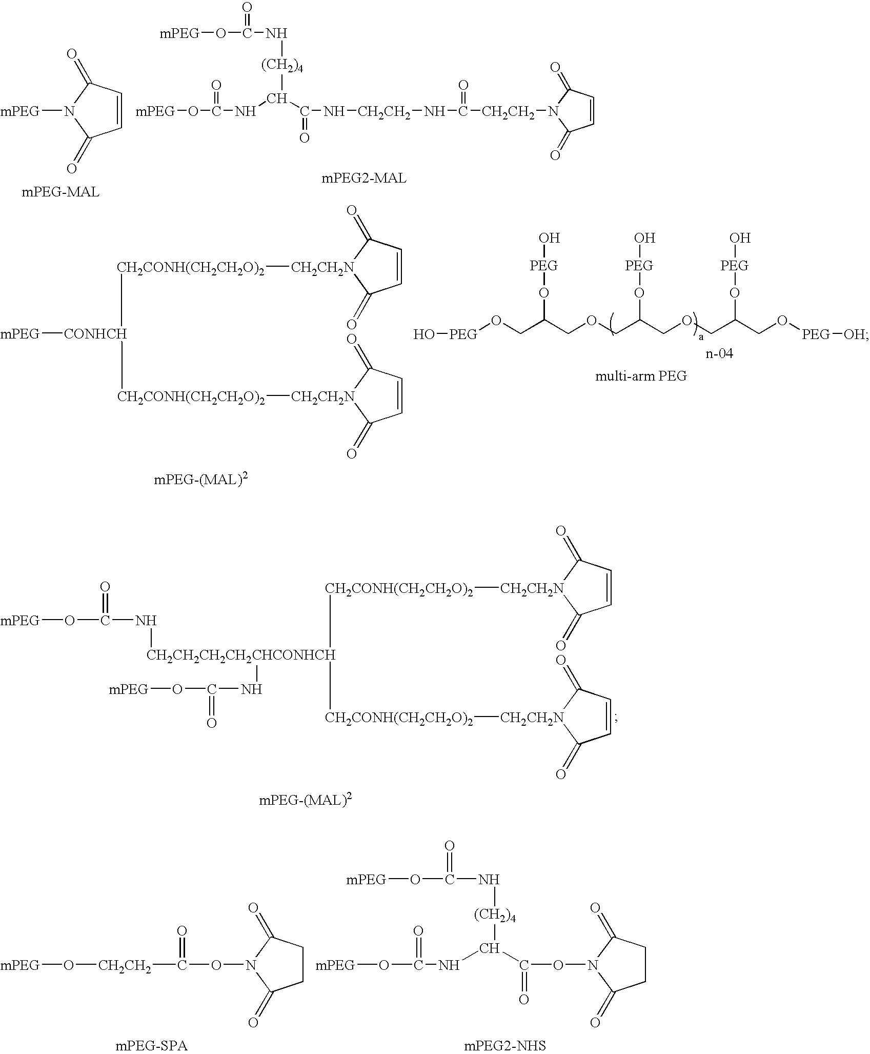
- As used herein, a “sulfhydryl-selective reagent” is a reagent which is useful for the attachment of a PEG polymer to a thiol-containing amino acid. Thiol groups on the amino acid residue cysteine are particularly useful for interaction with a sulfhydryl-selective reagent. Sulfhydryl-selective reagents which are useful in the invention include, but are not limited to maleimide, vinyl sulfone, and thiol. The use of sulfhydryl-selective reagents for coupling to cysteine residues is known in the art and may be adapted as needed according to the present invention (See Eg., Zalipsky, 1995, Bioconjug. Chem. 6:150; Greenwald et al., 2000, Crit. Rev. Ther. Drug Carrier Syst. 17:101; Herman et al., 1994, Macromol. Chem. Phys. 195:203).
- As used herein, an “antigen” is bound by an antibody or a binding region (e.g., a variable domain) of an antibody. Typically, antigens are capable of raising an antibody response in vivo. An antigen can be a peptide, polypeptide, protein, nucleic acid, lipid, carbohydrate, or other molecule, and includes multisubunit molecules. Generally, an immunoglobulin variable domain is selected for target specificity against a particular antigen.
- As used herein, the term “epitope” refers to a unit of structure conventionally bound by an antibody single variable domain VH/VL pair. Epitopes define the minimum binding site for an antibody, and thus represent the target of specificity of an antibody. In the case of a antibody single variable domain, an epitope represents the unit of structure bound by a variable domain in isolation.
- As used herein, the term “neutralizing,” when used in reference to an antibody single variable domain polypeptide as described herein, means that the polypeptide interferes (e.g., completely or at least partially suppresses or eradicates) with a measurable activity or function of the target antigen. A polypeptide is a “neutralizing” polypeptide if it reduces a measurable activity or function of the target antigen by at least 50%, and preferably at least 60%, 70%, 80%, 90%, 95% or more, up to and including 100% inhibition (i.e., no detectable effect or function of the target antigen). This reduction of a measurable activity or function of the target antigen can be assessed by one of skill in the art using standard methods of measuring one or more indicators of such activity or function. As an example, where the target is TNF-α, neutralizing activity can be assessed using a standard L929 cell killing assay or by measuring the ability of an antibody single variable domain polypeptide to inhibit TNF-α-induced expression of ELAM-1 on HUVEC, which measures TNF-α-induced cellular activation. Analogous to “neutralizing” as used herein, “inhibit cell cytotoxicity” as used herein refers to a decrease in cell death as measured, for example, using a standard L929 cell killing assay, wherein cell cytotoxicity is inhibited were cell death is reduced by at least 10% or more.
- As used herein, a “measurable activity or function of a target antigen” includes, but is not limited to, for example, cell signaling, enzymatic activity, binding activity, ligand-dependent internalization, cell killing, cell activation, promotion of cell survival, and gene expression. One of skill in the art can perform assays that measure such activities for a given target antigen. Preferably, “activity”, as used herein, is defined by (1) ND50 in a cell-based assay; (2) affinity for a target ligand, (3) ELISA binding, or (4) a receptor binding assay. Methods for performing these tests are known to those of skill in the art and are described in further detail below.
- As used herein, “dAb activity” or “antibody single variable domain activity” refers to the ability of the antibody single variable domain or polypeptide to bind antigen. As used herein, “retains activity” refers to a level of activity of the PEG-linked antibody single variable domain or polypeptide which is at least 10% of the level of activity of a non-PEG-linked antibody single variable domain or polypeptide, preferably at least 20%, 30%, 40%, 50%, 60%, 70%, 80% and up to 90%, preferably up to 95%, 98%, and up to 100% of the activity of a non-PEG-linked antibody single variable domain or polypeptide comprising the same variable domain as the PEG-linked antibody single variable domain or polypeptide, wherein activity is determined as described above. More specifically, the activity of a PEG-linked antibody single variable domain or polypeptide compared to a non-PEG linked antibody variable domain or polypeptide should be determined on a single antibody variable domain or polypeptide molar basis; that is equivalent numbers of moles of each of the PEG-linked and non-PEG-linked antibody single variable domain should be used in each trial wherein all other conditions are equivalent between trials. In determining whether a particular PEG-linked antibody single variable domain “retains activity”, it is preferred that the activity of a PEG-linked antibody single variable domain be compared with the activity of the same antibody single variable domain in the absence of PEG.
- As used herein, the phrase “specifically binds” refers to the binding of an antigen by an immunoglobulin variable domain or polypeptide with a dissociation constant (Kd) of 1 μM or lower as measured by surface plasmon resonance analysis using, for example, a BIAcore™ surface plasmon resonance system and BIAcore™ kinetic evaluation software (e.g., version 2.1). The affinity or Kd for a specific binding interaction is preferably about 1 μM or lower, preferably 500 nM or lower, more preferably 100 nM or lower, more preferably about 80 nM or lower, and preferably as low as 10 pM.
- As used herein, the terms “heterodimer,” “heterotrimer”, “heterotetramer”, and “heteromultimer” refer to molecules comprising two, three or more (e.g., four, five, six, seven and up to eight or more) monomers of two or more different single immunoglobulin variable domain polypeptide sequence, respectively. For example, a heterodimer would include two VH sequences, such as VH1 and VH2, or VHH1 and VHH2, or may alternatively include a combination of VH and VL. Similar to a homodimer, trimer, or tetramer, the monomers in a heterodimer, heterotrimer, heterotetramer, or heteromultimer can be linked either by expression as a fusion polypeptide, e.g., with a peptide linker between monomers, or, by chemically joining monomers after translation either to each other directly or through a linker by disulfide bonds, or by linkage to a di-, tri- or multivalent linking moiety. In one embodiment, the monomers in a heterodimer, trimer, tetramer, or multimer can be linked by a multi-arm PEG polymer, wherein each monomer of the dimer, trimer, tetramer, or multimer is linked as described above to a PEG moiety of the multi-arm PEG.
- As used herein, the term “half-life” refers to the time taken for the serum concentration of a ligand (e.g., a single immunoglobulin variable domain) to reduce by 50%, in vivo, for example due to degradation of the ligand and/or clearance or sequestration of the ligand by natural mechanisms. The antibody single variable domains of the invention are stabilized in vivo and their half-life increased by binding to molecules which are hypothesized to resist degradation and/or clearance or sequestration, such as PEG. The half-life of a dAb or polypeptide is increased if its functional activity persists (to a degree), in vivo, for a longer period than a similar dAb which is not linked to a PEG polymer. Typically, the half life of a PEGylated dAb or polypeptide is increased by 10%, 20%, 30%, 40%, 50% or more relative to a non-PEGylated dAb or polypeptide. Increases in the range of 2×, 3×, 4×, 5×, 10×, 20×, 30×, 40×, 50× or more of the half life are possible. Alternatively, or in addition, increases in the range of up to 30×, 40×, 50×, 60×, 70×, 80×, 90×, 100×, 150× of the half life are possible. According to the invention, a PEG-linked antibody single variable domain or polypeptide has a half-life of between 0.25 and 170 hours, preferably between 1 and 100 hours, more preferably between 30 and 100 hours, and still more preferably between 50 and 100 hours, and up to 170, 180, 190, and 200 hours or more.
- As used herein, “resistant to degradation” or “resists degradation” with respect to a PEG or other polymer linked dAb monomer or multimer means that the PEG- or other polymer-linked dAb monomer or multimer is degraded by no more than 10% when exposed to pepsin at pH 2.0 for 30 minutes, and preferably not degraded at all. With specific reference to a PEG- or other polymer-linked dAb multimer (e.g., hetero- or homodimer, trimer, tetramer, etc) of the invention, such a multimer is degraded by less than 5%, and is preferably not degraded at all in the presence of pepsin at pH 2.0 for 30 minutes.
- As used herein, “hydrodynamic size” refers to the apparent size of a molecule (e.g., a protein molecule) based on the diffusion of the molecule through an aqueous solution. The diffusion, or motion of a protein through solution can be processed to derive an apparent size of the protein, where the size is given by the “Stokes radius” or “hydrodynamic radius” of the protein particle. The “hydrodynamic size” of a protein depends on both mass and shape (conformation), such that two proteins having the same molecular mass may have differing hydrodynamic sizes based on the overall conformation of the protein. The hydrodynamic size of a PEG-linked antibody single variable domain (including antibody variable domain multimers as described herein) can be in the range of 24 kDa to 500 kDa; 30 to 500 kDa; 40 to 500 kDa; 50 to 500 kDa; 100 to 500 kDa; 150 to 500 kDa; 200 to 500 kDa; 250 to 500 kDa; 300 to 500 kDa; 350 to 500 kDa; 400 to 500 kDa and 450 to 500 kDa. Preferably the hydrodynamic size of a PEGylated dAb of the invention is 30 to 40 kDa; 70 to 80 kDa or 200 to 300 kDa. Where an antibody variable domain multimer is desired for use in imaging applications, the multimer should have a hydrodynamic size of between 50 and 100 kDa. Alternatively, where an antibody single domain multimer is desired for therapeutic applications, the multimer should have a hydrodynamic size of greater than 200 kDa.
- As used herein “TAR1” refers to a dAb whose target antigen is TNFα.
- As used herein “TAR2” refers to a dAb whose target antigen is the human p55-TNFα receptor.
-
FIG. 1 shows the results of a receptor binding assay showing the affinity of a range of PEGylated formats of TAR1-5-19. -
FIG. 2 shows the results of a cell cytotoxicity assay showing the affinity of a range of PEGylated formats of TAR1-5-19. -
FIG. 3 shows SDS page gels showing the results of affinity binding of various formats of PEGylated HEL4 dAb to lysozyme. Lane descriptions are provided in the Examples. -
FIG. 4 shows the results of a receptor binding assay showing the affinity of TAR2-10-27 and the 40K PEGylated monomer. -
FIG. 5 shows the protease stability of TAR1-5-19 and PEGylated variants against the action of pepsin at pH 2.0. -
FIG. 6 shows schematics of monomer PEGylation of dAbs.FIG. 6-1 shows an unmodified VH or VL dAb.FIG. 6-2 shows a VH or VL with surface PEGylation.FIG. 6-3 shows VH or VL dAb with a C-terminal cysteine which is linked to PEG. -
FIG. 7 shows schematics of PEGylated VH or VL hetero- or homodimeric dAbs.FIG. 7-4 shows VH or VL disulfide dimer formed by a C-terminal disulfide bond.FIG. 7-5 shows VH or VL disulfide dimer PEGylated on one subunit.FIG. 7-6 shows VH or VL disulfide dimer PEGylated on both subunits.FIG. 7-7 shows VH or VL dimer formed by a branched/forked/multi-arm PEG via a C-terminal cysteine.FIG. 7-8 shows a VH or VL disulfide dimer formed by a surface disulfide bond.FIG. 7-9 shows a VH or VL disulfide dimer PEGylated on one subunit.FIG. 7-10 shows a VH or VL dimer PEGylated on both subunits.FIG. 7-11 shows a VH or VL dimer formed by a branched/forked/multi-arm PEG via surface cysteine residues. -
FIG. 8 shows further schematics of PEGylated VH or VL hetero- or homodimers of the invention.FIG. 8-12 shows a VH or VL linked dimer formed by a (Gly4Ser)n linker (n=0-10).FIG. 8-13 shows a VH or VL linker dimer PEGylated on one subunit.FIG. 8-14 shows a VH or VL linker dimer PEGylated on both subunits.FIG. 8-15 shows a VH or VL linker dimer PEGylated via the linker.FIG. 8-16 shows a VH or VL linker dimer with a C-terminal cysteine residue.FIG. 8-17 shows two VH or VL linker dimers dimerized by disulfide bonds. -
FIG. 9 shows schematics of PEGylated linker dAb dimers.FIG. 9-18 and 9-19 show VH or VL linker dimers PEGylated via a C-terminal cysteine residue on one subunit.FIG. 9-20 shows a VH or VL linker dimer PEGylated via a cysteine present in the linker.FIG. 9-21 shows a VH or VL or VL linker dimer PEGylated via a cysteine present on one subunit.FIG. 9-22 shows a VH or VL linker dimer PEGylated via cysteines present on both subunits. -
FIG. 10 shows schematic representations of PEGylation of VH or VL hetero- or homotrimeric dAbs.FIGS. 10-23 and 10-24 show PEGylation and formation of dAb trimers using 3-arm PEG to covalently trimerize via C-terminal amino acids.FIG. 10-25 shows surface PEGylation of one of the dAb monomers, wherein the dAb trimer is formed via linker peptides.FIG. 10-26 shows C-terminal PEGylation of one of the monomers of the dAb trimer.FIG. 10-27 shows a dual-specific dAb trimer in which two of the dAb monomers have binding affinity for TNFα and the third monomer has a binding specificity for serum albumin. This format can also be PEGylated as shown in either ofFIGS. 10-25 or 10-26. -
FIG. 11 shows a schematic representation of VH or VL hetero- or homotetrameric dAbs.FIG. 11-28 shows a dAb tetramer formed by linking a 4-arm PEG to C-terminal cysteines of each dAb monomer.FIGS. 11-29 and 11-31 show the formation of a dAb tetramer by linking two dAb linker dimers via a branched/multi-arm PEG where the PEG is linked either to a C-terminal cysteine (11-29) or to the linking peptide (11-31).FIG. 11-30 shows a dAb tetramer in which each of the monomers of the tetramer are linked by a single branched PEG to C-terminal cysteine residues of each monomer.FIG. 11-32 shows a dAb tetramer in which each of the monomers is linked to the other by a linking peptide. This configuration may be PEGylated using any of the strategies shown inFIGS. 10-25 or 10-26. -
FIG. 12 shows other multimeric PEG-linked dAb formats useful in the invention.FIG. 12-31 shows a tetramer of dAb linker dimers which are themselves linked to form the tetramer by a multi-arm PEG wherein the PEG is linked to C-terminal cysteine residues present in one of the monomers of each dimer.FIG. 32 shows a tetramer of dAb linker dimers which are themselves linked to form the tetramer by a multi-arm PEG wherein each PEG is linked to cysteine residue present in the linker of each dimer pair. -
FIG. 13 shows the sequence of the VH framework based on germline sequence DP47-JH4b (SEQ ID NO: 1, 2) HCDRs 1-3 are indicated by underlining. -
FIG. 14 shows the sequence of the Vκ framework based on germline sequence DPκ9-Jκ1 (SEQ ID NO: 3, 4). LCDRs 1-3 are indicated by underlining. -
FIG. 15 shows a plot showing the relationship of native hydrodynamic size of the dAb vs the in vivo serum half-life in mouse. Data shown in Table 8 was used to generate the graph. -
FIG. 16 shows protease stability profile of monomeric and 40K PEGylated TAR1-5-19. The relative activity is as a percentage of the no protease control. The proteases used were pepsin, porcine intestinal mucosa peptidase, elastase, crude bovine pancreatic protease (CBP) and rat intestinal powder (Rat In). - The present invention provides polymer linked dAbs and dAb homo- and heteromultimers with increased half-life and resistance to proteolytic degradation relative to non-polymer linked dAbs. The invention relates, in one embodiment, to PEG-linked dabs and dAb multimers, and still further to PEG-linked dAb monomers, dimers, trimers, and tetramers having a half-life of at least 0.25 hours, and further having a hydrodynamic size of at least 24 kDa. The invention also relates to a PEG-linked antibody single variable domain which retains its activity relative to a non-PEG-linked antibody single variable domain comprising the same antibody variable domain as the PEG-linked antibody variable domain. This provides dAb molecules with increased therapeutic efficacy due to their prolonged circulation time and potent and efficacious activity.
- In one embodiment, the invention provides PEG-linked dAb multimers which comprise at least two non-complementary variable domains. For example, the dAbs may comprise a pair of VH domains or a pair of VL domains. Advantageously, the domains are of non-camelid origin; preferably they are human domains or comprise human framework regions (FWs) and one or more heterologous CDRs. CDRs and framework regions are those regions of an immunoglobulin variable domain as defined in the Kabat database of Sequences of Proteins of Immunological Interest. In one embodiment, the dAb domains are of camelid origin.
- Preferred human framework regions are those encoded by germline gene segments DP47 and DPK9. Advantageously, FW1, FW2 and FW3 of a VH or VL domain have the sequence of FW1, FW2 or FW3 from DP47 or DPK9. The human frameworks may optionally contain mutations, for example up to about 5 amino acid changes or up to about 10 amino acid changes collectively in the human frameworks used in the dAbs of the invention.
- Preparation of Single Immunoglobulin Variable Domains:
- The antibody single variable domains (or dabs) of the invention are a folded polypeptide domain which comprises sequences characteristic of immunoglobulin variable domains and which specifically binds an antigen (i.e., dissociation constant of 500 nM or less), and which binds antigen as a single variable domain; that is, without any complementary variable domain. An antibody single variable domain therefore includes complete antibody variable domains as well as modified variable domains, for example in which one or more loops have been replaced by sequences which are not characteristic of antibody variable domains or antibody variable domains which have been truncated or comprise N- or C-terminal extensions, as well as folded fragments of variable domains which retain a dissociation constant of 500 nM or less (e.g., 450 nM or less, 400 nM or less, 350 nM or less, 300 nM or less, 250 nM or less, 200 nM or less, 150 nM or less, 100 nM or less) and the target antigen specificity of the full-length domain. Preferably an antibody single variable domain useful in the invention is selected from the group of VHH, VH and VL, including Vkappa and Vlambda.
- Single immunoglobulin variable domains are prepared in a number of ways. For each of these approaches, well-known methods of preparing (e.g., amplifying, mutating, etc.) and manipulating nucleic acid sequences are applicable.
- One means is to amplify and express the VH or VL region of a heavy chain or light chain gene for a cloned antibody known to bind the desired antigen. The boundaries of VH and VL domains are set out by Kabat et al. (1991, supra). The information regarding the boundaries of the VH and VL domains of heavy and light chain genes is used to design PCR primers that amplify the V domain from a cloned heavy or light chain coding sequence encoding an antibody known to bind a given antigen. The amplified V domain is inserted into a suitable expression vector, e.g., pHEN-1 (Hoogenboom et al., 1991, Nucleic Acids Res. 19:4133-4137) and expressed, either alone or as a fusion with another polypeptide sequence. The expressed VH or VL domain is then screened for high affinity binding to the desired antigen in isolation from the remainder of the heavy or light chain polypeptide. For all aspects of the present invention, screening for binding is performed as known in the art or as described herein below.
- A repertoire of VH or VL domains is screened by, for example, phage display, panning against the desired antigen. Methods for the construction of bacteriophage display libraries and lambda phage expression libraries are well known in the art, and taught, for example, by: McCafferty et al., 1990, Nature 348: 552; Kang et al., 1991, Proc. Natl. Acad. Sci. U.S.A., 88:4363; Clackson et al., 1991, Nature 352: 624; Lowman et al., 1991, Biochemistry 30: 10832; Burton et al., 1991, Proc. Natl. Acad. Sci U.S.A. 88: 10134; Hoogenboom et al., 1991, Nucleic Acids Res. 19: 4133; Chang et al., 1991, J. Immunol. 147: 3610; Breitling et al., 1991, Gene 104: 147; Marks et al., 1991, J. Mol. Biol. 222: 581; Barbas et al., 1992, Proc. Natl. Acad. Sci. U.S.A. 89: 4457; Hawkins and Winter (1992) J. Immunol., 22: 867; Marks et al. (1992) J. Biol. Chem., 267: 16007; and Lerner et al. (1992) Science, 258: 1313. scFv phage libraries are taught, for example, by Huston et al., 1988, Proc. Natl. Acad. Sci U.S.A. 85:5879-5883; Chaudhary et al., 1990, Proc. Natl. Acad. Sci U.S.A. 87: 1066-1070; McCafferty et al., 1990, supra; Clackson et al., 1991, supra; Marks et al., 1991, supra; Chiswell et al., 1992, Trends Biotech. 10: 80; and Marks et al., 1992, supra. Various embodiments of scFv libraries displayed on bacteriophage coat proteins have been described. Refinements of phage display approaches are also known, for example as described in WO96/06213 and WO92/01047 (Medical Research Council et al.) and WO97/08320 (Morphosys, supra).
- The repertoire of VH or VL domains can be a naturally-occurring repertoire of immunoglobulin sequences or a synthetic repertoire. A naturally-occurring repertoire is one prepared, for example, from immunoglobulin-expressing cells harvested from one or more animals, including humans. Such repertoires can be “naïve,” i.e., prepared, for example, from human fetal or newborn immunoglobulin-expressing cells, or rearranged, i.e., prepared from, for example, adult human B cells. Natural repertoires are described, for example, by Marks et al., 1991, J. Mol. Biol. 222: 581 and Vaughan et al., 1996, Nature Biotech. 14: 309. If desired, clones identified from a natural repertoire, or any repertoire, for that matter, that bind the target antigen are then subjected to mutagenesis and further screening in order to produce and select variants with improved binding characteristics.
- Synthetic repertoires of single immunoglobulin variable domains are prepared by artificially introducing diversity into a cloned V domain. Synthetic repertoires are described, for example, by Hoogenboom & Winter, 1992, J. Mol. Biol. 227: 381; Barbas et al., 1992, Proc. Natl. Acad. Sci. U.S.A. 89: 4457; Nissim et al., 1994, EMBO J. 13: 692; Griffiths et al., 1994, EMBO J. 13: 3245; DeKriuf et al., 1995, J. Mol. Biol. 248: 97; and WO 99/20749.
- The antigen binding domain of a conventional antibody comprises two separate regions: a heavy chain variable domain (VH) and a light chain variable domain (VL: which can be either Vκ or Vλ). The antigen binding site of such an antibody is formed by six polypeptide loops: three from the VH domain (H1, H2 and H3) and three from the VL domain (L1, L2 and L3). The boundaries of these loops are described, for example, in Kabat et al. (1991, supra). A diverse primary repertoire of V genes that encode the VH and VL domains is produced in vivo by the combinatorial rearrangement of gene segments. The VH gene is produced by the recombination of three gene segments, VH, D and JH. In humans, there are approximately 51 functional VH segments (Cook and Tomlinson (1995) Immunol Today 16:237), 25 functional D segments (Corbett et al. (1997) J. Mol. Biol. 268: 69) and 6 functional JH segments (Ravetch et al. (1981) Cell 27: 583), depending on the haplotype. The VH segment encodes the region of the polypeptide chain which forms the first and second antigen binding loops of the VH domain (H1 and H2), while the VH, D and JH segments combine to form the third antigen binding loop of the VH domain (H3).
- The VL gene is produced by the recombination of only two gene segments, VL and JL. In humans, there are approximately 40 functional Vκ segments (Schäble and Zachau (1993) Biol. Chem. Hoppe-Seyler 374: 1001), 31 functional Vλ segments (Williams et al. (1996) J. Mol. Biol. 264: 220; Kawasaki et al. (1997) Genome Res. 7: 250), 5 functional Jκ segments (Hieter et al. (1982) J. Biol. Chem. 257: 1516) and 4 functional Jλ segments (Vasicek and Leder (1990) J. Exp. Med. 172: 609), depending on the haplotype. The VL segment encodes the region of the polypeptide chain which forms the first and second antigen binding loops of the VL domain (L1 and L2), while the VL and JL segments combine to form the third antigen binding loop of the VL domain (L3). Antibodies selected from this primary repertoire are believed to be sufficiently diverse to bind almost all antigens with at least moderate affinity. High affinity antibodies are produced in vivo by “affinity maturation” of the rearranged genes, in which point mutations are generated and selected by the immune system on the basis of improved binding.
- Analysis of the structures and sequences of antibodies has shown that five of the six antigen binding loops (H1, H2, L1, L2, L3) possess a limited number of main-chain conformations or canonical structures (Chothia and Lesk (1987) J. Mol. Biol. 196: 901; Chothia et al. (1989) Nature 342: 877). The main-chain conformations are determined by (i) the length of the antigen binding loop, and (ii) particular residues, or types of residue, at certain key position in the antigen binding loop and the antibody framework. Analysis of the loop lengths and key residues has enabled the prediction of the main-chain conformations of H1, H2, L1, L2 and L3 encoded by the majority of human antibody sequences (Chothia et al. (1992) J. Mol. Biol. 227: 799; Tomlinson et al. (1995) EMBO J. 14: 4628; Williams et al. (1996) J. Mol. Biol. 264: 220). Although the H3 region is much more diverse in terms of sequence, length and structure (due to the use of D segments), it also forms a limited number of main-chain conformations for short loop lengths which depend on the length and the presence of particular residues, or types of residue, at key positions in the loop and the antibody framework (Martin et al. (1996) J. Mol. Biol. 263: 800; Shirai et al. (1996) FEBS Letters 399: 1.
- While, according to one embodiment of the invention, diversity can be added to synthetic repertoires at any site in the CDRs of the various antigen-binding loops, this approach results in a greater proportion of V domains that do not properly fold and therefore contribute to a lower proportion of molecules with the potential to bind antigen. An understanding of the residues contributing to the main chain conformation of the antigen-binding loops permits the identification of specific residues to diversify in a synthetic repertoire of VH or VL domains. That is, diversity is best introduced in residues that are not essential to maintaining the main chain conformation. As an example, for the diversification of loop L2, the conventional approach would be to diversify all the residues in the corresponding CDR (CDR2) as defined by Kabat et al. (1991, supra), some seven residues. However, for L2, it is known that positions 50 and 53 are diverse in naturally occurring antibodies and are observed to make contact with the antigen. The preferred approach would be to diversify only those two residues in this loop. This represents a significant improvement in terms of the functional diversity required to create a range of antigen binding specificities.
- In one aspect, synthetic variable domain repertoires are prepared in VH or Vκ backgrounds, based on artificially diversified germline VH or Vκ sequences. For example, the VH domain repertoire is based on cloned germline VH gene segments V3-23/DP47 (Tomlinson et al., 1992, J. Mol. Biol. 227: 7768) and JH4b. The Vκ domain repertoire is based, for example, on germline Vκ gene segments O2/O12/DPK9 (Cox et al., 1994, Eur. J. Immunol. 24: 827) and Jκ1. Diversity is introduced into these or other gene segments by, for example, PCR mutagenesis. Diversity can be randomly introduced, for example, by error prone PCR (Hawkins, et al., 1992, J. Mol. Biol. 226: 889) or chemical mutagenesis. As discussed above, however it is preferred that the introduction of diversity is targeted to particular residues. It is further preferred that the desired residues are targeted by introduction of the codon NNK using mutagenic primers (using the IUPAC nomenclature, where N=G, A, T or C, and K=G or T), which encodes all amino acids and the TAG stop codon. Other codons which achieve similar ends are also of use, including the NNN codon (which leads to the production of the additional stop codons TGA and TAA), DVT codon ((A/G/T) (A/G/C)T), DVC codon ((A/G/T)(A/G/C)C), and DVY codon ((A/G/T)(A/G/C)(C/T). The DVT codon encodes 22% serine and 11% tyrosine, aspargine, glycine, alanine, aspartate, threonine and cysteine, which most closely mimics the distribution of amino acid residues for the antigen binding sites of natural human antibodies. Repertoires are made using PCR primers having the selected degenerate codon or codons at each site to be diversified. PCR mutagenesis is well known in the art; however, considerations for primer design and PCR mutagenesis useful in the methods of the invention are discussed below in the section titled “PCR Mutagenesis.”
- Diversified repertoires are cloned into phage display vectors as known in the art and as described, for example, in WO 99/20749. In general, the nucleic acid molecules and vector constructs required for the performance of the present invention are available in the art and are constructed and manipulated as set forth in standard laboratory manuals, such as Sambrook et al. (1989). Molecular Cloning: A Laboratory Manual, Cold Spring Harbor, USA.
- The manipulation of nucleic acids in the present invention is typically carried out in recombinant vectors. As used herein, “vector” refers to a discrete element that is used to introduce heterologous DNA into cells for the expression and/or replication thereof. Methods by which to select or construct and, subsequently, use such vectors are well known to one of skill in the art. Numerous vectors are publicly available, including bacterial plasmids, bacteriophage, artificial chromosomes and episomal vectors. Such vectors may be used for simple cloning and mutagenesis; alternatively, as is typical of vectors in which repertoire (or pre-repertoire) members of the invention are carried, a gene expression vector is employed. A vector of use according to the invention is selected to accommodate a polypeptide coding sequence of a desired size, typically from 0.25 kilobase (kb) to 40 kb in length. A suitable host cell is transformed with the vector after in vitro cloning manipulations. Each vector contains various functional components, which generally include a cloning (or “polylinker”) site, an origin of replication and at least one selectable marker gene. If a given vector is an expression vector, it additionally possesses one or more of the following: enhancer element, promoter, transcription termination and signal sequences, each positioned in the vicinity of the cloning site, such that they are operatively linked to the gene encoding a polypeptide repertoire member according to the invention.
- Both cloning and expression vectors generally contain nucleic acid sequences that enable the vector to replicate in one or more selected host cells. Typically in cloning vectors, this sequence is one that enables the vector to replicate independently of the host chromosomal DNA and includes origins of replication or autonomously replicating sequences. Such sequences are well known for a variety of bacteria, yeast and viruses. The origin of replication from the plasmid pBR322 is suitable for most Gram-negative bacteria, the 2 micron plasmid origin is suitable for yeast, and various viral origins (
e.g. SV 40, adenovirus) are useful for cloning vectors in mammalian cells. Generally, the origin of replication is not needed for mammalian expression vectors unless these are used in mammalian cells able to replicate high levels of DNA, such as COS cells. - Advantageously, a cloning or expression vector also contains a selection gene also referred to as selectable marker. This gene encodes a protein necessary for the survival or growth of transformed host cells grown in a selective culture medium. Host cells not transformed with the vector containing the selection gene will therefore not survive in the culture medium. Typical selection genes encode proteins that confer resistance to antibiotics and other toxins, e.g. ampicillin, neomycin, methotrexate or tetracycline, complement auxotrophic deficiencies, or supply critical nutrients not available in the growth media.
- Because the replication of vectors according to the present invention is most conveniently performed in E. coli, an E. coli-selectable marker, for example, the β-lactamase gene that confers resistance to the antibiotic ampicillin, is of use. These can be obtained from E. coli plasmids, such as pBR322 or a pUC plasmid such as pUC18 or pUC19.
- Expression vectors usually contain a promoter that is recognized by the host organism and is operably linked to the coding sequence of interest. Such a promoter may be inducible or constitutive. The term “operably linked” refers to a juxtaposition wherein the components described are in a relationship permitting them to function in their intended manner. A control sequence “operably linked” to a coding sequence is ligated in such a way that expression of the coding sequence is achieved under conditions compatible with the control sequences.
- Promoters suitable for use with prokaryotic hosts include, for example, the α-lactamase and lactose promoter systems, alkaline phosphatase, the tryptophan (trp) promoter system and hybrid promoters such as the tac promoter. Promoters for use in bacterial systems will also generally contain a Shine-Dalgarno sequence operably linked to the coding sequence.
- In libraries or repertoires as described herein, the preferred vectors are expression vectors that enable the expression of a nucleotide sequence corresponding to a polypeptide library member. Thus, selection is performed by separate propagation and expression of a single clone expressing the polypeptide library member or by use of any selection display system. As described above, a preferred selection display system uses bacteriophage display. Thus, phage or phagemid vectors can be used. Preferred vectors are phagemid vectors, which have an E. coli origin of replication (for double stranded replication) and also a phage origin of replication (for production of single-stranded DNA). The manipulation and expression of such vectors is well known in the art (Hoogenboom and Winter (1992) supra; Nissim et al. (1994) supra). Briefly, the vector contains a β-lactamase or other selectable marker gene to confer selectivity on the phagemid, and a lac promoter upstream of a expression cassette that consists (N to C terminal) of a leader sequence (which directs the expressed polypeptide to the periplasmic space), a multiple cloning site (for cloning the nucleotide version of the library member), optionally, one or more peptide tags (for detection), optionally, one or more TAG stop codons and the phage protein pIII. Leader sequences, which may be used in bacterial expression and/or phage or phagemid display, include pelB, stII, ompA, phoA, bla, and pelA. Using various suppressor and non-suppressor strains of E. coli and with the addition of glucose, iso-propyl thio-β-D-galactoside (IPTG) or a helper phage, such as VCS M13, the vector is able to replicate as a plasmid with no expression, produce large quantities of the polypeptide library member only, or produce phage, some of which contain at least one copy of the polypeptide-pIII fusion on their surface.
- An example of a preferred vector is the pHEN1 phagemid vector (Hoogenboom et al., 1991, Nucl. Acids Res. 19: 4133-4137; sequence is available, e.g., as SEQ ID NO: 7 in WO 03/031611), in which the production of pIII fusion protein is under the control of the LacZ promoter, which is inhibited in the presence of glucose and induced with IPTG. When grown in suppressor strains of B. coli, e.g., TG1, the gene III fusion protein is produced and packaged into phage, while growth in non-suppressor strains, e.g., HB2151, permits the secretion of soluble fusion protein into the bacterial periplasm and into the culture medium. Because the expression of gene III prevents later infection with helper phage, the bacteria harboring the phagemid vectors are propagated in the presence of glucose before infection with VCSM13 helper phage for phage rescue.
- Construction of vectors according to the invention employs conventional ligation techniques. Isolated vectors or DNA fragments are cleaved, tailored, and re-ligated in the form desired to generate the required vector. If desired, sequence analysis to confirm that the correct sequences are present in the constructed vector is performed using standard methods. Suitable methods for constructing expression vectors, preparing in vitro transcripts, introducing DNA into host cells, and performing analyses for assessing expression and function are known to those skilled in the art. The presence of a gene sequence in a sample is detected, or its amplification and/or expression quantified by conventional methods, such as Southern or Northern analysis, Western blotting, dot blotting of DNA, RNA or protein, in situ hybridization, immunocytochemistry or sequence analysis of nucleic acid or protein molecules. Those skilled in the art will readily envisage how these methods may be modified, if desired.
- Scaffolds for use in Constructing Antibody Single Variable Domains
- i. Selection of the Main-Chain Conformation
- The members of the immunoglobulin superfamily all share a similar fold for their polypeptide chain. For example, although antibodies are highly diverse in terms of their primary sequence, comparison of sequences and crystallographic structures has revealed that, contrary to expectation, five of the six antigen binding loops of antibodies (H1, H2, L1, L2, L3) adopt a limited number of main-chain conformations, or canonical structures (Chothia and Lesk (1987) J. Mol. Biol., 196: 901; Chothia et al. (1989) Nature, 342: 877). Analysis of loop lengths and key residues has therefore enabled prediction of the main-chain conformations of H1, H2, L1, L2 and L3 found in the majority of human antibodies (Chothia et al. (1992) J. Mol. Biol., 227: 799; Tomlinson et al. (1995) EMBO J, 14: 4628; Williams et al. (1996) J. Mol. Biol., 264: 220). Although the H3 region is much more diverse in terms of sequence, length and structure (due to the use of D segments), it also forms a limited number of main-chain conformations for short loop lengths which depend on the length and the presence of particular residues, or types of residue, at key positions in the loop and the antibody framework (Martin et al. (1996) J. Mol. Biol., 263: 800; Shirai et al. (1996) FEBS Letters, 399: 1).
- The PEG-linked antibody single variable domain monomers and multimers of the present invention are advantageously assembled from libraries of domains, such as libraries of VH domains and/or libraries of VL domains. Moreover, the PEG-linked dAbs of the invention may themselves be provided in the form of libraries. In one aspect of the present invention, libraries of antibody single variable domains are designed in which certain loop lengths and key residues have been chosen to ensure that the main-chain conformation of the members is known. Advantageously, these are real conformations of immunoglobulin superfamily molecules found in nature, to minimize the chances that they are non-functional, as discussed above. Germline V gene segments serve as one suitable basic framework for constructing antibody or T-cell receptor libraries; other sequences are also of use. Variations may occur at a low frequency, such that a small number of functional members may possess an altered main-chain conformation, which does not affect its function.
- Canonical structure theory is also of use to assess the number of different main-chain conformations encoded by ligands, to predict the main-chain conformation based on ligand sequences and to chose residues for diversification which do not affect the canonical structure. It is known that, in the human Vκ domain, the L1 loop can adopt one of four canonical structures, the L2 loop has a single canonical structure and that 90% of human Vκ domains adopt one of four or five canonical structures for the L3 loop (Tomlinson et al. (1995) supra); thus, in the Vκ domain alone, different canonical structures can combine to create a range of different main-chain conformations. Given that the Vλ domain encodes a different range of canonical structures for the L1, L2 and L3 loops and that Vκ and Vλ domains can pair with any VH domain which can encode several canonical structures for the H1 and H2 loops, the number of canonical structure combinations observed for these five loops is very large. This implies that the generation of diversity in the main-chain conformation may be essential for the production of a wide range of binding specificities. However, by constructing an antibody library based on a single known main-chain conformation it has been found, contrary to expectation, that diversity in the main-chain conformation is not required to generate sufficient diversity to target substantially all antigens. Even more surprisingly, the single main-chain conformation need not be a consensus structure—a single naturally occurring conformation can be used as the basis for an entire library. Thus, in one aspect, the polymer-linked antibody single variable domains of the invention possess a single known main-chain conformation.
- The single main-chain conformation that is chosen is preferably commonplace among molecules of the immunoglobulin superfamily type in question. A conformation is commonplace when a significant number of naturally occurring molecules are observed to adopt it. Accordingly, in a preferred aspect of the invention, the natural occurrence of the different main-chain conformations for each binding loop of an immunoglobulin domain are considered separately and then a naturally occurring variable domain is chosen which possesses the desired combination of main-chain conformations for the different loops. If none is available, the nearest equivalent may be chosen. It is preferable that the desired combination of main-chain conformations for the different loops is created by selecting germline gene segments which encode the desired main-chain conformations. It is more preferable, that the selected germline gene segments are frequently expressed in nature, and most preferable that they are the most frequently expressed of all natural germline gene segments.
- In designing antibody single variable domains or libraries thereof the incidence of the different main-chain conformations for each of the six antigen binding loops may be considered separately. For H1, H2, L1, L2 and L3, a given conformation that is adopted by between 20% and 100% of the antigen binding loops of naturally occurring molecules is chosen. Typically, its observed incidence is above 35% (i.e. between 35% and 100%) and, ideally, above 50% or even above 65%. Since the vast majority of H3 loops do not have canonical structures, it is preferable to select a main-chain conformation which is commonplace among those loops which do display canonical structures. For each of the loops, the conformation which is observed most often in the natural repertoire is therefore selected. In human antibodies, the most popular canonical structures (CS) for each loop are as follows: H1-CS 1 (79% of the expressed repertoire), H2-CS 3 (46%), L1-
CS 2 of Vκ (39%), L2-CS 1 (100%), L3-CS 1 of Vκ (36%) (calculation assumes a κ:λ ratio of 70:30, Hood et al. (1967) Cold Spring Harbor Symp. Quant. Biol., 48: 133). For H3 loops that have canonical structures, a CDR3 length (Kabat et al. (1991) Sequences of proteins of immunological interest, U.S. Department of Health and Human Services) of seven residues with a salt-bridge from residue 94 to residue 101 appears to be the most common. There are at least 16 human antibody sequences in the EMBL data library with the required H3 length and key residues to form this conformation and at least two crystallographic structures in the protein data bank which can be used as a basis for antibody modeling (2cgr and 1tet). The most frequently expressed germline gene segments that this combination of canonical structures are the VH segment 3-23 (DP47), the JH segment JH4b, the Vκ segment O2/O12 (DPK9) and the Jκ segment Jκ VH segments DP45 and DP38 are also suitable. These segments can therefore be used in combination as a basis to construct a library with the desired single main-chain conformation. - Alternatively, instead of choosing the single main-chain conformation based on the natural occurrence of the different main-chain conformations for each of the binding loops in isolation, the natural occurrence of combinations of main-chain conformations is used as the basis for choosing the single main-chain conformation. In the case of antibodies, for example, the natural occurrence of canonical structure combinations for any two, three, four, five or for all six of the antigen binding loops can be determined. Here, it is preferable that the chosen conformation is commonplace in naturally occurring antibodies and most preferable that it observed most frequently in the natural repertoire. Thus, in human antibodies, for example, when natural combinations of the five antigen binding loops, H1, H2, L1, L2 and L3, are considered, the most frequent combination of canonical structures is determined and then combined with the most popular conformation for the H3 loop, as a basis for choosing the single main-chain conformation.
- Diversification of the Canonical Sequence
- Having selected several known main-chain conformations or, preferably a single known main-chain conformation, antibody single variable domains according to the invention or libraries for use in the invention can be constructed by varying the binding site of the molecule in order to generate a repertoire with structural and/or functional diversity. This means that variants are generated such that they possess sufficient diversity in their structure and/or in their function so that they are capable of providing a range of activities.
- The desired diversity is typically generated by varying the selected molecule at one or more positions. The positions to be changed can be chosen at random or are preferably selected. The variation can then be achieved either by randomization, during which the resident amino acid is replaced by any amino acid or analogue thereof, natural or synthetic, producing a very large number of variants or by replacing the resident amino acid with one or more of a defined subset of amino acids, producing a more limited number of variants.
- Various methods have been reported for introducing such diversity. Error-prone PCR (Hawkins et al. (1992) J. Mol. Biol., 226: 889), chemical mutagenesis (Deng et al. (1994) J. Biol. Chem., 269: 9533) or bacterial mutator strains (Low et al. (1996) J. Mol. Biol., 260: 359) can be used to introduce random mutations into the genes that encode the molecule. Methods for mutating selected positions are also well known in the art and include the use of mismatched oligonucleotides or degenerate oligonucleotides, with or without the use of PCR. For example, several synthetic antibody libraries have been created by targeting mutations to the antigen binding loops. The H3 region of a human tetanus toxoid-binding Fab has been randomised to create a range of new binding specificities (Barbas et al. (1992) Proc. Natl. Acad. Sci. USA, 89: 4457). Random or semi-random H3 and L3 regions have been appended to germline V gene segments to produce large libraries with unmutated framework regions (Hoogenboom & Winter (1992) J. Mol. Biol., 227: 381; Barbas et al. (1992) Proc. Natl. Acad. Sci. USA, 89: 4457; Nissim et al. (1994) EMBO J, 13: 692; Griffiths et al. (1994) EMBO J, 13: 3245; De Kruif et al. (1995) J. Mol. Biol., 248: 97). Such diversification has been extended to include some or all of the other antigen binding loops (Crameri et al. (1996) Nature Med., 2: 100; Riechmann et al. (1995) Bio/Technology, 13: 475; Morphosys, WO97/08320, supra).
- Since loop randomization has the potential to create approximately more than 1015 structures for H3 alone and a similarly large number of variants for the other five loops, it is not feasible using current transformation technology or even by using cell free systems to produce a library representing all possible combinations. For example, in one of the largest libraries constructed to date, 6×10¹⁰ different antibodies, which is only a fraction of the potential diversity for a library of this design, were generated (Griffiths et al. (1994) supra).
- In a preferred embodiment, only those residues which are directly involved in creating or modifying the desired function of the molecule are diversified. For many molecules, the function will be to bind a target and therefore diversity should be concentrated in the target binding site, while avoiding changing residues which are crucial to the overall packing of the molecule or to maintaining the chosen main-chain conformation.
- Diversification of the Canonical Sequence as it Applies to Antibody Domains
- In the case of antibody single variable domains, the binding site for the target is most often the antigen binding site. Thus, in a highly preferred aspect, the invention provides libraries of or for the assembly of antibody single variable domains in which only those residues in the antigen binding site are varied. These residues are extremely diverse in the human antibody repertoire and are known to make contacts in high-resolution antibody/antigen complexes. For example, in L2 it is known that positions 50 and 53 are diverse in naturally occurring antibodies and are observed to make contact with the antigen. In contrast, the conventional approach would have been to diversify all the residues in the corresponding Complementarity Determining Region (CDR1) as defined by Kabat et al. (1991, supra), some seven residues compared to the two diversified in the library for use according to the invention. This represents a significant improvement in terms of the functional diversity required to create a range of antigen binding specificities.
- In nature, antibody diversity is the result of two processes: somatic recombination of germline V, D and J gene segments to create a naive primary repertoire (so called germline and junctional diversity) and somatic hypermutation of the resulting rearranged V genes. Analysis of human antibody sequences has shown that diversity in the primary repertoire is focused at the center of the antigen binding site whereas somatic hypermutation spreads diversity to regions at the periphery of the antigen binding site that are highly conserved in the primary repertoire (see Tomlinson et al. (1996) J. Mol. Biol., 256: 813). This complementarity has probably evolved as an efficient strategy for searching sequence space and, although apparently unique to antibodies, it can easily be applied to other polypeptide repertoires. The residues which are varied are a subset of those that form the binding site for the target. Different (including overlapping) subsets of residues in the target binding site are diversified at different stages during selection, if desired.
- In the case of an antibody repertoire, an initial ‘naive’ repertoire is created where some, but not all, of the residues in the antigen binding site are diversified. As used herein in this context, the term “naive” refers to antibody molecules that have no pre-determined target. These molecules resemble those which are encoded by the immunoglobulin genes of an individual who has not undergone immune diversification, as is the case with fetal and newborn individuals, whose immune systems have not yet been challenged by a wide variety of antigenic stimuli. This repertoire is then selected against a range of antigens or epitopes. If required, further diversity can then be introduced outside the region diversified in the initial repertoire. This matured repertoire can be selected for modified function, specificity or affinity.
- In the construction of libraries for use in the invention, diversification of chosen positions is typically achieved at the nucleic acid level, by altering the coding sequence which specifies the sequence of the polypeptide such that a number of possible amino acids (all 20 or a subset thereof) can be incorporated at that position. Using the IUPAC nomenclature, the most versatile codon is NNK, which encodes all amino acids as well as the TAG stop codon. The NNK codon is preferably used in order to introduce the required diversity. Other codons which achieve the same ends are also of use, including the NNN codon, which leads to the production of the additional stop codons TGA and TAA.
- A feature of side-chain diversity in the antigen binding site of human antibodies is a pronounced bias which favors certain amino acid residues. If the amino acid composition of the ten most diverse positions in each of the VH, Vκ and Vλ regions are summed, more than 76% of the side-chain diversity comes from only seven different residues, these being, serine (24%), tyrosine (14%), asparagine (11%), glycine (9%), alanine (7%), aspartate (6%) and threonine (6%). This bias towards hydrophilic residues and small residues which can provide main-chain flexibility probably reflects the evolution of surfaces which are predisposed to binding a wide range of antigens or epitopes and may help to explain the required promiscuity of antibodies in the primary repertoire.
- Since it is preferable to mimic this distribution of amino acids, the distribution of amino acids at the positions to be varied preferably mimics that seen in the antigen binding site of antibodies. Such bias in the substitution of amino acids that permits selection of certain polypeptides (not just antibody polypeptides) against a range of target antigens is easily applied to any polypeptide repertoire. There are various methods for biasing the amino acid distribution at the position to be varied (including the use of tri-nucleotide mutagenesis, see WO97/08320), of which the preferred method, due to ease of synthesis, is the use of conventional degenerate codons. By comparing the amino acid profile encoded by all combinations of degenerate codons (with single, double, triple and quadruple degeneracy in equal ratios at each position) with the natural amino acid use it is possible to calculate the most representative codon. The codons (AGT)(AGC)T, (AGT)(AGC)C and (AGT)(AGC)(CT)—that is, DVT, DVC and DVY, respectively using IUPAC nomenclature—are those closest to the desired amino acid profile: they encode 22% serine and 11% tyrosine, asparagine, glycine, alanine, aspartate, threonine and cysteine. Preferably, therefore, libraries are constructed using either the DVT, DVC or DVY codon at each of the diversified positions.
- PCR Mutagenesis:
- The primer is complementary to a portion of a target molecule present in a pool of nucleic acid molecules used in the preparation of sets of nucleic acid repertoire members encoding polypeptide repertoire members. Most often, primers are prepared by synthetic methods, either chemical or enzymatic. Mutagenic oligonucleotide primers are generally 15 to 100 nucleotides in length, ideally from 20 to 40 nucleotides, although oligonucleotides of different length are of use.
- Typically, selective hybridization occurs when two nucleic acid sequences are substantially complementary (at least about 65% complementary over a stretch of at least 14 to 25 nucleotides, preferably at least about 75%, more preferably at least about 85% or 90% complementary). See Kanehisa, 1984, Nucleic Acids Res. 12: 203, incorporated herein by reference. As a result, it is expected that a certain degree of mismatch at the priming site is tolerated. Such mismatch may be small, such as a mono-, di- or tri-nucleotide. Alternatively, it may comprise nucleotide loops, which are defined herein as regions in which mismatch encompasses an uninterrupted series of four or more nucleotides.
- Overall, five factors influence the efficiency and selectivity of hybridization of the primer to a second nucleic acid molecule. These factors, which are (i) primer length, (ii) the nucleotide sequence and/or composition, (iii) hybridization temperature, (iv) buffer chemistry and (v) the potential for steric hindrance in the region to which the primer is required to hybridize, are important considerations when non-random priming sequences are designed.
- There is a positive correlation between primer length and both the efficiency and accuracy with which a primer will anneal to a target sequence; longer sequences have a higher melting temperature (TM) than do shorter ones, and are less likely to be repeated within a given target sequence, thereby minimizing promiscuous hybridization. Primer sequences with a high G-C content or that comprise palindromic sequences tend to self-hybridize, as do their intended target sites, since unimolecular, rather than bimolecular, hybridization kinetics are generally favored in solution; at the same time, it is important to design a primer containing sufficient numbers of G-C nucleotide pairings to bind the target sequence tightly, since each such pair is bound by three hydrogen bonds, rather than the two that are found when A and T bases pair. Hybridization temperature varies inversely with primer annealing efficiency, as does the concentration of organic solvents, e.g. formamide, that might be included in a hybridization mixture, while increases in salt concentration facilitate binding. Under stringent hybridization conditions, longer probes hybridize more efficiently than do shorter ones, which are sufficient under more permissive conditions. Stringent hybridization conditions for primers typically include salt concentrations of less than about 1M, more usually less than about 500 mM and preferably less than about 200 mM. Hybridization temperatures range from as low as 0° C. to greater than 22° C., greater than about 30° C., and (most often) in excess of about 37° C. Longer fragments may require higher hybridization temperatures for specific hybridization. As several factors affect the stringency of hybridization, the combination of parameters is more important than the absolute measure of any one alone.
- Primers are designed with these considerations in mind. While estimates of the relative merits of numerous sequences may be made mentally by one of skill in the art, computer programs have been designed to assist in the evaluation of these several parameters and the optimization of primer sequences. Examples of such programs are “PrimerSelect” of the DNAStar™ software package (DNAStar, Inc.; Madison, Wis.) and OLIGO 4.0 (National Biosciences, Inc.). Once designed, suitable oligonucleotides are prepared by a suitable method, e.g. the phosphoramidite method described by Beaucage and Carruthers, 1981, Tetrahedron Lett. 22: 1859) or the triester method according to Matteucci and Caruthers, 1981, J. Am. Chem. Soc. 103: 3185, both incorporated herein by reference, or by other chemical methods using either a commercial automated oligonucleotide synthesizer or, for example, VLSIPS™ technology.
- PCR is performed using template DNA (at least 1 fg; more usefully, 1-1000 ng) and at least 25 pmol of oligonucleotide primers; it maybe advantageous to use a larger amount of primer when the primer pool is heavily heterogeneous, as each sequence is represented by only a small fraction of the molecules of the pool, and amounts become limiting in the later amplification cycles. A typical reaction mixture includes: 2 μl of DNA, 25 pmol of oligonucleotide primer, 2.5 μl of 10×PCR buffer 1 (Perkin-Elmer), 0.4 μl of 1.25 μM dNTP, 0.15 μl (or 2.5 units) of Taq DNA polymerase (Perkin Elmer) and deionized water to a total volume of 25 μl. Mineral oil is overlaid and the PCR is performed using a programmable thermal cycler.
- The length and temperature of each step of a PCR cycle, as well as the number of cycles, is adjusted in accordance to the stringency requirements in effect. Annealing temperature and timing are determined both by the efficiency with which a primer is expected to anneal to a template and the degree of mismatch that is to be tolerated; obviously, when nucleic acid molecules are simultaneously amplified and mutagenized, mismatch is required, at least in the first round of synthesis. In attempting to amplify a population of molecules using a mixed pool of mutagenic primers, the loss, under stringent (high-temperature) annealing conditions, of potential mutant products that would only result from low melting temperatures is weighed against the promiscuous annealing of primers to sequences other than the target site. The ability to optimize the stringency of primer annealing conditions is well within the knowledge of one of skill in the art. An annealing temperature of between 30° C. and 72° C. is used. Initial denaturation of the template molecules normally occurs at between 92° C. and 99° C. for 4 minutes, followed by 20-40 cycles consisting of denaturation (94-99° C. for 15 seconds to 1 minute), annealing (temperature determined as discussed above; 1-2 minutes), and extension (72° C. for 1-5 minutes, depending on the length of the amplified product). Final extension is generally for 4 minutes at 72° C., and may be followed by an indefinite (0-24 hour) step at 4° C.
- Screening Single Immunoglobulin Variable Domains for Antigen Binding:
- Following expression of a repertoire of single immunoglobulin variable domains on the surface of phage, selection is performed by contacting the phage repertoire with immobilized target antigen, washing to remove unbound phage, and propagation of the bound phage, the whole process frequently referred to as “panning.” Alternatively, phage are pre-selected for the expression of properly folded member variants by panning against an immobilized generic ligand (e.g., protein A or protein L) that is only bound by folded members. This has the advantage of reducing the proportion of non-functional members, thereby increasing the proportion of members likely to bind a target antigen. Pre-selection with generic ligands is taught in WO 99/20749. The screening of phage antibody libraries is generally described, for example, by Harrison et al., 1996, Meth. Enzymol. 267: 83-109.
- Screening is commonly performed using purified antigen immobilized on a solid support, for example, plastic tubes or wells, or on a chromatography matrix, for example Sepharose™ (Pharmacia). Screening or selection can also be performed on complex antigens, such as the surface of cells (Marks et al., 1993, BioTechnology 11: 1145; de Kruif et al., 1995, Proc. Natl. Acad. Sci. U.S.A. 92: 3938). Another alternative involves selection by binding biotinylated antigen in solution, followed by capture on streptavidin-coated beads.
- In a preferred aspect, panning is performed by immobilizing antigen (generic or specific) on tubes or wells in a plate, e.g., Nunc MAXISORP™ immunotube 8 well strips. Wells are coated with 150 μl of antigen (100 μg/ml in PBS) and incubated overnight. The wells are then washed 3 times with PBS and blocked with 400 μl PBS-2% skim milk (2% MPBS) at 37° C. for 2 hr. The wells are rinsed 3 times with PBS and phage are added in 2% MPBS. The mixture is incubated at room temperature for 90 minutes and the liquid, containing unbound phage, is removed. Wells are rinsed 10 times with PBS-0.1
% tween 20, and then 10 times with PBS to remove detergent. Bound phage are eluted by adding 200 μl of freshly prepared 100 mM triethylamine, mixing well and incubating for 10 mm at room temperature. Eluted phage are transferred to a tube containing 100 μl of 1M Tris-HCl, pH 7.4 and vortexed to neutralize the triethylamine. Exponentially-growing E. coli host cells (e.g., TG1) are infected with, for example, 150 ml of the eluted phage by incubating for 30 min at 37° C. Infected cells are spun down, resuspended in fresh medium and plated in top agarose. Phage plaques are eluted or picked into fresh cultures of host cells to propagate for analysis or for further rounds of selection. One or more rounds of plaque purification are performed if necessary to ensure pure populations of selected phage. Other screening approaches are described by Harrison et al., 1996, supra. - Following identification of phage expressing a single immunoglobulin variable domain that binds a desired target, if a phagemid vector such as pHEN1 has been used, the variable domain fusion protein are easily produced in soluble form by infecting non-suppressor strains of bacteria, e.g., HB2151 that permit the secretion of soluble gene III fusion protein. Alternatively, the V domain sequence can be sub-cloned into an appropriate expression vector to produce soluble protein according to methods known in the art.
- Purification and Concentration of Single Immunoglobulin Variable Domains:
- Single immunoglobulin variable domain polypeptides secreted into the periplasmic space or into the medium of bacteria are harvested and purified according to known methods (Harrison et al., 1996, supra). Skerra & Pluckthun (1988, Science 240: 1038) and Breitling et al. (1991, Gene 104: 147) describe the harvest of antibody polypeptides from the periplasm, and Better et al. (1988, Science 240: 1041) describes harvest from the culture supernatant. Purification can also be achieved by binding to generic ligands, such as protein A or Protein L. Alternatively, the variable domains can be expressed with a peptide tag, e.g., the Myc, HA or 6×-His tags, which facilitates purification by affinity chromatography.
- Polypeptides are concentrated by several methods well known in the art, including, for example, ultrafiltration, diafiltration and tangential flow filtration. The process of ultrafiltration uses semi-permeable membranes and pressure to separate molecular species on the basis of size and shape. The pressure is provided by gas pressure or by centrifugation. Commercial ultrafiltration products are widely available, e.g., from Millipore (Bedford, Mass.; examples include the Centricon™ and Microcon™ concentrators) and Vivascience (Hannover, Germany; examples include the Vivaspin™ concentrators). By selection of a molecular weight cutoff smaller than the target polypeptide (usually ⅓ to ⅙ the molecular weight of the target polypeptide, although differences of as little as 10 kD can be used successfully), the polypeptide is retained when solvent and smaller solutes pass through the membrane. Thus, a molecular weight cutoff of about 5 kD is useful for concentration of single immunoglobulin variable domain polypeptides described herein.
- Diafiltration, which uses ultrafiltration membranes with a “washing” process, is used where it is desired to remove or exchange the salt or buffer in a polypeptide preparation. The polypeptide is concentrated by the passage of solvent and small solutes through the membrane, and remaining salts or buffer are removed by dilution of the retained polypeptide with a new buffer or salt solution or water, as desired, accompanied by continued ultrafiltration. In continuous diafiltration, new buffer is added at the same rate that filtrate passes through the membrane. A diafiltration volume is the volume of polypeptide solution prior to the start of diafiltration—using continuous diafiltration, greater than 99.5% of a fully permeable solute can be removed by washing through six diafiltration volumes with the new buffer. Alternatively, the process can be performed in a discontinuous manner, wherein the sample is repeatedly diluted and then filtered back to its original volume to remove or exchange salt or buffer and ultimately concentrate the polypeptide. Equipment for diafiltration and detailed methodologies for its use are available, for example, from Pall Life Sciences (Ann Arbor, Mich.) and Sartorius AG/Vivascience (Hannover, Germany).
- Tangential flow filtration (TFF), also known as “cross-flow filtration,” also uses ultrafiltration membrane. Fluid containing the target polypeptide is pumped tangentially along the surface of the membrane. The pressure causes a portion of the fluid to pass through the membrane while the target polypeptide is retained above the filter. In contrast to standard ultrafiltration, however, the retained molecules do not accumulate on the surface of the membrane, but are carried along by the tangential flow. The solution that does not pass through the filter (containing the target polypeptide) can be repeatedly circulated across the membrane to achieve the desired degree of concentration. Equipment for TFF and detailed methodologies for its use are available, for example, from Millipore (e.g., the ProFlux M12™ Benchtop TFF system and the Pellicon™ systems), Pall Life Sciences (e.g., the Minim™ Tangential Flow Filtration system).
- Protein concentration is measured in a number of ways that are well known in the art. These include, for example, amino acid analysis, absorbance at 280 nm, the “Bradford” and “Lowry” methods, and SDS-PAGE. The most accurate method is total hydrolysis followed by amino acid analysis by HPLC, concentration is then determined then comparison with the known sequence of the single immunoglobulin variable domain polypeptide. While this method is the most accurate, it is expensive and time-consuming. Protein determination by measurement of UV absorbance at 280 nm faster and much less expensive, yet relatively accurate and is preferred as a compromise over amino acid analysis. Absorbance at 280 nm was used to determine protein concentrations reported in the Examples described herein.
- “Bradford” and “Lowry” protein assays (Bradford, 1976, Anal. Biochem. 72: 248-254; Lowry et al., 1951, J. Biol. Chem. 193: 265-275) compare sample protein concentration to a standard curve most often based on bovine serum albumin (BSA). These methods are less accurate, tending to underestimate the concentration of single immunoglobulin variable domains. Their accuracy could be improved, however, by using a VH or Vκ single domain polypeptide as a standard.
- An additional protein assay method is the bicinchoninic acid assay described in U.S. Pat. No. 4,839,295 (incorporated herein by reference) and marketed by Pierce Biotechnology (Rockford, Ill.) as the “BCA Protein Assay” (e.g., Pierce Catalog No. 23227).
- The SDS-PAGE method uses gel electrophoresis and Coomassie Blue staining in comparison to known concentration standards, e.g., known amounts of a single immunoglobulin variable domain polypeptide. Quantitation can be done by eye or by densitometry.
- Single human immunoglobulin variable domain antigen-binding polypeptides described herein retain solubility at high concentration (e.g., at least 4.8 mg (˜400 μM) in aqueous solution (e.g., PBS), and preferably at least 5 mg/ml (˜417 μM), 10 mg/ml (˜833 μM), 20 mg/ml (˜1.7 mM), 25 mg/ml (˜2.1 mM), 30 mg/ml (˜2.5 mM), 35 mg/ml (˜2.9 mM), 40 mg/ml (˜3.3 mM), 45 mg/ml (˜3.75 mM), 50 mg/ml (˜4.2 mM), 55 mg/ml (˜4.6 mM), 60 mg/ml (˜5.0 mM), 65 mg/ml (˜5.4 mM), 70 mg/ml (˜5.8 mM), 75 mg/ml (˜6.3 mM), 100 mg/ml (˜8.33 mM), 150 mg/ml (˜12.5 mM), 200 mg/ml (˜16.7 mM), 240 mg/ml (˜20 mM) or higher). One structural feature that promotes high solubility is the relatively small size of the single immunoglobulin variable domain polypeptides. A full length conventional four chain antibody, e.g., IgG is about 150 kD in size. In contrast, single immunoglobulin variable domains, which all have a general structure comprising 4 framework (FW) regions and 3 CDRs, have a size of approximately 12 kD, or less than 1/10 the size of a conventional antibody. Similarly, single immunoglobulin variable domains are approximately ½ the size of an scFv molecule (˜26 kD), and approximately ⅕ the size of a Fab molecule (˜60 kD). It is preferred that the size of a single immunoglobulin variable domain-containing structure disclosed herein is 100 kD or less, including structures of, for example, about 90 kD or less, 80 kD or less, 70 kD or less, 60 kD or less, 50 kD or less, 40 kD or less, 30 kD or less, 20 kD or less, down to and including about 12 kD, or a single immunoglobulin variable domain in isolation.
- The solubility of a polypeptide is primarily determined by the interactions of the amino acid side chains with the surrounding solvent. Hydrophobic side chains tend to be localized internally as a polypeptide folds, away from the solvent-interacting surfaces of the polypeptide. Conversely, hydrophilic residues tend to be localized at the solvent-interacting surfaces of a polypeptide. Generally, polypeptides having a primary sequence that permits the molecule to fold to expose more hydrophilic residues to the aqueous environment are more soluble than one that folds to expose fewer hydrophilic residues to the surface. Thus, the arrangement and number of hydrophobic and hydrophilic residues is an important determinant of solubility. Other parameters that determine polypeptide solubility include solvent pH, temperature, and ionic strength. In a common practice, the solubility of polypeptides can be maintained or enhanced by the addition of glycerol (e.g., ˜10% v/v) to the solution.
- As discussed above, specific amino acid residues have been identified in conserved residues of human VH domains that vary in the VH domains of camelid species, which are generally more soluble than human VH domains. These include, for example, Gly 44 (Glu in camelids), Leu 45 (Arg in camelids) and Trp 47 (Gly in camelids). Amino acid residue 103 of VH is also implicated in solubility, with mutation from Trp to Arg tending to confer increased VH solubility.
- In preferred aspects of the invention, single immunoglobulin variable domain polypeptides are based on the DP47 germline VH gene segment or the DPK9 germline Vκ gene segment. Thus, these germline gene segments are capable, particularly when diversified at selected structural locations described herein, of producing specific binding single immunoglobulin variable domain polypeptides that are highly soluble. In particular, the four framework regions, which are preferably not diversified, can contribute to the high solubility of the resulting proteins.
- It is expected that a single human immunoglobulin variable domain that is highly homologous to one having a known high solubility will also tend to be highly soluble. Thus, as one means of prediction or recognition that a given single immunoglobulin variable domain would have the high solubility recited herein, one can compare the sequence of a single immunoglobulin variable domain polypeptide to one or more single immunoglobulin variable domain polypeptides having known solubility. Thus, when a single immunoglobulin variable domain polypeptide is identified that has high binding affinity but unknown solubility, comparison of its amino acid sequence with that of one or more (preferably more) human single immunoglobulin variable domain polypeptides known to have high solubility (e.g., a dAb sequence disclosed herein) can permit prediction of its solubility. While it is not an absolute predictor, where there is a high degree of similarity to a known highly soluble sequence, e.g., 90-95% or greater similarity, and particularly where there is a high degree of similarity with respect to hydrophilic amino acid residues, or residues likely to be exposed at the solvent interface, it is more likely that a newly identified binding polypeptide will have solubility similar to that of the known highly soluble sequence.
- Molecular modeling software can also be used to predict the solubility of a polypeptide sequence relative to that of a polypeptide of known solubility. For example, the substitution or addition of a hydrophobic residue at the solvent-exposed surface, relative to a molecule of known solubility that has a less hydrophobic or even hydrophilic residue exposed in that position is expected to decrease the relative solubility of the polypeptide. Similarly, the substitution or addition of a more hydrophilic residue at such a location is expected to increase the relative solubility. That is, a change in the net number of hydrophilic or hydrophobic residues located at the surface of the molecule (or the overall hydrophobic or hydrophilic nature of the surface-exposed residues) relative to a single immunoglobulin variable domain polypeptide structure with known solubility can predict the relative solubility of a single immunoglobulin variable domain polypeptide.
- Alternatively, or in conjunction with such prediction, one can determine limits of a single immunoglobulin variable domain polypeptide's solubility by simply concentrating the polypeptide.
- Affinity/Activity Determination:
- Isolated single human immunoglobulin variable domain-containing polypeptides as described herein have affinities (dissociation constant, Kd, =Koff/Kon) of at least 300 nM or less, and preferably at least 300 nM-50 pM, 200 nM-50 pM, and more preferably at least 100 nM-50 pM, 75 nM-50 pM, 50 nM-50 pM, 25 nM-50 pM, 10 nM-50 pM, 5 nM-50 pM, 1 nM-50 pM, 950 pM-50 pM, 900 pM-50 pM, 850 pM-50 pM, 800 pM-50 pM, 750 pM-50 pM, 700 pM-50 pM, 650 pM-50 pM, 600 pM-50 pM, 550 pM-50 pM, 500 pM-50 pM, 450 pM-50 pM, 400 pM-50 pM, 350 pM-50 pM, 300 pM-50 pM, 250 pM-50 pM, 200 pM-50 pM, 150 pM-50 pM, 100 pM-50 pM, 90 pM-50 pM, 80 pM-50 pM, 70 pM-50 pM, 60 pM-50 pM, or even as low as 50 pM.
- The antigen-binding affinity of a variable domain polypeptide can be conveniently measured by surface plasmon resonance (SPR) using the BIAcore system (Pharmacia Biosensor, Piscataway, N.J.). In this method, antigen is coupled to the BIAcore chip at known concentrations, and variable domain polypeptides are introduced. Specific binding between the variable domain polypeptide and the immobilized antigen results in increased protein concentration on the chip matrix and a change in the SPR signal. Changes in SPR signal are recorded as resonance units (RU) and displayed with respect to time along the Y axis of a sensorgram. Baseline signal is taken with solvent alone (e.g., PBS) passing over the chip. The net difference between baseline signal and signal after completion of variable domain polypeptide injection represents the binding value of a given sample. To determine the off rate (Koff), on rate (Kon) and dissociation rate (Kd) constants, BIAcore kinetic evaluation software (e.g., version 2.1) is used.
- High affinity is dependent upon the complementarity between a surface of the antigen and the CDRs of the antibody or antibody fragment. Complementarity is determined by the type and strength of the molecular interactions possible between portions of the target and the CDR, for example, the potential ionic interactions, van der Waals attractions, hydrogen bonding or other interactions that can occur. CDR3 tends to contribute more to antigen binding interactions than
CDRs - The structures conferring high affinity of a single immunoglobulin variable domain polypeptide for a given antigen can be highlighted using molecular modeling software that permits the docking of an antigen with the polypeptide structure. Generally, a computer model of the structure of a single immunoglobulin variable domain of known affinity can be docked with a computer model of a polypeptide or other target antigen of known structure to determine the interaction surfaces. Given the structure of the interaction surfaces for such a known interaction, one can then predict the impact, positive or negative, of conservative or less-conservative substitutions in the variable domain sequence on the strength of the interaction, thereby permitting the rational design of improved binding molecules.
- Multimeric Forms of Antibody Single Variable Domains:
- In one aspect, a single immunoglobulin variable domain as described herein is multimerized, as for example, hetero- or homodimers, hetero- or homotrimers, hetero- or homotetramers, or higher order hetero- or homomultimers (e.g., hetero- or homo-pentamer and up to octomers). Multimerization can increase the strength of antigen binding through the avidity effect, wherein the strength of binding is related to the sum of the binding affinities of the multiple binding sites.
- Hetero- and Homomultimers are prepared through expression of single immunoglobulin variable domains fused, for example, through a peptide linker, leading to the configuration dAb-linker-dAb or a higher multiple of that arrangement. The multimers can also be linked to additional moieties, e.g., a polypeptide sequence that increases serum half-life or another effector moiety, e.g., a toxin or targeting moiety; e.g., PEG. Any linker peptide sequence can be used to generate hetero- or homomultimers, e.g., a linker sequence as would be used in the art to generate an scFv. One commonly useful linker comprises repeats of the peptide sequence (Gly4Ser)n, wherein n=1 to about 10 (e.g., n=1, 2, 3, 4, 5, 6, 7, 8, 9, or 10). For example, the linker can be (Gly4Ser)3, (Gly4Ser)5, (Gly4Ser)7 or another multiple of the (Gly4Ser) sequence.
- An alternative to the expression of multimers as monomers linked by peptide sequences is linkage of the monomeric single immunoglobulin variable domains post-translationally through, for example, disulfide bonding or other chemical linkage. For example, a free cysteine is engineered, e.g., at the C-terminus of the monomeric polypeptide, permits disulfide bonding between monomers. In this aspect or others requiring a free cysteine, the cysteine is introduced by including a cysteine codon (TGT, TGC) into a PCR primer adjacent to the last codon of the dAb sequence (for a C-terminal cysteine, the sequence in the primer will actually be the reverse complement, i.e., ACA or GCA, because it will be incorporated into the downstream PCR primer) and immediately before one or more stop codons. If desired, a linker peptide sequence, e.g., (Gly4Ser)n is placed between the dAb sequence and the free cysteine. Expression of the monomers having a free cysteine residue results in a mixture of monomeric and dimeric forms in approximately a 1:1 mixture. Dimers are separated from monomers using gel chromatography, e.g., ion-exchange chromatography with salt gradient elution.
- Alternatively, an engineered free cysteine is used to couple monomers through thiol linkages to a multivalent chemical linker, such as a trimeric maleimide molecule (e.g., Tris[2-maleimidoethyl]amine, TMEA) or a bi-maleimide PEG molecule (available from, for example, Nektar (Shearwater).
- In one embodiment, a homodimer or heterodimer of the invention includes VH or VL domains which are covalently attached at a C-terminal amino acid to an
immunoglobulin C H1 domain or Cκ domain, respectively. Thus the hetero- or homodimer may be a Fab-like molecule wherein the antigen binding domain contains associated VH and/or VL domains covalently linked at their C-termini to a CH1 and Cκ domain respectively. In addition, or alternatively, a dAb. multimer of the invention may be modeled on the camelid species which express a large proportion of fully functional, highly specific antibodies that are devoid of light chain sequences. The camelid heavy chain antibodies are found as homodimers of a single heavy chain, dimerized via their constant regions. The variable domains of these camelid heavy chain antibodies are referred to as VHH domains and retain the ability, when isolated as fragments of the VH chain, to bind antigen with high specificity ((Hamers-Casterman et al., 1993, Nature 363: 446-448; Gahroudi et al., 1997, FEBS Lett. 414: 521-526). Thus, an antibody single variable domain multimer of the invention may be constructed, using methods known in the art, and described above, to possess the VHH conformation of the camelid species heavy chain antibodies. - Target Antigens
- Target antigens for antibody single variable domain polypeptides as described herein are human antigens related to a disease or disorder. That is, target antigens as described herein are therapeutically relevant targets. A “therapeutically relevant target” is one which, when bound by a single immunoglobulin variable domain or other antibody polypeptide that binds target antigen and acts as an antagonist or agonist of that target's activity, has a beneficial effect on the individual (preferably mammalian, preferably human) in which the target is bound. A “beneficial effect” is demonstrated by at least a 10% improvement in one or more clinical indicia of a disease or disorder, or, alternatively, where a prophylactic use of the single immunoglobulin variable domain polypeptide is desired, by an increase of at least 10% in the time before symptoms of the targeted disease or disorder are observed, relative to an individual not treated with the single immunoglobulin variable domain polypeptide preparation. Non-limiting examples of antigens that are suitable targets for single immunoglobulin variable domain polypeptides as described herein include cytokines, cytokine receptors, enzymes, enzyme co-factors, or DNA binding proteins. Suitable cytokines and growth factors include but are not limited to: ApoE, Apo-SAA, BDNF, Cardiotrophin-1, EGF, EGF receptor, ENA-78, Eotaxin, Eotaxin-2, Exodus-2, FGF-acidic, FGF-basic, fibroblast growth factor-10, FLT3 ligand, Fractalkine (CX3C), GDNF, G-CSF, GM-CSF, GF-β1, insulin, IFN-g, IGF-I, IGF-II, IL-1α, IL-1β, IL-2, IL-3, IL-4, IL-5, IL-6, IL-7, IL-8 (72 a.a.), IL-8 (77 a.a.), IL-9, IL-10, IL-11, IL-12, IL-13, IL-15, IL-16, IL-17, IL-18 (IGIF), Inhibin α, Inhibin β, IP-10, keratinocyte growth factor-2 (KGF-2), KGF, Leptin, LIF, Lymphotactin, Mullerian inhibitory substance, monocyte colony inhibitory factor, monocyte attractant protein, M-CSF, MDC (67 a.a.), MDC (69 a.a.), MCP-1 (MCAF), MCP-2, MCP-3, MCP-4, MDC (67 a.a.), MDC (69 a.a.), MIG, MIP-1a, MIP-1β, MIP-3α, MIP-3β, MIP-4, myeloid progenitor inhibitor factor-1 (MPIF-1), NAP-2, Neurturin, Nerve growth factor, β-NGF, NT-3, NT-4, Oncostatin M, PDGF-AA, PDGF-AB, PDGF-BB, PF-4, RANTES, SDF1α, SDF1β, SCF, SCGF, stem cell factor (SCF), TARC, TACE recognition site, TGF-α, TGF-β, TGF-β2, TGF-β3, tumor necrosis factor (TNF), TNF-α, TNF-β, TNF receptor I (p55), TNF receptor II, TNIL-1, TPO, VEGF, VEGF receptor 1, VEGF receptor 2, VEGF receptor 3, GCP-2, GRO/MGSA, GRO-β, GRO-γ, HCCl, 1-309, HER 1, HER 2, HER 3 and HER 4. Cytokine receptors include receptors for each of the foregoing cytokines, e.g., IL-1R, IL-6R, IL-10R, IL-18R, etc. It will be appreciated that this list is by no means exhaustive. Preferred targets for antigen single variable domain polypeptides according to the invention are disclosed in WO04/041867 (the contents of which are incorporated herein in their entirety) and include, but are not limited to TNFα, IgE, IFNγ, MMP-12, EGFR, CEA, H. pylori, TB, influenza, PDK-1, GSK1, Bad, caspase, Forkhead and VonWillebrand Factor (vWF). Targets may also be fragments of the above targets. Thus, a target is also a fragment of the above targets capable of eliciting an immune response. A target is also a fragment of the above targets, capable of binding to an antibody single variable domain polypeptide raised against the full length target.
- In one aspect, a single immunoglobulin variable domain is linked to another single immunoglobulin variable domain to form a homodimer or heterodimer in which each individual domain is capable of binding its cognate antigen. Fusing single immunoglobulin variable domains as homodimers can increase the efficiency of target binding. e.g., through the avidity effect. Fusing single immunoglobulin variable domains as heterodimers, wherein each monomer binds a different target antigen, can produce a dual-specific ligand capable, for example, of bridging the respective target antigens. Such dual specific ligands may be used to target cytokines and other molecules which cooperate synergistically in therapeutic situations in the body of an organism. Thus, there is provided a method for synergising the activity of two or more cytokines, comprising administering a dual specific single immunoglobulin variable domain heterodimer capable of binding to the two or more cytokines. In this aspect, the dual specific ligand may be any dual specific ligand, including a ligand composed of complementary and/or non-complementary domains. For example, this aspect relates to combinations of VH domains and VL domains, VH domains only and VL domains only.
- Preferably, the cytokines bound by the dual specific single immunoglobulin variable domain heterodimer of this aspect of the invention are selected from the following list:
Pairing Evidence for therapeutic impact TNF/TGF-β TGF-β and TNF when injected into the ankle joint of mouse collagen induced arthritis model significantly enhanced joint inflammation. In non-collagen challenged mice there was no effect. TNF/IL-1 TNF and IL-1 synergize in the pathology of uveitis. TNF and IL-1 synergize in the pathology of malaria (hypoglycaemia, NO). TNF and IL-1 synergize in the induction of polymorphonuclear (PMN) cells migration in inflammation. IL-1 and TNF synergize to induce PMN infiltration into the mouse peritoneum. IL-1 and TNF synergize to induce the secretion of IL-1 by endothelial cells. Important in inflammation. IL-1 or TNF alone induced some cellular infiltration into rabbit knee synovium. IL-1 induced PMNs, TNF - monocytes. Together they induced a more severe infiltration due to increased PMNs. Circulating myocardial depressant substance (present in sepsis) is low levels of IL-1 and TNF acting synergistically. TNF/IL-2 References relating to synergisitic activation of killer T-cells. TNF/IL-3 TNF/IL-4 IL-4 and TNF synergize to induce VCAM expression on endothelial cells. Implied to have a role in asthma. Same for synovium - implicated in RA. TNF and IL-4 synergize to induce IL-6 expression in keratinocytes. TNF/IL-6 TNF/IL-8 TNF and IL-8 synergized with PMNs to activate platelets. Implicated in Acute Respiratory Distress Syndrome. TNF/IL-10 IL-10 induces and synergizes with TNF in the induction of HIV expression in chronically infected T-cells. TNF/IL-12 TNF/IFN-γ MHC induction in the brain. Synergize in anti-viral response/IFN-b induction. Neutrophil activation/respiratory burst. Endothelial cell activation Toxicities noted when patients treated with TNF/IFN-γ as anti-viral therapy (will find out more). Fractalkine expression by human astrocytes. Many papers on inflammatory responses - i.e. LPS, also macrophage activation. Anti-TNF and anti-IFN-γ synergize to protect mice from lethal endotoxemia. TGF-β/IL-1 Prostaglndin synthesis by osteoblasts IL-6 production by intestinal epithelial cells (inflammation model) Stimulates IL-11 and IL-6 in lung fibroblasts (inflammation model) IL-6 and IL-8 production in the retina TGF-β/IL-6 Chondrocarcoma proliferation IL-1/IL-2 B-cell activation LAK cell activation T-cell activation IL-1/IL-3 IL-1/IL-4 B-cell activation IL-4 induces IL-1 expression in endothelial cell activation. IL-1/IL-6 B cell activation T cell activation (can replace accessory cells) IL-1 induces IL-6 expression C3 and serum amyloid expression (acute phase response) HIV expression Cartilage collagen breakdown. IL-1/IL-8 IL-1/IL-10 IL-1/IFN-g IL-2/IL-3 T-cell proliferation B cell proliferation IL-2/IL-4 B-cell proliferation T-cell proliferation IL-2/IL-5 B-cell proliferation/Ig secretion IL-5 induces IL-2 receptors on B-cells IL-2/IL-6 Development of cytotoxic T-cells IL-2/IL-7 IL-2/IL-10 B-cell activation IL-2/IL-12 IL-2/IL-15 IL-2/IFN-γ Ig secretion by B-cells IL-2 induces IFN-g expression by T-cells IL-2/IFN-α/β IL-3/IL-4 Synergize in mast cell growth IL-3/IL-5 IL-3/IL-6 IL-3/IFN-γ IL-4/IL-5 Enhanced mast cell histamine etc. secretion in response to IgE IL-4/IL-6 IL-4/IL-10 IL-4/IL-12 IL-4/IL-13 IL-4/IFN-γ IL-4/SCF Mast cell proliferation IL-5/IL-6 IL-5/IFN-γ IL-6/IL-10 IL-6/IL-11 IL-6/IFN-γ IL-10/IL-12 IL-10/IFN-γ IL-12/IL-18 IL-12/IFN-γ IL-12 induces IFN-g expression by B and T- cells as part of immune stimulation. IL-18/IFN-γ Anti-TNF/anti-CD4 Synergistic therapeutic effect in DBA/1 arthritic mice. - The amino acid and nucleotide sequences for the target antigens listed above and others are known and available to those of skill in the art. Standard methods of recombinant protein expression are used by one of skill in the art to express and purify these and other antigens where necessary, e.g., to pan for single immunoglobulin variable domains that bind the target antigen.
- Functional Assays
- In one embodiment, antibody single variable domains (and single domain multimers) as described herein have neutralizing activity (e.g., antagonizing activity) or agonizing activity towards their target antigens. The activity (whether neutralizing or agonizing) of a single immunoglobulin variable domain polypeptide as described herein is measured relative to the activity of the target antigen in the absence of the polypeptide in any accepted assay for such activity. For example, if the target antigen is an enzyme, an in vivo or in vitro functional assay that monitors the activity of that enzyme is used to monitor the activity or effect of an antibody single variable domain polypeptide.
- Where, for example, the target antigen is a receptor, e.g., a cytokine receptor, activity is measured in terms of reduced or increased ligand binding to the receptor or in terms of reduced or increased signaling activity by the receptor in the presence of the single immunoglobulin variable domain polypeptide. Receptor signaling activity is measured by monitoring, for example, receptor conformation, co-factor or partner polypeptide binding, GDP for GTP exchange, a kinase, phosphatase or other enzymatic activity possessed by the activated receptor, or by monitoring a downstream result of such activity, such as expression of a gene (including a reporter gene) or other effect, including, for example, cell death, DNA replication, cell adhesion, or secretion of one or more molecules normally occurring as a result of receptor activation.
- Where the target antigen is, for example, a cytokine or growth factor, activity is monitored by assaying binding of the cytokine to its receptor or by monitoring the activation of the receptor, e.g., by monitoring receptor signaling activity as discussed above. An example of a functional assay that measures a downstream effect of a cytokine is the L929 cell killing assay for TNF-α activity, which is well known in the art (see, for example, U.S. Pat. No. 6,090,382). The following L929 cytotoxicity assay is referred to herein as the “standard” L929 cytotoxicity assay. Anti-TNF single immunoglobulin variable domains (“anti-TNF dAbs”) are tested for the ability to neutralize the cytotoxic activity of TNF on mouse L929 fibroblasts (Evans, T. (2000)
Molecular Biotechnology 15, 243-248). Briefly, L929 cells plated in microtiter plates are incubated overnight with anti-TNF dAbs, 100 pg/ml TNF and 1 mg/ml actinomycin D (Sigma, Poole, UK). Cell viability is measured by reading absorbance at 490 nm following an incubation with [3-(4,5-dimethylthiazol-2-yl)-5-(3-carbboxymethoxyphenyl)-2-(4-sulfophenyl)-2H-tetrazolium (Promega, Madison, USA). Anti-TNF dAb activity leads to a decrease in TNF cytotoxicity and therefore an increase in absorbance compared with the TNF only control. A single immunoglobulin variable domain polypeptide described herein that is specific for TNF-α or TNF-α receptor has an IC50 of 500 nM or less in this standard L929 cell assay, preferably 50 nM or less, 5 nM or less, 500 pM or less, 200 pM or less, 100 pM or less or even 50 pM. - Assays for the measurement of receptor binding by a ligand, e.g., a cytokine, are known in the art. As an example, anti-TNF dabs can be tested for the ability to inhibit the binding of TNF to recombinant TNF receptor 1 (p55). Briefly, Maxisorp plates are incubated overnight with 30 mg/ml anti-human Fc mouse monoclonal antibody (Zymed, San Francisco, USA). The wells are washed with phosphate buffered saline (PBS) containing 0.05% Tween-20 and then blocked with 1% BSA in PBS before being incubated with 100 ng/
ml TNF receptor 1 Fc fusion protein (R&D Systems, Minneapolis, USA). Anti-TNF dAb is mixed with TNF which is added to the washed wells at a final concentration of 10 ng/ml. TNF binding is detected with 0.2 mg/ml biotinylated anti-TNF antibody (HyCult biotechnology, Uben, Netherlands) followed by 1 in 500 dilution of horse radish peroxidase labeled streptavidin (Amersham Biosciences, UK) and incubation with TMB substrate (KPL, Gaithersburg, Md.). The reaction is stopped by the addition of HCl and the absorbance is read at 450 nm. Anti-TNF dAb inhibitory activity beads to a decrease in TNF binding and therefore to a decrease in absorbance compared with the TNF only control. - Assays for the measurement of receptor binding by a ligand, e.g., a cytokine, are known in the art. As an example, anti-TNF receptor I dAbs can be tested for the ability to inhibit the binding of TNF to recombinant TNF receptor 1 (p55). Briefly, Maxisorp plates are incubated overnight with 30 mg/ml anti-human Fc mouse monoclonal antibody (Zymed, San Francisco, USA). The wells are washed with phosphate buffered saline (PBS) containing 0.05% Tween-20 and then blocked with 1% BSA in PBS before being incubated with 100 ng/
ml TNF receptor 1 Fc fusion protein (R&D Systems, Minneapolis, USA). Anti-TNF receptor I dAb is incubated on the plate for 30 mins prior to the addition of TNF which is added to a final concentration of 3 ng/ml and left to incubate for a further 60 mins. The plate is washed to remove any unbound protein before the detection step. TNF binding is detected with 0.2 mg/ml biotinylated anti-TNF antibody (HyCult biotechnology, Uben, Netherlands) followed by 1 in 500 dilution of horse radish peroxidase labeled streptavidin (Amersham Biosciences, UK) and incubation with TMB substrate (KPL, Gaithersburg, Md.). The reaction is stopped by the addition of HCl and the absorbance is read at 450 nm. Anti-TNF dAb inhibitory activity beads to a decrease in TNF binding and therefore to a decrease in absorbance compared with the TNF only control. - As an alternative when evaluating the effect of a single immunoglobulin variable domain polypeptide on the p55 TNF-α receptor, the following HeLa cell assay based on the induction of IL-8 secretion by TNF in HeLa cells can be used (method is adapted from that of Alceson, L. et al (1996) Journal of Biological Chemistry 271, 30517-30523, describing the induction of IL-8 by IL-1 in HUVEC; here we look at induction by human TNF-α and we use HeLa cells instead of the HUVEC cell line). Briefly, HeLa cells plated in microtitre plates are incubated overnight with dAb and 300 pg/ml TNF. Following incubation, the supernatant is aspirated off the cells and the IL-8 concentration is measured via a sandwich ELISA (R&D Systems). Anti-TNFR1 dAb activity leads to a decrease in IL-8 secretion into the supernatant compared with the TNF only control.
- As an alternative when evaluating the effect of a single immunoglobulin variable domain polypeptide on the p55 TNF-α receptor, the following MRC-5 cell assay based on the induction of IL-8 secretion by TNF in MRC-5 cells can be used (method is adapted from that of Alceson, L. et al (1996) Journal of Biological Chemistry 271, 30517-30523, describing the induction of IL-8 by IL-1 in HUVEC; here we look at induction by human TNF-α and we use MRC-5 cells instead of the HUVEC cell line). Briefly, MRC-5 cells plated in microtitre plates are incubated overnight with dAb and 300 pg/ml TNF. Following incubation, the supernatant is aspirated off the cells and the IL-8 concentration is measured via a sandwich ELISA (R&D Systems). Anti-TNFR1 dAb activity leads to a decrease in IL-8 secretion into the supernatant compared with the TNF only control.
- Similar functional assays for the activity of other ligands (cytokines, growth factors, etc.) or their receptors are known to those of skill in the art and can be employed to evaluate the antagonistic or agonistic effect of single immunoglobulin variable domain polypeptides.
- In one embodiment of the invention, the PEGylated dAb polypeptides (monomers and/or multimers) retain activity relative to non-PEGylated dAb monomers or multimers, wherein activity is measured as described above; that is, measured by affinity of the PEGylated dAb to a target molecule. A PEGylated dAb monomer or multimer of the invention will retain a level of activity (e.g., target affinity) which is at least 10% of the level of activity of a non-PEG-linked antibody single variable domain, preferably at least 20%, 30%, 40%, 50%, 60%, 70%, 80% and up to 90% or more of the activity of a non-PEG-linked antibody single variable domain, wherein activity is determined as described above. In a preferred embodiment, a PEGylated dAb monomer or multimer retains at least 90% of the activity of a non-PEGylated dAb monomer or multimer, and still more preferably, retains all (100%) of the activity of a non-PEGylated dAb monomer or multimer.
- Homologous Sequences
- The invention encompasses antibody single variable domain clones and clones with substantial sequence similarity or homology to them that also bind target antigen with high affinity and are soluble at high concentration (as well as such antibody single variable domain clones incorporated into multimers). As used herein, “substantial” sequence similarity or homology is at least 85% similarity or homology.
- Calculations of “homology” or “sequence identity” between two sequences (the terms are used interchangeably herein) are performed as follows. The sequences are aligned for optimal comparison purposes (e.g., gaps can be introduced in one or both of a first and a second amino acid or nucleic acid sequence for optimal alignment and non-homologous sequences can be disregarded for comparison purposes). In a preferred embodiment, the length of a reference sequence aligned for comparison purposes is at least 30%, preferably at least 40%, more preferably at least 50%, even more preferably at least 60%, and even more preferably at least 70%, 80%, 90%, 100% of the length of the reference sequence. The amino acid residues or nucleotides at corresponding amino acid positions or nucleotide positions are then compared. When a position in the first sequence is occupied by the same amino acid residue or nucleotide as the corresponding position in the second sequence, then the molecules are identical at that position (as used herein, amino acid or nucleic acid “homology” is equivalent to amino acid or nucleic acid “identity”). The percent identity between the two sequences is a function of the number of identical positions shared by the sequences, taking into account the number of gaps, and the length of each gap, which need to be introduced for optimal alignment of the two sequences.
- As used herein, sequence “similarity” is a measure of the degree to which amino acid sequences share similar amino acid residues at corresponding positions in an alignment of the sequences. Amino acids are similar to each other where their side chains are similar. Specifically, “similarity” encompasses amino acids that are conservative substitutes for each other. A “conservative” substitution is any substitution that has a positive score in the blosum62 substitution matrix (Hentikoff and Hentikoff, 1992, Proc. Natl. Acad. Sci. USA 89:10915-10919). By the statement “sequence A is n % similar to sequence B” is meant that n % of the positions of an optimal global alignment between sequences A and B consists of identical amino acids or conservative substitutions. Optimal global alignments can be performed using the following parameters in the Needleman-Wunsch alignment algorithm:
- For polypeptides:
- Substitution matrix: biosum62.
- Gap scoring function: −A−B*LG, where A=11 (the gap penalty), B=1 (the gap length penalty) and LG is the length of the gap.
- For nucleotide sequences:
- Substitution matrix: 10 for matches, 0 for mismatches.
- Gap scoring function: −A−B*LG where A=50 (the gap penalty), B=3 (the gap length penalty) and LG is the length of the gap.
- Typical conservative substitutions are among Met, Val, Leu and Ile; among Ser and Thr; among the residues Asp, Glu and Asn; among the residues Gin, Lys and Arg; or aromatic residues Phe and Tyr. In calculating the degree (most often as a percentage) of similarity between two polypeptide sequences, one considers the number of positions at which identity or similarity is observed between corresponding amino acid residues in the two polypeptide sequences in relation to the entire lengths of the two molecules being compared.
- Alternatively, the BLAST (Basic Local Alignment Search Tool) algorithm is employed for sequence alignment, with parameters set to default values. The BLAST algorithm “
BLAST 2 Sequences” is available at the world wide web site (“www”) of the National Center for Biotechnology Information (“.ncbi”), of the National Library of Medicine (“.nlm”) of the National Institutes of Health (“nih”) of the U.S. government (“.gov”), in the “/blast/” directory, sub-directories “bl2seq/bl2.html.” This algorithm aligns two sequences for comparison and is described by Tatusova & Madden, 1999, FEMS Microbiol Lett. 174:247-250. - An additional measure of homology or similarity is the ability to hybridize under highly stringent hybridization conditions. Thus, a first sequence encoding a single immunoglobulin variable domain polypeptide is substantially similar to a second coding sequence if the first sequence hybridizes to the second sequence (or its complement) under highly stringent hybridization conditions (such as those described by Sambrook et al., Molecular Cloning, Laboratory Manual, Cold Spring, Harbor Laboratory Press, New York). “Highly stringent hybridization conditions” refer to hybridization in 6×SSC at about 45° C., followed by one or more washes in 0.2×SSC, 0.1% SDS at 65° C. “Very highly stringent hybridization conditions” refer to hybridization in 0.5M sodium phosphate, 7% SDS at 65° C., followed by one or more washes at 0.2×SSC, 1% SDS at 65° C.
- PEGylation of dAbs
- The present invention provides PEGylated dAb monomers and multimers which provide increased half-life and resistance to degradation without a loss in activity (e.g., binding affinity) relative to non-PEGylated dAbs.
- dAb molecules of the invention may be coupled, using methods known in the art, to polymer molecules (preferably PEG) useful for achieving the increased half-life and degradation resistance properties encompassed by the present invention. Polymer moieties which may be utilized in the invention may be synthetic or naturally occurring and include, but are not limited to, straight or branched chain polyalkylene, polyalkenylene or polyoxyalkylene polymers, or a branched or unbranched polysaccharide such as a homo- or heteropolysaccharide. Preferred examples of synthetic polymers which may be used in the invention include straight or branched chain poly(ethylene glycol) (PEG), poly(propylene glycol), or poly(vinyl alcohol) and derivatives or substituted forms thereof. Particularly preferred substituted polymers useful in the invention include substituted PEG, including methoxy(polyethylene glycol). Naturally occurring polymer moieties which may be used according to the invention in addition to or in place of PEG include lactose, amylose, dextran, or glycogen, as well as derivatives thereof which would be recognized by one of skill in the mPEG-MAL art. Derivatized forms of polymer molecules of the invention include, for example, derivatives which have additional moieties or reactive groups present therein to permit interaction with amino acid residues of the dAb polypeptides described herein. Such derivatives include N-hydroxylsuccinimide (NHS) active esters, succinimidyl propionate polymers, and sulfhydryl-selective reactive agents such as maleimide, vinyl sulfone, and thiol. Particularly preferred derivatized polymers include, but are not limited to, PEG polymers having the formulae: PEG-O—CH2CH2CH2—CO2-NHS; PEG-O—CH2—NHS; PEG-O—CH2CH2—CO2-NHS; PEG-S—CH2CH2—CO—NHS; PEG-O2CNH—CH(R)—CO2—NHS; PEG-NHCO—CH2CH2—CO—NHS; and PEG-O—CH2—CO2-NHS; where R is (CH2)4)NHCO2(mPEG). PEG polymers useful in the invention may be linear molecules, or may be branched wherein multiple PEG moieties are present in a single polymer. Some particularly preferred PEG derivatives which are useful in the invention include, but are not limited to the following:


The reactive group (e.g., MAL, NHS, SPA, VS, or Thiol) may be attached directly to the PEG polymer or may be attached to PEG via a linker molecule. - The size of polymers useful in the invention may be in the range of between 500 Da to 60 kDa, for example, between 1000 Da and 60 kDa, 10 kDa and 60 kDa, 20 kDa and 60 kDa, 30 kDa and 60 kDa, 40 kDa and 60 kDa, and up to between 50 kDa and 60 kDa. The polymers used in the invention, particularly PEG, may be straight chain polymers or may possess a branched conformation. Depending on the combination of molecular weight and conformation, the polymer molecules useful in the invention, when attached to a dAb monomer or multimer, will yield a molecule having an average hydrodynamic size of between 24 and 500 kDa. The hydrodynamic size of a polymer molecule used herein refers to the apparent size of a molecule (e.g., a protein molecule) based on the diffusion of the molecule through an aqueous solution. The diffusion, or motion of a protein through solution, can be processed to derive an apparent size of the protein, where the size is given by the Stokes radius or hydrodynamic radius of the protein particle. The “hydrodynamic size” of a protein depends on both mass and shape (conformation), such that two proteins having the same molecular mass may have differing hydrodynamic sizes based on the overall conformation of the protein. The hydrodynamic size of a PEG-linked antibody single variable domain (including antibody variable domain multimers as described herein) can be in the range of 24 kDa to 500 kDa; 30 to 500 kDa; 40 to 500 kDa; 50 to 500 kDa; 100 to 500 kDa; 150 to 500 kDa; 200 to 500 kDa; 250 to 500 kDa; 300 to 500 kDa; 350 to 500 kDa; 400 to 500 kDa and 450 to 500 kDa. Preferably the hydrodynamic size of a PEGylated dAb of the invention is 30 to 40 kDa; 70 to 80 kDa or 200 to 300 kDa.
- Hydrodynamic size of the PEG-linked dAb monomers and multimers of the invention may be determined using methods which are well known in the art. For example, gel filtration may be used to determine the hydrodynamic size of a PEG- or non-PEG-linked dAb monomer or multimer, wherein the size of the dAb band on the gel is compared to a molecular weight standard. Other methods for the determination of hydrodynamic size include, but are not limited to, SDS-PAGE size exclusion chromatography columns such as, for example, Superose 12HR (Amersham Pharmacia, Piscataway, N.J.). Other methods for determining the hydrodynamic size of a PEG- or non-PEG-linked dAb monomer or multimer of the invention will be readily appreciated by one of skill in the art. In one embodiment, hydrodynamic size of PEG- or other polymer-linked antibody single variable domain polypeptides of the invention may be determined using gel filtration matricies. The gel filtration matrices used to determine the hydrodynamic sizes of various PEG-linked antibody single variable domain polypeptides may be based upon highly cross-linked agarose. For example, the fractionation range of the two columns for globular proteins can be; Superose 12 HR 1000-3×10⁵ Mr and
Superose 6 HR 5000-5×10⁶ Mr. The globular protein size exclusion limits are ˜2×10⁶ for theSuperose 12 HR and ˜4×10⁷ Mr for the Superose 6HR. - The size of a polymer molecule attached to a dAb or dAb multimer of the invention can be thus varied depending on the desired application. For example, where the PEGylated dAb is intended to leave the circulation and enter into peripheral tissues, it is desirable to keep the size of the attached polymer low to facilitate extravazation from the blood stream. Alternatively, where it is desired to have the PEGylated dAb remain in the circulation for a longer period of time, a higher molecular weight polymer can be used (e.g., a 30 to 60 kDa polymer).
- The polymer (PEG) molecules useful in the invention may be attached to dAb polypeptides (and polypeptide multimers) using methods which are well known in the art. The first step in the attachment of PEG or other polymer moieties to a dAb monomer or multimer of the invention is the substitution of the hydroxyl end-groups of the PEG polymer by electrophile-containing functional groups. Particularly, PEG polymers are attached to either cysteine or lysine residues present in the dAb monomers or multimers of the invention. The cysteine and lysine residues may be naturally occurring, or may be engineered into the dAb molecule. For example, cysteine residues may be recombinantly engineered at the C-terminus of dAb polypeptides, or residues at specific solvent accessible locations in the dAb may be substituted with cysteine or lysine. In a preferred embodiment, a PEG moiety is attached to a cysteine residue which is present in the hinge region at the C-terminus of a dAb monomer or multimer of the invention. In a further preferred embodiment a PEG moiety or other polymer is attached to a cysteine or lysine residue which is either naturally occurring at or engineered into the N-terminus of antibody single variable domain polypeptide of the invention. In a still further embodiment, a PEG moiety or other polymer is attached to an antibody single variable domain according to the invention at a cysteine or lysine residue (either naturally occurring or engineered) which is at least 2 residues away from (e.g., internal to) the C- and/or N-terminus of the antibody single variable domain polypeptide.
- In one embodiment, the PEG polymer(s) is attached to one or more cysteine or lysine residues present in a framework region (FWs) and one or more heterologous CDRs of an antibody single variable domain or the invention. CDRs and framework regions are those regions of an immunoglobulin variable domain as defined in the Kabat database of Sequences of Proteins of Immunological Interest (Kabat et al. (1991) Sequences of proteins of immunological interest, U.S. Department of Health and Human Services). In a preferred embodiment, a PEG polymer is linked to a cysteine or lysine residue in the VH framework segment DP47, or the Vk framework segment DPK9. Cysteine and/or lysine residues of DP47 which may be linked to PEG according to the invention include the cysteine at
positions 22, or 96 and the lysine atpositions 43, 65, 76, or 98 of SEQ ID NO: 2 (FIG. 13 ). Cysteine and/or lysine residues of DPK9 which may be linked to PEG according to the invention include the cysteine residues atpositions 23, or 88 and the lysine residues at positions 39, 42, 45, 103, or 107 of SEQ ID NO: 4 (FIG. 14 ). In addition, specific cysteine or lysine residues may be linked to PEG in the VH canonical framework region DP38, or DP45. - In addition, specific solvent accessible sites in the dAb molecule which are not naturally occurring cysteine or lysine residues may be mutated to a cysteine or lysine for attachment of a PEG polymer. Solvent accessible residues in any given dAb monomer or multimer may be determined using methods known in the art such as analysis of the crystal structure of a given dAb. For example, using the solved crystal structure of the VH dAb HEL4 (SEQ ID NO: 5), the residues Gln-13, Pro-14, Gly-15, Pro-41, Gly-42, Lys-43, Asp-62, Lys-65, Arg-87, Ala-88, Glu-89, Gln-112, Leu-115, Thr-117, Ser-119, and Ser-120 have been identified as being solvent accessible, and according to the present invention would be attractive candidates for mutation to cysteine or lysine residues for the attachment of a PEG polymer. In addition, using the solved crystal structure of the Vk dummy dAb (SEQ ID NO: 6), the residues Val-15, Pro-40, Gly-41, Ser-56, Gly-57, Ser-60, Pro-80, Glu-81, Gln-100, Lys-107, and Arg-108 have been identified as being solvent accessible, and according to the present invention would be attractive candidates for mutation to cysteine or lysine residues for the attachment of a PEG polymer. Preferably, a PEG moiety or other polymer is attached to a cysteine or lysine residue which is substituted into one or more of the positions Gln13, Pro41 or Leu115, or residues having similar solvent accessibility in other antibody single variable domain polypeptides according to the invention. In one embodiment of the invention, a PEG polymer is linked to multiple solvent accessible cysteine, or lysine residues, or to solvent accessible residues which have been mutated to a cysteine or lysine residue. Alternatively, only one solvent accessible residue is linked to PEG, either where the particular dAb only possesses one solvent accessible cysteine or lysine (or residue modified to a cysteine or lysine) or where a particular solvent accessible residue is selected from along several such residues for PEGylation.
- Primary amino acid sequence of HEL4 (SEQ ID NO: 3).
- 1 EVQLLESGGG LVQPGGSLRL SCAASGFRIS DEDMGWVRQA PGKGLEWVSS
- 51 IYGPSGSTYY ADSVKGRFTI SRDNSKNTLY LQMNSLRAED TAVYYCASAL
- 101 EPLSEPLGFW GQGTLVTVSS
- Primary amino acid sequence of Vk dummy (SEQ ID NO: 4).
- 1 DIQMTQSPSS LSASVGDRVT ITCRASQSIS SYLNWYQQKP GKAPKLLIYA
- 51 ASSLQSGVPS RFSGSGSGTD FTLTISSLQP EDFATYYCQQ SYSTPNTFGQ
- 101 GTKVEIKR
- Several attachment schemes which are useful in the invention are provided by the company Nektar (San Carlos, Calif.). For example, where attachment of PEG or other polymer to a lysine residue is desired, active esters of PEG polymers which have been derivatized with N-hydroxylsuccinimide, such as succinimidyl propionate may be used. Where attachment to a cysteine residue is intended, PEG polymers which have been derivatized with sulfhydryl-selective reagents such as maleimide, vinyl sulfone, or thiols may be used. Other examples of specific embodiments of PEG derivatives which may be used according to the invention to generate PEGylated dAbs may be found in the Nektar Catalog (available on the world wide web at nektar.com). In addition, several derivatized forms of PEG may be used according to the invention to facilitate attachment of the PEG polymer to a dAb monomer or multimer of the invention. PEG derivatives useful in the invention include, but are not limited to PEG-succinimidyl succinate, urethane linked PEG, PEG phenylcarbonate, PEG succinimidyl carbonate, PEG-carboxymethyl azide, dimethylmaleic anhydride PEG, PEG dithiocarbonate derivatives, PEG-tresylates (2,2,2-trifluoroethanesolfonates), MPEG imidoesters, and other as described in Zalipsky and Lee, (1992) (“Use of functionalized poly(ethylene glycol)s for modification of peptides” in Poly(Ethylene Glycol) Chemistry: Biotechnical and Biomedical Applications, J. Milton Harris, Ed., Plenum Press, NY).
-
FIGS. 6-11 show several embodiments of the PEGylated dAb and dAb multimers of the invention. In each of the figures “X” represents the chemical modification of an amino acid in a dAb with a chemically activated PEG, e.g., PEG-NHS, SPA, MAL, VS, thiol, etc. The modified amino acid may be any residue in the dAb, but is preferably a cysteine or lysine, and is more preferably a solvent accessible residue. “PEG” as used in the figures represents a linear, branched, forked, and/or multi-arm PEG. “S” represents the amino acid cysteine. -
FIG. 7 shows various dimerized dAb formats wherein the dimer is formed by a disulfide bond between cysteine residues internal to the dAb structure, or which are present at the C-terminus of the dAb. In one embodiment, the C-terminal cysteine residue is present in the hinge region. The dAb dimers can be PEGylated at internal sites (seeFIG. 7-5 ) on either one or both of the dAb monomers. Alternatively, the dAb dimers may be formed by the attachment of branched, forked, or multi-arm PEG polymers linked to C-terminal cysteines of each dAb monomer (FIG. 7-7 ), or linked to amino acid residues internal to each dAb monomer (FIG. 7-11 ). -
FIG. 8 shows various embodiments of dAb homo- or heterodimers in which the dAb monomers are linked to form a dimer by means of a linking peptide such as a (Gly4Ser)n linker, wherein n=1 to about 10. Once the dAb monomers are linked to form dimers, derivatized PEG polymer(s) may then be attached randomly to the dimers, either at cysteine or lysine residues, or may be specifically targeted to cysteine residues at the C-terminus or internal residues of one or both of the dAb monomers using any of the PEG linking moieties described herein (e.g., MAL, NHS, SPA, VS, or Thiol). Alternatively, the PEG polymer may be attached to thiol reactive residues present in the linker peptide which is used to link the dAb monomers (FIG. 8-15 ). In addition, as shown inFIG. 8-17 , two or more dAb dimers, each formed using linker peptides, can be coupled via C-terminal disulfide bonds.FIG. 9 specifically shows various embodiments of PEG attachment to specific cysteine residues present at internal sites, in the linker peptide, or at the C-terminus of one or both of the dAb monomers. - As can be seen from
FIG. 10 , homo- or heterotrimeric dAbs can be formed by linking three dAb monomers together using either a series of linker peptides (e.g., a Gly Ser linker;FIG. 10-25 , 26, or 27), or using a multi-arm PEG polymer (FIG. 10-23 , 24), such that each PEG polymer of the multi-arm polymer is linked (via cysteine, lysine, or other solvent accessible residue) to the C-terminus of each of the dAb monomers. Where the dAb monomers are linked to one another via a linker peptide, the PEG polymers can be attached to the trimeric dAb at a C-terminal cysteine, or, as described above, may be attached via MAL, NETS, SPA, VS, or Thiol moieties to any other solvent accessible residue in one, two, or all three of the dAb monomers.FIG. 10-27 shows one embodiment of the invention comprising a dAb trimer with dual specificity. In this embodiment, one or more (but not all) of the dAb monomers has a binding specificity for a different antigen than the remaining dAb monomers. dAb monomers in this type of dual-specific embodiment may be linked via a linking peptide as shown inFIG. 10-27 , or may alternatively be linked via a branched or multi-arm PEG in which each monomer of the trimer is linked to a PEG moiety. Where the monomers of the dual-specific trimer shown inFIG. 10-27 are linked by means of a linker peptide, the trimer may be linked to one or more PEG polymers via any of the mechanisms described above. That is, PEG may be linked to a cysteine or lysine residue in one or more of the dAb monomers or present at the C-terminus of one or more of the monomers, or further still, engineered into one or more of the dAb monomers comprising the trimer. In any of the monomeric, dimeric, or trimeric dAbs described above, and shown in FIGS. 6 to 11, a PEG polymer may be attached to an existing cysteine or lysine residue, or may be attached to a cysteine or lysine residue which has been engineered into one or more of the dAb monomers, or alternatively, engineered into one or more of the linking peptides. -
FIG. 11 shows various embodiments for the PEGylation of a dAb hetero- or homotetramer of the invention. dAb monomers can be linked via one or more linking peptides (FIG. 11-29 ), or alternatively may be linked to form a tetramer using a multi-arm or branched PEG polymer (FIG. 11-28 , 30). Alternatively, two or more dAb dimers, formed either by linking two monomers via a linker peptide may be subsequently linked to form a tetramer by a branched or multi-arm PEG polymer (FIG. 11-29 , 31), where the PEG polymer is attached to a lysine or cysteine residue present in the dAb monomer, at the C-terminus of the dAb monomer (FIG. 11-29 ), or present in the linker peptide (FIG. 11-31 ). As described above for dAb monomers, dimers, and trimers, one or more PEG polymers can be attached at any desired position in a dAb tetramer of the invention. For example, a PEG polymer may be attached at a C-terminal cysteine, or other residue, or may be attached to one or more residues in the linking peptides, or still further may be attached to any cysteine or lysine residue present or engineered into the dAb monomers comprising the tetramer. -
FIG. 12 shows examples of higher-order PEG-linked dAb multimers which may be generated according to the present invention. For example, a plurality of dAb dimers, linked either by means of a linker peptide (as shown inFIG. 12-31 ) or alternatively linked via disulfide bonds between the monomers of each dimer, are themselves linked to form a tetramer of dimers by a multi-arm PEG polymer wherein each PEG of the multi-arm PEG polymer is linked to a C-terminal cysteine residue present in one of the monomers of each dimer pair.FIG. 12-32 shows that, in an alternate embodiment, the PEG polymers of the multi-arm PEG may be linked to cysteine residues which are present in the linker peptide of each dimer. The means of dimer, trimer and tetramer foriation (e.g., using linker peptides or disulfide bonding), as well as the location and means for PEG attachment as described above may be varied according to the invention to generate large PEG-linked dAb multimers, such as octamers, decamers, etc. For example, similar to the strategy shown inFIG. 12 to generate large PEG-linked dAb multimers, trimers or tetramers of dAbs may themselves be linked together by, for example, a multi-arm PEG, a linker peptide, or disulfide bonding, to generate a large PEG-linked dAb multimer. Where a plurality of dAb dimers, trimers, tetramers, etc. are linked together by means of linking peptides, or disulfide bonds (e.g., instead of using a multi-arm PEG as shown inFIG. 12 ), PEG polymers can then be linked to the resulting multimer by any of the means described herein. For example, the PEG polymer may be linked to a cysteine or lysine residue on the surface of one or more of the dAb monomers comprising the multimer, the PEG polymer may be linked to a C-terminal cysteine present in one or more of the dAb monomers comprising the multimer, or PEG polymer(s) may be linked to cysteine or lysine residues present in any of the linking peptides which link together the components of the multimer (i.e., the linking peptides which link the individual monomers, and/or the linking peptides which link the dimers, trimers, or tetramers together). - In each of the above embodiments, the PEG polymers can be attached to any amenable residue present in the dAb peptides, or, preferably, one or more residues of the dAb may be modified or mutated to a cysteine or lysine residue which may then be used as an attachment point for a PEG polymer. Preferably, a residue to be modified in this manner is a solvent accessible residue; that is, a residue, which when the dAb is in its natural folded configuration is accessible to an aqueous environment and to a derivatized PEG polymer. Once one or more of these residues is mutated to a cysteine residue according to the invention, it is available for PEG attachment using a linear or branched MAL derivatized PEG (MAL-PEG).
- In one embodiment, the invention provides an antibody single variable domain composition comprising an antibody single variable domain and PEG polymer wherein the ratio of PEG polymer to antibody single variable domain is a molar ratio of at least 0.25:1. In a further embodiment, the molar ratio of PEG polymer to antibody single variable domain is 0.33:1 or greater. In a still further embodiment, the molar ratio of PEG polymer to antibody single variable domain is 0.5:1 or greater.
- Increased Half-Life
- The PEGylated dAb monomers and multimers of the invention confer a distinct advantage over those dAb molecules taught in the art, in that the PEGylated dAb molecules of the invention have a greatly prolonged half-life. Without being bound to one particular theory, it is believed that the increased half-life of the dAb molecules of the invention is conferred by the increased hydrodynamic size of the dAb resulting from the attachment of PEG polymer(s). More specifically, it is believed that two parameters play an important role in determining the serum half-life of PEGylated dAbs and dAb multimers. The first criterion is the nature and size of the PEG attachment, i.e., if the polymer used is simply a linear chain or a branched/forked chain, wherein the branched/forked chain gives rise to a longer half-life. The second is the location of the dAb on the final format and how many “free” unmodified PEG arms the molecule has. The resulting hydrodynamic size of the PEGylated dAb, as estimated, for example, by size exclusion chromatography, reflects the serum half-life of the molecule. Accordingly, the larger the hydrodynamic size of the PEGylated molecule, the greater the serum half-life.
- Increased half-life is useful in in vivo applications of immunoglobulins, especially antibodies and most especially antibody fragments of small size. Such fragments (Fvs, Fabs, scFvs, dAbs) suffer from rapid clearance from the body; thus, while they are able to reach most parts of the body rapidly, and are quick to produce and easier to handle, their in vivo applications have been limited by their only brief persistence in vivo.
- In one aspect, an antibody single variable domain polypeptide as described herein is stabilized in vivo by fusion with a moiety, such as PEG, that increases the hydrodynamic size of the dAb polypeptide. Methods for pharmacokinetic analysis and determination of dAb half-life will be familiar to those skilled in the art. Details may be found in Kenneth, A et al., Chemical Stability of Pharmaceuticals: A Handbook for Pharmacists and in Peters et al., Pharmacokinetic analysis: A Practical Approach (1996). Reference is also made to “Pharmacokinetics”, M Gibaldi & D Perron, published by Marcel Dekker, 2nd Rev. ex edition (1982), which describes pharmacokinetic parameters such as t alpha and t beta half lives and area under the curve (AUC).
- Typically, the half-life of a PEGylated dAb monomer or multimer of the invention is increased by 10%, 20%, 30%, 40%, 50% or more relative to a non-PEGylated dAb (wherein the dAb of the PEGylated dAb and non-PEGylated dAb are the same). Increases in the range of 2×, 3×, 4×, 5×, 7×, 10×, 20×, 30×, 40×, and up to 50× or more of the half-life are possible. Alternatively, or in addition, increases in the range of up to 30×, 40×, 50×, 60×, 70×, 80×, 90×, 100×, 150× of the half-life are possible.
- Half lives (t½ alpha and t½ beta) and AUC can be determined from a curve of serum concentration of ligand against time. The WinNonlin analysis package (available from Pharsight Corp., Mountain View, Calif. 94040, USA) can be used, for example, to model the curve. In a first phase (the alpha phase) the ligand is undergoing mainly distribution in the patient, with some elimination. A second phase (beta phase) is the terminal phase when the ligand has been distributed and the serum concentration is decreasing as the ligand is cleared from the patient. The tα half-life is the half-life of the first phase and the tβ half-life is the half-life of the second phase. “Half-life” as used herein, unless otherwise noted, refers to the overall half-life of an antibody single variable domain of the invention determined by non-compartment modeling (as contrasted with biphasic modeling, for example). Beta half-life is a measurement of the time it takes for the amount of dAb monomer or multimer to be cleared from the mammal to which it is administered. Thus, advantageously, the present invention provides a dAb-containing composition, e.g., a dAb-effector group composition, having a tα half-life in the range of 0.25 hours to 6 hours or more. In one embodiment, the lower end of the range is 30 minutes, 45 minutes, 1 hour, 1.3 hours, 2 hours, 3 hours, 4 hours, 5 hours, 6 hours, 7 hours, 10 hours, 11 hours or 12 hours. In addition or alternatively, a dAb containing composition will have a tα half-life in the range of up to and including 12 hours. In one embodiment, the upper end of the range is 11, 10, 9, 8, 7, 6, or 5 hours. An example of a suitable range is 1.3 to 6 hours, 2 to 5 hours or 3 to 4 hours.
- Advantageously, the present invention provides a dAb containing composition comprising a ligand according to the invention having a tβ half-life in the range of 1-170 hours or more. In one embodiment, the lower end of the range is 2.5 hours, 3 hours, 4 hours, 5 hours, 6 hours, 7 hours, 8 hours, 9 hours, 10 hours, 11 hours or 12 hours. In addition, or alternatively, a dAb containing composition, e.g. a dAb-effector group composition has a tβ half-life in the range of up to and including 21 days. In one embodiment, the upper end of the range is 12 hours, 24 hours, 2 days, 3 days, 5 days, 10 days, 15 days, or 20 days. Advantageously a dAb containing composition according to the invention will have a tβ half-life in the range 2-100 hours, 4-80 hours, and 1040 hours. In a further embodiment, it will be in the range 1248 hours. In a further embodiment still, it will be in the range 12-26 hours. The present invention provides a dAb containing composition comprising a ligand according to the invention having a half-life in the range of 1-170 hours or more. In one embodiment, the lower end of the range is 1.3 hours, 2.5 hours, 3 hours, 4 hours, 5 hours, 6 hours, 7 hours, 8 hours, 9 hours, 10 hours, 11 hours or 12 hours. In addition, or alternatively, a dAb containing composition, e.g. a dAb-effector group composition has a half-life in the range of up to and including 21 days. In one embodiment, the upper end of the range is 12 hours, 24 hours, 2 days, 3 days, 5 days, 10 days, 15 days, or 20 days.
- In addition, or alternatively to the above criteria, the present invention provides a dAb containing composition comprising a ligand according to the invention having an AUC value (area under the curve) in the range of 1 mg.min/ml or more. In one embodiment, the lower end of the range is 5, 10, 15, 20, 30, 100, 200 or 300 mg.min/ml. In addition, or alternatively, a ligand or composition according to the invention has an AUC in the range of up to 600 mg.min/ml. In one embodiment, the upper end of the range is 500, 400, 300, 200, 150, 100, 75 or 50 mg.min/ml. Advantageously a ligand according to the invention will have an AUC in the range selected from the group consisting of the following: 15 to 150 mg.min/ml, 15 to 100 mg.min/ml, 15 to 75 mg.min/ml, and 15 to 50 mg.min/ml.
- Increased Protease Stability
- A further advantage of the present invention is that the PEGylated dAbs and
dAb 5 multimers described herein possess increased stability to the action of proteases. Depending on the assay conditions, dAbs are generally intrinsically stable to the action of proteases. In the presence of pepsin, however, many dAbs are totally degraded atpH 2 because the protein is unfolded under the acid conditions, thus making the protein more accessible to the protease enzyme. The present invention provides PEGylated dAb molecules, including dAb multimers, wherein it is believed that the PEG polymer provides protection of the polypeptide backbone due the physical coverage of the backbone by the PEG polymer, thereby preventing the protease from gaining access to the polypeptide backbone and cleaving it. In a preferred embodiment a PEGylated dAb having a higher hydrodynamic size (e.g., 200 to 500 kDa) is generated according to the invention, because the larger hydrodynamic size will confirm a greater level of protection from protease degradation than a PEGylated dAb having a lower hydrodynamic size. In one embodiment, a PEG- or other polymer-linked antibody single variable domain monomer or multimer is degraded by no more than 10% when exposed to one or more of pepsin, trypsin, elastase, chymotrypsin, or carboxypeptidase, wherein if the protease is pepsin then exposure is carried out at pH 2.0 for 30 minutes, and if the protease is one or more of trypsin, elastase, chymotrypsin, or carboxypeptidase, then exposure is carried out at pH 8.0 for 30 minutes. In a preferred embodiment, a PEG- or other polymer-linked dAb monomer or multimer is degraded by no more than 10% when exposed to pepsin at pH 2.0 for 30 minutes, preferably no more than 5%, and preferably not degraded at all. In a further preferred embodiment, a PEG- or other polymer-linked dAb multimer (e.g., hetero- or homodimer, trimer, tetramer, octamer, etc.) of the invention is degraded by less than 5%, and is preferably not degraded at all in the presence of pepsin at pH 2.0 for 30 minutes. In a preferred embodiment, a PEG- or other polymer-linked dAb monomer or multimer is degraded by no more than 10% when exposed to trypsin, elastase, chymotrypsin, or carboxypeptidase at pH 8.0 for 30 minutes, preferably no more than 5%, and preferably not degraded at all. In a further preferred embodiment, a PEG- or other polymer-linked dAb multimer (e.g., hetero- or homodimer, trimer, tetramer, octamer, etc.) of the invention is degraded by less than 5%, and is preferably not degraded at all in the presence of trypsin, elastase, chymotrypsin, or carboxypeptidase at pH 8.0 for 30 minutes. - The relative ratios of protease:antibody single variable domain polypeptide may be altered according to the invention to achieve the desired level of degradation as described above. For example the ratio or protease to antibody single variable domain may be from about 1:30, to about 10:40, to about 20:50, to about 30:50, about 40:50, about 50:50, about 50:40, about 50:30, about 50:20, about 50:10, about 50:1, about 40:1, and about 30:1.
- Accordingly, the present invention provides a method for decreasing the degradation of an antibody single variable domain comprising linking an antibody single variable domain monomer or multimer to a PEG polymer according to any of the embodiments described herein. According to this aspect of the invention, the antibody single variable domain is degraded by no more than 10% in the presence of pepsin at pH2.0 for 30 minutes. In particular, a PEG-linked dAb multimer is degraded by no more than 5%, and preferably not degraded at all in the presence of pepsin at pH 2.0 for 30 minutes. In an alternate embodiment, the antibody single variable domain is degraded by no more than 10% when exposed to trypsin, elastase, chymotrypsin, or carboxypeptidase at pH 8.0 for 30 minutes, preferably no more than 5%, and preferably not degraded at all.
- Degradation of PEG-linked dAb monomers and multimers according to the invention may be measured using methods which are well known to those of skill in the art. For example, following incubation of a PEG-linked dAb with pepsin at pH 2.0 for 30 minutes, or with trypsin, elastase, chymotrypsin, or carboxypeptidase at pH 8.0 for 30 minutes, the dAb samples may be analyzed by gel filtration, wherein degradation of the dAb monomer or multimer is evidenced by a gel band of a smaller molecular weight than an un-degraded (i.e., control dAb not treated with pepsin, trypsin, chymotrypsin, elastase, or carboxypeptidase) dAb. Molecular weight of the dAb bands on the gel may be determined by comparing the migration of the band with the migration of a molecular weight ladder (see
FIG. 5 ). Other methods of measuring protein degradation are known in the art and may be adapted to evaluate the PEG-linked dAb monomers and multimers of the present invention. - Uses of Single Immunoglobulin Variable Domain Polypeptides:
- PEGylated Antibody single variable domain polypeptides as described herein are useful for a variety of in vivo and in vitro diagnostic, and therapeutic and prophylactic applications. For example, the polypeptides can be incorporated into immunoassays (e.g., ELISAs, RIA, etc.) for the detection of their target antigens in biological samples. Single immunoglobulin variable domain polypeptides can also be of use in, for example, Western blotting applications and in affinity chromatography methods. Such techniques are well known to those of skill in the art.
- A very important field of use for single immunoglobulin variable domain polypeptides is the treatment or prophylaxis of diseases or disorders related to the target antigen.
- Essentially any disease or disorder that is a candidate for treatment or prophylaxis with an antibody preparation is a candidate for treatment or prophylaxis with a single immunoglobulin variable domain polypeptide as described herein. The high binding affinity, human sequence origin, small size and high solubility of the single immunoglobulin variable domain polypeptides described herein render them superior to, for example, full length antibodies or even, for example, scFv for the treatment or prophylaxis of human disease.
- Among the diseases or disorders treatable or preventable using the single immunoglobulin variable domain polypeptides described herein are, for example, inflammation, sepsis (including, for example, septic shock, endotoxic shock, Gram negative sepsis and toxic shock syndrome), allergic hypersensitivity, cancer or other hyperproliferative disorders, autoimmune disorders (including, for example, diabetes, rheumatoid arthritis, multiple sclerosis, lupus erythematosis, myasthenia gravis, scleroderma, Crohn's disease, ulcerative colitis, Hashimoto's disease, Graves' disease, Sjögren's syndrome, polyendocrine failure, vitiligo, peripheral neuropathy, graft-versus-host disease, autoimmune polyglandular syndrome type I, acute glomerulonephritis, Addison's disease, adult-onset idiopathic hypoparathyroidism (AOIH), alopecia totalis, amyotrophic lateral sclerosis, ankylosing spondylitis, autoimmune aplastic anemia, autoimmune hemolytic anemia, Behcet's disease, Celiac disease, chronic active hepatitis, CREST syndrome, dermatomyositis, dilated cardiomyopathy, eosinophilia-myalgia syndrome, epidermolisis bullosa acquisita (EBA), giant cell arteritis, Goodpasture's syndrome, Guillain-Barre syndrome, hemochromatosis, Henoch-Schönlein purpura, idiopathic IgA nephropathy, insulin-dependent diabetes mellitus (IDDM), juvenile rheumatoid arthritis, Lambert-Eaton syndrome, linear IgA dermatosis, myocarditis, narcolepsy, necrotizing vasculitis, neonatal lupus syndrome (NLE), nephrotic syndrome, pemphigoid, pemphigus, polymyositis, primary sclerosing cholangitis, psoriasis, rapidly-progressive glomerulonephritis (RPGN), Reiter's syndrome, stiff-man syndrome and thyroiditis), effects of infectious disease (e.g., by limiting inflammation, cachexia or cytokine-mediated tissue damage), transplant rejection and graft versus host disease, pulmonary disorders (e.g., respiratory distress syndrome, shock lung, chronic pulmonary inflammatory disease, pulmonary sarcoidosis, pulmonary fibrosis and silicosis), cardiac disorders (e.g., ischemia of the heart, heart insufficiency), inflammatory bone disorders and bone resorption disease, hepatitis (including alcoholic hepatitis and viral hepatitis), coagulation disturbances, reperfusion injury, keloid formation, scar tissue formation and pyrexia.
- Cancers can be treated, for example, by targeting one or more molecules, e.g., cytokines or growth factors, cell surface receptors or antigens, or enzymes, necessary for the growth and/or metabolic activity of the tumor, or, for example, by using a single immunoglobulin variable domain polypeptide specific for a tumor-specific or tumor-enriched antigen to target a liked cytotoxic or apoptosis-inducing agent to the tumor cells. Other diseases or disorders, e.g., inflammatory or autoimmune disorders, can be treated in a similar manner, by targeting one or more mediators of the pathology with a neutralizing single immunoglobulin variable domain polypeptide as described herein. Most commonly, such mediators will be, for example, endogenous cytokines (e.g., TNF-α) or their receptors that mediate inflammation or other tissue damage.
- Pharmaceutical Compositions, Dosage and Administration
- The single immunoglobulin variable domain polypeptides of the invention can be incorporated into pharmaceutical compositions suitable for administration to a subject.
- Typically, the pharmaceutical composition comprises a single immunoglobulin variable domain polypeptide and a pharmaceutically acceptable carrier. As used herein, “pharmaceutically acceptable carrier” or “carrier” includes any and all solvents, dispersion media, coatings, antibacterial and antifungal agents, isotonic and absorption delaying agents, and the like that are physiologically compatible. The term “pharmaceutically acceptable carrier” excludes tissue culture medium comprising bovine or horse serum. Examples of pharmaceutically acceptable carriers include one or more of water, saline, phosphate buffered saline, dextrose, glycerol, ethanol and the like, as well as combinations thereof. In many cases, it will be preferable to include isotonic agents, for example, sugars, polyalcohols such as mannitol, sorbitol, or sodium chloride in the composition. Pharmaceutically acceptable substances include minor amounts of auxiliary substances such as wetting or emulsifying agents, preservatives or buffers, which enhance the shelf life or effectiveness of the single immunoglobulin variable domain polypeptide.
- The compositions as described herein may be in a variety of forms. These include, for example, liquid, semi-solid and solid dosage forms, such as liquid solutions (e.g., injectable and infusible solutions), dispersions or suspensions, tablets, pills, powders, liposomes and suppositories. The preferred form depends on the intended mode of administration and therapeutic application. Typical preferred compositions are in the form of injectable or infusible solutions, such as compositions similar to those used for passive immunization of humans with other antibodies. The preferred mode of administration is parenteral (e.g., intravenous, subcutaneous, intraperitoneal, intramuscular).
- Therapeutic compositions typically must be sterile and stable under the conditions of manufacture and storage. The composition can be formulated as a solution, microemulsion, dispersion, liposome, or other ordered structure suitable to high drug concentration. Sterile injectable solutions can be prepared by incorporating the active compound in the required amount in an appropriate solvent with one or a combination of ingredients enumerated above, as required, followed by filter sterilization. Generally, dispersions are prepared by incorporating the active compound into a sterile vehicle that contains a basic dispersion medium and the required other ingredients from those enumerated above. In the case of sterile powders for the preparation of sterile injectable solutions, the preferred methods of preparation are vacuum drying and freeze-drying that yields a powder of the active ingredient plus any additional desired ingredient from a previously sterile-filtered solution thereof. The proper fluidity of a solution can be maintained, for example, by the use of a coating such as lecithin, by the maintenance of the required particle size in the case of dispersion and by the use of surfactants.
- The single immunoglobulin variable domain polypeptides described herein can be administered by a variety of methods known in the art, although for many therapeutic applications, the preferred route/mode of administration is intravenous injection or infusion. The polypeptide can also be administered by intramuscular or subcutaneous injection. PEGylated immunoglobulin variable region polypeptides of the invention may alternatively or in addition to the foregoing, be administered in via a delayed release mechanism. The delayed release mechanism may include suitable cellulose based polymers which are known to those of skill in the art, and may further include osmotic pumps which may be implanted in an individual mammal and which will release the PEGylated variable region polypeptide slowly over time. Release rates for osmotic pumps according to the invention include from about 0.5 ml over a 6 week period, to 0.1 ml over a 2 week period. Release rates are preferably about 0.2 ml over a 4 week period. It will be understood by one of skill in the art that the specific release rate may be varied depending on the particular outcome desired, or PEGylated variable region polypeptide employed. Preparations according to the invention include concentrated solutions of the PEGylated antibody single variable domain (or PEGylated multimer), e.g., solutions of at least 5 mg/ml (˜417 μM) in aqueous solution (e.g., PBS), and preferably at least 10 mg/ml (˜833 μM), 20 mg/ml (˜1.7 mM), 25 mg/ml (˜2.1 mM), 30 mg/ml (˜2.5 mM), 35 mg/ml (˜2.9 mM), 40 mg/ml (˜3.3 mM), 45 mg/ml (˜3.75 mM), 50 mg/ml (˜4.2 mM), 55 mg/ml (˜4.6 mM) 60 mg/ml (˜5.0 mM), 65 mg/ml (˜5.4 mM), 70 mg/ml (˜5.8 mM), 75 mg/ml (˜6.3 mM), 100 mg/m (˜8.33 mM), 150 mg/ml (˜12.5 mM), 200 mg/ml (˜16.7 mM) or higher. In some embodiments, preparations can be, for example, 250 mg/ml (˜20.8 mM), 300 mg/ml (˜25 mM), 350 mg/m (29.2 mM) or even higher, but be diluted down to 200 mg/ml or below prior to use.
- As will be appreciated by the skilled artisan, the route and/or mode of administration will vary depending upon the desired results. In certain embodiments, the active compound may be prepared with a carrier that will protect the compound against rapid release, such as a controlled release formulation, including implants, transdermal patches, and microencapsulated delivery systems. Single immunoglobulin variable domains are well suited for formulation as extended release preparations due, in part, to their small size—the number of moles per dose can be significantly higher than the dosage of, for example, full sized antibodies. Biodegradable, biocompatible polymers can be used, such as ethylene vinyl acetate, polyanhydrides, polyglycolic acid, collagen, polyorthoesters, and polylactic acid. Prolonged absorption of injectable compositions can be brought about by including in the composition an agent that delays absorption, for example, monostearate salts and gelatin. Many methods for the preparation of such formulations are patented or generally known to those skilled in the art. See, e.g., Sustained and Controlled Release Drug Delivery Systems, J. R. Robinson, ed., Marcel Dekker, Inc., New York, 1978. Additional methods applicable to the controlled or extended release of polypeptide agents such as the single immunoglobulin variable domain polypeptides disclosed herein are described, for example, in U.S. Pat. Nos. 6,306,406 and 6,346,274, as well as, for example, in U.S. Patent Application Nos. US20020182254 and US20020051808, all of which are incorporated herein by reference.
- In certain embodiments, a single immunoglobulin variable domain polypeptide may be orally administered, for example, with an inert diligent or an assimilable edible carrier. The compound (and other ingredients, if desired) may also be enclosed in a hard or soft shell gelatin capsule, compressed into tablets, or incorporated directly into the individual's diet. For oral therapeutic administration, the compounds may be incorporated with excipients and used in the form of ingestible tablets, buccal tablets, troches, capsules, elixirs, suspensions, syrups, wafers, and the like. To administer a compound of the invention by other than parenteral administration, it may be necessary to coat the compound with, or coadminister the compound with, a material to prevent its inactivation.
- Another embodiment of the present invention is a method for delivering a therapeutic polypeptide, agent or antigen across a natural barrier by covalently or non-covalently attaching thereto a polypeptide construct comprising at least one single domain antibody directed against an internalizing cellular receptor, wherein said construct internalizes upon binding to said receptor. According to the invention, a natural barrier includes, but is not limited to, the blood-brain, lung-blood, gut-blood, vaginal-blood, rectal-blood and nasal-blood barriers. For example, a peptide construct delivered via the upper respiratory tract and lung can be used for transport of therapeutic polypeptides or agents from the lung lumen to the blood. The construct binds specifically to a receptor present on the mucosal surface (bronchial epithelial cells) resulting in transport, via cellular internalization, of the therapeutic polypeptides or agents specific for bloodstream targets from the lung lumen to the blood. In another example, a therapeutic polypeptide or agent is linked to a polypeptide construct comprising at least one single domain antibody directed against an internalizing cellular receptor present on the intestinal wall into the bloodstream. Said construct induces a transfer through the wall, via cellular internalization, of said therapeutic polypeptide or agent.
- Methods for the delivery and administration of compounds such as the antibody single variable domains of the present invention are known in the art and may be found in, for example, the teachings of WO04/041867, the contents of which are incorporated herein in their entirety.
- The present invention also contemplates a method to determine which antibody single variable domain polypeptides (e.g., dAbs; VHH) cross a natural barrier into the bloodstream upon administration using, for example, oral, nasal, lung, skin. In a non-limiting example, the method comprises administering a naive, synthetic or immune antibody single variable domain phage library to a small animal such as a mouse. At different time points after administration, blood is retrieved to rescue phages that have been actively transferred to the bloodstream. Additionally, after administration, organs can be isolated and bound phages can be stripped off. A non-limiting example of a receptor for active transport from the lung lumen to the bloodstream is the Fc receptor N (FcRn). The method of the invention thus identifies single domain antibodies which are not only actively transported to the blood, but are also able to target specific organs. The method may identify which antibody single variable domain polypeptides are transported across the gut and into the blood; across the tongue (or beneath) and into the blood; across the skin and into the blood etc. One aspect of the invention is the single domain antibodies obtained by using said method. Methods for determining which antibody single variable domain polypeptides may be best suited for administration in a pharmaceutical formulation according to the invention are taught in WO04/041867, the contents of which are incorporated herein in their entirety.
- Additional active compounds can also be incorporated into the compositions. In certain embodiments, a single immunoglobulin variable domain polypeptide is coformulated with and/or coadministered with one or more additional therapeutic agents. For example, a single immunoglobulin variable domain polypeptide may be coformulated and/or coadministered with one or more additional antibodies that bind other targets (e.g., antibodies that bind other cytokines or that bind cell surface molecules), or, for example, one or more cytokines. Such combination therapies may utilize lower dosages of the administered therapeutic agents, thus avoiding possible toxicities or complications associated with the various monotherapies.
- The pharmaceutical compositions of the invention may include a “therapeutically effective amount” or a “prophylactically effective amount” of a single immunoglobulin variable domain polypeptide. A “therapeutically effective amount” refers to an amount effective, at dosages and for periods of time necessary, to achieve the desired therapeutic result. A therapeutically effective amount of the single immunoglobulin variable domain polypeptide may vary according to factors such as the disease state, age, sex, and weight of the individual, and the ability of the single immunoglobulin variable domain polypeptide to elicit a desired response in the individual. A therapeutically effective amount is also one in which any toxic or detrimental effects of the antibody or antibody portion are outweighed by the therapeutically beneficial effects. A “prophylactically effective amount” refers to an amount effective, at dosages and for periods of time necessary, to achieve the desired prophylactic result. Typically, because a prophylactic dose is used in subjects prior to or at an earlier stage of disease, the prophylactically effective amount will be less than the therapeutically effective amount.
- Dosage regimens may be adjusted to provide the optimum desired response (e.g., a therapeutic or prophylactic response). For example, a single bolus may be administered, several divided doses may be administered over time or the dose may be proportionally reduced or increased as indicated by the exigencies of the therapeutic situation. It is advantageous to formulate parenteral compositions in dosage unit form for ease of administration and uniformity of dosage. Dosage unit form as used herein refers to physically discrete units suited as unitary dosages for the mammalian subjects to be treated; each unit containing a predetermined quantity of active compound calculated to produce the desired therapeutic effect in association with the required pharmaceutical carrier.
- A non-limiting range for a therapeutically or prophylactically effective amount of a single immunoglobulin variable domain polypeptide is 0.1-20 mg/kg, more preferably 1-10 mg/kg. It is to be noted that dosage values may vary with the type and severity of the condition to be alleviated. It is to be further understood that for any particular subject, specific dosage regimens should be adjusted over time according to the individual need and the professional judgment of the administering clinician.
- The efficacy of treatment with a single immunoglobulin variable domain polypeptide as described herein is judged by the skilled clinician on the basis of improvement in one or more symptoms or indicators of the disease state or disorder being treated. An improvement of at least 10% (increase or decrease, depending upon the indicator being measured) in one or more clinical indicators is considered “effective treatment,” although greater improvements are preferred, such as 20%, 30%, 40%, 50%, 75%, 90%, or even 100%, or, depending upon the indicator being measured, more than 100% (e.g., two-fold, three-fold, ten-fold, etc., up to and including attainment of a disease-free state). Indicators can be physical measurements, e.g., enzyme, cytokine, growth factor or metabolite levels, rate of cell growth or cell death, or the presence or amount of abnormal cells. One can also measure, for example, differences in the amount of time between flare-ups of symptoms of the disease or disorder (e.g., for remitting/relapsing diseases, such as multiple sclerosis). Alternatively, non-physical measurements, such as a reported reduction in pain or discomfort or other indicator of disease status can be relied upon to gauge the effectiveness of treatment. Where non-physical measurements are made, various clinically acceptable scales or indices can be used, for example, the Crohn's Disease Activity Index, or CDAI (Best et al., 1976, Gastroenterology 70:439), which combines both physical indicators, such as hematocrit and the number of liquid or very soft stools, among others, with patient-reported factors such as the severity of abdominal pain or cramping and general well-being, to assign a disease score.
- As the term is used herein, “prophylaxis” performed using a composition as described herein is “effective” if the onset or severity of one or more symptoms is delayed or reduced by at least 10%, or abolished, relative to such symptoms in a similar individual (human or animal model) not treated with the composition.
- Accepted animal models of human disease can be used to assess the efficacy of a single immunoglobulin variable domain polypeptide as described herein for treatment or prophylaxis of a disease or disorder. Examples of such disease models include, for example: a guinea pig model for allergic asthma as described by Savoie et al., 1995, Am. J. Respir. Cell Biol. 13:133-143; an animal model for multiple sclerosis, experimental autoimmune encephalomyelitis (EAE), which can be induced in a number of species, e.g., guinea pig (Suckling et al., 1984, Lab. Anim. 18:36-39), Lewis rat (Feurer et al., 1985, J. Neuroimmunol. 10:159-166), rabbits (Brenner et al., 1985, Isr. J. Med. Sci. 21:945-949), and mice (Zamvil et al., 1985, Nature 317:355-358); animal models known in the art for diabetes, including models for both insulin-dependent diabetes mellitus (IDDM) and non-insulin-dependent diabetes mellitus (NIDDM)—examples include the non-obese diabetic (NOD) mouse (e.g., Li et al., 1994, Proc. Natl. Acad. Sci. U.S.A. 91:11128-11132), the BB/DP rat (Okwueze et al., 1994, Am. J. Physiol. 266:R572-R577), the Wistar fatty rat (Jiao et al., 1991, Int. J. Obesity 15:487-495), and the Zucker diabetic fatty rat (Lee et al., 1994, Proc. Natl. Acad. Sci. U.S.A. 91:10878-10882); animal models for prostate disease (Loweth et al., 1990, Vet. Pathol. 27:347-353), models for atherosclerosis (numerous models, including those described by Chao et al., 1994, J. Lipid Res. 35:71-83; Yoshida et al., 1990, Lab. Anim. Sci. 40:486-489; and Hara et al., 1990, Jpn. J. Exp. Med. 60:315-318); nephrotic syndrome (Ogura et al., 1989, Lab. Anim. 23:169-174); autoimmune thyroiditis (Dietrich et al., 1989, Lab. Anim. 23:345-352); hyperuricemia/gout (Wu et al., 1994, Proc. Natl. Acad. Sci. U.S.A. 91:742-746), gastritis (Engstrand et al., 1990, Infect. Immunity 58:1763-1768); proteinuria/kidney glomerular defect (Hyun et al., 1991, Lab. Anim. Sci. 41:442-446); food allergy (e.g., Ermel et al., 1997, Lab. Anim. Sci. 47:40-49; Knippels et al., 1998, Clin. Exp. Allergy 28:368-375; AdeI-Patient et al., 2000, J. Immunol. Meth. 235:21-32; Kitagawa et al., 1995, Am. J. Med. Sci. 310:183-187; Panush et al., 1990, J. Rheumatol. 17:285-290); rheumatoid disease (Mauri et al., 1997, J. Immunol. 159:5032-5041; Saegusa et al., 1997, J. Vet. Med. Sci. 59:897-903; Takeshita et al., 1997, Exp. Anim. 46:165-169); osteoarthritis (Rothschild et al., 1997, Clin. Exp. Rheumatol. 15:45-51; Matyas et al., 1995, Arthritis Rheum. 38:420-425); lupus (Walker et al., 1983, Vet. Immunol. Immunopathol. 15:97-104; Walker et al., 1978, J. Lab. Clin. Med. 92:932-943); and Crohn's disease (Dieleman et al., 1997, Scand. J. Gastroenterol. Supp. 223:99-104; Anthony et al., 1995, Int. J. Exp. Pathol. 76:215-224; Osborne et al., 1993, Br. J. Surg. 80:226-229). Other animal models are known to those skilled in the art.
- Whereas the single immunoglobulin variable domain polypeptides described herein must bind a human antigen with high affinity, where one is to evaluate its effect in an animal model system, the polypeptide must cross-react with one or more antigens in the animal model system, preferably at high affinity. One of skill in the art can readily determine if this condition is satisfied for a given animal model system and a given single immunoglobulin variable domain polypeptide. If this condition is satisfied, the efficacy of the single immunoglobulin variable domain polypeptide can be examined by administering it to an animal model under conditions which mimic a disease state and monitoring one or more indicators of that disease state for at least a 10% improvement.
- All patents, patent applications, and published references cited herein are hereby incorporated by reference in their entirety. While this invention has been particularly shown and described with references to preferred embodiments thereof, it will be understood by those skilled in the art that various changes in form and details may be made therein without departing from the scope of the invention encompassed by the appended claims.
- Summary
- Site specific maleimide-PEGylation of VH and Vk dAbs requires a solvent accessible cysteine to be provided on the surface of the protein, in this example at the C-terminus. The cysteine residue, once reduced to give the free thiol, can then be used to specifically couple the protein to maleimide or thiol-PEG dAb. A wide range of chemical modified PEGs of different sizes and branched formats are available from Nektar (formally known as Shearwater Corp). This allows the basic dAb-cys monomer to be formatted in a variety of ways for example as a PEGylated monomer, dimer, trimer, tetramer etc. The size of the PEGs is given in kDa but can also be referred to as K (i.e. “40K PEG”=40 kDa PEG).
- 1.1 PCR Construction of TAR1-5-19 cys
- TAR1-5-19 will be used as an example involving the engineering of a C-terminal cys onto a Vk cLAb (
FIG. 6-3 ). The site of attachment for the PEG may be placed elsewhere on the surface of the dAb as long as the targeted amino acid is solvent accessible and the resultant PEGylated protein still maintains antigen binding. Thus it is also possible to engineer the cys into any one of frameworks 1-4 of the dAb for PEGylation and still maintain some antigen binding. The following oligonucleotides were used to specifically PCR TAR1-5-19 with a SalI and BamHI sites for cloning and also to introduce a C-terminal cysteine residue. The sequence below is the DNA sequence of TAR1-5-19 cys and the PCR primers used to amplify the engineered dAb.SalI Trp Ser Ala Ser Thr Asp Ile Gln Met Thr Gln Ser Pro Ser Ser Leu Ser Ala Ser Val (SEQ ID NO: 9) 1 TGG AGC GCG TCG ACG GAC ATC CAG ATG ACC CAG TCT CCA TCC TCT CTG TCT GCA TCT GTA (SEQ ID NO: 8) ACC TCG CGC AGC TGC CTG TAG GTC TAC TGG GTC AGA GGT AGG AGA GAC AGA CGT AGA CAT (SEQ ID NO: 7) Gly Asp Arg Val Thr Ile Thr Cys Arg Ala Ser Gln Ser Ile Asp Ser Tyr Leu His Trp 61 GGA GAC CGT GTC ACC ATC ACT TGC CGG GCA AGT CAG AGC ATT GAT AGT TAT TTA CAT TGG CCT CTG GCA CAG TGG TAG TGA ACG GCC CGT TCA GTC TCG TAA CTA TCA ATA AAT GTA ACC Tyr Gln Gln Lys Pro Gly Lys Ala Pro Lys Leu Leu Ile Tyr Ser Ala Ser Glu Leu Gln 121 TAC CAG CAG AAA CCA GGG AAA GCC CCT AAG CTC CTG ATC TAT AGT GCA TCC GAG TTG CAA ATG GTC GTC TTT GGT CCC TTT CGG GGA TTC GAG GAC TAG ATA TCA CGT AGG CTC AAC GTT Ser Gly Val Pro Ser Arg Phe Ser Gly Ser Gly Ser Gly Tbr Asp Phe Thr Leu Thr Ile 181 AGT GGG GTC CCA TCA CGT TTC AGT GGC AGT GGA TCT GGG ACA GAT TTC ACT CTC ACC ATC TCA CCC CAG GGT AGT GCA AAG TCA CCG TCA CCT AGA CCC TGT CTA AAG TGA GAG TGG TAG Ser Ser Leu Gln Pro Glu Asp Phe Ala Thr Tyr Tyr Gys Gln Gln Val Val Trp Arg Pro 241 AGC AGT CTG CAA CCT GAA GAT TTT GCT ACG TAC TAC TGT CAA CAG GTT GTG TGG CGT CCT TCG TCA GAC GTT GGA CTT CTA AAA CGA TGC ATG ATG ACA GTT GTC CAA CAC ACC GCA GGA BamHI Phe Thr Phe Gly Gln Gly Thr Lys Val Glu Ile Lys Arg Cys *** *** Gly Ser Gly 301 TTT ACG TTC GGC CAA GGG ACC AAG GTG GAA ATC AAA CGG TGC TAA TAA GGA TCC GGC AAA TGC AAG CCG GTT CCC TGG TTC CAC CTT TAG TTT GCC ACG ATT ATT CCT AGG CCG Forward primer 5′-TGGAGCGCGTCGACGGACATCCAGATGACCCAGTCTCGA-3′ (SEQ ID NO: 10) Reverse primer 5′-TTAGCAGCCGGATCCTTATTAGCACCGTTTGATTTCCAC-3′ (SEQ ID NO: 11) - The PCR reaction (50 μL volume) was set up as follows: 200 μM dNTP's, 0.4 μM of each primer, 5 μL of 10× Pfu Turbo buffer (Stratagene), 100 ng of template plasmid (TAR1-5-19), 1 μL of Pfu Turbo enzyme (Stratagene) and the volume adjusted to 50 μL using sterile water. The following PCR conditions were used: initial denaturing step 94° C. for 2 mins, then 25 cycles of 94° C. for 30 secs, 64° C. for 30 sec and 72° C. for 30 sec. A final extension step was also included of 72° C. for 5 mins. The PCR product was purified and digested with SalI and BamHI and ligated into the vector which had also been cut with the same restriction enzymes. Correct clones were verified by DNA sequencing.
- 1.2 Expression and Purification of TAR1-5-19 cys
- TAR1-5-19 cys vector was transformed into BL21 (DE3) pLysS chemically competent cells (Novagen) following the manufacturer's protocol. Cells carrying the dAb plasmid were selected for using 100 μg/mL carbenicillin and 37 μg/mL chloramphenicol. Cultures were set up in 2 L baffled flasks containing 500 mL of terrific broth (Sigma-Aldrich), 100 μg/mL carbenicillin and 37 μg/mL chloramphenicol. The cultures were grown at 30° C. at 200 rpm to an O.D.600 of 1-1.5 and then induced with 1 mM IPTG (isopropyl-beta-D-thiogalactopyranoside, from Melford Laboratories). The expression of the dAb was allowed to continue for 12-16 hrs at 30° C. It was found that most of the dAb was present in the culture media. Therefore, the cells were separated from the media by centrifugation (8,000×g for 30 mins), and the supernatant used to purify the dAb. Per litre of supernatant, 30 mL of Protein L agarose (Affitech) was added and the dAb allowed to batch bind with stirring for 2 hours. The resin was then allowed to settle under gravity for a further hour before the supernatant was siphoned off. The agarose was then packed into a
XK 50 column (Amersham Pharmacia) and was washed with 10 column volumes of 2×PBS. The bound dAb was eluted with 100 mM glycine pH 2.0 and protein containing fractions were then neutralized by the addition of ⅕ volume of 1 M Tris pH 8.0. Per litre of culture supernatant 20-30 mg of pure protein was isolated, which contained a 50:50 ratio of monomer to TAR1-5-19 disulphide dimer [FIG. 7 -4]. - 1.3 PEGylation of TAR1-5-19 cys using MAL Activated PEG
- 1.3.1 Monomer PEGylation
- The cysteine residue which has been engineered onto the surface of the VH or Vk dAb may be specifically modified with a single linear or branched PEG-MAL to give monomeric modified protein. Shown below are two mPEG-MAL formats which may be used to PEGylate a monomeric VH or Vk dAb. The PEGs may be of MW from 500 to 60,000 (e.g., from 2,000 to 40,000) in size.

2.5 ml of 500 μM TAR1-5-19 cys was reduced with 5 mM dithiothreitol and left at room temperature for 20 minutes. The sample was then buffer exchanged using a PD-10 column (Amersham Pharmacia). The column had been pre-equilibrated with 5 mM EDTA, 20 mM sodium phosphate pH 6.5, 10% glycerol, and the sample applied and eluted following the manufactures guidelines. The sample (3.5 ml of 360 μM dAb) was placed on ice until required. A four fold molar excess of 40K PEG-MAL (˜200 mgs) was weighed out and solubilised in 100% methanol prior to mixing with the reduced dAb solution. The reaction was left to proceed at room temperature for 3 hours. - 1.3.2 Purification of PEGylated TAR1-5-19 cys Monomer
- In this example the Vk dAb was purified using cation exchange chromatography as the isoelectric point (pI) of the protein is ˜8.8. If the pI of the protein was low, for example 4.0, then anion exchange chromatography would be used. 40 μL of 40% glacial acetic acid was added per mL of the 40K PEG TAR1-5-19 cys reaction to reduce the pH to 4. The sample was then applied to a 1
mL Resource 5 cation exchange column (Amersham Pharmacia), which had been pre-equilibrated with 50 mM sodium acetate pH 4.0. The PEGylated material was separated from the unmodified dAb by running a linear sodium chloride gradient from 0 to 500 mM over 20 column volumes. Fractions containing PEGylated dAb only were identified using SDS-PAGE and then pooled and the pH increased to 8 by the addition of ⅕ volume of 1M Tris pH 8.0. - 1.3.3 The Multimerisation of TAR1-5-19 cys using mPEG-MALs
- The multimerisation of dAbs can be achieved by using a wide range of forked/branched PEGs which have been modified with multiple reactive groups to which the dAb may be covalently attached [See, e.g.,
FIGS. 7 , and 10]. It has been shown that for multi-subunit targets such as TNF, multimerisation of the dAb (to dimers, trimers and tetramers) has a significant increase in the avidity for its antigen (see Table 1).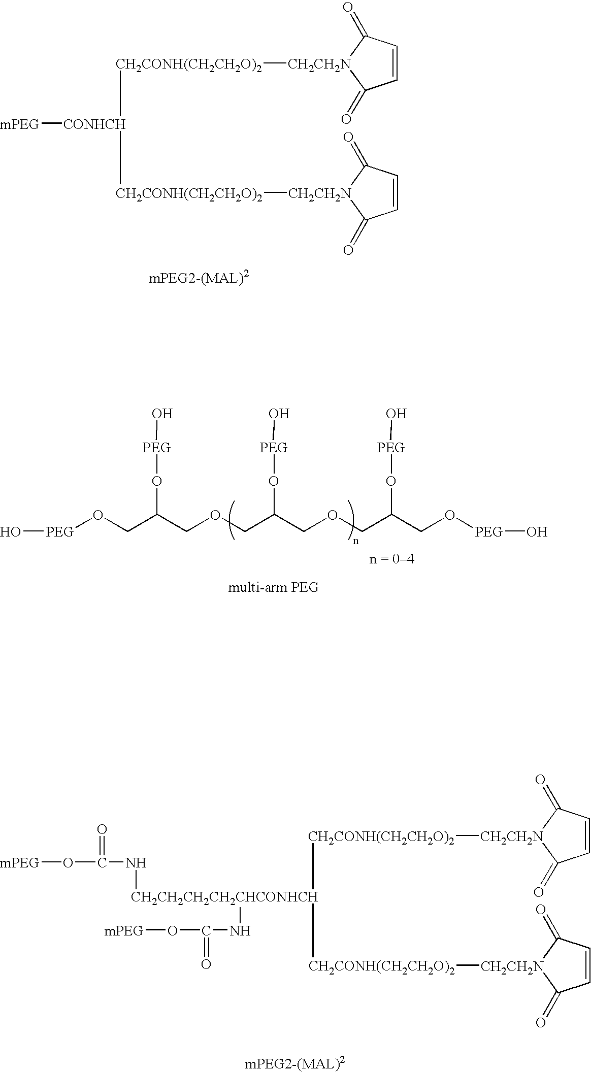
- The above structures show mPEG-MAL formats which may be used to multimerise VH and Vk dAbs. The mPEG MW range from 500 to 60,000 (e.g., from 2,000 to 40,000) in size. dAb dimers are produced using mPEG(MAL)2 or mPEG2(MAL)2; trimers and tetramers using 3 or 4-arm PEGs respectively. The multi-arm PEG shown would require to be modified with MAL to allow the attachment of the dAbs.
- To produce the multimerised dAb formats the experimental methodology was identical to that used to produce the monomer except that the molar ratio of the PEG-MAL:dAb was varied depending upon the format of the PEG-MAL being used. For example, to produce TAR1-5-19 dimer [shown in
FIG. 7 -7] using mPEG2-(MAL)2 the PEG-MAL:dAb molar ratio used was 0.5:1. To purify the PEGylated dimer, again cation exchange chromatography was used as described for the PEGylated monomer with the following changes. The sodium chloride gradient used was 0 to 250 mM salt over 30 column volumes. To produce TAR1-5-19 trimer [shown inFIG. 10 -23] using 3-arm PEG-MAL the PEG-MAL:dAb molar ratio used was 0.33:1. To purify the PEGylated trimer, again cation exchange chromatography was used as described for the PEGylated monomer with the following changes. The sodium chloride gradient used was 0 to 250 mM salt over 30 column volumes. To produce TAR1-5-19 tetramer [shown inFIG. 11 -28] using 4-arm PEG-MAL the PEG-MAL:dAb molar ratio used was 0.25:1. To purify the PEGylated tetramer, again cation exchange chromatography was used as described for the PEGylated monomer with the following changes. The sodium chloride gradient used was 0 to 250 mM salt over 30 column volumes. For the TAR1-5-19 PEG-trimer and tetramer, if required, the samples were further purified using size exclusion chromatography. ASuperose 6 HR column (Amersham Pharmacia) was equilibrated with phosphate buffered saline (PBS) prior to loading on the sample. The column was run at 0.5 ml/min and the protein elution monitored by following the absorption at 280 nm. Fractions containing PEGylated dAb only were identified using SDS-PAGE and then pooled. - 2.1 PCR Construction of TAR2-10-27 cys
- TAR2-10-27 will be used as an example the engineering of a C-terminal cys onto a VH dAb. As with Vk dAbs the site of attachment for the PEG may be placed elsewhere on the surface of the dAb as long as the targeted amino acid is solvent accessible and the resultant PEGylated protein still maintains antigen binding. The following oligonucleotides were used to specifically PCR TAR1-5-19 with a SalI and BamHI sites for cloning and also to introduce a C-terminal cysteine residue. The PCR reaction conditions and cloning was done as outlined in Section 1.1, with the only changes being that the template used was plasmid DNA containing TAR2-10-27.
SalI Ala Ser Thr Glu Val Gln Leu Leu Glu Ser Gly Gly Gly Leu Val (SEQ ID NO: 14) 1 GCG TCG ACG GAG GTC CAG CTG TTG GAG TCT GGG GGA GGC TTG GTA (SEQ ID NO: 12) CGC AGC TGC CTC CAC GTC GAC AAC CTC AGA CCC CCT CCG AAC CAT (SEQ ID NO: 13) Gln Pro Gly Gly Ser Leu Arg Leu Ser Cys Ala Ala Ser Gly Phe 46 CAG CCT GGG GGG TCC CTG CGT CTC TCC TGT GCA GCC TCC GGA TTC GTC GGA CCC CCC AGG GAC GCA GAG AGG ACA CGT CGG AGG CCT AAG Thr Phe Glu Trp Tyr Trp Met Gly Trp Val Arg Gln Ala Pro Gly 91 ACC TTT GAG TGG TAT TGG ATG GGT TGG GTC CGC CAG GCT CCA GGG TGG AAA CTC ACC ATA ACC TAC CCA ACC CAG GCG GTC CGA GGT CCC Lys Gly Leu Glu Trp Val Ser Ala Ile Ser Gly Ser Gly Gly Ser 136 AAG GGT CTA GAG TGG GTC TCA GCT ATC AGT GGT AGT GGT GGT AGC TTC CCA GAT CTC ACC CAG AGT CGA TAG TCA CCA TCA CCA CCA TCG Thr Tyr Tyr Ala Asp Ser Val Lys Gly Arg Phe Thr Ile Ser Arg 181 ACA TAC TAC GCA GAC TCC GTG AAG GGC CGG TTC ACC ATC TCC CGC TGT ATG ATG CGT CTG AGG CAC TTC CCG GCC AAG TGG TAG AGG GCG Asp Asn Ser Lys Asn Thr Leu Tyr Leu Gln Met Asn Ser Leu Arg 226 GAC AAT TCC AAG AAC ACG CTG TAT CTG CAA ATG AAC AGC CTG CGT CTG TTA AGG TTC TTG TGC GAC ATA GAC GTT TAC TTG TCG GAC GCA Ala Glu Asp Ala Ala Val Tyr Tyr Cys Ala Lys Val Lys Leu Gly 271 GCC GAG GAC GCC GCG GTA TAT TAC TGT GCG AAA GTT AAG TTG GGG CGG CTC CTG CGG CGC CAT ATA ATG ACA CGC TTT CAA TTC AAC CCC Gly Gly Pro Asn Phe Gly Tyr Arg Gly Gln Gly Thr Leu Val Thr 316 GGG GGG CCT AAT TTT GGC TAC CGG GGC CAG GGA ACC CTG GTC ACC CCC CCC GGA TTA AAA CCG ATG GCC CCG GTC CCT TGG GAC CAG TGG BamHI Val Ser Cys *** *** Gly Ser 361 GTC TCG TGC TAA TAA GGA TCC CAG AGC ACG ATT ATT CCT AGG Forward primer 5′-AGTGCGTCGACGGAGGTGCAGCTGTTGGAGTCT-3′ (SEQ ID NO: 15) Reverse primer 5′-AAAGGATCCTTATTAGCACGAGACGGTGACCAGGGTTCCCTG-3′ (SEQ ID NO: 16)
FIG. 2 .1: DNA Sequence of TAR2-10-27 cys and the PCR Primers used to Amplify the Engineered dAb.
2.2 Expression and Purification of TAR2-10-27 cys - The expression and purification of DOM1h-10-27 cys was as described in Section 1.2 but with the following modifications. As the dAb is a VH, Streamline Protein A was used to purify the protein from the culture supernatant. Also the protein was eluted from the resin using 100 mM glycine pH 3.0 instead of pH 2.0 buffer.
- 2.3 PEGylation of TAR2-10-27 cys
- The monomer PEGylation of TAR2-10-27cys was as outlined in Section 1.3.1. Again VH dAbs may be multimerised using PEG as outlined in Section 1.3.3. The purification of PEGylated TAR2-10-27 cys was done using cation exchange chromatography at pH 4.0 (as outlined in Section 1.3.2), as the pI of the protein is ˜8.5.
- 3.0 PEGylation of VH and Vk dAbs using NHS and SPA Modified mPEG
- As well as using a site specific method of PEGylation, a more random approach was used to covalently modify the protein. This involved using NHS or SPA modified PEGs, which react with solvent exposed surface lysine residues on the dAb [
FIG. 6 -2]. This has the advantage in that the dAb does not require any protein engineering as there are several surface lysines already present on the surface of VH and Vk dabs. Also any dAb format may be PEGylated without any prior modification, for example TAR1-5-19 disulphide dimer [FIG. 7 -5,6] or ultra affinity dimers [FIG. 8-13 , 8-14, 8-15] (see Section 3.3 and 5.0). As with the MAL-PEGs a variety of linear/forked and branched SPA and NHS-PEG formats are available as shown below.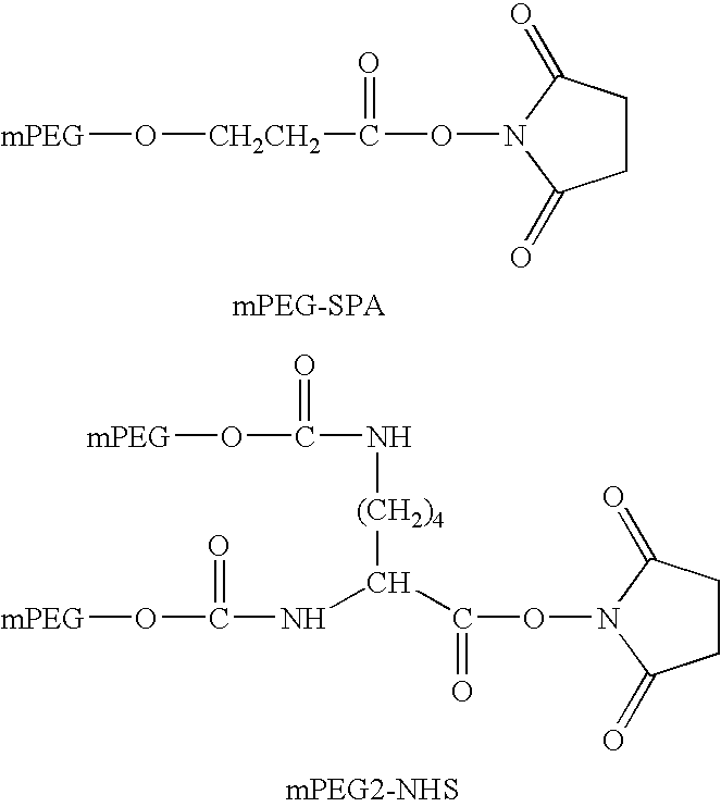
- 3.1 PCR Construction of TAR1-5-19
- TAR1-5-19 will be used as an example of a Vk dAb which has been PEGylated using NHS and SPA-PEGs. TAR1-5-19 was PCR amplified and cloned using the primers below and as outlined in Section 1.1.
SalI Trp Ser Ala Ser Thr Asp Ile Gln Met Thr Gln Ser Pro Ser Ser Leu Ser Ala Ser Val (SEQ ID NO: 19) 1 TGG AGG GCG TCG ACG GAC ATC CAG ATG ACC CAG TCT CCA TCC TCT CTG TCT GCA TCT GTA (SEQ ID NO: 17) ACC TCG CGC AGC TGC CTG TAG GTC TAC TGG GTC AGA GGT AGG AGA GAC AGA CGT AGA CAT (SEQ ID NO: 18) Gly Asp Arg Val Thr Ile Thr Cys Arg Ala Ser Gln Ser Ile Asp Ser Tyr Leu His Trp 61 GGA GAC CGT GTC ACC ATC ACT TGC CGG GCA AGT GAG AGC ATT GAT AGT TAT TTA CAT TGG CCT CTG GCA CAG TGG TAG TGA ACG GCC CGT TCA GTC TCG TAA CTA TCA ATA AAT GTA ACC Tyr Gln Gln Lys Pro Gly Lys Ala Pro Lys Leu Leu Ile Tyr Ser Ala Ser Glu Leu Gln 121 TAC CAG CAG AAA CCA GGG AAA GCC CCT AAG CTC CTG ATC TAT AGT GCA TCC GAG TTG CAA ATG GTC GTC TTT GGT CCC TTT CGG GGA TTC GAG GAC TAG ATA TCA CGT AGG CTC AAC GTT Ser Gly Val Pro Ser Arg Phe Ser Gly Ser Gly Ser Gly Thr Asp Phe Thr Leu Thr Ile 181 AGT GGG GTC CCA TCA CGT TTC AGT GGC AGT GGA TCT GGG ACA GAT TTC ACT CTC ACC ATC TCA CCC CAG GGT AGT GCA AAG TCA CCG TCA CCT AGA CCC TGT CTA AAG TGA GAG TGG TAG Ser Ser Leu Gln Pro Glu Asp Phe Ala Thr Tyr Tyr Cys Gln Gln Val Val Trp Arg Pro 241 AGC AGT CTG CAA CCT GAA GAT TTT GCT ACG TAC TAC TGT CAA CAG GTT GTG TGG CGT CCT TCG TCA GAC GTT GGA CTT CTA AAA CGA TGC ATG ATG ACA GTT GTC CAA CAC ACC GCA GGA BamHI Phe Thr Phe Gly Gln Gly Thr Lys Val Glu Ile Lys Arg *** *** Gly Ser Gly 301 TTT ACG TTC GGC CAA GGG ACC AAG GTG GAA ATC AAA CGG TAA TAA GGA TCC GGC AAA TGC AAG CCG GTT CCC TGG TTC CAC CTT TAG TTT GCC ATT ATT CCT AGG CCG Forward primer 5′-TGGAGCGCGTCGACGGACATCCAGATGACCCAGTCTCCA-3′ (SEQ ID NO: 20) Reverse primer 5′-AAAGGATCCTTATTACCGTTTGATTTCCACCTTGGTCCC-3′ (SEQ ID NO: 21)
3.2 Expression and Purification of TAR1-5-19 - The expression and purification of TAR1-5-19 was as outlined in Section 1.2. The only modification was that once purified the protein was dialysed against PBS or buffer exchanged (PD10) to remove the tris/glycine buffer present.
- 3.3 PEGylation of Monomeric and TAR1-5-19 Disulphide Dimer with NHS or SPA-PEG
- PEGylation reactions were carried out in either PBS buffer or 50 mM sodium phosphate pH 7.0. 400 μM of TAR1-5-19 monomer was mixed with a 5-10 molar excess of activated PEG (either NHS or SPA dissolved in 100% methanol). The reaction was allowed to proceed at room temperature for 24 hours and then stopped by the addition of 1M glycine pH 3.0 to a final concentration of 20 mM. It was found that the TAR1-5-19 disulphide dimer produced during expression (Section 1.2) could also be PEGylated using the above methodology. This allowed the disulphide dimer format to be PEGylated without the need for reduction with DTT followed by modification with the PEG2-(MAL)2. The only modification to the protocol was the addition of 10% glycerol to the dAb dimer to prevent precipitation during any concentration steps.
- 3.4 Purification of the PEGylated Vk dAbs
- The monomer and disulphide dimer PEGylated dAb was purified using cation exchange chromatography as outlined in Section 1.3.3.
- HEL4 will be used as an example of a VH dAb which has been PEGylated using NHS and SPA-PEGs. HEL4 was PCR amplified and cloned using the primers below and under the conditions as outlined in Section 1.1, with the only modification being that the template DNA was a plasmid vector containing the HEL4 DNA sequence.
SalI Ala Ser Thr Glu Val Gln Leu Leu Glu Ser Gly Gly Gly Leu Val Gln Pro Gly Gly Ser (SEQ ID NO: 24) 1 GCG TCG ACG GAG GTG CAG CTG TTG GAG TCT GGG GGA GGC TTG GTA CAG CCT GGG GGG TCC (SEQ ID NO: 22) CGC AGC TGC CTC CAC GTC GAC AAC CTC AGA CCC CCT CCG AAC CAT GTC GGA CCC CCC AGG (SEQ ID NO: 23) Leu Arg Leu Ser Cys Ala Ala Ser Gly Phe Arg Ile Ser Asp Glu Asp Met Gly Trp Val 61 CTG CGT CTC TCC TGT GCA GCC TCC GGA TTT AGG ATT AGC GAT GAG GAT ATG GGC TGG GTC GAC GCA GAG AGG ACA CGT CGG AGG CCT AAA TCC TAA TCG CTA CTC CTA TAC CCG ACC GAG Arg Gln Ala Pro Gly Lys Gly Leu Glu Trp Val Ser Ser Ile Tyr Gly Pro Ser Gly Ser 121 CGC CAG GCT CCA GGG AAG GGT CTA GAG TGG GTA TCA AGC ATT TAT GGC CCT AGC GGT AGC GCG GTC CGA GGT CCC TTC CCA GAT CTC ACC CAT AGT TCG TAA ATA CCG GGA TCG CCA TCG Thr Tyr Tyr Ala Asp Ser Val Lys Gly Arg Phe Thr Ile Ser Arg Asp Asn Ser Lys Asn 181 ACA TAC TAC GCA GAC TCC GTG AAG GGC CGG TTC ACC ATC TCC CGT GAC AAT TCC AAG AAC TGT ATG ATG CGT CTG AGG CAC TTC CCG GCC AAG TGG TAG AGG GCA CTG TTA AGG TTC TTG Thr Leu Tyr Leu Gln Met Asn Ser Leu Arg Ala Glu Asp Thr Ala Val Tyr Tyr Cys Ala 241 ACG CTG TAT CTG CAA ATG AAC AGC CTG CGT GCC GAG GAC ACC GCG GTA TAT TAT TGC GCG TGC GAC ATA GAC GTT TAC TTG TCG GAC GCA CGG CTC CTG TGG CGC CAT ATA ATA ACG CGC Ser Ala Leu Glu Pro Leu Ser Glu Pro Leu Gly Phe Trp Gly Gln Gly Thr Leu Val Thr 301 AGT GCT TTG GAG CCG CTT TCG GAG CCC CTG GGC TTT TGG GGT CAG GGA ACC CTG GTC ACC TCA CGA AAC CTC GGC GAA AGC CTC GGG GAC CCG AAA ACC CCA GTC CCT TGG GAC CAG TGG BamHI Val Ser Ser *** *** Gly Ser 361 GTC TCG AGC TAA TAA GGA TCC CAG AGC TCG ATT ATT CCT AGG Forward primer 5′-AGTGCGTCGACGGAGGTGCAGCTGTTGGAGTCT-3′ (SEQ ID NO: 25) Reverse primer 5′-AAAGGATCCTTATTAGCTCGAGACGGTGACCAGGGTTCCCTG-3′ (SEQ ID NO: 26)
4.2 Expression and Purification of HEL4 - The VH dAb was expressed and purified as outlined in Section 2.2.
- 4.3 NHS and SPA PEGylation of HEL4
- The methodology for modification of the surface lysine residues using NHS and SPA activated PEGs was as described in Section 3.3. The only modification to the protocol was that the reaction was terminated by the addition of 1M Tris buffer pH 8.0 to a final concentration of 20 mM.
- 4.4 Purification of NHS or SPA PEGylated HEL4
- Purification of the PEGylated protein was carried out using anion exchange chromatography as the pI of the HEL4 is ˜4. The column used was a 1 ml Resource Q column (Amersham Pharmacia), which had been equilibrated with 50 mM Tris pH 8.0. The pH of the sample was shifted to 8 prior to loading onto the column. A linear gradient from 0 to 500 mM sodium chloride in 50 mM Tris pH 8.0 was used to separate the PEGylated protein from unmodified dAb.
- 4.5 Affinity Binding of PEGylated HEL4
- A HEL4 affinity based resin (based on the antigen, hen egg white lysozyme) was produced to test the functionality of the PEGylated dAb. Lysozyme was coupled to NETS activated Sepharose 4B resin (Amersham Pharmacia) following the manufacturer's instructions. The PEGylated samples (5 μg) were then mixed with the affinity resin (100 μl) and allowed to bind in PBS at room temperature for 30 mins. The resin was then extensively washed with PBS (3×1 ml) to remove unbound protein. The affinity resin was then run on a SDS-PAGE to determine if the PEGylated HEL4 had bound to the matrix (see Section 8.3).
- VH and Vk dAbs may be multimerised using a (Gly4Ser)n linker (n=0-7) to form a single polypeptide chain (e.g. two dAbs to give an ultra affinity dimer;
FIG. 8-12 ). Therefore it is possible to form homodimeric (VH1-VH1 and VL1-VL1) or heterodimeric (VH1-VH2 and VL1-VL2) pairings with dual specificities. As well as forming dimers, additional dAbs may be added to make larger proteins such as trimers [FIG. 10 -27] or tetramers [FIG. 11 -32]. Again since a combination of VH and Vk dAbs may be used to make these larger formats, it is possible to make dual specific molecules. For example a dAb trimer with the ability to have an extended serum half-life and engage TNFα; one dAb with the ability to bind to serum albumin linked to two TAR1-5-19 dabs that bind TNF as a dimer. The ultra affinity dimers have been PEGylated using NHS and SPA-PEGs [FIG. 8-13 , 8-14, and 8-15] as well as being specifically engineered to accept MAL-PEGs at the C-terminus of the protein (example 6a and 6b) [FIG. 8 -16]. In this example the ultra affinity dimer TAR1-5-19-Dimer 4 was modified with either 20K-SPA or 40K-NHS. The DNA sequence of the ultra affinity dimer TAR1-5-19Dimer 4 is shown below.Asp Ile Gln Met Thr Gln Ser Pro Ser Ser Leu Ser Ala Ser Val Gly Asp Arg Val Thr (SEQ ID NO: 29) 1 GAC ATC CAG ATG ACC CAG TCT CCA TCC TCT CTG TCT GCA TCT GTA GGA GAC CGT GTC ACC (SEQ ID NO: 27) CTG TAG GTC TAC TGG GTC AGA GGT AGG AGA GAC AGA CGT AGA CAT CCT CTG GCA CAG TCC (SEQ ID NO: 28) KpnI Ile Thr Cys Arg Ala Ser Gln Ser Ile Asp Ser Tyr Leu His Trp Tyr Gln Gln Lys Pro 61 ATC ACT TGC CGG GCA AGT CAG AGC ATT GAT AGT TAT TTA CAT TGG TAC CAG CAG AAA CCA TAG TGA ACG GCC CGT TCA GTC TCG TAA CTA TCA ATA AAT GTA ACC ATG GTC GTC TTT GGT Gly Lys Ala Pro Lys Leu Leu Ile Tyr Ser Ala Ser Glu Leu Gln Ser Gly Val Pro Ser 121 GGG AAA GCC CCT AAG CTC CTG ATC TAT AGT GCA TCC GAG TTG CAA AGT GGG GTC CCA TCA CCC TTT CGG GGA TTC GAG GAC TAG ATA TCA CGT AGG CTC AAC GTT TCA CCC CAG GGT AGT Arg Phe Ser Gly Ser Gly Ser Gly Thr Asp Phe Thr Leu Thr Ile Ser Ser Leu Gln Pro 181 CGT TTC AGT GGC AGT GGA TCT GGG ACA GAT TTC ACT CTC ACC ATC AGC AGT CTG CAA CTT GCA AAG TCA CCG TCA CCT AGA CCC TGT CTA AAG TGA GAG TGG TAG TGG TCA GAC GTT GGA HindII Glu Asp Phe Ala Thr Tyr Tyr Cys Gln Gln Val Val Trp Arg Pro Phe Thr Phe Gly Gln 241 GAA GAT TTT GCT ACG TAC TAC TGT CAA CAG GTT GTG TGG CGT CCT TTT ACG TTC GGC CAA CTT CTA AAA CGA TGC ATG ATG ACA GTT GTC CAA CAC ACC GCA GGA AAA TGC AAG CCG GTT XhoI Gly Thr Lys Val Gln Ile Lys Arg Ser Ser Gly Gly Gly Gly Ser Gly Gly Gly Gly Ser 301 GGG ACC AAG GTG GAA ATC AAA CGC TCG AGC GGT GGA GGC GGT TCA GGC GGA GGT GGC AGC CCC TGG TTC CAC CTT TAG TTT GCG AGC TCG CCA CCT CCG CCA AGT CCG CCT CCA CCG TCG SalI HindII Gly Gly Gly Gly Ser Gly Gly Gly Gly Ser Gly Gly Gly Gly Ser Thr Asp Ile Gln Met 361 GGC GGT GGC GGG TCA GGT GGT GGC GGA AGC GGC GGT GGC GGG TCG ACG GAC ATC CAG ATG CCG CCA CCG CCC AGT CCA CCA CCG CCT TCG CCG CCA CCG CCC AGC TGC CTG TAG GTC TAC Thr Gln Ser Pro Ser Ser Leu Ser Ala Ser Val Gly Asp Arg Val Thr Ile Thr Cys Arg 421 ACC CAG TCT CCA TCC TCC CTG TCT GCA TCT GTA GGA GAC CGT GTC ACC ATC ACT TGC CGG TGG GTC AGA GGT AGG AGG GAC AGA CGT AGA CAT CCT CTG GCA CAG TGG TAG TGA ACG GCC KpnI Ala Ser Gln Ser Val Lys Glu Phe Leu Trp Trp Tyr Gln Gln Lys Pro Gly Lys Ala Pro 481 GCA AGT CAG AGC GTT AAG GAG TTT TTA TGG TGG TAC CAG CAG AAA CCA GGG AAA GCC CCT CGT TCA GTC TCG CAA TTC CTC AAA AAT ACC ACC ATG GTC GTC TTT GGT CCC TTT CGG GGA Lys Leu Leu Ile Tyr Met Ala Ser Asn Leu Gln Ser Gly Val Pro Ser Arg Phe Ser Gly 541 AAG CTC CTG ATC TAT ATG GCA TCC AAT TTG CAA AGT GGG GTC CCA TCA CGT TTC AGT GGC TTC GAG GAC TAG ATA TAC CGT AGG TTA AAC GTT TCA CCC CAG GGT AGT GCA AAG TCA CCG Ser Gly Ser Gly Thr Asp Phe Thr Leu Thr Ile Ser Ser Leu Gln Pro Glu Asp Phe Ala 601 AGT GGA TCT GGG ACA GAT TTC ACT CTC ACC ATC AGC AGT CTG CAA CCT GAA GAT TTT GCT TCA CCT AGA CCC TGT CTA AAG TGA GAG TGG TAG TCG TCA GAC GTT GGA CTT CTA AAA CGA HindII Thr Tyr Tyr Cys Gln Gln Lys Phe Lys Leu Pro Arg Thr Phe Gly Gln Gly Thr Lys Val 661 ACG TAC TAC TGT CAA CAG AAG TTT AAG CTG CCT CGT ACG TTC GGC CAA GGG ACC AAG GTG TGC ATG ATG ACA GTT GTC TTC AAA TTC GAC GGA GCA TGC AAG CCG GTT CCC TGG TTC CAC NotI Glu Ile Lys Arg Ala Ala Ala His His His His His His Gly Ala Ala Glu Gln Lys Leu 721 GAA ATC AAA CGG GCG GCC GCA CAT CAT CAT CAC CAT CAC GGG GCC GCA GAA CAA AAA CTC CTT TAG TTT GCC CGC CGG CGT GTA GTA GTA GTG GTA GTG CCC CGG CGT CTT GTT TTT GAG Ile Ser Glu Glu Asp Leu Asn Gly Ala Ala 781 ATC TCA GAA GAG GAT CTG AAT GGG GCC GCA TAG AGT CTT CTC CTA GAC TTA CCC CGG CGT
5.1 Expression and Purification of Ultra Affinity Dimer TAR1-5-19Dimer 4 - The expression of the dimer was as described in Section 1.2 with the only modification in the protocol being that the construct was transformed into TOP10 F′ cells (Invitrogen) and that carbenicillin was used at a concentration of 100 μg/ml.
- 5.2 20K SPA and 40K NHS PEGylation and Purification of the Ultra Affinity Dimer TAR1-5-19
Dimer 4 - The dimer was PEGylated as outlined in Section 3.3 with either 20K SPA PEG or 40K NHS PEG. Both the 20K SPA and the 40K NHS PEGylated dimers were purified by cation exchange chromatography as described in Section 1.3.3 example 1a.
- As with dAbs engineered with C-terminal cys, ultra affinity dAb dimers can also be modified in a similar fashion [
FIG. 8 -16]. Again the cys-dimer can be used as the basic building block to create larger multi-dAb PEGylated formats [FIG. 8-17 , and 11-29, 11-31]. TAR1-5-19 homodimer with a (Gly4Ser)s linker cys (seeFIG. 8-16 ) will be used as an example. As with the TAR1-5-19 cys monomer, both a monomeric (i.e. dimer;FIG. 8-16 ) and dimeric (i.e. tetramer;FIG. 8-17 ) will be produced in solution during expression. The DNA sequence of the ultra affinity dimer TAR1-5-19 homodimer cys is shown below.Asp Ile Gln Met Thr Gln Ser Pro Ser Ser Leu Ser Ala Ser Val Gly Asp Arg Val Thr (SEQ ID NO: 32) 1 GAC ATC CAG ATG ACC CAG TCT CCA TCC TCT CTG TCT GCA TCT GTA GGA GAC CGT GTC ACC (SEQ ID NO: 30) CTG TAG GTC TAC TGG GTC AGA GGT AGG AGA GAC AGA CGT AGA CAT CCT CTG GCA CAG TGG (SEQ ID NO: 31) KpnI Ile Thr Cys Arg Ala Ser Gln Ser Ile Asp Ser Tyr Leu His Trp Tyr Gln Gln Lys Pro 61 ATC ACT TGC CGG GCA AGT CAG AGC ATT GAT AGT TAT TTA CAT TGG TAC CAG CAG AAA CCA TAG TGA ACG GCC CGT TCA GTC TCG TAA CTA TCA ATA AAT GTA ACC ATG GTC GTC TTT GGT Gly Lys Ala Pro Lys Leu Leu Ile Tyr Ser Ala Ser Glu Leu Gln Ser Gly Val Pro Ser 121 GGG AAA GCC CCT AAG CTC CTG ATC TAT AGT GCA TCC GAG TTG CAA AGT GGG GTC CCA TCA CCC TTT CGG GGA TTC GAG GAC TAG ATA TCA CGT AGG CTC AAC GTT TCA CCC CAG GGT AGT Arg Phe Ser Gly Ser Gly Ser Gly Thr Asp Phe Thr Leu Thr Ile Ser Ser Leu Gln Pro 181 CGT TTC AGT GGC AGT GGA TCT GGG ACA GAT TTC ACT CTC ACC ATC AGC AGT CTG CAA CCT GCA AAG TCA CCG TCA CCT AGA CCC TGT CTA AAG TGA GAG TGG TAG TCG TCA GAC GTT GGA HindII Glu Asp Phe Ala Thr Tyr Tyr Cys Gln Gln Val Val Trp Arg Pro Phe Thr Phe Gly Gln 241 GAA GAT TTT GCT ACG TAC TAC TGT CAA CAG GTT GTG TGG CGT CCT TTT ACG TTC GGC CAA CTT CTA AAA CGA TGC ATG ATG ACA GTT GTC CAA CAC ACC GCA GGA AAA TGC AAG CCG GTT XhoI Gly Thr Lys Val Glu Ile Lys Arg Ser Ser Gly Gly Gly Gly Ser Gly Gly Gly Gly Ser 301 GGG ACC AAG GTG GAA ATC AAA CGC TCG AGC GGT GGA GGC GGT TCA GGC GGA GGT GGC AGC CCC TGG TTC CAC CTT TAG TTT GCG AGC TCG CCA CCT CCG CCA AGT CCG CCT CCA CCG TCG SalI HindII Gly Gly Gly Gly Ser Gly Gly Gly Gly Ser Gly Gly Gly Gly Ser Thr Asp Ile Gln Met 361 GGC GGT GGC GGG TCA GGT GGT GGC GGA AGC GGC GGT GGC GGG TCG ACG GAC ATC CAG ATG CCG CCA CCG CCC AGT CCA CCA CCG CCT TCG CCG CCA CCG CCC AGC TGC CTG TAG GTC TAC Thr Gln Ser Pro Ser Ser Leu Ser Ala Ser Val Gly Asp Arg Val Thr Ile Thr Cys Arg 421 ACC CAG TCT CCA TCC TCT CTG TCT GCA TCT GTA GGA GAC CGT GTC ACC ATC ACT TGC CGG TGG GTC AGA GGT AGG AGA GAC AGA CGT AGA CAT CCT CTG GCA CAG TGG TAG TGA ACG GCC KpnI Ala Ser Gln Ser Ile Asp Ser Tyr Leu His Trp Tyr Gln Gln Lys Pro Gly Lys Ala Pro 481 GCA AGT CAG AGC ATT GAT AGT TAT TTA CAT TGG TAC CAG CAG AAA CCA GGG AAA GCC CCT CGT TCA GTC TCG TAA CTA TCA ATA AAT GTA ACC ATG GTC GTC TTT GGT CCC TTT CGG GGA Lys Leu Leu Ile Tyr Ser Ala Ser Glu Leu Gln Ser Gly Val Pro Ser Arg Phe Ser Gly 541 AAG CTC CTG ATC TAT AGT GCA TCC GAG TTG CAA AGT GGG GTC CCA TCA CGT TTC AGT GGC TTC GAG GAC TAG ATA TCA CGT AGG CTC AAC GTT TCA CCC CAG GGT AGT GCA AAG TCA CCG Ser Gly Ser Gly Thr Asp Phe Thr Leu Thr Ile Ser Ser Leu Gln Pro Glu Asp Phe Ala 601 AGT GGA TCT GGG ACA GAT TTC ACT CTC ACC ATC AGC AGT CTG CAA CCT GAA GAT TTT GCT TCA CCT AGA CCC TGT CTA AAG TGA GAG TGG TAG TCG TCA GAC GTT GGA CTT CTA AAA CGA HindII Thr Tyr Tyr Cys Gln Gln Val Val Trp Arg Pro Phe Thr Phe Gly Gln Gly Thr Lys Val 661 ACG TAC TAC TGT CAA CAG GTT GTG TGG CGT CCT TTT ACG TTC GGC CAA GGG ACC AAG GTG TGC ATG ATG ACA GTT GTC CAA CAC ACC GCA GGA AAA TGC AAG CCG GTT CCC TGG TTC CAC Glu Ile Lys Arg Cys 721 GAA ATC AAA CGC TGC CTT TAG TTT GCG ACG
6.1 Expression and Purification of Ultra Affinity Dimer TAR1-5-19 Homodimer cys - The expression of the dimer was as described in Section 1.2 with the only modification in the protocol being that the construct was transformed into TOP10 F′ cells (Invitrogen) and that carbenicillin was used at a concentration of 100 μg/ml.
- 6.2 Dimerisation and Purification of TAR1-5-19 Homodimer cys with 40K PEG2-(MAL)2
- TAR1-5-19 homodimer cys was PEGylated with 40K PEG2-(MAL)2 to form the dimer (i.e. 2× linker dimers with a total of 4 dAbs;
FIG. 11 -29]). The reaction and purification conditions were as described in Section 1.3.1 and modified as outlined in Example 1a. - TAR1-5-19 homodimer cys was PEGylated with 4-
arm 20K PEG MAL to form the dimer tetramer (i.e. 4× linker dimers with a total of 8 dAbs;FIG. 11-31 ). The reaction and purification conditions were as described in Section 1.3.1 and modified as outlined in Example 1c. - TAR1-5 will be used as an example of a Vk dAb which has a relatively low binding affinity as a monomer for TNF, but the dAb can be formatted to increase its affinity via multimerisation. Again a C-terminal cysteine was used to multimerised the dAb via 4-
arm 20K PEG MAL. - 7.1 PCR Construction of TAR1-5 cys
- The following oligonucleotides were used to specifically PCR TAR1-5 with SalI and BamHI sites for cloning and also to introduce a C-terminal cysteine residue. The DNA sequence of TAR1-5 cys and the PCR primers used to amplify the engineered dAb are shown below.
SalI Trp Ser Ala Ser Thr Asp Ile Gln Met Thr Gln Ser Pro Ser Ser Leu Ser Ala Ser Val (SEQ ID NO: 35) 1 TGG AGC GCG TCG ACG GAC ATC CAG ATG ACC CAG TCT CCA TCC TCC CTG TCT GCA TCT GTA (SEQ ID NO: 33) ACC TCG CGC AGC TGC CTG TAG GTC TAC TGG GTC AGA GGT AGG AGG GAC AGA CGT AGA CAT (SEQ ID NO: 34) KpnI Gly Asp Arg Val Thr Ile Thr Cys Arg Ala Ser Gln Ser Ile Phe Met Asn Leu Leu Trp 61 GGA GAC CGT GTC ACC ATC ACT TGC CGG GCA AGT CAG AGC ATT TTT ATG AAT TTA TTG TGG CCT CTG GCA CAG TGG TAG TGA ACG GCC CGT TCA GTC TCG TAA AAA TAC TTA AAT AAC ACC KpnI Tyr Gln Gln Lys Pro Gly Lys Ala Pro Lys Leu Leu Ile Tyr Asn Ala Ser Val Leu Gln 121 TAC CAG CAG AAA CCA GGG AAA GCC CCT AAG CTC CTG ATC TAT AAT GCA TCC GTG TTG CAA ATG GTC GTC TTT GGT CCC TTT CGG GGA TTC GAG GAC TAG ATA TTA CGT AGG CAC AAC GTT Ser Gly Val Pro Ser Arg Phe Ser Gly Ser Gly Ser Gly Thr Asp Phe Thr Leu Thr Ile 181 AGT GGG GTC CCA TCA CGT TTC AGT GGC AGT GGA TCT GGG ACA GAT TTC ACT CTC ACC ATC TCA CCC CAG GGT AGT GCA AAG TCA CCG TCA CCT AGA CCC TGT CTA AAG TGA GAG TGG TAG HindII Ser Ser Leu Gln Pro Glu Asp Phe Ala Thr Tyr Tyr Cys Gln Gln Val Val Trp Arg Pro 241 AGC AGT CTG CAA CCT GAA GAT TTT GCT ACG TAC TAC TGT CAA CAG GTT GTG TGG CGT CCT TCG TCA GAC GTT GGA CTT CTA AAA CGA TGC ATG ATG ACA GTT GTC CAA CAC ACC GCA GGA BamHI Phe Thr Phe Gly Gln Gly Thr Lys Val Glu Ile Lys Arg Cys *** *** Gly Ser Phe 301 TTT ACG TTC GGC CAA GGG ACC AAG GTG GAA ATC AAA CGG TGC TAA TAA GGA TCC TTT AAA TGC AAG CCG GTT CCC TGG TTC CAC CTT TAG TTT GCC ACG ATT ATT CCT AGG AAA Forward primer 5′-TGGAGCGCGTCGACGGACATCCAGATGACCCAGTCTCCA-3′ (SEQ ID NO: 36) Reverse primer 5′-TTAGCAGCCGGATCCTTATTAGCACCGTTTGATTTCCAC-3′ (SEQ ID NO: 37) - The reaction conditions used were as described in Section 1.1.
- 7.2 Expression and Purification of TAR1-5 cys
- The expression and purification of the dAb was as outlined in Section 1.2. Again, a 50:50 mix of monomer and TAR1-5 disulphide dimer were formed.
- 7.3 Multimerisation via PEGylation and Purification of 4-
arm 20K PEG-MAL TAR1-5 - The PEGylation and purification conditions were as described in Section 1.3.3 example 1c. The results of cell cytotoxicity assays are shown in Table 1.
- 8.1 Assay Data for PEGylated TAR1-5-19 Monomer and TAR1-5-19 cys
- The affinity of the PEGylated dAb for human TNFα was determined using the TNF receptor binding (RBA) and cell cytotoxicity assay (summarized in Table 1).
FIG. 1 shows the results from the RBA assay of a few selected PEGylated Vk dAbs, from the graphs theIC 50 was determined and summarized in Table 1. It can be seen that there is only a slight difference between the PEGylated and unmodified forms of the monomer and dimer in the RBA. Even with the larger 40K PEG-MAL theIC 50 is unaffected. A slight decrease is observed with the 40K-NHS, but this may be due to the PEGylation site being closer to the CDRs, whereas with the 40K-MAL is located at the C-terminus of the dAb. This shows that PEGylation has an insignificant effect on antigen binding as long as it is placed away from the CDRs. It can also be seen that the 3 and 4-arm PEGylated formats using the monomeric dAb have an improved affinity for TNF in the RBA.FIG. 2 shows the same samples in the cell cytotoxicity assay. Again there is little difference between the PEGylated and unmodified samples, showing the potency of the dAb is maintained. The most significant difference is with the 3 and 4-arm PEG samples, which shows a reduction in theND 50 to 100 and 30 pM respectively. With TAR1-5 it can be seen that the low affinity binding monomer has anND 50 of 10 pM in the cell assay. Upon dimerisation, the affinity is reduced to ˜3 pM and when tetramerised using the 4-arm PEG, down to anND 50 of 200 nM. Therefore even a low affinity dAb may be multimerised using multi-arm PEG to create formats with greater avidity for their antigen. - 8.2 Assay Data for PEGylated Ultra Affinity Dimers TAR1-5-19
Dimer 4 and TAR1-5-19 Homodimer cys - The SPA and NHS PEGylation of TAR1-5-19
Dimer 4 again showed that random surface modification only slightly affected the potency of the dimer (see Table 1). With the TAR1-5-19 homodimer cys it was found that dimerising the dimer to form the tetramer maintained theND 50 of 3 nM. A significant drop in theND 50 was seen when the dimer was further multimerised using the 4-arm MAL-PEG to produce the octamer, shifting theND 50 to ˜100 pM. - This reflects the fact that the RBA assay's sensitivity is relatively high and that formats with high affinity will all appear to be ˜100 pM.
TABLE 1 Summary of the results from the receptor and cell cytotoxicity assays of dAb formats based upon TAR1-5-19 Receptor Cell cytotoxicity assay assay dAb format IC 50 (nM) ND 50 (nM) Unmodified TAR1-5-19 monomer 30 70 20K SPA TAR1-5-19 monomer 45 70 40K NHS TAR1-5-19 monomer 45 90 20K MAL TAR1-5-19 monomer 30 70 40K MAL TAR1-5-19 monomer 30 80 Unmodified TAR1-5-19 disulphide 0.8 3 dimer 1 × 20K SPA TAR1-5-19 disulphide 1 3 dimer 1 × 40K NHS TAR1-5-19 disulphide 3 10 dimer 40K PEG2-(MAL2) TAR1-5-19 1 3 dimer TAR1-5-19 D4 ultra affinity — 10 dimer 20K SPA PEG TAR1-5-19 D4 ultra — 30 affinity dimer 40K NHS PEG TAR1-5-19 D4 ultra — 25 affinity dimers TAR1-5-19 homodimer cys ultra — 3 affinity dimer TAR1-5-19 homodimer cys ultra — 3 affinity disulphide dimer 40K PEG2-(MAL)2 TAR1-5-19 — 3 homodimer cys ultra affinity 4-arm PEG-MAL TAR1-5-19 — 0.1 homodimer cys ultra affinity dimer tetramer (5K per arm) 3-arm PEG-MAL TAR1-5-19 trimer 0.2 0.1 (5K per arm) 4-arm PEG-MAL TAR1-5-19 tetramer 0.1 0.03 (5K per arm) 4-arm PEG-MAL TAR1-5-19 tetramer — 0.02 (10K per arm) TAR1-5 monomer — 10,000 TAR1-5 disulphide dimer — 3000 4-arm PEG-MAL TAR1-5 tetramer — 200 (5K per arm)
8.3 Binding of PEGylated HEL4 to Lysozyme Affinity Matrix - Various PEGylated HEL4 formats were tested to see if they retained the ability to bind antigen once they had been modified.
FIG. 3 shows the results of the binding assay. Specifically, the SDS PAGE gels showing affinity binding to lysozyme, and the lanes are as follows: 1, 4, 7, 10, 13, 16 and 19 show protein remaining in the supernatant after batch binding for 30 mins;lanes lanes - From the gels it can be clearly seen that the PEGylated protein is efficiently removed from the supernatant and remains bound to the resin even after several washes with PBS. Thus showing that the PEGylated dAb maintained specificity for its antigen whether is was specifically PEGylated with THIOL or MAL-PEG, or via surface lysine residues using SPA or NHS-PEGs.
- 8.4 PEGylation of TAR2-1O-27
- The binding potency of TAR2-10-27 can be determined using a TNF receptor binding assay. The unmodified monomer had an
IC 50 of ˜3 nM in the assay, and as can be seen fromFIG. 4 . The 40K PEGylated monomer has anIC 50 of ˜20 nM. - PEGylation of proteins has been used to increase their in vivo serum half-life. The renal filtration cut-off size is approximately 70 kDa, which means for a protein the size of an unmodified dAb (VH ˜13-14 kDa and Vk ˜12 kDa) the serum half-life will be relatively low (t1/2 beta of ˜10-30 mins). Work carried out on the PEGylation of dAbs has shown that the serum half-life of a dAb (either VH or Vk) can be modulated by the size and the branched nature of the PEG used. This has important applications for example in drug therapies where a prolonged half-life of tens of hours is desired (e.g. >30 hrs); whereas a significantly shorter residency time of a few hours (3-6 hrs) is required if the dAb were to be labeled and used as an in vivo imaging reagent for diagnostic purposes.
- We have shown that two parameters play an important role in determining the serum half-lives of PEGylated dabs. The first is the nature and size of the PEG attachment, i.e. if the polymer used is simply a linear chain or branched/forked in nature. The second is the location of the dAb on the final format and how many “free” unmodified PEG arms the molecule has. The resultant hydrodynamic size of the PEGylated format, as estimated by size exclusion chromatography, reflects the serum half-life of the molecule. Thus the larger the hydrodynamic size of the PEGylated molecule the greater the serum half-life, as shown in Table 2.
- The gel filtration matrices used to determine the hydrodynamic sizes of the various PEGylated proteins was based upon highly cross-linked agarose. The fractionation range of the two columns for globular proteins are; Superose 12 HR 1000-3×10⁵ Mr and
Superose 6 HR 5000-5×10⁶ Mr. The globular protein size exclusion limits are ˜2×10⁶ for theSuperose 12 HR and ˜4×10⁷ Mr for the Superose 6HR.TABLE 2 Estimated Hydrodynamic In vivo Size of PEG size size serum half-life Format Linear/branched (kDa)a (kDa)b (t1/2β hrs) Vk dAbs TAR1-5-19 monomer na 12 14 0.5 TAR1-5-19 disulphide na 24 30 2.5 dimer TAR1-5-19 disulphide 20K linear 60 280c 16 dimer 20K PEG-SPATAR1-5-19 Pc fusion na 80 na >24 TAR1-5-19 monomer 40K branched 95 550c >36 40K PEG-MAL (2 × 20K) TAR1-5-19 dimer 40K 40K branched 110 580c ˜51.6 PEG (2 × 20K) TAR1-5-19 trimer PEG- 15K 3-arm branched 52 30c na MAL (3 × 5K) TAR1-5-19 tetramer 20K 4-arm branched 90 150c ˜12.7 PEG-MAL (4 × 5K) VH dAbs HEL4 na 14 16 na HEL4 5K PEG-MAL 5K linear 24 70 na HEL4 20K PEG-MAL 20K linear 55 275 na HEL4 30K PEG-THIOL 30K linear 84 310 na HEL4 40K PEG-NHS 40K branched (2 × 20K) 95 544 na
Notes:
aestimated size determined by SDS-PAGE.
bhydrodynamic sizes as determined by Superose 12HR (Amersham Pharmacia) size exclusion chromatography.
cestimations on sizes.
- Thus, it can be seen from Table 2 that even though the 4-arm PEG format of TAR1-5-19 consists of 20K of PEG and its molecular mass is greater than that of the 20K SPA TAR1-5-19 disulphide dimer (95 kDa compared to 60 kDa), they have similar half-lives considering the renal cut-off is 70 kDa. This may be due to the fact that in the 4-arm format the dAbs are linked to the end of each PEG molecule and so overall the molecule is more compact (smaller hydrodynamic size); i.e. in this format there is a very high density of PEG at the core of the molecule compared to just having a single linear 20K chain. The advantage in using the 4-arm format is the significant increase in affinity to TNF compared to the dimeric molecules (30 pM compared to 3 nM respectively). Thus in some applications where a high affinity binder is required but with a short serum half-life, the molecule could be tailored to have both desired characteristics by varying the number of dAbs present and the size of PEG used.
- Another key feature of PEGylated dAbs is their increased stability to the action of proteases. dAbs intrinsically are relatively stable to the action of proteases, depending upon the assay conditions. For example TAR1-5-19 is not degraded by trypsin even in the presence of 5 M urea. This is because the dAb does not unfold in the presence of the denaturant, whereas it is totally degraded by pepsin at
pH 2 as the protein is unfolded under the acidic conditions. PEGylation has the advantage in that the polymer chain most likely covers the surface of the dAb, thus preventing the protease from gaining access to the polypeptide backbone and cleaving it. Even when the protein is partially denatured at low pH, the presence of the PEG on the dAb significantly increased its resistance to the action of pepsin (FIG. 5 ). -
FIG. 5 shows the results of pepsin degradation of TAR1-5-19 and other PEGylated variants. Reactions were carried out at pH 2.0, at 37° C. for up to 30 minutes in the presence of 250 μg pepsin. The lane designations are as follows: 1: molecular weight ladder; 2: monomer (0 min); 3: monomer (15 min); 4: monomer (30 min); 5: 40K MAL2 dimer (0 min); 6: 40K MAL2 dimer (15 min); 7: 40K MAL2 dimer (30 min); 8: 20K MAL monomer (0 min); 9: 20K MAL monomer (15 min); 10: 20K MAL monomer. The gel shows that the unmodified dAb is degraded very quickly at low pH.FIG. 5 also shows that the type of PEG present on the surface of the dAb also can have a significant effect on its resistance to the action of the protease. The linear 20K PEG does protect the dAb to some degree, but even after 30 mins some degradation is seen (bands at 3 kDa, estimated ˜15-20% degradation from the gel). Whereas with the 40K PEG MAL2 dimer, very little degradation is seen even after 30 mins (at most <5% degradation from the gel). This is most likely due to the 2×20K format of PEG, which offers greater protection from the action of pepsin. This would suggest that the PEGylated dAb would also be resistant to the action of a wide range of proteases found in the digestive tract of humans and also may maintain functionality through the low pH environment of the stomach. Therefore a PEGylated dAb could be administered orally as a therapeutic drug without the need of complex formulations to prevent degradation or loss of functionality. A PEGylated dAb offers a clear advantage over other formats such as IgG, scFv or Fabs which may be more susceptible to the action of proteases and denaturation (thus potentially loosing functionality) at the extreme pHs experienced during digestion. - Due to the presence of several lysine residues on the surface of a dAb (VH or Vk), it is relatively inefficient to try to couple a single NHS/SPA-PEG to the protein as only low ratios of PEGs:dAb are used. Although a single PEGylated species can be produced, usually only between ˜10-20% of the total protein will be PEGylated. Increasing the ratio of activated PEG invariably results in over-PEGylation. This could potentially cause the loss of antigen binding due to the CDRs being obscured by the PEG. A solution to this problem would be to substitute these surface lysines with amino acids that are also present in other human antibody frameworks. For example, VkI (DPK9) has 3 lysine residues in
framework 2. These residues could be changed to those found in VkII (DPK18) (arginine and glutamine residues). Substituting the residues with another already found in a human framework would also reduce the potential effects of immunogenicity. Vk dAbs also have a C-terminal lysine residue, which would be retained for site specific PEGylation. Specific lysine engineering could also be done for VH dabs. Again the surface lysines can be substituted to residues found in other corresponding human VH frameworks, but in this case the C-terminal serine would also require substituting to a lysine. This would then allow the site specific engineering of a single lysine into the dAb (in an identical manner already done with cysteine). The only requirement is that the lysine would need to be solvent accessible and placed so that upon PEGylation it would not significantly reduce binding to antigen. - To specifically engineer a cysteine residue into the framework of VH or Vk dAbs for PEGylation, several factors have to be considered. These include;
-
- solvent accessibility of the introduced residue, which requires that it be placed at the surface of the protein
- the proximity of the introduced site to the CDR's, and how upon PEGylation this will affect antigen binding
- the disruption of native favorable interactions (such as hydrogen bonding) which upon substitution to cysteine may destabilize the protein or affect the folding pathway so that the resultant dAb is unable to be expressed efficiently
Therefore to investigate the most suitable site for the surface cysteine a direct comparison of protein expression, PEGylation and affinity binding assays was carried out. This would determine if a C-terminal positioned cysteine residue (Ser120cys) was indeed better than an internal coupling site for protein expression whilst maintaining antigen affinity.
- Swiss PDB Viewer was used to calculate the solvent accessibility of surface amino acid residues, thus identifying potential sites for engineering in a cysteine residue. Also these residues were selected due to their distance from the CDRs. Introducing a large PEG molecule close to the CDRs would certainly have a have an affect on antigen binding. Table 3 shows the most suitable sites identified using the structures of HEL4 and Vk dummy (sequences shown below). It can be seen that the potential sites for PEGylation are clustered together into areas on the surface of the dAb. Therefore only one mutant (except for Group V) in each group was selected for expression (shown in bold). These sites are the most likely to be able to accommodate the engineered cysteine residue without significant loss in antigen binding.
TABLE 3 The sites were identified using the HEL4 crystal structure coordinates, and the modeled structure of Vk dummy. The amino acid numbering used reflects the primary amino acid sequence of each dAb (shown below) Amino acid residues identified dAb suitable for engineering VH Group I Gln-13, Pro-14, Gly-15 Group II Pro-41, Gly-42, Lys-43 Group III Asp-62, Lys-65 Group IV Arg-87, Ala-88, Glu-89 Group V Gln-112, Leu-115, Thr-117, Ser-119, Ser-120 Vk Group I Val-15, Group II Pro-40, Gly-41 Group III Ser-56, Gly-57, Ser-60 Group IV Pro-80, Glu-81 Group V Gln-100, Lys-107, Arg-108 - The primary amino acid sequence of the VH dAb HEL4 (SEQ ID NO: 5)
1 EVQLLESGGG LVQPGGSLRL SCAASGFRIS DEDMGWVRQA PGKGLEWVSS 51 IYGPSGSTYY ADSVKGRFTI SRDNSKNTLY LQMNSLRAED TAVYYCASAL 101 EPLSEPLGFW GQGTLVTVSS - The primary amino acid sequence of Vk dummy (SEQ ID NO: 6)
1 DIQMTQSPSS LSASVGDRVT ITCRASQSIS SYLNWYQQKP GKAPKLLIYA 51 ASSLQSGVPS RFSGSGSGTD FTLTISSLQP EDFATYYCQQ SYSTPNTFGQ 101 GTKVEIKR
12.1 Quick-Change Mutagenesis of TAR2-10-27 to Introduce Surface Cysteines for Site Specific PEGylation - Quick-change mutagenesis (Stratagene) was used to substitute a surface residue with a single cysteine for site specific PEGylation. Quick-change mutagenesis was carried out following the manufacturer's protocol, using wild type TAR2-10-27 DNA as the template and the appropriate primer pairs (Table 4). Positive clones were identified by DNA sequencing, and each mutant dAb expressed and purified as described in Section 2.2. PEGylation and purification of the formatted proteins was carried out as described in Section 2.3
TABLE 4 Oligonucleotide pairs used for Quick-change mutagenesis of TAR2-10-27 SEQ Point ID mutation NO Oligonucleotide sequence 5′ to 3′ Gln13cys Forward primer 38 TCTGGGGGAGGCTTGGTATGCCCTGGGGGGTCCCTGCGT Reverse primer 39 ACGCAGGGACCCCCCAGGGCATACCAAGCCTCCCCCAGA Pro41cys Forward primer 40 ATGGGTTGGGTCCGCCAGGCTTGCGGGAAGGGTCTAGAGTGG Reverse primer 41 CCACTCTAGACCCTTCCCGCAAGCCTGGCGGACCCAACCCAT Asp62cys Forward primer 42 GGTAGCACATACTACGCATGCTCCGTGAAGGGCCGGTTC Reverse primer 43 GAACCGGCCCTTCACGGAGCATGCGTAGTATGTGCTACC Glu89cys Forward primer 44 ATGAACAGCCTGCGTGCCTGCGACGCCGCGGTATATTAC Reverse primer 45 GTAATATACCGCGGCGTCGCAGGCACGCAGGCTGTTCAT Gln112cys Forward primer 46 CCTAATTTTGGCTACCGGGGCTGCGGAACCCTGGTCACCGTC Reverse primer 47 GACGGTGACCAGGGTTCCGCAGCCCCGGTAGCCAAAATTAGG Leu115cys Forward primer 48 GGCTACCGGGGCCAGGGAACCTGCGTCACCGTCTCGAGCTAA Reverse primer 49 TTAGCTCGAGACGGTGACGCAGGTTCCCTGGCCCCGGTAGCC Thr117cys Forward primer 50 CGGGGCCAGGGAACCCTGGTCTGCGTCTCGAGCTAATAAGGA Reverse primer 51 TCCTTATTAGCTCGAGACGCAGACCAGGGTTCCCTGGCCCCG - Each one of the TAR2-10-27 cys mutants was expressed and purified and the final yield of protein shown in Table 5.
TABLE 5 Final purification yields of surface cys mutants of TAR2-10-27 Final expression yield Expression relative to Mutant (mg/L culture) Ser120cys Gln13cys 1.10 2.2 Pro41cys 0.86 1.7 Asp62cys 0.24 0.5 Glu89cys 0.93 1.9 Gln112cys 0.87 1.7 Leu115cys 2.65 5.3 Thr117cys 1.00 2.0 Ser120cys 0.50 1.0 - It can be seen from Table 5 that the expression levels of each mutant dAb does vary significantly depending on the position of the cysteine on the surface of the protein. For example, the mutant Leu115cys has a ˜5.3 times greater expression level than Ser120cys. Therefore the position of the PEGylation site can have a significant effect on the expression levels in vivo. To see if the position of the PEGylation site had affected the affinity of the dAb for its antigen, each mutant was PEGylated with 30K-MAL PEG and purified by ion exchange chromatography. As a control, each mutant had the surface cysteine blocked with N-ethylmaleimide to produce monomeric protein. All proteins were put through a DOM1 receptor binding assay (RBA) as well as a MRC-5 cell assay and the results are shown in Table 6.
TABLE 6 Summary of the RBA and cell assay data for TAR210-27 cys surface mutants. Blocked proteins were generated using N-ethylmaleimide. Receptor assay MRC5 cell assay TAR2-10-27 cys mutant IC 50 (nM) ND 50 (nM) Wild type TAR2-10-27 3 30 Gln13cys blocked 2 30 Pro41cys blocked 1 45 Asp62cys blocked 15 430 Glu89cys blocked 3 70 Gln112cys blocked 2 15 Leu115cys blocked 3 30 Thr117cys blocked 3 30 Ser120cys blocked 3 30 Gln13cys 30K MAL PEG 8 200 Pro41cys 30K MAL PEG 8 200 Asp62cys 30K MAL PEG >300 1000 Glu89cys 30K MAL PEG 20 100 Gln112cys 30K MAL PEG 15 650 Leu115cys 30K MAL PEG 10 300 Thr117cys 30K MAL PEG 8 700 Ser120cys 30K MAL PEG 10 600 - It can be clearly seen that the location of the PEGylation site does indeed have a significant effect on the affinity of the dAb for its antigen. The best site for PEGylation appears to be at either position Gln13, Pro41 or Leu115 (
ND 50's of 200-300 nM). All of these constructs seem to maintain a relatively high affinity for the antigen compared to direct C-terminal PEGylation at Ser120cys (ND 50 of 600 nM). Therefore selecting an internal PEGylation site rather than a C-terminal one may be more favourable on the grounds that; (i) protein expression is significantly higher and (ii) antigen binding is less affected. - The native molecular mass of the various PEGylated VH and VL dAbs was determined using gel filtration chromatography. A Superose 6HR column (Amersham Biosciences) was equilibrated with 5 column volumes of PBS using an
AKTA 100 system (Amersham Biosciences). The column was calibrated using high and low molecular weight protein calibration kits (Amersham Biosciences), following the manufactures instructions. 100 μl of PEGylated protein (1.5 mg/ml) was applied to the column at a flow rate of 0.5 ml/min. The elution volume of the protein was determined by following the absorption at 280 nm. The process was repeated for each PEGylated dAb to determine their individual elution volumes. A calibration curve for the column was generated by plotting the log of the molecular mass vs the Kav, where Kav is:
K av=(V e −V 0)/(V t −V 0) - Ve=elution volume of protein (ml)
- V0=void volume (blue dextran elution volume [7.7 ml])
- Vt=total column volume (
acetone elution volume 22 ml) - The native molecular mass of each PEGylated dAb was determined by converting the elution volume to a Kav. The calibration curve plot of Kav's vs log of the molecular weight standards was used to extrapolate the mass of the PEGylated proteins.
- From the calibration curve of the gel filtration column, the estimated size of the various PEGylated dabs was estimated and is shown in Table 7. The in vivo serum half-life of all of the formats was also determined in mice.
TABLE 7 Estimations of the hydrodynamic size and in vivo serum half-lives of Vk dAbs. Es- Hydro- In vivo timated dynamic serum Format of Vk Size of PEG size size half-life dAb Linear/branched (kDa)a (kDa)b (t1/2β hrs) TAR1-5-19 na 12 19.5 0.5 monomer TAR1-5-19 na 24 22 1.2 disulphide dimer TAR1-5-19 Fc na 80 na >24 fusion TAR1-5-19 30K linear 70 1100 38.5 monomer 30K PEG-MAL TAR1-5-19 40K branched 95 1700 >36 monomer 40K (2 × 20K) PEG-MAL TAR1-5-19 dimer 40K branched 110 1990 39.5 40K PEG (2 × 20K) TAR1-5-19 20K 4- arm 90 200 12.7 tetramer PEG branched (4 × 5K) MAL TAR1-5-19 40K 4-arm >110 570 26.4 tetramer PEG branched (4 × 10K) MAL
Notes:
aestimated size determined by SDS-PAGE.
bhydrodynamic sizes as determined bySuperose 6 HR (Amersham Biosciences) size exclusion chromatography.
- What clearly can be seen from Table 7 is that the hydrodynamic size of the dAb can be significantly increased by the addition of PEG. Also, not only the size of the PEG, but the structure of the polymer can greatly affect the hydrodynamic size. For example, the 2×20 K and the 4×10 K have the same PEG content, but the 2×20 K is ˜3 times the hydrodynamic size of the 4×10 K. This can be attributed to the fact that the PEG in the 4×10 K format is more densely packed at the core of the molecule compared to the 2×20 K PEG, thus significantly reducing its hydrodynamic size. What is also clear is that there is a relationship between the native hydrodynamic mass of the protein and the in vivo serum half-life (see
FIG. 15 ). Thus the hydrodynamic size as determined by gel filtration chromatography can be used to give an estimated serum half-life. Irrespective of the format and size of the PEG used to modify the dAb (i.e. linear vs branched), it can be seen that the hydrodynamic size is a better indicator of serum half-life than total mass of polymer used to modify the protein. - Thus the hydrodynamic size as determined by gel filtration chromatography can be used to give an estimated serum half-life using
FIG. 15 . - The protease stability of unmodified and PEGylated protein was investigated by following antigen binding by ELISA. Due to the lower signal generated by PEGylated protein in ELISA, a significantly higher concentration of dAb had to be used. 0.4 μM (5 μg/ml) monomer and 25 μM (300 μg/ml) 40K PEGylated TAR1-5-19 were digested with 250 μg/ml of protease in a final volume of 100 μl for all proteases. The proteases used were porcine pepsin, bovine crude mucosa peptidase, porcine pancreatic elastase, crude bovine pancreatic protease (type I) and rat intestinal powder (Sigma). All digests were carried out in 50 mM Tris buffer pH 8.0, with the exception of pepsin which was done in 20
mM HCl pH 2. Each digest was incubated at 37° C. for 30 mins and the reaction then terminated by the addition of 10 μl of a 10× stock solution of Complete protease inhibitors (Roche). Samples were then kept on ice until required. A Maxisorb plate which had been previously coated overnight at 4° C. with 1 μg/ml of TNF in PBS, was blocked with 2% Tween-20 in PBS (2% TPBS) for one hour at room temperature. 20 μl of each protease test sample was diluted to 200 μl using 2% TPBS. The samples were then transferred to the antigen coated plated and incubated at room temperate for one hour. The plate was then washed with 0.1% TPBS before adding 100 μl per well of Protein L-HRP solution (Sigma) (diluted 1:2000 in 2% TPBS) and incubated for one hour. The plate was again washed with 0.1% TPBS and then PBS solution before developing the plate. 100 μl of TMB solution per well was added and allowed to develop before the addition of 100 μl of 1M HCl to terminated the reaction. The level of bound dAb present was indirectly determined by measuring the absorbance at 450 nm using a Versamax plate-reader (Molecular devices). The percentage of functional protein remaining after protease treatment was determined from the A450 nm measurement relative to the no protease control. - The protease stability of the monomeric and 40K PEG TAR1-5-19 against the various proteases is shown in
FIG. 16 . It can be seen that even the unmodified monomeric dAb does show a degree of protease resistance, only showing a ˜50-70% loss in antigen binding with peptidase, elastase and rat intestinal powder, 90% loss with CBP and a total loss in activity with pepsin and CBP. The protease resistance may be due to the compact protein fold of the dAb, which in turn may reduce the ability of proteases from gaining access to the peptide backbone. When compared to the unmodified dAb, the 40K PEGylated TAR1-5-19 does show a greater degree of stability to all the proteases except pepsin. The increase stability may be due to the surface modification of the dAb with such a large mobile polymer. The surface PEG may “coat” the protein, preventing the proteases from binding and cleaving the peptide backbone, thus enhancing the dAbs stability to these enzymes. - The reason for the total degradation of both formats to pepsin may be attributed to the low pH denaturing conditions to which the dAbs are exposed to during the assay. The lower pH would cause protein unfolding which would allow greater access to the peptide backbone for cleavage by pepsin.
- The effect of PEGylation on the binding affinity of TAR2-10-27 for its antigen was investigated using Biacore. All the PEGylated formats were generated using TAR2-10-27cys (see Example 2). The following methodology was used to determine the on and off rates of each PEGylated dAb format for the antigen. Biotinylated human TNFRI (approx 1 biotin per molecule) was coated onto a single flow cell of a Biacore streptavidin sensor chip at a density of approximately 400 RU. A concentration series of each dAb or PEGylated dAb (2.6 μM, 770 nM, 260 nM, 77 nM and 2.6 nM) was sequentially injected (45 μl injection at a flow rate of 30 μl/min) over both the TNFRI coated flow cell and an uncoated flow cell (streptavidin), (a subtractive curve was generated by subtracting the uncoated flow cell curve from the TNFRI flow cell curve). The off-rate was analysed for five minutes after the injection of each sample after which the surface was regenerated by injection of 10
mM glycine pH 3. - Data generated from the Biacore was fitted using the curve fitting software (Biaevaluation 3.2) and the association constant (ka) and dissociation constant (kd) determined (Table 8).
TABLE 8 On and off rate analysis of TAR210-27 in various PEGylated formats. Blocked TAR210-27 was generated using N-ethylmaleimide. KD (from mean ka kd kd/ka) Format (M−1 S−1) (S−1) (M) TAR210-27 1.60E+06 8.52E−03 5.33E−09 TAR210-27 blocked 5.77E+05 5.96E−03 1.03E−08 TAR210-27 5K MAL PEG 9.00E+04 0.011 1.22E−07 TAR210-27 2 × 10K MAL 1.20E+05 9.54E−03 7.95E−08 PEG TAR210-27 20K MAL PEG 7.99E+04 0.012 1.50E−07 TAR210-27 30K MAL PEG 8.34E+04 7.69E−03 9.22E−08 TAR210-27 2 × 20K MAL 7.47E+04 5.17E−03 6.92E−08 PEG - From Table 8 it can be seen that as the size of the PEG chain increases the KD decreases. This change in the KD in mainly due to a decrease in ka, with kd remaining relatively unchanged as the size of the PEG increases.
- Tg197 mice are transgenic for the human TNF-globin hybrid gene and heterozygotes at 4-7 weeks of age develop a chronic, progressive polyarthritis with histological features in common with rheumatoid arthritis [Keffer, J., Probert, L., Cazlaris, H., Georgopoulos, S., Kaslaris, E., Kioussis, D., Kollias, G. (1991). Transgenic mice expressing human tumor necrosis factor: a predictive genetic model of arthritis. EMBO J., Vol. 10, pp. 4025-4031.]
- To test the efficacy of a PEGylated dAb (PEG format being 2×20k branched with 2 sites for attachment of the dAb [i.e. 40 K mPEG2 MAL2], the dAb being TAR1-5-19cys) in the prevention of arthritis in the Tg197 model, heterozygous transgenic mice were divided into groups of 10 animals with equal numbers of male and females. Treatment commenced at 3 weeks of age with weekly intraperitoneal injections of test items. The expression and PEGylation of TAR1-5-19cys monomer is outlined in Section 1.3.3, example 1. All protein preparations were in phosphate buffered saline and were tested for acceptable levels of endotoxins.
- The study was performed blind. Each week the animals were weighed and the macrophenotypic signs of arthritis scored according to the following system: 0=no arthritis (normal appearance and flexion), 1=mild arthritis (joint distortion), 2=moderate arthritis (swelling, joint deformation), 3=heavy arthritis (severely impaired movement).
- The outcome of the study clearly demonstrated that 10 mg/kg PEGylated TAR1-5-19 inhibited the development of arthritis with a significant difference between the arthritic scoring of the saline control and treated group. The 1 mg/kg dose of PEGylated TAR1-5-19 also produced a statistically significantly lower median arthritic score than saline control group (P<0.05% using normal approximation to the Wilcoxon Test).
- To test the efficacy of a PEGylated dAb in the therapeutic model of arthritis in the Tg197 model, heterozygous transgenic mice were divided into groups of 10 animals with equal numbers of male and females. Treatment commenced at 6 weeks of age when the animals had significant arthritic phenotypes. Treatment was twice weekly with 4.6 mg/kg intraperitoneal injections of test items. The sample preparation and disease scoring are as described above in example 17.
- The arthritic scoring clearly demonstrated that PEGylated TAR1-5-19 inhibited the progression of arthritis in a therapeutic model. The 4.6 mg/kg dose of PEGylated TAR1-5-19 produced a statistically significantly lower median arthritic score than saline control group at week 9 (P<0.01% using normal approximation to the Wilcoxon Test).
- To test the efficacy of a dAb from a slow release format, a dAb with a small PEG molecule (where the PEG is 4×5k with four sites for attachment of a dAb with a C-terminal cys residue, the dAb being TAR1-5-19 [i.e.
20K PEG 4 arm MAL]) was loaded into a 0.2 ml osmotic pump. The pump had a release rate of 0.2 ml over a 4 week period was implanted subcutaneously into mice atweek 6 in the therapeutic Tg197 model as described above. The arthritic scores of these animals increased at a clearly slower rate when compared to animals implanted with pumps loaded with saline. This demonstrates that dAbs are efficacious when delivered from a slow release format. - Methods outlined in Example 1.3 use dithiothreitol (DTT) to reduce the surface thiol on the dAb prior to MAL PEGylation. This method does require the removal of the reducing agent by gel filtration or dialysis before PEGylation as DTT will react rapidly with maleimide-PEG, even at low pH, preventing the formation of the polymer-dAb conjugate. An alternative method is to use a reducing agent such as TCEP. TCEP can be used at lower concentrations, due to its reduction potential, and also reacts relatively slowly with free maleimide, preferentially reducing thiols. Thus the surface thiol present on the dAb may be reduced with TCEP and the MAL-PEG added directly to the reaction mixture. There is no need to remove the TCEP by gel filtration prior to PEGylation. It is also possible to carry out repeated cycles of TCEP reduction followed by MAL-PEG additions to significantly increase yields of PEGylated dAb.
- An alternative method of PEGylation of VH and VL dAbs is via N-terminal PEGylation. This can be achieved in two ways, firstly using PEG-aldehyde, which under the correct reaction conditions can be used to selectively modify the N-terminal amine of a protein. This can be achieved due to the fact that the N-terminus amine has a relatively low pKa of ˜6.5. Secondly, a cysteine residue may be engineered at the N-terminus of the protein for site specific PEGylation using MAL-PEG. Again the protein would be reduced using TCEP or DTT (see example 1.3), prior to coupling to PEG-MAL of the desired size.
Claims (39)
1. A PEG-linked polypeptide comprising one or two antibody single variable domains, wherein the polypeptide has a hydrodynamic size of at least 24 kDa and a half life of at least 1.3 hours, and wherein each variable domain has an antigen binding site, and each variable domain binds antigen as a single antibody variable domain in the polypeptide, wherein said PEG-linked polypeptide retains at least 90% activity relative to the same polypeptide not linked to PEG, wherein activity is measured by affinity of said PEG-linked polypeptide or polypeptide not linked to PEG to a target ligand.
2. A PEG-linked polypeptide according to claim 1 wherein the polypeptide has a hydrodynamic size of at least 200 kDa and a total PEG size of from 20 to 60 kDa.
3. A PEG-linked polypeptide according to claim 1 , comprising a multimer of antibody single variable domains, and wherein the total PEG size is from 20 to 60 kDa.
4. A PEG-linked polypeptide according to claim 1 , wherein said each variable domain comprises a universal framework.
5. A PEG-linked polypeptide according to claim 4 , wherein each variable domain comprises a VH framework selected from the group consisting of DP47, DP45 and DP38; or the VL framework is DPK9.
6. A PEG-linked polypeptide according to claim 1 , wherein said polypeptide has specificity TNFα.
7. A PEG-linked polypeptide according to claim 1 , and wherein said PEG is linked to the antibody single variable domain at a cysteine or lysine residue of said single antibody variable domain.
8. A PEG-linked polypeptide according to claim 7 , wherein said cysteine or lysine residue is at a predetermined location in said antibody single variable domain.
9. A PEG-linked polypeptide according to claim 7 , wherein said cysteine or lysine residue is present at the C-terminus or N-terminus of said antibody single variable domain.
10. A PEG-linked polypeptide according to claim 7 , wherein said PEG is linked to said antibody single variable domain at a cysteine or lysine residue not present at either the C-terminus or N-terminus of said antibody single variable domain.
11. A PEG-linked polypeptide according to claim 7 , wherein said PEG is linked to said antibody single variable domain at a cysteine or lysine residue spaced at least two residues away from the C- and/or N-terminus.
12. A PEG-linked polypeptide according to claim 7 , wherein said PEG is linked to a heavy chain variable domain comprising a cysteine or lysine residue substituted at a position selected from the group consisting of Gln13, Pro41 or Leu115.
13. A PEG-linked polypeptide according to claim 1 , wherein said half life is between 1.3 and 170 hours.
14. A PEG-linked multimer according to claim 3 , wherein said multimer is a dimer, trimer or tetramer of antibody single variable domains.
15. A PEG-linked polypeptide according to claim 3 , comprising a homomultimer of antibody single variable domains.
16. A PEG-linked polypeptide according to claim 15 , wherein said homomultimer comprises only a first and second antibody single variable domain, wherein said first antibody single variable domain of said homodimer comprises an antibody single variable domain and a heavy chain (CH1) constant region, and wherein said second antibody single variable domain of said homodimer comprises an antibody single variable domain and a light chain (CL) constant region.
17. A PEG-linked polypeptide according to claim 3 , comprising a heteromultimer of antibody single variable domains.
18. A PEG-linked polypeptide according to claim 17 , wherein said heteromultimer comprises only a first and second antibody single variable domain, wherein said first antibody single variable domain of said heteromultimer comprises an antibody single variable domain and a heavy chain (CH1) constant region, and wherein said second antibody single variable domain of said heteromultimer comprises an antbody single variable domain and a light chain (CL) constant region.
19. A PEG-linked polypeptide according to claim 1 wherein said PEG moiety is a branched PEG.
20. A PEG-linked polypeptide according to claim 1 which specifically binds to a target antigen with a Kd of 80 nM to 30 pM.
21. A PEG-linked polypeptide according to claim 1 which specifically binds to a target antigen with a Kd of 3 nM to 30 pM.
22. A PEG-linked polypeptide according to claim 1 which specifically binds to target antigen with a Kd of 100 pM to 30 pM.
23. A PEG-linked polypeptide according to claim 1 , which dissociates from human TNFα with a dissociation constant (Kd) of 50 nM to 20 pM, and a Koff rate constant of 5×10⁻¹ to 1×10⁻⁷ s−1, as determined by surface plasmon resonance.
24. A PEG-linked polypeptide according to claim 23 , wherein said binding is measured as the ability of said PEG-linked polypeptide to neutralise human TNFα or TNF receptor 1 in a standard cell assay.
25. A PEG-linked polypeptide according to claim 24 , wherein said PEG-linked polypeptide neutralises human TNFα or TNF receptor 1 in a standard cell assay with an ND50 of 500 mM to 50 pM.
26. A PEG-linked polypeptide according to claim 1 , wherein said PEG is linked to a solvent-accessible lysine in the form of a PEG linked N-hydroxylsuccinimide active ester.
27. A PEG-linked polypeptide according to claim 26 , wherein said N-hydroxylsuccinimide active ester is selected from the group consisting of PEG-O—CH2CH2CH2—CO2-NHS; PEG-O—CH2—NHS; PEG-O—CH2CH2—NHS; PEG-S—CH2CH2—CO—NHS; PEG-O2CNH—CH(R)—CO2—NHS; PEG-NHCO—CH2CH2—CO—NHS; and PEG-O—CH2CO2—NHS; where R is (CH2)4)NHCO2(mPEG).
28. A PEG-linked polypeptide according to claim 1 wherein said PEG is linked to a solvent-accessible cysteine by a sulfhydryl-selective reagent selected from the group consisting of maleimide, vinyl sulfone, and thiol.
29. A PEG-linked polypeptide comprising one or two antibody single variable domains, wherein the polypeptide has a hydrodynamic size of at least 24 kDa and a half life of at least 1.3 hours, and wherein each variable domain has an antigen binding site and each variable domain binds antigen as a single variable domain in the polypeptide, and wherein the polypeptide has a total PEG size of from 20 to 60 kDa.
30. A PEG-linked polypeptide according to claim 29 , wherein the polypeptide has a total PEG size of from 20 to 40 kDa.
31. A PEG-linked polypeptide according to claim 29 , wherein the polypeptide has a total PEG size of 20 or 40 kDa.
32. A PEG-linked polypeptide according to claim 29 , wherein the PEG is provided as a single polymer.
33. A PEG-linked polypeptide according to claim 29 , wherein the polypeptide has a hydrodynamic size of at least 24 kDa and a half-life of at least 1.3 hours.
34. A PEG-linked polypeptide according to claim 29 , wherein the or each dAb has a binding site with specificity for TNFα.
35. A pharmaceutical formulation comprising a PEG-linked polypeptide according to claim 1 or 29 ; and a carrier.
36. A pharmaceutical formulation according to claim 35 , wherein said pharmaceutical formulation is suitable for oral administration or is suitable for parenteral administration via a route selected from the group consisting of intravenous, intramuscular or intraperitoneal injection, implantation, rectal and transdermal administration.
37. A pharmaceutical formulation according to claim 35 , wherein said pharmaceutical formulation is an extended release parenteral or oral dosage formulation.
38. A method for reducing the degradation of an antibody single variable monomer or multimer domain by a protease selected from the group consisting of pepsin, trypsin, elastase, chymotrypsin, and carboxypeptidase comprising linking said single variable domain to at least one PEG polymer.
39. The method of claim 38 , wherein said degradation is reduced in the stomach of an animal.
Priority Applications (1)
| Application Number | Priority Date | Filing Date | Title |
|---|---|---|---|
| US11/315,848 US20060286066A1 (en) | 2003-06-30 | 2005-12-22 | Pegylated immunoglobulin variable region polypeptides |
Applications Claiming Priority (6)
| Application Number | Priority Date | Filing Date | Title |
|---|---|---|---|
| PCT/GB2003/002804 WO2004003019A2 (en) | 2002-06-28 | 2003-06-30 | Immunoglobin single variant antigen-binding domains and dual-specific constructs |
| WOPCT/GB03/02804 | 2003-06-30 | ||
| US50961303P | 2003-10-08 | 2003-10-08 | |
| US53507604P | 2004-01-08 | 2004-01-08 | |
| PCT/GB2004/002829 WO2004081026A2 (en) | 2003-06-30 | 2004-06-30 | Polypeptides |
| US11/315,848 US20060286066A1 (en) | 2003-06-30 | 2005-12-22 | Pegylated immunoglobulin variable region polypeptides |
Related Parent Applications (1)
| Application Number | Title | Priority Date | Filing Date |
|---|---|---|---|
| PCT/GB2004/002829 Continuation WO2004081026A2 (en) | 2001-06-28 | 2004-06-30 | Polypeptides |
Publications (1)
| Publication Number | Publication Date |
|---|---|
| US20060286066A1 true US20060286066A1 (en) | 2006-12-21 |
Family
ID=35811376
Family Applications (4)
| Application Number | Title | Priority Date | Filing Date |
|---|---|---|---|
| US11/315,848 Abandoned US20060286066A1 (en) | 2003-06-30 | 2005-12-22 | Pegylated immunoglobulin variable region polypeptides |
| US11/401,745 Abandoned US20070065440A1 (en) | 2003-10-08 | 2006-04-10 | Antibody compositions and methods |
| US12/186,281 Abandoned US20090148434A1 (en) | 2003-10-08 | 2008-08-05 | Antibody Compositions and Methods |
| US12/186,245 Abandoned US20090082550A1 (en) | 2003-10-08 | 2008-08-05 | Antibody compositions and methods |
Family Applications After (3)
| Application Number | Title | Priority Date | Filing Date |
|---|---|---|---|
| US11/401,745 Abandoned US20070065440A1 (en) | 2003-10-08 | 2006-04-10 | Antibody compositions and methods |
| US12/186,281 Abandoned US20090148434A1 (en) | 2003-10-08 | 2008-08-05 | Antibody Compositions and Methods |
| US12/186,245 Abandoned US20090082550A1 (en) | 2003-10-08 | 2008-08-05 | Antibody compositions and methods |
Country Status (16)
| Country | Link |
|---|---|
| US (4) | US20060286066A1 (en) |
| EP (3) | EP1639011B1 (en) |
| JP (2) | JP5087274B2 (en) |
| CN (1) | CN1845938B (en) |
| AT (1) | ATE414106T1 (en) |
| AU (3) | AU2004220325B2 (en) |
| CA (2) | CA2529819A1 (en) |
| CY (1) | CY1108691T1 (en) |
| DE (1) | DE602004017726D1 (en) |
| DK (1) | DK1639011T3 (en) |
| ES (1) | ES2315664T3 (en) |
| PL (1) | PL1639011T3 (en) |
| PT (1) | PT1639011E (en) |
| SI (1) | SI1639011T1 (en) |
| WO (2) | WO2004081026A2 (en) |
| ZA (1) | ZA200702879B (en) |
Cited By (22)
| Publication number | Priority date | Publication date | Assignee | Title |
|---|---|---|---|---|
| US20050192224A1 (en) * | 2002-08-07 | 2005-09-01 | Huizinga Eric G. | Modulation platelet adhesion based on the surface-exposed beta-switch loop of platelet glycoprotein IB-alpha |
| US20060149041A1 (en) * | 2003-01-10 | 2006-07-06 | Ablynx N.V. | Therapeutic polypeptides, homologues thereof, fragments thereof and for use in modulating platelet-mediated aggregation |
| US20070065440A1 (en) * | 2003-10-08 | 2007-03-22 | Domantis Limited | Antibody compositions and methods |
| US20080096223A1 (en) * | 2005-01-14 | 2008-04-24 | Ablynx N.V. | Methods And Assays For Distinguishing Between Different Forms Of Diseases And Disorders Characterized By Thrombocytopenia And/Or By Spontaneous Interaction Between Von Willebrand Factor (Vwf) And Platelets |
| US20100015155A1 (en) * | 2007-01-18 | 2010-01-21 | Kelly Renee Bales | Pegylated abeta fab |
| US20100022452A1 (en) * | 2005-05-20 | 2010-01-28 | Ablynx N.V. | Nanobodies for the treatment of aggregation-mediated disorders |
| US20110117167A1 (en) * | 2009-11-18 | 2011-05-19 | Affinergy, Inc. | Methods and compositions for soft tissue repair |
| US20110158996A1 (en) * | 2008-03-21 | 2011-06-30 | Ablynx N.V. | Von willebrand factor specific binders and methods of use therefor |
| US20120014975A1 (en) * | 2010-07-16 | 2012-01-19 | Wyeth Llc | Modified single domain antigen binding molecules and uses thereof |
| WO2012131053A1 (en) | 2011-03-30 | 2012-10-04 | Ablynx Nv | Methods of treating immune disorders with single domain antibodies against tnf-alpha |
| US20130217622A1 (en) * | 2011-06-28 | 2013-08-22 | B & L Delipharm, Corp. | Exendin-4 analogue pegylated with polyethylene glycol or derivative thereof, preparation method thereof, and pharmaceutical compostion for preventing or treating diabetes, containing same as active ingredient |
| US20150118212A1 (en) * | 2008-12-05 | 2015-04-30 | Council Of Scientific & Industrial Research | Mutants of staphylokinase carrying amino and carboxy-terminal extensions for polyethylene glycol conjugation |
| RU2569186C2 (en) * | 2009-06-04 | 2015-11-20 | Новартис Аг | METHOD FOR IDENTIFYING SITES FOR IgG CONJUGATIONS |
| US20160030587A1 (en) * | 2006-04-07 | 2016-02-04 | Nektar Therapeutics | Conjugates of an anti-tnf-alpha antibody |
| EP3011953A1 (en) | 2008-10-29 | 2016-04-27 | Ablynx N.V. | Stabilised formulations of single domain antigen binding molecules |
| US10118962B2 (en) | 2008-10-29 | 2018-11-06 | Ablynx N.V. | Methods for purification of single domain antigen binding molecules |
| CN111868079A (en) * | 2017-12-27 | 2020-10-30 | 协和麒麟株式会社 | IL-2 variants |
| US11123405B2 (en) | 2015-12-23 | 2021-09-21 | The Johns Hopkins University | Long-acting GLP-1R agonist as a therapy of neurological and neurodegenerative conditions |
| US11299528B2 (en) | 2014-03-11 | 2022-04-12 | D&D Pharmatech Inc. | Long acting TRAIL receptor agonists for treatment of autoimmune diseases |
| US11472871B2 (en) | 2005-05-18 | 2022-10-18 | Ablynx N.V. | Nanobodies against tumor necrosis factor-alpha |
| US11472856B2 (en) | 2016-06-13 | 2022-10-18 | Torque Therapeutics, Inc. | Methods and compositions for promoting immune cell function |
| US11524033B2 (en) | 2017-09-05 | 2022-12-13 | Torque Therapeutics, Inc. | Therapeutic protein compositions and methods of making and using the same |
Families Citing this family (293)
| Publication number | Priority date | Publication date | Assignee | Title |
|---|---|---|---|---|
| US20060073141A1 (en) * | 2001-06-28 | 2006-04-06 | Domantis Limited | Compositions and methods for treating inflammatory disorders |
| US20080026068A1 (en) * | 2001-08-16 | 2008-01-31 | Baxter Healthcare S.A. | Pulmonary delivery of spherical insulin microparticles |
| JP2005289809A (en) | 2001-10-24 | 2005-10-20 | Vlaams Interuniversitair Inst Voor Biotechnologie Vzw (Vib Vzw) | Mutant heavy chain antibody |
| US20060002935A1 (en) * | 2002-06-28 | 2006-01-05 | Domantis Limited | Tumor Necrosis Factor Receptor 1 antagonists and methods of use therefor |
| US9028822B2 (en) | 2002-06-28 | 2015-05-12 | Domantis Limited | Antagonists against TNFR1 and methods of use therefor |
| US9321832B2 (en) | 2002-06-28 | 2016-04-26 | Domantis Limited | Ligand |
| US20060034845A1 (en) | 2002-11-08 | 2006-02-16 | Karen Silence | Single domain antibodies directed against tumor necrosis factor alpha and uses therefor |
| US9320792B2 (en) | 2002-11-08 | 2016-04-26 | Ablynx N.V. | Pulmonary administration of immunoglobulin single variable domains and constructs thereof |
| AU2003286004A1 (en) * | 2002-11-08 | 2004-06-07 | Ablynx N.V. | Single domain antibodies directed against interferon- gamma and uses therefor |
| JP2006520584A (en) * | 2002-11-08 | 2006-09-14 | アブリンクス エン.ヴェー. | Stabilized single domain antibody |
| US8728525B2 (en) * | 2004-05-12 | 2014-05-20 | Baxter International Inc. | Protein microspheres retaining pharmacokinetic and pharmacodynamic properties |
| WO2005112885A2 (en) * | 2004-05-12 | 2005-12-01 | Baxter International Inc. | Oligonucleotide-containing microspheres, their use for the manufacture of a medicament for treating diabetes type 1 |
| WO2005112893A1 (en) * | 2004-05-12 | 2005-12-01 | Baxter International Inc. | Microspheres comprising protein and showing injectability at high concentrations of said agent |
| EP1765294B1 (en) * | 2004-05-12 | 2008-09-24 | Baxter International Inc. | Nucleic acid microspheres, production and delivery thereof |
| JP2008500830A (en) | 2004-06-01 | 2008-01-17 | ドマンティス リミテッド | Bispecific fusion antibodies with increased serum half-life |
| EP2322554A1 (en) * | 2004-06-30 | 2011-05-18 | Domantis Limited | Composition comprising an anti-TNF-alpha domain antibody for the treatment of rheumatoid arthritis |
| US8647625B2 (en) | 2004-07-26 | 2014-02-11 | Biogen Idec Ma Inc. | Anti-CD154 antibodies |
| US7563443B2 (en) * | 2004-09-17 | 2009-07-21 | Domantis Limited | Monovalent anti-CD40L antibody polypeptides and compositions thereof |
| KR20070084069A (en) * | 2004-10-08 | 2007-08-24 | 도만티스 리미티드 | Single domain antibody against TENF1 and method of using the same |
| GB0521621D0 (en) * | 2005-10-24 | 2005-11-30 | Domantis Ltd | Tumor necrosis factor receptor 1 antagonists for treating respiratory diseases |
| RU2428431C2 (en) * | 2004-12-02 | 2011-09-10 | Домантис Лимитед | Fused drug constructs and conjugates |
| CA2601115A1 (en) | 2005-03-18 | 2006-09-21 | Domantis Limited | Antibodies against candida antigens |
| EP2062591A1 (en) | 2005-04-07 | 2009-05-27 | Novartis Vaccines and Diagnostics, Inc. | CACNA1E in cancer diagnosis detection and treatment |
| AU2006235258A1 (en) | 2005-04-07 | 2006-10-19 | Novartis Vaccines And Diagnostics Inc. | Cancer-related genes |
| DE102005023617A1 (en) | 2005-05-21 | 2006-11-23 | Aspre Ag | Method for mixing colors in a display |
| ES2776657T3 (en) | 2005-06-14 | 2020-07-31 | Amgen Inc | Self-buffering protein formulations |
| GB2427194A (en) * | 2005-06-16 | 2006-12-20 | Domantis Ltd | Single domain Helicobacter pylori adhesin antibodies |
| CN101466734A (en) * | 2005-12-01 | 2009-06-24 | 杜门蒂斯有限公司 | Competitive domain antibody formats that bind interleukin 1 receptor type 1 |
| JP2009519011A (en) * | 2005-12-01 | 2009-05-14 | ドマンティス リミテッド | Non-competitive domain antibody format that binds to interleukin 1 receptor type 1 |
| EP1966242A1 (en) * | 2005-12-06 | 2008-09-10 | Domantis Limited | Ligands that have binding specificity for egfr and/or vegf and methods of use therefor |
| WO2007070948A1 (en) | 2005-12-20 | 2007-06-28 | Arana Therapeutics Limited | Anti-inflammatory dab |
| US7625564B2 (en) * | 2006-01-27 | 2009-12-01 | Novagen Holding Corporation | Recombinant human EPO-Fc fusion proteins with prolonged half-life and enhanced erythropoietic activity in vivo |
| JP5259423B2 (en) | 2006-02-01 | 2013-08-07 | セファロン・オーストラリア・ピーティーワイ・リミテッド | Domain antibody construct |
| AU2007227224A1 (en) | 2006-03-23 | 2007-09-27 | Novartis Ag | Anti-tumor cell antigen antibody therapeutics |
| GB0611116D0 (en) | 2006-06-06 | 2006-07-19 | Oxford Genome Sciences Uk Ltd | Proteins |
| AU2007281737B2 (en) | 2006-08-04 | 2013-09-19 | Baxter Healthcare S.A. | Microsphere-based composition for preventing and/or reversing new-onset autoimmune diabetes |
| CA2662350A1 (en) | 2006-09-05 | 2008-03-13 | Medarex, Inc. | Antibodies to bone morphogenic proteins and receptors therefor and methods for their use |
| SI2486941T1 (en) | 2006-10-02 | 2017-08-31 | E. R. Squibb & Sons, L.L.C. | Human antibodies that bind CXCR4 and uses thereof |
| GB0621513D0 (en) | 2006-10-30 | 2006-12-06 | Domantis Ltd | Novel polypeptides and uses thereof |
| EP2097097B1 (en) | 2006-12-01 | 2018-05-30 | E. R. Squibb & Sons, L.L.C. | Antibodies, in particular human antibodies, that bind cd22 and uses thereof |
| CL2007003622A1 (en) | 2006-12-13 | 2009-08-07 | Medarex Inc | Human anti-cd19 monoclonal antibody; composition comprising it; and tumor cell growth inhibition method. |
| NZ578354A (en) | 2006-12-14 | 2012-01-12 | Medarex Inc | Antibody-partner molecule conjugates that bind cd70 and uses thereof |
| EP2557090A3 (en) | 2006-12-19 | 2013-05-29 | Ablynx N.V. | Amino acid sequences directed against GPCRs and polypeptides comprising the same for the treatment of GPCR-related diseases and disorders |
| EP2514767A1 (en) | 2006-12-19 | 2012-10-24 | Ablynx N.V. | Amino acid sequences directed against a metalloproteinase from the ADAM family and polypeptides comprising the same for the treatment of ADAM-related diseases and disorders |
| WO2008104803A2 (en) | 2007-02-26 | 2008-09-04 | Oxford Genome Sciences (Uk) Limited | Proteins |
| EP2447719B1 (en) | 2007-02-26 | 2016-08-24 | Oxford BioTherapeutics Ltd | Proteins |
| JP5721951B2 (en) * | 2007-03-22 | 2015-05-20 | バイオジェン アイデック マサチューセッツ インコーポレイテッド | Binding proteins that specifically bind to CD154, including antibodies, antibody derivatives, and antibody fragments, and uses thereof |
| AU2008233173B2 (en) * | 2007-03-29 | 2013-09-19 | Abbvie Inc. | Crystalline anti-human IL-12 antibodies |
| US8808747B2 (en) * | 2007-04-17 | 2014-08-19 | Baxter International Inc. | Nucleic acid microparticles for pulmonary delivery |
| GB0724331D0 (en) * | 2007-12-13 | 2008-01-23 | Domantis Ltd | Compositions for pulmonary delivery |
| MX2009013137A (en) | 2007-06-06 | 2010-04-30 | Domantis Ltd | Methods for selecting protease resistant polypeptides. |
| US8067548B2 (en) | 2007-07-26 | 2011-11-29 | Novagen Holding Corporation | Fusion proteins having mutated immunoglobulin hinge region |
| ES2667729T3 (en) | 2007-09-26 | 2018-05-14 | Ucb Biopharma Sprl | Fusions of antibodies with double specificity |
| US8163285B2 (en) | 2007-11-02 | 2012-04-24 | Novartis Ag | Nogo-A binding molecules and pharmaceutical use thereof |
| GB0721752D0 (en) * | 2007-11-06 | 2007-12-19 | Univ Southampton | Configurable electronic device and method |
| KR20210049186A (en) * | 2007-11-30 | 2021-05-04 | 애브비 바이오테크놀로지 리미티드 | Protein formulations and methods of making same |
| US8883146B2 (en) | 2007-11-30 | 2014-11-11 | Abbvie Inc. | Protein formulations and methods of making same |
| PT2594590E (en) | 2007-12-14 | 2015-01-14 | Bristol Myers Squibb Co | Method of producing binding molecules for the human ox40 receptor |
| EP2245064B1 (en) * | 2007-12-21 | 2014-07-23 | Medimmune Limited | BINDING MEMBERS FOR INTERLEUKIN-4 RECEPTOR ALPHA (IL-4Ralpha) |
| CN101977932B (en) | 2008-01-31 | 2014-09-03 | 美国政府健康及人类服务部 | Engineered antibody constant domain molecules |
| CN102056945A (en) | 2008-04-07 | 2011-05-11 | 埃博灵克斯股份有限公司 | Amino acid sequences directed against the Notch pathways and uses thereof |
| CN101565465B (en) * | 2008-05-14 | 2011-12-21 | 复旦大学 | Surface antigen 1 of Toxoplasma gondii human antibody Fab fragment and encoded gene thereof |
| EP2313436B1 (en) | 2008-07-22 | 2014-11-26 | Ablynx N.V. | Amino acid sequences directed against multitarget scavenger receptors and polypeptides |
| CN102177438A (en) | 2008-07-25 | 2011-09-07 | 理查德·W·瓦格纳 | protein screening method |
| WO2010017595A1 (en) * | 2008-08-14 | 2010-02-18 | Arana Therapeutics Limited | Variant domain antibodies |
| WO2010033913A1 (en) * | 2008-09-22 | 2010-03-25 | Icb International, Inc. | Antibodies, analogs and uses thereof |
| US20100136584A1 (en) * | 2008-09-22 | 2010-06-03 | Icb International, Inc. | Methods for using antibodies and analogs thereof |
| HUE033438T2 (en) * | 2008-09-26 | 2017-11-28 | Ucb Biopharma Sprl | Biological products |
| US8779107B2 (en) | 2008-10-21 | 2014-07-15 | Lipotek Pty Ltd | Composition for targeting dendritic cells |
| MX2011004244A (en) * | 2008-10-21 | 2011-05-25 | Domantis Ltd | Ligands that have binding specificity for dc-sign. |
| BRPI0921320A2 (en) * | 2008-11-28 | 2018-05-22 | Abbott Laboratories | stable antibody compositions and methods for stabilizing them |
| WO2010081787A1 (en) | 2009-01-14 | 2010-07-22 | Domantis Limited | IMPROVED TNFα ANTAGONISM, PROPHYLAXIS & THERAPY WITH REDUCED ORGAN NECROSIS |
| US20120058131A1 (en) | 2009-01-21 | 2012-03-08 | Oxford Biotherapeutics Ltd | Pta089 protein |
| WO2010085590A1 (en) | 2009-01-23 | 2010-07-29 | Biosynexus Incorporated | Opsonic and protective antibodies specific for lipoteichoic acid gram positive bacteria |
| CA2750477A1 (en) | 2009-02-19 | 2010-08-26 | Stephen Duffield | Improved anti-tnfr1 polypeptides, antibody variable domains & antagonists |
| LT2403878T (en) | 2009-03-05 | 2017-10-10 | E. R. Squibb & Sons, L.L.C. | Fully human antibodies specific to cadm1 |
| KR101431318B1 (en) | 2009-04-02 | 2014-08-20 | 로슈 글리카트 아게 | Multispecific antibodies comprising full length antibodies and single chain fab fragments |
| KR101470690B1 (en) | 2009-04-10 | 2014-12-10 | 아블린쓰 엔.브이. | Improved amino acid sequences directed against il-6r and polypeptides comprising the same for the treatment of il-6r related diseases and disorders |
| SG10201401604VA (en) | 2009-04-20 | 2014-08-28 | Oxford Biotherapeutics Ltd | Antibodies Specific To Cadherin-17 |
| EP3205670A1 (en) | 2009-06-05 | 2017-08-16 | Ablynx N.V. | Improved amino acid sequences directed against human respiratory syncytial virus (hrsv) and polypeptides comprising the same for the prevention and/or treatment of respiratory tract infections |
| CA2764758C (en) | 2009-06-25 | 2019-02-12 | The University Of North Carolina At Chapel Hill | Chimeric factor vii molecules |
| US9150640B2 (en) | 2009-07-10 | 2015-10-06 | Ablynx N.V. | Method for the production of variable domains |
| JP2012532619A (en) | 2009-07-16 | 2012-12-20 | グラクソ グループ リミテッド | Antagonists, uses and methods for partially inhibiting TNFR1 |
| CA2768981A1 (en) | 2009-07-29 | 2011-02-03 | Rudolf Maria De Wildt | Ligands that bind tgf-beta receptor rii |
| WO2011030107A1 (en) | 2009-09-10 | 2011-03-17 | Ucb Pharma S.A. | Multivalent antibodies |
| CA2781519A1 (en) | 2009-09-16 | 2011-03-24 | Genentech, Inc. | Coiled coil and/or tether containing protein complexes and uses thereof |
| LT2993231T (en) | 2009-09-24 | 2018-11-12 | Ucb Biopharma Sprl | BACTERIA CAMPAIGN WITH PROTEASE-PRODUCING FUEL WITH SUSPENSIONED SAPERONIC ACTIVITY AND RECOVERED TSP AND PTR GENUS |
| GB201005063D0 (en) | 2010-03-25 | 2010-05-12 | Ucb Pharma Sa | Biological products |
| UY32917A (en) | 2009-10-02 | 2011-04-29 | Boehringer Ingelheim Int | DLL-4 BINDING MOLECULES |
| UY32920A (en) | 2009-10-02 | 2011-04-29 | Boehringer Ingelheim Int | BISPECIFIC UNION MOLECULES FOR ANTI-ANGIOGENESIS THERAPY |
| US20120231004A1 (en) | 2009-10-13 | 2012-09-13 | Oxford Biotherapeutic Ltd. | Antibodies |
| CA2777312A1 (en) | 2009-10-27 | 2011-05-05 | Inusha De Silva | Stable anti-tnfr1 polypeptides, antibody variable domains & antagonists |
| US20120282177A1 (en) | 2009-11-02 | 2012-11-08 | Christian Rohlff | ROR1 as Therapeutic and Diagnostic Target |
| CN102640001A (en) | 2009-11-05 | 2012-08-15 | 诺瓦提斯公司 | Biomarkers predictive of progression of fibrosis |
| GB0920127D0 (en) * | 2009-11-17 | 2009-12-30 | Ucb Pharma Sa | Antibodies |
| CN102164949B (en) * | 2009-11-19 | 2013-10-23 | 浙江大学 | Novel recombinant fusion protein |
| GB0920324D0 (en) * | 2009-11-19 | 2010-01-06 | Ucb Pharma Sa | Antibodies |
| WO2011064382A1 (en) | 2009-11-30 | 2011-06-03 | Ablynx N.V. | Improved amino acid sequences directed against human respiratory syncytial virus (hrsv) and polypeptides comprising the same for the prevention and/or treatment of respiratory tract infections |
| EP3778917A3 (en) | 2009-12-04 | 2021-06-09 | F. Hoffmann-La Roche AG | Multispecific antibodies, antibody analogs, compositions, and methods |
| AR078377A1 (en) | 2009-12-11 | 2011-11-02 | Genentech Inc | ANTI-VEGF-C ANTIBODIES (ISOLATED ANTI-VASCULAR ENDOTELIAL GROWTH FACTOR C) AND ITS METHODS OF USE |
| US8962807B2 (en) | 2009-12-14 | 2015-02-24 | Ablynx N.V. | Single variable domain antibodies against OX40L, constructs and therapeutic use |
| EP2516465B1 (en) | 2009-12-23 | 2016-05-18 | F.Hoffmann-La Roche Ag | Anti-bv8 antibodies and uses thereof |
| WO2011083141A2 (en) | 2010-01-08 | 2011-07-14 | Ablynx Nv | Method for generation of immunoglobulin sequences by using lipoprotein particles |
| GB201000587D0 (en) | 2010-01-14 | 2010-03-03 | Ucb Pharma Sa | Bacterial hoist strain |
| GB201000591D0 (en) | 2010-01-14 | 2010-03-03 | Ucb Pharma Sa | Bacterial hoist strain |
| GB201000590D0 (en) | 2010-01-14 | 2010-03-03 | Ucb Pharma Sa | Bacterial host strain |
| AR079944A1 (en) | 2010-01-20 | 2012-02-29 | Boehringer Ingelheim Int | NEUTRALIZING ANTIBODY OF THE ACTIVITY OF AN ANTICOAGULANT |
| US9120855B2 (en) | 2010-02-10 | 2015-09-01 | Novartis Ag | Biologic compounds directed against death receptor 5 |
| JP2013519869A (en) | 2010-02-10 | 2013-05-30 | ノバルティス アーゲー | Methods and compounds for muscle growth |
| CA2788275A1 (en) | 2010-03-03 | 2011-09-09 | Boehringer Ingelheim International Gmbh | Biparatopic abeta binding polypeptides |
| TW201138821A (en) | 2010-03-26 | 2011-11-16 | Roche Glycart Ag | Bispecific antibodies |
| TW201742925A (en) | 2010-04-23 | 2017-12-16 | 建南德克公司 | Production of heteromultimeric proteins |
| CA2798390A1 (en) | 2010-05-06 | 2011-11-10 | Novartis Ag | Compositions and methods of use for therapeutic low density lipoprotein - related protein 6 (lrp6) multivalent antibodies |
| EP2566892B1 (en) | 2010-05-06 | 2017-12-20 | Novartis AG | Compositions and methods of use for therapeutic low density lipoprotein-related protein 6 (lrp6) antibodies |
| EP3546483A1 (en) | 2010-05-20 | 2019-10-02 | Ablynx N.V. | Biological materials related to her3 |
| GB201012599D0 (en) | 2010-07-27 | 2010-09-08 | Ucb Pharma Sa | Process for purifying proteins |
| WO2012020096A1 (en) | 2010-08-13 | 2012-02-16 | Medimmune Limited | Monomeric polypeptides comprising variant fc regions and methods of use |
| JP2013537539A (en) | 2010-08-13 | 2013-10-03 | ジェネンテック, インコーポレイテッド | Antibodies against IL-1β and IL-18 for the treatment of disease |
| WO2012022734A2 (en) | 2010-08-16 | 2012-02-23 | Medimmune Limited | Anti-icam-1 antibodies and methods of use |
| PL2606070T3 (en) | 2010-08-20 | 2017-06-30 | Novartis Ag | Antibodies for epidermal growth factor receptor 3 (her3) |
| EP2609111B1 (en) | 2010-08-24 | 2017-11-01 | F. Hoffmann-La Roche AG | Bispecific antibodies comprising a disulfide stabilized-fv fragment |
| US20120225081A1 (en) | 2010-09-03 | 2012-09-06 | Boehringer Ingelheim International Gmbh | Vegf-binding molecules |
| MY177065A (en) | 2010-09-09 | 2020-09-03 | Pfizer | 4-1bb binding molecules |
| CU24111B1 (en) | 2010-11-08 | 2015-08-27 | Novartis Ag | POLYPEPTIDES THAT LINK TO CXCR2 |
| JP5766296B2 (en) | 2010-12-23 | 2015-08-19 | エフ.ホフマン−ラ ロシュ アーゲーF. Hoffmann−La Roche Aktiengesellschaft | Polypeptide-polynucleotide complexes and their use in targeted delivery of effector components |
| JP6062375B2 (en) | 2011-01-06 | 2017-01-18 | グラクソ グループ リミテッドGlaxo Group Limited | Ligand binding to TGF-beta receptor II |
| KR101913448B1 (en) | 2011-02-04 | 2018-10-30 | 제넨테크, 인크. | Fc VARIANTS AND METHODS FOR THEIR PRODUCTION |
| US10689447B2 (en) | 2011-02-04 | 2020-06-23 | Genentech, Inc. | Fc variants and methods for their production |
| WO2012122512A1 (en) | 2011-03-10 | 2012-09-13 | Hco Antibody, Inc. | Recombinant production of mixtures of single chain antibodies |
| BR112013025031A2 (en) | 2011-03-30 | 2017-08-01 | Boehringer Ingelheim Int | anticoagulant antidotes |
| US20130078247A1 (en) * | 2011-04-01 | 2013-03-28 | Boehringer Ingelheim International Gmbh | Bispecific binding molecules binding to dii4 and ang2 |
| US9527925B2 (en) | 2011-04-01 | 2016-12-27 | Boehringer Ingelheim International Gmbh | Bispecific binding molecules binding to VEGF and ANG2 |
| UA117218C2 (en) | 2011-05-05 | 2018-07-10 | Мерк Патент Гмбх | POLYPEPTIDE AGAINST IL-17A, IL-17F AND / OR IL17-A / F |
| MY177267A (en) | 2011-06-28 | 2020-09-10 | Berlin Chemie Ag | Antibodies |
| ES2615256T3 (en) | 2011-06-28 | 2017-06-06 | Oxford Biotherapeutics Ltd. | Therapeutic and diagnostic objective |
| EP2731973B1 (en) | 2011-07-13 | 2017-11-15 | UCB Biopharma SPRL | Bacterial host strain expressing recombinant dsbc |
| DK2747782T3 (en) | 2011-09-23 | 2018-04-23 | Ablynx Nv | Long-term inhibition of interleukin-6-mediated signal transmission |
| MX359384B (en) | 2011-10-11 | 2018-09-25 | Genentech Inc | Improved assembly of bispecific antibodies. |
| SMT202000091T1 (en) | 2011-10-13 | 2020-05-08 | Bristol Myers Squibb Co | Antibody polypeptides that antagonize cd40l |
| CN104023804B (en) | 2011-11-02 | 2017-05-24 | 弗·哈夫曼-拉罗切有限公司 | Overload and Elution Chromatography |
| EP3252075A1 (en) | 2011-11-04 | 2017-12-06 | Novartis AG | Low density lipoprotein-related protein 6 (lrp6) - half life extender constructs |
| BR112014013568A2 (en) | 2011-12-05 | 2017-06-13 | Novartis Ag | epidermal growth factor 3 (her3) receptor antibodies directed to her3 domain ii |
| SG11201402783YA (en) | 2011-12-05 | 2014-06-27 | Novartis Ag | Antibodies for epidermal growth factor receptor 3 (her3) |
| US10112987B2 (en) * | 2012-01-09 | 2018-10-30 | Icb International, Inc. | Blood-brain barrier permeable peptide compositions comprising a vab domain of a camelid single domain heavy chain antibody against an amyloid-beta peptide |
| US10112988B2 (en) | 2012-01-09 | 2018-10-30 | Icb International, Inc. | Methods of assessing amyloid-beta peptides in the central nervous system by blood-brain barrier permeable peptide compositions comprising a vab domain of a camelid single domain heavy chain antibody against an anti-amyloid-beta peptide |
| EP2812357B1 (en) | 2012-02-10 | 2020-11-04 | F.Hoffmann-La Roche Ag | Single-chain antibodies and other heteromultimers |
| PH12021553014A1 (en) | 2012-02-27 | 2022-09-05 | Ablynx Nv | Cx3cr1-binding polypeptides |
| US9328174B2 (en) * | 2012-05-09 | 2016-05-03 | Novartis Ag | Chemokine receptor binding polypeptides |
| GB201208367D0 (en) | 2012-05-14 | 2012-06-27 | Ucb Pharma Sa | Biological product |
| RU2639287C2 (en) | 2012-06-27 | 2017-12-20 | Ф. Хоффманн-Ля Рош Аг | Method for selection and obtaining of highly selective and multispecific targeting groups with specified properties, including at least two different binding groups, and their applications |
| KR20150030744A (en) | 2012-06-27 | 2015-03-20 | 에프. 호프만-라 로슈 아게 | Method for making antibody fc-region conjugates comprising at least one binding entity that specifically binds to a target and uses thereof |
| GB201213652D0 (en) | 2012-08-01 | 2012-09-12 | Oxford Biotherapeutics Ltd | Therapeutic and diagnostic target |
| EP2888279A1 (en) | 2012-08-22 | 2015-07-01 | Glaxo Group Limited | Anti lrp6 antibodies |
| JOP20200308A1 (en) | 2012-09-07 | 2017-06-16 | Novartis Ag | IL-18 binding molecules |
| CN105051528A (en) | 2012-11-15 | 2015-11-11 | 弗·哈夫曼-拉罗切有限公司 | Ionic Strength-Mediated pH Gradient Ion Exchange Chromatography |
| WO2014087010A1 (en) | 2012-12-07 | 2014-06-12 | Ablynx N.V. | IMPROVED POLYPEPTIDES DIRECTED AGAINST IgE |
| GB201302447D0 (en) | 2013-02-12 | 2013-03-27 | Oxford Biotherapeutics Ltd | Therapeutic and diagnostic target |
| CA3175634A1 (en) | 2013-02-28 | 2014-09-04 | Caprion Proteomics Inc. | Tuberculosis biomarkers and uses thereof |
| EP3611189A1 (en) | 2013-03-14 | 2020-02-19 | Novartis AG | Antibodies against notch 3 |
| ES2808654T3 (en) | 2013-03-15 | 2021-03-01 | Glaxosmithkline Ip Dev Ltd | Anti-LAG-3 binding proteins |
| CN105377873B (en) | 2013-07-12 | 2021-03-30 | 豪夫迈·罗氏有限公司 | Elucidation of ion exchange chromatography input optimization |
| BR112016002008B1 (en) | 2013-08-02 | 2021-06-22 | Pfizer Inc. | ANTI-CXCR4 ANTIBODIES, THEIR USE, ANTIBODY-DRUG CONJUGATE AND PHARMACEUTICAL COMPOSITION |
| KR20160050062A (en) | 2013-09-05 | 2016-05-10 | 제넨테크, 인크. | Method for chromatography reuse |
| EP4331605A3 (en) | 2013-09-13 | 2024-05-22 | F. Hoffmann-La Roche AG | Methods and compositions comprising purified recombinant polypeptides |
| JP6546178B2 (en) | 2013-09-13 | 2019-07-17 | ジェネンテック, インコーポレイテッド | Compositions and methods for detecting and quantifying host cell proteins and recombinant polypeptide products in cell lines |
| WO2015050959A1 (en) | 2013-10-01 | 2015-04-09 | Yale University | Anti-kit antibodies and methods of use thereof |
| AU2014329609B2 (en) | 2013-10-02 | 2019-09-12 | Humabs Biomed Sa | Neutralizing anti-influenza A antibodies and uses thereof |
| ES2960807T3 (en) | 2013-10-11 | 2024-03-06 | Us Health | Antibodies against TEM8 and their use |
| EP2873680A1 (en) | 2013-11-13 | 2015-05-20 | F.Hoffmann-La Roche Ag | Oligopeptide and methods for producing conjugates thereof |
| WO2015090229A1 (en) | 2013-12-20 | 2015-06-25 | Novartis Ag | Regulatable chimeric antigen receptor |
| WO2015103549A1 (en) | 2014-01-03 | 2015-07-09 | The United States Of America, As Represented By The Secretary Department Of Health And Human Services | Neutralizing antibodies to hiv-1 env and their use |
| WO2015104322A1 (en) | 2014-01-09 | 2015-07-16 | Glaxosmithkline Intellectual Property Development Limited | Treatment of inflammatory diseases with non-competitive tnfr1 antagonists |
| WO2015142661A1 (en) | 2014-03-15 | 2015-09-24 | Novartis Ag | Regulatable chimeric antigen receptor |
| HRP20231139T1 (en) | 2014-05-06 | 2024-01-05 | F. Hoffmann - La Roche Ag | Production of heteromultimeric proteins using mammalian cells |
| NL2013661B1 (en) | 2014-10-21 | 2016-10-05 | Ablynx Nv | KV1.3 Binding immunoglobulins. |
| PE20170286A1 (en) | 2014-07-01 | 2017-03-30 | Pfizer | BISPECIFIC HETERODIMERIC DIACBODIES AND THEIR USES |
| WO2016014553A1 (en) | 2014-07-21 | 2016-01-28 | Novartis Ag | Sortase synthesized chimeric antigen receptors |
| EP3660042B1 (en) | 2014-07-31 | 2023-01-11 | Novartis AG | Subset-optimized chimeric antigen receptor-containing t-cells |
| WO2016077369A1 (en) | 2014-11-10 | 2016-05-19 | Genentech, Inc. | Animal model for nephropathy and agents for treating the same |
| US10160795B2 (en) | 2014-11-14 | 2018-12-25 | The United States Of America, As Represented By The Secretary, Department Of Health And Human Services | Neutralizing antibodies to Ebola virus glycoprotein and their use |
| JP6721590B2 (en) | 2014-12-03 | 2020-07-15 | エフ.ホフマン−ラ ロシュ アーゲーF. Hoffmann−La Roche Aktiengesellschaft | Multispecific antibody |
| CA2971278C (en) * | 2014-12-19 | 2023-09-19 | Ablynx N.V. | Cysteine linked nanobody dimers |
| GB201500463D0 (en) | 2015-01-12 | 2015-02-25 | Cresendo Biolog Ltd | Therapeutic molecules |
| WO2016138160A1 (en) | 2015-02-24 | 2016-09-01 | The United States Of America, As Represented By The Secretary, Department Of Health And Human Services | Middle east respiratory syndrome coronavirus immunogens, antibodies, and their use |
| SG11201707186YA (en) | 2015-03-06 | 2017-10-30 | Genentech Inc | Ultrapurified dsba and dsbc and methods of making and using the same |
| DK3271389T3 (en) | 2015-03-20 | 2020-04-27 | Us Health | NEUTRALIZING ANTIBODIES AGAINST GP120 AND USE THEREOF |
| CN107646039B (en) | 2015-04-02 | 2022-04-15 | 埃博灵克斯股份有限公司 | Bispecific CXCR4-CD4 polypeptides having potent anti-HIV activity |
| EP3280736A1 (en) | 2015-04-07 | 2018-02-14 | F. Hoffmann-La Roche AG | Antigen binding complex having agonistic activity and methods of use |
| HUE071467T2 (en) | 2015-05-13 | 2025-08-28 | Ablynx Nv | T-cell recruiting polypeptides based on TCR-alpha/-beta reactivity |
| CN114920847A (en) | 2015-05-13 | 2022-08-19 | 埃博灵克斯股份有限公司 | T cell recruitment polypeptides based on CD3 responsiveness |
| WO2016191750A1 (en) | 2015-05-28 | 2016-12-01 | Genentech, Inc. | Cell-based assay for detecting anti-cd3 homodimers |
| WO2016196975A1 (en) | 2015-06-03 | 2016-12-08 | The United States Of America, As Represented By The Secretary Department Of Health & Human Services | Neutralizing antibodies to hiv-1 env and their use |
| MX2018002610A (en) | 2015-09-02 | 2018-09-27 | Immutep Sas | ANTI-LAG-3 ANTIBODIES. |
| CN108472382A (en) | 2015-10-30 | 2018-08-31 | 豪夫迈·罗氏有限公司 | Anti- factor D antibody variants conjugate and application thereof |
| WO2017075173A2 (en) * | 2015-10-30 | 2017-05-04 | Genentech, Inc. | Anti-factor d antibodies and conjugates |
| AU2016349392B2 (en) | 2015-11-03 | 2023-07-13 | The Trustees Of Columbia University In The City Of New York | Neutralizing antibodies to HIV-1 gp41 and their use |
| NO2768984T3 (en) | 2015-11-12 | 2018-06-09 | ||
| CN109073635A (en) | 2016-01-25 | 2018-12-21 | 豪夫迈·罗氏有限公司 | Method for Assaying T Cell Dependent Bispecific Antibodies |
| AU2017259876A1 (en) | 2016-05-02 | 2018-10-25 | Ablynx Nv | Treatment of RSV infection |
| WO2017192589A1 (en) | 2016-05-02 | 2017-11-09 | The United States Of America, As Represented By The Secretary, Department Of Health And Human Services | Neutralizing antibodies to influenza ha and their use and identification |
| EP3458608B1 (en) | 2016-05-17 | 2025-07-23 | F. Hoffmann-La Roche AG | Stromal gene signatures for diagnosis and use in immunotherapy |
| EP3458478B1 (en) | 2016-05-18 | 2021-01-06 | Boehringer Ingelheim International GmbH | Anti pd-1 and anti-lag3 antibodies for cancer treatment |
| HRP20241323T1 (en) | 2016-06-17 | 2024-12-20 | F. Hoffmann - La Roche Ag | PURIFICATION OF MULTISPECIFIC ANTIBODIES |
| WO2018007442A1 (en) | 2016-07-06 | 2018-01-11 | Ablynx N.V. | Treatment of il-6r related diseases |
| CN109661406A (en) | 2016-07-29 | 2019-04-19 | 国家医疗保健研究所 | Antibodies targeting tumor-associated macrophages and their uses |
| WO2018027204A1 (en) | 2016-08-05 | 2018-02-08 | Genentech, Inc. | Multivalent and multiepitopic anitibodies having agonistic activity and methods of use |
| WO2018029182A1 (en) | 2016-08-08 | 2018-02-15 | Ablynx N.V. | Il-6r single variable domain antibodies for treatment of il-6r related diseases |
| WO2018035025A1 (en) | 2016-08-15 | 2018-02-22 | Genentech, Inc. | Chromatography method for quantifying a non-ionic surfactant in a composition comprising the non-ionic surfactant and a polypeptide |
| EP3512880A1 (en) | 2016-09-15 | 2019-07-24 | Ablynx NV | Immunoglobulin single variable domains directed against macrophage migration inhibitory factor |
| CA3040899A1 (en) | 2016-10-21 | 2018-04-26 | Amgen Inc. | Pharmaceutical formulations and methods of making the same |
| CN117700549A (en) | 2016-11-16 | 2024-03-15 | 埃博灵克斯股份有限公司 | T cell recruiting polypeptides capable of binding CD123 and TCR alpha/beta |
| WO2018099968A1 (en) | 2016-11-29 | 2018-06-07 | Ablynx N.V. | Treatment of infection by respiratory syncytial virus (rsv) |
| EP3360898A1 (en) | 2017-02-14 | 2018-08-15 | Boehringer Ingelheim International GmbH | Bispecific anti-tnf-related apoptosis-inducing ligand receptor 2 and anti-cadherin 17 binding molecules for the treatment of cancer |
| EP3589654A1 (en) | 2017-03-02 | 2020-01-08 | INSERM (Institut National de la Santé et de la Recherche Médicale) | Antibodies having specificity to nectin-4 and uses thereof |
| EP3621990A1 (en) | 2017-05-11 | 2020-03-18 | VIB vzw | Glycosylation of variable immunoglobulin domains |
| TWI811220B (en) | 2017-06-02 | 2023-08-11 | 比利時商艾伯林克斯公司 | Aggrecan binding immunoglobulins |
| RS66191B1 (en) | 2017-06-02 | 2024-12-31 | Merck Patent Gmbh | Adamts binding immunoglobulins |
| US12129308B2 (en) | 2017-06-02 | 2024-10-29 | Merck Patent Gmbh | MMP13 binding immunoglobulins |
| KR20250005464A (en) | 2017-06-02 | 2025-01-09 | 메르크 파텐트 게엠베하 | Polypeptides binding adamts5, mmp13 and aggrecan |
| WO2019016237A1 (en) | 2017-07-19 | 2019-01-24 | Vib Vzw | Serum albumin binding agents |
| WO2019020480A1 (en) | 2017-07-24 | 2019-01-31 | INSERM (Institut National de la Santé et de la Recherche Médicale) | Antibodies and peptides to treat hcmv related diseases |
| WO2019036855A1 (en) | 2017-08-21 | 2019-02-28 | Adagene Inc. | ANTI-CD137 MOLECULES AND THEIR USE |
| WO2019129054A1 (en) | 2017-12-27 | 2019-07-04 | 信达生物制药(苏州)有限公司 | Triabody, preparation method and use thereof |
| WO2019136029A1 (en) | 2018-01-02 | 2019-07-11 | The United States Of America, As Represented By The Secretary, Department Of Health And Human Services | Neutralizing antibodies to ebola virus glycoprotein and their use |
| EP3737692A4 (en) | 2018-01-09 | 2021-09-29 | Elstar Therapeutics, Inc. | CALRETICULIN-BINDING CONSTRUCTS AND GENERALLY MODIFIED T-CELLS FOR THE TREATMENT OF DISEASES |
| WO2019148444A1 (en) | 2018-02-02 | 2019-08-08 | Adagene Inc. | Anti-ctla4 antibodies and methods of making and using the same |
| WO2019148445A1 (en) | 2018-02-02 | 2019-08-08 | Adagene Inc. | Precision/context-dependent activatable antibodies, and methods of making and using the same |
| WO2019178362A1 (en) | 2018-03-14 | 2019-09-19 | Elstar Therapeutics, Inc. | Multifunctional molecules that bind to calreticulin and uses thereof |
| WO2019184909A1 (en) | 2018-03-27 | 2019-10-03 | 信达生物制药(苏州)有限公司 | Novel antibody molecule, and preparation method and use thereof |
| CN110357966A (en) * | 2018-04-09 | 2019-10-22 | 非同(成都)生物科技有限公司 | The bispecific antibody of antitumous effect with extended half-life period and enhancing |
| EP3569618A1 (en) | 2018-05-19 | 2019-11-20 | Boehringer Ingelheim International GmbH | Antagonizing cd73 antibody |
| CN112955465A (en) | 2018-07-03 | 2021-06-11 | 马伦戈治疗公司 | anti-TCR antibody molecules and uses thereof |
| CA3104142A1 (en) | 2018-07-11 | 2020-01-16 | Timothy Clyde GRANADE | Monoclonal antibody for the detection of the antiretroviral drug emtricitabine (ftc, 2',3'-dideoxy-5-fluoro-3'-thiacytidine) |
| CN108997495B (en) * | 2018-08-09 | 2020-08-07 | 江苏省农业科学院 | Improved human-derived Bt Cry-simulated insecticidal protein functional effector and design method and application thereof |
| EP3849602A1 (en) | 2018-09-10 | 2021-07-21 | INSERM (Institut National de la Santé et de la Recherche Médicale) | Combination of her2/neu antibody with heme for treating cancer |
| GB201818477D0 (en) | 2018-11-13 | 2018-12-26 | Emstopa Ltd | Tissue plasminogen activator antibodies and method of use thereof |
| EP3883609A2 (en) | 2018-12-20 | 2021-09-29 | The United States of America, as represented by the Secretary, Department of Health and Human Services | Ebola virus glycoprotein-specific monoclonal antibodies and uses thereof |
| TWI756621B (en) | 2019-01-25 | 2022-03-01 | 大陸商信達生物製藥(蘇州)有限公司 | Novel bispecific antibody molecules and bispecific antibodies that simultaneously bind pd-l1 and lag-3 |
| GB201901608D0 (en) | 2019-02-06 | 2019-03-27 | Vib Vzw | Vaccine adjuvant conjugates |
| CN114026122B (en) | 2019-02-21 | 2024-12-31 | 马伦戈治疗公司 | Multifunctional molecules that bind to T cell-associated cancer cells and their uses |
| WO2020172605A1 (en) | 2019-02-21 | 2020-08-27 | Elstar Therapeutics, Inc. | Antibody molecules that bind to nkp30 and uses thereof |
| JP2022526334A (en) | 2019-03-25 | 2022-05-24 | アンスティチュ ナショナル ドゥ ラ サンテ エ ドゥ ラ ルシェルシュ メディカル | Methods of Treatment of Tauopathy Disorders by Targeting New Tau Species |
| EP3947441A1 (en) | 2019-03-27 | 2022-02-09 | UMC Utrecht Holding B.V. | Engineered iga antibodies and methods of use |
| WO2020214963A1 (en) | 2019-04-18 | 2020-10-22 | Genentech, Inc. | Antibody potency assay |
| TWI870412B (en) | 2019-06-05 | 2025-01-21 | 美商建南德克公司 | A method for regeneration of an overload chromatography column |
| EP3994173A1 (en) | 2019-07-02 | 2022-05-11 | The United States of America, as represented by the Secretary, Department of Health and Human Services | Monoclonal antibodies that bind egfrviii and their use |
| US12441788B2 (en) | 2019-09-27 | 2025-10-14 | Inserm (Institut National De La Sante Et De La Recherche Medicale) | Anti-müllerian inhibiting substance antibodies and uses thereof |
| WO2021058729A1 (en) | 2019-09-27 | 2021-04-01 | INSERM (Institut National de la Santé et de la Recherche Médicale) | Anti-müllerian inhibiting substance type i receptor antibodies and uses thereof |
| EP4072682A1 (en) | 2019-12-09 | 2022-10-19 | Institut National de la Santé et de la Recherche Médicale (INSERM) | Antibodies having specificity to her4 and uses thereof |
| EP4084823A4 (en) | 2020-01-03 | 2024-05-15 | Marengo Therapeutics, Inc. | Anti-tcr antibody molecules and uses thereof |
| EP4118119A4 (en) * | 2020-03-12 | 2024-07-03 | Biond Biologics Ltd. | Shedding blocking agents with increased stability |
| CN115551553A (en) | 2020-05-12 | 2022-12-30 | Inserm(法国国家健康医学研究院) | Novel method for treating cutaneous T cell lymphoma and lymphoma of TFH origin |
| CA3177717A1 (en) | 2020-05-13 | 2021-11-18 | Adagene Ag | Compositions and methods for treating cancer |
| KR20230012559A (en) | 2020-05-19 | 2023-01-26 | 베링거 인겔하임 인터내셔날 게엠베하 | Binding Molecules for Cancer Treatment |
| WO2022044573A1 (en) | 2020-08-26 | 2022-03-03 | 国立大学法人熊本大学 | Human antibody or antigen-binding fragment thereof against coronavirus spike protein |
| CN116847887A (en) | 2020-08-27 | 2023-10-03 | 伊诺西治疗公司 | Methods and compositions for treating autoimmune diseases and cancer |
| JP2023542079A (en) | 2020-09-21 | 2023-10-05 | ジェネンテック, インコーポレイテッド | Purification of multispecific antibodies |
| AR123862A1 (en) | 2020-10-21 | 2023-01-18 | Boehringer Ingelheim Int | ANTI-VEGF AND ANTI-TRKB BISPECIFIC BINDING MOLECULES FOR THE TREATMENT OF OCULAR DISEASES |
| MX2023007299A (en) | 2020-12-18 | 2023-07-04 | Ablynx Nv | T cell recruiting polypeptides based on tcr alpha/beta reactivity. |
| WO2022216993A2 (en) | 2021-04-08 | 2022-10-13 | Marengo Therapeutics, Inc. | Multifuntional molecules binding to tcr and uses thereof |
| EP4355778A1 (en) | 2021-06-17 | 2024-04-24 | Boehringer Ingelheim International GmbH | Novel tri-specific binding molecules |
| CN115611983A (en) | 2021-07-14 | 2023-01-17 | 三优生物医药(上海)有限公司 | CLDN18.2 binding molecules and uses thereof |
| US20240279335A1 (en) * | 2021-09-06 | 2024-08-22 | Biond Biologics Ltd. | Cd28 shedding blocking agents |
| CN116410326A (en) | 2021-12-30 | 2023-07-11 | 三优生物医药(上海)有限公司 | anti-CD 40 xCLDN 18.2 bispecific antibody and uses thereof |
| WO2023175171A1 (en) | 2022-03-18 | 2023-09-21 | Inserm (Institut National De La Sante Et De La Recherche Medicale) | Bk polyomavirus antibodies and uses thereof |
| MX2024011181A (en) | 2022-04-01 | 2024-09-18 | Genentech Inc | Hydroxypropyl methyl cellulose derivatives to stabilize polypeptides. |
| AU2023313033A1 (en) | 2022-07-27 | 2025-03-13 | Ablynx Nv | Polypeptides binding to a specific epitope of the neonatal fc receptor |
| JPWO2024053719A1 (en) | 2022-09-08 | 2024-03-14 | ||
| WO2024052503A1 (en) | 2022-09-08 | 2024-03-14 | Institut National de la Santé et de la Recherche Médicale | Antibodies having specificity to ltbp2 and uses thereof |
| CN120225559A (en) | 2022-09-12 | 2025-06-27 | 国家健康与医学研究院 | Novel anti-ITGB 8 antibody and application thereof |
| EP4601745A1 (en) * | 2022-10-14 | 2025-08-20 | Bactolife A/S | Sdab-cbm fusion protein bound to polysaccharide polymer |
| WO2024083843A1 (en) | 2022-10-18 | 2024-04-25 | Confo Therapeutics N.V. | Amino acid sequences directed against the melanocortin 4 receptor and polypeptides comprising the same for the treatment of mc4r-related diseases and disorders |
| JP2025541670A (en) | 2022-11-18 | 2025-12-23 | ジェネンテック, インコーポレイテッド | IA - Signal amplification and multiplexing using mass tags for LC-MS/MS based assays |
| KR20250151445A (en) | 2023-02-17 | 2025-10-21 | 아블린쓰 엔.브이. | Polypeptide that binds to the neonatal FC receptor |
| AU2024267410A1 (en) | 2023-05-08 | 2026-01-08 | Sanofi | Glycosylation of immunoglobulin single variable domains |
| WO2024251826A1 (en) | 2023-06-05 | 2024-12-12 | Vib Vzw | Glyco-engineered antibody-drug-conjugates comprising an oxime linker |
| AU2024296940A1 (en) | 2023-07-07 | 2026-01-29 | The United States Of America, As Represented By The Secretary, Department Of Health And Human Services | Humanized 40h3 antibody |
| WO2025012417A1 (en) | 2023-07-13 | 2025-01-16 | Institut National de la Santé et de la Recherche Médicale | Anti-neurotensin long fragment and anti-neuromedin n long fragment antibodies and uses thereof |
| WO2025032158A1 (en) | 2023-08-08 | 2025-02-13 | Institut National de la Santé et de la Recherche Médicale | Method to treat tauopathies |
| WO2025049818A1 (en) | 2023-08-29 | 2025-03-06 | Enosi Therapeutics Corporation | Tnfr1 antagonists lacking agonist activity and uses thereof |
| GB202314424D0 (en) | 2023-09-20 | 2023-11-01 | Cambridge Entpr Ltd | A conjugate |
| TW202532432A (en) | 2023-09-22 | 2025-08-16 | 比利時商艾伯霖克斯公司 | Bi- and multivalent albumin binders |
| WO2025073890A1 (en) | 2023-10-06 | 2025-04-10 | Institut National de la Santé et de la Recherche Médicale | Method to capture circulating tumor extracellular vesicles |
| WO2025114411A1 (en) | 2023-11-29 | 2025-06-05 | Institut National de la Santé et de la Recherche Médicale | New method to treat brain or neurologic disorders |
| WO2025224297A1 (en) | 2024-04-26 | 2025-10-30 | Institut National de la Santé et de la Recherche Médicale | Antibodies having specificity to tgfbi and uses thereof |
| WO2025242732A1 (en) | 2024-05-21 | 2025-11-27 | Institut National de la Santé et de la Recherche Médicale | Pan antibodies against sars-cov-2 spike protein and uses thereof for therapeutical purposes |
| EP4653460A1 (en) | 2024-05-23 | 2025-11-26 | Egle Therapeutics | Il18r agonist antibodies and uses thereof |
| WO2025242910A1 (en) | 2024-05-23 | 2025-11-27 | Egle Therapeutics | Il18r agonist antibodies and uses thereof |
| WO2025248097A2 (en) | 2024-05-31 | 2025-12-04 | Gamamabs Pharma | Humanized anti-human her3 antibodies and uses thereof |
| WO2025257181A1 (en) | 2024-06-11 | 2025-12-18 | Institut National de la Santé et de la Recherche Médicale | Antibodies targeting trans-active response dna-binding protein-43 (tdp-43) |
| WO2026011013A1 (en) | 2024-07-02 | 2026-01-08 | Epibiologics, Inc. | Binding agents and uses thereof |
| WO2026017820A1 (en) | 2024-07-18 | 2026-01-22 | Egle Therapeutics | Fusion protein for cancer treatment |
| EP4681780A1 (en) | 2024-07-18 | 2026-01-21 | Egle Therapeutics | Immunocytokine for cancer treatment |
Citations (8)
| Publication number | Priority date | Publication date | Assignee | Title |
|---|---|---|---|---|
| US20010018507A1 (en) * | 1989-08-07 | 2001-08-30 | Rathjen Deborah Ann | Tumour necrosis factor binding ligands |
| US20020164642A1 (en) * | 1997-10-20 | 2002-11-07 | Ian Tomlinson | Method to screen phage display libraries with different ligands |
| US20030027207A1 (en) * | 2000-02-29 | 2003-02-06 | Filpula David Ray | Anti-platelet binding proteins and polymer conjugates containing the same |
| US20040009166A1 (en) * | 1997-04-30 | 2004-01-15 | Filpula David R. | Single chain antigen-binding polypeptides for polymer conjugation |
| US20040062746A1 (en) * | 2002-09-30 | 2004-04-01 | Mountain View Pharmaceuticals, Inc. | Polymer conjugates with decreased antigenicity, methods of preparation and uses thereof |
| US6908963B2 (en) * | 2001-10-09 | 2005-06-21 | Nektar Therapeutics Al, Corporation | Thioester polymer derivatives and method of modifying the N-terminus of a polypeptide therewith |
| US20060073141A1 (en) * | 2001-06-28 | 2006-04-06 | Domantis Limited | Compositions and methods for treating inflammatory disorders |
| US7563443B2 (en) * | 2004-09-17 | 2009-07-21 | Domantis Limited | Monovalent anti-CD40L antibody polypeptides and compositions thereof |
Family Cites Families (33)
| Publication number | Priority date | Publication date | Assignee | Title |
|---|---|---|---|---|
| US4839295A (en) | 1984-06-08 | 1989-06-13 | Pierce Chemical Company | Measurement of protein using bicinchoninic acid |
| US5116964A (en) * | 1989-02-23 | 1992-05-26 | Genentech, Inc. | Hybrid immunoglobulins |
| CA2046830C (en) | 1990-07-19 | 1999-12-14 | Patrick P. Deluca | Drug delivery system involving inter-action between protein or polypeptide and hydrophobic biodegradable polymer |
| US5169627A (en) * | 1991-10-28 | 1992-12-08 | Mount Sinai School Of Medicine Of The City University Of New York | Oral pharmaceutical composition containing a polyethylene glycol-immunoglobulin G conjugate for reconstitution of secretory immunity and method of reconstituting secretory immunity |
| US6005079A (en) | 1992-08-21 | 1999-12-21 | Vrije Universiteit Brussels | Immunoglobulins devoid of light chains |
| ATE420178T1 (en) | 1992-08-21 | 2009-01-15 | Univ Bruxelles | IMMUNOGLOBULINS WITHOUT LIGHT CHAIN |
| US5443953A (en) * | 1993-12-08 | 1995-08-22 | Immunomedics, Inc. | Preparation and use of immunoconjugates |
| JP4979843B2 (en) | 1995-03-10 | 2012-07-18 | ロッシュ ディアグノスティクス ゲゼルシャフト ミット ベシュレンクテル ハフツング | Polypeptide-containing dosage form in microparticle form |
| US6090382A (en) | 1996-02-09 | 2000-07-18 | Basf Aktiengesellschaft | Human antibodies that bind human TNFα |
| GB9625640D0 (en) * | 1996-12-10 | 1997-01-29 | Celltech Therapeutics Ltd | Biological products |
| JP2002505574A (en) * | 1997-04-30 | 2002-02-19 | エンゾン,インコーポレイテッド | Polyalkylene oxide-modified single-chain polypeptides |
| ATE281155T1 (en) | 1998-04-20 | 2004-11-15 | Genzyme Corp | DRUG ADMINISTRATION OF PROTEINS FROM POLYMER MIXTURES |
| IL127127A0 (en) * | 1998-11-18 | 1999-09-22 | Peptor Ltd | Small functional units of antibody heavy chain variable regions |
| DE60028567T2 (en) * | 1999-04-23 | 2007-05-16 | Mitsubishi Chemical Corp. | ANTIBODIES AND POLYALKYLENGLYCOL-BOUND LIPOSOM |
| GB0013810D0 (en) * | 2000-06-06 | 2000-07-26 | Celltech Chiroscience Ltd | Biological products |
| DE60105647T2 (en) * | 2000-06-09 | 2005-09-22 | The Government Of The United States Of America As Represented By The Department Of Health And Human Services | PEGYLATION FROM LINKER IMPROVES ANTITUMOR ACTIVITY AND REDUCES THE TOXICITY OF IMMUNOKONJUGATES |
| US8372954B2 (en) * | 2000-12-22 | 2013-02-12 | National Research Council Of Canada | Phage display libraries of human VH fragments |
| DE60237282D1 (en) * | 2001-06-28 | 2010-09-23 | Domantis Ltd | DOUBLE-SPECIFIC LIGAND AND ITS USE |
| US20050271663A1 (en) * | 2001-06-28 | 2005-12-08 | Domantis Limited | Compositions and methods for treating inflammatory disorders |
| JP2005289809A (en) * | 2001-10-24 | 2005-10-20 | Vlaams Interuniversitair Inst Voor Biotechnologie Vzw (Vib Vzw) | Mutant heavy chain antibody |
| UA78726C2 (en) * | 2001-11-01 | 2007-04-25 | Sciclone Pharmaceuticals Inc | Pharmaceutical composition of thymosin alpha 1 conjugated with polyethylene glycol, method for production, and method for treatment |
| NZ532838A (en) * | 2001-11-30 | 2008-05-30 | Ca Nat Research Council | Multimeric complex preferably pentamer molecules derived from the AB5 toxin family i.e. verotoxin and their use in medical treatment |
| US7425618B2 (en) * | 2002-06-14 | 2008-09-16 | Medimmune, Inc. | Stabilized anti-respiratory syncytial virus (RSV) antibody formulations |
| US20080008713A1 (en) * | 2002-06-28 | 2008-01-10 | Domantis Limited | Single domain antibodies against tnfr1 and methods of use therefor |
| US7696320B2 (en) * | 2004-08-24 | 2010-04-13 | Domantis Limited | Ligands that have binding specificity for VEGF and/or EGFR and methods of use therefor |
| US20070298041A1 (en) * | 2002-06-28 | 2007-12-27 | Tomlinson Ian M | Ligands That Enhance Endogenous Compounds |
| US20060002935A1 (en) * | 2002-06-28 | 2006-01-05 | Domantis Limited | Tumor Necrosis Factor Receptor 1 antagonists and methods of use therefor |
| EP1600459A3 (en) * | 2002-06-28 | 2005-12-07 | Domantis Limited | Ligand |
| US20070104710A1 (en) * | 2002-06-28 | 2007-05-10 | Domants Limited | Ligand that has binding specificity for IL-4 and/or IL-13 |
| AU2003286004A1 (en) | 2002-11-08 | 2004-06-07 | Ablynx N.V. | Single domain antibodies directed against interferon- gamma and uses therefor |
| JP2006520584A (en) | 2002-11-08 | 2006-09-14 | アブリンクス エン.ヴェー. | Stabilized single domain antibody |
| CA2511910A1 (en) * | 2002-12-27 | 2004-07-15 | Domantis Limited | Dual specific single domain antibodies specific for a ligand and for the receptor of the ligand |
| AU2004220325B2 (en) * | 2003-06-30 | 2011-05-12 | Domantis Limited | Polypeptides |
-
2004
- 2004-06-30 AU AU2004220325A patent/AU2004220325B2/en not_active Ceased
- 2004-06-30 EP EP04737302A patent/EP1639011B1/en not_active Revoked
- 2004-06-30 CN CN2004800249396A patent/CN1845938B/en not_active Expired - Fee Related
- 2004-06-30 PT PT04737302T patent/PT1639011E/en unknown
- 2004-06-30 JP JP2006518331A patent/JP5087274B2/en not_active Expired - Fee Related
- 2004-06-30 WO PCT/GB2004/002829 patent/WO2004081026A2/en not_active Ceased
- 2004-06-30 EP EP08166692A patent/EP2221317A3/en not_active Withdrawn
- 2004-06-30 PL PL04737302T patent/PL1639011T3/en unknown
- 2004-06-30 CA CA002529819A patent/CA2529819A1/en not_active Abandoned
- 2004-06-30 DK DK04737302T patent/DK1639011T3/en active
- 2004-06-30 ES ES04737302T patent/ES2315664T3/en not_active Expired - Lifetime
- 2004-06-30 SI SI200431021T patent/SI1639011T1/en unknown
- 2004-06-30 AT AT04737302T patent/ATE414106T1/en active
- 2004-06-30 DE DE602004017726T patent/DE602004017726D1/en not_active Expired - Lifetime
- 2004-10-08 CA CA002539999A patent/CA2539999A1/en not_active Abandoned
- 2004-10-08 WO PCT/GB2004/004253 patent/WO2005035572A2/en not_active Ceased
- 2004-10-08 AU AU2004280288A patent/AU2004280288B2/en not_active Ceased
- 2004-10-08 EP EP04768787A patent/EP1720906A2/en not_active Withdrawn
- 2004-10-08 JP JP2006530595A patent/JP2008500268A/en not_active Withdrawn
-
2005
- 2005-12-22 US US11/315,848 patent/US20060286066A1/en not_active Abandoned
-
2006
- 2006-04-10 US US11/401,745 patent/US20070065440A1/en not_active Abandoned
-
2007
- 2007-04-05 ZA ZA200702879A patent/ZA200702879B/en unknown
-
2008
- 2008-08-05 US US12/186,281 patent/US20090148434A1/en not_active Abandoned
- 2008-08-05 US US12/186,245 patent/US20090082550A1/en not_active Abandoned
-
2009
- 2009-01-14 CY CY20091100035T patent/CY1108691T1/en unknown
-
2011
- 2011-03-03 AU AU2011200930A patent/AU2011200930B2/en not_active Ceased
Patent Citations (8)
| Publication number | Priority date | Publication date | Assignee | Title |
|---|---|---|---|---|
| US20010018507A1 (en) * | 1989-08-07 | 2001-08-30 | Rathjen Deborah Ann | Tumour necrosis factor binding ligands |
| US20040009166A1 (en) * | 1997-04-30 | 2004-01-15 | Filpula David R. | Single chain antigen-binding polypeptides for polymer conjugation |
| US20020164642A1 (en) * | 1997-10-20 | 2002-11-07 | Ian Tomlinson | Method to screen phage display libraries with different ligands |
| US20030027207A1 (en) * | 2000-02-29 | 2003-02-06 | Filpula David Ray | Anti-platelet binding proteins and polymer conjugates containing the same |
| US20060073141A1 (en) * | 2001-06-28 | 2006-04-06 | Domantis Limited | Compositions and methods for treating inflammatory disorders |
| US6908963B2 (en) * | 2001-10-09 | 2005-06-21 | Nektar Therapeutics Al, Corporation | Thioester polymer derivatives and method of modifying the N-terminus of a polypeptide therewith |
| US20040062746A1 (en) * | 2002-09-30 | 2004-04-01 | Mountain View Pharmaceuticals, Inc. | Polymer conjugates with decreased antigenicity, methods of preparation and uses thereof |
| US7563443B2 (en) * | 2004-09-17 | 2009-07-21 | Domantis Limited | Monovalent anti-CD40L antibody polypeptides and compositions thereof |
Cited By (48)
| Publication number | Priority date | Publication date | Assignee | Title |
|---|---|---|---|---|
| US20050192224A1 (en) * | 2002-08-07 | 2005-09-01 | Huizinga Eric G. | Modulation platelet adhesion based on the surface-exposed beta-switch loop of platelet glycoprotein IB-alpha |
| US7771724B2 (en) | 2002-08-07 | 2010-08-10 | Ablynx N.V. | Modulation of platelet adhesion based on the surface-exposed beta-switch loop of platelet glycoprotein IB-alpha |
| US20060149041A1 (en) * | 2003-01-10 | 2006-07-06 | Ablynx N.V. | Therapeutic polypeptides, homologues thereof, fragments thereof and for use in modulating platelet-mediated aggregation |
| US9028816B2 (en) | 2003-01-10 | 2015-05-12 | Ablynx N.V. | Polypeptides and polypeptide constructs comprising single domain antibodies directed against von Willebrand factor |
| US10112989B2 (en) | 2003-01-10 | 2018-10-30 | Ablynx, N.V. | Polypeptides and polypeptide constructs comprising single domain antibodies directed against von Willebrand factor |
| US11034755B2 (en) | 2003-01-10 | 2021-06-15 | Ablynx N.V. | Polypeptides and polypeptide constructs comprising single domain antibodies directed against von willebrand factor |
| US20070065440A1 (en) * | 2003-10-08 | 2007-03-22 | Domantis Limited | Antibody compositions and methods |
| US20090082550A1 (en) * | 2003-10-08 | 2009-03-26 | Domantis Limted | Antibody compositions and methods |
| US20090148434A1 (en) * | 2003-10-08 | 2009-06-11 | Domantis Limted | Antibody Compositions and Methods |
| US20080096223A1 (en) * | 2005-01-14 | 2008-04-24 | Ablynx N.V. | Methods And Assays For Distinguishing Between Different Forms Of Diseases And Disorders Characterized By Thrombocytopenia And/Or By Spontaneous Interaction Between Von Willebrand Factor (Vwf) And Platelets |
| US7939277B2 (en) | 2005-01-14 | 2011-05-10 | Umc Utrecht Holding Bv | Methods and assays for distinguishing between different forms of diseases and disorders characterized by thrombocytopenia and/or by spontaneous interaction between Von Willebrand Factor (vWF) and platelets |
| US11472871B2 (en) | 2005-05-18 | 2022-10-18 | Ablynx N.V. | Nanobodies against tumor necrosis factor-alpha |
| US8372398B2 (en) | 2005-05-20 | 2013-02-12 | Ablynx N.V. | Single domain VHH antibodies against Von Willebrand Factor |
| US7807162B2 (en) | 2005-05-20 | 2010-10-05 | Ablynx N.V. | Single domain VHH antibodies against von Willebrand factor |
| US20100022452A1 (en) * | 2005-05-20 | 2010-01-28 | Ablynx N.V. | Nanobodies for the treatment of aggregation-mediated disorders |
| US20100330084A1 (en) * | 2005-05-20 | 2010-12-30 | Ablynx N.V. | Single domain vhh antibodies against von willebrand factor |
| US10232042B2 (en) | 2006-04-07 | 2019-03-19 | Nektar Therapeutics | Conjugates of an anti-TNF-alpha antibody |
| US20160030587A1 (en) * | 2006-04-07 | 2016-02-04 | Nektar Therapeutics | Conjugates of an anti-tnf-alpha antibody |
| US9636412B2 (en) * | 2006-04-07 | 2017-05-02 | Nektar Therapeutics | Conjugates of an anti-TNF-alpha antibody |
| US8066999B2 (en) * | 2007-01-18 | 2011-11-29 | Eli Lilly And Company | PEGylated Aβ fab |
| US20100015155A1 (en) * | 2007-01-18 | 2010-01-21 | Kelly Renee Bales | Pegylated abeta fab |
| US20110158996A1 (en) * | 2008-03-21 | 2011-06-30 | Ablynx N.V. | Von willebrand factor specific binders and methods of use therefor |
| US11370835B2 (en) | 2008-10-29 | 2022-06-28 | Ablynx N.V. | Methods for purification of single domain antigen binding molecules |
| US10118962B2 (en) | 2008-10-29 | 2018-11-06 | Ablynx N.V. | Methods for purification of single domain antigen binding molecules |
| EP4104821A1 (en) | 2008-10-29 | 2022-12-21 | Ablynx N.V. | Formulations of single domain antigen binding molecules |
| US9993552B2 (en) | 2008-10-29 | 2018-06-12 | Ablynx N.V. | Formulations of single domain antigen binding molecules |
| US9393304B2 (en) | 2008-10-29 | 2016-07-19 | Ablynx N.V. | Formulations of single domain antigen binding molecules |
| EP3011953A1 (en) | 2008-10-29 | 2016-04-27 | Ablynx N.V. | Stabilised formulations of single domain antigen binding molecules |
| US9518255B2 (en) * | 2008-12-05 | 2016-12-13 | Council Of Scientific & Industrial Research | Mutants of staphylokinase carrying amino and carboxy-terminal extensions for polyethylene glycol conjugation |
| US20150118212A1 (en) * | 2008-12-05 | 2015-04-30 | Council Of Scientific & Industrial Research | Mutants of staphylokinase carrying amino and carboxy-terminal extensions for polyethylene glycol conjugation |
| RU2569186C2 (en) * | 2009-06-04 | 2015-11-20 | Новартис Аг | METHOD FOR IDENTIFYING SITES FOR IgG CONJUGATIONS |
| US20110117167A1 (en) * | 2009-11-18 | 2011-05-19 | Affinergy, Inc. | Methods and compositions for soft tissue repair |
| US20110223142A1 (en) * | 2009-11-18 | 2011-09-15 | Affinergy, Inc. | Methods and compositions for soft tissue repair |
| US8691946B2 (en) | 2009-11-18 | 2014-04-08 | Affinergy, Llc | Methods and compositions for soft tissue repair |
| US20110117169A1 (en) * | 2009-11-18 | 2011-05-19 | Affinergy, Inc. | Methods and compositions for capture of cells |
| US8779089B2 (en) | 2009-11-18 | 2014-07-15 | Affinergy, Llc | Methods and compositions for soft tissue repair |
| WO2011063140A3 (en) * | 2009-11-18 | 2011-09-29 | Affinergy, Inc. | Implantable bone graft materials |
| WO2012007880A2 (en) | 2010-07-16 | 2012-01-19 | Ablynx Nv | Modified single domain antigen binding molecules and uses thereof |
| US20120014975A1 (en) * | 2010-07-16 | 2012-01-19 | Wyeth Llc | Modified single domain antigen binding molecules and uses thereof |
| WO2012131053A1 (en) | 2011-03-30 | 2012-10-04 | Ablynx Nv | Methods of treating immune disorders with single domain antibodies against tnf-alpha |
| US20130217622A1 (en) * | 2011-06-28 | 2013-08-22 | B & L Delipharm, Corp. | Exendin-4 analogue pegylated with polyethylene glycol or derivative thereof, preparation method thereof, and pharmaceutical compostion for preventing or treating diabetes, containing same as active ingredient |
| US10406230B2 (en) | 2011-06-28 | 2019-09-10 | D&D Pharmatech Inc. | Exendin-4 analogue pegylated with polyethylene glycol or derivative thereof, preparation method thereof, and pharmaceutical composition for preventing or treating diabetes, containing same as active ingredient |
| US11299528B2 (en) | 2014-03-11 | 2022-04-12 | D&D Pharmatech Inc. | Long acting TRAIL receptor agonists for treatment of autoimmune diseases |
| US11123405B2 (en) | 2015-12-23 | 2021-09-21 | The Johns Hopkins University | Long-acting GLP-1R agonist as a therapy of neurological and neurodegenerative conditions |
| US12233109B2 (en) | 2015-12-23 | 2025-02-25 | The Johns Hopkins University | Long-acting GLP-1r agonist as a therapy of neurological and neurodegenerative conditions |
| US11472856B2 (en) | 2016-06-13 | 2022-10-18 | Torque Therapeutics, Inc. | Methods and compositions for promoting immune cell function |
| US11524033B2 (en) | 2017-09-05 | 2022-12-13 | Torque Therapeutics, Inc. | Therapeutic protein compositions and methods of making and using the same |
| CN111868079A (en) * | 2017-12-27 | 2020-10-30 | 协和麒麟株式会社 | IL-2 variants |
Also Published As
Similar Documents
| Publication | Publication Date | Title |
|---|---|---|
| EP1639011B1 (en) | Pegylated single domain antibodies (dAb) | |
| KR101205643B1 (en) | Compositions and methods for treating inflammatory disorders | |
| RU2401842C2 (en) | Antagonists and method of using said antagonists | |
| US20090258012A1 (en) | Compositions and methods for treating inflammatory disorders | |
| US20100081792A1 (en) | Ligand | |
| US9028822B2 (en) | Antagonists against TNFR1 and methods of use therefor | |
| US20080008713A1 (en) | Single domain antibodies against tnfr1 and methods of use therefor | |
| EP2267028A2 (en) | DUAL SPECIFIC SINGLE DOMAIN ANTIBODIES (dAb) conjugated to PEG | |
| CN101039959A (en) | Compositions and methods for treating inflammatory disorders | |
| HK1152313A (en) | Dual specific single domain antibodies (dab) conjugated to peg | |
| US20130216538A1 (en) | Compositions and Methods for Treating Inflammatory Disorders | |
| CN101443043A (en) | Antibody compositions and methods | |
| US20110223168A1 (en) | Ligand that has binding specificity for il-4 and/or il-13 | |
| HK1103238A (en) | Compositions and methods for treating inflammatory disorders |
Legal Events
| Date | Code | Title | Description |
|---|---|---|---|
| AS | Assignment |
Owner name: DOMANTIS LIMITED, UNITED KINGDOM Free format text: ASSIGNMENT OF ASSIGNORS INTEREST;ASSIGNOR:BASRAN, AMRIK;REEL/FRAME:017660/0538 Effective date: 20060223 |
|
| STCB | Information on status: application discontinuation |
Free format text: ABANDONED -- FAILURE TO RESPOND TO AN OFFICE ACTION |