JP5584345B2 - 光画像計測装置及び撮影装置 - Google Patents
光画像計測装置及び撮影装置 Download PDFInfo
- Publication number
- JP5584345B2 JP5584345B2 JP2013222986A JP2013222986A JP5584345B2 JP 5584345 B2 JP5584345 B2 JP 5584345B2 JP 2013222986 A JP2013222986 A JP 2013222986A JP 2013222986 A JP2013222986 A JP 2013222986A JP 5584345 B2 JP5584345 B2 JP 5584345B2
- Authority
- JP
- Japan
- Prior art keywords
- image
- optical system
- light
- eye
- optical
- Prior art date
- Legal status (The legal status is an assumption and is not a legal conclusion. Google has not performed a legal analysis and makes no representation as to the accuracy of the status listed.)
- Active
Links
- 230000003287 optical effect Effects 0.000 title claims description 188
- 238000005259 measurement Methods 0.000 claims description 54
- 238000003384 imaging method Methods 0.000 claims description 42
- 238000001514 detection method Methods 0.000 claims description 25
- 238000012014 optical coherence tomography Methods 0.000 description 50
- 210000004220 fundus oculi Anatomy 0.000 description 34
- 238000000034 method Methods 0.000 description 29
- 238000005286 illumination Methods 0.000 description 26
- 238000012545 processing Methods 0.000 description 25
- 230000006870 function Effects 0.000 description 18
- 239000013307 optical fiber Substances 0.000 description 15
- 230000008569 process Effects 0.000 description 14
- 238000003860 storage Methods 0.000 description 10
- 238000009826 distribution Methods 0.000 description 8
- 239000000835 fiber Substances 0.000 description 8
- 230000008859 change Effects 0.000 description 7
- 238000007689 inspection Methods 0.000 description 7
- 238000012937 correction Methods 0.000 description 6
- 238000011156 evaluation Methods 0.000 description 6
- 238000004458 analytical method Methods 0.000 description 5
- 210000004087 cornea Anatomy 0.000 description 5
- 238000006073 displacement reaction Methods 0.000 description 5
- 230000003595 spectral effect Effects 0.000 description 5
- 206010025421 Macule Diseases 0.000 description 4
- 206010027646 Miosis Diseases 0.000 description 4
- 238000004891 communication Methods 0.000 description 4
- 238000004590 computer program Methods 0.000 description 4
- 230000000694 effects Effects 0.000 description 4
- 239000011521 glass Substances 0.000 description 4
- 230000005484 gravity Effects 0.000 description 4
- 230000002207 retinal effect Effects 0.000 description 4
- 230000004888 barrier function Effects 0.000 description 3
- 238000004364 calculation method Methods 0.000 description 3
- 238000003745 diagnosis Methods 0.000 description 3
- 239000006185 dispersion Substances 0.000 description 3
- 238000005516 engineering process Methods 0.000 description 3
- 230000004424 eye movement Effects 0.000 description 3
- 230000007246 mechanism Effects 0.000 description 3
- 210000003733 optic disk Anatomy 0.000 description 3
- 230000005855 radiation Effects 0.000 description 3
- 238000009877 rendering Methods 0.000 description 3
- 230000004044 response Effects 0.000 description 3
- 238000009825 accumulation Methods 0.000 description 2
- 230000008901 benefit Effects 0.000 description 2
- 210000001061 forehead Anatomy 0.000 description 2
- 230000001678 irradiating effect Effects 0.000 description 2
- 238000000691 measurement method Methods 0.000 description 2
- 230000004048 modification Effects 0.000 description 2
- 238000012986 modification Methods 0.000 description 2
- 230000007935 neutral effect Effects 0.000 description 2
- 108010043121 Green Fluorescent Proteins Proteins 0.000 description 1
- 230000009471 action Effects 0.000 description 1
- 230000004323 axial length Effects 0.000 description 1
- 210000004204 blood vessel Anatomy 0.000 description 1
- 230000000295 complement effect Effects 0.000 description 1
- 238000007796 conventional method Methods 0.000 description 1
- 238000000354 decomposition reaction Methods 0.000 description 1
- 238000011161 development Methods 0.000 description 1
- 230000018109 developmental process Effects 0.000 description 1
- 238000010586 diagram Methods 0.000 description 1
- GNBHRKFJIUUOQI-UHFFFAOYSA-N fluorescein Chemical compound O1C(=O)C2=CC=CC=C2C21C1=CC=C(O)C=C1OC1=CC(O)=CC=C21 GNBHRKFJIUUOQI-UHFFFAOYSA-N 0.000 description 1
- 230000006872 improvement Effects 0.000 description 1
- MOFVSTNWEDAEEK-UHFFFAOYSA-M indocyanine green Chemical compound [Na+].[O-]S(=O)(=O)CCCCN1C2=CC=C3C=CC=CC3=C2C(C)(C)C1=CC=CC=CC=CC1=[N+](CCCCS([O-])(=O)=O)C2=CC=C(C=CC=C3)C3=C2C1(C)C MOFVSTNWEDAEEK-UHFFFAOYSA-M 0.000 description 1
- 229960004657 indocyanine green Drugs 0.000 description 1
- 238000003780 insertion Methods 0.000 description 1
- 230000037431 insertion Effects 0.000 description 1
- 238000005304 joining Methods 0.000 description 1
- 230000003902 lesion Effects 0.000 description 1
- 239000004973 liquid crystal related substance Substances 0.000 description 1
- 229910044991 metal oxide Inorganic materials 0.000 description 1
- 150000004706 metal oxides Chemical class 0.000 description 1
- 230000010287 polarization Effects 0.000 description 1
- 238000002360 preparation method Methods 0.000 description 1
- 238000003825 pressing Methods 0.000 description 1
- 238000013441 quality evaluation Methods 0.000 description 1
- 230000009467 reduction Effects 0.000 description 1
- 210000001525 retina Anatomy 0.000 description 1
- 239000004065 semiconductor Substances 0.000 description 1
- 230000008054 signal transmission Effects 0.000 description 1
- 230000002123 temporal effect Effects 0.000 description 1
- 238000012360 testing method Methods 0.000 description 1
- 230000000007 visual effect Effects 0.000 description 1
- 229910052724 xenon Inorganic materials 0.000 description 1
- FHNFHKCVQCLJFQ-UHFFFAOYSA-N xenon atom Chemical compound [Xe] FHNFHKCVQCLJFQ-UHFFFAOYSA-N 0.000 description 1
Images


Landscapes
- Eye Examination Apparatus (AREA)
Description
光画像計測装置1は、図1に示すように、眼底カメラユニット1A、OCTユニット150及び演算制御装置200を含んで構成される。これら各部は、複数の筐体内に分散して設けられていてもよいし、単一の筐体内にまとめて設けられていてもよい。眼底カメラユニット1Aは、従来の眼底カメラとほぼ同様の光学系を有する。眼底カメラは、眼底を撮影する装置である。また、眼底カメラは、眼底血管の形態の撮影に利用される。OCTユニット150は、被検眼のOCT画像を取得するための光学系を格納している。演算制御装置200は、各種の演算処理や制御処理等を実行するコンピュータを具備している。
眼底カメラユニット1Aは、眼底表面の形態を表す2次元画像を形成するための光学系を有する。ここで、眼底表面の2次元画像には、眼底表面を撮影したカラー画像やモノクロ画像、更には蛍光画像(フルオレセイン蛍光画像、インドシアニングリーン蛍光画像等)などが含まれる。
次に、OCTユニット150の構成について図3を参照しつつ説明する。OCTユニット150は、従来のフーリエドメインタイプの光画像計測装置と同様の光学系を備えている。すなわち、OCTユニット150は、低コヒーレンス光を参照光と信号光に分割し、被検眼の眼底を経由した信号光と参照物体を経由した参照光とを干渉させて干渉光を生成し、この干渉光を検出して検出信号を生成する光学系を備えている。この検出信号は演算制御装置200に送られる。
演算制御装置200の構成について説明する。演算制御装置200は、CCD184から入力される検出信号を解析して眼底EfのOCT画像を形成する。そのための演算処理は、従来のフーリエドメインタイプの光画像計測装置と同様である。
光画像計測装置1の制御系の構成について図5を参照しつつ説明する。なお、図5において、撮像装置10、12は眼底カメラユニット1Aと別途に記載され、CCD184はOCTユニット150と別途に記載されているが、上記の説明のように、撮像装置10、12は眼底カメラユニット1Aに搭載され、CCD184はOCTユニット150に搭載されている。
光画像計測装置1の制御系は、演算制御装置200の制御部210を中心に構成される。制御部210は、たとえば、前述のマイクロプロセッサ、RAM、ROM、ハードディスクドライブ、通信インターフェイス等を含んで構成される。制御部210は、この発明の「制御手段」の一例である。
画像形成部220は、CCD184からの検出信号に基づいて眼底Efの断層像の画像データを形成する。この画像データ形成処理には、従来のフーリエドメインタイプのOCT技術と同様に、ノイズ除去(ノイズ低減)、フィルタ処理、FFT(Fast Fourier Transform)などの処理が含まれている。
画像処理部230は、眼底カメラユニット1Aにより取得された眼底像(眼底表面の撮影画像)や、画像形成部220により形成された断層像に対して、各種の画像処理や解析処理を施す。たとえば、画像処理部230は、画像の輝度補正や分散補正等の各種補正処理などを実行する。
表示部240は、タッチパネルモニタ11を含んで構成される。更に、演算制御装置200のディスプレイなども表示部240に含まれていてもよい。表示部240は、この発明の「表示手段」の一例である。操作部250は、たとえばキーボードやマウスのような、入力デバイスや操作デバイスを含んで構成される。また、操作部250には、光画像計測装置1の筐体表面や外部に設けられた各種の入力デバイスや操作デバイスが含まれている。
ここで、信号光LSの走査及びOCT画像について説明しておく。
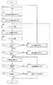
光画像計測装置1の動作について説明する。図6及び図7に示すフローチャートは、光画像計測装置1の動作の一例を表す。また、図8〜図15は、表示画面の一例を表す。
以上のような光画像計測装置1の作用及び効果について説明する。
以上に説明した構成は、この発明を好適に実施するための一例に過ぎない。よって、この発明の要旨の範囲内における任意の変形を適宜に施すことが可能である。
1A 眼底カメラユニット
100 照明光学系
120 撮影光学系
141 走査ユニット
150 OCTユニット
160 低コヒーレンス光源
174 参照ミラー
180 スペクトロメータ
184 CCD
190A アライメント光学系
200 演算制御装置
210 制御部
220 画像形成部
230 画像処理部
231 アライメント判定部
232 合焦判定部
233 像位置判定部
234 画質判定部
235 トラッキング判定部
240 表示部
250 操作部
309 アライメントスケール
310 アライメント輝点
311 スプリット輝線
Claims (3)
- 光源からの光を信号光と参照光とに分割し、被測定物体を経由した前記信号光と参照物体を経由した前記参照光とを干渉させて干渉光を生成し、前記干渉光を検出して検出信号を生成する光学系と、
前記検出信号に基づいて前記被測定物体の画像を形成する画像形成手段と、
を有する光画像計測装置であって、
前記被測定物体に対する前記光学系の位置合わせを行うアライメント手段と、
前記被測定物体に対する前記光学系の合焦を行う合焦手段と、
前記検出信号に基づいて所定フレーム内における当該画像の位置を判定する像位置判定手段と、
前記光学系の位置の適否及び合焦状態の適否を判定し、前記所定フレーム内における当該画像の位置の適否を判定し、前記光学系の位置の適否の判定、前記合焦状態の適否の判定及び前記当該画像の位置の適否の判定の全てが終了した後に、これら全ての適否の判定を再度行う判定手段と、
当該再度の適否判定において前記光学系の位置、前記合焦状態、及び前記当該画像の位置の全てが適正であると判定されたときに、前記光学系及び前記画像形成手段を制御して前記被測定物体の画像の取得を可能にする制御手段と、
を備えることを特徴とする光画像計測装置。 - 前記判定手段は、前記被測定物体の画像の上端領域及び下端領域が前記フレーム内に配置されるように当該画像の位置判定を行う、
ことを特徴とする請求項1に記載の光画像計測装置。 - 被検眼の画像を取得するための光学系と、
前記被検眼に対する前記光学系の位置合わせを行うアライメント手段と、
前記被検眼に対する前記光学系の合焦を行う合焦手段と、
前記光学系を用いて予備的に前記被検眼の画像が取得された後に所定のフレーム内における当該被検眼の画像の位置を判定する像位置判定手段と、
前記光学系の位置の適否及び合焦状態の適否を判定し、前記所定のフレーム内における当該被検眼の画像の位置の適否を判定し、前記光学系の位置の適否の判定、前記合焦状態の適否の判定及び前記当該被検眼の画像の位置の適否の判定の全てが終了した後に、これら全ての適否の判定を再度行う判定手段と、
当該再度の適否判定において前記光学系の位置、前記合焦状態、及び前記当該被検眼の画像の位置の全てが適正であると判定されたときに、前記光学系を制御して前記被検眼の画像の取得を可能にする制御手段と、
を備えることを特徴とする撮影装置。
Priority Applications (1)
| Application Number | Priority Date | Filing Date | Title |
|---|---|---|---|
| JP2013222986A JP5584345B2 (ja) | 2013-10-28 | 2013-10-28 | 光画像計測装置及び撮影装置 |
Applications Claiming Priority (1)
| Application Number | Priority Date | Filing Date | Title |
|---|---|---|---|
| JP2013222986A JP5584345B2 (ja) | 2013-10-28 | 2013-10-28 | 光画像計測装置及び撮影装置 |
Related Parent Applications (1)
| Application Number | Title | Priority Date | Filing Date |
|---|---|---|---|
| JP2009022622A Division JP5404078B2 (ja) | 2009-02-03 | 2009-02-03 | 光画像計測装置 |
Publications (2)
| Publication Number | Publication Date |
|---|---|
| JP2014039870A JP2014039870A (ja) | 2014-03-06 |
| JP5584345B2 true JP5584345B2 (ja) | 2014-09-03 |
Family
ID=50392620
Family Applications (1)
| Application Number | Title | Priority Date | Filing Date |
|---|---|---|---|
| JP2013222986A Active JP5584345B2 (ja) | 2013-10-28 | 2013-10-28 | 光画像計測装置及び撮影装置 |
Country Status (1)
| Country | Link |
|---|---|
| JP (1) | JP5584345B2 (ja) |
Families Citing this family (4)
| Publication number | Priority date | Publication date | Assignee | Title |
|---|---|---|---|---|
| JP5220155B2 (ja) * | 2011-03-31 | 2013-06-26 | キヤノン株式会社 | 眼科装置および眼科装置の制御方法 |
| JP6421919B2 (ja) * | 2014-09-01 | 2018-11-14 | 株式会社ニデック | 眼科撮影装置 |
| JP7325169B2 (ja) | 2017-12-28 | 2023-08-14 | 株式会社トプコン | 眼科装置、及びその制御方法 |
| JP7050588B2 (ja) | 2018-06-13 | 2022-04-08 | 株式会社トプコン | 眼科装置、その制御方法、プログラム、及び記録媒体 |
Family Cites Families (9)
| Publication number | Priority date | Publication date | Assignee | Title |
|---|---|---|---|---|
| JP3406933B2 (ja) * | 1993-12-30 | 2003-05-19 | キヤノン株式会社 | 角膜検査装置 |
| JP2002315753A (ja) * | 2001-04-20 | 2002-10-29 | Olympus Optical Co Ltd | 画像診断装置 |
| JP4359489B2 (ja) * | 2003-11-28 | 2009-11-04 | 株式会社ニデック | 眼底カメラ |
| WO2006078802A1 (en) * | 2005-01-21 | 2006-07-27 | Massachusetts Institute Of Technology | Methods and apparatus for optical coherence tomography scanning |
| JP4897293B2 (ja) * | 2006-01-11 | 2012-03-14 | 株式会社日立メディコ | 医用画像表示装置 |
| JP4869757B2 (ja) * | 2006-03-24 | 2012-02-08 | 株式会社トプコン | 眼底観察装置 |
| US8223143B2 (en) * | 2006-10-27 | 2012-07-17 | Carl Zeiss Meditec, Inc. | User interface for efficiently displaying relevant OCT imaging data |
| JP4996917B2 (ja) * | 2006-12-26 | 2012-08-08 | 株式会社トプコン | 光画像計測装置及び光画像計測装置を制御するプログラム |
| JP5061380B2 (ja) * | 2007-03-23 | 2012-10-31 | 株式会社トプコン | 眼底観察装置、眼科画像表示装置及びプログラム |
-
2013
- 2013-10-28 JP JP2013222986A patent/JP5584345B2/ja active Active
Also Published As
| Publication number | Publication date |
|---|---|
| JP2014039870A (ja) | 2014-03-06 |
Similar Documents
| Publication | Publication Date | Title |
|---|---|---|
| JP5404078B2 (ja) | 光画像計測装置 | |
| JP6538945B2 (ja) | 眼科装置 | |
| JP5085086B2 (ja) | 眼底観察装置、眼底画像表示装置及びプログラム | |
| JP5324839B2 (ja) | 光画像計測装置 | |
| JP6522827B2 (ja) | 眼科装置 | |
| JP6045895B2 (ja) | 眼科観察装置 | |
| JP6566541B2 (ja) | 眼科装置 | |
| JP2007275375A (ja) | 眼科装置 | |
| JP2008005987A (ja) | 眼底観察装置及びそれを制御するプログラム | |
| JP6498398B2 (ja) | 眼科装置 | |
| JP2016041221A (ja) | 眼科撮影装置およびその制御方法 | |
| JP6220022B2 (ja) | 眼科装置 | |
| JP5378157B2 (ja) | 眼科観察装置 | |
| JP2015093128A (ja) | 眼科観察装置 | |
| JP5584345B2 (ja) | 光画像計測装置及び撮影装置 | |
| US10045691B2 (en) | Ophthalmologic observation apparatus using optical coherence tomography | |
| JP6407631B2 (ja) | 眼科装置 | |
| JP6108845B2 (ja) | 眼科観察装置 | |
| JP6159454B2 (ja) | 眼科観察装置 | |
| JP6311045B2 (ja) | 眼科観察装置 | |
| JP5919175B2 (ja) | 光画像計測装置 | |
| JP2012223428A (ja) | 眼科装置 | |
| JP7106320B2 (ja) | 眼科装置、及び眼科装置の制御方法 | |
| JP2019135005A (ja) | 眼科撮影装置 |
Legal Events
| Date | Code | Title | Description |
|---|---|---|---|
| A977 | Report on retrieval |
Free format text: JAPANESE INTERMEDIATE CODE: A971007 Effective date: 20140630 |
|
| TRDD | Decision of grant or rejection written | ||
| A01 | Written decision to grant a patent or to grant a registration (utility model) |
Free format text: JAPANESE INTERMEDIATE CODE: A01 Effective date: 20140715 |
|
| A61 | First payment of annual fees (during grant procedure) |
Free format text: JAPANESE INTERMEDIATE CODE: A61 Effective date: 20140717 |
|
| R150 | Certificate of patent or registration of utility model |
Ref document number: 5584345 Country of ref document: JP Free format text: JAPANESE INTERMEDIATE CODE: R150 |
|
| R250 | Receipt of annual fees |
Free format text: JAPANESE INTERMEDIATE CODE: R250 |
|
| R250 | Receipt of annual fees |
Free format text: JAPANESE INTERMEDIATE CODE: R250 |
|
| R250 | Receipt of annual fees |
Free format text: JAPANESE INTERMEDIATE CODE: R250 |
|
| R250 | Receipt of annual fees |
Free format text: JAPANESE INTERMEDIATE CODE: R250 |
|
| R250 | Receipt of annual fees |
Free format text: JAPANESE INTERMEDIATE CODE: R250 |
|
| R250 | Receipt of annual fees |
Free format text: JAPANESE INTERMEDIATE CODE: R250 |
|
| R250 | Receipt of annual fees |
Free format text: JAPANESE INTERMEDIATE CODE: R250 |
|
| R250 | Receipt of annual fees |
Free format text: JAPANESE INTERMEDIATE CODE: R250 |