JP2023503180A - Anti-human claudin-18.2 antibody and its application - Google Patents
Anti-human claudin-18.2 antibody and its application Download PDFInfo
- Publication number
- JP2023503180A JP2023503180A JP2022540748A JP2022540748A JP2023503180A JP 2023503180 A JP2023503180 A JP 2023503180A JP 2022540748 A JP2022540748 A JP 2022540748A JP 2022540748 A JP2022540748 A JP 2022540748A JP 2023503180 A JP2023503180 A JP 2023503180A
- Authority
- JP
- Japan
- Prior art keywords
- seq
- amino acid
- acid sequence
- antibody
- identity
- Prior art date
- Legal status (The legal status is an assumption and is not a legal conclusion. Google has not performed a legal analysis and makes no representation as to the accuracy of the status listed.)
- Pending
Links
Images


Classifications
-
- A—HUMAN NECESSITIES
- A61—MEDICAL OR VETERINARY SCIENCE; HYGIENE
- A61P—SPECIFIC THERAPEUTIC ACTIVITY OF CHEMICAL COMPOUNDS OR MEDICINAL PREPARATIONS
- A61P35/00—Antineoplastic agents
-
- C—CHEMISTRY; METALLURGY
- C07—ORGANIC CHEMISTRY
- C07K—PEPTIDES
- C07K16/00—Immunoglobulins [IGs], e.g. monoclonal or polyclonal antibodies
- C07K16/18—Immunoglobulins [IGs], e.g. monoclonal or polyclonal antibodies against material from animals or humans
- C07K16/28—Immunoglobulins [IGs], e.g. monoclonal or polyclonal antibodies against material from animals or humans against receptors, cell surface antigens or cell surface determinants
-
- C—CHEMISTRY; METALLURGY
- C12—BIOCHEMISTRY; BEER; SPIRITS; WINE; VINEGAR; MICROBIOLOGY; ENZYMOLOGY; MUTATION OR GENETIC ENGINEERING
- C12N—MICROORGANISMS OR ENZYMES; COMPOSITIONS THEREOF; PROPAGATING, PRESERVING, OR MAINTAINING MICROORGANISMS; MUTATION OR GENETIC ENGINEERING; CULTURE MEDIA
- C12N15/00—Mutation or genetic engineering; DNA or RNA concerning genetic engineering, vectors, e.g. plasmids, or their isolation, preparation or purification; Use of hosts therefor
- C12N15/09—Recombinant DNA-technology
- C12N15/63—Introduction of foreign genetic material using vectors; Vectors; Use of hosts therefor; Regulation of expression
-
- G01N33/575—
-
- G01N33/57557—
-
- A—HUMAN NECESSITIES
- A61—MEDICAL OR VETERINARY SCIENCE; HYGIENE
- A61K—PREPARATIONS FOR MEDICAL, DENTAL OR TOILETRY PURPOSES
- A61K39/00—Medicinal preparations containing antigens or antibodies
- A61K2039/505—Medicinal preparations containing antigens or antibodies comprising antibodies
-
- C—CHEMISTRY; METALLURGY
- C07—ORGANIC CHEMISTRY
- C07K—PEPTIDES
- C07K2317/00—Immunoglobulins specific features
- C07K2317/20—Immunoglobulins specific features characterized by taxonomic origin
- C07K2317/24—Immunoglobulins specific features characterized by taxonomic origin containing regions, domains or residues from different species, e.g. chimeric, humanized or veneered
-
- C—CHEMISTRY; METALLURGY
- C07—ORGANIC CHEMISTRY
- C07K—PEPTIDES
- C07K2317/00—Immunoglobulins specific features
- C07K2317/30—Immunoglobulins specific features characterized by aspects of specificity or valency
- C07K2317/33—Crossreactivity, e.g. for species or epitope, or lack of said crossreactivity
-
- C—CHEMISTRY; METALLURGY
- C07—ORGANIC CHEMISTRY
- C07K—PEPTIDES
- C07K2317/00—Immunoglobulins specific features
- C07K2317/50—Immunoglobulins specific features characterized by immunoglobulin fragments
- C07K2317/56—Immunoglobulins specific features characterized by immunoglobulin fragments variable (Fv) region, i.e. VH and/or VL
- C07K2317/565—Complementarity determining region [CDR]
-
- C—CHEMISTRY; METALLURGY
- C07—ORGANIC CHEMISTRY
- C07K—PEPTIDES
- C07K2317/00—Immunoglobulins specific features
- C07K2317/60—Immunoglobulins specific features characterized by non-natural combinations of immunoglobulin fragments
- C07K2317/62—Immunoglobulins specific features characterized by non-natural combinations of immunoglobulin fragments comprising only variable region components
- C07K2317/622—Single chain antibody (scFv)
-
- C—CHEMISTRY; METALLURGY
- C07—ORGANIC CHEMISTRY
- C07K—PEPTIDES
- C07K2317/00—Immunoglobulins specific features
- C07K2317/70—Immunoglobulins specific features characterized by effect upon binding to a cell or to an antigen
- C07K2317/73—Inducing cell death, e.g. apoptosis, necrosis or inhibition of cell proliferation
- C07K2317/732—Antibody-dependent cellular cytotoxicity [ADCC]
-
- C—CHEMISTRY; METALLURGY
- C07—ORGANIC CHEMISTRY
- C07K—PEPTIDES
- C07K2317/00—Immunoglobulins specific features
- C07K2317/70—Immunoglobulins specific features characterized by effect upon binding to a cell or to an antigen
- C07K2317/73—Inducing cell death, e.g. apoptosis, necrosis or inhibition of cell proliferation
- C07K2317/734—Complement-dependent cytotoxicity [CDC]
-
- C—CHEMISTRY; METALLURGY
- C07—ORGANIC CHEMISTRY
- C07K—PEPTIDES
- C07K2317/00—Immunoglobulins specific features
- C07K2317/90—Immunoglobulins specific features characterized by (pharmaco)kinetic aspects or by stability of the immunoglobulin
- C07K2317/92—Affinity (KD), association rate (Ka), dissociation rate (Kd) or EC50 value
-
- C—CHEMISTRY; METALLURGY
- C07—ORGANIC CHEMISTRY
- C07K—PEPTIDES
- C07K2319/00—Fusion polypeptide
- C07K2319/01—Fusion polypeptide containing a localisation/targetting motif
- C07K2319/03—Fusion polypeptide containing a localisation/targetting motif containing a transmembrane segment
-
- C—CHEMISTRY; METALLURGY
- C12—BIOCHEMISTRY; BEER; SPIRITS; WINE; VINEGAR; MICROBIOLOGY; ENZYMOLOGY; MUTATION OR GENETIC ENGINEERING
- C12N—MICROORGANISMS OR ENZYMES; COMPOSITIONS THEREOF; PROPAGATING, PRESERVING, OR MAINTAINING MICROORGANISMS; MUTATION OR GENETIC ENGINEERING; CULTURE MEDIA
- C12N2740/00—Reverse transcribing RNA viruses
- C12N2740/00011—Details
- C12N2740/10011—Retroviridae
- C12N2740/16011—Human Immunodeficiency Virus, HIV
- C12N2740/16041—Use of virus, viral particle or viral elements as a vector
- C12N2740/16043—Use of virus, viral particle or viral elements as a vector viral genome or elements thereof as genetic vector
Landscapes
- Health & Medical Sciences (AREA)
- Chemical & Material Sciences (AREA)
- Organic Chemistry (AREA)
- Life Sciences & Earth Sciences (AREA)
- General Health & Medical Sciences (AREA)
- Immunology (AREA)
- Genetics & Genomics (AREA)
- Medicinal Chemistry (AREA)
- Molecular Biology (AREA)
- Biochemistry (AREA)
- Biophysics (AREA)
- Proteomics, Peptides & Aminoacids (AREA)
- Chemical Kinetics & Catalysis (AREA)
- General Chemical & Material Sciences (AREA)
- Nuclear Medicine, Radiotherapy & Molecular Imaging (AREA)
- Pharmacology & Pharmacy (AREA)
- Animal Behavior & Ethology (AREA)
- Public Health (AREA)
- Veterinary Medicine (AREA)
- Engineering & Computer Science (AREA)
- Biomedical Technology (AREA)
- Biotechnology (AREA)
- General Engineering & Computer Science (AREA)
- Bioinformatics & Cheminformatics (AREA)
- Wood Science & Technology (AREA)
- Zoology (AREA)
- Microbiology (AREA)
- Physics & Mathematics (AREA)
- Plant Pathology (AREA)
- Peptides Or Proteins (AREA)
- Hematology (AREA)
- Micro-Organisms Or Cultivation Processes Thereof (AREA)
- Urology & Nephrology (AREA)
- Oncology (AREA)
- Cell Biology (AREA)
- Hospice & Palliative Care (AREA)
- Food Science & Technology (AREA)
- Analytical Chemistry (AREA)
- General Physics & Mathematics (AREA)
- Pathology (AREA)
Abstract
本発明は、ヒトクローディン18.2に結合する抗体又はそのフラグメント、並びにそのコード核酸等を提供する。本発明の抗ヒトクローディン18.2抗体は、抗原クローディン18.2に対して強い親和性を有し、発現細胞を標的とするための顕著な補体依存性細胞傷害(CDC)活性及び抗体依存性細胞傷害(ADCC)活性を有し、ヒトCLDN18.2に対して高い特異性を示す。【選択図】なしThe present invention provides antibodies or fragments thereof that bind to human claudin-18.2, encoding nucleic acids thereof, and the like. The anti-human Claudin-18.2 antibodies of the present invention have strong affinity for the antigen Claudin-18.2, and have significant complement-dependent cytotoxicity (CDC) activity to target expressing cells and It has antibody-dependent cellular cytotoxicity (ADCC) activity and exhibits high specificity for human CLDN18.2. [Selection figure] None
Description
本出願は、2019年9月29日に出願された中国特許出願第201910929614.0号の優先権の利益を主張し、引用することでその全体が本明細書の一部をなす。 This application claims the priority benefit of Chinese Patent Application No. 201910929614.0 filed on September 29, 2019, which is incorporated herein by reference in its entirety.
本開示は、生物医学の分野に属し、新規な抗ヒトクローディン(Claudin)18.2抗体又はその機能的フラグメントに関する。本開示はまた、該抗体又はその機能的フラグメントの使用に関する。 The present disclosure belongs to the field of biomedicine and relates to novel anti-human Claudin 18.2 antibodies or functional fragments thereof. The disclosure also relates to uses of said antibodies or functional fragments thereof.
ヒトタイトジャンクションタンパク質クローディン18(CLDN18)は、密接な細胞接合部の構築、細胞バリア機能の維持、及び細胞間分子輸送への関与において生物学的に機能するタイトジャンクションタンパク質のファミリーに属する。クローディン18タンパク質は約26KDの分子量を有し、選択的スプライシングによって、異なる特性を有するクローディンサブタイプCLDN18.1及びCLDN18.2へと変更され得る。CLDN18.1及びCLDN18.2は、それぞれ261個のアミノ酸を含み、4つの膜貫通ドメイン、すなわち、細胞内に位置するNH2末端ドメイン及びCOOH末端ドメイン、並びに2つの細胞外ループ(ECL1、ECL2)を有する。CLDN18.1及びCLDN18.2の細胞外領域1は、互いに8アミノ酸異なる。
Human tight junction protein claudin 18 (CLDN18) belongs to a family of tight junction proteins that biologically function in building tight cell junctions, maintaining cell barrier function, and participating in intercellular molecular transport. The claudin-18 protein has a molecular weight of approximately 26 KD and can be altered by alternative splicing into the claudin subtypes CLDN18.1 and CLDN18.2, which have different properties. CLDN18.1 and CLDN18.2 each contain 261 amino acids and contain four transmembrane domains, an intracellularly located NH2-terminal domain and a COOH-terminal domain, and two extracellular loops (ECL1, ECL2). have.
CLDN18.2は、多くのヒト上皮腫瘍タイプの原発性病変及び転移において発現され、びまん性癌腫細胞において継続的に発現される。CLDN18.2の発現レベルは、食道癌、膵臓癌、肺癌及び胃癌の細胞において更に有意に増加する。Sahin et alによる研究は、CLDN18.2が胃癌の重要なバイオマーカーであることを示唆し、胃癌患者の70%に存在し、そのうち90%~95%が腺癌と診断され、別の5%~10%がリンパ腫、胃腸間質腫瘍(GIST)及びカルチノイド腫瘍を含んだ。 CLDN18.2 is expressed in primary lesions and metastases of many human epithelial tumor types and is continuously expressed in diffuse carcinoma cells. Expression levels of CLDN18.2 are also significantly increased in esophageal, pancreatic, lung and gastric cancer cells. A study by Sahin et al suggested that CLDN18.2 is an important biomarker for gastric cancer, present in 70% of patients with gastric cancer, 90%-95% of whom are diagnosed with adenocarcinoma, another 5% ~10% contained lymphomas, gastrointestinal stromal tumors (GIST) and carcinoid tumors.
CLDN18.2に対するモノクローナル抗体であるIMAB362は、ドイツのバイオテクノロジー企業であるGanymedによって開発され、現在、進行胃食道癌を有する患者の治療のための第III相臨床試験中である。IMAB362抗体単独の結合は、in vitro及びin vivoの両方でCLDN18.2を発現する標的細胞の増殖の阻害をもたらし、腫瘍増殖を阻害し、補体依存性細胞傷害(CDC)及び抗体依存性細胞傷害(ADCC)の作用機序を介して癌細胞を排除し、様々な作用様式が独立しているが相乗的であることが示されている。IMAB362抗体は、転移性食道癌、及び転移性胃癌を含む適応症を対象としており、現在、米国及び欧州で第III相臨床試験中であり、2021年に販売承認を申請する予定である。 IMAB362, a monoclonal antibody against CLDN18.2, was developed by Ganymed, a German biotechnology company, and is currently in phase III clinical trials for the treatment of patients with advanced gastroesophageal cancer. Binding of the IMAB362 antibody alone resulted in inhibition of proliferation of target cells expressing CLDN18.2 both in vitro and in vivo, inhibited tumor growth, inhibited complement dependent cytotoxicity (CDC) and antibody dependent cells. It eliminates cancer cells via a mechanism of action of injury (ADCC) and various modes of action have been shown to be independent but synergistic. The IMAB362 antibody, for indications including metastatic esophageal and metastatic gastric cancer, is currently in Phase III clinical trials in the US and Europe and is expected to be submitted for marketing approval in 2021.
IMAB362はキメラ抗体であるが、比較的多くのマウスアミノ酸を有し、ヒトにおいて拒絶反応免疫応答を引き起こす確率が比較的高い。そのため、ヒトCLDN18.2に対してより高い特異性、親和性及び生物学的活性を有する抗体、特にヒト化抗体に対する必要性が当該技術分野において依然として存在する。さらに、クローディン18の2つのサブタイプは、構造が非常に類似しているが、機能が異なる。CLDN18.1とCLDN18.2との間の相同性が高く、ECL1の上位構造のため、CLDN18.2に対する高品質の抗体をスクリーニングすることが難しくなり、抗体特異性に関して高い不確実性が存在する。 Although IMAB362 is a chimeric antibody, it has relatively many murine amino acids and is more likely to provoke a negative immune response in humans. Therefore, there remains a need in the art for antibodies, especially humanized antibodies, with higher specificity, affinity and biological activity for human CLDN18.2. Moreover, the two subtypes of claudin-18 are very similar in structure but differ in function. The high homology between CLDN18.1 and CLDN18.2 and the high order structure of ECL1 makes it difficult to screen high quality antibodies against CLDN18.2 and there is high uncertainty regarding antibody specificity. .
本発明が解決しようとする技術的課題は、ハイブリドーマスクリーニング及びヒト化を通じて、モノクローナル抗体の調製方法を最適化し、クローディン18.2特異的抗体のスクリーニング効率を向上させることにより高親和性でヒトCLDN18.2に特異的に結合し得る抗体を得ることであり、該抗体は、ヒト化設計により最も少ないマウスアミノ酸を有し、より優れたin vivoでの安全性及び応用可能性を有することが期待される。 The technical problem to be solved by the present invention is to optimize the method for preparing monoclonal antibodies through hybridoma screening and humanization, and to improve the efficiency of screening claudin 18.2-specific antibodies to detect human CLDN18 with high affinity. .2 to obtain an antibody that can specifically bind to .2, which has the least mouse amino acids due to humanized design and is expected to have better in vivo safety and applicability be done.
上記のような技術的課題に対して、免疫原に関しては、ヒトCLDN18.2を発現するマウス由来細胞が本開示で利用される。一態様では、宿主としてのマウス由来細胞は、非免疫原性であるか、又はマウスに対する免疫原性が非常に低く、それによって、目的の免疫原CLDN18.2に対する宿主細胞の陰影効果を回避し、異種免疫原の導入によって引き起こされるCLDN18.2特異的ハイブリドーマの生産効率に対する悪影響を回避する。別の態様では、マウス由来細胞の細胞膜の表面におけるヒトCLDN18.2の発現は、スクリーニングによって得られたモノクローナル抗体がCLDN18.2の細胞外領域に特異的に結合し、CLDN18.2特異的抗体の生物学的活性を保証するように達成することができる。 For the above technical problems, mouse-derived cells expressing human CLDN18.2 are utilized in the present disclosure as immunogens. In one aspect, mouse-derived cells as hosts are non-immunogenic or very poorly immunogenic to mice, thereby avoiding the shadowing effect of host cells on the immunogen of interest CLDN18.2. , avoiding the adverse effects on the production efficiency of CLDN18.2-specific hybridomas caused by the introduction of heterologous immunogens. In another aspect, the expression of human CLDN18.2 on the surface of the cell membrane of mouse-derived cells is confirmed by the monoclonal antibody obtained by screening that specifically binds to the extracellular region of CLDN18.2, and the expression of the CLDN18.2-specific antibody. can be achieved to ensure biological activity.
スクリーニング戦略に関しては、陽性スクリーニング及び陰性スクリーニングを組み合わせた戦略が本開示で使用される。まず、ヒトCLDN18.2を発現する組換え細胞を用いて陽性スクリーニングを行い、細胞に対して高い親和性を有するモノクローナル抗体を得て、次いで、ヒトCLDN18.2を発現する組換え細胞に結合し得る抗体クローンについてヒトCLDN18.1を発現する組換え細胞を用いて陰性スクリーニングを行い、CLDN18.2及びCLDN18.1の共通の細胞外領域構造に特異的に結合することができるモノクローナル抗体を除外する。さらに、本発明で用いる免疫原、陽性スクリーニング抗原、及び陰性スクリーニング抗原は、全て同じマウス由来細胞プラットフォームであり、プロセスを簡素化して、異種抗原の導入を回避するだけでなく、マウス由来細胞の個々のマウスに対する同種抗原効果によって生成された抗体を排除する陰性スクリーニング工程を可能にし、CLDN18.2の細胞外領域2に特異的に結合するモノクローナル抗体の生産効率を更に向上させる。
With respect to screening strategies, strategies that combine positive and negative screening are used in this disclosure. Positive screening was first performed using recombinant cells expressing human CLDN18.2 to obtain monoclonal antibodies with high affinity for the cells, which then bound to recombinant cells expressing human CLDN18.2. The antibody clones obtained are negatively screened using recombinant cells expressing human CLDN18.1 to exclude monoclonal antibodies that can specifically bind to the common extracellular domain structure of CLDN18.2 and CLDN18.1. . Furthermore, the immunogens, positive screening antigens, and negative screening antigens used in the present invention are all from the same mouse-derived cell platform, simplifying the process and avoiding the introduction of heterologous antigens, as well as the individualization of mouse-derived cells. allows a negative screening step to eliminate antibodies generated by the alloantigen effect on mice, further improving the production efficiency of monoclonal antibodies that specifically bind to the
本開示の更なる目的は、ヒトCLDN18.2に特異的に結合する抗体又はそのフラグメントを提供し、その用途を提供することである。本開示による抗体のフラグメントは、とりわけ、抗体の様々な機能的フラグメント、例えばその抗原結合部分、例えばFab、F(ab’)2又はscFvフラグメントを包含する。 A further object of the present disclosure is to provide an antibody or fragment thereof that specifically binds to human CLDN18.2 and to provide uses thereof. Fragments of antibodies according to the present disclosure include, inter alia, various functional fragments of antibodies, such as antigen-binding portions thereof, such as Fab, F(ab') 2 or scFv fragments.
本開示は、以下の技術的解決策を提供する。 The present disclosure provides the following technical solutions.
1つの態様では、本開示は、抗ヒトクローディン18.2抗体の調製方法であって、
(1)表面にヒトクローディン18.2タンパク質を発現する細胞を免疫原として用いて、動物を免疫する工程と、
(2)工程(1)で免疫した動物を用いて抗体を産生することができる細胞クローンを調製する工程と、
(3)表面にヒトクローディン18.2タンパク質を発現する細胞を陽性スクリーニング抗原として用い、陽性スクリーニング抗原に対する結合活性を有する抗体、及び抗体を産生する細胞をスクリーニングする工程と、
(4)表面にヒトクローディン18.1タンパク質を発現する細胞を陰性スクリーニング抗原として用い、陰性スクリーニング抗原に対する結合活性を有する抗体、及び抗体を産生する細胞を排除する工程と、
を含む、調製方法を提供する。
In one aspect, the disclosure provides a method of preparing an anti-human claudin-18.2 antibody comprising:
(1) immunizing an animal with cells expressing human claudin-18.2 protein on their surface as an immunogen;
(2) preparing cell clones capable of producing antibodies using the animal immunized in step (1);
(3) using cells expressing human claudin 18.2 protein on their surface as a positive screening antigen, screening for antibodies having binding activity to the positive screening antigen and cells producing the antibodies;
(4) using cells expressing human claudin 18.1 protein on their surface as a negative screening antigen to eliminate antibodies having binding activity to the negative screening antigen and cells producing the antibodies;
A method of preparation is provided, comprising:
好ましくは、本開示による調製方法において、工程(1)においてヒトクローディン18.2タンパク質を発現する細胞は、免疫された動物と同じ種に由来し、好ましくは、ヒトクローディン18.2タンパク質を発現する細胞がマウス細胞であり、免疫された動物がマウスである。 Preferably, in the preparation method according to the present disclosure, the cells expressing human claudin-18.2 protein in step (1) are from the same species as the immunized animal, preferably human claudin-18.2 protein. The expressing cells are mouse cells and the immunized animals are mice.
好ましくは、本開示による調製方法において、工程(2)において、抗体を産生することができる細胞クローンは、ハイブリドーマ手法及び単一細胞増幅手法からなる群から選択される手法により調製される。 Preferably, in the preparation method according to the present disclosure, in step (2), cell clones capable of producing antibodies are prepared by a technique selected from the group consisting of hybridoma technique and single cell amplification technique.
好ましくは、本開示による調製方法において、工程(3)における陽性スクリーニング抗原は、工程(1)における免疫原と同じであり、工程(4)における陰性スクリーニング抗原が工程(1)における免疫原と異なるのは、発現されるタンパク質がヒトクローディン18.1タンパク質である点のみである。 Preferably, in the preparation method according to the present disclosure, the positive screening antigen in step (3) is the same as the immunogen in step (1), and the negative screening antigen in step (4) is different from the immunogen in step (1). The only difference is that the protein expressed is the human Claudin 18.1 protein.
好ましくは、本開示による調製方法において、工程(3)及び工程(4)は、それぞれ、酵素結合免疫吸着測定法(ELISA)及び蛍光共鳴エネルギー移動法(FRET)からなる群から選択される方法により行われる。 Preferably, in the preparation method according to the present disclosure, step (3) and step (4) are each performed by a method selected from the group consisting of enzyme-linked immunosorbent assay (ELISA) and fluorescence resonance energy transfer (FRET). done.
別の態様では、本開示は、本開示の調製方法に従って得られた抗ヒトクローディン18.2抗体を提供する。 In another aspect, the present disclosure provides anti-human claudin-18.2 antibodies obtained according to the methods of preparation of the present disclosure.
好ましくは、本開示による抗ヒトクローディン18.2抗体は、ヒトクローディン18.2のN末端、好ましくは第1の細胞外ループ(ECL1)を含むヒトクローディン18.2のN末端の細胞外領域に特異的に結合する。 Preferably, the anti-human claudin-18.2 antibody according to the present disclosure is directed to the N-terminus of human claudin-18.2, preferably the N-terminus of human claudin-18.2 including the first extracellular loop (ECL1). Binds specifically to the outer region.
好ましくは、本開示による抗ヒトクローディン18.2抗体は、nMスケールでヒトCLDN18.2に特異的な結合を有し、アイソタイプ陰性抗体(又は無関係な抗体)と比較してヒトCLDN18.1への結合において有意差を有さない。 Preferably, an anti-human Claudin-18.2 antibody according to the present disclosure has specific binding to human CLDN18.2 on the nM scale and has a binding affinity to human CLDN18.1 relative to an isotype-negative antibody (or an irrelevant antibody). no significant difference in the binding of
更なる態様では、本開示は、重鎖可変領域(VH)及び軽鎖可変領域(VL)を含む抗体又はそのフラグメントであって、重鎖可変領域(VH)及び軽鎖可変領域(VL)は、以下:
(1)配列番号31に示されるVH-CDR1、配列番号32に示されるVH-CDR2、及び配列番号33に示されるVH-CDR3;並びに配列番号34に示されるVL-CDR1、配列番号35に示されるVL-CDR2、及び配列番号36に示されるVL-CDR3;
(2)配列番号37に示されるVH-CDR1、配列番号38に示されるVH-CDR2、及び配列番号39に示されるVH-CDR3;並びに配列番号40に示されるVL-CDR1、配列番号41に示されるVL-CDR2、及び配列番号42に示されるVL-CDR3;
(3)配列番号43に示されるVH-CDR1、配列番号44に示されるVH-CDR2、及び配列番号45に示されるVH-CDR3;並びに配列番号46に示されるVL-CDR1、配列番号41に示されるVL-CDR2、及び配列番号47に示されるVL-CDR3;
(4)配列番号48に示されるVH-CDR1、配列番号49に示されるVH-CDR2、及び配列番号50に示されるVH-CDR3;並びに配列番号40に示されるVL-CDR1、配列番号41に示されるVL-CDR2、及び配列番号51に示されるVL-CDR3;
(5)配列番号52に示されるVH-CDR1、配列番号53に示されるVH-CDR2、及び配列番号54に示されるVH-CDR3;並びに配列番号55に示されるVL-CDR1、配列番号41に示されるVL-CDR2、及び配列番号56に示されるVL-CDR3;
(6)配列番号57に示されるVH-CDR1、配列番号58に示されるVH-CDR2、及び配列番号33に示されるVH-CDR3;並びに配列番号34に示されるVL-CDR1、配列番号59に示されるVL-CDR2、及び配列番号60に示されるVL-CDR3;
(7)配列番号61に示されるVH-CDR1、配列番号62に示されるVH-CDR2、及び配列番号63に示されるVH-CDR3;並びに配列番号46に示されるVL-CDR1、配列番号41に示されるVL-CDR2、及び配列番号64に示されるVL-CDR3;
(8)配列番号65に示されるVH-CDR1、配列番号66に示されるVH-CDR2、及び配列番号67に示されるVH-CDR3;並びに配列番号68に示されるVL-CDR1、配列番号41に示されるVL-CDR2、及び配列番号69に示されるVL-CDR3;
(9)配列番号65に示されるVH-CDR1、配列番号70に示されるVH-CDR2、及び配列番号71に示されるVH-CDR3;並びに配列番号72に示されるVL-CDR1、配列番号41に示されるVL-CDR2、及び配列番号73に示されるVL-CDR3;
(10)配列番号74に示されるVH-CDR1、配列番号75に示されるVH-CDR2、及び配列番号76に示されるVH-CDR3;並びに配列番号77に示されるVL-CDR1、配列番号41に示されるVL-CDR2、及び配列番号78に示されるVL-CDR3;
(11)配列番号79に示されるVH-CDR1、配列番号80に示されるVH-CDR2、及び配列番号81に示されるVH-CDR3;並びに配列番号82に示されるVL-CDR1、配列番号41に示されるVL-CDR2、及び配列番号83に示されるVL-CDR3;
(12)配列番号37に示されるVH-CDR1、配列番号38に示されるVH-CDR2、及び配列番号39に示されるVH-CDR3;並びに配列番号85に示されるVL-CDR1、配列番号41に示されるVL-CDR2、及び配列番号42に示されるVL-CDR3;
(13)配列番号37に示されるVH-CDR1、配列番号38に示されるVH-CDR2、及び配列番号39に示されるVH-CDR3;並びに配列番号85に示されるVL-CDR1、配列番号41に示されるVL-CDR2、及び配列番号86に示されるVL-CDR3;
(14)配列番号37に示されるVH-CDR1、配列番号38に示されるVH-CDR2、及び配列番号39に示されるVH-CDR3;並びに配列番号85に示されるVL-CDR1、配列番号41に示されるVL-CDR2、及び配列番号87に示されるVL-CDR3;
(15)配列番号74に示されるVH-CDR1、配列番号75に示されるVH-CDR2、及び配列番号76に示されるVH-CDR3;並びに配列番号89に示されるVL-CDR1、配列番号41に示されるVL-CDR2、及び配列番号90に示されるVL-CDR3;並びに、
(16)配列番号79に示されるVH-CDR1、配列番号91に示されるVH-CDR2、及び配列番号81に示されるVH-CDR3;並びに配列番号92に示されるVL-CDR1、配列番号41に示されるVL-CDR2、及び配列番号93に示されるVL-CDR3;
からなる群から選択されるCDR(VH-CDR1、VH-CDR2、VH-CDR3;及びVL-CDR1、VL-CDR2、VL-CDR3)の組合せを含む、抗体又はそのフラグメントを提供する。
In a further aspect, the disclosure provides an antibody or fragment thereof comprising a heavy chain variable region (VH) and a light chain variable region (VL), wherein the heavy chain variable region (VH) and the light chain variable region (VL) are ,the following:
(1) VH-CDR1 shown in SEQ ID NO: 31, VH-CDR2 shown in SEQ ID NO: 32, and VH-CDR3 shown in SEQ ID NO: 33; and VL-CDR3 shown in SEQ ID NO: 36;
(2) VH-CDR1 shown in SEQ ID NO:37, VH-CDR2 shown in SEQ ID NO:38, and VH-CDR3 shown in SEQ ID NO:39; and VL-CDR1 shown in SEQ ID NO:40, shown in SEQ ID NO:41 and VL-CDR3 shown in SEQ ID NO: 42;
(3) VH-CDR1 shown in SEQ ID NO: 43, VH-CDR2 shown in SEQ ID NO: 44, and VH-CDR3 shown in SEQ ID NO: 45; and VL-CDR3 shown in SEQ ID NO: 47;
(4) VH-CDR1 shown in SEQ ID NO: 48, VH-CDR2 shown in SEQ ID NO: 49, and VH-CDR3 shown in SEQ ID NO: 50; and VL-CDR3 shown in SEQ ID NO: 51;
(5) VH-CDR1 shown in SEQ ID NO: 52, VH-CDR2 shown in SEQ ID NO: 53, and VH-CDR3 shown in SEQ ID NO: 54; and VL-CDR1 shown in SEQ ID NO: 55, SEQ ID NO: 41 and VL-CDR3 shown in SEQ ID NO: 56;
(6) VH-CDR1 shown in SEQ ID NO: 57, VH-CDR2 shown in SEQ ID NO: 58, and VH-CDR3 shown in SEQ ID NO: 33; and VL-CDR1 shown in SEQ ID NO: 34, SEQ ID NO: 59 and VL-CDR3 shown in SEQ ID NO: 60;
(7) VH-CDR1 shown in SEQ ID NO: 61, VH-CDR2 shown in SEQ ID NO: 62, and VH-CDR3 shown in SEQ ID NO: 63; and VL-CDR1 shown in SEQ ID NO: 46, SEQ ID NO: 41 and VL-CDR3 shown in SEQ ID NO: 64;
(8) VH-CDR1 shown in SEQ ID NO: 65, VH-CDR2 shown in SEQ ID NO: 66, and VH-CDR3 shown in SEQ ID NO: 67; and VL-CDR1 shown in SEQ ID NO: 68, shown in SEQ ID NO: 41 and VL-CDR3 shown in SEQ ID NO: 69;
(9) VH-CDR1 shown in SEQ ID NO: 65, VH-CDR2 shown in SEQ ID NO: 70, and VH-CDR3 shown in SEQ ID NO: 71; and VL-CDR1 shown in SEQ ID NO: 72, SEQ ID NO: 41 and VL-CDR3 shown in SEQ ID NO: 73;
(10) VH-CDR1 shown in SEQ ID NO: 74, VH-CDR2 shown in SEQ ID NO: 75, and VH-CDR3 shown in SEQ ID NO: 76; and VL-CDR1 shown in SEQ ID NO: 77, SEQ ID NO: 41 and VL-CDR3 shown in SEQ ID NO: 78;
(11) VH-CDR1 shown in SEQ ID NO: 79, VH-CDR2 shown in SEQ ID NO: 80, and VH-CDR3 shown in SEQ ID NO: 81; and VL-CDR1 shown in SEQ ID NO: 82, SEQ ID NO: 41 and VL-CDR3 shown in SEQ ID NO:83;
(12) VH-CDR1 shown in SEQ ID NO: 37, VH-CDR2 shown in SEQ ID NO: 38, and VH-CDR3 shown in SEQ ID NO: 39; and VL-CDR1 shown in SEQ ID NO: 85, SEQ ID NO: 41 and VL-CDR3 shown in SEQ ID NO: 42;
(13) VH-CDR1 shown in SEQ ID NO: 37, VH-CDR2 shown in SEQ ID NO: 38, and VH-CDR3 shown in SEQ ID NO: 39; and VL-CDR1 shown in SEQ ID NO: 85, SEQ ID NO: 41 and VL-CDR3 shown in SEQ ID NO:86;
(14) VH-CDR1 shown in SEQ ID NO: 37, VH-CDR2 shown in SEQ ID NO: 38, and VH-CDR3 shown in SEQ ID NO: 39; and VL-CDR1 shown in SEQ ID NO: 85, SEQ ID NO: 41 and VL-CDR3 shown in SEQ ID NO:87;
(15) VH-CDR1 shown in SEQ ID NO: 74, VH-CDR2 shown in SEQ ID NO: 75, and VH-CDR3 shown in SEQ ID NO: 76; and VL-CDR1 shown in SEQ ID NO: 89, SEQ ID NO: 41 and VL-CDR3 shown in SEQ ID NO: 90; and
(16) VH-CDR1 shown in SEQ ID NO: 79, VH-CDR2 shown in SEQ ID NO: 91, and VH-CDR3 shown in SEQ ID NO: 81; and VL-CDR1 shown in SEQ ID NO: 92, SEQ ID NO: 41 and VL-CDR3 shown in SEQ ID NO:93;
Antibodies or fragments thereof comprising a combination of CDRs selected from the group consisting of (VH-CDR1, VH-CDR2, VH-CDR3; and VL-CDR1, VL-CDR2, VL-CDR3) are provided.
好ましくは、本開示による抗体又はそのフラグメントにおいて、重鎖可変領域は、配列番号1、配列番号3、配列番号5、配列番号7、配列番号9、配列番号11、配列番号13、配列番号15、配列番号17、配列番号19、配列番号21、配列番号23、配列番号27、及び配列番号29のいずれか1つに示されるアミノ酸配列、若しくは示されるアミノ酸配列に対して少なくとも75%同一性を有するアミノ酸配列を含み、及び/又は軽鎖可変領域は、配列番号2、配列番号4、配列番号6、配列番号8、配列番号10、配列番号12、配列番号14、配列番号16、配列番号18、配列番号20、配列番号22、配列番号24、配列番号25、配列番号26、配列番号28、及び配列番号30のいずれか1つに示されるアミノ酸配列、若しくは示されるアミノ酸配列に対して少なくとも75%同一性を有するアミノ酸配列を含む。 Preferably, in an antibody or fragment thereof according to the present disclosure, the heavy chain variable region comprises SEQ ID NO: 17, SEQ ID NO: 19, SEQ ID NO: 21, SEQ ID NO: 23, SEQ ID NO: 27, and SEQ ID NO: 29, or having at least 75% identity to the amino acid sequence shown SEQ ID NO:2, SEQ ID NO:4, SEQ ID NO:6, SEQ ID NO:8, SEQ ID NO:10, SEQ ID NO:12, SEQ ID NO:14, SEQ ID NO:16, SEQ ID NO:18, SEQ ID NO: 20, SEQ ID NO: 22, SEQ ID NO: 24, SEQ ID NO: 25, SEQ ID NO: 26, SEQ ID NO: 28, and SEQ ID NO: 30, or at least 75% of the amino acid sequence shown Contains amino acid sequences with identity.
本開示の特定の実施の形態によれば、抗体又はそのフラグメントは、以下:
(1)配列番号1に示されるアミノ酸配列、又は配列番号1に示されるアミノ酸配列に対して少なくとも75%同一性を有するアミノ酸配列;及び配列番号2に示されるアミノ酸配列、又は配列番号2に示されるアミノ酸配列に対して少なくとも75%同一性を有するアミノ酸配列;
(2)配列番号3に示されるアミノ酸配列、又は配列番号3に示されるアミノ酸配列に対して少なくとも75%同一性を有するアミノ酸配列;及び配列番号4に示されるアミノ酸配列、又は配列番号4に示されるアミノ酸配列に対して少なくとも75%同一性を有するアミノ酸配列;
(3)配列番号5に示されるアミノ酸配列、又は配列番号5に示されるアミノ酸配列に対して少なくとも75%同一性を有するアミノ酸配列;及び配列番号6に示されるアミノ酸配列、又は配列番号6に示されるアミノ酸配列に対して少なくとも75%同一性を有するアミノ酸配列;
(4)配列番号7に示されるアミノ酸配列、又は配列番号7に示されるアミノ酸配列に対して少なくとも75%同一性を有するアミノ酸配列;及び配列番号8に示されるアミノ酸配列、又は配列番号8に示されるアミノ酸配列に対して少なくとも75%同一性を有するアミノ酸配列;
(5)配列番号9に示されるアミノ酸配列、又は配列番号9に示されるアミノ酸配列に対して少なくとも75%同一性を有するアミノ酸配列;及び配列番号10に示されるアミノ酸配列、又は配列番号10に示されるアミノ酸配列に対して少なくとも75%同一性を有するアミノ酸配列;
(6)配列番号11に示されるアミノ酸配列、又は配列番号11に示されるアミノ酸配列に対して少なくとも75%同一性を有するアミノ酸配列;及び配列番号12に示されるアミノ酸配列、又は配列番号12に示されるアミノ酸配列に対して少なくとも75%同一性を有するアミノ酸配列;
(7)配列番号13に示されるアミノ酸配列、又は配列番号13に示されるアミノ酸配列に対して少なくとも75%同一性を有するアミノ酸配列;及び配列番号14に示されるアミノ酸配列、又は配列番号14に示されるアミノ酸配列に対して少なくとも75%同一性を有するアミノ酸配列;
(8)配列番号15に示されるアミノ酸配列、又は配列番号15に示されるアミノ酸配列に対して少なくとも75%同一性を有するアミノ酸配列;及び配列番号16に示されるアミノ酸配列、又は配列番号16に示されるアミノ酸配列に対して少なくとも75%同一性を有するアミノ酸配列;
(9)配列番号17に示されるアミノ酸配列、又は配列番号17に示されるアミノ酸配列に対して少なくとも75%同一性を有するアミノ酸配列;及び配列番号18に示されるアミノ酸配列、又は配列番号18に示されるアミノ酸配列に対して少なくとも75%同一性を有するアミノ酸配列;
(10)配列番号19に示されるアミノ酸配列、又は配列番号19に示されるアミノ酸配列に対して少なくとも75%同一性を有するアミノ酸配列;及び配列番号20に示されるアミノ酸配列、又は配列番号20に示されるアミノ酸配列に対して少なくとも75%同一性を有するアミノ酸配列;
(11)配列番号21に示されるアミノ酸配列、又は配列番号21に示されるアミノ酸配列に対して少なくとも75%同一性を有するアミノ酸配列;及び配列番号22に示されるアミノ酸配列、又は配列番号22に示されるアミノ酸配列に対して少なくとも75%同一性を有するアミノ酸配列;
(12)配列番号23に示されるアミノ酸配列、又は配列番号23に示されるアミノ酸配列に対して少なくとも75%同一性を有するアミノ酸配列;及び配列番号24に示されるアミノ酸配列、又は配列番号24に示されるアミノ酸配列に対して少なくとも75%同一性を有するアミノ酸配列;
(13)配列番号23に示されるアミノ酸配列、又は配列番号23に示されるアミノ酸配列に対して少なくとも75%同一性を有するアミノ酸配列;及び配列番号25に示されるアミノ酸配列、又は配列番号25に示されるアミノ酸配列に対して少なくとも75%同一性を有するアミノ酸配列;
(14)配列番号23に示されるアミノ酸配列、又は配列番号23に示されるアミノ酸配列に対して少なくとも75%同一性を有するアミノ酸配列;及び配列番号26に示されるアミノ酸配列、又は配列番号26に示されるアミノ酸配列に対して少なくとも75%同一性を有するアミノ酸配列;
(15)配列番号27に示されるアミノ酸配列、又は配列番号27に記載のアミノ酸配列に対して少なくとも75%同一性を有するアミノ酸配列;及び配列番号28に示されるアミノ酸配列、又は配列番号28に示されるアミノ酸配列に対して少なくとも75%同一性を有するアミノ酸配列;並びに、
(16)配列番号29に示されるアミノ酸配列、又は配列番号29に示されるアミノ酸配列に対して少なくとも75%の同一性を有するアミノ酸配列;及び配列番号30に示されるアミノ酸配列、又は配列番号30に示されるアミノ酸配列に対して少なくとも75%の同一性を有するアミノ酸配列;
からなる群から選択される重鎖可変領域及び軽鎖可変領域を含む。
According to certain embodiments of the present disclosure, the antibody or fragment thereof is:
(1) the amino acid sequence shown in SEQ ID NO: 1, or an amino acid sequence having at least 75% identity to the amino acid sequence shown in SEQ ID NO: 1; and the amino acid sequence shown in SEQ ID NO: 2, or an amino acid sequence having at least 75% identity to the amino acid sequence identified;
(2) the amino acid sequence set forth in SEQ ID NO:3, or an amino acid sequence having at least 75% identity to the amino acid sequence set forth in SEQ ID NO:3; an amino acid sequence having at least 75% identity to the amino acid sequence identified;
(3) the amino acid sequence set forth in SEQ ID NO:5, or an amino acid sequence having at least 75% identity to the amino acid sequence set forth in SEQ ID NO:5; and the amino acid sequence set forth in SEQ ID NO:6, or an amino acid sequence having at least 75% identity to the amino acid sequence identified;
(4) the amino acid sequence set forth in SEQ ID NO:7 or an amino acid sequence having at least 75% identity to the amino acid sequence set forth in SEQ ID NO:7; and the amino acid sequence set forth in SEQ ID NO:8 or set in SEQ ID NO:8 an amino acid sequence having at least 75% identity to the amino acid sequence identified;
(5) the amino acid sequence set forth in SEQ ID NO:9, or an amino acid sequence having at least 75% identity to the amino acid sequence set forth in SEQ ID NO:9; an amino acid sequence having at least 75% identity to the amino acid sequence identified;
(6) the amino acid sequence set forth in SEQ ID NO: 11, or an amino acid sequence having at least 75% identity to the amino acid sequence set forth in SEQ ID NO: 11; an amino acid sequence having at least 75% identity to the amino acid sequence identified;
(7) the amino acid sequence set forth in SEQ ID NO: 13, or an amino acid sequence having at least 75% identity to the amino acid sequence set forth in SEQ ID NO: 13; an amino acid sequence having at least 75% identity to the amino acid sequence identified;
(8) the amino acid sequence set forth in SEQ ID NO: 15, or an amino acid sequence having at least 75% identity to the amino acid sequence set forth in SEQ ID NO: 15; an amino acid sequence having at least 75% identity to the amino acid sequence identified;
(9) the amino acid sequence set forth in SEQ ID NO: 17, or an amino acid sequence having at least 75% identity to the amino acid sequence set forth in SEQ ID NO: 17; an amino acid sequence having at least 75% identity to the amino acid sequence identified;
(10) the amino acid sequence set forth in SEQ ID NO: 19, or an amino acid sequence having at least 75% identity to the amino acid sequence set forth in SEQ ID NO: 19; an amino acid sequence having at least 75% identity to the amino acid sequence identified;
(11) the amino acid sequence set forth in SEQ ID NO:21, or an amino acid sequence having at least 75% identity to the amino acid sequence set forth in SEQ ID NO:21; an amino acid sequence having at least 75% identity to the amino acid sequence identified;
(12) the amino acid sequence set forth in SEQ ID NO:23, or an amino acid sequence having at least 75% identity to the amino acid sequence set forth in SEQ ID NO:23; an amino acid sequence having at least 75% identity to the amino acid sequence identified;
(13) the amino acid sequence set forth in SEQ ID NO:23, or an amino acid sequence having at least 75% identity to the amino acid sequence set forth in SEQ ID NO:23; an amino acid sequence having at least 75% identity to the amino acid sequence identified;
(14) the amino acid sequence set forth in SEQ ID NO:23, or an amino acid sequence having at least 75% identity to the amino acid sequence set forth in SEQ ID NO:23; an amino acid sequence having at least 75% identity to the amino acid sequence identified;
(15) the amino acid sequence set forth in SEQ ID NO:27, or an amino acid sequence having at least 75% identity to the amino acid sequence set forth in SEQ ID NO:27; an amino acid sequence that has at least 75% identity to the amino acid sequence described in;
(16) the amino acid sequence set forth in SEQ ID NO:29, or an amino acid sequence having at least 75% identity to the amino acid sequence set forth in SEQ ID NO:29; and the amino acid sequence set forth in SEQ ID NO:30, or to SEQ ID NO:30 an amino acid sequence having at least 75% identity to the indicated amino acid sequence;
A heavy chain variable region and a light chain variable region selected from the group consisting of
本開示の文脈において少なくとも75%の同一性は、75%以上の任意の同一性パーセント、例えば少なくとも80%、好ましくは少なくとも85%、より好ましくは少なくとも90%、更により好ましくは少なくとも91%、92%、93%、94%、95%、96%、97%、98%、又は更に99%の同一性である。 At least 75% identity in the context of the present disclosure refers to any percent identity greater than or equal to 75%, such as at least 80%, preferably at least 85%, more preferably at least 90%, even more preferably at least 91%, 92 %, 93%, 94%, 95%, 96%, 97%, 98%, or even 99% identity.
本開示による抗体又はそのフラグメントは、任意の形態、例えば、モノクローナル抗体、一本鎖抗体、ダイアボディ、単一ドメイン抗体、ナノボディ、完全若しくは部分ヒト化抗体、又はキメラ抗体等であるか、或いは、抗体又はそのフラグメントは、半抗体又は半抗体の抗原結合フラグメントであり、例えば、一本鎖可変フラグメント(scFv)、二価の一本鎖可変フラグメント(BsFv)、ジスルフィド安定化Fvフラグメント(dsFv)、(ジスルフィド安定化Fvフラグメント)2(dsFv)2、抗原結合フラグメント(Fab)、Fab’フラグメント、F(ab’)2フラグメント、又は可変フラグメント(Fv)である。本開示によって提供されるフラグメントに関して、好ましくは、抗体又はそのフラグメントは、ヒトクローディン18.2に結合し得る抗体の任意のフラグメントである。 Antibodies or fragments thereof according to the present disclosure may be in any form, such as monoclonal antibodies, single chain antibodies, diabodies, single domain antibodies, nanobodies, fully or partially humanized antibodies, or chimeric antibodies, or Antibodies or fragments thereof are half-antibodies or antigen-binding fragments of half-antibodies, such as single-chain variable fragments (scFv), bivalent single-chain variable fragments (BsFv), disulfide-stabilized Fv fragments (dsFv), (Disulfide stabilized Fv fragment) 2 (dsFv) 2 , antigen binding fragment (Fab), Fab′ fragment, F(ab′) 2 fragment, or variable fragment (Fv). With respect to fragments provided by the present disclosure, preferably the antibody or fragment thereof is any fragment of an antibody capable of binding to human claudin-18.2.
好ましくは、抗体又はそのフラグメントは、ヒト又はマウス定常領域、好ましくはヒト又はマウス軽鎖定常領域(CL)及び/又は重鎖定常領域(CH)を更に含み、より好ましくは、抗体又はそのフラグメントは、IgG、IgA、IgM、IgD及びIgEからなる群から選択される重鎖定常領域及び/又はカッパ型若しくはラムダ型の軽鎖定常領域を含む。 Preferably, the antibody or fragment thereof further comprises a human or mouse constant region, preferably a human or mouse light chain constant region (CL) and/or a heavy chain constant region (CH), more preferably the antibody or fragment thereof , IgG, IgA, IgM, IgD and IgE, and/or light chain constant regions of the kappa or lambda type.
好ましくは、抗体は、モノクローナル抗体、好ましくはマウスモノクローナル抗体、キメラモノクローナル抗体又はヒト化モノクローナル抗体であり、好ましくは、モノクローナル抗体の重鎖定常領域はIgG1又はIgG4サブタイプのものであり、モノクローナル抗体の軽鎖定常領域はカッパ型である。 Preferably, the antibody is a monoclonal antibody, preferably a murine monoclonal antibody, a chimeric monoclonal antibody or a humanized monoclonal antibody, preferably the heavy chain constant region of the monoclonal antibody is of the IgG1 or IgG4 subtype; Light chain constant regions are kappa-type.
本開示の特定の実施の形態によれば、モノクローナル抗体の重鎖定常領域は、配列番号124に示されるアミノ酸配列、又は示されるアミノ酸配列に対して少なくとも75%同一性を有するアミノ酸配列を含む。本開示の特定の実施の形態によれば、モノクローナル抗体の軽鎖定常領域は、配列番号125に示されるアミノ酸配列、又は示されるアミノ酸配列に対して少なくとも75%同一性を有するアミノ酸配列を含む。 According to certain embodiments of the present disclosure, the heavy chain constant region of the monoclonal antibody comprises the amino acid sequence set forth in SEQ ID NO: 124, or an amino acid sequence having at least 75% identity to the amino acid sequence set forth. According to certain embodiments of the present disclosure, the light chain constant region of the monoclonal antibody comprises the amino acid sequence set forth in SEQ ID NO: 125 or an amino acid sequence having at least 75% identity to the amino acid sequence set forth.
更に別の態様では、本開示は、本開示による抗体若しくはそのフラグメントをコードする、又は抗体若しくはそのフラグメントに含まれる重鎖CDR、軽鎖CDR、重鎖可変領域、軽鎖可変領域、重鎖若しくは軽鎖をコードするヌクレオチド配列を含む核酸分子を提供する。 In yet another aspect, the present disclosure provides a heavy chain CDR, light chain CDR, heavy chain variable region, light chain variable region, heavy chain or antibody encoding or contained in an antibody or fragment thereof according to the disclosure. A nucleic acid molecule is provided that includes a nucleotide sequence that encodes a light chain.
本開示の特定の実施の形態によれば、核酸分子は、本開示による抗体の重鎖可変領域又は軽鎖可変領域をコードするヌクレオチド配列を含む。例えば、核酸分子は、配列番号96~配列番号125に示されるような塩基配列を含む。 According to certain embodiments of the disclosure, the nucleic acid molecule comprises a nucleotide sequence encoding a heavy chain variable region or light chain variable region of an antibody according to the disclosure. For example, a nucleic acid molecule includes a base sequence as shown in SEQ ID NO:96-SEQ ID NO:125.
本開示による核酸分子は、ベクターにクローニングすることができ、次にこのベクターが宿主細胞をトランスフェクト又は形質転換する。更に別の態様では、本開示は、本開示の核酸分子を含むベクターを提供する。ベクターは、真核生物発現ベクター、原核生物発現ベクター、人工染色体、ファージベクター等であり得る。 A nucleic acid molecule according to the present disclosure can be cloned into a vector, which then transfects or transforms a host cell. In yet another aspect, the disclosure provides vectors comprising the nucleic acid molecules of the disclosure. Vectors can be eukaryotic expression vectors, prokaryotic expression vectors, artificial chromosomes, phage vectors, and the like.
本開示のベクター又は核酸分子は、宿主細胞を形質転換若しくはトランスフェクトするために、又は抗体の保存若しくは発現等のために宿主細胞に任意の方法で入るために使用され得る。したがって、更なる態様では、本開示は、本開示による核酸分子及び/又はベクターを含む宿主細胞、又は本開示による核酸分子及び/又はベクターで形質転換若しくはトランスフェクトされた宿主細胞を提供する。宿主細胞は、任意の原核生物又は真核生物細胞、例えば細菌若しくは昆虫、真菌、植物又は動物の細胞であり得る。 The vectors or nucleic acid molecules of the present disclosure can be used to transform or transfect a host cell or enter the host cell in any way, such as for antibody storage or expression. Thus, in a further aspect, the disclosure provides host cells comprising, or transformed or transfected with, nucleic acid molecules and/or vectors according to the disclosure. A host cell can be any prokaryotic or eukaryotic cell, such as a bacterial or insect, fungal, plant or animal cell.
本開示による抗体は、当該技術分野で知られている任意の従来の技術を用いて得ることができる。例えば、抗体の重鎖可変領域及び/又は軽鎖可変領域、又は抗体の重鎖及び/又は軽鎖は、本開示によって提供される核酸分子から得ることができ、次いで、抗体は、それらを抗体の任意の他のドメインと共に集合させることによって得られるか、或いは、本開示によって提供される宿主細胞は、宿主細胞が抗体の重鎖可変領域及び/又は軽鎖可変領域、又は抗体の重鎖及び/又は軽鎖を発現し、それらを抗体に集合させることを可能にする条件下で培養される。任意に、この方法は、産生された抗体を回収する工程を更に含む。 Antibodies according to the present disclosure can be obtained using any conventional technique known in the art. For example, antibody heavy and/or light chain variable regions, or antibody heavy and/or light chains, can be obtained from the nucleic acid molecules provided by the present disclosure, and the antibody can then convert them into antibody or provided by this disclosure, the host cell is an antibody heavy chain variable region and/or light chain variable region, or an antibody heavy chain and /or cultured under conditions that allow light chains to be expressed and assembled into antibodies. Optionally, the method further comprises recovering the antibody produced.
本開示によって提供される抗体又はそのフラグメントはまた、他の部分、例えば、細胞表面受容体、アミノ酸及び炭水化物等の小分子化合物、小分子ポリマー、若しくは本開示の抗体を修飾する任意の他の部分、又は活性タンパク質若しくは活性ポリペプチドと組み合わせることもできる。したがって、別の態様では、本発明は、本開示によって提供される抗体又はそのフラグメントを含むコンジュゲート又は融合タンパク質を提供する。例えば、コンジュゲート又は融合タンパク質は、本開示による抗体又はそのフラグメントを含む二重特異性抗体であり得る。 Antibodies or fragments thereof provided by the present disclosure may also include other moieties, such as cell surface receptors, small molecule compounds such as amino acids and carbohydrates, small molecule polymers, or any other moiety that modifies the antibodies of the present disclosure. , or in combination with an active protein or active polypeptide. Accordingly, in another aspect, the invention provides conjugates or fusion proteins comprising an antibody or fragment thereof provided by this disclosure. For example, a conjugate or fusion protein can be a bispecific antibody comprising an antibody or fragment thereof according to this disclosure.
本開示によって提供される抗体若しくはそのフラグメント、核酸分子、ベクター、宿主細胞、及び/又はコンジュゲート若しくは融合タンパク質は、医薬組成物、より詳しくは、医薬製剤に含有されてもよく、実際に必要に応じて様々な目的に使用される。したがって、更なる態様では、本開示はまた、本開示による抗体若しくはそのフラグメント、核酸分子、ベクター、宿主細胞、及び/又はコンジュゲート若しくは融合タンパク質と、任意に薬学的に許容され得る賦形剤とを含む医薬組成物を提供する。 Antibodies or fragments thereof, nucleic acid molecules, vectors, host cells, and/or conjugates or fusion proteins provided by the present disclosure may be contained in pharmaceutical compositions, more particularly pharmaceutical formulations, and may be administered if actually necessary. It is used for various purposes. Thus, in a further aspect, the disclosure also provides antibodies or fragments thereof, nucleic acid molecules, vectors, host cells, and/or conjugates or fusion proteins according to the disclosure, optionally with a pharmaceutically acceptable excipient. A pharmaceutical composition comprising
任意の使用目的のため、本開示はまた、本開示の抗体若しくはそのフラグメント、核酸分子、ベクター、宿主細胞、医薬組成物及び/又はコンジュゲート若しくは融合タンパク質を含むキットを提供する。 For any intended use, this disclosure also provides kits comprising the antibodies or fragments thereof, nucleic acid molecules, vectors, host cells, pharmaceutical compositions and/or conjugate or fusion proteins of this disclosure.
別の態様では、本開示はまた、癌の予防及び/又は治療のための医薬の製造における上記のような抗体若しくはそのフラグメント、核酸分子、ベクター、宿主細胞、医薬組成物、及び/又はコンジュゲート若しくは融合タンパク質の使用を提供する。好ましくは、癌は、膵臓癌、胃癌、結腸癌、食道癌、肝臓癌、卵巣癌、肺癌、胆嚢癌、及び頭頸部癌からなる群から選択される。 In another aspect, the disclosure also provides antibodies or fragments thereof, nucleic acid molecules, vectors, host cells, pharmaceutical compositions, and/or conjugates as described above in the manufacture of a medicament for the prevention and/or treatment of cancer. Or provide for the use of fusion proteins. Preferably, the cancer is selected from the group consisting of pancreatic cancer, stomach cancer, colon cancer, esophageal cancer, liver cancer, ovarian cancer, lung cancer, gallbladder cancer, and head and neck cancer.
更なる態様では、本開示はまた、癌の診断のための試薬の製造における上記のような抗体若しくはそのフラグメント、核酸分子、ベクター、宿主細胞、医薬組成物、及び/又はコンジュゲート若しくは融合タンパク質の使用を提供する。好ましくは、癌は、膵臓癌、胃癌、結腸癌、食道癌、肝臓癌、卵巣癌、肺癌、胆嚢癌、及び頭頸部癌からなる群から選択される。 In a further aspect, the present disclosure also provides the use of antibodies or fragments thereof, nucleic acid molecules, vectors, host cells, pharmaceutical compositions, and/or conjugates or fusion proteins as described above in the manufacture of reagents for the diagnosis of cancer. provide use. Preferably, the cancer is selected from the group consisting of pancreatic cancer, stomach cancer, colon cancer, esophageal cancer, liver cancer, ovarian cancer, lung cancer, gallbladder cancer, and head and neck cancer.
別の態様では、本発明はまた、癌を予防及び/又は治療する方法を提供し、該方法は、本開示による抗体若しくはそのフラグメント、核酸分子、ベクター、宿主細胞、医薬組成物、及び/又はコンジュゲート若しくは融合タンパク質と任意に他の薬物又は手段とを、それを必要とする被験体に投与することを含む。任意の他の薬物又は手段は、本開示の抗体若しくはそのフラグメント、核酸分子、ベクター、宿主細胞、医薬組成物、及び/又はコンジュゲート若しくは融合タンパク質と組み合わせて投与され得る他の薬物又は手段、例えば小分子薬物;標的薬物;抗体、ワクチン、ADC、腫瘍溶解性ウイルス、遺伝子又は核酸療法薬等の組換えタンパク質薬物;及び放射線療法を指す。2者の同時投与は、同時、逐次的又は間隔をおくことを含む任意の方法であってもよい。 In another aspect, the invention also provides a method of preventing and/or treating cancer, comprising an antibody or fragment thereof, nucleic acid molecule, vector, host cell, pharmaceutical composition, and/or according to the disclosure. Including administering the conjugate or fusion protein and optionally other drugs or means to a subject in need thereof. Any other drug or means that may be administered in combination with an antibody or fragment thereof, nucleic acid molecule, vector, host cell, pharmaceutical composition, and/or conjugate or fusion protein of the disclosure, e.g. It refers to small molecule drugs; targeted drugs; recombinant protein drugs such as antibodies, vaccines, ADCs, oncolytic viruses, gene or nucleic acid therapeutics; and radiotherapy. Co-administration of the two may be in any manner including simultaneous, sequential or spaced apart.
好ましくは、癌は、膵臓癌、胃癌、結腸癌、食道癌、肝臓癌、卵巣癌、肺癌、胆嚢癌、及び頭頸部癌からなる群から選択される。好ましくは、被験体は哺乳動物であり、より好ましくは、被験体はヒトである。 Preferably, the cancer is selected from the group consisting of pancreatic cancer, stomach cancer, colon cancer, esophageal cancer, liver cancer, ovarian cancer, lung cancer, gallbladder cancer, and head and neck cancer. Preferably the subject is a mammal, more preferably the subject is human.
或いは、本発明はまた、癌を診断する方法を提供し、該方法は、本開示による抗体若しくはそのフラグメント、核酸分子、ベクター、宿主細胞、医薬組成物、及び/又はコンジュゲート若しくは融合タンパク質を被験体に由来する試料と接触させることを含む。好ましくは、癌は、膵臓癌、胃(stomach)癌、結腸癌、食道癌、肝臓癌、卵巣癌、肺癌、胆嚢癌、及び頭頸部癌からなる群から選択される。好ましくは、被験体は哺乳動物であり、より好ましくは、被験体はヒトである。 Alternatively, the invention also provides a method of diagnosing cancer, wherein the method tests an antibody or fragment thereof, nucleic acid molecule, vector, host cell, pharmaceutical composition, and/or conjugate or fusion protein according to the disclosure. Including contact with samples from the body. Preferably, the cancer is selected from the group consisting of pancreatic cancer, stomach cancer, colon cancer, esophageal cancer, liver cancer, ovarian cancer, lung cancer, gallbladder cancer, and head and neck cancer. Preferably the subject is a mammal, more preferably the subject is human.
レンチウイルスベクターを介して本開示による抗ヒトクローディン18.2抗体をコードする遺伝子を健常ドナー由来の初代T細胞に形質導入し、調製したCAR-T細胞がヒトクローディン18.2を発現する細胞によって効率的に活性化されることを見出した。したがって、更なる態様では、本発明はまた、CAR-T細胞の製造における本開示による抗体若しくはそのフラグメント、核酸分子、ベクター、宿主細胞、医薬組成物、及び/又はコンジュゲート若しくは融合タンパク質の使用を提供する。 A gene encoding an anti-human Claudin-18.2 antibody according to the present disclosure is transduced into primary T cells from a healthy donor via a lentiviral vector, and the prepared CAR-T cells express human Claudin-18.2. It was found to be efficiently activated by cells. Thus, in a further aspect, the invention also provides the use of antibodies or fragments thereof, nucleic acid molecules, vectors, host cells, pharmaceutical compositions, and/or conjugates or fusion proteins according to the disclosure in the production of CAR-T cells. offer.
本発明は、ヒトクローディン18.2に特異的に結合することができる新規抗体を提供する。既存の抗ヒトクローディン18.2抗体とは対照的に、本開示による抗体は、以下の特徴を有する。 The present invention provides novel antibodies capable of specifically binding to human claudin-18.2. In contrast to existing anti-human Claudin-18.2 antibodies, antibodies according to the present disclosure have the following characteristics.
本開示によって提供される抗ヒトクローディン18.2抗体は、抗原クローディン18.2に対して強い親和性を示し、標的発現細胞に対して有意な補体依存性細胞傷害(CDC)活性及び抗体依存性細胞傷害(ADCC)活性を有し、いずれもIMAB362よりも強かったか、又はIMAB362に匹敵していた。さらに、本開示の抗ヒトクローディン18.2抗体は、IMAB362と比較してヒトCLDN18.2に対してより高い特異性を示した。 The anti-human Claudin-18.2 antibodies provided by the present disclosure exhibit strong affinity for the antigen Claudin-18.2, exhibit significant complement-dependent cytotoxicity (CDC) activity against target-expressing cells and They had antibody-dependent cellular cytotoxicity (ADCC) activity and were either stronger than IMAB362 or comparable to IMAB362. Furthermore, the anti-human Claudin-18.2 antibodies of the present disclosure exhibited higher specificity for human CLDN18.2 compared to IMAB362.
さらに、レンチウイルスベクターを介して本開示による抗ヒトクローディン18.2抗体をコードする遺伝子を健常ドナー由来の初代T細胞に形質導入し、CAR-T細胞を調製した。標的細胞の刺激に際し、調製したCAR-T細胞を、CAR-T細胞の表面上の活性化タンパク質CD25及び分泌されたIFNγについて検出し、ヒトクローディン18.2を発現する細胞によって効率的に活性化されることが見出され、ヒトCLDN18.2に関する本開示による抗体の標的特異性を実証した。 In addition, primary T cells derived from healthy donors were transduced with a gene encoding an anti-human claudin-18.2 antibody according to the present disclosure via a lentiviral vector to prepare CAR-T cells. Upon stimulation of target cells, prepared CAR-T cells were detected for the activating protein CD25 on the surface of CAR-T cells and secreted IFNγ and were efficiently activated by cells expressing human claudin-18.2. , demonstrating the target specificity of the antibodies according to the present disclosure for human CLDN18.2.
定義
他に定義されない限り、本明細書で使用される科学用語及び技術用語の意味は、当業者によって一般的に理解される意味である。本明細書に記載の細胞及び組織培養、分子生物学、並びにタンパク質及びオリゴヌクレオチド又はポリヌクレオチドの化学及びハイブリダイゼーションにおいて使用される命名法及び技術は、当該技術分野においてよく知られており、一般的に使用されている。標準的手法は、組換えDNA、オリゴヌクレオチド合成、並びに組織培養及び形質転換(例えば、エレクトロポレーション、脂質トランスフェクション)に使用される。酵素反応及び精製技術は、当該技術分野において通常使用されるか、又は本明細書に記載される製造業者の仕様又は手順に従って行われる。前述の技術及び手順は、当該技術分野において一般的に知られている多くの包括的でより具体的な文献に記載される通りに使用され、本明細書において引用及び検討される。例えば、Sambrook et al, Molecular Cloning: A Laboratory Manual (2nd ed., Cold Spring Harbor Laboratory Press, Cold Spring Harbor, N.Y. (1989))を参照されたい。本明細書に記載される分析化学、合成有機化学、並びに医学及び薬学化学において使用される命名法、並びに実験室の手順及び技術は、当該技術分野においてよく知られており、一般的に使用されている。
DEFINITIONS Unless defined otherwise, the meanings of scientific and technical terms used herein are those commonly understood by those of ordinary skill in the art. The nomenclature and techniques used in cell and tissue culture, molecular biology, and protein and oligonucleotide or polynucleotide chemistry and hybridization as described herein are well known and commonly used in the art. used for Standard techniques are used for recombinant DNA, oligonucleotide synthesis, and tissue culture and transformation (eg, electroporation, lipid transfection). Enzymatic reactions and purification techniques are performed according to manufacturer's specifications or procedures commonly used in the art or as described herein. The foregoing techniques and procedures are used as described in a number of general and more specific documents generally known in the art and are cited and discussed herein. See, eg, Sambrook et al, Molecular Cloning: A Laboratory Manual (2nd ed., Cold Spring Harbor Laboratory Press, Cold Spring Harbor, NY (1989)). The nomenclature and laboratory procedures and techniques used in analytical chemistry, synthetic organic chemistry, and medical and pharmaceutical chemistry described herein are well known and commonly used in the art. ing.
本明細書で使用される場合、「抗体」という用語は、一般に、2対のポリペプチド鎖(各対が1つの「軽」(L)鎖及び1つの「重」(H)鎖を有する)からなる免疫グロブリン分子を指す。一般的な意味では、重鎖は、抗体においてより大きな分子量を有するポリペプチド鎖として解釈することができ、軽鎖は、抗体においてより小さい分子量を有するポリペプチド鎖を指す。軽鎖は、κ及びλ軽鎖に分類される。重鎖は一般に、μ、δ、γ、α又はεに分類され、抗体のアイソタイプはそれぞれIgM、IgD、IgG、IgA及びIgEとして定義される。軽鎖及び重鎖において、可変領域及び定常領域は、約12個以上のアミノ酸の「J」領域によって連結され、重鎖はまた、約3個以上のアミノ酸の「D」領域を含む。各重鎖は、重鎖可変領域(VH)及び重鎖定常領域(CH)からなる。重鎖定常領域は、3つのドメイン(CH1、CH2及びCH3)からなる。各軽鎖は、軽鎖可変領域(VL)及び軽鎖定常領域(CL)からなる。軽鎖定常領域は、1つのドメインCLからなる。抗体の定常領域は、宿主の組織又は因子への免疫グロブリンの結合を媒介することができ、免疫系の様々な細胞(例えば、エフェクター細胞)の古典的補体系の第1成分(C1q)への結合を含む。VH領域及びVL領域は、高度に可変な領域(相補性決定領域(CDR)と呼ばれる)に更に細分化することができ、その間にフレームワーク領域(FR)と呼ばれる保存領域が分布している。各VH及びVLは、アミノ末端からカルボキシル末端に以下の順序で配置された3つのCDR及び4つのFRからなる:FR1、CDR1、FR2、CDR2、FR3、CDR3、FR4。各重鎖/軽鎖対の可変領域(VH及びVL)は、それぞれ抗体結合部位を形成する。各領域又はドメインへのアミノ酸の割り当ては、Kabat Sequences of Proteins of Immunological Interest (National Institutes of Health, Bethesda, Md. (1987 and 1991))、又はChothia & Lesk (1987) J. Mol. Biol. 196:901-917; Chothia et al. (1989) Nature 342:878-883の定義に従う。特に、重鎖はまた、6個、9個、又は12個等の4個以上のCDRを含み得る。例えば、本発明の二重特異性抗体において、重鎖は、IgG抗体の重鎖のN末端が別の抗体に連結されたScFvであってもよく、この場合、重鎖は9個のCDRを含む。 As used herein, the term "antibody" generally refers to two pairs of polypeptide chains, each pair having one "light" (L) chain and one "heavy" (H) chain refers to an immunoglobulin molecule consisting of In a general sense, heavy chains can be interpreted as polypeptide chains with the greater molecular weight in antibodies, and light chains refer to polypeptide chains with the smaller molecular weight in antibodies. Light chains are classified as kappa and lambda light chains. Heavy chains are generally classified as mu, delta, gamma, alpha, or epsilon, and define the antibody's isotype as IgM, IgD, IgG, IgA and IgE, respectively. In light and heavy chains, the variable and constant regions are joined by a "J" region of about 12 or more amino acids, and heavy chains also include a "D" region of about 3 or more amino acids. Each heavy chain consists of a heavy chain variable region (VH) and a heavy chain constant region (CH). The heavy chain constant region consists of three domains (CH1, CH2 and CH3). Each light chain consists of a light chain variable region (VL) and a light chain constant region (CL). The light chain constant region consists of one domain, CL. The constant regions of antibodies are capable of mediating the binding of immunoglobulins to host tissues or factors, and to the first component (C1q) of the classical complement system of various cells of the immune system (e.g., effector cells). Including binding. The VH and VL regions can be further subdivided into highly variable regions, called complementarity determining regions (CDRs), interspersed with conserved regions called framework regions (FRs). Each VH and VL consists of three CDRs and four FRs arranged from amino-terminus to carboxyl-terminus in the following order: FR1, CDR1, FR2, CDR2, FR3, CDR3, FR4. The variable regions (VH and VL) of each heavy/light chain pair each form the antibody binding site. Assignment of amino acids to each region or domain is according to Kabat Sequences of Proteins of Immunological Interest (National Institutes of Health, Bethesda, Md. (1987 and 1991)), or Chothia & Lesk (1987) J. Mol. Biol. 196: 901-917; as defined by Chothia et al. (1989) Nature 342:878-883. In particular, heavy chains may also comprise 4 or more CDRs, such as 6, 9, or 12. For example, in a bispecific antibody of the invention, the heavy chain may be a ScFv in which the N-terminus of the heavy chain of an IgG antibody is linked to another antibody, where the heavy chain has 9 CDRs. include.
本明細書で使用される場合、「抗原結合フラグメント」という用語は、全長抗体が結合する同じ抗原に特異的に結合する能力を維持する、及び/又は「抗原結合部分」としても知られる抗原への特異的結合について全長抗体と競合する、全長抗体のフラグメントを含むポリペプチドを指す。一般に、Fundamental Immunology, Ch.7 (Paul, W., ed., 2nd edition, Raven Press, N.Y. (1989)を参照されたい(あらゆる目的でその全体を引用することにより本明細書の一部をなす)。抗体の抗原結合フラグメントは、組換えDNA技術によって、又はインタクトな抗体の酵素的若しくは化学的な切断によって製造することができる。場合によっては、抗原結合フラグメントは、Fab、Fab’、F(ab’)2、Fd、Fv、dAb、相補性決定領域(CDR)フラグメント、単鎖抗体フラグメント(例えば、scFv)、キメラ抗体、ダイアボディ、及びポリペプチドに特異的な抗原結合能を付与するのに十分な抗体の少なくとも一部を含むポリペプチドを含む。 As used herein, the term "antigen-binding fragment" retains the ability to specifically bind to the same antigen that a full-length antibody binds and/or to an antigen, also known as an "antigen-binding portion". Refers to a polypeptide comprising a fragment of a full-length antibody that competes with the full-length antibody for specific binding of . See generally Fundamental Immunology, Ch. 7 (Paul, W., ed., 2nd edition, Raven Press, N.Y. (1989), incorporated herein by reference in its entirety for all purposes. ).Antigen-binding fragments of antibodies can be produced by recombinant DNA techniques, or by enzymatic or chemical cleavage of intact antibodies.In some cases, the antigen-binding fragments are Fab, Fab', F ( ab') 2, Fd, Fv, dAbs, complementarity determining region (CDR) fragments, single chain antibody fragments (e.g. scFv), chimeric antibodies, diabodies, and conferring specific antigen-binding ability on the polypeptide. and a polypeptide comprising at least a portion of an antibody sufficient to
本明細書で使用される場合、「Fvフラグメント」という用語は、抗体の単一アームのVLドメイン及びVHドメインからなる抗体フラグメントを指し、「Fabフラグメント」という用語は、VLドメイン、VHドメイン、CLドメイン及びCH1(又はCH)ドメインからなる抗体フラグメントを指し、「F(ab’)2」という用語は、ヒンジ領域上のジスルフィド架橋によって連結された2つのFabフラグメントを含む抗体フラグメントを指す。 As used herein, the term "Fv fragment" refers to an antibody fragment that consists of the VL and VH domains of a single arm of an antibody, and the term "Fab fragment" refers to the VL, VH, CL The term "F(ab')2" refers to an antibody fragment that consists of the domain and the CH1 (or CH) domain, and the term "F(ab')2" refers to an antibody fragment that contains two Fab fragments linked by a disulfide bridge on the hinge region.
幾つかの場合、抗体の抗原結合フラグメントは、VLドメイン及びVHドメインが対合して、それらが単一のポリペプチド鎖を生成することを可能にするリンカーを介して一価分子を形成している一本鎖結合フラグメント(例えば、scFv)である(例えば、Bird et al., Science 242:423-426 (1988)及びHuston et al., Proc. Natl. Acad. Sci. USA 85:5879-5883 (1988)を参照されたい)。かかるscFv分子は、一般的な構造:NH2-VL-リンカー-VH-COOH、又はNH2-VH-リンカー-VL-COOHを有し得る。適切な先行技術のリンカーは、反復G4Sアミノ酸配列又はその変異体からなる。例えば、アミノ酸配列(G4S)4を有するリンカーを用いることができるが、その変異体も用いることができる(Holliger et al. (1993), Proc. Natl. Acad. Sci. USA 90:6444-6448)。 In some cases, antigen-binding fragments of antibodies are formed by combining the VL and VH domains to form a monovalent molecule via a linker that allows them to produce a single polypeptide chain. is a single-chain binding fragment (e.g., scFv) that binds (e.g., Bird et al., Science 242:423-426 (1988) and Huston et al., Proc. Natl. Acad. Sci. USA 85:5879-5883). (1988)). Such scFv molecules may have the general structure: NH 2 -VL-linker-VH-COOH, or NH 2 -VH-linker-VL-COOH. Suitable prior art linkers consist of repeated G4S amino acid sequences or variants thereof. For example, a linker with the amino acid sequence (G4S) 4 can be used, although variants thereof can also be used (Holliger et al. (1993), Proc. Natl. Acad. Sci. USA 90:6444-6448). .
抗体の抗原結合フラグメント(例えば、上述の抗体フラグメント)は、当業者に既知の従来の手法(例えば、組換えDNA技術、又は酵素的若しくは化学的な切断)を用いて所与の抗体から得ることができ、抗体の抗原結合フラグメントは、インタクトな抗体の場合と同様に特異性についてスクリーニングされる。 Antigen-binding fragments of antibodies (e.g., antibody fragments described above) are obtained from a given antibody using conventional techniques known to those skilled in the art (e.g., recombinant DNA techniques, or enzymatic or chemical cleavage) and antigen-binding fragments of antibodies are screened for specificity in the same manner as for intact antibodies.
本明細書で使用される場合、文脈において特に明確に定義されない限り、「抗体」という用語に言及する場合、それはインタクトな抗体だけでなく、抗体の抗原結合フラグメントも含む。 As used herein, unless the context clearly defines otherwise, reference to the term "antibody" includes not only intact antibodies, but also antigen-binding fragments of antibodies.
本明細書で使用される場合、「単離された」という用語は、人工的な手段によって天然の状態から得られる状態を指す。或る特定の「単離された」物質又は成分が自然界に存在する場合、それは、その自然環境が変化するか、又は物質が自然環境から単離されるか、又はその両方であるから可能である。例えば、或る特定の単離されていないポリヌクレオチド又はポリペプチドが、或る特定の動物生体内に天然に存在し、かかる天然の状態から単離された高純度の同じポリヌクレオチド又はポリペプチドを、単離されたポリヌクレオチド又はポリペプチドと呼ぶ。「単離された」という用語は、単離された物質の活性に影響を及ぼさない混合された人工物質若しくは合成物質、又は他の不純物質を排除するものではない。 As used herein, the term "isolated" refers to a state obtained from the natural state by artificial means. If a particular "isolated" substance or component occurs in nature, it is possible because either its natural environment has been altered or the substance has been isolated from its natural environment, or both. . For example, certain non-isolated polynucleotides or polypeptides naturally occur in certain animal organisms, and high purity of the same polynucleotides or polypeptides isolated from such natural state are referred to as isolated polynucleotides or polypeptides. The term "isolated" does not exclude mixed man-made or synthetic materials or other contaminants that do not affect the activity of the isolated material.
「宿主細胞」という用語は、ベクターを導入することができる細胞を指し、限定されるものではないが、大腸菌(Escherichia coli)細胞等の原核細胞、酵母細胞等の真菌細胞、キイロショウジョウバエ(Drosophila melanogaster)(S2)細胞若しくはスポドプテラ・フルギペルダ(Spodoptera frugiperda)(Sf9)細胞等の昆虫細胞、又は線維芽細胞、CHO細胞、COS細胞、NSO細胞、HeLa細胞、BHK細胞、HEK293細胞又はヒト細胞等の動物細胞を含む。 The term "host cell" refers to a cell into which a vector can be introduced, including, but not limited to, prokaryotic cells such as Escherichia coli cells, fungal cells such as yeast cells, Drosophila melanogaster. ) (S2) cells or insect cells such as Spodoptera frugiperda (Sf9) cells, or animal cells such as fibroblasts, CHO cells, COS cells, NSO cells, HeLa cells, BHK cells, HEK293 cells or human cells Contains cells.
「KD」という用語は、特異的抗体-抗原相互作用の平衡解離定数(KD)を指し、これは、抗体と抗原との間の結合親和性を記述するために使用される。平衡解離定数が小さければ小さいほど、抗体-抗原結合が近くなり、抗体と抗原との間の親和性が高くなる。一般に、抗体は、例えば、BIACORE機器における表面プラズモン共鳴(SPR)によって決定する場合、約10-5M未満、例えば、約10-6M未満、10-7M未満、10-8M未満、10-9M未満、又は10-10M未満の、又はそれよりも少ない平衡解離定数で抗原に結合する。例えば、抗体の細胞との親和性は、KINEXA法を用いてKINEXA 400機器で検出される。
The term "KD" refers to the equilibrium dissociation constant (KD) of a specific antibody-antigen interaction, which is used to describe the binding affinity between antibody and antigen. The smaller the equilibrium dissociation constant, the closer the antibody-antigen binding and the higher the affinity between antibody and antigen. Generally, the antibody is less than about 10 −5 M, such as less than about 10 −6 M, less than 10 −7 M, less than 10 −8 M, 10 It binds antigen with an equilibrium dissociation constant of less than −9 M, or less than 10 −10 M, or less. For example, antibody affinity to cells is detected on a
「特異的結合」という用語は、抗体が抗原の1つ以上の抗原決定基と反応するが、他のポリペプチドとは反応しないか、又は抗体が非常に低い親和性(Kd>10-6)で他のポリペプチドに結合することを意味する。抗体としては、限定されるものではないが、ポリクローナル、モノクローナル、キメラ、dAb(ドメイン抗体)、一本鎖、Fab、Fab’及びF(ab’)2フラグメント、Fv、scFv及びFab発現ライブラリーが挙げられる。モノクローナル抗体(mAb)は、モノクローナル細胞株から得られる抗体であり、細胞株は真核生物、原核生物又はファージクローン細胞株に限定されない。モノクローナル抗体又はその抗原結合フラグメントは、例えばハイブリドーマ技術、組換え技術、ファージディスプレイ技術、及びCDR移植等の合成技術、又は他の既知の手法を用いて組換えを行うことにより得ることができる。 The term "specific binding" means that the antibody reacts with one or more antigenic determinants of the antigen but not with other polypeptides or the antibody has very low affinity (Kd> 10-6 ) means to bind to another polypeptide at Antibodies include, but are not limited to, polyclonal, monoclonal, chimeric, dAbs (domain antibodies), single chain, Fab, Fab' and F(ab')2 fragments, Fv, scFv and Fab expression libraries. mentioned. A monoclonal antibody (mAb) is an antibody obtained from a monoclonal cell line, not limited to eukaryotic, prokaryotic or phage clonal cell lines. Monoclonal antibodies or antigen-binding fragments thereof can be obtained recombinantly using, for example, hybridoma technology, recombinant technology, phage display technology, and synthetic techniques such as CDR-grafting, or other known techniques.
本開示の「マウス抗体」は、当該技術分野の知識及び技能に従って産生されたヒトCLDN18.2に対するモノクローナル抗体である。産生の間、試験被験体にCLDN18.2抗原を注射し、次いで、所望の配列又は機能特性を有する抗体を発現するハイブリドーマを単離する。 A "mouse antibody" of the present disclosure is a monoclonal antibody against human CLDN18.2 produced according to the knowledge and skill in the art. During production, test subjects are injected with the CLDN18.2 antigen and hybridomas expressing antibodies with the desired sequence or functional properties are then isolated.
本開示の「キメラ抗体」は、マウス由来抗体の可変領域とヒト抗体の定常領域とを融合させてなる抗体であり、マウス由来抗体により誘導される免疫応答を低下させることができる。キメラ抗体を樹立するためには、まずマウス由来特異的モノクローナル抗体を分泌するハイブリドーマを樹立し、マウスハイブリドーマ細胞から可変領域遺伝子をクローニングし、次いで必要に応じてヒト抗体の定常領域遺伝子をクローニングし、マウス可変領域とヒト定常領域遺伝子とを連結することによって形成されるキメラ遺伝子を発現ベクターに挿入する必要がある。最後に、キメラ抗体は、真核生物系又は原核生物系において発現される。 A “chimeric antibody” of the present disclosure is an antibody obtained by fusing a variable region of a mouse-derived antibody and a constant region of a human antibody, and can reduce the immune response induced by the mouse-derived antibody. In order to establish a chimeric antibody, first, a hybridoma that secretes a mouse-derived specific monoclonal antibody is established, variable region genes are cloned from mouse hybridoma cells, then, if necessary, constant region genes of human antibodies are cloned, A chimeric gene formed by joining the mouse variable region and the human constant region gene must be inserted into the expression vector. Finally, chimeric antibodies are expressed in eukaryotic or prokaryotic systems.
本開示の「ヒト化抗体」は、CDR移植抗体(CDR grafted antibody)とも呼ばれ、マウスCDR配列をヒト抗体の可変領域フレームワーク(FR)に移植することによって産生される抗体である。かかる可変領域フレームワーク配列は、公開DNAデータベース又は公開参考文献から、例えば、ImMunoGeneTics(IMGT)ウェブサイトhttp://imgt.cines.frから、又はthe Journal of Immunoglobulin, 2001ISBN012441351から得ることができる。 A "humanized antibody" of the present disclosure, also called a CDR grafted antibody, is an antibody produced by grafting mouse CDR sequences onto the variable region framework (FR) of a human antibody. Such variable region framework sequences can be obtained from public DNA databases or published references, for example from the ImMunoGeneTics (IMGT) website http://imgt.cines.fr or from the Journal of Immunoglobulin, 2001ISBN012441351.
「CLDN18.2」という用語は、アイソフォーム、哺乳動物(例えばヒト)CLDN18.2、ヒトCLDN18.2の種相同体、及びヒトCLDN18.2と共通の少なくとも1つのエピトープを有する類縁体(analogs)を含む。CLDN18.2(例えばヒトCLDN18.2)のアミノ酸配列は、例えばNCBIデータベースに示されるように、当該技術分野において知られている。 The term "CLDN18.2" includes isoforms, mammalian (e.g., human) CLDN18.2, species homologues of human CLDN18.2, and analogs having at least one epitope in common with human CLDN18.2. including. The amino acid sequence of CLDN18.2 (eg, human CLDN18.2) is known in the art, eg, as shown in the NCBI database.
「CLDN18.1」という用語は、アイソフォーム、哺乳動物(例えばヒト)CLDN18.1、ヒトCLDN18.1の種相同体、及びヒトCLDN18.1と共通の少なくとも1つのエピトープを有する類縁体(analogs)を含む。CLDN18.1(例えばヒトCLDN18.1)のアミノ酸配列は、例えばNCBIデータベースに示されるように、当該技術分野において知られている。 The term "CLDN18.1" includes isoforms, mammalian (e.g., human) CLDN18.1, species homologues of human CLDN18.1, and analogs having at least one epitope in common with human CLDN18.1. including. The amino acid sequence of CLDN18.1 (eg, human CLDN18.1) is known in the art, eg, as shown in the NCBI database.
「任意の(optional)」、「任意に(optionally)」、「任意の(any)」、又は「いずれか1つ(any one)」は、次に続く事象又は状況が起こり得るが、必ずしも起こらないことを意味し、その記載は、事象又は状況が発生するか又は発生しない場合を含む。例えば、「任意に1つの抗体重鎖可変領域を含む」とは、特定の配列を有する抗体重鎖可変領域が存在し得るが、必ずしも存在しなくてもよいことを意味する。 "optional", "optionally", "any", or "any one" means that the following event or situation can, but does not necessarily occur. not, and the description includes the event or circumstance occurring or not occurring. For example, "optionally comprising one antibody heavy chain variable region" means that an antibody heavy chain variable region having a particular sequence may be present, but is not necessarily present.
「医薬組成物」という用語は、本開示の1つ以上の化合物、又はその生理学的に/薬学的に許容され得る塩若しくはプロドラッグを、他の化学成分と並んで生理学的に/薬学的に許容され得る担体及び賦形剤等の他の成分と共に含む混合物を指す。医薬組成物は、生物への投与を容易にし、生物活性を発揮する有効成分の吸収を促進するために用いられる。治療用組成物は、一般に、製造及び保存の条件下にて無菌で安定でなくてはならない。組成物は、溶液、マイクロエマルジョン、分散液、リポソーム、又は高抗体濃度に適した他の秩序構造として製剤化することができる。滅菌注射用溶液は、活性化合物(すなわち、抗体又は抗体部分)を、上記に列挙したような1つの成分又は成分の組合せと共に適切な溶媒中に必要量で組み込み、必要に応じて、その後、滅菌のため濾過されることによって調製することができる。 The term "pharmaceutical composition" refers to one or more compounds of the present disclosure, or a physiologically/pharmaceutically acceptable salt or prodrug thereof, in a physiologically/pharmaceutical formulation alongside other chemical moieties. It refers to a mixture containing other ingredients such as acceptable carriers and excipients. Pharmaceutical compositions are used to facilitate administration to living organisms and to facilitate absorption of active ingredients that exhibit biological activity. Therapeutic compositions generally must be sterile and stable under the conditions of manufacture and storage. The composition can be formulated as a solution, microemulsion, dispersion, liposome, or other ordered structure suitable to high antibody concentration. Sterile injectable solutions are prepared by incorporating the active compound (i.e., antibody or antibody portion) in the required amount in an appropriate solvent with one component or a combination of components as enumerated above, optionally followed by sterilization. can be prepared by being filtered for
本開示による方法、組成物、及び併用療法は、他の活性剤又は治療方法と組み合わせることができる。この方法は、疾患(例えば、癌)の治療又は予防に有効な量の本開示のCLDN18.2抗体分子を、任意に、PD-1、PD-L1、PD-L2、LAG-3、CTLA-4、若しくはTim-3抗体(免疫療法)、又はHer-2、EGFR、VEGF、VEGFR抗体等の他の腫瘍治療用抗体、並びにADC(T-DM1等の抗体薬物コンジュゲート)、二重特異性抗体、化学療法薬等から選択される1種以上の阻害剤と組み合わせて、被験体に投与することを含む。該方法はまた、単独で(例えば、単独療法として)使用される各活性剤の量又は用量より高い、より低い、又はそれと等しい量又は用量で投与され得るCLDN18.2抗体分子、追加の活性剤又は全ての投与を含む。CLDN18.2抗体分子、追加の活性剤又はそれらの全ての投与量又は投与用量は、単独で(例えば、単独療法として)使用される各活性剤の量又は用量よりも低い(例えば、少なくとも20%、少なくとも30%、少なくとも40%、又は少なくとも50%)。 Methods, compositions, and combination therapies according to the present disclosure can be combined with other active agents or therapeutic methods. The method includes administering an amount of a CLDN18.2 antibody molecule of the present disclosure effective to treat or prevent a disease (eg, cancer), optionally PD-1, PD-L1, PD-L2, LAG-3, CTLA- 4, or Tim-3 antibody (immunotherapy), or other tumor therapeutic antibodies such as Her-2, EGFR, VEGF, VEGFR antibodies and ADCs (antibody drug conjugates such as T-DM1), bispecific administration to a subject in combination with one or more inhibitors selected from antibodies, chemotherapeutic agents, and the like. The method also includes the CLDN18.2 antibody molecule, the additional active agent, which can be administered in an amount or dose that is higher, lower, or equal to the amount or dose of each active agent used alone (e.g., as a monotherapy). or including all administrations. The dose or administration dose of the CLDN18.2 antibody molecule, additional active agents or all thereof is lower (e.g., at least 20% less than the amount or dose of each active agent used alone (e.g., as monotherapy)) , at least 30%, at least 40%, or at least 50%).
さらに、本開示の抗CLDN18.2抗体は、CLDN18.2に結合して標的細胞(腫瘍細胞)のアポトーシスを誘導し、腫瘍細胞の増殖を阻害し、in vivoでエフェクター細胞による腫瘍細胞に対するADCCを増大させ、癌患者を治療するためのCDC殺傷効果を得ることができる。したがって、或る特定の実施の形態において、本明細書に記載の抗CLDN18.2抗体分子は、これらの機構を介して本開示の抗体の抗腫瘍効果を示す。また、腫瘍細胞の増殖を阻害する方法が提供され、これは、治療有効量の本明細書に記載の抗CLDN18.2抗体分子を被験体に投与することを含む。この方法は、癌のin vivo治療に適している。標的特異的な治療効果を得るために、抗CLDN18.2抗体分子を他の抗体と共に投与してもよい。CLDN18.2抗体が1つ以上の活性剤と組み合わせて投与される場合、その組合せは、癌の種類に対して、特にCLDN18.2発現腫瘍を有する患者に対して、任意の順序で又は同時に投与され得る。或る特定の態様では、被験体において、固形腫瘍、血液学的癌、軟部組織腫瘍、又は転移性病変等の過剰増殖性の状態又は疾患(例えば、癌)を治療(例えば、軽減又は緩和)する方法が提供される。該方法は、本明細書に記載の1つ以上の抗CLDN18.2抗体分子を、単独で、又は他の活性剤若しくは治療と組み合わせて被験体に投与することを含む。 Furthermore, the anti-CLDN18.2 antibodies of the present disclosure bind to CLDN18.2 to induce apoptosis of target cells (tumor cells), inhibit tumor cell proliferation, and induce ADCC against tumor cells by effector cells in vivo. can be increased to obtain a CDC-killing effect for treating cancer patients. Accordingly, in certain embodiments, the anti-CLDN18.2 antibody molecules described herein exhibit anti-tumor effects of the disclosed antibodies through these mechanisms. Also provided is a method of inhibiting tumor cell proliferation comprising administering to a subject a therapeutically effective amount of an anti-CLDN18.2 antibody molecule described herein. This method is suitable for in vivo treatment of cancer. Anti-CLDN18.2 antibody molecules may also be administered in conjunction with other antibodies for target-specific therapeutic effect. When a CLDN18.2 antibody is administered in combination with one or more active agents, the combination may be administered in any order or simultaneously to cancer types, particularly to patients with CLDN18.2-expressing tumors. can be In certain embodiments, treating (eg, alleviating or ameliorating) a hyperproliferative condition or disease (eg, cancer) such as a solid tumor, hematologic cancer, soft tissue tumor, or metastatic lesion in a subject A method is provided. The methods comprise administering to a subject one or more anti-CLDN18.2 antibody molecules described herein, alone or in combination with other active agents or treatments.
本明細書で使用される場合、「癌腫(carcinoma)」、「癌」、「癌患者」という用語は、組織病理学的タイプ又は侵襲性の段階にかかわらず、全てのタイプの癌化増殖若しくは腫瘍形成のプロセス、転移性組織、又は悪性形質転換細胞、組織若しくは器官を含むことを意図する。例としては、限定されるものではないが、固形腫瘍、血液学的癌、軟部組織腫瘍、及び転移性病変が挙げられる。固形腫瘍の例としては、肝臓、肺、乳房、リンパ、胃腸管(例えば結腸)、尿生殖路(例えば腎臓、膀胱上皮細胞)、前立腺及び咽頭を冒すもの等の多臓器系の悪性腫瘍、例えば肉腫及び癌腫(腺癌及び扁平上皮癌を含む)が挙げられる。腺癌としては、結腸癌、直腸癌、胃(stomach)癌、腎細胞癌、肝臓癌及び肺癌の大部分における非小細胞癌、小腸癌、並びに食道癌等の悪性腫瘍が挙げられる。扁平上皮癌としては、肺、食道、皮膚、頭頸部領域、口腔、肛門及び子宮頸部等の悪性腫瘍が挙げられる。また、前述の癌の転移性病変が本開示の方法及び組成物を用いて治療又は予防され得る。CLDN18.2に向けられた抗体分子は、癌細胞、精製腫瘍抗原(組換えタンパク質、ペプチド及び糖分子を含む)、細胞及び免疫刺激性サイトカインをコードする遺伝子でトランスフェクトした細胞等の免疫原性物質と組み合わせることができる。 As used herein, the terms “carcinoma,” “cancer,” “cancer patient” refer to all types of cancerous growths or cancers, regardless of histopathological type or stage of invasiveness. It is intended to include tumorigenic processes, metastatic tissue, or malignantly transformed cells, tissues or organs. Examples include, but are not limited to, solid tumors, hematologic cancers, soft tissue tumors, and metastatic disease. Examples of solid tumors include malignant tumors of multiple organ systems such as those affecting the liver, lung, breast, lymph, gastrointestinal tract (e.g. colon), genitourinary tract (e.g. kidney, bladder epithelial cells), prostate and pharynx, e.g. Sarcomas and carcinomas (including adenocarcinoma and squamous cell carcinoma) are included. Adenocarcinoma includes malignancies such as colon, rectal, stomach, renal cell, liver and most lung cancers, small cell cancer, small bowel cancer, and esophageal cancer. Squamous cell carcinomas include malignant tumors of the lung, esophagus, skin, head and neck area, oral cavity, anus and cervix. Also, metastatic lesions of the aforementioned cancers can be treated or prevented using the methods and compositions of the present disclosure. Antibody molecules directed against CLDN18.2 are immunogenic, such as cancer cells, purified tumor antigens (including recombinant proteins, peptides and sugar molecules), cells and cells transfected with genes encoding immunostimulatory cytokines. Can be combined with matter.
本明細書で使用される場合、「抗体依存性細胞傷害」(ADCC)という用語は、好ましくは標的細胞が抗体によってマークされることを必要とするエフェクター細胞(特にリンパ球)の細胞殺傷能力を説明する。ADCCは、好ましくは、抗体が腫瘍細胞上の抗原に結合し、抗体Fcドメインが免疫エフェクター細胞の表面上のFc受容体(FcR)に係合するときに生じる。Fc受容体の幾つかのファミリーが同定されており、特定の細胞集団は、規定のFc受容体を特徴的に発現する。ADCCは、抗原提示及び腫瘍指向性T細胞応答の誘導をもたらす様々な程度の即時腫瘍破壊を直接誘導する機構と見なすことができる。好ましくは、ADCCのin vivo誘導は、腫瘍指向性T細胞応答及び宿主由来抗体応答をもたらすであろう。 As used herein, the term "antibody-dependent cellular cytotoxicity" (ADCC) preferably refers to the cell-killing ability of effector cells (particularly lymphocytes) that require target cells to be marked by antibodies. explain. ADCC preferably occurs when antibodies bind to antigens on tumor cells and antibody Fc domains engage Fc receptors (FcR) on the surface of immune effector cells. Several families of Fc receptors have been identified, and specific cell populations characteristically express defined Fc receptors. ADCC can be viewed as a mechanism that directly induces varying degrees of immediate tumor destruction leading to the induction of antigen presentation and tumor-directed T cell responses. Preferably, in vivo induction of ADCC will result in tumor-directed T cell responses and host-derived antibody responses.
本明細書で使用される場合、「補体依存性細胞傷害性」(CDC)という用語は、抗体によって指示され得る別の細胞殺傷方法である。IgMは補体活性化に最も効果的なアイソタイプである。IgG1及びIgG3はまた、いずれも古典的な補体活性化経路を介してCDCを導くのに非常に有効である。好ましくは、このカスケードにおいて、抗原抗体複合体の形成は、IgG分子等の関与抗体分子のCH2ドメイン上の極めて近接した複数のC1q結合部位の解除(uncloaking)を生じる(C1qは補体CIの3つのサブコンポーネントのうちの1つである)。好ましくは、これらの解除されたC1q結合部位は、以前は低親和性のC1q-IgG相互作用を高いアビディティのものに変換し、これは一連の他の補体タンパク質を含む事象のカスケードを誘発し、エフェクター細胞走化性薬剤/活性化剤であるC3a及びC5aのタンパク質分解的放出につながる。好ましくは、補体カスケードは、膜攻撃複合体の形成において終了し、これは、細胞膜内に細孔を作り出し、水及び溶質の細胞への自由な出入りを促進する。 As used herein, the term "complement dependent cytotoxicity" (CDC) is another method of cell killing that can be directed by antibodies. IgM is the most effective isotype for complement activation. Both IgG1 and IgG3 are also very effective in directing CDC via the classical complement activation pathway. Preferably, in this cascade, the formation of an antigen-antibody complex results in the uncloaking of multiple C1q binding sites in close proximity on the CH2 domain of a participating antibody molecule, such as an IgG molecule (C1q is 3 of the complement CI). one of the three subcomponents). Preferably, these released C1q binding sites convert previously low affinity C1q-IgG interactions to high avidity ones, which trigger a cascade of events involving a series of other complement proteins. , leading to the proteolytic release of the effector cell chemotactic agents/activators C3a and C5a. Preferably, the complement cascade ends in the formation of the membrane attack complex, which creates pores within the cell membrane and facilitates the free movement of water and solutes into and out of the cell.
驚くべきことに、本明細書の抗CLDN18.2抗体を用いて調製された抗体薬物コンジュゲートは、補体依存性細胞傷害(CDC)媒介性溶解及び/又は抗体依存性細胞傷害(ADCC)媒介性溶解を誘導することによって、細胞、特に癌細胞等のCLDN18.2を発現する細胞の死滅を媒介できることがわかった。したがって、一実施の形態において、本発明の抗体薬物コンジュゲートは、補体依存性細胞傷害(CDC)媒介性溶解及び/又は抗体依存性細胞傷害(ADCC)媒介性溶解を誘導することによって、好ましくはCDC媒介性溶解及びADCC媒介性溶解を誘導することによって、細胞死滅を媒介する。 Surprisingly, antibody-drug conjugates prepared using the anti-CLDN18.2 antibodies herein exhibit complement-dependent cytotoxicity (CDC)-mediated lysis and/or antibody-dependent cellular cytotoxicity (ADCC)-mediated It has been found that inducing sexual lysis can mediate the death of cells, especially cells expressing CLDN18.2, such as cancer cells. Therefore, in one embodiment, the antibody-drug conjugate of the present invention is preferably mediates cell death by inducing CDC-mediated lysis and ADCC-mediated lysis.
本発明の実施形態を、添付の図面を参照して以下に詳細に説明する。 Embodiments of the present invention are described in detail below with reference to the accompanying drawings.
以下に本発明を、具体例を参照して説明する。これらの実施例は本発明の単なる例示に過ぎず、本発明の範囲を何ら限定するものではないことが当業者には理解されよう。 The invention will now be described with reference to specific examples. Those skilled in the art will appreciate that these examples are merely illustrative of the invention and do not in any way limit the scope of the invention.
以下の実施例における実験手順は、特に明示がない限り、全て慣用のものである。以下の実施例で用いた材料及び試薬は、特に明示がない限り、いずれも市販品である。 All experimental procedures in the following examples are conventional unless otherwise specified. All materials and reagents used in the following examples are commercially available unless otherwise specified.
IMAB362の重鎖配列及び軽鎖配列は、配列番号126及び配列番号127に示す通りである。 The heavy and light chain sequences of IMAB362 are shown in SEQ ID NO:126 and SEQ ID NO:127.
抗原ヒトCLDN18.2はNP_001002026.1に示す通りであり、抗原ヒトCLDN18.1はNP_057453.1に示す通りである。 Antigen human CLDN18.2 is shown in NP_001002026.1 and antigen human CLDN18.1 is shown in NP_057453.1.
図中の陰性対照(アイソタイプ対照)は、全長の抗CD33 IgG抗体リンツズマブである。 The negative control (isotype control) in the figure is the full-length anti-CD33 IgG antibody lintuzumab.
実施例1 マウスモノクローナル抗体のスクリーニング
Balb/cマウスを、細胞表面にヒトCLDN18.2タンパク質を安定に発現するCHOK1細胞で免疫した。1ヶ月後、マウス由来の血清をフローサイトメトリー(FACS)で分析し、血清中の抗体価が高いマウスから脾臓を採取した。標準的な方法で単離した脾臓細胞を、PEG法又は電気融合法を用いて骨髄腫細胞P3X63Ag8.653と融合させた。融合したハイブリドーマ細胞を384ウェルプレートに播種し、10日~14日間培養後、得られた上清をハイブリドーマ細胞による抗体分泌についてFACSで分析した。細胞表面にヒトCLDN18.2タンパク質を安定に発現するCHOK1細胞に結合することができ、細胞表面にヒトCLDN18.1タンパク質を安定に発現するCHOK1細胞には結合できなかったクローンが幾つか得られた。得られたクローンの単一細胞を限界希釈によって取得し、3倍に希釈した後に得られたモノクローナルハイブリドーマ細胞クローンの各々は1つの抗体のみを分泌した。
Example 1 Screening for Murine Monoclonal Antibodies Balb/c mice were immunized with CHOK1 cells stably expressing human CLDN18.2 protein on their cell surface. One month later, sera from mice were analyzed by flow cytometry (FACS), and spleens were collected from mice with high serum antibody titers. Spleen cells isolated by standard methods were fused with myeloma cells P3X63Ag8.653 using PEG or electrofusion methods. Fused hybridoma cells were seeded in 384-well plates and after 10-14 days of culture, the resulting supernatants were analyzed by FACS for antibody secretion by hybridoma cells. Several clones were obtained that were able to bind to CHOK1 cells stably expressing human CLDN18.2 protein on the cell surface, but not to CHOK1 cells stably expressing human CLDN18.1 protein on the cell surface. . Single cells of the resulting clones were obtained by limiting dilution and each monoclonal hybridoma cell clone obtained after 3-fold dilution secreted only one antibody.
抗ヒトCLDN18.2を分泌するモノクローナルハイブリドーマ細胞を増殖培養に供し、キットの説明書に記載される工程に従ってRNAfast200キット(Shanghai Flytech Biotechnology Co., Ltd.)を用いて細胞の全RNAを抽出し、得られたハイブリドーマ細胞の全RNAを、5×PrimeScript RTマスターミックス(Takara)を用いてcDNAに逆転写させ、抗体軽鎖可変領域IgVL(κ)及び重鎖可変領域VHの配列を、縮重プライマー(Anke Krebber., 1997)及びExtaq PCR試薬(Takara)を用いて増幅した。PCR増幅産物を、PCRクリーンアップゲル抽出キット(Macherey-Nagel GmbH & Co.)を用いて精製し、キットの説明書に従ってpClone007シンプルベクターキット(Tsingke Biotechnology Co., Ltd.)を用いてT-ベクターに連結させ、コンピテント大腸菌(Escherichia coli)細胞に形質転換した。モノクローナル抗体の可変領域配列は、株増幅及びプラスミド抽出後のDNAシーケンシングにより得た。 The monoclonal hybridoma cells secreting anti-human CLDN18.2 were subjected to growth culture, and the total RNA of the cells was extracted using the RNAfast200 kit (Shanghai Flytech Biotechnology Co., Ltd.) according to the steps described in the kit instructions, Total RNA of the resulting hybridoma cells was reverse transcribed into cDNA using 5× PrimeScript RT master mix (Takara), and the sequences of antibody light chain variable region IgVL (κ) and heavy chain variable region VH were transcribed with degenerate primers. (Anke Krebber., 1997) and Extaq PCR reagents (Takara). PCR amplification products were purified using a PCR Cleanup Gel Extraction Kit (Macherey-Nagel GmbH & Co.) and T-vector extracted using the pClone007 Simple Vector Kit (Tsingke Biotechnology Co., Ltd.) according to the kit instructions. and transformed into competent Escherichia coli cells. Variable region sequences of monoclonal antibodies were obtained by DNA sequencing after strain amplification and plasmid extraction.
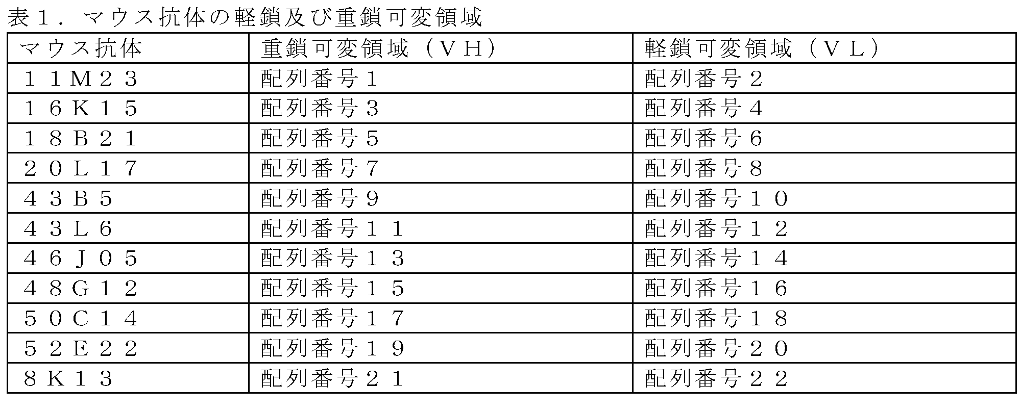
>11M23_vh(配列番号1)
QVQLKESGPDLVAPSQSLSITCTVSGFSLTNYGVHWVRQPPGKGLEWLVVIWSDGRINYNSALKSRLSITKDNSKRQVFLKMNSLQIDDTAIYYCVRHPAFGPHAMDYWGQGISVTVSS
>11M23_vl(配列番号2)
DIVMTQDAPSIPVTPGESVSISCRSSKSLLNSNGNTYLYWFLQRPGQSPHLLLYRMSNPASGAPDRFSGSGSGTEFTLRISRVEAEDVGVYYCMQYLEYPPTFGAGTRLELK
>16K15_vh(配列番号3)
EVMLVESGGGLVRPGGSLKLSCAGSGITLSTYAMSWVRQTPERRLEWVASIISGGITYYLDSVKGRFTISRDNARNILYLQMSSLRSEDTAIYYCARKYHGNALDYWGQGTSVTVSL
>16K15_vl(配列番号4)
DIVMTQSPSSLPVTAGETVTMRCKSSQSLLNSGNQRNYLTWYQRKPGQPPKKLIYWASTRESGVPDRFTGSGSGTDFTLTISGVQAEDLAVYYCQNNYFYPLTFGAGTKLELK
>18B21_vh(配列番号5)
QIQMVQSGPELKKPGETVRISCKASGYSFTTAGMQWVRKMPGEGLKWIGWIIAHSGEPKYTEDFKGRFAFSLETSASTTYLQISNLKNEDTATYFCARWGKGNTMDYWGQGTSVIVSS
>18B21_vl(配列番号6)
DIVMTQSPSSLTVTAGEKVTMSCKSSQSLLNGGNQRNYLTWYQQKPGQPPKLLIYWASTRESGVPDRFTGSGSGTHFTLTISSVQAEDLAVYYCQNAYFFPLTFGAGTKLELK
>20L17_vh(配列番号7)
DVQLVESGGGLVQPGGSRKLSCAASGFTFSSFGMHWVRQAPEKGLEWVAYISSGSSTIYYPDTVKGRFTVSRDNPKNTLFLQMTSLRSEDTAMYYCVRLGPRGNVMDHWGQGTSVTVSS
>20L17_vl(配列番号8)
DIVMTQSPSSLTVTAGEKVTMSCKSSQSLLNSGNQRNYLTWYQQKPGQPPKLLIYWASTRESGVPDRFTGSGSGTDFTLTISSVQAEDLAVYYCQNVYFYPLTFGTGTKLELR
>43B5_vh(配列番号9)
DVQLQESGPDLVKPSQSLSLTCTVSGYSISGAYNWHWIRQFPGNKLEWLAYMQYSGSSNYNPSFKSRISISRDTSKNQFFLQLKSVTTEDTATYYCARMYNGNSFLYWGQGTLVTVSA
>43B5_vl(配列番号10)
DIVMTQSPSSLTVTAGEKVTMNCKSSQSLFNSGNQKNYLTWYQQKPGQPPRLLIYWASTRESGVPDRFTGSGSGTDFTLTISSVQAEDLSLYYCQNSYSYPLTFGAGTKLELK
>43L6_vh(配列番号11)
QVQLKESGPDLVAPSQSLSLTCSVSGFSLTSYGIHWVRQPPGKGLEWLVVIWSDGRTTYNSGLKSRLSISKDNSKSQVLLKMNSLRTDDTAIYYCVRHPAFGPHAMDYWGQGTSVTVSS
>43L6_vl(配列番号12)
DIVMTQAAPSVPVTPGESVSISCRSSKSLLNSNGNTYLYWFLQRPGQSPQLLIYRMSNLASGVPDRFSGSGSGTDFTLRISRVEAGDVGVYYCMQYLEYPVTFGAGTKLELK
>46J05_vh(配列番号13)
DVQLVESGGGLVQPGGSRKLSCAASGFTFSRFGMHWVRQAPKKGLEWVAYISSGSNTIYYADTVKGRFTISRDNPKNTLFLQTTSLRSEDTAIYYCGRLGFYGNSFDHWGQGTLVTVSA
>46J05_vl(配列番号14)
NILMTQSPSSLTVTAGEKVTMNCKSSQSLLNGGNQRNYLTWYQQKAGQPPKLLIYWASTRESGVPDRFTGGGSGTDFTLTISSVQAEDLALYYCQNSYYYPLTFGAGTKLELK
>48G12_vh(配列番号15)
EVQLRQSGPELVKPGASVKMSCKASGYTFTTYIINWVKQKPGQGLEWIGYINPYNDDTRYNERVKGKATLTSDKSSSTAYMELSSLTSEDSAVYYCARFYFGNSFTYWGQGTLVTVSA
>48G12_vl(配列番号16)
DIVMTQSPSSLPVTVGERVTMTCKSSQGLFNSGNQRNYLTWYQQKPGQPPKLLIYWASTRESGVPDRFTGSGSGTDFTLTISSVQAEDLAIYYCQNNYIYPLTFGAGTKLELK
>50C14_vh(配列番号17)
EVQLRQSGPELVKPGASVKMSCKASGYTFTTYIINWVKQKPGQGLEWIGYINPYNDGTRYNERVKGKATLTSDKSSSTAYMELSSLTSEDSAVYYCARFHFGNSFTYWGQGTLVTVSA
>50C14_vl(配列番号18)
DIVMTQSPSSLPVTTGEKVTMTCKSSQGLFNNGNQRNYLTWYQQKPGQPPKLLIYWASTRESGVPDRFIGSGSGTDFTLTISSVQAEDLAIYYCQNNYIFPLTFGAGTKLELK
>52E22_vh(配列番号19)
QIQLVQSGPELKKPGETVKISCKASGYTLTNYGMNWVRQAPGKGLKWMGWIRPNTGEPTYAEDFKGRFVFSLETSAATAYLQITNLKSEDTSTYFCARLYRGNTLDNWGQGTSVIVSS
>52E22_vl(配列番号20)
DIVMTQSPSSLTVTTGEKVTMSCKSSQNLLNSGNQRNYLTWYQQKPGQSPKLLIYWASTRESGVPYRFTGSGSGTDFTLTISSVQTDDLAIYYCQNGYSFPFTFGSGTKLEIK
>8K13_vh(配列番号21)
QVHLQQSGAELVRPGSSVKISCKASGYAFSNYWMNWVRQRPGQGLEWIGQIYPGNGDTKYSGKFNSKDTLTADKSSNTAYMQLNSLTSEDSAVYFCARFYYGNVMDYWGQGTSVTVSS
>8K13_vl(配列番号22)
DIVLTQSPSSLTVTAGEKVTMSCKSSQTLLNGGNQKNYLTWYQQKSGQPPKLLIYWASTRESGVPDRFTGSGSGTDFTLTISSVQAEDLAVYYCQNGYSYPLTFGVGTKLELK
>11M23_vh (SEQ ID NO: 1)
QVQLKESGPDLVAPSQSLSITCTVS GFSLTNYGVH WVRQPPGKGLEWL VVIWSDGRINYNSALKS RLSITKDNSKRQVFLKMNSLQIDDTAIYYCVRHP AFGPHAMDY WGQGISVTVSS
>11M23_vl (SEQ ID NO: 2)
DIVMTQDAPSIPVTPGESVSISC RSSKSLLNSNGNTYLY WFLQRPGQSPHLLLY RMSNPAS GAPDRFSGSGSGTEFTLRISRVEAEDVGVYYC MQYLEYPPT FGAGTRLELK
>16K15_vh (SEQ ID NO: 3)
EVMLVESGGGLVRPGGSLKLSCAGS GITLSTYAMS WVRQTPERRLEWVA SIISGGITYYLDSVKG RFTISRDNARNILYLQMSSLRSEDTAIYYCAR KYHGNALDY WGQGTSVTVSL
>16K15_vl (SEQ ID NO: 4)
DIVMTQSPSSLPVTAGETVTMRC KSSQSLLNSGNQRNYLT WYQRKPGQPPKKLIY WASTRES GVPDRFTGSGSGTDFTLTISGVQAEDLAVYYC QNNYFYPLT FGAGTKLELK
>18B21_vh (SEQ ID NO: 5)
QIQMVQSGPELKKPGETVRISCKAS GYSFTTAGMQ WVRKMPGEGLKWI GWIIAHSGEPKYTEDFKG RFAFSLETSASTTYLQISNLKNEDTATYFCAR WGKGNTMDY WGQGTSVIVSS
>18B21_vl (SEQ ID NO: 6)
DIVMTQSPSSLTVTAGEKVTMSC KSSQSLLNGGNQRNYLT WYQQKPGQPPKLLIY WASTRES GVPDRFTGSGSGTHFTLTISSVQAEDLAVYYC QNAYFFPLT FGAGTKLELK
>20L17_vh (SEQ ID NO: 7)
DVQLVESGGGLVQPGGSRKLSCAAS GFTFSSFGMH WVRQAPEKGLEWV AYISSGSSTIYYPDTVKG RFTVSRDNPKNTLFLQMTSLRSEDTAMYYCVRLG PRGNVMDH WGQGTSVTVSS
>20L17_vl (SEQ ID NO: 8)
DIVMTQSPSSLTVTAGEKVTMSC KSSQSLLNSGNQRNYLT WYQQKPGQPPKLLIY WASTRES GVPDRFTGSGSGTDFTLTISSVQAEDLAVYYC QNVYFYPLT FGTGTKLELR
>43B5_vh (SEQ ID NO: 9)
DVQLQESGPDLVKPSQSLSLTCTVS GYSISGAYNWH WIRQFPGNKLEWL AYMQYSGSSNYNPSFKS RISISRDTSKNQFFLQLKSVTTEDTATYYCAR MYNGNSFLY WGQGTLVTVSA
>43B5_vl (SEQ ID NO: 10)
DIVMTQSPSSLTVTAGEKVTMNC KSSQSLFNSGNQKNYLT WYQQKPGQPPRLLIY WASTRES GVPDRFTGSGSGTDFTLTISSVQAEDLSLYYC QNSYSYPLT FGAGTKLELK
>43L6_vh (SEQ ID NO: 11)
QVQLKESGPDLVAPSQSLSLTCSVS GFSLTSYGIH WVRQPPGKGLEWL VVIWSDGRTTYNSGLKS RLSISKDNSKSQVLLKMNSLRTDDTAIYYCVRHP AFGPHAMDY WGQGTSVTVSS
>43L6_vl (SEQ ID NO: 12)
DIVMTQAAPSPVTPGESVSISC RSSKSLLNSNGNTYLY WFLQRPGQSPQLLIY RMSNLAS GVPDRFSGSGSGTDFTLRISRVEAGDVGVYYC MQYLEYPVT FGAGTKLELK
>46J05_vh (SEQ ID NO: 13)
DVQLVESGGGLVQPGGSRKLSCAAS GFTFSRFGMH WVRQAPKKGLEWV AYISSGSNTIYYADTVKG RFTISRDNPKNTLFLQTTSLRSEDTAIYYCGRLG FYGNSFDH WGQGTLVTVSA
>46J05_vl (SEQ ID NO: 14)
NILMTQSPSSLTVTAGEKVTMNC KSSQSLLNGGNQRNYLT WYQQKAGQPPKLLIY WASTRES GVPDRFTGGGSGTDFTLTISSVQAEDLALYYC QNSYYYPLT FGAGTKLELK
>48G12_vh (SEQ ID NO: 15)
EVQLRQSGPELVKPGASVKMSCKAS GYTFTTYIIN WVKQKPGQGLEWI GYINPYNDDTRYNERVKG KATLTSDKSSSTAYMELSSLTSEDSAVYYCAR FYFGNSFTY WGQGTLVTVSA
>48G12_vl (SEQ ID NO: 16)
DIVMTQSPSSLPVTVGERVTMTC KSSQGLFNSGNQRNYLT WYQQKPGQPPKLLIY WASTRES GVPDRFTGSGSGTDFTLTISSVQAEDLAIYYC QNNYIYPLT FGAGTKLELK
>50C14_vh (SEQ ID NO: 17)
EVQLRQSGPELVKPGASVKMSCKAS GYTFTTYIIN WVKQKPGQGLEWI GYINPYNDGTRYNERVKG KATLTSDKSSSTAYMELSSLTSEDSAVYYCAR FHFGNSFTY WGQGTLVTVSA
>50C14_vl (SEQ ID NO: 18)
DIVMTQSPSSLPVTTGEKVTMTC KSSQGLFNNGNQRNYLT WYQQKPGQPPKLLIY WASTRES GVPDRFIGSGSGTDFTLTISSVQAEDLAIYYC QNNYIFPLT FGAGTKLELK
>52E22_vh (SEQ ID NO: 19)
QIQLVQSGPELKKPGETVKISCKAS GYTLTNYGMN WVRQAPGKGLKWM GWIRPNTGEPTYAEDFKG RFVFSLETSAATAYLQITNLKSEDTSTYFCAR LYRGNTLDN WGQGTSVIVSS
>52E22_vl (SEQ ID NO: 20)
DIVMTQSPSSLTVTTGEKVTMSC KSSQNLLNSGNQRNYLT WYQQKPGQSPKLLIY WASTRES GVPYRFTGSGSGTDFTLTISSVQTDDLAIYYC QNGYSFPFT FGSGTKLEIK
>8K13_vh (SEQ ID NO: 21)
QVHLQQSGAELVRPGSSVKISCKAS GYAFSNYWMN WVRQRPGQGLEWI GQIYPGNGDTKYSGKFNS KDTLTADKSSNTAYMQLNSLTSEDSAVYFCAR FYYGNVMDY WGQGTSVTVSS
>8K13_vl (SEQ ID NO: 22)
DIVLTQSPSSLTVTAGEKVTMSC KSSQTLLNGGNQKNYLT WYQQKSGQPPKLLIY WASTRES GVPDRFTGSGSGTDFTLTISSVQAEDLAVYYC QNGYSYPLT FGVGTKLELK
実施例2 抗ヒトCLDN18.2キメラ抗体の調製
各マウス抗ヒトCLDN18.2モノクローナル抗体の重鎖可変領域配列と、公開されたヒトモノクローナル抗体IgG1サブクラスの重鎖定常領域配列(配列番号124を参照されたい)とを共にスプライシングして、哺乳動物細胞発現ベクターへと構築し、また、各マウス抗ヒトCLDN18.2モノクローナル抗体の軽鎖可変領域配列と、公開されたヒトモノクローナル抗体カッパサブクラスの軽鎖定常領域配列(配列番号125を参照されたい)とを共にスプライシングして、哺乳動物細胞発現ベクターへと構築した。構築された抗ヒトCLDN18.2キメラ抗体の重鎖ベクターと軽鎖ベクターとをペアで混合し、ポリエチレンイミン(PEI)を用いてHEK293細胞にベクターをトランスフェクトし、約7日後に細胞上清を回収した。抗ヒトCLDN18.2キメラ抗体タンパク質を、プロテインA精製を介して得た。
Example 2 Preparation of Anti-Human CLDN18.2 Chimeric Antibodies The heavy chain variable region sequences of each murine anti-human CLDN18.2 monoclonal antibody and the heavy chain constant region sequences of the published human monoclonal antibody IgG1 subclass (see SEQ ID NO: 124). ) were spliced together into a mammalian cell expression vector, and the light chain variable region sequences of each mouse anti-human CLDN18.2 monoclonal antibody and the light chain constants of the published human monoclonal antibody kappa subclass The region sequence (see SEQ ID NO: 125) was spliced together and constructed into a mammalian cell expression vector. The heavy chain vector and the light chain vector of the constructed anti-human CLDN18.2 chimeric antibody were mixed in pairs, the vector was transfected into HEK293 cells using polyethyleneimine (PEI), and after about 7 days the cell supernatant was collected. Recovered. Anti-human CLDN18.2 chimeric antibody protein was obtained via Protein A purification.
本開示のキメラ抗体を、フォーマットに従って「マウス抗体略語-xiIgG」と命名した。 The chimeric antibody of this disclosure was named "mouse antibody abbreviation-xiIgG" according to the format.
実施例3 抗ヒトCLDN18.2キメラ抗体のin vitroでの細胞結合アッセイ
キメラ抗ヒトCLDN18.2抗体を初期濃度100nMから勾配で2倍に希釈し、全部で16濃度の各抗体の溶液を得た。各濃度の各抗体の溶液10μlを384ウェルプレートに添加した。細胞表面にCLDN18.2を発現しているCHOK1細胞を室温にて100gで5分間遠心分離により回収し、0.5%BSAを含むPBSで細胞を1回洗浄した後、室温にて100gで5分間遠心分離した。細胞を約2×10⁶細胞/mlの密度で再懸濁し、抗体が添加された384ウェルプレートの各ウェルに10μlを加えた。4℃で1時間インキュベートした後、蛍光標識した二次ヤギ抗ヒトIgG抗体を加えた。4℃で1時間のインキュベーションを継続した後、細胞集団の平均蛍光読み取り値をフローサイトメーターで分析した。
Example 3 In Vitro Cell Binding Assay of Anti-Human CLDN18.2 Chimeric Antibodies Chimeric anti-human CLDN18.2 antibodies were diluted in two-fold gradients from an initial concentration of 100 nM, resulting in a total of 16 concentrations of each antibody in solution. . A 10 μl solution of each antibody at each concentration was added to a 384-well plate. CHOK1 cells expressing CLDN18.2 on the cell surface were collected by centrifugation at 100 g for 5 minutes at room temperature, washed once with PBS containing 0.5% BSA, and then centrifuged at 100 g for 5 minutes at room temperature. Centrifuge for 1 minute. Cells were resuspended at a density of approximately 2×10 6 cells/ml and 10 μl was added to each well of a 384-well plate to which antibody was added. After incubation for 1 hour at 4° C., a fluorescently labeled secondary goat anti-human IgG antibody was added. After continued incubation at 4° C. for 1 hour, the mean fluorescence reading of the cell population was analyzed with a flow cytometer.
結果は、キメラ抗体がnMスケールでヒトCLDN18.2を発現する細胞に特異的な結合を有することを示した。図1を参照されたい。 The results showed that the chimeric antibody had specific binding to cells expressing human CLDN18.2 at the nM scale. See FIG.
実施例4 抗ヒトCLDN18.2マウス抗体のヒト化
Kabatの包括的な分析、及びChothiaの抗体コードスキームに基づいて、各マウス抗体の重鎖及び軽鎖の6つの相補性決定領域(CDR)のアミノ酸配列領域、並びに保存された三次元立体構造を支持するフレームワーク領域を決定した。続いて、主にIGHVl | IGHJ4*01等のマウス抗体に類似するヒト抗体の重鎖可変領域を、既知のヒト抗体配列について検索し、選択した。フレームワーク領域配列を鋳型として選択し、マウス抗体の重鎖CDRをヒト抗体のフレームワーク領域と合わせ、最終的にヒト化重鎖可変領域配列を生成した。同様にして、ヒト化軽鎖可変領域配列を生成した。
Example 4 Humanization of anti-human CLDN18.2 mouse antibody
Based on Kabat's comprehensive analysis and Chothia's antibody coding scheme, the amino acid sequence regions of the six complementarity-determining regions (CDRs) of the heavy and light chains of each murine antibody, as well as the conserved three-dimensional conformation, were identified. The supporting framework regions were determined. Subsequently, human antibody heavy chain variable regions that are largely similar to murine antibodies, such as IGHVl | IGHJ4*01, were searched for known human antibody sequences and selected. The framework region sequences were selected as templates and the mouse antibody heavy chain CDRs were combined with the human antibody framework regions to finally generate the humanized heavy chain variable region sequences. Similarly, humanized light chain variable region sequences were generated.
マウスCDRをヒトフレームワーク領域に直接移植した抗体は、しばしば結合活性の劇的な低下を示すため、フレームワーク領域内の個々のアミノ酸をヒトからマウスに戻す変換を必要とする。どの位置を元のマウス残基に戻す必要があるかを決定するため、設計したヒト化抗体配列と元のマウス抗体配列とを比較して、アミノ酸の違いを確認し、それらの異なるアミノ酸が抗体構造を支持する、又は抗原に結合するために重要であるかを確認する必要がある。ヒト化設計によって得られた配列を、N(アスパラギン)グリコシル化部位、N-脱アミド化部位、D(アスパラギン酸)異性化部位等の潜在的な翻訳後修飾部位(PTM)について確認する必要がある。 Antibodies grafted with murine CDRs directly into human framework regions often show a dramatic reduction in binding activity, requiring conversion of individual amino acids within the framework regions back from human to murine. To determine which positions need to be returned to the original murine residues, the designed humanized antibody sequence is compared to the original murine antibody sequence to identify amino acid differences and identify those different amino acids in the antibody. It is necessary to confirm whether it is important for supporting the structure or binding to the antigen. Sequences resulting from humanized designs should be checked for potential post-translational modification sites (PTMs) such as N (asparagine) glycosylation sites, N-deamidation sites, D (aspartic acid) isomerization sites, etc. be.
各ヒト化重鎖可変領域配列の遺伝子を、ヒトモノクローナル抗体IgG1サブクラスの重鎖定常領域配列の遺伝子を含む哺乳動物細胞発現ベクターに構築し、各ヒト化軽鎖可変領域配列の遺伝子を、ヒトモノクローナル抗体カッパサブクラスの軽鎖定常領域配列の遺伝子を含む哺乳動物細胞発現ベクターに構築した。構築されたヒト化抗ヒトCLDN18.2抗体の重鎖ベクターと軽鎖ベクターとをペアで混合し、ポリエチレンイミン(PEI)を用いてベクターでHEK293細胞にトランスフェクトし、約7日後に細胞上清を回収した。抗ヒトCLDN18.2ヒト化抗体タンパク質を、プロテインA精製を介して得た。 The gene for each humanized heavy chain variable region sequence was constructed into a mammalian cell expression vector containing the gene for the heavy chain constant region sequence of a human monoclonal antibody IgG1 subclass, and the gene for each humanized light chain variable region sequence was constructed into a human monoclonal antibody. It was constructed into a mammalian cell expression vector containing the gene for the light chain constant region sequence of the antibody kappa subclass. The heavy chain vector and light chain vector of the constructed humanized anti-human CLDN18.2 antibody were mixed in pairs, transfected into HEK293 cells with the vector using polyethyleneimine (PEI), and after about 7 days the cell supernatant was collected. recovered. Anti-human CLDN18.2 humanized antibody protein was obtained via Protein A purification.
本開示のヒト化抗体を、フォーマットに従って「マウス抗体略語-hz」と命名した。それらのヒトフレームワーク領域に直接移植されたマウス抗体由来のCDRを有する抗体を、フォーマットに従って「マウス抗体略語-hz00」と命名し、更に操作された抗体を、ヒト化配列の番号でナンバリングした。 The humanized antibody of this disclosure was named "mouse antibody abbreviation-hz" following the format. Antibodies with CDRs derived from murine antibodies directly grafted into their human framework regions were named "mouse antibody abbreviation-hz00" according to the format, and further engineered antibodies were numbered with their humanized sequence numbers.
抗原であるヒトCLDN18.2に対するキメラ抗体及びそのヒト化抗体の結合速度論パラメータを、Fortebio(BLITZ pro1.1.0.28)機器により分析した。アッセイを行う前に、NTAバイオプローブをPBSに10分間浸漬し、次いで、100nM抗原を含むPBS中にプローブを300秒間入れ、Hisタグ付き抗原を捕捉した。プローブを更に、400秒の結合時間にわたって100nM抗体との結合反応に供し、次いで、プローブをPBSに移し、600秒間解離反応に供した。アッセイが終了すると、ブランク対照の応答値を差し引いたデータを、ソフトウェアを使用して1:1ラングミュア結合モデルに当てはめ、抗原抗体結合の速度論定数を計算した。結果を表2に示す。 The binding kinetic parameters of the chimeric antibody against the antigen, human CLDN18.2, and its humanized antibody were analyzed by Fortebio (BLITZ pro 1.1.0.28) instrument. Prior to performing the assay, the NTA bioprobes were soaked in PBS for 10 minutes and then placed in PBS containing 100 nM antigen for 300 seconds to capture the His-tagged antigen. Probes were further subjected to a binding reaction with 100 nM antibody for a binding time of 400 seconds, then probes were transferred to PBS and subjected to a dissociation reaction for 600 seconds. Once the assay was completed, the blank control subtracted data were fitted to a 1:1 Langmuir binding model using software to calculate the kinetic constants for antigen-antibody binding. Table 2 shows the results.

結果は、11M23-hz22の解離定数が、マウスキメラ抗体に対して、ヒト化及び重要な翻訳後修飾部位(PTM)の除去後に10倍を超えて上昇し、したがって候補のリストから排除されたことを示した。残りの8K13-hz24、16K15-hz22及び52E22-hz12の解離定数は、元のマウスキメラ抗体と比較して1倍~4倍上昇し、その後の研究のリード分子として使用することができた。16K15-hz22の軽鎖アミノ酸配列は2つの連続する「NN」残基を含み、更に最適化された変異抗体16K15-hz22_2及び16K15-hz22_3は、「NN」残基をそれぞれ「SN」残基及び「QN」残基に変更することによって得られた。 The results showed that the dissociation constant of 11M23-hz22 was increased over 10-fold relative to the mouse chimeric antibody after humanization and removal of key post-translational modification sites (PTMs), thus eliminating it from the candidate list. showed that. The dissociation constants of the remaining 8K13-hz24, 16K15-hz22 and 52E22-hz12 were increased 1- to 4-fold compared to the original mouse chimeric antibody and could be used as lead molecules for subsequent studies. The light chain amino acid sequence of 16K15-hz22 contains two consecutive "NN" residues, and further optimized mutant antibodies 16K15-hz22_2 and 16K15-hz22_3 replace the "NN" residues with "SN" residues and It was obtained by changing to a "QN" residue.

>16K15_vl_hz2(配列番号24)
DIVMTQSPDSLAVSLGERATINCKSSQSLLSSGNQRNYLTWYQQKPGQPPKKLIYWASTRESGVPDRFSGSGSGTDFTLTISSLQAEDLAVYYCQNNYFYPLTFGQGTKLEIK
>16K15_vl_hz2_N-S(配列番号25)
DIVMTQSPDSLAVSLGERATINCKSSQSLLSSGNQRNYLTWYQQKPGQPPKKLIYWASTRESGVPDRFSGSGSGTDFTLTISSLQAEDLAVYYCQSNYFYPLTFGQGTKLEIK
>16K15_vl_hz2_N-Q(配列番号26)
DIVMTQSPDSLAVSLGERATINCKSSQSLLSSGNQRNYLTWYQQKPGQPPKKLIYWASTRESGVPDRFSGSGSGTDFTLTISSLQAEDLAVYYCQQNYFYPLTFGQGTKLEIK
>16k15_vh_hz2(配列番号23)
EVQLVESGGGLVQPGGSLRLSCAGSGITLSTYAMSWVRQAPGKGLEWVSSIISGGITYYLDSVKGRFTISRDNAKNTLYLQMSSLRAEDTAVYYCARKYHGNALDYWGQGTLVTVSS
>52E22_vl_hz2(配列番号28)
DIVMTQSPDSLAVSLGERATINCKSSQNLLSSGNQRNYLTWYQQKPGQPPKLLIYWASTRESGVPDRFSGSGSGTDFTLTISSLQAEDLAVYYCQQGYSFPFTFGQGTKLEIK
>52E22_vh_hz1(配列番号27)
QIQLVQSGSELKKPGASVKVSCKASGYTLTNYGMNWVRQAPGQGLEWMGWIRPNTGEPTYAEDFKGRFVFSLDTSVATAYLQITSLKAEDTAVYYCARLYRGNTLDNWGQGTLVTVSS
>8K13_vl_hz4_N-S_N-Q(配列番号30)
DIVLTQSPDSLAVSLGERATINCKSSQTLLSGGNQKNYLTWYQQKPGQPPKLLIYWASTRESGVPDRFSGSGSGTDFTLTISSLQAEDLAVYYCQQGYSYPLTFGQGTKLEIK
>8K13_vh_hz2(配列番号29)
QVQLVQSGAEVKKPGSSVKVSCKASGYAFSNYWMNWVRQAPGQGLEWMGQIYPGSGDTKYSGKFQSRVTITADKSTNTAYMELSSLRSEDTAVYYCARFYYGNVMDYWGQGTLVTVSS
>16K15_vl_hz2 (SEQ ID NO: 24)
DIVMTQSPDSLAVSLGERATINC KSSQSLLSSGNQRNYLT WYQQKPGQPPKKLIY WASTRES GVPDRFSGSGSGTDFTLTISSLQAEDLAVYYC QNNYFYPLT FGQGTKLEIK
>16K15_vl_hz2_NS (SEQ ID NO: 25)
DIVMTQSPDSLAVSLGERATINC KSSQSLLSSGNQRNYLT WYQQKPGQPPKKLIY WASTRES GVPDRFSGSGSGTDFTLTISSLQAEDLAVYYC QSNYFYPLT FGQGTKLEIK
>16K15_vl_hz2_NQ (SEQ ID NO: 26)
DIVMTQSPDSLAVSLGERATINC KSSQSLLSSGNQRNYLT WYQQKPGQPPKKLIY WASTRES GVPDRFSGSGSGTDFTLTISSLQAEDLAVYYC QQNYFYPLT FGQGTKLEIK
>16k15_vh_hz2 (SEQ ID NO: 23)
EVQLVESGGGLVQPGGSLRLSCAGS GITLSTYAMS WVRQAPGKGLEWVS SIISGGITYYLDSVKG RFTISRDNAKNTLYLQMSSLRAEDTAVYYCAR KYHGNALDY WGQGTLVTVSS
>52E22_vl_hz2 (SEQ ID NO: 28)
DIVMTQSPDSLAVSLGERATINC KSSQNLLSSGNQRNYLT WYQQKPGQPPKLLIY WASTRES GVPDRFSGSGSGTDFTLTISSLQAEDLAVYYC QQGYSFPFT FGQGTKLEIK
>52E22_vh_hz1 (SEQ ID NO: 27)
QIQLVQSGSELKKPGASVKVSCKAS GYTLTNYGMN WVRQAPGQGLEWMG WIRPNTGEPTYAEDFKG RFVFSLDTSVATAYLQITSLKAEDTAVYYCAR LYRGNTLDN WGQGTLVTVSS
>8K13_vl_hz4_NS_NQ (SEQ ID NO: 30)
DIVLTQSPDSLAVSLGERATINC KSSQTLLSGGNQKNYLT WYQQKPGQPPKLLIY WASTRES GVPDRFSGSGSGTDFTLTISSLQAEDLAVYYC QQGYSYPLT FGQGTKLEIK
>8K13_vh_hz2 (SEQ ID NO: 29)
QVQLVQSGAEVKKPGSSVKVSCKAS GYAFSNYWMN WVRQAPGQGLEWM GQIYPGSGDTKYSGKFQS RVTITADKSTNTAYMELSSLRSEDTAVYYCAR FYYGNVMDY WGQGTLVTVSS
実施例5 抗ヒトCLDN18.2ヒト化抗体のin vitroでの結合親和性の速度実験
抗体-抗原相互作用を、GE Healthcare製のBIAcore S200を用いて測定した。
Example 5 In Vitro Binding Affinity Kinetic Studies of Anti-Human CLDN18.2 Humanized Antibodies Antibody-antigen interactions were measured using a BIAcore S200 from GE Healthcare.
GE Healthcare製のヒト抗体捕捉キット(カタログ番号BR-1008-39、ロット10261753)に提供される説明書を参照して、CM5センサーチップ上の分析チャネル及び対照試料チャネルを最初に飽和させ、最大量の抗ヒトFc抗体と結合させ、次いで、抗ヒトCLDN18.2キメラ抗体、ヒト化抗体又は対照抗体IMAB362を7.5μg/mlで含む緩衝液を分析チャネルに流して、均一に分配させ、最後に勾配で希釈した抗原試料(初期濃度を20nMとし、1:3で希釈して8濃度を得て、濃度0.741nMを繰り返すように設定した)を分析チャネルと試料チャネルとの両方に流し、抗体の抗原への結合時の光反応を測定した。続いて、各抗体の会合定数Kon及び解離定数Koff及び親和性定数KDを、最終的に機器ソフトウェアフィッティング分析により得た。 Refer to the instructions provided in the Human Antibody Capture Kit from GE Healthcare (Catalog No. BR-1008-39, Lot 10261753) to first saturate the analysis and control sample channels on the CM5 sensor chip and extract the maximum volume. of anti-human Fc antibody, then anti-human CLDN18.2 chimeric antibody, humanized antibody or control antibody IMAB362 at 7.5 μg/ml in buffer is flowed through the analysis channel for uniform distribution, and finally A gradient diluted antigen sample (starting at 20 nM, diluted 1:3 to give 8 concentrations, set to repeat concentration 0.741 nM) was run through both the analysis and sample channels, and antibody The photoresponse upon binding to the antigen was measured. The association and dissociation constants Koff and affinity constants KD for each antibody were then finally obtained by instrument software fitting analysis.
結果は、抗ヒトCLDN18.2ヒト化抗体のin vitro結合親和性定数が、元のマウス抗体の結合親和性定数と有意に異ならない一方で、IMAB362の結合親和性定数よりも1桁低いことを示した。表4を参照されたい。 The results show that the in vitro binding affinity constant of the anti-human CLDN18.2 humanized antibody is not significantly different from that of the original murine antibody, while it is an order of magnitude lower than that of IMAB362. Indicated. See Table 4.
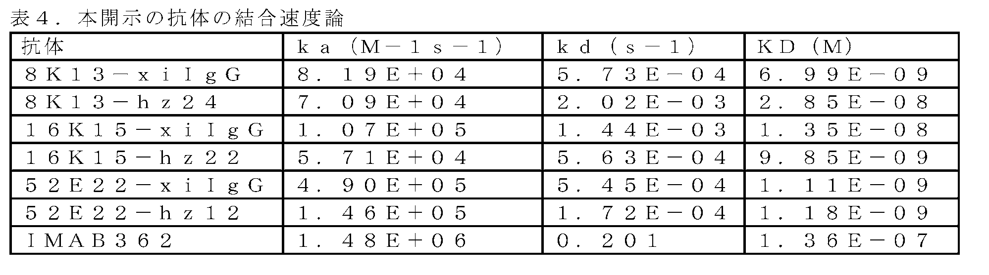
実施例6 抗ヒトCLDN18.2ヒト化抗体のin vitroでの細胞診断アッセイ
6.1 補体依存性細胞傷害(CDC)
本開示のヒト化抗体について、Quidel, Inc.によって入手可能な市販のヒト血清全補体を用いて、ヒトCLDN18.2を安定して発現するCHOK1細胞、BxPC3細胞及びNCI-N87細胞上の補体依存性細胞傷害(CDC)を誘導する能力を分析した。
Example 6 In Vitro Cytodiagnostic Assay of Anti-Human CLDN18.2 Humanized Antibody 6.1 Complement Dependent Cytotoxicity (CDC)
Humanized antibodies of the present disclosure were complemented on CHOK1, BxPC3 and NCI-N87 cells stably expressing human CLDN18.2 using commercially available whole human serum complement available by Quidel, Inc. Ability to induce body dependent cytotoxicity (CDC) was analyzed.
細胞を、250μg/ml~3.8ng/mlの範囲の最終濃度で試験するために抗体と混合し、細胞培養培地RPMI-1640に溶解した6.25%の濃度の全ヒト血清を混合物に添加し、次いでこれを37℃で3時間インキュベートした。細胞傷害性をCCK-8キットにより測定し、最後に、450nmにおける吸光度をMDプレートリーダーにより測定した。試料のEC50値を、softmax pro7ソフトウェアを使用して吸光度値を4パラメータフィッティング曲線にプロットすることによって計算した。 Cells were mixed with antibodies for testing at final concentrations ranging from 250 μg/ml to 3.8 ng/ml, and a concentration of 6.25% whole human serum dissolved in cell culture medium RPMI-1640 was added to the mixture. and then incubated at 37° C. for 3 hours. Cytotoxicity was measured by CCK-8 kit and finally absorbance at 450 nm was measured by MD plate reader. EC50 values of samples were calculated by plotting absorbance values to a 4-parameter fitting curve using softmax pro7 software.
結果は、抗ヒトCLDN18.2ヒト化抗体は、標的発現細胞に対して特異的補体依存性細胞傷害(CDC)を有し、その殺細胞活性がIMAB362よりも有意に優れていることが示された。図2及び表5を参照されたい。 The results show that the anti-human CLDN18.2 humanized antibody has specific complement-dependent cytotoxicity (CDC) against target-expressing cells, and its cell-killing activity is significantly superior to IMAB362. was done. See FIG. 2 and Table 5.

6.2 抗体依存性細胞傷害(ADCC)
FcγRIIIa-FcεRIaγハイブリッド受容体を安定に発現し、NFAT応答エレメントによる駆動下でホタルルシフェラーゼを発現する操作されたJurkat細胞をエフェクター細胞として用いた。ADCC機構における抗体の生物学的活性は、NFAT経路の活性化により産生されるルシフェラーゼにより定量される。1.5E5エフェクター細胞を、33μg/ml~85pg/mlの範囲の最終濃度で試験するために抗体と混合し、次いで2.5E4標的細胞を混合物に添加し(エフェクター対標的E:T比は6:1であった)、次いでこれを37℃で16時間インキュベートした。細胞傷害性を、Promega製のキットBio-Glo(商標)ルシフェラーゼアッセイシステムによって測定し、最終的にMDプレートリーダーによってLUM値を決定した。
6.2 Antibody Dependent Cytotoxicity (ADCC)
Engineered Jurkat cells stably expressing the FcγRIIIa-FcεRIaγ hybrid receptor and expressing firefly luciferase under the drive of an NFAT response element were used as effector cells. Antibody biological activity in the ADCC mechanism is quantified by luciferase produced by activation of the NFAT pathway. 1.5E5 effector cells were mixed with antibody for testing at final concentrations ranging from 33 μg/ml to 85 pg/ml, then 2.5E4 target cells were added to the mixture (effector to target E:T ratio of 6 : 1), which was then incubated at 37°C for 16 hours. Cytotoxicity was measured by the kit Bio-Glo™ luciferase assay system from Promega and finally LUM values were determined by MD plate reader.
データを次のように処理した:誘導倍率=(検出されたウェルの読み取り値-バックグラウンド値)/(陰性対照ウェルの読み取り値-バックグラウンド値)。試料のEC50値を、データを4パラメータフィッティング曲線にプロットすることによって計算した。 Data were processed as follows: fold induction = (reading of detected wells - background)/(reading of negative control wells - background). EC50 values for samples were calculated by plotting the data to a four-parameter fitting curve.
結果は、抗ヒトCLDN18.2ヒト化抗体が、標的発現細胞に対して特異的抗体依存性細胞傷害(ADCC)を有し、それらの殺細胞活性はIMAB362に匹敵することを示した。図3及び表6を参照されたい。 The results showed that the anti-human CLDN18.2 humanized antibodies possessed specific antibody-dependent cellular cytotoxicity (ADCC) against target-expressing cells and their cell-killing activity was comparable to IMAB362. See FIG. 3 and Table 6.

実施例7 同ファミリーのタンパク質に対する抗ヒトCLDN18.2ヒト化抗体の結合特性の分析
ヒトCLDN18.2及びCLDN18.1の遺伝子をそれぞれ真核生物発現ベクターに構築し、ポリエチレンイミン(PEI)を用いてベクターでHEK293細胞にトランスフェクトした。トランスフェクションの3日後、遠心分離により細胞を回収し、PBSで1回洗浄し、細胞密度2×10⁶/mlで再懸濁した。384ウェルプレートの各ウェルに10μlを加え、次いで異なる濃度のヒト化抗体をウェルに加えた。4℃で1時間インキュベートした後、蛍光標識した二次ヤギ抗ヒトIgG抗体を加えた。4℃で1時間のインキュベーションを継続した後、384ウェルプレートの平均蛍光読み取り値をフローサイトメーターで読み取った。データを分析して、抗ヒトCLDN18.2ヒト化抗体に対する細胞の結合特性を得た。実験に使用した陽性対照は市販の抗ヒトCLDN18ウサギmAb 34H14L15(Abcam社から入手可能)であり、陰性アイソタイプ対照は全長の抗CD33 IgG抗体であるリンツズマブであった。
Example 7 Analysis of Binding Properties of Anti-Human CLDN18.2 Humanized Antibodies to Proteins of the Same Family The genes for human CLDN18.2 and CLDN18.1 were each constructed into eukaryotic expression vectors and expressed using polyethyleneimine (PEI). Vectors were transfected into HEK293 cells. Three days after transfection, cells were harvested by centrifugation, washed once with PBS, and resuspended at a cell density of 2×10 6 /ml. 10 μl was added to each well of a 384-well plate, then different concentrations of humanized antibodies were added to the wells. After incubation for 1 hour at 4° C., a fluorescently labeled secondary goat anti-human IgG antibody was added. After continued incubation at 4°C for 1 hour, the mean fluorescence reading of the 384-well plate was read on a flow cytometer. The data were analyzed to obtain the binding characteristics of the cells to the anti-human CLDN18.2 humanized antibody. The positive control used in the experiment was the commercial anti-human CLDN18 rabbit mAb 34H14L15 (available from Abcam) and the negative isotype control was lintuzumab, a full-length anti-CD33 IgG antibody.
結果は、抗ヒトCLDN18.2ヒト化抗体が、ヒトCLDN18.2には結合するが、ヒトCLDN18.1には結合しないという特性を示したことを示した。図4を参照されたい。また、低濃度、中濃度及び高濃度でのヒトCLDN18.1細胞への非特異的結合は、全てIMAB362のものよりも低かった。図5を参照されたい。 The results showed that the anti-human CLDN18.2 humanized antibody exhibited the property of binding to human CLDN18.2 but not to human CLDN18.1. Please refer to FIG. Also, the non-specific binding to human CLDN18.1 cells at low, medium and high concentrations were all lower than that of IMAB362. See FIG.
実施例8 抗ヒトCLDN18.2ヒト化抗体のヒト、サル、及びマウスの種交差結合特性の分析
ヒト、マウス及びアカゲザルCLDN18.2の遺伝子をそれぞれ真核生物発現ベクターに構築し、ポリエチレンイミン(PEI)を用いてベクターでHEK293細胞にトランスフェクトした。細胞を2日後に回収した。抗ヒトCLDN18.2ヒト化抗体に対する細胞の結合特異性を、フローサイトメトリーを用いて分析した。フローサイトメトリーの手順については実施例7を参照されたい。
Example 8 Analysis of Human, Monkey, and Mouse Species Cross-Binding Properties of Anti-Human CLDN18.2 Humanized Antibodies ) was used to transfect HEK293 cells with the vector. Cells were harvested after 2 days. Binding specificity of cells to anti-human CLDN18.2 humanized antibody was analyzed using flow cytometry. See Example 7 for flow cytometry procedures.
結果は、抗ヒトCLDN18.2ヒト化抗体16K15-22 IgGが3つの種の全てのCLDN18.2に結合し、8K13-24 IgG及び52E22-12 IgGはアカゲザルCLDN18.2に結合したがマウスCLDN18.2には結合しなかったことを示した。結果を図6に示す。 The results showed that the anti-human CLDN18.2 humanized antibody 16K15-22 IgG bound to all three species of CLDN18.2, 8K13-24 IgG and 52E22-12 IgG bound to rhesus monkey CLDN18.2 but mouse CLDN18. 2 showed no binding. The results are shown in FIG.
実施例9 抗ヒトCLDN18.2ヒト化抗体を用いたCAR-T細胞の調製及びその活性化
9.1 レンチウイルスのパッケージング
レンチウイルスのパッケージングを、表7に示すグループ分けに従って行った。異なる抗体遺伝子を含むレンチウイルスベクタープラスミドpLTRを最初に構築し、シーケンシングによりDNAが正しいことを確認した後、Qiagen製プラスミド抽出キットを用いてそれらのプラスミドDNAを抽出した。プラスミドDNAを滅菌TEに溶解し、その濃度及び純度をUV光吸収によって決定し、抽出されたプラスミドDNAのA260/A280が1.8~2.0であったことを確認した。2つのヘルパーパッケージングエレメントプラスミドであるpCMV-VSV-G及びpCMV-dR8.2のDNAも抽出した。トランスフェクション用のHEK293T細胞を調製し、約60%の培養密度まで増殖するはずの新たに継代した細胞を得た。3つのプラスミドを、リン酸カルシウムをトランスフェクション試薬として用いてHEK293T細胞に共移入した。トランスフェクションの48時間後、パッケージングされたウイルスを含む細胞上清を低温での遠心分離により回収し、0.45μmフィルターを用いて細胞の残骸を除去した。限外濾過遠沈管を用いてウイルスを濃縮し、サブパッケージを行って、-80℃の冷凍庫で保管した。少量のウイルス濃縮物を取って、ウイルス力価をFACSによって決定した。
Example 9 Preparation of CAR-T cells with anti-human CLDN18.2 humanized antibody and their activation 9.1 Lentiviral packaging Lentiviral packaging was performed according to the grouping shown in Table 7. Lentiviral vector plasmids pLTR containing different antibody genes were first constructed and after sequencing to confirm the correctness of the DNA, their plasmid DNA was extracted using Qiagen's plasmid extraction kit. The plasmid DNA was dissolved in sterile TE and its concentration and purity determined by UV light absorption, confirming that the A260/A280 of the extracted plasmid DNA was 1.8-2.0. DNA of two helper packaging element plasmids, pCMV-VSV-G and pCMV-dR8.2, was also extracted. HEK293T cells were prepared for transfection to obtain freshly passaged cells that should grow to approximately 60% confluency. The three plasmids were co-transfected into HEK293T cells using calcium phosphate as transfection reagent. Forty-eight hours after transfection, cell supernatant containing packaged virus was harvested by centrifugation at low temperature and cell debris was removed using a 0.45 μm filter. The virus was concentrated using ultrafiltration centrifuge tubes, subpackaged and stored in a −80° C. freezer. A small amount of virus concentrate was taken and the virus titer was determined by FACS.

表7に示す陰性対照はCLDN18.2に結合しない別の抗体であり、陽性対照はCarsgen Therapeutics製の抗体hu8E5(国際公開第2018006882号を参照されたい)であった。*:この一本鎖抗体は、ヒト化VHとVLとを短いペプチド(GSTSGGGSGGGSGGGGSS)を介して連結することによって形成された。 The negative control shown in Table 7 was another antibody that did not bind to CLDN18.2 and the positive control was antibody hu8E5 from Carsgen Therapeutics (see WO2018006882). *: This single-chain antibody was formed by linking humanized VH and VL via a short peptide (GSTSGGGSGGGSGGGGSS).
9.2 CAR-T細胞の調製
HBV、HCV、HIVについて陰性と検出された健康なドナーを選択した。前肘静脈から100mlの血液を採取し、Ficoll密度勾配遠心分離を実施してPBMCを含む白色層を単離した。CD3+T細胞を、3:1のDynaBeads/CD3+T細胞の比率でDynaBeads CD3/CD28(LifeTechnologies、カタログ番号40203D)を用いて単離した。活性化の24時間後、CD25+CD69+T細胞のパーセンテージをフローサイトメトリーによって検出した。活性化されると、5MOIのレンチウイルスを用いてCD3+T細胞に形質導入を行った。24ウェルプレートを、ノボネクチンを用いて37℃で2時間コーティングし、上記の手順により得られた細胞懸濁液を、各種レンチウイルス(MOI=5)、Synperonic(商標)F108(Sigma、カタログ番号07579-250G-F)及びTscm(2U/ml)とともに、形質導入用の懸濁液に製剤化した。形質導入用の懸濁液を、細胞密度を1.0E+06/mlに調整した24ウェルプレートに添加した。プレートを500gで30分間遠心分離し、37℃、5%CO2のインキュベーターに入れ、48時間静置培養した。形質導入後、細胞を、Tscm(最終濃度2U/ml)を隔日で補充した5%FBS X-vivo15培地(LONZA、カタログ番号04-418Q)で培養した。細胞を計数し、0.5E+06/mlに調整し、培養8日~10日目に採取した。
9.2 Preparation of CAR-T Cells Healthy donors who tested negative for HBV, HCV, HIV were selected. 100 ml of blood was collected from the antecubital vein and Ficoll density gradient centrifugation was performed to isolate the white layer containing PBMCs. CD3+ T cells were isolated using DynaBeads CD3/CD28 (LifeTechnologies, catalog number 40203D) at a ratio of 3:1 DynaBeads/CD3+ T cells. Twenty-four hours after activation, the percentage of CD25+CD69+ T cells was detected by flow cytometry. Once activated, a 5 MOI of lentivirus was used to transduce CD3+ T cells. 24-well plates were coated with Novonectin for 2 hours at 37° C., and the cell suspension obtained by the above procedure was infected with various lentiviruses (MOI=5), Synperonic™ F108 (Sigma, Catalog No. 07579). -250G-F) and Tscm (2 U/ml) were formulated into a suspension for transduction. The transduction suspension was added to a 24-well plate adjusted to a cell density of 1.0E+06/ml. The plate was centrifuged at 500 g for 30 minutes, placed in a 37° C., 5% CO 2 incubator, and cultured statically for 48 hours. After transduction, cells were cultured in 5% FBS X-vivo 15 medium (LONZA, Cat. No. 04-418Q) supplemented every other day with Tscm (2 U/ml final concentration). Cells were counted, adjusted to 0.5E+06/ml and harvested on days 8-10 of culture.
得られた各群におけるCAR-T細胞の陽性パーセンテージの結果を図7に示す。 The obtained results of positive percentage of CAR-T cells in each group are shown in FIG.
9.3 CAR-T細胞の活性化
抗原ヒトCLDN18.1及び抗原ヒトCLDN18.2を発現するCHO細胞を構築し、標的細胞として用いた。
9.3 Activation of CAR-T Cells CHO cells expressing antigen human CLDN18.1 and antigen human CLDN18.2 were constructed and used as target cells.
細胞の抗原発現の検出結果を図8に示す。CHO-ブランク(ブランク対照)及びCHO-CLDN18.1は抗原CLDN18.2の発現がなく、CHO-CLDN18.2は抗原CLDN18.2の発現が高かった。 FIG. 8 shows the results of detection of antigen expression in cells. CHO-Blank (blank control) and CHO-CLDN18.1 had no expression of antigen CLDN18.2 and CHO-CLDN18.2 had high expression of antigen CLDN18.2.
レンチウイルス形質導入を伴わないCAR-T細胞及びT細胞の各群を含む上述の密度調整されたエフェクター細胞を、それぞれ1.5mL遠沈管中で標的細胞と16:1のエフェクター対標的(effective to target)比で混合した。全量を、T細胞ブロスX-vivo15(自家血清及びTscmを含まない)を用いて200μLに補充し、次いで、200μLの系をそれぞれV底の96ウェルプレートに移し、24時間の共インキュベーションを行った。 Density-adjusted effector cells described above, including each population of CAR-T cells and T cells without lentiviral transduction, were each spiked with target cells at a 16:1 effector-to-target ratio in 1.5 mL centrifuge tubes. target) ratio. The total volume was supplemented to 200 μL with T cell broth X-vivo15 (free of autologous serum and Tscm), then 200 μL of each system was transferred to a V-bottomed 96-well plate and co-incubated for 24 hours. .
インキュベーション後、ヒトIFNγの検出のために各培養系の上清を採取した。IFNγ放出の有意な増加は、CHO-CLDN18.1及びCHO-CLDN18.2とのインキュベーション後の群417のCAR-T細胞において見出され、IFNγ放出の増加は、CHO-CLDN18.2とのインキュベーション後にのみ、群418、群419、及び群420のCAR-T細胞において見られた。結果を表8及び図9に示す。
After incubation, the supernatant of each culture was harvested for detection of human IFNγ. A significant increase in IFNγ release was found in
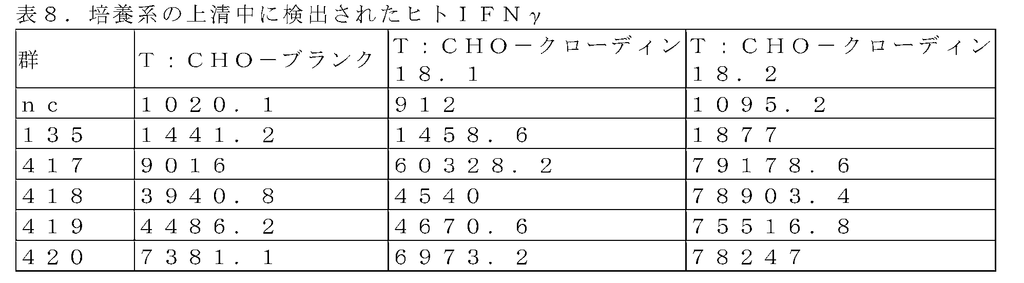
各群由来のCAR-T細胞にてT細胞活性化マーカータンパク質であるCD3/CD25を検出した。CD25発現は、CHO-CLDN18.1及びCHO-CLDN18.2とのインキュベーション後に群417のCAR-T細胞においてアップレギュレートされ、群418、群419、及び群420のCARTのCAR-T細胞ではCHO-CLDN18.2とのインキュベーション後にのみアップレギュレートされることがわかった。図10を参照されたい。
CD3/CD25, which is a T cell activation marker protein, was detected in CAR-T cells derived from each group. CD25 expression was upregulated in
本発明の実施形態に関する以上の説明は、本発明を限定することを意図するものではなく、当業者は、本発明の趣旨を逸脱しない範囲で本発明に種々の変更及び修正を加えることができ、これは添付の特許請求の範囲に含まれるべきである。 The above description of the embodiments of the present invention is not intended to limit the present invention, and those skilled in the art can make various changes and modifications to the present invention without departing from the spirit of the present invention. , which should be included in the appended claims.
Claims (24)
(1)表面にヒトクローディン18.2タンパク質を発現する細胞を免疫原として用いて、動物を免疫する工程と、
(2)工程(1)で免疫した前記動物を用いて抗体を産生することができる細胞クローンを調製する工程と、
(3)表面にヒトクローディン18.2タンパク質を発現する細胞を陽性スクリーニング抗原として用い、前記陽性スクリーニング抗原に対する結合活性を有する抗体、及び前記抗体を産生する細胞をスクリーニングする工程と、
(4)表面にヒトクローディン18.1タンパク質を発現する細胞を陰性スクリーニング抗原として用い、前記陰性スクリーニング抗原に対する結合活性を有する抗体、及び前記抗体を産生する細胞を排除する工程と、
を含む、調製方法。 A method for preparing an anti-human claudin-18.2 antibody, comprising:
(1) immunizing an animal with cells expressing human claudin-18.2 protein on their surface as an immunogen;
(2) preparing cell clones capable of producing antibodies using the animal immunized in step (1);
(3) using a cell expressing human claudin 18.2 protein on its surface as a positive screening antigen, screening for an antibody having binding activity to the positive screening antigen and for cells producing the antibody;
(4) using cells expressing human claudin 18.1 protein on their surface as a negative screening antigen, and excluding antibodies having binding activity to the negative screening antigen and cells producing the antibodies;
A method of preparation, comprising:
(1)配列番号31に示されるVH-CDR1、配列番号32に示されるVH-CDR2、及び配列番号33に示されるVH-CDR3;並びに配列番号34に示されるVL-CDR1、配列番号35に示されるVL-CDR2、及び配列番号36に示されるVL-CDR3;
(2)配列番号37に示されるVH-CDR1、配列番号38に示されるVH-CDR2、及び配列番号39に示されるVH-CDR3;並びに配列番号40に示されるVL-CDR1、配列番号41に示されるVL-CDR2、及び配列番号42に示されるVL-CDR3;
(3)配列番号43に示されるVH-CDR1、配列番号44に示されるVH-CDR2、及び配列番号45に示されるVH-CDR3;並びに配列番号46に示されるVL-CDR1、配列番号41に示されるVL-CDR2、及び配列番号47に示されるVL-CDR3;
(4)配列番号48に示されるVH-CDR1、配列番号49に示されるVH-CDR2、及び配列番号50に示されるVH-CDR3;並びに配列番号40に示されるVL-CDR1、配列番号41に示されるVL-CDR2、及び配列番号51に示されるVL-CDR3;
(5)配列番号52に示されるVH-CDR1、配列番号53に示されるVH-CDR2、及び配列番号54に示されるVH-CDR3;並びに配列番号55に示されるVL-CDR1、配列番号41に示されるVL-CDR2、及び配列番号56に示されるVL-CDR3;
(6)配列番号57に示されるVH-CDR1、配列番号58に示されるVH-CDR2、及び配列番号33に示されるVH-CDR3;並びに配列番号34に示されるVL-CDR1、配列番号59に示されるVL-CDR2、及び配列番号60に示されるVL-CDR3;
(7)配列番号61に示されるVH-CDR1、配列番号62に示されるVH-CDR2、及び配列番号63に示されるVH-CDR3;並びに配列番号46に示されるVL-CDR1、配列番号41に示されるVL-CDR2、及び配列番号64に示されるVL-CDR3;
(8)配列番号65に示されるVH-CDR1、配列番号66に示されるVH-CDR2、及び配列番号67に示されるVH-CDR3;並びに配列番号68に示されるVL-CDR1、配列番号41に示されるVL-CDR2、及び配列番号69に示されるVL-CDR3;
(9)配列番号65に示されるVH-CDR1、配列番号70に示されるVH-CDR2、及び配列番号71に示されるVH-CDR3;並びに配列番号72に示されるVL-CDR1、配列番号41に示されるVL-CDR2、及び配列番号73に示されるVL-CDR3;
(10)配列番号74に示されるVH-CDR1、配列番号75に示されるVH-CDR2、及び配列番号76に示されるVH-CDR3;並びに配列番号77に示されるVL-CDR1、配列番号41に示されるVL-CDR2、及び配列番号78に示されるVL-CDR3;
(11)配列番号79に示されるVH-CDR1、配列番号80に示されるVH-CDR2、及び配列番号81に示されるVH-CDR3;並びに配列番号82に示されるVL-CDR1、配列番号41に示されるVL-CDR2、及び配列番号83に示されるVL-CDR3;
(12)配列番号37に示されるVH-CDR1、配列番号38に示されるVH-CDR2、及び配列番号39に示されるVH-CDR3;並びに配列番号85に示されるVL-CDR1、配列番号41に示されるVL-CDR2、及び配列番号42に示されるVL-CDR3;
(13)配列番号37に示されるVH-CDR1、配列番号38に示されるVH-CDR2、及び配列番号39に示されるVH-CDR3;並びに配列番号85に示されるVL-CDR1、配列番号41に示されるVL-CDR2、及び配列番号86に示されるVL-CDR3;
(14)配列番号37に示されるVH-CDR1、配列番号38に示されるVH-CDR2、及び配列番号39に示されるVH-CDR3;並びに配列番号85に示されるVL-CDR1、配列番号41に示されるVL-CDR2、及び配列番号87に示されるVL-CDR3;
(15)配列番号74に示されるVH-CDR1、配列番号75に示されるVH-CDR2、及び配列番号76に示されるVH-CDR3;並びに配列番号89に示されるVL-CDR1、配列番号41に示されるVL-CDR2、及び配列番号90に示されるVL-CDR3;並びに、
(16)配列番号79に示されるVH-CDR1、配列番号91に示されるVH-CDR2、及び配列番号81に示されるVH-CDR3;並びに配列番号92に示されるVL-CDR1、配列番号41に示されるVL-CDR2、及び配列番号93に示されるVL-CDR3;
からなる群から選択されるCDR(VH-CDR1、VH-CDR2、VH-CDR3;及びVL-CDR1、VL-CDR2、VL-CDR3)の組合せを含む、抗体又はそのフラグメント。 An antibody or fragment thereof comprising a heavy chain variable region (VH) and a light chain variable region (VL), wherein said heavy chain variable region (VH) and said light chain variable region (VL) are:
(1) VH-CDR1 shown in SEQ ID NO: 31, VH-CDR2 shown in SEQ ID NO: 32, and VH-CDR3 shown in SEQ ID NO: 33; and VL-CDR3 shown in SEQ ID NO: 36;
(2) VH-CDR1 shown in SEQ ID NO:37, VH-CDR2 shown in SEQ ID NO:38, and VH-CDR3 shown in SEQ ID NO:39; and VL-CDR1 shown in SEQ ID NO:40, shown in SEQ ID NO:41 and VL-CDR3 shown in SEQ ID NO: 42;
(3) VH-CDR1 shown in SEQ ID NO: 43, VH-CDR2 shown in SEQ ID NO: 44, and VH-CDR3 shown in SEQ ID NO: 45; and VL-CDR3 shown in SEQ ID NO: 47;
(4) VH-CDR1 shown in SEQ ID NO: 48, VH-CDR2 shown in SEQ ID NO: 49, and VH-CDR3 shown in SEQ ID NO: 50; and VL-CDR3 shown in SEQ ID NO: 51;
(5) VH-CDR1 shown in SEQ ID NO: 52, VH-CDR2 shown in SEQ ID NO: 53, and VH-CDR3 shown in SEQ ID NO: 54; and VL-CDR1 shown in SEQ ID NO: 55, SEQ ID NO: 41 and VL-CDR3 shown in SEQ ID NO: 56;
(6) VH-CDR1 shown in SEQ ID NO: 57, VH-CDR2 shown in SEQ ID NO: 58, and VH-CDR3 shown in SEQ ID NO: 33; and VL-CDR1 shown in SEQ ID NO: 34, SEQ ID NO: 59 and VL-CDR3 shown in SEQ ID NO: 60;
(7) VH-CDR1 shown in SEQ ID NO: 61, VH-CDR2 shown in SEQ ID NO: 62, and VH-CDR3 shown in SEQ ID NO: 63; and VL-CDR1 shown in SEQ ID NO: 46, SEQ ID NO: 41 and VL-CDR3 shown in SEQ ID NO: 64;
(8) VH-CDR1 shown in SEQ ID NO: 65, VH-CDR2 shown in SEQ ID NO: 66, and VH-CDR3 shown in SEQ ID NO: 67; and VL-CDR1 shown in SEQ ID NO: 68, shown in SEQ ID NO: 41 and VL-CDR3 shown in SEQ ID NO: 69;
(9) VH-CDR1 shown in SEQ ID NO: 65, VH-CDR2 shown in SEQ ID NO: 70, and VH-CDR3 shown in SEQ ID NO: 71; and VL-CDR1 shown in SEQ ID NO: 72, SEQ ID NO: 41 and VL-CDR3 shown in SEQ ID NO: 73;
(10) VH-CDR1 shown in SEQ ID NO: 74, VH-CDR2 shown in SEQ ID NO: 75, and VH-CDR3 shown in SEQ ID NO: 76; and VL-CDR1 shown in SEQ ID NO: 77, SEQ ID NO: 41 and VL-CDR3 shown in SEQ ID NO: 78;
(11) VH-CDR1 shown in SEQ ID NO: 79, VH-CDR2 shown in SEQ ID NO: 80, and VH-CDR3 shown in SEQ ID NO: 81; and VL-CDR1 shown in SEQ ID NO: 82, SEQ ID NO: 41 and VL-CDR3 shown in SEQ ID NO:83;
(12) VH-CDR1 shown in SEQ ID NO: 37, VH-CDR2 shown in SEQ ID NO: 38, and VH-CDR3 shown in SEQ ID NO: 39; and VL-CDR1 shown in SEQ ID NO: 85, SEQ ID NO: 41 and VL-CDR3 shown in SEQ ID NO: 42;
(13) VH-CDR1 shown in SEQ ID NO: 37, VH-CDR2 shown in SEQ ID NO: 38, and VH-CDR3 shown in SEQ ID NO: 39; and VL-CDR1 shown in SEQ ID NO: 85, SEQ ID NO: 41 and VL-CDR3 shown in SEQ ID NO:86;
(14) VH-CDR1 shown in SEQ ID NO: 37, VH-CDR2 shown in SEQ ID NO: 38, and VH-CDR3 shown in SEQ ID NO: 39; and VL-CDR1 shown in SEQ ID NO: 85, SEQ ID NO: 41 and VL-CDR3 shown in SEQ ID NO:87;
(15) VH-CDR1 shown in SEQ ID NO: 74, VH-CDR2 shown in SEQ ID NO: 75, and VH-CDR3 shown in SEQ ID NO: 76; and VL-CDR1 shown in SEQ ID NO: 89, SEQ ID NO: 41 and VL-CDR3 shown in SEQ ID NO: 90; and
(16) VH-CDR1 shown in SEQ ID NO: 79, VH-CDR2 shown in SEQ ID NO: 91, and VH-CDR3 shown in SEQ ID NO: 81; and VL-CDR1 shown in SEQ ID NO: 92, SEQ ID NO: 41 and VL-CDR3 shown in SEQ ID NO:93;
An antibody or fragment thereof comprising a combination of CDRs selected from the group consisting of (VH-CDR1, VH-CDR2, VH-CDR3; and VL-CDR1, VL-CDR2, VL-CDR3).
(1)配列番号1に示されるアミノ酸配列、又は配列番号1に示されるアミノ酸配列に対して少なくとも75%同一性を有するアミノ酸配列;及び配列番号2に示されるアミノ酸配列、又は配列番号2に示されるアミノ酸配列に対して少なくとも75%同一性を有するアミノ酸配列;
(2)配列番号3に示されるアミノ酸配列、又は配列番号3に示されるアミノ酸配列に対して少なくとも75%同一性を有するアミノ酸配列;及び配列番号4に示されるアミノ酸配列、又は配列番号4に示されるアミノ酸配列に対して少なくとも75%同一性を有するアミノ酸配列;
(3)配列番号5に示されるアミノ酸配列、又は配列番号5に示されるアミノ酸配列に対して少なくとも75%同一性を有するアミノ酸配列;及び配列番号6に示されるアミノ酸配列、又は配列番号6に示されるアミノ酸配列に対して少なくとも75%同一性を有するアミノ酸配列;
(4)配列番号7に示されるアミノ酸配列、又は配列番号7に示されるアミノ酸配列に対して少なくとも75%同一性を有するアミノ酸配列;及び配列番号8に示されるアミノ酸配列、又は配列番号8に示されるアミノ酸配列に対して少なくとも75%同一性を有するアミノ酸配列;
(5)配列番号9に示されるアミノ酸配列、又は配列番号9に示されるアミノ酸配列に対して少なくとも75%同一性を有するアミノ酸配列;及び配列番号10に示されるアミノ酸配列、又は配列番号10に示されるアミノ酸配列に対して少なくとも75%同一性を有するアミノ酸配列;
(6)配列番号11に示されるアミノ酸配列、又は配列番号11に示されるアミノ酸配列に対して少なくとも75%同一性を有するアミノ酸配列;及び配列番号12に示されるアミノ酸配列、又は配列番号12に示されるアミノ酸配列に対して少なくとも75%同一性を有するアミノ酸配列;
(7)配列番号13に示されるアミノ酸配列、又は配列番号13に示されるアミノ酸配列に対して少なくとも75%同一性を有するアミノ酸配列;及び配列番号14に示されるアミノ酸配列、又は配列番号14に示されるアミノ酸配列に対して少なくとも75%同一性を有するアミノ酸配列;
(8)配列番号15に示されるアミノ酸配列、又は配列番号15に示されるアミノ酸配列に対して少なくとも75%同一性を有するアミノ酸配列;及び配列番号16に示されるアミノ酸配列、又は配列番号16に示されるアミノ酸配列に対して少なくとも75%同一性を有するアミノ酸配列;
(9)配列番号17に示されるアミノ酸配列、又は配列番号17に示されるアミノ酸配列に対して少なくとも75%同一性を有するアミノ酸配列;及び配列番号18に示されるアミノ酸配列、又は配列番号18に示されるアミノ酸配列に対して少なくとも75%同一性を有するアミノ酸配列;
(10)配列番号19に示されるアミノ酸配列、又は配列番号19に示されるアミノ酸配列に対して少なくとも75%同一性を有するアミノ酸配列;及び配列番号20に示されるアミノ酸配列、又は配列番号20に示されるアミノ酸配列に対して少なくとも75%同一性を有するアミノ酸配列;
(11)配列番号21に示されるアミノ酸配列、又は配列番号21に示されるアミノ酸配列に対して少なくとも75%同一性を有するアミノ酸配列;及び配列番号22に示されるアミノ酸配列、又は配列番号22に示されるアミノ酸配列に対して少なくとも75%同一性を有するアミノ酸配列;
(12)配列番号23に示されるアミノ酸配列、又は配列番号23に示されるアミノ酸配列に対して少なくとも75%同一性を有するアミノ酸配列;及び配列番号24に示されるアミノ酸配列、又は配列番号24に示されるアミノ酸配列に対して少なくとも75%同一性を有するアミノ酸配列;
(13)配列番号23に示されるアミノ酸配列、又は配列番号23に示されるアミノ酸配列に対して少なくとも75%同一性を有するアミノ酸配列;及び配列番号25に示されるアミノ酸配列、又は配列番号25に示されるアミノ酸配列に対して少なくとも75%同一性を有するアミノ酸配列;
(14)配列番号23に示されるアミノ酸配列、又は配列番号23に示されるアミノ酸配列に対して少なくとも75%同一性を有するアミノ酸配列;及び配列番号26に示されるアミノ酸配列、又は配列番号26に示されるアミノ酸配列に対して少なくとも75%同一性を有するアミノ酸配列;
(15)配列番号27に示されるアミノ酸配列、又は配列番号27に記載のアミノ酸配列に対して少なくとも75%同一性を有するアミノ酸配列;及び配列番号28に示されるアミノ酸配列、又は配列番号28に示されるアミノ酸配列に対して少なくとも75%同一性を有するアミノ酸配列;並びに、
(16)配列番号29に示されるアミノ酸配列、又は配列番号29に示されるアミノ酸配列に対して少なくとも75%の同一性を有するアミノ酸配列;及び配列番号30に示されるアミノ酸配列、又は配列番号30に示されるアミノ酸配列に対して少なくとも75%の同一性を有するアミノ酸配列;
からなる群から選択される重鎖可変領域及び軽鎖可変領域を含む、請求項6~10のいずれか一項に記載の抗体又はそのフラグメント。 wherein said antibody or fragment thereof is:
(1) the amino acid sequence shown in SEQ ID NO: 1, or an amino acid sequence having at least 75% identity to the amino acid sequence shown in SEQ ID NO: 1; and the amino acid sequence shown in SEQ ID NO: 2, or an amino acid sequence having at least 75% identity to the amino acid sequence identified;
(2) the amino acid sequence set forth in SEQ ID NO:3, or an amino acid sequence having at least 75% identity to the amino acid sequence set forth in SEQ ID NO:3; an amino acid sequence having at least 75% identity to the amino acid sequence identified;
(3) the amino acid sequence set forth in SEQ ID NO:5, or an amino acid sequence having at least 75% identity to the amino acid sequence set forth in SEQ ID NO:5; and the amino acid sequence set forth in SEQ ID NO:6, or an amino acid sequence having at least 75% identity to the amino acid sequence identified;
(4) the amino acid sequence set forth in SEQ ID NO:7 or an amino acid sequence having at least 75% identity to the amino acid sequence set forth in SEQ ID NO:7; and the amino acid sequence set forth in SEQ ID NO:8 or set in SEQ ID NO:8 an amino acid sequence having at least 75% identity to the amino acid sequence identified;
(5) the amino acid sequence set forth in SEQ ID NO:9, or an amino acid sequence having at least 75% identity to the amino acid sequence set forth in SEQ ID NO:9; an amino acid sequence having at least 75% identity to the amino acid sequence identified;
(6) the amino acid sequence set forth in SEQ ID NO: 11, or an amino acid sequence having at least 75% identity to the amino acid sequence set forth in SEQ ID NO: 11; an amino acid sequence having at least 75% identity to the amino acid sequence identified;
(7) the amino acid sequence set forth in SEQ ID NO: 13, or an amino acid sequence having at least 75% identity to the amino acid sequence set forth in SEQ ID NO: 13; an amino acid sequence having at least 75% identity to the amino acid sequence identified;
(8) the amino acid sequence set forth in SEQ ID NO: 15, or an amino acid sequence having at least 75% identity to the amino acid sequence set forth in SEQ ID NO: 15; an amino acid sequence having at least 75% identity to the amino acid sequence identified;
(9) the amino acid sequence set forth in SEQ ID NO: 17, or an amino acid sequence having at least 75% identity to the amino acid sequence set forth in SEQ ID NO: 17; an amino acid sequence having at least 75% identity to the amino acid sequence identified;
(10) the amino acid sequence set forth in SEQ ID NO: 19, or an amino acid sequence having at least 75% identity to the amino acid sequence set forth in SEQ ID NO: 19; an amino acid sequence having at least 75% identity to the amino acid sequence identified;
(11) the amino acid sequence set forth in SEQ ID NO:21, or an amino acid sequence having at least 75% identity to the amino acid sequence set forth in SEQ ID NO:21; an amino acid sequence having at least 75% identity to the amino acid sequence identified;
(12) the amino acid sequence set forth in SEQ ID NO:23, or an amino acid sequence having at least 75% identity to the amino acid sequence set forth in SEQ ID NO:23; an amino acid sequence having at least 75% identity to the amino acid sequence identified;
(13) the amino acid sequence set forth in SEQ ID NO:23, or an amino acid sequence having at least 75% identity to the amino acid sequence set forth in SEQ ID NO:23; an amino acid sequence having at least 75% identity to the amino acid sequence identified;
(14) the amino acid sequence set forth in SEQ ID NO:23, or an amino acid sequence having at least 75% identity to the amino acid sequence set forth in SEQ ID NO:23; an amino acid sequence having at least 75% identity to the amino acid sequence identified;
(15) the amino acid sequence set forth in SEQ ID NO:27, or an amino acid sequence having at least 75% identity to the amino acid sequence set forth in SEQ ID NO:27; an amino acid sequence that has at least 75% identity to the amino acid sequence described in;
(16) the amino acid sequence set forth in SEQ ID NO:29, or an amino acid sequence having at least 75% identity to the amino acid sequence set forth in SEQ ID NO:29; and the amino acid sequence set forth in SEQ ID NO:30, or to SEQ ID NO:30 an amino acid sequence having at least 75% identity to the indicated amino acid sequence;
The antibody or fragment thereof according to any one of claims 6 to 10, comprising a heavy chain variable region and a light chain variable region selected from the group consisting of:
好ましくは、前記抗体又はそのフラグメントが、ヒト又はマウス定常領域、好ましくはヒト又はマウス軽鎖定常領域(CL)及び/又は重鎖定常領域(CH)を更に含み、
より好ましくは、前記抗体又はそのフラグメントが、IgG、IgA、IgM、IgD及びIgEからなる群から選択される重鎖定常領域及び/又はカッパ型若しくはラムダ型の軽鎖定常領域を含む、
請求項6~11のいずれか一項に記載の抗体又はそのフラグメント。 said antibody or fragment thereof is in any form, such as a monoclonal antibody, single chain antibody, diabodies, single domain antibodies, nanobodies, fully or partially humanized antibodies, or chimeric antibodies, or said antibody or the fragment is a half-antibody or an antigen-binding fragment of a half-antibody, such as scFv, BsFv, dsFv, (dsFv)2, Fab, Fab', F(ab')2, or Fv;
Preferably said antibody or fragment thereof further comprises a human or mouse constant region, preferably a human or mouse light chain constant region (CL) and/or a heavy chain constant region (CH),
More preferably, said antibody or fragment thereof comprises a heavy chain constant region and/or a kappa or lambda light chain constant region selected from the group consisting of IgG, IgA, IgM, IgD and IgE,
An antibody or fragment thereof according to any one of claims 6-11.
好ましくは、前記モノクローナル抗体の重鎖定常領域が、配列番号124に示されるアミノ酸配列、又は示される前記アミノ酸配列に対して少なくとも75%同一性を有するアミノ酸配列を含み、
好ましくは、前記モノクローナル抗体の軽鎖定常領域が、配列番号125に示されるアミノ酸配列、又は示される前記アミノ酸配列に対して少なくとも75%同一性を有するアミノ酸配列を含む、
請求項6~12のいずれか一項に記載の抗体若しくはそのフラグメント。 Said antibody is a monoclonal antibody, preferably a murine monoclonal antibody, a chimeric monoclonal antibody or a humanized monoclonal antibody, preferably the heavy chain constant region of said monoclonal antibody is of the IgG1 or IgG4 subtype; the chain constant region is kappa-type,
Preferably, the heavy chain constant region of said monoclonal antibody comprises the amino acid sequence shown in SEQ ID NO: 124 or an amino acid sequence having at least 75% identity to said amino acid sequence shown,
Preferably, the light chain constant region of said monoclonal antibody comprises the amino acid sequence shown in SEQ ID NO: 125 or an amino acid sequence having at least 75% identity to said amino acid sequence shown,
An antibody or fragment thereof according to any one of claims 6-12.
Applications Claiming Priority (3)
| Application Number | Priority Date | Filing Date | Title |
|---|---|---|---|
| CN201910929614.0 | 2019-09-29 | ||
| CN201910929614.0A CN112574307B (en) | 2019-09-29 | 2019-09-29 | Anti-human Claudin18.2 antibody and its application |
| PCT/CN2020/118424 WO2021058000A1 (en) | 2019-09-29 | 2020-09-28 | Anti-human claudin 18.2 antibody and application thereof |
Publications (1)
| Publication Number | Publication Date |
|---|---|
| JP2023503180A true JP2023503180A (en) | 2023-01-26 |
Family
ID=75110273
Family Applications (1)
| Application Number | Title | Priority Date | Filing Date |
|---|---|---|---|
| JP2022540748A Pending JP2023503180A (en) | 2019-09-29 | 2020-09-28 | Anti-human claudin-18.2 antibody and its application |
Country Status (9)
| Country | Link |
|---|---|
| US (1) | US20230250168A1 (en) |
| EP (1) | EP4063386A4 (en) |
| JP (1) | JP2023503180A (en) |
| KR (1) | KR20220110177A (en) |
| CN (1) | CN112574307B (en) |
| AU (1) | AU2020352382A1 (en) |
| CA (1) | CA3156788A1 (en) |
| TW (1) | TWI845767B (en) |
| WO (1) | WO2021058000A1 (en) |
Families Citing this family (25)
| Publication number | Priority date | Publication date | Assignee | Title |
|---|---|---|---|---|
| WO2022171134A1 (en) * | 2021-02-09 | 2022-08-18 | 江苏迈威康新药研发有限公司 | Antibody-drug conjugate comprising anti-cldn18.2 antibody or antigen-binding fragment thereof and use thereof |
| CN113416260B (en) * | 2021-04-14 | 2022-02-01 | 南京凯地医疗技术有限公司 | Claudin18.2-targeted specific chimeric antigen receptor cell and preparation method and application thereof |
| CN112940124B (en) * | 2021-04-14 | 2022-04-05 | 南京凯地医疗技术有限公司 | Humanized monoclonal antibody targeting Claudin18.2 and preparation method and application thereof |
| WO2022253155A1 (en) * | 2021-05-31 | 2022-12-08 | Shijiazhuang Yiling Pharmaceutical Co., Ltd. | Monoclonal antibodies against cldn18.2 and fc-engineered versions thereof |
| CN115611983A (en) * | 2021-07-14 | 2023-01-17 | 三优生物医药(上海)有限公司 | CLDN18.2 binding molecules and uses thereof |
| WO2023045997A1 (en) * | 2021-09-24 | 2023-03-30 | 盛禾(中国)生物制药有限公司 | Anti-claudin18.2 antibody and use thereof |
| KR20240099308A (en) * | 2021-10-19 | 2024-06-28 | 바이오션, 인코포레이티드 | Antibodies binding to CLDN18.2 and uses thereof |
| CN114480504A (en) * | 2021-12-28 | 2022-05-13 | 宁波熙宁检测技术有限公司 | Construction and application of Claudin18.2 reporter gene CHO-K1 stably transfected cell line |
| CN114539410B (en) * | 2022-03-15 | 2023-09-05 | 苏州量化细胞生物科技有限公司 | CLDN18.2 binding antibodies, probes and use in single cell sequencing of CLDN18.2 expressing cells |
| CN114507287B (en) * | 2022-04-20 | 2022-07-05 | 苏州百道医疗科技有限公司 | anti-CLDN 18.2 recombinant rabbit monoclonal antibody and application thereof |
| CN117777306B (en) * | 2023-07-04 | 2024-08-20 | 深圳豪石生物科技有限公司 | Enhanced chimeric antigen receptor targeting CLDN18.2 and application thereof |
| US20250049810A1 (en) | 2023-08-07 | 2025-02-13 | Revolution Medicines, Inc. | Methods of treating a ras protein-related disease or disorder |
| CN117777307B (en) * | 2023-09-26 | 2024-08-20 | 深圳豪石生物科技有限公司 | CLDN18.2 specific chimeric T cell receptor, chimeric T cell receptor immune cell and application thereof |
| WO2025080946A2 (en) | 2023-10-12 | 2025-04-17 | Revolution Medicines, Inc. | Ras inhibitors |
| WO2025171296A1 (en) | 2024-02-09 | 2025-08-14 | Revolution Medicines, Inc. | Ras inhibitors |
| CN118440198B (en) * | 2024-04-03 | 2024-11-15 | 武汉爱博泰克生物科技有限公司 | Anti-human CD69 protein rabbit monoclonal antibody and application thereof |
| WO2025240847A1 (en) | 2024-05-17 | 2025-11-20 | Revolution Medicines, Inc. | Ras inhibitors |
| WO2025255438A1 (en) | 2024-06-07 | 2025-12-11 | Revolution Medicines, Inc. | Methods of treating a ras protein-related disease or disorder |
| WO2025265060A1 (en) | 2024-06-21 | 2025-12-26 | Revolution Medicines, Inc. | Therapeutic compositions and methods for managing treatment-related effects |
| WO2026006747A1 (en) | 2024-06-28 | 2026-01-02 | Revolution Medicines, Inc. | Ras inhibitors |
| WO2026015801A1 (en) | 2024-07-12 | 2026-01-15 | Revolution Medicines, Inc. | Methods of treating a ras related disease or disorder |
| WO2026015796A1 (en) | 2024-07-12 | 2026-01-15 | Revolution Medicines, Inc. | Methods of treating a ras related disease or disorder |
| WO2026015790A1 (en) | 2024-07-12 | 2026-01-15 | Revolution Medicines, Inc. | Methods of treating a ras related disease or disorder |
| WO2026015825A1 (en) | 2024-07-12 | 2026-01-15 | Revolution Medicines, Inc. | Use of ras inhibitor for treating pancreatic cancer |
| CN120904331A (en) * | 2024-11-07 | 2025-11-07 | 北京荣瑷医学生物科技有限责任公司 | Nanometer antibody targeting claudin 18.2 and application thereof |
Citations (3)
| Publication number | Priority date | Publication date | Assignee | Title |
|---|---|---|---|---|
| JP2009517354A (en) * | 2005-11-24 | 2009-04-30 | ガニメド ファーマシューティカルズ アーゲー | Monoclonal antibodies against claudin 18 for the treatment of cancer |
| WO2019173420A1 (en) * | 2018-03-08 | 2019-09-12 | Phanes Therapeutics, Inc. | Anti-claudin 18.2 antibodies and uses thereof |
| WO2019174617A1 (en) * | 2018-03-14 | 2019-09-19 | Beijing Xuanyi Pharmasciences Co., Ltd. | Anti-claudin 18.2 antibodies |
Family Cites Families (8)
| Publication number | Priority date | Publication date | Assignee | Title |
|---|---|---|---|---|
| CA2672581A1 (en) * | 2006-12-14 | 2008-06-19 | Forerunner Pharma Research Co., Ltd. | Anti-claudin 3 monoclonal antibody and treatment and diagnosis of cancer using the same |
| EP1997832A1 (en) * | 2007-05-29 | 2008-12-03 | Ganymed Pharmaceuticals AG | Monoclonal antibodies against Claudin-18 for treatment of cancer |
| WO2013167153A1 (en) * | 2012-05-09 | 2013-11-14 | Ganymed Pharmaceuticals Ag | Antibodies useful in cancer diagnosis |
| WO2013174404A1 (en) * | 2012-05-23 | 2013-11-28 | Ganymed Pharmaceuticals Ag | Combination therapy involving antibodies against claudin 18.2 for treatment of cancer |
| NZ746691A (en) * | 2012-11-13 | 2020-08-28 | Astellas Pharma Inc | Agents for treatment of claudin expressing cancer diseases |
| JP7587921B2 (en) | 2016-07-08 | 2024-11-21 | クレージュ メディカル カンパニー,リミテッド | Anti-claudin 18A2 antibody and its applications |
| CN107219351A (en) * | 2017-06-08 | 2017-09-29 | 广东省生物资源应用研究所 | A kind of antibody screening method based on unicellular chip technology |
| CN109762067B (en) * | 2019-01-17 | 2020-02-28 | 北京天广实生物技术股份有限公司 | Antibodies that bind human Claudin18.2 and uses thereof |
-
2019
- 2019-09-29 CN CN201910929614.0A patent/CN112574307B/en active Active
-
2020
- 2020-09-28 EP EP20869539.5A patent/EP4063386A4/en active Pending
- 2020-09-28 AU AU2020352382A patent/AU2020352382A1/en active Pending
- 2020-09-28 KR KR1020227014700A patent/KR20220110177A/en active Pending
- 2020-09-28 JP JP2022540748A patent/JP2023503180A/en active Pending
- 2020-09-28 CA CA3156788A patent/CA3156788A1/en active Pending
- 2020-09-28 US US17/755,427 patent/US20230250168A1/en not_active Abandoned
- 2020-09-28 WO PCT/CN2020/118424 patent/WO2021058000A1/en not_active Ceased
- 2020-09-29 TW TW109133786A patent/TWI845767B/en active
Patent Citations (3)
| Publication number | Priority date | Publication date | Assignee | Title |
|---|---|---|---|---|
| JP2009517354A (en) * | 2005-11-24 | 2009-04-30 | ガニメド ファーマシューティカルズ アーゲー | Monoclonal antibodies against claudin 18 for the treatment of cancer |
| WO2019173420A1 (en) * | 2018-03-08 | 2019-09-12 | Phanes Therapeutics, Inc. | Anti-claudin 18.2 antibodies and uses thereof |
| WO2019174617A1 (en) * | 2018-03-14 | 2019-09-19 | Beijing Xuanyi Pharmasciences Co., Ltd. | Anti-claudin 18.2 antibodies |
Also Published As
| Publication number | Publication date |
|---|---|
| WO2021058000A1 (en) | 2021-04-01 |
| CN112574307A (en) | 2021-03-30 |
| AU2020352382A1 (en) | 2022-05-26 |
| TWI845767B (en) | 2024-06-21 |
| CN112574307B (en) | 2023-11-28 |
| TW202126692A (en) | 2021-07-16 |
| KR20220110177A (en) | 2022-08-05 |
| EP4063386A1 (en) | 2022-09-28 |
| CA3156788A1 (en) | 2021-04-01 |
| EP4063386A4 (en) | 2023-12-06 |
| US20230250168A1 (en) | 2023-08-10 |
Similar Documents
| Publication | Publication Date | Title |
|---|---|---|
| TWI845767B (en) | Anti-human Claudin18.2 antibody and its application | |
| JP7717767B2 (en) | Novel monoclonal antibodies against cytotoxic T-lymphocyte-associated protein 4 (CTLA-4) | |
| CN113015749B (en) | Antibodies targeting CD3, bispecific antibodies, and uses thereof | |
| WO2018113258A1 (en) | Anti-pd-1 antibody and use thereof | |
| KR20220167331A (en) | Anti-FLT3 Antibodies and Compositions | |
| EP4506360A1 (en) | Anti-pvrig antibody, pharmaceutical composition thereof and use thereof | |
| CA3255478A1 (en) | Anti-tigit-anti-pvrig bispecific antibody, pharmaceutical composition thereof and use thereof | |
| JP2025503772A (en) | Anti-B7-H7 antibody or antigen-binding fragment thereof, and production method and application | |
| WO2024175093A1 (en) | Antibodies, antigen-binding fragments and methods of use | |
| CN114276451B (en) | Antibodies or antigen-binding fragments thereof targeting CD3e/g, and preparation and use thereof | |
| WO2024131846A1 (en) | Antibody, antigen-binding fragment thereof, and pharmaceutical use thereof | |
| RU2836004C1 (en) | Antibodies and compositions against flt3 | |
| JP7813721B2 (en) | Antibody to Nectin-4 and its use | |
| RU2808138C1 (en) | Cd3-targeting antibody, bispecific antibody and their applications | |
| WO2024012513A1 (en) | Antibody, antigen-binding fragment thereof, and pharmaceutical use thereof | |
| WO2024012434A1 (en) | Antibody, antigen-binding fragment thereof, and pharmaceutical use thereof | |
| JP2026504747A (en) | Trispecific antigen-binding molecules and uses thereof | |
| TW202432593A (en) | Antibody, antigen-binding fragment thereof, and pharmaceutical use thereof | |
| WO2023025306A1 (en) | Bispecific antibody targeting pd-l1 and cldn18.2, and preparation method therefor and use thereof | |
| CN119285769A (en) | Multispecific antibodies and their medical uses | |
| HK40048219A (en) | Cd3-targeting antibody, bispecific antibody and use thereof | |
| HK40048219B (en) | Cd3-targeting antibody, bispecific antibody and use thereof | |
| CN114685658A (en) | OX40 targeted antibody and preparation method and application thereof | |
| HK40017675A (en) | Epcam-targeting antibody and preparation and application thereof | |
| HK40017675B (en) | Epcam-targeting antibody and preparation and application thereof |
Legal Events
| Date | Code | Title | Description |
|---|---|---|---|
| A521 | Request for written amendment filed |
Free format text: JAPANESE INTERMEDIATE CODE: A523 Effective date: 20221111 |
|
| A621 | Written request for application examination |
Free format text: JAPANESE INTERMEDIATE CODE: A621 Effective date: 20230920 |
|
| A977 | Report on retrieval |
Free format text: JAPANESE INTERMEDIATE CODE: A971007 Effective date: 20240830 |
|
| A131 | Notification of reasons for refusal |
Free format text: JAPANESE INTERMEDIATE CODE: A131 Effective date: 20240910 |
|
| A02 | Decision of refusal |
Free format text: JAPANESE INTERMEDIATE CODE: A02 Effective date: 20250311 |