WO2002016401A2 - Monoclonal antibody ds6, tumor-associated antigen ca6, and methods of use thereof - Google Patents
Monoclonal antibody ds6, tumor-associated antigen ca6, and methods of use thereof Download PDFInfo
- Publication number
- WO2002016401A2 WO2002016401A2 PCT/US2001/041765 US0141765W WO0216401A2 WO 2002016401 A2 WO2002016401 A2 WO 2002016401A2 US 0141765 W US0141765 W US 0141765W WO 0216401 A2 WO0216401 A2 WO 0216401A2
- Authority
- WO
- WIPO (PCT)
- Prior art keywords
- monoclonal antibody
- antibody
- cancer
- group
- antigen
- Prior art date
- Legal status (The legal status is an assumption and is not a legal conclusion. Google has not performed a legal analysis and makes no representation as to the accuracy of the status listed.)
- Ceased
Links
Classifications
-
- G01N33/57557—
-
- A—HUMAN NECESSITIES
- A61—MEDICAL OR VETERINARY SCIENCE; HYGIENE
- A61P—SPECIFIC THERAPEUTIC ACTIVITY OF CHEMICAL COMPOUNDS OR MEDICINAL PREPARATIONS
- A61P35/00—Antineoplastic agents
-
- C—CHEMISTRY; METALLURGY
- C07—ORGANIC CHEMISTRY
- C07K—PEPTIDES
- C07K16/00—Immunoglobulins [IGs], e.g. monoclonal or polyclonal antibodies
- C07K16/18—Immunoglobulins [IGs], e.g. monoclonal or polyclonal antibodies against material from animals or humans
- C07K16/28—Immunoglobulins [IGs], e.g. monoclonal or polyclonal antibodies against material from animals or humans against receptors, cell surface antigens or cell surface determinants
- C07K16/30—Immunoglobulins [IGs], e.g. monoclonal or polyclonal antibodies against material from animals or humans against receptors, cell surface antigens or cell surface determinants from tumour cells
-
- A—HUMAN NECESSITIES
- A61—MEDICAL OR VETERINARY SCIENCE; HYGIENE
- A61K—PREPARATIONS FOR MEDICAL, DENTAL OR TOILETRY PURPOSES
- A61K39/00—Medicinal preparations containing antigens or antibodies
- A61K2039/505—Medicinal preparations containing antigens or antibodies comprising antibodies
-
- G—PHYSICS
- G01—MEASURING; TESTING
- G01N—INVESTIGATING OR ANALYSING MATERIALS BY DETERMINING THEIR CHEMICAL OR PHYSICAL PROPERTIES
- G01N2800/00—Detection or diagnosis of diseases
- G01N2800/12—Pulmonary diseases
Definitions
- the present invention concerns the monoclonal antibody DS6, tumor- associated antigen CA6, and methods of use thereof.
- Some examples include monoclonal antibodies (mabs) directed against sTn (TAG-72) in breast cancer therapy (Estava, F.J. and Hayes, D.F. Monoclonal antibody-based therapy of breast cancer.
- M.L. Grossbard ed.
- Monoclonal antibody-based therapy of cancer pp.309-338, Marcel Dekker, New York (1998)
- mabs to gangliosides such as GD2, GD3 and GM2 in the therapy of lung cancer and melanoma
- the present invention is based upon the development of murine monoclonal antibody DS6.
- This antibody immunohistochemically reacts with an antigen, CA6, that is expressed by human serous ovarian carcinomas but not expressed by normal ovarian surface epithelium or mesothelium.
- the CA6 antigen has a limited distribution in normal adult tissues and is most characteristically detected in fallopian tube epithelium, inner urothelium and type 2 pneumocytes.
- Pretreatment of tissue sections with either periodic acid or neuraminidase from Vibrio cholerae abolishes immunoreactivity with DS6 indicating that CA6 is a neuraminidase-sensitive and periodic acid-sensitive sialic acid glycoconjugate ("sialoglycotope").
- SDS- polyacrylamide gel electrophoresis of ONCAR5 cell lysates reveals the DS6 epitope to be expressed on an 80 kiloDalton nondisulfide-linked glycoprotein containing ⁇ - linked oligosaccharides.
- Two-dimensional nonequilibrium pH gradient electrophoresis gels indicates an isoelectric point of approximately 6.2-6.5.
- DS6 immunostaining can be partially diminished by pretreatment of tissue sections with chloroform/methanol, suggesting that DS6 may also be expressed as a glycolipid.
- CA6 antigen Comparison of the immunohistochemical distribution of the CA6 antigen in human serous ovarian adenocarcinomas reveals similarities to that of CA125, however, distinct differences and some complementarity of antigen expression are revealed by double-label, two-color immunohistochemical studies.
- the DS6-detected CA6 antigen appears distinct from other well-characterized tumor-associated antigens, including MUC1, CA125, and the histo-blood group-related antigens, sLea, sLex and sTn.
- a first aspect of the present invention is a monoclonal antibody selected from the group consisting of monoclonal antibody DS6 and monoclonal antibodies that specifically bind to the antigen or epitope bound by monoclonal antibody DS6, and fragments of the foregoing that specifically bind to the antigen or epitope bound by monoclonal antibody DS6.
- a second aspect of the present invention is a method of screening for the presence of cancer in a human subject, comprising the steps of: (a) contacting a biological sample taken from said subject with an antibody as described above under conditions permitting said antibody to specifically bind an antigen in the sample to form an antibody-antigen complex; and then (b) determining the amount of antibody- antigen complex in the sample as a measure of the amount of antigen in the sample, wherein an elevated level of the antigen in the sample is associated with the presence of cancer in said subject.
- a further aspect of the present invention is a method of treating cancer in a subject in need thereof, comprising, administering to a subject afflicted with cancer a monoclonal antibody as described above in a therapeutically effective amount.
- a still further aspect of the present invention is isolated tumor-associated antigen CA6, an about 80 kDaN-linked glycoprotein, reduced or non-reduced, with a PI of about 6.2-6.5, and containing a sialiadase and periodate sensistive epitope called DS6.
- Such antigen may be isolated by affinity purification with an monoclonal antibody DS6 as described herein.
- Figure 1 DS6 immunohistochemical staining pattern on normal human tissues, A-D, with AEC (red) as the chromagen, hematoxylin counterstain: (A) Fallopian tube with apical staining of cells lining lumen, (B) apical aspect of type 2 pneumocytes of lung, (C) transitional epithelium of ureter with DS6 staining of luminal-facing aspects of inner cell layers, (D) squamous metaplasia of uterine cervix with rim pattern outlining cell membrane.
- A Fallopian tube with apical staining of cells lining lumen
- B apical aspect of type 2 pneumocytes of lung
- C transitional epithelium of ureter with DS6 staining of luminal-facing aspects of inner cell layers
- D squamous metaplasia of uterine cervix with rim pattern outlining cell membrane.
- Figure 2 (A) DS6 immunoperoxidase buffer control, ie, without neuraminidase, on serous ovarian carcinoma, (B) DS6 immunoperoxidase on serous ovarian carcinoma following neuraminidase (Vibrio cholerae) treatment.
- C-F Double label, sequential two color immunohistochamical staining of serous ovarian carcinomas with DS6 and OC125, hematoxylin counterstain: (C) DS6 (DAB, brown) followed by mouse Ig control (VIP, purple) shows a typical staining pattern for DS6 on tumors with both luminal and cytoplasmic staining, (D) OC125 (DAB, brown) followed by mouse Ig control (NIP, purple) shows OC125 with a luminal pattern and a focus demonstrating a rim pattern along tumor cell membranes, (E) DS6 (DAB) staining followed by OC125 (NIP) on a tumor demonstrating a discreet area of staining (purple chromagen) that is DS6-nonreactive but OC125-reactive, (F) OC125 (DAB) staining followed by DS6 (VIP) staining reveal areas of DS6-reactivity that were not detected by OC125.
- Figure 3 (A) Two-dimensional nonequillibrium pH gradient gel electrophoresis (NEPHGE) analysis of the DS6-detected CA6 antigen. Samples were analyzed on NEPHGE gels (pi range -3.2-7.8) in the first dimension, followed by 13% SDS-PAGE gels in the second dimension, all under reducing conditions. (B) One dimensional SDS-PAGE analysis on 13% gels under reducing conditions and in (C) on 10%) gels under nonreducing conditions.
- NEPHGE Two-dimensional nonequillibrium pH gradient gel electrophoresis
- antibody any type of antibody may be used in the present invention.
- the term "antibodies” as used herein refers to all types of immunoglobulins, including IgG, IgM, IgA, IgD, and IgE. Of these, IgM and IgG are particularly preferred.
- the antibodies may be monoclonal or polyclonal (with monoclonal antibodies preferred) and may be of any species of origin, including (for example) mouse, rat, rabbit, horse, or human. See, e.g., M. Walker et al., Molec. Immunol. 26, 403-11 (1989).
- Antibody fragments that retain specific binding to the protein or epitope bound by DS6 are included within the scope of the term "antibody” and include, for example, Fab, F(ab')2, and Fc fragments, and the corresponding fragments obtained from antibodies other than IgG. Such fragments can be produced by known techniques.
- the antibodies may be chimeric or humanized, particularly when they are used for therapeutic purposes.
- Subjects or patients with which the instant invention is concerned are primarily human subjects, but the invention may also be employed with other mammalian subjects such as dogs, cats, and horses for veterinary purposes. Subjects may be male or female.
- Monoclonal antibodies of the present invention may be prepared using any technique which provides for the production of antibody molecules by continuous cell lines in culture. These include, but are not limited to, the hybridoma technique, the human B-cell hybridoma technique, and the EBV-hybridoma technique (Kohler, G. et al. (1975) Nature 256:495-497; Kozbor, D. et al. (1985) J Immunol. Methods 81:31- 42; Cote, R. j. et al. (1983) Proc. Natl. Acad. Sci. 80:2026-2030; Cole, S. P. et al. (1984) Mol. CellBiol. 62:109-120).
- an animal is immunized with antigen or immunogenic fragments or conjugates thereof.
- haptenic oligopeptides of antigen can be conjugated to a carrier protein to be used as an immunogen.
- Lymphoid cells e.g. splenic lymphocytes
- immortalizing cells e.g. myeloma or heteromyeloma
- Human hybridomas which secrete human antibody can be produced by the Kohler and Milstein technique. Although human antibodies are especially preferred for treatment of human, in general, the generation of stable human-human hybridomas for long-term production of human monoclonal antibody can be difficult. Hybridoma production in rodents, especially mouse, is a very well established procedure and thus, stable murine hybridomas provide an unlimited source of antibody of select characteristics.
- the mouse antibodies can be converted to chimeric murine/human antibodies by genetic engineering techniques. See V. T. Oi et al., Bio Techniques 4(4):214-221 (1986); L. K. Sun et al., Hybridoma 5 (1986).
- Antibodies may also be produced by inducing in vivo production in the lymphocyte population or by screening immunoglobulin libraries or panels of highly specific binding reagents as disclosed in the literature (R .Orlandi et al., Proc. Natl. Acad. Sci. 86, 3833-3837 (1989)); G. Winter et al., Nature 349, 293-299 (1991)).
- Polyclonal antibodies used to carry out the present invention may be produced by immunizing a suitable animal (e.g., rabbit, goat, etc.) with the antigen to which monoclonal antibody DS6 binds, collecting immune serum from the animal, and separating the polyclonal antibodies from the immune serum, in accordance with known procedures.
- a suitable animal e.g., rabbit, goat, etc.
- various adjuvants may be used to increase immunological response.
- adjuvants include, but are not limited to, Freund's, mineral gels such as aluminum hydroxide, and surface active substances such as lysolecithin, pluronic polyols, polyanions, peptides, oil emulsions, keyhole limpet hemocyanin, and dinitrophenol.
- BCG Bacilli Calmette-Guerin
- Corynebacterium parvum are especially preferable.
- Antibodies that bind to the same epitope (i.e., the specific binding site) that is bound by the DS6 antibody can be identified in accordance with known techniques, such as their ability to compete with labeled antibody to in binding to C A6 in a competitive binding assay.
- Monoclonal antibodies specific for CA6 epitope can be used to produce anti- idiotypic (paratope-specific) antibodies. See e.g., McNamara et al., Science 220,1325- 26 (1984), R. C. Kennedy, et al., Science 232,220 (1986). These antibodies resemble the CA6 epitope and thus can be used as an antigen to stimulate an immune response against CA6, or to screen other antibodies for the ability to specifically bind to the same epitope bound by monoclonal antibody DS6.
- DS6 can also be bound to a column (such as Protein A/G) and used to obtain purified CA6 antigen from a variety of sources, including human tissues/tumors and cancer cell lines that produce CA6. Such purified CA6 antigen can then be used to produce additional antibodies (monoclonal and/or polyclonal) by methods described above. Some of these antibodies may react with the DS6 epitope while others can recognize different epitopes on CA6. In one example, enzyme immunoassay to detect antigens in human body fluids often use a combination of antibodies that recognize different, non-sterically interfering epitopes on the same antigen.
- a column containing immobilized neuraminidase could be used to desialylate purified CA6; the desialylated CA6 can then be used as an immunogen to produce antibodies (monoclonal and/or polyclonal) that react with non-DS6 epitopes on CA6. These antibodies could then be used as either capture and/or tracer antibodies in an enzyme immunoassay for quantitation of the CA6 antigen for use in monitoring of CA6 in pathologic states.
- Antibodies as described herein may be coupled or conjugated to a solid support suitable for a diagnostic assay (e.g., beads, plates, slides or wells formed from materials such as latex or polystyrene) in accordance with known techniques, such as precipitation.
- a diagnostic assay e.g., beads, plates, slides or wells formed from materials such as latex or polystyrene
- Antibodies as described herein may likewise be conjugated to detectable groups such as radiolabels (e.g., S, I, I), enzyme labels (e.g., horseradish peroxidase, alkaline phosphatase), fluorescent labels (e.g., fluorescein), chemiluminescent labels (e.g., acridinium groups, metalloporphyrins such as phthalocyanine dyes, luminol, etc.), metal atoms (e.g., technetium-99m), etc., in accordance with known techniques. See, e.g., U.S. Patent No. 4,472,509 to Gansow (metal chelates to monoclonal antibodies); U.S. Patent No. 5,061,641 to Schochat et al; and U.S. Patent No. 4,861,869 to Nicoleotti et al. (radiolabelling proteins).
- radiolabels e.g., S, I, I
- enzyme labels e
- Immunoassays or other types of assays to detect and/or quantitate the level of the CA6 antigen in samples as described below, may be used in screening assays to detect pathologic states associated with aberrant levels of CA6 expression (e.g., tumors, inflammatory states), diagnostic studies, prognostic studies, or to monitor the progression or diminution of CA6 expression in correlation with disease state.
- pathologic states associated with aberrant levels of CA6 expression e.g., tumors, inflammatory states
- diagnostic studies e.g., prognostic studies
- prognostic studies e.g., to monitor the progression or diminution of CA6 expression in correlation with disease state.
- Samples that may be collected for use in carrying out the immunoassay may be tissue samples from the organ or tissue of interest within the subject, such tissue generally of most interest being those types of tissues/cells that express differing amounts of CA6 in pathologic states as compared to non-pathologic states, or biological fluids such as blood (including blood fractions such as blood plasma or blood serum), urine, cerebrospinal fluid, etc).
- tissue samples from the organ or tissue of interest within the subject, such tissue generally of most interest being those types of tissues/cells that express differing amounts of CA6 in pathologic states as compared to non-pathologic states, or biological fluids such as blood (including blood fractions such as blood plasma or blood serum), urine, cerebrospinal fluid, etc).
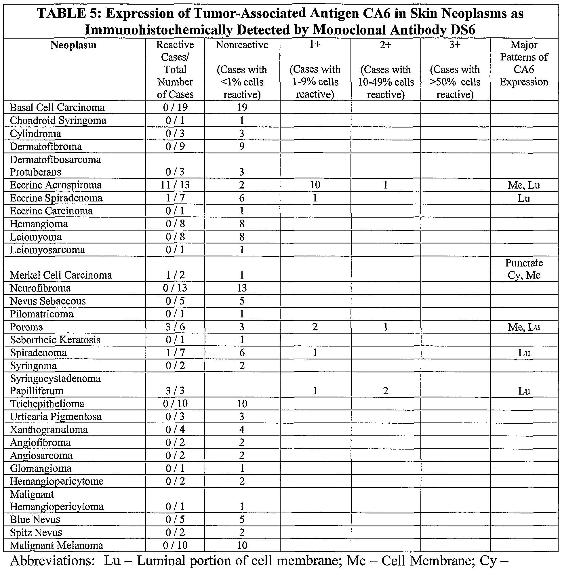
- Examples may include overexpression or aberrant expression of CA6 in various types of malignancies as will be seen in the Tables below (e.g ovarian cancer, endometrial cancer, pancreatic cancer, breast cancer, urinary bladder cancer, lung cancer , etc.), as well as overexpression or aberrant expression in other pathologic states, such as overexpression of CA6 by pneumocytes in lungs disease, for example, pneumonia.
- Tables below e.g ovarian cancer, endometrial cancer, pancreatic cancer, breast cancer, urinary bladder cancer, lung cancer , etc.
- overexpression or aberrant expression in other pathologic states such as overexpression of CA6 by pneumocytes in lungs disease, for example, pneumonia.
- a biological sample may be a cell sample, with an intervening culturing step being performed between the time the cell sample is collected from the subject and the immunoassay is carried out on the biological sample.
- a tissue sample is collected from the subject, and the presence or absence of binding of an antibody of the invention is detected.
- the presence of binding of the antibody in an abnormal pattern or a pattern indicative of a tumor or cancer indicates the presence of a tumor or cancer in the subject from which the tissue sample is collected.
- the presence of the antigen in a metastatic tumor deposit can also be used to determine a likely source of the primary tumor. Any suitable immunohistology format may be used.
- the tissue sample may include patient biopsies, resections or cells for cytologic study.
- a similar technique to immumohistogy is the use of similar techniques to detect and/or phenotype cells in body fluids or other suspensions as is used for flow cytometric examination.
- the antibody according to the invention is coupled to or provided with a suitable externally detectable label, such as e.g.
- a radiolabel as described above or a metal atom e.g., technetium-99m
- administered to a subject e.g., by intraveneous or intraarterial injection, in an amount sufficient to produce an externally detectable signal, whereupon the possible localized accumulation of antibody in the body is determined, with a localized accumulation of the antibody (in a region other than that which would ordinarily be expected for normal subjects or subjects free of disease) indicating the present of a tumor in that subject.
- Monoclonal antibodies used for therapy may be monoclonal antibodies per se or monoclonal antibodies coupled to a therapeutic agent. Such antibodies are referred to herein as therapeutic monoclonal antibodies. Any therapeutic agent conventionally coupled to a monoclonal antibody may be employed, including (but not limited to) radioisotopes, cytotoxic agents, and chemotherapeutic agents (See generally Monoclonal Antibodies and Cancer Therapy (R. Reisfeld and S. Sell Eds. 1985)(Alan R. Liss Inc. NY); u.s. Patent No. 5,558,852 to Bigner and Zalutsky; U.S. Patent No. 5,624,659 to Bigner and Zalutsky).
- Therapeutic agents may be conjugated or coupled to the antibody by direct means or indirect means (e.g., via a chelator), such as the Iodogen method or with N- succinimidyl-3-(tri-n-butylstanyl)benzoate (the "ATE method"), as will be apparent to those skilled in the art. See, e.g., M. Zalutsky and A. Narula, Appl. Radiat. Isot. 38, 1051 (1987). Examples of radioisotopes which may be coupled to a therapeutic monoclonal antibody include, but are not limited to, 131 1, 90 Y, 211 At, 212 Bi, 67 Cu, 186 Re, 188 Re, and 212 Pb.
- chemotherapeutic agents which may be coupled to a therapeutic monoclonal antibody include, but are not limited to, methotrexate.
- cytotoxic agents which may be coupled to a therapeutic monoclonal antibody include, but are not limited to, ricin (or more particularly the ricin A chain).
- the monoclonal antibodies of the invention can be conjugated to and used as targeting agents for genes (immunogenes, suicide genes), immunoliposomes, boron neutron capture therapy, photosensitizers for photodynamic therapy, and other types of therapies that can be directed by antibodies. It will be appreciated that monoclonal antibodies per se which are used as therapeutic monoclonal antibodies incorporate those portions of the constant region of an antibody necessary to evoke a therapeutically useful immunological response in the subject being treated.
- Therapeutic monoclonal antibodies may be provided in lyophylized form in a sterile aseptic container or may be provided in a pharmaceutical formulation in combination with a pharmaceutically acceptable carrier, such as sterile pyrogen-free water or sterile pyrogen-free physiological saline solution.
- a pharmaceutically acceptable carrier such as sterile pyrogen-free water or sterile pyrogen-free physiological saline solution.
- the therapeutic methods disclosed herein may be employed with subjects suspected of having a variety of tumors, whether primary or metastatic or micrometastatic (see Tables below), of particular importance are tumors of the ovary, endometrium, breast, urinary bladder, pancreas and lung.
- DS6 may also be of therapeutic use in other types of neoplasms, especially if used as part of a panel or combination of therapeutic antibodies, each with different specificities (Smith, N.L. et al, Human Antibodies, 9, 61-65, (1999); Oldham, R.K., Mol
- the antibody will generally be mixed, prior to administration, with a non-toxic, pharmaceutically acceptable carrier substance (e.g. normal saline or phosphate-buffered saline), and may be administered using any medically appropriate procedure, e.g., intravenous or intra-arterial administration, injection into the cerebrospinal fluid).
- a non-toxic, pharmaceutically acceptable carrier substance e.g. normal saline or phosphate-buffered saline
- intravenous or intra-arterial administration injection into the cerebrospinal fluid.
- intradermal, intracavity, intrathecal or direct administration to the tumor or to an artery supplying the tumor is advantageous.
- Dosage of the antibody will depend, among other things, on the tumor being treated, the route of administration, the nature of the therapeutic agent employed, and the sensitivity of the tumor to the particular therapeutic agent.
- the dosage will typically be about 1 to 10 micrograms per Kilogram subject body weight.
- the dosage to the patient will typically be from 10 mCi to 100, 300 or even 500 mCi. Stated otherwise, where the therapeutic agent is I, the dosage to the patient will typically be from 5,000 Rads to 100,000 Rads (preferably at least 13,000 Rads, or even at least 50,000 Rads). Doses for other radionuclides are typically selected so that the tumoricidal dose will be equivalent to the foregoing range for 131 I.
- the antibody can be administered to the subject in a series of more than one administration, and regular periodic administration will sometimes be required.
- mab monoclonal antibody
- TAA tumor-associated antigen
- IH immunohistochemistry/immunohistochemical
- kDa kiloDalton
- Monoclonal antibody DS6 detects a tumor-associated sialoglycotope expressed on human serous ovarian carcinomas
- mab murine monoclonal antibody
- clone DS6 using human serous ovarian carcinoma as the immunogen
- Hybridoma production Immunizations, fusions and screening were performed essentially as described previously using P3X63-Ag8.653 myeloma cells with human serous ovarian carcinoma as the immunogen (Wennerberg, A.E. et al, Am. J. Pathol, 143(4), 1050-1054 (1993)). Institutional Animal Care and Use approval was obtained. Preliminary and secondary screenings were by avidin-biotin IH (mouse IgG peroxidase kit, Vector Laboratories, Burlingame, CA) on tissue sections of ovarian carcinomas and selected normal adult tissues.
- avidin-biotin IH mouse IgG peroxidase kit, Vector Laboratories, Burlingame, CA
- DS6 was chosen for further studies and, after several rounds of single cell cloning by limiting dilution, was isotyped as an IgGl (ImmunoType Kit, Sigma Chemicals, St. Louis, MO). Supernatant was collected in batch for the studies in this report from DS6 cells grown in DMEM-F12 supplemented with 10% horse serum. Quantitation of murine IgGl in the supernatant was by EIA methodology (performed by the East Carolina University Hybridoma Core Facility, Greenville, NC). Immunohistochemistry. Tissue culture supernatant (40ug DS6/ml) was used for IH as above with AEC (Vector Laboratories) as the chromagen.
- Frozen cryostat tissue sections were air-dried, acetone-permeabilized while formalin-fixed, paraffin- embedded tissue sections were de-waxed through solvents and rehydrated, prior to blocking of endogenous peroxidase with methanol/hydrogen peroxide and subsequent immunostaining.
- Mouse IgGl was used as a negative control mab.
- Anti-transferrin receptor mab (IgGl) was used as a positive control on frozen cryostat sections while anti-smooth muscle myosin mab (IgGl) was used as a positive control on formalin- fixed tissues (all control antibodies from DAKO, Carpenteria, CA). Human adult tissues.
- Samples of grossly normal, incidental tissues were obtained fresh from either autopsy or the surgical bench from individuals without significant pathology in the primary organ or tissue; half were used for air-dried, acetone-permeabilized cryostat sections and half were fixed in buffered formalin and paraffin-embedded. Additional normal tissues as well as a pilot selection of gynecologic and related neoplasms were obtained from the archival tissue stores of formalin-fixed, paraffin-embedded tissue blocks of the University hospital (Table 1 and Table 2). Fresh specimens of serous ovarian tumors and normal fallopian tubes were obtained from surgical cases and cryostat sections were cut for chloroform extraction studies. Use of incidental human tissues had prior approval of the Institutional Review Board.
- neuraminidase for detection of sialic acid, 0.02 U/ml neuraminidase from Vibrio cholerae ( reacts with sialic acids in ⁇ 2-3,-6,-8 linkages, Boehringer Mannheim/Roche, Indianapolis, IN), in 0.01M Ca++ in PBS was used to pretreat formalin-fixed tissue sections of the above tissues with parallel control sections pretreated with Ca++/PBS buffer without neuraminidase.
- carbohydrate antigens were added to DS6 supernatant at lOug, 50ug, 250ug and 500ug carbohydrate to 4ug DS6 and allowed to react at 4°C overnight. This preabsorbed supernatant was then used by IH to study sections of formalin-fixed, paraffin-embedded serous ovarian carcinomas and compared to control sections stained with unabsorbed DS6 supernatant.
- Double-immunolabel, two-color IH on tissue sections Small sections (up to 1.0 cm) of eight formalin-fixed and paraffin-embedded serous adenocarcinomas and a metastasis of each were re-embedded into two paraffin blocks for single antibody and double label/sequential IH (Battifora, H, Lab. Invest., 55, 244-248 (1986)). Seven of the cases of adenocarcinoma had their primary site in the ovary, and one case was a primary papillary serous carcinoma of the peritoneum. A section of normal fallopian tube was also included in each block.
- a manufacturer's protocol for two-color, double-label IH was used: in brief, one of the murine mabs is used in a standard avidin-biotin IH technique with DAB (brown product, Vector Laboratories) as chromagen and then the entire process is repeated on the same slide using the alternate murine mab with VIP ("Very Intense Purple”; purple product, Vector Laboratories) as the chromagen.
- DS6 (neat) supernatant was used as one mab and a murine anti-Cal25 (OCH125 Level I, an IgGl, Signet Laboratories, Dedham, MA) as the alternative mab.
- DAB was consistently used as the first chromagen and VIP as the second chromagen in the sequence.
- the order of the primary antibodies was switched while maintaining the order of the chromagens so that each mab was evaluated both as a first and second mab in the sequence.
- Controls consisted of a primary mab (DS6 or OC125) with DAB followed by a mouse ascites negative control (Signet Laboratories) as the second mab with VIP.
- Antigen retrieval (lOmM, pH 6.0 citrate buffer) by microwave proceeded all double-label staining to assure antigen detection by mab OC125.
- the results of IH staining using DS6 on formalin-fixed, paraffin-embedded tissues are similar to those obtained on acetone-permeabilized cryostat sections, consistent with an epitope that is fairly resistant to the effects of formalin-fixation.
- the IH pattern of DS6 on cells of normal adult tissues is predominately an apical, epithelial cell membrane localization in cells lining certain tubular or saccular structures (e.g., fallopian tube, pulmonary alveoli) and along the luminal-facing aspect of the inner layers of some stratified epithelia, such as urothelium ( Figure 1, A-C).
- the entire cell membrane can be decorated giving a rim pattern to the cell ( Figure 1, D).
- Figure 1, D There is absent to minimal cytoplasmic expression of the antigen in any of the normal human adult epithelia studied.
- the CA6 antigen is most consistently detected in fallopian tube epithelium, urothelium and type 2 pneumocytes.
- the CA6 antigen has a more inhomogeneous and variable expression pattern. This is particularly evident in ductal structures, where expression can vary from one duct cross section to another within a given histologic preparation.
- CA6 is expressed along the apical membranes of many cells in ovarian serous cystadenomas.
- the level of CA6 expression is more intense and is additionally seen in the cytoplasm of many malignant cells and extracellularly within gland lumen ( Figure 2, C).
- Figure 2, C A similar luminal/apical pattern with variable cytoplasmic staining is also seen in the other adenocarcinomas that are DS6 immunoreactive.
- Mucinous ovarian tumors are not characteristically DS6 immunoreactive as no reactivity is seen in benign and borderline mucinous tumors and is only seen quite focally in 3 of 9 mucinous adenocarcinomas.
- CA6 is not detected in normal or hyperplastic mesothelium and does not appear to be characteristic of mesotheliomas (1/6), but is readily detected in serous surface carcinomas of the peritoneum (3/3). Sensitivity ofCA ⁇ epitope to periodic acid, neuraminidase, and chloroform extraction.
- DS6-reactive (IH) fallopian tube and serous ovarian carcinoma were subjected to a series of pretreatments with either periodic acid or neuraminidase (V. cholera) prior to immuonostaining with DS6.
- Pretreatment of the tissue sections with either periodic acid or neuraminidase (sialidase) prior to IH with DS6 results in complete abolishment of DS6 immunoreactivity in the sections of fallopian tube and ovarian carcinoma and no DS6 immunoreactivity was unmasked in any of the sections of gastrointestinal tissues or breast.
- Chloroform extraction studies were performed on sections of fallopian tubes (2) and serous ovarian tumors (3) prior to immunostaining with DS6.
- Control cryostat sections of fallopian tubes and ovarian tumors show strong staining with DS6 in the formalin-fixed as well as in the acetone-permeabilized slides.
- the chloroform- pretreated slides, stained with DS6, reveal mild to moderate reduction in staining intensity in fallopian tubes and ovarian tumors respectively.
- Immunoblotting and SDS-polyacrylamide gel electrophoresis OVCAR5, a human ovarian carcinoma cell line reactive with DS6 by IH (data not shown) was used as a reproducible source of antigen.
- Double-immunolabel, two-color IH of tissue sections Single antibody and double-label, two-color IH studies were performed using mabs DS6 and OC125 to determine if CA6 and CA125 are expressed by the same or differing tumor cell populations. Both antibodies reacted with the luminal aspect of fallopian tubes but only OC125 reacted with the mesothelium of the fallopian tubes.
- CA6 often has an additional prominent cytoplasmic component while CA125 will occasionally show a distinct pattern of circumferential membrane expression on the neoplastic cells in several of the tumors, giving a honeycomb appearance to the tumor that is not as characteristic of CA6 ( Figure 2, C-D).
- Figure 2, C-D When both DS6 and OC125 are used in sequential, two-color IH, intensely positive dark chromagen results from overlapping, dual localization of chromagens in many areas of the tumors. This dark chromagen can be difficult to distinguish from an intensely positive deposition from the first mab/chromagen (brown, DAB) reaction alone.
- CA6 antigen is not restricted to ovarian carcinomas but can be expressed by a variety of other carcinomas, including, but not limited to, those of the breast, endometrium, pancreas, urinary bladder and lung.
- CA6 has only a limited distribution in the normal tissues studied, is not detected in normal ovarian surface epithelium, yet is expressed in serous ovarian carcinomas which arise from the surface epithelium (18/18), it can be described as a TAA (Suresh, M. R, Anticancer Res., 16(4B), 2273-2277 (1996); Khawli, L.A. and Epstein, A.L., Q.J. Nucl. Med., Al, 25-35 (1997)).
- the staining of the intracellular canaliculus of parietal cells is a feature of the MUCl-related mab HMFG2 (Walker, M.M. et al., J Clin. Pathol, 48, 832-834 (1995)), while luminal staining of ductal structures is variably reported for a large number of TAAs, including the MUC1 mucin family and histo-blood group related antigens (Arklie, J. et al., Int. J. Cancer, 28, 23-29 (1981); Zhang et al., supra (1997); Cao, Y. et al, Histochem. CellBiol, 106, 197-207 (1996)).
- Pretreatment of tissue sections with either periodic acid or neuraminidase from Vibrio cholerae gives similar results. These techniques are used to determine if the epitope is carbohydrate-based or sialic acid-dependent, respectively, and have also been used to unmask hidden epitopes such as masking of the binding sites of several MUC1 mabs by glycosylation (Cao et al., supra (1997); Cao et al, supra (1998)). The abolishment of DS6 immunoreactivity by both treatments is consistent with a carbohydrate epitope that is sialic acid-dependent. No DS6-reactive epitopes are unmasked in any of the sections by such removal of periodic-sensitive carbohydrates or sialic acid.
- Chloroform extraction studies are used to extract lipids from tissue sections prior to DS6 immunostaining and are then compared to DS6 immunostaining on tissue sections without prior chloroform extraction. While some general reduction in staining intensity was noted in sections of fallopian tube, a more marked reduction was noted in the sections of ovarian tumors. Whether this apparent difference in efficiency of extraction in a normal tissue (i.e., fallopian tube) as compared to tumors is quantitative or qualitative is uncertain, however, ongoing studies into the chemical nature of the epitope may help explain the significance of this finding.
- CA6 The sialoglycotope nature of CA6 is similar to many of the histo-blood group-related antigens such as sTn, sLea, sLex, some of which are dually-expressed as glycoproteins and glycolipids, however, unlike CA6, as glycoproteins, these are usually high molecular weight, carbohydrate rich O-linked glycoproteins (mucins) (Magnani, J.L. et al., Cancer Res., A3, 5489-5492 (1983); Muraro, R. et al, Cancer Res., 48, 4588-4596 (1988)).
- TAAs to consider are those of approximately 80 kDa such as OC133 (Berkowitz, R. et al., Am. J. Obstet. Gynecol, 146(6), 607-612 (1983); Masuho, Y. et al., Cancer Res., 44, 2813-2819 (1984)), Ki- OCI-6-2 (Mettler, D.L.
- CA6 has been detected by IH in some normal epithelia of pancreas, ovary, fallopian tube, colon, gallbladder, stomach, endometrium, bronchus, lung, kidney and in mesothelium and amnion
- the IH pattern of CA6 in our series of ovarian tumors is similar to that reported for CA125 (Davis, H.M. et al., Cancer Res., 46, 6143-6148, 1986; Nouwen, E.J. et al., Cancer Res., 46(2), 866-876 (1986); Dietel, M.
- both antigens are more characteristic of ovarian serous carcinomas than ovarian mucinous neoplasms and both have a variegated distribution pattern, often with luminal accentuation (Mattes, M. et al., Cancer Res. (suppl), 50, 880-884 (1990)).
- both antigens show a spectrum of expression ranging from strong and diffuse in some tumors to patchy and variegated patterns in other neoplasms, with strongly positive groups of cells adjacent to nonreactive foci.
- CA125 is distributed along luminal epithelial surfaces and is readily detected in the mesothelium. Conversely, CA6, which is more intensely expressed along the fallopian tube lumen, is not detected in the mesothelium.
- DS6 clearly recognizes an antigen that has a similar, but not identical, distribution to the CA125 antigen, with some distinct differences in patterns and sites of antigen expression. The lack of CA6 expression in normal mesothelium could make DS6 a candidate for intraperitoneal antibody-targeted therapeutic applications.
- TABLE 1 Immunohistochemical detection of CA6 in tissue sections of normal adult tissues. Acetone-permeabilized and formalin-fixed, paraffin-embedded tissues were stained with mab DS6 as the primary antibody by avidin-biotin immunohistochemistry.
Landscapes
- Health & Medical Sciences (AREA)
- Chemical & Material Sciences (AREA)
- Life Sciences & Earth Sciences (AREA)
- Organic Chemistry (AREA)
- Immunology (AREA)
- Medicinal Chemistry (AREA)
- General Health & Medical Sciences (AREA)
- Biochemistry (AREA)
- Cell Biology (AREA)
- Proteomics, Peptides & Aminoacids (AREA)
- Molecular Biology (AREA)
- Genetics & Genomics (AREA)
- Biophysics (AREA)
- Public Health (AREA)
- General Chemical & Material Sciences (AREA)
- Chemical Kinetics & Catalysis (AREA)
- Veterinary Medicine (AREA)
- Pharmacology & Pharmacy (AREA)
- Nuclear Medicine, Radiotherapy & Molecular Imaging (AREA)
- Animal Behavior & Ethology (AREA)
- Peptides Or Proteins (AREA)
- Preparation Of Compounds By Using Micro-Organisms (AREA)
- Medicines Containing Antibodies Or Antigens For Use As Internal Diagnostic Agents (AREA)
Abstract
Description


Claims
Priority Applications (6)
| Application Number | Priority Date | Filing Date | Title |
|---|---|---|---|
| GB0306083A GB2383331B (en) | 2000-08-18 | 2001-08-17 | Monoclonal antibody DS6 Tumor-associated antigen CA6 and methods of use thereof |
| CA2419270A CA2419270C (en) | 2000-08-18 | 2001-08-17 | Monoclonal antibody ds6, tumor-associated antigen ca6, and methods of use thereof |
| EP01968944A EP1362060A4 (en) | 2000-08-18 | 2001-08-17 | DS6 MONOCLONAL ANTIBODIES AND METHODS OF USE |
| AU2001289145A AU2001289145A1 (en) | 2000-08-18 | 2001-08-17 | Monoclonal antibody ds6, tumor-associated antigen ca6, and methods of use thereof |
| MXPA03001460A MXPA03001460A (en) | 2000-08-18 | 2001-08-17 | DS6 MONOCLONAL ANTIBODY, CA6 ANTIGEN ASSOCIATED WITH TUMOR, AND SAME USE METHODS. |
| MX2013004010A MX347883B (en) | 2000-08-18 | 2001-08-17 | Monoclonal antibody ds6, tumor-associated antigen ca6, and methods of use thereof. |
Applications Claiming Priority (2)
| Application Number | Priority Date | Filing Date | Title |
|---|---|---|---|
| US09/641,499 | 2000-08-18 | ||
| US09/641,499 US6596503B1 (en) | 2000-08-18 | 2000-08-18 | Monoclonal antibody DS6, tumor-associated antigen CA6, and methods of use thereof |
Publications (2)
| Publication Number | Publication Date |
|---|---|
| WO2002016401A2 true WO2002016401A2 (en) | 2002-02-28 |
| WO2002016401A3 WO2002016401A3 (en) | 2002-04-11 |
Family
ID=24572648
Family Applications (1)
| Application Number | Title | Priority Date | Filing Date |
|---|---|---|---|
| PCT/US2001/041765 Ceased WO2002016401A2 (en) | 2000-08-18 | 2001-08-17 | Monoclonal antibody ds6, tumor-associated antigen ca6, and methods of use thereof |
Country Status (7)
| Country | Link |
|---|---|
| US (5) | US6596503B1 (en) |
| EP (1) | EP1362060A4 (en) |
| AU (1) | AU2001289145A1 (en) |
| CA (1) | CA2419270C (en) |
| GB (1) | GB2383331B (en) |
| MX (2) | MXPA03001460A (en) |
| WO (1) | WO2002016401A2 (en) |
Cited By (20)
| Publication number | Priority date | Publication date | Assignee | Title |
|---|---|---|---|---|
| WO2005009369A3 (en) * | 2003-07-21 | 2005-06-30 | Immunogen Inc | A ca6 antigen-specific cytotoxic conjugate and methods of using the same |
| WO2006086733A2 (en) | 2005-02-11 | 2006-08-17 | Immunogen, Inc. | Process for preparing maytansinoid antibody conjugates |
| US7374762B2 (en) | 2003-05-14 | 2008-05-20 | Immunogen, Inc. | Drug conjugate composition |
| JP2009504193A (en) * | 2005-08-22 | 2009-02-05 | イミュノジェン・インコーポレーテッド | CA6 antigen-specific cytotoxic conjugate and method using the conjugate |
| US7811572B2 (en) | 2005-08-24 | 2010-10-12 | Immunogen, Inc. | Process for preparing purified drug conjugates |
| WO2010126551A1 (en) | 2009-04-30 | 2010-11-04 | Immunogen, Inc. | Potent conjugates and hydrophilic linkers |
| US7834155B2 (en) | 2003-07-21 | 2010-11-16 | Immunogen Inc. | CA6 antigen-specific cytotoxic conjugate and methods of using the same |
| JP2012161321A (en) * | 2012-03-30 | 2012-08-30 | Immunogen Inc | Ca6 antigen-specific cytotoxic conjugate, and method using the conjugate |
| WO2012135522A2 (en) | 2011-03-29 | 2012-10-04 | Immunogen, Inc. | Process for manufacturing conjugates of improved homogeneity |
| US8795673B2 (en) | 2011-03-29 | 2014-08-05 | Immunogen, Inc. | Preparation of maytansinoid antibody conjugates by a one-step process |
| AU2012201260B2 (en) * | 2003-07-21 | 2014-10-23 | Immunogen, Inc. | A CA6 antigen-specific cytotoxic conjugate and methods of using the same |
| CN104804094A (en) * | 2005-08-22 | 2015-07-29 | 伊缪诺金公司 | CA6 antigen-specificity cytotoxicity conjugate and application method thereof |
| US9114179B2 (en) | 2005-08-03 | 2015-08-25 | Immunogen, Inc. | Immunoconjugate formulations |
| WO2016081584A1 (en) | 2014-11-19 | 2016-05-26 | Immunogen, Inc. | Process for preparing cell-binding agent-cytotoxic agent conjugates |
| US9376500B2 (en) | 2009-06-03 | 2016-06-28 | Immunogen, Inc. | Conjugation methods |
| WO2017136623A1 (en) | 2016-02-05 | 2017-08-10 | Immunogen, Inc. | Efficient process for preparing cell-binding agent-cytotoxic agent conjugates |
| WO2017194568A1 (en) | 2016-05-11 | 2017-11-16 | Sanofi | Treatment regimen using anti-muc1 maytansinoid immunoconjugate antibody for the treatment of tumors |
| EP3266469A2 (en) | 2008-04-30 | 2018-01-10 | ImmunoGen, Inc. | Cross-linkers and their uses |
| US10035817B2 (en) | 2012-10-04 | 2018-07-31 | Immunogen, Inc. | Method of purifying cell-binding agent-cytotoxic agent conjugates with a PVDF membrane |
| US10844135B2 (en) | 2003-10-10 | 2020-11-24 | Immunogen, Inc. | Method of targeting specific cell populations using cell-binding agent maytansinoid conjugates linked via a non-cleavable linker, said conjugates and methods of making said |
Families Citing this family (13)
| Publication number | Priority date | Publication date | Assignee | Title |
|---|---|---|---|---|
| EP1732596A2 (en) * | 2004-03-11 | 2006-12-20 | Shantha West, Inc. | Therapeutic use of rm1 antigen |
| WO2007127160A2 (en) * | 2006-04-24 | 2007-11-08 | Shantha West, Inc. | Agrm2 antigen |
| AU2007355108B2 (en) | 2006-11-27 | 2013-07-11 | Patrys Limited | Novel glycosylated peptide target in neoplastic cells |
| US8012678B2 (en) * | 2007-07-24 | 2011-09-06 | Wisconsin Alumni Research Foundation | Biomarkers for human papilloma virus-associated cancer |
| AU2009269704B2 (en) | 2008-06-16 | 2014-06-12 | Patrys Limited | LM-antibodies, functional fragments, LM-1 target antigen, and methods for making and using same |
| GB2478441B (en) * | 2008-09-09 | 2013-03-27 | Somalogic Inc | Lung cancer biomarkers and uses thereof |
| US10359425B2 (en) | 2008-09-09 | 2019-07-23 | Somalogic, Inc. | Lung cancer biomarkers and uses thereof |
| MX355020B (en) | 2010-07-09 | 2018-04-02 | Somalogic Inc | Lung cancer biomarkers and uses thereof. |
| MX341517B (en) | 2010-08-13 | 2016-08-24 | Somalogic Inc | PANCREATIC CANCER BIOMARCATORS AND USES OF THE SAME. |
| SG11201400375SA (en) * | 2011-10-24 | 2014-04-28 | Somalogic Inc | Lung cancer biomarkers and uses thereof |
| CA2852709A1 (en) | 2011-10-28 | 2013-05-02 | Patrys Limited | Pat-lm1 epitopes and methods for using same |
| US9989524B2 (en) * | 2013-02-05 | 2018-06-05 | Sanofi | Immuno imaging agent for use with antibody-drug conjugate therapy |
| JP2016515093A (en) | 2013-02-05 | 2016-05-26 | サノフイ | Immunoimaging agents for use with antibody-drug conjugate therapy |
Family Cites Families (8)
| Publication number | Priority date | Publication date | Assignee | Title |
|---|---|---|---|---|
| US4931385A (en) * | 1985-06-24 | 1990-06-05 | Hygeia Sciences, Incorporated | Enzyme immunoassays and immunologic reagents |
| US5366866A (en) | 1991-07-25 | 1994-11-22 | Duke University | Method of diagnosing ovarian and endometrial cancer with monoclonal antibodies OXA and OXB |
| WO1994021294A1 (en) | 1993-03-19 | 1994-09-29 | Bigner Darell D | Method of treating tumors with antibodies |
| AU6445894A (en) | 1993-03-19 | 1994-10-11 | Duke University | Method of treatment of tumors with an antibody binding to tenascin |
| US5871941A (en) | 1995-07-28 | 1999-02-16 | Univ British Columbia | Method of testing expression of CA125 antigen in cultured ovarian surface epithelial cells to identify and monitor individuals having a predisposition to develop ovarian cancer |
| US7037667B1 (en) * | 1998-06-01 | 2006-05-02 | Agensys, Inc. | Tumor antigen useful in diagnosis and therapy of prostate and colon cancer |
| WO2005009369A2 (en) | 2003-07-21 | 2005-02-03 | Immunogen, Inc. | A ca6 antigen-specific cytotoxic conjugate and methods of using the same |
| US7834155B2 (en) * | 2003-07-21 | 2010-11-16 | Immunogen Inc. | CA6 antigen-specific cytotoxic conjugate and methods of using the same |
-
2000
- 2000-08-18 US US09/641,499 patent/US6596503B1/en not_active Expired - Lifetime
-
2001
- 2001-08-17 MX MXPA03001460A patent/MXPA03001460A/en active IP Right Grant
- 2001-08-17 EP EP01968944A patent/EP1362060A4/en not_active Ceased
- 2001-08-17 AU AU2001289145A patent/AU2001289145A1/en not_active Abandoned
- 2001-08-17 WO PCT/US2001/041765 patent/WO2002016401A2/en not_active Ceased
- 2001-08-17 CA CA2419270A patent/CA2419270C/en not_active Expired - Lifetime
- 2001-08-17 GB GB0306083A patent/GB2383331B/en not_active Expired - Fee Related
- 2001-08-17 MX MX2013004010A patent/MX347883B/en unknown
-
2003
- 2003-06-19 US US10/465,176 patent/US7351805B2/en not_active Expired - Lifetime
-
2007
- 2007-02-28 US US11/680,190 patent/US7507410B2/en not_active Expired - Fee Related
-
2009
- 2009-01-30 US US12/363,191 patent/US8067186B2/en not_active Expired - Fee Related
-
2011
- 2011-11-02 US US13/287,633 patent/US8728741B2/en not_active Expired - Fee Related
Cited By (58)
| Publication number | Priority date | Publication date | Assignee | Title |
|---|---|---|---|---|
| US7501120B2 (en) | 2003-05-14 | 2009-03-10 | Immunogen, Inc. | Drug conjugate composition |
| US8012485B2 (en) | 2003-05-14 | 2011-09-06 | Immunogen, Inc. | Drug conjugate composition |
| EP2281577A2 (en) | 2003-05-14 | 2011-02-09 | Immunogen, Inc. | Drug conjugate composition |
| US7374762B2 (en) | 2003-05-14 | 2008-05-20 | Immunogen, Inc. | Drug conjugate composition |
| US7514080B2 (en) | 2003-05-14 | 2009-04-07 | Immunogen, Inc. | Drug conjugate composition |
| US7494649B2 (en) | 2003-05-14 | 2009-02-24 | Immunogen, Inc. | Drug conjugate composition |
| US8987424B2 (en) | 2003-07-21 | 2015-03-24 | Immunogen, Inc. | CA6 antigen-specific cytotoxic conjugate and methods of using the same |
| JP2015012861A (en) * | 2003-07-21 | 2015-01-22 | イミュノジェン・インコーポレーテッド | Ca6 antigen-specific cytotoxic conjugate and methods of using the same |
| NO339324B1 (en) * | 2003-07-21 | 2016-11-28 | Immunogen Inc | Humanized antibody or epitope binding fragment thereof which binds the CA6 glycotope; polynucleotide and expression vector encoding the antibody or fragments; cytotoxic conjugate thereof; using the conjugate for the manufacture of a drug; seen from there; an in vitro method for inhibiting the growth of a cell expressing the CA6 glycotope and a method for detecting the CA6 glycotope. |
| US9370584B2 (en) | 2003-07-21 | 2016-06-21 | Immunogen Inc. | CA6 antigen-specific cytotoxic conjugate and methods of using the same |
| US7834155B2 (en) | 2003-07-21 | 2010-11-16 | Immunogen Inc. | CA6 antigen-specific cytotoxic conjugate and methods of using the same |
| JP2007503202A (en) * | 2003-07-21 | 2007-02-22 | イミュノジェン・インコーポレーテッド | CA6 antigen-specific cytotoxic conjugates and methods of use thereof |
| JP2011092194A (en) * | 2003-07-21 | 2011-05-12 | Immunogen Inc | Ca6 antigen-specific cytotoxic conjugate and method of using the same |
| US9822183B2 (en) | 2003-07-21 | 2017-11-21 | Sanofi | CA6 antigen-specific cytotoxic conjugate and methods of using the same |
| AU2004258955B2 (en) * | 2003-07-21 | 2011-12-01 | Immunogen, Inc. | A CA6 antigen-specific cytotoxic conjugate and methods of using the same |
| WO2005009369A3 (en) * | 2003-07-21 | 2005-06-30 | Immunogen Inc | A ca6 antigen-specific cytotoxic conjugate and methods of using the same |
| JP2016128405A (en) * | 2003-07-21 | 2016-07-14 | イミュノジェン・インコーポレーテッド | Ca6 antigen-specific cytotoxic conjugate and method of using the same |
| AU2004258955C1 (en) * | 2003-07-21 | 2012-07-26 | Immunogen, Inc. | A CA6 antigen-specific cytotoxic conjugate and methods of using the same |
| KR101565721B1 (en) * | 2003-07-21 | 2015-11-03 | 이뮤노젠 아이엔씨 | A ca6 antigen-specific cytotoxic conjugate and methods of using the same |
| AU2012201260B2 (en) * | 2003-07-21 | 2014-10-23 | Immunogen, Inc. | A CA6 antigen-specific cytotoxic conjugate and methods of using the same |
| CN103145844A (en) * | 2003-07-21 | 2013-06-12 | 伊缪诺金公司 | A ca6 antigen-specific cytotoxic conjugate and methods of using the same |
| KR101263950B1 (en) | 2003-07-21 | 2013-05-31 | 이뮤노젠 아이엔씨 | 6 a ca6 antigen-specific cytotoxic conjugate and methods of using the same |
| US10844135B2 (en) | 2003-10-10 | 2020-11-24 | Immunogen, Inc. | Method of targeting specific cell populations using cell-binding agent maytansinoid conjugates linked via a non-cleavable linker, said conjugates and methods of making said |
| WO2006086733A2 (en) | 2005-02-11 | 2006-08-17 | Immunogen, Inc. | Process for preparing maytansinoid antibody conjugates |
| EP2468304A2 (en) | 2005-02-11 | 2012-06-27 | ImmunoGen, Inc. | Process for preparing stable drug conjugates |
| US9114179B2 (en) | 2005-08-03 | 2015-08-25 | Immunogen, Inc. | Immunoconjugate formulations |
| CN104804094A (en) * | 2005-08-22 | 2015-07-29 | 伊缪诺金公司 | CA6 antigen-specificity cytotoxicity conjugate and application method thereof |
| JP2009504193A (en) * | 2005-08-22 | 2009-02-05 | イミュノジェン・インコーポレーテッド | CA6 antigen-specific cytotoxic conjugate and method using the conjugate |
| US8933205B2 (en) | 2005-08-24 | 2015-01-13 | Immunogen, Inc. | Process for preparing purified drug conjugates |
| US8383122B2 (en) | 2005-08-24 | 2013-02-26 | Immunogen, Inc. | Process for preparing purified drug conjugates |
| EP2399609A1 (en) | 2005-08-24 | 2011-12-28 | ImmunoGen, Inc. | Process for preparing maytansinoid antibody conjugates |
| EP3539572A1 (en) | 2005-08-24 | 2019-09-18 | ImmunoGen, Inc. | Process for preparing maytansinoid antibody conjugates |
| EP2662096A1 (en) | 2005-08-24 | 2013-11-13 | ImmunoGen, Inc. | Process for preparing maytansinoid antibody conjugates |
| US9789204B2 (en) | 2005-08-24 | 2017-10-17 | Immunogen, Inc. | Process for preparing purified drug conjugates |
| US11471536B2 (en) | 2005-08-24 | 2022-10-18 | Immunogen, Inc. | Process for preparing purified drug conjugates |
| US7811572B2 (en) | 2005-08-24 | 2010-10-12 | Immunogen, Inc. | Process for preparing purified drug conjugates |
| EP3266469A2 (en) | 2008-04-30 | 2018-01-10 | ImmunoGen, Inc. | Cross-linkers and their uses |
| WO2010126551A1 (en) | 2009-04-30 | 2010-11-04 | Immunogen, Inc. | Potent conjugates and hydrophilic linkers |
| US10815309B2 (en) | 2009-06-03 | 2020-10-27 | Immunogen, Inc. | Methods for preparing antibody-drug conjugates |
| US10233257B2 (en) | 2009-06-03 | 2019-03-19 | Immunogen, Inc. | Methods for preparing antibody-drug conjugates |
| US9771432B2 (en) | 2009-06-03 | 2017-09-26 | Immunogen, Inc. | Conjugation methods |
| EP3480202A1 (en) | 2009-06-03 | 2019-05-08 | ImmunoGen, Inc. | Conjugation methods |
| US11498979B2 (en) | 2009-06-03 | 2022-11-15 | Immunogen, Inc. | Methods for preparing a purified maytansinoid conjugate in a solution |
| US9376500B2 (en) | 2009-06-03 | 2016-06-28 | Immunogen, Inc. | Conjugation methods |
| US9914748B2 (en) | 2011-03-29 | 2018-03-13 | Immunogen, Inc. | Preparation of maytansinoid antibody conjugates by a one-step process |
| WO2012135522A2 (en) | 2011-03-29 | 2012-10-04 | Immunogen, Inc. | Process for manufacturing conjugates of improved homogeneity |
| US8795673B2 (en) | 2011-03-29 | 2014-08-05 | Immunogen, Inc. | Preparation of maytansinoid antibody conjugates by a one-step process |
| US11090390B2 (en) | 2011-03-29 | 2021-08-17 | Immunogen, Inc. | Preparation of maytansinoid antibody conjugates by a one-step process |
| EP3545977A1 (en) | 2011-03-29 | 2019-10-02 | ImmunoGen, Inc. | Preparation of maytansinoid antibody conjugates by a one-step process |
| WO2012135517A2 (en) | 2011-03-29 | 2012-10-04 | Immunogen, Inc. | Preparation of maytansinoid antibody conjugates by a one-step process |
| US11744900B2 (en) | 2011-03-29 | 2023-09-05 | Immunogen, Inc. | Preparation of maytansinoid antibody conjugates by a one-step process |
| US10435432B2 (en) | 2011-03-29 | 2019-10-08 | Immunogen, Inc. | Preparation of maytansinoid antibody conjugates by a one-step process |
| US9428543B2 (en) | 2011-03-29 | 2016-08-30 | Immunogen, Inc. | Preparation of maytansinoid antibody conjugates by a one-step process |
| JP2012161321A (en) * | 2012-03-30 | 2012-08-30 | Immunogen Inc | Ca6 antigen-specific cytotoxic conjugate, and method using the conjugate |
| US10035817B2 (en) | 2012-10-04 | 2018-07-31 | Immunogen, Inc. | Method of purifying cell-binding agent-cytotoxic agent conjugates with a PVDF membrane |
| WO2016081584A1 (en) | 2014-11-19 | 2016-05-26 | Immunogen, Inc. | Process for preparing cell-binding agent-cytotoxic agent conjugates |
| WO2017136623A1 (en) | 2016-02-05 | 2017-08-10 | Immunogen, Inc. | Efficient process for preparing cell-binding agent-cytotoxic agent conjugates |
| WO2017194568A1 (en) | 2016-05-11 | 2017-11-16 | Sanofi | Treatment regimen using anti-muc1 maytansinoid immunoconjugate antibody for the treatment of tumors |
Also Published As
| Publication number | Publication date |
|---|---|
| AU2001289145A1 (en) | 2002-03-04 |
| EP1362060A2 (en) | 2003-11-19 |
| CA2419270A1 (en) | 2002-02-28 |
| GB2383331A (en) | 2003-06-25 |
| MXPA03001460A (en) | 2004-12-13 |
| US8728741B2 (en) | 2014-05-20 |
| US8067186B2 (en) | 2011-11-29 |
| MX347883B (en) | 2017-05-16 |
| GB2383331B (en) | 2004-09-29 |
| US6596503B1 (en) | 2003-07-22 |
| US20120045776A1 (en) | 2012-02-23 |
| EP1362060A4 (en) | 2004-09-08 |
| US20090155930A1 (en) | 2009-06-18 |
| US20030215895A1 (en) | 2003-11-20 |
| WO2002016401A3 (en) | 2002-04-11 |
| US20070196276A1 (en) | 2007-08-23 |
| GB0306083D0 (en) | 2003-04-23 |
| US7507410B2 (en) | 2009-03-24 |
| CA2419270C (en) | 2013-05-14 |
| US7351805B2 (en) | 2008-04-01 |
Similar Documents
| Publication | Publication Date | Title |
|---|---|---|
| US7507410B2 (en) | Monoclonal antibody DS6, tumor-associated antigen CA6, and methods of use thereof | |
| JP7289346B2 (en) | Diagnostic assays and kits for the detection of folate receptor 1 | |
| US6180357B1 (en) | Individualized patient-specific anti-cancer antibodies | |
| CN101254303A (en) | Cytotoxicity mediated by cells displaying surface expression of MCSP | |
| CA2560864A1 (en) | Cytotoxicity mediation of cells evidencing surface expression of cd63 | |
| JPH10505749A (en) | Monoclonal antibodies against human colon carcinoma binding antigen and their use. | |
| US20090022722A1 (en) | Cytotoxicity mediation of cells evidencing surface expression of CD59 | |
| EP1635869A1 (en) | Cancerous disease modifying antibodies | |
| JP2008545615A (en) | Cytotoxic mediation of cells demonstrating surface expression of CD63 | |
| AU8071187A (en) | Monoclonal paratopic molecule directed to human ganglioside gd2 | |
| JP4826697B2 (en) | Personalized anti-cancer antibody | |
| Kearse et al. | Monoclonal antibody DS6 detects a tumor‐associated sialoglycotope expressed on human serous ovarian carcinomas | |
| US9539348B2 (en) | Monoclonal antibody DS6, tumor-associated antigen CA6, and methods of use thereof | |
| JP2004529849A (en) | Identification and development of specific monoclonal antibodies against squamous cell carcinoma | |
| US20050244899A1 (en) | Laminin Receptor 1 Precursor Protein (37LRP) epitope delineated by an Hepatocellular carcinoma specific antibody | |
| JPH03188099A (en) | Novel tumor-related antigen | |
| WO1994003629A9 (en) | Monoclonal antibodies specific for 3',6'-isold1 ganglioside | |
| WO1994003629A1 (en) | Monoclonal antibodies specific for 3',6'-isold1 ganglioside | |
| HK1111176A (en) | Cytotoxicity mediation of cells evidencing surface expression of mcsp | |
| HK1120410A (en) | Cytotoxicity mediation of cells evidencing surface expression of mcsp | |
| HK1128478A (en) | Cytotoxicity mediation of cells evidencing surface expression of cd59 |
Legal Events
| Date | Code | Title | Description |
|---|---|---|---|
| AK | Designated states |
Kind code of ref document: A2 Designated state(s): AE AG AL AM AT AU AZ BA BB BG BR BY BZ CA CH CN CO CR CU CZ DE DK DM DZ EC EE ES FI GB GD GE GH GM HR HU ID IL IN IS JP KE KG KP KR KZ LC LK LR LS LT LU LV MA MD MG MK MN MW MX MZ NO NZ PH PL PT RO RU SD SE SG SI SK SL TJ TM TR TT TZ UA UG US UZ VN YU ZA ZW |
|
| AL | Designated countries for regional patents |
Kind code of ref document: A2 Designated state(s): GH GM KE LS MW MZ SD SL SZ TZ UG ZW AM AZ BY KG KZ MD RU TJ TM AT BE CH CY DE DK ES FI FR GB GR IE IT LU MC NL PT SE TR BF BJ CF CG CI CM GA GN GQ GW ML MR NE SN TD TG |
|
| ENP | Entry into the national phase |
Ref document number: 0306083 Country of ref document: GB Kind code of ref document: A Free format text: PCT FILING DATE = 20010817 Format of ref document f/p: F |
|
| AK | Designated states |
Kind code of ref document: A3 Designated state(s): AE AG AL AM AT AU AZ BA BB BG BR BY BZ CA CH CN CO CR CU CZ DE DK DM DZ EC EE ES FI GB GD GE GH GM HR HU ID IL IN IS JP KE KG KP KR KZ LC LK LR LS LT LU LV MA MD MG MK MN MW MX MZ NO NZ PH PL PT RO RU SD SE SG SI SK SL TJ TM TR TT TZ UA UG US UZ VN YU ZA ZW |
|
| AL | Designated countries for regional patents |
Kind code of ref document: A3 Designated state(s): GH GM KE LS MW MZ SD SL SZ TZ UG ZW AM AZ BY KG KZ MD RU TJ TM AT BE CH CY DE DK ES FI FR GB GR IE IT LU MC NL PT SE TR BF BJ CF CG CI CM GA GN GQ GW ML MR NE SN TD TG |
|
| 121 | Ep: the epo has been informed by wipo that ep was designated in this application | ||
| DFPE | Request for preliminary examination filed prior to expiration of 19th month from priority date (pct application filed before 20040101) | ||
| WWE | Wipo information: entry into national phase |
Ref document number: 2419270 Country of ref document: CA |
|
| WWE | Wipo information: entry into national phase |
Ref document number: PA/a/2003/001460 Country of ref document: MX |
|
| WWE | Wipo information: entry into national phase |
Ref document number: 2001968944 Country of ref document: EP |
|
| REG | Reference to national code |
Ref country code: DE Ref legal event code: 8642 |
|
| WWP | Wipo information: published in national office |
Ref document number: 2001968944 Country of ref document: EP |
|
| NENP | Non-entry into the national phase |
Ref country code: JP |