KR20170053617A - Methods for Evaluating Lung Cancer Status - Google Patents
Methods for Evaluating Lung Cancer Status Download PDFInfo
- Publication number
- KR20170053617A KR20170053617A KR1020177004127A KR20177004127A KR20170053617A KR 20170053617 A KR20170053617 A KR 20170053617A KR 1020177004127 A KR1020177004127 A KR 1020177004127A KR 20177004127 A KR20177004127 A KR 20177004127A KR 20170053617 A KR20170053617 A KR 20170053617A
- Authority
- KR
- South Korea
- Prior art keywords
- lung cancer
- genes
- subject
- gene
- expression
- Prior art date
- Legal status (The legal status is an assumption and is not a legal conclusion. Google has not performed a legal analysis and makes no representation as to the accuracy of the status listed.)
- Granted
Links
Images


Classifications
-
- C—CHEMISTRY; METALLURGY
- C12—BIOCHEMISTRY; BEER; SPIRITS; WINE; VINEGAR; MICROBIOLOGY; ENZYMOLOGY; MUTATION OR GENETIC ENGINEERING
- C12Q—MEASURING OR TESTING PROCESSES INVOLVING ENZYMES, NUCLEIC ACIDS OR MICROORGANISMS; COMPOSITIONS OR TEST PAPERS THEREFOR; PROCESSES OF PREPARING SUCH COMPOSITIONS; CONDITION-RESPONSIVE CONTROL IN MICROBIOLOGICAL OR ENZYMOLOGICAL PROCESSES
- C12Q1/00—Measuring or testing processes involving enzymes, nucleic acids or microorganisms; Compositions therefor; Processes of preparing such compositions
- C12Q1/68—Measuring or testing processes involving enzymes, nucleic acids or microorganisms; Compositions therefor; Processes of preparing such compositions involving nucleic acids
- C12Q1/6876—Nucleic acid products used in the analysis of nucleic acids, e.g. primers or probes
- C12Q1/6883—Nucleic acid products used in the analysis of nucleic acids, e.g. primers or probes for diseases caused by alterations of genetic material
- C12Q1/6886—Nucleic acid products used in the analysis of nucleic acids, e.g. primers or probes for diseases caused by alterations of genetic material for cancer
-
- G06F19/20—
-
- G06F19/24—
-
- G—PHYSICS
- G16—INFORMATION AND COMMUNICATION TECHNOLOGY [ICT] SPECIALLY ADAPTED FOR SPECIFIC APPLICATION FIELDS
- G16B—BIOINFORMATICS, i.e. INFORMATION AND COMMUNICATION TECHNOLOGY [ICT] SPECIALLY ADAPTED FOR GENETIC OR PROTEIN-RELATED DATA PROCESSING IN COMPUTATIONAL MOLECULAR BIOLOGY
- G16B25/00—ICT specially adapted for hybridisation; ICT specially adapted for gene or protein expression
- G16B25/10—Gene or protein expression profiling; Expression-ratio estimation or normalisation
-
- G—PHYSICS
- G16—INFORMATION AND COMMUNICATION TECHNOLOGY [ICT] SPECIALLY ADAPTED FOR SPECIFIC APPLICATION FIELDS
- G16B—BIOINFORMATICS, i.e. INFORMATION AND COMMUNICATION TECHNOLOGY [ICT] SPECIALLY ADAPTED FOR GENETIC OR PROTEIN-RELATED DATA PROCESSING IN COMPUTATIONAL MOLECULAR BIOLOGY
- G16B25/00—ICT specially adapted for hybridisation; ICT specially adapted for gene or protein expression
- G16B25/30—Microarray design
-
- G—PHYSICS
- G16—INFORMATION AND COMMUNICATION TECHNOLOGY [ICT] SPECIALLY ADAPTED FOR SPECIFIC APPLICATION FIELDS
- G16B—BIOINFORMATICS, i.e. INFORMATION AND COMMUNICATION TECHNOLOGY [ICT] SPECIALLY ADAPTED FOR GENETIC OR PROTEIN-RELATED DATA PROCESSING IN COMPUTATIONAL MOLECULAR BIOLOGY
- G16B40/00—ICT specially adapted for biostatistics; ICT specially adapted for bioinformatics-related machine learning or data mining, e.g. knowledge discovery or pattern finding
- G16B40/10—Signal processing, e.g. from mass spectrometry [MS] or from PCR
-
- C—CHEMISTRY; METALLURGY
- C12—BIOCHEMISTRY; BEER; SPIRITS; WINE; VINEGAR; MICROBIOLOGY; ENZYMOLOGY; MUTATION OR GENETIC ENGINEERING
- C12Q—MEASURING OR TESTING PROCESSES INVOLVING ENZYMES, NUCLEIC ACIDS OR MICROORGANISMS; COMPOSITIONS OR TEST PAPERS THEREFOR; PROCESSES OF PREPARING SUCH COMPOSITIONS; CONDITION-RESPONSIVE CONTROL IN MICROBIOLOGICAL OR ENZYMOLOGICAL PROCESSES
- C12Q2600/00—Oligonucleotides characterized by their use
- C12Q2600/106—Pharmacogenomics, i.e. genetic variability in individual responses to drugs and drug metabolism
-
- C—CHEMISTRY; METALLURGY
- C12—BIOCHEMISTRY; BEER; SPIRITS; WINE; VINEGAR; MICROBIOLOGY; ENZYMOLOGY; MUTATION OR GENETIC ENGINEERING
- C12Q—MEASURING OR TESTING PROCESSES INVOLVING ENZYMES, NUCLEIC ACIDS OR MICROORGANISMS; COMPOSITIONS OR TEST PAPERS THEREFOR; PROCESSES OF PREPARING SUCH COMPOSITIONS; CONDITION-RESPONSIVE CONTROL IN MICROBIOLOGICAL OR ENZYMOLOGICAL PROCESSES
- C12Q2600/00—Oligonucleotides characterized by their use
- C12Q2600/112—Disease subtyping, staging or classification
-
- C—CHEMISTRY; METALLURGY
- C12—BIOCHEMISTRY; BEER; SPIRITS; WINE; VINEGAR; MICROBIOLOGY; ENZYMOLOGY; MUTATION OR GENETIC ENGINEERING
- C12Q—MEASURING OR TESTING PROCESSES INVOLVING ENZYMES, NUCLEIC ACIDS OR MICROORGANISMS; COMPOSITIONS OR TEST PAPERS THEREFOR; PROCESSES OF PREPARING SUCH COMPOSITIONS; CONDITION-RESPONSIVE CONTROL IN MICROBIOLOGICAL OR ENZYMOLOGICAL PROCESSES
- C12Q2600/00—Oligonucleotides characterized by their use
- C12Q2600/158—Expression markers
-
- G01N33/5752—
-
- G—PHYSICS
- G16—INFORMATION AND COMMUNICATION TECHNOLOGY [ICT] SPECIALLY ADAPTED FOR SPECIFIC APPLICATION FIELDS
- G16B—BIOINFORMATICS, i.e. INFORMATION AND COMMUNICATION TECHNOLOGY [ICT] SPECIALLY ADAPTED FOR GENETIC OR PROTEIN-RELATED DATA PROCESSING IN COMPUTATIONAL MOLECULAR BIOLOGY
- G16B40/00—ICT specially adapted for biostatistics; ICT specially adapted for bioinformatics-related machine learning or data mining, e.g. knowledge discovery or pattern finding
Landscapes
- Health & Medical Sciences (AREA)
- Life Sciences & Earth Sciences (AREA)
- Physics & Mathematics (AREA)
- Engineering & Computer Science (AREA)
- Chemical & Material Sciences (AREA)
- Genetics & Genomics (AREA)
- Bioinformatics & Cheminformatics (AREA)
- General Health & Medical Sciences (AREA)
- Biotechnology (AREA)
- Biophysics (AREA)
- Organic Chemistry (AREA)
- Proteomics, Peptides & Aminoacids (AREA)
- Medical Informatics (AREA)
- Molecular Biology (AREA)
- Spectroscopy & Molecular Physics (AREA)
- Bioinformatics & Computational Biology (AREA)
- Evolutionary Biology (AREA)
- Theoretical Computer Science (AREA)
- Zoology (AREA)
- Wood Science & Technology (AREA)
- Analytical Chemistry (AREA)
- Immunology (AREA)
- Pathology (AREA)
- Biochemistry (AREA)
- General Engineering & Computer Science (AREA)
- Microbiology (AREA)
- Hospice & Palliative Care (AREA)
- Oncology (AREA)
- Databases & Information Systems (AREA)
- Data Mining & Analysis (AREA)
- Epidemiology (AREA)
- Evolutionary Computation (AREA)
- Computer Vision & Pattern Recognition (AREA)
- Public Health (AREA)
- Software Systems (AREA)
- Bioethics (AREA)
- Artificial Intelligence (AREA)
- Signal Processing (AREA)
- Measuring Or Testing Involving Enzymes Or Micro-Organisms (AREA)
Abstract
일부 양태에서 본 발명은 정보 유전자의 발현에 기초하여 환자가 폐암에 걸릴 가능성을 측정하는 방법을 제공한다. 다른 양태에서, 본 발명은 정보 유전자의 발현에 기초하여 환자에 대한 적절한 진단 중재 계획을 결정하는 방법을 제공한다. 관련된 조성물 및 키트는 본 발명의 다른 양태에서 제공된다.In some embodiments, the present invention provides a method for measuring the likelihood that a patient will have lung cancer based on the expression of an information gene. In another aspect, the invention provides a method for determining an appropriate diagnostic intervention plan for a patient based on expression of an information gene. Related compositions and kits are provided in other aspects of the invention.
Description
본 발명은 일반적으로 유전자 발현 정보를 사용하여 암을 평가하는 방법 및 조성물에 관한 것이다.The present invention relates generally to methods and compositions for assessing cancer using gene expression information.
가장 효과적으로 치료할 수 있는 특히 초기 단계에서 폐암을 진단하는데 어려움은 질병을 진단하기 위해 세포에 접근하는 것이다. 초기 단계 폐암은 특히 기관지 내시경과 같은 표준 기술로 접근하기 어려운 폐 기도의 주변 부위에도 나타날 수 있는 작은 병변과 통상적으로 관련이 있다.The difficulty in diagnosing lung cancer, especially at an early stage, where it can be treated most effectively, is to approach cells to diagnose disease. Early-stage lung cancer is typically associated with small lesions that may also appear in the periphery of the lungs, which are difficult to access with standard techniques such as bronchoscopy.
본 발명은 상기한 문제를 해결하는 것을 목적으로 한다.The present invention aims at solving the above-mentioned problems.
대상에 대한 적절한 진단 중재 계획 및/또는 치료 계획을 수립하고 적절한 진단 중재 계획 및/또는 치료 계획을 수립하는데 의료 제공자를 돕는 방법이 본 발명에 제공된다. 일부 실시태양에서, 상기 방법은 상해 개념의 기도 분야에 기초한다. 일부 실시태양에서, 상기 방법은 환자가 암에 걸릴 가능성을 평가하는데 유용한 정보 유전자의 발현 수준에 기초하여 폐암 위험 점수를 설정하는 단계를 필요로 한다. 일부 실시태양에서, 본 발명에 제공된 방법은 일상적인 세포 또는 조직 샘플링 절차 동안 대상으로부터 얻은 생물학적 샘플에서 정보 유전자의 발현 수준에 기초하여 평가하는 단계를 필요로 한다. 일부 실시태양에서, 생물학적 샘플은 조직학적으로 정상적인 세포를 포함한다. 일부 실시태양에서, 본 발명의 양태는 제 1 기도 위치로부터 얻은 명백하게 조직학적으로 정상인 세포에서 특정 정보 유전자의 발현 수준이 기도의 제 2 위치(예를 들어, 조직학적으로 정상적인 세포를 샘플링한 위치로부터 멀리 떨어진 기도에서 위치)에서 암의 가능성을 평가하는데 사용될 수 있다는 결정에 적어도 부분적으로 기초한다. 일부 실시태양에서, 조직학적으로 정상적인 세포(예를 들어, 기관지 세포)의 샘플링은 이러한 세포를 함유하는 조직이 일반적으로 쉽게 입수할 수 있고, 따라서 훨씬 덜 재현가능하게 샘플링될 수 있는 의심스러운 병변의 조직을 얻는 것을 필요로 하는 절차와 비교하여 유용한 샘플을 재현 가능하게 얻을 수 있기 때문에 유리하다. 일부 실시태양에서, 상기 방법은 대상의 기관지로부터 수집된 세포학상으로 정상적으로 보이는 세포에서 정보 유전자의 발현 수준에 기초하여 폐암 평가를 하는 단계를 필요로 한다. 일부 실시태양에서, 폐암의 가능성을 예측하는데 유용한 정보 유전자가 표 1, 11 및 26에 제공된다.Methods of assisting a healthcare provider in establishing an appropriate diagnostic intervention plan and / or treatment plan for a subject and establishing an appropriate diagnostic intervention plan and / or treatment plan are provided herein. In some embodiments, the method is based on the prayer field of injury concept. In some embodiments, the method requires the step of setting a lung cancer risk score based on the level of expression of an information gene useful in assessing the likelihood that a patient will become cancer. In some embodiments, the methods provided in the present invention require a step of assessing based on the level of expression of the information gene in the biological sample obtained from the subject during a routine cell or tissue sampling procedure. In some embodiments, the biological sample comprises histologically normal cells. In some embodiments, aspects of the invention provide for the expression level of a particular information gene in an apparently histologically normal cell from a first airway location to be at a second location of the airway (e. G., From a location At a location that is remote from the airways). ≪ RTI ID = 0.0 > In some embodiments, the sampling of histologically normal cells (e. G., Bronchial cells) can be performed in the presence of suspicious lesions that can be sampled much less reproducibly, It is advantageous because reproducible samples can be obtained in comparison with the procedures that require obtaining the tissue. In some embodiments, the method requires a step of lung cancer assessment based on the level of expression of the information gene in cells normally seen cytologically collected from the bronchi of the subject. In some embodiments, informational genes useful for predicting the likelihood of lung cancer are provided in Tables 1, 11 and 26.
본 발명의 일부 양태에 따라, 대상으로부터 얻은 생물학적 샘플을 유전자 발현 분석하는 단계를 필요로 하는 대상이 폐암에 걸릴 가능성을 측정하는 방법이 제공되며, 여기서 유전자 발현 분석은 폐암 상태와 관련된 하나 이상의 정보 유전자(예를 들어, 표 11에서 선택된 정보 유전자)의 생물학적 샘플에서 mRNA 발현 수준을 측정하는 단계를 포함한다. 일부 실시태양에서, 상기 방법은 대상의 하나 이상의 자가 보고(self-reportable) 특성과 관련된 하나 이상의 게놈 상관 유전자의 생물학적 샘플에서 mRNA 발현 수준을 측정하는 단계를 포함한다. 일부 실시태양에서, 상기 방법은 상기 결정된 발현 수준을 대상이 폐암에 걸릴 가능성을 나타내는 폐암 위험 점수로 변형시키는 단계를 추가로 포함한다. 일부 실시태양에서, 대상의 하나 이상의 자가 보고 특성은 흡연 갑년(pack year), 흡연 상태, 연령 및 성별로부터 선택된다. 일부 실시태양에서, 폐암-위험 점수는 미래의 사용 인구에서 폐암을 배제하기 위해 90% 초과의 음성 예측치(NPV)를 갖는 모델에 따라 측정된다. 일부 실시태양에서, 폐암-위험 점수는 COPD로 진단된 대상에 대해 85% 초과의 음성 예측치(NPV)를 갖는 모델에 따라 측정된다.According to some aspects of the present invention there is provided a method of measuring the likelihood that a subject in need of a step of gene expression analysis of a biological sample from a subject is at risk for lung cancer wherein the gene expression analysis comprises one or more informational genes (E. G., The information gene selected in Table < RTI ID = 0.0 > 11). ≪ / RTI > In some embodiments, the method comprises measuring mRNA expression levels in a biological sample of one or more genomic correlated genes associated with one or more self-reportable characteristics of the subject. In some embodiments, the method further comprises transforming the determined expression level to a lung cancer risk score that indicates the subject is likely to develop lung cancer. In some embodiments, one or more self-reporting characteristics of the subject are selected from a smoking cigarette pack year, smoking status, age and gender. In some embodiments, a lung cancer-risk score is measured according to a model having a negative predictive value (NPV) of greater than 90% to exclude lung cancer in a future user population. In some embodiments, the lung cancer-risk score is measured according to a model having a negative predictive value (NPV) of greater than 85% for subjects diagnosed with COPD.
일부 실시태양에서, 적절한 진단 중재 계획은 폐암 위험 점수에 적어도 부분적으로 기초하여 수립된다. 일부 실시태양에서, 상기 방법은 의료 제공자가 조기에 정확한 진단을 하도록 돕는다. 일부 실시태양에서, 상기 방법은 의료 제공자가 환자의 임상평가에서 조기에 적절한 치료 중재를 수립하는 것을 돕는다. 일부 실시태양에서, 상기 방법은 기관지 내시경 동안 얻은 생물학적 샘플을 평가하는 단계를 필요로 한다. 일부 실시태양에서, 상기 방법은 의료 제공자가 다른 상황에서는 불유익한 기관지 내시경으로부터 환자 진단 및/또는 치료에 관한 유익한 결정을 내리는 것을 가능하게 하기 때문에 유익하다. 일부 실시태양에서, 위험 또는 가능성 평가는 저 위험 병변을 모니터링하기 위한 적절한 감시로 이어진다. 일부 실시태양에서, 위험 또는 가능성 평가는 특정 암에 대해 더 빠른 진단 및 더 빠른 치료를 유도한다.In some embodiments, a suitable diagnostic intervention plan is established based at least in part on a lung cancer risk score. In some embodiments, the method helps the medical provider to make an accurate diagnosis early. In some embodiments, the method assists the medical provider in establishing appropriate therapeutic intervention early in the patient ' s clinical evaluation. In some embodiments, the method requires evaluating the biological sample obtained during the bronchoscopy. In some embodiments, the method is beneficial because it enables the healthcare provider to make informative decisions about patient diagnosis and / or treatment from a non-beneficial bronchoscopy in other situations. In some embodiments, a risk or likelihood assessment leads to appropriate monitoring to monitor low-risk lesions. In some embodiments, a risk or likelihood assessment leads to faster diagnosis and faster treatment for certain cancers.
본 발명에 기술된 특정 방법은, 단독으로 또는 다른 방법과 조합하여, 의료 제공자가 환자에게 진단 및 치료 결정을 내리는 것을 돕도록 유용한 정보를 제공한다. 본 발명에 개시된 특정 방법은 다른 방법이 환자의 폐암 상태에 관한 유용한 정보를 제공하지 못하는 경우에 사용된다. 본 발명에 개시된 특정 방법은 일상적 기관지 내시경 동안 얻은 세포 또는 조직 샘플을 평가하거나 진단하기 위한 대안적 또는 보완적인 방법을 제공하고, 절차가 환자의 간호를 관리하기 위한 유용한 정보를 가져올 가능성을 증가시킨다. 본 발명에 개시된 방법은 매우 민감하며, 악성 폐 조직으로부터 떨어진 위치로부터 얻을 수 있는 세포 또는 조직 샘플 (예를 들어, 조직학적으로 정상적인 조직)으로부터 대상이 폐암에 걸릴 가능성에 관한 정보를 생성한다. 본 발명에 기술된 특정 방법은 일상적인 세포 또는 조직 샘플링 절차(예를 들어, 사이토브러쉬(cytobrush)에 의한 브러싱; 생검; 세척; 바늘 흡입과 같은 보조 기관지 내시경적 절차) 동안 얻은 조직학적으로 정상적인 세포 또는 조직을 평가함으로써 대상이 폐암에 걸릴 가능성을 평가하는 데 사용될 수 있다. 그러나, 임의의 적합한 조직 또는 세포 샘플이 사용될 수 있다는 것을 이해해야 한다. 종종 상기 방법에 의해 평가된 세포 또는 조직은 조직학적으로 정상인 것으로 나타난다. 일부 실시태양에서, 대상은 기관지 내시경의 후보로서 및/또는 호흡관에서 의심스러운 병변을 갖는 것으로 확인되었다.The particular methods described in the present invention provide useful information to aid the health care provider in making diagnosis and treatment decisions to the patient, either alone or in combination with other methods. The specific methods disclosed herein are used where other methods do not provide useful information about the lung cancer status of the patient. The particular methods disclosed in the present invention provide an alternative or complementary method for evaluating or diagnosing a cell or tissue sample obtained during a routine bronchoscopy and increase the likelihood that the procedure will yield useful information for managing the patient's care. The methods disclosed in the present invention are highly sensitive and generate information about the likelihood that a subject will have lung cancer from a cell or tissue sample (e.g., a histologically normal tissue) that can be obtained from a location remote from malignant lung tissue. The particular methods described in the present invention can be used to treat histologically normal cells obtained during routine cell or tissue sampling procedures (for example, brushing by cytobrush, biopsy, washing, assisted bronchoscopic procedures such as needle aspiration) Or tissue to assess the likelihood that the subject will have lung cancer. However, it should be understood that any suitable tissue or cell sample may be used. Often, cells or tissues evaluated by the above methods appear to be histologically normal. In some embodiments, the subject has been identified as a candidate for bronchoscopy and / or have suspicious lesions in the respiratory tract.
일부 실시태양에서, 본 발명에 개시된 방법은 폐암에 걸린 대상의 위험을 대상에서 폐암의 존재 또는 부존재를 확인하는 것을 목표로 한 특정 침습 진단 절차와 관련된 위험과 비교 평가함으로써 의료 제공자가 적절한 진단 중재 및/또는 치료 계획을 결정할 수 있기 때문에 유용하다. 일부 실시태양에서, 목적은 암을 배제할 수 없지만 위험도가 낮은 중재에 의해 대상을 질환의 낮은 확률로 정렬시키는 것이다.In some embodiments, the method disclosed herein compares the risk of a subject with lung cancer to the risk associated with a particular invasive diagnostic procedure aimed at identifying the presence or absence of lung cancer in the subject, It is useful because you can decide on a treatment plan. In some embodiments, the goal is to align the subject with a low probability of disease by low-risk intervention, although the cancer can not be excluded.
본 발명의 일부 양태에 따라, 다음 단계 중 하나 이상을 필요로 하는 유전자 발현 정보를 사용하여 대상의 폐암 상태를 평가하는 방법이 제공된다: (a) 대상의 호흡관으로부터 생물학적 샘플을 얻는 단계, 여기서 대상은 기관지 내시경을 받았다(예를 들어, 호흡관에서 의심스러운 병변을 갖는 것으로 확인되어 병변을 평가하기 위해 기관지 내시경을 받았다), (b) 생물학적 샘플을 유전자 발현 분석하는 단계, 여기서 유전자 발현 분석은 생물학적 샘플에서 복수의 정보 유전자의 발현 수준을 측정하는 단계를 포함한다, (c) 복수의 정보 유전자의 발현 수준에 기초하여 폐암 위험 점수를 계산하는 단계, (d) 폐암 위험 점수의 수준이 제 1 임계값을 넘는 초과하는 경우(예를 들어, 위), 대상이 폐암 상태를 평가하기 위한 제 1 진단 중재가 필요한지를 측정하는 단계, 및 (e) 폐암 위험 점수의 수준이 두 번째 경계 수준을 초과하는 경우(예를 들어, 아래), 대상이 폐암 상태를 평가하는 제 2 진단 중재가 필요한지를 측정하는 단계. 일부 실시태양에서, 상기 방법은 (f) 폐암 위험 점수의 레벨이 상기 제 1 임계값과 상기 제 2 임계값 사이에 있는 경우, 대상이 폐암 상태를 평가하기 위한 제 3 진단 중재를 필요한지를 측정하는 단계를 더 포함한다.According to some aspects of the present invention, there is provided a method of assessing lung cancer status in a subject using gene expression information that requires one or more of the following steps: (a) obtaining a biological sample from the subject's respiratory tract, The subject received bronchoscopy (for example, bronchoscopy was performed to identify lesions identified as having a suspicious lesion in the respiratory tract), (b) gene expression analysis of the biological sample, where gene expression analysis (C) calculating a lung cancer risk score based on an expression level of a plurality of information genes, (d) determining a level of lung cancer risk score in the first If the threshold is exceeded (e. G., Above), the subject measures whether a first diagnostic intervention is required to evaluate lung cancer status System, and (e) if the level of cancer risk score exceeds a second boundary level (eg, below), measuring a second diagnostic interventions are needed to evaluate the target lung condition. In some embodiments, the method further comprises: (f) if the level of the lung cancer risk score is between the first threshold and the second threshold, determining whether the subject requires a third diagnostic intervention to evaluate a lung cancer condition .
특정 실시태양에서, 본 발명의 방법은 대상이 기관지 내시경을 받을 때 및 기관지 내시경 절차가 불확실한 또는 비 진단적 정보를 생성하였을 때 사용될 수 있다. 따라서, 이런 대상에게 낮은 위험을 부여하기 위한 방법이 본 발명에 개시되며, (a) 대상의 호흡관으로부터 생물학적 샘플을 얻는 단계, 여기서 대상은 비 진단적 기관지 내시경 절차를 받았다, (b) 생물학적 샘플을 유전자 발현 분석하는 단계, 여기서 유전자 발현 분석은 생물학적 샘플에서 복수의 정보 유전자의 발현 수준을 측정하는 단계를 포함한다, (c) 복수의 정보 유전자의 발현 수준에 기초하여 폐암 위험 점수를 계산하는 단계, (d) 폐암 위험 점수의 수준이 제 1 임계값을 넘는 초과하는 경우(예를 들어, 아래), 대상이 폐암의 위험이 낮다는 것을 측정하는 단계 및 선택적으로 (e) 낮은 위험의 대상에게 하나 이상의 비침습 후속 절차: 예를 들어, CT 감시를 부여하는 단계 중 하나 이상을 포함한다. 이런 방법은 대상의 집단이 후속 침습 절차를 피하게 한다. 임계 수준보다 아래이지 않은 대상의 경우, 비 진단적 기관지 내시경 검사 이후의 전통적인 방법이 이어질 수 있다. In certain embodiments, the methods of the present invention can be used when an object receives a bronchoscopy and when the bronchoscopy procedure is uncertain or non-diagnostic information is generated. Thus, a method for conferring a low risk to such subjects is disclosed in the present invention, comprising: (a) obtaining a biological sample from a respiratory tract of a subject, wherein the subject has undergone a non-diagnostic bronchoscopy procedure; (b) Wherein the gene expression analysis comprises measuring the level of expression of a plurality of information genes in a biological sample, (c) calculating a lung cancer risk score based on an expression level of the plurality of information genes, , (d) measuring that the subject is at low risk for lung cancer if the level of lung cancer risk score exceeds a first threshold value (e.g., below), and optionally (e) One or more of the following non-invasive subsequent procedures: for example, conferring CT monitoring. This method allows a group of objects to avoid subsequent invasive procedures. For subjects not below the critical level, traditional methods after non-diagnostic bronchoscopy may be followed.
일부 실시태양에서, 제 1 진단 중재는 경흉부 세침 흡인, 종격동경검사 또는 개흉술을 수행하는 단계를 포함한다. 일부 실시태양에서, 제 2 진단 중재는 추적 관찰(예를 들어, 주기적 모니터링)에 참여하는 단계를 포함한다. 일부 실시태양에서, 추적 관찰은 의심스러운 병변을 평가하기 위해 주기적으로 호흡관을 영상화하는 단계를 포함한다. 일부 실시태양에서, 추적 관찰은 1년, 2년, 4년, 5년 또는 그 이상 동안 의심스러운 병변을 평가하기 위해 호흡관을 주기적으로 영상화하는 단계를 포함한다. 일부 실시태양에서, 추적 관찰은 1년에 적어도 1회 의심스러운 병변을 평가하기 위해 호흡관을 영상화하는 단계를 포함한다. 일부 실시태양에서, 추적 관찰은 적어도 1년에 적어도 2회 의심되는 병변을 평가하기 위해 호흡관을 영상화하는 단계를 포함한다. 일부 실시태양에서, 추적 관찰은 대상이 암이 없는 것으로 진단되지 않는 경우 및 대상이 암이 없는 것으로 진단될 때까지 대상의 주기적 모니터링을 포함한다. 일부 실시태양에서, 추적 관찰은 대상이 암에 걸리지 않는 경우 및 대상이 암에 걸린 것으로 진단될 때까지 대상의 주기적 모니터링을 포함한다. 일부 실시태양에서, 추적 관찰은 이전 단락에서 언급된 단계 (a) 내지 (f) 중 하나 이상을 주기적으로 반복하는 것을 포함한다. 일부 실시태양에서, 제 3 진단 중재는 기관지 내시경을 수행하는 단계를 포함한다. 일부 실시태양에서, 제 3 진단 중재는 전 단락에서 언급된 단계 (a) 내지 (e)를 반복하는 것을 포함한다. 특정 실시태양에서, 제 3 진단 중재는 폐암 위험 점수가 제 1 임계값과 제 2 임계값 사이에 있다는 것을 측정하는 단계 (a) 내지 (e)를 6개월 이내에 반복하는 단계를 포함한다. 특정 실시태양에서, 제 3 진단 중재는 폐암 위험 점수가 제 1 임계값과 제 2 임계값 사이에 있다는 것을 측정하는 단계 (a) 내지 (e)를 3개월 이내에 반복하는 단계를 포함한다. 일부 실시태양에서, 폐암 위험 점수가 제 1 임계값과 제 2 임계값 사이에 있다는 것을 측정하는 단계 (a) 내지 (e)를 1개월 이내에 반복하는 단계를 포함한다.In some embodiments, the first diagnostic intervention includes performing a transthoracic fine needle aspiration, mediastinoscopy, or thoracotomy. In some embodiments, the second diagnostic intervention includes participating in a follow-up observation (e.g., periodic monitoring). In some embodiments, the follow-up includes periodically imaging the respiratory tract to assess suspicious lesions. In some embodiments, the follow-up includes periodically imaging the respiratory tract to assess suspicious lesions for 1 year, 2 years, 4 years, 5 years, or more. In some embodiments, the follow-up includes imaging the respiratory tract to assess at least one suspicious lesion a year. In some embodiments, the follow-up includes imaging the respiratory tract to assess at least two suspected lesions at least once a year. In some embodiments, follow-up observations include periodic monitoring of the subject until the subject is not diagnosed with no cancer and until the subject is diagnosed with cancer. In some embodiments, the follow-up includes periodic monitoring of the subject until the subject does not become cancerous and the subject is diagnosed as having cancer. In some embodiments, the follow-up observations include periodically repeating one or more of the steps (a) to (f) mentioned in the preceding paragraph. In some embodiments, the third diagnostic intervention comprises performing a bronchoscopy. In some embodiments, the third diagnostic intervention includes repeating steps (a) through (e) mentioned in the preceding paragraph. In certain embodiments, the third diagnostic intervention includes repeating steps (a) through (e) to measure that the lung cancer risk score is between a first threshold and a second threshold within six months. In certain embodiments, the third diagnostic intervention includes repeating steps (a) through (e) to measure that the lung cancer risk score is between a first threshold and a second threshold within three months. In some embodiments, the step (a) - (e) of measuring a lung cancer risk score is between a first threshold and a second threshold, repeating within one month.
일부 실시태양에서, 복수의 정보 유전자는 표 11의 유전자 그룹으로부터 선택된다. 일부 실시태양에서, 이들 유전자의 서브세트의 발현 수준이 평가되고 참조 발현 수준과 비교된다(예를 들어, 암이 없는 정상 환자의 경우). 일부 실시태양에서, 서브세트는 a) 발현의 증가가 폐암 또는 폐암에 대한 위험 증가와 관련된 유전자, b) 발현 감소가 폐암 또는 폐암 위험 증가와 관련된 유전자, 또는 둘 다를 포함한다. 일부 실시태양에서, 서브세트에서 유전자의 적어도 5%, 적어도 10%, 적어도 20%, 적어도 30%, 적어도 40% 또는 적어도 50%가 폐암에 대해 증가된 위험과 관련하여 증가된 발현 수준을 갖는다. 일부 실시태양에서, 서브세트에서 유전자의 적어도 5%, 적어도 10%, 적어도 20%, 적어도 30%, 적어도 40% 또는 적어도 50%가 폐암에 대해 증가된 위험과 관련하여 감소된 발현 수준을 갖는다. 일부 실시태양에서, 발현 수준은 표 11의 유전자로부터 선택된 10-80개 이상의 유전자(예를 들어, 5-10, 10-20, 20-30, 30-40, 40-50, 50-60, 60-70, 70-80, 약 10, 약 15, 약 17, 약 25, 약 35, 약 45, 약 55, 약 65, 약 75 이상의 유전자)의 각각에 대해 평가된다(예를 들어, 분석되거나 질문된다). 일부 실시태양에서, 하나 이상의 대조군 유전자에 대한 발현 수준도 평가된다(예를 들어, 1, 2, 3, 4 또는 5개 대조군 유전자). 분석은 또한 다른 유전자, 예를 들어 기준 유전자 또는 다른 유전자(이들이 얼마나 유익한지에 관계없이)를 포함할 수 있음을 이해해야 한다. 그러나, 본 발명에 기술된 유익한 유전자 서브세트에 대한 발현 프로파일이 폐암에 대한 위험 증가를 나타내는 경우, 적절한 치료 또는 진단 권고는 본 발명에 기술된 바와 같이 이루어질 수 있다.In some embodiments, a plurality of information genes are selected from the group of genes in Table 11. In some embodiments, the expression level of a subset of these genes is assessed and compared to a reference expression level (e.g., in the case of a normal patient without cancer). In some embodiments, the subset includes a) a gene associated with an increased risk of lung cancer or lung cancer, b) a gene associated with increased lung cancer or lung cancer risk, or both. In some embodiments, at least 5%, at least 10%, at least 20%, at least 30%, at least 40%, or at least 50% of the genes in the subset have increased expression levels with respect to increased risk for lung cancer. In some embodiments, at least 5%, at least 10%, at least 20%, at least 30%, at least 40%, or at least 50% of the genes in the subset have reduced expression levels relative to increased risk for lung cancer. In some embodiments, the expression level is 10-80 or more genes selected from the genes of Table 11 (e.g., 5-10, 10-20, 20-30, 30-40, 40-50, 50-60, 60 -70, 70-80, about 10, about 15, about 17, about 25, about 35, about 45, about 55, about 65, about 75 or more genes) do). In some embodiments, expression levels for one or more control genes are also assessed (e. G., 1, 2, 3, 4 or 5 control genes). It is to be understood that the analysis may also include other genes, for example reference genes or other genes, regardless of how beneficial they are. However, if the expression profile for the beneficial gene subset described herein exhibits increased risk for lung cancer, appropriate treatment or diagnostic recommendations may be made as described herein.
일부 실시태양에서, 표 11의 유전자의 하나 이상의 서브세트의 발현 수준의 변화의 확인은 임의의 적합한 포맷으로 의사 또는 다른 의료 전문가에게 제공될 수 있다. 일부 실시태양에서, 본 발명에 단독으로 개시된 예측 모델의 이러한 유전자 발현 프로파일 및/또는 결과는 진단을 하거나, 예후를 제공하거나, 또는 추가 진단 또는 특정 치료를 권고하기에 충분할 수 있다. 그러나, 일부 실시태양에서, 본 발명에 개시된 예측 모델의 유전자 발현 프로파일 및/또는 결과는 다른 정보(예를 들어, 다른 발현 정보, 및/또는 가족력을 포함하는 대상에 대한 다른 물리적 또는 화학적 정보)와 함께 대상의 진단, 예후 및/또는 치료를 도울 수 있다.In some embodiments, confirmation of a change in the expression level of one or more subsets of the genes of Table 11 can be provided to a physician or other medical professional in any suitable format. In some embodiments, this gene expression profile and / or outcome of a predictive model that is solely disclosed herein may be sufficient to make a diagnosis, provide a prognosis, or recommend additional diagnosis or treatment. However, in some embodiments, the gene expression profile and / or the results of the predictive model disclosed herein may include other information (e.g., other expression information, and / or other physical or chemical information about the subject, including family history) Together, they can aid in the diagnosis, prognosis, and / or treatment of the subject.
일부 실시태양에서, 대상은 호흡관을 영상화함으로써 호흡관에서 수상한 병변을 갖는 것으로 확인된다. 특정 실시태양에서, 호흡관을 영상화하는 단계는 컴퓨터 보조 단층촬영, 자기 공명 영상화, 초음파 또는 흉부 엑스레이를 수행하는 단계를 포함한다.In some embodiments, the subject is identified as having a suspicious lesion in the respiratory tract by imaging the respiratory tract. In certain embodiments, imaging the respiratory tract comprises performing a computer assisted tomography, magnetic resonance imaging, ultrasound or chest x-ray.
일부 실시태양에서, 환자로부터 생물학적 샘플을 얻기 위한 방법이 제공된다. 이러한 생물학적 샘플에서 정보 유전자의 발현 수준은 환자가 폐암을 앓을 가능성을 평가하기 위한 기초를 제공한다. 생물학적 샘플을 처리하기 위한 방법이 제공된다. 일부 실시태양에서, 처리 방법은 정보 유전자의 하류 분석을 가능하게 하고 얻은 결과의 품질을 보장하기 위해 RNA 품질 및 무결성을 보장한다. 따라서, 다양한 품질 제어 단계(예를 들어, RNA 크기 분석)가 이들 방법에 사용될 수 있다. 생물학적 샘플을 포장하고 저장하기 위한 방법이 제공된다. 예를 들어, 생물학적 샘플이 처리될 수 있고 및/또는 유전자 발현 분석이 수행될 수 있는 분석 실험실로 생물학적 샘플을 선적 또는 운송하기 위한 방법이 제공된다. 샘플에서 정보 유전자의 발현 수준을 측정하기 위해 생물학적 샘플에 대한 유전자 발현 분석을 수행하는 방법이 제공된다. 정보 유전자의 유전자 발현 분석 결과를 분석하고 해석하기 위한 방법이 제공된다. 유전자 발현 분석의 결과를 요약하는 보고서를 생성하고, 분석 결과 및/또는 분석 해석을 의료 제공자(예를 들어, 의사)에게 전송 또는 송부하기 위한 방법이 제공된다. 또한, 추가 치료 또는 침습 진단 절차에 대한 권고를 포함하는, 유전자 발현 분석 결과에 기초하여 치료 결정을 내리기 위한 방법이 제공된다. In some embodiments, a method for obtaining a biological sample from a patient is provided. The level of expression of information genes in these biological samples provides the basis for evaluating the likelihood that patients will have lung cancer. A method for processing a biological sample is provided. In some embodiments, the method of treatment ensures RNA quality and integrity to enable downstream analysis of the information gene and to ensure the quality of the results obtained. Thus, various quality control steps (e.g., RNA size analysis) can be used in these methods. A method for packaging and storing biological samples is provided. For example, a method is provided for shipping or transporting a biological sample to an assay laboratory in which the biological sample can be processed and / or gene expression analysis can be performed. A method is provided for performing gene expression analysis on a biological sample to determine the level of expression of the information gene in the sample. Methods for analyzing and interpreting gene expression analysis results of information genes are provided. Methods are provided for generating a report summarizing the results of the gene expression analysis and for sending the analysis results and / or analysis interpretations to a medical provider (e.g., a physician). Also provided is a method for making treatment decisions based on gene expression analysis results, including recommendations for additional therapy or invasive diagnostic procedures.
일부 실시태양에서, 본 발명의 양태는 대상으로부터 얻은 생물학적 샘플을 유전자 발현 분석함으로써 대상이 폐암에 걸릴 가능성을 측정하는 것에 관한 것이며, 유전자 발현 분석은 생물학적 샘플에서 적어도 하나의 유익한 유전자(예를 들어, 표 11로부터 선택된 적어도 두 개의 유전자)의 발현 수준을 측정하는 단계 및 이 발현 수준을 사용하여 대상자가 폐암에 걸릴 가능성을 측정하는 데 도움을 주는 단계를 포함한다.In some embodiments, aspects of the invention relate to determining the likelihood that a subject will engage in lung cancer by analyzing the gene expression of the biological sample from the subject, and the gene expression analysis may be performed on at least one beneficial gene (e. G. The at least two genes selected from Table 11), and using the expression levels to help measure the likelihood that the subject will have lung cancer.
일부 실시태양에서, 측정 단계는 발현 수준을 대상이 폐암에 걸릴 가능성을 나타내는 폐암 위험 점수로 변형시키는 단계를 포함한다. 일부 실시태양에서, 폐암 위험 점수는 가중 발현 수준의 조합이다. 일부 실시태양에서, 폐암 위험 점수는 가중 발현 수준의 합이다. 일부 실시태양에서, 발현 수준은 폐암에 걸릴 가능성의 증가를 예측하는데 대한 이들의 상대적 기여에 의해 가중된다.In some embodiments, the measuring step comprises transforming the expression level to a lung cancer risk score that indicates the likelihood that the subject will have lung cancer. In some embodiments, the lung cancer risk score is a combination of weighted expression levels. In some embodiments, the lung cancer risk score is a sum of weighted expression levels. In some embodiments, expression levels are weighted by their relative contribution to predicting an increased likelihood of lung cancer.
일부 실시태양에서, 본 발명의 양태는 대상으로부터 얻은 생물학적 샘플을 유전자 발현 분석함으로써, 대상에 대한 치료 과정을 결정하는 것에 관한 것이며, 여기서 유전자 발현 분석은 생물학적 샘플에서 적어도 2개의 정보 유전자(예를 들어, 표 11에서 선택된 적어도 2개의 mRNA)의 발현 수준을 측정하는 단계 및 발현 수준에 기초하여 대상에 대한 치료 과정을 결정하는 단계를 포함한다. 일부 실시태양에서, 치료 과정은 발현 수준으로부터 유도된 폐암 위험 점수에 기초하여 결정된다. 일부 실시태양에서, 대상은 대상이 폐암에 걸릴 비교적 높은 가능성을 갖는 것을 나타내는 폐암 위험 점수에 기초하여 폐암 치료에 대한 후보로 확인된다. 일부 실시태양에서, 대상은 대상이 폐암에 걸릴 비교적 높은 가능성을 갖는 것을 나타내는 폐암 위험 점수에 기초하여 침습적 폐 절차에 대한 후보로서 확인된다. 일부 실시태양에서, 침습적 폐 절차는 경흉부 세침 흡인, 종격동경검사 또는 개흉술이다. 일부 실시태양에서, 대상은 대상이 폐암에 걸릴 비교적 낮은 가능성을 갖는 다는 것을 나타내는 폐암 위험 점수에 기초하여 폐암 치료 또는 침습적 폐 절차에 대한 후보가 아닌 것으로 확인된다. 일부 실시태양에서, 유전자 발현 분석의 결과를 요약하는 보고서가 생성된다. 일부 실시태양에서, 보고서는 폐암 위험 점수를 나타낸다.In some embodiments, aspects of the invention relate to determining a therapeutic process for a subject by gene expression analysis of a biological sample obtained from the subject, wherein the gene expression analysis comprises at least two information genes in the biological sample , At least two mRNAs selected in Table 11), and determining the therapeutic process for the subject based on the expression level. In some embodiments, the course of treatment is determined based on lung cancer risk scores derived from expression levels. In some embodiments, the subject is identified as a candidate for lung cancer treatment based on a lung cancer risk score indicating that the subject has a relatively high likelihood of having lung cancer. In some embodiments, the subject is identified as a candidate for an invasive pulmonary procedure based on a lung cancer risk score indicating that the subject has a relatively high likelihood of having lung cancer. In some embodiments, the invasive pulmonary procedure is transthoracic fine needle aspiration, mediastinoscopy, or thoracotomy. In some embodiments, the subject is identified as not a candidate for lung cancer treatment or invasive pulmonary procedures based on lung cancer risk scores indicating that the subject has a relatively low likelihood of developing lung cancer. In some embodiments, a report is generated that summarizes the results of gene expression analysis. In some embodiments, the report shows a lung cancer risk score.
일부 실시태양에서, 본원의 양태는 대상으로부터 얻은 생물학적 샘플을 유전자 발현 분석함으로써 대상이 폐암에 걸릴 가능성을 측정하는 것에 관한 것이며, 여기서 유전자 발현 분석은 생물학적 샘플에서 적어도 하나의 정보 유전자(예를 들어, 표 11에서 선택된 적어도 하나의 정보 mRNA)의 발현 수준을 측정하는 단계 및 발현 수준에 적어도 부분적으로 기초하여 환자가 폐암에 걸릴 가능성을 측정하는 단계를 포함한다.In some embodiments, aspects herein relate to determining the likelihood that a subject will engage in lung cancer by analyzing gene expression from a biological sample obtained from the subject, wherein the gene expression analysis comprises at least one information gene (e.g., Measuring the level of expression of at least one information mRNA selected in Table 11) and measuring the likelihood that the patient will have lung cancer based at least in part on the level of expression.
일부 실시태양에서, 본 발명의 양태는 대상의 호흡 상피로부터 얻은 생물학적 샘플을 유전자 발현 분석함으로써 대상이 폐암에 걸릴 가능성을 측정하는 것에 관한 것이며, 상기 유전자 발현 분석은 생물학적 샘플에서 적어도 하나의 정보 유전자(예를 들어, 표 11에서 선택된 적어도 하나의 정보 mRNA)의 발현 수준을 측정하는 단계 및 발현 수준에 적어도 부분적으로 기초하여 대상이 폐암에 걸릴 가능성을 측정하는 단계를 포함하며, 생물학적 샘플은 조직학적으로 정상적인 조직을 포함한다.In some embodiments, aspects of the invention relate to measuring the likelihood that a subject will engage in lung cancer by analyzing gene expression from a respiratory epithelium of a subject by gene expression analysis, wherein said gene expression analysis comprises analyzing at least one information gene Measuring the level of expression of the at least one information mRNA selected in Table 11) and measuring the likelihood that the subject will be at risk for lung cancer based at least in part on the level of expression, wherein the biological sample is histologically Includes normal tissue.
일부 실시태양에서, 본 발명의 양태는 적어도 2개의 정보 유전자(예를 들어, 표 11로부터의 적어도 2개의 정보 mRNA)의 생물학적 샘플에서 발현 수준을 나타내는 데이터를 얻고(생물학적 샘플은 환자로부터 얻었다), 발현 수준을 사용하여 대상이 폐암에 걸릴 가능성을 측정하는데 도움으로써 게놈 정보를 처리하는 컴퓨터 구현 방법에 관한 것이다. 컴퓨터 구현 방법은 사용자 인터페이스를 통해 데이터를 입력하고, 프로세서를 사용하여 계산하고(예를 들어, 계산하고, 비교하고 또는 분석하고) 및/또는 디스플레이 또는 다른 사용자 인터페이스를 통해 결과를 출력하는 단계를 포함할 수 있다.In some embodiments, embodiments of the invention obtain data representing the level of expression in a biological sample of at least two information genes (e. G., At least two information mRNAs from Table 11) (the biological sample is obtained from the patient) To a computer implemented method for processing genomic information by helping to measure the likelihood that a subject will have lung cancer using expression levels. Computer-implemented methods include inputting data through a user interface, computing (e.g., calculating, comparing, or analyzing) using a processor and / or outputting results via a display or other user interface can do.
일부 실시태양에서, 측정하는 단계는 대상이 폐암에 걸릴 가능성을 나타내는 위험 점수를 계산하는 단계를 포함한다. 일부 실시태양에서, 위험 점수를 계산하는 단계는 가중 발현 수준(예를 들어, 단독 또는 더 많은 게놈 상관 유전자 중 하나와 함께 하나 이상의 정보 유전자의 발현 수준)의 조합을 측정하는 단계를 필요로 하며, 발현 수준은 폐암에 걸릴 가능성의 증가를 예측하는데 대한 이들의 상대적 기여에 의해 가중된다. 일부 실시태양에서, 게놈 상관 유전자는 특정 임상적 변수 (예를 들어, 자가 보고 변수)와 관련되거나 상관관계가 있는 유전자이다. 일부 실시태양에서, 이러한 임상적 변수는 암, 예를 들어 폐암과 상호관련된다. 일부 실시태양에서, 유전자의 발현 수준을 사용하는 것보다, 대상 집단 내에 공선적으로 변하는(예를 들어, 서로 상관됨) 관련 유전자의 그룹은 단일 값(예를 들어, 관련 유전자 그룹의 평균값)으로 결합되거나 붕괴될 수 있다. 일부 실시태양에서, 컴퓨터 구현 방법은 위험 점수를 나타내는 보고서를 생성하는 단계를 포함한다. 일부 실시태양에서, 보고서는 대상의 의료 제공자에게 전송된다.In some embodiments, the measuring step includes calculating a risk score indicative of the likelihood that the subject will have lung cancer. In some embodiments, calculating the risk score requires measuring a combination of weighted expression levels (e. G., Expression levels of one or more information genes with one or more genomic correlated genes alone or more) Expression levels are weighted by their relative contribution to predicting the increased likelihood of lung cancer. In some embodiments, a genomic correlated gene is a gene associated or correlated with a particular clinical variable (e. G., A self-reported variable). In some embodiments, such clinical variables are correlated with cancer, such as lung cancer. In some embodiments, a group of related genes that are co-locally altered (e.g., correlated with each other) within the subject population may have a single value (e. G., An average value of the associated gene group) They can be combined or collapsed. In some embodiments, a computer implemented method includes generating a report that represents a risk score. In some embodiments, the report is transmitted to the subject's medical provider.
일부 실시태양에서, 컴퓨터 구현 방법은 표 11의 클러스터 1, 2, 3, 4, 5, 6, 7, 8, 9, 10 또는 11에서 확인된 유전자 세트로부터 선택된 적어도 2, 3, 4, 5, 6, 7, 8, 9 또는 10 유전자의 생물학적 샘플에서 발현 수준을 나타내는 데이터를 얻는 단계를 포함한다. 일부 실시태양에서, 유전자는 MYOT를 포함한다.In some embodiments, the computer-implemented method comprises at least 2, 3, 4, 5, 6, 7, 8, 9, 6, 7, 8, 9, or 10 genes in a biological sample. In some embodiments, the gene comprises MYOT.
본 발명에 기술된 임의의 실시태양 또는 양태에서, 생물학적 샘플은 대상의 호흡 상피로부터 얻을 수 있음을 이해해야 한다. 호흡 상피는 입, 코, 인두, 기관, 기관지, 세기관지 또는 폐포일 수 있다. 그러나 호흡기 상피의 다른 소스도 사용할 수 있다. 생물학적 샘플은 조직학적으로 정상적인 조직을 포함할 수 있다. 생물학적 샘플은 사이토브러쉬(cytobrush) 또는 히스토브러쉬(histobrush)와 같은 기관지 브러싱; 기관지 폐포 세척; 기관지 생검; 구강 세척; 터치 프렙(touch preps); 미세 바늘 흡인; 또는 가래 수집을 사용하여 얻을 수 있다. 대상은 하나 이상의 폐암 증상을 나타낼 수 있고 및/또는 컴퓨터 보조 단층 촬영 또는 흉부 X 선에 의해 관찰될 수 있는 병변을 가질 수 있다. 일부 경우에, 대상은 본 발명에 개시된 방법에 의해 평가되기 전에 원발성 폐암으로 진단되지 않았다.It should be understood that in any of the embodiments or aspects described herein, the biological sample can be obtained from the respiratory epithelium of the subject. The respiratory epithelium can be mouth, nose, pharynx, organs, bronchi, bronchioles or alveoli. However, other sources of respiratory epithelium can also be used. The biological sample may comprise histologically normal tissue. Biological samples may include bronchial brushes such as cytobrush or histobrush; Bronchoalveolar lavage; Bronchial biopsy; Mouth wash; Touch preps; Microneedle suction; Or sputum collection. The subject may have one or more lung cancer symptoms and / or have lesions that can be observed by computer assisted tomography or chest X-ray. In some cases, subjects were not diagnosed with primary lung cancer before being evaluated by the methods disclosed herein.
본 발명에 기술된 임의의 실시태양 또는 양태에서, 발현 수준은 정량 역전사 중합효소 연쇄 반응, 비드-기반 핵산 탐지 분석 또는 올리고뉴클레오티드 어레이 분석(예를 들어, 마이크로어레이 분석) 또는 다른 기술을 사용하여 측정될 수 있다.In any embodiment or aspect described herein, expression levels can be measured using quantitative reverse transcription polymerase chain reaction, bead-based nucleic acid detection assays or oligonucleotide array assays (e. G., Microarray analysis) .
본 발명에 기재된 임의의 실시태양 또는 양태에서, 폐암은 선암종, 편평 세포 암종, 소 세포암 또는 비-소 세포암일 수 있다.In any of the embodiments or aspects described herein, lung cancer can be adenocarcinoma, squamous cell carcinoma, small cell carcinoma or non-small cell carcinoma.
일부 실시태양에서, 본 발명의 양태는 필수적으로 적어도 하나의 핵산 프로브로 이루어진 조성물에 관한 것이며, 상기 적어도 하나의 핵산 프로브의 각각은 정보 유전자(예를 들어, 표 11로부터 선택된 적어도 하나의 정보 mRNA)와 특이적으로 혼성화된다.In some embodiments, aspects of the invention relate to a composition consisting essentially of at least one nucleic acid probe, wherein each of said at least one nucleic acid probe comprises an information gene (e.g., at least one information mRNA selected from Table 11) ≪ / RTI >
일부 실시태양에서, 본 발명의 양태는 5개까지의, 10개까지의, 25개까지의, 50개까지의, 100개까지의 또는 200개까지의 핵산 프로브를 포함하는 조성물에 관한 것이며, 핵산 프로브의 각각은 정보 유전자(예를 들어, 표 1 또는 11로부터 선택한 적어도 하나의 정보 mRNA)와 특이 적으로 혼성화된다.In some embodiments, aspects of the invention relate to a composition comprising up to 5, up to 10, up to 25, up to 50, up to 100 or up to 200 nucleic acid probes, Each of the probes is specifically hybridized with an information gene (e. G. At least one information mRNA selected from Table 1 or 11).
일부 실시태양에서, 조성물은 적어도 1, 2, 3, 4, 5, 6, 7, 8, 9 또는 10개의 핵산 프로브를 포함한다. 일부 실시태양에서, 핵산 프로브의 적어도 2, 3, 4, 5, 6, 7, 8, 9 또는 10개는 표 11의 클러스터 1, 2, 3, 4, 5, 6, 7, 8, 9, 10 또는 11로부터 선택된 다른 유전자로부터 발현된 mRNA와 혼성화된다.In some embodiments, the composition comprises at least 1, 2, 3, 4, 5, 6, 7, 8, 9 or 10 nucleic acid probes. In some embodiments, at least 2, 3, 4, 5, 6, 7, 8, 9 or 10 of the nucleic acid probes are
일부 실시태양에서, 핵산 프로브는 비드에 직접 또는 간접적으로 접합된다. 일부 실시태양에서, 비드는 자기 비드이다. 일부 실시태양에서, 핵산 프로브는 고체 지지체에 고정된다. 일부 실시태양에서, 고체 지지체는 유리, 플라스틱 또는 실리콘 칩이다.In some embodiments, the nucleic acid probe is conjugated directly or indirectly to the bead. In some embodiments, the bead is a magnetic bead. In some embodiments, the nucleic acid probe is immobilized on a solid support. In some embodiments, the solid support is a glass, plastic, or silicon chip.
일부 실시태양에서, 본 발명의 양태는 본 발명에 기재된 임의의 핵산 프로브 조성물을 수용하는 적어도 하나의 용기 또는 패키지를 포함하는 키트에 관한 것이다.In some embodiments, aspects of the invention relate to a kit comprising at least one container or package that accommodates any of the nucleic acid probe compositions described herein.
일부 실시태양에서, 발현 수준은 정량 역전사 중합효소 연쇄 반응을 사용하여 측정된다.In some embodiments, expression levels are measured using quantitative reverse transcription polymerase chain reaction.
일부 실시태양에서, 본 발명의 양태는 대상(예를 들어, 사람 대상)이 폐암에 걸릴 가능성을 측정하기 위해 발현 수준을 사용할 수 있는 유전자에 관한 것이다. 일부 실시태양에서, 본 발명에 기술된 하나 이상의 유전자의 발현 수준(예를 들어, mRNA 수준)은 기도 샘플(예를 들어, 상피 세포 또는 기관지 내시경 동안 또는 적절한 기관지 세척 샘플에서 얻은 다른 샘플)에서 측정될 수 있다. 일부 실시태양에서, 본 발명에 기술된 정보 유전자(예를 들어, 1-5, 5-10, 10-15, 15-20, 20-25, 25-50, 50-80 또는 그 이상의 유전자)의 하나 이상의 서브세트에 대한 증가된 및/또는 감소된 mRNA 발현 수준의 패턴은 측정되어 진단, 예후 및/또는 치료 목적으로 사용될 수 있다. 본 발명에 기술된 하나 이상의 발현 패턴이 단독으로 사용될 수 있거나, 또는 하나 이상의 추가 환자-특정 표시 또는 증상과 함께 도움이 될 수 있어서, 환자에 대해 개인화된 진단, 예후 및/또는 치료적 예측 또는 권고를 제공할 수 있다는 것을 이해해야 한다. 일부 실시태양에서, 폐암에 걸린 및 걸리지 않은 흡연자(현재 또는 과거)를 식별하는 정보 유전자 세트가 제공되며, 이는 폐암의 위험을 높은 정확성로 예측하는데 유용하다. 일부 실시태양에서, 정보 유전자는 표 1 또는 표 11로부터 선택된다.In some embodiments, aspects of the invention relate to genes that can use expression levels to measure the likelihood that a subject (e. G., A human subject) will have lung cancer. In some embodiments, the level of expression (e.g., mRNA level) of one or more of the genes described herein is measured in an airway sample (e.g., an epithelial cell or other sample obtained from a bronchial endoscope or from a suitable bronchial washing sample) . In some embodiments, the expression of an information gene (e. G., 1-5, 5-10, 10-15, 15-20, 20-25, 25-50, 50-80 or more genes described herein) Patterns of increased and / or reduced mRNA expression levels for one or more subsets may be measured and used for diagnostic, prognostic and / or therapeutic purposes. One or more of the expression patterns described herein may be used alone or may be helpful with one or more additional patient-specific indications or symptoms so that a personalized diagnosis, prognosis and / or therapeutic prediction or recommendation ≪ / RTI > In some embodiments, a set of information genes are provided that identify smokers (current or past) who have had and are not suffering from lung cancer, which is useful for predicting the risk of lung cancer with high accuracy. In some embodiments, the information gene is selected from Table 1 or Table 11.
일부 실시태양에서, 대상이 폐암에 걸릴 가능성을 측정하기 위해 본 발명에 제공된 방법은 대상으로부터 얻은 생물학적 샘플을 폐암 상태와 관련된 하나 이상의 정보 유전자(예를 들어, 표 1 또는 표 11로부터 선택된 정보 유전자)의 생물학적 샘플에서 mRNA 발현 수준을 결정하는 단계를 포함하는 유전자 발현 분석하는 단계를 필요로 한다. 일부 실시태양에서, 상기 방법은 대상의 하나 이상의 자가 보고 특성과 관련된 하나 이상의 게놈 상관 유전자의 생물학적 샘플에서 mRNA 발현 수준을 측정하는 단계를 포함한다. 일부 실시태양에서, 상기 방법은 위에서 측정된 발현 수준을 대상이 폐암에 걸릴 가능성을 나타내는 폐암 위험 점수로 변형시키는 단계를 추가로 포함한다. 일부 실시태양에서, 대상의 하나 이상의 자가 보고 특성은 흡연 갑년, 흡연 상태, 연령 및 성별로부터 선택된다. 일부 실시태양에서, 폐암 위험 점수는 하기 방정식에 따라 결정된다:In some embodiments, the method provided in the present invention to measure the likelihood that a subject will engage in lung cancer may be performed by comparing a biological sample obtained from the subject with one or more informational genes associated with a lung cancer condition (e.g., an information gene selected from Table 1 or Table 11) Lt; RTI ID = 0.0 > mRNA < / RTI > expression level in a biological sample of the subject. In some embodiments, the method comprises measuring mRNA expression levels in a biological sample of one or more genomic correlated genes associated with one or more self-reporting characteristics of the subject. In some embodiments, the method further comprises the step of transforming the expression level measured above into a lung cancer risk score indicative of the likelihood that the subject will have lung cancer. In some embodiments, one or more self-reporting characteristics of the subject are selected from smoking age, smoking status, age and gender. In some embodiments, the lung cancer risk score is determined according to the following equation:
점수 = 
여기서:here:
X점수 1 = W 0 + W 1 x GG + W 2 x GS + W 3 x GPY + W 4 x GA + W 5 x C1A + W 6 x C1B + W 7 x C2 + W 8 x C3 + W 9 x C4A + W 10 x C4B X score 1 = W 0 + W 1 x GG + W 2 x G S + W 3 x GPY + W 4 x GA + W 5 x C 1 A + W 6 x C 1 B + W 7
및And
x점수 2= W 0 + W 1 x GG + W 2 x GS + W 3 x GPY + W 4 x 보고된 연령 + W 5 x C1A + W 6 x C1B + W 7 x C2 + W 8 x C3 + W 9 x C4A + W 10 x C4B,x score 2 = W 0 + W 1 x GG + W 2 x G S + W 3 x GPY + W 4 x the reported age + W 5 x C 1 A + W 6 x C 1 B + W 7 x C 2 + W 8 x C 3 + W 9 x C 4 A + W 10 x C 4 B ,
여기서 GG , GS , GPY , GA, C1A , C1B , C2, C3, C4A 및 C4B는 본 발명에 개시된 방정식에 따라 결정된다.Here, GG , GS , GPY , GA, C1A , C1B , C2, C3, C4A and C4B are determined according to the equations disclosed in the present invention.
일부 실시태양에서, 정보 유전자는 표 1 또는 표 11로부터 선택된다. 일부 실시태양에서, 대상 집단 내에서 공선적으로 변화하는(예를 들어, 서로 상호 관련된) 관련 유전자의 그룹은 단일 값(예를 들어, 관련 유전자 집단의 평균값)으로 결합하거나 붕괴될 수 있다. 일부 실시태양에서, 관련 유전자의 그룹은 동일한 세포 및/또는 분자 경로와 관련되기 때문에 상관관계가 있다. 일부 실시태양에서, 적어도 2개, 적어도 3개, 적어도 4개, 적어도 5개 이상의 관련 유전자(예를 들어, 상관 유전자, 공통 클러스터 내의 유전자)는 단일 값으로 함께 결합된다. 일부 실시태양에서, 관련 유전자의 그룹은 다수의 대상(예를 들어, 2 내지 100, 2 내지 500, 2 내지 1000 또는 그 이상의 대상)으로부터 얻은 발현 수준의 클러스터 분석을 수행함으로써 확인된다. 예를 들어, 중심 기반 클러스터링(예를 들어, k-평균 클러스터링), 연결성 기반 클러스터링 (예를 들어, 계층적 클러스터링) 및 다른 적절한 접근을 포함하는 임의의 적절한 클러스터 분석은 이러한 관련 유전자를 식별하는데 사용될 수 있다. 이러한 클러스터의 비 제한적인 예는 각 유전자가 존재하여 관련 유전자(예를 들어, 상관 유전자) 동일한 클러스터 내에 있는 클러스터를 특정하는 2열의 값으로 표 11에서 확인된다. 일부 실시태양에서, 관련 유전자의 세트의 발현 상태를 반영하는 값은 관련 유전자의 세트의 평균 발현 수준이다. 예를 들어, 하나 이상의 값이 사용될 수 있다: 대상이 암에 걸릴 가능성을 예측하기 위한 모델에서 CIA, C1B, C2, C3, C4A 및 C4B, 여기서In some embodiments, the information gene is selected from Table 1 or Table 11. In some embodiments, a group of related genes that co-locally change (e.g., correlate with one another) within the subject population may be combined or disrupted with a single value (e. In some embodiments, the groups of related genes are correlated because they are associated with the same cell and / or molecular pathway. In some embodiments, at least two, at least three, at least four, at least five or more related genes (e.g., genes in a correlated gene, common cluster) are combined together into a single value. In some embodiments, a group of related genes is identified by performing cluster analysis of expression levels from a number of subjects (e.g., 2-100, 2-500, 2-1000, or more subjects). For example, any suitable cluster analysis, including center based clustering (e.g., k-means clustering), connectivity based clustering (e.g., hierarchical clustering), and other suitable approaches, . A non-limiting example of such clusters is shown in Table 11, with two columns of values for each cluster to identify the clusters in which the respective genes are present and are within the same cluster of related genes (e.g., correlated genes). In some embodiments, the value reflecting the expression status of a set of related genes is an average expression level of a set of related genes. For example, one or more values may be used: CIA, C1B, C2, C3, C4A and C4B in the model for predicting the likelihood that the subject will get cancer,
CIA = (BST1, CD177.1, CD177.2)의 평균,The mean of CIA = (BST1, CD177.1, CD177.2)
C1B = (ATP12A, TSPAN2)의 평균,The average of C1B = (ATP12A, TSPAN2)
C2 = (GABBRI, MCAM, NOVA1, SDC2)의 평균,C2 = (GABBRI, MCAM, NOVA1, SDC2)
C3 = (CDR1, CGREF1, CLND22, NKX3-1)의 평균,The average of C3 = (CDR1, CGREF1, CLND22, NKX3-1)
C4A = (EPHX3, LYPD2)의 평균, 및C4A = (EPHX3, LYPD2), and
C4B = (MIA, RNF150)의 평균.Average of C4B = (MIA, RNF150).
일부 실시태양에서, 클러스터 내의 유전자는 서로 치환될 수 있다. 따라서, 일부 실시태양에서, 클러스터 내의 모든 유전자는 예측 모델에서 평가되거나 사용될 필요가 있다. 일부 실시태양에서, 클러스터 내의 단지 1, 2, 3, 4, 5, 6, 7, 8, 9 또는 10개의 유전자는 본 발명에서 기술된 바와 같이 분석을 위해 독립적으로 선택된다. 일부 실시태양에서, 표 11의 클러스터 내의 적어도 1, 2, 3, 4, 5, 6, 7, 8, 9 또는 10개의 유전자가 확인된다.In some embodiments, the genes in the cluster can be substituted for each other. Thus, in some embodiments, all genes in a cluster need to be evaluated or used in a predictive model. In some embodiments, only 1, 2, 3, 4, 5, 6, 7, 8, 9 or 10 genes in a cluster are independently selected for analysis as described herein. In some embodiments, at least 1, 2, 3, 4, 5, 6, 7, 8, 9 or 10 genes in the cluster of Table 11 are identified.
일부 실시태양에서, 하나 이상의 정보 유전자는 표 11에서 클러스터 1로 확인된 유전자의 세트로부터 선택된다. 일부 실시태양에서, 하나 이상의 정보 유전자는 하나 이상의 정보 유전자는 표 11에서 클러스터 2로 확인된 유전자의 세트로부터 선택된다. 일부 실시태양에서, 하나 이상의 정보 유전자는 표 11에서 클러스터 3으로 확인된 유전자의 세트로부터 선택된다. 일부 실시태양에서, 하나 이상의 정보 유전자는 표 11에서 클러스터 4로 확인된 유전자의 세트로부터 선택된다. 일부 실시태양에서, 하나 이상의 정보 유전자는 표 11에서 클러스터 5로 확인된 유전자의 세트로부터 선택된다. 일부 실시태양에서, 하나 이상의 정보 유전자는 표 11에서 클러스터 6으로 확인된 유전자의 세트로부터 선택된다. 일부 실시태양에서, 하나 이상의 정보 유전자는 표 11에서 클러스터 7로 확인된 유전자의 세트로부터 선택된다. 일부 실시태양에서, 하나 이상의 정보 유전자는 표 11에서 클러스터 8로 확인된 유전자의 세트로부터 선택된다. 일부 실시태양에서, 하나 이상의 정보 유전자는 표 11에서 클러스터 9로 확인된 유전자의 세트로부터 선택된다. 일부 실시태양에서, 하나 이상의 정보 유전자는 표 11에서 클러스터 10으로 확인된 유전자의 세트로부터 선택된다. 일부 실시태양에서, 하나 이상의 정보 유전자는 표 11에서 클러스터 11로 확인된 유전자의 세트로부터 선택된다. 일부 실시태양에서, 정보 유전자는 MYOT이다. 일부 실시태양에서, 클러스터로부터 선택된 유전자는, 예를 들어, 선택된 유전자의 발현 수준의 평균, 중앙값, 모드 또는 다른 요약 통계와 같은 단일 값으로 감소된다.In some embodiments, the one or more information genes are selected from the set of genes identified in cluster 11 in Table 11. In some embodiments, one or more information genes are selected from a set of genes identified in cluster 11 in Table 11 as one or more information genes. In some embodiments, one or more information genes are selected from the set of genes identified in cluster 11 in Table 11. In some embodiments, the one or more information genes are selected from the set of genes identified in Cluster 11 in Table 11. In some embodiments, the one or more information genes are selected from the set of genes identified in Cluster 11 in Table 11. In some embodiments, the one or more information genes are selected from the set of genes identified in Table 11 as
일부 실시태양에서, 적절한 진단 중재 계획 및/또는 치료 계획을 수립하고 적절한 진단 중재 계획 및/또는 치료 계획을 수립할 때 의료 제공자를 돕는 방법이 본 발명에 제공된다. 일부 실시태양에서, 통상적인 세포 또는 조직 샘플링 절차 동안 대상으로부터 얻은 생물학적 샘플에서 정보 유전자의 발현 수준에 기초하여 위험 평가를 하는 단계를 필요로 하는 방법이 제공한다. 일부 실시태양에서, 정보 유전자의 발현 수준에 기초하여 폐암 위험 점수를 설정하는 단계를 포함하는 방법이 제공된다. 일부 실시태양에서, 적절한 진단 중재 계획은 폐암 위험 점수에 적어도 부분적으로 기초하여 수립된다. 일부 실시태양에서, 본 발명에 제공된 방법은 의료 제공자가 조기에 정확한 진단을 하도록 돕는다. 일부 실시태양에서, 본 발명에 제공된 방법은 의료 제공자가 환자의 임상 평가에서 조기에 적절한 치료 중재를 수립하는 것을 돕는다. 일부 실시태양에서, 본원에서 제공된 방법은 기관지 내시경 시술 동안 얻어진 생물학적 샘플을 평가하는 단계를 포함한다. 일부 실시태양에서, 상기 방법은 의료 제공자가 다른 상황에서는 불유익한 기관지 내시경으로부터 환자 진단 및/또는 치료에 관한 유익한 결정을 내리는 것을 가능하게 하기 때문에 유익하다. 일부 실시태양에서, 위험 평가는 저 위험 병변을 모니터링하기 위한 적절한 감시로 이어진다. 일부 실시태양에서, 위험 평가는 특정 암에 대해 더 빠른 진단 및 더 빠른 치료를 유도한다.In some embodiments, a method of establishing an appropriate diagnostic intervention plan and / or treatment plan and assisting the health care provider in establishing a proper diagnostic intervention plan and / or treatment plan is provided herein. In some embodiments, methods are provided that require a risk assessment step based on the level of expression of an information gene in a biological sample obtained from a subject during a conventional cell or tissue sampling procedure. In some embodiments, a method is provided that includes setting a lung cancer risk score based on an expression level of the information gene. In some embodiments, a suitable diagnostic intervention plan is established based at least in part on a lung cancer risk score. In some embodiments, the methods provided in the present invention help the medical provider to make an accurate and early diagnosis. In some embodiments, the methods provided in the present invention help the medical provider to establish appropriate treatment interventions early in the patient ' s clinical evaluation. In some embodiments, the methods provided herein comprise evaluating the biological sample obtained during the bronchoscopy procedure. In some embodiments, the method is beneficial because it enables the healthcare provider to make informative decisions about patient diagnosis and / or treatment from a non-beneficial bronchoscopy in other situations. In some embodiments, the risk assessment leads to appropriate monitoring to monitor low-risk lesions. In some embodiments, the risk assessment leads to faster diagnosis and faster treatment for certain cancers.
대상이 선암종, 편평 세포 암종, 소세포암 또는 비소세포 암과 같은 폐암에 걸릴 가능성을 측정하는 방법이 본 발명에 제공된다. 이 방법은 단독으로 또는 다른 방법과 함께 의료 제공자가 환자에게 진단 및 치료 결정을 내리는 것을 돕는 유용한 정보를 제공한다. 본 발명에 개시된 방법은 다른 방법이 대상의 폐암 상태에 관한 유용한 정보를 제공하지 못하는 경우에 종종 사용된다. 예를 들어, 기관지 내시경 절차의 약 50%는 불확실하거나 비 진단적인 정보를 초래한다. 불확실한 결과의 여러 출처가 존재하며, 다른 의료 센터에서 제공되는 교육 및 절차에 따라 달라질 수 있다. 그러나, 특정 실시태양에서, 기관지 내시경과 병행된 분자 방법은 암 검출 정확성을 향상시키는 것으로 예상된다.Methods for measuring the likelihood that a subject is at risk for lung cancer such as adenocarcinoma, squamous cell carcinoma, small cell carcinoma or non-small cell carcinoma is provided herein. This method, alone or in combination with other methods, provides useful information that helps the healthcare provider to make diagnostic and therapeutic decisions to the patient. The methods disclosed herein are often used where other methods do not provide useful information about the lung cancer status of a subject. For example, approximately 50% of bronchoscopic procedures result in uncertain or non-diagnostic information. There are many sources of uncertain outcomes and may vary depending on the training and procedures available at other medical centers. However, in certain embodiments, molecular methods in conjunction with bronchoscopy are expected to improve cancer detection accuracy.
일부 실시태양에서, 대상이 폐암에 걸릴 가능성을 측정하는 방법이 본 발명에 제공된다. 일부 실시태양에서, 대상으로부터 얻은 생물학적 샘플을 유전자 발현 분석하는 단계를 포함하는 방법이 제공되며, 유전자 발현 분석은 폐암 상태와 관련된 하나 이상의 정보 유전자의 cDNA 수준을 측정하는 단계 및 대상의 하나 이상의자가 보고 특성과 관련이 있는 하나 이상의 게놈 상관 유전자의 cDNA 수준을 측정하는 단계; 및 (a) 및 (b)에서 측정된 cDNA 수준에 기초하여 대상이 폐암에 걸릴 가능성을 나타내는 폐암 위험 점수를 측정하는 단계를 포함하며; cDNA는 생물학적 샘플로부터의 mRNA로부터 제조된다.In some embodiments, a method of measuring the likelihood that a subject will engage in lung cancer is provided herein. In some embodiments, there is provided a method comprising gene expression analysis of a biological sample obtained from a subject, wherein the gene expression analysis comprises the steps of measuring the cDNA level of one or more information genes associated with a lung cancer condition, Measuring the cDNA level of one or more genomic correlated genes associated with the characteristic; And measuring a lung cancer risk score indicative of the likelihood that the subject will have lung cancer based on the level of the cDNA measured in (a) and (b); cDNA is prepared from mRNA from a biological sample.
일부 실시태양에서, 본 발명의 방법은 mRNA의 cDNA로의 전환을 포함한다. 추가 실시태양에서, cDNA는 증폭된다.In some embodiments, the methods of the invention involve the conversion of mRNA to cDNA. In a further embodiment, the cDNA is amplified.
이런 및 다른 양태는 본 발명에 더욱 상세하게 기술되며 비 제한적인 도면과 실시예에 의해 예시된다.These and other aspects are described in greater detail in the present invention and are illustrated by way of non-limiting drawings and embodiments.
본 발명의 내용 중에 포함되어 있다.Are included in the scope of the present invention.
도 1은 모든 자격을 갖춘 AEGIS I 샘플의 모든 유전자 발현 데이터의 쌍 상관관계로부터의 상관 계수의 도포의 비 제한적 예이며; 상관 계수 < 0.955를 가진 샘플은 특이점으로 식별되어 추후 분석에서 제외되며; 총 597개의 샘플이 보유되었다.
도 2는 트레이닝 샘플 세트에 기초한 예측 모델에 대한 ROC 곡선의 비 제한적 예이다.
도 3은 기관지 내시경 음성 트레이닝 샘플 세트에 기초한 예측 모델에 대한 ROC 곡선의 비 제한적 예이다.
도 4는 다음과 같은 색 지정을 묘사한다: 연구의 포함 기준을 충족시킨 환자(파란색); 제외된 환자(황색); 최종 분석에 포함된 환자(녹색).
도 5는 총 환자(밝은 회색)에 대한 ROC 곡선을 나타내고 AEGIS 1(좌측) 및 AEGIS 2(우측) 그룹에서 비 진단 기관지 내시경을 한 환자(흑색)의 서브 세트가 도시된다. AEGIS 1에서 두 그룹, 각각에 대해 AUC = 0.78(95% CI, 0.73-0.83) 및 AUC = 0.76(95% CI, 0.68-0.83)(p = 0.31). AEGIS 2에서 두 그룹에서 AUC = 0.74(95% CI, 0.68-0.80) 및 AUC = 0.75(95% CI, 0.68-0.82)(p = 0.85). AUC는 AEGIS 1과 AEGIS 2를 비교하는 비 진단 기관지 내시경을 한 환자에 대해 현저하게 유의하게 다르지 않았다(p = 0.61).
도 6a 및 도 6b. 도 6a는 기관지 내시경 검사와 병행하여 분류기에 대해 계산된 음성 분류기 호출(실선; 음의 가능성 비를 사용하여 조정) 및 양성 분류기 호출(점선; 양의 가능성 비를 사용하여 조정)에 기초한 사전 테스트 POM과 관련된 사후 테스트 POM을 나타낸다. 음성 분류기 호출 곡선은 <66%의 사전 테스트 POM을 가진 환자의 경우, 기관지 내시경이 음성이고 분류기가 음성일 때 사후 테스트 POM이 <10%인 것을 도시한다. 음성 기관지 내시경과 양성 분류기 점수가 있는 환자의 경우, 사전 테스트 가능성이 5% 이상일 때 암의 사후 테스트 가능성은 >10%이다. 도 6b는 사전 테스트 확률 및 분류기 및 기관지 내시경의 음의 가능성 비에 기초하여 암의 사후 테스트 확률을 나타낸다. 폐암의 사후 테스트 확률은 비 진단 기관지 내시경 검사와 음의 분류기 점수(음의 가능성 비를 사용하여 조정)에 기초한 사전 테스트 확률과 관련하여 도시된다. 이 곡선은 66% 미만(짧은 수직선)의 암의 사전 테스트 확률을 가진 환자의 경우, 사후 테스트 확률은 기관지 내시경 소견이 음성이고 분류자 점수가 음인 경우 사후 테스트 확률이 10% 미만(파선)임을 도시한다.
도 7은 암 관련 유전자 발현과 유전자의 쌍 상관관계를 나타낸다. 암 관련 유전자 발현(n = 232)을 가진 가능한 모든 유전자 쌍 사이의 상관관계를 평가하여 유전자 발현의 유사한 패턴을 공유하는 유전자 그룹을 확인하였다. 감독되지 않은 계층적 클러스터링을 사용하여 연관된 유전자를 11개의 클러스터로 그룹화하고, 클러스터를 설정하는 덴드로그램(dendrogram) 임계값 레벨은 y축(녹색 선)에 표시되었다. 유전자는 선형 회귀분석을 사용하여 폐암 상태를 예측하기 위해 간결한 방식으로 클러스터로부터 선택되었다. 분류기 유전자는 각 클러스터로부터 2-4개의 유전자를 사용하여 특정 클러스터(파란색으로 표시)로부터 유래되었다. 클러스터 4와 7은 폐암에서 상향 조절된 유전자를 함유하며 클러스터 1, 2, 9 및 10은 폐암에서 하향 조절되었다.
도 8은 시험 세트에서 비 진단 기관지 내시경을 한 환자의 ROC 곡선을 나타낸다. 기관지 내시경가 폐암의 진단을 초래하지 않은 되지 않은 123명의 환자에 대한 AUC = 0.81(폐암의 유병율 = 31%).
도 9는 트레이닝 세트의 모든 환자(검정 라인)에 해당하는 유전자 발현 데이터를 나타내고, 비 진단 기관지 내시경을 한 환자(회색 라인)의 서브세트는 고정된 분류기를 사용하여 분석되었다. AUC는 두 그룹에 대해 각각 0.78(95% CI, 0.73-0.82) 및 0.78(95% CI, 0.71-0.85)로 계산되었다.
도 10a 및 도 10b는 유전자 분류기 CD177로 대표되는 핵산 서열에 혼성화하는데 사용되는 핵산 프로브를 나타낸다. 도 10a는 CD177.1에서 19개의 핵산 프로브 (위에서부터 아래의 순서로 SEQ ID NOs: 24-42)를 나타내고, 도 10b는 CD177.2에서 4개의 핵산 프로브(위에서부터 아래로 SEQ ID NOs: 43-46)를 개시한다.Figure 1 is a non-limiting example of the application of correlation coefficients from the pair correlation of all gene expression data of all qualified AEGIS I samples; Samples with a correlation coefficient < 0.955 are identified as singularities and are excluded from further analysis; A total of 597 samples were retained.
Figure 2 is a non-limiting example of an ROC curve for a prediction model based on a training sample set.
Figure 3 is a non-limiting example of an ROC curve for a predictive model based on a bronchoscopy voice training sample set.
Figure 4 depicts the following color assignments: patient (blue) who met the inclusion criteria of the study; Patients excluded (yellow); Patients included in the final analysis (green).
FIG. 5 shows a subset of patients (black) who underwent non-diagnostic bronchoscopy in the AEGIS 1 (left) and AEGIS 2 (right) groups showing the ROC curve for the total patient (light gray). AUC = 0.78 (95% CI, 0.73-0.83) and AUC = 0.76 (95% CI, 0.68-0.83) (p = 0.31) for both groups in
6A and 6B. Figure 6a shows a pre-test POM based on a speech classifier call calculated for a classifier (solid line, adjusted using a negative probability ratio) and a positive classifier call (dotted line; adjusted using a positive likelihood ratio) in parallel with bronchoscopy RTI ID = 0.0 > POM < / RTI > The voice sorter call curve shows that the post test POM is <10% when the bronchoscopy is negative and the classifier is negative for patients with pre-test POM of <66%. For patients with negative bronchoscopy and positive classifier scores, the likelihood of post-test cancer is> 10% when pre-testability is> 5%. Figure 6B shows the pre-test probability and the post-test probability of cancer based on the negative likelihood ratio of the classifier and bronchoscopy. The post-test probability of lung cancer is shown in terms of pre-test probabilities based on non-diagnostic bronchoscopy and negative classifier scores (adjusted using negative probability ratios). This curve shows that for patients with a pre-test probability of cancer of less than 66% (short vertical line), the post-test probability is less than 10% (dashed) for post-test probabilities if the bronchoscopy findings are negative and the classifier score is negative do.
FIG. 7 shows a gene-gene pair correlation between cancer-related gene expression. The correlation between all possible gene pairs with cancer-associated gene expression (n = 232) was evaluated to identify gene groups sharing a similar pattern of gene expression. Uncontrolled hierarchical clustering was used to group the associated genes into 11 clusters, and the dendrogram threshold level to set the cluster was shown on the y-axis (green line). The genes were selected from clusters in a concise manner to predict lung cancer status using linear regression analysis. The classifier gene was derived from a specific cluster (shown in blue) using 2-4 genes from each cluster.
Figure 8 shows the ROC curve of a patient who underwent a non-diagnostic bronchoscopy in the test set. AUC = 0.81 (prevalence of lung cancer = 31%) in 123 patients who did not lead to diagnosis of bronchoscopic lung cancer.
Figure 9 shows gene expression data corresponding to all patients (black lines) in the training set, and a subset of patients (gray lines) with non-diagnostic bronchoscopy was analyzed using a fixed classifier. The AUC was calculated as 0.78 (95% CI, 0.73-0.82) and 0.78 (95% CI, 0.71-0.85) for the two groups, respectively.
10A and 10B show nucleic acid probes used for hybridization to the nucleic acid sequence represented by gene classifier CD177. Figure 10a shows 19 nucleic acid probes in CD177.1 (SEQ ID NOs: 24-42 in the following order from top to bottom), Figure 10b shows four nucleic acid probes at CD177.2 (from top to bottom: SEQ ID NOs: 43 -46).
본 발명에 개시된 방법은 기관지 내시경 절차(또는 호흡 조직 평가를 위한 다른 절차)에 의해 얻은 세포 또는 조직 샘플을 평가하기 위한 대안적 또는 보완적인 접근법을 제공하고, 절차가 환자의 치료를 관리하기 위한 유용한 정보를 가져올 가능성을 증가시킨다. 본 발명에 개시된 방법은 매우 민감하며, 종종 악성 폐 조직으로부터 멀리 떨어진 기도에서 영역으로부터 얻어지는 세포 또는 조직 샘플(예를 들어, 기도 상피 세포의 기관지 브러싱)로부터 폐암에 걸릴 가능성에 관한 정보를 생성한다. 일반적으로, 본 발명에 개시된 방법은 대상으로부터 얻은 생물학적 샘플을 유전자 발현 분석하여 유전자 발현 수준을 평가하는 단계를 포함한다. 그러나, 일부 실시태양에서, 대상이 폐암에 걸릴 가능성은 추가로 생물학적 시료의 조직학적 검사의 결과에 부분적으로 기초하여 또는 단백질 수준, mRNA 수준, 영상 진단 결과, 흉부 X선 검사 결과 등과 같은 다른 진단 지표를 고려하여 측정된다.The method disclosed in the present invention provides an alternative or complementary approach for evaluating a cell or tissue sample obtained by a bronchoscopy procedure (or other procedure for respiratory tissue evaluation), and the procedure is useful for managing the treatment of a patient Thereby increasing the likelihood of retrieving information. The methods disclosed in the present invention are highly sensitive and generate information about the likelihood of getting lung cancer from a cell or tissue sample (e.g., bronchial brushing of airway epithelial cells) obtained from the area in the airway, often remote from malignant lung tissue. Generally, the methods disclosed in the present invention include gene expression analysis of a biological sample obtained from a subject to evaluate gene expression levels. However, in some embodiments, the likelihood that a subject will engage in lung cancer may be further assessed based, in part, on the outcome of a biologic sample's histological examination or other diagnostic indicators such as protein levels, mRNA levels, imaging results, chest X- .
본 발명에 사용된 용어 "대상"은 일반적으로 포유동물을 의미한다. 통상적으로 대상은 사람이다. 그러나, 이 용어는 돼지, 생쥐(mice), 쥐(rats), 개, 고양이 또는 다른 영장류와 같은 다른 종을 포함한다. 특정 실시태양에서, 대상은 생쥐 또는 쥐와 같은 실험 대상이다.As used herein, the term "subject " generally refers to a mammal. Typically, the subject is a person. However, the term encompasses other species such as pigs, mice, rats, dogs, cats or other primates. In certain embodiments, the subject is an experimental subject, such as a mouse or a mouse.
대상은 남성 또는 여성 일 수 있다. 대상은 유아, 유아, 어린이, 젊은 성인, 성인 또는 노인일 수 있다. 대상은 흡연자, 이전의 흡연자 또는 비 흡연자일 수 있다. 대상은 암의 개인력 또는 가족력을 가질 수 있다. 대상은 암이 없는 개인력 또는 가족력을 가질 수 있다. 대상은 폐암 또는 다른 폐 질환(예컨대, 폐기종, COPD)의 하나 이상의 증상을 나타낼 수 있다. 예를 들어, 대상은 새로운 또는 지속적인 기침, 현존 만성 기침의 악화, 혈담, 지속적인 기관지염 또는 반복적인 호흡기 감염, 흉부 통증, 설명할 수 없는 체중 감소 및/또는 피로, 또는 호흡 짧음 또는 천명과 같은 호흡 곤란을 가질 수 있다. 대상은 컴퓨터 단층 촬영이나 흉부 X선에 의해 관찰할 수 있는 병변을 가질 수 있다. 대상은 기관지 내시경을 받거나 기관지 내시경의 후보자로 확인된 사람(예를 들어, 검출 가능한 병변 또는 의심스런 영상 결과의 존재 때문)일 수 있다. 일부 실시태양에서, 대상은 만성 폐쇄성 폐 질환 (COPD)에 걸리거나 진단되었다. 일부 실시태양에서, 대상은 COPD에 걸리지 않거나 COPD로 진단되지 않았다. 의사 또는 다른 의료 제공자의 간호를 받는 피험자는 "환자"라고 부를 수 있다.The object may be male or female. The subject may be an infant, an infant, a child, a young adult, an adult or an elderly person. The subject may be a smoker, a former smoker, or a non smoker. The subject may have a history of cancer or family history. The subject may have a cancer-free person or family history. The subject may exhibit one or more symptoms of lung cancer or other pulmonary diseases (e. G., Emphysema, COPD). For example, the subject may be suffering from dyspnea such as new or persistent cough, worsening of existing chronic cough, bleeding, persistent bronchitis or recurrent respiratory infection, chest pain, unexplained weight loss and / or fatigue, or shortness of breath or wheezing Lt; / RTI > The subject may have a lesion that can be observed by computed tomography or chest X-ray. The subject may be a person who receives a bronchoscopy or is identified as a candidate for a bronchoscopy (e.g., due to the presence of a detectable lesion or suspicious imaging results). In some embodiments, the subject has been diagnosed or diagnosed with COPD (chronic obstructive pulmonary disease). In some embodiments, the subject has not been exposed to COPD or diagnosed with COPD. A subject who is under the care of a physician or other health care provider may be called a "patient".
본 발명에 사용된 용어 "약"은 "대략" 변경되는 대상의 ± 10퍼센트를 의미한다. 따라서, "약 10, 20 또는 30"이라는 문구는 각각 8-11, 18-22, 27-33을 포함한다.The term "about" as used in the present invention means "about" Thus, the phrase "about 10, 20, or 30" includes 8-11, 18-22, 27-33, respectively.
정보 유전자Information gene
본 발명의 유전자의 발현 수준은 대상의 폐암 상태에 관한 유용한 정보를 제공하는 것으로 확인되었다. 이들 유전자는 본 발명에서 "정보 유전자"라고 불린다. 정보 유전자는 단백질 암호화 유전자와 비 단백질 암호화 유전자를 포함한다. 정보 유전자의 발현 수준은 적절한 유전자 생성물(예를 들어, mRNA, miRNA, 단백질 등)의 수준을 평가함으로써 측정될 수 있음을 당업자는 인식할 것이다. 따라서, 특정 mRNA의 발현 수준은 대상의 폐암 상태에 관한 유용한 정보를 제공하는 것으로 확인되었다. 이들 mRNA는 본 발명에서 "정보 mRNA"로 불린다.The level of expression of the gene of the present invention has been found to provide useful information about the lung cancer status of the subject. These genes are referred to as "information genes" in the present invention. Information genes include protein-encoding genes and non-protein-encoding genes. Those skilled in the art will recognize that the level of expression of an information gene can be determined by assessing the level of the appropriate gene product (e.g., mRNA, miRNA, protein, etc.). Thus, the level of expression of a particular mRNA has been found to provide useful information about the lung cancer status of the subject. These mRNAs are referred to herein as "information mRNA ".
표 11은 암에서 상이하게 발현되는 정보 유전자의 목록을 제공한다. 일부 실시태양에서, 폐암에서 상이하게 발현되는 정보 유전자는 BST1, CD177.1, CD177.2, ATP12A, TSPAN2, GABBR1, MCAM, NOVA1, SDC2, CDR1, CGREF1, CLND22, NKX3-1, EPHX3, LYPD2, MIA, RNF150으로부터 선택된다. 일부 실시태양에서, 폐암에서 상이하게 발현되는 정보 유전자는 TMEM51, CR1L, PDZKlIP1, MICAL2, VWA5A, ACAD8, SAA4, GLYATL2, ETV6, CD177, CEACAM7, QPCT, CASP10, PI3, BST1, MTNR1A, STARD4, CFB, SLC26A8, VNN2, HDAC9, SLC26A4, 및 LCN2로부터 선택된다. 일부 실시태양에서, 폐암에서 상이하게 발현되는 정보 유전자는 CCDC18, FAM72D, NUF2, FBXO28, GPR137B, STIL, DEPDC1, TSPAN2, ASPM, KIF14, KIF20B, RAD51AP1, GAS2L3, SPIC, SMAGP, ATP12A, BRCA2, BORA, SKA3, DLGAP5, CASC5, LRRC28, PYCARD, TXNL4B, EFCAB5, SPAG5, ABCAl2, AURKA, SGOL1, BANK1, CENPE, CASP6, MAD2L1, CCNA2, CCNB1, KIF20A, CENPK, ERAP1, FAM54A, PHTF2, CLDN12, BPGM, PCMTD1, MELK, 및 MST4로부터 선택된다. 일부 실시태양에서, 폐암에서 상이하게 발현되는 정보 유전자는 CR1, GOS2, CSF3R, S100Al2, SELL, NCF2, LIPN, ZNF438, NAMPT, CBL, CASP5, CARD16, CARD17, CLEC4A, LRRK2, HMGN2P46, AQP9, BCL2A1, ITGAX, GPR97, CCL4, PSTPIP2, IFI30, FFAR2, EMR3, FPR1, LILRA5, PLEK, MXD1, TNFAIP6, CXCR2, IL1B, CXCR1, SIRPB1, NCF4, IRAK2, PROK2, TLR2, TREM1, SOD2, CREB5, TNFRSF10C, CSGALNACT1, 및 ASAP 1로부터 선택된다. 일부 실시태양에서, 폐암에서 상이하게 발현되는 정보 유전자는 PLA2G2A, NFYC, RASSF10, GLB1L3, TRIM3, MCAM, MSRB3, SLITRK5, GAS6, NOVA1, GABRG3, ABCA3, LPO, FSCN2, RASD1, HILS1, SDK2, NTN5, KCNA7, ATOH8, KCNIP3, INHBB, VSTM2L, ZNRF3, PLEKHG4B, GNMT, GABBR1, ARHGEF10, SDC2, CRB2, GAS1, PNPLA7, 및 RAI2로부터 선택된다.Table 11 provides a list of information genes that are differentially expressed in cancers. In some embodiments, the information gene that is differentially expressed in lung cancer is BST1, CD177.1, CD177.2, ATP12A, TSPAN2, GABBR1, MCAM, NOVA1, SDC2, CDR1, CGREF1, CLND22, NKX3-1, EPHX3, LYPD2, MIA, and RNF150. In some embodiments, the information gene that is differentially expressed in lung cancer is selected from the group consisting of TMEM51, CR1L, PDZKlIP1, MICAL2, VWA5A, ACAD8, SAA4, GLYATL2, ETV6, CD177, CEACAM7, QPCT, CASP10, PI3, BST1, MTNR1A, STARD4, CFB, SLC26A8, VNN2, HDAC9, SLC26A4, and LCN2. In some embodiments, the information genes that are differentially expressed in lung cancers may be selected from the group consisting of: BAMK1, CENPE, CASP6, MAD2L1, CCNA2, CCNB1, KIF20A, CENPK, ERAP1, FAM54A, PHTF2, CLDN12, BPGM, PCMTD1, SKC3, DLGAP5, CASC5, LRRC28, PYCARD, TXNL4B, EFCAB5, SPAG5, ABCAl2, AURKA, SGOL1, MELK, and MST4. In some embodiments, the information gene that is differentially expressed in lung cancer is CR1, GOS2, CSF3R, S100Al2, SELL, NCF2, LIPN, ZNF438, NAMPT, CBL, CASP5, CARD16, CARD17, CLEC4A, LRRK2, HMGN2P46, AQP9, BCL2A1, IL1B, CXCR1, SIRPB1, NCF4, IRAK2, PROK2, TLR2, TREM1, SOD2, CREB5, TNFRSF10C, CSGALNACT1, TNFIPF6, CXCR2, IL1B, And
본 발명에 개시된 특정 방법은 적어도 하나의 정보 유전자의 생물학적 샘플에서 발현 수준을 측정하는 단계를 포함한다. 그러나, 일부 실시태양에서, 발현 분석은 적어도 2, 적어도 3, 적어도 4, 적어도 5, 적어도 6, 적어도 7, 적어도 8, 적어도 9, 적어도 10, 적어도 20, 적어도 30, 적어도 40, 적어도 50, 적어도 60, 적어도 70 또는 적어도 80개 정보 유전자의 생물학적 샘플에서 발현 수준을 측정하는 단계를 필요로 한다. 일부 실시태양에서, 발현 분석은 표 11에 있는 것과 같이, 생물학적 샘플에서 1 내지 5, 1 내지 10, 5 내지 10, 5 내지 15, 10 내지 15, 10 내지 20, 15 내지 20, 15 내지 25, 20 내지 30, 25 내지 50, 25 내지 75, 50 내지 100, 50 내지 200 또는 그 이상의 정보 유전자의 생물학적 샘플에서의 발현 수준을 측정하는 단계를 필요로 한다. 일부 실시태양에서, 발현 분석은 표 11에 있는 것과 같이, 적어도 1 내지 5, 1 내지 10, 2 내지 10, 5 내지 10, 5 내지 15, 10 내지 15, 10 내지 20, 15 내지 20, 15 내지 25, 20 내지 30, 25 내지 50, 25 내지 75, 50 내지 100, 50 내지 200 또는 그 이상의 정보 유전자의 생물학적 샘플에서의 발현 수준을 측정하는 단계를 필요로 한다.The specific methods disclosed in the present invention comprise measuring the level of expression in a biological sample of at least one information gene. However, in some embodiments, the expression analysis is performed at least 2, at least 3, at least 4, at least 5, at least 6, at least 7, at least 8, at least 9, at least 10, at least 20, at least 30, at least 40, at least 50, 60, at least 70 or at least 80 information genes in a biological sample. In some embodiments, expression assays can be performed in a biological sample in a biological sample, such as in Table 11, at a concentration of 1 to 5, 1 to 10, 5 to 10, 5 to 15, 10 to 15, 10 to 20, 15 to 20, 15 to 25, 20 to 30, 25 to 50, 25 to 75, 50 to 100, 50 to 200 or more in the biological sample of the information gene. In some embodiments, the expression assays can be performed at least 1 to 5, 1 to 10, 2 to 10, 5 to 10, 5 to 15, 10 to 15, 10 to 20, 15 to 20, 15, 25, 20 to 30, 25 to 50, 25 to 75, 50 to 100, 50 to 200 or more in the biological sample of the information gene.
일부 실시태양에서, 발현 분석을 위한 정보 유전자의 수는 임상적으로 유용한 예측 결과에 신뢰 수준을 제공하기에 충분하다. 이러한 신뢰 수준(예를 들어, 예측 모델의 강도)은 정확성, 민감성, 특이성 및 수신자 작동 특성(ROC) 곡선의 면적(AUC)을 포함하나 이에 제한되지 않는 다양한 성능 매개 변수에 의해 평가될 수 있다. 이들 파라미터는 정보 유전자의 최적 숫자 및 세트를 결정하기 위해 다양한 숫자의 특징(예를 들어, 유전자의 숫자, mRNA)으로 평가될 수 있다. 단독으로 사용하거나 다른 정보와 함께 사용할 때 적어도 60%, 70%, 80%, 90%의 정확성, 민감성 또는 특이성이 유용할 수 있다.In some embodiments, the number of information genes for expression analysis is sufficient to provide a confidence level in clinically useful prediction results. This confidence level (e.g., the strength of the predictive model) can be assessed by various performance parameters including, but not limited to, accuracy, sensitivity, specificity, and the area of the receiver operating characteristic (ROC) curve (AUC). These parameters can be evaluated with various numbers of features (e.g., number of genes, mRNA) to determine the optimal number and set of information genes. At least 60%, 70%, 80%, 90% accuracy, sensitivity, or specificity may be useful when used alone or in conjunction with other information.
정보 유전자의 발현 수준을 결정하기 위해 임의의 적절한 시스템 또는 방법이 사용될 수 있다. 유전자 발현 수준은 혼성화-기초 분석의 사용을 통해 측정될 수 있다. 본 발명에서 사용된 용어 "혼성화-기초 분석"은 핵산 혼성화를 필요로 하는 임의의 분석을 의미한다. 혼성화-기초 분석은 핵산의 증폭을 필요로 하거나 않을 수 있다. 혼성화-기초 분석은 당업계에 주지되어 있고 어레이-기초 분석(예를 들어, 올리고뉴클레오타이드 어레이, 마이크로어레이), 올리고뉴클레오티드 접합 된 비드 분석(예를 들어, Multiplex Bead-based Luminex® Assays), 분자 역위 프로브 분석 및 정량적 RT-PCR 분석을 포함하나 이에 제한되지 않는다. 올리고뉴클레오타이드 어레이 또는 비드-기초 핵산 분석 시스템과 같은 다중 시스템이 복수의 유전자의 수준을 동시에 평가하는데 특히 유용하다. 핵산의 수준을 측정하기 위한 다른 적절한 방법은 당업자에게 명백할 것이다.Any suitable system or method may be used to determine the level of expression of the information gene. Gene expression levels can be measured through the use of hybridization-based assays. The term "hybridization-based assay" as used herein refers to any assay requiring nucleic acid hybridization. Hybridization-based analysis may or may not require amplification of the nucleic acid. Hybridization-based assays are well known in the art and include, but are not limited to, array-based assays (eg, oligonucleotide arrays, microarrays), oligonucleotide conjugated bead assays (eg, Multiplex Bead-based Luminex® Assays) But are not limited to, probe analysis and quantitative RT-PCR analysis. Multiple systems such as oligonucleotide arrays or bead-based nucleic acid analysis systems are particularly useful for simultaneously evaluating the levels of multiple genes. Other suitable methods for determining the level of nucleic acid will be apparent to those skilled in the art.
본 발명에 사용된 "수준"은 물질, 예를 들어 mRNA의 양 또는 발생을 나타내는 값을 의미한다. 수준은 예를 들어 샘플 내의 mRNA의 양과 같은 절대값, 또는 참조 샘플(대조군 샘플)에서의 mRNA의 양에 대한 샘플에서의 mRNA의 양과 같은 상대값일 수 있다. 수준은 또한 물질의 존재 또는 부재를 나타내는 이진값일 수 있다. 예를 들어, 물질은 샘플에서 물질의 양의 측정치, 예를 들어 PCR 반응 또는 마이크로 어레이로부터의 형광 측정치가 백그라운드 값을 초과할 때 샘플에 존재하는 것으로 확인될 수 있다. 유사하게, 물질은 샘플에서 분자량의 측정치가 백그라운드 값 이하일 때 샘플에 존재하지 않는 것으로(또는 샘플에서 검출 불가능한 것으로) 확인 될 수 있다. 물질의 수준은 직접적으로 또는 간접적으로 측정될 수 있음을 이해해야 한다.As used herein, "level" means a value indicating the amount or occurrence of a substance, e.g., mRNA. The level may be, for example, an absolute value such as the amount of mRNA in the sample, or a relative value such as the amount of mRNA in the sample relative to the amount of mRNA in the reference sample (control sample). The level may also be a binary value indicating the presence or absence of the substance. For example, the material can be identified as being present in the sample when a measure of the amount of material in the sample, e. G., A PCR measurement or a fluorescence measurement from the microarray, exceeds a background value. Similarly, a material can be identified as not being present in the sample (or not detectable in the sample) when the measurement of the molecular weight in the sample is below the background value. It should be understood that the level of a substance can be measured directly or indirectly.
정보 mRNA의 추가 비 제한적인 예는, 예를 들어, 다음 특허 출원에 개시되며, 이의 내용은 온전히 전문이 참조로 본 발명에 포함된다: ISOLATION OF NUCLEIC ACID FROM MOUTH EPITHELIAL CELLS 이란 제목으로 2006년 5월12일 출원된 미국 특허출원 공개공보 No. US2007/148650; ISOLATION OF NUCLEIC ACID FROM MOUTH EPITHELIAL CELLS 이란 제목으로 2009년 1월9일 출원된 미국 특허출원 공개공보 No. US2009/311692; ISOLATION OF NUCLEIC ACID FROM MOUTH EPITHELIAL CELLS 이란 제목으로 2010년 9월17일 출원된 미국출원 No. 12/884,714, ; DETECTION METHODS FOR DISORDER OF THE LUNG 이란 제목으로 2005년 12월6일 출원된 미국 특허출원 공개공보 No. US2006/154278; DIAGNOSTIC FOR LUNG DISORDERS USING CLASS PREDICTION 이란 제목으로 2008년 2월8일 출원된 미국 특허출원 공개공보 No. US2010/035244; DIAGNOSTIC FOR LUNG DISORDERS USING CLASS PREDICTION 이란 제목으로 2010년 8월26일 출원된 미국 특허출원 공개공보 No. 12/869,525; BIOMARKERS FOR SMOKE EXPOSURE 이란 제목으로 2008년 9월19일 출원된 미국 특허출원 공개공보 No. 12/234,368; BIOMARKERS FOR SMOKE EXPOSURE 이란 제목으로 2010년 10월15일 출원된 미국 특허출원 공개공보 No. 12/905,897; IDENTIFICATION OF NOVEL PATHWAYS FOR DRUG DEVELOPMENT FOR LUNG DISEASE 이란 제목으로 2008년 9월19일에 출원된 미국 특허출원 공개공보 No. US2009/186951; DIAGNOSTIC AND PROGNOSTIC METHODS FOR LUNG DISORDERS USING GENE EXPRESSION PROFILES 이란 제목으로 2008년 9월 9일 출원된 미국 특허출원 공개공보 No. US2009/061454; DIAGNOSTIC AND PROGNOSTIC METHODS FOR LUNG DISORDERS USING GENE EXPRESSION PROFILES 이란 제목으로 2010년 11월5일 출원된 미국 특허출원 공개공보 No. 12/940,840; MULTIFACTORIAL METHODS FOR DETECTING LUNG DISORDERS 이란 제목으로 2009년 3월30일에 출원된 미국 특허출원 공개공보 No. US2010/055689; 및 METHODS FOR EVALUATING LUNG CANCER STATUS 이란 제목으로 2013년 4월26일 출원된 국제 특허출원 No. PCT/US13/38449.Additional non-limiting examples of information mRNAs are disclosed, for example, in the following patent applications, the contents of which are incorporated herein by reference in their entirety: ISOLATION OF NUCLEIC ACID FROM MOUTH EPITHELIAL CELLS, May 2006 U.S. Patent Application Laid-Open Publication No. Hei. US 2007/148650; U.S. Patent Application Laid-Open Publication No. Hei. US2009 / 311692; Filed September 17, 2010 under the title ISOLATION OF NUCLEIC ACID FROM MOUTH EPITHELIAL CELLS. 12 / 884,714; DETECTION METHODS FOR DISORDER OF THE LUNG, filed December 6, 2005; US2006 / 154278; Filed February 8, 2008, entitled " DIAGNOSTIC FOR LUNG DISORDERS USING CLASS PREDICTION " US2010 / 035244; U.S. Patent Application Laid-Open Publication No. Hei 10-27, filed on August 26, 2010, entitled DIAGNOSTIC FOR LUNG DISORDER USING CLASS PREDICTION. 12 / 869,525; U.S. Patent Application Laid-Open No. Hei. 12 / 234,368; U.S. Patent Application Laid-Open No. Hei. 12 / 905,897; U.S. Patent Application Laid-Open No. 2007/0058701, filed on September 19, 2008, entitled IDENTIFICATION OF NOVEL PATHWAYS FOR DRUG DEVELOPMENT FOR LUNG DISEASE. US2009 / 186951; Filed September 9, 2008, entitled " DIAGNOSTIC AND PROGNOSTIC METHODS FOR LUNG DISORDERS USING GENE EXPRESSION PROFILES " US2009 / 061454; Filed November 5, 2010, entitled DIAGNOSTIC AND PROGNOSTIC METHODS FOR LUNG DISORDERS USING GENE EXPRESSION PROFILES. 12 / 940,840; U.S. Patent Application Laid-Open No. 2007/0054101 filed on March 30, 2009, entitled MULTIFACTORIAL METHODS FOR DETECTING LUNG DISORDERS. US2010 / 055689; And METHODS FOR EVALUATING LUNG CANCER STATUS, filed April 26, 2013; PCT / US13 / 38449.
cDNAcDNA
cDNA 분자는 당업자에 의해 mRNA 분자로부터 합성되는 비 자연 발생 폴리뉴클레오티드 서열이다. 일부 실시태양에서, 본 발명의 cDNA 분자가 얻어지거나 획득된다. 역전사 효소를 사용하는 RNA의 cDNA로의 전환은 인트론이 없는 자연 발생 분자인 cDNA를 생성한다. 예를 들어, cDNA 분자의 단백질 발현 또는 cDNA 분자의 혼성화인 cDNA에 의존하는 방법은 필수적으로 자연적으로 발생하지 않는 인공 분자에 의존한다. cDNA molecules are non-naturally occurring polynucleotide sequences synthesized from mRNA molecules by those skilled in the art. In some embodiments, the cDNA molecules of the invention are obtained or obtained. Conversion of RNA using reverse transcriptase to cDNA produces cDNA, a naturally occurring molecule without an intron. For example, a method dependent on cDNA, which is protein expression of a cDNA molecule or hybridization of a cDNA molecule, is essentially dependent on naturally occurring artificial molecules.
특정 양태에서, 생물학적 샘플에서 mRNA는 적어도 하나의 프라이머를 사용하여 mRNA의 역전사에 의해 샘플로부터 cDNA를 생성하고; 센스 및 안티센스 프라이머로서 폴리뉴클레오티드를 사용하여 cDNA를 증폭시키고; 증폭된 cDNA의 존재를 탐지하는데 사용된다. 추가의 양태에서, 증폭된 cDNA의 서열은 임의의 적합한 방법에 의해 측정될 수 있다.In certain embodiments, the mRNA in the biological sample is generated from the sample by reverse transcription of the mRNA using at least one primer; Amplifying the cDNA using polynucleotides as sense and antisense primers; It is used to detect the presence of amplified cDNA. In a further embodiment, the sequence of the amplified cDNA can be determined by any suitable method.
한 실시태양에서, 일단 mRNA가 샘플로부터 얻어지면, 상보적인 DNA(cDNA)로 전환된다. cDNA는 생체 내에 존재하지 않으므로 비 천연 분자이다. 추가 실시태양에서, cDNA는, 예를 들어, 중합효소 연쇄 반응(PCR) 또는 당업자에게 공지된 다른 증폭 방법에 의해 증폭된다. 이 증폭 반응의 생성물, 즉 증폭된 cDNA는 필수적으로 비 천연 생성물이다. 위에서 언급했듯이, cDNA는 비 천연 분자이다. 둘째, PCR의 경우, 증폭 과정은 출발 재료의 모든 개별 cDNA 분자에 대해 수억 개의 cDNA 복제물을 생성하는 역할을 한다. 생성된 복제물의 숫자는 생체 내에서 존재하는 mRNA의 복제물의 숫자로부터 제거된다.In one embodiment, once the mRNA is obtained from the sample, it is converted to complementary DNA (cDNA). cDNA is a non-natural molecule because it is not present in vivo. In a further embodiment, the cDNA is amplified, for example, by polymerase chain reaction (PCR) or other amplification methods known to those skilled in the art. The product of this amplification reaction, the amplified cDNA, is essentially an unnatural product. As mentioned above, cDNA is a non-natural molecule. Second, in the case of PCR, the amplification process produces hundreds of millions of cDNA copies for every individual cDNA molecule in the starting material. The number of copies generated is removed from the number of copies of mRNA present in vivo.
한 실시태양에서, cDNA는 (어댑터 특이적 프라이머를 사용하여) 단편 상에 추가의 DNA 서열(어댑터 서열)을 도입하는 프라이머로 증폭된다. 따라서 증폭은 기존 비 천연 cDNA 상에 바코드, 어댑터 및/또는 리포터 서열을 도입함으로써 비 천연 단일 가닥 cDNA로부터 비 천연 이중 가닥 분자를 생성하는 역할을 한다. 한 실시태양에서, 어댑터-특이적 프라이머에 의한 증폭 동안, 검출 가능한 표지, 예를 들어, 형광단이 단일 가닥 cDNA 분자에 첨가된다. 따라서 증폭은 적어도 (i) cDNA가 생체 내에서 존재하지 않고, (i) 어댑터 서열이 cDNA 분자의 말단에 가해져서 생체 내에 존재하지 않는 DNA 서열을 만들며, (ii) 증폭과 관련된 실수율이 생체 내에 존재하지 않는 DNA 서열을 더 생성하며, (iii) 자연에서 존재하는 것과 비교하여 cDNA 분자의 상이한 구조 및 (iv) cDNA 분자에 대한 검출 가능한 표지의 화학적 첨가 때문에 자연에서 발생하지 않는 DNA 복합체를 생성하는 역할을 한다.In one embodiment, the cDNA is amplified with primers that introduce additional DNA sequences (adapter sequences) onto the fragments (using adapter-specific primers). Thus, amplification serves to generate unnatural double-stranded molecules from non-native single-stranded cDNA by introducing barcodes, adapters and / or reporter sequences onto existing non-native cDNA. In one embodiment, during amplification by an adapter-specific primer, a detectable label, e. G. A fluorophore, is added to the single stranded cDNA molecule. Thus, amplification is achieved by at least (i) the absence of the cDNA in vivo, (i) the adapter sequence being added to the end of the cDNA molecule to create a DNA sequence that is not present in vivo, and (ii) (Iii) a different structure of the cDNA molecule relative to that present in nature, and (iv) the ability to produce a DNA complex that does not occur in nature due to the chemical addition of a detectable label to the cDNA molecule .
한 실시태양에서, 합성된 cDNA(예를 들어, 증폭된 cDNA)는 프로브와의 혼성화를 통해, 예를 들어 마이크로어레이를 통해 고체 표면상에 고정화된다. 다른 실시태양에서, cDNA 생성물은 cDNA 생성물과 혼성화하는 형광 프로브의 도입을 통해 실시간 중합효소 연쇄 반응(PCR)을 통해 검출된다. 예를 들어, 한 실시태양에서, 바이오마커 검출은 정량적 형광 발현 RT-PCR(예를 들어, TaqMan® 프로브로)에 의해 평가된다. PCR 분석의 경우, 공지된 방법이 분석에 사용하기 위한 프라이머 서열의 측정을 위해 당업계에 이용 가능하다.In one embodiment, the synthesized cDNA (e. G., Amplified cDNA) is immobilized on the solid surface via hybridization with a probe, e. G. Through a microarray. In another embodiment, the cDNA product is detected by real-time PCR (PCR) through the introduction of a fluorescent probe that hybridizes to the cDNA product. For example, in one embodiment, biomarker detection is assessed by quantitative fluorescent expression RT-PCR (e. G., With a TaqMan probe). For PCR assays, known methods are available in the art for the determination of primer sequences for use in assays.
한 실시태양에서, cDNA를 합성 및 증폭하기 위해, 5' Ampli FINDER RACE 키트(Clontech 제조) 및 PCR을 사용하는 5'-RACE 방법(Frohman, MA et al., Proc. Natl. Acad. Sci. USA (1988) 85:8998-9002; Belyaysky, A. et al., Nucleic Acids Research (1989) 17:2919-2932)이 사용될 수 있다. 이러한 cDNA 합성 과정에서, 제한 효소 부위가 cDNA의 양 말단 속에 도입될 수 있다.In one embodiment, the 5'-Ampli FINDER RACE kit (Clontech) and the 5'-RACE method using PCR (Frohman, MA et al., Proc. Natl. Acad. Sci. USA (1988) 85: 8998-9002; Belyaysky, A. et al., Nucleic Acids Research (1989) 17: 2919-2932). In the course of such cDNA synthesis, restriction enzyme sites can be introduced into both ends of the cDNA.
게놈 상관관계Genome correlation
본 발명에 개시된 바와 같이, 특정 유전자의 발현 수준은 대상의 특정 자가 보고 특징과 관련이 있는(상관관계가 있는) 것으로 확인되었다. 이런 유전자는 본 발명에서 "게놈 상관 유전자(genomic correlate genes)"또는 "게놈 상관관계 (genomic correlates)"로 불리며, 이들은 정확하지 않게 및/또는 부정확하게 보고될 수 있는 대상의 특성에 대한 대리 마커를 제공하기 때문에 유용하다. 예를 들어, 일부 실시태양에서, 대상은 (예를 들어, 이런 정보의 과소평가를 제공함으로써) 갑년, 흡연 상태 또는 연령과 같은 정보를 잘못 추정할 수 있다. 이런 실시태양에서, 게놈 상관 유전자에 기초한 예측 모델의 사용은 부정확한 보고와 관련된 가변성을 감소시키거나 제거할 수 있는데, 이는 어떤 정보를 보고할지에 대한 대상의 결정 및/또는 대상의 상황 회상보다는 게놈 상관 유전자의 발현에 기초하기 때문이다. 이런 게놈 상관 유전자의 발현 수준은 적절한 유전자 생성물(예를 들어, mRNA, miRNA, 단백질 등)의 수준을 평가함으로써 결정될 수 있음을 당업자는 인식할 것이다. 게놈 상관 유전자의 발현 수준은 폐암 상태의 정보 유전자(예를 들어, 표 11로부터 선택된 정보 유전자)와 비교하여 또는 이런 유전자와 독립적으로 측정될 수 있다.As disclosed in the present invention, the level of expression of a particular gene has been found to be correlated (correlated) to the reporting characteristics of the subject. Such genes are referred to in the present invention as "genomic correlate genes" or "genomic correlates ", and they may represent surrogate markers for features of the subject that can be reported inaccurately and / It is useful because it provides. For example, in some embodiments, a subject may mis-estimate information such as childhood, smoking status, or age (e.g., by providing an underestimate of such information). In this embodiment, the use of a predictive model based on genomic correlated genes can reduce or eliminate variability associated with inaccurate reporting, which may include determining the subject to which information is reported and / Because it is based on the expression of a correlated gene. Those skilled in the art will recognize that the level of expression of such genomic correlated genes can be determined by assessing the level of the appropriate gene product (e.g., mRNA, miRNA, protein, etc.). The level of expression of a genomic correlated gene can be measured in comparison to an informational gene in a lung cancer state (e.g., an information gene selected from Table 11) or independently of such a gene.
일부 실시태양에서, 게놈 상관관계는 환경 위험(예를 들어, 담배 흡연)에 대한 개인의 반응을 반영한다. 일부 실시태양에서, 게놈 상관관계는 위험에 대한 노출을 반영한다.In some embodiments, the genomic correlation reflects an individual's response to environmental risks (e.g., cigarette smoking). In some embodiments, the genomic correlation reflects exposure to the risk.
일부 실시태양에서, 대상의 성별은 하나 이상의 게놈 상관 유전자에 기초하여 측정된다. 일부 실시태양에서, 성별에 관련된 게놈 상관 유전자는 RPS4Y1이다. 일부 실시태양에서, RPS4Y1의 발현이 임계치보다 낮으면 대상은 수컷으로 확인되고, RPS4Y1의 발현이 임계치를 초과하면 대상은 암컷으로 확인된다. 일부 실시태양에서, 임계치는 관심 유전자(들)에 대해 남성 및 여성을 정확하게 구별하는 상대적인 발현 수준이다.In some embodiments, the sex of the subject is measured based on one or more genomic correlated genes. In some embodiments, the sex-related genomic correlated gene is RPS4Y1. In some embodiments, if the expression of RPS4Y1 is below the threshold, the subject is identified as male, and if the expression of RPS4Y1 exceeds the threshold, the subject is identified as a female. In some embodiments, the threshold is a relative expression level that accurately discriminates between male and female for the gene (s) of interest.
일부 실시태양에서, 대상의 흡연 상태(예를 들어, 현재 또는 과거)는 하나 이상의 게놈 상관 유전자에 기초하여 측정된다. 일부 실시태양에서, 흡연 상태와 관련된 게놈 상관 유전자는 SLC7A11, CLND10 또는 TKT이다. 일부 실시태양에서, 대상의 흡연 상태는 다음 모델: 흡연 상태(또한, 게놈 흡연(GS)으로 불림)= exp(x)/(1 + exp(x))에 따라 측정되며, 여기서 x=β![]()
![]()
![]()
![]()
![]()
![]()
![]()
![]()
일부 실시태양에서, 대상의 흡연력은 하나 이상의 게놈 상관 유전자에 기초하여 측정된다. 일부 실시태양에서, 흡연력과 관련된 게놈 상관 유전자는 AKR1C2 또는 RUNX1T1이다. 일부 실시태양에서, 대상의 흡연력은 다음 모델: 흡연력(또한, 게놈 갑년(GPY)으로 불림)= exp(x)/(l+exp(x))에 따라 측정되며, 여기서 x= ![]()
![]()
![]()
![]()
![]()
![]()
![]()
![]()
일부 실시태양에서, 대상의 연령은 하나 이상의 게놈 상관 유전자에 기초하여 측정된다. 일부 실시태양에서, 연령과 관련된 게놈 상관 유전자는 CD52, SYT8, TNNT3, ALX1, KLRK1, RASA3, CERS3, ASPA, GRP, APOC1, EPHX3, REEP1, FAM198B, PCDHB4, PCDHB16, FOXD1, SPARC, NKAPL 또는 GPR110이다. 일부 실시태양에서, 대상의 연령은 다음 모델: 연령(또한 게놈 연령(GA)으로 불림)= β![]()
![]()
![]()
![]()
![]()
![]()
![]()
![]()
![]()
![]()
![]()
![]()
![]()
![]()
![]()
![]()
![]()
![]()
![]()
![]()
![]()
![]()
![]()
![]()
![]()
![]()
![]()
![]()
![]()
![]()
![]()
![]()
![]()
![]()
![]()
![]()
![]()
![]()
![]()
![]()
![]()
![]()
생물학적 샘플Biological sample
상기 방법은 일반적으로 대상으로부터 생물학적 샘플을 얻는 단계를 포함한다. 본 발명에 사용된 바와 같이, "생물학적 샘플을 얻는"이라는 문구는 대상으로부터 생물학적 샘플을 직접 또는 간접적으로 획득하기 위한 임의의 과정을 의미한다. 예를 들어, 생물학적 샘플은 대상으로부터 조직 또는 유체 샘플을 획득함으로써(예를 들어, 환자접점지역(point-of-care) 시설, 의사의 사무실, 병원에서) 얻을 수 있다. 대안적으로, 생물학적 샘플은 환자로부터 샘플을 직접 획득한 1명 이상의 사람으로부터 (예를 들어, 실험실 시설에서) 샘플을 받아서 얻을 수있다.The method generally comprises obtaining a biological sample from a subject. As used herein, the phrase "obtaining a biological sample" means any process for obtaining a biological sample directly or indirectly from a subject. For example, a biological sample may be obtained by obtaining a tissue or fluid sample from a subject (e.g., at a point-of-care facility, a doctor's office, a hospital). Alternatively, the biological sample can be obtained by taking a sample from one or more persons (e. G., In a laboratory facility) that directly acquires the sample from the patient.
용어 "생물학적 샘플"은 대상, 예를 들어 환자로부터 유래된 샘플을 의미한다. 생물학적 샘플은 전형적으로 조직, 세포 및 / 또는 생체분자를 포함한다. 일부 실시태양에서, 생물학적 샘플은, 내시경, 예를 들어, 기관지 내시경에 의해 측정되는 것과 같이 조직학적으로 정상이라는 사실에 기초하여 얻어진다. 일부 실시태양에서, 생물학적 샘플은 암세포를 함유하는 것으로 의심되는 않는 지역, 예를 들어, 기관지 또는 다른 영역 또는 지역으로부터 얻어진다. 일부 실시태양에서, 조직학적 또는 세포학적 검사가 수행된다. 그러나, 조직학적 또는 세포학적 검사는 선택적 일 수 있음을 이해해야 한다. 일부 실시태양에서, 생물학적 샘플은 호흡 상피의 샘플이다. 호흡 상피는 피검자의 입, 코, 인두, 기관, 기관지, 세기관지 또는 폐포일 수 있다. 생물학적 샘플은 기관지의 상피를 포함할 수 있다. 일부 실시태양에서, 생물학적 샘플은, 예를 들어 표준 조직학적 또는 세포학적 방법에 의해 측정된 것과 같이 검출 가능한 암세포가 없다. 일부 실시태양에서, 조직학적으로 정상인 샘플을 평가를 위해 얻는다. 흔히 생물학적 샘플은 스크래핑(scraping) 또는 브러싱(brushing), 예를 들어, 기관지 브러싱(bronchial brushings)에 의해 얻어진다. 그러나, 예를 들어, 브러싱, 스크래핑, 기관지 폐포 세척, 기관지 생검 또는 경기관지 바늘 흡인을 포함하는 다른 절차가 사용될 수 있다는 것을 이해해야 한다.The term "biological sample" refers to a sample, e.g., a sample derived from a patient. Biological samples typically include tissues, cells and / or biomolecules. In some embodiments, the biological sample is obtained based on the fact that it is histologically normal as measured by an endoscope, e.g., a bronchoscopy. In some embodiments, the biological sample is obtained from an area that is not suspected of containing cancer cells, such as a bronchial or other area or area. In some embodiments, a histological or cytological examination is performed. However, it should be understood that histological or cytological examinations may be optional. In some embodiments, the biological sample is a sample of the respiratory epithelium. The respiratory epithelium can be the mouth, nose, pharynx, organ, bronchus, bronchi or alveoli of the subject. The biological sample may comprise the epithelium of the bronchi. In some embodiments, the biological sample is free of detectable cancer cells, e.g., as measured by standard histological or cytological methods. In some embodiments, a histologically normal sample is obtained for evaluation. Often, biological samples are obtained by scraping or brushing, e.g., bronchial brushings. It should be understood, however, that other procedures can be used, including, for example, brushing, scraping, bronchoalveolar lavage, bronchial biopsy or transbronchial needle aspiration.
생물학적 샘플은 발현 수준 결정을 용이하게 하기 위한 임의의 적절한 방식으로 처리될 수 있음을 이해해야 한다. 예를 들어, 생화학적, 기계적 및 / 또는 열처리 방법이 관심 생체 분자, 예를 들어 RNA를 생물학적 샘플로부터 분리하는데 적절하게 사용될 수 있다. 따라서, RNA 또는 다른 분자는 당업계에 공지된 방법을 사용하여 샘플을 처리함으로써 생물학적 샘플로부터 분리될 수 있다. It is to be understood that biological samples may be processed in any suitable manner to facilitate determination of expression levels. For example, biochemical, mechanical, and / or heat treatment methods may be suitably employed to separate biomolecules of interest, such as RNA, from biological samples. Thus, RNA or other molecules can be separated from the biological sample by treating the sample using methods known in the art.
폐암 평가Lung cancer evaluation
본 발명에 개시된 방법은 정보 유전자의 발현 수준을 하나 이상의 적절한 기준과 비교하는 단계를 포함할 수 있다. "적절한 기준"은 알려진 폐암 상태를 나타내는 특정 정보 유전자의 발현 수준 (또는 발현 수준 범위)이다. 적절한 기준은 방법의 실무자에 의해 실험적으로 측정되거나 기존 값 또는 값의 범위 일 수 있다. 적절한 기준은 폐암을 나타내는 발현 수준 (또는 발현 수준의 범위)을 나타낸다. 예를 들어, 적절한 기준은 폐암에 걸린 것으로 알려진 대상으로부터 얻은 기준(대조군) 생물학적 샘플에서 정보 유전자의 발현 수준을 나타낼 수 있다. 적절한 기준이 폐암을 나타낼 때, 폐암의 특성화 또는 진단을 필요로 하는 대상으로부터 측정된 발현 수준과 적절한 기준 사이의 탐지가능한 차이의 결여(예를 들어, 통계적으로 유의한 차이의 결여)는 대상에서 폐암을 나타낼 수 있다. 적절한 참조가 폐암을 나타낼 때, 폐암의 특성화 또는 진단을 필요로 하는 대상으로부터 측정된 발현 수준과 적절한 기준 사이의 탐지가능한 차이는 대상이 없다는 것을 나타낼 수 있다. The methods disclosed in the present invention can include comparing the level of expression of an information gene to one or more appropriate criteria. "Appropriate criteria" is the level of expression (or range of expression levels) of a particular informational gene that represents a known lung cancer condition. Appropriate criteria may be measured experimentally by a practitioner of the method or may be in the range of an existing value or value. Appropriate criteria represent expression levels (or range of expression levels) that represent lung cancer. For example, an appropriate criterion may indicate the level of expression of an information gene in a reference (control) biological sample obtained from a subject known to be suffering from lung cancer. When an appropriate criterion indicates lung cancer, the lack of detectable differences (e.g., a statistically insignificant difference) between the level of expression measured from a subject in need of a characterization or diagnosis of lung cancer and the appropriate criteria, Lt; / RTI > When the appropriate reference indicates lung cancer, the detectable difference between the level of expression measured from a subject in need of a characterization or diagnosis of lung cancer and the appropriate criteria may indicate that there is no subject.
선택적으로, 적절한 기준은 대상이 폐암이 없는 것을 나타내는 유전자의 발현 수준(또는 발현 수준의 범위) 일 수 있다. 예를 들어, 적절한 기준은 폐암이 없는 것으로 알려진 대상으로부터 얻은 기준(대조군) 생물학적 샘플에서 정보 유전자의 발현 수준을 나타낼 수 있다. 적절한 기준이 대상이 폐암이 없다는 것을 나타낼 때, 폐암의 진단을 필요로 하는 대상으로부터 측정된 발현 수준과 적절한 기준 사이의 탐지가능한 차이는 대상에서 폐암을 나타낼 수 있다. 선택적으로, 적절한 기준이 대상이 폐암이 없다는 것을 나타낼 때, 폐암의 진단을 필요로 하는 대상으로부터 측정된 발현 수준과 적절한 기준 사이의 탐지가능한 차이의 결여(예를 들어, 통계적으로 유의한 차이의 결여)는 대상이 폐암이 없다는 것을 나타낼 수 있다.Alternatively, an appropriate criterion may be the expression level (or range of expression levels) of the gene indicating that the subject is free of lung cancer. For example, an appropriate criterion may indicate the level of expression of an information gene in a reference (control) biological sample obtained from a subject known to be free of lung cancer. When appropriate criteria indicate that the subject does not have lung cancer, a detectable difference between the level of expression measured from a subject in need of diagnosis of lung cancer and the appropriate criteria may indicate lung cancer in the subject. Alternatively, when an appropriate criterion indicates that the subject does not have lung cancer, a lack of detectable difference between the level of expression measured from a subject in need of diagnosis of lung cancer and the appropriate criteria (e.g., a lack of statistically significant difference ) May indicate that the subject does not have lung cancer.
일부 실시태양에서, 기준 표준은 임계 변화 수준을 제공하여 샘플에서 유전자의 발현 수준이 임계 변화 수준(특정 마커에 따라 증가 또는 감소) 내에 있는 경우, 대상은 폐암이 없는 것으로 확인되지만, 그 수준이 임계값을 초과하면 그 대상은 폐암에 걸릴 위험이 있는 것으로 확인된다.In some embodiments, the reference standard provides a threshold change level so that if the level of expression of the gene in the sample is within a threshold change level (increase or decrease, depending on the particular marker), the subject is found to be free of lung cancer, If the value is exceeded, the subject is identified as having a risk of lung cancer.
일부 실시태양에서, 상기 방법은 유방암에 걸리지 않는 것으로 확인된 대조군 대상에서 정보 유전자의 발현 수준을 나타내는 기준 표준과 정보 유전자의 발현 수준을 비교하는 단계를 포함한다. 이 기준 표준은, 예를 들어, 폐암에 걸리지 않은 것으로 확인된 대조군 대상의 개체군에서 정보 유전자의 평균 발현 수준일 수 있다.In some embodiments, the method comprises comparing the level of expression of the information gene with a reference standard that indicates the level of expression of the information gene in a control subject that has not been found to be free of breast cancer. This reference standard may be, for example, the average expression level of the information gene in a population of control subjects that has not been found to have lung cancer.
발현 수준과 통계적으로 유의한 적절한 기준 사이의 차이의 크기는 변할 수 있다. 예를 들어, 생물학적 샘플에서 유익한 유전자의 발현 수준이 그 유전자의 적절한 기준보다 적어도 1%, 적어도 5%, 적어도 10%, 적어도 25%, 적어도 50%, 적어도 100%, 적어도 250%, 적어도 500%, 또는 적어도 1000% 높거나 낮을 때 폐암을 나타내는 유의한 차이가 탐지될 수 있다. 유사하게, 생물학적 샘플에서 정보 유전자의 발현 수준이 그 유전자의 적절한 기준보다 적어도 1.1배, 1.2배, 1.5배, 2배, 3배, 4배, 적어도 5배, 적어도 6배, 적어도 7배, 적어도 8배, 적어도 9배, 적어도 10배, 적어도 20배, 적어도 30배, 적어도 40배, 50배 이상, 100배 이상이거나 이하일 때 탐지될 수 있다. 일부 실시태양에서, 유익한 유전자와 적절한 기준 사이의 발현에서 적어도 20% 내지 50% 차이는 유의하다. 유의한 차이는 적절한 통계 테스트를 사용하여 확인될 수 있다. 통계적 유의성에 대한 테스트는 당 업계에 공지되어 있으며, Applied Statistics for Engineers and Scientists by Petruccelli, Chen and Nandram 1999 Reprint Ed에 예시되어 있다.The magnitude of the difference between the expression level and the appropriate reference statistically significant may vary. For example, the level of expression of a gene of interest in a biological sample is at least 1%, at least 5%, at least 10%, at least 25%, at least 50%, at least 100%, at least 250%, at least 500% , Or at least 1000% higher or lower, may be detected. Similarly, the level of expression of an information gene in a biological sample may be at least 1.1, 1.2, 1.5, 2, 3, 4, at least 5, at least 6, at least 7, at least 8 times, at least 9 times, at least 10 times, at least 20 times, at least 30 times, at least 40 times, at least 50 times, at least 100 times, or less. In some embodiments, at least 20% to 50% difference in expression between the beneficial gene and the appropriate reference is significant. Significant differences can be identified using appropriate statistical tests. Testing for statistical significance is well known in the art and is illustrated in Applied Statistics for Engineers and Scientists by Petruccelli, Chen and Nandram 1999 Reprint Ed.
대상의 폐암 상태를 평가하기 위해, 복수의 발현 수준이 복수의 적절한 기준 수준과, 예를 들어, 유전자 대 유전자를 기준으로 비교될 수 있음을 이해해야 한다. 비교는 벡터 차이로 이루어질 수 있다. 이런 경우에, 다변수 테스트, 예를 들어, 호텔링의 T2 테스트가 관찰된 차이의 중요성을 평가하는데 사용될 수 있다. 이런 다변수 테스트는 당업계에 주지되어 있으며, Applied Multivariate Statistical Analysis by Richard Arnold Johnson and Dean W. Wichern Prentice Hall; 6th edition (April 2, 2007)에 예시되어 있다.To assess the lung cancer status of a subject, it should be appreciated that multiple levels of expression can be compared against a plurality of appropriate baseline levels, e.g., gene versus gene. The comparison may be made by a vector difference. In this case, a multivariate test, for example, a T2 test of Hotelling can be used to assess the significance of the observed difference. Such multivariate tests are well known in the art and are described in Applied Multivariate Statistical Analysis by Richard Arnold Johnson and Dean W. Wichern Prentice Hall; 6th edition (April 2, 2007).
분류 방법Classification method
상기 방법은 또한 대상으로부터 얻은 생물학적 샘플에서 정보 유전자의 발현 수준 세트(발현 패턴 또는 프로파일으로 불림)를 기준 수준의 복수 세트(기준 패턴으로 불림, 각각의 기준 패턴은 공지된 폐암 상태와 관련된다)와 비교하는 단계, 발현 패턴과 가장 유사한 기준 패턴을 확인하는 단계 및 기준 패턴의 공지된 폐암 상태를 발현 패턴과 연관시킴으로써, 대상의 폐암 상태를 분류(특성화)하는 단계를 포함한다.The method may also comprise generating a plurality of sets of reference levels (referred to as reference patterns, each reference pattern being associated with a known lung cancer condition) and a set of expression levels (referred to as expression patterns or profiles) of an information gene in a biological sample obtained from the subject, Identifying (characterizing) the lung cancer status of the subject by comparing, identifying the reference pattern most similar to the expression pattern, and associating the known lung cancer status of the reference pattern with the expression pattern.
상기 방법은 또한 대상의 질병 상태를 분류하는데 사용될 수 있는 분류기 또는 예측기로 불릴 수 있는 예측 모델을 구축 또는 구성하는 단계를 필요로 할 수 있다. 본 발명에서 사용된 "폐암 분류기"는 대상으로부터 얻은 생물학적 샘플에서 측정된 발현 수준에 기초하여 대상의 폐암 상태를 특성화하는 예측 모델이다. 통상적으로 모델은 분류(폐암 상태)가 이미 확인된 표본을 사용하여 구축된다. 일단 모델(분류기)이 구축되면, 폐암 상태가 알려지지 않은 피험자의 생물학적 샘플로부터 얻은 발현 수준에 적용되어 대상의 폐암 상태를 예측할 수 있다. 따라서, 상기 방법은 폐암 분류기를 발현 수준에 적용하여, 폐암 분류기가 발현 수준에 기초하여 대상의 폐암 상태를 특성화하는 단계를 필요로 할 수 있다. 예를 들어, 대상은, 예측된 폐암 상태에 기초하여, 의료 제공자에 의해 추가로 치료되거나 평가될 수 있다.The method may also require constructing or constructing a predictive model, which may be referred to as a classifier or predictor, which may be used to classify an object's disease state. As used herein, a "lung cancer classifier" is a predictive model that characterizes the lung cancer status of a subject based on the expression level measured in the biological sample obtained from the subject. Typically, the model is constructed using a sample for which the classification (lung cancer status) has already been identified. Once the model (classifier) is constructed, the lung cancer status can be applied to expression levels from biological samples of unknown subjects to predict the lung cancer status of the subject. Thus, the method may involve the step of applying a lung cancer classifier to an expression level, wherein the lung cancer classifier characterizes the lung cancer status of the subject based on the expression level. For example, the subject may be further treated or evaluated by the healthcare provider based on the predicted lung cancer status.
분류 방법은 발현 수준을 대상이 폐암에 걸릴 가능성을 나타내는 폐암 위험 점수로 변환하는 단계를 필요로 할 수 있다. 일부 실시태양에서, 예를 들어 선형 판별 분류기가 사용되는 경우, 폐암 위험 점수는 발현 수준이 폐암에 걸릴 증가된 가능성을 예측하는데 이의 상대적 기여도에 의해 가중되는 가중 발현 수준의 조합(예를 들어, 합, 곱 또는 다른 조합)으로 얻어질 수 있다.The classification method may require the step of converting the expression level into a lung cancer risk score that indicates the subject is likely to have lung cancer. In some embodiments, for example, when a linear discriminant classifier is used, the lung cancer risk score may be a combination of weighted expression levels (e.g., sums) that are predicted by the relative contribution of the expression level to the lung cancer, , Multiplication, or other combination).
당업계에 공지된 다양한 예측 모델이 폐암 분류기로서 사용될 수 있다는 것을 이해해야 한다. 예를 들어, 폐암 분류기는 로지스틱 회귀 분석, 부분 최소 자승 분석, 선형 판별 분석, 2차 판별 분석, 신경망, 나이브 베이즈, C4.5 결정 트리, k-최근접 이웃, 랜덤 포레스트, 지원 벡터 기계, 또는 다른 적절한 방법으로부터 선택된 알고리즘을 포함할 수 있다.It should be understood that various predictive models known in the art can be used as the lung cancer classifier. For example, the lung cancer classifier can be classified as a logistic regression, a partial least squares analysis, a linear discriminant analysis, a second discriminant analysis, a neural network, Naive Bayes, a C4.5 decision tree, a k-nearest neighbor, Or any other suitable method.
폐암 분류기는 폐암에 걸린 것으로 확인된 다수의 대상으로부터 얻은 생물학적 샘플에서 복수의 정보 유전자의 발현 수준을 포함하는 데이터 세트에 대해 트레이닝될 수 있다. 예를 들어, 폐암 분류기는 조직학적 소견을 기초로 폐암에 걸린 것으로 확인된 다수의 대상으로부터 얻은 생물학적 샘플에서 복수의 정보 유전자의 발현 수준을 포함하는 데이터 세트에 대해 트레이닝될 수 있다. 트레이닝 세트는 또한 통상적으로 폐암이 없는 것으로 확인된 대조군 대상을 포함할 것이다. 당업자가 인식할 수 있는 바와 같이, 트레이닝 데이터 세트의 대상 개체군은 의도적으로 다양한 특징을 가질 수 있는데, 예를 들어, 개체군의 특징은 분류기를 사용하는 진단 방법이 유용할 수 있는 대상의 특징에 의존될 수 있다. 예를 들어, 개체군은 모든 수컷, 모든 암컷 또는 수컷과 암컷으로 구성될 수 있다. 개체군은 암 이력이 있는 대상, 암 이력이 없는 대상 또는 두 카테고리의 대상으로 구성될 수 있다. 개체군은 흡연자, 이전 흡연자 및/또는 비 흡연자인 대상을 포함할 수 있다.A lung cancer classifier may be trained for a data set that includes the level of expression of a plurality of information genes in a biological sample obtained from a number of subjects identified as having suffered from lung cancer. For example, a lung cancer classifier may be trained on a data set that includes expression levels of a plurality of information genes in a biological sample from a number of subjects identified as having lung cancer based on histological findings. The training set will also include control subjects that have been found to be generally free of lung cancer. As will be appreciated by those skilled in the art, the subject population of the training data set may have intentionally various characteristics, for example, the characteristics of the population may depend on the characteristics of the subject for which the diagnostic method using the classifier may be useful . For example, the population may consist of all males, all females or males and females. The population may consist of subjects with a cancer history, subjects without a cancer history, or subjects of both categories. The population may include smokers, former smokers, and / or non-smokers.
분류 예측 강도는 또한 모델이 생물학적 샘플을 분류하는 신뢰도를 결정하기 위해 측정될 수 있다. 이 신뢰도는 대상이 모델에 의해 예측된 특정 분류인 가능성의 추정치로서 역할을 할 수 있다.The classification prediction strength can also be measured to determine the reliability with which the model classifies biological samples. This reliability can serve as an estimate of the likelihood that the object is a particular classification predicted by the model.
따라서, 예측 강도는 샘플의 분류의 신뢰도를 전달하고 샘플이 분류될 수 없는 경우를 평가한다. 샘플이 테스트되었지만, 특정 분류에 속하지 않거나 신뢰할 수 있게 선정될 수 없는 경우가 있을 수 있다. 이것은, 예를 들어, 임계값 또는 범위를 사용함으로써 달성될 수 있으며, 여기서, 측정된 임계값의 위 또는 아래 또는 특정 범위 내에서 점수를 갖는 샘플은 분류될 수 있는 샘플이 아니다(예를 들어, "노 콜(no call)" ).Thus, the prediction strength conveys the reliability of the classification of the samples and evaluates when the samples can not be classified. Samples have been tested but may not be in a particular category or selected reliably. This can be achieved, for example, by using a threshold or range, wherein a sample having a score above or below the measured threshold or within a certain range is not a sample that can be classified (e.g., "No call").
일단 모델이 구축되면, 당업계에 공지된 방법을 사용하여 모델의 유효성이 테스트될 수 있다. 모델의 유효성을 테스트하는 한 방법은 데이터세트의 교차 유효성 검사이다. 교차 유효성 검사를 수행하기 위해 샘플 중 하나 또는 서브세트는 제거되고, 상기한 대로, 제거된 샘플 없이, 모델을 구축하여, "교차 유효성 검사 모델"을 형성한다. 그런 후에 제거된 샘플은 상기한 대로 모델에 따라 분류된다. 이 프로세스는 초기 데이터세트의 모든 샘플 또는 서브세트로 완료되며 오류율이 측정된다. 모델의 정확성이 평가된다. 이런 모델은 공지된 또는 이전에 확인된 분류에 대해 높은 정확성로 테스트될 샘플을 분류한다. 모델을 검증하는 또 다른 방식은 알려지지 않은 폐암 상태를 가진 새로운 생물학적 샘플과 같은 독립적인 데이터세트에 모델을 적용하는 것이다.Once the model is built, the validity of the model can be tested using methods known in the art. One way to test the validity of a model is cross validation of the data set. One or a subset of the samples is removed to perform the cross validation and the model is constructed without the removed samples as described above to form the "cross validation model ". The removed samples are then sorted according to the model as described above. This process is completed with all samples or subsets of the initial data set and the error rate is measured. The accuracy of the model is evaluated. These models classify samples to be tested with high accuracy for known or previously identified classifications. Another way to verify the model is to apply the model to an independent data set, such as a new biological sample with unknown lung cancer status.
당업자가 인식할 수 있는 바와 같이, 모델의 강도는 정확성, 민감성 및 특이성을 포함하나 이에 제한되지 않는 다양한 파라미터에 의해 평가될 수 있다. 정확성, 민감성 및 특이성을 계산하기 위한 방법은 당업계에 공지되어 있으며 본 발명에 기술되어 있다(예를 들어, 실시예 참조). 폐암 분류기는 적어도 60% 이상, 적어도 65% 이상, 적어도 70% 이상, 적어도 75% 이상, 적어도 80% 이상, 적어도 85% 이상, 적어도 90% 이상, 적어도 95% 이상, 적어도 99% 이상 이상의 정확성을 가질 수 있다. 폐암 분류기는 약 60% 내지 70%, 70% 내지 80%, 80% 내지 90% 또는 90% 내지 100% 범위의 정확성을 가질 수 있다. 폐암 분류기는 적어도 60% 이상, 적어도 65% 이상, 적어도 70% 이상, 적어도 75% 이상, 적어도 80% 이상, 적어도 85% 이상, 적어도 90% 이상, 적어도 95% 이상, 적어도 99% 이상 이상의 민감성을 가질 수 있다. 폐암 분류기는 약 60% 내지 70%, 70% 내지 80%, 80% 내지 90% 또는 90% 내지 100% 범위의 민감성을 가질 수 있다. 폐암 분류기는 적어도 60% 이상, 적어도 65% 이상, 적어도 70% 이상, 적어도 75% 이상, 적어도 80% 이상, 적어도 85% 이상, 적어도 90% 이상, 적어도 95% 이상, 적어도 99% 이상 이상의 특이성을 가질 수 있다. 폐암 분류기는 약 60% 내지 70%, 70% 내지 80%, 80% 내지 90% 또는 90% 내지 100% 범위의 특이성을 가질 수 있다.As will be appreciated by those skilled in the art, the strength of the model can be assessed by a variety of parameters including, but not limited to, accuracy, sensitivity, and specificity. Methods for calculating accuracy, sensitivity and specificity are known in the art and described in the present invention (see, e. G., The Examples). The lung cancer classifier has an accuracy of at least 60%, at least 65%, at least 70%, at least 75%, at least 80%, at least 85%, at least 90%, at least 95%, at least 99% Lt; / RTI > The lung cancer classifier may have an accuracy ranging from about 60% to 70%, 70% to 80%, 80% to 90%, or 90% to 100%. The lung cancer classifier may have a sensitivity of at least 60%, at least 65%, at least 70%, at least 75%, at least 80%, at least 85%, at least 90%, at least 95%, at least 99% Lt; / RTI > The lung cancer classifier may have a sensitivity ranging from about 60% to 70%, 70% to 80%, 80% to 90% or 90% to 100%. The lung cancer classifier has at least 60%, at least 65%, at least 70%, at least 75%, at least 80%, at least 85%, at least 90%, at least 95%, at least 99% Lt; / RTI > The lung cancer classifier may have a specificity ranging from about 60% to 70%, 70% to 80%, 80% to 90%, or 90% to 100%.
네거티브 예측치(NPV)는 의도된 사용 개체군에서 폐암을 배제하기 위해 40%, 41%, 42%, 43%, 44%, 45%, 46%, 47%, 48%, 49%, 50%, 51%, 52%, 53%, 54%, 55%, 56%, 57%, 58%, 59%, 60%, 61%, 62%, 63%, 64%, 65%, 66%, 67%, 68%, 69%, 70%, 71%, 72%, 73%, 74%, 75%, 76%, 77%, 78%, 79%, 80%, 81%, 82%, 83%, 84%, 85%, 86%, 87%, 88%, 89%, 90%, 91%, 92%, 93%, 94%, 95%, 96%, 97%, 98%, 또는 99%보다 클 수 있다.The negative predictive value (NPV) was estimated to be 40%, 41%, 42%, 43%, 44%, 45%, 46%, 47%, 48%, 49%, 50%, 51% %, 52%, 53%, 54%, 55%, 56%, 57%, 58%, 59%, 60%, 61%, 62%, 63%, 64%, 65%, 66% 81%, 82%, 83%, 84%, 68%, 69%, 70%, 71%, 72%, 73%, 74%, 75%, 76%, 77%, 78%, 79% , 85%, 86%, 87%, 88%, 89%, 90%, 91%, 92%, 93%, 94%, 95%, 96%, 97%, 98% or 99% .
의도된 사용 개체군은 40%, 41%, 42%, 43%, 44%, 45%, 46%, 47%, 48%, 49%, 50%, 51%, 52%, 53%, 54%, 55%, 56%, 57%, 58%, 59%, 60%, 61%, 62%, 63%, 64%, 65%, 66%, 67%, 68%, 69%, 70%, 71%, 72%, 73%, 74%, 75%, 76%, 77%, 78%, 79%, 80%, 81%, 82%, 83%, 84%, 85%, 86%, 87%, 88%, 89%, 90%, 91%, 92%, 93%, 94%, 95%, 96%, 97%, 98%, 또는 99% 또는 약 40%, 41%, 42%, 43%, 44%, 45%, 46%, 47%, 48%, 49%, 50%, 51%, 52%, 53%, 54%, 55%, 56%, 57%, 58%, 59%, 60%, 61%, 62%, 63%, 64%, 65%, 66%, 67%, 68%, 69%, 70%, 71%, 72%, 73%, 74%, 75%, 76%, 77%, 78%, 79%, 80%, 81%, 82%, 83%, 84%, 85%, 86%, 87%, 88%, 89%, 90%, 91%, 92%, 93%, 94%, 95%, 96%, 97%, 98%, 또는 99%의 암 유병율을 가질 수 있다.The intended use populations were 40%, 41%, 42%, 43%, 44%, 45%, 46%, 47%, 48%, 49%, 50%, 51%, 52%, 53% 66%, 67%, 68%, 69%, 70%, 71%, 55%, 56%, 57%, 58%, 59%, 60%, 61%, 62%, 63%, 64% , 72%, 73%, 74%, 75%, 76%, 77%, 78%, 79%, 80%, 81%, 82%, 83%, 84%, 85%, 86%, 87%, 88 %, 89%, 90%, 91%, 92%, 93%, 94%, 95%, 96%, 97%, 98%, or 99% %, 45%, 46%, 47%, 48%, 49%, 50%, 51%, 52%, 53%, 54%, 55%, 56%, 57%, 58%, 59% 74%, 75%, 76%, 77%, 61%, 62%, 63%, 64%, 65%, 66%, 67%, 68%, 69%, 70%, 71%, 72% , 78%, 79%, 80%, 81%, 82%, 83%, 84%, 85%, 86%, 87%, 88%, 89%, 90%, 91%, 92% , 95%, 96%, 97%, 98%, or 99%.
임상 치료/관리Clinical treatment / management
특정 양태에서, 대상에 대한 치료 과정을 결정하기 위한 방법이 제공된다. 상기 방법은 통상적으로 하나 이상의 유익한 유전자의 대상으로부터 얻은 생물학적 샘플에서 발현 수준을 측정하는 단계 및 발현 수준에 기초하여 대상에 대한 치료 과정을 결정하는 단계를 포함한다. 종종 치료 과정은 발현 수준으로부터 유래된 폐암 위험 점수에 따라 측정된다. 대상은 대상이 폐암에 걸릴 비교적 높은 가능성을 갖는 것을 나타내는 폐암 위험 점수에 기초하여 침습적 폐 절차에 대한 후보로서 확인될 수 있다. 대상은 폐암에 걸릴 비교적 높은 가능성(예를 들어, 60% 초과, 70% 초과, 80% 초과, 90% 초과)을 갖는 것을 나타내는 폐암 위험 점수를 기초로 침습성 폐 절차(예를 들어, 경흉부 세침 흡인, 종격동경검사 또는 개흉술)의 후보로 확인될 수 있다. 대상은 대상이 폐암에 걸릴 비교적 낮은 가능성(예를 들어, 50% 미만, 40% 미만, 또는 30% 미만, 20% 미만)을 갖는 것을 나타내는 폐암 위험 점수에 기초하여 한 폐암 치료 또는 침습적 폐 절차의 후보자가 아닌 것으로 확인될 수 있다. 일부 경우에, 중간 위험 점수가 획득되고 대상은 고 위험 또는 저 위험 카테고리에 속하는 것으로 표시되지 않는다. 일부 실시태양에서, 의료 제공자는 "추적관찰"에 관여하고 하나 이상의 추후 시점에서 취해진 생물학적 샘플에 대한 분석을 반복하거나 폐암을 배제하거나, 위험 결정이 내려진 직후, 암이 존재한다는 결정을 내리는 추가 진단 절차를 수행할 수 있다. 특정 예에서, 대상은 비 진단 기관지 내시경 검사로 인해 중간 위험으로 확인되고, 본 발명의 방법을 사용하여 환자가 암의 저 위험 상태에 있다는 결정에 따라 (CT 감시와 같은) 비 침습적 모니터링에 재선정된다. 다른 특정 예에서, 본 발명에 기술된 바와 같이 분석된 샘플이 사용될 수 있다. 상기 방법은 또한 유전자 발현 분석 결과를 요약하는 리포트를 생성하는 단계를 필요로 할 수 있다. 통상적으로 보고서는 폐암 위험 점수 표시를 포함할 수 있다.In certain embodiments, a method is provided for determining a course of treatment for a subject. The methods typically include determining the level of expression in a biological sample from a subject of one or more beneficial genes and determining the course of treatment for the subject based on the level of expression. Often the course of treatment is determined by the lung cancer risk score derived from the expression level. The subject can be identified as a candidate for invasive pulmonary procedures based on lung cancer risk scores indicating that the subject has a relatively high likelihood of having lung cancer. Based on the lung cancer risk score indicating that the subject has a relatively high likelihood of having lung cancer (e.g., greater than 60%, greater than 70%, greater than 80%, greater than 90%), an invasive pulmonary procedure Aspiration, mediastinoscopy, or thoracotomy). A subject is defined as having a lung cancer therapy or an invasive pulmonary procedure based on a lung cancer risk score indicating that the subject has relatively low likelihood of taking lung cancer (e.g., less than 50%, less than 40%, or less than 30%, less than 20% It can be confirmed that it is not a candidate. In some cases, an intermediate risk score is obtained and the subject is not marked as belonging to the high risk or low risk category. In some embodiments, the health care provider may be able to perform additional diagnostic procedures involving "follow-up" and repeating the analysis of biological samples taken at one or more future time points, excluding lung cancer, or just after a risk decision is made, Can be performed. In a specific example, the subject is identified as an intermediate risk due to non-diagnostic bronchoscopy, and using the method of the present invention, the patient is re-selected for non-invasive monitoring (such as CT monitoring) do. In another specific example, a sample analyzed as described herein can be used. The method may also require generating a report summarizing the gene expression analysis results. Typically, the report may include a lung cancer risk score display.
컴퓨터 구현 방법Computer Implementation Method
본 발명에 개시된 방법들은 다양한 방법들 중 임의의 방법으로 구현될 수 있다. 예를 들어, 특정 실시태양은 하드웨어, 소프트웨어 또는 이들의 조합을 사용하여 구현될 수 있다. 소프트웨어로 구현될 때, 소프트웨어 코드는 단일 컴퓨터에서 제공되든 또는 여러 컴퓨터 사이에 배분되든, 임의의 적절한 프로세서 또는 프로세서의 집합에서 실행될 수 있다. 이러한 프로세서는 집적 회로 구성 요소에 하나 이상의 프로세서를 갖는 집적 회로로서 구현될 수 있다. 그러나, 프로세서는 임의의 적합한 포맷의 회로를 사용하여 구현될 수 있다.The methods disclosed herein may be implemented in any of a variety of ways. For example, certain embodiments may be implemented using hardware, software, or a combination thereof. When implemented in software, the software code may be executed on any suitable processor or collection of processors whether provided on a single computer or distributed among several computers. Such a processor may be implemented as an integrated circuit having one or more processors in an integrated circuit component. However, the processor may be implemented using any suitable format of circuitry.
또한, 컴퓨터는 랙-장착 컴퓨터, 데스크톱 컴퓨터, 랩톱 컴퓨터 또는 태블릿 컴퓨터와 같은 다수의 형태 중 임의의 형태로 구현될 수 있음을 이해해야 한다. 또한, 컴퓨터는 PDA(Personal Digital Assistant), 스마트폰 또는 기타 휴대용 또는 고정 전자 장치를 포함하는 일반적으로 컴퓨터로 간주되지 않지만 적절한 처리 능력을 갖춘 장치에 삽입될 수 있다.It should also be appreciated that the computer may be embodied in any of a number of forms, such as a rack-mounted computer, a desktop computer, a laptop computer, or a tablet computer. The computer may also be inserted into a device that is not generally considered to be a computer, but which has the appropriate processing capability, including a PDA (Personal Digital Assistant), a smart phone or other portable or stationary electronic device.
또한, 컴퓨터는 하나 이상의 입력 및 출력 장치를 가질 수 있다. 이러한 장치는 무엇보다도 사용자 인터페이스를 제공하는데 사용될 수 있다. 사용자 인터페이스를 제공하는 데 사용될 수 있는 출력 장치의 예는 출력의 시각적 표현을 위한 프린터 또는 디스플레이 스크린 및 출력의 청각적 표현을 위한 스피커 또는 다른 소리 발생 장치를 포함한다. 사용자 인터페이스에 사용될 수 있는 입력 장치의 예는 키보드, 마우스, 터치 패드 및 디지털 태블릿과 같은 포인팅 장치를 포함한다. 다른 예로서, 컴퓨터는 음성 인식 또는 다른 청취 가능한 포맷을 통해 입력 정보를 수신할 수 있다.The computer may also have one or more input and output devices. Such a device may be used to provide a user interface, among other things. Examples of output devices that may be used to provide a user interface include a printer or display screen for visual representation of the output and a speaker or other sound generating device for audible presentation of the output. Examples of input devices that may be used in the user interface include pointing devices such as keyboards, mice, touch pads, and digital tablets. As another example, a computer may receive input information via speech recognition or other audible format.
이러한 컴퓨터는 로컬 네트워크 또는 엔터프라이즈 네트워크 또는 인터넷과 같은 광역 네트워크를 포함하는 임의의 적합한 형태의 하나 이상의 네트워크에 의해 상호접속될 수 있다. 이러한 네트워크는 임의의 적합한 기술에 기초할 수 있으며, 임의의 적합한 프로토콜에 따라 작동할 수 있고 무선 네트워크, 유선 네트워크 또는 광섬유 네트워크를 포함 할 수 있다.Such a computer may be interconnected by one or more networks of any suitable type, including a local network or a wide area network such as an enterprise network or the Internet. Such a network may be based on any suitable technology and may operate in accordance with any suitable protocol and may include a wireless network, a wired network or a fiber optic network.
또한, 본 발명에 설명된 다양한 방법 또는 프로세스는 다양한 운영 시스템 또는 플랫폼 중 임의의 하나를 채용하는 하나 이상의 프로세서에서 실행 가능한 소프트웨어로서 코딩될 수 있다. 또한, 이런 소프트웨어는 다수의 적절한 프로그래밍 언어 및/또는 프로그래밍 또는 스크립팅 툴 중 임의의 것을 사용하여 기록될 수 있고, 또한 프레임워크 또는 가상 머신에서 실행되는 실행 가능한 기계 언어 코드 또는 중간 코드로서 컴파일될 수 있다.In addition, the various methods or processes described in this invention may be coded as software executable on one or more processors employing any of the various operating systems or platforms. In addition, such software may be written using any of a number of suitable programming languages and / or programming or scripting tools, and may also be compiled as executable machine language code or intermediate code executing in a framework or virtual machine .
이 관점에서, 본 발명의 양태는 하나 이상의 컴퓨터 또는 다른 컴퓨터에서 실행될 때 상기 본 발명의 다양한 실시태양을 구현하는 방법을 실행하는 하나 이상의 프로그램으로 인코딩된 컴퓨터 판독가능 매체(또는 다수의 컴퓨터 판독가능 매체)(예를 들어, 컴퓨터 메모리, 하나 이상의 플로피 디스크, 컴팩트 디스크(CD), 광학 디스크, 디지털 비디오 디스크(DVD), 자기 테이프, 플래시 메모리, 필드 프로그래머블 게이트 어레이의 회로 구성 또는 다른 반도체 장치 또는 다른 비 일시적, 유형의 컴퓨터 저장 매체)로서 구현될 수 있다. 컴퓨터 판독 가능 매체 또는 미디어는 운반 가능하여, 이에 저장된 프로그램 또는 프로그램이 상기와 같이 본 발명의 다양한 양태를 구현하기 위해 하나 이상의 상이한 컴퓨터 또는 다른 프로세서에 로딩 될 수 있다. 본 발명에 사용된 바와 같이, "비 일시적 컴퓨터 판독 가능 저장 매체"라는 용어는 제작품(즉, 제작 물품) 또는 기계로 고려될 수 있는 컴퓨터 판독 가능 매체만을 포함한다.In this regard, aspects of the invention may be embodied in a computer-readable medium (or a computer readable medium encoded with one or more programs that when executed on one or more computers or other computers performs the methods of implementing the various embodiments of the present invention, (E.g., computer memory, one or more floppy disks, a compact disk (CD), an optical disk, a digital video disk (DVD), a magnetic tape, a flash memory, a circuit configuration of a field programmable gate array, Temporary, type of computer storage media). The computer readable medium or media is transportable such that a program or program stored thereon may be loaded onto one or more different computers or other processors to implement various aspects of the invention as described above. As used herein, the term "non-volatile computer readable storage medium " includes only computer readable media that may be considered workpieces (i.e., manufactured articles) or machines.
"프로그램" 또는 "소프트웨어"라는 용어는 본 발명에서 일반적인 의미로 컴퓨터 또는 다른 프로세서를 프로그램하여 상기한 바와 같이 본 발명의 다양한 양태를 구현하기 위해 사용될 수 있는 컴퓨터 실행 가능 지시의 컴퓨터 코드 또는 세트의 임의의 형태를 의미한다. 또한, 본 실시태양의 한 양태에 따르면, 실행될 때 본 발명의 방법을 수행하는 하나 이상의 컴퓨터 프로그램은 단일 컴퓨터 또는 프로세서에 존재할 필요는 없지만, 본 발명의 다양한 양태를 구현하기 위한 여러 상이한 컴퓨터 또는 프로세서 중에서 모듈 방식으로 분포될 수 있다는 것을 이해해야 한다 The term "program" or "software" refers to any computer code or set of computer-executable instructions that can be used to implement a computer or other processor in the sense of the present invention to implement various aspects of the invention, . Also, in accordance with one aspect of the present embodiments, one or more computer programs that, when executed, perform the method of the present invention need not be present in a single computer or processor, but may be stored in any of a variety of different computers or processors to implement various aspects of the invention It should be understood that they can be distributed modularly
본 발명에 사용된 바와 같이, 용어 "데이터베이스"는 일반적으로 탐색 및 검색의 용이성 및 속도를 위해 배열된 데이터의 집합을 의미한다. 또한, 데이터베이스는 통상적으로 논리적 및 물리적 데이터 구조를 포함한다. 당업자는 본 발명에 기술된 방법이 관계형 데이터베이스, 객체 관계형 데이터베이스 및 XML이 "eXtensible-Markup Language"를 나타내는 XML 기반 데이터베이스를 포함하는 임의의 유형의 데이터베이스와 함께 사용될 수 있다는 것을 인식할 것이다. 예를 들어, 유전자 발현 정보는 데이터베이스에 저장되고 데이터베이스로부터 검색될 수 있다. 유전자 발현 정보는 유전자 발현 정보를 다양한 다른 관련 정보(예를 들어, 치료 프로토콜을 수립하고/하거나 진단을 내리는데 도움을 주는 보고서 또는 문서를 생성하는데 관련된 정보 또는 환자 샘플을 추적하는데 도움이 되는 정보)와 관련된 방식으로 저장되거나 색인될 수 있다. 이런 관련 정보는, 예를 들어, 환자 식별 정보, 담당 의사 식별 정보, 당당 의사에 관한 정보(예를 들어, 주소, 전화번호), 생물학적 샘플의 기원에 관한 정보(예를 들어, 조직 유형, 샘플링 날짜), 생물학적 샘플 처리 정보, 표본 품질 관리 정보, 생물학적 샘플 보관 정보, 유전자 주석 정보, 폐암 위험 분류기 정보, 폐암 위험 요인 정보, 지불 정보, 주문 날짜 정보 등을 포함할 수 있다.As used in the present invention, the term "database" generally refers to a collection of data arranged for ease and speed of search and retrieval. In addition, databases typically include logical and physical data structures. Those skilled in the art will recognize that the methods described herein can be used with any type of database including relational databases, object relational databases, and XML-based databases representing XML "eXtensible-Markup Language". For example, gene expression information can be stored in a database and retrieved from a database. Gene expression information may include information related to gene expression information and various other relevant information (e.g., information related to generating a report or document to help establish and / or diagnose a therapeutic protocol) Can be stored or indexed in a related manner. Such related information may include, for example, patient identification information, physician identification information, information about the physician (e.g., address, telephone number), information about the origin of the biological sample (e.g., Date), biological sample processing information, sample quality control information, biological sample storage information, gene annotation information, lung cancer risk classifier information, lung cancer risk factor information, payment information, order date information, and the like.
컴퓨터 실행가능 명령어는 하나 이상의 컴퓨터 또는 다른 장치에 의해 실행되는 프로그램 모듈과 같은 많은 형태 일 수 있다. 일반적으로, 프로그램 모듈은 특정 작업을 수행하거나 특정 추상 데이터 유형을 구현하는 루틴, 프로그램, 객체, 구성요소, 데이터 구조 등을 포함한다. 통상적으로, 프로그램 모듈의 기능은 다양한 실시태양에서 요구되는 바와 같이 결합되거나 분산될 수 있다.Computer-executable instructions may be in many forms, such as program modules, executed by one or more computers or other devices. Generally, program modules include routines, programs, objects, components, data structures, etc. that perform particular tasks or implement particular abstract data types. Typically, the functionality of the program modules may be combined or distributed as required in various embodiments.
본 발명의 일부 양태에서, 게놈 정보를 처리하기 위한 컴퓨터 구현 방법이 제공된다. 이 방법은 일반적으로 하나 이상의 유익한 유전자의 생물학적 샘플에서 발현 수준을 나타내는 데이터를 얻는 단계 및 발현 수준에 적어도 부분적으로 기초하여 대상이 폐암에 걸릴 가능성을 측정하는 단계를 필요로 한다. 본 발명에 개시된 임의의 통계 또는 분류 방법은 컴퓨터 구현 방법에 통합될 수 있다. 일부 실시태양에서, 상기 방법은 대상이 폐암에 걸릴 가능성을 나타내는 위험 점수를 계산하는 단계를 필요로 한다. 위험 점수를 계산하는 단계는 발현 수준이 이의 상대적 기여도에 의해 가중되는 가중 발현 수준의 조합(예를 들어, 합, 곱 또는 다른 조합)의 측정을 필요로 할 수 있다. 컴퓨터 구현 방법은 또한 위험 점수를 지정함으로써 유전자 발현 분석의 결과를 요약하는 보고서를 생성하는 단계를 필요로 할 수 있다. 이런 방법은 또한 대상의 의료 제공자에게 보고서를 전송하는 단계를 필요로 할 수 있다.In some aspects of the invention, a computer implemented method for processing genomic information is provided. The method generally requires obtaining data representing the level of expression in a biological sample of one or more beneficial genes and measuring the likelihood that the subject will have lung cancer based at least in part on the level of expression. Any of the statistics or classification methods disclosed in the present invention may be incorporated into a computer implemented method. In some embodiments, the method requires calculating a risk score indicative of the likelihood that the subject will have lung cancer. The step of calculating the risk score may require the measurement of a combination of weighted expression levels (e.g., sum, product, or other combination) at which the expression level is weighted by its relative contribution. The computer implemented method may also require generating a report summarizing the results of the gene expression analysis by specifying a risk score. This method may also require sending a report to the subject ' s health care provider.
어피메트릭스Affymetrix 어레이 Array
일부 양태에서, 어피메트릭스 인간 유전자 1.0 ST 어레이(Affymetrix Cat. # 901087)가 생물학적 샘플에서 mRNA 또는 cDNA를 동정하는데 사용된다. 어피메트릭스 인간 유전자 1.0 ST 어레이는 www.affymetrix.com/site/include/byproduct.affx?product=hugene-1_0-st-v1에 개시된 여러 프로브를 사용한다. 유전자 당 하나의 프로브의 1:1 비율이 아니라는 것을 의미하는 특정 유전자의 단편에 해당하는 다수의 프로브가 존재하며, 오히려 단일 유전자의 다중 단편에 해당하는 다수의 프로브가 존재한다. 한 예에서, LYPD2 유전자는 어피메트릭스 인간 유전자 1.0 ST 어레이(HuGene-1_0-st-v1 프로브세트 주석)에 개시된 바와 같이 인간 유전자 1.0 ST 어레이(릴리즈 32)에 3개의 프로브 세트, 프로브세트 ID 8153343, 8153344, 및 8153345로 나타내어진다. 어레이의 예시적인 적절한 빌드는 릴리즈 32(09/30/2011), 릴리즈 33(03/27/2013), 릴리즈 34 (04/07/2014), 릴리즈 35 (04/15/2015), 및 릴리즈 36을 포함한다. 프로브세트 및 핵산 서열과 관련이 있는 정보는 www.affymetrix.com/site/include/byproduct.affx?product=hugene-1_0-st-v1을 포함하는 Affymetrix.com에서 발견될 수 있다. 또한, 데이터세트는 본 발명의 방법을 실시하는데 사용될 수 있는 프로브 및 유전자를 열거하는 플랫폼 GPL6244 하의 NCBI 유전자 발현 옴니버스 웹사이트 www.ncbi.nlm.nih.gov/geo/query/acc.cgi?acc=GPL6244에서 이용할 수 있다. 프로브세트 및 유전자 기호와 관련이 있는 문서를 포함하는 이런 문서는 본 발명에 참조로 포함된다.In some embodiments, the Affymetrix human gene 1.0 ST array (Affymetrix Cat. # 901087) is used to identify mRNA or cDNA in a biological sample. The Affymetrix human gene 1.0 ST array uses several probes as described in www.affymetrix.com/site/include/byproduct.affx?product=hugene-1_0-st-v1. There are a number of probes corresponding to fragments of a particular gene, meaning that it is not a 1: 1 ratio of one probe per gene, rather a number of probes corresponding to multiple fragments of a single gene. In one example, the LYPD2 gene has three probe sets in a human gene 1.0 ST array (Release 32) as described in the Affymetrix human gene 1.0 ST array (HuGene-1_0-st-v1 probe set annotation), probe set ID 8153343, 8153344, and 8153345. Exemplary appropriate builds of the arrays are described in the Release 32 (09/30/2011), Release 33 (03/27/2013), Release 34 (04/07/2014), Release 35 (04/15/2015), and Release 36 . Information related to probe sets and nucleic acid sequences can be found at Affymetrix.com, including www.affymetrix.com/site/include/byproduct.affx?product=hugene-1_0-st-v1. In addition, the data set includes the NCBI gene expression omnibus website www.ncbi.nlm.nih.gov/geo/query/acc.cgi?acc========================================================================================= Also, Available in GPL6244. Such a document, including a probe set and a document related to the gene symbol, is incorporated herein by reference.
조성물 및 Composition and 키트Kit
일부 양태에서, 정보 유전자의 발현 수준을 측정하는데 유용한 조성물 및 관련 방법이 제공된다. 예를 들어, 정보 유전자 또는 정보 유전자에 상보적인 서열을 갖는 핵산과 특이적으로 혼성화하는 핵산 프로브로 본질적으로 이루어진 조성물이 제공된다. 이런 조성물은 또한 대조군 유전자 또는 이에 상보적인 핵산과 특이적으로 혼성화하는 프로브를 포함할 수 있다. 이런 조성물은 또한 적절한 완충액, 염 또는 검출 시약을 포함할 수 있다. 핵산 프로브는 고체 지지체(예를 들어, 유리, 플라스틱 또는 실리콘 칩) 또는 비드(예를 들어, 자기 비드)에 직접 또는 간접적으로 고정될 수 있다. 핵산 프로브는 비드 기반 핵산 검출 분석에 사용하기 위해 맞춤화될 수 있다.In some embodiments, compositions and related methods useful for measuring the level of expression of an information gene are provided. For example, a composition consisting essentially of a nucleic acid probe that specifically hybridizes with a nucleic acid having a sequence complementary to an information gene or an information gene is provided. Such compositions may also include probes that specifically hybridize with a control gene or a complementary nucleic acid. Such compositions may also comprise suitable buffers, salts or detection reagents. The nucleic acid probe can be immobilized directly or indirectly to a solid support (e.g., glass, plastic or silicon chip) or a bead (e.g. magnetic bead). Nucleic acid probes can be customized for use in bead-based nucleic acid detection assays.
일부 실시태양에서, 5개까지, 10개까지, 25개까지, 50개까지, 100개까지 또는 200개까지의 핵산 프로브를 포함하는 조성물이 제공된다. 일부 경우에, 각각의 핵산 프로브는 표 11로부터 선택된 mRNA 또는 mRNA에 상보적인 서열을 갖는 핵산과 특이적으로 혼성화된다. 일부 실시태양에서, 정보 mRNA를 탐지하는 프로브가 또한 포함된다. 일부 경우에, 핵산 프로브의 적어도 2개, 적어도 3개, 적어도 4개, 적어도 5개, 적어도 6개, 적어도 7개, 적어도 8개, 적어도 9개, 적어도 10개 또는 적어도 20개의 각각은 표 11로부터 선택된 mRNA 또는 mRNA에 상보적인 서열을 갖는 핵산과 특이적으로 혼성화한다. 일부 실시태양에서, 조성물은 생화학적으로 분리된 반응에서 상이한 유전자를 탐지하거나 동일한 생화학 반응에서 다중 유전자를 탐지하기 위해 제조된다. 일부 실시태양에서, 조성물은 다중 반응을 수행하기 위해 제조된다.In some embodiments, compositions are provided comprising up to 5, up to 10, up to 25, up to 50, up to 100 or up to 200 nucleic acid probes. In some cases, each nucleic acid probe is specifically hybridized with a nucleic acid having a sequence complementary to the mRNA or mRNA selected from Table 11. In some embodiments, probes that detect information mRNA are also included. In some cases, at least two, at least three, at least four, at least five, at least six, at least seven, at least eight, at least nine, at least ten, or at least twenty each of the nucleic acid probes are shown in Table 11 Lt; RTI ID = 0.0 > mRNA < / RTI > or mRNA. In some embodiments, the composition is prepared to detect a different gene in a biochemically separated reaction or to detect multiple genes in the same biochemical reaction. In some embodiments, the composition is prepared to perform multiple reactions.
다중 정보 유전자의 수준을 동시에 측정하는 방법에 유용한 올리고뉴클레오티드(핵산) 어레이가 또한 본 발명에 제공된다. 이런 어레이는 상업적 출처로부터 얻어지거나 생산될 수 있다. 핵산 어레이를 생산하는 방법은 또한 당업계에 주지되어 있다. 예를 들어, 핵산 어레이는 다수의 올리고뉴클레오티드, 폴리뉴클레오티드 또는 유전자에 해당하는 핵산 또는 그의 일부에 혼성화될 수 있는 cDNA를 고체 지지체에 고정함으로써 제조될 수 있다. 당업자는 핵산 어레이 구축 및 관심 핵산 탐지에 사용되는 방법의 비 제한적인 예를 제공하는 Chapter 22 "Nucleic Acid Arrays" of Current Protocols In Molecular Biology (Eds. Ausubel et al. John Wiley and #38; Sons NY, 2000) or Liu CG, et al., An oligonucleotide microchip for genome-wide microRNA profiling in human and mouse tissues. Proc Nall Acad Sci USA. 2004 Jun 29;101(26):9740-4를 참조한다. 일부 실시태양에서, 어레이는 적어도 2, 적어도 5, 적어도 10, 적어도 20, 적어도 50, 적어도 60, 적어도 70개 이상의 정보 유전자에 대한 결합 프로브를 포함하거나 이들로 필수적으로 이루어진다. 일부 실시태양에서, 어레이는 2개까지, 5개까지, 10개까지, 20개까지, 50개까지, 60개까지, 70개까지 이상의 정보 유전자에 대한 결합 프로브를 포함하거나 이들로 필수적으로 이루어진다. 일부 실시태양에서, 어레이는 표 11로부터 선택된 1, 2, 3, 4, 5, 6, 7, 8, 9 또는 10개의 mRNA를 포함하거나 이들로 이루어진다. 일부 실시태양에서, 어레이는 표 11로부터 선택된 4, 5 또는 6개의 mRNA를 포함하거나 이들로 이루어진다. 올리고뉴클레오티드 어레이를 포함하는 키트가 또한 제공된다. 키트는 핵산 표지 시약 및 어레이를 사용하여 발현 수준을 측정하기 위한 지침을 포함할 수 있다.Oligonucleotide (nucleic acid) arrays useful in methods for simultaneously measuring the level of multiple information genes are also provided herein. Such arrays can be obtained or produced from commercial sources. Methods for producing nucleic acid arrays are also well known in the art. For example, a nucleic acid array can be prepared by immobilizing a cDNA, which can hybridize to a plurality of oligonucleotides, polynucleotides, or nucleic acids corresponding to genes, or a portion thereof, on a solid support. Those skilled in the art will appreciate that the nucleic acid constructs and nucleic acids of interest can be used in a variety of ways, including those described in Chapter 22 "Nucleic Acid Arrays" of Current Protocols In Molecular Biology (Eds. Ausubel et al., John Wiley & 2000) or Liu CG, et al., An oligonucleotide microchip for genome-wide microRNA profiling in human and mouse tissues. Proc Nall Acad Sci USA. 2004 Jun 29; 101 (26): 9740-4. In some embodiments, the array comprises or consists essentially of a binding probe for at least 2, at least 5, at least 10, at least 20, at least 50, at least 60, at least 70 information genes. In some embodiments, the array comprises or consists essentially of a binding probe for up to two, up to five, up to ten, up to twenty, up to 50, up to 60, up to 70 information genes. In some embodiments, the array comprises or consists of 1, 2, 3, 4, 5, 6, 7, 8, 9 or 10 mRNAs selected from Table 11. In some embodiments, the array comprises or consists of 4, 5, or 6 mRNAs selected from Table 11. A kit comprising an oligonucleotide array is also provided. The kit may include instructions for measuring the level of expression using nucleic acid labeling reagents and arrays.
본 발명에 기술된 조성물은 정보 유전자의 발현 수준을 측정하고 평가하기 위한 키트로서 제공될 수 있다. 상기 조성물은 진단 또는 연구 분야에서의 사용을 용이하게 하도록 진단 키트 또는 연구 키트로 조립될 수 있다. 키트는 본 발명의 구성 요소를 수용하는 하나 이상의 용기 및 사용 설명서를 포함할 수 있다. 구체적으로, 이러한 키트는 본 발명에 기재된 하나 이상의 조성물 및 이 조성물의 의도된 응용분야 및 적절한 사용을 기술하는 지시를 포함할 수 있다. 키트는 다양한 실험을 하기 위해 적절한 농도 또는 양으로 성분을 함유할 수 있다.The composition described in the present invention can be provided as a kit for measuring and evaluating the level of expression of an information gene. The composition may be assembled with a diagnostic kit or a research kit to facilitate its use in the diagnostic or research field. The kit may include one or more containers and instructions for accepting components of the present invention. Specifically, such kits may include one or more compositions described herein and instructions describing the intended application and appropriate use of the compositions. The kit may contain ingredients in appropriate concentrations or amounts for various experiments.
키트는 연구자, 의료 제공자, 진단 실험실 또는 다른 개체에 의해 본 발명에 기술된 방법의 사용을 용이하게 하도록 설계될 수 있으며, 많은 형태를 취할 수 있다. 적용 가능한 경우 키트의 각 조성물은 액체 형태(예를 들어, 용액) 또는 고체 형태(예를 들어, 건조 분말)로 제공 될 수 있다. 특정 경우에, 조성물의 일부는, 예를 들어, 키트와 함께 제공되거나 제공되지 않을 수 있는 적합한 용매 또는 다른 물질의 첨가에 의해 구성 가능하거나 다른 방법으로 가공될 수 있다. 본 발명에 사용된 바와 같이, "지시"는 지시 및/또는 촉진의 구성요소를 정의할 수 있으며, 통상적으로는 본 발명의 포장에 관한 또는 이에 관련된 서면 지시를 포함한다. 지시는 또한 임의의 방식으로 임의의 구두 또는 전자 지시를 포함할 수 있어서 사용자는 지시가 키트, 예를 들어, 시청각(예를 들어, 비디오테이프, DVD 등), 인터넷 및/또는 웹 기반 통신과 결합될 것을 명확하게 인식할 것이다. 서면 지시는 진단 또는 생물학적 생성물의 제조, 사용 또는 판매를 규제하는 정부 기관이 정한 양식으로 작성될 수 있으며, 이 지시는 또한 해당 기관에 의한 승인을 반영할 수 있다.The kit may be designed to facilitate the use of the methods described herein by a researcher, medical provider, diagnostic laboratory, or other entity, and may take many forms. If applicable, each composition of the kit may be provided in liquid form (e.g., solution) or solid form (e.g., dry powder). In certain instances, a portion of the composition may be constitutable or otherwise processed by the addition of a suitable solvent or other material, which may or may not be provided with the kit, for example. As used herein, an "indication" may define the elements of indication and / or facilitation and typically includes written instructions relating to or related to the packaging of the present invention. The instructions may also include any verbal or electronic instructions in any manner so that the user may enter instructions in combination with a kit, e.g., audiovisual (e.g., videotape, DVD, etc.), internet and / Will be clearly recognized. Written instructions may be made in a form prescribed by a governmental agency regulating the manufacture, use, or sale of diagnostics or biological products, which may also reflect authorizations by the agency.
키트는 하나 이상의 용기에 본 발명에 기술된 임의의 하나 이상의 성분을 함유할 수 있다. 예로서, 한 실시태양에서, 키트는 키트의 하나 이상의 성분을 혼합하고 및/또는 샘플을 분리 및 혼합하고 대상에 적용하기 위한 지시를 포함할 수 있다. 키트는 본 발명에 기술된 제제를 수용하는 용기를 포함할 수 있다. 성분은 액체, 겔 또는 고체(예를 들어, 분말)의 형태 일 수 있다. 성분은 무균적으로 제조되고 냉장 수송될 수 있다. 선택적으로 성분은 저장용 바이알 또는 다른 용기에 수용될 수 있다. 제 2 용기는 무균적으로 제조된 다른 성분을 가질 수 있다.The kit may contain any one or more of the ingredients described herein in one or more containers. By way of example, in one embodiment, the kit may include instructions for mixing one or more components of the kit and / or isolating and mixing the sample and applying the sample to the subject. The kit may comprise a container for receiving the formulation described herein. The component may be in the form of a liquid, a gel or a solid (e.g., a powder). Ingredients can be prepared aseptically and refrigerated. Optionally, the components may be contained in a storage vial or other container. The second container may have other components made aseptically.
본 발명에 사용된 바와 같이, 숫자와 관련하여 용어 "대략" 또는 "약"은 달리 명시되지 않거나 문맥에서 명백한 경우가 아니라면 숫자의 방향(초과 또는 미만)(이 숫자가 0% 미만이거나 가능한 값의 100%를 초과하는 경우는 제외)에서 일반적으로 1%, 5%, 10%, 15% 또는 20%의 범위 내에 해당하는 숫자를 포함하도록 해석된다.As used herein, the term "about" or "about" in relation to a number is intended to indicate the direction of the number (either less than or equal to 0% , Except for the case where it exceeds 100%), it is generally interpreted to include a number within the range of 1%, 5%, 10%, 15% or 20%.
본 발명에 기술된 모든 참조문헌은 본 발명에 기술된 목적을 위해 참조로 포함된다.All references cited herein are incorporated by reference for the purposes described herein.
본 발명의 예시적인 실시태양은 하기 실시예에 의해 더욱 상세히 기술될 것이다. 이러한 실시태양은 본 발명의 예시이며, 당업자는 예시적인 실시태양에 한정되지 않는다는 것을 인식할 것이다.Exemplary embodiments of the present invention will be described in more detail by the following examples. It will be appreciated that these embodiments are illustrative of the invention and that those skilled in the art are not limited to the illustrative embodiments.
실시예Example
실시예Example 1: One: 마이크로어레이Microarray 기반 예측 모델의 개발 Development of a Prediction Model
개요summary
이 실시예는 예측 알고리즘을 개발하는 방법을 설명한다. 모델에 사용된 유전자의 조합을 포함하는 최종 최적화 모델이 기술된다. 이 방법은 임상적 인자 게놈 상관관계(CFGC)를 사용하여 암 특이적 신호의 선택을 돕는다. This embodiment describes a method of developing a prediction algorithm. A final optimization model is described that includes combinations of genes used in the model. This method aids in the selection of cancer-specific signals using clinical factor genomic correlations (CFGC).
상기 목적은 게놈 상관관계를 유도함으로써 임상적 인자에 더 구체적으로 기여하는 유전자 발현 신호를 계산한 후 폐암 상태를 효과적으로 예측하는 새로운 암 예측 모델을 개발하고 특성화하는 것이었다. 게놈 상관관계는 본 발명에서 대상의 성별, 흡연 상태 및 흡연력과 같은 특정 임상 특징을 예측하기 위한 유전자 발현 알고리즘으로 정의된다.The goal was to develop and characterize a new cancer prediction model that effectively predicts lung cancer status after calculating gene expression signals that more specifically contribute to clinical factors by inducing genomic correlations. Genome correlation is defined in the present invention as a gene expression algorithm for predicting certain clinical characteristics such as sex, smoking status and smoking ability of a subject.
목적은 다음의 특정 성능 기준을 만족하는 예측 알고리즘을 개발하는 것이었다:The objective was to develop a prediction algorithm that meets the following specific performance criteria:
의도된 사용 개체군에서 폐암을 배제하기 위한 90% 초과의 네거티브 예측치(NPV), 및A negative predictive value (NPV) of greater than 90% to exclude lung cancer from the intended use population, and
COPD로 진단된 대상에 대한 85% 초과의 NPV.More than 85% NPV for subjects diagnosed with COPD.
재료 및 방법Materials and methods
샘플 처리 및 분석Sample processing and analysis
폐암의 의심에 대해 기관지 내시경을 받기로 예정된 환자로부터 임상 표본을 수집하였다. 기관지 상피 세포(BECs)를 예정된 기관지 내시경 동안 표준 기관지 브러쉬를 사용하여 주 기관지에서 수집하였다.Clinical specimens were collected from patients scheduled to receive bronchoscopy for lung cancer suspicion. Bronchial epithelial cells (BECs) were collected from the primary bronchial tubes using standard bronchial brushes during scheduled bronchoscopy.
유전자 1.0 ST 마이크로어레이(Affymetrix)를 사용하여 유전자 발현 마이크로어레이상에서 샘플을 분석하였다. 어레이 데이터의 쌍 상관 분석(pairwise correlation analysis)을 수행하여 특이점(본 발명에 기술)을 확인하였다. 총 597개의 샘플을 최종 데이터 세트로 유지하였다. 그런 후에, 데이터 세트를 동일한 크기의 트레이닝 및 유효성 검사 세트로 무작위로 분할했다.Samples were analyzed on gene expression microarrays using the gene 1.0 ST microarray (Affymetrix). A pairwise correlation analysis of array data was performed to identify outliers (described in the present invention). A total of 597 samples were retained as the final data set. The data set was then randomly divided into training and validation sets of equal size.
마이크로어레이 CEL 파일을 예측 알고리즘의 개발에 사용하였다. 대상을 처음에 독립적인 트레이닝 및 유효성 검사 세트로 지정하고, 예측 모델의 유전자 선택 및 최적화를 트레이닝 모델 내에서 수행하였다. 유효성 검사 세트 샘플의 암 상태를 예측하기 전에 모델을 최적화하고 잠갔다.The microarray CEL file was used to develop the prediction algorithm. The subjects were initially designated as independent training and validation sets, and gene selection and optimization of predictive models were performed within the training model. The model was optimized and locked before predicting the cancer status of the validation set samples.
정규화/배치 조절(Batch Adjustment)Normalization / Batch Adjustment
RMA를 유전자 1.0 ST(Affymetrix) CEL 파일로부터 유전자 발현 값을 계산하는데 사용하였다. ComBat 배치 조절을 배치 효과를 수정하는 데 사용하였다. 모든 샘플을 5개의 개별 마이크로 어레이 실험(즉, 배치) 내에서 분석하였다. 트레이닝 및 테스트 샘플을 RMA 및 ComBat 전처리 과정에서 결합하였다. 후속 개발은 상기한 대로 트레이닝 세트로 제한하였다. .955 미만의 평균 쌍 상관을 가진 샘플과 같이, 4 미만의 RIN 점수를 가진 샘플에 해당하는 CEL 파일은 배제하였다(도 1 참조).RMA was used to calculate the gene expression value from the gene 1.0 ST (Affymetrix) CEL file. ComBat placement controls were used to modify the placement effects. All samples were analyzed within five separate microarray experiments (i.e., batches). Training and test samples were combined in RMA and ComBat pretreatment. Subsequent developments were limited to training sets as described above. A CEL file corresponding to a sample with an RIN score of less than 4, such as a sample with an average pair correlation of less than .955, was excluded (see FIG. 1).
게놈 상관관계Genome correlation
게놈 상관관계를 상응하는 임상적 특성을 정확하게 예측하는 유전자 발현 시그니처로서 확립하였다. 게놈 상관관계는, 암 상태를 예측하기 위한 개별 유전자와 함께, 예측 알고리즘에 조합된다. 다음의 임상적 특징에 대한 상관관계를 개발하고 평가하였다:Genomic correlations were established as gene expression signatures that accurately predicted the corresponding clinical characteristics. Genomic correlations are combined with prediction algorithms, along with individual genes to predict cancer status. Correlations to the following clinical features were developed and evaluated:
성별gender
흡연 상태(현재 vs 과거)Smoking status (current vs. past)
흡연력(갑년; PY)Smoking power (PY)
연령age
게놈 상관관계는 관심 임상 특징을 차별화하는 최상위 등급의 유전자를 선택하고 이런 유전자를 로지스틱 회귀(logistic regression)를 사용하여 모델에 맞춤으로써 개발하였다. 임상 특징의 채점은 이런 선택된 유전자의 유전자 발현에 기초 하였다.Genomic correlations were developed by selecting the highest grade genes that differentiate the clinical features of interest and fitting these genes to the model using logistic regression. Scoring of clinical features was based on gene expression of these selected genes.
2개 모델을 유효성 검사 세트에서 동시에 테스트되도록 동시 개발하였다. 첫 번째 모델(점수 1)은 본 발명에 기술된 방법을 기반으로 하고 보고된 연령을 예측 알고리즘 및 게놈 신호로 분해한다. 두 번째 모델(점수 2)은 연령에 대한 게놈 상관관계를 사용하여 개발하였고, 그런 후에 예측 알고리즘에 통합하였다.Two models were simultaneously developed to be tested simultaneously in a validation set. The first model (score 1) is based on the method described in the present invention and decomposes the reported ages into prediction algorithms and genomic signals. The second model (score 2) was developed using genomic correlations to age, and then incorporated into the prediction algorithm.
최초 유전자 선택Initial gene selection
연령, 성별, 흡연 상태 및 갑년에 대한 암 상태(0/1)의 로지스틱 회귀를 사용하여 임상 인자 모델을 개발하였다. 임상 인자 모델의 오차를 각 유전자와 오차와의 연관성을 테스트하기 위해 경험적 베이즈 선형 모델을 사용하여 유전자를 선택하는데 사용하였다. 상위 232개 유전자를 이 모델의 p 값과 배수 변화에 기초하여 선택하였다. 상위 232개 유전자는 표 11에 나열된다.Clinical factor models were developed using logistic regression of age, sex, smoking status, and cancer status (0/1) for adolescents. We used the empirical Bayes linear model to select the genes to test the correlation between the error of the clinical factor model and each gene. The top 232 genes were selected based on the change in p value and multiple of this model. The top 232 genes are listed in Table 11.
클러스터링/최종 유전자 선택Clustering / final gene selection
유전자 선택 및 모델 피팅을 선택 과정 동안 편향을 최소화하고 견고한 최종 선택을 제공하기 위해 자동화된 교차 유효성 검사 접근법에서 수행하였다. 유전자 선택은 다음 단계로 이루어졌다. 계층적 클러스터링을 사용하여 유전자를 11개의 클러스터로 나눴다. 각 유전자의 클러스터 멤버쉽은 표 11의 2열에서 확인된다. 각 클러스터에 대해, 클러스터 평균을 각 클러스터 내의 모든 유전자를 사용하여 계산하였다. 암 상태와 독립적인 예측 연관성을 갖도록 지속적으로 선택되는 6개의 클러스터를 확인하기 위해 데이터의 반복되는 무작위 서브세트에 LASSO와 후진 선택의 조합을 사용했다. 그런 후에 교차 유효성 검사를 사용하여 클러스터 수단의 예측 강도를 유지할 각 클러스터 내 유전자의 대략적인 수를 측정하였다.Gene selection and model fitting were performed in an automated cross validation approach to minimize bias and provide robust final selection during the selection process. Gene selection was done in the following steps. Using hierarchical clustering, genes were divided into 11 clusters. Cluster membership of each gene is identified in Table 11,
최종 모델 내에서 선택된 클러스터로부터 증가된 수의 유전자에 대한 모델의 민감성을 결정하기 위해 유전자 적정 분석을 수행하였다. 이것은 상보적인 유전자가 모델에 추가적인 임상 민감성을 부가할 수 있는지를 결정하기 위한 최적화의 일부로 포함되었다.Genetic titration analysis was performed to determine the sensitivity of the model to an increased number of genes from selected clusters in the final model. This was included as part of optimization to determine whether complementary genes could add additional clinical sensitivity to the model.
소프트웨어software
패키지 rms, limma, 유효성 검사 및 sva를 포함하는 분석을 위해 R(버전 3.01)을 사용하였다.We used R (version 3.01) for analysis including package rms, limma, validation and sva.
결과result
게놈 상관관계의 유래Origin of Genome Correlation
유전자는 전체 트레이닝 세트를 사용하여 임상 특징을 나타내기 위해 선택하였다. 이런 "게놈 상관관계 유전자"는 가능한 한 정확하게 임상 인자를 나타내기 위한 최소한의 유전자 세트를 기초로 했다. 게놈 성별(GG)은 단일 유전자를 사용하여 100% 정확성로 정의하였다. 값은 다음과 같이 설정한다: RPS4Y1 <임계값이면 GG = 1(수), 그렇지 않으면 GG = 0 (암).The genes were selected to represent clinical characteristics using the entire training set. These "genomic-correlated genes" are based on a minimal set of genes to represent clinicians as accurately as possible. Genomic sex (GG) was defined as 100% accuracy using a single gene. The values are set as follows: RPS4Y1 <threshold GG = 1 (number), otherwise GG = 0 (cancer).
게놈 흡연 상태의 경우, 경험적 베이즈 t-테스트에 기초하여 유전자를 스크리닝하였다. p 값에 의한 상위 유전자를 흡연 상태가 종속 변수인 로지스틱 회귀 모델에 포함하였다. 최종 예측 게놈 흡연(GS) 값은 (상대적 발현 상도를 나타내는 유전자 기호를 사용하는) 이 모델로부터 유래되었고, 여기서For genomic smoking status, the genes were screened based on empirical bayer t-test. The upper gene by p value was included in logistic regression model which is dependent variable. The final predicted genomic smoking (GS) value was derived from this model (using the gene symbol representing relative expression levels), where
x=β![]()
![]()
![]()
![]()
![]()
![]()
![]()
![]()
게놈 갑년의 경우, 경험적 베이즈 t-테스트에 기초하여 유전자를 스크리닝하였다. P 값에 의한 최상위 유전자를 갑년 <10이 종속 변수인 로지스틱 회귀 모델에 포함하였다. 최종 예측 게놈 갑년(GPY) 값은 이 모델로부터 유도되었고, 여기서In the case of the genome early in the year, the genes were screened based on the empirical bays t-test. The highest gene by P value was included in the logistic regression model which is dependent variable <10 years. The final predicted genome parental year (GPY) value was derived from this model, where
x= ![]()
![]()
![]()
![]()
![]()
![]()
![]()
![]()
게놈 연령의 경우, 경험적 베이즈 선형 모델에 기초하여 유전자를 스크리닝하였다. P 값에 의한 상위 유전자를 연령(년)이 종속 변수인 벌점 선형 회귀 모델 (LASSO)에 포함하였다. 최종 예측 게놈 연령(GA) 값은 이 모델로부터 유도되었고, 여기서 For genome age, genes were screened based on the empirical Bayes linear model. The parent gene by P value was included in the penalty linear regression model (LASSO), where age (years) is a dependent variable. The final predicted genome age (GA) value was derived from this model, where
GA= β![]()
![]()
![]()
![]()
![]()
![]()
![]()
![]()
![]()
![]()
![]()
![]()
![]()
![]()
![]()
![]()
![]()
![]()
![]()
![]()
![]()
![]()
![]()
![]()
![]()
![]()
![]()
![]()
![]()
![]()
![]()
![]()
![]()
![]()
![]()
![]()
![]()
![]()
![]()
![]()
![]()
![]()
암 유전자의 유래Origin of cancer gene
먼저, 상부 232개 유전자를 선택하고, 방법 섹션에 기술된 클러스터링 분석을 사용하여 유전자의 하향 선택을 수행하였다First, the upper 232 genes were selected and down-selection of the genes was performed using the clustering analysis described in the Methods section
그런 후에 교차 유효성 검사를 사용하여 클러스터 수단의 예측 강도를 보유 할 각 클러스터 내 유전자의 대략적인 수를 측정하였다. 이 숫자는 클러스터 당 2 내지 4개의 유전자로 밝혀졌다. 각 클러스터 내의 최종 유전자 선택은 p 값, 배수 변화 및 문헌으로부터의 암 연관성에 대한 증거의 강도에 기반하였다. 클러스터 수단은 다음과 같이 각 클러스터 내에서 감소된 유전자 세트를 사용하여 재계산되었다:The cross validation was then used to determine the approximate number of genes in each cluster to hold the predictive strength of the cluster means. This number was found to be 2 to 4 genes per cluster. The final gene selection in each cluster was based on the p value, the change in multiple, and the strength of evidence for cancer association from the literature. The cluster means was recalculated using a reduced set of genes within each cluster as follows:
CIA = (BST1, CD177.1, CD177.2)의 평균,The mean of CIA = (BST1, CD177.1, CD177.2)
C1B = (ATP12A, TSPAN2)의 평균,The average of C1B = (ATP12A, TSPAN2)
C2 = (GABBRI, MCAM, NOVA1, SDC2)의 평균,C2 = (GABBRI, MCAM, NOVA1, SDC2)
C3 = (CDR1, CGREF1, CLND22, NKX3-1)의 평균,The average of C3 = (CDR1, CGREF1, CLND22, NKX3-1)
C4A = (EPHX3, LYPD2)의 평균, 및C4A = (EPHX3, LYPD2), and
C4B = (MIA, RNF150)의 평균.Average of C4B = (MIA, RNF150).
최종 모델의 설명Description of the final model
최종 모델 계수를 평가하기 위해, 연령(년), 게놈 성별(GG), 게놈 흡연 상태 (GS), 게놈 갑년(GPY), 및 독립 예측자로서 6개의 감소된 유전자 클러스터 수단(CIA, C1B, C2, C3, C4A, C4B로 표지) 및 종속 변수로서 암 상태(0/1)를 가진 벌점 로지스틱 회귀 모델을 사용하였다. 벌점 인자(lambda)는 임상/게놈 상관관계에 대해 0이었고 유전자 발현 클러스터 각각에 대해 10이었다. 두 번째 모델은 동일한 접근법을 사용하였으나, 위에서 정의한 대로 연령을 게놈 연령(GA)으로 대체하여 만들었다. 모델 계수는 재추정하였다.Six reduced gene cluster means (CIA, C1B, C2) as age (years), genome sex (GG), genomic smoking status (GS), genomic biopsy (GPY) , C3, C4A, and C4B) and a penalty logistic regression model with a dark state (0/1) as a dependent variable. The penalty factor (lambda) was 0 for clinical / genomic correlations and 10 for each of the gene expression clusters. The second model used the same approach, but was created by replacing age with genomic age (GA) as defined above. Model coefficients were re-estimated.
최종 분류 알고리즘은 다음 형태이었다,The final classification algorithm was of the following form:
X점수 1 = W 0 + W 1 x GG + W 2 x GS + W 3 x GPY + W 4 x GA + W 5 x C1A + W 6 x C1B + W 7 x C2 + W 8 x C3 + W 9 x C4A + W 10 x C4B X score 1 = W 0 + W 1 x GG + W 2 x G S + W 3 x GPY + W 4 x GA + W 5 x C 1 A + W 6 x C 1 B + W 7
x점수 2= W 0 + W 1 x GG + W 2 x GS + W 3 x GPY + W 4 x 보고된 연령 + W 5 x C1A + W 6 x C1B + W 7 x C2 + W 8 x C3 + W 9 x C4A + W 10 x C4B . x score 2 = W 0 + W 1 x GG + W 2 x G S + W 3 x GPY + W 4 x the reported age + W 5 x C 1 A + W 6 x C 1 B + W 7 x C 2 + W 8 x C 3 + W 9 x C 4 A + W 10 x C 4 B.
그런 후에 로지스틱 회귀 점수를 다음 방정식을 사용하여, 0 내지 1의 예측 점수로 전환하였다.The logistic regression score was then converted to a predicted score of 0 to 1 using the following equation:
예측 점수 = 
예측 모델의 평가Evaluation of the prediction model
트레이닝 세트 샘플을 각각 트레이닝 및 테스트 그룹으로 무작위로 분할하는 (90:10) 교차 유효성 검사 접근법을 사용하였다. 임상적 정확성을 기록하고 그 과정을 100회 반복하였다. 트레이닝 세트의 점수는 각 샘플의 총 반복 수의 평균으로 보고하였다. 결과는 50% 특이성을 산출하고 임상 민감성을 극대화하기 위해 점수 임계값을 정의한 후 ROC 곡선, AUC, 민감성 및 특이성으로서 보고하였다.A cross validation approach was used that randomly divided the training set samples into training and test groups, respectively (90:10). Clinical accuracy was recorded and the procedure was repeated 100 times. The scores of the training set were reported as the average of the total number of repetitions of each sample. The results are reported as ROC curve, AUC, sensitivity and specificity after defining the scoring threshold to produce 50% specificity and maximize clinical sensitivity.
모든 트레이닝 세트 샘플에 대한 점수가 생성되고 기록된 임상 상태와 비교된다. 점수 1 및 점수 2에 대한 ROC 곡선은 도 2에서 모든 샘플에 대해 도시되고, 도 3에서 기관지 내시경 음성 샘플에 대해서만 도시된다. AUC는 모든 샘플에 대하여 점수 1 = 0.803, 점수 2 = 0.785, 기관지 내시경 음성 세트에 대해 점수 1 = 0.808, 점수 2 = 0.778로 계산되었다. 표 1은 예측 점수 모델에 사용된 정보 유전자 목록을 제공한다. 표 1.1은 이런 유전자의 발현을 탐지하기 위한 프로브 서열의 비 제한적 목록을 제공한다. The scores for all training set samples are generated and compared to the recorded clinical status. The ROC curve for
RPS4Y1 GG
SLC7A11 GS
CLDN10 GS
TKT GS
RUNX1T1 GPY
AKR1C2 GPY
CD52 GA
SYT8 GA
TNNT3 GA
ALX1 GA
KLRK1 GA
RASA3 GA
CERS3 GA
ASPA GA
GRP GA
APOC1 GA
EPHX3 GA, CA
REEP1 GA
FAM198B GA
PCDHB4 GA
PCDHB16 GA
FOXD1 GA
SPARC GA
NKAPL GA
GPR110 GAGenetic symbol function
RPS4Y1 GG
SLC7A11 GS
CLDN10 GS
TKT GS
RUNX1T1 GPY
AKR1C2 GPY
CD52 GA
SYT8 GA
TNNT3 GA
ALX1 GA
KLRK1 GA
RASA3 GA
CERS3 GA
ASPA GA
GRP GA
APOC1 GA
EPHX3 GA, CA
REEP1 GA
FAM198B GA
PCDHB4 GA
PCDHB16 GA
FOXD1 GA
SPARC GA
NKAPL GA
GPR110 GA
TSPAN2 CA
MCAM CA
ATP12A CA
NOVA1 CA
MIA CA
DC177.1 CA
EPHX3 CA
CD177.2 CA
CGREP1 CA
BST1 CA
RNF150 CA
CLND22 CA
GABBR1 CA
SDC2 CA
NKX3-1 CA
LYPD2 CA
CDR1 CA
Genetic symbol function
TSPAN2CA
MCAMCA
ATP12ACE
NOVA1CA
MIAce
DC177.1CA
EPHX3CA
CD177.2CA
CGREP1CE
BST1CE
RNF150CA
CLND22CE
GABBR1CA
SDC2CA
NKX3-1CE
LYPD2CA
CDR1CA
[표 1.1][Table 1.1]
정보 유전자를 탐지하기 위한 프로브 서열의 비 제한적인 예Non-limiting examples of probe sequences for detecting information genes
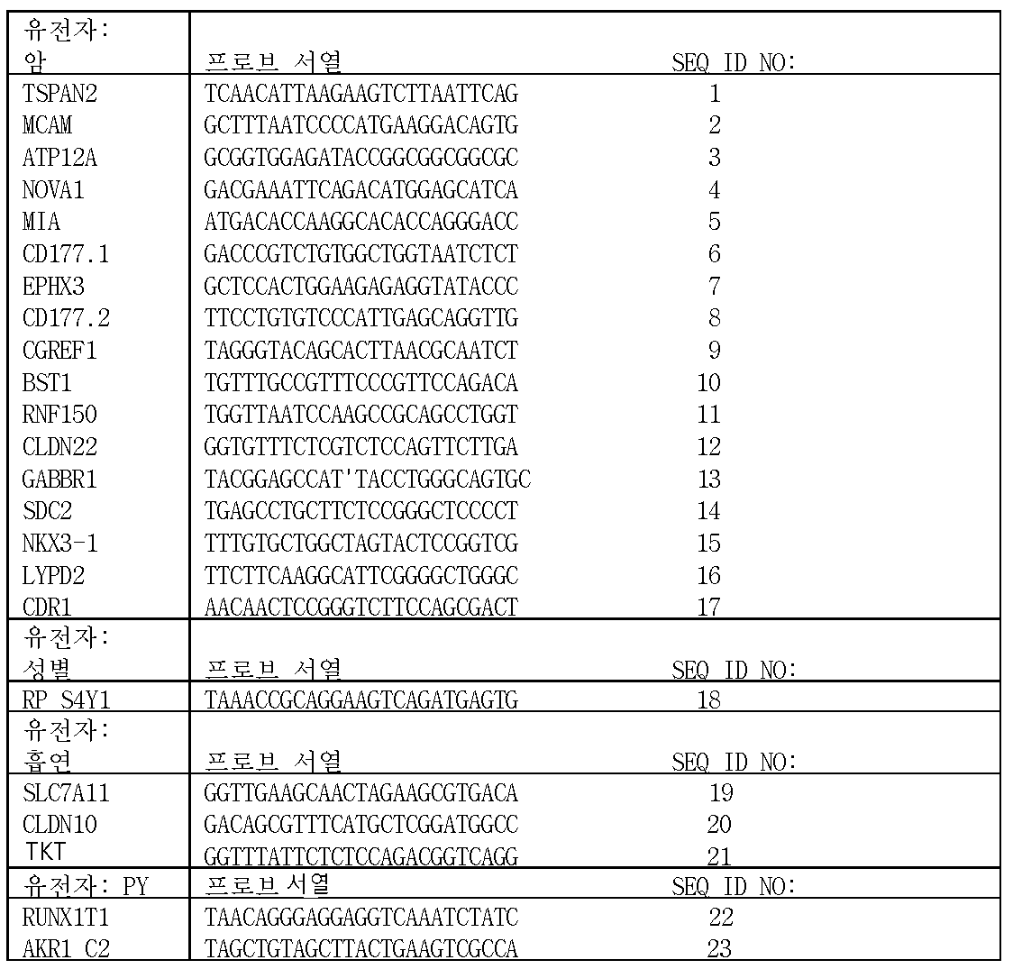
아래의 표 2는 트레이닝 세트에서 기관지 내시경 음성 대상에 대한 2개의 모델에 대한 성능 특성의 요약을 제공한다. 기관지 내시경 음성 대상(N)의 수는 CA + 민감성에 대한 대상과 CA - 특이성에 대한 대상에 해당한다.Table 2 below provides a summary of the performance characteristics for the two models of bronchoscopy voice subjects in the training set. The number of bronchoscopic voice subjects (N) corresponds to subjects with CA + sensitivity and subjects with CA - specificity.
*NPV는 기관지 내시경 및 예측 모델의 관찰된 민감성 및 특이성과 결합된 암의 50% 유병률을 가정하여 계산된다. * NPV is calculated assuming a 50% prevalence of cancer combined with the observed sensitivity and specificity of bronchoscopy and predictive models.
모델 성능Model Performance
CA + 및 CA - 샘플의 예측을 구별하기 위해, 특정 점수 임계값을 사용하여 예측 모델(점수 1 및 점수 2에 기초)의 민감도 및 특이성을 계산하였다. 트레이닝 세트(모델 점수 = 0.65)에서 두 모델에 대해 선택된 동일한 임계값을 유효성 검사 세트에 사용하였다. 예측 정확도는 두 모델에 대한 기관지 내시경 음성 샘플(의도 된 용도의 경우)에서 처음 측정하였고 다음에서 요약된다.To distinguish the predictions of CA + and CA - samples, the sensitivity and specificity of predictive models (based on
예측 모델의 민감성은 또한 유효성 검사 세트 내의 몇몇 서브 그룹 카테고리에 대해 계산하였다. 두 모델에 대한 결과는 표 3-9에 도시된다. 하위 카테고리는 신뢰 구간에 영향을 미치는 상이한 수의 샘플을 포함한다.The sensitivity of the prediction model was also calculated for several subgroup categories within the validation set. The results for both models are shown in Table 3-9. The subcategory includes a different number of samples that affect the confidence interval.
PM = 예측 모델; BR = 기관지 내시경PM = predictive model; BR = bronchoscopy
PM = 예측 모델; BR = 기관지 내시경PM = predictive model; BR = bronchoscopy
트레이닝training 및 유효성 검사 세트에서 성능의 비교 And performance comparison in a validation set
트레이닝 및 유효성 검사 세트에서 예측 모델의 전체 예측 정확성을 비교하여 독립 그룹에서 모델의 재현성을 평가하였다. 결과는 표 10에 제공된다.The reproducibility of the model in the independent group was evaluated by comparing the total prediction accuracy of the prediction model in the training and validation sets. The results are provided in Table 10.
샘플all
Sample
샘플BR-negative
Sample
샘플all
Sample
샘플BR-negative
Sample
샘플all
Sample
샘플BR-negative
Sample
샘플all
Sample
샘플BR-negative
Sample
특이성
AUCSensitivity
Specificity
AUC
민감성 또는 특이성의 유의한 차이(p> 0.05)는 각 모델에 대한 트레이닝 및 유효성 검사 세트 사이에서 관찰되지 않았다. 마찬가지로, 두 그룹 사이의 각 모델에 대한 AUC에서 작은 차이는 통계적으로 유의하지 않았다(p> 0.05 기준). 민감성과 특이성은 모든 샘플(기관지 내시경-음성과 기관지 내시경-양성 합침)에 비해 기관지 내시경-음성 샘플에 대해 유사하다. 점수 1과 점수 2 사이의 전체 성능에 상대적으로 작은 하락이 있었고, 점수 2는 점수 1에 비해 AUC에서 2 - 4% 포인트 하락을 보였다.No significant differences in sensitivity or specificity (p> 0.05) were observed between the training and validation sets for each model. Likewise, small differences in AUC for each model between the two groups were not statistically significant (p> 0.05). Sensitivity and specificity are similar for bronchoscopy-negative samples compared to all samples (bronchoscopy-negative and bronchoscopy-positive consolidation). There was a relatively small drop in overall performance between
실시예Example 2: 진단 기관지 내시경을 받는 폐암 환자에 대한 기관지 게놈 분류기의 유효성 검사 2: Validation of bronchial genome classifier for lung cancer patients receiving diagnostic bronchoscopy
개요summary
기관지 내시경은 종종 폐암으로 의심되는 폐 병변을 갖는 환자에서 진단에 유용하지 않다. 여러 병변이 양성이긴 하지만, 이것은 종종 추가적인 침습 테스트를 초래한다. 본 발명자는 기관지 내시경의 진단 성능을 향상시킬 수 있는 기관지 유전자 발현 분류기를 검증하고자 했다.Bronchoscopy is often not useful for diagnosis in patients with suspected pulmonary lung lesions. Although many lesions are benign, this often results in additional invasive testing. The present inventors tried to verify a bronchial gene expression classifier capable of improving the diagnostic performance of bronchoscopy.
폐암으로 의심하는 병변은 가슴 영상에서 종종 확인된다. 조직 표본 샘플링을 필요로 하는 감시 영상 또는 침습 평가를 수행할지의 결정은 복잡하며 악성 가능성, 생검 능력, 수술 위험 및 환자 선호도의 평가가 필요하다[1]. 생검이 필요할 때, 접근법은 기관지 내시경, 경흉부 세침 생검(transthoracic needle biopsy, TTNB) 또는 수술적 폐 생검(surgical lung biopsy, SLB)을 포함할 수 있다. 이러한 양상들 사이의 선택은 일반적으로 병변의 크기와 위치, 선종의 유무, 절차의 위험 및 현지 전문 지식과 같은 고려 사항에 의해 결정된다. 기관지 내시경 검사는 안전한 방법이며, 기흉에 의해 1% 미만의 합병증이 발생한다[2]. 미국에서 1년에 약 500,000건의 기관지 내시경이 시행되며[3], 그 중 대략 절반이 폐암의 진단을 위한 것이다. 그러나 기관지 내시경은 병변의 위치와 크기에 따라 34-88% 범위의 민감성으로 제한된다[4]. 더 새로운 기관지 내시경 모범 기술을 사용하더라도, 말초 병변에 대한 민감성은 단지 ~70%이다 [5].Suspected lung cancer lesions are often seen in breast images. Determination of whether to perform a surveillance image or invasive assessment that requires sampling of tissue specimens is complex and requires evaluation of malignancy potential, biopsy capability, operative risk and patient preference [1]. When biopsy is needed, the approach may include bronchoscopy, transthoracic needle biopsy (TTNB), or surgical lung biopsy (SLB). The choice between these aspects is generally determined by considerations such as lesion size and location, presence of adenoma, procedure risk, and local expertise. Bronchoscopy is a safe method and less than 1% of complications are caused by pneumothorax [2]. Approximately 500,000 bronchial endoscopes are performed in the United States a year [3], of which approximately half are for the diagnosis of lung cancer. However, bronchoscopy is limited to a sensitivity range of 34-88% depending on the lesion location and size [4]. Even with newer bronchoscopy-based techniques, the sensitivity to peripheral lesions is only ~ 70% [5].
비 진단 기관지 내시경을 받은 환자는 종종 최종 진단을 확립하기 위해 추가 침습 검사를 받는다. SLB는 대략 5%의 합병증 비율과 ~1%의 30일 사망률을 나타내는 본질적 위험을 감안할 때 초기 선호 접근법이 아니다[6]. 중요하게는, SLB의 20-25%가 양성 병변으로 진단된 환자에서 수행된다[7,8]. 또한, TTNB는 15% 기흉 비율을 포함하는 현저한 이환율과 관련이 있으며, 이중 6%는 흉관 배액을 필요로 한다[10,11]. 침습적 절차의 함정을 고려할 때, 이미징 감시에 적합한 악성 종양의 가능성이 더 낮은 환자를 확인하기 위한 대안적인 접근이 필요하다.Patients receiving non-diagnostic bronchoscopy often undergo additional invasive testing to establish a final diagnosis. SLB is not an initial preference approach, given the inherent risk of approximately 5% complication rate and ~ 1% 30-day mortality [6]. Importantly, 20-25% of SLB are performed in patients diagnosed with benign lesions [7,8]. In addition, TTNB is associated with a significant morbidity including 15% pneumothorax, of which 6% require chest tube drainage [10,11]. Given the pitfalls of invasive procedures, an alternative approach is needed to identify patients with a lower probability of malignancy suitable for imaging surveillance.
임상 표본의 유전자 발현 측정을 사용하여, 암을 포함하는 생물학적 질병 상태의 분류가 잘 확립되어있다[12]. 폐암의 상태에서, 흡연자의 근위기도로부터 수집된 세포학적으로 정상 상피 세포에서 뚜렷한 암 관련 유전자 발현 패턴이 존재한다[13]. 최근, 본 발명자는 현재 및 과거 흡연자들 중에서 폐암에 걸린 환자와 걸리지 않은 환자를 구별하는 기관지 내시경을 통해 주 기관지로부터 수집한 기관지 상피 세포(BEC)에서 유전자 발현 분류기를 개발하였다(원고 제출). 본 발명자는 의심되는 폐암에 대해 기관지 내시경을 받는 환자의 두 가지 전향적인 다기관 임상 시험에서 이 분류기의 유효성을 검사하고 이 분류기가 기관지 내시경의 진단 성능을 어떻게 바꾸는지 평가하기 위해 본 연구에 착수했다.Using the gene expression measurements of clinical specimens, classification of biological disease states, including cancer, has been well established [12]. In lung cancer, there is a clear cancer-associated gene expression pattern in cytologically normal epithelial cells collected from proximal airways of smokers [13]. Recently, the present inventor has developed a gene expression classifier (BEC) in bronchial epithelial cells (BEC) collected from the main bronchus through a bronchoscopy which distinguishes between patients who have lung cancer and those who do not. The present inventor has undertaken this study to validate this classifier in two prospective multicenter trials of patients receiving bronchoscopy for suspected lung cancer and to assess how this classifier alters the diagnostic performance of bronchoscopy.
재료 및 방법Materials and methods
연구 설계, 개체군 및 프로토콜Research designs, populations and protocols
폐암의 의심을 위한 기관지 내시경을 받는 현재 및 과거 흡연자를 AEGIS 1 및 AEGIS 2, 두 개의 독립적인 전향적 다기관 관찰 연구(NCT01309087 및 NCT00746759)에 등록하였다. 환자를 미국, 캐나다 및 아일랜드의 28개 장소에 등록하였다. 세포학 브러쉬를 사용하여 주 기관지로부터 정상적으로 보이고 RNA 방부제에 잠긴 BEC를 수집하였다. 분류기의 결과는 의사 또는 환자에게 보고되지 않았다. 환자들이 연구 프로토콜의 요구 사항을 충족시키는지를 측정하기 위해 기관지 내시경 전에 환자를 스크리닝하였다. 배제 기준은 대상 <21세, 비 흡연자(일생 동안 흡연 <100 담배로 정의됨), 동시 암 또는 폐암의 이력이 있는 환자를 포함하였다. 기관지 내시경 직전 > 24시간 기계 인공호흡기를 사용했거나 연구에 동의하거나 응하지 않은 환자는 배제하였다. 최종 진단이 확립될 때까지 또는 기관지 내시경 후 12개월까지 환자를 추적관찰했다. 폐암의 진단은 지표 기관지 내시경 시 또는 TTNB, SLB, 제 2 기관지 내시경 또는 기타 침습적 절차를 사용하는 후속 생검으로 확립되었다. 사용된 특정 기관지 내시경 방법 및 비 진단 기관지 내시경 후 수행된 후속적인 감시 영상 또는 절차는 치료 의사의 재량이었다. 암이 없는 것으로 진단된 환자는 12개월의 추적 관찰에서 특정 양성 진단 또는 방사선 안정성/소산을 나타냈다. 12개월의 추적 관찰에서 암의 확실한 진단, 특정 양성 진단 또는 안정성/소산이 없는 환자는 추가 분석에서 배제하였다. 치료 의사는 5단계 척도(<10%, 10-39%, 40-60%, 61-85% 및> 85%)를 사용하여 기관지 내시경 전에 각 환자의 사전 테스트 악성도(POM)를 평가하였다. 연구 프로토콜은 각 참여 센터의 기관 검토위원회에 의해 승인받았고, 모든 환자는 등록 전에 사전 동의서를 제공하였다.Current and former smokers receiving bronchoscopy for suspected lung cancer were enrolled in two independent prospective multicenter observational studies (NCT01309087 and NCT00746759),
AEGIS 1에서 855명 및 AEGIS 2에서 502명의 전체 환자가 적격이었고 미국, 캐나다 및 아일랜드의 28개 의료 센터에 2009년 1월 내지 2012년 8월(도 4)에 등록하였다(표 12). 이 장소들은 3차/학술 의료 센터(n = 20), 지역사회 기반 병원(n = 6) 및 재향군인 병원(n = 2)의 혼합이었다.A total of 855 patients from
추가 환자를 하기 기준에 기초한 등록 후 연구로부터 배제하였다(도 4 참조). 첫째, AEGIS 1에서 111명과 AEGIS 2에서 71명의 전체 환자는 다음과 같은 프로토콜 편차로 인해 부적격 판정을 받았다: AEGIS 1에서, 환자 24명이 검토시 연구 등록 기준을 충족하지 못했고, 승인시 승인 기준을 충족하지 못하는 수집된 표본을 가졌거나 등록되었으나 수집된 표본을 갖지 않았다. 추가 87개 샘플은 RIN 점수 <4 또는 RNA 수율 <1μg로 인해, RNA 분리 후 최소 QC 기준을 충족하지 못했다. AEGIS 2에서, 승인시 승인 기준을 충족하지 않는 표본 수집으로 인해 부적합한 환자가 3명 있었고, RNA의 최소 QC 기준을 충족하지 못하는 68개의 샘플이 있었다. 둘째, AEGIS 1에서 환자 40명과 AEGIS 2에서 환자 10명을 포함하여 비 원발성 폐암으로 진단된 환자는 배제하였다. 또한, 12개월에 검토시, AEGIS 1에서 환자 91명과 AEGIS 2에서 환자 71명을 포함하는 최종 진단이 없는 환자는 연구에서 배제하였다. AEGIS 1과 2의 이런 환자 162명(n = 106)의 대략 2/3은 기관지 내시경 후 추적관찰에서 배제되었고, 나머지 1/3(n=56)은 기관지 내시경 후 12개월에 최종 진단을 받지 못했다. 마지막으로, 본 발명자는 AEGIS 1에서 환자 1명과 AEGIS 2에서 환자 9명을 분석 QC 기준(마이크로어레이 처리 방법에서 기술됨)을 충족하지 못한 유전자 발현 데이터에 의해 배제하였다.Additional patients were excluded from post-registration studies based on the following criteria (see FIG. 4). First, 111 patients in
AEGIS 1 그룹은 이전에 무작위 방식으로 동일한 트레이닝 및 테스트 세트로 분할하였다. 트레이닝 세트(n = 환자 299명)는 23개 유전자와 연령으로 구성된 유전자 발현 분류기를 유도하는데 사용되었고 다른 곳에 기술되었다(원고 제출). 트레이닝 세트에서, 분류기는 기관지 내시경 검사가 폐암의 진단으로 이어지지 않는 환자(n = 134)에서 0.78(95% CI, 0.71-0.85)의 AUC, 93%의 민감성과 57%의 특이성을 갖는 것으로 밝혀졌다. 현재 연구는 두 개의 독립적인 유효성 검사 세트에서 잠금 분류기의 유효성 검사에 집중한다.The
최종 AEGIS 1 및 AEGIS 2 유효성 검사 세트의 기준 인구통계학적 및 임상적 특성이 표 1에서 비교된다. 각 그룹 내에서 암 및 양성 질환으로 진단된 환자의 개별 비교는 표 12에 제공된다.The baseline demographic and clinical characteristics of the
a) 중앙값(및 사분위 범위; IQR)으로 보고하였다. P-값은 맨-휘트니 테스트를 사용하여 계산하였다.a) Median (and range of quadrants; IQR). P-values were calculated using the Man-Whitney test.
b) P-값은 백인 vs. 비 백인에 대해 계산하였다.b) P-value is white vs. white. Were calculated for non-white people.
임상 및 인구통계학적 데이터를 각 환자에 대해 수집하고 연구 임상 보고서 양식(CRF)에 기록하였다. 추가 병리학 및 방사선학 보고서는 각 환자의 의료 기록에서 유지되었으며 검토를 위해 사용할 수 있었다. 모든 출처 문서는 관찰되고 연구 책임자가 관리하는 데이터베이스에 입력되었다. 폐 병변의 크기와 위치는 CT 스캔 결과에서 얻었다. 대상을 기관지 내시경 후 12개월까지 추적관찰하여 임상 진단을 위한 데이터를 수집하였다. 폐암의 진단은 병리학의 결과에 기초했고 병리학 보고서 사본은 의료 센터에서 수집하였다. 암의 임상 진단으로 이끄는 표본은 기관지 내시경 중 또는 기관지 내시경이 비 진단인 후속 침습 검사에서 얻었다. 비 진단 기관지 내시경 후 수행된 모든 임상 절차에 대한 데이터는 연구 중에 수집하였다. 비 진단 기관지 내시경을 받은 환자의 후속 평가는 각 연구 센터의 치료 의사의 재량이었다. 일부 경우에, 성공적으로 진단을 내리기 위해 기관지 내시경 이후에 1회 이상의 절차가 필요했으나, 모든 경우 폐암 진단에 이르는 절차를 수집하였다. 침습적 후속 절차는 종격동경검사, 흉강경검사, 개흉술 또는 비디오 흉강경 수술(VATS)을 포함할 수 있는 제 2 기관지 내시경, TTNB 또는 수술적 폐 생검(SLB)으로 정의되었다.Clinical and demographic data were collected for each patient and recorded in the Study Clinical Report Form (CRF). Additional pathology and radiology reports were maintained in each patient's medical records and were available for review. All source documents were observed and entered into a database maintained by the investigator. The size and location of lung lesions were obtained from CT scans. Data were collected for clinical diagnosis by follow - up up to 12 months after bronchoscopy. The diagnosis of lung cancer was based on the results of pathology, and a copy of the pathology report was collected at the medical center. The specimens leading to the clinical diagnosis of cancer were obtained from bronchoscopy or subsequent invasive tests in which bronchoscopy was not diagnosed. Data on all clinical procedures performed after non-diagnostic bronchoscopy were collected during the study. Follow-up evaluation of patients with non-diagnostic bronchoscopy was at the discretion of the treating physician at each study center. In some cases, more than one procedure was required after bronchoscopy to successfully diagnose, but in all cases, procedures to diagnose lung cancer were collected. Invasive follow-up procedures have been defined as second bronchoscopy, TTNB or surgical lung biopsy (SLB), which may include mediastinoscopy, thoracoscopy, thoracotomy or video-assisted thoracoscopic surgery (VATS).
암으로 진단되지 않은 환자의 기록은 대상이 암이 없는 것으로 선언하기 위해 판결 과정을 거쳤다. 이 과정은 5명의 폐병학자 패널이 각 환자에 대해 사용할 수 있는 의료 기록의 검토로 구성되었다. 이 패널의 2명의 폐병학자가 각각의 환자를 독립적으로 검토하고 남성/여성이 다음 기준 중 하나를 충족하면 환자는 암이 없는 것으로 결정하였다: 환자는 초기의 의심스러운 이상을 설명하는 대체 진단으로 진단되었고, 이상은 안정한 것으로 결정되거나 이상이 해소되었다. 12개월 추적관찰 기간의 종료시 이런 기준을 충족하지 못했던 환자는 최종 진단이 없으며 연구에서 추가 분석으로부터 배제되었다.Records of patients not diagnosed with cancer were judged to declare that the subject had no cancer. This process consisted of a review of the medical records available to each patient by a panel of five leprosyologists. Two lung cancer clinicians on this panel independently reviewed each patient and determined that if the male / female met one of the following criteria, the patient had no cancer: the patient was diagnosed with an alternative diagnosis that described the initial suspicious anomaly And the ideal was determined to be stable or the anomaly was resolved. Patients who did not meet these criteria at the end of the 12-month follow-up period had no final diagnosis and were excluded from further analysis in the study.
원발성 폐암으로 진단된 환자에 대한 조직학(표 13) 및 암 단계(표 14) 데이터를 분석하였다. 연구 CRF에서 데이터를 추출하여 완전한 의학 기록의 검토에 의해 확인하였다. 전체 암 중에서, AEGIS 1에서 80%(175/220; 95% CI, 74-84%)와 AEGIS 2에서 83%(222/267, 95% CI, 78-87%)가 NSCLC(비소세포 폐암)이었다. 각각 17%(38/220, 95% CI, 13-23%) 및 16%(42/267, 95% CI, 12-21%) SCLC 암이 존재하였다. 공지된 암 단계 데이터를 가진 NSCLC에 걸린 환자 중에서, 초기 단계(단계 1 또는 2)는 AEGIS 1에서 40%(53/139, 95% CI, 32-48 %), AEGIS 1에서 37%(67/199, 95% CI, 30-44 %)로 존재하였다.Histology (Table 13) and cancer stage (Table 14) data were analyzed for patients diagnosed with primary lung cancer. Data were extracted from study CRF and confirmed by review of complete medical records. Of the total cancers, 80% (175/220; 95% CI, 74-84%) in
않음Not
않음Not
양성 질환의 각각에 관한 데이터를 의학 기록으로부터 추출하고 표 15에 요약하였다. 두 연구에서 암이 없는 대상의 대략 2/3가 대체 진단을 받았고, 특정 대체 진단은 각각의 연구에 대해 표 S4에 열거되었다. 해소되었거나 안정된 것으로 판단된 이상을 가진 환자의 진단은 기관지 내시경 12개월 후, 후속 이미징을 포함한 의료 기록의 검토에 기반하였고, 암이 없는 환자의 대략 1/3이 두 연구에서 이런 카테고리에 있었다.Data on each of the benign diseases were extracted from the medical records and summarized in Table 15. Approximately two-thirds of cancer-free subjects in both studies were diagnosed with a replacement, and specific alternative diagnoses were listed in Table S4 for each study. The diagnosis of patients who were resolved or stable was based on a review of medical records including follow-up imaging after 12 months of bronchoscopy and approximately one-third of patients without cancer were in this category in both studies.
임상적으로 필요한 기관지 내시경 동안, 연구 참여자는 표준, 일회용 세포학 브러시를 사용하여 기관지 벽에 문질러서 우측 또는 좌측 주 기관지의 점막 브러싱을 수집하였다. 각 대상에게서 2개의 브러싱을 얻었다; 의사들은 정상적으로 보이는 상피 조직의 브러싱을 얻고 종양 조직 또는 이형성 세포를 샘플링하는 것을 피하도록 훈련받았다. 모든 시료를 RNA 방부제(RNAprotect, Qiagen)에 저장하고 승인 및 처리를 위해 중앙 CLIA-인증 연구소로 운송하였다. 센터는 최대 14일 동안 4℃에서 샘플을 저장하고, 스폰서가 제공한 2일 운송 및 절연 운송 용기(NanoCool)를 사용하여 주변 온도 이하(4-20℃)로 운송하도록 요청받았다.During clinically necessary bronchoscopy, participants were rubbed against the bronchial wall using a standard, disposable cytology brush to collect mucosal brushing of the right or left main bronchus. Two brushes were obtained from each subject; Physicians were trained to obtain normal visible brushing of epithelial tissue and avoid sampling tumor tissue or dysplastic cells. All samples were stored in RNA preservatives (RNAprotect, Qiagen) and shipped to a central CLIA-certified laboratory for approval and treatment. The Center was asked to store samples at 4 ° C for up to 14 days and to transport below ambient temperature (4-20 ° C) using a two-day shipping and insulation shipping container (NanoCool) provided by the sponsor.
실험실 방법Laboratory method
모든 BEC 표본을 처리하여 RNA를 분리하고 유전자 발현 분석 전에 품질 및 수율에 대해 RNA를 분석하였다; 적어도 1μg의 RNA 수율과 >4의 RIN 점수를 가진 표본을 Gene-ST 1.0 마이크로어레이에서 실행하였다. 모든 마이크로어레이 데이터를 GEO에 GSE66499로 기탁하였다.All BEC samples were processed to isolate RNA and analyzed for quality and yield prior to gene expression analysis; A sample with an RNA yield of at least 1 μg and an RIN score of> 4 was run on a Gene-ST 1.0 microarray. All microarray data were deposited with GEO on GSE66499.
샘플을 용해하고 컬럼-기초 방법(miRNeasy Kit, Qiagen) 및 제조사의 권장 프로토콜을 사용하여 RNA를 분리하였다. 큰 RNA 분획을 분광 광도계(Nanodrop; ThermoFisher) 상에서 분석하여 농도, 순도 및 수율을 측정하였다. 샘플을 RIN 점수로 보고된, RNA 무결성(Bioanalyzer)에 대해 분석하였다. <1μg의 수율 및 RIN 점수 <4를 가진 샘플을 연구에서 배제하였다. AEGIS 1에서 표본의 약 10%와 AEGIS 2에서 표본의 약 13%를 불충분한 RNA의 품질 또는 양 때문에 배제하였다. 그런 후에 RNA를 추후 처리 때까지 냉동 보관하였다(-80℃).Samples were dissolved and RNA was isolated using the column-based method (miRNeasy Kit, Qiagen) and the manufacturer's recommended protocol. Large RNA fractions were analyzed on a spectrophotometer (Nanodrop; ThermoFisher) to determine concentration, purity and yield. Samples were analyzed for RNA integrity (Bioanalyzer) reported as RIN scores. Samples with a yield of <1 μg and a RIN score <4 were excluded from the study. Approximately 10% of the sample in
200ng의 전체 RNA를 Ambion WT 발현 키트(Life technologies Cat. # 4440536)를 사용하여 센스 가닥 cDNA로 전환시킨 후, Affymetrix GeneChip WT 말단 표지화 키트(Affymetrix Cat. # 900671)로 표지하였다. 혼성화를 위해, 혼성화 칵테일을 준비하고 인간 유전자 1.0 ST 어레이(Affymetrix Cat. # 901087)에 적용된 혼성화, 세척 및 염색 키트(Affymetrix Cat. # 900720)를 사용하여 표지된 cDNA 표적에 첨가하고 45℃에서 16시간 동안 배양하였다. 혼성화 이후, 어레이를 세척하고 Affymetrix GeneChip 스캐너에서 스캐닝하기 전에 표준 어피메트릭스 절차를 사용하여 염색하였다. 발현 콘솔 소프트웨어(어피메트릭스)를 사용하여 데이터를 추출하였다.200 ng of total RNA was converted into sense strand cDNA using the Ambion WT expression kit (Life technologies Cat. # 4440536) and labeled with Affymetrix GeneChip WT terminal labeling kit (Affymetrix Cat. # 900671). For hybridization, a hybridization cocktail was prepared and added to the labeled cDNA target using a hybridization, washing and staining kit (Affymetrix Cat. # 900720) applied to a human gene 1.0 ST array (Affymetrix Cat. # 901087) Lt; / RTI > After hybridization, the array was washed and stained using standard Affymetrix procedures prior to scanning on an Affymetrix GeneChip scanner. Data was extracted using the expression console software (Affymetrix).
어피메트릭스 인간 유전자 1.0 ST 어레이(Affymetrix Cat. # 901087)는 www.affymetrix.com/site/include/byproduct.affx?product=hugene-1_0-st-v1에 개시된 여러 프로브를 사용한다. 유전자 당 하나의 프로브의 1:1 비율이 아니라는 것을 의미하는 특정 유전자의 단편에 해당하는 다수의 프로브가 존재하며, 오히려 단일 유전자의 다중 단편에 해당하는 다수의 프로브가 존재한다. 한 예에서, LYPD2 유전자는 어피메트릭스 인간 유전자 1.0 ST 어레이(HuGene-1_0-st-v1 프로브세트 주석, 09/30/2011에 릴리즈 32, 03/27/2013에 릴리즈 33, 04/07/2014에 릴리즈 34)에 개시된 바와 같이 인간 유전자 1.0 ST 어레이(릴리즈 32)에 3개의 프로브 세트, 프로브세트 ID 8153343, 8153344, 및 8153345로 나타내어진다. 프로브세트 및 핵산 서열과 관련이 있는 정보는 www.affymetrix.com/site/include/byproduct.affx?product=hugene-1_0-st-v1을 포함하는 Affymetrix.com에서 발견될 수 있다. 또한, 데이터세트는 본 발명의 방법을 실시하는데 사용될 수 있는 프로브 및 유전자를 열거하는 플랫폼 GPL6244 하의 NCBI 유전자 발현 옴니버스 웹사이트 www.ncbi.nlm.nih.gov/geo/query/acc.cgi?acc=GPL6244에서 이용할 수 있다. 프로브세트 및 유전자 기호와 관련이 있는 문서를 포함하는 이런 문서는 본 발명에 참조로 포함된다.The Affymetrix Human Gene 1.0 ST array (Affymetrix Cat. # 901087) uses several probes as described in www.affymetrix.com/site/include/byproduct.affx?product=hugene-1_0-st-v1. There are a number of probes corresponding to fragments of a particular gene, meaning that it is not a 1: 1 ratio of one probe per gene, rather a number of probes corresponding to multiple fragments of a single gene. In one example, the LYPD2 gene is cloned into an Affymetrix human gene 1.0 ST array (HuGene-1_0-st-v1 probe set annotation, 09/30/2011 at Release 32, 03/27/2013 at Release 33, 04/07/2014 Release 34), three probe sets in the human gene 1.0 ST array (Release 32), probe set IDs 8153343, 8153344, and 8153345. Information related to probe sets and nucleic acid sequences can be found at Affymetrix.com, including www.affymetrix.com/site/include/byproduct.affx?product=hugene-1_0-st-v1. In addition, the data set includes the NCBI gene expression omnibus website www.ncbi.nlm.nih.gov/geo/query/acc.cgi?acc========================================================================================= Also, Available in GPL6244. Such a document, including a probe set and a document related to the gene symbol, is incorporated herein by reference.
각 그룹의 최종 데이터 세트에서 샘플에 해당하는 마이크로어레이 데이터(CEL 파일)는 RMA를 사용하여 정규화되었다[22]. AEGIS 1 샘플은 총 5개 배치로 실행되었고 ComBat[23]는 배치 효과를 수정하는데 사용하였다. AEGIS 2 샘플은 모두 단일 마이크로어레이 배치로 실행되었다. 정규화 이후, 특이점은 글로벌 유전자 발현에서 0.955 미만의 게놈-와이드 쌍 상관관계를 갖는 것으로 확인되었다. 모든 마이크로어레이 데이터를 GEO에 수탁 번호# GSE66499로 기탁하였다.The microarray data (CEL file) corresponding to the samples in the last data set of each group was normalized using RMA [22].
마이크로어레이 데이터의 정규화 및 전처리는 보충 부록에 기술된다. AEGIS 1에 등록된 대상은 이전에 무작위로 독립적인 트레이닝 및 유효성 검사 세트로 할당되었고(도 4) 분류기 알고리즘은 이미 기술한 바와 같이, AEGIS 1 트레이닝 세트 내에서 엄격하게 유도되고 잠겼다. AEGIS 1 유효성 검사 세트에서 각 샘플 및 모든 AEGIS 2 샘플에 대한 점수는 23개 유전자의 발현 및 환자 연령에 기초한 이 사전-지정 분류기를 사용하여 생성되었다. 이 점수는 사전-지정 임계값을 사용하여 테스트-양성 및 테스트-음성으로 이분화되었다.Normalization and preprocessing of microarray data are described in the supplementary appendix. Subjects enrolled in
분류기의 성능은 수용기 작동자 특성(ROC) 곡선, 곡선 아래 면적 계산(AUC) [14], 및 민감성, 특이성, 음성 예측치(NPV), 양성 예측치(PPV) 및 (1-민감성)/ 특이성으로 정의된 음성 가능성 비율(NLR)을 사용하여 평가하였다. 맨-휘트니 비 매개변수 테스트는 연속 변수 분석에 사용되었고 피셔의 정확도 테스트(Fisher 's exact test)는 카테고리 변수에 사용되었다. 모든 신뢰 구간은 양면 이항 95% 신뢰 구간(95% CI)으로 보고된다. 통계 분석은 R 소프트웨어(버전 3.01)를 사용하여 수행되었다.The performance of the classifier is defined as the receiver operator characteristic (ROC) curve, area under the curve (AUC) [14], and sensitivity, specificity, negative predictive value (NPV), positive predictive value (PPV) and (1-sensitive) (NLR). ≪ / RTI > Mann - Whitney 's nonparametric test was used for continuous variable analysis and Fisher' s exact test was used for categorical variables. All confidence intervals are reported as two-sided binomial 95% confidence intervals (95% CI). Statistical analysis was performed using R software (version 3.01).
의사는 <10%, 10-39%, 40-60%, 40-60%, 61-85%, 및 >85%의 카테고리 중 하나를 사용하여, 기관지 내시경에 전에 전문 견해 및 이용가능한 의료 기록에 기초하여 등록된 모든 대상에 대해 사전 테스트 POM을 평가하도록 요청받았다. 결과는 <10%, 10-60% 및> 60%의 카테고리로 축소되었다. 본 발명자는 계층화된 POM 수준에서 암의 실제 유병률을 비교하기 위해 각 POM 카테고리에서 암의 실제 유병률을 계산하였다(표 16). 또한, 본 발명자는 폐암 후 비 기관지 내시경 검사의 유병률이 <10%, 10-60% 및 >60% POM 그룹에서 각각 3%, 29% 및 80%임을 발견하였다.The physician will use the prior knowledge of the bronchoscopy and the available medical records, using one of the categories of <10%, 10-39%, 40-60%, 40-60%, 61-85%, and> 85% Based on this, we were asked to evaluate the pre-test POM for all registered subjects. Results were reduced to categories of <10%, 10-60% and> 60%. The inventors calculated the actual prevalence of cancer in each POM category to compare the actual prevalence of cancer at the level of the layered POM (Table 16). We also found that the prevalence rates of non-bronchoscopy after lung cancer were 3%, 29%, and 80% in the POM group of <10%, 10-60%, and> 60%, respectively.
a) POM = 악성 가능성; 기관지 내시경 전에 치료 의사가 평가.a) POM = malicious possibility; Doctors assessed before bronchoscopy.
b) 유병률은 각 POM 카테고리 내에서 폐암으로 진단된 비율로서 계산하였다.b) Prevalence was calculated as the percentage of lung cancer diagnosed within each POM category.
병변 크기에 의해 계층화된 기관지 게놈 분류기의 예측 정확도가 표 2에 요약되며 사전 테스트 POM에 의해 계층화된 기관지 게놈 분류기의 예측 정확도가 표 3에 요약된다. 분류기의 민감성은 표 S5 및 S6에 단계와 조직학으로 계층화된 폐암으로 진단된 환자에 대해 보고된다. 기관지 내시경과 병합된 분류기는 모든 카테고리에서 민감성 >90% 유도한다.The prediction accuracy of the bronchial genome classifiers stratified by lesion size is summarized in Table 2 and the prediction accuracy of the bronchial genome classifiers stratified by the pre-test POM is summarized in Table 3. [ The sensitivity of the classifier is reported for patients diagnosed with stage and histologically stratified lung cancer in Tables S5 and S6. Classifiers combined with bronchoscopy lead to sensitivity> 90% in all categories.
(95% CI)Bronchoscopy
(95% CI)
(95% CI)Classifier
(95% CI)
결과result
연구 참가자의 특성Characteristics of research participants
AEGIS 1의 환자 298명을 제 1 유효성 검사 세트로 사용하고 AEGIS 2 회의 연구 기준의 모든 환자 341명을 제 2 유효성 검사 세트로 사용하였다 (도 4 및 표 13). 폐암의 유병률은 AEGIS 1과 AEGIS 2 그룹에 대해 각각 74%와 78%이었다. 폐암 환자는 암이 없는 환자보다 나이가 많았고(p <0.001), 누적 담배 노출이 더 컸고 (p <0.001) 현재 흡연자일 가능성이 더 높았다(AEGIS 1에서 p = 0.07, AEGIS 2에서 p = 0.03; 표 12). 암 단계의 요약과 양성 진단의 카테고리가 각각 표 15와 16에 도시된다.298 patients of
기관지 내시경의 성능Performance of bronchoscopy
총 639명의 환자가 의심되는 폐암에 대해 기관지 내시경을 받았다. 이들 중, 272명(43%, 95% CI, 39 내지 46)이 진단되지 않았고, 487명의 환자 중 120명(25%, 95% CI, 21 내지 29)이 최종 폐암으로 진단되었다. 폐암에 대한 기관지 내시경의 민감성은 AEGIS 1과 AEGIS 2에서 각각 74%(95% CI, 68 내지 79)와 76%(95% CI, 71 내지 81)이었다. 비 진단 기관지 내시경을 받은 환자 중 98%(272명 중 267명)에 대해 추적 관찰 절차 자료를 이용할 수 있었다. 양성 병변이 있는 147명 중 52명(35%; 95%CI, 24 내지 38) 및 120명의 암 환자 중 118명(98%; 95%CI, 94 내지 99)을 포함하는 267명 환자 중 170명(64%, 95% CI, 58 내지 69)에 비 진단 기관지 내시경 이후 침습적 절차를 시행하였다. SLB를 76명 환자에서 시행하였고, 이중 27명(36%, 95% CI, 26-47)이 양성 병변이 있었다.A total of 639 patients received bronchoscopy for suspected lung cancer. Of these, 272 (43%, 95% CI, 39 to 46) were not diagnosed and 120 of the 487 patients (25%, 95% CI, 21 to 29) were diagnosed as terminal lung cancer. The sensitivities of bronchoscopy to lung cancer were 74% (95% CI, 68-79) and 76% (95% CI, 71-81) in
유전자 발현 분류기의 성능Performance of gene expression classifier
분류기 단독은 AUC = 0.78(95% CI, 0.73 내지 0.83)이었고 AEGIS 1에서 암 환자 220명 중 194명(88% 민감성, 95% CI, 83 내지 92) 및 암이 없는 78명 중 37명(47% 특이성, 95% CI, 37 내지 58)을 정확하게 확인하였다(도 5). AEGIS 2에서, 분류기는 AUC = 0.74(95% CI, 0.68 내지 0.80)이었고 암 환자 267명 중 237명(89% 민감성, 95% CI, 84 내지 92) 및 암이 없는 74명 중 35명(47% 특이성, 95% CI, 36 내지 58)을 정확하게 확인하였다. 분류기와 기관지 내시경의 병합은 기관지 내시경 단독에 대한 74% 및 76%(p <0.001)과 비교하여, AEGIS 1과 2에서 각각 96%(95% CI, 93 내지 98) 및 98%(95% CI, 96 내지 99)로 민감성을 증가시켰다.In the
비 진단 기관지 내시경을 받은 환자에서, 분류기는 AEGIS 1에서 57명의 환자 중 49명(86% 민감성; 95% CI, 74 내지 94) 및 AEGIS 2에서 67명의 환자 중 62명 (92% 민감성; 95% CI, 82 내지 97)에서 암을 정확하게 확인하였다. 모든 환자(p = 0.32) 또는 비 진단 기관지 내시경을 받은 환자(p = 0.61)에 대한 분류기 AUC에 관한 두 그룹의 환자 사이에 유의한 차이가 없었기 때문에(도 5), 본 발명자는 하위 그룹의 후속 분석을 위해 두 그룹을 병합하였다. 기관지 내시경 단독의 민감성은 <3cm인 병변(p <0.001) 또는 주변부에 위치한 병변(p <0.001)에서 더 낮았다(표 17). 반대로, 분류기 단독 및 기관지 내시경과 병합된 분류기의 민감성은 지속적으로 높았으며 병변의 크기 또는 위치(표 17), 암 단계(표 18) 또는 조직학적 하위 유형(표 19)과 현저한 관련이 없었다.In patients receiving non-diagnostic bronchoscopy, the classifier was identified in 49 of the 57 patients (86% sensitivity; 95% CI, 74-94) in
NSCLC
NSCLC
않음Unknown
Not
암의 중간 확률을 가진 환자에서 분류기의 정확성Accuracy of classifier in patients with intermediate probability of cancer
본 발명자들은 낮은(<10%), 중간 (10-60 %) 및 높은(>60%)의 POM(표 3)로 나뉜 카테고리의 의사가 평가한 악성 종양 확률(POM)을 병합하여 폐암 위험 평가를 위한 안내 권고와 맞췄다[1]. 기관지 내시경은, 41%의 암 유병률에도 불구하고, 중간 사전 테스트 POM(n = 101)을 가진 환자의 83%에서 암에 대한 비 진단적이었다. 이 그룹의 환자에서, 분류기는 비 진단 기관지 내시경을 받고 40%의 PPV(95% CI, 28 내지 54)을 가진 환자 중에서 91%의 NPV(95% CI, 75 내지 98)를 얻었다(표 20).We combined a malignant tumor probability (POM) assessed by a physician in the category of low (<10%), medium (10-60%) and high (> 60%) POM (Table 3) With the guidance recommendations for [1]. Bronchoscopy was non-diagnostic for cancer in 83% of patients with intermediate pre-test POM (n = 101), despite a 41% cancer prevalence. In this group of patients, the classifier received non-diagnostic bronchoscopy and obtained 91% NPV (95% CI, 75-98) in patients with 40% PPV (95% CI, 28-54) .
N <10% 10-60% >60% 알려지지 않음Pre-test POM category
N <10% 10-60%> 60% Unknown
전체 환자a 639 62 101 425 51
폐암 487 3(5%) 41(41%) 404(95%) 39(76%)
양성 152 59(95%) 60(59%) 21(5%) 12(24%)Patient population
All patients a 639 62 101 425 51
Lung cancer 487 3 (5%) 41 (41%) 404 (95%) 39 (76%)
Positive 152 59 (95%) 60 (59%) 21 (5%) 12 (24%)
기관지 내시경
민감성(95% CI) 33%(6-80) 41%(28-57) 79%(74-82) 79%(64-89)
비진단적 272
기관지 내시경을
받은 환자b 61(98%) 84(83%) 107(25%) 20(39%)Bronchoscopy performance
Bronchoscopy
Sensitivity (95% CI) 33% (6-80) 41% (28-57) 79% (74-82) 79% (64-89)
Non-diagnostic 272
Bronchoscopy
Received patient b 61 (98%) 84 (83%) 107 (25%) 20 (39%)
분류기 민감성(95% CI)c 100%(29-100) 88%(68-96) 90%(82-95) 88%(51-100)
분류기 특이성(95% CI)d 56%(43-68) 48%(36-61) 29%(14-50) 33%(14-61)
분류기 NPV(95% CI)e 100%(88-100) 91%(75-98) 40%(20-64) 80%(36-98)
분류기 PPV(95% CI)f 7%(1-24) 40%(28-54) 84%(75-90) 47%(25-70)
병합 분류기 & 기관지 100%(38-100) 93%(80-98) 98%(96-99) 97%(86-100)
내시경 민감성Classifier performance
(95% CI) c 100% (29-100) 88% (68-96) 90% (82-95) 88% (51-100)
Classifier specificity (95% CI) d 56% (43-68) 48% (36-61) 29% (14-50) 33% (14-61)
Classifier NPV (95% CI) e 100% (88-100) 91% (75-98) 40% (20-64) 80% (36-98)
Classifier PPV (95% CI) f 7% (1-24) 40% (28-54) 84% (75-90) 47% (25-70)
Merge Classifier &
Endoscopic sensitivity
분류기는 비 진단 기관지 내시경을 받은 환자에서 높은 NPV를 가졌지만, 폐암에 걸리고 음성 분류기 점수를 가진 비 진단 기관지 내시경을 받은 13명의 환자가 있었다(즉, 거짓 네거티브). 대다수(13명 중 10명)는 높은 (>60%) POM을 가졌고 단지 3명의 환자가 사전 테스트 POM 그룹에서 10-60%이었다.The classifier had 13 patients with high NPV in patients who received non-diagnostic bronchoscopy, but who had non-diagnostic bronchoscopy with lung cancer and a negative classifier score (ie false negatives). The majority (10 out of 13) had a high (> 60%) POM and only 3 patients were 10-60% in the pretest POM group.
기관지 내시경과 병합된 분류기의 NLR을 계산하여 사후 테스트 확률이 <10%일 사전 테스트 POM의 범위를 측정하였다. 기관 내시경의 NLR(0.244; 95% CI, 0.21 내지 0.29)은 분류기와 병합될 때 0.056(95% CI, 0.03 내지 0.10)으로 향상된다. 그 결과, 기관지 내시경과 분류기가 모두 음성일 때, 사전 테스트 POM이 66%인 환자에 대한 사후 테스트 POM이 <10%로 감소된다 (도 6).The NLR of the classifier combined with bronchoscopy was calculated to determine the range of pre-test POM at post-test probability <10%. The NLR (0.244; 95% CI, 0.21 to 0.29) of the endoscopy improves to 0.056 (95% CI, 0.03 to 0.10) when combined with the classifier. As a result, post-test POM for patients with 66% pre-test POM is reduced to <10% when both bronchoscopy and classifier are negative (FIG. 6).
결과의 평가Evaluation of results
본 연구는 두 개의 독립된 기대되는 그룹에서 기관지 내시경을 받는 환자 중 폐암이 없는 환자를 확인하는 기관지 게놈 분류기의 유효성 검사를 설명한다. 본 발명자는 유전자 발현 분류기가 결합된 그룹에서 폐암의 상이한 크기, 위치, 단계 및 세포 유형에 대해 높은 민감성을 갖는다는 것을 발견하였다. 분류기와 기관지 내시경의 병합은 AEGIS-1 및 AEGIS-2 유효성 검사 그룹에서 각각 96% 및 98%의 만감성을 가진다. 본 발명자는 또한 이러한 유형의 분류기에 대한 임상적 필요성을 뒷받침하는 몇 가지 추가적인 발견을 보고한다. 첫째, 우리의 연구는 비 진단 기관지 내시경이 일반적이며(특히 중급 사전 테스트 POM 환자의 경우) 최종적으로 폐암을 갖지 않은 것으로 발견된 환자에서 SLB를 포함하는 추가 침습적 테스팅을 유도하는 이전의 보고 사실을 확인한다. 둘째, 분류기의 높은 민감성과 달리, 본 발명자는 기관지 내시경은 작고, 말초 또는 초기 암에서 잘 수행되지 않았다는 것을 발견한다. 셋째, 분류기는 중간 POM을 가지며 비 진단 기관지 내시경을 받은 환자에서 높은 NPV를 가진다. 이러한 결과는 이 분류기가 폐암의 유병률이 41%인 중간 POM을 가진 환자에서 임상 의사 결정을 지원하는 가능성을 가지나 기관지 내시경의 민감성은 단지 41%이라는 것을 암시한다. 높은 NPV 때문에, 비 기관지 내시경을 받고 중간 POM을 가진 환자에서 음성 분류기 점수는 이미징을 통한 능동적인 감시로 더욱 보수적인 진단 전략을 보장한다.This study demonstrates the validation of a bronchial genome classifier that identifies patients without lung cancer among patients receiving bronchoscopy in two independent and expected groups. The present inventors have found that the gene expression classifier has a high sensitivity to different sizes, locations, stages and cell types of lung cancer in the group to which it is associated. The combination of classifier and bronchoscopy has 96% and 98% of the fullness in the AEGIS-1 and AEGIS-2 validation groups, respectively. The present inventors also report several additional findings supporting the clinical need for this type of classifier. First, our study confirms previous reports that non-diagnostic bronchoscopy is common (especially for intermediate-trial POM patients) and ultimately leads to additional invasive testing involving SLB in patients who were found not to have lung cancer do. Second, unlike the high sensitivity of the classifier, the present inventors find that bronchoscopy is small, and has not performed well in peripheral or early cancer. Third, the classifier has a moderate POM and has high NPV in patients who have undergone non - diagnostic bronchoscopy. These results suggest that this classifier has the potential to support clinical decision making in patients with intermediate POM with a 41% prevalence of lung cancer, but the sensitivity of bronchoscopy is only 41%. Because of high NPV, voice classifier scores in patients with non-bronchoscopy and intermediate POM ensure a more conservative diagnostic strategy with active monitoring through imaging.
분류기의 높은 NPV가 분류기 음성인 중간 POM을 가진 환자에서 불필요한 침습적인 절차를 피하는 것을 도울 수 있지만, 폐암에 걸린 이 그룹에 소수의 환자가 있었으며; 음성 유전자-발현 분류기 결과는 이런 환자에서 추가 침습적인 테스팅을 지연시킬 수 있다. 그러나, 이 그룹의 환자는 이미징을 통해 능동적인 감시를 받게 될 것인데, 이는 즉각적인 침습적인 전략이 사용되지 않을 때 표준 사례이다 [1,15]. 이것은 병변 성장의 동정을 가능하게 하여, 최종 진단을 확립하기 위한 추가적인 침습적 테스팅을 유발한다. 중간 POM을 가진 환자에서 관찰된 높은 NPV와 반대로, 이 분류기는 이 설정에서 40%의 적절한 PPV를 가진다. 따라서, 분류기에 의한 양성 결과는 진단 전략에 변경을 보증하지 않으며; 추가 테스팅은 침습적인 이미징 감시 전략 대 이미징 감시 전략 중 하나를 선택하는데 사용되는 전통적인 인자에 기반할 것을 필요로 한다.Although the high NPV of the classifier can help avoid unnecessary invasive procedures in patients with intermediate POM negative classifiers, there were a small number of patients in this group with lung cancer; Negative gene-expression classifier results may delay further invasive testing in these patients. However, this group of patients will receive active surveillance through imaging, a standard practice when immediate invasive strategies are not used [1,15]. This enables the identification of lesion growth, leading to additional invasive testing to establish a final diagnosis. Contrary to the high NPV observed in patients with intermediate POM, this classifier has an appropriate PPV of 40% at this setting. Thus, a positive result by the classifier does not warrant a change in the diagnostic strategy; Additional testing needs to be based on the traditional factors used to select one of the invasive imaging surveillance strategy vs. imaging surveillance strategy.
이 유전자 발현 분류기는 근위 BEC에서 측정되고 폐 병변 내의 세포로부터는 측정되지 않는다. 폐 실질 세포 내에서 폐암의 존재를 검출하기 위한 세포학적으로 정상적인 근위기도에서 유전자 발현 변화의 능력은 "손상 영역" 패러다임에서 직접 유래한다[13]. 스피라 등은 폐암에 걸린 현재 및 과거 흡연자 중에서 세포학적으로 정상적인 기관지 상피 세포에서 유전자 발현 변형의 뚜렷한 패턴이 존재한다는 것을 이미 밝혀냈다[13,16]. 추가로 종양 신호 전달 경로는 폐암에 걸린 흡연자와 전염성 병변이 있는 흡연자의 근위기도 상피에서 활성화된다[17]. 보다 최근에, 카다라 등[18]은 비소 세포 폐암 그 자체와 인접한 작은 기도 상피에서 발현이 변화하는 유전자가 근위기도 상피에서 변형된 유전자들 사이에서 풍부해지는 것을 입증하였고, 이는 근위기도 내에서 유전자 발현 변화가 부분적으로 폐 종양에서 관찰된 변이된 전사체를 반영하는 것을 암시한다.This gene expression classifier is measured in proximal BEC and not in cells in lung lesions. The ability of gene expression changes in the cytologically normal proximal airway to detect the presence of lung cancer in parenchymal cells results directly from the "impaired zone" paradigm [13]. Spira et al. Have already found that there is a distinct pattern of gene expression alterations in cytologically normal bronchial epithelial cells among current and former smokers with lung cancer [13,16]. In addition, tumor signaling pathways are activated in proximal airway epithelium of lung cancer smokers and smokers with infectious lesions [17]. More recently, Kadara et al. [18] demonstrated that genes whose expression changes in small airways epithelium adjacent to non-small cell lung cancer itself become enriched among transformed genes in the proximal airway epithelium, Suggesting that expression changes partially reflect mutated transcripts observed in lung tumors.
기관지 내시경은 이 절차가 폐암 진단을 한 경우에만 "진단"으로 간주되었다. 상대적으로 특정 양성 병인을 잠재적으로 진단한 상대적으로 적은 수의 기관지 내시경이 존재하였지만, 폐암으로 궁극적으로 진단된 환자를 포함하는 대부분의 환자는 추가 침습적인 테스팅을 받았으며, 이는 기관지 내시경에 의한 초기 양성 소견에도 불구하고 폐암에 대한 우려는 증가된 상태로 존재했다는 것을 암시한다. 마지막으로, 본 발명자는 임상 변수와 함께 분류기를 통합한 모델의 정확성을 평가하지 않았다. 고형 폐결절에 대한 임상적 위험 예측 모델이 개발되었지만[1,20,21], 큰 병변(즉 > 3cm), 침윤 또는 림프절병증과 같은 다른 특징을 포함하는 광범위한 소견을 가진 환자를 포함하는, 진단 기관지 내시경을 시행하도록 선택된 환자에 대한 검증된 모델이 없다. 따라서, 대부분의 환자는 폐암 확률에 대한 의사의 정성적 평가를 기반으로 기관지 내시경에 대해 선택된다. 중요하게는, 본 발명자는 우리의 분류기가 의사 평가, 즉 이용 가능한 임상 위험 인자를 통합하는 과정에 의해 중급 POM을 가진 환자에서 잘 실행되는 것을 입증한다.Bronchoscopy was considered "diagnosed" only if this procedure was diagnosed with lung cancer. Although relatively few bronchial endoscopes existed that potentially diagnosed certain benign pathologies, most patients, including those ultimately diagnosed as lung cancer, received additional invasive testing, which included early positive findings by bronchoscopy But concerns about lung cancer suggest that they existed in an increased state. Finally, the inventor did not evaluate the accuracy of the model incorporating classifiers along with clinical variables. Although a clinical risk prediction model for solid lung nodules has been developed [1,20,21], a diagnostic bronchodilator, including patients with extensive features, including large lesions (ie> 3 cm), other features such as invasion or lymphadenopathy There is no proven model for patients selected to undergo endoscopy. Therefore, most patients are selected for bronchoscopy based on a physician's qualitative assessment of lung cancer probability. Importantly, the inventors demonstrate that our classifier performs well in patients with intermediate POM by a doctors assessment, i.e., by incorporating available clinical risk factors.
이 연구의 잠재적 영향은 연구 설계에서 다수의 주요 강점에 의해 보강된다. 첫째, 이들은 테스트가 임상적으로(즉, 진단 전에) 사용되는 설정에서 분류기가 측정되는 2개의 독립된 기대되는 유효성 검사 연구이었다. 이것은 발견 연구에서 궁극적인 임상 적용으로 분자 바이오 마커를 이동시키는 중요한 단계이다. 둘째, 대규모 다기관 설계는 다른 실시 환경 및 지리적 위치에서 기관지 내시경을 받는 환자를 포함할 수 있었다. 셋째, 우리의 데이터는 비 진단 기관지 내시경을 받고 중간 사전 테스트 POM를 가진 환자 중에서 폐암의 높은 유병률이 나타내며 암 상태 대한 가장 큰 불확실성이 있는 상황에서 이 분류기가 91%의 NPV를 갖는 것을 보여준다. 이 상황에서, 폐암에 대한 높은 민감성을 가진 분류기를 사용할 기회는 능동적인 감시가 뒤따를 수 있는 폐암이 없는 환자의 확실한 확인을 가능하게 하여, 잠재적으로 유해한 침습 후속 테스팅을 피하게 한다.The potential impact of this study is reinforced by a number of key strengths in research design. First, they were two independent, expected validation studies where the test was measured in the settings used clinically (ie before diagnosis). This is an important step in moving molecular biomarkers from discovery studies to ultimate clinical applications. Second, large-scale multicenter designs could include patients receiving bronchoscopy in different operating environments and geographical locations. Third, our data show that the high prevalence of lung cancer among patients with non-diagnostic bronchoscopy and intermediate pre-test POM indicates that this classifier has an NPV of 91% with the greatest uncertainty about cancer status. In this situation, the opportunity to use a highly sensitive classifier for lung cancer allows a clear identification of patients without lung cancer that may be followed by active surveillance, thereby avoiding potentially harmful invasive follow-up testing.
실시예Example 3: 진단 기관지 내시경을 시행중인 환자에서 폐암 환자에 대한 기관지 게놈 분류기 유도 3: Bronchial genomic classifier induction in patients with lung cancer in patients undergoing diagnostic bronchoscopy
개요summary
폐암은 미국에서 암 사망의 주요 원인으로 남아 있으며, 추정된 224,000건의 새로운 진단 및 2014년에 160,000건의 사망이 있으며, 그 중 90%가 흡연에 기인한다[24]. 최근, 국가 폐암 선별 검사(National Lung Cancer Screening Trial)는 저선량 컴퓨터 단층 촬영(CT) 검사는 고위험군에서 20% 상대적 사망률 감소를 가져왔다[25]. 그러나, 사망률 감소는 높은 비율(~96%)의 위양성 CT 소견을 동반하며, 이는 침습적인 진단 절차의 과다 사용에 대한 우려를 발생시켰다[26].Lung cancer remains a major cause of cancer deaths in the United States, with an estimated 224,000 new diagnoses and 160,000 deaths in 2014, of which 90% are due to smoking [24]. Recently, the National Lung Cancer Screening Trial has shown that low-dose computed tomography (CT) scans reduce 20% relative mortality in high-risk patients [25]. However, mortality reduction is accompanied by a high percentage (~ 96%) of false-positive CT findings, which have raised concerns about overuse of invasive diagnostic procedures [26].
폐암이 의심되는 환자는 종종 병리학적 분석을 위해 의심스러운 폐 병변을 채취하는 것이 주된 목적인 기관지 내시경을 받는다. 미국에서는 1년에 50만 건의 기관지 내시경이 실행되는 것으로 추정되며[27], 이 중 약 절반이 폐암 진단을 위한 것이다. 기관지 내시경은 경흉부 세침 생검(TTNB) 또는 수술 기법과 같은 다른 침습적인 샘플링 방법보다 안전하다. 그러나 기관지 내시경의 진단 민감성은 34% (<2cm의 말초 결절의 경우) 내지 88%(중심부의 큰 병변의 경우)로 준최적이다. 안내 기술의 채택은 기관지 내시경의 응용가능성을 의심스러운 병변(즉, 주로 폐의 주변부인 고립 폐결절)으로 확대시켰으나, 폐암에 대한 기관지 내시경의 전반적인 임상 민감성은 실질적으로 향상되지 못했다[29,30]. 기관지 내시경이 비 진단적일 때, 종종 추가적인 합병증[31,32]을 동반하는 추가 침습적인 진단 절차를 추구할지 또는 이미징 감시를 선택할지에 대한 모호함이 의사들에게 남겨진다. 이러한 침습적인 시술이 시행되는 현재 실시에서, 환자의 대략 1/3이 양성 질환으로 판명되며 [33], 이러한 절차는 피할 수 있음을 암시한다. 기관지 내시경의 진단 수율을 실질적으로 개선함으로써 이러한 모호함을 줄이는 방법은 환자 치료를 향상시킬 수 있다.Patients with suspected lung cancer often suffer from bronchoscopy, the primary purpose of which is to collect suspicious lung lesions for pathological analysis. In the United States, it is estimated that 500,000 bronchoscopy per year is performed [27], about half of which is for the diagnosis of lung cancer. Bronchoscopy is safer than other invasive sampling methods such as transthoracic fine needle biopsy (TTNB) or surgical techniques. However, the diagnostic sensitivity of bronchoscopy is suboptimal to 34% (in the case of peripheral nodules <2 cm) to 88% (in the case of large lesions in the center). Adoption of guidance technology has extended the applicability of bronchoscopy to suspicious lesions (ie, isolated pulmonary nodules, primarily the periphery of the lung), but the overall clinical sensitivity of bronchoscopy for lung cancer has not substantially improved [29,30]. When bronchoscopy is non-diagnostic, there is often ambiguity for doctors to pursue additional invasive diagnostic procedures with additional complications [31,32] or to choose imaging surveillance. In current practice in which such invasive procedures are performed, approximately one-third of the patients are found to be benign disease [33], suggesting that such procedures can be avoided. A method of reducing this ambiguity by substantially improving the diagnostic yield of bronchoscopy can improve patient treatment.
담배 연기는 전체 호흡 기관을 정렬시키는 기도 상피 세포에서 분자 장을 생성한다는 것이 이전에 증명되었다[34]. 기관지 기도 전사체에 대한 담배 연기의 가역적 및 비가역적 영향이 밝혀졌으며, 기관지 상피의 일련의 유전자 발현 변화가 폐암에 걸린 현재 및 과거 흡연자에서 확인되었다[35]. 이러한 암 관련 유전자 발현 프로파일은 기관지 내시경이 비 진단 일 때 폐암을 탐지하기 위한 민감한 분류기를 생성하는 것으로 이미 입증되었다. 기관지 내시경 중에 쉽게 접근할 수 있는 생체표본에서 측정된 이 분류기의 높은 민감성은 테스트 결과가 음성일 때 폐암의 확률이 매우 낮으며 의사가 능동적 감시를 확신있게 추구하고 폐암이 없는 대상에서 위험한 침습적 절차를 줄일 수 있음을 암시한다.Tobacco smoke has previously been shown to produce molecular fields in airway epithelial cells that align the entire respiratory tract [34]. Reversible and irreversible effects of tobacco smoke on bronchial airway transcripts have been identified, and a series of gene expression changes in the bronchial epithelium have been identified in current and past smokers with lung cancer [35]. This cancer-associated gene expression profile has already been demonstrated to produce a sensitive classifier for lung cancer detection when bronchoscopy is not diagnostic. The high sensitivity of this classifier, measured in a readily accessible biopsy specimen during bronchoscopy, indicates that the probability of lung cancer is very low when the test result is negative and the physician is convinced that active surveillance is necessary and that a hazardous invasive procedure .
재료 및 방법Materials and methods
트레이닝training 세트 환자 개체군 Set patient population
환자 진단 작업의 일부로서 기관지 내시경을 받은 폐암의 의심이 있는 현재 및 과거 흡연자에 대한 기대되는, 관찰적, 그룹 연구(NCT01309087 및 NCT00746759로 등록)로 설계된 AEGIS 시험(Airway Epithelium Gene Expression In the DiagnosiS of Lung Cancer)에 환자를 등록시켰다. 그룹 중 하나의 환자 세트("AEGIS 1")는 유전자 발현 분류기를 훈련시키는 배타적 목적으로 선택되었다. 이 연구는 각 참여 의료기관에서 IRB에 의해 승인되었고, 모든 환자는 등록하기 전에 사전 동의서에 서명하였다. 등록된 모든 환자는 최종 진단이 이루어지기 전 또는 12개월 동안 기관지 내시경을 받았다. 환자는 기관지 내시경에서 또는 후속 폐 생검(기관지 내시경이 폐암의 진단을 유도하지 않을 때 TTNB 또는 수술적 폐 생검(SLB))에서 얻은 세포병리학에 근거하여 원발성 폐암으로 진단받았다. 환자는 기관지 내시경 검사 12개월 후 의료 기록 검토 및 추적 관찰에 근거하여 양성 질환으로 진단되었다(추가 파일 1에서보다 자세하게 기술됨). 기관지 내시경 절차 시 수집된 임상 샘플이 세포학이나 병리학을 통해 확인된 폐암 진단을 야기했을 때 기관지 내시경은 "진단"으로 간주되었다.Seen as part of a patient's diagnostic work is expected for the current and past smokers with suspect lung cancer receiving bronchoscopy, red, AEGIS test designed by the research group (registered as NCT01309087 and NCT00746759) (A irway E pithelium G ene Expression I n The patient was enrolled in the Diagnosi S of Lung Cancer. One patient set ("
NIH 등록 AEGIS 연구(NCT01309087 및 NCT00746759)에 등록된 환자는 적어도 21세이며 생애에서 적어도 담배 100개피를 피운 폐암의 의심을 위한 임상적으로 필요한 기관지 내시경을 받는 환자다. 연구 배제는 이전에 원발성 폐암으로 진단받았고, 기관지 내시경 직전 연속 ≥24시간 동안 기계식 인공호흡기를 사용중이거나 연구에 동의하지 않거나 이를 준수할 수 없었던 환자를 포함하였다. 원발성 폐암 이외의 악성 종양 환자를 배제하기 위해 분류기를 훈련하기 전에 추가 환자를 배제하였다. 배제는 등록 후 임의의 악성 종양의 이력, 확인된 폐 전이암을 갖거나 활동성의 비 폐 원발성 암이 있는 것으로 발견된 환자의 배제를 포함하였다. 또한, 최종 정확한 진단이 없는 환자도 배제되었다. 마지막으로, 표본 처리 후, RNA의 불충분한 수율(<1μg) 또는 품질(RIN <4)을 가진 환자를 추가 분석에서 배제하였다.Patients enrolled in NIH-registered AEGIS studies (NCT01309087 and NCT00746759) are at least 21 years of age and are receiving a clinically necessary bronchoscopy for suspicion of lung cancer at least 100 cigarettes smoked in their lifetime. Study exclusion included patients who were previously diagnosed with primary lung cancer, were using a mechanical ventilator for 24 consecutive hours immediately prior to bronchoscopy, or were unable or unwilling to comply with the study. Additional patients were excluded prior to training the classifier to exclude malignant tumors other than primary lung cancer. Exclusion included the history of any malignancy after registration, the exclusion of patients with confirmed pulmonary metastases or found to have active non-lung primary cancers. Patients without final definitive diagnosis were also excluded. Finally, after sample treatment, patients with insufficient RNA yield (<1 μg) or quality (RIN <4) were excluded from further analysis.
기관지 내시경 후 12개월까지 환자를 추적관찰하고 기록을 검토하여 최종 임상 진단을 확인하거나 결정하였다. 암의 진단은 기관지 내시경 중 또는 기관지 내시경이 비 진단인 경우 후속 절차에서 수집된 세포/조직의 세포 병리학에 기반하였다. 진단으로 이어지는 후속 절차는 제 2 기관지 내시경, 흉부 천자 흡인술(TTNA), 수술적 폐 생검(SLB) 또는 여러 가지 절차로 이루어졌다. 암으로 진단되지 않고 12개월 동안 추적관찰된 환자의 기록은 5명의 폐병학자 패널에 의해 판결 과정을 거쳤다. 이 과정은 이용가능한 의료 기록의 검토로 구성되었고, 환자가 다음과 같은 기준 중 하나를 충족하는 경우 암이 없는 것으로 결정되었다: 초기의 의심스러운 이상을 설명하는 대체 진단으로 진단되었고, 이상은 안정한 것으로 결정되거나 이상이 해소되었다. 12개월 추적관찰 기간의 종료시 이런 기준을 충족하지 못했던 환자는 진단 "진실"의 결여 때문에 "불확실한" 것으로 분류하고 훈련에서 배제하였다.Patients were followed up for up to 12 months after bronchoscopy and the records were reviewed to confirm or determine the final clinical diagnosis. The diagnosis of cancer was based on the cytopathology of cells / tissues collected during follow-up if the bronchoscopy or bronchoscopy were not diagnosed. Follow-up procedures leading to diagnosis consisted of a second bronchoscopy, thoracic suction (TTNA), surgical lung biopsy (SLB) or various procedures. The records of the patients who were not diagnosed with cancer and followed up for 12 months were judged by five leprosy panel members. This process consisted of a review of available medical records and was determined to be cancer-free if the patient met one of the following criteria: an initial diagnosis of an alternative diagnosis describing a suspected abnormality, It was decided or resolved. Patients who did not meet these criteria at the end of the 12-month follow-up period were classified as "uncertain" due to a lack of diagnostic "truth" and excluded from training.
샘플 수집Sample collection
25개 참여 의료 센터 각각의 의사들은 표준 기관지 내시경 브러쉬를 사용하여 우측 주 기관지(임의의 이상이 우측에서 관찰된 경우 좌측)으로부터 정상으로 보이는 기관지 상피 세포(BEC)를 수집하도록 지시받았다. 수집 후, 세포학 브러쉬를 잘라내어 수집 직후 RNA 방부제(Qiagen RNAProtect, Cat. 76526)에 넣고 4℃에서 저장하였다. 그런 후에 표본을 추가 처리를 위해 중앙 실험실로 4-20℃에서 수송하였다.Twenty-five participating medical centers. Each physician was instructed to collect bronchial epithelial cells (BEC) that appeared normal from the right main bronchial tree (left side if any abnormality was observed on the right) using a standard bronchoscope endoscope brush. After collection, cytology brushes were cut out and stored at 4 ° C in RNA preservative (Qiagen RNAProtect, Cat. 76526) immediately after collection. The specimens were then transported to a central laboratory at 4-20 ° C for further processing.
48시간 내에 4-20℃에서 표본의 수송을 가능하게 하는 모든 장소에 선적 컨테이너가 제공되었다. 장소는 2일 배송 서비스를 사용하여 표본을 보내달라고 요청받았다. 중앙 실험실에서 수령하면, 표본을 검사하고 실험실 정보 시스템에 등록시켰다. 수용된 표본은 RNA 분리 전 4℃에서 보관하였고, RNA 분리는 통상적으로 수령 후 7일 이내에 실행하였다. 모든 저장 기록과 운송 시간을 유지하였고 표본 수집과 RNA 분리 사이의 누적 시간은 30일 미만이었다(RNA 방부제에 대한 제조업체의 권장 사항과 일치).A shipping container was provided at any place that allowed transport of the specimens at 4-20 ° C within 48 hours. The place was asked to send a sample using the two-day shipping service. Upon receipt in the central laboratory, samples were examined and enrolled in the laboratory information system. Accepted samples were stored at 4 ° C prior to RNA isolation and RNA isolation was typically performed within 7 days of receipt. All record keeping and transit times were maintained and the cumulative time between sample collection and RNA isolation was less than 30 days (consistent with manufacturer's recommendations for RNA preservatives).
RNA 분리RNA isolation
BEC를 볼텍스(vortex) 믹서를 사용하여 세포학 브러쉬로부터 분리하고 펠렛 화하고 QIAzol 용해 시약(Qiagen)을 사용하여 처리하였다. RNA를 페놀/클로로포름 추출로 분리하고 제조사의 권고에 따라 실리카 막 스핀-컬럼(Qiagen miRNeasy 키트, Cat.#217004)으로 정제하였다. RNA를 NanoDrop ND-1000 분광 광도계(Thermo Scientific)에서 분석하여 농도 및 순도를 측정하였고, RNA 무결성 (RIN)을 2100 Bioanalyzer(Agilent Technologies)에서 측정하였다. 그런 후에 각 샘플을 마이크로어레이에서 추가 처리할 때까지 -80℃에서 저장하였다.BEC was separated from the cytology brush using a vortex mixer and pelleted and treated with QIAzol lysis reagent (Qiagen). The RNA was separated by phenol / chloroform extraction and purified by silica membrane spin-column (Qiagen miRNeasy kit, Cat. # 217004) according to the manufacturer's recommendations. RNA was analyzed on a NanoDrop ND-1000 spectrophotometer (Thermo Scientific) to determine concentration and purity, and RNA integrity (RIN) was measured on a 2100 Bioanalyzer (Agilent Technologies). Each sample was then stored at -80 ° C until further processing in the microarray.
cDNA 제조cDNA production
전체 RNA를 어피메트릭스 마이크로어레이와 함께 사용하기 위해 설계된 Ambion WT 발현 키트(Life Technologies Cat. # 4440536)를 사용하여 증폭한 센스 가닥 cDNA로 전환시켰다. 200ng의 전체 RNA로부터 시작하여, T7 프로모터 프라이머 프로토콜을 사용하여 역전사를 통해 단일 가닥 cDNA를 제조하였다. 단일 가닥 cDNA를 DNA 중합효소를 사용하여 이중 가닥 cDNA로 전환시켰다.Total RNA was converted to the sense strand cDNA amplified using the Ambion WT expression kit (Life Technologies Cat. # 4440536) designed for use with the affymetrix microarray. Starting from 200 ng of total RNA, single strand cDNA was prepared by reverse transcription using the T7 promoter primer protocol. Single strand cDNA was converted to double stranded cDNA using DNA polymerase.
마이크로어레이Microarray 처리 process
전체 RNA로부터 얻은 cDNA를 Affymetrix GeneChip WT 말단 표지화 키트 (Affymetrix Cat. # 900671)로 표지하였다. 표지된 cDNA를 유전자 1.0 ST 마이크로어레이(Affymetrix Cat. # 901085)에 혼성화시키고 Affymetrix GeneChip 스캐너로 분석하였다. 환자 샘플 각각에 대한 개별 CEL 파일을 표준 어피메트릭스 유전자 1.0 ST CDF 및 RMA를 사용하여 정규화하였다[38].CDNA from total RNA was labeled with Affymetrix GeneChip WT end labeling kit (Affymetrix Cat. # 900671). The labeled cDNA was hybridized to a gene 1.0 ST microarray (Affymetrix Cat. # 901085) and analyzed with an Affymetrix GeneChip scanner. Individual CEL files for each patient sample were normalized using standard Affymetrix gene 1.0 ST CDF and RMA [38].
전체 RNA를 어피메트릭스마이크로 어레이와 함께 사용하기 위해 설계된 Ambion WT 발현 키트(Life Technologies Cat. # 4440536)를 사용하여 증폭한 센스 가닥 cDNA로 전환시켰다. 200ng의 전체 RNA로부터 시작하여, T7 프로모터 프라이머 프로토콜을 사용하여 역전사를 통해 단일 가닥 cDNA를 제조하였다. 단일 가닥 cDNA를 DNA 중합효소를 사용하여 이중 가닥 cDNA로 전환시켰다. 이중 가닥 cDNA는 효소, 염, 무기 인산염 및 비 결합 뉴클레오티드를 제거하기 위해 정제된 cRNA의 생체외 전사를 위한 주형으로서 작용한다. cRNA의 수율은 UV 흡착을 사용하여 측정하였고, 표지된 센스 가닥 cDNA는 무작위 프라이머 및 dUTP/dNTP 혼합액에 의한 역전사에 의해 정제된 cRNA 10 μg을 사용하여 생성하고, 단편화하고 GeneChip WT 말단 표지화 키트(Affymetrix, Cat. # 900671)를 사용하여 표지하였다. 표지된 cDNA를 혼성화, 세척 및 염색 키트(Affymetrix Cat. # 900720)를 사용하여 유전자 1.0 ST 마이크로 어레이(Affymetrix Cat. # 901085)에 혼성화시키고, 45℃에서 16시간 동안 배양하였다. 혼성화 후, 어레이를 세척하고 Affymetrix GeneChip 스캐너에서 스캔하기 전에 표준 어피메트릭스 절차를 사용하여 염색하고 발현 콘솔 소프트웨어 (Affymetrix)를 사용하여 데이터를 추출하였다. 따라서, 본 발명에 기술된 마이크로어레이는 천연 분자의 발현을 측정하지 않고, 마이크로어레이는 자연적으로 발생하지 않는 cDNA 분자의 발현을 측정한다.Total RNA was converted to the sense strand cDNA amplified using the Ambion WT expression kit (Life Technologies Cat. # 4440536) designed for use with the affymetrix microarray. Starting from 200 ng of total RNA, single strand cDNA was prepared by reverse transcription using the T7 promoter primer protocol. Single strand cDNA was converted to double stranded cDNA using DNA polymerase. The double-stranded cDNA serves as a template for in vitro transcription of purified cRNA to remove enzymes, salts, inorganic phosphates, and unconjugated nucleotides. The yield of cRNA was measured using UV adsorption, and the labeled sense strand cDNA was generated using 10 μg of purified cRNA by reverse transcription with a random primer and dUTP / dNTP mixture, fragmented and ligated to a GeneChip WT end labeling kit (Affymetrix , Cat. # 900671). The labeled cDNA was hybridized to a gene 1.0 ST microarray (Affymetrix Cat. # 901085) using a hybridization, washing and staining kit (Affymetrix Cat. # 900720) and incubated at 45 ° C for 16 hours. After hybridization, the array was washed and stained using standard Affymetrix procedures prior to scanning on an Affymetrix GeneChip scanner and data was extracted using the expression console software (Affymetrix). Thus, the microarray described in the present invention does not measure the expression of natural molecules, and the microarray measures the expression of cDNA molecules that do not naturally occur.
분류기 개발Classifier Development
유전자 발현 분류기는 다단계 공정에서 유래되었다. 초기 모델링은 이러한 임상 변수의 유전자 발현 상관관계를 확인하기 위해 세 가지 임상 공변수(성별, 담배 사용 및 흡연력)와 관련된 유전자("유전자 발현 상관관계")를 선택하도록 트레이닝 데이터를 사용하는 것으로 구성되었다. 그런 후에 폐암 관련 유전자를 선택하고, 마지막으로 암 유전자, 유전자 발현 상관관계 및 환자 연령의 조합을 기반으로 폐암의 가능성을 예측하는 분류기를 결정하였다. 이 분류기 개발 절차의 모든 양태는 교차 유효성 검사를 사용하고 트레이닝 세트 샘플의 데이터만을 사용하여 결정하였다.The gene expression classifier was derived from a multistage process. Initial modeling consisted of using training data to select genes associated with three clinical variables (sex, tobacco use, and smoking) and the gene ("gene expression correlation") to identify gene expression correlations of these clinical variables . Then, we selected lung cancer-related genes, and finally decided on a classifier to predict the likelihood of lung cancer based on a combination of cancer genes, gene expression correlations, and patient age. All aspects of this classifier development procedure were determined using cross validation and using only data from training set samples.
임상 인자 유전자 발현 상관관계(Clinical factor gene expression correlation ( CFGCCFGC ))
성별(남성/여성), 흡연 상태(현재/과거) 및 갑년(<10/>10)를 포함하는이 연구 개체군에서 폐암의 공변량을 모델링하여 임상 인자에 대한 유전자 발현 상관관계를 확인하였다. 경험적 베이즈 t-테스트를 발현이 각 임상 인자와 현저하게 관련된 유전자를 확인하였다. 다음으로, 각 임상 인자의 가치를 예측하는데 사용될 수 있는 드문 방식으로 유전자를 선택하였다[36]. 마지막으로, 성별(GG), 흡연 상태(GS) 및 갑년(GPY)에 대한 유전자 발현 상관관계로부터 예측된 값을 각 환자에 대해 계산하고 폐암 관련 유전자 발현을 가진 유전자를 선택하는데 사용하고 하기 폐암 분류기에 사용하였다.The covariance of lung cancer was modeled in this study population including sex (male / female), smoking status (current / past) and childhood (<10 /> 10) Experimental bayer t-test expression was identified for genes that were significantly associated with each clinical factor. Next, genes were selected in a rare way that could be used to predict the value of each clinical parameter [36]. Finally, predicted values from gene expression correlations for gender (GG), smoking status (GS) and childhood (GPY) were calculated for each patient and used to select genes with lung cancer gene expression, Lt; / RTI >
폐암 유전자의 선택Selection of lung cancer genes
종속 변수로서의 폐암 상태(1 = 암 양성 및 0 = 암 음성)를 갖는 로지스틱 회귀 모델은 트레이닝 데이터, CFGC 및 환자 연령을 예측자로 사용하여 적합하였다. 다음으로, 경험적 베이즈 선형 모델은 유전자 발현 값을 독립 변수로 사용하고 로지스틱 회귀 모형 잔차를 종속 변수로 사용하여 적합하였다. 이것은 질병 상태와 가장 직접적으로 상관관계가 있고 임상적 공변량과 무관한 유전자를 선택하는데 사용되었다. 이 분석에서 상위 폐암 관련 유전자는 계층적 클러스터링을 사용하여 그룹화하였다. 예측 정확성을 추정하기 위해 교차 유효성 검사를 사용하여 AUC를 최대화하도록 반복적 방식으로 유전자를 선택했다. 목표는 누적적으로 최상의 분류기 성능을 제공하는 클러스터와 각 클러스터를 가장 잘 나타내는 특정 유전자를 선택하는 것이었다. 유전자 적정 분석을 실행하여 최적의 성능을 제공하는 클러스터 당 유전자 수를 결정하였다. 선택된 클러스터의 경우, 상위 유전자를 평균하여, 각 환자/클러스터 조합에 대한 클러스터 평균 추정치를 산출하였다. DAVID [37]를 사용하여 각 암 클러스터 내에서 유전자의 기능 분석을 실행하여 분류기에서 암 관련 유전자를 기술하는 생물학적 용어를 확인하였다.A logistic regression model with lung cancer status (1 = cancer positive and 0 = cancer negative) as a dependent variable was fitted using the training data, CFGC and patient age as predictors. Next, the empirical Bayesian linear model was fitted using the gene expression value as an independent variable and the logistic regression model residual as the dependent variable. It was used to select genes that were most directly correlated with disease status and were independent of clinical covariance. In this analysis, upper lung cancer genes were grouped using hierarchical clustering. To estimate the accuracy of the predictions, the genes were selected in an iterative fashion to maximize the AUC using cross validation. The goal was to select the clusters that provide the best classifier performance and the specific genes that best represent each cluster cumulatively. Genetic titration analysis was performed to determine the number of genes per cluster to provide optimal performance. For the selected clusters, the cluster mean estimates for each patient / cluster combination were calculated by averaging the top genes. DAVID [37] was used to perform gene function analysis within each cancer cluster to identify biological terms describing cancer-associated genes in the classifier.
폐암 분류기Lung cancer classifier
폐암 분류기는 결과 변수 및 암 유전자 발현 추정으로서의 폐암 상태, 환자 연령 및 성별(GG)에 대한 CFGC, 흡연 상태(GS), 갑년(GPY)를 예측 인자로서 사용하여 개발하였다. 모델은 벌점 로지스틱 회귀 모델을 사용하여 적합하였고; 벌점 인자(람다)는 임상/유전자 발현 상관관계에 대해 0이고, 유전자 발현 클러스터 추정에 대해 10이었다. 결과 점수는 0 내지 1 크기이다. 폐암 상태를 예측하기 위한 점수 임계값은 비 진단 기관지 내시경을 받은 환자에서 대략 90%의 민감성을 얻도록 설정하였다. 임상 인자 단독과 비교된 폐암을 예측하는 유전자 발현 분류기의 이점의 평가는 폐암 상태를 예측하는 로지스틱 회귀 모델에서 연령, 성별, 흡연 상태 및 갑년(임상적으로 결정됨)을 포함하는 "임상 모델"을 생성함으로써 실행되었다. 폐암 상태를 예측하는 완전한 유전자 발현 분류기와 임상 인자 분류기 사이의 성능 차이는 트레이닝 세트의 각 모델의 AUC를 비교함으로써 평가되었다.The lung cancer classifier was developed using CFGC, smoking status (GS), and childhood (GPY) as predictors of lung cancer status, patient age and sex (GG) as outcome variables and cancer gene expression estimates. The model was fitted using a penalty logistic regression model; The penalty factor (lambda) was 0 for clinical / gene expression correlations and 10 for gene expression cluster estimates. The resulting score is a size of 0 to 1. Scoring thresholds for predicting lung cancer status were set to be approximately 90% sensitive in patients receiving non-diagnostic bronchoscopy. Evaluation of the benefits of gene expression classifiers that predict lung cancer compared to clinical agents alone produces a "clinical model" that includes age, sex, smoking status, and age (determined clinically) in a logistic regression model that predicts lung cancer status . The performance difference between the complete gene expression classifier and the clinical factor classifier that predicts lung cancer status was assessed by comparing the AUC of each model in the training set.
독립 테스트 세트의 분석Analysis of independent test sets
이전 연구[35]의 데이터는 본 연구에서 유도된 고정된 분류기의 성능을 평가하기 위한 독립적인 테스트 세트로서 사용되었다. 이 연구에서 BEC는 폐암의 의심을 위해 기관지 내시경을 받은 환자로부터 기관지 내시경에서 수집되었고, RNA는 마이크로어레이(Affymetrix HG-U133A)에서 분석되었다. 본 연구(n = 163)의 CEL 파일을 재정규화하여 바이오컨덕터 R 패키지(버전 1.28.1)에서 강한 멀티어레이 평균(RMA)[38]을 사용하여 유전자 수준의 발현 값을 생산하였다. 이 프로세싱은 교차-플랫폼 분석을 용이하게 하도록 어피메트릭스가 제공한 표준 U133A CDF 대신에 엔트레즈 유전자-특이적 프로브세트(Entrez Gene-specific probeset) 칩 정의 파일(CDF)[39]을 사용하였다. 분석은 통계 계산을 위한 R 환경(버전 2.9.2)을 사용하여 실행되었다.The data from the previous study [35] was used as an independent set of tests to assess the performance of the fixed classifier derived in this study. In this study, BEC was collected from bronchoscopy from patients who received bronchoscopy for suspicion of lung cancer, and RNA was analyzed on a microarray (Affymetrix HG-U133A). The CEL file of this study (n = 163) was renormalized to produce a gene-level expression value using the robust multi-array average (RMA) [38] in the bioconductor R package (version 1.28.1). This processing used an Entrez Gene-specific probeset chip definition file (CDF) [39] instead of the standard U133A CDF provided by Affymetrix to facilitate cross-platform analysis. The analysis was performed using the R environment for statistical calculations (version 2.9.2).
분류기는 마이크로어레이 플랫폼의 차이를 설명하기 위한 2가지 변형을 갖는 테스트 세트의 환자에게 적용되었다. 첫째, HG-U133A RMA 발현 값은 테스트 세트에서 각 유전자 발현 수준의 평균을 트레이닝 세트에서 관찰된 평균으로 이동시킨 유전자-관련 상수에 의해 조정되었다. 둘째, 대응하는 HG-U133A 프로브세트가 이용 가능하지 않은 분류기 유전자(LYPD2 및 RNF150)에 대해, 트레이닝 세트의 유전자의 평균 발현 값을 모든 테스트 세트 샘플에 사용하였다.The classifier was applied to patients in a test set with two variants to account for differences in microarray platforms. First, the HG-U133A RMA expression value was adjusted by a gene-related constant that shifted the average of each gene expression level in the test set to the average observed in the training set. Second, for the classifier genes (LYPD2 and RNF150) for which the corresponding HG-U133A probe set was not available, the average expression values of the genes in the training set were used for all test set samples.
통계 방법Statistical method
분류기 정확성은 예측 정확성의 표준 측정치: 곡선 하 면적(AUC), 민감성, 특이성, NPV 및 PPV를 사용하여 평가하였다. 10% 샘플 홀드 아웃(hold-out) 세트를 사용하는 교차 유효성 검사는 이러한 접근법을 사용하여 생성된 예측 분류기의 성능을 평가하기 위해 트레이닝 세트에서 사용되었다[40]. 이러한 성능 추정은 분류 기 발견 절차의 개발을 안내하기 위해 사용되었다. 최종 모델은 테스트 세트의 1회성 분석을 실행하기 전에 설정되었다. 피셔의 정확한 테스트는 모든 카테고리 변수의 통계적 유의성을 계산하는데 사용되었고 t-테스트는 연속 변수에 사용되었다.Classifier accuracy was assessed using standard measures of prediction accuracy: area under the curve (AUC), sensitivity, specificity, NPV and PPV. Cross validation using a 10% sample hold-out set was used in the training set to evaluate the performance of the predictive classifiers generated using this approach [40]. These performance estimates were used to guide the development of classifier discovery procedures. The final model was set prior to performing a one-time analysis of the test set. Fisher's exact test was used to calculate the statistical significance of all categorical variables and the t-test was used for continuous variables.
결과 result
연구 개체군Research population
폐암으로 진단된 223명의 환자 및 양성 질환으로 진단된 76명의 환자(표 1)로 구성된 AEGIS 1의 299명의 환자 세트가 우리의 유전자 발현 분류기를 유도하는데 사용되었다. 독립적인 테스트 세트의 특성은 이전에 설명되었고 본 발명에 요약된다. 연구 설계는 본 발명에 설명된 것과 유사하지만, 연구 개체군에 일부 차이가 있었다. (폐암으로 진단된 환자에 대해 나이(p = 0.959)에 유의한 차이는 없었음에도) 환자는 시험 세트와 비교하여 훈련 세트에서 평균적으로 나이가 많았다(p <0.001). 트레이닝 세트는 또한 현재 흡연자(p = 0.050)와 <3cm 병변을 가진 더 낮은 비율의 환자(p <0.001)로 구성되었다. 또한, 폐암의 유병율은 트레이닝 세트에서 더 높았다(75% vs 48%, p <0.001).A set of 299 patients with
질환positivity
disease
분류기의 유도 및 성능 평가Derivation and performance evaluation of classifier
유전자 발현은 어레이 상의 많은 비율의 유전자(p <0.001를 가진 6477개 유전자, 표 22에 보고된 상위 10개 유전자)에 대한 현재 흡연 상태와 관련이 있었다. 상위 순위 유전자 3개(SLC7A11, TKT, CLND10)는 교차 유효성 검사에 기반한 로지스틱 회귀-기반 흡연 상태 분류기로 이용하도록 선정되었다. 이 흡연 상태 분류기는 트레이닝 세트 내에서 0.93의 AUC를 가졌다. 추가 CFGC는 흡연 상태와 무관하게 흡연 상태에 대해 유도되었고 갑년에서 측정된 누적 연기 노출을 기반으로 하였다. 흡연력 (<10 PY vs > 10 PY)은 531개 유전자(p <0.001; 표 23에 보고된 상위 10개 유전자)의 발현과 현저하게 연관되었다. 상위 유전자 중 2개는 트레이닝 세트 내에서 0.78의 AUC를 갖는 로지스틱 회귀 분석-기반 흡연력 분류기(RUNX1T1, AKR1C2)로 이용하도록 선택되었다. 성별은 339개 유전자(p <0.001, 표 24에 보고된 상위 10개 유전자)와 현저하게 연관되었다. 상위 유전자(RPS4Y1)는 트레이닝 세트 내에서 성별의 완벽한 분류기(AUC = 1)이었다.Gene expression was associated with current smoking status for a large percentage of genes on the array (6477 genes with p <0.001, the top 10 genes reported in Table 22). Three top ranking genes (SLC7A11, TKT, CLND10) were selected for use as logistic regression-based smoking status classifiers based on cross validation. This smoking status classifier had an AUC of 0.93 in the training set. Additional CFGCs were induced for cigarette smoking regardless of cigarette smoking status and were based on accumulated cumulative smoke exposure measured in the first year of life. Smoking (<10 PY vs> 10 PY) was significantly associated with the expression of 531 genes (p <0.001; the top 10 genes reported in Table 23). Two of the top genes were selected for use as logistic regression-based smoking force classifiers (RUNX1T1, AKR1C2) with an AUC of 0.78 in the training set. Sex was significantly associated with 339 genes (p <0.001, the top 10 genes reported in Table 24). The superior gene (RPS4Y1) was the perfect classifier of sex (AUC = 1) within the training set.
상기 방법에서 기술된 바와 같이, 본 발명자는 발현이 폐암에 대한 CFGC 모델로부터의 잔류물과 현저하게 연관된 유전자를 확인하였다. 총 232개 암 연관 유전자(표 25)는 유의 기준(T 점수> 2.7)을 충족시켰다. 계층적 클러스터링에 이어 232개 유전자의 쌍 상관관계를 조사하여 유전자를 확인하고 유전자를 11개 클러스터로 나누었다 (도 7). 유전자는 각 클러스터 내에서 서로 연관되어 있기 때문에, 각 클러스터 내에서 작은 유전자 세트의 평균이 드문 방식으로 클러스터를 나타내는데 사용될 수 있다고 가정하였다. AUC를 추정하기 위해 교차 유효성 검사를 사용하여 분류기를 최적화하였다. 본 발명자는 발현이 폐암과 가장 밀접하게 연관되어있는 유전자 클러스터를 나타내는 유전자를 선택하고 클러스터 1, 2, 4, 7, 9 및 10의 포함이 최상의 AUC를 제공하였다는 것을 결정하였다. 본 발명자는 또한 클러스터 당 2-4개 이상의 유전자가 시험의 성능을 향상시키지 못했다는 것을 결정하였다. 교차 유효성 검사에서, 트레이닝 세트의 모든 환자(n = 299)에 대해 AUC = 0.80(95% CI 0.75 - 0.84); 비 진단 기관지 내시경을 받은 환자(n = 134)의 서브세트에 대해 성능은 비슷하였다(AUC = 0.81; 95% CI 0.74 - 0.87).As described in this method, the inventors have identified genes that are significantly associated with residues from the CFGC model for expression of lung cancer. A total of 232 cancer-associated genes (Table 25) met the significance criteria (T score> 2.7). Following hierarchical clustering, 232 genes were examined for paired correlations and genes were identified and genes were divided into 11 clusters (Figure 7). Since the genes are interrelated within each cluster, it is assumed that the average of a small set of genes within each cluster can be used to represent a cluster in a rare way. Cross - validation was used to optimize the classifier to estimate AUC. We chose the genes representing the gene clusters whose expression is most closely associated with lung cancer and determined that the inclusion of
모델final
Model
최종 폐암 분류기는 전체 트레이닝 세트에 대한 최종 분류기 발견 절차를 사용하여 결정되었다. 분류기는 예측자로서 6개 암 유전자 클러스터(총 17개 유전자로 표시됨), 환자 연령 및 유전자 발현 상관관계(GG, GS, GPY)(표 26)의 조합으로 구성되었다. 이분법 분류는 0.65의 점수 임계값을 사용하여 실행되었다(점수 >/=0.65를 가진 환자는 암 양성으로, <0.65, 암 음성으로 예측되었다). 분류기는 트레이닝 세트에서 93%의 민감성과 57%의 특이성을 가졌고 기관지 내시경이 폐암으로 진단하지 않은 환자의 서브 세트(AUC = 0.78, 95% 0.71-0.85)와 비교하여, 전체 훈련 세트(0.78, 95% CI, 0.73-0.82)에 대한 분류기의 AUC에 차이가 없었다 (도 9 참조).The final lung cancer classifier was determined using the final classifier discovery procedure for the entire training set. The classifier consisted of a combination of six cancer gene clusters (represented by a total of 17 genes), patient age and gene expression correlations (GG, GS, GPY) (Table 26) as predictors. The dichotomy classification was performed using a scoring threshold of 0.65 (patients with score> / = 0.65 were cancer positive, <0.65, predicted cancer negative). The classifier had a 93% sensitivity and 57% specificity in the training set and a total training set (0.78, 95%) compared to a subset of patients without bronchoscopically diagnosed lung cancer (AUC = 0.78, 95% 0.71-0.85) % CI, 0.73-0.82) (see FIG. 9).
a) 게놈 성별은 RPS4Y1 <7.5이면 GG = 1(암컷)로, 그렇지 않으면 0(수컷)으로 정의되었다. 예측된 게놈 흡연(GS) 값이 유도되었고, 여기서 x = 40.8579-0.4462*SLC7A11-2.1298 * CLND10-1.8256*TKT 및 게놈 흡연 GS = e x /(1+ e x ). 예측된 게놈 갑년(GPY) 값은 유도되었고, 여기서 x = -5.1429 + 2.1891*RUNX1T1 -0.9506*AKR1C2, 게놈 갑년 GPY = exp(x)/(1 + exp(x)). 예측 분류기에 대한 일반화된 방정식은 다음과 같았다: 점수 = e y /(1+ e y ), 여기서 y = b 0 + Σ(b i * x i ) 여기서 b 0 는 절편이고 b i 는 계수이며 x i 는 특징이다(도시됨).a) Genomic sex was defined as GG = 1 (female) if RPS4Y1 <7.5 and 0 (male) otherwise. Predicted genomic smoking (GS) values were derived, where x = 40.8579-0.4462 * SLC7A11-2.1298 * CLND10-1.8256 * TKT and genomic smoking GS = e x / (1+ e x ). The predicted genomic year (GPY) value was derived, where x = -5.1429 + 2.1891 * RUNX1T1 -0.9506 * AKR1C2, genomic GPY = exp (x) / (1 + exp (x)). The generalized equation for the predictive classifier was: score = e y / (1 + e y ), where y = b 0 + Σ (b i * x i ) where b 0 is the intercept and b i is the coefficient and x i is characteristic (shown).
b) 특징은 환자 연령(보고됨), 상기 방법에서 기술된 GG, GS, GPY 및 CA(i), 폐암 유전자 클러스터(도 7에 도시됨)를 포함한다.b) Features include patient age (reported), GG, GS, GPY and CA ( i ) described in the method, lung cancer gene cluster (shown in FIG. 7).
유전자 발현 분류기는 트레이닝 세트(p < 0.001)에서 임상 인자 단독(AUC = 0.72; 95% CI, 0.67-0.77)을 사용하는 모델보다 현저하게 좋게 실행되었다(AUC = 0.78; 95% CI, 0.73-0.82). 17개 암 유전자의 기능 분석은 별도로 요약된다(표 27). 유전자 중 9개는 암과 연관에서 하향 조절되고 8개는 상향 조절된다.The gene expression classifier performed significantly better than the model using the clinical factor alone (AUC = 0.72; 95% CI, 0.67-0.77) in the training set (p <0.001) (AUC = 0.78; 95% CI, 0.73-0.82 ). Functional analysis of 17 cancer genes is summarized separately (Table 27). Nine of the genes are down-regulated in the association with cancer and eight are up-regulated.
독립 테스트 세트에서 유효성 검사Validation in independent test sets
독립 테스트 세트의 비 진단 기관지 내시경을 받은 환자(n = 123)에서, 분류기의 AUC는 0.81(95% CI, 0.73-0.88)이었고(도 8), 이는 트레이닝 세트에서 비 진단 기관지 내시경을 받은 환자에서의 성능과 유사하였다(AUC = 0.78; 95% 0.71-0.85; p = 0.495). 민감성은 92%이었고 특이성은 55%이었고, NPV는 94 %(95% CI, 83-99%)이었다(표 28 참조). 흥미롭게도, 본 발명자는 암에 대한 분류기의 민감성에 대해 암 조직학 또는 단계(표 29, 또는 병변 크기(표 30)의 임의의 영향을 관찰하지 않았다. 또한, 테스트 세트에서 분류기는 현재 흡연자에서 0.79의 AUC 및 과거 흡연자에서 0.82의 AUC를 가졌고, 이는 흡연 상태는 분류기 성능에 대해 매우 효과적인 영향을 미치지 않는다는 것을 암시한다(p = 0.710). 기관지 내시경 단독과 비교할 때, 기관지 내시경과 유전자 발현 분류기의 병합은 51%에서 95%로 민감성을 향상시켰다(p <0.001) .In patients receiving nondiagnostic bronchoscopy of the independent test set (n = 123), the classifier AUC was 0.81 (95% CI, 0.73-0.88) (Figure 8), indicating that in patients receiving non-diagnostic bronchoscopy in the training set (AUC = 0.78; 95%, 0.71-0.85; p = 0.495). Sensitivity was 92%, specificity was 55%, and NPV was 94% (95% CI, 83-99%) (see Table 28). Interestingly, the inventors did not observe any effect of cancer histology or stage (Table 29, or lesion size (Table 30) on the sensitivity of the classifier to cancer. In addition, the classifier in the test set is 0.79 AUC and past smokers had an AUC of 0.82, suggesting that the smoking status had no significant effect on the classifier performance (p = 0.710). The combination of bronchoscopy and gene expression classifiers, compared with bronchoscopy alone Sensitivity was improved from 51% to 95% (p <0.001).
a) 암으로 진단된 환자 = CA + 및 양성 질환 = CA -a) Patients diagnosed with cancer = CA + and benign disease = CA -
b) 분류기의 성능은 기관지 내시경이 암의 발견을 초래하지 못한 환자에서 평가되었다(n=123).b) The performance of the classifier was evaluated in patients whose bronchoscopy did not lead to the discovery of cancer (n = 123).
민감성Bronchoscopy
Sensitivity
민감성Classifier
Sensitivity
민감성absorption
Sensitivity
폐암 의심 진단을 위해 기관지 내시경을 받은 163명의 환자 중, 78명이 암 진단을 받았다. 기관지 내시경에서 (a) 40명(51%, 95% CI, 40-62 %)에 폐암 진단이 내려졌고, 나머지 폐암 환자는 기관지 내시경 검사에서 진단이 없었고, 분류기는 이 중 34명(b)(89%, 95% CI, 75-96%)을 정확하게 예측하였다. 기관지 내시경과 병합된 분류기는 폐암 환자 78명 중 74명을 탐지하였다(95%, 95% CI, 87-98%). 기관지 내시경, 분류기 및 병합 절차의 민감성은 서브 타입 및 단계에 따라 폐암에 대해 도시된다.Of the 163 patients who received bronchoscopy for lung cancer suspicion, 78 were diagnosed with cancer. In bronchoscopy (a), lung cancer was diagnosed in 40 patients (51%, 95% CI, 40-62%), and the remaining lung cancer patients were not diagnosed by bronchoscopy. 89%, 95% CI, 75-96%) were accurately predicted. The classifier combined with bronchoscopy detected 74 out of 78 lung cancer patients (95%, 95% CI, 87-98%). The sensitivity of bronchoscopy, classifiers and merging procedures is shown for lung cancer according to subtype and stage.
민감성Bronchoscopy
Sensitivity
민감성Classifier
Sensitivity
민감성absorption
Sensitivity
폐암으로 진단된 환자 및 양성 질환을 가진 환자를 포함한다.Patients diagnosed with lung cancer and patients with benign diseases.
표 26은 관심 있는 다수의 유전자 발현 분류기를 열거한다. 아래 표 31은 NCBI에서 이용할 수 있는 유전자 ID를 제공하여, 유전자 발현 분류기에 대한 서술 적 지원을 제공한다. 유전자 분류기 CD177은 표 31에 두 명칭, CD177.1 및 CD177.2로 나타내었다. .1 및 .2 명칭은 두 개의 상이한 프로브 세트가 유전자 분류기에 의해 표시된 유전자의 상이한 발현을 탐지하는 어레이에서 사용되는 것을 확인한다. 도 10a는 CD177.1을 설명하는, CD177에 혼성화하는데 이용된 19개의 프로브를 개시한다. 도 10b는 CD177.2를 설명하는, CD177에 혼성화하는데 이용된 4개의 프로브를 개시한다.Table 26 lists a number of gene expression classifiers of interest. Table 31 below provides descriptive support for the gene expression classifier, providing the gene IDs available in the NCBI. The gene classifier CD177 is shown in Table 31 in two names, CD177.1 and CD177.2. The names .1 and .2 confirm that two different sets of probes are used in the array to detect different expression of the genes indicated by the gene classifier. Figure 10A discloses 19 probes used to hybridize to CD177, describing CD177.1. Figure 10b discloses four probes used to hybridize to CD177, describing CD177.2.
결과의 평가Evaluation of results
연구는 담배 연기 노출 및 현재 및 과거 흡연자에서 폐암의 존재와 연관된 기관지 기도로부터의 정상 상피 세포에서 지속적인 유전자 발현 변형이 존재한다는 것을 입증하였다[32,41,42,43]. 이러한 암-연관 차이는 기관지 내시경 동안 얻은 상대적으로 비침습적으로 수집된 생물표본에서 폐암을 정확하게 탐지할 수 있는 분류기를 유도하는데 사용될 수 있다. 현재의 관행에서, 기관지 내시경이 악성 종양을 발견하지 못할 때 폐암을 배제하는 것이 어렵고 위음성 비율은 20-70%일 수 있다[28]. 현재의 가이드라인은 질환의 위험이 높은 환자는 증가된 합병증의 위험을 수반하는[31], 더욱 침습적인 후속 진단 절차[28]로 추적되어야 한다고 제안한다. 그러나 불확실성으로 인해, 이러한 절차는 종종 양성 질환을 갖는 것으로 발견된 환자에서 수행되었다[33]. 따라서 본 발명자의 목표는 전체 민감성을 높이고, 기관지 내시경이 비 진단적일 때 모호성을 최소화하며, 불필요한 침습적 절차의 필요성을 줄이기 위해 기관지 내시경 중 근위기도로부터 유전자 발현 분류기를 유도하는 것이었다.Studies have demonstrated that there are persistent gene expression changes in normal epithelial cells from bronchial airways associated with exposure to tobacco smoke and presence of lung cancer in current and past smokers [32,41,42,43]. These cancer-related differences can be used to derive a classifier that can accurately detect lung cancer in relatively non-invasively collected biological specimens obtained during bronchoscopy. In current practice, it is difficult to exclude lung cancer when bronchoscopy can not detect malignant tumors, and the false negative rate may be 20-70% [28]. Current guidelines suggest that patients at high risk for disease should be traced to a more invasive follow-up diagnostic procedure [28] involving increased risk of complications [31]. However, due to uncertainty, this procedure has often been performed in patients who have been found to have a benign disease [33]. The goal of the present inventors was therefore to derive gene expression classifiers from proximal airways in bronchoscopy to increase overall sensitivity, minimize ambiguity when bronchoscopy is non-diagnostic, and reduce the need for unnecessary invasive procedures.
이 연구에서, 본 발명자는 폐암에 대한 유전자 발현 분류기를 유도하기 위해 더 큰 다기관 연구로부터 의심된 폐암에 대해 기관지 내시경을 받는 현재 및 과거 흡연자의 그룹을 활용하였다. 분류기는 높은 민감성과 높은 NPV를 갖는 다변수 로지스틱 회귀 모델이다. 중요한 것은, 본 발명자는 의심된 폐암에 대해 기관지 내시경을 받는 흡연자로부터 수집된 기도 샘플의 이전에 발표된 연구 결과로부터의 데이터를 사용하여, 독립 그룹에서 분류기의 성능을 검증하였다. 기관지 내시경이 55%의 특이성을 가진 테스트 세트에서 비 진단인 환자에서 민감성은 92%이다. NPV는 기관지 내시경 단독에 대해 69%의 NPV와 비교하여 테스트 세트에서 94%이며 이는 분류기가 비 진단 기관지 내시경 후 폐암을 갖지 않을 가능성이 있는 환자를 의사가 확실하게 확인하도록 도울 수 있다는 것을 암시한다.In this study, we utilized a group of current and past smokers who received bronchoscopy for suspected lung cancer from larger multicenter studies to derive a gene expression classifier for lung cancer. The classifier is a multivariate logistic regression model with high sensitivity and high NPV. Importantly, the inventors verified the performance of the classifier in independent groups using data from previously published studies of airway samples collected from smokers receiving bronchoscopy for suspected lung cancer. Sensitivity is 92% in non-diagnostic patients in a test set with bronchoscopy 55% specificity. NPV is 94% in the test set compared to 69% NPV for bronchoscopy alone, suggesting that the physician can help the physician to confirm that the classifier is not likely to have lung cancer after non-diagnostic bronchoscopy.
폐암에 걸린 현재 및 과거 흡연자에서 정상적으로 보이는 기도 상피에서 상이하게 발현된 유전자의 기능은 손상 분야의 기초가 되는 생물학에 대한 통찰력을 제공한다. 억압된 유전자들 중에, CD177 및 BST1을 포함하는 면역 반응에 관여하는 여러 유전자가 존재하며, 이는 폐암에 걸린 흡연자의 기도에서 손상된 면역 반응을 암시한다. 발현이 p53 낙다운(knockdown)에 의해 억제되고 폐 선암에서 나쁜 예후와 연관되는 유전자 TSPAN2는 또한 암 환자에서 더 낮은 수준으로 발현되었다. 또한 이종 생물 대사, 담배 연기에서의 발암 물질 처리, 및 다른 상피암에서의 발암 현상에 관여하는 유전자 EPHX3는 하향 조절된다[45]. 폐암에서 상향 조절되는 분류기 유전자 중에서, NOVA1과 CDR1은 뉴런에서 우세하게 발현되지만, 종양에서도 발현되며 소 세포 폐암을 포함하는 여러 악성 종양에서 부종양성 항체와 연관된다[46,47, 48,49,50]. 또한, 폐암에서 상향 조절되는 MCAM은 기저 기관지 상피 세포에서 발현된다[51]. MCAM은 또한 회복 동안 기관 상피에서 강하고 일시적으로 상향 조절되고[52], 기관 상피 재생에 필요하며[53], COPD 환자[54]와 천식 환자[55]의 기관지 상피에서 상향 조절된다. SDC2, NKX3-1 및 세포주기 정지 매개체인 CGREF1을 포함하는, 세포 성장 및 증식을 조절하는 다수의 분류기 유전자는 폐암 환자에서 상향 조절된다. 마지막으로 분류기에서 흡연 상태(SLC7A11, CLDN10, TKT)와 흡연력(RUNX1T1, AKR1C2)을 예측하기 위해 선택된 CFGC 유전자는 담배 연기 노출에 의해 변형된 것으로 이미 보고되었고, 기도 상피 생물학에 대한 강력한 흡연 효과를 확인하였다[11,18,33].The function of differentially expressed genes in airway epithelium that is normally seen in current and past smokers with lung cancer provides insight into the underlying biology of injury. Among the suppressed genes, there are several genes involved in the immune response, including CD177 and BST1, which imply a damaged immune response in the lungs of lung cancer smokers. Expression was suppressed by p53 knockdown and the gene TSPAN2 associated with poor prognosis in lung adenocarcinoma was also expressed at a lower level in cancer patients. The gene EPHX3, which is involved in heterologous metabolism, carcinogen treatment in tobacco smoke, and carcinogenesis in other carcinomas, is down-regulated [45]. Of the classifier genes up-regulated in lung cancer, NOVA1 and CDR1 are predominantly expressed in neurons, but are also expressed in tumors and are associated with edematous antibodies in several malignant tumors, including small cell lung cancer [46,47,48,49,50 ]. In addition, upregulated MCAM in lung cancer is expressed in basal bronchial epithelial cells [51]. MCAM is also upregulated in the bronchial epithelium of patients with COPD [54] and asthma [55], which is also strongly and temporarily upregulated in the organ epithelium during recovery [52], necessary for tracheal epithelial regeneration [53] Several classifier genes that regulate cell growth and proliferation, including SDC2, NKX3-1 and the cell cycle arrest mediator CGREF1, are up-regulated in lung cancer patients. Finally, the CFGC gene selected to predict smoking status (SLC7A11, CLDN10, TKT) and smoking power (RUNX1T1, AKR1C2) in the classifier was previously reported to have been modified by tobacco smoke exposure and confirmed a strong smoking effect on airway epithelial biology [11,18,33].
본 발명자들의 발견 접근법은 폐암 연관 발현을 갖는 특징의 선택 전에 예측 모델의 구성요소로서 주로 임상 공변수의 명시적인 모델링에서 유전자 발현 기반 폐암 진단[35]에 대한 초기 연구를 확장시킨다. 환경 공격 및 기타 임상적 요인에 대한 반응은 개인들 사이에 크게 다를 수 있음이 알려져 있다. 따라서 우리의 접근법은 환경 공격(예를 들어, 누적 연기 노출)에 대한 환자 수준의 생리적 반응을 포착하기 위해 유전자 발현을 사용하는 것이었는데, 이는 이런 반응은 실제 보고된 값보다 질병 위험을 더 잘 반영 할 수 있기 때문이다[57]. 우리의 접근법의 또 다른 구성요소는 모델화된 임상 인자를 설명한 후 발현이 암과 연관이 있는 유전자를 선택하는 것이었다. 우리는 이 접근법이 암 연관 유전자 발현을 가진 유전자에 의해 포착된 암의 가능성에 대한 정보가 모델링된 임상 인자에 의해 포착된 암에 대한 정보와 독립적이라는 것을 보장하는데 도움이 될 것을 가정하였다. 우리의 분류기 발견 접근법의 또 다른 중요한 양태는 클러스터링을 통해 독립적인 암 연관 유전자 발현 패턴을 확인한 다음 각 암 연관 유전자 발현 모듈의 부가 효과로 암을 모델링하는 방법론이었다. 이것은 단일 암 연관 분자 과정을 반영하는 전체 유전자 패널을 잠재적으로 선택하게 할 수 있는 암과 이들의 연관성에 따라 세계적으로 상위 순위에 있는 유전자만을 선택하는 것과는 대조적이다. 정상으로 보이는 기도 상피 세포에서 폐암의 위험을 예측하기 위한 유전자 발현 분류기를 유도하는 이전의 연구는 기관지 내시경이 비 진단적일 때 높은 민감성 및 NPV와 유사한 결과를 기술하였다[35]. 본 발명에 기술된 것과 비교된 분류기에 공통적인 유전자가 없는 반면, 본 발명자는 우리의 새로운 분류기가 독립 테스트 세트에서 강력한 성능을 고려하여 유사한 작용 메커니즘을 나타낸다고 믿는다. 그러나, 선택된 특정 유전자의 차이는 특징 선택 과정의 차이로 인한 것일 수 있다.Our discovery approach extends early research into gene expression-based lung cancer diagnosis [35], primarily in the explicit modeling of clinical parameters as a component of predictive models before selection of features with lung cancer-associated expression. It is known that responses to environmental attacks and other clinical factors can vary widely among individuals. Our approach, therefore, was to use gene expression to capture patient-level physiological responses to environmental attacks (eg, cumulative smoke exposures), which reflects disease risk better than the actual reported value Because it can be done [57]. Another component of our approach was to explain the modeled clinical factors and then select genes whose expression is associated with cancer. We hypothesized that this approach would help to ensure that information on the likelihood of cancer acquired by genes with cancer-associated gene expression is independent of the information on cancers captured by modeled clinical factors. Another important aspect of our classifier discovery approach was to identify independent cancer-associated gene expression patterns through clustering and then model cancer with the added effect of each cancer-associated gene expression module. This is in contrast to choosing only the top-ranked genes globally, depending on their association with cancer, which could potentially select the entire gene panel reflecting the single cancer-associated molecular process. Previous studies that derive gene expression classifiers to predict the risk of lung cancer in normal airway epithelial cells have reported high sensitivity and NPV-like results when bronchoscopy is non-diagnostic [35]. While there is no gene common to classifiers compared to those described in the present invention, the inventor believes that our new classifier represents a similar mechanism of action in consideration of robust performance in an independent test set. However, the difference in the particular gene selected may be due to differences in the selection process of the features.
본 발명자들은 의심되는 폐암에 대해 기관지 내시경과 함께 사용될 수 있는 근위기도로부터의 세포를 사용하여 폐암에 대한 유전자 발현 분류기를 유도하였다. 우리는 독립 테스트 세트에서 이 분류기의 성능을 검증하였다. 이 분류기는 기관지 내시경에 상당한 민감성을 추가하여 높은 NPV를 생성한다. 이 분류기는 기관지 내시경이 비 진단일 때 폐암에 걸릴 위험이 낮은 환자를 확인함으로써 의사 결정을 돕는데 사용될 수 있다.The present inventors have induced gene expression classifiers for lung cancer using cells from proximal airways that can be used with bronchoscopy for suspected lung cancer. We verified the performance of this classifier in an independent test set. This classifier adds significant sensitivity to bronchoscopy to produce high NPV. This classifier can be used to help decision-making by identifying patients at low risk for lung cancer when the bronchoscopy is non-diagnostic.
따라서, 본 발명의 적어도 하나의 실시태양의 몇몇 양태를 기술하였으므로, 당업자는 다양한 변형, 수정 및 개선이 용이하게 일어날 수 있음을 이해할 것이다. 이러한 변형, 수정 및 개선은 본 발명의 일부로서 의도되며 본 발명의 취지 및 범위 내에 있는 것으로 의도된다. 따라서, 상기 설명 및 도면은 단지 예시적이며, 본 발명은 이하의 청구항에 의해 상세히 설명된다.Thus, as several aspects of at least one embodiment of the present invention have been described, one of ordinary skill in the art will appreciate that various changes, modifications, and improvements can readily occur. Such variations, modifications, and improvements are intended to be part of the present invention and are intended to be within the spirit and scope of the present invention. Accordingly, the above description and drawings are illustrative only and the invention is described in detail by the following claims.
특허 청구항 요소를 변경하기 위해 청구항에서 "제 1", "제 2", "제 3" 등과 같은 서술 용어의 사용은 방법의 행위가 실행되는 다른 또는 시간적 순서에 비해 그 자체로 하나의 청구항 요소의 임의의 우선순위, 선행 또는 순서를 암시하지 않으며, 청구항 요소를 구별하기 위해 특정 명칭을 갖는 하나의 청구항 요소를 동일한 명칭을 갖는(그러나 서수 용어를 사용하는) 다른 요소와 구별하기 위한 표지로서 단지 사용된다.The use of descriptive terms such as "first", "second", "third", etc. in the claims to modify the patent claim element is intended to encompass the use of descriptive terms such as " And does not imply any priority, precedence, or order, and merely serves as a label for distinguishing one claim element having a particular name from another element having the same name (but employing an ordinal term) do.
본 발명에 인용된 모든 참고 문헌, 논문, 출판물, 특허, 특허 공보 및 특허 출원은 모든 목적을 위해 그 전체가 참조로 포함된다.All references, articles, publications, patents, patent publications and patent applications cited herein are incorporated by reference in their entirety for all purposes.
참조문헌References
1. Gould, M. K., Donington, J., Lynch, W. R., et al. Evaluation of individuals with pulmonary nodules: When is it lung cancer?: Diagnosis and management of lung cancer: American College of Chest Physicians evidence-based clinical practice guidelines. CHEST Journal 2013; 143(5_suppl): e93S-e120S. 1. Gould, MK, Donington, J., Lynch, WR, et al. Evaluation of patients with pulmonary nodules: A review of the literature. CHEST Journal 2013; 143 (5_suppl): e93S-e120S.
2. Tukey, M. H., & Wiener, R. S. .Population-based estimates of transbronchial lung biopsy utilization and complications. Respiratory medicine 2012; 106(11): 1559-1565. 2. Tukey, MH, & Wiener, RS.Population-based estimates of transbronchial lung biopsy utilization and complications. Respiratory medicine 2012; 106 (11): 1559-1565.
3. Ernst A, Silvestri G, Johnstone D. Interventional Pulmonary Procedures: Guidelines from the American College of Chest Physicians. Chest 2003;123;1693-1717 3. Ernst A, Silvestri G, Johnstone D. Interventional Pulmonary Procedures: Guidelines from the American College of Chest Physicians. Chest 2003; 123; 1693-1717
4. Rivera MP, Mehta AC, Wahidi MM. Establishing the Diagnosis of Lung Cancer: Diagnosis and Management of Lung Cancer, 3rd ed: American College of Chest Physicians Evidence-Based Clinical Practice Guidelines. Chest 2013;143: e142S-65S. 4. Rivera MP, Mehta AC, Wahidi MM. Establishing the Diagnosis of Lung Cancer: Diagnosis and Management of Lung Cancer, 3rd ed: American College of Chest Physicians Evidence-Based Clinical Practice Guidelines. Chest 2013; 143: e142S-65S.
5. Memoli, J. S. W., Nietert, P. J., & Silvestri, G. A. Meta-analysis of guided bronchoscopy for the evaluation of the pulmonary nodule. CHEST Journal 2012; 142(2): 385-393. 5. Memoli, JSW, Nietert, PJ, & Silvestri, GA Meta-analysis of guided bronchoscopy for the evaluation of the pulmonary nodule. CHEST Journal 2012; 142 (2): 385-393.
6. Ost, D., Fein, A. M., & Feinsilver, S. H. (2003). The solitary pulmonary nodule. New England Journal of Medicine, 348(25), 2535-2542. 6. Ost, D., Fein, AM, & Feinsilver, SH (2003). The solitary pulmonary nodule. New England Journal of Medicine, 348 (25), 2535-2542.
7. Grogan, E. L., Weinstein, J. J., Deppen, S. A., et al. Thoracic operations for pulmonary nodules are frequently not futile in patients with benign disease. Journal of thoracic oncology: official publication of the International Association for the Study of Lung Cancer 2011, 6(10): 1720. 7. Grogan, EL, Weinstein, JJ, Deppen, SA, et al. Thoracic operations for pulmonary nodules are frequently encountered in patients with benign disease. Journal of thoracic oncology: Official publication of the International Association for the Study of Lung Cancer 2011, 6 (10): 1720.
8. Detterbeck, F. C., Mazzone, P. J., Naidich, D. P., & Bach, P. B. Screening for lung cancer: diagnosis and management of lung cancer: American College of Chest Physicians evidence-based clinical practice guidelines. CHEST Journal 2013; 143(5_suppl): e78S-e92S. 8. Detterbeck, FC, Mazzone, PJ, Naidich, DP, & Bach, PB Screening for lung cancer: diagnosis and management of lung cancer: evidence-based clinical practice guidelines. CHEST Journal 2013; 143 (5_suppl): e78S-e92S.
9. Wiener, R. S., Wiener, D. C., & Gould, M. K. Risks of Transthoracic Needle Biopsy: How High? Clinical pulmonary medicine 2013; 20(1): 29. 9. Wiener, RS, Wiener, DC, & Gould, MK Risks of Transthoracic Needle Biopsy: How High? Clinical pulmonary medicine 2013; 20 (1): 29.
10. Covey, A. M., Gandhi, R., Brody, L. A., Getrajdman, G., Thaler, H. T., & Brown, K. T. Factors associated with pneumothorax and pneumothorax requiring treatment after percutaneous lung biopsy in 443 consecutive patients. Journal of vascular and interventional radiology 2004; 15(5): 479-483. 10. Covey, AM, Gandhi, R., Brody, LA, Getrajdman, G., Thaler, HT, and Brown, KT Factors associated with pneumothorax and pneumothorax requiring treatment after percutaneous lung biopsy in 443 consecutive patients. Journal of vascular and interventional radiology 2004; 15 (5): 479-483.
11. Geraghty, P. R., Kee, S. T., McFarlane, G., Razavi, M. K., Sze, D. Y., & Dake, M. D. CT-guided Transthoracic Needle Aspiration Biopsy of Pulmonary Nodules: Needle Size and Pneumothorax Rate 1. Radiology 2003; 229(2): 475-481. 11. CT-guided Transthoracic Needle Aspiration Biopsy of Pulmonary Nodules: Needle Size and
12. Golub, T. R., Slonim, D. K., Tamayo, P., et al. Molecular classification of cancer: class discovery and class prediction by gene expression monitoring. Science 1999; 286(5439): 531-537. 12. Golub, TR, Slonim, DK, Tamayo, P., et al. Molecular classification of cancer: class discovery and class prediction by gene expression monitoring. Science 1999; 286 (5439): 531-537.
13. Spira A, Beane JE, Shah V, et al. Airway epithelial gene expression in the diagnostic evaluation of smokers with suspect lung cancer. Nature medicine 2007;13:361-6. 13. Spirae, Beane JE, Shah V, et al. Airway epithelial gene expression in smokers with suspect lung cancer. Nature medicine 2007; 13: 361-6.
14. Hanley JA, McNeil BJ. The meaning and use of the area under a receiver operating characteristic (ROC) curve. Radiology 1982;143:29-36. 14. Hanley JA, McNeil BJ. The meaning and use of the area under a receiver operating characteristic (ROC) curve. Radiology 1982; 143: 29-36.
15. MacMahon, H., Austin, J. H., Gamsu, G., et al. Guidelines for management of small pulmonary nodules detected on CT scans: a statement from the Fleischner Society 1. Radiology 2005, 237(2): 395-400.. 15. MacMahon, H., Austin, JH, Gamsu, G., et al. Guidelines for management of small pulmonary nodules detected on CT scans: a statement from the
16. Beane, J., Sebastiani, P., Whitfield, T. H., et al. A prediction model for lung cancer diagnosis that integrates genomic and clinical features. Cancer Prevention Research 2008; 1(1): 56-64. 16. Beane, J., Sebastiani, P., Whitfield, TH, et al. A prediction model for lung cancer diagnosis that integrates genomic and clinical features. Cancer Prevention Research 2008; 1 (1): 56-64.
17. Gustafson, A. M., Soldi, R., Anderlind, C., et al. Airway PI3K pathway activation is an early and reversible event in lung cancer development. Science translational medicine 2010; 2(26): 26ra25-26ra25. 17. Gustafson, AM, Soldi, R., Anderlind, C., et al. Airway PI3K pathway activation is an early and reversible event in lung cancer development. Science translational medicine 2010; 2 (26): 26: 25-26: 25.
18. Kadara, H., Fujimoto, J., Yoo, S. Y., Maki, et al. Transcriptomic architecture of the adjacent airway field cancerization in non-small cell lung cancer. Journal of the National Cancer Institute 2014; 106(3): dju004. 18. Kadara, H., Fujimoto, J., Yoo, SY, Maki, et al. The transcriptomic architecture of the adjacent airway field is a non-small cell lung cancer. Journal of the National Cancer Institute 2014; 106 (3): dJuOO4.
19. Alexander, E. K., Kennedy, G. C., Baloch, et al. Preoperative diagnosis of benign thyroid nodules with indeterminate cytology. New England Journal of Medicine 2012, 367(8): 705-715. 19. Alexander, EK, Kennedy, GC, Baloch, et al. Preoperative diagnosis of benign thyroid nodules with indeterminate cytology. New England Journal of Medicine 2012, 367 (8): 705-715.
20. McWilliams A, Tammemagi MC, Mayo JR, et al. Probability of cancer in pulmonary nodules detected on first screening CT. N Engl J Med 2013;369:910-9. 20. McWilliams A, Tammemagi MC, Mayo JR, et al. Detection of pulmonary nodules on on first screening CT. N Engl J Med 2013; 369: 910-9.
21. Ost DE, Gould MK. Decision making in patients with pulmonary nodules. American journal of respiratory and critical care medicine 2012;185:363-72. 21. Ost DE, Gould MK. Decision making in patients with pulmonary nodules. American Journal of Respiratory and Critical Care Medicine 2012; 185: 363-72.
22. Irizarry, R. A., Bolstad, B. M., Collin, F., Cope, L. M., Hobbs, B., & Speed, T. P. (2003). Summaries of Affymetrix GeneChip probe level data. Nucleic acids research, 31(4), e15-e15. 22. Irizarry, RA, Bolstad, BM, Collin, F., Cope, LM, Hobbs, B., & Speed, TP (2003). Summaries of Affymetrix GeneChip probe level data. Nucleic acids research, 31 (4), e15-e15.
23. Johnson WE, Li C, Rabinovic A. Adjusting batch effects in microarray expression data using empirical Bayes methods. Biostatistics 2007; 8: 118-127. 23. Johnson WE, Li C, Rabinovic A. Adjusting batch effects in microarray expression data using empirical Bayes methods. Biostatistics 2007; 8: 118-127.
24. Howlader N, Noone AM, Krapcho M, et al. SEER stat fact sheets: lung and bronchus. Available online: http://seer.cancer.gov/statfacts/html/lungb.html. 24. Howlader N, Noone AM, Krapcho M, et al. SEER stat fact sheets: lung and bronchus. Available online: http://seer.cancer.gov/statfacts/html/lungb.html.
25. The National Lung Screening Trial Research Team. Reduced Lung-Cancer Mortality with Low-Dose Computed Tomographic Screening. N Engl J Med 2011; 365:395-409 25. The National Lung Screening Trial Research Team. Reduced Lung-Cancer Mortality with Low-Dose Computed Tomographic Screening. N Engl J Med 2011; 365: 395-409
26. Tanoue LT, Tanner NT, Gould MK, Silvestri GA. Lung Cancer Screening. Am J Respir Crit Care Med. published online 04 Nov 2014 26. Tanoue LT, Tanner NT, Gould MK, Silvestri GA. Lung Cancer Screening. Am J Respir Crit Care Med. published online 04 Nov 2014
27. Ernst A, Silvestri G, Johnstone D. Interventional Pulmonary Procedures: Guidelines from the American College of Chest Physicians. Chest 2003;123;1693-1717 27. Ernst A, Silvestri G, Johnstone D. Interventional Pulmonary Procedures: Guidelines from the American College of Chest Physicians. Chest 2003; 123; 1693-1717
28. Rivera, M. P., Mehta, A. C., & Wahidi, M. M. (2013). Establishing the Diagnosis of Lung Cancer Diagnosis and Management of Lung Cancer, 3rd ed: American College of Chest Physicians Evidence-Based Clinical Practice Guidelines. CHEST Supplement. Chest, 143, e143S. 28. Rivera, MP, Mehta, AC, & Wahidi, MM (2013). Establishing the Diagnosis of Lung Cancer Diagnosis and Management of Lung Cancer, 3rd ed: American College of Chest Physicians Evidence-Based Clinical Practice Guidelines. CHEST Supplement. Chest, 143, E143S.
29. Silvestri, G. A., Feller-Kopman, D., Chen, A., Wahidi, M., Yasufuku, K., & Ernst, A. (2012). Latest Advances in Advanced Diagnostic and Therapeutic Pulmonary Procedures Update on Pulmonary Procedures. CHEST Journal, 142(6), 1636-1644. 29. Silvestri, GA, Feller-Kopman, D., Chen, A., Wahidi, M., Yasufuku, K., & Ernst, A. (2012). Latest Advances in Advanced Diagnostic and Therapeutic Pulmonary Procedures Update on Pulmonary Procedures. CHEST Journal, 142 (6), 1636-1644.
30. Gildea, T. R., Mazzone, P. J., Karnak, D., Meziane, M., & Mehta, A. C. (2006). Electromagnetic navigation diagnostic bronchoscopy: a prospective study. American journal of respiratory and critical care medicine, 174(9), 982-989. 30. Gildea, TR, Mazzone, PJ, Karnak, D., Meziane, M., & Mehta, AC (2006). Electromagnetic navigation diagnostic bronchoscopy: a prospective study. American Journal of Respiratory and Critical Care Medicine, 174 (9), 982-989.
31. Wiener, R. S., Schwartz, L. M., Woloshin, S., & Welch, H. G. (2011). Population-based risk for complications after transthoracic needle lung biopsy of a pulmonary nodule: an analysis of discharge records. Annals of internal medicine, 155(3), 137-144. 31. Wiener, RS, Schwartz, LM, Woloshin, S., & Welch, HG (2011). Population-based risk for complications after transthoracic needle biopsy of a pulmonary nodule: an analysis of discharge records. Annals of internal medicine, 155 (3), 137-144.
32. Fontaine-Delaruelle, C., Ferretti, G., Gamondes, D., Pradat, E., Souquet, P. J., & Couraud, S. (2014). Is transthoracic core needle biopsy under CT scan a good deal for benign diseases' diagnosis?. European Respiratory Journal, 44(Suppl 58), P679. 32. Fontaine-Delaruelle, C., Ferretti, G., Gamondes, D., Pradat, E., Souquet, PJ, & Couraud, S. (2014). Is transthoracic core needle biopsy under CT scan a good deal for benign diseases' diagnosis ?. European Respiratory Journal , 44 (Suppl 58), p679.
33. Smith MA, Battafarano RJ, Meyers BF, Zoole JB, Cooper JD, Patterson GA. Prevalence of benign disease in patients undergoing resection for suspected lung cancer. The Annals of thoracic surgery 2006;81:1824-1828; discussion 1828-1829 33. Smith MA, Battafarano RJ, Meyers BF, Zoole JB, Cooper JD, Patterson GA. Prevalence of benign disease in patients undergoing resection for suspected lung cancer. The Annals of thoracic surgery 2006; 81: 1824-1828; discussion 1828-1829
34. Beane, J., Sebastiani, P., Liu, G., Brody, J. S., Lenburg, M. E., & Spira, A. (2007). Reversible and permanent effects of tobacco smoke exposure on airway epithelial gene expression. Genome Biol, 8(9), R201. 34. Beane, J., Sebastiani, P., Liu, G., Brody, JS, Lenburg, ME, & Spira, A. (2007). Reversible and permanent effects of tobacco smoke exposure on airway epithelial gene expression. Genome Biol, 8 (9), R201.
35. Spira A, Beane JE, Shah V, Steiling K, Liu G, Schembri F, Gilman S, Dumas YM, Calner P, Sebastiani P, Sridhar S, Beamis J, Lamb C, Anderson T, Gerry N, Keane J, Lenburg ME, Brody J. Airway epithelial gene expression in the diagnostic evaluation of smokers with suspect lung cancer. Nature Medicine 2007; 13(3):361-6 35. Spirae, Beane JE, Shah V, Steiling K, Liu G, Schembri F, Gilman S, Dumas YM, Calner P, Sebastiani P, Sridhar S, Beamis J, Lamb C, Anderson T, Gerry N, Keane J, Lenburg ME, Brody J. Airway epithelial gene expression in smokers with suspect lung cancer. Nature Medicine 2007; 13 (3): 361-6
36. Tibshirani, R. (1996). Regression shrinkage and selection via the lasso. Journal of the Royal Statistical Society. Series B (Methodological), 267-288. 36. Tibshirani, R. (1996). Regression shrinkage and selection via the lasso. Journal of the Royal Statistical Society. Series B (Methodological), 267-288.
37. Da Wei Huang, B. T. S., & Lempicki, R. A. (2008). Systematic and integrative analysis of large gene lists using DAVID bioinformatics resources. Nature protocols, 4(1), 44-57. 37. Da Wei Huang, BTS, & Lempicki, RA (2008). Systematic and integrative analysis of large gene lists using DAVID bioinformatics resources. Nature protocols, 4 (1), 44-57.
38. Irizarry, R. A., Hobbs, B., Collin, F., Beazer-Barclay, Y. D., Antonellis, K. J., Scherf, U., & Speed, T. P. (2003). Exploration, normalization, and summaries of high density oligonucleotide array probe level data. Biostatistics, 4(2), 249-264. 38. Irizarry, RA, Hobbs, B., Collin, F., Beazer-Barclay, YD, Antonellis, KJ, Scherf, U., & Speed, TP (2003). Exploration, normalization, and summaries of high density oligonucleotide array probe level data. Biostatistics, 4 (2), 249-264.
3 9. Dai, M., Wang, P., Boyd, A. D., Kostov, G., Athey, B., Jones, E. G., et al., (2005). Evolving gene/transcript definitions significantly alter the interpretation of GeneChip data. Nucleic acids research, 33(20), e175-e175. 3 9. Dai, M., Wang, P., Boyd, AD, Kostov, G., Athey, B., Jones, EG, et al., (2005). Evolving gene / transcript definitions significantly alter the interpretation of GeneChip data. Nucleic acids research, 33 (20), e175-e175.
4 0. Golub, T. R., Slonim, D. K., Tamayo, P., Huard, C., Gaasenbeek, M., Mesirov, J. P., et al., (1999). Molecular classification of cancer: class discovery and class prediction by gene expression monitoring. Science, 286(5439), 531-537. 4 0. Golub, TR, Slonim, DK, Tamayo, P., Huard, C., Gaasenbeek, M., Mesirov, JP, et al., (1999). Molecular classification of cancer: class discovery and class prediction by gene expression monitoring. Science, 286 (5439), 531-537.
4 1. Kadara, H., Fujimoto, J., Yoo, S. Y., Maki, Y., Gower, A. C., Kabbout, M., et al., (2014). Transcriptomic Architecture of the Adjacent Airway Field Cancerization in Non-Small cell lung cancer. Journal of the National Cancer Institute, 106(3) 4 1. Kadara, H., Fujimoto, J., Yoo, SY, Maki, Y., Gower, AC, Kabbout, M., et al., (2014). Transcriptomic Architecture of the Adjacent Airway Field Cancer in Non-Small Cell Lung Cancer. Journal of the National Cancer Institute, 106 (3)
4 2. Steiling, K., Ryan, J., Brody, J. S., & Spira, A. (2008). The field of tissue injury in the lung and airway. Cancer prevention research, 1(6), 396-403. 4 2. Steiling, K., Ryan, J., Brody, JS, & Spira, A. (2008). The field of tissue injury in the lung and airway. Cancer prevention research, 1 (6), 396-403.
4 3. Bosse, Y., Postma, D. S., Sin, D. D., Lamontagne, M., Couture, C., Gaudreault, N., et al., (2012). Molecular signature of smoking in human lung tissues. Cancer research, 72(15), 3753-3763. 4 3. Bosse, Y., Postma, DS, Sin, DD, Lamontagne, M., Couture, C., Gaudreault, N., et al. Molecular signature of smoking in human lung tissues. Cancer research, 72 (15), 3753-3763.
4 4. Otsubo, C., Otomo, R., Miyazaki, M., Matsushima-Hibiya, Y., Kohno, T., Iwakawa, R., et al., (2014). TSPAN2 Is Involved in Cell Invasion and Motility during Lung Cancer Progression. Cell reports, 7(2), 527-538. 4 4. Otsubo, C., Otomo, R., Miyazaki, M., Matsushima-Hibiya, Y., Kohno, T., Iwakawa, R., et al., (2014). TSPAN2 Is Involved in Cell Invasion and Motility during Lung Cancer Progression. Cell reports, 7 (2), 527-538.
4 5. Oster, B., Thorsen, K., Lamy, P., Wojdacz, T. K., Hansen, L. L., Birkenkamp-Demtroder, K., et al., (2011). Identification and validation of highly frequent CpG island hypermethylation in colorectal adenomas and carcinomas. International journal of cancer, 129(12), 2855-2866. 4 5. Oster, B., Thorsen, K., Lamy, P., Wojdacz, TK, Hansen, LL, Birkenkamp-Demtroder, K., et al. Identification and validation of highly frequent CpG island hypermethylation in colorectal adenomas and carcinomas. International journal of cancer, 129 (12), 2855-2866.
4 6. Knudsen, A., Monstad, S. E., Dorum, A., Lonning, P. E., Salvesen, H. B., Drivsholm, L., et al., (2006). Ri antibodies in patients with breast, ovarian or small cell lung cancer determined by a sensitive immunoprecipitation technique. Cancer Immunology, Immunotherapy, 55(10), 1280-1284. 4, 6. Knudsen, A., Monstad, SE, Dorum, A., Lonning, PE, Salvesen, HB, Drivsholm, L., et al., (2006). Ri antibodies in patients with breast, ovarian or small cell lung cancer were determined by a sensitive immunoprecipitation technique. Cancer Immunology, Immunotherapy, 55 (10), 1280-1284.
4 7. Buckanovich, R. J., Posner, J. B., & Darnell, R. B. (1993). Nova, the paraneoplastic Ri antigen, is homologous to an RNA-binding protein and is specifically expressed in the developing motor system. Neuron, 11(4), 657-672. 4 7. Buckanovich, RJ, Posner, JB, & Darnell, RB (1993). Nova, the paraneoplastic Ri antigen, is homologous to an RNA-binding protein and is expressed specifically in the developing motor system. Neuron, 11 (4), 657-672.
4 8. Salemi, M., Fraggetta, F., Galia, A., Pepe, P., Cimino, L., Condorelli, R. A., & Calogero, A. E. (2013). Cerebellar degeneration-related autoantigen 1 (CDR1) gene expression in prostate cancer cell lines. The International journal of biological markers, 30;29(3):e288-90 4 8. Salemi, M., Fraggetta, F., Galia, A., Pepe, P., Cimino, L., Condorelli, RA, & Calogero, AE (2013). Cerebellar degeneration-related autoantigen 1 (CDR1) gene expression in prostate cancer cell lines. The International journal of biological markers, 30; 29 (3): e288-90
4 9. Tanaka, M., Tanaka, K., Onodera, O., & Tsuji, S. (1995). Trial to establish an animal model of paraneoplastic cerebellar degeneration with anti-Yo antibody: 1. Mouse strains bearing different MHC molecules produce antibodies on immunization with recombinant Yo protein, but do not cause Purkinje cell loss. Clinical neurology and neurosurgery, 97(1), 95-100. 4 9. Tanaka, M., Tanaka, K., Onodera, O., & Tsuji, S. (1995). Trial to establish an animal model of paraneoplastic cerebellar degeneration with anti-Yo antibody: 1. Mouse strains bearing different MHC molecules producing antibodies with immunization with recombinant Yo protein, but do not cause Purkinje cell loss. Clinical neurology and neurosurgery, 97 (1), 95-100.
50. Furneaux, H. M., Rosenblum, M. K., Dalmau, J., Wong, E., Woodruff, P., Graus, F., & Posner, J. B. (1990). Selective expression of Purkinje-cell antigens in tumor tissue from patients with paraneoplastic cerebellar degeneration. New England Journal of Medicine, 322(26), 1844-1851. 50. Furneaux, HM, Rosenblum, MK, Dalmau, J., Wong, E., Woodruff, P., Graus, F., & Posner, JB (1990). Selective expression of Purkinje-cell antigens in tumor tissue from patients with paraneoplastic cerebellar degeneration. New England Journal of Medicine, 322 (26), 1844-1851.
51. Shih, I. M., Nesbit, M., Herlyn, M., & Kurman, R. J. (1998). A new Mel-CAM (CD146)-specific monoclonal antibody, MN-4, on paraffin-embedded tissue. Modern pathology: an official journal of the United States and Canadian Academy of Pathology, Inc, 11(11), 1098-1106. 51. Shih, IM, Nesbit, M., Herlyn, M., & Kurman, RJ (1998). A new Mel-CAM (CD146) -specific monoclonal antibody, MN-4, on paraffin-embedded tissue. Modern pathology: an official journal of the United States and Canadian Academy of Pathology, Inc., 11 (11), 1098-1106.
52. Tsukamoto, Y., Taira, E., Miki, N., & Sasaki, F. (2001). The role of gicerin, a novel cell adhesion molecule, in development, regeneration and neoplasia. Histol Histopathol. 2001; 16(2):563-71. 52. Tsukamoto, Y., Taira, E., Miki, N., & Sasaki, F. (2001). The role of gicerin, a novel cell adhesion molecule, in development, regeneration and neoplasia. Histol Histopathol. 2001; 16 (2): 563-71.
53. Tsukamoto, Y., Taira, E., Kotani, T., Yamate, J., Wada, S., Takaha, N., et al., (1996). Involvement of gicerin, a cell adhesion molecule, in tracheal development and regeneration. Cell Growth Differ. 7(12), 1761-1767. 53. Tsukamoto, Y., Taira, E., Kotani, T., Yamate, J., Wada, S., Takahashi, N., et al., (1996). Involvement of gicerin, a cell adhesion molecule, in tracheal development and regeneration. Cell Growth Differ. 7 (12), 1761-1767.
54. Schulz, C., Petrig, V., Wolf, K., Kratzel, K., Kohler, M., Becker, B., & Pfeifer, M. (2003). Upregulation of MCAM in primary bronchial epithelial cells from patients with COPD. Eur Respir J. 2003; 22(3):450-6. 54. Schulz, C., Petrig, V., Wolf, K., Kratzel, K., Kohler, M., Becker, B., & Pfeifer, M. (2003). Upregulation of MCAM in primary bronchial epithelial cells from patients with COPD. Eur Respir J. 2003; 22 (3): 450-6.
55. Simon, G. C., Martin, R. J., Smith, S., Thaikoottathil, J., Bowler, R. P., Barenkamp, S. J., & Chu, H. W. (2011). Up-regulation of MUC18 in airway epithelial cells by IL-13: implications in bacterial adherence. Am J Resp Cell Mol Biol, 44(5), 606-613. 55. Simon, GC, Martin, RJ, Smith, S., Thaikoottathil, J., Bowler, RP, Barenkamp, SJ, & Chu, HW (2011). Up-regulation of MUC18 in airway epithelial cells by IL-13: implications in bacterial adherence. Am J Resp Cell Mol Biol, 44 (5), 606-613.
56. Penning, T. M., & Lerman, C. (2008). Genomics of smoking exposure and cessation: lessons for cancer prevention and treatment. Cancer Prevention Research, 1(2), 80-83. 56. Penning, TM, & Lerman, C. (2008). Genomics of smoking exposure and cessation: lessons for cancer prevention and treatment. Cancer Prevention Research, 1 (2), 80-83.
57. Lampe, J. W., Stepaniants, S. B., Mao, M., Radich, J. P., Dai, H., Linsley, P. S., et al., (2004). Signatures of environmental exposures using peripheral leukocyte gene expression: tobacco smoke. Cancer Epidemiology Biomarkers & Prevention, 13(3), 445-453. 57. Lampe, JW, Stepaniants, SB, Mao, M., Radich, JP, Dai, H., Linsley, PS, et al., (2004). Signatures of environmental exposures using peripheral leukocyte gene expression: tobacco smoke. Cancer Epidemiology Biomarkers & Prevention, 13 (3), 445-453.
SEQUENCE LISTING <110> Whitney, Duncan H Elashoff, Michael <120> METHODS FOR EVALUATING LUNG CANCER STATUS <130> VRCT-008/02US <150> US 62/024,456 <151> 2014-07-14 <150> US 62/160,403 <151> 2015-05-12 <160> 46 <170> PatentIn version 3.5 <210> 1 <211> 25 <212> DNA <213> Artificial Sequence <220> <223> TSPAN2 Probe Sequence <400> 1 tcaacattaa gaagtcttaa ttcag 25 <210> 2 <211> 25 <212> DNA <213> Artificial Sequence <220> <223> MCAM Probe Sequence <400> 2 gctttaatcc ccatgaagga cagtg 25 <210> 3 <211> 25 <212> DNA <213> Artificial Sequence <220> <223> ATP12A Probe Sequence <400> 3 gcggtggaga taccggcggc ggcgc 25 <210> 4 <211> 25 <212> DNA <213> Artificial Sequence <220> <223> NOVA1 Probe Sequence <400> 4 gacgaaattc agacatggag catca 25 <210> 5 <211> 25 <212> DNA <213> Artificial Sequence <220> <223> MIA Probe Sequence <400> 5 atgacaccaa ggcacaccag ggacc 25 <210> 6 <211> 25 <212> DNA <213> Artificial Sequence <220> <223> CD177.1 Probe Sequence <400> 6 gacccgtctg tggctggtaa tctct 25 <210> 7 <211> 25 <212> DNA <213> Artificial Sequence <220> <223> EPHX3 Probe Sequence <400> 7 gctccactgg aagagaggta taccc 25 <210> 8 <211> 25 <212> DNA <213> Artificial Sequence <220> <223> CD177.2 Probe Sequence <400> 8 ttcctgtgtc ccattgagca ggttg 25 <210> 9 <211> 25 <212> DNA <213> Artificial Sequence <220> <223> CGREF1 <400> 9 tagggtacag cacttaacgc aatct 25 <210> 10 <211> 25 <212> DNA <213> Artificial Sequence <220> <223> BST1 Probe Sequence <400> 10 tgtttgccgt ttcccgttcc agaca 25 <210> 11 <211> 25 <212> DNA <213> Artificial Sequence <220> <223> RNF150 Probe Sequence <400> 11 tggttaatcc aagccgcagc ctggt 25 <210> 12 <211> 25 <212> DNA <213> Artificial Sequence <220> <223> CLDN22 Probe Sequence <400> 12 ggtgtttctc gtctccagtt cttga 25 <210> 13 <211> 25 <212> DNA <213> Artificial Sequence <220> <223> GABBR1 Probe Sequence <400> 13 tacggagcca ttacctgggc agtgc 25 <210> 14 <211> 25 <212> DNA <213> Artificial Sequence <220> <223> SDC2 Probe Sequence <400> 14 tgagcctgct tctccgggct cccct 25 <210> 15 <211> 25 <212> DNA <213> Artificial Sequence <220> <223> NKX3-1 Probe Sequence <400> 15 tttgtgctgg ctagtactcc ggtcg 25 <210> 16 <211> 25 <212> DNA <213> Artificial Sequence <220> <223> LYPD2 Probe Sequence <400> 16 ttcttcaagg cattcggggc tgggc 25 <210> 17 <211> 25 <212> DNA <213> Artificial Sequence <220> <223> CDR1 <400> 17 aacaactccg ggtcttccag cgact 25 <210> 18 <211> 25 <212> DNA <213> Artificial Sequence <220> <223> RPS4Y1 Probe Sequence <400> 18 taaaccgcag gaagtcagat gagtg 25 <210> 19 <211> 25 <212> DNA <213> Artificial Sequence <220> <223> SLC7A11 <400> 19 ggttgaagca actagaagcg tgaca 25 <210> 20 <211> 25 <212> DNA <213> Artificial Sequence <220> <223> CLDN10 Probe Sequence <400> 20 gacagcgttt catgctcgga tggcc 25 <210> 21 <211> 25 <212> DNA <213> Artificial Sequence <220> <223> TKT Probe Sequence <400> 21 ggtttattct ctccagacgg tcagg 25 <210> 22 <211> 25 <212> DNA <213> Artificial Sequence <220> <223> RUNX1T1 Probe Sequence <400> 22 taacagggag gaggtcaaat ctatc 25 <210> 23 <211> 25 <212> DNA <213> Artificial Sequence <220> <223> AKR1C2 Probe Sequence <400> 23 tagctgtagc ttactgaagt cgcca 25 <210> 24 <211> 25 <212> DNA <213> Artificial Sequence <220> <223> CD177.1 Probe Sequence <400> 24 gacgtgaacc agaccaatga agggc 25 <210> 25 <211> 25 <212> DNA <213> Artificial Sequence <220> <223> CD177.1 Probe Sequence <400> 25 tcccataggg caagtccgga tgctc 25 <210> 26 <211> 25 <212> DNA <213> Artificial Sequence <220> <223> CD177.1 Probe Sequence <400> 26 agcaggaagg gcaaaccact cccca 25 <210> 27 <211> 25 <212> DNA <213> Artificial Sequence <220> <223> CD177.1 Probe Sequence <400> 27 ccaagtgaga gactccaggc cctca 25 <210> 28 <211> 25 <212> DNA <213> Artificial Sequence <220> <223> CD177.1 Probe Sequence <400> 28 aggaggctgc acatcacgct tctca 25 <210> 29 <211> 25 <212> DNA <213> Artificial Sequence <220> <223> CD177.1 Probe Sequence <400> 29 ggaggctgca catcacgctt ctcac 25 <210> 30 <211> 25 <212> DNA <213> Artificial Sequence <220> <223> CD177.1 Probe Sequence <400> 30 acaagaagct ggaaggttgg gccac 25 <210> 31 <211> 25 <212> DNA <213> Artificial Sequence <220> <223> CD177.1 Probe Sequence <400> 31 aatgctcatt ttggtggaca gccca 25 <210> 32 <211> 25 <212> DNA <213> Artificial Sequence <220> <223> CD177.1 Probe Sequence <400> 32 ctacatctag gagcagcagc gtctc 25 <210> 33 <211> 25 <212> DNA <213> Artificial Sequence <220> <223> CD177.1 Probe Sequence <400> 33 agcagcgtct cctgacacac ctgcc 25 <210> 34 <211> 25 <212> DNA <213> Artificial Sequence <220> <223> CD177.1 Probe Sequence <400> 34 tcttgagcca agtttccgtg tgtca 25 <210> 35 <211> 25 <212> DNA <213> Artificial Sequence <220> <223> CD177.1 Probe Sequence <400> 35 tccgtgtgtc ataatggtgg tcccc 25 <210> 36 <211> 25 <212> DNA <213> Artificial Sequence <220> <223> CD177.1 Probe Sequence <400> 36 gttaacgagg ttgttgcaga agtcc 25 <210> 37 <211> 25 <212> DNA <213> Artificial Sequence <220> <223> CD177.1 Probe Sequence <400> 37 ttggagagca ccaggctcac ttggg 25 <210> 38 <211> 25 <212> DNA <213> Artificial Sequence <220> <223> CD177.1 Probe Sequence <400> 38 tctcaatgag catcaacgtg tcctg 25 <210> 39 <211> 25 <212> DNA <213> Artificial Sequence <220> <223> CD177.1 Probe Sequence <400> 39 gcaggtcgga caccttccac acatg 25 <210> 40 <211> 25 <212> DNA <213> Artificial Sequence <220> <223> CD177.1 Probe Sequence <400> 40 agggccagca gtaataccgc gctca 25 <210> 41 <211> 25 <212> DNA <213> Artificial Sequence <220> <223> CD177.1 Probe Sequence <400> 41 gggccagcag taataccgcg ctcat 25 <210> 42 <211> 25 <212> DNA <213> Artificial Sequence <220> <223> CD177.1 Probe Sequence <400> 42 gacccgtctg tggctggtaa tctct 25 <210> 43 <211> 25 <212> DNA <213> Artificial Sequence <220> <223> CD177.2 Probe Sequence <400> 43 ttcctgtgtc ccattgagca ggttg 25 <210> 44 <211> 25 <212> DNA <213> Artificial Sequence <220> <223> CD177.2 Probe Sequence <400> 44 tcccattgag caggttgcaa actgg 25 <210> 45 <211> 25 <212> DNA <213> Artificial Sequence <220> <223> CD177.2 Probe Sequence <400> 45 cctgaggagg ccatcataac agtgt 25 <210> 46 <211> 25 <212> DNA <213> Artificial Sequence <220> <223> CD177.2 Probe Sequence <400> 46 aggccatcat aacagtgtgt ggtcc 25 SEQUENCE LISTING <110> Whitney, Duncan H Elashoff, Michael <120> METHODS FOR EVALUATING LUNG CANCER STATUS <130> VRCT-008 / 02US ≪ 150 > US 62 / 024,456 <151> 2014-07-14 ≪ 150 > US 62 / 160,403 <151> 2015-05-12 <160> 46 <170> PatentIn version 3.5 <210> 1 <211> 25 <212> DNA <213> Artificial Sequence <220> <223> TSPAN2 Probe Sequence <400> 1 tcaacattaa gaagtcttaa ttcag 25 <210> 2 <211> 25 <212> DNA <213> Artificial Sequence <220> <223> MCAM Probe Sequence <400> 2 gctttaatcc ccatgaagga cagtg 25 <210> 3 <211> 25 <212> DNA <213> Artificial Sequence <220> <223> ATP12A Probe Sequence <400> 3 gcggtggaga taccggcggc ggcgc 25 <210> 4 <211> 25 <212> DNA <213> Artificial Sequence <220> <223> NOVA1 Probe Sequence <400> 4 gacgaaattc agacatggag catca 25 <210> 5 <211> 25 <212> DNA <213> Artificial Sequence <220> <223> MIA Probe Sequence <400> 5 atgacaccaa ggcacaccag ggacc 25 <210> 6 <211> 25 <212> DNA <213> Artificial Sequence <220> <223> CD177.1 Probe Sequence <400> 6 gacccgtctg tggctggtaa tctct 25 <210> 7 <211> 25 <212> DNA <213> Artificial Sequence <220> <223> EPHX3 Probe Sequence <400> 7 gctccactgg aagagaggta taccc 25 <210> 8 <211> 25 <212> DNA <213> Artificial Sequence <220> <223> CD177.2 Probe Sequence <400> 8 ttcctgtgtc ccattgagca ggttg 25 <210> 9 <211> 25 <212> DNA <213> Artificial Sequence <220> <223> CGREF1 <400> 9 tagggtacag cacttaacgc aatct 25 <210> 10 <211> 25 <212> DNA <213> Artificial Sequence <220> <223> BST1 Probe Sequence <400> 10 tgtttgccgt ttcccgttcc agaca 25 <210> 11 <211> 25 <212> DNA <213> Artificial Sequence <220> <223> RNF150 Probe Sequence <400> 11 tggttaatcc aagccgcagc ctggt 25 <210> 12 <211> 25 <212> DNA <213> Artificial Sequence <220> <223> CLDN22 Probe Sequence <400> 12 ggtgtttctc gtctccagtt cttga 25 <210> 13 <211> 25 <212> DNA <213> Artificial Sequence <220> <223> GABBR1 Probe Sequence <400> 13 tacggagcca ttacctgggc agtgc 25 <210> 14 <211> 25 <212> DNA <213> Artificial Sequence <220> <223> SDC2 Probe Sequence <400> 14 tgagcctgct tctccgggct cccct 25 <210> 15 <211> 25 <212> DNA <213> Artificial Sequence <220> <223> NKX3-1 Probe Sequence <400> 15 tttgtgctgg ctagtactcc ggtcg 25 <210> 16 <211> 25 <212> DNA <213> Artificial Sequence <220> <223> LYPD2 Probe Sequence <400> 16 ttcttcaagg cattcggggc tgggc 25 <210> 17 <211> 25 <212> DNA <213> Artificial Sequence <220> <223> CDR1 <400> 17 aacaactccg ggtcttccag cgact 25 <210> 18 <211> 25 <212> DNA <213> Artificial Sequence <220> <223> RPS4Y1 Probe Sequence <400> 18 taaaccgcag gaagtcagat gagtg 25 <210> 19 <211> 25 <212> DNA <213> Artificial Sequence <220> <223> SLC7A11 <400> 19 ggttgaagca actagaagcg tgaca 25 <210> 20 <211> 25 <212> DNA <213> Artificial Sequence <220> <223> CLDN10 Probe Sequence <400> 20 gacagcgttt catgctcgga tggcc 25 <210> 21 <211> 25 <212> DNA <213> Artificial Sequence <220> <223> TKT Probe Sequence <400> 21 ggtttattct ctccagacgg tcagg 25 <210> 22 <211> 25 <212> DNA <213> Artificial Sequence <220> <223> RUNX1T1 Probe Sequence <400> 22 taacagggag gaggtcaaat ctatc 25 <210> 23 <211> 25 <212> DNA <213> Artificial Sequence <220> <223> AKR1C2 Probe Sequence <400> 23 tagctgtagc ttactgaagt cgcca 25 <210> 24 <211> 25 <212> DNA <213> Artificial Sequence <220> <223> CD177.1 Probe Sequence <400> 24 gacgtgaacc agaccaatga agggc 25 <210> 25 <211> 25 <212> DNA <213> Artificial Sequence <220> <223> CD177.1 Probe Sequence <400> 25 tcccataggg caagtccgga tgctc 25 <210> 26 <211> 25 <212> DNA <213> Artificial Sequence <220> <223> CD177.1 Probe Sequence <400> 26 agcaggaagg gcaaaccact cccca 25 <210> 27 <211> 25 <212> DNA <213> Artificial Sequence <220> <223> CD177.1 Probe Sequence <400> 27 ccaagtgaga gactccaggc cctca 25 <210> 28 <211> 25 <212> DNA <213> Artificial Sequence <220> <223> CD177.1 Probe Sequence <400> 28 aggaggctgc acatcacgct tctca 25 <210> 29 <211> 25 <212> DNA <213> Artificial Sequence <220> <223> CD177.1 Probe Sequence <400> 29 ggaggctgca catcacgctt ctcac 25 <210> 30 <211> 25 <212> DNA <213> Artificial Sequence <220> <223> CD177.1 Probe Sequence <400> 30 acaagaagct ggaaggttgg gccac 25 <210> 31 <211> 25 <212> DNA <213> Artificial Sequence <220> <223> CD177.1 Probe Sequence <400> 31 aatgctcatt ttggtggaca gccca 25 <210> 32 <211> 25 <212> DNA <213> Artificial Sequence <220> <223> CD177.1 Probe Sequence <400> 32 ctacatctag gagcagcagc gtctc 25 <210> 33 <211> 25 <212> DNA <213> Artificial Sequence <220> <223> CD177.1 Probe Sequence <400> 33 agcagcgtct cctgacacac ctgcc 25 <210> 34 <211> 25 <212> DNA <213> Artificial Sequence <220> <223> CD177.1 Probe Sequence <400> 34 tcttgagcca agtttccgtg tgtca 25 <210> 35 <211> 25 <212> DNA <213> Artificial Sequence <220> <223> CD177.1 Probe Sequence <400> 35 tccgtgtgtc ataatggtgg tcccc 25 <210> 36 <211> 25 <212> DNA <213> Artificial Sequence <220> <223> CD177.1 Probe Sequence <400> 36 gttaacgagg ttgttgcaga agtcc 25 <210> 37 <211> 25 <212> DNA <213> Artificial Sequence <220> <223> CD177.1 Probe Sequence <400> 37 ttggagagca ccaggctcac ttggg 25 <210> 38 <211> 25 <212> DNA <213> Artificial Sequence <220> <223> CD177.1 Probe Sequence <400> 38 tctcaatgag catcaacgtg tcctg 25 <210> 39 <211> 25 <212> DNA <213> Artificial Sequence <220> <223> CD177.1 Probe Sequence <400> 39 gcaggtcgga caccttccac acatg 25 <210> 40 <211> 25 <212> DNA <213> Artificial Sequence <220> <223> CD177.1 Probe Sequence <400> 40 agggccagca gtaataccgc gctca 25 <210> 41 <211> 25 <212> DNA <213> Artificial Sequence <220> <223> CD177.1 Probe Sequence <400> 41 gggccagcag taataccgcg ctcat 25 <210> 42 <211> 25 <212> DNA <213> Artificial Sequence <220> <223> CD177.1 Probe Sequence <400> 42 gacccgtctg tggctggtaa tctct 25 <210> 43 <211> 25 <212> DNA <213> Artificial Sequence <220> <223> CD177.2 Probe Sequence <400> 43 ttcctgtgtc ccattgagca ggttg 25 <210> 44 <211> 25 <212> DNA <213> Artificial Sequence <220> <223> CD177.2 Probe Sequence <400> 44 tcccattgag caggttgcaa actgg 25 <210> 45 <211> 25 <212> DNA <213> Artificial Sequence <220> <223> CD177.2 Probe Sequence <400> 45 cctgaggagg ccatcataac agtgt 25 <210> 46 <211> 25 <212> DNA <213> Artificial Sequence <220> <223> CD177.2 Probe Sequence <400> 46 aggccatcat aacagtgtgt ggtcc 25
Claims (138)
대상으로부터 얻은 생물학적 샘플을 유전자 발현 분석하는 단계로서, 상기 유전자 발현 분석은
(a) 폐암 상태와 관련된 하나 이상의 정보 유전자의 생물학적 샘플에서 mRNA 발현 수준을 측정하고,
(b) 대상의 하나 이상의 자가 보고 특성과 관련된 하나 이상의 게놈 상관 유전자의 생물학적 샘플에서 mRNA 발현 수준을 측정하는 단계를 포함한다; 및
(a) 및 (b)에서 측정된 발현 수준에 기초하여, 대상이 폐암에 걸릴 가능성을 나타내는 폐암 위험 점수를 측정하는 단계를 포함하는 방법.A method for measuring the likelihood of a subject having lung cancer,
A method for gene expression analysis of a biological sample obtained from a subject,
(a) measuring mRNA expression levels in a biological sample of one or more information genes associated with a lung cancer condition,
(b) measuring mRNA expression levels in a biological sample of one or more genomic correlated genes associated with one or more self-reporting characteristics of the subject; And
comprising the step of measuring a lung cancer risk score indicating a likelihood that the subject will have lung cancer based on the expression levels measured in (a) and (b).
대상의 하나 이상의 자가 보고 특성은 흡연 갑년(smoking pack years), 흡연 상태, 연령 및 성별로부터 선택되는 방법.The method according to claim 1,
Wherein the at least one self-reporting characteristic of the subject is selected from smoking pack years, smoking status, age and sex.
정보 유전자는 표 11로부터 선택되는 방법.3. The method according to claim 1 or 2,
Wherein the information gene is selected from Table 11.
정보 유전자는 표 11에서 클러스터 1로 확인된 유전자 세트로부터 선택된 적어도 2개의 유전자를 포함하는 방법.4. The method according to any one of claims 1 to 3,
Wherein the information gene comprises at least two genes selected from the set of genes identified in Cluster 1 in Table 11. < RTI ID = 0.0 >
정보 유전자는 표 11에서 클러스터 2로 확인된 유전자 세트로부터 선택된 적어도 2개의 유전자를 포함하는 방법.5. The method according to any one of claims 1 to 4,
Wherein the information gene comprises at least two genes selected from the set of genes identified in cluster 11 as cluster 2.
정보 유전자는 표 11에서 클러스터 3으로 확인된 유전자 세트로부터 선택된 적어도 2개의 유전자를 포함하는 방법.6. The method according to any one of claims 1 to 5,
Wherein the information gene comprises at least two genes selected from the set of genes identified in cluster 11 in Table 11.
정보 유전자는 표 11에서 클러스터 4로 확인된 유전자 세트로부터 선택된 적어도 2개의 유전자를 포함하는 방법.7. The method according to any one of claims 1 to 6,
Wherein the information gene comprises at least two genes selected from the set of genes identified in cluster 11 in Table 11.
정보 유전자는 표 11에서 클러스터 5로 확인된 유전자 세트로부터 선택된 적어도 2개의 유전자를 포함하는 방법.8. The method according to any one of claims 1 to 7,
Wherein the information gene comprises at least two genes selected from the set of genes identified in cluster 11 in Table 11.
정보 유전자는 표 11에서 클러스터 6으로 확인된 유전자 세트로부터 선택된 적어도 2개의 유전자를 포함하는 방법.9. The method according to any one of claims 1 to 8,
Wherein the information gene comprises at least two genes selected from the set of genes identified in Table 11 as cluster 6.
정보 유전자는 표 11에서 클러스터 7로 확인된 유전자 세트로부터 선택된 적어도 2개의 유전자를 포함하는 방법.10. The method according to any one of claims 1 to 9,
Wherein the information gene comprises at least two genes selected from the set of genes identified in cluster 11 in Table 11.
정보 유전자는 표 11에서 클러스터 8로 확인된 유전자 세트로부터 선택된 적어도 2개의 유전자를 포함하는 방법.11. The method according to any one of claims 1 to 10,
Wherein the information gene comprises at least two genes selected from the set of genes identified as cluster 8 in Table 11.
정보 유전자는 표 11에서 클러스터 9로 확인된 유전자 세트로부터 선택된 적어도 2개의 유전자를 포함하는 방법.12. The method according to any one of claims 1 to 11,
Wherein the information gene comprises at least two genes selected from the set of genes identified as cluster 9 in Table 11.
정보 유전자는 표 11에서 클러스터 10으로 확인된 유전자 세트로부터 선택된 적어도 2개의 유전자를 포함하는 방법.13. The method according to any one of claims 1 to 12,
Wherein the information gene comprises at least two genes selected from the set of genes identified as cluster 10 in Table 11. < Desc / Clms Page number 19 >
정보 유전자는 MYOT인 방법14. The method according to any one of claims 1 to 13,
The information gene is MYOT
폐암 위험 점수는 가중된 발현 수준의 합에 기초하여 측정되는 방법.15. The method according to any one of claims 1 to 14,
Wherein the lung cancer risk score is measured based on the sum of the weighted expression levels.
폐암 위험 점수는 하기 모델에 따라 결정되는 방법:
X점수 1 = W 0 + W 1 x GG + W 2 x GS + W 3 x GPY + W 4 x 보고된 연령 + W 5 x C1A + W 6 x C1B + W 7 x C2 + W 8 x C3 + W 9 x C4A + W 10 x C4B
및
x점수 2= W 0 + W 1 x GG + W 2 x GS + W 3 x GPY + W 4 x GA + W 5 x C1A + W 6 x C1B + W 7 x C2 + W 8 x C3 + W 9 x C4A + W 10 x C4B,
여기서 GG , GS , GA, GPY , C1A , C1B , C2, C3, C4A 및 C4B는 본 발명에 개시된 방정식에 따라 결정된다.16. The method according to any one of claims 1 to 15,
The lung cancer risk score is determined according to the following model:
X score 1 = W 0 + W 1 x GG + W 2 x G S + W 3 x GPY + W 4 x the reported age + W 5 x C 1 A + W 6 x C 1 B + W 7 x C 2 + W 8 x C 3 + W 9 x C 4 A + W 10 x C 4 B
And
x score 2 = W 0 + W 1 x GG + W 2 x G S + W 3 x GPY + W 4 x GA + W 5 x C 1 A + W 6 x C 1 B + W 7 x C 2 + W 8 x C 3 + W 9 x C 4 A + W 10 x C 4 B ,
Here, GG , GS , GA, GPY , C1A , C1B , C2, C3, C4A and C4B are determined according to the equations disclosed in the present invention.
폐암 위험 점수는 의도된 사용 개체군에서 폐암을 배제하기 위해 90% 초과의 음성 예측치(NPV)를 갖는 모델에 따라 측정되는 방법.17. The method according to any one of claims 1 to 16,
The lung cancer risk score is measured according to a model with a negative predictive value (NPV) of greater than 90% to exclude lung cancer from the intended use population.
폐암 위험 점수는 COPD로 진단된 대상에 대해 85% 초과의 음성 예측치(NPV)를 갖는 모델에 따라 측정되는 방법.18. The method according to any one of claims 1 to 17,
The lung cancer risk score is measured according to a model with a negative predictive value (NPV) of greater than 85% for subjects diagnosed with COPD.
대상으로부터 얻은 생물학적 샘플을 유전자 발현 분석하는 단계로서, 상기 유전자 발현 분석은 표 11에서 선택된 적어도 2개 내지 적어도 10개의 유전자의 생물학적 샘플에서 mRNA 발현 수준을 결정하는 단계를 포함한다; 및
mRNA 발현 수준에 대한 통계적 유의성을 측정함으로써 대상이 폐암에 걸릴 가능성을 측정하는 단계를 포함하는 방법.A method for measuring the likelihood of a subject having lung cancer,
Wherein the gene expression analysis comprises determining mRNA expression levels in a biological sample of at least 2 to at least 10 genes selected in Table 11; And
and measuring the probability that the subject will have lung cancer by measuring a statistical significance for mRNA expression levels.
통계적 유의성을 측정하는 단계는 발현 수준을 대상이 폐암에 걸릴 가능성을 나타내는 폐암 위험 점수로 변환하는 단계를 포함하는 방법.20. The method of claim 19,
Measuring the statistical significance comprises converting the expression level to a lung cancer risk score indicating the subject is likely to have lung cancer.
폐암 위험 점수는 가중 발현 수준의 조합인 방법.21. The method of claim 20,
Wherein the lung cancer risk score is a combination of weighted expression levels.
폐암 위험 점수는 가중 발현 수준의 합인 방법.22. The method of claim 21,
Lung cancer risk score is the sum of weighted expression levels.
발현 수준은 폐암에 걸릴 증가된 가능성을 예측하는데 이들의 상대적 기여에 의해 가중되는 방법.23. The method of claim 21 or 22,
Expression levels are weighted by their relative contribution in predicting increased likelihood of taking lung cancer.
대상으로부터 얻은 생물학적 샘플을 유전자 발현 분석하는 단계로서, 상기 유전자 발현 분석은 표 11에서 선택된 적어도 2개 내지 적어도 10개 유전자의 생물학적 샘플에서 mRNA 발현 수준을 측정하는 단계를 포함한다; 및
발현 수준에 기초하여 대상에 대한 치료 과정을 결정하는 단계를 포함하는 방법.As a method for determining a treatment process of an object,
Wherein the gene expression analysis comprises measuring mRNA expression levels in a biological sample of at least 2 to at least 10 genes selected in Table 11; And
Determining a therapeutic course for the subject based on the level of expression.
치료 과정은 발현 수준으로부터 유도된 폐암 위험 점수에 기초하여 결정되는 방법.25. The method of claim 24,
Wherein the therapeutic regimen is determined based on a lung cancer risk score derived from an expression level.
대상이 폐암에 걸릴 가능성이 상대적으로 높다는 것을 나타내는 폐암 위험 점수에 기초하여 대상이 폐암 치료에 대한 후보로 확인되는 방법.26. The method of claim 25,
A method in which a subject is identified as a candidate for lung cancer treatment based on a lung cancer risk score indicating that the subject is relatively more likely to have lung cancer.
대상이 폐암에 걸릴 가능성이 상대적으로 높다는 것을 나타내는 폐암 위험 점수에 기초하여 대상이 침습성 폐 절차의 후보로 확인되는 방법.26. The method of claim 25,
The subject is identified as a candidate for an invasive pulmonary procedure based on a lung cancer risk score indicating that the subject is relatively more likely to have lung cancer.
침습성 폐 절차는 경흉부 세침 흡인, 종격동경검사 또는 개흉술인 방법.28. The method of claim 27,
Invasive pulmonary procedures include transthoracic fine needle aspiration, mediastinoscopy or thoracotomy.
대상이 폐암에 걸릴 가능성이 상대적으로 낮다는 것을 나타내는 폐암 위험 점수에 기초하여 대상이 폐암 치료 또는 침습성 폐 절차에 대한 후보로 확인되지 않은 방법.26. The method of claim 25,
A method in which the subject has not been identified as a candidate for lung cancer treatment or invasive pulmonary procedure based on a lung cancer risk score indicating that the subject is relatively unlikely to have lung cancer.
유전자 발현 분석 결과를 요약하는 보고서를 생성하는 단계를 더 포함하는 방법.30. The method according to any one of claims 19 to 29,
And generating a report summarizing the gene expression analysis results.
폐암 위험 점수를 나타내는 보고서를 생성하는 단계를 더 포함하는 방법.29. The method according to any one of claims 20, 21, 25, 27 and 29,
And generating a report indicating a lung cancer risk score.
생물학적 샘플이 대상의 호흡 상피로부터 얻어지는 방법.32. The method according to any one of claims 19 to 31,
Wherein the biological sample is obtained from the respiratory epithelium of the subject.
호흡 상피는 입, 코, 인두, 기관, 기관지, 세기관지 또는 폐포인 방법.33. The method of claim 32,
The respiratory epithelium is the mouth, nose, pharynx, organs, bronchi, bronchioles or alveoli.
생물학적 샘플은 기관지 브러싱, 기관지 폐포 세척 또는 기관지 생검을 사용하여 얻어지는 방법.34. The method according to any one of claims 19 to 33,
Wherein the biological sample is obtained using bronchial brushing, bronchoalveolar lavage or bronchial biopsy.
대상이 폐암의 하나 이상의 증상을 나타내고 및/또는 컴퓨터 보조 단층 촬영 또는 흉부 X 선에 의해 관찰 가능한 병변을 갖는 방법.35. The method according to any one of claims 19 to 34,
Wherein the subject has at least one symptom of lung cancer and / or has a lesion observable by computer assisted tomography or chest X-ray.
생물학적 샘플을 유전자 발현 분석하기 전에, 대상은 원발성 폐암으로 진단되지 않는 방법.36. The method of claim 35,
A method wherein the subject is not diagnosed with primary lung cancer, prior to gene expression analysis of the biological sample.
유전자는 표 11에서 클러스터 1로 확인된 유전자 세트로부터 선택된 적어도 2개의 유전자를 포함하는 방법.37. The method according to any one of claims 19 to 36,
Wherein the gene comprises at least two genes selected from the set of genes identified in Cluster 1 in Table 11.
유전자는 표 11에서 클러스터 2로 확인된 유전자 세트로부터 선택된 적어도 2개의 유전자를 포함하는 방법.37. The method according to any one of claims 19 to 37,
Wherein the gene comprises at least two genes selected from the set of genes identified in Table 11 as cluster 2.
유전자는 표 11에서 클러스터 3으로 확인된 유전자 세트로부터 선택된 적어도 2개의 유전자를 포함하는 방법.39. The method according to any one of claims 19 to 38,
Wherein the gene comprises at least two genes selected from the set of genes identified in Table 11 as cluster 3.
유전자는 표 11에서 클러스터 4로 확인된 유전자 세트로부터 선택된 적어도 2개의 유전자를 포함하는 방법.40. The method according to any one of claims 19 to 39,
Wherein the gene comprises at least two genes selected from the set of genes identified in Table 11 as cluster 4.
유전자는 표 11에서 클러스터 5로 확인된 유전자 세트로부터 선택된 적어도 2개의 유전자를 포함하는 방법.41. The method according to any one of claims 19 to 40,
Wherein the gene comprises at least two genes selected from the set of genes identified in Table 11 as cluster 5.
유전자는 표 11에서 클러스터 6으로 확인된 유전자 세트로부터 선택된 적어도 2개의 유전자를 포함하는 방법.42. The method according to any one of claims 19 to 41,
Wherein the gene comprises at least two genes selected from the set of genes identified in Table 11 as cluster 6.
유전자는 표 11에서 클러스터 7로 확인된 유전자 세트로부터 선택된 적어도 2개의 유전자를 포함하는 방법.43. The method according to any one of claims 19 to 42,
Wherein the gene comprises at least two genes selected from the set of genes identified in Table 11 as cluster 7.
유전자는 표 11에서 클러스터 8로 확인된 유전자 세트로부터 선택된 적어도 2개의 유전자를 포함하는 방법.44. The method according to any one of claims 19 to 43,
Wherein the gene comprises at least two genes selected from the set of genes identified in Table 11 as cluster 8.
유전자는 표 11에서 클러스터 9로 확인된 유전자 세트로부터 선택된 적어도 2개의 유전자를 포함하는 방법.45. The method according to any one of claims 19 to 44,
Wherein the gene comprises at least two genes selected from the set of genes identified in Table 11 as cluster 9.
유전자는 표 11에서 클러스터 10으로 확인된 유전자 세트로부터 선택된 적어도 2개의 유전자를 포함하는 방법.46. The method according to any one of claims 19 to 45,
Wherein the gene comprises at least two genes selected from the set of genes identified as cluster 10 in Table 11. < Desc / Clms Page number 19 >
유전자는 MYOT를 포함하는 방법.46. The method according to any one of claims 19 to 46,
Wherein the gene comprises MYOT.
유전자 발현 분석은 표 11에서 선택된 적어도 10개 유전자의 발현 수준을 측정하는 단계를 포함하는 방법.47. The method according to any one of claims 19 to 47,
Wherein the gene expression analysis comprises measuring the expression level of at least 10 genes selected in Table 11.
유전자 발현 분석은 표 11에서 선택된 적어도 15개 유전자의 발현 수준을 측정하는 단계를 포함하는 방법.49. The method according to any one of claims 19 to 48,
Wherein the gene expression analysis comprises measuring the expression level of at least 15 genes selected in Table 11.
발현 수준은 정량적 역전사 중합효소 연쇄 반응, 비드-기반 핵산 탐지 분석 또는 올리고뉴클레오티드 어레이 분석을 사용하여 측정되는 방법.The method according to any one of claims 19 to 49,
Expression levels are measured using quantitative reverse transcription polymerase chain reaction, bead-based nucleic acid detection assays or oligonucleotide array assays.
대상으로부터 얻은 생물학적 샘플을 유전자 발현 분석하는 단계로서, 유전자 발현 분석은 표 11로부터 선택된 적어도 2개 내지 적어도 10개 유전자의 생물학적 샘플에서 mRNA 발현 수준을 결정하는 단계를 포함한다; 및 발현 수준에 적어도 부분적으로 기초하여 대상이 폐암에 걸릴 가능성을 측정하는 단계를 포함하는 방법.A method for measuring the likelihood of a subject having lung cancer,
Gene expression analysis of a biological sample obtained from a subject comprises determining the level of mRNA expression in a biological sample of at least 2 to at least 10 genes selected from Table 11; And measuring the likelihood that the subject will engage in lung cancer based at least in part on the level of expression.
대상의 호흡 상피로부터 얻은 생물학적 샘플을 유전자 발현 분석하는 단계로서, 유전자 발현 분석은 표 11로부터 선택된 적어도 2개 내지 적어도 10개 유전자의 생물학적 샘플에서 mRNA 발현 수준을 결정하는 단계를 포함한다; 및
발현 수준에 적어도 부분적으로 기초하여 대상이 폐암에 걸릴 가능성을 측정하는 단계를 포함하는 방법.A method for measuring the likelihood of a subject having lung cancer,
Gene expression analysis of a biological sample obtained from respiratory epithelium of a subject, wherein the gene expression analysis comprises determining mRNA expression levels in a biological sample of at least 2 to at least 10 genes selected from Table 11; And
And measuring the likelihood that the subject will have lung cancer based at least in part on the level of expression.
폐암은 선암, 편평 세포 암종, 소세포암 또는 비소 세포 암인 방법.52. The method according to any one of claims 19 to 52,
Lung cancer is adenocarcinoma, squamous cell carcinoma, small cell carcinoma or non-small cell carcinoma.
상기 표 11에서 15개 유전자 각각의 발현 수준이 측정되는 방법.The method according to any one of claims 19 to 53,
Wherein the expression levels of each of the 15 genes are measured in Table 11 above.
적어도 2개 유전자의 발현 수준이 평가되는 방법.55. The method according to any one of claims 19 to 54,
Wherein expression levels of at least two genes are evaluated.
적어도 3개 유전자의 발현 수준이 평가되는 방법.The method according to any one of claims 19 to 55,
Wherein expression levels of at least three genes are evaluated.
적어도 4개 유전자의 발현 수준이 평가되는 방법.57. The method according to any one of claims 19 to 56,
Wherein expression levels of at least four genes are evaluated.
적어도 5개 유전자의 발현 수준이 평가되는 방법.57. The method according to any one of claims 19 to 57,
Wherein expression levels of at least 5 genes are evaluated.
측정하는 단계는 대상이 폐암에 걸릴 가능성을 나타내는 위험 점수를 계산하는 단계를 포함하는 컴퓨터 구현 방법.60. The method of claim 59,
Wherein the measuring comprises calculating a risk score indicative of the likelihood that the subject will have lung cancer.
위험 점수를 계산하는 단계는 가중된 발현 수준의 조합을 측정하는 단계를 포함하고, 발현 수준은 폐암에 걸릴 증가된 가능성을 예측하는데 이들의 상대적 기여에 의해 가중되는 컴퓨터 구현 방법.64. The method of claim 60,
Wherein the step of calculating a risk score comprises measuring a combination of weighted expression levels and wherein the level of expression is weighted by their relative contribution to predicting increased likelihood of taking lung cancer.
위험 점수를 나타내는 보고서를 생성하는 단계를 더 포함하는 컴퓨터 구현 방법.60. The method of claim 59,
And generating a report representing a risk score.
보고서를 대상의 의료 제공자에게 전송하는 단계를 더 포함하는 컴퓨터 구현 방법.63. The method of claim 62,
And transmitting the report to a medical provider of the subject.
유전자는 표 11에서 클러스터 1로 확인된 유전자 세트로부터 선택된 적어도 2개 유전자를 포함하는 컴퓨터 구현 방법.63. The method according to any one of claims 59 to 63,
Wherein the gene comprises at least two genes selected from the set of genes identified as Cluster 1 in Table 11.
유전자는 표 11에서 클러스터 2로 확인된 유전자 세트로부터 선택된 적어도 2개 유전자를 포함하는 컴퓨터 구현 방법.65. The method according to any one of claims 59 to 64,
Wherein the gene comprises at least two genes selected from the set of genes identified in cluster 11 as cluster 2.
유전자는 표 11에서 클러스터 3으로 확인된 유전자 세트로부터 선택된 적어도 2개 유전자를 포함하는 컴퓨터 구현 방법.65. The method according to any one of claims 59 to 65,
Wherein the gene comprises at least two genes selected from the set of genes identified in cluster 11 in Table 11.
유전자는 표 11에서 클러스터 4로 확인된 유전자 세트로부터 선택된 적어도 2개 유전자를 포함하는 컴퓨터 구현 방법.66. The method according to any one of claims 59 to 66,
Wherein the gene comprises at least two genes selected from the set of genes identified in Cluster 11 in Table 11.
유전자는 표 11에서 클러스터 5로 확인된 유전자 세트로부터 선택된 적어도 2개 유전자를 포함하는 컴퓨터 구현 방법.68. A method according to any one of claims 59 to 67,
Wherein the gene comprises at least two genes selected from the set of genes identified in Cluster 11 in Table 11.
유전자는 표 11에서 클러스터 6으로 확인된 유전자 세트로부터 선택된 적어도 2개 유전자를 포함하는 컴퓨터 구현 방법.69. The method according to any one of claims 59 to 68,
Wherein the gene comprises at least two genes selected from the set of genes identified in Table 11 as cluster 6.
유전자는 표 11에서 클러스터 7로 확인된 유전자 세트로부터 선택된 적어도 2개 유전자를 포함하는 컴퓨터 구현 방법.70. The method according to any one of claims 59 to 69,
Wherein the gene comprises at least two genes selected from the set of genes identified in cluster 11 in Table 11. < Desc / Clms Page number 24 >
유전자는 표 11에서 클러스터 8로 확인된 유전자 세트로부터 선택된 적어도 2개 유전자를 포함하는 컴퓨터 구현 방법.A method according to any one of claims 59 to 70,
Wherein the gene comprises at least two genes selected from the set of genes identified in Table 11 as cluster 8.
유전자는 표 11에서 클러스터 9로 확인된 유전자 세트로부터 선택된 적어도 2개 유전자를 포함하는 컴퓨터 구현 방법.72. The method according to any one of claims 59 to 71,
Wherein the gene comprises at least two genes selected from the set of genes identified in Table 11 as cluster 9.
유전자는 표 11에서 클러스터 10으로 확인된 유전자 세트로부터 선택된 적어도 2개 유전자를 포함하는 컴퓨터 구현 방법.72. The method according to any one of claims 59 to 72,
Wherein the gene comprises at least two genes selected from the set of genes identified as cluster 10 in Table 11. < Desc / Clms Page number 24 >
유전자는 MYOT를 포함하는 컴퓨터 구현 방법.73. The method according to any one of claims 59 to 73,
Wherein the gene comprises MYOT.
유전자 발현 분석은 표 11에서 선택된 적어도 10개 유전자의 RNA 샘플에서 mRNA 발현 수준을 측정하는 단계를 포함하는 컴퓨터 구현 방법.74. The method according to any one of claims 59 to 74,
Wherein the gene expression analysis comprises measuring mRNA expression levels in an RNA sample of at least 10 genes selected in Table 11. < Desc / Clms Page number 24 >
유전자 발현 분석은 표 11에서 선택된 적어도 15개 유전자의 RNA 샘플에서 mRNA 발현 수준을 측정하는 단계를 포함하는 컴퓨터 구현 방법.The method according to any one of claims 59 to 75,
Wherein the gene expression analysis comprises measuring mRNA expression levels in an RNA sample of at least 15 genes selected in Table 11. < Desc / Clms Page number 31 >
생물학적 샘플은 대상의 호흡 상피로부터 얻는 컴퓨터 구현 방법.77. The method according to any one of claims 59 to 76,
Wherein the biological sample is obtained from the respiratory epithelium of the subject.
유전자는 표 11에서 클러스터 1로 확인된 유전자 세트로부터 선택된 적어도 2개 유전자를 포함하는 조성물.79. The method of claim 78 or 79,
Wherein the gene comprises at least two genes selected from the set of genes identified in Cluster 1 in Table 11.
유전자는 표 11에서 클러스터 2로 확인된 유전자 세트로부터 선택된 적어도 2개 유전자를 포함하는 조성물.79. The apparatus of any one of claims 78 to 80,
Wherein the gene comprises at least two genes selected from the set of genes identified in cluster 11 as cluster 2.
유전자는 표 11에서 클러스터 3으로 확인된 유전자 세트로부터 선택된 적어도 2개 유전자를 포함하는 조성물.83. The method according to any one of claims 78 to 81,
Wherein the gene comprises at least two genes selected from the set of genes identified in Table 11 as Cluster 3.
유전자는 표 11에서 클러스터 4로 확인된 유전자 세트로부터 선택된 적어도 2개 유전자를 포함하는 조성물.A method according to any one of claims 78 to 82,
Wherein the gene comprises at least two genes selected from the set of genes identified in Cluster 11 in Table 11.
유전자는 표 11에서 클러스터 5로 확인된 유전자 세트로부터 선택된 적어도 2개 유전자를 포함하는 조성물.83. The method according to any one of claims 78 to 83,
Wherein the gene comprises at least two genes selected from the set of genes identified in Table 11 as cluster 5.
유전자는 표 11에서 클러스터 6으로 확인된 유전자 세트로부터 선택된 적어도 2개 유전자를 포함하는 조성물.83. The method according to any one of claims 78 to 84,
Wherein the gene comprises at least two genes selected from the set of genes identified in Table 11 as cluster 6.
유전자는 표 11에서 클러스터 7로 확인된 유전자 세트로부터 선택된 적어도 2개 유전자를 포함하는 조성물.A method according to any one of claims 78 to 85,
Wherein the gene comprises at least two genes selected from the set of genes identified in Table 11 as cluster 7.
유전자는 표 11에서 클러스터 8로 확인된 유전자 세트로부터 선택된 적어도 2개 유전자를 포함하는 조성물.A method according to any one of claims 78 to 86,
Wherein the gene comprises at least two genes selected from the set of genes identified in Table 11 as cluster 8.
유전자는 표 11에서 클러스터 9로 확인된 유전자 세트로부터 선택된 적어도 2개 유전자를 포함하는 조성물.87. The method according to any one of claims 78 to 87,
Wherein the gene comprises at least two genes selected from the set of genes identified in Table 11 as cluster 9.
유전자는 표 11에서 클러스터 10으로 확인된 유전자 세트로부터 선택된 적어도 2개 유전자를 포함하는 조성물.90. The apparatus of any one of claims 78 to 88,
Wherein the gene comprises at least two genes selected from the set of genes identified as cluster 10 in Table 11. < Desc / Clms Page number 24 >
유전자는 MYOT를 포함하는 조성물.90. The method according to any one of claims 78 to 89,
Wherein the gene comprises MYOT.
적어도 10개 핵산 프로브의 각각은 표 1 또는 표 11로부터 선택된 유전자로부터 발현된 mRNA 또는 mRNA에 상보적인 서열을 갖는 핵산과 특이적으로 혼성화되는 조성물.A method according to any one of claims 78 to 90,
Wherein each of the at least 10 nucleic acid probes is specifically hybridized with a nucleic acid having a sequence complementary to the mRNA or mRNA expressed from the gene selected from Table 1 or Table 11. [
적어도 15개 핵산 프로브의 각각은 표 1 또는 표 11로부터 선택된 유전자로부터 발현된 mRNA 또는 mRNA에 상보적인 서열을 갖는 핵산과 특이적으로 혼성화되는 조성물.92. The method according to any one of claims 78 to 91,
Wherein each of the at least 15 nucleic acid probes is specifically hybridized with a nucleic acid having a sequence complementary to the mRNA or mRNA expressed from the gene selected from Table 1 or Table 11.
핵산 프로브는 비드에 직접적으로 또는 간접적으로 접합되는 조성물.A method according to any one of claims 78 to 92,
Wherein the nucleic acid probe is conjugated directly or indirectly to the bead.
비드는 자기 비드인 조성물.A method according to any one of claims 78 to 92,
Wherein the bead is a magnetic bead.
핵산 프로브는 고체 지지체에 고정화되는 조성물.A method according to any one of claims 78 to 93,
Wherein the nucleic acid probe is immobilized on a solid support.
고체 지지체는 유리, 플라스틱 또는 실리콘 칩인 조성물.95. The method of claim 95,
Wherein the solid support is a glass, plastic or silicon chip.
핵산 프로브는 표 1.1에 기재된 서열 또는 이들의 임의의 하나의 역 상보적 서열을 포함하는 조성물.A method according to any one of claims 78 to 96,
Wherein the nucleic acid probe comprises the sequence set forth in Table 1.1 or any one of the reverse complementary sequences thereof.
(a) RNA 샘플을 얻는 단계;
(b) RNA 샘플에서 제 1 mRNA의 발현 수준을 측정하는 단계; 및
(c) RNA 샘플에서 제 2 mRNA의 발현 수준을 측정하는 단계를 포함하며, 제 1 mRNA 및 제 2 mRNA의 발현 수준은 생화학적으로 별개의 분석법에서 측정되고, 제 1 mRNA 및 제 2 mRNA는 표 1 또는 11로부터 선택된 유전자로부터 발현되는 방법.A method of treating an RNA sample,
(a) obtaining an RNA sample;
(b) measuring the level of expression of the first mRNA in the RNA sample; And
(c) measuring the level of expression of the second mRNA in the RNA sample, wherein the level of expression of the first mRNA and the second mRNA is measured in a biochemically distinct assay, 1 or 11. < / RTI >
RNA 샘플에서 적어도 하나의 다른 mRNA의 발현 수준을 측정하는 단계를 더 포함하고, 제 1 mRNA, 제 2 mRNA 및 적어도 하나의 다른 mRNA의 발현 수준은 생화학적으로 별개의 분석법에서 측정되고, 적어도 하나의 다른 mRNA는 표 1 또는 표 11로부터 선택된 유전자로부터 발현되는 방법.The method of claim 99,
Determining the level of expression of at least one other mRNA in the RNA sample, wherein the level of expression of the first mRNA, the second mRNA and the at least one other mRNA is measured in a biochemically distinct assay, and at least one Wherein the other mRNA is expressed from the gene selected from Table 1 or Table 11.
발현 수준은 정량 역전사 중합 효소 연쇄 반응을 사용하여 결정되는 방법.100. The method of claim 99 or claim 100,
Wherein the expression level is determined using a quantitative reverse transcription polymerase chain reaction.
(a) RNA 샘플을 얻는 단계;
(b) RNA 샘플에서 제 1 mRNA의 발현 수준을 측정하는 단계; 및
(c) RNA 샘플에서 제 2 mRNA의 발현 수준을 측정하는 단계를 포함하며, 제 1 mRNA 및 제 2 mRNA의 발현 수준은 생화학적으로 별개의 분석법에서 측정되고, 제 1 mRNA 및 제 2 mRNA는 표 26으로부터 선택된 유전자로부터 발현되는 방법.A method of treating an RNA sample,
(a) obtaining an RNA sample;
(b) measuring the level of expression of the first mRNA in the RNA sample; And
(c) measuring the level of expression of the second mRNA in the RNA sample, wherein the level of expression of the first mRNA and the second mRNA is measured in a biochemically distinct assay, 26. ≪ / RTI >
RNA 샘플에서 적어도 하나의 다른 mRNA의 발현 수준을 측정하는 단계를 더 포함하고, 제 1 mRNA, 제 2 mRNA 및 적어도 하나의 다른 mRNA의 발현 수준은 생화학적으로 별개의 분석법에서 측정되고, 하나 이상의 다른 mRNA는 표 26으로부터 선택된 유전자로부터 발현되는 방법.103. The method of claim 102,
Measuring the level of expression of at least one other mRNA in the RNA sample, wherein the level of expression of the first mRNA, the second mRNA and the at least one other mRNA is measured in a biochemically distinct assay, mRNA is expressed from a gene selected from Table 26. < Desc / Clms Page number 36 >
발현 수준은 정량 역전사 중합 효소 연쇄 반응을 사용하여 결정되는 방법.The method of claim 102 or 103,
Wherein the expression level is determined using a quantitative reverse transcription polymerase chain reaction.
대상으로부터 얻은 생물학적 샘플을 유전자 발현 분석하는 단계로서, 상기 유전자 발현 분석은
(a) 폐암 상태와 관련된 하나 이상의 정보 유전자의 생물학적 샘플에서 mRNA 발현 수준을 측정하고,
(b) 대상의 하나 이상의 자가 보고 특성과 관련된 하나 이상의 게놈 상관 유전자의 생물학적 샘플에서 mRNA 발현 수준을 측정하는 단계를 포함한다; 및
(a) 및 (b)에서 측정된 발현 수준에 기초하여, 대상이 폐암에 걸릴 가능성을 나타내는 폐암 위험 점수를 측정하는 단계를 포함하는 방법.A method for measuring the likelihood of a subject having lung cancer,
A method for gene expression analysis of a biological sample obtained from a subject,
(a) measuring mRNA expression levels in a biological sample of one or more information genes associated with a lung cancer condition,
(b) measuring mRNA expression levels in a biological sample of one or more genomic correlated genes associated with one or more self-reporting characteristics of the subject; And
comprising the step of measuring a lung cancer risk score indicating a likelihood that the subject will have lung cancer based on the expression levels measured in (a) and (b).
대상의 하나 이상의 자가 보고 특성은 흡연 갑년, 흡연 상태, 연령 및 성별로부터 선택되는 방법.105. The method of claim 105,
Wherein the at least one self-reporting characteristic of the subject is selected from smoking age, smoking status, age and sex.
정보 유전자는 표 26으로부터 선택되는 방법.105. The method of claim 104 or 105,
Wherein the information gene is selected from Table 26.
정보 유전자는 표 26으로부터 선택된 적어도 2개의 유전자를 포함하는 방법.A method according to any one of claims 105 to 107,
Wherein the information gene comprises at least two genes selected from Table 26. < tb >< TABLE >
정보 유전자는 표 26으로부터 선택된 적어도 3개의 유전자를 포함하는 방법.108. The method according to any one of claims 105 to 108,
Wherein the information gene comprises at least three genes selected from Table 26. < tb >< TABLE >
정보 유전자는 표 26으로부터 선택된 적어도 4개의 유전자를 포함하는 방법.109. The method according to any one of claims 105 to 109,
Wherein the information gene comprises at least four genes selected from Table 26. < RTI ID = 0.0 >
정보 유전자는 표 26으로부터 선택된 적어도 5개의 유전자를 포함하는 방법.112. The method according to any one of claims 105 to < RTI ID = 0.0 > 110,
Wherein the information gene comprises at least 5 genes selected from Table 26. < RTI ID = 0.0 >
폐암 위험 점수는 하기 모델에 따라 결정되는 방법:
X점수 1 = W 0 + W 1 x GG + W 2 x GS + W 3 x GPY + W 4 x 보고된 연령 + W 5 x C1A + W 6 x C1B + W 7 x C2 + W 8 x C3 + W 9 x C4A + W 10 x C4B
및
x점수 2= W 0 + W 1 x GG + W 2 x GS + W 3 x GPY + W 4 x GA + W 5 x C1A + W 6 x C1B + W 7 x C2 + W 8 x C3 + W 9 x C4A + W 10 x C4B,
여기서 GG , GS , GA, GPY , C1A , C1B , C2, C3, C4A 및 C4B는 본 발명에 개시된 방정식에 따라 결정된다.111. The method according to any one of claims 105 to 111,
The lung cancer risk score is determined according to the following model:
X score 1 = W 0 + W 1 x GG + W 2 x G S + W 3 x GPY + W 4 x the reported age + W 5 x C 1 A + W 6 x C 1 B + W 7 x C 2 + W 8 x C 3 + W 9 x C 4 A + W 10 x C 4 B
And
x score 2 = W 0 + W 1 x GG + W 2 x G S + W 3 x GPY + W 4 x GA + W 5 x C 1 A + W 6 x C 1 B + W 7 x C 2 + W 8 x C 3 + W 9 x C 4 A + W 10 x C 4 B ,
Here, GG , GS , GA, GPY , C1A , C1B , C2, C3, C4A and C4B are determined according to the equations disclosed in the present invention.
폐암 위험 점수는 의도된 사용 개체군에서 폐암을 배제하기 위해 90% 초과의 음성 예측치(NPV)를 갖는 모델에 따라 측정되는 방법.The method according to any one of claims 105 to 112,
The lung cancer risk score is measured according to a model with a negative predictive value (NPV) of greater than 90% to exclude lung cancer from the intended use population.
폐암 위험 점수는 COPD로 진단된 대상에 대해 85% 초과의 음성 예측치(NPV)를 갖는 모델에 따라 측정되는 방법.115. The method according to any one of claims 105 to 113,
The lung cancer risk score is measured according to a model with a negative predictive value (NPV) of greater than 85% for subjects diagnosed with COPD.
대상으로부터 얻은 생물학적 샘플을 유전자 발현 분석하는 단계로서, 상기 유전자 발현 분석은 표 26에서 선택된 적어도 2개 내지 적어도 10개의 유전자의 생물학적 샘플에서 mRNA 발현 수준을 결정하는 단계를 포함한다; 및
mRNA 발현 수준에 대한 통계적 유의성을 측정함으로써 대상이 폐암에 걸릴 가능성을 측정하는 단계를 포함하는 방법.A method for measuring the likelihood of a subject having lung cancer,
Gene expression analysis of a biological sample obtained from a subject comprises determining the level of mRNA expression in a biological sample of at least 2 to at least 10 genes selected in Table 26; And
and measuring the probability that the subject will have lung cancer by measuring a statistical significance for mRNA expression levels.
통계적 유의성을 측정하는 단계는 발현 수준을 대상이 폐암에 걸릴 가능성을 나타내는 폐암 위험 점수로 변환하는 단계를 포함하는 방법.116. The method of claim 115,
Measuring the statistical significance comprises converting the expression level to a lung cancer risk score indicating the subject is likely to have lung cancer.
폐암 위험 점수는 가중 발현 수준의 조합인 방법.116. The method of claim 116,
Wherein the lung cancer risk score is a combination of weighted expression levels.
폐암 위험 점수는 가중 발현 수준의 합인 방법.118. The method of claim 117,
Lung cancer risk score is the sum of weighted expression levels.
발현 수준은 폐암에 걸릴 증가된 가능성을 예측하는데 이들의 상대적 기여에 의해 가중되는 방법.117. The method of claim 116 or claim 117,
Expression levels are weighted by their relative contribution in predicting increased likelihood of taking lung cancer.
대상으로부터 얻은 생물학적 샘플을 유전자 발현 분석하는 단계로서, 상기 유전자 발현 분석은 표 26에서 선택된 적어도 2개 내지 적어도 10개 유전자의 생물학적 샘플에서 mRNA 발현 수준을 측정하는 단계를 포함한다; 및
발현 수준에 기초하여 대상에 대한 치료 과정을 결정하는 단계를 포함하는 방법.As a method for determining a treatment process of an object,
Wherein the gene expression analysis comprises measuring mRNA expression levels in a biological sample of at least 2 to at least 10 genes selected in Table 26; And
Determining a therapeutic course for the subject based on the level of expression.
치료 과정은 발현 수준으로부터 유도된 폐암 위험 점수에 기초하여 결정되는 방법.119. The method of claim 120,
Wherein the therapeutic regimen is determined based on a lung cancer risk score derived from an expression level.
대상이 폐암에 걸릴 가능성이 상대적으로 높다는 것을 나타내는 폐암 위험 점수에 기초하여 대상이 폐암 치료에 대한 후보로 확인되는 방법.124. The method of claim 121,
A method in which a subject is identified as a candidate for lung cancer treatment based on a lung cancer risk score indicating that the subject is relatively more likely to have lung cancer.
대상이 폐암에 걸릴 가능성이 상대적으로 높다는 것을 나타내는 폐암 위험 점수에 기초하여 대상이 침습성 폐 절차의 후보로 확인되는 방법.124. The method of claim 121,
The subject is identified as a candidate for an invasive pulmonary procedure based on a lung cancer risk score indicating that the subject is relatively more likely to have lung cancer.
침습성 폐 절차는 경흉부 세침 흡인, 종격동경검사 또는 개흉술인 방법.124. The method of claim 123,
Invasive pulmonary procedures include transthoracic fine needle aspiration, mediastinoscopy or thoracotomy.
대상이 폐암에 걸릴 가능성이 상대적으로 낮다는 것을 나타내는 폐암 위험 점수에 기초하여 대상이 폐암 치료 또는 침습성 폐 절차에 대한 후보로 확인되지 않은 방법.124. The method of claim 121,
A method in which the subject has not been identified as a candidate for lung cancer treatment or invasive pulmonary procedure based on a lung cancer risk score indicating that the subject is relatively unlikely to have lung cancer.
유전자 발현 분석 결과를 요약하는 보고서를 생성하는 단계를 더 포함하는 방법.126. The method according to any one of claims 115 to 125,
And generating a report summarizing the gene expression analysis results.
폐암 위험 점수를 나타내는 보고서를 생성하는 단계를 더 포함하는 방법.The method according to any one of claims 116, 117, 121, 123 and 125,
And generating a report indicating a lung cancer risk score.
생물학적 샘플이 대상의 호흡 상피로부터 얻어지는 방법.127. The method according to any one of claims 116 to 127,
Wherein the biological sample is obtained from the respiratory epithelium of the subject.
호흡 상피는 입, 코, 인두, 기관, 기관지, 세기관지 또는 폐포인 방법.127. The method of claim 128,
The respiratory epithelium is the mouth, nose, pharynx, organs, bronchi, bronchioles or alveoli.
생물학적 샘플은 기관지 브러싱, 기관지 폐포 세척 또는 기관지 생검을 사용하여 얻어지는 방법.129. The method according to any one of claims 116 to 129,
Wherein the biological sample is obtained using bronchial brushing, bronchoalveolar lavage or bronchial biopsy.
대상이 폐암의 하나 이상의 증상을 나타내고 및/또는 컴퓨터 보조 단층 촬영 또는 흉부 X 선에 의해 관찰 가능한 병변을 갖는 방법.130. The method according to any one of claims 116 to < RTI ID = 0.0 > 130,
Wherein the subject has at least one symptom of lung cancer and / or has a lesion observable by computer assisted tomography or chest X-ray.
생물학적 샘플을 유전자 발현 분석하기 전에, 대상은 원발성 폐암으로 진단되지 않는 방법.132. The method of claim 131,
A method wherein the subject is not diagnosed with primary lung cancer, prior to gene expression analysis of the biological sample.
유전자는 표 26에서 확인된 유전자 세트로부터 선택된 적어도 2개의 유전자를 포함하는 방법.The method according to any one of claims 116 to 132,
Wherein the gene comprises at least two genes selected from the set of genes identified in Table 26. < tb >< TABLE >
대상으로부터 얻은 생물학적 샘플은 기관지 내시경 절차로 얻는 방법.133. The method according to any one of claims 105 to 133,
A method of obtaining a biological sample from a subject by a bronchoscopy procedure.
대상으로부터 얻은 생물학적 샘플은 사이토브러쉬로 얻는 방법.The method according to any one of claims 105 to 134,
A method of obtaining a biological sample from a subject with a cytobrush.
대상으로부터 얻은 생물학적 샘플을 유전자 발현 분석하는 단계로서, 상기 유전자 발현 분석은
(a) 폐암 상태와 관련된 하나 이상의 정보 유전자의 cDNA 수준을 측정하고,
(b) 대상의 하나 이상의 자가 보고 특성과 관련된 하나 이상의 게놈 상관 유전자의 cDNA 수준을 측정하는 단계를 포함한다; 및
(a) 및 (b)에서 측정된 cDNA 수준에 기초하여, 대상이 폐암에 걸릴 가능성을 나타내는 폐암 위험 점수를 측정하는 단계를 포함하는 방법.A method for measuring the likelihood of a subject having lung cancer,
A method for gene expression analysis of a biological sample obtained from a subject,
(a) measuring the cDNA level of one or more informational genes associated with lung cancer status,
(b) measuring the cDNA level of one or more genomic correlated genes associated with one or more self-reporting characteristics of the subject; And
measuring a lung cancer risk score indicating a likelihood that the subject will have lung cancer based on the measured cDNA levels in (a) and (b).
mRNA가 cDNA로 전환되는 방법.The method according to any one of claims 1 to 58, 75 to 77, and 99 to 136,
mRNA is converted to cDNA.
cDNA가 증폭되는 방법.136. The method of claim 137,
The method by which the cDNA is amplified.
Applications Claiming Priority (5)
| Application Number | Priority Date | Filing Date | Title |
|---|---|---|---|
| US201462024456P | 2014-07-14 | 2014-07-14 | |
| US62/024,456 | 2014-07-14 | ||
| US201562160403P | 2015-05-12 | 2015-05-12 | |
| US62/160,403 | 2015-05-12 | ||
| PCT/US2015/040437 WO2016011068A1 (en) | 2014-07-14 | 2015-07-14 | Methods for evaluating lung cancer status |
Publications (2)
| Publication Number | Publication Date |
|---|---|
| KR20170053617A true KR20170053617A (en) | 2017-05-16 |
| KR102461014B1 KR102461014B1 (en) | 2022-10-31 |
Family
ID=55078998
Family Applications (1)
| Application Number | Title | Priority Date | Filing Date |
|---|---|---|---|
| KR1020177004127A Active KR102461014B1 (en) | 2014-07-14 | 2015-07-14 | Methods for Evaluating Lung Cancer Status |
Country Status (8)
| Country | Link |
|---|---|
| US (2) | US20160130656A1 (en) |
| EP (2) | EP3916110A1 (en) |
| JP (2) | JP6775499B2 (en) |
| KR (1) | KR102461014B1 (en) |
| CN (2) | CN106795565B (en) |
| AU (2) | AU2015289758B2 (en) |
| CA (1) | CA2954169A1 (en) |
| WO (1) | WO2016011068A1 (en) |
Cited By (2)
| Publication number | Priority date | Publication date | Assignee | Title |
|---|---|---|---|---|
| KR20200004204A (en) * | 2018-07-03 | 2020-01-13 | (주) 프로큐라티오 | Method and Device for diagnosing cancer using bia data analysis |
| WO2023101444A1 (en) * | 2021-11-30 | 2023-06-08 | (주)플라스바이오 | Apparatus and method for managing tumor on basis of artificial intelligence model |
Families Citing this family (29)
| Publication number | Priority date | Publication date | Assignee | Title |
|---|---|---|---|---|
| EP2360279A1 (en) | 2005-04-14 | 2011-08-24 | Trustees Of Boston University | Diagnostic for lung disorders using class prediction |
| WO2007103541A2 (en) | 2006-03-09 | 2007-09-13 | The Trustees Of Boston University | Diagnostic and prognostic methods for lung disorders using gene expression profiles from nose epithelial cells |
| EP2193211A4 (en) | 2007-09-19 | 2010-12-08 | Univ Boston | IDENTIFICATION OF NEW PATHWAYS FOR THE DEVELOPMENT OF MEDICINES FOR THE TREATMENT OF LUNG DISEASES |
| US9495515B1 (en) | 2009-12-09 | 2016-11-15 | Veracyte, Inc. | Algorithms for disease diagnostics |
| US10236078B2 (en) | 2008-11-17 | 2019-03-19 | Veracyte, Inc. | Methods for processing or analyzing a sample of thyroid tissue |
| JP6078339B2 (en) | 2009-05-07 | 2017-02-08 | ベラサイト インコーポレイテッド | Methods and compositions for diagnosis of thyroid status |
| EP2968988A4 (en) | 2013-03-14 | 2016-11-16 | Allegro Diagnostics Corp | Methods for evaluating copd status |
| US11976329B2 (en) | 2013-03-15 | 2024-05-07 | Veracyte, Inc. | Methods and systems for detecting usual interstitial pneumonia |
| US12297505B2 (en) * | 2014-07-14 | 2025-05-13 | Veracyte, Inc. | Algorithms for disease diagnostics |
| WO2016073768A1 (en) | 2014-11-05 | 2016-05-12 | Veracyte, Inc. | Systems and methods of diagnosing idiopathic pulmonary fibrosis on transbronchial biopsies using machine learning and high dimensional transcriptional data |
| EP3472361A4 (en) * | 2016-06-21 | 2020-02-19 | The Wistar Institute Of Anatomy And Biology | COMPOSITIONS AND METHODS FOR DIAGNOSING LUNG CANCER USING GENE EXPRESSION PROFILES |
| WO2018009915A1 (en) | 2016-07-08 | 2018-01-11 | Trustees Of Boston University | Gene expression-based biomarker for the detection and monitoring of bronchial premalignant lesions |
| CA3036597C (en) * | 2016-09-14 | 2023-03-28 | Philip Morris Products S.A. | Systems, methods, and gene signatures for predicting a biological status of an individual |
| US11244764B2 (en) * | 2016-12-21 | 2022-02-08 | Cerner Innovation, Inc. | Monitoring predictive models |
| CN110662844A (en) | 2017-05-22 | 2020-01-07 | 内盖夫生物技术国家研究所有限公司 | Biomarkers for diagnosis of lung cancer |
| EP3629904A4 (en) * | 2017-06-02 | 2021-03-31 | Veracyte, Inc. | PROCEDURES AND SYSTEMS FOR IDENTIFYING OR MONITORING LUNG DISEASE |
| WO2019126343A1 (en) | 2017-12-19 | 2019-06-27 | The Wistar Institute Of Anatomy And Biology | Compositions and methods for diagnosing lung cancers using gene expression profiles |
| EP3817650A4 (en) | 2018-07-02 | 2022-04-13 | 3M Innovative Properties Company | SENSOR SYSTEM AND METHODS FOR MONITORING TIME DEPENDENT PROCESSES |
| CN108998531B (en) * | 2018-08-31 | 2021-10-01 | 昆明医科大学第一附属医院 | Down-regulation of long non-coding RNA markers in lung cancer and its application |
| CN109615075B (en) * | 2018-12-14 | 2022-08-19 | 大连海事大学 | Resident daily behavior identification method based on multilayer clustering model |
| US20210287795A1 (en) * | 2020-03-12 | 2021-09-16 | Optellum Limited | Cad device and method for assisting an estimation of lung disease from medical images |
| CN114164277B (en) * | 2020-03-30 | 2022-08-26 | 中国医学科学院肿瘤医院 | Application of exosomes ARPC5, KYAT3 and the like in lung cancer diagnosis |
| CN114214423B (en) * | 2020-03-30 | 2023-06-27 | 中国医学科学院肿瘤医院 | Application of exosome miR-30e-5p, IL1B and the like in lung cancer diagnosis |
| CN111863130A (en) * | 2020-04-07 | 2020-10-30 | 至本医疗科技(上海)有限公司 | Screening method and application of tumor immunotherapy prognosis marker |
| CN112034185A (en) * | 2020-09-11 | 2020-12-04 | 上海市胸科医院 | The use of GPR110, VEGFR and CD34 in identifying subtypes of lung adenocarcinoma |
| EP4314323A4 (en) * | 2021-03-29 | 2025-06-04 | Veracyte, Inc. | METHODS AND SYSTEMS FOR IDENTIFYING LUNG DISEASE |
| US12094107B2 (en) * | 2021-04-07 | 2024-09-17 | Optellum Limited | CAD device and method for analyzing medical images |
| CN113444796B (en) * | 2021-06-29 | 2023-02-07 | 浙江医院 | Biomarkers associated with lung cancer and their application in the diagnosis of cancer |
| CN114854755B (en) * | 2022-05-30 | 2023-05-23 | 陕西师范大学 | A kind of lentiviral vector and application thereof for inhibiting the expression of VSTM2L |
Citations (1)
| Publication number | Priority date | Publication date | Assignee | Title |
|---|---|---|---|---|
| WO2005000098A2 (en) * | 2003-06-10 | 2005-01-06 | The Trustees Of Boston University | Detection methods for disorders of the lung |
Family Cites Families (17)
| Publication number | Priority date | Publication date | Assignee | Title |
|---|---|---|---|---|
| WO2003097666A2 (en) | 2002-05-16 | 2003-11-27 | Euro-Celtique, S.A. | Use of gastrin releasing peptide (grp) and its receptor in cancer screening |
| CA2552686A1 (en) | 2003-11-12 | 2005-05-26 | Trustees Of Boston University | Isolation of nucleic acid from mouth epithelial cells |
| EP2360279A1 (en) * | 2005-04-14 | 2011-08-24 | Trustees Of Boston University | Diagnostic for lung disorders using class prediction |
| US9347945B2 (en) * | 2005-12-22 | 2016-05-24 | Abbott Molecular Inc. | Methods and marker combinations for screening for predisposition to lung cancer |
| WO2007103541A2 (en) | 2006-03-09 | 2007-09-13 | The Trustees Of Boston University | Diagnostic and prognostic methods for lung disorders using gene expression profiles from nose epithelial cells |
| WO2007142936A2 (en) * | 2006-05-30 | 2007-12-13 | Duke University | Prediction of lung cancer tumor recurrence |
| KR101443214B1 (en) * | 2007-01-09 | 2014-09-24 | 삼성전자주식회사 | Compositions, kits and microarrays for diagnosing the risk of recurrence of lung cancer in patients with lung cancer or lung cancer treated with lung cancer |
| WO2008140774A2 (en) | 2007-05-08 | 2008-11-20 | Picobella Llc | Methods for diagnosing and treating prostate and lung cancer |
| EP2193211A4 (en) | 2007-09-19 | 2010-12-08 | Univ Boston | IDENTIFICATION OF NEW PATHWAYS FOR THE DEVELOPMENT OF MEDICINES FOR THE TREATMENT OF LUNG DISEASES |
| US20100055689A1 (en) | 2008-03-28 | 2010-03-04 | Avrum Spira | Multifactorial methods for detecting lung disorders |
| GB0811413D0 (en) * | 2008-06-20 | 2008-07-30 | Kanton Basel Stadt | Gene expression signatures for lung cancers |
| MX355020B (en) * | 2010-07-09 | 2018-04-02 | Somalogic Inc | Lung cancer biomarkers and uses thereof. |
| KR20130017525A (en) | 2011-08-11 | 2013-02-20 | 주식회사 젠닥스 | Biomarker for early diagnosis of colorectal cancer, breast cancer, renal cell carcinoma or thyroid cancer and uses thereof |
| EA201490533A1 (en) * | 2011-08-29 | 2014-08-29 | Кардиодиикс, Инк. | METHODS AND COMPOSITIONS FOR DETERMINING THE STATUS WITH REGARD TO SMOKING |
| CA2799163C (en) | 2011-12-18 | 2025-01-07 | 20/20 Genesystems, Inc. | Methods and algorithms for aiding in the detection of cancer |
| US20150088430A1 (en) * | 2012-04-26 | 2015-03-26 | Allegro Diagnostics Corp | Methods for evaluating lung cancer status |
| US20140220006A1 (en) * | 2013-02-01 | 2014-08-07 | Meso Scale Technologies, Llc | Lung cancer biomarkers |
-
2015
- 2015-07-14 CN CN201580048833.8A patent/CN106795565B/en active Active
- 2015-07-14 KR KR1020177004127A patent/KR102461014B1/en active Active
- 2015-07-14 WO PCT/US2015/040437 patent/WO2016011068A1/en not_active Ceased
- 2015-07-14 US US14/799,472 patent/US20160130656A1/en not_active Abandoned
- 2015-07-14 AU AU2015289758A patent/AU2015289758B2/en active Active
- 2015-07-14 CN CN202210456480.7A patent/CN114807368A/en active Pending
- 2015-07-14 JP JP2017523184A patent/JP6775499B2/en active Active
- 2015-07-14 CA CA2954169A patent/CA2954169A1/en active Pending
- 2015-07-14 EP EP21185798.2A patent/EP3916110A1/en active Pending
- 2015-07-14 EP EP15822338.8A patent/EP3169814B1/en active Active
-
2020
- 2020-05-15 US US16/875,673 patent/US20210040562A1/en not_active Abandoned
- 2020-07-27 JP JP2020126239A patent/JP2020182489A/en active Pending
-
2021
- 2021-08-20 AU AU2021218178A patent/AU2021218178A1/en not_active Abandoned
Patent Citations (1)
| Publication number | Priority date | Publication date | Assignee | Title |
|---|---|---|---|---|
| WO2005000098A2 (en) * | 2003-06-10 | 2005-01-06 | The Trustees Of Boston University | Detection methods for disorders of the lung |
Cited By (2)
| Publication number | Priority date | Publication date | Assignee | Title |
|---|---|---|---|---|
| KR20200004204A (en) * | 2018-07-03 | 2020-01-13 | (주) 프로큐라티오 | Method and Device for diagnosing cancer using bia data analysis |
| WO2023101444A1 (en) * | 2021-11-30 | 2023-06-08 | (주)플라스바이오 | Apparatus and method for managing tumor on basis of artificial intelligence model |
Also Published As
| Publication number | Publication date |
|---|---|
| AU2021218178A1 (en) | 2021-09-09 |
| EP3169814B1 (en) | 2022-05-04 |
| AU2015289758B2 (en) | 2021-09-09 |
| WO2016011068A1 (en) | 2016-01-21 |
| JP2017527304A (en) | 2017-09-21 |
| KR102461014B1 (en) | 2022-10-31 |
| AU2015289758A1 (en) | 2017-02-02 |
| CN106795565A (en) | 2017-05-31 |
| EP3916110A1 (en) | 2021-12-01 |
| CN114807368A (en) | 2022-07-29 |
| JP6775499B2 (en) | 2020-10-28 |
| US20210040562A1 (en) | 2021-02-11 |
| EP3169814A1 (en) | 2017-05-24 |
| EP3169814A4 (en) | 2018-03-07 |
| CA2954169A1 (en) | 2016-01-21 |
| JP2020182489A (en) | 2020-11-12 |
| US20160130656A1 (en) | 2016-05-12 |
| CN106795565B (en) | 2022-05-10 |
Similar Documents
| Publication | Publication Date | Title |
|---|---|---|
| KR102461014B1 (en) | Methods for Evaluating Lung Cancer Status | |
| Jamshidi et al. | Evaluation of cell-free DNA approaches for multi-cancer early detection | |
| US20220325348A1 (en) | Biomarker signature method, and apparatus and kits therefor | |
| JP5405110B2 (en) | Methods and materials for identifying primary lesions of cancer of unknown primary | |
| US12297505B2 (en) | Algorithms for disease diagnostics | |
| WO2013163568A2 (en) | Methods for evaluating lung cancer status | |
| CN108323184A (en) | Validation of biomarker measurements | |
| WO2013049152A2 (en) | Methods for evaluating lung cancer status | |
| US20250137066A1 (en) | Compostions and methods for diagnosing lung cancers using gene expression profiles | |
| AU2020398660A1 (en) | Machine learning techniques for gene expression analysis | |
| US20220084632A1 (en) | Clinical classfiers and genomic classifiers and uses thereof | |
| US20250305051A1 (en) | Systems and methods of diagnosing idiopathic pulmonary fibrosis | |
| US20240071622A1 (en) | Clinical classifiers and genomic classifiers and uses thereof | |
| JP7813140B2 (en) | Systems and methods for using sequencing data for pathogen detection - Patents.com |
Legal Events
| Date | Code | Title | Description |
|---|---|---|---|
| PA0105 | International application |
St.27 status event code: A-0-1-A10-A15-nap-PA0105 |
|
| P11-X000 | Amendment of application requested |
St.27 status event code: A-2-2-P10-P11-nap-X000 |
|
| P13-X000 | Application amended |
St.27 status event code: A-2-2-P10-P13-nap-X000 |
|
| PG1501 | Laying open of application |
St.27 status event code: A-1-1-Q10-Q12-nap-PG1501 |
|
| PN2301 | Change of applicant |
St.27 status event code: A-3-3-R10-R13-asn-PN2301 St.27 status event code: A-3-3-R10-R11-asn-PN2301 |
|
| P22-X000 | Classification modified |
St.27 status event code: A-2-2-P10-P22-nap-X000 |
|
| P22-X000 | Classification modified |
St.27 status event code: A-2-2-P10-P22-nap-X000 |
|
| A201 | Request for examination | ||
| AMND | Amendment | ||
| E13-X000 | Pre-grant limitation requested |
St.27 status event code: A-2-3-E10-E13-lim-X000 |
|
| P11-X000 | Amendment of application requested |
St.27 status event code: A-2-2-P10-P11-nap-X000 |
|
| P13-X000 | Application amended |
St.27 status event code: A-2-2-P10-P13-nap-X000 |
|
| PA0201 | Request for examination |
St.27 status event code: A-1-2-D10-D11-exm-PA0201 |
|
| R17-X000 | Change to representative recorded |
St.27 status event code: A-3-3-R10-R17-oth-X000 |
|
| P22-X000 | Classification modified |
St.27 status event code: A-2-2-P10-P22-nap-X000 |
|
| E902 | Notification of reason for refusal | ||
| PE0902 | Notice of grounds for rejection |
St.27 status event code: A-1-2-D10-D21-exm-PE0902 |
|
| AMND | Amendment | ||
| E13-X000 | Pre-grant limitation requested |
St.27 status event code: A-2-3-E10-E13-lim-X000 |
|
| P11-X000 | Amendment of application requested |
St.27 status event code: A-2-2-P10-P11-nap-X000 |
|
| P13-X000 | Application amended |
St.27 status event code: A-2-2-P10-P13-nap-X000 |
|
| E601 | Decision to refuse application | ||
| PE0601 | Decision on rejection of patent |
St.27 status event code: N-2-6-B10-B15-exm-PE0601 |
|
| AMND | Amendment | ||
| P11-X000 | Amendment of application requested |
St.27 status event code: A-2-2-P10-P11-nap-X000 |
|
| P13-X000 | Application amended |
St.27 status event code: A-2-2-P10-P13-nap-X000 |
|
| PX0901 | Re-examination |
St.27 status event code: A-2-3-E10-E12-rex-PX0901 |
|
| PX0701 | Decision of registration after re-examination |
St.27 status event code: A-3-4-F10-F13-rex-PX0701 |
|
| X701 | Decision to grant (after re-examination) | ||
| GRNT | Written decision to grant | ||
| PR0701 | Registration of establishment |
St.27 status event code: A-2-4-F10-F11-exm-PR0701 |
|
| PR1002 | Payment of registration fee |
St.27 status event code: A-2-2-U10-U12-oth-PR1002 Fee payment year number: 1 |
|
| PG1601 | Publication of registration |
St.27 status event code: A-4-4-Q10-Q13-nap-PG1601 |
|
| P22-X000 | Classification modified |
St.27 status event code: A-4-4-P10-P22-nap-X000 |
|
| P22-X000 | Classification modified |
St.27 status event code: A-4-4-P10-P22-nap-X000 |