KR101443214B1 - Compositions, kits and microarrays for diagnosing the risk of recurrence of lung cancer in patients with lung cancer or lung cancer treated with lung cancer - Google Patents
Compositions, kits and microarrays for diagnosing the risk of recurrence of lung cancer in patients with lung cancer or lung cancer treated with lung cancer Download PDFInfo
- Publication number
- KR101443214B1 KR101443214B1 KR1020070002643A KR20070002643A KR101443214B1 KR 101443214 B1 KR101443214 B1 KR 101443214B1 KR 1020070002643 A KR1020070002643 A KR 1020070002643A KR 20070002643 A KR20070002643 A KR 20070002643A KR 101443214 B1 KR101443214 B1 KR 101443214B1
- Authority
- KR
- South Korea
- Prior art keywords
- lung cancer
- recurrence
- patients
- group
- expression level
- Prior art date
- Legal status (The legal status is an assumption and is not a legal conclusion. Google has not performed a legal analysis and makes no representation as to the accuracy of the status listed.)
- Expired - Fee Related
Links
Classifications
-
- G01N33/5752—
-
- C—CHEMISTRY; METALLURGY
- C12—BIOCHEMISTRY; BEER; SPIRITS; WINE; VINEGAR; MICROBIOLOGY; ENZYMOLOGY; MUTATION OR GENETIC ENGINEERING
- C12Q—MEASURING OR TESTING PROCESSES INVOLVING ENZYMES, NUCLEIC ACIDS OR MICROORGANISMS; COMPOSITIONS OR TEST PAPERS THEREFOR; PROCESSES OF PREPARING SUCH COMPOSITIONS; CONDITION-RESPONSIVE CONTROL IN MICROBIOLOGICAL OR ENZYMOLOGICAL PROCESSES
- C12Q1/00—Measuring or testing processes involving enzymes, nucleic acids or microorganisms; Compositions therefor; Processes of preparing such compositions
- C12Q1/68—Measuring or testing processes involving enzymes, nucleic acids or microorganisms; Compositions therefor; Processes of preparing such compositions involving nucleic acids
- C12Q1/6876—Nucleic acid products used in the analysis of nucleic acids, e.g. primers or probes
- C12Q1/6883—Nucleic acid products used in the analysis of nucleic acids, e.g. primers or probes for diseases caused by alterations of genetic material
- C12Q1/6886—Nucleic acid products used in the analysis of nucleic acids, e.g. primers or probes for diseases caused by alterations of genetic material for cancer
-
- C—CHEMISTRY; METALLURGY
- C40—COMBINATORIAL TECHNOLOGY
- C40B—COMBINATORIAL CHEMISTRY; LIBRARIES, e.g. CHEMICAL LIBRARIES
- C40B30/00—Methods of screening libraries
- C40B30/04—Methods of screening libraries by measuring the ability to specifically bind a target molecule, e.g. antibody-antigen binding, receptor-ligand binding
-
- C—CHEMISTRY; METALLURGY
- C40—COMBINATORIAL TECHNOLOGY
- C40B—COMBINATORIAL CHEMISTRY; LIBRARIES, e.g. CHEMICAL LIBRARIES
- C40B40/00—Libraries per se, e.g. arrays, mixtures
- C40B40/04—Libraries containing only organic compounds
- C40B40/06—Libraries containing nucleotides or polynucleotides, or derivatives thereof
-
- G—PHYSICS
- G01—MEASURING; TESTING
- G01N—INVESTIGATING OR ANALYSING MATERIALS BY DETERMINING THEIR CHEMICAL OR PHYSICAL PROPERTIES
- G01N33/00—Investigating or analysing materials by specific methods not covered by groups G01N1/00 - G01N31/00
- G01N33/48—Biological material, e.g. blood, urine; Haemocytometers
- G01N33/50—Chemical analysis of biological material, e.g. blood, urine; Testing involving biospecific ligand binding methods; Immunological testing
- G01N33/53—Immunoassay; Biospecific binding assay; Materials therefor
-
- C—CHEMISTRY; METALLURGY
- C12—BIOCHEMISTRY; BEER; SPIRITS; WINE; VINEGAR; MICROBIOLOGY; ENZYMOLOGY; MUTATION OR GENETIC ENGINEERING
- C12Q—MEASURING OR TESTING PROCESSES INVOLVING ENZYMES, NUCLEIC ACIDS OR MICROORGANISMS; COMPOSITIONS OR TEST PAPERS THEREFOR; PROCESSES OF PREPARING SUCH COMPOSITIONS; CONDITION-RESPONSIVE CONTROL IN MICROBIOLOGICAL OR ENZYMOLOGICAL PROCESSES
- C12Q2600/00—Oligonucleotides characterized by their use
- C12Q2600/112—Disease subtyping, staging or classification
-
- C—CHEMISTRY; METALLURGY
- C12—BIOCHEMISTRY; BEER; SPIRITS; WINE; VINEGAR; MICROBIOLOGY; ENZYMOLOGY; MUTATION OR GENETIC ENGINEERING
- C12Q—MEASURING OR TESTING PROCESSES INVOLVING ENZYMES, NUCLEIC ACIDS OR MICROORGANISMS; COMPOSITIONS OR TEST PAPERS THEREFOR; PROCESSES OF PREPARING SUCH COMPOSITIONS; CONDITION-RESPONSIVE CONTROL IN MICROBIOLOGICAL OR ENZYMOLOGICAL PROCESSES
- C12Q2600/00—Oligonucleotides characterized by their use
- C12Q2600/118—Prognosis of disease development
-
- C—CHEMISTRY; METALLURGY
- C12—BIOCHEMISTRY; BEER; SPIRITS; WINE; VINEGAR; MICROBIOLOGY; ENZYMOLOGY; MUTATION OR GENETIC ENGINEERING
- C12Q—MEASURING OR TESTING PROCESSES INVOLVING ENZYMES, NUCLEIC ACIDS OR MICROORGANISMS; COMPOSITIONS OR TEST PAPERS THEREFOR; PROCESSES OF PREPARING SUCH COMPOSITIONS; CONDITION-RESPONSIVE CONTROL IN MICROBIOLOGICAL OR ENZYMOLOGICAL PROCESSES
- C12Q2600/00—Oligonucleotides characterized by their use
- C12Q2600/158—Expression markers
-
- Y—GENERAL TAGGING OF NEW TECHNOLOGICAL DEVELOPMENTS; GENERAL TAGGING OF CROSS-SECTIONAL TECHNOLOGIES SPANNING OVER SEVERAL SECTIONS OF THE IPC; TECHNICAL SUBJECTS COVERED BY FORMER USPC CROSS-REFERENCE ART COLLECTIONS [XRACs] AND DIGESTS
- Y10—TECHNICAL SUBJECTS COVERED BY FORMER USPC
- Y10S—TECHNICAL SUBJECTS COVERED BY FORMER USPC CROSS-REFERENCE ART COLLECTIONS [XRACs] AND DIGESTS
- Y10S977/00—Nanotechnology
- Y10S977/70—Nanostructure
- Y10S977/788—Of specified organic or carbon-based composition
- Y10S977/789—Of specified organic or carbon-based composition in array format
- Y10S977/79—Of specified organic or carbon-based composition in array format with heterogeneous nanostructures
- Y10S977/791—Molecular array
- Y10S977/792—Nucleic acid array, e.g. human genome array
Landscapes
- Chemical & Material Sciences (AREA)
- Health & Medical Sciences (AREA)
- Life Sciences & Earth Sciences (AREA)
- Organic Chemistry (AREA)
- Immunology (AREA)
- Molecular Biology (AREA)
- Biochemistry (AREA)
- Proteomics, Peptides & Aminoacids (AREA)
- Engineering & Computer Science (AREA)
- Analytical Chemistry (AREA)
- Medicinal Chemistry (AREA)
- General Health & Medical Sciences (AREA)
- Pathology (AREA)
- Zoology (AREA)
- General Chemical & Material Sciences (AREA)
- Genetics & Genomics (AREA)
- Chemical Kinetics & Catalysis (AREA)
- Wood Science & Technology (AREA)
- Physics & Mathematics (AREA)
- Biotechnology (AREA)
- Microbiology (AREA)
- Biophysics (AREA)
- Hospice & Palliative Care (AREA)
- General Engineering & Computer Science (AREA)
- Oncology (AREA)
- Bioinformatics & Cheminformatics (AREA)
- Biomedical Technology (AREA)
- Hematology (AREA)
- Urology & Nephrology (AREA)
- Cell Biology (AREA)
- Food Science & Technology (AREA)
- General Physics & Mathematics (AREA)
- Measuring Or Testing Involving Enzymes Or Micro-Organisms (AREA)
- Peptides Or Proteins (AREA)
Abstract
본 발명은 폐암 환자로부터 생물학적 시료를 얻는 단계; 상기 시료 중에서 표 1, 표 2 또는 표 3의 마커 유전자로 이루어진 군으로부터 선택된 하나 이상의 마커 유전자의 발현 정도를 측정하여 상기 마커 유전자의 발현 수준에 대한 데이터를 얻는 단계; 및 상기 마커 유전자의 발현 수준이 재발 군의 발현 수준 또는 비재발 군의 발현 수준에 해당하는지를 결정하는 단계;를 포함하는, 폐암 환자 또는 폐암 치료를 받은 폐암 환자의 폐암 재발의 위험을 예측하는 방법을 제공한다. The present invention provides a method of treating a lung cancer patient, comprising: obtaining a biological sample from a lung cancer patient; Measuring the expression level of at least one marker gene selected from the group consisting of marker genes shown in Table 1, Table 2 or Table 3 in the sample to obtain data on the expression level of the marker gene; And determining whether the expression level of the marker gene corresponds to the expression level of the recurrence group or the expression level of the non-recurrence group. A method for predicting the risk of recurrence of lung cancer in a patient suffering from lung cancer or lung cancer treated with lung cancer, to provide.
폐암, 재발, 마커 유전자 Lung cancer, recurrence, marker gene
Description
본 발명은 폐암 환자 또는 폐암 치료를 받은 폐암 환자의 폐암 재발의 위험을 예측하는 방법, 폐암 환자 또는 폐암 치료를 받은 환자의 폐암 재발 위험성에 대한 보고서를 작성하는 방법, 그에 의하여 작성된 보고서, 폐암 환자 또는 폐암 치료를 받은 폐암 환자의 폐암 재발 위험을 진단하기 위한 조성물, 키트 및 마이크로어레이에 관한 것이다.The present invention relates to a method for predicting the risk of recurrence of lung cancer in patients with lung cancer or lung cancer treated with lung cancer, a method for preparing a report on the risk of recurrence of lung cancer in patients suffering from lung cancer or lung cancer, Kits and microarrays for diagnosing the risk of recurrence of lung cancer in lung cancer patients who have undergone lung cancer treatment.
폐암은 세계적으로 암으로 인한 사망 중 가장 큰 원인이다. 폐암은 소세포 암 (small cell lung cancer: SCLC)와 비소세포암 (non-small cell lung cancer: NSCLC)으로 구분되며, 비소세포암이 약 80%를 차지한다. 비소세포암은 3종류의 서브 타입으로 구성된다: 40% 샘암종 (adenocarcinoma), 40% 편평상피세포암 (squamous cell carcinoma) 및 20% 대세포암 (large cell carcinoma). TMN 병기 구분법 (staging system)이 폐암의 관리에 널리 받아들여지고 있다. Lung cancer is the leading cause of cancer deaths worldwide. Lung cancer is divided into small cell lung cancer (SCLC) and non-small cell lung cancer (NSCLC), which account for about 80% of non-small cell lung cancer. Non-small cell carcinoma is composed of three subtypes: 40% adenocarcinoma, 40% squamous cell carcinoma, and 20% large cell carcinoma. The TMN staging system is widely accepted in the management of lung cancer.
일차 종양은 종양 크기, 부위 및 국부적 병발 (local involvement)에 따라 4개의 T 카테고리 (T1-T4)로 구분된다. 림프 절 확산 (spread)은 폐 내의 기관지/폐 (bronchio/pulmonary) 내로 전달 (N1), 상기 일차 종양과 같은 측면 상의 종격동 확산 (medistinal spread) (N2) 및 상기 일차 폐 종양의 맞은 편으로 종격동 확산 또는 상부클라비움 병발 (supraclavilcular involvement) (N3)로 구분된다. 원격 또는 전이 확산 (metastatic spread)는 없거나 있다 (M0 또는 M1). 일반적으로 전이가 이루어지지 않은 폐암은 외과적 수술을 통하여 제거하는 방법으로 치료를 한다. 그러나, 폐암 제거 수술 후의 재발율은 20 내지 50%로 높다(Cancer : Principles & Practice of Oncology, 56th. ed. In: Devita DV, Hellman S, Rosenberg SA, eds. Philadelphia, PA: Lippincott Williams & Wilkins, 2001). Primary tumors are divided into four T categories (T1-T4) according to tumor size, site and local involvement. The lymphocyte spread is transmitted through the bronchio / pulmonary in the lung (N1), mediastinal spread (N2) on the same side as the primary tumor and mediastinal diffusion Or supraclavilcular involvement (N3). There is no remote or metastatic spread (M0 or M1). Generally, non-metastatic lung cancer is treated by surgical removal. However, the recurrence rate after lung cancer removal surgery is as high as 20 to 50% ( Cancer : Principles & Practice of Oncology , 56th. ed. In: Devita DV, Hellman S, Rosenberg SA, eds. Philadelphia, PA: Lippincott Williams & Wilkins, 2001).
종래 폐암 특이적인 마커 유전자를 이용하여 폐암을 진단하는 방법이 알려져 있다. 예를 들면, 미국특허공개 제2006025057호에는 폐암 특이적 마커를 이용하여 폐암 상태를 검사하는 방법이 개시되어 있다. 또한, 미국특허공개 제20050272061호에는 폐암 조직과 세포에서 특이적으로 분별적으로 발현되는 L 유전자와 그 산물을 측정하는 단계를 포함하는 개체 중의 암을 진단하는 방법이 개시되어 있다. A method of diagnosing lung cancer using a lung cancer-specific marker gene is known. For example, U.S. Patent Publication No. 2006025057 discloses a method for examining lung cancer status using lung cancer-specific markers. In addition, U.S. Patent Publication No. 20050272061 discloses a method for diagnosing cancer in an individual, comprising the step of measuring L gene and its product specifically expressed in lung cancer tissues and cells.
그러나, 상기한 종래 기술에 의하더라도 폐암 환자 또는 폐암 치료를 받은 환자의 재발 위험을 임상에서 적용할 수 있을 정도로 효과적으로 예측하는 방법은 여전히 요구되고 있다.However, even with the above-mentioned conventional techniques, there is still a need for a method for predicting the risk of recurrence of a lung cancer patient or a patient suffering from lung cancer treatment to such an extent as to be clinically applicable.
본 발명의 목적은 폐암 환자 또는 폐암 치료를 받은 폐암 환자의 폐암 재발의 위험을 예측하는 방법을 제공하는 것이다.It is an object of the present invention to provide a method for predicting the risk of recurrence of lung cancer in lung cancer patients or lung cancer patients who have received lung cancer treatment.
본 발명의 다른 목적은 폐암 환자 또는 폐암 치료를 받은 환자의 폐암 재발 위험성에 대한 보고서를 작성하는 방법 및 그에 의하여 작성된 보고서를 제공하는 것이다.Another object of the present invention is to provide a method for producing a report on the risk of recurrence of lung cancer in a patient suffering from lung cancer or treated with lung cancer, and a report prepared thereby.
본 발명의 다른 목적은 폐암 환자 또는 폐암 치료를 받은 폐암 환자의 폐암 재발 위험을 진단하기 위한 조성물, 키트 및 마이크로어레이를 제공하는 것이다.Another object of the present invention is to provide a composition, a kit, and a microarray for diagnosing the risk of recurrence of lung cancer in lung cancer patients or lung cancer patients who have received treatment for lung cancer.
본 발명은 폐암 환자로부터 생물학적 시료를 얻는 단계;The present invention provides a method of treating a lung cancer patient, comprising: obtaining a biological sample from a lung cancer patient;
상기 시료 중에서 표 1, 표 2 또는 표 3의 마커 유전자로 이루어진 군으로부터 선택된 하나 이상의 마커 유전자의 발현 정도를 측정하여 상기 마커 유전자의 발현 수준에 대한 데이터를 얻는 단계; 및Measuring the expression level of at least one marker gene selected from the group consisting of marker genes shown in Table 1, Table 2 or Table 3 in the sample to obtain data on the expression level of the marker gene; And
상기 마커 유전자의 발현 수준이 재발 군의 발현 수준 또는 비재발 군의 발현 수준에 해당하는지를 결정하는 단계;를 포함하는, 폐암 환자 또는 폐암 치료를 받은 폐암 환자의 폐암 재발의 위험을 예측하는 방법을 제공한다. Determining whether the expression level of the marker gene corresponds to the expression level of the recurrence group or the expression level of the non-recurrence group, and a method for predicting the risk of recurrence of lung cancer in a lung cancer patient or a lung cancer patient who has undergone lung cancer treatment do.
본 발명의 폐암 환자 또는 폐암 치료를 받은 폐암 환자의 폐암 재발의 위험을 예측하는 방법은, 폐암 환자로부터 생물학적 시료를 얻는 단계를 포함한다. A method for predicting the risk of recurrence of lung cancer in a lung cancer patient or a lung cancer patient who has undergone lung cancer treatment of the present invention includes obtaining a biological sample from a lung cancer patient.
상기 생물학적 시료를 얻는 단계는 폐암 환자로부터 임의의 세포를 포함하는 시료를 얻는 것이면 어느 것이나 포함된다. 예를 들면, 상기 생물학적 시료는 혈액, 혈장, 혈청, 소변, 조직, 세포, 기관, 골수, 타액, 객담 및 뇌척수액 등이 될 수 있으나, 이들 예에 한정되지 않는다. 상기 생물학적 시료는 바람직하게는, 폐암 조직이다. 상기 생물학적 시료를 얻는 것은, 폐암 제거 수술을 하는 동안 제거된 폐암 조직일 수 있으나, 반드시 폐암 제거 수술에 의하여 채취되는 것에 한정되지 않는다. 폐암 조직의 적출은 물질적 또는 레이저 등을 통한 광학적 적출에 의하는 것일 수 있다. The step of obtaining the biological sample includes any method for obtaining a sample containing any cell from a lung cancer patient. For example, the biological sample may be blood, plasma, serum, urine, tissue, cells, organs, bone marrow, saliva, sputum and cerebrospinal fluid, but is not limited thereto. The biological sample is preferably lung cancer tissue. Obtaining the biological sample may be lung cancer tissue removed during lung cancer removal surgery, but is not necessarily limited to those obtained by lung cancer removal surgery. Extraction of lung cancer tissue may be by material or optical extraction through a laser or the like.
본 발명의 폐암 환자 또는 폐암 치료를 받은 폐암 환자의 폐암 재발의 위험을 예측하는 방법은, 상기 시료 중에서 표 1, 표 2 또는 표 3의 마커 유전자 세트로부터 선택된 하나 이상의 마커 유전자의 발현 정도를 측정하여 상기 마커 유전자의 발현 수준에 대한 데이터를 얻는 단계를 포함한다. A method for predicting the risk of recurrence of lung cancer in a lung cancer patient or a lung cancer patient who has undergone lung cancer treatment according to the present invention is characterized in that the degree of expression of one or more marker genes selected from the marker gene sets of Table 1, And obtaining data on the expression level of the marker gene.
상기 마커 유전자의 발현 정도를 측정하는 단계는, 표 1의 마커 유전자로 이루어진 군으로부터 선택된 하나 이상의 마커 유전자의 발현 정도를 측정하는 것일 수 있다. 바람직하게는, 표 1의 마커 유전자로 이루어진 군으로 선택된 2 이상, 4이상, 6이상, 8이상, 10이상, 15이상, 20이상, 30이상, 70이상, 100이상, 150이상, 또는 166개 마커 유전자 전체의 발현 정도를 측정하는 것이다. 이때 상기 폐암은 샘암종 (adenocarcinoma) 또는 편평상피세포암 (squoumous tumor)일 수 있다. The step of measuring the expression level of the marker gene may include measuring the expression level of one or more marker genes selected from the group consisting of the marker genes shown in Table 1. Preferably 2 or more, 4 or more, 6 or more, 8 or more, 10 or more, 15 or more, 20 or more, 30 or more, 70 or more, 100 or more, 150 or more, 166 And the degree of expression of the entire marker gene is measured. The lung cancer may be adenocarcinoma or squamous tumor.
상기 폐암이 샘암종인 경우, 상기 마커 유전자의 발현 정도를 측정하는 단계는, 표 2의 마커 유전자로 이루어진 군으로부터 선택된 하나 이상의 마커 유전자의 발현 정도를 측정하는 것일 수 있다. 바람직하게는, 표 2의 마커 유전자로 이루어진 군으로 선택된 2 이상, 4이상, 6이상, 8이상, 10이상, 15이상, 20이상, 30이상, 70이상, 100이상, 150이상, 200이상, 250이상 또는 300개 마커 유전자 전체의 발현 정도를 측정하는 것이다. When the lung cancer is adenocarcinoma, the step of measuring the expression level of the marker gene may include measuring the expression level of one or more marker genes selected from the group consisting of the marker genes of Table 2. Preferably 2 or more, 4 or more, 6 or more, 8 or more, 10 or more, 15 or more, 20 or more, 30 or more, 70 or more, 100 or more, 150 or more, 200 or more, Or more than 250 or 300 marker genes.
상기 폐암이 편평상피세포암인 경우, 상기 마커 유전자의 발현 정도를 측정하는 단계는, 표 3의 마커 유전자로 이루어진 군으로부터 선택된 하나이상의 마커 유전자의 발현 정도를 측정하는 것일 수 있다. 바람직하게는, 표 3의 마커 유전자로 이루어진 군으로 선택된 2 이상, 4이상, 6이상, 8이상, 10이상, 15이상, 20이상, 30이상, 70이상, 100이상, 150이상, 또는 166개 마커 유전자 전체의 발현 정도를 측정하는 것이다. When the lung cancer is squamous cell carcinoma, the step of measuring the expression level of the marker gene may include measuring the expression level of at least one marker gene selected from the group consisting of the marker genes of Table 3. Preferably at least 2, at least 4, at least 6, at least 8, at least 10, at least 15, at least 20, at least 30, at least 70, at least 100, at least 150, or at least 166 And the degree of expression of the entire marker gene is measured.
상기 마커 유전자의 발현 정도를 측정하는 것은, 상기 마커 유전자로부터 발현되는 임의의 발현 산물을 측정하는 것이 포함된다. 예를 들면, 상기 마커 유전자로부터 유래된 mRNA 또는 단백질의 수준을 측정하는 것일 수 있다. Measuring the expression level of the marker gene includes measuring an expression product expressed from the marker gene. For example, the level of mRNA or protein derived from the marker gene may be measured.
본 발명에 있어서, 상기 "mRNA의 수준 측정"은 RT-PCR, 경쟁적 RT-PCR, 실시간 RT-PCR, RNase 보호분석법, 노던 블롯팅, DNA 마이크로어레이 등을 포함한 종래 알려진 임의의 방법에 의하여 분석될 수 있다. 바람직하게는, 표 1, 2 및 3의 마커 유전자로 이루어진 군으로부터 선택된 하나 이상의 마커 유전자에 특이적인 프로브 가 고정화되어 있는 마이크로어레이 상에 상기 생물학적 시료로부터 분리된 mRNA 또는 그로부터 유도된 cDNA를 혼성화시키고, 그 결과 얻어진 혼성화 정도를 측정함으로써 이루어질 수 있다. 상기 혼성화 정도는 형광 측정 및 전기적 측정과 같은 당업계에 알려진 임의의 측정 방법에 의하여 측정될 수 있다. 이 경우, 상기 프로브 또는 표적 핵산은 검출가능한 적절한 표지로 표지되어 있을 수 있다. 여기에서, 상기 cDNA는 표 1, 2 및 3의 마커 유전자로 이루어진 군으로부터 선택된 하나 이상의 마커 유전자를 표적으로 하는 센스 및 안티 센스 프라이머 쌍을 프라이머로 한 RT-PCR에 의하여 직접적으로 증폭된 것일 수 있다. In the present invention, the above-mentioned "measurement of the level of mRNA" is analyzed by any conventionally known method including RT-PCR, competitive RT-PCR, real-time RT-PCR, RNase protection assay, Northern blotting, . Preferably, mRNA isolated from the biological sample or a cDNA derived therefrom is hybridized on a microarray in which a probe specific to one or more marker genes selected from the group consisting of marker genes in Tables 1, 2 and 3 is immobilized, And measuring the hybridization degree obtained as a result. The hybridization degree can be measured by any measurement method known in the art such as fluorescence measurement and electrical measurement. In this case, the probe or the target nucleic acid may be labeled with a detectable appropriate label. Here, the cDNA may be directly amplified by RT-PCR using a sense and antisense primer pair primers targeting one or more marker genes selected from the group consisting of the marker genes in Tables 1, 2 and 3 .
본 발명에 있어서, 상기 "단백질의 수준 측정"은 종래 알려진 임의의 단백질 측정 또는 검출 방법이 사용될 수 있다. 예를 들면, 표 1, 2 및 3의 마커 유전자로 이루어진 군으로부터 선택된 하나 이상의 마커 유전자로부터 발현된 단백질에 특이적으로 결합하는 항체를 이용한 분석방법이 사용될 수 있다. 항체를 이용한 단백질 분석 방법에는, 웨스턴 블롯팅, ELISA, 방사선 면역분석, 방사면역확산법, 오우크테로니 면역확산법, 로케트 면역전기영동, 조직면역기염색, 면역침전 분석법, 보체 고정 분석법, FACS 등이 포함되나, 이들 예에 한정되는 것은 아니다. 상기 ELISA에는 직접적 ELISA, 간접적 ELISA, 직접적 샌드위치 ELISA, 간접적 샌드위치 ELISA 등이 포함된다. 웨스턴 블롯팅이란, 전체 단백질을 분리하고, 전기영동하여, 단백질을 크기에 따라 분리한 다음, 니트로셀룰로즈 막으로 이동시켜 항체와 반응시키고, 생성된 항원-항체 복합체의 양을 표지된 항체를 이용하여 확인하는 방법이다. 그외에 단백질 수준을 측정하는 방법에는, 표적 단백질에 특이적으로 결합하는 효 소, 기질, 조효소, 리간드 등을 이용하는 방법이 사용될 수 있다. In the present invention, the above-mentioned "measurement of the level of the protein" may be performed using any of known protein measurement or detection methods. For example, an assay using an antibody that specifically binds to a protein expressed from one or more marker genes selected from the group consisting of the marker genes of Tables 1, 2 and 3 may be used. Methods for analyzing proteins using antibodies include Western blotting, ELISA, radioimmunoassay, radial immunodiffusion, Oucheronin immunodiffusion, rocket immunoelectrophoresis, tissue immunostaining, immunoprecipitation assays, complement fixation assays, FACS But are not limited to these examples. Such ELISAs include direct ELISA, indirect ELISA, direct sandwich ELISA, indirect sandwich ELISA, and the like. Western blotting refers to separation of whole proteins, electrophoresis, separation of proteins according to their size, transfer to a nitrocellulose membrane, reaction with the antibody, and quantification of the amount of the produced antigen-antibody complex by using labeled antibodies It is a way to confirm. In addition, methods for measuring protein levels include methods using enzymes, substrates, coenzymes, and ligands that bind specifically to the target protein.
본 발명에 있어서, 상기 마커 유전자의 발현 수준은 상기 시료로부터 분리된 RNA를 주형으로 한, 역전사 중합효소 연쇄 반응 (RT-PCR)에 의하여 수행된 핵산 증폭에 의하여 얻어진 증폭 산물의 양을 측정함으로써 결정되는 것일 수 있다.In the present invention, the expression level of the marker gene can be determined by measuring the amount of the amplification product obtained by nucleic acid amplification performed by RT-PCR using RNA isolated from the sample as a template. .
또한, 본 발명의 폐암 환자 또는 폐암 치료를 받은 폐암 환자의 폐암 재발의 위험을 예측하는 방법은, 상기 마커 유전자의 발현 수준이 재발 군의 발현 수준 또는 비재발 군의 발현 수준에 해당하는지를 결정하는 단계를 포함한다.The method for predicting the risk of recurrence of lung cancer in a patient suffering from lung cancer or lung cancer treated with lung cancer according to the present invention comprises the steps of determining whether the expression level of the marker gene corresponds to the expression level of the recurrence group or the expression level of the non- .
본 발명의 명세서에 있어서, 상기 "재발 군"이란 폐암 환자 중에서 폐암 치료를 받은 후에 일정한 기간 내에 폐암이 재발된 환자의 군을 말한다. 바람직하게는, "재발 군"이란 폐암 환자 중에서 폐암 적출 수술을 받은 후 1년 내에 폐암이 재발된 환자의 군을 말한다. 그러나, 폐암 치료의 종류 및 재발의 기준이 되는 기간은 당업자에 의하여 적절하게 조절될 수 있다. 또한, 상기 "비재발 군"이란 폐암 환자 중에서 폐암 치료를 받은 후에 일정한 기간이 경과하여도 폐암이 재발하지 않은 환자의 군을 말한다. 바람직하게는, "비재발 군"이란 폐암 환자 중에서 폐암 적출 수술을 받은 후 3년이 경과하여도 폐암이 재발하지 않은 환자의 군을 말한다. 그러나, 폐암 치료의 종류 및 비재발의 기준이 되는 기간은 당업자에 의하여 적절하게 조절될 수 있다. In the specification of the present invention, the term "recurring group" refers to a group of patients who have lung cancer recurred within a certain period of time after receiving lung cancer treatment among lung cancer patients. Preferably, the "recurrent group" refers to a group of patients who have recurred lung cancer within one year after undergoing lung cancer surgery among lung cancer patients. However, the type of lung cancer treatment and the period of time for which relapse is a standard can be appropriately adjusted by those skilled in the art. The term "non-recurring group" refers to a group of patients who have not recurred lung cancer even after a certain period of time after receiving lung cancer treatment among lung cancer patients. Preferably, the "non-recurring group" refers to a group of patients who have not recurred lung cancer even after 3 years of lung cancer removal surgery among lung cancer patients. However, the type of lung cancer treatment and the period of time for which non-recurrence is a standard can be appropriately adjusted by those skilled in the art.
본 발명의 명세서에 있어서, 상기 "재발 군의 발현 수준" 또는 "비재발 군의 발현 수준"이란 표준 발현 수준 (standard expression level)에 해당하는 것으로, 미리 예비 실험을 통하여 폐암 환자의 생물학적 시료 예를 들면, 폐암 조직을 채취하고, 상기 조직에서 상기 표 1, 표 2 및 표 3으로 이루어진 군으로부터 선택된 하나이상의 마커 유전자의 발현 수준을 측정하고, 폐암 치료를 받은 환자 중 시간의 경과에 따라서 재발이 일어나는 재발 군과 재발이 일어나지 않은 비재발 군으로 분류한다. 다음으로, 재발 군과 비재발 군에서 측정된 상기 마커 유전자의 발현 수준을 각각 재발 군 또는 비재발 군의 발현 수준으로 분류한다. In the specification of the present invention, the expression level of the recurrent group or the expression level of the non-recurrent group corresponds to a standard expression level, and the biological sample of the lung cancer patient The lung cancer tissue is collected and the level of expression of one or more marker genes selected from the group consisting of the above Tables 1, 2 and 3 is measured in the tissue, and the recurrence occurs in the patients who have undergone lung cancer treatment over time Recurrence and non - recurrence without recurrence. Next, the expression levels of the marker genes measured in the recurrence group and the non-recurrence group are classified into the expression levels of the recurrence group or the non-recurrence group, respectively.
상기 마커 유전자의 발현 수준이 재발 군의 발현 수준 또는 비재발 군의 발현 수준에 해당하는지를 결정하는 단계는, 통계적 예측 모델을 사용하는 것일 수 있다. 이 경우, 상기 마커 유전자의 발현 수준이 재발 군의 발현 수준 또는 비재발 군의 발현 수준에 해당하는지는, 상기 발현 수준이 서로 통계적으로 유의한 차이가 있는지에 의하여 결정된다. The step of determining whether the expression level of the marker gene corresponds to the expression level of the recurrence group or the expression level of the non-recurrence group may be a statistical prediction model. In this case, whether the expression level of the marker gene corresponds to the expression level of the recurrence group or the expression level of the non-recurrence group is determined by whether there is a statistically significant difference between the expression levels.
상기 통계적으로 유의한 차이가 있는지 여부는, 당업계에 알려진 통계적 분석 모델을 사용하여 결정되는 것일 수 있다. 바람직하게는, 상기 통계적 분석 모델은, LDA 모델 (LDA model), QDA 예측 모델 (QDA prediction model), 뉴럴 네트워크 모델 (Neural Network model), 디시전 트리 모델 (Decision Tree model), 서포트 벡터 머신 모델 (Support Vector Machine model), 및 나이브 베이즈 모델 (Naive Bayes model)로 이루어진 군으로부터 선택되는 통계적 예측 모델일 수 있으나, 이들 예에 한정되는 것은 아니다. Whether or not there is a statistically significant difference may be determined using statistical analysis models known in the art. Preferably, the statistical analysis model is selected from the group consisting of an LDA model, a QDA prediction model, a neural network model, a decision tree model, a support vector machine model Support Vector Machine model, and Naive Bayes model. However, the present invention is not limited to these examples.
상기 마커 유전자의 발현 수준이 재발 군의 발현 수준 또는 비재발 군의 발 현 수준에 해당하는지를 결정하는 단계는, 상기 마커 유전자의 발현 수준이 재발 군의 발현 수준과 통계적으로 유의하게 차이가 나는 경우, 비재발 군에 해당하는 것으로 결정하는 단계 또는 상기 마커 유전자의 발현 수준이 비재발 군의 발현 수준과 통계적으로 유의하게 차이가 나는 경우, 재발 군에 해당하는 것으로 결정하는 단계인 것일 수 있다. 또한, 상기 마커 유전자의 발현 수준이 재발 군의 발현 수준과 통계적으로 유의하게 차이가 나지 않는 경우, 재발 군에 해당하는 것으로 결정하는 단계 또는 상기 마커 유전자의 발현 수준이 비재발 군의 발현 수준과 통계적으로 유의하게 차이가 나지 않는 경우, 비재발 군에 해당하는 것으로 결정하는 단계인 것일 수 있다. The step of determining whether the expression level of the marker gene corresponds to the expression level of the recurrence group or the development level of the non-recurrence group may be such that when the expression level of the marker gene is statistically different from the expression level of the recurrence group, Recurrence group, or when the expression level of the marker gene is significantly different from the expression level of the non-recurrence group, it may be determined that it corresponds to the recurrence group. In addition, when the expression level of the marker gene is not statistically different from the expression level of the recurrence group, it is determined that the expression level of the marker gene corresponds to the recurrence group or the expression level of the marker gene is compared with the expression level of the non- , It can be determined that it corresponds to the non-recurrence group.
상기 통계적으로 유의한 차이는 재발 군 또는 비재발 군의 발현 수준과 통계적으로 유의하게 높거나 낮은 p 값을 갖는 경우인 것일 수 있다. 바람직하게는 상기 p 값은 0.05 미만인 것일 수 있다. The statistically significant difference may be that the expression level of the recurrence group or the non-recurrence group is statistically significantly higher or lower than that of the non-recurrence group. Preferably, the p value may be less than 0.05.
본 발명의 폐암 환자 또는 폐암 치료를 받은 폐암 환자의 폐암 재발의 위험을 예측하는 방법에 있어서, 마커 유전자의 발현 수준이 재발 군의 발현 수준에 해당하는 것으로 결정되는 경우, 환자의 폐암 재발 위험이 높은 것으로 예측하고, 마커 유전자의 발현 수준이 비재발 군의 발현 수준에 해당하는 것으로 결정되는 경우, 환자의 폐암 재발 위험이 낮은 것으로 예측할 수 있다. In the method of predicting the risk of recurrence of lung cancer in patients with lung cancer of the present invention or patients with lung cancer treated with lung cancer, when the expression level of the marker gene is determined to correspond to the expression level of the recurrent group, And if the expression level of the marker gene is determined to correspond to the expression level of the non-recurring group, the risk of recurrence of lung cancer in the patient can be predicted to be low.
본 발명의 폐암 환자 또는 폐암 치료를 받은 폐암 환자의 폐암 재발의 위험을 예측하는 방법에 있어서, 특이도 (specificity)는 바람직하게는 50%이상, 더욱 바람직하게는 60%, 더욱 바람직하게는 70%이상, 더 더욱 바람직하게는 80%이상, 가장 바람직하게는 90%이다. In the method for predicting the risk of recurrence of lung cancer in a lung cancer patient or a lung cancer patient who has undergone lung cancer treatment according to the present invention, the specificity is preferably 50% or more, more preferably 60%, more preferably 70% , Even more preferably at least 80%, and most preferably at least 90%.
본 발명은 또한, 상기한 바와 같은 본 발명의 폐암 환자 또는 폐암 치료를 받은 폐암 환자의 폐암 재발의 위험을 예측하는 방법에 따른 예측 결과를 표시한 보고서를 작성하는 단계를 포함하는, 폐암 환자 또는 폐암 치료를 받은 환자의 폐암 재발 위험성에 대한 보고서를 작성하는 방법을 제공한다. The present invention also relates to a method of predicting the risk of recurrence of lung cancer in a patient with lung cancer or lung cancer treated with lung cancer according to the present invention as described above, Provides a way to report on the risk of recurrence of lung cancer in treated patients.
상기 보고서에는 시간에 따른 재발의 확률 값을 포함하는 것일 수 있다. The report may include a probability of recurrence over time.
본 발명은 또한, 상기한 바와 같은 본 발명의 폐암 환자 또는 폐암 치료를 받은 환자의 폐암 재발 위험성에 대한 보고서를 작성하는 방법에 의하여 작성된 폐암 환자 또는 폐암 치료를 받은 환자의 폐암 재발 위험성에 대한 보고서를 제공한다. The present invention also provides a report on the risk of recurrence of lung cancer in lung cancer patients or lung cancer patients who have been prepared by a report on the risk of recurrence of lung cancer in patients with lung cancer or patients with lung cancer as described above to provide.
본 발명은 또한, 표 1, 표 2 및 표 3으로 이루어진 군으로부터 선택된 마커 유전자로부터 선택된 하나 이상의 프로브 또는 프로브 세트를 포함하는, 폐암 환자 또는 폐암 치료를 받은 폐암 환자의 폐암 재발 위험을 진단하기 위한 조성물을 제공한다.The present invention also provides a composition for diagnosing the risk of recurrence of lung cancer in lung cancer patients or lung cancer patients who have received lung cancer treatment, comprising at least one probe or probe set selected from marker genes selected from the group consisting of Tables 1, 2 and 3 .
상기 조성물에는 시료 중의 상기 마커 유전자 또는 그로부터 발현된 핵산 발현 산물과의 혼성화 반응에 필요한 시약을 더 포함할 수 있다. 또한, 상기 조성물에는 상기 프로브를 안정화시키고, 반응의 매질이 되는 버퍼, 용매 등을 더 포함할 수 있다. The composition may further include a reagent necessary for hybridization with the marker gene in the sample or the nucleic acid expression product expressed therefrom. In addition, the composition may further comprise a buffer, a solvent, etc., which stabilizes the probe and becomes a reaction medium.
본 명세서 전체에 있어서, "프로브"라는 용어는, 표적 핵산과 부분적으로 또는 완전히 상보적인 핵산 가닥으로서, 표적 핵산과 염기 특이적인 방식으로 결합할 수 있는 올리고뉴클레오티드이다. 바람직하게는, 표적 핵산에 완전 상보적인 올리고뉴클레오티드이다. 상기 프로브는 핵산뿐만 아니라, 펩티드 핵산을 포함한 상보적 결합을 할 수 있는 종래 알려진 임의의 핵산 유도체가 포함된다. Throughout this specification, the term "probe" is an oligonucleotide that is capable of binding to a target nucleic acid in a base-specific manner, as a nucleic acid strand partially or completely complementary to the target nucleic acid. Preferably, it is an oligonucleotide that is completely complementary to the target nucleic acid. The probe includes not only nucleic acid but also any nucleic acid derivative known in the art which is capable of complementary binding including a peptide nucleic acid.
상기 프로브와 표적 핵산의 결합 (일반으로, 혼성화라고도 함)은, 서열 의존적으로 일어나는 것으로 다양한 조건에서 수행될 수 있다. 일반적으로 혼성화 반응은 특정한 이온 강도 및 pH에서 특정 서열에 대한 Tm 보다 약 5℃ 낮은 온도에서 이루어진다. 상기 Tm 은 표적 서열에 상보적인 프로브의 50%가 표적 서열에 결합한 상태를 의미한다. 혼성화 반응 조건의 예는, pH 7.0 내지 8.3, 0.01 내지 1.0M Na+ 이온 농도일 수 있다. 또한, 표적 핵산과 프로브의 특이성을 높이기 위하여는, 표적 핵산과 프로브의 결합을 불안정하게 하는 조건, 예를 들면, 높은 온도, 높은 농도의 불안정화제 (예를 들면 포름아미드)의 존재하에서 수행되는 것일 수 있다. The binding of the probe to the target nucleic acid (generally, also referred to as hybridization) occurs in a sequence-dependent manner and can be performed under various conditions. Generally, the hybridization reaction occurs at a temperature about 5 ° C below the Tm for a particular sequence at a specific ionic strength and pH. The Tm means that 50% of the probe complementary to the target sequence is bound to the target sequence. An example of the hybridization reaction conditions may be a pH 7.0 to 8.3, 0.01 to 1.0 M Na + ion concentration. In addition, in order to enhance the specificity of the target nucleic acid and the probe, it is necessary to carry out the hybridization under the conditions that the binding of the probe nucleic acid and the target nucleic acid becomes unstable, for example, in the presence of a high temperature, high concentration of a destabilizer (for example, formamide) .
상기 프로브의 길이는 표적 핵산과 서열 특이적으로 결합할 수 있는 것이며, 어떠한 길이의 폴리뉴클레오티드도 포함된다. 예를 들면, 상기 프로브의 길이는, 7 내지 200 뉴클레오티드, 7 내지 150 뉴클레오티드, 7 내지 100 뉴클레오티드, 7 내지 50 뉴클레오티드, 또는 전장 유전자의 일 가닥의 길이일 수 있으나, 이들 예에 한정되는 것은 아니다. The length of the probe is capable of specifically binding to the target nucleic acid sequence, and includes polynucleotides of any length. For example, the length of the probe may be a length of 7 to 200 nucleotides, 7 to 150 nucleotides, 7 to 100 nucleotides, 7 to 50 nucleotides, or a single strand of a full-length gene, but is not limited thereto.
상기 프로브는 검출가능한 표지로 표지된 것일 수 있다. 상기 검출가능한 표지에는, Cy3 또는 Cy5와 같은 형광표지, 방사성 물질 표지, 기질을 발색 물질로 전환시키는 효소 등이 포함되나, 이들 예에 한정되는 것은 아니다. The probe may be labeled with a detectable label. The detectable label includes a fluorescent label such as Cy3 or Cy5, a radioactive label, an enzyme for converting the substrate into a coloring material, and the like, but the present invention is not limited to these examples.
본 발명은 또한, 표 1, 표 2 및 표 3으로 이루어진 군으로부터 선택된 마커 유전자로부터 선택된 하나 이상의 프로브 또는 프로브 세트를 포함하는, 폐암 환자 또는 폐암 치료를 받은 폐암 환자의 폐암 재발 위험을 진단하기 위한 키트를 제공한다. The present invention also provides a kit for diagnosing the risk of recurrence of lung cancer in a lung cancer patient or a lung cancer patient who has undergone lung cancer treatment, comprising at least one probe or a probe set selected from marker genes selected from the group consisting of Tables 1, 2 and 3 Lt; / RTI >
상기 프로브에 대하여는 상기한 바와 같다. 상기 프로브는 검출가능한 표지로 표지된 것일 수 있다. 상기 검출가능한 표지에는, Cy3 또는 Cy5와 같은 형광표지, 방사성 물질 표지, 기질을 발색 물질로 전환시키는 효소 등이 포함되나, 이들 예에 한정되는 것은 아니다. The probes are as described above. The probe may be labeled with a detectable label. The detectable label includes a fluorescent label such as Cy3 or Cy5, a radioactive label, an enzyme for converting the substrate into a coloring material, and the like, but the present invention is not limited to these examples.
본 발명의 키트에 있어서, 상기 프로브 또는 프로브 세트는 마이크로어레이에 고정화되어 있는 것일 수 있다. 시료 중의 표적 핵산은 상기 마이크로어레이 중의 상기 프로브와 혼성화되고, 그 혼성화 결과를 측정함으로써 그 존재 여부 및 농도가 측정될 수 있다. 혼성화 과정 중 상기 표적 핵산은 검출가능한 표지로 표지될 수 있다. In the kit of the present invention, the probe or the probe set may be immobilized on the microarray. The target nucleic acid in the sample is hybridized with the probe in the microarray, and its presence and concentration can be measured by measuring the hybridization result. During the hybridization process, the target nucleic acid may be labeled with a detectable label.
본 발명의 키트에는 또한, 상기 프로브 또는 프로브 세트를 폐암 환자 또는 폐암 치료를 받은 환자의 폐암 재발 위험을 측정하는 데 사용하기 위한 과정을 기재한 지침서를 더 포함할 수 있다. The kit of the present invention may further comprise instructions describing a process for using the probe or probe set to measure the risk of recurrence of lung cancer in a lung cancer patient or a patient who has undergone lung cancer treatment.
본 발명은 또한, 표 1, 표 2 및 표 3으로 이루어진 군으로부터 선택된 하나 이상의 마커 유전자에 대한, 센스 및 안티센스 프라이머 쌍을 포함하는, 폐암 환자 또는 폐암 치료를 받은 폐암 환자의 폐암 재발 위험을 진단하기 위한 키트를 포함한다. The present invention also provides a method for diagnosing the risk of recurrence of lung cancer in a lung cancer patient or a lung cancer patient who has been treated for lung cancer, comprising a pair of sense and antisense primers for one or more marker genes selected from the group consisting of Tables 1, 2 and 3 . ≪ / RTI >
본 명세서 전체에 있어서, "프라이머"란 유리 3' 히드록실기를 갖는 핵산으로서, 주형 핵산과 상보적이거나 부분적으로 상보적이어서 서열 특이적으로 결합할 수 있고, 중합 반응에 있어서 주형 가닥 복사를 위한 시작 지점으로 기능을 하는 올리고뉴클레오티드를 의미한다. Throughout this specification, the term "primer" refers to a nucleic acid having a free 3 'hydroxyl group, which can be complementarily or partially complementary to the template nucleic acid and bind sequence-specifically, Means an oligonucleotide that functions as a starting point.
본 발명의 키트는, 또한, 상기 프라이머를 프라이머로 하고, 시료 중의 표적 핵산을 주형으로 하는 중합효소 연쇄 반응 (PCR) 또는 역전사 중합효소 연쇄반응(RT-PCR)을 위한 시약을 더 포함할 수 있다. 상기 시약에는, 적절한 완충용액, DNA 중합효소 (및/또는 역전사 효소), 4가지 종류의 dNTP가 포함될 수 있다. The kit of the present invention may further comprise reagents for polymerase chain reaction (PCR) or reverse transcription polymerase chain reaction (RT-PCR) using the primer as a primer and the target nucleic acid in the sample as a template . The reagents may include appropriate buffer solutions, DNA polymerase (and / or reverse transcriptase), and four kinds of dNTPs.
상기 프라이머의 길이는 표적 핵산과 서열 특이적으로 결합할 수 있고, 중합효소 연쇄반응에서 주형 가닥 복사를 위한 시작 지점으로 기능을 하는 것이면, 어떠한 길이의 폴리뉴클레오티드도 포함된다. 예를 들면, 상기 프라이머의 길이는, 7 내지 200 뉴클레오티드, 7 내지 150 뉴클레오티드, 7 내지 100 뉴클레오티드, 7 내지 50 뉴클레오티드, 또는 전장 유전자의 일 가닥의 길이일 수 있으나, 이들 예에 한정되는 것은 아니다. The length of the primer can be sequence-specific to the target nucleic acid and includes any length of the polynucleotide if it serves as a starting point for template strand copy in the polymerase chain reaction. For example, the length of the primer may be 7 to 200 nucleotides, 7 to 150 nucleotides, 7 to 100 nucleotides, 7 to 50 nucleotides, or a single strand length of a full-length gene, but is not limited thereto.
상기 프라이머는 검출가능한 표지로 표지된 것일 수 있다. 상기 검출가능한 표지에는, Cy3 또는 Cy5와 같은 형광표지, 방사성 물질 표지, 기질을 발색 물질로 전환시키는 효소 등이 포함되나, 이들 예에 한정되는 것은 아니다. The primer may be labeled with a detectable label. The detectable label includes a fluorescent label such as Cy3 or Cy5, a radioactive label, an enzyme for converting the substrate into a coloring material, and the like, but the present invention is not limited to these examples.
본 발명은 또한, 표 1, 표 2 및 표 3으로 이루어진 군으로부터 선택된 마커 유전자로부터 선택된 하나 이상의 프로브 또는 프로브 세트가 기판 상에 고정화되어 있는, 폐암 환자 또는 폐암 치료를 받은 폐암 환자의 폐암 재발 위험을 진단하기 위한 마이크로어레이를 제공한다. The present invention also relates to a method of treating a patient with lung cancer or lung cancer treated with lung cancer, wherein at least one probe or probe set selected from marker genes selected from the group consisting of Tables 1, 2 and 3 is immobilized on a substrate, Thereby providing a microarray for diagnosis.
본 발명에 있어서, "마이크로어레이"란 기판 상에 폴리뉴클레오티드의 그룹이 높은 밀도로 고정화되어 있는 것으로서, 상기 폴리뉴클레오티드 그룹은 각각 일정한 영역에 고정화되어 있는 것을 의미한다. 이러한 마이크로어레이는 당업계에 잘 알려져 있다. 마이크로어레이에 관하여는 예를 들면, 미국특허 제5.445,934호 및 제5,744,305호에 개시되어 있으며, 이들 특허의 내용은 참조에 의하여 본 명세서에 포함되어진다. 상기 기판은, 플레이트, 막, 및 미세구 (또는 비드)와 같은 다양한 형상을 가질 수 있다. In the present invention, the term "microarray" means that a group of polynucleotides is immobilized on a substrate at a high density, and each polynucleotide group is immobilized in a certain region. Such microarrays are well known in the art. Microarrays are described, for example, in U.S. Patents Nos. 5,445,934 and 5,744,305, the contents of which are incorporated herein by reference. The substrate may have various shapes such as plates, films, and microspheres (or beads).
상기 프로브에 대하여는 상기한 바와 같다. 상기 프로브는 검출가능한 표지로 표지된 것일 수 있다. 상기 검출가능한 표지에는, Cy3 또는 Cy5와 같은 형광표지, 방사성 물질 표지, 기질을 발색 물질로 전환시키는 효소 등이 포함되나, 이들 예에 한정되는 것은 아니다. The probes are as described above. The probe may be labeled with a detectable label. The detectable label includes a fluorescent label such as Cy3 or Cy5, a radioactive label, an enzyme for converting the substrate into a coloring material, and the like, but the present invention is not limited to these examples.
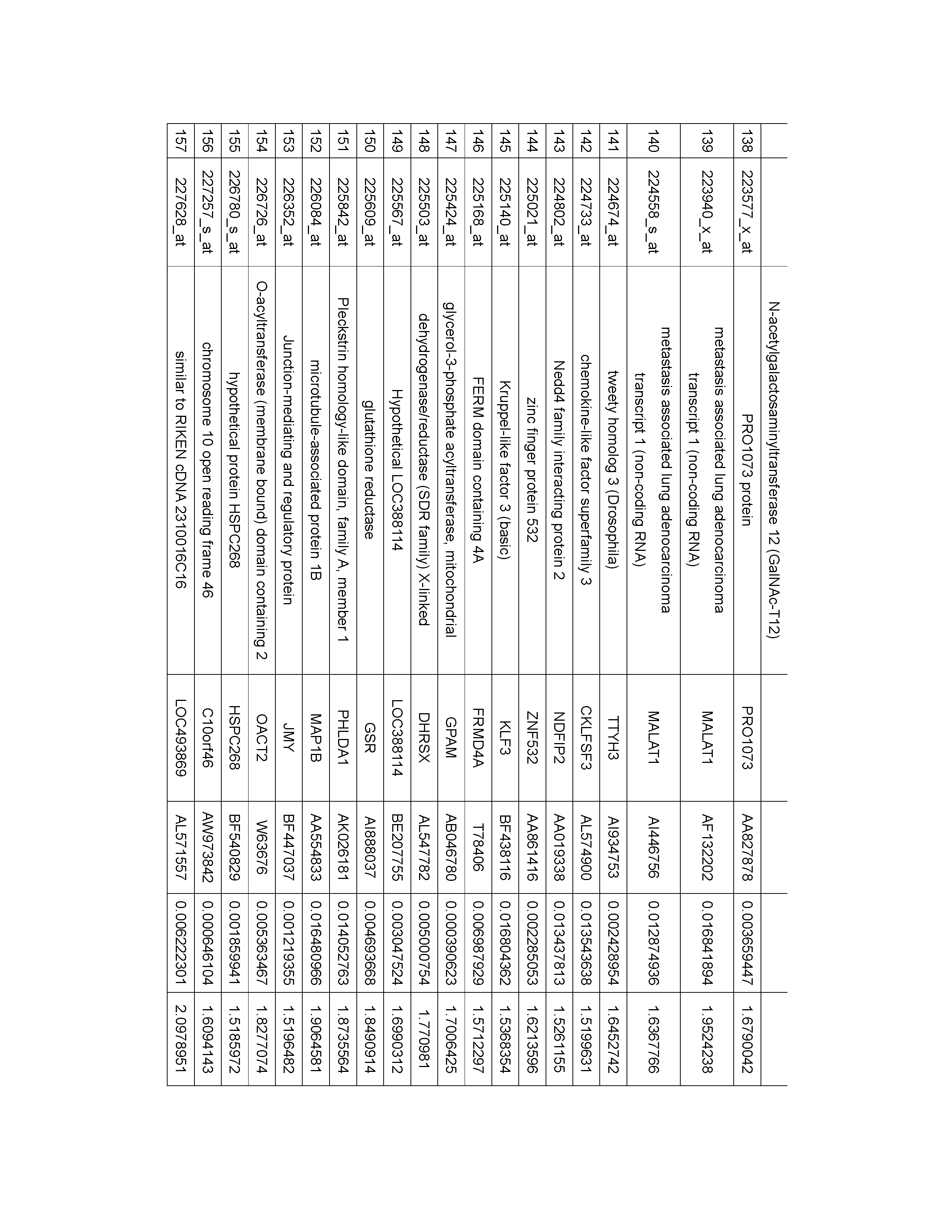
하기 표 1은 폐암 조직 적출한 후, 상기 폐암 세포의 유전자 발현 패턴을 마이크로어레 상의 프로브와의 혼성화 분석을 통하여 분석하고, 1년 내에 폐암이 재발한 환자 (재발 군)와 3년이 경과하여도 재발하지 않은 환자 (비재발 군)에서의 발현 수준에 있어서 차이가 있는 것으로 판단되는 마커 유전자를 선발한 결과를 나타내는 표이다. 총 환자 수는 60명이며, 이들 중 19명이 폐암 조직 적출 후 1년 내에 재발하였으며, 41명은 3년이 경과하여도 재발하지 않았다. The following Table 1 analyzes the gene expression pattern of the lung cancer cells by analyzing hybridization with probes on the microarray after the lung cancer tissues were extracted, and the patients who recurred lung cancer within one year (recurrent group) This is a table showing the results of selecting a marker gene which is considered to have a difference in the expression level in a patient who has not relapsed (non-recurrence group). The total number of patients was 60, and 19 of them recurred within 1 year after removal of lung cancer tissue, and 41 did not recur even after 3 years.


하기 표 2는 폐암 조직 적출한 후, 샘암종(adenocarcinoma)으로 분류된 폐암 세포의 유전자 발현 패턴을 마이크로어레 상의 프로브와의 혼성화 분석을 통하여 분석하고, 1년 내에 폐암이 재발한 환자와 3년이 경과하여도 재발하지 않은 환자에서의 발현 수준에 있어서 차이가 있는 것으로 판단되는 마커 유전자를 선발한 결과를 나타내는 표이다. 샘암종(adenocarcinoma)으로 분류된 폐암을 가진 총 환자 수는 23명이며, 이들 중 8명이 폐암 조직 적출 후 1년 내에 재발하였으며, 15명은 3년이 경과하여도 재발하지 않았다. The following Table 2 analyzes the gene expression pattern of lung cancer cells classified as adenocarcinoma by analysis of hybridization with a probe on a microarray, and the patient who has recurrence of lung cancer within one year, The results of the selection of the marker genes which are considered to be different in the expression level in the patients who did not relapse were also shown. The total number of patients with lung cancer classified as adenocarcinoma was 23, and 8 of them recurred within 1 year after removal of lung cancer tissue, and 15 did not recur even after 3 years.
하기 표 3은 폐암 조직 적출한 후, 편평상피세포암(squamous cell carcinoma)으로 분류된 폐암 세포의 유전자 발현 패턴을 마이크로어레 상의 프로브와의 혼성화 분석을 통하여 분석하고, 1년 내에 폐암이 재발된 환자와 3년이 경과하여도 재발하지 않은 환자에서의 발현 수준에 있어서 차이가 있는 것으로 판단되는 마커 유전자를 선발한 결과를 나타내는 표이다. 편평상피세포암(squamous cell carcinoma)로 분류되는 폐암을 가진 총 환자 수는 37명이며, 이들 중 11명이 폐암 조직 적출 후 1년 내에 재발하였으며, 26명은 3년이 경과하여도 재발하지 않았다. The following Table 3 analyzes the gene expression patterns of lung cancer cells classified as squamous cell carcinoma by analysis of hybridization with probes on a microarray after lung cancer tissues were extracted, And a marker gene that is considered to be different in the level of expression in patients who have not recurred even after 3 years. The total number of patients with lung cancer classified as squamous cell carcinoma was 37, and 11 of them recurred within 1 year after removal of lung cancer tissue, and 26 did not recur even after 3 years.


표 1, 2 및 3에서, gene name 은 유전자 명칭을 나타내고, gene symbol은 유전자를 나타내는 기호를 나타내고, Genbank Accession #는 Genbank 허가번호를 나타내는 것으로 상기 Genbank 데이터베이스는 일반 공중에게 자유롭게 접근가능한 데이터베이스이다. T-test p 값은 폐암 적출 수출을 받은 환자에 대하여 재발한 환자에서의 발현 평균값과 재발하지 않은 환자에서의 발현 평균값의 차이의 정도를 통계적으로 분석한 값이다. 여기서, 발현의 정도는 프로브가 고정화된 마이크로어레이를 사용한 혼성화 분석을 통하여 측정하였다. Fold change (abs)는 프로브가 고정화된 마이크로어레이를 사용한 혼성화 분석에 있어서, 폐암 적출 수술을 받은 환자에 대하여 재발한 환자에서의 발현 평균과 재발하지 않은 환자에서의 발현 평균의 비율을 나타내는 값이다.In Tables 1, 2 and 3, the gene name represents a gene name, the gene symbol represents a symbol representing a gene, and the Genbank Accession # represents a Genbank permission number. The Genbank database is freely accessible to the general public. The T-test p-value is a statistical analysis of the difference between the mean value of expression in recurrent patients and the mean value of expression in non-recurrent patients in patients receiving lung cancer exports. Here, the degree of expression was measured by hybridization analysis using a microarray in which probes were immobilized. Fold change (abs) is a measure of the percentage of recurrence-free and non-recurrence expression in patients who underwent lung cancer removal surgery in a hybridization assay using probe-immobilized microarrays.
표 1, 2 및 3에 나타낸 바와 같이, 표 1, 2 및 3의 Genbank 허가번호에 해당하는 마커 유전자 군으로부터 선택된 하나 이상의 발현 값은, 재발한 환자와 재발하지 않은 환자에 있어서 T-test p 값이 모두 0.05 미만으로서 통계적으로 유의한 차이를 보였다. 따라서, 표 1, 2 및 3의 Genbank 허가번호에 해당하는 마커 유전자 군으로부터 선택된 하나 이상의 마커 유전자는 폐암 적출 수출을 받은 환자에 대하여 추후에 폐암이 재발할지 여부를 예측할 수 있는 마커 유전자로서 사용될 수 있다. 또한, 표 1, 2 및 3의 Genbank 허가번호에 해당하는 마커 유전자 군으로부터 선택된 하나 이상의 마커 유전자는 재발한 환자의 발현 평균에 대한 재발하지 않은 환자의 발현평균의 비율이 모두 1.5배 이상으로서, 재발한 환자에서 그 발현이 현저하게 증가되는 것으로 확인되었다. As shown in Tables 1, 2 and 3, one or more expression values selected from the marker gene group corresponding to the GenBank license number of Tables 1, 2, and 3 were compared with the T-test p value Were all less than 0.05, indicating statistically significant differences. Thus, one or more marker genes selected from the marker gene group corresponding to the GenBank license number of Tables 1, 2 and 3 can be used as a marker gene for predicting whether or not the lung cancer will recur in a patient who has undergone lung cancer exportation . In addition, one or more marker genes selected from the marker gene group corresponding to the GenBank license number in Tables 1, 2, and 3 are all 1.5 times or more of the average expression level of the non-recurrent patients relative to the average of the recurrent patients, In one patient, the expression was markedly increased.
이하 본 발명을 실시예를 통하여 보다 상세하게 설명한다. 그러나, 이들 실시예는 본 발명을 예시적으로 설명하기 위한 것으로 본 발명의 범위가 이들 실시예에 한정되는 것은 아니다. Hereinafter, the present invention will be described in more detail with reference to examples. However, these examples are for illustrative purposes only, and the scope of the present invention is not limited to these examples.
실시예Example
실시예Example 1: 폐암의 재발과 관련된 1: associated with recurrence of lung cancer 마커Marker 유전자의 선별 Selection of genes
본 실시예에서는 종양의 크기가 3cm 미만이고 림프 절 전이가 없는 1기 폐암 조직 (즉, N0M0T1 기)을 적출하고, 적출된 폐암 조직으로부터 즉시 총 RNA를 분리하였다. 적출된 모든 종양 조직은 RNA 추출 전에 가시화를 개선하기 위하여 헤마톡실린으로 가볍게 염색하였다. 각 미세 절단된 표본은 90% 이상이 종양 세포로 구성되었다.In this example, primary lung cancer tissues (i.e., N 0 M 0 T 1 group) with less than 3 cm of tumor size and no lymph node metastasis were excised and total RNA was immediately isolated from the extracted lung cancer tissue. All extracted tumor tissues were lightly stained with hematoxylin to improve visualization prior to RNA extraction. Each micro-sectioned specimen consisted of more than 90% of tumor cells.
괴사 영역 (necrotic region)을 피하기 위하여, 종양 괴 (tumor mass)의 가장자리로부터 5mmx 5mm 크기의 종양 조직의 하나 또는 2개의 단편(pieces)를 즉시 -80℃에 저장하였다.To avoid the necrotic region, one or two pieces of tumor tissue 5 mm x 5 mm in size from the edge of the tumor mass were immediately stored at -80 ° C.
상기 미세 절단된 종양 조직을 1ml Trizol 시약 (Life Technologies, Rockville, MD) 중에 넣고, 보텍싱(vortexing)에 의하여 즉시 균질화하였다. Trizol 시약 프로토콜에 따라 총 RNA를 분리하였다. 분리된 상기 총 RNA의 질은 0.6M 포름아미드 및 에티디움 브로마이드를 포함하는 1% 아가로즈 겔을 사용한 전기영동에 의하여 분석하였다. 총 RNA의 양은 Nanodrop 분광기 (Nanodrop Technologies, Rockland, DE)를 사용하여 분석하였다. The micro-sectioned tumor tissue was placed in 1 ml Trizol reagent (Life Technologies, Rockville, Md.) And immediately homogenized by vortexing. Total RNA was isolated according to the Trizol reagent protocol. The quality of the total RNA thus isolated was analyzed by electrophoresis using 1% agarose gel containing 0.6M formamide and ethidium bromide. The amount of total RNA was analyzed using a Nanodrop spectrometer (Nanodrop Technologies, Rockland, DE).
분리된 상기 총 RNA의 질과 양이 우수한 것을 확인하고, 상기 RNA를 주형으로 하고 올리고 dT를 프라이머로 하여 역전사 반응을 시켜, cDNA를 얻었다. 얻어진 cDNA는 인 비트로 전사 반응을 통해 cRNA를 합성하는 주형으로 사용되는데 이때 비오틴으로 변형된 UTP를 반응액에 첨가함으로써 합성된 cRNA는 비오틴으로 표지가 되었다. 다음으로, 상기 합성된 비오틴 표지된 cRNA를 히드록시 라디칼과 반응시켜 50 내지 200 bp 크기로 단편화하였다. 상기 단편화된 cRNA 시료 10 ㎍을 Affymetrix GeneChip array (인간 133 plus ver 2) 상에 주입하고 45℃에서 16시간 동안 혼성화 반응을 시켰다. 다음으로, 세척하고, 피코에리쓰린 (phycoerythrin: PE)으로 표지된 스트렙타비딘으로 혼성화된 비오틴을 염색하였는데, 이때 신호를 증폭하기 위하여 비오틴화된 항-스트렙타비딘 항체를 첨가하여 염색하였다. 염색된 마이크로어레이 표면을 532nm 파장의 빛으로 조사하고 570nm 파장의 형광을 검출하여 형광 강도를 측정하였다. The obtained RNA was confirmed to be superior in quality and quantity, and reverse transcription was performed using the RNA as a template and oligo dT as a primer to obtain cDNA. The obtained cDNA was used as a template to synthesize cRNA through in vitro transcription reaction. The biotin-modified cRNA was labeled with biotin-modified UTP by adding it to the reaction solution. Next, the synthesized biotin-labeled cRNA was fragmented into a size of 50 to 200 bp by reacting with a hydroxy radical. Ten μg of the fragmented cRNA sample was injected onto an Affymetrix GeneChip array (human 133 plus ver 2) and hybridized for 16 hours at 45 ° C. Next, the cells were washed and stained with streptavidin-labeled biotin labeled with phycoerythrin (PE), where biotinylated anti-streptavidin antibody was added and stained to amplify the signal. The surface of the dyed microarray was irradiated with light having a wavelength of 532 nm, and fluorescence intensity was measured by detecting fluorescence at a wavelength of 570 nm.
얻어진 데이터는 ArrayAssistTM (Stratagene, Inc., San Diego, USA) 프로그램을 이용하여 분석하였다. 데이터 전처리는 분석에 사용된 전체 마이크로어레이들에 대해 형광 강도 값을 log2로 치환한 후 핵산 서열의 GC 함량을 고려하여 전체 마이크로어레이 대하여 형광 강도 평균을 맞춰주는 다중 마이크로어레이 수준 (multi-microarray level)의 노말화 방법인 GCRMA (log2 transformation) 방법을 적용하여 처리하였다. 그룹 비교는, unpaired t-test, 퍼뮤테이션 =100. 보정한 p-값 No/FDR 조건으로 수행하였다. 데이터 필터링은 발현 수준 (재발 및 비재발, 그 룹 평균) > 5 및 fold change ≥ 1.5을 만족하는 데이터만을 취하였다. probeset_id 별 개수(count)는, 샘암종 (ADC), 편평세포암종 (SQC), 또는 세포 형태와 무관하게 재발군과 비재발 군에서의 상기 필터링 기준을 만족하는 수준의 유전자 발현 차이가 나는 프로브 세트 개수로 정의하였다. The obtained data were analyzed using ArrayAssist TM (Stratagene, Inc., San Diego, USA) program. Data preprocessing is performed at a multi-microarray level that replaces the fluorescence intensity value with log2 for all microarrays used for analysis and then adjusts the fluorescence intensity average for all microarrays considering the GC content of the nucleic acid sequence. (GCRMA) (log2 transformation) method. Group comparison, unpaired t-test, permutation = 100. Corrected p-value No / FDR condition. Data filtering only took data that satisfied expression levels (recurrence and non relapse, group mean)> 5 and fold change ≥ 1.5. The probeset_id count (count) is the number of probe sets with a difference in gene expression level that satisfies the filtering criteria in the recurrent and non-recurrent groups regardless of adenocarcinoma (ADC), squamous cell carcinoma (SQC) Respectively.
분석 결과, 총 폐암 조직, 샘암종 (ADC) 및 편평세포암종 (SQC)에 대하여 양성 발현으로 선택된 마커의 수는 하기 표 4와 같다.As a result, the number of markers selected by positive expression for total lung cancer tissue, adenocarcinoma (ADC) and squamous cell carcinoma (SQC) is shown in Table 4 below.
표 4. Table 4.
상기 형광 강도 측정에 의하여 얻어진 각 유전자의 발현에 관련된 데이터를 확보하였다. 이렇게 수집된 유전자의 발현에 관련된 데이터와 폐암의 재발과의 연관성을 확인하기 위하여, 폐암 제거 수술을 받은 환자를 5년 동안 모니터링하여, 폐암이 재발하는지를 확인하였다. 만약 폐암 제거 수술 후 1년 내에 폐암이 재발하는 경우, 폐암 재발 그룹으로 분류하고, 3년이 경과하였는데도 폐암이 재발하지 않는 경우에는, 재발하지 않는 그룹으로 분류하였다. 이렇게 얻어진 폐암 제거 수술을 받은 환자 중 재발 그룹과 비재발 그룹에 대한 데이터를 확보하였다. Data relating to the expression of each gene obtained by the fluorescence intensity measurement were obtained. In order to confirm the association between the data on the expression of the collected genes and the recurrence of lung cancer, the patients who underwent lung cancer removal surgery were monitored for 5 years to confirm the recurrence of lung cancer. If lung cancer recurs within one year after lung cancer removal surgery, it is classified as recurrent lung cancer group. If lung cancer does not recur after 3 years, it is classified as non - recurrence group. Data on the recurrence group and the non - recurrence group of the patients who underwent the lung cancer removal procedure were obtained.
다음으로, 상기 폐암 제거 수술 시에 분석된 각 유전자의 발현 패턴과, 그 후에 환자 관찰을 통하여 얻어진 재발 및 비재발 그룹과의 상호연관성을 분석하였다. 그 결과를 표 1, 2 및 3에 나타내었다. Next, the expression pattern of each gene analyzed at the time of the above-described lung cancer removal surgery and the correlation between the recurrence and the non-recurrence group obtained after the patient observation were analyzed. The results are shown in Tables 1, 2 and 3.
표 1은 폐암 조직 적출한 후, 상기 폐암 세포의 유전자 발현 패턴을 마이크로어레이 상의 프로브와의 혼성화 분석을 통하여 분석하고, 1년 내에 폐암이 재발한 환자와 3년이 경과하여도 재발하지 않은 환자에서의 발현 수준에 있어서 차이가 있는 것으로 판단되는 마커 유전자를 선발한 결과를 나타내는 표이다. 총 환자 수는 60명이며, 이들 중 19명이 폐암 조직 적출 후 1년 내에 재발하였으며, 41명은 3년이 경과하여도 재발하지 않았다. Table 1 analyzes the gene expression patterns of the lung cancer cells by analyzing hybridization with the probes on the microarray. After the lung cancer recurrence within 1 year and the patients who did not relapse 3 years later Of the present invention is a table showing the results of selecting a marker gene which is judged to have a difference in the expression level of the marker gene. The total number of patients was 60, and 19 of them recurred within 1 year after removal of lung cancer tissue, and 41 did not recur even after 3 years.
표 2는 폐암 조직 적출한 후, 샘암종(adenocarcinoma)로 분류되는 폐암 세포의 유전자 발현 패턴을 마이크로어레이 상의 프로브와의 혼성화 분석을 통하여 분석하고, 1년 내에 폐암이 재발한 환자와 3년이 경과하여도 재발하지 않은 환자에서의 발현 수준에 있어서 차이가 있는 것으로 판단되는 마커 유전자를 선발한 결과를 나타내는 표이다. 샘암종(adenocarcinoma)로 분류되는 폐암을 가진 총 환자 수는 23명이며, 이들 중 8명이 폐암 조직 적출 후 1년 내에 재발하였으며, 15명은 3년이 경과하여도 재발하지 않았다. Table 2 shows the gene expression pattern of lung cancer cells classified as adenocarcinoma after the lung cancer tissue extraction and analyzed by hybridization analysis with the probes on the microarray. After 3 years from the recurrence of lung cancer within 1 year The results of the selection of the marker genes which are considered to be different in the expression level in the patients who did not relapse were also shown. The total number of patients with lung cancer classified as adenocarcinoma was 23, and 8 of them recurred within 1 year after removal of lung cancer tissue, and 15 did not recur even after 3 years.
표 3은 폐암 조직 적출한 후, 편평상피세포암(squamous cell carcinoma)로 분류되는 폐암 세포의 유전자 발현 패턴을 마이크로어레이 상의 프로브와의 혼성화 분석을 통하여 분석하고, 1년 내에 폐암이 재발한 환자와 3년이 경과하여도 재발하지 않은 환자에서의 발현 수준에 있어서 차이가 있는 것으로 판단되는 마커 유전자를 선발한 결과를 나타내는 표이다. 편평상피세포암(squamous cell carcinoma)로 분류되는 폐암을 가진 총 환자 수는 37명이며, 이들 중 11명이 폐암 조직 적출 후 1년 내에 재발하였으며, 26명은 3년이 경과하여도 재발하지 않았다. Table 3 shows the gene expression patterns of lung cancer cells classified as squamous cell carcinoma after lung cancer tissue extraction and analyzed by hybridization with probes on a microarray. This is a table showing the results of selecting a marker gene that is considered to have a difference in expression level in patients who have not relapsed even after 3 years. The total number of patients with lung cancer classified as squamous cell carcinoma was 37, and 11 of them recurred within 1 year after removal of lung cancer tissue, and 26 did not recur even after 3 years.
표 1, 2 및 3에 나타낸 바와 같이, 표 1, 2 및 3의 Genbank 허가번호에 해당하는 마커 유전자 군으로부터 선택된 하나 이상의 발현 값은, 재발한 환자와 재발하지 않은 환자에 있어서 T-test p 값이 모두 0.05 미만으로서 통계적으로 유의한 차이를 보였다. 따라서, 표 1, 2 및 3의 Genbank 허가번호에 해당하는 마커 유전자 군으로부터 선택된 하나 이상의 마커 유전자는 폐암 적출 수출을 받은 환자에 대하여 추후에 폐암이 재발할지 여부를 예측할 수 있는 마커 유전자로서 사용될 수 있다. 또한, 표 1, 2 및 3의 Genbank 허가번호에 해당하는 마커 유전자 군으로부터 선택된 하나 이상의 마커 유전자는 재발한 환자의 발현 평균에 대한 재발하지 않은 환자의 발현 평균의 비율이 모두 1.5배 이상으로서, 재발한 환자에서 그 발현이 현저하게 증가되는 것으로 확인되었다. As shown in Tables 1, 2 and 3, one or more expression values selected from the marker gene group corresponding to the GenBank license number of Tables 1, 2, and 3 were compared with the T-test p value Were all less than 0.05, indicating statistically significant differences. Thus, one or more marker genes selected from the marker gene group corresponding to the GenBank license number of Tables 1, 2 and 3 can be used as a marker gene for predicting whether or not the lung cancer will recur in a patient who has undergone lung cancer exportation . In addition, one or more marker genes selected from the marker gene group corresponding to the GenBank license number in Tables 1, 2, and 3 are all 1.5 times or more of the average expression level of the non-recurrent patients relative to the average of the recurrent patients, In one patient, the expression was markedly increased.
그 외 폐암 제거 수술을 받은 환자의 재발 여부와 나이, 성별, 흡연 상태, 세포형태, 암기 (pstage), 종양 크기와의 상관 관계를 분석하였으며, 그 결과는 하기 표 5, 6 및 7에 나타낸 바와 같다. The correlation between recurrence and age, sex, smoking status, cell type, pstage, and tumor size was analyzed. The results were as shown in Tables 5, 6, and 7 same.
표 5. Table 5.
표 5은, 암의 종류를 세포 형태에 따라 구분하지 않은 60명의 종양 환자에 대하여 분석한 결과이다. 60명 중 재발 환자는 19명이며, 비재발 환자는 41명이었 다. 표 5에 나타낸 바와 같이, 재발 여부 및 종양 크기를 제외하고 나머지 분석에서 혼란 (confounder)를 가져올 가능성이 있는 임상 정보들은 재발과 비재발 군에서 통계적으로 유의한 차이를 나타내지 않음을 알 수 있다. 즉, 분석된 결과는 재발 여부에 대해서만 통계적으로 유의한 발현 차이를 나타내는 유전자 목록이라고 할 수 있다.Table 5 shows the results of analysis of 60 cancer patients who were not classified according to the cell type. Of the 60 patients, 19 had relapsed patients and 41 had non relapsed patients. As shown in Table 5, clinical information that may cause confounder in the remaining analysis except for recurrence and tumor size shows no statistically significant difference between recurrence and non-recurrence group. In other words, the analyzed result is a gene list showing statistically significant differences in expression only for recurrence.
표 6. Table 6.
표 6은, 암의 종류를 세포 형태에 따라 구분하였을 경우, 샘암종에 해당하는 23명의 종양 환자에 대하여 분석한 결과이다. 23명 중 재발 환자는 8명이며, 비재발 환자는 15명이었다. 표 6에 나타낸 바와 같이, 재발 여부를 제외하고 나머지 분석에서 혼란 (confounder)를 가져올 가능성이 있는 임상 정보들은 재발과 비재발 군에서 통계적으로 유의한 차이를 나타내지 않음을 알 수 있다. 즉, 분석된 결과는 재발 여부에 대해서만 통계적으로 유의한 발현 차이를 나타내는 유전자 목록이라고 할 수 있다.Table 6 shows the results of analysis of 23 tumors corresponding to adenocarcinoma when the type of cancer was classified according to cell type. Of the 23 patients, 8 had recurrence and 15 had non - recurrence. As shown in Table 6, there is no statistically significant difference between the recurrence-free and non-recurrence-related clinical information that may cause confounder in the remaining analysis except recurrence. In other words, the analyzed result is a gene list showing statistically significant differences in expression only for recurrence.
표 7. Table 7.
표 7은, 암의 종류를 세포 형태에 따라 구분하였을 경우, 편평세포암종에 해당하는 37명의 종양 환자에 대하여 분석한 결과이다. 37명 중 재발 환자는 11명이며, 비재발 환자는 26명이었다. 표 7에 나타낸 바와 같이, 재발 여부를 제외하고 나머지 분석에서 혼란 (confounder)를 가져올 가능성이 있는 임상 정보들은 재발과 비재발 군에서 통계적으로 유의한 차이를 나타내지 않음을 알 수 있다. 즉, 분석된 결과는 남자 폐암 환자 중 편평세포암종 (SQC) 내에서 재발 여부에 대해서만 통계적으로 유의한 발현 차이를 나타내는 유전자 목록이라고 할 수 있다.Table 7 shows the results of analysis of 37 patients with squamous cell carcinoma when the type of cancer was classified according to the cell type. Of the 37 patients, 11 had recurrence and 26 had non - recurrence. As shown in Table 7, clinical information that may cause confounder in the remaining analysis except recurrence does not show statistically significant difference between recurrence and non-recurrence group. In other words, the analyzed result is a list of genes showing statistically significant difference in expression of recurrence in squamous cell carcinoma (SQC) among male lung cancer patients.
실시예Example 2 : 통계적 모델을 이용한 폐암 재발의 위험의 예측 2: Estimation of Risk of Recurrence of Lung Cancer Using Statistical Model
본 실시예에서는, 실시예 1에서 얻어진 재발 및 비재발 환자에서 수십된 마커 유전자의 발현 수준을 토대로, 통계적 분석 모델을 사용하여 폐암의 재발 위험을 예측할 수 있는지를 확인하였다.In this example, it was confirmed whether the risk of recurrence of lung cancer can be predicted using a statistical analysis model, based on the expression levels of dozens of marker genes in the relapsed and non relapsed patients obtained in Example 1.
분석은, 총 폐암조직, 샘암종 및 편평세포암종에 대하여 얻어진 각각의 데이터 중 일부는 통계적 모델의 예측 정확도 기준을 설정하기 위한 러닝 세트 (learning set)로 사용하고, 나머지 데이터를 상기 러닝 세트 데이터를 이용하여 설정된 예측 정확도 조건이 실제로 정확한지를 확인하였다. The analysis used some of the data obtained for total lung cancer tissue, adenocarcinoma and squamous cell carcinoma as a learning set for setting the prediction accuracy criterion of the statistical model and using the remaining data as the learning set data We confirmed that the predicted accuracy condition set is actually correct.
총 폐암조직, 샘암종 및 편평세포암종에 대하여 각각 사용된 러닝 세트와 테스트 세트에 대한 데이터는 각각 다음과 표 8, 9 및 10과 같다. Data for running and test sets used for total lung cancer tissue, adenocarcinoma and squamous cell carcinoma, respectively, are as follows: Tables 8, 9 and 10, respectively.
표 8.Table 8.
표 9.Table 9.
표 10.Table 10.
폐암조직, 샘암종 및 편평세포암종에 대하여, 상기 테스트 세트를 QDA 예측 모델 (QDA prediction model)을 사용하여 예측한 결과는 하기 표 11, 12, 및 13과 같다. 표 11, 12, 및 13에 나타낸 바와 같이, 예측 정확도는 76.4%이상이었다. For lung cancer tissues, adenocarcinomas and squamous cell carcinomas, the test set was predicted using the QDA prediction model as shown in Tables 11, 12, and 13 below. As shown in Tables 11, 12, and 13, the prediction accuracy was 76.4% or more.
표 11. 총 폐암조직에 대한 QDA 예측 모델 (QDA prediction model)을 사용한 예측한 결과Table 11. Predicted results using the QDA prediction model for total lung cancer tissue
분류
Classification
system
True class
표 11에서 정확도는 총 시료에 대한 진실 값과 일치하는 예측 값의 백분율이다. 즉 정확도는 =(17-4)x100/17 =76.4%. 이하 동일하게 계산하였다.In Table 11, the accuracy is the percentage of predicted values that match the true value for the total sample. That is, the accuracy is = (17-4) x100 / 17 = 76.4%. The same calculation was made below.
표 12. 샘암종 조직에 대한 QDA 예측 모델 (QDA prediction model)을 사용한 예측한 결과Table 12. Predicted results using the QDA prediction model for adenocarcinoma tissue
분류
Classification
system
True class
표 13. 편평세포암종 조직에 대한 QDA 예측 모델 (QDA prediction model)을 사용한 예측한 결과Table 13. Predicted results using the QDA prediction model for squamous cell carcinoma tissues
system
True class
폐암조직, 샘암종 및 편평세포암종에 대하여, 상기 테스트 세트를 LDA 예측 모델 (LDA prediction model)을 사용하여 예측한 결과는 하기 표 14, 15, 및 16과 같다. 표 14, 15, 및 16에 나타낸 바와 같이, 예측 정확도는 76.4%이상이었다. For lung cancer tissues, adenocarcinomas and squamous cell carcinomas, the test set was predicted using the LDA prediction model (LDA prediction model) as shown in Tables 14, 15, and 16 below. As shown in Tables 14, 15, and 16, the prediction accuracy was 76.4% or more.
표 14. 총 폐암조직에 대한 LDA 예측 모델 (LDA prediction model)을 사용한 예측한 결과Table 14. Predictions using the LDA prediction model for total lung cancer tissue (LDA prediction model)
분류
Classification
system
True class
표 15. 샘암종 조직에 대한 LDA 예측 모델 (LDA prediction model)을 사용한 예측한 결과Table 15. Predicted results using LDA prediction model (LDA prediction model) for adenocarcinoma tissue
분류
Classification
system
True class
표 16. 편평세포암종 조직에 대한 LDA 예측 모델 (LDA prediction model)을 사용한 예측한 결과Table 16. Predicted results using LDA prediction model (LDA prediction model) for squamous cell carcinoma tissue
system
True class
폐암조직, 샘암종 및 편평세포암종에 대하여, 상기 테스트 세트를 뉴럴 네트워크 예측 모델 (Neural network prediction model)을 사용하여 예측한 결과는 하기 표 17, 18, 및 19과 같다. 표 17, 18, 및 19에 나타낸 바와 같이, 예측 정확도는 59.46%이상이었다. For lung cancer tissues, adenocarcinomas and squamous cell carcinomas, the test set was predicted using a neural network prediction model as shown in Tables 17, 18, and 19 below. As shown in Tables 17, 18, and 19, the prediction accuracy was more than 59.46%.
표 17. 총 폐암조직에 대한 뉴럴 네트워크 예측 모델 (Neural network prediction model)을 사용한 예측한 결과Table 17. Predicted results using a neural network prediction model for total lung cancer tissue
분류
Classification
system
True class
표 18. 샘암종 조직에 대한 뉴럴 네트워크 예측 모델 (Neural network prediction model)을 사용한 예측한 결과Table 18. Predicted results using neural network prediction model for adenocarcinoma tissue
분류
Classification
system
True class
표 19. 편평세포암종 조직에 대한 뉴럴 네트워크 예측 모델 (Neural network prediction model)을 사용한 예측한 결과Table 19. Predicted results using a neural network prediction model for squamous cell carcinoma tissues
system
True class
폐암조직, 샘암종 및 편평세포암종에 대하여, 상기 테스트 세트를 디시전 트리 예측 모델 (Decision tree prediction model)을 사용하여 예측한 결과는 하기 표 20, 21, 및 22과 같다. 표 20, 21, 및 22에 나타낸 바와 같이, 예측 정확도는 61.67%이상이었다. For lung cancer tissues, adenocarcinomas and squamous cell carcinomas, the predicted results of the test set using the decision tree prediction model are shown in Tables 20, 21, and 22 below. As shown in Tables 20, 21, and 22, the prediction accuracy was more than 61.67%.
표 20. 총 폐암조직에 대한 디시전 트리 예측 모델 (Decision tree prediction model)을 사용한 예측한 결과Table 20. Predicted results using a decision tree prediction model for total lung cancer tissue
분류
Classification
system
True class
표 21. 샘암종 조직에 대한 디시전 트리 예측 모델 (Decision tree prediction model)을 사용한 예측한 결과Table 21. Predicted results using the decision tree prediction model for adenocarcinoma tissues
분류
Classification
system
True class
표 22. 편평세포암종 조직에 대한 디시전 트리 예측 모델 (Decision tree prediction model)을 사용한 예측한 결과Table 22. Predicted results using a decision tree prediction model for squamous cell carcinoma tissue
system
True class
폐암조직, 샘암종 및 편평세포암종에 대하여, 상기 테스트 세트를 서포트 벡 터 머신 예측 모델 (Support vector machine prediction model)을 사용하여 예측한 결과는 하기 표 23, 24, 및 25와 같다. 표 23, 24, 및 25에 나타낸 바와 같이, 예측 정확도는 65%이상이었다. For lung cancer tissues, adenocarcinomas and squamous cell carcinomas, the test set was predicted using the support vector machine prediction model as shown in Tables 23, 24 and 25 below. As shown in Tables 23, 24 and 25, the prediction accuracy was more than 65%.
표 23 총 폐암조직에 대한 서포트 벡터 머신 예측 모델 (Support vector machine prediction model)을 사용한 예측한 결과Table 23 Predicted results using a support vector machine prediction model for total lung cancer tissue
분류
Classification
system
True class
표 24. 샘암종 조직에 대한 서포트 벡터 머신 예측 모델 (Support vector machine prediction model)을 사용한 예측한 결과Table 24. Predicted results using support vector machine prediction model for adenocarcinoma tissue
분류
Classification
system
True class
표 25. 편평세포암종 조직에 대한 서포트 벡터 머신 예측 모델 (Support vector machine prediction model)을 사용한 예측한 결과Table 25. Predicted results using support vector machine prediction model for squamous cell carcinoma tissue
system
True class
폐암조직, 샘암종 및 편평세포암종에 대하여, 상기 테스트 세트를 나이브 베이즈 예측 모델 (Naive Bayes prediction model)을 사용하여 예측한 결과는 하기 표 26, 27, 및 28과 같다. 표 26, 27, 및 28에 나타낸 바와 같이, 예측 정확도는 58.33%이상이었다. For lung cancer tissues, adenocarcinomas and squamous cell carcinomas, the test set was predicted using the Naive Bayes prediction model as shown in Tables 26, 27, and 28 below. As shown in Tables 26, 27 and 28, the prediction accuracy was 58.33% or more.
표 26. 총 폐암조직에 대한 나이브 베이즈 예측 모델 (Naive Bayes prediction model)을 사용한 예측한 결과Table 26. Predictions using the Naive Bayes prediction model for total lung cancer tissue
분류
Classification
system
True class
표 27. 샘암종 조직에 대한 나이브 베이즈 예측 모델 (Naive Bayes prediction model)을 사용한 예측한 결과Table 27. Predicted results using the Naive Bayes prediction model for adenocarcinoma tissue
분류
Classification
system
True class
표 28. 편평세포암종 조직에 대한 나이브 베이즈 예측 모델 (Naive Bayes prediction model)을 사용한 예측한 결과Table 28. Predicted results using the Naive Bayes prediction model for squamous cell carcinoma tissue
system
True class
본 실시예에 사용된 예측 모델들은 통계적 분야에서는 통상적으로 사용되고 있는 모델들로 당업자라면 용이하게 선택하여 사용할 수 있는 것이다. The prediction models used in the present embodiment are models that are commonly used in the statistical field, and those skilled in the art can easily select and use the prediction models.
본 발명의 폐암 치료를 받은 폐암 환자의 폐암 재발의 위험을 예측하는 방법에 의하면, 폐암 제거 수출을 받은 폐암 환자의 폐암 재발의 위험을 높은 정확도로 예측할 수 있다.The method of predicting the risk of recurrence of lung cancer in lung cancer patients treated with lung cancer of the present invention can predict with high accuracy the risk of recurrence of lung cancer in patients with lung cancer who have undergone lung cancer ablation exports.
본 발명의 폐암 치료를 받은 환자의 폐암 재발 위험성에 대한 보고서를 작성하는 방법에 의하면, 폐암 제거 수출을 받은 폐암 환자의 폐암 재발의 위험을 높은 정확도로 예측한 결과를 포함하는 보고서를 작성할 수 있다.A method of reporting the risk of recurrence of lung cancer in a patient who has undergone lung cancer treatment of the present invention can produce a report that includes the result of highly accurate prediction of the risk of recurrence of lung cancer in lung cancer patients who have undergone lung cancer ablation exports.
본 발명의 폐암 치료를 받은 환자의 폐암 재발 위험성에 대한 보고서에 의하면, 폐암 제거 수출을 받은 폐암 환자의 폐암 재발의 위험을 높은 정확도로 예측한 결과를 포함한다.The report on the risk of recurrence of lung cancer in patients treated with lung cancer of the present invention includes the results of highly accurate prediction of the risk of recurrence of lung cancer in patients with lung cancer who have undergone lung cancer clearance exports.
본 발명의 폐암 치료를 받은 폐암 환자의 폐암 재발 위험을 진단하기 위한 조성물, 키트 및 마이크로어레이에 의하면, 폐암 치료를 받은 폐암 환자의 폐암 재발 위험을 진단하는데 효율성을 높일 수 있다. According to the composition, kit and microarray for diagnosing the risk of recurrence of lung cancer in a lung cancer patient who has undergone lung cancer treatment according to the present invention, the efficiency of diagnosing the risk of recurrence of lung cancer in lung cancer patients treated with lung cancer can be improved.
Claims (24)


Priority Applications (3)
| Application Number | Priority Date | Filing Date | Title |
|---|---|---|---|
| KR1020070002643A KR101443214B1 (en) | 2007-01-09 | 2007-01-09 | Compositions, kits and microarrays for diagnosing the risk of recurrence of lung cancer in patients with lung cancer or lung cancer treated with lung cancer |
| US11/971,585 US7585634B2 (en) | 2007-01-09 | 2008-01-09 | Method of predicting risk of lung cancer recurrence, and a composition, kit and microarray for the same |
| US12/512,401 US20090291853A1 (en) | 2007-01-09 | 2009-07-30 | Method of predicting risk of lung cancer recurrence, and a composition, kit and microarray for the same |
Applications Claiming Priority (1)
| Application Number | Priority Date | Filing Date | Title |
|---|---|---|---|
| KR1020070002643A KR101443214B1 (en) | 2007-01-09 | 2007-01-09 | Compositions, kits and microarrays for diagnosing the risk of recurrence of lung cancer in patients with lung cancer or lung cancer treated with lung cancer |
Publications (2)
| Publication Number | Publication Date |
|---|---|
| KR20080065476A KR20080065476A (en) | 2008-07-14 |
| KR101443214B1 true KR101443214B1 (en) | 2014-09-24 |
Family
ID=39594628
Family Applications (1)
| Application Number | Title | Priority Date | Filing Date |
|---|---|---|---|
| KR1020070002643A Expired - Fee Related KR101443214B1 (en) | 2007-01-09 | 2007-01-09 | Compositions, kits and microarrays for diagnosing the risk of recurrence of lung cancer in patients with lung cancer or lung cancer treated with lung cancer |
Country Status (2)
| Country | Link |
|---|---|
| US (2) | US7585634B2 (en) |
| KR (1) | KR101443214B1 (en) |
Families Citing this family (37)
| Publication number | Priority date | Publication date | Assignee | Title |
|---|---|---|---|---|
| US20030114410A1 (en) | 2000-08-08 | 2003-06-19 | Technion Research And Development Foundation Ltd. | Pharmaceutical compositions and methods useful for modulating angiogenesis and inhibiting metastasis and tumor fibrosis |
| EP2537529B1 (en) | 2007-08-02 | 2018-10-17 | Gilead Biologics, Inc. | Loxl2 inhibitory antibodies and uses thereof |
| EP2321410A4 (en) * | 2008-07-16 | 2011-09-14 | Oncotherapy Science Inc | ONCOGEN ECT2 AS A THERAPEUTIC TARGET AND PROGNOSTIC INDICATOR FOR LUNG AND SOPHAGE CANCER |
| US9495515B1 (en) | 2009-12-09 | 2016-11-15 | Veracyte, Inc. | Algorithms for disease diagnostics |
| US10236078B2 (en) | 2008-11-17 | 2019-03-19 | Veracyte, Inc. | Methods for processing or analyzing a sample of thyroid tissue |
| US9107935B2 (en) | 2009-01-06 | 2015-08-18 | Gilead Biologics, Inc. | Chemotherapeutic methods and compositions |
| JP6078339B2 (en) | 2009-05-07 | 2017-02-08 | ベラサイト インコーポレイテッド | Methods and compositions for diagnosis of thyroid status |
| CA2771774A1 (en) * | 2009-08-21 | 2011-02-24 | Gilead Biologics, Inc. | In vitro screening assays |
| JP2013502226A (en) | 2009-08-21 | 2013-01-24 | ギリアド バイオロジクス,インク. | Catalytic domain derived from lysyl oxidase and LOXL2 |
| CA2789022A1 (en) | 2010-02-04 | 2011-08-11 | Gilead Biologics, Inc. | Antibodies that bind to lysyl oxidase-like 2 (loxl2) and methods of use therefor |
| EP2426216A1 (en) * | 2010-09-01 | 2012-03-07 | Institut Gustave Roussy (IGR) | Prognostic and/or predictive biomarkers and biological applications thereof |
| WO2012028703A1 (en) * | 2010-09-02 | 2012-03-08 | INSERM (Institut National de la Santé et de la Recherche Médicale) | Method for the prognosis of the progression of cancer |
| WO2013183964A1 (en) * | 2012-06-07 | 2013-12-12 | 한양대학교 산학협력단 | Target protein for diagnosing and treating lung cancer |
| EP2968988A4 (en) | 2013-03-14 | 2016-11-16 | Allegro Diagnostics Corp | Methods for evaluating copd status |
| US11976329B2 (en) | 2013-03-15 | 2024-05-07 | Veracyte, Inc. | Methods and systems for detecting usual interstitial pneumonia |
| CN106795565B (en) * | 2014-07-14 | 2022-05-10 | 威拉赛特公司 | Methods for Assessing Lung Cancer Status |
| US12297505B2 (en) | 2014-07-14 | 2025-05-13 | Veracyte, Inc. | Algorithms for disease diagnostics |
| CN104198722A (en) * | 2014-08-11 | 2014-12-10 | 广东药学院 | Novel tumor marker GSTA1 for lung caner as well as screening method and application thereof |
| US20160053327A1 (en) * | 2014-08-23 | 2016-02-25 | Tiehua Chen | Compositions and methods for prediction of clinical outcome for all stages and all cell types of non-small cell lung cancer in multiple countries |
| WO2016073768A1 (en) | 2014-11-05 | 2016-05-12 | Veracyte, Inc. | Systems and methods of diagnosing idiopathic pulmonary fibrosis on transbronchial biopsies using machine learning and high dimensional transcriptional data |
| JPWO2016121715A1 (en) * | 2015-01-26 | 2017-11-02 | 国立大学法人名古屋大学 | Method for providing information for evaluating prognosis of lung cancer patient, prognosis prediction method for lung cancer patient, internal standard, antibody, prognosis prediction device for lung cancer patient, program for prognosis prediction device, and recording medium |
| CN104761634B (en) | 2015-03-27 | 2018-05-18 | 李翀 | A kind of pulmonary cancer diagnosis marker, antibody and its application |
| US11161874B2 (en) | 2016-08-19 | 2021-11-02 | Genoimmune Therapeutics Co., Ltd. | Tumor-specific polypeptide and use thereof |
| CN107058480B (en) * | 2016-12-14 | 2018-07-13 | 河北医科大学第四医院(河北省肿瘤医院) | Long-chain non-coding RNA marker for diagnosing adenocarcinoma of lung |
| HRP20220550T1 (en) | 2016-12-23 | 2022-06-10 | Immunogen, Inc. | Immunoconjugates targeting adam9 and methods of use thereof |
| KR102630036B1 (en) | 2016-12-23 | 2024-01-29 | 마크로제닉스, 인크. | ADAM9-Binding Molecules, and Methods of Use Thereof |
| TWI831797B (en) | 2018-06-26 | 2024-02-11 | 美商伊繆諾金公司 | Immunoconjugates targeting adam9 and methods of use thereof |
| CN109411023B (en) * | 2018-09-30 | 2022-03-18 | 华中农业大学 | Method for mining inter-gene interaction relation based on Bayesian network inference |
| EP3778924A1 (en) * | 2019-08-16 | 2021-02-17 | Siemens Healthcare GmbH | Molecular predictors of patient response to radiotherapy treatment |
| CN110749734A (en) * | 2019-12-06 | 2020-02-04 | 四川大学华西医院 | Application of GTF2I autoantibody detection reagent in preparation of lung cancer screening kit |
| CN111394456B (en) * | 2020-03-19 | 2022-12-02 | 中国医学科学院肿瘤医院 | Early lung adenocarcinoma patient prognosis evaluation system and application thereof |
| CN111401798A (en) * | 2020-06-02 | 2020-07-10 | 南京百敖软件有限公司 | Enterprise waste escaping and debt risk early warning system and construction method |
| US20230314408A1 (en) * | 2020-07-02 | 2023-10-05 | Gopath Laboratories Llc | Immune profiling and methods of using same to predict responsiveness to an immunotherapy and treat cancer |
| CN117729942A (en) | 2021-03-08 | 2024-03-19 | 伊缪诺金公司 | Methods for increasing the efficacy of ADAM9-targeting immunoconjugates in the treatment of cancer |
| CN113430268A (en) * | 2021-06-29 | 2021-09-24 | 北京泱深生物信息技术有限公司 | Prediction of lung cancer prognosis |
| CN117144006B (en) * | 2023-08-18 | 2025-02-18 | 大连医科大学附属第二医院 | LACTB gene and its application in diagnosis of prostate cancer |
| CN118374593A (en) * | 2024-01-23 | 2024-07-23 | 复旦大学附属中山医院 | Characteristic gene set of alveolar histological subtype of early lung adenocarcinoma and its screening method and application |
Citations (3)
| Publication number | Priority date | Publication date | Assignee | Title |
|---|---|---|---|---|
| JP2004500895A (en) | 2000-06-21 | 2004-01-15 | 日立化成工業株式会社 | Gene markers for lung cancer |
| JP2004518630A (en) | 2000-09-18 | 2004-06-24 | ザ・トラスティーズ・オブ・コランビア・ユニバーシティー・イン・ザ・シティー・オブ・ニューヨーク | Novel tumor-related markers |
| JP2004520831A (en) | 2000-11-20 | 2004-07-15 | ディアデクサス インコーポレーテッド | Compositions and methods for lung-specific genes and proteins |
Family Cites Families (4)
| Publication number | Priority date | Publication date | Assignee | Title |
|---|---|---|---|---|
| US5744101A (en) * | 1989-06-07 | 1998-04-28 | Affymax Technologies N.V. | Photolabile nucleoside protecting groups |
| US5143854A (en) * | 1989-06-07 | 1992-09-01 | Affymax Technologies N.V. | Large scale photolithographic solid phase synthesis of polypeptides and receptor binding screening thereof |
| US20050272061A1 (en) * | 2004-02-19 | 2005-12-08 | Seattle Genetics, Inc. | Expression profiling in non-small cell lung cancer |
| BRPI0518734A2 (en) * | 2004-11-30 | 2008-12-02 | Veridex Llc | prognosis of lung cancer |
-
2007
- 2007-01-09 KR KR1020070002643A patent/KR101443214B1/en not_active Expired - Fee Related
-
2008
- 2008-01-09 US US11/971,585 patent/US7585634B2/en not_active Expired - Fee Related
-
2009
- 2009-07-30 US US12/512,401 patent/US20090291853A1/en not_active Abandoned
Patent Citations (3)
| Publication number | Priority date | Publication date | Assignee | Title |
|---|---|---|---|---|
| JP2004500895A (en) | 2000-06-21 | 2004-01-15 | 日立化成工業株式会社 | Gene markers for lung cancer |
| JP2004518630A (en) | 2000-09-18 | 2004-06-24 | ザ・トラスティーズ・オブ・コランビア・ユニバーシティー・イン・ザ・シティー・オブ・ニューヨーク | Novel tumor-related markers |
| JP2004520831A (en) | 2000-11-20 | 2004-07-15 | ディアデクサス インコーポレーテッド | Compositions and methods for lung-specific genes and proteins |
Non-Patent Citations (2)
| Title |
|---|
| Cancer. 1980, Volume 46, Issue 9, pages 2062-2067. * |
| Cancer. 1980, Volume 46, Issue 9, pages 2062-2067.* |
Also Published As
| Publication number | Publication date |
|---|---|
| US20090291853A1 (en) | 2009-11-26 |
| US20080166729A1 (en) | 2008-07-10 |
| US7585634B2 (en) | 2009-09-08 |
| KR20080065476A (en) | 2008-07-14 |
Similar Documents
| Publication | Publication Date | Title |
|---|---|---|
| KR101443214B1 (en) | Compositions, kits and microarrays for diagnosing the risk of recurrence of lung cancer in patients with lung cancer or lung cancer treated with lung cancer | |
| JP4938672B2 (en) | Methods, systems, and arrays for classifying cancer, predicting prognosis, and diagnosing based on association between p53 status and gene expression profile | |
| CN102443627B (en) | Kit for the prognosis of breast cancer | |
| KR101557183B1 (en) | Prognostic Marker for Diagnosis of Bladder Cancer | |
| US20110159498A1 (en) | Methods, agents and kits for the detection of cancer | |
| WO2012094744A1 (en) | Prognostic signature for oral squamous cell carcinoma | |
| KR102029775B1 (en) | Biomarkers for diagnosis of Non-muscle invasive bladder cancer and uses thereof | |
| EP2115170A2 (en) | Method for predicting the occurrence of metastasis in breast cancer patients | |
| EP2007904A2 (en) | Propagation of primary cells | |
| JP2011525106A (en) | Markers for diffuse B large cell lymphoma and methods of use thereof | |
| KR102096498B1 (en) | MicroRNA-4732-5p for diagnosing or predicting recurrence of colorectal cancer and use thereof | |
| KR20100120657A (en) | Molecular staging of stage ii and iii colon cancer and prognosis | |
| EP2307570B1 (en) | Molecular signature of liver tumor grade and use to evaluate prognosis and therapeutic regimen | |
| KR101501826B1 (en) | Method for preparing prognosis prediction model of gastric cancer | |
| US20150344962A1 (en) | Methods for evaluating breast cancer prognosis | |
| US20160222461A1 (en) | Methods and kits for diagnosing the prognosis of cancer patients | |
| US20200370122A1 (en) | Immune index methods for predicting breast cancer outcome | |
| US9708666B2 (en) | Prognostic molecular signature of sarcomas, and uses thereof | |
| EP2138589A1 (en) | Molecular signature of liver tumor grade and use to evaluate prognosis and therapeutic regimen | |
| KR101879392B1 (en) | miRNA classifier for for the diagnosis of lymph node metastasis of colorectal cancer and a method for diagnosis using the same as | |
| KR102384992B1 (en) | Age-specific biomarker of a patient with colorectal cancer and use thereof | |
| KR20240021975A (en) | Materials and Methods for Tumor Evaluation | |
| KR20190113106A (en) | MicroRNA-3960 for diagnosing or predicting recurrence of colorectal cancer and use thereof | |
| EP2978861A2 (en) | Unbiased dna methylation markers define an extensive field defect in histologically normal prostate tissues associated with prostate cancer: new biomarkers for men with prostate cancer | |
| KR101967100B1 (en) | miRNA classifier for for the diagnosis of lymph node metastasis of colorectal cancer and a method for diagnosis using the same as |
Legal Events
| Date | Code | Title | Description |
|---|---|---|---|
| PA0109 | Patent application |
St.27 status event code: A-0-1-A10-A12-nap-PA0109 |
|
| PG1501 | Laying open of application |
St.27 status event code: A-1-1-Q10-Q12-nap-PG1501 |
|
| A201 | Request for examination | ||
| PA0201 | Request for examination |
St.27 status event code: A-1-2-D10-D11-exm-PA0201 |
|
| R18-X000 | Changes to party contact information recorded |
St.27 status event code: A-3-3-R10-R18-oth-X000 |
|
| E902 | Notification of reason for refusal | ||
| PE0902 | Notice of grounds for rejection |
St.27 status event code: A-1-2-D10-D21-exm-PE0902 |
|
| E13-X000 | Pre-grant limitation requested |
St.27 status event code: A-2-3-E10-E13-lim-X000 |
|
| P11-X000 | Amendment of application requested |
St.27 status event code: A-2-2-P10-P11-nap-X000 |
|
| P13-X000 | Application amended |
St.27 status event code: A-2-2-P10-P13-nap-X000 |
|
| E902 | Notification of reason for refusal | ||
| PE0902 | Notice of grounds for rejection |
St.27 status event code: A-1-2-D10-D21-exm-PE0902 |
|
| T11-X000 | Administrative time limit extension requested |
St.27 status event code: U-3-3-T10-T11-oth-X000 |
|
| E13-X000 | Pre-grant limitation requested |
St.27 status event code: A-2-3-E10-E13-lim-X000 |
|
| P11-X000 | Amendment of application requested |
St.27 status event code: A-2-2-P10-P11-nap-X000 |
|
| P13-X000 | Application amended |
St.27 status event code: A-2-2-P10-P13-nap-X000 |
|
| E701 | Decision to grant or registration of patent right | ||
| PE0701 | Decision of registration |
St.27 status event code: A-1-2-D10-D22-exm-PE0701 |
|
| GRNT | Written decision to grant | ||
| PR0701 | Registration of establishment |
St.27 status event code: A-2-4-F10-F11-exm-PR0701 |
|
| PR1002 | Payment of registration fee |
St.27 status event code: A-2-2-U10-U11-oth-PR1002 Fee payment year number: 1 |
|
| PG1601 | Publication of registration |
St.27 status event code: A-4-4-Q10-Q13-nap-PG1601 |
|
| FPAY | Annual fee payment |
Payment date: 20170818 Year of fee payment: 4 |
|
| PR1001 | Payment of annual fee |
St.27 status event code: A-4-4-U10-U11-oth-PR1001 Fee payment year number: 4 |
|
| PR1001 | Payment of annual fee |
St.27 status event code: A-4-4-U10-U11-oth-PR1001 Fee payment year number: 5 |
|
| FPAY | Annual fee payment |
Payment date: 20190820 Year of fee payment: 6 |
|
| PR1001 | Payment of annual fee |
St.27 status event code: A-4-4-U10-U11-oth-PR1001 Fee payment year number: 6 |
|
| PC1903 | Unpaid annual fee |
St.27 status event code: A-4-4-U10-U13-oth-PC1903 Not in force date: 20200917 Payment event data comment text: Termination Category : DEFAULT_OF_REGISTRATION_FEE |
|
| PC1903 | Unpaid annual fee |
St.27 status event code: N-4-6-H10-H13-oth-PC1903 Ip right cessation event data comment text: Termination Category : DEFAULT_OF_REGISTRATION_FEE Not in force date: 20200917 |
|
| P22-X000 | Classification modified |
St.27 status event code: A-4-4-P10-P22-nap-X000 |
|
| P22-X000 | Classification modified |
St.27 status event code: A-4-4-P10-P22-nap-X000 |