JP5819826B2 - Differentiation of human embryonic stem cells - Google Patents
Differentiation of human embryonic stem cells Download PDFInfo
- Publication number
- JP5819826B2 JP5819826B2 JP2012521714A JP2012521714A JP5819826B2 JP 5819826 B2 JP5819826 B2 JP 5819826B2 JP 2012521714 A JP2012521714 A JP 2012521714A JP 2012521714 A JP2012521714 A JP 2012521714A JP 5819826 B2 JP5819826 B2 JP 5819826B2
- Authority
- JP
- Japan
- Prior art keywords
- cells
- stem cells
- cell
- pluripotent stem
- pancreatic
- Prior art date
- Legal status (The legal status is an assumption and is not a legal conclusion. Google has not performed a legal analysis and makes no representation as to the accuracy of the status listed.)
- Active
Links
Images


Classifications
-
- C—CHEMISTRY; METALLURGY
- C12—BIOCHEMISTRY; BEER; SPIRITS; WINE; VINEGAR; MICROBIOLOGY; ENZYMOLOGY; MUTATION OR GENETIC ENGINEERING
- C12N—MICROORGANISMS OR ENZYMES; COMPOSITIONS THEREOF; PROPAGATING, PRESERVING, OR MAINTAINING MICROORGANISMS; MUTATION OR GENETIC ENGINEERING; CULTURE MEDIA
- C12N5/00—Undifferentiated human, animal or plant cells, e.g. cell lines; Tissues; Cultivation or maintenance thereof; Culture media therefor
- C12N5/06—Animal cells or tissues; Human cells or tissues
- C12N5/0602—Vertebrate cells
- C12N5/0603—Embryonic cells ; Embryoid bodies
- C12N5/0606—Pluripotent embryonic cells, e.g. embryonic stem cells [ES]
-
- A—HUMAN NECESSITIES
- A61—MEDICAL OR VETERINARY SCIENCE; HYGIENE
- A61P—SPECIFIC THERAPEUTIC ACTIVITY OF CHEMICAL COMPOUNDS OR MEDICINAL PREPARATIONS
- A61P3/00—Drugs for disorders of the metabolism
- A61P3/08—Drugs for disorders of the metabolism for glucose homeostasis
- A61P3/10—Drugs for disorders of the metabolism for glucose homeostasis for hyperglycaemia, e.g. antidiabetics
-
- C—CHEMISTRY; METALLURGY
- C12—BIOCHEMISTRY; BEER; SPIRITS; WINE; VINEGAR; MICROBIOLOGY; ENZYMOLOGY; MUTATION OR GENETIC ENGINEERING
- C12N—MICROORGANISMS OR ENZYMES; COMPOSITIONS THEREOF; PROPAGATING, PRESERVING, OR MAINTAINING MICROORGANISMS; MUTATION OR GENETIC ENGINEERING; CULTURE MEDIA
- C12N5/00—Undifferentiated human, animal or plant cells, e.g. cell lines; Tissues; Cultivation or maintenance thereof; Culture media therefor
- C12N5/06—Animal cells or tissues; Human cells or tissues
- C12N5/0602—Vertebrate cells
- C12N5/0676—Pancreatic cells
- C12N5/0678—Stem cells; Progenitor cells; Precursor cells
-
- C—CHEMISTRY; METALLURGY
- C12—BIOCHEMISTRY; BEER; SPIRITS; WINE; VINEGAR; MICROBIOLOGY; ENZYMOLOGY; MUTATION OR GENETIC ENGINEERING
- C12N—MICROORGANISMS OR ENZYMES; COMPOSITIONS THEREOF; PROPAGATING, PRESERVING, OR MAINTAINING MICROORGANISMS; MUTATION OR GENETIC ENGINEERING; CULTURE MEDIA
- C12N2501/00—Active agents used in cell culture processes, e.g. differentation
- C12N2501/10—Growth factors
- C12N2501/115—Basic fibroblast growth factor (bFGF, FGF-2)
-
- C—CHEMISTRY; METALLURGY
- C12—BIOCHEMISTRY; BEER; SPIRITS; WINE; VINEGAR; MICROBIOLOGY; ENZYMOLOGY; MUTATION OR GENETIC ENGINEERING
- C12N—MICROORGANISMS OR ENZYMES; COMPOSITIONS THEREOF; PROPAGATING, PRESERVING, OR MAINTAINING MICROORGANISMS; MUTATION OR GENETIC ENGINEERING; CULTURE MEDIA
- C12N2501/00—Active agents used in cell culture processes, e.g. differentation
- C12N2501/10—Growth factors
- C12N2501/117—Keratinocyte growth factors (KGF-1, i.e. FGF-7; KGF-2, i.e. FGF-12)
-
- C—CHEMISTRY; METALLURGY
- C12—BIOCHEMISTRY; BEER; SPIRITS; WINE; VINEGAR; MICROBIOLOGY; ENZYMOLOGY; MUTATION OR GENETIC ENGINEERING
- C12N—MICROORGANISMS OR ENZYMES; COMPOSITIONS THEREOF; PROPAGATING, PRESERVING, OR MAINTAINING MICROORGANISMS; MUTATION OR GENETIC ENGINEERING; CULTURE MEDIA
- C12N2501/00—Active agents used in cell culture processes, e.g. differentation
- C12N2501/10—Growth factors
- C12N2501/15—Transforming growth factor beta (TGF-β)
-
- C—CHEMISTRY; METALLURGY
- C12—BIOCHEMISTRY; BEER; SPIRITS; WINE; VINEGAR; MICROBIOLOGY; ENZYMOLOGY; MUTATION OR GENETIC ENGINEERING
- C12N—MICROORGANISMS OR ENZYMES; COMPOSITIONS THEREOF; PROPAGATING, PRESERVING, OR MAINTAINING MICROORGANISMS; MUTATION OR GENETIC ENGINEERING; CULTURE MEDIA
- C12N2501/00—Active agents used in cell culture processes, e.g. differentation
- C12N2501/10—Growth factors
- C12N2501/155—Bone morphogenic proteins [BMP]; Osteogenins; Osteogenic factor; Bone inducing factor
-
- C—CHEMISTRY; METALLURGY
- C12—BIOCHEMISTRY; BEER; SPIRITS; WINE; VINEGAR; MICROBIOLOGY; ENZYMOLOGY; MUTATION OR GENETIC ENGINEERING
- C12N—MICROORGANISMS OR ENZYMES; COMPOSITIONS THEREOF; PROPAGATING, PRESERVING, OR MAINTAINING MICROORGANISMS; MUTATION OR GENETIC ENGINEERING; CULTURE MEDIA
- C12N2501/00—Active agents used in cell culture processes, e.g. differentation
- C12N2501/10—Growth factors
- C12N2501/16—Activin; Inhibin; Mullerian inhibiting substance
-
- C—CHEMISTRY; METALLURGY
- C12—BIOCHEMISTRY; BEER; SPIRITS; WINE; VINEGAR; MICROBIOLOGY; ENZYMOLOGY; MUTATION OR GENETIC ENGINEERING
- C12N—MICROORGANISMS OR ENZYMES; COMPOSITIONS THEREOF; PROPAGATING, PRESERVING, OR MAINTAINING MICROORGANISMS; MUTATION OR GENETIC ENGINEERING; CULTURE MEDIA
- C12N2501/00—Active agents used in cell culture processes, e.g. differentation
- C12N2501/30—Hormones
- C12N2501/38—Hormones with nuclear receptors
- C12N2501/385—Hormones with nuclear receptors of the family of the retinoic acid recptor, e.g. RAR, RXR; Peroxisome proliferator-activated receptor [PPAR]
-
- C—CHEMISTRY; METALLURGY
- C12—BIOCHEMISTRY; BEER; SPIRITS; WINE; VINEGAR; MICROBIOLOGY; ENZYMOLOGY; MUTATION OR GENETIC ENGINEERING
- C12N—MICROORGANISMS OR ENZYMES; COMPOSITIONS THEREOF; PROPAGATING, PRESERVING, OR MAINTAINING MICROORGANISMS; MUTATION OR GENETIC ENGINEERING; CULTURE MEDIA
- C12N2501/00—Active agents used in cell culture processes, e.g. differentation
- C12N2501/40—Regulators of development
- C12N2501/41—Hedgehog proteins; Cyclopamine (inhibitor)
-
- C—CHEMISTRY; METALLURGY
- C12—BIOCHEMISTRY; BEER; SPIRITS; WINE; VINEGAR; MICROBIOLOGY; ENZYMOLOGY; MUTATION OR GENETIC ENGINEERING
- C12N—MICROORGANISMS OR ENZYMES; COMPOSITIONS THEREOF; PROPAGATING, PRESERVING, OR MAINTAINING MICROORGANISMS; MUTATION OR GENETIC ENGINEERING; CULTURE MEDIA
- C12N2501/00—Active agents used in cell culture processes, e.g. differentation
- C12N2501/40—Regulators of development
- C12N2501/415—Wnt; Frizzeled
-
- C—CHEMISTRY; METALLURGY
- C12—BIOCHEMISTRY; BEER; SPIRITS; WINE; VINEGAR; MICROBIOLOGY; ENZYMOLOGY; MUTATION OR GENETIC ENGINEERING
- C12N—MICROORGANISMS OR ENZYMES; COMPOSITIONS THEREOF; PROPAGATING, PRESERVING, OR MAINTAINING MICROORGANISMS; MUTATION OR GENETIC ENGINEERING; CULTURE MEDIA
- C12N2506/00—Differentiation of animal cells from one lineage to another; Differentiation of pluripotent cells
- C12N2506/02—Differentiation of animal cells from one lineage to another; Differentiation of pluripotent cells from embryonic cells
-
- C—CHEMISTRY; METALLURGY
- C12—BIOCHEMISTRY; BEER; SPIRITS; WINE; VINEGAR; MICROBIOLOGY; ENZYMOLOGY; MUTATION OR GENETIC ENGINEERING
- C12N—MICROORGANISMS OR ENZYMES; COMPOSITIONS THEREOF; PROPAGATING, PRESERVING, OR MAINTAINING MICROORGANISMS; MUTATION OR GENETIC ENGINEERING; CULTURE MEDIA
- C12N2533/00—Supports or coatings for cell culture, characterised by material
- C12N2533/90—Substrates of biological origin, e.g. extracellular matrix, decellularised tissue
Landscapes
- Health & Medical Sciences (AREA)
- Life Sciences & Earth Sciences (AREA)
- Engineering & Computer Science (AREA)
- Biomedical Technology (AREA)
- Chemical & Material Sciences (AREA)
- Bioinformatics & Cheminformatics (AREA)
- Organic Chemistry (AREA)
- Biotechnology (AREA)
- Zoology (AREA)
- Wood Science & Technology (AREA)
- Genetics & Genomics (AREA)
- General Health & Medical Sciences (AREA)
- Developmental Biology & Embryology (AREA)
- General Engineering & Computer Science (AREA)
- Biochemistry (AREA)
- Microbiology (AREA)
- Cell Biology (AREA)
- Diabetes (AREA)
- Gynecology & Obstetrics (AREA)
- Reproductive Health (AREA)
- Emergency Medicine (AREA)
- Animal Behavior & Ethology (AREA)
- Veterinary Medicine (AREA)
- Hematology (AREA)
- Public Health (AREA)
- Nuclear Medicine, Radiotherapy & Molecular Imaging (AREA)
- Medicinal Chemistry (AREA)
- Obesity (AREA)
- General Chemical & Material Sciences (AREA)
- Chemical Kinetics & Catalysis (AREA)
- Endocrinology (AREA)
- Pharmacology & Pharmacy (AREA)
- Micro-Organisms Or Cultivation Processes Thereof (AREA)
- Medicines Containing Material From Animals Or Micro-Organisms (AREA)
Description
(関連出願の相互参照)
本発明は、米国特許公開番号第61/226,929号(2009年7月20日出願)に対する優先権を請求する。
(Cross-reference of related applications)
The present invention claims priority to US Patent Publication No. 61 / 226,929 (filed July 20, 2009).
(発明の分野)
本発明は、PDX1及びNKX6.1は共発現するが、CDX2及びNGN3は発現しない、膵内分泌系に特徴的なマーカーを発現している細胞への多能性幹細胞の分化を促進する方法を提供する。
(Field of Invention)
The present invention provides a method for promoting the differentiation of pluripotent stem cells into cells expressing markers characteristic of the pancreatic endocrine system, wherein PDX1 and NKX6.1 are co-expressed but CDX2 and NGN3 are not expressed To do.
I型糖尿病の細胞置換療法の進歩及び移植可能なランゲルハンス島の不足により、生着に適したインスリン産生細胞すなわちβ細胞の供給源の開発に注目が集まっている。1つの手法は、例えば、胚性幹細胞のような多能性幹細胞から機能性のβ細胞を生成することである。 Advances in cell replacement therapy for type I diabetes and the lack of transplantable islets of Langerhans have attracted attention to the development of a source of insulin producing cells or beta cells suitable for engraftment. One approach is to generate functional β cells from pluripotent stem cells such as, for example, embryonic stem cells.
脊椎動物の胚発生において、多能性細胞は、原腸形成として公知のプロセスにより3つの胚葉(外胚葉、中胚葉、及び内胚葉)を含む細胞のグループを生じる。例えば、甲状腺、胸腺、膵臓、腸、及び肝臓などの組織は、内胚葉から中間ステージを経て発達する。このプロセスにおける中間ステージは、胚体内胚葉(definitive endoderm)の形成である。胚体内胚葉細胞はHNF3 β、GATA4、MIXL1、CXCR4及びSOX17などの多数のマーカーを発現する。 In vertebrate embryogenesis, pluripotent cells produce groups of cells that contain three germ layers (ectodermal, mesoderm, and endoderm) by a process known as gastrulation. For example, tissues such as thyroid, thymus, pancreas, intestine, and liver develop from the endoderm through an intermediate stage. An intermediate stage in this process is the formation of definitive endoderm. Definitive endoderm cells express a number of markers such as HNF3β, GATA4, MIXL1, CXCR4 and SOX17.
膵臓の形成は、胚体内胚葉が膵臓内胚葉へと分化することにより生じる。膵臓内胚葉の細胞は膵臓−十二指腸ホメオボックス遺伝子、PDX1を発現する。PDX1が存在しない場合、膵臓は、腹側芽及び背側芽の形成より先に発達が進行しない。したがって、PDX1の発現は、膵臓器官形成において重要な工程として特徴付けられる。成熟した膵臓は、他の細胞型間で外分泌組織及び内分泌組織を含有する。外分泌組織及び内分泌組織は、膵臓内胚葉の分化によって生じる。 The formation of the pancreas occurs when the definitive endoderm differentiates into a pancreatic endoderm. Pancreatic endoderm cells express the pancreas-duodenal homeobox gene, PDX1. In the absence of PDX1, the pancreas does not progress before the formation of ventral and dorsal buds. Thus, PDX1 expression is characterized as an important step in pancreatic organogenesis. The mature pancreas contains exocrine tissue and endocrine tissue among other cell types. Exocrine and endocrine tissues arise from the differentiation of pancreatic endoderm.
島細胞の特徴を保持する細胞がマウスの胚細胞から誘導されたことが報告されている。例えば、Lumelskyら(Science 292:1389,2001)は、膵島と同様のインスリン分泌構造へのマウスの胚性幹細胞の分化を報告している。Soriaら(Diabetes 49:157,2000)は、ストレプトゾトシン誘発糖尿病のマウスにおいて、マウスの胚性幹細胞から誘導されたインスリン分泌細胞が血糖を正常化することを報告している。 It has been reported that cells retaining the characteristics of islet cells were derived from mouse embryonic cells. For example, Lumelsky et al. (Science 292: 1389, 2001) report the differentiation of mouse embryonic stem cells into an insulin secretory structure similar to islets. Soria et al. (Diabetes 49: 157, 2000) report that insulin-secreting cells derived from mouse embryonic stem cells normalize blood glucose in mice with streptozotocin-induced diabetes.
一例において、Horiら(PNAS 99:16105,2002)は、ホスホイノシチド3−キナーゼ(LY294002)の阻害剤でマウス胚性幹細胞を処理することにより、β細胞に類似した細胞が生じたことを開示している。 In one example, Hori et al. (PNAS 99: 16105, 2002) disclosed that treating mouse embryonic stem cells with an inhibitor of phosphoinositide 3-kinase (LY294002) resulted in cells similar to β cells. Yes.
他の例では、Blyszczukら(PNAS 100:998,2003)が、Pax4を構成的に発現しているマウス胚性幹細胞からのインスリン産生細胞の生成を報告している。 In another example, Blyszczuk et al. (PNAS 100: 998, 2003) report the production of insulin-producing cells from mouse embryonic stem cells that constitutively express Pax4.
Micallefらは、レチノイン酸が、胚性幹細胞のPDX1陽性膵臓内胚葉の形成に対する関与を制御できることを報告している。レチノイン酸は、胚における原腸形成の終了時に対応する期間中の、胚性幹細胞分化の4日目に培養液に添加すると、Pdx1発現の誘発に最も効果的である(Diabetes 54:301、2005年)。 Micalef et al. Report that retinoic acid can control the involvement of embryonic stem cells in the formation of PDX1-positive pancreatic endoderm. Retinoic acid is most effective in inducing Pdx1 expression when added to the culture medium on day 4 of embryonic stem cell differentiation during the period corresponding to the end of gastrulation in the embryo (Diabetes 54: 301, 2005). Year).
Miyazakiらは、Pdx1を過剰発現しているマウス胚性幹細胞株を報告している。Miyazakiらの研究結果は、外因性のPdx1発現が、得られた分化細胞内でインスリン、ソマトスタチン、グルコキナーゼ、ニューロゲニン3、P48、Pax6、及びHNF6遺伝子の発現を明らかに増加させたことを示している(Diabetes 53:1030,2004)。 Miyazaki et al. Report a mouse embryonic stem cell line overexpressing Pdx1. The results of Miyazaki et al. Showed that exogenous Pdx1 expression clearly increased the expression of insulin, somatostatin, glucokinase, neurogenin 3, P48, Pax6, and HNF6 genes in the resulting differentiated cells (Diabetes 53: 1030, 2004).
Skoudyらは、マウス胚性幹細胞内で、アクチビンA(TGF−βスーパーファミリーのメンバー)が、膵臓外分泌遺伝子(p48及びアミラーゼ)、並びに内分泌遺伝子(Pdx1、インスリン及びグルカゴン)の発現を上方制御することを報告している。最大の効果は、1nMアクチビンAを使用した場合に認められた。Skoudyらはまた、インスリン及びPdx1 mRNAの発現レベルはレチノイン酸により影響されなかったが、3nMのFGF7による処理によりPdx1の転写レベルが増加したことも観察している(Biochem.J.379:749,2004)。 Skudy et al., In mouse embryonic stem cells, activin A (a member of the TGF-β superfamily) upregulates the expression of pancreatic exocrine genes (p48 and amylase) and endocrine genes (Pdx1, insulin and glucagon). Has been reported. The greatest effect was observed when 1 nM activin A was used. Skudy et al. Also observed that expression levels of insulin and Pdx1 mRNA were not affected by retinoic acid, but treatment with 3 nM FGF7 increased Pdx1 transcription levels (Biochem. J. 379: 749, 2004).
Shirakiらは、PDX1陽性細胞への胚性幹細胞の分化を特異的に増加させる増殖因子の効果を研究した。Shirakiらは、TGF−β2によってPDX1陽性細胞がより高い比率で再現性よく得られたことを観察している(Genes Cells.2005 Jun;10(6):503〜16.)。 Shiraki et al. Studied the effects of growth factors that specifically increase the differentiation of embryonic stem cells into PDX1-positive cells. Shiraki et al. Observed that PDGF1-positive cells were obtained with higher reproducibility by TGF-β2 (Genes Cells. 2005 Jun; 10 (6): 503-16.).
Gordonらは、血清の非存在下、かつアクチビンとWntシグナル伝達阻害剤の存在下での、マウス胚性幹細胞からの短尾奇形[陽性]/HNF3 β[陽性]内胚葉細胞への誘導を示した((米国特許第2006/0003446(A1)号)。 Gordon et al. Show induction of short-tailed malformation [positive] / HNF3 β [positive] endoderm cells from mouse embryonic stem cells in the absence of serum and in the presence of activin and Wnt signaling inhibitors. ((US 2006/0003446 (A1)).
Gordonら(PNAS,Vol 103,p 16806,2006)は、「Wnt及びTGF−β/nodal/アクチビンの同時シグナル伝達が前原始線条の形成には必要であった」と述べている。 Gordon et al. (PNAS, Vol 103, p 16806, 2006) state that “simultaneous Wnt and TGF-β / nodal / activin signaling was required for preprimitive streak formation”.
しかしながら、胚性幹細胞発達のマウスモデルは、例えば、ヒトなどのより高等な哺乳動物における発達プログラムを正確には模倣しない恐れがある。 However, mouse models of embryonic stem cell development may not accurately mimic developmental programs in higher mammals such as, for example, humans.
Thomsonらは、ヒト胚盤胞から胚性幹細胞を単離した(Science 282:114,1998)。同時に、Gearhart及び共同研究者は、胎児腺組織から、ヒト胚性生殖細胞(hEG)株を誘導した(Shamblottら、Proc.Natl.Acad.Sci.USA 95:13726,1998)。単に白血病抑制因子(LIF)と共に培養すれば分化が阻止され得るマウス胚幹細胞とは異なり、ヒト胚幹細胞は、非常に特殊な条件下で維持する必要がある(米国特許第6,200,806号、国際公開第99/20741号;国際公開第01/51616号)。 Thomson et al. Isolated embryonic stem cells from human blastocysts (Science 282: 114, 1998). At the same time, Garhart and co-workers derived human embryonic germline (hEG) lines from fetal gland tissue (Shamlott et al., Proc. Natl. Acad. Sci. USA 95: 13726, 1998). Unlike mouse embryonic stem cells, which can be prevented from differentiation if simply cultured with leukemia inhibitory factor (LIF), human embryonic stem cells need to be maintained under very specific conditions (US Pat. No. 6,200,806). WO 99/20741; WO 01/51616).
D’Amourらは、高濃度のアクチビン及び低濃度の血清の存在下で、ヒト胚性幹細胞由来の胚体内胚葉の濃縮化された培養物が調製されたことを述べている(Nature Biotechnology 2005)。これらの細胞を、マウスの腎臓被膜下に移植することにより、内胚葉性器官の特徴の一部を有するより成熟した細胞への分化が得られた。ヒト胚性幹細胞由来の胚体内胚葉細胞は、FGF−10の添加後、PDX1陽性細胞に更に分化することができる(米国特許出願公開第2005/0266554(A1)号)。 D'Amour et al. States that enriched cultures of definitive endoderm from human embryonic stem cells were prepared in the presence of high concentrations of activin and low concentrations of serum (Nature Biotechnology 2005). . By transplanting these cells under the kidney capsule of mice, differentiation into more mature cells with some features of endoderm organs was obtained. Definitive endoderm cells derived from human embryonic stem cells can be further differentiated into PDX1-positive cells after addition of FGF-10 (US Patent Application Publication No. 2005/0266554 (A1)).
D’Amourら(Nature Biotechnology−24,1392〜1401(2006))は、「我々は、ヒト胚性幹細胞(hES)を、インスリン、グルカゴン、ソマトスタチン、膵臓ポリペプチド及びグレリンといった膵臓ホルモンを合成可能な内分泌細胞へと転換させる分化プロセスを開発した。このプロセスは、胚体内胚葉、腸管内胚葉、膵臓内胚葉及び内分泌前駆体が、内分泌ホルモンを発現する細胞へと向かう段階に類似した段階を介して細胞を指向させることにより、in vivoでの膵臓器官形成を模倣する。」と述べている。 D'Amour et al. (Nature Biotechnology-24, 1392-1401 (2006)), “We are able to synthesize human embryonic stem cells (hES) and pancreatic hormones such as insulin, glucagon, somatostatin, pancreatic polypeptides and ghrelin. We have developed a differentiation process that transforms into endocrine cells through a process similar to the process of definitive endoderm, gut endoderm, pancreatic endoderm and endocrine precursors towards cells expressing endocrine hormones. By directing the cells, it mimics the formation of pancreatic organs in vivo. "
別の例において、Fiskらは、ヒト胚性幹細胞から膵島細胞を産生するシステムを報告している(米国特許出願公開第2006/0040387(A1)号)。この場合、分化経路は3つのステージに分割された。先ず、ヒト胚性幹細胞を、酪酸ナトリウムとアクチビンAの組み合わせを用いて内胚葉に分化させた。次に細胞をノギンなどのTGF−βアンタゴニストとEGF又はベータセルリンの組み合わせと培養してPDX1陽性細胞を生成する。最終分化は、ニコチンアミドにより誘導した。 In another example, Fisk et al. Report a system that produces islet cells from human embryonic stem cells (US 2006/0040387 (A1)). In this case, the differentiation pathway was divided into three stages. First, human embryonic stem cells were differentiated into endoderm using a combination of sodium butyrate and activin A. The cells are then cultured with a combination of a TGF-β antagonist such as Noggin and EGF or betacellulin to produce PDX1-positive cells. Terminal differentiation was induced by nicotinamide.
一例において、Benvenistryらは、「我々は、PDX1の過剰発現が、膵臓に多く見られる遺伝子の発現を上昇させたことを結論付ける。インスリン発現の誘導には、in vivoでのみ存在する更なるシグナルを必要とする可能性がある。」と述べている(Benvenistryら、Stem Cells 2006;24:1923〜1930)。 In one example, Benvenistry et al., “We conclude that PDX1 overexpression increased the expression of genes commonly found in the pancreas. Induction of insulin expression is an additional signal that exists only in vivo. May be required "(Benventry et al., Stem Cells 2006; 24: 1923-1930).
他の例では、Grapin−Bottonらは次のように記載している:「Ngn3を初期活性化させると、ほぼ例外なくグルカゴン+細胞を誘導し、膵臓前駆細胞プールを消費した。E11.5からPDX1前駆細胞はコンピテントになり、インスリン[陽性]でPP[陽性]の細胞へと分化した」(Johansson KAら、Developmental Cell 12,457〜465,March 2007)。 In another example, Grapin-Botton et al. Described: “Initial activation of Ngn3 induced glucagon + cells with almost no exception and consumed the pancreatic progenitor cell pool. PDX1 progenitor cells became competent and differentiated into insulin [positive] and PP [positive] cells ”(Johanceson KA et al., Developmental Cell 12, 457-465, March 2007).
したがって、膵内分泌細胞、膵臓ホルモン発現細胞、又は膵臓ホルモン分泌細胞へと分化する可能性を維持する一方で、現在の臨床上の必要性に対処するよう拡張できる、多能性幹細胞株を確立するための条件を開発する有意な必要性が今尚存在する。我々は、膵内分泌細胞へのヒト胚性幹細胞の分化効率を改善させるために、PDX1及びNKX6.1は共発現するが、CDX2及びNGN3は発現しない、膵臓内胚葉系に特徴的なマーカーを発現している細胞集団を生成するという、代替アプローチを用いた。 Thus, establishing a pluripotent stem cell line that can be expanded to address current clinical needs while maintaining the potential to differentiate into pancreatic endocrine cells, pancreatic hormone-expressing cells, or pancreatic hormone-secreting cells There is still a significant need to develop conditions for. We expressed markers characteristic of the pancreatic endoderm system that co-express PDX1 and NKX6.1 but not CDX2 and NGN3 to improve the differentiation efficiency of human embryonic stem cells into pancreatic endocrine cells An alternative approach was used to generate cell populations.
一実施形態では、本発明は、PDX1及びNKX6.1は共発現するが、CDX2及びNGN3は発現しない、膵臓内胚葉系に特徴的なマーカーを発現している細胞集団へと、多能性幹細胞集団を分化させる方法を提供し、この方法は以下の工程a、b及びcを包含する:
a.多能性幹細胞を培養する工程、
b.多能性幹細胞を、胚体内胚葉系に特徴的なマーカーを発現している細胞へと分化させる工程、及び
c.胚体内胚葉系に特徴的なマーカーを発現している細胞を、FGF7を添加した第1培地で処理し、次いで細胞を、FGF7、BMP阻害能を持つ因子、アクチビンA、レチノイン酸、及びhedgehogシグナル経路阻害剤を添加した第2培地で培養することにより、PDX1及びNKX6.1は共発現するが、CDX2及びNGN3は発現しない、膵臓内胚葉系に特徴的なマーカーを発現している細胞へと分化させる工程。
In one embodiment, the present invention provides pluripotent stem cells into a population of cells expressing markers characteristic of the pancreatic endoderm system that co-express PDX1 and NKX6.1 but not CDX2 and NGN3. A method for differentiating a population is provided, which method comprises the following steps a, b and c:
a. Culturing pluripotent stem cells,
b. Differentiating pluripotent stem cells into cells expressing markers characteristic of the definitive endoderm system, and c. Cells expressing markers characteristic of the definitive endoderm system are treated with a first medium supplemented with FGF7, and then the cells are treated with FGF7, a factor capable of inhibiting BMP, activin A, retinoic acid, and a hedgehog signal. By culturing in a second medium supplemented with a pathway inhibitor, PDX1 and NKX6.1 are co-expressed but CDX2 and NGN3 are not expressed, and cells expressing markers characteristic of the pancreatic endoderm system are expressed. Differentiation process.
開示を明確にするために、本発明の「発明を実施するための形態」を、限定を目的とすることなく、本発明の特定の特徴、実施形態又は応用を説明若しくは図示した以下の小項目に分ける。 For clarity of disclosure, the following detailed description of or illustrated specific features, embodiments or applications of the present invention, without limiting the “Mode for Carrying Out the Invention” of the present invention to the limitation. Divide into
定義
幹細胞は、単一の細胞レベルにて自己複製し、分化して後代細胞を生成する、それら両方の能力で定義される未分化細胞であり、後代細胞としては、自己複製前駆細胞、非再生前駆細胞、及び最終分化細胞が挙げられる。幹細胞はまた、in vitroで複数の胚葉(内胚葉、中胚葉及び外胚葉)から様々な細胞系の機能的細胞へと分化する能力によって、また移植後に複数の胚葉の組織を生じ、胚盤胞への注入後、全部ではないとしてもほとんどの組織を提供する能力によっても、特徴付けられる。
Definition Stem cells are undifferentiated cells defined by their ability to self-replicate at a single cell level and differentiate to produce progeny cells. Progeny cells include self-renewing progenitor cells, non-regenerative cells Examples include progenitor cells and terminally differentiated cells. Stem cells also produce multiple germ layer tissues after transplantation due to their ability to differentiate from multiple germ layers (endoderm, mesoderm and ectoderm) to functional cells of various cell lines in vitro, resulting in blastocysts It is also characterized by its ability to provide most if not all tissue after infusion.
幹細胞は、発生上の能力によって、(1)全ての胚性及び胚体外細胞のタイプを生ずる能力を有することを意味する、分化全能性、(2)全ての胚性細胞のタイプを生ずる能力を有することを意味する、分化万能性、(3)細胞系のサブセットを生ずる能力を有するが、それらが全て特定の組織、臓器、又は生理学的システムのものであるような、分化多能性(例えば、造血幹細胞(HSC)は、HSC(自己再生性)、血球限定的寡能性前駆細胞、及び血液の通常の成分である全ての細胞種及び要素(例えば、血小板)を生じ得る)、(4)多能性幹細胞よりも限定された細胞系のサブセットを生ずる能力を有することを意味する、分化寡能性、及び(5)単一の細胞系(例えば、精原幹細胞)を生ずる能力を有することを意味する、分化単能性に分類される。 Stem cells have, by virtue of developmental capacity, (1) totipotency, meaning that they have the ability to generate all embryonic and extraembryonic cell types, and (2) the ability to generate all embryonic cell types. Pluripotency, meaning to have, (3) pluripotency (eg, having the ability to generate a subset of cell lines, all of which are of a particular tissue, organ, or physiological system) , Hematopoietic stem cells (HSC) can give rise to HSC (self-renewing), blood cell-limited competent progenitor cells, and all cell types and elements that are normal components of blood (eg, platelets)), (4 Has the ability to produce a differentiation competence, which means having the ability to generate a limited subset of cell lines than pluripotent stem cells, and (5) has the ability to generate a single cell line (eg, spermatogonial stem cells) That means, differentiation It is classified as potential.
分化は、特殊化していない(「中立の」)又は比較的特殊化されていない細胞が、例えば、神経細胞又は筋細胞などの特殊化した細胞の特徴を獲得するプロセスである。分化した、又は分化を誘導された細胞は、細胞系内でより特殊化した(「傾倒した」)状況を呈している細胞である。分化プロセスに適用した際の用語「傾倒した」は、通常の環境下で特定の細胞型又は細胞型の小集合に分化し続ける分化経路の地点に進行しており、通常の環境下で異なる細胞型に分化し、又はより分化されていない細胞型に戻ることができない細胞を指す。脱分化は、細胞が細胞系内で比較的特殊化されて(又は傾倒して)いない状況に戻るプロセスを指す。本明細書で使用するとき、細胞系は、細胞の遺伝、すなわちその細胞がどの細胞から来たか、またどの細胞を生じ得るかを規定する。細胞系は、細胞を発達及び分化の遺伝的スキーム内に配置する。系特異的なマーカーは、対象とする系の細胞の表現型に特異的に関連した特徴を指し、中立細胞の対象とする系への分化を評価する際に使用することができる。 Differentiation is the process by which unspecialized ("neutral") or relatively unspecialized cells acquire the characteristics of specialized cells such as, for example, nerve cells or muscle cells. A differentiated or differentiated cell is a cell that exhibits a more specialized ("tilted") situation within a cell line. The term “tilted” when applied to a differentiation process is a progression to a point of a differentiation pathway that continues to differentiate into a specific cell type or a subset of cell types under normal circumstances, and different cells under normal circumstances. Refers to a cell that cannot differentiate into a type or return to a less differentiated cell type. Dedifferentiation refers to the process of returning a cell to a situation that is relatively specialized (or not tilted) within the cell line. As used herein, a cell line defines the inheritance of a cell, ie from which cell the cell came from and which cell can be produced. Cell lines place cells within a genetic scheme of development and differentiation. A system-specific marker refers to a feature that is specifically associated with the phenotype of a cell of the subject system and can be used in assessing the differentiation of neutral cells into the subject system.
本明細書で使用するとき、「胚体内胚葉系に特徴的なマーカーを発現している細胞」、又は「ステージ1細胞」、又は「ステージ1」とは、以下のマーカー、すなわち、SOX−17、GATA4、HNF3 β、GSC、CER1、Nodal、FGF8、Brachyury、Mix様ホメオボックスタンパク質、FGF4 CD48、eomesodermin(EOMES)、DKK4、FGF17、GATA6、CXCR4、C−Kit、CD99又はOTX2のうちの少なくとも1つを発現している細胞を指す。胚体内胚葉系に特徴的なマーカーを発現している細胞としては、原始線条前駆体細胞、原始線条細胞、中内胚葉細胞及び胚体内胚葉細胞が挙げられる。 As used herein, “cells expressing markers characteristic of the definitive endoderm system”, or “stage 1 cells”, or “stage 1” refers to the following markers: SOX-17 , GATA4, HNF3β, GSC, CER1, Nodal, FGF8, Brachyury, Mix-like homeobox protein, FGF4 CD48, eomesermin (EOMES), DKK4, FGF17, GATA6, CXCR4, C-Kit, CD99 or OTX2 Refers to a cell expressing one. Examples of cells expressing markers characteristic of the definitive endoderm system include primitive streak precursor cells, primitive streak cells, mesendoderm cells, and definitive endoderm cells.
本明細書で使用するとき、「膵臓内胚葉系に特徴的なマーカーを発現している細胞」とは、以下のマーカー、すなわち、PDX1、HNF1 β、PTF1 α、HNF6、NKX6.1、又はHB9のうちの少なくとも1つを発現している細胞を指す。膵臓内胚葉系に特徴的なマーカーを発現している細胞としては、膵臓内胚葉細胞、原腸管細胞、後部前腸細胞が挙げられる。 As used herein, “cells expressing markers characteristic of the pancreatic endoderm system” refer to the following markers: PDX1, HNF1 β, PTF1 α, HNF6, NKX6.1, or HB9. Refers to a cell expressing at least one of Examples of cells expressing markers characteristic of the pancreatic endoderm system include pancreatic endoderm cells, gastrointestinal cells, and posterior foregut cells.
本明細書で使用するとき、「胚体内胚葉」は、原腸形成中、胚盤葉上層から生じ、胃腸管及びその誘導体を形成する細胞の特徴を保持する細胞を指す。胚体内胚葉細胞は、以下のマーカー:HNF3 β、GATA4、SOX17、ケルベロス、OTX2、グースコイド、C−Kit、CD99、及びMIXL1を発現する。 As used herein, “definitive endoderm” refers to cells that originate from the upper blastoder layer during gastrulation and retain the characteristics of the cells that form the gastrointestinal tract and its derivatives. Definitive endoderm cells express the following markers: HNF3β, GATA4, SOX17, Cerberus, OTX2, Goosecoid, C-Kit, CD99, and MIXL1.
本明細書で使用するとき、「マーカー」とは、対象とする細胞で差異的に発現される核酸又はポリペプチド分子である。本文脈において、差異的な発現は、陽性マーカーの発現レベルの上昇、及び陰性マーカーのレベルの減少を意味する。検出可能なレベルのマーカー核酸又はポリペプチドは、他の細胞と比較して対象とする細胞内で十分高く又は低く、そのため当該技術分野において既知の多様な方法のいずれかを使用して、対象とする細胞を他の細胞から識別及び区別することができる。 As used herein, a “marker” is a nucleic acid or polypeptide molecule that is differentially expressed in a cell of interest. In this context, differential expression means an increase in the expression level of positive markers and a decrease in the level of negative markers. The detectable level of the marker nucleic acid or polypeptide is sufficiently high or low in the cell of interest compared to other cells, and thus can be determined using any of a variety of methods known in the art. Cells can be identified and distinguished from other cells.
本明細書で使用するとき、「膵内分泌細胞」又は「膵臓ホルモン発現細胞」とは、以下のホルモン:インスリン、グルカゴン、ソマトスタチン、及び膵臓ポリペプチドのうちの少なくとも1つを発現することが可能な細胞を指す。 As used herein, “pancreatic endocrine cell” or “pancreatic hormone-expressing cell” is capable of expressing at least one of the following hormones: insulin, glucagon, somatostatin, and pancreatic polypeptide. Refers to a cell.
多能性幹細胞の単離、増殖及び培養
多能性幹細胞の特徴付け
多能性幹細胞は、ステージ特異的胚抗原(SSEA)3及び4、並びにTra−1−60及びTra−1−81と呼ばれる抗体によって検出可能なマーカーのうちの1つ以上を発現している(Thomsonら、Science 282:1145,1998)。in vitroで多能性幹細胞を分化させると、SSEA−4、Tra 1−60、及びTra 1−81の発現が減少し(存在する場合)、SSEA−1の発現が上昇する。未分化の多能性幹細胞は通常アルカリホスファターゼ活性を有し、これは、細胞を4%パラホルムアルデヒドで固定した後、製造業者(Vector Laboratories(Burlingame Calif.))によって記載されるようにVectorRedを基質として現像することによって検出することができる。未分化の多能性幹細胞はまた、RT−PCRで検出されるように、典型的にOct−4及びTERTも発現している。
Isolation, proliferation and culture of pluripotent stem cells Characterization of pluripotent stem cells Pluripotent stem cells are called stage-specific embryonic antigens (SSEA) 3 and 4, and Tra-1-60 and Tra-1-81 It expresses one or more of the markers detectable by the antibody (Thomson et al., Science 282: 1145, 1998). Differentiation of pluripotent stem cells in vitro decreases the expression of SSEA-4, Tra 1-60, and Tra 1-81 (if present) and increases the expression of SSEA-1. Undifferentiated pluripotent stem cells usually have alkaline phosphatase activity, after fixing the cells with 4% paraformaldehyde, substrate VectorRed as described by the manufacturer (Vector Laboratories (Burlingame Calif.)) Can be detected by developing. Undifferentiated pluripotent stem cells also typically express Oct-4 and TERT, as detected by RT-PCR.
別の望ましい表現型の、増殖させた多能性幹細胞は、内胚葉、中胚葉、及び外胚葉組織の3胚葉の全ての細胞に分化し得る。多能性幹細胞の多能性は、例えば、細胞を重症複合免疫不全症(SCID)マウスに注入し、形成される奇形腫を4%パラホルムアルデヒドで固定し、次いでこれを3つの胚細胞層由来の細胞種の根拠について組織学的に調べることによって確認することができる。代替的に、多能性は、胚様体を形成させ、この胚様体を3つの胚葉に関連したマーカーの存在に関して評価することにより決定することができる。 Another desirable phenotype of expanded pluripotent stem cells can differentiate into all cells of the three germ layers of endoderm, mesoderm, and ectoderm tissue. The pluripotency of pluripotent stem cells is, for example, by injecting the cells into severe combined immunodeficiency (SCID) mice, fixing the teratoma formed with 4% paraformaldehyde, which is then derived from three germ cell layers This can be confirmed by histological examination of the basis of the cell type. Alternatively, pluripotency can be determined by forming embryoid bodies and evaluating the embryoid bodies for the presence of markers associated with the three germ layers.
増殖した多能性幹細胞株は、標準的なGバンド法を使用して核型を決定することができ、確立された対応する霊長類種の核型と比較される。「正常な核型」を有する細胞を獲得することが望ましく、「正常な核型」とは細胞が正倍数体であり、全ヒト染色体が存在し、かつ著しく変更されてはいないことを意味する。 Proliferated pluripotent stem cell lines can be karyotyped using standard G-band methods and compared to the established primate karyotype. It is desirable to acquire cells with “normal karyotype”, which means that the cell is euploid, all human chromosomes are present and not significantly altered .
多能性幹細胞源
使用が可能な多能性幹細胞の種類としては、妊娠期間中の任意の時期(必ずしもではないが、通常は妊娠約10〜12週よりも前)に採取した前胚性組織(例えば胚盤胞など)、胚性組織、胎児組織などの、妊娠後に形成される組織に由来する多能性細胞の株化細胞系が挙げられる。非限定的な例は、例えばヒト胚幹細胞株H1、H7、及びH9(WiCell)などのヒト胚幹細胞又はヒト胚生殖細胞の確立株である。それらの細胞の最初の樹立又は安定化中に本開示の組成物を使用することも想定され、その場合、供給源となる細胞は、供給源となる組織から直接採取した一次多能性細胞であろう。フィーダー細胞の不在下で既に培養された多能性幹細胞集団から採取した細胞も好適である。例えば、BG01v(BresaGen、Athens、GA)などの変異ヒト胚性幹細胞株も好適である。
Pluripotent Stem Cell Sources The types of pluripotent stem cells that can be used include pre-embryonic tissues collected at any time during pregnancy (although not necessarily, usually before about 10-12 weeks of gestation). Examples include established cell lines of pluripotent cells derived from tissues formed after pregnancy (eg, blastocysts, etc.), embryonic tissues, fetal tissues, and the like. Non-limiting examples are human embryonic stem cells or established embryos of human embryonic germ cells, such as human embryonic stem cell lines H1, H7, and H9 (WiCell). It is envisioned that the compositions of the present disclosure may be used during the initial establishment or stabilization of those cells, in which case the source cell is a primary pluripotent cell harvested directly from the source tissue. I will. Also suitable are cells harvested from a pluripotent stem cell population already cultured in the absence of feeder cells. For example, mutant human embryonic stem cell lines such as BG01v (BresaGen, Athens, GA) are also suitable.
一実施形態では、ヒト胚性幹細胞はThomsonらにより説明されているように調製される(米国特許第5,843,780号;Science 282:1145,1998;Curr.Top.Dev.Biol.38:133 ff.,1998;Proc.Natl.Acad.Sci.U.S.A.92:7844,1995)。 In one embodiment, human embryonic stem cells are prepared as described by Thomson et al. (US Pat. No. 5,843,780; Science 282: 1145, 1998; Curr. Top. Dev. Biol. 38: 133 ff., 1998; Proc. Natl. Acad. Sci. USA 92: 7844, 1995).
多能性幹細胞の培養
一実施形態では、多能性幹細胞は、典型的にはフィーダー細胞の層上で培養され、このフィーダー細胞は、多能性幹細胞を様々な方法で支持する。あるいは、多能性幹細胞を、フィーダー細胞を本質的に含まないにも関わらず、細胞を実質的に分化させることなく多能性幹細胞の増殖を支持するような培養システム中で培養する。フィーダー細胞不含培養における多能性幹細胞の、分化を伴わない増殖は、あらかじめ他の細胞種を培養することにより馴化培地を使用して支持される。あるいはフィーダー細胞不含培養における多能性幹細胞の分化を伴わない増殖は、合成培地を使用して支持される。
Culture of pluripotent stem cells In one embodiment, pluripotent stem cells are typically cultured on a layer of feeder cells, which feeder cells support the pluripotent stem cells in various ways. Alternatively, the pluripotent stem cells are cultured in a culture system that supports the proliferation of pluripotent stem cells without substantial differentiation of the cells despite being essentially free of feeder cells. Proliferation without differentiation of pluripotent stem cells in feeder cell-free culture is supported using a conditioned medium by culturing other cell types in advance. Alternatively, proliferation without differentiation of pluripotent stem cells in feeder cell-free culture is supported using a synthetic medium.
例えば、Reubinoffら(Nature Biotechnology 18:399〜404(2000))及びThompsonら(Science 6 November 1998:Vol.282.no.5391,pp.1145〜1147)は、マウス胚線維芽細胞層(フィーダー細胞層)を用いる、ヒト線維芽細胞由来多能性幹細胞株の培養法を開示している。 For example, Reubinoff et al. (Nature Biotechnology 18: 399-404 (2000)) and Thompson et al. (Science 6 November 1998: Vol. 282. no. 5391, pp. 1451 to 1147) are used in mouse embryo fibroblast layers (feeder cells). A method for culturing a human fibroblast-derived pluripotent stem cell line is disclosed.
Richardsら(Stem Cells 21:546〜556,2003)は、11種類の異なるヒト成人、胎児、及び新生児フィーダー細胞層についてヒト多能性幹細胞の培養を支持する能力の評価を行っている。Richardsらは、「成人の皮膚線維芽フィーダー細胞上で培養したヒト胚性幹細胞系は、ヒト胚性幹細胞の形態を有し、多能性を維持する」と述べている。 Richards et al. (Stem Cells 21: 546-556, 2003) are evaluating the ability to support human pluripotent stem cell culture for 11 different human adult, fetal, and neonatal feeder cell layers. Richards et al. States that "The human embryonic stem cell line cultured on adult dermal fibroblast feeder cells has the morphology of human embryonic stem cells and maintains pluripotency."
米国特許出願公開第20020072117号は、フィーダー不含細胞培養中に霊長類の多能性幹細胞の増殖を支持する培地を生成する細胞系を開示している。使用される細胞系は、胚性組織から得られるかあるいは胚性幹細胞から分化した間葉系かつ線維芽細胞様の細胞系である。米国特許出願公開第20020072117号はまた、この細胞系の1次フィーダー細胞層としての使用を開示している。 US Patent Publication No. 20020072117 discloses a cell line that generates a medium that supports the growth of primate pluripotent stem cells during feeder-free cell culture. The cell lines used are mesenchymal and fibroblast-like cell lines obtained from embryonic tissue or differentiated from embryonic stem cells. U.S. Patent Application Publication No. 20020072117 also discloses the use of this cell line as a primary feeder cell layer.
別の例として、Wangら(Stem Cells 23:1221〜1227,2005)は、ヒト胚性幹細胞由来のフィーダー細胞層上でヒト多能性幹細胞を長期にわたって増殖させるための方法を開示している。 As another example, Wang et al. (Stem Cells 23: 1221-1227, 2005) discloses a method for proliferating human pluripotent stem cells over a long period on a feeder cell layer derived from human embryonic stem cells.
別の例として、Stojkovicら(Stem Cells 2005 23:306〜314,2005)は、ヒト胚性幹細胞の自然分化により誘導されたフィーダー細胞システムを開示している。 As another example, Stojkovic et al. (Stem Cells 2005 23: 306-314, 2005) discloses a feeder cell system induced by spontaneous differentiation of human embryonic stem cells.
更なる別の例として、Miyamotoら(Stem Cells 22:433〜440,2004)は、ヒトの胎盤から得られたフィーダー細胞の供給源を開示している。 As yet another example, Miyamoto et al. (Stem Cells 22: 433-440, 2004) discloses a source of feeder cells obtained from the human placenta.
Amitら(Biol.Reprod 68:2150〜2156,2003)は、ヒト包皮に由来するフィーダー細胞層を開示している。 Amit et al. (Biol. Reprod 68: 2150-2156, 2003) discloses a feeder cell layer derived from human foreskin.
別の例として、Inzunzaら(Stem Cells 23:544〜549,2005)は、ヒトの出生直後産児の包皮線維芽細胞から得られたフィーダー細胞層を開示している。 As another example, Inzunza et al. (Stem Cells 23: 544-549, 2005) discloses a feeder cell layer obtained from human forensic foreskin fibroblasts.
米国特許第6642048号は、フィーダー不含細胞培養中での霊長類の多能性幹(pPS)細胞の増殖を支持する培地、及びこうした培地の製造に有用な細胞系を開示している。米国特許第6642048号は、「本発明は、胚性組織から得られるかあるいは胚性幹細胞から分化した間葉系かつ線維芽細胞様の細胞系を含む。本開示では、こうした細胞系を誘導し、培地を調整し、この馴化培地を用いて幹細胞を増殖させるための方法を説明及び図示する」と述べている。 US Pat. No. 6,642,048 discloses a medium that supports the growth of primate pluripotent stem (pPS) cells in feeder-free cell culture and cell lines useful for the production of such a medium. US Pat. No. 6,642,048, “The present invention includes mesenchymal and fibroblast-like cell lines obtained from embryonic tissue or differentiated from embryonic stem cells. The present disclosure induces such cell lines. Describes and illustrates a method for preparing a medium and growing stem cells using this conditioned medium. "
別の例として、国際公開第2005014799号は、哺乳動物細胞の維持、増殖及び分化のための馴化培地を開示している。国際公開特許第2005014799号は、「本発明に基づいて製造される培地は、マウス細胞、特にMMH(Metマウス肝細胞)と称される分化及び不死化したトランスジェニック肝細胞の細胞分泌活性によって馴化される」と述べている。 As another example, WO2005014799 discloses a conditioned medium for the maintenance, growth and differentiation of mammalian cells. WO 2005014799 states, “The medium produced in accordance with the present invention is conditioned by the cell secretory activity of mouse cells, especially differentiated and immortalized transgenic hepatocytes called MMH (Met mouse hepatocytes). "
別の例として、Xuら(Stem Cells 22:972〜980,2004)は、ヒトテロメラーゼ逆転写酵素を過剰発現するように遺伝子改変されたヒト胚性幹細胞由来細胞から得られた馴化培地を開示している。 As another example, Xu et al. (Stem Cells 22: 972-980, 2004) disclose conditioned media obtained from human embryonic stem cell-derived cells that have been genetically modified to overexpress human telomerase reverse transcriptase. ing.
別の例において、米国特許出願公開第20070010011号は、多能性幹細胞を維持するための合成培地を開示している。 In another example, US Patent Application Publication No. 2007010011 discloses a synthetic medium for maintaining pluripotent stem cells.
代替的な培養システムでは、胚性幹細胞の増殖を促進することが可能な増殖因子を添加した無血清培地を使用している。例えば、Cheonら(BioReprod DOI:10.1095/biolreprod.105.046870,October 19,2005)は、胚性幹細胞の自己再生を誘導することが可能な異なる増殖因子を添加した非馴化血清補充(SR)培地中に胚性幹細胞が維持された、フィーダー細胞不含でかつ無血清の培養システムを開示している。 An alternative culture system uses a serum-free medium supplemented with growth factors capable of promoting the growth of embryonic stem cells. For example, Cheon et al. (BioReprod DOI: 10.1095 / biolreprod. 105.046870, October 19, 2005), unconditioned serum supplementation (SR) supplemented with different growth factors capable of inducing self-renewal of embryonic stem cells. ) Disclosed is a feeder cell-free and serum-free culture system in which embryonic stem cells are maintained in the medium.
別の例において、Levensteinら(Stem Cells 24:568〜574,2006)は、線維芽細胞又は馴化培地の非存在下で、bFGFを添加した培地を使用して、胚幹細胞を長期間培養する方法を開示している。 In another example, Levenstein et al. (Stem Cells 24: 568-574, 2006) uses a medium supplemented with bFGF in the absence of fibroblasts or conditioned medium for a long-term culture of embryonic stem cells. Is disclosed.
別の例において、米国特許出願公開第20050148070号は、無血清でかつ線維芽細胞フィーダー細胞不含の合成培地でのヒト胚幹細胞の培養方法を開示する。この方法は、幹細胞を、アルブミン、アミノ酸、ビタミン、無機物、少なくとも1つのトランスフェリン又はトランスフェリン代替物、少なくとも1つのインスリン又はインスリン代替物を含有し、哺乳動物胎児血清は本質的に不含であり、線維芽細胞増殖因子シグナル伝達受容体を活性化できる少なくとも約100ng/mLの線維芽細胞増殖因子を含有している培地中で培養することを含み、この方法において、増殖因子は線維芽細胞フィーダー層のみだけでなく他の供給源からも供給され、培地は、フィーダー細胞又は馴化培地を用いられずとも、未分化状態の幹細胞の増殖を支持した。 In another example, US Patent Application Publication No. 20050148070 discloses a method of culturing human embryonic stem cells in a serum-free and fibroblast feeder cell-free synthetic medium. This method comprises stem cells containing albumin, amino acids, vitamins, minerals, at least one transferrin or transferrin substitute, at least one insulin or insulin substitute, essentially free of mammalian fetal serum, Culturing in a medium containing at least about 100 ng / mL fibroblast growth factor capable of activating the blast growth factor signaling receptor, wherein the growth factor is the fibroblast feeder layer only As well as from other sources, the medium supported the growth of undifferentiated stem cells without the use of feeder cells or conditioned medium.
別の例において、米国特許出願公開第20050233446号は、未分化の霊長類始原幹細胞などの幹細胞の培養に有用な合成培地を開示している。溶液において、培地は、培養されている幹細胞と比較して実質的に等張である。所定の培養において、特定の培地は、基本培地と、実質的に未分化の始原幹細胞の増殖の支持に必要な、ある量のbFGF、インスリン、及びアスコルビン酸の各々とを含有する。 In another example, US Patent Application Publication No. 200502233446 discloses a synthetic medium useful for culturing stem cells such as undifferentiated primate primordial stem cells. In solution, the medium is substantially isotonic compared to the cultured stem cells. In a given culture, a particular medium contains a basal medium and an amount of each of bFGF, insulin, and ascorbic acid necessary to support the growth of substantially undifferentiated progenitor stem cells.
別の例として、米国特許第6800480号は、「一実施形態では、実質的に未分化状態の霊長類由来の始原幹細胞を増殖させるための細胞培地であって、霊長類由来の始原幹細胞の増殖を支持する上で効果的な低浸透圧、低エンドトキシンの基本培地を含む細胞培地を提供する。この基本培地は、霊長類由来の始原幹細胞の増殖を支持する上で効果的な栄養素血清、並びにフィーダー細胞及びフィーダー細胞から誘導される細胞外支持体成分からなる群から選択される支持体と組み合わされる。培地は更に、非必須アミノ酸、抗酸化剤、並びにヌクレオシド及びピルビン酸塩からなる群から選択される第1の増殖因子を含む。」と述べている。 As another example, U.S. Pat. No. 6,800,600 states, “In one embodiment, a cell culture medium for growing primate-derived primate stem cells in a substantially undifferentiated state, wherein primate-derived primordial stem cells are grown. A cell medium comprising a low osmotic, low endotoxin basal medium that is effective in supporting a basal medium, wherein the basal medium is effective in supporting the growth of primate-derived primordial stem cells; Combined with a support selected from the group consisting of feeder cells and extracellular support components derived from feeder cells, the medium is further selected from the group consisting of non-essential amino acids, antioxidants, and nucleosides and pyruvates The first growth factor to be produced. "
別の例では、米国特許出願公開第20050244962号は、「一態様では、本発明は、霊長類の胚性幹細胞を培養する方法を提供する」と記述している。1つの方法は、哺乳動物の胎児血清を本質的に含まない(好ましくはあらゆる動物の血清をも本質的に含まない)培地中で、線維芽フィーダー細胞層以外の供給源から供給される線維芽細胞増殖因子の存在下で、幹細胞を培養する。好ましい形態では、十分な量の線維芽増殖因子を添加することによって、幹細胞の培養を維持するために従来必要とされていた線維芽フィーダー細胞層の必要性がなくなる。 In another example, US Patent Application Publication No. 200502444962 describes, "In one aspect, the invention provides a method of culturing primate embryonic stem cells." One method is fibroblast supplied from a source other than the fibroblast feeder cell layer in a medium that is essentially free of mammalian fetal serum (preferably essentially free of any animal serum). Stem cells are cultured in the presence of cell growth factor. In a preferred form, the addition of a sufficient amount of fibroblast growth factor eliminates the need for a fibroblast feeder cell layer previously required to maintain stem cell culture.
更なる例として、国際特許出願公開第2005065354号は、本質的にフィーダー不含細胞でかつ無血清の合成等張培地であって、a.基本培地、b.実質的に未分化の哺乳動物幹細胞の増殖を支持する上で十分な量のbFGF、c.実質的に未分化の哺乳動物幹細胞の増殖を支持する上で十分な量のインスリン、及びd.実質的に未分化の哺乳動物幹細胞の増殖を支持する上で十分な量のアスコルビン酸、を含む培地を開示している。 As a further example, International Patent Application Publication No. 2005065354 is a feeder-free cell and serum-free synthetic isotonic medium comprising: a. A basic medium, b. A sufficient amount of bFGF to support the growth of substantially undifferentiated mammalian stem cells, c. A sufficient amount of insulin to support the growth of substantially undifferentiated mammalian stem cells, and d. A medium comprising a sufficient amount of ascorbic acid to support the growth of substantially undifferentiated mammalian stem cells is disclosed.
別の例として、国際公開第2005086845号は、幹細胞を、細胞を未分化な状態に維持するのに十分な量の、トランスフォーミング増殖因子β(TGF−β)ファミリータンパク質のメンバー、線維芽細胞増殖因子(FGF)ファミリータンパク質のメンバー、又はニコチンアミド(NIC)に、所望の結果を得るのに十分な時間曝露することを含む、未分化の幹細胞を維持するための方法を開示している。 As another example, WO2005086845 describes a sufficient amount of stem cells to maintain the cells in an undifferentiated state, a member of the transforming growth factor beta (TGF-beta) family protein, fibroblast proliferation. Disclosed is a method for maintaining undifferentiated stem cells comprising exposing to a member of a factor (FGF) family protein, or nicotinamide (NIC), for a time sufficient to achieve a desired result.
多能性幹細胞は、好適な培養基材上に播くことができる。一実施形態では、好適な培養基材は、例えば基底膜から誘導されたもの、又は接着分子受容体−リガンド結合の一部を形成し得るものなどの細胞外マトリックス成分である。一実施形態において、好適な培養培地は、MATRIGEL(登録商標)(Becton Dickenson)である。MATRIGEL(登録商標)は、Engelbreth−Holm Swarm腫瘍細胞由来の可溶性製剤であり、室温でゲル化して再構成基底膜を形成する。 Pluripotent stem cells can be seeded on a suitable culture substrate. In one embodiment, suitable culture substrates are extracellular matrix components such as those derived from the basement membrane or those that can form part of an adhesion molecule receptor-ligand bond. In one embodiment, a suitable culture medium is MATRIGEL® (Becton Dickenson). MATRIGEL® is a soluble formulation derived from Engelbreth-Holm Swarm tumor cells that gels at room temperature to form a reconstituted basement membrane.
他の細胞外マトリックス成分及び成分混合物は代替物として好適である。増殖させる細胞型に応じて、代替基材はラミニン、フィブロネクチン、プロテオグリカン、エンタクチン、ヘパラン硫塩、及び同様物を、単独で又は様々な組み合わせで含み得る。 Other extracellular matrix components and component mixtures are suitable as an alternative. Depending on the cell type being grown, alternative substrates can include laminin, fibronectin, proteoglycan, entactin, heparan sulfate, and the like, alone or in various combinations.
多能性幹細胞は、細胞の生存、増殖、及び所望の特徴の維持を促進する培地の存在下で、基材上に好適に分布させることで播いてもよい。これら全ての特徴は、播種分布に細心の注意を払うことから効果が得られ、かつこれら全ての特徴は当業者により容易に決定することができる。 Pluripotent stem cells may be seeded by suitably distributing on a substrate in the presence of a medium that promotes cell survival, proliferation, and maintenance of desired characteristics. All these features benefit from meticulous attention to the seeding distribution, and all these features can be easily determined by those skilled in the art.
好適な培地は、以下の成分、例えば、ダルベッコ変法イーグル培地(DMEM)、Gibco # 11965−092;ノックアウトダルベッコ変法イーグル培地(KO DMEM)、Gibco #10829−018;ハムF12/50% DMEM基本培地、200mM L−グルタミン、Gibco # 15039−027;非不可欠アミノ酸溶液、Gibco 11140−050;β−メルカプトエタノール、Sigma # 7522;ヒト組み換え塩基性線維芽細胞増殖因子(bFGF)、Gibco # 13256−029などから調製することができる。 Suitable media include the following ingredients, such as Dulbecco's Modified Eagle Medium (DMEM), Gibco # 11965-092; Knockout Dulbecco's Modified Eagle Medium (KO DMEM), Gibco # 10829-018; Ham F12 / 50% DMEM Basic Medium, 200 mM L-glutamine, Gibco # 15039-027; non-essential amino acid solution, Gibco 11140-050; β-mercaptoethanol, Sigma # 7522; human recombinant basic fibroblast growth factor (bFGF), Gibco # 13256-029 Etc. can be prepared.
多能性幹細胞からの、膵臓内胚葉系に特徴的なマーカーを発現している細胞の形成
一実施形態では、本発明は、多能性幹細胞から、膵臓内胚葉系に特徴的なマーカーを発現している細胞を産生するための方法を提供し、この方法は以下の工程a、b及びcを包含する:
a.多能性幹細胞を培養する工程、
b.多能性幹細胞を、胚体内胚葉系に特徴的なマーカーを発現している細胞へと分化させる工程、及び
c.胚体内胚葉系に特徴的なマーカーを発現している細胞を、膵臓内胚葉系に特徴的なマーカーを発現している細胞へと分化させる工程。
Formation of cells expressing markers characteristic of pancreatic endoderm system from pluripotent stem cells In one embodiment, the present invention expresses markers characteristic of pancreatic endoderm system from pluripotent stem cells A method for producing a cell is provided comprising the following steps a, b and c:
a. Culturing pluripotent stem cells,
b. Differentiating pluripotent stem cells into cells expressing markers characteristic of the definitive endoderm system, and c. Differentiating cells expressing markers characteristic of the definitive endoderm system into cells expressing markers characteristic of the pancreatic endoderm system.
本発明の一態様では、膵臓内胚葉系に特徴的なマーカーを発現している細胞は、PDX1及びNKX6.1は共発現するが、CDX−2及びNGN3は発現しない。 In one aspect of the invention, cells expressing markers characteristic of the pancreatic endoderm system co-express PDX1 and NKX6.1 but do not express CDX-2 and NGN3.
胚体内胚葉系に特徴的なマーカーを発現している細胞への多能性幹細胞の分化
胚体内胚葉系に特徴的なマーカーを発現している細胞の形成は、以下の特定のプロトコルの前後に、マーカーの存在に関して試験することにより決定することができる。多能性幹細胞は、一般にこのようなマーカーを発現しない。したがって、多能性細胞の分化は、細胞がそれらの発現を開始した際に検出される。
Differentiation of pluripotent stem cells into cells expressing markers characteristic of the definitive endoderm system Formation of cells expressing markers characteristic of the definitive endoderm system is performed before and after the following specific protocols: Can be determined by testing for the presence of the marker. Pluripotent stem cells generally do not express such markers. Thus, differentiation of pluripotent cells is detected when the cells start their expression.
多能性幹細胞は、当該技術分野のいかなる方法、又は本発明で提案されるいかなる方法によって胚体内胚葉系に特徴的なマーカーを発現する細胞へと分化させてもよい。 Pluripotent stem cells may be differentiated into cells expressing markers characteristic of the definitive endoderm system by any method in the art or any method proposed in the present invention.
例えば、多能性幹細胞は、D’Amourら、Nature Biotechnology 23,1534〜1541(2005)に開示される方法に従って胚体内胚葉系に特徴的なマーカーを発現する細胞へと分化させることができる。 For example, pluripotent stem cells can be differentiated into cells expressing markers characteristic of the definitive endoderm system according to the method disclosed in D'Amour et al., Nature Biotechnology 23, 1534-1541 (2005).
例えば、多能性幹細胞は、Shinozakiら、Development 131,1651〜1662(2004)により開示される方法に従って、胚体内胚葉系に特徴的なマーカーを発現している細胞へと分化させることができる。 For example, pluripotent stem cells can be differentiated into cells expressing markers characteristic of the definitive endoderm system according to the method disclosed by Shinozaki et al., Development 131, 1651-1662 (2004).
例えば、多能性幹細胞は、McLeanら、Stem Cells 25,29〜38(2007)により開示される方法に従って、胚体内胚葉系に特徴的なマーカーを発現している細胞へと分化させることができる。 For example, pluripotent stem cells can be differentiated into cells expressing markers characteristic of the definitive endoderm system according to the method disclosed by McLean et al., Stem Cells 25, 29-38 (2007). .
例えば、多能性幹細胞は、D’Amourら、Nature Biotechnology 24,1392〜1401(2006)に開示される方法に従って胚体内胚葉系に特徴的なマーカーを発現する細胞へと分化させることができる。 For example, pluripotent stem cells can be differentiated into cells that express markers characteristic of the definitive endoderm system according to the method disclosed in D'Amour et al., Nature Biotechnology 24, 1392-1401 (2006).
例えば、多能性幹細胞は、アクチビンAを含む培地中、血清の非存在下で多能性幹細胞を培養し、次いで細胞をアクチビンA及び血清と培養し、次いで細胞をアクチビンA及び異なる濃度の血清と培養することによって胚体内胚葉系に特徴的なマーカーを発現する細胞へと分化させることができる。この方法の一例は、Nature Biotechnology 23,1534〜1541(2005)に開示されている。 For example, pluripotent stem cells are cultured in a medium containing activin A in the absence of serum, then the cells are cultured with activin A and serum, and then the cells are activated with activin A and different concentrations of serum. Can be differentiated into cells expressing markers characteristic of the definitive endoderm system. An example of this method is disclosed in Nature Biotechnology 23, 1534-1541 (2005).
例えば、多能性幹細胞は、アクチビンAを含む培地中、血清の非存在下で多能性幹細胞を培養し、次いで細胞をアクチビンAと、別の濃度の血清の存在下で培養することによって胚体内胚葉系に特徴的なマーカーを発現する細胞へと分化させることができる。この方法の例は、D’Amourら、Nature Biotechnology(2005)に開示されている。 For example, pluripotent stem cells are embryos by culturing pluripotent stem cells in the absence of serum in a medium containing activin A, and then culturing the cells with activin A in the presence of another concentration of serum. It can be differentiated into cells that express markers characteristic of the endoderm system. An example of this method is disclosed in D'Amour et al., Nature Biotechnology (2005).
例えば、多能性幹細胞は、アクチビンA及びWntリガンドを含む培地中、血清の非存在下で多能性幹細胞を培養し、次いでWntリガンドを除去し、細胞をアクチビンAと、血清の存在下で培養することによって胚体内胚葉系に特徴的なマーカーを発現する細胞へと分化させることができる。この方法の例は、Nature Biotechnology 24,1392〜1401(2006)に開示されている。 For example, pluripotent stem cells are cultured in a medium containing activin A and Wnt ligand in the absence of serum, then the pluripotent stem cells are removed and the cells are removed in the presence of activin A and serum. By culturing, the cells can be differentiated into cells expressing markers characteristic of the definitive endoderm system. An example of this method is disclosed in Nature Biotechnology 24, 1392-1401 (2006).
例えば、多能性幹細胞は、LifeScan,Inc.に譲渡された米国特許出願第11/736,908号に開示される方法に従って、多能性幹細胞を処理することによって、胚体内胚葉系に特徴的なマーカーを発現する細胞へと分化させることができる。 For example, pluripotent stem cells are available from LifeScan, Inc. Differentiation into cells expressing markers characteristic of the definitive endoderm system by treating pluripotent stem cells according to the method disclosed in US patent application Ser. No. 11 / 736,908 assigned to US Pat. it can.
例えば、多能性幹細胞は、LifeScan,Inc.に譲渡された米国特許出願第11/779,311号に開示される方法に従って、多能性幹細胞を処理することによって、胚体内胚葉系に特徴的なマーカーを発現する細胞へと分化させることができる。 For example, pluripotent stem cells are available from LifeScan, Inc. Differentiation into cells expressing markers characteristic of the definitive endoderm system by treating pluripotent stem cells according to the method disclosed in US patent application Ser. No. 11 / 779,311 assigned to it can.
例えば、多能性幹細胞は、米国特許出願第60/990,529号に開示される方法に従って、多能性幹細胞を処理することによって、胚体内胚葉系に特徴的なマーカーを発現する細胞へと分化させることができる。 For example, pluripotent stem cells can be transformed into cells expressing markers characteristic of the definitive endoderm system by treating pluripotent stem cells according to the method disclosed in US Patent Application No. 60 / 990,529. Can be differentiated.
例えば、多能性幹細胞は、米国特許出願第61/076,889号に開示される方法に従って、多能性幹細胞を処理することによって、胚体内胚葉系に特徴的なマーカーを発現する細胞へと分化させることができる。 For example, pluripotent stem cells can be transformed into cells expressing markers characteristic of the definitive endoderm system by treating the pluripotent stem cells according to the method disclosed in US Patent Application No. 61 / 076,889. Can be differentiated.
例えば、多能性幹細胞は、米国特許出願第61/076,900号に開示される方法に従って、多能性幹細胞を処理することによって、胚体内胚葉系に特徴的なマーカーを発現する細胞へと分化させることができる。 For example, pluripotent stem cells can be processed into cells that express markers characteristic of the definitive endoderm system by treating the pluripotent stem cells according to the method disclosed in US Patent Application No. 61 / 076,900. Can be differentiated.
例えば、多能性幹細胞は、米国特許出願第61/076,908号に開示される方法に従って、多能性幹細胞を処理することによって、胚体内胚葉系に特徴的なマーカーを発現する細胞へと分化させることができる。 For example, pluripotent stem cells can be transformed into cells expressing markers characteristic of the definitive endoderm system by treating the pluripotent stem cells according to the method disclosed in US Patent Application No. 61 / 076,908. Can be differentiated.
例えば、多能性幹細胞は、米国特許出願第61/076,915号に開示される方法に従って、多能性幹細胞を処理することによって、胚体内胚葉系に特徴的なマーカーを発現する細胞へと分化させることができる。 For example, pluripotent stem cells can be transformed into cells expressing markers characteristic of the definitive endoderm system by treating the pluripotent stem cells according to the method disclosed in US Patent Application No. 61 / 076,915. Can be differentiated.
膵臓内胚葉系に特徴的なマーカーを発現している細胞への、胚体内胚葉系に特徴的なマーカーを発現している細胞の分化
一実施形態では、胚体内胚葉系に特徴的なマーカーを発現している細胞を、FGF7を添加した第1培地で培養し、次いで細胞を、FGF7、BMP阻害能を持つ因子、アクチビンA、レチノイン酸、及びhedgehogシグナル経路阻害剤を添加した第2培地で培養することにより、PDX1及びNKX6.1は共発現するが、CDX2及びNGN3は発現しない、膵臓内胚葉系に特徴的なマーカーを発現している細胞へと分化させる。
Differentiation of cells expressing markers characteristic of the definitive endoderm into cells expressing markers characteristic of the pancreatic endoderm system In one embodiment, markers characteristic of the definitive endoderm system are differentiated. The expressing cells are cultured in a first medium supplemented with FGF7, and then the cells are cultured in a second medium supplemented with FGF7, a factor capable of inhibiting BMP, activin A, retinoic acid, and a hedgehog signal pathway inhibitor. By culturing, the cells are differentiated into cells expressing a marker characteristic of pancreatic endoderm system in which PDX1 and NKX6.1 are co-expressed but CDX2 and NGN3 are not expressed.
一実施形態では、FGF7は、約50pg/mL〜約50μg/mLの濃度で使用することができる。一実施形態では、FGF7は、50ng/mLの濃度で使用することができる。 In one embodiment, FGF7 can be used at a concentration of about 50 pg / mL to about 50 μg / mL. In one embodiment, FGF7 can be used at a concentration of 50 ng / mL.
一実施形態では、BMP阻害能を持つ因子はノギンである。ノギンは、約500ng/mL〜約500μg/mLの濃度で使用することができる。一実施形態では、ノギンは100ng/mLの濃度で使用される。 In one embodiment, the factor capable of inhibiting BMP is Noggin. Noggin can be used at a concentration of about 500 ng / mL to about 500 μg / mL. In one embodiment, noggin is used at a concentration of 100 ng / mL.
アクチビンAは、約2ng/mL〜100ng/mLの濃度で使用することができる。一実施形態では、アクチビンAは20ng/mLの濃度で使用される。別の実施形態では、アクチビンAは50ng/mLの濃度で使用される。 Activin A can be used at a concentration of about 2 ng / mL to 100 ng / mL. In one embodiment, activin A is used at a concentration of 20 ng / mL. In another embodiment, activin A is used at a concentration of 50 ng / mL.
レチノイン酸は、約1nM〜約1mMの濃度で使用することができる。一実施形態では、レチノイン酸は1μMの濃度で使用される。 Retinoic acid can be used at a concentration of about 1 nM to about 1 mM. In one embodiment, retinoic acid is used at a concentration of 1 μM.
一実施形態では、hedgehogシグナル経路阻害剤は、シクロパミン−KAADである。シクロパミン−KAADは約0.025μM〜約2.5μMの濃度で使用することができる。一実施形態では、シクロパミン−KAADは0.25μMの濃度で使用される。 In one embodiment, the hedgehog signaling pathway inhibitor is cyclopamine-KAAD. Cyclopamine-KAAD can be used at a concentration of about 0.025 μM to about 2.5 μM. In one embodiment, cyclopamine-KAAD is used at a concentration of 0.25 μM.
分化効率は、処理した細胞集団を、胚体内胚葉系に特徴的なマーカーを発現している細胞により発現されたタンパク質マーカーを特異的に認識する薬剤(抗体など)に曝露することにより測定することができる。 Differentiation efficiency is measured by exposing the treated cell population to drugs (such as antibodies) that specifically recognize protein markers expressed by cells expressing markers characteristic of the definitive endoderm system. Can do.
培養又は単離された細胞中のタンパク質及び核酸マーカーの発現を評価する方法は、当該技術分野において標準技術である。こうした方法には、定量的逆転写ポリメラーゼ連鎖反応(RT−PCR)、ノーザンブロット、in situハイブリダイゼーション(例えば、「Current Protocols in Molecular Biology」Ausubel et al.編、2001年度版補遺、を参照)、並びに材料切片の免疫組織化学的分析、ウエスタンブロット、及び無傷細胞中のアクセシブルなマーカーに対する免疫アッセイ、フローサイトメトリー分析(FACS)などの免疫アッセイが含まれる(例えば、Harlow及びLane、Using Antibodies:A Laboratory Manual,New York:Cold Spring Harbor Laboratory Press(1998)を参照)。 Methods for assessing the expression of protein and nucleic acid markers in cultured or isolated cells are standard techniques in the art. Such methods include quantitative reverse transcription polymerase chain reaction (RT-PCR), Northern blot, in situ hybridization (see, eg, “Current Protocols in Molecular Biology”, Ausubel et al., Ed. 2001 edition supplement), And immunohistochemical analysis of material sections, Western blots, immunoassays for accessible markers in intact cells, flow cytometry analysis (FACS) and other immunoassays (eg, Harlow and Lane, Using Antibodies: A (See Laboratory Manual, New York: Cold Spring Harbor Laboratory Press (1998)).
多能性幹細胞の特徴は当業者に周知であり、多能性幹細胞の更なる特徴は、継続して同定されている。例えば多能性幹細胞マーカーとしては、以下のもののうちの一つ以上の発現が含まれる:ABCG2、cripto、FOXD3、コネキシン43、コネキシン45、OCT4、SOX2、Nanog、hTERT、UTF1、ZFP42、SSEA−3、SSEA−4、Tra 1−60、又はTra 1−81。 The characteristics of pluripotent stem cells are well known to those skilled in the art, and further characteristics of pluripotent stem cells are continually identified. For example, pluripotent stem cell markers include expression of one or more of the following: ABCG2, clipto, FOXD3, connexin 43, connexin 45, OCT4, SOX2, Nanog, hTERT, UTF1, ZFP42, SSEA-3 SSEA-4, Tra 1-60, or Tra 1-81.
多能性幹細胞を本発明の方法で処理した後、処理した細胞集団を、胚体内胚葉系に特徴的なマーカーを発現している細胞により発現される、例えばCXCR4などのタンパク質マーカーを特異的に認識する薬剤(抗体など)に曝露することにより、分化した細胞を精製することができる。 After treating the pluripotent stem cells with the method of the present invention, the treated cell population is specifically expressed by a cell marker expressing a marker characteristic of the definitive endoderm system, for example, a protein marker such as CXCR4. Differentiated cells can be purified by exposure to a recognizing agent (such as an antibody).
本発明での使用に好適な多能性幹細胞としては、例えばヒト胚性幹細胞株H9(NIH code:WA09)、ヒト胚性幹細胞株H1(NIH code:WA01)、ヒト胚性幹細胞株H7(NIH code:WA07)、及びヒト胚性幹細胞株SA002(Cellartis,Sweden)が挙げられる。多能性細胞に特徴的な以下のマ−カー、すなわち、ABCG2、cripto、CD9、FOXD3、コネキシン43、コネキシン45、OCT4、SOX2、Nanog、hTERT、UTF1、ZFP42、SSEA−3、SSEA−4、Tra 1−60及びTra 1−81のうちの少なくとも1つを発現する細胞も本発明での使用に適している。 Examples of pluripotent stem cells suitable for use in the present invention include human embryonic stem cell line H9 (NIH code: WA09), human embryonic stem cell line H1 (NIH code: WA01), and human embryonic stem cell line H7 (NIH). code: WA07), and human embryonic stem cell line SA002 (Cellartis, Sweden). The following markers characteristic of pluripotent cells: ABCG2, clipto, CD9, FOXD3, connexin 43, connexin 45, OCT4, SOX2, Nanog, hTERT, UTF1, ZFP42, SSEA-3, SSEA-4, Cells expressing at least one of Tra 1-60 and Tra 1-81 are also suitable for use in the present invention.
胚体内胚葉系に特徴的なマーカーは、SOX17、GATA4、HNF3 β、GSC、CER1、Nodal、FGF8、短尾奇形、Mix−様ホメオボックスタンパク質、FGF4 CD48、エオメソダーミン(EOMES)、DKK4、FGF17、GATA6、CXCR4、C−Kit、CD99、及びOTX2からなる群から選択される。本発明での使用に好適なものは、胚体内胚葉系に特徴的なマーカーのうちの少なくとも1つを発現している細胞である。本発明の一態様において、胚体内胚葉系に特徴的なマーカーを発現している細胞は、原始線条前駆体細胞である。別の態様において、胚体内胚葉系に特徴的なマーカーを発現している細胞は、中内胚葉細胞である。別の態様において、胚体内胚葉系に特徴的なマーカーを発現している細胞は、胚体内胚葉細胞である。 Markers characteristic of the definitive endoderm system are SOX17, GATA4, HNF3β, GSC, CER1, Nodal, FGF8, short tail malformation, Mix-like homeobox protein, FGF4 CD48, Eomesodermin (EOMES), DKK4, FGF17, GATA6 , CXCR4, C-Kit, CD99, and OTX2. Suitable for use in the present invention are cells expressing at least one of the markers characteristic of the definitive endoderm system. In one embodiment of the present invention, the cell expressing a marker characteristic of the definitive endoderm system is a primitive streak precursor cell. In another embodiment, the cell expressing a marker characteristic of the definitive endoderm system is a mesendoderm cell. In another embodiment, the cell expressing a marker characteristic of the definitive endoderm system is a definitive endoderm cell.
膵臓内胚葉系に特徴的なマーカーは、PDX1、HNF1 β、PTF1 α、HNF6、HB9及びPROX1からなる群から選択される。本発明での使用に好適なものは、膵臓内胚葉系の特徴を示す少なくとも1つのマーカーを発現している細胞である。本発明の一態様において、膵臓内胚葉系に特徴的なマーカーを発現している細胞は、膵臓内胚葉細胞である。 Markers characteristic of the pancreatic endoderm system are selected from the group consisting of PDX1, HNF1β, PTF1α, HNF6, HB9 and PROX1. Suitable for use in the present invention are cells expressing at least one marker exhibiting features of the pancreatic endoderm system. In one embodiment of the present invention, the cell expressing a marker characteristic of the pancreatic endoderm system is a pancreatic endoderm cell.
膵内分泌系に特徴的なマーカーを発現している細胞の形成
一実施形態では、本発明の方法により産生される、PDX1及びNKX6.1は共発現するが、CDX2及びNGN3は発現しない、膵臓内胚葉系に特徴的なマーカーを発現している細胞は、膵内分泌系に特徴的なマーカーを発現している細胞へと更に分化し得る。
Formation of cells expressing markers characteristic of the pancreatic endocrine system In one embodiment, PDX1 and NKX6.1 produced by the methods of the invention are co-expressed but CDX2 and NGN3 are not expressed. Cells expressing markers characteristic of the germ layer can be further differentiated into cells expressing markers characteristic of the pancreatic endocrine system.
膵臓内胚葉系に特徴的なマーカーを発現している細胞は、当該技術分野の任意の方法、又は本発明で提案する任意の方法により、膵内分泌系に特徴的なマーカーを発現している細胞へと分化し得る。 Cells expressing markers characteristic of the pancreatic endoderm system are cells expressing markers characteristic of the pancreatic endocrine system by any method in the art or any method proposed in the present invention. Can differentiate into
例えば、本発明の方法に従って得られる、膵臓内胚葉系に特徴的なマーカーを発現している細胞は、膵臓内胚葉系に特徴的なマーカーを発現している細胞をエキセンディン4を含有している培地で培養し、次にエキセンディン4を含有している培地を除去し、続いて細胞をエキセンディン1、IGF−1及びHGFを含有している培地で培養することにより、膵内分泌系に特徴的なマーカーを発現している細胞へと更に分化する。この方法の例は、D’Amourら、Nature Biotechnology,2006に開示されている。 For example, a cell expressing a marker characteristic of the pancreatic endoderm system obtained according to the method of the present invention contains exendin 4 as a cell expressing a marker characteristic of the pancreatic endoderm system. In the pancreatic endocrine system by removing the medium containing exendin 4 and then culturing the cells in a medium containing exendin 1, IGF-1 and HGF. Further differentiation into cells expressing characteristic markers. An example of this method is disclosed in D'Amour et al., Nature Biotechnology, 2006.
例えば、本発明の方法に従って得られる、膵臓内胚葉系に特徴的なマーカーを発現している細胞は、膵臓内胚葉系に特徴的なマーカーを発現している細胞を、DAPT(Sigma−Aldrich,MO)及びエキセンディン4を含有している培地で培養することにより、膵内分泌系に特徴的なマーカーを発現している細胞へと更に分化する。この方法の例は、D’Amourら、Nature Biotechnology,2006に開示されている。 For example, the cell expressing a marker characteristic of the pancreatic endoderm system obtained according to the method of the present invention is expressed as DAPT (Sigma-Aldrich, By further culturing in a medium containing MO) and exendin 4, the cells are further differentiated into cells expressing markers characteristic of the pancreatic endocrine system. An example of this method is disclosed in D'Amour et al., Nature Biotechnology, 2006.
例えば、本発明の方法に従って得られる、膵臓内胚葉系に特徴的なマーカーを発現している細胞は、膵臓内胚葉系に特徴的なマーカーを発現している細胞をエキセンディン4を含有している培地で培養することにより、膵内分泌系に特徴的なマーカーを発現している細胞へと更に分化する。この方法の例は、D’Amourら、Nature Biotechnology,2006に開示されている。 For example, a cell expressing a marker characteristic of the pancreatic endoderm system obtained according to the method of the present invention contains exendin 4 as a cell expressing a marker characteristic of the pancreatic endoderm system. The cells are further differentiated into cells expressing markers characteristic of the pancreatic endocrine system. An example of this method is disclosed in D'Amour et al., Nature Biotechnology, 2006.
例えば、本発明の方法に従って得られる、膵臓内胚葉系に特徴的なマーカーを発現している細胞は、膵臓内胚葉系に特徴的なマーカーを発現している細胞を米国特許出願第11/736,908号(LifeScan,Inc.に譲渡)に開示された方法に従って、ノッチシグナル伝達経路を阻害する因子で処理することで、膵内分泌系に特徴的なマーカーを発現している細胞へと更に分化する。 For example, a cell expressing a marker characteristic of the pancreatic endoderm lineage obtained according to the method of the present invention is a cell expressing a marker characteristic of the pancreatic endoderm lineage. , 908 (assigned to LifeScan, Inc.) and further differentiation into cells expressing markers characteristic of the pancreatic endocrine system by treatment with factors that inhibit the Notch signaling pathway. To do.
例えば、膵臓内胚葉系に特徴的なマーカーを発現している細胞は、膵臓内胚葉系に特徴的なマーカーを発現している細胞を米国特許出願第11/779,311号(LifeScan,Inc.に譲渡)に開示された方法に従って、ノッチシグナル伝達経路を阻害する因子で処理することで、膵内分泌系に特徴的なマーカーを発現している細胞へと更に分化する。 For example, cells expressing markers characteristic of the pancreatic endoderm system may be expressed as cells expressing markers characteristic of the pancreatic endoderm system in US Patent Application No. 11 / 779,311 (LifeScan, Inc.). Is further differentiated into cells expressing markers characteristic of the pancreatic endocrine system by treatment with a factor that inhibits the Notch signaling pathway.
例えば、膵臓内胚葉系に特徴的なマーカーを発現している細胞は、膵臓内胚葉系に特徴的なマーカーを発現している細胞を米国特許出願第60/953,178号(LifeScan,Inc.に譲渡)に開示された方法に従って、ノッチシグナル伝達経路を阻害する因子で処理することで、膵内分泌系に特徴的なマーカーを発現している細胞へと更に分化する。 For example, cells expressing a marker characteristic of the pancreatic endoderm system may be expressed as cells expressing a marker characteristic of the pancreatic endoderm system, US Patent Application No. 60 / 953,178 (LifeScan, Inc.). Is further differentiated into cells expressing markers characteristic of the pancreatic endocrine system by treatment with a factor that inhibits the Notch signaling pathway.
例えば、本発明の方法に従って得られる、膵臓内胚葉系に特徴的なマーカーを発現している細胞は、膵臓内胚葉系に特徴的なマーカーを発現している細胞を米国特許出願第60/990,529号(LifeScan,Inc.に譲渡)に開示された方法に従って、ノッチシグナル伝達経路を阻害する因子で処理することで、膵内分泌系に特徴的なマーカーを発現している細胞へと更に分化する。 For example, cells expressing a marker characteristic of the pancreatic endoderm lineage obtained according to the method of the present invention can be expressed as cells expressing a marker characteristic of the pancreatic endoderm lineage in US Patent Application No. 60/990. , 529 (assigned to LifeScan, Inc.), and further treatment into a cell expressing a marker characteristic of the pancreatic endocrine system by treatment with a factor that inhibits the Notch signaling pathway. To do.
膵臓内分泌系統に特徴的なマーカーは、NGN3、NEUROD、ISL1、PDX1、NKX6.1、PAX4、NGN3、及びPTF−1 αからなる群から選択される。一実施形態では、膵内分泌細胞は、以下のホルモン:インスリン、グルカゴン、ソマトスタチン、及び膵臓ポリペプチドの少なくとも1つを発現することができる。本発明で使用するのに好適なものは、膵内分泌系に特徴的なマーカーを少なくとも1つ発現している細胞である。本発明の一態様において、膵内分泌系に特徴的なマーカーを発現している細胞は、膵内分泌細胞である。膵内分泌細胞は、膵臓ホルモン発現細胞であってよい。また、膵内分泌細胞は膵臓ホルモン分泌細胞であってよい。 Markers characteristic of the pancreatic endocrine lineage are selected from the group consisting of NGN3, NEUROD, ISL1, PDX1, NKX6.1, PAX4, NGN3, and PTF-1α. In one embodiment, pancreatic endocrine cells can express at least one of the following hormones: insulin, glucagon, somatostatin, and pancreatic polypeptide. Suitable for use in the present invention are cells that express at least one marker characteristic of the pancreatic endocrine system. In one embodiment of the present invention, the cell expressing a marker characteristic of the pancreatic endocrine system is a pancreatic endocrine cell. Pancreatic endocrine cells may be pancreatic hormone-expressing cells. The pancreatic endocrine cell may be a pancreatic hormone secreting cell.
本発明の一態様では、膵内分泌細胞は、β細胞系統に特徴的なマーカーを発現する細胞である。β細胞系に特徴的なマーカーを発現している細胞は、PDX1と、以下の転写因子、すなわち、NGN3、NKX2.2、NKX6.1、NEUROD、ISL1、HNF3 β、MAFA、PAX4、及びPAX6のうちの少なくとも1つを発現している。本発明の一態様では、β細胞系統に特徴的なマーカーを発現する細胞は、β細胞である。 In one aspect of the invention, pancreatic endocrine cells are cells that express markers characteristic of the β cell lineage. Cells expressing markers characteristic of the β cell line are PDX1 and the following transcription factors: NGN3, NKX2.2, NKX6.1, NEUROD, ISL1, HNF3 β, MAFA, PAX4, and PAX6. At least one of them is expressed. In one embodiment of the present invention, the cell expressing a marker characteristic of the β cell lineage is a β cell.
治療
一態様では、本発明は、I型糖尿病に罹患しているかあるいはI型糖尿病を発症するリスクを有する患者を治療する方法を提供する。一実施形態では、本方法は、多能性幹細胞を培養することと、多能性幹細胞をin vitroでβ細胞系に分化させることと、β細胞系の細胞を患者に移植することと、を包含する。別の実施形態では、本方法は、多能性幹細胞を培養することと、多能性幹細胞を、PDX1及びNKX6.1は共発現するが、CDX2及びNGN3は発現しない、膵臓内胚葉系に特徴的なマーカーを発現している細胞へとin vitroで分化させることと、PDX1及びNKX6.1は共発現するが、CDX2及びNGN3は発現しない膵臓内胚葉系細胞を患者に移植することと、を包含する。
Treatment In one aspect, the present invention provides a method of treating a patient suffering from or at risk for developing type I diabetes. In one embodiment, the method comprises culturing pluripotent stem cells, differentiating pluripotent stem cells into a β-cell line in vitro, and transplanting β-cell lineage cells into a patient. Include. In another embodiment, the method is characterized by culturing pluripotent stem cells and pancreatic endoderm systems that co-express PDX1 and NKX6.1 but not CDX2 and NGN3. In vitro differentiation into cells expressing a specific marker, and transplanting pancreatic endoderm cells that co-express PDX1 and NKX6.1 but not CDX2 and NGN3 to a patient, Include.
更に別の態様では、本発明は、II型糖尿病に罹患しているかあるいはII型糖尿病を発症するリスクを有する患者を治療する方法を提供する。一実施形態では、本方法は、多能性幹細胞を培養することと、多能性幹細胞をin vitroでβ細胞系に分化させることと、β細胞系の細胞を患者に移植することと、を包含する。別の実施形態では、本方法は、多能性幹細胞を培養することと、多能性幹細胞を、PDX1及びNKX6.1は共発現するが、CDX2及びNGN3は発現しない、膵臓内胚葉系に特徴的なマーカーを発現している細胞へとin vitroで分化させることと、PDX1及びNKX6.1は共発現するが、CDX2及びNGN3は発現しない膵臓内胚葉系細胞を患者に移植することと、を包含する。 In yet another aspect, the present invention provides a method of treating a patient suffering from or at risk for developing type II diabetes. In one embodiment, the method comprises culturing pluripotent stem cells, differentiating pluripotent stem cells into a β-cell line in vitro, and transplanting β-cell lineage cells into a patient. Includes. In another embodiment, the method is characterized by culturing pluripotent stem cells and pancreatic endoderm systems that co-express PDX1 and NKX6.1 but not CDX2 and NGN3. In vitro differentiation into cells expressing a specific marker, and transplanting pancreatic endoderm cells that co-express PDX1 and NKX6.1 but not CDX2 and NGN3 to a patient, Includes.
適切であるならば、移植した細胞の生存及び機能を亢進する医薬品又は生理活性物質で患者を更に処置してもよい。それらの薬剤は、例えば特に、インスリン、TGF−β1、2、及び3を含むTGF−βファミリーのメンバー、骨形成タンパク質(BMP−2、−3、−4、−5、−6、−7、−11、−12、及び−13)、線維芽細胞増殖因子−1及び−2、血小板由来増殖因子−AA及び−BB、多血小板血漿、インスリン増殖因子(IGF−I、II)、増殖分化因子(GDF−5、−6、−7、−8、−10、−15)、血管内皮由来増殖因子(VEGF)、プレイオトロフィン、エンドセリンを含んでもよい。他の医薬化合物としては例えば、ニコチンアミド、グルカゴン様ペプチド−I(GLP−1)及びII、GLP−1及び2模倣体(mimetibody)、エキセンディン−4、レチノイン酸、副甲状腺ホルモン、例えば米国特許出願公開第2004/0209901号及び同第2004/0132729号に開示される化合物のようなMAPK阻害剤などが挙げられる。 If appropriate, the patient may be further treated with a pharmaceutical or bioactive substance that enhances the survival and function of the transplanted cells. These agents include, for example, insulin, members of the TGF-β family including TGF-β1, 2, and 3, bone morphogenetic proteins (BMP-2, -3, -4, -5, -6, -7, -11, -12, and -13), fibroblast growth factor-1 and -2, platelet-derived growth factor-AA and -BB, platelet-rich plasma, insulin growth factor (IGF-I, II), growth differentiation factor (GDF-5, -6, -7, -8, -10, -15), vascular endothelium-derived growth factor (VEGF), pleiotrophin, endothelin. Other pharmaceutical compounds include, for example, nicotinamide, glucagon-like peptide-I (GLP-1) and II, GLP-1 and 2 mimetibody, exendin-4, retinoic acid, parathyroid hormone such as US Patent And MAPK inhibitors such as the compounds disclosed in Japanese Patent Application Publication Nos. 2004/0209901 and 2004/0132729.
多能性幹細胞は、レシピエントに移植する前にインスリン産生細胞へと分化させてもよい。具体的な実施形態では、多能性幹細胞は、レシピエントに移植する前にβ細胞へと完全に分化させる。あるいは多能性幹細胞は、未分化又は一部が分化した状態でレシピエントに移植してもよい。更なる分化はレシピエント内で行われ得る。 Pluripotent stem cells may be differentiated into insulin producing cells prior to transplantation into the recipient. In a specific embodiment, pluripotent stem cells are fully differentiated into β cells prior to transplantation into the recipient. Alternatively, pluripotent stem cells may be transplanted into a recipient in an undifferentiated state or partially differentiated state. Further differentiation can be performed within the recipient.
胚体内胚葉細胞、又は代替的には膵臓内胚葉細胞、又は代替的にはβ細胞を、分散した細胞として移植してもよく、又は肝門静脈内に注入され得るクラスターとして編成してもよい。あるいは細胞は、生体適合性の分解性ポリマー支持体、多孔性の非分解性デバイス内に提供されてもよく、又は宿主免疫応答から保護されるよう封入されてもよい。細胞は、レシピエント内の適切な部位内に移植されてもよい。移植部位としては、例えば肝臓、天然の膵臓、腎被膜下空間、網、腹膜、漿膜下空間、腸、胃、又は皮下ポケットが挙げられる。 Definitive endoderm cells, or alternatively pancreatic endoderm cells, or alternatively beta cells, may be transplanted as dispersed cells or organized as a cluster that can be injected into the hepatic portal vein. . Alternatively, the cells may be provided in a biocompatible degradable polymer support, a porous non-degradable device, or encapsulated to be protected from a host immune response. The cells may be transplanted into an appropriate site within the recipient. Examples of the transplant site include the liver, natural pancreas, renal subcapsular space, omentum, peritoneum, subserosa space, intestine, stomach, or subcutaneous pocket.
移植された細胞の更なる分化、生存又は活性を向上するために、増殖因子、抗酸化剤又は抗炎症剤などの追加の因子を、細胞の投与前に、投与と同時に、又は投与後に投与してもよい。所定の実施形態において、増殖因子は、in vivoで、投与された細胞を分化させるよう使用される。これらの因子は、内在性細胞により分泌され、投与された細胞にin situで曝露されてもよい。移植された細胞には、当該技術分野で既知の内因性の及び外因性の増殖因子の任意の組み合わせにより、分化を誘導することもできる。 In order to improve further differentiation, survival or activity of the transplanted cells, additional factors such as growth factors, antioxidants or anti-inflammatory agents are administered before, simultaneously with or after administration of the cells. May be. In certain embodiments, growth factors are used to differentiate administered cells in vivo. These factors are secreted by endogenous cells and may be exposed in situ to the administered cells. Transplanted cells can also be induced to differentiate by any combination of endogenous and exogenous growth factors known in the art.
移植に使用する細胞の量は、患者の状態及び治療に対する応答を含む、多数の様々な要因に基づいて当業者により決定され得る。 The amount of cells used for transplantation can be determined by those skilled in the art based on a number of different factors, including the patient's condition and response to therapy.
一態様では、本発明は糖尿病に罹患しているかあるいは糖尿病を発症するリスクを有する患者を治療する方法を提供する。本方法は、多能性幹細胞を培養し、培養した細胞をin vitroでβ細胞系に分化させ、この細胞を3次元支持体に埋め込むことを含む。細胞は、患者に移植する前に、in vitroでこの支持体上に維持してもよい。あるいは細胞を含む支持体を、in vitroで更に培養することなく直接患者に移植してもよい。支持体は、場合により、埋め込まれた細胞の生存及び機能を亢進する少なくとも1つの医薬品を組み込んでもよい。 In one aspect, the present invention provides a method of treating a patient suffering from or at risk for developing diabetes. The method includes culturing pluripotent stem cells, differentiating the cultured cells into a β-cell line in vitro, and embedding the cells in a three-dimensional support. The cells may be maintained on this support in vitro prior to implantation into the patient. Alternatively, the support containing the cells may be implanted directly into the patient without further culturing in vitro. The support may optionally incorporate at least one pharmaceutical agent that enhances the survival and function of the implanted cells.
本発明の目的のために使用するのに好適な支持体材料には、組織修復に有用な組織鋳型、導管、バリア及びリザーバが挙げられる。より詳細には、発泡体、スポンジ、ゲル、ヒドロゲル、織物、及び不織構造の形態を有する合成及び天然材料であって、in vitro及びin vivoで使用されて、生物組織を再構築又は再生し、また走化性薬剤を送達して組織増殖を誘発する材料が、本発明の方法の実施における使用に適切である。例えば、米国特許第5,770,417号、同第6,022,743号、同第5,567,612号、同第5,759,830号、同第6,626,950号、同第6,534,084号、同第6,306,424号、同第6,365,149号、同第6,599,323号、同第6,656,488号、米国特許出願公開第2004/0062753 A1号、米国特許第4,557,264号及び同第6,333,029号に開示されている材料を参照されたい。 Support materials suitable for use for the purposes of the present invention include tissue templates, conduits, barriers and reservoirs useful for tissue repair. More particularly, synthetic and natural materials having the form of foams, sponges, gels, hydrogels, wovens, and nonwoven structures, used in vitro and in vivo to reconstruct or regenerate biological tissue. Also suitable for use in practicing the methods of the invention are materials that deliver chemotactic agents to induce tissue growth. For example, U.S. Pat. Nos. 5,770,417, 6,022,743, 5,567,612, 5,759,830, 6,626,950, 6,534,084, 6,306,424, 6,365,149, 6,599,323, 6,656,488, US Patent Application Publication No. 2004 / See the materials disclosed in 0062753 A1, US Pat. Nos. 4,557,264 and 6,333,029.
医薬品が組み込まれた支持体を形成するために、支持体を形成するのに先立ち、薬剤をポリマー溶液と混合することもできる。あるいは加工された支持体上に、医薬品を好ましくは医薬担体の存在下で被覆してもよい。医薬品は、液体、超微粒子状固体、又は任意の他の適切な物理的形態として存在し得る。あるいは医薬品の放出速度を変更するために、支持体に賦形剤を加えてもよい。別の実施形態では、抗炎症性化合物である少なくとも1種の医薬化合物(例えば米国特許第6,509,369号に開示される化合物)を支持体に組み込む。 To form a support incorporating the pharmaceutical product, the drug can be mixed with the polymer solution prior to forming the support. Alternatively, the medicament may be coated on the processed support, preferably in the presence of a pharmaceutical carrier. The medicament may exist as a liquid, a finely divided solid, or any other suitable physical form. Alternatively, excipients may be added to the support to alter the drug release rate. In another embodiment, at least one pharmaceutical compound that is an anti-inflammatory compound (eg, a compound disclosed in US Pat. No. 6,509,369) is incorporated into a support.
支持体には、抗アポトーシス化合物である少なくとも1種の医薬化合物、例えば米国特許第6,793,945号に開示されている化合物を組み込んでもよい。 The support may incorporate at least one pharmaceutical compound that is an anti-apoptotic compound, such as the compounds disclosed in US Pat. No. 6,793,945.
支持体には、線維症阻害剤である少なくとも1種の医薬化合物、例えば米国特許第6,331,298号に開示されている化合物も組み込まれ得る。 The support can also incorporate at least one pharmaceutical compound that is a fibrosis inhibitor, such as the compounds disclosed in US Pat. No. 6,331,298.
支持体には、血管新生を促進させることができる少なくとも1種の医薬化合物、例えば米国特許出願公開第2004/0220393号及び同第2004/0209901号に開示されている化合物も組み込まれ得る。 The support can also incorporate at least one pharmaceutical compound capable of promoting angiogenesis, such as the compounds disclosed in US Patent Application Publication Nos. 2004/0220393 and 2004/0209901.
支持体には、免疫抑制化合物である少なくとも1種の医薬化合物、例えば、米国特許出願公開第2004/0171623号に開示されている化合物も組み込まれ得る。 The support can also incorporate at least one pharmaceutical compound that is an immunosuppressive compound, such as the compounds disclosed in US Patent Application Publication No. 2004/0171623.
例えば支持体には、特に、TGF−β1、2、及び3を含むTGF−βファミリーのメンバー、骨形成タンパク質(BMP−2、−3、−4、−5、−6、−7、−11、−12、及び−13)、線維芽細胞増殖因子−1及び−2、血小板由来増殖因子−AA及び−BB、多血小板血漿、インスリン増殖因子(IGF−I、II)、増殖分化因子(GDF−5、−6、−8、−10、−15)、血管内皮増殖因子(VEGF)、プレイオトロフィン、エンドセリンなどの増殖因子である、少なくとも1種の医薬化合物も組み込まれ得る。他の医薬化合物としては、例えばニコチンアミド、低酸素誘導因子1−α、グルカゴン様ペプチド−I(GLP−I)、GLP−1及びGLP−2疑似体、並びにII、エキセンディン4、nodal、ノギン、NGF、レチノイン酸、副甲状腺ホルモン、テネイシン−C、トロポエラスチン、トロンビン由来ペプチド、カテリシジン、デフェンシン、ラミニン、フィブロネクチン及びビトロネクチンなどの接着性細胞外マトリックスタンパク質の細胞−及びヘパリン−結合ドメインを含む生物ペプチド、例えば米国特許出願公開第2004/0209901号及び同第2004/0132729号に開示されている化合物などのMAPK阻害剤を挙げることができる。 For example, the support includes, among others, members of the TGF-β family, including TGF-β1, 2, and 3, bone morphogenetic proteins (BMP-2, -3, -4, -5, -6, -7, -11 -12 and -13), fibroblast growth factor-1 and -2, platelet-derived growth factor-AA and -BB, platelet-rich plasma, insulin growth factor (IGF-I, II), growth differentiation factor (GDF) -5, -6, -8, -10, -15), at least one pharmaceutical compound that is a growth factor such as vascular endothelial growth factor (VEGF), pleiotrophin, endothelin may also be incorporated. Other pharmaceutical compounds include, for example, nicotinamide, hypoxia-inducing factor 1-α, glucagon-like peptide-I (GLP-I), GLP-1 and GLP-2 mimetics, and II, exendin 4, nodal, noggin , NGF, retinoic acid, parathyroid hormone, tenascin-C, tropoelastin, thrombin-derived peptides, cellic and heparin-binding domains of adherent extracellular matrix proteins such as cathelicidin, defensin, laminin, fibronectin and vitronectin Mention may be made of peptides, for example MAPK inhibitors such as the compounds disclosed in U.S. Patent Application Publication Nos. 2004/0209901 and 2004/0132729.
スキャフォールド内への本発明の細胞の組み込みは、細胞をスキャフォールド上に単に沈着させることにより達成できる。細胞は、単純拡散によりスキャフォールドに入り込ませることができる(J.Pediatr.Surg.23(1 Pt 2):3〜9(1988))。細胞播種の効率を向上させるために、いくつかの他の手法が開発されている。例えば、軟骨細胞をポリグリコール酸スキャフォールド上に播種する際に、スピナーフラスコが使用されている(Biotechnol.Prog.14(2):193〜202(1998))。細胞播種のための他の手法は遠心法の使用であり、これは播種する細胞に与えるストレスを最小にし、かつ播種効率を高める。例えば、Yangらは、遠心分離細胞固定法(Centrifugational Cell Immobilization;CCI)と呼ばれる細胞播種方法を開発した(J.Biomed.Mater.Res.55(3):379〜86(2001))。 Incorporation of the cells of the invention into the scaffold can be accomplished by simply depositing the cells on the scaffold. Cells can enter the scaffold by simple diffusion (J. Pediatr. Surg. 23 (1 Pt 2): 3-9 (1988)). Several other approaches have been developed to improve the efficiency of cell seeding. For example, spinner flasks have been used when seeding chondrocytes on polyglycolic acid scaffolds (Biotechnol. Prog. 14 (2): 193-202 (1998)). Another technique for cell seeding is the use of centrifugation, which minimizes stress on the seeded cells and increases seeding efficiency. For example, Yang et al. Developed a cell seeding method called Centrifugational Cell Immobilization (CCI) (J. Biomed. Mater. Res. 55 (3): 379-86 (2001)).
以下の実施例により本発明を更に例示するが、本発明はこれらの実施例により限定されるものではない。 The present invention is further illustrated by the following examples, but the present invention is not limited by these examples.
(実施例1)
PDX1及びNKX6.1は共発現するが、CDX2及びNGN3は発現しない、膵臓内胚葉系に特徴的なマーカーを発現している細胞へのヒト多能性幹細胞の分化
本実施例は、アクチビンAをノギン及びレチノイン酸と組み合わせて使用することで、NKX6.1発現の上方制御を促進できることを示す。簡潔に述べると、ヒト胚性幹細胞株H1細胞を、MATRIGEL(商標)(1:30希釈)をコートしたディッシュと、2%のBSA、100ng/mLのアクチビンA、20ng/mLのWNT−3a、8ng/mLのbFGFを添加したRPMI培地により1日培養した後で、2%のBSA、100ng/mLのアクチビンA、8ng/mLのbFGFを添加したRPMI培地で更に2日間処理し(ステージ1)、次いで
a.2%のBSA、50ng/mLのFGF7及び0.25μMのシクロパミン−KAADを添加したDMEM/F12で3日間にわたって培養し(ステージ2)、次いで
b.1%のB27、50ng/mLのFGF7、0.25μMのシクロパミン−KAAD、2μMのレチノイン酸(RA)、100ng/mLのノギン、及び20ng/mLのアクチビンA若しくは50ng/mLのアクチビンAを添加したDMEM(高グルコース)で4日間にわたって培養した(ステージ3)。
Example 1
Differentiation of human pluripotent stem cells into cells expressing markers characteristic of the pancreatic endoderm system that co-express PDX1 and NKX6.1 but not CDX2 and NGN3 It shows that up-regulation of NKX6.1 expression can be promoted when used in combination with noggin and retinoic acid. Briefly, human embryonic stem cell line H1 cells were separated from a dish coated with MATRIGEL ™ (1:30 dilution), 2% BSA, 100 ng / mL activin A, 20 ng / mL WNT-3a, After culturing for 1 day in RPMI medium supplemented with 8 ng / mL bFGF, treated with RPMI medium supplemented with 2% BSA, 100 ng / mL activin A, 8 ng / mL bFGF for another 2 days (stage 1) And then a. Incubate for 3 days in DMEM / F12 supplemented with 2% BSA, 50 ng / mL FGF7 and 0.25 μM cyclopamine-KAAD (stage 2), then b. 1% B27, 50 ng / mL FGF7, 0.25 μM cyclopamine-KAAD, 2 μM retinoic acid (RA), 100 ng / mL noggin, and 20 ng / mL activin A or 50 ng / mL activin A were added. Incubated in DMEM (high glucose) for 4 days (stage 3).
対照として、別の細胞集団を1%のB27、50ng/mLのFGF7、0.25μMのシクロパミン−KAAD、2μMのレチノイン酸(RA)及び100ng/mLのノギンを添加したDMEM(高グルコース)で処理した。 As a control, another cell population was treated with DMEM (high glucose) supplemented with 1% B27, 50 ng / mL FGF7, 0.25 μM cyclopamine-KAAD, 2 μM retinoic acid (RA) and 100 ng / mL noggin. did.
ステージ3の4日目に培養物から2つ組でサンプルを調製し、リアルタイムPCRを用いて膵臓マーカーの発現について解析した。 Samples were prepared in duplicate from the culture on day 4 of stage 3 and analyzed for pancreatic marker expression using real-time PCR.
図1に示すように、アクチビンA処理は、ステージ3の4日目でNKX6.1発現の上昇を誘起した。この上昇は、アクチビンAを投与しなかった細胞で観察されたものよりも大きかった。アクチビンAにより介在される、NKX6.1の発現上昇は、アクチビンA投与に比例して増加した。ステージ3の4日目の細胞ではNGN3発現の下方制御も観察された。 As shown in FIG. 1, activin A treatment induced an increase in NKX6.1 expression on the fourth day of stage 3. This increase was greater than that observed in cells that did not receive activin A. The increase in NKX6.1 expression mediated by activin A increased in proportion to activin A administration. Downregulation of NGN3 expression was also observed in stage 3 cells on day 4.
PDX1及びNKX6.1は共発現するが、CDX2及びNGN3は発現しない、膵内分泌系に特徴的なマーカーを発現している細胞の形成の促進に、TGF−β経路が関与したのかどうかと判断するために、細胞を次のように処理した:ヒト胚性幹細胞株H1をMATRIGEL(商標)(1:30希釈)をコートしたプレート上で培養し、次のa〜eのプロトコルを用いて分化させた。
a.2%のBSA(カタログ# 152401,MP Biomedical,Ohio)、100ng/mLのアクチビンA(R&D Systems,MN)、20ng/mLのWNT−3a(カタログ# 1324−WN−002,R&D Systems,MN)及び8ng/mLのbFGF(カタログ# 100−18B,PeproTech,NJ)を添加したRPMI培地(カタログ#22400,Invitrogen,Ca)で1日培養した後に、2%のBSA、100ng/mLのアクチビンA、8ng/mLのbFGFを添加したRPMI培地で更に2日間にわたって処理し(ステージ1)、次いで
b.2%のBSA及び50ng/mLのFGF7を添加したDMEM/F12(カタログ#11330,Invitrogen,Ca)で3日間にわたって培養し(ステージ2)、次いで
c.処理1:1%のB27(Invitrogen,CA)、50ng/mLのFGF7、0.25μMのシクロパミン−KAAD、2μMのレチノイン酸(RA)及び100ng/mLのノギンを添加したDMEM(高グルコース)で4日間にわたって処理するか、又は
d.処理2:1%のB27(Invitrogen,CA)、50ng/mLのFGF7、0.25μMのシクロパミン−KAAD、2μMのレチノイン酸(RA)、100ng/mLのノギン、20ng/mLのアクチビンAを添加したDMEM(高グルコース)で4日間にわたって処理するか、又は
e.処理3:1%のB27(Invitrogen,CA)、50ng/mLのFGF7、0.25μMのシクロパミン−KAAD、2μMのレチノイン酸(RA)、100ng/mLのノギン及び1μMのALK5阻害剤IIを添加したDMEM(高グルコース)で4日間にわたって処理した。
Determine whether the TGF-β pathway was involved in promoting the formation of cells expressing markers characteristic of the pancreatic endocrine system that co-express PDX1 and NKX6.1 but not CDX2 and NGN3 To do this, cells were treated as follows: human embryonic stem cell line H1 was cultured on a MATRIGEL ™ (1:30 dilution) coated plate and differentiated using the following protocols ae It was.
a. 2% BSA (Catalog # 152401, MP Biomedical, Ohio), 100 ng / mL Activin A (R & D Systems, MN), 20 ng / mL WNT-3a (Catalog # 1324-WN-002, R & D Systems, MN) and After 1 day culture in RPMI medium (catalog # 22400, Invitrogen, Ca) supplemented with 8 ng / mL bFGF (catalog # 100-18B, PeproTech, NJ), 2% BSA, 100 ng / mL activin A, 8 ng Treated with RPMI medium supplemented with / mL bFGF for an additional 2 days (stage 1), then b. Incubate for 3 days in DMEM / F12 (Catalog # 11330, Invitrogen, Ca) supplemented with 2% BSA and 50 ng / mL FGF7 (stage 2), then c. Treatment 4: 4% DMEM (high glucose) with 1% B27 (Invitrogen, CA), 50 ng / mL FGF7, 0.25 μM cyclopamine-KAAD, 2 μM retinoic acid (RA) and 100 ng / mL noggin. Process for days, or d. Treatment 2: 1% B27 (Invitrogen, CA), 50 ng / mL FGF7, 0.25 μM cyclopamine-KAAD, 2 μM retinoic acid (RA), 100 ng / mL noggin, 20 ng / mL activin A were added Treat with DMEM (high glucose) for 4 days, or e. Treatment 3: 1% B27 (Invitrogen, CA), 50 ng / mL FGF7, 0.25 μM cyclopamine-KAAD, 2 μM retinoic acid (RA), 100 ng / mL noggin and 1 μM ALK5 inhibitor II were added Treated with DMEM (high glucose) for 4 days.
ステージ3の4日目に培養物から2つ組でサンプルを調製し、リアルタイムPCRを用いて膵臓マーカーの発現について解析した。並行して免疫蛍光解析を行うために、培養物を固定した。 Samples were prepared in duplicate from the culture on day 4 of stage 3 and analyzed for pancreatic marker expression using real-time PCR. Cultures were fixed for immunofluorescence analysis in parallel.
表1は、この実験の最小条件(処理1)について標準化した場合の、ステージ3の4日目のNKX6.1、NGN3及びPDX1の相対発現量を示す。 Table 1 shows the relative expression levels of NKX6.1, NGN3, and PDX1 on the fourth day of stage 3 when standardized for the minimum conditions of this experiment (treatment 1).
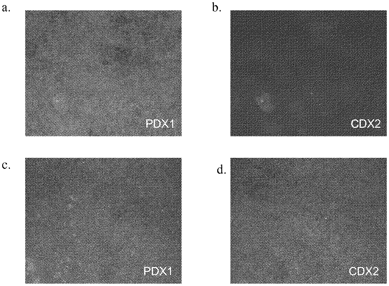
処理1(FGF7、レチノイン酸及びノギン)は、NKX6.1及びNGN3の発現を誘導した。図2のパネルa及びbを参照されたい。しかしながら、アクチビンAを添加すると(処理2)、NGN3の発現はブロックされ、NKX6.1を発現している細胞の数が有意に増加した。図2のパネルc及びdを参照されたい。これらのデータは、膵臓内胚葉系に特徴的なマーカーを発現している細胞集団形成時に、TGFβ受容体経路を活性化すると、膵臓内胚葉系に特徴的なマーカーは発現しているがNGN3は発現しない細胞集団が生じることを示す。 Treatment 1 (FGF7, retinoic acid and noggin) induced the expression of NKX6.1 and NGN3. See panels a and b of FIG. However, when activin A was added (treatment 2), NGN3 expression was blocked and the number of cells expressing NKX6.1 was significantly increased. See panels c and d of FIG. These data show that when the TGFβ receptor pathway is activated during the formation of a cell population expressing markers characteristic of the pancreatic endoderm system, markers characteristic of the pancreatic endoderm system are expressed, but NGN3 is It shows that a non-expressing cell population occurs.
細胞を、TGFβ受容体阻害剤であるAlk5阻害剤IIと共にインキュベートすることで、この仮説を確認した(処理3を参照のこと)。1%のB27(Invitrogen,CA)、50ng/mLのFGF7、0.25μMのシクロパミン−KAAD、2μMのレチノイン酸(RA)、100ng/mLのノギン及び1μMのALK5阻害剤IIを添加したDMEM(高グルコース)により細胞を処理すると、NKX6.1の発現のレベルの減少が生じた。観察された発現レベルは、処理1を行った細胞で観察されたものよりも低かった。表1及び図2のパネルeを参照のこと。それに対し、NGN3を発現している細胞の数は有意に増加した。表1及び図2のパネルfを参照のこと。PDX1発現に対しては有意な影響は観察されなかった。これらの結果は、ノギン、レチノイド酸及びアクチビンAの組み合わせは、NKN6.1及びPDX1の発現が陽性であるがNGN3の発現が陰性である特定の膵臓前駆細胞集団に対して相乗作用することを示す。 This hypothesis was confirmed by incubating the cells with Alk5 inhibitor II, a TGFβ receptor inhibitor (see treatment 3). DMEM supplemented with 1% B27 (Invitrogen, CA), 50 ng / mL FGF7, 0.25 μM cyclopamine-KAAD, 2 μM retinoic acid (RA), 100 ng / mL noggin and 1 μM ALK5 inhibitor II (high Treatment of the cells with glucose) resulted in a decrease in the level of expression of NKX6.1. The observed level of expression was lower than that observed in cells treated 1. See Table 1 and panel e of FIG. In contrast, the number of cells expressing NGN3 increased significantly. See Table 1 and panel f in FIG. No significant effect was observed on PDX1 expression. These results indicate that the combination of noggin, retinoid acid and activin A synergizes with certain pancreatic progenitor cell populations that are positive for NKN6.1 and PDX1 but negative for NGN3. .
図3のパネルa及びbに示されるように、DMEM(1%のB27(Invitrogen,CA)、50ng/mLのFGF7、0.25μMのシクロパミン−KAAD、2μMのレチノイン酸(RA)、100ng/mLのノギン及び20ng/mLのアクチビンAを添加したDMEM(高グルコース)(処理2))を用いて生成された、PDX1を発現している細胞のほとんどは、ステージ3の4日目ではCDX2を発現していなかった。このことは、1%のB27(Invitrogen,CA)、50ng/mLのFGF7、0.25μMのシクロパミン−KAAD、2μMのレチノイン酸(RA)、100ng/mLのノギン及び20ng/mLのアクチビンAを添加したDMEM/F12を用いて生成された、PDX1を発現している細胞とは対照的であり、この処理では、PDX1を発現している細胞のほとんどはCDX2も発現していた。図3のパネルc及びdを参照されたい。 As shown in panels a and b of FIG. 3, DMEM (1% B27 (Invitrogen, CA), 50 ng / mL FGF7, 0.25 μM cyclopamine-KAAD, 2 μM retinoic acid (RA), 100 ng / mL Most cells expressing PDX1 produced with DMEM (high glucose) supplemented with 20 ng / mL activin A (treatment 2)) expressed CDX2 on day 4 of stage 3 I did not. This adds 1% B27 (Invitrogen, CA), 50 ng / mL FGF7, 0.25 μM cyclopamine-KAAD, 2 μM retinoic acid (RA), 100 ng / mL noggin and 20 ng / mL activin A In contrast to cells expressing PDX1 produced using DMEM / F12, most of the cells expressing PDX1 also expressed CDX2 in this treatment. See panels c and d of FIG.
本明細書を通して引用された刊行物は、その全体が参照により本明細書に組み込まれる。以上、本発明の様々な態様を実施例及び好ましい実施形態を参照して説明したが、本発明の範囲は、上記の説明文によってではなく、特許法の原則の下で適切に解釈される以下の「特許請求の範囲」によって定義されるものである点は認識されるであろう。 Publications cited throughout this specification are hereby incorporated by reference in their entirety. While various aspects of the present invention have been described with reference to examples and preferred embodiments, the scope of the present invention is not limited by the foregoing description, but is construed as appropriate under the principles of patent law. It will be appreciated that this is defined by the “claims”.
Claims (1)
a.前記多能性幹細胞を培養する工程、
b.前記多能性幹細胞を、胚体内胚葉細胞へと分化させる工程、及び
c.前記胚体内胚葉細胞を、FGF7を添加した第1培地で処理し、次いでこうして得られる細胞を、FGF7、ノギン、アクチビンA、レチノイン酸、及びシクロパミン−KAADを添加した第2培地で培養することにより、PDX1及びNKX6.1は共発現するが、CDX2及びNGN3は発現しない、膵臓内胚葉細胞へと分化させる工程。 PDX-1 and NKX-6.1 is co-expressed, CDX-2 and NGN-3 is not expressed, and the pancreas embryo HaHoso胞集group, a method for differentiating pluripotent stem cell population, less Comprising steps a, b and c of:
a. Culturing the pluripotent stem cells,
b. The pluripotent stem cells, the step of differentiating into embryonic body embryo HaHoso cells, and c. The embryo body embryo HaHoso cells, treated with a first medium supplemented with FGF7, the then thus obtained cells, FGF7, noggin, activin A, and cultured in a second medium supplemented with retinoic acid and cyclopamine -KAAD Thus, PDX1 and NKX6.1 are co-expressed, but CDX2 and NGN3 are not expressed, and are differentiated into pancreatic endoderm cells.
Applications Claiming Priority (3)
| Application Number | Priority Date | Filing Date | Title |
|---|---|---|---|
| US22692909P | 2009-07-20 | 2009-07-20 | |
| US61/226,929 | 2009-07-20 | ||
| PCT/US2010/042504 WO2011011349A2 (en) | 2009-07-20 | 2010-07-20 | Differentiation of human embryonic stem cells |
Publications (2)
| Publication Number | Publication Date |
|---|---|
| JP2012533323A JP2012533323A (en) | 2012-12-27 |
| JP5819826B2 true JP5819826B2 (en) | 2015-11-24 |
Family
ID=43465586
Family Applications (1)
| Application Number | Title | Priority Date | Filing Date |
|---|---|---|---|
| JP2012521714A Active JP5819826B2 (en) | 2009-07-20 | 2010-07-20 | Differentiation of human embryonic stem cells |
Country Status (15)
| Country | Link |
|---|---|
| US (1) | US8785185B2 (en) |
| EP (1) | EP2456859A4 (en) |
| JP (1) | JP5819826B2 (en) |
| KR (1) | KR101786735B1 (en) |
| CN (2) | CN103952372B (en) |
| AR (1) | AR077766A1 (en) |
| AU (1) | AU2010276402B2 (en) |
| BR (1) | BR112012001564A2 (en) |
| CA (1) | CA2768720C (en) |
| GB (1) | GB2485113B (en) |
| MX (1) | MX2012000898A (en) |
| RU (1) | RU2540016C2 (en) |
| SG (1) | SG177416A1 (en) |
| WO (1) | WO2011011349A2 (en) |
| ZA (1) | ZA201201220B (en) |
Families Citing this family (58)
| Publication number | Priority date | Publication date | Assignee | Title |
|---|---|---|---|---|
| US8017395B2 (en) | 2004-12-17 | 2011-09-13 | Lifescan, Inc. | Seeding cells on porous supports |
| PT1888123E (en) | 2005-06-08 | 2013-03-13 | Janssen Biotech Inc | A cellular therapy for ocular degeneration |
| US8741643B2 (en) | 2006-04-28 | 2014-06-03 | Lifescan, Inc. | Differentiation of pluripotent stem cells to definitive endoderm lineage |
| US8685730B2 (en) | 2006-05-02 | 2014-04-01 | Wisconsin Alumni Research Foundation | Methods and devices for differentiating pluripotent stem cells into cells of the pancreatic lineage |
| US9080145B2 (en) | 2007-07-01 | 2015-07-14 | Lifescan Corporation | Single pluripotent stem cell culture |
| US9096832B2 (en) | 2007-07-31 | 2015-08-04 | Lifescan, Inc. | Differentiation of human embryonic stem cells |
| CN107574142B (en) | 2007-11-27 | 2021-07-06 | 生命扫描有限公司 | Differentiation of human embryonic stem cells |
| CA2959401C (en) | 2008-02-21 | 2021-12-07 | Centocor Ortho Biotech Inc. | Methods, surface modified plates and compositions for cell attachment, cultivation and detachment |
| US8623648B2 (en) | 2008-04-24 | 2014-01-07 | Janssen Biotech, Inc. | Treatment of pluripotent cells |
| KR20180018839A (en) | 2008-06-30 | 2018-02-21 | 얀센 바이오테크 인코포레이티드 | Differentiation of pluripotent stem cells |
| CN102333862B (en) | 2008-10-31 | 2018-04-27 | 詹森生物科技公司 | Differentiation of human embryonic stem cells to the pancreatic endocrine lineage |
| US9234178B2 (en) | 2008-10-31 | 2016-01-12 | Janssen Biotech, Inc. | Differentiation of human pluripotent stem cells |
| ES2584053T3 (en) | 2008-11-20 | 2016-09-23 | Janssen Biotech, Inc. | Methods and compositions for cell binding and culture in flat substrates |
| MX356756B (en) | 2008-11-20 | 2018-06-11 | Centocor Ortho Biotech Inc | Pluripotent stem cell culture on micro-carriers. |
| KR101785626B1 (en) | 2009-07-20 | 2017-10-16 | 얀센 바이오테크 인코포레이티드 | Differentiation of human embryonic stem cells |
| JP5819826B2 (en) | 2009-07-20 | 2015-11-24 | ヤンセン バイオテツク,インコーポレーテツド | Differentiation of human embryonic stem cells |
| WO2011011300A2 (en) | 2009-07-20 | 2011-01-27 | Centocor Ortho Biotech Inc. | Differentiation of human embryonic stem cells |
| KR101773538B1 (en) | 2009-12-23 | 2017-08-31 | 얀센 바이오테크 인코포레이티드 | Differentiation of human embryonic stem cells |
| AU2010333840B2 (en) | 2009-12-23 | 2016-01-07 | Janssen Biotech, Inc. | Differentiation of human embryonic stem cells |
| PH12012501686A1 (en) | 2010-03-01 | 2020-10-19 | Janssen Biotech Inc | Methods for purifying cells derived from pluripotent stem cells |
| CN102884176B (en) | 2010-05-12 | 2017-09-05 | 詹森生物科技公司 | Differentiation of human embryonic stem cells |
| AU2011296383B2 (en) | 2010-08-31 | 2016-03-10 | Janssen Biotech, Inc. | Differentiation of pluripotent stem cells |
| MX355077B (en) | 2010-08-31 | 2018-04-03 | Janssen Biotech Inc | Differentiation of human embryonic stem cells. |
| RU2620938C2 (en) | 2010-08-31 | 2017-05-30 | Янссен Байотек, Инк. | Differentiation of human embryonic stem cells |
| ITMI20110780A1 (en) * | 2011-05-06 | 2012-11-07 | Euroclone Spa | CULTURE GROUND TO DIFFERENTIATE STEM CELLS IN BETA CELLS |
| WO2012170853A1 (en) * | 2011-06-10 | 2012-12-13 | Wisconsin Alumni Research Foundation ("Warf") | Methods and devices for differentiating pluripotent stem cells into cells of the pancreatic lineage |
| US9388386B2 (en) | 2011-12-22 | 2016-07-12 | Janssen Biotech, Inc. | Differentiation of human embryonic stem cells into single hormonal insulin positive cells |
| US9434920B2 (en) | 2012-03-07 | 2016-09-06 | Janssen Biotech, Inc. | Defined media for expansion and maintenance of pluripotent stem cells |
| BR112014027783A2 (en) * | 2012-05-07 | 2017-06-27 | Janssen Biotech Inc | differentiation of human embryonic stem cells into pancreatic endoderm |
| CN108103005A (en) | 2012-06-08 | 2018-06-01 | 詹森生物科技公司 | Differentiation of the human embryo stem cell to pancreatic endocrine cell |
| US20150247123A1 (en) | 2012-09-03 | 2015-09-03 | Novo Nordisk A/S | Generation of pancreatic endoderm from Pluripotent Stem cells using small molecules |
| US10370644B2 (en) | 2012-12-31 | 2019-08-06 | Janssen Biotech, Inc. | Method for making human pluripotent suspension cultures and cells derived therefrom |
| SG10201709338RA (en) | 2012-12-31 | 2017-12-28 | Janssen Biotech Inc | Culturing of human embryonic stem cells at the air-liquid interface for differentiation into pancreatic endocrine cells |
| ES2942484T3 (en) | 2012-12-31 | 2023-06-01 | Janssen Biotech Inc | Differentiation of human embryonic stem cells into pancreatic endocrine cells using HB9 regulators |
| JP6529440B2 (en) | 2012-12-31 | 2019-06-12 | ヤンセン バイオテツク,インコーポレーテツド | Suspension and clustering of human pluripotent cells for differentiation to pancreatic endocrine cells |
| CN103194424A (en) * | 2013-03-28 | 2013-07-10 | 于涛 | Method for inducing embryonic stem cell into pancreatic tissue-like cells |
| EP3569694A1 (en) | 2013-06-11 | 2019-11-20 | President and Fellows of Harvard College | Sc-beta cells and compositions and methods for generating the same |
| CN117821369A (en) | 2014-05-16 | 2024-04-05 | 詹森生物科技公司 | Use of small molecules to enhance MAFA expression in pancreatic endocrine cells |
| US9371516B2 (en) | 2014-09-19 | 2016-06-21 | Regenerative Medical Solutions, Inc. | Compositions and methods for differentiating stem cells into cell populations comprising beta-like cells |
| EP4374863A3 (en) | 2014-12-18 | 2024-09-04 | President and Fellows of Harvard College | Methods for generating stem cell-derived beta cells and uses thereof |
| CN113234661A (en) | 2014-12-18 | 2021-08-10 | 哈佛学院校长同事会 | Method for producing stem cell-derived beta cells and method for using same |
| US10443042B2 (en) | 2014-12-18 | 2019-10-15 | President And Fellows Of Harvard College | Serum-free in vitro directed differentiation protocol for generating stem cell-derived beta cells and uses thereof |
| MA45479A (en) | 2016-04-14 | 2019-02-20 | Janssen Biotech Inc | DIFFERENTIATION OF PLURIPOTENT STEM CELLS IN ENDODERMAL CELLS OF MIDDLE INTESTINE |
| CN110167455B (en) | 2016-11-10 | 2022-10-11 | 韦尔赛特公司 | Cell delivery device containing PDX1 pancreatic endoderm cells and methods thereof |
| EP3587560A4 (en) * | 2017-01-27 | 2020-12-16 | Kaneka Corporation | ENDODERMAL CELL MASS AND METHOD OF PREPARING ANY OF THREE PRIMARY LAYER CELL MASS FROM PLURIPOTENT CELLS |
| US10767164B2 (en) | 2017-03-30 | 2020-09-08 | The Research Foundation For The State University Of New York | Microenvironments for self-assembly of islet organoids from stem cells differentiation |
| US10391156B2 (en) | 2017-07-12 | 2019-08-27 | Viacyte, Inc. | University donor cells and related methods |
| CA3081762A1 (en) | 2017-11-15 | 2019-05-23 | Semma Therapeutics, Inc. | Islet cell manufacturing compositions and methods of use |
| WO2020033879A1 (en) | 2018-08-10 | 2020-02-13 | Semma Therapeutics, Inc. | Stem cell derived islet differentiation |
| US12378572B2 (en) | 2018-09-07 | 2025-08-05 | Crispr Therapeutics Ag | Universal donor cells |
| BR112021023389A2 (en) * | 2019-05-22 | 2022-02-01 | Cleveland Clinic Found | Generation of dorsal foregut endoderm cells and anterior domain |
| WO2020243663A1 (en) | 2019-05-31 | 2020-12-03 | W. L. Gore & Associates, Inc. | A biocompatible membrane composite |
| CA3139292C (en) | 2019-05-31 | 2025-06-10 | Viacyte, Inc. | Cell encapsulation devices with controlled oxygen diffusion distances |
| CN114173837A (en) | 2019-05-31 | 2022-03-11 | W.L.戈尔及同仁股份有限公司 | Biocompatible film composite |
| US20220233298A1 (en) | 2019-05-31 | 2022-07-28 | W. L. Gore & Associates, Inc. | A biocompatible membrane composite |
| EP4025690A1 (en) | 2019-09-05 | 2022-07-13 | CRISPR Therapeutics AG | Universal donor cells |
| EP4025224A1 (en) | 2019-09-05 | 2022-07-13 | CRISPR Therapeutics AG | Universal donor cells |
| KR20230146008A (en) | 2020-12-31 | 2023-10-18 | 크리스퍼 테라퓨틱스 아게 | universal donor cells |
Family Cites Families (176)
| Publication number | Priority date | Publication date | Assignee | Title |
|---|---|---|---|---|
| US3209652A (en) | 1961-03-30 | 1965-10-05 | Burgsmueller Karl | Thread whirling method |
| AT326803B (en) | 1968-08-26 | 1975-12-29 | Binder Fa G | MESHWARE AND METHOD OF MANUFACTURING THE SAME |
| US3935067A (en) | 1974-11-22 | 1976-01-27 | Wyo-Ben Products, Inc. | Inorganic support for culture media |
| CA1201400A (en) | 1982-04-16 | 1986-03-04 | Joel L. Williams | Chemically specific surfaces for influencing cell activity during culture |
| US4499802A (en) | 1982-09-29 | 1985-02-19 | Container Graphics Corporation | Rotary cutting die with scrap ejection |
| US4537773A (en) | 1983-12-05 | 1985-08-27 | E. I. Du Pont De Nemours And Company | α-Aminoboronic acid derivatives |
| US4557264A (en) * | 1984-04-09 | 1985-12-10 | Ethicon Inc. | Surgical filament from polypropylene blended with polyethylene |
| US5215893A (en) | 1985-10-03 | 1993-06-01 | Genentech, Inc. | Nucleic acid encoding the ba chain prodomains of inhibin and method for synthesizing polypeptides using such nucleic acid |
| US5089396A (en) | 1985-10-03 | 1992-02-18 | Genentech, Inc. | Nucleic acid encoding β chain prodomains of inhibin and method for synthesizing polypeptides using such nucleic acid |
| US4737578A (en) | 1986-02-10 | 1988-04-12 | The Salk Institute For Biological Studies | Human inhibin |
| US5863531A (en) * | 1986-04-18 | 1999-01-26 | Advanced Tissue Sciences, Inc. | In vitro preparation of tubular tissue structures by stromal cell culture on a three-dimensional framework |
| CA1340581C (en) | 1986-11-20 | 1999-06-08 | Joseph P. Vacanti | Chimeric neomorphogenesis of organs by controlled cellular implantation using artificial matrices |
| US5567612A (en) * | 1986-11-20 | 1996-10-22 | Massachusetts Institute Of Technology | Genitourinary cell-matrix structure for implantation into a human and a method of making |
| US5804178A (en) | 1986-11-20 | 1998-09-08 | Massachusetts Institute Of Technology | Implantation of cell-matrix structure adjacent mesentery, omentum or peritoneum tissue |
| EP0363125A3 (en) | 1988-10-03 | 1990-08-16 | Hana Biologics Inc. | Proliferated pancreatic endocrine cell product and process |
| US5837539A (en) * | 1990-11-16 | 1998-11-17 | Osiris Therapeutics, Inc. | Monoclonal antibodies for human mesenchymal stem cells |
| US5449383A (en) * | 1992-03-18 | 1995-09-12 | Chatelier; Ronald C. | Cell growth substrates |
| GB9206861D0 (en) | 1992-03-28 | 1992-05-13 | Univ Manchester | Wound healing and treatment of fibrotic disorders |
| CA2114282A1 (en) | 1993-01-28 | 1994-07-29 | Lothar Schilder | Multi-layered implant |
| JP3525221B2 (en) | 1993-02-17 | 2004-05-10 | 味の素株式会社 | Immunosuppressants |
| US5523226A (en) | 1993-05-14 | 1996-06-04 | Biotechnology Research And Development Corp. | Transgenic swine compositions and methods |
| GB9310557D0 (en) * | 1993-05-21 | 1993-07-07 | Smithkline Beecham Plc | Novel process and apparatus |
| TW257671B (en) | 1993-11-19 | 1995-09-21 | Ciba Geigy | |
| US6703017B1 (en) | 1994-04-28 | 2004-03-09 | Ixion Biotechnology, Inc. | Reversal of insulin-dependent diabetes by islet-producing stem cells, islet progenitor cells and islet-like structures |
| US5834308A (en) * | 1994-04-28 | 1998-11-10 | University Of Florida Research Foundation, Inc. | In vitro growth of functional islets of Langerhans |
| US6001647A (en) | 1994-04-28 | 1999-12-14 | Ixion Biotechnology, Inc. | In vitro growth of functional islets of Langerhans and in vivo uses thereof |
| US6083903A (en) | 1994-10-28 | 2000-07-04 | Leukosite, Inc. | Boronic ester and acid compounds, synthesis and uses |
| DE69525971T3 (en) | 1994-12-29 | 2013-01-10 | Chugai Seiyaku K.K. | USE OF A PM-1 ANTIBODY OR AN MH 166 ANTIBODY FOR REINFORCING THE ANTI-TUMOR EFFECT OF CISPLATINE OR CARBOPLATIN |
| US5843780A (en) | 1995-01-20 | 1998-12-01 | Wisconsin Alumni Research Foundation | Primate embryonic stem cells |
| US5718922A (en) * | 1995-05-31 | 1998-02-17 | Schepens Eye Research Institute, Inc. | Intravitreal microsphere drug delivery and method of preparation |
| US5908782A (en) * | 1995-06-05 | 1999-06-01 | Osiris Therapeutics, Inc. | Chemically defined medium for human mesenchymal stem cells |
| PT1028954E (en) | 1997-04-24 | 2003-11-28 | Ortho Mcneil Pharm Inc | IMIDAZOLES SUBSTITUIDOS UTEIS IN THE TREATMENT OF INFLAMMATORY DISEASES |
| AU8476698A (en) * | 1997-07-03 | 1999-01-25 | Osiris Therapeutics, Inc. | Human mesenchymal stem cells from peripheral blood |
| US6670127B2 (en) | 1997-09-16 | 2003-12-30 | Egea Biosciences, Inc. | Method for assembly of a polynucleotide encoding a target polypeptide |
| ATE294860T1 (en) | 1997-09-16 | 2005-05-15 | Egea Biosciences Llc | METHODS FOR COMPLETE CHEMICAL SYNTHESIS AND ASSEMBLY OF GENES AND GENOMES |
| JP3880795B2 (en) | 1997-10-23 | 2007-02-14 | ジェロン・コーポレーション | Method for growing primate-derived primordial stem cells in a culture that does not contain feeder cells |
| ZA9811898B (en) | 1997-12-29 | 2000-06-28 | Ortho Mcneil Pharm Inc | Anti-Inflammatory Compounds. |
| DK1066052T3 (en) | 1998-03-18 | 2006-06-12 | Osiris Therapeutics Inc | Mesenchymal stem cells for the prevention and treatment of immune responses in transplants |
| MY132496A (en) | 1998-05-11 | 2007-10-31 | Vertex Pharma | Inhibitors of p38 |
| US7410798B2 (en) | 2001-01-10 | 2008-08-12 | Geron Corporation | Culture system for rapid expansion of human embryonic stem cells |
| US6667176B1 (en) * | 2000-01-11 | 2003-12-23 | Geron Corporation | cDNA libraries reflecting gene expression during growth and differentiation of human pluripotent stem cells |
| US6413556B1 (en) | 1999-01-08 | 2002-07-02 | Sky High, Llc | Aqueous anti-apoptotic compositions |
| WO2000043500A2 (en) * | 1999-01-21 | 2000-07-27 | Vitro Diagnostics, Inc. | Immortalized cell lines and methods of making the same |
| US6815203B1 (en) | 1999-06-23 | 2004-11-09 | Joslin Diabetes Center, Inc. | Methods of making pancreatic islet cells |
| US6333029B1 (en) | 1999-06-30 | 2001-12-25 | Ethicon, Inc. | Porous tissue scaffoldings for the repair of regeneration of tissue |
| US6306424B1 (en) | 1999-06-30 | 2001-10-23 | Ethicon, Inc. | Foam composite for the repair or regeneration of tissue |
| US6685936B2 (en) | 1999-10-12 | 2004-02-03 | Osiris Therapeutics, Inc. | Suppressor cells induced by culture with mesenchymal stem cells for treatment of immune responses in transplantation |
| US20030082155A1 (en) * | 1999-12-06 | 2003-05-01 | Habener Joel F. | Stem cells of the islets of langerhans and their use in treating diabetes mellitus |
| JP2003517592A (en) * | 1999-12-13 | 2003-05-27 | ザ スクリプス リサーチ インスティチュート | Markers for identification and isolation of islet α and β precursors |
| US7005252B1 (en) | 2000-03-09 | 2006-02-28 | Wisconsin Alumni Research Foundation | Serum free cultivation of primate embryonic stem cells |
| US7439064B2 (en) | 2000-03-09 | 2008-10-21 | Wicell Research Institute, Inc. | Cultivation of human embryonic stem cells in the absence of feeder cells or without conditioned medium |
| US6436704B1 (en) | 2000-04-10 | 2002-08-20 | Raven Biotechnologies, Inc. | Human pancreatic epithelial progenitor cells and methods of isolation and use thereof |
| US6458589B1 (en) * | 2000-04-27 | 2002-10-01 | Geron Corporation | Hepatocyte lineage cells derived from pluripotent stem cells |
| KR100947577B1 (en) * | 2000-06-26 | 2010-03-15 | 엔씨 메디컬 리서치 가부시키가이샤 | Cell fraction containing cells capable of differentiating into nervous system cells |
| CN1847229B (en) * | 2000-10-23 | 2011-05-04 | 史密丝克莱恩比彻姆公司 | 2,4,8- trisubstituted-8H- pyridine[2,3-d] pyridine -7-one compound |
| CA2431187A1 (en) | 2000-12-08 | 2002-06-13 | Ortho-Mcneil Pharmaceutical, Inc. | Macroheterocylic compounds useful as kinase inhibitors |
| EP1362047B1 (en) | 2000-12-08 | 2006-05-17 | Ortho-McNeil Pharmaceutical, Inc. | Indazolyl-substituted pyrroline compounds as kinase inhibitors |
| US6599323B2 (en) | 2000-12-21 | 2003-07-29 | Ethicon, Inc. | Reinforced tissue implants and methods of manufacture and use |
| WO2002059278A2 (en) | 2001-01-24 | 2002-08-01 | The Government Of The United States Of America, As Represented By The Secretary Of Department Of Health & Human Services | Differentiation of stem cells to pancreatic endocrine cells |
| CA2435124A1 (en) | 2001-01-25 | 2002-08-01 | Millennium Pharmaceuticals, Inc. | Formulation of boronic acid compounds |
| US6656488B2 (en) | 2001-04-11 | 2003-12-02 | Ethicon Endo-Surgery, Inc. | Bioabsorbable bag containing bioabsorbable materials of different bioabsorption rates for tissue engineering |
| DE10290025T1 (en) * | 2001-04-19 | 2003-10-09 | Develogen Ag | Procedure for differentiating stem cells into insulin-producing cells |
| DE60231035D1 (en) | 2001-04-24 | 2009-03-19 | Ajinomoto Kk | STEM CELLS AND METHOD FOR THEIR SEPARATION |
| CA2447015A1 (en) | 2001-05-15 | 2002-11-21 | Rappaport Family Institute For Research In The Medical Sciences | Insulin producing cells derived from human embryonic stem cells |
| US6626950B2 (en) | 2001-06-28 | 2003-09-30 | Ethicon, Inc. | Composite scaffold with post anchor for the repair and regeneration of tissue |
| KR100418195B1 (en) | 2001-07-05 | 2004-02-11 | 주식회사 우리기술 | Apparatus and method for multi-testing insulation of power cables |
| GB0117583D0 (en) * | 2001-07-19 | 2001-09-12 | Astrazeneca Ab | Novel compounds |
| US7432104B2 (en) * | 2001-08-06 | 2008-10-07 | Bresgen Inc. | Methods for the culture of human embryonic stem cells on human feeder cells |
| US6617152B2 (en) * | 2001-09-04 | 2003-09-09 | Corning Inc | Method for creating a cell growth surface on a polymeric substrate |
| JP2005506074A (en) | 2001-10-18 | 2005-03-03 | イクシオン・バイオテクノロジー・インコーポレーテッド | Conversion of hepatic stem and progenitor cells into functional pancreatic cells |
| WO2003042405A2 (en) | 2001-11-15 | 2003-05-22 | Children's Medical Center Corporation | Methods of isolation, expansion and differentiation of fetal stem cells from chorionic villus, amniotic fluid, and placenta and therapeutic uses thereof |
| DK1921133T3 (en) | 2001-12-07 | 2015-08-24 | Cytori Therapeutics Inc | System for processing lipoaspiratceller |
| GB2399823B (en) | 2001-12-07 | 2006-02-15 | Geron Corp | Islet cells from primate pluripotent stem cells |
| AU2002218893A1 (en) | 2001-12-21 | 2003-07-09 | Thromb-X Nv | Compositions for the in vitro derivation and culture of embryonic stem (es) cell lines with germline transmission capability |
| US20030162290A1 (en) | 2002-01-25 | 2003-08-28 | Kazutomo Inoue | Method for inducing differentiation of embryonic stem cells into functioning cells |
| EP1498478A1 (en) * | 2002-04-17 | 2005-01-19 | Otsuka Pharmaceutical Co., Ltd. | Method of forming pancreatic beta cells from mesenchymal cells |
| US20040161419A1 (en) * | 2002-04-19 | 2004-08-19 | Strom Stephen C. | Placental stem cells and uses thereof |
| AU2003225295A1 (en) | 2002-05-08 | 2003-11-11 | Janssen Pharmaceutica N.V. | Substituted pyrroline kinase inhibitors |
| US20060003446A1 (en) * | 2002-05-17 | 2006-01-05 | Gordon Keller | Mesoderm and definitive endoderm cell populations |
| AU2003228255A1 (en) | 2002-05-28 | 2003-12-19 | Becton, Dickinson And Company | Pancreatic acinar cells into insulin-producing cells |
| CN1671694A (en) | 2002-06-05 | 2005-09-21 | 詹森药业有限公司 | Bisindolyl-maleimid derivatives as kinase inhibitors |
| GB0212976D0 (en) | 2002-06-06 | 2002-07-17 | Tonejet Corp Pty Ltd | Ejection method and apparatus |
| CN1171991C (en) | 2002-07-08 | 2004-10-20 | 徐如祥 | Culture process of human nerve stem cell |
| US6877147B2 (en) * | 2002-07-22 | 2005-04-05 | Broadcom Corporation | Technique to assess timing delay by use of layout quality analyzer comparison |
| US7838290B2 (en) * | 2002-07-25 | 2010-11-23 | The Scripps Research Institute | Hematopoietic stem cells and methods of treatment of neovascular eye diseases therewith |
| WO2004011621A2 (en) | 2002-07-29 | 2004-02-05 | Es Cell International Pte Ltd. | Multi-step method for the differentiation of insulin positive, glucose |
| AU2003262628A1 (en) | 2002-08-14 | 2004-03-03 | University Of Florida | Bone marrow cell differentiation |
| US7371576B2 (en) | 2002-09-06 | 2008-05-13 | Reneuron, Inc. | CD56 positive human adult pancreatic endocrine progenitor cells |
| US9969977B2 (en) | 2002-09-20 | 2018-05-15 | Garnet Biotherapeutics | Cell populations which co-express CD49c and CD90 |
| US20040062753A1 (en) | 2002-09-27 | 2004-04-01 | Alireza Rezania | Composite scaffolds seeded with mammalian cells |
| AU2003285172A1 (en) | 2002-11-08 | 2004-06-03 | The Johns Hopkins University | Human embryonic stem cell cultures, and compositions and methods for growing same |
| US7144999B2 (en) * | 2002-11-23 | 2006-12-05 | Isis Pharmaceuticals, Inc. | Modulation of hypoxia-inducible factor 1 alpha expression |
| EP1567639A4 (en) | 2002-12-05 | 2005-12-21 | Technion Res & Dev Foundation | Cultured human pancreatic islets, and uses thereof |
| ES2705683T3 (en) | 2002-12-16 | 2019-03-26 | Technion Res & Dev Foundation | Culture medium of pluripotent stem cells |
| US20070155661A1 (en) | 2003-02-14 | 2007-07-05 | The Board Of Trustees Of The Leland Standord Junior University | Methods and compositions for modulating the development of stem cells |
| WO2005045001A2 (en) * | 2003-02-14 | 2005-05-19 | The Board Of Trustees Of The Leland Stanford Junior University | Insulin-producing cells derived from stem cells |
| US20070020242A1 (en) | 2003-03-27 | 2007-01-25 | Ixion Biotechnology, Inc. | Method for transdifferentiation of non-pancreatic stem cells to the pancreatic pathway |
| WO2004090110A2 (en) | 2003-03-31 | 2004-10-21 | Bresagen Inc. | Compositions and methods for the control, differentiation and/or manipulation of pluripotent cells through a gamma-secretase signaling pathway |
| US20090203141A1 (en) | 2003-05-15 | 2009-08-13 | Shi-Lung Lin | Generation of tumor-free embryonic stem-like pluripotent cells using inducible recombinant RNA agents |
| WO2005001079A2 (en) * | 2003-06-27 | 2005-01-06 | Ethicon, Incorporated | Soft tissue repair and regeneration using postpartum-derived cells |
| IL161903A0 (en) | 2003-07-17 | 2005-11-20 | Gamida Cell Ltd | Ex vivo progenitor and stem cell expansion for usein the treatment of disease of endodermally- deri ved organs |
| ITRM20030395A1 (en) | 2003-08-12 | 2005-02-13 | Istituto Naz Per Le Malattie Infettive Lazz | CULTURE GROUND FOR MAINTENANCE, PROLIFERATION AND DIFFERENTIATION OF MAMMALIAN CELLS. |
| US7569385B2 (en) * | 2003-08-14 | 2009-08-04 | The Regents Of The University Of California | Multipotent amniotic fetal stem cells |
| US7157275B2 (en) * | 2003-08-15 | 2007-01-02 | Becton, Dickinson And Company | Peptides for enhanced cell attachment and growth |
| JP2007503815A (en) | 2003-08-27 | 2007-03-01 | ステムセルズ・カリフォルニア・インコーポレイテッド | Enriched pancreatic stem and progenitor cell populations and methods for identifying, isolating and enriching these populations |
| EP1696899A1 (en) | 2003-12-17 | 2006-09-06 | Allergan, Inc. | Methods for treating retinoid responsive disorders using selective inhibitors of cyp26a and cyp26b |
| US20060030042A1 (en) | 2003-12-19 | 2006-02-09 | Ali Brivanlou | Maintenance of embryonic stem cells by the GSK-3 inhibitor 6-bromoindirubin-3'-oxime |
| US20050266554A1 (en) | 2004-04-27 | 2005-12-01 | D Amour Kevin A | PDX1 expressing endoderm |
| US7625753B2 (en) | 2003-12-23 | 2009-12-01 | Cythera, Inc. | Expansion of definitive endoderm cells |
| KR101221302B1 (en) | 2003-12-23 | 2013-01-11 | 비아싸이트, 인크. | Definitive endoderm |
| TWI334443B (en) * | 2003-12-31 | 2010-12-11 | Ind Tech Res Inst | Method of single cell culture of undifferentiated human embryonic stem cells |
| WO2005065354A2 (en) | 2003-12-31 | 2005-07-21 | The Burnham Institute | Defined media for pluripotent stem cell culture |
| US20080241107A1 (en) | 2004-01-23 | 2008-10-02 | Copland Iii John A | Methods and Compositions For Preparing Pancreatic Insulin Secreting Cells |
| GB2441530B (en) | 2004-02-12 | 2009-09-23 | Univ Newcastle | Stem Cells |
| AU2005221095A1 (en) * | 2004-03-09 | 2005-09-22 | John J. O'neil | Methods for generating insulin-producing cells |
| CN1950498A (en) * | 2004-03-10 | 2007-04-18 | 加利福尼亚大学董事会 | Compositions and methods for culturing embryonic stem cells |
| KR20070029681A (en) | 2004-04-01 | 2007-03-14 | 위스콘신 얼럼나이 리서어치 화운데이션 | Differentiation of Stem Cells into Endoderm and Interest Lineages |
| ES2801001T3 (en) | 2004-04-27 | 2021-01-07 | Viacyte Inc | Endoderm expressing pdx1 |
| WO2006016999A1 (en) | 2004-07-09 | 2006-02-16 | Cythera, Inc. | Methods for identifying factors for differentiating definitive endoderm |
| EP1791952A4 (en) | 2004-08-13 | 2008-06-11 | Univ Georgia Res Found | Compositions and methods for self-renewal and differentiation in human embryonic stem cells |
| WO2006026473A2 (en) | 2004-08-25 | 2006-03-09 | University Of Georgia Research Foundation, Inc. | METHODS AND COMPOSITIONS UTILIZING MYC AND GSK3ß TO MANIPULATE THE PLURIPOTENCY OF EMBRYONIC STEM CELLS |
| DE102004043256B4 (en) | 2004-09-07 | 2013-09-19 | Rheinische Friedrich-Wilhelms-Universität Bonn | Scalable process for culturing undifferentiated stem cells in suspension |
| BRPI0514778B1 (en) | 2004-09-08 | 2018-07-10 | Wisconsin Alumni Research Foundation | MEASURE TO GROW EMBRYONIC STEM CELLS |
| WO2006029198A2 (en) | 2004-09-08 | 2006-03-16 | Wisconsin Alumni Research Foundation | Culturing human embryonic stem cells |
| WO2006083782A2 (en) | 2005-01-31 | 2006-08-10 | Es Cell International Pte Ltd. | Directed differentiation of embryonic stem cells and uses thereof |
| AU2006218359A1 (en) | 2005-03-04 | 2006-09-08 | John O'neil | Adult pancreatic derived stromal cells |
| GB0505970D0 (en) | 2005-03-23 | 2005-04-27 | Univ Edinburgh | Culture medium containing kinase inhibitor, and uses thereof |
| US7998938B2 (en) | 2005-04-15 | 2011-08-16 | Geron Corporation | Cancer treatment by combined inhibition of proteasome and telomerase activities |
| WO2006114097A2 (en) | 2005-04-26 | 2006-11-02 | Aarhus Universitet | Biosurface structure array |
| MX2007015610A (en) | 2005-06-10 | 2008-02-21 | Irm Llc | Compounds that maintain pluripotency of embryonic stem cells. |
| WO2006138433A2 (en) | 2005-06-14 | 2006-12-28 | The Regents Of The University Of California | Induction of cell differentiation by class i bhlh polypeptides |
| US20080199959A1 (en) | 2005-06-21 | 2008-08-21 | Ge Healthcare Bio-Sciences Ab | Method For Cell Culture |
| WO2007003525A2 (en) | 2005-06-30 | 2007-01-11 | Janssen Pharmaceutica N.V. | Cyclic anilino-pyridinotriazines as gsk-3 inhibitors |
| US20080194021A1 (en) | 2005-07-29 | 2008-08-14 | Mays Robert W | Use of a Gsk-3 Inhibitor to Maintain Potency of Culture Cells |
| CA2616863A1 (en) | 2005-07-29 | 2007-02-01 | Australian Stem Cell Centre Limited | Compositions and methods for growth of pluripotent cells |
| BRPI0617084A2 (en) | 2005-09-02 | 2011-07-12 | Agency Science Tech & Res | method, parent cell line, differentiated cell and method for generating a differentiated cell from an embryonic stem cell (s) |
| WO2007030870A1 (en) | 2005-09-12 | 2007-03-22 | Es Cell International Pte Ltd | Cardiomyocyte production |
| DK2674485T3 (en) * | 2005-10-27 | 2019-08-26 | Viacyte Inc | PDX-1 EXPRESSING DORSAL AND VENTRAL FORTARM ENDODERM |
| WO2007082963A1 (en) | 2006-01-18 | 2007-07-26 | Fundación Instituto Valenciano De Infertilidad | Human embryo stem-cell lines and methods for using same |
| KR20080109775A (en) | 2006-02-23 | 2008-12-17 | 노보셀, 인크 | Compositions and methods useful for culturing differentiable cells |
| US7695965B2 (en) | 2006-03-02 | 2010-04-13 | Cythera, Inc. | Methods of producing pancreatic hormones |
| EP1999253B1 (en) | 2006-03-02 | 2019-05-22 | Viacyte, Inc. | Endocrine precursor cells, pancreatic hormone-expressing cells and methods of production |
| US8741643B2 (en) * | 2006-04-28 | 2014-06-03 | Lifescan, Inc. | Differentiation of pluripotent stem cells to definitive endoderm lineage |
| ES2725601T3 (en) | 2006-04-28 | 2019-09-25 | Lifescan Inc | Differentiation of human embryonic stem cells |
| SE534150C2 (en) * | 2006-05-02 | 2011-05-10 | Wisconsin Alumni Res Found | Procedure for differentiating stem cells into cells of the endodermal and pancreatic developmental line |
| WO2007139929A2 (en) | 2006-05-25 | 2007-12-06 | The Burnham Institute For Medical Research | Methods for culture and production of single cell populations of human embryonic stem cells |
| WO2007149182A2 (en) | 2006-06-19 | 2007-12-27 | Geron Corporation | Differentiation and enrichment of islet-like cells from human pluripotent stem cells |
| CN100494359C (en) | 2006-06-23 | 2009-06-03 | 中日友好医院 | Method for in vitro expansion of neural stem cells in three-dimensional culture |
| US20080003676A1 (en) | 2006-06-26 | 2008-01-03 | Millipore Corporation | Growth of embryonic stem cells |
| CA2893679C (en) | 2006-06-26 | 2018-10-30 | Lifescan, Inc. | Conditioned medium for pluripotent stem cell culture |
| GB2454386B (en) | 2006-07-06 | 2011-07-06 | Es Cell Int Pte Ltd | Method for embryonic stem cell culture on a positively charged support surface |
| WO2008013664A2 (en) | 2006-07-26 | 2008-01-31 | Cythera, Inc. | Methods of producing pancreatic hormones |
| JP2008099662A (en) | 2006-09-22 | 2008-05-01 | Institute Of Physical & Chemical Research | Stem cell culture method |
| US20080091234A1 (en) * | 2006-09-26 | 2008-04-17 | Kladakis Stephanie M | Method for modifying a medical implant surface for promoting tissue growth |
| WO2008048647A1 (en) | 2006-10-17 | 2008-04-24 | Cythera, Inc. | Modulation of the phosphatidylinositol-3-kinase pathway in the differentiation of human embryonic stem cells |
| CA2666789C (en) | 2006-10-18 | 2016-11-22 | Yong Zhao | Embryonic-like stem cells derived from adult human peripheral blood and methods of use |
| WO2008086005A1 (en) | 2007-01-09 | 2008-07-17 | University Of South Florida | Compositions including triciribine and bortezomib and derivatives thereof and methods of use thereof |
| CA2676044C (en) | 2007-01-30 | 2019-10-22 | University Of Georgia Research Foundation, Inc. | Early mesoderm cells, a stable population of mesendoderm cells that has utility for generation of endoderm and mesoderm lineages and multipotent migratory cells (mmc) |
| GB0703188D0 (en) | 2007-02-19 | 2007-03-28 | Roger Land Building | Large scale production of stem cells |
| EP2185695B1 (en) | 2007-07-18 | 2015-02-25 | Lifescan, Inc. | Differentiation of human embryonic stem cells |
| US9096832B2 (en) | 2007-07-31 | 2015-08-04 | Lifescan, Inc. | Differentiation of human embryonic stem cells |
| MX2010002179A (en) | 2007-08-24 | 2010-04-27 | Stichting Het Nl Kanker I | Composition. |
| CN107574142B (en) * | 2007-11-27 | 2021-07-06 | 生命扫描有限公司 | Differentiation of human embryonic stem cells |
| SG154367A1 (en) | 2008-01-31 | 2009-08-28 | Es Cell Int Pte Ltd | Method of differentiating stem cells |
| US20100330677A1 (en) | 2008-02-11 | 2010-12-30 | Cambridge Enterprise Limited | Improved Reprogramming of Mammalian Cells, and Cells Obtained |
| CA2959401C (en) | 2008-02-21 | 2021-12-07 | Centocor Ortho Biotech Inc. | Methods, surface modified plates and compositions for cell attachment, cultivation and detachment |
| EP2479260B1 (en) | 2008-03-17 | 2016-01-06 | Agency For Science, Technology And Research | Microcarriers for stem cell culture |
| DK2727998T3 (en) | 2008-04-21 | 2019-08-26 | Viacyte Inc | Methods for purifying pancreatic endoderm cells derived from human embryonic stem cells |
| US20090298178A1 (en) | 2008-06-03 | 2009-12-03 | D Amour Kevin Allen | Growth factors for production of definitive endoderm |
| DE102008032236A1 (en) | 2008-06-30 | 2010-04-01 | Eberhard-Karls-Universität Tübingen | Isolation and / or identification of stem cells with adipocytic, chondrocytic and pancreatic differentiation potential |
| KR20180018839A (en) | 2008-06-30 | 2018-02-21 | 얀센 바이오테크 인코포레이티드 | Differentiation of pluripotent stem cells |
| US20100028307A1 (en) | 2008-07-31 | 2010-02-04 | O'neil John J | Pluripotent stem cell differentiation |
| US9234178B2 (en) | 2008-10-31 | 2016-01-12 | Janssen Biotech, Inc. | Differentiation of human pluripotent stem cells |
| US8008075B2 (en) | 2008-11-04 | 2011-08-30 | Viacyte, Inc. | Stem cell aggregate suspension compositions and methods of differentiation thereof |
| US10350795B2 (en) | 2009-07-15 | 2019-07-16 | The University Of Akron | Flexible and electrically conductive polymer films and methods of making same |
| JP5819826B2 (en) | 2009-07-20 | 2015-11-24 | ヤンセン バイオテツク,インコーポレーテツド | Differentiation of human embryonic stem cells |
-
2010
- 2010-07-20 JP JP2012521714A patent/JP5819826B2/en active Active
- 2010-07-20 US US12/839,551 patent/US8785185B2/en active Active
- 2010-07-20 GB GB1202848.6A patent/GB2485113B/en not_active Expired - Fee Related
- 2010-07-20 RU RU2012105924/10A patent/RU2540016C2/en active
- 2010-07-20 SG SG2011097169A patent/SG177416A1/en unknown
- 2010-07-20 BR BRBR112012001564-8A patent/BR112012001564A2/en not_active Application Discontinuation
- 2010-07-20 CN CN201410217168.8A patent/CN103952372B/en active Active
- 2010-07-20 CA CA2768720A patent/CA2768720C/en active Active
- 2010-07-20 AU AU2010276402A patent/AU2010276402B2/en not_active Ceased
- 2010-07-20 KR KR1020127003771A patent/KR101786735B1/en active Active
- 2010-07-20 MX MX2012000898A patent/MX2012000898A/en active IP Right Grant
- 2010-07-20 CN CN201080033700.0A patent/CN102597219B/en not_active Expired - Fee Related
- 2010-07-20 EP EP10802743.4A patent/EP2456859A4/en not_active Withdrawn
- 2010-07-20 WO PCT/US2010/042504 patent/WO2011011349A2/en not_active Ceased
- 2010-07-20 AR ARP100102640A patent/AR077766A1/en unknown
-
2012
- 2012-02-17 ZA ZA2012/01220A patent/ZA201201220B/en unknown
Also Published As
| Publication number | Publication date |
|---|---|
| BR112012001564A2 (en) | 2015-09-01 |
| WO2011011349A2 (en) | 2011-01-27 |
| KR20120037986A (en) | 2012-04-20 |
| AU2010276402B2 (en) | 2014-07-03 |
| SG177416A1 (en) | 2012-02-28 |
| CN102597219B (en) | 2015-08-19 |
| CN102597219A (en) | 2012-07-18 |
| RU2540016C2 (en) | 2015-01-27 |
| AU2010276402A1 (en) | 2012-01-19 |
| AR077766A1 (en) | 2011-09-21 |
| CA2768720A1 (en) | 2011-01-27 |
| GB201202848D0 (en) | 2012-04-04 |
| EP2456859A2 (en) | 2012-05-30 |
| WO2011011349A3 (en) | 2011-05-05 |
| ZA201201220B (en) | 2013-07-31 |
| CA2768720C (en) | 2018-12-18 |
| US8785185B2 (en) | 2014-07-22 |
| GB2485113B (en) | 2016-12-28 |
| CN103952372A (en) | 2014-07-30 |
| GB2485113A (en) | 2012-05-02 |
| RU2012105924A (en) | 2013-08-27 |
| EP2456859A4 (en) | 2015-03-18 |
| US20110014703A1 (en) | 2011-01-20 |
| JP2012533323A (en) | 2012-12-27 |
| CN103952372B (en) | 2016-10-05 |
| MX2012000898A (en) | 2012-06-01 |
| KR101786735B1 (en) | 2017-10-18 |
Similar Documents
| Publication | Publication Date | Title |
|---|---|---|
| JP5819826B2 (en) | Differentiation of human embryonic stem cells | |
| JP6230572B2 (en) | Differentiation of human embryonic stem cells into the pancreatic endocrine system | |
| JP5819825B2 (en) | Differentiation of human embryonic stem cells | |
| JP6219568B2 (en) | Differentiation of human embryonic stem cells | |
| JP6392496B2 (en) | Differentiation of human embryonic stem cells |
Legal Events
| Date | Code | Title | Description |
|---|---|---|---|
| A621 | Written request for application examination |
Free format text: JAPANESE INTERMEDIATE CODE: A621 Effective date: 20130718 |
|
| A131 | Notification of reasons for refusal |
Free format text: JAPANESE INTERMEDIATE CODE: A131 Effective date: 20141112 |
|
| A601 | Written request for extension of time |
Free format text: JAPANESE INTERMEDIATE CODE: A601 Effective date: 20150212 |
|
| A521 | Request for written amendment filed |
Free format text: JAPANESE INTERMEDIATE CODE: A523 Effective date: 20150306 |
|
| TRDD | Decision of grant or rejection written | ||
| A01 | Written decision to grant a patent or to grant a registration (utility model) |
Free format text: JAPANESE INTERMEDIATE CODE: A01 Effective date: 20150902 |
|
| A61 | First payment of annual fees (during grant procedure) |
Free format text: JAPANESE INTERMEDIATE CODE: A61 Effective date: 20151001 |
|
| R150 | Certificate of patent or registration of utility model |
Ref document number: 5819826 Country of ref document: JP Free format text: JAPANESE INTERMEDIATE CODE: R150 |
|
| R250 | Receipt of annual fees |
Free format text: JAPANESE INTERMEDIATE CODE: R250 |
|
| R250 | Receipt of annual fees |
Free format text: JAPANESE INTERMEDIATE CODE: R250 |
|
| R250 | Receipt of annual fees |
Free format text: JAPANESE INTERMEDIATE CODE: R250 |
|
| R250 | Receipt of annual fees |
Free format text: JAPANESE INTERMEDIATE CODE: R250 |
|
| R250 | Receipt of annual fees |
Free format text: JAPANESE INTERMEDIATE CODE: R250 |
|
| R250 | Receipt of annual fees |
Free format text: JAPANESE INTERMEDIATE CODE: R250 |
|
| R250 | Receipt of annual fees |
Free format text: JAPANESE INTERMEDIATE CODE: R250 |
|
| R250 | Receipt of annual fees |
Free format text: JAPANESE INTERMEDIATE CODE: R250 |