JP5476557B2 - Packaging container and sample observation method using the same - Google Patents
Packaging container and sample observation method using the same Download PDFInfo
- Publication number
- JP5476557B2 JP5476557B2 JP2011179492A JP2011179492A JP5476557B2 JP 5476557 B2 JP5476557 B2 JP 5476557B2 JP 2011179492 A JP2011179492 A JP 2011179492A JP 2011179492 A JP2011179492 A JP 2011179492A JP 5476557 B2 JP5476557 B2 JP 5476557B2
- Authority
- JP
- Japan
- Prior art keywords
- packaging container
- container
- main body
- lid
- sample
- Prior art date
- Legal status (The legal status is an assumption and is not a legal conclusion. Google has not performed a legal analysis and makes no representation as to the accuracy of the status listed.)
- Active
Links
Images


Classifications
-
- C—CHEMISTRY; METALLURGY
- C12—BIOCHEMISTRY; BEER; SPIRITS; WINE; VINEGAR; MICROBIOLOGY; ENZYMOLOGY; MUTATION OR GENETIC ENGINEERING
- C12M—APPARATUS FOR ENZYMOLOGY OR MICROBIOLOGY; APPARATUS FOR CULTURING MICROORGANISMS FOR PRODUCING BIOMASS, FOR GROWING CELLS OR FOR OBTAINING FERMENTATION OR METABOLIC PRODUCTS, i.e. BIOREACTORS OR FERMENTERS
- C12M45/00—Means for pre-treatment of biological substances
- C12M45/22—Means for packing or storing viable microorganisms
-
- C—CHEMISTRY; METALLURGY
- C12—BIOCHEMISTRY; BEER; SPIRITS; WINE; VINEGAR; MICROBIOLOGY; ENZYMOLOGY; MUTATION OR GENETIC ENGINEERING
- C12M—APPARATUS FOR ENZYMOLOGY OR MICROBIOLOGY; APPARATUS FOR CULTURING MICROORGANISMS FOR PRODUCING BIOMASS, FOR GROWING CELLS OR FOR OBTAINING FERMENTATION OR METABOLIC PRODUCTS, i.e. BIOREACTORS OR FERMENTERS
- C12M23/00—Constructional details, e.g. recesses, hinges
- C12M23/52—Mobile; Means for transporting the apparatus
Landscapes
- Health & Medical Sciences (AREA)
- Life Sciences & Earth Sciences (AREA)
- Wood Science & Technology (AREA)
- Organic Chemistry (AREA)
- Engineering & Computer Science (AREA)
- Bioinformatics & Cheminformatics (AREA)
- Chemical & Material Sciences (AREA)
- Zoology (AREA)
- Biochemistry (AREA)
- Genetics & Genomics (AREA)
- Microbiology (AREA)
- Biotechnology (AREA)
- Biomedical Technology (AREA)
- General Engineering & Computer Science (AREA)
- General Health & Medical Sciences (AREA)
- Sustainable Development (AREA)
- Clinical Laboratory Science (AREA)
- Molecular Biology (AREA)
- Sampling And Sample Adjustment (AREA)
- Investigating Or Analysing Materials By Optical Means (AREA)
- Details Of Rigid Or Semi-Rigid Containers (AREA)
- Packages (AREA)
- Apparatus Associated With Microorganisms And Enzymes (AREA)
- Measuring Or Testing Involving Enzymes Or Micro-Organisms (AREA)
Description
本発明は、清浄性に関する環境を維持した状態で試料を持ち運び、観察可能な包装容器に関する。例えば細胞処理施設で製造した再生組織等を含む生体試料を持ち運び及び観察可能な包装容器に関する。 The present invention relates to a packaging container capable of carrying and observing a sample while maintaining an environment relating to cleanliness. For example, the present invention relates to a packaging container capable of carrying and observing a biological sample including a regenerated tissue manufactured in a cell processing facility.
少量の細胞を培養して製造した再生組織等の生体試料を用い、失われた臓器等の機能を回復させる再生医療は、従来治療法のなかった疾病に対し効果の期待される技術である。
再生医療に用いる再生組織等の生体試料の製造工程は、医薬品等の製造管理と品質管理の基準である適正製造基準(GMP;Good Manufacturing Practice)に基づく。製造は細胞処理施設(CPC;Cell Processing Center)において行われ、GMPを満たした標準手順書(SOP;Standard Operating Procedure)に従う。GMPは、日本国内では、厚生労働省の定める法規が既に施行されている(例えば厚生省令第179号、薬発第480号)。日本国外においては、欧米の機関(例えば米国食料医薬品庁、欧州委員会)を中心に関連法規が施行されている。
Regenerative medicine using a biological sample such as a regenerated tissue produced by culturing a small amount of cells to restore the function of a lost organ or the like is a technique that is expected to be effective for diseases for which there has been no conventional treatment.
The manufacturing process of biological samples such as regenerative tissues used in regenerative medicine is based on Good Manufacturing Practice (GMP), which is a standard for manufacturing control and quality control of pharmaceuticals. Manufacture is performed in a cell processing center (CPC) and follows a standard operating procedure (SOP) that satisfies GMP. In Japan, the laws and regulations set by the Ministry of Health, Labor and Welfare have already been enforced in GMP (for example, Ministry of Health, Labor and Welfare No. 179, Yakuhatsu No. 480). Outside Japan, relevant laws and regulations are being enforced mainly by Western institutions (eg, US Food and Drug Administration, European Commission).
現状においては、生産拠点となる少数のCPCにおいて再生組織等の生体試料を製造し、当該製造された生体試料を各地の医療機関、研究機関へ輸送して、治療・研究に用いられている。
生体試料を生体へ適用するに当たり、その治療を行う場合には、清潔な状態を維持したまま、培養容器から生体試料を取り出す必要がある。取りだされた生体試料は、輸送中に清浄度の低い空間を通過する。よって、菌等の生物や粒子が、細胞輸送容器等の外部に付着してしまう。治療を行う際には、当該培養容器の内側にある生体試料に、菌等が付着して生物学的汚染を生じることがないよう、無菌的に取り出す必要がある。
上記背景に対する従来技術として、再生組織等の生体試料の輸送・包装技術に関する報告が幾つかある。
At present, biological samples such as regenerative tissues are manufactured in a small number of CPCs serving as production bases, and the manufactured biological samples are transported to medical institutions and research institutions in various places and used for treatment and research.
When applying a biological sample to a living body, when performing the treatment, it is necessary to take out the biological sample from the culture container while maintaining a clean state. The removed biological sample passes through a low clean space during transportation. Therefore, organisms such as bacteria and particles adhere to the outside of the cell transport container or the like. When performing treatment, it is necessary to aseptically remove the biological sample inside the culture vessel so that bacteria or the like do not adhere to it and cause biological contamination.
As prior art against the above-mentioned background, there are several reports on transport / packaging techniques for biological samples such as regenerated tissues.
特許文献1には、滅菌物を滅菌袋により多重包装した多重滅菌包装物に関し、施設到着後の空間清浄度に応じて順次包装袋を破ることで、内部の滅菌物の清浄度を保つ技術について開示されている。 Patent Document 1 relates to a multiple sterilized package in which a sterilized product is packaged in multiple sterilization bags, and a technique for maintaining the cleanliness of the sterilized product inside by sequentially breaking the packaging bag according to the cleanliness of the space after arrival at the facility. It is disclosed.
特許文献2には、培養容器の収容が可能な箱について開示されている。箱には、気体のみを通過させ、菌や粒子等が外部から侵入することのないフィルタが設置されている。これにより、細胞処理施設の培養エリアで培養容器を収容することで、培養容器の清浄性を維持できる。また、清浄性を維持した状態で、細胞処理施設の外へ運び出すことができる。 Patent Document 2 discloses a box that can accommodate a culture vessel. The box is provided with a filter that allows only gas to pass therethrough and prevents bacteria and particles from entering from the outside. Thereby, the cleanliness of the culture container can be maintained by accommodating the culture container in the culture area of the cell treatment facility. Moreover, it can be carried out of the cell treatment facility while maintaining cleanliness.
特許文献1に記載の方法では、輸送時に滅菌物である生体試料が汚染される危険性は低い。一方、輸送中における温度条件、輸送環境等によっては、細胞にダメージが生じる可能性がある。そのため、輸送後に出来るだけ早く輸送された生体試料を観察・検査することが望まれるが、輸送後の生体試料の状態を観察等するためには、滅菌袋を開封し、培養容器を露出させて、位相差顕微鏡等の光学顕微鏡等で細胞を観察する必要がある。
このとき、輸送先である医療機関、研究機関等にCPCの様な無菌状態を維持できる細胞処理施設が無ければ、無菌状態ではない検査室等において観察等することになる。これはすなわち、培養容器を汚染空間に曝すことを意味する。
このように汚染空間に培養容器を曝すことは、培養容器に菌等が付着し汚染される可能性を示唆している。清潔な環境である手術室等の処理施設内に当該容器を持ち込む際にエタノール消毒等したとしても、全ての付着物を除去できるとは限らない。
このように菌や粒子等が付着した可能性の有る状態で、培養容器を手術室等に持ち込み、培養容器から生体試料を取り出すと、培養容器の蓋を開ける際に、除去しきれなかった菌や粒子等が落下して生体試料に付着、或いは培地内に入ると、生物学的汚染が生じる可能性がある。
In the method described in Patent Document 1, there is a low risk of contamination of a biological sample that is a sterilized product during transportation. On the other hand, the cells may be damaged depending on temperature conditions during transportation, transportation environment, and the like. Therefore, it is desirable to observe and inspect the biological sample transported as soon as possible after transportation. To observe the state of the biological sample after transportation, open the sterilization bag and expose the culture container. It is necessary to observe the cells with an optical microscope such as a phase contrast microscope.
At this time, if there is no cell processing facility that can maintain a sterilized state such as CPC in a medical institution or research institution that is a transport destination, observation is performed in a non-sterile laboratory. This means that the culture vessel is exposed to the contaminated space.
Exposing the culture container to the contaminated space in this way suggests the possibility that bacteria or the like adhere to the culture container and become contaminated. Even if ethanol sterilization is performed when the container is brought into a treatment facility such as an operating room which is a clean environment, not all deposits can be removed.
In such a state that bacteria or particles may have adhered in this way, if the culture container is brought into the operating room etc. and the biological sample is taken out from the culture container, the bacteria that could not be removed when opening the lid of the culture container If the particles or particles fall and adhere to the biological sample or enter the culture medium, biological contamination may occur.
また、特許文献2の方法においても、輸送中、培養容器から培地が漏出する可能性がある。培養容器の蓋部は通常、空気を取り込むために蓋部と容器本体部との間に隙間を設けている。このため、当該隙間より輸送中に培地の漏出する可能性がある。輸送中に培地が漏出した場合、生体試料を取り出す際に培地を介して、生物学的汚染が生じる。このように、汚染された培養容器内の生体試料を観察、検査し、手術室等に持ち込むと、上記と同様に生物学的汚染の生じる可能性がある。 Moreover, also in the method of patent document 2, a culture medium may leak from a culture container during transport. The lid portion of the culture vessel usually has a gap between the lid portion and the container main body portion for taking in air. For this reason, there is a possibility that the medium leaks out during the transportation from the gap. If the medium leaks during transport, biological contamination occurs through the medium when the biological sample is removed. Thus, when a biological sample in a contaminated culture vessel is observed, inspected, and brought into an operating room or the like, biological contamination may occur as described above.
上述したように、再生組織等の生体試料を輸送する包装容器において、清浄性を維持する機能が必要である。そのために、輸送中の培地の漏出を回避する必要がある。また、輸送後、清浄性を維持したまま、非侵襲的な生体試料の検査が可能である必要がある。さらに、治療時に輸送した生体試料を取り出す時には、清浄な状態を維持したまま開封する技術が必要である。 As described above, a packaging container for transporting a biological sample such as a regenerated tissue needs a function of maintaining cleanliness. Therefore, it is necessary to avoid leakage of the culture medium during transportation. Further, it is necessary to be able to inspect a non-invasive biological sample after transportation while maintaining cleanliness. Furthermore, when taking out the biological sample transported at the time of treatment, a technique for opening it while maintaining a clean state is necessary.
よって、本発明は、密封性、清浄性を実現し、非侵襲的な検査を実施することが可能な包装容器を提供することを、目的とする。 Therefore, an object of this invention is to provide the packaging container which implement | achieves sealing property and cleanliness and can implement a noninvasive test | inspection.
上記の目的を達成するために、本発明は、その一例として以下の構成を有する。 In order to achieve the above object, the present invention has the following configuration as an example.
本発明の包装容器は、試料を内部に有する試料容器を底面において圧接保持するための光透過性を有する第一包装容器本体部と、当該第一包装容器本体部を封止する光透過性を有する第一包装容器蓋部と、当該第一包装容器蓋部により封止された前記第一包装容器本体部を底面において圧接保持する光透過性を有する第二包装容器本体部と、当該第二包装容器本体部を封止する光透過性を有する第二包装容器蓋部と、を有し、前記第一包装容器蓋部は、前記第一包装容器本体部の蓋部と、前記試料容器の蓋部を兼ねており、当該第一包装容器蓋部が当該第一包装容器本体部を封止することで、前記試料容器を圧接して封止することを特徴とするものである。 The packaging container according to the present invention has a light-transmitting first packaging container body portion for holding the sample container having the sample therein in pressure contact with the bottom surface, and a light-transmitting property for sealing the first packaging container body portion. A first packaging container lid having a light-transmitting second packaging container body that press-holds the first packaging container body sealed by the first packaging container lid on the bottom surface, and the second a second package container lid having optical transparency to seal the packaging container body, have a, the first packaging container lid, and the lid portion of the first packaging container body portion, the sample container It also serves as a lid, and the first packaging container lid seals the first packaging container main body so that the sample container is pressed and sealed .
また、本発明の包装容器は、試料を内部に有する試料容器と、当該試料容器を底面において保持する光透過性を有する第一包装容器本体部と、当該第一包装容器本体部を封止する光透過性を有する第一包装容器蓋部とを有し、前記第一包装容器蓋部は、前記第一包装容器本体部の蓋部と、前記試料容器の蓋部を兼ねており、当該第一包装容器蓋部が当該第一包装容器本体部を封止することで、前記試料容器を圧接して封止すると共に、前記第一包装容器蓋部の前記第一包装容器本体部への封止力により、前記試料容器底部外表面と、前記第一包装容器本体部底面とが圧接していることを特徴とするものである。 In addition, the packaging container of the present invention seals the sample container having the sample therein, the first packaging container main body having light permeability for holding the sample container on the bottom surface, and the first packaging container main body. A first packaging container lid portion having light permeability, the first packaging container lid portion also serving as a lid portion of the first packaging container main body portion and a lid portion of the sample container, The one packaging container lid part seals the first packaging container body part so that the sample container is pressed and sealed, and the first packaging container lid part is sealed to the first packaging container body part. The outer surface of the bottom of the sample container and the bottom surface of the first packaging container main body are pressed against each other by a stopping force.
本発明の試料観察方法は、試料を内部に有する試料容器を底面において圧接保持する光透過性を有する第一包装容器本体部と、当該第一包装容器本体部を封止する光透過性を有する第一包装容器蓋部と、当該第一包装容器蓋部により封止された前記第一包装容器本体部を底面において圧接保持する光透過性を有する第二包装容器本体部と、当該第二包装容器本体部を封止する、光透過性を有する第二包装容器蓋部とを有し、前記第一包装容器蓋部は、前記第一包装容器本体部の蓋部と、前記試料容器の蓋部を兼ねており、当該第一包装容器蓋部が当該第一包装容器本体部を封止することで、前記試料容器を圧接して封止する包装容器を載置台に載置し、当該包装容器に対して光を照射し、当該包装容器を透過した光を検出することを特徴とするものである。
The sample observation method of the present invention has a light-transmitting first packaging container main body having a light-transmitting property that holds a sample container having a sample inside in pressure contact with the bottom surface, and a light-transmitting property that seals the first packaging container main-body portion A first packaging container lid, a second packaging container body having light permeability for pressing and holding the first packaging container body sealed by the first packaging container lid on the bottom surface, and the second packaging sealing the container body, possess a second packaging container lid having optical transparency, the first packaging container lid, wherein the lid portion of the first packaging container body, the lid of the sample container The first packaging container lid portion seals the first packaging container main body portion, whereby the packaging container that presses and seals the sample container is placed on a mounting table, and the packaging It is characterized by irradiating the container with light and detecting the light transmitted through the packaging container. It is intended.
本発明に係る包装容器によれば、清浄性を維持した状態で試料を輸送し、輸送後において、清浄性を維持したまま、輸送した全てのサンプルに対し、生体試料の状態の非侵襲的な検査が可能となる。 According to the packaging container of the present invention, the sample is transported in a state where cleanliness is maintained, and after transporting, all the samples transported while maintaining cleanness are non-invasive in the state of biological samples. Inspection is possible.
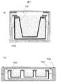
図1、図2を用いて包装容器の基本的な構成要素を説明する。再生組織等の生体試料120の入った培養容器104は、第一包装容器本体部103および蓋部101と、第二包装容器本体部203および蓋部201により、二重に包装される。
第一包装容器本体部103および第二包装容器本体部203に関し、素材と光学特性は、培養容器104と同一のものが好ましい。同一のものを使用することにより、屈折率等の光学特性も同一となり、顕微鏡画像が鮮明となる。それぞれの蓋部については、同一素材を用いても良いし、異なる素材を用いてもよいが、顕微鏡視野を明るくするため、顕微鏡の照明光を透過することは必要である。
また、同一素材を用いることで滅菌処理による無菌化が一元的に可能となる。例えば、培養容器がポリスチレンを素材とする場合、第一および第二包装容器本体部および蓋部も同素材であると、使用時において、事前にγ線照射による滅菌処理を実施し、無菌化することができる。
上記ではポリスチレンを例にしたが、光透過性を有し、生体試料にとって有害とならない素材であれば、適用可能であることは云うまでもない。
The basic components of the packaging container will be described with reference to FIGS. The
Regarding the first packaging container
Further, by using the same material, sterilization by sterilization can be performed in a unified manner. For example, when the culture container is made of polystyrene, if the first and second packaging container main body and the lid are made of the same material, sterilization is performed in advance by γ-ray irradiation before use. be able to.
In the above, polystyrene is taken as an example, but it is needless to say that it is applicable to any material that has optical transparency and is not harmful to a biological sample.
図1(A)は第一包装容器蓋部101を示している。第一包装容器蓋部101は、培養容器104内の培地の漏出を防ぐためにシリコーン、ゴム等の第一弾性部材102を有する構成とすることも可能である。
第一弾性部材102は、γ線照射等による滅菌処理が可能で、有害物質等を発することのない、医療用途の品質のものが好ましい。当該第一弾性部材102は容器素材よりも軟質のものが好ましく、後述する培養容器104を第一包装容器本体部103に圧着させる際、適度な押圧力で培養容器104を第一包装容器本体部の底面部に押しつけることが可能となる。また、第一包装容器蓋部101の内部側面には、第一包装容器本体部103を封止するためのネジ受部110を有している。
FIG. 1A shows the first
The first
図1(B)は第一包装容器本体部103を示している。当該第一包装容器本体部103の底面に培養容器104を保持することになる。また、第一包装容器本体部103の外周側面には、第一包装容器蓋部101のネジ受部110と接合可能なネジ部111を有している。当該ネジ部111とネジ受部110とが接合することで、第一包装容器を封止することが可能となる。
また、本例においては、ネジ部111を設けることで封止する例を示すが、これに限らず、フック材等を用いて両者を封止しても良いし、第一包装容器本体部側面または蓋部内部側面にゴム等の弾性部材を装着して、蓋部材を封止する構成としても良いことは云うまでもない。
FIG. 1B shows the first packaging container
Moreover, in this example, although the example sealed by providing the
図1(C)は生体試料が導入、収納される培養容器104を示している。培養容器104の中には、培地105が入っている。培地の中には生体試料120が入っている。一般的に、培養容器を用いて生体試料を培養する場合は、蓋も併せて使用する。蓋には、培養容器の外側から酸素等の気体が内部へ入り込めるよう、隙間が設けられている。そのため、この培養容器を輸送に用いると、傾いた時に培地が漏出する。これは生物学的汚染の原因となる。
そのため、本方法では、培養容器の蓋を第一包装容器蓋部101に変える。すなわち、第一包装容器本体部103と、第一包装容器本体部103の底面において保持される培養容器104とを第一包装容器蓋部101で封止することで、輸送中の培地の漏出を防止する。
FIG. 1C shows a
Therefore, in this method, the lid of the culture container is changed to the first packaging
第一包装容器本体部103と培養容器104とを封止するためには、培養容器104の側壁と第一包装容器本体部103の側壁部の高さを同一とすることが望ましい。これにより第一包装容器蓋部101のネジ受部110の締め付け力のみで両者を封止することが可能となる。また、前述したように蓋部101に弾性部材102を装着しておくことでさらに漏出の可能性を低減することが可能となる。
またさらに、培養容器104の側壁の高さを同一としなくとも僅かに高い場合には、弾性部材102の弾性力により高さの違いを吸収することが可能となるとともに、第一包装容器本体103の底面とのさらに強固な圧接が可能となる。たとえ培養容器104の側壁が第一包装容器本体部103よりも低くとも、蓋部101の当該側壁に対応する位置に凸部を設け、弾性部材102の形状をそれに合わせた形状にしておけば、同様の効果を得られることは云うまでもない。
In order to seal the first packaging container
Furthermore, when the height of the side wall of the
ここで、上記では培養容器104の蓋と、第一包装容器本体部103の蓋を共通の蓋部材101とすることを示したが、これに限るものではなく、培養容器104を密封可能な蓋部材を別途設けることも可能であることは云うまでもない。つまり、培養容器を封止する培養容器蓋部を設け、第一包装容器本体部と第一包装容器蓋部から成る第一包装容器に収納する。この場合、第一包装容器蓋部101と本体部103とにおけるネジ部110、111の締め付け力により、培養容器104が第一包装容器本体部の底面部において保持される構造であれば良い。この場合、後述する第二包装容器は無くても良い。輸送先の清浄度の段階に応じて多重包装とすることも可能である。
Here, in the above description, the lid of the
図1(D)は、第一包装容器本体部103へ、培養容器本体部104を収容した状態を示している。図1(E)は、図1(D)の状態からさらに、第一包装容器蓋部101を、第一包装容器本体部103と一体化した状態を示している。これらは、ネジ構造により一体化が可能である。この時、第一包装容器蓋部101の締め付け力により培養容器104へ、第一弾性部材102が圧着されている。これにより、培養容器104を傾けても、内部の培地105は外部に漏出しない。また、培養容器104と第一包装容器本体部103も圧着されている。よって、両者の間に空気層は略存在しないことになる。
FIG. 1D shows a state in which the culture container
図2(A)は第二包装容器蓋部201を示している。第二包装容器蓋部201は、培地等の漏出を強固に防ぐためにシリコーン、ゴム等の第二弾性部材202を有する構成としても良い。
第二弾性部材202は、γ線照射等による滅菌処理が可能で、有害物質等を発することのない、医療用途の品質のものが好ましい。当該第二弾性部材202は容器素材よりも軟質のものが好ましく、後述する第一包装容器本体103の底部外表面を第二包装容器本体部203の底面に圧着させる際に、適度な押圧力で第二包装容器本体部203底面部に押しつけることが可能となる。ここで第二弾性部材202は、滅菌処理の一元化等が可能となる様に、第一弾性部材102と同一の素材を選択することが望ましい。また、第二包装容器蓋部201の内部側面には第二包装容器本体部203を封止するためのネジ受部210を有している。
FIG. 2A shows the second
The second
図2(B)は第二包装容器本体部203を示している。当該第二包装容器本体部203の底面に第一包装容器本体部103を保持することになる。具体的には第一包装容器本体部103の底部外表面が第二包装容器本体部203の底面において保持されることになる。
また、第二包装容器本体部203の外周側面には、第二包装容器蓋部201のネジ受部210と接合可能なネジ部211を有している。当該ネジ部211とネジ受部210とが接合することで、第二包装容器を封止することが可能となる。また、本例においては、ネジ部211を設けることで封止する例を示すが、これに限らず、フック材等を用いて両者を封止しても良いし、第二包装容器本体部側面または蓋部内部側面にゴム等の弾性部材を装着して、蓋部材とを封止する構成としても良いことは云うまでもない。
FIG. 2B shows the second packaging container
In addition, a
図2(C)は、第二包装容器本体部203へ、図1(E)で示した第一包装容器100で包装した培養容器104を、収容した状態を示している。ここで、第二包装容器本体部203の側壁に備えられるネジ部211は、第一包装容器100の持つ全幅よりも外側に設置されている。これにより、第二包装容器蓋部201のネジ受部210が接合することで、その締め付け力により第一包装容器100を押しつけることが可能となり、その結果、第二包装容器本体部203底部に対して第一包装容器本体部103が圧接され保持されることとなる。
FIG. 2C shows a state in which the
図2(D)は、図2(C)の状態からさらに、第二包装容器蓋部201で一体化した状態を示している。第二包装容器蓋部201および本体部203は、ネジ構造により一体化が可能である。この時、第二包装容器蓋部201と第一包装容器蓋部101は圧着されている。第二包装容器本体部203と第一包装容器本体部103も同様に圧着されている。これにより、それぞれの境界において、空気層は略存在しない。
FIG. 2D shows a state in which the second
尚、培養容器104には、底部外表面に高さ0.1〜数mm程度のリブ(凸部)が設けられている場合がある。これは、培養容器を顕微鏡のステージ等に置いた時に、培養容器の底部の全面が接着しないようにするためのものである。リブを有する培養容器については、第一包装容器本体において、リブが入るための窪み(凹部)を設けても良い。これにより、培養容器の底面と、第一包装容器本体はより密接に接することが可能となる。また、包装容器により包装した状態で輸送する際、窪みの中にリブが入っていることで、横方向へ培養容器が移動しないという効果も期待できる。
The
図3は、図1及び図2に示す実施例の変形例を示すものである。第一包装容器本体部103の内部側面及び培養容器104の外表面にネジ構造330を、また、第二包装容器本体部203の内部側面及び第一包装容器本体部103の外表面にネジ構造340を夫々設けたものである。当該構造によれば、ネジ構造により夫々の保持位置が一意に決まるという効果がある。さらに、ネジ構造による締め付け力により、保持すべき底面と、底部外表面との圧接が容易となる。
FIG. 3 shows a modification of the embodiment shown in FIGS. A
ここで、図11を用いて、包装容器により培養容器を包装した状態で、培養容器内の生体試料を観察する時の光路の概略について説明する。図11は、包装容器に収容された培養容器の内部にある、生体試料を顕微鏡により観察する時の状態を示している。顕微鏡は、医療機関や細胞処理施設等で一般に使われる倒立式位相差顕微鏡の例である。 Here, the outline of the optical path when observing the biological sample in the culture container in a state where the culture container is packaged by the packaging container will be described with reference to FIG. FIG. 11 shows a state when a biological sample inside the culture container accommodated in the packaging container is observed with a microscope. The microscope is an example of an inverted phase contrast microscope generally used in medical institutions, cell processing facilities, and the like.
包装容器により包装された培養容器1101の内部には、培地1102と生体試料1103が入っている。また、包装状態の培養容器は、倒立式位相差顕微鏡のステージ1104の上に置かれている。対物レンズ1105がステージ1104の下部に設置され、視野を明るくするための照明1106がステージの上部に設置されている。生体試料を観察する際の光路1107を例として示している。観察手法は従来の観察方法と同様のため、説明を省略する。
A
ここで、生体試料を上述の構造において観察するための条件について、以下図12を用いて説明する。
図12(A)は、培養容器1208、包装容器の内容器1209、外容器1210に対し、拡大した模式図であり、本発明において、包装容器等の圧着が十分になされ、各面が十分に平滑である場合の光路1211を示している。この場合、光路は各容器間の接着面で乱されることがないため、結果として、鮮明な顕微鏡画像を得ることができる。
一方、図12(B)は、本発明において、包装容器および培養容器の接続、保持が不十分で、各面が十分に平滑ではなく、結果として光路に対し影響を及ぼしている場合の例を示している。培養容器1212、包装容器の内容器1213、外容器1214の境界面において、接続の不足と、各面の非平滑性を原因として生じる空隙1215により、光路1216に対し、屈折や散乱および乱反射1217が生じることになる。これにより、鮮明な顕微鏡画像を得ることはできなくなる。
Here, conditions for observing the biological sample in the above-described structure will be described below with reference to FIG.
FIG. 12A is an enlarged schematic view of the
On the other hand, FIG. 12 (B) shows an example in the present invention where the connection and holding of the packaging container and the culture container are insufficient and each surface is not sufficiently smooth, resulting in an influence on the optical path. Show. At the boundary surfaces of the
上記検討によれば、各層間において、出来る限り空気層等の屈折率の異なる領域を排することが必要であり、好ましくは各層表面の粗さを滑らかにし、さらに各容器を圧着することが必要であると分かる。
またさらに、一般的な倒立式位相差顕微鏡を用いる場合、細胞の観察には100倍程度が必要である。この場合、第一包装容器本体部と培養容器の間における圧着と合わせ、かつ、全体の厚さの合計値を3mm以下にすることが望ましい。40倍程度のより低倍率で観察する場合は、全体の厚さの合計値はそれよりも大きくなっても良い。これにより、倒立式位相差顕微鏡により外部から観察する際、生体試料へ焦点を合せることが可能となる。
すなわち、鮮明な顕微鏡画像を取得することが可能となる。
上記条件は一例であるが、光学的手段により非侵襲的評価を鮮明な画像により行う場合には上記要件とすることが望ましい。
According to the above examination, it is necessary to eliminate regions with different refractive indexes such as air layers as much as possible between each layer, preferably smooth the surface roughness of each layer and further crimp each container. I understand that.
Furthermore, when a general inverted phase contrast microscope is used, the cell observation requires about 100 times. In this case, it is desirable to match with the pressure bonding between the first packaging container main body and the culture container, and the total value of the total thickness is 3 mm or less. When observing at a lower magnification of about 40 times, the total value of the total thickness may be larger than that. This makes it possible to focus on a biological sample when observing from the outside with an inverted phase contrast microscope.
That is, a clear microscope image can be acquired.
The above condition is an example, but it is desirable to satisfy the above requirement when noninvasive evaluation is performed with a clear image by optical means.
次に、本実施例の包装容器を維持した状態での、上記の光学的観察の他の評価例を、図4を用いて以下説明する。 Next, another evaluation example of the above optical observation in a state where the packaging container of the present example is maintained will be described below with reference to FIG.
図4は、培養容器を、第一包装容器および第二包装容器に収容したまま、生体試料403の厚さを計測する方法を示している。図4(A)は培養容器402と第一包装容器本体部401の断面拡大図である。図4(A)のように、第一包装容器本体部401と培養容器402の間に、窪み404を設置する。すなわち、第一包装容器本体部401の底面に凹部を設けるものである。尚、培養容器402の培養表面側には、細胞403が層状の厚みを持った状態で接着している場合を示している。
FIG. 4 shows a method for measuring the thickness of the
図4(B)は、窪み404の鉛直方向および水平方向の断面図を示したものである。第一包装容器本体部の表面405に、柱状の窪みを設置する。第一包装容器本体部の表面405と、窪みの底面406は、窪みの側面部407を介している。側面部の高さは50〜500μm程度とし、その値は正確に把握できるものとする。第一包装容器本体部の表面405には、5μm程度の深さで任意の文字408を刻印する。本図では「A」という文字の例を示した。窪みの底面406には、文字408と異なる種類の文字409を刻印する。本図では「B」という文字の例を示した。図4(C)は、第一包装容器本体部401と培養容器402の間に、目盛り410を設置した図である。顕微鏡視野内で、生体試料の縦および横の大きさを把握する目安として用いる。
FIG. 4B is a cross-sectional view of the
包装容器に培養容器を収容した状態で、倒立式位相差顕微鏡の顕微鏡視野において、文字408に対し焦点を合わせ、その時の、倒立式位相差顕微鏡の焦点を作動させるダイヤルの角度あるいは位置を記録する。続いて、文字409に対し焦点を合わせ、その時のダイヤルの角度あるいは位置を記録する。そして、前述の2文字に焦点を合わせた時のダイヤルの角度あるいは位置の差分値を算出する。それを用いて、ダイヤルの差分値と、窪みの深さの値より、回転させるダイヤルの角度あるいは位置の変化の単位量が、培養容器の鉛直方向の高さの変異において相当する量を算出する。
続いて文字408に対し焦点を合わせ、その時のダイヤルの角度あるいは位置を記録する。次に、培養容器の培養表面上にある生体試料の上端へ焦点を合わせ、ダイヤルの角度あるいは位置を記録する。それぞれの焦点を合わせた時のダイヤルの角度あるいは位置の差分値を算出する。
事前に求めた、焦点を合わせるために回転させるダイヤルの角度あるいは位置の変化の単位量が、培養容器の鉛直方向の高さの変異において相当する量を用いて、培養容器の培養表面と、再生組織等の生体試料の上端との距離を算出する。そして、そこから培養容器の厚さの値を引く。得られた値が、生体試料の厚さとなる。
With the culture container in the packaging container, focus on the
Subsequently, the
The unit amount of the change in the angle or position of the dial that is rotated to adjust the focus, which is obtained in advance, is equivalent to the variation in the vertical height of the culture vessel, and the culture surface of the culture vessel is regenerated. The distance from the upper end of a biological sample such as tissue is calculated. And the value of the thickness of a culture container is subtracted from there. The obtained value is the thickness of the biological sample.
同等の窪みと文字の刻印を、第一包装容器本体部の任意の位置に設置し、それぞれの場所で生体試料の厚さを求め、その平均値および分散を算出することにより、再生組織等の生体試料の厚さのムラの評価も可能である。以上より、細胞の清浄性を維持したまま、非侵襲的に生体試料の厚さを評価することが可能となる。包装された状態で、外部から顕微鏡による光学的手段により細胞を観察する方法は、細胞に直接触れてはいないため、非侵襲的な評価方法である。細胞の状態に影響は及ぼさない。
尚、上述の例において、文字をマークとして使用する例を示したが、文字に限らず、記号、図記号、等マークとなり得るものであれば、限定されるものではないことは云うまでもない。
Equivalent indentations and inscriptions of letters are placed at arbitrary positions on the first packaging container body, and the thickness of the biological sample is obtained at each location, and the average value and variance are calculated, so that It is also possible to evaluate the unevenness of the thickness of the biological sample. As described above, the thickness of the biological sample can be evaluated noninvasively while maintaining the cleanliness of the cells. The method of observing cells from the outside by optical means using a microscope in a packaged state is a noninvasive evaluation method because the cells are not touched directly. It does not affect the state of the cells.
In the above-described example, an example is shown in which characters are used as marks. However, the present invention is not limited to the characters as long as they can be marks, symbols, and the like. .
また、上記においては倒立式位相差顕微鏡を例に説明したが、これに限るものではなく、例えば反射型の光学顕微鏡を用いることも可能である。
この場合、ステージ上に容器を載置し、容器の裏面側(下方)から光源により光を照射する。ここで反射される光を容器裏面側に配置され(好ましくは光源と対向配置され)た撮像装置で撮像することになる。
このとき、培養容器に蓋が有る場合は培養容器の蓋、包装容器と共用する場合には包装容器の蓋部の光が侵入する側の面を、ステンレス、鏡等の反射部材で構成すると、明るさが担保され、画像が鮮明となる。
In the above description, the inverted phase contrast microscope has been described as an example. However, the present invention is not limited to this. For example, a reflective optical microscope can be used.
In this case, the container is placed on the stage, and light is irradiated from the back surface side (downward) of the container by the light source. The light reflected here is imaged by an imaging device arranged on the back side of the container (preferably arranged opposite to the light source).
At this time, if the culture container has a lid, if the lid of the culture container, in the case of sharing with the packaging container, the surface on the side where the light enters the lid of the packaging container is made of a reflective member such as stainless steel, a mirror, Brightness is ensured and the image becomes clear.
図5は、第一包装容器の一部に、培地を満たすための栓501を有した構成図を示している。栓501を使い、培養容器の中に培地を満たす。栓には、弾性のある医療用ゴム等を用いるのが好ましい。培地を満たす作業では、シリンジを2つ用意する。片方のシリンジには、十分量の培地を入れる。これを、片方の栓に刺し入れる。その状態で、残りの栓に、空のシリンジを刺し入れる。培地の入ったシリンジより培地を注入し、同時に、空のシリンジより余った空気を回収する。培地が培養容器の内部に満たされた後、両方のシリンジを抜く。包装容器を開封して生体試料を取り出す時は、培地が満たされた状態のままでは培地がこぼれてしまうため、逆の手順を実施して培養容器内に空気を注入してから開封する。
このような構造を採用することで、培地を満たすことが必要な生体試料についても容易にハンドリングが可能となる。
FIG. 5 shows a configuration diagram in which a
By adopting such a structure, it is possible to easily handle a biological sample that needs to be filled with a culture medium.
図6は、第一および第二包装容器蓋部の一部を、気体透過性膜601、602にしたものである。これにより、輸送中も外気から酸素等の気体を内部に取り込むことが可能となる。生体試料は様々な細胞により構成されるが、例えば心筋細胞は酸素要求性が大きい。
そのような細胞を輸送する場合には、気体透過膜より、包装容器の外部から酸素を供給することで、輸送後の生体試料の状態は良くなる。また、外部より二酸化炭素を供給し、培地のpHを制御することも可能である。
このように気体透過膜を蓋部材に採用することで、酸素要求性の高い細胞等の生体試料についても清浄性を維持しつつ、非侵襲的に観察が可能な包装容器を提供することが可能となる。
FIG. 6 shows gas
When transporting such cells, the state of the biological sample after transport is improved by supplying oxygen from the outside of the packaging container through the gas permeable membrane. It is also possible to control the pH of the medium by supplying carbon dioxide from the outside.
By using the gas permeable membrane as the lid member in this way, it is possible to provide a packaging container that can be observed noninvasively while maintaining cleanliness even for biological samples such as cells with high oxygen demand. It becomes.
図7は、他の種類の培養容器を包装した場合の例である。図7(A)はインサート型培養容器701に対する例である。インサート型培養容器を用いて細胞を培養する場合、一般的には、6ウェルプレート等の中にインサート型培養容器を入れて二層培養を行う。インサート型培養容器の輸送では、これを収容するインサート型培養容器用容器702を用意し、その中に入れた状態で、第一および第二包装容器内へ収容する。図7(B)は6ウェルプレート703に対して包装した例である。円柱状の培養容器やインサート型培養容器用容器は、水平方向の断面が円形であるため、第一および第二包装容器の蓋を回転させることにより一体化が可能である。一方、6ウェルプレートは直方体形状である。よって、本図では、ネジ704、705を用いて、第一および第二包装容器の4隅を固定することで、一体化させるものを示している。他の方法として、勘合、ピン止め、バネ材等により、複数箇所を固定することにより保持できることは言うまでもない。
FIG. 7 shows an example of packaging other types of culture containers. FIG. 7A shows an example for the insert
図8は、前述のように包装した培養容器に対し、蓄熱材を用いて内部温度を一定に維持することのできる細胞輸送容器を用いて、輸送を実施する場合の構成の例を示したものである。細胞輸送容器は、内部の構成部品を収容する容器本体801と、容器蓋802の内側に、断熱材803を配置する。その内側に、蓄熱材が封入された蓄熱材ボックス804を配置する。蓄熱材の種類は、一定の融点を有する純物質、あるいは、熱容量が大きく融点の温度変化が小さい(例えば±1℃以下)物質を用いるものとする。例として炭化水素が挙げられる。例えばC20H42を使用する場合、融点は36.4℃である。Cの数の異なる炭水素は、異なる融点を有する。よって、適切に炭化水素の種類を決定することにより、細胞輸送容器が一定に維持する温度の値を変えることが可能である。蓄熱材ボックス804に挟まれた場所に、培養容器収容部805の中に配置された培養容器806と、輸送中の温度等を評価するためのモニタリング装置807を配置する。これにより、輸送中の温度を、輸送後に確認することが可能となる。
FIG. 8 shows an example of the configuration in the case where the transport is carried out using the cell transport container capable of maintaining the internal temperature constant by using the heat storage material with respect to the culture container packaged as described above. It is. In the cell transport container, a
ここで図9、10を用いて、本実施例に示す包装容器をCPC等の細胞処理施設から医療機関等に運ぶ様子を模式的に示す。図9は、細胞処理施設から培養容器を運び出す時の過程を、図10は、輸送後、医療機関へ培養容器を運び込む時の過程について示している。 9 and 10 schematically show how the packaging container shown in the present embodiment is transported from a cell processing facility such as CPC to a medical institution or the like. FIG. 9 shows a process when the culture container is carried out from the cell processing facility, and FIG. 10 shows a process when the culture container is carried into the medical institution after transportation.
図9に示すように、細胞は、CPC等の細胞処理施設内の培養エリアで培養する。この部屋の清浄度は、グレードAに次いで高い、グレードBと一般に設定される。培養時の培養容器は恒温槽に入っており、必要に応じて取り出し、安全キャビネット内にて培地交換等の作業を実施する。安全キャビネット内の清浄度は、グレードAと一般に設定される。培養が終わり、医療機関へ出荷することになったサンプルは、安全キャビネット内で包装容器により二重に包装を行う。包装容器は事前に滅菌処理を施し無菌状態である。これにより、包装し終えた時点で、培養容器内および包装容器の内外は、グレードAと同等の清浄性を有する。
この状態で、細胞処理施設の外へ、包装した培養容器を運び出す。この時、必要に応じ、温度を一定に維持する輸送容器へ収容する。培養エリアから細胞処理施設の外へ運び出すにつれ、輸送環境中の清浄性は低下する。よって、最終的に、一番外側に位置する包装容器の外部には菌等の生物や粒子が付着する。一方、一番外側に位置する包装容器の内部、培養容器等は、未開封状態であるため、グレードAの清浄性を維持する。
As shown in FIG. 9, the cells are cultured in a culture area in a cell processing facility such as CPC. The cleanliness of this room is generally set to Grade B, which is the second highest after Grade A. The culture container at the time of culturing is in a thermostatic bath, and is taken out as necessary, and the medium is exchanged in the safety cabinet. The cleanliness in the safety cabinet is generally set to grade A. Samples that have been cultured and shipped to a medical institution are double packed in a safety cabinet using a packaging container. The packaging container is sterilized in advance and is in a sterile state. Thereby, when packaging is completed, the inside and outside of the culture container and the inside and outside of the packaging container have the same cleanliness as Grade A.
In this state, the packaged culture container is carried out of the cell processing facility. At this time, if necessary, it is accommodated in a transport container that maintains a constant temperature. As it is transported out of the cell processing facility from the culture area, the cleanliness in the transport environment decreases. Therefore, finally, organisms and particles such as bacteria adhere to the outside of the outermost packaging container. On the other hand, the inside of the outermost packaging container, the culture container, and the like are in an unopened state, and therefore maintain Grade A cleanliness.
図10は、医療機関に運び込む時の過程を示したものである。医療機関に到着後、まず、生体試料の状態を評価する。当該評価の結果を受けて、治療に用いることが可能であることを確認する。
この時、治療に用いる予定のサンプルについては、非侵襲的な評価方法でなければならない。侵襲的な評価方法では、生体試料の質が変化するからである。また、全数検査を実施できることが望ましい。培養した生体試料は、同じ細胞ソースを用い、同じ製造過程を経ているため、培養後の質も同じと考えられるが、細胞はわずかな環境の変化により容易に質が変わりうるからである。
よって、本実施例では、培養容器を包装した状態のまま、上述した非侵襲的な評価方法である顕微鏡観察を実施する。そして、細胞の形状、接着状態、生体試料の厚さの評価を行う。必要に応じ、包装容器に組み込んだ窪みや目盛りにより、定量評価を行う。
FIG. 10 shows a process when transporting to a medical institution. After arriving at a medical institution, first, the state of the biological sample is evaluated. Based on the result of the evaluation, it is confirmed that it can be used for treatment.
At this time, the sample to be used for treatment must be a non-invasive evaluation method. This is because the quality of a biological sample changes in an invasive evaluation method. In addition, it is desirable that 100% inspection can be performed. Since cultured biological samples use the same cell source and have undergone the same manufacturing process, the quality after culturing is considered to be the same, but the quality of cells can be easily changed by slight environmental changes.
Therefore, in the present example, the microscopic observation, which is the above-described noninvasive evaluation method, is carried out with the culture container being packaged. Then, the cell shape, the adhesion state, and the thickness of the biological sample are evaluated. Quantitative evaluation is performed as necessary using the dents and scales incorporated in the packaging container.
従来、研究機関等で行われていた細胞の検査等では、伸縮性シール等により密封された培養容器に対し、顕微鏡観察を行うのみであった。定量的な評価を行うには、生体試料に対し、侵襲的な評価(例えば組織学的評価、細胞生存率評価)を実施する必要があった。これを実施すると、生体試料を治療に用いることができなくなる。一方、非侵襲的な評価方法である顕微鏡観察のみを実施する場合、細胞の形状、接着状態を観察することは可能だが、それは定性的な評価であった。また、生体試料の良否の判定は、作業者の知識と経験に基づいていた。よって、作業者間で判定の基準を一定にすることは困難であった。それに対し、本発明により、より正確な定量的な評価が可能となる。 Conventionally, in cell inspections conducted at research institutions, etc., only a microscopic observation was performed on a culture vessel sealed with a stretchable seal or the like. In order to perform quantitative evaluation, it was necessary to perform invasive evaluation (for example, histological evaluation, cell viability evaluation) on a biological sample. When this is done, the biological sample cannot be used for therapy. On the other hand, when only microscopic observation, which is a non-invasive evaluation method, is performed, it is possible to observe the shape and adhesion of cells, but this was a qualitative evaluation. Moreover, the judgment of the quality of the biological sample was based on the knowledge and experience of the operator. Therefore, it has been difficult to make the determination standard constant among workers. On the other hand, the present invention enables more accurate quantitative evaluation.
上記の評価により、治療に用いることができると判断されたなら、治療の準備を開始する。治療を行う患者への準備等を実施する。その後、培養容器を、治療を実施する手術室へ運ぶ。手術室内は清浄であり、清潔野と不潔野が設けられている。手術室に運び込んだ培養容器は、まず、外側をエタノール等により消毒する。そして、不潔野で、包装容器の一番外側に位置する外容器を開封する。この時、外容器の外部が、清浄な内容器の外側に触れないように注意を払う。そして、内容器のみを、取り出す。続いて内容器を開封する。内容器の蓋を取り外し、清潔野の器具のみを取り扱う担当である作業者が、培養容器のみを取り出す。この時、内容器の外部が、清浄な培養容器の外側に触れないように注意を払う。最後に、同じ作業者が、清潔野にて培養容器から生体試料を取り出す。それを治療に用いる。 If it is determined by the above evaluation that it can be used for treatment, preparation for treatment is started. Prepare the patient for treatment. Thereafter, the culture container is transported to the operating room where the treatment is performed. The operating room is clean, with clean and unclean fields. First, disinfect the outside of the culture container brought into the operating room with ethanol or the like. And in unclean field, the outer container located in the outermost side of a packaging container is opened. At this time, care is taken so that the outside of the outer container does not touch the outside of the clean inner container. Then, only the inner container is taken out. Subsequently, the inner container is opened. An operator who is in charge of removing the lid of the inner container and handling only the instruments in the clean field takes out only the culture container. At this time, care should be taken so that the outside of the inner container does not touch the outside of the clean culture container. Finally, the same worker takes out the biological sample from the culture container in the clean field. Use it for treatment.
次に、以上の構成を有する包装容器を用い、細胞を輸送する時の一連の手順について説明する。 Next, a series of procedures for transporting cells using the packaging container having the above configuration will be described.
<ステップS1:事前準備>
細胞を輸送するために必要な事前準備を行う。包装容器は、事前にオートクレーブバッグ等により包装し、その状態で滅菌処理を施し無菌化する。滅菌処理の方法は、オートクレーブ処理、エチレンオキシダイドガス処理、γ線照射等とし、滅菌処理を施すことにより、包装容器の性質を変化させない方法を選択する。例えば素材がポリスチレンであれば、γ線照射処理を採用する。
蓄熱材を封入した蓄熱材ボックスは、蓄熱材が炭化水素C20H42である場合、蓄熱材ボックスの素材を金属または耐熱性のポリカーボネイトとし、炭化水素C20H42を完全に密封した状態で封入する。炭化水素C20H42の融点は344℃であるため、オートクレーブ処理(120℃)を施しても気化せず、蓄熱材ボックスの温度維持性能に関する影響はない。滅菌後は、蓄熱材に熱を蓄えるため、包装した状態で恒温槽の中に入れ、温度が安定するまで静置する。例として、融点が36.4℃である炭化水素C20H42の場合、輸送する外界の温度の大半が36.4℃以下である場合、恒温槽の温度は37℃とする。輸送中、外界の温度の方がC20H42の融点よりも温度が低いため、熱は細胞輸送容器の中から外へ出ていくからである。逆に、輸送する外界の温度の大半が36.4℃以上である場合、恒温槽の温度は36℃とする。輸送中、外界の温度の方がC20H42の融点よりも温度が高いため、熱は細胞輸送容器の中へ外から入り込むためである。
各種滅菌に対する耐性を有していないモニタリング機器等については、エタノール消毒を施すこととする。
<Step S1: Preparation>
Make the necessary preparations to transport the cells. The packaging container is packaged in advance with an autoclave bag or the like, and sterilized in that state to be sterilized. The method of sterilization is autoclaving, ethylene oxide gas treatment, γ-ray irradiation, etc., and a method that does not change the properties of the packaging container by performing sterilization is selected. For example, if the material is polystyrene, γ-ray irradiation treatment is adopted.
When the heat storage material is hydrocarbon C20H42, the heat storage material box in which the heat storage material is sealed is sealed with the hydrocarbon C20H42 being completely sealed with the material of the heat storage material box being a metal or heat-resistant polycarbonate. Since the hydrocarbon C20H42 has a melting point of 344 ° C, it does not vaporize even if it is autoclaved (120 ° C), and there is no effect on the temperature maintenance performance of the heat storage material box. After sterilization, in order to store heat in the heat storage material, put it in a thermostatic bath in a packaged state and leave it until the temperature is stabilized. For example, in the case of hydrocarbon C20H42 having a melting point of 36.4 ° C, the temperature of the thermostatic chamber is 37 ° C when most of the outside temperature to be transported is 36.4 ° C or less. This is because, during transportation, the temperature of the outside world is lower than the melting point of C20H42, so that heat goes out of the cell transport container. Conversely, if most of the outside temperature to be transported is 36.4 ° C or higher, the temperature of the thermostatic bath shall be 36 ° C. This is because during transportation, the outside temperature is higher than the melting point of C20H42, so heat enters the cell transport container from the outside.
For monitoring devices that are not resistant to various types of sterilization, ethanol disinfection shall be performed.
<ステップS2:細胞処理施設内への運び込み>
滅菌を施した細胞輸送容器と包装容器を、細胞処理施設内の培養エリアへ運び込む。細胞処理施設内の部屋間の移動に際しては、部屋の清浄性の維持と交差汚染防止のため、パスボックスを通過させる必要がある。パスボックスを通過させる時には、それぞれの構成部品に対し、包装の外側からエタノールを噴霧して消毒し、パスボックスの中に入れ、移動する部屋の側の扉から取り出す。
細胞培養エリアに到着後、包装容器以外の機材に関しては、包装を開け、包装の外側に触れないよう無菌的に取り出す。蓄熱材ボックスは、室温下に晒したままでは温度が変化するため、可能ならば同じ部屋に恒温槽を用意しておき、使用するまで恒温槽の中に入れ、温度変化を防ぐことが望ましい。
包装容器は、包装の周囲をエタノール噴霧により消毒し、安全キャビネット内へ入れる。その後、包装の外側に触れないように包装容器を無菌的に取り出す。
モニタリング装置の運び込みについては、事前に外部へエタノール消毒を施すとする。細胞を処理する部屋への機材等の持ち込みは、滅菌処理を施し無菌化することが望ましいが、機械装置に対し滅菌処理を施せないため、一般にエタノール処理のみを施す。
<Step S2: Carrying into cell processing facility>
Carry the sterilized cell transport container and packaging container to the culture area in the cell treatment facility. When moving between rooms in a cell processing facility, it is necessary to pass through a pass box in order to maintain cleanliness of the room and prevent cross contamination. When passing through the pass box, each component is sterilized by spraying ethanol from the outside of the package, placed in the pass box, and removed from the door on the side of the moving room.
After arriving at the cell culture area, the equipment other than the packaging container is opened and removed aseptically so as not to touch the outside of the packaging. Since the temperature of the heat storage material box changes when exposed to room temperature, it is desirable to prepare a thermostatic chamber in the same room if possible and place it in the thermostatic chamber until use to prevent temperature changes.
The packaging container is sterilized by spraying with ethanol around the package and placed in a safety cabinet. Thereafter, the packaging container is removed aseptically so as not to touch the outside of the packaging.
It is assumed that ethanol is disinfected outside in advance for bringing in the monitoring device. It is desirable to carry out sterilization and sterilization when bringing equipment into the room where cells are treated. However, since sterilization cannot be performed on mechanical devices, only ethanol treatment is generally performed.
<ステップS3:培養容器の収容>
恒温槽の中で培養していた培養容器を、安全キャビネット内へ移動する。安全キャビネット内では、必要に応じ、恒温槽の温度と同じ値に設定したヒートブロック等の設備をあらかじめ準備しておく。これを用い、細胞輸送容器への培養容器の収容が終了するまでの間、培養容器の温度を一定に維持する。
安全キャビネット内に移動した培養容器に対し、蓋を取り外し、培養容器の中へ、溢れない程度に培地を満たす。そして第一包装容器で包装する。この時、培地がこぼれないように注意する。また、この操作は、内部が汚染される可能性を低くするため、手早く行う。作業後や待ち時間等では、培養容器の温度の低下を極力避けるため、ヒートブロック等を利用する。続いて、同様の手順により、第二包装容器により、手早く収容を行う。
栓を有する第一包装容器蓋部を用いて培地を完全に満たす場合は、シリンジを2つ用意する。片方のシリンジには、十分量の培地を入れる。これを、片方の栓に刺し入れる。その状態で、残りの栓に、空のシリンジを刺し入れる。培地の入ったシリンジより培地を注入し、同時に、余った空気を空のシリンジより回収する。培地が培養容器の内部に満たされた後、両方のシリンジを抜く。
<Step S3: Accommodation of culture vessel>
The culture vessel that has been cultured in the thermostatic chamber is moved into the safety cabinet. In the safety cabinet, equipment such as a heat block set to the same value as the temperature of the thermostatic bath is prepared in advance if necessary. Using this, the temperature of the culture container is kept constant until the culture container is completely accommodated in the cell transport container.
Remove the lid from the culture container that has moved into the safety cabinet, and fill the culture medium into the culture container to the extent that it does not overflow. And it packs with a 1st packaging container. Be careful not to spill the medium at this time. In addition, this operation is performed quickly to reduce the possibility of contamination inside. In order to avoid a decrease in the temperature of the culture vessel as much as possible after work or during waiting time, a heat block or the like is used. Subsequently, the container is quickly accommodated by the second packaging container by the same procedure.
When the culture medium is completely filled using the first packaging container lid having a stopper, two syringes are prepared. Place a sufficient amount of medium in one syringe. This is inserted into one of the stoppers. In that state, insert an empty syringe into the remaining stopper. The medium is injected from the syringe containing the medium, and at the same time, excess air is collected from the empty syringe. After the culture medium is filled into the culture container, both syringes are removed.
<ステップS4:細胞輸送容器への収容>
事前に用意した培養容器収容部へ、ステップS3で包装した培養容器を収容する。次に、細胞輸送容器の内部へ、蓄熱材ボックス、培養容器を収容した培養容器収容部、モニタリング装置を収容する。モニタリング装置は、収容する前に電源を入れて測定を開始し、輸送中の全行程にわたり、温度を測定する。
<Step S4: Accommodation in cell transport container>
The culture container packaged in step S3 is accommodated in the culture container accommodation part prepared in advance. Next, the heat storage material box, the culture container housing part containing the culture container, and the monitoring device are housed inside the cell transport container. The monitoring device turns on the power and starts the measurement before it is accommodated, and measures the temperature throughout the entire transport process.
<ステップS5:細胞処理施設外への運び出し>
細胞輸送容器を、細胞培養を実施する部屋から、細胞処理施設外へ運び出す。部屋間の
移動に際しては、交差汚染防止のため、エタノール噴霧により消毒してから、パスボック
スを通過させる。
<Step S5: Carrying out of cell processing facility>
The cell transport container is carried out of the cell processing facility from the room where the cell culture is performed. When moving between rooms, in order to prevent cross-contamination, disinfect with ethanol spray and then pass through the pass box.
<ステップS6:細胞輸送容器の輸送>
輸送先の医療機関の場所に応じ、移動手段を選択して細胞輸送容器を輸送する。輸送手段は主に、車両、鉄道、航空機、手運びである。車両、鉄道、航空機で運んでいる最中は、細胞輸送容器が横転しないよう、必要に応じて細胞輸送容器を床面に固定することが望ましい。また、手運びの場合は、細胞輸送容器が極力揺れないよう、輸送作業者が注意を払う。
培養容器内の培地は、培養容器内に満たした状態となっている。輸送中に培養容器が傾いた場合に生じる細胞への影響は、細胞が気相の中に入り乾くこと、細胞が気相と液相の間に生じた表面張力による作用を受けること、液相の中に対流が生じて細胞にシアストレスの生じることが想定される。これらは、培養容器内の気相部分が小さいほど、影響も小さくなる。本発明による輸送方法では、培地を培養容器内に極力満たした状態となっているため、輸送中の気相の影響は、あまり大きくないと考えられる。また、輸送中の傾きにより、生体試料にかかる重力の方向は、細胞処理施設内での培養時と比べ、変わる。この影響については、細胞輸送容器が直立している限りは、細胞処理施設内での培養時と同じ重力の方向である。また、細胞輸送容器が直立していない状態を長時間とることはない。
よって、重力による影響は軽微と考えられる。
<Step S6: Transport of Cell Transport Container>
Depending on the location of the destination medical institution, the transport means is selected to transport the cell transport container. The transportation means are mainly vehicles, railroads, airplanes, and hand-carried. It is desirable to fix the cell transport container to the floor as necessary so that the cell transport container does not roll over while being transported by vehicle, railroad, or aircraft. When carrying by hand, the transport operator should pay attention so that the cell transport container does not shake as much as possible.
The culture medium in the culture container is in a state filled in the culture container. The effects on the cells that occur when the culture vessel is tilted during transport are that the cells enter the gas phase and dry, that the cells are affected by the surface tension created between the gas phase and the liquid phase, the liquid phase It is assumed that convection occurs in the cells and shear stress occurs in the cells. These are less affected as the gas phase portion in the culture vessel is smaller. In the transport method according to the present invention, the culture medium is filled in the culture vessel as much as possible, so that the influence of the gas phase during transport is not so great. In addition, the direction of gravity applied to the biological sample changes due to the inclination during transportation compared to the culture in the cell processing facility. As for this influence, as long as the cell transport container is upright, the direction of gravity is the same as that during culture in the cell treatment facility. Moreover, the state where the cell transport container is not upright is not taken for a long time.
Therefore, the effect of gravity is considered minor.
<ステップS7:輸送先における受入検査>
輸送先である医療機関等への到着後は、まず、輸送中の生体試料の周辺温度の確認を行う。モニタリング装置のデータをPC等へ移動させ、評価を行う。続いて、輸送した生体試料の状態を確認する。検査の方法は様々にあるが、治療に用いるサンプルに対しては、非侵襲的な検査である必要がある。すなわち、生体試料に対し、直接的、もしくは培地等を介して、接触することのない方法である。本発明の包装容器を用いる場合は、非侵襲的な検査方法として、倒立式位相差顕微鏡による細胞形態、細胞接着等の評価、生体試料の厚さの計測を行う。これは、全サンプルに対して評価を実施することが可能である。また、検査を行う時は、細胞輸送容器から培養容器を一旦取り出し、顕微鏡にて素早く評価を実施し、速やかに細胞輸送容器へ再び収容する。これにより、検査後も、培養容器は同じ温度下に保管されることになる。尚、治療に用いないサンプルについては、侵襲的な検査により、より詳細に調べてもよい。その場合、生体試料に対して各種処置を行い、細胞数、細胞生存率、組織構造、特定タンパク質の発現状況等を調べることが可能である。
評価の結果、輸送したサンプルが治療に適することが確認できたならば、治療の準備を開始する。治療の準備に1日程度を要することはありうる。加えて、全ての医療機関に、恒温槽等の設備があるとは限らない。その場合は、医療機関に到着後も、治療を開始する時まで、細胞輸送容器の中に培養容器を収容したままにし、温度と清浄性を維持する。
<Step S7: Acceptance inspection at the destination>
After arriving at a medical institution or the like that is the transport destination, first, the ambient temperature of the biological sample being transported is checked. Move the monitoring device data to a PC, etc. for evaluation. Subsequently, the state of the transported biological sample is confirmed. There are various examination methods, but the sample used for treatment needs to be a non-invasive examination. That is, it is a method that does not contact a biological sample directly or via a medium or the like. In the case of using the packaging container of the present invention, as a noninvasive inspection method, evaluation of cell morphology, cell adhesion, etc., and measurement of the thickness of a biological sample are performed using an inverted phase contrast microscope. This can be evaluated for all samples. Moreover, when inspecting, the culture container is once taken out from the cell transport container, evaluated quickly with a microscope, and promptly stored in the cell transport container. Thereby, the culture container is stored at the same temperature even after the inspection. In addition, about the sample which is not used for a treatment, you may investigate in detail by an invasive test | inspection. In that case, various treatments can be performed on the biological sample, and the number of cells, cell survival rate, tissue structure, expression state of the specific protein, and the like can be examined.
If the evaluation confirms that the transported sample is suitable for treatment, preparation for treatment begins. It can take up to a day to prepare for treatment. In addition, not all medical institutions have facilities such as a thermostatic bath. In that case, after arriving at the medical institution, the culture container is kept in the cell transport container until the start of the treatment, and the temperature and cleanliness are maintained.
<ステップS8:治療>
治療の準備が整ったら、治療を行う部屋(以下手術室とする)へ細胞輸送容器を移動させる。手術室へ着いたら、包装容器に包装された培養容器を取り出す。必要に応じ、この状態で、手術室に設置された恒温槽へ培養容器を入れ、所定の温度下に維持する。例えば、培養容器が温度応答性培養表面を有している場合、治療を行う直前に、低温処理(例として20℃下に30分晒す)を施し、温度応答性培養表面に接着していた生体試料を剥離させる。
続いて、生体試料を培養容器より取り出す。包装容器の外側は、日常空間を通過しているため、菌等の生物や粒子が付着している可能性が高い。よって、包装容器の内部が清浄性を維持するよう、段階的に開封する。まず、手術室における不潔野の作業者が、エタノールあるいはポビドンヨード等の消毒薬により、包装容器の外側を清拭する。そして、包装容器の一番外側である第二包装容器を持ち、その蓋を取り去る。続いて、別の作業者が、内側の第一包装容器を取り出す。この時、第一包装容器と、第二包装容器の外側が、接しないようにする。この状態で、第一包装容器蓋部が栓を有している場合は、満たされた培地の一部を吸引する。培地が満たされた状態のまま開封すると、培地がこぼれてしまう可能性が高いからである。培地を満たした時のS3と逆の手順を実施する。
続いて、第一包装容器を持ち、その蓋を取り去る。そして清潔野の作業者が、無菌的に、培養容器を取り出す。この時、培養容器と、第一包装容器の外側が、接しないように無菌的に取り出す。最後に、培養容器から再生組織等を含む生体試料をピンセット等で取り出す。これを治療に用いる。以上のように、包装を段階的に開封することで、培養容器内の清浄性を確実に維持することが可能となる。
<Step S8: Treatment>
When the preparation for treatment is completed, the cell transport container is moved to a room for treatment (hereinafter referred to as an operating room). When you arrive at the operating room, take out the culture container packaged in the packaging container. If necessary, in this state, the culture vessel is placed in a thermostatic chamber installed in the operating room and maintained at a predetermined temperature. For example, if the culture vessel has a temperature-responsive culture surface, a living body that has been subjected to a low-temperature treatment (eg, exposed to 20 ° C. for 30 minutes) and adhered to the temperature-responsive culture surface immediately prior to treatment. The sample is peeled off.
Subsequently, the biological sample is taken out from the culture container. Since the outside of the packaging container passes through daily space, there is a high possibility that organisms and particles such as bacteria are attached. Therefore, it opens in steps so that the inside of a packaging container may maintain cleanliness. First, an unclean field operator in the operating room wipes the outside of the packaging container with a disinfectant such as ethanol or povidone iodine. Then, hold the second packaging container, which is the outermost side of the packaging container, and remove the lid. Subsequently, another worker takes out the inner first packaging container. At this time, the first packaging container and the outer side of the second packaging container are not in contact with each other. In this state, when the first packaging container lid has a stopper, a part of the filled medium is sucked. This is because if the medium is opened while the medium is filled, the medium is likely to spill. The reverse procedure to S3 when the medium is filled is performed.
Subsequently, the first packaging container is held and the lid is removed. Then, an operator in the clean field removes the culture container aseptically. At this time, the culture container is removed aseptically so that the outside of the first packaging container does not contact. Finally, a biological sample containing regenerated tissue or the like is taken out from the culture container with tweezers or the like. This is used for treatment. As described above, it is possible to reliably maintain the cleanliness in the culture container by opening the package in stages.
以上の実施例においては、主に培養容器を二重包装する例について示したが、これに限ることは無く、清浄レベルが細分化された施設等への運び込みに対応するために、さらなる多重包装形態を採用することも可能である。また、培養容器を密封可能な蓋を設けることで、少なくとも第一包装容器と培養容器とが底部で保持され非侵襲的に観察可能な構造であれば、少なくとも二段階の清浄レベルを有する空間において対処が可能となる。 In the above embodiment, an example in which the culture container is mainly double-wrapped has been shown. However, the present invention is not limited to this, and in order to cope with transportation to a facility or the like where the cleaning level is subdivided, further multiple packaging is possible. It is also possible to adopt a form. In addition, by providing a lid that can seal the culture container, if at least the first packaging container and the culture container are held at the bottom and can be observed noninvasively, in a space having at least two levels of cleanliness. It becomes possible to deal with it.
以上の実施例においては生体試料を主に記載したが、同様に清浄性が求められる、半導体チップ等の精密部品が導入された試料容器にも本発明の包装容器を用いることが可能であることは、言うまでもない。 Although the biological sample is mainly described in the above embodiments, the packaging container of the present invention can also be used for a sample container into which a precision component such as a semiconductor chip is similarly required. Needless to say.
100・・・第一包装容器
101・・・第一包装容器蓋部
102・・・第一弾性部材
103・・・第一包装容器本体部
104・・・培養容器
105・・・培地
110・・・ネジ受部
111・・・ネジ部
120・・・生体試料
201・・・第二包装容器蓋部
202・・・第二弾性部材
203・・・第二包装容器本体部
210・・・ネジ受部
211・・・ネジ部
330、340・・・ネジ構造
401・・・第一包装容器本体部
402・・・培養容器
403・・・細胞
404・・・窪み
405・・・第一包装容器本体部の培養容器側の表面
406・・・窪みの底面
407・・・窪みの側面部
408、409・・・文字
410・・・目盛り
501・・・栓
601、602・・・気体透過性膜
701・・・インサート型培養容器
702・・・インサート型培養容器用容器
703・・・6ウェルプレート
704、705・・・ネジ
801・・・容器本体
802・・・容器蓋
803・・・断熱材
804・・・蓄熱材ボックス
805・・・培養容器収容部
806・・・培養容器
807・・・モニタリング装置
1101・・・培養容器
1102・・・培地
1103・・・生体試料
1104・・・ステージ
1105・・・対物レンズ
1106・・・照明
1207・・・光路
1208・・・培養容器
1209・・・内容器
1210・・・外容器
1211・・・光路
1212・・・培養容器
1213・・・内容器
1214・・・外容器
1215・・・空隙
1216・・・光路
1217・・・散乱および乱反射。
DESCRIPTION OF
Claims (19)
当該第一包装容器本体部を封止する光透過性を有する第一包装容器蓋部と、
当該第一包装容器蓋部により封止された前記第一包装容器本体部を底面において圧接保持する光透過性を有する第二包装容器本体部と、
当該第二包装容器本体部を封止する光透過性を有する第二包装容器蓋部と、
を有し、
前記第一包装容器蓋部は、前記第一包装容器本体部の蓋部と、前記試料容器の蓋部を兼ねており、当該第一包装容器蓋部が当該第一包装容器本体部を封止することで、前記試料容器を圧接して封止することを特徴とする包装容器。 A first packaging container main body having light permeability for pressure-holding a sample container having a sample therein on the bottom surface;
A first packaging container lid having light permeability for sealing the first packaging container main body, and
A second packaging container main body having light permeability that press-holds the first packaging container main body sealed by the first packaging container lid on the bottom surface;
A second packaging container lid having light permeability for sealing the second packaging container main body , and
I have a,
The first packaging container lid part serves as the lid part of the first packaging container body part and the lid part of the sample container, and the first packaging container lid part seals the first packaging container body part By doing so, the sample container is pressure-welded and sealed .
当該第一包装容器本体部を封止する光透過性を有する第一包装容器蓋部と、
当該第一包装容器蓋部により封止された前記第一包装容器本体部を底面において圧接保持する光透過性を有する第二包装容器本体部と、
当該第二包装容器本体部を封止する光透過性を有する第二包装容器蓋部と、
を有し、
前記第一包装容器蓋部は、前記第一包装容器本体部の蓋部と、前記試料容器の蓋部を兼ねており、当該第一包装容器蓋部が当該第一包装容器本体部を封止することで、前記試料容器を圧接して封止するものであり、
前記第一包装容器蓋部の、前記試料容器及び前記第一包装容器本体部との当接部に、弾性部材を設けたことを特徴とする包装容器。 A first packaging container main body having light permeability for pressure-holding a sample container having a sample therein on the bottom surface;
A first packaging container lid having light permeability for sealing the first packaging container main body, and
A second packaging container main body having light permeability that press-holds the first packaging container main body sealed by the first packaging container lid on the bottom surface;
A second packaging container lid having light permeability for sealing the second packaging container main body, and
Have
The first packaging container lid part serves as the lid part of the first packaging container body part and the lid part of the sample container, and the first packaging container lid part seals the first packaging container body part By doing so, the sample container is pressed and sealed,
An elastic member is provided in a contact portion of the first packaging container lid portion with the sample container and the first packaging container main body .
当該第一包装容器本体部を封止する光透過性を有する第一包装容器蓋部と、
当該第一包装容器蓋部により封止された前記第一包装容器本体部を底面において圧接保持する光透過性を有する第二包装容器本体部と、
当該第二包装容器本体部を封止する光透過性を有する第二包装容器蓋部と、
を有し、
前記第一包装容器蓋部は、前記第一包装容器本体部の蓋部と、前記試料容器の蓋部を兼ねており、当該第一包装容器蓋部が当該第一包装容器本体部を封止することで、前記試料容器を圧接して封止するものであり、
前記第一包装容器本体部底面に凹部を有し、前記第一包装容器本体部底面及び前記凹部の底面にマークを形成したことを特徴とする包装容器。 A first packaging container main body having light permeability for pressure-holding a sample container having a sample therein on the bottom surface;
A first packaging container lid having light permeability for sealing the first packaging container main body, and
A second packaging container main body having light permeability that press-holds the first packaging container main body sealed by the first packaging container lid on the bottom surface;
A second packaging container lid having light permeability for sealing the second packaging container main body, and
Have
The first packaging container lid part serves as the lid part of the first packaging container body part and the lid part of the sample container, and the first packaging container lid part seals the first packaging container body part By doing so, the sample container is pressed and sealed,
A packaging container comprising a recess on the bottom surface of the first packaging container body, and a mark formed on the bottom surface of the first packaging container body and the bottom surface of the recess .
前記試料容器底部及び前記第一包装容器本体部底部及び前記第二包装容器本体部底部が、前記第一包装容器蓋部の前記第一包装容器本体部への押し付け力及び第二包装容器蓋部の前記第二包装容器本体部への押し付け力により圧接保持されていることを特徴とする包装容器。 In the packaging container according to any one of claims 1 to 3 ,
The sample container bottom, the first packaging container main body bottom, and the second packaging container main body bottom are pressed against the first packaging container main body by the first packaging container lid and the second packaging container lid. The packaging container is held in pressure contact with the pressing force against the second packaging container body.
前記光透過性を有することで観察可能であることを特徴とする包装容器。 In the packaging container according to any one of claims 1 to 3 ,
A packaging container which is observable by having the light transmittance.
前記第一包装容器本体部および前記第二包装容器本体部の素材が、前記試料容器と同一であることを特徴とする包装容器。 In the packaging container according to any one of claims 1 to 3 ,
The packaging container, wherein the material of the first packaging container main body and the second packaging container main body is the same as that of the sample container.
前記第一包装容器本体部外周側面にネジ部を有し、前記第一包装容器蓋部の内部側面にネジ受部を有することを特徴とする包装容器。 In the packaging container according to any one of claims 1 to 3 ,
A packaging container having a screw part on an outer peripheral side surface of the first packaging container main body and a screw receiving part on an inner side surface of the first packaging container lid part.
前記第二包装容器本体部外周側面にネジ部を有し、前記第二包装容器蓋部の内部側面にネジ受部を有することを特徴とする包装容器。 In the packaging container according to any one of claims 1 to 3 ,
A packaging container comprising a screw part on an outer peripheral side surface of the second packaging container main body and a screw receiving part on an inner side surface of the second packaging container lid part.
前記第一包装容器蓋部及び第二包装容器蓋部の一部に気体透過性膜が形成されていることを特徴とする包装容器。 In the packaging container according to any one of claims 1 to 3 ,
A packaging container, wherein a gas permeable membrane is formed on part of the first packaging container lid and the second packaging container lid.
前記第一包装容器蓋部は、当該蓋部表面に少なくとも2つの穴部を有し、当該穴部を塞ぐ栓を有することを特徴とする包装容器。 In the packaging container according to any one of claims 1 to 3 ,
The first packaging container lid portion has at least two holes on the surface of the lid, and has a plug that closes the holes.
前記試料容器の底部外表面に凸部を複数備え、前記第一包装容器本体部底部に当該凸部を嵌め込む凹部が形成されていることを特徴とする包装容器。 In the packaging container according to any one of claims 1 to 3 ,
A packaging container comprising a plurality of convex portions on the outer surface of the bottom portion of the sample container, and a concave portion into which the convex portions are fitted is formed at the bottom portion of the first packaging container main body portion.
前記第一包装容器蓋部は、前記第一包装容器本体部の蓋部と、前記試料容器の蓋部を兼ねており、当該第一包装容器蓋部が当該第一包装容器本体部を封止することで、前記試料容器を圧接して封止すると共に、
前記第一包装容器蓋部の前記第一包装容器本体部への封止力により、前記試料容器底部外表面と、前記第一包装容器本体部底面とが圧接していることを特徴とする包装容器。 A sample container having a sample therein, a light-transmitting first packaging container main body for holding the sample container on the bottom surface, and a light-transmitting first packaging container for sealing the first packaging container main body A lid,
The first packaging container lid part serves as the lid part of the first packaging container body part and the lid part of the sample container, and the first packaging container lid part seals the first packaging container body part By doing so, the sample container is pressed and sealed,
The sealing force of the the first package body portion of the first packaging container lid, and the sample container bottom outer surface and said first package container body bottom, characterized in that in pressure contact packaging container.
前記試料容器が当該包装容器に格納された状態で前記試料容器内の試料に対して光を照射し観察可能であることを特徴とする包装容器。 The packaging container according to claim 12 ,
A packaging container, wherein the sample container can be observed by irradiating light to the sample in the sample container in a state where the sample container is stored in the packaging container.
前記第一包装容器本体部底面に凹部を有し、前記第一包装容器本体部底面及び前記凹部の底面にマークを形成したことを特徴とする包装容器。 The packaging container according to claim 12 ,
A packaging container comprising a recess on the bottom surface of the first packaging container body, and a mark formed on the bottom surface of the first packaging container body and the bottom surface of the recess.
前記第一包装容器本体部の素材が、前記試料容器と同一であることを特徴とする包装容器。 The packaging container according to claim 12 ,
The packaging container characterized in that the material of the first packaging container body is the same as the sample container.
前記第一包装容器本体部外周側面にネジ部を有し、前記第一包装容器蓋部の内部側面にネジ受部を有することを特徴とする包装容器。 The packaging container according to claim 12 ,
A packaging container having a screw part on an outer peripheral side surface of the first packaging container main body and a screw receiving part on an inner side surface of the first packaging container lid part.
前記第一包装容器本体部の内部側面、及び前記試料容器外表面にネジ構造を夫々備えることを特徴とする包装容器。 The packaging container according to claim 12 ,
A packaging container comprising a screw structure on each of an inner side surface of the first packaging container body and an outer surface of the sample container.
当該包装容器に対して光を照射し、
当該包装容器を透過した光を検出することを特徴とする試料観察方法。 A first packaging container main body having light permeability for pressing and holding a sample container having a sample inside at the bottom, and a first packaging container lid having light permeability for sealing the first packaging container main body, Sealing the first packaging container main body part sealed by the first packaging container lid part with a light transmissive second packaging container main body part pressed against the bottom surface, and the second packaging container main body part; possess a second packaging container lid having optical transparency, the first packaging container lid, and the lid portion of the first packaging container body, also serves as a lid of the sample container, the first One packaging container lid part seals the first packaging container main body part, and the packaging container that seals the sample container by pressure contact is placed on the mounting table,
Irradiate the packaging container with light,
A sample observation method, comprising: detecting light transmitted through the packaging container.
前記試料容器の底部及び前記第一包装容器本体部底部及び前記第二包装容器本体部底部が密着していることを特徴とする試料観察方法。 The sample observation method according to claim 18 ,
The sample observation method, wherein the bottom of the sample container, the bottom of the first packaging container main body, and the bottom of the second packaging container main body are in close contact.
Priority Applications (1)
| Application Number | Priority Date | Filing Date | Title |
|---|---|---|---|
| JP2011179492A JP5476557B2 (en) | 2011-08-19 | 2011-08-19 | Packaging container and sample observation method using the same |
Applications Claiming Priority (1)
| Application Number | Priority Date | Filing Date | Title |
|---|---|---|---|
| JP2011179492A JP5476557B2 (en) | 2011-08-19 | 2011-08-19 | Packaging container and sample observation method using the same |
Publications (2)
| Publication Number | Publication Date |
|---|---|
| JP2013039103A JP2013039103A (en) | 2013-02-28 |
| JP5476557B2 true JP5476557B2 (en) | 2014-04-23 |
Family
ID=47888139
Family Applications (1)
| Application Number | Title | Priority Date | Filing Date |
|---|---|---|---|
| JP2011179492A Active JP5476557B2 (en) | 2011-08-19 | 2011-08-19 | Packaging container and sample observation method using the same |
Country Status (1)
| Country | Link |
|---|---|
| JP (1) | JP5476557B2 (en) |
Families Citing this family (14)
| Publication number | Priority date | Publication date | Assignee | Title |
|---|---|---|---|---|
| JP5545689B2 (en) * | 2012-05-24 | 2014-07-09 | Ysec株式会社 | Cell sheet transport container |
| KR101624962B1 (en) * | 2014-07-25 | 2016-05-27 | (주)메디포유 | Transport container for tissue samples |
| JP6711825B2 (en) * | 2015-06-24 | 2020-06-17 | 株式会社サンプラテック | Culture container transport set and cell/biological tissue transport unit |
| CN109415677A (en) * | 2016-06-21 | 2019-03-01 | 株式会社三博特 | Container is used in cell/bio-tissue conveying |
| WO2018003073A1 (en) * | 2016-06-30 | 2018-01-04 | 株式会社サンプラテック | Set for transporting culture container and unit for transporting cell or biological tissue |
| JP6952973B2 (en) * | 2017-01-31 | 2021-10-27 | 学校法人東京女子医科大学 | Container for transporting sheet-shaped biological tissue and transportation method using it |
| WO2019017463A1 (en) * | 2017-07-20 | 2019-01-24 | テルモ株式会社 | Fragile object preserving device |
| KR101849706B1 (en) | 2017-08-24 | 2018-04-18 | 한국화학연구원 | Apparutus for observing a biotissue, method of preparing thereof, and method for observing the biotissue using the same |
| EP3819366A4 (en) * | 2018-07-10 | 2021-09-15 | TERUMO Kabushiki Kaisha | DEVICE FOR TRANSPORTING A TRANSPLANT |
| CN113166691B (en) * | 2018-12-18 | 2024-02-09 | 泰尔茂株式会社 | Clamping piece |
| JP7365887B2 (en) * | 2019-12-17 | 2023-10-20 | 浜松ホトニクス株式会社 | Biological sample holding container and biological sample holding method |
| CN115997112A (en) * | 2020-09-02 | 2023-04-21 | 威盛电子股份有限公司 | Virus collection assembly, virus storage device and virus collection method |
| DE102022117490A1 (en) | 2022-01-17 | 2023-07-20 | Groninger & Co. Gmbh | Transport container and method for automated germ monitoring in a barrier system |
| EP4466337A1 (en) | 2022-01-17 | 2024-11-27 | Groninger & Co. Gmbh | Transport container and method for an automated microbial monitoring process in a barrier system |
Family Cites Families (7)
| Publication number | Priority date | Publication date | Assignee | Title |
|---|---|---|---|---|
| JP3082912B2 (en) * | 1997-11-19 | 2000-09-04 | 富士平工業株式会社 | Livestock egg maturation, fertilization, developmental incubator |
| JP4806761B2 (en) * | 2005-05-24 | 2011-11-02 | 独立行政法人国立がん研究センター | Culture tray |
| JP3115553U (en) * | 2005-08-05 | 2005-11-10 | 株式会社東海ヒット | Medium storage device, petri dish lid |
| ZA200602094B (en) * | 2006-01-16 | 2007-11-28 | Reliance Life Sciences Pvt Ltd | Device for culturing and transporting cells |
| JP4878195B2 (en) * | 2006-03-30 | 2012-02-15 | 株式会社 ジャパン・ティッシュ・エンジニアリング | Cultured tissue packaging container |
| JP4362633B2 (en) * | 2006-03-31 | 2009-11-11 | 宮崎県 | Culture container and culture apparatus |
| JP3134034U (en) * | 2007-05-18 | 2007-08-02 | ツルイ化学株式会社 | Canister |
-
2011
- 2011-08-19 JP JP2011179492A patent/JP5476557B2/en active Active
Also Published As
| Publication number | Publication date |
|---|---|
| JP2013039103A (en) | 2013-02-28 |
Similar Documents
| Publication | Publication Date | Title |
|---|---|---|
| JP5476557B2 (en) | Packaging container and sample observation method using the same | |
| JP5982492B2 (en) | Biological sample packaging container and biological sample transport method using the same | |
| JP5662927B2 (en) | Packaging container | |
| JP5610312B2 (en) | Packaging container | |
| JP5476556B2 (en) | Cell transport container | |
| US10582994B2 (en) | Implant packaging assembly | |
| US10465228B2 (en) | Filtration system and use thereof | |
| EP2604679A1 (en) | Cell culture treatment system, and method for connection of modules for cell culture treatment system | |
| JP2018512889A (en) | Automatic cell culture incubator | |
| JP2004321111A (en) | Sterile system and method for using the same | |
| JP5265492B2 (en) | Wiping device for environmental microbiological examination | |
| US9186430B2 (en) | Apparatus and method for accessing a biological indicator within a container | |
| US20140045252A1 (en) | Cell cultivation container and cell culturing apparatus | |
| US12129065B2 (en) | Method and system for reprocessing reusable medical instruments | |
| JP6753971B2 (en) | Culture vessels with inner top coating on gas barrier coating, and related methods | |
| US8722356B2 (en) | Sampling system and method | |
| JP5764045B2 (en) | Wiping inspection kit | |
| US20150132842A1 (en) | Package for cell-containing material and package containing cell-containing material | |
| JP2024109625A (en) | Liquid-filled combination container, inspection method, and manufacturing method of liquid-filled combination container | |
| CN101227930A (en) | Cold sterilizer | |
| US20200309649A1 (en) | Receptacles for staining and/or rinsing samples and methods of their use | |
| GB2546408A (en) | Temperature regulating container | |
| TR201815852T4 (en) | Transportation of medical instruments. | |
| US20230142866A1 (en) | Determining Effectiveness of Sterilization Procedure from Outside the Sterilization Package | |
| US9091682B1 (en) | Tissue specimen bottle with color indicator in lid verifying and confirming presence of human tissue or blood contained in specimen bottle |
Legal Events
| Date | Code | Title | Description |
|---|---|---|---|
| A977 | Report on retrieval |
Free format text: JAPANESE INTERMEDIATE CODE: A971007 Effective date: 20130612 |
|
| A131 | Notification of reasons for refusal |
Free format text: JAPANESE INTERMEDIATE CODE: A131 Effective date: 20130702 |
|
| A521 | Written amendment |
Free format text: JAPANESE INTERMEDIATE CODE: A523 Effective date: 20130829 |
|
| TRDD | Decision of grant or rejection written | ||
| A01 | Written decision to grant a patent or to grant a registration (utility model) |
Free format text: JAPANESE INTERMEDIATE CODE: A01 Effective date: 20131217 |
|
| A61 | First payment of annual fees (during grant procedure) |
Free format text: JAPANESE INTERMEDIATE CODE: A61 Effective date: 20140115 |
|
| R150 | Certificate of patent or registration of utility model |
Ref document number: 5476557 Country of ref document: JP Free format text: JAPANESE INTERMEDIATE CODE: R150 |