JP2022549270A - Biomaterial-based non-antigen vaccine and its use - Google Patents
Biomaterial-based non-antigen vaccine and its use Download PDFInfo
- Publication number
- JP2022549270A JP2022549270A JP2022518252A JP2022518252A JP2022549270A JP 2022549270 A JP2022549270 A JP 2022549270A JP 2022518252 A JP2022518252 A JP 2022518252A JP 2022518252 A JP2022518252 A JP 2022518252A JP 2022549270 A JP2022549270 A JP 2022549270A
- Authority
- JP
- Japan
- Prior art keywords
- cancer
- cells
- scaffold
- subject
- composition
- Prior art date
- Legal status (The legal status is an assumption and is not a legal conclusion. Google has not performed a legal analysis and makes no representation as to the accuracy of the status listed.)
- Pending
Links
Images


Classifications
-
- A—HUMAN NECESSITIES
- A61—MEDICAL OR VETERINARY SCIENCE; HYGIENE
- A61K—PREPARATIONS FOR MEDICAL, DENTAL OR TOILETRY PURPOSES
- A61K9/00—Medicinal preparations characterised by special physical form
- A61K9/0012—Galenical forms characterised by the site of application
- A61K9/0019—Injectable compositions; Intramuscular, intravenous, arterial, subcutaneous administration; Compositions to be administered through the skin in an invasive manner
- A61K9/0024—Solid, semi-solid or solidifying implants, which are implanted or injected in body tissue
-
- A—HUMAN NECESSITIES
- A61—MEDICAL OR VETERINARY SCIENCE; HYGIENE
- A61K—PREPARATIONS FOR MEDICAL, DENTAL OR TOILETRY PURPOSES
- A61K31/00—Medicinal preparations containing organic active ingredients
- A61K31/70—Carbohydrates; Sugars; Derivatives thereof
- A61K31/7028—Compounds having saccharide radicals attached to non-saccharide compounds by glycosidic linkages
- A61K31/7034—Compounds having saccharide radicals attached to non-saccharide compounds by glycosidic linkages attached to a carbocyclic compound, e.g. phloridzin
- A61K31/704—Compounds having saccharide radicals attached to non-saccharide compounds by glycosidic linkages attached to a carbocyclic compound, e.g. phloridzin attached to a condensed carbocyclic ring system, e.g. sennosides, thiocolchicosides, escin, daunorubicin
-
- A—HUMAN NECESSITIES
- A61—MEDICAL OR VETERINARY SCIENCE; HYGIENE
- A61K—PREPARATIONS FOR MEDICAL, DENTAL OR TOILETRY PURPOSES
- A61K38/00—Medicinal preparations containing peptides
- A61K38/16—Peptides having more than 20 amino acids; Gastrins; Somatostatins; Melanotropins; Derivatives thereof
- A61K38/17—Peptides having more than 20 amino acids; Gastrins; Somatostatins; Melanotropins; Derivatives thereof from animals; from humans
- A61K38/19—Cytokines; Lymphokines; Interferons
-
- A—HUMAN NECESSITIES
- A61—MEDICAL OR VETERINARY SCIENCE; HYGIENE
- A61K—PREPARATIONS FOR MEDICAL, DENTAL OR TOILETRY PURPOSES
- A61K39/00—Medicinal preparations containing antigens or antibodies
- A61K39/0005—Vertebrate antigens
- A61K39/0011—Cancer antigens
- A61K39/001152—Transcription factors, e.g. SOX or c-MYC
- A61K39/001153—Wilms tumor 1 [WT1]
-
- A—HUMAN NECESSITIES
- A61—MEDICAL OR VETERINARY SCIENCE; HYGIENE
- A61K—PREPARATIONS FOR MEDICAL, DENTAL OR TOILETRY PURPOSES
- A61K39/00—Medicinal preparations containing antigens or antibodies
- A61K39/39—Medicinal preparations containing antigens or antibodies characterised by the immunostimulating additives, e.g. chemical adjuvants
-
- A—HUMAN NECESSITIES
- A61—MEDICAL OR VETERINARY SCIENCE; HYGIENE
- A61K—PREPARATIONS FOR MEDICAL, DENTAL OR TOILETRY PURPOSES
- A61K40/00—Cellular immunotherapy
- A61K40/10—Cellular immunotherapy characterised by the cell type used
- A61K40/19—Dendritic cells
-
- A—HUMAN NECESSITIES
- A61—MEDICAL OR VETERINARY SCIENCE; HYGIENE
- A61K—PREPARATIONS FOR MEDICAL, DENTAL OR TOILETRY PURPOSES
- A61K40/00—Cellular immunotherapy
- A61K40/20—Cellular immunotherapy characterised by the effect or the function of the cells
- A61K40/24—Antigen-presenting cells [APC]
-
- A—HUMAN NECESSITIES
- A61—MEDICAL OR VETERINARY SCIENCE; HYGIENE
- A61K—PREPARATIONS FOR MEDICAL, DENTAL OR TOILETRY PURPOSES
- A61K40/00—Cellular immunotherapy
- A61K40/40—Cellular immunotherapy characterised by antigens that are targeted or presented by cells of the immune system
- A61K40/41—Vertebrate antigens
- A61K40/42—Cancer antigens
- A61K40/4242—Transcription factors, e.g. SOX or c-MYC
- A61K40/4243—Wilms tumor 1 [WT1]
-
- A—HUMAN NECESSITIES
- A61—MEDICAL OR VETERINARY SCIENCE; HYGIENE
- A61K—PREPARATIONS FOR MEDICAL, DENTAL OR TOILETRY PURPOSES
- A61K40/00—Cellular immunotherapy
- A61K40/40—Cellular immunotherapy characterised by antigens that are targeted or presented by cells of the immune system
- A61K40/41—Vertebrate antigens
- A61K40/42—Cancer antigens
- A61K40/4267—Cancer testis antigens, e.g. SSX, BAGE, GAGE or SAGE
- A61K40/427—PRAME
-
- A—HUMAN NECESSITIES
- A61—MEDICAL OR VETERINARY SCIENCE; HYGIENE
- A61K—PREPARATIONS FOR MEDICAL, DENTAL OR TOILETRY PURPOSES
- A61K45/00—Medicinal preparations containing active ingredients not provided for in groups A61K31/00 - A61K41/00
- A61K45/06—Mixtures of active ingredients without chemical characterisation, e.g. antiphlogistics and cardiaca
-
- A—HUMAN NECESSITIES
- A61—MEDICAL OR VETERINARY SCIENCE; HYGIENE
- A61K—PREPARATIONS FOR MEDICAL, DENTAL OR TOILETRY PURPOSES
- A61K9/00—Medicinal preparations characterised by special physical form
- A61K9/14—Particulate form, e.g. powders, Processes for size reducing of pure drugs or the resulting products, Pure drug nanoparticles
- A61K9/16—Agglomerates; Granulates; Microbeadlets ; Microspheres; Pellets; Solid products obtained by spray drying, spray freeze drying, spray congealing,(multiple) emulsion solvent evaporation or extraction
- A61K9/1605—Excipients; Inactive ingredients
- A61K9/1629—Organic macromolecular compounds
- A61K9/1652—Polysaccharides, e.g. alginate, cellulose derivatives; Cyclodextrin
-
- A—HUMAN NECESSITIES
- A61—MEDICAL OR VETERINARY SCIENCE; HYGIENE
- A61L—METHODS OR APPARATUS FOR STERILISING MATERIALS OR OBJECTS IN GENERAL; DISINFECTION, STERILISATION OR DEODORISATION OF AIR; CHEMICAL ASPECTS OF BANDAGES, DRESSINGS, ABSORBENT PADS OR SURGICAL ARTICLES; MATERIALS FOR BANDAGES, DRESSINGS, ABSORBENT PADS OR SURGICAL ARTICLES
- A61L27/00—Materials for grafts or prostheses or for coating grafts or prostheses
- A61L27/14—Macromolecular materials
- A61L27/20—Polysaccharides
-
- A—HUMAN NECESSITIES
- A61—MEDICAL OR VETERINARY SCIENCE; HYGIENE
- A61L—METHODS OR APPARATUS FOR STERILISING MATERIALS OR OBJECTS IN GENERAL; DISINFECTION, STERILISATION OR DEODORISATION OF AIR; CHEMICAL ASPECTS OF BANDAGES, DRESSINGS, ABSORBENT PADS OR SURGICAL ARTICLES; MATERIALS FOR BANDAGES, DRESSINGS, ABSORBENT PADS OR SURGICAL ARTICLES
- A61L27/00—Materials for grafts or prostheses or for coating grafts or prostheses
- A61L27/50—Materials characterised by their function or physical properties, e.g. injectable or lubricating compositions, shape-memory materials, surface modified materials
- A61L27/52—Hydrogels or hydrocolloids
-
- A—HUMAN NECESSITIES
- A61—MEDICAL OR VETERINARY SCIENCE; HYGIENE
- A61L—METHODS OR APPARATUS FOR STERILISING MATERIALS OR OBJECTS IN GENERAL; DISINFECTION, STERILISATION OR DEODORISATION OF AIR; CHEMICAL ASPECTS OF BANDAGES, DRESSINGS, ABSORBENT PADS OR SURGICAL ARTICLES; MATERIALS FOR BANDAGES, DRESSINGS, ABSORBENT PADS OR SURGICAL ARTICLES
- A61L27/00—Materials for grafts or prostheses or for coating grafts or prostheses
- A61L27/50—Materials characterised by their function or physical properties, e.g. injectable or lubricating compositions, shape-memory materials, surface modified materials
- A61L27/54—Biologically active materials, e.g. therapeutic substances
-
- A—HUMAN NECESSITIES
- A61—MEDICAL OR VETERINARY SCIENCE; HYGIENE
- A61L—METHODS OR APPARATUS FOR STERILISING MATERIALS OR OBJECTS IN GENERAL; DISINFECTION, STERILISATION OR DEODORISATION OF AIR; CHEMICAL ASPECTS OF BANDAGES, DRESSINGS, ABSORBENT PADS OR SURGICAL ARTICLES; MATERIALS FOR BANDAGES, DRESSINGS, ABSORBENT PADS OR SURGICAL ARTICLES
- A61L27/00—Materials for grafts or prostheses or for coating grafts or prostheses
- A61L27/50—Materials characterised by their function or physical properties, e.g. injectable or lubricating compositions, shape-memory materials, surface modified materials
- A61L27/56—Porous materials, e.g. foams or sponges
-
- A—HUMAN NECESSITIES
- A61—MEDICAL OR VETERINARY SCIENCE; HYGIENE
- A61P—SPECIFIC THERAPEUTIC ACTIVITY OF CHEMICAL COMPOUNDS OR MEDICINAL PREPARATIONS
- A61P35/00—Antineoplastic agents
- A61P35/02—Antineoplastic agents specific for leukemia
-
- A—HUMAN NECESSITIES
- A61—MEDICAL OR VETERINARY SCIENCE; HYGIENE
- A61P—SPECIFIC THERAPEUTIC ACTIVITY OF CHEMICAL COMPOUNDS OR MEDICINAL PREPARATIONS
- A61P37/00—Drugs for immunological or allergic disorders
- A61P37/02—Immunomodulators
-
- A—HUMAN NECESSITIES
- A61—MEDICAL OR VETERINARY SCIENCE; HYGIENE
- A61P—SPECIFIC THERAPEUTIC ACTIVITY OF CHEMICAL COMPOUNDS OR MEDICINAL PREPARATIONS
- A61P37/00—Drugs for immunological or allergic disorders
- A61P37/02—Immunomodulators
- A61P37/04—Immunostimulants
-
- A—HUMAN NECESSITIES
- A61—MEDICAL OR VETERINARY SCIENCE; HYGIENE
- A61K—PREPARATIONS FOR MEDICAL, DENTAL OR TOILETRY PURPOSES
- A61K39/00—Medicinal preparations containing antigens or antibodies
- A61K2039/555—Medicinal preparations containing antigens or antibodies characterised by a specific combination antigen/adjuvant
- A61K2039/55511—Organic adjuvants
- A61K2039/55522—Cytokines; Lymphokines; Interferons
-
- A—HUMAN NECESSITIES
- A61—MEDICAL OR VETERINARY SCIENCE; HYGIENE
- A61K—PREPARATIONS FOR MEDICAL, DENTAL OR TOILETRY PURPOSES
- A61K39/00—Medicinal preparations containing antigens or antibodies
- A61K2039/555—Medicinal preparations containing antigens or antibodies characterised by a specific combination antigen/adjuvant
- A61K2039/55511—Organic adjuvants
- A61K2039/55561—CpG containing adjuvants; Oligonucleotide containing adjuvants
-
- A—HUMAN NECESSITIES
- A61—MEDICAL OR VETERINARY SCIENCE; HYGIENE
- A61K—PREPARATIONS FOR MEDICAL, DENTAL OR TOILETRY PURPOSES
- A61K39/00—Medicinal preparations containing antigens or antibodies
- A61K2039/57—Medicinal preparations containing antigens or antibodies characterised by the type of response, e.g. Th1, Th2
- A61K2039/572—Medicinal preparations containing antigens or antibodies characterised by the type of response, e.g. Th1, Th2 cytotoxic response
-
- A—HUMAN NECESSITIES
- A61—MEDICAL OR VETERINARY SCIENCE; HYGIENE
- A61L—METHODS OR APPARATUS FOR STERILISING MATERIALS OR OBJECTS IN GENERAL; DISINFECTION, STERILISATION OR DEODORISATION OF AIR; CHEMICAL ASPECTS OF BANDAGES, DRESSINGS, ABSORBENT PADS OR SURGICAL ARTICLES; MATERIALS FOR BANDAGES, DRESSINGS, ABSORBENT PADS OR SURGICAL ARTICLES
- A61L2300/00—Biologically active materials used in bandages, wound dressings, absorbent pads or medical devices
- A61L2300/40—Biologically active materials used in bandages, wound dressings, absorbent pads or medical devices characterised by a specific therapeutic activity or mode of action
- A61L2300/412—Tissue-regenerating or healing or proliferative agents
- A61L2300/414—Growth factors
-
- A—HUMAN NECESSITIES
- A61—MEDICAL OR VETERINARY SCIENCE; HYGIENE
- A61L—METHODS OR APPARATUS FOR STERILISING MATERIALS OR OBJECTS IN GENERAL; DISINFECTION, STERILISATION OR DEODORISATION OF AIR; CHEMICAL ASPECTS OF BANDAGES, DRESSINGS, ABSORBENT PADS OR SURGICAL ARTICLES; MATERIALS FOR BANDAGES, DRESSINGS, ABSORBENT PADS OR SURGICAL ARTICLES
- A61L2300/00—Biologically active materials used in bandages, wound dressings, absorbent pads or medical devices
- A61L2300/40—Biologically active materials used in bandages, wound dressings, absorbent pads or medical devices characterised by a specific therapeutic activity or mode of action
- A61L2300/426—Immunomodulating agents, i.e. cytokines, interleukins, interferons
Landscapes
- Health & Medical Sciences (AREA)
- Life Sciences & Earth Sciences (AREA)
- Public Health (AREA)
- General Health & Medical Sciences (AREA)
- Animal Behavior & Ethology (AREA)
- Veterinary Medicine (AREA)
- Epidemiology (AREA)
- Chemical & Material Sciences (AREA)
- Medicinal Chemistry (AREA)
- Pharmacology & Pharmacy (AREA)
- Immunology (AREA)
- Engineering & Computer Science (AREA)
- Bioinformatics & Cheminformatics (AREA)
- Dermatology (AREA)
- Chemical Kinetics & Catalysis (AREA)
- General Chemical & Material Sciences (AREA)
- Oral & Maxillofacial Surgery (AREA)
- Transplantation (AREA)
- Oncology (AREA)
- Microbiology (AREA)
- Mycology (AREA)
- Organic Chemistry (AREA)
- Nuclear Medicine, Radiotherapy & Molecular Imaging (AREA)
- Molecular Biology (AREA)
- Biomedical Technology (AREA)
- Dispersion Chemistry (AREA)
- Hematology (AREA)
- Zoology (AREA)
- Gastroenterology & Hepatology (AREA)
- Neurosurgery (AREA)
- Proteomics, Peptides & Aminoacids (AREA)
- Medicines Containing Antibodies Or Antigens For Use As Internal Diagnostic Agents (AREA)
- Medicines That Contain Protein Lipid Enzymes And Other Medicines (AREA)
- Medicinal Preparation (AREA)
- Pharmaceuticals Containing Other Organic And Inorganic Compounds (AREA)
Abstract
ワクチン組成物およびその使用方法を本明細書中に開示する。本明細書中に開示の組成物および方法は、様々な癌を予防および/または処置するための手段を提供する。1つの態様では、本発明は、被験体における癌を予防または処置する方法を提供する。前述の方法は、被験体にワクチン組成物を投与する工程であって、前述のワクチン組成物が、多孔質足場;および免疫細胞を前述の足場に動員する動員組成物を含み、ここで、前述のワクチン組成物が、前述のワクチン組成物の前述の被験体への投与前に、癌抗原を含まない、投与する工程;および前述の被験体に、免疫原性の癌細胞死を誘導する作用因子を投与する工程を含み、それにより、前述の癌を予防または処置する。Vaccine compositions and methods of use thereof are disclosed herein. The compositions and methods disclosed herein provide means for preventing and/or treating various cancers. In one aspect, the invention provides methods of preventing or treating cancer in a subject. The aforementioned method comprises administering to a subject a vaccine composition, said vaccine composition comprising a porous scaffold; and a recruitment composition that recruits immune cells to said scaffold, wherein said is free of cancer antigens prior to administration of said vaccine composition to said subject; and the effect of inducing immunogenic cancer cell death in said subject. A step of administering the factor thereby prevents or treats the aforementioned cancers.
Description
関連出願の相互参照
本出願は、2019年9月23日に出願された米国仮特許出願第62/904,446号の優先権を主張し、その内容全体が本明細書中で参考として援用される。
政府支援
CROSS-REFERENCE TO RELATED APPLICATIONS This application claims priority to U.S. Provisional Patent Application No. 62/904,446, filed September 23, 2019, the entire contents of which are incorporated herein by reference. be.
government support
本発明は、国立衛生研究所によって授与されたCA223255、EB023287、HL129903、およびCA214369の政府支援を用いて実施した。政府は、本発明において一定の権利を有する。
配列表の参照
This invention was made with government support of CA223255, EB023287, HL129903, and CA214369 awarded by the National Institutes of Health. The Government has certain rights in this invention.
SEQUENCE LISTING REFERENCE
本出願は、配列表を含んでおり、この配列表は、ASCII形式で電子的に提出され、その全体が本明細書中で参考として援用される。2020年9月22日に作成された前述のASCIIコピーの名称は117823_20520.txtであり、サイズは755バイトである。 The instant application contains a Sequence Listing, which has been submitted electronically in ASCII format and is hereby incorporated by reference in its entirety. The name of the aforementioned ASCII copy created on September 22, 2020 is 117823_20520. txt and is 755 bytes in size.
発明の背景
多くの癌は、依然として現在の化学療法および/または免疫療法に抵抗性を示す。抗原およびアジュバントを抗原提示細胞(APC)(例えば、樹状細胞(DC))に送達させ、その後に抗原特異性の細胞傷害性Tリンパ球(CTL)および体液性応答を誘発する癌ワクチンは、これらの癌免疫療法と協働して奏効率を向上させ、有害作用を低下させる可能性があり得る。しかしながら、抗原とアジュバントとの混合物を送達させる伝統的な癌ワクチンは、おそらく適応免疫応答の活性化が不適切であるために、樹立癌の処置においては有効でない。
BACKGROUND OF THE INVENTION Many cancers remain resistant to current chemotherapy and/or immunotherapy. Cancer vaccines that deliver antigens and adjuvants to antigen-presenting cells (APCs), such as dendritic cells (DCs), which subsequently elicit antigen-specific cytotoxic T lymphocyte (CTL) and humoral responses It may be possible to work with these cancer immunotherapies to improve response rates and reduce adverse effects. However, traditional cancer vaccines that deliver mixtures of antigens and adjuvants are not effective in treating established cancers, possibly due to inadequate activation of the adaptive immune response.
癌、特に、化学療法および/または免疫療法に抵抗性を示す癌のためのより新しくより有効な処置が必要である。 There is a need for newer and more effective treatments for cancers, especially those that are resistant to chemotherapy and/or immunotherapy.
発明の概要
癌に対する免疫応答を増強するための新規の組成物および方法を本明細書中に開示する。本明細書中に開示の組成物および方法は、癌を処置および/または予防するための手段を提供する。
SUMMARY OF THE INVENTION Disclosed herein are novel compositions and methods for enhancing the immune response against cancer. The compositions and methods disclosed herein provide means for treating and/or preventing cancer.
1つの態様では、本発明は、被験体における癌を予防または処置する方法を提供する。前述の方法は、被験体にワクチン組成物を投与する工程であって、前述のワクチン組成物が、多孔質足場;および免疫細胞を前述の足場に動員する動員組成物を含み、ここで、前述のワクチン組成物が、前述のワクチン組成物の前述の被験体への投与前に、癌抗原を含まない、投与する工程;および前述の被験体に、免疫原性の癌細胞死を誘導する作用因子を投与する工程を含み、それにより、前述の癌を予防または処置する。 In one aspect, the invention provides methods of preventing or treating cancer in a subject. The aforementioned method comprises administering to a subject a vaccine composition, said vaccine composition comprising a porous scaffold; and a recruitment composition that recruits immune cells to said scaffold, wherein said is free of cancer antigens prior to administration of said vaccine composition to said subject; and the effect of inducing immunogenic cancer cell death in said subject. A step of administering the factor thereby prevents or treats the aforementioned cancers.
1つの実施形態では、前述の方法は、腫瘍サイズを縮小し、癌負荷量を低下させ、生存期間を延伸し、被験体における癌の発症を予防し、被験体における癌細胞を枯渇させ、癌の再燃を予防するか軽減し、または癌の再発もしくは転移を予防するか軽減する。 In one embodiment, the aforementioned method reduces tumor size, reduces cancer burden, prolongs survival, prevents the development of cancer in a subject, depletes cancer cells in a subject, reduces cancer prevent or reduce relapse of cancer or prevent or reduce cancer recurrence or metastasis.
別の態様では、本発明は、被験体における癌に対する免疫応答を増強する方法を提供する。前述の方法は、前述の被験体にワクチン組成物を投与する工程であって、ワクチン組成物が、多孔質足場;および免疫細胞を前述の足場に動員する動員組成物を含み、ここで、前述のワクチン組成物が、前述のワクチン組成物の前述の被験体への投与前に、癌抗原を含まない、投与する工程;および前述の被験体に、免疫原性の癌細胞死を誘導する作用因子を投与する工程を含み、それにより、前述の癌に対する免疫応答を増強する。 In another aspect, the invention provides a method of enhancing an immune response against cancer in a subject. The aforementioned method comprises administering to the aforementioned subject a vaccine composition, the vaccine composition comprising a porous scaffold; and a recruitment composition that recruits immune cells to the aforementioned scaffold, wherein is free of cancer antigens prior to administration of said vaccine composition to said subject; and the effect of inducing immunogenic cancer cell death in said subject. administering a factor, thereby enhancing the immune response against said cancers.
1つの実施形態では、免疫応答は、樹状細胞の活性化、樹状細胞の活性化の維持、腫瘍微小環境における樹状細胞の活性化、細胞傷害性Tリンパ球による抗原の認識、腫瘍浸潤T細胞の増加、および腫瘍部位でのCD8+:Treg比の上昇からなる群から選択される。 In one embodiment, the immune response is activation of dendritic cells, maintenance of dendritic cell activation, activation of dendritic cells in the tumor microenvironment, recognition of antigen by cytotoxic T lymphocytes, tumor invasion Selected from the group consisting of increased T cells and increased CD8+:Treg ratio at the tumor site.
さらに別の態様では、本発明は、被験体における手術後の固形腫瘍の再発を予防または軽減する方法を提供する。前述の方法は、被験体に、原発性腫瘍の切除後、元の腫瘍領域またはその近傍にワクチン組成物を投与する工程であって、前述のワクチン組成物が、多孔質足場、免疫原性の癌細胞死を誘導する作用因子、および免疫細胞を前述の足場に動員する動員組成物を含み、前述のワクチン組成物が、前述の被験体への前述の組成物の投与前に癌抗原を含まない、投与する工程を含み、それにより、腫瘍の再発を予防または軽減する。 In yet another aspect, the invention provides a method of preventing or reducing post-surgical solid tumor recurrence in a subject. The aforementioned method comprises administering to a subject, after resection of the primary tumor, a vaccine composition at or near the original tumor area, wherein the aforementioned vaccine composition comprises a porous scaffold, an immunogenic an agent that induces cancer cell death and a recruitment composition that recruits immune cells to said scaffold, said vaccine composition comprising a cancer antigen prior to administration of said composition to said subject. administering, thereby preventing or reducing tumor recurrence.
さらに別の態様では、本発明は、被験体における癌を処置する方法を提供する。前述の方法は、前述の被験体に、免疫抑制の阻害剤およびワクチン組成物を投与する工程を含み、前述のワクチン組成物が、多孔質足場;免疫細胞を前述の足場に動員する動員組成物;および免疫原性の癌細胞死を誘導する作用因子を含み、前述のワクチン組成物が、前述の被験体への投与前に癌抗原を含まない。 In yet another aspect, the invention provides methods of treating cancer in a subject. The aforementioned method comprises administering to said subject an inhibitor of immunosuppression and a vaccine composition, wherein said vaccine composition comprises a porous scaffold; a recruitment composition that recruits immune cells to said scaffold. and an agent that induces immunogenic cancer cell death, wherein said vaccine composition is free of cancer antigens prior to administration to said subject.
本発明の種々の態様の1つの実施形態では、ワクチン組成物は、アジュバントをさらに含む。 In one embodiment of the various aspects of the invention, the vaccine composition further comprises an adjuvant.
1つの実施形態では、免疫抑制の阻害剤は、免疫チェックポイントタンパク質に対する抗体を含む。別の実施形態では、抗体は、抗PD-1抗体または抗PD-L1抗体を含む。別の実施形態では、ワクチン組成物を、免疫抑制の阻害剤の投与前、投与と同時、または投与後に投与する。さらに別の実施形態では、ワクチン組成物を、免疫抑制の阻害剤の投与前に投与する。さらに別の実施形態では、ワクチン組成物を、免疫抑制の阻害剤の投与の1日前、2日前、4日前、1週間前、2週間前、1ヶ月前、2ヶ月前、4ヶ月前、または6ヶ月前に投与する。 In one embodiment, inhibitors of immunosuppression include antibodies against immune checkpoint proteins. In another embodiment, the antibody comprises an anti-PD-1 antibody or an anti-PD-L1 antibody. In another embodiment, the vaccine composition is administered prior to, concurrently with, or after administration of the inhibitor of immunosuppression. In yet another embodiment, the vaccine composition is administered prior to administration of the inhibitor of immunosuppression. In yet another embodiment, the vaccine composition is administered 1 day, 2 days, 4 days, 1 week, 2 weeks, 1 month, 2 months, 4 months, or Administer 6 months prior.
本発明の任意の態様の種々の実施形態では、免疫原性の癌細胞死を誘導する作用因子は、放射線療法および化学療法剤からなる群から選択される。1つの実施形態では、作用因子は化学療法剤である。さらに別の実施形態では、化学療法剤は、アントラサイクリン、オキサリプラチン、ボルテゾミブ、およびその誘導体またはアナログからなる群から選択される。さらに別の実施形態では、化学療法剤は、アントラサイクリン、またはその誘導体もしくはアナログを含む。別の実施形態では、化学療法剤は、ドキソルビシン、ダウノルビシン、エピルビシン、イダルビシン、バルルビシン、およびその誘導体またはアナログからなる群から選択される。さらに別の実施形態では、化学療法剤は、ドキソルビシンまたはドキソルビシン-iRGDを含む。 In various embodiments of any aspect of the invention, the agent that induces immunogenic cancer cell death is selected from the group consisting of radiotherapy and chemotherapeutic agents. In one embodiment, the agent is a chemotherapeutic agent. In yet another embodiment, the chemotherapeutic agent is selected from the group consisting of an anthracycline, oxaliplatin, bortezomib, and derivatives or analogs thereof. In yet another embodiment, the chemotherapeutic agent comprises an anthracycline, or derivative or analog thereof. In another embodiment, the chemotherapeutic agent is selected from the group consisting of doxorubicin, daunorubicin, epirubicin, idarubicin, valrubicin, and derivatives or analogs thereof. In yet another embodiment, the chemotherapeutic agent comprises doxorubicin or doxorubicin-iRGD.
本発明の任意の態様の種々の実施形態では、癌は低免疫原性癌である。 In various embodiments of any aspect of the invention, the cancer is a hypoimmunogenic cancer.
さらに別の態様では、免疫原性の癌細胞死を誘導する作用因子を、ワクチン組成物の投与前、投与と同時、または投与後に投与する。1つの実施形態では、免疫原性の癌細胞死を誘導する作用因子を、ワクチン組成物の投与前に投与する。別の実施形態では、免疫原性の癌細胞死を誘導する作用因子を、ワクチン組成物投与の少なくとも1日前、7日前、14日前、1ヶ月前、2ヶ月前、3ヶ月前、または6ヶ月前に投与する。 In yet another aspect, an agent that induces immunogenic cancer cell death is administered prior to, concurrently with, or after administration of the vaccine composition. In one embodiment, the agent that induces immunogenic cancer cell death is administered prior to administration of the vaccine composition. In another embodiment, the agent that induces immunogenic cancer cell death is administered at least 1 day, 7 days, 14 days, 1 month, 2 months, 3 months, or 6 months prior to vaccine composition administration. dosing prior to
本発明の任意の態様の種々の実施形態では、癌は、血液学的悪性疾患または転移細胞を発生させていた固形腫瘍癌を含む。1つの実施形態では、ワクチン組成物を、皮下、腹腔内、静脈内、または筋肉内に投与する。 In various embodiments of any aspect of the invention, the cancer comprises a hematologic malignancy or a solid tumor cancer that has developed metastatic cells. In one embodiment, the vaccine composition is administered subcutaneously, intraperitoneally, intravenously, or intramuscularly.
別の実施形態では、血液学的悪性疾患は、ホジキン病、非ホジキンリンパ腫(バーキットリンパ腫、未分化大細胞リンパ腫、脾性(spelenic)辺縁帯リンパ腫、肝脾(hepatospelenic)T細胞リンパ腫、血液免疫芽球性T細胞リンパ腫など)、多発性骨髄腫、ワルデンシュトレームマクログロブリン血症、プラズマ細胞腫、急性リンパ球性白血病(acute lymphcytic leukemia)(ALL)、慢性リンパ球性白血病(chronic lyphcytic leukemia)(CLL)、急性骨髄球性白血病(AML)、急性巨核芽球性白血病(AMKL)、慢性特発性(chronic idiopthic)骨髄線維症(MF)、慢性骨髄性白血病(CML)、T細胞前リンパ球性白血病(T-PLL)、B細胞前リンパ球性白血病(B-PLL)、慢性好中球性白血病(CNL)、毛様細胞白血病(HCL)、T細胞大顆粒リンパ球白血病(T-LGL)、アグレッシブNK細胞白血病からなる群から選択される。 In another embodiment, the hematologic malignancy is Hodgkin's disease, non-Hodgkin's lymphoma (Burkitt's lymphoma, anaplastic large cell lymphoma, spelenic marginal zone lymphoma, hepatospelenic T-cell lymphoma, hematologic blastic T-cell lymphoma), multiple myeloma, Waldenström macroglobulinemia, plasmacytoma, acute lymphcytic leukemia (ALL), chronic lymphcytic leukemia ) (CLL), acute myeloid leukemia (AML), acute megakaryoblastic leukemia (AMKL), chronic idiopathic myelofibrosis (MF), chronic myelogenous leukemia (CML), T-cell prolymph Cyclic leukemia (T-PLL), B-cell prolymphocytic leukemia (B-PLL), chronic neutrophilic leukemia (CNL), hairy cell leukemia (HCL), T-cell large granular lymphocytic leukemia (T- LGL), aggressive NK cell leukemia.
さらに別の実施形態では、血液学的悪性疾患はAMLを含む。 In yet another embodiment, the hematologic malignancy comprises AML.
本発明の任意の態様の種々の実施形態では、癌は、固形腫瘍癌を含む。1つの実施形態では、ワクチン組成物を、腫瘍周囲または腫瘍内に投与する。別の実施形態では、固形腫瘍は、膀胱癌、乳癌、子宮頸癌、結腸直腸癌、子宮内膜癌、腎臓癌、口腔癌、肝臓癌、黒色腫、中皮腫、非小細胞肺癌、非黒色腫皮膚癌、卵巣癌、膵臓癌、前立腺癌、肉腫、小細胞肺癌、および甲状腺癌からなる群から選択される。 In various embodiments of any aspect of the invention, the cancer comprises solid tumor cancer. In one embodiment, the vaccine composition is administered peritumorally or intratumorally. In another embodiment, the solid tumor is bladder cancer, breast cancer, cervical cancer, colorectal cancer, endometrial cancer, renal cancer, oral cancer, liver cancer, melanoma, mesothelioma, non-small cell lung cancer, non- selected from the group consisting of melanoma skin cancer, ovarian cancer, pancreatic cancer, prostate cancer, sarcoma, small cell lung cancer and thyroid cancer.
本発明の任意の態様の種々の実施形態では、多孔質足場は、開放型の相互接続マクロポアを含む。 In various embodiments of any aspect of the invention, the porous scaffold comprises open interconnected macropores.
本発明の任意の態様の種々の実施形態では、多孔質足場は、ヒドロゲルを含む。1つの実施形態では、多孔質足場は、クリオゲルを含む。 In various embodiments of any aspect of the invention, the porous scaffold comprises a hydrogel. In one embodiment, the porous scaffold comprises cryogel.
本発明の任意の態様の種々の実施形態では、足場は、ポリ乳酸、ポリグリコール酸、PLGA、アルギナートまたはアルギナート誘導体、ゼラチン、コラーゲン、アガロース、ヒアルロン酸、ポリ(リジン)、ポリヒドロキシブチラート、ポリ-ε-カプロラクトン、ポリホスファジン、ポリ(ビニルアルコール)、ポリ(アルキレンオキシド)、ポリ(エチレンオキシド)、ポリ(アリルアミン)、ポリ(アクリラート)、ポリ(4-アミノメチルスチレン)、プルロニック(登録商標)ポリオール、ポリオキサマー、ポリ(ウロン酸)、ポリ(無水物)、ポリ(ビニルピロリドン)、およびその任意の組み合わせからなる群から選択されるポリマーまたはコポリマーを含む。 In various embodiments of any aspect of the invention, the scaffold is polylactic acid, polyglycolic acid, PLGA, alginate or alginate derivatives, gelatin, collagen, agarose, hyaluronic acid, poly(lysine), polyhydroxybutyrate, poly -ε-caprolactone, polyphosphazine, poly(vinyl alcohol), poly(alkylene oxide), poly(ethylene oxide), poly(allylamine), poly(acrylate), poly(4-aminomethylstyrene), Pluronic® polyol, including polymers or copolymers selected from the group consisting of polyoxamers, poly(uronic acids), poly(anhydrides), poly(vinylpyrrolidone), and any combination thereof.
本発明の任意の態様の種々の実施形態では、足場は、クリック-ヒドロゲルまたはクリッククリオゲルを含む。1つの実施形態では、足場は、クリック-アルギナート、クリックゼラチン、またはクリックヒアルロン酸を含む。 In various embodiments of any aspect of the invention, the scaffold comprises a click-hydrogel or click-cryogel. In one embodiment, the scaffold comprises click-alginate, click-gelatin, or click-hyaluronic acid.
本発明の任意の態様の種々の実施形態では、足場は、アルギナート、アルギナート誘導体、ヒアルロン酸、ヒアルロン酸誘導体、ゼラチン、ゼラチン誘導体、ポリエチレングリコール(PEG)、ポリエチレングリコール誘導体、およびその組み合わせからなる群から選択されるポリマーまたはコポリマーを含む。1つの実施形態では、足場は、メタクリル化アルギナート(MA-アルギナート)、メタクリル化PEG(MA-PEG)、またはその組み合わせを含む。別の実施形態では、足場は、メタクリル化アルギナート(MA-アルギナート)およびメタクリル化PEG(MA-PEG)を含む。さらに別の実施形態では、MA-アルギナートとMA-PEGとの間のモル比は、100:1~0.1:1である。さらに別の実施形態では、MA-アルギナートとMA-PEGとの間のモル比は、約50:1、25:1、10:1、4:1、2:1、または1:1である。別の実施形態では、足場は、MA-アルギナートを含み、MA-PEGを実質的に含まない。本発明の任意の態様の種々の実施形態では、足場は、約1μmと500μmとの間の直径を有する細孔を含む。1つの実施形態では、足場は、マクロポアを含む。別の実施形態では、マクロポアは、約50μmと300μmとの間の直径を有する。さらに別の実施形態では、マクロポアは、異なるサイズのものである。 In various embodiments of any aspect of the invention, the scaffold is from the group consisting of alginates, alginate derivatives, hyaluronic acid, hyaluronic acid derivatives, gelatin, gelatin derivatives, polyethylene glycol (PEG), polyethylene glycol derivatives, and combinations thereof. Including selected polymers or copolymers. In one embodiment, the scaffold comprises methacrylated alginate (MA-alginate), methacrylated PEG (MA-PEG), or a combination thereof. In another embodiment, the scaffold comprises methacrylated alginate (MA-alginate) and methacrylated PEG (MA-PEG). In yet another embodiment, the molar ratio between MA-alginate and MA-PEG is from 100:1 to 0.1:1. In yet another embodiment, the molar ratio between MA-alginate and MA-PEG is about 50:1, 25:1, 10:1, 4:1, 2:1, or 1:1. In another embodiment, the scaffold comprises MA-alginate and is substantially free of MA-PEG. In various embodiments of any aspect of the invention, the scaffold comprises pores having diameters between about 1 μm and 500 μm. In one embodiment, the scaffold comprises macropores. In another embodiment, the macropores have diameters between about 50 μm and 300 μm. In yet another embodiment, the macropores are of different sizes.
本発明の任意の態様の種々の実施形態では、足場は、被験体への投与前にマクロポアを含まず、前述の足場は、ポロゲンヒドロゲルマイクロビーズおよびバルクヒドロゲルを含み、前述のポロゲンヒドロゲルマイクロビーズが、前述のバルクヒドロゲルポリマー足場より、前述の足場の被験体への投与後に少なくとも10%迅速に分解され、それにより、マクロポアネットワークが得られる。 In various embodiments of any aspect of the invention, the scaffold does not comprise macropores prior to administration to a subject, said scaffold comprises porogen hydrogel microbeads and bulk hydrogels, said porogen hydrogel microbeads The beads degrade at least 10% faster than the bulk hydrogel polymer scaffolds described above after administration of the scaffolds described above to a subject, thereby resulting in a macroporous network.
1つの実施形態では、ポロゲンヒドロゲルマイクロビーズは、酸化アルギナート、または還元アルギナートを含む。別の実施形態では、バルクヒドロゲルは、アルギナートを含む。 In one embodiment, the porogen hydrogel microbeads comprise oxidized alginate or reduced alginate. In another embodiment, the bulk hydrogel comprises alginate.
本発明の任意の態様の種々の実施形態では、動員組成物は、成長因子またはサイトカインを含む。1つの実施形態では、成長因子またはサイトカインは、GM-CSF、Flt3L、CCL-19、CCL-20、CCL-21、N-ホルミルペプチド、フラクタルカイン、単球走化性タンパク質-1、MIP-3α、CXCL10(IP-10)、CXCL9(MIG)、およびCCL5からなる群から選択される。別の実施形態では、成長因子またはサイトカインは、GM-CSFを含む。 In various embodiments of any aspect of the invention, the recruitment composition comprises a growth factor or cytokine. In one embodiment, the growth factor or cytokine is GM-CSF, Flt3L, CCL-19, CCL-20, CCL-21, N-formylpeptide, fractalkine, monocyte chemoattractant protein-1, MIP-3α , CXCL10 (IP-10), CXCL9 (MIG), and CCL5. In another embodiment, the growth factor or cytokine comprises GM-CSF.
本発明の任意の態様の種々の実施形態では、アジュバントは、無機塩ベースのアジュバント(ミョウバンベースのアジュバント、カルシウムベースのアジュバント、鉄ベースのアジュバント、またはジルコニウムベースのアジュバントなど);粒子アジュバント;粘膜アジュバント;張力活性アジュバント;細菌由来アジュバント;油ベースのアジュバント;サイトカイン;リポソームアジュバント;高分子ミクロスフェアアジュバント;および炭水化物アジュバントからなる群から選択される。 In various embodiments of any aspect of the invention, the adjuvant is an inorganic salt-based adjuvant, such as an alum-based adjuvant, a calcium-based adjuvant, an iron-based adjuvant, or a zirconium-based adjuvant; a particulate adjuvant; a mucosal adjuvant. tensioactive adjuvants; bacterial-derived adjuvants; oil-based adjuvants; cytokines; liposomal adjuvants; polymeric microsphere adjuvants;
1つの実施形態では、アジュバントは、水酸化アルミニウム、リン酸アルミニウム、リン酸カルシウム、QuilA、QuilA由来サポニンQS-21、または他のタイプのサポニン、Detox、ISCOM、グラム陰性菌の細胞壁ペプチドグリカンまたはリポ多糖、トレハロースジミコラート、細菌核酸(CpGモチーフを含むDNAなど)、FIA、Montanide、アジュバント65、フロイント完全アジュバント、フロイント不完全アジュバント、Lipovant、インターフェロン、顆粒球マクロファージコロニー刺激因子(GM-CSF)、AS03、AS04、IL-1、IL-2、IL-3、IL-4、IL-5、IL-6、IL-7、IL-8、IL-10、IL-12、IL-15、IL-17、IL-18、STING、Toll様受容体リガンド、CD40L、オボアルブミン(OVA)、モノホスホリルリピドA(MPL)、ポリイノシン酸:ポリシチジル酸(ポリ(I:C))、LPS(またはMPLA)とOxPAPCとの組み合わせ、MF59、N-アセチルムラミル-L-アラニル-D-イソグルタミン(MDP)、ポリ(DL-ラクチド-コグリコリド)ミクロスフェア、パラフィン油、スクアレン、ビロソーム、ガンマイヌリン、グルカン、デキストラン、レンチナン、グルコマンナンおよびガラクトマンナン、病原体関連分子パターン(PAMP)、ダメージ関連分子パターン分子(DAMP)、免疫抑制分子に対する抗体(例えば、トランスフォーミング成長因子(TGF)-ベータに対する抗体またはアンタゴニスト、A2aRアンタゴニスト)、フロイント完全アジュバント、フロイント不完全アジュバント、リポ多糖(LPS)、Fasリガンド、Trail、リンホタクチン、マンナン(M-FP)、APG-2、Hsp70、およびHsp90からなる群から選択される。 In one embodiment, the adjuvant is aluminum hydroxide, aluminum phosphate, calcium phosphate, QuilA, QuilA-derived saponin QS-21, or other types of saponins, Detox, ISCOMs, Gram-negative cell wall peptidoglycans or lipopolysaccharides, trehalose Dimycolate, Bacterial Nucleic Acids (such as DNA containing CpG motifs), FIA, Montanide, Adjuvant 65, Freund's Complete Adjuvant, Freund's Incomplete Adjuvant, Lipovant, Interferon, Granulocyte Macrophage Colony Stimulating Factor (GM-CSF), AS03, AS04, IL-1, IL-2, IL-3, IL-4, IL-5, IL-6, IL-7, IL-8, IL-10, IL-12, IL-15, IL-17, IL- 18, STING, Toll-like receptor ligands, CD40L, ovalbumin (OVA), monophosphoryl lipid A (MPL), polyinosinic:polycytidylic acid (poly(I:C)), LPS (or MPLA) in combination with OxPAPC , MF59, N-acetylmuramyl-L-alanyl-D-isoglutamine (MDP), poly(DL-lactide-coglycolide) microspheres, paraffin oil, squalene, virosomes, gamma inulin, glucan, dextran, lentinan, glucomannan and galactomannan, pathogen-associated molecular pattern (PAMP), damage-associated molecular pattern molecule (DAMP), antibodies to immunosuppressive molecules (e.g., antibodies or antagonists to transforming growth factor (TGF)-beta, A2aR antagonists), complete Freund's adjuvant , incomplete Freund's adjuvant, lipopolysaccharide (LPS), Fas ligand, Trail, lymphotactin, mannan (M-FP), APG-2, Hsp70, and Hsp90.
本発明の任意の態様の種々の実施形態では、アジュバントは、TLRアゴニストを含む。1つの実施形態では、TLRアゴニストは、TLR1アゴニスト、TLR2アゴニスト、TLR3アゴニスト、TLR4アゴニスト、TLR5アゴニスト、TLR6アゴニスト、TLR7アゴニスト、TLR8アゴニスト、TLR9アゴニスト、TLR10アゴニスト、TLR11アゴニスト、TLR12アゴニスト、およびTLR13アゴニストからなる群から選択される。別の実施形態では、TLRアゴニストは、TLR9アゴニストを含む。さらに別の実施形態では、TLR9アゴニストは、CpG-ODNを含む。 In various embodiments of any aspect of the invention, the adjuvant comprises a TLR agonist. In one embodiment, the TLR agonist is a TLR1 agonist, a TLR2 agonist, a TLR3 agonist, a TLR4 agonist, a TLR5 agonist, a TLR6 agonist, a TLR7 agonist, a TLR8 agonist, a TLR9 agonist, a TLR10 agonist, a TLR11 agonist, a TLR11 agonist, a TLR12 agonist, and a TLR12 agonist. selected from the group consisting of In another embodiment, the TLR agonist comprises a TLR9 agonist. In yet another embodiment, the TLR9 agonist comprises CpG-ODN.
本発明の任意の態様の種々の実施形態では、免疫細胞は、T細胞、B細胞、白血球、リンパ球、抗原提示細胞、樹状細胞、好中球、好酸球、好塩基球、単球、マクロファージ、組織球、肥満細胞、ミクログリア、およびNK細胞からなる群から選択される細胞を含む。1つの実施形態では、免疫細胞は、抗原提示細胞を含む。別の実施形態では、抗原提示細胞は、樹状細胞、マクロファージ、ランゲルハンス細胞、およびB細胞からなる群から選択される。さらに別の実施形態では、抗原提示細胞は、樹状細胞を含む。 In various embodiments of any aspect of the invention, the immune cells are T cells, B cells, leukocytes, lymphocytes, antigen presenting cells, dendritic cells, neutrophils, eosinophils, basophils, monocytes , macrophages, histiocytes, mast cells, microglia, and NK cells. In one embodiment, the immune cells comprise antigen presenting cells. In another embodiment, the antigen-presenting cell is selected from the group consisting of dendritic cells, macrophages, Langerhans cells, and B cells. In yet another embodiment, the antigen-presenting cells comprise dendritic cells.
本発明の任意の態様の種々の実施形態では、ワクチン組成物は、足場に癌細胞を動員する癌細胞化学誘引物質を含む。1つの実施形態では、癌細胞化学誘引物質は、免疫原性の細胞死に感受性を示すか、免疫原性の細胞死を経ている段階にあるか、免疫原性の細胞死を経た癌細胞を動員する。 In various embodiments of any aspect of the invention, the vaccine composition comprises a cancer cell chemoattractant that recruits cancer cells to the scaffold. In one embodiment, the cancer cell chemoattractant is susceptible to immunogenic cell death, is in the stage of undergoing immunogenic cell death, or recruits cancer cells that have undergone immunogenic cell death. do.
本発明の任意の態様の種々の実施形態では、ワクチン組成物は、ワクチン組成物またはその近傍に癌抗原を引き付けるか、捕捉するか、捕獲するか、そうでなければ確保する。1つの実施形態では、癌抗原は、癌特異的抗原、癌関連抗原、癌細胞ライセート、または弱毒化された生存癌細胞を含む。別の実施形態では、癌抗原は、細胞内タンパク質に由来する。 In various embodiments of any aspect of the invention, the vaccine composition attracts, traps, captures or otherwise secures cancer antigens at or near the vaccine composition. In one embodiment, cancer antigens include cancer-specific antigens, cancer-associated antigens, cancer cell lysates, or attenuated viable cancer cells. In another embodiment, cancer antigens are derived from intracellular proteins.
1つの態様では、本発明は、疾患に対する免疫応答を増強するためのワクチン組成物を提供する。ワクチン組成物は、多孔質足場;免疫細胞を前述の足場に動員する動員組成物;およびアジュバントを含む。1つの実施形態では、組成物は、被験体への投与前に癌抗原を含まない。 In one aspect, the invention provides vaccine compositions for enhancing the immune response against disease. A vaccine composition comprises a porous scaffold; a recruitment composition that recruits immune cells to said scaffold; and an adjuvant. In one embodiment, the composition is free of cancer antigens prior to administration to the subject.
本発明の任意の態様の種々の実施形態では、多孔質足場は、開放型の相互接続マクロポアを含む。 In various embodiments of any aspect of the invention, the porous scaffold comprises open interconnected macropores.
本発明の任意の態様の種々の実施形態では、多孔質足場は、ヒドロゲルを含む。1つの実施形態では、多孔質足場は、クリオゲルを含む。 In various embodiments of any aspect of the invention, the porous scaffold comprises a hydrogel. In one embodiment, the porous scaffold comprises cryogel.
本発明の任意の態様の種々の実施形態では、足場は、ポリ乳酸、ポリグリコール酸、PLGA、アルギナートまたはアルギナート誘導体、ゼラチン、コラーゲン、アガロース、ヒアルロン酸、ポリ(リジン)、ポリヒドロキシブチラート、ポリ-ε-カプロラクトン、ポリホスファジン、ポリ(ビニルアルコール)、ポリ(アルキレンオキシド)、ポリ(エチレンオキシド)、ポリ(アリルアミン)、ポリ(アクリラート)、ポリ(4-アミノメチルスチレン)、プルロニック(登録商標)ポリオール、ポリオキサマー、ポリ(ウロン酸)、ポリ(無水物)、ポリ(ビニルピロリドン)、およびその任意の組み合わせからなる群から選択されるポリマーまたはコポリマーを含む。 In various embodiments of any aspect of the invention, the scaffold is polylactic acid, polyglycolic acid, PLGA, alginate or alginate derivatives, gelatin, collagen, agarose, hyaluronic acid, poly(lysine), polyhydroxybutyrate, poly -ε-caprolactone, polyphosphazine, poly(vinyl alcohol), poly(alkylene oxide), poly(ethylene oxide), poly(allylamine), poly(acrylate), poly(4-aminomethylstyrene), Pluronic® polyol, including polymers or copolymers selected from the group consisting of polyoxamers, poly(uronic acids), poly(anhydrides), poly(vinylpyrrolidone), and any combination thereof.
本発明の任意の態様の種々の実施形態では、足場は、クリック-ヒドロゲルまたはクリッククリオゲルを含む。1つの実施形態では、足場は、クリック-アルギナート、クリックゼラチン、またはクリックヒアルロン酸を含む。 In various embodiments of any aspect of the invention, the scaffold comprises a click-hydrogel or click-cryogel. In one embodiment, the scaffold comprises click-alginate, click-gelatin, or click-hyaluronic acid.
本発明の任意の態様の種々の実施形態では、足場は、アルギナート、アルギナート誘導体、ヒアルロン酸、ヒアルロン酸誘導体、ゼラチン、ゼラチン誘導体、ポリエチレングリコール(PEG)、ポリエチレングリコール誘導体、およびその組み合わせからなる群から選択されるポリマーまたはコポリマーを含む。1つの実施形態では、足場は、メタクリル化アルギナート(MA-アルギナート)、メタクリル化PEG(MA-PEG)、またはその組み合わせを含む。別の実施形態では、足場は、メタクリル化アルギナート(MA-アルギナート)およびメタクリル化PEG(MA-PEG)を含む。さらに別の実施形態では、MA-アルギナートとMA-PEGとの間のモル比は、100:1~0.1:1である。さらに別の実施形態では、MA-アルギナートとMA-PEGとの間のモル比は、約50:1、25:1、10:1、4:1、2:1、または1:1である。別の実施形態では、足場は、MA-アルギナートを含み、MA-PEGを実質的に含まない。 In various embodiments of any aspect of the invention, the scaffold is from the group consisting of alginates, alginate derivatives, hyaluronic acid, hyaluronic acid derivatives, gelatin, gelatin derivatives, polyethylene glycol (PEG), polyethylene glycol derivatives, and combinations thereof. Including selected polymers or copolymers. In one embodiment, the scaffold comprises methacrylated alginate (MA-alginate), methacrylated PEG (MA-PEG), or a combination thereof. In another embodiment, the scaffold comprises methacrylated alginate (MA-alginate) and methacrylated PEG (MA-PEG). In yet another embodiment, the molar ratio between MA-alginate and MA-PEG is from 100:1 to 0.1:1. In yet another embodiment, the molar ratio between MA-alginate and MA-PEG is about 50:1, 25:1, 10:1, 4:1, 2:1, or 1:1. In another embodiment, the scaffold comprises MA-alginate and is substantially free of MA-PEG.
本発明の任意の態様の種々の実施形態では、足場は、約1μmと500μmとの間の直径を有する細孔を含む。1つの実施形態では、足場は、マクロポアを含む。別の実施形態では、マクロポアは、約50μmと300μmとの間の直径を有する。さらに別の組成物では、足場は、異なるサイズのマクロポアを含む。 In various embodiments of any aspect of the invention, the scaffold comprises pores having diameters between about 1 μm and 500 μm. In one embodiment, the scaffold comprises macropores. In another embodiment, the macropores have diameters between about 50 μm and 300 μm. In yet another composition, the scaffold comprises macropores of different sizes.
本発明の任意の態様の種々の実施形態では、組成物は、被験体への投与前にマクロポアを含まず、前述のポロゲンヒドロゲルマイクロビーズが、前述のバルクヒドロゲルポリマー足場より、前述の足場の被験体への投与後に少なくとも10%迅速に分解され、それにより、その場所にマクロポアネットワークが得られる。1つの実施形態では、ポロゲンヒドロゲルマイクロビーズは、酸化または還元アルギナートを含む。別の実施形態では、バルクヒドロゲルは、アルギナートを含む。 In various embodiments of any aspect of the invention, the composition does not comprise macropores prior to administration to a subject, and said porogen hydrogel microbeads are more concentrated in said scaffold than said bulk hydrogel polymer scaffold. It is rapidly degraded by at least 10% after administration to a subject, resulting in a macropore network in its place. In one embodiment, the porogen hydrogel microbeads comprise oxidized or reduced alginate. In another embodiment, the bulk hydrogel comprises alginate.
本発明の任意の態様の種々の実施形態では、疾患は、癌または感染症を含む。1つの実施形態では、癌は、血液悪性疾患または固形腫瘍癌である。 In various embodiments of any aspect of the invention, the disease comprises cancer or an infectious disease. In one embodiment, the cancer is hematologic malignancy or solid tumor cancer.
1つの実施形態では、血液学的悪性疾患は、ホジキン病、非ホジキンリンパ腫(バーキットリンパ腫、未分化大細胞リンパ腫、脾性(spelenic)辺縁帯リンパ腫、肝脾(hepatospelenic)T細胞リンパ腫、血液免疫芽球性T細胞リンパ腫など)、多発性骨髄腫、ワルデンシュトレームマクログロブリン血症、プラズマ細胞腫、急性リンパ球性白血病(acute lymphcytic leukemia)(ALL)、慢性リンパ球性白血病(chronic lyphcytic leukemia)(CLL)、急性骨髄球性白血病(AML)、急性巨核芽球性白血病(AMKL)、慢性特発性(chronic idiopthic)骨髄線維症(MF)、慢性骨髄性白血病(CML)、T細胞前リンパ球性白血病(T-PLL)、B細胞前リンパ球性白血病(B-PLL)、慢性好中球性白血病(CNL)、毛様細胞白血病(HCL)、T細胞大顆粒リンパ球白血病(T-LGL)、およびアグレッシブNK細胞白血病からなる群から選択される。 In one embodiment, the hematologic malignancy is Hodgkin's disease, non-Hodgkin's lymphoma (Burkitt's lymphoma, anaplastic large cell lymphoma, spelenic marginal zone lymphoma, hepatospelenic T-cell lymphoma, hematologic blastic T-cell lymphoma), multiple myeloma, Waldenström macroglobulinemia, plasmacytoma, acute lymphcytic leukemia (ALL), chronic lymphcytic leukemia ) (CLL), acute myeloid leukemia (AML), acute megakaryoblastic leukemia (AMKL), chronic idiopathic myelofibrosis (MF), chronic myelogenous leukemia (CML), T-cell prolymph Cyclic leukemia (T-PLL), B-cell prolymphocytic leukemia (B-PLL), chronic neutrophilic leukemia (CNL), hairy cell leukemia (HCL), T-cell large granular lymphocytic leukemia (T- LGL), and aggressive NK cell leukemia.
別の実施形態では、固形腫瘍癌は、膀胱癌、乳癌、子宮頸癌、結腸直腸癌、子宮内膜癌、腎臓癌、口腔癌、肝臓癌、黒色腫、中皮腫、非小細胞肺癌、非黒色腫皮膚癌、卵巣癌、膵臓癌、前立腺癌、肉腫、小細胞肺癌、および甲状腺癌からなる群から選択される。 In another embodiment, the solid tumor cancer is bladder cancer, breast cancer, cervical cancer, colorectal cancer, endometrial cancer, renal cancer, oral cancer, liver cancer, melanoma, mesothelioma, non-small cell lung cancer, selected from the group consisting of non-melanoma skin cancer, ovarian cancer, pancreatic cancer, prostate cancer, sarcoma, small cell lung cancer, and thyroid cancer.
本発明の任意の態様の種々の実施形態では、癌は低免疫原性癌である。 In various embodiments of any aspect of the invention, the cancer is a hypoimmunogenic cancer.
本発明の任意の態様の種々の実施形態では、組成物は、癌抗原をさらに含む。1つの実施形態では、抗原は、細胞外タンパク質または細胞内タンパク質に由来する。別の実施形態では、癌抗原は、癌特異的抗原、癌関連抗原、癌細胞ライセート、または弱毒化された生存癌細胞である。 In various embodiments of any aspect of the invention, the composition further comprises a cancer antigen. In one embodiment, the antigen is derived from extracellular or intracellular proteins. In another embodiment, the cancer antigen is a cancer-specific antigen, a cancer-associated antigen, a cancer cell lysate, or an attenuated viable cancer cell.
1つの実施形態では、癌特異的抗原または癌関連抗原は、中枢神経系(CNS)癌抗原、CNS胚細胞腫瘍抗原、肺癌抗原、白血病抗原、急性骨髄球性白血病抗原、多発性骨髄腫抗原、腎癌抗原、悪性神経膠腫抗原、髄芽腫抗原、乳癌抗原、前立腺癌抗原、カポジ肉腫抗原、卵巣癌抗原、腺癌抗原、および黒色腫抗原からなる群から選択される。 In one embodiment, the cancer-specific or cancer-associated antigen is a central nervous system (CNS) cancer antigen, CNS germ cell tumor antigen, lung cancer antigen, leukemia antigen, acute myelocytic leukemia antigen, multiple myeloma antigen, Selected from the group consisting of renal cancer antigen, malignant glioma antigen, medulloblastoma antigen, breast cancer antigen, prostate cancer antigen, Kaposi's sarcoma antigen, ovarian cancer antigen, adenocarcinoma antigen, and melanoma antigen.
別の実施形態では、癌特異的抗原または癌関連抗原は、MAGE抗原シリーズ(MAGE-1は一例である)、MART-1/melana、チロシナーゼ、ガングリオシド、gp100、GD-2、O-アセチル化GD-3、GM-2、MUC-1、Sos1、プロテインキナーゼC結合タンパク質、逆転写酵素タンパク質、AKAPタンパク質、VRK1、KIAA1735、T7-1、T11-3、T11-9、Homo Sapiensテロメラーゼ断片(hTRT)、サイトケラチン-19(CYFRA21-1)、扁平上皮癌抗原1(SCCA-1)、(プロテインT4-A)、扁平上皮癌抗原2(SCCA-2)、卵巣癌抗原CA125(1A1-3B)(KIAA0049)、ムチン1(腫瘍関連ムチン)、(癌腫関連ムチン)、(多形上皮ムチン)、(PEM)、(PEMT)、(エピシアリン)、(腫瘍関連上皮膜抗原)、(EMA)、(H23AG)、(ピーナッツ反応性尿ムチン)、(PUM)、(乳癌関連抗原DF3)、CTCL腫瘍抗原se1-1、CTCL腫瘍抗原se14-3、CTCL腫瘍抗原se20-4、CTCL腫瘍抗原se20-9、CTCL腫瘍抗原se33-1、CTCL腫瘍抗原se37-2、CTCL腫瘍抗原se57-1、CTCL腫瘍抗原se89-1、前立腺特異的膜抗原、5T4癌胎児性栄養芽層糖タンパク質、Orf73カポジ肉腫関連ヘルペスウイルス、MAGE-C1(癌/精巣抗原CT7)、MAGE-B1抗原(MAGE-XP抗原)(DAM10)、MAGE-B2抗原(DAM6)、MAGE-2抗原、MAGE-4a抗原、MAGE-4b抗原、結腸癌抗原NY-CO-45、肺癌抗原NY-LU-12バリアントA、癌関連表面抗原、腺癌抗原ART1、腫瘍随伴性脳-精巣-癌抗原(腫瘍神経抗原MA2;腫瘍随伴神経抗原)、腹側神経腫瘍抗原2(NOVA2)、肝細胞癌抗原遺伝子520、腫瘍関連抗原CO-029、腫瘍関連抗原MAGE-X2、滑膜肉腫、Xブレイクポイント2、T細胞によって認識される扁平上皮癌抗原、血清学的に定義された結腸癌抗原1、血清学的に定義された乳癌抗原NY-BR-15、血清学的に定義された乳癌抗原NY-BR-16、クロモグラニンA;副甲状腺分泌タンパク質1、DUPAN-2、CA19-9、CA72-4、CA195、癌胎児性抗原(CEA)、Trp2、オボアルブミン、M27、M30、p53、hCGβ、TARP、hTERT、MIF、プロテイナーゼ3、およびウィルムス腫瘍タンパク質-1(WT-1)からなる群から選択される。 In another embodiment, the cancer-specific or cancer-associated antigen is MAGE antigen series (MAGE-1 is an example), MART-1/melana, tyrosinase, gangliosides, gp100, GD-2, O-acetylated GD -3, GM-2, MUC-1, Sos1, protein kinase C binding protein, reverse transcriptase protein, AKAP protein, VRK1, KIAA1735, T7-1, T11-3, T11-9, Homo Sapiens telomerase fragment (hTRT) , cytokeratin-19 (CYFRA21-1), squamous cell carcinoma antigen 1 (SCCA-1), (protein T4-A), squamous cell carcinoma antigen 2 (SCCA-2), ovarian cancer antigen CA125 (1A1-3B) ( KIAA0049), mucin 1 (tumor-associated mucin), (carcinoma-associated mucin), (pleomorphic epithelial mucin), (PEM), (PEMT), (epicialin), (tumor-associated epithelial membrane antigen), (EMA), (H23AG ), (peanut-reactive urinary mucin), (PUM), (breast cancer-associated antigen DF3), CTCL tumor antigen se1-1, CTCL tumor antigen se14-3, CTCL tumor antigen se20-4, CTCL tumor antigen se20-9, CTCL tumor antigen se33-1, CTCL tumor antigen se37-2, CTCL tumor antigen se57-1, CTCL tumor antigen se89-1, prostate specific membrane antigen, 5T4 oncofetal trophoblast glycoprotein, Orf73 Kaposi's sarcoma-associated herpes virus, MAGE-C1 (cancer/testis antigen CT7), MAGE-B1 antigen (MAGE-XP antigen) (DAM10), MAGE-B2 antigen (DAM6), MAGE-2 antigen, MAGE-4a antigen, MAGE-4b antigen, colon cancer antigen NY-CO-45, lung cancer antigen NY-LU-12 variant A, cancer-associated surface antigen, adenocarcinoma antigen ART1, paraneoplastic brain-testis-cancer antigen (tumor neural antigen MA2; paraneoplastic neural antigen), ventral Neural tumor antigen 2 (NOVA2), hepatocellular carcinoma antigen gene 520, tumor-associated antigen CO-029, tumor-associated antigen MAGE-X2, synovial sarcoma, X breakpoint 2, squamous cell carcinoma antigen recognized by T cells, serum serologically defined colon cancer antigen 1, serologically defined breast cancer antigen NY-BR-15, serologically defined breast cancer antigen NY-BR-16, chromogranin A; parathyroid secretory protein 1, DUPAN-2, CA19-9, CA72-4, CA195, carcinoembryonic antigen (CEA), Trp2, ovalub selected from the group consisting of Min, M27, M30, p53, hCGβ, TARP, hTERT, MIF, proteinase 3, and Wilms tumor protein-1 (WT-1).
別の実施形態では、癌特異的抗原または癌関連抗原は、急性骨髄球性白血病抗原を含む。さらに別の実施形態では、癌抗原は、WT-1126~134抗原(配列番号1)である。さらに別の実施形態では、癌特異的抗原または癌関連抗原は、乳癌抗原である。 In another embodiment, the cancer-specific or cancer-associated antigen comprises an acute myeloid leukemia antigen. In yet another embodiment, the cancer antigen is the WT-1 126-134 antigen (SEQ ID NO: 1). In yet another embodiment, the cancer-specific or cancer-associated antigen is a breast cancer antigen.
本発明の任意の態様の種々の実施形態では、動員組成物は、成長因子またはサイトカインを含む。1つの実施形態では、動員組成物は、GM-CSF、Flt3L、CCL-19、CCL-20、CCL-21、N-ホルミルペプチド、フラクタルカイン、単球走化性タンパク質-1、およびMIP-3αからなる群から選択される。別の実施形態では、動員組成物は、GM-CSFを含む。 In various embodiments of any aspect of the invention, the recruitment composition comprises a growth factor or cytokine. In one embodiment, the recruiting composition comprises GM-CSF, Flt3L, CCL-19, CCL-20, CCL-21, N-formylpeptide, fractalkine, monocyte chemoattractant protein-1, and MIP-3α selected from the group consisting of In another embodiment, the mobilizing composition comprises GM-CSF.
本発明の任意の態様の種々の実施形態では、アジュバントは、無機塩ベースのアジュバント(ミョウバンベースのアジュバント、カルシウムベースのアジュバント、鉄ベースのアジュバント、ジルコニウムベースのアジュバントなど)、粒子アジュバント、粘膜アジュバント、張力活性アジュバント、細菌由来アジュバント;油ベースのアジュバント、サイトカイン、リポソームアジュバント、高分子ミクロスフェアアジュバント、および炭水化物アジュバントからなる群から選択される。 In various embodiments of any aspect of the invention, the adjuvants are inorganic salt-based adjuvants (such as alum-based adjuvants, calcium-based adjuvants, iron-based adjuvants, zirconium-based adjuvants), particulate adjuvants, mucosal adjuvants, selected from the group consisting of tensioactive adjuvants, bacterial-derived adjuvants; oil-based adjuvants, cytokines, liposomal adjuvants, polymeric microsphere adjuvants, and carbohydrate adjuvants.
1つの実施形態では、アジュバントは、水酸化アルミニウム、リン酸アルミニウム、リン酸カルシウム、QuilA、QuilA由来サポニンQS-21、または他のタイプのサポニン、Detox、ISCOM、グラム陰性菌の細胞壁ペプチドグリカンまたはリポ多糖、トレハロースジミコラート、細菌核酸(CpGモチーフを含むDNAなど)、FIA、Montanide、アジュバント65、フロイント完全アジュバント、フロイント不完全アジュバント、Lipovant、インターフェロン、顆粒球マクロファージコロニー刺激因子(GM-CSF)、AS03、AS04、IL-1、IL-2、IL-3、IL-4、IL-5、IL-6、IL-7、IL-8、IL-10、IL-12、IL-15、IL-17、IL-18、STING、Toll様受容体リガンド、CD40L、オボアルブミン(OVA)、モノホスホリルリピドA(MPL)、ポリイノシン酸:ポリシチジル酸(ポリ(I:C))、LPS(またはMPLA)とOxPAPCとの組み合わせ、MF59、N-アセチルムラミル-L-アラニル-D-イソグルタミン(MDP)、ポリ(DL-ラクチド-コグリコリド)ミクロスフェア、パラフィン油、スクアレン、ビロソーム、ガンマイヌリン、グルカン、デキストラン、レンチナン、グルコマンナンおよびガラクトマンナン、病原体関連分子パターン(PAMP)、ダメージ関連分子パターン分子(DAMP)、免疫抑制分子に対する抗体(例えば、トランスフォーミング成長因子(TGF)-ベータに対する抗体またはアンタゴニスト、A2aRアンタゴニスト)、フロイント完全アジュバント、フロイント不完全アジュバント、リポ多糖(LPS)、Fasリガンド、Trail、リンホタクチン、マンナン(M-FP)、APG-2、Hsp70、およびHsp90からなる群から選択される。 In one embodiment, the adjuvant is aluminum hydroxide, aluminum phosphate, calcium phosphate, QuilA, QuilA-derived saponin QS-21, or other types of saponins, Detox, ISCOMs, Gram-negative cell wall peptidoglycans or lipopolysaccharides, trehalose Dimycolate, Bacterial Nucleic Acids (such as DNA containing CpG motifs), FIA, Montanide, Adjuvant 65, Freund's Complete Adjuvant, Freund's Incomplete Adjuvant, Lipovant, Interferon, Granulocyte Macrophage Colony Stimulating Factor (GM-CSF), AS03, AS04, IL-1, IL-2, IL-3, IL-4, IL-5, IL-6, IL-7, IL-8, IL-10, IL-12, IL-15, IL-17, IL- 18, STING, Toll-like receptor ligands, CD40L, ovalbumin (OVA), monophosphoryl lipid A (MPL), polyinosinic:polycytidylic acid (poly(I:C)), LPS (or MPLA) in combination with OxPAPC , MF59, N-acetylmuramyl-L-alanyl-D-isoglutamine (MDP), poly(DL-lactide-coglycolide) microspheres, paraffin oil, squalene, virosomes, gamma inulin, glucan, dextran, lentinan, glucomannan and galactomannan, pathogen-associated molecular pattern (PAMP), damage-associated molecular pattern molecule (DAMP), antibodies to immunosuppressive molecules (e.g., antibodies or antagonists to transforming growth factor (TGF)-beta, A2aR antagonists), complete Freund's adjuvant , incomplete Freund's adjuvant, lipopolysaccharide (LPS), Fas ligand, Trail, lymphotactin, mannan (M-FP), APG-2, Hsp70, and Hsp90.
本発明の任意の態様の種々の実施形態では、アジュバントは、TLRアゴニストを含む。1つの実施形態では、TLRアゴニストは、TLR1アゴニスト、TLR2アゴニスト、TLR3アゴニスト、TLR4アゴニスト、TLR5アゴニスト、TLR6アゴニスト、TLR7アゴニスト、TLR8アゴニスト、TLR9アゴニスト、TLR10アゴニスト、TLR11アゴニスト、TLR12アゴニスト、およびTLR13アゴニストからなる群から選択される。別の実施形態では、TLRアゴニストは、TLR9アゴニストを含む。さらに別の実施形態では、TLR9アゴニストは、CpG-ODNを含む。 In various embodiments of any aspect of the invention, the adjuvant comprises a TLR agonist. In one embodiment, the TLR agonist is a TLR1 agonist, a TLR2 agonist, a TLR3 agonist, a TLR4 agonist, a TLR5 agonist, a TLR6 agonist, a TLR7 agonist, a TLR8 agonist, a TLR9 agonist, a TLR10 agonist, a TLR11 agonist, a TLR11 agonist, a TLR12 agonist, and a TLR12 agonist. selected from the group consisting of In another embodiment, the TLR agonist comprises a TLR9 agonist. In yet another embodiment, the TLR9 agonist comprises CpG-ODN.
本発明の任意の態様の種々の実施形態では、免疫細胞は、T細胞、B細胞、ロイコサイト、リンパ球、抗原提示細胞、樹状細胞、好中球、好酸球、好塩基球、単球、マクロファージ、組織球、肥満細胞、およびミクログリアからなる群から選択される細胞を含む。1つの実施形態では、免疫細胞は、抗原提示細胞を含む。別の実施形態では、抗原提示細胞は、樹状細胞、マクロファージ、ランゲルハンス細胞、およびB細胞からなる群から選択される。さらに別の実施形態では、抗原提示細胞は、樹状細胞を含む。 In various embodiments of any aspect of the invention, the immune cells are T cells, B cells, leukocytes, lymphocytes, antigen presenting cells, dendritic cells, neutrophils, eosinophils, basophils, monocytogenes. including cells selected from the group consisting of spheres, macrophages, histiocytes, mast cells, and microglia. In one embodiment, the immune cells comprise antigen presenting cells. In another embodiment, the antigen-presenting cell is selected from the group consisting of dendritic cells, macrophages, Langerhans cells, and B cells. In yet another embodiment, the antigen-presenting cells comprise dendritic cells.
本発明の任意の態様の種々の実施形態では、ワクチン組成物は、免疫原性の癌細胞死を誘導する作用因子をさらに含む。1つの実施形態では、作用因子は、化学療法剤、放射線療法、放射線療法を送達させる作用因子、光力学療法、および光力学療法を送達させる作用因子からなる群から選択される。別の実施形態では、作用因子は、化学療法剤を含む。さらに別の実施形態では、化学療法剤は、シタラビンまたはアントラサイクリンを含む。さらに別の実施形態では、化学療法剤は、ドキソルビシン、ダウノルビシン、エピルビシン、イダルビシン、バルルビシン、およびその誘導体またはアナログからなる群から選択される。。1つの実施形態では、化学療法剤は、選択されたドキソルビシンまたはドキソルビシン-iRGDである。 In various embodiments of any aspect of the invention, the vaccine composition further comprises an agent that induces immunogenic cancer cell death. In one embodiment, the agent is selected from the group consisting of a chemotherapeutic agent, radiation therapy, an agent that delivers radiation therapy, photodynamic therapy, and an agent that delivers photodynamic therapy. In another embodiment, the agent comprises a chemotherapeutic agent. In yet another embodiment, the chemotherapeutic agent comprises cytarabine or an anthracycline. In yet another embodiment, the chemotherapeutic agent is selected from the group consisting of doxorubicin, daunorubicin, epirubicin, idarubicin, valrubicin, and derivatives or analogs thereof. . In one embodiment, the chemotherapeutic agent is selected doxorubicin or doxorubicin-iRGD.
本発明の任意の態様の種々の実施形態では、ワクチン組成物は、足場に癌細胞を動員する癌細胞化学誘引物質をさらに含む。1つの実施形態では、癌細胞は、免疫原性の細胞死に感受性を示すか、免疫原性の細胞死を経ている段階にあるか、免疫原性の細胞死を経ていた。 In various embodiments of any aspect of the invention, the vaccine composition further comprises a cancer cell chemoattractant that recruits cancer cells to the scaffold. In one embodiment, the cancer cell is susceptible to immunogenic cell death, is in a stage undergoing immunogenic cell death, or has undergone immunogenic cell death.
さらに別の態様では、本発明は、被験体における癌に対する免疫応答を増強するためのワクチン組成物であって、マクロ多孔質のクリオゲル足場であって、前述のクリオゲルがMA-アルギナートおよびMA-PEGを含み、前述のクリオゲルが注射可能である、クリオゲル足場;細胞動員組成物であって、前述の細胞動員組成物がGM-CSFを含む、細胞動員組成物;およびアジュバントであって、前述のアジュバントがCpG-ODNを含む、アジュバントを含む、ワクチン組成物を提供する。1つの実施形態では、組成物は、被験体への組成物の投与前に癌抗原を含まない。別の実施形態では、ワクチン組成物は、癌特異的抗原または癌関連抗原をさらに含む。さらに別の実施形態では、癌は急性骨髄性白血病(AML)である。さらに別の実施形態では、抗原は、WT-1 H-2Dペプチド(配列番号1)またはAML癌細胞ライセートである。 In yet another aspect, the invention provides a vaccine composition for enhancing an immune response against cancer in a subject, comprising a macroporous cryogel scaffold, said cryogel comprising MA-alginate and MA-PEG a cryogel scaffold, wherein said cryogel is injectable; a cell recruitment composition, said cell recruitment composition comprising GM-CSF; and an adjuvant, said adjuvant provides a vaccine composition comprising an adjuvant comprising CpG-ODN. In one embodiment, the composition is free of cancer antigens prior to administration of the composition to the subject. In another embodiment, the vaccine composition further comprises a cancer-specific or cancer-associated antigen. In yet another embodiment, the cancer is acute myelogenous leukemia (AML). In yet another embodiment, the antigen is the WT-1 H-2D peptide (SEQ ID NO: 1) or AML cancer cell lysate.
本発明の任意の態様の種々の実施形態では、ワクチン組成物を、癌の予防または処置で使用する。1つの実施形態では、癌はAMLである。 In various embodiments of any aspect of the invention, the vaccine compositions are used in the prevention or treatment of cancer. In one embodiment, the cancer is AML.
さらに別の態様では、本発明は、被験体における癌に対する免疫応答を増強するためのワクチン組成物を提供する。ワクチン組成物は、細孔形成ヒドロゲル足場であって、前述の足場は、ポロゲンヒドロゲルマイクロビーズおよびバルクヒドロゲルを含み、前述のポロゲンヒドロゲルマイクロビーズが、前述のバルクヒドロゲルポリマー足場より、前述の足場の被験体への投与後に少なくとも10%迅速に分解され、前述のポロゲンヒドロゲルが酸化または還元アルギナートを含み、前述のバルクヒドロゲルがアルギナートを含む、細孔形成ヒドロゲル足場;GM-CSFを含む動員組成物;アジュバントであって、前述のアジュバントがCpG-ODNを含む、アジュバント;および免疫原性の癌細胞死を誘導する作用因子であって、前述の作用因子が、ドキソルビシン-iRGD抱合体を含む、作用因子を含む。1つの実施形態では、組成物は、被験体への投与前に癌抗原を含まない。別の実施形態では、ワクチン組成物は、癌の癌抗原をさらに含む。さらに別の実施形態では、癌は、トリプルネガティブ乳癌である。さらに別の実施形態では、ワクチン組成物を、癌の予防または処置で使用する。1つの実施形態では、癌はトリプルネガティブ乳癌である。 In yet another aspect, the invention provides vaccine compositions for enhancing an immune response against cancer in a subject. The vaccine composition is a pore-forming hydrogel scaffold, said scaffold comprising porogen hydrogel microbeads and a bulk hydrogel, wherein said porogen hydrogel microbeads are preferred over said bulk hydrogel polymer scaffold. to a subject, wherein said porogen hydrogel comprises an oxidized or reduced alginate and said bulk hydrogel comprises an alginate; a mobilization composition comprising GM-CSF; an adjuvant, said adjuvant comprising CpG-ODN; and an agent that induces immunogenic cancer cell death, said agent comprising a doxorubicin-iRGD conjugate. Contains an agent. In one embodiment, the composition is free of cancer antigens prior to administration to the subject. In another embodiment, the vaccine composition further comprises a cancer antigen of cancer. In yet another embodiment, the cancer is triple negative breast cancer. In yet another embodiment, the vaccine compositions are used in the prevention or treatment of cancer. In one embodiment, the cancer is triple negative breast cancer.
1つの態様では、本発明は、被験体における癌に対する免疫応答を増強する方法を提供する。前述の方法は、本発明の任意の態様のワクチン組成物を被験体に投与する工程を含み、それにより、被験体における腫瘍に対する免疫応答が増強される。1つの実施形態では、免疫応答は、樹状細胞の活性化、樹状細胞の活性化の維持、腫瘍微小環境における樹状細胞の活性化、細胞傷害性Tリンパ球による抗原の認識、腫瘍浸潤T細胞の増加、および腫瘍部位でのCD8+:Treg比の上昇からなる群から選択される。 In one aspect, the invention provides a method of enhancing an immune response against cancer in a subject. The aforementioned methods comprise administering to the subject a vaccine composition according to any aspect of the invention, thereby enhancing an immune response against the tumor in the subject. In one embodiment, the immune response is activation of dendritic cells, maintenance of dendritic cell activation, activation of dendritic cells in the tumor microenvironment, recognition of antigen by cytotoxic T lymphocytes, tumor invasion Selected from the group consisting of increased T cells and increased CD8+:Treg ratio at the tumor site.
別の態様では、本発明は、被験体における癌を予防または処置する方法を提供する。前述の方法は、本発明の任意の態様のワクチン組成物を被験体に投与する工程を含み、それにより、前述の癌を予防または処置する。1つの実施形態では、前述の方法は、腫瘍サイズを縮小し、癌負荷量を低下させ、生存期間を延伸し、被験体における癌の発症を予防し、被験体における癌細胞を枯渇させ、癌の再燃を予防するか軽減し、または癌の再発もしくは転移を予防するか軽減する。 In another aspect, the invention provides methods of preventing or treating cancer in a subject. The aforementioned methods comprise administering the vaccine composition of any aspect of the invention to a subject, thereby preventing or treating the aforementioned cancers. In one embodiment, the aforementioned method reduces tumor size, reduces cancer burden, prolongs survival, prevents the development of cancer in a subject, depletes cancer cells in a subject, reduces cancer prevent or reduce relapse of cancer or prevent or reduce cancer recurrence or metastasis.
本発明の任意の態様の種々の実施形態では、ワクチン組成物は、被験体へのワクチン組成物の投与前に癌抗原を含まない。 In various embodiments of any aspect of the invention, the vaccine composition does not contain cancer antigens prior to administration of the vaccine composition to the subject.
本発明の任意の態様の種々の実施形態では、ワクチン組成物は、癌抗原をさらに含む。 In various embodiments of any aspect of the invention, the vaccine composition further comprises a cancer antigen.
本発明の任意の態様の種々の実施形態では、方法は、被験体に、免疫原性の癌細胞死を誘導する作用因子を投与する工程をさらに含む。1つの実施形態では、免疫原性の癌細胞死を誘導する作用因子を、被験体にワクチン組成物の投与前、投与と同時、または投与後に投与する。別の実施形態では、免疫原性の癌細胞死を誘導する作用因子を、ワクチン組成物の投与前に被験体に投与する。さらに別の実施形態では、免疫原性の癌細胞死を誘導する作用因子を、ワクチン組成物の投与の1日前、2日前、7日前、14日前、1ヶ月前、2ヶ月前、3ヶ月前、または6ヶ月前に被験体に投与する。 In various embodiments of any aspect of the invention, the method further comprises administering to the subject an agent that induces immunogenic cancer cell death. In one embodiment, an agent that induces immunogenic cancer cell death is administered to the subject prior to, concurrently with, or after administration of the vaccine composition. In another embodiment, an agent that induces immunogenic cancer cell death is administered to the subject prior to administration of the vaccine composition. In yet another embodiment, the agent that induces immunogenic cancer cell death is administered 1, 2, 7, 14, 1, 2, 3 months prior to administration of the vaccine composition. , or administered to the subject 6 months prior.
本発明の任意の態様の種々の実施形態では、作用因子は、放射線療法および化学療法剤からなる群から選択される。1つの実施形態では、作用因子は化学療法剤である。別の実施形態では、化学療法剤は、アントラサイクリン、オキサリプラチン、ボルテゾミブ、およびその誘導体またはアナログからなる群から選択される。さらに別の実施形態では、化学療法剤は、アントラサイクリン、またはその誘導体もしくはアナログである。さらに別の実施形態では、化学療法剤は、ドキソルビシン、ダウノルビシン、エピルビシン、イダルビシン、バルルビシン、およびその誘導体またはアナログからなる群から選択される。。 In various embodiments of any aspect of the invention, the agent is selected from the group consisting of radiotherapy and chemotherapeutic agents. In one embodiment, the agent is a chemotherapeutic agent. In another embodiment, the chemotherapeutic agent is selected from the group consisting of an anthracycline, oxaliplatin, bortezomib, and derivatives or analogs thereof. In yet another embodiment, the chemotherapeutic agent is an anthracycline, or derivative or analog thereof. In yet another embodiment, the chemotherapeutic agent is selected from the group consisting of doxorubicin, daunorubicin, epirubicin, idarubicin, valrubicin, and derivatives or analogs thereof. .
本発明の任意の態様の種々の実施形態では、癌は低免疫原性癌である。 In various embodiments of any aspect of the invention, the cancer is a hypoimmunogenic cancer.
図1Aは、サイトカインおよび抗原を負荷した共有結合性に架橋したクリオゲルワクチン、およびその後の皮下注射の略図を示す。 FIG. 1A shows a schematic representation of a covalently cross-linked cryogel vaccine loaded with cytokines and antigens, followed by subcutaneous injection.
図1Bおよび1Cは、WT-1126~134クリオゲルワクチン(四角)またはブランククリオゲル(円)中の動員された宿主細胞(図1B)およびCD11c+樹状細胞(図1C)の総数を示す。 FIGS. 1B and 1C show the total number of recruited host cells (FIG. 1B) and CD11c + dendritic cells (FIG. 1C) in WT-1 126-134 cryogel vaccine (squares) or blank cryogel (circles). .
図5Aは、2週間にわたる皮下注射後のヒドロゲル注射部位の体積測定を示す(*P<0.05、**P<0.01、***P<0.001、n.s.、有意でない(P>0.05)、チューキー事後検定を用いた分散分析(ANOVA);n=5)。 Figure 5A shows volumetric measurements of hydrogel injection sites after subcutaneous injection over 2 weeks ( * P<0.05, ** P<0.01, *** P<0.001, n.s., significant No (P>0.05), analysis of variance (ANOVA) with Tukey post hoc test; n=5).
図5Bは、ボーラス注射したマウスの皮下と比較したGM-CSFおよびCpGのみを含むクリオゲルワクチンへのCD11c+DC動員を示す(マン・ホイットニー検定;n=5)。 FIG. 5B shows CD11c + DC recruitment to a cryogel vaccine containing only GM-CSF and CpG compared to subcutaneous bolus-injected mice (Mann-Whitney test; n=5).
図5Cは、CD86を発現するCD11c+DCの百分率を示す(両側t検定;n=5)。 FIG. 5C shows the percentage of CD11c + DC expressing CD86 (two-tailed t-test; n=5).
図6Aは、予防的ワクチンの投与、AMLチャレンジ、および白血病のモニタリングのスケジュールを示す。 FIG. 6A shows the schedule for prophylactic vaccine administration, AML challenge, and leukemia monitoring.
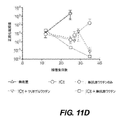
図6Fは、種々の予防的ワクチン製剤の皮下注射、AMLチャレンジ(0日目)、および再チャレンジ(100日目)後の生存率を示す。(*P<0.05、**P<0.01、***P<0.001、n.s.、有意でない(P>0.05)、チューキー事後検定を用いた分散分析(ANOVA))。 FIG. 6F shows survival rates after subcutaneous injection of various prophylactic vaccine formulations, AML challenge (day 0), and re-challenge (day 100). ( * P<0.05, ** P<0.01, *** P<0.001, n.s., not significant (P>0.05), analysis of variance with Tukey post hoc test (ANOVA )).
図9Aは、WT-1を予防的にワクチン接種したマウスから採取した骨髄細胞中の残存AML細胞およびMLL-AF9 AML細胞の正の対照をモニタリングするためのGFP発現を示す。 FIG. 9A shows GFP expression to monitor positive control of residual AML cells and MLL-AF9 AML cells in bone marrow cells harvested from mice prophylactically vaccinated with WT-1.
図9Cは、白血病または免疫の導入を試験するための二次移植のスケジュールを示す。 FIG. 9C shows the schedule for secondary transplantation to test leukemia or induction of immunity.
図11Aは、in vitroでの2つの濃度のiCtへの曝露3時間後のアポトーシス細胞およびカルレティキュリン発現細胞の百分率を示す(n=3/群)。
FIG. 11A shows the percentage of apoptotic and calreticulin-expressing
図11Bは、AMLの樹立、処置の実施、および疾患の進行のモニタリングについてのタイムラインを示す。 FIG. 11B shows a timeline for establishment of AML, administration of treatment, and monitoring of disease progression.
図13Aは、ルシフェラーゼ発現AML細胞由来の全身輝度として測定した処置研究群におけるAMLの進行を示す。 FIG. 13A shows the progression of AML in the treated study groups measured as whole-body brightness from luciferase-expressing AML cells.
図13Bは、表示のように処置を行ったMLL-AF9細胞でチャレンジしたマウスにおける生存率を示す(GM-ゲルおよびCpG-ゲルは、それぞれGM-CSFまたはCpGのみを含む無抗原ワクチンを指す)。 FIG. 13B shows survival in mice challenged with MLL-AF9 cells treated as indicated (GM-gel and CpG-gel refer to antigen-free vaccines containing only GM-CSF or CpG, respectively). .
図14Aは、無抗原ワクチン単一治療(上)およびiCtとの組み合わせ(下)の処置についてのタイムラインを示す。 FIG. 14A shows the timeline for treatment with antigen-free vaccine monotherapy (top) and in combination with iCt (bottom).
図14Bは、GFP+AML細胞のクリオゲル足場および流入領域リンパ節への動員を示す(両側t検定(左)、マン・ホイットニー検定(右);各時点についてn=4~5/群)。 FIG. 14B shows recruitment of GFP + AML cells to cryogel scaffolds and draining lymph nodes (two-tailed t-test (left), Mann-Whitney test (right); n=4-5/group for each time point).
図16Aは、化学療法を実施したか実施していない樹立AMLモデルに送達されたGM-CSFおよびCpGのみを含むクリオゲルワクチンへのCD11c+DCの動員を示す(マン・ホイットニー検定、各時点についてn=4~5/群)。 FIG. 16A shows the recruitment of CD11c + DCs to cryogel vaccines containing only GM-CSF and CpG delivered to established AML models with or without chemotherapy (Mann-Whitney test, for each time point). n=4-5/group).
図16Bは、CD86を発現するCD11c+DCの百分率を示す(マン・ホイットニー検定;各時点についてn=4~5/群)。データは、平均±SDである。 FIG. 16B shows the percentage of CD11c + DC expressing CD86 (Mann-Whitney test; n=4-5/group for each time point). Data are mean±SD.
図17Aおよび17Bは、ワクチン接種後6日目に化学療法を用いた場合および用いない場合の無抗原クリオゲルワクチンで処置したマウスの骨髄中(図17A)および脾臓中(図17B)のGFP+AML細胞数を示す(両側t検定(図17A)、マン・ホイットニー検定(図17B);各時点についてn=4~5/群)。
Figures 17A and 17B show GFP + in bone marrow (Figure 17A) and spleen (Figure 17B) of mice treated with antigen-free cryogel vaccine with and without chemotherapy on
図17Cおよび17Dは、ワクチン接種後9日目の総骨髄細胞数(図17C)および総脾臓細胞数(図17D)に対するGFPを発現する細胞(すなわち、AML細胞)の百分率を示す(両側t検定;各時点についてn=4~5/群)。 Figures 17C and 17D show the percentage of cells expressing GFP (i.e., AML cells) relative to total bone marrow (Figure 17C) and total spleen (Figure 17D) 9 days after vaccination (two-tailed t-test). n=4-5/group for each time point).
図20Aは、iCtとクリオゲルワクチン接種との組み合わせ(左)およびMLL-AF9 AML細胞の正の対照(右)で処置したAML担持マウスから採取した骨髄細胞中の残存AML細胞をモニタリングするためのGFP発現を示す。 FIG. 20A is a graph for monitoring residual AML cells in bone marrow cells harvested from AML-bearing mice treated with a combination of iCt and cryogel vaccination (left) and a positive control of MLL-AF9 AML cells (right). GFP expression is shown.
図20Bは、iCtおよびWT-1ワクチン接種を受けたマウスの採取した骨髄細胞、およびナイーブマウス由来の骨髄中のWT-1テトラマー+CD8+T細胞を示す。 FIG. 20B shows harvested bone marrow cells of iCt and WT-1 vaccinated mice and WT-1 tetramer + CD8 + T cells in bone marrow from naive mice.
図20Cは、白血病または免疫の導入を決定するための二次移植アッセイのスケジュールを示す。 FIG. 20C shows the schedule for secondary transplantation assays to determine induction of leukemia or immunity.
図22Aは、異なる濃度のDoxでin vitroにて24時間処置後の4T1細胞の表面上のカルレティキュリンについての代表的なフローサイトメトリーヒストグラムを示す。 FIG. 22A shows representative flow cytometry histograms for calreticulin on the surface of 4T1 cells after treatment with different concentrations of Dox for 24 hours in vitro.
図22Bは、細孔形成アルギナートゲルからのDoxおよびDox-iRGDの放出プロフィールを示す(n=4)。 FIG. 22B shows the release profiles of Dox and Dox-iRGD from microporous alginate gels (n=4).
図22Cは、それぞれDox-iRGD(赤色)、Dox-iRDG(赤色)、およびDox(赤色)を含む細孔形成ゲルの腫瘍周囲への注射後4日目に腫瘍およびゲル切片の共焦点(confocal)画像を示す。GM-CSFを全ての群に組み込んだ。細胞核を、DAPI(青色)で染色した。白色点線は、腫瘍とゲルの境界を示す。スケールバー:200μm。
FIG. 22C shows confocal images of tumor and
図22Dは、それぞれDox-iRGD、Dox-iRDG、およびDoxの半定量的腫瘍浸透プロフィールを示す。 FIG. 22D shows semi-quantitative tumor penetration profiles of Dox-iRGD, Dox-iRDG, and Dox, respectively.
図22Fは、ゲル注射後18日目のDox-iRGD負荷ゲル中のCD11b+CD11c+細胞の代表的なフローサイトメトリープロットを示す。
FIG. 22F shows representative flow cytometry plots of CD11b + CD11c + cells in Dox-iRGD loaded
図22Gは、細孔形成ゲル中の動員された細胞の間のCD11b+CD11c+DCの百分率を示す(n=4)。 FIG. 22G shows the percentage of CD11b + CD11c + DC among recruited cells in the pore-forming gel (n=4).
図22Hは、ゲル注射後18日目のDox-iRGD負荷ゲル中のCD11b+CD11c+細胞内のCD86+MHCI+細胞の代表的なフローサイトメトリープロットを示す。
FIG. 22H shows representative flow cytometry plots of CD86 + MHCI + cells within CD11b + CD11c + cells in Dox-iRGD-loaded
図22Iは、細孔形成ゲル中のCD11b+CD11c+細胞内のCD86+MHCI+細胞の百分率を示す(n=4)。図22A~22I中の全ての数値データを、平均±SDとして表す(0.01<*P≦0.05;**P≦0.01;***P≦0.001)。 FIG. 22I shows the percentage of CD86 + MHCI + cells within CD11b + CD11c + cells in the pore-forming gel (n=4). All numerical data in FIGS. 22A-22I are expressed as mean±SD (0.01< * P≦0.05; ** P≦0.01; *** P≦0.001).
図23Aは、異なる濃度のDoxでの24時間の処置およびAlexa Fluor647抱合抗カルレティキュリンと20分間のインキュベーション後の4T1細胞の平均Alexa Fluor647蛍光強度を示す。 FIG. 23A shows mean Alexa Fluor647 fluorescence intensity of 4T1 cells after 24 hours treatment with different concentrations of Dox and incubation with Alexa Fluor647-conjugated anti-calreticulin for 20 minutes.
図23Bは、異なる濃度のDoxで24時間の処置およびFITC抱合抗CD47と20分間のインキュベーション後の4T1細胞の代表的なフローサイトメトリーのヒストグラムを示す(n=4)。生/死細胞のゲーティングは既に行っていた。 FIG. 23B shows representative flow cytometry histograms of 4T1 cells after treatment with different concentrations of Dox for 24 hours and incubation with FITC-conjugated anti-CD47 for 20 minutes (n=4). Live/dead cell gating was already done.
図23Cは、(b)と同一の処置を用いた4T1細胞の平均FITC蛍光強度を示す。図23A~23C中の全ての数値データを、平均±SDとして表す(0.01<*P≦0.05;**P≦0.01;***P≦0.001)。 Figure 23C shows mean FITC fluorescence intensity of 4T1 cells with the same treatment as in (b). All numerical data in FIGS. 23A-23C are expressed as mean±SD (0.01< * P≦0.05; ** P≦0.01; *** P≦0.001).
図24Aは、Dox-iRGDの合成経路を示す。 FIG. 24A shows the synthetic route of Dox-iRGD.
図24Bおよび24Cは、254nmの波長で検出したDox-Malの高速液体クロマトグラフィ(図24B)および質量スペクトル(図24C)を示す。 Figures 24B and 24C show the high performance liquid chromatography (Figure 24B) and mass spectrum (Figure 24C) of Dox-Mal detected at a wavelength of 254 nm.
図24Dは、それぞれDox-MalおよびDox-iRGDの1H NMRスペクトルを示す。 FIG. 24D shows the 1 H NMR spectra of Dox-Mal and Dox-iRGD, respectively.
図24Eは、Dox-iRGDのMALDIスペクトルを示す。 FIG. 24E shows the MALDI spectrum of Dox-iRGD.
図24Gは、それぞれ異なる濃度のDox-iRGD、Dox-iRDG、およびDoxで48時間のインキュベーション後の4T1細胞の生存能を示す。図24A~24G中の全ての数値データを、平均±SDとして表す(0.01<*P≦0.05;**P≦0.01;***P≦0.001)。 FIG. 24G shows the viability of 4T1 cells after 48 hours of incubation with different concentrations of Dox-iRGD, Dox-iRDG, and Dox, respectively. All numerical data in FIGS. 24A-24G are expressed as mean±SD (0.01< * P≦0.05; ** P≦0.01; *** P≦0.001).
図25Aは、有効性研究の時間枠を示す。腫瘍が約6~7mmまで成長したときにゲルを腫瘍周囲に注射した。Dox等価物中の薬物用量を記載する。 FIG. 25A shows the time frame of the efficacy study. The gel was injected around the tumor when the tumor grew to approximately 6-7 mm. Drug doses in Dox equivalents are listed.
図25Bは、それぞれDox-iRGDおよびDoxを含むゲルで処置したマウスの平均体重を示す(n=4~5)。 FIG. 25B shows the average body weight of mice treated with Dox-iRGD and Dox-containing gels, respectively (n=4-5).
図26Aは、有効性研究期間にわたる各群の平均4T1腫瘍容積を示す(n=5)。 FIG. 26A shows the mean 4T1 tumor volume for each group over the efficacy study period (n=5).
図26Bは、それぞれDox-iRGD、Dox-iRDG、およびDoxを含むゲルの腫瘍周囲への注射後18日目に採取したH&E染色した肺組織の代表的な画像を示す。Tは腫瘍を示す。 FIG. 26B shows representative images of H&E-stained lung tissue taken 18 days after peritumoral injection of gels containing Dox-iRGD, Dox-iRDG, and Dox, respectively. T indicates tumor.
図26Cは、ゲル注射後18日目の肺組織上の平均腫瘍小結節カウントを示す。データを、平均±SDで表した(n=5)。
FIG. 26C shows mean tumor nodule counts on
図26Dは、ゲル処置後18日目に採取した脊髄骨髄組織の組織病理学を示す。スケールバー:1mm。 FIG. 26D shows the histopathology of spinal bone marrow tissue harvested 18 days after gel treatment. Scale bar: 1 mm.
図27Aは、有効性研究の時間枠を示す。GM-CSFを全てのゲルに組み込んだ。 FIG. 27A shows the time frame of the efficacy study. GM-CSF was incorporated into all gels.
図27Bは、各群についての生存期間中央値および無処置群と比較した処置群における生存期間中央値の増加の概要を示す。 FIG. 27B shows a summary of the median survival time for each group and the increase in median survival time in the treated group compared to the untreated group.
図28A~28Gは、GM-CSF、Dox-iRGD、およびCpGを負荷した細孔形成ゲルが、4T1トリプルネガティブ乳癌に対する腫瘍特異的CTL応答および抗腫瘍有効性を改善することを示す。 Figures 28A-28G show that GM-CSF, Dox-iRGD, and CpG-loaded pore-forming gels improve tumor-specific CTL responses and anti-tumor efficacy against 4T1 triple-negative breast cancer.
図28Aは、有効性研究期間にわたる各群についての平均4T1腫瘍容積を示す(n=7~8)。 Figure 28A shows the mean 4T1 tumor volume for each group over the efficacy study period (n=7-8).
図28C~28Gは、0日目の4T1腫瘍接種後、5日目にDox-iRGD(200μg)およびCpG(100μg)、またはDox-iRGD(200μg)のみ、またはCpG(100μg)のみを含むゲルを、腫瘍周囲に注射したことを示す。GM-CSFを全ての群に組み込んだ。
FIGS. 28C-28G show gels containing Dox-iRGD (200 μg) and CpG (100 μg), or Dox-iRGD (200 μg) alone, or CpG (100 μg) alone on
図28Cは、ゲル注射後4日目に腫瘍流入領域リンパ節から単離し、4T1細胞で再刺激した細胞の代表的なFACSプロットを示す。APC抱合抗IFN-γおよびパシフィックブルー抱合抗CD8を染色のために使用した。再刺激を行わない細胞を対照として使用した。
FIG. 28C shows representative FACS plots of cells isolated from tumor-draining
図28Eは、CD8+T細胞の平均APC蛍光強度を示す(n=3)。 FIG. 28E shows mean APC fluorescence intensity of CD8 + T cells (n=3).
図28Fは、有効性研究期間にわたる各群の平均4T1腫瘍容積を示す(n=7)。 FIG. 28F shows the mean 4T1 tumor volume for each group over the efficacy study period (n=7).
図29Aは、Dox-iRGD(200μg)およびCpG(100μg)、またはDox-iRGDのみを含むゲルの注射後4日目のゲル中の動員細胞の総数を示す。GM-CSFを全てのゲルに組み込んだ。
FIG. 29A shows the total number of recruited cells in
図29Bは、ゲルの注射後4日目のゲル中のCD11c+DC数を示す。
FIG. 29B shows the number of CD11c + DC in the
図29Cは、ゲル中のCD11c+DCの間のCD86+MHCII+細胞の百分率を示す。 Figure 29C shows the percentage of CD86 + MHCII + cells among CD11c + DCs in the gel.
図29Dは、ゲル中のCD86+MHCII+DC数を示す。図29A~29D中の全ての数値データを、平均±SDとして表す(n=6;0.01<*P≦0.05;**P≦0.01;***P≦0.001)。 FIG. 29D shows CD86 + MHCII + DC counts in gels. All numerical data in FIGS. 29A-29D are expressed as mean±SD (n=6; 0.01< * P<0.05; ** P<0.01; *** P<0.001). .
図30Aは、Dox-iRGD(200μg)およびCpG(100μg)、またはDox-iRGDのみを含むゲルの注射後4日目の脾細胞の代表的なIFN-γ対CD8プロットを示す。GM-CSFを全てのゲルに組み込んだ。
FIG. 30A shows representative IFN-γ vs. CD8 plots of
図30Bおよび30Cは、CD8+T細胞の間の(図30B)IFN-γ+細胞の百分率および(図30C)平均APC抗IFN-γの蛍光強度を示す。 Figures 30B and 30C show the percentage of (Figure 30B) IFN-γ + cells and (Figure 30C) mean APC anti-IFN-γ fluorescence intensity among CD8 + T cells.
図30Eおよび30Fは、異なる群の脾臓中のCD4+T細胞の間の(図30E)IFN-γ+細胞の百分率および(図30F)平均APC抗IFN-γ蛍光強度を示す。 Figures 30E and 30F show the percentage of (Figure 30E) IFN-γ + cells and (Figure 30F) mean APC anti-IFN-γ fluorescence intensity among CD4 + T cells in the spleen of different groups.
図30Hおよび30Iは、tdLN中のCD4+T細胞の間の(図30H)IFN-γ+細胞の百分率および(図30I)平均APC抗IFN-γ蛍光強度を示す。図30A~30I中の全ての数値データを、平均±SDとして表す(n=3~4;0.01<*P≦0.05;**P≦0.01;***P≦0.001)。 Figures 30H and 30I show the percentage of (Figure 30H) IFN-γ + cells and (Figure 30I) mean APC anti-IFN-γ fluorescence intensity among CD4 + T cells in tdLN. All numerical data in FIGS. 30A-30I are expressed as mean±SD (n=3-4; 0.01< * P<0.05; ** P<0.01; *** P<0.05). 001).
図31Aは、有効性研究の時間枠を示す。GM-CSFを全てのゲルに組み込んだ。 FIG. 31A shows the time frame of the efficacy study. GM-CSF was incorporated into all gels.
図31Bは、異なる群の各動物についての腫瘍成長曲線を示す。 FIG. 31B shows tumor growth curves for each animal in different groups.
図31Cは、各群についての生存期間中央値および無処置群と比較した処置群における生存期間中央値の増加の概要を示す。 FIG. 31C shows a summary of the median survival time for each group and the increase in median survival time in the treated group compared to the untreated group.
図32Aは、免疫学的死のマーカーであるカルレティキュリンについて染色された腫瘍細胞の代表的なFACSプロットを示す。CD11b+、CD3+、およびCD8+細胞は、これらのプロットから排除されている。 FIG. 32A shows a representative FACS plot of tumor cells stained for calreticulin, a marker of immunological death. CD11b + , CD3 + , and CD8 + cells are excluded from these plots.
図32Bは、異なる群におけるカルレティキュリン+腫瘍細胞の百分率を示す。 Figure 32B shows the percentage of calreticulin + tumor cells in different groups.
図32Cは、ELISAによって定量された各群の腫瘍抽出物中のHMGB-1レベルを示す。 FIG. 32C shows HMGB-1 levels in tumor extracts of each group quantified by ELISA.
図32Dは、CD11b+F4/80+腫瘍関連マクロファージの間のCD86+細胞の百分率を示す。 FIG. 32D shows the percentage of CD86 + cells among CD11b + F4/80 + tumor-associated macrophages.
図32Eは、腫瘍中のCD11c+DCの間のCD86+MHCII+細胞の百分率を示す。 FIG. 32E shows the percentage of CD86 + MHCII + cells among CD11c + DCs in tumors.
図32Fは、異なる群におけるCD8+T細胞の腫瘍内浸潤を示す。 FIG. 32F shows intratumoral infiltration of CD8 + T cells in different groups.
図32Gは、腫瘍中のCD8+の制御性T細胞に対する比を示す。図32A~32G中の全ての数値データを、平均±SDとして表す(n=6;0.01<*P≦0.05;**P≦0.01;***P≦0.001)。 FIG. 32G shows the ratio of CD8 + to regulatory T cells in tumors. All numerical data in FIGS. 32A-32G are expressed as mean±SD (n=6; 0.01< * P<0.05; ** P<0.01; *** P<0.001). .
図33Aおよび33Bは、Dox-iRGD(200μg)およびCpG(100μg)、またはDox-iRGDのみ、またはCpGのみを含むゲルの注射後11日目の(図33A)CD47+および(図33B)カルレティキュリン+CD47+腫瘍細胞の百分率を示す。GM-CSFを全てのゲルに組み込んだ。
Figures 33A and 33B show (Fig. 33A) CD47 + and (Fig. 33B)
図33Cは、腫瘍微小環境中のCD11b+F4/80+マクロファージの百分率を示す。 FIG. 33C shows the percentage of CD11b + F4/80 + macrophages in the tumor microenvironment.
図33Dは、異なる群における腫瘍関連マクロファージの代表的なCD206対CD86プロットを示す。 FIG. 33D shows representative CD206 versus CD86 plots of tumor-associated macrophages in different groups.
図34Aは、Dox-iRGD(200μg)およびCpG(100μg)、またはDox-iRGDのみ、またはCpGのみを含むゲルの注射後11日目の腫瘍中のCD11c+DCの百分率を示す。GM-CSFを全てのゲルに組み込んだ。
FIG. 34A shows the percentage of CD11c + DC in
図34Bは、異なる群における腫瘍内DCの代表的なCD86対MHC IIプロットを示す。 FIG. 34B shows representative CD86 vs. MHC II plots of intratumoral DCs in different groups.
図37Aおよび37Bは、Dox-iRGD(200μg)およびCpG(100μg)、またはDox-iRGD(200μg)のみ、またはCpG(100μg)のみを含むゲルでの処置後の腫瘍細胞(CD11b+、CD3+、およびCD8+細胞は、これらのプロットから排除されている)のPD-L1発現についての代表的なFACSプロット(図37A)およびPD-L1+腫瘍細胞の百分率(図37B)を示す。 Figures 37A and 37B show tumor cells (CD11b + , CD3 + , Representative FACS plots for PD-L1 expression (FIG . 37A) and the percentage of PD-L1 + tumor cells (FIG. 37B) are shown.
図37Cは、有効性研究の時間枠を示す。0日目の4T1腫瘍接種後、Dox-iRGD(200μg)およびCpG(100μg)を含むゲルを、5日目に腫瘍の近傍に注射し、抗PD-1を、6、9、12、15、および18日目に腹腔内注射した。
FIG. 37C shows the time frame for efficacy studies. After 4T1 tumor inoculation on
図37Eは、全群についてのカプラン・マイヤープロットを示す(n=8~9)。 FIG. 37E shows Kaplan-Meier plots for all groups (n=8-9).
図38Aは、異なる群の各動物についての腫瘍成長曲線を示す。 FIG. 38A shows tumor growth curves for each animal in different groups.
図38Bは、各群についての生存期間中央値および無処置群と比較した処置群における生存期間中央値の増加の概要を示す。 FIG. 38B shows a summary of the median survival time for each group and the increase in median survival time in the treated group compared to the untreated group.
図39A~39Dは、ルシフェラーゼ発現4T1(luc-4T1)腫瘍の外科的切除後に、GM-CSF、Dox-iRGD(200μg)、およびCpG(100μg)、またはボーラスワクチン(同量のGM-CSF、Dox-iRGD、およびCpGの溶液)を含むゲルを手術部位に注射したことを示す。 FIGS. 39A-39D show GM-CSF, Dox-iRGD (200 μg), and CpG (100 μg) or bolus vaccine (equal amounts of GM-CSF, Dox - iRGD, and CpG solutions) were injected into the surgical site.
図39Aは、研究の概要を示す。 Figure 39A shows an overview of the study.
図39Bは、異なる時点のマウスの発光シグナルを示す(n=8~9)。 FIG. 39B shows the luminescence signal of mice at different time points (n=8-9).
図39Cは、全群の無腫瘍生存についてのカプラン・マイヤープロットを示す(n=8~9)。 FIG. 39C shows Kaplan-Meier plots for tumor-free survival for all groups (n=8-9).
図39E~39Hは、術後82日目のluc-4T1細胞の静脈内注射を用いた再チャレンジ後に、腫瘍成長および動物の生存をモニタリングした。luc-4T1細胞の静脈内注射を受けたナイーブマウスを、対照として使用した。
Figures 39E-39H monitored tumor growth and animal survival after re-challenge with intravenous injection of luc-4T1 cells on
図39Eは、再チャレンジ研究の概要を示す。 FIG. 39E shows a summary of the re-challenge study.
図39Gは、luc-4T1細胞の注射後の異なる時点でのマウスの発光強度の定量を示す(n=5~9)。 FIG. 39G shows quantification of luminescence intensity in mice at different time points after injection of luc-4T1 cells (n=5-9).
発明の詳細な説明
I.定義
本発明をより容易に理解できるように、ある特定の用語を最初に定義する。
DETAILED DESCRIPTION OF THE INVENTION I. DEFINITIONS Certain terms are first defined so that the invention may be more readily understood.
本明細書中に別段の定義がない限り、本発明に関連して使用される科学用語および技術用語は、当業者によって一般に理解される意味を有するものとする。用語の意味および範囲は明確でなければならないが、しかしながら、これらが曖昧になる可能性がある場合、本明細書中に提供した定義がいかなる辞書や外部の定義よりも優先される。 Unless otherwise defined herein, scientific and technical terms used in connection with the present invention shall have the meanings that are commonly understood by those of ordinary skill in the art. The meaning and scope of the terms should be clear, however, where they may be ambiguous, the definitions provided herein take precedence over any dictionary or extrinsic definitions.
本発明を記載する状況(特に、以下の特許請求の範囲の状況)での用語「a」および「an」および「the」および類似の参照対象の使用は、本明細書に別様の指示がない限り、または状況と明らかに矛盾しない限り、単数および複数(すなわち、1つまたはそれを超える)を両方とも包含すると解釈されるべきである。用語「含む(comprising)」、「有する(having)」、「含む(including)」、および「含有する(containing)」は、別様の指示がない限り、非限定的用語として解釈されるべきである(すなわち、「含む(including)が、限定されない」ことを意味する)。本明細書における数値範囲の記載は、本明細書中に別段の指示がない限り、記載された各別個の数値について個別に言及するか、各別個の数値が前述の範囲に入っていることを簡潔に述べる方法としての役目を果たすことが意図されているに過ぎず、各別個の数値は、それが本明細書にあたかも個々に記載されているように、本明細書に組み込まれる。 The use of the terms "a" and "an" and "the" and similar references in the context of describing the present invention (particularly in the context of the claims below) is not otherwise indicated herein. It should be construed to include both the singular and the plural (ie, one or more) unless otherwise specified or clearly contradicted by the context. The terms “comprising,” “having,” “including,” and “containing” should be interpreted as open-ended terms unless otherwise indicated. (ie, means “including but not limited to”). The recitation of numerical ranges herein refers individually to each separate numerical value recited or to the extent that each individual numerical value falls within the stated range, unless otherwise indicated herein. Merely intended to serve as a method of brevity, each separate numerical value is incorporated herein as if it were individually set forth herein.
用語「約」または「およそ」は、通常、所与の値または範囲の5%以内、より好ましくは1%以内を意味する。 The terms "about" or "approximately" usually mean within 5%, more preferably within 1% of a given value or range.
一般に、用語「処置(treatment)」または「処置(treating)」は、患者への治療剤の適用もしくは投与、または患者由来の単離された組織もしくは細胞株への治療剤の適用もしくは投与と定義され、前述の患者は、疾患、疾患の徴候、もしくは疾患の素因を有し、疾患、疾患の徴候、もしくは疾患の素因を治療するか、治癒させるか、緩和するか、軽減するか、変化させるか、矯正するか、回復させるか、改善するか、影響を及ぼすことを目的とする。したがって、処置(treating)には、抑制、阻害、予防、処置、またはその組み合わせが含まれ得る。処置(treating)は、とりわけ、進行に対抗する時間の増加、寛解の促進、寛解の誘導、寛解の強化、回復の加速、代替治療の有効性の増加もしくは抵抗性の低下、またはその組み合わせを指す。「抑制」または「阻害」は、とりわけ、徴候の発生の遅延、疾患再燃の予防、再燃エピソードの数もしくは頻度の減少、徴候性エピソード間の潜伏期の増加、徴候の重症度の低下、急性エピソードの重症度の低下、徴候数の減少、疾患関連徴候の発生数の減少、徴候の潜伏期の短縮、徴候の回復、二次徴候の減少、二次感染の減少、患者の寿命の延伸、またはその組み合わせを指す。1つの実施形態では、徴候は一次徴候であり、一方、別の実施形態では、徴候は二次徴候である。「一次」は、障害(例えば、糖尿病)の直接的な結果である徴候を指し、一方、二次は、腫瘍原因に由来するか、腫瘍原因の結果生じる徴候を指す。徴候は、疾患または病的状態の任意の出現であり得る。 In general, the term "treatment" or "treating" is defined as the application or administration of a therapeutic agent to a patient or to an isolated tissue or cell line derived from a patient. and said patient has a disease, symptom of disease, or predisposition to disease, and the disease, symptom of disease, or predisposition to disease is treated, cured, alleviated, alleviated, or altered. intended to or, correct, restore, ameliorate, or affect. Thus, treating can include suppression, inhibition, prevention, treatment, or a combination thereof. Treating refers, inter alia, to increasing time to combat progression, promoting remission, inducing remission, enhancing remission, accelerating recovery, increasing efficacy or decreasing resistance to alternative therapies, or combinations thereof. . “Suppression” or “inhibition” means, inter alia, delaying the onset of symptoms, preventing disease flares, reducing the number or frequency of flare-up episodes, increasing the latency between symptomatic episodes, reducing the severity of symptoms, reducing the severity of acute episodes. Reduced severity, reduced number of symptoms, reduced incidence of disease-related symptoms, reduced latency of symptoms, reversal of symptoms, reduced secondary symptoms, decreased secondary infections, increased patient life span, or a combination thereof point to In one embodiment, the symptoms are primary symptoms, while in another embodiment the symptoms are secondary symptoms. "Primary" refers to symptoms that are a direct result of the disorder (eg, diabetes), while secondary refers to symptoms that derive from or result from neoplastic causes. A symptom can be any manifestation of a disease or pathological condition.
したがって、本明細書中で使用される場合、用語「処置(treatment)」または「処置(treating)」には、本明細書中に記載の組成物の任意の投与が含まれ、かつ以下が含まれる:(i)疾患の素因を有し得るが、依然として疾患の病理学または徴候学を経験も示しもしていない被験体における発症を予防すること;(ii)疾患の病理学または徴候学を経験しているか示している被験体の疾患を阻害すること(すなわち、病理学および/または徴候学のさらなる進展を停止すること);または(iii)疾患の病理学または徴候学を経験しているか示している被験体の疾患を回復させること(すなわち、病理学および/または徴候学の逆行)。 Thus, as used herein, the term "treatment" or "treating" includes any administration of the compositions described herein and includes (i) preventing onset in subjects who may be predisposed to the disease but have not yet experienced or exhibited disease pathology or manifestations; (ii) experienced disease pathology or manifestations. inhibiting the disease (i.e., halting further development of the pathology and/or symptomatology) in a subject who is experiencing or exhibiting disease; or (iii) experiencing or exhibiting disease pathology or symptomatology. ameliorating the disease in a subject suffering from disease (ie, reversal of pathology and/or symptomatology).
疾患または障害の「処置」、「予防」、または「改善」は、かかる疾患または障害の発症の遅延または予防、かかる疾患または障害の進行、かかる疾患または障害に関連する症状の進行または重症度の悪化または増悪の逆転、緩和、改善、阻害、減速もしくは停止を意味する。1つの実施形態では、疾患または障害の徴候は、少なくとも5%、少なくとも10%、少なくとも20%、少なくとも30%、少なくとも40%または少なくとも50%緩和される。 "Treatment", "prevention", or "amelioration" of a disease or disorder means delaying or preventing the onset of such disease or disorder, progression of such disease or disorder, reduction in the progression or severity of symptoms associated with such disease or disorder. It means reversing, alleviating, ameliorating, inhibiting, slowing down or stopping exacerbation or exacerbation. In one embodiment, the symptoms of the disease or disorder are alleviated by at least 5%, at least 10%, at least 20%, at least 30%, at least 40%, or at least 50%.
処置の有効性は、障害の任意の公知の診断方法を用いて決定される。障害の1またはそれを超える徴候の緩和は、組成物が臨床的利点を付与することを示す。上記の任意の治療方法を、任意の好適な被験体(例えば、哺乳動物、例えば、イヌ、ネコ、ウシ、ウマ、ウサギ、サル、最も好ましくはヒトが含まれる)に適用することができる。 Efficacy of treatment is determined using any known method of diagnosing the disorder. Alleviation of one or more symptoms of the disorder indicates that the composition confers clinical benefit. Any of the above methods of treatment can be applied to any suitable subject, including, for example, mammals such as dogs, cats, cows, horses, rabbits, monkeys, and most preferably humans.
本明細書中で使用される場合、用語「被験体」には、本発明のヒドロゲルまたは植込み型の薬物送達ワクチン組成物の投与の恩恵を受け得る任意の被験体が含まれる。用語「被験体」には、動物、例えば、脊椎動物、両生類、魚類、哺乳動物、非ヒト動物(ヒトおよび霊長類(チンパンジーおよびサルなど)が含まれる)が含まれる。本発明の1つの実施形態では、被験体はヒトである。 As used herein, the term "subject" includes any subject that may benefit from administration of the hydrogel or implantable drug delivery vaccine compositions of the invention. The term "subject" includes animals such as vertebrates, amphibians, fish, mammals, non-human animals, including humans and primates such as chimpanzees and monkeys. In one embodiment of the invention, the subject is human.
また、用語「被験体」には、家畜、例えば、畜牛、ヒツジ、ヤギ、ウマ、ブタ、ロバ、ラクダ、バッファロー、ウサギ、ニワトリ、シチメンチョウ、アヒル、ガチョウ、およびミツバチ;ならびに愛玩動物、例えば、イヌ、ネコ、愛玩鳥、および観賞魚が含まれ、いわゆる実験動物、例えば、ハムスター、モルモット、ラット、およびマウスも含まれる。 The term "subject" also includes livestock such as cattle, sheep, goats, horses, pigs, donkeys, camels, buffalo, rabbits, chickens, turkeys, ducks, geese, and bees; and companion animals such as dogs. , cats, companion birds, and ornamental fish, as well as so-called laboratory animals such as hamsters, guinea pigs, rats, and mice.
本発明は、バイオマテリアルベースのワクチン組成物が被験体における癌に対する免疫応答を有効に増強するという発見に少なくとも一部が基づく。本発明は、癌を予防および/または処置するためのワクチン組成物およびその使用方法を提供する。本発明は、当該分野の癌処置を超える利点を提供する。 The present invention is based, at least in part, on the discovery that biomaterial-based vaccine compositions effectively enhance the immune response against cancer in a subject. The present invention provides vaccine compositions and methods of use thereof to prevent and/or treat cancer. The present invention provides advantages over cancer treatments in the art.
例えば、本発明の組成物および/または方法は、被験体の強力な免疫応答を誘発する一方で、当該分野の癌処置ではそれができない。いかなる理論にも拘束されることを望まないが、本発明のワクチン組成物(compostion)のバイオマテリアルが免疫応答の増強を容易にするとの仮説が立てられる。バイオマテリアルは、免疫細胞(例えば、樹状細胞)をバイオマテリアルに動員する。免疫細胞は、バイオマテリアル中に一過性に存在して、他の構成要素(アジュバント、抗原、または他の物質など)と接触および/または相互作用する。また、バイオマテリアルによって提供された微小環境(microenviroment)は、動員された免疫細胞の他の免疫抑制シグナルとの相互作用を防止または軽減する。したがって、免疫細胞は、バイオマテリアル中で高効率で活性化される。 For example, the compositions and/or methods of the invention elicit a strong immune response in a subject, while cancer treatments in the art do not. While not wishing to be bound by any theory, it is hypothesized that the biomaterials of the vaccine compositions of the invention facilitate enhanced immune responses. The biomaterial recruits immune cells (eg, dendritic cells) to the biomaterial. Immune cells are transiently present in biomaterials to contact and/or interact with other components (such as adjuvants, antigens, or other substances). Also, the microenvironment provided by the biomaterial prevents or reduces the interaction of recruited immune cells with other immunosuppressive signals. Immune cells are thus activated with high efficiency in biomaterials.
ある特定の態様では、本発明は、被験体への投与前に抗原を含まないワクチン組成物を提供する。ワクチン組成物は、in vivoで生成された抗原を確保して、特異的適応免疫応答を誘導することができる。そのようなものとして、ワクチン組成物は、ワクチン組成物上またはワクチン組成物中の免疫細胞を、ワクチン組成物の投与後に確保された癌抗原に曝露する。本発明は、少なくとも一部が、投与前に抗原を含まないがin vivoで生成された抗原を捕獲するかかるワクチンが同等またはより優れた有効性を示すという驚くべき発見に基づく。例えば、かかる無抗原ワクチンを、化学療法(例えば、免疫原性の細胞死を誘導するが、依然として同等またはさらに優れた有効性を示す作用因子の投与)の後または同時に投与することができる。あるいは、または組み合わせて、本発明のワクチン組成物は、癌細胞を死滅させる免疫原性の癌細胞死を誘導する作用因子を含むことができ、癌細胞は、その後にワクチン組成物に組み込まれるか動員される。 In certain aspects, the present invention provides vaccine compositions that are antigen-free prior to administration to a subject. Vaccine compositions are capable of securing antigens generated in vivo to induce specific adaptive immune responses. As such, the vaccine composition exposes immune cells on or in the vaccine composition to cancer antigens secured after administration of the vaccine composition. The present invention is based, at least in part, on the surprising discovery that such vaccines that are free of antigens prior to administration but that capture antigens produced in vivo show comparable or superior efficacy. For example, such antigen-free vaccines can be administered after or concurrently with chemotherapy (eg, administration of an agent that induces immunogenic cell death but still exhibits equal or superior efficacy). Alternatively, or in combination, the vaccine compositions of the invention can include an immunogenic cancer cell death-inducing agent that kills cancer cells, which are then incorporated into the vaccine composition. Mobilized.
本発明の無抗原ワクチン組成物は、癌抗原の調製およびワクチン組成物への組み込みを必要としない。本発明の無抗原ワクチン組成物は、ある特定の癌に対する既知の抗原が存在しない場合または既知の抗原が強力な免疫応答を惹起することができない場合に特に有利である。新生抗原アプローチのように、本発明の組成物および方法は、患者に特異的な強力な免疫応答を生成することができる。しかしながら、新生抗原アプローチと異なり、かつ有利なことに、無抗原ワクチンの使用方法は、財政的および技術的に非常に複雑になる各ワクチンの患者特異的製造を必要としない。
II.無抗原ワクチン
The antigen-free vaccine composition of the invention does not require the preparation and incorporation of cancer antigens into the vaccine composition. Antigen-free vaccine compositions of the invention are particularly advantageous when there are no known antigens for a particular cancer or when known antigens fail to elicit a strong immune response. Like the neoantigen approach, the compositions and methods of the invention are capable of generating a strong patient-specific immune response. However, unlike the neoantigen approach, and advantageously, the use of non-antigen vaccines does not require patient-specific manufacturing of each vaccine, which is financially and technically very complex.
II. antigen-free vaccine
本発明は、癌を予防または処置するための無抗原ワクチンおよびその使用方法を提供する。「無抗原ワクチン」は、本明細書中で使用される場合、被験体へのワクチン組成物の投与前に抗原を含まないワクチン組成物を指す。いかなる理論にも拘束されることを望まないが、無抗原ワクチンが被験体への投与後にワクチン組成物またはその近傍に癌抗原を引き付けるか、捕捉するか、捕獲するか、そうでなければ確保し、その後に癌抗原が免疫細胞に曝露され、それにより、癌特異的および/または被験体特異的免疫応答が得られ、そして/または癌を予防するか処置するとの仮説が立てられる。 The present invention provides antigen-free vaccines and methods of use thereof to prevent or treat cancer. "Antigen-free vaccine," as used herein, refers to a vaccine composition that does not contain antigens prior to administration of the vaccine composition to a subject. Without wishing to be bound by any theory, it is believed that the antigen-free vaccine attracts, traps, captures or otherwise secures cancer antigens at or near the vaccine composition after administration to a subject. It is hypothesized that cancer antigens are subsequently exposed to immune cells, thereby generating cancer-specific and/or subject-specific immune responses and/or preventing or treating cancer.
ある特定の実施形態では、ワクチン組成物は、バイオマテリアルベースのワクチンである。本明細書中で使用される場合、「バイオマテリアルベースのワクチン組成物」は、本明細書中に詳述の多孔質足場を含むワクチンである。かかるバイオマテリアルベースの無抗原ワクチン組成物は、癌の予防または処置に利点を提供する。ワクチン組成物を、癌抗原の調製および組み込みを必要とせずに調製することができる。ある特定の癌に対する既知の抗原が存在しない場合、または既知の抗原が強力な免疫応答を惹起することができない場合にこれは特に有利である。無抗原ワクチンを、免疫原性の癌細胞死を誘導する作用因子と組み合わせて使用して、新生抗原アプローチとして患者に特異的な強力な免疫応答を生じることができる。有利には、新生抗原アプローチと異なり、本発明の方法は、新生抗原の同定および/または財政的および技術的に非常に複雑になる各ワクチンの患者特異的製造を必要としない。 In certain embodiments, the vaccine composition is a biomaterial-based vaccine. As used herein, a "biomaterial-based vaccine composition" is a vaccine comprising a porous scaffold as detailed herein. Such biomaterial-based antigen-free vaccine compositions offer advantages in the prevention or treatment of cancer. Vaccine compositions can be prepared without the need for preparation and incorporation of cancer antigens. This is particularly advantageous when there are no known antigens for a particular cancer, or when known antigens fail to elicit a strong immune response. Antigen-free vaccines can be used in combination with immunogenic cancer cell death-inducing agents to generate strong patient-specific immune responses as a neoantigen approach. Advantageously, unlike the neoantigen approach, the method of the present invention does not require the identification of neoantigens and/or the patient-specific production of each vaccine, which is very complicated financially and technically.
いくつかの実施形態では、無抗原ワクチン組成物は、本明細書中に詳述の免疫原性の癌細胞死を誘導する作用因子と組み合わせて使用される。いかなる理論にも拘束されることを望まないが、作用因子が免疫原性の癌細胞死を誘導し、癌抗原をワクチン組成物に利用できるようにするとの仮説が立てられる。かかる作用因子は、ワクチン組成物の投与前、同時、投与後に投与され得る。好ましくは、作用因子は、ワクチン組成物前に投与され得る。 In some embodiments, the antigen-free vaccine composition is used in combination with an agent that induces immunogenic cancer cell death detailed herein. Without wishing to be bound by any theory, it is hypothesized that the agent induces immunogenic cancer cell death, making cancer antigens available for vaccine compositions. Such agents may be administered prior to, concurrently with, or subsequent to administration of the vaccine composition. Preferably, the agent may be administered prior to the vaccine composition.
特定の例では、無抗原ワクチンは、免疫細胞(例えば、樹状細胞)を足場に動員する動員組成物(例えば、GM-CSF)、およびアジュバント(例えば、CpG-ODN)を含む。 In certain examples, an antigen-free vaccine includes a recruiting composition (eg, GM-CSF) that recruits immune cells (eg, dendritic cells) to a scaffold, and an adjuvant (eg, CpG-ODN).
無抗原ワクチン組成物は、血液学的悪性疾患(急性骨髄球性白血病(AML)など)の処置のための化学療法剤(例えば、ドキソルビシン)と組み合わせて使用される。いかなる理論にも拘束されることを望まないが、化学療法剤が癌細胞を死滅させると仮定される。ワクチン組成物は、依然として生存しているが免疫原性の細胞死を経ている癌細胞、または免疫原性の細胞死を経ていた癌細胞を足場または足場の近傍に動員し、その後に足場上または足場の近傍で溶解して癌抗原を放出させる。また、ワクチン組成物は、足場またはその近傍に死滅した癌細胞から放出された癌抗原を引き付け得るか、捕捉し得るか、捕獲し得るか、そうでなければ確保し得る。また、ワクチン組成物は、免疫細胞(例えば、樹状細胞)を足場に動員する。免疫細胞は、足場中、足場上、または足場の近傍に存在する癌抗原に曝露され、それにより、癌特異的および/または被験体特異的免疫応答を生じ、そして/または癌を予防または処置する。 Antigen-free vaccine compositions are used in combination with chemotherapeutic agents (eg, doxorubicin) for the treatment of hematological malignancies such as acute myeloid leukemia (AML). While not wishing to be bound by any theory, it is hypothesized that chemotherapeutic agents kill cancer cells. The vaccine composition recruits cancer cells that are still viable but have undergone immunogenic cell death, or that have undergone immunogenic cell death, to the scaffold or the vicinity of the scaffold, and then onto or onto the scaffold. It dissolves in the vicinity of the scaffold to release cancer antigens. The vaccine composition may also attract, capture, capture, or otherwise retain cancer antigens released from dead cancer cells at or near the scaffold. Vaccine compositions also recruit immune cells (eg, dendritic cells) to the scaffold. Immune cells are exposed to cancer antigens present in, on, or near the scaffold, thereby generating a cancer-specific and/or subject-specific immune response and/or preventing or treating cancer .
ある特定の実施形態では、無抗原ワクチン組成物は、免疫原性の癌細胞死を誘導する作用因子(例えば、ドキソルビシン)を足場に組み込む。また、ワクチン組成物は、動員組成物(例えば、GM-CSF)およびアジュバント(例えば、CpG-ODN)を含み得る。 In certain embodiments, the antigen-free vaccine composition incorporates an immunogenic cancer cell death-inducing agent (eg, doxorubicin) into the scaffold. Vaccine compositions may also include a mobilizing composition (eg GM-CSF) and an adjuvant (eg CpG-ODN).
また、無抗原ワクチンは、外科的治療と組み合わせて使用され得る。いかなる理論にも拘束されることを望まないが、癌特異的および/または被験体特異的抗原がin situで生成されて免疫応答を誘発し、再発および/または転移を予防または軽減するとの仮説が立てられる。ワクチン組成物は、原発巣切除後の腫瘍部位またはその近傍に投与され得る。化学療法剤は、近傍の癌細胞を免疫学的に死滅させ得る。動員組成物によって足場に動員された免疫細胞は、癌細胞から放出された癌抗原に曝露される。それにより、癌特異的免疫応答が誘発され、そして/または再発/転移が予防および/または軽減される。
III.免疫原性の癌細胞死を誘導する作用因子
An antigen-free vaccine can also be used in combination with surgical treatment. While not wishing to be bound by any theory, it is hypothesized that cancer-specific and/or subject-specific antigens are generated in situ to elicit an immune response to prevent or reduce recurrence and/or metastasis. be erected. Vaccine compositions may be administered at or near the tumor site after primary tumor resection. Chemotherapeutic agents can immunologically kill nearby cancer cells. Immune cells recruited to the scaffold by the recruitment composition are exposed to cancer antigens released from the cancer cells. Thereby, a cancer-specific immune response is elicited and/or recurrence/metastasis is prevented and/or reduced.
III. Agents that induce immunogenic cancer cell death
本発明は、免疫原性癌/腫瘍細胞死を誘導する作用因子およびこれを使用する方法を提供する。免疫原性の細胞死(ICDまたは免疫原性アポトーシス)は、免疫系によって認識されて免疫を活性化する細胞死の一形態である。この細胞死は、膜完全性を維持しているアポトーシス性形態を特徴とする。小胞体(ER)ストレスは、一般に、ICDの原因因子と認識されており、活性酸素種(ROS)を高産生する。2つのICD誘導物質群が認識されている。タイプI誘導物質は、付帯的損害としてのみERにストレスを与え、主にDNAもしくはクロマチンの維持装置または膜構成要素を標的にする。タイプII誘導物質は、ERを特異的に標的にする。ICDは、いくつかの細胞増殖抑制剤(アントラサイクリン、オキサリプラチン、およびボルテゾミブなど)、または照射療法および光力学療法(PDT)によって誘導される。いくつかのウイルスを、ICDの生物学的原因として列挙することができる。感染細胞の免疫原性の死滅が感染性因子に対する免疫応答を誘導するのと全く同じように、癌細胞の免疫原性の死滅は、樹状細胞(DC)の活性化およびその結果としての特異的T細胞応答の活性化によって有効な抗腫瘍免疫応答を誘導することができる。 The present invention provides agents that induce immunogenic cancer/tumor cell death and methods of using the same. Immunogenic cell death (ICD or immunogenic apoptosis) is a form of cell death that is recognized by the immune system to activate immunity. This cell death is characterized by an apoptotic morphology that maintains membrane integrity. Endoplasmic reticulum (ER) stress is generally recognized as a causative factor of ICD, resulting in high production of reactive oxygen species (ROS). Two classes of ICD inducers are recognized. Type I inducers stress the ER only as collateral damage and target primarily DNA or chromatin maintainers or membrane components. Type II inducers specifically target the ER. ICD is induced by some cytostatic drugs (such as anthracyclines, oxaliplatin, and bortezomib) or radiation therapy and photodynamic therapy (PDT). Several viruses can be listed as biological causes of ICD. Just as immunogenic killing of infected cells induces an immune response to infectious agents, immunogenic killing of cancer cells leads to the activation and consequent specificity of dendritic cells (DCs). Effective anti-tumor immune responses can be induced by activation of targeted T cell responses.
ICDは、ダメージ関連分子パターン(DAMP)の分泌を特徴とする。ICD中に細胞表面に曝露される最も重要なDAMPは3種ある。カルレティキュリン(CRT)(DAMP分子の1つであり、通常は小胞体内腔に存在する)は、免疫原性死の誘導後に瀕死の細胞の表面に移動する。ここで、カルレティキュリンは、プロフェッショナル食細胞への「イート・ミー」シグナルとしての機能を果たす。他の重要な表面に曝露されたDAMPは、ヒートショックタンパク質(HSP)(すなわち、HSP70およびHSP90)であり、ストレス条件下でも原形質膜に移動する。細胞表面上で、HSPは、CD91およびCD40のようないくつかの抗原提示細胞(APC)表面受容体との相互作用に基づいた免疫賦活効果を有し、MHCクラスI分子上の腫瘍細胞由来の抗原の交差提示も容易にし、次いで、CD8+T細胞応答に至る。ICDを特徴的とする他の重要なDAMPは、分泌高移動度群ボックス1(HMGB1)およびATPである。HMGB1は、遅発型ICDのマーカーと見なされており、その細胞外間隙への放出は、樹状細胞による最適な抗原提示に必要なようである。これは、APC上に発現されるいくつかのパターン認識受容体(PRR)(Toll様受容体(TLR)2および4など)に結合する。免疫原性の細胞死中に放出されたATPは、分泌時の食細胞への「ファインド・ミー」シグナルとして機能し、ICD部位への食細胞の誘引を誘導する。また、標的細胞上のプリン受容体へのATPの結合は、インフラマソーム活性化を介した免疫賦活効果を有する。ICD中に放出されたDNAおよびRNA分子は、瀕死の細胞および食細胞の両方におけるTLR3およびcGAS応答を活性化する。 ICD is characterized by the secretion of damage-associated molecular patterns (DAMPs). There are three most important DAMPs exposed to the cell surface during ICD. Calreticulin (CRT), a DAMP molecule normally located in the lumen of the endoplasmic reticulum, is translocated to the surface of dying cells after induction of immunogenic death. Here, calreticulin serves as an "eat me" signal to professional phagocytic cells. Other important surface-exposed DAMPs are the heat shock proteins (HSPs) (ie, HSP70 and HSP90), which translocate to the plasma membrane even under stress conditions. On the cell surface, HSPs have immunostimulatory effects based on their interaction with several antigen-presenting cell (APC) surface receptors, such as CD91 and CD40, and tumor cell-derived immunostimulatory effects on MHC class I molecules. It also facilitates antigen cross-presentation, which in turn leads to a CD8+ T cell response. Other important DAMPs that characterize the ICD are secreted high mobility group box 1 (HMGB1) and ATP. HMGB1 is considered a marker of late-onset ICD and its release into the extracellular space appears to be required for optimal antigen presentation by dendritic cells. It binds to several pattern recognition receptors (PRRs) expressed on APCs, such as Toll-like receptors (TLR) 2 and 4. ATP released during immunogenic cell death functions as a 'find me' signal to phagocytic cells upon secretion, inducing their attraction to the ICD site. ATP binding to purinergic receptors on target cells also has immunostimulatory effects via inflammasome activation. DNA and RNA molecules released during the ICD activate TLR3 and cGAS responses in both dying cells and phagocytic cells.
免疫原性の癌細胞死を誘導する作用因子は、癌細胞を死滅させ、その後に癌細胞に対する免疫応答が得られる他の手段を利用した任意の物質、化学物質、または治療法であり得る。免疫原性の癌細胞死を誘導する「作用因子」には、原子、抗生物質、化学基、化合物、無機物質、有機化合物、多糖、脂質、ヌクレオシド、ヌクレオチド、核酸塩基、糖、核酸、アミノ酸、ペプチド、ポリペプチド、タンパク質、タンパク質複合体、生物(ウイルス、細菌細胞、真核細胞(CAR-Tなどの免疫細胞が含まれる)など)、または免疫療法が含まれ得るが、これらに限定されない。作用因子は、人工物質または天然物質であり得る。 An agent that induces immunogenic cancer cell death can be any substance, chemical, or therapy that kills cancer cells and utilizes other means to subsequently obtain an immune response against cancer cells. "Agents" that induce immunogenic cancer cell death include atoms, antibiotics, chemical groups, compounds, inorganic substances, organic compounds, polysaccharides, lipids, nucleosides, nucleotides, nucleobases, sugars, nucleic acids, amino acids, It may include, but is not limited to, peptides, polypeptides, proteins, protein complexes, organisms such as viruses, bacterial cells, eukaryotic cells (including immune cells such as CAR-T), or immunotherapy. Agents can be man-made or natural.
ある特定の実施形態では、免疫原性の癌細胞死を誘導する作用因子は、放射性同位体、放射線療法、光力学療法、温熱療法、低体温療法、ウイルス、および化学療法剤からなる群から選択される。ある特定の実施形態では、免疫原性腫瘍細胞を誘導する作用因子は、化学療法剤である。かかる化学療法剤は、アントラサイクリン化合物クラスのメンバー(例えば、ドキソルビシン、ダウノルビシン、エピルビシン、イダルビシン、およびバルルビシン)ならびにミトキサントロン、アントラサイクリンアナログが含まれる。この化合物クラスは、癌細胞を直接死滅させることに加えて、免疫系を活性化する能力があるので、好ましい。また、作用因子オキサリプラチンおよびシクロホスファミドは、免疫原性に細胞を死滅させる。免疫原性の細胞死を誘導する化合物の他の限定的な例には、シコニン、プロテアソーム阻害剤ボルテゾミブ、7A7(上皮成長因子受容体特異的抗体)、強心配糖体、およびボリノスタット(ヒストンデアセチラーゼ阻害剤)が含まれる。例えば、H Inoue and K Tani(2014)Cell Death and Differentiation 21,39-49(その内容全体が、本明細書中で参考として援用される)を参照のこと。
In certain embodiments, the agent that induces immunogenic cancer cell death is selected from the group consisting of radioisotopes, radiotherapy, photodynamic therapy, hyperthermia, hypothermia, viruses, and chemotherapeutic agents. be done. In certain embodiments, the agent that induces immunogenic tumor cells is a chemotherapeutic agent. Such chemotherapeutic agents include members of the anthracycline compound class (eg, doxorubicin, daunorubicin, epirubicin, idarubicin, and valrubicin) and mitoxantrone, an anthracycline analogue. This class of compounds is preferred due to its ability to activate the immune system in addition to directly killing cancer cells. The agents oxaliplatin and cyclophosphamide also immunogenically kill cells. Other non-limiting examples of compounds that induce immunogenic cell death include shikonin, the proteasome inhibitor bortezomib, 7A7 (an epidermal growth factor receptor-specific antibody), cardiac glycosides, and vorinostat (a histone deacetylase). enzyme inhibitors). See, eg, H Inoue and K Tani (2014) Cell Death and
いくつかの実施形態では、免疫原性の細胞死を誘導する作用因子は、本明細書中に記載の任意の作用因子の誘導体またはアナログを含む。用語「誘導体」は、本明細書中で使用される場合、化学反応によって類似の化合物から誘導された化合物を指す。例えば、付加によってドキソルビシンから誘導されたドキソルビシン-iRGDは、ドキソルビシンの誘導体である。用語「アナログ」は、本明細書中で使用される場合、別の化合物に類似の構造を有するが、一定の構成要素が異なる化合物である。アナログは、他の原子、基、または物質と置換された、1またはそれを超える原子、官能基、または構造が異なり得る。 In some embodiments, agents that induce immunogenic cell death include derivatives or analogs of any of the agents described herein. The term "derivative," as used herein, refers to a compound derived from similar compounds by a chemical reaction. For example, doxorubicin-iRGD, derived from doxorubicin by addition, is a derivative of doxorubicin. The term "analog," as used herein, is a compound that has a similar structure to another compound, but differs in certain components. Analogs may differ in one or more atoms, functional groups, or structures replaced by other atoms, groups, or substances.
化学療法薬に加えて、本発明の免疫原性の腫瘍細胞死を誘導する作用因子は、免疫原性細胞を死滅させることもできる放射線療法および/または光力学療法、ならびに腫瘍細胞を示すさせる一方で、腫瘍に対する免疫応答を活性化する他のアプローチも含む。免疫原性の腫瘍細胞死を誘導する作用因子には、放射線療法および/または光力学療法を提供する物質が含まれる。 In addition to chemotherapeutic agents, the immunogenic tumor cell death-inducing agents of the present invention can also kill immunogenic cells, while radiation and/or photodynamic therapy can also kill tumor cells. and other approaches to activate immune responses against tumors. Agents that induce immunogenic tumor cell death include agents that provide radiotherapy and/or photodynamic therapy.
いくつかの実施形態では、放射性同位体によって照射を行う。好適な放射性同位体には、ヨウ素-131、ヨウ素-125、レニウム-185、リン-33、リン-32、パラジウム-100、パラジウム-101、パラジウム-201、パラジウム-103、パラジウム-105、パラジウム-106、パラジウム-108、パラジウム-109、パラジウム-110、パラジウム-111、パラジウム-112、セシウム-137、イリジウム-192、コバルト-60、ルテチウム-177、イットリウム-90、タリウム-201、ガリウム-67、テクネチウム-99m、ストロンチウム-90、またはストロンチウム-89が含まれる。 In some embodiments, irradiation is with a radioactive isotope. Suitable radioisotopes include iodine-131, iodine-125, rhenium-185, phosphorus-33, phosphorus-32, palladium-100, palladium-101, palladium-201, palladium-103, palladium-105, palladium- 106, Palladium-108, Palladium-109, Palladium-110, Palladium-111, Palladium-112, Cesium-137, Iridium-192, Cobalt-60, Lutetium-177, Yttrium-90, Thallium-201, Gallium-67, Includes technetium-99m, strontium-90, or strontium-89.
ある特定の実施形態では、免疫原性の腫瘍細胞死を誘導する作用因子は、ハイパーサミア誘導組成物を含む。好適なハイパーサミア誘導組成物は、磁性ナノ粒子または近赤外(NIR)吸収ナノ粒子を含む。ある場合には、ナノ粒子は磁性を示し、方法は、磁性ナノ粒子を交流磁場(AMF)と接触させてin situで局所ハイパーサミアを誘導し、それにより、癌細胞を変化または破壊し、プロセシングされた腫瘍抗原を生成する工程をさらに含む。別の例では、方法は、NIRナノ粒子をNIR放射と接触させてin situで局所ハイパーサミアを誘導し、それにより、癌細胞を変化または破壊し、プロセシングされた腫瘍抗原を生成する工程をさらに含む。ハイパーサミアは、摂氏37度(℃)を超える局所温度を特徴とする。例えば、局所温度(例えば、腫瘍細胞が豊富な領域の温度を、約40、45、50、60、70、75、80、85、90、95℃、またはそれを超える温度まで一過性に加熱する)。
化学療法剤
In certain embodiments, the agent that induces immunogenic tumor cell death comprises a hyperthermia-inducing composition. Suitable hyperthermia-inducing compositions comprise magnetic nanoparticles or near-infrared (NIR) absorbing nanoparticles. In some cases, the nanoparticles exhibit magnetism, and methods include contacting the magnetic nanoparticles with an alternating magnetic field (AMF) to induce local hyperthermia in situ, thereby altering or destroying cancer cells and processing cancer cells. further comprising the step of generating a modified tumor antigen. In another example, the method further comprises contacting the NIR nanoparticles with NIR radiation to induce local hyperthermia in situ, thereby altering or destroying cancer cells and generating processed tumor antigens. include. Hyperthermia is characterized by local temperatures above 37 degrees Celsius (°C). For example, transiently heating the local temperature (e.g., the temperature of an area rich in tumor cells to a temperature of about 40, 45, 50, 60, 70, 75, 80, 85, 90, 95°C, or more) do).
chemotherapeutic agent
本発明は、免疫原性の腫瘍細胞死を誘導する化学療法剤を含む。かかる化学療法剤は、アントラサイクリン化合物クラスのメンバー(例えば、ドキソルビシン、ダウノルビシン、エピルビシン、イダルビシン、およびバルルビシン)ならびにミトキサントロン、アントラサイクリンアナログが含まれる。 The present invention includes chemotherapeutic agents that induce immunogenic tumor cell death. Such chemotherapeutic agents include members of the anthracycline compound class (eg, doxorubicin, daunorubicin, epirubicin, idarubicin, and valrubicin) and mitoxantrone, an anthracycline analogue.
化学療法剤は、抗原を生成して免疫系をプライミングするために使用され得る。アントラサイクリンクラスの化学療法剤は、免疫系をプライミングさせる方法で腫瘍細胞を死滅させる(免疫原性の細胞死)。アントラサイクリンは、元はStreptomyces sp.に由来していた抗癌化合物である。免疫原性の癌細胞死を誘導する化学療法剤(例えば、ドキソルビシンなどのアントラサイクリン、または強心配糖体)は、当該分野で公知である。例えば、米国特許出願公開第2018/0021253A号(本明細書中で参考として援用される)を参照のこと。 Chemotherapeutic agents can be used to generate antigens to prime the immune system. The anthracycline class of chemotherapeutic agents kill tumor cells in a way that primes the immune system (immunogenic cell death). Anthracyclines are originally from Streptomyces sp. is an anticancer compound that was derived from Chemotherapeutic agents that induce immunogenic cancer cell death (eg, anthracyclines such as doxorubicin, or cardiac glycosides) are known in the art. See, for example, US Patent Application Publication No. 2018/0021253A, which is incorporated herein by reference.
かかる免疫原性の細胞死の誘導物質が作用すると、抗原提示細胞が動員されてワクチン組成物の注射部位の瀕死の腫瘍細胞を貪食する。 Upon action of such immunogenic inducers of cell death, antigen-presenting cells are recruited to phagocytize dying tumor cells at the site of injection of the vaccine composition.
免疫原性の癌細胞死を誘導する作用因子は、足場中の癌細胞を死滅させるか、足場から放出させて足場近傍の癌細胞を死滅させるのに有効な量で存在し得る。足場および特定の作用因子のサイズおよび性質に依存して、足場組成物は、動員組成物をマイクログラムレベルまたはミリグラム(minigram)レベルで含み得る。約100mm3の細孔形成ヒドロゲルは、約100μgのDox-iRGDを含み得る。免疫原性の癌細胞死を誘導する作用因子の放出および免疫細胞のその動員効果を定量する方法については、当該分野で公知である。例えば、米国特許出願公開第2018/0021253A1(本明細書中で参考として援用される)を参照のこと。
IV.ワクチン組成物
The agent that induces immunogenic cancer cell death can be present in an amount effective to kill cancer cells in the scaffold or be released from the scaffold to kill cancer cells in the vicinity of the scaffold. Depending on the size and nature of the scaffold and the particular agent, the scaffold composition may contain microgram or minigram levels of the recruitment composition. A pore-forming hydrogel of about 100 mm 3 can contain about 100 μg of Dox-iRGD. Methods to quantify the release of agents that induce immunogenic cancer cell death and their recruitment effects on immune cells are known in the art. See, for example, US Patent Application Publication No. 2018/0021253A1, incorporated herein by reference.
IV. vaccine composition
本発明は、疾患に対する被験体の免疫応答を増強するワクチン組成物および方法を特徴とする。本発明のワクチン組成物は、多孔質足場、免疫細胞を前述の足場に動員する動員組成物、およびアジュバントを含む。文脈に応じて、ワクチン組成物は、時折、本開示において、「ワクチンデバイス」、「デバイス」、または「組成物」と称され得る。
足場
The present invention features vaccine compositions and methods that enhance a subject's immune response to disease. Vaccine compositions of the invention comprise a porous scaffold, a recruitment composition that recruits immune cells to said scaffold, and an adjuvant. Depending on the context, vaccine compositions may sometimes be referred to in this disclosure as "vaccine devices,""devices," or "compositions."
scaffold
本発明のワクチン組成物は、足場(例えば、ポリマー足場)を含む。足場は、1またはそれを超えるバイオマテリアルを含むことができる。好ましくは、バイオマテリアルは、非毒性および非免疫原性の生体適合性材料である。本明細書中で使用される場合、用語「生体適合性材料」は、被験体の生物組織内に移植されるか、近傍に配置されたときに経時的に著しい免疫応答や有害な組織反応(例えば、毒性反応または著しい照射)を誘導しない任意の材料を指す。 A vaccine composition of the invention includes a scaffold (eg, a polymer scaffold). A scaffold can include one or more biomaterials. Preferably, the biomaterial is a non-toxic and non-immunogenic biocompatible material. As used herein, the term "biocompatible material" refers to significant immune responses and adverse tissue reactions (such as For example, it refers to any material that does not induce toxic reactions or significant irradiation).
足場は、非生物分解性または生物分解性のバイオマテリアルを含むことができる。ある特定の実施形態では、バイオマテリアルは、非生分解性材料であり得る。例示的な非生分解性材料には金属、可塑性ポリマー、またはシルクポリマーが含まれるが、これらに限定されない。ある特定の実施形態では、ポリマー足場は、生分解性材料を含む。生分解性材料は、物理的作用または化学作用(例えば、水和、熱、酸化、またはイオン交換のレベル)または細胞作用(例えば、酵素、ペプチド、または細胞付近もしくは細胞に内在する他の化合物の同化)によって分解され得る。ある特定の実施形態では、ポリマー足場は、非分解性材料および分解性材料の両方を含む。 The scaffold can comprise non-biodegradable or biodegradable biomaterials. In certain embodiments, a biomaterial can be a non-biodegradable material. Exemplary non-biodegradable materials include, but are not limited to metals, plastic polymers, or silk polymers. In certain embodiments, the polymer scaffold comprises biodegradable materials. Biodegradable materials can be subject to physical or chemical action (e.g. levels of hydration, heat, oxidation, or ion exchange) or cellular action (e.g. enzymes, peptides, or other compounds near or in cells). assimilation). In certain embodiments, the polymer scaffold includes both non-degradable and degradable materials.
いくつかの実施形態では、足場組成物は、温度、pH、水和状態、および多孔度、架橋密度、タイプ、および主鎖の結合の化学または分解に対する感受性からなる群から選択される物理的パラメーターに基づいて所定の速度で分解することができる。あるいは、足場組成物は、化学ポリマーの比にもとづいて所定の速度で分解する。例えば、ラクチドのみから構成された高分子量ポリマーは年単位で分解し(例えば、1~2年)、一方で、ラクチドとグリコリドとの50:50混合物から構成された低分子量ポリマーは、およそ数週間で(例えば、1、2、3、4、6、または10週間で)分解する。高分子量の高グルロン酸アルギナートから構成されたカルシウム架橋ゲルは、in vivoで数ヶ月間(1、2、4、6、8、10、または12ヶ月間)から数年間(1、2、または5年間)にわたって分解し、一方で、低分子量アルギナート、および/または部分酸化されたアルギナートから構成されたゲルは、およそ数週間で分解するであろう。ある特定の実施形態では、足場組成物は酸化アルギナートを含み、足場組成物の分解速度は、アルギナートの酸化度の調整によって調整され得る。米国特許出願公開第2014/0079752A1号(その内容が本明細書中で参考として援用される)を参照のこと。 In some embodiments, the scaffold composition comprises a physical parameter selected from the group consisting of temperature, pH, hydration state, and porosity, crosslink density, type, and susceptibility to chemical or degradation of backbone bonds. can be decomposed at a predetermined rate based on Alternatively, the scaffold composition degrades at a predetermined rate based on the ratio of chemical polymers. For example, a high molecular weight polymer composed of lactide alone degrades in years (eg, 1-2 years), while a low molecular weight polymer composed of a 50:50 mixture of lactide and glycolide degrades on the order of weeks. (eg, in 1, 2, 3, 4, 6, or 10 weeks). Calcium-crosslinked gels composed of high molecular weight high guluronic acid alginate can be maintained in vivo for several months (1, 2, 4, 6, 8, 10, or 12 months) to several years (1, 2, or 5 years), while gels composed of low molecular weight alginates and/or partially oxidized alginates will degrade in approximately a few weeks. In certain embodiments, the scaffold composition comprises oxidized alginate, and the degradation rate of the scaffold composition can be modulated by adjusting the degree of alginate oxidation. See US Patent Application Publication No. 2014/0079752A1, the contents of which are incorporated herein by reference.
ある特定の実施形態では、1またはそれを超える本明細書中に開示の化合物またはタンパク質(例えば、動員組成物、アジュバント、および抗原)は、足場組成物に共有結合または非共有結合する。種々の実施形態では、1またはそれを超える本明細書中に開示の化合物またはタンパク質は、足場組成物の構造物または細孔上、構造物または細孔中に組み込まれるか、構造物または細孔内に存在する。 In certain embodiments, one or more of the compounds or proteins disclosed herein (eg, recruitment composition, adjuvant, and antigen) are covalently or non-covalently attached to the scaffold composition. In various embodiments, one or more of the compounds or proteins disclosed herein are incorporated on, in, or in the structures or pores of the scaffold composition. exist within.
いくつかの実施形態では、足場は、修飾された(例えば、酸化または還元された)バイオマテリアルを含む。修飾度(酸化度など)は、約1%から約100%まで変動し得る。本明細書中で使用される場合、修飾度は、官能基で修飾されたバイオマテリアル上の部位のモル百分率を意味する。例えば、修飾度は、約1%、2%、3%、4%、5%、6%、7%、8%、9%、10%、11%、12%、13%、14%、15%、16%、17%、18%、19%、20%、21%、22%、23%、24%、25%、26%、27%、28%、29%、30%、31%、32%、33%、34%、35%、36%、37%、38%、39%、40%、41%、42%、43%、44%、45%、46%、47%、48%、49%、50%、51%、52%、53%、54%、55%、56%、57%、58%、59%、60%、61%、62%、63%、64%、65%、66%、67%、68%、69%、70%、71%、72%、73%、74%、75%、76%、77%、78%、79%、80%、81%、82%、83%、84%、85%、86%、87%、88%、89%、90%、91%、92%、93%、94%、95%、96%、97%、98%、99%、または100%であり得る。値および列挙された値の間を示す範囲が本発明の一部であることが意図される。例示的な修飾バイオマテリアル(例えば、ヒドロゲル)には、還元アルギナート、酸化アルギナート、MA-アルギナート(メタクリル化アルギナート)、MA-ゼラチン、またはMA-ポリエチレングリコール(MA-PEG)が含まれるが、これらに限定されない。 In some embodiments, scaffolds comprise modified (eg, oxidized or reduced) biomaterials. The degree of modification (such as the degree of oxidation) can vary from about 1% to about 100%. As used herein, degree of modification means the molar percentage of sites on the biomaterial that are modified with functional groups. For example, the degree of modification is about 1%, 2%, 3%, 4%, 5%, 6%, 7%, 8%, 9%, 10%, 11%, 12%, 13%, 14%, 15% %, 16%, 17%, 18%, 19%, 20%, 21%, 22%, 23%, 24%, 25%, 26%, 27%, 28%, 29%, 30%, 31%, 32%, 33%, 34%, 35%, 36%, 37%, 38%, 39%, 40%, 41%, 42%, 43%, 44%, 45%, 46%, 47%, 48% , 49%, 50%, 51%, 52%, 53%, 54%, 55%, 56%, 57%, 58%, 59%, 60%, 61%, 62%, 63%, 64%, 65% %, 66%, 67%, 68%, 69%, 70%, 71%, 72%, 73%, 74%, 75%, 76%, 77%, 78%, 79%, 80%, 81%, 82%, 83%, 84%, 85%, 86%, 87%, 88%, 89%, 90%, 91%, 92%, 93%, 94%, 95%, 96%, 97%, 98% , 99%, or 100%. All values and ranges between recited values are intended to be part of this invention. Exemplary modified biomaterials (eg, hydrogels) include reduced alginate, oxidized alginate, MA-alginate (methacrylated alginate), MA-gelatin, or MA-polyethylene glycol (MA-PEG), including Not limited.
本発明における足場としての使用に好適なバイオマテリアルの例には、グリコサミノグリカン、シルク、フィブリン、MATRIGEL(登録商標)、ポリ-エチレングリコール(PEG)、ポリヒドロキシエチルメタクリラート、ポリアクリルアミド、ポリ(N-ビニルピロリドン)、(PGA)、ポリ乳酸-コ-グリコール酸(PLGA)、ポリe-カプロラクトン(poly e-carpolactone)(PCL)、ポリエチレンオキシド、ポリプロピレンフマラート(PPF)、ポリアクリル酸(PAA)、ポリヒドロキシ酪酸、加水分解ポリアクリロニトリル、ポリメタクリル酸、ポリエチレンアミン、アルギン酸のエステル;ペクチン酸;およびアルギナート、完全酸化もしくは部分酸化アルギナート、ヒアルロン酸、カルボキシメチルセルロース、ヘパリン、ヘパリン硫酸、キトサン、カルボキシメチルキトサン、キチン、プルラン、ゲラン、キサンタン、コラーゲン、ゼラチン、カルボキシメチルデンプン、カルボキシメチルデキストラン、コンドロイチン硫酸、カチオン性グアー、カチオン性デンプン、およびその組み合わせが含まれる。ある特定の実施形態では、バイオマテリアルは、アルギナート、完全酸化または部分酸化アルギナートまたは還元アルギナート、およびその組み合わせからなる群から選択される。 Examples of biomaterials suitable for use as scaffolds in the present invention include glycosaminoglycans, silk, fibrin, MATRIGEL®, poly-ethylene glycol (PEG), polyhydroxyethyl methacrylate, polyacrylamide, poly (N-vinylpyrrolidone), (PGA), polylactic-co-glycolic acid (PLGA), poly e-caprolactone (PCL), polyethylene oxide, polypropylene fumarate (PPF), polyacrylic acid ( PAA), polyhydroxybutyric acid, hydrolyzed polyacrylonitrile, polymethacrylic acid, polyethyleneamines, esters of alginic acid; pectic acid; Included are methyl chitosan, chitin, pullulan, gellan, xanthan, collagen, gelatin, carboxymethyl starch, carboxymethyl dextran, chondroitin sulfate, cationic guar, cationic starch, and combinations thereof. In certain embodiments, the biomaterial is selected from the group consisting of alginates, fully or partially oxidized or reduced alginates, and combinations thereof.
本発明の足場は、外面を含み得る。あるいは、またはさらに、足場は、内面を含み得る。本発明の足場の外面または内面は、固体または多孔質であり得る。足場の細孔サイズは、約10nm未満、約100nm~20μm、または約20μm超(例えば、直径1000μmを含むそれ以下)であり得る。例えば、細孔は、ナノ多孔質、マイクロ多孔質、またはマクロ多孔質であり得る。例えば、ナノポアの直径は、約10nm未満である;マイクロポアの直径は、約100nm~20μmの範囲である;マクロポアの直径は、約20μm超(例えば、約50μm超、例えば、約100μm超、例えば、約400μm超、例えば、600μm超または800μm超である。いくつかの実施形態では、細孔の直径は、約50μmと約80μmとの間である。 A scaffold of the invention may include an outer surface. Alternatively, or additionally, a scaffold can include an inner surface. The exterior or interior surface of scaffolds of the invention can be solid or porous. The scaffold pore size can be less than about 10 nm, between about 100 nm and 20 μm, or greater than about 20 μm (eg, up to and including 1000 μm in diameter). For example, pores can be nanoporous, microporous, or macroporous. For example, nanopore diameters are less than about 10 nm; micropore diameters range from about 100 nm to 20 μm; macropore diameters are greater than about 20 μm (eg, greater than about 50 μm, such as greater than about 100 μm, such as , greater than about 400 μm, such as greater than 600 μm or greater than 800 μm, hi some embodiments, the pore diameter is between about 50 μm and about 80 μm.
いくつかの実施形態では、本発明の足場は、種々の幾何学的形態(例えば、ディスク、ビーズ、ペレット)、くぼみ、平面状の層(例えば、薄板)に組織化されている。例えば、直径約0.1~200ミリメートル(例えば、5、10、20、40、または50ミリメートル)のディスクを、皮下、静脈内、腹腔内、または筋肉内に植え込み得る。ディスクは、厚さが0.1~10ミリメートル(例えば、1、2、または5ミリメートル)であり得る。ディスクは、患者への投与のために容易に圧縮または凍結乾燥される。皮下投与のための例示的なディスクは、以下の寸法を有する:直径8ミリメートルおよび厚さ1ミリメートル。 In some embodiments, scaffolds of the invention are organized into various geometric forms (eg, discs, beads, pellets), depressions, planar layers (eg, laminae). For example, discs about 0.1 to 200 millimeters (eg, 5, 10, 20, 40, or 50 millimeters) in diameter can be implanted subcutaneously, intravenously, intraperitoneally, or intramuscularly. The disc can be 0.1 to 10 millimeters thick (eg, 1, 2, or 5 millimeters). The discs are easily compressed or lyophilized for patient administration. An exemplary disc for subcutaneous administration has the following dimensions: 8 millimeters in diameter and 1 millimeter in thickness.
本発明の足場は、意図する適用に応じて任意の好適なサイズであり得る。足場は、マイクロメートルのスケールからセンチメートルのスケールで調製され得る。例示的な体積は、数百μm3(例えば、約100~500μm3)から約10cm3まで変動する。ある特定の実施形態では、例示的な足場は、約100μm3~100mm3のサイズである。種々の実施形態では、足場は、約10mm3~約100mm3のサイズである。ある特定の実施形態では、足場は、約30mm3または約100mm3のサイズである。 A scaffold of the present invention can be of any suitable size depending on the intended application. Scaffolds can be prepared on the scale of micrometers to centimeters. Exemplary volumes range from a few hundred μm 3 (eg, about 100-500 μm 3 ) to about 10 cm 3 . In certain embodiments, exemplary scaffolds are between about 100 μm 3 and 100 mm 3 in size. In various embodiments, the scaffold is about 10 mm 3 to about 100 mm 3 in size. In certain embodiments, the scaffold is about 30 mm 3 or about 100 mm 3 in size.
いくつかの実施形態では、足場は、複数の構成要素および/または区画を含み得る。ある特定の実施形態では、多区画足場を、同様にもしくは差分的にドープしたゲルまたは他の足場材料の連続層を標的部位に適用することによってin vivoでアセンブリする。例えば、針を使用して前に注射した材料の中心に向かって次の内側の層に連続的に注入し、それにより、同心スフェロイドを形成することによって足場が形成される。ある特定の実施形態では、前に注射した層と異なる位置に材料を注射することによって非同心性の区画が形成される。多頭注射足場は、区画が並行して同時に突出している。層は、医薬組成物が差分的にドープされた類似するか異なるバイオマテリアルで作製されている。あるいは、区画は、その親水性/疎水性または各区画内での二次相互作用に基づいて自己組織化する。ある特定の実施形態では、多成分足場は、必要に応じて、各々が異なる物理的性質(ポリマーの百分率、ポリマーの架橋の百分率、ヒドロゲルの化学組成、細孔サイズ、多孔度、および細孔の構造、剛性、靭性、延性、粘弾性、足場に組み込まれる動員組成物、アジュバント、および/または抗原、および/または足場に組み込まれる任意の他の組成物など)によって特徴づけられる同心円層に構築される。
ヒドロゲルおよびクリオゲルの足場
In some embodiments, a scaffold can include multiple components and/or compartments. In certain embodiments, multicompartmental scaffolds are assembled in vivo by applying successive layers of similarly or differentially doped gels or other scaffolding materials to the target site. For example, a scaffold is formed by sequentially injecting the next inner layer toward the center of the previously injected material using a needle, thereby forming concentric spheroids. In certain embodiments, non-concentric compartments are formed by injecting material in different locations than previously injected layers. A multi-headed injection scaffold has parallel, simultaneously projecting compartments. Layers are made of similar or different biomaterials that are differentially doped with pharmaceutical compositions. Alternatively, the compartments self-assemble based on their hydrophilicity/hydrophobicity or secondary interactions within each compartment. In certain embodiments, the multi-component scaffolds optionally each have different physical properties (percentage of polymer, percentage of polymer cross-linking, hydrogel chemical composition, pore size, porosity, and pore size). structure, stiffness, toughness, ductility, viscoelasticity, recruitment composition incorporated into the scaffold, adjuvant, and/or antigen, and/or any other composition incorporated into the scaffold, etc.). be.
Hydrogel and cryogel scaffolds
ある特定の実施形態では、本発明の足場は、1またはそれを超えるヒドロゲルを含む。ヒドロゲルは、架橋されたポリマー鎖のネットワークを含むポリマーゲルである。ヒドロゲルは、通常、親水性のポリマー鎖を含む組成物である。ヒドロゲルの網目構造により、大量の水を吸収可能である。いくつかのヒドロゲルは、伸縮性および弾性が高く;他は粘弾性を示す。ヒドロゲルは、時折、水が分散媒であるコロイド状ゲルとして見出される。ある特定の実施形態では、ヒドロゲルは、高吸収性の(99%を超える水(v/v)を含むことができる)天然または合成ポリマーであり、その含水量の高さにより、天然組織に非常に類似した柔軟度を有する。ある特定の実施形態では、ヒドロゲルは、適切な剪断応力を印加したときに、変形可能なヒドロゲルが劇的かつ可逆的に圧縮され(その体積の95%まで)、その結果、注射可能マクロ多孔質が予め形成された足場が得られるという性質を有し得る。ヒドロゲルは、治療のために適用されている(例えば、治療剤(小分子、細胞、および生物製剤など)のin vivo送達のためのビヒクル)。ドロゲルは、一般に、ポリサッカリド(アルギナートなど)から生成される。ポリサッカリドは、その性質および得られたヒドロゲルの性質を調整するように化学的に操作され得る。ヒドロゲル足場は、時折、本開示において「ゲル」称される。 In certain embodiments, scaffolds of the invention comprise one or more hydrogels. A hydrogel is a polymer gel that contains a network of crosslinked polymer chains. A hydrogel is generally a composition comprising hydrophilic polymer chains. The network structure of hydrogels allows them to absorb large amounts of water. Some hydrogels are highly stretchable and elastic; others exhibit viscoelasticity. Hydrogels are sometimes found as colloidal gels in which water is the dispersion medium. In certain embodiments, hydrogels are highly absorbent (can contain greater than 99% water (v/v)) natural or synthetic polymers whose high water content makes them highly absorbent to natural tissue. has a degree of flexibility similar to In certain embodiments, the hydrogel undergoes dramatic and reversible compression of the deformable hydrogel (up to 95% of its volume) upon application of appropriate shear stress, resulting in an injectable macroporous can have the property that a preformed scaffold is obtained. Hydrogels have been applied for therapeutic purposes (eg, vehicles for in vivo delivery of therapeutic agents such as small molecules, cells, and biologics). Dragels are commonly made from polysaccharides such as alginates. Polysaccharides can be chemically manipulated to tune their properties and the properties of the resulting hydrogel. Hydrogel scaffolds are sometimes referred to as "gels" in this disclosure.
好ましくは、本発明の組成物は、多孔質ヒドロゲルから形成される。例えば、ヒドロゲルは、細孔の直径が約10nm未満であるナノ多孔質;細孔の直径が好ましくは約100nm~20μmの範囲であるマイクロ多孔質;または細孔の直径が約20μm超、より好ましくは約100μm超、さらにより好ましくは約400μm超であるマクロ多孔質であり得る。ある特定の実施形態では、ヒドロゲルは、直径約50~80μmの細孔を有するマクロ多孔質である。ある特定の実施形態では、ヒドロゲルは、直径約400~500μmの位置合わせした細孔を有するマクロ多孔質である。多孔質ヒドロゲル生成物の調製方法は、当該分野で公知である。(例えば、米国特許第6,511,650号(本明細書中で参考として援用される)を参照のこと)。 Preferably, the compositions of the invention are formed from porous hydrogels. For example, hydrogels can be nanoporous with pore diameters less than about 10 nm; microporous with pore diameters preferably in the range of about 100 nm to 20 μm; or pore diameters greater than about 20 μm, more preferably. can be macroporous greater than about 100 μm, even more preferably greater than about 400 μm. In certain embodiments, the hydrogel is macroporous with pores about 50-80 μm in diameter. In certain embodiments, the hydrogel is macroporous with aligned pores about 400-500 μm in diameter. Methods for preparing porous hydrogel products are known in the art. (See, eg, US Pat. No. 6,511,650, incorporated herein by reference).
ヒドロゲルは、いくつかの異なる硬質の、半硬質の、可塑性の、ゲル状の、自己集合の、液晶の、または流体の組成物、例えば、ペプチドポリマー、ポリサッカリド、合成ポリマー、ヒドロゲル材料、セラミックス(例えば、リン酸カルシウムまたはヒドロキシアパタイト)、タンパク質、糖タンパク質、プロテオグリカン、金属、および合金から構築され得る。組成物は、当該分野で公知の方法(例えば、射出成形、予め形成された構造物の凍結乾燥、プリンティング、自己集合、位相反転、溶液流延、溶融加工、ガス発泡、繊維形成/加工、粒子浸出、またはその組み合わせ)を使用してヒドロゲルにアセンブリされる。次いで、アセンブリされた足場は、処置すべき個体の身体に植え込まれるか投与される。 Hydrogels are a number of different rigid, semi-rigid, plastic, gel-like, self-assembled, liquid crystalline, or fluid compositions such as peptide polymers, polysaccharides, synthetic polymers, hydrogel materials, ceramics ( For example, calcium phosphate or hydroxyapatite), proteins, glycoproteins, proteoglycans, metals, and alloys. Compositions may be prepared by methods known in the art (e.g., injection molding, freeze-drying of preformed structures, printing, self-assembly, phase inversion, solution casting, melt processing, gas foaming, fiber formation/processing, particle leaching, or a combination thereof) into a hydrogel. The assembled scaffold is then implanted or administered into the body of the individual to be treated.
ヒドロゲルを含む組成物は、いくつかの方法にてin vivoでアセンブリされ得る。ヒドロゲルは、非ゲル化形態で体内に導入され、その場所にてin situでゲル化するゲル化材料でできている。アセンブリが生じる部位への組成物の構成要素の送達方法の例には、組織部位での針または他の押し出しツールによる注射、噴霧、ペインティング、または沈着方法(例えば、カニューレを介して挿入された適用足場を使用した送達)が含まれる。いくつかの実施形態では、非ゲル化または非形成ヒドロゲル材料は、体内への導入前または導入中に少なくとも1つの医薬組成物と混合される。得られたin vivo/in situでアセンブリされた足場(例えば、ヒドロゲル)は、少なくとも1つの医薬組成物の混合物を含む。 Compositions containing hydrogels can be assembled in vivo in several ways. Hydrogels are made of gelling materials that are introduced into the body in a non-gelling form and gel in situ in situ. Examples of methods of delivery of the components of the composition to the site where assembly occurs include injection, spraying, painting, or deposition methods at the tissue site with a needle or other extrusion tool (e.g., cannulated). delivery using an application scaffold). In some embodiments, the non-gelling or non-forming hydrogel material is mixed with at least one pharmaceutical composition prior to or during introduction into the body. The resulting in vivo/in situ assembled scaffold (eg, hydrogel) comprises a mixture of at least one pharmaceutical composition.
ヒドロゲルのin situアセンブリは、ポリマーの自発的会合の結果としてか、相乗的もしくは化学的に触媒された重合から生じ得る。相乗的なまたは化学的な触媒作用は、アセンブリ部位またはその近傍でのいくつかの内因性の要因もしくは条件(例えば、体温、体内のイオンまたはpH)、または操作者によるアセンブリ部位に供給される外因性の要因もしくは条件(例えば、非ゲル化材料が導入された後に非ゲル化材料に対して実施される光子、熱、電気、音響、または他の照射)によって開始される。エネルギーは、放射線ビームによるか、熱導体もしくは光導体(ワイヤケーブルまたは光ファイバーケーブルなど)または超音波変換器を介してヒドロゲル材料に向けられる。あるいは、例えば針中の通過によって発揮されていたせん断力が低下した後に再架橋するずり減粘材料(両親媒性物質など)を使用する。 In situ assembly of hydrogels can result from spontaneous association of polymers or from synergistic or chemically catalyzed polymerization. Synergistic or chemical catalysis can be due to some intrinsic factor or condition (e.g., body temperature, body ions or pH) at or near the assembly site, or an exogenous factor supplied to the assembly site by the operator. initiated by a sexual factor or condition (eg, photon, thermal, electrical, acoustic, or other irradiation performed on the non-gelling material after the non-gelling material is introduced). Energy is directed into the hydrogel material by a radiation beam or via a heat or light conductor (such as a wire or fiber optic cable) or an ultrasonic transducer. Alternatively, a shear-thinning material (such as an amphiphile) is used that re-crosslinks after the shear forces exerted by passage through the needle are reduced, for example.
いくつかの実施形態では、ヒドロゲルは、ex vivoでアセンブリされ得る。いくつかの実施形態では、ヒドロゲルは注射可能である。例えば、ヒドロゲルは、マクロ多孔質足場として身体の外側で作出される。体内への注射の際に、細孔が崩壊し、それによってゲルが非常に小さくなり、針を通過させることが可能になる。例えば、WO2012/149358号;およびBencherif et al.,2012,Proc.Natl.Acad.Sci.USA 109.48:19590-5(その内容が本明細書中で参考として援用される)を参照のこと。 In some embodiments, hydrogels can be assembled ex vivo. In some embodiments, the hydrogel is injectable. For example, hydrogels are created outside the body as macroporous scaffolds. Upon injection into the body, the pores collapse, making the gel so small that it can be passed through by a needle. See, eg, WO2012/149358; and Bencherif et al. , 2012, Proc. Natl. Acad. Sci. USA 109.48:19590-5, the contents of which are incorporated herein by reference.
ヒドロゲル足場のin vivoおよびex vivoアセンブリの両方に好適なヒドロゲルは、当該分野で周知であり、例えば、Lee et al.,2001,Chem.Rev.7:1869-1879に記載されている。自己集合させるためのペプチド両親媒性物質アプローチは、例えば、Hartgerink et al.,2002,Proc.Natl.Acad.Sci.USA 99:5133-5138に記載されている。ずり減粘後の可逆的ゲル化方法は、Lee et al.,2003,Adv.Mat.15:1828-1832に例示されている。 Hydrogels suitable for both in vivo and ex vivo assembly of hydrogel scaffolds are well known in the art, see, for example, Lee et al. , 2001, Chem. Rev. 7:1869-1879. A peptide amphiphile approach to self-assembly is described, for example, in Hartgerink et al. , 2002, Proc. Natl. Acad. Sci. USA 99:5133-5138. A reversible gelation method after shear thinning is described by Lee et al. , 2003, Adv. Mat. 15:1828-1832.
ある特定の実施形態では、例示的なヒドロゲルは、ポリマー、ナノ粒子、ポリペプチド、および細胞が含まれる材料のカプセル化に適合する材料から構成される。例示的なヒドロゲルは、アルギナート、ポリエチレングリコール(PEG)、PEG-アクリラート、アガロース、ヒアルロン酸、または合成タンパク質(例えば、コラーゲンまたは人工タンパク質(すなわち、自己集合ペプチドベースのヒドロゲル))から作製される。例えば、市販のヒドロゲルには、BD(商標)PuraMatrix(商標)が含まれる。BD(商標)PuraMatrix(商標)ペプチドヒドロゲルは、細胞培養のための規定の三次元(3D)微小環境を作出するために使用される合成マトリックスである。 In certain embodiments, exemplary hydrogels are composed of materials compatible with encapsulation of materials, including polymers, nanoparticles, polypeptides, and cells. Exemplary hydrogels are made from alginate, polyethylene glycol (PEG), PEG-acrylate, agarose, hyaluronic acid, or synthetic proteins such as collagen or engineered proteins (ie, self-assembling peptide-based hydrogels). For example, commercially available hydrogels include BD™ PuraMatrix™. BD™ PuraMatrix™ peptide hydrogels are synthetic matrices used to create defined three-dimensional (3D) microenvironments for cell culture.
いくつかの実施形態では、ヒドロゲルは、全体または一部が生分解性の生体適合性ポリマーマトリックスである。ヒドロゲルを形成することができる材料の例には、アルギナートおよびアルギナート誘導体、ポリ乳酸、ポリグリコール酸、ポリ(乳酸-コ-グリコール酸)(PLGA)ポリマー、ゼラチン、コラーゲン、アガロース、ヒアルロン酸、ヒアルロン酸誘導体、天然および合成ポリサッカリド、ポリアミノ酸、例えば、ポリペプチド、特にポリ(リジン)、ポリエステル、例えば、ポリヒドロキシブチラートおよびポリ-ε-カプロラクトン、ポリ酸無水物;ポリホスファジン、ポリ(ビニルアルコール)、ポリ(アルキレンオキシド)、特にポリ(エチレンオキシド)、ポリ(アリルアミン)(PAM)、ポリ(アクリラート)、修飾スチレンポリマー、例えば、ポリ(4-アミノメチルスチレン)、プルロニック(登録商標)ポリオール、ポリオキサマー、ポリ(ウロン酸)、ポリ(ビニルピロリドン)、および上記のコポリマー(グラフトコポリマーが含まれる)が含まれる。合成ポリマーおよび天然に存在するポリマー(コラーゲン、フィブリン、ヒアルロン酸、アガロース、およびラミニンリッチゲルなどであるが、これらに限定されない)も使用され得る。用語「誘導体」は、本明細書中で使用される場合、化学反応によって類似の化合物から誘導された化合物を指す。例えば、酸化反応を介してアルギナートから誘導された酸化または還元アルギナートは、アルギナートの誘導体である。 In some embodiments, a hydrogel is a biocompatible polymer matrix that is wholly or partially biodegradable. Examples of materials capable of forming hydrogels include alginates and alginate derivatives, polylactic acid, polyglycolic acid, poly(lactic-co-glycolic acid) (PLGA) polymers, gelatin, collagen, agarose, hyaluronic acid, hyaluronic acid. derivatives, natural and synthetic polysaccharides, polyamino acids such as polypeptides, especially poly(lysine), polyesters such as polyhydroxybutyrate and poly-ε-caprolactone, polyanhydrides; polyphosphazines, poly(vinyl alcohol), Poly(alkylene oxide), especially poly(ethylene oxide), poly(allylamine) (PAM), poly(acrylate), modified styrene polymers such as poly(4-aminomethylstyrene), Pluronic® polyols, polyoxamers, poly (uronic acid), poly(vinylpyrrolidone), and copolymers of the above, including graft copolymers. Synthetic and naturally occurring polymers such as, but not limited to, collagen, fibrin, hyaluronic acid, agarose, and laminin-rich gels can also be used. The term "derivative," as used herein, refers to a compound derived from similar compounds by a chemical reaction. For example, an oxidized or reduced alginate derived from alginate via an oxidation reaction is a derivative of alginate.
植込み型組成物は、事実上任意の規則的なまたは不規則な形状(スフェロイド、立方体、多面体、角柱、円柱、桿体、ディスク、または他の幾何学的形態が含まれるが、これらに限定されない)を有し得る。したがって、いくつかの実施形態では、挿入物は、直径約0.5~約10mmおよび長さ約0.5~約10cmの円筒形である。好ましくは、その直径は約1~約5mmであり、その長さは約1~約5cmである。 The implantable composition can be virtually any regular or irregular shape including, but not limited to, spheroids, cubes, polyhedra, prisms, cylinders, rods, discs, or other geometric forms. ). Accordingly, in some embodiments, the insert is cylindrical with a diameter of about 0.5 to about 10 mm and a length of about 0.5 to about 10 cm. Preferably, its diameter is about 1 to about 5 mm and its length is about 1 to about 5 cm.
いくつかの実施形態では、本発明の組成物は、球形である。組成物が球形である場合、その直径は、いくつかの実施形態では、直径約0.5~約50mmの範囲であり得る。いくつかの実施形態では、球形挿入物の直径は、約5~約30mmである。例示的な実施形態では、直径は、約10~約25mmである。 In some embodiments, compositions of the present invention are spherical. If the composition is spherical, its diameter can range from about 0.5 to about 50 mm in diameter in some embodiments. In some embodiments, the spherical insert has a diameter of about 5 to about 30 mm. In exemplary embodiments, the diameter is from about 10 to about 25 mm.
ある特定の実施形態では、足場は、クリック-ヒドロゲルおよび/またはクリック-クリオゲルを含む。クリックヒドロゲルまたはクリオゲルは、ヒドロゲルまたはクリオゲルポリマーの間の架橋がポリマー間のクリック反応によって促進されたゲルである。各ポリマーは、1またはそれを超えるクリック反応に有用な官能基を含み得る。クリック反応における官能基対の特異性が高いことを考慮すると、活性化合物を、予め形成された足場に、クリック化学によるヒドロゲル足場の形成の前または形成と同時に添加することができる。クリック-ヒドロゲルを形成するために使用され得るクリック反応の非限定的な例には、銅I触媒アジド-アルキン付加環化、歪み促進型アジド(assize)-アルキン付加環化、チオール-エンフォトカップリング、ディールズ・アルダー反応、逆電子要請型ディールズ・アルダー反応、テトラゾール-アルケンフォト-クリック反応、オキシム反応、チオール-マイケル付加、およびアルデヒド-ヒドラジドカップリングが含まれる。クリックヒドロゲルの非限定的な態様は、Jiang et al.,2014,Biomaterials,35:4969-4985(その内容全体が、本明細書中で参考として援用される)に記載されている。 In certain embodiments, the scaffold comprises a click-hydrogel and/or a click-cryogel. A click hydrogel or cryogel is a gel in which cross-linking between hydrogel or cryogel polymers is facilitated by a click reaction between the polymers. Each polymer may contain one or more functional groups useful for click reactions. Given the high specificity of functional group pairs in click reactions, active compounds can be added to pre-formed scaffolds prior to or concurrently with hydrogel scaffold formation by click chemistry. Non-limiting examples of click reactions that can be used to form click-hydrogels include copper I-catalyzed azide-alkyne cycloaddition, strain-promoted assize-alkyne cycloaddition, thiol-ene photocap Included are rings, Diels-Alder reactions, inverse electron demand Diels-Alder reactions, tetrazole-alkene photo-click reactions, oxime reactions, thiol-Michael additions, and aldehyde-hydrazide couplings. Non-limiting aspects of click hydrogels are described in Jiang et al. , 2014, Biomaterials, 35:4969-4985, the entire contents of which are incorporated herein by reference.
種々の実施形態では、クリックアルギナートが利用される(例えば、2015年10月8日公開のPCT国際公開番号WO2015/154078号(その全体が、本明細書中で参考として援用される)を参照のこと)。 In various embodiments, click alginates are utilized (see, e.g., PCT International Publication No. WO2015/154078, published Oct. 8, 2015, which is incorporated herein by reference in its entirety). ).
ある特定の実施形態では、ヒドロゲル(例えば、クリオゲル)システムは、細胞(例えば、免疫細胞(樹状細胞(DC)など)の浸潤および輸送)のための空間を作出する一方で、1またはそれを超える作用因子(例えば、GM-CSFなどの動員組成物、および/またはCpGなどのアジュバントを送達することができる。いくつかの実施形態では、本発明のヒドロゲルシステムは、免疫細胞動員組成物として作用するGM-CSF、および免疫細胞の活性化を増強し得るアジュバントとしてのCpGを送達させる。 In certain embodiments, hydrogel (e.g., cryogel) systems create space for cells (e.g., infiltration and transport of immune cells such as dendritic cells (DC)) while agents such as GM-CSF and/or adjuvants such as CpG can be delivered. In some embodiments, the hydrogel systems of the invention act as immune cell recruiting compositions. It delivers GM-CSF for cytotoxicity and CpG as an adjuvant that can enhance immune cell activation.
いくつかの実施形態では、例えばMA-アルギナートから形成されたクリオゲル組成物は、局所ニッチ(T系列運命決定を増強するための特異的ニッチなど)の作出による送達プラットフォームとして機能することができる。いくつかの実施形態では、クリオゲルは、細胞(動員された幹細胞または前駆細胞など)の遭遇、および様々な例示的な本発明の作用因子(動員組成物および/またはアジュバントなど)を調節することができる局所ニッチを作出する。ある特定の実施形態では、細胞および例示的な本発明の作用因子は、小体積内に局在化され、細胞と作用因子との接触を、空間および時間において定量的に調節することができる。 In some embodiments, cryogel compositions formed, for example, from MA-alginate can serve as a delivery platform by creating local niches (such as specific niches to enhance T-lineage fate determination). In some embodiments, cryogels are capable of modulating cell (such as recruited stem or progenitor cell) encounters and various exemplary agents of the invention (such as recruitment compositions and/or adjuvants). Create a local niche that can In certain embodiments, cells and exemplary agents of the invention are localized within a small volume, and contact between cells and agents can be quantitatively modulated in space and time.
ある特定の実施形態では、ヒドロゲル(例えば、クリオゲル)は、空間および時間における動員組成物およびアジュバントの両方の送達が調和するように操作することができ、動員された細胞(樹状細胞など)の分化および/または活性化を調整することによって免疫応答の全体が増強される可能性がある。ある特定の実施形態では、細胞および動員組成物/アジュバントは、小体積内に局在化され、作用因子の空間的および時間的な送達を定量的に調節することができる。動員組成物/アジュバントは局所的に放出されるので、アジュバントなどの全身送達される作用因子と対照的に、全身にはほとんど影響しないと予測される。 In certain embodiments, hydrogels (e.g., cryogels) can be engineered to coordinate the delivery of both the recruitment composition and adjuvant in space and time, resulting in increased concentration of recruited cells (such as dendritic cells). The overall immune response may be enhanced by modulating differentiation and/or activation. In certain embodiments, the cells and recruitment composition/adjuvant are localized within a small volume and can quantitatively modulate the spatial and temporal delivery of the agent. Because the mobilizing composition/adjuvant is released locally, it is expected to have little systemic effect, in contrast to systemically delivered agents such as adjuvants.
クリオゲルまたはヒドロゲルが作製されるポリマー組成物の例は、本開示を通して記載されており、アルギナート、ヒアルロン酸、ゼラチン、ヘパリン、デキストラン、キャロブガム、PEG、PEG誘導体(PEG-コ-PGAおよびPEG-ペプチド複合物が含まれる)が含まれる。任意の生体適合性ポリマー(例えば、コラーゲン、キトサン、カルボキシメチルセルロース、プルラン、ポリビニルアルコール(PVA)、ポリ(2-ヒドロキシエチルメタクリラート)(PHEMA)、ポリ(N-イソプロピルアクリルアミド)(PNIPAAm)、またはポリ(アクリル酸)(PAAc)に前述の技術を適用することができる。例えば、特定の実施形態では、組成物は、アルギナートベースのヒドロゲル/クリオゲルを含む。別の例では、足場は、ゼラチンベースのヒドロゲル/クリオゲルを含む。 Examples of polymer compositions from which cryogels or hydrogels are made are described throughout this disclosure and include alginate, hyaluronic acid, gelatin, heparin, dextran, carob gum, PEG, PEG derivatives (PEG-co-PGA and PEG-peptide conjugates). (things are included) are included. Any biocompatible polymer such as collagen, chitosan, carboxymethylcellulose, pullulan, polyvinyl alcohol (PVA), poly(2-hydroxyethyl methacrylate) (PHEMA), poly(N-isopropylacrylamide) (PNIPAAm), or poly (Acrylic acid) (PAAc) can be applied the aforementioned techniques.For example, in certain embodiments, the composition comprises an alginate-based hydrogel/cryogel.In another example, the scaffold is a gelatin-based of hydrogels/cryogels.
クリオゲルは、クリオトロピックゲル化(またはクリオゲル化)技術を使用して生産される高度に多孔質の相互接続構造を有する材料のクラスである。また、クリオゲルは、高度に多孔質の構造を有する。典型的には、活性化合物は、クリオゲルの細孔/壁構造の凍結形成後にクリオゲル足場に添加される。クリオゲルは、高多孔度(例えば、高密度のポリマー架橋を特徴とする薄い孔壁を有する少なくとも約50、55、60、65、70、75、80、85、90、または95%の細孔)を特徴とする。本明細書中で使用される場合、用語「多孔度」は、足場体積に対する細孔体積の百分率を指す。値および列挙された値の間を示す範囲が本発明の一部であることが意図される。クリオゲルの壁は、典型的には、高密度かつ高度に架橋されており、永続的な変形や実質的な構造の損傷を伴わずに、針によって被験体に押し込むことが可能である。 Cryogels are a class of materials with highly porous interconnected structures produced using cryotropic gelation (or cryogelling) techniques. Also, cryogels have a highly porous structure. Typically, the active compound is added to the cryogel scaffold after freezing formation of the pore/wall structure of the cryogel. Cryogels have a high porosity (e.g., at least about 50, 55, 60, 65, 70, 75, 80, 85, 90, or 95% pores with thin pore walls characterized by a high density of polymer crosslinks). characterized by As used herein, the term "porosity" refers to the percentage of pore volume to scaffold volume. All values and ranges between recited values are intended to be part of this invention. The walls of cryogel are typically dense and highly crosslinked, allowing needles to push into a subject without permanent deformation or substantial structural damage.
種々の実施形態では、孔壁は、少なくとも約10、15、20、25、30、35、または40%(w/v)のポリマーを含む。値および列挙された値の間を示す範囲が本発明の一部であることが意図される。他の実施形態では、孔壁は、約10~40%のポリマーを含む。いくつかの実施形態では、約0.5~4%(w/v)(クリオゲル化前)の濃度が使用され、この濃度はクリオゲル化の完了の際に実質的に増加する。クリオゲルのゲル化およびクリオゲル化後のポリマー濃度の増加の非限定的な態様は、Beduer et al.,2015 Advanced Healthcare Materials 4.2:301-312(その内容全体が、本明細書中で参考として援用される)で考察されている。 In various embodiments, the pore walls comprise at least about 10, 15, 20, 25, 30, 35, or 40% (w/v) polymer. All values and ranges between recited values are intended to be part of this invention. In other embodiments, the pore walls comprise about 10-40% polymer. In some embodiments, a concentration of about 0.5-4% (w/v) (before cryogelation) is used, and this concentration increases substantially upon completion of cryogelation. Non-limiting aspects of cryogel gelation and increasing polymer concentration after cryogelation are described in Beduer et al. , 2015 Advanced Healthcare Materials 4.2:301-312, the entire contents of which are incorporated herein by reference.
ある特定の実施形態では、クリオゲル化は、重合-架橋反応が準凍結反応溶液中で行われる技術を含む。クリオゲル化技術の非限定的な例は、2014年8月14日公開の米国特許出願公開第20140227327号(その内容全体が、本明細書中で参考として援用される)に記載されている。相分離によって得られる従来のマクロ多孔質ヒドロゲルと比較したクリオゲルの利点は、可逆性の高い変形能である。クリオゲルは非常に柔軟であり得るが、変形し、その形状を再形成することができる。ある特定の実施形態では、クリオゲルは、非常に硬い場合があり、伸長およびねじれなどの高レベルの変形に耐えることができ、その溶媒の内容物を排出するために機械力下で圧搾することもできる。アルギナートクリオゲルの改良された変形性は、反応系の架橋密度の高い未凍結液体チャネルに起因する。 In certain embodiments, cryogelling involves techniques in which the polymerization-crosslinking reaction is carried out in a sub-frozen reaction solution. Non-limiting examples of cryogelling techniques are described in US Patent Application Publication No. 20140227327, published Aug. 14, 2014, the entire contents of which are incorporated herein by reference. An advantage of cryogels compared to conventional macroporous hydrogels obtained by phase separation is their highly reversible deformability. Cryogels can be very flexible, but can be deformed and re-formed their shape. In certain embodiments, the cryogel can be very rigid, able to withstand high levels of deformation such as stretching and torsion, and can be squeezed under mechanical force to expel its solvent content. can. The improved deformability of alginate cryogels is attributed to the highly cross-linked, unfrozen liquid channels of the reaction system.
クリオゲル化プロセスでは、マクロモノマー(例えば、メタクリル化アルギナート)溶液の凍結中に、マクロモノマーおよびイニシエーター系(例えば、APS/TEMED)が氷晶間のチャネル内の氷濃縮物から排出され、その結果、これらの未凍結液体チャネルにおいてのみ反応が起こる。重合後および氷の融解後、多孔質材料が生産され、そのミクロ構造は、形成された氷のネガティブレプリカである。氷晶は、ポロゲンとして作用する。所望の細孔サイズは、その一部が、クリオゲル化プロセスの温度を変化させることによって達成される。例えば、クリオゲル化プロセスは、典型的には、-20℃での溶液の急速凍結によって行われる。例えば、-80℃に温度を低下させると、より多くの氷晶が得られ、細孔がより小さくなるであろう。いくつかの実施形態では、クリオゲルは、少なくともメタクリル化(MA)-アルギナートおよびMA-PEGの低温重合によって生産される。いくつかの実施形態では、クリオゲルは、少なくともMA-アルギナート、MA-PEG、動員組成物、およびアジュバントの低温重合によって生産される。いくつかの実施形態では、クリオゲルは、MA-アルギナートの低温重合によって生産され、MA-PEGを実質的に含まない。 In the cryogelling process, during freezing of a macromonomer (e.g., methacrylated alginate) solution, the macromonomer and initiator system (e.g., APS/TEMED) are expelled from the ice concentrate in channels between the ice crystals, resulting in , reactions occur only in these unfrozen liquid channels. After polymerization and melting of the ice, a porous material is produced whose microstructure is a negative replica of the ice formed. Ice crystals act as porogens. The desired pore size is achieved in part by varying the temperature of the cryogelation process. For example, the cryogelling process is typically performed by rapid freezing of the solution at -20°C. For example, lowering the temperature to -80°C will result in more ice crystals and smaller pores. In some embodiments, cryogels are produced by low temperature polymerization of at least methacrylated (MA)-alginate and MA-PEG. In some embodiments, cryogels are produced by low temperature polymerization of at least MA-alginate, MA-PEG, a recruitment composition, and an adjuvant. In some embodiments, cryogels are produced by low temperature polymerization of MA-alginate and are substantially free of MA-PEG.
いくつかの実施形態では、また、本発明は、ゼラチン足場(例えば、ゼラチンヒドロゲル(ゼラチンクリオゲルなど)を特徴とし、この足場は、バイオマテリアルベースの治療のための細胞応答性プラットフォームである。ゼラチンは、部分加水分解によるコラーゲン由来のポリペプチドの混合物である。これらのゼラチン足場は、他のタイプの足場およびヒドロゲル/クリオゲルを超える明らかな利点を有する。例えば、本発明のゼラチン足場は、細胞の付着、増殖、および生存を支持し、細胞(例えば、マトリックスメタロプロテイナーゼ(MMP)(例えば、組換えマトリックスメタロプロテイナーゼ-2および-9)などの酵素の作用)によって分解される。 In some embodiments, the invention also features gelatin scaffolds, such as gelatin hydrogels (such as gelatin cryogels), which are cell-responsive platforms for biomaterial-based therapeutics. is a mixture of polypeptides derived from collagen by partial hydrolysis.These gelatin scaffolds have distinct advantages over other types of scaffolds and hydrogels/cryogels.For example, the gelatin scaffolds of the present invention are capable of It supports attachment, growth, and survival and is degraded by cells (eg, the action of enzymes such as matrix metalloproteinases (MMPs) (eg, recombinant matrix metalloproteinases-2 and -9)).
ある特定の実施形態では、作成済みのクリオゲル(ゲル化クリオゲル、アルギナートクリオゲル、PEGクリオゲル、または1つを超えるクリオゲル材料を含むクリオゲルなど)は、被験体(例えば、ヒト、イヌ、ネコ、ブタ、ウマなどの哺乳動物)に皮下、腹腔内、静脈内、または筋肉内、任意の他のタイプの注射によって注射されたときにそのほぼ元の形状に迅速に戻り(「形状記憶」)、注射後に有害な宿主免疫応答(例えば、免疫拒絶)をほとんどまたは全く惹起しない。 In certain embodiments, prefabricated cryogels (such as gelled cryogels, alginate cryogels, PEG cryogels, or cryogels comprising more than one cryogel material) are administered to subjects (e.g., humans, dogs, cats, pigs). , horses and other mammals) by subcutaneous, intraperitoneal, intravenous, or intramuscular injection, or any other type of injection, rapidly returning to its approximate original shape ("shape memory"); It provokes little or no subsequent detrimental host immune response (eg, immune rejection).
いくつかの実施形態では、ヒドロゲル(例えば、クリオゲル)は、修飾されたポリマーを含む(例えば、ポリマー分子上の部位がメタクリル酸基(メタクリラート(MA))またはアクリル酸基(アクリラート)で修飾されている)。例示的な修飾ヒドロゲル/クリオゲルは、MA-アルギナート(メタクリル化アルギナート)、MA-ゼラチン、またはMA-PEGである。MA-アルギナート、MA-ゼラチン、またはMA-PEGの場合、50%が、アルギナートまたはゼラチンのメタクリル化度に対応する。これは、一つおきの反復単位がメタクリル化基を含むことを意味する。メタクリル化度は、約1%から約100%まで変動し得る。好ましくは、メタクリル化度は、約1%から約90%まで変動する。 In some embodiments, the hydrogel (e.g., cryogel) comprises a modified polymer (e.g., sites on the polymer molecule modified with methacrylic acid groups (methacrylate (MA)) or acrylic acid groups (acrylate)). ing). Exemplary modified hydrogels/cryogels are MA-alginate (methacrylated alginate), MA-gelatin, or MA-PEG. For MA-alginate, MA-gelatin or MA-PEG, 50% corresponds to the degree of methacrylation of alginate or gelatin. This means that every other repeating unit contains a methacrylated group. The degree of methacrylation can vary from about 1% to about 100%. Preferably, the degree of methacrylation varies from about 1% to about 90%.
ある特定の実施形態では、また、ポリマーは、メタクリル化基の代わりにアクリル化基で修飾され得る。次いで、生成物は、アクリル化ポリマーとも称されるであろう。メタクリル化(またはアクリル化)度は、ほとんどのポリマーで変動し得る。しかしながら、いくつかのポリマー(例えば、PEG)は、100%化学修飾されても水溶性を維持する。架橋後、ポリマーは、通常、ほぼ完全にメタクリラート基に変換され、およそ100%の架橋効率を示す。本明細書中で使用される場合、用語「架橋効率」は、共有結合したマクロモノマーの百分率を指す。例えば、ヒドロゲル中のポリマーは、50~100%架橋(共有結合)される。架橋の程度は、ヒドロゲルの耐久性と相関する。したがって、架橋レベルの高い(90~100%)修飾ポリマーが望ましい。 In certain embodiments, the polymer can also be modified with acrylated groups instead of methacrylated groups. The product will then also be referred to as an acrylated polymer. The degree of methacrylate (or acrylate) can vary with most polymers. However, some polymers (eg PEG) remain water soluble even when 100% chemically modified. After cross-linking, the polymer is usually almost completely converted to methacrylate groups, exhibiting a cross-linking efficiency of approximately 100%. As used herein, the term "crosslinking efficiency" refers to the percentage of covalently bound macromonomers. For example, the polymers in hydrogels are 50-100% crosslinked (covalently bonded). The degree of cross-linking correlates with the durability of the hydrogel. Therefore, modified polymers with high levels of cross-linking (90-100%) are desirable.
例えば、高架橋ヒドロゲル/クリオゲルポリマー組成物は、少なくとも約50%のポリマーが架橋されていることを特徴とする(例えば、約75%、80%、85%、90%、95%、98%、99%、または100%;値および列挙された値の間を示す範囲が本発明の一部であることが意図される)。架橋レベルが高いと、構造が機械的に頑強になる。好ましくは、架橋率は約100%未満である。組成物は、フリーラジカル重合プロセスおよびクリオゲル化プロセスを使用して形成される。例えば、クリオゲルは、メタクリル化ゼラチン、メタクリル化アルギナート、またはメタクリル化ヒアルロン酸の低温重合によって形成される。いくつかの実施形態では、クリオゲルは、メタクリル化ゼラチンマクロモノマーまたはメタクリル化アルギナートマクロモノマーを、約1.5%(w/v)またはそれ未満の濃度で含む(例えば、約1.5%、1.4%、1.3%、1.2%、1.1%、1%、0.9%、0.8%、0.7%、0.6%、0.5%、0.4%、0.3%、0.2%、またはそれ未満;値および列挙された値の間を示す範囲が本発明の一部であることが意図される)。いくつかの実施形態では、メタクリル化ゼラチンまたはアルギナートマクロモノマーの濃度は、約1%(w/v)である。 For example, highly crosslinked hydrogel/cryogel polymer compositions are characterized by at least about 50% of the polymer being crosslinked (e.g., about 75%, 80%, 85%, 90%, 95%, 98%, 99%, or 100%; values and ranges denoting between the recited values are intended to be part of the invention). A high level of cross-linking makes the structure mechanically robust. Preferably, the degree of cross-linking is less than about 100%. The composition is formed using a free radical polymerization process and a cryogelling process. For example, cryogels are formed by low temperature polymerization of methacrylated gelatin, methacrylated alginate, or methacrylated hyaluronic acid. In some embodiments, the cryogel comprises a methacrylated gelatin macromonomer or a methacrylated alginate macromonomer at a concentration of about 1.5% (w/v) or less (e.g., about 1.5%, 1.4%, 1.3%, 1.2%, 1.1%, 1%, 0.9%, 0.8%, 0.7%, 0.6%, 0.5%, 0.5% 4%, 0.3%, 0.2%, or less; values and ranges denoting between the recited values are intended to be part of the invention). In some embodiments, the concentration of methacrylated gelatin or alginate macromonomers is about 1% (w/v).
ある特定の実施形態では、クリオゲルは、少なくとも約75%(v/v)の細孔(例えば、約80%、85%、90%、91%、92%、93%、94%、95%、96%、97%、98%、または99%(v/v)、またはそれを超える細孔を含む。値および列挙された値の間を示す範囲が本発明の一部であることが意図される。いくつかの実施形態では、細孔は相互接続している。相互接続性は、ヒドロゲルおよび/またはクリオゲルの機能に重要であり、相互接続性を持たないと、水がゲル内に捕捉されるようになるであろう。細孔が相互接続していると、構造の内外に水(ならびに細胞および化合物などの他の組成物)が通過可能である。ある特定の実施形態では、完全に水和した状態では、ヒドロゲル(例えば、クリオゲル)は、少なくとも約90%の水(水の体積/足場の体積)(例えば、約90~99%、少なくとも約92%、95%、97%、99%、またはそれを超える)を含む。例えば、クリオゲル体積の少なくとも約90%(例えば、少なくとも約92%、95%、97%、99%、またはそれを超える)が、細孔中に含まれる液体(例えば、水)でできている。値および列挙された値の間を示す範囲が本発明の一部であることが意図される。ある特定の実施形態では、圧縮または脱水されたヒドロゲルでは、その水の約50%、60%、70%までが不在である(例えば、クリオゲルは、約25%未満(例えば、約20%、15%、10%、5%、またはそれ未満)の水を含む)。 In certain embodiments, the cryogel has at least about 75% (v/v) porosity (e.g., about 80%, 85%, 90%, 91%, 92%, 93%, 94%, 95%, including 96%, 97%, 98%, or 99% (v/v) or greater porosity, the values and ranges denoting between the recited values are intended to be part of the invention. In some embodiments, the pores are interconnected, which is important for hydrogel and/or cryogel function, and lack of interconnectivity traps water within the gel. Interconnected pores allow the passage of water (as well as other compositions such as cells and compounds) into and out of the structure. In the hydrated state, the hydrogel (eg, cryogel) contains at least about 90% water (volume of water/volume of scaffold) (eg, about 90-99%, at least about 92%, 95%, 97%, 99%). For example, at least about 90% (e.g., at least about 92%, 95%, 97%, 99%, or more) of the cryogel volume is liquid contained in the pores (e.g., water).The values and ranges between the recited values are intended to be part of the invention.In certain embodiments, the hydrogel that is compressed or dehydrated comprises: Up to about 50%, 60%, 70% of its water is absent (e.g., the cryogel has less than about 25% (e.g., about 20%, 15%, 10%, 5%, or less) of water). include).
ある特定の実施形態では、本発明のクリオゲルは、細胞がゲル内を移動するのに十分な大きさの細孔を含む。例えば、クリオゲルは、直径約20~500μm(例えば、約20~30μm、約30~150μm、約50~500μm、約50~450μm、約100~400μm、約200~500μm)の細孔を含む。いくつかの実施形態では、水和細孔のサイズは、約1~500μm(例えば、約10~400μm、約20~300μm、約50~250μm)である。ある特定の実施形態では、クリオゲルは、直径約50~80μmの細孔を含む。 In certain embodiments, the cryogels of the invention comprise pores large enough to allow cells to migrate within the gel. For example, the cryogel comprises pores about 20-500 μm in diameter (eg, about 20-30 μm, about 30-150 μm, about 50-500 μm, about 50-450 μm, about 100-400 μm, about 200-500 μm). In some embodiments, the hydration pore size is about 1-500 μm (eg, about 10-400 μm, about 20-300 μm, about 50-250 μm). In certain embodiments, the cryogel comprises pores about 50-80 μm in diameter.
いくつかの実施形態では、注射可能なヒドロゲルまたはクリオゲルは、アミノ、ビニル、アルデヒド、チオール、シラン、カルボキシル、アジド、またはアルキンからなる群から選択される官能基の付加によってさらに官能化される。あるいはまたはさらに、クリオゲルは、さらなる架橋剤(例えば、複数のアームを有するポリマー、塩、アルデヒドなど)の添加によってさらに官能化される。溶媒は、水性、特に、酸性またはアルカリ性であり得る。水性溶媒は、水混和性溶媒(例えば、メタノール、エタノール、DMF、DMSO、アセトン、ジオキサンなど)を含むことができる。 In some embodiments, the injectable hydrogel or cryogel is further functionalized by the addition of functional groups selected from the group consisting of amino, vinyl, aldehyde, thiol, silane, carboxyl, azide, or alkyne. Alternatively or additionally, the cryogel is further functionalized by the addition of additional cross-linking agents (eg, multi-armed polymers, salts, aldehydes, etc.). Solvents may be aqueous, especially acidic or alkaline. Aqueous solvents can include water-miscible solvents such as methanol, ethanol, DMF, DMSO, acetone, dioxane, and the like.
クリオゲルについて、鋳型内で低温架橋してよく、クリオゲル(注射され得る)は分解性であり得る。細孔サイズは、使用された主な溶媒の選択、ポロゲンの組み込み、適用される凍結温度および凍結速度、架橋状態(例えば、ポリマー濃度)によって調節することができ、使用されるポリマーのタイプおよび分子量によっても調節することができる。クリオゲルの形状は鋳型によって決定づけられ得るので、製造者が所望する任意の形状を得ることができる(例えば、様々なサイズおよび形状(ディスク、円筒形、正方形、ストリングなど)が低温での重合によって調製される)。 For cryogels, they may be low temperature cross-linked in the template, and cryogels (which may be injected) may be degradable. The pore size can be controlled by the choice of primary solvent used, the incorporation of porogens, the freezing temperature and freezing rate applied, the cross-linking conditions (e.g. polymer concentration), the type and molecular weight of the polymer used. can also be adjusted by Since the shape of the cryogel can be dictated by the template, any shape desired by the manufacturer can be obtained (e.g., various sizes and shapes (discs, cylinders, squares, strings, etc.) can be prepared by polymerization at low temperature. is done).
注射可能なクリオゲルは、マイクロメートルスケールからセンチメートルスケールで調製され得る。例示的な体積は、数百μm3(例えば、約100~500μm3)から約10cm3まで変動する。ある特定の実施形態では、例示的な足場組成物のサイズは、約100μm3~100mm3である。種々の実施形態では、足場は、約10mm3~約100mm3のサイズである。ある特定の実施形態では、足場のサイズは、約30mm3である。 Injectable cryogels can be prepared on the micrometer to centimeter scale. Exemplary volumes range from a few hundred μm 3 (eg, about 100-500 μm 3 ) to about 10 cm 3 . In certain embodiments, the size of an exemplary scaffold composition is between about 100 μm 3 and 100 mm 3 . In various embodiments, the scaffold is about 10 mm 3 to about 100 mm 3 in size. In one particular embodiment, the scaffold size is about 30 mm 3 .
いくつかの実施形態では、クリオゲルは、水和され、化合物を負荷され、シリンジまたは他の送達装置に負荷される。例えば、シリンジに事前に充填され、使用まで冷蔵される。別の例では、クリオゲルは、必要に応じてゲル中に負荷された化合物(動員組成物またはアジュバントなど)と共に脱水され(例えば、凍結乾燥され)、乾燥状態で保存されるか、冷蔵される。投与前に、クリオゲルを負荷したシリンジまたは装置は、送達すべき化合物を含む溶液と接触され得る。例えば、クリオゲルを予め負荷したシリンジの筒部に、生理学的に適合可能な溶液(例えば、リン酸緩衝生理食塩水(PBS)または生理食塩水(0.9%塩化ナトリウム)を充填する。あるいは、クリオゲルは、所望の解剖学的部位に投与され、その後に必要に応じて他の成分(例えば、動員組成物および/またはアジュバント)を含むか、本明細書中に開示の1またはそれを超える化合物と共に生理学的に適合可能な溶液が投与され得る。次いで、クリオゲルは再水和され、in situで元の形状に回復する。ある特定の実施形態では、クリオゲル配置後のPBSまたは他の生理学的溶液の投与体積は、一般に、クリオゲル自体の体積の約10倍である。 In some embodiments, the cryogel is hydrated, loaded with a compound, and loaded into a syringe or other delivery device. For example, syringes are pre-filled and refrigerated until use. In another example, the cryogel is optionally dehydrated (e.g., lyophilized) with a compound (such as a recruitment composition or adjuvant) loaded into the gel and stored dry or refrigerated. Prior to administration, the cryogel-loaded syringe or device may be contacted with a solution containing the compound to be delivered. For example, the barrel of a syringe preloaded with cryogel is filled with a physiologically compatible solution such as phosphate buffered saline (PBS) or saline (0.9% sodium chloride). The cryogel is administered to the desired anatomical site and then optionally contains other components (e.g., a recruitment composition and/or adjuvant) or one or more compounds disclosed herein. Physiologically compatible solution can be administered with.The cryogel is then rehydrated and recovers its original shape in situ.In certain embodiments, PBS or other physiological solution after cryogel placement The administration volume of is generally about 10 times the volume of the cryogel itself.
また、クリオゲルは、圧縮の際に、クリオゲル組成物が構造的完全性および形状記憶性を維持するという利点を有する。例えば、クリオゲルは、中空針を介して注射可能である。例えば、クリオゲルは、針内(例えば、16ゲージ(G)の針、例えば、1.65mmの内径を有する)を移動後にほぼ元の幾何学的形状に戻る。他の例示的な針のサイズは、16ゲージ、18ゲージ、20ゲージ、22ゲージ、24ゲージ、26ゲージ、28ゲージ、30ゲージ、32ゲージ、または34ゲージの針である。注射可能なクリオゲルは、中空構造(例えば、18~30Gの針などの極細の針)を通過するように設計されている。ある特定の実施形態では、クリオゲルは、針内の移動後に短期間で(約10秒未満、約5秒未満、約2秒未満、または約1秒未満など)そのほぼ元の幾何学的形状に戻る。 Cryogels also have the advantage that the cryogel composition maintains structural integrity and shape memory upon compression. For example, cryogels are injectable through hollow needles. For example, a cryogel returns to approximately its original geometry after being moved within a needle (eg, a 16 gauge (G) needle, eg, having an inner diameter of 1.65 mm). Other exemplary needle sizes are 16 gauge, 18 gauge, 20 gauge, 22 gauge, 24 gauge, 26 gauge, 28 gauge, 30 gauge, 32 gauge, or 34 gauge needles. Injectable cryogels are designed to pass through hollow structures (eg, very fine needles such as 18-30 G needles). In certain embodiments, the cryogel returns to approximately its original geometric shape for a short period of time (such as less than about 10 seconds, less than about 5 seconds, less than about 2 seconds, or less than about 1 second) after movement within the needle. return.
クリオゲルは、任意の好適な注射足場を使用して被験体に注射され得る。例えば、クリオゲルは、シリンジの針を介して注射され得る。シリンジは、プランジャ、針、および本発明の組成物を含むリザーバを含み得る。また、注射可能なクリオゲルは、カテーテル、カニューレ、またはステントを使用して被験体に注射され得る。 A cryogel can be injected into a subject using any suitable injection scaffold. For example, a cryogel can be injected through the needle of a syringe. A syringe may include a plunger, a needle, and a reservoir containing the composition of the invention. Injectable cryogels can also be injected into a subject using a catheter, cannula, or stent.
注射可能なクリオゲルは、所望の形状、桿体、正方形、ディスク、球体、立方体、ファイバー、フォームの形態に鋳造され得る。ある場合には、クリオゲルは、ディスク、円筒形、正方形、矩形、またはストリングの形状である。例えば、クリオゲル組成物は、約100μm3~10cm3のサイズ(例えば、10mm3~100mm3のサイズ)である。例えば、クリオゲル組成物は、直径約1mm~直径約50mm(例えば、約5mm)である。必要に応じて、クリオゲルの厚さは、約0.2mm~約50mm(例えば、約2mm)である。 Injectable cryogels can be cast in the form of desired shapes, rods, squares, discs, spheres, cubes, fibers, foams. In some cases, the cryogel is in the shape of a disc, cylinder, square, rectangle, or string. For example, the cryogel composition is about 100 μm 3 to 10 cm 3 in size (eg, 10 mm 3 to 100 mm 3 in size). For example, cryogel compositions are from about 1 mm in diameter to about 50 mm in diameter (eg, about 5 mm). Optionally, the cryogel has a thickness of about 0.2 mm to about 50 mm (eg, about 2 mm).
3つの例示的なクリオゲル材料系を、以下に記載する。 Three exemplary cryogel material systems are described below.
a)メタクリル化ゼラチンクリオゲル(CryoGeIMA)-例示的なクリオゲルは、メタクリル化ゼラチンを利用し、結果は、2014年8月14日公開の米国特許出願公開第2014-0227327号(その内容全体が、本明細書中で参考として援用される)に詳述されている。 a) Methacrylated Gelatin Cryogel (CryoGeIMA)—An exemplary cryogel utilizes methacrylated gelatin, the results of which are disclosed in US Patent Application Publication No. 2014-0227327, published Aug. 14, 2014, the entire contents of which are (herein incorporated by reference).
b)メタクリル化アルギナートクリオゲル(CryoMAAlginate)-例示的なクリオゲルはメタクリル化アルギナートを利用し、結果は、2014年8月14日公開の米国特許出願公開第2014-0227327号(その内容全体が、本明細書中で参考として援用される)に詳述されている。 b) CryoMAAlginate—An exemplary cryogel utilizes methacrylated alginate, the results of which are described in US Patent Application Publication No. 2014-0227327, published Aug. 14, 2014, the entire contents of which are (herein incorporated by reference).
c)Laponiteナノプレートレットを含むクリックアルギナートクリオゲル(CryoClick)-基材は、クリックアルギナートである(2015年10月8日公開のPCT国際公開番号WO2015/154078号(その全体が本明細書中で参考として援用される))。いくつかの例では、基材は、ラポナイト(化粧品などの多くの消費者製品で使用される市販のシリケートクレイ)を含む。ラポナイトは、表面積が大きく、負電荷密度が高いため、静電相互作用によって様々なタンパク質および他の生物学的に活性な分子上の正電荷を持つ部分に吸着することが可能であり、それにより、薬物を負荷させることが可能である。低濃度の薬物を有する環境に置かれると、吸着された薬物は、ラポナイトから持続的に放出される。この系は、基材のみと比較して、より不定の配列の様々な作用因子(例えば、動員組成物)を放出可能である。 c) CryoClick alginate containing Laponite nanoplatelets - the substrate is click alginate (PCT International Publication No. WO 2015/154078, published Oct. 8, 2015, the entirety of which is hereby incorporated by reference). incorporated by reference therein)). In some examples, the substrate comprises Laponite, a commercially available silicate clay used in many consumer products such as cosmetics. Due to its large surface area and high negative charge density, Laponite can adsorb to positively charged moieties on various proteins and other biologically active molecules through electrostatic interactions, thereby , can be loaded with drugs. When placed in an environment with low concentrations of drug, the adsorbed drug is continuously released from Laponite. This system is capable of releasing a more random sequence of various agents (eg, recruitment compositions) compared to the substrate alone.
本発明の対象の種々の実施形態は、細孔形成足場組成物を含む送達ビヒクルを含む。例えば、細孔(マクロポアなど)は、被験体へのヒドロゲル注射後にヒドロゲル内にin situで形成される。周囲のヒドロゲル(バルクヒドロゲル)内の犠牲ポロゲンヒドロゲルの分解を介してin situで形成された細孔は、細胞の動員および輸送を容易にし、任意の本発明の組成物または作用因子、例えば、動員組成物(GM-CSFなど)、アジュバント、または抗原、またはその組み合わせの放出も容易にする。いくつかの実施形態では、犠牲ポロゲンヒドロゲル、バルクヒドロゲル、または犠牲ポロゲンヒドロゲルおよびバルクヒドロゲルの両方は、本発明の任意の組成物または作用因子、例えば、動員組成物、アジュバント、および/または抗原、またはその任意の組み合わせを含み得る。 Various embodiments of the present subject matter include a delivery vehicle comprising a pore-forming scaffold composition. For example, pores (such as macropores) are formed in situ within the hydrogel after injection of the hydrogel into a subject. Pores formed in situ through the degradation of a sacrificial porogen hydrogel within the surrounding hydrogel (bulk hydrogel) facilitate cell recruitment and transport, allowing any composition or agent of the invention, e.g. It also facilitates the release of mobilizing compositions (such as GM-CSF), adjuvants, or antigens, or combinations thereof. In some embodiments, sacrificial porogen hydrogels, bulk hydrogels, or both sacrificial porogen hydrogels and bulk hydrogels are combined with any composition or agent of the invention, such as a mobilizing composition, adjuvant, and/or antigen. , or any combination thereof.
種々の実施形態では、細孔形成組成物は、哺乳動物被験体などのレシピエント動物の体内に存在するときに経時的にマクロ多孔質になる。例えば、細孔形成組成物は、犠牲ポロゲンヒドロゲルおよびバルクヒドロゲルを含み得、前述の犠牲ポロゲンヒドロゲルは、バルクヒドロゲルより少なくとも約10%速く(例えば、少なくとも約15%、少なくとも約20%、少なくとも約25%、少なくとも約30%、少なくとも約35%、少なくとも約40%、少なくとも約45%、または少なくとも約50%速く)分解する。値および列挙された値の間を示す範囲が本発明の一部であることが意図される。犠牲ポロゲンヒドロゲルが分解してマクロポアがその位置に残存し得る。ある特定の実施形態では、マクロポアは、開放型の相互接続マクロポアである。いくつかの実施形態では、犠牲ポロゲンヒドロゲルは、(i)水溶性がより高く(可溶性指標がより低い)、(ii)2005年6月2日公開の米国特許出願公開第2005-0119762号(その全体が本明細書中で参考として援用される)に記載のプロテアーゼ媒介分解モチーフに架橋しており、(iii)長いバルクヒドロゲルポリマーと比較して迅速に分解する短いポリマーを含み、(iv)(例えば、酸化によって)バルクヒドロゲルより加水分解されやすいように改変されており、そして/または(v)バルクヒドロゲルと比較して酵素分解されやすいので、犠牲ポロゲンヒドロゲルは、バルクヒドロゲルより迅速に分解され得る。 In various embodiments, the pore-forming composition becomes macroporous over time when in the body of a recipient animal, such as a mammalian subject. For example, the pore-forming composition can include a sacrificial porogen hydrogel and a bulk hydrogel, wherein said sacrificial porogen hydrogel is at least about 10% faster than the bulk hydrogel (e.g., at least about 15%, at least about 20%, at least about 25%, at least about 30%, at least about 35%, at least about 40%, at least about 45%, or at least about 50% faster). All values and ranges between recited values are intended to be part of this invention. The sacrificial porogen hydrogel can degrade leaving macropores in place. In certain embodiments, the macropores are open interconnected macropores. In some embodiments, the sacrificial porogen hydrogel is (i) more water soluble (lower solubility index) and (ii) US Patent Application Publication No. 2005-0119762, published Jun. 2, 2005 ( (iii) comprising short polymers that degrade rapidly compared to long bulk hydrogel polymers, and (iv) Sacrificial porogen hydrogels degrade more rapidly than bulk hydrogels because they have been modified to be more hydrolyzable than bulk hydrogels (e.g., by oxidation) and/or (v) are more susceptible to enzymatic degradation than bulk hydrogels. can be
種々の実施形態では、重合後に、足場は、1またはそれを超える活性化合物で負荷される(例えば、浸漬する)。ある特定の実施形態では、足場または足場ポリマー形成材料は、重合前に1またはそれを超える活性化合物と混合される。いくつかの実施形態では、足場または足場ポリマー形成材料は、重合前に1またはそれを超える活性化合物と混合され、次いで、重合後にさらなる同一の活性化合物または1またはそれを超えるさらなる活性化合物で負荷される。 In various embodiments, the scaffold is loaded (eg, soaked) with one or more active compounds after polymerization. In certain embodiments, the scaffold or scaffold polymer-forming material is mixed with one or more active compounds prior to polymerization. In some embodiments, the scaffold or scaffold polymer-forming material is mixed with one or more active compounds prior to polymerization and then loaded with additional identical active compounds or one or more additional active compounds after polymerization. be.
いくつかの実施形態では、組成物または足場の細孔サイズまたは総細孔体積は、足場からの組成物の放出に影響を及ぼすように選択される。例示的な多孔度(例えば、ナノ多孔質、マイクロ多孔質、およびマクロ多孔質足場、および足場)および総細孔容積(例えば、足場体積の約5、10、15、20、25、30、35、40、45、50、55、60、65、70、75、80、85、90、95%、またはそれを超える)は、本明細書中に記載されている。値および列挙された値の間を示す範囲が本発明の一部であることが意図される。細孔サイズおよび総細孔容積が増加すると、骨髄などの組織中またはその近傍に送達される得る化合物の量が増加する。いくつかの実施形態では、細孔サイズまたは総細孔容積は、有効成分が組成物または足場から排出される速度が増加するように選択される。種々の実施形態では、例えば、細孔の空隙から拡散し得る有効成分と比較して長期間にわたって足場から有効成分が継続して放出されるように、有効成分がヒドロゲルまたはクリオゲルの足場材料に組み込まれ得る。 In some embodiments, the pore size or total pore volume of the composition or scaffold is selected to affect release of the composition from the scaffold. Exemplary porosities (e.g., nanoporous, microporous, and macroporous scaffolds, and scaffolds) and total pore volumes (e.g., about 5, 10, 15, 20, 25, 30, 35 scaffold volume , 40, 45, 50, 55, 60, 65, 70, 75, 80, 85, 90, 95% or more) are described herein. All values and ranges between recited values are intended to be part of this invention. Increasing the pore size and total pore volume increases the amount of compound that can be delivered into or near tissues such as bone marrow. In some embodiments, the pore size or total pore volume is selected to increase the rate at which the active ingredient is expelled from the composition or scaffold. In various embodiments, the active ingredient is incorporated into the hydrogel or cryogel scaffold material such that the active ingredient is continuously released from the scaffold over an extended period of time compared to, for example, the active ingredient that can diffuse through the pore voids. can be
種々の実施形態では、1つの層または区画を作製し、これに1またはそれを超える化合物を用いて注入またはコーティングするという1段階で組成物が製造される。例示的な生物活性組成物は、ポリペプチドまたはポリヌクレオチドを含む。ある特定の実施形態では、1つの層または区画を作製し、これに1またはそれを超える化合物を用いて注入またはコーティングし、その後に第2、第3、第4、またはそれを超える層を構築し、これらに1またはそれを超える化合物を用いて順に注入またはコーティングするという2またはそれを超える(3、4、5、6、....10、またはそれを超える)段階で組成物が製造される。いくつかの実施形態では、各層または区画は、他と同一であるか、生物活性組成物の数または混合物、ならびに化学的、物理的、および生物学的性質の相違によって相互に区別される。ポリマーは、分子量、分解速度、および足場の形成方法を調節することによって特定の適用に合わせて調製され得る。カップリング反応は、分化因子などの生物活性剤をポリマーバックボーンに共有結合するために使用することができる。 In various embodiments, the composition is manufactured in one step by creating one layer or compartment and injecting or coating it with one or more compounds. Exemplary bioactive compositions include polypeptides or polynucleotides. In certain embodiments, one layer or compartment is made and infused or coated with one or more compounds, followed by building up a second, third, fourth, or more layers. The composition is prepared in two or more (3, 4, 5, 6, . be done. In some embodiments, each layer or compartment is identical to the others or is distinguished from one another by the number or mixture of bioactive compositions and differences in chemical, physical, and biological properties. Polymers can be tailored to specific applications by adjusting the molecular weight, degradation rate, and method of scaffold formation. Coupling reactions can be used to covalently attach bioactive agents, such as differentiation factors, to the polymer backbone.
いくつかの実施形態では、1またはそれを超える化合物は、公知の方法(足場材料中の物質を捕捉するための表面吸収、物理的固定(例えば、相転移を使用)が含まれる)を使用して、足場組成物に添加される。例えば、動員組成物は、水相または液相に存在しながら足場組成物と混合され、環境条件(例えば、pH、温度、イオン濃度)の変更後、液体がゲル化または固化し、それにより、生物活性物質を捕捉する。ある特定の実施形態では、ワクチンの構成要素(例えば、動員組成物、またはアジュバント)は、足場の作製(例えば、クリオゲル化)前に添加される。他の実施形態では、既に形成された足場に、滴下によって構成要素を負荷する。ワクチン組成物は、直ちに注射されるか、構成要素(例えば、いくつかのCpG-ODNバリアント(PEI縮合CpG-ODNなど))を取り込むために一定期間(例えば、1~6時間)インキュベートされ得る。 In some embodiments, one or more compounds are combined using known methods, including surface absorption, physical immobilization (e.g., using phase transition) to trap substances in scaffolding materials. are added to the scaffold composition. For example, the recruitment composition is mixed with the scaffolding composition while present in an aqueous or liquid phase, and after changes in environmental conditions (e.g., pH, temperature, ionic concentration), the liquid gels or solidifies, thereby Capturing biologically active substances. In certain embodiments, vaccine components (eg, recruitment compositions, or adjuvants) are added prior to scaffold creation (eg, cryogelation). In other embodiments, an already formed scaffold is loaded with components by dripping. The vaccine composition can be injected immediately or incubated for a period of time (eg, 1-6 hours) to incorporate components such as some CpG-ODN variants such as PEI-condensed CpG-ODN.
いくつかの実施形態では、確定した高次構造の足場上に化合物を安定に長期間存在させるために、例えば、アルキル化剤またはアシル化剤を用いた共有結合を使用する。かかる物質の共有結合のための例示的な試薬を、以下の表に提供する。

DCC:ジシクロヘキシルカルボジイミド
In some embodiments, covalent attachment using, for example, alkylating or acylating agents is used to provide stable, long-term presence of compounds on scaffolds of defined conformation. Exemplary reagents for covalent attachment of such substances are provided in the table below.
DCC: dicyclohexylcarbodiimide
いくつかの特定の実施形態では、本発明は、少なくとも2つのポリマー(MA-アルギナートおよびMA-PEGなど)を含むクリオゲルを提供する。MA-アルギナートのMA-PEGに対するモル比は、約100:1~0.1:1、例えば、約50:1、25:1、10:1、4:1、2:1、または1:1であり得る。いくつかの実施形態では、本発明は、MA-アルギナートを含み、かつMA-PEGを実質的に含まないクリオゲルを提供する。値および列挙された値の間を示す範囲が本発明の一部であることが意図される。
アルギナート足場
In some particular embodiments, the invention provides cryogels comprising at least two polymers, such as MA-alginate and MA-PEG. The molar ratio of MA-alginate to MA-PEG is from about 100:1 to 0.1:1, such as about 50:1, 25:1, 10:1, 4:1, 2:1, or 1:1 can be In some embodiments, the present invention provides cryogels comprising MA-alginate and substantially free of MA-PEG. All values and ranges between recited values are intended to be part of this invention.
alginate scaffold
ある特定の実施形態では、本発明の組成物は、アルギナートヒドロゲルを含む。アルギナートは、多用途のポリサッカリド系ポリマーであり、分子量、分解速度、および足場の形成方法を調節することによって特定の適用に合わせて調製され得る。アルギナートポリマーは、2つの異なるモノマー単位、(1-4)連結β-D-マンヌロン酸(M単位)およびαL-グルロン酸(G単位)モノマーから構成され、これらのモノマーは、比率およびポリマー鎖に沿った順序が変動し得る。アルギナートポリマーは、二価カチオン(例えば、Ca+2、Mg+2、Ba+2)に対して強い親和性を有し、これらの分子に曝露されたときに安定なヒドロゲルを形成する高分子電解質系である。Martinsen A.,et al.,1989,Biotech.&Bioeng.,33:79-89)を参照のこと。例えば、カルシウム架橋アルギナートヒドロゲルは、歯科用途、創傷被覆材、軟骨細胞移植に、および他の細胞型のためのマトリックスとして有用である。理論に拘束されることを望まないが、G単位がカルシウム架橋を使用して優先的に架橋されるのに対して、クリック反応ベースの架橋は、G単位またはM単位をより区別しない(すなわち、G単位およびM単位の両方を、クリック化学によって架橋することができる)と考えられる。アルギナート足場およびその作製方法は、当該分野で公知である。例えば、2017年5月4日公開の国際公開番号WO2017/075055A1号(その内容全体が、本明細書中で参考として援用される)を参照のこと。 In certain embodiments, compositions of the invention comprise alginate hydrogels. Alginates are versatile polysaccharide-based polymers that can be tailored to specific applications by adjusting the molecular weight, degradation rate, and method of scaffold formation. Alginate polymers are composed of two different monomeric units, (1-4) linked β-D-mannuronic acid (M units) and αL-guluronic acid (G units) monomers, which vary in proportion and polymer chain may vary. Alginate polymers are polyelectrolyte systems that have a strong affinity for divalent cations (e.g., Ca +2 , Mg +2 , Ba +2 ) and form stable hydrogels when exposed to these molecules. be. Martinsen A. , et al. , 1989, Biotech. & Bioeng. , 33:79-89). For example, calcium cross-linked alginate hydrogels are useful in dental applications, wound dressings, chondrocyte implants, and as matrices for other cell types. Without wishing to be bound by theory, it is believed that G units are preferentially crosslinked using calcium crosslinks, whereas click reaction-based crosslinks are less discriminating between G or M units (i.e., Both G and M units can be crosslinked by click chemistry). Alginate scaffolds and methods of making them are known in the art. See, for example, International Publication No. WO2017/075055A1, published May 4, 2017, the entire contents of which are incorporated herein by reference.
本発明の文脈で有用なアルギナートポリマーは、約20kDa~約500kDa(例えば、約20kDa~約40kDa、約30kDa~約70kDa、約50kDa~約150kDa、約130kDa~約300kDa、約230kDa~約400kDa、約300kDa~約450kDa、または約320kDa~約500kDa)の平均分子量を有し得る。1つの例では、本発明で有用なアルギナートポリマーは、約32kDaの平均分子量を有し得る。別の例では、本発明で有用なアルギナートポリマーは、約265kDaの平均分子量を有し得る。いくつかの実施形態では、アルギナートポリマーは、約1000kDa未満(例えば、約900KDa未満、約800kDa未満、約700kDa未満、約600kDa未満、約500kDa未満、約400kDa未満、約300kDa未満、約200kDa未満、約100kDa未満、約50kDa未満、約40kDa未満、約30kDa未満、または約25kDa未満)の分子量を有する。いくつかの実施形態では、アルギナートポリマーは、約1000kDa(例えば、約900kDa、約800kDa、約700kDa、約600kDa、約500kDa、約400kDa、約300kDa、約200kDa、約100kDa、約50kDa、約40kDa、約30kDa、または約25kDa)の分子量を有する。1つの実施形態では、アルギナートポリマーの分子量は、約20kDaである。 Alginate polymers useful in the context of the present invention have a range of about 20 kDa to about 500 kDa (eg, about 20 kDa to about 40 kDa, about 30 kDa to about 70 kDa, about 50 kDa to about 150 kDa, about 130 kDa to about 300 kDa, may have an average molecular weight of about 300 kDa to about 450 kDa, or about 320 kDa to about 500 kDa). In one example, alginate polymers useful in the present invention can have an average molecular weight of about 32 kDa. In another example, alginate polymers useful in the present invention can have an average molecular weight of about 265 kDa. In some embodiments, the alginate polymer is less than about 1000 kDa (e.g., less than about 900 kDa, less than about 800 kDa, less than about 700 kDa, less than about 600 kDa, less than about 500 kDa, less than about 400 kDa, less than about 300 kDa, less than about 200 kDa, less than about 100 kDa, less than about 50 kDa, less than about 40 kDa, less than about 30 kDa, or less than about 25 kDa). In some embodiments, the alginate polymer has a mass of about 1000 kDa (e.g., about 900 kDa, about 800 kDa, about 700 kDa, about 600 kDa, about 500 kDa, about 400 kDa, about 300 kDa, about 200 kDa, about 100 kDa, about 50 kDa, about 40 kDa, It has a molecular weight of about 30 kDa, or about 25 kDa). In one embodiment, the alginate polymer has a molecular weight of about 20 kDa.
カップリング反応は、生物活性剤(原子、化学基、ヌクレオシド、ヌクレオチド、核酸塩基、糖、核酸、アミノ酸、ペプチド、ポリペプチド、タンパク質、またはタンパク質複合体など)をポリマーバックボーンに共有結合するために使用され得る。 Coupling reactions are used to covalently attach bioactive agents (such as atoms, chemical groups, nucleosides, nucleotides, nucleobases, sugars, nucleic acids, amino acids, peptides, polypeptides, proteins, or protein complexes) to polymer backbones. can be
用語「アルギナートポリマー」と交換可能に使用される用語「アルギナート」には、未修飾アルギナートまたは修飾アルギナートが含まれる。修飾アルギナートには、酸化アルギナート(例えば、1またはそれを超えるアルゴキサラートモノマー単位を含む)、還元アルギナート(例えば、1またはそれを超えるアルゴキシノールモノマー単位を含む)、メタクリル化もしくはアシル化アルギナート、クリック修飾アルギナート、および/またはフルオロフォアカップリングアルギナート(in vivo可視化用)が含まれるが、これらに限定されない。いくつかの実施形態では、酸化アルギナートは、1またはそれを超えるアルデヒド基を含むアルギナート、または1またはそれを超えるカルボキシラート基を含むアルギナートを含む。他の実施形態では、酸化アルギナートは、高酸化アルギナート(例えば、1またはそれを超えるアルゴキサラート単位を含む)を含む。また、酸化アルギナートは、相対的に少数のアルデヒド基(例えば、モル基準で15%未満、例えば、14%、13%、12%、11%、10%、9%、8%、7%、6%、5%、4%、3%、2%、1%、0.9%、0.8%、0.7%、0.6%、0.5%、0.4%、0.3%、0.2%、または0.1%のアルデヒド基または酸化)を含み得る。値および列挙された値の間を示す範囲が本発明の一部であることが意図される。また、用語「アルギナート」または「アルギナートポリマー」は、アルギナート(例えば、未修飾アルギナート、酸化アルギナートもしくは還元アルギナート、またはメタクリル化アルギナートもしくはアクリル化アルギナート)が含まれ得る。また、アルギナートは、任意の数のアルギン酸誘導体(例えば、カルシウム塩、ナトリウム塩、もしくはカリウム塩、またはプロピレングリコールアルギナート)を指し得る。例えば、WO1998012228A1号(本明細書中で参考として援用される)を参照のこと。
ポリエチレングリコール(PEG)
The term "alginate" used interchangeably with the term "alginate polymer" includes unmodified or modified alginate. Modified alginates include oxidized alginates (eg, containing one or more argoxalate monomer units), reduced alginates (eg, containing one or more argoxynol monomer units), methacrylated or acylated alginates, Including, but not limited to, click-modified alginates, and/or fluorophore-coupled alginates (for in vivo visualization). In some embodiments, oxidized alginates include alginates containing one or more aldehyde groups or alginates containing one or more carboxylate groups. In other embodiments, oxidized alginates include highly oxidized alginates (eg, containing one or more algoxalate units). Also, oxidized alginates have relatively few aldehyde groups (e.g., less than 15%, e.g., 14%, 13%, 12%, 11%, 10%, 9%, 8%, 7%, 6% on a molar basis). %, 5%, 4%, 3%, 2%, 1%, 0.9%, 0.8%, 0.7%, 0.6%, 0.5%, 0.4%, 0.3 %, 0.2%, or 0.1% of aldehyde groups or oxidation). All values and ranges between recited values are intended to be part of this invention. The term "alginate" or "alginate polymer" can also include alginates, such as unmodified, oxidized or reduced alginates, or methacrylated or acrylated alginates. Alginate can also refer to any number of alginic acid derivatives, such as calcium, sodium, or potassium salts, or propylene glycol alginate. See, for example, WO1998012228A1, incorporated herein by reference.
Polyethylene glycol (PEG)
ある特定の実施形態では、本発明の組成物は、ポリエチレングリコール(PEG)ヒドロゲルを含む。PEG(工業生産から医薬品にまでわたる多用途のポリエーテル化合物)。PEGは、その分子量に応じて、ポリエチレンオキシド(PEO)またはポリオキシエチレン(POE)としても公知である。PEGの構造は、一般に、H-(O-CH2-CH2)n-OHと表される。PEGは、組織工学研究において人工足場として広く使用されている。任意の長さのPEG鎖は、水溶液中でのエチレンオキシドまたはエチレングリコールの重合の調節によって容易に合成され得る。PEGは、生体適合性が高く、生物学的研究のためのヒドロゲルに用いるのに非常によく適している。その反復アルカン-エーテルモチーフにより、PEGは、非常に親水性が高い(栄養素および老廃物の輸送に重要である)だけでなく、生化学的に不活性でもある。また、PEGは、非免疫原性であり、かつタンパク質吸着に耐性を示すため、in vivoおよびin vitro研究に好適である。PEGは、一般に、生分解性ポリマー(ポリ(グリコール酸)(PGA)、ポリ(乳酸)(PLA)、またはアルギナートなど)と共にブロックコポリマーに組み込まれる。 In certain embodiments, compositions of the invention comprise polyethylene glycol (PEG) hydrogels. PEG (a versatile polyether compound ranging from industrial production to pharmaceuticals). PEG is also known as polyethylene oxide (PEO) or polyoxyethylene (POE), depending on its molecular weight. The structure of PEG is commonly represented as H--(O--CH 2 --CH 2 ) n --OH. PEG is widely used as an artificial scaffold in tissue engineering research. PEG chains of arbitrary length can be readily synthesized by controlled polymerization of ethylene oxide or ethylene glycol in aqueous solution. PEG is highly biocompatible and very well suited for use in hydrogels for biological research. Due to its repeating alkane-ether motif, PEG is not only highly hydrophilic (important for transport of nutrients and waste products), but also biochemically inert. PEG is also suitable for in vivo and in vitro studies as it is non-immunogenic and resistant to protein adsorption. PEG is commonly incorporated into block copolymers with biodegradable polymers such as poly(glycolic acid) (PGA), poly(lactic acid) (PLA), or alginate.
用語「PEG」には、未修飾PEGまたは修飾PEGが含まれる。修飾ヒアルロン酸には、メタクリル化PEGおよび/またはアクリル化PEGが含まれるが、これらに限定されない。また、用語「PEG」または「PEGポリマー」には、PEG(例えば、未修飾PEG)、またはメタクリル化ヒアルロン酸、またはアクリル化ヒアルロン酸が含まれ得る。また、PEGは、任意の数のPEG誘導体を指し得る。
多孔質かつ細孔を形成する足場
The term "PEG" includes unmodified PEG or modified PEG. Modified hyaluronic acid includes, but is not limited to, methacrylated PEG and/or acrylated PEG. The term "PEG" or "PEG polymer" can also include PEG (eg, unmodified PEG), or methacrylated hyaluronic acid, or acrylated hyaluronic acid. PEG may also refer to any number of PEG derivatives.
Porous and pore-forming scaffold
好ましくは、本発明の足場は多孔質である。足場組成物の多孔度は、足場を通過する細胞の移動に影響を及ぼす。細孔は、ナノ多孔質、マイクロ多孔質、またはマクロ多孔質であり得る。例えば、ナノポアの直径は、約10nm未満である。マイクロポアは、直径が約100nm~約20μmの範囲である。マクロポアは、直径が約20μm超(例えば、約100μm超または約400μm超)である。例示的なマクロポアサイズには、約50μm、100μm、150μm、200μm、250μm、300μm、350μm、400μm、450μm、500μm、550μm、および600μmの直径が含まれる。値および列挙された値の間を示す範囲が本発明の一部であることが意図される。マクロポアは、真核細胞が組成物の内外を行き来することが可能なサイズである。ある特定の実施形態では、マクロ多孔質組成物は、直径が約400μm~500μmの細孔を有する。細孔のサイズは、様々な目的のために調整され得る。例えば、細胞動員および細胞放出のために、細孔の直径は、50μm超であり得る。別の例では、細孔の直径は、細胞型に応じて調整され得る。ある特定の実施形態では、マクロ多孔質組成物は、直径が約50μm~約300μmの細孔を有する。 Preferably, the scaffolds of the invention are porous. The porosity of the scaffold composition affects cell migration through the scaffold. Pores can be nanoporous, microporous, or macroporous. For example, nanopore diameters are less than about 10 nm. Micropores range in diameter from about 100 nm to about 20 μm. Macropores are greater than about 20 μm in diameter (eg, greater than about 100 μm or greater than about 400 μm). Exemplary macropore sizes include diameters of about 50 μm, 100 μm, 150 μm, 200 μm, 250 μm, 300 μm, 350 μm, 400 μm, 450 μm, 500 μm, 550 μm, and 600 μm. All values and ranges between recited values are intended to be part of this invention. A macropore is a size that allows eukaryotic cells to move in and out of the composition. In certain embodiments, the macroporous composition has pores with diameters of about 400 μm to 500 μm. The pore size can be adjusted for various purposes. For example, for cell recruitment and cell release, pore diameters can be greater than 50 μm. In another example, the pore diameter can be adjusted depending on the cell type. In certain embodiments, the macroporous composition has pores from about 50 μm to about 300 μm in diameter.
いくつかの実施形態では、足場は、被験体への投与前に細孔を含む。いくつかの実施形態では、足場は、細孔形成足場組成物を含む。細孔形成足場および細孔形成足場の作製方法は、当該分野で公知である。例えば、米国特許出願公開第2014/0079752A1号(その内容が本明細書中で参考として援用される)を参照のこと。ある特定の実施形態では、細孔形成足場は、最初は多孔質ではないが、哺乳動物被験体などのレシピエント動物の体内に存在したときに経時的にマクロ多孔質になる。ある特定の実施形態では、細孔形成足場は、ヒドロゲル足場である。細孔は、種々の時間で(例えば、投与から、すなわち、被験体の体内に存在してから約12時間後、または1、3、5、7、もしくは10日後もしくはそれを超える期間の後)に形成され得る。 In some embodiments, the scaffold comprises pores prior to administration to a subject. In some embodiments, the scaffold comprises a pore-forming scaffold composition. Pore-forming scaffolds and methods of making pore-forming scaffolds are known in the art. See, for example, US Patent Application Publication No. 2014/0079752A1, the contents of which are incorporated herein by reference. In certain embodiments, the pore-forming scaffold is not initially porous, but becomes macroporous over time when present in a recipient animal, such as a mammalian subject. In certain embodiments, the pore-forming scaffold is a hydrogel scaffold. Pores are opened at various times (e.g., about 12 hours after administration, i.e., after being in the subject's body, or after 1, 3, 5, 7, or 10 days or more). can be formed in
ある特定の実施形態では、細孔形成足場は、第1のヒドロゲルおよび第2のヒドロゲルを含み、前述の第1のヒドロゲルは、第2のヒドロゲルよりも少なくとも約10%速く(例えば、少なくとも約15%、少なくとも約20%、少なくとも約25%、少なくとも約30%、少なくとも約35%、少なくとも約40%、少なくとも約45%、少なくとも約50%速く、少なくとも約2倍速く、または少なくとも約5倍速く)分解する。値および列挙された値の間を示す範囲が本発明の一部であることが意図される。ある特定の実施形態では、第1のヒドロゲルは、ポロゲンを含み、これは、分解してその場所に細孔が残存する。例えば、第1のヒドロゲルはポロゲンであり、in situでの分解後に得られた細孔は、最初のポロゲンのサイズの25%以内(例えば、最初のポロゲンのサイズの20%以内、15%以内、または10%以内)である。好ましくは、得られた細孔は、最初のポロゲンのサイズの5%以内である。値および列挙された値の間を示す範囲が本発明の一部であることが意図される。第1のヒドロゲルは、その物理的、化学的、および/または生物学的性質の相違に起因して、第2のヒドロゲルより迅速に分解し得る。ある特定の実施形態では、第1のヒドロゲルの水溶性がより高い(可溶性指標がより低い)ので、第1のヒドロゲルは、第2のヒドロゲルより迅速に分解する。ある特定の実施形態では、第1のヒドロゲルは、米国特許出願公開第2005/0119762A1号(その内容が本明細書中で参考として援用される)に記載のようにプロテアーゼ媒介分解モチーフに架橋しているので、より迅速に分解する。 In certain embodiments, the pore-forming scaffold comprises a first hydrogel and a second hydrogel, wherein said first hydrogel is at least about 10% faster than the second hydrogel (eg, at least about 15 %, at least about 20%, at least about 25%, at least about 30%, at least about 35%, at least about 40%, at least about 45%, at least about 50% faster, at least about 2 times faster, or at least about 5 times faster ) to decompose. All values and ranges between recited values are intended to be part of this invention. In certain embodiments, the first hydrogel comprises a porogen, which decomposes leaving pores in place. For example, the first hydrogel is a porogen, and the pores obtained after in situ degradation are within 25% of the size of the initial porogen (e.g., within 20%, within 15% of the size of the initial porogen, or within 10%). Preferably, the resulting pores are within 5% of the size of the starting porogen. All values and ranges between recited values are intended to be part of this invention. A first hydrogel may degrade more rapidly than a second hydrogel due to differences in its physical, chemical, and/or biological properties. In certain embodiments, the first hydrogel degrades more quickly than the second hydrogel because the first hydrogel is more water soluble (lower solubility index). In certain embodiments, the first hydrogel is crosslinked to protease-mediated degradation motifs as described in US Patent Application Publication No. 2005/0119762A1, the contents of which are incorporated herein by reference. so it decomposes more quickly.
ある特定の実施形態では、第1のヒドロゲル組成物(ポロゲン)を形成するために使用されるポリマーの分子量はおよそ50キロダルトン(kDa)であり、第2のヒドロゲル組成物(バルク)を形成するために使用されるポリマーの分子質量はおよそ250kDaである。短い方のポリマー(例えば、ポロゲンのポリマー)は、長い方のポリマー(例えば、バルク組成物のポリマー)と比較して迅速に分解する。ある特定の実施形態では、組成物は、糖類の存在によってより加水分解されるように修飾される(例えば、アルギナート組成物のおよそ3~10%の糖)。ある特定の実施形態では、ポロゲンヒドロゲルは、分解により感受性を示すように化学修飾される(酸化または還元など)。いくつかの実施形態では、ポロゲンヒドロゲルは、バルクヒドロゲルと比較して酵素分解可能である。複合(第1および第2のヒドロゲル)組成物は、例えば酵素を含む体液に対して透過性があり、この酵素が組成物に曝露されてポロゲンヒドロゲルを分解する。いくつかの実施形態では、第2のヒドロゲルは、第1のヒドロゲルの周囲に架橋する(すなわち、ポロゲン(第1のヒドロゲル)は、バルク(第2の)ヒドロゲル中に完全に物理的に捕捉される)。 In certain embodiments, the polymer used to form the first hydrogel composition (porogen) has a molecular weight of approximately 50 kilodaltons (kDa) and forms the second hydrogel composition (bulk). The molecular mass of the polymer used for is approximately 250 kDa. Shorter polymers (eg, porogenic polymers) degrade faster than longer polymers (eg, bulk composition polymers). In certain embodiments, the composition is modified to be more hydrolyzable by the presence of sugars (eg, approximately 3-10% sugars of the alginate composition). In certain embodiments, the porogen hydrogel is chemically modified (such as oxidized or reduced) to make it more susceptible to degradation. In some embodiments, the porogen hydrogel is enzymatically degradable as compared to the bulk hydrogel. The composite (first and second hydrogel) composition is permeable, for example, to body fluids containing enzymes that degrade the porogen hydrogel upon exposure to the composition. In some embodiments, the second hydrogel is crosslinked around the first hydrogel (i.e., the porogen (first hydrogel) is completely physically entrapped in the bulk (second) hydrogel). ).
本明細書中に開示のクリック試薬は、バルクヒドロゲルまたはポロゲンヒドロゲル中に提供され得る。例示的な実施形態では、クリック試薬(例えば、ポリマーまたはナノ粒子)は、バルクヒドロゲル中に提供される。 The click reagents disclosed herein can be provided in bulk hydrogels or porogen hydrogels. In exemplary embodiments, click reagents (eg, polymers or nanoparticles) are provided in bulk hydrogels.
ある特定の実施形態では、ヒドロゲルミクロビーズ(「ポロゲン」)が形成される。ポロゲンは、非分解性であるか、ポロゲンと比較してより遅い速度で分解する「バルク」ヒドロゲルにカプセル化される。ヒドロゲル形成またはin vivoでの所望の部位への注射の直後に、複合材料は細孔を欠く。その後に、ポロゲンが分解することによってin situで細孔が形成される。細孔のサイズおよび分布は、ポロゲン形成中に、およびバルクヒドロゲルを形成するポリマーと混合して調節される。 In certain embodiments, hydrogel microbeads (“porogens”) are formed. Porogens are either non-degradable or encapsulated in "bulk" hydrogels that degrade at a slower rate compared to porogens. The composite lacks pores immediately after hydrogel formation or injection at the desired site in vivo. Pores are then formed in situ by decomposition of the porogen. Pore size and distribution are controlled during porogen formation and mixing with the polymer that forms the bulk hydrogel.
いくつかの実施形態では、細孔形成足場で利用されるポリマーは、天然に存在するポリマーまたは合成ポリマーである。1つの例では、ポロゲンおよびバルクヒドロゲルの両方は、アルギナートから形成される。 In some embodiments, the polymers utilized in the pore-forming scaffold are naturally occurring or synthetic polymers. In one example, both the porogen and bulk hydrogel are formed from alginate.
ある特定の実施形態では、ポロゲン形成に好適なアルギナートポリマーは、5,000~500,000ダルトンの分子量を有する。ポリマーは、必要に応じて、迅速な分解を容易にするためにさらに修飾される(例えば、過ヨウ素酸ナトリウムでの酸化による、(Bouhadir et al.,2001,Biotech.Prog.17:945-950(参考として援用される))。ある特定の実施形態では、ポリマーは、同軸気流を備えたネブライザーによる二価カチオン浴(例えば、Ca2+またはBa2+)への押し出しによって架橋されて、ヒドロゲルミクロビーズを形成する。気流速度が速いほど、ポロゲンの直径が小さくなる。 In certain embodiments, alginate polymers suitable for porogen formation have molecular weights between 5,000 and 500,000 Daltons. The polymer is optionally further modified to facilitate rapid degradation, such as by oxidation with sodium periodate (Bouhadir et al., 2001, Biotech. Prog. 17:945-950 (incorporated by reference) In certain embodiments, the polymer is crosslinked by extrusion into a divalent cation bath (e.g., Ca 2+ or Ba 2+ ) through a nebulizer with coaxial airflow to form hydrogel microbeads. The higher the air velocity, the smaller the diameter of the porogen.
いくつかの実施形態では、ポロゲンヒドロゲルマイクロビーズは、酸化または還元アルギナートを含む。例えば、ポロゲンヒドロゲルは、約1~50%(w/v)の酸化または還元アルギナートを含むことができる。例示的な実施形態では、ポロゲンヒドロゲルは、約1~10%の酸化または還元アルギナートを含むことができる。1つの実施形態では、ポロゲンヒドロゲルは、約7.5%の酸化または還元アルギナートを含む。別の実施形態では、ポロゲンヒドロゲルは、約2%の酸化または還元アルギナートを含む。また、アルギナートは、最初に酸化され、次いで還元され得る。 In some embodiments, the porogen hydrogel microbeads comprise oxidized or reduced alginate. For example, the porogen hydrogel can contain about 1-50% (w/v) oxidized or reduced alginate. In an exemplary embodiment, the porogen hydrogel can contain about 1-10% oxidized or reduced alginate. In one embodiment, the porogen hydrogel comprises about 7.5% oxidized or reduced alginate. In another embodiment, the porogen hydrogel comprises about 2% oxidized or reduced alginate. Alternatively, alginates can be oxidized first and then reduced.
ある特定の実施形態では、ポロゲンを形成するために使用される二価イオンの濃度は、約5から約500mMまで、および約1%から約5重量%/体積までのポリマー濃度で変動し得る。しかしながら、バルク相より有意に小さなポロゲンを生産する任意の方法が好適である。ポロゲン化学は、宿主タンパク質および/または細胞と相互作用するか、宿主タンパク質および/または細胞との相互作用を阻害するポロゲンを生産するようにさらに操作され得る。 In certain embodiments, the concentration of divalent ions used to form the porogen may vary from about 5 to about 500 mM and polymer concentrations from about 1% to about 5% weight/volume. However, any method that produces porogens significantly smaller than the bulk phase is suitable. Porogen chemistry can be further manipulated to produce porogens that interact with or inhibit interaction with host proteins and/or cells.
バルクヒドロゲルの形成に好適なアルギナートポリマーは、約5,000~約500,000Daの分子量を有する。ポリマーは、バルクヒドロゲルがポロゲンよりゆっくり分解する限り、分解を容易にするようにさらに修飾され得る(例えば、過ヨウ素酸ナトリウムでの酸化による)。また、ポリマーは、細胞応答を調節するための生物学的手がかり(例えば、RGDなどのインテグリン結合接着ペプチド)が得られるように修飾され得る。また、ポロゲンまたはバルクヒドロゲルのいずれかは、細胞応答をさらに調節するために生物活性因子(オリゴヌクレオチド、動員組成物、または薬物など)をカプセル化し得る。バルクヒドロゲルを形成するために使用される二価イオンの濃度は、約5から約500mMまで変動し、ポリマー濃度は約1%から約5重量%/体積まで変動し得る。バルクポリマーの弾性率は、その目的(例えば、幹細胞または前駆細胞を動員するため)に合わせて調整される。 Alginate polymers suitable for forming bulk hydrogels have molecular weights from about 5,000 to about 500,000 Da. The polymer can be further modified to facilitate degradation (eg, by oxidation with sodium periodate), so long as the bulk hydrogel degrades more slowly than the porogen. Polymers can also be modified to provide biological cues (eg, integrin-binding adhesion peptides such as RGD) to modulate cellular responses. Either porogens or bulk hydrogels may also encapsulate bioactive agents such as oligonucleotides, recruiting compositions, or drugs to further modulate cellular responses. The concentration of divalent ions used to form the bulk hydrogel can vary from about 5 to about 500 mM and the polymer concentration can vary from about 1% to about 5% weight/volume. The elastic modulus of the bulk polymer is tailored for its purpose (eg, to mobilize stem or progenitor cells).
本明細書中に記載のヒドロゲルの生成に関する方法には、以下が含まれる。Bouhadir et al.,1999,Polymer,40:3575-84(その全体が本明細書中で参考として援用される)は、過ヨウ素酸ナトリウムでのアルギナートの酸化を記載し、反応を特徴づけている。Bouhadir et al.,2001,Biotechnol.Prog.,17:945-50(その全体が本明細書中で参考として援用される)は、アルギナートジアルデヒドを形成するための高分子量アルギナートの酸化(アルギナートジアルデヒドは、アルギナート中のある特定の百分率(例えば、5%)の糖が酸化されてアルデヒドを形成する高分子量(Mw)アルギナートである)、およびヒドロゲルを迅速に分解するための適用を記載している。Kong et al.,2002,Polymer,43:6239-46(その全体が本明細書中で参考として援用される)は、describes GA含有量を実質的に減少させることなくグルロン酸(GA)リッチアルギナートの重量平均分子量(Mw)を減少させるためのガンマ線照射の使用(例えば、ガンマ線照射は、マンヌロン酸(アルギナートのMAブロック)を選択的に攻撃する)を記載している。アルギナートは、GAブロックおよびMAブロックから構成され、アルギナートに剛性(弾性率)を付与しているのはGAブロックである。Kong et al.,2002,Polymer,43:6239-46(その全体が本明細書中で参考として援用される)は、高MwでGAリッチな照射済みアルギナートと低Mwで高GAのアルギナートとの2成分の組み合わせをカルシウムで架橋すると、硬質のヒドロゲルが形成されるが、これは、全重量濃度が同一の高MwでGAリッチなアルギナートのみから作製されたヒドロゲルより迅速に分解し、また、溶液粘度が低いことを示している。Alsberg et al.,2003,J Dent Res,82(11):903-8(その全体が本明細書中で参考として援用される)は、骨組織工学で適用される照射済みの低MwでGAリッチなアルギナートから作製されたヒドロゲルの分解プロフィールを記載している。Kong et al.,2004,Adv.Mater,16(21):1917-21(本明細書中で参考として援用される)は、ガンマ線照射手技を酸化反応と組み合わせることによるヒドロゲル分解プロフィールの調節、および軟骨エンジニアリングへの適用を記載している。 Methods for producing hydrogels described herein include the following. Bouhadir et al. , 1999, Polymer, 40:3575-84, which is incorporated herein by reference in its entirety, describes the oxidation of alginate with sodium periodate and characterizes the reaction. Bouhadir et al. , 2001, Biotechnol. Prog. , 17:945-50 (incorporated herein by reference in its entirety), describes the oxidation of high molecular weight alginates to form alginate dialdehydes (alginate dialdehydes are certain specific It describes a percentage (eg, 5%) of high molecular weight (M w ) alginates in which the sugars are oxidized to form aldehydes) and its application to rapidly degrade hydrogels. Kong et al. , 2002, Polymer, 43:6239-46 (incorporated herein by reference in its entirety) describes weight-average guluronic acid (GA)-rich alginate without substantial reduction in GA content. The use of gamma irradiation to reduce molecular weight (M w ) has been described (eg, gamma irradiation selectively attacks mannuronic acid (the MA block of alginates)). Alginate is composed of GA block and MA block, and it is the GA block that imparts stiffness (elastic modulus) to alginate. Kong et al. , 2002, Polymer, 43:6239-46 (incorporated herein by reference in its entirety) describes a two-component synthesis of a high M w , GA-rich irradiated alginate and a low M w , high GA alginate. Calcium cross-linking of the combination results in the formation of rigid hydrogels, which degrade more rapidly than hydrogels made solely from high Mw , GA-rich alginate at the same total weight concentration, and the solution viscosity is low. Alsberg et al. , 2003, J Dent Res, 82(11):903-8 (incorporated herein by reference in its entirety) describes irradiated low- Mw , GA-rich alginate applied in bone tissue engineering. describes the degradation profile of hydrogels made from Kong et al. , 2004, Adv. Mater, 16(21):1917-21 (incorporated herein by reference) describes modulation of hydrogel degradation profiles by combining gamma irradiation procedures with oxidative reactions and its application to cartilage engineering. there is
水素バイオマテリアルの分解を調節する技術は、当該分野で周知である。例えば、Lutolf MP et al.,2003,Nat Biotechnol.,21:513-8(その全体が本明細書中で参考として援用される)は、哺乳動物酵素(MMP)によって分解されるように操作されたポリ(エチレングリコール)系材料を記載している。Bryant SJ et al.,2007,Biomaterials,28(19):2978-86(US 7,192,693 B2;その全体が本明細書中で参考として援用される)は、マイクロスケールの細孔を有するヒドロゲルを生産する方法を記載している。細孔テンプレート(例えば、ポリ-メチルメタクリラートビーズ)を、バルクヒドロゲル内にカプセル化し、次いで、アセトンおよびメタノールを使用して、バルクヒドロゲルをインタクトに保ちながらポロゲンを抽出する。Silva et al.,2008,Proc.Natl.Acad.Sci USA,105(38):14347-52(その全体が本明細書中で参考として援用される;米国特許出願公開第2008/0044900号)は、アルギナートスポンジ由来の内皮前駆細胞の配置を記載している。アルギナートヒドロゲルを形成し、次いで、これを凍結乾燥すること(氷晶が細孔を形成する)によってスポンジを作製する。Ali et al.,2009,Nat Mater(その全体が本明細書中で参考として援用される)は、樹状細胞を動員し、樹状細胞を抗腫瘍応答を惹起するようにプログラミングするための多孔質足場の使用を記載している。Huebsch et al.,2010,Nat Mater,9:518-26(その全体が本明細書中で参考として援用される)は、カプセル化された間葉系幹細胞の分化を調節するためのヒドロゲル 弾性率の使用を記載している。 Techniques for modulating the decomposition of hydrogen biomaterials are well known in the art. For example, Lutolf MP et al. , 2003, Nat Biotechnol. , 21:513-8, which is incorporated herein by reference in its entirety, describes poly(ethylene glycol)-based materials engineered to be degraded by mammalian enzymes (MMPs). . Bryant SJ et al. , 2007, Biomaterials, 28(19):2978-86 (US 7,192,693 B2; incorporated herein by reference in its entirety) describes a method for producing hydrogels with microscale pores. is described. A pore template (eg, poly-methylmethacrylate beads) is encapsulated within the bulk hydrogel, then acetone and methanol are used to extract the porogen while keeping the bulk hydrogel intact. Silva et al. , 2008, Proc. Natl. Acad. Sci USA, 105(38):14347-52 (incorporated herein by reference in its entirety; US Patent Application Publication No. 2008/0044900) describes placement of endothelial progenitor cells from alginate sponges. is doing. Sponges are made by forming an alginate hydrogel, which is then lyophilized (ice crystals form pores). Ali et al. , 2009, Nat Mater (incorporated herein by reference in its entirety), Use of Porous Scaffolds to Recruit Dendritic Cells and Program Dendritic Cells to Trigger Antitumor Responses is described. Huebsch et al. , 2010, Nat Mater, 9:518-26 (incorporated herein by reference in its entirety) describes the use of hydrogel modulus to modulate differentiation of encapsulated mesenchymal stem cells. is doing.
いくつかの実施形態では、足場組成物は、開放型の相互接続マクロポアを含む。あるいはまたはさらに、足場組成物は、細孔形成足場組成物を含む。ある特定の実施形態では、細孔形成足場組成物は、犠牲ポロゲンヒドロゲルおよびバルクヒドロゲルを含んでよく、前述の細孔形成足場組成物はマクロポアを欠く。例えば、前述の細孔形成足場を被験体に投与した後に、犠牲ポロゲンヒドロゲルは、バルクヒドロゲルよりも少なくとも10%速く分解されてその位置にマクロポアを残存し得る。いくつかの実施形態では、犠牲ポロゲンヒドロゲルは、ポロゲンの形態であり、これは、分解して前述のマクロポアを形成する。例えば、マクロポアは、例えば約10~400μmの直径を有する細孔を含み得る。
動員組成物
In some embodiments, the scaffold composition comprises open interconnected macropores. Alternatively or additionally, the scaffolding composition comprises a pore-forming scaffolding composition. In certain embodiments, pore-forming scaffolding compositions may include sacrificial porogen hydrogels and bulk hydrogels, wherein the aforementioned pore-forming scaffolding compositions lack macropores. For example, the sacrificial porogen hydrogel may degrade at least 10% faster than the bulk hydrogel leaving macropores in place after administration of the aforementioned pore-forming scaffolds to a subject. In some embodiments, the sacrificial porogen hydrogel is in the form of porogen, which degrades to form the aforementioned macropores. For example, macropores can include pores having diameters of, for example, about 10-400 μm.
mobilization composition
ある特定の実施形態では、ワクチン組成物は、動員組成物を含む。用語「動員組成物」は、本明細書中で使用される場合、運動性細胞(免疫細胞など)を足場に引き付ける任意の作用因子を指す。 In certain embodiments, the vaccine composition comprises a mobilization composition. The term "recruitment composition," as used herein, refers to any agent that attracts motile cells (such as immune cells) to the scaffold.
ある特定の実施形態では、免疫細胞のための動員組成物(recruitment compositioin)は、成長因子またはサイトカインである。いくつかの実施形態では、動員組成物(recruitment compositioin)はケモカインである。例示的なケモカインには、CCケモカイン、CXCケモカイン、Cケモカイン、CX3Cケモカインが含まれるが、これらに限定されない。例示的なサイトカインには、インターロイキン、リンホカイン、モノカイン、インターフェロン、およびコロニー刺激因子が含まれるが、これらに限定されない。全ての公知の成長因子が、本発明の組成物、方法、および足場に包含される。例示的な成長因子には、トランスフォーミング成長因子ベータ(TGF-β)、顆粒球-コロニー刺激因子(G-CSF)、顆粒球マクロファージコロニー刺激因子(GM-CSF)、神経成長因子(NGF)、ニューロトロフィン、血小板由来成長因子(PDGF)、エリスロポエチン(EPO)、トロンボポエチン(TPO)、ミオスタチン(GDF-8)、増殖分化因子-9(GDF9)、酸性線維芽細胞成長因子(aFGFまたはFGF-1)、塩基性線維芽細胞成長因子(bFGFまたはFGF-2)、上皮成長因子(EGF)、肝細胞成長因子(HGF)が含まれるが、これらに限定されない。いくつかの実施形態では、ワクチン組成物には、免疫細胞のための動員組成物(recruitment compositioin)が含まれる。いくつかの実施形態では、ワクチン組成物は、免疫細胞を足場にまたは足場中に引き付ける化合物を含み、前述の免疫細胞は、マクロファージ、T細胞、B細胞、ナチュラルキラー(NK)細胞、または樹状細胞を含む。免疫細胞を足場にまたは足場中に引き付けるのに有用な化合物の非限定的な例は、顆粒球マクロファージコロニー刺激因子(GM-CSF)、FMS様チロシンキナーゼ3リガンド(Flt3L)、ケモカイン(C-Cモチーフ)リガンド19(CCL-19)、ケモカイン(C-Cモチーフ)リガンド20(CCL20)、ケモカイン(C-Cモチーフ)リガンド21(CCL-21)、N-ホルミルペプチド、フラクタルカイン、単球走化性タンパク質-1、マクロファージ炎症タンパク質-3(MIP-3α)、CXCL10(IP-10)、CXCL9(MIG)、およびCCL5を含む。本発明は、樹状細胞活性化を刺激するためのサイトカインおよび成長因子を包含する。例示的なサイトカインには、IL-1、IL-2、IL-3、IL-4、IL-5、IL-6、IL-8、IL-10、IL-12 1L-15、1L-17、1L-18、TNF-α、IFN-γ、およびIFN-αが含まれるが、これらに限定されない。
In certain embodiments, the recruitment composition for immune cells is a growth factor or cytokine. In some embodiments, the recruitment composition is a chemokine. Exemplary chemokines include, but are not limited to CC-chemokines, CXC-chemokines, C-chemokines, CX3C - chemokines. Exemplary cytokines include, but are not limited to interleukins, lymphokines, monokines, interferons, and colony stimulating factors. All known growth factors are included in the compositions, methods and scaffolds of the invention. Exemplary growth factors include transforming growth factor beta (TGF-β), granulocyte-colony stimulating factor (G-CSF), granulocyte macrophage colony stimulating factor (GM-CSF), nerve growth factor (NGF), neurotrophins, platelet-derived growth factor (PDGF), erythropoietin (EPO), thrombopoietin (TPO), myostatin (GDF-8), growth differentiation factor-9 (GDF9), acidic fibroblast growth factor (aFGF or FGF-1) ), basic fibroblast growth factor (bFGF or FGF-2), epidermal growth factor (EGF), hepatocyte growth factor (HGF). In some embodiments, the vaccine composition includes a recruitment composition for immune cells. In some embodiments, the vaccine composition comprises a compound that attracts immune cells to or into the scaffold, wherein said immune cells are macrophages, T cells, B cells, natural killer (NK) cells, or dendritic cells. Contains cells. Non-limiting examples of compounds useful for attracting immune cells to or into scaffolds include granulocyte-macrophage colony-stimulating factor (GM-CSF), FMS-
ある特定の実施形態では、免疫細胞のための動員組成物(recruitment compositioin)は、顆粒球-マクロファージコロニー刺激因子(GM-CSF)である。顆粒球-マクロファージコロニー刺激因子(GM-CSF)は、マクロファージ、T細胞、肥満細胞、内皮細胞、および線維芽細胞によって分泌されるタンパク質である。特に、GM-CSFは、白血球成長因子として機能するサイトカインである。GM-CSFは、顆粒球および単球を産生するように幹細胞を刺激する。単球は、血流から抜け出し、組織に遊走し、その後にマクロファージに成熟する。 In certain embodiments, the recruitment composition for immune cells is granulocyte-macrophage colony-stimulating factor (GM-CSF). Granulocyte-macrophage colony-stimulating factor (GM-CSF) is a protein secreted by macrophages, T cells, mast cells, endothelial cells, and fibroblasts. In particular, GM-CSF is a cytokine that functions as a white blood cell growth factor. GM-CSF stimulates stem cells to produce granulocytes and monocytes. Monocytes leave the bloodstream, migrate into tissues, and then mature into macrophages.
いくつかの実施形態では、ワクチン組成物は、GM-CSFポリペプチドを含み、これを放出させて宿主DCを足場に引き付けることができる。意図するGM-CSFポリペプチドは、内因性供給源から単離されるか、in vivoまたはin vitroで合成される。内因性GM-CSFポリペプチドは、健常なヒト組織から単離され得る。合成GM-CSFポリペプチドは、宿主生物または細胞(例えば、哺乳動物またはヒトの細胞株)へのテンプレートDNAのトランスフェクションまたは形質転換後に、in vivoで合成される。あるいは、合成GM-CSFポリペプチドは、ポリメラーゼ連鎖反応(PCR)または他の当該分野で認識されている方法によってin vitroで合成される(Sambrook,J.,Fritsch,E.F.,and Maniatis,T.,Molecular Cloning:A Laboratory Manual.Cold Spring Harbor Laboratory Press,NY,Vol.1,2,3(1989)(本明細書中で参考として援用される))。 In some embodiments, the vaccine composition comprises a GM-CSF polypeptide, which can be released to attract host DCs to the scaffold. A contemplated GM-CSF polypeptide is isolated from an endogenous source or synthesized in vivo or in vitro. Endogenous GM-CSF polypeptide can be isolated from healthy human tissue. Synthetic GM-CSF polypeptides are synthesized in vivo following transfection or transformation of template DNA into a host organism or cell (eg, mammalian or human cell line). Alternatively, synthetic GM-CSF polypeptides are synthesized in vitro by the polymerase chain reaction (PCR) or other art-recognized methods (Sambrook, J., Fritsch, EF, and Maniatis, T., Molecular Cloning: A Laboratory Manual, Cold Spring Harbor Laboratory Press, NY, Vol.
ある特定の実施形態では、GM-CSFポリペプチドは、組換えであり得る。いくつかの実施形態では、GM-CSFポリペプチドは、哺乳動物GM-CSFポリペプチドのヒト化誘導体である。GM-CSFポリペプチドが誘導される例示的な哺乳動物種には、マウス、ラット、ハムスター、モルモット、フェレット、ネコ、イヌ、サル、または霊長類が含まれるが、これらに限定されない。いくつかの実施形態では、GM-CSFは、組換えヒトタンパク質(PeproTech、カタログ番号300-03)である。いくつかの実施形態では、GM-CSFは、組換えネズミ(マウス)タンパク質である(PeproTech、カタログ番号315-03)。いくつかの実施形態では、GM-CSFは、組換えマウスタンパク質のヒト誘導体である。 In certain embodiments, the GM-CSF polypeptide can be recombinant. In some embodiments, the GM-CSF polypeptide is a humanized derivative of a mammalian GM-CSF polypeptide. Exemplary mammalian species from which GM-CSF polypeptides are derived include, but are not limited to, mice, rats, hamsters, guinea pigs, ferrets, cats, dogs, monkeys, or primates. In some embodiments, GM-CSF is a recombinant human protein (PeproTech, catalog number 300-03). In some embodiments, GM-CSF is a recombinant murine (mouse) protein (PeproTech, catalog number 315-03). In some embodiments, GM-CSF is a human derivative of the recombinant mouse protein.
ある特定の実施形態では、GM-CSFポリペプチドは、in vivoでのタンパク質安定性を増加するように改変され得る。ある特定の実施形態では、GM-CSFポリペプチドは、免疫原性を増加または低下するように操作され得る。内因性成熟ヒトGM-CSFポリペプチドは、報告によると、アミノ酸残基23(ロイシン)、27(アスパラギン)、および39(グルタミン酸)がグリコシル化されている(米国特許第5,073,627号を参照のこと)。ある特定の実施形態では、本発明のGM-CSFポリペプチドは、これらのグリコシル化状態のアミノ酸残基の1またはそれよりも多くで修飾され得る。 In certain embodiments, GM-CSF polypeptides may be modified to increase protein stability in vivo. In certain embodiments, GM-CSF polypeptides can be engineered to increase or decrease immunogenicity. The endogenous mature human GM-CSF polypeptide is reportedly glycosylated at amino acid residues 23 (leucine), 27 (asparagine), and 39 (glutamic acid) (see US Pat. No. 5,073,627). see). In certain embodiments, the GM-CSF polypeptides of the invention may be modified at one or more of these glycosylation state amino acid residues.
免疫細胞のための動員組成物は、免疫細胞を本発明の足場に動員し得る。免疫細胞には、免疫応答に関与する免疫系の細胞が含まれる。例示的な免疫細胞には、T細胞、B細胞、ロイコサイト、リンパ球、抗原提示細胞、樹状細胞、好中球、好酸球、好塩基球、単球、マクロファージ、組織球、肥満細胞、ミクログリア、およびNK細胞が含まれるが、これらに限定されない。 A recruitment composition for immune cells can recruit immune cells to the scaffolds of the invention. Immune cells include cells of the immune system that participate in the immune response. Exemplary immune cells include T cells, B cells, leukocytes, lymphocytes, antigen presenting cells, dendritic cells, neutrophils, eosinophils, basophils, monocytes, macrophages, histiocytes, mast cells. , microglia, and NK cells.
ある特定の実施形態では、免疫細胞のための動員組成物は、樹状細胞(DC)を本発明の足場に動員する。樹状細胞(DC)は、哺乳動物免疫系内の免疫細胞であり、造血骨髄前駆細胞に由来する。より具体的には、樹状細胞は、それぞれリンパ系(または形質細胞様)または骨髄性前駆体細胞から生じたリンパ系(または形質細胞様)樹状細胞(pDC)および骨髄系樹状細胞(mDC)下位区分に分類され得る。樹状細胞は、従来の亜集団(cDC1およびcDC2)にさらに分類することができ、これらを、遊走性またはリンパ節常在性の亜集団にさらに分類することができる。前駆細胞型にかかわらず、前駆細胞から未熟樹状細胞が誕生する。未熟樹状細胞は、細胞内活性の高さおよびT細胞活性化能力の低さを特徴とする。したがって、未熟樹状細胞は、病原体が隣接する環境を構成性にサンプリングする。例示的な病原体には、ウイルスまたは細菌が含まれるが、これらに限定されない。パターン認識受容体(PRR)(Toll様受容体(TLR)など)によってサンプリングする。病原体がパターン認識受容体(Toll様受容体など)によって認識された時点で、樹状細胞が活性化および成熟する。 In certain embodiments, the recruitment composition for immune cells recruits dendritic cells (DC) to the scaffolds of the invention. Dendritic cells (DC) are immune cells within the mammalian immune system and are derived from hematopoietic bone marrow progenitor cells. More specifically, dendritic cells are divided into lymphoid (or plasmacytoid) dendritic cells (pDCs) and myeloid dendritic cells (pDCs), which arose from lymphoid (or plasmacytoid) or myeloid progenitor cells, respectively. mDC) can be classified into subdivisions. Dendritic cells can be further divided into conventional subpopulations (cDC1 and cDC2), which can be further divided into migratory or nodal-resident subpopulations. Immature dendritic cells arise from progenitor cells, regardless of the progenitor cell type. Immature dendritic cells are characterized by high intracellular activity and low ability to activate T cells. Thus, immature dendritic cells constitutively sample the pathogen-contiguous environment. Exemplary pathogens include, but are not limited to viruses or bacteria. Sampling by pattern recognition receptors (PRRs), such as Toll-like receptors (TLRs). Once pathogens are recognized by pattern recognition receptors (such as Toll-like receptors), dendritic cells become activated and mature.
動員組成物は、免疫細胞を足場に動員するのに有効な量で存在し得る。足場および特定の動員組成物のサイズおよび性質に依存して、足場組成物は、動員組成物をマイクログラムレベルで含み得る。例えば、約30mm3のクリオゲル足場は、約0.5~3μg(例えば、1μg)のGM-CSFを含み得る。約100mm3の細孔形成ヒドロゲルは、約2~4μg(例えば、3μg)のGM-CSFを含み得る。動員組成物の放出および免疫細胞の動員に対する影響を定量する方法については、当該分野で公知である。例えば、米国特許第8,067,237号、米国特許出願公開第2016/0220667A1号、米国特許第9,821,045号(本明細書中で参考として援用される)を参照のこと。
アジュバント
The recruitment composition can be present in an effective amount to recruit immune cells to the scaffold. Depending on the size and nature of the scaffold and the particular recruitment composition, the scaffold composition may contain microgram levels of the recruitment composition. For example, a cryogel scaffold of about 30 mm 3 can contain about 0.5-3 μg (eg, 1 μg) of GM-CSF. A pore-forming hydrogel of about 100 mm 3 can contain about 2-4 μg (eg, 3 μg) of GM-CSF. Methods for quantifying the release of recruitment compositions and their effects on immune cell recruitment are known in the art. See, for example, U.S. Patent No. 8,067,237, U.S. Patent Application Publication No. 2016/0220667A1, U.S. Patent No. 9,821,045, which are incorporated herein by reference.
adjuvant
ある特定の実施形態では、本発明のワクチン組成物は、アジュバントを含む。用語「アジュバント」は、本明細書中で使用される場合、抗原に対する免疫応答を刺激するためにワクチンに添加することができる化合物を指す。アジュバントは、高度に精製されたか組換えの抗原の免疫原性を増強し得る。アジュバントは、防御免疫に必要な抗原の量または免疫化の回数を減少させ得る。例えば、アジュバントは、抗体分泌B細胞を活性化して、大量の抗体を産生し得る。あるいは、アジュバントは、長期間にわたって抗原を提供する抗原の貯蔵所として作用することができ、それにより、免疫応答を最大にして、防御を長期間持続させることができる。また、アジュバントは、例えば、ワクチンの目的に応じて抗体分泌B細胞の代わりにT細胞を活性化することによって特定のタイプの免疫系細胞に対する免疫応答の改変を補助することにより、ワクチンの有効性を増強し得る。また、アジュバントは、免疫化動物からの抗体の産生で使用される(Petrovsky1 et al,2002,Immunology and Cell Biology 82:488-496)。 In certain embodiments, vaccine compositions of the invention include an adjuvant. The term "adjuvant" as used herein refers to a compound that can be added to a vaccine to stimulate an immune response to an antigen. Adjuvants can enhance the immunogenicity of highly purified or recombinant antigens. Adjuvants can reduce the amount of antigen or the number of immunizations required for protective immunity. For example, adjuvants can activate antibody-secreting B cells to produce large amounts of antibody. Alternatively, the adjuvant can act as an antigen depot that provides antigen over an extended period of time, thereby maximizing the immune response and prolonging protection. Adjuvants also contribute to the efficacy of vaccines by, for example, helping to modify the immune response to specific types of immune system cells by activating T cells instead of antibody-secreting B cells depending on the purpose of the vaccine. can enhance Adjuvants are also used in the production of antibodies from immunized animals (Petrovskyl et al, 2002, Immunology and Cell Biology 82:488-496).
アジュバントは、その供給源、作用機序、または物理化学的性質にしたがって分類することができる。例えば、アジュバントは、以下の3つの群に分類することができる:(i)活性免疫刺激薬(抗原に対する免疫応答を増加させる物質である);(ii)担体(T細胞を補助する免疫原性タンパク質である);および(iii)ビヒクルアジュバント(抗原のマトリックスとしての機能を果たし、免疫応答刺激の機能も果たす油乳剤またはリポソームである)(Edelman R.1992,AIDS Res.Hum.Retroviruses 8:1409-11)。別のアジュバント分類では、投与経路(すなわち、粘膜または非経口)にしたがってアジュバントを分類する。第3の分類では、アジュバントを、ミョウバン塩および他のミネラルアジュバント;界面活性剤;細菌誘導体;ビヒクルおよび遅滞放出材料、またはサイトカインに分類する(Byars et al.,1990,Laboratory Methods in Immunology:39-51)。第4の分類では、アジュバントを、以下の群に分類する:ゲルベースのアジュバント、界面活性剤、細菌産物、油乳剤、粒子アジュバント、融合タンパク質、またはリポペプチド(Jennings R et al.,1998,Dev.Biol.Stand,92:19-28)。 Adjuvants can be classified according to their source, mechanism of action, or physicochemical properties. For example, adjuvants can be classified into three groups: (i) active immunostimulants (substances that increase the immune response to antigens); (ii) carriers (immunogenic agents that assist T cells); and (iii) vehicle adjuvants, which are oil emulsions or liposomes that serve as matrices for antigens and also to stimulate immune responses (Edelman R. 1992, AIDS Res. Hum. Retroviruses 8:1409). -11). Another adjuvant classification classifies adjuvants according to route of administration (ie, mucosal or parenteral). A third classification divides adjuvants into alum salts and other mineral adjuvants; surfactants; bacterial derivatives; vehicles and slow-release materials, or cytokines (Byars et al., 1990, Laboratory Methods in Immunology: 39- 51). In the fourth classification, adjuvants are divided into the following groups: gel-based adjuvants, surfactants, bacterial products, oil emulsions, particulate adjuvants, fusion proteins, or lipopeptides (Jennings R et al., 1998, Dev. Biol. Stand, 92:19-28).
本発明のワクチン組成物は、1またはそれを超えるアジュバントを含み得る。本発明での使用に好適なアジュバントには、無機塩ベースのアジュバント(ミョウバンベースのアジュバント、カルシウムベースのアジュバント、鉄ベースのアジュバント、ジルコニウムベースのアジュバントなど);粒子アジュバント;粘膜アジュバント;張力活性アジュバント;細菌由来アジュバント;油ベースのアジュバント;サイトカイン、リポソームアジュバント、高分子ミクロスフェアアジュバント、炭水化物アジュバントが含まれるが、これらに限定されない。 Vaccine compositions of the invention may include one or more adjuvants. Adjuvants suitable for use in the present invention include inorganic salt-based adjuvants (such as alum-based adjuvants, calcium-based adjuvants, iron-based adjuvants, zirconium-based adjuvants); particulate adjuvants; mucosal adjuvants; tensioactive adjuvants; Oil-based adjuvants; cytokines, liposomal adjuvants, polymeric microsphere adjuvants, carbohydrate adjuvants, including but not limited to.
例示的なアジュバントには、水酸化アルミニウム、リン酸アルミニウム、リン酸カルシウム、QuilA、QuilA由来サポニンQS-21、または他のタイプのサポニン、Detox、ISCOM、グラム陰性菌の細胞壁ペプチドグリカンまたはリポ多糖、トレハロースジミコラート、細菌核酸(CpGモチーフを含むDNAなど)、FIA、Montanide、アジュバント65、フロイント完全アジュバント、フロイント不完全アジュバント、Lipovant、インターフェロン、顆粒球マクロファージコロニー刺激因子(GM-CSF)、AS03、AS04、IL-1、IL-2、IL-3、IL-4、IL-5、IL-6、IL-7、IL-8、IL-10、IL-12、IL-15、IL-17、IL-18、STING、Toll様受容体リガンド、CD40L、オボアルブミン(OVA)、モノホスホリルリピドA(MPL)、ポリイノシン酸:ポリシチジル酸(ポリ(I:C))、LPS(またはMPLA)とOxPAPCとの組み合わせ、MF59、N-アセチルムラミル-L-アラニル-D-イソグルタミン(MDP)、ポリ(DL-ラクチド-コグリコリド)ミクロスフェア、パラフィン油、スクアレン、ビロソーム、ガンマイヌリン、グルカン、デキストラン、レンチナン、グルコマンナンおよびガラクトマンナン、病原体関連分子パターン(PAMP)、ダメージ関連分子パターン分子(DAMP)、免疫抑制分子に対する抗体(例えば、トランスフォーミング成長因子(TGF)-ベータに対する抗体またはアンタゴニスト、A2aRアンタゴニスト)、フロイント完全アジュバント、フロイント不完全アジュバント、リポ多糖(LPS)、Fasリガンド、Trail、リンホタクチン、マンナン(M-FP)、APG-2、Hsp70、およびHsp90が含まれるが、これらに限定されない。 Exemplary adjuvants include aluminum hydroxide, aluminum phosphate, calcium phosphate, QuilA, QuilA-derived saponin QS-21, or other types of saponins, Detox, ISCOMs, Gram-negative cell wall peptidoglycans or lipopolysaccharides, trehalose dimycolate. , bacterial nucleic acids (such as DNA containing CpG motifs), FIA, Montanide, adjuvant 65, Freund's complete adjuvant, Freund's incomplete adjuvant, Lipovant, interferon, granulocyte-macrophage colony-stimulating factor (GM-CSF), AS03, AS04, IL- 1, IL-2, IL-3, IL-4, IL-5, IL-6, IL-7, IL-8, IL-10, IL-12, IL-15, IL-17, IL-18, STING, Toll-like receptor ligand, CD40L, ovalbumin (OVA), monophosphoryl lipid A (MPL), polyinosinic:polycytidylic acid (poly(I:C)), LPS (or MPLA) in combination with OxPAPC, MF59 , N-acetylmuramyl-L-alanyl-D-isoglutamine (MDP), poly(DL-lactide-co-glycolide) microspheres, paraffin oil, squalene, virosomes, gamma-inulin, glucans, dextrans, lentinans, glucomannans and galactoses Mannan, pathogen-associated molecular pattern (PAMP), damage-associated molecular pattern molecule (DAMP), antibodies to immunosuppressive molecules (e.g., antibodies or antagonists to transforming growth factor (TGF)-beta, A2aR antagonists), complete Freund's adjuvant, Freund's Incomplete adjuvants, lipopolysaccharide (LPS), Fas ligand, Trail, lymphotactin, mannan (M-FP), APG-2, Hsp70, and Hsp90 include, but are not limited to.
ある特定の実施形態では、本発明のワクチン組成物は、動員された免疫細胞を活性化および成熟させるアジュバントを含む。いくつかの実施形態では、アジュバントは、Toll様受容体(TLR)リガンドを含む。 In certain embodiments, vaccine compositions of the invention include adjuvants that activate and mature the recruited immune cells. In some embodiments, adjuvants include Toll-like receptor (TLR) ligands.
TLRは、1回膜貫通ドメインの非触媒性受容体クラスであり、病原体関連分子パターン(PAMP)と称される構造が保存された分子を認識する。PAMPは微生物上に存在し、宿主分子と区別できる。TLRは、全ての脊椎動物に存在する。13種類のTLR(連番でTLR1~13と称される)がヒトおよびマウスにおいて同定されている。ヒトTLRは、TLR1~10を含む。いくつかの実施形態では、TLRリガンドは、TLR1、TLR2、TLR3、TLR4、TLR5、TLR6、TLR7、TLR8、TLR9、TLR10、TLR11、TLR12、およびTLR13からなる群から選択されるTLRアゴニストである。 TLRs are a class of single transmembrane domain, non-catalytic receptors that recognize structurally conserved molecules termed pathogen-associated molecular patterns (PAMPs). PAMPs are present on microorganisms and are distinguishable from host molecules. TLRs are present in all vertebrates. Thirteen TLRs (designated sequentially as TLR1-13) have been identified in humans and mice. Human TLRs include TLRs 1-10. In some embodiments, the TLR ligand is a TLR agonist selected from the group consisting of TLR1, TLR2, TLR3, TLR4, TLR5, TLR6, TLR7, TLR8, TLR9, TLR10, TLR11, TLR12, and TLR13.
TLRおよびインターロイキン-1(IL-1)受容体は、その全メンバーがTIRドメイン(Toll-IL-1受容体)を共有する受容体スーパーファミリーに属する。TIRドメインは、3つの異なる機能を有する3つの変種に存在する。サブグループ1のTIRドメインは、マクロファージ、単球、および樹状細胞によって産生されるインターロイキンの受容体に存在する。サブグループ2のTIRドメインは、微生物起源の分子に直接または間接的に結合する古典的なTLRに存在する。サブグループ3のTIRドメインは、サブグループ1および2のTIRドメインを含むタンパク質間のシグナル伝達を媒介するサイトゾルアダプタータンパク質に存在する。
TLRs and interleukin-1 (IL-1) receptors belong to a receptor superfamily in which all members share a TIR domain (Toll-IL-1 receptor). The TIR domain exists in three variants with three different functions.
TLRリガンドは、病原体または細胞ストレスなどの宿主の生存に対する脅威に常に会合し、特異性が高い分子を含む。TLRリガンドは、宿主ではなく病原体に高い特異性を示す。例示的な病原性分子には、リポ多糖(LPS)、リポタンパク質、リポアラビノマンナン、フラジェリン、二本鎖RNA、およびDNAの非メチル化CpGアイランドが含まれるが、これらに限定されない。 TLR ligands include highly specific molecules that are constantly associated with threats to host survival such as pathogens or cellular stress. TLR ligands exhibit high specificity for pathogens rather than hosts. Exemplary pathogenic molecules include, but are not limited to, lipopolysaccharide (LPS), lipoproteins, lipoarabinomannans, flagellin, double-stranded RNA, and unmethylated CpG islands of DNA.
細胞表面上または内部細胞区画のいずれかで見出される全ての公知のTLRリガンドは、本発明の組成物、方法、および足場に包含される。例示的なTLRリガンドには、トリアシルリポタンパク質(TLR1);リポタンパク質、グラム陽性ペプチドグリカン、リポテイコ酸(lipteichoic acid)、真菌およびウイルスの糖タンパク質(TLR2);二本鎖RNA、ポリI:C(TLR3);リポ多糖(lipopolysaccaride)、ウイルス糖タンパク質(TLR4);フラジェリン(TLR5);ジアシルリポタンパク質(TLR6);イミキモド(TLR7);低分子合成化合物、一本鎖RNA(TLR7およびTLR8);非メチル化CpG DNA(TLR9);プロフィリン(TLR11)が含まれるが、これらに限定されない。フィブロネクチンおよび熱ショックタンパク質(HSP)のような宿主分子もTRLリガンドとして含まれる。宿主TLRリガンドも、本発明に包含される。先天免疫におけるTLRおよびTLRを活性化および阻害するために使用されるシグナル伝達分子の役割は、当該分野で公知である(概説については、Holger K.Frank B.,Hessel E.,and Coffman RL.Therapeutic targeting of innate immunity with Toll-like receptor agonists and antagonists.Nature Medicine 13,552-559(2007)(その内容が、本明細書中で参考として援用される)を参照のこと)。
All known TLR ligands found either on the cell surface or in internal cell compartments are encompassed by the compositions, methods and scaffolds of the invention. Exemplary TLR ligands include triacyl lipoproteins (TLR1); lipoproteins, Gram-positive peptidoglycan, lipteichoic acid, fungal and viral glycoproteins (TLR2); double-stranded RNA, poly I:C ( lipopolysaccharide, viral glycoprotein (TLR4); flagellin (TLR5); diacyllipoprotein (TLR6); imiquimod (TLR7); small synthetic compound, single-stranded RNA (TLR7 and TLR8); CpG DNA (TLR9); profilin (TLR11); but not limited to. Host molecules such as fibronectin and heat shock proteins (HSPs) are also included as TRL ligands. Host TLR ligands are also encompassed by the present invention. The role of TLRs and signaling molecules used to activate and inhibit TLRs in innate immunity is known in the art (for review see Holger K. Frank B., Hessel E., and Coffman RL. Therapeutic targeting of innate immunity with Toll-like receptor agonists and antagonists.
ある特定の実施形態では、核酸TLRリガンドまたは任意の核酸アジュバントは、ポリエチルイミン(PEI)を使用して縮合される。 In certain embodiments, a nucleic acid TLR ligand or optional nucleic acid adjuvant is condensed using polyethylimine (PEI).
CpG部位は、塩基の直鎖状配列内にその長さに沿ってシステインヌクレオチドがグアニンヌクレオチドの隣に存在するデオキシリボ核酸(DNA)の領域である(「p」は、塩基間のリン酸結合を表し、シトシン-グアニン相補的塩基対合と区別される)。CpG部位は、遺伝子発現をサイレンシングするために細胞が使用するいくつかの内因性機序のうちの1つであるDNAメチル化で重要な役割を果たす。プロモーターエレメント内でCpG部位がメチル化されると、遺伝子をサイレンシングすることができる。癌の場合、腫瘍抑制遺伝子は、しばしば、癌遺伝子、すなわち癌誘導遺伝子の発現中にサイレンシングされることが知られている。重要なことに、癌遺伝子のプロモーター領域中のCpG部位がある特定の癌において低メチル化されるか非メチル化されている間に、腫瘍抑制遺伝子(癌形成を防止する)のプロモーター領域中のCpG部位はメチル化されることが示されている。TLR-9受容体は、DNA中の非メチル化CpG部位に結合する。 A CpG site is a region of deoxyribonucleic acid (DNA) in which cysteine nucleotides are flanked by guanine nucleotides along its length in a linear sequence of bases ("p" denotes phosphate bonds between bases). cytosine-guanine complementary base pairing). CpG sites play an important role in DNA methylation, one of several endogenous mechanisms that cells use to silence gene expression. A gene can be silenced when a CpG site is methylated within a promoter element. It is known that in cancer, tumor suppressor genes are often silenced during the expression of oncogenes, ie cancer-inducing genes. Importantly, CpG sites in the promoter regions of oncogenes are hypomethylated or unmethylated in certain cancers, while CpG sites in the promoter regions of tumor suppressor genes (which prevent cancer formation) CpG sites have been shown to be methylated. TLR-9 receptors bind to unmethylated CpG sites in DNA.
ある特定の実施形態では、本発明のワクチン組成物は、シトシン-グアノシンジヌクレオチドおよびオリゴヌクレオチド(CpG-ODN)を含む。意図するCpGオリゴヌクレオチドは、内因性供給源から単離され得るか、in vivoまたはin vitroで合成され得る。例示的な内因性CpGオリゴヌクレオチドの供給源には、微生物、細菌、真菌、原生動物、ウイルス、カビ、または寄生虫が含まれるが、これらに限定されない。いくつかの実施形態では、内因性CpGオリゴヌクレオチドは、哺乳動物の良性または悪性の新生物性腫瘍から単離される。いくつかの実施形態では、合成CpGオリゴヌクレオチドは、宿主生物へのテンプレートDNAのトランスフェクションまたは形質転換後にin vivoで合成される。ある特定の実施形態では、合成CpGオリゴヌクレオチドは、ポリメラーゼ連鎖反応(PCR)または他の当該分野で認識されている方法によってin vitroで合成される(Sambrook,J.,Fritsch,E.F.,and Maniatis,T.,Molecular Cloning:A Laboratory Manual.Cold Spring Harbor Laboratory Press,NY,Vol.1,2,3(1989)(本明細書中で参考として援用される))。 In certain embodiments, vaccine compositions of the invention comprise cytosine-guanosine dinucleotides and oligonucleotides (CpG-ODNs). A contemplated CpG oligonucleotide can be isolated from an endogenous source, synthesized in vivo or in vitro. Exemplary endogenous CpG oligonucleotide sources include, but are not limited to, microorganisms, bacteria, fungi, protozoa, viruses, fungi, or parasites. In some embodiments, the endogenous CpG oligonucleotide is isolated from a mammalian benign or malignant neoplastic tumor. In some embodiments, synthetic CpG oligonucleotides are synthesized in vivo following transfection or transformation of template DNA into a host organism. In certain embodiments, synthetic CpG oligonucleotides are synthesized in vitro by the polymerase chain reaction (PCR) or other art-recognized methods (Sambrook, J., Fritsch, EF, and Maniatis, T., Molecular Cloning: A Laboratory Manual, Cold Spring Harbor Laboratory Press, NY, Vol.
CpGオリゴヌクレオチドは、樹状細胞による細胞取り込みのために提供される。いくつかの実施形態では、裸のCpGオリゴヌクレオチドが使用される。用語「裸の」は、さらなる置換を含まない単離された内因性または合成のポリヌクレオチド(またはオリゴヌクレオチド)を説明するために使用される。いくつかの実施形態では、CpGオリゴヌクレオチドは、細胞取り込み効率を増加させるために1またはそれを超える化合物に結合される。いくつかの実施形態では、CpGオリゴヌクレオチドは、足場および/または樹状細胞内のオリゴヌクレオチドの安定性を増大させるために1またはそれを超える化合物に結合される。 CpG oligonucleotides are provided for cellular uptake by dendritic cells. In some embodiments, naked CpG oligonucleotides are used. The term "naked" is used to describe an isolated endogenous or synthetic polynucleotide (or oligonucleotide) that contains no further substitutions. In some embodiments, CpG oligonucleotides are conjugated to one or more compounds to increase cellular uptake efficiency. In some embodiments, the CpG oligonucleotides are conjugated to one or more compounds to increase the stability of the oligonucleotide within the scaffold and/or dendritic cells.
ある特定の実施形態では、CpGオリゴヌクレオチドは、細胞取り込み前に縮合される。いくつかの実施形態では、CpGオリゴヌクレオチドは、足場および/または樹状細胞内のオリゴヌクレオチドの安定性を増大させるために1またはそれを超える化合物に結合される。CpGオリゴヌクレオチドは、細胞取り込み前に必要に応じて縮合される。例えば、CpGオリゴヌクレオチドは、ポリエチルイミン(PEI)(樹状細胞中への細胞取り込み効率を増大させる、カチオン性ナノ粒子を生成するためのカチオン性ポリマー)を使用して縮合される。また、CpGオリゴヌクレオチドは、カチオン性ナノ粒子を生成するための他のポリカチオン性試薬を使用して縮合され得る。使用され得るポリカチオン性試薬のさらなる非限定的な例には、ポリ-L-リジン(PLL)およびポリアミドアミン(PAMAM)デンドリマーが含まれる。 In certain embodiments, CpG oligonucleotides are condensed prior to cellular uptake. In some embodiments, the CpG oligonucleotides are conjugated to one or more compounds to increase the stability of the oligonucleotide within the scaffold and/or dendritic cells. CpG oligonucleotides are optionally condensed prior to cellular uptake. For example, CpG oligonucleotides are condensed using polyethylimine (PEI), a cationic polymer to produce cationic nanoparticles that increases the efficiency of cellular uptake into dendritic cells. CpG oligonucleotides can also be condensed using other polycationic reagents to produce cationic nanoparticles. Further non-limiting examples of polycationic reagents that can be used include poly-L-lysine (PLL) and polyamidoamine (PAMAM) dendrimers.
TLR9への送達および内在化を増強するためのDCへのCpG内在化を促進するベクター系が開発されている。アミンリッチポリカチオンであるポリエチルイミン(PEI)は、DNAリン酸基との会合によってプラスミドDNAを縮合して、細胞膜への会合および細胞内へのDNA取り込みを容易にする小型で正電荷を持つ縮合物を得るために広く使用されている(Godbey W.T.,Wu K.K.,and Mikos,A.G.J.of Biomed Mater Res,1999,45,268-275;Godbey W.T.,Wu K.K.,and Mikos,A.G.Proc Natl Acad Sci USA.96(9),5177-81.(1999);各々が本明細書中で参考として援用される)。例示的なCpG-ODNの縮合方法は、2013年8月8日公開の米国特許出願第20130202707A1号(その内容全体が、本明細書中で参考として援用される)に記載されている。それ故に、PEIは、遺伝子トランスフェクションを増強し、in situでの宿主細胞における長期遺伝子発現が促進されるPEI-DNA負荷PLGマトリックスを作製するための非ウイルスベクターとして利用されている(Huang Y C,Riddle F,Rice K G,and Mooney D J.Hum Gene Ther.5,609-17.(2005)(本明細書中で参考として援用される))。 Vector systems have been developed that promote CpG internalization into DCs for enhanced delivery and internalization into TLR9. Polyethylimine (PEI), an amine-rich polycation, is small and positively charged that condenses plasmid DNA by association with DNA phosphate groups to facilitate association with cell membranes and DNA uptake into cells. widely used to obtain condensates (Godbey WT, Wu KK, and Mikos, AGJ of Biomed Mater Res, 1999, 45, 268-275; Godbey WT Wu KK, and Mikos, AG Proc Natl Acad Sci USA 96(9), 5177-81 (1999); each incorporated herein by reference). An exemplary CpG-ODN condensation method is described in US Patent Application No. 20130202707A1, published Aug. 8, 2013, the entire contents of which are incorporated herein by reference. Therefore, PEI has been utilized as a non-viral vector to create PEI-DNA-loaded PLG matrices that enhance gene transfection and promote long-term gene expression in host cells in situ (Huang Y C , Riddle F, Rice K G, and Mooney D J. Hum Gene Ther.5, 609-17 (2005) (incorporated herein by reference)).
CpGオリゴヌクレオチドは、複数のクラスに分類され得る。例えば、本発明の組成物、方法、および足場に包含される例示的なCpG-ODNは、刺激性、中性、または抑制性である。本明細書中で使用される用語「刺激性」は、TLR9を活性化するCpG-ODN配列クラスを説明することを意味する。本明細書中で使用される用語「中性」は、TLR9を活性化しないCpG-ODN配列クラスを説明することを意味する。本明細書中で使用される用語「抑制性」は、TLR9を阻害するCpG-ODN配列クラスを説明することを意味する。用語「TLR9を活性化する」は、TLR9が細胞内シグナル伝達を開始させるプロセスを説明する。 CpG oligonucleotides can be divided into multiple classes. For example, exemplary CpG-ODNs included in the compositions, methods, and scaffolds of the invention are stimulatory, neutral, or inhibitory. As used herein, the term "stimulatory" is meant to describe a class of CpG-ODN sequences that activate TLR9. The term "neutral" as used herein is meant to describe a class of CpG-ODN sequences that do not activate TLR9. As used herein, the term "inhibitory" is meant to describe a class of CpG-ODN sequences that inhibit TLR9. The term "activating TLR9" describes the process by which TLR9 initiates intracellular signaling.
刺激性(simulatory)CpG-ODNは、免疫刺激活性が異なる3つのタイプA、B、およびCにさらに分類され得る。A型刺激性Cpg-odnは、ホスホジエステルを中心とするCpGを含有するパリンドロームモチーフおよびホスホロチオアート3’ポリ-Gストリングを特徴とする。TLR9の活性化後、これらのCpg-odnは、形質細胞様樹状細胞(pDC)からの高IFN-α産生を誘導する。A型Cpg-odnは、TLR9依存性NF-κBシグナル伝達を弱く刺激する。 Simulatory CpG-ODNs can be further classified into three types A, B, and C, which differ in immunostimulatory activity. Type A stimulatory Cpg-odns are characterized by a phosphodiester-centered CpG-containing palindromic motif and a phosphorothioate 3' poly-G string. After activation of TLR9, these Cpg-odns induce high IFN-α production from plasmacytoid dendritic cells (pDC). Type A Cpg-odn weakly stimulates TLR9-dependent NF-κB signaling.
B型刺激性Cpg-odnは、1またはそれを超えるCpGジヌクレオチドを有する完全なホスホロチオアートバックボーンを含む。TLR9の活性化後、これらのCpG-ODNは、B細胞を強く活性化する。A型Cpg-ODNと対照的に、B型CpG-ODNは、IFN-α分泌を弱く刺激する。 Type B stimulatory Cpg-odn contains a complete phosphorothioate backbone with one or more CpG dinucleotides. After activation of TLR9, these CpG-ODNs strongly activate B cells. In contrast to type A Cpg-ODNs, type B CpG-ODNs weakly stimulate IFN-α secretion.
C型刺激性Cpg-odnは、A型およびB型の特徴を含む。C型CpG-ODNは、完全なホスホロチオアートバックボーンおよびCpG含有パリンドロームモチーフを含む。A型Cpg-odnと同様に、C型Cpg-odnは、pDCからの強力なIFN-α産生を誘導する。B型Cpg-odnと同様に、C型Cpg-odnは、強力なB細胞刺激を誘導する。 Type C stimulatory Cpg-odn contains features of type A and type B. C-type CpG-ODNs contain a complete phosphorothioate backbone and CpG-containing palindromic motifs. Similar to A-type Cpg-odn, C-type Cpg-odn induces strong IFN-α production from pDC. Similar to B-type Cpg-odn, C-type Cpg-odn induces potent B-cell stimulation.
例示的な刺激性Cpg-odnは、ODN1585、ODN1668、ODN1826、ODN2006、ODN2006-G5、ODN2216、ODN2336、ODN2395、ODN M362(全てInvivoGen)を含むが、これらに限定されない。また、本発明は、前述のCpg-odnの任意のヒト化バージョンを包含する。1つの好ましい実施形態では、本発明の組成物、方法、および足場は、ODN1826(その配列は、5’→3’で、tccatgacgttcctgacgttであり、CpGエレメントを太字で示す、配列番号2)を含む。 Exemplary stimulatory Cpg-odns include, but are not limited to, ODN1585, ODN1668, ODN1826, ODN2006, ODN2006-G5, ODN2216, ODN2336, ODN2395, ODN M362 (all InvivoGen). The present invention also encompasses any humanized versions of the aforementioned Cpg-odn. In one preferred embodiment, the compositions, methods and scaffolds of the present invention comprise ODN 1826 (whose sequence is 5′→3′ and is tccatgacgttcctgacgtt, with CpG elements shown in bold, SEQ ID NO:2).
TLR9を刺激しない中性の(すなわち、対照)Cpg-odnは、本発明に包含される。これらのODNは、その刺激性の対応物と同一の配列を含むが、CpGジヌクレオチドの代わりにGpCジヌクレオチドを含む。 Neutral (ie, control) Cpg-odn that do not stimulate TLR9 are encompassed by the present invention. These ODNs contain sequences identical to their stimulatory counterparts, but contain GpC dinucleotides instead of CpG dinucleotides.
本発明に包含される例示的な中性の(すなわち、対照)Cpg-odnは、ODN1585対照、ODN1668対照、ODN1826対照、ODN2006対照、ODN2216対照、ODN2336対照、ODN2395対照、ODN M362対照(全てInvivoGen)を含むが、これらに限定されない。また、本発明は、前述のCpg-odnの任意のヒト化バージョンを包含する。 Exemplary neutral (ie, control) Cpg-odns encompassed by the present invention are ODN1585 control, ODN1668 control, ODN1826 control, ODN2006 control, ODN2216 control, ODN2336 control, ODN2395 control, ODN M362 control (all from InvivoGen) including but not limited to. The present invention also encompasses any humanized versions of the aforementioned Cpg-odn.
TLR9を阻害する抑制性Cpg-odnは、本発明に包含される。例示的な強力な阻害配列は、(TTAGGG)4(ODN TTAGGG、InvivoGen、配列番号3)であり、これは、哺乳動物テロメアおよびODN2088(InvivoGen)で見出され、3塩基を置換することによってマウス刺激性Cpg-odnから誘導される。抑制性ODNは、細胞の結合および取り込みに影響を及ぼすことなくエンドソーム小胞中のCpg-odnのTLR9との共存を破壊する。本発明に包含される抑制性Cpg-odnは、刺激性CpG-ODNの作用を微調整するか、減弱するか、逆転させるか、妨害するために使用される。あるいは、またはさらに、抑制性Cpg-odnを含む本発明の組成物、方法、または足場は、自己免疫状態を処置するか、移植手技後の免疫応答を予防するために使用される。 Inhibitory Cpg-odns that inhibit TLR9 are encompassed by the present invention. An exemplary potent inhibitory sequence is (TTAGGG) 4 (ODN TTAGGG, InvivoGen, SEQ ID NO: 3), which is found in mammalian telomeres and ODN2088 (InvivoGen), and was found in mouse telomeres by substituting three bases. Induced from stimulatory Cpg-odn. Inhibitory ODN disrupt co-localization of Cpg-odn with TLR9 in endosomal vesicles without affecting cell binding and uptake. Inhibitory Cpg-odns encompassed by the present invention are used to fine-tune, attenuate, reverse or block the action of stimulatory CpG-ODNs. Alternatively, or additionally, compositions, methods or scaffolds of the invention comprising inhibitory Cpg-odn are used to treat autoimmune conditions or prevent immune responses after transplant procedures.
TLRアゴニストのさらなる非限定的な例には、イミキモド、CRX-527、およびOM-174が含まれる。 Further non-limiting examples of TLR agonists include imiquimod, CRX-527, and OM-174.
イミキモドは、2008年1月29日発行の米国特許第7,323,568号;2004年2月4日発行の米国特許第8,642,616号;Walter et al.(2013)Nat Commun 4:1560;Bilu and Sauder(2003)Br.J.Dermatol.149 Suppl 66:5-8;およびMiller et al.(1999)Int J Immunopharmacol 21(1):1-14(その内容全体が、本明細書中で参考として援用される)に記載されている。 Imiquimod is described in US Pat. No. 7,323,568, issued Jan. 29, 2008; US Pat. No. 8,642,616, issued Feb. 4, 2004; Walter et al. (2013) Nat Commun 4:1560; Bilu and Sauder (2003) Br. J. Dermatol. 149 Suppl 66:5-8; and Miller et al. (1999) Int J Immunopharmacol 21(1):1-14, the entire contents of which are incorporated herein by reference.
CRX-527は、Lembo et al.,J Immunol.2008 Jun 1;180(11):7574-81;and Hennessy et al.,Nature Reviews Drug Discovery 9,293-307(April 2010)(その内容全体が、本明細書中で参考として援用される)に記載されている。CRX-527は、化学名(2S)-2-[[(3R)-3-デカノイルオキシテトラデカノイル]アミノ]-3-[(2R,3R,4R,5S,6R)-3-[[(3R)-3-デカノイルオキシテトラデカノイル]アミノ]-4-[(3R)-3-デカノイルオキシテトラデカノイル]オキシ-6-(ヒドロキシメチル)-5-ホスホノオキシオキサン-2-イル]オキシプロパン酸を有する。
CRX-527 has been described by Lembo et al. , J Immunol. 2008
OM-174は、化学名[(3R)-1-[[(2R,3R,4R,5S,6R)-2-[[(2R,3S,4R,5R,6R)-3,4-ジヒドロキシ-5-[[(3R)-3-ヒドロキシテトラデカノイル]アミノ]-6-ホスホノオキシオキサン-2-イル]メトキシ]-4-ヒドロキシ-6-(ヒドロキシメチル)-5-ホスホノオキシオキサン-3-イル]アミノ]-1-オキソテトラデカン-3-イル]ドデカノアートを有する。OM-174は、Onier et al.,Int J Cancer.1999 May 31;81(5):755-60;Isambert et al.,BMC Cancer(2013)13:172;およびHennessy et al.,Nature Reviews Drug Discovery 9,293-307(April 2010)(各々の内容全体が、本明細書中で参考として援用される)に記載されている。
OM-174 has the chemical name [(3R)-1-[[(2R,3R,4R,5S,6R)-2-[[(2R,3S,4R,5R,6R)-3,4-dihydroxy- 5-[[(3R)-3-hydroxytetradecanoyl]amino]-6-phosphonooxyoxan-2-yl]methoxy]-4-hydroxy-6-(hydroxymethyl)-5-phosphonooxyoxy san-3-yl]amino]-1-oxotetradecane-3-yl]dodecanoate. OM-174 is described by Onier et al. , Int J Cancer. 1999 May 31;81(5):755-60; Isambert et al. , BMC Cancer (2013) 13:172; and Hennessy et al. , Nature
アジュバントは、足場上または足場中で免疫細胞を活性化するのに有効な量で存在し得る。足場および特定のアジュバントのサイズおよび性質に依存して、足場組成物は、動員組成物をマイクログラムレベルまたはミリグラム(minigram)レベルで含み得る。例えば、約30mm3のクリオゲル足場は、約50~150μg(例えば、100μg)のCpG-ODNを含み得る。約100mm3の細孔形成ヒドロゲルは、約50~200(例えば、100)μgのCpG-ODNを含み得る。アジュバントの放出および免疫細胞に対するその効果の定量は、当該分野で公知の技術によって評価され得る。例えば、米国特許第8,067,237号、米国特許出願公開第2016/0220667A1号、および米国特許第9,821,045号(その各々の内容が、本明細書中で参考として援用される)を参照のこと。 Adjuvants can be present in amounts effective to activate immune cells on or in the scaffold. Depending on the size and nature of the scaffold and the particular adjuvant, the scaffold composition may contain microgram or minigram levels of the recruitment composition. For example, a cryogel scaffold of about 30 mm 3 can contain about 50-150 μg (eg, 100 μg) of CpG-ODN. A pore-forming hydrogel of about 100 mm 3 can contain about 50-200 (eg, 100) μg of CpG-ODN. Quantification of adjuvant release and its effect on immune cells can be assessed by techniques known in the art. For example, U.S. Patent No. 8,067,237, U.S. Patent Application Publication No. 2016/0220667A1, and U.S. Patent No. 9,821,045, the contents of each of which are incorporated herein by reference. checking ...
本明細書中に記載のアジュバントに加えて、本発明はまた、他の免疫賦活化合物を提供する。本明細書中で使用される場合および文脈に応じて、用語「免疫賦活化合物」には、抗体に対する被験体の免疫応答を増大させる化合物が含まれる。免疫賦活化合物の例には、免疫刺激薬および免疫細胞活性化化合物が含まれる。発明の対象のデバイスは、リガンドを認識して抗原提示を増強するように免疫細胞をプログラミングするのを補助する免疫賦活化合物を含み得る。免疫細胞活性化化合物には、TLRアゴニストが含まれる。さらなる非限定的な免疫賦活化合物には、免疫賦活抗体が含まれる。
抗原
In addition to the adjuvants described herein, the invention also provides other immunostimulatory compounds. As used herein and depending on the context, the term "immunostimulatory compound" includes compounds that increase a subject's immune response to antibodies. Examples of immunostimulatory compounds include immunostimulants and immune cell activating compounds. Devices of the subject invention may include immunostimulatory compounds that help program immune cells to recognize ligands and enhance antigen presentation. Immune cell activation compounds include TLR agonists. Additional non-limiting immunostimulatory compounds include immunostimulatory antibodies.
antigen
ある特定の実施形態では、本発明のデバイスは、被験体への投与の際に抗原を確保する。あるいは、または組み合わせて、デバイスは、投与の際に抗原を含む。抗原は、癌抗原または非癌抗原(例えば、微生物抗原またはウイルス抗原)であり得る。1つの実施形態では、抗原は、ポリペプチドである。1つの実施形態では、ポリペプチド抗原は、腫瘍抗原、微生物抗原、またはウイルス抗原の一連の少なくとも10個の連続アミノ酸に対する一連の少なくとも10個の連続アミノ酸を含む。いくつかの実施形態では、抗原は癌抗原である。癌抗原を含むデバイスは、ワクチン接種し、そして/またはかかるデバイスが投与された被験体に対して防御免疫を提供するために使用され得る。いくつかの実施形態では、癌/腫瘍抗原は、本明細書中に提供したデバイスを投与した被験体に由来する。ある特定の実施形態では、癌/腫瘍抗原は、異なる被験体に由来する。種々の実施形態では、癌抗原は、癌細胞ライセート中に存在する。例えば、癌細胞ライセートは、生検由来の1またはそれを超える溶解細胞を含み得る。いくつかの実施形態では、癌抗原は、弱毒化された生きている癌細胞上に存在する。例えば、弱毒化された生きている癌細胞は、照射済み癌細胞または化学療法剤(ドキソルビシンなど)によって処置された癌細胞であり得る。さらなるいくつかの実施形態では、癌抗原は、癌細胞由来の分泌因子(例えば、癌細胞由来の細胞外小胞(EV)またはこれらの細胞から分泌されたタンパク質)である。抗原は、単独で使用され得るか、ワクチン組成物の他の構成要素(GM-CSF、CpG-ODN配列、または免疫調節薬など)と組み合わせて使用され得る。さらに、抗原は、ワクチン組成物の他の構成要素(GM-CSF、CpG-ODN配列、または免疫調節薬など)と同時または連続的に提供され得る。さらにいくつかの実施形態では、癌抗原は、免疫原性を増強するように製剤化され得る。例えば、ポリペプチド抗原は、タンパク質担体に連結され得る。 In certain embodiments, the devices of the invention retain antigen upon administration to a subject. Alternatively, or in combination, the device contains an antigen upon administration. Antigens can be cancer antigens or non-cancer antigens (eg, microbial or viral antigens). In one embodiment, the antigen is a polypeptide. In one embodiment, the polypeptide antigen comprises a string of at least 10 contiguous amino acids relative to a string of at least 10 contiguous amino acids of a tumor, microbial, or viral antigen. In some embodiments the antigen is a cancer antigen. Devices containing cancer antigens can be used to vaccinate and/or provide protective immunity to subjects to whom such devices have been administered. In some embodiments, the cancer/tumor antigen is derived from a subject administered a device provided herein. In certain embodiments, cancer/tumor antigens are derived from different subjects. In various embodiments, cancer antigens are present in cancer cell lysates. For example, a cancer cell lysate can contain one or more lysed cells from a biopsy. In some embodiments, the cancer antigen is present on live, attenuated cancer cells. For example, live attenuated cancer cells can be irradiated cancer cells or cancer cells that have been treated with a chemotherapeutic agent such as doxorubicin. In some further embodiments, the cancer antigen is a secreted factor derived from cancer cells (eg, extracellular vesicles (EV) derived from cancer cells or proteins secreted from these cells). Antigens can be used alone or in combination with other components of the vaccine composition, such as GM-CSF, CpG-ODN sequences, or immunomodulatory agents. Additionally, the antigen may be provided simultaneously or sequentially with other components of the vaccine composition, such as GM-CSF, CpG-ODN sequences, or immunomodulatory agents. Furthermore, in some embodiments, cancer antigens may be formulated to enhance immunogenicity. For example, a polypeptide antigen can be linked to a protein carrier.
1またはそれを超える抗原は、患者の癌または病原体の抗原プロフィールに基づいて選択され得る。ある特定の実施形態では、ワクチン組成物は、被験体への投与前に腫瘍抗原を欠く。いくつかの実施形態では、ワクチン組成物は、イムノコンジュゲート(イムノコンジュゲートが、抗原に共有結合した免疫賦活化合物を含む)を含む。種々の実施形態では、抗原は、腫瘍抗原(中枢神経系(CNS)癌抗原、CNS胚細胞腫瘍抗原、肺癌抗原、白血病抗原、急性骨髄球性白血病抗原、多発性骨髄腫抗原、腎癌抗原、悪性神経膠腫抗原、髄芽腫抗原、乳癌抗原、前立腺癌抗原、カポジ肉腫抗原、卵巣癌抗原、腺癌抗原、または黒色腫抗原など)を含む。いくつかの実施形態では、被験体の処置は、被験体における転移の軽減を含む。 One or more antigens may be selected based on the patient's cancer or pathogen antigen profile. In certain embodiments, a vaccine composition lacks tumor antigens prior to administration to a subject. In some embodiments, the vaccine composition comprises an immunoconjugate (where the immunoconjugate comprises an immunostimulatory compound covalently attached to an antigen). In various embodiments, the antigen is a tumor antigen (central nervous system (CNS) cancer antigen, CNS germ cell tumor antigen, lung cancer antigen, leukemia antigen, acute myelocytic leukemia antigen, multiple myeloma antigen, renal cancer antigen, malignant glioma antigen, medulloblastoma antigen, breast cancer antigen, prostate cancer antigen, Kaposi's sarcoma antigen, ovarian cancer antigen, adenocarcinoma antigen, melanoma antigen, etc.). In some embodiments, treating a subject comprises reducing metastasis in the subject.
いくつかの実施形態では、抗原は、時折、本明細書中で腫瘍抗原とも称される癌抗原である。癌抗原は、癌細胞によって選択的または半選択的に発現され、一般に、通常の条件下で非癌性細胞によって発現されない抗原である。癌抗原は、腫瘍細胞上のみに存在し、任意の他の細胞上に存在しない癌/腫瘍特異的抗原(TSA)であり得る。また、癌抗原は、いくつかの腫瘍細胞上に存在するが、いくつかの正常細胞上にも存在する癌/腫瘍関連抗原(TAA)であり得る。TAAは、癌細胞中で選択的に上方制御されるか、発現される。また、TAAは、本来は弱くまたは一過性に発現されるタンパク質またはポリサッカリドの発現を増加させる発癌性シグナル伝達プロセスによって生じ得る。 In some embodiments, the antigen is a cancer antigen, sometimes referred to herein as a tumor antigen. Cancer antigens are antigens that are selectively or semi-selectively expressed by cancer cells and generally not expressed by non-cancerous cells under normal conditions. A cancer antigen can be a cancer/tumor specific antigen (TSA) that is present only on tumor cells and not on any other cell. Cancer antigens can also be cancer/tumor-associated antigens (TAAs) that are present on some tumor cells but also on some normal cells. TAAs are selectively upregulated or expressed in cancer cells. TAAs can also result from oncogenic signaling processes that increase the expression of proteins or polysaccharides that are weakly or transiently expressed in nature.
癌抗原は、細胞外タンパク質(extracelluar protein)または細胞内タンパク質に由来し得る。細胞外タンパク質は、癌細胞表面上に発現されるタンパク質である。細胞内タンパク質は、典型的には癌細胞内に局在する(すなわち、癌細胞表面上ではない)タンパク質である。細胞内タンパク質は、プロテアソームによって、短い、一般に8~10アミノ酸長のペプチドに分解され得、このペプチドは、細胞表面上の主要組織適合性遺伝子複合体クラスI(MHC-I)分子によって提示され、T細胞上のTCRによって認識される。例示的な癌抗原を生じる細胞内タンパク質には、p53、hCGβ、TARP、hTERT、MIF、プロテイナーゼ3、またはウィルムス腫瘍タンパク質-1(WT-1)が含まれるが、これらに限定されない。
Cancer antigens can be derived from extracellular or intracellular proteins. Extracellular proteins are proteins expressed on the surface of cancer cells. Intracellular proteins are proteins that are typically localized within cancer cells (ie, not on the surface of cancer cells). Intracellular proteins can be degraded by the proteasome into short, generally 8-10 amino acid long peptides, which are presented by major histocompatibility complex class I (MHC-I) molecules on the cell surface, Recognized by the TCR on T cells. Exemplary cancer antigen-producing intracellular proteins include, but are not limited to, p53, hCGβ, TARP, hTERT, MIF,
本発明の組成物、方法、およびデバイスに包含される例示的な癌抗原には、生検および照射腫瘍細胞から抽出した腫瘍ライセートが含まれるが、これに限定されない。例示的なポリペプチド癌抗原には、1またはそれを超える以下のタンパク質またはその断片が含まれる:MAGE抗原シリーズ(MAGE-1は一例である)、MART-1/melana、チロシナーゼ、ガングリオシド、gp100、GD-2、O-アセチル化GD-3、GM-2、MUC-1、Sos1、プロテインキナーゼC結合タンパク質、逆転写酵素タンパク質、AKAPタンパク質、VRK1、KIAA1735、T7-1、T11-3、T11-9、Homo Sapiensテロメラーゼ断片(hTRT)、サイトケラチン-19(CYFRA21-1)、扁平上皮癌抗原1(SCCA-1)、(プロテインT4-A)、扁平上皮癌抗原2(SCCA-2)、卵巣癌抗原CA125(1A1-3B)(KIAA0049)、ムチン1(腫瘍関連ムチン)、(癌腫関連ムチン)、(多形上皮ムチン)、(PEM)、(PEMT)、(エピシアリン)、(腫瘍関連上皮膜抗原)、(EMA)、(H23AG)、(ピーナッツ反応性尿ムチン)、(PUM)、(乳癌関連抗原DF3)、CTCL腫瘍抗原se1-1、CTCL腫瘍抗原se14-3、CTCL腫瘍抗原se20-4、CTCL腫瘍抗原se20-9、CTCL腫瘍抗原se33-1、CTCL腫瘍抗原se37-2、CTCL腫瘍抗原se57-1、CTCL腫瘍抗原se89-1、前立腺特異的膜抗原、5T4癌胎児性栄養芽層糖タンパク質、Orf73カポジ肉腫関連ヘルペスウイルス、MAGE-C1(癌/精巣抗原CT7)、MAGE-B1抗原(MAGE-XP抗原)(DAM10)、MAGE-B2抗原(DAM6)、MAGE-2抗原、MAGE-4a抗原、MAGE-4b抗原、結腸癌抗原NY-CO-45、肺癌抗原NY-LU-12バリアントA、癌関連表面抗原、腺癌抗原ART1、腫瘍随伴性脳-精巣-癌抗原(腫瘍神経抗原MA2;腫瘍随伴神経抗原)、腹側神経腫瘍抗原2(NOVA2)、肝細胞癌抗原遺伝子520、腫瘍関連抗原CO-029、腫瘍関連抗原MAGE-X2、滑膜肉腫、Xブレイクポイント2、T細胞によって認識される扁平上皮癌抗原、血清学的に定義された結腸癌抗原1、血清学的に定義された乳癌抗原NY-BR-15、血清学的に定義された乳癌抗原NY-BR-16、クロモグラニンA;副甲状腺分泌タンパク質1、DUPAN-2、CA19-9、CA72-4、CA195、癌胎児性抗原(CEA)、Trp2、オボアルブミン、M27、およびM30。実施形態では、抗原は、以下のタンパク質の1またはそれよりも多くの断片を含む。例示的な実施形態では、断片は、前述のタンパク質の1またはそれよりも多くと配列が同一の10またはそれを超える連続アミノ酸を含むことができる。いくつかの実施形態では、断片は、10、20、30、40、50、60、70、80、90、100、150、200、250、300、350、400、450、500、600、700、800、900、1000個、またはそれを超えるアミノ酸を含むことができる。1つの実施形態では、断片は、10~500個のアミノ酸を含むことができる。 Exemplary cancer antigens included in the compositions, methods, and devices of the invention include, but are not limited to, tumor lysates extracted from biopsies and irradiated tumor cells. Exemplary polypeptide cancer antigens include one or more of the following proteins or fragments thereof: MAGE antigen series (MAGE-1 is an example), MART-1/melana, tyrosinase, ganglioside, gp100, GD-2, O-acetylated GD-3, GM-2, MUC-1, Sos1, protein kinase C binding protein, reverse transcriptase protein, AKAP protein, VRK1, KIAA1735, T7-1, T11-3, T11- 9, Homo Sapiens telomerase fragment (hTRT), cytokeratin-19 (CYFRA21-1), squamous cell carcinoma antigen 1 (SCCA-1), (protein T4-A), squamous cell carcinoma antigen 2 (SCCA-2), ovary cancer antigen CA125 (1A1-3B) (KIAA0049), mucin 1 (tumor-associated mucin), (carcinoma-associated mucin), (pleomorphic epithelial mucin), (PEM), (PEMT), (epicialin), (tumor-associated epithelial membrane antigen), (EMA), (H23AG), (peanut-reactive urinary mucin), (PUM), (breast cancer-associated antigen DF3), CTCL tumor antigen se1-1, CTCL tumor antigen se14-3, CTCL tumor antigen se20-4 , CTCL tumor antigen se20-9, CTCL tumor antigen se33-1, CTCL tumor antigen se37-2, CTCL tumor antigen se57-1, CTCL tumor antigen se89-1, prostate specific membrane antigen, 5T4 oncofetal trophoblastic saccharide Protein, Orf73 Kaposi's sarcoma-associated herpesvirus, MAGE-C1 (cancer/testis antigen CT7), MAGE-B1 antigen (MAGE-XP antigen) (DAM10), MAGE-B2 antigen (DAM6), MAGE-2 antigen, MAGE-4a antigens, MAGE-4b antigen, colon cancer antigen NY-CO-45, lung cancer antigen NY-LU-12 variant A, cancer-associated surface antigen, adenocarcinoma antigen ART1, paraneoplastic brain-testis-cancer antigen (tumor neural antigen MA2 ; paraneoplastic neural antigen), ventral neural tumor antigen 2 (NOVA2), hepatocellular carcinoma antigen gene 520, tumor-associated antigen CO-029, tumor-associated antigen MAGE-X2, synovial sarcoma, X breakpoint 2, by T cells recognized squamous cell carcinoma antigen, serologically defined colon cancer antigen 1, serologically defined breast cancer antigen NY-BR-15, serologically defined breast cancer antigen NY-BR-16, Chromogranin A; Parathyroid secretory protein 1, DUPAN-2, CA19-9, CA72-4, CA195, carcinoembryonic antigen (CEA), Trp2, ovalbumin, M27, and M30. In embodiments, the antigen comprises fragments of one or more of the following proteins. In exemplary embodiments, a fragment can comprise 10 or more contiguous amino acids that are identical in sequence to one or more of the aforementioned proteins. In some embodiments, the fragment is 10, 20, 30, 40, 50, 60, 70, 80, 90, 100, 150, 200, 250, 300, 350, 400, 450, 500, 600, 700, It can contain 800, 900, 1000 or more amino acids. In one embodiment, a fragment can comprise 10-500 amino acids.
1つの実施形態では、抗原は、黒色腫抗原である。例示的な黒色腫抗原には、チロシナーゼ、gp75(チロシナーゼ関連タンパク質-1(TRP-1))、gp100(Pmel17)、MelanA/MART-1、TRP-2、MAGEファミリー、BAGEファミリー、GAGEファミリー、NY-ESO-1、CDK4、β-カテニン、変異イントロン、N-アセチルグルコサミルトランスフェラーゼV遺伝子産物、MUM-1、p15、ガングリオシド(例えば、GM2、GD2、GM3、GD3)、高分子量コンドロイチン硫酸プロテオグリカン、p97メラノトランスフェリン、およびSEREX抗原(例えば、D-1、SSX-2)(Hodi FS,Clin Cancer Res,February 1,2006,12:673-678)、またはその断片が含まれるが、これらに限定されない。 In one embodiment, the antigen is a melanoma antigen. Exemplary melanoma antigens include tyrosinase, gp75 (tyrosinase-related protein-1 (TRP-1)), gp100 (Pmel17), MelanA/MART-1, TRP-2, MAGE family, BAGE family, GAGE family, NY -ESO-1, CDK4, β-catenin, mutated intron, N-acetylglucosamyltransferase V gene product, MUM-1, p15, gangliosides (eg GM2, GD2, GM3, GD3), high molecular weight chondroitin sulfate proteoglycans, p97 Including, but not limited to, melanotransferrin, and SEREX antigens (eg, D-1, SSX-2) (Hodi FS, Clin Cancer Res, February 1, 2006, 12:673-678), or fragments thereof.
ある特定の実施形態では、抗原は、非腫瘍抗原(微生物抗原など)を含む。例えば、微生物抗原は、細菌抗原、真菌抗原、古細菌抗原、または原生動物抗原を含み得る。いくつかの実施形態では、微生物抗原は、ウイルス抗原(例えば、HIV抗原またはインフルエンザ抗原)である。いくつかの実施形態では、抗原は、微生物(細菌、ウイルス、原生動物、古細菌、または真菌など)に由来する。様々な実施形態は、細菌、ウイルス、または真菌の感染に対するワクチン接種または処置に関する。種々の実施形態では、病原体由来の抗原を含む送達ビヒクル。例えば、病原体には、真菌、細菌(例えば、Staphylococcus種、Staphylococcus aureus、Streptococcus種、Streptococcus pyogenes、Pseudomonas aeruginosa、Burkholderia cenocepacia、Mycobacterium種、Mycobacterium tuberculosis、Mycobacterium avium、Salmonella種、Salmonella typhi、Salmonella typhimurium、Neisseria種、Brucella種、Bordetella種、Borrelia種、Campylobacter種、Chlamydia種、Chlamydophila種、Clostrium種、Clostrium botulinum、Clostridium difficile、Clostridium tetani、Helicobacter種、Helicobacter pylori、Mycoplasma pneumonia、Corynebacterium種、Neisseria gonorrhoeae、Neisseria meningitidis、Enterococcus種、Escherichia種、Escherichia coli、Listeria種、Francisella種、Vibrio種、Vibrio cholera、Legionella種、またはYersinia pestis)、ウイルス(例えば、アデノウイルス、エプスタイン・バーウイルス、A型肝炎ウイルス、B型肝炎ウイルス、C型肝炎ウイルス、単純ヘルペスウイルス1、2、または8型、ヒト免疫不全ウイルス、インフルエンザウイルス、麻疹、ムンプス、ヒトパピローマウイルス、ポリオウイルス、狂犬病、呼吸器合胞体ウイルス、風疹ウイルス、または水痘帯状疱疹ウイルス)、寄生虫、または原生動物(例えば、Entamoeba histolytica、Plasmodium、Giardia lamblia、Trypanosoma brucei、または寄生性原生動物(マラリア原虫など))が含まれるが、これらに限定されない。1つの実施形態では、病原体抗原は、本明細書中に記載の病原体細胞または粒子に由来し得る。
癌細胞のための化学誘引物質
In certain embodiments, antigens include non-tumor antigens (such as microbial antigens). For example, microbial antigens can include bacterial, fungal, archaeal, or protozoan antigens. In some embodiments, the microbial antigen is a viral antigen (eg, HIV antigen or influenza antigen). In some embodiments, the antigen is derived from a microorganism (such as bacteria, viruses, protozoa, archaea, or fungi). Various embodiments relate to vaccination or treatment against bacterial, viral, or fungal infections. In various embodiments, a delivery vehicle comprising an antigen from a pathogen.例えば、病原体には、真菌、細菌(例えば、Staphylococcus種、Staphylococcus aureus、Streptococcus種、Streptococcus pyogenes、Pseudomonas aeruginosa、Burkholderia cenocepacia、Mycobacterium種、Mycobacterium tuberculosis、Mycobacterium avium、Salmonella種、Salmonella typhi、Salmonella typhimurium、Neisseria種、Brucella種、Bordetella種、Borrelia種、Campylobacter種、Chlamydia種、Chlamydophila種、Clostrium種、Clostrium botulinum、Clostridium difficile、Clostridium tetani、Helicobacter種、Helicobacter pylori、Mycoplasma pneumonia、Corynebacterium種、Neisseria gonorrhoeae、Neisseria meningitidis、Enterococcus species, Escherichia sp., Escherichia coli, Listeria sp., Francisella sp., Vibrio sp., Vibrio cholera, Legionella sp., or Yersinia pestis), viruses (e.g., adenovirus, Epstein-Barr virus, hepatitis A virus, hepatitis B virus, hepatitis C virus, herpes
Chemoattractant for cancer cells
いくつかの実施形態では、ワクチン組成物は、腫瘍細胞のための化学誘引物質を含む。腫瘍細胞のための化学誘引物質は、運動性腫瘍細胞を足場に引き付ける。かかる分子(およびそのアミノ酸(aa)および核酸(na)配列)は、当該分野で周知である。例えば、癌細胞の化学誘引物質は、ケモカイン(C-Cモチーフ)リガンド21(CCL-21、GenBank受入番号:(aa)CAG29322.1(GI:47496599)、(na)EF064765.1(GI:117606581)(本明細書中で参考として援用される))、ケモカイン(C-Cモチーフ)リガンド19(CCL-19、GenBank受入番号:(aa)CAG33149.1(GI:48145853)、(na)NM_006274.2(GI:22165424)(本明細書中で参考として援用される))、間質細胞由来因子-1(SDF-1、GenBank受入番号:(aa)ABC69270.1(GI:85067619)、(na)E09669.1(GI:22026296)(本明細書中で参考として援用される))、血管内皮成長因子(例えば、VEGFA;GenBank受入番号:(aa)AAA35789.1(GI:181971)、(na)NM_001171630.1(GI:284172472)(本明細書中で参考として援用される))、およびインターロイキン-4(IL-4、GenBank受入番号:(aa)AAH70123.1(GI:47123367)(本明細書中で参考として援用される))からなる群から選択されるケモカインである。 In some embodiments, the vaccine composition comprises a chemoattractant for tumor cells. A chemoattractant for tumor cells attracts motile tumor cells to the scaffold. Such molecules (and their amino acid (aa) and nucleic acid (na) sequences) are well known in the art. For example, cancer cell chemoattractants include chemokine (CC motif) ligand 21 (CCL-21, GenBank accession number: (aa) CAG29322.1 (GI: 47496599), (na) EF064765.1 (GI: 117606581 ) (herein incorporated by reference)), chemokine (C—C motif) ligand 19 (CCL-19, GenBank accession number: (aa) CAG33149.1 (GI: 48145853), (na) NM_006274. 2 (GI: 22165424) (incorporated herein by reference)), stromal cell-derived factor-1 (SDF-1, GenBank accession number: (aa) ABC69270.1 (GI: 85067619), (na ) E09669.1 (GI: 22026296) (incorporated herein by reference)), vascular endothelial growth factor (e.g., VEGFA; GenBank accession number: (aa) AAA35789.1 (GI: 181971), (na ) NM_001171630.1 (GI: 284172472) (incorporated herein by reference)), and interleukin-4 (IL-4, GenBank accession number: (aa) AAH70123.1 (GI: 47123367) (this )), which are incorporated herein by reference.
いくつかの実施形態では、癌細胞のための化学誘引物質は、依然として生存しているが、免疫原性の死滅を受けている癌細胞を動員する。例えば、アポトーシスマーカーは、足場に動員した腫瘍細胞上で高発現した。種々の実施形態では、癌細胞のための化学誘引物質は、免疫原性の死滅を誘導する作用因子への曝露後に運動性を維持している癌細胞を動員する。
免疫原性の癌細胞死を誘導する作用因子
In some embodiments, the chemoattractant for cancer cells recruits cancer cells that are still viable but undergo immunogenic killing. For example, apoptotic markers were highly expressed on tumor cells recruited to scaffolds. In various embodiments, chemoattractants for cancer cells recruit cancer cells that remain motile following exposure to agents that induce immunogenic death.
Agents that induce immunogenic cancer cell death
ある特定の実施形態では、ワクチン組成物は、免疫原性の腫瘍細胞死を誘導する作用因子を含む。免疫原性の腫瘍細胞死を誘導する作用因子は、本明細書中の他所に記載されている。 In certain embodiments, a vaccine composition comprises an agent that induces immunogenic tumor cell death. Agents that induce immunogenic tumor cell death are described elsewhere herein.
いくつかの実施形態では、ワクチン組成物は、免疫原性の腫瘍細胞死を誘導する化学療法剤(例えば、本明細書中の他所に記載のアントラサイクリン(anthryaccline)クラスの化合物)を含む。他の実施形態では、ワクチン組成物は、本明細書中の他所に記載のハイパーサミア誘導組成物を含む。いくつかの実施形態では、ハイパーサミア誘導組成物は、本発明の足場の表面上に存在する(例えば、足場は、ハイパーサミア誘導組成物でコーティングされる)。種々の実施形態では、ハイパーサミア誘導組成物は、足場内または足場の至る所に存在する。 In some embodiments, the vaccine composition comprises a chemotherapeutic agent (eg, anthryaccline class of compounds described elsewhere herein) that induces immunogenic tumor cell death. In other embodiments, the vaccine composition comprises a hyperthermia-inducing composition as described elsewhere herein. In some embodiments, the hyperthermia-inducing composition is present on the surface of the scaffold of the invention (eg, the scaffold is coated with the hyperthermia-inducing composition). In various embodiments, the hyperthermia-inducing composition is present within or throughout the scaffold.
ある特定の実施形態では、ワクチン組成物は、本明細書中の他所に記載の放射性同位体を含む。いくつかの実施形態では、放射性同位体は、本発明のデバイスまたは足場の表面上に存在する(例えば、足場は、放射性同位体でコーティングされる)。種々の実施形態では、放射性同位体組成物は、足場内または足場の至る所に存在する。
V.免疫抑制の阻害剤
In certain embodiments, the vaccine composition comprises a radioisotope as described elsewhere herein. In some embodiments, the radioisotope is present on the surface of the device or scaffold of the invention (eg, the scaffold is coated with the radioisotope). In various embodiments, the radioisotope composition is present within or throughout the scaffold.
V. Immunosuppressive inhibitor
腫瘍によって生じた免疫抑制性の微小環境の阻害剤は、腫瘍部位の免疫抑制を下方制御し、上に列挙した作用因子の作用を増強するために使用される。阻害剤は、標的タンパク質の発現を減少させるタンパク質、ペプチド、抗体、小分子、またはRNA干渉(RNAi)分子を含む。 Inhibitors of the tumor-generated immunosuppressive microenvironment are used to downregulate immunosuppression at the tumor site and potentiate the action of the agents listed above. Inhibitors include proteins, peptides, antibodies, small molecules, or RNA interference (RNAi) molecules that reduce expression of a target protein.
腫瘍内には、腫瘍抗原提示および抗腫瘍免疫応答を抑制する多くの阻害経路が存在する。例えば、TGF-βは、腫瘍免疫監視を鈍化させ、自然免疫細胞を未熟な分化状態に極性化して最適な抗腫瘍免疫を防止する。さらに、STAT3経路は、腫瘍内での免疫抑制性サイトカイン産生を促進し、抗腫瘍T-ヘルパー1媒介免疫を鈍化させ、樹状細胞の成熟を阻害する。また、インドールアミン-ピロール2,3-ジオキシゲナーゼ(IDOまたはINDO EC1.13.11.52)。IDOは、ヒトにおいてIDO1遺伝子によってコードされ、必須アミノ酸L-トリプトファンのN-ホルミルキヌレニンへの分解を触媒する酵素である。IDOは、腫瘍微小環境中のトリプトファンを枯渇させて、T細胞および樹状細胞の活性を阻害することができる。これらの小分子(TGF-β、STAT3、およびIDO)および他の免疫抑制経路の阻害剤が開発されており、臨床試験が行われているところである。かかる阻害剤の例には、TGF-β経路阻害剤(LY2157299)、STAT3経路阻害剤(BP-1-102)、IDO経路阻害剤(NLG919);PD-1経路阻害剤、CTLA-4経路阻害剤、LAG-3経路阻害剤、B7-H3経路阻害剤、および/またはTIM3経路阻害剤が含まれる。
Within tumors, there are many inhibitory pathways that suppress tumor antigen presentation and anti-tumor immune responses. For example, TGF-β dampens tumor immune surveillance and polarizes innate immune cells into an immaturely differentiated state, preventing optimal anti-tumor immunity. In addition, the STAT3 pathway promotes immunosuppressive cytokine production within tumors, dampens anti-tumor T-helper 1-mediated immunity, and inhibits dendritic cell maturation. Also indoleamine-
いくつかの実施形態では、阻害剤は、腫瘍微小環境の作出に寄与する作用因子を標的にし得る。例えば、阻害剤は、血管新生因子の機能を低下させるため、腫瘍脈管構造を標的にし得る。かかる阻害剤には、例えば、血管新生因子の阻害剤(抗VEGF抗体、抗線維成長因子抗体、またはかかる因子の機能を阻害する小分子など)が含まれ得る。いくつかの他の例では、阻害剤は、腫瘍ECMを枯渇させる酵素(例えば、ヒアルロニダーゼ、コラゲナーゼ)であり得る。 In some embodiments, inhibitors may target agents that contribute to the creation of the tumor microenvironment. For example, inhibitors may target tumor vasculature to reduce the function of angiogenic factors. Such inhibitors can include, for example, inhibitors of angiogenic factors, such as anti-VEGF antibodies, anti-fibrous growth factor antibodies, or small molecules that inhibit the function of such factors. In some other examples, inhibitors can be enzymes that deplete tumor ECM (eg, hyaluronidase, collagenase).
タンパク質阻害剤および抗体ベースの阻害剤に加えて、小分子阻害剤を、デバイス中またはデバイス上に負荷してよく、これを、局所腫瘍媒介免疫抑制を阻害するために腫瘍の位置/腫瘍部位に送達させる。小分子は、分子質量が1000ダルトン未満(例えば、500ダルトンまたはそれ未満、250ダルトンまたはそれ未満、100ダルトンまたはそれ未満)の化合物である。例示的な小分子免疫調節化合物(例えば、免疫抑制の阻害剤)は、以下に記載されている。その多くが一般に疎水性である。免疫抑制の阻害剤は、米国特許出願公開第2018/0021253A1号(本明細書中で参考として援用される)に記載されている。
RNA干渉
In addition to protein inhibitors and antibody-based inhibitors, small molecule inhibitors may be loaded in or on the device, which are delivered to the tumor location/site to inhibit local tumor-mediated immunosuppression. have it delivered. Small molecules are compounds with a molecular mass of less than 1000 Daltons (eg, 500 Daltons or less, 250 Daltons or less, 100 Daltons or less). Exemplary small molecule immunomodulatory compounds (eg, inhibitors of immunosuppression) are described below. Many are generally hydrophobic. Inhibitors of immunosuppression are described in US Patent Application Publication No. 2018/0021253A1, incorporated herein by reference.
RNA interference
本明細書中で使用される場合、「RNA干渉誘導化合物」または「RNAi化合物」は、状況に応じてタンパク質発現のRNA干渉または「RNAi」を誘導することができる化合物を指す。RNAiはmRNA分解に関与するが、この干渉の基礎となる生化学的機序の多くは知られていない。RNAiの使用は、Fire et al.,1998、Carthew et al.,2001、およびElbashir et al.,2001(その内容が本明細書中で参考として援用される)に記載されている。 As used herein, "RNA interference-inducing compound" or "RNAi compound" refers to a compound capable of inducing RNA interference or "RNAi" of protein expression, depending on the context. RNAi is involved in mRNA degradation, but many of the biochemical mechanisms underlying this interference are unknown. The use of RNAi is described by Fire et al. , 1998, Carthew et al. , 2001, and Elbashir et al. , 2001, the contents of which are incorporated herein by reference.
本明細書中で使用される場合、句「RNAiを媒介する」は、どのmRNA分子にRNAiの機構またはプロセスを負わせるのかを区別する能力を指し示す。RNAiを媒介するRNAはRNAi機構と相互作用し、その結果、前述のRNAは前述の機構に特定のmRNAを分解するか、そうでなければ標的タンパク質の発現を低下させるように指示する。1つの実施形態では、本発明は、RNA分子の配列が対応する特異的mRNAの切断を指示するRNA分子に関する。配列が完全に対応する必要は無いが、この対応は、RNAが標的mRNAの切断または発現の遮断によってRNAi阻害を指示することができるのに十分でなければならない。 As used herein, the phrase “mediate RNAi” refers to the ability to distinguish which mRNA molecules are responsible for the RNAi machinery or process. RNAs that mediate RNAi interact with the RNAi machinery such that said RNAs instruct said machinery to degrade specific mRNAs or otherwise reduce the expression of target proteins. In one embodiment, the present invention relates to RNA molecules whose sequences direct the cleavage of specific mRNAs to which they correspond. Although the sequences need not match perfectly, the correspondence must be sufficient so that the RNA can direct RNAi inhibition by cleaving or blocking expression of the target mRNA.
上述の通り、本発明のRNA分子は、一般に、RNA部分およびいくつかのさらなる部分(例えば、デオキシリボヌクレオチド部分)を含む。いくつかの実施形態では、RNAi分子は、約10、11、12、13、14、15、16、17、18、19、20、21、22、または23ヌクレオチド、約16~29ヌクレオチド、約18~23ヌクレオチド、または約21~23ヌクレオチドを含む。種々の実施形態では、デバイスまたは足場は、T細胞または樹状細胞の抑制を阻害する1またはそれを超える遺伝子のRNAiを媒介する1またはそれを超えるRNAi分子を含む。いくつかの実施形態では、標的遺伝子は、免疫チェックポイント遺伝子である。いくつかの実施形態では、標的遺伝子は、免疫抑制遺伝子である。ある特定の実施形態では、標的遺伝子は、TGF-β、STAT3、IDO、PD-1、PD-1リガンド1、CTLA-4、LAG-3、またはTIM3タンパク質をコードする。これらの標的の各々の例示的なヌクレオチド配列は、国立バイオテクノロジー情報センター(NCBI)(GenBankを含む)で同定および検索することができる。これらの標的のGenBank IDまたはNCBI参照番号を以下に示すTGF-β(GenBank番号:M60316.1);STAT3(NCBI参照配列番号:NM_139276.2);IDO1(NCBI参照配列番号:NM_002164.5);PD-1(NCBI参照配列番号:NM_005018.2);PD-L1(NCBI参照配列番号:NM_014143.3);CTLA-4(NCBI参照配列番号:NM_001037631.2);LAG-3(GenBank番号:X51985.3);およびTIM3(GenBank番号:AF450242.1)。各タンパク質のさらなるバリアントおよびイソ型が標的にされ得るので、これらの配列に制限されない。
As mentioned above, the RNA molecules of the invention generally comprise an RNA portion and some additional portion (eg, a deoxyribonucleotide portion). In some embodiments, the RNAi molecule is about 10, 11, 12, 13, 14, 15, 16, 17, 18, 19, 20, 21, 22, or 23 nucleotides, about 16-29 nucleotides, about 18 -23 nucleotides, or about 21-23 nucleotides. In various embodiments, the device or scaffold comprises one or more RNAi molecules that mediate RNAi of one or more genes that inhibit T cell or dendritic cell suppression. In some embodiments, the target gene is an immune checkpoint gene. In some embodiments, the target gene is an immunosuppressive gene. In certain embodiments, the target gene encodes TGF-β, STAT3, IDO, PD-1, PD-1
種々の実施形態では、RNAi分子は、トランスフェクション剤と共にデバイスまたは足場中に存在し得る。例えば、RNAi分子は、ポリエチルイミン(PEI)、ポリ-L-リジン(PLL)、またはポリアミドアミン(PAMAM)デンドリマーを用いて縮合され得る。例えば、Huang et al.(2005)Human Gene Therapy 16:609-617を参照のこと。トランスフェクション剤のさらなる非限定的な例には、リポソーム(例えば、リポフェクタミン)が含まれる。
VI.ワクチン組成物を使用した癌の予防または処置
In various embodiments, an RNAi molecule can be present in a device or scaffold with a transfection agent. For example, RNAi molecules can be condensed with polyethylimine (PEI), poly-L-lysine (PLL), or polyamidoamine (PAMAM) dendrimers. For example, Huang et al. (2005) Human Gene Therapy 16:609-617. Further non-limiting examples of transfection agents include liposomes (eg Lipofectamine).
VI. Prevention or treatment of cancer using vaccine compositions
本発明は、疾患に対する被験体の免疫応答を増強する方法を特徴とする。また、本発明は、被験体における癌の予防または処置方法を特徴とする。本発明のある特定の実施形態では、前述の方法は、被験体に1またはそれを超える本発明の組成物を投与する工程を含む。組成物は、多孔質足場、免疫細胞を足場に動員する動員組成物、およびアジュバントを含み得る。 The invention features a method of enhancing a subject's immune response to disease. The invention also features a method of preventing or treating cancer in a subject. In certain embodiments of the invention, the aforementioned methods comprise administering to a subject one or more compositions of the invention. Compositions can include a porous scaffold, a recruitment composition that recruits immune cells to the scaffold, and an adjuvant.
ある特定の実施形態では、ワクチン組成物の投与は、別の治療レジメン(免疫原性の腫瘍細胞死を誘導する治療またはチェックポイント遮断治療など)と組み合わされる。 In certain embodiments, administration of the vaccine composition is combined with another therapeutic regimen, such as a therapy that induces immunogenic tumor cell death or a checkpoint blocking therapy.
1つの態様では、本発明は、被験体における癌を予防または処置するか、被験体における癌に対する免疫応答を増強する方法を提供する。前述の方法は、ワクチン組成物および免疫原性の癌細胞死を誘導する作用因子を被験体に投与する工程を含む。ワクチン組成物は、多孔質足場、免疫細胞を前述の足場に動員する動員組成物、およびアジュバントを含む。ある特定の実施形態では、ワクチン組成物は、被験体へのワクチン組成物の投与前に癌抗原を含まない。 In one aspect, the invention provides a method of preventing or treating cancer in a subject or enhancing an immune response against cancer in a subject. The aforementioned methods comprise administering to a subject a vaccine composition and an agent that induces immunogenic cancer cell death. A vaccine composition comprises a porous scaffold, a recruitment composition that recruits immune cells to said scaffold, and an adjuvant. In certain embodiments, the vaccine composition does not contain cancer antigens prior to administration of the vaccine composition to the subject.
別の態様では、本発明は、被験体における手術後の固形腫瘍の再発または転移を予防または軽減する方法を提供する。前述の方法は、被験体に、原発性腫瘍の切除後、元の腫瘍領域またはその近傍にワクチン組成物を投与する工程を含む。ワクチン組成物は、多孔質足場、免疫原性の癌細胞死を誘導する作用因子、および免疫細胞を前述の足場に動員する動員組成物を含み得る。いくつかの実施形態では、ワクチン組成物は、アジュバントをさらに含む。ある特定の実施形態では、ワクチン組成物は、被験体への組成物の投与前に癌抗原を含まない。 In another aspect, the invention provides a method of preventing or reducing post-surgical solid tumor recurrence or metastasis in a subject. The aforementioned methods include administering to a subject, after resection of the primary tumor, a vaccine composition at or near the original tumor area. A vaccine composition may comprise a porous scaffold, an agent that induces immunogenic cancer cell death, and a recruitment composition that recruits immune cells to said scaffold. In some embodiments, the vaccine composition further comprises an adjuvant. In certain embodiments, the vaccine composition does not contain cancer antigens prior to administration of the composition to the subject.
さらに別の態様では、本発明は、被験体における癌を処置する方法を提供する。前述の方法は、被験体に免疫抑制の阻害剤およびワクチン組成物を投与する工程を含む。ワクチン組成物は、多孔質足場、免疫原性の癌細胞死を誘導する作用因子、免疫細胞を前述の足場に動員する動員組成物、およびアジュバントを含み得る。 In yet another aspect, the invention provides methods of treating cancer in a subject. The aforementioned methods comprise administering to a subject an inhibitor of immunosuppression and a vaccine composition. A vaccine composition may comprise a porous scaffold, an agent that induces immunogenic cancer cell death, a recruitment composition that recruits immune cells to said scaffold, and an adjuvant.
本発明の組成物および方法は、癌の予防または処置で使用される場合、癌の発症の予防、全身腫瘍組織量の減少、腫瘍サイズの減少、生存期間の延伸、癌細胞の枯渇、再燃の予防、または原発巣切除後の再発および/もしくは転移の予防が含まれるが、これらに限定されない効果を有し得る。 The compositions and methods of the present invention, when used in the prevention or treatment of cancer, can prevent cancer development, reduce tumor burden, reduce tumor size, prolong survival, deplete cancer cells, prevent relapse Effects may include, but are not limited to, prevention, or prevention of recurrence and/or metastasis after resection of the primary tumor.
本発明の組成物および方法は、癌に対する免疫応答の増強において使用される場合、樹状細胞の活性化、樹状細胞の活性化の維持、腫瘍微小環境における樹状細胞の活性化、細胞傷害性Tリンパ球による抗原の認識、腫瘍浸潤T細胞の増加、および腫瘍部位でのCD8+:Treg比の上昇が含まれるが、これらに限定されない効果を有し得る。
様々な癌の予防および/または処置
The compositions and methods of the present invention, when used in enhancing immune responses against cancer, are effective in activating dendritic cells, maintaining dendritic cell activation, activating dendritic cells in the tumor microenvironment, cytotoxic effects may include, but are not limited to, antigen recognition by sex T lymphocytes, increased tumor-infiltrating T cells, and elevated CD8 + :Treg ratios at the tumor site.
Prevention and/or treatment of various cancers
本発明のワクチン組成物は、被験体における様々な癌を予防または処置するために使用することができる。ある特定の実施形態では、癌は血液悪性疾患である。いくつかの実施形態では、癌は固形腫瘍である。 Vaccine compositions of the invention can be used to prevent or treat various cancers in a subject. In certain embodiments, the cancer is a hematologic malignancy. In some embodiments the cancer is a solid tumor.
血液悪性疾患は、血液学的癌、血液癌、または液性腫瘍としても公知であり、造血組織(骨髄など)または免疫系の細胞に発症する癌である。血液学的癌の例は、白血病、リンパ腫、および多発性骨髄腫である。固形腫瘍は、通常は嚢胞や液体の領域を含まない異常な組織塊を指す。癌には、悪性固形腫瘍が含まれる。種々のタイプの固形腫瘍は、固形腫瘍を形成する細胞型から命名され得る。固形腫瘍の例には、肉腫、癌腫(carinoma)、またはリンパ腫が含まれる。 Hematological malignancies, also known as hematologic cancers, blood cancers, or liquid tumors, are cancers that arise in blood-forming tissues (such as the bone marrow) or cells of the immune system. Examples of hematologic cancers are leukemia, lymphoma, and multiple myeloma. A solid tumor usually refers to an abnormal mass of tissue that does not contain cysts or areas of fluid. Cancer includes malignant solid tumors. Various types of solid tumors can be named for the cell types that form the solid tumor. Examples of solid tumors include sarcoma, carinoma, or lymphoma.
特定の実施形態では、本発明は、患者における癌(例えば、AMLまたは乳癌)を予防する方法を提供する。前述の方法は、被験体に本発明のワクチン組成物を投与する工程を含む。ワクチン組成物は、多孔質足場、免疫細胞を前述の足場に動員する動員組成物、およびアジュバントを含み得る。ある特定の実施形態では、ワクチン組成物は、癌抗原をさらに含む。 In certain embodiments, the invention provides methods of preventing cancer (eg, AML or breast cancer) in a patient. The aforementioned methods comprise administering to a subject a vaccine composition of the invention. A vaccine composition may comprise a porous scaffold, a recruitment composition that recruits immune cells to said scaffold, and an adjuvant. In certain embodiments, a vaccine composition further comprises a cancer antigen.
いくつかの実施形態では、被験体は、癌の発症リスクが高い被験体である。例えば、被験体は、癌(例えば、AMLまたは乳癌)の処置を受けていてよく、かつ現在は癌を罹患していない。別の例では、被験体は、診断および/または分析(例えば、バイオマーカー分析)によって癌(例えば、血液悪性疾患)の発症リスクが高いと同定され得る。被験体は、癌発症前に本発明のワクチン組成物を予防的に投与され得る。予防的適用のために、ワクチン組成物は、多孔質足場、免疫細胞を足場に動員する動員組成物、アジュバント、および癌の抗原を含む。いくつかの特定の実施形態では、予防すべき癌はAMLであり、足場は注射可能なマクロ多孔質クリオゲルであり、動員組成物はGM-CSFであり、アジュバントはCpG-ODNであり、抗原はWT-1ペプチドWT-1126~134である。 In some embodiments, the subject is at high risk of developing cancer. For example, the subject may have undergone treatment for cancer (eg, AML or breast cancer) and is currently cancer-free. In another example, a subject can be identified as having an increased risk of developing cancer (eg, hematologic malignancies) by diagnosis and/or analysis (eg, biomarker analysis). A subject can be prophylactically administered a vaccine composition of the invention prior to the onset of cancer. For prophylactic applications, the vaccine composition comprises a porous scaffold, a recruiting composition that recruits immune cells to the scaffold, an adjuvant, and cancer antigens. In some specific embodiments, the cancer to be prevented is AML, the scaffold is an injectable macroporous cryogel, the recruitment composition is GM-CSF, the adjuvant is CpG-ODN, and the antigen is WT-1 peptide WT-1 126-134 .
ある特定の実施形態では、本発明は、養子細胞治療を提供する。本明細書中で「養子細胞移入」、「細胞養子免疫療法」としても公知の用語「養子細胞治療」は、身体が疾患(癌など)と戦うのを補助するために被験体に免疫細胞を投与するタイプの免疫療法を指す。免疫細胞は、T細胞(癌細胞を特異的に標的にする細胞傷害性T細胞など)または抗原提示細胞(樹状細胞など)であり得る。 In certain embodiments, the invention provides adoptive cell therapy. The term “adoptive cell therapy,” also known herein as “adoptive cell transfer,” “cell adoptive immunotherapy,” refers to the delivery of immune cells to a subject to help the body fight disease (such as cancer). Refers to the type of immunotherapy that is administered. Immune cells can be T cells (such as cytotoxic T cells that specifically target cancer cells) or antigen presenting cells (such as dendritic cells).
いくつかの実施形態では、被験体は、本明細書中の他所に記載のように、本発明のワクチン組成物を予防的または治療的に投与される。ワクチン組成物の投与は、1またはそれを超える他の癌治療と組み合わされ得る。免疫細胞(細胞傷害性T細胞または樹状細胞など)は、被験体から採取され、必要としている被験体に投与され得る。 In some embodiments, the subject is prophylactically or therapeutically administered a vaccine composition of the invention, as described elsewhere herein. Administration of vaccine compositions may be combined with one or more other cancer treatments. Immune cells (such as cytotoxic T cells or dendritic cells) can be obtained from a subject and administered to a subject in need.
ある特定の実施形態では、養子細胞移入は、自家性であり得る(すなわち、例えば、1またはそれを超える本発明のワクチン組成物を被験体に予防的または治療的に投与する場合に、被験体に被験体自身の免疫細胞を投与する)。免疫細胞は、被験体から回収され、同一の被験体へのその後の使用のために保存される。 In certain embodiments, adoptive cell transfer can be autologous (i.e., when one or more vaccine compositions of the invention are prophylactically or therapeutically administered to a subject, for example, a subject administering the subject's own immune cells to the patient). Immune cells are harvested from a subject and stored for subsequent use in the same subject.
ある特定の実施形態では、養子細胞移入は、同種間であり得る(すなわち、被験体に別の被験体由来の免疫細胞を投与する)。これは、1またはそれを超える本発明のワクチン組成物を被験体に予防的または治療的に投与する場合に起こり得る。免疫細胞は、被験体から回収され、異なる被験体に使用される。 In certain embodiments, adoptive cell transfer can be allogeneic (ie, a subject is administered immune cells from another subject). This can occur when prophylactically or therapeutically administering one or more of the vaccine compositions of the invention to a subject. Immune cells are harvested from a subject and used in different subjects.
いくつかの実施形態では、養子細胞移入は、回収された免疫細胞のさらなる操作を含み得る。例えば、免疫細胞は、被験体への導入前に当該分野で周知の方法によって刺激され、そして/または増殖され得る。
血液悪性疾患
In some embodiments, adoptive cell transfer may involve further manipulation of the harvested immune cells. For example, immune cells can be stimulated and/or expanded by methods well known in the art prior to introduction into a subject.
hematologic malignancies
特定の態様では、本発明の方法およびワクチン組成物は、血液悪性疾患を処置または予防するために使用され得る。例示的な血液悪性疾患には、ホジキン病、非ホジキンリンパ腫(バーキットリンパ腫、未分化大細胞リンパ腫、脾性(spelenic)辺縁帯リンパ腫、肝脾(hepatospelenic)T細胞リンパ腫、血液免疫芽球性T細胞リンパ腫など)、多発性骨髄腫、ワルデンシュトレームマクログロブリン血症、プラズマ細胞腫、急性リンパ球性白血病(acute lymphcytic leukemia)(ALL)、慢性リンパ球性白血病(chronic lyphcytic leukemia)(CLL)、急性骨髄球性白血病(AML)、急性巨核芽球性白血病(AMKL)、慢性特発性(chronic idiopthic)骨髄線維症(MF)、慢性骨髄性白血病(CML)、T細胞前リンパ球性白血病(T-PLL)、B細胞前リンパ球性白血病(B-PLL)、慢性好中球性白血病(CNL)、毛様細胞白血病(HCL)、T細胞大顆粒リンパ球白血病(T-LGL)、およびアグレッシブNK細胞白血病が含まれる。 In certain aspects, the methods and vaccine compositions of the invention can be used to treat or prevent hematologic malignancies. Exemplary hematological malignancies include Hodgkin's disease, non-Hodgkin's lymphoma (Burkitt's lymphoma, anaplastic large cell lymphoma, spelenic marginal zone lymphoma, hepatospelenic T-cell lymphoma, hematoimmunoblastic T cell lymphoma), multiple myeloma, Waldenström macroglobulinemia, plasmacytoma, acute lymphcytic leukemia (ALL), chronic lymphcytic leukemia (CLL) , acute myeloid leukemia (AML), acute megakaryoblastic leukemia (AMKL), chronic idiopathic myelofibrosis (MF), chronic myelogenous leukemia (CML), T-cell prolymphocytic leukemia ( T-PLL), B-cell prolymphocytic leukemia (B-PLL), chronic neutrophilic leukemia (CNL), hairy cell leukemia (HCL), T-cell large granular lymphocytic leukemia (T-LGL), and Included are aggressive NK cell leukemias.
ある特定の実施形態では、本発明は、被験体における血液悪性疾患を処置する方法を提供する。前述の方法は、被験体に本発明のワクチン組成物を投与する工程を含む。ワクチン組成物は、多孔質足場、および免疫細胞を前述の足場に動員する動員組成物を含み得る。ある特定の実施形態では、ワクチン組成物は、アジュバントをさらに含む。ある特定の実施形態では、ワクチン組成物は、癌抗原をさらに含む。 In certain embodiments, the invention provides methods of treating hematologic malignancies in a subject. The aforementioned methods comprise administering to a subject a vaccine composition of the invention. A vaccine composition may comprise a porous scaffold and a recruitment composition that recruits immune cells to said scaffold. In certain embodiments, the vaccine composition further comprises an adjuvant. In certain embodiments, a vaccine composition further comprises a cancer antigen.
いくつかの実施形態では、ワクチン組成物の投与は、1またはそれを超える癌治療と組み合わされる。癌治療は、放射線療法、化学療法、免疫療法、または標的療法から選択され得る。ある特定の実施形態では、癌治療により、癌細胞が免疫原性に死滅する。例えば、癌治療は、癌細胞を免疫原性に死滅させる放射線療法または化学療法であり得る。本発明のワクチン組成物は、癌治療の前、同時、または後に投与され得る。 In some embodiments, administration of the vaccine composition is combined with one or more cancer treatments. Cancer therapy may be selected from radiotherapy, chemotherapy, immunotherapy, or targeted therapy. In certain embodiments, the cancer treatment immunogenically kills cancer cells. For example, the cancer therapy can be radiation therapy or chemotherapy that immunogenically kills cancer cells. The vaccine compositions of the invention can be administered before, concurrently with, or after cancer therapy.
ある特定の実施形態では、癌治療(例えば、化学療法)によって免疫原性の癌細胞死が起こる場合、ワクチン組成物は、被験体への投与前に癌抗原を含まない。いくつかの特定の例では、被験体に、化学療法剤(例えば、ドキソルビシン)および無抗原ワクチン(足場(例えば、メタクリル化アルギナートおよびメタクリル化PEGクリオゲルなどの注射可能なクリオゲル)、および動員組成物(例えば、GM-CSF)を含むが、無抗原ワクチンの投与前に癌抗原を含まない)を投与する。いくつかの実施形態では、無抗原ワクチンは、アジュバント(例えば、CpG-ODN)をさらに含む。いかなる理論にも拘束されることを望まないが、化学療法剤(chemoterapeutic agent)は、癌細胞を免疫原性に死滅させる。ワクチン組成物は癌抗原を確保する。免疫細胞は、ワクチン組成物に動員され、抗原に曝露され、それにより、癌特異的免疫応答を生じる。 In certain embodiments, if the cancer treatment (eg, chemotherapy) results in immunogenic cancer cell death, the vaccine composition does not contain cancer antigens prior to administration to the subject. In some specific examples, the subject receives a chemotherapeutic agent (e.g., doxorubicin) and an antigen-free vaccine (scaffolds (e.g., injectable cryogels such as methacrylated alginate and methacrylated PEG cryogels), and a mobilization composition ( For example, GM-CSF) but without cancer antigens) is administered prior to administration of the non-antigen vaccine. In some embodiments, the antigen-free vaccine further comprises an adjuvant (eg, CpG-ODN). Without wishing to be bound by any theory, chemoterapeutic agents immunogenically kill cancer cells. The vaccine composition secures cancer antigens. Immune cells are recruited to the vaccine composition and exposed to the antigen, thereby generating a cancer-specific immune response.
ある特定の実施形態では、ワクチン組成物は、癌抗原を含む。癌抗原は、癌特異的抗原または癌関連抗原(腫瘍細胞中で特異的に発現されるか、癌細胞中で選択的に上方制御されるタンパク質など)であり得る。また、癌抗原は、癌細胞ライセートであり得る。血液悪性疾患の癌細胞は、被験体および当該分野で公知の任意の技術にしたがって調製されたライセートから得ることができる。
固形腫瘍癌
In certain embodiments, vaccine compositions comprise cancer antigens. Cancer antigens can be cancer-specific antigens or cancer-associated antigens, such as proteins that are specifically expressed in tumor cells or selectively upregulated in cancer cells. Cancer antigens can also be cancer cell lysates. Cancer cells of a hematologic malignancy can be obtained from a subject and a lysate prepared according to any technique known in the art.
solid tumor cancer
ある特定の実施形態では、本発明は、被験体における固形腫瘍癌を処置する方法を提供する。前述の方法は、被験体に本発明のワクチン組成物を投与する工程を含む。1つの態様では、ワクチン組成物は、多孔質足場、免疫細胞を前述の足場に動員する動員組成物、および免疫原性の腫瘍細胞死を誘導する作用因子を含み得る。ある特定の実施形態では、ワクチン組成物は、アジュバントをさらに含む。いくつかの実施形態では、ワクチン組成物は、被験体への投与前に腫瘍抗原を含まない。いくつかの実施形態では、ワクチン組成物は、腫瘍抗原を含む。ワクチン組成物は、腫瘍周囲または腫瘍内に投与され得る。 In certain embodiments, the invention provides methods of treating solid tumor cancer in a subject. The aforementioned methods comprise administering to a subject a vaccine composition of the invention. In one aspect, a vaccine composition may comprise a porous scaffold, a recruitment composition that recruits immune cells to said scaffold, and an agent that induces immunogenic tumor cell death. In certain embodiments, the vaccine composition further comprises an adjuvant. In some embodiments, the vaccine composition is free of tumor antigens prior to administration to the subject. In some embodiments, vaccine compositions comprise tumor antigens. The vaccine composition may be administered peritumorally or intratumorally.
いくつかの実施形態では、ワクチン組成物の投与は、1またはそれを超える他の癌治療(手術および/または免疫療法など)と組み合わされる。本発明のワクチン組成物は、1またはそれを超える他の癌治療の前、同時、または後に被験体に投与され得る。ある特定の実施形態では、ワクチン組成物は、原発腫瘍切除後に、腫瘍の再発および/または転移を予防または軽減するために、被験体の元の腫瘍領域またはその近傍に投与する。他の実施形態では、本発明のワクチン組成物は、免疫療法と組み合わせて投与される。免疫療法は、1またはそれを超える、腫瘍によって生じた免疫抑制性の微小環境(microenviroment)の阻害剤の投与を含む。ある特定の実施形態では、免疫療法は、チェックポイント遮断治療である。 In some embodiments, administration of the vaccine composition is combined with one or more other cancer treatments (such as surgery and/or immunotherapy). A vaccine composition of the invention may be administered to a subject prior to, concurrently with, or after one or more other cancer treatments. In certain embodiments, a vaccine composition is administered to a subject at or near the original tumor area after primary tumor resection to prevent or reduce tumor recurrence and/or metastasis. In other embodiments, vaccine compositions of the invention are administered in combination with immunotherapy. Immunotherapy involves administration of one or more inhibitors of the tumor-generated immunosuppressive microenvironment. In certain embodiments, the immunotherapy is checkpoint blockade therapy.
例示的な固形腫瘍癌には、膀胱癌、乳癌、子宮頸癌、結腸直腸癌、子宮内膜癌、腎臓癌、口腔癌、肝臓癌、黒色腫、中皮腫、非小細胞肺癌、非黒色腫皮膚癌、卵巣癌、膵臓癌、前立腺癌、肉腫、小細胞肺癌、および甲状腺癌が含まれるが、これらに限定されない。ある特定の実施形態では、固形腫瘍癌は乳癌である。
転移細胞を有する癌
Exemplary solid tumor cancers include bladder cancer, breast cancer, cervical cancer, colorectal cancer, endometrial cancer, renal cancer, oral cancer, liver cancer, melanoma, mesothelioma, non-small cell lung cancer, non-melanoma cancer, ovarian cancer, pancreatic cancer, prostate cancer, sarcoma, small cell lung cancer, and thyroid cancer. In certain embodiments, the solid tumor cancer is breast cancer.
cancer with metastatic cells
ある特定の実施形態では、本発明は、被験体における転移細胞を有する固形癌を処置する方法を提供する。転移細胞を有する癌には、血液悪性疾患および固形腫瘍癌の両方が含まれる。 In certain embodiments, the present invention provides methods of treating solid cancers with metastatic cells in a subject. Cancers with metastatic cells include both hematologic malignancies and solid tumor cancers.
固形腫瘍癌の場合、癌細胞は、主要な腫瘍から離脱し、循環系またはリンパ系に侵入し、転移性になり得る。この系は、身体の至る所に流動物を運搬し、癌細胞は、元の腫瘍から離れた系内を移動し、癌細胞が身体の異なる部分に定着して成長したときに新規の腫瘍を形成することができる。癌細胞が移動性になり、身体の他の部分に広がったときに、転移が発症する。 In solid tumor cancer, cancer cells can break away from the primary tumor, invade the circulatory or lymphatic system, and become metastatic. This system carries fluid throughout the body, cancer cells migrate in systems away from the original tumor, and form new tumors when cancer cells colonize and grow in different parts of the body. can be formed. Metastasis develops when cancer cells become mobile and spread to other parts of the body.
いくつかの実施形態では、本発明は、(i)転移していた(例えば、転移細胞を発生していた)か、転移しやすい固形腫瘍癌を処置する方法、および/または(ii)例えば、既に転移していたか、転移しやすい癌の転移を予防または軽減する方法を提供する。固形腫瘍癌には、本明細書中の他所に記載の任意の固形腫瘍癌が含まれ得る。前述の方法は、被験体に本発明のワクチン組成物を投与する工程を含む。ワクチン組成物は、多孔質足場、および免疫細胞を前述の足場に動員する動員組成物を含み得る。ある特定の実施形態では、ワクチン組成物は、アジュバントをさらに含む。ある特定の実施形態では、ワクチン組成物は、癌抗原をさらに含む。 In some embodiments, the present invention provides (i) methods of treating solid tumor cancers that have metastasized (e.g., have developed metastatic cells) or are susceptible to metastasis, and/or (ii), for example, Provided are methods of preventing or reducing metastasis of cancers that have already metastasized or are susceptible to metastasis. Solid tumor cancers can include any solid tumor cancer described elsewhere herein. The aforementioned methods comprise administering to a subject a vaccine composition of the invention. A vaccine composition may comprise a porous scaffold and a recruitment composition that recruits immune cells to said scaffold. In certain embodiments, the vaccine composition further comprises an adjuvant. In certain embodiments, a vaccine composition further comprises a cancer antigen.
いくつかの実施形態では、ワクチン組成物の投与は、1またはそれを超える癌治療と組み合わされる。癌治療は、放射線療法、化学療法、免疫療法、または標的療法から選択され得る。ある特定の実施形態では、癌治療により、癌細胞が免疫原性に死滅する。例えば、癌治療は、癌細胞を免疫原性に死滅させる放射線療法または化学療法であり得る。本発明のワクチン組成物は、癌治療の前、同時、または後に投与され得る。 In some embodiments, administration of the vaccine composition is combined with one or more cancer treatments. Cancer therapy may be selected from radiotherapy, chemotherapy, immunotherapy, or targeted therapy. In certain embodiments, the cancer treatment immunogenically kills cancer cells. For example, the cancer therapy can be radiation therapy or chemotherapy that immunogenically kills cancer cells. The vaccine compositions of the invention can be administered before, concurrently with, or after cancer therapy.
ある特定の実施形態では、ワクチン組成物は、被験体への投与前に癌抗原を含まない。特定の例では、被験体に、化学療法剤(例えば、ドキソルビシン)および無抗原ワクチン(足場(例えば、メタクリル化アルギナートおよびメタクリル化PEGクリオゲルなどの注射可能なクリオゲル)および動員組成物(例えば、GM-CSF)を含むが、無抗原ワクチンの投与前に癌抗原を含まない)を含む)を投与する。いくつかの実施形態では、無抗原ワクチンは、アジュバント(例えば、CpG-ODN)をさらに含む。いかなる理論にも拘束されることを望まないが、化学療法剤は、癌細胞を免疫原性に死滅させる。 In certain embodiments, the vaccine composition does not contain cancer antigens prior to administration to the subject. In certain examples, a subject receives a chemotherapeutic agent (e.g., doxorubicin) and an antigen-free vaccine (e.g., scaffolds (e.g., injectable cryogels such as methacrylated alginate and methacrylated PEG cryogel) and a mobilizing composition (e.g., GM- CSF) but without cancer antigens) is administered prior to administration of the non-antigen vaccine. In some embodiments, the antigen-free vaccine further comprises an adjuvant (eg, CpG-ODN). Without wishing to be bound by any theory, chemotherapeutic agents immunogenically kill cancer cells.
前述の方法によれば、無抗原ワクチン組成物は、その免疫原性に誘導された死滅の際に癌抗原を確保する。免疫細胞は、ワクチン組成物に動員され、抗原に曝露され、それにより、癌特異的免疫応答が得られる。 According to the methods described above, the antigen-free vaccine composition retains cancer antigens during its immunogenicity-induced killing. Immune cells are recruited to the vaccine composition and exposed to the antigen, resulting in a cancer-specific immune response.
別の実施形態では、ワクチン組成物は、癌抗原を含む。癌抗原は、癌特異的抗原または癌関連抗原(腫瘍細胞中に特異的に発現されるか、癌細胞中で選択的に上方制御されるタンパク質など)であり得る。また、癌抗原は、癌細胞ライセートであり得る。癌細胞は、被験体および当該分野で公知の任意の技術にしたがって調製されたライセートから得ることができる。
低免疫原性癌
In another embodiment, the vaccine composition comprises cancer antigens. Cancer antigens can be cancer-specific antigens or cancer-associated antigens, such as proteins that are specifically expressed in tumor cells or selectively upregulated in cancer cells. Cancer antigens can also be cancer cell lysates. Cancer cells can be obtained from a subject and a lysate prepared according to any technique known in the art.
low immunogenic cancer
ある特定の実施形態では、本発明のワクチン組成物および方法を、低免疫原性腫瘍を特徴とする癌を予防または処置するために使用する。本明細書中で使用される場合および文脈に応じて、用語「低免疫原性腫瘍」および「低免疫原性癌」は、交換可能に使用され得る。免疫原性は、特定の組成物が被験体の体内で免疫応答を誘発する能力である。したがって、低免疫原性癌/腫瘍は、免疫系がもはや腫瘍細胞の増殖を調節できないように癌/腫瘍免疫原性が低下した癌/腫瘍である。 In certain embodiments, the vaccine compositions and methods of the invention are used to prevent or treat cancers characterized by hypoimmunogenic tumors. As used herein and depending on the context, the terms "low immunogenic tumor" and "low immunogenic cancer" can be used interchangeably. Immunogenicity is the ability of a particular composition to elicit an immune response in a subject. Thus, a hypoimmunogenic cancer/tumor is a cancer/tumor that has reduced cancer/tumor immunogenicity such that the immune system can no longer regulate the growth of tumor cells.
低免疫原性腫瘍は、腫瘍細胞および/または微小環境の変化の両方に関与する多くの異なる機序によって被験体の免疫系から逃避する。腫瘍は、抗原のプロセシングおよび提示の欠失の確保またはペプチド-MHC複合体に提示される免疫原性ペプチドを欠如させる免疫原性腫瘍抗原の喪失によって抗原性を喪失し得る。癌細胞は、さらなる免疫抑制性(PD-L1の発現または抑制性サイトカイン(例えば、IL-10およびTGFβ)の分泌など)を回復することができる。また、腫瘍は、抑制性細胞(骨髄由来サプレッサー細胞および制御性T細胞(Treg)など)の動員、免疫抑制性サイトカイン(IL-10およびトランスフォーミング成長因子ベータなど)の産生による免疫抑制性微小環境(microenviroment)の作出によって免疫監視を回避し得るか、または腫瘍細胞、免疫細胞、またはその両方のいずれかによるB7ファミリー(プログラム死リガンド1(PD-L1)/PD-1など)、細胞傷害性Tリンパ球抗原-4(CTLA-4)、リンパ球活性化遺伝子3(LAG-3)、T細胞免疫グロブリン、およびムチンドメイン3(TIM-3)の免疫チェックポイントの発現も免疫回避を促進する。 Poorly immunogenic tumors evade the subject's immune system by a number of different mechanisms involving both tumor cells and/or changes in the microenvironment. Tumors can be rendered non-antigenic by ensuring deficiencies in antigen processing and presentation or by loss of immunogenic tumor antigens that lack immunogenic peptides presented on peptide-MHC complexes. Cancer cells can restore additional immunosuppressive properties, such as expression of PD-L1 or secretion of inhibitory cytokines such as IL-10 and TGFβ. Tumors also maintain an immunosuppressive microenvironment through recruitment of suppressive cells (such as myeloid-derived suppressor cells and regulatory T cells (Treg)), production of immunosuppressive cytokines (such as IL-10 and transforming growth factor beta). B7 family (such as programmed death ligand 1 (PD-L1)/PD-1), cytotoxic Expression of immune checkpoints T lymphocyte antigen-4 (CTLA-4), lymphocyte activation gene 3 (LAG-3), T cell immunoglobulin, and mucin domain 3 (TIM-3) also promotes immune evasion. .
低免疫原性腫瘍によって生じる癌の例には、膵臓癌、ある特定の乳癌(例えば、トリプルネガティブ乳癌)、黒色腫、膠芽細胞腫、髄芽腫、または神経芽細胞腫が含まれるが、これらに限定されない。腫瘍免疫原性は、当該分野で公知の方法によって決定され得る。様々な機序を介して、低免疫原性癌は、細胞傷害性T細胞応答の惹起を回避することができる。したがって、低免疫原性腫瘍は、細胞傷害性T細胞による腫瘍細胞の溶解および死滅の失敗または能力の不足によって特徴づけられ得る。また、低免疫原性腫瘍を有する被験体は、腫瘍抗原に対して十分な親和性を有する抗体を十分な量で産生する能力を欠くか、損ない得る。
癌に対する免疫応答の増強
Examples of cancers caused by hypoimmunogenic tumors include pancreatic cancer, certain breast cancers (eg, triple-negative breast cancer), melanoma, glioblastoma, medulloblastoma, or neuroblastoma, but It is not limited to these. Tumor immunogenicity can be determined by methods known in the art. Through various mechanisms, hypoimmunogenic cancers are able to evade the initiation of cytotoxic T cell responses. Thus, hypoimmunogenic tumors can be characterized by a failure or inability to lyse and kill tumor cells by cytotoxic T cells. Also, subjects with low immunogenic tumors may lack or have an impaired ability to produce antibodies in sufficient quantities with sufficient affinity for tumor antigens.
Enhanced immune response to cancer
ある特定の実施形態では、本発明は、癌に対する被験体の免疫応答を増強する方法を提供する。前述の方法は、被験体に1またはそれを超える本発明のワクチン組成物を投与する工程を含む。ワクチン組成物は、多孔質足場、免疫細胞を足場に動員する動員組成物、およびアジュバントを含み得る。 In certain embodiments, the invention provides methods of enhancing a subject's immune response against cancer. The aforementioned methods comprise administering to a subject one or more vaccine compositions of the invention. A vaccine composition may comprise a porous scaffold, a recruitment composition that recruits immune cells to the scaffold, and an adjuvant.
免疫応答には、抗原提示細胞(例えば、樹状細胞)の活性化、抗原提示細胞の活性化の維持、細胞傷害性Tリンパ球活性(例えば、腫瘍細胞の溶解および/または腫瘍抗原の認識)の増強、de novoでのT細胞応答の促進、および/または腫瘍微小環境の変化が含まれるが、これらに限定されない。 Immune responses include activation of antigen presenting cells (e.g. dendritic cells), maintenance of antigen presenting cell activation, cytotoxic T lymphocyte activity (e.g. lysis of tumor cells and/or recognition of tumor antigens) enhancement of T cell responses de novo, and/or alteration of the tumor microenvironment.
いかなる理論にも拘束されることを望まないが、本発明のワクチン組成物が免疫細胞を足場に動員し、ここで免疫細胞がワクチン組成物の他の成分(アジュバントおよび/または抗原など)に接触および/または相互作用し、それにより、癌に対する免疫応答が増強されるとの仮説が立てられる。
免疫細胞
Without wishing to be bound by any theory, it is believed that the vaccine compositions of the invention recruit immune cells to a scaffold where the immune cells contact other components of the vaccine composition (such as adjuvants and/or antigens). and/or interact, thereby enhancing the immune response against cancer.
immune cells
動員組成物は、免疫細胞を足場に動員する。免疫細胞には、免疫応答に関与する免疫系の細胞が含まれる。例示的な免疫細胞には、T細胞、B細胞、ロイコサイト、リンパ球、抗原提示細胞、樹状細胞、好中球、好酸球、好塩基球、単球、マクロファージ、組織球、肥満細胞、ミクログリア、およびNK細胞が含まれるが、これらに限定されない。 The recruitment composition recruits immune cells to the scaffold. Immune cells include cells of the immune system that participate in the immune response. Exemplary immune cells include T cells, B cells, leukocytes, lymphocytes, antigen presenting cells, dendritic cells, neutrophils, eosinophils, basophils, monocytes, macrophages, histiocytes, mast cells. , microglia, and NK cells.
ある特定の実施形態では、動員組成物は、抗原提示細胞(antigen present cell)を足場に動員する。抗原提示細胞(APC)は、ある特定のリンパ球(T細胞など)による認識のために抗原をプロセシングおよび提示する細胞性免疫応答を媒介する異種起源の免疫細胞群である。古典的なAPCには、樹状細胞、マクロファージ、ランゲルハンス細胞、およびB細胞が含まれる。 In certain embodiments, the recruitment composition recruits antigen present cells to the scaffold. Antigen-presenting cells (APCs) are a heterogeneous population of immune cells that mediate a cell-mediated immune response that processes and presents antigens for recognition by certain lymphocytes, such as T cells. Classical APCs include dendritic cells, macrophages, Langerhans cells, and B cells.
いくつかの実施形態では、動員組成物は、樹状細胞(DC)を本発明の足場に動員する。樹状細胞(DC)は、哺乳動物免疫系内の免疫細胞であり、造血骨髄前駆細胞に由来する。より具体的には、樹状細胞は、それぞれリンパ系(または形質細胞様)または骨髄性前駆体細胞から生じたリンパ系(または形質細胞様)樹状細胞(pDC)および骨髄系樹状細胞(mDC)下位区分に分類され得る。樹状細胞は、従来の亜集団(cDC1およびcDC2)にさらに分類することができ、これらを、遊走性またはリンパ節常在性の亜集団にさらに分類することができる。前駆細胞型にかかわらず、前駆細胞から未熟樹状細胞が誕生する。未熟樹状細胞は、細胞内活性の高さおよびT細胞活性化能力の低さを特徴とする。したがって、未熟樹状細胞は、病原体が隣接する環境を構成性にサンプリングする。例示的な病原体の例には、ウイルスまたは細菌が含まれるが、これらに限定されない。パターン認識受容体(PRR)(Toll様受容体(TLR)など)によってサンプリングする。病原体がパターン認識受容体(Toll様受容体など)によって認識された時点で、樹状細胞が活性化および成熟する。
併用療法
In some embodiments, the recruitment composition recruits dendritic cells (DC) to the scaffolds of the invention. Dendritic cells (DC) are immune cells within the mammalian immune system and are derived from hematopoietic bone marrow progenitor cells. More specifically, dendritic cells are divided into lymphoid (or plasmacytoid) dendritic cells (pDC) and myeloid dendritic cells (pDCs), which arose from lymphoid (or plasmacytoid) or myeloid progenitor cells, respectively. mDC) can be classified into subdivisions. Dendritic cells can be further divided into conventional subpopulations (cDC1 and cDC2), which can be further divided into migratory or nodal-resident subpopulations. Immature dendritic cells arise from progenitor cells, regardless of the progenitor cell type. Immature dendritic cells are characterized by high intracellular activity and low ability to activate T cells. Thus, immature dendritic cells constitutively sample the pathogen-contiguous environment. Examples of exemplary pathogens include, but are not limited to viruses or bacteria. Sampling by pattern recognition receptors (PRRs), such as Toll-like receptors (TLRs). Once pathogens are recognized by pattern recognition receptors (such as Toll-like receptors), dendritic cells become activated and mature.
Combination therapy
種々の実施形態では、本発明の1またはそれを超えるワクチン組成物は、1またはそれを超える他の癌治療と組み合わせて使用され得る。他の癌治療には、手術、放射線療法、化学療法、免疫療法、標的療法、ホルモン療法、または幹細胞移植が含まれるが、これらに限定されない。これらの治療は、当該分野で周知である。例えば、https://www.cancer.gov/about-cancer/treatment/typesを参照のこと。いくつかの実施形態では、本発明の1またはそれを超えるワクチン組成物は、PD-1阻害剤(例えば、抗PD-1抗体(ニボルマブ、ペムブロリズマブ、ピディリズマブ、またはBGB-A317など))、PD-L1阻害剤(例えば、抗PD-L1抗体(アベルマブ、アテゾリズマブ、デュルバルマブ、またはMDX-1105など))、CTLA-4阻害剤(例えば、イピリムマブ)、TIM3阻害剤、BTLA阻害剤、TIGIT阻害剤、CD47阻害剤、GITR阻害剤、別のT細胞コインヒビターまたはリガンドのアンタゴニスト(例えば、CD-28、2B4、LY108、LAIR1、ICOS、CD160、またはVISTAに対する抗体)、インドールアミン-2,3-ジオキシゲナーゼ(IDO)阻害剤、血管内皮成長因子(VEGF)アンタゴニスト(例えば、「VEGF-Trap」(米国特許第7,087,411号に記載のアフリベルセプトもしくは他のVEGF阻害融合タンパク質など)、または抗VEGF抗体またはその抗原結合断片(例えば、ベバシズマブ、またはラニビズマブ)またはVEGF受容体の小分子キナーゼ阻害剤(例えば、スニチニブ、ソラフェニブ、またはパゾパニブ))、Ang2阻害剤(例えば、ネスバクマブ)、トランスフォーミング成長因子ベータ(TGFβ)阻害剤、上皮成長因子受容体(EGFR)阻害剤(例えば、エルロチニブ、セツキシマブ)、CD20阻害剤(例えば、抗CD20抗体(リツキシマブなど))、ヒト上皮成長因子受容体2(HER2)阻害剤(例えば、トラスツズマブ)、未分化リンパ腫キナーゼ(ALK)阻害剤(例えば、クリゾチニブ、セリチニブ、アレクチニブ、ブリガチニブ、ロルラチニブ)、腫瘍特異的抗原に対する抗体(例えば、CA9、CA125、黒色腫関連抗原3(MAGE3)、癌胎児性抗原(CEA)、ビメンチン、腫瘍-M2-PK、前立腺-特異的抗原(PSA)、ムチン-1、MART-1、およびCA19-9)、ワクチン(例えば、カルメット・ゲラン菌、癌ワクチン)、抗原提示を増大させるためのアジュバント(例えば、顆粒球-マクロファージコロニー刺激因子)、二重特異性抗体(例えば、CD3xCD20二重特異性抗体、またはPSMAxCD3二重特異性抗体)、細胞毒素、化学療法剤(例えば、ダカルバジン、テモゾロミド、シクロホスファミド、ドセタキセル、ドキソルビシン、ダウノルビシン、シスプラチン、カルボプラチン、ゲムシタビン、メトトレキサート、ミトキサントロン、オキサリプラチン、パクリタキセル、およびビンクリスチン)、シクロホスファミド、照射療法、手術、IL-6R阻害剤(例えば、サリルマブ)、IL-4R阻害剤(例えば、デュピルマブ)、IL-10阻害剤、サイトカイン(IL-2、IL-7、IL-21、およびIL-15など)、抗体-薬物複合体(ADC)(例えば、抗CD19-DM4 ADC、および抗DS6-DM4 ADC)、抗炎症薬(例えば、コルチコステロイド、および非ステロイド性抗炎症薬)、健康補助食品(抗酸化剤など)、または癌を処置するための任意の他の治療と組み合わせて使用され得る。ある特定の実施形態では、本発明のTCRは、抗癌応答を強化するために癌ワクチン(樹状細胞ワクチン、腫瘍崩壊ウイルス、腫瘍細胞ワクチンなどが含まれる)と組み合わせて使用され得る。 In various embodiments, one or more vaccine compositions of the invention may be used in combination with one or more other cancer treatments. Other cancer treatments include, but are not limited to, surgery, radiation therapy, chemotherapy, immunotherapy, targeted therapy, hormone therapy, or stem cell transplantation. These treatments are well known in the art. For example, https://www. cancer. See gov/about-cancer/treatment/types. In some embodiments, one or more vaccine compositions of the invention comprise a PD-1 inhibitor (eg, an anti-PD-1 antibody (such as nivolumab, pembrolizumab, pidilizumab, or BGB-A317)), PD-1 L1 inhibitors (e.g. anti-PD-L1 antibodies (such as avelumab, atezolizumab, durvalumab, or MDX-1105)), CTLA-4 inhibitors (e.g. ipilimumab), TIM3 inhibitors, BTLA inhibitors, TIGIT inhibitors, CD47 inhibitors, GITR inhibitors, antagonists of another T cell inhibitor or ligand (e.g., antibodies to CD-28, 2B4, LY108, LAIR1, ICOS, CD160, or VISTA), indoleamine-2,3-dioxygenase ( IDO) inhibitors, vascular endothelial growth factor (VEGF) antagonists (e.g., "VEGF-Trap" (such as aflibercept or other VEGF inhibitory fusion proteins described in US Pat. No. 7,087,411), or anti-VEGF Antibodies or antigen-binding fragments thereof (e.g., bevacizumab, or ranibizumab) or small molecule kinase inhibitors of VEGF receptors (e.g., sunitinib, sorafenib, or pazopanib), Ang2 inhibitors (e.g., nesvacumab), transforming growth factor beta (TGFβ) inhibitors, epidermal growth factor receptor (EGFR) inhibitors (e.g. erlotinib, cetuximab), CD20 inhibitors (e.g. anti-CD20 antibodies (such as rituximab)), human epidermal growth factor receptor 2 (HER2) inhibitors anaplastic lymphoma kinase (ALK) inhibitors (e.g. crizotinib, ceritinib, alectinib, brigatinib, lorlatinib), antibodies against tumor-specific antigens (e.g. CA9, CA125, melanoma-associated antigen 3 (MAGE3 ), carcinoembryonic antigen (CEA), vimentin, tumor-M2-PK, prostate-specific antigen (PSA), mucin-1, MART-1, and CA19-9), vaccines (e.g. Calmette Guerin, cancer vaccines), adjuvants to increase antigen presentation (eg granulocyte-macrophage colony-stimulating factor), bispecific antibodies (eg CD3xCD20 bispecific, or PSMAxCD3 bispecific), cytotoxins , chemotherapeutic agents (e.g., dacarbazine, temozolomide, cyclophosphamide, docetaxel, doxorubi syn, daunorubicin, cisplatin, carboplatin, gemcitabine, methotrexate, mitoxantrone, oxaliplatin, paclitaxel, and vincristine), cyclophosphamide, radiation therapy, surgery, IL-6R inhibitors (eg, sarilumab), IL-4R inhibition agents (e.g. dupilumab), IL-10 inhibitors, cytokines (such as IL-2, IL-7, IL-21 and IL-15), antibody-drug conjugates (ADC) (e.g. anti-CD19-DM4 ADC , and anti-DS6-DM4 ADC), anti-inflammatory drugs (e.g., corticosteroids, and non-steroidal anti-inflammatory drugs), dietary supplements (such as antioxidants), or any other therapy for treating cancer can be used in combination with In certain embodiments, the TCRs of the invention can be used in combination with cancer vaccines (including dendritic cell vaccines, oncolytic viruses, tumor cell vaccines, etc.) to enhance anti-cancer responses.
ある特定の実施形態では、他の癌治療は、免疫原性の癌細胞死を引き起こすか誘導する。例えば、他の癌治療は、免疫原性の癌細胞死を引き起こすか誘導する化学療法、放射線療法、または免疫療法であり得る。免疫原性の癌細胞死を誘導する作用因子は、本明細書中の他所に記載されている。 In certain embodiments, other cancer therapies cause or induce immunogenic cancer cell death. For example, other cancer therapies can be chemotherapy, radiotherapy, or immunotherapy that cause or induce immunogenic cancer cell death. Agents that induce immunogenic cancer cell death are described elsewhere herein.
ある特定の実施形態では、免疫原性の癌細胞死を誘導する化学療法(例えば、ドキソルビシンを使用した治療)は、本発明のワクチン組成物の使用と組み合わされる。ワクチン組成物は、多孔質足場(例えば、マクロ多孔質クリオゲル)、免疫細胞を足場に動員する動員組成物(例えば、GM-CSF)、およびアジュバント(例えば、CpG)を含み得る。ワクチン組成物は、化学療法を受ける被験体に(例えば、注射によって)投与され得る。いくつかの実施形態では、ワクチン組成物は、組成物の投与前に癌抗原を含まない。いかなる理論にも拘束されることを望まないが、化学療法が癌細胞を死滅させて癌抗原を放出するとの仮説が立てられる。ワクチン組成物は、免疫細胞を足場に動員する。放出された癌抗原は、足場に動員した免疫細胞に曝露されて、癌特異的免疫応答を生じ得る。 In certain embodiments, chemotherapy that induces immunogenic cancer cell death (eg, treatment with doxorubicin) is combined with use of the vaccine compositions of the invention. A vaccine composition can include a porous scaffold (eg, macroporous cryogel), a recruitment composition (eg, GM-CSF) that recruits immune cells to the scaffold, and an adjuvant (eg, CpG). A vaccine composition can be administered (eg, by injection) to a subject undergoing chemotherapy. In some embodiments, the vaccine composition does not contain cancer antigens prior to administration of the composition. Without wishing to be bound by any theory, it is hypothesized that chemotherapy kills cancer cells and releases cancer antigens. Vaccine compositions recruit immune cells to the scaffold. Released cancer antigens can be exposed to immune cells recruited to the scaffold to generate cancer-specific immune responses.
ワクチン組成物を化学療法と組み合わせて使用する場合、化学療法剤(例えば、ドキソルビシン)を、ワクチン組成物の投与前、投与と同時、または投与後に投与し得る。いくつかの特定の実施形態では、化学療法剤(例えば、ドキソルビシン)は、ワクチン組成物の投与前に投与され得る。化学療法剤(例えば、ドキソルビシン)は、ワクチン組成物投与の1日前と2年間前との間(例えば、ワクチン組成物投与の1日前、2日前、3日前、4日前、5日前、6日前、7日前、2週間前、3週間前、4週間前、2ヶ月前、3ヶ月前、4ヶ月前、5ヶ月前、6ヶ月前、12ヶ月前、18ヶ月前、または2年前)に投与され得る。さらなるいくつかの特定の実施形態では、化学療法剤(例えば、ドキソルビシン)は、ワクチン組成物の投与後に投与され得る。化学療法剤(例えば、ドキソルビシン)は、ワクチン組成物投与の1日後と2年間後との間(例えば、ワクチン組成物投与の1日前、2日前、3日前、4日前、5日前、6日前、7日前、2週間前、3週間前、4週間前、2ヶ月前、3ヶ月前、4ヶ月前、5ヶ月前、6ヶ月前、12ヶ月前、18ヶ月前、または2年前)に投与され得る。値および列挙された値の間を示す範囲が本発明の一部であることが意図される。 When the vaccine composition is used in combination with chemotherapy, the chemotherapeutic agent (eg, doxorubicin) can be administered before, concurrently with, or after administration of the vaccine composition. In certain embodiments, a chemotherapeutic agent (eg, doxorubicin) may be administered prior to administration of the vaccine composition. A chemotherapeutic agent (e.g., doxorubicin) between 1 day and 2 years before vaccine composition administration (e.g., 1 day, 2 days, 3 days, 4 days, 5 days, 6 days, 7 days, 2 weeks, 3 weeks, 4 weeks, 2 months, 3 months, 4 months, 5 months, 6 months, 12 months, 18 months, or 2 years ago) can be In some further specific embodiments, a chemotherapeutic agent (eg, doxorubicin) may be administered after administration of the vaccine composition. A chemotherapeutic agent (e.g., doxorubicin) between 1 day and 2 years after vaccine composition administration (e.g., 1 day, 2 days, 3 days, 4 days, 5 days, 6 days, 7 days, 2 weeks, 3 weeks, 4 weeks, 2 months, 3 months, 4 months, 5 months, 6 months, 12 months, 18 months, or 2 years ago) can be All values and ranges between recited values are intended to be part of this invention.
種々の実施形態では、免疫療法は、本発明のワクチン組成物の使用と組み合わされる。ワクチン組成物は、多孔質足場(例えば、注射可能な細孔形成アルギナートヒドロゲル)、免疫原性の癌細胞死を誘導する化学療法剤(例えば、ドキソルビシンまたはその誘導体)、免疫細胞を前述の足場に動員する動員組成物、およびアジュバント(例えば、CpG)を含み得る。本発明の1またはそれを超えるワクチン組成物は、本明細書の他所に記載の免疫抑制の阻害剤と組み合わせて使用され得る。ある特定の実施形態では、1またはそれを超える本発明のワクチン組成物は、PD-1阻害剤(例えば、抗PD-1抗体(ニボルマブ、ペムブロリズマブ、ピディリズマブ、またはBGB-A317など))、PD-L1阻害剤(例えば、抗PD-L1抗体(アベルマブ、アテゾリズマブ、デュルバルマブ、またはMDX-1105など))、CTLA-4阻害剤(例えば、イピリムマブ)、TIM3阻害剤、BTLA阻害剤、TIGIT阻害剤、CD47阻害剤、GITR阻害剤、別のT細胞コインヒビターまたはリガンドのアンタゴニスト(例えば、CD-28、2B4、LY108、LAIR1、ICOS、CD160、またはVISTAに対する抗体)、インドールアミン-2,3-ジオキシゲナーゼ(IDO)阻害剤、血管内皮成長因子(VEGF)アンタゴニスト(例えば、「VEGF-Trap」(米国特許第7,087,411号に記載のアフリベルセプトもしくは他のVEGF阻害融合タンパク質など)、または抗VEGF抗体またはその抗原結合断片(例えば、ベバシズマブ、またはラニビズマブ)またはVEGF受容体の小分子キナーゼ阻害剤(例えば、スニチニブ、ソラフェニブ、またはパゾパニブ))、Ang2阻害剤(例えば、ネスバクマブ)、トランスフォーミング成長因子ベータ(TGFβ)阻害剤、上皮成長因子受容体(EGFR)阻害剤(例えば、エルロチニブ、セツキシマブ)、CD20阻害剤(例えば、抗CD20抗体(リツキシマブなど))、腫瘍特異的抗原に対する抗体(例えば、CA9、CA125、黒色腫関連抗原3(MAGE3)、癌胎児性抗原(CEA)、ビメンチン、腫瘍-M2-PK、前立腺-特異的抗原(PSA)、ムチン-1、MART-1、およびCA19-9)と組み合わせて使用され得る。 In various embodiments, immunotherapy is combined with use of the vaccine compositions of the invention. The vaccine composition comprises a porous scaffold (e.g., an injectable pore-forming alginate hydrogel), a chemotherapeutic agent that induces immunogenic cancer cell death (e.g., doxorubicin or a derivative thereof), immune cells, and the aforementioned scaffold. and an adjuvant (eg, CpG). One or more vaccine compositions of the invention may be used in combination with inhibitors of immunosuppression as described elsewhere herein. In certain embodiments, one or more vaccine compositions of the invention comprise a PD-1 inhibitor (eg, an anti-PD-1 antibody such as nivolumab, pembrolizumab, pidilizumab, or BGB-A317), PD-1 L1 inhibitors (e.g. anti-PD-L1 antibodies (such as avelumab, atezolizumab, durvalumab, or MDX-1105)), CTLA-4 inhibitors (e.g. ipilimumab), TIM3 inhibitors, BTLA inhibitors, TIGIT inhibitors, CD47 inhibitors, GITR inhibitors, antagonists of another T cell inhibitor or ligand (e.g., antibodies to CD-28, 2B4, LY108, LAIR1, ICOS, CD160, or VISTA), indoleamine-2,3-dioxygenase ( IDO) inhibitors, vascular endothelial growth factor (VEGF) antagonists (e.g., "VEGF-Trap" (such as aflibercept or other VEGF inhibitory fusion proteins described in US Pat. No. 7,087,411), or anti-VEGF Antibodies or antigen-binding fragments thereof (e.g., bevacizumab, or ranibizumab) or small molecule kinase inhibitors of VEGF receptors (e.g., sunitinib, sorafenib, or pazopanib), Ang2 inhibitors (e.g., nesvacumab), transforming growth factor beta (TGFβ) inhibitors, epidermal growth factor receptor (EGFR) inhibitors (e.g. erlotinib, cetuximab), CD20 inhibitors (e.g. anti-CD20 antibodies (such as rituximab)), antibodies against tumor-specific antigens (e.g. CA9, CA125, melanoma-associated antigen 3 (MAGE3), carcinoembryonic antigen (CEA), vimentin, tumor-M2-PK, prostate-specific antigen (PSA), mucin-1, MART-1, and CA19-9) and May be used in combination.
ある特定の実施形態では、1またはそれを超える本発明のワクチン組成物は、免疫チェックポイント遮断治療と組み合わせて使用され得る。いくつかの特定の実施形態では、免疫チェックポイントタンパク質はPD-1である。 In certain embodiments, one or more vaccine compositions of the invention may be used in combination with immune checkpoint blockade therapy. In some specific embodiments, the immune checkpoint protein is PD-1.
ワクチン組成物を免疫療法と組み合わせて使用する場合、免疫療法剤(例えば、PD-1抗体)を、ワクチン組成物の投与前、投与と同時、または投与後に投与し得る。いくつかの特定の実施形態では、ワクチン組成物は、免疫療法剤(例えば、PD-1抗体)の投与前に投与され得る。ワクチン組成物は、免疫療法剤(例えば、PD-1抗体)投与の1日前と2年間前との間(例えば、ワクチン組成物投与の1日前、2日前、3日前、4日前、5日前、6日前、7日前、2週間前、3週間前、4週間前、2ヶ月前、3ヶ月前、4ヶ月前、5ヶ月前、6ヶ月前、12ヶ月前、18ヶ月前、または2年前)に投与され得る。値および列挙された値の間を示す範囲が本発明の一部であることが意図される。 When the vaccine composition is used in combination with immunotherapy, an immunotherapeutic agent (eg, PD-1 antibody) can be administered prior to, concurrently with, or after administration of the vaccine composition. In certain embodiments, a vaccine composition may be administered prior to administration of an immunotherapeutic agent (eg, PD-1 antibody). Between 1 day and 2 years before administration of an immunotherapeutic agent (e.g., PD-1 antibody) (e.g., 1 day, 2 days, 3 days, 4 days, 5 days, 4 days, 5 days, 6 days ago, 7 days ago, 2 weeks ago, 3 weeks ago, 4 weeks ago, 2 months ago, 3 months ago, 4 months ago, 5 months ago, 6 months ago, 12 months ago, 18 months ago, or 2 years ago ). All values and ranges between recited values are intended to be part of this invention.
いくつかの実施形態では、1またはそれを超える本発明のワクチン組成物は、手術と組み合わせて使用され得る。ワクチン組成物は、多孔質足場(例えば、注射可能な細孔形成アルギナートヒドロゲル)、化学療法剤(例えば、ドキソルビシン)、免疫細胞を足場に動員する動員組成物(例えば、GM-CSF)、およびアジュバント(例えば、CpG)を含み得る。ワクチン組成物は、被験体への投与前に癌抗原を含まない。固形腫瘍の外科的除去後、ワクチン組成物は、(例えば、術後の注射または植え込みによって)元の腫瘍領域の近傍の部位に投与され得る。この併用療法は、腫瘍の再発を予防または軽減し得る。いかなる理論にも拘束されることを望まないが、癌特異的抗原がin situで生成されて免疫応答を誘発し、再発および/または転移を予防するとの仮説が立てられる。ワクチン組成物または局所流入領域リンパ節(複数可)は外科的切除後に残存する癌細胞を動員または捕獲し、化学療法剤は癌細胞を免疫原性に死滅させる。あるいはまたはさらに、化学療法剤(chemotherapeutica agent)は、ワクチン組成物から放出されて、近傍の癌細胞を死滅させ得る。動員組成物によって足場または局所流入領域リンパ節(複数可)に動員された免疫細胞は、癌細胞から放出された癌抗原に曝露される。それにより、癌特異的免疫応答が誘発される。
VII.キット
In some embodiments, one or more vaccine compositions of the invention may be used in combination with surgery. The vaccine composition comprises a porous scaffold (eg, an injectable pore-forming alginate hydrogel), a chemotherapeutic agent (eg, doxorubicin), a recruitment composition that recruits immune cells to the scaffold (eg, GM-CSF), and Adjuvants such as CpG may be included. A vaccine composition is free of cancer antigens prior to administration to a subject. After surgical removal of a solid tumor, vaccine compositions can be administered (eg, by post-operative injection or implantation) to sites near the original tumor area. This combination therapy may prevent or reduce tumor recurrence. Without wishing to be bound by any theory, it is hypothesized that cancer-specific antigens are generated in situ to elicit an immune response and prevent recurrence and/or metastasis. The vaccine composition or local draining lymph node(s) mobilize or trap cancer cells remaining after surgical resection, and the chemotherapeutic agents immunogenically kill the cancer cells. Alternatively or additionally, a chemotherapeutica agent may be released from the vaccine composition to kill nearby cancer cells. Immune cells recruited to the scaffold or local draining lymph node(s) by the recruitment composition are exposed to cancer antigens released from the cancer cells. A cancer-specific immune response is thereby induced.
VII. kit
本明細書中に記載の組成物のいずれかを、キットに含めることができる。非限定的な例では、キットは、多孔質足場、免疫細胞を前述の足場に動員する動員組成物、およびアジュバントを含むワクチン組成物を含む。 Any of the compositions described herein can be included in a kit. In a non-limiting example, a kit includes a porous scaffold, a recruitment composition that recruits immune cells to said scaffold, and a vaccine composition comprising an adjuvant.
いくつかの実施形態では、キットは、本明細書中の他所に記載の組成物を含む。 In some embodiments, the kit includes a composition described elsewhere herein.
特定の実施形態では、キットは、ワクチン組成物を投与するためのシリンジまたは別の注射デバイスを含む。具体的な実施形態では、予め充填するためのシリンジまたは注射デバイスに、ワクチン組成物を予め充填する。 In certain embodiments, the kit includes a syringe or another injection device for administering the vaccine composition. In a specific embodiment, a syringe or injection device for prefilling is prefilled with the vaccine composition.
キットは、本発明の組成物を被験体に投与するための試薬または説明書をさらに含み得る。また、キットは、1またはそれを超える試薬を含み得る。 Kits can further comprise reagents or instructions for administering the compositions of the invention to a subject. Kits may also include one or more reagents.
キットの構成要素は、水性媒体または凍結乾燥の形態で包装され得る。キットの容器手段は、一般に、少なくとも1つのバイアル、試験管、フラスコ、ボトル、シリンジ、または他の容器手段を含み、その中に、構成要素が配置され、好ましくは等分して配置され得る。キット中に1つを超える構成要素が存在する場合、キットは、一般に、さらなる構成要素が別個に配置され得る第2の、第3の、または他のさらなる容器も含むであろう。また、キットは、無菌の薬学的に許容され得る緩衝液および/または他の希釈剤を含むための第2の容器手段を含み得る。しかしながら、構成要素の様々な組み合わせがバイアル中に含まれ得る。また、本発明のキットは、典型的には、疾患に対して免疫応答を増強するための本発明の組成物(例えば、ワクチン組成物)、および任意の他の試薬の容器を、市販するために厳封して含めるための手段を含むであろう。 Kit components may be packaged in aqueous medium or in lyophilized form. The container means of the kit generally comprises at least one vial, test tube, flask, bottle, syringe, or other container means in which the components may be arranged, preferably halved. If there is more than one component in the kit, the kit will generally also include a second, third, or other additional container in which the additional components can be separately placed. The kit may also include a second container means for containing sterile pharmaceutically acceptable buffers and/or other diluents. However, various combinations of components can be included in the vial. Kits of the invention also typically include containers for marketing the compositions of the invention (e.g., vaccine compositions), and any other reagents for enhancing an immune response against disease. will include means for its sealed inclusion in
キットの構成要素が1および/またはそれを超える溶液で提供される場合、溶液は水溶液であり、滅菌水溶液が特に好ましい。しかしながら、キットの構成要素は、乾燥粉末(複数可)として提供され得る。試薬および/または構成要素が乾燥粉末として提供される場合、粉末は、適切な溶媒の添加によって再構成することができる。溶媒が別の容器手段でも提供され得ることが想定される。 When the components of the kit are provided in one and/or more solutions, the solutions are aqueous solutions, with sterile aqueous solutions being particularly preferred. However, the kit components may be provided as dry powder(s). Where reagents and/or components are provided as dry powders, the powder can be reconstituted by addition of a suitable solvent. It is envisioned that the solvent may also be provided in another container means.
本発明は、以下の実施例によってさらに例証されるが、実施例は、本発明を制限すると解釈されないものとする。全ての引用した典拠、例えば、参考文献、刊行物、データベース、データベースエントリー、および本明細書中に引用した技術は、引用文に明確に記載されていなくとも、本出願に参考として援用される。典拠の記述と本出願の記述に矛盾が生じた場合、本出願の記述を優先するものとする。 The invention is further illustrated by the following examples, which are not to be construed as limiting the invention. All cited sources, such as references, publications, databases, database entries, and art cited herein, are hereby incorporated by reference into this application, even if not explicitly stated in the citation. In the event of a conflict between the statement of the authority and the statement of the present application, the statement of the present application shall prevail.
節および表の見出しは、本発明を制限すると解釈されない。 Section and table headings are not to be construed as limiting the invention.
実施例
実施例I.急性骨髄球性白血病のための腫瘍特異的免疫療法を得るためのバイオマテリアルワクチンの利用
1.1 材料と方法
材料
グルロナート高含有のUP MVGアルギン酸ナトリウムを、ProNova Biomedicalから購入した;2-モルホリノエタンスルホン酸(MES)、塩化ナトリウム(NaCl)、水酸化ナトリウム(NaOH)、N-ヒドロキシスクシンイミド(NHS)、1-エチル-3-(3-ジメチルアミノプロピル)-カルボジイミド塩酸塩(EDC)、2-アミノエチル メタクリラート塩酸塩(AEMA)、およびアセトンを、Sigma-Aldrichから購入した。ACRL-PEG-NHS(3.5kDa)および4アームPEGアクリラート(10kDa)を、JenKem Technologyから購入した。この研究で使用した動物は、C57BL/6(Jackson Laboratories)であった。全ての動物は雌であり、実験開始時に6週齢と8週齢との間であった。記載のように同系マウスHoxA9-Meis1およびMLL-AF9白血病細胞株を事前に生成し、使用した(Sykes et al.,Inhibition of Dihydroorotate Dehydrogenase Overcomes Differentiation Blockade in Acute Myeloid Leukemia.Cell 167,171-186 e115(2016);9月22日にEpubにてオンラインで発表(10.1016/j.cell.2016.08.057))。WT-1ペプチドRMFPNAPYL(Arg-Met-Phe-Pro-Asn-Ala-Pro-Tyr-Leu)を、ペプチド2.0によってカスタムメイドした。
クリオゲルワクチンの作製
Examples Example I.1. Application of Biomaterial Vaccines to Obtain Tumor-Specific Immunotherapy for Acute Myeloid Leukemia 1.1 Materials and Methods Materials UP MVG sodium alginate with high guluronate content was purchased from ProNova Biomedical; acid (MES), sodium chloride (NaCl), sodium hydroxide (NaOH), N-hydroxysuccinimide (NHS), 1-ethyl-3-(3-dimethylaminopropyl)-carbodiimide hydrochloride (EDC), 2-amino Ethyl methacrylate hydrochloride (AEMA), and acetone were purchased from Sigma-Aldrich. ACRL-PEG-NHS (3.5 kDa) and 4-arm PEG acrylate (10 kDa) were purchased from JenKem Technology. The animals used in this study were C57BL/6 (Jackson Laboratories). All animals were female and between 6 and 8 weeks of age at the start of the experiment.記載のように同系マウスHoxA9-Meis1およびMLL-AF9白血病細胞株を事前に生成し、使用した(Sykes et al.,Inhibition of Dihydroorotate Dehydrogenase Overcomes Differentiation Blockade in Acute Myeloid Leukemia.Cell 167,171-186 e115( 2016); published online in Epub on September 22 (10.1016/j.cell.2016.08.057)). WT-1 peptide RMFPNAPYL (Arg-Met-Phe-Pro-Asn-Ala-Pro-Tyr-Leu) was custom made by Peptide 2.0.
Production of cryogel vaccine
クリオゲルワクチンを、いくつかの修正を加えた以前に記載の技術に従って作製した(Bencherif et al.、前出)。メタクリル化アルギナート(MA-アルギナート)を、アルギナートをAEMAと反応させることによって調製した。アルギン酸ナトリウムを、100mM MES緩衝液(0.6%(wt/vol)、約pH6.5)に溶解した。NHSおよびEDCを混合物に添加して、アルギナートバックボーン上のカルボン酸基を活性化後、AEMAを添加し(モル比NHS:EDC:AEMA=1:1.3:1.1)、溶液を室温(RT)で24時間撹拌した。混合物をアセトン中に沈殿させ、濾過し、真空オーブン中にてRTで一晩乾燥させた。アルギナート-PEGクリオゲルワクチンを、脱イオン水中のMA-アルギナートおよび4アームPEGアクリラートマクロモノマー(モル比MA-アルギナート:4アームPEGアクリラート=4:1)の2.5wt%溶液を調製し、その後にテトラメチルエチレンジアミン(TEMED)(0.5%(wt/vol))および過硫酸アンモニウム(APS)(0.25%(wt/vol))を添加することによって合成した。Cpg-odn1826(5’-TCC ATG ACG TTC CTG ACG TT-3’)(Invivogen)およびGM-CSF(PeproTech)および抗原(ライセートまたはペプチド)を、ポリマー溶液に添加し、その後に低温重合させた。全ての前駆体溶液を、凍結前に重合速度を低下させるために4℃に予冷した。プレポリマー溶液への開始剤の添加後、溶液を直ちに予冷した(-20℃)テフロン(登録商標)鋳型に移した。一晩のインキュベーション後、ゲルを解凍し、氷上のペトリ皿に回収した。
生体分子放出の定量
A cryogel vaccine was produced according to a previously described technique with some modifications (Bencherif et al., supra). Methacrylated alginate (MA-alginate) was prepared by reacting alginate with AEMA. Sodium alginate was dissolved in 100 mM MES buffer (0.6% (wt/vol), approximately pH 6.5). After adding NHS and EDC to the mixture to activate the carboxylic acid groups on the alginate backbone, AEMA was added (molar ratio NHS:EDC:AEMA=1:1.3:1.1) and the solution was brought to room temperature. Stir at (RT) for 24 hours. The mixture was precipitated into acetone, filtered and dried in a vacuum oven overnight at RT. An alginate-PEG cryogel vaccine was prepared as a 2.5 wt% solution of MA-alginate and 4-arm PEG acrylate macromonomers (molar ratio MA-alginate: 4-arm PEG acrylate = 4:1) in deionized water, followed by was synthesized by adding tetramethylethylenediamine (TEMED) (0.5% (wt/vol)) and ammonium persulfate (APS) (0.25% (wt/vol)). Cpg-odn1826 (5′-TCC ATG ACG TTC CTG ACG TT-3′) (Invivogen) and GM-CSF (PeproTech) and antigen (lysate or peptide) were added to the polymer solution followed by low temperature polymerization. All precursor solutions were pre-cooled to 4°C to slow down the polymerization rate before freezing. After addition of the initiator to the prepolymer solution, the solution was immediately transferred to a pre-cooled (-20°C) Teflon mold. After overnight incubation, gels were thawed and collected in petri dishes on ice.
Quantification of biomolecule release
Cpg-odn、GM-CSF、および抗原のクリオゲルワクチンへの組み込み効率および放出動態を決定するために、ゲルを、1mlの滅菌PBS中にて振盪しながら37℃でインキュベートした。媒体を定期的に置換した。Micro-BCA(Pierce Biotechnology)を使用して、総タンパク質含有量を定量した。上清中に放出されたGM-CSFおよびCpg-odnを、それぞれ、ELISA(Invitrogen)およびOliGreenアッセイ(Invitrogen)によって検出した。抗原量を、ELISAによって定量されたGM-CSF量から総タンパク質量を引くことによって決定した。
in vivoクリオゲルワクチンの送達および細胞輸送
To determine the incorporation efficiency and release kinetics of Cpg-odn, GM-CSF, and antigens into the cryogel vaccine, gels were incubated in 1 ml sterile PBS at 37° C. with shaking. The medium was replaced periodically. Total protein content was quantified using Micro-BCA (Pierce Biotechnology). GM-CSF and Cpg-odn released into the supernatant were detected by ELISA (Invitrogen) and OliGreen assay (Invitrogen), respectively. Antigen amount was determined by subtracting total protein amount from GM-CSF amount quantified by ELISA.
In vivo cryogel vaccine delivery and cellular trafficking
全ての動物研究は、Harvard Institutional Animal Care and Use Committeeによって承認され、国立衛生研究所ガイドラインに従った。雌C57BL/6マウス(Jackson Laboratory)(6~8週齢)を麻酔し、0.2mlの滅菌PBSに懸濁した2つのクリオゲルまたはボーラスワクチンを、16ゲージ針を用いて背側に皮下注射した。1つのクリオゲルを、各脊柱側面に注射し、前肢と後肢との間のほぼ中間点に配置した。皮下結節サイズを、カリパスを用いて結節の長さ、幅、および高さを測定することによって経時的に定量した。ワクチン部位の細胞浸潤物を定量および特徴づけるために、クリオゲルを、安楽死させたマウスから所定の時間間隔で採取し、小片に切断し、コラゲナーゼ/ディスパーゼ(約250Uml-1;Roche)を用いて撹拌しながら37℃で30分間消化した。懸濁液を、40μmの細胞瀘過器に通過させて足場粒子を低減させた。細胞をカウントし、Cellometer(Nexcelom)を用いて生存能を評価した。流入領域リンパ節を採取し、機械的破壊および40μmの細胞瀘過器に組織を押し付けることによってdLN由来の懸濁液を調製し、分析のために単一の細胞を調製した。ゲルと同サイズの皮下皮膚組織切片を、ボーラス注射部位で回収し、上記と同一のゲル消化手技を用いて処置して、単一細胞懸濁液を生成した。
予防用および治療用ワクチンの研究
All animal studies were approved by the Harvard Institutional Animal Care and Use Committee and followed National Institutes of Health guidelines. Female C57BL/6 mice (Jackson Laboratory) (6-8 weeks old) were anesthetized and injected subcutaneously dorsally with 2 cryogel or bolus vaccines suspended in 0.2 ml of sterile PBS using a 16 gauge needle. . One cryogel was injected into each lateral aspect of the spinal column, placed approximately midway between the forelimbs and hindlimbs. Subcutaneous nodule size was quantified over time by measuring nodule length, width, and height using calipers. To quantify and characterize the cellular infiltrate at the vaccine site, cryogels were harvested from euthanized mice at predetermined time intervals, cut into small pieces, and treated with collagenase/dispase (approximately 250 Uml −1 ; Roche). Digestion was carried out for 30 minutes at 37°C with agitation. The suspension was passed through a 40 μm cell filter to reduce scaffold particles. Cells were counted and viability assessed using a Cellometer (Nexcelom). Draining lymph nodes were harvested, dLN-derived suspensions were prepared by mechanical disruption and pressing the tissue through a 40 μm cell filter, and single cells were prepared for analysis. A subcutaneous skin tissue section of the same size as the gel was collected at the bolus injection site and treated using the same gel digestion procedure described above to generate a single cell suspension.
Prophylactic and therapeutic vaccine research
動物を、抗原として細胞ライセートまたはWT-1126~134ペプチドのいずれかを含む2つのクリオゲルワクチン(各々約30μl)、ならびに100μgのペプチド、100μgのCpG-ODNおよび1μgのGM-CSFを含むボーラスワクチンで免疫化した。10日後、動物を、5×10⁶個のMLL-AF9 AML細胞の静脈内注射を用いてチャレンジし、白血病の進行をモニタリングした。100日後、生存しているマウスを、5×10⁶個のMLL-AF9白血病細胞で再チャレンジした。5×10⁶個のMLL-AF9またはHoxA9-Meis1 AML細胞のいずれかでチャレンジしたマウスについて、導入化学療法またはクリオゲルワクチンを、チャレンジの7日後に投与し、5日目については100mg/kgシタラビン(Ara-C)を投与し、3日目については3mg/kgドキソルビシンを投与した。クリオゲルワクチンは、抗原としてWT-1126~134ペプチドを含んでいた。白血病負荷を、生物発光イメージングによってモニタリングした。所定の時間間隔で、血液、骨髄、クリオゲル足場、鼠径リンパ節、および脾臓を、ワクチン接種研究において安楽死マウスから回収した。骨髄を、脛骨、大腿骨、および骨盤を破砕することによって回収した。脾細胞を、40μm細胞瀘過器に対する脾臓の機械的破壊によって単離した。ゲルおよびリンパ節の懸濁液を、ペトリ皿中での機械的破壊によって得た。採取した組織中の赤血球を、ACK溶解緩衝液(Lonza)を使用して溶解し、白血球を分析のために調製した。
in vitro化学療法での処置
Animals were treated with two cryogel vaccines (approximately 30 μl each) containing either cell lysate or WT-1 126-134 peptide as antigen and a bolus containing 100 μg peptide, 100 μg CpG-ODN and 1 μg GM-CSF. immunized with a vaccine. Ten days later, animals were challenged with an intravenous injection of 5×10 6 MLL-AF9 AML cells and leukemia progression was monitored. After 100 days, surviving mice were re-challenged with 5×10 6 MLL-AF9 leukemia cells. For mice challenged with either 5×10 6 MLL-AF9 or HoxA9-Meis1 AML cells, induction chemotherapy or cryogel vaccine was administered 7 days after challenge and 100 mg/kg cytarabine on
Treatment with in vitro chemotherapy
MLL-AF9 AML細胞を回収し、完全媒体中に再懸濁した(100nM dox/7.95uM ara-c(低iCt)を含む完全媒体、または5uM dox/397.5uM ara-c(高iCt)を含む完全媒体)。濃度範囲を、(Haladyna et al.,Transient Potential Receptor Melastatin-2(TRPM2)Does Not Influence Murine MLL-AF9 Driven AML Leukemogenesis or in vitro Response to Chemotherapy.Experimental Hematology 44,596-602(2016))からin vivo dox/ara-c比を固定して決定した。
治療用無抗原ワクチンの研究
MLL-AF9 AML cells were harvested and resuspended in complete medium (complete medium containing 100 nM dox/7.95 uM ara-c (low iCt) or 5 uM dox/397.5 uM ara-c (high iCt) complete medium including).濃度範囲を、(Haladyna et al.,Transient Potential Receptor Melastatin-2(TRPM2)Does Not Influence Murine MLL-AF9 Driven AML Leukemogenesis or in vitro Response to Chemotherapy.Experimental Hematology 44,596-602(2016))からin vivo The dox/ara-c ratio was determined fixed.
Research on therapeutic antigen-free vaccines
無抗原ワクチン接種は、100μg CpG-ODNおよび1μg GM-CSFを含む2つのクリオゲルワクチン(各々約30μl)の注射からなっていた。iCt群に、5×10⁶個のMLL-AF9 AML細胞を-14日目に接種し、iCt処置を-7日目に開始し、無抗原ワクチンを0日目に接種した。iCtを用いない群に、5×10⁶個のMLL-AF9 AML細胞を-7日目に接種し、0日目に無抗原ワクチンを接種した。ワクチン接種後3、6、および9日目に分析を行った。
フローサイトメトリー分析
Antigen-free vaccination consisted of injections of two cryogel vaccines (approximately 30 μl each) containing 100 μg CpG-ODN and 1 μg GM-CSF. The iCt group was inoculated with 5×10 6 MLL-AF9 AML cells on day −14, iCt treatment started on day −7, and a blank vaccine was inoculated on
Flow cytometry analysis
CD8-α(53-6.7)、IFN-γ(XMG1.2)、CD3-ε(145-2C11)、B220(RA3-6B2)、Ly-6G(1A8)、F4/80(BM8)、CD11b(M1/70)、CD11c(N418)、CD14(Sa14-2)、CD86(GL-1)、FoxP3(150D)、CD25(PC61)、CD4(RM4-4)、およびアネキシンVに対する抗体を、BioLegendから購入した。カルレティキュリンに対する抗体を、Abcamから購入した(EPR3924)。WT-1テトラマー(Alexa Fluor647H-2KdRMFPNAPYL)およびSIINFEKLテトラマー(Alexa Fluor647H-2KbOVA)を、NIH Tetramer Core Facilityから入手した。IFN-γの細胞内サイトカイン染色を、製造者のプロトコールに従って、Fixation and Permeabilization Solution Golgiplug(BD Biosciences)を使用して実施した。FoxP3の細胞内染色を、eBioscience Foxp3/Transcription Factor Staining Buffer Set(Invitrogen)を使用して行った。再刺激に使用したペプチドは、10μg/mlの関連抗原であった。全ての細胞を、死細胞を含むデブリを制限するために、前方および側方の散乱特性に基づいてゲーティングした。製造者が推奨するように抗体を希釈した。細胞を、正の対照に対してゲーティングし、各マーカーに対してポジティブ染色された細胞の百分率を記録した。
WT-1特異的CTL活性を測定するためのDNA断片化アッセイ
CD8-α (53-6.7), IFN-γ (XMG1.2), CD3-ε (145-2C11), B220 (RA3-6B2), Ly-6G (1A8), F4/80 (BM8), Antibodies to CD11b (M1/70), CD11c (N418), CD14 (Sa14-2), CD86 (GL-1), FoxP3 (150D), CD25 (PC61), CD4 (RM4-4), and annexin V, Purchased from BioLegend. Antibody against calreticulin was purchased from Abcam (EPR3924). WT-1 tetramer (Alexa Fluor647H-2K d RMFPNAPYL) and SIINFEKL tetramer (Alexa Fluor647H-2K b OVA) were obtained from the NIH Tetramer Core Facility. Intracellular cytokine staining for IFN-γ was performed using the Fixation and Permeabilization Solution Golgiplug (BD Biosciences) according to the manufacturer's protocol. FoxP3 intracellular staining was performed using the eBioscience Foxp3/Transcription Factor Staining Buffer Set (Invitrogen). Peptides used for restimulation were relevant antigens at 10 μg/ml. All cells were gated based on forward and side scatter properties to limit debris containing dead cells. Antibodies were diluted as recommended by the manufacturer. Cells were gated against positive controls and the percentage of positively stained cells for each marker was recorded.
DNA fragmentation assay to measure WT-1 specific CTL activity
予防的ワクチン接種後10日目に動物の脾臓を単離し、穏やかにホモジナイズした。CD8+T細胞を、各脾臓から磁気的に選別した(Miltenyi Biotec)。次いで、T細胞を、丸底96ウェルプレート中にて1μM WT-1ペプチドで24時間パルスしたLPS(100ng/ml)プライミング骨髄由来樹状細胞と共培養した。CD8+T細胞および樹状細胞を、2:1(T細胞:樹状細胞)の比で共培養した。WT-1特異的CTLの誘導後、死滅した標的AML細胞からのチミジン放出(以前に記載(Wonderlich et al.,Induction and measurement of cytotoxic T lymphocyte activity.Current protocols in immunology,3.11.11-13.11.23(2006))を使用して、in vitro CTL活性を評価した。簡潔に述べれば、標的AML細胞を、[3H]チミジンで標識し、細胞傷害性エフェクター細胞と混合し、クリオゲルをワクチン接種したマウスまたはナイーブマウスの脾臓から単離した。溶解率を、CD8+T細胞の存在下または非存在下での[3H]チミジン標識DNA断片の量を比較することによって計算した。
白血病導入細胞分析のための移植
Animals' spleens were isolated 10 days after prophylactic vaccination and gently homogenized. CD8 + T cells were magnetically sorted from each spleen (Miltenyi Biotec). T cells were then co-cultured with LPS (100 ng/ml) primed bone marrow-derived dendritic cells pulsed with 1 μM WT-1 peptide for 24 hours in round-bottom 96-well plates. CD8 + T cells and dendritic cells were co-cultured at a ratio of 2:1 (T cells:dendritic cells). Thymidine release from dead target AML cells after induction of WT-1-specific CTLs (described previously (Wonderlich et al., Induction and measurement of cytotoxic Tlymphocyte activity. Current protocols in immunology, 3.11.11-13). 11.23 (2006)) were used to assess in vitro CTL activity.Briefly, target AML cells were labeled with [ 3 H]thymidine, mixed with cytotoxic effector cells, and plated on cryogel. was isolated from the spleens of vaccinated or naive mice, and the lysis rate was calculated by comparing the amount of [ 3 H]thymidine-labeled DNA fragments in the presence or absence of CD8 + T cells.
Transplantation for leukemogenic cell analysis
処置マウス由来の骨髄細胞を、大腿骨、骨盤、および脛骨由来の細胞を採取し、破砕し、プールすることによって単離した。5×10⁶個の生細胞を、各レシピエントマウスに注射し、5Gyの亜致死照射を用いて順化した。
遺伝子発現の分析
Bone marrow cells from treated mice were isolated by harvesting, crushing, and pooling cells from femur, pelvis, and tibia. 5×10 6 viable cells were injected into each recipient mouse and conditioned using 5 Gy of sublethal irradiation.
Analysis of gene expression
GFP発現細胞を、蛍光標示式細胞分取を使用して骨髄から単離した。総RNAを、微量のゲノムDNAを排除するためにカラム上でさらにDNアーゼ処置したQIAGEN RNeasy-Plusミニカラムを使用して単離した。cDNAを、高性能cDNAアーカイブキット(Applied Biosystems;ABI)を用いて合成した。等量のcDNAおよびTaqMan Universal PCR Master Mix(ABI)を合わせ、製造者の説明書にしたがってTaqManカスタム低密度アレイのポートにロードした。リアルタイムPCRを、StepOnePlusリアルタイムPCRシステム(ABI)にて行った。遺伝子発現を、Gapdhハウスキーピング遺伝子と比較した。
統計解析
GFP-expressing cells were isolated from bone marrow using fluorescence-activated cell sorting. Total RNA was isolated using QIAGEN RNeasy-Plus mini columns with an additional DNase treatment on the column to eliminate traces of genomic DNA. cDNA was synthesized using the High Performance cDNA Archive Kit (Applied Biosystems; ABI). Equal amounts of cDNA and TaqMan Universal PCR Master Mix (ABI) were combined and loaded into the ports of the TaqMan Custom Low Density Array according to the manufacturer's instructions. Real-time PCR was performed on the StepOnePlus real-time PCR system (ABI). Gene expression was compared to the Gapdh housekeeping gene.
Statistical analysis
実験は、少なくとも2人の研究者によって行い、盲検ではなかった。正規性検定を、GraphPad Prismで行った。2群を比較した結果を、正規分布した試料については両側t検定を使用し、その他はマン・ホイットニー検定を使用して、GraphPad Prismで分析した。複数の群を比較するために、チューキー事後検定を用いた一元配置ANOVAを、GraphPad Prismソフトウェアを使用して行った。ANOVAを使用した場合、群間のばらつきは、バレットの検定によって類似することが見出された。生存曲線を、ログランク(マンテル・コックス)検定を使用して解析した。解析から排除した試料はなかった。
1.2バイオマテリアルベースのAMLワクチンの合成およびアセンブリ
Experiments were performed by at least two investigators and were not blinded. Normality tests were performed in GraphPad Prism. Results comparing the two groups were analyzed in GraphPad Prism using the two-tailed t-test for normally distributed samples and the Mann-Whitney test for others. To compare multiple groups, one-way ANOVA with Tukey post hoc test was performed using GraphPad Prism software. Using ANOVA, the variability between groups was found to be similar by Barrett's test. Survival curves were analyzed using the log-rank (Mantel-Cox) test. No samples were excluded from analysis.
1.2 Synthesis and assembly of biomaterial-based AML vaccines
架橋メタクリル化ポリエチレングリコール(MA-PEG)およびメタクリル化アルギナート(MA-アルギナート)(モル比:1:4)を含むマクロ多孔質ヒドロゲルを、以前に報告された低温重合技術を使用して構築した。低温重合の開始前に、1マイクログラム(1μg)のサイトカイン顆粒球-マクロファージコロニー刺激因子(GM-CSF)および100μgの非メチル化シトシン-グアノシンオリゴデオキシヌクレオチド(CpG-ODN1826)を、MA-PEGとMA-アルギナートとの混合物に添加した。AML末期マウスの骨髄由来の100μgの凍結融解細胞ライセートまたは100μgのWT-1 H-2DbペプチドWT-1126~134(RMFPNAPYL)(配列番号1)のいずれかの形態のAML関連抗原を、混合物に添加した。低温重合プロセスは、ワクチンクリオゲルと称される得られたマクロ多孔質ヒドロゲル中に生体分子をカプセル化することを意図していた(図1A)。その後にGM-CSF(カプセル化効率87%)、CpG-ODN(カプセル化効率48%)、および抗原放出(ライセートカプセル化効率77%;WT-1126~134カプセル化効率75%)を、それぞれ、サンドイッチ酵素結合免疫吸着アッセイ(ELISA)、Oligreenアッセイ、およびミクロビシンコニン酸(ミクロ-BCA)アッセイによってアッセイした。約8%の負荷量のバースト放出後、GM-CSFが持続的に溶出された。85パーセント(85%)のGM-CSFが、in vitroにて最初の5日間で放出された(図2)。50パーセント(50%)のCpG-ODNが、最初の2日以内でヒドロゲルから溶出され、その後により遅い速度で徐放した(図3)。類似の様式でカプセル化および放出されたGM-CSFおよびCpG-ODNの両方が、in vitroでその生物活性を保持した(>80%)ことが以前に実証されている(Bencherif et al.,前出)。3.3%のWT-1126~134および8%の負荷細胞ライセートの最初の2日間にわたるバースト放出後、抗原が持続して放出された(図4)。およそ4μgのWT-1126~134および9μgの細胞ライセートが、バースト放出後10日間にわたって放出された。負荷抗原の大部分(>80%)が、足場内に保持された。
1.3自然免疫細胞輸送の時空特徴づけ
A macroporous hydrogel containing cross-linked methacrylated polyethylene glycol (MA-PEG) and methacrylated alginate (MA-alginate) (molar ratio: 1:4) was constructed using a previously reported low temperature polymerization technique. One microgram (1 μg) of the cytokine granulocyte-macrophage colony-stimulating factor (GM-CSF) and 100 μg of unmethylated cytosine-guanosine oligodeoxynucleotide (CpG-ODN1826) were combined with MA-PEG before initiation of cold polymerization. Added to the mixture with MA-alginate. AML-associated antigen in the form of either 100 μg of freeze-thawed cell lysate from the bone marrow of terminal AML mice or 100 μg of the WT-1 H-2D b peptide WT-1 126-134 (RMFPNAPYL) (SEQ ID NO: 1) was added to the mixture. was added to The low-temperature polymerization process was intended to encapsulate biomolecules in the resulting macroporous hydrogel, termed vaccine cryogel (Fig. 1A). GM-CSF (encapsulation efficiency 87%), CpG-ODN (encapsulation efficiency 48%), and antigen release (lysate encapsulation efficiency 77%; WT-1 126-134
1.3 Spatio-temporal characterization of innate immune cell trafficking
次に、マクロ多孔質クリオゲルを、宿主自然免疫細胞の輸送を誘導する能力について分析した。ワクチン、またはカプセル化したGM-CSF、CpG-ODN、およびAML関連抗原を含まないブランククリオゲルを、6~8週齢のC57BL/6マウスに皮下注射した。注射後のブランクおよびワクチンクリオゲルの浸潤を目視で定量するために、各皮下結節を6週間にわたって測定した(図5A)。クリオゲルワクチンでは、結節サイズは最初の5日間で急速に増加し、最初の体積のおよそ25倍まで成長し、その後に40日目までに最初の体積の3~4倍まで減少した。対照的に、ブランク足場は最初の体積のおよそ15倍まで増加し、同一期間にわたって元の体積まで減少した。クリオゲルおよび流入領域リンパ節(dLN)をマウスから採取し、2週間にわたって分析して細胞輸送の動力学を定量した。ワクチンクリオゲル中の細胞の存在数は、クリオゲルと比較して、それぞれ1日目および7日目で3倍および9倍であった(図1B)。CD11c+細胞数は、全時点で有意により多かった(図1C)。
The macroporous cryogels were then analyzed for their ability to induce trafficking of host innate immune cells. Vaccine or blank cryogels without encapsulated GM-CSF, CpG-ODN, and AML-associated antigens were injected subcutaneously into 6-8 week old C57BL/6 mice. To visually quantify the infiltration of blank and vaccine cryogels after injection, each subcutaneous nodule was measured over a period of 6 weeks (Fig. 5A). With the cryogel vaccine, the nodule size increased rapidly in the first 5 days, growing to approximately 25 times its initial volume, then decreasing to 3-4 times its initial volume by
細胞組成の詳細な分析により、ワクチンクリオゲル中のCD11c+細胞の存在数が5日目と7日目との間でピークであることが示された(図1Dおよび1E)。5日目に、ワクチンクリオゲルは、CD11c+細胞(18%)、B220+B細胞(9%)、およびCD14+単球(62%)を含んでいた。対照的に、ブランククリオゲル中のほとんどの細胞は、全時点でCD14+細胞であった(>80%)。リン酸緩衝生理食塩水(PBS)中のクリオゲルワクチンとしての同一用量でのGM-CSF、CpG、およびWT-1126~134の組み合わせの皮下(s.c.)注射からなるボーラスワクチン接種により、ワクチンクリオゲルと比較した場合に分析した全時点でdLN中のCD11c+CD86+活性化樹状細胞数が有意に減少した(図1F)。GM-CSFおよびCpGを含むが抗原供給源を含まない無抗原ワクチンのワクチン接種部位(クリオゲルおよび皮下)の分析により、7日目にボーラス注射したマウスと比較してCD11c+DC動員の有意な増加が同様に実証された(図5B)。特に、このクリオゲルワクチン製剤中の動員されたDCのほとんど(>98%)がCD86+であり、ボーラスワクチン中に見出されるもの(約50~70%)より有意に多かった(図5C)。
1.4予防的ワクチン接種がAML生着を防止する
A detailed analysis of cell composition showed that the number of CD11c + cells present in the vaccine cryogel peaked between
1.4 Prophylactic vaccination prevents AML engraftment
次に調査したのは、クリオゲルワクチン接種がAMLを予防的に防御する能力であった。マウスを、(i)抗原として細胞ライセートを有するワクチンクリオゲル、(ii)抗原としてWT-1126~134を有するワクチンクリオゲル、または(iii)抗原としてWT-1126~134を有するボーラスワクチンを使用して-10日目に免疫化し(図6A)、その後、0日目に500万個のMLL-AF9 AML細胞(WT-1を発現する)の静脈内注射を用いてチャレンジした(図7)。CD8+T細胞応答の強度を、AML細胞が一般的に観察される造血区画を構成する血液、脾臓、および骨髄由来のCTLの機能を読み取るためのin vitroにおけるWT-1126~134ペプチドでの再刺激後の抗原特異的WT-1テトラマー+CD8+T細胞およびIFN-γ+CD8+T細胞の頻度を分析することによって測定した。クリオゲルワクチンを接種したマウスは、ボーラスワクチンを受けたマウスと比較して、AMLチャレンジ前およびその後の全ての時点で、WT-1テトラマー+CD8+T細胞(図6Bおよび6C)およびIFN-γ+CD8+T細胞(図6Bおよび6D)の数が有意に高かった。ナイーブマウスとAMLを接種したが無処置のマウスとの間でWT1抗原特異的T細胞プライミングに相違は認められなかった(図8Aおよび8B)。初回AMLチャレンジ後100日目に、ワクチン接種したマウスに、500万個のMLL-AF9 AML細胞を再チャレンジした。再チャレンジ後の血液、脾臓、および骨髄中の抗原特異的CD8+T細胞の増加は、これらの区画中のIFN-γ分泌CD8+T細胞の対応する増加に酷似していた(図6Cおよび6D)。抗原として細胞ライセートまたはWT-1126~134のいずれかを有するクリオゲルワクチンは、全てのワクチン接種したマウスを初回AMLチャレンジおよびその後の再チャレンジから完全に防御した。AML細胞中のGFP-ルシフェラーゼレポーターを使用して、チャレンジ後65日目まで生きている動物におけるAML負荷を測定した(図6E)。生き残っている動物はAML再燃を示さなかったので、イメージングを使用してさらに分析しなかった。無処置マウスにおいてAMLの進行が加速し、チャレンジ後23日目と29日目との間にAMLによって死亡した(図6F)。ボーラスワクチンは、AMLの進行を遅延させ、生存期間を有意に増加させた;しかしながら、このコホートの全てのマウスは、チャレンジ後49日目と59日目の間に死亡した。クリオゲルワクチン接種マウスでは、検出可能なレベルのAML細胞は認められなかった。
1.5予防的クリオゲルワクチン接種は免疫を導入可能である
Next investigated was the ability of cryogel vaccination to prophylactically protect against AML. Mice were treated with (i) vaccine cryogel with cell lysate as antigen, (ii) vaccine cryogel with WT-1 126-134 as antigen, or (iii) bolus vaccine with WT-1 126-134 as antigen. were used to immunize on day −10 (FIG. 6A) and then challenged with an intravenous injection of 5 million MLL-AF9 AML cells (expressing WT-1) on day 0 (FIG. 7). ). WT-1 126-134 peptide in vitro to read the strength of the CD8 + T cell response and the function of blood-, spleen-, and bone marrow-derived CTLs that make up the hematopoietic compartment in which AML cells are commonly observed. It was determined by analyzing the frequencies of antigen-specific WT-1 tetramer + CD8 + T cells and IFN-γ + CD8 + T cells after restimulation. Cryogel-vaccinated mice had WT-1 tetramer + CD8 + T cells (FIGS. 6B and 6C) and IFN-γ at all time points before and after AML challenge compared to bolus-vaccinated mice. There were significantly higher numbers of + CD8 + T cells (Figures 6B and 6D). No difference was observed in WT1 antigen-specific T cell priming between naive and AML-inoculated but untreated mice (FIGS. 8A and 8B). Vaccinated mice were re-challenged with 5 million MLL-
1.5 Prophylactic cryogel vaccination can induce immunity
ワクチン接種したマウス中にAML細胞が残存していたかどうかを決定するために、抗原としてWT-1126~134を含むクリオゲルワクチンでワクチン接種した全てのマウスから初回AMLチャレンジ後150日目に骨髄を採取し、プールした。採取した骨髄中にGFP発現細胞が存在しないことから、AMLが残存しないことが示唆された(図9A)。ナイーブ対照由来のプール骨髄と対照的に、150日後にワクチン接種したマウスの骨髄中に多数の(約4×10⁵個の)WT-1テトラマー+CD8+T細胞が残存していた(図9B)。(i)採取した骨髄がAMLを誘導することができる細胞を含んでいるかどうか、および(ii)免疫化マウス由来のAML反応性免疫細胞をレシピエントに導入することができるかどうかを試験するために、二次骨髄移植を行った(図9C)。レシピエントマウスを照射し、500万個のワクチン接種したドナー由来のプール骨髄細胞を静脈内注射した。身体状態(行動、脾腫および肝腫大の証拠)および生存期間に注目すると、レシピエントマウスにおいて移植後14日間にわたってAMLによる死亡は認められなかった。移植された細胞がAMLに対して機能的な免疫防御を付与したかどうかを試験するために、レシピエントマウスを移植後14日目に500万個のMLL-AF9 AML細胞でチャレンジした。移植を受けなかった対照マウスと比較して、移植マウスの脾臓および骨髄中に相当数のIFN-γ+CD8+T細胞が測定された(図9D)。ワクチン接種したドナー由来の骨髄を移植したマウスの応答は、ワクチン接種したマウスの動力学を再現し、ワクチン接種したドナー由来の骨髄を移植した全てのマウスは、チャレンジを生き抜いた(図9E)。リン酸緩衝生理食塩水(PBS)のみの対照注射を受けたマウスは、一様に23日目と26日目の間にAMLによって死亡した。骨髄移植を受けたか受けていないマウスに事前に亜致死照射しても、非照射マウスと比較した場合にAMLの進行や生存期間を変化させなかった(図10A~10C)。
1.6 細胞傷害性Tリンパ球によるAML細胞中のWT-1の認識
To determine whether AML cells remained in the vaccinated mice, bone marrow was obtained 150 days after the initial AML challenge from all mice vaccinated with a cryogel vaccine containing WT-1 126-134 as antigen. were collected and pooled. The absence of GFP-expressing cells in the harvested bone marrow suggested that AML was absent (Fig. 9A). In contrast to pooled bone marrow from naive controls, a large number (approximately 4×10 5 ) of WT-1 tetramer + CD8 + T cells remained in the bone marrow of vaccinated mice after 150 days (FIG. 9B). ). To test whether (i) the harvested bone marrow contains cells capable of inducing AML and (ii) whether AML-reactive immune cells from the immunized mice can be introduced into the recipient. , a secondary bone marrow transplant was performed (Fig. 9C). Recipient mice were irradiated and injected intravenously with pooled bone marrow cells from 5 million vaccinated donors. Focusing on physical condition (behavior, evidence of splenomegaly and hepatomegaly) and survival time, no mortality from AML was observed over 14 days post-transplantation in recipient mice. To test whether the transplanted cells conferred functional immune protection against AML, recipient mice were challenged with 5 million MLL-
1.6 Recognition of WT-1 in AML cells by cytotoxic T lymphocytes
ワクチン接種によって生成された腫瘍特異的CTLによってAML細胞を溶解することができるかどうかを試験するために、CD8+脾細胞を、(i)予防的クリオゲルワクチン接種、(ii)処置なしの500万個のAML細胞、または(iii)ナイーブ対照のいずれかを、ワクチン接種またはAML注射後10日目に受けたマウスから単離した。MLL-AF9およびHoxA9-Meis1細胞は共に、ワクチン接種したマウス由来の細胞によるin vitroでの溶解に感受性を示したのに対して、系列枯渇造血幹細胞および前駆細胞は細胞傷害性応答を付与しなかった(図9F)。無処置のAML曝露マウスまたはナイーブマウスのいずれかから単離したCD8+脾細胞は、腫瘍細胞溶解に無効であり、両方について見出されたMLL-AF9の溶解レベルは低レベルで類似していた。細胞傷害性応答は、非標識およびGFP-ルシフェラーゼ発現AML細胞バリアントにおいて類似していた。
1.7 導入化学療法および治療ワクチン接種は、樹立疾患におけるAML再燃を防止する
To test whether AML cells can be lysed by tumor-specific CTL generated by vaccination, CD8 + splenocytes were immunized with (i) prophylactic cryogel vaccination, (ii) 500 g without treatment. Either 10,000 AML cells or (iii) naive controls were isolated from mice that received vaccination or 10 days after AML injection. Both MLL-AF9 and HoxA9-Meis1 cells were susceptible to lysis in vitro by cells from vaccinated mice, whereas lineage-depleted hematopoietic stem and progenitor cells did not confer a cytotoxic response. (Fig. 9F). CD8 + splenocytes isolated from either untreated AML-exposed or naive mice were ineffective at lysing tumor cells, and the lysis levels of MLL-AF9 found for both were similar at low levels. . Cytotoxic responses were similar in unlabeled and GFP-luciferase expressing AML cell variants.
1.7 Induction chemotherapy and therapeutic vaccination prevent AML relapse in established disease
次に、樹立疾患治療モデルで、細胞傷害性導入化学療法(iCt)のレジメンと組み合わせたWT-1126~134を含むボーラスおよびクリオゲルワクチンを、樹立急性骨髄球性白血病のためのiCtについての標準的プロトコールにしたがって試験した(Zuber et al.,Mouse models of human AML accurately predict chemotherapy response.Genes&development 23,877-889(2009);4月1日にEpubにてオンラインで発表(10.1101/gad.1771409))。このシタラビン(Ara-C)とドキソルビシン(Dox)化学療法との組み合わせは、in vitroでAML細胞のアポトーシスを用量に依存して有効に誘導し、免疫原性の細胞死のマーカーであるカルレティキュリンの発現も増加させた(図11A)。
Bolus and cryogel vaccines containing WT-1 126-134 in combination with regimens of cytotoxic induction chemotherapy (iCt) were then administered in established disease treatment models for iCt for established acute myeloid leukemia. Tested according to standard protocols (Zuber et al., Mouse models of human AML accurately predict chemotherapeutic response. Genes &
抗原特異的CTL応答を調査するために、異なる処置条件を受けたマウス由来の血液、脾臓、および骨髄由来のIFN-γ+CD8+T細胞の頻度を分析した(図11Bおよび11C)。iCtのみでは、非常に低レベルのIFN-γ+CD8+T細胞(<5000細胞)に対しする応答は短期間であり、造血区画中にWT-1テトラマー+CD8+T細胞は検出できなかった。しかしながら、iCtをボーラスワクチン接種またはクリオゲルワクチン接種と組み合わせた場合、IFN-γ+およびWT-1テトラマー+CD8+T細胞応答は、iCtのみと比較して28日目に有意に増強された。脾臓におけるクリオゲルワクチンによって生じたIFN-γ+およびWT-1テトラマー+CD8+T細胞応答の規模は、ぞれぞれ、ボーラスワクチン接種の6.4倍および2.1倍であった。
To investigate antigen-specific CTL responses, we analyzed the frequencies of IFN-γ + CD8 + T cells from blood, spleen, and bone marrow from mice subjected to different treatment conditions (FIGS. 11B and 11C). With iCt alone, the response to very low levels of IFN-γ + CD8 + T cells (<5000 cells) was short-lived, with no detectable WT-1 tetramer + CD8 + T cells in the hematopoietic compartment. . However, when iCt was combined with bolus or cryogel vaccination, IFN-γ + and WT-1 tetramer + CD8 + T cell responses were significantly enhanced at
生物発光シグナルによって測定した場合、白血病負荷は、無処置マウスで増大したのに対して、iCtでの処置後に有意に減少した(図11D)。しかしながら、生物発光シグナルの急速な増加によって定量されたように、14日目と21日目の間にiCtのみで処置されたマウスにおいてAMLが再燃した。クリオゲルWT-1ワクチンのみおよびボーラスワクチンを用いたiCtは、最初のAMLチャレンジ後少なくとも1ヶ月間AMLの成長を抑制し、その後にこれらの両方の群でAMLがほぼ同時に再燃し、指数関数的に増加したが、クリオゲルワクチン接種マウスにおいては速度が有意に遅かった。クリオゲルワクチンのみで生存期間が延伸され、再燃した場合、このマウスは、iCtおよびボーラスワクチンの両方を受けたマウスより遅い速度で死亡した(図11Eおよび図12Aおよび12B)。
Leukemia burden, as measured by bioluminescence signal, was significantly reduced after treatment with iCt, whereas it was increased in untreated mice (FIG. 11D). However, AML relapsed in mice treated with iCt alone between
治療上の利益が他のAMLモデルにまで及ぶかどうかを決定するために、処置レジメンを、類似の致死率のGFP-ルシフェラーゼ発現HoxA9-Meis1 AMLモデルで試験した。iCtと治療ワクチンレジメンとの組み合わせがAML生着を完全に防御するという処置群の傾向は、MLL-AF9 AMLモデルの傾向と類似していた(図12C)。
1.8 iCTとクリオゲルワクチンとの組み合わせは、de novoでのT細胞応答を促進する
To determine whether therapeutic benefit extends to other AML models, treatment regimens were tested in the GFP-luciferase-expressing HoxA9-Meis1 AML model with similar lethality. The trend in treatment groups that the combination of iCt and therapeutic vaccine regimens fully protected against AML engraftment was similar to that in the MLL-AF9 AML model (FIG. 12C).
1.8 Combination of iCT and cryogel vaccine enhances de novo T cell responses
アントラサイクリンを使用した細胞減少化学療法が免疫原性の細胞死に関連するので、ワクチンによって送達されなかったAML関連抗原についてのde novoでの適応免疫応答の発生を調査した。既知のAML関連抗原のサブセットにおける標的遺伝子発現分析のために、AML細胞を28日目に末期マウスから単離した(図12D)。iCt+ボーラスワクチン処置は、WT-1クリオゲルワクチンまたはiCtのみと比較して、AML抗原のサブセットに対して広い抑制性を示した。無処置マウスが28日目まで生存せず、AML細胞をiCt+クリオゲルワクチン接種群から単離できなかったので、2群を比較に含めることができなかった。クリオゲルワクチンによって付与された防御が広い適応免疫応答によって媒介されたかどうかを試験するために、マウスに、オボアルブミン(OVA)発現AML細胞(oAML)を接種し、その後に、(i)iCt、(ii)抗原としてWT-1126~134を含むクリオゲルワクチン、(iii)iCtおよび抗原としてWT-1126~134を含むクリオゲルワクチン、または(iv)iCtおよびWT-1126~134を有するボーラスワクチンで処置した(図11F)。oAML細胞の接種後28日目に、SIINFEKLテトラマー+CD8+T細胞の数は、iCtと組み合わせた両方のワクチン群で増加し、iCtおよびクリオゲルワクチンの両方を受けたマウスで有意により多かった(図11G)。iCtまたはクリオゲルワクチンのみでは応答は弱かった。
Since cytoreductive chemotherapy using anthracyclines is associated with immunogenic cell death, we investigated the generation of adaptive immune responses de novo for AML-associated antigens not delivered by vaccines. AML cells were isolated from terminal mice at
特に、iCtおよびその後の抗原を送達しないかWT-1126~134を送達させるクリオゲルワクチンで処置した動物において類似の生存率が認められ(図11E)、これらの両処置群の全てのマウスが生存した。この効果は、足場内のGM-CSFおよびCpGの両方に依存していた(図13A~13C)。化学療法を使用しない場合、抗原を含まないクリオゲルワクチンで処置したマウスの生存率は、無処置マウスと類似していた。この設定におけるDCによる可能な抗原取り込み部位を同定するために、クリオゲル足場および流入領域リンパ節へのAML細胞の動員を、無抗原クリオゲルワクチンのみ、またはiCt後にこのワクチンで処置したマウスで測定した(図14A)。ワクチン接種後9日間にわたって、クリオゲル足場中(図15)およびdLN中の両方のGFP+AML細胞の数は、iCtを用いても用いなくても増加し、iCtを受けなかったマウスで有意に多かった(図14B)。アポトーシスマーカーは、両群の足場および流入領域リンパ節中に見出されたAML細胞上で高発現した(35%以上の細胞)(図14C)。iCtレジメンは、足場に動員されたCD11c+DCの数に影響を及ぼさず、これは2群間で類似しており(図16A)、CD86+DCの百分率は、マウスをiCt処置に供した場合にわずかに上昇した(図16B)。 Notably, similar survival rates were observed in animals treated with iCt followed by a cryogel vaccine that delivered no antigen or delivered WT-1 126-134 (FIG. 11E), with all mice in both treatment groups survived. This effect was dependent on both GM-CSF and CpG within the scaffold (Figures 13A-13C). In the absence of chemotherapy, the survival rate of mice treated with antigen-free cryogel vaccine was similar to untreated mice. To identify possible sites of antigen uptake by DCs in this setting, recruitment of AML cells to cryogel scaffolds and draining lymph nodes was measured in mice treated with antigen-free cryogel vaccine alone or with this vaccine after iCt. (Fig. 14A). Over nine days after vaccination, the number of GFP + AML cells both in the cryogel scaffold (Fig. 15) and in the dLN increased with and without iCt and was significantly higher in mice that did not receive iCt. (Fig. 14B). Apoptotic markers were highly expressed on AML cells (>35% cells) found in the scaffolds and draining lymph nodes of both groups (Fig. 14C). The iCt regimen had no effect on the number of CD11c + DCs recruited to the scaffold, which was similar between the two groups (Fig. 16A), and the percentage of CD86 + DCs increased when mice were subjected to iCt treatment. slightly increased (Fig. 16B).
免疫抑制性骨髄環境の調整に対するiCtの影響を定量するために、骨髄中の調節性T細胞(Treg)を分析した。予想通り、iCtは、特徴づけた全ての時点で骨髄および脾臓由来のAML負荷を有意に低下させた(図17A~17D)。9日目までに、無抗原ワクチンのみで処置したマウスにおいて全骨髄細胞のおよそ70%がGFP+AML細胞であり、iCtと組み合わせた場合におよそ30%に低下した(p=0.0286)(図17C)。iCtを受けた群における3日目の骨髄中には総数が約2×10⁵個のCD3+T細胞が存在し、これは、6日目までに約6.5×10⁵個に増加した(図14D)。対照的に、無抗原ワクチンのみを受けたマウスにおける骨髄T細胞数は、3日目から6日目までにおよそ45%減少した(図14D)。無抗原ワクチンと組み合わせたiCtは、無抗原ワクチンのみと比較して、3日目に骨髄FoxP3+CD25+Tregが一過性に減少した(図14Eおよび図18)。ワクチン接種後6日目および9日目の骨髄中のWT-1テトラマー+CTL数が有意により多く見出されたので、iCtとの組み合わせを用いると、腫瘍特異的T細胞は同様に増強された(図14Fおよび図19)。CD8+T細胞/Treg比は、無抗原ワクチンのみと比較して、iCtおよび無抗原ワクチンで処置されたマウスの骨髄においてより高かった(図14G)。
1.9 iCTとクリオゲルワクチン接種との組み合わせは、白血病細胞を有効に枯渇させ、免疫を導入可能である
To quantify the effect of iCt on modulating the immunosuppressive bone marrow milieu, regulatory T cells (T reg ) in the bone marrow were analyzed. As expected, iCt significantly reduced bone marrow and spleen-derived AML burden at all characterized time points (FIGS. 17A-17D). By
1.9 Combination of iCT and cryogel vaccination can effectively deplete leukemic cells and induce immunity
iCtおよび抗原としてWT-1126~134を含むクリオゲルワクチンを受けたマウスにおける残存白血病細胞について試験するために、最初のMLL-AF9 AMLチャレンジ後100日目に骨髄を処置マウスから採取した。GFP発現細胞は検出されず、これはAML細胞が残存しないことを示していた(図20A)。ナイーブ対照と比較して、処置マウスの骨髄中により高いレベルのWT-1テトラマー+CD8+T細胞が認められた(図20B)。その後に、処置ドナー由来の骨髄細胞を使用して、二次骨髄移植を前に記載のように行った(図20C)。身体状態および生存期間によって評価したところ、レシピエントマウスにおいて、移植後14日目までAMLによる死亡は認められなかった。その後に、移植マウスを、500万個のMLL-AF9 AML細胞でチャレンジして、機能的な免疫防御について試験した。移植マウスの脾臓および骨髄中のIFN-γ+CD8+T細胞を測定した(図20D)。ワクチン接種したドナー由来の骨髄を移植したマウスの応答は、ワクチン接種したマウスの動力学を再現し、全ての移植を受けたマウスはチャレンジを生き抜いた(図20E)。リン酸緩衝生理食塩水(PBS)のみの対照注射を受けたマウスは、予想通り、26日目と31日目の間にAMLによって死亡した。別のAMLモデルでは、HoxA9-Meis1 AML細胞で再度チャレンジした生存マウス由来の骨髄を使用した二次移植によって生存し、その後のチャレンジから防御された(図20F)。
1.10 考察
To test for residual leukemia cells in mice that received iCt and a cryogel vaccine containing WT-1 126-134 as antigen, bone marrow was harvested from treated
1.10 Considerations
AML患者は、一般に、診断時に大きな負担を強いられる播種性疾患を呈し、有効かつ許容される全身治療が必要である。化学療法は、見かけ上の寛解を迅速に誘導することができるが、ほとんどの患者で再燃し、全てのAML細胞根絶の困難さが際立っている。治療ワクチンは、化学療法後に残存する残存疾患を根治することができる持続的なAML特異的免疫応答が達成される可能性がある。癌ワクチンの開発には、同時刺激に関連する1またはそれを超える腫瘍抗原の有効な提示に起因するT細胞の活性化を必要とする(Palucka&Banchereau,Cancer immunotherapy via dendritic cells.Nature reviews.Cancer 12,265-277(2012);3月22日にEpubにてオンラインで発表(10.1038/nrc3258))。この研究では、注射可能なバイオマテリアルワクチンが局所的な調節された免疫学的微小環境を生成し、AMLに対する免疫応答の制御部位としての機能を果たすことができることを実証している。クリオゲルワクチンは、AMLに対する強力かつ永続的な応答を誘起するための免疫調節因子を局所的に送達させる。樹立AMLモデルでは、クリオゲルワクチンは、導入化学療法と組み合わせて使用した場合、白血病細胞を枯渇させ、腫瘍抗原が組み込まれていないマウスでさえも免疫を導入することができる。
AML patients generally present with disseminated disease that is burdensome at diagnosis and require effective and well-tolerated systemic therapy. Chemotherapy can rapidly induce apparent remission, but relapses occur in most patients, highlighting the difficulty of eradicating all AML cells. A therapeutic vaccine may achieve a durable AML-specific immune response that can eradicate residual disease remaining after chemotherapy. Cancer vaccine development requires activation of T cells due to effective presentation of one or more tumor antigens associated with costimulation (Palucka & Banchereau, Cancer immunotherapy via dendritic cells. Nature reviews.
クリオゲルワクチンを予防的に投与すると、ボーラスワクチンと比較して、強力かつ永続的な全身免疫応答が惹起される。in vitroでのチミジン放出アッセイによって測定した場合、ワクチンクリオゲルによってWT-1特異的CTL応答を誘導すると、AML特異的に細胞溶解が誘導されることが認められた。ワクチン接種したマウスは、AML細胞ライセートおよび有効なワクチン抗原としての機能を果たすWT-1126~134ペプチドの両方を用いた初回チャレンジ後にAML細胞の生着を拒絶した。さらに、マウスは、100日後の再チャレンジを克服することができ、これは、これらのワクチンが長期免疫を樹立する可能性を示していた。強力な細胞性免疫応答の誘導は、ワクチンに局在化された樹状細胞数の増加、その持続的かつ長期の活性化およびプライミング、ならびにその後のリンパ節中での免疫細胞との相互作用の結果である可能性が高い。予防的AMLワクチン接種のためのいくつかのDC養子移入技術と対照的に、細胞のex-vivo操作を必要とせずに有効性が認められる(Delluc et al.,Dramatic efficacy improvement of a DC-based vaccine against AML by CD25 T cell depletion allowing the induction of a long-lasting T cell response.Cancer immunology,immunotherapy 58,1669-1677(2009);Gattinoni et al.,Adoptive immunotherapy for cancer:building on success.Nature Reviews Immunology 6,383-393(2006))。また、クリオゲルワクチンプラットフォームは、ワクチンを個別化するための新生抗原同定のための患者の腫瘍の配列決定との組み合わせ、およびT細胞と他の養子移入技術が相乗作用する可能性の探索するによく適している(Davila et al.,Chimeric antigen receptors for the adoptive T cell therapy of hematologic malignancies.International journal of hematology 99,361-371(2014);Gubin et al.,Tumor neoantigens:building a framework for personalized cancer immunotherapy.The Journal of clinical investigation 125,3413-3421(2015);Ritchie et al.,Persistence and efficacy of second generation CAR T cell against the LeY antigen in acute myeloid leukemia.Molecular Therapy 21,2122-2129(2013);Rosenblatt et al.,Individualized vaccination of AML patients in remission is associated with induction of antileukemia immunity and prolonged remissions.Science translational medicine 8,368ra171-368ra171(2016)10.1126/scitranslmed.aag1298))。
Prophylactic administration of the cryogel vaccine elicits a strong and durable systemic immune response compared to the bolus vaccine. Induction of WT-1-specific CTL responses by vaccine cryogels was found to induce cytolysis in an AML-specific manner, as measured by an in vitro thymidine release assay. Vaccinated mice rejected AML cell engraftment after initial challenge with both AML cell lysate and WT-1 126-134 peptide, which served as an effective vaccine antigen. Moreover, mice were able to survive re-challenge after 100 days, indicating the potential of these vaccines to establish long-term immunity. The induction of a strong cellular immune response is associated with an increase in the number of vaccine-localized dendritic cells, their sustained and prolonged activation and priming, and their subsequent interaction with immune cells in the lymph nodes. likely to be the result. In contrast to some DC adoptive transfer techniques for prophylactic AML vaccination, efficacy is observed without requiring ex-vivo manipulation of cells (Delluc et al., Dramatic efficiency improvement of a DC-based vaccine against AML by CD25 T cell depletion allowing the induction of a long-lasting T cell response.Cancer immunology,immunotherapy 58,1669-1677(2009);Gattinoni et al.,Adoptive immunotherapy for cancer:building on success.
クリオゲルワクチン接種を用いた全生存の利点は、樹立AMLのマウスモデルで最も顕著に認められる。DCの動員および別の免疫抑制性AML環境におけるその活性化の維持により、ワクチンクリオゲルは、化学療法誘導性AML細胞死から生じる広範な腫瘍抗原を利用することができる。生物発光イメージングおよび採取された骨髄および脾臓中のGFP+細胞数の両方によって測定したところ、iCtは白血病の負荷を軽減した。AML細胞の減少は、残存AML細胞を枯渇させて再燃を予防するためのクリオゲルワクチン投与の論理的な準備段階であった。iCtとクリオゲルワクチンとの組み合わせで処置したマウスは、iCtとボーラスワクチンとの組み合わせまたはiCtのみと比較してより頑強なWT-1特異的CTLを有しており、持続的かつ導入可能な免疫防御が得られる。その効果は、ワクチンによって送達されない白血病抗原に対する免疫の生成に一部由来する。iCtのみと比較した部分的に有効なWT-1標的処置(iCt+ボーラスワクチンおよびクリオゲルワクチンのみ)を受けたマウスから回収したAML細胞では、WT-1発現は、AML関連抗原(例えば、RHAMM、PRAME)のサブセットと共に減少した。しかしながら、いくつかの他の抗原(例えば、BMI1、RGS5、Bl-1)は、iCtのみの群と比較して、部分的に有効なワクチン条件で処置したマウスから回収したAML細胞で類似またはより高いレベルで発現され、これは、これらの群における疾患再燃に寄与している可能性が高かった。免疫回避モードは、iCtと組み合わせたボーラスワクチンで処置したマウスおよび無処置マウスにおいて再燃後AMLの進行速度が類似することによってさらに支持される。対照的に、化学療法と組み合わせたクリオゲルワクチンは頑強な免疫応答を有効に誘導し、長期間の無再燃生存期間が得られる。 The overall survival advantage with cryogel vaccination is most pronounced in a mouse model of established AML. By recruiting DCs and maintaining their activation in an otherwise immunosuppressive AML environment, vaccine cryogels can exploit a wide range of tumor antigens resulting from chemotherapy-induced AML cell death. iCt reduced leukemic burden, as measured by both bioluminescence imaging and GFP + cell counts in harvested bone marrow and spleen. AML cell depletion was a logical prelude to cryogel vaccination to deplete residual AML cells and prevent relapse. Mice treated with the combination of iCt and cryogel vaccine had more robust WT-1-specific CTLs compared to the combination of iCt and bolus vaccine or iCt alone, leading to durable and inducible immunity. You get protection. Its effect derives in part from the generation of immunity against leukemic antigens not delivered by vaccines. In AML cells harvested from mice that received partially effective WT-1 targeted treatment (iCt + bolus and cryogel vaccines only) compared to iCt alone, WT-1 expression decreased AML-associated antigens (e.g., RHAMM, decreased with a subset of PRAME). However, several other antigens (e.g., BMI1, RGS5, Bl-1) were similar or greater in AML cells recovered from mice treated with partially effective vaccine conditions compared to the iCt-only group. It was expressed at high levels, which likely contributed to disease flares in these groups. The immune escape mode is further supported by the similar rate of progression of post-relapse AML in mice treated with bolus vaccine combined with iCt and untreated mice. In contrast, cryogel vaccine combined with chemotherapy effectively induces a robust immune response and results in long-term relapse-free survival.
抗原を用いずにiCtおよびクリオゲルワクチンを用いて得られる完全な防御という印象的な所見は、AMLの治療上の処置におけるiCtの重要な役割を強調するものである。in vitroでは、iCtにAML細胞を曝露すると、細胞表面上のアポトーシスおよびカルレティキュリンの発現が促進された。in vivoでは、iCtの無抗原ワクチンとの協働により、骨髄中のTreg数が一過性に減少し、CD8+/Treg比をさらに増加させ、AMLによる免疫回避に打ち勝つように免疫抑制性微小環境が調整された。ワクチンクリオゲルおよび流入領域リンパ節中に蓄積されたAML細胞は、高レベルのアポトーシスマーカーを発現した。臨床試験では、カルレティキュリンの発現は、抗腫瘍免疫応答の増強およびAML患者における処置結果の改善に関連していた(Fucikova et al.,Calreticulin exposure by malignant blasts correlates with robust anticancer immunity and improved clinical outcome in AML patients.Blood 128,3113-3124(2016))。これらの結果は、ヒト腫瘍細胞がクリオゲルワクチンとの組み合わせで可能な免疫応答に同様に供され得ることを示す。本明細書中に開示のデータは、iCtを用いて、アポトーシス性AML細胞をクリオゲルおよび流入領域リンパ節に動員し、ここでDCと共存し、DCをプライミングするためのAML関連抗原が供給されることを示唆している。足場内のCD86+DCおよびワクチンクリオゲルのみでは増強されないiCtおよび無抗原ワクチンの併用処置後の骨髄中の抗原特異的CTL応答の増強は、iCtの重要性をさらに支持する。 The impressive finding of complete protection obtained with iCt and cryogel vaccine without antigen emphasizes the important role of iCt in the therapeutic treatment of AML. In vitro, exposure of AML cells to iCt promoted apoptosis and calreticulin expression on the cell surface. In vivo, iCt in conjunction with an antigen-free vaccine transiently reduced the number of T regs in the bone marrow, further increased the CD8 + /T reg ratio, and mediated immunosuppression to overcome immune evasion by AML. The sexual microenvironment was adjusted. AML cells accumulated in vaccine cryogels and draining lymph nodes expressed high levels of apoptotic markers. In clinical trials, calreticulin expression was associated with enhanced anti-tumor immune responses and improved treatment outcomes in patients with AML (Fucikova et al., Calreticulin exposure by malignant blasts correlates with robust anticancer immunity and improved clinical outcomes in AML patients. Blood 128, 3113-3124 (2016)). These results demonstrate that human tumor cells can similarly be subjected to possible immune responses in combination with cryogel vaccines. The data disclosed herein demonstrate that iCt was used to recruit apoptotic AML cells to cryogels and draining lymph nodes, where they colocalize with DCs and are supplied with AML-associated antigens to prime them. suggests that The enhancement of antigen-specific CTL responses in bone marrow after combined treatment of iCt and antigen-free vaccine, which is not enhanced by CD86 + DCs in scaffolds and vaccine cryogel alone, further supports the importance of iCt.
クリオゲルワクチン処置は、忍容性が高く、研究において汎血球減少症または自己免疫を呈することなくAML拒絶を促進した。癌関連抗原を標的にすることは自己免疫のリスクがあり得るが、WT-1特異的T細胞が長期に存在しても自己免疫を生じないことが実証されている(Pospori et al.,Specificity for the tumor-associated self-antigen WT1 drives the development of fully functional memory T cells in the absence of vaccination.Blood 117,6813-6824(2011))。臨床において、HSCTを用いたAML患者の処置後に過剰反応を予防しながら適切な免疫学的活性化の間のバランスを維持することの重要性は周知である(GvLの効果は移植片対宿主病に関連する)。臨床研究では、HSCT移植片中の既存リンパ球の急速な増殖がT細胞の反応性を促進すること、および、ドナー由来T細胞が再燃を防御することが認められている(Kolb,Graft-versus-leukemia effects of transplantation and donor リンパ球.Blood 112,4371-4383(2008))。 Cryogel vaccination was well tolerated and promoted AML rejection without pancytopenia or autoimmunity in the study. Targeting cancer-associated antigens can be risky for autoimmunity, but it has been demonstrated that the long-term presence of WT-1-specific T cells does not result in autoimmunity (Pospori et al., Specificity for the tumor-associated self-antigen WT1 drives the development of fully functional memory T cells in the absence of vaccination. Blood 117, 6813-6821) (2013-6821). In clinical practice, the importance of maintaining a balance between adequate immunological activation while preventing hyper-reactivity after treatment of AML patients with HSCT is well known (GvL's effects are associated with graft-versus-host disease). is connected with). Clinical studies have shown that rapid proliferation of pre-existing lymphocytes in HSCT grafts promotes T-cell reactivity and that donor-derived T cells protect against relapse (Kolb, Graft-versus -leukemia effects of transplantation and donor lymphocytes.Blood 112, 4371-4383 (2008)).
別の適用領域は、より低い強度のiCtとクリオゲルワクチン接種処置との組み合わせであり得、これは、AML患者の大部分を占めるが、HSCTを受けることができないために特に転帰が不良な高齢患者に適用可能であり得る(National Cancer Institute:SEER Cancer Statistics Review.2016,(2016))。 Another area of application could be the combination of lower intensity iCt with cryogel vaccination treatment, which accounts for the majority of patients with AML but the elderly who have particularly poor outcomes due to the inability to undergo HSCT. It may be applicable to patients (National Cancer Institute: SEER Cancer Statistics Review. 2016, (2016)).
AMLワクチンの様式および送達機序は、その有効性の鍵となる可能性が高い。有効性を得るために複数のワクチン接種が必要であるということが細胞傷害性T細胞応答を著しく下方制御し得ることが実証されている(Hailemichael et al.,Persistent antigen at vaccination sites induces tumor-specific CD8+T cell sequestration,dysfunction and deletion.Nature Medicine 19,465-472(2013);Melero et al.,Therapeutic vaccines for cancer:an overview of clinical trials.Nature reviews clinical oncology 11,509-524(2014);Rezvani et al.,Repeated PR1 and WT1 peptide vaccination in Montanide-adjuvant fails to induce sustained high-avidity,epitope-specific CD8+T cells in myeloid malignancies.Haematologica 96,432-440(2011))。標準的な長期放出抗原/アジュバント送達ストラテジー(油中水型乳濁液など)は、数ヶ月間の放出が可能であるが、疾患部位で免疫応答を欠損し得る(Herbert,The mode of action of mineral-oil emulsion adjuvants on antibody production in mice.Immunology 14,301(1968))。ワクチンクリオゲルは、予防および治療の両方でAMLに対する有効性を付与する頑強な免疫応答を惹起した単回用量の皮下注射である。マウスにおける二次移植後の白血病の根治およびAML生着予防の有効性からも明らかなように、クリオゲルワクチンを用いた長期細胞媒介性免疫応答の欠損は認められなかった。
The modalities and delivery mechanisms of AML vaccines are likely key to their efficacy. It has been demonstrated that the need for multiple vaccinations for efficacy can significantly downregulate cytotoxic T cell responses (Hailemichael et al., Persistent antigen at vaccination sites induces tumor-specific CD8+T cell sequestration,dysfunction and deletion.Nature Medicine 19,465-472(2013);Melero et al.,Therapeutic vaccines for cancer:an overview of clinical trials.Nature reviews
所定の抗原を標的にするバイオマテリアルベースのクリオゲルワクチンでAMLに対する頑強な免疫応答が生じ得ることが実証された。AML生着の予防は、AMLに対する細胞性免疫を活性化するための予防的ワクチン接種ストラテジーに依存していたのに対して、樹立AMLの根治は、無抗原送達の場合でさえiCtとクリオゲルワクチンとの組み合わせによって惹起されるより広範な応答に依存していた。新生抗原ベースのワクチン接種と対照的に、in situで腫瘍特異的免疫応答を生成する足場は、iCt後に患者を処置するためのオフ・ザ・シェルフアプローチであり得る。本明細書中に開示の所見は、かかる状況における強力な抗AML免疫応答の誘導がこの疾患の生命を脅かす進化を予防し得ることを示唆している。
実施例2.低免疫原性腫瘍を処置するための無抗原癌ワクチン
It has been demonstrated that biomaterial-based cryogel vaccines targeting predetermined antigens can generate robust immune responses against AML. Prevention of AML engraftment relied on prophylactic vaccination strategies to activate cell-mediated immunity against AML, whereas definitive cure of established AML was associated with iCt and cryogel even in the case of antigen-free delivery. It relied on a broader response elicited by combination with the vaccine. In contrast to neoantigen-based vaccination, scaffolds that generate tumor-specific immune responses in situ could be an off-the-shelf approach to treat patients after iCt. The findings disclosed herein suggest that induction of a strong anti-AML immune response in such settings could prevent the life-threatening evolution of this disease.
Example 2. Antigen-free cancer vaccine for treating hypoimmunogenic tumors
低免疫原性腫瘍(トリプルネガティブ乳癌(TNBC)が含まれる)は、依然として現在の免疫療法に抵抗性を示し、これは、その一部が、高免疫抑制性の腫瘍微小環境(TME)のリプログラミングの困難さに起因する。ここでは、樹状細胞(DC)動員のための顆粒球-マクロファージコロニー刺激因子、CpGオリゴヌクレオチド、およびドキソルビシン-iRGD抱合体を負荷したマクロ多孔質アルギナートゲルを腫瘍周囲に注射すると、腫瘍細胞の免疫原性の死滅を増強し、全身の腫瘍特異的CD8+T細胞を増加させ、腫瘍関連マクロファージを炎症性M1様表現型へ再極性化し、低免疫原性4T1 TNBCに対する抗腫瘍有効性を有意に改善することを示す。また、このワクチンは、外科的切除後の腫瘍の再発を予防し、再チャレンジの際の無転移生存率は100%であった。in situで生成された内因性腫瘍抗原を提示するための大量のDCを濃縮する癌ワクチン接種に対するこのアプローチは、抑制性TMEを調整し、in situでの個別化癌ワクチン接種を可能にするための簡便なプラットフォームとして広く機能を果たし得る。
2.1 材料と方法
材料と装置
Low-immunogenic tumors, including triple-negative breast cancer (TNBC), remain resistant to current immunotherapies, partly due to the risk of the highly immunosuppressive tumor microenvironment (TME). Due to programming difficulties. Here, peritumoral injection of macroporous alginate gels loaded with granulocyte-macrophage colony-stimulating factor, CpG oligonucleotides, and doxorubicin-iRGD conjugate for dendritic cell (DC) recruitment resulted in tumor cell proliferation. Enhanced immunogenic killing, increased systemic tumor-specific CD8 + T cells, repolarized tumor-associated macrophages to an inflammatory M1-like phenotype, and significantly enhanced antitumor efficacy against hypoimmunogenic 4T1 TNBC shows improvement to The vaccine also prevented tumor recurrence after surgical resection, with a 100% metastasis-free survival rate upon rechallenge. Because this approach to cancer vaccination, which enriches large numbers of DCs to present endogenous tumor antigens generated in situ, it modulates suppressive TME and allows for personalized cancer vaccination in situ. It can serve a wide range of functions as a simple platform for
2.1 Materials and methods Materials and equipment
ドキソルビシンを、AstaTech Inc.(Bristol,PA,USA)から購入した。N-β-マレイミドプロピオン酸ヒドラジドを、Thermo Fisher Scientific(Waltham,MA,USA)から購入した。iRGD-SHおよびiRDG-SHを、ペプチド2.0 Inc.(Chantilly,VA,USA)から取り寄せた。PRONOVA UP MVGアルギン酸ナトリウム(無内毒素)を、Fmc Biopolymer AS(Sandvika,Norway)から購入した。Cpg-odn-SHを、Integrated DNA Technologies(Coralville,IA,USA)から購入した。この研究で使用した一次抗体には、brilliant violet421抱合抗CD11b(Biolegend)、FITC抱合抗CD11c(Biolegend)、PE/Cy7抱合抗CD3(Biolegend)、PE抱合抗F4/80(Tonbo biosciences)、APC抱合抗Gr1(Biolegend)、PE/Cy7抱合抗MHCII(Biolegend)、およびPE抱合抗CD86(ebioscience)、efluor450抱合抗CD4(ebioscience)、およびFITC抱合抗CD8(Biolegend)、APC抱合IFN-γ(ebioscience)およびPE抱合抗TNF-α(ebioscience)、パシフィックブルー抱合抗CD103(ebioscience)、PerCP/Cy5.5抱合抗PD-L1(Biolegend)、APC抱合抗CD8(Biolegend)、PE/Cy7抱合抗CD11b(ebioscience)、efluor450抱合抗CD163(Biolegend)、APC抱合抗CD206(Biolegend)、FITC抱合抗CD86(Biolegend)、efluor450抱合抗CD8(ebioscience)、FITC抱合抗PD-1(Biolegend)、PE抱合抗LAG3(Biolegend)、APC抱合抗CTLA-4(Biolegend)、PE/Cy7抱合抗TIM(Biolegend)、FITC抱合抗CD25(Biolegend)、PE抱合抗Foxp3(Biolegend)、Alexa fluor647抱合抗カルレティキュリン(Abcam)、およびFITC抱合抗CD47(Biolegend)が含まれる。固定可能な生存率解析用色素efluor780を、Thermo Fisher Scientificから入手した。FACS分析を、BD LSRIIまたはBD LSR Fortessaフローサイトメトリーで行った。Doxの蛍光測定を、プレートリーダーで行った。小化合物を、Agilent1290/6140超高速液体クロマトグラフィ/質量分析計で測定した。プロトン核磁気共鳴スペクトルを、Agilent DD2 600で取得した。マトリックス支援レーザー脱離/イオン化質量スペクトルを、Bruker Ultraflextreme MALDI-TOF/TOF質量分析計で取得した。共焦点画像を、Upright Zeiss LSM 710顕微鏡を用いて得た。
細胞株および動物
Doxorubicin was obtained from AstaTech Inc. (Bristol, PA, USA). N-β-Maleimidopropionic hydrazide was purchased from Thermo Fisher Scientific (Waltham, Mass., USA). iRGD-SH and iRDG-SH were obtained from Peptide 2.0 Inc. (Chantilly, VA, USA). PRONOVA UP MVG sodium alginate (endotoxin-free) was purchased from Fmc Biopolymer AS (Sandvika, Norway). Cpg-odn-SH was purchased from Integrated DNA Technologies (Coralville, IA, USA). Primary antibodies used in this study included brilliant violet421-conjugated anti-CD11b (Biolegend), FITC-conjugated anti-CD11c (Biolegend), PE/Cy7-conjugated anti-CD3 (Biolegend), PE-conjugated anti-F4/80 (Tonbo biosciences), APC-conjugated anti-Gr1 (Biolegend), PE/Cy7-conjugated anti-MHCII (Biolegend), and PE-conjugated anti-CD86 (ebioscience), efluor450-conjugated anti-CD4 (ebioscience), and FITC-conjugated anti-CD8 (Biolegend), APC-conjugated IFN-γ (ebioscience) and PE-conjugated anti-TNF-α (ebioscience), Pacific Blue-conjugated anti-CD103 (ebioscience), PerCP/Cy5.5-conjugated anti-PD-L1 (Biolegend), APC-conjugated anti-CD8 (Biolegend), PE/Cy7-conjugated anti-CD11b (ebioscience). ), efluor450-conjugated anti-CD163 (Biolegend), APC-conjugated anti-CD206 (Biolegend), FITC-conjugated anti-CD86 (Biolegend), efluor450-conjugated anti-CD8 (ebioscience), FITC-conjugated anti-PD-1 (Biolegend), PE-conjugated anti-LAG3 (Biolegend). ), APC-conjugated anti-CTLA-4 (Biolegend), PE/Cy7-conjugated anti-TIM (Biolegend), FITC-conjugated anti-CD25 (Biolegend), PE-conjugated anti-Foxp3 (Biolegend), Alexa fluor647-conjugated anti-calreticulin (Abcam), and FITC-conjugated anti-CD47 (Biolegend). Fixable viability analysis dye efluor780 was obtained from Thermo Fisher Scientific. FACS analysis was performed on a BD LSRII or BD LSR Fortessa flow cytometer. Dox fluorescence measurements were performed on a plate reader. Small compounds were measured on an Agilent 1290/6140 Ultra Performance Liquid Chromatography/Mass Spectrometer. Proton nuclear magnetic resonance spectra were acquired on an Agilent DD2600. Matrix-assisted laser desorption/ionization mass spectra were acquired on a Bruker Ultraflextreme MALDI-TOF/TOF mass spectrometer. Confocal images were obtained using an Upright Zeiss LSM 710 microscope.
cell lines and animals
4T1細胞株を、American Type Culture Collection(Manassas,VA,USA)から購入した。細胞を、10%FBS、100単位/mLペニシリンG、および100μg/mLストレプトマイシンを含むDMEM中にて37℃の5%CO2加湿空気中で培養した。BALB/cマウスを、Jackson Laboratory(Bar Harbor,ME,USA)から購入した。飼料および水を自由に与えた。人工光を12時間/12時間サイクルで照射した。動物を含む全ての手技を、国立衛生研究所およびHarvard University’s Institutional Animal Care and Use Committeeの承認を得た施設のガイダンスに従って実施した。
Dox-iRGDおよびDox-iRDGの合成
The 4T1 cell line was purchased from the American Type Culture Collection (Manassas, VA, USA). Cells were cultured in DMEM containing 10% FBS, 100 units/mL penicillin G, and 100 μg/mL streptomycin at 37° C. in 5% CO2 humidified air. BALB/c mice were purchased from Jackson Laboratory (Bar Harbor, ME, USA). Feed and water were provided ad libitum. Artificial light was applied in a 12h/12h cycle. All procedures involving animals were performed in accordance with institutional guidance approved by the National Institutes of Health and Harvard University's Institutional Animal Care and Use Committee.
Synthesis of Dox-iRGD and Dox-iRDG
Dox(0.1mmol)およびN-β-マレイミドプロピオン酸ヒドラジド(0.11mmol)を、メタノールに溶解後、酢酸(20μL)を添加した。混合物を45℃で48時間撹拌した。溶媒の除去後、粗生成物を、分取HPLCで精製して、Dox-Malを得た。Dox-Mal(0.02mmol)およびiRGD-SH(0.02mmol)またはiRDG-SH(0.02mmol)をPBS(pH7.4)に溶解し、室温で48時間撹拌した。1kDaカットオフのメンブレンを用いてDI水に対して72時間透析した後、Dox-iRGDおよびDox-iRDGを凍結乾燥させ、使用するまで保存した。
Dox-iRGDの分解
After dissolving Dox (0.1 mmol) and N-β-maleimidopropionic acid hydrazide (0.11 mmol) in methanol, acetic acid (20 μL) was added. The mixture was stirred at 45° C. for 48 hours. After removal of solvent, the crude product was purified by preparative HPLC to give Dox-Mal. Dox-Mal (0.02 mmol) and iRGD-SH (0.02 mmol) or iRDG-SH (0.02 mmol) were dissolved in PBS (pH 7.4) and stirred at room temperature for 48 hours. After 72 hours of dialysis against DI water using a 1 kDa cut-off membrane, Dox-iRGD and Dox-iRDG were lyophilized and stored until use.
Degradation of Dox-iRGD
Dox-iRGDを、pH4および8のPBSにそれぞれ溶解し、分子量カットオフ1kDaの膜を有する透析チューブに添加した。透析チューブを、pH4および8のPBS浴にそれぞれ入れた。異なる時点で、放出溶媒のアリコートを、放出媒体を、プレートリーダーでの蛍光測定のために取り出した。一連の濃度のDoxを、検量線決定のために使用した。
Dox-iRGD負荷細孔形成アルギナートゲルの調製
Dox-iRGD was dissolved in PBS at
Preparation of Dox-iRGD-loaded pore-forming alginate gels
ポロゲンビーズを、報告されている方法(Verbeke&Mooney,前出)に従って調製した。Dox-iRGDを含む3パーセント(3%)w/vのアルギナート溶液を、GM-CSF抱合金ナノ粒子(Verbeke et al.,前出)と混合し、最終濃度2%w/vの未修飾アルギナートを得た。次いで、ゲルのバルク相を構成しているこの混合物を、予め形成したポロゲンビーズと混合した。最後に、バルク相のアルギナートを、滅菌CaSO4スラリー(0.2g/mL)と混合することによって架橋した。CaSO4架橋溶液の使用体積は、バルクアルギナートと比較して4%v/vであり、ポロゲンの体積分率は、総ゲル体積の50%であった。全ての混合工程を、Luer-Lockコネクタで連結したLuer-Lockシリンジを使用して行った。in vitro研究のために、ゲルを直ちに2mmのスペーサーを挟み込んだ2つのシラン処理済みガラスプレートの間に流し込んだ。ゲルを30分間架橋させた後に、ゲルディスクを、滅菌生検パンチを用いて打ち抜いた。in vivo研究のために、3μgのGM-CSFを含むゲル(100μL)を新たに調製し、18G針によって皮下注射した。
細孔形成アルギナートゲルからのDox-iRGDまたはDoxの放出
Porogen beads were prepared according to reported methods (Verbeke & Mooney, supra). A three percent (3%) w/v alginate solution containing Dox-iRGD was mixed with GM-CSF alloy nanoparticles (Verbeke et al., supra) to a final concentration of 2% w/v unmodified alginate. got This mixture, which constitutes the bulk phase of the gel, was then mixed with preformed porogen beads. Finally, the bulk phase alginate was crosslinked by mixing with a sterile CaSO4 slurry (0.2 g/mL). The working volume of CaSO 4 cross-linking solution was 4% v/v compared to bulk alginate and the porogen volume fraction was 50% of the total gel volume. All mixing steps were performed using Luer-Lock syringes connected with Luer-Lock connectors. For in vitro studies, gels were immediately cast between two silanized glass plates with 2 mm spacers in between. After cross-linking the gel for 30 minutes, gel discs were punched out using a sterile biopsy punch. For in vivo studies, gels (100 μL) containing 3 μg of GM-CSF were freshly prepared and injected subcutaneously via an 18G needle.
Dox-iRGD or Dox Release from Porous Alginate Gels
Dox-iRGDまたは無抱合Doxを負荷したゲルディスクを、DMEM中37℃でインキュベートした。選択された時点で、放出溶媒のアリコートを、放出媒体を、プレートリーダーでの蛍光測定のために取り出し、同量の新たな媒体を添加した。一連の濃度のDox-iRGDまたはDoxを、検量線決定のために使用した。
in vitroでの免疫原性細胞死アッセイ
Gel discs loaded with Dox-iRGD or unconjugated Dox were incubated in DMEM at 37°C. At selected time points, aliquots of the release medium were removed for fluorescence measurements on a plate reader and the same amount of fresh medium was added. A concentration series of Dox-iRGD or Dox was used for standard curve determination.
Immunogenic cell death assay in vitro
4T1細胞を、異なる濃度のDoxと37℃で24時間インキュベートした。薬物処理していない細胞を、対照として使用した。細胞を、PBSで3回洗浄し、回収し、FACS分析の前にFITC抱合抗CD47、Alex Fluor 647抱合抗カルレティキュリン、および固定可能な生存率解析用色素efluor780で20分間染色した。
Dox-iRGDの腫瘍浸透研究
4T1 cells were incubated with different concentrations of Dox for 24 hours at 37°C. Non-drug treated cells were used as a control. Cells were washed three times with PBS, harvested and stained with FITC-conjugated anti-CD47, Alex Fluor 647-conjugated anti-calreticulin, and the fixable viability dye efluor780 for 20 minutes prior to FACS analysis.
Dox-iRGD tumor penetration studies
4T1細胞(60万個)を含むHBSS緩衝液を、Balb/cマウスの右側腹部に皮下注射した。5日後、GM-CSF(3μg)+Dox-iRGD(Dox等価物中50μg)またはGM-CSF+Dox-iRDGまたはGM-CSF+Doxを負荷した細孔形成アルギナートゲルを、腫瘍の近傍に注射した。48時間後、ゲルおよび腫瘍を採取し、O.C.T.コンパウンドを用いて凍結し、厚さ8μmの切片にした。薬物の腫瘍浸透を決定するために、組織切片を、DAPIで10分間染色し、共焦点顕微鏡下でイメージングした。腫瘍アポトーシスを、TUNELアッセイによって分析した。
ゲル中の細胞のフローサイトメトリー分析
HBSS buffer containing 4T1 cells (0.6 million) was injected subcutaneously into the right flank of Balb/c mice. Five days later, pore-forming alginate gels loaded with GM-CSF (3 μg) plus Dox-iRGD (50 μg in Dox equivalent) or GM-CSF plus Dox-iRDG or GM-CSF plus Dox were injected adjacent to the tumor. After 48 hours, gels and tumors were harvested and O.D. C. T. It was frozen using a compound and cut into 8 μm thick sections. To determine tumor penetration of the drug, tissue sections were stained with DAPI for 10 minutes and imaged under a confocal microscope. Tumor apoptosis was analyzed by TUNEL assay.
Flow cytometric analysis of cells in gel
アルギナートゲルをマウスから採取し、40mM EDTAに氷上で10分間溶解し、40μm細胞瀘過器で濾過した。細胞をペレット化し、FACS緩衝液に再懸濁し、抗CD16/32で20分間ブロッキングした。免疫細胞集団の目視評価のために、細胞をbrilliant violet421抱合抗CD11b、FITC抱合抗CD11c、PE/Cy7抱合抗CD3、PE抱合抗F4/80、およびAPC抱合抗Gr1で20分間染色した。DC分析のために、細胞を、brilliant violet421抱合抗CD11b、FITC抱合抗CD11c、PE/Cy7抱合抗MHCII、およびPE抱合抗CD86で20分間染色した。
組織の組織学的評価
Alginate gels were harvested from mice, dissolved in 40 mM EDTA for 10 minutes on ice, and filtered through a 40 μm cell filter. Cells were pelleted, resuspended in FACS buffer and blocked with anti-CD16/32 for 20 minutes. For visual assessment of immune cell populations, cells were stained with brilliant violet421-conjugated anti-CD11b, FITC-conjugated anti-CD11c, PE/Cy7-conjugated anti-CD3, PE-conjugated anti-F4/80, and APC-conjugated anti-Gr1 for 20 minutes. For DC analysis, cells were stained with brilliant violet421-conjugated anti-CD11b, FITC-conjugated anti-CD11c, PE/Cy7-conjugated anti-MHCII, and PE-conjugated anti-CD86 for 20 minutes.
Histological evaluation of tissue
肺、肝臓、心臓、腎臓、脾臓、および脊柱をマウスから採取し、ホルマリン固定し、パラフィン包埋し、厚さ4μmの切片にし、H&Eで染色した。組織を、有資格の病理学者が盲検様式で分析した。顕微鏡下で画像を取得した。
脾臓およびリンパ節由来のT細胞のin vitroでの再刺激
Lungs, livers, hearts, kidneys, spleens, and spines were harvested from mice, formalin-fixed, paraffin-embedded, sectioned at 4 μm thickness, and stained with H&E. Tissues were analyzed in a blinded fashion by a board-certified pathologist. Images were acquired under a microscope.
In vitro restimulation of T cells from spleen and lymph nodes
リンパ節をコラゲナーゼで処理し、破壊し、40μm細胞瀘過器で濾過して、単一細胞懸濁液を得た。脾臓を破壊し、40μm細胞瀘過器で濾過し、ACK溶解緩衝液で処理して単一細胞懸濁液を得た。リンパ節および脾臓から単離された細胞を、96ウェルプレート中で一晩プレインキュベートした4T1細胞に添加し、GolgiPlugでT細胞培地中37℃で4時間処理した。次いで、細胞をフローサイトメトリー分析のために染色した。
治療有効性研究
Lymph nodes were treated with collagenase, disrupted and filtered through a 40 μm cell filter to obtain a single cell suspension. Spleens were disrupted, filtered through a 40 μm cell filter and treated with ACK lysis buffer to obtain single cell suspensions. Cells isolated from lymph nodes and spleens were added to 4T1 cells pre-incubated overnight in 96-well plates and treated with GolgiPlug in T cell medium at 37° C. for 4 hours. Cells were then stained for flow cytometry analysis.
Therapeutic efficacy studies
4T1腫瘍を、右側腹部への4T1細胞(50μLのHBSS中60万個の細胞)の皮下注射によってBalb/cマウス中に樹立した。腫瘍の直径が6~7mmに到達した時点である5日後または6日後に、マウスを4群に無作為に分けた。細孔形成アルギナートゲルを新たに調製し、腫瘍の近傍に皮下注射した。マウスの腫瘍容積および体重を、1日おきに測定した。腫瘍容積を、式(長さ)×(幅)2/2を使用して計算し、ここで、長軸の直径を長さと見なし、短軸の直径を幅と見なした。Dox-iRGDを含むゲルの抗腫瘍有効性研究のために、Dox-iRGD(Dox等価物中50μg)またはDox-iRDG(Dox等価物中50μg)またはDox(50μg)を含む細孔形成アルギナートゲル(100μL)を使用した(N=5)。ゲル注射後18日目に、マウスを屠殺し、ゲルおよび腫瘍を分析のために採取した。Dox-iRGDおよびCpGを含むゲルの抗腫瘍有効性研究のために、Dox-iRGD(100μg)およびCpG(50μg)またはDox-iRGD(100μg)のみまたはCpG(50μg)のみを含む細孔形成アルギナートゲルを使用した(N=7~8)。用量増加による抗腫瘍有効性の研究のために、Dox-iRGD(200μg)およびCpG(100μg)を含む細孔形成アルギナートゲルを使用した(N=7)。ブースターワクチン群のために、Dox-iRGD(200μg)およびCpG(100μg)を含む第2の用量のゲルを、第1のゲル注射後7日目に腫瘍周囲に注射した。全てのゲルを、GM-CSF(3μg)で負荷した。
ワクチン接種後の腫瘍微小環境の分析
4T1 tumors were established in Balb/c mice by subcutaneous injection of 4T1 cells (600,000 cells in 50 μL HBSS) into the right flank. Mice were randomized into four groups after 5 or 6 days, when tumors reached 6-7 mm in diameter. A pore-forming alginate gel was freshly prepared and injected subcutaneously in the vicinity of the tumor. Tumor volumes and body weights of mice were measured every other day. Tumor volume was calculated using the formula (length) x (width) 2 /2, where the diameter of the major axis was considered as length and the diameter of the minor axis was considered as width. Porous alginate gels containing Dox-iRGD (50 μg in Dox equivalents) or Dox-iRDG (50 μg in Dox equivalents) or Dox (50 μg) for anti-tumor efficacy studies of gels containing Dox-iRGD (100 μL) were used (N=5). Eighteen days after gel injection, mice were sacrificed and gels and tumors were harvested for analysis. Pore-forming alginates containing Dox-iRGD (100 μg) and CpG (50 μg) or Dox-iRGD (100 μg) alone or CpG (50 μg) alone for antitumor efficacy studies of gels containing Dox-iRGD and CpG Gels were used (N=7-8). Porous alginate gels containing Dox-iRGD (200 μg) and CpG (100 μg) were used (N=7) for anti-tumor efficacy studies with increasing doses. For the booster vaccine group, a second dose of gel containing Dox-iRGD (200 μg) and CpG (100 μg) was injected
Analysis of the tumor microenvironment after vaccination
4T1腫瘍を、右側腹部への4T1細胞(50μLのHBSS中60万個の細胞)の皮下注射によってBalb/cマウス中に樹立した。5日後に、マウスを異なる群に無作為に分けた(N=6)。Dox-iRGD(200μg)およびCpG(100μg)またはDox-iRGD(200μg)のみまたはCpG(100μg)のみを含む細孔形成アルギナートゲルを新たに調製し、腫瘍の近傍に皮下注射した。処置を行っていないマウスを、対照として使用した。ゲル注射後11日目に、ゲル、腫瘍、リンパ節、および脾臓を、分析のために採取した。上記手技に従って細胞をゲルから単離し、フローサイトメトリーによって分析した。腫瘍を、コラゲナーゼおよびDNAseと37℃で40分間インキュベートし、破壊し、40μm細胞瀘過器で濾過した。細胞をペレット化し、抗CD16/32で20分間ブロッキングし、次いで、蛍光標識抗体で20分間染色した。DC分析のために、パシフィックブルー抱合抗CD103、FITC抱合抗CD11c、PE抱合抗CD86、PE/Cy7抱合抗MHCII、PerCP/Cy5.5抱合抗PD-L1、APC抱合抗CD8、および固定可能な生存率解析用色素efluor780を使用した。マクロファージ分析のために、PE/Cy7抱合抗CD11b、PE抱合抗F4/80、efluor450抱合抗CD163、APC抱合抗CD206、FITC抱合抗CD86、および固定可能な生存率解析用色素efluor780を使用した。T細胞分析のために、以下の2つの抗体カクテルを使用した:(1)efluor450抱合抗CD8、FITC抱合抗PD1、PE抱合抗LAG3、APC抱合抗CTLA-4、PE/Cy7抱合抗TIM、および固定可能な生存率解析用色素efluor780;(2)efluor450抱合抗CD4、APC抱合抗CD8、FITC抱合抗CD25、および固定可能な生存率解析用色素efluor780。第2のカクテルでの染色後,細胞を、固定/透過処理液中4℃で40分間インキュベートした。透過処理緩衝液で2回の洗浄後、細胞を透過処理緩衝液中PE抱合抗Foxp3で室温にて30分間染色した。腫瘍細胞染色のために、Alexa Fluor647抱合抗カルレティキュリン、FITC抱合抗CD47、PerCP/Cy5.5抱合抗PD-L1、および固定可能な生存率解析用色素efluor780を使用した。腫瘍抽出物を、ELISAキットを使用してHMGB-1について分析した。
抗PD-1との組み合わせ
4T1 tumors were established in Balb/c mice by subcutaneous injection of 4T1 cells (600,000 cells in 50 μL HBSS) into the right flank. After 5 days, mice were randomized into different groups (N=6). Porous alginate gels containing Dox-iRGD (200 μg) and CpG (100 μg) or Dox-iRGD (200 μg) alone or CpG (100 μg) alone were freshly prepared and injected subcutaneously in the vicinity of the tumor. Untreated mice were used as controls. Eleven days after gel injection, gels, tumors, lymph nodes, and spleens were harvested for analysis. Cells were isolated from the gel according to the procedure described above and analyzed by flow cytometry. Tumors were incubated with collagenase and DNAse for 40 min at 37° C., disrupted and filtered through a 40 μm cell filter. Cells were pelleted, blocked with anti-CD16/32 for 20 minutes, and then stained with fluorescently labeled antibodies for 20 minutes. For DC analysis, Pacific Blue-conjugated anti-CD103, FITC-conjugated anti-CD11c, PE-conjugated anti-CD86, PE/Cy7-conjugated anti-MHCII, PerCP/Cy5.5-conjugated anti-PD-L1, APC-conjugated anti-CD8, and fixable survival The
Combination with anti-PD-1
4T1腫瘍を、0日目の右側腹部への4T1細胞(50μLのHBSS中60万個の細胞)の皮下注射によってBalb/cマウス中に樹立した。腫瘍の直径が6~7mmに到達した時点である5日後に、マウスを異なる群(N=8~9)に無作為に分けた。Dox-iRGD(200μg)およびCpG(100μg)を含む細孔形成アルギナートゲルを新たに調製し、腫瘍の近傍に皮下注射した。抗PD-1(100μg)を、6、9、12、15、18日目に腹腔内注射した。マウスの腫瘍容積および体重を、1日おきに測定した。
腫瘍切除モデル
4T1 tumors were established in Balb/c mice on
Tumor resection model
4T1腫瘍を、右側腹部へのルシフェラーゼ発現4T1細胞(luc-4T1、50μLのHBSS中100万個の細胞)の皮下注射によってBalb/cマウス中に樹立した。腫瘍の直径が約10~12mmに到達した時点である9日後に、経験のある外科医によって腫瘍を外科的に切除した。簡潔に述べれば、動物の麻酔後、外科用メスを使用して、腫瘍部位から約1cmの部位を小さく切開し、その後に顕微鏡下で腫瘍の大部分を切除した。目視可能な残存腫瘍を、顕微鏡下で結合組織を損傷させることなく外科用メスを用いてさらに取り除いた。次いで、切開部位を縫合糸を使用して縫合していき、最後の数ステッチを残した時点で、GM-CSF(3μg)、Dox-iRGD(200μg)およびCpG(100μg)またはボーラスワクチン(GM-CSF、Dox-iRGD、およびCpGの溶液)を含む細孔形成アルギナートゲルを、18G針によって手術部位に注射した。次いで、腫瘍成長を、生物発光イメージングおよびサイズの測定によってモニタリングした。腫瘍切除後約80日目に、生存しているマウスに、尾静脈注射を用いてluc-4T1(200μLのHBSS中100k)で再チャレンジした。転移の発生を、生物発光イメージングによってモニタリングした。
統計分析
4T1 tumors were established in Balb/c mice by subcutaneous injection of luciferase-expressing 4T1 cells (luc-4T1, 1 million cells in 50 μL HBSS) into the right flank. Tumors were surgically resected by an experienced surgeon after 9 days, when tumors reached approximately 10-12 mm in diameter. Briefly, after anesthetizing the animals, a small incision was made approximately 1 cm from the tumor site using a scalpel, followed by excision of most of the tumor under a microscope. Visible residual tumor was further removed using a scalpel without damaging connective tissue under a microscope. The incision site was then sutured up using sutures, leaving the last few stitches, GM-CSF (3 μg), Dox-iRGD (200 μg) and CpG (100 μg) or bolus vaccine (GM- A pore-forming alginate gel containing CSF, Dox-iRGD, and CpG solution) was injected into the surgical site via an 18G needle. Tumor growth was then monitored by bioluminescence imaging and size measurements. Approximately 80 days after tumor resection, surviving mice were rechallenged with luc-4T1 (100k in 200 μL HBSS) using tail vein injection. Metastasis development was monitored by bioluminescence imaging.
statistical analysis
統計分析を、GraphPad PrismおよびMicrosoft Excelを使用して行った。標本分散を、F検定を使用して検定した。分散が等しい試料について、群間の有意性を両側スチューデントt検定によって分析した。不等分散を示す試料について、両側ウェルチt検定を行った。多重比較のために、事後フィッシャーLSD検定を用いた一元配置分散分析(ANOVA)を使用した。結果を、0.01<*P≦0.05を有意差ありと見なし、0.001<**P≦0.01を有意差が高いと見なし、***P≦0.001を有意差が非常に高いと見なした。標本サイズを、経験的に、in vitro細胞実験についてはn=3~6、腫瘍研究についてはn=5-9に設定した。
2.2 in vitroおよびin vivoでの4T1 TNBC細胞の免疫原性の死滅
Statistical analysis was performed using GraphPad Prism and Microsoft Excel. Sample variance was tested using the F-test. Significance between groups was analyzed by two-tailed Student's t-test for samples with equal variance. A two-tailed Welch t-test was performed on samples showing unequal variances. One-way analysis of variance (ANOVA) with post hoc Fisher LSD test was used for multiple comparisons. Results were considered significant when 0.01< * P≤0.05, highly significant when 0.001< ** P≤0.01, and *** P≤0.001 when significantly different. considered to be very high. Sample sizes were set empirically at n=3-6 for in vitro cell experiments and n=5-9 for tumor studies.
2.2 Immunogenic killing of 4T1 TNBC cells in vitro and in vivo
臨床で一般に使用されているアントラサイクリンであるドキソルビシン(Dox)がin vitroおよびin vivoでの4T1 TNBC細胞の免疫原性の死滅を誘導することができるかどうかを最初に研究した。Doxと24時間培養した4T1細胞は、癌細胞の免疫原性の死滅についてのバイオマーカーとして報告されている細胞表面カルレティキュリンを上方制御した(Obeid et al.,前出;Pfirschke et al.,前出;Yamazaki et al.,Defective immunogenic cell death of HMGB1-deficient tumors:compensatory therapy with TLR4 agonists.Cell Death Differ.21,69(2014))。カルレティキュリンの表面発現は、Dox濃度が0~100nMの範囲の場合に増加した(図22Aおよび図23A)。また、Doxで処置された4T1細胞は、抗貪食作用マーカーCD47の発現を増加させ(図23Bおよび23C)、これは、癌細胞による免疫回避を補助することが以前に実証されていた(Jaiswal et al.,CD47 is upregulated on circulating hematopoietic stem cells and leukemia cells to avoid phagocytosis.Cell 138,271-285(2009);Chao et al.,Calreticulin is the dominant pro-phagocytic signal on multiple human cancers and is counterbalanced by CD47.Sci.Transl.Med.2,63ra94-63ra94(2010))。静脈内または腹腔内に全身送達されたDoxの急速なクリアランス、腫瘍蓄積の制限、および重篤なオフターゲット細胞傷害性を回避するために(Allen&Cullis,Drug delivery systems:entering the mainstream.Science 303,1818-1822(2004))、腫瘍への制御放出および局所放出のためにDoxを細孔形成アルギナートゲルに組み込むことを目的としていた。Doxをカプセル化した細孔形成アルギナートゲルを、Doxを含む3%(w/v)アルギナート溶液を分解性ポロゲンと混合し、その後にカルシウムイオンと架橋することによって形成した。Doxは、約30%が6時間以内に放出され、60%超が24時間以内に放出されるというゲルからの急速放出プロフィールを示た(図22B)。より高い徐放性を達成し、かつ腫瘍浸透を補助できるように、Doxを、腫瘍浸透iRGDペプチドと抱合した。簡潔に述べれば、Doxを、酸不安定性ヒドラゾンリンカーを介してマレイミド官能基で修飾後、チオール保有iRGDに抱合してDox-iRGDを得た(図24A)。Dox-マレイミドを、液体クロマトグラフィ-質量分析およびプロトン核磁気共鳴(1H NMR)によって特徴づけ(図24B~24D)、Dox-iRGDを、1H NMRおよびマトリックス支援レーザー離脱/イオン化質量分析によって特徴づけた(図24Dおよび24E)。予想通り、Dox-iRGDは、pH4でDoxに急速に分解されるが、pH8では安定である(図24F)。Dox-iRGDおよびスクランブル対照Dox-iRDGは、予想通り、in vitroで4T1細胞に対して類似の抗癌作用を示し、DoxよりIC50値が高かった(図24G)。Doxと比較して、Dox-iRGDは、細孔形成アルギナートゲルからのより遅い放出プロフィールを示し、in vitroで約20%が24時間以内に放出され、約28%が48時間以内に放出され、これはおそらく、Doxと比較して、正荷電数が多く、Dox-iRGDとアルギナートゲルとの間の静電相互作用が高いことに起因する(図22B)。
2.3 腫瘍周囲に(PEIRTUMORALLY)注射された細孔形成ゲルから放出されたDOXの4T1腫瘍への浸透
We first investigated whether doxorubicin (Dox), a commonly used clinical anthracycline, could induce immunogenic killing of 4T1 TNBC cells in vitro and in vivo. 4T1 cells cultured with Dox for 24 hours upregulated cell surface calreticulin, a reported biomarker for immunogenic killing of cancer cells (Obeid et al., supra; Pfirschke et al. Yamazaki et al., Defective immunogenic cell death of HMGB1-deficient tumors: compensatory therapy with TLR4 agonists. Cell Death Differ.21, 69 (2014)). Calreticulin surface expression increased when Dox concentrations ranged from 0 to 100 nM (FIGS. 22A and 23A). Dox-treated 4T1 cells also increased expression of the anti-phagocytic marker CD47 (FIGS. 23B and 23C), which was previously demonstrated to aid immune evasion by cancer cells (Jaiswal et al. al.,CD47 is upregulated on circulating hematopoietic stem cells and leukemia cells to avoid phagocytosis.Cell 138,271-285(2009);Chao et al.,Calreticulin is the dominant pro-phagocytic signal on multiple human cancers and is counterbalanced by CD47 Sci.Transl.Med.2, 63ra94-63ra94 (2010)). To avoid rapid clearance, limited tumor accumulation, and severe off-target cytotoxicity of intravenously or intraperitoneally delivered Dox systemically (Allen & Cullis, Drug delivery systems: entering the mainstream. Science 303, 1818 -1822 (2004)) aimed to incorporate Dox into pore-formed alginate gels for controlled and localized release to tumors. A pore-forming alginate gel encapsulating Dox was formed by mixing a 3% (w/v) alginate solution containing Dox with a degradable porogen followed by cross-linking with calcium ions. Dox showed a rapid release profile from the gel with approximately 30% released within 6 hours and over 60% released within 24 hours (Figure 22B). Dox was conjugated with a tumor penetrating iRGD peptide to achieve a higher sustained release and assist in tumor penetration. Briefly, Dox was modified with a maleimide functional group via an acid-labile hydrazone linker and then conjugated to thiol-bearing iRGD to give Dox-iRGD (FIG. 24A). Dox-maleimide was characterized by liquid chromatography-mass spectrometry and proton nuclear magnetic resonance ( 1 H NMR) (FIGS. 24B-24D), Dox-iRGD by 1 H NMR and matrix-assisted laser desorption/ionization mass spectrometry. (Figures 24D and 24E). As expected, Dox-iRGD is rapidly degraded to Dox at
2.3 Penetration of DOX into 4T1 tumors released from PEIRTUMORALLY injected pore-forming gels
腫瘍周囲に注射された細孔形成ゲルから放出されたDoxが4T1腫瘍に浸透して腫瘍細胞のアポトーシスを誘導することができるかどうかを次に研究した。皮下4T1腫瘍(直径6~7mm)を担持するBalb/cマウスに、Dox-iRGD、Dox-iRDG、または未修飾Doxのいずれかと共にGM-CSFを含むゲルを腫瘍周囲に注射し、ゲルおよび腫瘍を、4日後の分析のために回収した。ゲルおよび腫瘍切片の共焦点イメージングは、Dox-iRDGと比較してDox-iRGDの腫瘍浸透が有意に改善されていることを示しており(図22Cおよび22D)、これは、以前に報告されていたiRGDの腫瘍浸透能力を実証していた(Sugahara et al.,Coadministration of a tumor-penetrating peptide enhances the efficacy of cancer drugs.Science 328,1031-1035(2010);Sugahara et al.Tissue-penetrating delivery of compounds and nanoparticles into tumors.Cancer Cell 16,510-520(2009))。未修飾Doxの腫瘍組織中の蓄積は無視できるほどであり(図22Cおよび22D)、これはおそらく、Doxのゲルからの急速放出および周辺組織中の保持の低さに起因する。浸透範囲内で、Dox-iRGDは、腫瘍細胞の有意なアポトーシスを誘導した(図22E)。これらのゲルがDCを濃縮する能力を確認するために、ゲル注射後18日目にゲルを回収し、浸潤細胞を特徴づけた。かなりの数の細胞(約160万個)がゲル中に見出され、Dox-iRGD負荷ゲルおよびDox負荷ゲル中のそれぞれ約40%および32%がCD11b+CD11c+DCであった(図22Fおよび22G)。Dox-iRGD負荷ゲル中のおよそ23%のDCがCD86+MHCII+であった(図22Hおよび22I)。
It was next investigated whether Dox released from peritumorally injected pore-forming gels could penetrate 4T1 tumors and induce tumor cell apoptosis. Balb/c mice bearing subcutaneous 4T1 tumors (6-7 mm in diameter) were peritumorally injected with a gel containing GM-CSF with either Dox-iRGD, Dox-iRDG, or unmodified Dox, and the gel and tumor were injected peritumorally. was collected for analysis after 4 days. Confocal imaging of gels and tumor sections showed significantly improved tumor penetration of Dox-iRGD compared to Dox-iRDG (FIGS. 22C and 22D), which has been reported previously.たiRGDの腫瘍浸透能力を実証していた(Sugahara et al.,Coadministration of a tumor-penetrating peptide enhances the efficacy of cancer drugs.Science 328,1031-1035(2010);Sugahara et al.Tissue-penetrating delivery of compounds and nanoparticle into tumors.
次に、Dox-iRGDおよびGM-CSFを有するゲルの抗腫瘍有効性を調査した(図25A)。Dox-iRGD負荷ゲルは、Dox-iRDG負荷ゲルまたはDox負荷ゲル、または無処置動物と比較して、4T1腫瘍の成長を有意に遅延させた(図26A)。ゲル注射後18日目に全ての群で癌転移が認められたが(図26B)、Dox-iRGD負荷ゲルで処置したマウスは、未修飾Doxまたは無処置マウスと比較して、肺腫瘍小結節が有意に少なかった(図26Bおよび26C)。マウスの脊髄骨髄組織の組織学的評価および体重変化の両方は、Dox-iRGD負荷ゲルよりもDox負荷ゲルの方が有毒であることを示し(図25Bおよび図26D)、これはおそらく、血流にDoxがより有意に漏出し、その結果全身が曝露されるためであった。まとめると、これらの実験は、Dox-iRGDおよびGM-CSFを負荷した細孔形成ゲルを腫瘍周囲に注射すると、4T1腫瘍中にDox-iRGDが放出され、腫瘍細胞のアポトーシスを誘導し、in situでDCが濃縮され、オフターゲット全身毒性を軽減しながら腫瘍成長を遅延させることができることを示唆している。
2.4 CpGによるIN SITUワクチン接種および抗腫瘍有効性の強化
Next, the anti-tumor efficacy of gels with Dox-iRGD and GM-CSF was investigated (Fig. 25A). Dox-iRGD-loaded gel significantly retarded the growth of 4T1 tumors compared to Dox-iRDG- or Dox-loaded gels, or untreated animals (FIG. 26A). Although cancer metastases were observed in all
2.4 IN SITU Vaccination with CpG and Enhancing Antitumor Efficacy
次に、アジュバントCpGがin situワクチン接種および抗腫瘍有効性をさらに強化する能力を調査した。0日目の4T1腫瘍の接種後、Dox-iRGD(100μg)およびCpG(50μg)、CpG(50μg)のみ、またはDox-iRGD(100μg)のみを負荷したゲルを、5日目に腫瘍周囲に注射した(図27A)。別段の指定が無い限り、この研究およびその後の全ての研究の全てのゲル条件において、DCを濃縮するためにGM-CSFを組み込んだ。3つ全ての処置群は、無処置群と比較して、腫瘍成長速度が低下した(図28A)。Dox-iRGDのみまたはCpGのみのいずれかと比較して、CpGを有するDox-iRGDは、腫瘍成長を有意に遅くした(図28A)。Dox-iRGDおよびCpGを負荷したゲルで処置したマウスの生存期間中央値は、CpGのみの群、Dox-iRGDのみの群、および無処置群より有意に長かった(図27Bおよび図28B)。まとめると、これらのデータは、Dox-iRGDとアジュバントCpGとの組み合わせが抗腫瘍有効性を改善する可能性があり、これはおそらく、動員されたDCの活性化の改善、ならびに抗原提示およびT細胞プライミングプロセスの促進に起因する。しかしながら、この研究の終了時にDox-iRGDおよびCpGで処置したマウスにおいて肺転移が依然として認められ、得られたCTLの抗腫瘍応答能が不十分であることを示していた。
2.5 DOX-iRGDおよびCpGの投薬量の影響
Next, the ability of adjuvant CpG to further enhance in situ vaccination and anti-tumor efficacy was investigated. After inoculation of 4T1 tumors on
2.5 Effects of DOX-iRGD and CpG Dosage
次いで、in situでのゲルワクチンの抗腫瘍有効性がDox-iRGDおよびCpGの用量の増加によってさらに改善され得るかどうかを調査した。0日目の4T1腫瘍接種後、Dox-iRGD(200μg)およびCpG(100μg)を負荷したゲルを、5日目に腫瘍の近傍に注射した。ゲル注射後4日目に脾臓および腫瘍流入領域リンパ節(tdLN)中に4T1特異的CD8+T細胞が存在していたので、免疫応答を特徴づけるために、ゲル中のDCの動員および活性化を分析した。Dox-iRGDおよびCpGを負荷したゲルは、Dox-iRGDのみを負荷したゲルと比較して、動員された総細胞数およびCD11c+DC数が類似していた(図29Aおよび29B)。しかしながら、前者の群ではより多くのDCが活性化表現型を発現した(28%のCD86+MHCII+DC)(図29Cおよび29D)。印象的には、Dox-iRGDおよびCpGを含むゲルで処置したマウスの24%のtdLN由来CD8+T細胞は、ex vivoでの4T1細胞による再刺激の際に、Dox-iRGDのみを含むゲルで処置したマウス(14%)または未処置マウス(9%)から見出されるよりも有意に高いIFN-γ+を示した(図28C~28E)。また、Dox-iRGDおよびCpGを含むゲルで処置したマウスの脾臓から単離されたCD8+T細胞および脾臓/tdLNから単離したCD4+T細胞は、Dox-iRGDのみの群または無処置群と比較して、IFN-γ発現が増加した(図30)。用量を増加させたDox-iRGDおよびCpGを含むゲルで処置したマウスにおいて認められたDCの活性化およびIFN-γ+T細胞応答は、無処置マウスと比較して、腫瘍成長の有意な低下と相関しおり(図28Fおよび図31Aおよび31B)、生存期間中央値は、45日目に50%増加した(図28Gおよび図31C)。12日目(第1のゲル注射後7日目)のブースターゲルワクチン投与により、腫瘍調節はさらに改善され、生存期間中央値は49日間であった(図28Fおよび28G、図31C)。研究終了時にゲルワクチン群で全身腫瘍組織量が低かった一方で、マウスにおいて肺転移が認められ、これらは動物損失の原因である可能性が高いことは注目に値する。肝臓組織、心臓組織、脾臓組織、および腎臓組織の組織学的評価では、無処置マウスと比較して、高用量のin situゲルワクチンのオフターゲット毒性は最小であった(図31D)。まとめると、これらの実験は、GM-CSF、Dox-iRGDおよびCpGを負荷したin situゲルワクチンが、tdLNおよび体循環における腫瘍特異的CD8+およびCD4+T細胞の活性を増強し、4T1腫瘍の成長を遅延させることを実証している。
2.6 処置後の腫瘍微小環境の変化
We then investigated whether the anti-tumor efficacy of the in situ gel vaccine could be further improved by increasing doses of Dox-iRGD and CpG. After 4T1 tumor inoculation on
2.6 Changes in tumor microenvironment after treatment
免疫抑制性腫瘍微小環境は、強力な全身CTL応答を用いた腫瘍調節でさえも通用しないバリアを構成し得る(Zon,Immunosuppressive networks in the tumour environment and their therapeutic relevance.Nat.Rev.cancer 5,263(2005))。in situゲルワクチンの基本的な免疫調節効果をより深く理解するために、次に、腫瘍微小環境(腫瘍細胞、集団の免疫原性の死滅、および腫瘍浸潤DCの活性化状態、エフェクターT細胞および疲弊、制御性T細胞、ならびに腫瘍関連マクロファージ(TAM)が含まれる)の処置後の変化を分析した。Dox-iRGDおよびCpGの両方またはDox-iRGDのみもしくはCpGのみのいずれかを負荷したGM-CSF含有ゲルを、4T1腫瘍の近傍に再度注射した。11日後に分析のためにゲルおよび腫瘍を採取したが、これは、in situワクチンがその時に腫瘍成長の遅延に有効性を示したからである。Dox-iRGDおよびCpGを含むゲルで処置した腫瘍中の細胞は、Dox-iRGDのみを含むゲルで処置した腫瘍または無処置腫瘍と比較して、カルレティキュリンの細胞表面発現が増強され、これは、腫瘍細胞の免疫原性の死滅の増加を示していた(図32Aおよび32B)。また、CpGのみを含むゲルで処置した腫瘍は、無処置腫瘍と比較して、カルレティキュリン発現を増加させ(図32Aおよび32B)、これは、内因性の免疫原性の癌細胞死がCpG誘導性に増幅したことを示唆していた。また、CD47の細胞表面発現は、無処置腫瘍と比較して、おそらく免疫回避機序の一部として、Dox-iRGDおよびCpGを含むゲルで処置した腫瘍で増強された(図33Aおよび33B)。カルレティキュリンおよびCD47の上方制御発現と一致して、また、腫瘍環境中に放出されたHMGB1の濃度は、Dox-iRGDのみを含むゲルで処置したマウスまたは無処置マウスと比較して、Dox-iRGDおよびCpGを含むゲルで処置したマウスにおいて有意に増加し(図32C)、腫瘍細胞の免疫原性の死滅がさらに実証された。腫瘍微小環境中のCD11b+F4/80+TAM数は、ゲル処置後に有意に変化しなかった(図33C)。しかしながら、Dox-iRGDおよびCpGを含むゲルで処置したマウスは、Dox-iRGDのみを含むゲルで処置したマウスまたは無処置マウスと比較して、炎症促進性M1型マクロファージとして公知のCD86+TAMの数が有意に増加した(図32Dおよび図33D)。まとめると、これらのデータは、in situゲルワクチンがTAMを炎症促進性M1表現型に再極性化することができることを示唆しており、この再極性化は、前臨床背景および臨床背景の両方において抗癌有効性の改善および延命に正に関連することが示されている(Ohri et al.,Macrophages within NSCLC tumour islets are predominantly of a cytotoxic M1 phenotype associated with extended survival.Eur.Respir.J.33,118-126(2009))。
The immunosuppressive tumor microenvironment may constitute a barrier against even tumor regulation using strong systemic CTL responses (Zon, Immunosuppressive networks in the tumor environment and their therapeutic relevance.
CpGを含むか含まないDox-iRGDを組み込んでいるゲルは、CpGのみを含むゲルでの処置または無処置腫瘍と比較して、活性化された(CD86+MHCII+)腫瘍浸潤CD11c+DCの数をおよそ50%増加させたが(図32Eおよび図34B~34D)、DCの総数は依然として不変であった(図34A)。また、CD8+T細胞の腫瘍浸潤は、3つ全ての処置群で有意に改善され(図32Fおよび図35A)、CD8+/FoxP3+制御性T(Treg)細胞比の有意な増加を伴っていた(図32Gおよび図35B)。CpGのみを含むゲルで処置した腫瘍は、Dox-iRGDおよびCpGを含むゲルで処置した腫瘍と比較して、CD8+T細胞の浸潤がわずかに増加したが、制御性T細胞数も増加し、それにより、CD8+/Treg比がわずかに低下した(図32Gおよび図35B)。最後に、ワクチンの抗腫瘍有効性がホメオスタティック免疫チェックポイント経路の上方制御によって鈍化され得るので、次に、腫瘍浸潤CD8+T細胞上の古典的な免疫調節性マーカーおよび疲弊マーカーの発現を分析した。Dox-iRGDおよびCpGで処置した腫瘍中のおよそ30%のCD8+T細胞がPD-1陽性を示し、異なる処置群にわたって有意差はなかった(図36A)。また、TIM3およびLAG3の発現は、全ての群において腫瘍内CD8+T細胞のサブセット上に見出され、条件の間に有意差はなかった(図36B~36D)。興味深いことに、対照的に、腫瘍細胞表面上のPD-L1発現は、処置群において有意に上方制御された。特に、無処置腫瘍細胞中の3%と比較して、Dox-iRGDおよびCpGを含むゲルで処置した腫瘍細胞は13%がPD-L1陽性であった(図37Aおよび37B)。CpGのみを含むゲルで処置した腫瘍は、Dox-iRGDおよびCpGを含むゲルで処置した腫瘍に類似するPD-L1発現を示し、これらの両方は、Dox-iRGDのみを含むゲルで処置した腫瘍より有意に高かった(図32Aおよび32B)。まとめると、これらのデータは、in situワクチンが腫瘍浸潤DCの活性化およびCD8+T細胞の腫瘍浸潤を改善し、CD8+/Treg数の比を増加させ、腫瘍細胞のPD-L1発現を上方制御することを示す。
2.7 抗PD-1チェックポイント遮断治療との組み合わせによるゲルワクチンの抗腫瘍有効性の改善
Gels incorporating Dox-iRGD with or without CpG increased the number of activated (CD86 + MHCII + ) tumor-infiltrating CD11c + DCs compared to treatment with CpG-only gels or untreated tumors. was increased by approximately 50% (FIGS. 32E and 34B-34D), but the total number of DCs remained unchanged (FIG. 34A). Tumor infiltration of CD8 + T cells was also significantly improved in all three treatment groups (FIGS. 32F and 35A), accompanied by a significant increase in the CD8 + /FoxP3 + regulatory T (Treg) cell ratio. (FIGS. 32G and 35B). Tumors treated with gels containing CpG alone had slightly increased infiltration of CD8 + T cells compared to tumors treated with gels containing Dox-iRGD and CpG, but also increased numbers of regulatory T cells, It slightly decreased the CD8 + /Treg ratio (FIGS. 32G and 35B). Finally, since the antitumor efficacy of vaccines can be blunted by upregulation of homeostatic immune checkpoint pathways, we next analyzed the expression of classical immunomodulatory and exhaustion markers on tumor-infiltrating CD8 + T cells. did. Approximately 30% of CD8 + T cells in tumors treated with Dox-iRGD and CpG showed PD-1 positivity, with no significant difference across different treatment groups (FIG. 36A). Expression of TIM3 and LAG3 was also found on a subset of intratumoral CD8 + T cells in all groups, with no significant differences between conditions (FIGS. 36B-36D). Interestingly, in contrast, PD-L1 expression on the tumor cell surface was significantly upregulated in the treated groups. Notably, 13% of tumor cells treated with gel containing Dox-iRGD and CpG were PD-L1 positive, compared to 3% in untreated tumor cells (FIGS. 37A and 37B). Tumors treated with gels containing CpG only showed similar PD-L1 expression to tumors treated with gels containing Dox-iRGD and CpG, both of which were higher than tumors treated with gels containing Dox-iRGD only. significantly higher (Figures 32A and 32B). Taken together, these data demonstrate that an in situ vaccine improves the activation of tumor-infiltrating DCs and tumor infiltration of CD8 + T cells, increases the CD8 + /Treg number ratio, and upregulates tumor cell PD-L1 expression. Indicates control.
2.7 Improving Anti-tumor Efficacy of Gel Vaccines in Combination with Anti-PD-1 Checkpoint Blocking Therapy
in situゲルワクチンでの処置後の腫瘍細胞のPD-L1発現の上方制御および腫瘍浸潤CD8+T細胞のPD-1発現が本質的に高いことを考慮して、次に、ゲルワクチンの抗腫瘍有効性が抗PD-1チェックポイント遮断治療との組み合わせによってさらに改善され得るかどうかを研究した(図37C)。ゲルワクチン群は、さらに、無処置群と比較して、4T1腫瘍の成長を有意に遅延させた(図37Dおよび図38A)。抗PD-1のみでは21日目までに抗腫瘍効果が実証されず、腫瘍成長のわずかな減少が認められた(図37Dおよび図38A)。ゲルワクチンと抗PD-1との組み合わせで処置したマウスは、ゲルワクチンのみまたは抗PD-1のみで処置したマウスと比較して、腫瘍サイズが有意に減少した(図37Dおよび図38A)。抗PD-1のみで処置したマウスの生存期間中央値は28日であり、これは無処置マウス(27日)と類似していた(図37Eおよび図38B)。ゲルワクチンと抗PD-1との組み合わせまたはゲルワクチンのみで処置したマウスは、無処置マウスまたは抗PD-1のみで処置したマウスより生存期間が有意に延伸され、生存期間中央値は、それぞれ、40日および37.5日であった(図37Eおよび図38B)。特に、ゲルワクチンおよび抗PD-1の両方で処置したマウスは、抗PD-1のみで処置したマウスまたは無処置マウスと比較して、肺腫瘍小結節の発生が有意に少なかった(図37F)。また、抗PD-1のみで処置したマウスは、生存期間の改善は認められなかったが、無処置マウスより肺腫瘍小結節が少なかった(図37F)。まとめると、これらの実験は、原発性4T1腫瘍の肺転移の発生および成長の調節におけるin situゲルワクチンと抗PD-1との間の併用療法の相乗効果を実証している。
2.8 原発腫瘍切除後の癌再発の予防
Considering the upregulation of tumor cell PD-L1 expression and the inherently higher PD-1 expression of tumor-infiltrating CD8 + T cells after treatment with in situ gel vaccines, we next considered the anti-tumor properties of gel vaccines. We investigated whether efficacy could be further improved by combination with anti-PD-1 checkpoint blockade therapy (FIG. 37C). The gel vaccine group also significantly delayed the growth of 4T1 tumors compared to the untreated group (Figures 37D and 38A). Anti-PD-1 alone demonstrated no anti-tumor effect by
2.8 Prevention of cancer recurrence after primary tumor resection
より臨床的に関連する状況を調査するために、次に、in situゲルワクチンが原発腫瘍切除後の癌再発を予防することができるかどうかを研究した。ルシフェラーゼ発現4T1腫瘍の外科的除去の際に、ゲルワクチンまたはボーラスワクチン(PBS中にワクチンの全ての活性成分を含むが、ゲル担体を含まない)を、元の腫瘍領域の近傍に注射した(図39A)。その後の生物発光イメージングにより、無処置群における経時的な腫瘍シグナルの増加および腫瘍再発率の高さが明らかとなった(図39B)。両方の処置群は、生物発光シグナルの低さならびに無腫瘍生存および全生存の改善によって証明されるように、腫瘍再発が有意に低かった(図39B~39D)。しかしながら、ボーラスワクチンと比較すると、ゲルワクチンは、腫瘍成長を有意に低下させ、かつ生存期間を改善し(図39B~39D)、治療効果におけるバイオマテリアル足場の重要性が確認された。持続的な免疫学的記憶について試験するために、マウスを、ルシフェラーゼ発現4T1癌細胞の静脈内注射を用いて再チャレンジした(図39E)。ゲルワクチン処置を行わない対照マウスの生物発光イメージングにより、肺実質中の4T1転移の再発および増殖が確認された(図39Fおよび39G)。特に、ゲルワクチンで事前に処置したマウスは、無処置群の0%に対して、100%無転移生存を示し(図39F~39Hおよび図40)、in situゲルワクチンによって生成された抗腫瘍免疫応答の効力および持続性が実証された。Dox-iRGDが樹立腫瘍を浸透する能力は、この状況と関連しない可能性が高いが、in situ濃縮DCと組み合わせたその持続した局所放出および残存腫瘍細胞を死滅させる能力は、原発腫瘍モデルで認められるように、認められた抗腫瘍免疫応答を担う可能性が高い。
2.9 要約
To explore a more clinically relevant situation, we next investigated whether an in situ gel vaccine could prevent cancer recurrence after primary tumor resection. Upon surgical removal of luciferase-expressing 4T1 tumors, gel vaccine or bolus vaccine (containing all active components of the vaccine in PBS but without the gel carrier) was injected adjacent to the original tumor area (Fig. 39A). Subsequent bioluminescence imaging revealed an increase in tumor signal over time and a high rate of tumor recurrence in the untreated group (Fig. 39B). Both treatment groups had significantly lower tumor recurrence as evidenced by lower bioluminescence signal and improved tumor-free and overall survival (FIGS. 39B-39D). However, compared to the bolus vaccine, the gel vaccine significantly reduced tumor growth and improved survival (Figures 39B-39D), confirming the importance of the biomaterial scaffold in therapeutic efficacy. To test for sustained immunological memory, mice were rechallenged with an intravenous injection of luciferase-expressing 4T1 cancer cells (Fig. 39E). Bioluminescence imaging of control mice without gel vaccination confirmed the recurrence and growth of 4T1 metastases in the lung parenchyma (FIGS. 39F and 39G). Notably, mice pretreated with the gel vaccine showed 100% metastasis-free survival versus 0% in the untreated group (Figures 39F-39H and Figure 40), demonstrating the anti-tumor immunity generated by the in situ gel vaccine. Efficacy and persistence of responses were demonstrated. Although the ability of Dox-iRGD to penetrate established tumors is likely not relevant to this situation, its sustained local release in combination with in situ enriched DCs and ability to kill residual tumor cells was observed in primary tumor models. As shown, it is likely responsible for the observed anti-tumor immune response.
2.9 Summary
4T1腫瘍の成長および転移速度はこの研究の実験の間で変動したが、in situゲルワクチンは、有意な治療上の利点を一貫して示した。4T1 TNBCは、転移率が高く、かつ様々な治療が無効であることが証明されている悪名高い浸潤性腫瘍モデルであることは注目に値する(Sengupta et al.,Cholesterol-tethered platinum II-based supramolecular nanoparticle increases antitumor efficacy and reduces nephrotoxicity.Proc.Natl.Acad.Sci.109,11294-11299(2012);Schrand et al.,Radiation-induced enhancement of antitumor T-cell immunity by VEGF-targeted 4-1BB costimulation.Cancer Res.77,1310-1321(2017);Sagiv-Barfi et al.m Therapeutic antitumor immunity by checkpoint blockade is enhanced by ibrutinib,an inhibitor of both BTK and ITK.Proc.Natl.Acad.Sci.112,E966-E972,(2015))。低免疫原性4T1腫瘍モデルの処置についてこれまで報告されてきた化学療法および照射療法(Sengupta et al.,前出;Schrand et al.,前出)と比較して、in situゲルワクチンは、単回注射であり、より良いか類似の有効性を示し、全身毒性が低い。また、この単回投与ゲルワクチンは、4T1腫瘍処置を目的とした大多数の以前に報告された免疫療法(チェックポイント遮断治療、DCワクチン、および免疫賦活薬が含まれる)を凌いでおり(Schrand et al.,前出;Sagiv-Barfi,I.et al.,Therapeutic antitumor immunity by checkpoint blockade is enhanced by ibrutinib,an inhibitor of both BTK and ITK,Proc.Natl.Acad.Sci.112,E966-E972,(2015))、これはおそらく、腫瘍微小環境を再構成し、強力な抗腫瘍T細胞応答をプライミングするようにDCを濃縮する能力が優れているためである。化学療法薬および局所投与される免疫調節薬剤を含む併用療法はこのモデルにおいて以前に調査されているが(Murad et al.,CPG-7909(PF-3512676、ProMune(登録商標):Toll-like receptor-9 agonist in cancer therapy.Expert Opin.Biol.Ther.7,1257-1266(2007);Smyth et al.,Combination cancer immunotherapies tailored to the tumour microenvironment.Nat.Rev.Clin.Oncol.13,143(2016))、標的にされる免疫サブセットおよび免疫調節機序については現時点で依然として不明である。対照的に、本明細書中に開示のシステムは、in situでのDCの大量の動員およびリプログラミングのためにバイオマテリアルニッチで特異的に機能する合理的に設計された構成要素の制御放出が可能である。現在の乳癌の臨床的処置は、典型的には、腫瘍の減量のための手術、その後の再発/転移を長期間予防するための他のさらなる治療を含む。この状況では、in situゲルワクチンは、4T1 TNBCモデルにおいて大きな潜在的可能性も実証された。免疫療法、化学療法、および放射線療法の間のさらなる相乗作用の調査によって、将来的にはさらなる改善が可能である(Bouquet et al.,TGFβ1 inhibition increases the radiosensitivity of breast cancer cells in vitro and promotes tumor control by radiation in vivo.Clin.Cancer Res.17,6754-6765(2011);Janat-Amsbury et al.m Combination of local,nonviral IL12 gene therapy and systemic paclitaxel treatment in a metastatic breast cancer model.Mol.Ther.9,829-836(2004))。 Although 4T1 tumor growth and metastasis rates varied between experiments in this study, the in situ gel vaccine consistently showed significant therapeutic benefit. It is worth noting that 4T1 TNBC is a notorious invasive tumor model with a high metastatic rate and in which various treatments have proven ineffective (Sengupta et al., Cholesterol-tethered platinum II-based supramolecular nanoparticle increases antitumor efficacy and reduces nephrotoxicity.Proc.Natl.Acad.Sci.109,11294-11299(2012);Schrand et al.,Radiation-induced enhancement of antitumor T-cell immunity by VEGF-targeted 4-1BB costimulation.Cancer Res.77,1310-1321(2017);Sagiv-Barfi et al.m Therapeutic antitumor immunity by checkpoint blockade is enhanced by ibrutinib,an inhibitor of both BTK and ITK.Proc.Natl.Acad.Sci.112,E966-E972 , (2015)). Compared to previously reported chemotherapy and radiation therapy for the treatment of a hypoimmunogenic 4T1 tumor model (Sengupta et al., supra; Schrand et al., supra), an in situ gel vaccine is a single It is a single injection, shows better or similar efficacy, and has lower systemic toxicity. This single-dose gel vaccine also outperforms most previously reported immunotherapies aimed at treating 4T1 tumors, including checkpoint blockade therapies, DC vaccines, and immunomodulators (Schrand et al., supra; (2015)), possibly due to their superior ability to enrich DCs to reconstitute the tumor microenvironment and prime potent anti-tumor T cell responses. Combination therapy involving chemotherapeutic agents and locally administered immunomodulatory agents has been previously investigated in this model (Murad et al., CPG-7909 (PF-3512676, ProMune®: Toll-like receptor -9 agonist in cancer therapy.Expert Opin.Biol.Ther.7, 1257-1266 (2007); )), the targeted immune subsets and immunoregulatory mechanisms remain unclear at this time.In contrast, the system disclosed herein is capable of massive recruitment and reprogramming of DCs in situ. Controlled release of rationally-designed components that function specifically in biomaterial niches is possible because current clinical treatments for breast cancer typically consist of surgery for tumor debulking followed by In this context, in situ gel vaccines have also demonstrated great potential in the 4T1 TNBC model, including immunotherapy, chemotherapy, and radiation. Further improvements may be possible in the future by investigation of further synergy between therapies (Bouquet et al., TGFβ1 inhibition increases the radiosensitivity of breast cancer cells in vitro and promotes tumor control by radiation in cancer. Res.17, 6754-6765 (2011); Janat-Amsbury et al.m Combination of local, nonviral IL12 gene therapy and systematic paclitaxel treatment in a metastatic breast cancer model. . 9, 829-836 (2004)).
結論づけるために、腫瘍細胞の免疫原性の死滅およびバイオマテリアル足場によるDC動員の両方を利用するin situでの無抗原癌ワクチン接種の一般的ストラテジーを本明細書中に記載した。このストラテジーは、4T1腫瘍の成長を調節し、最小限の全身毒性で転移を減少させ、TNBCの強力な治療選択肢となる可能性がある。チェックポイント遮断治療のみでは、TNBCが含まれる様々な癌の処置で有効ではないが、in situゲルワクチンと組み合わせることで抗腫瘍有効性が改善され得る。アルギナート溶液、予め形成されたポロゲンビーズ、ケモカイン、薬物、アジュバント、およびカルシウムイオンを数分間簡潔に混合する工程を含むゲルワクチン作製の簡便さは、臨床に応用しやすい。この研究で使用したin situゲルワクチンの構成要素(Dox-iRGDおよびCpGなど)を、他の化学療法薬および免疫療法薬およびアジュバントと容易に置換することができる。また、このin situゲルワクチンは、様々な他のタイプの癌に容易に適用でき、同定されたTAAおよび新生抗原の利用可能性が制限された癌に特に意味を持つ可能性が高い。このアプローチは、TAAの同定および個別の製造を必要とせずに頑強な個別化in situ癌ワクチンを開発できる。 To conclude, a general strategy for in situ antigen-free cancer vaccination that utilizes both immunogenic killing of tumor cells and DC recruitment by biomaterial scaffolds has been described herein. This strategy modulates 4T1 tumor growth, reduces metastasis with minimal systemic toxicity, and may represent a powerful therapeutic option for TNBC. Checkpoint blockade therapy alone is not effective in treating various cancers, including TNBC, but in combination with an in situ gel vaccine may improve anti-tumor efficacy. The simplicity of gel vaccine preparation, which involves briefly mixing alginate solution, preformed porogen beads, chemokines, drugs, adjuvants, and calcium ions for a few minutes, lends itself to clinical application. The in situ gel vaccine components used in this study (such as Dox-iRGD and CpG) can be easily replaced with other chemotherapeutic and immunotherapeutic agents and adjuvants. This in situ gel vaccine can also be readily applied to a variety of other types of cancer, and is likely to be of particular relevance to cancers with limited availability of identified TAAs and neoantigens. This approach can develop robust personalized in situ cancer vaccines without requiring identification and separate manufacture of TAAs.
Claims (50)
前記被験体にワクチン組成物を投与する工程であって、前記ワクチン組成物が、
多孔質足場;および
免疫細胞を前記足場に動員する動員組成物
を含み、
ここで、前記ワクチン組成物が、前記ワクチン組成物の前記被験体への投与前に、癌抗原を含まない、投与する工程;ならびに
前記被験体に、免疫原性の癌細胞死を誘導する作用因子を投与する工程を含み、
それにより、前記癌を予防または処置する、方法。 A method of preventing or treating cancer in a subject, comprising:
administering a vaccine composition to the subject, wherein the vaccine composition comprises
a porous scaffold; and a recruitment composition that recruits immune cells to said scaffold;
wherein the vaccine composition is free of cancer antigens prior to administration of the vaccine composition to the subject; and an effect of inducing immunogenic cancer cell death in the subject. administering the factor;
A method thereby preventing or treating said cancer.
前記被験体にワクチン組成物を投与する工程であって、前記ワクチン組成物が、
多孔質足場;および
免疫細胞を前記足場に動員する動員組成物
を含み、
ここで、前記ワクチン組成物が、前記ワクチン組成物の前記被験体への投与前に、癌抗原を含まない、投与する工程;ならびに
前記被験体に、免疫原性の癌細胞死を誘導する作用因子を投与する工程を含み、
それにより、前記癌に対する免疫応答を増強する、方法。 A method of enhancing an immune response to cancer in a subject, comprising:
administering a vaccine composition to the subject, wherein the vaccine composition comprises
a porous scaffold; and a recruitment composition that recruits immune cells to said scaffold;
wherein the vaccine composition is free of cancer antigens prior to administration of the vaccine composition to the subject; and an effect of inducing immunogenic cancer cell death in the subject. administering the factor;
A method thereby enhancing an immune response against said cancer.
前記被験体に、原発性腫瘍の切除後、元の腫瘍領域またはその近傍にワクチン組成物を投与する工程であって、
前記ワクチン組成物が、多孔質足場、免疫原性の癌細胞死を誘導する作用因子、および免疫細胞を前記足場に動員する動員組成物を含み、
前記ワクチン組成物が、前記被験体への前記組成物の投与前に癌抗原を含まない、投与する工程を含み、
それにより、腫瘍の再発を予防または軽減する、方法。 A method of preventing or reducing post-surgical solid tumor recurrence in a subject, comprising:
administering to said subject a vaccine composition at or near the original tumor area after resection of the primary tumor, comprising:
said vaccine composition comprising a porous scaffold, an agent that induces immunogenic cancer cell death, and a recruitment composition that recruits immune cells to said scaffold;
wherein said vaccine composition is free of cancer antigens prior to administration of said composition to said subject;
A method thereby preventing or reducing tumor recurrence.
前記被験体に、免疫抑制の阻害剤およびワクチン組成物を投与する工程を含み、前記ワクチン組成物が、
多孔質足場;
免疫細胞を前記足場に動員する動員組成物;および
免疫原性の癌細胞死を誘導する作用因子;
を含み、
ここで、前記ワクチン組成物が、前記被験体への投与前に癌抗原を含まない、方法。 A method of treating cancer in a subject comprising:
administering to said subject an inhibitor of immunosuppression and a vaccine composition, said vaccine composition comprising:
a porous scaffold;
a recruitment composition that recruits immune cells to said scaffold; and an agent that induces immunogenic cancer cell death;
including
The method wherein said vaccine composition is free of cancer antigens prior to administration to said subject.
マクロ多孔質の注射可能なクリオゲル足場であって、前記クリオゲルがMA-アルギナートもしくはMA-PEGまたは組み合わせを含む、クリオゲル足場;
細胞動員組成物であって、前記細胞動員組成物がGM-CSFを含む、細胞動員組成物;および
アジュバントであって、前記アジュバントがCpG-ODNを含む、アジュバントを含む、ワクチン組成物。 A vaccine composition for enhancing an immune response against cancer in a subject, comprising:
A macroporous injectable cryogel scaffold, said cryogel comprising MA-alginate or MA-PEG or a combination;
A vaccine composition comprising a cell-recruiting composition, said cell-recruiting composition comprising GM-CSF; and an adjuvant, wherein said adjuvant comprises CpG-ODN.
細孔形成ヒドロゲル足場であって、前記足場が、ポロゲンヒドロゲルマイクロビーズおよびバルクヒドロゲルを含み、前記ポロゲンヒドロゲルマイクロビーズが、前記バルクヒドロゲルポリマー足場より、前記足場の被験体への投与後に少なくとも10%迅速に分解され、前記ポロゲンヒドロゲルが酸化アルギナートを含み、前記バルクヒドロゲルがアルギナートを含む、細孔形成ヒドロゲル足場;
GM-CSFを含む動員組成物;
アジュバントであって、前記アジュバントがCpG-ODNを含む、アジュバント;および
免疫原性の癌細胞死を誘導する作用因子であって、前記作用因子がドキソルビシン-iRGD抱合体を含む、作用因子
を含む、ワクチン組成物。 A vaccine composition for enhancing an immune response against cancer in a subject, comprising:
A pore-forming hydrogel scaffold, wherein the scaffold comprises porogen hydrogel microbeads and a bulk hydrogel, wherein the porogen hydrogel microbeads decrease at least 10% more than the bulk hydrogel polymer scaffold after administration of the scaffold to a subject. a pore-forming hydrogel scaffold that degrades % rapidly, wherein said porogen hydrogel comprises oxidized alginate and said bulk hydrogel comprises alginate;
a mobilizing composition comprising GM-CSF;
an adjuvant, said adjuvant comprising CpG-ODN; and an agent that induces immunogenic cancer cell death, said agent comprising a doxorubicin-iRGD conjugate. vaccine composition.
Applications Claiming Priority (3)
| Application Number | Priority Date | Filing Date | Title |
|---|---|---|---|
| US201962904446P | 2019-09-23 | 2019-09-23 | |
| US62/904,446 | 2019-09-23 | ||
| PCT/US2020/052301 WO2021061837A1 (en) | 2019-09-23 | 2020-09-23 | Biomaterial-based antigen free vaccine and the use thereof |
Publications (2)
| Publication Number | Publication Date |
|---|---|
| JP2022549270A true JP2022549270A (en) | 2022-11-24 |
| JPWO2021061837A5 JPWO2021061837A5 (en) | 2023-10-11 |
Family
ID=75167092
Family Applications (1)
| Application Number | Title | Priority Date | Filing Date |
|---|---|---|---|
| JP2022518252A Pending JP2022549270A (en) | 2019-09-23 | 2020-09-23 | Biomaterial-based non-antigen vaccine and its use |
Country Status (4)
| Country | Link |
|---|---|
| US (1) | US12514865B2 (en) |
| EP (1) | EP4034148A4 (en) |
| JP (1) | JP2022549270A (en) |
| WO (1) | WO2021061837A1 (en) |
Families Citing this family (9)
| Publication number | Priority date | Publication date | Assignee | Title |
|---|---|---|---|---|
| US9675561B2 (en) | 2011-04-28 | 2017-06-13 | President And Fellows Of Harvard College | Injectable cryogel vaccine devices and methods of use thereof |
| JP7348708B2 (en) | 2014-04-30 | 2023-09-21 | プレジデント・アンド・フェロウズ・オブ・ハーバード・カレッジ | Combination vaccine device and method for killing cancer cells |
| CN115531609A (en) | 2016-02-06 | 2022-12-30 | 哈佛学院校长同事会 | Remodeling hematopoietic niches to reconstitute immunity |
| WO2018026884A1 (en) | 2016-08-02 | 2018-02-08 | President And Fellows Of Harvard College | Biomaterials for modulating immune responses |
| WO2020061129A1 (en) | 2018-09-19 | 2020-03-26 | President And Fellows Of Harvard College | Compositions and methods for labeling and modulation of cells in vitro and in vivo |
| EP4168034B1 (en) * | 2020-06-22 | 2025-11-26 | Imunami Laboratories Pte. Ltd. | Recombinant alpha-a-crystallin and combinations for use in the treatment of cancer |
| US12318401B2 (en) | 2021-09-29 | 2025-06-03 | Mayo Foundation For Medical Education And Research | Injectable shear-thinning hydrogel containing therapeutic agent for enhanced tumor therapy |
| CN116271054A (en) * | 2023-01-06 | 2023-06-23 | 清华大学 | A kind of engineered myeloid cell and its preparation method and application |
| WO2024215759A2 (en) * | 2023-04-11 | 2024-10-17 | The Regents Of The University Of California | Biodegradable scaffolds for enhancing vaccine efficacy |
Family Cites Families (275)
| Publication number | Priority date | Publication date | Assignee | Title |
|---|---|---|---|---|
| US3773919A (en) | 1969-10-23 | 1973-11-20 | Du Pont | Polylactide-drug mixtures |
| US4522811A (en) | 1982-07-08 | 1985-06-11 | Syntex (U.S.A.) Inc. | Serial injection of muramyldipeptides and liposomes enhances the anti-infective activity of muramyldipeptides |
| US4946778A (en) | 1987-09-21 | 1990-08-07 | Genex Corporation | Single polypeptide chain binding molecules |
| US5091513A (en) | 1987-05-21 | 1992-02-25 | Creative Biomolecules, Inc. | Biosynthetic antibody binding sites |
| US5132405A (en) | 1987-05-21 | 1992-07-21 | Creative Biomolecules, Inc. | Biosynthetic antibody binding sites |
| US6352694B1 (en) | 1994-06-03 | 2002-03-05 | Genetics Institute, Inc. | Methods for inducing a population of T cells to proliferate using agents which recognize TCR/CD3 and ligands which stimulate an accessory molecule on the surface of the T cells |
| GB9206504D0 (en) | 1992-03-25 | 1992-05-06 | Jevco Ltd | Heteromorphic sponges as wound implants |
| CA2178902A1 (en) | 1993-12-14 | 1995-06-22 | Drew Pardoll | Controlled release of pharmaceutically active substances for immunotherapy |
| US5538733A (en) | 1994-07-07 | 1996-07-23 | Willmar Poultry Company, Inc. | Method of priming an immune response in a one-day old animal |
| US6429199B1 (en) | 1994-07-15 | 2002-08-06 | University Of Iowa Research Foundation | Immunostimulatory nucleic acid molecules for activating dendritic cells |
| JP3468773B2 (en) | 1994-07-15 | 2003-11-17 | ザ ユニバーシティ オブ アイオワ リサーチ ファウンデーション | Immunomodulatory oligonucleotide |
| US5625048A (en) | 1994-11-10 | 1997-04-29 | The Regents Of The University Of California | Modified green fluorescent proteins |
| US6251396B1 (en) | 1994-11-18 | 2001-06-26 | Neurocrine Biosciences, Inc. | Methods for treatment of multiple sclerosis using peptide analogs of human myelin basic protein |
| US6329499B1 (en) | 1994-11-18 | 2001-12-11 | Neurocrine Biosciences, Inc. | Methods for treatment of multiple sclerosis using peptide analogues of human myelin basic protein |
| US5951976A (en) | 1996-03-28 | 1999-09-14 | Whitenead Institute For Biomedical Research | Opsonin-enhanced cells, and methods of modulating an immune response to an antigen |
| IL118376A0 (en) | 1996-05-22 | 1996-09-12 | Univ Ben Gurion | Polysaccharide sponges for cell culture and transplantation |
| AU3214797A (en) | 1996-05-28 | 1998-01-05 | Regents Of The University Of Michigan, The | Engineering oral tissues |
| US6124128A (en) | 1996-08-16 | 2000-09-26 | The Regents Of The University Of California | Long wavelength engineered fluorescent proteins |
| EP0927196B1 (en) | 1996-09-19 | 2008-11-05 | The Regents Of The University Of Michigan | Polymers containing polysaccharides such as alginates or modified alginates |
| US5863551A (en) | 1996-10-16 | 1999-01-26 | Organogel Canada Ltee | Implantable polymer hydrogel for therapeutic uses |
| GB2318577B (en) | 1996-10-28 | 2000-02-02 | Johnson & Johnson Medical | Solvent dried polysaccharide sponges |
| GB2323282B (en) | 1997-03-17 | 2001-03-07 | Bristol Myers Squibb Co | Improvements relating to hygiene and medical products |
| CA2285329A1 (en) | 1997-03-31 | 1998-10-08 | The Regents Of The University Of Michigan | Open pore biodegradable matrices and process for making them |
| WO1998053768A1 (en) | 1997-05-30 | 1998-12-03 | Osteobiologics, Inc. | Fiber-reinforced, porous, biodegradable implant device |
| DE69836428T2 (en) | 1997-08-21 | 2007-09-27 | Takara Bio Inc., Otsu | Carcinostatic agents |
| DE69917224T2 (en) | 1998-02-23 | 2004-09-09 | Massachusetts Institute Of Technology, Cambridge | BIODEGRADABLE POLYMERS WITH SHAPED MEMORY |
| BR9908466A (en) | 1998-03-02 | 2000-12-05 | Applied Vaccine Technologies C | Process to modulate the immune response, in a mammal, to an antigen, implantable device to modulate an immune response to an antigen, and, processes to obtain immune cells from a mammal, for immunization of a mammal with an antigen, for the production of hybridomas producing human monoclonal antibodies against a pre-selected antigen, to transfect a mammal's immune cells with genetic material, and for the treatment or prophylaxis of a disease or condition caused by an immune response |
| WO1999051259A2 (en) | 1998-04-03 | 1999-10-14 | University Of Iowa Research Foundation | Methods and products for stimulating the immune system using immunotherapeutic oligonucleotides and cytokines |
| AU3552099A (en) | 1998-04-09 | 1999-11-01 | Charlotte-Mecklenburg Hospital Authority | Creation of three-dimensional tissues |
| US7427602B1 (en) | 1998-05-13 | 2008-09-23 | The Regents Of The University Of Michigan | Sustained DNA delivery from structural matrices |
| US7244714B1 (en) | 1998-06-12 | 2007-07-17 | Aradigm Corporation | Methods of delivering aerosolized polynucleotides to the respiratory tract |
| FR2780730B1 (en) | 1998-07-01 | 2000-10-13 | Corneal Ind | INJECTABLE BIPHASIC COMPOSITIONS, ESPECIALLY USEFUL IN RESTORATIVE AND AESTHETIC SURGERIES |
| WO2000006190A1 (en) | 1998-07-30 | 2000-02-10 | The Government Of The United States Of America, As Represented By The Secretary Of Health And Human Services, National Institutes Of Health | THYMOSIN β4 PROMOTES WOUND REPAIR |
| US20030235557A1 (en) | 1998-09-30 | 2003-12-25 | Corixa Corporation | Compositions and methods for WT1 specific immunotherapy |
| CA2346329A1 (en) | 1998-10-09 | 2000-04-20 | Kamal H. Bouhadir | Hydrogels and water soluble polymeric carriers for drug delivery |
| CN100406060C (en) | 1998-10-16 | 2008-07-30 | 史密丝克莱恩比彻姆生物有限公司 | Adjuvant composition |
| GB9823897D0 (en) | 1998-11-02 | 1998-12-30 | Imp College Innovations Ltd | Immunotherapeutic methods and molecules |
| US7109003B2 (en) | 1998-12-23 | 2006-09-19 | Abgenix, Inc. | Methods for expressing and recovering human monoclonal antibodies to CTLA-4 |
| US6974698B1 (en) | 1999-01-15 | 2005-12-13 | The United States Of America As Represented By The Department Of Health And Human Services | Methods for delivering biologically active molecules into cells |
| JP2002537102A (en) | 1999-02-26 | 2002-11-05 | カイロン コーポレイション | Microemulsion with adsorbed polymer and fine particles |
| DE60003006T2 (en) | 1999-03-19 | 2004-04-01 | The Regents Of The University Of Michigan Technology Management Wolverine Tower, Ann Arbor | MINERALIZATION AND CELLULAR STRUCTURING OF BIOMATERIAL SURFACES |
| US6767928B1 (en) | 1999-03-19 | 2004-07-27 | The Regents Of The University Of Michigan | Mineralization and biological modification of biomaterial surfaces |
| ES2395057T3 (en) | 1999-03-25 | 2013-02-07 | Metabolix, Inc. | Medical devices and applications of polyhydroxyalkanoate polymers |
| EP1169378A4 (en) | 1999-04-09 | 2004-06-02 | Univ Michigan | PREPARATION OF PRODUCTS BASED ON HYDROGEL |
| AU782297B2 (en) | 1999-06-30 | 2005-07-14 | Ethicon Inc. | Porous tissue scaffoldings for the repair or regeneration of tissue |
| US6306424B1 (en) | 1999-06-30 | 2001-10-23 | Ethicon, Inc. | Foam composite for the repair or regeneration of tissue |
| US8084258B2 (en) | 1999-07-12 | 2011-12-27 | University Of Basel | Manipulation of tissue of organ type using the notch pathway |
| US6596296B1 (en) | 1999-08-06 | 2003-07-22 | Board Of Regents, The University Of Texas System | Drug releasing biodegradable fiber implant |
| US7015205B1 (en) | 1999-10-18 | 2006-03-21 | St. Vincent's Hospital And Medical Center Of New York | Melanoma vaccine and methods of making and using same |
| WO2001035932A2 (en) | 1999-11-18 | 2001-05-25 | The Regents Of The University Of Michigan | Sustained drug delivery from structural matrices |
| US6682754B2 (en) | 1999-11-24 | 2004-01-27 | Willmar Poultry Company, Inc. | Ovo delivery of an immunogen containing implant |
| US6790840B1 (en) | 1999-11-26 | 2004-09-14 | The Regents Of The University Of Michigan | Reversibly cross-linked hydrogels |
| US20020098167A1 (en) | 2000-07-31 | 2002-07-25 | Piero Anversa | Methods and compositions for the repair and/or regeneration of damaged myocardium |
| WO2002016557A2 (en) | 2000-08-21 | 2002-02-28 | Rice University | Tissue engineering scaffolds promoting matrix protein production |
| AU8843701A (en) | 2000-09-09 | 2002-03-22 | Univ New York State Res Found | Method and compositions for isolating metastatic cancer cells, and use in measuring metastatic potential of a cancer thereof |
| US6748954B2 (en) | 2000-10-27 | 2004-06-15 | The Regents Of The University Of Michigan | Drug release from polymer matrices through mechanical stimulation |
| AU2002212739A1 (en) | 2000-11-14 | 2002-05-27 | Osteogenesis Co., Ltd. | Compositions for forming bone or periodontium and injections for forming bone orperiodontium |
| WO2002041913A1 (en) | 2000-11-27 | 2002-05-30 | Jens Christian Jensenius | Collectins as adjuvants |
| US8554940B2 (en) | 2001-01-19 | 2013-10-08 | Single Touch Interactive, Inc. | System and method for routing media |
| CA2434320A1 (en) | 2001-01-24 | 2002-08-01 | Schering Corporation | Chemokines as adjuvants of immune response |
| US7029697B2 (en) | 2001-02-14 | 2006-04-18 | Northwestern University | Controlled surface-associated delivery of genes and oligonucleotides |
| EP1372418A4 (en) | 2001-02-26 | 2006-01-25 | Univ | Non-oligomerizing tandem fluorescent proteins |
| US20020131953A1 (en) | 2001-03-14 | 2002-09-19 | Ut Southwestern Medical Center | In situ langerhans cell vaccine |
| JP2002263981A (en) | 2001-03-14 | 2002-09-17 | Murata Mach Ltd | Suction control device for plate suction-lifting device |
| US6656488B2 (en) | 2001-04-11 | 2003-12-02 | Ethicon Endo-Surgery, Inc. | Bioabsorbable bag containing bioabsorbable materials of different bioabsorption rates for tissue engineering |
| EP1390076A4 (en) | 2001-04-27 | 2004-12-15 | Xcyte Therapies Inc | MATURATION OF CELLS PRESENTING ANTIGENS USING T ACTIVE LYMPHOCYTES |
| US20030118630A1 (en) | 2001-12-07 | 2003-06-26 | Anthony Cerami | Immune modulation device for use in animals |
| WO2002092054A2 (en) | 2001-05-11 | 2002-11-21 | Ortho-Mcneil Pharmaceutical, Inc. | Immune modulation device for use in animals |
| EP1407261A4 (en) | 2001-06-13 | 2006-11-22 | Massachusetts Inst Technology | IN VIVO BIOREACTORS |
| US7297343B2 (en) | 2001-07-31 | 2007-11-20 | Biosurface Engineering Technologies, Inc. | Bioactive medical films |
| US7354909B2 (en) | 2001-08-14 | 2008-04-08 | The United States Of America As Represented By Secretary Of The Department Of Health And Human Services | Method for rapid generation of mature dendritic cells |
| CA2412012C (en) | 2001-11-20 | 2011-08-02 | Ed. Geistlich Soehne Ag Fuer Chemische Industrie | Resorbable extracellular matrix containing collagen i and collagen ii for reconstruction of cartilage |
| CA2471994A1 (en) | 2001-12-31 | 2003-07-17 | Ares Laboratories, Llc | Hemostatic compositions and methods for controlling bleeding |
| US7575759B2 (en) | 2002-01-02 | 2009-08-18 | The Regents Of The University Of Michigan | Tissue engineering scaffolds |
| WO2003070291A1 (en) | 2002-02-19 | 2003-08-28 | National Institute Of Advanced Industrial Science And Technology | Implant containing cells having growhfactor gene transferred thereinto |
| US20070026518A1 (en) | 2005-03-29 | 2007-02-01 | The Regents Of The University Of California | Controlling stem cell destiny with tunable matrices |
| US20080233181A1 (en) | 2002-04-12 | 2008-09-25 | Nagy Jon O | Nanoparticle adjuvants for sub-unit vaccines |
| US6811777B2 (en) | 2002-04-13 | 2004-11-02 | Allan Mishra | Compositions and minimally invasive methods for treating incomplete connective tissue repair |
| EP1497424A1 (en) | 2002-04-22 | 2005-01-19 | Bioniche Life Sciences Inc. | Oligonucleotide compositions and their use for the modulation of immune responses |
| WO2003089506A1 (en) | 2002-04-22 | 2003-10-30 | Purdue Research Foundation | Hydrogels having enhanced elasticity and mechanical strength properties |
| WO2003096972A2 (en) | 2002-05-13 | 2003-11-27 | Massachusetts Institute Of Technology | Angiogenesis and cardiac tissue engineering with peptide hydrogels and related compositions and methods of use thereof |
| AUPS312602A0 (en) | 2002-06-21 | 2002-07-18 | James Cook University | Organ arrest, protection, preservation and recovery |
| US7332160B2 (en) | 2002-07-12 | 2008-02-19 | Boston Scientific Scimed, Inc. | Medical device and method for tissue removal and repair |
| DK2261249T3 (en) | 2002-09-12 | 2015-02-16 | Oncotherapy Science Inc | KDR peptides and vaccines comprising the same |
| US20040136968A1 (en) | 2002-09-27 | 2004-07-15 | Verigen Ag | Autologous cells on a support matrix for tissue repair |
| US20040063206A1 (en) | 2002-09-30 | 2004-04-01 | Rowley Jon A. | Programmable scaffold and method for making and using the same |
| WO2004030706A2 (en) | 2002-10-01 | 2004-04-15 | Law Peter K | Bioactive implants |
| JP2004159849A (en) | 2002-11-12 | 2004-06-10 | National Institute Of Advanced Industrial & Technology | Cell-adhesive bioabsorbable material, artificial blood vessel and method for producing them |
| JP4511943B2 (en) | 2002-12-23 | 2010-07-28 | ワイス エルエルシー | Antibody against PD-1 and use thereof |
| US8940292B2 (en) | 2003-01-28 | 2015-01-27 | Wake Forest University Health Sciences | Enhancement of angiogenesis to grafts using cells engineered to produce growth factors |
| SE0301109D0 (en) | 2003-04-14 | 2003-04-14 | Mallen Huang | Nucleotide vaccine composition |
| CA2524356C (en) | 2003-05-05 | 2015-03-10 | Smadar Cohen | Injectable cross-linked polymeric preparations and uses thereof |
| EP1475434A1 (en) | 2003-05-09 | 2004-11-10 | Oncoscience AG | Method for storing tumor cells |
| EP1641424A4 (en) | 2003-07-09 | 2007-05-02 | Vaxdesign Corp | Programmed immune responses using a vaccination node |
| US20060264380A1 (en) | 2003-07-21 | 2006-11-23 | Mats Hellstrom | Compounds and Methods for Promoting Angiogenesis |
| GB0317999D0 (en) | 2003-07-31 | 2003-09-03 | Univ Liege | Improvements in or relating to drug delivery systems |
| US8247231B2 (en) | 2003-07-31 | 2012-08-21 | Wilson Constance N | Methods and compositions for producing antigenic responses |
| ATE416223T1 (en) | 2003-08-29 | 2008-12-15 | Mayo Foundation | HYDROGEL PORE FORMING AGENT FOR PRODUCING BIODEGRADABLE SCAFFOLDS |
| EP1670509A4 (en) | 2003-09-03 | 2007-10-31 | Dendritherapeutics Inc | Multiplex vaccines |
| GB0321615D0 (en) | 2003-09-15 | 2003-10-15 | Glaxo Group Ltd | Improvements in vaccination |
| EP1682159A4 (en) | 2003-10-16 | 2010-07-21 | Stephen John Ralph | IMMUNOMODULATORY COMPOSITIONS AND USES THEREOF |
| US8162925B2 (en) | 2003-11-07 | 2012-04-24 | Carnegie Mellon University | Robot for minimally invasive interventions |
| CA2448995A1 (en) | 2003-11-12 | 2005-05-12 | James Keenan | Device and method for attracting diseased cells and foreign substances |
| JP2005160669A (en) | 2003-12-02 | 2005-06-23 | Olympus Corp | Manufacturing method of biological tissue prosthesis |
| JP2005170816A (en) | 2003-12-09 | 2005-06-30 | Naoki Ishiguro | Material for restoring cartilage, and method for producing the same |
| JP2005168760A (en) | 2003-12-10 | 2005-06-30 | Gunze Ltd | Support for regenerative medicine, support for revascularization, support for nerve regeneration and treatment method |
| US20050244505A1 (en) | 2003-12-11 | 2005-11-03 | Higbee Russell G | Immunotherapy compositions, method of making and method of use thereof |
| US11395865B2 (en) | 2004-02-09 | 2022-07-26 | DePuy Synthes Products, Inc. | Scaffolds with viable tissue |
| US7192693B2 (en) | 2004-02-24 | 2007-03-20 | University Of Washington | Methods for photopatterning hydrogels |
| JP4887284B2 (en) | 2004-03-15 | 2012-02-29 | デイビッド・ケイ・アール・カラオリス | Methods for inhibiting the growth of cancer cells or increasing apoptosis of cancer cells |
| JP5795699B2 (en) | 2004-04-28 | 2015-10-14 | サノフィ パスツール ヴァックスデザイン コーポレーション | Artificial immune system: production and use |
| AU2005326144A1 (en) | 2004-06-08 | 2006-08-03 | Coley Pharmaceutical Gmbh | Abasic oligonucleotide as carrier platform for antigen and immunostimulatory agonist and antagonist |
| US8080245B2 (en) | 2004-08-04 | 2011-12-20 | University Of Massachusetts | Anti-pathogen immunoadhesins |
| CA2589602A1 (en) | 2004-09-01 | 2006-04-13 | Genvec, Inc. | Adenoviral vectors able to transduce apcs, potential use in immune response generation |
| US7235592B2 (en) | 2004-10-12 | 2007-06-26 | Zimmer Gmbh | PVA hydrogel |
| MX2007004261A (en) | 2004-10-12 | 2007-06-18 | Fmc Biopolymer As | Self-gelling alginate systems and uses thereof. |
| US7999161B2 (en) | 2005-01-22 | 2011-08-16 | Alexander Oraevsky | Laser-activated nanothermolysis of cells |
| US7569850B2 (en) | 2005-01-24 | 2009-08-04 | Lawrence Livermore National Security, Llc | Lipid bilayers on nano-templates |
| JP5199866B2 (en) | 2005-03-23 | 2013-05-15 | メイヨ フオンデーシヨン フオー メデイカル エジユケーシヨン アンド リサーチ | Photocrosslinkable oligo (poly (ethylene glycol) fumarate) hydrogels for cell and drug delivery |
| EP1712238A1 (en) | 2005-04-11 | 2006-10-18 | Institut Gustave Roussy | Anthracyclin induced immunogenic dead or dying cells composition |
| US8828433B2 (en) | 2005-04-19 | 2014-09-09 | Advanced Cardiovascular Systems, Inc. | Hydrogel bioscaffoldings and biomedical device coatings |
| US20070003595A1 (en) | 2005-04-19 | 2007-01-04 | Shaopeng Wang | Three dimensional micro-environments and methods of making and using same |
| US7976852B2 (en) | 2005-04-26 | 2011-07-12 | Eisai R&D Management Co., Ltd. | Compositions and methods for cancer immunotherapy |
| WO2006119619A1 (en) | 2005-05-06 | 2006-11-16 | Replicor Inc. | Oligonucleotides inhibiting cell proliferation |
| ITPI20050071A1 (en) | 2005-06-20 | 2006-12-21 | Giuseppe Calvosa | COMPOUND COMPOSITION FOR FABRIC REPLACEMENT / REGENERATION |
| US7645742B2 (en) | 2005-06-22 | 2010-01-12 | Advocare International, L.P. | Composition for enhancing cellular energy |
| CN1302050C (en) | 2005-07-07 | 2007-02-28 | 复旦大学 | Interpenetrating network polymer type super porous aquogel, its prepn. method and application |
| WO2007022188A2 (en) | 2005-08-15 | 2007-02-22 | New York Society For The Relief Of The Ruptured And Crippled Maintaining The Hospital For Special Surgery | A porous non-biodegradable hydrogel admixed with a chemoattractant for tissue replacement |
| BRPI0503817A (en) | 2005-09-12 | 2007-05-15 | Cristalia Prod Quimicos Farm | immunogenic complex formed by nanostructured silica mesoporous encapsulated vaccine antigens |
| SG165418A1 (en) | 2005-09-23 | 2010-10-28 | Cellerix Sl | Cell populations having immunoregulatory activity, method for isolation and uses |
| WO2007041190A2 (en) | 2005-09-30 | 2007-04-12 | The University Of Iowa Research Foundation | Polymer-based delivery system for immunotherapy of cancer |
| JP2009511034A (en) | 2005-10-12 | 2009-03-19 | キャンサー・リサーチ・テクノロジー・リミテッド | Methods and compositions for treating immune disorders |
| US8029575B2 (en) | 2005-10-25 | 2011-10-04 | Globus Medical, Inc. | Porous and nonporous materials for tissue grafting and repair |
| US20070116680A1 (en) | 2005-11-18 | 2007-05-24 | Rensselaer Polytechnic Institute | Stem cells within gel microenvironments |
| CN101316611B (en) | 2005-11-29 | 2017-05-03 | 英特瑞克斯顿阿克图比奥帝克斯有限公司 | Induction of mucosal tolerance to antigens |
| KR100687281B1 (en) | 2005-11-30 | 2007-02-27 | 한국과학기술연구원 | Injection-type temperature sensitive pluronic derivative hydrogel for tissue regeneration incorporating a physiologically active substance and its preparation method |
| ES2419106T3 (en) | 2005-12-01 | 2013-08-19 | Pronai Therapeutics, Inc. | Amphoteric liposome formulation |
| AU2014200405B2 (en) | 2005-12-13 | 2015-09-03 | President And Fellows Of Harvard College | Scaffolds for cell transplantation |
| ATE534415T1 (en) | 2005-12-13 | 2011-12-15 | Harvard College | SCAFFOLDS FOR CELL TRANSPLANTATION |
| AU2015264807A1 (en) | 2005-12-13 | 2015-12-17 | President And Fellows Of Harvard College | Scaffolds for Cell Transplantation |
| CN101330934B (en) | 2005-12-14 | 2013-03-20 | Scil技术有限公司 | A moldable biomaterial for bone regeneration, its preparation method and use |
| EP1806395A1 (en) | 2006-01-06 | 2007-07-11 | Stichting Sanquin Bloedvoorziening | Maturation of dendritic cells |
| US7615556B2 (en) | 2006-01-27 | 2009-11-10 | Bristol-Myers Squibb Company | Piperazinyl derivatives as modulators of chemokine receptor activity |
| US20070178159A1 (en) | 2006-01-30 | 2007-08-02 | Alza Corporation | In-Situ Forming Porous Scaffold |
| JP2009525341A (en) | 2006-01-31 | 2009-07-09 | メディバス エルエルシー | Vaccine delivery compositions and methods of use |
| US20070190646A1 (en) | 2006-02-10 | 2007-08-16 | The Trustees Of The University Of Pennsylvania | Regulating stem cell differentiation by controlling matrix elasticity |
| US20100015709A1 (en) | 2006-02-10 | 2010-01-21 | Trustees Of The University Of Pennsylvania | Regulating Stem Cell Differentiation By Controlling 2D and 3D Matrix Elasticity |
| US20090041825A1 (en) | 2006-02-10 | 2009-02-12 | Kotov Nicholas A | Cell culture well-plates having inverted colloidal crystal scaffolds |
| EP1988938A4 (en) | 2006-02-27 | 2011-09-28 | Agency Science Tech & Res | HARDENABLE BONE CEMENT |
| US9456860B2 (en) | 2006-03-14 | 2016-10-04 | Kci Licensing, Inc. | Bioresorbable foaming tissue dressing |
| GB0605521D0 (en) | 2006-03-18 | 2006-04-26 | Isis Innovation | Adjuvant |
| US20090297579A1 (en) | 2006-06-01 | 2009-12-03 | Massachusetts Institute Of Technology | Control of Cells and Cell Multipotentiality in Three Dimensional Matrices |
| US20080069801A1 (en) | 2006-06-13 | 2008-03-20 | Lee Randall J | Methods and apparatus for using polymer-based beads and hydrogels for cardiac applications |
| US20070298067A1 (en) | 2006-06-22 | 2007-12-27 | Boston Scientific Scimed, Inc. | Control release drug coating for medical devices |
| CA2690973A1 (en) | 2006-06-23 | 2007-12-27 | Paul M. Simon | Targeted immune conjugates |
| US8535719B2 (en) | 2006-07-07 | 2013-09-17 | University Of Pittsburgh - Of The Commonwealth System Of Higher Education | Biohybrid elastomeric scaffolds and methods of use thereof |
| US7993918B2 (en) | 2006-08-04 | 2011-08-09 | Anthrogenesis Corporation | Tumor suppression using placental stem cells |
| KR100802139B1 (en) | 2006-08-08 | 2008-02-11 | 한국생명공학연구원 | Gold Nanocage Containing Magnetic Nanoparticles |
| KR100818708B1 (en) | 2006-08-18 | 2008-04-01 | 주식회사 하이닉스반도체 | Method of manufacturing semiconductor device including surface cleaning |
| ITMI20061726A1 (en) | 2006-09-11 | 2008-03-12 | Fidia Farmaceutici | CROSSLINKATI DERIVATIVES BASED ON HYALURONIC ACID RETICULATED VIA CLICK CHEMISTRY |
| GB2455041B (en) | 2006-09-21 | 2012-03-07 | Purdue Research Foundation | Collagen preparation and method of isolation |
| CA2702340C (en) | 2006-10-12 | 2014-12-16 | The University Of Queensland | Compositions and methods for modulating immune responses |
| US20080114277A1 (en) | 2006-11-09 | 2008-05-15 | Archel Ambrosio | Porous bioresorbable dressing conformable to a wound and methods of making same |
| US8709464B2 (en) | 2007-01-10 | 2014-04-29 | The Regents Of The University Of Michigan | Porous objects having immobilized encapsulated biomolecules |
| ES2525801T3 (en) | 2007-03-07 | 2014-12-30 | Uti Limited Partnership | Compositions and methods for the prevention and treatment of autoimmune diseases |
| WO2008114149A2 (en) | 2007-03-21 | 2008-09-25 | Id Biomedical Corporation Of Quebec | Chimeric antigens |
| US8501905B2 (en) | 2007-03-22 | 2013-08-06 | The Regents Of The University Of California | Synthetic cell platforms and methods of use thereof |
| EP1975230A1 (en) | 2007-03-30 | 2008-10-01 | Capsulution Nanoscience AG | Method for forming glucose sensing micro-particles, use of micro-particles, and kit |
| CA2699190A1 (en) | 2007-04-19 | 2009-10-30 | University Of Pittsburgh - Of The Commonwealth System Of Higher Education | Use of toll-like receptor-9 agonists, toll-like receptor-4 antagonists, and/or nuclear oligomerization domain-2 agonists for the treatment or prevention of toll-like receptor-4-associated disorders |
| WO2008148761A1 (en) | 2007-06-05 | 2008-12-11 | Novartis Ag | Induction of tolerogenic phenotype in mature dendritic cells |
| EP2167647A2 (en) | 2007-06-13 | 2010-03-31 | La Jolla Institute For Allergy And Immunology | Regulatory t cells and methods of making and using same |
| US9770535B2 (en) | 2007-06-21 | 2017-09-26 | President And Fellows Of Harvard College | Scaffolds for cell collection or elimination |
| DK2173859T3 (en) | 2007-06-29 | 2016-08-22 | Makoto Funaki | Soft-gel systems for the modulation of stem cell development |
| JP2010535248A (en) | 2007-07-31 | 2010-11-18 | ザ ジョンズ ホプキンス ユニバーシティー | Polypeptide-nucleic acid complex for immunological prevention or immunotherapy of neoplastic disorders or infections |
| US9005263B2 (en) | 2007-08-17 | 2015-04-14 | The Invention Science Fund I, Llc | System, devices, and methods including actively-controllable sterilizing excitation delivery implants |
| GB0716264D0 (en) | 2007-08-21 | 2007-09-26 | Isis Innovation | Bilayers |
| US20090192079A1 (en) | 2007-10-09 | 2009-07-30 | Genzyme Corporation | Prolonged delivery of heparin-binding growth factors from heparin-derivatized collagen |
| DK2222341T3 (en) | 2007-11-21 | 2015-03-09 | Univ Georgia | AND METHODS alkynes of reacting alkynes of 1,3-dipole FUNCTIONAL COMPOUNDS |
| KR100900837B1 (en) | 2007-12-07 | 2009-06-04 | (주)두비엘 | Potent vaccine composition comprising lipopeptides and poly (I: C) as adjuvant |
| EP2072617A1 (en) | 2007-12-12 | 2009-06-24 | Trimed Biotech GmbH | Method for producing dendritic cells |
| DE102008008522A1 (en) | 2008-02-11 | 2009-08-13 | Magforce Nanotechnologies Ag | Implantable nanoparticle-containing products |
| JP5690143B2 (en) | 2008-02-13 | 2015-03-25 | プレジデント・アンド・フェロウズ・オブ・ハーバード・カレッジ | Continuous cell programming device |
| US9370558B2 (en) | 2008-02-13 | 2016-06-21 | President And Fellows Of Harvard College | Controlled delivery of TLR agonists in structural polymeric devices |
| WO2011063336A2 (en) | 2009-11-20 | 2011-05-26 | President And Fellows Of Harvard College | Secondary site of antigen stimulation for therapeutic vaccination |
| WO2009117721A1 (en) | 2008-03-21 | 2009-09-24 | 3D Biotek, Llc | Hybrid biomedical device fabricated from biomaterials and coated with a cell culture derived ecm coating |
| JP5721620B2 (en) | 2008-05-02 | 2015-05-20 | キャンサー・リサーチ・テクノロジー・リミテッドCancer Research Technology Limited | Products and methods for stimulating an immune response |
| US9012399B2 (en) | 2008-05-30 | 2015-04-21 | President And Fellows Of Harvard College | Controlled release of growth factors and signaling molecules for promoting angiogenesis |
| WO2009155583A1 (en) | 2008-06-19 | 2009-12-23 | The Trustees Of The University Of Pennsylvania | Biomaterials for tissue replacement |
| PL2350129T3 (en) | 2008-08-25 | 2015-12-31 | Amplimmune Inc | Compositions of pd-1 antagonists and methods of use |
| EP2328919A2 (en) | 2008-08-25 | 2011-06-08 | Amplimmune, Inc. | Pd-i antagonists and methods for treating infectious disease |
| US8889124B2 (en) | 2008-09-25 | 2014-11-18 | The Board Of Trustees Of The Leland Stanford Junior University | Tolerogenic populations of dendritic cells |
| US8940331B2 (en) | 2008-11-22 | 2015-01-27 | The Board Of Trustees Of The Leland Stanford Junior University | Hydrogels, methods of making hydrogels, methods of using hydrogels, and methods of isolating, trapping, attracting, and/or killing cancer cells |
| KR101132732B1 (en) | 2008-11-26 | 2012-04-06 | 한국과학기술연구원 | Intelligent porous biodegradable polymer scaffolds for in situ tissue regeneration and method for the preparation thereof |
| US8273373B2 (en) | 2008-12-30 | 2012-09-25 | Case Western Reserve University | Photocrosslinked biodegradable hydrogel |
| EP2385980B1 (en) | 2009-01-08 | 2018-04-18 | Albert Einstein College of Medicine, Inc. | Bacterial vaccines with cell wall-associated ceramide-like glycolipids and uses thereof |
| CN101829361B (en) | 2009-03-10 | 2013-08-21 | 广州迈普再生医学科技有限公司 | Nano-bionic material for tissue repair and preparation method thereof |
| JP2010227012A (en) | 2009-03-27 | 2010-10-14 | Seiko Epson Corp | Cancer cell capture device |
| WO2010120749A2 (en) | 2009-04-13 | 2010-10-21 | President And Fellow Of Harvard College | Harnessing cell dynamics to engineer materials |
| US8551749B2 (en) | 2009-04-23 | 2013-10-08 | The Invention Science Fund I, Llc | Device including bone cage and method for treatment of disease in a subject |
| CN101584612B (en) | 2009-06-12 | 2011-11-09 | 广州迈普再生医学科技有限公司 | Regeneration type artificial blood vessel based on in-situ self stem cell technology and preparation method thereof |
| CA2768552A1 (en) | 2009-07-31 | 2011-02-03 | President And Fellows Of Harvard College | Programming of cells for tolerogenic therapies |
| CN101655611B (en) | 2009-09-11 | 2011-06-08 | 中国科学院长春应用化学研究所 | Preparation method of inverse opal hydrogel photonic crystal with double layer hybridized structure |
| CA2776954A1 (en) | 2009-10-09 | 2011-04-14 | Anaphore, Inc. | Combinatorial libraries based on c-type lectin domain |
| WO2011043835A1 (en) | 2009-10-09 | 2011-04-14 | Anaphore, Inc. | Polypeptides that bind il-23r |
| US20110207166A1 (en) | 2009-11-06 | 2011-08-25 | Sarah Rivkah Vaiselbuh | Human bone marrow microenvironments and uses thereof |
| WO2011074573A1 (en) | 2009-12-18 | 2011-06-23 | 花王株式会社 | Method for producing mesoporous silica particles |
| JP5603063B2 (en) | 2009-12-21 | 2014-10-08 | 花王株式会社 | Method for producing composite silica particles |
| AU2011207626B2 (en) | 2010-01-19 | 2015-06-18 | President And Fellows Of Harvard College | Engineered opsonin for pathogen detection and treatment |
| SG183445A1 (en) | 2010-02-22 | 2012-09-27 | Le Labogroup Sas | Enclosing materials in natural transport systems |
| JP5972796B2 (en) | 2010-03-02 | 2016-08-17 | キング アブドゥーラ ユニバーシティ オブ サイエンス アンド テクノロジー | High surface area fibrous silica nanoparticles |
| EP2543636B1 (en) | 2010-03-04 | 2025-08-06 | Tokyo Metropolitan Industrial Technology Research Institute | Process for producing porous silica, |
| WO2011109834A2 (en) | 2010-03-05 | 2011-09-09 | President And Fellows Of Harvard College | Enhancement of skeletal muscle stem cell engrafment by dual delivery of vegf and igf-1 |
| JP2013522246A (en) | 2010-03-15 | 2013-06-13 | フェロサン メディカル デバイシーズ エイ/エス | Method for promoting hemostasis and / or wound healing |
| US20110256184A1 (en) | 2010-04-14 | 2011-10-20 | Battelle Memorial Institute | Non-ordered Mesoporous Silica Structure for Biomolecule Loading and Release |
| US20110300186A1 (en) | 2010-04-14 | 2011-12-08 | Battelle Memorial Institute | Functionalized Nano- and Micro-materials for Medical Therapies |
| EA030813B1 (en) | 2010-05-26 | 2018-10-31 | Селекта Байосайенсиз, Инк | Methods of generating an antibody immune response and enhancing local induction of immune cytokines when using synthetic nanocarriers coupled with adjuvants |
| GB201009273D0 (en) | 2010-06-03 | 2010-07-21 | Glaxosmithkline Biolog Sa | Novel vaccine |
| WO2011163669A2 (en) | 2010-06-25 | 2011-12-29 | President And Fellows Of Harvard College | Co-delivery of stimulatory and inhibitory factors to create temporally stable and spatially restricted zones |
| AU2011279024A1 (en) | 2010-07-16 | 2013-02-21 | The Johns Hopkins University | Methods and compositions for cancer immunotherapy |
| CA2807483C (en) | 2010-08-04 | 2019-02-26 | Georgia Tech Research Corporation | Devices, systems, and methods for excavating cancer cells |
| CN103237885B (en) | 2010-10-01 | 2016-01-20 | 纽约市哥伦比亚大学理事会 | Compositions and methods for cell homing and adipogenesis |
| ES2773858T3 (en) | 2010-10-06 | 2020-07-15 | Harvard College | Injectable Pore Forming Hydrogels for Cellular Therapies Based on Materials |
| CN102000689A (en) | 2010-10-13 | 2011-04-06 | 山东淄博山川医用器材有限公司 | Device for preventing reuse of disposable syringe |
| WO2012064697A2 (en) | 2010-11-08 | 2012-05-18 | President And Fellows Of Harvard College | Materials presenting notch signaling molecules to control cell behavior |
| EP2701753B1 (en) | 2011-04-27 | 2018-12-26 | President and Fellows of Harvard College | Cell-friendly inverse opal hydrogels for cell encapsulation, drug and protein delivery, and functional nanoparticle encapsulation |
| US9675561B2 (en) | 2011-04-28 | 2017-06-13 | President And Fellows Of Harvard College | Injectable cryogel vaccine devices and methods of use thereof |
| ES2685327T3 (en) | 2011-04-28 | 2018-10-08 | President And Fellows Of Harvard College | Injectable preformed macroscopic three-dimensional frames for minimally invasive administration |
| MX2013012596A (en) | 2011-04-29 | 2014-08-21 | Selecta Biosciences Inc | SYNTHETIC TOLEROGENIC NANOPORTERS TO GENERATE CD8 + T REGULATORS LYMPHOCYTES. |
| WO2012167230A1 (en) | 2011-06-03 | 2012-12-06 | President And Fellows Of Harvard College | In situ antigen-generating cancer vaccine |
| CN103748093B (en) | 2011-06-24 | 2016-06-01 | 艾科尔公司 | Imidazopyridyl-the aminopyridine compounds being replaced |
| US9593160B2 (en) | 2011-07-18 | 2017-03-14 | President And Fellows Of Harvard College | Engineered microbe-targeting molecules and uses thereof |
| US20130145488A1 (en) | 2011-12-06 | 2013-06-06 | Iowa State University Research Foundation, Inc. | Mesoporous silica nanoparticles suitable for co-delivery |
| CN104411331B (en) | 2012-01-13 | 2018-08-21 | 哈佛学院董事会 | Controlled delivery of TLR agonists in structurally aggregated devices |
| JP6527331B2 (en) | 2012-03-16 | 2019-06-05 | メルク パテント ゲゼルシャフト ミット ベシュレンクテル ハフツングMerck Patent Gesellschaft mit beschraenkter Haftung | Target-directed amino acid lipid |
| KR101416290B1 (en) | 2012-03-20 | 2014-07-09 | 한국과학기술연구원 | Method for in vivo targeting of nanoparticles via bioorthogonal copper-free click chemistry |
| PT2838515T (en) | 2012-04-16 | 2020-02-25 | Harvard College | Mesoporous silica compositions for modulating immune responses |
| CN108524919A (en) | 2012-05-17 | 2018-09-14 | 延伸生物科学股份有限公司 | carrier for improved drug delivery |
| EP2858630A4 (en) | 2012-06-07 | 2016-02-24 | Harvard College | NANOTHERAPEUTIC PRODUCTS FOR DRUG TARGETING |
| NZ622452A (en) | 2012-06-21 | 2017-10-27 | Compugen Ltd | Lsr antibodies, and uses thereof for treatment of cancer |
| US20140072510A1 (en) | 2012-09-13 | 2014-03-13 | Northwestern University | Synthetic Scaffolds for Metastasis Detection |
| CN105209074A (en) | 2012-10-20 | 2015-12-30 | 得克萨斯大学体系董事会 | Cancer cell trap |
| SG11201503271XA (en) | 2012-11-06 | 2015-05-28 | Scholar Rock Inc | Compositions and methods for modulating cell signaling |
| AU2014243704A1 (en) | 2013-03-13 | 2015-09-10 | Creatics Llc | Methods and compositions for detecting pancreatic cancer |
| DK2982681T3 (en) | 2013-03-29 | 2018-12-10 | Sumitomo Dainippon Pharma Co Ltd | WT1 ANTIGENPEPTIDKONJUGATVACCINE |
| US9724408B2 (en) | 2013-05-18 | 2017-08-08 | Aduro Biotech, Inc. | Compositions and methods for activating stimulator of interferon gene-dependent signalling |
| US9988617B2 (en) | 2013-05-21 | 2018-06-05 | President And Fellows Of Harvard College | Engineered heme-binding compositions and uses thereof |
| WO2014190229A1 (en) | 2013-05-24 | 2014-11-27 | President And Fellows Of Harvard College | Methods of isolating microorganisms and uses thereof |
| US20150094518A1 (en) | 2013-09-27 | 2015-04-02 | The Regents Of The University Of California | Modular polymer platform for the treatment of cancer |
| CN110302162A (en) | 2013-11-01 | 2019-10-08 | 耶鲁大学 | Modularization particle for immunotherapy |
| EP3730160B1 (en) | 2014-04-04 | 2023-06-07 | President and Fellows of Harvard College | Click-crosslinked hydrogels and methods of use |
| JP7348708B2 (en) | 2014-04-30 | 2023-09-21 | プレジデント・アンド・フェロウズ・オブ・ハーバード・カレッジ | Combination vaccine device and method for killing cancer cells |
| US11065362B2 (en) | 2014-06-12 | 2021-07-20 | President And Fellows Of Harvard College | Viscoelastic hydrogels with fast stress relaxation |
| WO2016004068A1 (en) | 2014-06-30 | 2016-01-07 | Jae-Won Shin | Hydrogel compositions comprising encapsulated cells and methods of use thereof |
| WO2016077067A2 (en) | 2014-10-27 | 2016-05-19 | President And Fellows Of Harvard College | Magnetic capture of a target from a fluid |
| WO2016123573A1 (en) | 2015-01-30 | 2016-08-04 | President And Fellows Of Harvard College | Peritumoral and intratumoral materials for cancer therapy |
| WO2016161372A1 (en) | 2015-04-01 | 2016-10-06 | President And Fellows Of Harvard College | Immunoconjugates for programming or reprogramming of cells |
| EP3280464A4 (en) | 2015-04-10 | 2018-09-26 | President and Fellows of Harvard College | Immune cell trapping devices and methods for making and using the same |
| EP3763378A1 (en) | 2015-08-06 | 2021-01-13 | President and Fellows of Harvard College | Improved microbe-binding molecules and uses thereof |
| KR20180058737A (en) | 2015-10-07 | 2018-06-01 | 더 보오드 오브 트러스티스 오브 더 유니버시티 오브 일리노이즈 | Trigger-activated metabolic precursor for cancer selective targeting and targeting |
| EP3368016B1 (en) | 2015-10-26 | 2020-10-21 | President and Fellows of Harvard College | Reduced and oxidized polysaccharides and methods of use thereof |
| CN115531609A (en) * | 2016-02-06 | 2022-12-30 | 哈佛学院校长同事会 | Remodeling hematopoietic niches to reconstitute immunity |
| CN108697778B (en) | 2016-02-16 | 2023-10-03 | 哈佛学院院长等 | Pathogen vaccines and methods of production and use |
| WO2017165464A1 (en) | 2016-03-21 | 2017-09-28 | Elstar Therapeutics, Inc. | Multispecific and multifunctional molecules and uses thereof |
| EP3254684B1 (en) | 2016-06-08 | 2019-10-23 | Lysatpharma GmbH | Human platelet lysate or fraction enriched in human platelet-derived extracellular vesicles, for use in medicine |
| CN115537372A (en) | 2016-07-13 | 2022-12-30 | 哈佛学院院长等 | Antigen-presenting cell mimic scaffold and methods for its preparation and use |
| WO2018026884A1 (en) | 2016-08-02 | 2018-02-08 | President And Fellows Of Harvard College | Biomaterials for modulating immune responses |
| JP6300289B1 (en) | 2017-01-23 | 2018-03-28 | 株式会社サンセイアールアンドディ | Game machine |
| WO2018144966A1 (en) | 2017-02-06 | 2018-08-09 | The Board Of Trustees Of The Leland Stanford Junior University | Bioconjugation methods for targeted in situ therapeutic delivery |
| WO2018148650A1 (en) | 2017-02-10 | 2018-08-16 | The Board Of Trustees Of The University Of Illinois | Trigger-activatable sugar conjugates for cancer-selective labeling and targeting |
| EP3595629A4 (en) | 2017-03-16 | 2021-01-06 | Children's Medical Center Corporation | NON-VIRAL, NON-CATIONIC NANOPARTICLES AND RELATED USES |
| CA3063932A1 (en) * | 2017-05-18 | 2018-11-12 | The Regents Of The University Of California | Nano-enabled immunotherapy in cancer |
| WO2018227205A1 (en) * | 2017-06-09 | 2018-12-13 | President And Fellows Of Harvard College | Compositions for inducing an immune response |
| WO2020061129A1 (en) | 2018-09-19 | 2020-03-26 | President And Fellows Of Harvard College | Compositions and methods for labeling and modulation of cells in vitro and in vivo |
| US20220047778A1 (en) | 2018-12-17 | 2022-02-17 | President And Fellows Of Harvard College | Bioengineered scaffolds for modulation of immune system and the uses thereof |
| WO2021155297A1 (en) | 2020-01-29 | 2021-08-05 | President And Fellows Of Harvard College | Methods for labeling and targeting cells |
-
2020
- 2020-09-23 JP JP2022518252A patent/JP2022549270A/en active Pending
- 2020-09-23 EP EP20868441.5A patent/EP4034148A4/en active Pending
- 2020-09-23 WO PCT/US2020/052301 patent/WO2021061837A1/en not_active Ceased
-
2022
- 2022-03-22 US US17/701,270 patent/US12514865B2/en active Active
Also Published As
| Publication number | Publication date |
|---|---|
| US20220339274A1 (en) | 2022-10-27 |
| EP4034148A4 (en) | 2025-09-10 |
| EP4034148A1 (en) | 2022-08-03 |
| WO2021061837A1 (en) | 2021-04-01 |
| US12514865B2 (en) | 2026-01-06 |
Similar Documents
| Publication | Publication Date | Title |
|---|---|---|
| US12514865B2 (en) | Biomaterial-based antigen free vaccine and the use thereof | |
| US12274744B2 (en) | Biomaterials for modulating immune responses | |
| Cuzzubbo et al. | Cancer vaccines: adjuvant potency, importance of age, lifestyle, and treatments | |
| Yousefpour et al. | Targeted modulation of immune cells and tissues using engineered biomaterials | |
| US12258430B2 (en) | Compositions and methods for labeling and modulation of cells in vitro and in vivo | |
| Riley et al. | Delivery technologies for cancer immunotherapy | |
| US12427118B2 (en) | Injectable cryogel vaccine devices and methods of use thereof | |
| JP7348708B2 (en) | Combination vaccine device and method for killing cancer cells | |
| JP5690143B2 (en) | Continuous cell programming device | |
| US20240269252A1 (en) | Compositions for inducing an immune response | |
| US20180021253A1 (en) | Peritumoral and intratumoral materials for cancer therapy | |
| JP2022540867A (en) | Immunotherapy Constructs and Methods of Their Use | |
| JP2022540652A (en) | Therapeutic constructs for co-delivery of mitotic kinase inhibitors and immune checkpoint inhibitors | |
| Kerr et al. | Biodegradable scaffolds for enhancing vaccine delivery | |
| US20250009918A1 (en) | Nanoparticles for potentiating effects of radiation therapy on anti-cancer immunotherapy | |
| Lanis et al. | Hydrogels in the Immune Context: In Vivo Applications for Modulating Immune Responses in Cancer Therapy | |
| US20240423972A1 (en) | Chemotherapeutic bioadhesive particles with immunostimulatory molecules for cancer treatment | |
| US20250144231A1 (en) | Compositions and methods for localized delivery of cytokines for adoptive cell therapy | |
| Twilhaar et al. | General discussion and future prospects | |
| WO2024211220A2 (en) | Extracellular matrix (ecm) mimetics, compositions thereof, and methods of making and using the same | |
| WO2026015886A1 (en) | Polymer matrix delivery system for delivering cancer adjuvant and implant comprising same | |
| Shih | Injectable, Ultrasound-Responsive Biomaterials as Cancer Vaccines |
Legal Events
| Date | Code | Title | Description |
|---|---|---|---|
| A521 | Request for written amendment filed |
Free format text: JAPANESE INTERMEDIATE CODE: A523 Effective date: 20230925 |
|
| A621 | Written request for application examination |
Free format text: JAPANESE INTERMEDIATE CODE: A621 Effective date: 20230925 |
|
| A131 | Notification of reasons for refusal |
Free format text: JAPANESE INTERMEDIATE CODE: A131 Effective date: 20240821 |
|
| A601 | Written request for extension of time |
Free format text: JAPANESE INTERMEDIATE CODE: A601 Effective date: 20241119 |
|
| A601 | Written request for extension of time |
Free format text: JAPANESE INTERMEDIATE CODE: A601 Effective date: 20250116 |
|
| A02 | Decision of refusal |
Free format text: JAPANESE INTERMEDIATE CODE: A02 Effective date: 20250319 |