EP3220811B1 - Blood pressure and arterial compliance estimation from arterial segments - Google Patents
Blood pressure and arterial compliance estimation from arterial segments Download PDFInfo
- Publication number
- EP3220811B1 EP3220811B1 EP15860209.4A EP15860209A EP3220811B1 EP 3220811 B1 EP3220811 B1 EP 3220811B1 EP 15860209 A EP15860209 A EP 15860209A EP 3220811 B1 EP3220811 B1 EP 3220811B1
- Authority
- EP
- European Patent Office
- Prior art keywords
- arterial
- blood pressure
- pwv
- flow velocity
- value
- Prior art date
- Legal status (The legal status is an assumption and is not a legal conclusion. Google has not performed a legal analysis and makes no representation as to the accuracy of the status listed.)
- Active
Links
Images


Classifications
-
- A—HUMAN NECESSITIES
- A61—MEDICAL OR VETERINARY SCIENCE; HYGIENE
- A61B—DIAGNOSIS; SURGERY; IDENTIFICATION
- A61B5/00—Measuring for diagnostic purposes; Identification of persons
- A61B5/72—Signal processing specially adapted for physiological signals or for diagnostic purposes
- A61B5/7271—Specific aspects of physiological measurement analysis
- A61B5/7278—Artificial waveform generation or derivation, e.g. synthesizing signals from measured signals
-
- A—HUMAN NECESSITIES
- A61—MEDICAL OR VETERINARY SCIENCE; HYGIENE
- A61B—DIAGNOSIS; SURGERY; IDENTIFICATION
- A61B5/00—Measuring for diagnostic purposes; Identification of persons
- A61B5/02—Detecting, measuring or recording for evaluating the cardiovascular system, e.g. pulse, heart rate, blood pressure or blood flow
- A61B5/026—Measuring blood flow
- A61B5/0285—Measuring or recording phase velocity of blood waves
-
- A—HUMAN NECESSITIES
- A61—MEDICAL OR VETERINARY SCIENCE; HYGIENE
- A61B—DIAGNOSIS; SURGERY; IDENTIFICATION
- A61B5/00—Measuring for diagnostic purposes; Identification of persons
- A61B5/02—Detecting, measuring or recording for evaluating the cardiovascular system, e.g. pulse, heart rate, blood pressure or blood flow
- A61B5/02007—Evaluating blood vessel condition, e.g. elasticity, compliance
-
- A—HUMAN NECESSITIES
- A61—MEDICAL OR VETERINARY SCIENCE; HYGIENE
- A61B—DIAGNOSIS; SURGERY; IDENTIFICATION
- A61B5/00—Measuring for diagnostic purposes; Identification of persons
- A61B5/02—Detecting, measuring or recording for evaluating the cardiovascular system, e.g. pulse, heart rate, blood pressure or blood flow
- A61B5/021—Measuring pressure in heart or blood vessels
- A61B5/02108—Measuring pressure in heart or blood vessels from analysis of pulse wave characteristics
- A61B5/02125—Measuring pressure in heart or blood vessels from analysis of pulse wave characteristics of pulse wave propagation time
-
- G—PHYSICS
- G09—EDUCATION; CRYPTOGRAPHY; DISPLAY; ADVERTISING; SEALS
- G09B—EDUCATIONAL OR DEMONSTRATION APPLIANCES; APPLIANCES FOR TEACHING, OR COMMUNICATING WITH, THE BLIND, DEAF OR MUTE; MODELS; PLANETARIA; GLOBES; MAPS; DIAGRAMS
- G09B23/00—Models for scientific, medical, or mathematical purposes, e.g. full-sized devices for demonstration purposes
- G09B23/28—Models for scientific, medical, or mathematical purposes, e.g. full-sized devices for demonstration purposes for medicine
- G09B23/288—Models for scientific, medical, or mathematical purposes, e.g. full-sized devices for demonstration purposes for medicine for artificial respiration or heart massage
-
- G—PHYSICS
- G09—EDUCATION; CRYPTOGRAPHY; DISPLAY; ADVERTISING; SEALS
- G09B—EDUCATIONAL OR DEMONSTRATION APPLIANCES; APPLIANCES FOR TEACHING, OR COMMUNICATING WITH, THE BLIND, DEAF OR MUTE; MODELS; PLANETARIA; GLOBES; MAPS; DIAGRAMS
- G09B23/00—Models for scientific, medical, or mathematical purposes, e.g. full-sized devices for demonstration purposes
- G09B23/28—Models for scientific, medical, or mathematical purposes, e.g. full-sized devices for demonstration purposes for medicine
- G09B23/30—Anatomical models
-
- A—HUMAN NECESSITIES
- A61—MEDICAL OR VETERINARY SCIENCE; HYGIENE
- A61B—DIAGNOSIS; SURGERY; IDENTIFICATION
- A61B5/00—Measuring for diagnostic purposes; Identification of persons
- A61B5/02—Detecting, measuring or recording for evaluating the cardiovascular system, e.g. pulse, heart rate, blood pressure or blood flow
- A61B5/026—Measuring blood flow
- A61B5/0295—Measuring blood flow using plethysmography, i.e. measuring the variations in the volume of a body part as modified by the circulation of blood therethrough, e.g. impedance plethysmography
-
- A—HUMAN NECESSITIES
- A61—MEDICAL OR VETERINARY SCIENCE; HYGIENE
- A61B—DIAGNOSIS; SURGERY; IDENTIFICATION
- A61B5/00—Measuring for diagnostic purposes; Identification of persons
- A61B5/24—Detecting, measuring or recording bioelectric or biomagnetic signals of the body or parts thereof
- A61B5/316—Modalities, i.e. specific diagnostic methods
- A61B5/318—Heart-related electrical modalities, e.g. electrocardiography [ECG]
- A61B5/346—Analysis of electrocardiograms
- A61B5/349—Detecting specific parameters of the electrocardiograph cycle
- A61B5/352—Detecting R peaks, e.g. for synchronising diagnostic apparatus; Estimating R-R interval
Definitions
- the disclosure relates to methods for determining at least one of a blood pressure, an arterial compliance parameter or an arterial distensibility of a subject.
- Exemplary blood vessel analysis apparatuses and methods are disclosed in document WO 2014/077233 A1 .
- the pulse wave generated by left ventricular ejection, propagates at a velocity that has been identified as an important marker of atherosclerosis and cardiovascular risk.
- Increased pulse wave velocity indicates an increased risk of stroke and coronary heart disease. This velocity is considered a surrogate marker for arterial compliance, is highly reproducible, and is widely used to assess the elastic properties of the arterial tree.
- Research shows that measurement of pulse wave velocity as an indirect estimate of aortic compliance could allow for early identification of patients at risk for cardiovascular disease. The ability to identify these patients would lead to better risk stratification and earlier, more cost-effective preventative therapy.
- LVET left ventricular ejection time
- a method for determining at least one of a blood pressure, an arterial compliance parameter, or an arterial distensibility of a subject as defined in the appended claims.
- a fully nonlinear model of pressure and flow propagation in arterial segments is disclosed that enables determination of blood pressure and arterial compliance based on measures of pulse wave velocity and flow velocity following a calibration.
- the approach allows for determination of systolic and diastolic blood pressure.
- the approach allows for determination of a systolic and diastolic aortic compliance.
- a noninvasive method for monitoring the blood pressure and arterial compliance of a patient based on measurements of a flow velocity and a pulse wave velocity is described.
- An embodiment uses a photoplethysmograph and includes a method to monitor the dynamic behavior of the arterial blood flow, coupled with a hemodynamic mathematical model of the arterial blood flow motion in a fully nonlinear vessel.
- a derived mathematical model creates the patient specific dependence of a blood pressure versus PWV and blood flow velocity, which allows continuous monitoring of arterial blood pressure.
- the calibrated mathematical model presents an arterial compliance and a distensibility as a clinical marker of arterial stiffness.
- the disclosure is applicable for fully nonlinear elastic vessels that are commonly found in the major arteries, as well as smaller vessels that operate closer to the linear elastic regime.
- the disclosure includes a fully nonlinear basic model for blood pressure wave propagation in compliant arteries.
- a nonlinear traveling wave model was used to investigate mechanisms underlying the effects of pressure, ejection time, ejection volume, geometric, and physical properties on PWV.
- a patient calibration procedure was developed that involves measurement of blood pressure and arterial dimensions (internal and external radii).
- An embodiment includes blood pressure prediction using the model, per patient calibration, and the measurement of flow velocity and pressure wave velocity.
- An embodiment includes arterial compliance determination using the model, per patient calibration, and the measurement of flow velocity and pressure wave velocity.
- a basic mathematical fluid-structure interaction model for pulse wave velocity (PWV) propagation incorporates the dynamics of incompressible flow in a compliant vessel.
- This one dimensional model simulating blood flow in arteries effectively describes pulsatile flow in terms of averages across the section flow parameters. Although it is not able to provide the details of flow separation, recirculation, or shear stress analysis, it accurately represents the overall and average pulsatile flow characteristics, particularly PWV.
- Equation (5) indicates sensitivity of pressure with respect to the wall normal deflection, and has a clear interpretation as tangent (incremental) moduli in finite strain deformation.
- equation (5) is supplemented by appropriate constituent equations for a hyperelastic anisotropic arterial wall, accounting for finite deformation.
- R, Z, H - as an internal radius, axial coordinate and a wall thickness in a stress free configuration
- r,z,h internal radius, axial coordinate and a wall thickness in a physiologically loaded configuration.
- Equations (14) determine arterial wall properties as local functions of transmural pressure.
- Figure 2 illustrates the dependency of distensibility on pressure and flow velocity. Since PWV is monotonically increasing with pressure, distensibility is a decreasing function. Unlike the classical Bramwell-Hill model, which being linked to the Moens-Korteweg wave speed predicts arterial distensibility as a constant irrespective to the pressure level, the present model predicts distensibility as a function of PWV, pressure and a blood flow.
- p is the transmural pressure
- r i is the internal radius
- r o the outer radius
- h the wall thickness.
- Measuring r i and r o corresponding to the pressure p leaves us with three unknowns ( a, c, R m ) which need to be determined as a part of a calibration procedure.
- the calibration provides a calibrated model which can be individualized for each subject.
- An embodiment of the calibration includes the use of published values for a population or segment of a population. Referenced values for material constants c, a 11 , a 12 , a 22 can be used to calculate the material constant a based on equation (20). The material constant c is used directly. A reference value for the mid radius in the stress free state ( R m ) is used along with a reference value for the aterial wall thickness ( h ) and associated mid radius ( r m ) for the loaded wall.
- the material parameters ( a, c, R m ) along with the product of wall thickness and mid radius for the loaded wall ( hr m ) can then be used to determine at least one of blood pressure and arterial compliance, e.g., distensibility, of a subject by measuring PWV and flow velocity.
- LS least square
- the following nonlinear calibration method describes one approach that may be completed to determine arterial constants.
- tonometry could be used to measure a continuous blood pressure to create the array of blood pressures
- Doppler speckle ultrasound could be used to measure artery radii to create the corresponding array of r ik , r ok .
- Step 2 Run a least square minimization as in equation (23) to identify the three constants (two material constants a, c and the mean radius R m , in a load free condition).
- ⁇ ⁇ k r mk R m
- E ⁇ k ⁇ ⁇ k 2 ⁇ 1 2 .
- LS is a function of measured parameters p k, r ik, r mk, h k and unknown properties (a,c, R m ), determined from the minimization procedure. Since we have 3 unknowns, at least 3 sets of pressure and associated outer and inner radii are required.
- a calibrated model can be used to determine at least one of blood pressure and arterial compliance, e.g., distensibility, of a subject by measuring PWV and flow velocity.
- a lookup table can be created for convenience to enable determination of a blood pressure and distensibility based on the 4 arterial parameters (can be subject specific) and measurement of PWV and flow velocity.
- a blood density ⁇ is either measured or a value is assumed based on age and gender.
- equation (31) Using equation (31) and the array of values for ⁇ and p ⁇ calculate a 1D array for p ⁇ (1 + ⁇ ).
- FIG. 4 An example resultant array is shown in Figure 4 .
- This row also contains other relevant arterial parameters such as E ⁇ , ⁇ ⁇ , h, and r i which can be extracted for that individual based on the measured PWV f .
- a systolic PWV and a maximum flow velocity is used to calculate PWV f .
- a diastolic PWV and a minimum (or zero) flow velocity is used to calculate PWV f .
- Intermediate values of blood pressure and distensibility may also be determined based on the associated intermediate values of PWV f and flow velocity.
- a method for monitoring a blood pressure of a subject is disclosed.
- the model is calibrated for the subject (or population) by a non-linear calibration.
- a non-linear calibration as illustrated in Figure 3 can be performed.
- a patient specific lookup table can be formed for convenience as described in Figure 3 and as shown in Figure 4 .
- the method for monitoring a subject's real time blood pressure is shown in Figure 5 .
- An ECG measurement can be made for use as a timing reference for start of the pulse wave transit time, or to be used as a reference for determining acceptance time windows for other waveform features, or for averaging waveforms.
- An arterial flow velocity is measured providing minimum and maximum flow velocities, although a minimum could be assumed to be zero.
- An average flow velocity can also be measured.
- the flow velocity can also be estimated based on other measures or as a percentage of PWV.
- the pulse wave velocity is measured, ideally providing both a systolic and diastolic PWV.
- the subject (or population based) blood density ⁇ are then used with the lookup table of Figure 4 to estimate blood pressure.
- the arrays of associated blood pressure ( p ), PWV f and distensibility ( D ) can also be used directly or with interpolation to estimate p and D based on the PWV f measure.
- the systolic PWV and the peak flow velocity are used to estimate a systolic blood pressure, while a diastolic PWV and the minimum flow velocity are used to estimate a diastolic blood pressure.
- PWV can be measured at the foot and the systolic flow velocity ( u ) can be added to it as an estimate of systolic PWV f .
- Other empirical or relational estimates for the systolic and diastolic PWV f can also be used.
- Determination of PWV includes measurement of the transit time of the pulse wave between two points, and a measure or estimate of the distance traveled.
- the PWV is the distance travelled divided by the time difference. This can be done by extracting the foot ( Figure 1 , item 12) or peak ( Figure 1 , item 13) of the pressure wave in two locations (proximal, distal) of the same artery, calculating the time difference between these two extracted features, and measuring the distance between these two measurement points. In one embodiment, this is done in the radial artery as shown in Figure 7 with measurements items 80 (proximal) and 81 (distal).
- the PWV can also be measured using the heart as the proximal measurement point with an electrocardiogram feature (e.g., the ECG r-wave) as the first time point, or by sensing when the aortic valve opens using a feature on measured waveforms or images such as the ballistocardiogram (BCG), ultrasound imaging, Doppler ultrasound, impedance plethysmography (IPG), or photoplethysmography (PPG) on the chest over the aorta.
- BCG ballistocardiogram
- IPG impedance plethysmography
- PPG photoplethysmography
- the distal pressure wave is then used as above to extract a second time point.
- the arterial distance between the aortic root and the distal measurement point is used in the calculation of PWV. This can be measured or estimated, and may be based on subject characteristics such as height, weight age and gender.
- the distal pressure wave can be measured using sensors such as tonometry, an arterial c
- the wave will propagate across multiple arterial segments between the proximal and distal points of pressure measurement.
- This measurement can be used in at least two ways.
- average properties of the vessel segments, radius, and modulus will be considered so that the result corresponds to bulk average of the segment.
- the properties of individual arterial segments are determined.
- use the model to determine the relative transit time through each sequential arterial segment based on geometrical properties of each segment and assuming a similar pressure within all segments.
- a solution method such as minimization of a least squares or another method, solve for the PWV within each segment by recognizing that the total transit time (measured) is the sum of the transit time through each segment.
- Measurement of flow velocity can be done using Doppler ultrasound, an inductive coil, MRI or CT scan with contrast agents.
- Left ventricular ejection time can be measured or estimated using a number of sensors (e.g. PPG, heart sound).
- PPG the measure of LVET is the length of time from the foot of the PPG wave ( Figure 1 item 12) to the dicrotic notch (item 14).
- heart sound LVET is the time between the first and second heart sound.
- Ejection volume can be based on direct measurement for the subject (e.g. ultrasound, thermal dilution, etc) at rest and at exercise with subsequent scaling based on heart rate.
- the cross-sectional area can be directly measured by ultrasound imaging, MRI, or CT scans.
- EV and CA can also be based on subject specific parameters such as age, gender, height and weight.
- EV can also be measured or estimated using features from the BCG such as the amplitude of the j-wave or m-wave.
- An estimate of flow velocity in the periphery can be made based on scaling of the blood volume in that arterial tree branch, and relative arterial size as compared to the aorta. Although a direct measurement of flow velocity or an estimate based on PWV is preferred in the periphery.
- Pressure can be measured using any approved technique, for example, brachial cuff, tonometry, or intra-arterial catheter.
- a continuous method e.g., tonometry, intra-arterial
- PWV measures e.g. via ECG
- serial measures can also be used.
- the pressure p can be systolic, diastolic, or any intermediate pressure (e.g., mean pressure) when coupled with the appropriate flow velocity (u).
- systolic pressure could be associated with the peak flow velocity
- diastolic pressure could be associated with the lowest (or zero) flow velocity
- an average pressure could be associated with an average flow velocity.
- ECG ECG
- Other locations are also possible such as ear lobes, behind the ears, buttocks, thighs, fingers, feet or toes.
- PPG can be measured at the chest or wrist.
- Other locations such as the ear lobes, fingers, forehead, buttocks, thighs, and toes also work.
- Video analysis methods examining changes in skin color can also be used to obtain a PPG waveform.
- Flow velocity can be measured at the chest or wrist. Other locations for flow velocity measure are also possible such as the neck, arm and legs.
- the pulse transit time is measured based on aortic valve opening determined by the J-wave of the BCG waveform, and a PPG foot measured (e.g., Figure 1 , item 12) from the thigh.
- the ECG r-wave may be used as a reference for determining acceptance windows for BCG and PPG feature delineations, or as a starting point for a PWV measure.
- the aorta distance is estimated from aortic root along the path of the aorta to the femoral artery at the thigh PPG measurement location.
- the PWV is calculated by dividing the aorta distance by the measured time difference (BCG J-wave to PPG foot).
- the minimum and maximum flow velocity is measured by ultrasound Doppler at the aortic root.
- a blood density ⁇ is assumed based on age and gender of the subject.
- the lookup table of Figure 4 is used to determine the associated pressure.
- p d the minimum measured flow velocity can be used in combination with an estimate of diastolic PWV, where diastolic PWV is calculated based on the BCG J-wave to PPG foot time difference.
- the peak flow velocity or a percentage of the peak flow velocity can be used in combination with an estimate of systolic PWV, where the systolic PWV is calculated based on the peak flow velocity time point to the PPG peak time difference.
- diastolic flow velocity can be assumed to be 0, while systolic flow velocity can be estimated as a percentage of PWV (e.g ⁇ 20%).
- the pulse transit time is measured from the carotid artery using tonometry, to the pressure pulse measured at thigh with a thigh cuff.
- the arterial distance is estimated from aortic root along the path of the aorta to the femoral artery at the thigh cuff measurement location.
- the PWV is calculated by dividing the arterial distance by the measured time difference.
- the foot to foot timing on the measured pressure pulses (e.g., Figure 7 items 801 and 810) is used to determine a diastolic pulse transit time and to calculate a diastolic PWV.
- the peak to peak timing (e.g., Figure 7 , items 802 and 811) is used to determine a systolic pulse transit time and to calculate a systolic PWV.
- the peak flow velocity is estimated at 20% of the systolic PWV while the minimum flow velocity is estimated at zero.
- a blood density ⁇ is assumed based on age and gender of the subject.
- the lookup table of Figure 4 is used to determine the associated pressure.
- To calculate p d the minimum measured flow velocity can be used in combination with the diastolic PWV.
- To calculate p s the peak flow velocity is used in combination with the systolic PWV.
- a method for monitoring an arterial compliance of a subject is disclosed.
- the model is calibrated for the subject (or population) by a non-linear calibration.
- a non-linear calibration as illustrated in Figure 8 can be performed.
- a patient specific lookup table can be formed for convenience described in Figure 3 and as shown in Figure 4 .
- the method for monitoring a subject's arterial compliance is shown in Figure 6 .
- An optional ECG measurement can be made for use as a timing reference for start of the pulse wave transit time, or to be used as a reference for determining acceptance time windows for other waveform features, or for averaging waveforms.
- An arterial flow velocity is measured providing minimum and maximum flow velocities, although a minimum could be assumed to be zero.
- An average flow velocity can also be measured.
- the flow velocity can also be estimated based on other measures or as a percentage of PWV.
- the pulse wave velocity is measured, ideally providing both a systolic and diastolic PWV.
- the subject (or population based) blood density ⁇ is then used with the patient specific lookup Figure 4 , to determine compliance as shown in Figure 6 and equation 16.
- the systolic PWV and the peak flow velocity are used to determine a systolic distensibility, while a diastolic PWV and the minimum flow velocity are used to estimate a diastolic distensibility. Although other estimates and combinations may be used to determine the subject distensibility parameter.
- the pulse transit time is measured based on aortic valve opening determined by the J-wave of the BCG waveform, and a PPG foot measured (e.g., Figure 1 , item 12) from the thigh.
- the aorta distance is estimated from aortic root to femoral artery at the thigh PPG measurement location.
- the PWV is calculated by dividing the aorta distance by the measured time difference (BCG J-wave to PPG foot).
- the minimum flow velocity is assumed to be zero, enabling distensibility calculation without a direct flow measurement.
- a blood density ⁇ is assumed based on age and gender of the subject with the patient specific lookup Figure 4 to determine compliance as shown in Figure 6 and equation 16. Measures of a systolic PWV, peak flow velocity, may also be used to determine D.
- the pulse transit time is measured from the carotid artery using tonometry, to the pressure pulse measured at thigh with a thigh cuff.
- the arterial distance is estimated from aortic root along the path of the aorta to the femoral artery at the thigh cuff measurement location.
- the PWV is calculated by dividing the arterial distance by the measured time difference.
- the foot to foot timing on the measured pressure pulses (e.g., Figure 8 items 801 and 810) are used to determine a diastolic pulse transit time and to calculate a diastolic PWV.
- the peak to peak timing (e.g., Figure 7 , items 802 and 811) is used to determine a systolic pulse transit time and to calculate a systolic PWV.
- the peak flow velocity is estimated at 20% of the systolic PWV while the minimum flow velocity is estimated at zero.
- a blood density ⁇ is assumed based on age and gender of the subject with the patient specific lookup Figure 4 to determine compliance as shown in Figure 6 and equation 16.
- the pulse transit time is measured based on time synchronized tonometry at the carotid artery and at the femoral artery, each providing a pressure pulse waveform.
- the equivalent aortic distance between the two measurement points is measured at for example 1.0m.
- the foot to foot time difference between the two tonometry waveforms is measured at for example 200 ms.
- the peak to peak time difference is measured at for example 140 ms.
Landscapes
- Health & Medical Sciences (AREA)
- Engineering & Computer Science (AREA)
- Life Sciences & Earth Sciences (AREA)
- Physics & Mathematics (AREA)
- General Health & Medical Sciences (AREA)
- Medical Informatics (AREA)
- Cardiology (AREA)
- General Physics & Mathematics (AREA)
- Heart & Thoracic Surgery (AREA)
- Pathology (AREA)
- Biomedical Technology (AREA)
- Biophysics (AREA)
- Molecular Biology (AREA)
- Surgery (AREA)
- Animal Behavior & Ethology (AREA)
- Physiology (AREA)
- Public Health (AREA)
- Veterinary Medicine (AREA)
- Mathematical Analysis (AREA)
- Business, Economics & Management (AREA)
- Theoretical Computer Science (AREA)
- Educational Technology (AREA)
- Chemical & Material Sciences (AREA)
- Medicinal Chemistry (AREA)
- Algebra (AREA)
- Computational Mathematics (AREA)
- Educational Administration (AREA)
- Pure & Applied Mathematics (AREA)
- Mathematical Optimization (AREA)
- Mathematical Physics (AREA)
- Vascular Medicine (AREA)
- Hematology (AREA)
- Computer Vision & Pattern Recognition (AREA)
- Artificial Intelligence (AREA)
- Signal Processing (AREA)
- Psychiatry (AREA)
- Measuring Pulse, Heart Rate, Blood Pressure Or Blood Flow (AREA)
Description
- The disclosure relates to methods for determining at least one of a blood pressure, an arterial compliance parameter or an arterial distensibility of a subject.
- Exemplary blood vessel analysis apparatuses and methods are disclosed in document
WO 2014/077233 A1 . The pulse wave, generated by left ventricular ejection, propagates at a velocity that has been identified as an important marker of atherosclerosis and cardiovascular risk. Increased pulse wave velocity (PWV) indicates an increased risk of stroke and coronary heart disease. This velocity is considered a surrogate marker for arterial compliance, is highly reproducible, and is widely used to assess the elastic properties of the arterial tree. Research shows that measurement of pulse wave velocity as an indirect estimate of aortic compliance could allow for early identification of patients at risk for cardiovascular disease. The ability to identify these patients would lead to better risk stratification and earlier, more cost-effective preventative therapy. Several studies have shown the influence of blood pressure and left ventricular ejection time (LVET) on pulse wave velocity. - Over the past decades, there has been ongoing research for better theoretical prediction of PWV. The clinical relationship between PWV and arterial stiffness is often based on classic linear models or the combination of the linear models, and measured results with an incorporated correction factor. Whereas linear models predict PWV as a function of only geometric and physical properties of the fluid and the wall, there is strong empirical evidence that PWV is also correlated to pressure and ejection time.
- There exist no models that accurately describe PWV and flow accounting for the nonlinearities in an arterial segment. A model that would enable solution of the inverse problem of determination of blood pressure and aortic compliance for a PWV measure has yet to be developed.
- In accordance with one aspect of the present invention, there is provided a method for determining at least one of a blood pressure, an arterial compliance parameter, or an arterial distensibility of a subject as defined in the appended claims.
- This aspect of the present disclosure will become apparent upon a review of the following detailed description and the claims appended thereto.
-
-
Figure 1 is a diagram on the longitudinal cross section of the arterial wall at diastolic (10) and systolic (11) pressure; -
Figure 2 is a graph of the dependence of distensibilty on transmural pressure and a flow velocity; -
Figure 3 is a flowchart depicting creation of a blood pressure and distensibility lookup table; -
Figure 4 is a format of a lookup table for blood pressure and distensibility; -
Figure 5 is a flowchart depicting a method for determining blood pressure of a subject based on a PWV measure using a lookup table; -
Figure 6 is a flowchart depicting a method for determining arterial compliance of a subject based on a PWV measure using a lookup table; -
Figure 7 is an illustration of pressure pulse measurement on two locations in the same artery for determination of a systolic and diastolic PWV; and -
Figure 8 is a flowchart depicting a method for determining the material characteristics of an arterial segment. - A fully nonlinear model of pressure and flow propagation in arterial segments is disclosed that enables determination of blood pressure and arterial compliance based on measures of pulse wave velocity and flow velocity following a calibration. The approach allows for determination of systolic and diastolic blood pressure. The approach allows for determination of a systolic and diastolic aortic compliance.
- A noninvasive method for monitoring the blood pressure and arterial compliance of a patient based on measurements of a flow velocity and a pulse wave velocity is described. An embodiment uses a photoplethysmograph and includes a method to monitor the dynamic behavior of the arterial blood flow, coupled with a hemodynamic mathematical model of the arterial blood flow motion in a fully nonlinear vessel.
- A derived mathematical model creates the patient specific dependence of a blood pressure versus PWV and blood flow velocity, which allows continuous monitoring of arterial blood pressure. The calibrated mathematical model presents an arterial compliance and a distensibility as a clinical marker of arterial stiffness. The disclosure is applicable for fully nonlinear elastic vessels that are commonly found in the major arteries, as well as smaller vessels that operate closer to the linear elastic regime.
- The disclosure includes a fully nonlinear basic model for blood pressure wave propagation in compliant arteries. A nonlinear traveling wave model was used to investigate mechanisms underlying the effects of pressure, ejection time, ejection volume, geometric, and physical properties on PWV. A patient calibration procedure was developed that involves measurement of blood pressure and arterial dimensions (internal and external radii).
- An embodiment includes blood pressure prediction using the model, per patient calibration, and the measurement of flow velocity and pressure wave velocity. An embodiment includes arterial compliance determination using the model, per patient calibration, and the measurement of flow velocity and pressure wave velocity.
- A basic mathematical fluid-structure interaction model for pulse wave velocity (PWV) propagation incorporates the dynamics of incompressible flow in a compliant vessel. This one dimensional model simulating blood flow in arteries effectively describes pulsatile flow in terms of averages across the section flow parameters. Although it is not able to provide the details of flow separation, recirculation, or shear stress analysis, it accurately represents the overall and average pulsatile flow characteristics, particularly PWV.
- Conservation of mass and momentum results in the following system of one dimensional equations


Figure 1 , A = A (z,t) is the arterial cross-sectional area, u= u(z,t) is the blood flow velocity, ρ is blood density, p= p(z,t) is blood pressure. - For an impermeable thin walled membrane, neglecting inertia forces, the vessel pressure - strain relationship is maintained by equilibrium condition as a function p=p(η), based on relevant constitutive relations where η is the circumferential strain (ratio of wall deflection to zero stress arterial radius (R)). Noting that A=πR 2(1+η)2, and assuming that transmural pressure is a smooth function of a wall normal deflection (derivative pη = ∂p l ∂η exists at any point), the total system of equations can be presented in the following non-conservative form


- We find the eigenvalues of H(U) to be real and distinct. PWV is associated with the forward running wave velocity, i.e., the largest eigenvalue, hence it is identified as
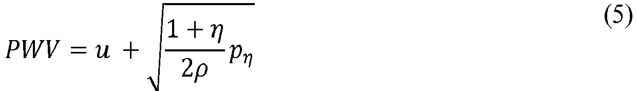
- The partial derivative pη indicates sensitivity of pressure with respect to the wall normal deflection, and has a clear interpretation as tangent (incremental) moduli in finite strain deformation. In the general case, equation (5) is supplemented by appropriate constituent equations for a hyperelastic anisotropic arterial wall, accounting for finite deformation.
- It is assumed that arterial wall is hyperelastic, incompressible, anisotropic, and undergoing finite deformation. After a few original loading cycles (preconditioning) the arterial behavior follows some repeatable, hysteresis free pattern with a typical exponential stiffening effect regarded as pseudo elastic. The strain energy density function W for the pseudo elastic constitutive relation may be presented in the form


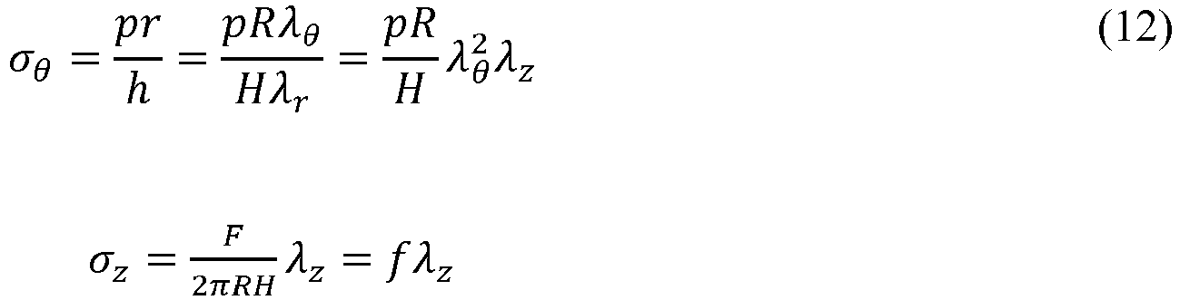
- With the geometry of the reference state determined, we define R, Z, H - as an internal radius, axial coordinate and a wall thickness in a stress free configuration, r,z,h - internal radius, axial coordinate and a wall thickness in a physiologically loaded configuration. The corresponding principal stretch ratios are

- Assuming isochoric deformation incorporate the incompressibility condition as

- The Green-Lagrangian strain components relate to the principal stretch ratios of Eq. 12 by

- For the membrane thin walled cylindrical artery undergoing finite inflation and axial deformation, the load - stress relations follow from the static conditions
- The solution of equations (13), (11), (7) results in a load - strain relations, which with account of the identity

- Arterial stiffness, or its reciprocals, arterial compliance and distensibility, may provide indication of vascular changes that predispose to the development of major vascular disease. In an isolated arterial segment filled with a moving fluid, compliance is defined as a change of a volume V for a given change of a pressure, and distensibilty as a compliance divided by initial volume. As functions of pressure the local (tangent) compliance C and distensibility D are defined as

- Equations (14) determine arterial wall properties as local functions of transmural pressure.
- We present equations (14) in the following equivalent form

- The classical results are generalized for the case of a hyperelastic arterial wall with account of finite deformation and flow velocity. To proceed, determine pη from Equation (5) and substitute in Equation (15), arriving at the following relations

-
Figure 2 illustrates the dependency of distensibility on pressure and flow velocity. Since PWV is monotonically increasing with pressure, distensibility is a decreasing function. Unlike the classical Bramwell-Hill model, which being linked to the Moens-Korteweg wave speed predicts arterial distensibility as a constant irrespective to the pressure level, the present model predicts distensibility as a function of PWV, pressure and a blood flow. - Arterial constants can be defined based on the developed mathematical model. The Cauchy stress components based on Fung's energy are presented in equations (6), (7), and (8). Neglecting longitudinal stress (σz = 0) in equation (8), we obtain


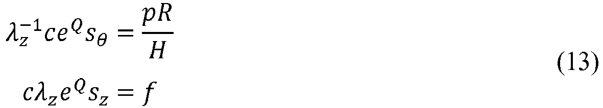
- It follows from Equations (7), (17) that a circumferential stress is a function of a circumferential strain and two material constants, a and c


- The governing equation specifies an equilibrium condition

- An embodiment of the calibration includes the use of published values for a population or segment of a population. Referenced values for material constants c, a 11, a 12, a 22 can be used to calculate the material constant a based on equation (20). The material constant c is used directly. A reference value for the mid radius in the stress free state (Rm ) is used along with a reference value for the aterial wall thickness (h) and associated mid radius (rm ) for the loaded wall. The material parameters (a, c, Rm ) along with the product of wall thickness and mid radius for the loaded wall (hrm ) can then be used to determine at least one of blood pressure and arterial compliance, e.g., distensibility, of a subject by measuring PWV and flow velocity.
- Another embodiment of the calibration is disclosed as follows and described in
Figure 8 . Assuming we have k measurements of the radius and pressure during a cardiac cycle or cycles, the following four variables are defined (rik , rok , rmk , pk ). By using these sampled variables circumferential stress can be presented as a function of three unknowns
- Now using a mathematical optimization, e.g., a least square (LS) minimization technique identifies (a,c,Rm )

- The following nonlinear calibration method describes one approach that may be completed to determine arterial constants.
- Step 1: Obtain k measurements, k ≥ 3, for blood pressure - pk, internal radius - rik; outer radius - rok, calculate mean radii rmk=0.5(rik+ rok) and wall thicknesses hk = rok- rik. For example, tonometry could be used to measure a continuous blood pressure to create the array of blood pressures, and Doppler speckle ultrasound could be used to measure artery radii to create the corresponding array of rik, rok.
- Step 2: Run a least square minimization as in equation (23) to identify the three constants (two material constants a, c and the mean radius Rm , in a load free condition). Substituting σθ in equation (23) with equation (18) results in


Since we have 3 unknowns, at least 3 sets of pressure and associated outer and inner radii are required. - In an embodiment, a calibrated model can be used to determine at least one of blood pressure and arterial compliance, e.g., distensibility, of a subject by measuring PWV and flow velocity.
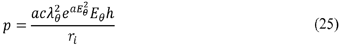
- With the three material properties (a,c,Rm ) and the constant product (hrm ) a blood pressure may be estimated based on the equilibrium equation (21) rearranged to

- The circumferential strain (Eθ ) can be defined in terms of η

- Wall thickness follows from incompressibility conditions


- Internal radius

- These formulations provide a relationship between pressure (p), circumferential strain (η), two arterial material parameters (a and c) and two arterial geometric parameters (Rm and the constant product hkrmk ).
- Equation (5) for PWV in arterial tissues can be re-arranged

- This relation can be used in combination with equation (16) to define distensibility D in terms of the pη

- In an embodiment, a lookup table can be created for convenience to enable determination of a blood pressure and distensibility based on the 4 arterial parameters (can be subject specific) and measurement of PWV and flow velocity. A blood density ρ is either measured or a value is assumed based on age and gender. As illustrated in an embodiment shown in
Figure 3 , the steps to create the blood pressure and distensibility lookup table are as follows:
Set η=0. - Using equation (26) calculate circumferential stretch ratio λθ .
- Using equation (27) calculate circumferential strain Eθ .
- Using equation (29) and any hkrmk product from the calibration, calculate wall thickness A.
- Using equation (30) calculate the internal radius ri .
- Using equation (25) calculate pressure p.
- While η<0.5, η= η+0.005, go back to calculation of the circumferential stretch ratio or else continue.
- Calculate the array for pη using the slope of the (p,η) curve.
- Using equation (31) and the array of values for η and pη calculate a 1D array for pη (1 + η).
- Using equation (31) and the array of pη , calculate D for each value of η.
- An example resultant array is shown in
Figure 4 . This lookup table enables determination of a subject blood pressure and distensibility by measuring PWVf and identifying the row (either directly or by interpolation) where
- A method for monitoring a blood pressure of a subject is disclosed. The model is calibrated for the subject (or population) by a non-linear calibration. For example, a non-linear calibration as illustrated in
Figure 3 can be performed. Once the calibration is complete, a patient specific lookup table can be formed for convenience as described inFigure 3 and as shown inFigure 4 . The method for monitoring a subject's real time blood pressure is shown inFigure 5 . An ECG measurement can be made for use as a timing reference for start of the pulse wave transit time, or to be used as a reference for determining acceptance time windows for other waveform features, or for averaging waveforms. An arterial flow velocity is measured providing minimum and maximum flow velocities, although a minimum could be assumed to be zero. An average flow velocity can also be measured. The flow velocity can also be estimated based on other measures or as a percentage of PWV. The pulse wave velocity is measured, ideally providing both a systolic and diastolic PWV. The subject (or population based) blood density ρ are then used with the lookup table ofFigure 4 to estimate blood pressure. The arrays of associated blood pressure (p), PWVf and distensibility (D) can also be used directly or with interpolation to estimate p and D based on the PWVf measure. The systolic PWV and the peak flow velocity are used to estimate a systolic blood pressure, while a diastolic PWV and the minimum flow velocity are used to estimate a diastolic blood pressure. Although other estimates and combinations may be used to estimate an average or systolic or diastolic blood pressure. PWV can be measured at the foot and the systolic flow velocity (u) can be added to it as an estimate of systolic PWVf. Diastolic pressure could use the raw PWV measured at the foot and use either the minimum flow velocity or assume u=0. Other empirical or relational estimates for the systolic and diastolic PWVf can also be used. - Determination of PWV includes measurement of the transit time of the pulse wave between two points, and a measure or estimate of the distance traveled. The PWV is the distance travelled divided by the time difference. This can be done by extracting the foot (
Figure 1 , item 12) or peak (Figure 1 , item 13) of the pressure wave in two locations (proximal, distal) of the same artery, calculating the time difference between these two extracted features, and measuring the distance between these two measurement points. In one embodiment, this is done in the radial artery as shown inFigure 7 with measurements items 80 (proximal) and 81 (distal). Use of the foot location on the pressure or PPG waveform (items 801 and 810) will correlate to a diastolic PWV, while use of the peak location (items 802 and 811) will correlate to a systolic PWV. Two different arteries can also be used with a measurement or estimate of the arterial path distance between the two measurement points. The PWV can also be measured using the heart as the proximal measurement point with an electrocardiogram feature (e.g., the ECG r-wave) as the first time point, or by sensing when the aortic valve opens using a feature on measured waveforms or images such as the ballistocardiogram (BCG), ultrasound imaging, Doppler ultrasound, impedance plethysmography (IPG), or photoplethysmography (PPG) on the chest over the aorta. The distal pressure wave is then used as above to extract a second time point. The arterial distance between the aortic root and the distal measurement point is used in the calculation of PWV. This can be measured or estimated, and may be based on subject characteristics such as height, weight age and gender. The distal pressure wave can be measured using sensors such as tonometry, an arterial cuff, ultrasound, RF based arterial wall tracking, and PPG. - In some embodiments, the wave will propagate across multiple arterial segments between the proximal and distal points of pressure measurement. This measurement can be used in at least two ways. In the first form, average properties of the vessel segments, radius, and modulus will be considered so that the result corresponds to bulk average of the segment. In the second, the properties of individual arterial segments are determined. First, use the model to determine the relative transit time through each sequential arterial segment based on geometrical properties of each segment and assuming a similar pressure within all segments. Then using a solution method, such as minimization of a least squares or another method, solve for the PWV within each segment by recognizing that the total transit time (measured) is the sum of the transit time through each segment.
- Measurement of flow velocity can be done using Doppler ultrasound, an inductive coil, MRI or CT scan with contrast agents. The flow velocity can be captured as a continuous wave, as a peak value, or a minimum value (including u=0). It is also possible to estimate flow velocity using related measures or with a scale factor. For example PWV can be measured using previously described techniques and flow velocity is then estimated as a percentage of PWV (for example u = 0.2PWV). Aortic flow velocity can be estimated through left ventricular ejection time (LVET), ejection volume (EV), and aortic cross-sectional area (CA) where u = EV / (LVET ∗ CA). Left ventricular ejection time (LVET) can be measured or estimated using a number of sensors (e.g. PPG, heart sound). Using PPG for example, the measure of LVET is the length of time from the foot of the PPG wave (
Figure 1 item 12) to the dicrotic notch (item 14). Using heart sound, LVET is the time between the first and second heart sound. Ejection volume can be based on direct measurement for the subject (e.g. ultrasound, thermal dilution, etc) at rest and at exercise with subsequent scaling based on heart rate. The cross-sectional area can be directly measured by ultrasound imaging, MRI, or CT scans. EV and CA can also be based on subject specific parameters such as age, gender, height and weight. EV can also be measured or estimated using features from the BCG such as the amplitude of the j-wave or m-wave. An estimate of flow velocity in the periphery can be made based on scaling of the blood volume in that arterial tree branch, and relative arterial size as compared to the aorta. Although a direct measurement of flow velocity or an estimate based on PWV is preferred in the periphery. - Pressure can be measured using any approved technique, for example, brachial cuff, tonometry, or intra-arterial catheter. Ideally a continuous method (e.g., tonometry, intra-arterial) is used with a method of time synchronization to the flow and PWV measures (e.g. via ECG). However serial measures can also be used. Here the pressure p can be systolic, diastolic, or any intermediate pressure (e.g., mean pressure) when coupled with the appropriate flow velocity (u). For example, systolic pressure could be associated with the peak flow velocity, and diastolic pressure could be associated with the lowest (or zero) flow velocity, or an average pressure could be associated with an average flow velocity.
- An optional ECG can be measured across the chest or wrists. Other locations are also possible such as ear lobes, behind the ears, buttocks, thighs, fingers, feet or toes. PPG can be measured at the chest or wrist. Other locations such as the ear lobes, fingers, forehead, buttocks, thighs, and toes also work. Video analysis methods examining changes in skin color can also be used to obtain a PPG waveform. Flow velocity can be measured at the chest or wrist. Other locations for flow velocity measure are also possible such as the neck, arm and legs.
- In one embodiment, the pulse transit time is measured based on aortic valve opening determined by the J-wave of the BCG waveform, and a PPG foot measured (e.g.,
Figure 1 , item 12) from the thigh. The ECG r-wave may be used as a reference for determining acceptance windows for BCG and PPG feature delineations, or as a starting point for a PWV measure. The aorta distance is estimated from aortic root along the path of the aorta to the femoral artery at the thigh PPG measurement location. The PWV is calculated by dividing the aorta distance by the measured time difference (BCG J-wave to PPG foot). The minimum and maximum flow velocity is measured by ultrasound Doppler at the aortic root. A blood density ρ is assumed based on age and gender of the subject. Using the measured PWVf as shown inFigure 5 the lookup table ofFigure 4 is used to determine the associated pressure. To calculate pd the minimum measured flow velocity can be used in combination with an estimate of diastolic PWV, where diastolic PWV is calculated based on the BCG J-wave to PPG foot time difference. To calculate ps the peak flow velocity or a percentage of the peak flow velocity can be used in combination with an estimate of systolic PWV, where the systolic PWV is calculated based on the peak flow velocity time point to the PPG peak time difference. Under conditions where flow velocity is not measured, diastolic flow velocity can be assumed to be 0, while systolic flow velocity can be estimated as a percentage of PWV (e.g ~20%). - In another embodiment, the pulse transit time is measured from the carotid artery using tonometry, to the pressure pulse measured at thigh with a thigh cuff. The arterial distance is estimated from aortic root along the path of the aorta to the femoral artery at the thigh cuff measurement location. The PWV is calculated by dividing the arterial distance by the measured time difference. The foot to foot timing on the measured pressure pulses (e.g.,
Figure 7 items 801 and 810) is used to determine a diastolic pulse transit time and to calculate a diastolic PWV. The peak to peak timing (e.g.,Figure 7 ,items 802 and 811) is used to determine a systolic pulse transit time and to calculate a systolic PWV. The peak flow velocity is estimated at 20% of the systolic PWV while the minimum flow velocity is estimated at zero. A blood density ρ is assumed based on age and gender of the subject. Using the measured PWVf , as shown inFigure 5 , the lookup table ofFigure 4 is used to determine the associated pressure. To calculate pd the minimum measured flow velocity can be used in combination with the diastolic PWV. To calculate ps the peak flow velocity is used in combination with the systolic PWV. - A method for monitoring an arterial compliance of a subject is disclosed. The model is calibrated for the subject (or population) by a non-linear calibration. For example, a non-linear calibration as illustrated in
Figure 8 can be performed. Once the calibration is complete, a patient specific lookup table can be formed for convenience described inFigure 3 and as shown inFigure 4 . The method for monitoring a subject's arterial compliance is shown inFigure 6 . An optional ECG measurement can be made for use as a timing reference for start of the pulse wave transit time, or to be used as a reference for determining acceptance time windows for other waveform features, or for averaging waveforms. An arterial flow velocity is measured providing minimum and maximum flow velocities, although a minimum could be assumed to be zero. An average flow velocity can also be measured. The flow velocity can also be estimated based on other measures or as a percentage of PWV. The pulse wave velocity is measured, ideally providing both a systolic and diastolic PWV. The subject (or population based) blood density ρ is then used with the patient specific lookupFigure 4 , to determine compliance as shown inFigure 6 and equation 16. - The systolic PWV and the peak flow velocity are used to determine a systolic distensibility, while a diastolic PWV and the minimum flow velocity are used to estimate a diastolic distensibility. Although other estimates and combinations may be used to determine the subject distensibility parameter.
- In one embodiment, the pulse transit time is measured based on aortic valve opening determined by the J-wave of the BCG waveform, and a PPG foot measured (e.g.,
Figure 1 , item 12) from the thigh. The aorta distance is estimated from aortic root to femoral artery at the thigh PPG measurement location. The PWV is calculated by dividing the aorta distance by the measured time difference (BCG J-wave to PPG foot). The minimum flow velocity is assumed to be zero, enabling distensibility calculation without a direct flow measurement. The flow corrected PWV is calculated (in this embodiment PWVf=PWV since u=0). A blood density ρ is assumed based on age and gender of the subject with the patient specific lookupFigure 4 to determine compliance as shown inFigure 6 and equation 16. Measures of a systolic PWV, peak flow velocity, may also be used to determine D. - In another embodiment, the pulse transit time is measured from the carotid artery using tonometry, to the pressure pulse measured at thigh with a thigh cuff. The arterial distance is estimated from aortic root along the path of the aorta to the femoral artery at the thigh cuff measurement location. The PWV is calculated by dividing the arterial distance by the measured time difference. The foot to foot timing on the measured pressure pulses (e.g.,
Figure 8 items 801 and 810) are used to determine a diastolic pulse transit time and to calculate a diastolic PWV. The peak to peak timing (e.g.,Figure 7 ,items 802 and 811) is used to determine a systolic pulse transit time and to calculate a systolic PWV. The peak flow velocity is estimated at 20% of the systolic PWV while the minimum flow velocity is estimated at zero. A blood density ρ is assumed based on age and gender of the subject with the patient specific lookupFigure 4 to determine compliance as shown inFigure 6 and equation 16. - The disclosure will be further illustrated with reference to the following specific examples. It is understood that these examples are given by way of illustration and are not meant to limit the disclosure or the claims to follow.
- This paper example uses referenced values for an aorta c=120123Pa, a11 =0.320, a12 =0.068, a22 =0.451. The reduced constant 'a' is calculated based on equation 20 (a=0.31). In addition reference values were used for the mid radius for the stress free vessel Rm =0.009m, the aortic wall thickness h=0.00211m, and the mid radius of the loaded wall rm = 0.011. A computer routine executed the functions outlined in
Figure 3 to calculate an array of pη (1 + η), D, and the associated pressures (p) for each value of η. Equation (31) was then used along with a reference value of blood density ρ=1060kg/m3 to create an array of associated PWVf for each value of η. The pulse transit time is measured based on time synchronized tonometry at the carotid artery and at the femoral artery, each providing a pressure pulse waveform. The equivalent aortic distance between the two measurement points is measured at for example 1.0m. The foot to foot time difference between the two tonometry waveforms is measured at for example 200 ms. The diastolic PWV is then calculated as 1.0m/200 ms = 5.0m/s. From the same tonometry waveforms the peak to peak time difference is measured at for example 140 ms. The systolic PWV is then calculated as 1.00m/140 ms = 7.14m/s. Flow velocity is measured using Doppler ultrasound at the aortic root, with the minimum taken as the diastolic flow velocity


Claims (13)
- A method for determining at least one of a blood pressure, an arterial compliance parameter, or an arterial distensibility of a subject, the method comprising;measuring a value for pulse wave velocity within an arterial segment or segments of a subject;providing a value for flow velocity within the arterial segment or segments of the subject;providing a value for blood density of the subject;determining values of material characteristics of an artery; andcalculating, by a computer, at least one of a blood pressure, an arterial compliance parameter, or an arterial distensibility of the subject by inputting the value for pulse wave velocity, the value for flow velocity, the value for blood density, and the values of the material characteristics of the artery into a calibrated model of a fluid-structure interaction incorporating conservation of mass and momentum for a fluid, and non-linear elasticity of a structure.
- The method of claim 1, wherein the value for flow velocity is measured directly from the subject.
- The method of claim 1, wherein the value for flow velocity is estimated on the value for pulse wave velocity for the subject.
- The method of claim 1, wherein the blood pressure is a systolic blood pressure.
- The method of claim 1, wherein the blood pressure is a diastolic blood pressure.
- The method of claim 4, wherein a peak pulse wave velocity is associated with the systolic blood pressure.
- The method of claim 5, wherein a minimum pulse wave velocity is associated with the diastolic blood pressure.
- The method of claim 4, wherein a peak flow velocity is associated with the systolic blood pressure.
- The method of claim 5, wherein a minimum flow velocity is associated with the diastolic blood pressure.
- The method of claim 1, wherein the value for pulse wave velocity is measured using at least one of photoplethysmography, impedance plethysmography, doppler ultrasound, an electrocardiogram, or a ballistocardiogram.
- The method of claim 1, wherein determining the values of the material characteristics of an artery includes calculating at least one material constant using reference values associated with a segment of a population.
- The method of claim 1, wherein the values of the material characteristics of an artery include a and c, Rm , where a is a reduced material constant,

- The method of claim 1, wherein the values of the material characteristics of an artery include one or more material constants associated with a strain energy density function representative of a pseudo-elastic behavior of the artery.
Applications Claiming Priority (3)
| Application Number | Priority Date | Filing Date | Title |
|---|---|---|---|
| US201462080738P | 2014-11-17 | 2014-11-17 | |
| US201462080740P | 2014-11-17 | 2014-11-17 | |
| PCT/US2015/061190 WO2016081519A1 (en) | 2014-11-17 | 2015-11-17 | Blood pressure and arterial compliance estimation from arterial segments |
Publications (3)
| Publication Number | Publication Date |
|---|---|
| EP3220811A1 EP3220811A1 (en) | 2017-09-27 |
| EP3220811A4 EP3220811A4 (en) | 2018-07-25 |
| EP3220811B1 true EP3220811B1 (en) | 2023-10-18 |
Family
ID=56014473
Family Applications (2)
| Application Number | Title | Priority Date | Filing Date |
|---|---|---|---|
| EP15860209.4A Active EP3220811B1 (en) | 2014-11-17 | 2015-11-17 | Blood pressure and arterial compliance estimation from arterial segments |
| EP15861809.0A Active EP3220812B1 (en) | 2014-11-17 | 2015-11-17 | Pulse wave velocity, arterial compliance, and blood pressure |
Family Applications After (1)
| Application Number | Title | Priority Date | Filing Date |
|---|---|---|---|
| EP15861809.0A Active EP3220812B1 (en) | 2014-11-17 | 2015-11-17 | Pulse wave velocity, arterial compliance, and blood pressure |
Country Status (3)
| Country | Link |
|---|---|
| US (3) | US11622730B2 (en) |
| EP (2) | EP3220811B1 (en) |
| WO (2) | WO2016081517A2 (en) |
Families Citing this family (22)
| Publication number | Priority date | Publication date | Assignee | Title |
|---|---|---|---|---|
| US11622730B2 (en) | 2014-11-17 | 2023-04-11 | Rochester Institute Of Technology | Pulse wave velocity, arterial compliance, and blood pressure |
| AU2015367622B2 (en) * | 2014-12-16 | 2020-01-23 | Lmd Ip, Llc | Personal health data collection |
| EP3073400B1 (en) * | 2015-03-25 | 2022-05-04 | Tata Consultancy Services Limited | System and method for determining psychological stress of a person |
| US10893809B2 (en) | 2016-11-10 | 2021-01-19 | Auburn University | Method and system for evaluating blood vessel |
| US10772571B2 (en) | 2016-11-15 | 2020-09-15 | Welch Allyn, Inc. | Method and systems for correcting for arterial compliance in a blood pressure assessment |
| WO2018136135A1 (en) | 2017-01-18 | 2018-07-26 | Physio-Control, Inc. | Non-invasive blood pressure measurement using ultrasound |
| US10939833B2 (en) | 2017-05-01 | 2021-03-09 | Samsung Electronics Company, Ltd. | Determining artery location using camera-based sensing |
| FR3067220B1 (en) * | 2017-06-13 | 2019-07-26 | Centre National De La Recherche Scientifique | DEVICE FOR INFUSION OF AN ORGAN |
| US11013488B2 (en) * | 2017-06-23 | 2021-05-25 | Stryker Corporation | Patient monitoring and treatment systems and methods |
| US11413005B2 (en) | 2017-08-14 | 2022-08-16 | Stryker Corporation | Constitutive equation for non-invasive blood pressure measurement systems and methods |
| US11357415B2 (en) | 2017-10-27 | 2022-06-14 | Stryker Corporation | Light-based non-invasive blood pressure systems and methods |
| CN108403093B (en) * | 2018-02-27 | 2021-12-14 | 京东方科技集团股份有限公司 | Apparatus and method for determining the location of blood vessels |
| EP3669762A1 (en) | 2018-12-18 | 2020-06-24 | Koninklijke Philips N.V. | Control unit for deriving a measure of arterial compliance |
| FR3092694B1 (en) * | 2019-02-13 | 2023-01-13 | Hagelsteen Marc | Chronotonometric diagram of the distribution of the pulse wave in humans for research in human physiology and the teaching of this discipline |
| US11534071B2 (en) * | 2019-04-12 | 2022-12-27 | Beijing Shunyuan Kaihua Technology Limited | Blood pressure measurement |
| US12102415B2 (en) * | 2019-11-07 | 2024-10-01 | The Curators Of The University Of Missouri | Systems and methods to improve management and monitoring of cardiovascular disease |
| CN111000537B (en) * | 2019-12-24 | 2022-05-27 | 中国人民解放军陆军军医大学第一附属医院 | Method for correcting influence of age, sex and blood pressure on pulse wave propagation speed |
| CN112842287B (en) * | 2021-01-05 | 2022-05-17 | 清华大学 | Device and method for measuring vascular stiffness parameters |
| CN113706984B (en) * | 2021-08-06 | 2022-07-12 | 西安交通大学 | Synchronous analog calibration device and method for blood pressure and reflective photoelectric accumulated wave |
| US20230142080A1 (en) * | 2021-08-18 | 2023-05-11 | Second Medical Center Of Chinese Pla General Hospital | Glove for detecting multiple physiological parameters and system for detecting risk of hypertension disease |
| WO2025086237A1 (en) * | 2023-10-27 | 2025-05-01 | Huawei Technologies Co., Ltd. | Determination of arterial blood pressure |
| US20250248603A1 (en) * | 2024-02-05 | 2025-08-07 | Wisconsin Alumni Research Foundation | Apparatus and Method for Characterizing Arterial Stiffness |
Family Cites Families (49)
| Publication number | Priority date | Publication date | Assignee | Title |
|---|---|---|---|---|
| US2658505A (en) * | 1949-03-08 | 1953-11-10 | Sheer Charles | Arterial pulse wave velocity meter |
| US4799491A (en) | 1986-11-06 | 1989-01-24 | Sri International | Blood pressure monitoring method and apparatus |
| EP0467853B1 (en) | 1990-07-18 | 1996-01-10 | AVL Medical Instruments AG | Device and method for the measurement of blood pressure |
| US5687731A (en) | 1992-09-10 | 1997-11-18 | Mti, Ltd. | Oscillometric method for determining hemodynamic parameters of the arterial portion of patient's circulatory system and a measuring system for its realization |
| US5565466A (en) * | 1993-08-13 | 1996-10-15 | Zonagen, Inc. | Methods for modulating the human sexual response |
| US6575969B1 (en) * | 1995-05-04 | 2003-06-10 | Sherwood Services Ag | Cool-tip radiofrequency thermosurgery electrode system for tumor ablation |
| US5743856A (en) | 1995-11-06 | 1998-04-28 | Colin Corporation | Apparatus for measuring pulse-wave propagation velocity |
| EP0832604A1 (en) * | 1996-09-30 | 1998-04-01 | Koninklijke Philips Electronics N.V. | Method and device for measuring the elasticity of an artery by ultrasonic echography |
| DE19746377C1 (en) | 1997-10-21 | 1999-07-01 | Fresenius Medical Care De Gmbh | Blood treatment device with a device for continuous monitoring of the patient's blood pressure |
| US6413223B1 (en) | 1999-06-01 | 2002-07-02 | Massachussetts Institute Of Technology | Cuffless continuous blood pressure monitor |
| US6984207B1 (en) * | 1999-09-14 | 2006-01-10 | Hoana Medical, Inc. | Passive physiological monitoring (P2M) system |
| CA2388033A1 (en) * | 1999-10-07 | 2001-04-12 | Alexander K. Mills | Device and method for noninvasive continuous determination of physiologic characteristics |
| AU2001221391A1 (en) | 2000-01-26 | 2001-08-07 | Vsm Medtech Ltd. | Continuous blood pressure monitoring method and apparatus |
| US6647287B1 (en) | 2000-04-14 | 2003-11-11 | Southwest Research Institute | Dynamic cardiovascular monitor |
| US6890303B2 (en) | 2001-05-31 | 2005-05-10 | Matthew Joseph Fitz | Implantable device for monitoring aneurysm sac parameters |
| WO2003034916A2 (en) | 2001-08-17 | 2003-05-01 | Russell Ted W | Methods, apparatus and sensor for hemodynamic monitoring |
| US6990222B2 (en) | 2001-11-21 | 2006-01-24 | Arnold Ben A | Calibration of tissue densities in computerized tomography |
| US20050107710A1 (en) * | 2002-04-24 | 2005-05-19 | Ryu Nakayama | Pulse wave analyzing method, pulse wave analyzing software, and so forth |
| US20040088123A1 (en) * | 2002-11-01 | 2004-05-06 | Zhong Ji | Method for real-time monitoring of cardiac output and blood flow in arteries and apparatus for implementing the same |
| US20140296677A1 (en) | 2003-09-18 | 2014-10-02 | New Paradigm Concepts, LLC | Method of measuring total vascular hemoglobin mass |
| TWI250867B (en) * | 2003-10-22 | 2006-03-11 | Surewin Technology Corp | Pulse wave analysis device |
| US7318804B2 (en) | 2003-12-09 | 2008-01-15 | The Regents Of The University Of Michigan | Methods and systems for measuring mechanical property of a vascular wall and method and system for determining health of a vascular structure |
| US7179228B2 (en) | 2004-04-07 | 2007-02-20 | Triage Wireless, Inc. | Cuffless system for measuring blood pressure |
| US7672706B2 (en) | 2004-08-23 | 2010-03-02 | Boston Scientific Scimed, Inc. | Systems and methods for measuring pulse wave velocity with an intravascular device |
| US7621876B2 (en) | 2005-03-17 | 2009-11-24 | Ge Medical Systems Information Technologies, Inc. | Continuous, non-invasive technique for determining blood pressure using a transmission line model and transcutaneous ultrasound measurements |
| US7674231B2 (en) | 2005-08-22 | 2010-03-09 | Massachusetts Institute Of Technology | Wearable pulse wave velocity blood pressure sensor and methods of calibration thereof |
| US20070163353A1 (en) * | 2005-12-07 | 2007-07-19 | Drexel University | Detection of blood pressure and blood pressure waveform |
| US8684900B2 (en) | 2006-05-16 | 2014-04-01 | Bao Tran | Health monitoring appliance |
| WO2008057478A2 (en) * | 2006-11-03 | 2008-05-15 | The Regents Of The University Of Michigan | Method and system for determining volume flow in a blood conduit |
| JP5297593B2 (en) | 2007-03-07 | 2013-09-25 | 国立大学法人 東京大学 | Synthesis method of polymer complex crystals |
| US20110144967A1 (en) * | 2008-08-12 | 2011-06-16 | Lev Adirovich | System and method for dynamic cardiac analysis, detection, prediction, and response using cardio-physiological mathematical modeling |
| EP2326241B1 (en) | 2008-08-19 | 2017-10-11 | Koninklijke Philips N.V. | Monitoring the blood pressure of a patient |
| US20100081946A1 (en) | 2008-09-26 | 2010-04-01 | Qualcomm Incorporated | Method and apparatus for non-invasive cuff-less blood pressure estimation using pulse arrival time and heart rate with adaptive calibration |
| CN102170821B (en) * | 2008-10-01 | 2013-08-07 | 株式会社Irumedi | Cardiovascular Analysis Device |
| US20100106016A1 (en) * | 2008-10-23 | 2010-04-29 | Skeletal Dynamics Llc | Non-Invasive Blood Pressure Monitoring Device and Method |
| US8554490B2 (en) * | 2009-02-25 | 2013-10-08 | Worcester Polytechnic Institute | Automatic vascular model generation based on fluid-structure interactions (FSI) |
| US8313439B2 (en) * | 2009-03-20 | 2012-11-20 | Massachusetts Institute Of Technology | Calibration of pulse transit time measurements to arterial blood pressure using external arterial pressure applied along the pulse transit path |
| JP5615073B2 (en) | 2010-07-20 | 2014-10-29 | オムロンヘルスケア株式会社 | measuring device |
| DE102010055772B4 (en) * | 2010-12-23 | 2014-06-05 | Carl Zeiss Meditec Ag | Arrangement and method for the quantitative determination of blood flow within blood vessels |
| EP2667769B1 (en) | 2011-01-27 | 2020-07-22 | The Board of Trustees of the Leland Stanford Junior University | Systems for monitoring the circulatory system |
| CN103648374B (en) * | 2011-02-17 | 2016-08-17 | 高通股份有限公司 | Methods and systems for determining cardiovascular volume in mammals |
| US20120215117A1 (en) * | 2011-02-23 | 2012-08-23 | Pacesetter, Inc. | Systems and methods for estimating central arterial blood pressure of a patient |
| TW201309263A (en) | 2011-08-19 | 2013-03-01 | 中原大學 | Measurement device and measurement method thereof for image-type pulse wave transduction velocity |
| WO2013110929A1 (en) | 2012-01-26 | 2013-08-01 | King's College London | Aortic pulse wave velocity measurement |
| EP2916725A4 (en) | 2012-11-08 | 2016-07-06 | Le Thai | Improved blood pressure monitor and method |
| JP2014100249A (en) * | 2012-11-19 | 2014-06-05 | Toshiba Corp | Blood vessel analysis device, medical image diagnostic device, blood vessel analysis method, and blood vessel analysis program |
| JP2016538097A (en) | 2013-10-23 | 2016-12-08 | クアンタス, インコーポレイテッド | Consumer biometric devices |
| US20150164351A1 (en) | 2013-10-23 | 2015-06-18 | Quanttus, Inc. | Calculating pulse transit time from chest vibrations |
| US11622730B2 (en) | 2014-11-17 | 2023-04-11 | Rochester Institute Of Technology | Pulse wave velocity, arterial compliance, and blood pressure |
-
2015
- 2015-11-17 US US15/527,412 patent/US11622730B2/en active Active
- 2015-11-17 WO PCT/US2015/061188 patent/WO2016081517A2/en not_active Ceased
- 2015-11-17 US US15/527,467 patent/US20170354331A1/en not_active Abandoned
- 2015-11-17 EP EP15860209.4A patent/EP3220811B1/en active Active
- 2015-11-17 EP EP15861809.0A patent/EP3220812B1/en active Active
- 2015-11-17 WO PCT/US2015/061190 patent/WO2016081519A1/en not_active Ceased
-
2023
- 2023-02-27 US US18/114,585 patent/US20240023898A1/en not_active Abandoned
Also Published As
| Publication number | Publication date |
|---|---|
| WO2016081517A3 (en) | 2016-08-04 |
| EP3220812A4 (en) | 2018-07-11 |
| EP3220811A4 (en) | 2018-07-25 |
| EP3220812A2 (en) | 2017-09-27 |
| US20170354331A1 (en) | 2017-12-14 |
| US11622730B2 (en) | 2023-04-11 |
| WO2016081519A1 (en) | 2016-05-26 |
| EP3220811A1 (en) | 2017-09-27 |
| EP3220812B1 (en) | 2023-10-25 |
| US20240023898A1 (en) | 2024-01-25 |
| WO2016081517A2 (en) | 2016-05-26 |
| US20190083045A1 (en) | 2019-03-21 |
Similar Documents
| Publication | Publication Date | Title |
|---|---|---|
| EP3220811B1 (en) | Blood pressure and arterial compliance estimation from arterial segments | |
| Barvik et al. | Noninvasive continuous blood pressure estimation from pulse transit time: A review of the calibration models | |
| Nabeel et al. | Local pulse wave velocity: theory, methods, advancements, and clinical applications | |
| US11241170B2 (en) | Monitor for blood pressure and other arterial properties | |
| EP3157416B1 (en) | System for cuff-less blood pressure (bp) measurement of a subject | |
| Segers et al. | Limitations and pitfalls of non-invasive measurement of arterial pressure wave reflections and pulse wave velocity | |
| EP2967368B1 (en) | Apparatus and methods for computing cardiac output of a living subject via applanation tonometry | |
| JP6789280B2 (en) | Systems and methods for assessing endothelial function | |
| US12263032B2 (en) | Hemodynamic parameter estimation | |
| WO2012021765A2 (en) | Methods and apparatus for determining arterial pulse wave velocity | |
| Bote et al. | Evaluation of blood pressure estimation models based on pulse arrival time | |
| EP2764820A1 (en) | Left atrial pressure measurement method and left atrial pressure measurement device | |
| Kips et al. | The use of diameter distension waveforms as an alternative for tonometric pressure to assess carotid blood pressure | |
| EP3357418A1 (en) | Method and apparatus for estimating the transit time of the aortic pulse from time intervals measured between fiducial points of a ballistocardiogram | |
| Kim et al. | Quantification of wave reflection using peripheral blood pressure waveforms | |
| Joseph et al. | Arterial compliance probe for calibration free pulse pressure measurement | |
| Pena et al. | Sequential changes of longitudinal and radial myocardial deformation indices in the healthy neonate heart | |
| Segers et al. | Principles of vascular physiology | |
| Nabeel et al. | Deep learning for blood pressure estimation: an approach using local measure of arterial dual diameter waveforms | |
| Xiang et al. | Calibration of pulse wave transit time method in blood pressure measurement based on the korotkoff sound delay time | |
| Mafi et al. | Oscillometric blood pressure pulse morphology | |
| Zhu et al. | Diastolic function alteration mechanisms in physiologic hypertrophy versus pathologic hypertrophy are elucidated by model-based Doppler E-wave analysis | |
| Sudarsan et al. | Assessment of Endothelial Reactivity using Brachial Pulse Wave Velocity Response to Shear | |
| Baktash | Ratio-independent arterial stiffness-based blood pressure estimation | |
| Havlik et al. | Design and realization of hardware for measurement of hemodynamic parameters |
Legal Events
| Date | Code | Title | Description |
|---|---|---|---|
| STAA | Information on the status of an ep patent application or granted ep patent |
Free format text: STATUS: THE INTERNATIONAL PUBLICATION HAS BEEN MADE |
|
| PUAI | Public reference made under article 153(3) epc to a published international application that has entered the european phase |
Free format text: ORIGINAL CODE: 0009012 |
|
| STAA | Information on the status of an ep patent application or granted ep patent |
Free format text: STATUS: REQUEST FOR EXAMINATION WAS MADE |
|
| 17P | Request for examination filed |
Effective date: 20170516 |
|
| AK | Designated contracting states |
Kind code of ref document: A1 Designated state(s): AL AT BE BG CH CY CZ DE DK EE ES FI FR GB GR HR HU IE IS IT LI LT LU LV MC MK MT NL NO PL PT RO RS SE SI SK SM TR |
|
| AX | Request for extension of the european patent |
Extension state: BA ME |
|
| DAV | Request for validation of the european patent (deleted) | ||
| DAX | Request for extension of the european patent (deleted) | ||
| A4 | Supplementary search report drawn up and despatched |
Effective date: 20180627 |
|
| RIC1 | Information provided on ipc code assigned before grant |
Ipc: A61B 5/0456 20060101ALN20180621BHEP Ipc: A61B 5/02 20060101ALI20180621BHEP Ipc: G09B 23/30 20060101ALI20180621BHEP Ipc: A61B 5/021 20060101AFI20180621BHEP Ipc: A61B 5/0285 20060101ALI20180621BHEP Ipc: A61B 5/0295 20060101ALN20180621BHEP Ipc: G09B 23/28 20060101ALI20180621BHEP |
|
| STAA | Information on the status of an ep patent application or granted ep patent |
Free format text: STATUS: EXAMINATION IS IN PROGRESS |
|
| 17Q | First examination report despatched |
Effective date: 20210413 |
|
| RIC1 | Information provided on ipc code assigned before grant |
Ipc: A61B 5/0295 20060101ALN20230131BHEP Ipc: G09B 23/28 20060101ALI20230131BHEP Ipc: A61B 5/0285 20060101ALI20230131BHEP Ipc: A61B 5/02 20060101ALI20230131BHEP Ipc: G09B 23/30 20060101ALI20230131BHEP Ipc: A61B 5/021 20060101AFI20230131BHEP |
|
| RIC1 | Information provided on ipc code assigned before grant |
Ipc: A61B 5/0295 20060101ALN20230221BHEP Ipc: G09B 23/28 20060101ALI20230221BHEP Ipc: A61B 5/0285 20060101ALI20230221BHEP Ipc: A61B 5/02 20060101ALI20230221BHEP Ipc: G09B 23/30 20060101ALI20230221BHEP Ipc: A61B 5/021 20060101AFI20230221BHEP |
|
| GRAP | Despatch of communication of intention to grant a patent |
Free format text: ORIGINAL CODE: EPIDOSNIGR1 |
|
| STAA | Information on the status of an ep patent application or granted ep patent |
Free format text: STATUS: GRANT OF PATENT IS INTENDED |
|
| INTG | Intention to grant announced |
Effective date: 20230504 |
|
| P01 | Opt-out of the competence of the unified patent court (upc) registered |
Effective date: 20230524 |
|
| GRAS | Grant fee paid |
Free format text: ORIGINAL CODE: EPIDOSNIGR3 |
|
| GRAA | (expected) grant |
Free format text: ORIGINAL CODE: 0009210 |
|
| STAA | Information on the status of an ep patent application or granted ep patent |
Free format text: STATUS: THE PATENT HAS BEEN GRANTED |
|
| AK | Designated contracting states |
Kind code of ref document: B1 Designated state(s): AL AT BE BG CH CY CZ DE DK EE ES FI FR GB GR HR HU IE IS IT LI LT LU LV MC MK MT NL NO PL PT RO RS SE SI SK SM TR |
|
| REG | Reference to a national code |
Ref country code: GB Ref legal event code: FG4D |
|
| REG | Reference to a national code |
Ref country code: CH Ref legal event code: EP |
|
| REG | Reference to a national code |
Ref country code: IE Ref legal event code: FG4D |
|
| REG | Reference to a national code |
Ref country code: DE Ref legal event code: R096 Ref document number: 602015086164 Country of ref document: DE |
|
| REG | Reference to a national code |
Ref country code: LT Ref legal event code: MG9D |
|
| REG | Reference to a national code |
Ref country code: NL Ref legal event code: MP Effective date: 20231018 |
|
| REG | Reference to a national code |
Ref country code: AT Ref legal event code: MK05 Ref document number: 1621690 Country of ref document: AT Kind code of ref document: T Effective date: 20231018 |
|
| PG25 | Lapsed in a contracting state [announced via postgrant information from national office to epo] |
Ref country code: NL Free format text: LAPSE BECAUSE OF FAILURE TO SUBMIT A TRANSLATION OF THE DESCRIPTION OR TO PAY THE FEE WITHIN THE PRESCRIBED TIME-LIMIT Effective date: 20231018 |
|
| PG25 | Lapsed in a contracting state [announced via postgrant information from national office to epo] |
Ref country code: GR Free format text: LAPSE BECAUSE OF FAILURE TO SUBMIT A TRANSLATION OF THE DESCRIPTION OR TO PAY THE FEE WITHIN THE PRESCRIBED TIME-LIMIT Effective date: 20240119 |
|
| PG25 | Lapsed in a contracting state [announced via postgrant information from national office to epo] |
Ref country code: IS Free format text: LAPSE BECAUSE OF FAILURE TO SUBMIT A TRANSLATION OF THE DESCRIPTION OR TO PAY THE FEE WITHIN THE PRESCRIBED TIME-LIMIT Effective date: 20240218 |
|
| PG25 | Lapsed in a contracting state [announced via postgrant information from national office to epo] |
Ref country code: LT Free format text: LAPSE BECAUSE OF FAILURE TO SUBMIT A TRANSLATION OF THE DESCRIPTION OR TO PAY THE FEE WITHIN THE PRESCRIBED TIME-LIMIT Effective date: 20231018 |
|
| PG25 | Lapsed in a contracting state [announced via postgrant information from national office to epo] |
Ref country code: AT Free format text: LAPSE BECAUSE OF FAILURE TO SUBMIT A TRANSLATION OF THE DESCRIPTION OR TO PAY THE FEE WITHIN THE PRESCRIBED TIME-LIMIT Effective date: 20231018 |
|
| PG25 | Lapsed in a contracting state [announced via postgrant information from national office to epo] |
Ref country code: ES Free format text: LAPSE BECAUSE OF FAILURE TO SUBMIT A TRANSLATION OF THE DESCRIPTION OR TO PAY THE FEE WITHIN THE PRESCRIBED TIME-LIMIT Effective date: 20231018 |
|
| PG25 | Lapsed in a contracting state [announced via postgrant information from national office to epo] |
Ref country code: LT Free format text: LAPSE BECAUSE OF FAILURE TO SUBMIT A TRANSLATION OF THE DESCRIPTION OR TO PAY THE FEE WITHIN THE PRESCRIBED TIME-LIMIT Effective date: 20231018 Ref country code: IS Free format text: LAPSE BECAUSE OF FAILURE TO SUBMIT A TRANSLATION OF THE DESCRIPTION OR TO PAY THE FEE WITHIN THE PRESCRIBED TIME-LIMIT Effective date: 20240218 Ref country code: GR Free format text: LAPSE BECAUSE OF FAILURE TO SUBMIT A TRANSLATION OF THE DESCRIPTION OR TO PAY THE FEE WITHIN THE PRESCRIBED TIME-LIMIT Effective date: 20240119 Ref country code: ES Free format text: LAPSE BECAUSE OF FAILURE TO SUBMIT A TRANSLATION OF THE DESCRIPTION OR TO PAY THE FEE WITHIN THE PRESCRIBED TIME-LIMIT Effective date: 20231018 Ref country code: BG Free format text: LAPSE BECAUSE OF FAILURE TO SUBMIT A TRANSLATION OF THE DESCRIPTION OR TO PAY THE FEE WITHIN THE PRESCRIBED TIME-LIMIT Effective date: 20240118 Ref country code: AT Free format text: LAPSE BECAUSE OF FAILURE TO SUBMIT A TRANSLATION OF THE DESCRIPTION OR TO PAY THE FEE WITHIN THE PRESCRIBED TIME-LIMIT Effective date: 20231018 Ref country code: PT Free format text: LAPSE BECAUSE OF FAILURE TO SUBMIT A TRANSLATION OF THE DESCRIPTION OR TO PAY THE FEE WITHIN THE PRESCRIBED TIME-LIMIT Effective date: 20240219 |
|
| PG25 | Lapsed in a contracting state [announced via postgrant information from national office to epo] |
Ref country code: SE Free format text: LAPSE BECAUSE OF FAILURE TO SUBMIT A TRANSLATION OF THE DESCRIPTION OR TO PAY THE FEE WITHIN THE PRESCRIBED TIME-LIMIT Effective date: 20231018 Ref country code: RS Free format text: LAPSE BECAUSE OF FAILURE TO SUBMIT A TRANSLATION OF THE DESCRIPTION OR TO PAY THE FEE WITHIN THE PRESCRIBED TIME-LIMIT Effective date: 20231018 Ref country code: PL Free format text: LAPSE BECAUSE OF FAILURE TO SUBMIT A TRANSLATION OF THE DESCRIPTION OR TO PAY THE FEE WITHIN THE PRESCRIBED TIME-LIMIT Effective date: 20231018 Ref country code: NO Free format text: LAPSE BECAUSE OF FAILURE TO SUBMIT A TRANSLATION OF THE DESCRIPTION OR TO PAY THE FEE WITHIN THE PRESCRIBED TIME-LIMIT Effective date: 20240118 Ref country code: LV Free format text: LAPSE BECAUSE OF FAILURE TO SUBMIT A TRANSLATION OF THE DESCRIPTION OR TO PAY THE FEE WITHIN THE PRESCRIBED TIME-LIMIT Effective date: 20231018 Ref country code: HR Free format text: LAPSE BECAUSE OF FAILURE TO SUBMIT A TRANSLATION OF THE DESCRIPTION OR TO PAY THE FEE WITHIN THE PRESCRIBED TIME-LIMIT Effective date: 20231018 |
|
| REG | Reference to a national code |
Ref country code: CH Ref legal event code: PL |
|
| PG25 | Lapsed in a contracting state [announced via postgrant information from national office to epo] |
Ref country code: DK Free format text: LAPSE BECAUSE OF FAILURE TO SUBMIT A TRANSLATION OF THE DESCRIPTION OR TO PAY THE FEE WITHIN THE PRESCRIBED TIME-LIMIT Effective date: 20231018 |
|
| PG25 | Lapsed in a contracting state [announced via postgrant information from national office to epo] |
Ref country code: LU Free format text: LAPSE BECAUSE OF NON-PAYMENT OF DUE FEES Effective date: 20231117 |
|
| PG25 | Lapsed in a contracting state [announced via postgrant information from national office to epo] |
Ref country code: CH Free format text: LAPSE BECAUSE OF NON-PAYMENT OF DUE FEES Effective date: 20231130 |
|
| REG | Reference to a national code |
Ref country code: DE Ref legal event code: R097 Ref document number: 602015086164 Country of ref document: DE |
|
| PG25 | Lapsed in a contracting state [announced via postgrant information from national office to epo] |
Ref country code: CZ Free format text: LAPSE BECAUSE OF FAILURE TO SUBMIT A TRANSLATION OF THE DESCRIPTION OR TO PAY THE FEE WITHIN THE PRESCRIBED TIME-LIMIT Effective date: 20231018 |
|
| PG25 | Lapsed in a contracting state [announced via postgrant information from national office to epo] |
Ref country code: SK Free format text: LAPSE BECAUSE OF FAILURE TO SUBMIT A TRANSLATION OF THE DESCRIPTION OR TO PAY THE FEE WITHIN THE PRESCRIBED TIME-LIMIT Effective date: 20231018 |
|
| PG25 | Lapsed in a contracting state [announced via postgrant information from national office to epo] |
Ref country code: SM Free format text: LAPSE BECAUSE OF FAILURE TO SUBMIT A TRANSLATION OF THE DESCRIPTION OR TO PAY THE FEE WITHIN THE PRESCRIBED TIME-LIMIT Effective date: 20231018 Ref country code: SK Free format text: LAPSE BECAUSE OF FAILURE TO SUBMIT A TRANSLATION OF THE DESCRIPTION OR TO PAY THE FEE WITHIN THE PRESCRIBED TIME-LIMIT Effective date: 20231018 Ref country code: RO Free format text: LAPSE BECAUSE OF FAILURE TO SUBMIT A TRANSLATION OF THE DESCRIPTION OR TO PAY THE FEE WITHIN THE PRESCRIBED TIME-LIMIT Effective date: 20231018 Ref country code: LU Free format text: LAPSE BECAUSE OF NON-PAYMENT OF DUE FEES Effective date: 20231117 Ref country code: IT Free format text: LAPSE BECAUSE OF FAILURE TO SUBMIT A TRANSLATION OF THE DESCRIPTION OR TO PAY THE FEE WITHIN THE PRESCRIBED TIME-LIMIT Effective date: 20231018 Ref country code: EE Free format text: LAPSE BECAUSE OF FAILURE TO SUBMIT A TRANSLATION OF THE DESCRIPTION OR TO PAY THE FEE WITHIN THE PRESCRIBED TIME-LIMIT Effective date: 20231018 Ref country code: DK Free format text: LAPSE BECAUSE OF FAILURE TO SUBMIT A TRANSLATION OF THE DESCRIPTION OR TO PAY THE FEE WITHIN THE PRESCRIBED TIME-LIMIT Effective date: 20231018 Ref country code: CZ Free format text: LAPSE BECAUSE OF FAILURE TO SUBMIT A TRANSLATION OF THE DESCRIPTION OR TO PAY THE FEE WITHIN THE PRESCRIBED TIME-LIMIT Effective date: 20231018 Ref country code: CH Free format text: LAPSE BECAUSE OF NON-PAYMENT OF DUE FEES Effective date: 20231130 |
|
| REG | Reference to a national code |
Ref country code: BE Ref legal event code: MM Effective date: 20231130 |
|
| PLBE | No opposition filed within time limit |
Free format text: ORIGINAL CODE: 0009261 |
|
| STAA | Information on the status of an ep patent application or granted ep patent |
Free format text: STATUS: NO OPPOSITION FILED WITHIN TIME LIMIT |
|
| PG25 | Lapsed in a contracting state [announced via postgrant information from national office to epo] |
Ref country code: MC Free format text: LAPSE BECAUSE OF FAILURE TO SUBMIT A TRANSLATION OF THE DESCRIPTION OR TO PAY THE FEE WITHIN THE PRESCRIBED TIME-LIMIT Effective date: 20231018 |
|
| PG25 | Lapsed in a contracting state [announced via postgrant information from national office to epo] |
Ref country code: MC Free format text: LAPSE BECAUSE OF FAILURE TO SUBMIT A TRANSLATION OF THE DESCRIPTION OR TO PAY THE FEE WITHIN THE PRESCRIBED TIME-LIMIT Effective date: 20231018 |
|
| REG | Reference to a national code |
Ref country code: DE Ref legal event code: R081 Ref document number: 602015086164 Country of ref document: DE Owner name: CASANA CARE INC., ROCHESTER, US Free format text: FORMER OWNERS: BORKHOLDER, DAVID A., CANANDAIGUA, NY, US; LIBERSON, ALEXANDER S., PITTSFORD, NY, US; LILLIE, JEFFREY S., MENDON, NY, US |
|
| REG | Reference to a national code |
Ref country code: IE Ref legal event code: MM4A Ref country code: GB Ref legal event code: 732E Free format text: REGISTERED BETWEEN 20240815 AND 20240821 |
|
| 26N | No opposition filed |
Effective date: 20240719 |
|
| PG25 | Lapsed in a contracting state [announced via postgrant information from national office to epo] |
Ref country code: IE Free format text: LAPSE BECAUSE OF NON-PAYMENT OF DUE FEES Effective date: 20231117 |
|
| PG25 | Lapsed in a contracting state [announced via postgrant information from national office to epo] |
Ref country code: BE Free format text: LAPSE BECAUSE OF NON-PAYMENT OF DUE FEES Effective date: 20231130 |
|
| PG25 | Lapsed in a contracting state [announced via postgrant information from national office to epo] |
Ref country code: SI Free format text: LAPSE BECAUSE OF FAILURE TO SUBMIT A TRANSLATION OF THE DESCRIPTION OR TO PAY THE FEE WITHIN THE PRESCRIBED TIME-LIMIT Effective date: 20231018 |
|
| PG25 | Lapsed in a contracting state [announced via postgrant information from national office to epo] |
Ref country code: SI Free format text: LAPSE BECAUSE OF FAILURE TO SUBMIT A TRANSLATION OF THE DESCRIPTION OR TO PAY THE FEE WITHIN THE PRESCRIBED TIME-LIMIT Effective date: 20231018 Ref country code: IE Free format text: LAPSE BECAUSE OF NON-PAYMENT OF DUE FEES Effective date: 20231117 Ref country code: BE Free format text: LAPSE BECAUSE OF NON-PAYMENT OF DUE FEES Effective date: 20231130 |
|
| PG25 | Lapsed in a contracting state [announced via postgrant information from national office to epo] |
Ref country code: FI Free format text: LAPSE BECAUSE OF FAILURE TO SUBMIT A TRANSLATION OF THE DESCRIPTION OR TO PAY THE FEE WITHIN THE PRESCRIBED TIME-LIMIT Effective date: 20231019 |
|
| PG25 | Lapsed in a contracting state [announced via postgrant information from national office to epo] |
Ref country code: CY Free format text: LAPSE BECAUSE OF FAILURE TO SUBMIT A TRANSLATION OF THE DESCRIPTION OR TO PAY THE FEE WITHIN THE PRESCRIBED TIME-LIMIT; INVALID AB INITIO Effective date: 20151117 |
|
| PG25 | Lapsed in a contracting state [announced via postgrant information from national office to epo] |
Ref country code: HU Free format text: LAPSE BECAUSE OF FAILURE TO SUBMIT A TRANSLATION OF THE DESCRIPTION OR TO PAY THE FEE WITHIN THE PRESCRIBED TIME-LIMIT; INVALID AB INITIO Effective date: 20151117 |
|
| PG25 | Lapsed in a contracting state [announced via postgrant information from national office to epo] |
Ref country code: TR Free format text: LAPSE BECAUSE OF FAILURE TO SUBMIT A TRANSLATION OF THE DESCRIPTION OR TO PAY THE FEE WITHIN THE PRESCRIBED TIME-LIMIT Effective date: 20231018 |
|
| PGFP | Annual fee paid to national office [announced via postgrant information from national office to epo] |
Ref country code: DE Payment date: 20251029 Year of fee payment: 11 |
|
| PGFP | Annual fee paid to national office [announced via postgrant information from national office to epo] |
Ref country code: GB Payment date: 20251030 Year of fee payment: 11 |
|
| PGFP | Annual fee paid to national office [announced via postgrant information from national office to epo] |
Ref country code: FR Payment date: 20251106 Year of fee payment: 11 |