DE102005059271B4 - catheter device - Google Patents
catheter device Download PDFInfo
- Publication number
- DE102005059271B4 DE102005059271B4 DE102005059271.6A DE102005059271A DE102005059271B4 DE 102005059271 B4 DE102005059271 B4 DE 102005059271B4 DE 102005059271 A DE102005059271 A DE 102005059271A DE 102005059271 B4 DE102005059271 B4 DE 102005059271B4
- Authority
- DE
- Germany
- Prior art keywords
- catheter
- catheter device
- stent
- sensor
- sensors
- Prior art date
- Legal status (The legal status is an assumption and is not a legal conclusion. Google has not performed a legal analysis and makes no representation as to the accuracy of the status listed.)
- Expired - Fee Related
Links
- 238000002608 intravascular ultrasound Methods 0.000 claims description 44
- 238000000034 method Methods 0.000 claims description 18
- 238000012545 processing Methods 0.000 claims description 17
- 238000000576 coating method Methods 0.000 claims description 16
- 239000011248 coating agent Substances 0.000 claims description 15
- 239000000463 material Substances 0.000 claims description 13
- 230000033001 locomotion Effects 0.000 claims description 11
- 230000004927 fusion Effects 0.000 claims description 7
- 238000002583 angiography Methods 0.000 claims description 6
- 229910052751 metal Inorganic materials 0.000 claims description 6
- 239000002184 metal Substances 0.000 claims description 6
- 239000003814 drug Substances 0.000 claims description 5
- 239000013543 active substance Substances 0.000 claims description 4
- 229940079593 drug Drugs 0.000 claims description 4
- 229910001000 nickel titanium Inorganic materials 0.000 claims description 4
- ZAHRKKWIAAJSAO-UHFFFAOYSA-N rapamycin Natural products COCC(O)C(=C/C(C)C(=O)CC(OC(=O)C1CCCCN1C(=O)C(=O)C2(O)OC(CC(OC)C(=CC=CC=CC(C)CC(C)C(=O)C)C)CCC2C)C(C)CC3CCC(O)C(C3)OC)C ZAHRKKWIAAJSAO-UHFFFAOYSA-N 0.000 claims description 4
- 229960002930 sirolimus Drugs 0.000 claims description 4
- QFJCIRLUMZQUOT-HPLJOQBZSA-N sirolimus Chemical compound C1C[C@@H](O)[C@H](OC)C[C@@H]1C[C@@H](C)[C@H]1OC(=O)[C@@H]2CCCCN2C(=O)C(=O)[C@](O)(O2)[C@H](C)CC[C@H]2C[C@H](OC)/C(C)=C/C=C/C=C/[C@@H](C)C[C@@H](C)C(=O)[C@H](OC)[C@H](O)/C(C)=C/[C@@H](C)C(=O)C1 QFJCIRLUMZQUOT-HPLJOQBZSA-N 0.000 claims description 4
- 238000002591 computed tomography Methods 0.000 claims description 3
- 239000011370 conductive nanoparticle Substances 0.000 claims description 3
- 239000002103 nanocoating Substances 0.000 claims description 3
- HLXZNVUGXRDIFK-UHFFFAOYSA-N nickel titanium Chemical compound [Ti].[Ti].[Ti].[Ti].[Ti].[Ti].[Ti].[Ti].[Ti].[Ti].[Ti].[Ni].[Ni].[Ni].[Ni].[Ni].[Ni].[Ni].[Ni].[Ni].[Ni].[Ni].[Ni].[Ni].[Ni] HLXZNVUGXRDIFK-UHFFFAOYSA-N 0.000 claims description 3
- 229920001296 polysiloxane Polymers 0.000 claims description 3
- 239000010409 thin film Substances 0.000 claims description 3
- HKVAMNSJSFKALM-GKUWKFKPSA-N Everolimus Chemical compound C1C[C@@H](OCCO)[C@H](OC)C[C@@H]1C[C@@H](C)[C@H]1OC(=O)[C@@H]2CCCCN2C(=O)C(=O)[C@](O)(O2)[C@H](C)CC[C@H]2C[C@H](OC)/C(C)=C/C=C/C=C/[C@@H](C)C[C@@H](C)C(=O)[C@H](OC)[C@H](O)/C(C)=C/[C@@H](C)C(=O)C1 HKVAMNSJSFKALM-GKUWKFKPSA-N 0.000 claims description 2
- XEEYBQQBJWHFJM-UHFFFAOYSA-N Iron Chemical group [Fe] XEEYBQQBJWHFJM-UHFFFAOYSA-N 0.000 claims description 2
- FYYHWMGAXLPEAU-UHFFFAOYSA-N Magnesium Chemical compound [Mg] FYYHWMGAXLPEAU-UHFFFAOYSA-N 0.000 claims description 2
- 229930012538 Paclitaxel Natural products 0.000 claims description 2
- 239000012620 biological material Substances 0.000 claims description 2
- 238000012937 correction Methods 0.000 claims description 2
- 230000005672 electromagnetic field Effects 0.000 claims description 2
- 229960005167 everolimus Drugs 0.000 claims description 2
- 239000003966 growth inhibitor Substances 0.000 claims description 2
- 229910052749 magnesium Inorganic materials 0.000 claims description 2
- 239000011777 magnesium Substances 0.000 claims description 2
- 239000002086 nanomaterial Substances 0.000 claims description 2
- 229960001592 paclitaxel Drugs 0.000 claims description 2
- 229910001285 shape-memory alloy Inorganic materials 0.000 claims description 2
- 229910001220 stainless steel Inorganic materials 0.000 claims description 2
- 239000010935 stainless steel Substances 0.000 claims description 2
- RCINICONZNJXQF-MZXODVADSA-N taxol Chemical compound O([C@@H]1[C@@]2(C[C@@H](C(C)=C(C2(C)C)[C@H](C([C@]2(C)[C@@H](O)C[C@H]3OC[C@]3([C@H]21)OC(C)=O)=O)OC(=O)C)OC(=O)[C@H](O)[C@@H](NC(=O)C=1C=CC=CC=1)C=1C=CC=CC=1)O)C(=O)C1=CC=CC=C1 RCINICONZNJXQF-MZXODVADSA-N 0.000 claims description 2
- 238000012014 optical coherence tomography Methods 0.000 description 42
- 238000003384 imaging method Methods 0.000 description 14
- 230000005855 radiation Effects 0.000 description 10
- 208000037803 restenosis Diseases 0.000 description 7
- 230000008569 process Effects 0.000 description 6
- 230000029058 respiratory gaseous exchange Effects 0.000 description 5
- 230000002792 vascular Effects 0.000 description 5
- 210000004351 coronary vessel Anatomy 0.000 description 4
- 238000012544 monitoring process Methods 0.000 description 4
- 208000031481 Pathologic Constriction Diseases 0.000 description 3
- 206010053648 Vascular occlusion Diseases 0.000 description 3
- 230000008901 benefit Effects 0.000 description 3
- 238000001514 detection method Methods 0.000 description 3
- 239000012530 fluid Substances 0.000 description 3
- 230000011218 segmentation Effects 0.000 description 3
- 208000021331 vascular occlusion disease Diseases 0.000 description 3
- 230000002411 adverse Effects 0.000 description 2
- 230000005540 biological transmission Effects 0.000 description 2
- 210000004204 blood vessel Anatomy 0.000 description 2
- 238000004364 calculation method Methods 0.000 description 2
- 238000004891 communication Methods 0.000 description 2
- 239000002872 contrast media Substances 0.000 description 2
- 201000010099 disease Diseases 0.000 description 2
- 208000037265 diseases, disorders, signs and symptoms Diseases 0.000 description 2
- 238000011010 flushing procedure Methods 0.000 description 2
- 210000000056 organ Anatomy 0.000 description 2
- 238000007781 pre-processing Methods 0.000 description 2
- 208000037804 stenosis Diseases 0.000 description 2
- 230000036262 stenosis Effects 0.000 description 2
- 238000003325 tomography Methods 0.000 description 2
- 208000019553 vascular disease Diseases 0.000 description 2
- 229910000831 Steel Inorganic materials 0.000 description 1
- 208000007536 Thrombosis Diseases 0.000 description 1
- 206010047139 Vasoconstriction Diseases 0.000 description 1
- 238000002679 ablation Methods 0.000 description 1
- 230000004913 activation Effects 0.000 description 1
- 238000002399 angioplasty Methods 0.000 description 1
- 230000015572 biosynthetic process Effects 0.000 description 1
- 239000008280 blood Substances 0.000 description 1
- 210000004369 blood Anatomy 0.000 description 1
- 230000036772 blood pressure Effects 0.000 description 1
- 230000000747 cardiac effect Effects 0.000 description 1
- 230000032823 cell division Effects 0.000 description 1
- 239000003795 chemical substances by application Substances 0.000 description 1
- 230000000295 complement effect Effects 0.000 description 1
- 238000007887 coronary angioplasty Methods 0.000 description 1
- 230000008878 coupling Effects 0.000 description 1
- 238000010168 coupling process Methods 0.000 description 1
- 238000005859 coupling reaction Methods 0.000 description 1
- 238000013500 data storage Methods 0.000 description 1
- 238000011161 development Methods 0.000 description 1
- 230000018109 developmental process Effects 0.000 description 1
- 238000003745 diagnosis Methods 0.000 description 1
- 238000002059 diagnostic imaging Methods 0.000 description 1
- 239000002961 echo contrast media Substances 0.000 description 1
- 230000000694 effects Effects 0.000 description 1
- 238000001839 endoscopy Methods 0.000 description 1
- 230000003176 fibrotic effect Effects 0.000 description 1
- 238000002594 fluoroscopy Methods 0.000 description 1
- 238000003702 image correction Methods 0.000 description 1
- 230000003993 interaction Effects 0.000 description 1
- 238000003973 irrigation Methods 0.000 description 1
- 230000002262 irrigation Effects 0.000 description 1
- 238000002595 magnetic resonance imaging Methods 0.000 description 1
- 238000013160 medical therapy Methods 0.000 description 1
- 229910001092 metal group alloy Inorganic materials 0.000 description 1
- 208000010125 myocardial infarction Diseases 0.000 description 1
- 239000002105 nanoparticle Substances 0.000 description 1
- 238000009206 nuclear medicine Methods 0.000 description 1
- 230000003287 optical effect Effects 0.000 description 1
- 238000012634 optical imaging Methods 0.000 description 1
- 229920000642 polymer Polymers 0.000 description 1
- 230000008092 positive effect Effects 0.000 description 1
- 238000012805 post-processing Methods 0.000 description 1
- 238000002360 preparation method Methods 0.000 description 1
- 238000002601 radiography Methods 0.000 description 1
- 238000001454 recorded image Methods 0.000 description 1
- 230000000241 respiratory effect Effects 0.000 description 1
- 238000007790 scraping Methods 0.000 description 1
- 239000010959 steel Substances 0.000 description 1
- 230000025033 vasoconstriction Effects 0.000 description 1
- 238000012800 visualization Methods 0.000 description 1
Images


Classifications
-
- A—HUMAN NECESSITIES
- A61—MEDICAL OR VETERINARY SCIENCE; HYGIENE
- A61F—FILTERS IMPLANTABLE INTO BLOOD VESSELS; PROSTHESES; DEVICES PROVIDING PATENCY TO, OR PREVENTING COLLAPSING OF, TUBULAR STRUCTURES OF THE BODY, e.g. STENTS; ORTHOPAEDIC, NURSING OR CONTRACEPTIVE DEVICES; FOMENTATION; TREATMENT OR PROTECTION OF EYES OR EARS; BANDAGES, DRESSINGS OR ABSORBENT PADS; FIRST-AID KITS
- A61F2/00—Filters implantable into blood vessels; Prostheses, i.e. artificial substitutes or replacements for parts of the body; Appliances for connecting them with the body; Devices providing patency to, or preventing collapsing of, tubular structures of the body, e.g. stents
- A61F2/95—Instruments specially adapted for placement or removal of stents or stent-grafts
-
- A—HUMAN NECESSITIES
- A61—MEDICAL OR VETERINARY SCIENCE; HYGIENE
- A61B—DIAGNOSIS; SURGERY; IDENTIFICATION
- A61B17/00—Surgical instruments, devices or methods
- A61B17/32—Surgical cutting instruments
- A61B17/3205—Excision instruments
- A61B17/3207—Atherectomy devices working by cutting or abrading; Similar devices specially adapted for non-vascular obstructions
-
- A—HUMAN NECESSITIES
- A61—MEDICAL OR VETERINARY SCIENCE; HYGIENE
- A61B—DIAGNOSIS; SURGERY; IDENTIFICATION
- A61B34/00—Computer-aided surgery; Manipulators or robots specially adapted for use in surgery
- A61B34/20—Surgical navigation systems; Devices for tracking or guiding surgical instruments, e.g. for frameless stereotaxis
-
- A—HUMAN NECESSITIES
- A61—MEDICAL OR VETERINARY SCIENCE; HYGIENE
- A61B—DIAGNOSIS; SURGERY; IDENTIFICATION
- A61B34/00—Computer-aided surgery; Manipulators or robots specially adapted for use in surgery
- A61B34/70—Manipulators specially adapted for use in surgery
- A61B34/73—Manipulators for magnetic surgery
-
- A—HUMAN NECESSITIES
- A61—MEDICAL OR VETERINARY SCIENCE; HYGIENE
- A61B—DIAGNOSIS; SURGERY; IDENTIFICATION
- A61B17/00—Surgical instruments, devices or methods
- A61B2017/00017—Electrical control of surgical instruments
- A61B2017/00022—Sensing or detecting at the treatment site
- A61B2017/00084—Temperature
-
- A—HUMAN NECESSITIES
- A61—MEDICAL OR VETERINARY SCIENCE; HYGIENE
- A61B—DIAGNOSIS; SURGERY; IDENTIFICATION
- A61B17/00—Surgical instruments, devices or methods
- A61B17/22—Implements for squeezing-off ulcers or the like on inner organs of the body; Implements for scraping-out cavities of body organs, e.g. bones; for invasive removal or destruction of calculus using mechanical vibrations; for removing obstructions in blood vessels, not otherwise provided for
- A61B2017/22051—Implements for squeezing-off ulcers or the like on inner organs of the body; Implements for scraping-out cavities of body organs, e.g. bones; for invasive removal or destruction of calculus using mechanical vibrations; for removing obstructions in blood vessels, not otherwise provided for with an inflatable part, e.g. balloon, for positioning, blocking, or immobilisation
- A61B2017/22052—Implements for squeezing-off ulcers or the like on inner organs of the body; Implements for scraping-out cavities of body organs, e.g. bones; for invasive removal or destruction of calculus using mechanical vibrations; for removing obstructions in blood vessels, not otherwise provided for with an inflatable part, e.g. balloon, for positioning, blocking, or immobilisation eccentric
-
- A—HUMAN NECESSITIES
- A61—MEDICAL OR VETERINARY SCIENCE; HYGIENE
- A61B—DIAGNOSIS; SURGERY; IDENTIFICATION
- A61B34/00—Computer-aided surgery; Manipulators or robots specially adapted for use in surgery
- A61B34/20—Surgical navigation systems; Devices for tracking or guiding surgical instruments, e.g. for frameless stereotaxis
- A61B2034/2046—Tracking techniques
- A61B2034/2051—Electromagnetic tracking systems
-
- A—HUMAN NECESSITIES
- A61—MEDICAL OR VETERINARY SCIENCE; HYGIENE
- A61B—DIAGNOSIS; SURGERY; IDENTIFICATION
- A61B90/00—Instruments, implements or accessories specially adapted for surgery or diagnosis and not covered by any of the groups A61B1/00 - A61B50/00, e.g. for luxation treatment or for protecting wound edges
- A61B90/39—Markers, e.g. radio-opaque or breast lesions markers
- A61B2090/397—Markers, e.g. radio-opaque or breast lesions markers electromagnetic other than visible, e.g. microwave
-
- A—HUMAN NECESSITIES
- A61—MEDICAL OR VETERINARY SCIENCE; HYGIENE
- A61B—DIAGNOSIS; SURGERY; IDENTIFICATION
- A61B90/00—Instruments, implements or accessories specially adapted for surgery or diagnosis and not covered by any of the groups A61B1/00 - A61B50/00, e.g. for luxation treatment or for protecting wound edges
- A61B90/39—Markers, e.g. radio-opaque or breast lesions markers
-
- A—HUMAN NECESSITIES
- A61—MEDICAL OR VETERINARY SCIENCE; HYGIENE
- A61F—FILTERS IMPLANTABLE INTO BLOOD VESSELS; PROSTHESES; DEVICES PROVIDING PATENCY TO, OR PREVENTING COLLAPSING OF, TUBULAR STRUCTURES OF THE BODY, e.g. STENTS; ORTHOPAEDIC, NURSING OR CONTRACEPTIVE DEVICES; FOMENTATION; TREATMENT OR PROTECTION OF EYES OR EARS; BANDAGES, DRESSINGS OR ABSORBENT PADS; FIRST-AID KITS
- A61F2/00—Filters implantable into blood vessels; Prostheses, i.e. artificial substitutes or replacements for parts of the body; Appliances for connecting them with the body; Devices providing patency to, or preventing collapsing of, tubular structures of the body, e.g. stents
- A61F2/02—Prostheses implantable into the body
- A61F2/30—Joints
- A61F2002/30001—Additional features of subject-matter classified in A61F2/28, A61F2/30 and subgroups thereof
- A61F2002/30667—Features concerning an interaction with the environment or a particular use of the prosthesis
- A61F2002/30668—Means for transferring electromagnetic energy to implants
- A61F2002/3067—Means for transferring electromagnetic energy to implants for data transfer
-
- A—HUMAN NECESSITIES
- A61—MEDICAL OR VETERINARY SCIENCE; HYGIENE
- A61F—FILTERS IMPLANTABLE INTO BLOOD VESSELS; PROSTHESES; DEVICES PROVIDING PATENCY TO, OR PREVENTING COLLAPSING OF, TUBULAR STRUCTURES OF THE BODY, e.g. STENTS; ORTHOPAEDIC, NURSING OR CONTRACEPTIVE DEVICES; FOMENTATION; TREATMENT OR PROTECTION OF EYES OR EARS; BANDAGES, DRESSINGS OR ABSORBENT PADS; FIRST-AID KITS
- A61F2250/00—Special features of prostheses classified in groups A61F2/00 - A61F2/26 or A61F2/82 or A61F9/00 or A61F11/00 or subgroups thereof
- A61F2250/0001—Means for transferring electromagnetic energy to implants
- A61F2250/0002—Means for transferring electromagnetic energy to implants for data transfer
Landscapes
- Health & Medical Sciences (AREA)
- Engineering & Computer Science (AREA)
- Life Sciences & Earth Sciences (AREA)
- Surgery (AREA)
- Biomedical Technology (AREA)
- Animal Behavior & Ethology (AREA)
- General Health & Medical Sciences (AREA)
- Heart & Thoracic Surgery (AREA)
- Veterinary Medicine (AREA)
- Public Health (AREA)
- Molecular Biology (AREA)
- Nuclear Medicine, Radiotherapy & Molecular Imaging (AREA)
- Medical Informatics (AREA)
- Vascular Medicine (AREA)
- Robotics (AREA)
- Cardiology (AREA)
- Oral & Maxillofacial Surgery (AREA)
- Transplantation (AREA)
- Surgical Instruments (AREA)
- Endoscopes (AREA)
Abstract
Kathetervorrichtung (1) zur Durchführung von Atherektomie, umfassend einen Atherektomiekatheter mit einem im Bereich einer Katheterspitze (8) angeordneten Schneidapparat und einen am Atherektomiekatheter vormontierten Stent (47, 48) .
Description
Die Erfindung betrifft eine Kathetervorrichtung zur Durchführung von Atherektomie.The invention relates to a catheter device for performing atherectomy.
Vaskuläre Gefäßerkrankungen zählen zu den häufigsten Erkrankungen mit Todesfolge. Dazu zählt vor allem der Herzinfarkt, der durch Erkrankungen der Koronargefäße verursacht wird. Wenn es durch arteriosklerotische Plaque zu einer Verstopfung von Koronargefäßen kommt, wird dieses Krankheitsbild zumeist durch perkutane transluminale koronare Angioplastie (PCTA) behandelt. Die Engstellen der Koronargefäße werden dabei mit einem Ballonkatheter gedehnt. Allerdings haben klinische Studien gezeigt, dass es bei sehr vielen Patienten zu einer Restenose kommt, zum Teil bildeten sich bei 50 % der Patienten derartige Restenosen. Als alternatives Verfahren ist seit einigen Jahren Hochfrequenz-Rotorablation-Angioplastie bekannt, das insbesondere bei fibrotischen oder verkalkten oder lang gestreckten Stenosen vorteilhaft eingesetzt werden kann.Vascular vascular disease is one of the most common fatal diseases. This mainly includes the heart attack, which is caused by diseases of the coronary vessels. When arteriosclerotic plaque causes coronary artery obstruction, this condition is usually treated by percutaneous transluminal coronary angioplasty (PCTA). The constrictions of the coronary vessels are stretched with a balloon catheter. However, clinical studies have shown that restenosis occurs in very many patients, and in some cases, such restenosis occurred in 50% of patients. As an alternative method, radiofrequency rotor ablation angioplasty has been known for some years, which can be advantageously used in particular in fibrotic or calcified or elongated stenoses.
Um die Gefahr der Bildung von Restenosen zu reduzieren, wird koronare Atherektomie eingesetzt, um eine Rekanalisation stenosierter Koronararterien durch „Debulking“ zu erzielen. Das Gerät für die Durchführung der Atherektomie ist ein Kathetersystem mit einem Metallgehäuse, in dem sich der eigentliche Schneidapparat, der so genannte Cutter befindet. Der Cutter, der aus einem konisch geschliffenen Messer besteht, ist über eine flexible Verbindung mit einem Motor außerhalb des Patienten verbunden. Das Schneidemesser wird von diesem Motor mit einer Geschwindigkeit von 1500 - 2000 rpm angetrieben. Auf dem Metallgehäuse ist auf einer Seite ein Ballon montiert, auf der contralateralen Seite ist ein Fenster. Bei der Atherektomie wird der Ballon aufgeblasen, und damit werden die Öffnungen und das Messer in die Plaque gedrückt. Das rotierende Messer kann nun von außen nach vorne gegen die Spitze des Atherektomiegehäuses geschoben werden. Dadurch wird die Plaque herausgeschnitten und das Plaque-Material an die Spitze des Atherektomiegerätes geschoben. Der Ballon wird dann abgelassen, das Atherektomiegerät ein wenig rotiert, sodass das Fenster in eine andere Richtung der Plaque zeigt, und der Prozess wird wiederholt. Eine Atherektomievorrichtung ist aus der
Oft ist es bei der Behandlung vaskulärer Gefäßerkrankungen notwendig, zum Aufrechterhalten der Öffnung des Gefäßes einen Stent, also eine Gefäßstütze, einzubringen, die die Gefäßwand mechanisch stabilisiert. Mit Stents kann beispielsweise eine weitere Aufdehnung des Gefäßes erreicht werden. Um solche Stents einzubringen, ist es erforderlich, den Katheter, an dem das Atherektomiewerkzeug für die Behandlung der Gefäßverengung vorgesehen ist, wieder zu entfernen und anschließend den Stent mit einem zweiten Katheter einzubringen. Dieser Vorgang ist jedoch belastend für den Patienten und mit Risiken verbunden, insbesondere hinsichtlich des Entstehens einer Restenose.It is often necessary in the treatment of vascular vascular diseases, to maintain the opening of the vessel, a stent, ie a vascular support to bring, which mechanically stabilizes the vessel wall. With stents, for example, a further expansion of the vessel can be achieved. In order to introduce such stents, it is necessary to remove the catheter on which the atherectomy tool is intended for the treatment of vasoconstriction, and then to introduce the stent with a second catheter. However, this process is burdensome for the patient and carries risks, in particular with regard to the development of restenosis.
In der
Durch die
Aus der
Die
Die
Aus der
Die
Aus der
Allen bekannten Lösungen ist gemeinsam, dass sie jeweils lediglich einzelne Problemstellungen lösen, es war jedoch bisher nicht möglich, die herkömmlichen Katheter auf optimale Weise in den medizinischen Arbeitsablauf zu integrieren.All known solutions have in common that they solve only individual problems, but it has not been possible to integrate the conventional catheter in an optimal way in the medical workflow.
Der Erfindung liegt daher das Problem zugrunde, eine Kathetervorrichtung anzugeben, die besser in den medizinischen Arbeitsablauf integriert ist und mit der das Einbringen eines Stents erleichtert werden kann.The invention is therefore based on the problem to provide a catheter device that is better integrated into the medical workflow and with the introduction of a stent can be facilitated.
Zur Lösung dieser Aufgabe ist bei einer Kathetervorrichtung der eingangs genannten Art erfindungsgemäß vorgesehen, dass sie einen Atherektomiekatheter und am Atherektomiekatheter einen vormontierten Stent umfasst.To solve this problem, it is provided according to the invention in a catheter device of the type mentioned that it comprises an atherectomy catheter and at the atherectomy catheter a preassembled stent.
Die Kathetervorrichtung kann zudem einen OCT-Sensor, einen IVUS-Sensor, bzw. Positionssensoren sowie eine Bildverarbeitungseinheit, die gegebenenfalls zur Erzeugung kombinierter, auf den Daten der Sensoren basierender, 2D- und/oder 3D-Bildaufnahmen ausgebildet ist, aufweisen.The catheter device may additionally have an OCT sensor, an IVUS sensor or position sensors and an image processing unit, which may be designed to generate combined 2D and / or 3D image recordings based on the data of the sensors.
Damit wird eine optimale diagnostische Bildgebung im Rahmen einer minimalinvasiven medizinischen Therapie gestattet.This allows for optimal diagnostic imaging as part of minimally invasive medical therapy.
Der Erfindung liegt die Erkenntnis zugrunde, dass bisher lediglich separat genutzte Katheter zu einer integrierten Baueinheit vereint werden können, indem ein Atherektomiekatheter und ein vormontierter Stent sowie gegebenenfalls ein IVUS-Sensor, ein OCT-Sensor sowie Positionssensoren verwendet werden und die daraus erhaltenen Bildinformationen in einer 2D-Darstellung überlagert bzw. zur Erstellung einer 3D-Bildaufnahme eingesetzt werden.The invention is based on the finding that previously only separately used catheter can be combined to form an integrated unit by an atherectomy catheter and a pre-assembled stent and optionally an IVUS sensor, an OCT sensor and position sensors are used and the resulting image information in one 2D representation superimposed or used to create a 3D image capture.
Der Kombinationskatheter weist einen vormontierten Stent auf, der zur Abstützung des Gefäßes dient. Dadurch, dass ein solcher Stent, worunter selbstverständlich auch mehrere separate, der Gefäßabstützung dienende Vorrichtungen bzw. Stents verstanden werden, ebenfalls an der einen integrierten Kathetervorrichtung angeordnet ist, entfällt ein erneutes Entfernen eines Katheters, der beispielsweise für die Beseitigung der Plaque im Rahmen einer Atherektomie verwendet wurde. Der Stent kann mittels der integrierten Kathetervorrichtung gleichzeitig mit den Behandlungswerkzeugen eingebracht werden. Hieraus folgt ein deutlich reduziertes Restenoserisiko.The combination catheter has a pre-assembled stent, which serves to support the vessel. By virtue of the fact that such a stent, which of course also comprises a number of separate devices or stents serving for supporting the vessel, is likewise arranged on the one integrated catheter device, a renewed removal of a catheter, which is used, for example, for removing the plaque during an atherectomy, is dispensed with has been used. The stent can be introduced by means of the integrated catheter device simultaneously with the treatment tools. This results in a significantly reduced risk of restenosis.
Insgesamt wird so eine Behandlung mit einem einzigen Katheter möglich, mit dem sowohl der Gefäßverschluss gegebenenfalls unter einer entsprechenden Bildüberwachung entfernt werden, als auch ein die Öffnung aufrechterhaltender Stent in das Gefäß eingebracht werden kann. Die Behandlung erfordert somit weniger Behandlungsschritte, wobei außerdem gegebenenfalls die Möglichkeit einer Überwachung des Vorgangs mit dreidimensionalen Aufnahmen besteht. Bei einer Kombination von IVUS, OCT und Positionssensoren ist die Nahbereichsdarstellung gewährleistet, während gleichzeitig ausreichend gute Bilder von tiefer liegenden Gewebeschichten erhalten werden. Durch die Ausnutzung der Signale des Positionssensorsystems können die Anordnung und Bewegung des integrierten Behandlungskatheters für die Atherektomie mit Hilfe der IVUS-, OCT- und der genannten, zum Beispiel elektromagnetischen Signale des Positionssensorsystems abgebildet werden, sodass die Röntgenstrahlung, der der Patient ausgesetzt ist, vermindert werden kann. Overall, a treatment with a single catheter is thus possible with which both the vascular occlusion are optionally removed with a corresponding image monitoring, as well as a stent maintaining the opening can be introduced into the vessel. The treatment thus requires fewer treatment steps, with the possibility, where appropriate, of monitoring the process with three-dimensional images. With a combination of IVUS, OCT and position sensors, close-range imaging is ensured while at the same time obtaining sufficiently good images of deeper tissue layers. By utilizing the signals from the position sensor system, the placement and movement of the integrated atherectomy treatment catheter can be imaged using the IVUS, OCT, and the aforementioned, for example, electromagnetic signals from the position sensor system, thereby reducing the X-radiation to which the patient is exposed can be.
Der Stent kann im Bereich der Spitze des Katheters vormontiert sein. Somit befindet sich die Vorrichtung zur Abstützung des Gefäßes von vorneherein im Behandlungsbereich, sodass anschließend ohne eine größere Katheterbewegung der Stent an der richtigen Stelle, an der die Behandlung durch Atherektomie stattgefunden hat, platziert werden kann.The stent may be preassembled in the area of the tip of the catheter. Thus, the device for supporting the vessel is located in the treatment area from the outset, so that subsequently without a major catheter movement, the stent can be placed in the right place where the treatment by atherectomy has taken place.
Im Bereich der Spitze des Katheters kann zudem ein ausdehnbarer Ballon vorgesehen sein, wobei der vormontierte Stent in Abhängigkeit von der Ausdehnung des Ballons platzierbar und/ oder befestigbar ist. Der Stent wird so beim Füllen des Ballons mit Druckluft beispielsweise in die Gefäßwand gedrückt und dadurch befestigt. Der Stent kann im nicht entfalteten Zustand auf dem Ballon angeordnet sein bzw. er befindet sich im Bereich des Ballons, sodass bei einer Ausdehnung des Ballons die Anordnung des Stents bezüglich des Gefäßes beeinflusst wird. Beispielsweise kann der Stent bei der Entfaltung des Ballons über seine elastischen Grenzen hinaus verformt bzw. überdehnt werden, sodass die durch das Aufblasen des Ballons entstandene Form hinterher bestehen bleibt. Dadurch wird der Stent mit Hilfe des Ballons gezielt geformt und angeordnet bzw. im Gefäßbereich befestigt oder verankert.In addition, an expandable balloon may be provided in the region of the tip of the catheter, wherein the pre-assembled stent can be placed and / or fastened in dependence on the expansion of the balloon. The stent is thus pressed when filling the balloon with compressed air, for example, in the vessel wall and thereby secured. The stent can be arranged in the unfolded state on the balloon or it is located in the region of the balloon, so that as the balloon expands, the stent's location relative to the vessel is affected. For example, when the balloon is deployed beyond its elastic limits, the stent can be deformed or overstretched so that the shape resulting from inflation of the balloon remains behind. As a result, the stent is specifically shaped and arranged using the balloon or fastened or anchored in the vessel area.
Der Stent kann außerdem wenigstens teilweise selbstentfaltend ausgebildet sein. In diesem Fall wird beispielsweise eine den Stent wenigstens teilweise umgebende Hülle aus einem Kunststoffmaterial entfernt, woraufhin sich der entsprechende Bereich des Stents entfaltet. In der Regel wird entweder ein sich mit Hilfe eines Ballons entfaltender Stent verwendet oder ein selbstentfaltender Stent. Es ist aber auch denkbar, diese beiden Möglichkeiten zur Einbringung bzw. Befestigung des Stents im Gefäßbereich zu kombinieren.The stent may also be at least partially self-unfolding. In this case, for example, a sheath at least partially surrounding the plastic material is removed, whereupon the corresponding area of the stent unfolds. Typically, either a balloon deploying stent or a self-deploying stent is used. However, it is also conceivable to combine these two possibilities for introducing or fastening the stent in the vessel area.
Darüber hinaus kann der Stent wenigstens teilweise aus Metall ausgebildet sein, insbesondere aus Edelstahl oder Nitinol. In der Regel werden für Stents gitterartige bzw. netzartige Anordnungen verwendet, die beispielsweise aus Stahl oder einem bestimmten Metall bzw. anderen Metalllegierungen bestehen, beispielsweise aus der Nickel-Titan-Legierung Nitinol oder einer anderen Formgedächtnislegierung.In addition, the stent may be at least partially formed of metal, in particular stainless steel or Nitinol. As a rule, stents are used for stents or net-like arrangements, which consist for example of steel or a specific metal or other metal alloys, for example nickel-titanium alloy nitinol or another shape memory alloy.
Außerdem kann der Stent wenigstens teilweise aus bioresorbierbarem Material ausgebildet sein, insbesondere aus biologischem Material und/oder Magnesium und/oder Bio-Engineering-Material und/oder Kunststoff. Beispielsweise können Polymere verwendet werden. Bei bioresorbierbaren Materialien besteht der Vorteil, dass diese sich nach einer gewissen, unter Umständen vordefinierten, Zeit auflösen, sodass der Stent, wenn er nach einer gewissen Zeit nicht mehr zur Gefäßabstützung notwendig ist, ohne einen weiteren Eingriff und ohne ein Risiko für den Patienten darzustellen selbsttätig abgebaut und damit entfernt wird. Selbstverständlich können für den Stent weitere vorteilhafte Materialien und Materialkombinationen verwendet werden, die positive Auswirkungen auf die Gefäßinnenfläche haben bzw. das Gefäß abstützen und dessen Öffnung aufrechterhalten können. Zudem sind Anforderungen hinsichtlich der Möglichkeiten zur Einbringung sowie zur Sichtbarmachung zum Beispiel für Kontrolluntersuchungen einzuhalten. Daneben sind die Eigenschaften der Stentmaterialien hinsichtlich der Beeinflussung der Strömung des Blutes bzw. der Entstehung von Blutgerinnseln zu beachten.In addition, the stent may be at least partially formed of bioresorbable material, in particular of biological material and / or magnesium and / or bioengineering material and / or plastic. For example, polymers can be used. In the case of bioresorbable materials, there is the advantage that they dissolve after a certain, possibly predefined, time, so that the stent, if it is no longer necessary for vessel support after a certain time, without further intervention and without posing a risk for the patient automatically dismantled and removed. Of course, further advantageous materials and material combinations can be used for the stent, which have positive effects on the vessel inner surface or support the vessel and can maintain its opening. In addition, requirements with regard to the possibilities for introduction and for visualization, for example for check-ups, must be observed. In addition, the properties of the stent materials with regard to influencing the flow of the blood or the formation of blood clots must be considered.
Der Stent ist vorteilhafterweise beschichtet ausgebildet, insbesondere mit einer Nanobeschichtung und/oder einer Wirkstoffbeschichtung. Mit derartigen Beschichtungen lässt sich beispielsweise die Führung der Kathetervorrichtung, auf der der Stent bzw. die Stents vormontiert sind, verbessern. Eine Beschichtung mit Wirkstoffen bzw. Medikamenten, die im Verlauf einer bestimmten Zeit oder zu einem bestimmten Zeitpunkt freigesetzt werden, wird beispielsweise verwendet, um die Zellteilung der Zellen der Gefäßwand zu kontrollieren. Über entsprechende Wirkstoffe bzw. Medikamente, die abgegeben werden, sobald der Stent im Gefäßbereich platziert ist, kann zudem das Risiko von Restenosen weiter abgesenkt werden.The stent is advantageously coated, in particular with a nano-coating and / or an active substance coating. With such coatings can be, for example, the leadership of the catheter device on which the stent or stents are pre-assembled, improve. For example, a coating of drugs released over a given time or time is used to control cell division of the cells of the vessel wall. In addition, the risk of restenosis can be further lowered by means of active substances or medicaments which are released as soon as the stent is placed in the vascular area.
Die Wirkstoffbeschichtung kann Sirolimus und/oder Paclitaxel und/oder Everolimus und/oder Rapamycin und/oder FK 506 bzw. gegebenenfalls eine Kombination davon beinhalten. Andere Wachstumshemmer sind ebenfalls geeignet.The active agent coating may include sirolimus and / or paclitaxel and / or everolimus and / or rapamycin and / or FK 506 or optionally a combination thereof. Other growth inhibitors are also suitable.
Weiterhin kann der Katheter mit einer automatischen Vorschub- und/oder Ziehvorrichtung ausgebildet sein. Damit ist es möglich, den integrierten Atherektomiekatheter mit definierter Geschwindigkeit in die Gefäße einzuführen bzw. aus diesen wieder hinauszubringen, wodurch beispielsweise Komplikationen durch ein zu hastiges oder ungenaues manuelles Führen vermieden werden können.Furthermore, the catheter may be formed with an automatic feed and / or pulling device. This makes it possible to introduce the integrated atherectomy catheter at a defined speed in the vessels or bring out of these again, so that, for example, complications can be avoided by too hasty or inaccurate manual guidance.
Es wird bevorzugt, dass die erfindungsgemäße Kathetervorrichtung in eine medizinische Behandlungseinrichtung, insbesondere eine Röntgeneinrichtung, integriert ist. Eine solche angiographische oder kardiologische Röntgenanlage mit Hochspannungsgenerator, Röntgenstrahler, Strahlungsblende, Bilddetektoreinheit, Patiententisch, Strahler- und Detektorstativen und einem digitalen Bildsystem ermöglicht die Erstellung von angiographischen Röntgenaufnahmen sowie von Bildaufnahmen in der Art von Computertomographieaufnahmen und ist in der Lage, die von der erfindungsgemäßen Kathetervorrichtung gelieferten Informationen und Bildaufnahmen zu verarbeiten, darzustellen und zu überlagern.It is preferred that the catheter device according to the invention is integrated into a medical treatment device, in particular an X-ray device. Such an angiographic or cardiac X-ray system with high voltage generator, X-ray source, radiation aperture, image detector unit, patient table, radiator and detector tripods and a digital image system allows the preparation of angiographic X-ray images and image recordings in the form of computed tomography recordings and is capable of that of the catheter device according to the invention to process, display and overlay delivered information and images.
Bei der erfindungsgemäßen Kathetervorrichtung kann eine magnetische Steuerung, alternativ jedoch auch eine mechanische Steuerung vorgesehen sein, die vorzugsweise Zugdrähte aufweist, um die Katheterspitze auszulenken. Auf diese Weise kann die Spitze des Katheters zu einer Seite ausgelenkt werden.In the catheter device according to the invention may be provided a magnetic control, but alternatively also a mechanical control, which preferably has puller wires to deflect the catheter tip. In this way, the tip of the catheter can be deflected to one side.
Es kann auch vorgesehen sein, dass der Katheter durch ein externes Magnetfeld steuerbar ist, wobei der Katheter wenigstens einen Permanentmagneten und/oder wenigstens einen Elektromagneten aufweist. In weiterer Ausgestaltung der Erfindung können die Empfängerspulen Eisenkerne aufweisen und wahlweise als Empfangsantenne oder als Elektromagnet zur magnetischen Navigation einsetzbar sein.It can also be provided that the catheter can be controlled by an external magnetic field, wherein the catheter has at least one permanent magnet and / or at least one electromagnet. In a further embodiment of the invention, the receiver coils may have iron cores and be used either as a receiving antenna or as an electromagnet for magnetic navigation.
Um eine Miniaturisierung des Katheters zu erzielen, ist es nicht erforderlich, dass die Spulen orthogonal zueinander angeordnet seien, vielmehr können diese auch unter einem beliebigen Winkel, insbesondere etwa 60°, angeordnet sein. To achieve a miniaturization of the catheter, it is not necessary that the coils are arranged orthogonal to each other, but they can also be arranged at an arbitrary angle, in particular about 60 °.
Bei der erfindungsgemäßen Kathetervorrichtung kann der OCT-Sensor und/oder der IVUS-Sensor, bezogen auf die Längsachse des Katheters, zur Seite ausgerichtet sein. Dementsprechend können der OCT-Sensor und der IVUS-Sensor separat oder gemeinsam um die Längsachse des Katheters drehbar sein. Alternativ können jedoch auch mehrere auf dem Umfang verteile feststehende Sensoren vorgesehen sein, die nacheinander abgefragt werden. Es ist auch möglich, dass der Katheter durch eine Antriebseinheit mit festlegbarer Geschwindigkeit vorschiebbar oder rückziehbar ist. Auf diese Weise können dreidimensionale Bildaufnahmen erstellt werden.In the catheter device according to the invention, the OCT sensor and / or the IVUS sensor, with respect to the longitudinal axis of the catheter, be aligned to the side. Accordingly, the OCT sensor and the IVUS sensor may be separately or commonly rotatable about the longitudinal axis of the catheter. Alternatively, however, it is also possible to provide a plurality of sensors which are fixed on the circumference and which are queried one after the other. It is also possible that the catheter is advanceable or retractable by a drive unit of fixed speed. In this way, three-dimensional images can be created.
Im Rahmen der Bildverarbeitung kann die Bildverarbeitungseinheit der erfindungsgemäßen Kathetervorrichtung zur Approximation der Mittellinie und/oder der Hüllkurve eines zu untersuchenden Körperteils, insbesondere eines Gefäßes, ausgebildet sein. Die Gefäßhüllkurve kann in weiteren Bildnachverarbeitungsschritten verwendet werden. Beispielsweise können mit Hilfe der Hüllkurve die dreidimensionalen OCT-IVUS-Bildaufnahmen mit anderen anatomischen Bilddaten, die etwa von einem 3D-Angiographiesystem stammen, registriert und anschließend fusioniert dargestellt werden. Dabei werden die 3D-Bildaufnahmen des Katheters und die anatomischen Bilddaten zweckmäßig in ein gemeinsames Koordinatensystem überführt.In the context of image processing, the image processing unit of the catheter device according to the invention can be designed for approximating the center line and / or the envelope of a body part to be examined, in particular a vessel. The vascular envelope can be used in further image post-processing steps. For example, with the aid of the envelope, the three-dimensional OCT-IVUS image recordings can be registered with other anatomical image data, for example from a 3D angiography system, and then displayed fused. The 3D image recordings of the catheter and the anatomical image data are expediently converted into a common coordinate system.
Um bei der erfindungsgemäßen Kathetervorrichtung Bewegungsartefakte, die beispielsweise durch die Atmung, die Bewegung des Herzens oder anderer Organe entstehen, zu vermeiden, kann die Frequenz und/oder die Amplitude der Bewegung erfasst und rechnerisch korrigiert werden.In order to avoid motion artifacts arising, for example, from respiration, the movement of the heart or other organs in the catheter device according to the invention, the frequency and / or the amplitude of the movement can be detected and corrected by calculation.
Um Störungen bei der Signalerfassung der Sensoren zu vermeiden, kann es vorgesehen sein, dass die Sensoren zeitlich versetzt getaktet auslesbar sind. Beispielsweise werden Röntgendetektoren und ein gegebenenfalls vorhandenes Elektrokardiogramm nicht ausgelesen, wenn die Sender des elektromagnetischen Positionssystems aktiv sind. Die OCT-Sensoren und die Positionssensoren werden nicht ausgelesen, wenn die Röntgenstrahlung aktiv ist. Es werden also jeweils nur diejenigen Signale erfasst, die nicht durch Störungen beeinflusst werden.In order to avoid disturbances in the signal detection of the sensors, it may be provided that the sensors are clocked at a time offset. For example, X-ray detectors and an optional electrocardiogram are not read when the transmitters of the electromagnetic position system are active. The OCT sensors and the position sensors are not read when the X-ray radiation is active. In each case, only those signals are detected which are not influenced by disturbances.
Besonders gute Ergebnisse lassen sich erzielen, wenn die erfindungsgemäße Kathetervorrichtung eine Beschichtung zur Abschirmung gegenüber elektromagnetischen Feldern aufweist. Eine solche Beschichtung kann eine Dünnfilmschicht aus leitenden Nanopartikeln aufweisen.Particularly good results can be achieved if the catheter device according to the invention has a coating for shielding against electromagnetic fields. Such a coating may comprise a thin film layer of conductive nanoparticles.
Um eine Gefährdung des Patienten durch die Netzspannung zu vermeiden, können der Katheter und seine Sensoren galvanisch von der Netzspannung entkoppelt sein.In order to avoid any danger to the patient due to the mains voltage, the catheter and its sensors can be galvanically decoupled from the mains voltage.
Um die Ortung des Katheters durch Röntgenaufnahmen zu erleichtern, kann der Katheter Röntgenmarker aufweisen.In order to facilitate the location of the catheter by X-rays, the catheter may have X-ray markers.
Um den Reibungswiderstand des Katheters während der Bewegung innerhalb eines Gefäßes zu verringern, kann er mit einer Beschichtung versehen sein, die vorzugsweise aus einem Silikonmaterial und/oder Nanomaterialien besteht. Zur Unterstützung der Positionierung kann der Katheter, insbesondere an seiner Spitze, einen aufblasbaren Ballon aufweisen.In order to reduce the frictional resistance of the catheter during movement within a vessel, it may be provided with a coating which preferably consists of a silicone material and / or nanomaterials. To assist positioning, the catheter may have an inflatable balloon, particularly at its tip.
Um gegebenenfalls bei erhöhten Temperaturen eine Warnung auszugeben, kann der Katheter einen vorzugsweise an der Spitze angeordneten Temperatursensor aufweisen, gegebenenfalls auch einen Drucksensor.In order optionally to issue a warning at elevated temperatures, the catheter may have a preferably arranged at the top temperature sensor, optionally also a pressure sensor.
Daneben betrifft die Erfindung eine medizinische Behandlungseinrichtung, insbesondere eine Röntgeneinrichtung. Die erfindungsgemäße Behandlungseinrichtung umfasst eine Kathetervorrichtung der beschriebenen Art.In addition, the invention relates to a medical treatment device, in particular an X-ray device. The treatment device according to the invention comprises a catheter device of the type described.
Daneben betrifft die Erfindung ein Verfahren zur Erzeugung von Untersuchungsbildern bei der Durchführung von Atherektomie. Das erfindungsgemäße Verfahren ist dadurch gekennzeichnet, dass ein Atherektomiekatheter benutzt wird, der einen OCT-Sensor, einen IVUS-Sensor und Positionssensoren sowie einen vormontierten Stent besitzt, wobei durch eine Bildverarbeitungseinheit kombinierte, auf den Daten der Sensoren basierende, 2D- und/oder 3D-Bildaufnahmen erzeugt werden.In addition, the invention relates to a method for generating examination images when performing atherectomy. The inventive method is characterized in that an atherectomy catheter is used, which has an OCT sensor, an IVUS sensor and position sensors and a pre-assembled stent, combined by an image processing unit based on the data of the sensors, 2D and / or 3D Image captures are generated.
Weitere Vorteile und Einzelheiten der Erfindung werden anhand von Ausführungsbeispielen unter Bezugnahme auf die Figuren erläutert. Die Figuren sind schematische Darstellungen und zeigen:
-
1 eine erfindungsgemäße Kathetervorrichtung zur Durchführung von Atherektomie; -
2 und3 ein zweites Ausführungsbeispiel einer erfindungsgemäßen Kathetervorrichtung; -
4 eine erfindungsgemäße Behandlungseinrichtung mit einer Kathetervorrichtung; und -
5 eine schematische Darstellung der Sensorauslesung mitder Behandlungseinrichtung von 4 .
-
1 a catheter device according to the invention for performing atherectomy; -
2 and3 A second embodiment of a catheter device according to the invention; -
4 a treatment device according to the invention with a catheter device; and -
5 a schematic representation of the sensor readout with the treatment device of4 ,
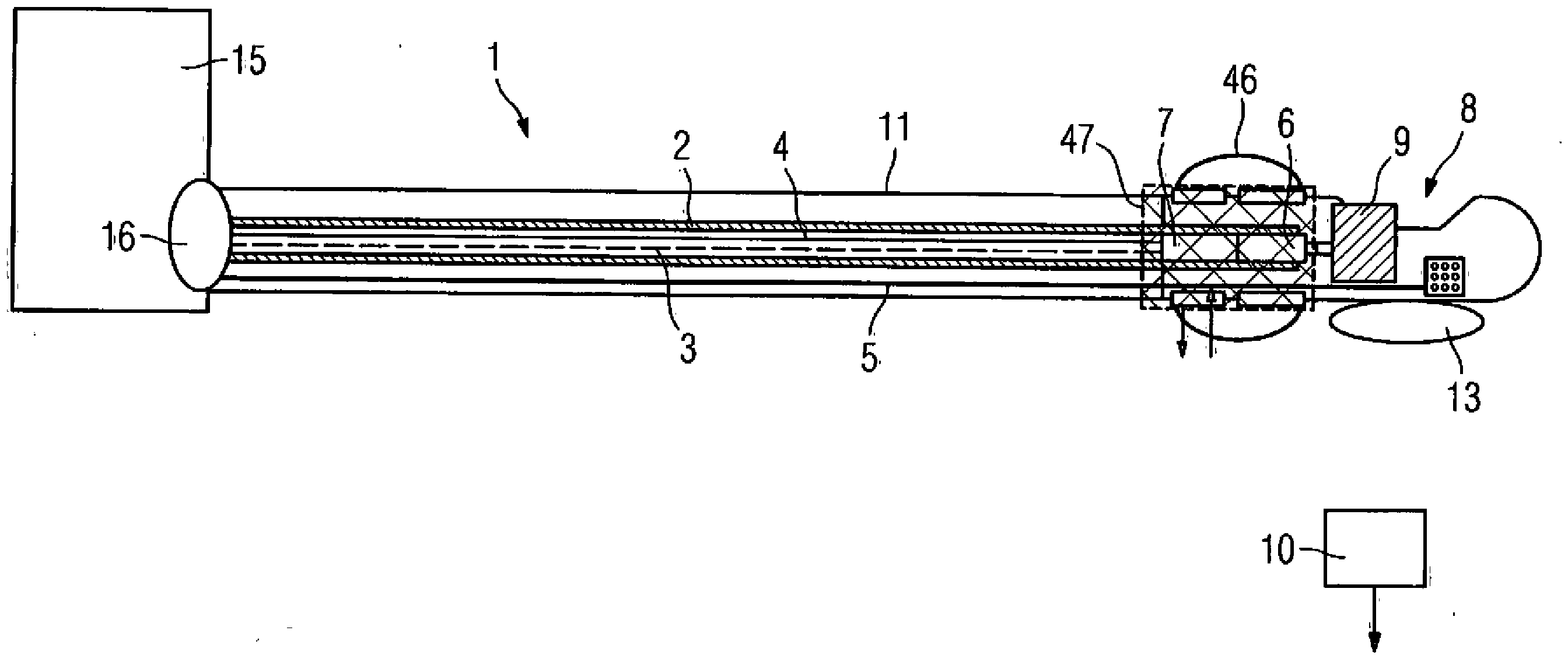
In
Zudem weist die Kathetervorrichtung einen vormontierten Stent
So kann mit der erfindungsgemäßen Kathetervorrichtung
Die Antriebswelle
Ein Signalinterface und eine Antriebseinheit
Bei der in
Für diejenigen Komponenten der Kathetervorrichtung, die mit denjenigen des ersten Ausführungsbeispiels übereinstimmen, werden dieselben Bezugszeichen verwendet.For those components of the catheter device that are the same as those of the first embodiment, the same reference numerals will be used.
Der Atherektomiekatheter
Die beiden in den
Die Kathetervorrichtungen
Bei der Approximation der Mittellinie des Gefäßes und der Hüllkurve des Gefäßes wird die geometrische Information der Mittellinie genutzt und mit den während der Bildaufnahme erfassten Sensorpositionen kombiniert, wodurch die Artefakte bei der 3D-Bilddarstellung deutlich reduziert werden. Die 3D-Koodinaten der Mittellinie und die während der Bildaufnahme erfassten Sensorpositionen werden voneinander subtrahiert. Das Ergebnis der Subtraktion wird dann für jedes der erfassten 2D-Bilder zur exakten 3D-Rekonstruktion verwendet. Diese Hüllkurve des Gefäßes kann bei weiteren Verarbeitungsschritten des Bildes verwendet werden. Mit Hilfe der Hüllkurve werden die 3D-rekonstruierten OCT-IVUS-Bilder mit anderen anatomischen Bilddaten, etwa von einem 3D-Angiographiegerät desselben Gefäßabschnitts registriert und anschließend fusioniert.In the approximation of the vessel's centerline and envelope, the geometric information of the centerline is used and combined with the sensor positions acquired during image acquisition, thereby significantly reducing artifacts in 3D imaging. The 3D coordinates of the centerline and the sensor positions acquired during image acquisition are subtracted from each other. The result of the subtraction is then used for each of the captured 2D images for exact 3D reconstruction. This envelope of the vessel can be used in further processing steps of the image. The envelope is used to register the 3D reconstructed OCT-IVUS images with other anatomical image data, for example from a 3D angiography device of the same vessel section, and then to fuse them.
Die bei den Ausführungsbeispielen der
Im Katheter können die elektromagnetischen Sender oder alternativ die elektromagnetischen Empfänger sitzen. Umgekehrt können außerhalb des Körpers die entsprechenden elektromagnetischen Empfänger oder Sender angebracht werden. Normalerweise wird mindestens ein Sender mit einer Ausstrahlung in X, Y, Z-Richtung einem Empfänger oder umgekehrt ein Empfänger mit X, Y, Z-Empfangsrichtungen einem Sender zugeordnet, um eine Ortung im Raum zu ermöglichen. Die Spulen der elektromagnetischen Positionssensoren werden nicht ausschließlich orthogonal zueinander angeordnet, sondern in einem beliebigen Winkel von zum Beispiel 60°, um eine bessere Miniaturisierung zu erreichen, die es ermöglicht, dass die Positionssensoren in einen Katheter eingebaut werden können.In the catheter, the electromagnetic transmitter or alternatively the electromagnetic receiver can sit. Conversely, the corresponding electromagnetic receivers or transmitters can be mounted outside the body. Normally, at least one transmitter having an X, Y, Z direction transmission is assigned to a receiver, or conversely, a receiver having X, Y, Z reception directions is assigned to a transmitter to enable location in space. The coils of the electromagnetic position sensors are not arranged exclusively orthogonal to each other, but at any angle of, for example, 60 ° in order to achieve better miniaturization, which enables the position sensors to be incorporated into a catheter.
Die Bildinformation des Katheters, die mit den Sensoren aufgenommen wird, wird mit anderen medizinischen Bildern, wie 2D- oder 3D-Aufnahmen, zusammengefügt bzw. überlagert. Die OCT-IVUS-Bilder des Katheters werden gemeinsam mit den Röntgenaufnahmen dargestellt. Dadurch werden die Informationen über die Bilder der Kathetervorrichtung und die Röntgenbilder gemeinsam für den Anwender sichtbar und ermöglichen eine schnellere und bessere Diagnose. Zusätzlich sind Überlagerungen 2D-2D, 2D-3D, 3D-3D und 3D-4D und 4D-4D möglich, wobei jeweils die angiographischen Röntgenbilder mit den Bildern der Kathetervorrichtung durch Segmentierung, Registrierung und Bildfusion kombiniert werden. Für die Überlagerung können Bilder der folgenden Modalitäten und Methoden eingesetzt werden: Sonographie einschließlich IVUS, Radiographie, Durchleuchtung (Fluoroskopie), Angiographie, OCT, diskrete Tomographie, Positronemmisions-Tomographie, nuklearmedizinische Diagnostik, Computertomographie, Kernspintomographie einschließlich intrakardiales MR, optische Aufnahmen einschließlich Endoskopie, Fluoreszenz und optische Marker.The image information of the catheter, which is taken with the sensors, is combined with other medical images, such as 2D or 3D images, or superimposed. The OCT-IVUS images of the catheter are displayed along with the radiographs. As a result, the information about the images of the catheter device and the X-ray images are jointly visible to the user and allow a faster and better diagnosis. In addition, overlays 2D-2D, 2D-3D, 3D-3D, and 3D-4D and 4D-4D are possible, each combining the angiographic X-ray images with the images of the catheter device through segmentation, registration, and image fusion. Images of the following modalities and methods may be used for the overlay: sonography including IVUS, radiography, fluoroscopy, angiography, OCT, discrete tomography, positron imaging tomography, nuclear medicine diagnostics, computed tomography, magnetic resonance imaging including intracardiac MR, optical imaging including endoscopy, Fluorescence and optical markers.
Die Kathetervorrichtung ist Teil einer medizinischen Behandlungsvorrichtung, die eine Funktionseinheit zur Beseitigung von Bewegungsartefakten besitzt, die durch die Atmung und die Bewegung des Herzens und der Blutgefäße entstehen. Zur Beseitigung der Atmungsartefakte kann auch ein Brustband benutzt werden, das über entsprechende Sensoren die Atemamplitude und die Frequenz ermittelt, sodass die Bildverarbeitungseinheit entsprechende Korrekturberechnungen durchführen kann, um die Bewegungsartefakte aus den Bildinformationen herauszurechnen.The catheter device is part of a medical treatment device that has a functional unit for eliminating movement artifacts that result from respiration and movement of the heart and blood vessels. To eliminate the respiratory artifacts, a chest band can also be used, which determines the breathing amplitude and the frequency via corresponding sensors, so that the image processing unit can perform corresponding correction calculations in order to calculate the motion artifacts from the image information.
Zur Erhöhung der Ortungsgenauigkeit werden die Sendespulen zyklisch, in bestimmten Zeitabschnitten mit unterschiedlichen Frequenzen betrieben und ausgewertet. Zur Vermeidung von Sensorartefakten, die durch Überlagerungen von Signalen der einzelnen Sensoren hervorgerufen werden können, wird vorgeschlagen, die Sensoren zeitlich versetzt und getaktet auszulesen. Beispielsweise werden die Röntgendetektoren und das EKG nicht ausgelesen, wenn die Sender des elektromagnetischen Positionssystems aktiv sind. Die OCT-Sensoren und Positionssensoren werden nicht ausgelesen, wenn die Röntgenstrahlung aktiv ist. Es werde also immer nur solche Signale ausgelesen, die keine Störungen erfahren und keine anderen aktiven Sensoren beeinflussen.To increase the locating accuracy, the transmission coils are cyclically operated and evaluated at different time intervals with different frequencies. To avoid sensor artifacts, which can be caused by superposition of signals from the individual sensors, it is proposed to read the sensors offset in time and clocked. For example, the X-ray detectors and the ECG are not read out when the transmitters of the electromagnetic position system are active. The OCT sensors and position sensors are not read when the X-ray radiation is active. So it is always read only those signals that experience no interference and affect other active sensors.
Die Funktionseinheiten und Signalleitungen sind mit Vorrichtungen und Maßnahmen ausgestattet, die die physiologischen Signale und Bildsignale und die Signalverarbeitung und Signalaufbereitung gegen die magnetischen Filter der Sendeantennen abschirmen. Dazu wird die Hülle des Katheters mit einer Dünnfilmschicht aus leitenden Nanopartikeln beschichtet. Ebenso können Nanopartikel verwendet werden, um eine magnetische Abschirmung zu bewirken.The functional units and signal lines are equipped with devices and measures that shield the physiological signals and image signals and the signal processing and signal processing against the magnetic filters of the transmitting antennas. For this purpose, the sheath of the catheter with a thin film layer of conductive nanoparticles coated. Likewise, nanoparticles can be used to effect magnetic shielding.
Die Katheterhülle ist mit einer Beschichtung versehen, die den Reibungswiderstand bei der Führung durch die Gefäße verringert. Diese Beschichtung kann ebenfalls auf Nanotechnologie basieren oder alternativ aus einem Silikonmaterial hergestellt werden.The catheter sheath is provided with a coating which reduces the frictional resistance when guided by the vessels. This coating may also be based on nanotechnology or alternatively made of a silicone material.
Um die Bildgebung durch den IVUS-Sensor durch den Einsatz von Ultraschall-Kontrastmittel zu verbessern, wird in das zu untersuchende Gefäß bzw. den Körperhohlraum durch einen Kanal im Katheter ein Kontrastmittel direkt eingeleitet.In order to improve the imaging by the IVUS sensor by the use of ultrasound contrast agent, a contrast medium is introduced directly into the vessel to be examined or the body cavity through a channel in the catheter.
In der Spitze des Katheters ist ein Temperatursensor oder ein Drucksensor angeordnet, um die Temperatur und den Druck in dem zu untersuchenden und zu behandelnden Gefäß oder Organ zu überwachen. Durch den Temperatursensor, der in der Spitze des Katheters angebracht ist, kann eine eventuelle Temperaturerhöhung erfasst werden, die durch Reibung entsteht.In the tip of the catheter, a temperature sensor or a pressure sensor is arranged to monitor the temperature and pressure in the vessel or organ to be examined and treated. Through the temperature sensor, which is mounted in the tip of the catheter, a possible increase in temperature caused by friction can be detected.
Die Behandlungseinrichtung
Über einen Anschluss
Die gewonnenen Bilddaten aus OCT, IVUS und dem Positionssensorsystem sowie die Röntgenbilder und mögliche Fusionsbilder der verschiedenen Bildaufnahmetechniken werden an einer Displayeinheit
Ein typischer Verfahrensablauf ist wie folgt: Einführen des Katheters unter Röntgenkontrolle, eventuell mit Kontrastmittel, Erstellen der angiographischen Übersichtsaufnahme, Erstellen der Aufnahmen der Positionssensoren, Überlagern der Aufnahmen der Positionssensoren mit der Übersichtsangiographie durch Segmentierung, Registrierung und Bildfusion, Navigieren des Katheters, basierend auf den gewonnenen Aufnahmen bis zur Zielposition, diese Schritte werden zum Teil parallel und automatisch ohne Interaktion des Benutzers durchgeführt. Wenn die gewünschte Zielposition erreicht ist, wird die Spülflüssigkeit für OCT eingespritzt und die Stenose mit den OCT-IVUS-Bildaufnahmen zweidimensional oder dreidimensional in hoher Auflösung betrachtet. Anschließend werden die OCT-IVUS-Aufnahmen erstellt. In der Folge werden die OCT-IVUS-Aufnahmen mit der Übersichtsangiographie überlagert durch Segmentierung, Registrierung und Bildfusion. Anschließend erfolgt eine 3D-Rekonstruktion der OCT-IVUS-Aufnahmen, basierend auf den Daten der Positionssensoren. Der Atherektomiekatheter wird platziert und vorläufig fixiert zum Beispiel durch Aufblasen des an der Katheterspitze angebrachten Ballons. Kontrolle mit OCT-IVUS in 2D und 3D , ob die Position und Lage des Atherektomiekatheters korrekt ist. Durchführen der Atherektomie, das heißt Abschaben der Plaque von der Gefäßwand mit den rotierenden Messern. Wenn eine bestimmte Menge Plaque abgetragen ist, wird mit dem OCT-Sensor die Stelle in der Gefäßwand kontrolliert. Der Vorgang wird wiederholt, bis an allen Stellen die Plaque abgetragen ist. Abschließende Kontrolle der Atherektomie, Platzierung und Entfaltung des Stents bis zur Befestigung in der Gefäßwand und Herausziehen des Katheters.A typical procedure is as follows: inserting the catheter under X-ray control, possibly with contrast agent, acquiring angiographic scans, creating position sensor scans, overlaying the scans of the positional angiographic sensors with segmentation, registration and image fusion, navigating the catheter based on the taken recordings to the target position, these steps are sometimes performed in parallel and automatically without user interaction. When the desired target position is reached, the OCT irrigation fluid is injected and the stenosis is viewed in two-dimensional or three-dimensional high resolution with the OCT-IVUS image acquisitions. Subsequently, the OCT-IVUS recordings are created. Subsequently, the OCT-IVUS images are superimposed on the overview angiography by segmentation, registration and image fusion. This is followed by a 3D reconstruction of the OCT-IVUS images, based on the data from the position sensors. The atherectomy catheter is placed and provisionally fixed, for example, by inflating the balloon attached to the catheter tip. Check with OCT-IVUS in 2D and 3D, if the position and position of the atherectomy catheter is correct. Performing the atherectomy, that is, scraping the plaque from the vessel wall with the rotating knives. When a certain amount of plaque has been removed, the OCT sensor controls the location in the vessel wall. The process is repeated until all plaque has been removed. Final check of the atherectomy, placement and deployment of the stent until attachment in the vessel wall and withdrawal of the catheter.
Durch die erfindungsgemäße Vorrichtung werden die erforderlichen Verfahrensschritte reduziert. Der OCT-Sensor liefert gute Aufnahmen im Nahbereich, der IVUS-Sensor ausreichend gute Bilder von tiefer liegenden Gewebeschichten. Durch die elektromagnetischen Positionssensoren lassen sich 3D-Aufnahmen aus den OCT- und IVUS-Aufnahmen erstellen. Daneben wird nach der Durchführung einer Übersichtsangiographie durch entsprechende Ausnutzung der Signale der Positionssensoren der Verlauf des Katheters allein anhand der IVUS-, OCT- und elektromagnetischen Signale abgebildet, das heißt Röntgenstrahlung kann eingespart werden. Das System liefert wichtige zusätzliche medizinische Informationen über die arteriosklerotische Plaque. Zusätzlich kann damit die richtige Lage der Spitze des Katheters besser überprüft werden. Ein weiterer Vorteil bei der Integration von Atherektomie und OCT liegt auch darin, dass in diesem Fall für OCT keine gesonderte Spülvorrichtung vorhanden sein muss, da für den Bohrkopf bereits ein Spülmittel verwendet wird.The inventive device reduces the required process steps. The OCT sensor provides good images at close range, the IVUS sensor sufficiently good images of deeper layers of tissue. The electromagnetic position sensors can be used to create 3D images from the OCT and IVUS recordings. In addition, after carrying out an overview angiography by appropriate utilization of the signals of the position sensors, the course of the catheter is mapped solely on the basis of the IVUS, OCT and electromagnetic signals, that is, X-ray radiation can be saved. The system provides important additional medical information about arteriosclerotic plaque. In addition, the correct position of the tip of the catheter can be better checked. Another advantage of integrating atherectomy and OCT is that in this case there is no need for a separate flushing device for OCT, since a flushing agent is already used for the drill head.
Die Sensoren der medizinischen Behandlungseinrichtung, die in dem dargestellten Ausführungsbeispiel eine Röntgeneinrichtung ist, werden teilweise zeitlich versetzt und getaktet ausgelesen. Zunächst wird ein Systemtakt vorgegeben, in dem einzelne Systempulse erzeugt werden, wobei sich an diese Pulserzeugung das Einschalten der Röntgenstrahlung und die Aktivierung der magnetischen Ortung anschließt. Nach dem Ausschalten der Röntgenstrahlung erfolgt das Auslesen des Röntgendetektors und zeitgleich das Auslesen der IVUS-Daten. Im Anschluss daran werden die OCT-Daten ausgelesen, wobei dies zeitgleich mit dem Auslesen des EKGs und der Daten zur Respiration erfolgt. Damit werden die einzelnen Sensoren so ausgelesen bzw. die Komponenten der Kathetervorrichtung so angesteuert, dass eine gegenseitige Störung ausgeschlossen werden kann. Das hier dargestellte zeitlich versetzte und getaktete Auslesen ist dabei beispielhaft für ein Auslesen unter Vermeidung von Störeinflüssen zu sehen.The sensors of the medical treatment device, which is an X-ray device in the illustrated embodiment, are partially read out in a staggered and timed manner. First, a system clock is specified, in which individual system pulses are generated, followed by this pulse generation, the switching on of the X-ray radiation and the activation of the magnetic detection. After the X-ray radiation has been switched off, the X-ray detector is read out and at the same time the IVUS data is read out. Following this, the OCT data are read out, simultaneously with the reading of the ECG and the respiration data. Thus, the individual sensors are read out or the components of the catheter device are controlled so that a mutual interference can be excluded. The temporally offset and clocked readout shown here is to be seen as an example for readout while avoiding disturbing influences.
Claims (41)
Priority Applications (2)
| Application Number | Priority Date | Filing Date | Title |
|---|---|---|---|
| DE102005059271.6A DE102005059271B4 (en) | 2005-12-12 | 2005-12-12 | catheter device |
| US11/634,035 US20070135886A1 (en) | 2005-12-12 | 2006-12-05 | Catheter device |
Applications Claiming Priority (1)
| Application Number | Priority Date | Filing Date | Title |
|---|---|---|---|
| DE102005059271.6A DE102005059271B4 (en) | 2005-12-12 | 2005-12-12 | catheter device |
Publications (2)
| Publication Number | Publication Date |
|---|---|
| DE102005059271A1 DE102005059271A1 (en) | 2007-06-14 |
| DE102005059271B4 true DE102005059271B4 (en) | 2019-02-21 |
Family
ID=38056013
Family Applications (1)
| Application Number | Title | Priority Date | Filing Date |
|---|---|---|---|
| DE102005059271.6A Expired - Fee Related DE102005059271B4 (en) | 2005-12-12 | 2005-12-12 | catheter device |
Country Status (2)
| Country | Link |
|---|---|
| US (1) | US20070135886A1 (en) |
| DE (1) | DE102005059271B4 (en) |
Families Citing this family (118)
| Publication number | Priority date | Publication date | Assignee | Title |
|---|---|---|---|---|
| US8328829B2 (en) | 1999-08-19 | 2012-12-11 | Covidien Lp | High capacity debulking catheter with razor edge cutting window |
| US7708749B2 (en) | 2000-12-20 | 2010-05-04 | Fox Hollow Technologies, Inc. | Debulking catheters and methods |
| US7713279B2 (en) | 2000-12-20 | 2010-05-11 | Fox Hollow Technologies, Inc. | Method and devices for cutting tissue |
| US6299622B1 (en) | 1999-08-19 | 2001-10-09 | Fox Hollow Technologies, Inc. | Atherectomy catheter with aligned imager |
| EP1345542B1 (en) | 2000-12-20 | 2011-02-23 | Fox Hollow Technologies, Inc. | Debulking catheter |
| US8246640B2 (en) | 2003-04-22 | 2012-08-21 | Tyco Healthcare Group Lp | Methods and devices for cutting tissue at a vascular location |
| DE10354496B4 (en) * | 2003-11-21 | 2011-03-31 | Siemens Ag | Medical examination and / or treatment system |
| US8784336B2 (en) | 2005-08-24 | 2014-07-22 | C. R. Bard, Inc. | Stylet apparatuses and methods of manufacture |
| US20070276419A1 (en) | 2006-05-26 | 2007-11-29 | Fox Hollow Technologies, Inc. | Methods and devices for rotating an active element and an energy emitter on a catheter |
| US8388546B2 (en) | 2006-10-23 | 2013-03-05 | Bard Access Systems, Inc. | Method of locating the tip of a central venous catheter |
| US7794407B2 (en) | 2006-10-23 | 2010-09-14 | Bard Access Systems, Inc. | Method of locating the tip of a central venous catheter |
| US8849382B2 (en) | 2007-11-26 | 2014-09-30 | C. R. Bard, Inc. | Apparatus and display methods relating to intravascular placement of a catheter |
| US10524691B2 (en) | 2007-11-26 | 2020-01-07 | C. R. Bard, Inc. | Needle assembly including an aligned magnetic element |
| US8781555B2 (en) | 2007-11-26 | 2014-07-15 | C. R. Bard, Inc. | System for placement of a catheter including a signal-generating stylet |
| US9649048B2 (en) | 2007-11-26 | 2017-05-16 | C. R. Bard, Inc. | Systems and methods for breaching a sterile field for intravascular placement of a catheter |
| US10751509B2 (en) | 2007-11-26 | 2020-08-25 | C. R. Bard, Inc. | Iconic representations for guidance of an indwelling medical device |
| US12440238B2 (en) | 2007-11-26 | 2025-10-14 | C. R. Bard, Inc. | Apparatus for use with needle insertion guidance system |
| US9521961B2 (en) | 2007-11-26 | 2016-12-20 | C. R. Bard, Inc. | Systems and methods for guiding a medical instrument |
| AU2008329807B2 (en) | 2007-11-26 | 2014-02-27 | C. R. Bard, Inc. | Integrated system for intravascular placement of a catheter |
| US10449330B2 (en) | 2007-11-26 | 2019-10-22 | C. R. Bard, Inc. | Magnetic element-equipped needle assemblies |
| US8478382B2 (en) | 2008-02-11 | 2013-07-02 | C. R. Bard, Inc. | Systems and methods for positioning a catheter |
| US8784440B2 (en) | 2008-02-25 | 2014-07-22 | Covidien Lp | Methods and devices for cutting tissue |
| WO2010022370A1 (en) | 2008-08-22 | 2010-02-25 | C.R. Bard, Inc. | Catheter assembly including ecg sensor and magnetic assemblies |
| DE102008050151B3 (en) * | 2008-10-01 | 2010-04-08 | Fraunhofer-Gesellschaft zur Förderung der angewandten Forschung e.V. | Shockwave head and method for its manufacture |
| US8437833B2 (en) | 2008-10-07 | 2013-05-07 | Bard Access Systems, Inc. | Percutaneous magnetic gastrostomy |
| CN102223847B (en) | 2008-10-13 | 2013-10-30 | 泰科保健集团有限合伙公司 | Devices and methods for manipulating catheter shaft |
| DE102009014489B4 (en) | 2009-03-23 | 2011-03-10 | Siemens Aktiengesellschaft | Catheter and medical device |
| AU2010241801B2 (en) | 2009-04-29 | 2013-04-11 | Covidien Lp | Methods and devices for cutting and abrading tissue |
| AU2010248909B2 (en) | 2009-05-14 | 2013-03-21 | Covidien Lp | Easily cleaned atherectomy catheters and methods of use |
| EP3542713A1 (en) | 2009-06-12 | 2019-09-25 | Bard Access Systems, Inc. | Adapter for a catheter tip positioning device |
| US9532724B2 (en) | 2009-06-12 | 2017-01-03 | Bard Access Systems, Inc. | Apparatus and method for catheter navigation using endovascular energy mapping |
| EP2464407A4 (en) | 2009-08-10 | 2014-04-02 | Bard Access Systems Inc | Devices and methods for endovascular electrography |
| CN102665541B (en) | 2009-09-29 | 2016-01-13 | C·R·巴德股份有限公司 | The probe used together with the equipment that the Ink vessel transfusing for conduit is placed |
| US11103213B2 (en) | 2009-10-08 | 2021-08-31 | C. R. Bard, Inc. | Spacers for use with an ultrasound probe |
| WO2011068932A1 (en) | 2009-12-02 | 2011-06-09 | Fox Hollow Technologies, Inc. | Methods and devices for cutting tissue |
| KR20140006106A (en) | 2009-12-11 | 2014-01-15 | 코비디엔 엘피 | Material removal device having improved material capture efficiency and methods of use |
| JP2013518676A (en) | 2010-02-02 | 2013-05-23 | シー・アール・バード・インコーポレーテッド | Apparatus and method for locating catheter navigation and tip |
| US8774903B2 (en) * | 2010-03-26 | 2014-07-08 | Headwater Partners Ii Llc | Medical imaging apparatus and method |
| CA2800813C (en) | 2010-05-28 | 2019-10-29 | C.R. Bard, Inc. | Apparatus for use with needle insertion guidance system |
| WO2011150376A1 (en) | 2010-05-28 | 2011-12-01 | C.R. Bard, Inc. | Apparatus for use with needle insertion guidance system |
| EP2742881B1 (en) | 2010-06-14 | 2015-10-07 | Covidien LP | Material removal device |
| US8672837B2 (en) | 2010-06-24 | 2014-03-18 | Hansen Medical, Inc. | Methods and devices for controlling a shapeable medical device |
| JP2013535301A (en) | 2010-08-09 | 2013-09-12 | シー・アール・バード・インコーポレーテッド | Ultrasonic probe head support / cover structure |
| EP2605699A4 (en) | 2010-08-20 | 2015-01-07 | Bard Inc C R | Reconfirmation of ecg-assisted catheter tip placement |
| BR112013009835A2 (en) | 2010-10-28 | 2016-07-26 | Covidien Lp | material removal device and method of use |
| CN103189009B (en) | 2010-10-29 | 2016-09-07 | C·R·巴德股份有限公司 | Bioimpedance Assisted Placement of Medical Devices |
| EP2659840B1 (en) | 2010-11-11 | 2017-02-01 | Covidien LP | Flexible debulking catheters with imaging |
| KR20140051284A (en) | 2011-07-06 | 2014-04-30 | 씨. 알. 바드, 인크. | Needle length determination and calibration for insertion guidance system |
| USD699359S1 (en) | 2011-08-09 | 2014-02-11 | C. R. Bard, Inc. | Ultrasound probe head |
| USD724745S1 (en) | 2011-08-09 | 2015-03-17 | C. R. Bard, Inc. | Cap for an ultrasound probe |
| US8992717B2 (en) | 2011-09-01 | 2015-03-31 | Covidien Lp | Catheter with helical drive shaft and methods of manufacture |
| US10238837B2 (en) | 2011-10-14 | 2019-03-26 | Intuitive Surgical Operations, Inc. | Catheters with control modes for interchangeable probes |
| US9387048B2 (en) | 2011-10-14 | 2016-07-12 | Intuitive Surgical Operations, Inc. | Catheter sensor systems |
| US9452276B2 (en) | 2011-10-14 | 2016-09-27 | Intuitive Surgical Operations, Inc. | Catheter with removable vision probe |
| US20130303944A1 (en) | 2012-05-14 | 2013-11-14 | Intuitive Surgical Operations, Inc. | Off-axis electromagnetic sensor |
| WO2013070775A1 (en) | 2011-11-07 | 2013-05-16 | C.R. Bard, Inc | Ruggedized ultrasound hydrogel insert |
| EP2861153A4 (en) | 2012-06-15 | 2016-10-19 | Bard Inc C R | Apparatus and methods for detection of a removable cap on an ultrasound probe |
| US9579157B2 (en) | 2012-09-13 | 2017-02-28 | Covidien Lp | Cleaning device for medical instrument and method of use |
| US9943329B2 (en) | 2012-11-08 | 2018-04-17 | Covidien Lp | Tissue-removing catheter with rotatable cutter |
| US20140148673A1 (en) | 2012-11-28 | 2014-05-29 | Hansen Medical, Inc. | Method of anchoring pullwire directly articulatable region in catheter |
| WO2014106137A1 (en) * | 2012-12-28 | 2014-07-03 | The General Hospital Corporation | Optical probe apparatus, systems, methods for guiding tissue asessment |
| US9057600B2 (en) | 2013-03-13 | 2015-06-16 | Hansen Medical, Inc. | Reducing incremental measurement sensor error |
| US9629595B2 (en) | 2013-03-15 | 2017-04-25 | Hansen Medical, Inc. | Systems and methods for localizing, tracking and/or controlling medical instruments |
| US9014851B2 (en) | 2013-03-15 | 2015-04-21 | Hansen Medical, Inc. | Systems and methods for tracking robotically controlled medical instruments |
| US9271663B2 (en) | 2013-03-15 | 2016-03-01 | Hansen Medical, Inc. | Flexible instrument localization from both remote and elongation sensors |
| US11020016B2 (en) | 2013-05-30 | 2021-06-01 | Auris Health, Inc. | System and method for displaying anatomy and devices on a movable display |
| CN105939647B (en) | 2013-10-24 | 2020-01-21 | 奥瑞斯健康公司 | Robot-assisted endovascular surgery system and related methods |
| WO2015120256A2 (en) | 2014-02-06 | 2015-08-13 | C.R. Bard, Inc. | Systems and methods for guidance and placement of an intravascular device |
| EP3243476B1 (en) | 2014-03-24 | 2019-11-06 | Auris Health, Inc. | Systems and devices for catheter driving instinctiveness |
| WO2015200702A1 (en) | 2014-06-27 | 2015-12-30 | Covidien Lp | Cleaning device for catheter and catheter including the same |
| EP3200718A4 (en) | 2014-09-30 | 2018-04-25 | Auris Surgical Robotics, Inc | Configurable robotic surgical system with virtual rail and flexible endoscope |
| US10314463B2 (en) | 2014-10-24 | 2019-06-11 | Auris Health, Inc. | Automated endoscope calibration |
| US10973584B2 (en) | 2015-01-19 | 2021-04-13 | Bard Access Systems, Inc. | Device and method for vascular access |
| US10314667B2 (en) | 2015-03-25 | 2019-06-11 | Covidien Lp | Cleaning device for cleaning medical instrument |
| US10349890B2 (en) | 2015-06-26 | 2019-07-16 | C. R. Bard, Inc. | Connector interface for ECG-based catheter positioning system |
| US10292721B2 (en) | 2015-07-20 | 2019-05-21 | Covidien Lp | Tissue-removing catheter including movable distal tip |
| EP3349649B1 (en) | 2015-09-18 | 2022-03-09 | Auris Health, Inc. | Navigation of tubular networks |
| US10314664B2 (en) | 2015-10-07 | 2019-06-11 | Covidien Lp | Tissue-removing catheter and tissue-removing element with depth stop |
| US10143526B2 (en) | 2015-11-30 | 2018-12-04 | Auris Health, Inc. | Robot-assisted driving systems and methods |
| US11000207B2 (en) | 2016-01-29 | 2021-05-11 | C. R. Bard, Inc. | Multiple coil system for tracking a medical device |
| US9931025B1 (en) | 2016-09-30 | 2018-04-03 | Auris Surgical Robotics, Inc. | Automated calibration of endoscopes with pull wires |
| US10244926B2 (en) | 2016-12-28 | 2019-04-02 | Auris Health, Inc. | Detecting endolumenal buckling of flexible instruments |
| EP3600031A4 (en) | 2017-03-31 | 2021-01-20 | Auris Health, Inc. | ROBOTIC SYSTEMS FOR NAVIGATION OF LUMINAL NETWORKS THAT COMPENSATE FOR PHYSIOLOGICAL NOISE |
| WO2018208994A1 (en) | 2017-05-12 | 2018-11-15 | Auris Health, Inc. | Biopsy apparatus and system |
| US10022192B1 (en) | 2017-06-23 | 2018-07-17 | Auris Health, Inc. | Automatically-initialized robotic systems for navigation of luminal networks |
| US11832889B2 (en) | 2017-06-28 | 2023-12-05 | Auris Health, Inc. | Electromagnetic field generator alignment |
| CN110913788B (en) | 2017-06-28 | 2024-03-12 | 奥瑞斯健康公司 | Electromagnetic distortion detection |
| EP4437999A3 (en) | 2017-06-28 | 2024-12-04 | Auris Health, Inc. | Instrument insertion compensation |
| US10426559B2 (en) | 2017-06-30 | 2019-10-01 | Auris Health, Inc. | Systems and methods for medical instrument compression compensation |
| US10016900B1 (en) | 2017-10-10 | 2018-07-10 | Auris Health, Inc. | Surgical robotic arm admittance control |
| US10145747B1 (en) | 2017-10-10 | 2018-12-04 | Auris Health, Inc. | Detection of undesirable forces on a surgical robotic arm |
| US10555778B2 (en) | 2017-10-13 | 2020-02-11 | Auris Health, Inc. | Image-based branch detection and mapping for navigation |
| US11058493B2 (en) | 2017-10-13 | 2021-07-13 | Auris Health, Inc. | Robotic system configured for navigation path tracing |
| CN110831536B (en) | 2017-12-06 | 2021-09-07 | 奥瑞斯健康公司 | System and method for correcting for uncommanded instrument roll |
| EP3684562B1 (en) | 2017-12-14 | 2025-03-12 | Auris Health, Inc. | System and program for estimating instrument location |
| CN110809453B (en) | 2017-12-18 | 2023-06-06 | 奥瑞斯健康公司 | Methods and systems for instrument tracking and navigation within a luminal network |
| EP3752085A4 (en) | 2018-02-13 | 2021-11-24 | Auris Health, Inc. | SYSTEM AND METHOD FOR TRAINING A MEDICAL INSTRUMENT |
| CN110913791B (en) | 2018-03-28 | 2021-10-08 | 奥瑞斯健康公司 | System and method for displaying estimated instrument location |
| WO2019191144A1 (en) | 2018-03-28 | 2019-10-03 | Auris Health, Inc. | Systems and methods for registration of location sensors |
| US10905499B2 (en) | 2018-05-30 | 2021-02-02 | Auris Health, Inc. | Systems and methods for location sensor-based branch prediction |
| WO2019231891A1 (en) | 2018-05-31 | 2019-12-05 | Auris Health, Inc. | Path-based navigation of tubular networks |
| KR102455671B1 (en) | 2018-05-31 | 2022-10-20 | 아우리스 헬스, 인코포레이티드 | Image-Based Airway Analysis and Mapping |
| EP3801280B1 (en) | 2018-05-31 | 2024-10-02 | Auris Health, Inc. | Robotic systems for navigation of luminal network that detect physiological noise |
| WO2020069404A1 (en) | 2018-09-28 | 2020-04-02 | Auris Health, Inc. | Robotic systems and methods for concomitant endoscopic and percutaneous medical procedures |
| KR20250130720A (en) | 2018-09-28 | 2025-09-02 | 아우리스 헬스, 인코포레이티드 | Systems and methods for docking medical instruments |
| CN112867443B (en) | 2018-10-16 | 2024-04-26 | 巴德阿克塞斯系统股份有限公司 | Safety equipment connection system and method for establishing electrical connection |
| US12478444B2 (en) | 2019-03-21 | 2025-11-25 | The Board Of Trustees Of The Leland Stanford Junior University | Systems and methods for localization based on machine learning |
| WO2020236182A1 (en) * | 2019-05-23 | 2020-11-26 | Transmed7, Llc | Excisional devices and methods |
| WO2021038469A1 (en) | 2019-08-30 | 2021-03-04 | Auris Health, Inc. | Systems and methods for weight-based registration of location sensors |
| JP7451686B2 (en) | 2019-08-30 | 2024-03-18 | オーリス ヘルス インコーポレイテッド | Instrument image reliability system and method |
| WO2021044297A1 (en) | 2019-09-03 | 2021-03-11 | Auris Health, Inc. | Electromagnetic distortion detection and compensation |
| CN114787868A (en) * | 2019-11-08 | 2022-07-22 | 直观外科手术操作公司 | System and method for registering an instrument to an image using point cloud data |
| KR20220123076A (en) | 2019-12-31 | 2022-09-05 | 아우리스 헬스, 인코포레이티드 | Alignment Techniques for Transdermal Access |
| CN114901194B (en) | 2019-12-31 | 2024-08-30 | 奥瑞斯健康公司 | Anatomical feature identification and targeting |
| EP4084722A4 (en) | 2019-12-31 | 2024-01-10 | Auris Health, Inc. | ALIGNMENT INTERFACES FOR PERCUTANEOUS ACCESS |
| US11737663B2 (en) | 2020-03-30 | 2023-08-29 | Auris Health, Inc. | Target anatomical feature localization |
| US12496203B2 (en) | 2021-09-28 | 2025-12-16 | National Guard Health Affairs | Ostial stenting under vision |
| CN114820600B (en) * | 2022-06-27 | 2022-09-27 | 天津恒宇医疗科技有限公司 | Coronary artery intravascular stent detection method and system based on OCT image |
Citations (9)
| Publication number | Priority date | Publication date | Assignee | Title |
|---|---|---|---|---|
| US5639274A (en) | 1995-06-02 | 1997-06-17 | Fischell; Robert E. | Integrated catheter system for balloon angioplasty and stent delivery |
| WO1998038907A1 (en) | 1997-03-06 | 1998-09-11 | Massachusetts Institute Of Technology | Instrument for optically scanning of living tissue |
| US5895402A (en) | 1994-12-16 | 1999-04-20 | Hundertmark; Ron Ray | Intravascular catheter with guiding structure |
| DE19843408A1 (en) | 1998-09-22 | 2000-03-30 | Siemens Ag | Catheter positioning method |
| US20050101859A1 (en) | 2003-09-22 | 2005-05-12 | Michael Maschke | System for medical examination or treatment |
| US20050113685A1 (en) | 2003-11-21 | 2005-05-26 | Michael Maschke | Medical system for examination or treatment |
| US20050203553A1 (en) | 2004-02-20 | 2005-09-15 | Siemens Aktiengesellschaft | Device for the performance and monitoring of rotablation |
| DE69834563T2 (en) | 1997-03-06 | 2007-04-26 | Medtronic Percusurge, Inc., Sunnyvale | extraction system |
| DE102004015640B4 (en) | 2004-03-31 | 2007-05-03 | Siemens Ag | Apparatus for performing a cutting-balloon intervention with OCT monitoring |
Family Cites Families (18)
| Publication number | Priority date | Publication date | Assignee | Title |
|---|---|---|---|---|
| US4732154A (en) * | 1984-05-14 | 1988-03-22 | Surgical Systems & Instruments, Inc. | Rotary catheter system |
| US6485413B1 (en) * | 1991-04-29 | 2002-11-26 | The General Hospital Corporation | Methods and apparatus for forward-directed optical scanning instruments |
| US5752513A (en) * | 1995-06-07 | 1998-05-19 | Biosense, Inc. | Method and apparatus for determining position of object |
| US5766192A (en) * | 1995-10-20 | 1998-06-16 | Zacca; Nadim M. | Atherectomy, angioplasty and stent method and apparatus |
| JP4166277B2 (en) * | 1996-02-15 | 2008-10-15 | バイオセンス・ウェブスター・インコーポレイテッド | Medical method and apparatus using in-vivo probe |
| US6514249B1 (en) * | 1997-07-08 | 2003-02-04 | Atrionix, Inc. | Positioning system and method for orienting an ablation element within a pulmonary vein ostium |
| GB2331365B (en) * | 1997-11-15 | 2002-03-13 | Roke Manor Research | Catheter tracking system |
| US5865748A (en) * | 1998-01-16 | 1999-02-02 | Guidant Corporation | Guided directional coronary atherectomy distal linear encoder |
| WO2000017615A2 (en) * | 1998-09-23 | 2000-03-30 | Keith Bridger | Physiological sensing device |
| US6233476B1 (en) * | 1999-05-18 | 2001-05-15 | Mediguide Ltd. | Medical positioning system |
| US20020007215A1 (en) * | 2000-05-19 | 2002-01-17 | Robert Falotico | Drug/drug delivery systems for the prevention and treatment of vascular disease |
| US6641611B2 (en) * | 2001-11-26 | 2003-11-04 | Swaminathan Jayaraman | Therapeutic coating for an intravascular implant |
| US20040225213A1 (en) * | 2002-01-22 | 2004-11-11 | Xingwu Wang | Magnetic resonance imaging coated assembly |
| DE10253634A1 (en) * | 2002-11-13 | 2004-05-27 | Biotronik Meß- und Therapiegeräte GmbH & Co. Ingenieurbüro Berlin | endoprosthesis |
| US7697972B2 (en) * | 2002-11-19 | 2010-04-13 | Medtronic Navigation, Inc. | Navigation system for cardiac therapies |
| US20040220514A1 (en) * | 2003-05-01 | 2004-11-04 | Medtronic Ave. | Method and system for treating vulnerable plaque |
| US20040230316A1 (en) * | 2003-05-12 | 2004-11-18 | Iulian Cioanta | Method for treating the prostate and inhibiting obstruction of the prostatic urethra using biodegradable stents |
| DE102004008371B4 (en) * | 2004-02-20 | 2006-05-24 | Siemens Ag | atherectomy |
-
2005
- 2005-12-12 DE DE102005059271.6A patent/DE102005059271B4/en not_active Expired - Fee Related
-
2006
- 2006-12-05 US US11/634,035 patent/US20070135886A1/en not_active Abandoned
Patent Citations (9)
| Publication number | Priority date | Publication date | Assignee | Title |
|---|---|---|---|---|
| US5895402A (en) | 1994-12-16 | 1999-04-20 | Hundertmark; Ron Ray | Intravascular catheter with guiding structure |
| US5639274A (en) | 1995-06-02 | 1997-06-17 | Fischell; Robert E. | Integrated catheter system for balloon angioplasty and stent delivery |
| WO1998038907A1 (en) | 1997-03-06 | 1998-09-11 | Massachusetts Institute Of Technology | Instrument for optically scanning of living tissue |
| DE69834563T2 (en) | 1997-03-06 | 2007-04-26 | Medtronic Percusurge, Inc., Sunnyvale | extraction system |
| DE19843408A1 (en) | 1998-09-22 | 2000-03-30 | Siemens Ag | Catheter positioning method |
| US20050101859A1 (en) | 2003-09-22 | 2005-05-12 | Michael Maschke | System for medical examination or treatment |
| US20050113685A1 (en) | 2003-11-21 | 2005-05-26 | Michael Maschke | Medical system for examination or treatment |
| US20050203553A1 (en) | 2004-02-20 | 2005-09-15 | Siemens Aktiengesellschaft | Device for the performance and monitoring of rotablation |
| DE102004015640B4 (en) | 2004-03-31 | 2007-05-03 | Siemens Ag | Apparatus for performing a cutting-balloon intervention with OCT monitoring |
Also Published As
| Publication number | Publication date |
|---|---|
| US20070135886A1 (en) | 2007-06-14 |
| DE102005059271A1 (en) | 2007-06-14 |
Similar Documents
| Publication | Publication Date | Title |
|---|---|---|
| DE102005059271B4 (en) | catheter device | |
| DE102005059262B4 (en) | catheter device | |
| DE102005048892B4 (en) | Device for carrying out rotablation and medical treatment device | |
| DE102005059261B4 (en) | Catheter device for the treatment of a partial and / or complete vascular occlusion and X-ray device | |
| DE102004058008B4 (en) | Guidewire for vascular catheter with improved tracking and navigation | |
| DE102004008371B4 (en) | atherectomy | |
| DE102005032755B4 (en) | System for performing and monitoring minimally invasive procedures | |
| DE102005022120B4 (en) | Catheter, catheter device and diagnostic imaging device | |
| DE69726576T2 (en) | Placemark sample | |
| DE102005023167B4 (en) | Method and device for registering 2D projection images relative to a 3D image data set | |
| DE102005045071A1 (en) | Catheter device with a position sensor system for the treatment of a partial and / or complete vascular occlusion under image monitoring | |
| DE102005027951A1 (en) | Medical system for introducing a catheter into a vessel | |
| DE102009009158B4 (en) | Localization of a medical instrument in a pre-invasively acquired tomographic image dataset | |
| US8529450B2 (en) | Device for performing a cutting-balloon intervention | |
| DE10210648A1 (en) | Medical 3-D imaging method for organ and catheter type instrument portrayal in which 2-D ultrasound images, the location and orientation of which are known, are combined in a reference coordinate system to form a 3-D image | |
| DE102005050344A1 (en) | Cryocatheter for medical investigation and treatment equipment for e.g. diagnosis and treatment of heart infarcts, has image capture device that maps region of vessel around balloon arranged near catheter tip | |
| DE102006061178A1 (en) | Medical system for carrying out and monitoring a minimal invasive intrusion, especially for treating electro-physiological diseases, has X-ray equipment and a control/evaluation unit | |
| DE102009043069A1 (en) | Visualization method and imaging system | |
| EP2564771A1 (en) | Medicinal product with a functional element for invasive use in the body of a patient | |
| DE102008054297A1 (en) | A catheter assembly for insertion into a blood vessel, medical examination and treatment device comprising such a catheter assembly and method for minimally invasive intervention on a blood vessel in the brain | |
| EP1659968A1 (en) | Method and device for visually assisting the electrophysiological use of a catheter in the heart | |
| DE102008002864A1 (en) | Dynamic reference method and system for use with surgical procedures | |
| DE102006011242A1 (en) | Hollow organ`s e.g. trachea, three dimensional representation reconstructing method for use in medical field, involves reconstructing representation of organ based on catheter image and deviation of catheter position from central path | |
| DE102008032313B3 (en) | Medical device for navigating e.g. medical instrument, in heart and lung of patient during e.g. minimal invasive surgery, has system control for operating and controlling imaging medical device and positioning and navigation device | |
| DE102005029270B4 (en) | Catheter, catheter device and diagnostic imaging device |
Legal Events
| Date | Code | Title | Description |
|---|---|---|---|
| OP8 | Request for examination as to paragraph 44 patent law | ||
| 8120 | Willingness to grant licences paragraph 23 | ||
| R081 | Change of applicant/patentee |
Owner name: SIEMENS HEALTHCARE GMBH, DE Free format text: FORMER OWNER: SIEMENS AKTIENGESELLSCHAFT, 80333 MUENCHEN, DE |
|
| R016 | Response to examination communication | ||
| R016 | Response to examination communication | ||
| R018 | Grant decision by examination section/examining division | ||
| R020 | Patent grant now final | ||
| R119 | Application deemed withdrawn, or ip right lapsed, due to non-payment of renewal fee |