CN116194566A - Method for obtaining antigen-presenting vesicles (APVs) capable of coupling one or more antigens - Google Patents
Method for obtaining antigen-presenting vesicles (APVs) capable of coupling one or more antigens Download PDFInfo
- Publication number
- CN116194566A CN116194566A CN202180059301.XA CN202180059301A CN116194566A CN 116194566 A CN116194566 A CN 116194566A CN 202180059301 A CN202180059301 A CN 202180059301A CN 116194566 A CN116194566 A CN 116194566A
- Authority
- CN
- China
- Prior art keywords
- antigen
- protein
- molecule
- affinity molecule
- peptide
- Prior art date
- Legal status (The legal status is an assumption and is not a legal conclusion. Google has not performed a legal analysis and makes no representation as to the accuracy of the status listed.)
- Pending
Links
Images


Classifications
-
- A—HUMAN NECESSITIES
- A61—MEDICAL OR VETERINARY SCIENCE; HYGIENE
- A61K—PREPARATIONS FOR MEDICAL, DENTAL OR TOILETRY PURPOSES
- A61K47/00—Medicinal preparations characterised by the non-active ingredients used, e.g. carriers or inert additives; Targeting or modifying agents chemically bound to the active ingredient
- A61K47/50—Medicinal preparations characterised by the non-active ingredients used, e.g. carriers or inert additives; Targeting or modifying agents chemically bound to the active ingredient the non-active ingredient being chemically bound to the active ingredient, e.g. polymer-drug conjugates
- A61K47/51—Medicinal preparations characterised by the non-active ingredients used, e.g. carriers or inert additives; Targeting or modifying agents chemically bound to the active ingredient the non-active ingredient being chemically bound to the active ingredient, e.g. polymer-drug conjugates the non-active ingredient being a modifying agent
- A61K47/54—Medicinal preparations characterised by the non-active ingredients used, e.g. carriers or inert additives; Targeting or modifying agents chemically bound to the active ingredient the non-active ingredient being chemically bound to the active ingredient, e.g. polymer-drug conjugates the non-active ingredient being a modifying agent the modifying agent being an organic compound
- A61K47/555—Medicinal preparations characterised by the non-active ingredients used, e.g. carriers or inert additives; Targeting or modifying agents chemically bound to the active ingredient the non-active ingredient being chemically bound to the active ingredient, e.g. polymer-drug conjugates the non-active ingredient being a modifying agent the modifying agent being an organic compound pre-targeting systems involving an organic compound, other than a peptide, protein or antibody, for targeting specific cells
- A61K47/557—Medicinal preparations characterised by the non-active ingredients used, e.g. carriers or inert additives; Targeting or modifying agents chemically bound to the active ingredient the non-active ingredient being chemically bound to the active ingredient, e.g. polymer-drug conjugates the non-active ingredient being a modifying agent the modifying agent being an organic compound pre-targeting systems involving an organic compound, other than a peptide, protein or antibody, for targeting specific cells the modifying agent being biotin
-
- A—HUMAN NECESSITIES
- A61—MEDICAL OR VETERINARY SCIENCE; HYGIENE
- A61K—PREPARATIONS FOR MEDICAL, DENTAL OR TOILETRY PURPOSES
- A61K39/00—Medicinal preparations containing antigens or antibodies
- A61K39/0003—Invertebrate antigens
-
- A—HUMAN NECESSITIES
- A61—MEDICAL OR VETERINARY SCIENCE; HYGIENE
- A61K—PREPARATIONS FOR MEDICAL, DENTAL OR TOILETRY PURPOSES
- A61K39/00—Medicinal preparations containing antigens or antibodies
- A61K39/02—Bacterial antigens
- A61K39/095—Neisseria
-
- C—CHEMISTRY; METALLURGY
- C07—ORGANIC CHEMISTRY
- C07K—PEPTIDES
- C07K14/00—Peptides having more than 20 amino acids; Gastrins; Somatostatins; Melanotropins; Derivatives thereof
- C07K14/195—Peptides having more than 20 amino acids; Gastrins; Somatostatins; Melanotropins; Derivatives thereof from bacteria
- C07K14/21—Peptides having more than 20 amino acids; Gastrins; Somatostatins; Melanotropins; Derivatives thereof from bacteria from Pseudomonadaceae (F)
-
- C—CHEMISTRY; METALLURGY
- C12—BIOCHEMISTRY; BEER; SPIRITS; WINE; VINEGAR; MICROBIOLOGY; ENZYMOLOGY; MUTATION OR GENETIC ENGINEERING
- C12N—MICROORGANISMS OR ENZYMES; COMPOSITIONS THEREOF; PROPAGATING, PRESERVING, OR MAINTAINING MICROORGANISMS; MUTATION OR GENETIC ENGINEERING; CULTURE MEDIA
- C12N1/00—Microorganisms, e.g. protozoa; Compositions thereof; Processes of propagating, maintaining or preserving microorganisms or compositions thereof; Processes of preparing or isolating a composition containing a microorganism; Culture media therefor
- C12N1/20—Bacteria; Culture media therefor
-
- C—CHEMISTRY; METALLURGY
- C12—BIOCHEMISTRY; BEER; SPIRITS; WINE; VINEGAR; MICROBIOLOGY; ENZYMOLOGY; MUTATION OR GENETIC ENGINEERING
- C12N—MICROORGANISMS OR ENZYMES; COMPOSITIONS THEREOF; PROPAGATING, PRESERVING, OR MAINTAINING MICROORGANISMS; MUTATION OR GENETIC ENGINEERING; CULTURE MEDIA
- C12N15/00—Mutation or genetic engineering; DNA or RNA concerning genetic engineering, vectors, e.g. plasmids, or their isolation, preparation or purification; Use of hosts therefor
- C12N15/09—Recombinant DNA-technology
- C12N15/11—DNA or RNA fragments; Modified forms thereof; Non-coding nucleic acids having a biological activity
- C12N15/62—DNA sequences coding for fusion proteins
-
- A—HUMAN NECESSITIES
- A61—MEDICAL OR VETERINARY SCIENCE; HYGIENE
- A61K—PREPARATIONS FOR MEDICAL, DENTAL OR TOILETRY PURPOSES
- A61K39/00—Medicinal preparations containing antigens or antibodies
- A61K2039/51—Medicinal preparations containing antigens or antibodies comprising whole cells, viruses or DNA/RNA
- A61K2039/52—Bacterial cells; Fungal cells; Protozoal cells
- A61K2039/523—Bacterial cells; Fungal cells; Protozoal cells expressing foreign proteins
-
- A—HUMAN NECESSITIES
- A61—MEDICAL OR VETERINARY SCIENCE; HYGIENE
- A61K—PREPARATIONS FOR MEDICAL, DENTAL OR TOILETRY PURPOSES
- A61K39/00—Medicinal preparations containing antigens or antibodies
- A61K2039/555—Medicinal preparations containing antigens or antibodies characterised by a specific combination antigen/adjuvant
- A61K2039/55505—Inorganic adjuvants
-
- A—HUMAN NECESSITIES
- A61—MEDICAL OR VETERINARY SCIENCE; HYGIENE
- A61K—PREPARATIONS FOR MEDICAL, DENTAL OR TOILETRY PURPOSES
- A61K39/00—Medicinal preparations containing antigens or antibodies
- A61K2039/57—Medicinal preparations containing antigens or antibodies characterised by the type of response, e.g. Th1, Th2
- A61K2039/572—Medicinal preparations containing antigens or antibodies characterised by the type of response, e.g. Th1, Th2 cytotoxic response
-
- C—CHEMISTRY; METALLURGY
- C07—ORGANIC CHEMISTRY
- C07K—PEPTIDES
- C07K2319/00—Fusion polypeptide
- C07K2319/55—Fusion polypeptide containing a fusion with a toxin, e.g. diphteria toxin
-
- C—CHEMISTRY; METALLURGY
- C12—BIOCHEMISTRY; BEER; SPIRITS; WINE; VINEGAR; MICROBIOLOGY; ENZYMOLOGY; MUTATION OR GENETIC ENGINEERING
- C12R—INDEXING SCHEME ASSOCIATED WITH SUBCLASSES C12C - C12Q, RELATING TO MICROORGANISMS
- C12R2001/00—Microorganisms ; Processes using microorganisms
- C12R2001/01—Bacteria or Actinomycetales ; using bacteria or Actinomycetales
- C12R2001/36—Neisseria
Landscapes
- Health & Medical Sciences (AREA)
- Life Sciences & Earth Sciences (AREA)
- Chemical & Material Sciences (AREA)
- Genetics & Genomics (AREA)
- Engineering & Computer Science (AREA)
- Organic Chemistry (AREA)
- General Health & Medical Sciences (AREA)
- Bioinformatics & Cheminformatics (AREA)
- Medicinal Chemistry (AREA)
- Molecular Biology (AREA)
- Biomedical Technology (AREA)
- Wood Science & Technology (AREA)
- Biotechnology (AREA)
- Zoology (AREA)
- Microbiology (AREA)
- Biochemistry (AREA)
- General Engineering & Computer Science (AREA)
- Veterinary Medicine (AREA)
- Public Health (AREA)
- Animal Behavior & Ethology (AREA)
- Epidemiology (AREA)
- Pharmacology & Pharmacy (AREA)
- Immunology (AREA)
- Biophysics (AREA)
- Mycology (AREA)
- Plant Pathology (AREA)
- Physics & Mathematics (AREA)
- Proteomics, Peptides & Aminoacids (AREA)
- Gastroenterology & Hepatology (AREA)
- Tropical Medicine & Parasitology (AREA)
- Virology (AREA)
- Peptides Or Proteins (AREA)
- Medicines Containing Antibodies Or Antigens For Use As Internal Diagnostic Agents (AREA)
Abstract
The present invention relates to a method for obtaining Antigen Presenting Vesicles (APV) capable of coupling to one or more antigens, wherein such APV comprises: (i) Outer Membrane Vesicles (OMVs) of gram-negative bacteria; (ii) at least one antigenic protein or peptide; and (iii) at least one pair of molecules having complementary affinities, including a first affinity molecule associated with a vesicle and a complementary affinity molecule associated with a protein or peptide. Thus, the method of the invention for obtaining APV is crucial for achieving a presentation form consisting of vesicles, which makes it possible to attach one or more proteins, or a plurality of different protein or peptide antigens. In this regard, such a method comprises the steps of: (a) conjugating the first affinity molecule to a vesicle (OMV); (b) Obtaining an antigen protein or peptide fused to a complementary affinity molecule; and (c) coupling the fusion protein obtained in step "b" with the product obtained in step "a".
Description
Technical Field
The present invention is in the field of application of formulations for medical purposes, more specifically in the field of pharmaceutical formulations comprising antigens or antibodies, as it relates to a method for obtaining Antigen Presenting Vesicles (APV) capable of coupling to one or more antigens, for the preparation of immunogenic compositions, such as vaccines and immunotherapeutic compounds.
Background
Outer Membrane Vesicles (OMVs) are structures produced by gram-negative bacteria. These globular structures are formed by the outer membrane of gram-negative bacteria and their periplasm. OMVs associate with soluble and insoluble proteins and perform a variety of biological functions.
Given the presence of Lipopolysaccharide (LPS) and proteins on its surface, these vesicles can be an effective adjuvant in vaccines and can also serve as important carriers.
With this in mind, the present invention proposes a method for obtaining Antigen Presenting Vesicles (APV) comprising an immunologically effective amount of at least one OMV, an immunologically effective amount of at least one antigenic protein or peptide, and at least one pair of molecules with complementary affinity (e.g., biotin or derivatives and proteins with affinity for biotin), thereby obtaining Antigen Presenting Vesicles (APV) capable of coupling to one or more antigens, for use in the preparation of immunogenic compositions, such as vaccines and immunotherapeutic compounds.
However, the prior art has technical problems with respect to usual conjugation techniques, which involve a strict handling of the protein, which may lead to difficulties, such as increased risk of denaturation and other modification of the antigen protein or peptide, and unnecessary modification/damage of the main structure of the vesicle.
In order to solve the problems of the prior art, the present invention proposes a method which unexpectedly avoids the risk of denaturation and other modifications of the antigen, thereby providing substantial advantages in maintaining the antigenicity of the contained proteins.
Likewise, the method of the invention prevents unnecessary modification/damage of the vesicle main structure, since there is no significant chemical insertion: biotinylation can be controlled to react with specific functional groups in vesicles. This control can be accomplished by selecting the conjugation reagent, biotin or derivative to be used. In one embodiment, the activator molecules EDAC react with carboxyl groups, activating them and making them available for conjugation with the amine groups of biotin derivatives. Since the only component directly conjugated to OMVs is biotin, no other parallel conjugation method is needed, as the antigen always has affinity for biotin.
The prior art describes methods of obtaining immunotherapeutic compositions capable of coupling to one or more antigens.
Published on month 8 and 22 2017 under the name CHILDREN' S MEDICAL CENTER CORPORATION, titled APRESENTANDO
 E
E E USOS DOS MESMOS "Brazilian patent application No. BR 11 2013 028887 6 describes an immunogenic composition comprising an immunologically effective amount of at least one antigenic polysaccharide, an immunologically effective amount of at least one peptide or polypeptide antigen and at least one pair of complementary affinity molecules, uses and kits thereof. In contrast, the present invention relates to a method for obtaining Antigen Presenting Vesicles (APV) capable of coupling one or more antigens for the preparation of immunogenic compositions, such as vaccines and immunotherapeutic compounds, by biotinylation of OMVs, without the use of the antigenic polysaccharide in document BR 11 2013 028887 6. It is important to emphasize that the document does not disclose the inventionThe use of the proposed OMVs.
Thus, no prior art document describes or suggests a method for obtaining Antigen Presenting Vesicles (APV) consisting of Outer Membrane Vesicles (OMVs) of gram negative bacteria, which vesicles are capable of coupling one or more antigens for the preparation of immunogenic compositions, such as vaccines and immunotherapeutic compounds.
Disclosure of Invention
The present invention will provide significant advantages in the field of pharmaceutical formulations comprising antigens or antibodies.
In a first aspect, the invention relates to a method for obtaining Antigen Presenting Vesicles (APV) capable of coupling to one or more antigens, wherein such APV comprises: (i) Outer Membrane Vesicles (OMVs) of gram-negative bacteria; (ii) at least one antigenic protein or peptide; and (iii) at least one pair of molecules having complementary affinities, comprising a first affinity molecule associated with said vesicle and a complementary affinity molecule associated with said protein or peptide.
Thus, the method of the invention for obtaining APV is crucial for achieving a presentation form consisting of vesicles, which makes it possible to attach one or more proteins, or a plurality of different protein or peptide antigens.
In this regard, the method comprises the steps of:
a) Conjugation of a first affinity molecule (biotin or derivative) to a vesicle (OMV), optionally with the aid of an activator molecule;
b) Genetically fusing a complementary affinity molecule (avidin or derivative such as rizavidin) to a protein antigen or peptide; and
c) Coupling the fusion protein obtained in step "b" with the product obtained in step "a".
Drawings
The construction and operation of the invention, together with additional advantages thereof, may be best understood by reference to the accompanying drawings and the following description.
FIG. 1 shows an illustrative image of a representative schematic of the assembly of APV complexes using recombinant antigen (rAG).
FIGS. 2A-B graphically illustrate the purification of the APV-rAG complex, in this case rAG being the Schistosoma mansoni (Schistosoma mansoni) protein rSmTSP-2, wherein (A) is the analysis of the gel filtration chromatographic fraction of the APV-rSmTSP-2 complex in a spectrophotometer and (B) is the Western blot of the fractions constituting peak A shown with anti-rSmTSP-2 antibodies.
FIGS. 3A-D show biophysical characterization of APV-rSmTSP-2 complex, where (A) is a Transmission Electron Microscope (TEM) image of OMVs isolated from Neisseria lactis (Neisseria lactamica); (B) is a TEM image of the detoxified OMV; (C) Is a TEM image of the APV-rstsp-2 complex, with the right-hand dimension bar marked on each image; and (D) is the size distribution curve of OMVs, detoxified OMVs and APV-rSmTSP-2 complex isolated from Neisseria lactis.
FIG. 4 graphically illustrates cytokine production in splenocyte supernatants stimulated after 2 or 3 doses of APV-rSmTSP-2.
FIG. 5 graphically illustrates cytokine production by T-CD4+ and T-CD8+ cells from mice immunized with APV-rTSP-2.
FIGS. 6A-B graphically represent IgG humoral responses to rSmTSP-2 protein in mice immunized with rSmTSP-2 or APV-rSmTSP-2, where (A) represents the dose of anti-rSmTSP-2 IgG antibody in immunized mice and (B) is the IgG1 and IgG2c isotype in the group immunized with rSmTSP-2 or APV-rSmTSP-2.
Detailed Description
While the invention is susceptible of various embodiments, there is shown in the drawings and will hereinafter be described in detail preferred embodiments with the understanding that the present disclosure is to be considered an exemplification of the principles of the invention and is not intended to limit the invention to that illustrated and described in the present disclosure.
The present invention relates to a method for obtaining Antigen Presenting Vesicles (APV) capable of coupling to one or more antigens, wherein such APV comprises:
outer Membrane Vesicles (OMVs) of gram-negative bacteria, in a preferred embodiment OMVs from neisseria lactose are used;
-at least one antigenic protein or peptide; and
-at least one pair of molecules with complementary affinities comprising:
(i) A first affinity molecule (e.g., biotin or biotin derivative) associated with the vesicle; and
(ii) Complementary affinity molecules (e.g., avidin or derivatives) associated with the protein or peptide.
Thus, the complementary affinity molecule acts as an indirect linkage between the vesicle and the antigenic protein or peptide.
The method of the invention for obtaining APV is crucial for achieving a presentation form consisting of vesicles, which makes it possible to attach one or more proteins, or a plurality of different protein or peptide antigens.
In this regard, the method comprises the steps of:
a) Conjugation of a first affinity molecule (biotin or derivative) to a vesicle (OMV), optionally with the aid of an activator molecule;
b) Obtaining an antigen protein or peptide fused to a complementary affinity molecule; and
c) Coupling the fusion protein obtained in step "b" with the product obtained in step "a".
In step "a", the conjugation reaction of OMVs with the first affinity molecule is carried out in a suitable solution with the addition of 3% sucrose, wherein OMVs are added in a ratio of 1:1 to 1:10 (mass/mass) relative to the first affinity molecule, and optionally 0.05-0.2M activator molecule.
The appropriate solution to be used depends on the type of conjugation used. Suitable solutions include, but are not limited to, ca-free solutions 2+ /Mg 2+ Phosphate Buffered Saline (PBS) or physiological saline (150 mM aqueous NaCl), all with the addition of 3% sucrose to stabilize OMV membranes. Also, a variety of buffers may be used in the appropriate solutions described above, however, these buffers must contain the desired pH range and be free of conjugated interfering agents such as EDTA or glycine.
This mixture is then maintained at a temperature of 4 to 25 ℃ for 4 to 18 hours. The mixture is then dialyzed against the appropriate solution previously used to eliminate the excess unbound first affinity molecules. Most commonly used solution package for such abatementIncluding but not limited to, ca-free 2+ /Mg 2+ Phosphate Buffered Saline (PBS), physiological saline (150 mM NaCl in water), and Tris buffer.
In step "a", such OMVs are derived from a bacterium selected from neisseria meningitidis serogroup B or neisseria lactose. Preferably, the OMVs are from Neisseria lactose N.285/03.
Also in step "a", such a first affinity molecule is selected from biotin, biotin derivatives or biotin mimics, such as but not limited to amine-PEG 3-biotin ((+) -biotin-3-6, 9-trioxaundecanediamine) or derivatives or functional fragments thereof, preferably amine-PEG 3-biotin ((+) -biotin-3-6, 9-trioxaundecanediamine), more preferably biotin.
Also in step "a", such activator molecules are selected from representative coupling agents, including organic compounds such as thioesters, carbodiimides, succinimidyl esters, diisocyanates, glutaraldehyde, diazobenzenes, and hexamethylenediamine.
As mentioned above, it is noted that the use of activator molecules is optional, wherein its use depends on the first affinity molecule used. For example, if modified biotin is used as the first affinity molecule, an activator molecule, such as EDAC (1-ethyl-3- [ 3-dimethylaminopropyl)]Carbodiimide) hydrochloride. However, if another Biotin is used, such as NHS-Biotin, the reaction will occur at the NH of the OMV itself 2 -a group without the use of an activator molecule.
In this regard, alternatively, in other embodiments, the first affinity binding molecule may comprise a functional group that binds directly to the OMV without the aid of an activating molecule.
In another embodiment, the activator molecule is used to covalently bind a first affinity binding molecule to a vesicle (OMV).
Thus, the first affinity molecule may be attached to a carboxyl, hydroxyl, amino, phenoxy, hemiacetal or thiol functional group of the OMV, depending on the conjugation chemistry selected.
In step "b", a fusion protein is obtained comprising an antigen protein or peptide fused to a complementary affinity molecule.
To this end, the complementary affinity molecule is genetically fused to the antigen protein or peptide by recombinant construction of the gene linking the complementary affinity molecule to the gene of the antigen or peptide of interest. Molecular biological techniques such as the use of conventional Polymerase Chain Reaction (PCR) or gene synthesis can be used to construct chimeric sequences encoding fusion proteins.
Such antigenic proteins or peptides are selected from any antigen that elicits an immune response in an organism, including but not limited to immunogenic peptides or proteins, toxins, toxoids, subunits thereof, or combinations thereof (e.g., cholera toxin, tetanus toxoid).
In addition, the antigen fused to the complementary affinity molecule is selected from any antigen associated with infectious disease or cancer or immune disease. In some embodiments, the antigen may be a component expressed by any of a variety of pathogens, including viruses, bacteria, fungi, or parasites.
In some embodiments, the antigen is derived from (e.g., obtained from) a pathogenic organism. In some embodiments, the antigen is a cancer antigen or a tumor antigen, e.g., an antigen derived from a tumor or a cancer cell.
In some embodiments, the antigen derived from a pathogenic organism is an antigen associated with an infectious disease; may be derived from any of a variety of pathogens including viruses, bacteria, fungi or parasites.
The antigens of interest for use in the methods described herein may be expressed recombinantly and may optionally include affinity tags or epitopes to facilitate purification, such methods being well known in the art. Chemical synthesis of oligopeptides, either free or conjugated to carrier proteins, can be used to obtain antigens of the invention. An oligopeptide is considered a polypeptide.
The antigen may be expressed as a pool with a complementary affinity molecule (such as, but not limited to, rizavidin or a functional derivative or fragment thereof). Alternatively, an antigen target may also be prepared and then conjugated to a complementary affinity molecule (such as, but not limited to, rizavidin or a functional derivative or fragment thereof).
The antigen may be obtained by recombinant means or chemical polypeptide synthesis, or may be obtained from natural sources or extracts, which may be purified by techniques known in the art using the physical and chemical properties of the antigen, for example by fractionation or chromatography.
In some embodiments, the antigen may be dissolved in water, solution, or buffer. Suitable buffers include, but are not limited to, ca-free buffers 2+ /Mg 2+ Phosphate Buffered Saline (PBS), physiological saline (NaCl 150mM aqueous solution) and Tris buffer. Antigens that are insoluble in neutral buffer may be dissolved in 10mM acetic acid and then diluted to the desired volume with neutral buffer, such as PBS. In the case where the antigen is soluble only at acidic pH, acetate-PBS at acidic pH may be used as a diluent after dissolution in dilute acetic acid. Glycerin may act on a suitable nonaqueous solvent for the methods described herein.
Recombinant antigens can be synthesized using bacterial expression systems, yeast expression systems, baculovirus/insect cell expression systems, mammalian cell expression systems or plant systems or transgenic animals and purified by protein purification methods, as known to those skilled in the art.
Fusion proteins can be synthesized and purified by protein and molecular methods well known to those skilled in the art. Molecular biology methods and recombinant heterologous protein expression systems are used. For example, the recombinant fusion protein may be expressed in a host cell such as a bacterium, mammal, insect, yeast or plant, or in a transgenic plant or animal host.
In one embodiment, in step "b", the isolated polynucleotide encodes a fusion protein. Conventional Polymerase Chain Reaction (PCR) cloning techniques can be used to construct chimeric sequences comprising a gene or gene fragment encoding a biotin-binding protein, a binding sequence ("linker") and a gene encoding an antigen, which together encode a fusion protein consisting of the linkage of two proteins as described herein. It is important to emphasize that fusion proteins are genetic constructs that join two proteins together, naturally with the respective genetic codes that encode the specific proteins that will be produced separately, but when fused, their genetic codes encode the two proteins that are produced together and joined.
The coding sequence may be cloned into a universal cloning vector such as, but not limited to pUC19, pBR322, (Stratagene, inc.) or +.>
 (Invitrogen) vector. The unfused protein contains only one gene in its construct, i.e., a single genetic code, encoding a unique sequence of a single protein.
In some embodiments, in step "b", wherein the antigen is fused to a complementary affinity molecule, the signal sequence may be located at the N-terminal portion of the complementary affinity molecule.
For example, if the antigen is fused to an avidin-like molecule, the signal sequence may be located at the N-terminal portion of the complementary affinity molecule. In some embodiments, the signal sequence is cleaved from the complementary affinity molecule prior to associating the complementary affinity molecule with the first affinity molecule.
Also in step "b", the complementary affinity molecule is selected from avidin, rizavidin, streptavidin, or a variant, derivative or functional portion thereof.
In some embodiments, the complementary affinity molecule is an avidin-related polypeptide. In particular embodiments, the complementary affinity molecule is rizavidins, e.g., recombinant rizavidins. In particular, recombinant rizavidins are modified so that they can be expressed in E.coli in high yield. Typical yields are >30mg per liter of E.coli culture. Rizavidin has low sequence homology (22.4% sequence identity and 35.0% similarity) compared to egg avidin (avidina de oo) compared to other avidin-like proteins. The use of modified rizavidins reduces the risk of APV inducing an egg-related allergic reaction in an individual. Furthermore, antibodies against recombinant modified rizavidins did not show significant cross-reaction with egg avidin (and vice versa).
In step "c", the coupling of the biotinylated OMV obtained in step "a" to the fusion protein obtained in the previous step is performed.
For the coupling of biotinylated OMVs to the fusion protein, the conjugation product OMV-first affinity molecule is mixed with the fusion protein obtained in step "b" in a ratio of 1:1 (mass/mass). The incubation is carried out at a temperature in the range of 4 ℃ to 25 ℃ for 4 to 18 hours. After incubation, the mixture was centrifuged at 3000 to 14000rpm for 3 to 30 minutes to remove insoluble aggregates. The supernatant was then purified.
Ultrafiltration can be used to pass through membranes of different pore sizes (e.g Or->
Or-> Membrane) purifying the APV product obtained in step "c". The mixture comprising APV is ultrafiltered through a membrane having a pore size smaller than the APV size. The ultrafiltration retentate will comprise APV which can then be chromatographed.
The APV product obtained in step "c" can also be separated from other proteins according to its size, surface charge, hydrophobicity and affinity for the ligand. In some embodiments, the obtained APV may be purified using a combination of purification steps including, for example, (a) ion exchange chromatography, (b) affinity chromatography, (c) hydrophobic interaction chromatography, and (d) size exclusion chromatography.
All of these methods are well known in the art. It will be apparent to those skilled in the art that the chromatographic techniques can be performed on any scale and using equipment from many different manufacturers.
Thus, surprisingly, the methods described herein allow to obtain Antigen Presenting Vesicles (APV) consisting of Outer Membrane Vesicles (OMVs) of gram-negative bacteria, which are capable of coupling one or more antigens for the preparation of immunogenic compositions, such as vaccines and immunotherapeutic compounds.
Definition of the definition
It is to be understood that this invention is not limited to the particular methodology, protocols, reagents, etc. used in the methods described herein, and as such may vary. The terminology used in this document is intended to describe only specific embodiments and is not intended to limit the scope of the present invention, which is limited only by the claims.
All technical and scientific terms used herein have the same meaning as commonly understood by one of ordinary skill in the art to which this invention belongs.
The term "Antigen Presenting Vesicle (APV)" as used herein is defined as a platform that allows the delivery of an antigen to effectively activate the immune system.
The term "immunogenic composition" as used herein is defined as a composition capable of eliciting an immune response (e.g., an antibody or cellular immune response) when administered to an individual. The immunogenic compositions of the invention may or may not be immunoprotected or therapeutic. When the immunogenic composition of the invention prevents, ameliorates, reduces or eliminates a disease in a subject, the immunogenic composition may optionally be referred to as a vaccine. However, as used herein, the term immunogenic composition is not intended to be limited to a vaccine.
As used herein, the term "antigen" refers to any substance that elicits an immune response against that substance. In some embodiments, the antigen is a peptide or polypeptide, while in other embodiments it may be any chemical substance or fraction, such as a carbohydrate, that elicits an immune response against the substance.
The term "associate" or "coupled" as used herein refers to the joining of two or more molecules by non-covalent bonds. In some embodiments, two or more molecules may form a complex when the bonding of the two or more molecules occurs through a non-covalent bond.
As used herein, the term "complex" refers to a collection of two or more molecules that are spatially linked by means other than covalent interactions; for example, they may be linked by electrostatic interactions, hydrogen bonds, or hydrophobic interactions (i.e., van der waals forces).
As used herein, the term "conjugation reaction" or "conjugation" refers to covalent bonding formed between the polymer chain and the second molecule. The term "activator molecule" refers to a component that acts as an intermediate molecule to promote covalent bonding of a polymer or OMV to another molecule, such as a first affinity molecule (biotin or derivative).
As used herein, the term "fused" or "fused" refers to at least one protein or peptide being physically associated with a second protein or peptide. In some embodiments, the fusion is generally covalent. Covalent bonding may encompass binding as fusion proteins or chemical coupling bonding, for example, by disulfide bonding between two cysteine residues.
As used herein, the term "fusion protein" refers to a protein produced by joining two or more polypeptide sequences. Fusion proteins encompassed by the present invention include the translation product of a chimeric gene construct linking a DNA sequence encoding one or more antigens or fragments or mutants thereof to a DNA sequence encoding a second polypeptide to form a single open reading frame. In other words, a "fusion protein" is a recombinant protein in which two or more proteins are linked by peptide bonds or by several peptides. In a preferred embodiment, the second protein fused to the antigen is a complementary affinity molecule capable of interacting with the first affinity molecule of the complementary affinity pair.
The terms "polypeptide" and "protein" are used interchangeably to refer to a polymer of amino acid residues joined by peptide bonds and have a typical minimum length of at least 25 amino acids for the purposes of the claimed invention. The terms "polypeptide" and "protein" may encompass multimeric proteins, e.g., proteins comprising more than one domain or subunit. As used herein, the term "peptide" refers to an amino acid sequence linked by peptide bonds that comprises less than 25 amino acids, e.g., between about 4 amino acids and 25 amino acids in length. Proteins and peptides may be composed of a linear array of amino acids linked by peptide bonds, whether biologically, recombinantly or synthetically produced, and naturally occurring or non-naturally occurring amino acid compounds are included in this definition. The definition of protein encompasses full-length proteins and fragments thereof that are more than 25 amino acids in length. The term also includes polypeptides having post-translational (e.g., signal peptide cleavage) and post-translational polypeptide modifications, such as disulfide bond formation, glycosylation, acetylation, phosphorylation, lipidation, proteolytic cleavage (e.g., cleavage by metalloproteases), and the like. Furthermore, as used herein, "polypeptide" refers to a protein that includes modifications such as deletions, additions and substitutions (typically of a conserved nature, as known to those skilled in the art) in the native sequence, so long as the protein retains the desired activity. These modifications may be deliberate, for example by site-directed mutagenesis, or may be occasional.
The term "functional fragment" as used in the context of a "functional portion of an antigen" refers to a portion of an antigen or a polypeptide of an antigen that mediates the same effect as the complete portion of an antigen, e.g., induces an immune response in an individual or mediates association with another molecule, e.g., comprising at least one epitope.
As used herein, the term "cytokine" refers to a molecule that is released by an immune cell in response to an antigen stimulus. Examples of such cytokines include, but are not limited to: GM-CSF; IL-1α; IL-1 beta; IL-2; IL-3; IL-4; IL-5; IL-6; IL-7; IL-8; IL-10; IL-12; IL-17A, IL-17F or other family members IL-17, IL-22, IL-23, IFN- α; IFN-alpha; IFN-gamma; MIP-1a; MIP-1 beta; TGF- β; TNF-alpha or TNF-beta. The term "cytokine" excludes antibodies.
As used herein, the term "pathogen" refers to an organism or molecule that causes a disease or disorder in an individual. For example, pathogens include, but are not limited to, viruses, fungi, bacteria, parasites and other infectious organisms or molecules, as well as algae, fungi, yeasts, protozoa, and the like, associated macroscopic organisms.
The term "native" refers to a naturally occurring normal polynucleotide sequence that encodes a protein or portion thereof, or a protein sequence or portion thereof, respectively, that is normally present in the body.
The term "mutant" refers to an organism or cell having any change in its genetic material, in particular a change (i.e. deletion, substitution, addition or alteration) relative to the wild-type polynucleotide sequence or any related change relative to the wild-type protein. The term "variant" may be used interchangeably with "mutant". While changes in genetic material are generally believed to result in changes in protein function, the terms "mutant" and "variant" refer to changes in wild-type protein sequence, whether such changes alter the function of the protein (e.g., increase, decrease, confer new function) or whether the changes do not affect the function of the protein (e.g., mutations or variations are silent).
As used herein, the term "heterologous" refers to a sequence of a nucleic acid, protein, or polypeptide, and means that the molecules are not naturally present in the cell. For example, a nucleic acid sequence encoding a fusion antigen protein described herein is inserted into a cell, e.g., in the case of a protein expression vector, which is a heterologous nucleic acid sequence.
The term "recombinant", when used to describe a nucleic acid molecule, refers to a polynucleotide of genomic, cDNA, viral, semisynthetic, and/or synthetic origin that, due to its origin or manipulation, is not associated with all or a portion of the polynucleotide sequence with which it is associated in nature. The term "recombinant" as used in connection with a recombinant peptide, polypeptide, protein or fusion protein refers to a polypeptide produced by expression of a recombinant polynucleotide. The term "recombinant" as used in connection with a host cell refers to a host cell into which a recombinant polynucleotide has been introduced. With respect to a material (e.g., a cell, nucleic acid, protein, or vector), a "recombinant" is also used herein to refer to the material as having been modified by the introduction of a heterologous material.
The term "host cell" refers to any cell capable of receiving the fusion protein expression vector and expressing the recombinant protein. The host cell may be a bacterial, mammalian, insect, yeast or plant cell, or in a transgenic plant or animal host.
Preferred embodiments of the invention
Production of Outer Membrane Vesicles (OMVs)
The neisseria lactose strain used was No.285/03. Samples stored in-80℃freezer tubes were thawed at 36℃and tubes containing Muller-Hinton agar were inoculated with bacterial suspension. These tubes were heated at 36℃and CO 2 The atmosphere was kept at an incline for 24 hours. After this first cell activation culture, the cell pads (cell mat) formed on the agar were resuspended in 3mL of MC2LAA-YE medium (5.8 g/L sodium chloride; 0.40g/L ammonium chloride; 0.186g/L potassium chloride; 0.037g/L calcium chloride dihydrate; 0.647g/L sodium citrate; 0.616g/L magnesium sulfate heptahydrate; 0.001g/L manganese sulfate hydrate; 2.36g/L L-glutamic acid; 0.21g/L L-arginine. HCl;0.302g/L glycine; 0.042g/L L-serine; 0.022g/L L-cysteine. HCl. H) 2 O;5ml/L glycerol; 15.02g/L sodium lactate; 1.062g/L disodium hydrogen phosphate; 0.171g/L potassium dihydrogen phosphate; 2g/L ultrafiltration yeast extract).
These 3mL were expanded to 100mL of the same medium, placed in 300mL Tunair flasks with complete baffles, and incubated at 36℃for 12 hours on a rotating shaker at 200-250 rpm. This culture was used to inoculate 1L of MC2LAA-YE medium in a 1L Tunair flask, which was incubated at 36℃on a rotating shaker at 200-250 rpm. Calculating the inoculum size to give the initial OD of the experiment 540nm About 0.15. Tracking OD during experiments 540nm To determine the end of the stationary phase and then the end of the incubation. The culture was centrifuged at 10℃for 45 minutes at 10,000rpm, and the supernatant was recovered and supplemented with sodium azide at a final concentration of 0.02% to avoid contamination.
The supernatant was filtered on PES membrane with pore size of 0.45. Mu.M, and ultracentrifuged at 30,000rpm at 10℃for 3 hours. The pellet was resuspended in 50ml PBS+3% sucrose. These 50mL were then mixed with 50mL of 100mM Tris, 2mM EDTA, 1% SDC (sodium deoxycholate) pH 8.5, and the mixture was kept at room temperature for 15 minutes so that OMVs could be detoxified, then subjected to tangential ultrafiltration using a 100kDa cut-off membrane in a LabScale apparatus (Labscale TFF System-Merck Millipore). In the same apparatus, 6-10 washes with PBS+3% sucrose were performed to eliminate SDC. Finally, vesicle concentration was determined by Bradford colorimetry.
Aliquots of OMVs were fixed in sodium dimethylarsinate and glutaraldehyde buffers for electron microscopy and their integrity assessment.
Step a-conjugation of Outer Membrane Vesicles (OMVs) to biotin
Conjugation of OMV with biotin was performed in PBS buffer, 150mM NaCl, 3% sucrose, to which 5mg OMV, 10mg biotin ((+) -biotin-3-6, 9-trioxaundecanediamine) and 0.1M EDAC (N- (3-dimethylaminopropyl) -N' -ethylcarbodiimide hydrochloride-Sigma-Aldrich) were added. The mixture was kept at 4℃for 18 hours. The mixture was then dialyzed against PBS +3% sucrose to eliminate excess unbound biotin. The concentration of OMV-biotin was determined by Bradford colorimetry.
Construction of a recombinant rizavidin-antigen fusion protein
To construct a fusion protein of a biotin-binding protein (e.g., avidin or rizavidin, rv) and an antigen (e.g., any antigenic protein or peptide or schistosoma mansoni protein SmTSP-2), a fusion gene comprising the rizavidin gene and the antigen gene of interest (SmTSP-2) separated by a DNA sequence encoding a flexible linker region is constructed, which is inserted directly into the 3' end of the biotin-binding protein (rizavidin) gene to help stabilize the fusion protein. The gene encoding the candidate antigen (full length or fragment) is amplified from the genomic DNA of the pathogen by conventional PCR procedures, or synthesized, after insertion into the binding region, and the fusion gene is inserted into an expression vector in E.coli.
For protein expression, plasmids containing the constructs of interest were transformed into E.coli strain BL21 (DE 3) using standard heat shock procedures. The monoclonal was picked from the plate (or glycerol stock was used later), inoculated into 30ml of Terrific Broth (TB) medium containing ampicillin (Amp+) and incubated overnight at 37 ℃. On day 2, 5ml of the initial culture was inoculated into 50ml of TB/Amp+ medium and grown at 37℃for 3-4 hours. These 50mL of the new cultures were used as preinoculums and were therefore inoculated in 1 liter of TB/Amp+ medium and incubated with shaking at 37℃until OD was reached 600 =3. After cooling the medium to 23℃IPTG was added to the culture at a final concentration of 0.6mMThe samples were subjected to overnight induction.
After incubation, the bacteria were centrifuged (4000 rpm, 10 min at 4 ℃) and resuspended in lysis buffer (150 mM Tris-HCl pH7.5;500mM NaCl;1mM PMSF) for mechanical lysis in a high pressure homogenizer (Panda PLUS 1000GEA Lab Homogenizer). After lysis, the suspension was centrifuged at 12,000rpm for 50 minutes to separate soluble and insoluble fractions. The soluble protein is purified directly. Insoluble proteins were solubilized with: 150mM Tris-HCl buffer pH7.5;500mM sodium chloride; 1mM PMSF;8M urea. According to the manufacturer's instructions, use Prime Plus apparatus (Amersham Pharmacia) purified proteins on a 5mL HisTrap HP Sepharose column (GE Healthcare).
Recombinant proteins were detoxified by Triton X-114 washing to remove Lipopolysaccharide (LPS) from E.coli. Mu.l of Triton X-114 was mixed with 1ml of protein, vigorously shaken and incubated at 37℃for 15 minutes for phase separation. After incubation, the samples were centrifuged at 12,000rpm for 30 seconds and the upper phase recovered. This wash was repeated three times to ensure elimination of LPS. After this step, the protein was quantified by Bradford colorimetry.
Step c-coupling and purification of OMV-biotin with antigen
For the coupling of biotinylated OMVs to fusion proteins OMV-biotin was mixed with rRvAg in a ratio of 1:1 (mass/mass). The incubation was carried out at 4℃for 18 hours. After incubation, the mixture was centrifuged at 13,200rpm for 3 minutes to remove insoluble aggregates. The supernatant was used for gel filtration chromatography using sepharose-200 column with PBS, tris buffer or saline solution as running solution. The peak fractions containing the large molecular weight complexes were collected and concentrated. The protein content and the proportion of different antigens in the APV-rAg complex were detected by coomassie blue stained SDS-PAGE and the protein/OMV ratio was determined by western blotting using standard curves for antigen and specific antibody.
FIG. 1 shows a diagram representing the assembly of APV-rAG complexes, wherein (A) represents an expression cassette comprising an antigen gene (ag) fused to a rizavidin gene (rv). Secretion signal sequence (ss) is present in the N-terminal region of Rv, followed by histidine tail following the antigen gene and indication of expression of Rv fusion protein in E.coli; (B) refers to biotinylation of OMVs; (C) is a schematic of APV-rAG multi-molecule complex production.
FIGS. 2A-B graphically illustrate the purification of APV-rSmTSP-2 complex, wherein FIG. 2A refers to the analysis of gel filtration chromatographic fractions of APV-rSmTSP-2 complex under a 280nm absorbance reading in a spectrophotometer. In fig. 2A, peak a refers to the purified complex, peak B refers to the protein not bound to OMV, wherein the calibration peak is blue and the absorbance of the sample fraction during purification is orange.
FIG. 2B is a Western blot of fractions constituting peak A, shown with anti-rSmTSP-2 antibody.
FIGS. 3A-D illustrate biophysical characterization of APV-rSmTSP-2 complexes, where (A) is a Transmission Electron Microscope (TEM) image of OMVs isolated from Neisseria lactose; (B) is a TEM image of the detoxified OMV; (C) Is a TEM image of the APV-rstsp-2 complex, with the right-hand dimension bar marked on each image; and (D) is the size distribution curve of OMVs, detoxified OMVs and APV-rSmTSP-2 complex isolated from Neisseria lactis.
The tests performed:
immunization, antibody analysis and cytokine production
The ethical committee (limit e de) according to the butan institute of animal use under the CEUA protocol 3314160715 em Uso de Animais do Instituto Butantan, CEUA/IB), C57Bl/6 female mice obtained from the central animal farm of the Brayton institute were housed in animal testing facilities of the vaccine development laboratory.
Vaccines were formulated with sterile saline solutions having the compounds listed in table 1 below. The adjuvant used in this formulation was aluminum hydroxide (Alum) in a ratio of (1:10) mass/mass.
TABLE 1 preparation of APV-rSmTSP-2 and control group vaccinated
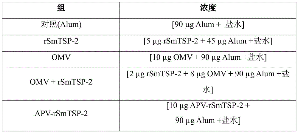
The immunoassay followed a regimen of 3 doses administered 15 days apart. Serum was collected from these animals before each immunization and 15 days after the last dose. Thus, antibody production between each dose can be assessed. Serum from the same group of animals was analyzed separately by ELISA to verify total IgG titers and concentrations of IgG1 and IgG2c specific for the rstsp-2 protein.
Spleen cells from immunized mice were isolated and stimulated "in vitro" with recombinant protein rSmTSP-2 or a corresponding control. The supernatant was collected and evaluated for inflammatory cytokine production by Cytometric Bead Array (CBA-kit Inflamat, tsao rio de bioci E-NCias BD-US), both according to the manufacturer's recommendations.
Table 2 shows the Zetasizer characterization of the OMV and APV-rSmTSP-2 complexes obtained.
Table 2-characterization of OMV and APV-rSmTSP-2 complexes by Zetasizer.

FIG. 4 graphically depicts cytokine production in splenocyte supernatants stimulated after 2 or 3 doses of APV-rSmTSP-2. Spleen cells isolated from spleen of mice immunized with APV-rSmTSP-2 were stimulated with 10. Mu.g rSmTSP-2 protein for 24 hours. Cytokine production was analyzed by CBA in the collected supernatant. Statistical analysis was performed by "one-way anova", where P values are 0.01; * 0.001; * 0.0001 is relative to saline group or value between different groups; ns indicates no statistical difference.
FIG. 5 graphically depicts cytokine production by T-CD4+ and T-CD8+ cells from mice immunized with APV-rTSP-2. Spleen cells isolated from spleens of 3 doses of APV-rSmTSP-2 immunized mice were stimulated with 10 μg rSmTSP-2 protein for 6 hours and labeled with anti-CD 3, CD4 or CD8 for immunophenotyping and labeled with anti-TNF- α, IFN- γ, IL-4 or IL-2 for detection of intracellular production of these cytokines by FACS. Statistical analysis was performed by "one-way anova", where P values are 0.01; * 0.001; * 0.0001 is relative to saline group or value between different groups; ns indicates no statistical difference.
FIGS. 6A-B graphically depict IgG humoral responses to rSmTSP-2 protein in mice immunized with rSmTSP-2 or APV-rSmTSP-2. FIG. 6A is a graph showing the doses of IgG anti-rSmTSP-2 antibodies in mice immunized with: alum (saline+aluminum hydroxide), rSmTSP-2[5 μg ], OMV [8 μg ] +rSmTSP-2[2 μg ] (Mix), and APV-rSmTSP-2[10 μg ]. Statistical analysis was performed by a "two-way anova" model, where P values <0.001. FIG. 6B is a diagram showing the IgG1 and IgG2c isotypes in the group immunized with rSmTSP-2 or APV-rSmTSP-2.
The present invention thus proposes a simple and highly flexible method for preparing affinity-based Antigen Presenting Vesicles (APV). The APV obtained has high specificity and stability, and can be stored at low temperature for several months and maintain its efficacy. Furthermore, the surprising process of the present invention is simple enough to ensure high reproducibility, since only a few steps are required, which reduces the risk of batch-to-batch variation, with great industrial advantages.
Thus, the embodiments presented in this invention are not limited in all possibilities, and it should be understood that various omissions, substitutions and changes can be made by those skilled in the art without departing from the spirit and scope of the invention.
It is expressly intended that all combinations of those elements that perform the same function in substantially the same way to achieve the same results are within the scope of the invention. Substitutions of elements from one described embodiment to another are also fully intended and contemplated.
It should also be understood that the drawings are not necessarily drawn to scale and that they are merely conceptual in nature.
Those skilled in the art will appreciate the knowledge presented herein and may reproduce the invention in the presented embodiments and other variations, which are encompassed within the scope of the claims.
Claims (15)
1. A method for obtaining Antigen Presenting Vesicles (APV) capable of coupling to one or more antigens, characterized in that it comprises the steps of:
a) Conjugating a first affinity molecule to a vesicle (OMV);
b) Obtaining complementary affinity molecules fused to one or more antigen proteins or peptides; and
c) Coupling the fusion protein obtained in step "b" with the product obtained in step "a",
wherein the APV obtained comprises:
outer Membrane Vesicles (OMVs) of gram-negative bacteria;
-at least one antigenic protein or peptide; and
-at least one pair of molecules with complementary affinities comprising:
(i) A first affinity molecule that binds to the vesicle; and
(ii) Complementary affinity molecules fused to the protein or peptide.
2. The method according to claim 1, wherein in step "a" the conjugation reaction of the OMV with the first affinity molecule is performed in a suitable solution with 3% sucrose added, wherein OMV is added in a ratio of 1:1 to 1:10 (mass/mass) relative to first affinity molecule, wherein the suitable solution used depends on the type of conjugation used and is selected from the group consisting of: buffers free of conjugation-interfering agents, preferably Ca-free 2+ /Mg 2+ Phosphate Buffered Saline (PBS) or physiological saline (150 mM NaCl in water).
3. The method according to claim 1 or 2, characterized in that in step "a" the mixture is kept at a temperature of 4 ℃ to 25 ℃ for 4 to 18 hours, wherein the mixture is subsequently dialyzed with the previously used suitable solution to which 3% sucrose is added, to eliminate excess unbound first affinity molecules.
4. A method according to any one of claims 1 to 3, wherein in step "a" optionally 0.05M to 0.2M activator molecule is used.
5. The method according to any one of claims 1 to 4, wherein in step "a" such OMVs are from a bacterium selected from the group consisting of neisseria meningitidis serogroup B or neisseria lactose, wherein preferably the OMVs are from neisseria lactose n.285/03.
6. The method according to any one of claims 1 to 5, wherein, still in step "a", such first affinity molecule is selected from the group consisting of biotin, biotin derivatives or biotin mimics, preferably amine-PEG 3-biotin ((+) -biotin-3-6, 9-trioxaundecanediamine) or a derivative or functional fragment thereof, more preferably biotin.
7. The method according to any one of claims 1 to 6, wherein, still in step "a", such activator molecule is selected from the group consisting of representative coupling agents, including organic compounds, such as thioesters, carbodiimides, succinimidyl esters, diisocyanates, glutaraldehyde, diazobenzenes and hexamethylenediamine, wherein preferably such activator molecule is EDAC (1-ethyl-3- [ 3-dimethylaminopropyl ] carbodiimide) hydrochloride.
8. The method according to any one of claims 1 to 7, wherein alternatively the first affinity binding molecule is linked to the OMV by a covalent bond, wherein preferably such an activator molecule is used for covalently binding the first affinity binding molecule to the vesicle (OMV).
9. The method according to any one of claims 1 to 8, wherein the first affinity molecule is instead directly attached to a carboxyl, hydroxyl, amino, phenoxy, hemiacetal or thiol functional group of the OMV without the aid of an activating molecule.
10. Method according to any one of claims 1 to 9, characterized in that in step "b" a fusion protein is obtained comprising the antigen protein or peptide fused to the complementary affinity molecule, wherein the complementary affinity molecule is genetically fused to the antigen protein or peptide by recombinant construction of the gene of the complementary affinity molecule linked to the gene of the antigen or peptide of interest, wherein the construction of the chimeric sequence encoding the fusion protein is achieved by molecular biological techniques, preferably techniques selected from the group consisting of conventional Polymerase Chain Reaction (PCR) or gene synthesis.
11. The method according to any one of claims 1 to 9, wherein such antigenic protein or peptide is selected from the group consisting of: any antigen that elicits an immune response in an organism, including immunogenic peptides or proteins, toxins, toxoids, subunits thereof, or combinations thereof; or wherein the antigen is selected from the group consisting of: any antigen associated with an infectious disease or cancer or immune disease; or wherein the antigen is an ingredient expressed by any of a variety of pathogens including viruses, bacteria, fungi, or parasites; or wherein the antigen is derived from a pathogenic organism; or wherein the antigen is a cancer antigen or a tumor antigen, e.g., an antigen derived from a tumor cell or a cancer cell; or wherein the antigen is expressed recombinantly and optionally comprises an affinity tag or epitope to facilitate purification; or wherein the antigen is obtained by chemical synthesis of an oligopeptide, either free or conjugated to a carrier protein.
12. The method according to any one of claims 1 to 11, wherein the antigen is expressed as a fusion with a complementary affinity molecule, or wherein alternatively the antigen is first prepared and then conjugated with a complementary affinity molecule.
13. The method according to any one of claims 1 to 12, wherein, still in step "b", the complementary affinity molecule is selected from the group consisting of: avidin, rizavidin, streptavidin or variants, derivatives or functional parts thereof, preferably avidin or recombinant rizavidin.
14. The method according to any one of claims 1 to 13, wherein in step "c" OMV-first affinity molecule conjugation product is mixed with the fusion protein obtained in step "b" in a ratio of 1:1 (mass/mass) for the purpose of coupling biotinylated OMV to the fusion protein, wherein incubation is performed at a temperature of 4 ℃ to 25 ℃ for 4 to 18 hours, and after incubation the mixture is centrifuged at 3000 to 14000rpm for 3 to 30 minutes to remove insoluble aggregates, wherein the supernatant is further purified.
15. The method according to claim 14, characterized in that the purification is performed by ultrafiltration or chromatographic techniques.
Applications Claiming Priority (3)
| Application Number | Priority Date | Filing Date | Title |
|---|---|---|---|
| BRBR1020200132164 | 2020-06-26 | ||
| BR102020013216-4A BR102020013216A2 (en) | 2020-06-26 | 2020-06-26 | PROCESS FOR OBTAINING ANTIGEN PRESENTING VESICLES (AVA) THAT MAKES IT POSSIBLE TO COUPLE ONE OR MORE ANTIGENS |
| PCT/BR2021/050284 WO2021258180A1 (en) | 2020-06-26 | 2021-06-28 | Process for obtaining antigen-presenting vesicles (apv) that enables the coupling of one or more antigens |
Publications (1)
| Publication Number | Publication Date |
|---|---|
| CN116194566A true CN116194566A (en) | 2023-05-30 |
Family
ID=77411504
Family Applications (1)
| Application Number | Title | Priority Date | Filing Date |
|---|---|---|---|
| CN202180059301.XA Pending CN116194566A (en) | 2020-06-26 | 2021-06-28 | Method for obtaining antigen-presenting vesicles (APVs) capable of coupling one or more antigens |
Country Status (5)
| Country | Link |
|---|---|
| US (1) | US20230256105A1 (en) |
| EP (1) | EP4173634A1 (en) |
| CN (1) | CN116194566A (en) |
| BR (1) | BR102020013216A2 (en) |
| WO (1) | WO2021258180A1 (en) |
Cited By (1)
| Publication number | Priority date | Publication date | Assignee | Title |
|---|---|---|---|---|
| CN119331108A (en) * | 2024-10-25 | 2025-01-21 | 扬州大学 | Preparation method and application of Escherichia coli biomimetic vesicles presenting H9 subtype avian influenza virus HA1 protein |
Families Citing this family (1)
| Publication number | Priority date | Publication date | Assignee | Title |
|---|---|---|---|---|
| US20230083394A1 (en) * | 2021-08-25 | 2023-03-16 | Cornell University | Methods and compositions for docking biotinylated antigens on the exterior of bacterial outer membrane vesicles |
Citations (5)
| Publication number | Priority date | Publication date | Assignee | Title |
|---|---|---|---|---|
| CN102065894A (en) * | 2008-04-17 | 2011-05-18 | 班扬生物标记公司 | An antibody bound synthetic vesicle containing active agent molecules |
| CN103648489A (en) * | 2011-05-11 | 2014-03-19 | 儿童医疗中心有限公司 | Multiple antigen presenting immunogenic compositions and methods and uses thereof |
| JP2019017341A (en) * | 2017-07-20 | 2019-02-07 | 国立研究開発法人国立がん研究センター | Labeled vesicle and production method thereof |
| CN110248681A (en) * | 2016-11-25 | 2019-09-17 | 葛兰素史密丝克莱恩生物有限公司 | NOMV- antigen conjugate and application thereof |
| CN110730670A (en) * | 2017-03-28 | 2020-01-24 | 儿童医疗中心有限公司 | Multi-antigen presentation system (MAPS) -based staphylococcus aureus vaccines, immunogenic compositions, and uses thereof |
Family Cites Families (4)
| Publication number | Priority date | Publication date | Assignee | Title |
|---|---|---|---|---|
| GB0130123D0 (en) * | 2001-12-17 | 2002-02-06 | Microbiological Res Agency | Outer membrane vesicle vaccine and its preparation |
| CU23377A1 (en) * | 2003-11-04 | 2009-05-28 | Ct De Ingenieria Genetica Y Biotecnologia | METHOD FOR THE INCORPORATION OF ANTIGENS IN EXTERNAL MEMBRANE VESICULES OF BACTERIA AND RESULTING FORMULATIONS |
| JP2013521770A (en) * | 2010-03-10 | 2013-06-13 | グラクソスミスクライン バイオロジカルズ ソシエテ アノニム | Vaccine composition |
| GB201015132D0 (en) * | 2010-09-10 | 2010-10-27 | Univ Bristol | Vaccine composition |
-
2020
- 2020-06-26 BR BR102020013216-4A patent/BR102020013216A2/en active Search and Examination
-
2021
- 2021-06-28 EP EP21758042.2A patent/EP4173634A1/en active Pending
- 2021-06-28 US US18/003,258 patent/US20230256105A1/en active Pending
- 2021-06-28 WO PCT/BR2021/050284 patent/WO2021258180A1/en not_active Ceased
- 2021-06-28 CN CN202180059301.XA patent/CN116194566A/en active Pending
Patent Citations (5)
| Publication number | Priority date | Publication date | Assignee | Title |
|---|---|---|---|---|
| CN102065894A (en) * | 2008-04-17 | 2011-05-18 | 班扬生物标记公司 | An antibody bound synthetic vesicle containing active agent molecules |
| CN103648489A (en) * | 2011-05-11 | 2014-03-19 | 儿童医疗中心有限公司 | Multiple antigen presenting immunogenic compositions and methods and uses thereof |
| CN110248681A (en) * | 2016-11-25 | 2019-09-17 | 葛兰素史密丝克莱恩生物有限公司 | NOMV- antigen conjugate and application thereof |
| CN110730670A (en) * | 2017-03-28 | 2020-01-24 | 儿童医疗中心有限公司 | Multi-antigen presentation system (MAPS) -based staphylococcus aureus vaccines, immunogenic compositions, and uses thereof |
| JP2019017341A (en) * | 2017-07-20 | 2019-02-07 | 国立研究開発法人国立がん研究センター | Labeled vesicle and production method thereof |
Cited By (1)
| Publication number | Priority date | Publication date | Assignee | Title |
|---|---|---|---|---|
| CN119331108A (en) * | 2024-10-25 | 2025-01-21 | 扬州大学 | Preparation method and application of Escherichia coli biomimetic vesicles presenting H9 subtype avian influenza virus HA1 protein |
Also Published As
| Publication number | Publication date |
|---|---|
| WO2021258180A1 (en) | 2021-12-30 |
| EP4173634A1 (en) | 2023-05-03 |
| BR102020013216A2 (en) | 2022-03-08 |
| US20230256105A1 (en) | 2023-08-17 |
Similar Documents
| Publication | Publication Date | Title |
|---|---|---|
| JP4180590B2 (en) | Immune adjuvant protein from Klebsiella pneumoniae | |
| JP7366333B2 (en) | Site-directed mutation carrier proteins and their use in the production of vaccines | |
| AU2021392894B2 (en) | Donor strand complemented fimh | |
| US6585973B1 (en) | Method for preparing solid phase conjugated vaccine | |
| CN116194566A (en) | Method for obtaining antigen-presenting vesicles (APVs) capable of coupling one or more antigens | |
| US6780420B1 (en) | Carrier protein having an adjuvant effect, immunogenic complex containing it, process for their preparation, nucleotide sequence and vaccine | |
| Burkhardt et al. | Assessment of the impact of manufacturing changes on the physicochemical properties of the recombinant vaccine carrier ExoProtein A | |
| Chuekwon et al. | N-terminus of flagellin enhances vaccine efficacy against Actinobacillus pleuropneumoniae | |
| Karch et al. | Production of E. coli-expressed self-assembling protein nanoparticles for vaccines requiring trimeric epitope presentation | |
| AU779056B2 (en) | Recombinant iron uptake proteins | |
| Cao et al. | Secretory expression and purification of recombinant Escherichia coli heat-labile enterotoxin B subunit and its applications on intranasal vaccination of hantavirus | |
| Montaner et al. | Ganglioside GM1-binding peptides as adjuvants of antigens inoculated by the intranasal route | |
| WO2025006263A2 (en) | Design of universal influenza vaccine candidates via antigen reorientation | |
| EA008254B1 (en) | DETOXIFIED TNF AND METHOD OF OBTAINING | |
| JP2000516444A (en) | Method for recovering and purifying recombinant non-lipidated Osp protein | |
| WO2024193713A1 (en) | Bivalent il-17 therapeutic vaccine, preparation method therefor, and use thereof | |
| CN117886947A (en) | Preparation method of recombinant epitope vaccine targeting amyloid | |
| CN121319218A (en) | A recombinant tetanus toxin protein rTT, its preparation method and application | |
| TW202311281A (en) | A self-assembled protein nanoparticle and its applications thereof |
Legal Events
| Date | Code | Title | Description |
|---|---|---|---|
| PB01 | Publication | ||
| PB01 | Publication | ||
| SE01 | Entry into force of request for substantive examination | ||
| SE01 | Entry into force of request for substantive examination |