CN113272018A - Method for producing genetically engineered T cells - Google Patents
Method for producing genetically engineered T cells Download PDFInfo
- Publication number
- CN113272018A CN113272018A CN201980087792.1A CN201980087792A CN113272018A CN 113272018 A CN113272018 A CN 113272018A CN 201980087792 A CN201980087792 A CN 201980087792A CN 113272018 A CN113272018 A CN 113272018A
- Authority
- CN
- China
- Prior art keywords
- cells
- population
- cell
- selection
- enriched
- Prior art date
- Legal status (The legal status is an assumption and is not a legal conclusion. Google has not performed a legal analysis and makes no representation as to the accuracy of the status listed.)
- Pending
Links
Images


Classifications
-
- C—CHEMISTRY; METALLURGY
- C07—ORGANIC CHEMISTRY
- C07K—PEPTIDES
- C07K14/00—Peptides having more than 20 amino acids; Gastrins; Somatostatins; Melanotropins; Derivatives thereof
- C07K14/195—Peptides having more than 20 amino acids; Gastrins; Somatostatins; Melanotropins; Derivatives thereof from bacteria
- C07K14/36—Peptides having more than 20 amino acids; Gastrins; Somatostatins; Melanotropins; Derivatives thereof from bacteria from Actinomyces; from Streptomyces (G)
-
- C—CHEMISTRY; METALLURGY
- C07—ORGANIC CHEMISTRY
- C07K—PEPTIDES
- C07K16/00—Immunoglobulins [IGs], e.g. monoclonal or polyclonal antibodies
- C07K16/18—Immunoglobulins [IGs], e.g. monoclonal or polyclonal antibodies against material from animals or humans
- C07K16/28—Immunoglobulins [IGs], e.g. monoclonal or polyclonal antibodies against material from animals or humans against receptors, cell surface antigens or cell surface determinants
- C07K16/2803—Immunoglobulins [IGs], e.g. monoclonal or polyclonal antibodies against material from animals or humans against receptors, cell surface antigens or cell surface determinants against the immunoglobulin superfamily
- C07K16/2809—Immunoglobulins [IGs], e.g. monoclonal or polyclonal antibodies against material from animals or humans against receptors, cell surface antigens or cell surface determinants against the immunoglobulin superfamily against the T-cell receptor (TcR)-CD3 complex
-
- A—HUMAN NECESSITIES
- A01—AGRICULTURE; FORESTRY; ANIMAL HUSBANDRY; HUNTING; TRAPPING; FISHING
- A01N—PRESERVATION OF BODIES OF HUMANS OR ANIMALS OR PLANTS OR PARTS THEREOF; BIOCIDES, e.g. AS DISINFECTANTS, AS PESTICIDES OR AS HERBICIDES; PEST REPELLANTS OR ATTRACTANTS; PLANT GROWTH REGULATORS
- A01N1/00—Preservation of bodies of humans or animals, or parts thereof
- A01N1/10—Preservation of living parts
- A01N1/12—Chemical aspects of preservation
- A01N1/122—Preservation or perfusion media
- A01N1/125—Freeze protecting agents, e.g. cryoprotectants or osmolarity regulators
-
- A—HUMAN NECESSITIES
- A61—MEDICAL OR VETERINARY SCIENCE; HYGIENE
- A61K—PREPARATIONS FOR MEDICAL, DENTAL OR TOILETRY PURPOSES
- A61K35/00—Medicinal preparations containing materials or reaction products thereof with undetermined constitution
- A61K35/12—Materials from mammals; Compositions comprising non-specified tissues or cells; Compositions comprising non-embryonic stem cells; Genetically modified cells
- A61K35/14—Blood; Artificial blood
- A61K35/17—Lymphocytes; B-cells; T-cells; Natural killer cells; Interferon-activated or cytokine-activated lymphocytes
-
- A—HUMAN NECESSITIES
- A61—MEDICAL OR VETERINARY SCIENCE; HYGIENE
- A61K—PREPARATIONS FOR MEDICAL, DENTAL OR TOILETRY PURPOSES
- A61K40/00—Cellular immunotherapy
- A61K40/10—Cellular immunotherapy characterised by the cell type used
- A61K40/11—T-cells, e.g. tumour infiltrating lymphocytes [TIL] or regulatory T [Treg] cells; Lymphokine-activated killer [LAK] cells
-
- A—HUMAN NECESSITIES
- A61—MEDICAL OR VETERINARY SCIENCE; HYGIENE
- A61K—PREPARATIONS FOR MEDICAL, DENTAL OR TOILETRY PURPOSES
- A61K40/00—Cellular immunotherapy
- A61K40/30—Cellular immunotherapy characterised by the recombinant expression of specific molecules in the cells of the immune system
- A61K40/31—Chimeric antigen receptors [CAR]
-
- A—HUMAN NECESSITIES
- A61—MEDICAL OR VETERINARY SCIENCE; HYGIENE
- A61K—PREPARATIONS FOR MEDICAL, DENTAL OR TOILETRY PURPOSES
- A61K40/00—Cellular immunotherapy
- A61K40/30—Cellular immunotherapy characterised by the recombinant expression of specific molecules in the cells of the immune system
- A61K40/32—T-cell receptors [TCR]
-
- A—HUMAN NECESSITIES
- A61—MEDICAL OR VETERINARY SCIENCE; HYGIENE
- A61K—PREPARATIONS FOR MEDICAL, DENTAL OR TOILETRY PURPOSES
- A61K40/00—Cellular immunotherapy
- A61K40/40—Cellular immunotherapy characterised by antigens that are targeted or presented by cells of the immune system
- A61K40/41—Vertebrate antigens
- A61K40/42—Cancer antigens
- A61K40/4202—Receptors, cell surface antigens or cell surface determinants
- A61K40/421—Immunoglobulin superfamily
- A61K40/4211—CD19 or B4
-
- A—HUMAN NECESSITIES
- A61—MEDICAL OR VETERINARY SCIENCE; HYGIENE
- A61K—PREPARATIONS FOR MEDICAL, DENTAL OR TOILETRY PURPOSES
- A61K40/00—Cellular immunotherapy
- A61K40/40—Cellular immunotherapy characterised by antigens that are targeted or presented by cells of the immune system
- A61K40/41—Vertebrate antigens
- A61K40/42—Cancer antigens
- A61K40/4202—Receptors, cell surface antigens or cell surface determinants
- A61K40/4214—Receptors for cytokines
- A61K40/4215—Receptors for tumor necrosis factors [TNF], e.g. lymphotoxin receptor [LTR], CD30
-
- A—HUMAN NECESSITIES
- A61—MEDICAL OR VETERINARY SCIENCE; HYGIENE
- A61P—SPECIFIC THERAPEUTIC ACTIVITY OF CHEMICAL COMPOUNDS OR MEDICINAL PREPARATIONS
- A61P35/00—Antineoplastic agents
-
- C—CHEMISTRY; METALLURGY
- C07—ORGANIC CHEMISTRY
- C07K—PEPTIDES
- C07K14/00—Peptides having more than 20 amino acids; Gastrins; Somatostatins; Melanotropins; Derivatives thereof
- C07K14/435—Peptides having more than 20 amino acids; Gastrins; Somatostatins; Melanotropins; Derivatives thereof from animals; from humans
- C07K14/705—Receptors; Cell surface antigens; Cell surface determinants
- C07K14/70503—Immunoglobulin superfamily
- C07K14/7051—T-cell receptor (TcR)-CD3 complex
-
- C—CHEMISTRY; METALLURGY
- C07—ORGANIC CHEMISTRY
- C07K—PEPTIDES
- C07K14/00—Peptides having more than 20 amino acids; Gastrins; Somatostatins; Melanotropins; Derivatives thereof
- C07K14/435—Peptides having more than 20 amino acids; Gastrins; Somatostatins; Melanotropins; Derivatives thereof from animals; from humans
- C07K14/705—Receptors; Cell surface antigens; Cell surface determinants
- C07K14/70503—Immunoglobulin superfamily
- C07K14/70517—CD8
-
- C—CHEMISTRY; METALLURGY
- C07—ORGANIC CHEMISTRY
- C07K—PEPTIDES
- C07K14/00—Peptides having more than 20 amino acids; Gastrins; Somatostatins; Melanotropins; Derivatives thereof
- C07K14/435—Peptides having more than 20 amino acids; Gastrins; Somatostatins; Melanotropins; Derivatives thereof from animals; from humans
- C07K14/705—Receptors; Cell surface antigens; Cell surface determinants
- C07K14/70596—Molecules with a "CD"-designation not provided for elsewhere
-
- C—CHEMISTRY; METALLURGY
- C07—ORGANIC CHEMISTRY
- C07K—PEPTIDES
- C07K16/00—Immunoglobulins [IGs], e.g. monoclonal or polyclonal antibodies
- C07K16/18—Immunoglobulins [IGs], e.g. monoclonal or polyclonal antibodies against material from animals or humans
- C07K16/28—Immunoglobulins [IGs], e.g. monoclonal or polyclonal antibodies against material from animals or humans against receptors, cell surface antigens or cell surface determinants
- C07K16/2803—Immunoglobulins [IGs], e.g. monoclonal or polyclonal antibodies against material from animals or humans against receptors, cell surface antigens or cell surface determinants against the immunoglobulin superfamily
- C07K16/2818—Immunoglobulins [IGs], e.g. monoclonal or polyclonal antibodies against material from animals or humans against receptors, cell surface antigens or cell surface determinants against the immunoglobulin superfamily against CD28 or CD152
-
- C—CHEMISTRY; METALLURGY
- C07—ORGANIC CHEMISTRY
- C07K—PEPTIDES
- C07K16/00—Immunoglobulins [IGs], e.g. monoclonal or polyclonal antibodies
- C07K16/18—Immunoglobulins [IGs], e.g. monoclonal or polyclonal antibodies against material from animals or humans
- C07K16/28—Immunoglobulins [IGs], e.g. monoclonal or polyclonal antibodies against material from animals or humans against receptors, cell surface antigens or cell surface determinants
- C07K16/2896—Immunoglobulins [IGs], e.g. monoclonal or polyclonal antibodies against material from animals or humans against receptors, cell surface antigens or cell surface determinants against molecules with a "CD"-designation, not provided for elsewhere
-
- C—CHEMISTRY; METALLURGY
- C12—BIOCHEMISTRY; BEER; SPIRITS; WINE; VINEGAR; MICROBIOLOGY; ENZYMOLOGY; MUTATION OR GENETIC ENGINEERING
- C12M—APPARATUS FOR ENZYMOLOGY OR MICROBIOLOGY; APPARATUS FOR CULTURING MICROORGANISMS FOR PRODUCING BIOMASS, FOR GROWING CELLS OR FOR OBTAINING FERMENTATION OR METABOLIC PRODUCTS, i.e. BIOREACTORS OR FERMENTERS
- C12M25/00—Means for supporting, enclosing or fixing the microorganisms, e.g. immunocoatings
- C12M25/16—Particles; Beads; Granular material; Encapsulation
-
- C—CHEMISTRY; METALLURGY
- C12—BIOCHEMISTRY; BEER; SPIRITS; WINE; VINEGAR; MICROBIOLOGY; ENZYMOLOGY; MUTATION OR GENETIC ENGINEERING
- C12N—MICROORGANISMS OR ENZYMES; COMPOSITIONS THEREOF; PROPAGATING, PRESERVING, OR MAINTAINING MICROORGANISMS; MUTATION OR GENETIC ENGINEERING; CULTURE MEDIA
- C12N5/00—Undifferentiated human, animal or plant cells, e.g. cell lines; Tissues; Cultivation or maintenance thereof; Culture media therefor
- C12N5/0081—Purging biological preparations of unwanted cells
- C12N5/0087—Purging against subsets of blood cells, e.g. purging alloreactive T cells
-
- C—CHEMISTRY; METALLURGY
- C12—BIOCHEMISTRY; BEER; SPIRITS; WINE; VINEGAR; MICROBIOLOGY; ENZYMOLOGY; MUTATION OR GENETIC ENGINEERING
- C12N—MICROORGANISMS OR ENZYMES; COMPOSITIONS THEREOF; PROPAGATING, PRESERVING, OR MAINTAINING MICROORGANISMS; MUTATION OR GENETIC ENGINEERING; CULTURE MEDIA
- C12N5/00—Undifferentiated human, animal or plant cells, e.g. cell lines; Tissues; Cultivation or maintenance thereof; Culture media therefor
- C12N5/06—Animal cells or tissues; Human cells or tissues
- C12N5/0602—Vertebrate cells
- C12N5/0634—Cells from the blood or the immune system
- C12N5/0636—T lymphocytes
-
- A—HUMAN NECESSITIES
- A61—MEDICAL OR VETERINARY SCIENCE; HYGIENE
- A61K—PREPARATIONS FOR MEDICAL, DENTAL OR TOILETRY PURPOSES
- A61K2239/00—Indexing codes associated with cellular immunotherapy of group A61K40/00
- A61K2239/31—Indexing codes associated with cellular immunotherapy of group A61K40/00 characterized by the route of administration
-
- A—HUMAN NECESSITIES
- A61—MEDICAL OR VETERINARY SCIENCE; HYGIENE
- A61K—PREPARATIONS FOR MEDICAL, DENTAL OR TOILETRY PURPOSES
- A61K2239/00—Indexing codes associated with cellular immunotherapy of group A61K40/00
- A61K2239/38—Indexing codes associated with cellular immunotherapy of group A61K40/00 characterised by the dose, timing or administration schedule
-
- A—HUMAN NECESSITIES
- A61—MEDICAL OR VETERINARY SCIENCE; HYGIENE
- A61K—PREPARATIONS FOR MEDICAL, DENTAL OR TOILETRY PURPOSES
- A61K2239/00—Indexing codes associated with cellular immunotherapy of group A61K40/00
- A61K2239/46—Indexing codes associated with cellular immunotherapy of group A61K40/00 characterised by the cancer treated
- A61K2239/48—Blood cells, e.g. leukemia or lymphoma
-
- C—CHEMISTRY; METALLURGY
- C07—ORGANIC CHEMISTRY
- C07K—PEPTIDES
- C07K2317/00—Immunoglobulins specific features
- C07K2317/50—Immunoglobulins specific features characterized by immunoglobulin fragments
- C07K2317/55—Fab or Fab'
-
- C—CHEMISTRY; METALLURGY
- C07—ORGANIC CHEMISTRY
- C07K—PEPTIDES
- C07K2317/00—Immunoglobulins specific features
- C07K2317/60—Immunoglobulins specific features characterized by non-natural combinations of immunoglobulin fragments
- C07K2317/62—Immunoglobulins specific features characterized by non-natural combinations of immunoglobulin fragments comprising only variable region components
- C07K2317/622—Single chain antibody (scFv)
-
- C—CHEMISTRY; METALLURGY
- C07—ORGANIC CHEMISTRY
- C07K—PEPTIDES
- C07K2319/00—Fusion polypeptide
-
- C—CHEMISTRY; METALLURGY
- C07—ORGANIC CHEMISTRY
- C07K—PEPTIDES
- C07K2319/00—Fusion polypeptide
- C07K2319/01—Fusion polypeptide containing a localisation/targetting motif
- C07K2319/02—Fusion polypeptide containing a localisation/targetting motif containing a signal sequence
-
- C—CHEMISTRY; METALLURGY
- C07—ORGANIC CHEMISTRY
- C07K—PEPTIDES
- C07K2319/00—Fusion polypeptide
- C07K2319/01—Fusion polypeptide containing a localisation/targetting motif
- C07K2319/03—Fusion polypeptide containing a localisation/targetting motif containing a transmembrane segment
-
- C—CHEMISTRY; METALLURGY
- C07—ORGANIC CHEMISTRY
- C07K—PEPTIDES
- C07K2319/00—Fusion polypeptide
- C07K2319/33—Fusion polypeptide fusions for targeting to specific cell types, e.g. tissue specific targeting, targeting of a bacterial subspecies
-
- C—CHEMISTRY; METALLURGY
- C12—BIOCHEMISTRY; BEER; SPIRITS; WINE; VINEGAR; MICROBIOLOGY; ENZYMOLOGY; MUTATION OR GENETIC ENGINEERING
- C12N—MICROORGANISMS OR ENZYMES; COMPOSITIONS THEREOF; PROPAGATING, PRESERVING, OR MAINTAINING MICROORGANISMS; MUTATION OR GENETIC ENGINEERING; CULTURE MEDIA
- C12N2501/00—Active agents used in cell culture processes, e.g. differentation
- C12N2501/50—Cell markers; Cell surface determinants
- C12N2501/505—CD4; CD8
-
- C—CHEMISTRY; METALLURGY
- C12—BIOCHEMISTRY; BEER; SPIRITS; WINE; VINEGAR; MICROBIOLOGY; ENZYMOLOGY; MUTATION OR GENETIC ENGINEERING
- C12N—MICROORGANISMS OR ENZYMES; COMPOSITIONS THEREOF; PROPAGATING, PRESERVING, OR MAINTAINING MICROORGANISMS; MUTATION OR GENETIC ENGINEERING; CULTURE MEDIA
- C12N2501/00—Active agents used in cell culture processes, e.g. differentation
- C12N2501/50—Cell markers; Cell surface determinants
- C12N2501/515—CD3, T-cell receptor complex
-
- C—CHEMISTRY; METALLURGY
- C12—BIOCHEMISTRY; BEER; SPIRITS; WINE; VINEGAR; MICROBIOLOGY; ENZYMOLOGY; MUTATION OR GENETIC ENGINEERING
- C12N—MICROORGANISMS OR ENZYMES; COMPOSITIONS THEREOF; PROPAGATING, PRESERVING, OR MAINTAINING MICROORGANISMS; MUTATION OR GENETIC ENGINEERING; CULTURE MEDIA
- C12N2501/00—Active agents used in cell culture processes, e.g. differentation
- C12N2501/50—Cell markers; Cell surface determinants
- C12N2501/599—Cell markers; Cell surface determinants with CD designations not provided for elsewhere
-
- C—CHEMISTRY; METALLURGY
- C12—BIOCHEMISTRY; BEER; SPIRITS; WINE; VINEGAR; MICROBIOLOGY; ENZYMOLOGY; MUTATION OR GENETIC ENGINEERING
- C12N—MICROORGANISMS OR ENZYMES; COMPOSITIONS THEREOF; PROPAGATING, PRESERVING, OR MAINTAINING MICROORGANISMS; MUTATION OR GENETIC ENGINEERING; CULTURE MEDIA
- C12N2510/00—Genetically modified cells
-
- C—CHEMISTRY; METALLURGY
- C12—BIOCHEMISTRY; BEER; SPIRITS; WINE; VINEGAR; MICROBIOLOGY; ENZYMOLOGY; MUTATION OR GENETIC ENGINEERING
- C12N—MICROORGANISMS OR ENZYMES; COMPOSITIONS THEREOF; PROPAGATING, PRESERVING, OR MAINTAINING MICROORGANISMS; MUTATION OR GENETIC ENGINEERING; CULTURE MEDIA
- C12N2740/00—Reverse transcribing RNA viruses
- C12N2740/00011—Details
- C12N2740/10011—Retroviridae
- C12N2740/16011—Human Immunodeficiency Virus, HIV
- C12N2740/16041—Use of virus, viral particle or viral elements as a vector
- C12N2740/16043—Use of virus, viral particle or viral elements as a vector viral genome or elements thereof as genetic vector
Landscapes
- Health & Medical Sciences (AREA)
- Life Sciences & Earth Sciences (AREA)
- Chemical & Material Sciences (AREA)
- Organic Chemistry (AREA)
- Immunology (AREA)
- General Health & Medical Sciences (AREA)
- Genetics & Genomics (AREA)
- Engineering & Computer Science (AREA)
- Zoology (AREA)
- Biochemistry (AREA)
- Medicinal Chemistry (AREA)
- Biomedical Technology (AREA)
- Veterinary Medicine (AREA)
- Public Health (AREA)
- Animal Behavior & Ethology (AREA)
- Molecular Biology (AREA)
- Wood Science & Technology (AREA)
- Biotechnology (AREA)
- Bioinformatics & Cheminformatics (AREA)
- Epidemiology (AREA)
- Proteomics, Peptides & Aminoacids (AREA)
- Biophysics (AREA)
- Cell Biology (AREA)
- Hematology (AREA)
- Microbiology (AREA)
- General Engineering & Computer Science (AREA)
- Gastroenterology & Hepatology (AREA)
- Pharmacology & Pharmacy (AREA)
- Toxicology (AREA)
- Nuclear Medicine, Radiotherapy & Molecular Imaging (AREA)
- General Chemical & Material Sciences (AREA)
- Chemical Kinetics & Catalysis (AREA)
- Developmental Biology & Embryology (AREA)
- Virology (AREA)
- Dentistry (AREA)
- Sustainable Development (AREA)
- Environmental Sciences (AREA)
- Micro-Organisms Or Cultivation Processes Thereof (AREA)
- Medicines Containing Material From Animals Or Micro-Organisms (AREA)
- Peptides Or Proteins (AREA)
Abstract
本公开文本提供富集CD57阴性T细胞或耗尽CD57阳性细胞的细胞群,以及用于刺激、培育、扩增和/或基因工程化富集CD57‑T细胞或耗尽CD57+T细胞的细胞群的方法。还包括用于生成、分离、富集或选择CD57‑T细胞或者耗尽CD57+细胞的方法,如通过阴性选择来进行。

The present disclosure provides cell populations enriched for CD57-negative T cells or depleted of CD57-positive cells, as well as cells for stimulating, culturing, expanding and/or genetically engineering enriching for CD57-T cells or depleting CD57+ T cells group method. Also included are methods for generating, isolating, enriching or selecting CD57-T cells or depleting CD57+ cells, such as by negative selection.
Description
Cross Reference to Related Applications
Priority of us provisional application 62/756,571 entitled "method of generating genetically engineered T cells (PROCESS FOR PROCESS program GENETICALLY ENGINEERED T CELLS)" filed on 6.11.2018 and us provisional application 62/798,457 entitled "method of generating genetically engineered T cells (PROCESS FOR program GENETICALLY ENGINEERED T CELLS)" filed on 29.1.2019, the contents of which are incorporated by reference in their entirety FOR all purposes, are claimed in this application.
Sequence listing incorporated by reference
This application is filed with a sequence listing in electronic format. The sequence listing is provided in a file named 735042017640seqlist. txt, created on day 11, month 5, 2019, and of size 74 kilobytes. The information in the sequence listing in electronic format is incorporated by reference in its entirety.
Technical Field
In some aspects, the disclosure relates to cell populations that are enriched for CD57 negative T cells or depleted of CD57 positive cells, and methods for stimulating, breeding, expanding, and/or genetically engineering cell populations that are enriched for CD57 negative T cells or depleted of CD57 positive T cells. Also included are methods for generating, isolating, enriching or selecting CD57 negative T cells or depleting CD57 positive cells, such as by negative selection.
Background
A variety of cell therapy methods are useful for treating diseases and disorders. Cell therapy methods include methods involving immune cells (e.g., T cells) genetically engineered with recombinant receptors (e.g., chimeric antigen receptors). However, in some cases, some existing methods may result in populations with low consistency, efficacy, or persistence in vivo. There is a need for improved methods for making and/or engineering such cell therapies, including providing improved cell therapy products with high consistency, efficacy, and persistence in vivo.
Disclosure of Invention
Provided herein are methods of enriching for T cells, the methods comprising performing a first selection comprising removing CD57+ T cells from a biological sample containing primary human T cells, thereby generating a depleted population containing fewer CD57+ T cells than the biological sample; and performing a second selection of cells from the depleted population, the second selection comprising enriching the depleted population for CD3+ T cells, the enriching thereby generating an enriched population of CD57-CD3+ T cells.
Also provided herein are methods of enriching T cells, the methods comprising performing a first selection comprising enriching CD3+ T cells from a biological sample containing primary human T cells, thereby generating an enriched population of T cells; and performing a second selection of cells from the enriched population of T cells, the second selection comprising removing CD57+ T cells from the enriched population of sample cells, thereby generating a depleted population, wherein the depleted population contains fewer CD57+ T cells than the biological sample and/or less than the enriched population of T cells, and is enriched for CD3+ T cells.
Also provided herein are methods of enriching T cells, the methods comprising removing CD57+ T cells from a biological sample containing an apheresis product or leukopheresis product comprising primary human T cells, thereby generating a depleted population of T cells, wherein the depleted population contains fewer CD57+ T cells than the biological sample, and wherein the depleted population contains at least one of: (i) less than or about 5% CD57+ T cells; (ii) a frequency of CD57+ T cells that is less than or about 35% of the frequency of CD57+ T cells present in the biological sample; and (iii) CD3+ T cells, wherein at least or about 95% of the CD3+ T cells are CD 57-.
In some of any such embodiments, the depleted population contains at least one of: (i) less than or about 5% CD57+ T cells; (ii) a frequency of CD57+ T cells that is less than or about 35% of the frequency of CD57+ T cells present in the biological sample; (iii) CD4+ T cells, wherein at least or about 95% of the CD4+ T cells are CD 57-; and (iv) CD8+ T cells, wherein at least or about 95% of the CD8+ T cells are CD 57-. In some of any such embodiments, at least or about 95% of the CD3+ T cells in the depleted population contain CD57-CD3+ T cells. In some of any such embodiments, the depleted population is a first depleted population, and the method further comprises enriching CD3+ T cells from the first depleted population, thereby generating an enriched population of CD57-CD3+ T cells.
In some of any such embodiments, the biological sample comprises an apheresis product or a leukocyte apheresis product.
Also provided herein are methods of enriching T cells, the methods comprising performing a first selection comprising removing CD57+ T cells from a biological sample containing an apheresis product or a leukopheresis product comprising primary human T cells, thereby generating a first depleted population that contains fewer CD57+ T cells than the biological sample; and (b) performing a second selection of cells from the first depleted population, the second selection comprising enrichment of CD3+ T cells from the first depleted population, the enrichment thereby generating an enriched population of CD57-CD3+ T cells.
Also provided herein are methods of enriching T cells, the methods comprising (a) performing a first selection comprising enriching CD3+ T cells from a biological sample containing an apheresis product or a leukopheresis product comprising primary human T cells, thereby generating an enriched biological sample; and (b) performing a second selection of cells from the enriched biological sample, the second selection comprising removing CD57+ T cells from the enriched biological sample, thereby generating a depleted population, the depleted population being an enriched population of CD57-CD3+ T cells.
Also provided herein are methods of enriching T cells, the methods comprising removing CD57+ T cells from a biological sample containing an apheresis product or leukopheresis product comprising primary human T cells, thereby generating a depleted population of T cells, wherein the depleted population contains fewer CD57+ T cells than the biological sample, and wherein the depleted population contains: (i) less than or about 5% CD57+ T cells; (ii) a frequency of CD57+ T cells that is less than or about 35% of the frequency of CD57+ T cells present in the biological sample; (iii) CD4+ T cells, wherein at least or about 95% of the CD4+ T cells are CD 57-; and/or (iv) CD8+ T cells, wherein at least or about 95% of the CD8+ T cells are CD 57-.
In some of any such embodiments, the depleted population contains CD57-CD4+ T cells. In some of any such embodiments, the depleted population contains CD57-CD8+ T cells. In some of any such embodiments, the depleted population contains CD57-CD4+ T cells and CD57-CD8+ T cells.
In some any such embodiments, the depleted population is a first depleted population, and the method further comprises selecting CD4+ T cells from the first depleted population, thereby generating an enriched second depleted population enriched for CD57-CD4+ T cells and an unselected population. In some any such embodiments, the method further comprises selecting CD8+ T cells from the unselected population, thereby generating a third depleted population that is an enriched population of CD57-CD8+ T cells and a second unselected population.
In some any such embodiments, the depleted population is a first depleted population, and the method further comprises selecting CD8+ T cells from the first depleted population, thereby generating a second depleted population that is an enriched population of CD57-CD8+ T cells and an unselected population. In some any such embodiments, the method further comprises selecting CD4+ T cells from the unselected population, thereby generating a third depleted population that is an enriched population of CD57-CD4+ T cells and a second unselected population.
In some any such embodiments, the depleted population is a first depleted population, and the method further comprises selecting CD3+ T cells from the first depleted population, thereby generating a second depleted population that is an enriched population of CD57-CD3+ T cells and an unselected population.
Also provided is a method of enriching T cells, the method comprising: (a) performing a first selection comprising removing CD57+ T cells from a biological sample containing an apheresis product or leukopheresis product comprising primary human T cells, thereby generating a first depleted population comprising less CD57+ T cells than the biological sample; (b) performing a second selection of cells from the first depleted population, the second selection comprising enriching the first depleted population for one of (i) CD4+ T cells and (ii) CD8+ T cells, the enriching thereby generating a second depleted population enriched for one of (i) CD4+ T cells and (ii) CD8+ T cells and an unselected population; and (c) performing a third selection comprising enriching the other of (i) CD4+ cells and (ii) CD8+ cells from the unselected population, the enriching thereby producing a third depleted population enriched in the other of (i) CD4+ T cells and (ii) CD8+ T cells. In some any such embodiments, the frequency of CD57+ cells in the depleted population (optionally the first, second, or third depleted population) is less than or about 35%, 30%, 20%, 10%, 5%, 1%, or 0.1% of the frequency of CD57+ T cells in the biological sample. In some any such embodiments, the frequency of CD57+ T cells in the depleted population (optionally the first, second, or third depleted population) is less than or about 35%, 30%, 20%, 10%, 5%, 1%, or 0.1% of the frequency of CD57+ T cells in the biological sample. In some any such embodiments, the depleted population (optionally the first, second or third depleted population) comprises less than or about 3%, less than or about 2%, less than or about 1%, less than or about 0.1%, or less than or about 0.01% CD57+ T cells. In some of any such embodiments, the depleted population (optionally the first depleted population, second depleted population, or third depleted population) is free or substantially free of CD57+ T cells. In some embodiments, a depleted population that is substantially free of CD57+ T cells comprises less than about or about 3%, less than about or about 2%, less than about or about 1%, less than about or about 0.1%, or less than about or about 0.01% CD57+ T cells.
In some any such embodiments, at least or about 95% of the CD4+ T cells in the depleted population (optionally the first, second, or third depleted population) comprise CD57-CD4+ T cells. In some any such embodiments, at least or about 95% of the CD8+ T cells in the depleted population (optionally the first, second, or third depleted population) comprise CD57-CD8+ T cells. In some any such embodiments, at least or about 95% of the CD4+ T cells and at least or about 95% of the CD8+ T cells in the depleted population (optionally the first, second, or third depleted population) comprise CD57-CD4+ T cells and CD57-CD8+ T cells, respectively.
In some any such embodiments, the frequency of naive-like cells in the depleted population (optionally the first depleted population, second depleted population, or third depleted population) is at least or about 10%, 20%, 30%, 40%, or 50% greater than or about the frequency of naive-like cells in the biological sample. In some any such embodiments, the frequency of one or more of CD25+ T cells, CD27+ T cells, CD28+ T cells, CCR7+ T cells, or CD45RA + T cells in the depleted population (optionally the first, second, or third depleted population) is at least or about 10%, 20%, 30%, 40%, or 50% greater than the frequency of the corresponding cells in the biological sample.
In some any such embodiments, the depleted population (optionally the first depleted population, second depleted population, or third depleted population) comprises at least or about 15%, 20%, 25%, 30%, 35%, or 40% CD27+ T cells. In some any such embodiments, the depleted population (optionally the first depleted population, second depleted population, or third depleted population) comprises at least or about 10%, 15%, 20%, 25%, 30%, 35%, or 40% CD28+ T cells. In some any such embodiments, the depleted population (optionally the first depleted population, second depleted population, or third depleted population) comprises at least or about 10%, 15%, 20%, 25%, 30%, 35%, or 40% CD27+ CD28+ T cells. In some any such embodiments, the depleted population (optionally the first, second, or third depleted population) comprises at least or about 10%, 15%, 20%, 25%, 30%, 35%, 40%, 50%, 60%, 70%, or 80% CD27+ CD28+ T cells. In some any such embodiments, the depleted population (optionally the first depleted population, second depleted population, or third depleted population) comprises at least or about 70% or 80% CD27+ CD28+ T cells. In some any such embodiments, the depleted population (optionally the first depleted population, second depleted population, or third depleted population) comprises at least or about 10%, 15%, 20%, or 25% CCR7+ T cells.
In some any of the provided embodiments, the depleted population (optionally the first depleted population, second depleted population, or third depleted population) is used as an input composition for a subsequent step in the process of genetically engineering T cells. In some embodiments, the input composition comprises a CD57 depleted population enriched for CD4+ cells (CD57-CD4+) and enriched for CD8+ cells (CD57-CD8 +). In some embodiments, the input composition comprises a CD57 depleted population enriched for CD4+ T cells (CD57-CD4+) and enriched for CD8+ T cells (CD57-CD8 +). In some embodiments of any of the methods, the method comprises combining the cells of the second depleted population and the third depleted population, thereby generating the input composition. In some of any of the provided embodiments, the method further comprises combining the second depleted population and the third depleted population. In some any provided embodiments, the second depleted population and the third depleted population are combined at a ratio of between or about 1:3 and at or about 3:1, thereby generating a depleted population comprising the second depleted population and the third depleted population. In some any provided embodiments, the second depleted population and the third depleted population are combined at a ratio of at or about 1:1, thereby generating a depleted population comprising the second depleted population and the third depleted population.
In some of any of the provided methods, the method produces a T cell composition containing at least or about 90%, at least or about 95%, at least or about 97%, at least or about 99%, or at least or about 99.9% CD57-CD4+ T cells. In some of any of the provided methods, the method produces a T cell composition containing at least or about 90%, at least or about 95%, at least or about 97%, at least or about 99%, or at least or about 99.9% CD57-CD8+ T cells. In some of any of the provided methods, the method produces a T cell composition containing at least or about 90%, at least or about 95%, at least or about 97%, at least or about 99%, or at least or about 99.9% CD57-CD3+ T cells.
In some of any provided embodiments, the ratio of CD4+ to CD8+ T cells in the composition is between 3:1 and 1:3, inclusive. In some of any provided embodiments, the ratio of CD4+ to CD8+ T cells in the composition is between 2:1 and 1:2, inclusive. In some of any provided embodiments, the ratio of CD4+ to CD8+ T cells in the composition is between 1.5:1 and 1:1.5, inclusive. In some of any provided embodiments, the ratio of CD4+ to CD8+ T cells in the composition is between 1.2:1 and 1:1.2, inclusive. In some any provided embodiments, the ratio of CD4+ to CD8+ T cells in the composition is at or about 1:1CD4 +. In some of any of the provided embodiments, the primary T cell is from a human subject. In some of any of the provided embodiments, the primary T cell is from a subject having a disease or disorder. In some of any of the provided embodiments, the disease or disorder is cancer. In some of any of the provided embodiments, the primary T cells are from a healthy subject.
In some of any of the provided embodiments, removing CD57+ T cells comprises immunoaffinity-based selection. In some of any of the provided embodiments, the immunoaffinity based selection comprises contacting the cells with an antibody capable of specifically binding to CD57 and recovering cells not bound to the antibody, thereby effecting a negative selection, wherein the recovered cells are depleted of CD57+ cells. In some of any of the provided embodiments, enriching the cells comprises immunoaffinity-based selection. In some any provided embodiments, the first, second, and/or third selection enriches CD4 or CD 8T cells and the immunoaffinity-based selection is effected by: positive selection is achieved by contacting the cells with an antibody capable of specifically binding to CD4 or CD8, respectively, and recovering the cells bound to the antibody, wherein the recovered cells are enriched for CD4+ cells or CD8+ cells. In some of any of the provided embodiments, the selecting is enriched for CD 3T cells, and the immunoaffinity-based selecting is effected by: positive selection is achieved by contacting cells with an antibody capable of specifically binding to CD3, and recovering cells bound to the antibody, wherein the recovered cells are enriched for CD3+ cells. In some of any of the provided embodiments, the antibody is immobilized on a solid surface. In some of any provided embodiments, the solid surface is a magnetic particle. In some of any provided embodiments, the antibody is immobilized on or attached to an affinity chromatography matrix.
In some of any provided embodiments, the antibody further comprises one or more binding partners capable of forming a reversible bond with a binding reagent immobilized on the matrix, whereby the antibody reversibly binds to the chromatography matrix during the contacting. In some of any provided embodiments, the binding reagent is a streptavidin mutein that reversibly binds to the binding partner. In some any of the provided embodiments, with reference to a position in streptavidin in the amino acid sequence set forth in SEQ ID No. 66, at a sequence position corresponding to positions 44 to 47, the streptavidin mutein contains the amino acid sequence lle44-Gly45-Ala46-Arg 47; or with reference to positions in streptavidin in the amino acid sequence shown in SEQ ID NO:66, the streptavidin mutein contains the amino acid sequence Va144-Thr45-Ala46-Arg47 at the sequence positions corresponding to positions 44 to 47. In some of any of the provided embodiments, the binding partner is a streptavidin binding peptide. In some any of the provided embodiments, the streptavidin-binding peptide is selected from the group consisting of Trp-Ser-His-Pro-Gln-Phe-Glu-Lys (SEQ ID NO:69), Ser-Ala-Trp-Ser-His-Pro-Gln-Phe-Glu-Lys- (GlyGlyGlyGlySer) 3-Trp-Ser-His-Pro-Gln-Phe-Glu-Lys (SEQ ID NO:78), SAWSHPQFEKGGGSGGGSGGSAWSHPQFEK (SEQ ID NO:79), Trp-Ser-His-Pro-Gln-Phe-Glu-Lys- (GlyGlyGlyGlySer) 3-Trp-Ser-His-Pro-Gln-Phe-Glu-Lys (SEQ ID NO:70), Trp-Ser-His-Pro-Gln-Phe-Glu-Lys- (GlyGlyGlySer) 2-Trp-Ser-His-Ser-Gln-Glu-Lys (SEQ ID NO:70) -Pro-Gln-Phe-Glu-Lys (SEQ ID NO:71) and Trp-Ser-His-Pro-Gln-Phe-Glu-Lys- (GlyGlyGlySer)2Gly-Gly-Ser-Ala-Trp-Ser-His-Pro-Gln-Phe-Glu-Lys (SEQ ID NO: 72). In some of any of the provided embodiments, the streptavidin binding peptide has sequence SAWSHPQFEKGGGSGGGSGGSAWSHPQFEK (SEQ ID NO: 79). In some of any provided embodiments, the method further comprises, after contacting the cells in the sample with the affinity chromatography matrix, applying a competition reagent to disrupt the bond between the binding partner and binding reagent, thereby recovering the cells bound to the antibody. In some of any of the provided embodiments, the competition agent is biotin or a biotin analogue. In some of any provided embodiments, the chromatography matrix is packed in a separation vessel, which is a column. In some of any of the provided embodiments, the biological sample comprising primary T cells is a sample formulated with a cryoprotectant. In some of any provided embodiments, the apheresis or leukopheresis sample is a sample formulated with a cryoprotectant.
Also provided are methods of stimulating T cells, comprising incubating T cells of an input composition under stimulation conditions, thereby generating a stimulated population of T cells, wherein the input composition contains a depleted population (optionally a first depleted population, a second depleted population, or a third depleted population) generated by any of the methods described herein.
Also provided are methods of stimulating T cells, the methods comprising incubating T cells of an infusion composition under stimulation conditions, wherein the percentage of CD57+ T cells in the infusion composition is below a threshold percentage of CD57+ T cells, wherein the threshold is no more than at or about 30% CD57+ T cells. In some any such embodiments, the threshold percentage is no more than or about 25%, 20%, 15%, 10%, 5%, or 1% CD57+ T cells.
In some of any such embodiments, the input composition contains: (i) less than or about 5% CD57+ T cells; (ii) at least or about 95% CD57-T cells; (iii) at least or about 90% CD57-CD4+ T cells; (iv) at least or about 90% CD57-CD8+ T cells; and (v) at least or about 90% CD57-CD3+ cells.
Also provided herein are methods of stimulating T cells comprising incubating T cells of an input composition under stimulation conditions, thereby generating a stimulated population of T cells, wherein the input composition contains at least one of: (i) less than or about 5% CD57+ T cells; (ii) at least or about 95% CD57-T cells; (iii) at least or about 90% CD57-CD4+ T cells; (iv) at least or about 90% CD57-CD8+ T cells; and (v) at least about or about 90% CD57-CD3+ T cells.
In some of any such embodiments, the input composition comprises cells obtained from a biological sample containing an apheresis product or a leukocyte apheresis product comprising primary human T cells.
In some of any such embodiments, the method further comprises removing CD57+ T cells from the biological sample, thereby generating an input composition comprising a depleted population of T cells.
Also provided is a method of stimulating T cells, the method comprising: (a) performing a first selection comprising removing CD57+ T cells from a biological sample containing primary human T cells, thereby generating a first depleted population containing fewer CD57+ T cells than the biological sample; (b) performing a second selection of cells from the first depleted population, the second selection comprising enriching the first depleted population for one of (i) CD4+ T cells and (ii) CD8+ T cells, the enriching thereby generating a second depleted population enriched for one of (i) CD57-CD4+ T cells and (ii) CD57-CD8+ T cells, and an unselected population; (c) performing a third selection of cells from the unselected population, the third selection comprising enriching the other of (i) CD4+ cells and (ii) CD8+ cells from the unselected population, the enriching thereby generating a third depleted population enriched in the other of (i) CD57-CD4+ T cells and (ii) CD57-CD8+ T cells; and (d) incubating one or more input compositions under stimulatory conditions, the one or more input compositions comprising T cells from the second depleted population or the third depleted population.
In some any such embodiments, step (d) comprises incubating an input composition comprising CD4+ T cells and CD8+ T cells from the second and third depleted populations. In some of any such embodiments, step (d) comprises separately incubating a first input composition and a second input composition, the first input composition containing T cells from the second depleted population, and the second input composition containing T cells from the third depleted population.
In some any such embodiments, the second selecting comprises enriching (i) CD4+ T cells, thereby generating a first enriched population enriched in (i) CD57-CD4+ T cells, and wherein the first input composition contains enriched CD57-CD4+ T cells; and the third selection comprises enriching (ii) CD8+ T cells, thereby generating a second enriched population enriched in (ii) CD57-CD8+ T cells, and wherein the second input composition contains enriched CD57-CD8+ T cells. In some any such embodiments, the second selecting comprises enriching (ii) CD8+ T cells, thereby generating a first enriched population enriched in (ii) CD57-CD8+ T cells, and wherein the first input composition contains enriched CD57-CD8+ T cells; and the third selection comprises enriching (i) CD4+ T cells, thereby generating a second enriched population enriched in (i) CD57-CD4+ T cells, and wherein the second input composition contains enriched CD57-CD4+ T cells. Also provided herein are methods of stimulating T cells, comprising (a) performing a first selection comprising removing CD57+ T cells from a biological sample containing primary human T cells, thereby generating a first depleted population containing fewer CD57+ T cells than the biological sample; (b) performing a second selection of cells from the first depleted population, the second selection comprising enriching the first depleted population for CD3+ T cells, the enriching thereby generating a second depleted population enriched for CD3+ T cells; and (c) incubating one or more input compositions under stimulatory conditions, the one or more input compositions containing T cells from the second depleted population.
Also provided herein are methods of stimulating T cells, comprising (a) performing a first selection of cells from a biological sample, the first selection comprising enriching for CD3+ T cells from the biological sample, the enriching thereby producing an enriched biological sample enriched for CD3+ T cells; (b) performing a second selection comprising removing CD57+ T cells from the enriched biological sample containing primary human T cells, thereby generating a depleted population containing fewer CD57+ cells than the enriched biological sample; and (c) incubating one or more input compositions under stimulatory conditions, the one or more input compositions containing T cells from the depleted population.
In some of any such embodiments, the input composition comprises less than or about 5% CD57+ T cells. In some any such embodiments, the input composition contains less than or about 3%, less than or about 2%, less than or about 1%, less than or about 0.1%, or less than or about 0.01% CD57+ T cells. In some any such embodiments, the frequency of CD57+ cells in the input composition is less than or about 35%, 30%, 25%, 20%, 10%, 5%, 1%, or 0.1% of the frequency of CD57+ T cells present in the biological sample. In some any such embodiments, the frequency of CD57+ T cells in the input composition is less than or about 35%, 30%, 25%, 20%, 10%, 5%, 1%, or 0.1% of the frequency of CD57+ T cells present in the biological sample. In some of any such embodiments, the input composition is free or substantially free of CD57+ T cells. In some embodiments, a depleted population that is substantially free of CD57+ T cells comprises less than about or about 3%, less than about or about 2%, less than about or about 1%, less than about or about 0.1%, or less than about or about 0.01% CD57+ T cells.
In some of any such embodiments, the input composition contains at least or about 95% CD57-T cells. In some of any such embodiments, the input composition contains at least or about 90%, at least or about 95%, at least or about 97%, at least or about 99%, or at least or about 99.9% CD57-CD4+ T cells. In some of any such embodiments, the input composition contains at least or about 90%, at least or about 95%, at least or about 97%, at least or about 99%, or at least or about 99.9% CD57-CD8+ T cells. In some of any such embodiments, the CD57-T cells include CD57-CD4+ T cells and CD57-CD8+ T cells. In some any such embodiments, the input composition contains a ratio of CD4+ T cells to CD8+ T cells between 3:1 and 1:3, 2:1 and 1:2, 1.5:1 and 1:1.5, or 1.2:1 and 1:1.2, inclusive. In some any such embodiments, the input composition contains a ratio of CD4+ T cells to CD8+ T cells that is at or about 1:1.
In some of any such embodiments, the frequency of naive-like cells in said input composition is at least or about 10%, 20%, 30%, 40%, or 50% greater than the frequency of naive-like cells in said biological sample. In some any such embodiments, the frequency of one or more of CD25+ T cells, CD27+ T cells, CD28+ T cells, CCR7+ T cells, or CD45RA + T cells in the input composition is at least or about 10%, 20%, 30%, 40%, or 50% greater than or about the frequency of the corresponding cells in the biological sample.
In some any such embodiments, the input composition contains at least or about 15%, 20%, 25%, 30%, 35%, or 40% CD27+ T cells. In some any such embodiments, the input composition contains at least or about 10%, 15%, 20%, 25%, 30%, 35%, or 40% CD28+ T cells. In some any such embodiments, the input composition contains at least or about 10%, 15%, 20%, 25%, 30%, 35%, 40%, 50%, 60%, 70%, or 80% CD27+ CD28+ T cells. In some any such embodiments, the input composition contains at least or about 70% or 80% CD27+ CD28+ T cells. In some any such embodiments, the input composition contains at least or about 10%, 15%, 20%, or 25% CCR7+ T cells.
In some any such embodiments, the removing CD57+ T cells and/or enriching cells from the biological sample in the first and/or second selection comprises immunoaffinity-based selection. In some of any such embodiments, the immunoaffinity-based selection is achieved by: contacting the cells with an antibody capable of specifically binding to CD57, CD4, or CD8 and recovering cells not bound to said antibody, thereby achieving a negative selection, or recovering cells bound to said antibody, thereby achieving a positive selection, wherein the recovered cells are depleted of CD57+ cells, and/or enriched for CD4+ cells or CD8+ cells, and the antibody is immobilized on magnetic particles. In some of any such embodiments, the immunoaffinity-based selection is achieved by: contacting the cells with an antibody immobilized on or attached to an affinity chromatography matrix, said antibody being capable of specifically binding to CD57, CD4 or CD8, to achieve a negative selection of CD57+ cells or a positive selection of CD4+ or CD8+ cells. In some of any such embodiments, the immunoaffinity-based selection is achieved by: contacting the cells with an antibody capable of specifically binding to CD57, CD4, or CD8 and recovering cells not bound to said antibody, thereby achieving a negative selection, or recovering cells bound to said antibody, thereby achieving a positive selection, wherein the recovered cells are depleted of CD57+ T cells, and/or enriched for CD4+ T cells or CD8+ T cells, and the antibody is immobilized on magnetic particles. In some of any such embodiments, the immunoaffinity-based selection is achieved by: contacting the cells with an antibody immobilized on or attached to an affinity chromatography matrix, said antibody being capable of specifically binding to CD57, CD4 or CD8, to achieve a negative selection of CD57+ T cells or a positive selection of CD4+ or CD8+ T cells.
In some of any such embodiments, the stimulatory condition comprises the presence of a stimulatory agent that is capable of activating one or more intracellular signaling domains of one or more components of the TCR complex and one or more intracellular signaling domains of one or more co-stimulatory molecules. In some any such embodiments, the stimulating agent comprises (i) a primary agent that specifically binds to a member of the TCR complex, optionally to CD3, and (ii) a secondary agent that specifically binds to a T cell costimulatory molecule, optionally wherein the costimulatory molecule is selected from the group consisting of CD28, CD137(4-1-BB), OX40, and ICOS.
In some of any such embodiments, one or both of the primary and secondary agents comprises an antibody or antigen-binding fragment thereof. In some any such embodiments, the primary and secondary agents comprise antibodies, optionally wherein the stimulating agent comprises incubation with an anti-CD 3 antibody and an anti-CD 28 antibody or antigen-binding fragment thereof. In some of any such embodiments, the primary agent is an anti-CD 3 antibody or antigen-binding fragment thereof, and the secondary agent is an anti-CD 28 antibody or antigen-binding fragment thereof. In some of any such embodiments, the antigen-binding fragment is a monovalent antibody fragment selected from the group consisting of: fab fragments, Fv fragments and single chain Fv fragments (scFv). In some of any such embodiments, the primary agent is an anti-CD 3 Fab, and the secondary agent comprises an anti-CD 28 Fab. In some of any such embodiments, the primary and secondary agents are present on or attached to the surface of a solid support. In some of any such embodiments, the solid support is or comprises a bead, optionally a paramagnetic bead. In some any such embodiments, the solid support is a paramagnetic bead having surface-attached anti-CD 3 and anti-CD 28 antibodies, and the stimulating reagent is present at a bead to cell ratio of less than about or about 3: 1.
In some any such embodiments, the stimulating conditions comprise the presence of a stimulating reagent comprising paramagnetic beads having surface-attached anti-CD 3 and anti-CD 28 antibodies, the stimulating reagent being present at a bead to cell ratio of less than or about 3: 1. In some any such embodiments, the stimulating agent is present at or about a 1:1 bead to cell ratio.
In some of any such embodiments, the method further comprises isolating the stimulating agent from the cell, the isolating comprising exposing the cell to a magnetic field.
In some of any such embodiments, the primary and secondary agents reversibly bind to the surface of an oligomeric particle reagent comprising a plurality of streptavidin or streptavidin mutein molecules. In some of any such embodiments, the streptavidin or streptavidin mutein molecule is bound to or is capable of binding to biotin, avidin, a biotin analogue or mutein, an avidin analogue or mutein and/or a biologically active fragment thereof. In some of any such embodiments, the streptavidin mutein molecule comprises the amino acid sequence Val at the sequence positions corresponding to positions 44 to 47, with reference to the position in streptavidin in the amino acid sequence shown in SEQ ID NO:66 44-Thr45-Ala46-Arg47Or Ile44-Gly45-Ala46-Arg47. In some of any such embodiments, the streptavidin mutein molecule contains: a) an amino acid sequence shown in any one of SEQ ID NOs 66-68, 73, 80-82, or 85-88; b) ammonia as followsThe amino acid sequence: which exhibits at least or about 85%, 86%, 87%, 88%, 89%, 90%, 91%, 92%, 93%, 94%, 95%, 96%, 97%, 98%, 99% or more sequence identity to any of SEQ ID NOS 70-73, 78 and 85-89 and contains a sequence corresponding to Val44-Thr45-Ala46-Arg47Or Ile44-Gly45-Ala46-Arg47And/or reversibly binds to biotin, a biotin analogue or a streptavidin binding peptide; or c) a functional fragment of a) or b) that reversibly binds to biotin, a biotin analogue or a streptavidin binding peptide.
In some of any such embodiments, the streptavidin mutein molecule contains the amino acid sequence shown in SEQ ID NO 73 or 78. In some of any such embodiments, the streptavidin mutein molecule comprises the amino acid sequence set forth in SEQ ID NO 73. In some of any such embodiments, the streptavidin mutein molecule comprises the amino acid sequence set forth in SEQ ID NO: 78. In some of any such embodiments, the primary agent and the secondary agent each contain a streptavidin-binding peptide. In some any such embodiments, the streptavidin-binding peptide is selected from the group consisting of Trp-Ser-His-Pro-Gln-Phe-Glu-Lys (SEQ ID NO:69), Trp-Ser-His-Pro-Gln-Phe-Glu-Lys- (GlyGlyGlySer) 3-Trp-Ser-His-Pro-Gln-Phe-Glu-Lys(SEQ ID NO:76)、Trp-Ser-His-Pro-Gln-Phe-Glu-Lys-(GlyGlyGlySer)2-Trp-Ser-His-Pro-Gln-Phe-Glu-Lys (SEQ ID NO:77) and Trp-Ser-His-Pro-Gln-Phe-Glu-Lys- (GlyGlyGlySer)2Gly-Gly-Ser-Ala-Trp-Ser-His-Pro-Gln-Phe-Glu-Lys (SEQ ID NO: 89). In some any such embodiments, the streptavidin-binding peptide is selected from the group consisting of Trp-Ser-His-Pro-Gln-Phe-Glu-Lys (SEQ ID NO:69), Trp-Ser-His-Pro-Gln-Phe-Glu-Lys- (GlyGlyGlySer)3-Trp-Ser-His-Pro-Gln-Phe-Glu-Lys(SEQ ID NO:70)、Trp-Ser-His-Pro-Gln-Phe-Glu-Lys-(GlyGlyGlySer)2-Trp-Ser-His-Pro-Gln-Phe-Glu-Lys (SEQ ID NO:71) and Trp-Ser-His-Pro-Gln-Phe-Glu-Lys- (GlyGlyGlySer)2Gly-Gly-Ser-Ala-Trp-Ser-His-Pro-Gln-Phe-Glu-Lys (SEQ ID NO: 89). In some of any such embodiments, the primary agent comprises an anti-CD 3 Fab, and wherein the secondary agent contains an anti-CD 28 Fab.
In some any such embodiments, the oligomeric particle reagent contains a radius of greater than or about 60nm, greater than or about 70nm, greater than or about 80nm, or greater than or about 90 nm. In some of any such embodiments, the oligomeric particle reagent contains the following molecular weights: at least or about 5x10⁷g/mol or at least or about 1x10⁸g/mol; and/or at 5x10⁷g/mol and 5x10⁸Between g/mol, 1x10⁸g/mol and 5x10⁸Between g/mol or 1x10⁸g/mol and 2x10⁸g/mol. In some any such embodiments, the oligomeric particle reagent contains at least or about 500 streptavidin or streptavidin mutein tetramers, at least or about 1,000 streptavidin or streptavidin mutein tetramers, at least or about 1,500 streptavidin or streptavidin mutein tetramers, or at least or about 2,000 streptavidin or streptavidin mutein tetramers; and/or; between 1,000 and 20,000, between 1,000 and 10,000 or between 2,000 and 5,000.
In some of any such embodiments, the method further comprises isolating the stimulating agent from the cell, the isolating comprising contacting the cell with a substance capable of reversing the bond between the primary and secondary agents and the oligomeric particle reagent.
In some of any such embodiments, the agent is a free binding partner and/or is a competitor. In some of any such embodiments, the presence of the agent terminates or reduces the signal induced or modulated by the primary and secondary agents in the T cell. In some of any such embodiments, the substance is or includes a streptavidin-binding peptide, biotin, or a biologically active fragment, or a biotin analog or peptideAn biologically active fragment. In some of any such embodiments, the agent is or comprises a biotin analogue. In some of any such embodiments, the agent is or comprises a streptavidin-binding peptide, and the streptavidin-binding peptide is selected from the group consisting of Trp-Ser-His-Pro-Gln-Phe-Glu-Lys (SEQ ID NO:69), Trp-Ser-His-Pro-Gln-Phe-Glu-Lys- (GlyGlyGlySer)3-Trp-Ser-His-Pro-Gln-Phe-Glu-Lys(SEQ ID NO:70)、Trp-Ser-His-Pro-Gln-Phe-Glu-Lys-(GlyGlyGlySer)2-Trp-Ser-His-Pro-Gln-Phe-Glu-Lys (SEQ ID NO:77, e.g., SEQ ID NO:71) and Trp-Ser-His-Pro-Gln-Phe-Glu-Lys- (GlyGlyGlySer) 2Gly-Gly-Ser-Ala-Trp-Ser-His-Pro-Gln-Phe-Glu-Lys (SEQ ID NO:89, also shown in SEQ ID NO: 72). In some of any such embodiments, the agent is or comprises biotin or a biologically active fragment or a biotin analogue or a biologically active fragment.
In some of any such embodiments, the stimulatory conditions comprise the presence of one or more recombinant cytokines. In some of any such embodiments, the stimulatory conditions comprise the presence of one or more of recombinant IL-2, IL-7, and IL-15.
In some of any such embodiments, the method further comprises introducing a heterologous polynucleotide encoding a recombinant receptor into a population of T cells obtained from the depleted population or the input composition or the stimulated population, thereby generating an engineered population of T cells.
Also provided is a method of genetically engineering T cells, the method comprising introducing a heterologous polynucleotide encoding a recombinant receptor into a stimulated population of T cells generated by incubating T cells of an input composition under stimulation conditions if the percentage of CD57+ T cells in the input composition is below a threshold percentage of CD57+ T cells, wherein the threshold is no more than or about 30%. In some of any such embodiments, the threshold is no more than or about 25%, 20%, 15%, 10%, 5%, or 1%.
Also provided are methods of genetically engineering T cells, the methods comprising introducing a heterologous polynucleotide encoding a recombinant receptor into a population of T cells from a T cell composition produced by the methods of any of the methods herein, thereby generating an engineered population of T cells. Also provided are methods of genetically engineering T cells, the methods comprising introducing a heterologous polynucleotide encoding a recombinant receptor into a population of T cells from any of the stimulated T cell populations provided herein, thereby generating an engineered population of T cells.
Also provided are methods of genetically engineering T cells, comprising introducing a heterologous polynucleotide encoding a recombinant receptor into a T cell from a import composition, wherein the import composition contains one or more of: (i) less than or about 5% CD57+ T cells; (ii) at least or about 95% CD57-T cells; (iii) at least or about 90% CD57-CD4+ T cells; or (iv) at least or about 90% CD57-CD8+ T cells.
Also provided is a method of genetically engineering a T cell, the method comprising: (a) performing a first selection comprising removing CD57+ T cells from a biological sample containing primary human T cells, thereby generating a depleted population containing fewer CD57+ T cells than the biological sample; (b) performing a second selection of cells from the depleted population, the second selection comprising enriching one of (i) CD4+ T cells and (ii) CD8+ T cells from the biological sample containing primary human T cells, the enriching thereby generating a first enriched population enriched in one of (i) CD57-CD4+ T cells and (ii) CD57-CD8+ T cells and an unselected population; and (c) performing a third selection of cells from the unselected population, the third selection comprising enriching the other of (i) CD4+ cells and (ii) CD8+ cells from the unselected population, the enriching thereby generating a second enriched population enriched in the other of (i) CD57-CD4+ T cells and (ii) CD57-CD8+ T cells; (d) introducing a heterologous polynucleotide encoding a recombinant receptor into the T cells from one or both of the first and second enriched populations, thereby generating first and second engineered populations of T cells.
Also provided are methods of genetically engineering T cells, the methods comprising performing a first selection comprising removing CD57+ T cells from a biological sample containing primary human T cells, thereby generating a depleted population containing fewer CD57+ T cells than the biological sample; performing a second selection of cells from the depleted population, the second selection comprising enriching for CD3+ T cells from the biological sample containing primary human T cells, the enriching thereby generating a first enriched population enriched for CD57-CD3+ T cells; and introducing a heterologous polynucleotide encoding a recombinant receptor into the T cells from the first enriched population, thereby generating a first engineered population of T cells. In some of any such embodiments, the first enriched population is incubated under stimulatory conditions prior to said introducing.
Also provided herein are methods of genetically engineering T cells, the methods comprising performing a first selection comprising enriching for CD3+ T cells from a biological sample containing primary human T cells, the enriching thereby generating an enriched biological sample enriched for CD3+ T cells; performing a second selection of cells from the enriched biological sample, the second selection comprising removing CD57+ T cells from the enriched biological sample containing primary human T cells, thereby generating a depleted population containing fewer CD57+ T cells than the enriched biological sample; and introducing a heterologous polynucleotide encoding a recombinant receptor into the T cells from the depleted population, thereby generating a first engineered population of T cells. In some of any such embodiments, the depleted population is incubated under stimulatory conditions prior to the introducing.
In some of any such embodiments, the input composition or the enriched population is incubated under stimulatory conditions prior to the introducing.
In some of any such embodiments, the step (d) comprises introducing the heterologous polynucleotide into a cell population containing CD4+ T cells and CD8+ T cells from the first and second enriched populations. In some of any such embodiments, step (d) comprises separately introducing the heterologous polynucleotide into the cells of the first enriched population and the cells of the second enriched population.
In some any such embodiments, the second selecting comprises enriching (i) CD4+ T cells, thereby generating a first enriched population enriched in (i) CD57-CD4+ T cells, and wherein the first engineered population comprises CD57-CD4+ T cells; and the third selection comprises enriching (ii) CD8+ T cells, thereby generating a second enriched population enriched in (ii) CD57-CD8+ T cells, and wherein the second engineered population comprises CD57-CD8+ T cells. In some any such embodiments, the second selecting comprises enriching (ii) CD8+ T cells, thereby generating a first enriched population enriched in (ii) CD57-CD8+ T cells, and wherein the first engineered population comprises CD57-CD8+ T cells; and the third selection comprises enriching (i) CD4+ T cells, thereby generating a second engineered population enriched in (i) CD57-CD4+ T cells, and wherein the second input population comprises CD57-CD4+ T cells.
In some of any such embodiments, the cells of one or both of the first and second enriched populations are incubated under stimulatory conditions prior to said introducing. In some of any such embodiments, the introducing comprises transducing with a viral vector comprising the heterologous polynucleotide. In some of any such embodiments, the viral vector is a gamma retroviral vector or a lentiviral vector. In some of any such embodiments, the viral vector is a lentiviral vector.
In some of any such embodiments, the method further comprises incubating the composition containing the transduced cells for up to 96 hours after said introducing. In some any such embodiments, the incubating is at a temperature of at or about 37 ℃ ± 2 ℃. In some any such embodiments, said incubating is for up to 72 hours after said introducing. In some any such embodiments, said incubating is for up to 48 hours after said introducing. In some any such embodiments, said incubating is for up to 24 hours after said introducing. In some of any such embodiments, the incubation results in integration of the viral vector into the genome of the T cell.
In some of any such embodiments, the method optionally further comprises incubating the cells of the transformed population under conditions that promote proliferation or expansion of the engineered cells, thereby generating an expanded cell population. In some of any such embodiments, the incubating is in the presence of one or more recombinant cytokines. In some embodiments, the one or more recombinant cytokines are one or more of IL-2, IL-7, and IL-15. In some any such embodiments, the proliferation or expansion results in an increase in the number of viable T cells comprising the heterologous polynucleotide of at, about, or at least about 2-fold, 3-fold, 4-fold, 5-fold, or greater than at or about 5-fold.
Also provided are methods of harvesting or collecting cells, comprising harvesting or collecting a population of cells produced by any of the methods described herein.
Also provided is a method of genetically engineering a T cell, the method comprising: (a) incubating T cells from the depleted population generated by any of the methods described herein under stimulatory conditions comprising the presence of a stimulatory agent capable of activating one or more intracellular signaling domains of one or more components of the TCR complex and one or more intracellular signaling domains of one or more co-stimulatory molecules, thereby generating a stimulated T cell; and (b) introducing a heterologous polynucleotide into the stimulated T cells, the introducing comprising transducing the stimulated T cells with a viral vector comprising the heterologous polynucleotide encoding a recombinant receptor, thereby generating an engineered population of T cells.
Also provided is a method of genetically engineering a T cell, the method comprising: (a) performing a first selection comprising removing CD57+ T cells from a biological sample comprising primary human T cells, thereby generating a depleted population comprising fewer CD57+ T cells than the biological sample; (b) performing a second selection of cells from the depleted population, the second selection comprising enriching one of (i) CD4+ T cells and (ii) CD8+ T cells from the biological sample containing primary human T cells, the enriching thereby generating a first enriched population enriched in one of (i) CD57-CD4+ T cells and (ii) CD57-CD8+ T cells and an unselected population; and (c) performing a third selection of cells from the unselected population, the third selection comprising enriching the other of (i) CD4+ cells and (ii) CD8+ cells from the unselected population, the enriching thereby generating a second enriched population enriched in the other of (i) CD57-CD4+ T cells and (ii) CD57-CD8+ T cells; (d) incubating T cells from the first enriched population and the second enriched population under stimulating conditions comprising the presence of a stimulating agent capable of activating one or more intracellular signaling domains of one or more components of a TCR complex and one or more intracellular signaling domains of one or more co-stimulatory molecules, thereby generating stimulated T cells of the first and second enriched populations; and (e) introducing a heterologous polynucleotide into the stimulated T cells of the first and second enriched populations, the introducing comprising transducing the stimulated T cells with a viral vector containing the heterologous polynucleotide, thereby generating an engineered population of T cells from the first and second enriched populations.
Also provided herein are methods of genetically engineering T cells, the methods comprising performing a first selection comprising removing CD57+ T cells from a biological sample containing primary human T cells, thereby generating a depleted population containing fewer CD57+ T cells than the biological sample; performing a second selection of cells from the depleted population, the second selection comprising enriching for CD3+ T cells from the biological sample containing primary human T cells, the enriching thereby generating an enriched population enriched for CD57-CD3+ T cells; incubating T cells from the enriched population under stimulatory conditions comprising the presence of a stimulatory agent capable of activating one or more intracellular signaling domains of one or more components of a TCR complex and one or more intracellular signaling domains of one or more co-stimulatory molecules, thereby generating stimulated T cells of the enriched population; and introducing a heterologous polynucleotide encoding a recombinant receptor into the stimulated T cells of the enriched population, the introducing comprising transducing the stimulated T cells with a viral vector containing the heterologous polynucleotide, thereby generating an engineered population of T cells from the enriched population.
Also provided herein are methods of genetically engineering T cells, the methods comprising performing a first selection comprising enriching for CD3+ T cells from a biological sample containing primary human T cells, the enriching thereby generating an enriched biological sample enriched for CD3+ T cells; performing a second selection of cells from the enriched biological sample, the second selection comprising removing CD57+ T cells from the enriched biological sample containing primary human T cells, thereby generating a depleted population containing fewer CD57+ T cells than the enriched biological sample; incubating T cells from the depleted population under stimulating conditions comprising the presence of a stimulating agent capable of activating one or more intracellular signaling domains of one or more components of a TCR complex and one or more intracellular signaling domains of one or more co-stimulatory molecules, thereby generating stimulated T cells of the depleted population; and (d) introducing a heterologous polynucleotide encoding a recombinant receptor into the stimulated T cells of the depleted population, the introducing comprising transducing the stimulated T cells with a viral vector containing the heterologous polynucleotide, thereby generating an engineered population of T cells from the depleted population.
In some of any such embodiments, the method further comprises: (i) incubating a population of engineered T cells under conditions that promote proliferation or expansion of the engineered population, thereby generating an expanded population of T cells; and (ii) harvesting or collecting the expanded population.
In some any such embodiments, prior to the incubating, the first enriched population and the second enriched population contain one or more of: (i) less than or about 5% CD57+ T cells; (ii) a frequency of CD57+ T cells that is less than or about 35% of the frequency of CD57+ T cells present in the biological sample; or (iii) at least or about 95% CD57-T cells. In some any such embodiments, prior to the incubating, the first enriched population and the second enriched population contain less than or about 5% CD57+ T cells. In some any such embodiments, the first enriched population and the second enriched population each contain less than or about 3%, less than or about 2%, less than or about 1%, less than or about 0.1%, or less than or about 0.01% CD57+ T cells prior to the incubating. In some any such embodiments, the frequency of CD57+ cells in the first enriched population and the second enriched population contains less than or about 35%, 30%, 20%, 10%, 5%, 1%, or 0.1% of the frequency of CD57+ T cells in the biological sample prior to the incubating. In some of any such embodiments, prior to said introducing, said first enriched population and said second enriched population are free or substantially free of CD57+ T cells. In some embodiments, a depleted population that is substantially free of CD57+ T cells comprises less than about or about 3%, less than about or about 2%, less than about or about 1%, less than about or about 0.1%, or less than about or about 0.01% CD57+ T cells.
In some of any such embodiments, the cells of the first enriched composition and the second composition are incubated and/or incubated together in a single mixed population. In some any such embodiments, prior to the introducing, the single mixed population comprises a ratio of CD4+ T cells to CD8+ T cells between 3:1 and 1:3, between 2:1 and 1:2, between 1.5:1 and 1:1.5, or between 1.2:1 and 1:1.2, inclusive. In some any such embodiments, prior to said introducing, said single mixed population comprises a ratio of CD4+ T cells to CD8+ T cells that is at or about 1:1. In some of any such embodiments, the cells of the first enriched population and the second enriched population are incubated and/or incubated separately.
In some any such embodiments, the stimulating reagent comprises paramagnetic beads having surface-attached anti-CD 3 and anti-CD 28 antibodies. In some of any such embodiments, the stimulating reagent comprises an oligomeric particle reagent comprising a plurality of streptavidin or streptavidin mutein molecules and reversibly bound anti-CD 3 and anti-CD 28 Fab.
In some any such embodiments, the removing CD57+ T cells and/or enriching cells from the biological sample in the first and/or second selection comprises immunoaffinity-based selection. In some of any such embodiments, the immunoaffinity-based selection is achieved by: contacting the cells with an antibody capable of specifically binding to CD57, CD4, or CD8 and recovering cells not bound to said antibody, thereby achieving a negative selection, or recovering cells bound to said antibody, thereby achieving a positive selection, wherein the recovered cells are depleted of CD57+ cells, and/or enriched for CD4+ cells or CD8+ cells, and the antibody is immobilized on magnetic particles. In some of any such embodiments, the immunoaffinity-based selection is achieved by: contacting the cells with an antibody immobilized on or attached to an affinity chromatography matrix, said antibody being capable of specifically binding to CD57, CD4 or CD8, to achieve a negative selection of CD57+ cells or a positive selection of CD4+ or CD8+ cells. In some of any such embodiments, the immunoaffinity-based selection is achieved by: contacting the cells with an antibody capable of specifically binding to CD57, CD4, or CD8 and recovering cells not bound to said antibody, thereby achieving a negative selection, or recovering cells bound to said antibody, thereby achieving a positive selection, wherein the recovered cells are depleted of CD57+ T cells, and/or enriched for CD4+ T cells or CD8+ T cells, and the antibody is immobilized on magnetic particles. In some of any such embodiments, the immunoaffinity-based selection is achieved by: contacting the cells with an antibody immobilized on or attached to an affinity chromatography matrix, said antibody being capable of specifically binding to CD57, CD4 or CD8, to achieve a negative selection of CD57+ T cells or a positive selection of CD4+ or CD8+ T cells.
In some any such embodiments, the harvesting is performed at or after a time when the engineered or expanded population of T cells comprises or at a threshold number of T cells, live T cells, engineered T cells, or live engineered T cells. In some any such embodiments, the threshold number or concentration of T cells, live T cells, engineered T cells, or live engineered T cells is reached within at or about 4, 5, 6, or 7 days after the initial stimulation.
In some any such embodiments, the threshold number or concentration of T cells, live T cells, engineered T cells, or activated engineered T cells is reached between a plurality of engineered T cell populations or expanded T cell populations in at least or about, or at least or about 70%, 80%, 90%, or 95% of the plurality within at or about 5 or 6 days after the initial stimulation. In some any such embodiments, the threshold number or concentration of T cells, live T cells, engineered T cells, or live engineered T cells is reached within at or about 2, 3, 4, or 5 population doublings after the initial stimulation.
In some any such embodiments, the engineered population or the expanded population contains less than or about 3%, less than or about 2%, less than or about 1%, less than or about 0.1%, or less than or about 0.01% CD57+ T cells. In some of any such embodiments, the engineered population or the expanded population is free or substantially free of CD57+ T cells. In some embodiments, a depleted population that is substantially free of CD57+ T cells comprises less than about or about 3%, less than about or about 2%, less than about or about 1%, less than about or about 0.1%, or less than about or about 0.01% CD57+ T cells.
In some any such embodiments, the frequency of naive-like cells in said engineered population or said expanded population is at least or about 10%, 20%, 30%, 40% or 50% of said cells in said population. In some any such embodiments, the frequency of one or more of CD25+ T cells, CD27+ T cells, CD28+ T cells, CCR7+ T cells, or CD45RA + T cells in the engineered population or the expanded population is at least or about 10%, 20%, 30%, 40%, or 50% greater than the frequency of the corresponding cells in the population. In some any such embodiments, the engineered population or the expanded population contains at least or about 15%, 20%, 25%, 30%, 35%, or 40% CD27+ T cells. In some any such embodiments, the engineered population or the expanded population contains at least or about 10%, 15%, 20%, 25%, 30%, 35%, or 40% CD28+ T cells. In some any such embodiments, the engineered population or the expanded population contains at least or about 10%, 15%, 20%, 25%, 30%, 35%, 40%, 50%, 60%, 70%, or 80% CD27+ CD28+ T cells.
In some of any such embodiments, the engineered population or the expanded population contains at least or about 70% or 80% CD27+ CD28+ T cells. In some any such embodiments, the engineered population or the expanded population contains at least or about 10%, 15%, 20%, or 25% CCR7+ T cells.
In some of any such embodiments, harvesting the cells comprises removing cell debris by washing or washing the cells. In some of any such embodiments, the harvesting or collecting further comprises formulating the cells for cryopreservation or administration to a subject. In some of any such embodiments, the harvested or harvested cells are formulated in the presence of a pharmaceutically acceptable excipient. In some of any such embodiments, the harvested or harvested cells are formulated for cryopreservation in the presence of a cryoprotectant. In some of any such embodiments, the cryoprotectant comprises DMSO. In some of any such embodiments, the harvested or collected cells are formulated in a container, optionally a vial or a bag.
In some of any such embodiments, the isolating is performed prior to the harvesting. In some of any such embodiments, the isolating is performed before or during the incubating. In some of any such embodiments, the isolating is performed after the introducing.
In some of any such embodiments, the recombinant receptor is capable of binding to a target antigen that is associated with, specific for, and/or expressed on a cell or tissue of a disease, disorder, or condition. In some of any such embodiments, the disease, disorder, or condition is an infectious disease or disorder, an autoimmune disease, an inflammatory disease, or a tumor or cancer. In some of any such embodiments, the target antigen is a tumor antigen. In some any such embodiments, the target antigen is selected from α v β 6 integrin (avb6 integrin), B Cell Maturation Antigen (BCMA), B7-H3, B7-H6, carbonic anhydrase 9(CA9, also known as CAIX or G250), cancer-testis antigen, cancer/testis antigen 1B (CTAG, also known as NY-ESO-1 and LAGE-2), carcinoembryonic antigen (CEA), cyclin a2, C-C motif chemokine ligand 1(CCL-1), CD19, CD20, CD22, CD23, CD24, CD30, CD33, CD38, CD44, CD44v6, CD44v7/8, CD123, CD133, CD138, CD171, chondroitin sulfate proteoglycan 4(CSPG4), Epidermal Growth Factor Receptor (EGFR), epidermal growth factor III receptor (EGFR), epithelial growth factor III mutant (EPG 2-2), epithelial glycoprotein (EGFR-2-EGFR), epithelial growth factor III-2), and EGFR (e-B250) Epithelial glycoprotein 40(EPG-40), ephrin B2, ephrin receptor A2(EPHa2), estrogen receptor, Fc receptor-like protein 5(FCRL 5; also known as Fc receptor homolog 5 or FCRH5), fetal acetylcholine receptor (fetal AchR), folate-binding protein (FBP), folate receptor alpha, ganglioside GD2, O-GD acetylation 2(OGD2), ganglioside GD3, glycoprotein 100(gp100), glypican-3 (GPC3), G-protein-coupled receptor 5D (GPRC5D), Her2/neu (receptor tyrosine kinase erb-B2), Her3(erb-B3), Her4(erb-B4), erbB dimer, human high molecular weight melanoma-associated antigen (HMW-MAA), hepatitis B surface antigen, human leukocyte antigen A1(HLA-A1), human HLA-A antigen 2 (HLA-2), IL-22 receptor alpha (IL-22R alpha), IL-13 receptor alpha 2(IL-13R alpha 2), kinase insertion domain receptor (kdr), kappa light chain, L1 cell adhesion molecule (L1-CAM), CE7 epitope of L1-CAM, protein 8 family member A containing leucine rich repeats (LRRC8A), Lewis Y, melanoma associated antigen (MAGE) -A1, MAGE-A3, MAGE-A6, MAGE-A10, Mesothelin (MSLN), c-Met, murine Cytomegalovirus (CMV), mucin 1(MUC1), MUC16, natural killer cell 2 family member D (NKG2D) ligand, melanin A (MART-1), Neural Cell Adhesion Molecule (NCAM), cancer embryonic antigen, melanoma preferentially expressing antigen (PRAME), progesterone receptor, prostate specific antigen, Prostate Stem Cell Antigen (PSCA), prostate specific antigen (PSCA), and the like, Prostate Specific Membrane Antigen (PSMA), receptor tyrosine kinase-like orphan receptor 1(ROR1), survivin, trophoblast glycoprotein (TPBG, also known as 5T4), tumor associated glycoprotein 72(TAG72), tyrosinase related protein 1(TRP1, also known as TYRP1 or gp75), tyrosinase related protein 2(TRP2, also known as dopachrome tautomerase, dopachrome delta isomerase, or DCT), Vascular Endothelial Growth Factor Receptor (VEGFR), vascular endothelial growth factor receptor 2(VEGFR2), wilms 1(WT-1), pathogen-specific or pathogen-expressed antigens, or antigens related to a universal TAG, and/or biotinylated molecules, and/or molecules expressed by HIV, HCV, HBV, or other pathogens.
In some of any such embodiments, the recombinant protein is or includes a functional non-TCR antigen receptor or a TCR or an antigen-binding fragment thereof. In some of any such embodiments, the recombinant protein is a Chimeric Antigen Receptor (CAR). In some of any such embodiments, the recombinant receptor is an anti-BCMA CAR. In some of any such embodiments, the recombinant protein is an anti-CD 19 CAR. In some of any such embodiments, the chimeric antigen receptor includes an extracellular domain comprising an antigen binding domain, a spacer, and/or a hinge region, a transmembrane domain, and an intracellular signaling domain comprising a costimulatory signaling region. In some of any such embodiments, the extracellular domain comprising an antigen-binding domain comprises an scFv.
In some any such embodiments, the intracellular signaling domain is or includes a primary signaling domain, a signaling domain capable of inducing a primary activation signal in a T cell, a signaling domain of a T Cell Receptor (TCR) component, and/or a signaling domain including an Immunoreceptor Tyrosine Activation Motif (ITAM). In some of any such embodiments, the intracellular signaling domain is or comprises an intracellular signaling domain of the CD3 chain, optionally the CD3-zeta (CD3 zeta) chain, or a signaling portion thereof. In some any such embodiments, the co-stimulatory signaling region comprises the intracellular signaling domain of CD28, 4-1BB, or ICOS, or a signaling portion thereof.
Also provided are methods of identifying a population of cells capable of expansion, the method comprising measuring the frequency of CD57+ cells in the population, wherein the population of cells is identified as capable of expansion if the frequency of CD57+ cells is below a threshold frequency. Also provided are methods of identifying a population of cells capable of expansion, the method comprising measuring the frequency of CD57+ T cells in the population, wherein the population of cells is identified as capable of expansion if the frequency of CD57+ T cells is below a threshold frequency.
In some any such embodiments, the threshold frequency is a percentage that is less than or about 35%, 30%, 20%, 10%, 5%, 1%, or 0.1%. In some any such embodiments, the population capable of expansion expands at least or about 2-fold, 4-fold, 8-fold, or 16-fold within 4, 5, 6, 7, or 8 days of incubation under conditions that promote proliferation or expansion.
Also provided is a method of determining the expansion capacity of a population of T cells, the method comprising measuring the value of a trait associated with expression of CD57 in a population of T cells, wherein the population of T cells is determined to be capable of expansion if the value of the trait is less than or about the threshold value for the trait.
In some of any such embodiments, the threshold is: i) 25%, 20%, 15%, 10% or 5%, about 25%, 20%, 15%, 10% or 5%, or within 25%, within 20%, within 15%, within 10% or within 5%, and/or less than or about the mean or median measurement of said trait associated with CD57 expression in a plurality of reference T cell populations by less than one standard deviation; ii) is less than the lowest measurement of the trait associated with CD57 expression in a population from a plurality of reference T cell populations, optionally within 50%, within 25%, within 20%, within 15%, within 10% or within 5% of the lowest measurement; iii) is less than the mean or median measure of said trait associated with CD57 expression calculated from more than 65%, 75%, 80%, 85% of samples from a plurality of reference T cell compositions; wherein the plurality of reference T cell populations are a plurality that do not expand when incubated under conditions that promote proliferation or expansion of T cells, optionally wherein the cells do not expand at least or about 2-fold, 4-fold, 8-fold, or 16-fold within 4, 5, 6, 7, or 8 days of incubation.
In some of any such embodiments, the trait is the level or amount of CD57 polypeptide expressed in total T cells, CD4+ T cells, or CD8+ T cells. In some of any such embodiments, the trait is the frequency, percentage, or amount of CD57+ T cells, CD57+ CD4+ T cells, or CD57+ CD8+ T cells present in the population of cells. In some of any such embodiments, the trait is the level or amount of CD57 mRNA present in T cells in the population of cells. In some of any such embodiments, the trait is the level or amount of chromatin accessibility of a gene encoding CD57 (B3GAT 1).
In some any such embodiments, wherein the method further comprises measuring a second value of a second trait associated with expression of one or more second gene products in the population of T cells, wherein the population is capable of expansion if the value of the trait is less than or about the threshold for the trait and if the second value of the second trait is greater than or about the second threshold for the second trait.
In some of any such embodiments, the second gene product is a marker associated with naive-like T cells. In some any such embodiments, the one or more second gene products are selected from CD27, CD28, CCR7, or CD45 RA. In some of any such embodiments, the one or more second gene products are CD27 and CD 28.
In some of any such embodiments, the second threshold is: i) 25%, 20%, 15%, 10% or 5%, about 25%, 20%, 15%, 10% or 5%, or within 25%, 20%, 15%, 10% or 5%, and/or is at or about the mean or median measurement above a standard deviation above a mean or median measurement of a trait associated with expression of a second gene in a plurality of second reference T cell compositions; ii) is above the highest measurement of a second trait associated with expression of a second gene in a composition from a plurality of reference T cell compositions, optionally within 50%, within 25%, within 20%, within 15%, within 10% or within 5% of the highest measurement; iii) is less than the mean or median measure of the trait associated with expression of the second gene calculated from more than 65%, 75%, 80%, 85% of the samples from the plurality of reference T cell compositions.
In some of any such embodiments, the second trait is: (i) the level or amount of polypeptide encoded by the second gene present in total T cells, CD4+ T cells, or CD8+ T cells; (ii) is the level or amount of polypeptide encoded by a second gene present on the surface of total T cells, CD4+ T cells, or CD8+ T cells; (iii) the following properties are provided: (ii) a frequency, percentage, or amount of T cells, CD4+ T cells, or CD8+ T cells that are positive for expression of the second gene; (iv) is the level or amount of mRNA of a second gene present in the T cell; or (v) the level or amount of accessibility of the second gene.
Also provided is a method of genetically engineering a T cell, the method comprising: (a) incubating a T cell from a population of T cells identified or determined by any of the methods described herein under stimulation conditions comprising the presence of a stimulating agent capable of activating one or more intracellular signaling domains of one or more components of a TCR complex and one or more intracellular signaling domains of one or more co-stimulatory molecules, thereby generating a stimulated T cell; and (b) introducing a heterologous polynucleotide into the stimulated T cells, the introducing comprising transducing the stimulated T cells with a viral vector comprising a heterologous polynucleotide encoding a recombinant receptor, thereby generating an engineered population of T cells.
Also provided herein are cell compositions, including any of those produced by the methods described herein. Also provided herein are cellular compositions comprising CD57-CD4+ cells. Also provided herein are cellular compositions comprising CD57-CD8+ cells. Also provided herein are cellular compositions comprising CD57-CD3+ cells.
Also provided are cell compositions comprising the deleted population of CD57-T cells produced by any of the methods described herein.
Also provided are compositions of enriched CD57-CD4+ T cells, including CD57-CD4+ T cells of an enriched population of CD57-CD4+ T cells produced by any of the methods described herein.
Also provided are compositions of enriched CD57-CD8+ T cells, including CD57-CD8+ T cells of an enriched population of CD57-CD8+ T cells produced by any of the methods described herein.
Also provided are compositions of enriched CD57-CD3+ T cells, including CD57-CD3+ T cells of an enriched population of CD57-CD3+ T cells produced by any of the methods described herein.
Also provided are cell compositions comprising a population of stimulated populations of T cells produced by any of the methods described herein.
Also provided are compositions comprising cells of an engineered population of T cells produced by any of the methods described herein.
Also provided are compositions comprising cells of an expanded population of T cells produced by any of the methods described herein.
Also provided are compositions comprising a population of T cells identified or determined to be capable of expansion by any of the methods described herein.
Also provided is a method of treating a subject having or suspected of having a disease, disorder or condition, the method comprising administering to the subject a dose of T cells from an engineered T cell population produced by any of the methods described herein or an expanded population of T cells produced by any of the methods described herein.
Also provided is the use of a population of engineered T cells produced by any of the methods described herein or an expanded population of T cells produced by any of the methods described herein for treating a disease, disorder or condition in a subject.
Also provided is the use of a population of engineered T cells produced by any of the methods described herein or an expanded population of T cells produced by any of the methods described herein for the manufacture of a medicament for treating a disease, disorder or condition in a subject.
Also provided are articles of manufacture comprising (i) one or more reagents specific for CD57, CD4, and/or CD8 for immunoaffinity-based selection of cells; and (ii) instructions for use of the one or more reagents to perform any of the methods described herein.
Also provided is an article comprising: (i) one or more reagents for immunoaffinity-based selection of cells specific for CD57, CD4, and/or CD 8; (ii) one or more stimulatory agents capable of activating one or more intracellular signaling domains of one or more components of the TCR complex and one or more intracellular signaling domains of one or more co-stimulatory molecules; and (iii) instructions for using the one or more reagents to perform any of the methods described herein.
In some of any such embodiments, the reagent for immunoaffinity-based selection is or comprises an antibody capable of specifically binding to CD57, CD4, or CD 8. In some of any such embodiments, the agent for immunoaffinity-based selection is or comprises an antibody capable of specifically binding to CD 57. In some of any such embodiments, the antibody is immobilized on a magnetic particle or immobilized on or attached to an affinity chromatography matrix.
In some any such embodiments, the stimulating agent comprises (i) a primary agent that specifically binds to a member of the TCR complex, optionally to CD3, and (ii) a secondary agent that specifically binds to a T cell costimulatory molecule, optionally wherein the costimulatory molecule is selected from the group consisting of CD28, CD137(4-1-BB), OX40, and ICOS. In some of any such embodiments, one or both of the primary and secondary agents comprises an antibody or antigen-binding fragment thereof. In some any such embodiments, the primary and secondary agents comprise antibodies, optionally wherein the stimulating agent comprises incubation with an anti-CD 3 antibody and an anti-CD 28 antibody or antigen-binding fragment thereof. In some of any such embodiments, the primary and secondary agents are present on or attached to the surface of a solid support. In some of any such embodiments, the solid support is or comprises a bead, optionally a paramagnetic bead. In some of any such embodiments, the primary and secondary agents reversibly bind on the surface of an oligomeric particle reagent comprising a plurality of streptavidin or streptavidin mutein molecules.
Also provided are articles of manufacture comprising (i) any of the compositions described herein; and (ii) instructions for administering the composition to a subject.
Also provided herein are therapeutic T cell compositions comprising CD4+ T cells expressing a recombinant receptor and CD8+ T cells expressing a recombinant receptor, wherein at least 80% of the total receptors in the composition+the/CD 8+ cells are CD57-, and at least 80% of the total receptors in the composition+the/CD 4+ cells are CD 57-.
In some any such embodiments, at least or at least about 80%, at least or at least about 85%, at least or at least about 90%, at least or at least about 95%, at least or at least about 96%, at least or at least about 97%, at least or at least about 98%, at least or at least about 99%, about 100%, or 100% of the cells in the composition are CD4+ T cells and CD8+ T cells. In some of any such embodiments, at least 80% of the total receptors in the composition+the/CD 3+ cells are CD 57-. In some any such embodiments, at least or at least about 80%, at least or at least about 85%, at least or at least about 90%, at least or at least about 95%, at least or at least about 96%, at least or at least about 97%, at least or at least about 98%, at least or at least about 99%, about 100%, or 100% of the cells in the composition are CD3+ T cells. In some any such embodiments, the ratio of receptor +/CD4+ T cells to receptor +/CD8+ T cells in the composition is between about 1:3 and about 3: 1. In some any such embodiments, the ratio of receptor +/CD4+ T cells to receptor +/CD8+ T cells in the composition is at or about 1: 1.
In some of any such embodiments, the recombinant protein is or contains a recombinant receptor capable of binding to a target protein associated with, characteristic of, and/or expressed on a cell or tissue of a disease, disorder, or condition. In some of any such embodiments, the recombinant protein is a Chimeric Antigen Receptor (CAR). In some of any such embodiments, the number of viable T cells in the composition is at or about 10x106Each cell having a molecular weight of at or about 200x106Between cells, optionally wherein the number of viable T cells in the composition is at or about 10x106Each cell having a molecular weight of at or about 100x106Between cells, at or about 10x106Each cell having a molecular weight of at or about 70x106Between cells, at or about 10x106Each cell having a molecular weight of at or about 50x106Between cells, at or about 50x106Each cell having a molecular weight of at or about 200x106Between cells, at or about 50x106Each cell having a molecular weight of at or about 100x106Between cells, at or about 50x106Each cell having a molecular weight of at or about 70x106Between cells, at or about 70x106Each cell having a molecular weight of at or about 200x106Between cells, at or about 70x106Each cell having a molecular weight of at or about 100x106Between cells or at or about 100x10 6Each cell having a molecular weight of at or about 200x106Between each cell, each contains an end value. In some any such embodiments, the volume of the composition is between 1.0mL and 10mL, inclusive, optionally at or about 2mL, at or about 3mL, at or about 4mL, at or about 5mL, at or about 6mL, at or about 7mL, at or about 8mL, at or about 9mL, or at or about 10mL, or any value between any of the foregoing values.
Drawings
Figures 1A-1D show the total cell number (figure 1A), fold expansion (figure 1B), percent viability (figure 1C) and percent Ki-67+ cells (markers associated with cell cycle entry; figure 1D) between CD8+ T cells obtained from seven different donors (donors a-G) in approximately 240 hours after the start of stimulation, in an exemplary manufacturing process for primary human T cells or a similar process without cell transduction.
Fig. 2A shows flow cytometric analysis of CD57 and Ki-67 expression at various time points approximately 216 hours after the start of stimulation in an exemplary procedure for primary human T cells. Figure 2B shows the percentage of CD57+ cells in the cell population during stimulation and incubation among three different donors (donors a-C). FIG. 2C shows the percentage of Ki-67+ cells between CD57+ cells or CD 57-cells in the cell population during stimulation and incubation. Figure 2D shows the percentage of CD57+ cells in the CD4+ cell population during stimulation and incubation in three different donors (donors a-C). Figure 2E shows CD57 and Ki67 expression on cells from donor cell compositions as a function of time after stimulation. Cells of one donor cell composition required 8 days to achieve the harvest criterion ("8 d to harvest"), while cells of another donor cell composition required only 7 days to achieve the harvest criterion ("7 d to harvest"). Figure 2F shows CD45RA and CD27 expression on cells from the donor cell composition described in figure 2E.
Fig. 3A shows flow cytometry analysis of CD69 and CD25 expression (activation-related markers) between CD57+ cells and CD 57-cells in a population of cells during stimulation and incubation in an exemplary process for primary human T cells. FIGS. 3B and 3C show the differentiation phenotype on T cells (e.g., naive-like T cells, effector T (T)EFF) Cells, memory T cells, central memory T cells (T)CM) Effect memory T (T)EM) Cellular, effector memory RA T (T)EMRA) Cells) and hierarchical cluster analysis of the expression of CD57 (fig. 3B) or Ki-67 (fig. 3C). Each row represents a population of cells from a single donor, and the columns represent markers or combinations of markers associated with a T cell differentiation phenotype.
Fig. 4A shows the number of total cells and the percentage of viable cells for a titrated cell composition containing a mixture of CD57+ and CD57 "cells at the following frequencies, approximately 240 hours after the start of stimulation, in an exemplary manufacturing process: (1) 100% CD57+ cells; (2) 75% CD57+ cells; (3) 25% CD57+ cells; and (4) 0% CD57+ cells. Indicating exemplary harvest criteria. Figure 4B shows an image of cell culture wells 48 hours after initiation of stimulation in stimulation and incubation of titrated cell compositions, showing cell clustering in the presence of stimulating agents. FIG. 4C shows the amount of IL-2 (μ g/mL) present in the media at 12 and 48 hours after initiation of stimulation in stimulation and incubation of titrated cell compositions.
Fig. 5A and 5B show the percentage of CD57+ T cells (fig. 5A), CD27+ CD28+ T cells (fig. 5B). Figure 5C shows expression of CD27 in four donor cell compositions that were either unspent for CD57+ cells (unspent) or depleted for CD57+ cells (depleted). Figure 5D shows Ki67 expression in depleted and undepleted CD3+ cell compositions. Fig. 5E shows the duration of incubation to harvest (time from start of stimulation to reach harvest criteria) for primary CD8+ cells obtained from subjects depleted of CD57+ cells (depleted) or not depleted of CD57+ cells (not depleted). Figure 5F shows the total number of T cells in the depleted and non-depleted cell compositions as a function of time after stimulation. Figure 5G shows the percentage of cells expressing Chimeric Antigen Receptor (CAR) in depleted and unspent cell compositions after transduction.
Fig. 6A shows a whisker plot indicating the percentage of CD57+ T cells among CD4+ and CD8+ T cells obtained from non-hodgkin's lymphoma (NHL) patients for cells engineered to express Chimeric Antigen Receptors (CARs) using an exemplary manufacturing process. The bins represent the quarterwise distances and must represent the full range of the data set. FIG. 6B shows the relationship between the percentage of CD57+ CD8+ cells and the percentage of Ki-67+ cells in various cell populations obtained from NHL patients. FIG. 6C shows differentiation phenotype on T cells (e.g., naive-like T cells, effector T (T) EFF) Cells, memory T cells, central memory T cells (T)CM) Terminal effector cells) and the expression of CD57 in cell populations obtained from NHL patients. Each row represents an individual patient and the columns represent markers or combinations of markers associated with the T cell differentiation phenotype. Figure 6D shows the percentage of live CD57+ cells expressing Ki67 in a subset of cell populations grouped by CD27 and CD45RA expression status. Figure 6E shows the percentage of live CD8+ CD57+ cells in the subset of cell populations sorted by CD27 and CD45RA expression (upper panel), and the percentage of live CD4+ CD57+ cells in the subset of cell populations sorted by CD27 and CD45RA expression (lower panel).
FIG. 7 shows the relationship between the percentage of central memory/naive-like CD4+ T cells in a therapeutic export T cell composition (e.g., drug product) and the number of population doublings that achieved harvest criteria (Spearman ρ: -0.54; p value: < 0.001). Similar results were observed for CD8+ T cells in therapeutic export T cell compositions (e.g., pharmaceutical products).
FIG. 8A shows the number of population doublings to harvest criteria for high (0.35x10^6 cells/mL) and low (0.05x10^6 cells/mL) seeding density during the expansion step of the production process. Figure 8B shows the percentage of CD27+ CAR + CD8+ T cells in the output therapeutic composition as a function of seeding density during the expansion step of the generation process. Figure 8C shows the effect of treatment duration on the amount of central memory T cells in the exported T cell composition.
FIGS. 9A-9D show the Kaplan-Meier survival curves of subjects administered with the CAR + T cell composition, the subjects being classified as having been administered with a composition containing at CD4+CAR+In T cells (FIG. 9A for progression-free survival, FIG. 9C for duration of response) and at CD8+CAR+CCR7 with percentage in T cells (FIG. 9B for progression-free survival, FIG. 9D for reaction duration) above or below some threshold level+CD27+CAR+A group of compositions of T cells.
Figure 10 shows PFS curves based on the best segmentation log rank test for patients with "high" or "low" Population Doubling (PDL) numbers of CD8+/CAR + T cells. Low PDL means <6PDL, and >6PDL means high PDL.
Figure 11 shows the percentage of enriched CD4+ (left panel) and CD8+ (right panel) infused CD27+ CD28+ T cells in the composition derived from non-hodgkin's lymphoma patients.
FIG. 12 shows the relationship between the percentage of effector memory T cells in an enriched CD4+ import composition and the number of population doublings required to achieve harvest criteria (Spearman ρ: 0.43; p value: < 0.001). Similar results were observed for enriched CD8+ import compositions.
Fig. 13A-13D show total viable cells (fig. 13A), viability (fig. 13B), and phenotype (fig. 13C and 13D) during and after a manufacturing run of an engineered process that includes multiple non-amplifications and amplifications.
Figure 14 shows the percentage of CD57+ cells in CD4+ and CD8+ cell populations during and after a manufacturing run that includes multiple non-expansion and expansion engineering processes.
Detailed Description
Provided herein are methods and compositions for, inter alia, selecting, isolating, enriching, stimulating, activating, genetically engineering, or expanding a cell sample, population, or composition such as CD57+ T cells having a reduced frequency as compared to the starting cell material or biological sample. In some aspects, provided herein is a method of enriching for T cells that is or includes selecting, isolating, or removing CD57+ T cells from a biological sample, such as to generate an enriched population of CD57-T cells or a depleted population of CD57+ cells. In some aspects, provided herein is a method of enriching for T cells that is or includes selecting, isolating, or removing CD57+ T cells from a biological sample, such as to generate an enriched population of CD57-T cells or a depleted population of CD57+ T cells.
In some aspects, provided herein is a method of stimulating a population of CD57-T cells by incubating the cells of the population under stimulating conditions. Also provided herein is a method of genetically engineering T cells, such as cells of an enriched population of CD57-T cells or cells derived from the population, by introducing a heterologous polypeptide into the cells.
Particular embodiments contemplate that existing methods for generating engineered T cells (e.g., engineered T cells expressing a Chimeric Antigen Receptor (CAR)) can include a step, stage, or phase in which a population or composition of T cells is propagated or expanded. However, in some cases, a portion of the population or composition may not exhibit any proliferation or amplification, or in some cases, may be slowly amplified, thus requiring additional time to complete the engineering process.
In some aspects, existing methods or processes for generating or manufacturing engineered cell compositions can result in heterogeneity in the manufacturing process. In some aspects, phenotypes associated with memory T cells may affect clinical outcome (see, e.g., Fraietta et al, Nat Med.2018; 24(5): 563-571; and Larson et al, Cancer Res.2018; 78(13 suppl): Abstract nr 960). In some cases, enrichment of cells exhibiting early memory T cell phenotypes can improve the manufacture of engineered cell compositions (see, e.g., Singh et al, Sci Trans Med.2016; 8(320):320ra 3).
The provided methods and compositions address these issues. The provided methods and compositions relate, at least in part, to cell populations (e.g., enriched populations of CD57-T cells) that undergo improved or faster proliferation and expansion, such as during a process for stimulating or engineering T cells. CD57 (which is also known as HNK1 and LEU7) is a β -1, 3-glucuronolactone transferase, which can be expressed on the surface of T and NK lymphocytes. In some aspects, CD57 expression (e.g., surface expression) is associated with a sub-population of mature effector differentiation of T and NK cells. In some aspects, CD57 expression corresponds to T cells or T cell populations lacking expression of the co-stimulatory receptors CD28 and CD27, which in certain aspects may affect sustained proliferation and cell survival. In particular aspects, CD57 expression may also identify cells with lower or reduced proliferative capacity. In some aspects, the CD57+ CD 28-cell population may exhibit shortened telomere length and reduced proliferative capacity as compared to the CD 57-cell population (reviewed in striga, pasukonitene, and Characiejus, Immunol. (2011)).
Particular embodiments contemplate that reducing the frequency of CD57+ T cells from starting cellular material used in the genetic engineering process will enrich for cells with greater proliferative capacity. In some aspects, negative selection of CD57+ T cells improves manufacturing power and drug product consistency by pre-enriching cells that are better prepared for expansion. Additionally, in some embodiments, enrichment of a cell population or composition with cells exhibiting increased proliferative capacity may reduce the extent of effector cell differentiation, which should help to improve the target product profile consistency between subjects in an autologous environment or between batches in an allogeneic environment (CD27+, CCR7+ T cells).
In some embodiments, the methods are used in conjunction with a process that generates or produces genetically engineered cells suitable for cell therapy in a manner that may be faster and more efficient than alternative processes. In certain embodiments, the methods provided herein have a high success rate of generating or producing engineered cell compositions from a broader population of subjects than is possible with alternative processes. Thus, in some aspects, the speed and efficiency with which the provided methods generate engineered cells for cell therapy allows cell therapy treatments, such as autologous therapy, to be more easily planned and coordinated for a broader population of subjects than is possible with some alternative methods. In some aspects, it is considered advantageous to deplete CD57+ cells (e.g., CD57+ T cells), such as because of improved consistency of cell populations in downstream processes. For example, it is observed herein that depleting CD57+ cells can deplete cells with lower or reduced proliferative capacity such that the depleted composition exhibits improved uniformity of cell proliferation rate. Relatedly, improving the consistency of the cell proliferation rate may improve the consistency of the duration of time required for the cell population to reach harvest criteria. It is further observed herein that depleting CD57+ cells prior to transducing a population of cells with a vector encoding a Chimeric Antigen Receptor (CAR) can improve the consistency of CAR expression of the transduced cells.
In some aspects, pre-selecting incoming donor cells with improved proliferative capacity (e.g., by depleting CD57+ T cells or screening for small numbers of CD57+ T cells) can provide improved process control of cell numbers in a process for generating cell therapy. In certain embodiments, expression of CD57 may be used as a biomarker indicative of cells exhibiting delayed or poor growth. Thus, some provided embodiments relate to methods of selectively removing CD57+ cells using one or more selection reagents or process steps prior to a process for stimulating, genetically engineering, or expanding cells. In some aspects, such reagents and process steps may be used in conjunction with CD8+ and CD4+ selection strategies. For example, in some embodiments, selection of CD57+ cells is utilized to remove or deplete CD57+ cells from a sample, composition, or population of cells prior to any step for CD8+ or CD4+ selection. For example, in some embodiments, prior to any step for CD8+ or CD4+ selection, CD57+ T cells are removed or depleted from a sample, composition, or population of cells using selection of CD57+ T cells, thereby generating a CD 57-depleted population. In some aspects, it may be advantageous to deplete CD57+ cells by negative selection (as opposed to positive selection), such as prior to any step for CD8+ or CD4+ selection. In some aspects, depleting CD57+ cells by negative selection, such as prior to any step used for CD8+ or CD4+ selection, reduces the likelihood that the CD 57-depleted population is contaminated with one or more reagents or solutions used in the CD57 selection step.
In some embodiments, the selection of CD57+ T cells is utilized to remove or deplete CD57+ T cells from a sample, composition, or population of cells after any step for CD8+ or CD4+ selection. In some aspects, such reagents and process steps may be used in conjunction with a CD3+ selection strategy. For example, in some embodiments, selection of CD57+ T cells is utilized to remove or deplete CD57+ T cells from a sample, composition, or population of cells prior to any step used for CD3+ selection. In some embodiments, the selection of CD57+ T cells is utilized to remove or deplete CD57+ T cells from a sample, composition, or population of cells after any step used for CD3+ selection. In some embodiments, CD57+ cells are selected or removed, e.g., in a column, with CD 57-directed magnetic beads that bind CD57+ cells, and then the column effluent (unbound fraction) will contain a CD57+ depleted cell source, e.g., a cell population enriched for CD57-T cells. In some embodiments, CD57+ T cells are selected or removed, e.g., in a column, with CD 57-directed magnetic beads that bind CD57+ T cells, and then the column effluent (unbound fraction) will contain a CD57+ depleted cell source, e.g., a cell population enriched for CD57-T cells.
Particular embodiments contemplate that some existing ex vivo T cell activation or expansion protocols may reduce the presence of CD57+ T cells after a certain time (e.g., after the first 48 hours of the process) because CD57+ T cells are less likely to proliferate. In some aspects, CD57+ cells (e.g., CD57+ T cells) die during such processes, or their frequency is reduced by a subset of cells that are capable of robust expansion. However, although a natural decrease of CD57+ T cells may occur during such processes, the natural decrease does not control the amount of cells (e.g., CD 57-cells) that exhibit a high proliferative capacity entering the process. Thus, in some aspects, because CD57+ cells (e.g., CD57+ T cells) are viable but less proliferative cells, they contribute to the overall starting cell number input for the process. Thus, in some embodiments, the advantage of removing CD57+ T cells or verifying low CD57+ T cell content in a sample, composition, or population ensures that CD57-T cells (e.g., cells with proliferative capacity) do not constitute a minority population of the incoming material. Thus, in some embodiments, ensuring a low frequency of CD57+ T cells or a reduced fraction of proliferating cells may reduce the incidence associated with extended process time, increased cell differentiation, and/or non-compliance with harvest criteria during the engineering process.
In some aspects, embodiments provided are based on the following observations: during processes for engineering cells (e.g., manufacturing processes for generating compositions containing recombinant receptor-expressing T cells for cell therapy), many cells express or up-regulate markers associated with stimulation or activation of T cells, including CD25 and CD69, upon stimulation (e.g., incubating the cells in the presence of anti-CD 3/anti-CD 8 antibodies). In some aspects, only a subset of cells were observed to enter the cell cycle, as shown by Ki-67-based expression. In some cases, Ki-67+ cells included predominantly cells expressing CD27 and CD28, while the Ki 67-population was observed to be enriched for CD57+ cells and exhibited the CD27-CD 28-phenotype. In some aspects, it is observed herein that CD57+ cells exhibit a phenotype associated with stimulation or activation, and persist throughout early stages of the manufacturing process. In some aspects, the frequency of CD57+ T cells decreases after about 48 hours post stimulation, which is generally consistent with T cell expansion and increased viability. In some aspects, despite continued use of growth factors and/or activating agents, particular types of cells (such as CD57+ cells, e.g., CD57+ T cells) exhibit low or no expansion during stimulation or incubation, resulting in process and product heterogeneity.
In some aspects, it was observed that the frequency of CD57+ cells in a pre-stimulation cell composition (e.g., an infusion composition) correlated with longer course duration when CD57+ cells (e.g., CD57+ T cells) were selected and combined with CD 57-cells (e.g., CD57+ T cells) in different ratios prior to stimulation. In some aspects, it is also observed that when 95% or more (e.g., 100%) of the T cells in a cell composition (e.g., a composition comprising CAR + T cells) are CD57+ T cells, the composition does not expand or proliferate.
In some aspects, although CD27+ T cells may facilitate expansion of cells during the manufacturing process used to generate the engineered T cells, CD57+ cells (e.g., CD57+ T cells) are generally not expanded and only minimal contribution to the cells in the engineered cell composition (e.g., the cell composition used for administration) is observed. Thus, the percentage of CD57+ T cells can affect the manufacturing process, and variability in their facilitation process (e.g., during incubation for expansion) and other cellular composition attributes is also observed. In some aspects, as provided herein, selective depletion of CD57+ T cells at the beginning of the manufacturing process (e.g., prior to stimulation of the cells) can improve the consistency, quality, and efficacy of the engineered cell composition.
Thus, in some embodiments, the provided methods can reduce the duration of the process for generating cell therapies and, thus, in certain aspects, improve the consistency of the manufacturing schedule. Moreover, in some aspects, the speed and efficiency of the provided methods for generating engineered cells for cell therapy allows cell therapy treatments, such as autologous therapy, to be more easily planned and coordinated for a broader population of subjects than is possible with some alternative methods.
In certain embodiments, the provided methods successfully remove at least a portion of the non-proliferating cells at the beginning of a process of generating cells (e.g., a population or composition of engineered T cells) for use in cell therapy. In some aspects, this is achieved by: CD57+ cells (e.g., CD57+ T cells) are selected prior to initiation or prior to such processes, or the success rate of such processes (e.g., the rate or frequency of successful generation of a population of cells suitable for use in cell therapy) is improved by screening to ensure that only cell compositions or populations having no or low CD57+ T cell content are used for such processes. In some embodiments, provided methods shorten the duration and number of cell doublings (e.g., during propagation, incubation, or expansion to harvest) required to obtain the necessary number of T cells for use as a cell therapy. Thus, without wishing to be bound by theory, some embodiments contemplate that the provided methods increase or validate a sufficient number of incoming donor cells that exhibit improved proliferative capacity.
In some embodiments, the cell therapy generated from the enriched population of CD57-T cells contains T cells that differentiate to a lesser extent than cell therapies generated from alternative processes. In certain embodiments, reduced cell differentiation of the cell therapy improves consistency between cell therapies generated by the provided processes (e.g., as compared to alternative processes (e.g., cell therapies generated from cell populations containing variable amounts of CD57+ T cells)). In certain embodiments, decreased cell differentiation of the cell therapy improves the product quality profile of the cell therapy.
It is contemplated in certain aspects that NK and NKT cells, in addition to T cells, also express CD57, both of which may be present in a biological sample (e.g., leukopheresis material). Thus, in some embodiments, negative selection for CD57+ cells (e.g., CD57+ T cells) reduces residual non-T cells and increases T cell purity. Thus, provided methods increase the purity of a population of T cells treated, e.g., by stimulation, transduction, or expansion, as well as the T cell purity of the resulting cell therapy.
All publications (including patent documents, scientific articles, and databases) mentioned in this application are incorporated by reference in their entirety for all purposes to the same extent as if each individual publication was individually incorporated by reference. If a definition set forth herein is contrary to or otherwise inconsistent with a definition set forth in the patents, applications, published applications and other publications that are incorporated herein by reference, the definition set forth herein overrides the definition incorporated herein by reference.
The section headings used herein are for organizational purposes only and are not to be construed as limiting the subject matter described.
I. Enriched population of CD57-T cells
In certain embodiments, provided herein is a population of enriched CD57-T cells (also referred to herein as a CD57-T cell population, a composition of enriched CD57-T cells, or a CD57-T cell composition). In certain embodiments, the enriched population of CD57-T cells provided is used in conjunction with a method for stimulating, activating, engineering, transducing, incubating, or expanding T cells (e.g., T cells of or derived from the enriched population of CD57-T cells). In some embodiments, the enriched population of CD57-T cells is obtained, for example, from the isolation, selection, or enrichment of a biological sample (e.g., a biological sample containing one or more immune cells), or is the product of such a process. In certain embodiments, the enriched population of CD57-T cells is or includes live T cells, CD3+ T cells, CD4+ T cells, and/or CD8+ T cells. In particular embodiments, the cells of the enriched population of CD57-T cells are or include live T cells, CD3+ T cells, CD4+ T cells, and/or CD8+ T cells, or a combination of any of the foregoing. In various embodiments, the cells of the enriched population of CD57-T cells are or include live CD57-T cells, CD57-CD3+ T cells, CD57-CD4+ T cells, CD57-CD8+ T cells, or a combination of any of the foregoing.
Particular embodiments contemplate that the amount of CD57 expression (e.g., the amount of CD57+ T cells) in a sample, population, or composition containing cells can be measured by any suitable known means. In some embodiments, CD57 expression is measured in a sample, population, or composition to measure, assess, or determine the amount, frequency, or percentage of CD57+ cells (e.g., CD57+ T cells) in the sample, population, or composition. In certain embodiments, CD57 expression is measured in a sample, population, or composition to measure, assess, or determine the amount, frequency, or percentage of CD57+ cells (e.g., CD57+ T cells) in the sample, population, or composition.
In some embodiments, a cell composition with a higher percentage of CD57+ cells may result in a lower percentage of cells capable of proliferative expansion. In some cases, engineered cell compositions with high percentages of CD57+ cells are associated with reduced proliferative capacity and may result in extended process times, achieve higher doubling of threshold cell numbers, increased cell differentiation, and/or failure to harvest criteria in manufacturing processes for producing engineered T cell compositions for cell therapy.
In some aspects, there is also provided a method of identifying a cell population capable of expansion, the method comprising measuring the frequency of CD57+ cells in the population, wherein the cell population is identified as capable of expansion if the frequency of CD57+ cells is below a threshold frequency. In some any such embodiments, the threshold frequency is a percentage that is less than or about 35%, 30%, 20%, 10%, 5%, 1%, or 0.1%. In some any such embodiments, the population capable of expansion expands at least or about 2-fold, 4-fold, 8-fold, or 16-fold within 4, 5, 6, 7, or 8 days of incubation under conditions that promote proliferation or expansion.
In some aspects, there is also provided a method of determining the expansion capacity of a T cell population, the method comprising measuring the value of a trait associated with CD57 expression in a T cell population, wherein the T cell population is determined to be capable of expansion if the value of the trait is less than or about the threshold value for the trait.
In some embodiments, the threshold is: i) 25%, 20%, 15%, 10% or 5%, about 25%, 20%, 15%, 10% or 5%, or within 25%, within 20%, within 15%, within 10% or within 5%, and/or less than or about the mean or median measurement of said trait associated with CD57 expression in a plurality of reference T cell populations by less than one standard deviation; ii) is less than the lowest measurement of the trait associated with CD57 expression in a population from a plurality of reference T cell populations, optionally within 50%, within 25%, within 20%, within 15%, within 10% or within 5% of the lowest measurement; iii) is less than the mean or median measure of said trait associated with CD57 expression calculated from more than 65%, 75%, 80%, 85% of samples from a plurality of reference T cell compositions; wherein the plurality of reference T cell populations are a plurality that do not expand when incubated under conditions that promote proliferation or expansion of T cells, optionally wherein the cells do not expand at least or about 2-fold, 4-fold, 8-fold, or 16-fold within 4, 5, 6, 7, or 8 days of incubation.
In some embodiments, the trait is the level or amount of CD57 polypeptide expressed in total T cells, CD4+ T cells, or CD8+ T cells. In some embodiments, the trait is the frequency, percentage, or amount of CD57+ T cells, CD57+ CD4+ T cells, or CD57+ CD8+ T cells present in the population of cells. In some embodiments, the trait is the level or amount of CD57 polypeptide expressed in CD3+ T cells. In some embodiments, the trait is the frequency, percentage, or amount of CD57+ CD3+ T cells present in the population of cells. In some embodiments, the trait is the level or amount of CD57 mRNA present in T cells in the population of cells. In some embodiments, the trait is the level or amount of chromatin accessibility of a gene encoding CD57 (B3GAT 1).
In some embodiments, wherein the method further comprises measuring a second value of a second trait associated with expression of one or more second gene products in the population of T cells, wherein the population is capable of expansion if the value of the trait is less than at or about the threshold for the trait and if the second value of the second trait is greater than at or about the second threshold for the second trait.
In some embodiments, the second gene product is a marker associated with naive-like T cells. In some embodiments, the one or more second gene products are selected from CD27, CD28, CCR7, or CD45 RA. In some embodiments, the one or more second gene products are CD27 and CD 28.
In certain embodiments, negative expression (e.g., negative expression of CD57 or CD57-) is expression at or below a background expression level, e.g., as detected using standard techniques (such as techniques involving antibody staining). In certain embodiments, negative expression is at or below a background expression level, as detected by a suitable technique for assessing protein or gene expression, such as, but not limited to, immunohistochemistry, immunofluorescence, or flow cytometry-based techniques. In some embodiments, for example, positive expression of a particular protein is or includes surface expression of the protein in an amount, level, or concentration above background. In particular embodiments, for example, negative expression of a particular protein is or includes surface expression of the protein in an amount, level, or concentration equal to or below background.
In certain embodiments, the methods provided herein comprise one or more steps of assessing, measuring, determining, and/or quantifying the expression of one or more proteins or genes (e.g., CD57) in a sample, population, or composition, such as to quantify cells in the sample, composition, or population that have positive or negative expression of the protein or gene (e.g., CD 57). Such steps may include assessing, measuring, determining, and/or quantifying any suitable trait associated with expression, such as measuring the level of protein, surface protein, mRNA, or gene accessibility (e.g., epigenetic gene accessibility).
In some embodiments, expression of a protein (e.g., CD57) is or includes assessing, measuring, determining, and/or quantifying the level, amount, or concentration of the protein or gene-encoded protein expressed on the surface of a cell. In particular embodiments, expression of a protein (e.g., CD57) is assessed by assessing, measuring, determining, and/or quantifying the surface expression of the protein (e.g., the level, amount, or concentration of the protein on the surface of a cell). In particular embodiments, the amount, frequency, or percentage of cells positive for surface expression of a protein (e.g., cells having a surface with a greater amount, concentration, or density of protein on the surface than the background signal of the technique used to measure the surface protein). In particular embodiments, the surface expression of a protein (e.g., CD57) is measured by immunohistochemistry, immunofluorescence, or flow cytometry-based techniques. In some embodiments, the amount, frequency, or percentage of cells positive for surface expression of the protein is determined by a suitable known technique (e.g., a technique based on immunohistochemistry, immunofluorescence, or flow cytometry).
In particular embodiments, the amount, frequency, or percentage of cells in a sample, composition, or population that are negative or positive for protein expression (e.g., surface expression) is determined by flow cytometry. In some embodiments, the protein is CD3, CD4, CD8, CD25, CD27, CD28, CD57, CCR7, or CD45 RA. In a particular embodiment, the protein is CD 57.
In particular embodiments, the expression of a protein (e.g., CD57) in a sample, population, or composition is or includes any suitable method for assessing, measuring, determining, and/or quantifying the level, amount, or concentration of the protein. Such methods include, but are not limited to, detection with: immunoassays, nucleic acid-based or protein-based aptamer techniques, HPLC (high precision liquid chromatography), peptide sequencing (such as Edman degradation sequencing or mass spectrometry (such as MS/MS), optionally coupled to HPLC), and microarray adaptation of any of the foregoing (including nucleic acid, antibody, or protein-protein (i.e., non-antibody) arrays). In some embodiments, the immunoassay is or includes a method or assay for detecting a protein based on an immunological reaction, for example by detecting binding of an antibody or antigen-binding antibody fragment to a gene product. Immunoassays include, but are not limited to, quantitative immunocytochemistry or immunohistochemistry, ELISA (including direct, indirect, sandwich, competitive, multiplex and portable ELISA (see, e.g., U.S. patent No. 7,510,687)), western blots (including one-dimensional, two-dimensional or higher-dimensional blots or other chromatographic means, optionally including peptide sequencing), Enzyme Immunoassays (EIA), RIA (radioimmunoassays), and SPR (surface plasmon resonance).
In certain embodiments, expression of a protein or its corresponding gene is measured, assessed or quantified by measuring mRNA (or cDNA product derived from the mRNA) encoding the protein (e.g., CD 57). In particular embodiments, the amount or level of mRNA (or corresponding cDNA) is assessed, measured, determined and/or quantified by any suitable means (PCR), including reverse transcriptase (rt) PCR, microdroplet digital PCR, real-time and quantitative PCR methods (including, for example, molecular beacons, LIGHT TUPTM、SCORPIONTM、
 See, for example, U.S. Pat. nos. 5,538,848; 5,925,517; 6,174,670; 6,329,144, respectively; 6,326,145 and 6,635,427); RNA blotting; such as reverse transcription products and derivationsSouthern blotting of the material; array-based methods, including blotting arrays, microarrays, or in situ synthesis arrays; and sequencing, e.g., sequencing by sequencing-by-synthesis, pyrosequencing, dideoxy sequencing or ligation, or any other known assay such as discussed in shedrey et al, nat. rev. genet.5:335-44(2004) or Nowrousian, euk. cell 9(9): 1300-:
 454、
454、 ABI
ABI and
and and (5) sequencing. In some embodiments, mRNA expression is determined by a next generation sequencing method, such as RNA sequencing (RNA-Seq). RNA sequencing methods have been adapted to the most common DNA sequencing platforms (HiSeq System (Illumina), 454 genome sequencer FLX System (Roche), Applied Biosystems SOLID (Life Technologies), IonTorrent (Life Technologies)). These platforms require that the RNA be first reverse transcribed into cDNA. In contrast, the single-molecule sequencer HeliScope (Helicos BioSciences) was able to use RNA as a sequencing template.
In some embodiments, the expression of a protein or its corresponding gene is or includes an epigenetic analysis of the protein. In some embodiments, the cell population is assessed for gene accessibility, e.g., accessibility of B3GAT1 encoding CD 57. Epigenetic analysis can be performed by any suitable known means, including but not limited to using sequencing to determine transposase accessible chromatin (ATAC-seq) to check chromatin accessibility.
In some embodiments, the enriched population of CD 57-cells contains, contains about or contains less than or about 25%, 20%, 15%, 12%, 10%, 9%, 8%, 7%, 6%, 5%, 4%, 3%, 2%, 1%, 0.5%, 0.1%, 0.01% or 0.001% CD57+ T cells. In certain embodiments, the enriched population of CD 57-cells is substantially free of CD57+ cells. In certain embodiments, the enriched population of CD 57-cells is substantially free of CD57+ cells. In particular embodiments, the enriched population of CD 57-cells contains less than or about 20% CD57+ cells. In certain embodiments, the enriched population of CD 57-cells contains less than or about 10% CD57+ cells. In some embodiments, the enriched population of CD 57-cells contains less than or about 5% CD57+ cells. In various embodiments, the enriched population of CD 57-cells contains less than or about 1%, 0.1%, or 0.01% CD57+ cells.
In certain embodiments, the enriched population of CD 57-cells contains, contains about or contains at least or about 60%, 65%, 70%, 75%, 80%, 85%, 90%, 95%, 97%, 98%, 99%, 99.5%, 99.9%, 99.99% or 100% or about 100% CD57-T cells. In certain embodiments, the enriched population of CD57-T cells contains at least or about 80% CD57-T cells. In certain embodiments, the enriched population of CD57-T cells contains at least or about 95% CD57-T cells. In various embodiments, the enriched population of CD57-T cells contains at least or about 99%, 99.9%, or 99.99% CD57-T cells. In particular embodiments, all or substantially all of the cells in the enriched population of CD57-T cells are CD57-T cells. In some embodiments, the population of enriched CD 57-cells contains, contains about or contains at least or about 60%, 65%, 70%, 75%, 80%, 85%, 90%, 95%, 97%, 98%, 99%, 99.5%, 99.9%, 99.99% or 100% or about 100% CD57-CD3+ T cells. In certain embodiments, the enriched population of CD57-T cells contains at least or about 80% CD57-CD3+ T cells. In certain embodiments, the enriched population of CD57-T cells contains at least or about 95% CD57-CD3+ T cells. In various embodiments, the enriched population of CD57-T cells contains at least or about 99%, 99.9%, or 99.99% CD57-CD3+ T cells. In particular embodiments, all or substantially all of the cells in the enriched population of CD57-T cells are CD57-CD3+ T cells.
In particular embodiments, the cells of the enriched population of CD57-T cells are or include living cells. In some embodiments, cell viability is assessed using the following assay: it can include, but is not limited to, dye uptake assays (e.g., calcein AM assay), XTT cell viability assays, and dye exclusion assays (e.g., trypan blue, eosin, or propylidine dye exclusion assays). In particular embodiments, the living cells have negative expression of one or more apoptotic markers (e.g., annexin V or active caspase 3). In some embodiments, the living cells are negative for expression of one or more apoptotic markers, which may include, but are not limited to, caspases or active caspases, e.g., caspase 2, caspase 3, caspase 6, caspase 7, caspase 8, caspase 9, or caspase 10; bcl-2 family members, e.g., Bax, Bad, and Bid; annexin V; or TUNEL staining. In a particular embodiment, the living cells are active caspase 3 negative. In certain embodiments, the living cells are annexin V negative. In certain embodiments, at least or about 60%, at least or about 65%, at least or about 70%, at least or about 75%, at least or about 80%, at least or about 85%, at least or about 90%, at least or about 95%, at least or about 97%, at least or about 99%, at least or about 99.5%, at least or about 99.9% or 100% or about 100% of the cells in the enriched population of CD57-T cells are viable cells. In some embodiments, the living cell is or comprises a living CD3+, a living CD4+, a living CD8+, a living CD57-, a living CD57-CD3+, a living CD57-CD4+, or a living CD57-CD8+ T cell, or a combination of any of the foregoing. In some embodiments, the living cell is active caspase 3 negative. In a particular embodiment, the living cells are annexin V negative.
In certain embodiments, the enriched population of CD 57-cells contains, contains about or contains at least or about 60%, 65%, 70%, 75%, 80%, 85%, 90%, 95%, 97%, 98%, 99%, 99.5%, 99.9%, 99.99% or 100% or about 100% CD57-CD4+ T cells. In certain embodiments, the enriched population of CD57-T cells contains at least or about 80% CD57-CD4+ T cells. In certain embodiments, the enriched population of CD57-T cells contains at least or about 90% CD57-T cells. In various embodiments, the enriched population of CD57-T cells contains at least or about 95% CD57-CD4+ T cells. In particular embodiments, all or substantially all of the cells in the enriched population of CD57-T cells are CD57-CD4+ T cells. In some embodiments, the population of enriched CD 57-cells contains, contains about or contains at least or about 60%, 65%, 70%, 75%, 80%, 85%, 90%, 95%, 97%, 98%, 99%, 99.5%, 99.9%, 99.99% or 100% or about 100% CD57-CD8+ T cells. In certain embodiments, the enriched population of CD57-T cells contains at least or about 80% CD57-CD8+ T cells. In certain embodiments, the enriched population of CD57-T cells contains at least or about 90% CD57-CD8+ T cells. In various embodiments, the enriched population of CD57-T cells contains at least or about 95% CD57-CD8+ T cells. In particular embodiments, all or substantially all of the cells in the enriched population of CD57-T cells are CD57-CD8+ T cells. In some embodiments, the population of enriched CD 57-cells contains, contains about or contains at least or about 60%, 65%, 70%, 75%, 80%, 85%, 90%, 95%, 97%, 98%, 99%, 99.5%, 99.9%, 99.99% or 100% or about 100% CD57-CD3+ T cells. In certain embodiments, the enriched population of CD57-T cells contains at least or about 80% CD57-CD3+ T cells. In certain embodiments, the enriched population of CD57-T cells contains at least or about 90% CD57-CD3+ T cells. In various embodiments, the enriched population of CD57-T cells contains at least or about 95% CD57-CD3+ T cells. In particular embodiments, all or substantially all of the cells in the enriched population of CD57-T cells are CD57-CD3+ T cells.
In a particular embodiment, a frequency of cells in the enriched population of CD 57-cells are naive-like cells. In some embodiments, naive-like T-cellsThe cells are T cells that are positive for expression of one or more markers that indicate that the cells are naive cells and/or are naive-like cells. In certain embodiments, a naive-like T cell is a cell that is positive for expression of a marker that is associated with a naive state or a naive-like state in the T cell. In particular embodiments, the naive-like T cell is a T cell that is negative for expression of one or more markers that indicate that the cell is not a naive cell and/or is not a naive-like cell. In certain embodiments, the non-naive or non-naive-like state in a T cell includes, for example, but is not limited to, effector T (T)EFF) Cells, memory T cells, central memory T cells (T)CM) Effect memory T (T)EM) Cells and combinations thereof.
In some embodiments, the naive-like T cell is positive for expression of at least one or more markers indicating that the cell is a naive cell and/or is a naive-like cell, and/or is associated with a naive or naive-like state in the T cell. In some embodiments, the marker is expressed on the surface of a cell. In certain embodiments, the naive-like T cells are negative for expression of at least one or more markers indicating that the cells are non-naive and/or are non-naive-like cells, and/or are associated with a non-naive or non-naive-like state in the T cells.
Markers that indicate that a T cell is a naive cell and/or is a naive-like T cell, and/or that are associated with a naive or naive-like state in a T cell include, but are not limited to, CD27, CD28, CD45RA, CD62L, and/or CCR 7. In some embodiments, naive-like T cells (e.g., naive-like CD4+ and/or CD8+ T cells) are positive for expression of CD27, CD28, CD45RA, and/or CCR 7. In certain embodiments, the naive-like T cells are positive for surface expression of one or more of CD27, CD28, CD45RA, and/or CCR 7. In some embodiments, the naive-like T cells (e.g., naive-like CD4+ and/or CD8+ T cells) are negative for expression of CD 62L. In some embodiments, the naive-like T cells (e.g., naive-like CD3+ T cells) are negative for expression of CD 62L. In some embodiments, the naive-like T cells (e.g., naive-like CD3+, CD4+, and/or CD8+ T cells) are negative for expression of CD 62L.
Markers that indicate whether a cell is a non-naive T cell and/or is a non-naive-like T cell, and/or that are associated with a non-naive or non-naive like state in a T cell include, but are not limited to, CD25, CD45RO, CD56, KLRG1, and/or CD 95. In some embodiments, the naive-like T cells (e.g., naive-like CD4+ and/or CD8+ T cells) are negative for expression of CD25, CD45RO, CD56, and/or KLRG 1. In particular embodiments, naive-like T cells (e.g., naive-like CD4+ and/or CD8+ T cells) have low expression of a marker associated with a non-naive or non-naive-like cell. In some embodiments, the naive-like T cells (e.g., naive-like CD3+ T cells) are negative for expression of CD25, CD45RO, CD56, and/or KLRG 1. In particular embodiments, naive-like T cells (e.g., naive-like CD3+ T cells) have low expression of a marker associated with non-naive or non-naive cells. In particular embodiments, the naive-like T cells have low expression of CD 95. In certain embodiments, the naive-like T cells are negative for surface expression of one or more of CD25, CD45RO, CD56, and/or KLRG 1.
In some embodiments, the low expression of the marker associated with the non-naive or non-naive like cells is or comprises an expression that is at least 10%, at least 20%, at least 30%, at least 40%, at least 50%, at least 60%, at least 70%, at least 80%, at least 90%, at least 95%, or at least 99% lower than the expression of said marker in: the cell is a non-naive like cell, and/or the cell is positive for one or more markers indicating that the cell is a non-naive T cell and/or is a non-naive like T cell, and/or is associated with a non-naive or non-naive like state in a T cell. In certain embodiments, the low expression of a marker associated with a non-naive or non-naive-like cell is or includes an effector T (T) at or including a higher level than said markerEFF) Cells, memory T cells, central memory T cells (T)CM) And/or effector memory T (T)EM) Expression in a cell is at least 10%, at least 20%, at least 30%, at least 40%, at least 50%, at least 60%, at least 70%, at least 80%, at least 90%, at least 95% or at least99% expression.
In some embodiments, the marker indicative of a cell being a non-naive T cell and/or being a non-naive-like T cell and/or being associated with a non-naive or non-naive state in a T cell comprises one or more cytokines. For example, in certain embodiments, the non-naive T cells or non-naive-like T cells are negative for expression and/or production of one or more of IL-2, IFN- γ, IL-4, and IL-10. In some embodiments, the one or more cytokines are secreted. In particular embodiments, the one or more cytokines are expressed in the interior of the non-naive-like T cell, e.g., during or after treatment with the agent that prevents, inhibits, or reduces secretion.
In certain embodiments, naive-like T cells (e.g., naive-like CD57-T cells) are positive for expression (e.g., surface expression) of CD45RA and CCR 7. In particular embodiments, naive-like CD4+ T cells are positive for expression (e.g., surface expression) of CD45RA and CCR 7. In some embodiments, naive-like CD8+ T cells are positive for expression (e.g., surface expression) of CD45RA and CCR 7. In some embodiments, naive-like CD3+ T cells are positive for expression (e.g., surface expression) of CD45RA and CCR 7. In particular embodiments, naive-like T cells are positive for expression (e.g., surface expression) of CD45RA, CD27, and CCR7, and negative for expression (e.g., surface expression) of CD45 RO. In particular embodiments, naive-like CD4+ T cells are positive for expression (e.g., surface expression) of CD45RA, CD27, and CCR7, and negative for expression (e.g., surface expression) of CD45 RO. In some embodiments, naive-like CD8+ T cells are positive for expression (e.g., surface expression) of CD45RA, CD27, and CCR7, and negative for expression (e.g., surface expression) of CD45 RO. In some embodiments, naive-like CD3+ T cells are positive for expression (e.g., surface expression) of CD45RA, CD27, and CCR7, and negative for expression (e.g., surface expression) of CD45 RO.
In certain embodiments, the enriched population of CD57-T cells contains, contains about or contains at least or about 10%, 15%, 20%, 25%, 30%, 35%, 40%, 45% or 50% of T cells that are positive for CD25 expression. In various embodiments, the population of enriched CD57-T cells contains, contains about or contains at least or about 10%, 15%, 20%, 25%, 30%, 35%, 40%, 45% or 50% CD57-CD25+ T cells. In various embodiments, the population of enriched CD 57-cells contains between or between about 10% and 60%, between 20% and 50%, or between 25% and 40% CD25+ T cells, each inclusive. In some embodiments, the population of enriched CD 57-cells contains between or between about 10% and 60%, between 20% and 50%, or between 25% and 40% CD57-CD25+ T cells, each inclusive.
In certain embodiments, the enriched population of CD57-T cells contains, contains about or contains at least or about 10%, 15%, 20%, 25%, 30%, 35%, 40%, 45% or 50% of T cells that are positive for CD27 expression. In various embodiments, the population of enriched CD57-T cells contains, contains about or contains at least or about 10%, 15%, 20%, 25%, 30%, 35%, 40%, 45% or 50% CD57-CD27+ T cells. In various embodiments, the population of enriched CD 57-cells contains between or between about 10% and 60%, between 20% and 50%, or between 25% and 40% CD27+ T cells, each inclusive. In some embodiments, the population of enriched CD 57-cells contains between or between about 10% and 60%, between 20% and 50%, or between 25% and 40% CD57-CD27+ T cells, each inclusive. In certain embodiments, the enriched population of CD57-T cells contains at least or about 25% CD27+ T cells.
In particular embodiments, the population of enriched CD57-T cells contains, contains about or contains at least or about 10%, 15%, 20%, 25%, 30%, 35%, 40%, 45% or 50% of T cells positive for CD28 expression. In various embodiments, the population of enriched CD57-T cells contains, contains about or contains at least or about 5%, 10%, 15%, 20%, 25%, 30%, 35%, 40%, 45% or 50% CD57-CD28+ T cells. In certain embodiments, the enriched population of CD 57-cells contains between or between about or about 5% and or about 50%, between or about 5% and or about 35%, or between or about 10% and or about 25% CD27+ T cells, each inclusive. In some embodiments, the population of enriched CD 57-cells contains between or about 5% and or about 50%, between or about 5% and or about 35%, or between or about 10% and or about 25% CD57-CD27+ T cells, each inclusive. In certain embodiments, the enriched population of CD57-T cells contains at least or about 25% CD27+ T cells.
In some embodiments, the population of enriched CD57-T cells contains, contains about or contains at least or about 10%, 15%, 20%, 25%, 30%, 35%, 40%, 45% or 50% of T cells positive for CCR7 expression. In certain embodiments, the enriched population of CD57-T cells contains, contains about, or contains at least or about 5%, 10%, 15%, 20%, 25%, 30%, 35%, 40%, 45%, or 50% CD57-CCR7+ T cells. In some embodiments, the population of enriched CD 57-cells contains between or between about or about 5% and or about 50%, between or about 5% and or about 35%, or between or about 10% and or about 25% of CCR7+ T cells, each inclusive. In particular embodiments, the population of enriched CD 57-cells contains between or between about or about 5% and or about 50%, between or about 5% and or about 35%, or between or about 10% and or about 25% CD57-CCR7+ T cells, each inclusive. In some embodiments, the population of enriched CD57-T cells contains at least or about 25% CCR7+ T cells.
In some embodiments, the population of enriched CD57-T cells contains, contains about or contains at least or about 10%, 15%, 20%, 25%, 30%, 35%, 40%, 45% or 50% of T cells positive for CD45RA expression. In certain embodiments, the enriched population of CD57-T cells contains, contains about or contains at least or about 5%, 10%, 15%, 20%, 25%, 30%, 35%, 40%, 45% or 50% CD57-CD45RA + T cells. In some embodiments, the population of enriched CD 57-cells contains between or between about or about 5% and or about 50%, between or about 5% and or about 35%, or between or about 10% and or about 25% CD45RA + T cells, each inclusive. In particular embodiments, the population of enriched CD 57-cells contains between or between about or about 5% and or about 50%, between or about 5% and or about 35%, or between or about 10% and or about 25% CD57-CD45RA + T cells, each inclusive. In some embodiments, the enriched population of CD57-T cells contains at least or about 25% CD45RA + T cells.
In some embodiments, the frequency of naive-like cells in the depleted population is at least or about 10%, 20%, 30%, 40%, or 50% greater than the frequency of naive-like cells in the biological sample. In some embodiments, the frequency of one or more of CD25+ T cells, CD27+ T cells, CD28+ T cells, CCR7+ T cells, or CD45RA + T cells in the depleted population is at least or about 10%, 20%, 30%, 40%, or 50% greater than the frequency of the corresponding cells in the biological sample. In some embodiments, the depleted population comprises at least or about 15%, 20%, 25%, 30%, 35%, or 40% CD27+ T cells. In some embodiments, the depleted population comprises at least or about 10%, 15%, 20%, 25%, 30%, 35%, or 40% CD28+ T cells. In some embodiments, the depleted population comprises at least or about 10%, 15%, 20%, 25%, 30%, 35%, 40%, 50%, 60%, 70%, or 80% CD27+ CD28+ T cells. In some embodiments, the depleted population comprises at least or about 70% or 80% CD27+ CD28+ T cells. In some embodiments, the depleted population comprises at least or about 10%, 15%, 20%, or 25% CCR7+ T cells.
Selection of CD57-T cells and/or depletion of CD57+ T cells
In some embodiments, the enriched population of CD57-T cells is obtained from a biological sample. In particular embodiments, the enriched population of CD57-T cells is selected, isolated, or enriched from a biological sample. In particular embodiments, CD57+ T cells are removed, isolated, or depleted from the biological sample. In certain embodiments, at least 25%, 30%, 35%, 40%, 45%, 50%, 55%, 60%, 65%, 70%, 75%, 80%, 85%, 90%, 95%, 99%, or 99.9% CD57+ T cells are removed, isolated, or depleted from the biological sample.
In particular embodiments, a subset of cells (e.g., a subset of T cells) is selected, isolated, or enriched from the biological sample prior to selecting, isolating, or enriching CD57-T cells from the biological sample. In some embodiments, a subset of cells (e.g., T cells) is selected, isolated, or enriched from the enriched population of CD57-T cells.
In some embodiments, for example, prior to said selecting, isolating, or enriching, the population of enriched CD57-T cells contains CD57+ T cells that are about, or less than 50%, 45%, 40%, 35%, 30%, 25%, 20%, 15%, 10%, 5%, 1%, 0.1%, 0.01%, or 0.001% of the biological sample. In particular embodiments, the enriched population of CD57-T cells contains CD57+ T cells that are about, or less than 20% of the biological sample. In certain embodiments, the enriched population of CD57-T cells contains CD57+ T cells that are about, or less than 5% of the biological sample. In some embodiments, for example, prior to said selecting, isolating, or enriching, the enriched population of CD57-T cells contains CD57+ T cells that are about or less than 1% of the biological sample. In various embodiments, the enriched population of CD57-T cells contains CD57+ T cells that are about, or less than 0.1% of the biological sample. In particular embodiments, the enriched population of CD57-T cells contains CD57+ T cells that are about, or less than 0.01% of the biological sample. In some embodiments, the frequency of CD57+ T cells in the depleted population is less than or about 35%, 30%, 20%, 10%, 5%, 1%, or 0.1% of the frequency of CD57+ T cells in the biological sample. In some embodiments, the depleted population comprises less than or about 3%, less than or about 2%, less than or about 1%, less than or about 0.1%, or less than or about 0.01% CD57+ T cells. In some embodiments, the depleted population is free or substantially free of CD57+ T cells.
In certain embodiments, for example, prior to said selecting, isolating or enriching, the cells of the enriched population of CD57-T cells are less differentiated than the cells of the biological sample. In certain embodiments, the enriched population of CD57-T cells contains a greater frequency of naive-like cells than the biological sample. In certain embodiments, for example, prior to said selecting, isolating, or enriching, the population of enriched CD57+ T cells comprises more, about, or at least or about 25%, 30%, 35%, 40%, 45%, 50%, 55%, 60%, 65%, 70%, 75%, 80%, 85%, 90%, 95%, 100%, 125%, 150%, 1-fold, 2-fold, 3-fold, 4-fold, 5-fold, or 10-fold more naive-like cells than the biological sample.
In some embodiments, the naive-like cells comprise naive T cells or central memory T cells. In some embodiments, naive-like cells may comprise cells positive for or expressing high levels of one or more surface markers, e.g., CD28+, CD62L +, CCR7+, CD27+, CD127+, CD4+, CD8+, CD45RA +, and/or CD45RO + T cells. In some aspects, the cell is CD27 +. In some aspects, the cell is CD28 +. In some aspects, the cell is CCR7 +. In particular aspects, CCR7 is expressed by naive or naive-like T cells (e.g., CCR7+ CD45RA + or CCR7+ CD27+) and central memory T cells (CCR7+ CD45 RA-). In certain embodiments, the naive-like T cell or a T cell surface positive for a marker expressed on the naive-like T cell is CCR7+ CD45RA +, wherein said cell is CD27+ or CD 27-. In certain embodiments, the naive-like T cell or a T cell surface positive for a marker expressed on the naive-like T cell is CD27+ CCR7+, wherein said cell is CD45RA + or CD45 RA-. In certain embodiments, the naive-like T cells or T cells that are surface positive for a marker expressed on the naive-like T cells are CD62L-CCR7 +. In certain embodiments, naive-like cells include cells at an early stage of differentiation (e.g., cells that are CCR7+ CD27 +).
In certain embodiments, central memory T cells may include cells in different differentiation states, and may be characterized by positive or high expression (e.g., surface expression) of certain cell markers and/or negative or low expression (e.g., surface expression) of other cell markers. In some aspects, less differentiated cells (e.g., central memory cells) no longer survive and are depleted more slowly, thereby increasing persistence and durability. In some aspects, responders to cell therapy (such as CAR-T cell therapy) have increased expression of a central memory gene. See, e.g., Fraietta et al (2018) Nat Med.24(5): 563-571. In some aspects, the central memory T cell is characterized by positive or high expression of CD45RO, CD62L, CCR7, CD28, CD3, and/or CD 127. In some aspects, the central memory T cell is characterized by negative or low expression of CD45RA and/or granzyme B. In certain embodiments, the central memory T cell or the T cell that is surface positive for the marker expressed on the central memory T cell is CCR7+ CD45 RA-.
In particular embodiments, for example, prior to said selecting, isolating, or enriching, the population of enriched CD57-T cells comprises more, about, or at least or about 25%, 30%, 35%, 40%, 45%, 50%, 55%, 60%, 65%, 70%, 75%, 80%, 85%, 90%, 95%, 100%, 125%, 150%, 1-fold, 2-fold, 3-fold, 4-fold, 5-fold, or 10-fold more CD27+ T cells than the biological sample. In particular embodiments, for example, prior to said selecting, isolating, or enriching, the population of enriched CD57-T cells comprises more, about, or at least or about 25%, 30%, 35%, 40%, 45%, 50%, 55%, 60%, 65%, 70%, 75%, 80%, 85%, 90%, 95%, 100%, 125%, 150%, 1-fold, 2-fold, 3-fold, 4-fold, 5-fold, or 10-fold more CD28+ T cells than the biological sample. In various embodiments, for example, prior to said selecting, isolating, or enriching, the population of enriched CD57-T cells comprises more, about, or at least or about 25%, 30%, 35%, 40%, 45%, 50%, 55%, 60%, 65%, 70%, 75%, 80%, 85%, 90%, 95%, 100%, 125%, 150%, 1-fold, 2-fold, 3-fold, 4-fold, 5-fold, or 10-fold more CD25+ T cells than the biological sample. In certain embodiments, for example, prior to said selecting, isolating or enriching, the population of enriched CD57-T cells comprises more, about, or at least or about 25%, 30%, 35%, 40%, 45%, 50%, 55%, 60%, 65%, 70%, 75%, 80%, 85%, 90%, 95%, 100%, 125%, 150%, 1-fold, 2-fold, 3-fold, 4-fold, 5-fold, or 10-fold more CCR7+ T cells than the biological sample. In certain embodiments, for example, prior to said selecting, isolating, or enriching, the population of enriched CD57-T cells comprises more, about, or at least or about 25%, 30%, 35%, 40%, 45%, 50%, 55%, 60%, 65%, 70%, 75%, 80%, 85%, 90%, 95%, 100%, 125%, 150%, 1-fold, 2-fold, 3-fold, 4-fold, 5-fold, or 10-fold more CD45RA + cells than the biological sample.
In certain embodiments, T cells (e.g., CD3+ T cells) are selected, isolated, or enriched from the biological sample prior to selecting, isolating, or enriching CD57-T cells from the biological sample. In some embodiments, T cells (e.g., CD3+ T cells) are selected, isolated, or enriched from an enriched population of CD57-T cells. In particular embodiments, selecting, isolating, or enriching T cells (e.g., CD3+ T cells) involves positive selection of cells from a sample.
In some embodiments, CD57+ cells are selected, isolated, or enriched from a sample, cell composition, or cell population, thereby generating a population of isolated or selected CD57+ cells and enriched CD 57-cells (e.g., T cells). In certain embodiments, CD57+ cells are selected, isolated, or enriched from a biological sample, thereby generating a population of isolated or selected CD57+ cells and enriched CD 57-cells (e.g., T cells). In certain embodiments, CD3+ T cells are enriched, selected, or isolated from the enriched population of CD 57-cells, thereby generating an enriched population of CD57-CD3+ T cells and an enriched unselected population of CD 57-cells. In certain embodiments, CD3+ T cells are enriched, selected, or isolated from an unselected population of enriched CD 57-cells, thereby generating an enriched population of CD57-CD3+ T cells. In various embodiments, CD3+ T cells are enriched, selected, or isolated from an enriched population of CD 57-cells, thereby generating an enriched population of CD57-CD3+ T cells and an enriched unselected population of CD 57-cells, and then CD4+ or CD8+ T cells are enriched, selected, or isolated from an enriched unselected population of CD 57-cells, thereby generating an enriched population of CD57-CD4+ T cells or CD57-CD8+ T cells.
In certain embodiments, a subset of T cells (e.g., CD4+ or CD8+ T cells) is selected, isolated, or enriched from the biological sample prior to selecting, isolating, or enriching CD57-T cells from the biological sample. In some embodiments, a subset of T cells (e.g., CD4+ or CD8+ T cells) is selected, isolated, or enriched from the enriched population of CD57-T cells. In particular embodiments, selecting, isolating, or enriching a subset of T cells (e.g., CD4+ or CD8+ T cells) involves positive selection of cells from a sample.
In some embodiments, CD57+ cells are selected, isolated, or enriched from a sample, cell composition, or cell population, thereby generating a population of isolated or selected CD57+ cells and enriched CD 57-cells (e.g., T cells). In certain embodiments, CD57+ cells are selected, isolated, or enriched from a biological sample, thereby generating a population of isolated or selected CD57+ cells and enriched CD 57-cells (e.g., T cells). In particular embodiments, CD4+ T cells are enriched, selected, or isolated from the enriched population of CD 57-cells, thereby generating an enriched population of CD57-CD4+ T cells and an enriched unselected population of CD 57-cells. In certain embodiments, CD8+ T cells are enriched, selected, or isolated from the enriched population of CD 57-cells, thereby generating an enriched population of CD57-CD8+ T cells and an enriched unselected population of CD 57-cells. In certain embodiments, CD8+ T cells are enriched, selected, or isolated from an unselected population of enriched CD 57-cells, thereby generating an enriched population of CD57-CD8+ T cells. In particular embodiments, CD4+ T cells are enriched, selected, or isolated from an unselected population of enriched CD 57-cells, thereby generating an enriched population of CD57-CD4+ T cells.
In particular embodiments, CD4+ T cells are enriched, selected, or isolated from an enriched population of CD 57-cells, thereby generating an enriched population of CD57-CD4+ T cells and an enriched unselected population of CD 57-cells, and then CD8+ T cells are enriched, selected, or isolated from an enriched unselected population of CD 57-cells, thereby generating an enriched population of CD57-CD8+ T cells. In various embodiments, CD4+ T cells are enriched, selected, or isolated from an enriched population of CD 57-cells, thereby generating an enriched population of CD57-CD4+ T cells and an enriched unselected population of CD 57-cells, and then CD8+ T cells are enriched, selected, or isolated from an enriched unselected population of CD 57-cells, thereby generating an enriched population of CD57-CD8+ T cells.
In particular embodiments, (1) CD4+ T cells are enriched, selected, or isolated from a biological sample, thereby generating an enriched population of CD4+ T cells and an unselected population enriched for CD4 "cells; (2) enriching, selecting, or isolating CD8+ T cells from an unselected population of enriched CD4 "cells, thereby generating an enriched population of CD8+ T cells; and (3) depleting the CD57+ T cells from the enriched CD4+ and CD8+ T cell populations, thereby generating an enriched population of CD57-CD4+ and CD57-CD8+ T cells. In particular embodiments, (1) CD8+ T cells are enriched, selected, or isolated from a biological sample, thereby generating an enriched population of CD8+ T cells and an unselected population enriched for CD8 "cells; (2) enriching, selecting, or isolating CD4+ T cells from an unselected population of enriched CD4 "cells, thereby generating an enriched population of CD4+ T cells; and (3) depleting the CD57+ T cells from the enriched CD4+ and CD8+ T cell populations, thereby generating an enriched population of CD57-CD4+ and CD57-CD8+ T cells.
In particular embodiments, CD4+ T cells are enriched, selected, or isolated from a biological sample, thereby generating an enriched population of CD4+ T cells, and then CD57+ cells are removed from the enriched population of CD4+ T cells, thereby generating an enriched population of CD57-CD4+ T cells. In particular embodiments, CD8+ T cells are enriched, selected, or isolated from a biological sample, thereby generating an enriched population of CD8+ T cells, and then CD57+ cells are removed from the enriched population of CD8+ T cells, thereby generating an enriched population of CD57-CD8+ T cells.
In some embodiments, after isolation, selection, and/or enrichment, one or more populations of enriched CD57-T cells are frozen, e.g., cryopreserved and/or cryopreserved. In particular embodiments, after isolation, selection, and/or enrichment, the enriched population of CD57-CD4+ T cells is frozen, e.g., cryopreserved and/or cryopreserved. In certain embodiments, after isolation, selection, and/or enrichment, the enriched population of CD57-CD8+ T cells is frozen, e.g., cryopreserved and/or cryopreserved. In certain embodiments, after isolation, selection, and/or enrichment, the enriched population of CD57-CD3+ T cells is frozen, e.g., cryopreserved and/or cryopreserved. In some embodiments, one or more populations of enriched T cells are frozen, e.g., cryopreserved and/or cryopreserved, prior to any steps of incubating, activating, stimulating, engineering, transducing, transfecting, incubating, expanding, harvesting, and/or formulating the cell population. In particular embodiments, the population of enriched CD57-CD4+ T cells is frozen, e.g., cryopreserved and/or cryopreserved, prior to any steps of incubating, activating, stimulating, engineering, transducing, transfecting, incubating, expanding, harvesting, and/or formulating the cell population. In some embodiments, the population of enriched CD57-CD8+ T cells is frozen, e.g., cryopreserved and/or cryopreserved, prior to any steps of incubating, activating, stimulating, engineering, transducing, transfecting, incubating, expanding, harvesting, and/or formulating the cell population. In some embodiments, the population of enriched CD57-CD3+ T cells is frozen, e.g., cryopreserved and/or cryopreserved, prior to any steps of incubating, activating, stimulating, engineering, transducing, transfecting, incubating, expanding, harvesting, and/or formulating the cell population. In particular embodiments, one or more of the cryoprotective input compositions are stored, for example, at or about-80 ℃ for between 12 hours and 7 days, between 24 hours and 120 hours, or between 2 days and 5 days. In particular embodiments, one or more of the cryoprotective input compositions are stored at or about-80 ℃ for an amount of time of less than 10 days, 9 days, 8 days, 7 days, 6 days, or 5 days, 4 days, 3 days, 2 days, or 1 day. In some embodiments, one or more of the cryoprotective input compositions are stored for less than 3 days, such as for about 2 days, at or about-70 ℃ or-80 ℃.
In some embodiments, "depleting" or "removing," in reference to one or more particular cell types or cell populations, refers to reducing the number or percentage of the cell types or populations, e.g., as compared to the total number of cells in the composition or the volume of the composition, or relative to other cell types, such as by negative selection based on markers expressed by the populations or cells, or by positive selection based on markers not present on the cell population or cells to be depleted. Generally, the term depleting or removing does not require complete removal of the cells, cell types or populations from the composition.
In some embodiments, "enrichment," in reference to one or more particular cell types or cell populations, refers to increasing the number or percentage of the cell type or population, e.g., as compared to the total number of cells in the composition or the volume of the composition, or relative to other cell types, such as by positive selection based on a marker expressed by the population or cells, or by negative selection based on a marker not present on the cell population or cells to be depleted. Generally, the term enrichment does not require the complete removal of other cells, cell types, or populations from the composition, and does not require that such enriched cells be present at 100% or even near 100% in the enriched composition.
In some aspects, a population of cells or a cellular composition obtained from a subject (e.g., a human subject) for use in cell therapy (e.g., adoptive cell therapy) can exhibit low or slow growth such that a threshold of harvested cells (e.g., a harvest criterion) (e.g., no growth) for generating a therapeutic composition is not reached, or a threshold of harvested cells (e.g., a harvest criterion) (e.g., slow growth) for generating a therapeutic composition is not reached within a particular time period. In some aspects, some such cell populations may contain a high frequency of CD57+ cells, such as CD57+ at a frequency above a threshold. In other aspects, a population of cells or a cellular composition obtained from a subject (such as a human subject) for cell therapy (e.g., adoptive cell therapy) can exhibit improved growth as compared to a population exhibiting no or slow growth. In some aspects, such cell populations or cell compositions may contain low frequency CD57+ cells, such as CD57+ cells with a frequency below a threshold. In some aspects, the cell population or cell composition exhibiting improved growth may exhibit a phenotype associated with a naive-like or central memory-like phenotype or express a marker associated with a naive-like or central memory-like phenotype, such as CD27+, CD28+, and/or CCR7 +. In some embodiments, the methods provided are based on the following observations: there is variability or heterogeneity in CD57+ T cell expression among T cells in a biological sample (e.g., a leukocyte apheresis or apheresis sample) from a human subject, which in some aspects can result in variability in the phenotype and function of engineered T cell compositions generated from multiple different subjects for use in adoptive cell therapy, even if the same manufacturing process is used. In particular embodiments, the provided methods control or reduce this variability by selecting, isolating, or enriching CD57-T cells from a biological sample, such as by removing, isolating, or depleting CD57+ T cells from the biological sample. Such cells can then be used in processes to engineer or manufacture cells for cell therapy to minimize variability between products while also improving the attributes and characteristics of a particular product, such as the ability to expand and persist after administration to a subject.
A. Sample and cell preparation
In particular embodiments, the provided methods are used in conjunction with isolating, selecting, or enriching cells from a biological sample to generate one or more populations of enriched cells (e.g., CD57-T cells). In some embodiments, the provided methods comprise isolating cells or populations thereof from a biological sample, such as those obtained or derived from a subject, such as a subject having a particular disease or disorder or in need of or to be administered a cell therapy. In some aspects, the subject is a human, such as a subject that is a patient in need of a particular therapeutic intervention (e.g., an adoptive cell therapy in which cells are isolated, processed, and/or engineered for use in the adoptive cell therapy). Thus, in some embodiments, the cell is a primary cell, e.g., a primary human cell. Samples include tissue, fluid, and other samples taken directly from a subject. The biological sample may be a sample obtained directly from a biological source or a processed sample. Biological samples include, but are not limited to, bodily fluids (e.g., blood, plasma, serum, cerebrospinal fluid, synovial fluid, urine, and sweat), tissue, and organ samples, including processed samples derived therefrom.
In some aspects, the sample is blood or a blood-derived sample, or is derived from an apheresis or leukopheresis product. Exemplary samples include whole blood, Peripheral Blood Mononuclear Cells (PBMCs), leukocytes, bone marrow, thymus, tissue biopsies, tumors, leukemias, lymphomas, lymph nodes, gut-associated lymphoid tissue, mucosa-associated lymphoid tissue, spleen, other lymphoid tissue, liver, lung, stomach, intestine, colon, kidney, pancreas, breast, bone, prostate, cervix, testis, ovary, tonsil, or other organs and/or cells derived therefrom. In the context of cell therapy (e.g., adoptive cell therapy), samples include samples from both autologous and allogeneic sources. In some embodiments, the sample is or comprises a whole blood sample, a buffy coat sample, a Peripheral Blood Mononuclear Cell (PBMC) sample, an unfractionated T cell sample, a lymphocyte sample, a leukocyte sample, an apheresis product, or a leukocyte apheresis product.
In some examples, the cells from the circulating blood of the subject are obtained, for example, by apheresis or leukopheresis. In some aspects, the sample contains lymphocytes, including T cells, monocytes, granulocytes, B cells, other nucleated leukocytes, erythrocytes, and/or platelets, and in some aspects contains cells other than erythrocytes and platelets.
In some embodiments, blood cells collected from a subject are washed, e.g., to remove plasma fractions and place the cells in an appropriate buffer or medium for subsequent processing steps. In some embodiments, the cells are washed with Phosphate Buffered Saline (PBS). In some embodiments, the wash solution lacks calcium and/or magnesium and/or more or all divalent cations. In some aspects, the washing step is accomplished by a semi-automatic "flow-through" centrifuge (e.g., Cobe 2991 cell processor, Baxter) according to the manufacturer's instructions. In some aspects, the washing step is accomplished by Tangential Flow Filtration (TFF) according to the manufacturer's instructions. In some embodiments, the cells are resuspended in various biocompatible buffers after washing, such as, for example, Ca + +/Mg + + free PBS. In certain embodiments, the blood cell sample is fractionated and the cells are resuspended directly in culture medium.
In some embodiments, the cell-containing sample (e.g., an apheresis product or a leukopheresis product) is washed to remove one or more anticoagulants, such as heparin, added during apheresis or leukopheresis.
In some embodiments, a cell-containing sample (e.g., a whole blood sample, a buffy coat sample, a Peripheral Blood Mononuclear Cell (PBMC) sample, an unfractionated T cell sample, a lymphocyte sample, a leukocyte sample, an apheresis product, or a leukocyte apheresis product) is cryopreserved and/or cryopreserved (e.g., frozen) and then thawed and optionally washed prior to any step for isolating, selecting, activating, stimulating, engineering, transducing, transfecting, incubating, culturing, harvesting, formulating a cell population, and/or administering the formulated cell population to a subject.
In some embodiments, a sample containing autologous Peripheral Blood Mononuclear Cells (PBMCs) is collected from a subject for manufacture in a method suitable to ensure proper quality. In one aspect, the sample containing PBMCs is derived from fractionated whole blood. In some embodiments, whole blood from a subject is fractionated by leukapheresis using centrifugal force and utilizing density differences between cellular phenotypes when autologous mononuclear cells (MNCs) are preferentially enriched while other cellular phenotypes, such as red blood cells, are depleted in the collected cell composition. In some embodiments, autologous plasma is collected concurrently during MNC collection, which may in some aspects allow for extended leukapheresis product stability. In one aspect, autologous plasma is added to the leukapheresis product to improve the buffering capacity of the leukapheresis product matrix. In some aspects, the total volume of whole blood processed to produce a leukapheresis product is at or about 2L, 4L, 6L, 8L, 10L, 12L, 14L, 16L, 18L, or 20L, or any value in between any of the foregoing values. In some embodiments, the volume of autologous plasma collected is or is about 10mL, 50mL, 100mL, 150mL, 200mL, 250mL, or 300mL or more, or a volume between any of the foregoing values. In some embodiments, within about 48 hours of completion of leukapheresis collection, the leukapheresis product is subjected to a procedure, e.g., washed and formulated for in-process cryopreservation. In some embodiments, for example, the leukapheresis product is subjected to one or more wash steps within about 2 hours, 6 hours, 12 hours, 18 hours, 24 hours, 36 hours, or 48 hours of completion of leukapheresis collection. In some aspects, the one or more wash steps remove anticoagulant, cellular waste that may accumulate in the leukapheresis product, residual platelets, and/or cellular debris during leukapheresis collection. In some embodiments, one or more buffer exchanges are performed during the one or more washing steps.
In particular embodiments, the apheresis product or leukocyte apheresis product is cryopreserved and/or cryopreserved (e.g., frozen) and then thawed prior to undergoing a cell enrichment, selection, or isolation step (e.g., a T cell selection or isolation step) as described below. In some embodiments, after subjecting the cryopreserved and/or cryopreserved apheresis product or leukocyte apheresis product to a T cell selection or isolation step, the cryopreservation and/or cryoprotection step is not performed during or between any subsequent steps (such as the steps of activating, stimulating, engineering, transducing, transfecting, incubating, culturing, harvesting, formulating a population of cells, and/or administering the formulated population of cells to the subject). For example, T cells selected from thawed cryopreserved and/or cryopreserved apheresis products or leukopheresis products are no longer cryopreserved and/or cryopreserved before thawing and optionally washing for downstream processes (e.g., T cell activation/stimulation or transduction).
In particular embodiments, the apheresis product or leukocyte apheresis product is cryopreserved and/or cryopreserved (e.g., frozen) in a cryopreservation solution or buffer at the following densities: is, is about or at least 5x10 6Individual cell/mL, 10X106Individual cell/mL, 20X106Individual cell/mL, 30X106Individual cell/mL, 40X106Individual cell/mL, 50X106Individual cell/mL, 60X106Individual cell/mL, 70X106Individual cell/mL, 80X106Individual cell/mL, 90X106Individual cell/mL, 100X106Individual cell/mL, 110X106Individual cell/mL, 120X106Individual cell/mL, 130X106Individual cell/mL, 140X106Individual cells/mL, or 150X106Individual cells/mL, or any value in between any of the foregoing values. In some embodiments, the cryopreservation solution or buffer is or contains, for example, a DMSO solution optionally comprising Human Serum Albumin (HSA), or other suitable cell freezing medium.
In particular embodiments, storing cryopreserved and/or cryopreserved apheresis products or leukopheresis products (e.g., without T cell selection prior to freezing the sample) may allow for more flexibility in subsequent manufacturing steps in some aspects. In some aspects, cryopreserved and/or cryopreserved apheresis products or leukocyte apheresis products are aliquoted into a plurality of cryopreserved containers (e.g., bags), which may each be used individually or in combination in product processing. For example, the total number of viable cells in the apheresis product or leukocyte apheresis product is less than 15x10 9For individual cells, cryopreserved and/or cryopreserved apheresis products or leukopheresis products are aliquoted into four cryopreservation containers (e.g., bags). In some embodiments, the total number of viable cells in the apheresis product or leukocyte apheresis product is 15-30x109For individual cells, cryopreserved and/or cryopreserved apheresis products or leukopheresis products are aliquoted into eight cryopreserved containers (e.g., bags).
In one aspect, storing cells prior to selection increases cell yield for downstream processes, and storing cells earlier may mean that the cells are healthier and may be more likely to meet criteria for manufacturing success. In another aspect, once thawed, cryopreserved and/or cryopreserved apheresis products or leukocyte apheresis products can be subjected to one or more different selection methods. The advantage of this approach is, inter alia, to enhance the availability, efficacy and/or other aspects of cells of a cell therapy for treating a disease or disorder in a subject, such as in the donor and/or another recipient of the sample.
In some embodiments, at a time after the donor is diagnosed with the disease or disorder, prior to cell selection or without prior cell selection (e.g., without prior T cell selection, such as by chromatography), a sample (e.g., an apheresis or leukopheresis sample) is collected and cryopreserved and/or cryopreserved. In some aspects, the time of cryopreservation is also before the donor has received one or more of the following: any initial treatment for the disease or condition, any targeted or therapy labeled for the disease or condition, or any therapy other than radiation and/or chemotherapy. In some embodiments, the sample is collected after the first recurrence of the disease after the initial treatment of the disease and before the donor or subject receives a subsequent treatment for the disease. The initial and/or subsequent treatment may be a therapy other than a cell therapy. In some embodiments, the collected cells may be used in cell therapy following initial and/or subsequent treatment. In one aspect, cryopreserved and/or cryopreserved samples can help reduce upfront costs, such as those associated with non-treated patients in randomized clinical trials, who may be crossed and require treatment later, without prior cell selection.
In some embodiments, at a time after a second recurrence of the disease after second line treatment of the disease, and before the donor or subject receives subsequent treatment for the disease, prior to cell selection or without prior cell selection (e.g., without prior T cell selection, such as selection by chromatography), a sample (e.g., an apheresis or leukopheresis sample) is collected and cryopreserved and/or cryopreserved. In some embodiments, the patient is identified as likely to relapse after second line treatment, e.g., by assessing certain risk factors. In some embodiments, the risk factor is based on disease type and/or genetics, such as double-hit lymphoma, primary refractory cancer, or activated B-cell lymphoma. In some embodiments, the risk factor is based on clinical manifestations, such as early relapse after first line treatment, or other poor prognostic index after treatment (e.g., IPI (international prognostic index) > 2).
In some embodiments, at a time prior to the donor or subject being diagnosed with the disease, prior to cell selection or without prior cell selection (e.g., without prior T cell selection, such as selection by chromatography), a sample (e.g., an apheresis or leukopheresis sample) is collected and cryopreserved and/or cryopreserved. In some aspects, a donor or subject can be determined to be at risk for developing a disease. In some aspects, the donor or subject may be a healthy subject. In certain cases, where it is considered not at risk of, or diagnosed as not suffering from, a disease, the donor or subject may choose to stock or store the cells in order to avoid the need for cell therapy later in life. In some embodiments, a donor or subject may be considered at risk for developing a disease based on factors such as: genetic mutations, genetic abnormalities, genetic disruptions, family history, protein abnormalities (e.g., defects in protein production and/or processing), and lifestyle choices that may increase the risk of disease.
In some embodiments, the cells are collected as a prophylactic.
In some embodiments, cryopreserved and/or cryopreserved cell samples (e.g., apheresis or leukopheresis samples), such as cell samples that have not undergone prior cell selection (e.g., have not undergone prior T cell selection, such as selection by chromatography), are stored or stored for a period of time that is: greater than or equal to 12 hours, 24 hours, 36 hours, or 48 hours, or greater than or equal to 0.5 days, one day, 1.5 days, or two days. In some embodiments, the sample is stored or stored for a period of time that is: greater than or equal to 1 week, 2 weeks, 3 weeks, or 4 weeks. In some embodiments, the sample is placed in long term storage or long term storage. In some aspects, the sample is stored for a period of time that is: greater than or equal to 1 month, 2 months, 3 months, 4 months, 5 months, 6 months, 7 months, 8 months, 9 months, 10 months, 11 months, 1 year, 2 years, 3 years, 4 years, 5 years, 6 years, 7 years, 8 years, 9 years, 10 years, 11 years, 12 years, 13 years, 14 years, 15 years, 16 years, 17 years, 18 years, 19 years, 20 years, 25 years, 30 years, 35 years, 40 years or more.
In some embodiments, an apheresis or leukopheresis sample obtained from a donor is transported to a storage or processing facility in a cooled environment and/or is cryogenically stored in a storage facility or is processed in a processing facility. In some embodiments, the sample is treated prior to transport, e.g., by selecting T cells, such as CD3+ T cells, CD4+ T cells, and/or CD8+ T cells. In some embodiments, such treatment is performed after transport and prior to storing the sample at cryogenic temperatures. In some embodiments, the treatment is performed after thawing the sample after cryogenic storage.
By allowing donors to store their cells while the donor and thus their cells have not undergone extensive treatment of a disease and/or at a stage prior to development of a disease or disorder or diagnosis thereof, such cells may have certain advantages for cell therapy as compared to cells harvested after one or more rounds of treatment. For example, cells harvested prior to one or more rounds of treatment may be healthier, may exhibit higher levels of certain cellular activities, may grow faster, and/or may be more amenable to genetic manipulation than cells that have undergone several rounds of treatment. Another example of an advantage according to embodiments described herein may include convenience. For example, by collecting, optionally processing and storing donor cells before they are needed for cell therapy, the cells will be readily available if and when needed later by the recipient. This may increase the apheresis laboratory capacity, providing a technician with greater flexibility in scheduling the apheresis collection process.
Exemplary methods and systems for cryogenic storage and processing of cells from a sample (e.g., an apheresis sample) may include those described in WO 2018170188. In some embodiments, the methods and systems involve collecting an apheresis prior to a patient requiring cell therapy, and then subjecting the apheresis sample to cryopreservation for later use in engineering cells (e.g., T cells) with recombinant receptors (e.g., CARs). In some cases, such processes may include those described herein. In some embodiments, an apheresis sample is collected from a subject and cryopreserved prior to subsequent T cell selection, activation, stimulation, engineering, transduction, transfection, incubation, culture, harvesting, formulation, and/or administration of the formulated cell population to the subject. In such instances, the cryopreserved apheresis sample is thawed prior to subjecting the sample to one or more selection steps (such as any of the steps described herein).
In some embodiments, cryopreserved and/or cryopreserved cell samples (e.g., apheresis or leukopheresis samples), such as cell samples that have not undergone prior cell selection (e.g., have not undergone prior T cell selection, such as selection by chromatography), are thawed before being used in downstream processes for making cell populations for cell therapy (e.g., a population of T cells containing CAR + T cells). In some embodiments, such cryopreserved and/or cryopreserved cell samples (e.g., apheresis or leukopheresis samples) are used in conjunction with the processes provided herein for engineering T cell therapies (e.g., CAR + T cell therapies). In certain instances, the cryopreservation step is not performed prior to or during the harvesting/formulation step.
In some embodiments, the cryopreserved and/or cryopreserved apheresis product or leukocyte apheresis product is thawed. In some embodiments, the thawed cell composition is subjected to dilution (e.g., with serum-free media) and/or washing (e.g., with serum-free media), which in some cases can remove or reduce unwanted or undesirable components. In some cases, dilution and/or washing removes or reduces the presence of cryoprotectants (e.g., DMSO) contained in the thawed sample that might otherwise negatively impact cell viability, yield, recovery after prolonged room temperature exposure. In some embodiments, the dilution and/or washing allows the thawed cryopreserved product medium to be exchanged into serum-free medium, as in PCT/US2018/064627, incorporated herein by reference.
In some embodiments, the serum-free medium comprises a basal medium (e.g., an OpTmizer) supplemented with one or more supplementsTMBasic medium for T cell expansion (ThermoFisher)). In some embodiments, the one or more supplements are serum free. In some embodiments, the serum-free medium comprises a basal medium supplemented with one or more additional components for the maintenance, expansion, and/or activation of cells (e.g., T cells), such as by an additional supplement (e.g., OpTmizer)TMT cell expansion supplements (ThermoFisher)). In some embodiments, the serum-free medium further comprises a serum replacement supplement, e.g., an immune cell serum replacement, e.g., ThermoFisher (# A2596101), CTSTMImmune cell serum replacement or Smith et al Clin trans immunology.2015 for 1 month; 4(1) the immune cell serum replacement described in e 31. In some embodiments, the serum-free medium further comprises an amino acid in free form, such as L-glutamine. In some embodiments, the serum-free medium further comprises L-glutamine in dipeptide form (e.g., L-alanyl-L-glutamine), such as Glutamax TM(ThermoFisher). In some embodiments, the serum-free medium further comprises one or more recombinant cytokines, such as recombinant human IL-2, recombinant human IL-7, and/or recombinant human IL-15.
B.T cell selection
In some embodiments, selection, isolation, or enrichment of cells (e.g., CD57+ or CD57 "cells, e.g., CD57+ T cells) comprises one or more preparation steps and/or non-affinity-based cell isolation steps. In some examples, cells are washed, centrifuged, and/or incubated in the presence of one or more reagents, e.g., to remove unwanted components, enrich for desired components, lyse, or remove cells that are sensitive to a particular reagent. In some examples, cells are isolated based on one or more characteristics (e.g., density, adhesion characteristics, size, sensitivity and/or resistance to a particular component). In some embodiments, the methods include density-based cell separation methods, such as preparing leukocytes from peripheral blood by lysing erythrocytes and centrifuging through Percoll or Ficoll gradients. In certain embodiments, methods, techniques and reagents for selection, isolation and enrichment are described, for example, in PCT application nos. WO 2013124474 and WO 2015164675, which are hereby incorporated by reference in their entireties.
In certain embodiments, CD 57-cells are isolated, enriched, or selected in a process or procedure that involves one or more selection steps. In some embodiments, the one or more selection steps are or involve negative selection. In certain embodiments, CD57 "cells are isolated, enriched, or selected by isolating or removing CD57+ cells. In certain embodiments, the population of cells enriched for CD 57-cells is obtained from a negative selection of CD57+ cells from the population.
In certain embodiments, the bivalent antibody links the CD57+ cells to a large density of cells or beads. This technique is most predominantly used with red blood cells (e.g., RosetteSep)TMStem cell Technologies), or with any other similar or suitable technique for coupling target cells (e.g., CD57+ T cells) to a density gradient for removal.
In some embodiments, at least a portion of the selecting step comprises incubating the cells with a selection agent. Incubation with one or more selection reagents, e.g., as part of a selection process, can be performed using one or more selection reagents for selecting one or more different cell types based on the expression or presence of one or more particular molecules, such as surface markers (e.g., surface proteins), intracellular markers, or nucleic acids, in or on the cell. In certain embodiments, such surface proteins may include CD57, CD4, or CD 8. In certain embodiments, such surface proteins may include CD 3. In some embodiments, any known method for performing such label-based separation using one or more selection reagents may be used. In some embodiments, the one or more selection reagents result in a separation that is an affinity-based or immunoaffinity-based separation. For example, in some aspects, the selection comprises incubation with one or more reagents for separating cells and cell populations based on cellular expression or expression levels of one or more markers (typically cell surface markers), for example by incubation with an antibody or binding partner that specifically binds to such markers, followed by typically performing a washing step and separating cells that have bound the antibody or binding partner from those that are not bound to the antibody or binding partner. In some embodiments, the one or more reagents for isolating cells are or comprise an antibody or antigen-binding fragment thereof that binds to or recognizes CD4, CD8, or CD 57. In some embodiments, the one or more reagents for isolating cells are or comprise an antibody or antigen-binding fragment thereof that binds to or recognizes CD 3.
In some aspects of such processes, a volume of cells is mixed with an amount of a desired selection reagent based on affinity. Immunoaffinity-based selection can be performed using any system or method that allows for favorable energetic interactions between isolated cells and labeled molecules that specifically bind to the cells (e.g., antibodies or other binding partners on a solid surface (e.g., particles)). In some embodiments, the method is performed using particles (e.g., beads, e.g., magnetic beads) coated with a selection agent (e.g., an antibody) specific for labeling of the cells. Particles (e.g., beads) can be incubated or mixed with cells in a container (e.g., tube or bag) while shaking or mixing, wherein the ratio of cell density to particles (e.g., beads) is constant to help promote energetically favorable interactions. In other cases, the method comprises selecting cells, wherein all or a portion of the selection is performed in an internal cavity of a centrifugal chamber, e.g., under centrifugal rotation. In some embodiments, incubating the cells with a selection agent (e.g., an immunoaffinity-based selection agent) is performed in a centrifugal chamber. In certain embodiments, the separation or isolation is performed using a system, apparatus or device described in international patent application publication No. WO 2009/072003 or US 20110003380 a 1. In one example, the system is a system as described in international publication number WO 2016/073602.
In some embodiments, by performing such selection steps or portions thereof in the cavity of the centrifugal chamber (e.g., incubation with antibody-coated particles (e.g., magnetic beads)), the user is able to control certain parameters, such as the volume of various solutions, addition of solutions during processing, and timing thereof, which can provide various advantages over other available methods. For example, the ability to reduce the volume of liquid in the cavity during incubation can increase the concentration of particles (e.g., bead reagents) used in the selection and thereby increase the chemical potential of the solution without affecting the total number of cells in the cavity. This in turn may enhance the pair-wise interaction between the cells being treated and the particles for selection. In some embodiments, for example, in association with systems, circuits, and controls as described herein, an incubation step is performed in a chamber, allowing a user to achieve agitation of the solution at one or more desired times during incubation, which may also improve the interaction.
In some embodiments, at least a portion of the selecting step is performed in a centrifugal chamber, said portion comprising incubating the cells with a selection agent. In some aspects of such processes, a volume of cells is mixed with an amount of desired affinity-based selection reagent that is much less than the amount typically used when similar selections are made in a tube or container for selecting the same number of cells and/or volume of cells according to the manufacturer's instructions. In some embodiments, the amount of the one or more selection reagents employed is no more than 5%, no more than 10%, no more than 15%, no more than 20%, no more than 25%, no more than 50%, no more than 60%, no more than 70%, or no more than 80% of the amount of the one or more same selection reagents employed for cell selection in a tube-or container-based incubation for the same number of cells and/or the same volume of cells according to the manufacturer's instructions.
In some embodiments, for selection of cells, e.g., immunoaffinity-based selection, the cells are incubated in a chamber cavity in a population that also contains a selection buffer with a selection reagent, e.g., a molecule, e.g., an antibody, that specifically binds to a surface marker on the cells desired to be enriched and/or depleted but not on other cells in the population, optionally coupled to a scaffold, e.g., a polymer or a surface, e.g., a bead, e.g., a magnetic bead coupled to a monoclonal antibody specific for CD4 and CD 8. In some embodiments, for selection of cells, e.g., immunoaffinity-based selection, the cells are incubated in a cavity of a chamber in a population that also contains a selection buffer with a selection reagent, e.g., a molecule, e.g., an antibody, that specifically binds to a surface marker on the cells desired to be enriched and/or depleted but not on other cells in the population, optionally coupled to a scaffold, e.g., a polymer or a surface, e.g., a bead, e.g., a magnetic bead coupled to a monoclonal antibody specific for CD 3. In some embodiments, as described, a selection reagent is added to cells in a chamber cavity in an amount that is significantly less (e.g., no more than 5%, 10%, 20%, 30%, 40%, 50%, 60%, 70%, or 80% of the amount) than the amount of selection reagent typically used or would be needed to achieve approximately the same or similar selection efficiency for the same number of cells or the same volume of cells when selecting in an oscillating or rotating tube. In some embodiments, the incubation is performed with the addition of selection buffer to the cells and selection reagent to achieve a target volume of incubation, e.g., 10mL to 200mL of reagent, such as at least or about or 10mL, 20mL, 30mL, 40mL, 50mL, 60mL, 70mL, 80mL, 90mL, 100mL, 150mL, or 200 mL. In some embodiments, the selection buffer and selection reagent are premixed prior to addition to the cells. In some embodiments, the selection buffer and the selection reagent are added separately to the cells. In some embodiments, the selective incubation is performed under periodic mild mixing conditions, which can help promote energetically favorable interactions, allowing for the use of less total selection reagents while achieving high selection efficiency.
In some embodiments, the total duration of incubation with the selection agent is at or about 5 minutes to 6 hours, such as 30 minutes to 3 hours, for example, at least or about at least 30 minutes, 60 minutes, 120 minutes, or 180 minutes.
In some embodiments, the incubation is typically performed under mixing conditions, such as in the presence of rotation, typically at a relatively low force or speed, such as a speed lower than the speed used to pellet the cells, such as at or about 600rpm to 1700rpm (e.g., at or about or at least 600rpm, 1000rpm, or 1500rpm, or 1700rpm), such as at a certain RCF at the sample or wall of the chamber or other container, which is at or about 80g to 100g (e.g., at or about or at least 80g, 85g, 90g, 95g, or 100 g). In some embodiments, the rotation is performed using the following repetition intervals: rotation at such a low speed is followed by a rest period, such as rotation and/or rest for 1, 2, 3, 4, 5, 6, 7, 8, 9 or 10 seconds, such as rotation for about 1 or 2 seconds, and then rest for about 5, 6, 7 or 8 seconds.
In some embodiments, this process is performed within a completely enclosed system integral with the chamber. In some embodiments, this process (and in some aspects, one or more additional steps, such as a pre-wash step of washing a cell-containing sample (e.g., an apheresis sample)) is performed in an automated fashion such that cells, reagents, and other components are drawn into and pushed out of the chamber at appropriate times and centrifuged to complete the washing and binding steps in a single closed system using an automated procedure.
In some embodiments, after incubating and/or mixing the cells and one or more selection reagents, the incubated cells are subjected to separation to select the cells based on the presence or absence of the one or more specific reagents. In some embodiments, the separation is performed in the same closed system, wherein the cells are incubated with the selection agent. In some embodiments, after incubation with the selection agent, the incubated cells (including cells in which the selection agent has been bound) are transferred into a system for isolation of the cells based on immunoaffinity. In some embodiments, the system for immunoaffinity-based separation is or comprises a magnetic separation column.
Such isolation steps can be based on positive selection (where cells that have bound the agent (e.g., antibody or binding partner) are retained for further use) and/or negative selection (where cells that have not bound the agent (e.g., antibody or binding partner) are retained). In some examples, both fractions are retained for further use. In some aspects, negative selection can be particularly useful where antibodies specifically identifying cell types in a heterogeneous population are not available, making it desirable to isolate based on markers expressed by cells other than the desired population.
In some embodiments, the process step further comprises negative and/or positive selection of the incubated cells, e.g., using a system or device that can perform affinity-based selection. In some embodiments, the separation is performed by: enrichment of a particular cell population by positive selection, or depletion of a particular cell population by negative selection. In some embodiments, positive or negative selection is accomplished by incubating the cells with one or more antibodies or other binding agents that specifically bind to one or more surface markers that are expressed (marker +) or at a relatively high level (marker high) on the positively or negatively selected cells, respectively.
Isolation need not result in 100% enrichment or depletion of a particular cell population or cells expressing a particular marker. For example, positive selection or enrichment for a particular type of cell (such as those expressing a marker) refers to increasing the number or percentage of such cells, but need not result in the complete absence of cells that do not express the marker. Likewise, negative selection, removal, or depletion of a particular type of cell (such as those expressing a marker) refers to a reduction in the number or percentage of such cells, but need not result in complete removal of all such cells.
In some examples, multiple rounds of separation steps are performed, wherein fractions from a positive or negative selection of one step are subjected to another separation step, such as a subsequent positive or negative selection. In some examples, a single isolation step can deplete cells expressing multiple markers simultaneously, such as by incubating the cells with multiple antibodies or binding partners, each specific for a marker targeted for negative selection. Similarly, multiple cell types can be positively selected simultaneously by incubating the cells with multiple antibodies or binding partners expressed on the various cell types. In certain embodiments, the separation step is repeated and/or performed more than once, wherein a fraction of positive or negative selections from one step is subjected to the same separation step, e.g., repeated positive or negative selections. In some examples, a single separation step is repeated and/or performed more than once, e.g., to increase the purity of the selected cells and/or to further remove and/or deplete the negatively selected cells from the negatively selected fraction. In certain embodiments, the one or more separation steps are performed two, three, four, five, six, seven, eight, nine, ten, or more than ten times. In certain embodiments, one or more selection steps are performed and/or repeated between one and ten times, between one and five times, or between three and five times.
For example, in some aspects, a particular subpopulation of T cells is isolated by positive or negative selection techniques, such as cells of one or more surface markers that are positive or highly expressed for the surface marker, e.g., CD3+, CD4+, CD8+, or CD57+ T cells. In some embodiments, such cells are selected by incubation with one or more antibodies or binding partners that specifically bind such labels. In some embodiments, the antibody or binding partner may be conjugated (e.g., directly or indirectly) to a solid support or substrate (e.g., a magnetic or paramagnetic bead) to effect selection. For example, in some embodiments, CD4+ T cells, CD8+ T cells, or CD57+ T cells may be selected (e.g., positively selected) with CD4 microbeads, CD8 microbeads, or CD57 microbeads (Miltenyl Biotec). For example, in some embodiments, CD3+ T cells can be selected (e.g., positively selected) with CD3 microbeads (Miltenyl Biotec).
In certain embodiments, CD 57-cells are isolated from a PBMC sample by negative selection of cells positive for CD57 expression. In various embodiments, T cells are isolated from a PBMC sample by negative selection for a marker (e.g., CD14) expressed on non-T cells (e.g., B cells, monocytes, or other leukocytes). In some aspects, the CD3+ selection step is used to separate T cells from non-T cells. This CD3+ population may be further sorted into subpopulations by positive or negative selection for CD4+ or CD8+ and/or markers expressed on or to a relatively high degree on one or more naive-like, memory and/or effector T cell subpopulations. In some aspects, CD4+ helper T cells are isolated from CD8+ cytotoxic T cells using a CD4+ or CD8+ selection step. Such CD4+ and CD8+ populations may be further sorted into subpopulations by positive or negative selection for markers expressed or expressed to a relatively high degree on one or more naive-like, memory and/or effector T cell subpopulations.
In some embodiments, CD57-T cells in CD8+ cells are further enriched or depleted, such as by positive or negative selection based on surface expression of CD 57. In certain embodiments, CD57-T cells in CD4+ cells are further enriched or depleted, such as by positive or negative selection based on surface expression of CD 57. In certain embodiments, CD57-T cells in CD3+ cells are further enriched or depleted, such as by positive or negative selection based on surface expression of CD 57.
In some embodiments, the naive, central memory, effector memory and/or central memory stem cells in CD8+ cells are further enriched or depleted, such as by positive or negative selection based on surface antigens associated with the respective subpopulation. In some embodiments, enrichment of central memory t (tcm) cells is performed to increase efficacy, such as to improve long-term survival, expansion, and/or graft implantation after administration, which is particularly robust in some aspects in such subpopulations. See Terakura et al, (2012) blood.1: 72-82; wang et al (2012) J Immunother.35(9): 689-. In some embodiments, combining TCM-enriched CD8+ T cells with CD4+ T cells further enhances efficacy.
In some aspects, the same selection step based on CD4 expression that is used to prepare a CD8+ cell population or subpopulation is also used to generate a CD4+ cell population or subpopulation, such that both positive and negative fractions from CD 4-based isolation are retained and used in subsequent steps of the method, optionally after one or more other positive or negative selection steps. In some embodiments, selection of the CD4+ cell population and selection of the CD8+ cell population are performed simultaneously. In some embodiments, the selection of the CD4+ cell population and the selection of the CD8+ cell population are performed sequentially in either order. In some embodiments, methods for selecting cells may include those described in published U.S. application No. US 20170037369. In some embodiments, the selected CD4+ cell population and the selected CD8+ cell population may be combined after selection. In some aspects, the selected CD4+ cell population and the selected CD8+ cell population can be combined in a bioreactor bag as described herein.
In various embodiments, a biological sample (e.g., a sample of PBMCs or other leukocytes) is subjected to selection of CD57+ T cells, wherein a negative fraction containing enriched CD 57-cells is retained. In some embodiments, the CD 57-cell enriched negative fraction is subjected to selection of CD3+ T cells, wherein the positive fraction is retained. In certain embodiments, CD8+ T cells are selected from the CD 57-cell enriched negative fraction. In some embodiments, the CD 57-cell enriched negative fraction is subjected to selection of CD8+ T cells, wherein both negative and positive fractions are retained. In certain embodiments, CD4+ T cells are selected from the negative fraction. In particular embodiments, the CD 57-cell enriched negative fraction is subjected to selection of CD4+ T cells, wherein both negative and positive fractions are retained. In certain embodiments, CD8+ T cells are selected from the negative fraction.
In some aspects, the incubated cell sample or population to be separated is contacted with a sample containing small magnetizable or magnetically responsive material (e.g., magnetically responsive particles or microparticles, such as paramagnetic beads (e.g., such as Dynabeads or Dynabeads) Beads)) are incubated with the selected agent. The magnetically responsive material (e.g., particles) are typically attached, directly or indirectly, to a binding partner (e.g., an antibody) that specifically binds to a molecule (e.g., a surface label) present on a cell, cells, or cell population for which isolation (e.g., negative or positive selection) is desired. In some aspects, the selection agent is or includes paramagnetic beads and attachmentThe antibody or antigen-binding fragment thereof of (a), which binds to or recognizes CD3, CD4, CD8, or CD 57. In some embodiments, the selective agent is CD3, CD4, CD8, or CD57
 A microbead.
In some embodiments, the magnetic particles or beads comprise a magnetically responsive material bound to a specific binding member (such as an antibody or other binding partner). Many well-known magnetically responsive materials for use in magnetic separation processes are known, such as those described in Molday, U.S. Pat. No. 4,452,773 and european patent specification EP 452342B, which are hereby incorporated by reference. Colloidal-sized particles, such as those described in U.S. patent No. 4,795,698 to Owen and U.S. patent No. 5,200,084 to Liberti et al, may also be used.
The incubation is typically performed under conditions whereby the antibodies or binding partners attached to the magnetic particles or beads, or molecules that specifically bind to such antibodies or binding partners (e.g., secondary antibodies or other reagents), specifically bind to cell surface molecules (if present) on the cells within the sample.
In certain embodiments, the magnetically responsive particles are coated in a primary or other binding partner, a secondary antibody, a lectin, an enzyme, or streptavidin. In certain embodiments, the magnetic particles are attached to the cells via a coating of a primary antibody specific for the one or more labels. In certain embodiments, cells are labeled with a primary antibody or binding partner rather than beads, and then a cell-type specific secondary antibody or other binding partner (e.g., streptavidin) coated magnetic particles are added. In certain embodiments, streptavidin-coated magnetic particles are used in conjunction with biotinylated primary or secondary antibodies.
In some aspects, the separation is achieved in the following procedure: wherein the sample is placed in a magnetic field and those cells having magnetically responsive or magnetizable particles attached thereto will be attracted to the magnet and separated from unlabeled cells. For positive selection, cells attracted to the magnet are retained; for negative selection, cells that were not attracted (unlabeled cells) were retained. In some aspects, a combination of positive and negative selections is performed during the same selection step, wherein positive and negative fractions are retained and further processed or subjected to further separation steps.
In some embodiments, affinity-based selection is via Magnetic Activated Cell Sorting (MACS) (Miltenyi Biotech, onten, ca). Magnetically Activated Cell Sorting (MACS) (e.g., CliniMACS system) enables high purity selection of cells with magnetized particles attached thereto. In certain embodiments, MACS operates in a mode in which non-target species and target species are eluted sequentially upon application of an external magnetic field. That is, cells attached to magnetized particles are held in place while unattached species are eluted. Then, after completion of this first elution step, the species trapped in the magnetic field and prevented from eluting are released in a manner such that they can be eluted and recovered. In certain embodiments, non-target cells are labeled and depleted from a heterogeneous population of cells. In various embodiments, the selective agent is CD3, CD4, CD8, or CD57 A microbead.
In some embodiments, the suboptimal yield concentration of an affinity reagent is a concentration that is lower than the concentration used or required to achieve the optimal or maximum yield of bound cells in a given selection or enrichment involving incubating cells with the reagent and recovering or isolating cells that have bound to the reagent (e.g., the "yield" is the number of cells so recovered or selected as compared to the total number of cells targeted by, or for which, the reagent is specific for, or for which the reagent is specific and capable of binding the label in the incubation). The suboptimal productivity concentration is generally the concentration or amount of the reagent that achieves less than full (e.g., no more than 70%) productivity of the bound cells (e.g., CD57+, CD4+, or CD8+ T cells) after recovery of the cells that have bound to the reagent in this process or step. In some embodiments, the suboptimal productivity concentration is generally the concentration or amount of the agent that achieves a yield of less than all (e.g., no more than 70%) of the bound cells (e.g., CD3+ T cells) after recovery of cells that have bound to the agent in this process or step. In some embodiments, no more than or no more than about 50%, 45%, 40%, 30%, or 25% yield is achieved by a suboptimal concentration of affinity reagents. The concentration may be expressed in terms of the number or mass of particles or surfaces per cell and/or the number of mass or molecules of the agent (e.g., antibody, such as an antibody fragment) per cell.
In some embodiments, for example, when operating at suboptimal productivity concentrations for each or one or more of two or more selection reagents having affinity for CD57+, CD4+, or CD8+ T cells, one or more of such reagents are used at higher concentrations than one or more other such reagents to bias the ratio of cell types recognized by the reagent as compared to one or more cell types recognized by the other reagent(s). In some embodiments, for example, when operating at suboptimal productivity concentrations for each or one or more of two or more selection reagents having affinity for CD57+, CD3+, CD4+, or CD8+ T cells, one or more of such reagents are used at higher concentrations than one or more other such reagents to bias the ratio of cell types recognized by the reagent as compared to the ratio of cell type(s) recognized by the other reagent(s). For example, the concentration at which the agent that specifically binds to the label for which it is desired to bias the ratio may be included (e.g., the agent or mass per cell) may be increased by half, 1-fold, 2-fold, 3-fold, 4-fold, 5-fold, 10-fold, or more, depending on how much it is desired to increase the ratio, as compared to one or more other agents. In some embodiments, the amount of immunoaffinity reagent is proportional to the approximate yield of enriched cells when within a suboptimal range and/or when operating with cells sufficient to achieve saturation of the reagent. In certain embodiments, the appropriate amount or concentration of immunoaffinity agent can be determined in a conventional manner, depending on the desired ratio of the generated population containing enriched or selected cells (e.g., CD57-, CD4+, or CD8+ T cells). In certain embodiments, the appropriate amount or concentration of immunoaffinity agent can be determined in a conventional manner, depending on the desired ratio of the generated population containing enriched or selected cells (e.g., CD3+ T cells).
In some embodiments, the separation and/or isolation step is performed using magnetic beads in which the immunoaffinity reagent is reversibly bound, e.g. by interaction with a peptide ligand of a streptavidin mutein, as described in WO 2015/164675. An example of such a magnetic bead is In some embodiments, the separation and/or step is performed using magnetic beads, such as those commercially available from Miltenyi Biotec.
In some embodiments, the magnetically responsive particles remain attached to cells that are subsequently incubated, cultured, and/or engineered; in some aspects, the particles remain attached to the cells for administration to the patient. In some embodiments, the magnetizable or magnetically responsive particles are removed from the cell. Methods for removing magnetizable particles from cells are known and include, for example, the use of competitive unlabeled antibodies, magnetizable particles or antibodies conjugated to cleavable linkers, or the like. In some embodiments, the magnetizable particles are biodegradable.
In some embodiments, the isolating and/or selecting results in an enriched population or populations of T cells (e.g., CD57-T cells, CD3+ T cells, CD4+ T cells, and/or CD8+ T cells). In some embodiments, two or more separate populations of enriched T cells are isolated, selected, enriched, or obtained from a single biological sample. In some embodiments, separate populations are isolated, selected, enriched, and/or obtained from separate biological samples collected, obtained, and/or obtained from the same subject.
In certain embodiments, the isolating and/or selecting results in an enriched population or populations of T cells comprising at least 60%, at least 65%, at least 70%, at least 75%, at least 80%, at least 85%, at least 90%, at least 95%, at least 98%, at least 99%, at least 99.5%, at least 99.9%, or at or about 100% CD57-CD3+ T cells. In a particular embodiment, the enriched population of T cells consists essentially of CD57-CD3+ T cells.
In certain embodiments, the isolating and/or enriching results in an enriched population of CD4+ T cells comprising at least 60%, at least 65%, at least 70%, at least 75%, at least 80%, at least 85%, at least 90%, at least 95%, at least 98%, at least 99%, at least 99.5%, at least 99.9%, or being about 100% CD57-CD4+ T cells. In certain embodiments, an import composition of CD4+ T cells includes less than 40%, less than 35%, less than 30%, less than 25%, less than 20%, less than 15%, less than 10%, less than 5%, less than 1%, less than 0.1%, or less than 0.01% CD8+ T cells, and/or is free of CD8+ T cells, and/or is free or substantially free of CD8+ T cells. In some embodiments, the enriched population of T cells consists essentially of CD57-CD4+ T cells.
In certain embodiments, the isolating and/or enriching results in an enriched population of CD57-CD8+ T cells comprising at least 60%, at least 65%, at least 70%, at least 75%, at least 80%, at least 85%, at least 90%, at least 95%, at least 98%, at least 99%, at least 99.5%, at least 99.9%, or at or about 100% CD57-CD8+ T cells. In certain embodiments, the population of CD8+ T cells contains less than 40%, less than 35%, less than 30%, less than 25%, less than 20%, less than 15%, less than 10%, less than 5%, less than 1%, less than 0.1%, or less than 0.01% CD4+ T cells, and/or is free of CD4+ T cells, and/or is free or substantially free of CD4+ T cells. In some embodiments, the enriched population of T cells consists essentially of CD57-CD8+ T cells.
C. Cell selection by chromatography
In various aspects of the methods provided herein, cells (e.g., T cells) of the sample are selected by chromatographic separation, such as by column chromatography (including affinity chromatography or gel permeation chromatography). In some embodiments, the cells (e.g., CD57-T cells) are isolated, selected, or enriched by chromatography, such as by column chromatography (including affinity chromatography or gel permeation chromatography). In some embodiments, the methods employ receptor binding agents that bind to a receptor molecule (e.g., CD57) located on the surface of a target cell, such as a cell to be isolated, selected, or enriched (e.g., CD57+ cell). Such methods may be described as (traceless) cell affinity chromatography techniques (caths) and may include any of the methods or techniques described in PCT application numbers WO 2013124474 and WO 2015164675, which are hereby incorporated by reference in their entirety. In particular embodiments, CD57+ cells are negatively selected by chromatographic separation.
In some embodiments, the target cell (e.g., CD57+ cell) has or expresses a receptor molecule on the cell surface such that the cells to be isolated, selected, or enriched are defined by the presence of at least one common specific receptor molecule (e.g., CD 57). In some embodiments, the sample containing the target cells may also contain additional cells lacking the receptor molecules. For example, in some embodiments, T cells are isolated, enriched, and/or selected from a sample containing multiple cell types (e.g., red blood cells or B cells). In certain embodiments, CD57+ cells are isolated, enriched, and/or selected from a sample containing multiple cell types (e.g., red blood cells or B cells), thereby providing an isolated CD57+ cell and an unselected cell population, e.g., an enriched population of CD57-T cells.
In some embodiments, the receptor binding agent is contained in a chromatography column, e.g., bound directly or indirectly to a chromatography matrix (e.g., stationary phase). In some embodiments, the receptor binding agent is present on the chromatography matrix (e.g., the stationary phase) when the sample is added to the column. In some embodiments, the receptor binding agent is capable of indirectly binding to a chromatography matrix (e.g., a stationary phase) via a reagent (e.g., an affinity reagent as described herein). In some embodiments, the affinity reagent is covalently or non-covalently bound to the stationary phase of the column. In some embodiments, the affinity reagent is reversibly immobilized on a chromatography matrix (e.g., stationary phase). In some cases, the affinity reagent is immobilized on the chromatography matrix (e.g., stationary phase) via a covalent bond. In some aspects, the affinity reagent is reversibly immobilized on the chromatography matrix (e.g., stationary phase) non-covalently.
In some embodiments, the receptor binding agent can be present (e.g., directly bound (e.g., covalently or non-covalently) or indirectly bound via an affinity agent) on the chromatography matrix (e.g., stationary phase) upon addition of the sample to the chromatography column (e.g., stationary phase). Thus, upon addition of the sample, the target cells can be bound by the receptor binding agent and immobilized on the chromatographic matrix (e.g., stationary phase) of the column. Alternatively, in some embodiments, a receptor binding reagent may be added to the sample. In this manner, the receptor binding reagent binds to a target cell (e.g., T cell) in the sample, and the sample can then be added to a chromatography matrix (e.g., stationary phase) comprising an affinity reagent, wherein the receptor binding reagent that has bound to the target cell binds to the affinity reagent, thereby immobilizing the target cell on the chromatography matrix (e.g., stationary phase). In some embodiments, for example as described herein, the receptor binding agent binds to an affinity reagent as described herein via binding partner C as described herein comprised in the receptor binding agent.
In some aspects, a receptor binding agent is added to the sample. In certain embodiments, the receptor binding agent has a binding site B that specifically binds to a receptor molecule on the surface of a cell (e.g., a target cell). In some aspects, the receptor binding reagent further comprises a binding partner C that can specifically and reversibly bind to the binding site Z of the affinity reagent. For example, in certain aspects, a receptor binding reagent that binds to and recognizes CD57 is added to the sample that binds to CD57 on the surface of cells positive for CD57 expression at binding site B.
In certain aspects, the affinity reagent may also contain two or more binding sites Z that can be bound by binding partner C, thereby providing multimerization of the receptor binding reagent. The affinity reagent used herein may therefore also be a multimerising reagent. The affinity reagent may for example be streptavidin, streptavidin mutein, avidin mutein or mixtures thereof. In some aspects, different chromatography matrices are coupled to different affinity reagents and can be layered in a column, thereby forming a multi-component system for separation.
In some embodiments, two or more receptor binding agents are associated with, e.g., reversibly or irreversibly bind to, an affinity agent, e.g., via one or more binding sites Z present on the affinity agent. In some cases, this results in the receptor binding agents being arranged in close proximity to each other such that an avidity effect can occur if a target cell having (at least two copies of) a cell surface molecule (e.g., a selectable marker) is contacted with a receptor binding agent capable of binding a particular molecule (e.g., a selectable marker).
In some embodiments, two or more different receptor binding reagents that are the same (i.e., have the same selective marker binding specificity) can reversibly bind to an affinity reagent. In some embodiments, at least two different receptor binding agents, and in some cases, three or four different receptor binding agents, which bind to different selectable markers, may be used. In some aspects, each of the at least two receptor binding agents can bind to a different molecule (e.g., a selectable marker), such as a first molecule, a second molecule, and the like. In some cases, different molecules (e.g., selectable markers, such as cell surface molecules) can be present on the same target cell. In other cases, different molecules (e.g., selectable markers, such as cell surface molecules) can be present on different target cells present in the same cell population. In some cases, a third, fourth, etc. receptor binding agent may be associated with the same agent, each receptor binding agent containing another, different binding site.
In some embodiments, two or more different receptor binding agents contain the same binding partner C. In some embodiments, the two or more different receptor binding agents contain different binding partners. In some aspects, the first receptor binding reagent can have a binding partner C1, which binding partner C1 can specifically bind to binding site Z1 present on the affinity reagent, and the second receptor binding reagent can have a binding partner C2, which binding partner C2 can specifically bind to binding site Z1 or binding site Z2 present on the affinity reagent. Thus, in some cases, the plurality of binding sites Z comprised by the affinity reagent comprises binding sites Z1 and Z2, which are capable of reversibly binding to the binding partners C1 and C2, respectively, comprised by the receptor binding reagent. In some embodiments, C1 and C2 are the same, and/or Z1 and Z2 are the same. In other aspects, one or more of the plurality of binding sites Z may be different. In other cases, one or more of the plurality of binding partners C may be different. The skilled artisan will be skilled in selecting any combination of different binding partners C that are compatible with the affinity reagent comprising a binding site Z, provided that each binding partner C is capable of interacting with (e.g., specifically binding to) one of the binding sites Z.
In certain embodiments, a sample (e.g., a cell-containing sample) and a receptor binding reagent (e.g., an antibody) are loaded onto or contacted with a chromatography matrix containing an attached or immobilized affinity reagent (e.g., a binding reagent). In a particular aspect, the affinity reagent has a plurality of binding sites Z that specifically bind to binding partner C of the receptor binding reagent. In certain aspects, the receptor binding agent binds to the affinity agent through an interaction between binding partner C and binding site Z. Thus, in some embodiments, a cell (e.g., a target cell) is immobilized on a chromatography matrix by a complex formed by one or more binding sites Z of an affinity reagent and a binding site Z of a receptor binding reagent. In other aspects, cells (e.g., target cells) can be depleted from the sample, such as by rinsing, releasing, or washing the remaining sample from the chromatography matrix. In particular aspects, the receptor binding reagent may be included in a sample containing cells, or may be applied to or contacted with a chromatography matrix for binding to an attached affinity or multimerization reagent, such as prior to addition of the sample to the chromatography matrix.
In some embodiments, the target cells are removed or isolated from the sample using a chromatography matrix, e.g., by negative selection. For example, in certain embodiments, a sample containing CD57+ cells and CD57 "cells is contacted with or incubated with a receptor binding reagent that binds to and/or recognizes CD 57. In certain embodiments, the sample and the receptor binding reagent are loaded onto a substrate, wherein, in some aspects, a complex is formed by the immobilized or attached affinity reagent, the receptor binding reagent, and the CD57+ T cells. In some embodiments, unbound cells are removed or washed from the chromatography matrix, thereby removing bound CD57+ cells and providing a sample (e.g., population) enriched for CD57 "cells.
In certain embodiments, the target cells are isolated, selected, or enriched from the sample using a chromatography matrix, e.g., by positive selection. For example, in some embodiments, a sample containing CD4+ or CD8+ T cells and other cells (e.g., non-T cell immune cells) is contacted with or incubated with a receptor binding reagent that binds to and/or recognizes CD4 or CD 8. In certain embodiments, the sample and the receptor binding reagent are loaded onto a matrix, wherein, in some aspects, a complex is formed by the immobilized or attached affinity reagent, the receptor binding reagent, and the CD4+ or CD8+ T cells. In certain embodiments, unbound cells are removed or washed from the chromatography matrix. In particular embodiments, immobilized CD4+ or CD8+ cells may be removed or released by addition of a competing agent, such as by disruption of the complex. In some aspects, the isolated, released or eluted CD4+ or CD8+ T cells are thus a cell sample, composition or population enriched for CD4+ or CD8+ T cells.
In certain embodiments, the target cells are isolated, selected, or enriched from the sample using a chromatography matrix, e.g., by positive selection. For example, in some embodiments, a sample containing CD3+ T cells and other cells (e.g., non-T cell immune cells) is contacted with or incubated with a receptor binding agent that binds to and/or recognizes CD 3. In certain embodiments, the sample and the receptor binding reagent are loaded onto a substrate, wherein, in some aspects, a complex is formed by the immobilized or attached affinity reagent, the receptor binding reagent, and the CD3+ T cells. In certain embodiments, unbound cells are removed or washed from the chromatography matrix. In particular embodiments, the immobilized CD3+ cells may be removed or released by the addition of a competing reagent, such as by disrupting the complex. In some aspects, the isolated, released or eluted CD3+ T cells are thus a cell sample, composition or population enriched for CD3+ T cells.
In some aspects, the competition agent is loaded onto a chromatography column. In some embodiments, the reversible bond formed between binding partner C and binding site Z may be broken by the competing reagent. In a particular embodiment, the competition reagent has a binding site that is capable of binding to the binding site Z of the affinity reagent. In some embodiments, the competition agent may be biotin, a biotin derivative or analogue or a streptavidin binding peptide capable of competing with the binding partner C for binding to one or more binding sites Z. In certain embodiments, the competition agent forms a complex with the affinity agent and is thereby immobilized on the chromatography matrix. In some embodiments, the binding partner C and the competition agent are different, and the competition agent exhibits a higher binding affinity for the one or more binding sites Z than the affinity of the binding partner. In particular aspects of any of the methods provided herein, desorbing the target cells (e.g., T cells) from the chromatography matrix (e.g., stationary phase) does not require adding a competition reagent to the stationary phase of the chromatography column to disrupt binding of the selective agent (e.g., receptor binding agent) to the affinity reagent.
As a result of this competitive binding, the binding between the receptor binding agent and the affinity agent at binding partner C and binding site Z is displaced. In particular embodiments, a competition reagent is added or loaded to a chromatography matrix having an attached complex containing an affinity reagent, a receptor binding reagent, and a cell (e.g., a target cell), which is eluted from the chromatography matrix. In some aspects, the receptor binding agent has a low affinity for the receptor molecule of the cell at binding site B, such that the receptor binding agent dissociates from the cell in the presence of the competition agent. Thus, in some embodiments, cells, e.g., target cells, are eluted from a chromatography matrix that is free or substantially free of bound receptor binding molecules.
In some embodiments, an elution sample is collected from the eluate of the first chromatography column, which includes cells (e.g., target cells), a competition agent, and a receptor binding agent. In certain embodiments, the eluted sample is loaded onto a second chromatography column having a suitable stationary phase that is both an affinity chromatography matrix and also can be used as a gel permeation matrix. In a particular embodiment, the affinity chromatography matrix has an affinity reagent immobilized thereon. In some aspects, the receptor binding agent and the competitor bind to the binding site Z on the affinity agent and are thereby immobilized on the chromatography matrix. Thus, in certain aspects, an eluted sample containing isolated target cells is depleted of receptor binding and competition agents. Thus, in some aspects, the target cells that do not contain any reactants are now in conditions for further use, e.g., for processing by any of the methods described herein.
In some embodiments, cells of the sample (e.g., target cells) can be depleted from the sample, such as by rinsing, releasing, or washing the remaining sample from the chromatography matrix (e.g., stationary phase). In some embodiments, one or more (e.g., 2, 3, 4, 5, 6) washing steps are used to remove unbound cells and debris from the chromatography matrix (e.g., stationary phase). In some embodiments, at least two washing steps are performed. In some embodiments, the sample is allowed to penetrate the matrix for at least or about 5, 10, 15, 16, 20, 25, 30, 35, 40, 45, 50, 55, 60, 70, 80, 90, 100, or 120 minutes before performing one or more washing steps. In some embodiments, the washing step is performed at, about, or at least 5, 10, 15, 20, 25, 30, 35, 40, 45, 50, 55, 60, 70, 80, 90, 100, or 120 minutes after the sample is added to the chromatography column (e.g., stationary phase). In some embodiments, the washing step is performed at, about, or at least 5, 10, 15, 20, 25, 30, 35, 40, 45, 50, 55, or 60 minutes after the sample is added to the chromatography column (e.g., stationary phase). In some embodiments, one or more washing steps are performed at or within about 120, 100, 90, 80, 70, 60, 55, 50, 45, 40, 35, 30, 25, 20, 15, 10, or 5 minutes after the sample is added to the chromatography column (e.g., stationary phase). In some embodiments, one or more washing steps are performed at or within about 60, 55, 50, 45, 40, 35, 30, 25, 20, 15, 10, or 5 minutes after the sample is added to the chromatography column (e.g., stationary phase). In some embodiments, one or more washing steps are performed at or within about 5 to 60 minutes after the sample is added to the chromatography column (e.g., stationary phase). In some embodiments, one or more washing steps are performed at or within about 5 to 50 minutes after the sample is added to the chromatography column (e.g., stationary phase). In some embodiments, one or more washing steps are performed at or within about 5 to 40 minutes after the sample is added to the chromatography column (e.g., stationary phase). In some embodiments, one or more washing steps are performed at or within about 5 to 30 minutes after the sample is added to the chromatography column (e.g., stationary phase). In some embodiments, one or more washing steps are performed at or within about 5 to 20 minutes after the sample is added to the chromatography column (e.g., stationary phase). In some embodiments, one or more washing steps are performed at or within about 5 to 10 minutes after the sample is added to the chromatography column (e.g., stationary phase). In some embodiments, one or more washing steps are performed at or within about 10 to 60 minutes after the sample is added to the chromatography column (e.g., stationary phase). In some embodiments, one or more washing steps are performed at or within about 20 to 60 minutes after the sample is added to the chromatography column (e.g., stationary phase). In some embodiments, one or more washing steps are performed at or within about 30 to 60 minutes after the sample is added to the chromatography column (e.g., stationary phase). In some embodiments, one or more washing steps are performed at or within about 40 to 60 minutes after the sample is added to the chromatography column (e.g., stationary phase). In some embodiments, one or more washing steps are performed at or within about 50 to 60 minutes after the sample is added to the chromatography column (e.g., stationary phase).
In some embodiments, multiple rounds of cell selection steps are performed, wherein fractions from a positive or negative selection of one step are subjected to another selection step, such as a subsequent positive or negative selection. In certain embodiments, methods, techniques and reagents for selection, isolation and enrichment are described, for example, in PCT application No. WO 2015164675, which is hereby incorporated by reference in its entirety.
In some embodiments, a single selection step may be used to isolate target cells (e.g., CD57-T cells) from a sample. In some embodiments, a single selection step may be performed on a single chromatography column. In some instances, a single selection step may deplete cells expressing multiple markers simultaneously. Also, multiple cell types can be positively selected simultaneously. In certain embodiments, the selection step is repeated and/or performed more than once, wherein fractions from a positive or negative selection of one step are subjected to the same selection step, such as repeated positive or negative selections. In some examples, a single selection step is repeated and/or performed more than once, e.g., to increase the purity of the selected cells and/or to further remove and/or deplete the negatively selected cells from the negatively selected fraction. In certain embodiments, the one or more selection steps are performed two, three, four, five, six, seven, eight, nine, ten, or more than ten times. In certain embodiments, one or more selection steps are performed and/or repeated between one and ten times, between one and five times, or between three and five times. In some embodiments, two selection steps are performed.
Cell selection may be performed using one or more chromatography columns. In some embodiments, one or more chromatography columns are included in a closed system. In some embodiments, the closure system is an automated closure system, e.g., requiring minimal or no user (e.g., human) input. In some embodiments, cell selection is performed sequentially (e.g., sequential selection techniques). In some embodiments, the one or more chromatography columns are in a sequential arrangement. For example, the orientation of the first column may be such that the output of the column (e.g., eluent) may be fed to the second chromatography column, e.g., via a connecting conduit. In some embodiments, the plurality of chromatography columns may be arranged sequentially. In some embodiments, cell selection may be achieved by performing successive positive and negative selection steps, with the latter step subjecting the negative and/or positive fractions from the previous step to further selection, wherein the entire process is performed in the same tube or set of tubes.
In some embodiments, the sample containing the target cells is subjected to sequential selection, wherein a first selection is effected to enrich one of the CD4+ or CD8+ populations and unselected cells from the first selection are used as a source of cells for a second selection for enriching the other of the CD4+ or CD8+ populations. In some embodiments, one or more further selections may be effected to enrich a subpopulation of one or both of the CD4+ or CD8+ populations, e.g., central memory T (T) CM) Cells, naive T cells, and/or cells positive or high level expression for one or more surface markers (e.g., CD28+, CD62L +, CCR7+, CD27+, CD127+, CD4+, CD8+, CD45RA +, and/or CD45RO +). In some embodiments, a sample containing target cells is subjected to sequential selection, wherein a first selection is effected to enrich for a CD3+ population and the selected cells are used as a source of cells for a second selection for enriching a CD3+ population. In some embodiments, a sample containing target cells is subjected to sequential selection, wherein a first selection is effected to enrich a CD3+ population on a first stationary phase (e.g., in a first chromatographic column), and an effluent containing unbound cells is used as a source of cells for a second selection to enrich a CD3+ population on a second stationary phase (e.g., in a second chromatographic column), wherein the first and second stationary phases are sequentially arranged. In some embodiments, one or more further selections may be effected to enrich a subpopulation of the CD3+ population, e.g., central memory T (T)CM) Cells, naive T cells, and/or cells positive or high level expression for one or more surface markers (e.g., CD28+, CD62L +, CCR7+, CD27+, CD127+, CD4+, CD8+, CD45RA +, and/or CD45RO +). In some embodiments, a sample containing target cells is subjected to sequential selection, wherein effecting The first selection to enrich for the CD3+ population and use the selected cells as a source of cells for the second selection to enrich for the CD4+ population. In some embodiments, one or more further selections may be effected to enrich a subpopulation of the CD3+ CD4+ population, e.g., central memory T (T)CM) Cells, naive T cells, and/or cells positive or high level expression for one or more surface markers (e.g., CD28+, CD62L +, CCR7+, CD27+, CD127+, CD4+, CD8+, CD45RA +, and/or CD45RO +). In some embodiments, a sample containing target cells is subjected to sequential selection, wherein a first selection is effected to enrich for a CD3+ population, and the selected cells are used as a source of cells for a second selection to enrich for a CD8+ population. In some embodiments, one or more further selections may be effected to enrich a subpopulation of the CD3+ CD8+ population, e.g., central memory T (T)CM) Cells, naive T cells, and/or cells positive or high level expression for one or more surface markers (e.g., CD28+, CD62L +, CCR7+, CD27+, CD127+, CD4+, CD8+, CD45RA +, and/or CD45RO +). It is contemplated that in some aspects, a particular subset of T cells (e.g., CD3+ cells) are selected by positive or negative sequential selection techniques, such as cells that are positive or highly expressed for one or more surface markers, e.g., CD28+, CD62L +, CCR7+, CD27+, CD127+, CD4+, CD8+, CD45RA +, and/or CD45RO + T cells.
In some embodiments, a sample containing target cells is subjected to sequential selection, wherein a first selection is effected to enrich for a CD 57-population and the selected cells are used as a source of cells for a second selection for enriching a CD3+ population. In some embodiments, a sample containing target cells is subjected to sequential selection, wherein a first selection is effected to enrich a CD57 "population on a first stationary phase (e.g., in a first chromatography column), and an effluent containing unbound cells is used as a source of cells for a second selection to enrich a CD3+ population on a second stationary phase (e.g., in a second chromatography column), wherein the first and second stationary phases are sequentially arranged. In some embodiments, one or more further selections may be effected to enrich a subset of the CD 57-population, e.g., central memory T (T)CM) Cells, naive T cells and/orCells that are positive or high-level expressed for one or more surface markers (e.g., CD28+, CD62L +, CCR7+, CD27+, CD127+, CD4+, CD8+, CD45RA +, and/or CD45RO +). In some embodiments, a sample containing target cells is subjected to sequential selection, wherein a first selection is effected to enrich for a CD 57-population and the selected cells are used as a source of cells for a second selection for enriching a CD3+ population. In some embodiments, one or more further selections may be effected to enrich a subset of the CD57-CD3+ population, e.g., central memory T (T) CM) Cells, naive T cells, and/or cells positive or high level expression for one or more surface markers (e.g., CD28+, CD62L +, CCR7+, CD27+, CD127+, CD4+, CD8+, CD45RA +, and/or CD45RO +). It is contemplated that in some aspects, a particular subset of T cells (e.g., CD 57-cells) are selected by positive or negative sequential selection techniques, such as cells that are positive or highly expressed for one or more surface markers, e.g., CD28+, CD62L +, CCR7+, CD27+, CD127+, CD4+, CD8+, CD45RA +, and/or CD45RO + T cells.
In some embodiments, a sample containing target cells is subjected to sequential selection, wherein a first selection is effected to enrich for a CD3+ population and the selected cells are used as a source of cells for a second selection for enriching a CD57 "population. In some embodiments, a sample containing target cells is subjected to sequential selection, wherein a first selection is effected to enrich a CD3+ population on a first stationary phase (e.g., in a first chromatography column), and an effluent containing unbound cells is used as a source of cells for a second selection to enrich a C57-population on a second stationary phase (e.g., in a second chromatography column), wherein the first and second stationary phases are sequentially arranged. In some embodiments, one or more further selections may be effected to enrich a subpopulation of the CD3+ population, e.g., central memory T (T) CM) Cells, naive T cells, and/or cells positive or high level expression for one or more surface markers (e.g., CD28+, CD62L +, CCR7+, CD27+, CD127+, CD4+, CD8+, CD45RA +, and/or CD45RO +). In some embodiments, the sample containing the target cells is subjected to sequential selection, wherein a first selection is effected to enrich for a CD3+ population, andthe selected cells were used as a source of cells for the second selection for enrichment of the CD 57-population. In some embodiments, one or more further selections may be effected to enrich a subset of the CD3+ CD 57-population, e.g., central memory T (T)CM) Cells, naive T cells, and/or cells positive or high level expression for one or more surface markers (e.g., CD28+, CD62L +, CCR7+, CD27+, CD127+, CD4+, CD8+, CD45RA +, and/or CD45RO +). It is contemplated that in some aspects, a particular subset of T cells (e.g., CD 57-cells) are selected by positive or negative sequential selection techniques, such as cells that are positive or highly expressed for one or more surface markers, e.g., CD28+, CD62L +, CCR7+, CD27+, CD127+, CD4+, CD8+, CD45RA +, and/or CD45RO + T cells.
In some embodiments, cell selection is performed in parallel (e.g., parallel selection techniques). In some embodiments, one or more chromatography columns are arranged in parallel. For example, the arrangement of two or more columns may be such that samples are loaded onto the two or more columns simultaneously via tubing that allows for the addition of samples to each column, e.g., without the need for the sample to pass through the first column. For example, using parallel selection techniques, cell selection can be achieved by performing positive and/or negative selection steps simultaneously, e.g., in a closed system, where the entire process is performed in the same tube or set of tubes. In some embodiments, a sample containing target cells is subjected to parallel selection, wherein the sample is loaded onto two or more chromatography columns, wherein each column effects selection of a cell population. In some embodiments, two or more chromatography columns alone achieve selection of a CD57-, CD3+, CD4+, or CD8+ population. In some embodiments, two or more chromatography columns (including affinity chromatography or gel permeation chromatography) independently effect selection of the same cell population. For example, two or more chromatography columns may enable the selection of CD 57-cells. In some embodiments, two or more chromatography columns (including affinity chromatography or gel permeation chromatography) independently effect selection of different cell populations. For example, two or more chromatography columns may independently effect selection of CD 57-cells, CD4+ cells, CD3+ and/or CD8+ cells. In some embodiments, may One or more further selections are effected, e.g., using sequential selection techniques, to enrich for a subpopulation of one or all cell populations selected via parallel selection. For example, one may target central memory T (T)CM) Cells, naive T cells, and/or cells positive or high level expression for one or more surface markers (e.g., CD28+, CD62L +, CCR7+, CD27+, CD127+, CD4+, CD8+, CD45RA +, and/or CD45RO +), further selecting the selected cells. In some embodiments, a sample containing target cells is subjected to parallel selection, wherein parallel selection is effected on two or more columns to enrich for CD 57-populations. In some embodiments, one or more further selections may be effected to enrich a subset of the CD 57-population, e.g., central memory T (T)CM) Cells, naive T cells, and/or cells positive or high level expression for one or more surface markers (e.g., CD28+, CD62L +, CCR7+, CD27+, CD127+, CD4+, CD8+, CD45RA +, and/or CD45RO +). In some embodiments, a sample containing target cells is subjected to parallel selection, wherein selection is effected independently on two or more columns to enrich for CD 57-and CD3+ populations. In some embodiments, one or more further selections may be effected to enrich a subset of the CD 57-and CD3+ populations, e.g., central memory T (T3 +) CM) Cells, naive T cells, and/or cells positive or high level expression for one or more surface markers (e.g., CD28+, CD62L +, CCR7+, CD27+, CD127+, CD4+, CD8+, CD45RA +, and/or CD45RO +). In some embodiments, a sample containing target cells is subjected to parallel selection, wherein selection is effected independently on two or more columns to enrich for CD 57-and CD4+ populations. In some embodiments, one or more further selections may be effected to enrich a subset of the CD 57-and CD4+ populations, e.g., central memory T (T4 +)CM) Cells, naive T cells, and/or cells positive or high level expression for one or more surface markers (e.g., CD28+, CD62L +, CCR7+, CD27+, CD127+, CD4+, CD8+, CD45RA +, and/or CD45RO +). In some embodiments, the sample containing the target cells is subjected to parallel selection, wherein parallel selection is effected to enrich for both the CD 57-population and the CD8+ population. In some embodiments, this may be accomplishedOne or more further selections are now made to enrich a subset of the CD 57-and CD8+ populations, e.g., central memory T (T)CM) Cells, naive T cells, and/or cells positive or high level expression for one or more surface markers (e.g., CD28+, CD62L +, CCR7+, CD27+, CD127+, CD4+, CD8+, CD45RA +, and/or CD45RO +). In some embodiments, the sample containing the target cells is subjected to parallel selection, wherein parallel selection is effected to enrich for both the CD4+ population and the CD8+ population. In some embodiments, one or more further selections may be effected to enrich a subset of the CD4+ and CD8+ populations, e.g., central memory T (T8 +) CM) Cells, naive T cells, and/or cells positive or high level expression for one or more surface markers (e.g., CD28+, CD62L +, CCR7+, CD27+, CD127+, CD4+, CD8+, CD45RA +, and/or CD45RO +). It is contemplated that in some aspects, a particular subset of T cells (e.g., CD3+, CD4+, CD8+ T cells) are selected by positive or negative parallel selection techniques, such as cells that are positive or highly expressed for one or more surface markers, e.g., CD28+, CD62L +, CCR7+, CD27+, CD127+, CD4+, CD8+, CD45RA +, and/or CD45RO + T cells. In some embodiments, the sequential and row selection techniques may be used in combination.
In some embodiments, two columns are used for parallel selection. In some embodiments, both columns select the same cell type (e.g., the same selection marker). In some embodiments, both columns are each selected for CD57-T cells.
Generally, the binding capacity of the stationary phase (e.g., selection resin) affects how much stationary phase is needed to select a certain number of target moieties, e.g., target cells, such as T cells. The binding capacity (e.g., the number of target cells that can be immobilized per mL of stationary phase (e.g., selection resin)) can be used to determine or control the number of target cells captured on one or more columns. The on-column cell selection and stimulation disclosed herein may be performed using one or more chromatography columns. When a plurality of columns are used, they may be arranged sequentially, in parallel, or in a suitable combination thereof. Thus, the binding capacity of the stationary phase (e.g., the selection resin) can be used to normalize the amount of reagent in a single column method or the amount of reagent per column in a multi-column method.
In some embodiments, the binding capacity of the stationary phase used herein is the maximum number of target cells that bind to the immobilization under the given solvent and cell concentration conditions when an excess of target cells is loaded onto the stationary phase. In some embodiments, the binding capacity is at or about 1 hundred million ± 2500 million target cells (e.g., T cells) per mL of stationary phase. In some embodiments, the stationary phase (e.g., selective resin) disclosed herein has a range of static binding capacity between about 7500 million target cells and about 1.25 million target cells per mL of stationary phase. In one aspect, the binding capacity of the stationary phase used herein for cell selection and stimulation on the column is a static binding capacity. In some embodiments, the static binding capacity is the maximum amount of cells that can be immobilized on the stationary phase, e.g., under certain solvent and cell concentration conditions. In some embodiments, the stationary phase (e.g., selective resin) disclosed herein has a static binding capacity in a range of between about 5000 ten thousand and about 1 hundred million target cells per mL of stationary phase. In some embodiments, the static binding capacity is at or about 1 hundred million ± 2500 million target cells (e.g., T cells) per mL of stationary phase. In some embodiments, the stationary phase (e.g., selective resin) disclosed herein has a range of static binding capacity between about 7500 million target cells and about 1.25 million target cells per mL of stationary phase. In some embodiments, the stationary phase (e.g., the selection resin) has a static binding capacity of between about 1000 and about 2000 million, between about 2000 and about 3000 million, between about 3000 and about 4000 million, between about 4000 and about 5000 million, between about 5000 and about 6000 million, between about 6000 and about 7000 million, between about 7000 and about 8000 million, between about 8000 and about 9000 million, between about 9000 and about 1 hundred million per mL of stationary phase, target cells between about 1 hundred million 1000 and about 1 hundred million 2000, between about 1 hundred million 2000 and about 1 hundred thousand, between about 1 hundred 3000 and about 1 hundred 4000, between about 1 hundred 4000 and about 1 hundred 5000, between about 1 hundred 5000 and about 1 hundred 6000, between about 1 hundred 6000 and about 1 hundred 7000, between about 1 hundred 7000 and about 1 hundred 8000, between about 1 hundred 8000 and about 1 hundred 9000, or between about 1 hundred 9000 and about 2 hundred million.
In some embodiments, the binding capacity of a stationary phase as used herein is the number of target cells bound to the stationary phase under a given flow condition before significant penetration of unbound target cells occurs. In one aspect, the binding capacity of the stationary phase used herein for on-column cell selection is a dynamic binding capacity, i.e. a binding capacity under operating conditions in a packed chromatography column during sample application. In some embodiments, the dynamic binding capacity is determined by: a sample containing a known concentration of target cells is loaded and the effluent is monitored and target cells will bind to the stationary phase to a certain turning point (break point) after which unbound target cells will flow out of the column. In some embodiments, the dynamic binding capacity is at or about 1 hundred million ± 2500 million target cells (e.g., T cells) per mL of stationary phase. In some embodiments, the dynamic binding capacity of a stationary phase (e.g., a selective resin) disclosed herein is between or between about 7500 million target cells and about 1.25 million target cells per mL of stationary phase. In some embodiments, the dynamic binding capacity of a stationary phase (e.g., a selective resin) disclosed herein ranges between about 5000 ten thousand and about 1 hundred million target cells per mL of stationary phase. In some embodiments, the dynamic binding capacity of the stationary phase (e.g., the selection resin) is between about 1000 and about 2000 million, between about 2000 and about 3000 million, between about 3000 and about 4000 million, between about 4000 and about 5000 million, between about 5000 and about 6000 million, between about 6000 and about 7000 million, between about 7000 and about 8000 million, between about 8000 and about 9000 million, between about 9000 and about 1 hundred million per mL of stationary phase, target cells between about 1 hundred million 1000 and about 1 hundred million 2000, between about 1 hundred million 2000 and about 1 hundred thousand, between about 1 hundred 3000 and about 1 hundred 4000, between about 1 hundred 4000 and about 1 hundred 5000, between about 1 hundred 5000 and about 1 hundred 6000, between about 1 hundred 6000 and about 1 hundred 7000, between about 1 hundred 7000 and about 1 hundred 8000, between about 1 hundred 8000 and about 1 hundred 9000, or between about 1 hundred 9000 and about 2 hundred million.
In some embodiments, the stationary phase is 20 mL. In some embodiments, the stationary phase has a binding capacity of 20 hundred million cells.
Typically, the chromatography is fluid chromatography, typically liquid chromatography. In some aspects, chromatography may be performed in flow-through mode, wherein a fluid sample containing cells (e.g., target cells) is applied on one end of the column containing the chromatography matrix, e.g., by gravity flow or by a pump, and wherein the fluid sample is present on the column at the other end of the column. In addition, chromatography may be performed in an "up and down" mode, wherein a fluid sample containing the cells to be separated is applied, e.g. by pipette, on one end of the column containing the chromatography matrix filled in the pipette tip, and wherein the fluid sample enters and is present at the other end of the column at the chromatography matrix/pipette tip. Alternatively, chromatography can also be performed in batch mode, wherein the chromatographic material (stationary phase) is incubated with the cell-containing sample, e.g., under shaking, rotation, or repeated contact and removal of the fluid sample (e.g., with a pipette).
In some aspects, any material may be used as a chromatography matrix in the context of the present invention, as long as the material is suitable for chromatographic separation of cells. In particular aspects, suitable chromatographic materials are at least innocuous or substantially harmless, e.g., harmless to cell viability when used in a packed chromatographic column under the desired conditions for cell separation and/or cell separation. In some aspects, the chromatography matrix is held in a predetermined location, typically a predetermined position, while the location of the sample to be separated and the components included therein is under change. Thus, in some aspects, the chromatography matrix is a "stationary phase".
Typically, the respective chromatography matrix has the form of a solid or semi-solid phase, whereas the sample containing the target cells to be separated/separated is a fluid phase. The mobile phase used to effect the chromatographic separation is also a fluid phase. The chromatography matrix may be a particulate material (of any suitable size and shape) or a monolithic chromatography material, including a paper substrate or membrane (see the examples section). Thus, the chromatography may be both column chromatography and planar chromatography. In addition to standard chromatography columns, columns that allow for bidirectional flow or pipette tips may be used for column-based/flow-through mode-based chromatographic separation of cells as described herein. Thus, in some cases, the chromatography column that can be used in the method of the invention also comprises a micropipette tip or a column allowing bi-directional flow. In some aspects, a particulate matrix material is used, and the particulate matrix material may, for example, have an average particle size of from about 5 μm to about 200 μm, or from about 5 μm to about 400 μm, or from about 5 μm to about 600 μm. In some aspects, planar chromatography is used, and the matrix material may be any material suitable for planar chromatography, such as a conventional cellulose-based or organic polymer-based membrane (e.g., a paper membrane, a nitrocellulose membrane, or a polyvinylidene fluoride (PVDF) membrane) or a silica-coated glass plate. In one embodiment, the chromatography matrix/stationary phase is a non-magnetic material or a non-magnetizable material.
In some aspects, the chromatography matrix/stationary phase is a non-magnetic material or a non-magnetizable material. Such materials may include derivatized silica or crosslinked gels. Crosslinked gels, which are typically made in the form of beads, can be based on natural polymers, such as crosslinked polysaccharides. Examples of polysaccharide matrices include, but are not limited to, agarose gels (e.g., Superflow, commercially available in different bead sizes and pore sizes)TMAgarose or Materials such as SuperflowTM
  ) Or a gel of cross-linked dextran. Another illustrative example is a commercially available dextran covalently bonded particulate cross-linked agarose matrix (in various bead sizes and with various pore sizes), such as
 Or
Or Both available from GE Healthcare. Another illustrative example of such a chromatographic material is
 It is also available from GE Healthcare in different bead sizes and pore sizes.
In some embodiments, the crosslinked gel may also be based on synthetic polymers, for example based on polymer classes that do not occur in nature. Suitable examples include, but are not limited to, agarose gels or gels of one or more cross-linked dextrans. The crosslinked gel may also be based on synthetic polymers, i.e. on polymer classes not occurring in nature. Generally, such synthetic polymers on which the chromatographic stationary phases for cell separation are based are polymers having polar monomer units and thus themselves have a polarity. Thus, in some cases, such polar polymers are hydrophilic. In some aspects, hydrophilic molecules (also referred to as lipophobicity) contain moieties that can form dipole-dipole interactions with water molecules. Generally, hydrophobic molecules (also called lipophilic) have a tendency to separate from water.
Illustrative examples of suitable synthetic polymers are polyacrylamides, styrene-divinylbenzene gels, and copolymers of acrylates and diols or copolymers of acrylamides and diols. An illustrative example is a polymethacrylate gel, which can be used as Are commercially available. Another example is a copolymer of ethylene glycol and a methacrylate, which can be used as
 Are commercially available. In some embodiments, the chromatographic stationary phase may also include natural and synthetic polymeric components, such as a complex matrix or complex or copolymer of a polysaccharide and agarose (e.g., polyacrylamide/agarose complexes) or a complex matrix or complex or copolymer of a polysaccharide and N, N' -methylenebisacrylamide. Illustrative examples of copolymers of dextran with N, N' -methylenebisacrylamide are mentioned above
 A series of materials. The derivatized silica may include silica particles coupled to synthetic or natural polymers. Examples of such embodiments include, but are not limited to, polysaccharide grafted silica, polyvinylpyrrolidone grafted silica, polyethylene oxide grafted silica, poly (2-hydroxyethylasparagine) silica, and poly (N-isopropylacrylamide) grafted silica.
In some embodiments, the chromatography matrix employed in the present invention is a gel filtration (also referred to as size exclusion) matrix, for example when used in a removal tank as described herein. Gel filtration may be characterized by the following properties: which is designed to undergo at least substantially no interaction with the cells to be isolated. Thus, the gel filtration matrix allows for the separation of cells or other biological entities as defined herein, based primarily on their size. The corresponding chromatography matrix is typically a particulate porous material as mentioned above. The chromatographic matrix may have a certain exclusion limit, which is usually defined in terms of a molecular weight above which molecules cannot enter the pores at all. The respective molecular weight defining the size exclusion limit may be selected to be lower than the weight corresponding to the weight of the target cells (or biological entities) to be separated. In such embodiments, the target cells are prevented from entering the pores of the size exclusion chromatography matrix. Also, the stationary phase, which is an affinity chromatography matrix, may have pores that are smaller in size than the size of the selected target cells. In illustrative embodiments, the average pore size of the affinity chromatography matrix and/or gel filtration matrix is from 0 to about 500 nm.
Other components present in the sample, such as receptor binding molecules or competitors, may be below the exclusion limit of the well and this may enter the wells of the size exclusion chromatography matrix. In such components that can partially or completely enter the pore volume, the larger molecules that are less in the pore volume will generally elute first, while the smallest molecules elute last. In some embodiments, the exclusion limit of the size exclusion chromatography matrix is selected to be below the maximum width of the target cells. Thus, components that can enter the pore volume will generally remain in/on the size exclusion chromatography matrix for a longer time than the target cells. Thus, the target cells may be collected separately from other substances/components of the sample in the eluate of the chromatography column. Thus, components such as receptor binding agents or, where applicable, competing agents elute from the gel filtration matrix at a time point later than the target cells. This separation is further enhanced if the gel permeation matrix comprises an affinity reagent (typically covalently bound to said matrix) comprising a binding site, e.g. binding site Z, capable of binding to an agent present in the sample, such as a receptor binding agent and/or a competition agent. The receptor binding and/or competition agent will be bound by the binding site Z of the affinity reagent and thereby immobilized on the gel-permeable matrix. Such methods are typically performed in a removal tank as used herein, and in some embodiments, methods, combinations, and kits according to the present invention include and/or employ such gel filtration matrices. In a corresponding method, the cells are thus separated on the basis of size.
The chromatographic matrix employed in the present invention may also comprise magnetically attractable substances, such as one or more magnetically attractable particles or ferrofluids. Corresponding magnetically attractable particles may comprise multimerization or affinity reagents having binding sites capable of binding to target cells. The magnetically attractable particles may contain diamagnetic, ferromagnetic, paramagnetic or superparamagnetic materials. Superparamagnetic materials respond to magnetic fields, the induced magnetic field of which does not result in permanent magnetization. The iron oxide-based magnetic particles may be, for example, in the form of iron oxide-based magnetic particles, to name a few Commercially available from Dynal Biotech, as magnetic microbeads from Miltenyi Biotech, as magnetic porous glass beads from CPG inc, and from a variety of other sources, such as Roche Applied Science, BIOCLON, BioSource International inc. Magnetic nanoparticles based on superparamagnetic Co and FeCo and ferromagnetic Co nanocrystals have been described, for example, in Hutten, A. et al (J.Biotech. (2004),112, 47-63). However, in some embodiments, the colors employed in the present inventionThe spectroscopic matrix is free of any magnetically attractable substances.
Receptor binding agents
As described above, in certain aspects, the methods provided herein employ receptor binding agents. In some embodiments, the agent as described in this section is a receptor binding agent. In some embodiments, the receptor binding agent binds to a molecule on the surface of a cell (e.g., a cell surface molecule). In some cases, the cell surface molecule is a selectable marker. In some embodiments, the receptor binding agent is capable of specifically binding to a selectable marker expressed by one or more cells in the sample. In some embodiments, reference to specific binding to a molecule (e.g., a cell surface molecule or cell surface receptor) throughout the present disclosure does not necessarily mean that the agent binds only to such a molecule. For example, an agent that specifically binds to a molecule may bind to other molecules with generally significantly lower affinity, such as through, for example, an immunoassay, KinExA 3000 Instruments (Sapidyne Instruments, Boixi, Edaho) or other measurements. In some cases, the ability of an agent to bind to a target molecule under specific binding conditions is such that its affinity or avidity is at least 5 times, such as at least 10, 20, 30, 40, 50, 100, 250, or 500 times, or even at least 1000 times, greater than the average affinity or avidity of the same agent for a series of random peptides or polypeptides of sufficient statistical size.
In some embodiments, a cell (e.g., a target cell, e.g., a T cell) has or expresses a molecule (e.g., a selectable marker) on the cell surface such that the cell to be selected is defined by the presence of at least one common specificity molecule (e.g., a selectable marker). In some embodiments, the sample containing the target cells may also contain additional cells that lack the molecule (e.g., a selectable marker). For example, in some embodiments, T cells may be selected from a sample containing multiple cell types (e.g., red blood cells or B cells). Selection markers and receptor molecules may be used interchangeably herein to refer to cell surface molecules.
In some embodiments, the receptor molecule located on the surface of a cell (e.g., the surface of a target cell) may be any molecule as long as it remains covalently or non-covalently bound to the cell surface during the chromatographic separation process in the method according to the invention. Receptor molecules are molecules to which a receptor binding agent can be directed. In some embodiments, the receptor is a peptide or protein, such as a membrane receptor protein. In some embodiments, the receptor is a lipid, a polysaccharide, or a nucleic acid. The receptor as a protein may be a peripheral membrane protein or an integral membrane protein. In some embodiments, it may have one or more transmembrane domains. In certain embodiments, the receptor molecule is a surface protein of an immune cell, e.g., CD4, CD8, or CD 57. In some cases, for T cells, the receptor molecule is CD 3. In some cases, for T cells, the receptor molecule is CD4 or CD 8. In some embodiments, the receptor molecule may be an antigen that defines a desired cell population or subpopulation, such as a population or subpopulation of blood cells, for example lymphocytes (e.g., T cells, CD57+ T cells, CD4+ T cells, or CD8+ T cells).
In some aspects, the cell surface molecule (e.g., a selectable marker) can be an antigen that defines a desired cell population or subpopulation, such as populations or subpopulations of the following cells: blood cells, such as lymphocytes (e.g., T cells, T helper cells (e.g., CD57-T cells, CD3+ T cells, CD8+ T cells, CD4+ T helper cells), B cells, or natural killer cells), monocytes, or stem cells (e.g., CD34 positive peripheral stem cells or stem cells expressing Nanog or Oct-4). In some embodiments, the selectable marker may be a marker expressed on the surface of a T cell or subset of T cells, such as CD57, CD25, CD28, CD62L, CCR7, CD27, CD127, CD3, CD4, CD8, CD45RA, and/or CD45 RO. Examples of T cells include cells such as: CMV-specific CD8+ T-lymphocytes, cytotoxic T-cells, memory T-cells, and regulatory T-cells (tregs). Illustrative examples of tregs include CD4 CD25 CD45RA Treg cells, and illustrative examples of memory T cells include CD62L CD8+ specific central memory T cells.
In some embodiments, the receptor binding agent has or contains a binding site B. In certain embodiments, the binding site B is monovalent. In some aspects, monovalent binding site B is or comprises a monovalent antibody fragment or a proteinaceous binding molecule with immunoglobulin-like function, an aptamer, or an MHC molecule. Examples of monovalent antibody fragments include, but are not limited to, Fab fragments, Fv fragments, and single chain Fv fragments (scFv), including divalent single chain Fv fragments. Examples of (recombinant) antibody fragments are Fab fragments, Fv fragments, single-chain Fv fragments (scFv), bivalent antibody fragments (e.g. (Fab)2' fragments), diabodies, triabodies (Iliades, P., et al, FEBS Lett (1997)409,437-441), decaantibodies (Stone, E., et al, Journal of Immunological Methods (2007)318,88-94) and other domain antibodies (Holt, L.J., et al, Trends Biotechnol. (2003),21,11, 484-490). In some embodiments, one or more binding sites of the receptor molecule binding agent may be a bivalent proteinaceous artificial binding molecule, such as a dimeric lipocalin mutein, also known as "dual transportan". In some embodiments, the receptor binding agent may have a single second binding site, i.e., it may be monovalent. Examples of monovalent receptor binding agents include, but are not limited to, monovalent antibody fragments, proteinaceous binding molecules with antibody-like binding properties, or MHC molecules.
Still other examples of suitable proteinaceous binding molecules are EGF-like domains, Kringle domains, fibronectin type I domains, fibronectin type II domains, fibronectin type III domains, PAN domains, G1a domains, SRCR domains, Kunitz/bovine pancreatic trypsin inhibitor domains, Amylase aprotinin, Kazal-type serine protease inhibitor domains, Trifolium (P-type) domains, von Willebrand factor type C domains, anaphylatoxin-like domains, CUB domains, thyroglobulin type I repeats, LDL receptor type A domains, Sushi domains, Link domains, thrombin sensitive protein type I domains, immunoglobulin domains or immunoglobulin-like domains (e.g., domain antibodies or camel heavy chain antibodies), C-type lectin domains, MAM domains, von Willebrand factor type A domains, somatomedin B domains, The WAP-type tetradisulfide core domain, the F5/8C-type domain, the blood binding domain, the SH2 domain, the SH3 domain, the laminin-type EGF-like domain, the C2 domain, the "kappa body" (kappa body) (cf. Ill. et al, Protein Eng (1997)10,949-57), the so-called "minibody" (Martin et al, EMBO J (1994)13, 5303-. An example of a nucleic acid molecule having an antibody-like function is an aptamer. Aptamers fold into defined three-dimensional motifs and show high affinity for a given target structure.
In a particular aspect, the receptor binding protein contains binding partner C. In some aspects, the binding partner C included in the receptor binding reagent may be, for example, hydrocarbon-based (including polymeric) and include a nitrogen group, a phosphorus group, a sulfur group, a carbene group, a halogen group, or a pseudohalogen group. It may be an alcohol, organic acid, inorganic acid, amine, phosphine, thiol, disulfide, alkane, amino acid, peptide, oligopeptide, polypeptide, protein, nucleic acid, lipid, sugar, oligosaccharide or polysaccharide. As other examples, it may also be a cation, anion, polycation, polyanion, polycation, electrolyte, polyelectrolyte, carbon nanotube or carbon nanofoam. Typically, such binding partners have a higher affinity for the binding site of the multimerization reagent than the other substances. Examples of corresponding binding partners include, but are not limited to, crown ethers, immunoglobulins, fragments thereof and proteinaceous binding molecules with antibody-like functions.
In some embodiments, the binding partner C included in the receptor binding reagent comprises biotin and the affinity reagent comprises a streptavidin analog or an avidin analog that reversibly binds to biotin. In some embodiments, the binding partner C included in the receptor binding reagent comprises a biotin analogue that reversibly binds to streptavidin or avidin, and the affinity reagent comprises streptavidin, avidin, a streptavidin analogue, or an avidin analogue that reversibly binds to the corresponding biotin analogue. In some embodiments, the binding partner C included in the receptor binding reagent comprises a streptavidin binding peptide or an avidin binding peptide, and the affinity reagent comprises streptavidin, avidin, a streptavidin analog, or an avidin analog that reversibly binds to the respective streptavidin binding peptide or avidin binding peptide.
In some embodiments, the binding partner included in the receptor binding reagent may include a streptavidin binding peptide. In some embodiments, the peptide sequence comprises a sequence having the general formula His-Pro-Xaa, wherein Xaa is glutamine, asparagine, or methionine, as contained in the sequence set forth in SEQ ID NO: 78. In some embodiments, the peptide sequence has the general formula shown in SEQ ID NO 69, as shown in SEQ ID NO 79. In one example, the peptide sequence is Trp-Arg-His-Pro-Gln-Phe-Gly-Gly (also known as Shown in SEQ ID NO: 75). In one example, the peptide sequence is Ala-Trp-Arg-His-Pro-Gln-Phe-Gly-Gly (also known as
 Shown in SEQ ID NO: 90). In one example, the peptide sequence is Trp-Ser-His-Pro-Gln-Phe-Glu-Lys (also known as Trp-Ser-His-Pro-Gln-Phe-Glu-Lys)
 II, shown in SEQ ID NO: 69), which is described in, for example, U.S. Pat. No. 6,103,493, and may be available under the trademark
 Are commercially available. StreptavidinThe binding peptide may, for example, be a single peptide, as described, for example, in U.S. Pat. No. 5,506,121
 Or a streptavidin binding peptide having two or more individual binding molecules in a sequential arrangement, as described in international patent publication WO 02/077018 or us patent 7,981,632.
In some embodiments, binding partner C of the receptor binding reagent comprises a moiety known to the skilled artisan as an affinity tag. In such embodiments, the affinity reagent comprises a corresponding binding partner, e.g., an antibody or antibody fragment, known to bind to an affinity tag. As a few illustrative examples of known affinity tags, the binding partner included in the receptor binding reagent may include dinitrophenol or digoxin, oligohistidine, polyhistidine, immunoglobulin domains, maltose binding protein, glutathione-S-transferase (GST), Chitin Binding Protein (CBP) or thioredoxin, Calmodulin Binding Peptide (CBP), FLAG' -peptide, HA tag, VSV-G tag, HSV tag, T7 epitope, Maltose Binding Protein (MBP), HSV epitope of the sequence of herpes simplex virus glycoprotein D, the "myc" epitope of the transcription factor c-myc of the sequence, V5 tag, or glutathione-S-transferase (GST). In such embodiments, the complex formed between one or more binding sites of the affinity reagent (in this case an antibody or antibody fragment) and the antigen can be competitively disrupted by the addition of free antigen, i.e., free peptide (epitope tag) or free protein (such as MBP or CBP). The affinity tag may also be an oligonucleotide tag. Such oligonucleotide tags may for example be used for hybridization with oligonucleotides having complementary sequences, linked to or comprised in affinity reagents.
According to international patent application publication No. WO 2013/011011 (the entire contents of which are incorporated herein by reference for all purposes), the strength of binding between a receptor binding agent and a receptor molecule on a target cell may not be necessary for the reversibility of binding of the target cell to an affinity agent via the receptor binding agent. In contrast, regardless of the strength of binding, it means regardless of whether the receptor binding agent passes through the junctionEquilibrium dissociation constant (K) for binding between the binding site B and the receptor moleculeD) Having a low affinity (e.g., at about 10)-3To about 10-7K of MDIn the range) or have a high affinity (e.g., at about 10)-7To about 1x10⁻¹⁰K of MDIn range), the target cells can be reversibly stained as long as dissociation of the receptor binding agent's binding to the receptor molecule via binding site B occurs fast enough. In this regard, the dissociation rate constant (k) of the binding between the receptor binding agent and the receptor molecule via binding site BDissociation) May have a size of about 3X 10⁻⁵sec-1Or greater (the dissociation rate constant is a constant that characterizes the dissociation reaction of the complex formed between the binding site B of the receptor binding agent and the receptor molecule on the surface of the target cell). Association rate constant (k) of association reaction between binding site B of receptor binding agent and receptor molecule on target cell surface Association of) And may have any value. In order to ensure that the binding between the receptor molecule and the receptor-binding agent is sufficiently reversible, it is advantageous to select the k of the binding equilibriumDissociationThe values are such as to have the following values: about 3X 10⁻⁵sec-1Or greater, about 5X 10⁻⁵sec-1Or greater, e.g., at or about 1X 10⁻⁴sec-1Or larger, 5X 10⁻⁴sec-1Or larger, 1X 10⁻³sec-1Or larger, 5X 10⁻³sec-1Or larger, 1X 10- 2sec-1Or larger, 1X 10⁻¹sec-1Or larger or 5 x 10⁻¹sec-1Or larger. It is noted here that the values of kinetic and thermodynamic constants as used herein relate to conditions of atmospheric pressure, i.e. 1.013 bar, and room temperature, i.e. 25 ℃.
In some embodiments, the receptor binding agent has a single (monovalent) binding site B capable of specifically binding to a receptor molecule. In some embodiments, the receptor binding agent has at least two (i.e., a plurality of binding sites B, including three, four, or also five identical binding sites B) that are capable of binding to a receptor molecule. In any of these embodiments, the receptor molecule is via one or more binding sites BThe bound k dissociation value of (each of) may be about 3 x 10⁻⁵sec "1 or greater. Thus, the receptor binding agent can be monovalent (e.g., a monovalent antibody fragment or a monovalent artificial binding molecule (proteinaceous or otherwise), such as a mutein based on a polypeptide of the lipocalin family (also referred to as a polypeptide)  ) Or a bivalent molecule, such as an antibody or fragment, such as a F (ab')2 fragment, in which both binding sites are retained. In some embodiments, the receptor molecule can be a multivalent molecule, such as a pentameric IgE molecule, provided that the k-off rate is 3X 10⁻⁵sec-1 or greater. In some embodiments, the Fab is an anti-CD 57 Fab. In particular embodiments, the Fab is an anti-CD 4 Fab. In some embodiments, the Fab is an anti-CD 8 Fab.
In some embodiments of the invention, at the molecular level, the k off rate (3 x 10 "5 sec" 1 or greater) of binding of a non-receptor binding agent to a receptor molecule on a target cell via at least binding site B provides for the (traceless) separation of biological material via the reversible cell affinity chromatography techniques described herein. In contrast, and as described for example in us patent 7,776,562 or international patent application WO02/054065, the low affinity binding between the receptor molecule and the binding site B that binds the receptor binding agent, together with the avidity effect mediated via the immobilized affinity agent, allows for reversible traceless separation of target cells. In these embodiments, complexes may be formed between two or more binding sites Z of the affinity reagent and a binding partner C of at least two receptor binding reagents, allowing reversible immobilization and subsequent elution of the target cell from the affinity chromatography matrix (via addition of a competitor that will disrupt the binding (complex) formed between the binding partner C and the binding site Z, which in turn results in dissociation of the receptor binding reagent from the target cell). As mentioned above, this low binding affinity may be characterized by the dissociation constant (K) for the binding of the receptor binding agent to the receptor molecule on the surface of the target cell via binding site B D) At about 1.0X 10- 3M to about 1.0X 10⁻⁷M is in the range of.
In some embodiments, the selectable marker may be CD57, and the receptor binding agent specifically binds CD 57. In some aspects, the receptor binding agent that specifically binds CD3 may be selected from the group consisting of an anti-CD 57 antibody, a bivalent antibody fragment of an anti-CD 57 antibody, a monovalent antibody fragment of an anti-CD 57 antibody, and a proteinaceous CD57 binding molecule with antibody-like binding properties. In some embodiments, the selection agent comprises an anti-CD 57 Fab fragment.
In some embodiments, the selectable marker may be CD4, and the receptor binding agent specifically binds CD 4. In some aspects, the receptor binding agent that specifically binds CD4 may be selected from the group consisting of an anti-CD 4 antibody, a bivalent antibody fragment of an anti-CD 4 antibody, a monovalent antibody fragment of an anti-CD 4 antibody, and a proteinaceous CD4 binding molecule with antibody-like binding properties. In some embodiments, an anti-CD 4 antibody, such as a bivalent or monovalent antibody fragment (e.g., a CD4 Fab fragment), may be derived from a functionally active mutant of antibody 13B8.2 or 13B8.2 that retains specific binding to CD 4. For example, an exemplary mutant of antibody 13B8.2 or m13b8.2 is described in: U.S. patent No. 7,482,000, U.S. patent application No. US2014/0295458, or international patent application No. WO 2013/124474; and Bes, C, et al J Biol Chem 278, 14265-. The mutant Fab fragment named "ml 3B 8.2" carries the variable domain of CD4 binding murine antibody 13B8.2, and the constant domain containing the gamma-type constant human CH1 domain of the heavy chain and the kappa-type constant human light chain domain, as described in us patent 7,482,000. In some embodiments, the anti-CD 4 antibody (e.g., a mutant of antibody 13B 8.2) contains the amino acid substitution in the variable light chain H91A, the amino acid substitution in the variable light chain Y92A, the amino acid substitution in the variable heavy chain H35A, and/or the amino acid substitution in the variable heavy chain R53A, each by Kabat numbering. In some aspects, a His residue at position 91 (position 93 in SEQ ID NO: 96) in the light chain is mutated to Ala and an Arg residue at position 53 (position 55 in SEQ ID NO: 95) in the heavy chain is mutated to Ala compared to the variable domain of the 13B8.2 Fab fragment in ml3B8.2. In some embodiments, the agent that reversibly binds to anti-CD 4 or a fragment thereof can be commercially obtained or derived from commercially available reagents (e.g., catalog numbers 6-8000-206 or 6-8000-205 or 6-8002-100; IBA GmbH, Gottingen, Germany). In some embodiments, the receptor binding agent comprises an anti-CD 4 Fab fragment. In some embodiments, the anti-CD 4 Fab fragment comprises a variable heavy chain having the sequence shown in SEQ ID No. 95 and a variable light chain having the sequence shown in SEQ ID No. 96. In some embodiments, the anti-CD 4 Fab fragment comprises the CDRs of the variable heavy chain having the sequence shown in SEQ ID No. 95 and the CDRs of the variable light chain having the sequence shown in SEQ ID No. 96.
In some embodiments, the selectable marker may be CD8, and the receptor binding agent specifically binds CD 8. In some aspects, the receptor binding agent that specifically binds CD8 may be selected from the group consisting of an anti-CD 8 antibody, a bivalent antibody fragment of an anti-CD 8 antibody, a monovalent antibody fragment of an anti-CD 8 antibody, and a proteinaceous CD8 binding molecule with antibody-like binding properties. In some embodiments, an anti-CD 8 antibody, such as a bivalent or monovalent antibody fragment (e.g., a CD8 Fab fragment), may be derived from the antibody OKT8 (e.g., ATCC CRL-8014) or a functionally active mutant thereof that retains specific binding to CD 8. In some embodiments, the agent that reversibly binds to anti-CD 8 or a fragment thereof can be commercially available or derived from commercially available reagents (e.g., catalog numbers 6-8003 or 6-8000-. In some embodiments, the receptor binding agent comprises an anti-CD 8 Fab fragment. In some embodiments, the anti-CD 8 Fab fragment comprises a variable heavy chain having the sequence shown in SEQ ID No. 97 and a variable light chain having the sequence shown in SEQ ID No. 98. In some embodiments, the anti-CD 8 Fab fragment comprises the CDRs of the variable heavy chain having the sequence shown in SEQ ID No. 97 and the CDRs of the variable light chain having the sequence shown in SEQ ID No. 98.
In some embodiments, the selectable marker may be CD3, and the receptor binding agent specifically binds CD 3. In some aspects, the receptor binding agent that specifically binds CD3 may be selected from the group consisting of an anti-CD 3 antibody, a bivalent antibody fragment of an anti-CD 3 antibody, a monovalent antibody fragment of an anti-CD 3 antibody, and a proteinaceous CD3 binding molecule with antibody-like binding properties. In some embodiments, an anti-CD 3 antibody, such as a bivalent or monovalent antibody fragment (e.g., a CD3 Fab fragment), may be derived from the antibody OKT3 (e.g., ATCC CRL-8001; see, e.g., Stemberger et al PLoS one.2012; 7(4): e35798) or a functionally active mutant thereof that retains specific binding to CD 3. In some embodiments, the agent that reversibly binds to anti-CD 3 or a fragment thereof can be commercially obtained or derived from commercially available reagents (e.g., catalog numbers 6-8000-. In some embodiments, the receptor binding agent comprises an anti-CD 3 Fab fragment. In some embodiments, the anti-CD 3 Fab fragment comprises a variable heavy chain having the sequence shown in SEQ ID No. 93 and a variable light chain having the sequence shown in SEQ ID No. 94. In some embodiments, the anti-CD 3 Fab fragment comprises the CDRs of the variable heavy chain having the sequence shown in SEQ ID No. 93 and the CDRs of the variable light chain having the sequence shown in SEQ ID No. 94.
In any of the above examples, the divalent antibody fragment may be a (Fab)2' fragment or a divalent single chain Fv fragment, and the monovalent antibody fragment may be selected from the group consisting of a Fab fragment, a Fv fragment, and a single chain Fv fragment (scFv). In any of the above examples, the proteinaceous binding molecule with antibody-like binding properties may be an aptamer, a mutein based on a polypeptide of the lipocalin family, glu, an ankyrin scaffold-based protein, a crystalline scaffold-based protein, adnectin protein and avilamol protein.
In certain embodiments, the separation and/or selection by chromatographic separation results in an enriched population or populations of T cells comprising at least 60%, at least 65%, at least 70%, at least 75%, at least 80%, at least 85%, at least 90%, at least 95%, at least 98%, at least 99%, at least 99.5%, at least 99.9%, or at or about 100% CD57-CD3+ T cells. In a particular embodiment, the enriched population of T cells consists essentially of CD57-CD3+ T cells.
In certain embodiments, the separation and/or enrichment by chromatographic separation results in an enriched population of CD4+ T cells comprising at least 60%, at least 65%, at least 70%, at least 75%, at least 80%, at least 85%, at least 90%, at least 95%, at least 98%, at least 99%, at least 99.5%, at least 99.9%, or at or about 100% CD57-CD4+ T cells. In certain embodiments, an import composition of CD4+ T cells includes less than 40%, less than 35%, less than 30%, less than 25%, less than 20%, less than 15%, less than 10%, less than 5%, less than 1%, less than 0.1%, or less than 0.01% CD8+ T cells, and/or is free of CD8+ T cells, and/or is free or substantially free of CD8+ T cells. In some embodiments, the enriched population of T cells consists essentially of CD57-CD4+ T cells.
In certain embodiments, the separation and/or enrichment by chromatographic separation results in a population of enriched CD57-CD8+ T cells comprising at least 60%, at least 65%, at least 70%, at least 75%, at least 80%, at least 85%, at least 90%, at least 95%, at least 98%, at least 99%, at least 99.5%, at least 99.9%, or at or about 100% CD57-CD8+ T cells. In certain embodiments, the population of CD8+ T cells contains less than 40%, less than 35%, less than 30%, less than 25%, less than 20%, less than 15%, less than 10%, less than 5%, less than 1%, less than 0.1%, or less than 0.01% CD4+ T cells, and/or is free of CD4+ T cells, and/or is free or substantially free of CD4+ T cells. In some embodiments, the enriched population of T cells consists essentially of CD57-CD8+ T cells.
D. Input composition
In certain embodiments, the provided methods are used in conjunction with generating or preparing an input population or composition of cells (the input composition or input population are used interchangeably herein). In some embodiments, the input composition of cells is generated following any provided method for selecting T cells from a biological sample (e.g., a primary T cell-containing sample, such as a leukocyte apheresis or apheresis sample), as described, for example, below. In certain embodiments, the input cell composition comprises a population of cells for genetic engineering (e.g., cells that will be genetically engineered or will undergo a process for producing genetically engineered cells). In certain embodiments, the cell is treated with, contacted with, or incubated with a nucleic acid encoding a recombinant receptor. In certain embodiments, the input composition comprises T cells, live T cells, CD57-T cells, CD3+ T cells, CD4+ T cells, CD8+ T cells, and/or subpopulations thereof.
In some embodiments, cell viability is assessed using the following assay: it can include, but is not limited to, dye uptake assays (e.g., calcein AM assay), XTT cell viability assays, and dye exclusion assays (e.g., trypan blue, eosin, or propylidine dye exclusion assays). In particular embodiments, the living cells have negative expression of one or more apoptotic markers (e.g., annexin V or active caspase 3). In some embodiments, the living cells are negative for expression of one or more apoptotic markers, which may include, but are not limited to, caspases or active caspases, e.g., caspase 2, caspase 3, caspase 6, caspase 7, caspase 8, caspase 9, or caspase 10; bcl-2 family members, e.g., Bax, Bad, and Bid; annexin V; or TUNEL staining. In a particular embodiment, the living cells are active caspase 3 negative. In certain embodiments, the living cells are annexin V negative.
In some embodiments, the input composition comprises an enriched population of CD57-T cells (e.g., live CD57-T cells). In some embodiments, at least 60%, at least 65%, at least 70%, at least 75%, at least 80%, at least 85%, at least 90%, at least 95%, at least 98%, at least 99%, at least 99.5%, at least 99.9%, or at or about 100% of the cells in the input population are CD57-T cells, e.g., live CD57-T cells. In some embodiments, the input population consists essentially of CD57-T cells (e.g., live CD57-T cells).
In certain embodiments, the input population is a population of cells enriched for CD4+ T cells and CD8+ T cells (e.g., CD4+ T cells and CD8+ T cells). In particular embodiments, the input population is or includes at least 60%, at least 65%, at least 70%, at least 75%, at least 80%, at least 85%, at least 90%, at least 95%, at least 98%, at least 99%, at least 99.5%, at least 99.9%, or at or about 100% of cells that are CD3+ T cells. In particular embodiments, the input population is or includes at least 60%, at least 65%, at least 70%, at least 75%, at least 80%, at least 85%, at least 90%, at least 95%, at least 98%, at least 99%, at least 99.5%, at least 99.9%, or at or about 100% of cells that are CD4+ and CD8+ T cells. In some embodiments, the input population consists essentially of CD4+ and CD8+ T cells. In particular embodiments, the input population is or includes at least 60%, at least 65%, at least 70%, at least 75%, at least 80%, at least 85%, at least 90%, at least 95%, at least 98%, at least 99%, at least 99.5%, at least 99.9%, or is about 100% of CD3+ T cells (CD4+ and CD8+ T cells) that are CD57- (e.g., live CD57-T cells).
In certain embodiments, the input population is an enriched population of CD4+ T cells. In particular embodiments, at least 60%, at least 65%, at least 70%, at least 75%, at least 80%, at least 85%, at least 90%, at least 95%, at least 98%, at least 99%, at least 99.5%, at least 99.9%, or at or about 100% of the cells in the input population are CD4+ T cells. In some embodiments, the input population consists essentially of CD4+ T cells. In particular embodiments, the input population is or includes at least 60%, at least 65%, at least 70%, at least 75%, at least 80%, at least 85%, at least 90%, at least 95%, at least 98%, at least 99%, at least 99.5%, at least 99.9%, or is about 100% CD4+ T cells that are CD57- (e.g., live CD57-T cells).
In certain embodiments, the input population is an enriched population of CD8+ T cells. In particular embodiments, at least 60%, at least 65%, at least 70%, at least 75%, at least 80%, at least 85%, at least 90%, at least 95%, at least 98%, at least 99%, at least 99.5%, at least 99.9%, or at or about 100% of the cells in the input population are CD8+ T cells. In some embodiments, the input population consists essentially of CD8+ T cells. In particular embodiments, the input population is or includes at least 60%, at least 65%, at least 70%, at least 75%, at least 80%, at least 85%, at least 90%, at least 95%, at least 98%, at least 99%, at least 99.5%, at least 99.9%, or is about 100% CD8+ T cells that are CD57- (e.g., live CD57-T cells).
In some embodiments, cells from the enriched population of CD57-CD4+ T cells are mixed, combined, and/or pooled with cells from the enriched population of CD57-CD8+ T cells to produce an input population containing CD57-CD4+ T cells and CD57-CD8+ T cells. In certain embodiments, the enriched populations of CD57-CD4+ T cells and CD57-CD8+ T cells are combined, mixed, and/or combined prior to stimulating the cells (e.g., culturing the cells under stimulating conditions). In particular embodiments, after freezing (e.g., cryopreservation) and thawing the enriched population of CD57-CD4+ and CD57-CD8+ T cells, the enriched population of CD57-CD4+ and CD57-CD8+ T cells are pooled, mixed and/or combined.
In certain embodiments, the input population is generated, created, or prepared by mixing, pooling, and/or combining cells from the enriched population of CD57-CD4+ cells with cells from the enriched population of CD57-CD8+ cells. In certain embodiments, the enriched population of CD57-CD4+ T cells contains at least 60%, 65%, 70%, 75%, 80%, 85%, 90%, 95%, 98%, 99%, or 99.9% CD57-CD4+ T cells. In particular embodiments, the population of enriched CD57-CD4+ T cells contains 100% CD57-CD4+ T cells or contains about 100% CD57-CD4+ T cells. In certain embodiments, the enriched population of T cells comprises or contains less than 20%, less than 10%, less than 5%, less than 1%, less than 0.1%, or less than 0.01% CD57-CD8+ T cells, and/or is free of CD57-CD8+ T cells, and/or is free or substantially free of CD57-CD8+ T cells. In some embodiments, the population of cells consists essentially of CD57-CD4+ T cells. In certain embodiments, the enriched population of CD57-CD8+ T cells contains at least 75%, 80%, 85%, 90%, 95%, 98%, 99%, or 99.9% CD57-CD8+ T cells, or contains about 100% CD57-CD8+ T cells. In certain embodiments, the enriched population of CD57-CD8+ T cells comprises or contains less than 20%, less than 10%, less than 5%, less than 1%, less than 0.1%, or less than 0.01% CD57-CD4+ T cells, and/or is free of CD57-CD4+ T cells, and/or is free or substantially free of CD57-CD4+ T cells. In some embodiments, the population of cells consists essentially of CD57-CD8+ T cells.
In certain embodiments, CD4+ T cells and CD8+ T cells are combined, mixed and/or combined in the following ratio of CD4+ T cells to CD8+ T cells: between 1:10 and 10:1, between 1:5 and 5:1, between 4:1 and 1:4, between 1:3 and 3:1, between 2:1 and 1:2, between 1.5:1 and 1:1.5, between 1.25:1 and 1:1.25, between 1.2:1 and 1:1.2, between 1.1:1 and 1:1.1, or about 1:1 or 1:1. In particular embodiments, live CD4+ T cells are combined, mixed and/or combined with live CD8+ T cells in the following ratio of CD4+ T cells to CD8+ T cells: between 1:10 and 10:1, between 1:5 and 5:1, between 4:1 and 1:4, between 1:3 and 3:1, between 2:1 and 1:2, between 1.5:1 and 1:1.5, between 1.25:1 and 1:1.25, between 1.2:1 and 1:1.2, between 1.1:1 and 1:1.1, or about 1:1 or 1:1.
In particular embodiments, the input composition has a molecular weight of at least 50 x 10, about, or at least6、100×106、150×106、200×106、250×106、300×106、350×106、400×106、450×106、500×106、550×106、600×106、700×106、800×106、900×106、1,000×106、1,100×106Or 1,200X 106An amount of T cells, such as live T cells, live CD3+ T cells, or live mixed CD4+ and CD8+ T cells. In particular embodiments, the input composition has a molecular weight of at least 50 x 10, about, or at least6、100×106、150×106、200×106、250×106、300×106、350×106、400×106、450×106、500×106、550×106、600×106An amount of CD4+ T cells, e.g., live CD4+ T cells. In certain embodiments, the input composition has a molecular weight of at least 50 x 10, about, or at least 6、100×106、150×106、200×106、250×106、300×106、350×106、400×106、450×106、500×106、550×106、600×106An amount of CD8+ T cells, e.g., live CD8+ T cells. In some embodiments, the amount of cells is the amount of live CD4+ and CD8+ T cells combined, mixed, and/or combined together in the same composition. In such embodiments, the CD4+ and CD8+ T cells are present in the following ratio of CD4+ T cells to CD8+ T cells: between 1:3 and 3:1, between 2:1 and 1:2, between 1.5:1 and 1:1.5, between 1.25:1 and 1:1.25, between 1.2:1 and 1:1.2, between 1.1:1 and 1:1.1, or about 1:1 or 1:1. In some embodiments, the amount of cells is the amount of live CD4+ and CD8+ T cells that are combined, mixed, and/or combined together in a ratio of about 1:1 or 1:1 of CD4+ T cells to CD8+ T cells.
In particular embodiments, the input composition has a composition at or about 300X 106And 600X 106E.g., live CD3+ cells, or mixed live CD4+ and live CD8+ cells (e.g., mixed at or about a 1:1 ratio). In some embodiments, the input population has a value of at or about 300 x 106For example, live CD3+ cells, or mixed live CD4+ and live CD8+ cells (e.g., mixed at or about a 1:1 ratio). In some embodiments, the input population has a value of or about 400 x 10 6For example, live CD3+ cells or mixed live CD4+ and live CD8+ cells (e.g., mixed at or about a 1:1 ratio). In some embodiments, the input population has a value of or about 500 x 106For example, live CD3+ cells or mixed live CD4+ and live CD8+ cells (e.g., mixed at or about a 1:1 ratio). In some embodiments, the input population has a value of or about 600 x 106For example, live CD3+ cells or mixed live CD4+ and live CD8+ cells (e.g., mixed at or about a 1:1 ratio). In some embodiments, the input population has a value of at or about 700 x 106For example, live CD3+ cells or mixed live CD4+ and live CD8+ cells (e.g., mixed at or about a 1:1 ratio). In some embodiments, the input population has a value of at or about 800 x 106For example, live CD3+ cells or mixed live CD4+ and live CD8+ cells (e.g., mixed at or about a 1:1 ratio). In some embodiments, the input population has a value of at or about 900 x 106For example, live CD3+ cells or mixed live CD4+ and live CD8+ cells (e.g., mixed at or about a 1:1 ratio). In some embodiments, the input population has a value of or about 100 x 10 7For example, live CD3+ cells or mixed live CD4+ and live CD8+ cells (e.g., mixed at or about a 1:1 ratio). In some embodiments, the input population has a value of at or about 110 x 107For example, live CD3+ cells or mixed live CD4+ and live CD8+ cells (e.g., mixed at or about a 1:1 ratio). In some embodiments, the input population has a value of or about 120 x 107For example, live CD3+ cells or mixed live CD4+ and live CD8+ cells (e.g., mixed at or about a 1:1 ratio).
In certain embodiments, CD4+ T cells and CD8+ T cells are combined, mixed, and/or combined such that the input composition has up to or up to about a target number (2n) of T cells, such as live T cells, live CD3+ T cells, or live mixed CD4+ and CD8+ T cells. In certain embodiments in which a composition comprising enriched CD4+ T cells contains at least n CD4+ T cells and a composition comprising enriched CD8+ T cells (e.g., derived from the same donor as the CD4+ T cell composition, e.g., from the same apheresis or leukopheresis sample from the donor) contains at least n CD8+ T cells, the n CD4+ T cells from the CD4+ T cell composition and the n CD8+ T cells from the CD8+ T cell composition are combined, mixed, and/or combined (i.e., in a 1:1 ratio of CD4+ to CD8 +) to generate an input composition containing a target number (2n) of T cells. In certain embodiments in which a composition comprising enriched CD4+ T cells contains no more than (e.g., less than) n CD4+ T cells and a composition comprising enriched CD8+ T cells (e.g., derived from the same donor as the CD4+ T cell composition, e.g., from the same apheresis or leukopheresis sample from the donor) contains no more than (e.g., less than) n CD8+ T cells, all cells of the CD4+ T cell composition and CD8+ T cells are combined All cells of the composition are combined, mixed and/or combined to generate the input composition. In these embodiments, the input composition may contain less than the target number (2n) of T cells. In certain embodiments in which a composition comprising enriched CD4+ T cells contains less than n CD4+ T cells and a composition comprising enriched CD8+ T cells (e.g., derived from the same donor as the CD4+ T cell composition, e.g., from the same apheresis or leukopheresis sample from a donor) contains more than n CD8+ T cells or vice versa, cells of the CD4+ or CD8+ T cell composition are used to supplement the alternate cell type such that the input composition contains up to the target number (2n) of T cells. In any of the foregoing embodiments, the target number 2n may be 300 × 106、350×106、400×106、450×106、500×106、550×106、600×106、700×106、800×106、900×106、1,000×106、1,100×106Or 1,200X 106。
In certain embodiments, 450 x 106 CD4+ T cells from a composition comprising enriched CD4+ T cells and 450 x 10T cells from a composition comprising enriched CD8+ T cells (e.g., derived from the same donor as the CD4+ T cell composition, e.g., from the same apheresis or leukopheresis sample from a donor) are combined6Individual CD8+ T cells were pooled, mixed and/or combined to generate a population containing 900 x 10 6Individual CD4+ and CD8+ T cell transfusions. In certain embodiments, the composition comprising enriched CD4+ T cells contains less than 450 x 106A composition that is CD4+ T cells and comprises enriched CD8+ T cells (e.g., derived from the same donor as the CD4+ T cell composition, e.g., from the same apheresis or leukopheresis sample from a donor) contains less than 450 x 106Individual CD8+ T cells, all cells of the CD4+ T cell composition and all cells of the CD8+ T cell composition are combined, mixed and/or combined to generate the input composition. In certain embodiments, less than 450X 10 is present in any one of the compositions6One CD4+ or CD8+ cell and the other composition contains more than 450X 106A CD8+ cell or CD4+ cell will be as many as 900X 106Individual CD4+ T cells and CD8+ T cells were combined to generate the input composition. The total number of CD4+ and CD8+ T cells in the infused composition may be less than 900 x 106. In other words, a composition comprising enriched CD8+ T cells may be supplemented with cells comprising a composition of enriched CD4+ T cells, or vice versa, to generate T cells comprising up to a target number (2n) (e.g., up to 900 x 10)6The individual T cells to be subjected to stimulation).
Although in the above embodiments, the cell selection, isolation, separation, enrichment, and/or purification processes are discussed in the context of preparing an input composition, it is to be understood that the cell selection, isolation, separation, enrichment, and/or purification processes disclosed herein can be used in any suitable combination and/or order during, before, or between any subsequent steps (e.g., activating, stimulating, engineering, transducing, transfecting, incubating, culturing, harvesting, formulating, and/or administering the formulated population of cells to a subject). For example, T cell selection, isolation, separation, enrichment, and/or purification steps may be performed between T cell activation/stimulation and T cell transduction. In another example, the T cell selection, isolation, separation, enrichment, and/or purification steps can be performed after T cell transduction, but prior to harvesting, prior to collection, and/or prior to formulating the cells. In particular examples, the T cell selection, isolation, separation, enrichment, and/or purification steps can be performed as a polishing or clarification step immediately prior to harvesting the cells. In some embodiments, the step of T cell selection by chromatography is performed between T cell activation/stimulation and T cell transduction. In some embodiments, the step of T cell selection by chromatography is performed after T cell transduction, but before harvesting, before collection, and/or before formulating the cells. In some embodiments, the step of T cell selection by chromatography is performed immediately prior to harvesting the cells.
In some embodiments, prior to stimulating the cells (e.g., culturing the cells under stimulating conditions), the input composition is subjected to one or more dilution and/or washing steps, e.g., using serum-free media. In some embodiments, the dilution and/or washing steps allow the medium to be exchanged for serum-free medium, such as the serum-free medium described in PCT/US2018/064627, which is incorporated herein by reference.
In some embodiments, the serum-free medium comprises a basal medium (e.g., an OpTmizer) supplemented with one or more supplementsTMBasic medium for T cell expansion (ThermoFisher)). In some embodiments, the one or more supplements are serum free. In some embodiments, the serum-free medium comprises a basal medium supplemented with one or more additional components for the maintenance, expansion, and/or activation of cells (e.g., T cells), such as by an additional supplement (e.g., OpTmizer)TMT cell expansion supplements (ThermoFisher)). In some embodiments, the serum-free medium further comprises a serum replacement supplement, e.g., an immune cell serum replacement, e.g., ThermoFisher (# A2596101), CTS TMImmune cell serum replacement or Smith et al Clin trans immunology.2015 for 1 month; 4(1) the immune cell serum replacement described in e 31. In some embodiments, the serum-free medium further comprises an amino acid in free form, such as L-glutamine. In some embodiments, the serum-free medium further comprises L-glutamine in dipeptide form (e.g., L-alanyl-L-glutamine), such as GlutamaxTM(ThermoFisher). In some embodiments, the serum-free medium further comprises one or more recombinant cytokines, such as recombinant human IL-2, recombinant human IL-7, and/or recombinant human IL-15.
In some embodiments, the input composition is generated by mixing, combining and/or pooling a population enriched for CD57-CD8+ T cells produced from a starting sample (e.g., a PBMC or leukapheresis sample) with a population enriched for CD57-CD4+ T cells produced from the starting sample. In some embodiments, the population enriched for CD57-CD4+ T cells is generated from a CD8 negative fraction generated during the process of generating the population enriched for CD8+ T cells from the starting sample. In particular embodiments, the input composition has a ratio of CD4+ T cells to CD8+ T cells of at or about 1:1 and is subjected to one or more washing steps, e.g., performed using a serum-free medium as described in PCT/US2018/064627, prior to stimulating the cells (e.g., culturing the cells under stimulating conditions). In some embodiments, one or more washing steps allow for the exchange from PBS/EDTA buffer media containing albumin to serum-free media, which is also used in cell stimulation.
Process for genetically engineering enriched populations of CD 57-cells
The provided methods include methods of genetically engineering T cells with recombinant receptors, such as Chimeric Antigen Receptors (CARs). In some aspects, the provided methods may include one or more steps of stimulating, activating, engineering, incubating, and/or expanding one or more populations of enriched CD57-T cells. In certain embodiments, the one or more populations are or include any population of CD57-T cells described herein, e.g., in section I. In some embodiments, the one or more populations are isolated, selected, or enriched from a biological sample by any of the methods or processes described herein, e.g., in section II. In some embodiments, one or more populations of enriched CD57-T cells are stimulated or activated, such as by incubating the cells of the population under stimulating conditions (such as any of the stimulating conditions described herein, e.g., in section iii.a). In certain embodiments, one or more populations of enriched CD57-T cells are genetically engineered, such as by introducing a heterologous polynucleotide into cells of the one or more populations. In some embodiments, the introducing is by any of the methods for genetic engineering provided herein, e.g., in section iii.b. In some aspects, the provided methods can include incubating the transduced T cells under conditions that allow for integration of the viral vector into the genome of the cells.
The provided method may include the following methods: wherein the engineered cells are not further cultured for the purpose of expanding the cell population. For example, in some aspects, the harvested cells have not undergone any incubation or incubation, wherein the amount of total viable cells at the end of the incubation or incubation is increased as compared to the number of total viable cells at the beginning of the incubation or incubation. In some embodiments, the harvested cells have not been subjected to any incubation or incubation steps specifically for the purpose of increasing (e.g., expanding) the total number of viable cells at the end of the incubation or incubation process as compared to the beginning of the incubation or incubation process. In some embodiments, the cells are incubated or incubated under conditions that can result in expansion, but the incubation or incubation conditions are performed for purposes other than expanding the population of cells. In some embodiments, the harvested cells may have undergone amplification, although they were made in a process that did not include an amplification step. In some embodiments, a manufacturing process that does not include an amplification step is referred to as a non-amplification or minimal amplification process. The "non-amplification" process may also be referred to as a "minimal amplification" process. In some embodiments, while the process does not include a step for amplification, a non-amplification or minimal amplification process may result in cells that have undergone amplification. In some embodiments, the harvested cells may have been subjected to an incubation or incubation step that includes a medium composition designed to reduce, inhibit, minimize, or eliminate expansion of the cell population as a population. In some embodiments, the collected, harvested or formulated cells have not previously undergone incubation or incubation in a bioreactor or under the following conditions: wherein the cells are rocked, rotated, shaken, or perfused throughout or a portion of the incubation or incubation.
In certain embodiments, one or more populations of enriched CD57-T cells are incubated, e.g., under conditions that promote or allow T cell division, growth, or expansion, such as for a fixed amount of time or until a threshold limit of expansion is reached. In some aspects, the incubating is by any method as described herein in section iii.c.
In particular embodiments, provided herein are methods of generating a genetically engineered T cell composition from one or more initial (e.g., input) populations of CD57-T cells. In some embodiments, the enriched population of CD57-T cells is incubated under stimulatory conditions, thereby generating a stimulated population. In certain embodiments, the stimulation (e.g., culturing cells under stimulation conditions) is performed for a set or fixed amount of time, such as an amount of time within 2 days, or for an amount of time between 18 hours and 30 hours. In some aspects, the stimulating activity will be stimulated with a stimulating agent for about 20 hours ± 4 hours.
In certain embodiments, a heterologous polynucleotide is introduced into cells of the stimulated population, thereby generating a transformed population. In particular embodiments, the cells are incubated during or after the genetically engineered cells, e.g., for an amount of time sufficient to allow integration of a heterologous or recombinant polynucleotide encoding a recombinant protein or to allow expression of a recombinant protein. In certain embodiments, the cells are incubated for a set or fixed amount of time, such as an amount of time greater than 18 hours or less than 4 days, e.g., 72 hours ± 6 hours. In any of the provided embodiments, the introducing can be performed on the cell after the cell has been stimulated with the stimulating agent. In some embodiments, the engineering step is initiated or initiated within a set amount of time from the time the stimulation is initiated or initiated, such as within 30 hours from the time the stimulating agent is added, cultured, or contacted with the cell. In particular embodiments, the engineering step is initiated or initiated between 18 hours and 30 hours, such as 20 hours ± 4 hours, after the stimulating agent is added, cultured, or contacted with the cells.
In certain embodiments, the transformed population is then expanded, such as for a set amount of time or until a threshold expansion is reached, thereby resulting in an expanded population. In particular embodiments, the transformed population or the expanded population is harvested or collected, and optionally formulated, such as for administration to a subject or for cryopreservation. In some embodiments, the population is or comprises CD57-CD4+ T cells and CD57-CD8+ T cells. In some embodiments, the population is or comprises CD57-CD3+ T cells.
In particular embodiments, the enriched population of T cells can be collected, formulated for cryoprotection, frozen (e.g., cryoprotection), and/or stored at less than 0 ℃, less than-20 ℃, or at or less than-70 ℃ or-80 ℃ before, during, or after any stage or step of the process that can be used to generate the enriched engineered population of T cells expressing recombinant receptors. In some embodiments, the cells may be stored for an amount of time within 1, 2, 3, 4, 5, 6, 7, 8, 9, or 10 days, or for an amount of time within 1, 2, 3, 4, 5, 6, 7, 8 weeks, or for an amount of time of at least 1, 2, 3, 4, 5, 6, 7, or 8 weeks, or for more than 8 weeks. After storage, the enriched population of T cells can be thawed and processing can be resumed from the same point in the process. In some embodiments, the enriched input population of T cells is cryopreserved and stored prior to further processing (e.g., incubation under stimulatory conditions). In particular embodiments, the cultured and/or formulated population of enriched T cells is cryopreserved and stored prior to administration to a subject, e.g., as autologous cell therapy.
In certain embodiments, the methods provided herein are used in conjunction with a process whereby engineered cells are generated by a process comprising the steps for: the cells are stimulated, and then a polynucleotide encoding a recombinant receptor (e.g., CAR) is introduced into the cells. In particular embodiments, the stimulation is performed for between 18 hours and 30 hours, such as for about 24 hours, and the introduction of the polynucleotide is subsequently performed. In certain embodiments, within 3 days after the introduction of the polynucleotide is initiated, the cells are harvested or collected, e.g., formulated for cryopreservation or administration to a subject. In various embodiments, within 4 days after the initial incubation under the stimulating conditions, the cells are harvested or collected, e.g., formulated for cryopreservation or administration to a subject.
In certain embodiments, provided herein are methods of generating a genetically engineered T cell composition from two initial (e.g., input) populations of CD57-T cells. In some embodiments, the two populations of enriched CD57-T cells are incubated separately under stimulatory conditions, thereby generating two separate populations of stimuli. In certain embodiments, the heterologous polynucleotide is introduced into cells of two separate stimulated populations, thereby producing two separate transformed populations. In certain embodiments, two separate transformed populations are then amplified, e.g., for a set amount of time or until a threshold amplification is reached, thereby generating two separate amplified populations. In particular embodiments, two separate transformed populations or two separate expanded populations are harvested or collected and optionally formulated, e.g., for administration to a subject or for cryopreservation. In particular embodiments, the two separate populations are derived or derived from the same biological sample or different biological samples from the same individual subject. In some embodiments, the two separate populations are or contain an enriched population of CD57-CD4+ T cells and a separate population of CD57-CD8+ T cells.
Also provided are methods of identifying a population of cells that are capable of expanding or proliferating (such as during incubation or incubation under conditions that promote T cell proliferation or expansion (such as any such conditions described herein, e.g., in section iii.c)). In some embodiments, such methods are or comprise measuring the frequency of CD57+ cells in the population, wherein the population is capable of expanding if the frequency of CD57+ cells is below a threshold frequency. In some embodiments, the threshold frequency is less than 30%, 25%, 20%, 15%, 10%, 5%, or 1%. In some embodiments, the threshold is at or about 20%. In some embodiments, the population capable of expansion expands at least 2-fold, 3-fold, 4-fold, or 5-fold within 10, 11, 12, 13, or 14 days during incubation under conditions that promote proliferation or expansion. In certain embodiments, the population capable of expansion is expanded at least 4-fold within 11 days during incubation (e.g., incubation as provided herein in section iii.c).
In some embodiments, the method is or comprises measuring a value of a trait associated with CD57 expression for a population of T cells, wherein the population of T cells is capable of expanding cell therapy if the value of the trait is less than a threshold value for the trait. In some embodiments, the trait is the level or amount of polypeptide encoded by CD57 present in a dose of total T cells, CD4+ T cells, or CD8+ T cells. In certain embodiments, the trait is the level or amount of polypeptide encoded by CD57 present on the surface of the dose of total T cells, CD4+ T cells, or CD8+ T cells, and in particular embodiments, the trait is the frequency, percentage, or amount of T cells, CD4+ T cells, or CD8+ T cells that are positive for expression of CD 57. In some embodiments, the trait is the level or amount of mRNA of a second gene present in the T cell. In particular embodiments, the level or amount of accessibility of CD 57.
In certain embodiments, the threshold is less than 25%, 20%, 15%, 10% or 5%, about 25%, 20%, 15%, 10% or 5%, or within 25%, 20%, 15%, 10% or 5%, and/or less than one standard deviation of the mean or median measure of the trait associated with CD57 expression in a plurality of reference T cell populations. In certain embodiments, the threshold is below the lowest measurement for the trait associated with CD57 expression in a population from a plurality of reference T cell populations, optionally within 50%, within 25%, within 20%, within 15%, within 10% or within 5% of the lowest measurement. In some embodiments, the threshold is lower than the mean or median measure of the trait associated with CD57 expression calculated from more than 65%, 75%, 80%, 85% of the samples from a plurality of reference T cell compositions. In particular embodiments, the plurality of reference T cell populations are a plurality of populations that are not expanded when incubated under conditions that promote proliferation or expansion of T cells, optionally wherein the cells are not expanded at least 3-fold, 4-fold, or 5-fold within 10, 11, 12, 13, or 14 days of incubation (e.g., incubation as described herein in section iii.c). In some embodiments, the reference T cell population is not expanded at least 4-fold within 11 days of incubation.
In some embodiments, the harvesting is performed at or after a time when the engineered or expanded population of T cells comprises a threshold number of T cells, live T cells, engineered T cells, or live engineered T cells, or a threshold concentration of T cells, live T cells, engineered T cells, or live engineered T cells. In some embodiments, the threshold number or concentration of T cells, live T cells, engineered T cells, or live engineered T cells is reached within at or about 4, 5, 6, or 7 days after the initial stimulation.
In some embodiments, the threshold number or concentration of T cells, live T cells, engineered T cells, or activated engineered T cells is reached between a plurality of engineered T cell populations or expanded T cell populations in at least or about or at least or about 70%, 80%, 90%, or 95% of the plurality within at or about 5 or 6 days after the initial stimulation. In some embodiments, the threshold number or concentration of T cells, live T cells, engineered T cells, or live engineered T cells is reached within at or about 2, 3, 4, or 5 population doublings after the initial stimulation.
In some embodiments, the method further comprises measuring a value of a trait associated with expression of a second gene, such that the population is capable of amplification if the value of the trait associated with expression of CD57 is below a threshold value and if the trait associated with expression of a second gene is greater than a second threshold value. In some embodiments, the second gene is a marker for naive-like cells, such as, but not limited to, CD25, CD27, CD28, CCR7, or CD45 RA. In some embodiments, the second gene encodes CD 27.
A. Stimulation of
In some embodiments, the provided methods are used in conjunction with incubating cells (e.g., CD57-T cells) under stimulatory conditions. In some embodiments, the stimulating condition comprises a condition that activates or stimulates and/or is capable of activating or stimulating a signal (such as a signal generated from a TCR and/or co-receptor) in a cell (e.g., a CD4+ or CD8+ T cell). In some embodiments, the stimulating conditions comprise one or more steps of culturing, incubating, activating, propagating cells with a stimulating agent (e.g., an agent that activates or stimulates and/or is capable of activating or stimulating a signal in a cell) and/or in the presence of a stimulating agent. In some embodiments, the stimulating agent stimulates and/or activates a TCR and/or a co-receptor. In particular embodiments, the stimulating agent is an agent described in section iii.a.1.
In certain embodiments, one or more populations of enriched CD57-T cells are incubated under stimulation conditions prior to genetically engineering the cells (e.g., transfecting and/or transducing the cells) as provided by the techniques provided in section iii.b. In particular embodiments, the one or more populations of enriched CD57-T cells are incubated under stimulatory conditions after the one or more compositions have been isolated, selected, enriched, or obtained from the biological sample. In certain embodiments, one or more populations of enriched CD57-T cells have been previously cryopreserved and stored and thawed prior to incubation.
In certain embodiments, the one or more populations of enriched CD57-T cells are or include two separate populations of enriched CD57-T cells. In particular embodiments, two separate compositions of enriched T cells are incubated separately under stimulatory conditions (e.g., two separate compositions of enriched T cells selected, isolated, and/or enriched from the same biological sample). In certain embodiments, the two separate compositions comprise a composition of enriched CD57-CD4+ T cells. In particular embodiments, the two separate compositions comprise a composition of enriched CD57-CD8+ T cells. In particular embodiments, the two separate compositions comprise a composition of enriched CD57-CD3+ T cells. In some embodiments, two separate compositions of enriched CD57-CD4+ T cells and enriched CD57-CD8+ T cells are incubated separately under stimulatory conditions. In some embodiments, a single composition of enriched T cells is incubated under stimulatory conditions. In certain embodiments, the single composition is a composition of enriched CD57-CD4+ T cells. In certain embodiments, the single composition is a composition of enriched CD57-CD8+ T cells. In certain embodiments, the single composition is a composition of enriched CD57-CD3+ T cells. In some embodiments, the single composition is a composition of enriched CD57-CD4+ and CD57-CD8+ T cells, which are combined from separate compositions prior to incubation.
In some embodiments, the population of enriched CD57-CD4+ T cells incubated under stimulatory conditions comprises at least or about 60%, at least or about 65%, at least or about 70%, at least or about 75%, at least or about 80%, at least or about 85%, at least or about 90%, at least or about 95%, at least or about 98%, at least or about 99%, at least or about 99.5%, at least or about 99.9%, or at or about 100% CD57-CD4+ T cells. In certain embodiments, the enriched CD57-CD4+ T cell composition incubated under stimulatory conditions comprises less than or about 40%, less than or about 35%, less than or about 30%, less than or about 25%, less than or about 20%, less than or about 15%, less than or about 10%, less than or about 5%, less than or about 1%, less than or about 0.1%, or less than or about 0.01% of T cells positive for CD57 expression or negative for CD4 expression.
In certain embodiments, the population of enriched CD57-CD8+ T cells incubated under stimulatory conditions comprises at least or about 60%, at least or about 65%, at least or about 70%, at least or about 75%, at least or about 80%, at least or about 85%, at least or about 90%, at least or about 95%, at least or about 98%, at least or about 99%, at least or about 99.5%, at least or about 99.9%, or at or about 100% CD57-CD8+ T cells. In certain embodiments, the enriched CD57-CD4+ T cell composition incubated under stimulatory conditions comprises less than or about 40%, less than or about 35%, less than or about 30%, less than or about 25%, less than or about 20%, less than or about 15%, less than or about 10%, less than or about 5%, less than or about 1%, less than or about 0.1%, or less than or about 0.01% of T cells positive for CD57 expression or negative for CD8 expression. In certain embodiments, the enriched CD57-CD8+ T cell composition incubated under stimulatory conditions comprises less than or about 40%, less than or about 35%, less than or about 30%, less than or about 25%, less than or about 20%, less than or about 15%, less than or about 10%, less than or about 5%, less than or about 1%, less than or about 0.1%, or less than or about 0.01% of T cells positive for CD57 expression or negative for CD8 expression.
In certain embodiments, the population of enriched CD57-CD3+ T cells incubated under stimulatory conditions comprises at least or about 60%, at least or about 65%, at least or about 70%, at least or about 75%, at least or about 80%, at least or about 85%, at least or about 90%, at least or about 95%, at least or about 98%, at least or about 99%, at least or about 99.5%, at least or about 99.9%, or at or about 100% CD57-CD3+ T cells. In certain embodiments, the enriched CD57-CD3+ T cell composition incubated under stimulatory conditions comprises less than or about 40%, less than or about 35%, less than or about 30%, less than or about 25%, less than or about 20%, less than or about 15%, less than or about 10%, less than or about 5%, less than or about 1%, less than or about 0.1%, or less than or about 0.01% of T cells positive for CD57 expression or negative for CD3 expression.
In certain embodiments, separate compositions of enriched CD57-CD4+ and CD57-CD8+ T cells are combined into a single composition and incubated under stimulatory conditions. In certain embodiments, the separate stimulated compositions of enriched CD57-CD4+ and enriched CD57-CD8+ T cells are combined into a single composition after incubation has been performed and/or completed.
In some embodiments, incubation under stimulatory conditions may include culturing, incubating, stimulating, activating, propagating, including by incubation in the presence of stimulatory conditions, e.g., conditions designed to induce proliferation, expansion, activation, and/or survival of cells in a population, mimic antigen exposure, and/or prime cells for genetic engineering (e.g., for introduction of recombinant antigen receptors). In particular embodiments, the stimulation conditions may include one or more of the following: specific media, temperature, oxygen content, carbon dioxide content, time, agents (e.g., nutrients, amino acids, antibiotics, ions, and/or stimulatory factors such as cytokines, chemokines, antigens, binding partners, fusion proteins, recombinant soluble receptors, and any other agent designed to activate cells)).
In some aspects, the stimulating and/or incubating under the stimulating conditions is performed according to a variety of techniques, such as those described in: U.S. patent numbers 6,040,177 to Riddell et al; klebanoff et al (2012) J immunother.35(9): 651-660; terakura et al (2012) blood.1: 72-82; and/or Wang et al (2012) J Immunother.35(9): 689-.
In some embodiments, CD57-T cells are expanded by: adding feeder cells, such as non-dividing Peripheral Blood Mononuclear Cells (PBMCs) to the culture starting composition (e.g., such that the resulting cell population contains at least about 5, 10, 20, or 40 or more PBMC feeder cells for each T lymphocyte in the initial population to be expanded); and incubating the culture (e.g., for a time sufficient to expand the number of T cells). In some aspects, the non-dividing feeder cells may comprise gamma irradiated PBMC feeder cells. In some embodiments, PBMCs are irradiated with gamma rays in the range of about 3000 to 3600 rads to prevent cell division. In some aspects, the feeder cells are added to the culture medium prior to addition of the T cell population.
In some embodiments, the stimulation conditions include a temperature suitable for human T lymphocyte growth, e.g., at least about 25 ℃, typically at least about 30 ℃, and typically at or about 37 ℃. In some embodiments, a temperature shift is achieved during the culturing, such as from 37 ℃ to 35 ℃. Optionally, the incubation may also include the addition of non-dividing EBV-transformed Lymphoblastoid Cells (LCLs) as feeder cells. The LCL may be irradiated with gamma rays in the range of about 6000 to 10,000 rads. In some aspects, the LCL feeder cells are provided in any suitable amount, such as a ratio of LCL feeder cells to naive T lymphocytes of at least about 10: 1.
In particular embodiments, the stimulating conditions comprise incubating, culturing and/or incubating the cells with a stimulating agent. In particular embodiments, the stimulating agent is an agent described in section iii.a.1. In certain embodiments, the stimulating agent comprises or includes beads. In particular embodiments, the stimulating reagent comprises or includes an oligomerizing reagent, e.g., an oligomeric streptavidin mutein reagent. In certain embodiments, the initiation and/or initiation of incubating, culturing, and/or incubating the cells under stimulating conditions occurs when the cells are contacted and/or incubated with a stimulating agent. In particular embodiments, the cells are incubated before, during, and/or after they are genetically engineered (e.g., recombinant polynucleotides are introduced into the cells (e.g., by transduction or transfection)). In some embodiments, the composition of enriched T cells is incubated with a stimulating agent and/or bead to cell ratio of at or about 3:1, 2.5:1, 2:1, 1.5:1, 1.25:1, 1.2:1, 1.1:1, 1:1, 0.9:1, 0.8:1, 0.75:1, 0.67:1, 0.5:1, 0.3:1, or 0.2: 1. In particular embodiments, the stimulating agent and/or bead to cell ratio is between 2.5:1 and 0.2:1, between 2:1 and 0.5:1, between 1.5:1 and 0.75:1, between 1.25:1 and 0.8:1, between 1.1:1 and 0.9: 1. In particular embodiments, the ratio of stimulating agent to cells is about 1:1 or is 1: 1.
In some embodiments, at every 106The individual cells are, about or are at least 0.01. mu.g, 0.02. mu.g, 0.03. mu.g, 0.04. mu.g, 0.05. mu.g, 0.1. mu.g, 0.2. mu.g, 0.3. mu.g, 0.4. mu.g, 0.5. mu.g, 0.75. mu.g, 1. mu.g, 2. mu.g, 3. mu.g, 4. mu.g, 5. mu.g, 6. mu.g, 7. mu.g, 8. mu.g, 9. mu.g or 10. mu.g of the stimulating agent. In some embodiments, at every 106Individual cells are stimulated in the presence of a stimulating agent at or about 4 μ g. In a particular embodiment, at every 106The individual cells are stimulated in the presence of at or about 0.8 μ g of a stimulating agent.
In certain embodiments, the cells (e.g., cells of an input population) are stimulated or subjected to stimulation (e.g., cultured under stimulation conditions such as in the presence of a stimulating agent) at a density of: is, is about or at least 0.01x10⁶Individual cells/mL, 0.1X10⁶Individual cells/mL, 0.5X10⁶Individual cells/mL, 1.0X10⁶Individual cells/mL, 1.5X10⁶Individual cells/mL, 2.0X10⁶Individual cell/mL, 2.5X10⁶Individual cells/mL, 3.0X10⁶Individual cells/mL, 4.0X10⁶Individual cell/mL, 5.0X10⁶Individual cell/mL, 10X106Individual cells/mL, or 50X106Individual cells/mL. In certain embodiments, is about, or is at least 3.0x10 6The density of individual cells/mL stimulates or subjects the cells (e.g., cells of an input population) to a stimulus (e.g., culture under stimulating conditions such as in the presence of a stimulating agent). In certain embodiments, the input cell is a living cell.
In some embodiments, the composition or cell is incubated in the presence of a stimulating condition or agent. Such conditions include those designed to induce proliferation, expansion, activation and/or survival of cells in a population, mimic antigen exposure and/or prime cells for genetic engineering (e.g., for introduction of recombinant antigen receptors). Exemplary stimulating agents are described below.
In some embodiments, at least a portion of the incubation in the presence of one or more stimulatory conditions or stimulatory agents is performed in the internal cavity of the centrifugal chamber, e.g., under centrifugal rotation, as described in international publication No. WO 2016/073602. In some embodiments, at least a portion of the incubation performed in the centrifugal chamber comprises mixing with one or more agents to induce stimulation and/or activation. In some embodiments, cells (e.g., selected cells) are mixed with a stimulating condition or agent in a centrifugal chamber. In some aspects of such processes, a volume of cells is mixed with an amount of one or more stimulation conditions or stimulators that is much smaller than those typically used when similar stimulation is performed in a cell culture plate or other system.
In some embodiments, a stimulating agent is added to cells in a chamber cavity in an amount that is significantly less (e.g., no greater than 5%, 10%, 20%, 30%, 40%, 50%, 60%, 70%, or 80% of the amount) than the amount of stimulating agent typically used or would be needed to achieve about the same or similar efficiency of selection of the same number of cells or same volume of cells, e.g., when selecting in a periodically oscillating or rotating tube or bag without mixing in a centrifugal chamber. In some embodiments, the incubation is performed with the addition of an incubation buffer to the cells and the stimulating agent to achieve a target volume of incubation of, for example, 10mL to 200mL (such as at least or about or 10mL, 20mL, 30mL, 40mL, 50mL, 60mL, 70mL, 80mL, 90mL, 100mL, 150mL, or 200mL) of the reagent. In some embodiments, the incubation buffer and the stimulating agent are pre-mixed prior to addition to the cells. In some embodiments, the incubation buffer and the stimulating agent are added separately to the cells. In some embodiments, stimulation incubation is performed under periodic mild mixing conditions, which may help promote energetically favorable interactions and thereby allow for the use of less overall stimulant while achieving stimulation and activation of cells.
In some embodiments, the incubation is typically performed under mixing conditions, such as in the presence of rotation, typically at a relatively low force or speed, such as a speed lower than the speed used to pellet the cells, such as at or about 600rpm to 1700rpm (e.g., at or about or at least 600rpm, 1000rpm, or 1500rpm, or 1700rpm), such as at a certain RCF at the sample or wall of the chamber or other container, which is at or about 80g to 100g (e.g., at or about or at least 80g, 85g, 90g, 95g, or 100 g). In some embodiments, the rotation is performed using the following repetition intervals: rotation at such a low speed is followed by a rest period, such as rotation and/or rest for 1, 2, 3, 4, 5, 6, 7, 8, 9 or 10 seconds, such as rotation for about 1 or 2 seconds, and then rest for about 5, 6, 7 or 8 seconds.
In some embodiments, for example, the total duration of incubation with the stimulating agent is at or between about 1 hour and 96 hours, 1 hour and 72 hours, 1 hour and 48 hours, 4 hours and 36 hours, 8 hours and 30 hours, or 12 hours and 24 hours, such as at least or about at least 6 hours, 12 hours, 18 hours, 24 hours, 36 hours, or 72 hours. In some embodiments, further incubation is performed for a time at or about between 1 hour and 48 hours, between 4 hours and 36 hours, between 8 hours and 30 hours, or between 12 hours and 24 hours, inclusive.
In some embodiments, the stimulation (e.g., culturing the cells under stimulation conditions) is performed for about or less than 48 hours, 42 hours, 36 hours, 30 hours, 24 hours, 22 hours, 20 hours, 18 hours, 16 hours, or 12 hours. In some embodiments, the stimulation (e.g., culturing the cells under stimulation conditions) is performed for 20 ± 4 hours or at or between about 16 hours and 24 hours. In particular embodiments, the stimulation (e.g., culturing the cells under stimulation conditions) is performed at or between about 36 hours and 12 hours, 30 hours and 18 hours, or at or about 24 hours or 22 hours. In some embodiments, the stimulation (e.g., culturing the cells under stimulation conditions) is performed for about or less than 2 days or one day.
In particular embodiments, is about, or is at least 50x10 for the input population6、100x106、150x106、200x106、250x106、300x106、350x106、400x106、450x106、500x106、550x106、600x106、700x106、800x106、900x106Or 1,000x106The amount of cells is stimulated or subjected to a stimulus, e.g., culturing under stimulating conditions. In particular embodiments, the amount of the input population that is stimulated (e.g., cultured under stimulation conditions) is at or about 50x106Individual cell, at or about 100x106Individual cell, at or about 150x106Individual cell, at or about 200x10 6Individual cell, at or about 250x106Individual cell, at or about 300x106Individual cell, at or about 350x106Individual cell, at or about 400x106Individual cell, at or about 450x106Individual cell, at or about 500x106Individual cell, at or about 550x106Individual cell, at or about 600x106Individual cell, at or about 700x106Individual cell, at or about 800x106Individual cell, at or about 900x106Individual cell, or at or about 1,000x106Individual cells, or any value in between any of the foregoing values. In particular embodiments, the average of the input population is at or about 900x106The amount of cells is stimulated, e.g., cultured under stimulating conditions.
In particular embodiments, the input composition comprises viable CD4+ T cells and viable CD8+ T cells in the following ratio of viable CD4+ T cells to viable CD8+ T cells: between 1:10 and 10:1, between 1:5 and 5:1, between 4:1 and 1:4, between 1:3 and 3:1, between 2:1 and 1:2, between 1.5:1 and 1:1.5, between 1.25:1 and 1:1.25, between 1.2:1 and 1:1.2, between 1.1:1 and 1:1.1, or about 1:1, or 1:1. In particular embodiments, the input composition comprises viable CD4+ T cells and viable CD8+ T cells at a ratio of about 1:1 or 1:1 of viable CD4+ T cells to viable CD8+ T cells. In particular embodiments, the input population is at or about 450x10 6Amount of CD4+ cells to prickFor example, the culture is performed under a stimulating condition. In particular embodiments, the average of the input population is at or about 450x106The amount of CD8+ cells is stimulated, e.g., cultured under stimulating conditions. In particular embodiments, the average of the input population is at or about 450x106Amount of CD4+ T cells and is at or about 450x106The amount of CD8+ T cells is stimulated, e.g., cultured under stimulation conditions.
In particular embodiments, the stimulating conditions comprise incubating, culturing and/or incubating the enriched T cell composition with and/or in the presence of one or more cytokines. In certain embodiments, the one or more cytokines are recombinant cytokines. In some embodiments, the one or more cytokines are human recombinant cytokines. In certain embodiments, the one or more cytokines bind to and/or are capable of binding to a receptor expressed by and/or endogenous to a T cell. In particular embodiments, the one or more cytokines are or include members of the 4-alpha-helical bundle family of cytokines. In some embodiments, members of the 4-alpha-helical bundle family of cytokines include, but are not limited to, interleukin 2(IL-2), interleukin 4(IL-4), interleukin 7(IL-7), interleukin 9(IL-9), interleukin 12(IL-12), interleukin 15(IL-15), granulocyte colony stimulating factor (G-CSF), and granulocyte macrophage colony stimulating factor (GM-CSF). In some embodiments, the one or more cytokines is or includes IL-15. In particular embodiments, the one or more cytokines is or includes IL-7. In particular embodiments, the one or more cytokines is or includes IL-2.
In certain embodiments, the amount or concentration of the one or more cytokines is measured and/or quantified in International Units (IU). International units can be used for the quantification of vitamins, hormones, cytokines, vaccines, blood products and similar biologically active substances. In some embodiments, the IU is or includes a unit of measure of potency of the biologic by comparison to an International reference Standard with a particular weight and strength (e.g., WHO 1st International Standard for Human IL-2, 86/504). International units are the only accepted and standardized method of reporting biological activity units published and derived from international cooperative research efforts. In particular embodiments, IU of a population, sample or source of cytokines may be obtained by product comparison testing with similar WHO standard products. For example, in some embodiments, IU/mg of a population, sample or source of human recombinant IL-2, IL-7 or IL-15 is compared to WHO standard IL-2 product (NIBSC code: 86/500), WHO standard IL-17 product (NIBSC code: 90/530), and WHO standard IL-15 product (NIBSC code: 95/554), respectively.
In some embodiments, the biological activity in IU/mg is equivalent to (ED 50 in ng/ml) -1x 10⁶. In a particular embodiment, the recombinant human IL-2 or IL-15 ED50 is equivalent to the concentration required for half maximal stimulation of cell proliferation (XTT cleavage) using CTLL-2 cells. In certain embodiments, the ED50 of recombinant human IL-7 is equivalent to the concentration required for half-maximal stimulation of PHA-activated human peripheral blood lymphocyte proliferation. Details relating to the determination and calculation of IU of IL-2 are discussed in Wadhwa et al, Journal of Immunological Methods (2013),379(1-2): 1-7; and Gearing and Thorpe, Journal of Immunological Methods (1988),114(1-2): 3-9; details concerning the determination and calculation of IU for IL-15 are discussed in Soman et al Journal of Immunological Methods (2009)348(1-2): 83-94.
In some embodiments, cells (e.g., input cells) are stimulated or subjected to stimulation in the presence of cytokines (e.g., recombinant human cytokines) at concentrations as follows: between 1IU/mL and 1,000IU/mL, between 10IU/mL and 50IU/mL, between 50IU/mL and 100IU/mL, between 100IU/mL and 200IU/mL, between 100IU/mL and 500IU/mL, between 250IU/mL and 500IU/mL, or between 500IU/mL and 1,000 IU/mL.
In some embodiments, cells (e.g., input cells) are stimulated or subjected to stimulation in the presence of recombinant IL-2 (e.g., human recombinant IL-2) at concentrations as follows: between 1IU/mL and 500IU/mL, between 10IU/mL and 250IU/mL, between 50IU/mL and 200IU/mL, between 50IU/mL and 150IU/mL, between 75IU/mL and 125IU/mL, between 100IU/mL and 200IU/mL, or between 10IU/mL and 100 IU/mL. In particular embodiments, cells (e.g., cells of an input population) are stimulated or subjected to stimulation in the presence of recombinant IL-2 at concentrations of: or about 50IU/mL, 60IU/mL, 70IU/mL, 80IU/mL, 90IU/mL, 100IU/mL, 110IU/mL, 120IU/mL, 130IU/mL, 140IU/mL, 150IU/mL, 160IU/mL, 170IU/mL, 180IU/mL, 190IU/mL, or 100 IU/mL. In some embodiments, cells (e.g., input cells) are stimulated or subjected to stimulation in the presence of at or about 100IU/mL recombinant IL-2 (e.g., human recombinant IL-2).
In some embodiments, cells (e.g., input cells) are stimulated or subjected to stimulation in the presence of recombinant IL-7 (e.g., human recombinant IL-7) at concentrations as follows: between 100IU/mL and 2,000IU/mL, between 500IU/mL and 1,000IU/mL, between 100IU/mL and 500IU/mL, between 500IU/mL and 750IU/mL, between 750IU/mL and 1,000IU/mL, or between 550IU/mL and 650 IU/mL. In particular embodiments, cells (e.g., input cells) are stimulated or subjected to stimulation in the presence of IL-7 at concentrations as follows: or about 50IU/mL, 100IU/mL, 150IU/mL, 200IU/mL, 250IU/mL, 300IU/mL, 350IU/mL, 400IU/mL, 450IU/mL, 500IU/mL, 550IU/mL, 600IU/mL, 650IU/mL, 700IU/mL, 750IU/mL, 800IU/mL, 750IU/mL, or 1,000 IU/mL. In particular embodiments, cells (e.g., input cells) are stimulated or subjected to stimulation in the presence of at or about 600IU/mL recombinant IL-7 (e.g., human recombinant IL-7).
In some embodiments, cells (e.g., input cells) are stimulated or subjected to stimulation in the presence of recombinant IL-15 (e.g., human recombinant IL-15) at concentrations as follows: between 1IU/mL and 500IU/mL, between 10IU/mL and 250IU/mL, between 50IU/mL and 200IU/mL, between 50IU/mL and 150IU/mL, between 75IU/mL and 125IU/mL, between 100IU/mL and 200IU/mL, or between 10IU/mL and 100 IU/mL. In particular embodiments, cells (e.g., cells of an input population) are stimulated or subjected to stimulation in the presence of recombinant IL-15 at concentrations of: or about 50IU/mL, 60IU/mL, 70IU/mL, 80IU/mL, 90IU/mL, 100IU/mL, 110IU/mL, 120IU/mL, 130IU/mL, 140IU/mL, 150IU/mL, 160IU/mL, 170IU/mL, 180IU/mL, 190IU/mL, or 200 IU/mL. In some embodiments, the cells (e.g., input cells) are stimulated or subjected to stimulation in the presence of at or about 100IU/mL of recombinant IL-15 (e.g., human recombinant IL-15).
In particular embodiments, cells (e.g., cells from an input population) are stimulated or subjected to stimulation in the presence of IL-2, IL-7, and/or IL-15 under stimulating conditions. In some embodiments, IL-2, IL-7 and/or IL-15 is recombinant. In certain embodiments, IL-2, IL-7 and/or IL-15 is human. In particular embodiments, the one or more cytokines are or include human recombinant IL-2, IL-7, and/or IL-15. In certain embodiments, the cells are stimulated or subjected to stimulation in the presence of recombinant IL-2, IL-7, and IL-15 under stimulating conditions. In certain embodiments, the cell is stimulated or subjected to stimulation under stimulating conditions in the presence of recombinant IL-2 at or about 100IU/mL, recombinant IL-7 at or about 600IU/mL, and recombinant IL-15 at or about 100 IU/mL.
The conditions may include one or more of the following: specific media, temperature, oxygen content, carbon dioxide content, time, agents (e.g., nutrients, amino acids, antibiotics, ions, and/or stimulatory factors such as cytokines, chemokines, antigens, binding partners, fusion proteins, recombinant soluble receptors, and any other agent designed to activate cells)).
In some aspects, stimulation is performed according to a variety of techniques, such as those described in the following documents: U.S. patent numbers 6,040,177 to Riddell et al; klebanoff et al (2012) J immunother.35(9): 651-660; terakura et al (2012) blood.1: 72-82; and/or Wang et al (2012) J Immunother.35(9): 689-.
In some embodiments, the stimulation is performed in serum-free media. In some embodiments, the serum-free medium is a defined and/or well-defined cell culture medium. In certain embodiments, the serum-free medium is a controlled medium that has been treated, e.g., filtered, to remove inhibitors and/or growth factors. In some embodiments, the serum-free medium contains a protein. In certain embodiments, the serum-free medium may contain serum albumin, hydrolysates, growth factors, hormones, carrier proteins and/or attachment factors.
In some embodiments, the stimulation is performed in serum-free medium as described herein or in PCT/US 2018/064627. In some embodiments, the serum-free medium comprises a basal medium (e.g., an OpTmizer) supplemented with one or more supplementsTMBasic medium for T cell expansion (ThermoFisher)). In some embodiments, the one or more supplements are serum free. In some embodiments, the serum-free medium comprises a basal medium supplemented with one or more additional components for the maintenance, expansion, and/or activation of cells (e.g., T cells), such as by an additional supplement (e.g., OpTmizer)TMT cell expansion supplements (ThermoFisher)). In some embodiments, the serum-free medium further comprises a serum replacement supplement, e.g., an immune cell serum replacement, e.g., ThermoFisher (# A2596101), CTSTMImmune cell serum replacement or Smith et al Clin trans immunology.2015 for 1 month; 4(1) the immune cell serum replacement described in e 31. In some embodiments, the serum-free medium further comprises an amino acid in free form, such as L-glutamine. In some embodiments, the serum-free medium further comprises L-glutamine in dipeptide form (e.g., L-alanyl-L-glutamine), such as Glutamax TM(ThermoFisher). In some embodiments, the serum-free medium further comprises one or more recombinant cytokines, such as recombinant human IL-2, recombinant human IL-7, and/or recombinant human IL-15. In some embodiments, at least a portion of the stimulation performed in the presence of one or more stimulation conditions or stimulating agents is performed in an internal cavity of a centrifugal chamber, e.g., under centrifugal rotation, as described in international publication No. WO 2016/073602, incorporated by reference. In some embodiments, the step is performed in a centrifugal chamberAt least a portion of the stimulation of the rows includes mixing with one or more agents to induce stimulation and/or activation. In some embodiments, cells (e.g., selected cells) are mixed with a stimulating condition or agent in a centrifugal chamber. In some aspects of such processes, a volume of cells is mixed with an amount of one or more stimulation conditions or stimulators that is much smaller than those typically used when similar stimulation is performed in a cell culture plate or other system.
In some embodiments, the stimulation is typically performed under mixing conditions, e.g., in the presence of rotation, typically at a relatively low force or speed, e.g., a speed lower than the speed used to pellet the cells, e.g., at or about or at least 600rpm to 1700rpm (e.g., at or about or at least 600rpm, 1000rpm, or 1500rpm, or 1700rpm), e.g., at a certain RCF at the sample or wall of the chamber or other container, which is at or about 80g to 100g (e.g., at or about or at least 80g, 85g, 90g, 95g, or 100 g). In some embodiments, the rotation is performed using the following repetition intervals: rotation at such a low speed is followed by a rest period, such as rotation and/or rest for 1, 2, 3, 4, 5, 6, 7, 8, 9 or 10 seconds, such as rotation for about 1 or 2 seconds, and then rest for about 5, 6, 7 or 8 seconds.
In certain embodiments, the stimulation is performed under static conditions (such as conditions that do not involve centrifugation, shaking, rotation, shaking, or perfusion (e.g., continuous or semi-continuous perfusion) of the culture medium). In some embodiments, the cells are transferred (e.g., transferred under sterile conditions) to a container (such as a bag or vial) and placed in an incubator either before or shortly after initiation, e.g., within 5, 15, or 30 minutes. In particular embodiments, the incubator is set at, at about or at least 16 ℃, 24 ℃, or 35 ℃. In some embodiments, the incubator is set at 37 ℃, at about 37 ℃ or 37 ℃ ± 2 ℃, ± 1 ℃, ± 0.5 ℃, or ± 0.1 ℃. In certain embodiments, the stimulation under static conditions is performed in a cell culture bag placed in an incubator. In some embodiments, the culture bag is comprised of a single mesh polyolefin gas permeable film, thereby enabling monocytes (if present) to adhere to the bag surface.
In particular aspects, the methods employ a reversible system in which at least one agent (e.g., an affinity or stimulatory agent) capable of binding to a molecule on the surface of a cell (a cell surface molecule) is reversibly associated with the agent (e.g., an affinity or stimulatory agent). In some cases, the agent contains multiple binding sites capable of reversibly binding to an agent (e.g., an affinity or stimulatory agent). In some cases, the agent (e.g., affinity or stimulatory agent) is a multimerization agent. In some embodiments, at least one reagent (e.g., an affinity or stimulatory reagent) contains at least one binding site B that can specifically bind to an epitope or region of a molecule, and also contains a binding partner C that specifically binds to at least one binding site Z of the reagent (e.g., an affinity or stimulatory reagent). In some cases, the binding interaction between binding partner C and at least one binding site Z is a non-covalent interaction. In some embodiments, the binding interaction (e.g., non-covalent interaction) between the binding partner C and the at least one binding site Z is reversible.
In some embodiments, the reversible association (e.g., competition agent) may be mediated in the presence of a substance that is or contains a binding site that is also capable of binding to at least one binding site Z. Typically, the substance (e.g. the competitor) may act as a competitor due to a higher binding affinity for the binding site Z present in the reagent than for the binding partner C and/or due to being present in a higher concentration, thereby causing the binding partner C to desorb and/or dissociate from the reagent. In some embodiments, the affinity of the substance (e.g., a competition agent) for the at least one binding site Z is greater than the affinity of the binding partner C of the agent (e.g., an affinity or stimulatory agent) for the at least one binding site Z. Thus, in some cases, the bond between the binding site Z of the agent and the binding partner C of the agent (e.g., affinity or stimulatory agent) can be broken by the addition of the substance (e.g., competition agent) such that the association of the agent (e.g., affinity or stimulatory agent) with the agent (e.g., affinity or stimulatory agent) is reversible.
Reagents that can be used in such reversible systems have been described and are known in the art, see, e.g., U.S. patent nos. 5,168,049; 5,506,121, respectively; 6,103,493, respectively; 7,776,562, respectively; 7,981,632, respectively; 8,298,782, respectively; 8,735,540, respectively; 9,023,604, respectively; and international published PCT application numbers WO 2013/124474 and WO 2014/076277. Non-limiting examples of agents and binding partners capable of forming reversible interactions and substances (e.g., competitors) capable of reversing such binding are described below.
In some embodiments, for example, prior to transduction, the stimulation results in activation and/or proliferation of the cell.
1. Stimulating agent
In particular embodiments, the stimulating conditions comprise incubating, culturing and/or incubating the cells with a stimulating agent. In certain embodiments, the stimulating agent comprises or includes beads. In certain embodiments, the initiation of stimulation occurs when the cell is incubated or contacted with a stimulating agent. In particular embodiments, the stimulating reagent comprises or includes an oligomerizing reagent, e.g., a streptavidin mutein oligomer. In particular embodiments, the stimulating agent activates and/or is capable of activating one or more intracellular signaling domains of one or more components of the TCR complex and/or one or more intracellular signaling domains of one or more co-stimulatory molecules.
In some embodiments, the stimulating condition or stimulating agent comprises one or more agents, e.g., ligands, capable of activating the intracellular signaling domain of the TCR complex. In some embodiments, agents as contemplated herein may include, but are not limited to, RNA, DNA, proteins (e.g., enzymes), antigens, polyclonal antibodies, monoclonal antibodies, antibody fragments, carbohydrates, lipids, lectins, or any other biological molecule having affinity for a desired target. In some embodiments, the desired target is a T cell receptor and/or a component of a T cell receptor. In certain embodiments, the desired target is CD 3. In certain embodiments, the desired target is a T cell costimulatory molecule, such as CD28, CD137(4-1-BB), OX40, or ICOS. The one or more agents may be attached directly or indirectly to the beads by a variety of known methods. The attachment may be covalent, non-covalent, electrostatic or hydrophobic, and may be achieved by various attachment means, including for example chemical, mechanical or enzymatic means. In some embodiments, the agent is an antibody or antigen binding fragment thereof, such as a Fab. In some embodiments, a biomolecule (e.g., a biotinylated anti-CD 3 antibody) can be indirectly attached to a bead via another biomolecule (e.g., an anti-biotin antibody) that is directly attached to the bead.
In some embodiments, the stimulating reagent contains one or more agents (e.g., antibodies) that are attached to a bead (e.g., a paramagnetic bead) and that specifically bind to one or more of the following macromolecules on a cell (e.g., a T cell): CD2, CD3, CD4, CD5, CD8, CD25, CD27, CD28, CD29, CD31, CD44, CD45RA, CD45RO, CD54(ICAM-1), CD127, MHCI, MHCII, CTLA-4, ICOS, PD-1, OX40, CD27L (CD70), 4-1BB (CD137), 4-1BBL, CD30L, LIGHT, IL-2R, IL-12R, IL-1R, IL-15R; IFN- γ R, TNF- α R, IL-4R, IL-10R, CD18/CDl la (LFA-1), CD62L (L-selectin), CD29/CD49d (VLA-4), Notch ligands (e.g., δ -like 1/4, Jagged1/2, etc.), CCR1, CCR2, CCR3, CCR4, CCR5, CCR7 and CXCR3 or fragments thereof, including the corresponding ligands for these macromolecules or fragments thereof. In some embodiments, an agent (e.g., an antibody) attached to a bead specifically binds to one or more of the following macromolecules on a cell (e.g., a T cell): CD28, CD62L, CCR7, CD27, CD127, CD3, CD4, CD8, CD45RA, and/or CD45 RO.
In some embodiments, the one or more agents attached to the bead are antibodies. The antibodies can include polyclonal antibodies, monoclonal antibodies (including full length antibodies with immunoglobulin Fc regions), antibody compositions with polyepitopic specificity, multispecific antibodies (e.g., bispecific antibodies, diabodies, and single chain molecules), and antibody fragments (e.g., Fab, F (ab')2, and Fv). In some embodiments, the stimulating agent is an antibody fragment (including an antigen-binding fragment), such as a Fab, Fab '-SH, Fv, scFv, or (Fab')2 fragment. It is understood that constant regions of any isotype can be used for the antibodies contemplated herein, including IgG, IgM, IgA, IgD, and IgE constant regions, and such constant regions can be obtained from any human or animal species (e.g., murine species).
In some embodiments, the agent is an antibody that binds to and/or recognizes one or more components of a T cell receptor. In certain embodiments, the agent is an anti-CD 3 antibody. In certain embodiments, the agent is an antibody that binds to and/or recognizes a co-receptor. In some embodiments, the stimulating agent comprises an anti-CD 28 antibody. In some embodiments, the stimulating agent comprises an anti-CD 28 antibody and an anti-CD 3 antibody. In some embodiments, the stimulating agent comprises one or more stimulating agents. In some embodiments, the stimulating agent comprises a primary and a secondary stimulating agent. In some embodiments, the first stimulatory agent is an anti-CD 3 antibody or antigen-binding fragment thereof, e.g., as described herein, and the second stimulatory agent is an anti-CD 28 antibody or antigen-binding fragment thereof, e.g., as described herein. In some embodiments, the first stimulatory agent is an anti-CD 3 Fab, e.g., as described herein, and the second stimulatory agent is an anti-CD 28 Fab, e.g., as described herein.
In some embodiments, cells (e.g., cells infused with a composition) are stimulated in the presence of the stimulating agent to cells in the following ratios: is or about 3:1, 2.5:1, 2:1, 1.5:1, 1.25:1, 1.2:1, 1.1:1, 1:1, 0.9:1, 0.8:1, 0.75:1, 0.67:1, 0.5:1, 0.3:1, or 0.2: 1. In particular embodiments, the ratio of stimulating agent to cells is between 2.5:1 and 0.2:1, between 2:1 and 0.5:1, between 1.5:1 and 0.75:1, between 1.25:1 and 0.8:1, between 1.1:1 and 0.9: 1. In particular embodiments, the ratio of stimulating agent to cells is about 1:1 or is 1: 1. In particular embodiments, the ratio of stimulating agent to cells is about 0.3:1 or is 0.3: 1. In particular embodiments, the ratio of stimulating agent to cells is about 0.2:1 or is 0.2: 1.
In some embodiments, at every 106About or at least 0.01. mu.g, 0.02. mu.g, 0.03. mu.g, 0.04. mu.g, 0.05. mu.g, 0.1. mu.g of cells,Cells are stimulated in the presence of 0.2. mu.g, 0.3. mu.g, 0.4. mu.g, 0.5. mu.g, 0.75. mu.g, 1. mu.g, 2. mu.g, 3. mu.g, 4. mu.g, 5. mu.g, 6. mu.g, 7. mu.g, 8. mu.g, 9. mu.g or 10. mu.g of a stimulating agent. In some embodiments, at every 106Individual cells were stimulated in the presence of at or about 4 μ g. In a particular embodiment, at every 106Individual cells are stimulated or subjected to stimulation in the presence of at or about 3 μ g. In a particular embodiment, at every 106The cells are stimulated or subjected to stimulation in the presence of at or about 2.5 μ g of the individual cells. In a particular embodiment, at every 106Individual cells are stimulated or subjected to stimulation in the presence of at or about 2 μ g. In a particular embodiment, at every 106The cells are stimulated or subjected to stimulation in the presence of at or about 1.8 μ g of the individual cell. In a particular embodiment, at every 106The cells are stimulated or subjected to stimulation in the presence of at or about 1.6 μ g of the individual cell. In a particular embodiment, at every 10 6The cells are stimulated or subjected to stimulation in the presence of at or about 1.4 μ g per cell. In a particular embodiment, at every 106The cells are stimulated or subjected to stimulation in the presence of at or about 1.2 μ g of the individual cell. In a particular embodiment, at every 106The cells are stimulated or subjected to stimulation in the presence of at or about 1 μ g of the cell. In a particular embodiment, at every 106The cells are stimulated in the presence of at or about 0.8. mu.g of individual cells. In various embodiments, at every 106The cells are stimulated in the presence of at or about 0.8. mu.g of individual cells.
In some embodiments, the stimulating agent binds to a molecule on the surface of the cell, and the binding between the stimulating agent and the molecule is capable of inducing, delivering, or modulating a stimulating signal in the cell. In some cases, the cell surface molecule (e.g., receptor) is a signaling molecule. In some such cases, the stimulating agent is capable of specifically binding to a signaling molecule expressed by one or more target cells (e.g., T cells). In some cases, a stimulating agent is any agent that is capable of inducing or delivering a stimulating signal in a cell (e.g., a T cell) upon binding to a cell surface molecule (e.g., a receptor). In some embodiments, the stimulatory signal may be immunostimulatory, in which case the stimulatory agent is capable of inducing, delivering, or modulating a signal that participates in or stimulates an immune response of a cell (e.g., a T cell) (e.g., increases immune cell proliferation or expansion, immune cell activation, immune cell differentiation, cytokine secretion, cytotoxic activity, or one or more other functional activities of an immune cell). In some embodiments, the stimulatory signal may be inhibitory, in which case the stimulatory agent is capable of inducing, delivering, or modulating a stimulatory signal in a cell (e.g., a T cell) that participates in or inhibits an immune response, such as inhibiting or reducing immune cell proliferation or expansion, immune cell activation, immune cell differentiation, cytokine secretion, cytotoxic activity, or one or more other functional activities of an immune cell.
In some embodiments, the stimulating agent comprises a primary stimulating agent. In some embodiments, the primary stimulant binds to a receptor molecule on the surface of a selected cell in the sample. Thus, in some cases, the primary stimulant delivers, induces, or modulates a stimulatory signal. In some aspects, the delivery, induction, or modulation of the stimulation signal by the primary stimulant effects stimulation of the cell. Thus, in some cases, the primary stimulant delivers a stimulation signal or provides a primary activation signal to the cell, thereby stimulating and/or activating the cell. In some embodiments, the primary stimulant further induces downregulation of the selectable marker. As used herein, downregulation can encompass a reduction in expression (e.g., cell surface expression) of a selectable marker as compared to an earlier time point.
In some embodiments, the target cell (e.g., T cell) comprises a TCR/CD3 complex and a costimulatory molecule (e.g., CD 28). In this case, the primary stimulatory agent binds to the TCR/CD3 complex, thereby delivering a stimulatory signal (e.g., a primary signal, e.g., a primary activation signal) in the T cell, and the secondary stimulatory agent binds to the costimulatory CD28 molecule. In particular aspects, the primary and/or secondary stimulatory agents further induce down-regulation of a selectable marker (e.g., a selectable marker for immobilizing a target cell (e.g., a T cell)).
In some embodiments, the primary stimulating agent delivers a TCR/CD3 complex-associated stimulation signal (e.g., a primary signal) in a cell (e.g., a T cell). In some embodiments, the primary stimulant specifically binds to a molecule containing an immunoreceptor tyrosine activation motif or ITAM. In some aspects, the primary stimulant specifically binds CD 3. In some cases, the primary stimulating agent that specifically binds CD3 may be selected from the group consisting of an anti-CD 3 antibody, a bivalent antibody fragment of an anti-CD 3 antibody, a monovalent antibody fragment of an anti-CD 3 antibody, and a proteinaceous CD3 binding molecule with antibody-like binding properties. The bivalent antibody fragment may be a F (ab')2 fragment or a bivalent single chain Fv fragment, and the monovalent antibody fragment may be selected from the group consisting of a Fab fragment, a Fv fragment, and a single chain Fv fragment (scFv). In some cases, a proteinaceous CD3 binding molecule with antibody-like binding properties can be an aptamer, a mutein based on a polypeptide of the lipocalin family, glusomes, ankyrin scaffold-based proteins, crystalline scaffold-based proteins, adnectin protein, or avilamol protein.
In some embodiments, anti-CD 3 Fab fragments may be derived from the hybridoma cell line OKT3 (h)  CRL-8001TM(ii) a See also U.S. Pat. No. 4,361,549) produces CD3 binding monoclonal antibodies. The variable domains of the heavy and light chains of the anti-CD 3 antibody OKT3 are described in Arakawa et al J.biochem.120,657-662(1996), and comprise the amino acid sequences shown in SEQ ID NOs 93 and 94, respectively. In some embodiments, the anti-CD 3 Fab comprises the CDRs of the variable heavy and light chains shown in SEQ ID NOs 93 and 94, respectively.
In some embodiments, the stimulating agent comprises a secondary stimulating agent. In some embodiments, the secondary stimulant binds to a molecule on the surface of the cell (such as a cell surface molecule, e.g., a receptor molecule). In some embodiments, the secondary stimulant is capable of enhancing, attenuating, or modifying the stimulation signal delivered via the molecule to which the first stimulant binds. In some embodiments, the secondary stimulant delivers, induces, or modulates a stimulation signal, e.g., a second or additional stimulation signal. In some aspects, the secondary stimulant enhances or potentiates the stimulatory signal induced by the primary stimulant. In some embodiments, the secondary stimulatory agent binds to an accessory molecule and/or may stimulate or induce an accessory or secondary stimulatory signal in the cell. In some aspects, the secondary stimulatory agent binds to and/or provides a costimulatory signal with the costimulatory molecule.
In some embodiments, a stimulating agent, which may comprise a secondary stimulating agent, binds (e.g., specifically binds) to a second molecule, which may be a co-stimulatory molecule, a helper molecule, a cytokine receptor, a chemokine receptor, an immune checkpoint molecule, or a member of the TNF family or TNF receptor family.
In some embodiments, the molecule on the cell (e.g., T cell) may be CD28, and the secondary stimulant specifically binds CD28. In some aspects, the secondary stimulatory agent that specifically binds CD28 may be selected from the group consisting of an anti-CD 28 antibody, a bivalent antibody fragment of an anti-CD 28 antibody, a monovalent antibody fragment of an anti-CD 28 antibody, and a proteinaceous CD28 binding molecule with antibody-like binding properties. The bivalent antibody fragment may be a F (ab')2 fragment or a bivalent single chain Fv fragment, and the monovalent antibody fragment may be selected from the group consisting of a Fab fragment, a Fv fragment, and a single chain Fv fragment (scFv). Proteinaceous CD28 binding molecules with antibody-like binding properties may be aptamers, muteins based on polypeptides of the lipocalin family, glusomes, proteins based on ankyrin scaffolds, proteins based on crystalline scaffolds, adnectin protein and avilamol protein.
In some embodiments, the anti-CD 28 Fab fragment may be derived from the antibody CD28.3 (deposited as a synthetic single chain Fv construct under GenBank accession number AF 451974.1; see also Vanhave et al, BLOOD, 7/15/2003, Vol.102, phase 2, p. 564-570) whose variable heavy and light chains comprise SEQ ID NOS: 91 and 92, respectively. In some embodiments, the anti-CD 28 Fab comprises the CDRs of the variable heavy and light chains shown in SEQ ID NOs 91 and 92, respectively.
In some embodiments, the molecule on the cell (e.g., T cell) is CD90, and the secondary stimulant specifically binds CD 90. In some aspects, the secondary stimulatory agent that specifically binds CD90 may be selected from the group consisting of an anti-CD 90 antibody, a bivalent antibody fragment of an anti-CD 90 antibody, a monovalent antibody fragment of an anti-CD 90 antibody, and a proteinaceous CD90 binding molecule with antibody-like binding properties. The antibody or antigen-binding fragment may be derived from any known in the art. See, e.g., anti-CD 90 antibody G7(Biolegend, cat 105201).
In some embodiments, the molecule on the cell (e.g., T cell) is CD95, and the secondary stimulant specifically binds CD 95. In some aspects, the secondary stimulatory agent that specifically binds CD95 may be selected from the group consisting of an anti-CD 95 antibody, a bivalent antibody fragment of an anti-CD 95 antibody, a monovalent antibody fragment of an anti-CD 95 antibody, and a proteinaceous CD95 binding molecule with antibody-like binding properties. The antibody or antigen-binding fragment may be derived from any known in the art. For example, in some aspects, the anti-CD 90 antibody can be monoclonal mouse anti-human CD95 CH11(Upstate Biotechnology, Prasicid lake, N.Y.) or can be anti-CD 95 mAb 7C11 or anti-APO-1, as described in Paulsen et al Cell Death & Differentiation 18.4(2011): 619-.
In some embodiments, the molecule on the cell (e.g., T cell or B cell) may be CD137, and the secondary stimulant specifically binds CD 137. In some aspects, the secondary stimulatory agent that specifically binds CD137 may be selected from the group consisting of an anti-CD 137 antibody, a bivalent antibody fragment of an anti-CD 137 antibody, a monovalent antibody fragment of an anti-CD 137 antibody, and a proteinaceous CD137 binding molecule with antibody-like binding properties. The antibody or antigen-binding fragment may be derived from any known in the art. For example, the anti-CD 137 antibody may be LOB12, IgG2a, or LOB12.3, human Eur J immunol.2002, e.g., Taraban, et al, month 12; 32(12) IgG1 as described in 3617-27. See also, e.g., US6569997, US6303121, Mittler et al Immunol res.2004; 29(1-3):197-208.
In some embodiments, the molecule on the cell (e.g., B cell) may be CD40, and the secondary stimulant specifically binds CD 40. In some aspects, the secondary stimulatory agent that specifically binds CD40 may be selected from the group consisting of an anti-CD 40 antibody, a bivalent antibody fragment of an anti-CD 40 antibody, a monovalent antibody fragment of an anti-CD 40 antibody, and a proteinaceous CD40 binding molecule with antibody-like binding properties.
In some embodiments, the molecule on the cell (e.g., T cell) may be CD40L (CD154) and the secondary stimulant specifically binds CD 40L. In some aspects, the secondary stimulatory agent that specifically binds CD40L may be selected from the group consisting of an anti-CD 40L antibody, a bivalent antibody fragment of an anti-CD 40L antibody, a monovalent antibody fragment of an anti-CD 40L antibody, and a proteinaceous CD40L binding molecule with antibody-like binding properties. The antibody or antigen-binding fragment may be derived from any known in the art. For example, the anti-CD 40L antibody may in some aspects be Hu5C8, as described in Blair et al JEM Vol 191, stage 4 651-660. See also e.g. WO 1999061065, US20010026932, US7547438, WO 2001056603.
In some embodiments, the molecule on the cell (e.g., T cell) may be an inducible T cell costimulator (ICOS), and the secondary stimulant specifically binds ICOS. In some aspects, the secondary stimulatory agent that specifically binds ICOS may be selected from the group consisting of an anti-ICOS antibody, a bivalent antibody fragment of an anti-ICOS antibody, a monovalent antibody fragment of an anti-ICOS antibody, and a proteinaceous ICOS binding molecule with antibody-like binding properties. The antibody or antigen-binding fragment may be derived from any known in the art. See, e.g., US 20080279851 and Deng et al Hybrid hybrids.2004, month 6; 23(3):176-82.
In some embodiments, the molecule on the cell (e.g., T cell) can be an activating Linker (LAT) of the T cell, and the secondary stimulant specifically binds to the LAT. In some aspects, the secondary stimulant that specifically binds to LAT can be selected from the group consisting of an anti-LAT antibody, a divalent antibody fragment of an anti-LAT antibody, a monovalent antibody fragment of an anti-LAT antibody, and a proteinaceous LAT binding molecule with antibody-like binding properties. The antibody or antigen-binding fragment may be derived from any known in the art.
In some embodiments, the molecule on the cell (e.g., T cell) may be CD27, and the secondary stimulant specifically binds CD 27. In some aspects, the secondary stimulatory agent that specifically binds CD27 may be selected from the group consisting of an anti-CD 27 antibody, a bivalent antibody fragment of an anti-CD 27 antibody, a monovalent antibody fragment of an anti-CD 27 antibody, and a proteinaceous CD27 binding molecule with antibody-like binding properties. The antibody or antigen-binding fragment may be derived from any known in the art. See, for example, WO 2008051424.
In some embodiments, the molecule on the cell (e.g., T cell) can be OX40, and the secondary stimulant specifically binds OX 40. In some aspects, the secondary stimulant that specifically binds OX40 may be selected from the group consisting of an anti-OX 40 antibody, a bivalent antibody fragment of an anti-OX 40 antibody, a monovalent antibody fragment of an anti-OX 40 antibody, and a proteinaceous OX40 binding molecule with antibody-like binding properties. The antibody or antigen-binding fragment may be derived from any known in the art. See, e.g., WO 2013038191; clin Cancer Res.2013, 3, month 1, Melero et al; 19(5):1044-53.
In some embodiments, the molecule on the cell (e.g., T cell) may be HVEM, and the secondary stimulant specifically binds to HVEM. In some aspects, the secondary stimulant that specifically binds to HVEM may be selected from an anti-HVEM antibody, a bivalent antibody fragment of an anti-HVEM antibody, a monovalent antibody fragment of an anti-HVEM antibody, and a proteinaceous HVEM binding molecule having antibody-like binding properties. The antibody or antigen-binding fragment may be derived from any known in the art. See, e.g., WO 2006054961, WO 2007001459; park et al Cancer Immunol immunoher.2012, month 2; 61(2):203-14.
In any of the above examples, the divalent antibody fragment may be a (Fab)2' fragment or a divalent single chain Fv fragment, and the monovalent antibody fragment may be selected from the group consisting of a Fab fragment, a Fv fragment, and a single chain Fv fragment (scFv). In any of the above examples, the proteinaceous binding molecule with antibody-like binding properties may be an aptamer, a mutein based on a polypeptide of the lipocalin family, glu, an ankyrin scaffold-based protein, a crystalline scaffold-based protein, adnectin protein and avilamol protein.
In some aspects, the stimulating agent specifically targets a molecule expressed on the surface of a target cell, wherein the molecule is a TCR, a chimeric antigen receptor, or a molecule comprising an immunoreceptor tyrosine activation motif or ITAM. For example, the molecule expressed on the surface of the target cell is selected from a T cell or B cell antigen receptor complex, a CD3 chain, CD3 ζ, an antigen binding portion of a T cell receptor or B cell receptor, or a chimeric antigen receptor. In some cases, the stimulating agent targets the peptide, the MHC class I complex.
In some embodiments, the stimulating agent binds to the His-tagged extracellular domain of the molecule expressed on the surface of the target cell. In some cases, the stimulating agent contains the peptide sequence Trp-Ser-His-Pro-Gln-Phe-Glu-Lys (also known as Nickel-bearing triple NTA-conjugated) and  II, shown in SEQ ID NO: 69) (also known as His-STREPER or His-
 II adaptors). In some embodiments, the His-tagged molecule expressed on the surface of the target cell is CD 19.
In some embodiments, the stimulating agent specifically binds to an antibody portion of the recombinant receptor (e.g., CAR). In some cases, the antibody portion of the recombinant receptor includes at least a portion of an immunoglobulin constant region, such as a hinge region (e.g., an IgG4 hinge region) and/or a CH1/CL and/or an Fc region. In some embodiments, the constant region or portion is of a human IgG (e.g., IgG4 or IgG 1). In some cases, the reagent is loaded with an alpha IgG that recognizes the IgG4 spacer.
In some embodiments, the desired target is a T cell receptor and/or a component of a T cell receptor. In certain embodiments, the desired target is CD 3. In certain embodiments, the desired target is a T cell costimulatory molecule, such as CD28, CD137(4-1-BB), OX40, or ICOS.
In some embodiments, the stimulating agent is an antibody, a bivalent antibody fragment, f (ab), e.g., when the stimulating agent is not bound to the stimulating agent or the receptor binding agent2Or a bivalent single chain Fv fragment. In some embodiments, the stimulating agent does not include binding partner C when the stimulating agent is not bound to the agent.
a. Bead reagent
In certain embodiments, the stimulating reagent contains particles (e.g., beads) conjugated or linked to one or more agents (e.g., biomolecules) capable of activating and/or expanding cells (e.g., T cells). In some embodiments, one or more agents are bound to the beads. In some embodiments, the beads are biocompatible, i.e., are composed of materials suitable for biological use. In some embodiments, the beads are non-toxic to cultured cells (e.g., cultured T cells). In some embodiments, the beads may be any particle that is capable of attaching an agent in a manner that allows interaction between the agent and a cell.
In some embodiments, the stimulating agent comprises a bead and one or more agents that interact directly with macromolecules on the surface of the cell. In certain embodiments, the beads (e.g., paramagnetic beads) interact with the cells via one or more agents (e.g., antibodies) specific for one or more macromolecules on the cells (e.g., one or more cell surface proteins). In certain embodiments, the beads (e.g., paramagnetic beads) are labeled with a first agent described herein (e.g., a primary antibody (e.g., an anti-biotin antibody) or other biomolecule) and then a second agent (e.g., a secondary antibody (e.g., a biotinylated anti-CD 3 antibody) or other second biomolecule (e.g., streptavidin) is added, whereby the secondary antibody or other second biomolecule specifically binds to such primary antibody or other biomolecule on the particles.
In some embodiments, the beads have a diameter of greater than about 0.001 μm, greater than about 0.01 μm, greater than about 0.1 μm, greater than about 1.0 μm, greater than about 10 μm, greater than about 50 μm, greater than about 100 μm, or greater than about 1000 μm and no more than about 1500 μm. In some embodiments, the beads have a diameter of about 1.0 μm to about 500 μm, about 1.0 μm to about 150 μm, about 1.0 μm to about 30 μm, about 1.0 μm to about 10 μm, about 1.0 μm to about 5.0 μm, about 2.0 μm to about 5.0 μm, or about 3.0 μm to about 5.0 μm. In some embodiments, the beads have a diameter of about 3 μm to about 5 μm. In some embodiments, the bead has a diameter of at least or at least about or about 0.001 μm, 0.01 μm, 0.1 μm, 0.5 μm, 1.0 μm, 1.5 μm, 2.0 μm, 2.5 μm, 3.0 μm, 3.5 μm, 4.0 μm, 4.5 μm, 5.0 μm, 5.5 μm, 6.0 μm, 6.5 μm, 7.0 μm, 7.5 μm, 8.0 μm, 8.5 μm, 9.0 μm, 9.5 μm, 10 μm, 12 μm, 14 μm, 16 μm, 18 μm, or 20 μm. In certain embodiments, the diameter of the column is at or about 4.5 μm. In certain embodiments, the diameter of the bead is at or about 2.8 μm.
In some embodiments, the beads have a density of greater than 0.001g/cm3More than 0.01g/cm3More than 0.05g/cm3More than 0.1g/cm 3More than 0.5g/cm3More than 0.6g/cm3More than 0.7g/cm3More than 0.8g/cm3More than 0.9g/cm3More than 1g/cm3Greater than 1.1g/cm3Greater than 1.2g/cm3Greater than 1.3g/cm3Greater than 1.4g/cm3More than 1.5g/cm3More than 2g/cm3More than 3g/cm3More than 4g/cm3Or more than 5g/cm3. In some embodiments, the beads have a density of about 0.001g/cm3And about 100g/cm3About 0.01g/cm3And about 50g/cm3About 0.1g/cm3And about 10g/cm3About 0.1g/cm3And about.5 g/cm3About 0.5g/cm3And about 1g/cm3About 0.5g/cm3And about 1.5g/cm3About 1g/cm3And about 1.5g/cm3About 1g/cm3And about 2g/cm3Or about 1g/cm3And about 5g/cm3In the meantime. In some embodiments, the beads have a density of about 0.5g/cm3About 0.5g/cm3About 0.6g/cm3About 0.7g/cm3About 0.8g/cm3About 0.9g/cm3About 1.0g/cm3About 1.1g/cm3About 1.2g/cm3About 1.3g/cm3About 1.4g/cm3About 1.5g/cm3About 1.6g/cm3About 1.7g/cm3About 1.8g/cm3About 1.9g/cm3Or about 2.0g/cm3. In certain embodiments, the density of the cartridge is about 1.6g/cm3. In a particular embodiment, the beads or particles have a density of about 1.5g/cm3. In certain embodiments, the density of the particles is about 1.3g/cm3。
In certain embodiments, the plurality of beads has a uniform density. In certain embodiments, a uniform density comprises a density standard deviation of less than 10%, less than 5%, or less than 1% of the average bead density.
In some embodiments, the beads contain at least one material at or near the surface of the bead that can be coupled, linked, or conjugated to an agent. In some embodiments, the beads are surface functionalized, i.e., comprise functional groups capable of forming covalent bonds with a binding molecule (e.g., a polynucleotide or polypeptide). In particular embodiments, the beads comprise surface exposed carboxyl, amino, hydroxyl, tosyl, epoxy, and/or chloromethyl groups. In particular embodiments, the beads comprise surface-exposed agarose and/or sepharose. In certain embodiments, the bead surface comprises attached stimulating agents, which may bind or attach to binding molecules. In a particular embodiment, the biomolecule is a polypeptide. In some embodiments, the bead comprises surface exposed protein a, protein G, or biotin.
In some embodiments, the beads are reacted in a magnetic field. In some embodiments, the beads are magnetic beads. In some embodiments, the magnetic beads are paramagnetic. In a particular embodiment, the magnetic beads are superparamagnetic. In certain embodiments, the beads do not exhibit any magnetic properties unless they are exposed to a magnetic field.
In particular embodiments, the bead comprises a magnetic, paramagnetic or superparamagnetic core. In some embodiments, the magnetic core comprises a metal. In some embodiments, the metal may be, but is not limited to, iron, nickel, copper, cobalt, gadolinium, manganese, tantalum, zinc, zirconium, or any combination thereof. In certain embodiments, the magnetic core comprises a metal oxide (e.g., iron oxide), a ferrite (e.g., manganese ferrite, cobalt ferrite, nickel ferrite, etc.), hematite, and a metal alloy (e.g., CoTaZn). In some embodiments, the magnetic core comprises one or more of ferrite, metal alloy, iron oxide, or chromium dioxide. In some embodiments, the magnetic core comprises elemental iron or a compound thereof. In some embodiments, the magnetic core comprises magnetite (Fe3O4), maghemite (γ Fe2O3), or pyrite (Fe3S4)One or more of (a). In some embodiments, the inner core comprises iron oxide (e.g., Fe)3O4)。
In certain embodiments, the beads contain magnetic, paramagnetic and/or superparamagnetic cores covered by a surface-functionalized coating (coat or coating). In some embodiments, the coating may contain the following materials: which may include, but is not limited to, polymers, polysaccharides, silica, fatty acids, proteins, carbon, agarose, sepharose, or combinations thereof. In some embodiments, the polymer may be polyethylene glycol, poly (lactic-co-glycolic acid), polyglutaridial, polyurethane, polystyrene, or polyvinyl alcohol. In certain embodiments, the overcoat (coat or coating) comprises polystyrene. In particular embodiments, the overcoat is surface functionalized.
In some embodiments, the stimulating agent comprises beads comprising a metal oxide core (e.g., an iron oxide core) and a coating, wherein the metal oxide core comprises at least one polysaccharide (e.g., dextran), and wherein the coating comprises at least one polysaccharide (e.g., aminodextran), at least one polymer (e.g., polyurethane), and silica. In some embodiments, the metal oxide core is a colloidal iron oxide core. In certain embodiments, the one or more agents comprise an antibody or antigen-binding fragment thereof. In particular embodiments, the one or more agents include anti-CD 3 antibodies and anti-CD 28 antibodies. In some embodiments, the stimulating agent comprises an anti-CD 3 antibody, an anti-CD 28 antibody, and an anti-biotin antibody. In some embodiments, the stimulating agent comprises an anti-biotin antibody. In some embodiments, the beads have a diameter of about 3 μm to about 10 μm. In some embodiments, the beads have a diameter of about 3 μm to about 5 μm. In certain embodiments, the beads are about 3.5 μm in diameter.
In some embodiments, the stimulating agent comprises one or more agents attached to a bead comprising a metal oxide core (e.g., an iron oxide core) and a coating (e.g., a protective coating), wherein the coating comprises polystyrene. In certain embodiments, the beads are monodisperse paramagnetic (e.g., superparamagnetic) beads comprising a paramagnetic (e.g., superparamagnetic) bead ) Iron core (e.g. containing magnetite (Fe)3O4) And/or maghemite (gamma Fe)2O3) Core) and polystyrene coating (coat or coating). In some embodiments, the beads are non-porous. In some embodiments, the beads contain a functionalized surface to which one or more agents are attached. In certain embodiments, one or more agents are covalently bound to the bead on the surface. In some embodiments, the one or more agents comprise an antibody or antigen-binding fragment thereof. In some embodiments, the one or more agents comprise an anti-CD 3 antibody and an anti-CD 28 antibody. In some embodiments, the one or more agents include an anti-CD 3 antibody and/or an anti-CD 28 antibody, and an antibody or antigenic fragment thereof capable of binding to a labeled antibody (e.g., a biotinylated antibody), such as a labeled anti-CD 3 or anti-CD 28 antibody. In certain embodiments, the beads have about 1.5g/cm3Density of about 1m2G to about 4m2Surface area in g. In a particular embodiment; the beads are monodisperse superparamagnetic beads having a diameter of about 4.5 μm and about 1.5g/cm3The density of (c). In some embodiments, the beads are about 2.8 μm in average diameter and about 1.3g/cm3Monodisperse superparamagnetic beads of a density of (a).
In some embodiments, the enriched population of T cells is incubated with a stimulating agent at a bead-to-cell ratio of: is or about 3:1, 2.5:1, 2:1, 1.5:1, 1.25:1, 1.2:1, 1.1:1, 1:1, 0.9:1, 0.8:1, 0.75:1, 0.67:1, 0.5:1, 0.3:1, or 0.2: 1. In particular embodiments, the bead to cell ratio is between 2.5:1 and 0.2:1, between 2:1 and 0.5:1, between 1.5:1 and 0.75:1, between 1.25:1 and 0.8:1, between 1.1:1 and 0.9: 1. In particular embodiments, the bead to cell ratio is about 1:1 or is 1: 1.
b. Oligo-streptavidin mutein reagents
In particular embodiments, the stimulating agent contains an oligomerizing agent (e.g., a streptavidin mutein agent) conjugated, linked, or attached to one or more agents (e.g., ligands) capable of activating the intracellular signaling domain of the TCR complex. In some embodiments, one or more agents have an attached binding domain or binding partner (e.g., binding partner C) capable of binding the oligomerizing agent at a specific binding site (e.g., binding site Z). In some embodiments, the plurality of agents reversibly bind to the oligomerizing agent. In various embodiments, the oligomerizing agent has a plurality of specific binding sites that reversibly bind a plurality of agents at a binding domain (e.g., binding partner C) in certain embodiments. In some embodiments, the amount of binding agent in the presence of a competing agent (e.g., an agent that is also capable of binding to a particular binding site (e.g., binding site Z)) is reduced or diminished.
In some embodiments, the stimulating agent is or comprises a reversible system in which at least one agent (e.g., an agent capable of generating a signal in a cell (e.g., a T cell)) is associated (e.g., reversibly associated) with the oligomerizing agent. Non-limiting examples of oligomeric stimulatory agents may be found, for example, in International published PCT application No. WO 2018/197949, the contents of which are incorporated herein by reference in their entirety. In some embodiments, the agent contains multiple binding sites capable of binding (e.g., reversibly binding) to an agent. In some cases, the agent is an oligomeric particle agent having at least one attached agent capable of generating a signal in a cell (e.g., a T cell). In some embodiments, the agent contains at least one binding site (e.g., binding site B) that can specifically bind to an epitope or region of a molecule and also contains a binding partner (also referred to herein as binding partner C) that binds to at least one binding site of an agent (e.g., binding site Z of an agent). In some embodiments, the binding interaction between binding partner C and at least one binding site Z is a non-covalent interaction. In some cases, the binding interaction between binding partner C and at least one binding site Z is a covalent interaction. In some embodiments, the binding interaction (e.g., non-covalent interaction) between the binding partner C and the at least one binding site Z is reversible.
Materials useful as oligomerizing agents in such reversible systems are known, see, e.g., U.S. patent nos. 5,168,049; 5,506,121, respectively; 6,103,493, respectively; 7,776,562, respectively; 7,981,632, respectively; 8,298,782, respectively; 8,735,540, respectively; 9,023,604, respectively; and international published PCT application numbers WO 2013/124474 and WO 2014/076277. Non-limiting examples of agents and binding partners capable of forming reversible interactions and substances (e.g., competing agents) capable of reversing such binding are described below.
In some embodiments, the oligomerizing agent is an oligomer of streptavidin, streptavidin mutein or analog, avidin mutein or analog (such as neutravidin), or mixtures thereof, wherein such oligomerizing agent contains one or more binding sites for reversibly associating with a binding domain (e.g., binding partner C) of an agent. In some embodiments, the binding domain of the agent can be biotin, a biotin derivative or analog, or a streptavidin-binding peptide or other molecule capable of specifically binding to streptavidin, a streptavidin mutein or analog, avidin or an avidin mutein or analog.
In certain embodiments, one or more agents (e.g., an agent capable of generating a signal in a cell, such as a T cell) are associated with (e.g., reversibly bind to) an oligomerizing agent, e.g., via a plurality of specific binding sites (e.g., binding sites Z) present on the oligomerizing agent. In some cases, this results in the agents being closely aligned with each other such that an avidity effect can occur if a target cell having (at least two copies of) a cell surface molecule bound or recognized by the agent is brought into contact with the agent.
In some embodiments, the oligomerizing agent is a streptavidin oligomer, a streptavidin mutein oligomer, a streptavidin analog oligomer, an avidin oligomer, an oligomer composed of avidin muteins or avidin analogs (such as neutravidin), or mixtures thereof. In certain embodiments, the oligomerizing agent contains a specific binding site capable of binding to a binding domain of an agent (e.g., binding partner C). In some embodiments, the binding domain may be biotin, a biotin derivative or analog, or a streptavidin binding peptide or other molecule capable of specifically binding to streptavidin, a streptavidin mutein or analog, avidin or an avidin mutein or analog. The methods provided herein also contemplate that the oligomerizing agent can comprise a molecule capable of binding to: an oligo-histidine affinity tag, glutathione-S-transferase, calmodulin or an analogue thereof, Calmodulin Binding Peptide (CBP), FLAG-peptide, HA-tag, Maltose Binding Protein (MBP), HSV epitope, myc epitope and/or biotinylated carrier protein.
In some embodiments, the streptavidin may be wild-type streptavidin, a streptavidin mutein, or an analog (e.g., a streptavidin-like polypeptide). Also, in some aspects, avidin includes wild-type avidin, or a mutant or analog of avidin (e.g., neutravidin, which is a deglycosylated avidin with modified arginine that typically exhibits a more neutral pi and can be used as a substitute for natural avidin). Typically, deglycosylated neutral forms of avidin include, for example, those commercially available in forms such as "Extravidin" available from Sigma Aldrich, or "NeutrAvidin" available from Thermo Scientific or Invitrogen.
In some embodiments, the agent is streptavidin or a streptavidin mutein or analog. In some embodiments, wild-type streptavidin (wt-streptavidin) has the amino acid sequence disclosed in Argarana et al, Nucleic Acids Res.14(1986)1871-1882 (SEQ ID NO: 66). Typically, streptavidin exists in nature as a tetramer of four identical subunits, i.e., it is a homotetramer in which each subunit contains a single binding site for biotin, a biotin derivative or analog, or a biotin mimetic. An exemplary sequence of the streptavidin subunit is the amino acid sequence shown in SEQ ID NO 66, but this sequence may also include sequences present in homologues from other Streptomyces (Streptomyces) species. Is specified in In turn, each subunit of streptavidin may exhibit strong binding affinity for biotin, which balances the dissociation constant (K)D) Is substantially about 10-14And M. In some cases, streptavidin may be present as a monovalent tetramer, with only one of the four binding sites being functional (Howarth et al (2006) nat. methods,3: 267-73; Zhang et al (2015) biochem. Biophys. Res. Commun.,463: 1059-63); exists as a bivalent tetramer in which two of the four binding sites are functional (Fairhead et al (2013) J.mol.biol.,426: 199-214); or may be present in monomeric or dimeric form (Wu et al (2005) J.biol.chem.,280: 23225-31; Lim et al (2010) Biochemistry,50: 8682-91).
In some embodiments, streptavidin may be in any form, such as wild-type or unmodified streptavidin, such as streptavidin from a Streptomyces species or a functionally active fragment thereof that includes at least one functional subunit that contains a binding site for biotin, a biotin derivative or analog, or a biotin mimetic, such as at least one functional subunit of wild-type streptavidin from Streptomyces avidinii (Streptomyces avidinii) or a functionally active fragment thereof that generally contains the amino acid sequence shown in SEQ ID NO: 66. For example, in some embodiments, streptavidin may include fragments of wild-type streptavidin that are shortened at the N-terminus and/or the C-terminus. Such minimal streptavidin includes any streptavidin that starts at the N-terminus in the region of amino acid positions 10 to 16 of SEQ ID NO 66 and ends at the C-terminus in the region of amino acid positions 133 to 142 of SEQ ID NO 66. In some embodiments, a functionally active fragment of streptavidin contains the amino acid sequence shown in SEQ ID NO 67. In some embodiments, streptavidin as shown in SEQ ID NO:67 may further contain an N-terminal methionine at the position corresponding to Ala13 (the numbering of which is shown in SEQ ID NO: 66). Reference to the position of a residue in streptavidin or a streptavidin mutein is made with reference to the numbering of the residue in SEQ ID NO: 66.
Examples of streptavidin or streptavidin muteins are mentioned, for example, in WO 86/02077, DE 19641876 Al, US 6,022,951, WO 98/40396 or WO 96/24606. Examples of streptavidin muteins are known, see, e.g., U.S. patent nos. 5,168,049; 5,506,121, respectively; 6,022,951, respectively; 6,156,493, respectively; 6,165,750, respectively; 6,103,493, respectively; or 6,368,813; or International published PCT application number WO 2014/076277.
In some embodiments, the streptavidin mutein may contain amino acids that are not part of unmodified or wild-type streptavidin, or may include only part of wild-type or unmodified streptavidin. In some embodiments, the streptavidin mutein contains at least one subunit that may have one or more amino acid substitutions (substitutions) as compared to a subunit of unmodified or wild-type streptavidin (e.g., as compared to the wild-type streptavidin subunit shown in SEQ ID NO:66 or a functionally active fragment thereof, e.g., shown in SEQ ID NO: 67).
In some embodiments, streptavidin or streptavidin mutein pair binding domain equilibrium dissociation constant (K) D) Less than 1x10⁻⁴M、5x10⁻⁴M、1x10⁻⁵M、5x10⁻⁵M、1x10⁻⁶M、5x10⁻⁶M or 1x10⁻⁷M, but typically greater than 1x10-13M、1x10-12M or 1x10-11And M. For example, a peptide sequence (Strep-tag) as disclosed in U.S. Pat. No. 5,506,121 can be used as a biotin mimetic and shows binding affinity for streptavidin, e.g., KDAbout 10-4And 10-5M is greater than or equal to the total weight of the composition. In some cases, binding affinity can be further improved by making mutations within the streptavidin molecule, see, e.g., U.S. patent No. 6,103,493 or international published PCT application No. WO 2014/076277. In some embodiments, binding affinity can be determined by known methods, such as any of the methods described herein.
In some embodiments, the agent (e.g., streptavidin or streptavidin mutein) exhibits binding affinity for a peptide ligand binding partner, which can be an agent (e.g., a receptor binding agent or a streptavidin mutein)Selection agent) of the binding partner C present in the sample. In some embodiments, the peptide sequence comprises a sequence having the general formula His-Pro-Xaa, wherein Xaa is glutamine, asparagine, or methionine, as contained in the sequence set forth in SEQ ID NO: 83. In some embodiments, the peptide sequence has the general formula shown in SEQ ID NO 83, as shown in SEQ ID NO 74. In one example, the peptide sequence is Trp-Arg-His-Pro-Gln-Phe-Gly-Gly (also known as 
 Shown in SEQ ID NO: 75). In one example, the peptide sequence is Trp-Ser-His-Pro-Gln-Phe-Glu-Lys (also known as Trp-Ser-His-Pro-Gln-Phe-Glu-Lys)
 II, shown in SEQ ID NO: 69). In some embodiments, the peptide ligand comprises a sequential arrangement of at least two streptavidin binding modules, wherein the distance between the two modules is at least 0 and NO greater than 50 amino acids, wherein one binding module has 3 to 8 amino acids and comprises at least the sequence His-Pro-Xaa, wherein Xaa is glutamine, asparagine, or methionine, and wherein the other binding module has the same or different streptavidin peptide ligand as set forth in SEQ ID NO:84 (see, e.g., International published PCT application No. WO 02/077018; U.S. Pat. No. 7,981,632). In some embodiments, the peptide ligand comprises a sequence having the formula shown in any one of SEQ ID NOs 76 or 77. In some embodiments, the peptide ligand has an amino acid sequence set forth in any one of SEQ ID NOs 70-73, 78-79. In most cases, all of these streptavidin binding peptides bind to the same binding site, the biotin binding site of streptavidin. If one or more such streptavidin binding peptides are used as binding partner C (e.g., C1 and C2), the multimerization and/or oligomerization particle reagents that are bound to the one or more agents via binding partner C typically consist of one or more streptavidin muteins.
In some embodiments, the streptavidin mutein is a mutant as described in U.S. patent No. 6,103,493. In some embodiments, the streptavidin mutein contains at least one mutation within the region of amino acid positions 44 to 53 based on the amino acid sequence of wild-type streptavidin (as shown in SEQ ID NO: 66). In some embodiments, the streptavidin mutein contains mutations at one or more of residues 44, 45, 46, and/or 47. In some embodiments, the streptavidin mutein contains a substitution of a hydrophobic aliphatic amino acid (e.g., Val, Ala, Ile, or Leu) for Glu at position 44, any amino acid at position 45, an aliphatic amino acid at position 46 (e.g., a hydrophobic aliphatic amino acid), and/or a substitution of a basic amino acid (e.g., Arg or Lys, such as typically Arg) for Val at position 47 of wild-type streptavidin. In some embodiments, Ala is at position 46 and/or Arg is at position 47 and/or Val or Ile is at position 44. In some embodiments, the streptavidin mutant contains residues Val44-Thr45-Ala46-Arg47 as shown in exemplary streptavidin muteins containing the amino acid sequence shown in SEQ ID NO:80 or SEQ ID NO:81 or 82 (also referred to as streptavidin mutant 1, SAM 1). In some embodiments, the streptavidin mutein contains residues Ile44-Gly45-Ala46-Arg47 as shown in exemplary streptavidin muteins containing the amino acid sequence shown in SEQ ID NO:85, 68, or 73 (also referred to as SAM 2). In some cases, such streptavidin muteins are described, for example, in U.S. patent 6,103,493, and may be under the trademark Proteus  The following were commercially available. In some embodiments, the mutein streptavidin comprises the amino acid sequence shown in SEQ ID NO 86 or SEQ ID NO 87. In a particular embodiment, the molecule is a tetramer of streptavidin or streptavidin mutein comprising the sequence shown in any one of
 SEQ ID NO 67, 81, 68, 86, 88, 82 or 73 as a tetramer, which is a molecule containing 20 primary amines (each monomer comprising 1N-terminal amine and 4 lysines).
In some embodiments, the streptavidin mutein exhibits a characteristic equilibrium dissociation constant (K) as followsD) Binding affinity of (3): for peptide ligands (Trp-Arg-His-Pro-Gln-Phe-Gly-Gly; also known as Shown in SEQ ID NO: 75) of 3.7x10 or less-5M; and/or for peptide ligands (Trp-Ser-His-Pro-Gln-Phe-Glu-Lys; also known as
 II, shown in SEQ ID NO: 69) of 7.1x10 or less-5M; and/or at or below 7.0x10 for any peptide ligand as set forth in any of SEQ ID NOs 69, 76-78, 70-72, 74, 75, 83, 84-5M、5.0x10⁻⁵M、1.0x10⁻⁵M、5.0x10⁻⁶M、1.0x10⁻⁶M、5.0x10⁻⁷M or 1.0x10⁻⁷M, but typically greater than 1x10-13M、1x10-12M or 1x10-11M。
In some embodiments, the resulting streptavidin mutein exhibits an equilibrium association constant (K) characterized by A) Binding affinity of (3): for peptide ligands (Trp-Arg-His-Pro-Gln-Phe-Gly-Gly; also known as Shown in SEQ ID NO: 75) of 2.7x10 or more4M-1(ii) a And/or for peptide ligands (Trp-Ser-His-Pro-Gln-Phe-Glu-Lys; also known as
 II, shown in SEQ ID NO: 69) of or greater than 1.4x10⁴M-1(ii) a And/or is greater than 1.43x10 for any peptide ligand shown as any one of SEQ ID NOs 69, 76-78, 70-72, 74, 75, 83, 844M-1、1.67x10⁴M-1、2x10⁴M-1、3.33x10⁴M-1、5x10⁴M-1、1x10⁵M-1、1.11x10⁵M-1、1.25x10⁵M-1、1.43x10⁵M-1、1.67x10⁵M-1、2x10⁵M-1、3.33x10⁵M-1、5x10⁵M-1、1x10⁶M-1、1.11x10⁶M-1、1.25x10⁶M-1、1.43x10⁶M-1、1.67x10⁶M-1、2x10⁶M-1、3.33x10⁶M-1、5x10⁶M-1、1x10⁷M-1But is typically less than 1x10¹³M-1、1x10¹²M-1Or 1x10¹¹M-1。
In particular embodiments, provided herein are oligomeric particle reagents comprised of and/or containing a plurality of streptavidin or streptavidin mutein tetramers. In certain embodiments, the oligomeric particle reagents provided herein contain multiple binding sites that reversibly bind or are capable of reversibly binding one or more agents (e.g., stimulating and/or selecting agents). In some embodiments, the oligomeric particles have a radius (e.g., average radius) between 70nm and 125nm, inclusive; at 1x10⁷g/mol and 1x10⁹(ii) a molecular weight between g/mol inclusive; and/or between 1,000 and 5,000 inclusive of streptavidin or streptavidin mutein tetramers. In some embodiments, the oligomeric particle reagent is bound (e.g., reversibly bound) to one or more agents, such as agents that bind to molecules (e.g., receptors) on the surface of a cell. In certain embodiments, the one or more agents are an agent described herein (e.g., in section ii.c.3). In some embodiments, the agent is an anti-CD 3 and/or anti-CD 28 antibody or antigen-binding fragment thereof, such as containing a binding partner (e.g., a streptavidin binding peptide, e.g., a streptavidin-labeled antibody, a labeled antibody, or a labeled antibody binding fragment thereof, or a labeled antibody  II) or an antigenic fragment thereof. In a particular embodimentThe one or more agents are those that contain a binding partner (e.g., a streptavidin binding peptide, e.g., a streptavidin-conjugated peptide)
 II) anti-CD 3 and/or anti-CD 28 Fab.
In some embodiments, provided herein are oligomeric particle reagents comprised of and/or containing a plurality of streptavidin or streptavidin mutein tetramers. In certain embodiments, the oligomeric particle reagents provided herein contain multiple binding sites that reversibly bind or are capable of reversibly binding one or more agents (e.g., stimulating and/or selecting agents). In some embodiments, the oligomeric particle has a radius (e.g., average radius) between and including 80nm and 120 nm; at 7.5x10⁶g/mol and 2x10⁸Molecular weights between and including g/mol (e.g., average molecular weight); and/or an amount (e.g., an average amount) of streptavidin or streptavidin mutein tetramer between 500 and 10,000 inclusive. In some embodiments, the oligomeric particle reagent is bound (e.g., reversibly bound) to one or more agents, such as agents that bind to molecules (e.g., receptors) on the surface of a cell. In certain embodiments, the one or more agents are an agent described herein (e.g., in section ii.c.3). In some embodiments, the agent is anti-CD 3 and/or anti-CD 28 Fab, e.g., containing a binding partner (e.g., a streptavidin binding peptide, e.g., a streptavidin-peptide, e.g., a peptide, e.g., a peptide, e.g., a peptide  Fab of II). In particular embodiments, the one or more agents are those that contain a binding partner (e.g., a streptavidin binding peptide, e.g., a streptavidin-conjugated peptide)
 II) anti-CD 3 and/or anti-CD 28 Fab.
In some embodiments, at every 106About or at least 0.01. mu.g, 0.02. mu.g, 0.03. mu.g per cellg. 0.04. mu.g, 0.05. mu.g, 0.1. mu.g, 0.2. mu.g, 0.3. mu.g, 0.4. mu.g, 0.5. mu.g, 0.75. mu.g, 1. mu.g, 2. mu.g, 3. mu.g, 4. mu.g, 5. mu.g, 6. mu.g, 7. mu.g, 8. mu.g, 9. mu.g or 10. mu.g of the oligomeric stimulatory agent. In some embodiments, at every 106Individual cells were stimulated in the presence of at or about 4 μ g. In some embodiments, at every 106Individual cells are stimulated or subjected to stimulation in the presence of at or about 3 μ g. In some embodiments, at every 106The cells are stimulated or subjected to stimulation in the presence of at or about 2.75 μ g of the individual cells. In some embodiments, at every 106The cells are stimulated or subjected to stimulation in the presence of at or about 2.5 μ g of the individual cells. In some embodiments, at every 106The cells are stimulated or subjected to stimulation in the presence of at or about 2.25 μ g of the individual cells. In some embodiments, at every 10 6Individual cells are stimulated or subjected to stimulation in the presence of at or about 2 μ g. In a particular embodiment, at every 106The cells are stimulated or subjected to stimulation in the presence of at or about 1.8 μ g of the individual cell. In a particular embodiment, at every 106The cells are stimulated or subjected to stimulation in the presence of at or about 1.6 μ g of the individual cell. In a particular embodiment, at every 106The cells are stimulated or subjected to stimulation in the presence of at or about 1.4 μ g per cell. In a particular embodiment, at every 106The cells are stimulated or subjected to stimulation in the presence of at or about 1.2 μ g of the individual cell. In a particular embodiment, at every 106The cells are stimulated or subjected to stimulation in the presence of at or about 1 μ g of the cell. In a particular embodiment, at every 106The cells are stimulated in the presence of at or about 0.8. mu.g of individual cells. In certain aspects, 4 μ g of the oligomeric stimulatory agent is or includes 3 μ g of the oligomeric particle and 1 μ g of the attached agent, e.g., 0.5 μ g of anti-CD 3 Fab and 0.5 μ g of anti-CD 28 Fab. In some embodiments, the cells are stimulated or subjected to stimulation in the presence of: is or about 10x10 8、9x10⁸、8x10⁸、7x10⁸、6x10⁸、5x10⁸、4x10⁸、3x10⁸、2x10⁸、1x10⁸An oligomerizing agent. In some embodiments, the cells are stimulated or subjected to stimulation in the presence of: is or about 7x10⁸、6x10⁸、5x10⁸、4x10⁸、3x10⁸An oligomerizing agent. In some embodiments, the cells are stimulated or subjected to stimulation in the presence of: is or about 7x10⁸To 3x10⁸An oligomerizing agent. In some embodiments, the cells are stimulated or subjected to stimulation in the presence of: is or about 6x10⁸To 4x10⁸An oligomerizing agent. In some embodiments, the cells are stimulated or subjected to stimulation in the presence of: is or about 6x10⁸To 5x10⁸An oligomerizing agent. In some embodiments, the cells are stimulated or subjected to stimulation in the presence of: is or about 5x10⁸An oligomerizing agent.
In some embodiments, cells (e.g., selected cells of a sample) are stimulated or subjected to stimulation in the presence of the oligomerizing agent to cells in the following ratios: is or about 3:1, 2.5:1, 2:1, 1.5:1, 1.25:1, 1.2:1, 1.1:1, 1:1, 0.9:1, 0.8:1, 0.75:1, 0.67:1, 0.5:1, 0.3:1, or 0.2: 1. In particular embodiments, the ratio of oligomerizing agent to cells is between 2.5:1 and 0.2:1, between 2:1 and 0.5:1, between 1.5:1 and 0.75:1, between 1.25:1 and 0.8:1, between 1.1:1 and 0.9: 1. In particular embodiments, the ratio of oligomerizing agent to cells is about 1:1 or is 1: 1. In particular embodiments, the ratio of oligomerizing agent to cells is about 0.3:1 or is 0.3: 1. In particular embodiments, the ratio of oligomerizing agent to cells is about 0.2:1 or is 0.2: 1.
In certain aspects, the mass ratio of the oligomerizing particle to the adherent agent within the oligomerizing agent is about 3: 1. In certain aspects, the mass ratio between the oligomerizing particle, the attached anti-CD 3 Fab, and the attached anti-CD 28Fab is about 3:0.5:0.5 within the oligomerizing agent. In certain aspects, 4 μ g of the oligomerizing agent is or includes 3 μ g of the oligomerized particle and 1 μ g of the adherent agentAgents, for example, 0.5 μ g anti-CD 3 Fab and 0.5 μ g anti-CD 28 Fab. In other examples, every 1061.2. mu.g per cell of the oligomerizing agent is or includes every 1060.9 μ g of oligomeric particles and 0.3 μ g of attached agent per cell, e.g., 0.15 μ g of anti-CD 3 Fab and 0.15 μ g of anti-CD 28 Fab. In some embodiments, the oligomerizing agent is added to a serum-free medium and the stimulation is performed in a serum-free medium, e.g., as described in PCT/US 2018/064627.
In some embodiments, the serum-free medium comprises a basal medium (e.g., an OpTmizer) supplemented with one or more supplementsTMBasic medium for T cell expansion (ThermoFisher)). In some embodiments, the one or more supplements are serum free. In some embodiments, the serum-free medium comprises a basal medium supplemented with one or more additional components for the maintenance, expansion, and/or activation of cells (e.g., T cells), such as by an additional supplement (e.g., OpTmizer) TMT cell expansion supplements (ThermoFisher)). In some embodiments, the serum-free medium further comprises a serum replacement supplement, e.g., an immune cell serum replacement, e.g., ThermoFisher (# A2596101), CTSTMImmune cell serum replacement or Smith et al Clin trans immunology.2015 for 1 month; 4(1) the immune cell serum replacement described in e 31. In some embodiments, the serum-free medium further comprises an amino acid in free form, such as L-glutamine. In some embodiments, the serum-free medium further comprises L-glutamine in dipeptide form (e.g., L-alanyl-L-glutamine), such as GlutamaxTM(ThermoFisher). In some embodiments, the serum-free medium further comprises one or more recombinant cytokines, such as recombinant human IL-2, recombinant human IL-7, and/or recombinant human IL-15.
2. Removal of stimulating agents
In some embodiments, the stimulating agent is removed or isolated from the cell or population of cells prior to collection, harvesting, or formulation of the cells. In some embodiments, the stimulating agent is removed or isolated from the cell or population of cells after or during incubation (e.g., incubation as described herein in section i.d.). In certain embodiments, after incubation but prior to the step for collecting, harvesting, or formulating the cells, the cells or cell populations are subjected to a process, procedure, step, or technique to remove the stimulating agent. In particular embodiments, after incubation, the cell or population of cells is subjected to a process, procedure, step or technique that removes the stimulating agent. In some aspects, when the stimulating agent is separated or removed from the cells during incubation, the cells are returned to the same incubation conditions as before the separation or removal for the remaining duration of the incubation.
In certain embodiments, the stimulating agent is removed and/or isolated from the cell. Without wishing to be bound by theory, particular embodiments contemplate that, in some cases, the binding and/or association between the stimulating agent and the cell may decrease over time during incubation. In certain embodiments, one or more agents may be added to reduce binding and/or association between the stimulating agent and the cell. In particular embodiments, a change in cell culture conditions (e.g., addition of an agent) can reduce binding and/or association between the stimulating agent and the cell. Thus, in some embodiments, the stimulating reagent may be removed from the incubation, cell culture system, and/or solution separately from the cells, e.g., without also removing the cells from the incubation, cell culture system, and/or solution.
In certain embodiments, the stimulating agent is isolated and/or removed from the cell after a certain amount of time. In particular embodiments, the amount of time is the amount of time since the stimulus was initiated. In particular embodiments, the initiation of incubation is considered to be at or about the time the cells are contacted with the stimulating agent and/or the medium or solution containing the stimulating agent. In particular embodiments, the stimulating agent is removed or isolated from the cells within at or about 120 hours, 108 hours, 96 hours, 84 hours, 72 hours, 60 hours, 48 hours, 36 hours, 24 hours, or 12 hours (inclusive) of the initial stimulation. In particular embodiments, the stimulating agent is removed or isolated from the cells after the initial stimulation or about 48 hours after stimulation. In certain embodiments, the stimulating agent is removed or isolated from the cells after the initial stimulation or about 72 hours after stimulation. In some embodiments, the stimulating agent is removed or isolated from the cells after the initial stimulation or about 96 hours after stimulation.
a. Bead reagent removal
In certain embodiments, the bead stimulating agent (e.g., anti-CD 3/anti-CD 28 antibody-conjugated paramagnetic beads) is isolated or removed from the cell or population of cells. Methods for removing stimulating agents (e.g., stimulating agents that are or contain particles such as bead particles or magnetizable particles) from cells are known. In some embodiments, the use of competitive antibodies (e.g., unlabeled antibodies) can be used, which, for example, bind to a primary antibody of the stimulating reagent and alter the affinity of the primary antibody for its antigen on the cell, thereby allowing for gentle desorption. In some cases, after desorption, the competing antibody may remain associated with the particle (e.g., bead particle) while unreacted antibody is washed away or may be washed away, and the cells are free of isolated, selected, enriched, and/or activated antibody. An example of such a reagent is DETACaBEAD (Friedl et al 1995; Entschladen et al 1997). In some embodiments, the particles (e.g., bead particles) can be removed in the presence of a cleavable linker (e.g., DNA linker), thereby conjugating the particle-bound antibody to the linker (e.g., cellectin, Dynal). In some cases, the linker region provides a cleavable site to remove particles (e.g., bead particles) from the cells after separation, e.g., by addition of dnase or other release buffer. In some embodiments, other enzymatic methods can also be used to release particles (e.g., bead particles) from cells. In some embodiments, the particles (e.g., bead particles or magnetizable particles) are biodegradable.
In some embodiments, the stimulating agent is magnetic, paramagnetic and/or superparamagnetic, and/or comprises magnetic, paramagnetic or superparamagnetic beads, and the stimulating agent may be removed from the cells by exposing the cells to a magnetic field. Examples of suitable devices containing magnets for generating a magnetic field include DynaMag CTS (Thermo Fisher), magnetic separator (Takara), and EasySep magnets (Stem Cell Technologies).
In particular embodiments, the stimulating agent is removed or isolated from the cells prior to completion of the provided methods, e.g., prior to harvesting, collecting, and/or formulating the engineered cells produced by the methods provided herein. In some embodiments, after engineering (e.g., transducing or transfecting) the cell, the stimulating agent is removed and/or isolated from the cell.
In some embodiments, the stimulatory bead reagents (e.g., stimulatory magnetic bead reagents) are removed or isolated from the cells or cell population prior to collection, harvesting, or formulation of the cells. In some embodiments, a stimulatory bead reagent, e.g., a stimulatory bead reagent, is removed or isolated from a cell or population of cells by exposure to a magnetic field during or after incubation (e.g., incubation as described herein in sections I-D). In certain embodiments, after incubation but prior to the step of collecting, harvesting, or formulating the cells, the cells or cell populations are exposed to a magnetic field to remove a stimulatory bead reagent, e.g., a stimulatory bead reagent. In particular embodiments, after incubation, the cells or cell populations are subjected to exposure to a magnetic field to remove the stimulatory bead reagents, e.g., stimulatory magnetic bead reagents. In some aspects, when the stimulatory bead reagent is separated or removed from the cell or population of cells during incubation, the cell or population of cells is returned to the same incubation conditions as before exposure to the magnetic field for the remaining duration of incubation.
In particular embodiments, a stimulatory bead reagent (e.g., a stimulatory bead reagent) is removed or isolated from a cell, e.g., by exposure to a magnetic field, within an incubation period of at or about 120 hours, 108 hours, 96 hours, 84 hours, 72 hours, 60 hours, 48 hours, 36 hours, 24 hours, or 12 hours (inclusive). In certain embodiments, the stimulatory bead reagents (e.g., stimulatory bead reagents) are removed or isolated from the cells after incubation for at or about 72 hours, e.g., by exposure to a magnetic field. In some embodiments, the stimulatory bead reagents (e.g., stimulatory bead reagents) are removed or isolated from the cells after incubation for at or about 96 hours, e.g., by exposure to a magnetic field.
b. Removal of oligomerizing agents
In some embodiments, a population of incubated T cells is produced or generated according to any of the methods provided herein, wherein a substance (e.g., a competitor) is added to the T cells to disrupt (e.g., reduce and/or terminate) signaling by the one or more stimulatory agents. In some embodiments, the population of incubated T cells contains the presence of a substance (such as a competitor, e.g., biotin or a biotin analog, e.g., D-biotin). In some embodiments, the substance (e.g., a competitor, e.g., biotin or a biotin analog, e.g., D-biotin) is present in an amount of: is at least 1.5-fold, at least 2-fold, at least 3-fold, at least 4-fold, at least 5-fold, at least 10-fold, at least 100-fold, at least 1000-fold, or more of the amount of the substance in a reference population or preparation of cultured T cells to which the substance has not been exogenously added during incubation. In some embodiments, the amount of the substance (such as a competitor, e.g., biotin or a biotin analogue, e.g., D-biotin) in the population of cultured T cells is from or from about 10 μ Μ to 100 μ Μ, 100 μ Μ to 1mM, 100 μ Μ to 500 μ Μ or 10 μ Μ to 100 μ Μ. In some embodiments, 10 μm or about 10 μm of biotin or a biotin analogue (e.g., D-biotin) is added to a cell or cell population to isolate or remove the oligomeric stimulatory agent from the cell or cell population.
In certain embodiments, one or more agents (e.g., agents that stimulate or activate TCRs and/or co-receptors) are associated with (e.g., reversibly bind to) the oligomerizing agent, such as via a plurality of specific binding sites (e.g., binding sites Z) present on the oligomerizing agent. In some cases, this results in the agents being closely aligned with each other such that an avidity effect can occur if a target cell having (at least two copies of) a cell surface molecule bound or recognized by the agent is brought into contact with the agent. In some aspects, the receptor binding agent has a low affinity for the receptor molecule of the cell at binding site B, such that the receptor binding agent dissociates from the cell in the presence of the competition agent. Thus, in some embodiments, the agent is removed from the cell in the presence of the competing agent.
In some embodiments, the oligomerizing stimulating reagent is a streptavidin mutein oligomer with reversibly attached anti-CD 3 and anti-CD 28 Fab. In some embodiments, the attached Fab contains a streptavidin binding domain, e.g., which allows for reversible attachment to streptavidin mutein oligomers. In some cases, the anti-CD 3 and anti-CD 28 Fab are closely aligned to each other such that an avidity effect can occur if T cells expressing CD3 and/or CD28 are contacted with an oligomerizing stimulating reagent having reversibly attached fabs. In some aspects, the Fab has a low affinity for CD3 and CD28 such that the Fab dissociates from the cell in the presence of a competing agent (e.g., biotin or a biotin variant or analog). Thus, in some embodiments, the Fab is removed or dissociated from the cell in the presence of a competing reagent (e.g., D-biotin).
In some embodiments, the stimulatory oligomeric agent (e.g., the stimulatory oligostreptavidin mutein reagent) is removed or isolated from the cell or population of cells prior to collection, harvesting, or formulation of the cells. In some embodiments, a stimulatory oligomeric agent (e.g., a stimulatory oligostreptavidin mutein reagent) is removed or isolated from a cell or population of cells by contact or exposure to a competing reagent (e.g., biotin or a biotin analog, such as D-biotin) after or during incubation (e.g., incubation as described herein in section i.d.). In certain embodiments, after incubation but prior to the step for collecting, harvesting, or formulating the cells, the cells or cell populations are contacted or exposed to a competing reagent (e.g., biotin or a biotin analog, such as D-biotin) to remove the stimulatory oligomeric reagent, e.g., the stimulatory oligostreptavidin mutein reagent. In particular embodiments, after incubation, the cell or population of cells is contacted or exposed to a competing reagent (e.g., biotin or a biotin analog, such as D-biotin) to remove the stimulatory oligomeric reagent (e.g., the stimulatory oligostreptavidin mutein reagent). In some aspects, when the stimulatory oligomeric agent (e.g., the stimulatory oligostreptavidin mutein reagent) is isolated or removed from the cells during incubation, e.g., by contact or exposure to a competing reagent (e.g., biotin or a biotin analog, such as D-biotin), the cells are returned to the same incubation conditions as prior to isolation or removal for the remaining duration of incubation.
In some embodiments, the cell is contacted with a competitor agent that is, is about, or is at least 0.01. mu.M, 0.05. mu.M, 0.1. mu.M, 0.5. mu.M, 1. mu.M, 2. mu.M, 3. mu.M, 4. mu.M, 5. mu.M, 10. mu.M, 100. mu.M, 500. mu.M, 0.01. mu.M, 1mM, or 10mM, to remove or isolate the oligomeric stimulatory agent from the cell. In various embodiments, the cell is contacted with biotin or a biotin analog (such as D-biotin) at, at about, or at least 0.01. mu.M, 0.05. mu.M, 0.1. mu.M, 0.5. mu.M, 1. mu.M, 2. mu.M, 3. mu.M, 4. mu.M, 5. mu.M, 10. mu.M, 100. mu.M, 500. mu.M, 0.01. mu.M, 1mM, or 10mM to remove or isolate the stimulatory streptavidin mutein oligomer with reversibly attached anti-CD 3 and anti-CD 28 Fab from the cell.
In particular embodiments, a stimulatory oligomeric agent (e.g., a stimulatory oligostreptavidin mutein reagent) is removed or isolated from a cell within or about 120 hours, 108 hours, 96 hours, 84 hours, 72 hours, 60 hours, 48 hours, 36 hours, 24 hours, or 12 hours (inclusive) of incubation (e.g., in the presence of a stimulatory oligostreptavidin mutein reagent). In particular embodiments, the stimulating agent is removed or isolated from the cells at or about 48 hours after incubation (e.g., in the presence of the stimulatory oligomerizing agent). In certain embodiments, the stimulatory oligomeric agent (e.g., the stimulatory oligomeric streptavidin mutein reagent) is removed or isolated from the cells after incubation for at or about 72 hours. In some embodiments, the stimulatory oligomeric agent (e.g., the stimulatory oligomeric streptavidin mutein reagent) is removed or isolated from the cells after incubation for at or about 96 hours.
B. Genetic engineering
In some embodiments, the provided methods include genetically engineering cells, e.g., cells of or derived from a population of enriched CD57-T cells, such as by introducing a heterologous polynucleotide encoding a recombinant protein. Such recombinant proteins can include recombinant receptors, such as any of those described herein, e.g., in section IV. Introduction of a polynucleotide encoding a recombinant protein (e.g., a heterologous or recombinant polynucleotide) into a cell can be performed using any of a variety of known vectors. Such vectors include viral systems, including lentivirus and gamma retrovirus systems. Exemplary methods include those for transferring a heterologous polynucleotide encoding a receptor, including transduction via a virus (e.g., a retrovirus or lentivirus). In some embodiments, the stimulating population of cells is genetically engineered, such as to introduce a heterologous or recombinant polynucleotide encoding a recombinant receptor, thereby producing a population of transformed cells (also referred to herein as a transformed cell population).
In particular embodiments, the cells are genetically engineered, transformed, or transduced following stimulation, activation, and/or incubation of the cells under the stimulation conditions, as performed by any of the methods provided herein, e.g., in section iii.a. In particular embodiments, the one or more stimulated populations have previously depleted CD57+ T cells or been isolated from CD57+ T cells.
In certain embodiments, the method for genetic engineering is performed by contacting one or more cells of a population with or introducing a polynucleotide encoding a recombinant protein (e.g., a recombinant receptor) into the cells. In certain embodiments, the nucleic acid molecule or polynucleotide is heterologous to the cell. In particular embodiments, the heterologous polynucleotide is not native to the cell. In certain embodiments, the heterologous polynucleotide is not native to any vector (e.g., a viral vector) that delivers it. In certain embodiments, the heterologous polynucleotide encodes a protein that is not naturally expressed by the cell, e.g., a recombinant protein. In particular embodiments, the heterologous nucleic acid polynucleotide is or comprises a nucleic acid sequence that was not found in the cell prior to introduction.
In some embodiments, the cells (e.g., stimulated cells) are engineered (e.g., transduced) in the presence of a transduction adjuvant. Exemplary transduction adjuvants include, but are not limited to, polycations, fibronectin or fibronectin-derived fragments or variants, and RetroNectin. In certain embodiments, the cells are engineered in the presence of polycations, fibronectin or fibronectin derived fragments or variants and/or RetroNectin. In particular embodiments, the cells are engineered in the presence of a polycation that is polybrene, DEAE-dextran, protamine sulfate, poly-L-lysine, or a cationic liposome. In particular embodiments, the cells are engineered in the presence of protamine sulfate.
In some embodiments, the genetic engineering (e.g., transduction) is performed in serum-free media. In some embodiments, the serum-free medium is a defined or well-defined cell culture medium. In certain embodiments, the serum-free medium is a controlled medium that has been treated, e.g., filtered, to remove inhibitors and/or growth factors. In some embodiments, the serum-free medium contains a protein. In certain embodiments, the serum-free medium may contain serum albumin, hydrolysates, growth factors, hormones, carrier proteins and/or attachment factors.
In particular embodiments, the cells are engineered in the presence of one or more cytokines. In certain embodiments, the one or more cytokines are recombinant cytokines. In certain embodiments, the one or more cytokines are human recombinant cytokines. In certain embodiments, the one or more cytokines bind to and/or are capable of binding to a receptor expressed by and/or endogenous to a T cell. In particular embodiments, the one or more cytokines are or include members of the 4-alpha-helical bundle family of cytokines. In some embodiments, members of the 4-alpha-helical bundle family of cytokines include, but are not limited to, interleukin 2(IL-2), interleukin 4(IL-4), interleukin 7(IL-7), interleukin 9(IL-9), interleukin 12(IL-12), interleukin 15(IL-15), granulocyte colony stimulating factor (G-CSF), and granulocyte macrophage colony stimulating factor (GM-CSF). In some embodiments, the one or more cytokines is or includes IL-15. In particular embodiments, the one or more cytokines is or includes IL-7. In particular embodiments, the one or more cytokines is or include recombinant IL-2.
In some embodiments, the cells are genetically engineered, transformed, or transduced in the presence of the same or similar media as that present during stimulation. In some embodiments, the cells are genetically engineered, transformed, or transduced in a medium with the same cytokines as the medium present during stimulation. In certain embodiments, the cells are genetically engineered, transformed, or transduced in media having the same cytokines at the same concentrations as the media present during stimulation.
In some embodiments, the cells are genetically engineered, transformed, or transduced in the presence of the same or similar media as that present during stimulation. In some embodiments, the cells are genetically engineered, transformed, or transduced in a medium with the same cytokines as the medium present during stimulation. In certain embodiments, the cells are genetically engineered, transformed, or transduced in media having the same cytokines at the same concentrations as the media present during stimulation.
1. Transduction of
In some embodiments, genetically engineering the cell is or includes introducing a polynucleotide (e.g., a heterologous polynucleotide) into the cell by transduction. In some embodiments, the cell is transduced with a viral vector. In some embodiments, the virus is a retroviral vector, such as a gammaretrovirus vector or a lentiviral vector. Methods of lentivirus transduction are known. Exemplary methods are described, for example, in the following documents: wang et al (2012) J.Immunother.35(9): 689-701; cooper et al (2003) blood.101: 1637-; verhoeyen et al (2009) Methods Mol biol.506: 97-114; and Cavalieri et al (2003) blood.102(2): 497-505.
In some embodiments, transduction is performed by contacting one or more cells of the population with a nucleic acid molecule encoding a recombinant protein (e.g., a recombinant receptor). In some embodiments, contacting can be achieved by centrifugation, such as rotational seeding (e.g., centrifugal seeding). The method includes any method as described in international publication number WO 2016/073602. Exemplary centrifugal chambers include those produced and sold by Biosafe SA, including for use in And
And 2 systems, including a-200/F and a-200 centrifugal chambers and various kits for use in such systems. Exemplary chambers, systems and processing instruments and cabinets are described, for example, in the following documents: U.S. patent No. 6,123,655, U.S. patent No. 6,733,433 and published U.S. patent application publication No. US 2008/0171951, and published international patent application publication No. WO 00/38762, the contents of each of which are incorporated herein by reference in their entirety. Exemplary kits for use in such systems include, but are not limited to, disposable kits sold by BioSafe SA under the product names CS-430.1, CS-490.1, CS-600.1, or CS-900.2.
In some embodiments, methods provided and viral vectors comprising polynucleotides encoding recombinant receptors are transduced to, about or to less than 300x10 6In combination with individual cells (e.g., live T cells of a stimulated cell population). In certain embodiments, transduction is at or about 100x106Individual cells (e.g., live T cells of a stimulated cell population).
In some embodiments, transduction is performed in serum-free media. In some embodiments, transduction is performed in the presence of IL-2, IL-7, and IL-15. In particular embodiments, the cells (e.g., cells of a stimulated cell population) contain at least 80%, at least 85%, at least 90%, or at least 95% of cells that are CD4+ T cells or CD8+ T cells. In some embodiments, transduction is performed for between 24 and 48 hours, between 36 and 12 hours, between 18 and 30 hours, or for or about 24 hours. In certain embodiments, the transduction step is initiated within two days, within 36 hours, or within 30 hours of the initiation or initiation of incubation (e.g., incubation under stimulatory conditions).
In some embodiments, the system is included with and/or placed in association with other instruments, including instruments for operating, automating, controlling and/or monitoring aspects of the transduction step as well as one or more various other processing steps performed in the system (e.g., one or more processing steps that may be performed using or in conjunction with a centrifuge system as described herein or in international publication number WO 2016/073602). In some embodiments, such an instrument is housed in a cabinet. In some embodiments, the instrument comprises a cabinet comprising a housing containing control circuitry, a centrifuge, a lid, a motor, a pump, a sensor, a display, and a user interface. Exemplary devices are described in U.S. patent No. 6,123,655, U.S. patent No. 6,733,433, and US 2008/0171951.
In some embodiments, the system comprises a series of containers, such as bags, tubing, stopcocks, clips, connectors, and centrifugation chambers. In some embodiments, the container (e.g., bag) comprises one or more containers (e.g., bags) containing the cells to be transduced and the viral vector particles in the same container or in separate containers (e.g., the same bag or separate bags). In some embodiments, the system further comprises one or more containers (e.g., bags) containing media, such as diluent and/or wash solution, that is drawn into the chamber and/or other components to dilute, resuspend, and/or wash the components and/or populations during the method. The containers may be connected at one or more locations in the system, such as at locations corresponding to an input line, a diluent line, a wash line, a waste line, and/or an output line.
In some embodiments, the chamber is associated with a centrifuge that enables rotation of the chamber, e.g., about its axis of rotation. Transduction in conjunction with cells and/or in one or more other processing steps, rotation may occur before, during and/or after incubation. Thus, in some embodiments, one or more of the various processing steps are performed under rotation (e.g., under a particular force). The chamber is typically rotatable vertically or substantially vertically such that the chamber is vertically disposed during centrifugation, and the side walls and shaft are vertical or substantially vertical, and the end wall or walls are horizontal or substantially horizontal.
In some embodiments, the cell-containing population and the population containing viral vector particles and optionally air can be combined or mixed prior to providing the population to the cavity. In some embodiments, the population containing cells and the population containing viral vector particles and optionally air are provided separately and combined and mixed in the cavity. In some embodiments, the population containing cells, the population containing viral vector particles, and optionally air can be provided into the internal cavity in any order. In any of such embodiments, the population containing cells and viral vector particles, upon combination or mixing together, are the input composition, whether combined or mixed inside or outside the chamber, and/or whether the cells and viral vector particles are provided together or separately (e.g., simultaneously or sequentially) into the chamber.
In some embodiments, in the transduction method, a volume of gas (e.g., air) is taken up prior to incubating the cells and the viral vector particles (e.g., spinning). In some embodiments, in the transduction method, uptake of a volume of gas (e.g., air) occurs during incubation (e.g., rotation) of the cells and the viral vector particles.
In some embodiments, the volume of liquid, and optionally the volume of air, of the cells or viral vector particles comprising the transduction population can be a predetermined volume. The volume may be a volume programmed into and/or controlled by circuitry associated with the system.
In some embodiments, the intake of the transduction population and optionally a gas (e.g., air) is controlled manually, semi-automatically, and/or automatically until a desired or predetermined volume has been taken into the internal cavity of the chamber. In some embodiments, sensors associated with the system may detect liquid and/or gas flow into and out of the centrifugal chamber, e.g., via color, flow rate, and/or density of the liquid and/or gas, and may communicate with associated circuitry to stop or continue ingestion as needed until such a desired or predetermined volume ingestion has been achieved. In some aspects, sensors that are programmed or only capable of detecting liquid in the system, rather than gas (e.g., air), may be enabled to allow gas (e.g., air) to pass into the system without stopping ingestion. In some such embodiments, an opaque tube may be placed in the line near the sensor when gas (e.g., air) uptake is desired. In some embodiments, the intake of gas (e.g., air) may be manually controlled.
In aspects of the provided methods, an internal cavity of a centrifugal chamber is subjected to high speed rotation. In some embodiments, the rotation is effected before, simultaneously, after, or intermittently with the intake of the liquid input composition and optionally air. In some embodiments, the rotation is effected after ingestion of the liquid input composition and optionally air. In some embodiments, the rotation is performed by centrifugation of a centrifugal chamber having a relative centrifugal force at the inner surface of the sidewall of the internal cavity and/or at the surface layer of the cells of at or about or at least about 200g, 300g, 400g, 500g, 600g, 700g, 800g, 1000g, 1100g, 1500, 1600g, 1800g, 2000g, 2200g, 2500g, 3000g, 3200g, 3500g, or 4000 g. In some embodiments, rotation is by centrifugation at a force greater than or greater than about 1100g, such as greater than or greater than about 1200g, greater than or greater than about 1400g, greater than or greater than about 1600g, greater than or greater than about 1800g, greater than or greater than about 2000g, greater than or greater than about 2400g, greater than or greater than about 2800g, greater than or greater than about 3000g, or greater than about 3200 g. In a particular embodiment, the force of the rotation by centrifugation is between 600g and 800 g. In particular embodiments, the force of rotation by centrifugation is at or about 693 g. In some embodiments, the rotation is by centrifugation at or about 1600 g.
In some embodiments, a gas (e.g., air) in a cavity of the chamber is vented from the chamber. In some embodiments, a gas (e.g., air) is vented to a container that is operably connected to the centrifugal chamber as part of a closed system. In some embodiments, the container is a free or empty container. In some embodiments, air (e.g., gas) in the cavity of the chamber is vented through a filter that is operatively connected to the interior cavity of the chamber via a sterile tubing line. In some embodiments, the air is vented using a manual, semi-automated, or automated process. In some embodiments, air is vented from the chamber prior to, simultaneously with, intermittently, or subsequently to (express) delivering from the cavity of the chamber an output population containing incubated cells and viral vector particles (e.g., cells that have begun to be transduced or cells that have been transduced with a viral vector).
In some embodiments, transduction and/or other incubation is performed as a continuous or semi-continuous process or as part of the continuous or semi-continuous process. In some embodiments, a continuous process involves continuous uptake of cells and viral vector particles, such as a transduction composition (either as a single pre-existing composition, or by continuous drawing into the same container (e.g., cavity), thereby mixing portions thereof), and/or continuous delivery or evacuation of liquid, and optionally venting of gas (e.g., air), from the container during at least a portion of the incubation (e.g., while centrifuging). In some embodiments, continuous intake and continuous delivery are performed at least partially simultaneously. In some embodiments, continuous uptake occurs during a portion of the incubation, e.g., during a portion of centrifugation, and continuous delivery occurs during a separate portion of the incubation. The two may be alternated. Thus, continuous uptake and delivery while incubation is taking place may allow for processing (e.g., transduction) of a larger total volume of sample.
In some embodiments, the incubation is part of a continuous process, the method comprising effecting continuous uptake of the transduction composition into the cavity during at least a portion of the incubation, during rotation of the chamber and during a portion of the incubation, effecting continuous delivery of liquid and optionally venting of gas (e.g., air) from the cavity through the at least one opening during rotation of the chamber.
In some embodiments, the semi-continuous incubation is performed by alternating between: ingestion of the composition into the cavity, incubation, delivery of liquid from the cavity and optionally venting of gas (e.g., air) from the cavity, such as to an output container, is effected, followed by ingestion of a subsequent (e.g., second, third, etc.) composition containing more cells and other reagents (e.g., viral vector particles) for processing, and the process is repeated. For example, in some embodiments, the incubating is part of a semi-continuous process, the method comprising effecting, prior to the incubating, uptake of the transduction composition into the cavity through the at least one opening, and, after the incubating, effecting expression of fluid from the cavity; effecting uptake of another transduction composition comprising a cell and a viral vector particle into the internal cavity; and incubating another transduction composition in the internal cavity under conditions whereby the cells in the other transduction composition are transduced by the vector. The process can continue in an iterative fashion for many additional rounds. In this regard, a semi-continuous or continuous process may allow for the production of even larger volumes and/or numbers of cells.
In some embodiments, a portion of the transduction incubation is performed in a centrifugal chamber, under conditions comprising rotation or centrifugation.
In particular embodiments, transducing cells with a viral vector is or includes rotational seeding, e.g., centrifugation of a mixture containing cells and viral particles. In some embodiments, the composition comprising the cells and viral particles can be rotated, typically at a relatively low force or speed, such as a speed lower than the speed used to pellet the cells, such as from or about 600rpm to 1700rpm (e.g., at or about or at least 600rpm, 1000rpm, or 1500rpm, or 1700 rpm). In some embodiments, the rotation is performed with a force (e.g., relative centrifugal force) as follows: from or about 100g to 4000g (e.g., at or about or at least at or about 100g, 200g, 300g, 400g, 500g, 600g, 700g, 800g, 900g, 1000g, 1500g, 2000g, 2500g, 3000g, or 3500g), as measured, for example, at an inner or outer wall of a chamber or cavity.
In some embodiments, the cells are spin-seeded with the viral vector with a force (e.g., relative centrifugal force) that: between or about 100g and 4000g, between 200g and 1,000g, between 500g and 1200g, between 1000g and 2000g, between 600g and 800g, between 1200g and 1800g, or between 1500g and 1800 g. In certain embodiments, cells are spin-seeded with viral vector particles at a force that: is at least or about 100g, 200g, 300g, 400g, 500g, 600g, 700g, 800g, 900g, 1000g, 1200g, 1500g, 1600g, 2000g, 2500g, 3000g, 3200g, or 3500 g. In some embodiments, the cells are transduced with the viral vector at or about 692g of force. In a particular embodiment, the cells are transduced with the viral vector at or about 1600g of force. In some embodiments, the force is a force at an inner surface of a sidewall of the internal cavity and/or at a surface layer of the cell.
In certain embodiments, the cells are spun-seeded, e.g., a cell composition comprising cells and viral vectors is spun for greater than or greater than about 5 minutes, such as greater than or greater than about 10 minutes, greater than or greater than about 15 minutes, greater than or greater than about 20 minutes, greater than or greater than about 30 minutes, greater than or greater than about 45 minutes, greater than or greater than about 60 minutes, greater than or greater than about 90 minutes, or greater than about 120 minutes; or between about 5 minutes and 120 minutes, between 30 minutes and 90 minutes, between 15 minutes and 60 minutes, between 15 minutes and 45 minutes, between 30 minutes and 60 minutes, or between 45 minutes and 60 minutes, inclusive. In some embodiments, the cells are spun-seeded with the viral vector for at or about 30 minutes. In certain embodiments, the cells are spun-seeded with the viral vector for at or about 60 minutes.
In some embodiments, the transduction method comprises rotational inoculation (e.g., rotation or centrifugation) of the transduction composition and optionally air in a centrifugal chamber for greater than or greater than about 5 minutes, such as greater than or greater than about 10 minutes, greater than or greater than about 15 minutes, greater than or greater than about 20 minutes, greater than or greater than about 30 minutes, greater than or greater than about 45 minutes, greater than or greater than about 60 minutes, greater than or greater than about 90 minutes, or greater than about 120 minutes. In some embodiments, the transduction composition and optionally air are spun or centrifuged in a centrifugal chamber for greater than 5 minutes, but for no more than 60 minutes, no more than 45 minutes, no more than 30 minutes, or no more than 15 minutes. In particular embodiments, transduction comprises rotation or centrifugation for about 60 minutes.
In some embodiments, the transduction method comprises rotating or centrifuging the transduction composition and optionally air in a centrifuge chamber at or between about 10 minutes and 60 minutes, 15 minutes and 45 minutes, 30 minutes and 60 minutes, or 45 minutes and 60 minutes, each inclusive, and at a force at the inner surface of the sidewall of the internal cavity and/or at the surface layer of the cell of about or at 1000g, 1100g, 1200g, 1400g, 1500g, 1600g, 1800g, 2000g, 2200g, 2400g, 2800g, 3200g, or 3600 g. In particular embodiments, the transduction method comprises transducing the composition (e.g., cells and viral vector particles) at or about 1600g rotation or centrifugation for at or about 60 minutes.
2. Viral vector particles
In some embodiments, recombinant infectious viral particles, such as, for example, vectors derived from simian virus 40(SV40), adenovirus, adeno-associated virus (AAV), are used to transfer the recombinant nucleic acid into a cell. In some embodiments, recombinant nucleic Acids are transferred into T cells using recombinant lentiviral or retroviral vectors (e.g., gamma-retroviral vectors) (see, e.g., Koste et al (2014) Gene Therapy 2014 4/3 d. doi:10.1038/gt 2014.25; Carlen et al (2000) Exp Hematol 28(10): 1137-46; Alonso-Camino et al (2013) Mol Ther Ther Nucl Acids 2, e 93; Park et al Trends Biotechnol.2011 11/29 (11): 550-557).
In some embodiments, the retroviral vector has a Long Terminal Repeat (LTR), such as a retroviral vector derived from moloney murine leukemia virus (MoMLV), myeloproliferative sarcoma virus (MPSV), murine embryonic stem cell virus (MESV), Murine Stem Cell Virus (MSCV), or splenomegaly-forming virus (SFFV). Most retroviral vectors are derived from murine retroviruses. In some embodiments, retroviruses include those derived from any avian or mammalian cell source. Retroviruses are generally amphotropic, meaning that they are capable of infecting host cells of several species, including humans. In one embodiment, the gene to be expressed replaces retroviral gag, pol and/or env sequences. A number of exemplary retroviral systems have been described (e.g., U.S. Pat. Nos. 5,219,740; 6,207,453; 5,219,740; Miller and Rosman (1989) BioTechniques 7: 980-.
Viral vector genomes are typically constructed in the form of plasmids, which can be transfected into packaging or production cell lines. In any such instance, the nucleic acid encoding the recombinant protein (e.g., a recombinant receptor) is inserted or localized in a region of the viral vector, e.g., typically in a non-essential region of the viral genome. In some embodiments, the nucleic acid is inserted into the viral genome in the position of certain viral sequences to produce a virus with a replication defect.
Retroviral particles can be produced using any of a variety of known methods, the genome of which contains an RNA copy of the viral vector genome. In some embodiments, at least two components are involved in the preparation of a virus-based gene delivery system: first, the packaging plasmid, including the structural proteins and enzymes necessary to produce the viral vector particle, and second, the viral vector itself, i.e., the genetic material to be transferred. Biosafety protection measures can be introduced when designing one or both of these components.
In some embodiments, the packaging plasmid may contain all of the retroviral (e.g., HIV-1) proteins except for the envelope proteins (Naldini et al, 1998). In other embodiments, the viral vector may lack additional viral genes (e.g., those associated with virulence, such as vpr, vif, vpu, and nef, and/or Tat (the major transactivator of HIV)). In some embodiments, a lentiviral vector (e.g., an HIV-based lentiviral vector) comprises only the genes of three parental viruses: gag, pol and rev, which reduces or eliminates the possibility of reconstitution of wild-type virus by recombination.
In some embodiments, the viral vector genome is introduced into a packaging cell line that contains all of the components necessary to package viral genomic RNA transcribed from the viral vector genome into viral particles. Alternatively, the viral vector genome may comprise one or more genes encoding viral components in addition to one or more sequences of interest (e.g., recombinant nucleic acids). However, in some aspects, to prevent replication of the genome in the target cell, endogenous viral genes required for replication are removed and provided separately in a packaging cell line.
In some embodiments, the packaging cell line is transfected with one or more plasmid vectors containing components necessary for particle production. In some embodiments, a plasmid containing the viral vector genome (including the LTRs, cis-acting packaging sequences, and sequences of interest, i.e., nucleic acids encoding antigen receptors (e.g., CARs)) is used; and one or more helper plasmids encoding viral enzymes and/or structural components (e.g., Gag, pol, and/or rev). In some embodiments, a plurality of vectors is used to isolate the various genetic components that produce the retroviral vector particles. In some such embodiments, providing a separate vector to the packaging cell reduces the likelihood of recombination events that might otherwise produce replication-competent viruses. In some embodiments, a single plasmid vector having all retroviral components may be used.
In some embodiments, retroviral vector particles (e.g., lentiviral vector particles) are pseudotyped to increase the transduction efficiency of a host cell. For example, in some embodiments, retroviral vector particles (e.g., lentiviral vector particles) are pseudotyped with VSV-G glycoprotein, which provides a broad host range of cells, thereby extending the types of cells that can be transduced. In some embodiments, the packaging cell line is transfected with a plasmid or polynucleotide encoding a non-native envelope glycoprotein to, for example, include a tropic, polyhalotropic, or amphotropic envelope, such as sindbis virus envelope, GALV, or VSV-G.
In some embodiments, the packaging cell line provides the components required in trans to package viral genomic RNA into lentiviral vector particles, including viral regulatory and structural proteins. In some embodiments, the packaging cell line can be any cell line capable of expressing a lentiviral protein and producing a functional lentiviral vector particle. In some aspects, suitable packaging cell lines include 293(ATCC cclx), 293T, HeLA (ATCC CCL 2), D17(ATCC CCL 183), MDCK (ATCC CCL 34), BHK (ATCC CCL-10), and Cf2Th (ATCC CRL 1430) cells.
In some embodiments, the packaging cell line stably expresses one or more viral proteins. For example, in some aspects, a packaging cell line can be constructed that contains gag, pol, rev, and/or other structural genes but lacks the LTRs and packaging components. In some embodiments, the packaging cell line can be transiently transfected with nucleic acid molecules encoding one or more viral proteins, as well as a viral vector genome containing nucleic acid molecules encoding heterologous proteins and/or nucleic acid encoding envelope glycoproteins.
In some embodiments, the viral vector and the packaging plasmid and/or helper plasmid are introduced into the packaging cell line via transfection or infection. The packaging cell line produces viral vector particles containing the viral vector genome. Methods for transfection or infection are well known. Non-limiting examples include calcium phosphate, DEAE-dextran and lipofection methods, electroporation and microinjection.
Upon introduction of the recombinant plasmid and retroviral LTRs and packaging sequences into a particular cell line (e.g., by calcium phosphate precipitation), the packaging sequences may allow transcription of the RNA of the recombinant plasmid to be packaged into viral particles that may then be secreted into the culture medium. In some embodiments, the recombinant retrovirus-containing medium is then collected, optionally concentrated, and used for gene transfer. For example, in some aspects, after co-transfection of the packaging plasmid and transfer vector into a packaging cell line, viral vector particles are recovered from the culture medium and titrated by standard methods used by those skilled in the art.
In some embodiments, retroviral vectors, such as lentiviral vectors, can be produced in a packaging cell line (e.g., an exemplary HEK293T cell line) by introducing a plasmid to allow production of lentiviral particles. In some embodiments, the packaging cell is transfected and/or contains polynucleotides encoding gag and pol, and a polynucleotide encoding a recombinant receptor (e.g., an antigen receptor, such as a CAR). In some embodiments, the packaging cell line is optionally and/or additionally transfected with and/or contains a polynucleotide comprising a polynucleotide encoding a rev protein. In some embodiments, the packaging cell line is optionally and/or additionally transfected with and/or contains a polynucleotide encoding a non-natural envelope glycoprotein (e.g., VSV-G). In some such embodiments, approximately two days after transfection of the cells (e.g., HEK293T cells), the cell supernatant contains the recombinant lentiviral vector that can be recovered and titrated.
The recovered and/or produced retroviral vector particles can be used to transduce target cells using methods as described. Once in the target cell, the viral RNA is reverse transcribed, enters the nucleus and is stably integrated into the host genome. One or two days after integration of the viral RNA, expression of the recombinant protein (e.g., an antigen receptor, such as a CAR) can be detected.
3. Incubation with viral vectors
In particular embodiments, transforming or transducing the cell is or includes, for example, one or more steps of incubating the cell in the presence of a viral vector. In some embodiments, the cells (e.g., cells of a transformed cell population) are incubated after genetically engineering, transforming, transducing, or transfecting the cells.
In certain embodiments, the incubation is performed under static conditions (such as conditions that do not involve centrifugation, shaking, rotation, shaking, or perfusion (e.g., continuous or semi-continuous perfusion) of the culture medium). In some embodiments, the cells are transferred (e.g., transferred under sterile conditions) to a container, such as a bag or vial, and placed in an incubator either before or shortly after the initial incubation, e.g., within 5, 15, or 30 minutes.
In some embodiments, the incubation is performed in serum-free media. In some embodiments, the serum-free medium is a defined and/or well-defined cell culture medium. In certain embodiments, the serum-free medium is a controlled medium that has been treated, e.g., filtered, to remove inhibitors and/or growth factors. In some embodiments, the serum-free medium contains a protein. In certain embodiments, the serum-free medium may contain serum albumin, hydrolysates, growth factors, hormones, carrier proteins and/or attachment factors.
The conditions may include one or more of the following: specific media, temperature, oxygen content, carbon dioxide content, time, agents (e.g., nutrients, amino acids, antibiotics, ions, and/or stimulatory factors such as cytokines, chemokines, antigens, binding partners, fusion proteins, recombinant soluble receptors, and any other agent designed to activate cells)).
In some embodiments, at least a portion of the incubation is performed in an internal cavity of a centrifugal chamber, e.g., under centrifugal rotation, as described in international publication No. WO 2016/073602.
In some embodiments, the cells and optionally heterologous or recombinant polypeptides (e.g., viral vectors) are transferred to a vessel for incubation. In some embodiments, the container is a vial. In a particular embodiment, the container is a bag. In some embodiments, the cell and optionally the heterologous or recombinant polypeptide are transferred into the container under closed or sterile conditions. In some embodiments, the container (e.g., vial or bag) is then placed into an incubator for all or a portion of the incubation. In particular embodiments, the incubator is set at, at about or at least 16 ℃, 24 ℃, or 35 ℃. In some embodiments, the incubator is set at 37 ℃, at about 37 ℃ or 37 ℃ ± 2 ℃, ± 1 ℃, ± 0.5 ℃, or ± 0.1 ℃.
In particular embodiments, the cells are incubated in the presence of one or more cytokines. In certain embodiments, the one or more cytokines are recombinant cytokines. In certain embodiments, the one or more cytokines are human recombinant cytokines. In certain embodiments, the one or more cytokines bind to and/or are capable of binding to a receptor expressed by and/or endogenous to a T cell. In particular embodiments, the one or more cytokines are or include members of the 4-alpha-helical bundle family of cytokines. In some embodiments, members of the 4-alpha-helical bundle family of cytokines include, but are not limited to, interleukin 2(IL-2), interleukin 4(IL-4), interleukin 7(IL-7), interleukin 9(IL-9), interleukin 12(IL-12), interleukin 15(IL-15), granulocyte colony stimulating factor (G-CSF), and granulocyte macrophage colony stimulating factor (GM-CSF). In some embodiments, the one or more cytokines is or includes IL-15. In particular embodiments, the one or more cytokines is or includes IL-7. In particular embodiments, the one or more cytokines is or include recombinant IL-2.
In some embodiments, the cells are incubated in the absence of the recombinant cytokine.
In some embodiments, all or a portion of the incubation is performed in a basal medium. In some embodiments, the basal medium is a balanced salt solution (e.g., PBS, DPBS, HBSS, EBSS). In some embodiments, the basal medium is selected from the group consisting of Darber Modified Eagle's Medium (DMEM), Minimal Essential Medium (MEM), eagle's Basal Medium (BME), F-10, F-12, RPMI 1640, Glasgow Minimal Essential Medium (GMEM), alpha minimal essential medium (alpha MEM), Iskoff modified Darber's medium, and M199. In some embodiments, the basal medium is a complex medium (e.g., RPMI-1640, IMDM). In some embodiments, the basal medium is an OpTsizerTM CTSTMT cell expansion basal medium (ThermoFisher).
In some embodiments, the basal medium contains a mixture of inorganic salts, sugars, amino acids, and optionally vitamins, organic acids, and/or buffers or other well-known cell culture nutrients. In addition to nutrients, the medium also helps to maintain pH and osmotic pressure. In some aspects, the basic culture medium reagents support cell growth, proliferation, and/or expansion. A variety of commercially available basal media are well known to those skilled in the art and include Darbek's Modified Eagle's Medium (DMEM), Roswell Park Memorial Institute Medium (RPMI), Iskoff's modified Darbek's Medium, and Haugh (Hams) Medium. In some embodiments, the basal medium is Iskoff's modified Darber's medium, RPMI-1640, or alpha-MEM.
In certain embodiments, the basal medium is supplemented with additional additives. In some embodiments, the basal medium is not supplemented with any additional additives. Additives to the cell culture medium may include, but are not limited to, nutrients, sugars (e.g., glucose), amino acids, vitamins, or additives (e.g., ATP and NADH).
In some embodiments, the cells are cultured with a heterologous polynucleotide (e.g., a viral vector). In certain embodiments, the cells are incubated with the polynucleotide (e.g., viral vector) for, about, or at least 18 hours, 24 hours, 30 hours, 36 hours, 40 hours, 48 hours, 54 hours, 60 hours, 72 hours, 84 hours, 96 hours, or more than 96 hours. In certain embodiments, the total duration of the incubation is, is about, or is at least 12 hours, 18 hours, 24 hours, 30 hours, 36 hours, 42 hours, 48 hours, 54 hours, 60 hours, 72 hours, 84 hours, 96 hours, 108 hours, or 120 hours. In particular embodiments, the incubation is completed at, about, or within the following times: 120 hours, 108 hours, 96 hours, 84 hours, 72 hours, 60 hours, 54 hours, 48 hours, 42 hours, 36 hours, 30 hours, 24 hours, 18 hours, or 12 hours. In some embodiments, the total duration of the incubation is at or between about 12 hours and 120 hours, 18 hours and 96 hours, 24 hours and 72 hours, or 24 hours and 48 hours, inclusive. In some embodiments, the total duration of the incubation is between or about 1 hour and 48 hours, between 4 hours and 36 hours, between 8 hours and 30 hours, or between 12 hours and 24 hours, inclusive.
C. Cultivation of
In particular embodiments, the process for generating the compositions of engineered T cells provided herein is performed in conjunction with an optional incubation step or step in which the cells are subjected to amplification or proliferation in vitro, such as after introduction of the heterologous polynucleotide into the cells. In some embodiments, the provided methods include one or more steps for growing cells (e.g., growing cells under conditions that promote proliferation or expansion). In some embodiments, after the step of genetically engineering (e.g., introducing a recombinant polypeptide into a cell by transduction or transfection), the cell is incubated under conditions that promote proliferation or expansion. In particular embodiments, the cells are incubated after the cells have been incubated under stimulation conditions and transduced or transfected with a recombinant polynucleotide (e.g., a polynucleotide encoding a recombinant receptor). Thus, in some aspects, cells of the transformed population of enriched T cells are cultured. In particular embodiments, one or more of the transformed populations have been previously depleted of CD57+ T cells or isolated from CD57+ T cells.
In some embodiments, the process for generating the compositions of engineered T cells provided herein does not require an incubation step or a step in which the cells are subjected to expansion or proliferation in vitro after introduction of the heterologous polynucleotide into the cells. In some embodiments, a process or method for generating or manufacturing an engineered cell composition does not include a step for incubating (e.g., to expand the number of engineered cells in a therapeutic composition).
In certain embodiments, the one or more populations of engineered T cells are or include two separate populations of enriched T cells. In particular embodiments, the two separate populations of enriched T cells are grown separately under stimulatory conditions (e.g., two separate populations of enriched T cells selected, isolated, and/or enriched from the same biological sample). In certain embodiments, the two separate populations comprise an enriched population of CD4+ T cells, e.g., enriched CD57-CD4+ T cells. In particular embodiments, the two separate populations comprise an enriched population of CD8+ T cells, e.g., enriched CD57-CD8+ T cells. In some embodiments, two separate populations of enriched CD4+ T cells and enriched CD8+ T cells (e.g., two separate populations of enriched CD57-CD4+ T cells and enriched CD57-CD8+ T cells) are grown separately, e.g., under conditions that promote proliferation and/or expansion. In some embodiments, a single population of enriched T cells is cultured, e.g., a single population comprising or containing CD57-CD4+ T cells and CD57-CD8+ T cells. In certain embodiments, the single population is an enriched population of CD4+ T cells. In some embodiments, the single population is a population of enriched CD4+ and CD8+ T cells that have been combined from separate populations prior to incubation.
In some embodiments, the population of enriched CD4+ T cells (e.g., CD57-CD4+ T cells), e.g., grown under conditions that promote proliferation and/or expansion, includes at least or about 60%, at least or about 65%, at least or about 70%, at least or about 75%, at least or about 80%, at least or about 85%, at least or about 90%, at least or about 95%, at least or about 98%, at least or about 99%, at least or about 99.5%, at least or about 99.9%, or at or about 100% CD4+ T cells (e.g., CD57-CD4+ T cells). In some embodiments, the population comprises at least or about 30%, at least or about 40%, at least or about 50%, at least or about 60%, at least or about 70%, at least or about 80%, at least or about 90%, at least or about 95%, at least or about 98%, at least or about 99%, at least or about 99.5%, at least or about 99.9%, or at or about 100% of CD4+ T (e.g., CD57-CD4+ T cells) cells that express recombinant receptors and/or have been transduced or transfected with recombinant polynucleotides. In certain embodiments, the population of enriched CD4+ T cells (e.g., CD57-CD4+ T cells) that is cultured includes less than or about 40%, less than or about 35%, less than or about 30%, less than or about 25%, less than or about 20%, less than or about 15%, less than or about 10%, less than or about 5%, less than or about 1%, less than or about 0.1%, or less than or about 0.01% CD8+ T cells, and/or no CD8+ T cells, and/or no or substantially no CD8+ T cells.
In some embodiments, the enriched population of CD8+ T cells, e.g., grown under conditions that promote proliferation and/or expansion, comprises at least or about 60%, at least or about 65%, at least or about 70%, at least or about 75%, at least or about 80%, at least or about 85%, at least or about 90%, at least or about 95%, at least or about 98%, at least or about 99%, at least or about 99.5%, at least or about 99.9%, or at or about 100% CD8+ T cells (e.g., CD57-CD8+ T cells). In particular embodiments, the population comprises at least or about 30%, at least or about 40%, at least or about 50%, at least or about 60%, at least or about 70%, at least or about 80%, at least or about 90%, at least or about 95%, at least or about 98%, at least or about 99%, at least or about 99.5%, at least or about 99.9%, or at or about 100% of CD8+ T cells (e.g., CD57-CD8+ T cells) that express recombinant receptors and/or have been transduced or transfected with recombinant polynucleotides. In certain embodiments, the population of enriched CD8+ T cells incubated under stimulatory conditions comprises less than or about 40%, less than or about 35%, less than or about 30%, less than or about 25%, less than or about 20%, less than or about 15%, less than or about 10%, less than or about 5%, less than or about 1%, less than or about 0.1%, or less than or about 0.01% CD4+ T cells, and/or is devoid of CD4+ T cells, and/or is devoid or substantially devoid of CD4+ T cells.
In some embodiments, the incubation is performed under the following conditions: the conditions typically include a temperature suitable for growth of primary immune cells (e.g., human T lymphocytes), for example, at or about 25 degrees celsius, typically at or about 30 degrees celsius, and typically at or about 37 degrees celsius. In some embodiments, the population of enriched T cells is incubated at a temperature of: from 25 ℃ to 38 ℃, such as from 30 ℃ to 37 ℃, for example at or about 37 ℃ ± 2 ℃. In some embodiments, the incubation is performed for a period of time until a desired or threshold density, number, or dose of cells is obtained from the culture (e.g., incubation or expansion). In some embodiments, the incubation is conducted for greater than or greater than about or for about or 24 hours, 48 hours, 72 hours, 96 hours, 5 days, 6 days, 7 days, 8 days, 9 days, or longer.
In some embodiments, the cells are incubated to achieve a threshold expansion that is at least 50%, at least or about 60%, at least or about 70%, at least or about 80%, at least or about 90%, at least or about 95%, at least or about 100%, at least or about 150%, at least or about 1-fold, at least or about 2-fold, at least or about 3-fold, at least or about 4-fold, at least or about 5-fold, at least or about 10-fold, at least or about 20-fold, at least or about 50-fold greater amount, concentration or density of cells as compared to the amount, concentration or density of cells at the beginning of incubation.
As described in the examples, the population doubling number is inversely related to the probability of progression-free survival of a patient treated with a therapeutic T cell composition (e.g., an output composition). Thus, in some embodiments, the number of population doublings is no greater than 1, 2, 3, 4, 5, 6, 8, 9, or 10 population doublings. In some embodiments, the number of population doublings is no greater than 1, 2, 3, 4, 5, or 6 population doublings. In some embodiments, the reduced population doubling (e.g., less than or equal to 6) is achieved by expanding a T cell composition (e.g., an engineered CD4+, CD +8T cell) comprising at least or at least about 55%, 60%, 65%, 70%, 75%, 80%, 85%, 90%, 91%, 92%, 93%, 94%, 95%, 96%, 97%, 98%, or 99% naive and/or central memory T cells. In some embodiments, the reduced number of population doublings (e.g., less than or equal to 6) is achieved by expanding a T cell composition (e.g., engineered CD4+, CD +8T cells) comprising no more than 45%, 40%, 35%, 30%, 25%, 20%, 15%, 10%, 9%, 8%, 7%, 6%, 5%, 4%, 3%, 2%, or 1% CD57+ T cells. In some embodiments, the reduced population doubling number (e.g., less than or equal to 6) is achieved by using a seeding density of: greater than 0.05x10^6 cells/mL, 0.1x10^6 cells/mL, 0.15x10^6 cells/mL, 0.2x10^6 cells/mL, 0.25x10^6 cells/mL, 0.3x10^6 cells/mL, 0.35x10^6 cells/mL, 0.4x10^6 cells/mL, 0.45x10^6 cells/mL or more.
The conditions may include one or more of the following: specific media, temperature, oxygen content, carbon dioxide content, time, agents (e.g., nutrients, amino acids, antibiotics, ions, and/or stimulatory factors such as cytokines, chemokines, antigens, binding partners, fusion proteins, recombinant soluble receptors, and any other agent designed to activate cells)).
In particular embodiments, the enriched T cell composition is incubated in the presence of one or more cytokines. In certain embodiments, the one or more cytokines are recombinant cytokines. In certain embodiments, the one or more cytokines are human recombinant cytokines. In certain embodiments, the one or more cytokines bind to and/or are capable of binding to a receptor expressed by and/or endogenous to a T cell. In particular embodiments, the one or more cytokines are or include members of the 4-alpha-helical bundle family of cytokines. In some embodiments, members of the 4-alpha-helical bundle family of cytokines include, but are not limited to, interleukin 2(IL-2), interleukin 4(IL-4), interleukin 7(IL-7), interleukin 9(IL-9), interleukin 12(IL-12), interleukin 15(IL-15), granulocyte colony stimulating factor (G-CSF), and granulocyte macrophage colony stimulating factor (GM-CSF). In some embodiments, the one or more cytokines is or includes IL-15. In particular embodiments, the one or more cytokines is or includes IL-7. In particular embodiments, the one or more cytokines is or include recombinant IL-2.
In some aspects, the incubation is performed according to a variety of techniques, such as those described in: U.S. patent No. 6,040,177 to Riddell et al; klebanoff et al (2012) J immunother.35(9): 651-660; terakura et al (2012) blood.1: 72-82; and/or Wang et al (2012) J Immunother.35(9): 689-.
In some embodiments, at least a portion of the incubation is performed in an internal cavity of a centrifugal chamber, e.g., under centrifugal rotation, as described in international publication No. WO 2016/073602. In some embodiments, at least a portion of the incubation performed in the centrifugal chamber comprises mixing with one or more agents to induce stimulation and/or activation. In some embodiments, cells (e.g., selected cells) are mixed with a stimulating condition or agent in a centrifugal chamber. In some aspects of such processes, a volume of cells is mixed with an amount of one or more stimulation conditions or stimulators that is much smaller than those typically used when similar stimulation is performed in a cell culture plate or other system.
In some embodiments, the incubation is performed with the addition of an incubation buffer to the cells and the stimulating agent to achieve a target volume, e.g., 10mL to 2,000mL, such as at least or about or 10mL, 20mL, 30mL, 40mL, 50mL, 60mL, 70mL, 80mL, 90mL, 100mL, 150mL, 200mL, 300mL, 400mL, 500mL, 600mL, 700mL, 800mL, 900mL, 1,000mL, 1,200mL, 1,400mL, 1,600mL, 1,800mL, 2,000mL, 2,200mL, or 2,400 mL. In some embodiments, the incubation buffer and the stimulating agent are pre-mixed prior to addition to the cells. In some embodiments, stimulation incubation is performed under periodic mild mixing conditions, which may help promote energetically favorable interactions and thereby allow for the use of less overall stimulant while achieving stimulation and activation of cells.
In some embodiments, the incubation is typically performed under mixing conditions, such as in the presence of rotation, typically at a relatively low force or speed, such as a speed lower than the speed used to pellet the cells, such as at or about 600rpm to 1700rpm (e.g., at or about or at least 600rpm, 1000rpm, or 1500rpm, or 1700rpm), such as at a certain RCF at the sample or wall of the chamber or other container, which is at or about 80g to 100g (e.g., at or about or at least 80g, 85g, 90g, 95g, or 100 g). In some embodiments, the rotation is performed using the following repetition intervals: rotation at such a low speed is followed by a rest period, such as rotation and/or rest for 1, 2, 3, 4, 5, 6, 7, 8, 9 or 10 seconds, such as rotation for about 1 or 2 seconds, and then rest for about 5, 6, 7 or 8 seconds.
In a particular embodiment, the incubation is performed in a closed system. In certain embodiments, the incubation is performed under sterile conditions in a closed system. In certain embodiments, the incubation is performed in a closed system as one or more steps of the provided system. In some embodiments, the enriched population of T cells is removed from the closed system and placed in and/or connected to a bioreactor for incubation. Examples of suitable bioreactors for incubation include, but are not limited to, GE Xuri W25, GE Xuri W5, Sartorius BioSTAT RM 20|50, Finesse SmartRocker bioreactor systems, and Pall XRS bioreactor systems. In some embodiments, the bioreactor is used to perfuse and/or mix the cells during at least a portion of the incubation step.
In some embodiments, the mixing is or includes rocking and/or motion. In some cases, the bioreactor may experience motion or rocking, which may in some aspects increase oxygen transfer. Moving the bioreactor may include, but is not limited to, rotation along a horizontal axis, rotation along a vertical axis, rocking motion along a horizontal axis of a tilt (tipped or inclined) of the bioreactor, or any combination thereof. In some embodiments, at least a portion of the incubation is performed with rocking. The rocking speed and angle can be adjusted to achieve the desired agitation. In some embodiments, the rocking angle is 20 °, 19 °, 18 °, 17 °, 16 °, 15 °, 14 °, 13 °, 12 °, 11 °, 10 °, 9 °, 8 °, 7 °, 6 °, 5 °, 4 °, 3 °, 2 °, or 1 °. In certain embodiments, the rocking angle is between 6 ° and 16 °. In other embodiments, the rocking angle is between 7 ° and 16 °. In other embodiments, the rocking angle is between 8-12 °. In some embodiments, the rocking rate is 1, 2, 3, 4, 5, 6, 7, 8, 9, 10, 11, 12, 13, 14, 15, 16, 17, 18, 19, 20, 21, 22, 23, 24, 25, 26, 27, 28, 29, 30, 31, 32, 33, 34, 35, 36, 37, 38, 39, 40 rpm. In some implementations, the rocking rate is between 4rpm and 12rpm, such as between 4rpm and 6rpm, inclusive.
In some embodiments, the bioreactor maintains a temperature at or near 37 ℃ and a level of CO2 at or near 5%, and a steady air flow at, or at least about, 0.01L/min, 0.05L/min, 0.1L/min, 0.2L/min, 0.3L/min, 0.4L/min, 0.5L/min, 1.0L/min, 1.5L/min, or 2.0L/min or greater than 2.0L/min. In certain embodiments, in the case of perfusion, at least a portion of the incubation is performed, such as at a rate of 290 ml/day, 580 ml/day, and/or 1160 ml/day (e.g., depending on the timing associated with the initiation of the incubation and/or the density of the incubated cells). In some embodiments, at least a portion of the cell culture expansion is performed under a rocking motion having an angle between 5 ° and 10 ° (e.g., 6 °), and a constant rocking speed, such as a speed between 5RPM and 15RPM (e.g., 6RMP or 10 RPM).
In some embodiments, at least a portion of the incubation step is performed under constant perfusion (e.g., perfusion at a slow steady rate). In some embodiments, perfusion is or includes an outflow of liquid (e.g., spent media) and an inflow of fresh media. In certain embodiments, perfusion replaces spent media with fresh media. In some embodiments, at least a portion of the incubation is performed at a steady rate of perfusion as follows: is or is about or is at least 100 ml/day, 200 ml/day, 250 ml/day, 275 ml/day, 290 ml/day, 300 ml/day, 350 ml/day, 400 ml/day, 450 ml/day, 500 ml/day, 550 ml/day, 575 ml/day, 580 ml/day, 600 ml/day, 650 ml/day, 700 ml/day, 750 ml/day, 800 ml/day, 850 ml/day, 900 ml/day, 950 ml/day, 1000 ml/day, 1100 ml/day, 1160 ml/day, 1200 ml/day, 1400 ml/day, 1500 ml/day, 1600 ml/day, 1800 ml/day, 2000 ml/day, 2200 ml/day, or ml/day.
D. Harvesting, collecting and formulating cells
In some embodiments, one or more processing steps (e.g., performed in a centrifuge chamber and/or a closed system) for making, producing, or producing cell therapy and/or engineered cells may include formulating the cells, e.g., formulating genetically engineered cells resulting from the provided transduction processing step, before or after culturing (e.g., culturing and expanding) and/or one or more other processing steps as described. In some embodiments, provided methods related to the formulation of cells include treating transduced cells in a closed system, such as cells transduced and/or expanded using the treatment steps described above.
In some embodiments, the stimulating agent is removed and/or isolated from the cells prior to formulation. In particular embodiments, the stimulating agent is removed and/or isolated from the cells after incubation. In certain embodiments, the stimulating agent is removed and/or isolated from the cells, e.g., under conditions promoting proliferation and/or expansion, after incubation and prior to formulating the incubated cells. In certain embodiments, the stimulating agent is a stimulating agent described herein, e.g., in section iii.a.1. In particular embodiments, the stimulating agent is removed and/or isolated from the cell, as described herein, e.g., in section iii.a.2.
In certain embodiments, the cells of the cultured cell population. In some embodiments, the cells are formulated during the incubation period between 0 and 10 days, between 0 and 5 days, between 2 and 7 days, between 0.5 and 4 days, or between 1 and 3 days after the threshold cell count, density, and/or expansion has been achieved. In certain embodiments, the cells are formulated at or about 12 hours, 18 hours, 24 hours, 1 day, 2 days, or 3 days or within 12 hours, 18 hours, 24 hours, 1 day, 2 days, or 3 days after the threshold cell count, density, and/or expansion has been achieved during the incubation period. In some embodiments, the cells are formulated at or within about 1 day after the threshold cell count, density, and/or expansion has been achieved during the incubation period.
In certain embodiments, the amount of time from initial stimulation to collection, harvesting, or formulation of cells is about or less than 36 hours, 42 hours, 48 hours, 54 hours, 60 hours, 72 hours, 84 hours, 96 hours, 108 hours, or 120 hours. In certain embodiments, the amount of time from initial stimulation to collection, harvesting, or formulation of cells is about or less than 1.5 days, 2 days, 3 days, 4 days, or 5 days. In some embodiments, the amount of time from initial stimulation to collection, harvesting, or formulation of cells for production of engineered cells is at or between about 36 hours and 120 hours, between 48 hours and 96 hours, or between 48 hours and 72 hours, inclusive, or at or between about 1.5 days and 5 days, between 2 days and 4 days, or between 2 days and 3 days, inclusive. In particular embodiments, the amount of time from initial incubation to harvesting, collecting, or formulating cells is about or less than 48 hours, 72 hours, or 96 hours. In particular embodiments, the amount of time from initial incubation to harvesting, collecting, or formulating cells is, about, or less than 2 days, 3 days, or 4 days. In particular embodiments, the amount of time from initial incubation to harvesting, collecting, or formulating cells is 48 hours ± 6 hours, 72 hours ± 6 hours, or 96 hours ± 6 hours. In particular embodiments, the amount of time from initial incubation to harvesting, collecting, or formulating cells is at or about 96 hours or four days.
In certain embodiments, the cells are harvested or harvested at least when the integrating vector is detected in the genome. In some embodiments, the cells are harvested or collected prior to stable Integration Vector Copy Number (iVCN) per diploid genome. In particular embodiments, the cells are harvested or collected after the integration vector is detected in the genome but before stable iVCN for each diploid genome is achieved.
In some embodiments, the cells are harvested or collected before the iVCN reaches, reaches about, or reaches at least 5.0, 4.0, 3.0, 2.5, 2.0, 1.75, 1.5, 1.25, 1.2, 1.1, 1.0, 0.9, 0.8, 0.75, 0.7, 0.6, 0.5, 0.4, 0.3, or 0.25 copies per diploid genome. In particular embodiments, cells are harvested or collected before the iVCN reaches or reaches about 1.0 copies/diploid genome. In some embodiments, cells are collected or harvested before the iVCN reaches or reaches about 0.5 copies/diploid genome.
In certain embodiments, the cells are harvested prior to being, about, or being at least one, two, three, four, five, six, eight, ten, twenty, or more cell doublings (e.g., doublings that occur during incubation) of the population of cells.
In particular embodiments, cells are harvested or collected at a time before the total number of cells (e.g., the total number of cells incubated or undergoing incubation) is greater than or greater than about one-fold, two-fold, three-fold, four-fold, five-fold, six-fold, eight-fold, ten-fold, twenty-fold, or more than twenty-fold the number of cells of the input population (e.g., the total number of cells contacted with the stimulating agent). In some embodiments, the cells are harvested or collected at a time before the total number of incubated cells is greater than or greater than about one, two, three, four, five, six, eight, ten, twenty or more times the total number of transformed, transduced or spun-seeded cells (e.g., the total number of cells contacted with the viral vector). In certain embodiments, the cell is a T cell, a live T cell, a CD3+ T cell, a CD4+ T cell, a CD8+ T cell, a CAR-expressing T cell, or a combination of any of the foregoing. In particular embodiments, the cells are harvested or collected at a time before the total number of cells is greater than the total number of cells in the input population. In various embodiments, the cells are harvested or collected at a time prior to the total number of live CD3+ T cells being greater than the total number of live CD3+ cells in the input population. In particular embodiments, the cells are harvested or collected at a time before the total number of cells is greater than the total number of cells in the transformed, transduced or spun-seeded cells. In various embodiments, the cells are harvested or harvested at a time prior to the total number of viable CD3+ T cells being greater than the total number of viable CD3+ cells in the transformed, transduced or spun-seeded cells. In various embodiments, the cells are harvested or collected at a time before the total number of live CD4+ cells and CD8+ cells is greater than the total number of live CD4+ cells and CD8+ cells in the input population. In particular embodiments, the cells are harvested or collected at a time before the total number of cells is greater than the total number of cells in the transformed, transduced or spun-seeded cells. In various embodiments, the cells are harvested or collected at a time before the total number of viable CD4+ cells and CD8+ cells is greater than the total number of viable CD4+ cells and CD8+ cells in the transformed, transduced or spun-seeded cells.
In some embodiments, provided methods for making, generating, or producing cell therapy and/or engineered cells may comprise formulating cells, e.g., formulating genetically engineered cells resulting from the provided treatment steps, before or after incubation, engineering, and incubation and/or one or more other treatment steps as described. In some embodiments, provided methods related to the formulation of cells include treating transduced cells in a closed system, such as cells transduced and/or expanded using the treatment steps described above. In some embodiments, a cell dose comprising cells engineered with a recombinant antigen receptor (e.g., a CAR or a TCR) is provided as a composition or formulation, such as a pharmaceutical composition or formulation. Such compositions can be used according to the methods provided, e.g., for the prevention or treatment of diseases, conditions, and disorders, or in detection, diagnosis, and prognosis methods.
In some cases, treating cells in one or more steps (e.g., in a centrifuge chamber and/or a closed system) for making, generating, or producing cell therapy and/or engineered cells may include formulating the cells, such as formulating genetically engineered cells produced by the provided transduction treatment step, before or after culturing (e.g., culturing and expanding) and/or one or more other treatment steps as described. In some cases, the cells can be formulated in an amount for dosage administration (e.g., for single unit dose administration or multiple dose administration). In some embodiments, provided methods related to the formulation of cells include treating transduced cells in a closed system, such as cells transduced and/or expanded using the treatment steps described above.
In certain embodiments, one or more compositions of enriched T cells are formulated. In particular embodiments, one or more compositions of enriched T cells are formulated after one or more compositions have been engineered and/or incubated. In particular embodiments, one or more of the compositions are input compositions. In some embodiments, one or more of the input compositions have been previously cryopreserved and stored and thawed prior to incubation.
In certain embodiments, the formulated cell is an export cell. In some embodiments, the formulated composition of enriched T cells is an output composition of enriched T cells. In particular embodiments, the formulated CD4+ T cells and the formulated CD8+ T cells are export CD4+ and CD8+ T cells. In particular embodiments, the formulated cell composition (e.g., a formulated composition enriched for CD4+ and CD8+ cells) is an export cell composition, e.g., an export composition enriched for CD4+ and CD8+ cells.
In some embodiments, the cells may be formulated into a container, such as a bag or vial. In some embodiments, the cells are formulated after the cells have achieved a threshold cell count, density, and/or expansion during incubation between 0 and 10 days, between 0 and 5 days, between 2 and 7 days, between 0.5 and 4 days, or between 1 and 3 days. In certain embodiments, the cells are formulated at or about 12 hours, 18 hours, 24 hours, 1 day, 2 days, or 3 days or within 12 hours, 18 hours, 24 hours, 1 day, 2 days, or 3 days after the threshold cell count, density, and/or expansion has been achieved during the incubation period. In some embodiments, the cells are formulated at or within about 1 day after the threshold cell count, density, and/or expansion has been achieved during the incubation period.
In certain embodiments, for example, cells are incubated for a minimum duration or for a minimum amount of time, e.g., such that the cells are harvested at a lower activation state than if the cells were formulated at an earlier time point during incubation, regardless of when the threshold is achieved. In some embodiments, the cells are incubated for between 0 days and 3 days, for example between 0 days and 3 days, between 1 day and 2 days, at or about 1 day, at or about 2 days, or at or about 3 days, after a threshold cell count, density, and/or expansion has been achieved during incubation. In certain embodiments, the cells reach a threshold cell count, density, and/or expansion, and are maintained for a minimum time or duration of incubation prior to formulation. In some embodiments, cells that have achieved the threshold are not formulated until they have been incubated for a minimum duration and/or amount of time, such as a minimum time or duration between 1 day and 14 days, 2 days and 7 days, or 3 days and 6 days, or a minimum incubation time or duration of at or about 2 days, 3 days, 4 days, 5 days, 6 days, 7 days, or more than 7 days. In some embodiments, the minimum incubation time or duration is between 3 days and 6 days.
In some embodiments, the cells are formulated in a pharmaceutically acceptable buffer, which in some aspects may include a pharmaceutically acceptable carrier or excipient. In some embodiments, the processing comprises exchanging the culture medium for a pharmaceutically acceptable medium or formulation buffer required for administration to the subject. In some embodiments, the treating step may involve washing the transduced and/or expanded cells in place of cells in a pharmaceutically acceptable buffer, which may include one or more optional pharmaceutically acceptable carriers or excipients. Examples of such pharmaceutical forms comprising a pharmaceutically acceptable carrier or excipient may be any of the forms described below in connection with forms acceptable for administration of the cells and composition to a subject. In some embodiments, the pharmaceutical composition contains the cell in an amount effective to treat or prevent the disease or disorder (e.g., a therapeutically effective amount or a prophylactically effective amount).
By "pharmaceutically acceptable carrier" is meant an ingredient of a pharmaceutical formulation that is non-toxic to a subject, except for the active ingredient. Pharmaceutically acceptable carriers include, but are not limited to, buffers, excipients, stabilizers, or preservatives.
In some aspects, the choice of vector will depend in part on the particular cell and/or method of administration. Thus, there are a variety of suitable formulations. For example, the pharmaceutical composition may contain a preservative. Suitable preservatives may include, for example, methyl paraben, propyl paraben, sodium benzoate and benzalkonium chloride. In some aspects, a mixture of two or more preservatives is used. Preservatives or mixtures thereof are typically present in amounts of about 0.0001% to about 2% by weight of the total composition. Vectors are described, for example, in Remington's Pharmaceutical Sciences 16 th edition, Osol, A. edition (1980). Pharmaceutically acceptable carriers are generally non-toxic to recipients at the dosages and concentrations used, and include, but are not limited to: buffers such as phosphate, citrate and other organic acids; antioxidants, including ascorbic acid and methionine; preservatives (such as octadecyl dimethyl benzyl ammonium chloride; hexamethonium chloride; benzalkonium chloride; benzethonium chloride; phenol, butanol or benzyl alcohol; alkyl parabens, such as methyl or propyl paraben, catechol; resorcinol; cyclohexanol; 3-pentanol; and m-cresol); low molecular weight (less than about 10 residues) polypeptides; proteins, such as serum albumin, gelatin or immunoglobulins; hydrophilic polymers such as polyvinylpyrrolidone; amino acids such as glycine, glutamine, asparagine, histidine, arginine or lysine; monosaccharides, disaccharides, and other carbohydrates including glucose, mannose, or dextrins; chelating agents, such as EDTA; sugars such as sucrose, mannitol, trehalose or sorbitol; salt-forming counterions, such as sodium; metal complexes (e.g., zinc-protein complexes); and/or a non-ionic surfactant, such as polyethylene glycol (PEG).
In some aspects, a buffer is included in the composition. Suitable buffering agents include, for example, citric acid, sodium citrate, phosphoric acid, potassium phosphate, and various other acids and salts. In some aspects, a mixture of two or more buffers is used. The buffering agent or mixtures thereof are typically present in an amount of from about 0.001% to about 4% by weight of the total composition. Methods for preparing administrable pharmaceutical compositions are known. Exemplary methods are described in more detail in, for example, Remington, The Science and Practice of Pharmacy, Lippincott Williams & Wilkins; 21 st edition (5/1/2005).
The formulation may comprise an aqueous solution. The formulation or composition may also contain more than one active ingredient useful for the particular indication, disease or condition being treated with the cells, preferably those ingredients whose activities are complementary to the cells, wherein the respective activities do not adversely affect each other. Such active ingredients are present in combination in an amount effective for the intended purpose in a suitable manner. Thus, in some embodiments, the pharmaceutical composition further comprises other pharmaceutically active agents or drugs, such as chemotherapeutic agents, e.g., asparaginase, busulfan, carboplatin, cisplatin, daunorubicin, doxorubicin, fluorouracil, gemcitabine, hydroxyurea, methotrexate, paclitaxel, rituximab, vinblastine, and/or vincristine.
In some embodiments, the compositions are provided as sterile liquid formulations (e.g., isotonic aqueous solutions, suspensions, emulsions, dispersions, or viscous compositions), which in some aspects may be buffered to a selected pH. The liquid composition may comprise a carrier, which may be a solvent or dispersion medium containing, for example, water, saline, phosphate buffered saline, polyols (e.g., glycerol, propylene glycol, liquid polyethylene glycol), and suitable mixtures thereof. Sterile injectable solutions can be prepared by incorporating the cells in a solvent, e.g., in admixture with a suitable carrier, diluent or excipient (e.g., sterile water, physiological saline, glucose, dextrose, and the like). The compositions may contain auxiliary substances such as wetting, dispersing or emulsifying agents (e.g., methylcellulose), pH buffering agents, gelling or viscosity-enhancing additives, preservatives, flavoring and/or coloring agents, depending on the route of administration and the desired formulation. In some aspects, suitable formulations may be prepared with reference to standard text.
Various additives may be added that enhance the stability and sterility of the composition, including antimicrobial preservatives, antioxidants, chelating agents, and buffers. Prevention of the action of microorganisms can be ensured by various antibacterial and antifungal agents (for example, parabens, chlorobutanol, phenol, and sorbic acid). Prolonged absorption of the injectable pharmaceutical form can be brought about by the use of agents delaying absorption, for example, aluminum monostearate and gelatin.
In some embodiments, the formulation buffer contains a cryopreservative. In some embodiments, cells are formulated with a cryopreservation solution containing 1.0% to 30% DMSO solution, such as 5% to 20% DMSO solution or 5% to 10% DMSO solution. In some embodiments, the cryopreservation solution is or contains, for example, PBS containing 20% DMSO and 8% Human Serum Albumin (HSA), or other suitable cell freezing medium. In some embodiments, the cryopreservation solution is or contains, for example, at least or about 7.5% DMSO. In some embodiments, the treating step may involve washing the transduced and/or expanded cells to replace the cells in the cryopreservative solution. In some embodiments, cells are frozen (e.g., cryopreserved or cryopreserved) in a medium and/or solution having a final concentration of DMSO at or about 12.5%, 12.0%, 11.5%, 11.0%, 10.5%, 10.0%, 9.5%, 9.0%, 8.5%, 8.0%, 7.5%, 7.0%, 6.5%, 6.0%, 5.5%, or 5.0%, or DMSO at between 1% and 15%, between 6% and 12%, between 5% and 10%, or between 6% and 8%. In particular embodiments, the cells are frozen, e.g., cryopreserved or cryopreserved, in a medium and/or solution having a final concentration of or about 5.0%, 4.5%, 4.0%, 3.5%, 3.0%, 2.5%, 2.0%, 1.5%, 1.25%, 1.0%, 0.75%, 0.5%, or 0.25% HSA, or between 0.1% and-5%, between 0.25% and 4%, between 0.5% and 2%, or between 1% and 2% HSA.
In particular embodiments, compositions of enriched T cells (e.g., T cells that have been stimulated, engineered, and/or cultured) are formulated, cryopreserved, and then stored for an amount of time. In certain embodiments, the formulated cryopreserved cells are stored until the cells are released for infusion. In particular embodiments, the formulated cryopreserved cells are stored for between 1 day and 6 months, between 1 month and 3 months, between 1 day and 14 days, between 1 day and 7 days, between 3 days and 6 days, between 6 months and 12 months, or longer than 12 months. In some embodiments, the cells are cryopreserved and stored for, about, or less than the following time: 1 day, 2 days, 3 days, 4 days, 5 days, 6 days, or 7 days. In certain embodiments, after storage, the cells are thawed and administered to the subject. In certain embodiments, the cells are stored for about 5 days.
In some embodiments, the formulation is performed using one or more processing steps, including washing, diluting, or concentrating cells, such as cultured or expanded cells. In some embodiments, the treatment may include diluting or concentrating the cells to a desired concentration or quantity, such as a unit dosage composition including the number of cells for use in a given dose or portion thereof. In some embodiments, the treating step may include reducing the volume, thereby increasing the concentration of cells as desired. In some embodiments, the treating step may include increasing the volume, thereby decreasing the concentration of cells as desired. In some embodiments, the treating comprises adding a volume of formulation buffer to the transduced and/or expanded cells. In some embodiments, the volume of formulation buffer is from or from about 10mL to 1000mL, such as at least or about or 50mL, 100mL, 200mL, 300mL, 400mL, 500mL, 600mL, 700mL, 800mL, 900mL, or 1000 mL.
In some embodiments, such processing steps for formulating the cell composition are performed in a closed system. Examples of such processing steps may use a centrifuge chamber incorporating one or more systems or kits associated with a cell processing system (e.g., a centrifuge chamber manufactured and sold by Biosafe SA, including those associated with Biosafe SA Or Sepax
 Those used with cell processing systems). Exemplary systems and processes are described in international publication number WO 2016/073602. In some embodiments, the method comprises effecting delivery of a formulated composition from an internal cavity of a centrifugal chamber, the formulated composition being a resulting composition of cells formulated in a formulation buffer (such as a pharmaceutically acceptable buffer) as described in any of the embodiments above. In some embodiments, the formulated composition is delivered to a container, such as a bag, that is operably connected to a centrifuge chamber as part of a closed system. In some embodiments, a container (e.g., a bag) is connected to the system at an output line or location.
In some embodiments, a closed system, such as that associated with a centrifuge chamber or cell processing system, comprises a multi-port output kit containing a multiplex manifold associated with ports at each end of a line, which ports may be connected to one or more containers for delivery of a formulated composition. In some aspects, a desired number or plurality of output containers (e.g., bags) can be aseptically connected to one or more (typically two or more, such as at least 3, 4, 5, 6, 7, 8 or more) ports of the multi-port output. For example, in some embodiments, one or more containers (e.g., bags) may be attached to a port, or to less than all ports. Thus, in some embodiments, the system may enable delivery of the output composition into a plurality of output bags.
In some aspects, the cells can be delivered into one or more of a plurality of output bags in an amount for dosage administration (e.g., for single unit dose administration or multiple dose administration). For example, in some embodiments, the output bags may each contain a number of cells for administration in a given dose or portion thereof. Thus, in some aspects, each bag may contain a single unit dose for administration or may contain a portion of the desired dose such that more than one of the plurality of output bags (e.g., two output bags, or 3 output bags) together comprise the dose for administration.
Thus, the container (e.g., output bag) typically contains the cells to be administered, e.g., one or more unit doses thereof. The unit dose can be the amount or number of cells to be administered to the subject, or twice the number (or more) of cells to be administered. It may be the lowest dose or the lowest possible dose of cells to be administered to a subject.
In some embodiments, each container (e.g., bag) individually contains a unit dose of cells. Thus, in some embodiments, each container comprises the same or about or substantially the same number of cells. In some embodiments, each unit dose contains at least or about at least 1x10 6、2x10⁶、5x10⁶、1x10⁷、5x10⁷Or 1x10⁸Individual engineered cells, total cells, T cells or PBMCs. In some embodiments, the volume of the formulated cell composition in each bag is 10mL to 100mL, such as at least or about at least 20mL, 30mL, 40mL, 50mL, 60mL, 70mL, 80mL, 90mL, or 100 mL.
In some embodiments, such cells produced by the methods, or compositions comprising such cells, are administered to a subject to treat a disease or disorder.
E. Sequential selection and parallel selection
The methods provided herein allow for multiple selection steps, e.g., by column chromatography, to isolate and/or enrich for a target cell population (e.g., T cells, CD3+, CD4+, CD8+, CD57-T cells). In some embodiments, one or more selection steps are performed after one or more time points or certain steps of a process for producing an output therapeutic cellular composition. In some embodiments, the selection step comprises a plurality of selection steps used, for example, to further purify the cell composition, select a particular cell subtype, select live cells, select engineered cells, and/or adjust the ratio, total number, or concentration of cells. In some embodiments, the selection step is performed prior to incubation. In some embodiments, the selection step is performed prior to harvesting and collecting.
In some aspects, such methods (e.g., selection steps) are accomplished by a single process stream, such as in a closed system, by employing sequential selection in which a plurality of different cell populations from a sample (e.g., an output composition of stimulated and/or engineered cells) as provided herein are enriched and/or isolated. In some aspects, separation or isolation in the same vessel or group of vessels (e.g., group of tubes) is achieved by performing sequential positive and negative selection steps, with the latter step subjecting the negative and/or positive fractions from the previous step to further selection, wherein the entire process is performed in the same tube or group of tubes. In one embodiment, a sample containing target cells (e.g., stimulated and/or engineered output composition of cells) is subjected to sequential selection, wherein a first selection is effected to enrich one of the CD4+ or CD8+ populations, and unselected cells in the first selection are used as a source of cells for a second selection to enrich the other of the CD4+ or CD8+ populations. In some embodiments, one or more further selections may be effected to enrich a subpopulation of one or both of the CD4+ or CD8+ populations, e.g., CD 57-cells. In some embodiments, a population of cells containing a target cell (e.g., a stimulated and/or engineered output composition of cells) is subjected to sequential selection for living cells. In some embodiments, the ratio or total number of cells in a population of cells containing a target cell (e.g., the output composition of stimulated and/or engineered cells) is controlled or adjusted.
In some aspects, such methods (e.g., selection steps) are accomplished by a single process stream, such as in a closed system, by employing sequential selection in which a plurality of different cell populations from a sample (e.g., an output composition of stimulated and/or engineered cells) as provided herein are enriched and/or isolated. In some aspects, separation or isolation in the same vessel or group of vessels (e.g., group of tubes) is achieved by performing sequential negative and positive selection steps, with the latter step subjecting the negative and/or positive fractions from the previous step to further selection, wherein the entire process is performed in the same tube or group of tubes. In one embodiment, a sample containing target cells (e.g., stimulated and/or engineered output composition of cells) is subjected to sequential selection, wherein a first selection is effected to remove the CD57+ population. In some embodiments, one or more further selections may be effected to enrich one or both of the CD4+ or CD8+ populations, e.g., CD57-CD4+ or CD57-CD8+ cells. In some embodiments, a population of cells containing a target cell (e.g., a stimulated and/or engineered output composition of cells) is subjected to sequential selection for living cells. In some embodiments, the ratio or total number of cells in a population of cells containing a target cell (e.g., the output composition of stimulated and/or engineered cells) is controlled or adjusted.
In one embodiment, a sample containing target cells (e.g., stimulated and/or engineered output composition of cells) is subjected to sequential selection, wherein a first selection is effected to enrich for a CD3+ population. In some embodiments, one or more further selections may be effected to enrich a subpopulation of the CD3+ population, e.g., CD 57-cells. In some embodiments, one or more further selections may be effected to enrich for viable cells. In some embodiments, one or more further selections may be effected to enrich a subpopulation of CD57-CD3+ cells, such as live CD3+ CD57-CD4+ or CD57-CD3+ CD8+ cells. In some embodiments, selecting live cells comprises or consists of removing dead cells from a population of cells (e.g., the output composition of stimulated and/or engineered cells or a subpopulation thereof).
In one embodiment, a sample containing target cells (e.g., a stimulated and/or engineered output composition of cells) is subjected to sequential selection, wherein a first selection is effected to remove CD57+ cells. In some embodiments, one or more further selections may be effected to enrich a subset of the CD 57-population, e.g., CD4+ and/or CD8+ cells. In some embodiments, one or more further selections may be effected to enrich for viable cells. In some embodiments, selecting live cells comprises or consists of removing dead cells from a population of cells (e.g., the output composition of stimulated and/or engineered cells or a subpopulation thereof).
In some embodiments, the methods (e.g., selection steps) disclosed in this section are performed without the use of sequential selection techniques. In some embodiments, the methods (e.g., selection steps) disclosed in this section can be performed using sequential selection techniques in combination with parallel selection techniques. In some embodiments, the selecting step does not employ sequential selection or may employ sequential selection that is not performed in a closed system or in a bank of vessels using the same piping. In some embodiments, the selection step is accomplished in a single step, for example, using a single chromatography column. In some embodiments, the selecting step is accomplished using a parallel selection technique. For example, the selection step is achieved by performing the positive and/or negative selection steps simultaneously, e.g., in a closed system, wherein the entire process is performed in the same tube set or tubing set. In some embodiments, a sample containing target cells (e.g., the output composition of stimulated and/or engineered cells) is subjected to parallel selection, wherein the sample (e.g., the output composition of stimulated and/or engineered cells) is loaded onto two or more chromatography columns, wherein each column effects selection of a population of cells. In some embodiments, two or more chromatography columns alone achieve selection of a CD57-, CD3+, CD4+, or CD8+ population. In some embodiments, two or more chromatography columns effect selection of the same cell population. For example, two or more chromatography columns may enable the selection of CD 57-cells. In some embodiments, two or more chromatography columns (including affinity chromatography or gel permeation chromatography) independently effect selection of the same cell population. In some embodiments, two or more chromatography columns (including affinity chromatography or gel permeation chromatography) independently effect selection of different cell populations. In some embodiments, one or more further selections may be effected to enrich for a subpopulation of one or all cell populations selected via parallel selection. In some embodiments, a sample containing target cells (e.g., stimulated and/or engineered output composition of cells) (e.g., CD 57-cells) is subjected to parallel selection, wherein parallel selection is achieved to enrich for a CD4+ population and a CD8+ population or a CD3+ population.
In some embodiments, the selection step may be performed using beads labeled with a selection agent as described herein, and the positive and negative fractions of the first selection step may be retained, followed by further positive selection of the positive fraction to enrich for the second selection marker, such as by using beads labeled with a second selection agent or by subjecting the positive fraction to column chromatography as described above. In some embodiments, one or more of the selection steps are performed using column chromatography as described herein. In some embodiments, the selecting step is accomplished using one or more methods comprising bead separation and column chromatography. In some embodiments, the selecting is accomplished using column chromatography.
In some aspects, separating multiple populations in a single or the same separation or separate container or group of containers (e.g., a single column or group of columns and/or the same group of tubes or conduits) or using the same separate matrix or medium or reagent (e.g., the same magnetic matrix, affinity-labeled solid support, or antibody or other binding partner) includes simplifying the features of the separation, e.g., resulting in reduced cost, time, complexity, need for sample manipulation, utilization of resources, reagents, or equipment. In some aspects, such features are advantageous in that they minimize the cost, efficiency, time, and/or complexity associated with the methods, and/or avoid potential damage to cellular products, such as damage caused by infection, contamination, and/or temperature changes. The methods provided herein allow for multiple selection steps to enrich for a target population before or after cell selection in combination with on-column stimulation.
The methods provided herein also allow for the selection and enrichment of successfully stimulated and engineered cells. For example, in some embodiments, stimulated cells expressing a recombinant receptor (e.g., CAR, TCR) can be identified using the sequential selection, parallel selection, or single selection procedures described above. In some embodiments, the subpopulation of cells, e.g., CD4+ CAR + T cells, CD8+ CAR + T cells, and/or viable cells, in the cells expressing the recombinant receptor (e.g., CAR) may be further enriched. In some embodiments, the selecting step allows for controlling or adjusting the ratio, concentration, or total number of cells and/or subpopulations thereof expressing the recombinant receptor (e.g., CAR, TCR). In some embodiments, the enriched population can be formulated for use (e.g., administration) in cell therapy.
F. Exemplary features of the Process
In some aspects, the provided methods and compositions relate to T cell populations enriched for CD57-T cells. Particular embodiments relate to methods of generating enriched populations of CD57-T cells, such as by negative selection of CD57+ cells and, in some aspects, positive selection of T cells (e.g., CD4+ T cells and/or CD8+ T cells). Particular embodiments relate to methods of generating enriched populations of CD57-T cells, such as by negative selection of CD57+ cells and, in some aspects, positive selection of T cells (e.g., CD3+ T cells). Certain embodiments relate to methods of incubating, stimulating, activating, engineering, transducing, incubating and/or expanding a population of enriched CD57-T cells. In certain embodiments, incubating, stimulating, activating, engineering, transducing, incubating, and/or expanding a population of enriched CD57-T cells provides advantages over such steps or processes using an alternative population of T cells (e.g., a population containing an amount or quantity of CD57+ T cells). Such advantages include, but are not limited to, improved proliferation or expansion and lower differentiation, e.g., terminal differentiation.
In some embodiments, the provided methods are or include a step of enriching T cells, such as by selecting CD57+ T cells from a biological sample containing primary human T cells to generate a population of T cells depleted of CD57+ T cells, e.g., an enriched population of CD57-T cells. In some embodiments, the population contains less than or less than about 10%, 5%, 1%, or 0.1% CD57+ T cells. In particular embodiments, the population contains less than or less than about 25%, 20%, 15%, 10%, or 5% of the frequency of CD57+ T cells present in the biological sample. In certain embodiments, at least 85%, 90%, 95%, or 99% of the CD4+ T cells in the population are CD57-CD4+ T cells. In particular embodiments, at least 85%, 90%, 95%, or 99% of the CD8+ T cells in the population are CD57-CD8+ T cells. In particular embodiments, at least 85%, 90%, 95%, or 99% of the CD3+ T cells in the population are CD57-CD3+ T cells.
In certain embodiments, enriching for T cells comprises selecting or removing CD57+ cells from the biological sample, then separately selecting CD4+ T cells and CD8+ T cells from a population that is negatively selected for CD57, such as to generate an enriched population of CD57-CD4+ T cells and an enriched population of CD57-CD8+ T cells. In some embodiments, these populations remain isolated, such as subsequently cryoprotected and stored separately, and/or engineered separately to express recombinant receptors. In particular embodiments, the individual populations are combined as a ratio of 1:1 CD57-CD4+ T cells to CD57-CD8+ T cells.
In some embodiments, the provided methods are or comprise stimulating a population of enriched CD57-T cells. In certain embodiments, the provided methods comprise one or more steps for stimulating an enriched population of CD57-CD4+ T cells. In particular embodiments, the provided methods include one or more steps for stimulating an enriched population of CD57-CD8+ T cells. In certain embodiments, the enriched population of CD57-CD4+ T cells and the enriched population of CD57-CD8+ T cells are stimulated, such as by incubating the cells under stimulating conditions (e.g., any of the stimulating conditions described herein as in section iii.a). In particular embodiments, the stimulating condition is or includes the presence of a stimulating agent. In certain embodiments, separate populations of enriched CD57-CD4+ T cells and enriched CD57-CD8+ T cells are stimulated separately. In particular embodiments, the separate populations of enriched CD57-CD4+ T cells and enriched CD57-CD8+ T cells are combined or mixed prior to stimulation, such that the combined composition of enriched CD57-CD4+ T cells and CD57-CD8+ T cells is stimulated.
In certain embodiments, a method or process for stimulating T cells comprises selecting or removing CD57+ cells from a biological sample, and then separately selecting CD4+ T cells and CD8+ T cells from a population that is negatively selected for CD57, such as to generate an enriched population of CD57-CD4+ T cells and an enriched population of CD57-CD8+ T cells. In certain embodiments, separate populations of enriched CD57-CD4+ T cells and enriched CD57-CD8+ T cells are incubated separately under stimulatory conditions. In particular embodiments, the enriched CD57-CD4+ T cells and the separate populations of enriched CD57-CD8+ T cells are combined, such as incubated separately under stimulatory conditions. In some embodiments, the stimulus comprises the presence of a stimulating agent. In certain embodiments, the stimulating agent is or comprises anti-CD 3 and anti-CD 28 antibodies conjugated to paramagnetic beads. In particular embodiments, the stimulating agent is or includes streptavidin-oligomeric particles with reversibly bound anti-CD 3 and anti-CD 28 Fab.
In particular embodiments, the methods provided are or include a population of genetically engineered enriched CD57-T cells, as by any of the methods provided herein, e.g., in section iii.b. In some aspects, the heterologous polynucleotide is introduced into the cells of the population, e.g., by transduction. In certain embodiments, provided methods include one or more steps for engineering a population of enriched CD57-CD4+ T cells, or a population of cells derived or derived from such cells. In particular embodiments, the provided methods include one or more steps for genetically engineering a population of enriched CD57-CD8+ T cells. In certain embodiments, the enriched population of CD57-CD4+ T cells and the enriched population of CD57-CD8+ T cells are genetically engineered, such as by transduction with a viral vector. In certain embodiments, separate populations of enriched CD57-CD4+ T cells and enriched CD57-CD8+ T cells (e.g., stimulated enriched CD57-CD4+ T cells and stimulated enriched CD57-CD8+ T cells) are genetically engineered. In particular embodiments, a single population of enriched CD57-T cells (e.g., stimulated CD57-T cells, including both CD57-CD4+ T cells and CD57-CD8+ T cells, e.g., stimulated enriched CD57-CD4+ T cells and stimulated enriched CD57-CD8+ T cells) is genetically engineered.
In a particular embodiment, the method for genetically engineering T cells is or comprises the steps of: CD57+ cells are selected or removed from the biological sample, and then CD4+ T cells and CD8+ T cells are individually selected from a population that is negatively selected for CD57, e.g., to generate an enriched population of CD57-CD4+ T cells and an enriched population of CD57-CD8+ T cells. In particular embodiments, separate populations of enriched CD57-CD4+ T cells and enriched CD57-CD8+ T cells are individually genetically engineered. In particular embodiments, the separate populations of enriched CD57-CD4+ T cells and enriched CD57-CD8+ T cells are combined prior to the step for genetically engineering the cells. In some aspects, the genetic engineering is or includes the introduction of, for example, a heterologous polynucleotide encoding a recombinant receptor. In some aspects, the introducing is or includes a step for transduction, such as performing a rotational inoculation, and then optionally incubating in the presence of a viral vector. In some embodiments, the heterologous polynucleotide is introduced by any of the methods provided herein, e.g., in section iii.b. In particular embodiments, CD57-CD4+ T cells and CD57-CD8+ T cells are stimulated prior to being genetically engineered, e.g., by any of the methods provided herein, e.g., in section iii.a.
In some embodiments, the genetic engineering is performed by: (a) incubating the enriched population of CD57-T cells in the presence of a stimulating agent capable of activating one or more intracellular signaling domains of one or more components of a TCR complex and one or more intracellular signaling domains of one or more co-stimulatory molecules, thereby generating stimulated T cells; (b) introducing a heterologous polynucleotide into the stimulated T cells of the first and second enriched populations by transducing the stimulated T cells with a viral vector containing the heterologous polynucleotide to produce transformed T cells; (c) incubating the transformed T cells under conditions that promote proliferation or expansion of the transformed T cells; and (d) harvesting or collecting the expanded T cells.
In some embodiments, the method for genetically engineering T cells is or comprises the steps of: CD57+ cells are selected or removed from the biological sample, and then CD4+ T cells and CD8+ T cells are individually selected from a population that is negatively selected for CD57, e.g., to generate an enriched population of CD57-CD4+ T cells and an enriched population of CD57-CD8+ T cells. In particular embodiments, separate populations of enriched CD57-CD4+ T cells and enriched CD57-CD8+ T cells are genetically engineered individually or combined prior to one or more steps for genetic engineering. In certain embodiments, CD57-CD4+ T cells and CD57-CD8+ T cells are incubated in the presence of a stimulating agent (e.g., anti-CD 3 and anti-CD 28 antibodies conjugated to paramagnetic beads). In particular embodiments, the stimulating agent is or includes streptavidin mutein oligomeric particles with reversibly bound anti-CD 3 and anti-CD 28 Fab to stimulate T cells prior to the step for introducing the heterologous polynucleotide encoding the recombinant receptor. In some embodiments, the stimulated CD57-CD4+ T cells and CD57-CD8+ T cells are transduced with a virus carrying a heterologous polynucleotide, such as by a step comprising rotational seeding and/or incubation in the presence of the virus, such as to generate transformed CD57-CD4+ T cells and CD57-CD8+ T cells.
In some embodiments, the method for genetically engineering T cells is or comprises the steps of: CD57+ cells are selected or removed from the biological sample, and then CD3+ T cells are individually selected from a population that is negatively selected for CD57, e.g., to generate an enriched population of CD57-CD3+ T cells. In certain embodiments, CD57-CD3+ T cells are incubated in the presence of a stimulating agent (e.g., anti-CD 3 and anti-CD 28 antibodies conjugated to paramagnetic beads). In particular embodiments, the stimulating agent is or includes streptavidin mutein oligomeric particles with reversibly bound anti-CD 3 and anti-CD 28 Fab to stimulate T cells prior to the step for introducing the heterologous polynucleotide encoding the recombinant receptor. In some embodiments, the stimulated CD57-CD3+ T cells are transduced with a virus carrying a heterologous polynucleotide, such as by a step comprising rotational seeding and/or incubation in the presence of the virus, such as to generate transformed CD57-CD3+ T cells.
In some embodiments, the transformed T cells are cultured under conditions that promote proliferation or expansion of the transformed T cells, for example, in the presence of a cytokine (e.g., IL-2, IL-7, or IL-15). In some aspects, the cells are incubated until the cells achieve at least 3-fold, 4-fold, or 5-fold expansion.
In certain embodiments, the expanded cells are collected or harvested, e.g., for formulation for cryoprotection and storage, or for administration to a subject as a cell therapy. In certain embodiments, the transformed cells are collected or harvested.
In certain embodiments, the provided methods are used in conjunction with successfully generating or producing an output composition of engineered cells suitable for use in cell therapy. In some embodiments, the output composition is successfully generated if the cells of the composition achieve a threshold cell count, density, and/or expansion during incubation. In particular embodiments, the provided methods, e.g., the probability or likelihood of successfully generating or producing a population of T cells suitable for cell therapy from an enriched initial population of T cells or from a biological sample, is at least 80%, at least 81%, at least 82%, at least 83%, at least 84%, at least 85%, at least 86%, at least 87%, at least 88%, at least 89%, at least 90%, at least 91%, at least 92%, at least 93%, at least 94%, at least 95%, at least 96%, at least 97%, at least 98%, or at least 99%.
In certain embodiments, the enriched population of T cells generated from the provided methods for use in cell therapy is active and expanded in vivo, and/or is capable of activation and expansion in vivo, when administered to a subject. In particular embodiments, the cells exhibit a characteristic and/or property indicative of or associated with in vivo efficacy, activity and/or amplification. For example, in some embodiments, such features or characteristics may include expression of proteins (e.g., surface proteins) associated with activation, proliferation, and/or amplification in vivo following administration to a subject.
In certain embodiments, the enriched population of T cells generated or produced by the provided methods, e.g., for use in cell therapy, has greater CD25 expression than cells generated or produced by alternative and/or exemplary processes (e.g., processes that stimulate, engineer, and/or cultivate T cell populations containing higher frequencies of CD57+ T cells). In some embodiments, the T cells of the population generated or produced by the provided methods have greater CD25 expression than T cells generated or produced by alternative and/or exemplary processes. In some embodiments, at least 20%, at least 30%, at least 40%, at least 50%, at least 60%, at least 70%, at least 75%, at least 80%, at least 85%, at least 90%, at least 100% of the T cells in the population generated by the provided process stain positive for CD25, e.g., express a detectable amount of CD 25. In certain embodiments, the output composition contains cells positive for CD25 at a greater frequency than a composition of cells produced or produced by alternative and/or exemplary processes. In some embodiments, for example, the cells of the population express at least 20%, at least 30%, at least 40%, at least 50%, at least 60%, at least 70%, at least 75%, at least 80%, at least 85%, at least 90%, at least 100%, at least 150%, at least 1-fold, at least 2-fold, at least 3-fold, at least 4-fold, or at least 5-fold more CD25 as compared to cells produced or produced by alternative and/or exemplary processes.
In certain embodiments, the enriched population of T cells generated or produced by the provided methods, e.g., for use in cell therapy, has greater CD27 expression than cells generated or produced by alternative and/or exemplary processes (e.g., processes that stimulate, engineer, and/or cultivate T cell populations containing higher frequencies of CD57+ T cells). In particular embodiments, the T cells of the population generated or produced by the provided methods have greater CD27 expression than T cells generated or produced by alternative and/or exemplary processes. In certain embodiments, at least 20%, at least 30%, at least 40%, at least 50%, at least 60%, at least 70%, at least 75%, at least 80%, at least 85%, at least 90%, at least 100% of the T cells in the population generated by the provided process stain positive for CD27, e.g., express a detectable amount of CD 27. In certain embodiments, the output composition contains cells positive for CD27 at a greater frequency than a composition of cells produced or produced by alternative and/or exemplary processes. In some embodiments, for example, the cells of the population express at least 20%, at least 30%, at least 40%, at least 50%, at least 60%, at least 70%, at least 75%, at least 80%, at least 85%, at least 90%, at least 100%, at least 150%, at least 1-fold, at least 2-fold, at least 3-fold, at least 4-fold, or at least 5-fold more CD25 as compared to cells produced or produced by alternative and/or exemplary processes.
In particular embodiments, the enriched population of T cells generated or produced by the provided methods, e.g., for use in cell therapy, has greater CD28 expression than cells generated or produced by alternative and/or exemplary processes (e.g., processes that stimulate, engineer, and/or cultivate T cell populations containing higher frequencies of CD57+ T cells). In some embodiments, the T cells of the population generated or produced by the provided methods have greater CD28 expression than T cells generated or produced by alternative and/or exemplary processes. In certain embodiments, at least 20%, at least 30%, at least 40%, at least 50%, at least 60%, at least 70%, at least 75%, at least 80%, at least 85%, at least 90%, at least 100% of the T cells in the population generated by the provided process stain positive for CD28, e.g., express a detectable amount of CD 28. In certain embodiments, the output composition contains cells positive for CD28 at a greater frequency than a composition of cells produced or produced by alternative and/or exemplary processes. In some embodiments, for example, the cells of the population express at least 20%, at least 30%, at least 40%, at least 50%, at least 60%, at least 70%, at least 75%, at least 80%, at least 85%, at least 90%, at least 100%, at least 150%, at least 1-fold, at least 2-fold, at least 3-fold, at least 4-fold, or at least 5-fold more CD28 as compared to cells produced or produced by alternative and/or exemplary processes.
In certain embodiments, the provided methods are used in conjunction with compositions that successfully generate or produce engineered T cells suitable for use in cell therapy. In some embodiments, a composition is successfully generated if cells of the composition achieve a target cell count, density, and/or expansion during incubation.
In particular embodiments, the provided methods have a probability or likelihood of successfully generating or producing an enriched population of T cells suitable for cell therapy of at least 80%, at least 81%, at least 82%, at least 83%, at least 84%, at least 85%, at least 86%, at least 87%, at least 88%, at least 89%, at least 90%, at least 91%, at least 92%, at least 93%, at least 94%, at least 95%, at least 96%, at least 97%, at least 98%, or at least 99%. In certain embodiments, the probability or likelihood is between 85% and 100%, between 90% and 95%, or between 92% and 94%. In certain embodiments, the provided methods successfully generate or produce an enriched population of T cells suitable for cell therapy from at least 70%, at least 75%, at least 80%, at least 81%, at least 82%, at least 83%, at least 84%, at least 85%, at least 86%, at least 87%, at least 88%, at least 89%, at least 90%, at least 91%, at least 92%, at least 93%, at least 94%, at least 95%, at least 96%, at least 97%, at least 98%, or at least 99% of the sample or enriched population of CD57-T cells.
In certain embodiments, the total duration of the provided process for producing engineered cells from initial stimulation to collection, harvesting, or formulation of cells is, about, or less than 36 hours, 42 hours, 48 hours, 54 hours, 60 hours, 72 hours, 84 hours, 96 hours, 108 hours, or 120 hours. In certain embodiments, the total duration of the provided process for producing engineered cells from initial stimulation to collection, harvesting, or formulation of cells is, about, or less than 1.5 days, 2 days, 3 days, 4 days, or 5 days. In some embodiments, the total duration of the provided process for producing engineered cells from initial stimulation to collection, harvesting, or formulation of cells is at or between about 36 hours and 120 hours, between 48 hours and 96 hours, or between 48 hours and 72 hours, inclusive, or at or between about 1.5 days and 5 days, between 2 days and 4 days, or between 2 days and 3 days, inclusive. In particular embodiments, the amount of time to complete the provided process as measured from initial incubation to harvesting, collecting, or formulating cells is, is about or less than 48 hours, 72 hours, or 96 hours, or is, is about or less than 2 days, 3 days, or 4 days. In particular embodiments, the amount of time to complete the provided process as measured from initial incubation to harvesting, collecting, or formulating cells is 48 hours ± 6 hours, 72 hours ± 6 hours, or 96 hours ± 6 hours.
In some embodiments, the incubation is completed at the following times after the initial stimulation: at or between about 24 hours and 120 hours, 36 hours and 108 hours, 48 hours and 96 hours, or 48 hours and 72 hours, inclusive. In some embodiments, the incubation is completed at, about, or within the following time from initiation of stimulation: 120 hours, 108 hours, 96 hours, 72 hours, 48 hours, or 36 hours. In particular embodiments, the incubation is completed after 24 hours ± 6 hours, 48 hours ± 6 hours, or 72 hours ± 6 hours. In some embodiments, the incubation is completed at the following times after the initial stimulation: at or between about one day and 5 days, 1.5 days and 4.5 days, 2 days and 4 days, or 2 days and 3 days, inclusive. In some embodiments, the incubation is completed at, about, or within the following time from initiation of stimulation: 5 days, 4 days, 3 days, 2 days or 1.5 days.
In some embodiments, the entire process is performed with a single population of enriched T cells (e.g., CD4+ and CD8+ T cells or CD3+ cells). In certain embodiments, the process is performed with two or more input populations of enriched T cells (e.g., CD4 and CD8 cells) that are combined prior to and/or during the process of generating or producing a single output population of enriched T cells. In some embodiments, the enriched T cells are or comprise engineered T cells, e.g., T cells transduced to express a recombinant receptor.
In some embodiments, the output population (e.g., the population of engineered T cells) is generated by: (i) incubating an input population of T cells or an input population containing T cells under stimulation conditions at or between about 18 hours and 30 hours, inclusive, (ii) introducing a heterologous or recombinant polynucleotide encoding a recombinant receptor into the T cells of the stimulated population, (iii) incubating the cells, and then (iv) collecting or harvesting the incubated cells.
In some embodiments, the cells are collected or harvested within between 36 hours and 108 hours or between 1.5 days and 4.5 days after the initial incubation under the stimulating conditions. In particular embodiments, cells are collected or harvested within 48 hours or two days after transformed (e.g., genetically engineered, transduced or transfected) T cells reach a stable integrated vector copy number per genome (iVCN) that does not increase or decrease by more than 20% over a span of 24-48 hours or one to two days. In some embodiments, integration is considered stable when the iVCN of the measured population of cells is at or within about 20%, 15%, 10%, or 5% of the total Vector Copy Number (VCN) measured in the population. Particular embodiments contemplate that, to achieve stable integration, after contacting or introducing the viral vector into the cell, the cell must be incubated for, about, or at least 48 hours, 60 hours, or 72 hours, or one day, 2 days, or 3 days. In some embodiments, stable integration occurs within at or about 72 hours of incubation. In some embodiments, the cells are collected or harvested at a time when the total number of transformed T cells is at or less than the total number of cells in the input population. In various embodiments, the cells are collected or harvested at a time before the cells of the input population have multiplied more than three times, two times, or one time.
In certain embodiments, the output population (e.g., the population of engineered T cells) is generated by: (i) incubating an input population comprising T cells in the presence of a stimulating agent (e.g., a stimulating agent described herein) for between 18 hours and 30 hours, inclusive, under stimulating conditions, (ii) transducing the stimulated T cells with a viral vector encoding a recombinant receptor, such as by rotation of the T cells stimulated with a seed in the presence of the viral vector, (iii) incubating the transduced T cells under static conditions for or between 18 hours and 96 hours, inclusive, and (iv) harvesting the T cells of the transformed population at or between about 36 hours and 108 hours after the initial incubation under stimulating conditions.
In some embodiments, processes related to the provided methods are compared to alternative processes. For example, in some embodiments, the methods provided herein are compared to alternative processes containing steps for expanding cells. In particular embodiments, alternative processes may differ in one or more particular respects, but otherwise contain features, aspects, steps, stages, reagents, and/or conditions similar or identical to those associated with the provided methods. In some embodiments, the alternative process is similar to the process associated with the provided methods, e.g., lacks or does not include amplification, but differs by one or more of, but is not limited to: different reagent and/or media formulations; the presence of serum during incubation, transduction, transfection and/or incubation; different cellular compositions of the input population, e.g., the ratio of CD4+ to CD8+ T cells; different stimulating conditions and/or different stimulating agents; different ratios of stimulating agent to cells; different transduction vectors and/or methods; different timing or sequence of incubation, transduction, and/or transfection of cells; absence or difference (e.g., different cytokines or different concentrations) of one or more recombinant cytokines present during incubation or transduction; or different timing of harvesting or collecting cells.
In some embodiments, the duration or amount of time required to complete a provided process is about or less than 48 hours, 72 hours, 96 hours, 120 hours, 2 days, 3 days, 4 days, 5 days, 7 days, or 10 days as measured from the time of isolating, enriching, and/or selecting input cells (e.g., CD4+ or CD8+ T cells) from a biological sample to collecting, formulating, and/or cryopreserving output cells. In some embodiments, the isolated, selected, or enriched cells are not cryopreserved prior to stimulation, and the duration or amount of time required to complete the provided process is about or less than 48 hours, 72 hours, 96 hours, or 120 hours, or 2 days, 3 days, 4 days, or 5 days as measured from the isolating, enriching, and/or selecting the input cells (to the time of collecting, formulating, and/or cryopreserving the output cells).
In certain embodiments, the provided processes are performed on a population of cells (e.g., CD4+ and CD8+ T cells or CD3+ cells) isolated, enriched, or selected from a biological sample. In some aspects, the provided methods can produce or generate compositions of engineered T cells in a reduced amount of time, as compared to other methods or processes, from the time a biological sample is collected from a subject. In some embodiments, the provided methods can produce or generate engineered T cells at or within about 10 days, 9 days, 8 days, 7 days, 6 days, 5 days, 4 days, 3 days, 2 days, or within about 120 hours, 96 hours, 72 hours, or 48 hours, from the time a biological sample is collected from a subject to the time engineered T cells are collected, harvested, or formulated (e.g., for cryopreservation or administration), including any or all of the times a biological sample or enriched, isolated, or selected cells are cryopreserved and stored prior to a step for stimulation or transduction. In particular embodiments, the provided methods can produce or generate engineered T cells from the time the biological sample is collected from the subject to the time the engineered T cells are collected, harvested, or formulated, within or between about 6 days and 8 days (inclusive), including any or all of the times the biological sample or the enriched, isolated, or selected cells are cryopreserved and stored prior to the step for stimulating or transducing.
Heterologous polynucleotides encoding recombinant proteins
In some embodiments, the provided methods are or comprise introducing a heterologous polynucleotide into a cell of a population enriched for CD 57-cells. In some embodiments, the heterologous polynucleotide encodes a recombinant protein. Such recombinant proteins may include recombinant receptors, as any described in section iii.a. Introduction of a polynucleotide encoding a recombinant protein (e.g., a heterologous or recombinant polynucleotide) into a cell can be performed using any of a variety of known vectors. Such vectors include viral systems, including lentivirus and gamma retrovirus systems. Exemplary methods include those for transferring a heterologous polynucleotide encoding a receptor, including transduction via a virus (e.g., a retrovirus or lentivirus). In some embodiments, the stimulating population of cells is genetically engineered, such as to introduce a heterologous or recombinant polynucleotide encoding a recombinant receptor, thereby producing a population of transformed cells (also referred to herein as a transformed cell population).
In certain embodiments, a polynucleotide encoding a recombinant protein (e.g., a recombinant receptor) is introduced into a cell. In certain embodiments, the polynucleotide or nucleic acid molecule is heterologous to the cell. In particular embodiments, the heterologous polynucleotide is not native to the cell. In certain embodiments, the heterologous nucleic acid molecule or heterologous polynucleotide encodes a protein that is not naturally expressed by the cell, e.g., a recombinant protein. In particular embodiments, the heterologous nucleic acid molecule or polynucleotide is or comprises a nucleic acid sequence that was not found in said cell prior to said contacting or introducing.
In particular embodiments, the heterologous polynucleotide encodes a recombinant protein. In certain embodiments, the recombinant protein is a recombinant receptor. In some embodiments, the recombinant protein is a recombinant antigen receptor, such as a recombinant TCR or a Chimeric Antigen Receptor (CAR).
A. Recombinant receptors
In some embodiments, engineered cells (e.g., CD57-T cells) that express or are engineered to express one or more recombinant receptors are provided. The receptors include antigen receptors and receptors containing one or more components thereof. Recombinant receptors may include chimeric receptors (such as those containing a ligand binding domain or binding fragment thereof and an intracellular signaling domain or region), functional non-TCR antigen receptors, Chimeric Antigen Receptors (CARs), T Cell Receptors (TCRs) (such as recombinant or transgenic TCRs), chimeric autoantibody receptors (CAARs), and components of any of the foregoing. Recombinant receptors, such as CARs, typically include an extracellular antigen (or ligand) binding domain linked (in some aspects via a linker and/or one or more transmembrane domains) to one or more intracellular signaling components. In some embodiments, the engineered cell expresses two or more receptors containing different components, domains, or regions. In some aspects, two or more receptors allow for spatial or temporal modulation or control of the specificity, activity, antigen (or ligand) binding, function, and/or expression of a recombinant receptor.
1. Chimeric Antigen Receptor (CAR)
In some embodiments of the methods and uses provided, a chimeric receptor (e.g., a chimeric antigen receptor) contains one or more domains that combine a ligand binding domain (e.g., an antibody or antibody fragment) with an intracellular signaling domain that provides specificity for a desired antigen (e.g., a tumor antigen). In some embodiments, the intracellular signaling domain is a stimulating or activating intracellular domain portion, such as a T cell stimulating or activating domain, thereby providing a primary activation signal or primary signal. In some embodiments, the intracellular signaling domain contains or otherwise contains a costimulatory signaling domain to facilitate effector function. In some embodiments, the chimeric receptor, when genetically engineered into an immune cell, can modulate T cell activity, and in some cases, can modulate T cell differentiation or homeostasis, thereby producing a genetically engineered cell with improved longevity, survival, and/or persistence in vivo, such as for adoptive cell therapy methods.
Exemplary antigen receptors (including CARs) and methods of engineering and introducing such receptors into cells include, for example, those described in: international patent application publication nos. WO 200014257, WO 2013126726, WO 2012/129514, WO 2014031687, WO 2013/166321, WO 2013/071154, WO 2013/123061; U.S. patent application publication nos. US 2002131960, US 2013287748, US 20130149337; U.S. patent nos.: 6,451,995, 7,446,190, 8,252,592, 8,339,645, 8,398,282, 7,446,179, 6,410,319, 7,070,995, 7,265,209, 7,354,762, 7,446,191, 8,324,353, and 8,479,118; and european patent application No. EP 2537416; and/or those described in the following documents: sadelain et al, Cancer discov.2013 for 4 months; 388-; davila et al (2013) PLoS ONE 8(4) e 61338; turtle et al, curr, opin, immunol, month 10 2012; 24, (5) 633-39; wu et al, Cancer, 3/2012/18/2: 160-75. In some aspects, antigen receptors include CARs as described in U.S. Pat. No. 7,446,190, and those described in international patent application publication No. WO/2014055668a 1. Examples of CARs include CARs as disclosed in any of the foregoing publications, such as WO 2014031687, US 8,339,645, US 7,446,179, US 2013/0149337, U.S. Pat. No. 7,446,190, U.S. Pat. No. 8,389,282; kochenderfer et al, 2013, Nature Reviews Clinical Oncology,10,267-276 (2013); wang et al (2012) J.Immunother.35(9): 689-701; and Bretjens et al, Sci Transl Med.20135 (177). See also WO 2014031687, US 8,339,645, US 7,446,179, US 2013/0149337, US patent No. 7,446,190 and US patent No. 8,389,282.
Chimeric receptors (e.g., CARs) typically include an extracellular antigen-binding domain, e.g., a portion of an antibody molecule, typically the variable weight (V) of an antibodyH) Chain region and/or variable lightness (V)L) Chain regions, e.g., scFv antibody fragments.
In some embodiments, the antigen targeted by the receptor is a polypeptide. In some embodiments, it is a carbohydrate or other molecule. In some embodiments, the antigen is selectively expressed or overexpressed on cells (e.g., tumor or pathogenic cells) of the disease or disorder as compared to normal or non-targeted cells or tissues. In other embodiments, the antigen is expressed on normal cells and/or on engineered cells.
In some embodiments, the receptor-targeted antigen is or includes α v β 6 integrin (avb6 integrin), B Cell Maturation Antigen (BCMA), B7-H3, B7-H6, carbonic anhydrase 9(CA9, also known as CAIX or G250), cancer-testis antigen, cancer/testis antigen 1B (CTAG, also known as NY-ESO-1 and LAGE-2), carcinoembryonic antigen (CEA), cyclin a2, C-C motif chemokine ligand 1(CCL-1), CD19, CD20, CD22, CD23, CD24, CD30, CD33, CD38, CD44, CD44v6, CD44v7/8, CD123, CD133, CD138, CD171, chondroitin sulfate 4(CSPG4), epidermal growth factor III receptor (EGFR), epidermal growth factor III receptor (EPG 2 III), Epithelial Glycoprotein (EGFR) 40-40), epithelial-40 (EPG-40) 2-Epithelial Glycoprotein (EGFR) 2), epithelial-40 (EGFR) mutants, Ephrin B2, ephrin receptor A2(EPHa2), estrogen receptor, Fc receptor-like protein 5(FCRL 5; also known as Fc receptor homolog 5 or FCRH5), fetal acetylcholine receptor (fetal AchR), folate-binding protein (FBP), folate receptor alpha, ganglioside GD2, O-acetylated GD2(OGD2), ganglioside GD3, glycoprotein 100(gp100), glypican-3 (GPC3), G-protein coupled receptor 5D (GPRC5D), Her2/neu (receptor tyrosine kinase erb-B2), Her3(erb-B3), Her4(erb-B4), erbB dimer, human high molecular weight melanoma-associated antigen (HMW-MAA), hepatitis B surface antigen, human antigen A1(HLA-A1), human leukocyte antigen A2(HLA-A2), IL 22-alpha receptor (IL 22-alpha R22-IL 22R) alpha-R22, IL-13 receptor alpha 2(IL-13R alpha 2), kinase insertion domain receptor (kdr), kappa light chain, L1 cell adhesion molecule (L1-CAM), CE7 epitope of L1-CAM, protein 8 family member A containing leucine rich repeats (LRRC8A), Lewis Y, melanoma associated antigen (MAGE) -A1, MAGE-A3, MAGE-A6, MAGE-A10, Mesothelin (MSLN), c-Met, murine Cytomegalovirus (CMV), mucin 1(MUC1), MUC16, Natural killer cell family 2 member D (NKG2D) ligand, melanin A (MART-1), neurocyte adhesion molecule (NCAM), oncofetal antigen, melanoma preferentially expressed antigen (PRAME), progesterone receptor, prostate specific antigen, Prostate Stem Cell Antigen (PSCA), Prostate Specific Membrane Antigen (PSMA), Receptor tyrosine kinase-like orphan receptor 1(ROR1), survivin, trophoblast glycoprotein (TPBG, also known as 5T4), tumor-associated glycoprotein 72(TAG72), tyrosinase-related protein 1(TRP1, also known as TYRP1 or gp75), tyrosinase-related protein 2(TRP2, also known as dopachrome tautomerase, dopachrome delta isomerase, or DCT), Vascular Endothelial Growth Factor Receptor (VEGFR), vascular endothelial growth factor receptor 2(VEGFR2), wilms 1(WT-1), pathogen-specific or pathogen-expressed antigen, or antigen associated with a universal TAG, and/or biotinylated molecules, and/or molecules expressed by HIV, HCV, HBV, or other pathogens. In some embodiments, the antigen targeted by the receptor comprises an antigen associated with a B cell malignancy, such as any of a variety of known B cell markers. In some embodiments, the antigen is or comprises CD20, CD19, CD22, ROR1, CD45, CD21, CD5, CD33, Ig κ, Ig λ, CD79a, CD79b, or CD 30.
In some embodiments, the antigen is or comprises an antigen specific to or expressed by a pathogen. In some embodiments, the antigen is a viral antigen (e.g., a viral antigen from HIV, HCV, HBV, etc.), a bacterial antigen, and/or a parasitic antigen.
In some embodiments, the antigen or antigen binding domain is CD 19. In some embodiments, the scFv comprises a VH and a VL derived from an antibody or antibody fragment specific for CD 19. In some embodiments, the antibody or antibody fragment that binds CD19 is a mouse-derived antibody, such as FMC63 and SJ25C 1. In some embodiments, the antibody or antibody fragment is a human antibody, for example as described in U.S. patent publication No. US 2016/0152723.
In some embodiments, the scFv is derived from FMC 63. FMC63 is typically a mouse monoclonal IgG1 antibody directed against production of human-derived Nalm-1 and Nalm-16 cells expressing CD19 (Ling, N.R. et al (1987) Leucocyte typing III.302). In some embodiments, the FMC63 antibody comprises CDRH1 and H2 shown in SEQ ID NOs 38 and 39, respectively, and CDRH3 shown in SEQ ID NOs 40 or 54, and CDRL1 shown in SEQ ID NO 35, and CDR L2 shown in SEQ ID NOs 36 or 55, and CDR L3 shown in SEQ ID NOs 37 or 56. In some embodiments, the FMC63 antibody comprises a heavy chain variable region (V) comprising the amino acid sequence of SEQ ID NO:41 H) And a light chain variable region (V) comprising the amino acid sequence of SEQ ID NO:42L)。
In some embodiments, the scFv comprises a variable light chain comprising the CDRL1 sequence of SEQ ID NO:35, the CDRL2 sequence of SEQ ID NO:36 and the CDRL3 sequence of SEQ ID NO:37 and/or a variable heavy chain comprising the CDRH1 sequence of SEQ ID NO:38, the CDRH2 sequence of SEQ ID NO:39 and the CDRH3 sequence of SEQ ID NO: 40. In some embodiments, the scFv comprises a variable heavy chain region as shown in SEQ ID NO:41 and a variable light chain region as shown in SEQ ID NO: 42. In some embodiments, the variable heavy chain and the variable light chain are linked by a linker. In some embodiments, the linker is as set forth in SEQ ID NO: 58. In some embodiments, the scFv comprises, in order, VHA joint and VL. In some embodiments, the scFv comprises, in order, VLA joint and VH. In some casesIn embodiments, the scFv is encoded by the nucleotide sequence set forth in SEQ ID NO. 43 or a sequence exhibiting at least 85%, 86%, 87%, 88%, 89%, 90%, 91%, 92%, 93%, 94%, 95%, 96%, 97%, 98% or 99% sequence identity to SEQ ID NO. 43. In some embodiments, the scFv comprises the amino acid sequence set forth in SEQ ID No. 43 or a sequence exhibiting at least 85%, 86%, 87%, 88%, 89%, 90%, 91%, 92%, 93%, 94%, 95%, 96%, 97%, 98%, or 99% sequence identity to SEQ ID No. 43.
In some embodiments, the scFv is derived from SJ25C 1. SJ25C1 is a mouse monoclonal IgG1 antibody raised against Nalm-1 and Nalm-16 cells of human origin expressing CD19 (Ling, N.R. et al (1987) Leucocyte typing III.302). In some embodiments, the SJ25C1 antibody comprises the CDRH1, H2, and H3 shown in SEQ ID NOs 47-49, respectively, and the CDRL1, L2, and L3 sequences shown in SEQ ID NOs 44-46, respectively. In some embodiments, the SJ25C1 antibody comprises a heavy chain variable region (V) comprising the amino acid sequence of SEQ ID NO:50H) And a light chain variable region (V) comprising the amino acid sequence of SEQ ID NO:51L)。
In some embodiments, the scFv comprises a variable light chain comprising the CDRL1 sequence of SEQ ID NO:44, the CDRL2 sequence of SEQ ID NO:45 and the CDRL3 sequence of SEQ ID NO:46 and/or a variable heavy chain comprising the CDRH1 sequence of SEQ ID NO:47, the CDRH2 sequence of SEQ ID NO:48 and the CDRH3 sequence of SEQ ID NO: 49. In some embodiments, the scFv comprises the variable heavy chain region shown in SEQ ID NO:50 and the variable light chain region shown in SEQ ID NO: 51. In some embodiments, the variable heavy chain and the variable light chain are linked by a linker. In some embodiments, the linker is as set forth in SEQ ID NO: 52. In some embodiments, the scFv comprises, in order, V HA joint and VL. In some embodiments, the scFv comprises, in order, VLA joint and VH. In some embodiments, the scFv comprises the amino acid sequence set forth in SEQ ID No. 53 or a sequence exhibiting at least 85%, 86%, 87%, 88%, 89%, 90%, 91%, 92%, 93%, 94%, 95%, 96%, 97%, 98%, or 99% sequence identity to SEQ ID No. 53.
In some embodiments, the chimeric antigen receptor comprises an extracellular portion comprising an antibody or antibody fragment. In some aspects, the chimeric antigen receptor comprises an extracellular portion comprising an antibody or fragment and an intracellular signaling domain. In some embodiments, the antibody or fragment comprises an scFv.
In some embodiments, the antibody portion of the recombinant receptor (e.g., CAR) further comprises at least a portion of an immunoglobulin constant region, such as a hinge region, e.g., an IgG4 hinge region and/or C H1/CLAnd/or an Fc region. In some embodiments, the constant region or portion is of a human IgG (e.g., IgG4 or IgG 1). In some aspects, a portion of the constant region serves as a spacer region between the antigen recognition component (e.g., scFv) and the transmembrane domain. The length of the spacer may provide increased cellular reactivity upon antigen binding compared to the absence of the spacer. Exemplary spacers include, but are not limited to, those described in the following documents: hudecek et al (2013) clin. cancer res, 19: 3153; international patent application publication No. WO 2014031687, U.S. patent No. 8,822,647, or published application No. US 2014/0271635.
In some embodiments, the constant region or portion is of a human IgG (e.g., IgG4 or IgG 1). In some embodiments, the spacer has sequence ESKYGPPCPPCP (shown in SEQ ID NO: 1) and is encoded by the sequence shown in SEQ ID NO: 2. In some embodiments, the spacer has the sequence shown in SEQ ID NO 3. In some embodiments, the spacer has the sequence shown in SEQ ID NO 4. In some embodiments, the constant region or moiety is IgD. In some embodiments, the spacer has the sequence shown in SEQ ID NO 5. In some embodiments, the spacer has an amino acid sequence that exhibits at least 85%, 86%, 87%, 88%, 89%, 90%, 91%, 92%, 93%, 94%, 95%, 96%, 97%, 98%, 99% or more sequence identity to any one of SEQ ID NOs 1, 3, 4 or 5. In some embodiments, the spacer has the sequence shown in SEQ ID NOS 27-34. In some embodiments, the spacer has an amino acid sequence that exhibits at least 85%, 86%, 87%, 88%, 89%, 90%, 91%, 92%, 93%, 94%, 95%, 96%, 97%, 98%, 99% or more sequence identity to any one of SEQ ID NOs 27-34.
In some embodiments, the antigen receptor comprises an intracellular domain linked directly or indirectly to an extracellular domain. In some embodiments, the chimeric antigen receptor includes a transmembrane domain connecting an extracellular domain and an intracellular signaling domain. In some embodiments, the intracellular signaling domain comprises ITAMs. For example, in some aspects, an antigen recognition domain (e.g., an extracellular domain) is typically linked to one or more intracellular signaling components (e.g., a signaling component that mimics activation by an antigen receptor complex (e.g., a TCR complex) and/or signals through another cell surface receptor in the case of a CAR). In some embodiments, the chimeric receptor comprises a transmembrane domain linked or fused between an extracellular domain (e.g., scFv) and an intracellular signaling domain. Thus, in some embodiments, an antigen binding component (e.g., an antibody) is linked to one or more transmembrane domains and an intracellular signaling domain.
In one embodiment, a transmembrane domain is used that is naturally associated with one domain in a receptor (e.g., CAR). In some cases, the transmembrane domains are selected or modified by amino acid substitutions to avoid binding of such domains to the transmembrane domains of the same or different surface membrane proteins to minimize interaction with other members of the receptor complex.
In some embodiments, the transmembrane domain is derived from a natural source or a synthetic source. Where the source is native, the domain is in some aspects derived from any membrane bound or transmembrane protein. Transmembrane regions include those derived from (i.e., comprising at least one or more of the transmembrane regions): the α, β or ζ chain of a T cell receptor, CD28, CD3 ∈, CD45, CD4, CD5, CD8, CD9, CD16, CD22, CD33, CD37, CD64, CD80, CD86, CD134, CD137, CD 154. Alternatively, in some embodiments, the transmembrane domain is synthetic. In some aspects, the synthetic transmembrane domain comprises predominantly hydrophobic residues, such as leucine and valine. In some aspects, triplets of phenylalanine, tryptophan, and valine will be found at each end of the synthetic transmembrane domain. In some embodiments, the linkage is achieved through a linker, spacer, and/or one or more transmembrane domains. In some aspects, the transmembrane domain comprises a transmembrane portion of CD 28.
In some embodiments, the extracellular domain and transmembrane domain may be directly or indirectly linked. In some embodiments, the extracellular domain and the transmembrane are linked by a spacer (as any one described herein). In some embodiments, the receptor contains an extracellular portion of a molecule from which the transmembrane domain is derived, such as the CD28 extracellular portion.
Intracellular signaling domains include those that mimic or approximate the signal through a native antigen receptor, the signal through a combination of such a receptor and a co-stimulatory receptor, and/or the signal through a co-stimulatory receptor alone. In some embodiments, a short oligopeptide or polypeptide linker is present, e.g., a linker between 2 and 10 amino acids in length (e.g., a glycine and serine containing linker, e.g., a glycine-serine doublet), and a linkage is formed between the transmembrane domain and the cytoplasmic signaling domain of the CAR.
In some aspects, T cell activation is described as being mediated by two classes of cytoplasmic signaling sequences: those that initiate antigen-dependent primary activation by the TCR (primary cytoplasmic signaling sequences), and those that act in an antigen-independent manner to provide secondary or costimulatory signals (secondary cytoplasmic signaling sequences). In some aspects, the CAR includes one or both of such signaling components.
Receptors (e.g., CARs) typically include at least one or more intracellular signaling components. In some aspects, the CAR comprises a primary cytoplasmic signaling sequence that modulates primary activation of the TCR complex. The primary cytoplasmic signaling sequence that functions in a stimulatory manner may contain a signaling motif (which is referred to as an immunoreceptor tyrosine activation motif or ITAM). Examples of primary cytoplasmic signaling sequences containing ITAMs include those derived from the CD3 zeta chain, FcR gamma, CD3 gamma, CD3 delta, and CD3 epsilon. In some embodiments, the one or more cytoplasmic signaling molecules in the CAR contain a cytoplasmic signaling domain derived from CD3 ζ, portion, or sequence thereof.
In some embodiments, the receptor comprises an intracellular component of a TCR complex, such as a TCR CD3 chain, e.g., CD3 zeta chain, that mediates T cell activation and cytotoxicity. Thus, in some aspects, the antigen binding moiety is linked to one or more cell signaling modules. In some embodiments, the cell signaling module comprises a CD3 transmembrane domain, a CD3 intracellular signaling domain, and/or other CD transmembrane domains. In some embodiments, the receptor (e.g., CAR) further comprises a portion of one or more additional molecules, such as Fc receptor gamma, CD8, CD4, CD25, or CD 16. For example, in some aspects, the CAR or other chimeric receptor comprises a chimeric molecule between CD3-zeta (CD 3-zeta) or Fc receptor gamma and CD8, CD4, CD25, or CD 16.
In some embodiments, upon attachment of a CAR or other chimeric receptor, the cytoplasmic domain or intracellular signaling domain of the receptor activates at least one of the normal effector functions or responses of an immune cell (e.g., a T cell engineered to express the CAR). For example, in some cases, the CAR induces a function of the T cell, such as a cytolytic activity or a T helper activity, such as secretion of cytokines or other factors. In some embodiments, a truncated portion of the intracellular signaling domain of an antigen receptor component or co-stimulatory molecule is used in place of an intact immunostimulatory chain, e.g., if the truncated portion transduces an effector function signal. In some embodiments, the one or more intracellular signaling domains include cytoplasmic sequences of T Cell Receptors (TCRs), and in some aspects also those of co-receptors that naturally work synergistically with such receptors to initiate signal transduction upon antigen receptor engagement.
In the case of native TCRs, complete activation typically requires not only signaling through the TCR, but also a costimulatory signal. Thus, in some embodiments, to facilitate full activation, components for generating a secondary or co-stimulatory signal are also included in the CAR. In other embodiments, the CAR does not include a component for generating a costimulatory signal. In some aspects, the additional CAR is expressed in the same cell and provides a component for generating a secondary or co-stimulatory signal.
In some embodiments, the chimeric antigen receptor contains the intracellular domain of a T cell costimulatory molecule. In some embodiments, the CAR comprises a signaling domain and/or transmembrane portion of a co-stimulatory receptor such as CD28, 4-1BB, OX40, DAP10, and ICOS. In some aspects, the same CAR includes both activating and co-stimulatory components. In some embodiments, the chimeric antigen receptor contains an intracellular domain derived from a T cell costimulatory molecule, or a functional variant thereof, such as between a transmembrane domain and an intracellular signaling domain. In some aspects, the T cell costimulatory molecule is CD28 or 41 BB.
In some embodiments, the activation domain is included within one CAR, and the co-stimulatory component is provided by another CAR that recognizes another antigen. In some embodiments, the CAR comprises an activating or stimulating CAR, a co-stimulating CAR, both expressed on the same cell (see WO 2014/055668). In some aspects, the cell comprises one or more stimulating or activating CARs and/or co-stimulating CARs. In some embodiments, the cell further comprises an inhibitory CAR (iCAR, see Fedorov et al, sci. trans. medicine,5(215) (12 months 2013)), such as a CAR that recognizes an antigen other than an antigen associated with and/or specific to a disease or disorder, whereby the activation signal delivered by the disease-targeted CAR is reduced or inhibited by binding of the inhibitory CAR to its ligand, e.g., to reduce off-target effects.
In certain embodiments, the intracellular signaling domain comprises a CD28 transmembrane and signaling domain linked to a CD3 (e.g., CD 3-zeta) intracellular domain. In some embodiments, the intracellular signaling domain comprises a chimeric CD28 and CD137(4-1BB, TNFRSF9) costimulatory domain linked to a CD3 ζ intracellular domain.
In some embodiments, the CAR comprises one or more (e.g., two or more) co-stimulatory domains and an activation domain (e.g., a primary activation domain) in the cytoplasmic portion. Exemplary CARs include intracellular components of CD 3-zeta, CD28, and 4-1 BB.
In some embodiments, the antigen receptor further comprises a marker, and/or the cells expressing the CAR or other antigen receptor further comprise a surrogate marker (such as a cell surface marker) that can be used to confirm transduction or engineering of the cells to express the receptor. In some aspects, the marker includes all or part (e.g., a truncated form) of CD34, NGFR, or epidermal growth factor receptor, such as a truncated form of such a cell surface receptor (e.g., tfegfr). In some embodiments, the nucleic acid encoding the label is operably linked to a polynucleotide encoding a linker sequence (e.g., a cleavable linker sequence, e.g., T2A). For example, the marker and optional linker sequence can be any of those disclosed in published patent application No. WO 2014031687. For example, the marker may be truncated egfr (tfegfr), optionally linked to a linker sequence, such as a T2A cleavable linker sequence.
Exemplary polypeptides of truncated EGFR (e.g., tfegfr) comprise the amino acid sequences set forth in SEQ ID NOs 7 or 16 or amino acid sequences exhibiting at least 85%, 86%, 87%, 88%, 89%, 90%, 91%, 92%, 93%, 94%, 95%, 96%, 97%, 98%, 99% or greater sequence identity to SEQ ID NOs 7 or 16. An exemplary T2A linker sequence comprises the amino acid sequence set forth in SEQ ID No. 6 or 17 or an amino acid sequence exhibiting at least 85%, 86%, 87%, 88%, 89%, 90%, 91%, 92%, 93%, 94%, 95%, 96%, 97%, 98%, 99% or more sequence identity to SEQ ID No. 6 or 17.
In some embodiments, the label is a molecule (e.g., a cell surface protein) or portion thereof that is not naturally found on T cells or is not naturally found on the surface of T cells. In some embodiments, the molecule is a non-self molecule, e.g., a non-self protein, i.e., a molecule that is not recognized as "self" by the host's immune system, into which cells are to be adoptively transferred.
In some embodiments, the marker does not serve a therapeutic function and/or does not produce a function other than that used as a marker for genetic engineering (e.g., for selecting successfully engineered cells). In other embodiments, the marker may be a therapeutic molecule or a molecule that otherwise performs some desired function, such as a ligand for a cell that will be encountered in vivo, such as a co-stimulatory or immune checkpoint molecule that serves to enhance and/or attenuate the cellular response upon adoptive transfer and encounter with the ligand.
In some cases, the CAR is referred to as a first generation, second generation, and/or third generation CAR. In some aspects, the first generation CAR is a CAR that provides only CD3 chain-induced signals upon antigen binding; in some aspects, the second generation CARs are CARs that provide such signals and costimulatory signals, such as a CAR that includes a signal from the intracellular signaling domain of a costimulatory receptor (e.g., CD28 or CD 137); in some aspects, the third generation CAR is a CAR that includes multiple co-stimulatory domains of different co-stimulatory receptors.
For example, in some embodiments, the CAR comprises an antibody, e.g., an antibody fragment, a transmembrane domain (which is the transmembrane portion of CD28 or a functional variant thereof or comprises a CD28 transmembrane portion or a functional variant thereof), and an intracellular signaling domain comprising a signaling portion of CD28 or a functional variant thereof and a signaling portion of CD3 ζ or a functional variant thereof. In some embodiments, the CAR contains an antibody (e.g., an antibody fragment), a transmembrane domain (which is or contains the transmembrane portion of CD28 or a functional variant thereof), and an intracellular signaling domain (containing a signaling portion of 4-1BB or a functional variant thereof and a signaling portion of CD3 ζ or a functional variant thereof). In some such embodiments, the receptor further comprises a spacer, such as a hinge-only spacer, comprising a portion of an Ig molecule (e.g., a human Ig molecule), such as an Ig hinge, e.g., an IgG4 hinge.
In some embodiments, the transmembrane domain of the recombinant receptor (e.g., CAR) is or includes a transmembrane domain of human CD28 (e.g., accession number P01747.1) or a variant thereof, such as a transmembrane domain comprising the amino acid sequence set forth in SEQ ID No. 8 or an amino acid sequence exhibiting at least 85%, 86%, 87%, 88%, 89%, 90%, 91%, 92%, 93%, 94%, 95%, 96%, 97%, 98%, 99% or more sequence identity to SEQ ID No. 8; in some embodiments, the transmembrane domain-containing portion of the recombinant receptor comprises the amino acid sequence set forth in SEQ ID No. 9 or an amino acid sequence having at least or at least about 85%, 86%, 87%, 88%, 89%, 90%, 91%, 92%, 93%, 94%, 95%, 96%, 97%, 98%, 99% or more sequence identity to SEQ ID No. 9.
In some embodiments, one or more intracellular signaling components of a recombinant receptor (e.g., CAR) comprises an intracellular costimulatory signaling domain of human CD28 or a functional variant or portion thereof, such as a domain with LL through GG substitutions at position 186-187 of the native CD28 protein. For example, an intracellular signaling domain may comprise an amino acid sequence set forth in SEQ ID No. 10 or 11 or an amino acid sequence exhibiting at least 85%, 86%, 87%, 88%, 89%, 90%, 91%, 92%, 93%, 94%, 95%, 96%, 97%, 98%, 99% or more sequence identity to SEQ ID No. 10 or 11. In some embodiments, the intracellular domain comprises an intracellular co-stimulatory signaling domain of 4-1BB (e.g., accession number Q07011.1), or a functional variant or portion thereof, such as the amino acid sequence set forth in SEQ ID No. 12 or an amino acid sequence exhibiting at least 85%, 86%, 87%, 88%, 89%, 90%, 91%, 92%, 93%, 94%, 95%, 96%, 97%, 98%, 99% or more sequence identity to SEQ ID No. 12.
In some embodiments, the intracellular signaling domain of the recombinant receptor (e.g., CAR) comprises a human CD3 zeta stimulating signaling domain or a functional variant thereof, such as the cytoplasmic domain of 112 AA of isoform 3 of human CD3 zeta (accession No.: P20963.2) or a CD3 zeta signaling domain as described in U.S. patent No. 7,446,190 or U.S. patent No. 8,911,993. For example, in some embodiments, the intracellular signaling domain comprises an amino acid sequence as set forth in SEQ ID No. 13, 14, or 15 or an amino acid sequence exhibiting at least 85%, 86%, 87%, 88%, 89%, 90%, 91%, 92%, 93%, 94%, 95%, 96%, 97%, 98%, 99% or more sequence identity to SEQ ID No. 13, 14, or 15.
In some aspects, the spacer contains only the hinge region of an IgG, e.g., only IThe hinge of gG4 or IgG1, as shown in SEQ ID NO:1 only the hinge spacer. In other embodiments, the spacer is or comprises an Ig hinge, e.g., an IgG 4-derived hinge, optionally linked to C H2 and/or C H3 domain. In some embodiments, the spacer is with C H2 and C H3 domain linked Ig hinges, such as the IgG4 hinge, are shown in SEQ ID NO: 4. In some embodiments, the spacer is with C only H3 domain linked Ig hinges, such as the IgG4 hinge, are shown in SEQ ID NO 3. In some embodiments, the spacer is or comprises a glycine-serine rich sequence or other flexible linker, such as known flexible linkers.
For example, in some embodiments, the CAR comprises an antibody (e.g., an antibody fragment, including an scFv), a spacer (e.g., a spacer comprising a portion of an immunoglobulin molecule (e.g., one or more constant regions of a hinge region and/or a heavy chain molecule), such as a spacer comprising an Ig hinge), a transmembrane domain comprising all or a portion of a CD 28-derived transmembrane domain, a CD 28-derived intracellular signaling domain, and a CD3 zeta signaling domain. In some embodiments, the CAR comprises an antibody or fragment (e.g., an scFv), a spacer (e.g., any spacer comprising an Ig hinge), a CD 28-derived transmembrane domain, a 4-1 BB-derived intracellular signaling domain, and a CD3 zeta-derived signaling domain.
In some embodiments, the nucleic acid molecule encoding such a CAR construct further comprises, e.g., downstream of the CAR-encoding sequence, a sequence encoding a T2A ribosome skipping element and/or a tfegfr sequence. In some embodiments, the sequence encodes the T2A ribosomal skip element set forth in SEQ ID No. 6 or 17, or an amino acid sequence exhibiting at least 85%, 86%, 87%, 88%, 89%, 90%, 91%, 92%, 93%, 94%, 95%, 96%, 97%, 98%, 99% or more sequence identity to SEQ ID No. 6 or 17. In some embodiments, T cells expressing antigen receptors (e.g., CARs) can also be generated to express truncated egfr (EGFRt) as a non-immunogenic selection epitope (e.g., by introducing constructs encoding CAR and EGFRt separated by a T2A ribosomal switch to express both proteins from the same construct), which can then be used as a marker to detect such cells (see, e.g., U.S. patent No. 8,802,374). In some embodiments, the sequence encodes the tfegfr sequence set forth in SEQ ID No. 7 or 16 or an amino acid sequence exhibiting at least 85%, 86%, 87%, 88%, 89%, 90%, 91%, 92%, 93%, 94%, 95%, 96%, 97%, 98%, 99% or more sequence identity to SEQ ID No. 7 or 16. In some cases, peptides such as T2A can lead to ribosomes that skip the synthesis of peptide bonds at the C-terminus of the 2A element (ribosome skipping), leading to separation between the end of the 2A sequence and the next peptide downstream (see, e.g., de Felip. Many 2A elements are known. Examples of 2A sequences that can be used in the methods and nucleic acids disclosed herein include, but are not limited to, the 2A sequence from foot-and-mouth disease virus (F2A, e.g., SEQ ID NO:21), the 2A sequence from equine rhinitis A virus (E2A, e.g., SEQ ID NO:20), the 2A sequence from Spodoptera litura beta tetrad virus (Thosea asigna virus) (T2A, e.g., SEQ ID NO:6 or 17), and the 2A sequence from porcine teschovir virus (porcine teschovir) -1 (P2A, e.g., SEQ ID NO:18 or 19), as described in U.S. patent publication No. 20070116690.
A recombinant receptor (e.g., CAR) expressed by a cell administered to a subject typically recognizes or specifically binds to a molecule that is expressed in, associated with, and/or specific for the disease or disorder being treated or a cell thereof. Upon specific binding to a molecule (e.g., an antigen), the receptor typically delivers an immunostimulatory signal (e.g., an ITAM-transduced signal) into the cell, thereby promoting an immune response that targets the disease or disorder. For example, in some embodiments, the cell expresses a CAR that specifically binds to an antigen expressed by a cell or tissue of the disease or disorder or associated with the disease or disorder.
T Cell Receptor (TCR)
In some embodiments, the engineered cells (e.g., T cells) used in conjunction with the provided methods, uses, articles of manufacture, or compositions are cells that express a T Cell Receptor (TCR), or an antigen-binding portion thereof, that recognizes a peptide epitope or T cell epitope of a target polypeptide (e.g., an antigen of a tumor, virus, or autoimmune protein).
In some embodiments, a "T cell receptor" or "TCR" is a molecule that contains variable alpha and beta chains (also known as TCR alpha and TCR beta, respectively) or variable gamma and delta chains (also known as TCR alpha and TCR beta, respectively) or antigen-binding portions thereof, and is capable of specifically binding to a peptide bound to an MHC molecule. In some embodiments, the TCR is in the α β form. Generally, TCRs in the α β and γ δ forms are generally similar in structure, but T cells expressing them may have different anatomical locations or functions. The TCR may be found on the surface of the cell or in soluble form. Generally, a TCR is found on the surface of a T cell (or T lymphocyte), where it is generally responsible for recognizing an antigen bound to a Major Histocompatibility Complex (MHC) molecule.
Unless otherwise indicated, the term "TCR" should be understood to encompass the entire TCR as well as antigen-binding portions or antigen-binding fragments thereof. In some embodiments, the TCR is an intact or full-length TCR, including TCRs in the α β form or the γ δ form. In some embodiments, the TCR is an antigen-binding portion that is less than a full-length TCR, but binds to a particular peptide bound in an MHC molecule (e.g., to an MHC-peptide complex). In some cases, an antigen-binding portion or fragment of a TCR may contain only a portion of the structural domain of a full-length or intact TCR, but still be capable of binding a peptide epitope (e.g., MHC-peptide complex) bound to the intact TCR. In some cases, the antigen-binding portion comprises the variable domains of a TCR (e.g., the variable α and variable β chains of a TCR) sufficient to form a binding site for binding to a particular MHC-peptide complex. Typically, the variable chain of a TCR contains complementarity determining regions involved in recognition of peptides, MHC and/or MHC-peptide complexes.
In some embodiments, the variable domain of the TCR contains hypervariable loops or Complementarity Determining Regions (CDRs), which are typically the major contributors to antigen recognition and binding capacity and specificity. In some embodiments, the CDRs of a TCR, or combinations thereof, form all or substantially all of the antigen binding site of a given TCR molecule. The individual CDRs within the variable region of the TCR chain are typically separated by Framework Regions (FRs) which typically exhibit lower variability between TCR molecules compared to the CDRs (see, e.g., Jores et al, proc.nat' l acad.sci.u.s.a.87:9138,1990; Chothia et al, EMBO J.7:3745,1988; see also Lefranc et al, dev.com.immunol.27: 55,2003). In some embodiments, CDR3 is the primary CDR responsible for antigen binding or specificity, or is most important for antigen recognition and/or for interaction with the treated peptide portion of the peptide-MHC complex in the three CDRs on a given TCR variable region. In some contexts, the CDR1 of the alpha chain may interact with the N-terminal portion of certain antigenic peptides. In some contexts, the CDR1 of the β chain may interact with the C-terminal portion of the peptide. In some contexts, CDR2 has the strongest effect on interaction or recognition with the MHC portion of the MHC-peptide complex or is the primary responsible CDR. In some embodiments, the variable region of the beta chain may contain another hypervariable region (CDR4 or HVR4) which is normally involved in superantigen binding rather than antigen recognition (Kotb (1995) Clinical Microbiology Reviews,8: 411-426).
In some embodiments, The TCR may also contain a constant domain, a transmembrane domain, and/or a short cytoplasmic tail (see, e.g., Janeway et al, immunology: The Immune System in Health and Disease, 3 rd edition, Current Biology Publications, p.4: 33, 1997). In some aspects, each chain of the TCR may have an N-terminal immunoglobulin variable domain, an immunoglobulin constant domain, a transmembrane region, and a short cytoplasmic tail located at the C-terminus. In some embodiments, the TCR is associated with an invariant protein of the CD3 complex involved in mediating signal transduction.
In some embodiments, the TCR chain contains one or more constant domains. For example, the extracellular portion of a given TCR chain (e.g., an alpha chain or a beta chain) may contain two immunoglobulin-like domains adjacent to the cell membrane, such as a variable domain (e.g., V.alpha.or V.beta.; typically amino acids 1 to 116 based on Kabat numbering, Kabat et al, "Sequences of Proteins of Immunological Interest", US Dept. Health and Human Services, Public Health Service National Institutesof Health,1991, 5 th edition) and a constant domain (e.g., an alpha chain constant domain or ca, typically positions 117 to 259 of the chain based on Kabat numbering; or beta chain constant domain or C βTypically positions 117 to 295 of the Kabat-based chain). For example, in some cases, the extracellular portion of a TCR formed by two chains contains two membrane proximal constant domains and two membrane distal variable domains, each containing a CDR. The constant domain of the TCR may contain short linking sequences in which cysteine residues form a disulfide bond, thereby linking the two chains of the TCR. In some embodiments, the TCR may have additional cysteine residues in each of the α and β chains, such that the TCR contains two disulfide bonds in the constant domain.
In some embodiments, the TCR chains comprise a transmembrane domain. In some embodiments, the transmembrane domain is positively charged. In some cases, the TCR chains contain a cytoplasmic tail. In some cases, the structure allows for TCR association with other molecules (like CD3) and their subunits. For example, a TCR comprising a constant domain and a transmembrane region can anchor the protein in the cell membrane and associate with an invariant subunit of a CD3 signaling device or complex. The intracellular tail of the CD3 signaling subunit (e.g., CD3 γ, CD3 δ, CD3 ∈, and CD3 ζ chain) contains one or more immunoreceptor tyrosine activation motifs or ITAMs involved in the signaling ability of the TCR complex.
In some embodiments, the TCR may be a heterodimer of the two chains α and β (or optionally γ and δ), or the TCR may be a single chain TCR construct. In some embodiments, the TCR is a heterodimer comprising two separate chains (α and β chains or γ and δ chains) linked, e.g., by one or more disulfide bonds.
In some embodiments, TCRs can be generated from one or more known TCR sequences (e.g., sequences of V α, β chains) whose substantially full-length coding sequences are readily available. Methods for obtaining full-length TCR sequences (including V chain sequences) from cellular sources are well known. In some embodiments, the nucleic acid encoding the TCR may be obtained from a variety of sources, such as by Polymerase Chain Reaction (PCR) amplification of TCR-encoding nucleic acid within or isolated from one or more given cells, or by synthesis of publicly available TCR DNA sequences.
In some embodiments, the TCR is obtained from a biological source, such as from a cell (e.g., from a T cell, e.g., a cytotoxic T cell), a T cell hybridoma, or other publicly available source. In some embodiments, T cells can be obtained from cells isolated in vivo. In some embodiments, the TCR is a thymically selected TCR. In some embodiments, the TCR is a neoepitope-restricted TCR. In some embodiments, the T cell may be a cultured T cell hybridoma or clone. In some embodiments, a TCR, or an antigen-binding portion thereof, or an antigen-binding fragment thereof, can be synthetically generated based on knowledge of the TCR sequence.
In some embodiments, the TCR is generated from a TCR identified or selected by screening a candidate TCR library against a target polypeptide antigen or target T cell epitope thereof. TCR libraries can be generated by expanding V α and V β repertoires from T cells isolated from a subject, including cells present in PBMCs, spleen, or other lymphoid organs. In some cases, T cells can be expanded from Tumor Infiltrating Lymphocytes (TILs). In some embodiments, the TCR library can be derived from CD4+Or CD8+The cells are produced. In some embodiments, the TCR may be expanded from a T cell source of a normal or healthy subject, i.e., a normal TCR library. In some embodiments, the TCR may be expanded from a T cell source of a diseased subject, i.e., a diseased TCR library. In some embodiments, the gene pool of V α and V β is amplified using degenerate primers, such as by RT-PCR in a sample (e.g., T cells) obtained from a human. In some embodiments, the scTv library can be assembled from a natural va and V β library, wherein the amplified products are cloned or assembled to be separated by linkers. Depending on the subject and the source of the cells, the library may be HLA allele specific. Alternatively, in some embodiments, a TCR library can be generated by mutagenesis or diversification of parental or scaffold TCR molecules. In some aspects, the TCR is subjected to, for example, α Or directed evolution of the beta strand, such as by mutagenesis. In some aspects, specific residues within the CDRs of the TCR are altered. In some embodiments, the selected TCR can be modified by affinity maturation. In some embodiments, antigen-specific T cells may be selected, such as by screening to assess CTL activity against the peptide. In some aspects, a TCR can be selected, e.g., for presence on an antigen-specific T cell, such as by binding activity, e.g., a particular affinity or avidity for an antigen.
In some embodiments, the TCR, or antigen-binding portion thereof, has been modified or engineered. In some embodiments, directed evolution methods are used to generate TCRs with altered properties (e.g., higher affinity for a particular MHC-peptide complex). In some embodiments, directed evolution is achieved by display methods including, but not limited to, yeast display (Holler et al (2003) Nat Immunol,4, 55-62; Holler et al (2000) Proc Natl Acad Sci U S A,97,5387-92); phage display (Li et al (2005) Nat Biotechnol,23,349-54) or T cell display (Chervin et al (2008) J Immunol Methods,339,175-84). In some embodiments, the display methods involve engineering or modifying a known parent or reference TCR. For example, in some cases, a wild-type TCR may be used as a template for generating a mutagenized TCR in which one or more residues of the CDRs are mutated, and mutants are selected that have the desired altered properties (e.g., higher affinity for a desired target antigen).
In some embodiments, the peptides used to generate or produce the target polypeptide of the TCR of interest are known or can be readily identified. In some embodiments, peptides suitable for use in generating a TCR or antigen-binding portion can be determined based on the presence of HLA-restricted motifs in a target polypeptide of interest (e.g., a target polypeptide described below). In some embodiments, available computer predictive models are used to identify peptides. In some embodiments, such models include, but are not limited to, ProPred1(Singh and Raghava (2001) biologics 17(12): 1236) 1237) and SYFPEITHI (see Schuler et al (2007) immunology Methods in Molecular Biology,409(1): 75-932007) for prediction of MHC class I binding sites. In some embodiments, the MHC-restricted epitope is HLA-a0201, which is expressed in approximately 39% -46% of all caucasians, and thus represents a suitable choice of MHC antigen for making TCRs or other MHC-peptide binding molecules.
HLA-A0201 binding motifs and cleavage sites for proteasomes and immunoproteasomes using computational predictive models are known. For predicting MHC class I binding sites, such models include, but are not limited to, ProPred1 (described in more detail in Singh and Raghava, ProPred: prediction of HLA-DR binding sites. BIOINFORMATICS 17(12):1236-12372001) and SYFPEITHI (see Schuler et al SYFPEITHI, Database for Searching and T-Cell Epitope prediction, Immunoformastratics Methods in Molecular Biology, Vol.409 (1): 75-932007).
In some embodiments, the TCR, or antigen-binding portion thereof, can be a recombinantly produced native protein, or a mutated form thereof, in which one or more properties (e.g., binding characteristics) have been altered. In some embodiments, the TCR may be derived from one of a plurality of animal species, such as human, mouse, rat, or other mammal. TCRs can be cell-bound or in soluble form. In some embodiments, for the purposes of the provided methods, the TCR is in a cell-bound form expressed on the surface of a cell.
In some embodiments, the TCR is a full-length TCR. In some embodiments, the TCR is an antigen-binding moiety. In some embodiments, the TCR is a dimeric TCR (dtcr). In some embodiments, the TCR is a single chain TCR (sc-TCR). In some embodiments, the dTCR or scTCR has a structure as described in WO 03/020763, WO 04/033685, WO 2011/044186.
In some embodiments, the TCR comprises a sequence corresponding to a transmembrane sequence. In some embodiments, the TCR does contain a sequence corresponding to a cytoplasmic sequence. In some embodiments, the TCR is capable of forming a TCR complex with CD 3. In some embodiments, any TCR (including dTCR or scTCR) may be linked to a signaling domain, thereby generating an active TCR on the surface of a T cell. In some embodiments, the TCR is expressed on the cell surface.
In some embodiments, the dTCR comprises a first polypeptide in which a sequence corresponding to a TCR α chain variable region sequence is fused to the N-terminus of a sequence corresponding to a TCR α chain constant region extracellular sequence and a second polypeptide in which a sequence corresponding to a TCR β chain variable region sequence is fused to the N-terminus of a sequence corresponding to a TCR β chain constant region extracellular sequence, the first and second polypeptides being linked by a disulfide bond. In some embodiments, the bond may correspond to a native interchain disulfide bond present in a native dimeric α β TCR. In some embodiments, the interchain disulfide bond is not present in native TCRs. For example, in some embodiments, one or more cysteines may be incorporated into the constant region extracellular sequence of a dTCR polypeptide pair. In some cases, both native and non-native disulfide bonds may be required. In some embodiments, the TCR contains a transmembrane sequence to anchor to the membrane.
In some embodiments, the dTCR comprises a TCR a chain comprising a variable a domain, a constant a domain, and a first dimerization motif attached to the C-terminus of the constant a domain; and a TCR β chain comprising a variable β domain, a constant β domain, and a first dimerization motif attached to the C-terminus of the constant β domain, wherein the first and second dimerization motifs readily interact to form a covalent bond between an amino acid of the first dimerization motif and an amino acid of the second dimerization motif, thereby linking the TCR α chain and the TCR β chain together.
In some embodiments, the TCR is a scTCR. Generally, scTCR's can be produced using known methods, see, e.g., Soo Hoo, W.F. et al PNAS (USA)89,4759 (1992); tulfing, C, and Pl ü ckthun, A., J.mol.biol.242,655 (1994); kurucz, i. et al pnas (usa) 903830 (1993); international published PCT Nos. WO 96/13593, WO 96/18105, WO99/60120, WO99/18129, WO 03/020763, WO 2011/044186; and Schlueter, C.J. et al J.mol.biol.256,859 (1996). In some embodiments, sctcrs contain an introduced non-native interchain disulfide bond to facilitate association of TCR chains (see, e.g., international publication PCT No. WO 03/020763). In some embodiments, the scTCR is a non-disulfide linked truncated TCR in which a heterologous leucine zipper fused to its C-terminus facilitates chain association (see, e.g., international publication No. WO 99/60120). In some embodiments, sctcrs contain a TCR alpha variable domain covalently linked to a TCR beta variable domain via a peptide linker (see, e.g., international publication PCT No. WO 99/18129).
In some embodiments, a scTCR contains a first segment (consisting of an amino acid sequence corresponding to a TCR α chain variable region), a second segment (consisting of an amino acid sequence corresponding to a TCR β chain variable region sequence fused to the N-terminus of an amino acid sequence corresponding to a TCR β chain constant domain extracellular sequence), and a linker sequence (linking the C-terminus of the first segment to the N-terminus of the second segment).
In some embodiments, the scTCR contains a first segment consisting of an alpha chain variable region sequence fused to the N-terminus of an alpha chain extracellular constant domain sequence and a second segment consisting of a beta chain variable region sequence fused to the N-terminus of a sequence beta chain extracellular constant and transmembrane sequences, and optionally a linker sequence linking the C-terminus of the first segment to the N-terminus of the second segment.
In some embodiments, the scTCR contains a first segment consisting of a TCR β chain variable region sequence fused to the N-terminus of a β chain extracellular constant domain sequence and a second segment consisting of an α chain variable region sequence fused to the N-terminus of a sequence α chain extracellular constant and transmembrane sequences, and optionally a linker sequence linking the C-terminus of the first segment to the N-terminus of the second segment.
In some embodiments, the linker of the scTCR that connects the first and second TCR segments can be any linker capable of forming a single polypeptide chain while retaining TCR binding specificity. In some embodiments, the linker sequence may, for example, have the formula-P-AA-P-, wherein P is proline and AA represents an amino acid sequence wherein the amino acids are glycine and serine. In some embodiments, the first and second segments are paired such that their variable region sequences are oriented for such binding. Thus, in some cases, the linker is of sufficient length to span the distance between the C-terminus of the first segment and the N-terminus of the second segment, or vice versa, but not too long to block or reduce binding of the scTCR to the target ligand . In some embodiments, the linker may contain from or from about 10 to 45 amino acids, such as 10 to 30 amino acids or 26 to 41 amino acid residues, for example 29, 30, 31 or 32 amino acids. In some embodiments, the linker has the formula-PGGG- (SGGGG)5-P-, wherein P is proline, G is glycine, and S is serine (SEQ ID NO: 22). In some embodiments, the linker has the sequence GSADDAKKDAAKKDGKS (SEQ ID NO:23)
In some embodiments, the scTCR contains a covalent disulfide bond that links residues of an immunoglobulin region of the constant domain of the α chain to residues of an immunoglobulin region of the constant domain of the β chain. In some embodiments, the interchain disulfide bond is absent in native TCRs. For example, in some embodiments, one or more cysteines can be incorporated into the constant region extracellular sequences of the first and second segments of the scTCR polypeptide. In some cases, both native and non-native disulfide bonds may be required.
In some embodiments of dTCR or scTCR containing an introduced interchain disulfide bond, no native disulfide bond is present. In some embodiments, one or more native cysteines forming a native interchain disulfide bond are substituted with another residue, such as serine or alanine. In some embodiments, the introduced disulfide bond may be formed by mutating non-cysteine residues on the first and second segments to cysteines. Exemplary non-native disulfide bonds of TCRs are described in published international PCT number WO 2006/000830.
In some embodiments, the TCR, or antigen-binding fragment thereof, exhibits an affinity for the target antigen with an equilibrium binding constant as follows: at or between about 10 "5 and 10" 12M and all individual values and ranges therein. In some embodiments, the target antigen is an MHC-peptide complex or ligand.
In some embodiments, one or more nucleic acids encoding a TCR (e.g., alpha and beta chains) can be amplified by PCR, cloning, or other suitable methods, and cloned into one or more suitable expression vectors. The expression vector may be any suitable recombinant expression vector and may be used to transform or transfect any suitable host. Suitable vectors include those designed for propagation and amplification or for expression or both, such as plasmids and viruses.
In some embodiments, the vector may be a vector of the following series: pUC series (Fermentas Life Sciences), pBluescript series (Stratagene, La Huo ya, Calif.), pET series (Novagen, Madison, Wis.), pGEX series (Pharmacia Biotech, Uppsala, Sweden), or pEX series (Clontech, Palo alto, Calif.). In some cases, phage vectors such as λ G10, λ GT11, λ zapii (stratagene), λ EMBL4 and λ NM1149 may also be used. In some embodiments, plant expression vectors may be used and include pBI01, pBI101.2, pBI101.3, pBI121, and pBIN19 (Clontech). In some embodiments, the animal expression vector comprises pEUK-Cl, pMAM, and pMAMneo (Clontech). In some embodiments, a viral vector, such as a retroviral vector, is used.
In some embodiments, the recombinant expression vector may be prepared using standard recombinant DNA techniques. In some embodiments, the vector may contain regulatory sequences, such as transcription and translation initiation and termination codons, which are specific for the type of host (e.g., bacteria, fungi, plant or animal) into which the vector is introduced, as appropriate and in view of whether the vector is DNA-based or RNA-based. In some embodiments, the vector may contain a non-native promoter operably linked to a nucleotide sequence encoding a TCR or antigen-binding portion (or other MHC-peptide binding molecule). In some embodiments, the promoter may be a non-viral promoter or a viral promoter, such as a Cytomegalovirus (CMV) promoter, an SV40 promoter, an RSV promoter, and promoters found in the long terminal repeats of murine stem cell viruses. Other known promoters are also contemplated.
In some embodiments, to generate a vector encoding a TCR, the α and β chains are PCR amplified from total cDNA isolated from a T cell clone expressing the TCR of interest and cloned into an expression vector. In some embodiments, the alpha and beta strands are cloned into the same vector. In some embodiments, the alpha and beta strands are cloned into different vectors. In some embodiments, the produced alpha and beta strands are incorporated into a retroviral (e.g., lentiviral) vector. Genetically engineered cells and methods of producing cells
In some embodiments, the provided methods involve administering cells expressing a recombinant antigen receptor to a subject having a disease or disorder. Various methods for introducing genetically engineered components, such as recombinant receptors (e.g., CARs or TCRs), are well known and can be used with the provided methods and compositions. Exemplary methods include those for transferring nucleic acids encoding a receptor, including by virus (e.g., retrovirus or lentivirus), transduction, transposon, and electroporation.
Cells that express the receptor and are administered by the provided methods include engineered cells. Genetic engineering typically involves introducing nucleic acids encoding recombinant or engineered components into a composition containing cells, such as by retroviral transduction, transfection or transformation.
3. Chimeric autoantibody receptors (CAAR)
In some embodiments, the recombinant receptor expressed by the engineered cells used in conjunction with the provided methods, uses, articles of manufacture, and compositions is a chimeric autoantibody receptor (CAAR). In some embodiments, the CAAR is specific for an autoantibody. In some embodiments, cells expressing CAAR (e.g., T cells engineered to express CAAR) can be used to specifically bind to and kill cells expressing autoantibodies, but not cells expressing normal antibodies. In some embodiments, cells expressing CAAR may be used to treat autoimmune diseases associated with the expression of self-antigens, such as autoimmune diseases. In some embodiments, CAAR expressing cells may target B cells that ultimately produce and display autoantibodies on their cell surface, which are labeled as disease-specific targets for therapeutic intervention. In some embodiments, CAAR-expressing cells can be used to effectively target and kill pathogenic B cells in autoimmune diseases by targeting disease-causing B cells using antigen-specific chimeric autoantibody receptors. In some embodiments, the recombinant receptor is CAAR, such as any one described in U.S. patent application publication No. US 2017/0051035.
In some embodiments, the CAAR comprises an autoantibody binding domain, a transmembrane domain, and an intracellular signaling region. In some embodiments, the intracellular signaling region comprises an intracellular signaling domain. In some embodiments, the intracellular signaling domain is or comprises a primary signaling domain, a signaling domain capable of inducing a primary activation signal in a T cell, a signaling domain of a T Cell Receptor (TCR) component, and/or a signaling domain comprising an Immunoreceptor Tyrosine Activation Motif (ITAM). In some embodiments, the intracellular signaling region comprises a secondary or co-stimulatory signaling region (secondary intracellular signaling region).
In some embodiments, the autoantibody binding domain comprises an autoantigen or fragment thereof. The choice of autoantigen may depend on the type of autoantibody targeted. For example, the autoantigen may be selected for its ability to recognize autoantibodies on target cells (e.g., B cells) associated with a particular disease state (e.g., an autoimmune disease, such as an autoantibody-mediated autoimmune disease). In some embodiments, the autoimmune disease comprises Pemphigus Vulgaris (PV). Exemplary autoantigens include desmoglein 1(Dsg1) and Dsg 3.
4. Multiple targeting
In some embodiments, cells used in conjunction with the provided methods, uses, articles of manufacture, and compositions include cells that employ a multi-targeting strategy, such as expression of two or more genetically engineered receptors on the cells, each genetically engineered receptor recognizing the same or different antigens, and typically each comprising a different intracellular signaling component. Such multi-targeting strategies are described, for example, in the following documents: international patent application publication No. WO 2014055668 a1 (describes a combination of activating and co-stimulating CARs, e.g., targeting two different antigens that are present alone on off-target (e.g., normal cells), but only on cells of the disease or disorder to be treated together) and Fedorov et al, sci.trans.medicine, 5(215) (2013) (describes cells that express activating and inhibitory CARs, such as those in which an activating CAR binds to one antigen expressed on both normal or non-diseased cells and cells of the disease or disorder to be treated, and an inhibitory CAR binds to another antigen expressed only on normal cells or cells that do not require treatment).
For example, in some embodiments, a cell includes a receptor expressing a first genetically engineered antigen receptor (e.g., a CAR or a TCR) that is generally capable of inducing an activation or stimulation signal to the cell upon specific binding to an antigen recognized by the first receptor (e.g., a first antigen). In some embodiments, the cell further comprises a second genetically engineered antigen receptor (e.g., CAR or TCR), such as a chimeric costimulatory receptor, which is capable of inducing a costimulatory signal to the immune cell, typically upon specific binding to a second antigen recognized by the second receptor. In some embodiments, the first antigen is the same as the second antigen. In some embodiments, the first antigen is different from the second antigen.
In some embodiments, the first and/or second genetically engineered antigen receptor (e.g., CAR or TCR) is capable of inducing an activation signal to the cell. In some embodiments, the receptor comprises an intracellular signaling component comprising an ITAM or ITAM-like motif. In some embodiments, the activation induced by the first receptor involves signal transduction or changes in protein expression in the cell, resulting in initiation of an immune response (e.g., ITAM phosphorylation) and/or initiation of an ITAM-mediated signal transduction cascade, formation of clusters of molecules near the immune synapse and/or bound receptor (e.g., CD4 or CD8, etc.), activation of gene expression, proliferation and/or survival of one or more transcription factors (e.g., NF- κ B and/or AP-1), and/or induction factors (e.g., cytokines).
In some embodiments, the first and/or second receptor comprises an intracellular signaling domain or region of a co-stimulatory receptor, such as CD28, CD137(4-1BB), OX40, and/or ICOS. In some embodiments, the first and second receptors comprise intracellular signaling domains of different co-stimulatory receptors. In one embodiment, the first receptor contains a CD28 co-stimulatory signaling region and the second receptor contains a 4-1BB co-stimulatory signaling region, or vice versa.
In some embodiments, the first and/or second receptor comprises both an intracellular signaling domain comprising an ITAM or ITAM-like motif and an intracellular signaling domain of a co-stimulatory receptor.
In some embodiments, the first receptor comprises an intracellular signaling domain comprising an ITAM or ITAM-like motif, and the second receptor comprises an intracellular signaling domain of a co-stimulatory receptor. Costimulatory signals combined with activation signals induced in the same cell are costimulatory signals that result in immune responses, such as robust and sustained immune responses, such as increased gene expression, secretion of cytokines and other factors, and T cell-mediated effector functions (e.g., cell killing).
In some embodiments, neither linkage of the first receptor alone nor linkage of the second receptor alone induces a robust immune response. In some aspects, if only one receptor is linked, the cell becomes resistant or unresponsive to the antigen, or is inhibited, and/or is not induced to proliferate or secrete factors or perform effector functions. However, in some such embodiments, upon linking multiple receptors, such as upon encountering cells expressing the first and second antigens, a desired response is achieved, such as complete immune activation or stimulation, e.g., as indicated by secretion, proliferation, persistence of one or more cytokines, and/or performance of immune effector functions (such as cytotoxic killing of target cells).
In some embodiments, the two receptors induce activation and inhibition signals, respectively, to the cell, such that binding of one receptor to its antigen activates the cell or induces a response, but binding of the second inhibitory receptor to its antigen induces a signal that inhibits or attenuates the response. An example is the combination of an activating CAR with an inhibitory CAR or iCAR. For example, a strategy can be used in which the activating CAR binds to an antigen that is expressed in the disease or disorder but is also expressed on normal cells, and the inhibitory receptor binds to a separate antigen that is expressed on normal cells but not on the cells of the disease or disorder.
In some embodiments, the multi-targeting strategy is used in the following cases: wherein the antigen associated with a particular disease or condition is expressed on non-diseased cells and/or on the engineered cells themselves, either transiently (e.g., following stimulation associated with genetic engineering) or permanently. In such cases, specificity, selectivity and/or efficacy may be improved by the need to link two separate and individual specific antigen receptors.
In some embodiments, a plurality of antigens (e.g., first and second antigens) are expressed on the targeted cell, tissue, or disease or disorder (e.g., on a cancer cell). In some aspects, the cell, tissue, disease, or disorder is a multiple myeloma or multiple myeloma cell. In some embodiments, one or more of the plurality of antigens are also typically expressed on cells that do not require targeting with cell therapy (e.g., normal or non-diseased cells or tissues, and/or engineered cells themselves). In such embodiments, specificity and/or efficacy is achieved because multiple receptors need to be linked to achieve cellular response.
B. Nucleic acids, vectors and methods for genetic engineering
In some embodiments, cells (e.g., cells of an enriched population of CD57-T cells) are genetically engineered to express a recombinant receptor. In some embodiments, engineering is by introducing one or more polynucleotides encoding a recombinant receptor, or a portion or component thereof. Also provided are polynucleotides encoding the recombinant receptors, and vectors or constructs comprising such nucleic acids and/or polynucleotides.
In some embodiments, the polynucleotide encoding the recombinant receptor contains at least one promoter operably linked to control expression of the recombinant receptor. In some examples, the polynucleotide contains two, three, or more promoters operably linked to control expression of the recombinant receptor. In some embodiments, the polynucleotide may contain regulatory sequences (such as transcription and translation initiation and termination codons) specific to the type of host (e.g., bacteria, fungi, plant, or animal) into which the polynucleotide is introduced, as appropriate and in view of whether the polynucleotide is DNA-based or RNA-based. In some embodiments, the polynucleotide may contain regulatory/control elements such as promoters, enhancers, introns, polyadenylation signals, Kozak consensus sequences, Internal Ribosome Entry Sites (IRES), 2A sequences, and splice acceptors or donors. In some embodiments, the polynucleotide may contain a non-native promoter operably linked to a nucleotide sequence encoding the recombinant receptor and/or one or more additional polypeptides. In some embodiments, the promoter is selected from an RNA pol I, pol II, or pol III promoter. In some embodiments, the promoter is recognized by RNA polymerase II (e.g., CMV, SV40 early region, or adenovirus major late promoter). In another embodiment, the promoter is recognized by RNA polymerase III (e.g., the U6 or H1 promoter). In some embodiments, the promoter may be a non-viral promoter or a viral promoter, such as a Cytomegalovirus (CMV) promoter, an SV40 promoter, an RSV promoter, and promoters found in the long terminal repeats of murine stem cell viruses. Other known promoters are also contemplated.
In some embodiments, the promoter is or comprises a constitutive promoter. Exemplary constitutive promoters include, for example, simian virus 40 early promoter (SV40), cytomegalovirus immediate early promoter (CMV), human ubiquitin C promoter (UBC), human elongation factor 1 alpha promoter (EF1 alpha), mouse phosphoglycerate kinase 1 Promoter (PGK), and chicken beta-actin promoter (CAGG) coupled to CMV early enhancer. In some embodiments, the constitutive promoter is a synthetic or modified promoter. In some embodiments, the promoter is or comprises an MND promoter, which is a synthetic promoter containing the modified U3 region of the MoMuLV LTR and a myeloproliferative sarcoma virus enhancer (see Challita et al (1995) J.Virol.69(2): 748-. In some embodiments, the promoter is a tissue-specific promoter. In another embodiment, the promoter is a viral promoter. In another embodiment, the promoter is a non-viral promoter. In some embodiments, exemplary promoters may include, but are not limited to, the human elongation factor 1 α (EF1 α) promoter or modified forms thereof or MND promoter.
In another embodiment, the promoter is a regulated promoter (e.g., an inducible promoter). In some embodiments, the promoter is an inducible promoter or a repressible promoter. In some embodiments, the promoter comprises a Lac operator sequence, a tetracycline operator sequence, a galactose operator sequence, or a doxycycline operator sequence, or is an analog thereof or is capable of being bound or recognized by a Lac repressor or a tetracycline repressor or an analog thereof. In some embodiments, the polynucleotide does not include a regulatory element, e.g., a promoter.
In some cases, the nucleic acid sequence encoding the recombinant receptor comprises a signal sequence encoding a signal peptide. In some aspects, the signal sequence may encode a signal peptide derived from a native polypeptide. In other aspects, the signal sequence may encode a heterologous or non-native signal peptide, such as the exemplary signal peptide of the GMCSFR α chain shown in SEQ ID NO. 25 and encoded by the nucleotide sequence shown in SEQ ID NO. 24. In some cases, a nucleic acid sequence encoding a recombinant receptor, such as a Chimeric Antigen Receptor (CAR), contains a signal sequence encoding a signal peptide. Non-limiting exemplary signal peptides include, for example, the GMCSFR alpha chain signal peptide shown in SEQ ID NO. 25 and encoded by the nucleotide sequence shown in SEQ ID NO. 24, or the CD8 alpha signal peptide shown in SEQ ID NO. 26.
In some embodiments, the polynucleotide comprises a nucleic acid sequence encoding one or more additional polypeptides (e.g., one or more markers and/or one or more effector molecules). In some embodiments, the one or more markers comprise a transduction marker, a surrogate marker, and/or a selection marker. For example, introduced additional nucleic acid sequences encoding one or more additional polypeptides include nucleic acid sequences that can improve the efficacy of a therapy, e.g., by promoting viability and/or function of a transferred cell; providing genetic markers for selecting and/or evaluating cells such as nucleic acid sequences for assessing survival or localization in vivo; nucleic acid sequences that improve safety, for example by making cells susceptible to in vivo negative selection, are described in: lupton S.D. et al, mol.and Cell biol.,11:6 (1991); and Riddell et al, Human Gene Therapy 3:319-338 (1992); see also WO 1992008796 and WO 1994028143 (describing the use of a bi-functional selection fusion gene derived from the fusion of a dominant positive selection marker with a negative selection marker) and us patent No. 6,040,177.
In some embodiments, the marker is a transduction marker or a surrogate marker. Transduction or surrogate markers can be used to detect cells into which a polynucleotide (e.g., a polynucleotide encoding a recombinant receptor) has been introduced. In some embodiments, the transduction marker may indicate or confirm modification of the cell. In some embodiments, the surrogate marker is a protein that is prepared for co-expression with a recombinant receptor (e.g., CAR) on the cell surface. In particular embodiments, such surrogate markers are surface proteins that have been modified to have little or no activity. In certain embodiments, the surrogate marker is encoded on the same polynucleotide encoding the recombinant receptor. In some embodiments, the nucleic acid sequence encoding the recombinant receptor is operably linked to a nucleic acid sequence encoding a marker, optionally separated by an Internal Ribosome Entry Site (IRES) or a nucleic acid encoding a self-cleaving peptide or a peptide that causes ribosome skipping, such as a 2A sequence. In some cases, an extrinsic marker gene may be used in conjunction with engineered cells to allow for detection or selection of cells, and in some cases also to promote cell elimination and/or cell suicide.
Exemplary surrogate markers can include truncated forms of a cell surface polypeptide, such as truncated forms that are non-functional and do not transduce or cannot transduce a signal or signals that are normally transduced by a full-length form of a cell surface polypeptide, and/or do not internalize or cannot internalize. Exemplary truncated cell surface polypeptides include truncated forms of growth factors or other receptors, such as truncated human epidermal growth factor receptor 2(tHER2), truncated epidermal growth factor receptor (tEGFR, exemplary tEGFR sequences shown in SEQ ID NO:7 or 16), or Prostate Specific Membrane Antigen (PSMA) or modified forms thereof (e.g., truncated PSMA (tSMA)). In some aspects, the tfegfr may contain the antibody cetuximab  Or other therapeutic anti-EGFR antibody or binding molecule, which can be used to identify or select cells that have been engineered with the tfegfr construct and the encoded foreign protein, and/or to eliminate or isolate cells that express the encoded foreign protein. See U.S. patent No. 8,802,374 and Liu et al, Nature biotech.2016,
 month 4; 34(4):430-434). In some aspects, the marker (e.g., a surrogate marker) comprises all or part (e.g., a truncated form) of CD34, NGFR, CD19, or truncated CD19 (e.g., truncated non-human CD 19). Exemplary polypeptides of truncated EGFR (e.g., tfegfr) comprise the amino acid sequences set forth in SEQ ID NOs 7 or 16 or amino acid sequences exhibiting at least 85%, 86%, 87%, 88%, 89%, 90%, 91%, 92%, 93%, 94%, 95%, 96%, 97%, 98%, 99% or greater sequence identity to SEQ ID NOs 7 or 16.
In some embodiments, the label is or comprises a detectable protein, such as a fluorescent protein, such as Green Fluorescent Protein (GFP), Enhanced Green Fluorescent Protein (EGFP) (such as superfolder GFP (sfgfp)), Red Fluorescent Protein (RFP) (such as tdTomato, mCherry, mStrawberry, AsRed2, DsRed or DsRed2), Cyan Fluorescent Protein (CFP), cyan fluorescent protein (BFP), Enhanced Blue Fluorescent Protein (EBFP), and Yellow Fluorescent Protein (YFP), and variants thereof, including species variants, monomeric variants, codon optimized, stable, and/or enhanced variants of fluorescent proteins. In some embodiments, the marker is or comprises an enzyme (such as luciferase), lacZ gene from e.coli (e.coli), alkaline phosphatase, Secreted Embryonic Alkaline Phosphatase (SEAP), Chloramphenicol Acetyltransferase (CAT). Exemplary luminescent reporter genes include luciferase (luc), β -galactosidase, Chloramphenicol Acetyltransferase (CAT), β -Glucuronidase (GUS), or variants thereof. In some aspects, expression of the enzyme may be detected by addition of a substrate, which may be detected based on expression and functional activity of the enzyme.
In some embodiments, the marker is a selectable marker. In some embodiments, the selectable marker is or comprises a polypeptide that confers resistance to an exogenous agent or drug. In some embodiments, the selectable marker is an antibiotic resistance gene. In some embodiments, the selectable marker is an antibiotic resistance gene that confers antibiotic resistance to mammalian cells. In some embodiments, the selectable marker is or comprises a puromycin resistance gene, a hygromycin resistance gene, a blasticidin resistance gene, a neomycin resistance gene, a geneticin resistance gene, or a bleomycin resistance gene or modified forms thereof.
Any of the recombinant receptors described herein and/or one or more additional polypeptides may be encoded by one or more polynucleotides comprising one or more nucleic acid sequences encoding the recombinant receptor in any combination, orientation, or arrangement. For example, one, two, three, or more polynucleotides may encode one, two, three, or more different polypeptides (e.g., recombinant receptors or portions or components thereof) and/or one or more additional polypeptides (e.g., labels and/or effector molecules). In some embodiments, a polynucleotide contains a nucleic acid sequence encoding a recombinant receptor (e.g., CAR), or a portion or component thereof, and a nucleic acid sequence encoding one or more additional polypeptides. In some embodiments, one vector or construct contains a nucleic acid sequence encoding a recombinant receptor (e.g., CAR) or a portion or component thereof, and a separate vector or construct contains a nucleic acid sequence encoding one or more additional polypeptides. In some embodiments, the nucleic acid sequence encoding the recombinant receptor and the nucleic acid sequence encoding the one or more additional polypeptides are operably linked to two different promoters. In some embodiments, the nucleic acid encoding the recombinant receptor is present upstream of the nucleic acid encoding the one or more additional polypeptides. In some embodiments, the nucleic acid encoding the recombinant receptor is present downstream of the nucleic acid encoding the one or more additional polypeptides.
In certain instances, a polynucleotide comprises a nucleic acid sequence encoding two or more different polypeptide chains, e.g., a recombinant receptor and one or more additional polypeptides, e.g., a label and/or an effector molecule. In some embodiments, the nucleic acid sequences encoding two or more different polypeptide chains (e.g., a recombinant receptor and one or more additional polypeptides) are present in two separate polynucleotides. For example, two separate polynucleotides are provided, and each can be separately transferred to or introduced into a cell for expression in the cell. In some embodiments, the nucleic acid sequence encoding the marker and the nucleic acid sequence encoding the recombinant receptor are present or inserted at different locations within the genome of the cell. In some embodiments, the nucleic acid sequence encoding the marker and the nucleic acid sequence encoding the recombinant receptor are operably linked to two different promoters.
In some embodiments, such as those in which the polynucleotide comprises first and second nucleic acid sequences, the coding sequences encoding each of the different polypeptide chains can be operably linked to the same or different promoters. In some embodiments, the nucleic acid molecule can contain promoters that drive expression of two or more different polypeptide chains. In some embodiments, such nucleic acid molecules may be polycistronic (bicistronic or tricistronic, see, e.g., U.S. patent No. 6,060,273). In some embodiments, the nucleic acid sequence encoding the recombinant receptor and the nucleic acid sequence encoding the one or more additional polypeptides are operably linked to the same promoter, and are optionally separated by an Internal Ribosome Entry Site (IRES) or a nucleic acid encoding a self-cleaving peptide or a peptide that causes ribosome skipping, such as a 2A element. For example, an exemplary marker and optional ribosome skipping sequence can be any of those disclosed in PCT publication No. WO 2014031687.
In some embodiments, the transcription unit may be engineered to contain a bicistronic unit of an IRES that allows for the co-expression of gene products (e.g., encoding a recombinant receptor and additional polypeptides) via information from a single promoter. Alternatively, in some cases, a single promoter can direct the expression of RNAs containing two or three genes (e.g., encoding a marker and encoding a recombinant receptor) in a single Open Reading Frame (ORF) separated from each other by sequences encoding a self-cleaving peptide (e.g., a 2A sequence) or a protease recognition site (e.g., furin). The ORF thus encodes a single polypeptide which is processed as a separate protein during (in the case of 2A) or post-translationally. In some cases, a peptide such as T2A may cause ribosomes to skip synthesis of peptide bonds at the C-terminus of the 2A element (ribosome skipping), thereby resulting in a separation between the end of the 2A sequence and the next peptide downstream (see, e.g., de Felipe, Genetic Vaccines and ther.2:13(2004) and de Felipe et al, Traffic 5:616-626 (2004)). Various 2A elements are known. Examples of 2A sequences that may be used in the methods and systems disclosed herein include, but are not limited to, 2A sequences from the following viruses: foot and mouth disease virus (F2A, e.g., SEQ ID NO:21), equine rhinitis A virus (E2A, e.g., SEQ ID NO:20), Gliocladium odoriferum beta tetrad virus (T2A, e.g., SEQ ID NO:6 or 17), and porcine teschovirus-1 (P2A, e.g., SEQ ID NO:18 or 19), as described in U.S. patent publication No. 20070116690.
In some embodiments, the polynucleotide encoding the recombinant receptor and/or additional polypeptide is contained in a vector or may be cloned into one or more vectors. In some embodiments, the one or more vectors may be used to transform or transfect host cells, e.g., for engineered cells. Exemplary vectors include vectors designed for introduction, propagation, and amplification or for expression or both, such as plasmids and viral vectors. In some aspects, the vector is an expression vector, such as a recombinant expression vector. In some embodiments, the recombinant expression vector may be prepared using standard recombinant DNA techniques.
In some embodiments, the vector may be a vector of the following series: pUC series (Fermentas Life Sciences), pBluescript series (Stratagene, laja, ca), pET series (Novagen, madison, wisconsin), pGEX series (Pharmacia Biotech, uppsala, sweden), or pEX series (Clontech, pajor, ca). In some cases, phage vectors such as λ G10, λ GT11, λ zapii (stratagene), λ EMBL4 and λ NM1149 may also be used. In some embodiments, plant expression vectors may be used and include pBI01, pBI101.2, pBI101.3, pBI121, and pBIN19 (Clontech). In some embodiments, the animal expression vector comprises pEUK-Cl, pMAM, and pMAMneo (Clontech).
In some embodiments, the vector is a viral vector, such as a retroviral vector. In some embodiments, polynucleotides encoding a recombinant receptor and/or one or more additional polypeptides are introduced into a cell via a retroviral or lentiviral vector or via a transposon (see, e.g., Baum et al (2006) Molecular Therapy: The Journal of The American Society of Gene therapy.13: 1050-.
In some embodiments, the one or more polynucleotides are introduced into the cell using recombinant infectious viral particles, such as, for example, vectors derived from simian virus 40(SV40), adenovirus, adeno-associated virus (AAV). In some embodiments, one or more polynucleotides are introduced into T cells using recombinant lentiviral or retroviral vectors (e.g., gamma-retroviral vectors) (see, e.g., Koste et al (2014) Gene Therapy 2014 3.4.doi: 10.1038/gt 2014.25; Carlen et al (2000) Exp Hematol 28(10): 1137-46; Alonso-Camino et al (2013) Mol Ther Nucl Acids 2, e 93; Park et al Trends Biotechnol.2011 29.11: 550; -557).
In some embodiments, the vector is a retroviral vector. In some aspects, the retroviral vector has a Long Terminal Repeat (LTR), for example, a retroviral vector derived from moloney murine leukemia virus (MoMLV), myeloproliferative sarcoma virus (MPSV), murine embryonic stem cell virus (MESV), Murine Stem Cell Virus (MSCV), Splenomegalovirus (SFFV), or adeno-associated virus (AAV). Most retroviral vectors are derived from murine retroviruses. In some embodiments, retroviruses include those derived from any avian or mammalian cell source. Retroviruses are generally amphotropic, meaning that they are capable of infecting host cells of several species, including humans. In one embodiment, the gene to be expressed replaces retroviral gag, pol and/or env sequences. A number of exemplary retroviral systems have been described (e.g., U.S. Pat. Nos. 5,219,740; 6,207,453; 5,219,740; Miller and Rosman (1989) BioTechniques 7: 980-.
Methods of lentivirus transduction are known. Exemplary methods are described, for example, in the following documents: wang et al (2012) J.Immunother.35(9): 689-701; cooper et al (2003) blood.101: 1637-; verhoeyen et al (2009) Methods Mol biol.506: 97-114; and Cavalieri et al (2003) blood.102(2): 497-505. In some embodiments, a polynucleotide encoding a recombinant receptor and/or one or more additional polypeptides is introduced into a population containing cultured cells, such as by retroviral transduction, transfection, or transformation.
In some embodiments, ONE or more polynucleotides are introduced into T cells using electroporation (see, e.g., Chicaybam et al, (2013) PLoS ONE 8(3): e 60298; and Van Tedeloo et al (2000) Gene Therapy 7(16): 1431-1437). In some embodiments, the recombinant nucleic acid is transferred to the T cell by transposition (see, e.g., Manuri et al (2010) Hum Gene Ther 21(4): 427-. Other methods of introducing and expressing genetic material (e.g., polynucleotides and/or vectors) in immune cells include calcium phosphate transfection (e.g., as described in Current Protocols in Molecular Biology, John Wiley & Sons, new york, state), protoplast fusion, cationic liposome-mediated transfection; tungsten particle-promoted microprojectile bombardment (Johnston, Nature,346:776-777 (1990)); and strontium phosphate DNA co-precipitation (Brash et al, mol. cell biol.,7:2031-2034(1987)) as well as other methods such as described in International patent application publication No. WO 2014055668 and U.S. Pat. No. 7,446,190.
In some embodiments, one or more polynucleotides or vectors encoding a recombinant receptor and/or one or more additional polypeptides may be introduced into a cell (e.g., a T cell) during or after amplification. For example, such introduction of one or more polynucleotides or vectors may be performed using any suitable retroviral vector. The resulting genetically engineered cells can then be freed from the initial stimulus (e.g., anti-CD 3/anti-CD 28 stimulus) and subsequently stimulated with a second type of stimulus (e.g., via de novo introduction of a recombinant receptor). This second type of stimulus may include an antigenic stimulus in the form of a peptide/MHC molecule, a cognate (cross-linked) ligand of a genetically introduced receptor (e.g., the natural antigen and/or ligand of a CAR), or any ligand (e.g., an antibody) that binds directly within the framework of a new receptor (e.g., by recognizing a constant region within the receptor). See, e.g., Cheadle et al, "Chimeric anti receptors for T-cell based therapy" Methods Mol biol.2012; 907: 645-66; or Barrett et al, Chinese antibiotic Receptor Therapy for Cancer Annual Review of Medicine volume 65: 333-.
In some cases, vectors that do not require activation of cells (e.g., T cells) may be used. In some such cases, cells may be selected and/or transduced prior to activation. Thus, the cells may be engineered before or after culturing the cells, and in some cases, simultaneously or during at least a portion of the culturing.
C. Cells for genetic engineering and preparation of cells
In some embodiments, engineered cells, e.g., genetically engineered or modified cells, and methods of engineering cells are provided. In some embodiments, one or more polynucleotides encoding, for example, a recombinant receptor and/or one or more additional polypeptides (such as any of those described herein) are introduced into a cell for engineering. In some aspects, the polynucleotide and/or portion thereof is heterologous, i.e., is not normally present in the cell or sample obtained from the cell, such as a polynucleotide obtained from another organism or cell, which is not normally found in the cell being engineered and/or the organism from which such cell is derived. In some embodiments, the nucleic acid sequence is not naturally occurring, such as a nucleic acid sequence not found in nature, or is modified from a nucleic acid sequence found in nature, including nucleic acid sequences comprising chimeric combinations of nucleic acids encoding various domains from a plurality of different cell types.
The cells are typically eukaryotic cells, such as mammalian cells, and typically human cells. In some embodiments, the cell is derived from blood, bone marrow, lymph or lymphoid organs, is a cell of the immune system, such as a cell of innate or adaptive immunity, for example bone marrow or lymphoid cells (including lymphocytes, typically T cells and/or NK cells). Other exemplary cells include stem cells, such as pluripotent stem cells and multipotent stem cells, including induced pluripotent stem cells (ipscs). The cells are typically primary cells, such as those isolated directly from a subject and/or isolated from a subject and frozen. In some embodiments, the cells comprise one or more subsets of T cells or other cell types, such as the entire T cell population, CD4+ cells, CD8+ cells, and subpopulations thereof, such as those defined by: function, activation status, maturity, likelihood of differentiation, expansion, recycling, localization and/or persistence ability, antigen specificity, antigen receptor type, presence in a particular organ or compartment, marker or cytokine secretion characteristics and/or degree of differentiation. With respect to the subject to be treated, the cells may be allogeneic and/or autologous. The methods include off-the-shelf methods. In some aspects, such as for off-the-shelf techniques, the cell is pluripotent and/or multipotent, such as a stem cell, such as an iPSC. In some embodiments, the methods comprise isolating cells from a subject, preparing, processing, culturing, and/or engineering them, and reintroducing them into the same subject either before or after cryopreservation.
Subtypes and subpopulations of T cells and/or CD4+ and/or CD8+ T cells include naive T (T)N) Cells, effector T cells (T)EFF) Memory T cells and subtypes thereof (e.g., stem cell memory T (T)SCM) Central memory T (T)CM) Effect memory T (T)EM) Or terminally differentiated effector memory T cells), Tumor Infiltrating Lymphocytes (TILs), immature T cells, mature T cells, helper T cells, cytotoxic T cells, mucosa-associated invariant T (MAIT) cells, naturally occurring and adaptive T cellsT (treg) cells, helper T cells (e.g., TH1 cells, TH2 cells, TH3 cells, TH17 cells, TH9 cells, TH22 cells, follicular helper T cells), α/β T cells, and δ/γ T cells are regulated.
In some embodiments, the cell is a Natural Killer (NK) cell. In some embodiments, the cell is a monocyte or granulocyte, such as a myeloid cell, a macrophage, a neutrophil, a dendritic cell, a mast cell, an eosinophil, and/or a basophil.
In some embodiments, the cell comprises one or more nucleic acids introduced via genetic engineering, and thereby expresses a recombinant or genetically engineered product of such nucleic acids. In some embodiments, the nucleic acid is heterologous, i.e., not normally present in a cell or sample obtained from a cell, such as a nucleic acid obtained from another organism or cell, e.g., the nucleic acid is not normally found in the cell engineered and/or the organism from which such cell is derived. In some embodiments, the nucleic acid is not naturally occurring, such as nucleic acids not found in nature, including nucleic acids comprising chimeric combinations of nucleic acids encoding multiple domains from multiple different cell types.
In some embodiments, the preparation of the engineered cell comprises one or more culturing and/or preparation steps. Cells for introducing nucleic acids encoding a transgenic receptor (e.g., CAR) can be isolated from a sample, such as a biological sample, e.g., a sample obtained from or derived from a subject. In some embodiments, the subject from which the cells are isolated is a subject having a disease or disorder or in need of or to be administered a cell therapy. In some embodiments, the subject is a human in need of a particular therapeutic intervention (such as an adoptive cell therapy for which the isolated, treated, and/or engineered cells are used).
Thus, in some embodiments, the cell is a primary cell, e.g., a primary human cell. Samples include tissues, fluids, and other samples taken directly from a subject, as well as samples derived from one or more processing steps, such as isolation, centrifugation, genetic engineering (e.g., transduction with a viral vector), washing, and/or incubation. The biological sample may be a sample obtained directly from a biological source or a processed sample. Biological samples include, but are not limited to, bodily fluids (e.g., blood, plasma, serum, cerebrospinal fluid, synovial fluid, urine, and sweat), tissue, and organ samples, including processed samples derived therefrom.
In some aspects, the sample from which the cells are derived or isolated is blood or a sample of blood origin, or is derived from an apheresis or leukopheresis product. Exemplary samples include whole blood, Peripheral Blood Mononuclear Cells (PBMCs), leukocytes, bone marrow, thymus, tissue biopsies, tumors, leukemias, lymphomas, lymph nodes, gut-associated lymphoid tissue, mucosa-associated lymphoid tissue, spleen, other lymphoid tissue, liver, lung, stomach, intestine, colon, kidney, pancreas, breast, bone, prostate, cervix, testis, ovary, tonsil, or other organs and/or cells derived therefrom. In the context of cell therapy (e.g., adoptive cell therapy), samples include samples from both autologous and allogeneic sources.
In some embodiments, the cells are derived from a cell line, such as a T cell line. In some embodiments, the cells are obtained from a xenogeneic source (e.g., from mice, rats, non-human primates, and pigs).
In some embodiments, the isolation of cells comprises one or more preparation steps and/or non-affinity based cell isolation steps. In some examples, cells are washed, centrifuged, and/or incubated in the presence of one or more reagents, e.g., to remove unwanted components, enrich for desired components, lyse, or remove cells that are sensitive to a particular reagent. In some examples, cells are isolated based on one or more characteristics (e.g., density, adhesion characteristics, size, sensitivity and/or resistance to a particular component).
In some examples, the cells from the circulating blood of the subject are obtained, for example, by apheresis or leukopheresis. In some aspects, the sample contains lymphocytes, including T cells, monocytes, granulocytes, B cells, other nucleated leukocytes, erythrocytes, and/or platelets, and in some aspects contains cells other than erythrocytes and platelets.
In some embodiments, blood cells collected from a subject are washed, e.g., to remove plasma fractions and place the cells in an appropriate buffer or medium for subsequent processing steps. In some embodiments, the cells are washed with Phosphate Buffered Saline (PBS). In some embodiments, the wash solution lacks calcium and/or magnesium and/or more or all divalent cations. In some aspects, the washing step is accomplished by a semi-automatic "flow-through" centrifuge (e.g., Cobe 2991 cell processor, Baxter) according to the manufacturer's instructions. In some aspects, the washing step is accomplished by Tangential Flow Filtration (TFF) according to the manufacturer's instructions. In some embodiments, the cells are resuspended in various biocompatible buffers such as, for example, Ca-free after washing ++/Mg++The PBS (1). In certain embodiments, the blood cell sample is fractionated and the cells are resuspended directly in culture medium.
In some embodiments, the methods include density-based cell separation methods, such as preparing leukocytes from peripheral blood by lysing erythrocytes and centrifuging through Percoll or Ficoll gradients.
In some embodiments, the isolation methods include isolating different cell types based on the expression or presence of one or more specific molecules, such as surface markers (e.g., surface proteins), intracellular markers, or nucleic acids, in the cell. In some embodiments, any known separation method based on such labels may be used. In some embodiments, the isolation is an affinity-based or immunoaffinity-based isolation. For example, in some aspects, isolation comprises isolating cells and cell populations based on the expression or level of expression of one or more markers (typically cell surface markers) in the cells, e.g., by incubation with an antibody or binding partner that specifically binds to such markers, followed by typically a washing step and isolating cells that have bound to the antibody or binding partner from those cells that have not bound to the antibody or binding partner.
Such isolation steps may be based on positive selection (where cells that have bound the agent are retained for further use) and/or negative selection (where cells that have not bound to the antibody or binding partner are retained). In some examples, both fractions are retained for further use. In some aspects, negative selection can be particularly useful where antibodies specifically identifying cell types in a heterogeneous population are not available, making it desirable to isolate based on markers expressed by cells other than the desired population.
Isolation need not result in 100% enrichment or depletion of a particular cell population or cells expressing a particular marker. For example, positive selection or enrichment for a particular type of cell (such as those expressing a marker) refers to increasing the number or percentage of such cells, but need not result in the complete absence of cells that do not express the marker. Likewise, negative selection, removal, or depletion of a particular type of cell (such as those expressing a marker) refers to a reduction in the number or percentage of such cells, but need not result in complete removal of all such cells.
In some examples, multiple rounds of separation steps are performed, wherein fractions from a positive or negative selection of one step are subjected to another separation step, such as a subsequent positive or negative selection. In some examples, a single isolation step can deplete cells expressing multiple markers simultaneously, such as by incubating the cells with multiple antibodies or binding partners, each specific for a marker targeted for negative selection. Similarly, multiple cell types can be positively selected simultaneously by incubating the cells with multiple antibodies or binding partners expressed on the various cell types.
For example, in some aspects, a particular subpopulation of T cells (e.g., cells positive or high-level expression for one or more surface markers (e.g., CD 28)+、CD62L+、CCR7+、CD27+、CD127+、CD4+、CD8+、CD45RA+And/or CD45RO+T cells)) were isolated by positive or negative selection techniques.
For example, anti-CD 3/anti-CD 28 conjugated magnetic beads (e.g., m-450CD3/CD 28T Cell Expander) positive selection for CD3+、CD28+T cells.
In some embodiments, the separation is performed by: enrichment of a particular cell population by positive selection, or depletion of a particular cell population by negative selection. In some embodiments, positive or negative selection is accomplished by incubating the cells with one or more antibodies or other binding agents expressed on the positively or negatively selected cells, respectively (marker)+) Or expressed at a relatively high level (marker)Height of) Specifically binds to one or more surface markers.
In some embodiments, T cells are isolated from PBMC samples by negative selection for markers (e.g., CD14) expressed on non-T cells (e.g., B cells, monocytes, or other leukocytes). In some aspects, CD4 is used+Or CD8+Selection step separation of CDs 4+Helper T cell and CD8 +Cytotoxic T cells. Such CD4 may be selected positively or negatively by selection for markers expressed or expressed to a relatively high degree on one or more subpopulations of naive, memory and/or effector T cells+And CD8+The populations were further sorted into subpopulations.
In some embodiments, CD8 is further enriched or depleted, such as by positive or negative selection based on surface antigens associated with the respective subpopulation+Naive, central memory, effector memory and/or central memory stem cells in the cell. In some embodiments, central memory T (T) is performedCM) Enrichment of cells to increase efficacy, such as to improve long-term survival after administration, expansion, and/or graft implantation, which is particularly robust in some aspects in such subpopulations. See Terakura et al (2012) blood.1: 72-82; wang et al, (2012) J Immunother.35(9): 689-. In some embodiments, the combinatorial enrichment of TCMCD8 (1)+T cells and CD4+T cells further enhance efficacy.
In embodiments, memory T cells are presentIn CD8+CD62L of peripheral blood lymphocytes+And CD62L-Two subsets. PBMCs can be directed against CD62L, for example, using anti-CD 8 and anti-CD 62L antibodies-CD8+And/or CD62L+CD8+Fractions were enriched or depleted.
In some embodiments, central memory T (T)CM) Enrichment of cells is based on positive or high surface expression of CD45RO, CD62L, CCR7, CD28, CD3 and/or CD 127; in some aspects, it is based on negative selection of cells expressing or highly expressing CD45RA and/or granzyme B. In some embodiments, central memory T (T)CM) Enrichment of cells was based on positive or high surface expression of CD45RO, CD62L, CCR7, CD28, CD3, CD27 and/or CD 127. In some embodiments, central memory T (T)CM) Enrichment of cells was based on positive or high surface expression of CD62L, CCR7, CD28 and/or CD 27. In some embodiments, central memory T (T)CM) Enrichment of cells is based on positive or high surface expression of CCR7, CD28, and/or CD 27. In some embodiments, central memory T (T)CM) Enrichment of cells was based on positive or high surface expression of CD28 and CD 27. In some aspects, enrichment for T is performed by depleting cells expressing CD4, CD14, CD45RA and positively selecting or enriching cells expressing CD62LCMCD8 of cells+And (4) separating the populations. In some aspects, enrichment for T is performed by depleting cells expressing CD4, CD14, CD45RA and positively selecting or enriching cells expressing CD27 and CD28 CMCD8 of cells+And (4) separating the populations. In one aspect, central memory T (T)CM) Enrichment of cells was performed starting from a negative cell fraction selected on the basis of CD4 expression, which was negatively selected on the basis of CD14 and CD45RA expression and positively selected on the basis of CD 62L. In some aspects, such selection is performed simultaneously, and in other aspects, sequentially in any order. In some aspects, will be used to prepare CD8+The same selection step based on CD4 expression of cell populations or subpopulations was also used to generate CD4+Cell population or subpopulation such that both positive and negative fractions from CD 4-based isolation are retained and usedIn a subsequent step of the method, optionally after one or more further positive or negative selection steps.
In particular examples, a PBMC sample or other leukocyte sample is subjected to CD4+Selection of cells, where both negative and positive fractions were retained. The negative fraction is then subjected to negative selection based on the expression of CD14 and CD45RA or CD19, and positive selection based on markers unique to central memory T cells (such as CD62L or CCR7), wherein the positive and negative selections are performed in any order.
By identifying cell populations with cell surface antigens, CD4 was identified+T helper cells are classified as naive, central memory and effector cells. CD4+Lymphocytes can be obtained by standard methods. In some embodiments, naive CD4+The T lymphocyte is CD45RO-、CD45RA+、CD62L+、CD4+T cells. In some embodiments, the central memory CD4+The cell is CD62L+And CD45RO+. In some embodiments, the effect CD4+The cell is CD62L-And CD45RO-。
In one example, to enrich for CD4 by negative selection+Cell, monoclonal antibody cocktail typically includes antibodies against CD14, CD20, CD11b, CD16, HLA-DR and CD 8. In some embodiments, the antibody or binding partner is bound to a solid support or matrix (e.g., magnetic or paramagnetic beads) to allow for the isolation of cells for positive and/or negative selection. For example, In some embodiments, immunomagnetic (or affinity magnetic) separation techniques are used to separate or isolate cells and Cell populations (reviewed In Methods In Molecular Medicine, Vol.58: Methods Research Protocols, Vol.2: Cell Behavior In Vitro and In Vivo, pp.17-25, S.A.Brooks and U.Schumacher, editions Human Press inc., tokowa, new jersey).
In some aspects, a sample or population of cells to be isolated is incubated with small magnetizable or magnetically responsive materials, such as magnetically responsive particles or microparticles, such as paramagnetic beads (e.g., such as Dynabeads or MACS beads). The magnetically responsive material (e.g., particles) are typically attached, directly or indirectly, to a binding partner (e.g., an antibody) that specifically binds to a molecule (e.g., a surface label) present on a cell, cells, or cell population for which isolation (e.g., negative or positive selection) is desired.
In some embodiments, the magnetic particles or beads comprise a magnetically responsive material bound to a specific binding member (such as an antibody or other binding partner). There are a variety of well-known magnetically responsive materials used in magnetic separation methods. Suitable magnetic particles include those described in U.S. Pat. No. 4,452,773 to Molday and european patent specification EP 452342B, which are hereby incorporated by reference. Colloidal-sized particles (such as those described in U.S. Pat. No. 4,795,698 to Owen; and U.S. Pat. No. 5,200,084 to Liberti et al) are other examples.
The incubation is typically performed under conditions whereby the antibodies or binding partners attached to the magnetic particles or beads, or molecules that specifically bind to such antibodies or binding partners (e.g., secondary antibodies or other reagents), specifically bind to cell surface molecules (if present) on the cells within the sample.
In some aspects, the sample is placed in a magnetic field and those cells to which magnetically responsive or magnetizable particles are attached will be attracted to the magnet and separated from unlabeled cells. For positive selection, cells attracted to the magnet are retained; for negative selection, cells that were not attracted (unlabeled cells) were retained. In some aspects, a combination of positive and negative selections is performed during the same selection step, wherein positive and negative fractions are retained and further processed or subjected to further separation steps.
In certain embodiments, the magnetically responsive particles are coated in a primary or other binding partner, a secondary antibody, a lectin, an enzyme, or streptavidin. In certain embodiments, the magnetic particles are attached to the cells via a coating of a primary antibody specific for the one or more labels. In certain embodiments, cells are labeled with a primary antibody or binding partner rather than beads, and then a cell-type specific secondary antibody or other binding partner (e.g., streptavidin) coated magnetic particles are added. In certain embodiments, streptavidin-coated magnetic particles are used in conjunction with biotinylated primary or secondary antibodies.
In some embodiments, the magnetically responsive particles remain attached to cells that are subsequently incubated, cultured, and/or engineered; in some aspects, the particles remain attached to the cells for administration to the patient. In some embodiments, the magnetizable or magnetically responsive particles are removed from the cell. Methods of removing magnetizable particles from cells are known and include, for example, the use of a competitive unlabeled antibody and a magnetizable particle or antibody conjugated to a cleavable linker. In some embodiments, the magnetizable particles are biodegradable.
In some embodiments, affinity-based selection is performed via Magnetic Activated Cell Sorting (MACS) (Miltenyi Biotec, onten, ca). Magnetically Activated Cell Sorting (MACS) systems enable high purity selection of cells with magnetized particles attached thereto. In certain embodiments, MACS operates in a mode in which non-target species and target species are eluted sequentially upon application of an external magnetic field. That is, cells attached to magnetized particles are held in place while unattached species are eluted. Then, after completion of this first elution step, the species trapped in the magnetic field and prevented from eluting are released in a manner such that they can be eluted and recovered. In certain embodiments, non-target cells are labeled and depleted from a heterogeneous population of cells.
In certain embodiments, the isolation or separation is performed using a system, device, or apparatus that performs one or more of the isolation, cell preparation, separation, processing, incubation, culturing, and/or formulation steps of the methods. In some aspects, each of these steps is performed in a closed or sterile environment using the system, e.g., to minimize errors, user handling, and/or contamination. In one example, the system is a system as described in international patent application publication No. WO 2009/072003 or US 20110003380.
In some embodiments, the system or apparatus performs one or more (e.g., all) of the separation, processing, engineering, and formulation steps in an integrated or stand-alone system and/or in an automated or programmable manner. In some aspects, the system or apparatus includes a computer and/or computer program in communication with the system or apparatus that allows a user to program, control, evaluate, and/or adjust various aspects of the processing, separation, engineering, and compounding steps.
In some aspects, the isolation and/or other steps are performed using a CliniMACS system (Miltenyi Biotec), e.g., for automated isolation of cells at a clinical scale level in a closed and sterile system. The components may include an integrated microcomputer, a magnetic separation unit, a peristaltic pump and various pinch valves. In some aspects, all of the components of the computer controlled instrument are integrated and the system is instructed to perform the repetitive procedures in a standardized sequence. In some aspects, the magnetic separation unit includes a movable permanent magnet and a support for the selection post. The peristaltic pump controls the flow rate of the entire tubing set and, together with the pinch valve, ensures a controlled flow of buffer through the system and continuous suspension of the cells.
In some aspects, the CliniMACS system uses antibody-conjugated magnetizable particles supplied in a sterile, pyrogen-free solution. In some embodiments, after labeling the cells with magnetic particles, the cells are washed to remove excess particles. The cell preparation bag is then connected to a tubing set which in turn is connected to a buffer containing bag and a cell collection bag. The tubing set consists of pre-assembled sterile tubing (including pre-column and separation column) and is intended for single use only. After initiating the separation procedure, the system automatically applies the cell sample to the separation column. The labeled cells remain within the column, while the unlabeled cells are removed by a series of washing steps. In some embodiments, the cell population for use with the methods described herein is unlabeled and does not remain in the column. In some embodiments, a population of cells for use with the methods described herein is labeled and retained in a column. In some embodiments, a cell population for use with the methods described herein is eluted from the column after removal of the magnetic field and collected in a cell collection bag.
In certain embodiments, the separation and/or other steps are performed using the CliniMACS Prodigy system (Miltenyi Biotec). In some aspects, the CliniMACS Prodigy system is equipped with a cell processing complex that allows automated washing and fractionation of cells by centrifugation. The CliniMACS progress system may also include an onboard camera and image recognition software that determines the optimal cell fractionation endpoint by identifying macroscopic layers of the source cell product. For example, peripheral blood is automatically separated into red blood cells, white blood cells, and a plasma layer. The CliniMACS Prodigy system may also include an integrated cell culture chamber that implements cell culture protocols, such as, for example, cell differentiation and expansion, antigen loading, and long-term cell culture. The input port may allow for sterile removal and replenishment of media, and the cells may be monitored using an integrated microscope. See, e.g., Klebanoff et al (2012) J immunother.35(9): 651-660; terakura et al (2012) blood.1: 72-82; and Wang et al (2012) J Immunother.35(9): 689-.
In some embodiments, the cell populations described herein are collected and enriched (or depleted) via flow cytometry, wherein the fluid stream carries cells stained for a plurality of cell surface markers. In some embodiments, the cell populations described herein are collected and enriched (or depleted) via preparative scale (FACS) sorting. In certain embodiments, the cell populations described herein are collected and enriched (or depleted) by using a microelectromechanical systems (MEMS) Chip in conjunction with a FACS-based detection system (see, e.g., WO 2010/033140, Cho et al (2010) Lab Chip 10, 1567-.
In some embodiments, the antibody or binding partner is labeled with one or more detectable labels to facilitate isolation for positive and/or negative selection. For example, the separation may be based on binding to a fluorescently labeled antibody. In some examples, the cells are separated based on binding of antibodies or other binding partners specific for one or more cell surface markers carried in the fluid stream, such as by Fluorescence Activated Cell Sorting (FACS) (including preparation scale (FACS)) and/or microelectromechanical systems (MEMS) chips, e.g., in combination with a flow cytometry detection system. Such methods allow for simultaneous positive and negative selection based on multiple markers.
In some embodiments, the methods of making comprise the step of freezing (e.g., cryopreserving) the cells prior to or after isolation, incubation, and/or engineering. In some embodiments, the freezing and subsequent thawing steps remove granulocytes and, to an extent, monocytes in the cell population. In some embodiments, for example, the cells are suspended in a freezing solution after a washing step to remove plasma and platelets. In some aspects, any of a variety of known freezing solutions and parameters may be used. One example includes the use of PBS containing 20% DMSO and 8% Human Serum Albumin (HSA), or other suitable cell freezing media. It was then diluted 1:1 with medium so that the final concentrations of DMSO and HSA were 10% and 4%, respectively. The cells are then typically frozen at a rate of 1 °/minute to-80 ℃ and stored in the gas phase of a liquid nitrogen storage tank.
In some embodiments, the cells are incubated and/or cultured prior to or in conjunction with genetic engineering. The incubation step may comprise culturing, incubating, stimulating, activating and/or propagating. The incubation and/or engineering may be performed in a culture vessel, such as a cell, chamber, well, column, tube set, valve, vial, culture dish, bag, or other vessel for culturing or incubating cells. In some embodiments, the population or cells are incubated in the presence of a stimulating condition or agent. Such conditions include those designed to induce proliferation, expansion, activation and/or survival of cells in a population, mimic antigen exposure and/or prime cells for genetic engineering (e.g., for introduction of recombinant antigen receptors).
The conditions may include one or more of the following: specific media, temperature, oxygen content, carbon dioxide content, time, agents (e.g., nutrients, amino acids, antibiotics, ions, and/or stimulatory factors such as cytokines, chemokines, antigens, binding partners, fusion proteins, recombinant soluble receptors, and any other agent designed to activate cells)).
In some embodiments, the stimulating condition or agent comprises one or more agents (e.g., ligands) capable of activating the intracellular signaling domain of the TCR complex. In some aspects, the agent opens or initiates a TCR/CD3 intracellular signaling cascade in a T cell. Such agents may include antibodies, such as those specific for a TCR, e.g., anti-CD 3. In some embodiments, the stimulating conditions include one or more agents (e.g., ligands) capable of stimulating a co-stimulatory receptor, such as anti-CD 28. In some embodiments, such agents and/or ligands may be bound to a solid support (e.g., beads) and/or one or more cytokines. Optionally, the amplification method may further comprise the step of adding anti-CD 3 and/or anti-CD 28 antibodies to the culture medium (e.g., at a concentration of at least about 0.5 ng/ml). In some embodiments, the stimulating agent includes IL-2, IL-15 and/or IL-7. In some aspects, the IL-2 concentration is at least about 10 units/mL.
In some aspects, the incubation is performed according to a variety of techniques, such as those described in the following references: U.S. patent nos. 6,040,177; klebanoff et al (2012) J immunother.35(9): 651-660; terakura et al (2012) blood.1: 72-82; and/or Wang et al (2012) J Immunother.35(9): 689-.
In some embodiments, T cells are expanded by: adding feeder cells, such as non-dividing Peripheral Blood Mononuclear Cells (PBMCs), to the culture starting population (e.g., such that the resulting cell population contains at least about 5, 10, 20, or 40 or more PBMC feeder cells for each T lymphocyte in the starting population to be expanded); and incubating the culture (e.g., for a time sufficient to expand the number of T cells). In some aspects, the non-dividing feeder cells may comprise gamma irradiated PBMC feeder cells. In some embodiments, PBMCs are irradiated with gamma rays in the range of about 3000 to 3600 rads to prevent cell division. In some aspects, the feeder cells are added to the culture medium prior to addition of the T cell population.
In some embodiments, the stimulation conditions include a temperature suitable for human T lymphocyte growth, for example, at least about 25 degrees celsius, typically at least about 30 degrees celsius, and typically at or about 37 degrees celsius. Optionally, the incubation may further comprise adding non-dividing EBV-transformed Lymphoblastoid Cells (LCLs) as feeder cells. The LCL may be irradiated with gamma rays in the range of about 6000 to 10,000 rads. In some aspects, the LCL feeder cells are provided in any suitable amount, such as a ratio of LCL feeder cells to naive T lymphocytes of at least about 10: 1.
In embodiments, antigen-specific T cells such as antigen-specific CD4+ and/or CD8+ T cells are obtained by stimulating naive or antigen-specific T lymphocytes with an antigen. For example, antigen-specific T cell lines or clones can be generated against cytomegalovirus antigens by isolating T cells from infected subjects and stimulating the cells in vitro with the same antigen.
Compositions and formulations
In some embodiments, provided herein are therapeutic compositions (e.g., therapeutic T cell compositions) produced by any of the manufacturing processes disclosed herein, e.g., export compositions containing engineered (recombinant receptor expressing) T cells. In some embodiments, provided herein are therapeutic compositions (e.g., therapeutic T cell compositions) having any one or more of the features disclosed herein, e.g., in sections III-F. In some embodiments, a cell dose comprising cells engineered with a recombinant antigen receptor (e.g., a CAR or TCR) is provided as a composition or formulation, such as a pharmaceutical composition or formulation. Such compositions can be used in accordance with and/or with provided articles or compositions, such as for the prevention or treatment of diseases, conditions, and disorders, or in detection, diagnosis, and prognosis methods.
In some embodiments, a therapeutic T cell composition comprises CD4+ T cells expressing a recombinant receptor and CD8+ T cells expressing a recombinant receptor, wherein at least 80% of the total receptor +/CD8+ cells in the composition are CD57-, and at least 80% of the total receptor +/CD4+ cells in the composition are CD 57-. In some embodiments, the therapeutic T cell composition is a composition comprising: wherein at least or at least about 80%, at least or at least about 85%, at least or at least about 90%, at least or at least about 95%, at least or at least about 96%, at least or at least about 97%, at least or at least about 98%, at least or at least about 99%, about 100%, or 100% of the cells in the composition are CD4+ T cells and CD8+ T cells.
In some embodiments, a therapeutic T cell composition comprises CD3+ T cells expressing a recombinant receptor, wherein at least 80% of the total receptor +/CD3+ cells in the composition are CD 57-. In some embodiments, the therapeutic T cell composition is a composition comprising: wherein at least or at least about 80%, at least or at least about 85%, at least or at least about 90%, at least or at least about 95%, at least or at least about 96%, at least or at least about 97%, at least or at least about 98%, at least or at least about 99%, about 100%, or 100% of the cells in the composition are CD3+ T cells.
In some embodiments, the therapeutic T cell composition contains a defined ratio of CD4 and CD 8T cells. In some embodiments, the ratio of receptor +/CD4+ T cells to receptor +/CD8+ T cells in the composition is between about 1:3 and about 3:1, such as at or about 1: 1.
In some embodiments, the recombinant receptor is any one as described in section iv.a. In some embodiments, the recombinant receptor is capable of binding to a target protein associated with, characteristic of, and/or expressed on a cell or tissue of a disease, disorder, or condition. In some embodiments, the recombinant receptor is a Chimeric Antigen Receptor (CAR).
In some embodiments, provided therapeutic compositions have a number of viable T cells between at or about 10x106 cells and at or about 200x106 cells. In some embodiments, provided therapeutic compositions have a number of viable T cells between at or about 10x106 cells and at or about 100x106 cells. In some embodiments, provided therapeutic compositions have a number of viable T cells between at or about 10x106 cells and at or about 70x106 cells. In some embodiments, provided therapeutic compositions have a number of viable T cells between at or about 10x106 cells and at or about 50x106 cells. In some embodiments, provided therapeutic compositions have a number of viable T cells between at or about 50x106 cells and at or about 200x106 cells. In some embodiments, provided therapeutic compositions have a number of viable T cells between at or about 50x106 cells and at or about 100x106 cells. In some embodiments, provided therapeutic compositions have a number of viable T cells between at or about 50x106 cells and at or about 70x106 cells. In some embodiments, provided therapeutic compositions have a number of viable T cells between at or about 70x106 cells and at or about 200x106 cells. In some embodiments, provided therapeutic compositions have a number of viable T cells between at or about 70x106 cells and at or about 100x106 cells. In some embodiments, provided therapeutic compositions have a number of viable T cells between at or about 100x106 cells and at or about 200x106 cells. In some aspects, the volume of the composition is between 1.0mL and 10 mL. In some embodiments, the volume is at or about 2mL, at or about 3mL, at or about 4mL, at or about 5mL, at or about 6mL, at or about 7mL, at or about 8mL, at or about 9mL, or at or about 10mL, or any value in between any of the foregoing values.
The term "pharmaceutical formulation" refers to a formulation in a form that allows the biological activity of the active ingredient contained therein to be effective and that is free of additional components having unacceptable toxicity to the subject to which the formulation will be applied.
By "pharmaceutically acceptable carrier" is meant an ingredient of a pharmaceutical formulation that is non-toxic to a subject, except for the active ingredient. Pharmaceutically acceptable carriers include, but are not limited to, buffers, excipients, stabilizers, or preservatives.
In some aspects, the choice of carrier depends in part on the particular cell or agent and/or the method of administration. Thus, there are a variety of suitable formulations. For example, the pharmaceutical composition may contain a preservative. Suitable preservatives may include, for example, methyl paraben, propyl paraben, sodium benzoate and benzalkonium chloride. In some aspects, a mixture of two or more preservatives is used. Preservatives or mixtures thereof are typically present in amounts of about 0.0001% to about 2% by weight of the total composition. Vectors are described, for example, in Remington's Pharmaceutical Sciences 16 th edition, Osol, A. edition (1980). Pharmaceutically acceptable carriers are generally non-toxic to recipients at the dosages and concentrations used, and include, but are not limited to: buffers such as phosphate, citrate and other organic acids; antioxidants, including ascorbic acid and methionine; preservatives (such as octadecyl dimethyl benzyl ammonium chloride; hexamethonium chloride; benzalkonium chloride; benzethonium chloride; phenol, butanol or benzyl alcohol; alkyl parabens, such as methyl or propyl paraben, catechol; resorcinol; cyclohexanol; 3-pentanol; and m-cresol); low molecular weight (less than about 10 residues) polypeptides; proteins, such as serum albumin, gelatin or immunoglobulins; hydrophilic polymers such as polyvinylpyrrolidone; amino acids such as glycine, glutamine, asparagine, histidine, arginine or lysine; monosaccharides, disaccharides, and other carbohydrates including glucose, mannose, or dextrins; chelating agents, such as EDTA; sugars such as sucrose, mannitol, trehalose or sorbitol; salt-forming counterions, such as sodium; metal complexes (e.g., zinc-protein complexes); and/or a non-ionic surfactant, such as polyethylene glycol (PEG).
In some aspects, a buffer is included in the composition. Suitable buffering agents include, for example, citric acid, sodium citrate, phosphoric acid, potassium phosphate, and various other acids and salts. In some aspects, a mixture of two or more buffers is used. The buffering agent or mixtures thereof are typically present in an amount of from about 0.001% to about 4% by weight of the total composition. Methods for preparing administrable pharmaceutical compositions are known. Exemplary methods are described in more detail in, for example, Remington, The Science and Practice of Pharmacy, Lippincott Williams & Wilkins; 21 st edition (5/1/2005).
The formulation or composition may also contain more than one active ingredient that may be used for a particular indication, disease or condition that is prevented or treated with the cell or agent, where the respective activities do not adversely affect each other. Such active ingredients are present in combination in an amount effective for the intended purpose in a suitable manner. Thus, in some embodiments, the pharmaceutical composition further comprises other pharmaceutically active agents or drugs, such as chemotherapeutic agents, e.g., asparaginase, busulfan, carboplatin, cisplatin, daunomycin, doxorubicin, fluorouracil, gemcitabine, hydroxyurea, methotrexate, paclitaxel, rituximab, vinblastine, vincristine, and the like. In some embodiments, the agent or cell is administered in the form of a salt (e.g., a pharmaceutically acceptable salt). Suitable pharmaceutically acceptable acid addition salts include those derived from inorganic acids (such as hydrochloric, hydrobromic, phosphoric, metaphosphoric, nitric and sulfuric) and organic acids (such as tartaric, acetic, citric, malic, lactic, fumaric, benzoic, glycolic, gluconic, succinic and arylsulfonic, e.g., p-toluenesulfonic acid).
In some embodiments, the pharmaceutical composition contains the agent or cell in an amount effective to treat or prevent the disease or disorder (e.g., a therapeutically effective amount or a prophylactically effective amount). In some embodiments, treatment or prevention efficacy is monitored by periodic assessment of the treated subject. For repeated administrations over several days or longer, depending on the condition, the treatment is repeated until the desired suppression of disease symptoms occurs. However, other dosage regimens may be useful and may be determined. The desired dose may be delivered by administering the composition as a single bolus, by administering the composition as multiple boluses, or by administering the composition as a continuous infusion.
The agent or cell may be administered by any suitable means, for example, by bolus infusion, by injection, for example, intravenous or subcutaneous injection, intraocular injection, periocular injection, subretinal injection, intravitreal injection, transseptal injection, subdural injection, intrachoroidal injection, anterior chamber injection, subconjunctival injection, sub-Tenon's injection, retrobulbar injection, peribulbar injection, or posterior juxtascleral (posteror juxtascleral) delivery. In some embodiments, they are administered by parenteral, intrapulmonary, and intranasal, as well as, if topical treatment is desired, intralesional administration. Parenteral infusion includes intramuscular, intravenous, intraarterial, intraperitoneal or subcutaneous administration. In some embodiments, a given dose is administered by a single bolus administration of the cell or agent. In some embodiments, a given dose is administered by multiple bolus administrations of the cell or agent, e.g., over a period of no more than 3 days, or by continuous infusion administration of the cell or agent.
For the prevention or treatment of a disease, the appropriate dosage may depend on the type of disease to be treated, the type of agent or agents, the type of cell or recombinant receptor, the severity and course of the disease, whether the agent or cell is administered for prophylactic or therapeutic purposes, previous therapy, the subject's clinical history and response to the agent or the cell, and the discretion of the attending physician. In some embodiments, the composition is suitably administered to the subject at one time or over a series of treatments.
The cells or agents may be applied using standard application techniques, formulations, and/or devices. Formulations and devices (e.g., syringes and vials) for storing and applying the compositions are provided. With respect to cells, administration may be autologous or heterologous. For example, immunoreactive cells or progenitor cells can be obtained from one subject and administered to the same subject or to a different, but compatible subject. The immune responsive cells derived from peripheral blood or progeny thereof (e.g., derived in vivo, ex vivo, or in vitro) can be administered via local injection, including catheter administration, systemic injection, local injection, intravenous injection, or parenteral administration. When a therapeutic composition (e.g., a pharmaceutical composition containing genetically modified immunoresponsive cells or an agent that treats or ameliorates symptoms of neurotoxicity) is administered, it is typically formulated in a unit dose injectable form (solution, suspension, emulsion).
Formulations include those for oral, intravenous, intraperitoneal, subcutaneous, pulmonary, transdermal, intramuscular, intranasal, buccal, sublingual, or suppository administration. In some embodiments, the agent or cell population is administered parenterally. The term "parenteral" as used herein includes intravenous, intramuscular, subcutaneous, rectal, vaginal and intraperitoneal administration. In some embodiments, the agent or cell population is administered to the subject by intravenous, intraperitoneal, or subcutaneous injection using peripheral systemic delivery.
In some embodiments, the compositions are provided as sterile liquid formulations (e.g., isotonic aqueous solutions, suspensions, emulsions, dispersions, or viscous compositions), which in some aspects may be buffered to a selected pH. Liquid formulations are generally easier to prepare than gels, other viscous compositions, and solid compositions. In addition, liquid compositions are somewhat more convenient to administer, particularly by injection. On the other hand, the adhesive composition may be formulated within an appropriate viscosity range to provide a longer contact time period with a particular tissue. The liquid or viscous composition can comprise a carrier, which can be a solvent or dispersion medium containing, for example, water, saline, phosphate buffered saline, polyols (e.g., glycerol, propylene glycol, liquid polyethylene glycol), and suitable mixtures thereof.
Sterile injectable solutions can be prepared by: the agent or cell is incorporated into a solvent, such as a mixture with a suitable carrier, diluent, or excipient (e.g., sterile water, saline, glucose, dextrose, and the like).
Formulations for in vivo administration are typically sterile. Sterility can be readily achieved, for example, by filtration through sterile filtration membranes.
Methods of treatment
Provided herein are methods of treatment, e.g., comprising administering any of the engineered cells described herein or a composition containing engineered cells. In some aspects, also provided are methods of administering any of the engineered cells described herein or a composition containing engineered cells to a subject (e.g., a subject having a disease or disorder). In some aspects, there is also provided a use of any of the engineered cells described herein or a composition containing the engineered cells for treating a disease or disorder. In some aspects, there is also provided a use of any of the engineered cells described herein or a composition containing the engineered cells for the manufacture of a medicament to treat a disease or disorder. In some aspects, also provided is any of the engineered cells described herein or a composition containing the engineered cells for use in treating a disease or disorder, or for administration to a subject having a disease or disorder.
Methods of administration of cells for adoptive cell therapy are known and can be used in conjunction with the provided methods and compositions. For example, adoptive T cell therapy methods are described in, for example, the following documents: U.S. patent application publication numbers 2003/0170238 to Gruenberg et al; U.S. Pat. nos. 4,690,915 to Rosenberg; rosenberg (2011) Nat Rev Clin Oncol.8(10): 577-85. See, e.g., Themeli et al (2013) Nat Biotechnol.31(10): 928-933; tsukahara et al (2013) Biochem Biophys Res Commun 438(1) 84-9; davila et al (2013) PLoS ONE 8(4) e 61338.
In some embodiments, a cell therapy having a low or reduced amount of CD57+ T cells is administered to a subject (e.g., a subject having or suspected of having a disease or disorder). In particular embodiments, the amount or frequency of CD57+ cells in the cell therapy is measured prior to administration. In certain embodiments, the cell therapy has less than or less than about 50%, 40%, 30%, 25%, 20%, 15%, 10%, 5%, or 1% CD57+ T cells. In certain embodiments, the amount or frequency of CD57+ cells in a cell therapy is measured and the cell therapy is administered to a subject if it has less than or less than about 50%, 40%, 30%, 25%, 20%, 15%, 10%, 5%, or 1% CD57+ T cells. In some embodiments, the cell therapy has less than or less than about 25% CD 57T cells. In particular embodiments, the cell therapy has less than or less than about 10% CD 57T cells. In certain embodiments, the cell therapy has less than or less than about a threshold amount of CD57+ T cells.
In particular embodiments, a cell therapy having a low or reduced amount of T cells positive for CD57 expression or a low or reduced amount of a trait associated with CD57 expression is administered to a subject (e.g., a subject having or suspected of having a disease or disorder). In some embodiments, the trait associated with CD57 expression is measured in cells of a cell therapy prior to administration of the cell therapy to a subject. In particular embodiments, the trait associated with CD57 expression is measured in a cell therapy, and the cell therapy is administered to the subject if the cell therapy has a trait associated with CD57 expression that is below a threshold.
In particular embodiments, the threshold is a predetermined value. In some embodiments, the threshold is experimentally obtained. In some embodiments, the threshold is the mean, median, or mean of the trait measured in the plurality of cell therapies. In some embodiments, the threshold is derived from a measurement experiment of a plurality of cell therapies (e.g., reference cell therapies). In some embodiments, the reference cell therapy is a reference composition of T cells, e.g., a T cell composition comprising T cells expressing a recombinant receptor. In particular embodiments, a reference composition of T cells (e.g., T cells expressing a recombinant receptor) is measured prior to administration to a subject.
In particular embodiments, a reference cell therapy or reference T cell composition is a composition of cell therapy comprising T cells (e.g., including T cells expressing a recombinant receptor) from a group of subjects who continue to receive the cell therapy for treating the same disease, disorder, or condition. In some embodiments, the reference cell therapy or reference T cell composition is a T cell composition administered to a subject who continues to develop a partial response or disease progression.
In some embodiments, the trait associated with CD57 expression is the level or amount of CD57 polypeptide present in total T cells, total CD4+ T cells, or total CD8+ T cells. In particular embodiments, the trait is the level or amount of CD57 polypeptide present in a dose of total T cells, CD4+ T cells, or CD8+ T cells. In some embodiments, the trait is the level or amount of CD57 polypeptide present on the surface of total T cells, CD4+ T cells, or CD8+ T cells. In various embodiments, the trait is the frequency, percentage, or amount of CD57+ T cells, CD57+ CD4+ T cells, or CD57+ CD8+ T cells. In certain embodiments, the trait is the level or amount of CD57mRNA present in the dose of T cells. In some embodiments, the trait is the level or amount of accessibility of a gene encoding CD57 (B3GAT 1).
In some embodiments, the trait associated with expression of CD57 is the level or amount of CD57 polypeptide present in total CD3+ T cells. In particular embodiments, the trait is the level or amount of CD57 polypeptide present in a dose of total CD3+ T cells. In some embodiments, the trait is the level or amount of CD57 polypeptide present on the surface of total CD3+ T cells. In various embodiments, the trait is the frequency, percentage, or amount of CD57+ CD3+ T cells. In certain embodiments, the trait is the level or amount of CD57 mRNA present in a dose of T cells. In some embodiments, the trait is the level or amount of accessibility of a gene encoding CD57 (B3GAT 1).
In particular embodiments, the threshold is 75%, 60%, 50%, 40%, 30%, 25%, 20%, 15%, 10%, 5%, 1%, is less than about 75%, 60%, 50%, 40%, 30%, 25%, 20%, 15%, 10%, 5%, 1%, or is within 75%, 60%, 50%, 40%, 30%, 25%, 20%, 15%, 10%, 5%, 1%, or is less than 1%, or is within less than 1% less than the mean, or median measurement of a trait associated with CD57 expression in a plurality of reference cell therapies or reference T cell compositions. The amount is less than one, one-half, one-third, one-fourth, one-fifth, one-sixth, one-eighth, or one-tenth of the standard deviation from the mean, or median measure of the trait. In particular embodiments, the threshold is below the lowest measurement of a trait associated with CD57 expression in a composition from a plurality of reference T cell compositions or reference cell therapies. In some embodiments, the threshold is within or about 50%, 40%, 35%, 30%, 25%, 20%, 15%, 10%, 5%, 1%, or less than 1% below the lowest measurement of the trait measured in the plurality of reference cell therapies or reference T cell compositions. In certain embodiments, the threshold is lower than a mean, median, or mean measurement of a trait associated with CD57 expression calculated from frequency in a reference T cell composition or reference cell therapy. In some embodiments, the threshold is a mean, median, or mean measurement that is, about, or is at least 25%, 33%, 50%, 60%, 65%, 70%, 75%, 80%, 85%, 90%, or 95% of the measurements obtained from the plurality of reference T cell compositions.
In particular embodiments, the trait associated with CD57 expression is measured in cell therapy prior to administration of the cell therapy to the subject. In some embodiments, the measurement is used to assess the risk, probability, or likelihood that the subject will not experience a complete response. In certain embodiments, the measurements are used to assess the risk, probability, or likelihood that a subject will experience a partial response or disease progression outcome after administration of cell therapy. In particular embodiments, if the value of the trait is greater than the threshold value for the trait, an increased risk, likelihood, or probability is determined that the subject is unable to experience a complete response following administration of the cell therapy. In some embodiments, if the value of the trait is greater than the threshold value for the trait, an increased risk, likelihood, or probability that the subject experiences a partial response or disease progression after administration of the cell therapy is determined. In some embodiments, the threshold is any threshold for a trait associated with CD57 described herein.
In some embodiments, a subject considered to be at increased risk of not achieving a complete response after administration of a cell therapy continues to achieve a complete response less than or less than about 50%, 40%, 30%, 25%, 20%, 15%, 10%, or less than 10% after administration of a dose of the cell therapy. In some embodiments, a subject considered to have an increased risk of failing to achieve a complete response continues to achieve a complete response less than 20% of the frequency after administration of a dose of cell therapy. In various embodiments, a subject considered to have an increased risk of failing to achieve a complete response continues to achieve a complete response at or about 0% frequency after administration of a dose of cell therapy.
In certain embodiments, a subject considered to have an increased risk of achieving a partial response or disease progression outcome after administration of a cell therapy continues to achieve a complete response less than or less than about 50%, 40%, 30%, 25%, 20%, 15%, 10%, or less than 10% after administration of a dose of the cell therapy. In various embodiments, a subject considered to have an increased risk of achieving a partial response or disease progression outcome continues to achieve a complete response less than 20% of the frequency after administration of a dose of cell therapy. In various embodiments, a subject considered to have an increased risk of achieving a partial response or a response as a result of disease progression continues to achieve a complete response at or about 0% frequency after administration of a dose of cell therapy.
In particular embodiments, a subject deemed to be at increased risk of not achieving a complete response receives an increased dose of cell therapy, e.g., to increase the likelihood or probability that the subject will continue to achieve a complete response. In some embodiments, a subject thought to be at increased risk of achieving a partial response or disease progression outcome receives an increased dose of cell therapy, e.g., to increase the likelihood or probability that the subject will continue to achieve a complete response.
The disease or condition to be treated can be any disease or condition in which expression of an antigen is associated with and/or involved in the etiology of the disease, condition or disorder, e.g., causing, exacerbating or otherwise participating in such disease, condition or disorder. Exemplary diseases and disorders can include diseases or disorders associated with malignancies or cellular transformation (e.g., cancer), autoimmune or inflammatory diseases or infectious diseases caused by, for example, bacteria, viruses, or other pathogens. Exemplary antigens are described above, including antigens associated with various diseases and conditions that can be treated. In particular embodiments, the chimeric antigen receptor or transgenic TCR specifically binds to an antigen associated with the disease or disorder.
Diseases, conditions and disorders include tumors, including solid tumors, hematologic malignancies, and melanoma, and include local and metastatic tumors; infectious diseases, such as infection by a virus or other pathogen, e.g., HIV, HCV, HBV, CMV, HPV and parasitic diseases; and autoimmune and inflammatory diseases. In some embodiments, the disease, disorder or condition is a tumor, cancer, malignancy, neoplasm, or other proliferative disease or disorder. Such diseases include, but are not limited to, leukemia, lymphomas, e.g., acute myeloid (or myelogenous) leukemia (AML), chronic myeloid (or myelogenous) leukemia (CML), acute lymphocytic (or lymphoblastic) leukemia (ALL), Chronic Lymphocytic Leukemia (CLL), Hairy Cell Leukemia (HCL), Small Lymphocytic Lymphoma (SLL), Mantle Cell Lymphoma (MCL), marginal zone lymphoma, burkitt's lymphoma, Hodgkin's Lymphoma (HL), non-hodgkin's lymphoma (NHL), Anaplastic Large Cell Lymphoma (ALCL), follicular lymphoma, refractory follicular lymphoma, diffuse large B-cell lymphoma (DLBCL), and Multiple Myeloma (MM). In some embodiments, the disease or disorder is a B cell malignancy selected from: acute Lymphoblastic Leukemia (ALL), adult ALL, Chronic Lymphoblastic Leukemia (CLL), non-hodgkin's lymphoma (NHL), and diffuse large B-cell lymphoma (DLBCL). In some embodiments, the disease or disorder is NHL, and the NHL is selected from aggressive NHL, diffuse large B-cell lymphoma (DLBCL) NOS type (de novo and de novo transformed), primary mediastinal large B-cell lymphoma (PMBCL), T cell/tissue cell-rich large B-cell lymphoma (TCHRBCL), burkitt's lymphoma, Mantle Cell Lymphoma (MCL), and/or Follicular Lymphoma (FL) (optionally grade 3B follicular lymphoma (FL 3B)).
In some embodiments, the disease or disorder is an infectious disease or disorder, such as, but not limited to, viral, retroviral, bacterial and protozoal infections, immunodeficiency, Cytomegalovirus (CMV), epstein-barr virus (EBV), adenovirus, BK polyoma virus. In some embodiments, the disease or disorder is an autoimmune or inflammatory disease or disorder, such as arthritis (e.g., Rheumatoid Arthritis (RA)), type I diabetes, Systemic Lupus Erythematosus (SLE), inflammatory bowel disease, psoriasis, scleroderma, autoimmune thyroid disease, graves 'disease, crohn's disease, multiple sclerosis, asthma, and/or a disease or disorder associated with transplantation.
In some embodiments, the antigen associated with the disease or disorder is or includes α v β 6 integrin (avb6 integrin), B Cell Maturation Antigen (BCMA), B7-H3, B7-H6, carbonic anhydrase9(CA9, also known as CAIX or G250), cancer-testis antigen, cancer/testis antigen1B(CTAG, also known as NY-ESO-1 and LAGE-2), carcinoembryonic antigen (CEA), cyclin A2, C-C motif chemokine ligand 1(CCL-1), CD19, CD20, CD22, CD23, CD24, CD30, CD33, CD38, CD44, CD44v6, CD44v7/8, CD123, CD133, CD138, CD171, epidermal growth factor protein (EGFR), truncated epidermal growth factor protein (tEGFR), epidermal growth factor receptor type III mutant (EGFR vIII), epithelial glycoprotein 2(EPG-2), epithelial glycoprotein 40(EPG-40), ephrin B2, ephrin receptor A2(EPHa2), estrogen receptor, Fc receptor-like protein 5(FCRL 5; also known as Fc receptor homolog 5 or FC 5), fetal acetylcholine receptor (AchR), FBRH binding protein (FBGD 7. alpha. folate receptor 2), folate receptor-like protein (CGD) O-acetylated GD2(OGD2), ganglioside GD3, glycoprotein 100(gp100), glypican-3 (GPC3), G protein-coupled receptor 5D (GPCR5D), Her2/neu (receptor tyrosine kinase erb-B2), Her3(erb-B3), Her4(erb-B4), erbB dimer, human high molecular weight melanoma associated antigen (HMW-MAA), hepatitis B surface antigen, human leukocyte antigen A1(HLA-A1), human leukocyte antigen A2(HLA-A2), IL-22 receptor alpha (IL-22R alpha), IL-13 receptor alpha 2(IL-13R alpha 2), kinase insert domain receptor (kdr), kappa light chain, L8 cell adhesion molecule (L6-686), CE7 epitope of L1-CAM, protein family member containing leucine rich repeat sequences (LR-29 RC 29) RC 29, Lewis Y, melanoma-associated antigen (MAGE) -A1, MAGE-A3, MAGE-A6, MAGE-A10, Mesothelin (MSLN), c-Met, murine Cytomegalovirus (CMV), mucin 1(MUC1), MUC16, natural killer cell group 2 member D (NKG2D) ligand, melanin A (MART-1), Neural Cell Adhesion Molecule (NCAM), oncofetal antigen, melanoma-preferentially-expressing antigen (PRAME), progesterone receptor, prostate specific antigen, Prostate Stem Cell Antigen (PSCA), Prostate Specific Membrane Antigen (PSMA), receptor tyrosine kinase-like orphan receptor 1(ROR1), survivin, trophoblast glycoprotein (TPBG, also known as 5T4), tumor-associated glycoprotein 72(TAG72), tyrosinase-associated protein 1(TRP1, also known as TYRP1 or gp75), tyrosinase-associated protein 2(TRP2, also known as DOPA tautomerase, Delta of dopachrome Constitutive enzyme or DCT), Vascular Endothelial Growth Factor Receptor (VEGFR), vascular endothelial growth factor receptor 2(VEGFR2), wilms tumor 1(WT-1), pathogen-specific or pathogen-expressed antigens, or antigens associated with a universal tag, and/or biotinylated molecules, and/or molecules expressed by HIV, HCV, HBV, or other pathogens. In some embodiments, the antigen targeted by the receptor comprises an antigen associated with a B cell malignancy, such as any of a variety of known B cell markers. In some embodiments, the antigen is or comprises CD20, CD19, CD22, ROR1, CD45, CD21, CD5, CD33, Ig κ, Ig λ, CD79a, CD79b, or CD 30. In some embodiments, the antigen is or includes a pathogen-specific or pathogen-expressed antigen, such as a viral antigen (e.g., a viral antigen from HIV, HCV, HBV), a bacterial antigen, and/or a parasitic antigen.
In some embodiments, the antibody or antigen-binding fragment (e.g., scFv or V)HDomain) specifically recognizes an antigen, such as CD 19. In some embodiments, the antibody or antigen-binding fragment is derived from, or is a variant of, an antibody or antigen-binding fragment that specifically binds to CD 19. In some embodiments, cell therapy (e.g., adoptive T cell therapy) is performed by autologous transfer, wherein cells are isolated and/or otherwise prepared from a subject receiving the cell therapy or from a sample derived from such a subject. Thus, in some aspects, the cells are derived from a subject (e.g., a patient) in need of treatment, and the cells are administered to the same subject after isolation and processing.
In some embodiments, cell therapy (e.g., adoptive T cell therapy) is performed by allogeneic transfer, wherein cells are isolated and/or otherwise prepared from a subject (e.g., a first subject) other than the subject that will receive or ultimately receives the cell therapy. In such embodiments, the cells are then administered to a different subject of the same species, e.g., a second subject. In some embodiments, the first and second subjects are genetically identical. In some embodiments, the first and second subjects are genetically similar. In some embodiments, the second subject expresses the same HLA class or supertype as the first subject.
The cells can be administered by any suitable means, for example by bolus infusion, by injection, for example intravenous or subcutaneous injection, intraocular injection, periocular injection, subretinal injection, intravitreal injection, transseptal injection, subdural injection, intrachoroidal injection, anterior chamber injection, subconjunctival (subsubtcibivall) injection, subconjunctival (subjuntival) injection, sub-Tenon (sub-Tenon) injection, retrobulbar injection, peribulbar injection, or posterior juxtascleral (posteror juxtascleral) delivery. In some embodiments, they are administered by parenteral, intrapulmonary, and intranasal, as well as, if topical treatment is desired, intralesional administration. Parenteral infusion includes intramuscular, intravenous, intraarterial, intraperitoneal or subcutaneous administration. In some embodiments, a given dose is administered by a single bolus administration of cells. In some embodiments, a given dose is administered by multiple bolus injections of cells, for example over a period of no more than 3 days, or by continuous infusion administration of cells. In some embodiments, administration of the cell dose or any other therapy (e.g., lymphodepletion therapy, interventional therapy, and/or combination therapy) is by outpatient delivery.
For the prevention or treatment of a disease, the appropriate dosage may depend on the type of disease to be treated, the type of cell or recombinant receptor, the severity and course of the disease, whether the cells are administered for prophylactic or therapeutic purposes, previous therapy, the subject's clinical history and response to the cells, and the discretion of the attending physician. In some embodiments, the compositions and cells are administered to a subject in a suitable manner, either at once or over a series of treatments.
In some embodiments, the cells are administered as part of a combination therapy, such as concurrently with another therapeutic intervention (e.g., an antibody or engineered cell or receptor or agent (e.g., a cytotoxic agent or therapeutic agent)) or sequentially in any order. In some embodiments, the cells are co-administered with one or more additional therapeutic agents or administered in combination with another therapeutic intervention (simultaneously or sequentially in any order). In some cases, the cells are co-administered in sufficient temporal proximity with another therapy such that the population of cells enhances the effect of the one or more additional therapeutic agents, or vice versa. In some embodiments, the cells are administered prior to the one or more additional therapeutic agents. In some embodiments, the cells are administered after the one or more additional therapeutic agents. In some embodiments, the one or more additional agents include a cytokine (e.g., IL-2), e.g., to enhance persistence. In some embodiments, the method comprises administering a chemotherapeutic agent.
In some embodiments, the method comprises administering a chemotherapeutic agent (e.g., an opsonic chemotherapeutic agent) prior to the administering, e.g., to reduce tumor burden.
In some aspects, preconditioning a subject with an immune depleting (e.g., lymphodepleting) therapy may improve the efficacy of Adoptive Cell Therapy (ACT).
Thus, in some embodiments, the method comprises administering a preconditioning agent, such as a lymphodepleting agent or a chemotherapeutic agent, such as cyclophosphamide, fludarabine, or a combination thereof, to the subject prior to initiating the cell therapy. For example, the preconditioning agent can be administered to the subject at least 2 days prior to initiating cell therapy (e.g., at least 3, 4, 5, 6, or 7 days prior). In some embodiments, the preconditioning agent is administered to the subject no more than 7 days prior to initiating cell therapy (e.g., no more than 6, 5, 4, 3, or 2 days prior).
In some embodiments, the subject is preconditioned with cyclophosphamide at a dose of between or about 20mg/kg and 100mg/kg, such as between or about 40mg/kg and 80 mg/kg. In some aspects, the subject is preconditioned with, or with, about 60mg/kg cyclophosphamide. In some embodiments, cyclophosphamide may be administered in a single dose or may be administered in multiple doses, such as daily, every other day, or every third day. In some embodiments, cyclophosphamide is administered once daily for one or two days. In some embodiments, where the lymphocyte scavenger comprises cyclophosphamide, the amount of the compound is at or about 100mg/m 2And 500mg/m2Between, e.g., at or about 200mg/m2And 400mg/m2Or 250mg/m2And 350mg/m2(iv) administering cyclophosphamide to the subject at a dose between (inclusive). In some cases, about 300mg/m is administered to the subject2Cyclophosphamide of (1). In some embodiments, cyclophosphamide may be administered in a single dose or may be administered in multiple doses, such as daily, every other day, or every third day. In some embodiments, cyclophosphamide is administered daily, such as for 1-5 days, e.g., for 3 to 5 days. In some cases, about 300mg/m is administered daily to the subject prior to initiating cell therapy2Cyclophosphamide for 3 days.
In some embodiments, when the lymphocyte scavenger comprises fludarabine, the subject is administered a dose at or about 1mg/m2And 100mg/m2Between, such as at or about 10mg/m2And 75mg/m2Middle, 15mg/m2And 50mg/m220mg/m2And 40mg/m2Or 24mg/m2And 35mg/m2Fludarabine in between (inclusive). In some cases, about 30mg/m is administered to the subject2Fludarabine. In some embodiments, fludarabine can be administered in a single dose or can be administered in multiple doses, such as daily, every other day, or every third day. In some embodiments, the fludarabine is administered daily, such as for 1-5 days, for example for 3 to 5 days. In some cases, about 30mg/m is administered daily to the subject prior to initiating cell therapy 2Fludarabine for 3 days.
In some embodiments, the lymphocyte scavenger comprises a combination of agents, such as cyclophosphamide and fludarabine. Thus, the combination of agents may include cyclophosphamide at any dose or schedule of administration (such as those described above), and fludarabine at any dose or schedule of administration (such as those described above). For example, in some aspects, 60mg/kg (about 2 g/m) is administered to the subject prior to the first dose or subsequent doses2) Cyclophosphamide and 3 to 5 doses of 25mg/m2Fludarabine.
In some embodiments, the biological activity of the engineered cell population is measured after administration of the cells, for example, by any of a number of known methods. Parameters to be assessed include specific binding of engineered or native T cells or other immune cells to an antigen, which is assessed in vivo, e.g., by imaging, or ex vivo, e.g., by ELISA or flow cytometry. In certain embodiments, the ability of an engineered cell to destroy a target cell can be measured using any suitable known method, such as the cytotoxicity assays described, for example, in: kochenderfer et al, J.immunotherapy,32(7):689-702(2009), and Herman et al J.immunological Methods,285(1):25-40 (2004). In certain embodiments, the biological activity of a cell is measured by determining the expression and/or secretion of one or more cytokines (e.g., CD107a, IFN γ, IL-2, and TNF). In some aspects, biological activity is measured by assessing clinical outcome (e.g., reduction in tumor burden or burden).
In certain embodiments, the engineered cell is further modified in any number of ways such that its therapeutic or prophylactic efficacy is increased. For example, the population-expressed engineered CARs or TCRs can be conjugated to a targeting moiety, either directly or indirectly through a linker. The practice of conjugating a compound (e.g., a CAR or TCR) to a targeting moiety is known. See, e.g., Wadwa et al, J.drug Targeting 3: 111 (1995) and U.S. Pat. No. 5,087,616.
In some embodiments, the cells are administered as part of a combination therapy, such as concurrently with another therapeutic intervention (e.g., an antibody or engineered cell or receptor or agent (e.g., a cytotoxic agent or therapeutic agent)) or sequentially in any order. In some embodiments, the cells are co-administered with one or more additional therapeutic agents or administered in combination with another therapeutic intervention (simultaneously or sequentially in any order). In some cases, the cells are co-administered in sufficient temporal proximity with another therapy such that the population of cells enhances the effect of the one or more additional therapeutic agents, or vice versa. In some embodiments, the cells are administered prior to the one or more additional therapeutic agents. In some embodiments, the cells are administered after the one or more additional therapeutic agents. In some embodiments, the one or more additional agents include a cytokine (such as IL-2), for example, to enhance persistence.
A. Administration of drugs
In some embodiments, a dose of cells is administered to a subject according to a provided method and/or with a provided article or composition. In some embodiments, the size or timing of the dose is determined according to the particular disease or condition of the subject. In some cases, the size or timing of the dose for a particular disease may be determined empirically based on the description provided.
In some embodiments, the dose of cells is comprised at 2x10⁵Or about 2x10⁵Individual cell/kg and 2X10⁶Or about 2x10⁶Between cells/kg, e.g. at 4X10⁵Or about 4x10⁵Individual cell/kg and 1x10⁶Or about 1x10⁶Between cells/kg or at 6X10⁵Or about 6x10⁵Individual cell/kg and 8x10⁵Or about 8x10⁵Between cells/kg. In some embodiments, the dose of cells comprises no more than 2x10⁵Individual cells (e.g., antigen expressing cells, such as CAR expressing cells) per kilogram of subject body weight (cells/kg), such as no more than or no more than about 3x10⁵Individual cells/kg, no more than or no more than about 4x10⁵Individual cells/kg, no more than or no more than about 5x10⁵Individual cells/kg, no more than or no more than about 6x10⁵Individual cells/kg, no more than or no more than about 7x10⁵Individual cells/kg, no more than or no more than about 8x10 5Individual cells/kg, no more than or no more than about 9x10⁵Individual cells/kg, no more than or no more than about 1x10⁶Individual cells/kg, or no more than about 2x10⁶Individual cells/kg. In some embodiments, the dose of cells comprises at least, or at least about, or is, or about 2x10⁵Individual cells (e.g., antigen expressing cells, such as CAR expressing cells) per kilogram of subject body weight (cells/kg), such as at least or at least about or at or about 3x10⁵Per cell/kg, at leastOr at least about or at or about 4x10⁵Individual cell/kg, at least or at least about or at or about 5x10⁵Individual cell/kg, at least or at least about or at or about 6x10⁵Individual cell/kg, at least or at least about or at or about 7x10⁵Individual cell/kg, at least or at least about or at or about 8x10⁵Individual cell/kg, at least or at least about or at or about 9x10⁵Individual cell/kg, at least or at least about or at or about 1x10⁶Individual cells/kg, or at least about or at or about 2x10⁶Individual cells/kg.
In certain embodiments, the individual population of cells or a subset of cells is administered to the subject in a range of about 100 to about 1000 million cells and/or in an amount of the cells per kilogram of body weight, such as, for example, 100 to about 500 million cells (e.g., about 500 million cells, 1000 million cells, about 1500 million cells, about 2000 million cells, about 2500 million cells, about 5 million cells, about 10 million cells, about 50 million cells, about 200 million cells, about 300 million cells, about 400 million cells, or a range defined by any two of the foregoing values), about 1000 to about 1000 million cells (e.g., about 2000, about 3000, about 4000, about 6000, about 7000 million cells, about 8000 million cells, about 9000, about 100 million cells, about 250 million cells, about 500 million cells, about 750 million cells, About 900 hundred million cells or a range defined by any two of the foregoing values), and in some cases about 1 hundred million cells to about 500 hundred million cells (e.g., about 1.2 hundred million cells, about 2.5 hundred million cells, about 3.5 hundred million cells, about 4.5 hundred million cells, about 6.5 hundred million cells, about 8 hundred million cells, about 9 hundred million cells, about 30 hundred million cells, about 300 hundred million cells, about 450 hundred million cells), or any value between these ranges and/or these ranges per kilogram of body weight. The dosage may vary depending on the disease or disorder and/or attributes specific to the patient and/or other treatment.
In some embodiments, the dose of cells is a flat dose of cells or a fixed dose of cells, such that the dose of cells is independent of or not based on the body surface area or body weight of the subject.
In some embodiments, for example, where the subject is a human, the dose comprises less than about 5x10⁸Total recombinant receptor (e.g., CAR) expressing cells, T cells, or Peripheral Blood Mononuclear Cells (PBMCs), e.g., at about 1x10⁶To 5x10⁸Within the scope of such cells, e.g. 2X10⁶、5x10⁶、1x10⁷、5x10⁷、1x10⁸Or 5x10⁸Total such cells, or a range between any two of the foregoing values.
In some embodiments, the dose of genetically engineered cells comprises from or about 1x10⁵To 5x10⁸Total CAR expressing T cells, 1x10⁵To 2.5x10⁸Total CAR expressing T cells, 1x10⁵To 1x10⁸Total CAR expressing T cells, 1x10⁵To 5x10⁷Total CAR expressing T cells, 1x10⁵To 2.5x10⁷Total CAR expressing T cells, 1x10⁵To 1x10⁷Total CAR expressing T cells, 1x10⁵To 5x10⁶Total CAR expressing T cells, 1x10⁵To 2.5x10⁶Total CAR expressing T cells, 1x10⁵To 1x10⁶Total CAR expressing T cells, 1x10⁶To 5x10⁸Total CAR expressing T cells, 1x10⁶To 2.5x10⁸Total CAR expressing T cells, 1x10 6To 1x10⁸Total CAR expressing T cells, 1x10⁶To 5x10⁷Total CAR expressing T cells, 1x10⁶To 2.5x10⁷Total CAR expressing T cells, 1x10⁶To 1x10⁷Total CAR expressing T cells, 1x10⁶To 5x10⁶Total CAR expressing T cells, 1x10⁶To 2.5x10⁶Total CAR expressing T cells, 2.5x10⁶To 5x10⁸Total CAR expressing T cells, 2.5x10⁶To 2.5x10⁸Total CAR expressing T cells, 2.5x10⁶To 1x10⁸Total CAR expressing T cells, 2.5x10⁶To 5x10⁷Total CAR expressing T cells, 2.5x10⁶To 2.5x10⁷Total CAR expressing T cells, 2.5x10⁶To 1x10⁷Total CAR expressing T cells, 2.5x10⁶To 5x10⁶Total CAR expression T fineCell, 5x10⁶To 5x10⁸Total CAR expressing T cells, 5x10⁶To 2.5x10⁸Total CAR expressing T cells, 5x10⁶To 1x10⁸Total CAR expressing T cells, 5x10⁶To 5x10⁷Total CAR expressing T cells, 5x10⁶To 2.5x10⁷Total CAR expressing T cells, 5x10⁶To 1x10⁷Total CAR expressing T cells, 1x10⁷To 5x10⁸Total CAR expressing T cells, 1x10⁷To 2.5x10⁸Total CAR expressing T cells, 1x10⁷To 1x10⁸Total CAR expressing T cells, 1x10⁷To 5x10⁷Total CAR expressing T cells, 1x10⁷To 2.5x10⁷Total CAR expressing T cells, 2.5x10⁷To 5x10⁸Total CAR expressing T cells, 2.5x10⁷To 2.5x10⁸Total CAR expressing T cells, 2.5x10 7To 1x10⁸Total CAR expressing T cells, 2.5x10⁷To 5x10⁷Total CAR expressing T cells, 5x10⁷To 5x10⁸Total CAR expressing T cells, 5x10⁷To 2.5x10⁸Total CAR expressing T cells, 5x10⁷To 1x10⁸Total CAR expressing T cells, 1x10⁸To 5x10⁸Total CAR expressing T cells, 1x10⁸To 2.5x10⁸Total CAR expressing T cells or 2.5x10⁸To 5x10⁸Each total CAR expresses T cells.
In some embodiments, the dose of genetically engineered cells comprises at least or at least about 1x10⁵A CAR-expressing cell, at least or at least about 2.5x10⁵A CAR-expressing cell, at least or at least about 5x10⁵A CAR-expressing cell, at least or at least about 1x10⁶A CAR-expressing cell, at least or at least about 2.5x10⁶A CAR-expressing cell, at least or at least about 5x10⁶A CAR-expressing cell, at least or at least about 1x10⁷A CAR-expressing cell, at least or at least about 2.5x10⁷A CAR-expressing cell, at least or at least about 5x10⁷A CAR-expressing cell, at least or at least about 1x10⁸A CAR-expressing cell, at least or at least about 2.5x10⁸A cell expressing a CAR, or at least orAt least about 5x10⁸A cell expressing a CAR.
In some embodiments, the cell therapy comprises administering a dose comprising the following cell numbers: from or about 1x10 5To 5x10⁸Total recombinant receptor expressing cells, total T cells or total Peripheral Blood Mononuclear Cells (PBMC) from or about 5x10⁵To 1x10⁷Total recombinant receptor expressing cells, total T cells or total Peripheral Blood Mononuclear Cells (PBMC) or from or about 1x10⁶To 1x10⁷Total recombinant receptor expressing cells, total T cells or total Peripheral Blood Mononuclear Cells (PBMCs), each inclusive. In some embodiments, the cell therapy comprises administering a dose of cells, the dose comprising the following cell numbers: at least or at least about 1x10⁵Total recombinant receptor expressing cells, total T cells or total Peripheral Blood Mononuclear Cells (PBMC), e.g. at least or at least 1x10⁶At least or at least about 1x10⁷At least or at least about 1x10⁸Such a cell. In some embodiments, the number is for the total number of CD3+ or CD8+, in some cases also for recombinant receptor expressing (e.g., CAR +) cells. In some embodiments, the cell therapy comprises administering a dose comprising the following cell numbers: from or about 1x10⁵To 5x10⁸Individual CD3+ or CD8+ total T cells or CD3+ or CD8+ recombinant receptor expressing cells from or about 5x10⁵To 1x10⁷CD3+ or CD8+ total T cells or CD3+ or CD8+ recombinant receptor expressing cells or from or about 1x10 6To 1x10⁷Individual CD3+ or CD8+ total T cells or CD3+ or CD8+ recombinant receptor expressing cells, each inclusive. In some embodiments, the cell therapy comprises administering a dose comprising the following cell numbers: from or about 1x10⁵To 5x10⁸Individual total CD3+/CAR + or CD8+/CAR + cells, from or about 5x10⁵To 1x10⁷Individual total CD3+/CAR + or CD8+/CAR + cells, or from or about 1x10⁶To 1x10⁷Individual total CD3+/CAR + or CD8+/CAR + cells, each inclusive.
In some embodiments, the dose of T cells comprises CD4+ T cells, CD8+ T cells, or CD4+ and CD8+ T cells.
In some embodiments, for example, if the subject is a human, then the dose (included in the dose including CD4+ and CD8+ T cells) of CD8+ T cells is included at about 1x10⁶And 5x10⁸Between total recombinant receptor (e.g., CAR) expressing CD8+ cells, e.g., at about 5x10⁶To 1x10⁸Within the scope of such cells, e.g. 1X10⁷、2.5x10⁷、5x10⁷、7.5x10⁷、1x10⁸Or 5x10⁸Total such cells, or a range between any two of the foregoing values. In some embodiments, multiple doses are administered to the patient, and each dose or the total dose can be within any of the foregoing values. In some embodiments, the cell dose comprises administration of from or from about 1x10 7To 0.75x10⁸Total recombinant receptor expressing CD8+ T cells, 1X10⁷To 2.5x10⁷Total recombinant receptor expressing CD8+ T cells from or about 1x10⁷To 0.75x10⁸The total recombinant receptors expressed CD8+ T cells, each inclusive. In some embodiments, the cell dose comprises administration at or about 1x10⁷、2.5x10⁷、5x10⁷、7.5x10⁷、1x10⁸Or 5x10⁸The total recombinant receptor expresses CD8+ T cells.
In some embodiments, the dose of cells (e.g., recombinant receptor expressing T cells) is administered to the subject as a single dose, or only once over a period of two weeks, one month, three months, six months, 1 year, or more.
In the case of adoptive cell therapy, administering a given "dose" encompasses administering a given amount or number of cells as a single composition and/or a single uninterrupted administration (e.g., as a single injection or continuous infusion), and also encompasses administering a given amount or number of cells provided in multiple separate compositions or infusions, as divided doses, or as multiple compositions, over a specified period of time (such as in no more than 3 days). Thus, in some cases, the dose is a single or continuous administration of a specified number of cells given or initiated at a single time point. However, in some cases, the dose is administered in multiple injections or infusions over a period of no more than three days, such as once daily for three days or for two days, or multiple infusions over a single day period.
Thus, in some aspects, the dose of cells is administered as a single pharmaceutical composition. In some embodiments, the dose of cells is administered in a plurality of compositions that collectively contain the dose of cells.
In some embodiments, the term "divided dose" refers to a dose that is divided so as to be administrable in more than one day. This type of administration is included in the methods of the invention and is considered a single dose.
Thus, the cell dose may be administered as a divided dose, e.g., a divided dose administered over time. For example, in some embodiments, the dose may be administered to the subject within 2 days or 3 days. An exemplary method for split dosing includes administering 25% of the dose on the first day and administering the remaining 75% of the dose on the second day. In other embodiments, 33% of the dose may be administered on the first day and the remaining 67% may be administered on the second day. In some aspects, 10% of the dose is administered on the first day, 30% of the dose is administered on the second day, and 60% of the dose is administered on the third day. In some embodiments, the distribution of the divided doses does not exceed 3 days.
In some embodiments, the dose of cells can be administered by administering multiple compositions or solutions (e.g., first and second, optionally more), each composition or solution containing some of the cells of the dose. In some aspects, multiple compositions each containing different cell populations and/or cell subtypes are administered separately or independently, optionally over a period of time. For example, the cell population or cell subtype may comprise CD8, respectively +And CD4+ T cells, and/or a population enriched in CD8+ and CD4+, respectively, such as CD4+ and/or CD8+ T cells, each individually comprising cells genetically engineered to express a recombinant receptor. In some embodiments, administration of the dose comprises administering a first composition comprising a dose of CD8+ T cells orDose of CD4+ T cells; and administering a second composition comprising the other of the doses of CD4+ T cells and CD8+ T cells.
In some embodiments, administration of the composition or dose (e.g., administration of the plurality of cellular compositions) involves separate administration of the cellular compositions. In some aspects, the separate administrations are simultaneous or sequential in any order. In some embodiments, the dose comprises a first composition and a second composition, and the administration of the first and second compositions is 0 to 12 hours apart, 0 to 6 hours apart, or 0 to 2 hours apart. In some embodiments, the initiation of administration of the first composition is separated from the initiation of administration of the second composition by no more than 2 hours, no more than 1 hour, or no more than 30 minutes, and is separated by no more than 15 minutes, no more than 10 minutes, or no more than 5 minutes. In some embodiments, the initiation and/or completion of the administration of the first composition is separated from the completion and/or initiation of the administration of the second composition by no more than 2 hours, no more than 1 hour, or no more than 30 minutes, and no more than 15 minutes, no more than 10 minutes, or no more than 5 minutes.
In some compositions, the first composition (e.g., the dose of the first composition) comprises CD4+ T cells. In some compositions, the first composition (e.g., the dose of the first composition) comprises CD8+ T cells. In some embodiments, the first composition is administered before the second composition.
In some embodiments, the dose or composition of cells comprises a defined or targeted ratio of CD4+ cells expressing a recombinant receptor to CD8+ cells expressing a recombinant receptor and/or CD4+ cells to CD8+ cells, optionally at a ratio of about 1:1, or between about 1:3 and about 3:1, such as about 1: 1. In some aspects, administration of a composition or dose of different cell populations (e.g., CD4+: CD8+ ratio or CAR + CD4+: CAR + CD8+ ratio, e.g., 1:1) having a target or desired ratio involves administration of a cell composition containing one population and subsequent administration of a separate cell composition comprising the other population, wherein the administration is at or about the target or desired ratio. In some aspects, administration of a dose or composition of defined ratios of cells results in improved expansion, persistence, and/or anti-tumor activity of the T cell therapy.
In some embodiments, the subject receives multiple doses of cells, e.g., two or more doses or multiple consecutive doses. In some embodiments, two doses are administered to the subject. In some embodiments, the subject receives consecutive doses, e.g., the second dose is administered about 4 days, 5 days, 6 days, 7 days, 8 days, 9 days, 10 days, 11 days, 12 days, 13 days, 14 days, 15 days, 16 days, 17 days, 18 days, 19 days, 20 days, or 21 days after the first dose. In some embodiments, multiple consecutive doses are administered after a first dose, such that one or more additional doses are administered after administration of the consecutive doses. In some aspects, the number of cells administered to the subject in the additional dose is the same or similar to the first dose and/or the consecutive dose. In some embodiments, the one or more additional doses are greater than the previous dose.
In some aspects, the size of the first and/or consecutive dose is determined based on one or more criteria, such as the subject's response to prior treatment (e.g., chemotherapy), the subject's disease burden (e.g., tumor burden, volume, size, or extent), the extent or type of metastasis, the stage, and/or the likelihood or incidence of a toxic outcome (e.g., CRS, macrophage activation syndrome, tumor lysis syndrome, neurotoxicity, and/or host immune response to the administered cells and/or recombinant receptor) in the subject.
In some aspects, the time between administration of the first dose and administration of the consecutive dose is about 9 to about 35 days, about 14 to about 28 days, or 15 to 27 days. In some embodiments, administering the consecutive doses is performed at a time point greater than about 14 days after administering the first dose and less than about 28 days after administering the first dose. In some aspects, the time between the first dose and the consecutive dose is about 21 days. In some embodiments, one or more additional doses (e.g., consecutive doses) are administered after administration of the consecutive doses. In some aspects, the additional one or more consecutive doses are administered at least about 14 days and less than about 28 days after administration of the previous dose. In some embodiments, the additional dose is administered less than about 14 days after the previous dose (e.g., 4, 5, 6, 7, 8, 9, 10, 11, 12, or 13 days after the previous dose). In some embodiments, no dose is administered less than about 14 days after the previous dose, and/or no dose is administered more than about 28 days after the previous dose.
In some embodiments, the dose of cells (e.g., recombinant receptor expressing cells) comprises two doses (e.g., a double dose), comprising a first dose of T cells and a consecutive dose of T cells, wherein one or both of the first dose and the second dose comprises administering a split dose of T cells.
In some embodiments, the dose of cells is generally large enough to be effective in reducing the disease burden.
In some embodiments, the cells are administered at a desired dose, which in some aspects comprises a desired dose or number of cells or one or more cell types and/or a desired ratio of cell types. Thus, in some embodiments, the dosage of cells is based on the total number of cells (or number per kg body weight) and the desired ratio of individual populations or subtypes, such as the ratio of CD4+ to CD8 +. In some embodiments, the dose of cells is based on the desired total number of cells or individual cell types (or number per kg body weight) in an individual population. In some embodiments, the dose is based on a combination of such characteristics, such as a desired number of total cells, a desired ratio, and a desired total number of cells in an individual population.
In some embodiments, the population or subset of cells (e.g., CD 8) is administered with or within tolerance for a desired dose of total cells (e.g., a desired dose of T cells)+And CD4+T cells). In some aspects, the desired dose is the desired number of cells or cells per unit body weight of the subject to which the cells are administered, e.g., cells/kg. In some aspects, the required dose is equal to or higher than the minimum cell number or the minimum cell number per unit body weight. In some aspects, the individual populations or subtypes are administered at or near a desired output rate (e.g., CD 4) in total cells administered at a desired dose+And CD8+Ratio) exists, for example, within some tolerance or error of such ratio.
In some embodiments, the cells are administered with or within tolerance of a desired dose of one or more individual cell populations or subtypes (e.g., a desired dose of CD4+ cells and/or a desired dose of CD8+ cells). In some aspects, the desired dose is a desired number of cells of a subtype or population, or of such cells per unit body weight of the subject to which the cells are administered, e.g., cells/kg. In some aspects, the required dose is equal to or higher than the minimum number of cells of a population or subtype or the minimum number of cells of said population or subtype per unit body weight.
Thus, in some embodiments, the dose is based on a fixed dose of total cells required and a required ratio, and/or on a fixed dose of one or more individual subtypes or subpopulations (e.g., each) required. Thus, in some embodiments, the dose is based on a desired fixed or minimum dose of T cells and CD4+And CD8+The desired ratio of cells, and/or is based on CD4+And/or CD8+A fixed or minimal dose of cells is required.
In some embodiments, the cells are administered at or within a tolerance range of a desired output ratio for a plurality of cell populations or subtypes (such as CD4+ and CD8+ cells or subtypes). In some aspects, the desired ratio may be a particular ratio or may be a series of ratios. For example, in some embodiments, the desired ratio (e.g., CD 4)+And CD8+The ratio of cells) is between or about 1:5 and at or about 5:1 (or greater than about 1:5 and less than about 5:1), or between or about 1:3 and at or about 3:1 (or greater than about 1:3 and less than about 3:1), such as between or about 2:1 and at or about 1:5 (or greater than about 1:5 and less than about 2:1, such as at or about 5:1, 4.5:1, 4:1, 3.5:1, 3:1, 2.5:1, 2:1, 1.9:1, 1.8:1, 1.7:1, 1.6:1, 1.5:1, 1.4:1, 1.3:1, 1.2:1, 1.1:1, 1:1.1, 1:1.2, 1:1.3, 1:1.4, 1:1, 1.5:1, 1.2:1, 1.5:1, 1.1, 1:1, 1.1, 1.5:1, 1.1:1, 1.5:1, 1.1, 1, 1.5:1, 1.1.1, 1, 1.1, 1, 1.5: 1.1, 1, 1.1.1, 1, or 1.1.1.1: 1.5:1, 1.1, 2. In some aspects, tolerance of differences at desired ratios About 1%, about 2%, about 3%, about 4%, about 5%, about 10%, about 15%, about 20%, about 25%, about 30%, about 35%, about 40%, about 45%, about 50%, including any value in between these ranges.
In particular embodiments, the number and/or concentration of cells refers to the number of recombinant receptor (e.g., CAR) expressing cells. In other embodiments, the number and/or concentration of cells refers to the number or concentration of all cells, T cells, or Peripheral Blood Mononuclear Cells (PBMCs) administered.
In some aspects, the size of the dose is determined based on one or more criteria, such as the subject's response to a prior treatment (e.g., chemotherapy), the subject's disease burden (e.g., tumor burden, volume, size, or extent), the degree or type of metastasis, the staging, and/or the likelihood or incidence that the subject develops a toxic outcome (e.g., CRS, macrophage activation syndrome, tumor lysis syndrome, neurotoxicity, and/or host immune response to the administered cells and/or recombinant receptor).
In some embodiments, the method further comprises administering one or more additional doses of a Chimeric Antigen Receptor (CAR) -expressing cell and/or lymphocyte depleting therapy, and/or repeating one or more steps of the method. In some embodiments, one or more additional doses are the same as the initial dose. In some embodiments, one or more additional doses are different from the initial dose, e.g., higher, such as 2-fold, 3-fold, 4-fold, 5-fold, 6-fold, 7-fold, 8-fold, 9-fold, or 10-fold higher or higher than the initial dose; or lower, such as 2-fold, 3-fold, 4-fold, 5-fold, 6-fold, 7-fold, 8-fold, 9-fold, or 10-fold lower or lower than the initial dose. In some embodiments, the administration of one or more additional doses is determined based on: the subject's response to the initial treatment or any prior treatment, the subject's disease burden (e.g., tumor burden, volume, size or extent), the degree or type of metastasis, the staging, and/or the likelihood or incidence of the subject's occurrence of a toxic outcome (e.g., CRS, macrophage activation syndrome, tumor lysis syndrome, neurotoxicity and/or host immune response to the administered cells and/or recombinant receptor).
Compositions and formulations
In some embodiments, a cell dose comprising cells engineered with a recombinant antigen receptor (e.g., a CAR or a TCR) is provided as a composition or formulation, such as a pharmaceutical composition or formulation. Such compositions can be used in accordance with and/or with provided articles or compositions, such as for the prevention or treatment of diseases, conditions, and disorders, or in detection, diagnosis, and prognosis methods.
The term "pharmaceutical formulation" refers to a formulation in a form that allows the biological activity of the active ingredient contained therein to be effective and that is free of additional components having unacceptable toxicity to the subject to which the formulation will be applied.
By "pharmaceutically acceptable carrier" is meant an ingredient of a pharmaceutical formulation that is non-toxic to a subject, except for the active ingredient. Pharmaceutically acceptable carriers include, but are not limited to, buffers, excipients, stabilizers, or preservatives.
In some aspects, the choice of carrier depends in part on the particular cell or agent and/or the method of administration. Thus, there are a variety of suitable formulations. For example, the pharmaceutical composition may contain a preservative. Suitable preservatives may include, for example, methyl paraben, propyl paraben, sodium benzoate and benzalkonium chloride. In some aspects, a mixture of two or more preservatives is used. Preservatives or mixtures thereof are typically present in amounts of about 0.0001% to about 2% by weight of the total composition. Vectors are described, for example, in Remington's Pharmaceutical Sciences 16 th edition, Osol, A. edition (1980). Pharmaceutically acceptable carriers are generally non-toxic to recipients at the dosages and concentrations used, and include, but are not limited to: buffers such as phosphate, citrate and other organic acids; antioxidants, including ascorbic acid and methionine; preservatives (such as octadecyl dimethyl benzyl ammonium chloride; hexamethonium chloride; benzalkonium chloride; benzethonium chloride; phenol, butanol or benzyl alcohol; alkyl parabens, such as methyl or propyl paraben, catechol; resorcinol; cyclohexanol; 3-pentanol; and m-cresol); low molecular weight (less than about 10 residues) polypeptides; proteins, such as serum albumin, gelatin or immunoglobulins; hydrophilic polymers such as polyvinylpyrrolidone; amino acids such as glycine, glutamine, asparagine, histidine, arginine or lysine; monosaccharides, disaccharides, and other carbohydrates including glucose, mannose, or dextrins; chelating agents, such as EDTA; sugars such as sucrose, mannitol, trehalose or sorbitol; salt-forming counterions, such as sodium; metal complexes (e.g., zinc-protein complexes); and/or a non-ionic surfactant, such as polyethylene glycol (PEG).
In some aspects, a buffer is included in the composition. Suitable buffering agents include, for example, citric acid, sodium citrate, phosphoric acid, potassium phosphate, and various other acids and salts. In some aspects, a mixture of two or more buffers is used. The buffering agent or mixtures thereof are typically present in an amount of from about 0.001% to about 4% by weight of the total composition. Methods for preparing administrable pharmaceutical compositions are known. Exemplary methods are described in more detail in, for example, Remington, The Science and Practice of Pharmacy, Lippincott Williams & Wilkins; 21 st edition (5/1/2005).
The formulation or composition may also contain more than one active ingredient that may be used for a particular indication, disease or condition that is prevented or treated with the cell or agent, where the respective activities do not adversely affect each other. Such active ingredients are present in combination in an amount effective for the intended purpose in a suitable manner. Thus, in some embodiments, the pharmaceutical composition further comprises other pharmaceutically active agents or drugs, such as chemotherapeutic agents, e.g., asparaginase, busulfan, carboplatin, cisplatin, daunomycin, doxorubicin, fluorouracil, gemcitabine, hydroxyurea, methotrexate, paclitaxel, rituximab, vinblastine, vincristine, and the like. In some embodiments, the agent or cell is administered in the form of a salt (e.g., a pharmaceutically acceptable salt). Suitable pharmaceutically acceptable acid addition salts include those derived from inorganic acids (such as hydrochloric, hydrobromic, phosphoric, metaphosphoric, nitric and sulfuric) and organic acids (such as tartaric, acetic, citric, malic, lactic, fumaric, benzoic, glycolic, gluconic, succinic and arylsulfonic, e.g., p-toluenesulfonic acid).
In some embodiments, the pharmaceutical composition contains the agent or cell in an amount effective to treat or prevent the disease or disorder (e.g., a therapeutically effective amount or a prophylactically effective amount). In some embodiments, treatment or prevention efficacy is monitored by periodic assessment of the treated subject. For repeated administrations over several days or longer, depending on the condition, the treatment is repeated until the desired suppression of disease symptoms occurs. However, other dosage regimens may be useful and may be determined. The desired dose may be delivered by administering the composition as a single bolus, by administering the composition as multiple boluses, or by administering the composition as a continuous infusion.
The agent or cell may be administered by any suitable means, for example, by bolus infusion, by injection, for example, intravenous or subcutaneous injection, intraocular injection, periocular injection, subretinal injection, intravitreal injection, transseptal injection, subdural injection, intrachoroidal injection, anterior chamber injection, subconjunctival injection, sub-Tenon's injection, retrobulbar injection, peribulbar injection, or posterior juxtascleral (posteror juxtascleral) delivery. In some embodiments, they are administered by parenteral, intrapulmonary, and intranasal, as well as, if topical treatment is desired, intralesional administration. Parenteral infusion includes intramuscular, intravenous, intraarterial, intraperitoneal or subcutaneous administration. In some embodiments, a given dose is administered by a single bolus administration of the cell or agent. In some embodiments, a given dose is administered by multiple bolus administrations of the cell or agent, e.g., over a period of no more than 3 days, or by continuous infusion administration of the cell or agent.
For the prevention or treatment of a disease, the appropriate dosage may depend on the type of disease to be treated, the type of agent or agents, the type of cell or recombinant receptor, the severity and course of the disease, whether the agent or cell is administered for prophylactic or therapeutic purposes, previous therapy, the subject's clinical history and response to the agent or the cell, and the discretion of the attending physician. In some embodiments, the composition is suitably administered to the subject at one time or over a series of treatments.
The cells or agents may be applied using standard application techniques, formulations, and/or devices. Formulations and devices (e.g., syringes and vials) for storing and applying the compositions are provided. With respect to cells, administration may be autologous or heterologous. For example, immunoreactive cells or progenitor cells can be obtained from one subject and administered to the same subject or to a different, but compatible subject. The immune responsive cells derived from peripheral blood or progeny thereof (e.g., derived in vivo, ex vivo, or in vitro) can be administered via local injection, including catheter administration, systemic injection, local injection, intravenous injection, or parenteral administration. When a therapeutic composition (e.g., a pharmaceutical composition containing genetically modified immunoresponsive cells or an agent that treats or ameliorates symptoms of neurotoxicity) is administered, it is typically formulated in a unit dose injectable form (solution, suspension, emulsion).
Formulations include those for oral, intravenous, intraperitoneal, subcutaneous, pulmonary, transdermal, intramuscular, intranasal, buccal, sublingual, or suppository administration. In some embodiments, the agent or cell population is administered parenterally. The term "parenteral" as used herein includes intravenous, intramuscular, subcutaneous, rectal, vaginal and intraperitoneal administration. In some embodiments, the agent or cell population is administered to the subject by intravenous, intraperitoneal, or subcutaneous injection using peripheral systemic delivery.
In some embodiments, the compositions are provided as sterile liquid formulations (e.g., isotonic aqueous solutions, suspensions, emulsions, dispersions, or viscous compositions), which in some aspects may be buffered to a selected pH. Liquid formulations are generally easier to prepare than gels, other viscous compositions, and solid compositions. In addition, liquid compositions are somewhat more convenient to administer, particularly by injection. On the other hand, the adhesive composition may be formulated within an appropriate viscosity range to provide a longer contact time period with a particular tissue. The liquid or viscous composition can comprise a carrier, which can be a solvent or dispersion medium containing, for example, water, saline, phosphate buffered saline, polyols (e.g., glycerol, propylene glycol, liquid polyethylene glycol), and suitable mixtures thereof.
Sterile injectable solutions can be prepared by: the agent or cell is incorporated into a solvent, such as a mixture with a suitable carrier, diluent, or excipient (e.g., sterile water, saline, glucose, dextrose, and the like).
Formulations for in vivo administration are typically sterile. Sterility can be readily achieved, for example, by filtration through sterile filtration membranes.
Article and kit
Also provided are articles of manufacture, systems, devices, and kits for performing the provided methods. Also provided are articles of manufacture comprising (i) one or more reagents specific for CD57, CD4, and/or CD8 for immunoaffinity-based selection of cells; and (ii) instructions for use of the one or more reagents to perform any of the methods described herein.
Also provided is an article comprising: (i) one or more reagents for immunoaffinity-based selection of cells specific for CD57, CD4, and/or CD 8; (ii) one or more stimulatory agents capable of activating one or more intracellular signaling domains of one or more components of the TCR complex and one or more intracellular signaling domains of one or more co-stimulatory molecules; and (iii) instructions for using the one or more reagents to perform any of the methods described herein.
In some of any such embodiments, the reagent for immunoaffinity-based selection is or comprises an antibody capable of specifically binding to CD57, CD4, or CD 8. In some of any such embodiments, the agent for immunoaffinity-based selection is or comprises an antibody capable of specifically binding to CD 57. In some of any such embodiments, the antibody is immobilized on a magnetic particle or immobilized on or attached to an affinity chromatography matrix.
In some any such embodiments, the stimulating agent comprises (i) a primary agent that specifically binds to a member of the TCR complex, optionally to CD3, and (ii) a secondary agent that specifically binds to a T cell costimulatory molecule, optionally wherein the costimulatory molecule is selected from the group consisting of CD28, CD137(4-1-BB), OX40, and ICOS. In some of any such embodiments, one or both of the primary and secondary agents comprises an antibody or antigen-binding fragment thereof. In some any such embodiments, the primary and secondary agents comprise antibodies, optionally wherein the stimulating agent comprises incubation with an anti-CD 3 antibody and an anti-CD 28 antibody or antigen-binding fragment thereof. In some of any such embodiments, the primary and secondary agents are present on or attached to the surface of a solid support. In some of any such embodiments, the solid support is or comprises a bead, optionally a paramagnetic bead. In some of any such embodiments, the primary and secondary agents reversibly bind on the surface of an oligomeric particle reagent comprising a plurality of streptavidin or streptavidin mutein molecules.
Also provided are articles of manufacture comprising (i) any of the compositions described herein; and (ii) instructions for administering the composition to a subject.
In some embodiments, an article of manufacture or kit comprises one or more containers (typically a plurality of containers), packaging material, and a label or package insert located on or associated with the one or more containers and/or packages, the label or package insert typically comprising instructions for use, e.g., instructions for immunoaffinity-based selection of a reagent for a particular cell (e.g., positive or negative selection of cells expressing CD57, CD4, and/or CD 8), and instructions for performing any of the methods provided herein, e.g., for engineering T cells to produce a composition for cell therapy (e.g., a therapeutic composition). In some aspects, provided articles contain agents for stimulating and/or growing cells, e.g., in one or more steps of a manufacturing process, such as any of the agents described in any of the steps of section II and section III.
Also provided are articles of manufacture and kits containing engineered cells expressing recombinant receptors or compositions thereof, such as those produced using the methods provided herein, and optionally instructions for use, e.g., instructions for administration. In some embodiments, the instructions provide guidance or assignment methods for assessing whether a subject is likely or suspected to be likely to respond prior to receiving cell therapy and/or the extent or level of response after administration of engineered cells expressing recombinant receptors for treating diseases or disorders. In some aspects, the article of manufacture may contain a dose or composition of engineered cells.
The articles provided herein contain packaging materials. Packaging materials for packaging provided materials are well known to those skilled in the art. See, for example, U.S. patent nos. 5,323,907, 5,052,558, and 5,033,252, each of which is incorporated herein in its entirety. Examples of packaging materials include, but are not limited to, blister packs, bottles, tubes, inhalers, pumps, bags, vials, containers, syringes, disposable laboratory items (e.g., pipette tips and/or plastic sheets), or bottles. The article or kit may include means to facilitate dispensing of materials or to facilitate use in a high throughput or large scale manner, for example to facilitate use in a robotic device. Typically, the package does not react with the composition contained therein.
In some embodiments, the reagents and/or cell compositions are packaged separately. In some embodiments, each vessel may have a single compartment. In some embodiments, the other components of the article of manufacture or kit are packaged separately, or together in a single compartment.
IX. definition
Unless defined otherwise, all technical terms, symbols, and other technical and scientific terms or expressions used herein are intended to have the same meaning as commonly understood by one of ordinary skill in the art to which the claimed subject matter belongs. In some instances, terms having commonly understood meanings are defined herein for clarity and/or for ease of reference, and the inclusion of such definitions herein should not necessarily be construed to represent a substantial difference over what is commonly understood in the art.
As used herein, the singular forms "a", "an" and "the" include plural referents unless the context clearly dictates otherwise. For example, "a" or "an" means "at least one" or "one or more". It is to be understood that aspects and variations described herein include "consisting of and/or" consisting essentially of.
Throughout this disclosure, various aspects of the claimed subject matter are presented in a range format. It is to be understood that the description in range format is merely for convenience and brevity and should not be construed as a rigid limitation on the scope of the claimed subject matter. Accordingly, the description of a range should be considered to have explicitly disclosed all the possible sub-ranges within that range as well as individual numerical values. For example, where a range of values is provided, it is understood that each intervening value, to the extent that there is a stated range of upper and lower limits, and any other stated or intervening value in that stated range, is encompassed within the claimed subject matter. The upper and lower limits of these smaller ranges may independently be included in the smaller ranges, and are also encompassed within the claimed subject matter, subject to any specifically excluded limit in the stated range. Where a stated range includes one or both of the stated limits, ranges excluding either or both of those included limits are also included in the claimed subject matter. This applies regardless of the breadth of the range.
The term "about" as used herein refers to the usual error range for the corresponding value that is readily known. Reference herein to "about" a value or parameter includes (and describes) embodiments that relate to that value or parameter per se. For example, a description referring to "about X" includes a description of "X". In certain embodiments, "about X" refers to a value of ± 25%, ± 10%, ± 5%, ± 2%, ± 1%, ± 0.1% or ± 0.01% of X.
As used herein, reciting a nucleotide or amino acid position "corresponding to" a nucleotide or amino acid position in a disclosed sequence (e.g., as described in a sequence listing) refers to the nucleotide or amino acid position that is identified after alignment with the disclosed sequence using standard alignment algorithms (e.g., the GAP algorithm) to maximize identity. By aligning the sequences, the corresponding residues can be identified, for example, using conserved and identical amino acid residues as a guide. Typically, to identify corresponding positions, the amino acid sequences are aligned so that the highest order matches are obtained (see, e.g., comparative Molecular Biology, Lesk, A.M. eds., Oxford University Press, New York, 1988; Biocomputing: information and Genome Projects, Smith, D.W. eds., Academic Press, New York, 1993; Computer Analysis of Sequence Data, Part I, Griffin, A.M. and Griffin, H.G. eds., Humana Press, New Jersey, 1994; Sequence Analysis Molecular Biology, von Heanje, G., Academic Press, 1987; and Sequence Analysis mer, Gribsskoto, M. and device J., device J. Press, 1988; 1988: substrate J., 1998; Mat. et al.).
As used herein, the term "vector" refers to a nucleic acid molecule capable of transmitting another nucleic acid to which it is linked. The term includes vectors which are self-replicating nucleic acid structures as well as vectors which are incorporated into the genome of a host cell into which they have been introduced. Certain vectors are capable of directing the expression of nucleic acids to which they are operatively linked. Such vectors are referred to herein as "expression vectors". Such vectors include viral vectors, such as retroviruses (e.g., gammaretroviruses) and lentiviral vectors.
The terms "host cell," "host cell line," and "host cell culture" are used interchangeably and refer to a cell into which an exogenous nucleic acid has been introduced, including the progeny of such a cell. Host cells include "transformants" and "transformed cells," which include the primary transformed cell and progeny derived therefrom, regardless of the number of passages. Progeny may not be identical in nucleic acid content to the parent cell, but may contain mutations. Included herein are mutant progeny that have the same function or biological activity as screened or selected in the originally transformed cell.
As used herein, a statement that a cell or population of cells is "positive" for a particular marker refers to the detectable presence of the particular marker (typically a surface marker) on or in the cell. When referring to a surface marker, the term refers to the presence of surface expression as detected by flow cytometry, for example by staining with an antibody that specifically binds to the marker and detecting the antibody, wherein the staining is detectable by flow cytometry at a level that is substantially higher than the staining detected by the same procedure with an isotype matched control under otherwise identical conditions, and/or that is substantially similar to the level of cells known to be positive for the marker, and/or that is substantially higher than the level of cells known to be negative for the marker.
As used herein, a statement that a cell or cell population is "negative" for a particular marker means that the particular marker (typically a surface marker) is not present on or in the cell in a substantially detectable presence. When referring to a surface marker, the term refers to the absence of surface expression as detected by flow cytometry, for example by staining with an antibody that specifically binds to the marker and detecting the antibody, wherein the staining is not detected by flow cytometry at a level that is substantially higher than the staining detected by the same procedure with an isotype matched control under otherwise identical conditions, and/or at a level that is substantially lower than the level of cells known to be positive for the marker, and/or at a level that is substantially similar compared to the level of cells known to be negative for the marker.
As used herein, "percent (%) amino acid sequence identity" and "percent identity," when used in reference to an amino acid sequence (a reference polypeptide sequence), is defined as the percentage of amino acid residues in a candidate sequence (e.g., a subject antibody or fragment) that are identical to the amino acid residues in the reference polypeptide sequence, after aligning the sequences and introducing gaps, if necessary, to achieve the maximum percent sequence identity, and without considering any conservative substitutions as part of the sequence identity. Alignment for the purpose of determining percent amino acid sequence identity can be accomplished in a variety of known ways, for example, using publicly available computer software, such as BLAST, BLAST-2, ALIGN, or megalign (dnastar) software. Appropriate parameters for aligning the sequences can be determined, including any algorithms necessary to achieve maximum alignment over the full length of the sequences being compared.
Amino acid substitutions can include the substitution of one amino acid for another in a polypeptide. Substitutions may be conservative amino acid substitutions or non-conservative amino acid substitutions. Amino acid substitutions may be introduced into the binding molecule of interest (e.g., an antibody) and the product screened for the desired activity (e.g., retained/improved antigen binding, reduced immunogenicity, or improved ADCC or CDC).
Amino acids can be generally grouped according to the following common side chain properties:
(1) hydrophobicity: norleucine, Met, Ala, Val, Leu, Ile;
(2) neutral hydrophilicity: cys, Ser, Thr, Asn, Gln;
(3) acidity: asp and Glu;
(4) alkalinity: his, Lys, Arg;
(5) residues that influence chain orientation: gly, Pro;
(6) aromatic: trp, Tyr, Phe.
In some embodiments, conservative substitutions may include exchanging a member of one of these classes for another member of the same class. In some embodiments, a non-conservative amino acid substitution may involve exchanging a member of one of these classes for another class.
As used herein, a composition refers to any mixture of two or more products, substances or compounds (including cells). It may be a solution, suspension, liquid, powder, paste, aqueous, non-aqueous, or any combination thereof.
As used herein, a "subject" is a mammal, such as a human or other animal, and typically a human.
Exemplary embodiments
Embodiments provided include:
1. a method of enriching for T cells, the method comprising: (a) performing a first selection comprising enriching CD 57-or CD3+ T cells from a biological sample containing an apheresis product or leukocyte apheresis product comprising primary human T cells, thereby generating an enriched population of T cells; and (b) performing a second selection of cells from the enriched population of T cells, wherein: the first selection comprises enrichment of CD57-T cells from the enriched population and the second selection comprises enrichment of CD3+ T cells; or the first selection comprises enrichment of CD3+ T cells from the enriched population of T cells and the second selection comprises depletion of CD57+ T cells, wherein the method generates a depleted population comprising fewer CD57+ T cells than the biological sample and enriched for CD3+ T cells.
2. A method of enriching for T cells, the method comprising (a) performing a first selection comprising removing CD57+ T cells from a biological sample comprising primary human T cells, thereby generating a depleted population comprising fewer CD57+ T cells than the biological sample; and (b) performing a second selection of cells from the depleted population, the second selection comprising enrichment of CD3+ T cells from the depleted population, the enrichment thereby generating an enriched population of CD57-CD3+ T cells.
3. A method of enriching for T cells, the method comprising: (a) performing a first selection comprising enriching CD3+ T cells from a biological sample comprising primary human T cells, thereby generating an enriched population of T cells; and (b) performing a second selection of cells from the enriched population of T cells, the second selection comprising removing CD57+ T cells from the enriched population of sample cells, thereby generating a depleted population, wherein the depleted population comprises fewer CD57+ T cells than the biological sample and/or fewer than the enriched population of T cells, and is enriched for CD3+ T cells.
4. A method of enriching for T cells, the method comprising:
(a) performing a first selection comprising removing CD57+ T cells from a biological sample comprising primary human T cells, thereby generating a first depleted population comprising fewer CD57+ T cells than the biological sample;
(b) performing a second selection of cells from the first depleted population, the second selection comprising enriching the first depleted population for one of (i) CD4+ T cells and (ii) CD8+ T cells, the enriching thereby generating a second depleted population enriched for one of (i) CD4+ T cells and (ii) CD8+ T cells and an unselected population; and
(c) Performing a third selection comprising enriching the other of (i) CD4+ cells and (ii) CD8+ cells from the unselected population, the enriching thereby generating a third depleted population enriched in the other of (i) CD4+ T cells and (ii) CD8+ T cells.
5. The method of any one of embodiments 1-4, wherein the depleted population (optionally the first depleted population, the second depleted population, or the third depleted population) comprises at least one of:
(i) less than or about 5% CD57+ T cells;
(ii) a frequency of CD57+ T cells that is less than or about 35% of the frequency of CD57+ T cells present in the biological sample;
(iii) CD4+ T cells, wherein at least or about 95% of the CD4+ T cells are CD 57-; and
(iv) CD8+ T cells, wherein at least or about 95% of the CD8+ T cells are CD 57-.
6. The method of any one of embodiments 1-5, wherein the biological sample comprises an apheresis product or a leukopheresis product.
7. A method of enriching T cells, the method comprising removing CD57+ T cells from a biological sample containing an apheresis product or a leukoapheresis product comprising primary human T cells, thereby generating a depleted population of T cells, wherein the depleted population comprises fewer CD57+ T cells than the biological sample, and wherein the depleted population comprises:
(i) Less than or about 5% CD57+ T cells;
(ii) a frequency of CD57+ T cells that is less than or about 35% of the frequency of CD57+ T cells present in the biological sample;
(iii) CD4+ T cells, wherein at least or about 95% of the CD4+ T cells are CD 57-; and/or
(iv) CD8+ T cells, wherein at least or about 95% of the CD8+ T cells are CD 57-.
8. The method of embodiment 7, wherein at least or about 95% of the CD4+ T cells in the depleted population comprise CD57-CD4+ T cells.
9. The method of embodiment 7, wherein at least or about 95% of the CD8+ T cells in the depleted population comprise CD57-CD8+ T cells.
10. The method of any one of embodiments 7-9, wherein at least or about 95% of the CD4+ T cells and CD8+ T cells in the depleted population comprise CD57-CD4+ T cells and CD57-CD8+ T cells.
11. The method of any one of embodiments 1-10, wherein at least or about 95% of the CD3+ T cells in the depleted population comprise CD57-CD3+ T cells.
12. The method of any one of embodiments 7-10, wherein the depleted population is a first depleted population, and the method further comprises selecting CD4+ T cells from the first depleted population, thereby generating a second depleted population that is an enriched population of CD57-CD4+ T cells and an unselected population.
13. The method of embodiment 12, wherein the method further comprises selecting CD8+ T cells from the unselected population, thereby generating a third depleted population that is an enriched population of CD57-CD8+ T cells and a second unselected population.
14. The method of any one of embodiments 7-10, wherein the depleted population is a first depleted population, and the method further comprises selecting CD8+ T cells from the first depleted population, thereby generating a second depleted population that is an enriched population of CD57-CD8+ T cells and an unselected population.
15. The method of embodiment 14, wherein the method further comprises selecting CD4+ T cells from the unselected population, thereby generating a third depleted population that is an enriched population of CD57-CD4+ T cells and a second unselected population.
16. The method of any one of embodiments 7-11, wherein the depleted population is a first depleted population, and the method further comprises selecting CD3+ T cells from the first depleted population, thereby generating a second depleted population that is an enriched population of CD57-CD3+ T cells and an unselected population.
17. The method of embodiment 16, wherein the depleted population (optionally the first depleted population) comprises at least one of:
(i) less than or about 5% CD57+ T cells;
(ii) a frequency of CD57+ T cells that is less than or about 35% of the frequency of CD57+ T cells present in the biological sample;
(iii) CD4+ T cells, wherein at least or about 95% of the CD4+ T cells are CD 57-; and
(iv) CD8+ T cells, wherein at least or about 95% of the CD8+ T cells are CD 57-.
18. The method of embodiment 17, wherein at least or about 95% of the CD3+ T cells in the depleted population comprise CD57-CD3+ T cells.
19. The method of embodiment 17 or embodiment 18, further comprising enriching the biological sample for CD3+ T cells prior to removing CD57+ T cells, thereby generating an enriched CD3+ T cell biological sample for removing CD57+ T cells.
20. The method of embodiment 17 or embodiment 18, wherein the depleted population is a first depleted population, and the method further comprises enriching CD3+ T cells from the first depleted population, thereby generating an enriched population of CD57-CD3+ T cells.
21. A method of enriching for T cells, the method comprising:
(a) performing a first selection comprising removing CD57+ T cells from a biological sample containing an apheresis product or leukopheresis product comprising primary human T cells, thereby generating a first depleted population comprising less CD57+ T cells than the biological sample; and
(b) performing a second selection of cells from the first depleted population, the second selection comprising enrichment of CD3+ T cells from the first depleted population, the enrichment thereby generating an enriched population of CD57-CD3+ T cells.
22. A method of enriching for T cells, the method comprising:
(a) performing a first selection comprising enriching CD3+ T cells from a biological sample containing an apheresis product or a leukocyte apheresis product comprising primary human T cells, thereby generating an enriched biological sample; and
(b) performing a second selection of cells from the enriched biological sample, the second selection comprising removing CD57+ T cells from the enriched biological sample, thereby generating a depleted population, the depleted population being an enriched population of CD57-CD3+ T cells.
23. The method of any one of embodiments 1-22, wherein the frequency of CD57+ T cells in the depleted population (optionally the first depleted population, the second depleted population, or the third depleted population) is less than or about 35%, 30%, 20%, 10%, 5%, 1%, or 0.1% of the frequency of CD57+ T cells in the biological sample.
24. The method of any one of embodiments 1-23, wherein the depleted population (optionally the first depleted population, the second depleted population, or the third depleted population) comprises less than or about 3%, less than or about 2%, less than or about 1%, less than or about 0.1% or less than or about 0.01% CD57+ T cells.
25. The method of any one of embodiments 1-24, wherein the depleted population (optionally the first depleted population, the second depleted population, or the third depleted population) is free or substantially free of CD57+ T cells.
26. The method according to any one of embodiments 1-25, wherein the frequency of naive-like cells in the depleted population (optionally the first depleted population, the second depleted population, or the third depleted population) is at least or about 10%, 20%, 30%, 40%, or 50% greater than the frequency of naive-like cells in the biological sample.
27. The method of any one of embodiments 1-26, wherein the frequency of one or more of CD25+ T cells, CD27+ T cells, CD28+ T cells, CCR7+ T cells, or CD45RA + T cells in the depleted population (optionally the first depleted population, the second depleted population, or the third depleted population) is at least or about 10%, 20%, 30%, 40%, or 50% greater than the frequency of the corresponding cells in the biological sample.
28. The method of any one of embodiments 1-27, wherein the frequency of CD28+ T cells in the depleted population (optionally the first depleted population, the second depleted population, or the third depleted population) is at least about or about 10%, 20%, 30%, 40%, or 50% greater than the frequency of the corresponding cells in the biological sample.
29. The method of any one of embodiments 1-27, wherein the frequency of CD27+/CD28+ T cells in the depleted population (optionally the first depleted population, the second depleted population, or the third depleted population) is at least about or about 10%, 20%, 30%, 40%, or 50% greater than the frequency of the corresponding cells in the biological sample.
30. The method according to any one of embodiments 1-27, wherein:
The depleted population (optionally the first depleted population, the second depleted population, or the third depleted population) comprises at least or about 15%, 20%, 25%, 30%, 35%, or 40% CD27+ T cells; or
The depleted population (optionally the first depleted population, the second depleted population, or the third depleted population) comprises at least or about 10%, 15%, 20%, 25%, 30%, 35%, or 40% CD28+ T cells.
31. The method of any one of embodiments 1-30, wherein the depleted population (optionally the first depleted population, the second depleted population, or the third depleted population) comprises at least or about 10%, 15%, 20%, 25%, 30%, 35%, 40%, 50%, 60%, 70%, or 80% CD27+ CD28+ T cells.
32. The method of any one of embodiments 1-31, wherein the depleted population (optionally the first depleted population, the second depleted population, or the third depleted population) comprises at least or about 70% or 80% CD27+ CD28+ T cells.
33. The method of any one of embodiments 1-32, wherein the depleted population (optionally the first depleted population, the second depleted population, or the third depleted population) comprises at least or about 10%, 15%, 20%, or 25% CCR7+ T cells.
34. The method of any one of embodiments 13 or 15-33, further comprising combining the second depleted population and the third depleted population, optionally at a ratio of between or about 1:3 and at or about 3:1, optionally at or about 1:1, thereby generating a depleted population comprising the second depleted population and the third depleted population.
35. A method of stimulating T cells, the method comprising incubating T cells of an input composition under stimulation conditions, thereby generating a stimulated population of T cells, wherein the input composition comprises a depleted population (optionally the first depleted population, the second depleted population, or the third depleted population) generated by the method according to any one of embodiments 1-34.
36. The method of any one of embodiments 1-35, wherein the method produces a T cell composition comprising at least or about 90%, at least or about 95%, at least or about 97%, at least or about 99% or at least or about 99.9% CD57-CD4+ T cells.
37. The method of any one of embodiments 1-35, wherein the method produces a T cell composition comprising at least or about 90%, at least or about 95%, at least or about 97%, at least or about 99%, or at least or about 99.9% CD57-CD8+ T cells.
38. The method of any one of embodiments 1-35, wherein the method produces a T cell composition comprising at least or about 90%, at least or about 95%, at least or about 97%, at least or about 99%, or at least or about 99.9% CD57-CD3+ T cells.
39. The method of embodiment 38, wherein the ratio of CD4+ to CD8+ T cells in the composition is between 3:1 and 1:3, between 2:1 and 1:2, between 1.5:1 and 1:1.5, or between 1.2:1 and 1:1.2, inclusive.
40. The method of embodiment 38 or embodiment 39, wherein the ratio of CD4+ to CD8+ T cells in the composition is at or about 1:1CD4 +.
41. The method according to any of embodiments 1-40, wherein the primary T cells are from a human subject.
42. The method according to any of embodiments 1-41, wherein the primary T cells are from a subject having a disease or disorder.
43. The method of embodiment 42, wherein the disease or disorder is cancer.
44. The method according to any of embodiments 1-41, wherein the primary T cells are from a healthy subject.
45. The method of any one of embodiments 1-44, wherein removing the CD57+ T cells comprises immunoaffinity-based selection.
46. The method of embodiment 45, wherein the immunoaffinity based selection comprises contacting cells with an antibody capable of specifically binding to CD57 and recovering cells not bound to the antibody, thereby effecting a negative selection, wherein the recovered cells are depleted of CD57+ cells.
47. The method of embodiment 45, wherein said first, second and/or third selection enriches CD4 or CD 8T cells and said immunoaffinity-based selection is effected by: contacting the cells with an antibody capable of specifically binding to CD4 or CD8, respectively, and recovering the cells bound to the antibody, thereby effecting positive selection, wherein the recovered cells are enriched for CD4+ cells or CD8+ cells; and wherein said selecting is enriched for CD 3T cells, and said immunoaffinity-based selection is effected by: positive selection is achieved by contacting cells with an antibody capable of specifically binding to CD3, and recovering cells bound to the antibody, wherein the recovered cells are enriched for CD3+ cells.
48. The method of embodiment 46 or embodiment 47, wherein the antibody is immobilized on a solid surface, optionally wherein the solid surface is a magnetic particle.
49. The method of embodiment 48, wherein the antibody is immobilized on or attached to an affinity chromatography matrix; and wherein said antibody further comprises one or more binding partners capable of forming a reversible bond with a binding reagent immobilized on said matrix, whereby said antibody reversibly binds to a chromatographic matrix during said contacting.
50. The method of embodiment 49, wherein the binding reagent is a streptavidin mutein that reversibly binds to the binding partner.
51. The method of embodiment 50, wherein:
66, at the sequence positions corresponding to positions 44 to 47, the streptavidin mutein comprises the amino acid sequence lle44-Gly45-Ala46-Arg 47; or
With reference to the positions in streptavidin in the amino acid sequence shown in SEQ ID NO:66, the streptavidin mutein comprises the amino acid sequence Va144-Thr45-Ala46-Arg47 at the sequence positions corresponding to positions 44 to 47.
52. The method according to any one of embodiments 49-51, wherein the binding partner is a streptavidin binding peptide.
53. The method of embodiment 52, wherein the streptavidin-binding peptide is selected from the group consisting of Trp-Ser-His-Pro-Gln-Phe-Glu-Lys (SEQ ID NO:69), Ser-Ala-Trp-Ser-His-Pro-Gln-Phe-Glu-Lys- (GlyGlyGlyGlySer) 3-Trp-Ser-His-Pro-Gln-Phe-Glu-Lys (SEQ ID NO:78), SAWSHPQFEKGGGSGGGSGGSAWSHPQFEK (SEQ ID NO:79), T rp-Ser-His-Pro-Gln-Phe-Glu-Lys- (GlyGlyGlySer) 3-Trp-Ser-His-Pro-Gln-Phe-Gl-Lys (SEQ ID NO:70), Trp-Ser-His-Pro-Gln-Phe-Glu-Lys- (GlyGlyGlyGlySer) 2-Trp-His-Pro-Gln-Phe-Glu-Lys- (GlyGlySer) -Ser-His-Pro-Gln-Phe-Glu-Lys (SEQ ID NO:71) and Trp-Ser-His-Pro-Gl n-Phe-Glu-Lys- (GlyGlyGlySer)2Gly-Gly-Ser-Ala-Trp-Ser-His-Pro-Gln-Phe-Glu-Lys (SEQ ID NO: 72).
54. The method of embodiment 52 or embodiment 53, wherein the streptavidin binding peptide has sequence SAWSHPQFEKGGGSGGGSGGSAWSHPQFEK (SEQ ID NO: 79).
55. The method according to any one of embodiments 49-54, further comprising applying a competition reagent to disrupt the bond between the binding partner and binding reagent after contacting the cells in the sample with the affinity chromatography matrix, thereby recovering cells bound to the antibody.
56. The method of embodiment 55, wherein the competition agent is biotin or a biotin analogue.
57. The method according to any one of embodiments 48-55, wherein the chromatography matrix is packed in a separation vessel, which is a column.
58. The method according to any of embodiments 1-57, wherein the biological sample comprising primary T cells is a sample formulated with a cryoprotectant.
59. The method according to any one of embodiments 13-58, wherein the apheresis or leukopheresis sample is a sample formulated with a cryoprotectant.
60. A method of stimulating T cells, the method comprising incubating T cells of an input composition under stimulation conditions, thereby generating a stimulated population of T cells, wherein the input composition is produced by the method of any one of embodiments 1-59.
61. A method of stimulating T cells, the method comprising incubating T cells of an infusion composition under stimulation conditions, wherein the percentage of CD57+ T cells in the infusion composition is below a threshold percentage of CD57+ T cells, wherein the threshold is no more than at or about 30% CD57+ T cells.
62. The method of embodiment 61, wherein the threshold percentage is no more than or about 25%, 20%, 15%, 10%, 5%, or 1% CD57+ T cells.
63. The method of any of embodiments 60-62, wherein the input composition comprises:
(i) less than or about 5% CD57+ T cells;
(ii) at least or about 95% CD57-T cells;
(iii) at least or about 90% CD57-CD4+ T cells; or
(iv) At least or about 90% CD57-CD8+ T cells.
64. A method of stimulating T cells, comprising incubating T cells of an input composition under stimulation conditions, thereby generating a stimulated population of T cells, wherein the input composition comprises:
(i) less than about or about 5% CD57+ T cells
(ii) At least about or about 95% CD57-T cells;
(iii) at least about or about 90% CD57-CD4+ T cells; or
(iv) At least about or about 90% CD57-CD8+ T cells.
65. The method of any of embodiments 60-64, wherein the input composition comprises cells obtained from a biological sample containing an apheresis product or leukopheresis product comprising primary human T cells.
66. The method of embodiment 65, further comprising removing CD57+ T cells from the biological sample, thereby generating an input composition comprising a depleted population of T cells.
67. A method of stimulating T cells, the method comprising:
(a) performing a first selection comprising removing CD57+ T cells from a biological sample comprising primary human T cells, thereby generating a first depleted population comprising fewer CD57+ T cells than the biological sample;
(b) Performing a second selection of cells from the first depleted population, the second selection comprising enriching the first depleted population for one of (i) CD4+ T cells and (ii) CD8+ T cells, the enriching thereby generating a second depleted population enriched for one of (i) CD57-CD4+ T cells and (ii) CD57-CD8+ T cells, and an unselected population;
(c) performing a third selection of cells from the unselected population, the third selection comprising enriching the other of (i) CD4+ cells and (ii) CD8+ cells from the unselected population, the enriching thereby generating a third depleted population enriched in the other of (i) CD57-CD4+ T cells and (ii) CD57-CD8+ T cells; and
(d) incubating one or more input compositions comprising T cells from the second depleted population or the third depleted population under stimulatory conditions.
68. The method of embodiment 67, wherein prior to step (d), the method comprises combining the second depleted population and the third depleted population, thereby generating an input composition comprising CD4+ T cells and CD8+ T cells.
69. The method of embodiment 67, wherein step (d) comprises separately incubating the first input composition and the second input composition,
The first input composition comprises T cells from the second depleted population, and the second input composition comprises T cells from the third depleted population.
70. The method of any one of embodiments 67-69, wherein:
the second selection comprises enriching (i) CD4+ T cells, thereby generating a second depleted population enriched in (i) CD57-CD4+ T cells, and wherein the first input composition comprises the enriched CD57-CD4+ T cells; and is
The third selection comprises enriching (ii) CD8+ T cells, thereby generating a third depleted population enriched in (ii) CD57-CD8+ T cells, and wherein the second input composition comprises enriched CD57-CD8+ T cells.
71. The method of any one of embodiments 67-69, wherein:
the second selection comprises enriching (ii) CD8+ T cells, thereby generating a second depleted population enriched in (ii) CD57-CD8+ T cells, and wherein the first input composition comprises enriched CD57-CD8+ T cells; and is
The third selection comprises enriching (i) CD4+ T cells, thereby generating a third depleted population enriched in (i) CD57-CD4+ T cells, and wherein the second input composition comprises enriched CD57-CD4+ T cells.
72. A method of stimulating T cells, the method comprising:
(a) performing a first selection comprising removing CD57+ T cells from a biological sample comprising primary human T cells, thereby generating a first depleted population comprising fewer CD57+ T cells than the biological sample;
(b) performing a second selection of cells from the first depleted population, the second selection comprising enriching the first depleted population for CD3+ T cells, the enriching thereby generating a second depleted population enriched for CD3+ T cells; and
(c) incubating one or more input compositions comprising T cells from the second depleted population under stimulatory conditions.
73. A method of stimulating T cells, the method comprising:
(a) performing a first selection of cells from a biological sample, the first selection comprising enrichment of CD3+ T cells from the biological sample, the enrichment thereby generating an enriched biological sample enriched for CD3+ T cells;
(b) performing a second selection comprising removing CD57+ T cells from the enriched biological sample comprising primary human T cells, thereby generating a depleted population comprising fewer CD57+ cells than the enriched biological sample; and
(c) Incubating one or more input compositions comprising T cells from the depleted population under stimulatory conditions.
74. The method of any one of embodiments 60-73, wherein the input composition comprises less than or about 5% CD57+ T cells.
75. The method of any of embodiments 60-74, wherein the input composition comprises less than or about 3%, less than or about 2%, less than or about 1%, less than or about 0.1% or less than or about 0.01% CD57+ T cells.
76. The method of any one of embodiments 60-75, wherein the frequency of CD57+ cells in the input composition is less than or about 35%, 30%, 25%, 20%, 10%, 5%, 1%, or 0.1% of the frequency of CD57+ T cells present in the biological sample.
77. The method according to any one of embodiments 60-76, wherein the input composition is free or substantially free of CD57+ T cells.
78. The method of any one of embodiments 60-77, wherein said input composition comprises at least or about 95% CD57-T cells.
79. The method of any one of embodiments 60-78, wherein the CD4+ T cells in the input composition comprise at least or about 90%, at least or about 95%, at least or about 97%, at least or about 99%, or at least or about 99.9% CD57-CD4+ T cells.
80. The method of any one of embodiments 60-79, wherein the CD8+ T cells in the input composition comprise at least or about 90%, at least or about 95%, at least or about 97%, at least or about 99%, or at least or about 99.9% CD57-CD8+ T cells.
81. The method of any one of embodiments 60-80, wherein the CD3+ T cells in the input composition comprise at least about or about 90%, at least about or about 95%, at least about or about 97%, at least about or about 99%, or at least about or about 99.9% CD57-CD3+ T cells.
82. The method of any one of embodiments 60-81, wherein the CD57-T cells comprise CD57-CD4+ T cells and CD57-CD8+ T cells.
83. The method of any one of embodiments 60-82, wherein the input composition comprises a ratio of CD4+ T cells to CD8+ T cells of between 3:1 and 1:3, between 2:1 and 1:2, between 1.5:1 and 1:1.5, or between 1.2:1 and 1:1.2, each inclusive.
84. The method of any one of embodiments 60-83, wherein the input composition comprises a ratio of CD4+ T cells to CD8+ T cells of at or about 1:1.
85. The method according to any one of embodiments 60-84, wherein the frequency of naive-like cells in said input composition is at least or about 10%, 20%, 30%, 40% or 50% greater than the frequency of naive-like cells in said biological sample.
86. The method of any one of embodiments 60-85, wherein the frequency of one or more of CD25+ T cells, CD27+ T cells, CD28+ T cells, CCR7+ T cells, or CD45RA + T cells in the input composition is at least or about 10%, 20%, 30%, 40%, or 50% greater than the frequency of the corresponding cells in the biological sample.
87. The method of any one of embodiments 60-86, wherein the input composition comprises at least or about 15%, 20%, 25%, 30%, 35%, or 40% CD27+ T cells.
88. The method of any one of embodiments 60-87, wherein the input composition comprises at least or about 10%, 15%, 20%, 25%, 30%, 35%, or 40% CD28+ T cells.
89. The method of any of embodiments 60-88, wherein the input composition comprises at least or about 10%, 15%, 20%, 25%, 30%, 35%, 40%, 50%, 60%, 70%, or 80% CD27+ CD28+ T cells.
90. The method of any one of embodiments 60-89, wherein the input composition comprises at least or about 70% or 80% CD27+ CD28+ T cells.
91. The method of any one of embodiments 20-47, wherein the input composition comprises at least or about 10%, 15%, 20%, or 25% CCR7+ T cells.
92. The method of any one of embodiments 1-91, wherein removing CD57+ T cells and/or enriching cells from the biological sample in the first and/or second selection comprises immunoaffinity-based selection.
93. The method of embodiment 92, wherein said immunoaffinity based selection is effected by: contacting the cells with an antibody capable of specifically binding to CD57, CD4, or CD8 and recovering cells not bound to said antibody, thereby achieving a negative selection, or recovering cells bound to said antibody, thereby achieving a positive selection, wherein the recovered cells are depleted of CD57+ cells and/or enriched for CD4+ cells or CD8+ cells, and said antibody is immobilized on magnetic particles.
94. The method of embodiment 92, wherein said immunoaffinity based selection is effected by: contacting the cells with an antibody immobilized on or attached to an affinity chromatography matrix, said antibody being capable of specifically binding to CD57, CD4 or CD8, to achieve a negative selection of CD57+ cells or a positive selection of CD4+ or CD8+ cells.
95. The method according to any one of embodiments 60-94, wherein the stimulatory conditions comprise the presence of a stimulatory agent capable of activating one or more intracellular signaling domains of one or more components of the TCR complex and one or more intracellular signaling domains of one or more co-stimulatory molecules.
96. The method of any one of embodiments 95, wherein the stimulating agent comprises (i) a primary agent that specifically binds to a member of the TCR complex, optionally to CD3, and (ii) a secondary agent that specifically binds to a T cell costimulatory molecule, optionally wherein the costimulatory molecule is selected from the group consisting of CD28, CD137(4-1-BB), OX40, and ICOS.
97. The method of embodiment 96, wherein one or both of the primary and secondary agents comprises an antibody or antigen-binding fragment thereof.
98. The method of embodiment 96 or embodiment 97, wherein the primary agent and the secondary agent comprise antibodies, optionally wherein the stimulating conditions comprise incubation with an anti-CD 3 antibody or antigen-binding fragment thereof and an anti-CD 28 antibody or antigen-binding fragment thereof.
99. The method of embodiment 97 or embodiment 98, wherein the antigen-binding fragment is a monovalent antibody fragment selected from the group consisting of: fab fragments, Fv fragments and single chain Fv fragments (scFv).
100. The method of any one of embodiments 96-99, wherein the primary agent is an anti-CD 3Fab and the secondary agent comprises an anti-CD 28 Fab.
101. The method of any one of embodiments 96-100, wherein the primary agent and the secondary agent are each present on or attached to a surface of a solid support.
102. The method of embodiment 101, wherein the solid support is or comprises a bead, optionally a paramagnetic bead.
103. The method of embodiment 102, wherein the stimulation conditions comprise the presence of a stimulating reagent comprising paramagnetic beads having surface-attached anti-CD 3 and anti-CD 28 antibodies, the stimulating reagent being present at a bead to cell ratio of less than or about 3: 1.
104. The method of embodiment 103, wherein the stimulating agent is present at or about a 1:1 bead to cell ratio.
105. The method of any one of embodiments 102-104, further comprising isolating the stimulating agent from the cell, the isolating comprising exposing the cell to a magnetic field.
106. The method of any one of embodiments 96-100, wherein the primary agent and the secondary agent reversibly bind on the surface of an oligomeric particle reagent comprising a plurality of streptavidin or streptavidin mutein molecules.
107. The method of embodiment 106, wherein the streptavidin or streptavidin mutein molecule is bound or capable of binding to biotin, avidin, a biotin analogue or a biotin mutein, avidin analogue or mutein and/or a biologically active fragment thereof.
108. The method according to embodiment 106 or embodiment 107, wherein the streptavidin mutein molecule comprises the amino acid sequence Val44-Thr45-Ala46-Arg47 or Ile44-Gly45-Ala46-Arg47 at the sequence positions corresponding to positions 44 to 47 with reference to the positions in streptavidin in the amino acid sequence depicted in SEQ ID NO: 66.
109. The method according to any one of embodiments 106-108, wherein the streptavidin mutein molecule comprises:
a) 70-73, 78, 85-89 of any one of the amino acid sequences shown in SEQ ID NOs;
b) the following amino acid sequence: 70-73, 78, 85-89, at least or about 85%, 86%, 87%, 88%, 89%, 90%, 91%, 92%, 93%, 94%, 95%, 96%, 97%, 98%, 99% or more sequence identity to any one of SEQ ID NOs, and containing an amino acid sequence corresponding to Val44-Thr45-Ala46-Arg47 or Ile44-Gly45-Ala46-Arg47, and/or reversibly binding to biotin, a biotin analogue or a streptavidin binding peptide; or
c) a functional fragment of a) or b) that reversibly binds to biotin, a biotin analogue or a streptavidin binding peptide.
110. The method according to any one of embodiments 106 and 109, wherein the streptavidin mutein molecule comprises the amino acid sequence shown in SEQ ID NO 73 or 78.
111. The method according to any one of embodiments 106 and 110, wherein the streptavidin mutein molecule comprises the amino acid sequence set forth in SEQ ID NO: 73.
112. The method according to any one of embodiments 106 and 110, wherein the streptavidin mutein molecule comprises the amino acid sequence set forth in SEQ ID NO: 78.
113. The method of any one of embodiments 106-112, wherein the primary agent and the secondary agent each comprise a streptavidin-binding peptide.
114. The method of embodiment 113, wherein the streptavidin-binding peptide is selected from the group consisting of Trp-Ser-His-Pro-Gln-Phe-Glu-Lys (SEQ ID NO:69), Trp-Ser-His-Pro-Gln-Phe-Glu-Lys- (GlyGlyGlyGlySer) 3-Trp-Ser-His-Pro-Gln-Phe-Glu-Lys (SEQ ID NO:70), Trp-Ser-His-Pro-Gln-Phe-Glu-Lys- (GlyGlyGlyGlySer) 2-Trp-Ser-His-Pro-Gln-Phe-Glu-Lys (SEQ ID NO:71), and Trp-Ser-His-Pro-Gln-Phe-Glu-Lys- (GlyGlyGlyGlySer) 2 Gly-Gly-Ser-Ala-Trp-Ser-His-Pro-Ser-His-Pro Gln-Phe-Glu-Lys (SEQ ID NO: 89).
115. The method of any one of embodiments 106-114, wherein the primary agent comprises an anti-CD 3 Fab, and wherein the secondary agent comprises an anti-CD 28 Fab.
116. The method of any one of embodiments 106 through 115, wherein the oligomeric particle reagent comprises a radius of greater than or about 60nm, greater than or about 70nm, greater than or about 80nm, or greater than or about 90 nm.
117. The method of any one of embodiments 106-116, wherein the oligomeric particle reagent comprises the following molecular weights: at least or about 5x10⁷g/mol or at least or about 1x10⁸ g/mol; or between 5x10⁷g/mol and 5x10⁸g/mol, between 1x10⁸g/mol and 5x10⁸g/mol, or between 1x10⁸g/mol and 2x10⁸g/mol, each inclusive.
118. The method of any one of embodiments 106-117, wherein the oligomeric particle reagent comprises at least or about 500 streptavidin or streptavidin mutein tetramers, at least or about 1,000 streptavidin or streptavidin mutein tetramers, at least or about 1,500 streptavidin or streptavidin mutein tetramers, or at least or about 2,000 streptavidin or streptavidin mutein tetramers; or; between 1,000 and 20,000, between 1,000 and 10,000 or between 2,000 and 5,000.
119. The method of any one of embodiments 106-118, further comprising isolating the stimulatory agent from the cell, the isolating comprising contacting the cell with a substance capable of reversing the bond between the primary and secondary agents and the oligomeric particle reagent.
120. The method of embodiment 119, wherein the agent is a free binding partner and/or is a competitor.
121. The method of embodiment 119 or embodiment 120, wherein the presence of the agent terminates or reduces the signal induced or modulated by the primary agent and the secondary agent in the T cell.
122. The method according to any one of embodiments 119-121, wherein the substance is or comprises a streptavidin binding peptide, biotin or a biologically active fragment thereof or a biotin analogue or a biologically active fragment thereof.
123. The method of any one of embodiments 119-122, wherein the substance is or comprises a biotin analogue.
124. The method of embodiment 122, wherein the agent is or comprises a streptavidin-binding peptide selected from the group consisting of Trp-Ser-His-Pro-Gln-Phe-Glu-Lys (SEQ ID NO:69), Trp-Ser-His-Pro-Gln-Phe-Glu-Lys- (GlyGlyGlyGlySer) 3-Trp-Ser-His-Pro-Gln-Phe-Glu-Lys (SEQ ID NO:70), Trp-Ser-His-Pro-Gln-Phe-Glu-Lys- (GlyGlyGlyGlySer) 2-Trp-Ser-His-Pro-Gln-Phe-Glu-Lys (SEQ ID NO:71), and Trp-Ser-His-Pro-Gln-Phe-Glu-Lys- (GlyGlyGlySer)2Gly-Gly-Ser -Ala-Trp-Ser-His-Pro-Gln-Phe-Glu-Lys (SEQ ID NO: 89).
125. The method of embodiment 122, wherein the substance is or comprises biotin or a biologically active fragment or a biotin analogue or a biologically active fragment.
126. The method of any one of embodiments 60-125, wherein the stimulatory conditions comprise the presence of one or more recombinant cytokines.
127. The method according to any one of embodiments 60-126, wherein the stimulatory conditions comprise the presence of one or more of recombinant IL-2, IL-7 and IL-15.
128. A method of genetically engineering T cells, the method comprising introducing a heterologous polynucleotide encoding a recombinant receptor into a population of T cells from a T cell composition produced by the method according to any one of embodiments 1-59, thereby generating an engineered population of T cells.
129. A method of genetically engineering T cells, the method comprising introducing a heterologous polynucleotide encoding a recombinant receptor into a population of T cells from the population of stimulated T cells of any one of embodiments 73-127, thereby generating an engineered population of T cells.
130. The method of any one of embodiments 1-127, further comprising introducing a heterologous polynucleotide encoding a recombinant receptor into a population of T cells obtained from the depleted population or the input composition or the stimulated population, thereby generating an engineered population of T cells.
131. A method of genetically engineering T cells, the method comprising introducing a heterologous polynucleotide encoding a recombinant receptor into a stimulated population of T cells generated by incubating T cells of an input composition under stimulation conditions if the percentage of CD57+ T cells in the input composition is below a threshold percentage of CD57+ T cells, wherein the threshold is no more than at or about 30%, thereby generating an engineered population of T cells.
132. The method of embodiment 131, wherein the threshold is no more than or about 25%, 20%, 15%, 10%, 5%, or 1%.
133. A method of genetically engineering T cells, comprising introducing a heterologous polynucleotide encoding a recombinant receptor into a T cell from an import composition, thereby generating an engineered population of T cells,
wherein the input composition comprises one or more of:
(i) less than or about 5% CD57+ T cells
(ii) At least or about 95% CD57-T cells;
(iii) at least or about 90% CD57-CD4+ T cells; or
(iv) At least or about 90% CD57-CD8+ T cells.
134. A method of genetically engineering T cells, the method comprising:
(a) Performing a first selection comprising removing CD57+ T cells from a biological sample comprising primary human T cells, thereby generating a depleted population comprising fewer CD57+ T cells than the biological sample;
(b) performing a second selection of cells from the depleted population, the second selection comprising enriching one of (i) CD4+ T cells and (ii) CD8+ T cells from the biological sample comprising primary human T cells, the enriching thereby generating a first enriched population enriched in one of (i) CD57-CD4+ T cells and (ii) CD57-CD8+ T cells and an unselected population;
(c) performing a third selection of cells from the unselected population, the third selection comprising enriching the other of (i) CD4+ cells and (ii) CD8+ cells from the unselected population, the enriching thereby generating a second enriched population enriched in the other of (i) CD57-CD4+ T cells and (ii) CD57-CD8+ T cells; and
(d) introducing a heterologous polynucleotide encoding a recombinant receptor into the T cells from one or both of the first and second enriched populations, thereby generating first and second engineered populations of T cells. 135. The method of any one of embodiments 130-134, wherein prior to said introducing, said input composition or said enriched population is incubated under stimulatory conditions.
136. The method of embodiment 134 or embodiment 135, wherein said step (d) comprises introducing said heterologous polynucleotide into a cell population comprising CD4+ T cells and CD8+ T cells from said first and second enriched populations.
137. The method of embodiment 134 or embodiment 135, wherein step (d) comprises separately introducing the heterologous polynucleotide into the cells of the first enriched population and the cells of the second enriched population.
138. The method as in any one of embodiments 134-137, wherein:
the second selection comprises enriching (i) CD4+ T cells, thereby generating a first enriched population enriched in (i) CD57-CD4+ T cells, and wherein the first engineered population comprises CD57-CD4+ T cells; and is
The third selection comprises enriching (ii) CD8+ T cells, thereby generating a second enriched population enriched in (ii) CD57-CD8+ T cells, and wherein the second engineered population comprises CD57-CD8+ T cells.
139. The method as in any one of embodiments 134-137, wherein:
the second selection comprises enriching (ii) CD8+ T cells, thereby generating a first enriched population enriched in (ii) CD57-CD8+ T cells, and wherein the first engineered population comprises CD57-CD8+ T cells; and is
The third selection comprises enriching (i) CD4+ T cells, thereby generating a second engineered population enriched in (i) CD57-CD4+ T cells, and wherein the second input population comprises CD57-CD4+ T cells.
140. A method of genetically engineering T cells, the method comprising:
(a) performing a first selection comprising removing CD57+ T cells from a biological sample comprising primary human T cells, thereby generating a depleted population comprising fewer CD57+ T cells than the biological sample;
(b) performing a second selection of cells from the depleted population, the second selection comprising enriching for CD3+ T cells from the biological sample comprising primary human T cells, the enriching thereby generating a first enriched population enriched for CD57-CD3+ T cells; and
(c) introducing a heterologous polynucleotide encoding a recombinant receptor into the T cells from the first enriched population, thereby generating a first engineered population of T cells.
141. The method of embodiment 140, wherein prior to said introducing, said first enriched population is incubated under stimulatory conditions.
142. A method of genetically engineering T cells, the method comprising:
(a) Performing a first selection comprising enriching CD3+ T cells from a biological sample comprising primary human T cells, the enriching thereby generating an enriched biological sample enriched for CD3+ T cells;
(b) performing a second selection of cells from the enriched biological sample, the second selection comprising removing CD57+ T cells from the enriched biological sample comprising primary human T cells, thereby generating a depleted population comprising fewer CD57+ T cells than the enriched biological sample; and
(c) introducing a heterologous polynucleotide encoding a recombinant receptor into a T cell from the depleted population, thereby generating a first engineered population of T cells.
143. The method of embodiment 142, wherein prior to said introducing, said depleted population is incubated under stimulatory conditions.
144. The method of any one of embodiments 134-143, wherein the cells of one or both of the first and second enriched populations are incubated under stimulatory conditions prior to said introducing.
145. The method of any one of embodiments 128-144, wherein the introducing comprises using transduction
146. The method of embodiment 145, wherein the viral vector is a gammaretrovirus vector or a lentiviral vector.
147. The method of embodiment 145 or embodiment 146, wherein the viral vector is a lentiviral vector.
148. The method of any one of embodiments 128-147, further comprising incubating the composition comprising transduced cells for up to 96 hours after said introducing, optionally at a temperature of at or about 37 ° ± 2 ℃.
149. The method of embodiment 148, wherein said incubating is for up to 72 hours after said introducing.
150. The method of embodiment 148, wherein said incubating is for up to 48 hours after said introducing.
151. The method of embodiment 148, wherein said incubating is performed for up to 24 hours after said introducing.
152. The method of any one of embodiments 148-151, wherein the incubating results in integration of the viral vector into the genome of the T cell.
153. The method of any one of embodiments 128-147, optionally further comprising incubating the cells of the engineered population under conditions that promote proliferation or expansion of the engineered cells, thereby generating an expanded cell population.
154. The method of embodiment 153, wherein said incubating is in the presence of one or more recombinant cytokines.
155. The method of embodiment 153, wherein said incubating is in the presence of one or more recombinant cytokines, optionally including one or more of IL-2, IL-7, and IL-15.
156. The method of any one of embodiments 153-155, wherein the proliferation or expansion results in about or at least about or about a 2-fold, 3-fold, 4-fold, 5-fold or greater or about a 5-fold increase in the number of live T cells comprising the heterologous polynucleotide.
157. A method of harvesting or collecting cells, the method comprising harvesting or collecting a population of cells produced by the method of any one of embodiments 1-156.
158. A method of genetically engineering T cells, the method comprising:
(a) incubating T cells from the depleted population (optionally the first depleted population, the second depleted population, or the third depleted population) generated by the method according to any one of embodiments 1-19 under stimulation conditions comprising the presence of a stimulating agent capable of activating one or more intracellular signaling domains of one or more components of a TCR complex and one or more intracellular signaling domains of one or more co-stimulatory molecules, thereby generating stimulated T cells; and
(b) Introducing a heterologous polynucleotide into the stimulated T cells, the introducing comprising transducing the stimulated T cells with a viral vector comprising the heterologous polynucleotide encoding a recombinant receptor, thereby generating an engineered population of T cells.
159. A method of genetically engineering T cells, the method comprising:
(a) performing a first selection comprising removing CD57+ T cells from a biological sample comprising primary human T cells, thereby generating a depleted population comprising fewer CD57+ T cells than the biological sample;
(b) performing a second selection of cells from the depleted population, the second selection comprising enriching one of (i) CD4+ T cells and (ii) CD8+ T cells from the biological sample comprising primary human T cells, the enriching thereby generating a first enriched population enriched in one of (i) CD57-CD4+ T cells and (ii) CD57-CD8+ T cells and an unselected population;
(c) performing a third selection of cells from the unselected population, the third selection comprising enriching the other of (i) CD4+ cells and (ii) CD8+ cells from the unselected population, the enriching thereby generating a second enriched population enriched in the other of (i) CD57-CD4+ T cells and (ii) CD57-CD8+ T cells;
(d) Incubating T cells from the first enriched population and the second enriched population under stimulating conditions comprising the presence of a stimulating agent capable of activating one or more intracellular signaling domains of one or more components of a TCR complex and one or more intracellular signaling domains of one or more co-stimulatory molecules, thereby generating stimulated T cells of the first and second enriched populations; and
(e) introducing a heterologous polynucleotide encoding a recombinant receptor into the stimulated T cells of the first and second enriched populations, the introducing comprising transducing the stimulated T cells with a viral vector comprising the heterologous polynucleotide, thereby generating an engineered population of T cells from the first and second enriched populations.
160. The method of embodiment 158 or embodiment 159, further comprising:
(i) incubating the population of engineered T cells under conditions that promote proliferation or expansion of the engineered population, thereby generating an expanded population of T cells; and
(ii) harvesting or collecting the expanded population.
161. The method of embodiment 159 or 160, wherein prior to said incubating, the first enriched population and the second enriched population comprise one or more of:
(i) Less than or about 5% CD57+ T cells
(ii) A frequency of CD57+ T cells that is less than or about 35% of the frequency of CD57+ T cells present in the biological sample; or
(iii) At least or about 95% CD57-T cells.
162. The method of embodiment 159, wherein prior to said incubating, said first enriched population and said second enriched population comprise less than or about 5% CD57+ T cells.
163. The method of any one of embodiments 159-162, wherein prior to said incubating, the first enriched population and the second enriched population each comprise less than or about 3%, less than or about 2%, less than or about 1%, less than or about 0.1% or less than or about 0.01% CD57+ T cells.
164. The method of any one of embodiments 159-163, wherein the frequency of CD57+ cells in the first enriched population and the second enriched population comprises less than or about 35%, 30%, 20%, 10%, 5%, 1%, or 0.1% of the frequency of CD57+ T cells in the biological sample prior to the incubating.
165. The method of any one of embodiments 159-164, wherein prior to said introducing, the first enriched population and the second enriched population are free or substantially free of CD57+ T cells.
166. The method of any one of embodiments 159-165, wherein the cells of the first enriched composition and the second composition are incubated and/or incubated together in a single mixed population.
167. The method of embodiment 166, wherein prior to said introducing, the single mixed population comprises CD4+ T cells and CD8+ T cells in a ratio of between 3:1 and 1:3, 2:1 and 1:2, 1.5:1 and 1:1.5, or 1.2:1 and 1:1.2, inclusive.
168. The method of embodiment 166 or embodiment 167, wherein prior to said introducing, the single mixed population comprises CD4+ T cells and CD8+ T cells in a ratio of at or about 1:1.
169. The method according to any one of embodiments 159-168, wherein the cells of the first enriched population and the second enriched population are incubated and/or incubated separately.
170. A method of genetically engineering T cells, the method comprising:
(a) performing a first selection comprising removing CD57+ T cells from a biological sample comprising primary human T cells, thereby generating a depleted population comprising fewer CD57+ T cells than the biological sample;
(b) Performing a second selection of cells from the depleted population, the second selection comprising enriching for CD3+ T cells from the biological sample comprising primary human T cells, the enriching thereby generating an enriched population enriched for CD57-CD3+ T cells;
(c) incubating T cells from the enriched population under stimulatory conditions comprising the presence of a stimulatory agent capable of activating one or more intracellular signaling domains of one or more components of a TCR complex and one or more intracellular signaling domains of one or more co-stimulatory molecules, thereby generating stimulated T cells of the enriched population; and
(d) introducing a heterologous polynucleotide encoding a recombinant receptor into a stimulated T cell of the enriched population, the introducing comprising transducing the stimulated T cell with a viral vector comprising the heterologous polynucleotide, thereby generating an engineered population of T cells from the enriched population.
171. A method of genetically engineering T cells, the method comprising:
(a) performing a first selection comprising enriching CD3+ T cells from a biological sample comprising primary human T cells, the enriching thereby generating an enriched biological sample enriched for CD3+ T cells;
(b) Performing a second selection of cells from the enriched biological sample, the second selection comprising removing CD57+ T cells from the enriched biological sample comprising primary human T cells, thereby generating a depleted population comprising fewer CD57+ T cells than the enriched biological sample;
(c) incubating T cells from the depleted population under stimulating conditions comprising the presence of a stimulating agent capable of activating one or more intracellular signaling domains of one or more components of a TCR complex and one or more intracellular signaling domains of one or more co-stimulatory molecules, thereby generating stimulated T cells of the depleted population; and
(d) introducing a heterologous polynucleotide encoding a recombinant receptor into a stimulated T cell of the depleted population, the introducing comprising transducing the stimulated T cell with a viral vector comprising the heterologous polynucleotide, thereby generating an engineered population of T cells from the depleted population.
172. The method of embodiment 170 or embodiment 171, further comprising:
(i) incubating the population of engineered T cells under conditions that promote proliferation or expansion of the engineered population, thereby generating an expanded population of T cells; and
(ii) Harvesting or collecting the expanded population.
173. The method of any one of embodiments 158-172, wherein the stimulating reagent comprises paramagnetic beads having surface-attached anti-CD 3 and anti-CD 28 antibodies.
174. The method of any one of embodiments 158-172, wherein the stimulating reagent comprises an oligomeric particle reagent comprising a plurality of streptavidin or streptavidin mutein molecules and reversibly bound anti-CD 3 and anti-CD 28 Fab.
175. The method according to any one of embodiments 159-174, wherein the removing CD57+ T cells and/or enriching cells from the biological sample in the first and/or second selection comprises an immunoaffinity-based selection.
176. The method of embodiment 175, wherein said immunoaffinity based selection is effected by: contacting the cells with an antibody capable of specifically binding to CD57, CD4, or CD8 and recovering cells not bound to said antibody, thereby achieving a negative selection, or recovering cells bound to said antibody, thereby achieving a positive selection, wherein the recovered cells are depleted of CD57+ cells and/or enriched for CD4+ cells or CD8+ cells, and the antibody is immobilized on magnetic particles.
177. The method of embodiment 175, wherein said immunoaffinity based selection is effected by: contacting cells with an antibody capable of specifically binding to CD57, CD3, CD4 or CD8 and recovering cells not bound to said antibody, thereby achieving a negative selection, or recovering cells bound to said antibody, thereby achieving a positive selection, wherein the recovered cells are depleted of CD57+ cells and/or enriched for CD3+ cells, CD4+ cells or CD8+ cells, and wherein said antibody is immobilized on a magnetosphere.
178. The method of embodiment 175, wherein said immunoaffinity based selection is effected by: contacting the cells with an antibody immobilized on or attached to an affinity chromatography matrix, said antibody being capable of specifically binding to CD57, CD4 or CD8, to achieve a negative selection of CD57+ cells or a positive selection of CD4+ or CD8+ cells.
179. The method of embodiment 175, wherein said immunoaffinity based selection is effected by: contacting the cells with an antibody immobilized on or attached to an affinity chromatography matrix, said antibody being capable of specifically binding to CD57, CD3, CD4 or CD8, to achieve a negative selection of CD57+ cells or a positive selection of CD3, CD4+, CD8+ cells.
180. The method according to any one of embodiments 157, 160 or 161-179, wherein the harvesting is performed at or after a time at which the engineered or expanded population of T cells comprises a threshold number of T cells, live T cells, engineered T cells, or live engineered T cells, or a threshold concentration of T cells, live T cells, engineered T cells, or live engineered T cells.
181. The method of any one of embodiments 180, wherein the threshold number or concentration of T cells, live T cells, engineered T cells, or live engineered T cells is reached within at or about 4, 5, 6, or 7 days after the initial stimulation.
182. The method of any one of embodiments 181, wherein the threshold number or concentration of T cells, live T cells, engineered T cells, or activated engineered T cells is reached in at least or about 70%, 80%, 90%, or 95% of the plurality within at or about 5 or 6 days after initiation of stimulation, among a plurality of populations of engineered T cells or expanded T cells.
183. The method of any one of embodiments 180-182, wherein the threshold number or concentration of T cells, live T cells, engineered T cells, or live engineered T cells is reached within at or about 2, 3, 4, or 5 population doublings after the initial stimulation.
184. The method of any one of embodiments 80-118, wherein the engineered population or the expanded population comprises less than or about 3%, less than or about 2%, less than or about 1%, less than or about 0.1% or less than or about 0.01% CD57+ T cells.
185. The method of any one of embodiments 128-184, wherein the engineered population or the expanded population is free or substantially free of CD57+ T cells.
186. The method of any one of embodiments 128-185, wherein the frequency of naive-like cells in said engineered population or said expanded population is at least or about 10%, 20%, 30%, 40% or 50% of said cells in said population.
187. The method of any one of embodiments 128-186, wherein the frequency of one or more of CD25+ T cells, CD27+ T cells, CD28+ T cells, CCR7+ T cells, or CD45RA + T cells in the engineered population or the expanded population is at least or about 10%, 20%, 30%, 40%, or 50% greater than the frequency of the corresponding cells in the population.
188. The method of any one of embodiments 128-187, wherein the engineered population or the expanded population comprises at least or about 15%, 20%, 25%, 30%, 35%, or 40% CD27+ T cells.
189. The method of any one of embodiments 128-188, wherein the engineered population or the expanded population comprises at least or about 10%, 15%, 20%, 25%, 30%, 35%, or 40% CD28+ T cells.
190. The method of any one of embodiments 128-189, wherein the engineered population or the expanded population comprises at least or about 10%, 15%, 20%, 25%, 30%, 35%, 40%, 50%, 60%, 70%, or 80% CD27+ CD28+ T cells.
191. The method of any one of embodiments 128-190, wherein the engineered population or the expanded population comprises at least or about 70% or 80% CD27+ CD28+ T cells.
192. The method of any one of embodiments 128-191, wherein the engineered population or the expanded population comprises at least or about 10%, 15%, 20%, or 25% CCR7+ T cells.
193. The method according to any one of embodiments 157, 160 or 161-192, wherein harvesting the cells comprises removing cell debris by washing or washing the cells.
194. The method of any one of embodiments 157, 160, or 161-193, wherein said harvesting or collecting further comprises formulating the cells for cryopreservation or administration to a subject.
195. The method of embodiment 194, wherein the harvested or collected cells are formulated in the presence of a pharmaceutically acceptable excipient.
196. The method of embodiment 194 or embodiment 195, wherein the harvested or collected cells are formulated for cryopreservation in the presence of a cryoprotectant.
197. The method of embodiment 196, wherein the cryoprotectant comprises DMSO.
198. The method according to any one of embodiments 129194-197, wherein the harvested or collected cells are formulated in a container, optionally a vial or a bag.
199. The method according to any one of embodiments 105 or 119-131, wherein said separating is performed prior to said harvesting.
200. The method according to any one of embodiments 105, 119, 131 or 199, wherein the separation is performed before or during the incubation.
201. The method according to any one of embodiments 105, 119, 131, 199 or 200, wherein said separating is performed after said introducing.
202. The method of any one of embodiments 128-201, wherein the recombinant receptor is capable of binding to a target antigen associated with, characteristic of, and/or expressed on a cell or tissue of a disease, disorder or condition.
203. The method of embodiment 202, wherein the disease, disorder or condition is an infectious disease or disorder, an autoimmune disease, an inflammatory disease, or a tumor or cancer.
204. The method of embodiment 202 or embodiment 203, wherein the target antigen is a tumor antigen.
205. The method according to any one of embodiments 202-204, wherein the target antigen is selected from the group consisting of α v β 6 integrin (avb6 integrin), B Cell Maturation Antigen (BCMA), B7-H3, B7-H6, carbonic anhydrase 9(CA9, also known as CAIX or G250), cancer-testis antigen, cancer/testis antigen 1B (CTAG, also known as NY-ESO-1 and LAGE-2), carcinoembryonic antigen (CEA), cyclin a2, C-C motif chemokine ligand 1(CCL-1), CD19, CD20, CD22, CD23, CD24, CD30, CD33, CD38, CD44, CD44v6, CD44v7/8, CD123, CD133, CD138, CD171, chondroitin sulfate proteoglycan 4(CSPG4), epidermal growth factor III protein (EGFR III receptor), epidermal growth factor III receptor (EGFR), epidermal growth factor III 2), EGFR (EGFR-2) mutant, EGFR-2, EGFR (vch-1), C-C motif chemokine ligand 1, CCL-1, CD19, and C-C motif chemokine ligand 1 (ccb-1, and C-B-C-B-C-B-C-B-C-B-C-B-C-B-C-f-C-B-C, Epithelial glycoprotein 40(EPG-40), ephrin B2, ephrin receptor A2(EPHa2), estrogen receptor, Fc receptor-like protein 5(FCRL 5; also known as Fc receptor homolog 5 or FCRH5), fetal acetylcholine receptor (fetal AchR), folate-binding protein (FBP), folate receptor alpha, ganglioside GD2, O-GD acetylation 2(OGD2), ganglioside GD3, glycoprotein 100(gp100), glypican-3 (GPC3), G-protein-coupled receptor 5D (GPRC5D), Her2/neu (receptor tyrosine kinase erb-B2), Her3(erb-B3), Her4(erb-B4), erbB dimer, human high molecular weight melanoma-associated antigen (HMW-MAA), hepatitis B surface antigen, human leukocyte antigen A1(HLA-A1), human HLA-A antigen 2 (HLA-2), IL-22 receptor alpha (IL-22R alpha), IL-13 receptor alpha 2(IL-13R alpha 2), kinase insertion domain receptor (kdr), kappa light chain, L1 cell adhesion molecule (L1-CAM), CE7 epitope of L1-CAM, protein 8 family member A containing leucine rich repeats (LRRC8A), Lewis Y, melanoma associated antigen (MAGE) -A1, MAGE-A3, MAGE-A6, MAGE-A10, Mesothelin (MSLN), c-Met, murine Cytomegalovirus (CMV), mucin 1(MUC1), MUC16, natural killer cell 2 family member D (NKG2D) ligand, melanin A (MART-1), Neural Cell Adhesion Molecule (NCAM), cancer embryonic antigen, melanoma preferentially expressing antigen (PRAME), progesterone receptor, prostate specific antigen, Prostate Stem Cell Antigen (PSCA), prostate specific antigen (PSCA), and the like, Prostate Specific Membrane Antigen (PSMA), receptor tyrosine kinase-like orphan receptor 1(ROR1), survivin, trophoblast glycoprotein (TPBG, also known as 5T4), tumor associated glycoprotein 72(TAG72), tyrosinase related protein 1(TRP1, also known as TYRP1 or gp75), tyrosinase related protein 2(TRP2, also known as dopachrome tautomerase, dopachrome delta isomerase, or DCT), Vascular Endothelial Growth Factor Receptor (VEGFR), vascular endothelial growth factor receptor 2(VEGFR2), wilms 1(WT-1), pathogen-specific or pathogen-expressed antigen, or antigen associated with a universal TAG, and/or biotinylated molecules, and/or molecules expressed by HIV, HCV, HBV, or other pathogens.
206. The method of any one of embodiments 202-204, wherein the recombinant receptor is or comprises a functional non-TCR antigen receptor or a TCR or antigen-binding fragment thereof.
207. The method of any one of embodiments 202-205, wherein the recombinant receptor is a Chimeric Antigen Receptor (CAR).
208. The method of any one of embodiments 202-205 or 207, wherein the recombinant receptor is an anti-BCMA CAR.
209. The method of any one of embodiments 202-205 or 207, wherein the recombinant receptor is an anti-CD 19 CAR.
210. The method of any one of embodiments 202-209, wherein the recombinant receptor comprises: an extracellular domain comprising an antigen binding domain, a spacer and/or a hinge region, a transmembrane domain, and an intracellular signaling domain comprising a costimulatory signaling region.
211. The method of embodiment 210, wherein the extracellular domain comprises an antigen-binding domain comprising an scFv.
212. The method according to embodiment 210 or embodiment 211, wherein the intracellular signaling domain is or comprises a primary signaling domain, a signaling domain capable of inducing a primary activation signal in a T cell, a signaling domain of a T Cell Receptor (TCR) component, and/or a signaling domain comprising an Immunoreceptor Tyrosine Activation Motif (ITAM).
213. The method according to any one of embodiments 210-212, wherein the intracellular signaling domain is or comprises an intracellular signaling domain of the CD3 chain, optionally the CD3-zeta (CD3 zeta) chain, or a signaling portion thereof.
214. The method of any one of embodiments 210-213, wherein the co-stimulatory signaling region comprises an intracellular signaling domain of CD28, 4-1BB, or ICOS, or a signaling portion thereof.
215. A method of identifying a population of T cells capable of expansion, the method comprising measuring the frequency of CD57+ cells in the population, wherein the population of cells is identified as capable of expansion if the frequency of CD57+ cells is below a threshold frequency.
216. The method of embodiment 215, wherein the threshold frequency is a percentage that is less than or about 35%, 30%, 20%, 10%, 5%, 1%, or 0.1%.
217. The method of embodiment 215 or embodiment 216, wherein the population of cells capable of expansion expands at least about 2-fold, 4-fold, 8-fold, or 16-fold within at or about 4, 5, 6, 7, or 8 days of incubation under conditions that promote proliferation or expansion.
218. A method of determining the expansion energy of a population of T cells, the method comprising measuring the value of a trait associated with CD57 expression in a population of T cells, wherein the population of T cells is determined to be capable of expansion if the value of the trait is less than or about the threshold value for the trait.
219. The method of embodiment 218, wherein the threshold value:
i) less than 25%, 20%, 15%, 10% or 5%, about 25%, 20%, 15%, 10% or 5%, or within about or about 25%, within about or about 20%, within about or about 15%, within about or about 10%, or within about or about 5%, and/or less than one standard deviation from about or about the mean or median measurement of the trait associated with CD57 expression in a plurality of reference T cell populations;
ii) is less than the lowest measurement for the trait associated with CD57 expression in a population from a plurality of reference T cell populations, optionally within about or about 50%, within about or about 25%, within about or about 20%, within about or about 15%, within about or about 10%, or within about or about 5% of the lowest measurement;
iii) is less than the mean or median measure of said trait associated with CD57 expression calculated from more than 65%, 75%, 80%, 85% of samples from a plurality of reference T cell compositions;
wherein the plurality of reference T cell populations are a plurality that do not expand when incubated under conditions that promote proliferation or expansion of T cells, optionally wherein the cells do not expand at least about or about 2-fold, 3-fold, 4-fold, or 5-fold within at or about 4, 5, 6, 7, or 8 days of incubation.
220. The method according to any one of embodiments 217-219, wherein the trait is the level or amount of CD57 protein expressed in total T cells, CD4+ T cells or CD8+ T cells.
221. The method according to any one of embodiments 217-220, wherein the trait is the frequency, percentage or amount of CD57+ T cells, CD57+ CD4+ T cells or CD57+ CD8+ T cells present in the population of cells.
222. The method according to any one of embodiments 217-221, wherein the trait is the level or amount of CD57 mRNA present in total T cells in the cell population.
223. The method according to any one of embodiments 217-221, wherein the trait is the level or amount of CD57 mRNA present in total T cells, CD4+ T cells or CD8+ T cells in the cell population.
224. The method of any one of embodiments 217-221, wherein the trait is the level or amount of chromatin accessibility of a gene encoding CD57 (B3GAT 1).
225. The method according to any one of embodiments 215-224, wherein the method further comprises measuring a second value of a second trait associated with expression of one or more second gene products in the population of T cells,
wherein the population is capable of expansion if the value of the trait is less than at or about the threshold value for the trait and if the second value of the second trait is greater than at or about the second threshold value for the second trait.
226. The method of embodiment 225, wherein the second gene product is a marker associated with naive-like T cells.
227. The method of embodiment 225 or embodiment 226, wherein the one or more second gene products are selected from CD27, CD28, CCR7 or CD45 RA.
228. The method according to any one of embodiments 225-227, wherein the one or more second gene products are CD27 and CD 28.
229. The method as in any one of embodiments 225-228, wherein the second threshold:
i) 25%, 20%, 15%, 10% or 5%, about 25%, 20%, 15%, 10% or 5%, or within about 25%, about or within about 20%, about or within about 15%, about or within about 10%, or within about 5%, and/or greater than or about the mean or median measurement of the plurality of second reference T cell compositions by more than 25%, 20%, 15%, 10% or 5%, and/or by more than one standard deviation;
ii) is higher than the highest measurement of the second trait associated with expression of the second gene in a composition from the plurality of reference T cell compositions, optionally within about or about 50%, within about or about 25%, within about or about 20%, within about or about 15%, within about or about 10%, or within about or about 5% of the highest measurement;
iii) is less than the mean or median measure of the trait associated with expression of the second gene calculated from more than 65%, 75%, 80%, 85% of the samples from the plurality of reference T cell compositions.
230. The method according to any one of embodiments 225-229, wherein the second trait is:
(i) the level or amount of polypeptide encoded by the second gene present in total T cells, CD4+ T cells, or CD8+ T cells;
(ii) is the level or amount of polypeptide encoded by the second gene present on the surface of total T cells, CD4+ T cells, or CD8+ T cells;
(iii) is a trait that is the frequency, percentage, or amount of T cells, CD4+ T cells, or CD8+ T cells that are positive for expression of the second gene;
(iv) is the level or amount of mRNA of the second gene present in the T cell; or
(v) Is the level or amount of accessibility of the second gene.
231. A method of genetically engineering T cells, the method comprising:
(a) incubating a T cell from a population of T cells identified or determined by the method according to any one of embodiments 215-230 under stimulatory conditions comprising the presence of a stimulatory agent capable of activating one or more intracellular signaling domains of one or more components of the TCR complex and one or more intracellular signaling domains of one or more co-stimulatory molecules, thereby generating a stimulated T cell; and
(b) Introducing a heterologous polynucleotide encoding a recombinant receptor into the stimulated T cells, the introducing comprising transducing the stimulated T cells with a viral vector comprising the heterologous polynucleotide, thereby generating an engineered population of T cells.
232. A cell composition comprising the deleted population of CD57-T cells produced by the method according to any one of embodiments 1-34.
233. A cell composition comprising the CD57-T cells of the deleted population produced by the method according to any one of embodiments 1-59.
234. The composition of embodiment 233, wherein the cells comprise CD57-CD4+ T cells.
235. The composition of embodiment 233, wherein the cells comprise CD57-CD8+ T cells.
236. The composition of embodiment 233, wherein the cells comprise CD57-CD3+ T cells.
237. A composition of enriched CD57-CD4+ T cells comprising CD57-CD4+ T cells of an enriched population of CD57-CD4+ T cells produced by the method of any one of embodiments 12, 13, or 15-34.
238. A composition of enriched CD57-CD8+ T cells comprising CD57-CD8+ T cells of an enriched population of CD57-CD8+ T cells produced by the method of any one of embodiments 13-34.
239. A composition of enriched CD57-CD3+ T cells comprising CD57-CD3+ T cells of an enriched population of CD57-CD3+ T cells produced by the method of any one of embodiments 20-33.
240. A cell composition comprising a population of stimulated T cells produced by the method according to any one of embodiments 35-127.
241. A composition comprising cells of an engineered population of T cells produced by the method of any one of embodiments 128-152.
242. A composition comprising cells of an expanded population of T cells produced by the method according to any one of embodiments 153-157, 160, 161, 163-198 and 202-214.
243. A composition comprising a population of T cells identified or determined to be capable of expansion by a method according to any one of embodiments 215 and 230.
244. A method of treating a subject having or suspected of having a disease, disorder or condition, the method comprising administering to the subject a dose of T cells from an expanded population of engineered T cells produced by the method according to any one of embodiments 130-151 or T cells produced by the method according to any one of embodiments 154-157, 160, 161, 163-198 and 202-214.
245. A method of treating a subject having or suspected of having a disease, disorder or condition, the method comprising administering to the subject a dose of T cells from a population of engineered T cells generated by the method according to any one of embodiments 128-214.
246. Use of a population of engineered T-cells produced by the method according to any one of embodiments 128-152 or an expanded population of T-cells produced by the method according to any one of embodiments 153-157, 160, 161, 163-198 and 202-214 for the treatment of a disease, disorder or condition in a subject.
247. Use of a population of engineered T-cells produced by the method according to any one of embodiments 128-152 or an expanded population of T-cells produced by the method according to any one of embodiments 153-157, 160, 161, 163-198 and 202-214 for the manufacture of a medicament for the treatment of a disease, disorder or condition in a subject.
248. Use of a population of engineered T cells produced by the method according to any one of embodiments 135-174 or the composition according to any one of embodiments 232-244 for treating a disease, disorder or condition in a subject.
249. Use of a population of engineered T cells produced by the method according to any one of embodiments 135-174 for the manufacture of a medicament for treating a disease, disorder or condition in a subject.
250. The composition according to any one of embodiments 232-244 for use in treating a disease, disorder or condition in a subject.
251. The use or composition for use according to any of embodiments 248-250, wherein the recombinant receptor, optionally the CAR, specifically recognizes or specifically binds to an antigen associated with the disease or disorder or expressed or present on a cell of the disease or disorder.
252. The use or the composition for use according to any one of embodiments 248-251, wherein the disease or disorder is cancer.
253. The use or composition for use according to any one of embodiments 248-252, which is autologous to the subject.
254. The use or composition for use according to any one of embodiments 248-253, which is allogeneic to the subject.
255. An article of manufacture, comprising:
(i) one or more reagents for immunoaffinity-based selection of cells specific for CD57, CD3, CD4, and/or CD 8; and
(ii) Instructions for use of the one or more reagents to perform the method of any one of embodiments 1-245.
256. An article of manufacture, comprising:
(i) one or more reagents for immunoaffinity-based selection of cells specific for CD57, CD4, and/or CD 8;
(ii) one or more stimulatory agents capable of activating one or more intracellular signaling domains of one or more components of the TCR complex and one or more intracellular signaling domains of one or more co-stimulatory molecules; and
(iii) instructions for use of the one or more reagents to perform the method of any one of embodiments 1-245.
257. An article of manufacture, comprising:
(i) one or more reagents for immunoaffinity-based selection of cells specific for CD57 and one or more of CD3, CD4, and/or CD 8;
(ii) one or more stimulatory agents capable of activating one or more intracellular signaling domains of one or more components of the TCR complex and one or more intracellular signaling domains of one or more co-stimulatory molecules; and
(iii) instructions for use of the one or more reagents to perform the method of any one of embodiments 1-245.
258. The preparation according to any one of embodiments 255-257, wherein the reagent for immunoaffinity based selection is or comprises an antibody capable of specifically binding to CD57, CD4 or CD 8.
259. The preparation according to any one of embodiments 255-257, wherein the reagent for immunoaffinity-based selection is or comprises an antibody capable of specifically binding to CD57, CD3, CD4 or CD 8.
260. The article of any one of embodiments 255-259, wherein the reagent for immunoaffinity-based selection is or comprises an antibody capable of specifically binding to CD 57.
261. The article of manufacture of embodiment 260, wherein the antibody is immobilized on a magnetic particle.
262. The article of manufacture of embodiment 260, wherein the antibody is immobilized on or attached to an affinity chromatography matrix.
263. The article of manufacture of any one of embodiments 256-262 wherein the stimulating agent comprises (i) a primary agent that specifically binds to a member of the TCR complex, optionally to CD3, and (ii) a secondary agent that specifically binds to a T cell costimulatory molecule, optionally wherein the costimulatory molecule is selected from the group consisting of CD28, CD137(4-1-BB), OX40, and ICOS.
264. The article of manufacture of embodiment 263, wherein one or both of the primary and secondary agents comprises an antibody or antigen-binding fragment thereof.
265. The article of manufacture of embodiment 263 or embodiment 264, wherein the primary and secondary agents comprise an antibody, optionally wherein the stimulating agent comprises incubation with an anti-CD 3 antibody and an anti-CD 28 antibody or antigen-binding fragment thereof.
266. The article of any of embodiments 263-265, wherein the primary agent and the secondary agent are present on or attached to a surface of a solid support.
267. The article of embodiment 266, wherein the solid support is or comprises a bead, optionally a paramagnetic bead.
268. The article of any one of embodiments 263-267, wherein the primary agent and the secondary agent are reversibly bound to the surface of an oligomeric particle reagent comprising a plurality of streptavidin or streptavidin mutein molecules.
269. An article of manufacture, comprising:
(i) the composition according to embodiment 241 or embodiment 242; and
(ii) instructions for administering the composition to a subject.
270. A therapeutic T cell composition comprising CD4+ T cells expressing a recombinant receptor and CD8+ T cells expressing a recombinant receptor, wherein at least 80% of the total receptor +/CD8+ cells in the composition are CD57-, and at least 80% of the total receptor +/CD4+ cells in the composition are CD 57-.
271. The therapeutic T cell composition of embodiment 270, wherein at least or at least about 80%, at least or at least about 85%, at least or at least about 90%, at least or at least about 95%, at least or at least about 96%, at least or at least about 97%, at least or at least about 98%, at least or at least about 99%, about 100%, or 100% of the cells in said composition are CD4+ T cells and CD8+ T cells.
272. A therapeutic T cell composition comprising CD3+ T cells expressing a recombinant receptor, wherein at least 80% of the total receptor +/CD3+ cells in the composition are CD 57-.
273. The therapeutic T cell composition of embodiment 272, wherein at least or at least about 80%, at least or at least about 85%, at least or at least about 90%, at least or at least about 95%, at least or at least about 96%, at least or at least about 97%, at least or at least about 98%, at least or at least about 99%, about 100%, or 100% of the cells in said composition are CD3+ T cells.
274. The therapeutic T cell composition of any one of embodiments 270-273, wherein the ratio of receptor +/CD4+ T cells to receptor +/CD8+ T cells in the composition is between about 1:3 and about 3: 1.
275. The therapeutic T cell composition of any one of embodiments 270-274, wherein the ratio of receptor +/CD4+ T cells to receptor +/CD8+ T cells in the composition is at or about 1: 1.
276. The therapeutic composition of any one of embodiments 270-275, wherein the recombinant protein is or comprises a recombinant receptor that is capable of binding to a target protein associated with, characteristic of, and/or expressed on a cell or tissue of a disease, disorder, or condition.
277. The therapeutic composition of any one of embodiments 270-276, wherein the recombinant protein is a Chimeric Antigen Receptor (CAR).
278. The therapeutic composition of any one of embodiments 270-277, wherein the number of live T cells in the composition is between or about 10x106 cells and or about 200x106 cells, optionally wherein the number of live T cells in the composition is between or about 10x106 cells and or about 100x106 cells, between or about 10x106 cells and or about 70x106 cells, between or about 10x106 cells and or about 50x106 cells, between or about 50x106 cells and or about 200x106 cells, between or about 50x106 cells and or about 100x106 cells, between or about 50x106 cells and or about 70x106 cells, between or about 70x106 cells and or about 200x106 cells, between or about 70x106 cells and or about 100x106 cells, or between or about 100x106 cells and about 200x106 cells, each containing endpoints.
279. The therapeutic composition of any one of embodiments 270-278, wherein the volume of the composition is between 1.0mL and 10mL, inclusive, optionally at or about 2mL, at or about 3mL, at or about 4mL, at or about 5mL, at or about 6mL, at or about 7mL, at or about 8mL, at or about 9mL, or at or about 10mL, or any value between any of the foregoing.
XI example
The following examples are included herein for illustrative purposes only and are not intended to limit the scope of the present invention.
Example 1: alterations to T cell expansion, cell viability and cell cycle entry during the process of engineering T cells
Evaluation of heterology
CD8+ T cells were obtained from seven exemplary donors (donors a-G) and engineered to express a Chimeric Antigen Receptor (CAR) by a manufacturing process involving stimulation, transduction with a lentiviral vector encoding a CAR construct, and incubation for amplification, or subjected to similar processes for stimulation and incubation. Stimulated and cultured cells were evaluated for cell expansion, cell viability and cell cycle entry.
To generate the engineered cell composition, CD8+ cells were isolated from human donor leukapheresis samples by immunoaffinity-based enrichment and cryopreserved. The cryopreserved cell composition is then thawed and stimulated by incubating the cells under stimulating conditions for about 24 hours in the presence of anti-CD 3/anti-CD 28 antibody-conjugated paramagnetic beads and recombinant cytokines (e.g., IL-2, IL-7, and IL-15). Cells from four donors (donor D-G) were then transduced with a viral preparation containing a nucleic acid encoding a Chimeric Antigen Receptor (CAR). Following transduction, cells were incubated in an incubator at 37 degrees Celsius in the presence of recombinant cytokines (e.g., IL-2, IL-7, and IL-15) and supplemented with media daily. Cells from three donors (donors a-C) were not subjected to transduction and were incubated in the presence of recombinant cytokines after stimulation.
The total cell number of cells was monitored up to about 240 hours after the start of stimulation and fold expansion was determined as a function of time. Viability was assessed and cells were stained for markers indicative of cell division (Ki-67) and analyzed by flow cytometry 72 hours after stimulation was initiated.
As shown in fig. 1A-1D, variability in T cell expansion (fig. 1A and 1B), cell viability (fig. 1C), and cell cycle entry (fig. 1D) was observed between CD8+ T cells from different donors.
Example 2: characterization of CD57+ and CD 57-populations following stimulation of donor-derived cells during cell fabrication
Phenotypes of CD57+ and CD 57-cells were characterized during an exemplary cell manufacturing process to engineer the cells to express Chimeric Antigen Receptors (CARs).
CD8+ and CD4+ T cells from three exemplary donors (donors A-C) were subjected to an exemplary procedure, and examples1, but without cell transduction. Cell samples were collected immediately prior to the start of stimulation, and at various time points during stimulation for up to about 216 hours after the start of stimulation. Cells were stained for various markers including markers associated with activation (CD25 and CD69), proliferative capacity (CD57), cell division (Ki-67), and phenotype of T cell differentiation (e.g., naive-like T cells, effector T (T cells), for analysis by flow cytometry EFF) Cells, memory T cells, central memory T cells (T)CM) Effect memory T (T)EM) Cellular, effector memory RA T (T)EMRA) Cells) associated with various surface markers. Hierarchical clustering analysis was performed to assess the association between CD57 expression, Ki-67 expression and T cell differentiation phenotype.
As shown in fig. 2A-2C, CD57+ T cells were present in the population at the start of stimulation, and the percentage of CD57+ cells in the population decreased during stimulation and incubation (see fig. 2A and 2B). Approximately 48 hours after the start of stimulation, the frequency of CD57+ T cells decreased, which is roughly consistent with T cell expansion and increased viability. Upon stimulation, only a subset of CD8+ T cells entered the cell cycle based on Ki-67 expression. After stimulation, there was no significant increase in Ki-67 expression between CD57+ cells. In contrast, the percentage of Ki-67+ cells was significantly increased between CD 57-cells (see fig. 2A and 2C). In contrast to the CD8+ cell compartment, between CD4+ cells, CD57+ cells were present at low levels at the beginning of stimulation and the percentage of CD57+ cells increased towards the end of the stimulation and incubation process (fig. 2D). The results are consistent with the following observations: the CD8+ cell compartment may be more homogenous than the CD4+ cell compartment.
Notably, the donor-derived compositions exhibited variability in the time required to reach an exemplary threshold number of cells for harvest (harvest criteria), which corresponds to variability in expression of various immunophenotypic markers. A donor-derived composition requires only 7 days of incubation time to achieve harvest criteria. In contrast, another donor-derived composition requires 8 days of incubation time to reach harvest standards. The analysis of CD57, Ki-67, CD45RA, and CD27 expression (as determined by flow cytometry) between the two donor compositions is shown in fig. 2E and 2F. The analysis of expression was started at time point t-0, which represents the time immediately before stimulation. As shown in fig. 2E, at all time points evaluated, significantly higher expression of CD57 was observed in donor cell compositions that required 8 days to reach the harvest criteria ("8 d to harvest") compared to donor cell compositions that required 7 days to reach the harvest criteria ("7 d to harvest"). As shown in figure 2F, at all time points evaluated, cells from the composition requiring 7 days exhibited a higher frequency of CD27+ CD45RA + than cells from the composition requiring 8 days. At all time points evaluated, cells from compositions requiring 8 days exhibited significantly lower CD27+ cell frequency. These observations are consistent with the following findings: cell compositions that exhibit a higher frequency of CD27+ cells in the starting material exhibit better expansion and a higher frequency of such cells at the end of the incubation process.
As shown in fig. 3A, CD57+ T cells were observed to express markers indicative of activation after stimulation (CD69 and CD 25). CD57+ cells exhibit a phenotype associated with activation and persist throughout early process stages. Hierarchical clustering analysis showed that CD57+ and Ki67-T cells exhibited phenotypic characteristics associated with a more differentiated population of effector cells (fig. 3B) compared to CD 57-and Ki67+ cells (fig. 3C). The Ki-67+ population predominantly included cells that were CD27+ CD28+, and the Ki 67-population was enriched for CD57+ cells and corresponded to the CD27-CD 28-phenotype.
The results are consistent with the following observations: although capable of being stimulated, CD57+ T cells exhibit phenotypes associated with cells with a higher degree of terminal differentiation and reduced proliferative capacity. In some aspects, CD27+ T cells were observed to contribute the majority of expanded cells during the CAR T cell manufacturing process. In contrast, CD57+ T cells were not expanded and contributed minimally to the cells in the finally manufactured CAR T cell composition. In some aspects, the presence of CD57+ cells in a cell population that may exhibit reduced proliferative capacity may contribute to variability and heterogeneity in cell populations undergoing T cell manufacturing processes.
Example 3: frequency of CD57+ cells in the input composition and expansion, viability, stimulating agents and cells
Effect of the factors
The input compositions containing donor-matched CD57+ and CD57 "cells were mixed in a specific ratio and subjected to an exemplary manufacturing process. The cell composition was evaluated for cell expansion and viability.
CD57+ CD8+ T cells and CD57-CD8+ T cells were isolated from a composition of CD8+ T cells obtained from donor subjects by positive selection of CD57+ cells based on immunoaffinity-based enrichment. The purity of the isolated population was determined by flow cytometry. To assess the effect of different frequencies of CD57+ T cells on the manufacturing process, an input composition containing CD57+ cells at the following frequencies was generated by mixing the isolated CD57+ and CD 57-populations prior to stimulation: (1) 100% CD57+ cells; (2) 75% CD57+ cells; (3) 25% CD57+ cells; and (4) 0% CD57+ cells. The differently titrated input compositions were subjected to an exemplary manufacturing process for CAR-expressing cells similar to the process described in example 1, including stimulation by incubation with anti-CD 3/anti-CD 28 conjugated beads and recombinant cytokines, and transduction with a viral preparation containing a nucleic acid encoding the CAR. Cells were monitored for total cell number and viability over time for up to about 288 hours after the start of stimulation. Images of wells of cell culture plates were obtained 48 hours after the start of stimulation and cell clustering was assessed in the presence of stimulating agents. The concentration of IL-2 in the medium was assessed 24 and 48 hours after the start of stimulation.
As shown in fig. 4A, the input composition containing the higher frequency of CD57+ cells exhibited slower cell expansion, required longer incubation times to reach the exemplary threshold cell number for harvest (harvest criteria), and exhibited lower cell viability compared to the input composition containing the lower frequency of CD57+ cells. Very little or no expansion was observed in the compositions containing 100% CD57+ cells. In general, the frequency of CD57+ cells in the input composition was observed to correlate with the duration of culture required to achieve harvest criteria. As shown in fig. 4B, the infusion composition containing the higher frequency of CD57+ cells also formed clusters of cells at 48 hours in the presence of the stimulating reagent (anti-CD 3/anti-CD 28 antibody conjugated paramagnetic beads), indicating that the cells were responsive to stimulation. It was observed that at 24 and 48 hours after the start of stimulation, the concentration of IL-2 in the culture medium was lower in cultures containing the infusion composition of higher frequency CD57+ cells compared to cultures containing the infusion composition of lower frequency CD57+ cells (fig. 4C).
The results are consistent with the following observations: the frequency of CD57+ cells in the input composition can affect total cell expansion and cell viability. Infusion compositions with higher frequency of CD57+ cells require longer incubation times to reach harvest standards. In some aspects, it was observed that CD57+ cells that were able to be stimulated and occupied or used a stimulating agent consumed IL-2 present in the incubation conditions and contributed to the number of cell counts in culture. This suggests that the presence of CD57+ cells may affect other cells in the population during the manufacturing process.
Example 4: depletion of CD57+ cells in the input composition and effect on duration of culture
The CD57+ cell depleted input composition was subjected to an exemplary manufacturing process and the cell phenotype and incubation duration before reaching harvest criteria was assessed.
CD8+ cells were obtained from four different subjects and divided into two groups each. In one panel, CD57+ cells were depleted from the cell composition to generate an input composition (depleted) and purity was assessed by flow cytometry. In another group, CD8+ cells were used as an infusion composition without depleting CD57+ cells (non-depleted). The depleted and unspent input compositions were subjected to an exemplary manufacturing process for CAR-expressing cells by viral transduction, similar to the process described in example 1, including stimulation by incubation with anti-CD 3/anti-CD 28 conjugated beads and recombinant cytokines, transduction with a viral preparation containing a nucleic acid encoding the CAR, and incubation under conditions for amplification. Immediately prior to the initiation of stimulation, samples of the cell composition were stained for Ki67, CD3, CD57, CD27, and CD 28. The duration of time spent and unspent cultures reached exemplary harvest criteria was evaluated. Harvest criteria represent approximately 10-fold expansion of cells.
As shown in fig. 5A, the unspent input composition exhibited varying frequencies of CD57 cells. As shown in fig. 5B, the frequency of CD27+ CD28+ cells was higher and more consistent between depleted input compositions compared to the unspent input compositions. A flowgram of CD27 expression between the unspent and depleted input compositions for the four donors is shown in fig. 5C. The upper panel of fig. 5C shows that CD8+ T cells from an unspent sample of four donors exhibit varying degrees of CD27 expression. In contrast, as shown in the lower panel of fig. 5C, depletion of CD8+ CD57+ cells from the input composition improved the starting material identity between donors, as evidenced by similar identity CD27 expression. Taken together, these results show variability in naive-like or central memory cells between donor cells in the starting donor leukapheresis sample, which can be improved by depletion of CD57+ cells.
Ki67 (a marker of T cell proliferation and growth) expression was assessed between total CD3+ T cells in the donor infusion composition 72 hours after the start of stimulation. As shown in fig. 5D, Ki67 expression was higher in the depleted input composition as compared to the unspent composition.
As shown in fig. 5E, the duration of incubation to harvest (time from start of stimulation to reach harvest criteria) is generally shorter and more consistent between depleted input compositions compared to non-depleted input compositions. Cell expansion from one exemplary donor, as assessed by total T cells at different days post-stimulation, is shown in figure 5F. The results show that at various time points after stimulation, the total number of cells expanded from the depleted input composition was observed to be higher than the total number of cells expanded from the unspent input composition (fig. 5F). These findings are consistent with the following observations: the depleted population may exhibit a shorter duration to harvest criteria due to the greater number of dividing cells and total cells.
The percentage of CAR-expressing cells was assessed after transduction of depleted and non-depleted cells with an exemplary Chimeric Antigen Receptor (CAR). As shown in figure 5G, the percentage of cells expressing the CAR in the depleted donor composition was more consistent than the percentage of cells expressing the anti-CD 19 CAR in the non-depleted donor composition. The results are consistent with the following observations: depletion of CD57+ cells can improve the consistency of CAR expression by T cells.
In summary, the results are consistent with the following observations: selective depletion of CD57+ cells improves expansion and shortens the incubation duration required to reach harvest criteria. Furthermore, the findings indicate that depletion of CD57+ cells can reduce variability in cellular phenotype (e.g., CD27+ cells) in starting materials between various input compositions. In some aspects, variability in the presence and frequency of CD57+ T cells between various input compositions may affect the CAR T cell manufacturing process and may lead to variability in incubation duration and cellular composition attributes. In some aspects, depleting CD57+ T cells in the input composition can improve process control and consistency of the CAR T cell composition manufactured.
Example 5: CD57+ CD8+ T cell frequency in peripheral blood of non-Hodgkin's lymphoma (NHL) patients
Cells from peripheral blood of non-hodgkin lymphoma (NHL) patients were assessed for CD57+ expression and various surface markers associated with the T cell differentiation phenotype.
Separate compositions of CD4+ and CD8+ cells from leukapheresis samples from NHL patients in clinical studies were isolated by immunoaffinity-based enrichment and cryopreserved. Cells from each isolated cell composition were then thawed and stimulated by incubating the cells alone for 48 hours in the presence of anti-CD 3/anti-CD 28 antibody-conjugated paramagnetic beads and recombinant cytokines (e.g., IL-2, IL-7, and IL-15) under stimulation conditions. At 48 hours after the initiation of stimulation, samples from the cell composition were stained for various markers including ligation (CD4 and CD8), proliferative capacity (CD57), cell division (Ki-67), and T cell differentiation phenotype (e.g., naive-like T cells) for analysis by flow cytometry Effect T (T)EFF) Cells, memory T cells, central memory T (T)CM) Cell) associated markers. Hierarchical clustering was performed to assess the association between CD57+ expression and various markers associated with T cell differentiation phenotypes.
As shown in fig. 6A, the frequency of CD57+ cells in the infused composition varied between CD8+ cells from different NHL patients. As shown in fig. 6B, a generally inverse relationship between the percentage of CD57+ CD8+ cells and the percentage of Ki67+ cells was observed 48 hours after the start of stimulation. As shown in fig. 6C, clusters that are distinguished by memory and effector T cell differentiation phenotypes were clustered hierarchically, and these phenotypic clusters further clustered samples with high or low frequency of CD57+ T cells.
Analysis of CD57+ cells from donor input compositions revealed that Ki67 expression varied between cells with different T cell differentiation immunophenotypes. As shown in fig. 6D, live CD57+ cells expressing Ki67 were the lowest among CD27-CD45RA + cells and higher among CD27-CD45 RA-cells, CD27+ CD45RA + and CD27+ CD45 RA-cells.
Ki67, CD27, CD45RA and CD57 expression was analyzed in CD4+ and CD8+ donor cell compositions alone. As shown in figure 6E, the majority of CD8+ CD57+ cells were CD27-CD45RA +, indicating that these T cells are terminally differentiated effector memory ra (temra) T cells. The majority of CD4+ CD57+ cells are CD27-CD45RA-, consistent with these cells being Effector Memory (EM) T cells. These findings indicate that CD57+ T cells are differentiated effector T cells of various immunophenotypes.
The results are consistent with the observation of variable frequency of CD57+ T cells in peripheral circulation in NHL subjects. Similar to the findings using cell compositions generated from donor cells (as described in previous examples), it was observed that high CD57 expression correlates with a phenotype indicative of more differentiated cells with reduced proliferative capacity.
Example 6: doubling number and relationship of cell differentiation to patient response
Exemplary therapeutic T cell compositions containing autologous T cells expressing a Chimeric Antigen Receptor (CAR) specific for CD19 were generated. The anti-CD 19 CAR contains an anti-CD 19 scFv derived from a murine antibody (variable region derived from FMC 63), an immunoglobulin-derived spacer, a transmembrane domain derived from CD28, a costimulatory region derived from 4-1BB, and a CD 3-zeta intracellular signaling domain.
For the generation of cell compositions for administration to subjects with relapsed and refractory non-hodgkin's lymphoma (NHL), autologous cells are isolated from the subject via leukapheresis. The leukapheresis sample is subjected to a process for generating CAR-expressing cells. The process involves washing cells using automated washing and purification of CD4 based on immunoaffinity-based selection +And CD8+T cells, thereby obtaining a CD 8-enriched fraction+(wherein the median of 99% (98% -100% of the quartering distances (IQR)) of the cells was CD8+) And CD4+(wherein the median of 99% (IQR 99% -100%) of the cells is CD4+) Two compositions of cells.
Will enrich CD4+And CD8+Cells of the composition were activated with anti-CD 3/anti-CD 28 paramagnetic beads and then subjected solely to lentiviral transduction with a vector encoding an anti-CD 19 CAR with a 4-1BB co-stimulatory domain. The CAR contains an anti-CD 19 scFv derived from a murine antibody, an immunoglobulin spacer, a transmembrane domain derived from CD28, a costimulatory region derived from 4-1BB, and a CD 3-zeta intracellular signaling domain. The transduced population was then incubated separately in the presence of recombinant IL-2 and IL-15 cytokines (and additional recombinant IL-7 for CD4+ T cell composition) for cell expansion. Incubations were performed in a rocking motion bioreactor under conditions for amplification until a threshold of about 4-fold amplification was reached. The amplified CD8 is administered prior to+And CD4+Cells were formulated separately and stored cryogenically and stored. To minimize variations in parameters indicative of cell health between batches and/or cell compositions derived from different patients (such as those with different patient attributes), the cells are maintained in a constant volume between batches. The cell products exhibited a narrow range of viable cell concentrations (based on the evaluation of the cellular composition of a panel of subjects, CD8 +: median of 31x106Individual cell/mL, IQR 28-40x106cell/mL, N38;CD4+: median value of 35x106Individual cell/mL, IQR 31-40x106,N=36)。
The resulting T cell compositions are used for administration in subjects with relapsed and refractory non-hodgkin lymphoma (NHL) as described below.
A. Exemplary attributes of therapeutic T cell compositions
Flow cytometry was used to evaluate the resultant therapeutic T cell compositions CCR7 and CD27 for administration in subjects with relapsed and refractory non-hodgkin lymphoma (NHL) as described below. The surface expression levels of CD4 and truncated receptors used as surrogate markers were also assessed.
For each therapeutic composition generated, the doubling number of the generated cell composition was also determined by double fluorescent staining with Acridine Orange (AO) and Propidium Iodide (PI) or DAPI, based on the Total Nuclear Count (TNC) of the cells, using the following formula:

figure 7 shows the correlation between the number of doublings in the process for producing a therapeutic composition and the percentage of CD27+ cells among CD4+ CAR + cells. Similar results were observed for CD8+ T cells. The results show that, in general, lower doubling numbers during the process for generating therapeutic T cell compositions correlate with increased levels of CD27+ cells. Without wishing to be bound by theory, the results are consistent with the following observations: the percentage of CD27+ cells increased in the process used to generate the engineered T cells may be affected by limited doublings in the process.
Studies modeling in-process seeding density during the amplification step in essentially the same process as described above showed that relatively higher seeding densities (e.g., 0.35x10^6 cells/mL or higher) were expected to reduce the number of population doublings that achieved the harvest criteria compared to lower seeding densities (e.g., 0.05x10^6 cells/mL or lower) (FIG. 8A). Modeling also revealed that a relatively higher seeding density (fig. 8B) or shorter process duration (fig. 8C) was also expected to result in an increased percentage of CD8+ CAR + T cells positive for CD27 in the output therapeutic T cell composition (e.g., drug product), respectively, as compared to a lower seeding density or longer process duration. These data are consistent with the following findings: higher cell seeding density or shorter process duration at the beginning of the expansion step can result in an increased proportion of central memory cells in the engineered therapeutic T cell composition.
B. Administration of anti-CD 19 CAR + T cell compositions
In clinical studies, the above therapeutic CAR + T cell compositions are administered to subjects with relapsed or refractory (R/R) aggressive non-hodgkin's lymphoma (NHL). Specifically, an anti-CD 19 CAR-expressing T cell composition is administered to a population of adult human subjects with R/R NHL including diffuse large B-cell lymphoma (DLBCL), high grade B-cell lymphoma (including double/triple hits), DLBCL transformed from Chronic Lymphocytic Leukemia (CLL) or Marginal Zone Lymphoma (MZL), primary mediastinal large B-cell lymphoma (PMBCL), and grade 3B follicular lymphoma (FLG3B), either de novo or transformed from indolent lymphoma (NOS). Results were evaluated individually for subjects of the core subset within the complete cohort (excluding those with poor physical performance status (ECOG 2), DLBCL transformed from Marginal Zone Lymphoma (MZL) and/or chronic lymphocytic leukemia (CLL, richett), and excluding those with primary mediastinal large b-cell lymphoma (PMBCL) and grade 3b follicular lymphoma (FLG3B) (core cohort)). The core group includes the following subjects: with DLBCL (nos) and transformed follicular lymphoma (tFL), or high grade B-cell lymphoma (double/triple hit) or high grade B-cell lymphoma with MYC and BCL2 and/or BCL6 rearrangements and DLBCL histology (double/triple hit), and eastern tumor cooperative group performance status (ECOG PS) of 0 or 1. The analysis of the time points presented in this example was based on the assessment of a total of 91 subjects (88 (65 from the core cohort) assessed for response and 91 (67 from the core cohort) assessed for safety) in the complete cohort to whom anti-CD 19 CAR expressing cells had been administered.
Low levels of anti-CD 19 CAR-expressing cells prior to intravenous administrationThe cryopreserved cell composition is thawed. By administering formulated CD4 administered alone at a targeted ratio of approximately 1:1+CAR+Cell population and formulated CD8+CAR+A population, administering a therapeutic T cell dose as a defined cell composition. Administering to the subject a single dose or double dose of CAR-expressing T cells (each single dose by separate infusion of CD4+CAR-expressing T cells and CD8+CAR expressing T cells) as follows: a single dose at dose level 1(DL1) contained 5x10⁷Individual total CAR expressing T cells, or a single dose at dose level 2(DL2) contained 1x10⁸Each total CAR expresses T cells. In some cases, a subject was administered a double dose of DL1, wherein each dose was administered approximately fourteen (14) days apart, on days 1 and 14, including one subject who received two doses of DL2 inadvertently on a double dose schedule due to dosing errors. The dose levels and target numbers of T cell subsets of the compositions administered as DL1 and DL2 are shown in table E1. In the core group, 34 subjects were administered DL1, and 27 subjects were administered DL 2.

Table E2 shows the overall response and safety results for the complete and core cohorts at two dose levels. The Objective Response Rate (ORR) was 74%, including 52% of subjects who showed a Complete Response (CR). The incidence of any grade of Cytokine Release Syndrome (CRS) is 35%, with 1% severe CRS; and the incidence of Neurotoxicity (NT) of any grade was 19%, with 1% severe NT.
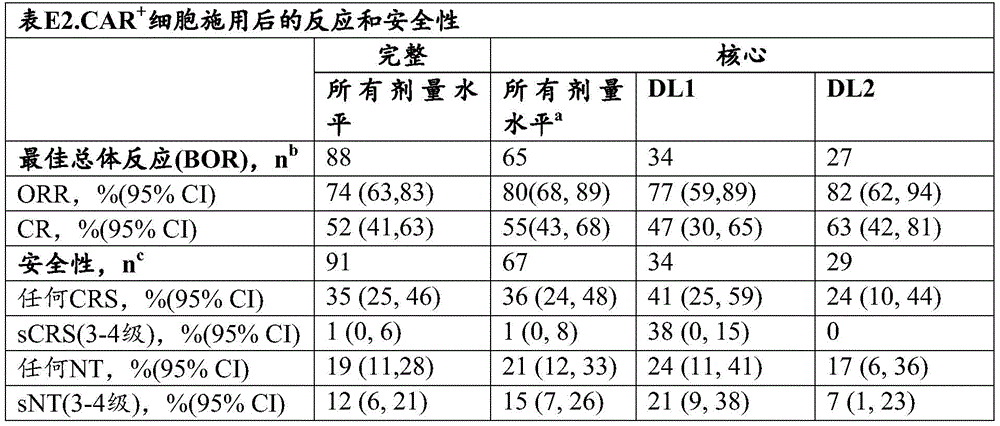
aFour patients treated with DL1D (dose level 1, double dose schedule) had similar results.
bPatients with PD, death, or 28 day rescheduled scan events were included. One nameThe patient failed to perform a re-staging scan.
cIncluding all subjects who had received at least one dose of compliant CAR-expressing cell product or died 28 days prior to the date of data snapshot.
C. Correlation between cellular attributes and responses of anti-CD 19 CAR-expressing T cells
The relationship between certain phenotypic attributes of CAR + T cells in the therapeutic composition and parameters related to clinical response outcome was evaluated. The correlation between memory phenotype and function in the composition is translated into a central memory subset composition and observed CAR+Positive correlation between peak in vivo expansion of cells (ρ ═ 0.42, P ═ 0.002) and progression-free survival (PFS) (kaplan-meier survival estimation, P ═ 0.0164). FIGS. 9A-9D show CAR administered+Subject of a T cell composition having a Kaplan-Meier survival Curve, said subject being divided into subjects administered with a composition comprising CD4+CAR+In T cells (FIG. 9A for progression-free survival, FIG. 9C for duration of response) and at CD8+CAR+CCR7 with frequency above or below a certain threshold level in T cells (FIG. 9B for progression-free survival, FIG. 9D for reaction duration) +CD27+CAR+A group of compositions of T cells. CCR7 was observed in the composition+CD27+Higher frequency of memory cells is associated with longer progression-free survival.
Univariate analysis revealed a negative correlation between T-cell Population Doubling (PDL) during the process for generating therapeutic T-cell compositions and the probability of progression-free survival (PFS) in non-hodgkin lymphoma (NHL) patients. Figure 10 shows PFS curves based on the best segmentation log rank test for patients with "high" (>6PDL) or "low" (<6PDL) PDL numbers in CD8+/CAR + T cells. This result is consistent with the following findings: factors that may limit the population doubling number of T cells in the process for producing a therapeutic T cell composition may increase the likelihood that a subject exhibits durable progression-free survival.
Example 7: of selected (input) cell compositions for use in the process of producing therapeutic cell compositions
And (4) evaluating the characteristics.
T cells selected from subjects with relapsed and refractory non-hodgkin's lymphoma (NHL) prior to engineering with anti-CD 19 CAR by the procedure described in example 6 were evaluated for phenotypic attributes. Selection of CD4 from leukapheresis of human Peripheral Blood Mononuclear Cells (PBMCs) from multiple subjects by immunoaffinity-based enrichment +And CD8+ T cells. Compositions of such selected T cells (prior to genetic engineering, designated "pre-engineered compositions") are evaluated for expression of cell surface markers indicative of certain T cell subtypes (such as effector memory or central memory cell subtypes), including C-C chemokine receptor type 7 (CCR7), CD27, and CD45 RA. Surface expression of CD4 or CD8 was also assessed.
Assessment of enriched CD4+ and CD8+ compositions (e.g., infusion compositions) revealed that the percentage of T cells positive for naive-like markers as present on a subset of central memory T cells (e.g., CD27+ CD28+ T cells) was highly variable between NHL patients (fig. 11). These data indicate that there is interpatient T cell heterogeneity of a subset of central memory T cells in the selected (input) T cell compositions used to generate CAR + T cell therapeutic T cell compositions.
The selected (input) T cell compositions were used to generate CD4+ and CD8+ therapeutic T cell compositions essentially as described in example 6. The number of doublings of the generated cell composition was also determined based on the Total Nuclear Count (TNC) of the cells as described in example 6. A correlation between the phenotypic attribute of the cells in the selected (input) T cell composition and the number of doublings in the process for producing the therapeutic composition is determined. As shown in figure 12, the higher percentage of effector memory CD4+ T cells in the enriched CD4+ (input) composition identified according to negative staining for CD45RA and CCR7 (CD45RA-CCR7-) was positively correlated with population doubling during the production of the therapeutic T cell composition. Similar results were observed for CD8+ T cells.
The above results support the following: comprising reducing the percentage of effector memory T cells and/or enriching for T cells positive for markers of naive-like or central memory cell subsets in an infusion composition used in a process for producing an engineered T cell composition. Consistent with the above results, this approach may improve one or more characteristics of the engineered therapeutic T cell composition, such as reducing inter-patient variability, reducing the number of population doublings in the process used to produce the engineered therapeutic T cell composition, and/or increasing the likelihood that a subject may exhibit persistent PFS.
Example 8: kinetics of non-amplification and amplification engineering processes
Human T cells (CD4+ and CD8+) were engineered with Chimeric Antigen Receptors (CARs) by a variety of manufacturing processes including processes that do not include a incubation step for amplification (non-amplified cohorts) and processes that include an incubation step for amplification (amplified cohorts). Comprehensive analysis was performed on cells generated during and after manufacturing runs through various processes. Manufacturing runs to produce engineered T cell compositions include large-scale (at-scale) runs as described in example 8, and processes as described below. The manufacturing run also includes a scale-down model (SDM) that is substantially the same as the manufacturing run, but where a lower number of T cells are used in the process for engineering the cells. In general, the manufacturing runs are scaled down to share the process activities described in table E3.
In the course of the analysis, T cells were engineered with either anti-CD 19 CAR or anti-BCMA CAR. An exemplary anti-CD 19 CAR contains an anti-CD 19 scFv derived from the murine antibody FMC63, an immunoglobulin spacer, a transmembrane domain derived from CD28, a costimulatory region derived from 4-1BB, and a CD 3-zeta intracellular signaling domain. The vector also encodes a truncated receptor molecule that serves as a surrogate marker for CAR expression and is separated from the CAR construct by a T2A sequence. An exemplary anti-BCMACAR contains a scFv antigen binding domain specific for BCMA, a CD28 transmembrane region, a 4-1BB costimulatory signaling region, and a CD 3-zeta derived intracellular signaling domain. The vector also encodes a truncated receptor molecule that serves as a surrogate marker for CAR expression and is separated from the CAR construct by a T2A sequence.
The process comprisesT cells were stimulated with anti-CD 3/anti-CD 28 paramagnetic beads or with anti-CD 3/anti-CD 28 Fa b conjugated oligo-streptavidin mutein reagents. The oligomerizing agent comprises a compound named M2 (streptavidin homotetramer containing the mutein amino acid sequence shown in SEQ ID NO:73 (International published application No. WO 2018/197949)). The streptavidin muteins are also described in the following documents: U.S. patent nos. 6,103,493; and Voss and Skerra (1997) Protein Eng.,1: 975-; and Argarana et al (1986) Nucleic Acids Research, 1871-. The stimulatory agents (
anti-CD 3 and anti-CD 28 Fab fragments) multimerize by reversibly binding to the oligostreptavidin mutein reagent. The anti-CD 3 and anti-CD 28 Fab fragments reversibly bind to streptavidin mutein oligomers via a streptavidin peptide binding partner fused to each Fab fragment. anti-CD 3 Fab fragment was derived from hybridoma cell line OKT3 (C)  CRL-8001TM(ii) a See also U.S. Pat. No. 4,361,549) binds to monoclonal antibodies and contains the heavy and light chain variable domains of the anti-CD 3 antibody OKT3 described in Arakawa et al j.biochem.120,657-662 (1996). These sequences are shown in SEQ ID NO 93 and 94, respectively. The anti-CD 28 Fab fragment was derived from the antibody CD28.3 (deposited as a synthetic single chain Fv construct under GenBank accession number AF 451974.1; see also Vanhave et al, BLOOD, 7.15.2003, Vol.102, No. 2, p.564-570) and contains the heavy and light chain variable domains of the anti-CD 28 antibody CD28.3 shown in SEQ ID NOS: 91 and 92, respectively. The Fab fragment was separately fused at the carboxy terminus of its heavy chain to a streptavidin peptide binding sequence containing two streptavidin binding modules in sequential arrangement having the amino acid sequence SAWSHPQFEK(GGGS)2GGSAWSHP QFEK (SEQ ID NO: 79). Peptide-tagged Fab fragments are recombinantly produced (see international patent application publication nos. WO 2013/011011 and WO 2013/124474). BeltThe binding of peptide-tagged
 anti-CD 3 and anti-CD 28 to the oligostreptavidin mutein reagent may be disrupted or reversed by the addition of D-biotin. D-biotin competes with strep-tag on the agent for binding to a binding partner on the streptavidin mutein, thereby disrupting binding.
Cells were collected at various times and at the end of the manufacturing run and counted by flow cytometry after staining with antibodies that recognize surface markers including CD4, CD8, CCR7, CD27, and CD45RA and evaluated for viability.

A.T Process for cell engineering
T cell compositions generated using different non-expanded processes that differ in a variety of characteristics, including the starting source of cells (cryopreserved or fresh apheresis), the concentration of oligomeric stimulatory agents, and the number of cells used for stimulation, are compared. T cell compositions are also produced, wherein the cells are further cultured for expansion. The procedure is performed on a healthy donor or on a patient donor.
1. Non-amplification process
Leukapheresis samples were collected from human donors and washed. CD4+ and CD8+ cells were selected directly from leukopheresis samples that were not cryopreserved by immunoaffinity-based selection. Following selection, individual CD4+ and CD8+ T cell compositions were cryopreserved and then thawed, and then selected CD4+ and CD8+ T cells were mixed at a 1:1 ratio of live CD4+ T cells to live CD8+ T cells to produce an infusion composition. By mixing with every 1x10 6Mu.g per cell of anti-CD 3/anti-CD 28 Fab conjugated oligo-streptavidin mutein reagents generated as described above in this example incubated together to stimulate approximately 600X10 from mixed input cell compositions6One cell (about 300X 10)6Individual CD4+ and 300x106And CD8 +. The stimulation is carried out in a serum-free complete medium for 18 to 30 hours (24. + -.6 hours), the serum-free complete medium beingThe medium comprises a basal medium (e.g., CTS)TMOpTsizer basal medium, Thermo Fisher), T cell supplement (e.g., 2.6% T cell expansion supplement, Thermo Fisher), immune cell serum replacement (e.g., 2.5% CTSTMImmune cell serum replacement), 2mM L-glutamine, dipeptide form of L-glutamine (e.g., 1.0% GlutamaxTMThermo Fisher), recombinant 100IU/mL IL-2, recombinant 600IU/mL IL-7, and recombinant 100IU/mL IL-15. Up to 300x10 was transduced following stimulation by rotational vaccination with lentiviral vectors encoding either exemplary anti-BCMA CARs or exemplary anti-CD 19 CARs6And (4) cells.
After rotational seeding, cells were washed and plated at up to about 0.75 × 10⁶The density of individual cells/mL is resuspended in basal medium (e.g., CTS) containing 2mM glutamine but no recombinant cytokine addition TMOpTmizer basal medium, Thermo Fisher), and incubated at about 37.0 ℃ in an incubator. After about 48 hours ± 6 hours after the initial stimulation (about 24 hours after the start of incubation), 1.0mM D-biotin was added and mixed with the cells to dissociate the anti-CD 3 and anti-CD 28 Fab reagents from the oligostreptavidin reagent. The cells were incubated for an additional about 48 hours (about 96 hours ± 6 hours after the initial stimulation or up to day 5 of the process) and then formulated with a cryoprotectant.
In another procedure, leukapheresis samples are collected from human donors, washed and cryopreserved. Cryopreserved leukapheresis samples were thawed and separate compositions of CD4+ and CD8+ cells were selected from each sample by immunoaffinity-based selection, followed by mixing of CD4+ and CD8+ T cells in order to produce up to about 900x106An infusion composition of viable CD4+ and CD8+ T cells wherein the ratio of viable CD4+ T cells to viable CD8+ T cells is varied. By mixing with every 1x10⁶Mu.g per cell of anti-CD 3/anti-CD 28 Fab conjugated oligo streptavidin mutein reagent generated as described above in this example (wherein 1.2. mu.g oligo stimulus reagent comprises 0.9. mu.g oligo particle and 0.15 μ g anti-CD 3 Fab and 0.15 μ g anti-CD 28 Fab) were incubated together and the mixed input cell composition was stimulated. The stimulation was performed in the same serum-free complete medium as above for between 16-24 hours (20 ± 4 hours). Following stimulation, up to about 600x10 was transduced by rotational vaccination with lentiviral vectors encoding CARs (in this case the same exemplary anti-BCMA CARs or exemplary anti-CD 19 CARs described above)6And (4) cells. In this study, cells incubated with higher concentrations of the oligo-streptavidin mutein reagent exhibited improved transduction efficiency (data not shown). After rotational seeding in this procedure, cells were washed and plated at 0.75 × 10⁶The density of individual cells/mL is resuspended in a solution containing 2mM glutamine and 2.6% T cell supplement (e.g., 2.6% Supplement, Thermofoisher) but without addition of recombinant cytokines (e.g., CTS)TMOpTmizer basal medium, Thermo Fisher), and incubated at about 37.0 ℃ in an incubator. After about 48 hours ± 6 hours after the initial stimulation (about 24 hours after the start of incubation), 1.0mM D-biotin was added and mixed with the cells to dissociate the anti-CD 3 and anti-CD 28 Fab reagents from the oligostreptavidin reagent. The cells were incubated for an additional about 48 hours (about 96 hours ± 6 hours after the initial stimulation or up to
 day 5 of the process) and then formulated with a cryoprotectant.
2. Amplification process
In one process, anti-CD 19 CAR T cells are engineered by the non-expansion process described above.
In another procedure, separate compositions of CD4+ and CD8+ cells were selected from human leukapheresis samples and cryogenically frozen. The selected cell compositions were then thawed and mixed at a ratio of 1:1 live CD4+ T cells to live CD8+ T cells. Approximately 300X10 of the composition will be mixed in serum-free medium containing recombinant IL-2, IL-7 and IL-15 in the presence of paramagnetic polystyrene coated beads with anti-CD 3 and anti-CD 28 antibodies attached at a bead to cell ratio of 1:16T cell (150X 10)6Individual CD4 and 150x106CD8+T cells) between 18 and 30 hours of stimulation. After the incubation, approximately 100x106 viable cells from the stimulated cell composition were concentrated in serum-free medium containing recombinant IL-2, IL-7 and IL-15. Cells were transduced by inoculation with lentiviral vectors encoding the exemplary CAR (in this case the exemplary anti-BCMA CAR described above) for 60 minutes at approximately 1600g rotation. After rotational seeding, cells were resuspended in serum-free medium containing recombinant IL-2, IL-7 and IL-15 and incubated at about 37 ℃ for about 18 to 30 hours. The cells are then incubated for expansion by transferring to approximately 500mL of an exemplary serum-free medium in a bioreactor (e.g., a rocking motion bioreactor) containing IL-2, IL-7, and IL-15 at twice the concentrations used during the incubation and transduction steps. When a set viable cell density is achieved, perfusion is initiated, wherein the medium is replaced by semi-continuous perfusion under continuous mixing. The following day the cells were incubated in the bioreactor until about 3x10 was achieved 6Threshold cell density of individual cells/mL, which typically occurs during a process involving 6-7 days of expansion. The anti-CD 3 and anti-CD 28 antibody-conjugated paramagnetic beads were removed from the cell composition by exposure to a magnetic field. Cells were then harvested, formulated and stored cryogenically.
B. Process metrics
Process metrics such as total viable cells (fig. 13A) and viability (fig. 13B) were monitored during the manufacturing run. As shown in fig. 13A, minimal amplification of cells was observed at day 5 time point, and robust amplification began after day 5. Figure 13B shows that cell viability in the manufacturing run initially decreased and began to increase as cells began to expand after day 5 of the process.
The results of comparing memory/differentiation phenotypes at different time points in the manufacturing run are shown in fig. 13C (expressed in terms of CD5RA and CCR 7) and fig. 13D (expressed in terms of CD27 and CCR 7). As shown, the evaluation of cells early during the manufacturing run showed that cells at about day 5 were significantly more enriched for cell types that were less differentiated, such as CCR7+ CD45 RA-or CCR7+ CD27+ cells, than cells at both earlier times in the process (e.g., day 1 or day 2) or later times in the process (e.g., day 9). The number of CD57+ cells was analyzed over the duration of the manufacturing process. As shown in fig. 14, the number of CD8+ CD57+ cells decreased during the manufacturing process, while the number of CD4+ CD57+ cells remained low at all times.
The scope of the invention is not intended to be limited to the specifically disclosed embodiments, which are provided, for example, to illustrate various aspects of the invention. Various modifications to the compositions and methods will be apparent from the description and teachings herein. Such variations may be practiced without departing from the true scope and spirit of the disclosure, and are intended to fall within the scope of the disclosure.
Sequence of


Sequence listing
<110> Zhununo therapeutics GmbH
<120> method for producing genetically engineered cell
<130> 735042017640
<140> not yet allocated
<141> simultaneous accompanying submission
<150> 62/756,571
<151> 2018-11-06
<150> 62/798,457
<151> 2019-01-29
<160> 98
<170> PatentIn 3.5 edition
<210> 1
<211> 12
<212> PRT
<213> Artificial sequence
<220>
<221> features not yet classified
<223> spacer (IgG 4 hinge)
<400> 1
Glu Ser Lys Tyr Gly Pro Pro Cys Pro Pro Cys Pro
1 5 10
<210> 2
<211> 36
<212> DNA
<213> Artificial sequence
<220>
<221> features not yet classified
<223> spacer (IgG 4 hinge)
<400> 2
gaatctaagt acggaccgcc ctgcccccct tgccct 36
<210> 3
<211> 119
<212> PRT
<213> Artificial sequence
<220>
<221> features not yet classified
<223> hinge-CH 3 spacer
<400> 3
Glu Ser Lys Tyr Gly Pro Pro Cys Pro Pro Cys Pro Gly Gln Pro Arg
1 5 10 15
Glu Pro Gln Val Tyr Thr Leu Pro Pro Ser Gln Glu Glu Met Thr Lys
20 25 30
Asn Gln Val Ser Leu Thr Cys Leu Val Lys Gly Phe Tyr Pro Ser Asp
35 40 45
Ile Ala Val Glu Trp Glu Ser Asn Gly Gln Pro Glu Asn Asn Tyr Lys
50 55 60
Thr Thr Pro Pro Val Leu Asp Ser Asp Gly Ser Phe Phe Leu Tyr Ser
65 70 75 80
Arg Leu Thr Val Asp Lys Ser Arg Trp Gln Glu Gly Asn Val Phe Ser
85 90 95
Cys Ser Val Met His Glu Ala Leu His Asn His Tyr Thr Gln Lys Ser
100 105 110
Leu Ser Leu Ser Leu Gly Lys
115
<210> 4
<211> 229
<212> PRT
<213> Artificial sequence
<220>
<221> features not yet classified
<223> hinge-CH 2-CH 3-spacer
<400> 4
Glu Ser Lys Tyr Gly Pro Pro Cys Pro Pro Cys Pro Ala Pro Glu Phe
1 5 10 15
Leu Gly Gly Pro Ser Val Phe Leu Phe Pro Pro Lys Pro Lys Asp Thr
20 25 30
Leu Met Ile Ser Arg Thr Pro Glu Val Thr Cys Val Val Val Asp Val
35 40 45
Ser Gln Glu Asp Pro Glu Val Gln Phe Asn Trp Tyr Val Asp Gly Val
50 55 60
Glu Val His Asn Ala Lys Thr Lys Pro Arg Glu Glu Gln Phe Asn Ser
65 70 75 80
Thr Tyr Arg Val Val Ser Val Leu Thr Val Leu His Gln Asp Trp Leu
85 90 95
Asn Gly Lys Glu Tyr Lys Cys Lys Val Ser Asn Lys Gly Leu Pro Ser
100 105 110
Ser Ile Glu Lys Thr Ile Ser Lys Ala Lys Gly Gln Pro Arg Glu Pro
115 120 125
Gln Val Tyr Thr Leu Pro Pro Ser Gln Glu Glu Met Thr Lys Asn Gln
130 135 140
Val Ser Leu Thr Cys Leu Val Lys Gly Phe Tyr Pro Ser Asp Ile Ala
145 150 155 160
Val Glu Trp Glu Ser Asn Gly Gln Pro Glu Asn Asn Tyr Lys Thr Thr
165 170 175
Pro Pro Val Leu Asp Ser Asp Gly Ser Phe Phe Leu Tyr Ser Arg Leu
180 185 190
Thr Val Asp Lys Ser Arg Trp Gln Glu Gly Asn Val Phe Ser Cys Ser
195 200 205
Val Met His Glu Ala Leu His Asn His Tyr Thr Gln Lys Ser Leu Ser
210 215 220
Leu Ser Leu Gly Lys
225
<210> 5
<211> 282
<212> PRT
<213> Artificial sequence
<220>
<221> features not yet classified
<223> IgD-hinge-Fc
<400> 5
Arg Trp Pro Glu Ser Pro Lys Ala Gln Ala Ser Ser Val Pro Thr Ala
1 5 10 15
Gln Pro Gln Ala Glu Gly Ser Leu Ala Lys Ala Thr Thr Ala Pro Ala
20 25 30
Thr Thr Arg Asn Thr Gly Arg Gly Gly Glu Glu Lys Lys Lys Glu Lys
35 40 45
Glu Lys Glu Glu Gln Glu Glu Arg Glu Thr Lys Thr Pro Glu Cys Pro
50 55 60
Ser His Thr Gln Pro Leu Gly Val Tyr Leu Leu Thr Pro Ala Val Gln
65 70 75 80
Asp Leu Trp Leu Arg Asp Lys Ala Thr Phe Thr Cys Phe Val Val Gly
85 90 95
Ser Asp Leu Lys Asp Ala His Leu Thr Trp Glu Val Ala Gly Lys Val
100 105 110
Pro Thr Gly Gly Val Glu Glu Gly Leu Leu Glu Arg His Ser Asn Gly
115 120 125
Ser Gln Ser Gln His Ser Arg Leu Thr Leu Pro Arg Ser Leu Trp Asn
130 135 140
Ala Gly Thr Ser Val Thr Cys Thr Leu Asn His Pro Ser Leu Pro Pro
145 150 155 160
Gln Arg Leu Met Ala Leu Arg Glu Pro Ala Ala Gln Ala Pro Val Lys
165 170 175
Leu Ser Leu Asn Leu Leu Ala Ser Ser Asp Pro Pro Glu Ala Ala Ser
180 185 190
Trp Leu Leu Cys Glu Val Ser Gly Phe Ser Pro Pro Asn Ile Leu Leu
195 200 205
Met Trp Leu Glu Asp Gln Arg Glu Val Asn Thr Ser Gly Phe Ala Pro
210 215 220
Ala Arg Pro Pro Pro Gln Pro Gly Ser Thr Thr Phe Trp Ala Trp Ser
225 230 235 240
Val Leu Arg Val Pro Ala Pro Pro Ser Pro Gln Pro Ala Thr Tyr Thr
245 250 255
Cys Val Val Ser His Glu Asp Ser Arg Thr Leu Leu Asn Ala Ser Arg
260 265 270
Ser Leu Glu Val Ser Tyr Val Thr Asp His
275 280
<210> 6
<211> 24
<212> PRT
<213> Artificial sequence
<220>
<223> T2A
<400> 6
Leu Glu Gly Gly Gly Glu Gly Arg Gly Ser Leu Leu Thr Cys Gly Asp
1 5 10 15
Val Glu Glu Asn Pro Gly Pro Arg
20
<210> 7
<211> 357
<212> PRT
<213> Artificial sequence
<220>
<223> tEGFR
<400> 7
Met Leu Leu Leu Val Thr Ser Leu Leu Leu Cys Glu Leu Pro His Pro
1 5 10 15
Ala Phe Leu Leu Ile Pro Arg Lys Val Cys Asn Gly Ile Gly Ile Gly
20 25 30
Glu Phe Lys Asp Ser Leu Ser Ile Asn Ala Thr Asn Ile Lys His Phe
35 40 45
Lys Asn Cys Thr Ser Ile Ser Gly Asp Leu His Ile Leu Pro Val Ala
50 55 60
Phe Arg Gly Asp Ser Phe Thr His Thr Pro Pro Leu Asp Pro Gln Glu
65 70 75 80
Leu Asp Ile Leu Lys Thr Val Lys Glu Ile Thr Gly Phe Leu Leu Ile
85 90 95
Gln Ala Trp Pro Glu Asn Arg Thr Asp Leu His Ala Phe Glu Asn Leu
100 105 110
Glu Ile Ile Arg Gly Arg Thr Lys Gln His Gly Gln Phe Ser Leu Ala
115 120 125
Val Val Ser Leu Asn Ile Thr Ser Leu Gly Leu Arg Ser Leu Lys Glu
130 135 140
Ile Ser Asp Gly Asp Val Ile Ile Ser Gly Asn Lys Asn Leu Cys Tyr
145 150 155 160
Ala Asn Thr Ile Asn Trp Lys Lys Leu Phe Gly Thr Ser Gly Gln Lys
165 170 175
Thr Lys Ile Ile Ser Asn Arg Gly Glu Asn Ser Cys Lys Ala Thr Gly
180 185 190
Gln Val Cys His Ala Leu Cys Ser Pro Glu Gly Cys Trp Gly Pro Glu
195 200 205
Pro Arg Asp Cys Val Ser Cys Arg Asn Val Ser Arg Gly Arg Glu Cys
210 215 220
Val Asp Lys Cys Asn Leu Leu Glu Gly Glu Pro Arg Glu Phe Val Glu
225 230 235 240
Asn Ser Glu Cys Ile Gln Cys His Pro Glu Cys Leu Pro Gln Ala Met
245 250 255
Asn Ile Thr Cys Thr Gly Arg Gly Pro Asp Asn Cys Ile Gln Cys Ala
260 265 270
His Tyr Ile Asp Gly Pro His Cys Val Lys Thr Cys Pro Ala Gly Val
275 280 285
Met Gly Glu Asn Asn Thr Leu Val Trp Lys Tyr Ala Asp Ala Gly His
290 295 300
Val Cys His Leu Cys His Pro Asn Cys Thr Tyr Gly Cys Thr Gly Pro
305 310 315 320
Gly Leu Glu Gly Cys Pro Thr Asn Gly Pro Lys Ile Pro Ser Ile Ala
325 330 335
Thr Gly Met Val Gly Ala Leu Leu Leu Leu Leu Val Val Ala Leu Gly
340 345 350
Ile Gly Leu Phe Met
355
<210> 8
<211> 27
<212> PRT
<213> Intelligent (homo sapiens)
<220>
<221> features not yet classified
<223> cd28
<300>
<308> P10747
<309> 1989-07-01
<313> (153)..(179)
<400> 8
Phe Trp Val Leu Val Val Val Gly Gly Val Leu Ala Cys Tyr Ser Leu
1 5 10 15
Leu Val Thr Val Ala Phe Ile Ile Phe Trp Val
20 25
<210> 9
<211> 66
<212> PRT
<213> Intelligent (homo sapiens)
<220>
<221> features not yet classified
<223> CD28
<300>
<308> P10747
<309> 1989-07-01
<313> (114)..(179)
<400> 9
Ile Glu Val Met Tyr Pro Pro Pro Tyr Leu Asp Asn Glu Lys Ser Asn
1 5 10 15
Gly Thr Ile Ile His Val Lys Gly Lys His Leu Cys Pro Ser Pro Leu
20 25 30
Phe Pro Gly Pro Ser Lys Pro Phe Trp Val Leu Val Val Val Gly Gly
35 40 45
Val Leu Ala Cys Tyr Ser Leu Leu Val Thr Val Ala Phe Ile Ile Phe
50 55 60
Trp Val
65
<210> 10
<211> 41
<212> PRT
<213> Intelligent (homo sapiens)
<220>
<221> features not yet classified
<223> CD28
<300>
<308> P10747
<309> 1989-07-01
<313> (180)..(220)
<400> 10
Arg Ser Lys Arg Ser Arg Leu Leu His Ser Asp Tyr Met Asn Met Thr
1 5 10 15
Pro Arg Arg Pro Gly Pro Thr Arg Lys His Tyr Gln Pro Tyr Ala Pro
20 25 30
Pro Arg Asp Phe Ala Ala Tyr Arg Ser
35 40
<210> 11
<211> 41
<212> PRT
<213> Intelligent (homo sapiens)
<220>
<221> features not yet classified
<223> CD28 (LL to GG)
<400> 11
Arg Ser Lys Arg Ser Arg Gly Gly His Ser Asp Tyr Met Asn Met Thr
1 5 10 15
Pro Arg Arg Pro Gly Pro Thr Arg Lys His Tyr Gln Pro Tyr Ala Pro
20 25 30
Pro Arg Asp Phe Ala Ala Tyr Arg Ser
35 40
<210> 12
<211> 42
<212> PRT
<213> Intelligent (homo sapiens)
<220>
<221> features not yet classified
<223> 4-1BB
<300>
<308> Q07011.1
<309> 1995-02-01
<313> (214)..(255)
<400> 12
Lys Arg Gly Arg Lys Lys Leu Leu Tyr Ile Phe Lys Gln Pro Phe Met
1 5 10 15
Arg Pro Val Gln Thr Thr Gln Glu Glu Asp Gly Cys Ser Cys Arg Phe
20 25 30
Pro Glu Glu Glu Glu Gly Gly Cys Glu Leu
35 40
<210> 13
<211> 112
<212> PRT
<213> Intelligent (homo sapiens)
<220>
<221> features not yet classified
<223> CD3ζ
<400> 13
Arg Val Lys Phe Ser Arg Ser Ala Asp Ala Pro Ala Tyr Gln Gln Gly
1 5 10 15
Gln Asn Gln Leu Tyr Asn Glu Leu Asn Leu Gly Arg Arg Glu Glu Tyr
20 25 30
Asp Val Leu Asp Lys Arg Arg Gly Arg Asp Pro Glu Met Gly Gly Lys
35 40 45
Pro Arg Arg Lys Asn Pro Gln Glu Gly Leu Tyr Asn Glu Leu Gln Lys
50 55 60
Asp Lys Met Ala Glu Ala Tyr Ser Glu Ile Gly Met Lys Gly Glu Arg
65 70 75 80
Arg Arg Gly Lys Gly His Asp Gly Leu Tyr Gln Gly Leu Ser Thr Ala
85 90 95
Thr Lys Asp Thr Tyr Asp Ala Leu His Met Gln Ala Leu Pro Pro Arg
100 105 110
<210> 14
<211> 112
<212> PRT
<213> Intelligent (homo sapiens)
<220>
<221> features not yet classified
<223> CD3ζ
<400> 14
Arg Val Lys Phe Ser Arg Ser Ala Glu Pro Pro Ala Tyr Gln Gln Gly
1 5 10 15
Gln Asn Gln Leu Tyr Asn Glu Leu Asn Leu Gly Arg Arg Glu Glu Tyr
20 25 30
Asp Val Leu Asp Lys Arg Arg Gly Arg Asp Pro Glu Met Gly Gly Lys
35 40 45
Pro Arg Arg Lys Asn Pro Gln Glu Gly Leu Tyr Asn Glu Leu Gln Lys
50 55 60
Asp Lys Met Ala Glu Ala Tyr Ser Glu Ile Gly Met Lys Gly Glu Arg
65 70 75 80
Arg Arg Gly Lys Gly His Asp Gly Leu Tyr Gln Gly Leu Ser Thr Ala
85 90 95
Thr Lys Asp Thr Tyr Asp Ala Leu His Met Gln Ala Leu Pro Pro Arg
100 105 110
<210> 15
<211> 112
<212> PRT
<213> Intelligent (homo sapiens)
<220>
<221> features not yet classified
<223> CD3ζ
<400> 15
Arg Val Lys Phe Ser Arg Ser Ala Asp Ala Pro Ala Tyr Lys Gln Gly
1 5 10 15
Gln Asn Gln Leu Tyr Asn Glu Leu Asn Leu Gly Arg Arg Glu Glu Tyr
20 25 30
Asp Val Leu Asp Lys Arg Arg Gly Arg Asp Pro Glu Met Gly Gly Lys
35 40 45
Pro Arg Arg Lys Asn Pro Gln Glu Gly Leu Tyr Asn Glu Leu Gln Lys
50 55 60
Asp Lys Met Ala Glu Ala Tyr Ser Glu Ile Gly Met Lys Gly Glu Arg
65 70 75 80
Arg Arg Gly Lys Gly His Asp Gly Leu Tyr Gln Gly Leu Ser Thr Ala
85 90 95
Thr Lys Asp Thr Tyr Asp Ala Leu His Met Gln Ala Leu Pro Pro Arg
100 105 110
<210> 16
<211> 335
<212> PRT
<213> Artificial sequence
<220>
<221> features not yet classified
<223> tEGFR
<400> 16
Arg Lys Val Cys Asn Gly Ile Gly Ile Gly Glu Phe Lys Asp Ser Leu
1 5 10 15
Ser Ile Asn Ala Thr Asn Ile Lys His Phe Lys Asn Cys Thr Ser Ile
20 25 30
Ser Gly Asp Leu His Ile Leu Pro Val Ala Phe Arg Gly Asp Ser Phe
35 40 45
Thr His Thr Pro Pro Leu Asp Pro Gln Glu Leu Asp Ile Leu Lys Thr
50 55 60
Val Lys Glu Ile Thr Gly Phe Leu Leu Ile Gln Ala Trp Pro Glu Asn
65 70 75 80
Arg Thr Asp Leu His Ala Phe Glu Asn Leu Glu Ile Ile Arg Gly Arg
85 90 95
Thr Lys Gln His Gly Gln Phe Ser Leu Ala Val Val Ser Leu Asn Ile
100 105 110
Thr Ser Leu Gly Leu Arg Ser Leu Lys Glu Ile Ser Asp Gly Asp Val
115 120 125
Ile Ile Ser Gly Asn Lys Asn Leu Cys Tyr Ala Asn Thr Ile Asn Trp
130 135 140
Lys Lys Leu Phe Gly Thr Ser Gly Gln Lys Thr Lys Ile Ile Ser Asn
145 150 155 160
Arg Gly Glu Asn Ser Cys Lys Ala Thr Gly Gln Val Cys His Ala Leu
165 170 175
Cys Ser Pro Glu Gly Cys Trp Gly Pro Glu Pro Arg Asp Cys Val Ser
180 185 190
Cys Arg Asn Val Ser Arg Gly Arg Glu Cys Val Asp Lys Cys Asn Leu
195 200 205
Leu Glu Gly Glu Pro Arg Glu Phe Val Glu Asn Ser Glu Cys Ile Gln
210 215 220
Cys His Pro Glu Cys Leu Pro Gln Ala Met Asn Ile Thr Cys Thr Gly
225 230 235 240
Arg Gly Pro Asp Asn Cys Ile Gln Cys Ala His Tyr Ile Asp Gly Pro
245 250 255
His Cys Val Lys Thr Cys Pro Ala Gly Val Met Gly Glu Asn Asn Thr
260 265 270
Leu Val Trp Lys Tyr Ala Asp Ala Gly His Val Cys His Leu Cys His
275 280 285
Pro Asn Cys Thr Tyr Gly Cys Thr Gly Pro Gly Leu Glu Gly Cys Pro
290 295 300
Thr Asn Gly Pro Lys Ile Pro Ser Ile Ala Thr Gly Met Val Gly Ala
305 310 315 320
Leu Leu Leu Leu Leu Val Val Ala Leu Gly Ile Gly Leu Phe Met
325 330 335
<210> 17
<211> 18
<212> PRT
<213> Artificial sequence
<220>
<223> T2A
<400> 17
Glu Gly Arg Gly Ser Leu Leu Thr Cys Gly Asp Val Glu Glu Asn Pro
1 5 10 15
Gly Pro
<210> 18
<211> 22
<212> PRT
<213> Artificial sequence
<220>
<223> P2A
<400> 18
Gly Ser Gly Ala Thr Asn Phe Ser Leu Leu Lys Gln Ala Gly Asp Val
1 5 10 15
Glu Glu Asn Pro Gly Pro
20
<210> 19
<211> 19
<212> PRT
<213> Artificial sequence
<220>
<223> P2A
<400> 19
Ala Thr Asn Phe Ser Leu Leu Lys Gln Ala Gly Asp Val Glu Glu Asn
1 5 10 15
Pro Gly Pro
<210> 20
<211> 20
<212> PRT
<213> Artificial sequence
<220>
<223> E2A
<400> 20
Gln Cys Thr Asn Tyr Ala Leu Leu Lys Leu Ala Gly Asp Val Glu Ser
1 5 10 15
Asn Pro Gly Pro
20
<210> 21
<211> 22
<212> PRT
<213> Artificial sequence
<220>
<223> F2A
<400> 21
Val Lys Gln Thr Leu Asn Phe Asp Leu Leu Lys Leu Ala Gly Asp Val
1 5 10 15
Glu Ser Asn Pro Gly Pro
20
<210> 22
<211> 10
<212> PRT
<213> Artificial sequence
<220>
<223> joint
<220>
<221> repetition
<222> (5)..(9)
<223> SGGGG repeated 5 times
<220>
<221> features not yet classified
<222> (5)..(9)
<223> wherein P is proline, G is glycine and S is serine
<400> 22
Pro Gly Gly Gly Ser Gly Gly Gly Gly Pro
1 5 10
<210> 23
<211> 17
<212> PRT
<213> Artificial sequence
<220>
<223> joint
<400> 23
Gly Ser Ala Asp Asp Ala Lys Lys Asp Ala Ala Lys Lys Asp Gly Lys
1 5 10 15
Ser
<210> 24
<211> 66
<212> DNA
<213> Artificial sequence
<220>
<223> GMCSFR alpha chain signal sequence
<400> 24
atgcttctcc tggtgacaag ccttctgctc tgtgagttac cacacccagc attcctcctg 60
atccca 66
<210> 25
<211> 22
<212> PRT
<213> Artificial sequence
<220>
<223> GMCSFR alpha chain signal sequence
<400> 25
Met Leu Leu Leu Val Thr Ser Leu Leu Leu Cys Glu Leu Pro His Pro
1 5 10 15
Ala Phe Leu Leu Ile Pro
20
<210> 26
<211> 18
<212> PRT
<213> Artificial sequence
<220>
<223> CD8 alpha Signal peptide
<400> 26
Met Ala Leu Pro Val Thr Ala Leu Leu Leu Pro Leu Ala Leu Leu Leu
1 5 10 15
His Ala
<210> 27
<211> 15
<212> PRT
<213> Artificial sequence
<220>
<223> hinge
<400> 27
Glu Pro Lys Ser Cys Asp Lys Thr His Thr Cys Pro Pro Cys Pro
1 5 10 15
<210> 28
<211> 12
<212> PRT
<213> Artificial sequence
<220>
<223> hinge
<400> 28
Glu Arg Lys Cys Cys Val Glu Cys Pro Pro Cys Pro
1 5 10
<210> 29
<211> 61
<212> PRT
<213> Artificial sequence
<220>
<223> hinge
<400> 29
Glu Leu Lys Thr Pro Leu Gly Asp Thr His Thr Cys Pro Arg Cys Pro
1 5 10 15
Glu Pro Lys Ser Cys Asp Thr Pro Pro Pro Cys Pro Arg Cys Pro Glu
20 25 30
Pro Lys Ser Cys Asp Thr Pro Pro Pro Cys Pro Arg Cys Pro Glu Pro
35 40 45
Lys Ser Cys Asp Thr Pro Pro Pro Cys Pro Arg Cys Pro
50 55 60
<210> 30
<211> 12
<212> PRT
<213> Artificial sequence
<220>
<223> hinge
<400> 30
Glu Ser Lys Tyr Gly Pro Pro Cys Pro Ser Cys Pro
1 5 10
<210> 31
<211> 5
<212> PRT
<213> Artificial sequence
<220>
<223> hinge
<220>
<221> variants
<222> (1)..(1)
<223> Xaa is glycine, cysteine or arginine
<220>
<221> variants
<222> (4)..(4)
<223> Xaa is cysteine or threonine
<400> 31
Xaa Pro Pro Xaa Pro
1 5
<210> 32
<211> 9
<212> PRT
<213> Artificial sequence
<220>
<223> hinge
<400> 32
Tyr Gly Pro Pro Cys Pro Pro Cys Pro
1 5
<210> 33
<211> 10
<212> PRT
<213> Artificial sequence
<220>
<223> hinge
<400> 33
Lys Tyr Gly Pro Pro Cys Pro Pro Cys Pro
1 5 10
<210> 34
<211> 14
<212> PRT
<213> Artificial sequence
<220>
<223> hinge
<400> 34
Glu Val Val Val Lys Tyr Gly Pro Pro Cys Pro Pro Cys Pro
1 5 10
<210> 35
<211> 11
<212> PRT
<213> Artificial sequence
<220>
<223> CDR L1
<400> 35
Arg Ala Ser Gln Asp Ile Ser Lys Tyr Leu Asn
1 5 10
<210> 36
<211> 7
<212> PRT
<213> Artificial sequence
<220>
<223> CDR L2
<400> 36
Ser Arg Leu His Ser Gly Val
1 5
<210> 37
<211> 9
<212> PRT
<213> Artificial sequence
<220>
<223> CDR L3
<400> 37
Gly Asn Thr Leu Pro Tyr Thr Phe Gly
1 5
<210> 38
<211> 5
<212> PRT
<213> Artificial sequence
<220>
<223> CDR H1
<400> 38
Asp Tyr Gly Val Ser
1 5
<210> 39
<211> 16
<212> PRT
<213> Artificial sequence
<220>
<223> CDR H2
<400> 39
Val Ile Trp Gly Ser Glu Thr Thr Tyr Tyr Asn Ser Ala Leu Lys Ser
1 5 10 15
<210> 40
<211> 7
<212> PRT
<213> Artificial sequence
<220>
<223> CDR H3
<400> 40
Tyr Ala Met Asp Tyr Trp Gly
1 5
<210> 41
<211> 120
<212> PRT
<213> Artificial sequence
<220>
<223> VH
<400> 41
Glu Val Lys Leu Gln Glu Ser Gly Pro Gly Leu Val Ala Pro Ser Gln
1 5 10 15
Ser Leu Ser Val Thr Cys Thr Val Ser Gly Val Ser Leu Pro Asp Tyr
20 25 30
Gly Val Ser Trp Ile Arg Gln Pro Pro Arg Lys Gly Leu Glu Trp Leu
35 40 45
Gly Val Ile Trp Gly Ser Glu Thr Thr Tyr Tyr Asn Ser Ala Leu Lys
50 55 60
Ser Arg Leu Thr Ile Ile Lys Asp Asn Ser Lys Ser Gln Val Phe Leu
65 70 75 80
Lys Met Asn Ser Leu Gln Thr Asp Asp Thr Ala Ile Tyr Tyr Cys Ala
85 90 95
Lys His Tyr Tyr Tyr Gly Gly Ser Tyr Ala Met Asp Tyr Trp Gly Gln
100 105 110
Gly Thr Ser Val Thr Val Ser Ser
115 120
<210> 42
<211> 107
<212> PRT
<213> Artificial sequence
<220>
<223> VL
<400> 42
Asp Ile Gln Met Thr Gln Thr Thr Ser Ser Leu Ser Ala Ser Leu Gly
1 5 10 15
Asp Arg Val Thr Ile Ser Cys Arg Ala Ser Gln Asp Ile Ser Lys Tyr
20 25 30
Leu Asn Trp Tyr Gln Gln Lys Pro Asp Gly Thr Val Lys Leu Leu Ile
35 40 45
Tyr His Thr Ser Arg Leu His Ser Gly Val Pro Ser Arg Phe Ser Gly
50 55 60
Ser Gly Ser Gly Thr Asp Tyr Ser Leu Thr Ile Ser Asn Leu Glu Gln
65 70 75 80
Glu Asp Ile Ala Thr Tyr Phe Cys Gln Gln Gly Asn Thr Leu Pro Tyr
85 90 95
Thr Phe Gly Gly Gly Thr Lys Leu Glu Ile Thr
100 105
<210> 43
<211> 245
<212> PRT
<213> Artificial sequence
<220>
<223> scFv
<400> 43
Asp Ile Gln Met Thr Gln Thr Thr Ser Ser Leu Ser Ala Ser Leu Gly
1 5 10 15
Asp Arg Val Thr Ile Ser Cys Arg Ala Ser Gln Asp Ile Ser Lys Tyr
20 25 30
Leu Asn Trp Tyr Gln Gln Lys Pro Asp Gly Thr Val Lys Leu Leu Ile
35 40 45
Tyr His Thr Ser Arg Leu His Ser Gly Val Pro Ser Arg Phe Ser Gly
50 55 60
Ser Gly Ser Gly Thr Asp Tyr Ser Leu Thr Ile Ser Asn Leu Glu Gln
65 70 75 80
Glu Asp Ile Ala Thr Tyr Phe Cys Gln Gln Gly Asn Thr Leu Pro Tyr
85 90 95
Thr Phe Gly Gly Gly Thr Lys Leu Glu Ile Thr Gly Ser Thr Ser Gly
100 105 110
Ser Gly Lys Pro Gly Ser Gly Glu Gly Ser Thr Lys Gly Glu Val Lys
115 120 125
Leu Gln Glu Ser Gly Pro Gly Leu Val Ala Pro Ser Gln Ser Leu Ser
130 135 140
Val Thr Cys Thr Val Ser Gly Val Ser Leu Pro Asp Tyr Gly Val Ser
145 150 155 160
Trp Ile Arg Gln Pro Pro Arg Lys Gly Leu Glu Trp Leu Gly Val Ile
165 170 175
Trp Gly Ser Glu Thr Thr Tyr Tyr Asn Ser Ala Leu Lys Ser Arg Leu
180 185 190
Thr Ile Ile Lys Asp Asn Ser Lys Ser Gln Val Phe Leu Lys Met Asn
195 200 205
Ser Leu Gln Thr Asp Asp Thr Ala Ile Tyr Tyr Cys Ala Lys His Tyr
210 215 220
Tyr Tyr Gly Gly Ser Tyr Ala Met Asp Tyr Trp Gly Gln Gly Thr Ser
225 230 235 240
Val Thr Val Ser Ser
245
<210> 44
<211> 11
<212> PRT
<213> Artificial sequence
<220>
<223> CDR L1
<400> 44
Lys Ala Ser Gln Asn Val Gly Thr Asn Val Ala
1 5 10
<210> 45
<211> 7
<212> PRT
<213> Artificial sequence
<220>
<223> CDR L2
<400> 45
Ser Ala Thr Tyr Arg Asn Ser
1 5
<210> 46
<211> 9
<212> PRT
<213> Artificial sequence
<220>
<223> CDR L3
<400> 46
Gln Gln Tyr Asn Arg Tyr Pro Tyr Thr
1 5
<210> 47
<211> 5
<212> PRT
<213> Artificial sequence
<220>
<223> CDR H1
<400> 47
Ser Tyr Trp Met Asn
1 5
<210> 48
<211> 17
<212> PRT
<213> Artificial sequence
<220>
<223> CDR H2
<400> 48
Gln Ile Tyr Pro Gly Asp Gly Asp Thr Asn Tyr Asn Gly Lys Phe Lys
1 5 10 15
Gly
<210> 49
<211> 13
<212> PRT
<213> Artificial sequence
<220>
<223> CDR H3
<400> 49
Lys Thr Ile Ser Ser Val Val Asp Phe Tyr Phe Asp Tyr
1 5 10
<210> 50
<211> 122
<212> PRT
<213> Artificial sequence
<220>
<223> VH
<400> 50
Glu Val Lys Leu Gln Gln Ser Gly Ala Glu Leu Val Arg Pro Gly Ser
1 5 10 15
Ser Val Lys Ile Ser Cys Lys Ala Ser Gly Tyr Ala Phe Ser Ser Tyr
20 25 30
Trp Met Asn Trp Val Lys Gln Arg Pro Gly Gln Gly Leu Glu Trp Ile
35 40 45
Gly Gln Ile Tyr Pro Gly Asp Gly Asp Thr Asn Tyr Asn Gly Lys Phe
50 55 60
Lys Gly Gln Ala Thr Leu Thr Ala Asp Lys Ser Ser Ser Thr Ala Tyr
65 70 75 80
Met Gln Leu Ser Gly Leu Thr Ser Glu Asp Ser Ala Val Tyr Phe Cys
85 90 95
Ala Arg Lys Thr Ile Ser Ser Val Val Asp Phe Tyr Phe Asp Tyr Trp
100 105 110
Gly Gln Gly Thr Thr Val Thr Val Ser Ser
115 120
<210> 51
<211> 108
<212> PRT
<213> Artificial sequence
<220>
<223> VL
<400> 51
Asp Ile Glu Leu Thr Gln Ser Pro Lys Phe Met Ser Thr Ser Val Gly
1 5 10 15
Asp Arg Val Ser Val Thr Cys Lys Ala Ser Gln Asn Val Gly Thr Asn
20 25 30
Val Ala Trp Tyr Gln Gln Lys Pro Gly Gln Ser Pro Lys Pro Leu Ile
35 40 45
Tyr Ser Ala Thr Tyr Arg Asn Ser Gly Val Pro Asp Arg Phe Thr Gly
50 55 60
Ser Gly Ser Gly Thr Asp Phe Thr Leu Thr Ile Thr Asn Val Gln Ser
65 70 75 80
Lys Asp Leu Ala Asp Tyr Phe Cys Gln Gln Tyr Asn Arg Tyr Pro Tyr
85 90 95
Thr Ser Gly Gly Gly Thr Lys Leu Glu Ile Lys Arg
100 105
<210> 52
<211> 15
<212> PRT
<213> Artificial sequence
<220>
<223> joint
<400> 52
Gly Gly Gly Gly Ser Gly Gly Gly Gly Ser Gly Gly Gly Gly Ser
1 5 10 15
<210> 53
<211> 245
<212> PRT
<213> Artificial sequence
<220>
<223> scFv
<400> 53
Glu Val Lys Leu Gln Gln Ser Gly Ala Glu Leu Val Arg Pro Gly Ser
1 5 10 15
Ser Val Lys Ile Ser Cys Lys Ala Ser Gly Tyr Ala Phe Ser Ser Tyr
20 25 30
Trp Met Asn Trp Val Lys Gln Arg Pro Gly Gln Gly Leu Glu Trp Ile
35 40 45
Gly Gln Ile Tyr Pro Gly Asp Gly Asp Thr Asn Tyr Asn Gly Lys Phe
50 55 60
Lys Gly Gln Ala Thr Leu Thr Ala Asp Lys Ser Ser Ser Thr Ala Tyr
65 70 75 80
Met Gln Leu Ser Gly Leu Thr Ser Glu Asp Ser Ala Val Tyr Phe Cys
85 90 95
Ala Arg Lys Thr Ile Ser Ser Val Val Asp Phe Tyr Phe Asp Tyr Trp
100 105 110
Gly Gln Gly Thr Thr Val Thr Val Ser Ser Gly Gly Gly Gly Ser Gly
115 120 125
Gly Gly Gly Ser Gly Gly Gly Gly Ser Asp Ile Glu Leu Thr Gln Ser
130 135 140
Pro Lys Phe Met Ser Thr Ser Val Gly Asp Arg Val Ser Val Thr Cys
145 150 155 160
Lys Ala Ser Gln Asn Val Gly Thr Asn Val Ala Trp Tyr Gln Gln Lys
165 170 175
Pro Gly Gln Ser Pro Lys Pro Leu Ile Tyr Ser Ala Thr Tyr Arg Asn
180 185 190
Ser Gly Val Pro Asp Arg Phe Thr Gly Ser Gly Ser Gly Thr Asp Phe
195 200 205
Thr Leu Thr Ile Thr Asn Val Gln Ser Lys Asp Leu Ala Asp Tyr Phe
210 215 220
Cys Gln Gln Tyr Asn Arg Tyr Pro Tyr Thr Ser Gly Gly Gly Thr Lys
225 230 235 240
Leu Glu Ile Lys Arg
245
<210> 54
<211> 12
<212> PRT
<213> Artificial sequence
<220>
<223> HC-CDR3
<400> 54
His Tyr Tyr Tyr Gly Gly Ser Tyr Ala Met Asp Tyr
1 5 10
<210> 55
<211> 7
<212> PRT
<213> Artificial sequence
<220>
<223> LC-CDR2
<400> 55
His Thr Ser Arg Leu His Ser
1 5
<210> 56
<211> 9
<212> PRT
<213> Artificial sequence
<220>
<223> LC-CDR3
<400> 56
Gln Gln Gly Asn Thr Leu Pro Tyr Thr
1 5
<210> 57
<211> 735
<212> DNA
<213> Artificial sequence
<220>
<223> scFv-encoding sequence
<400> 57
gacatccaga tgacccagac cacctccagc ctgagcgcca gcctgggcga ccgggtgacc 60
atcagctgcc gggccagcca ggacatcagc aagtacctga actggtatca gcagaagccc 120
gacggcaccg tcaagctgct gatctaccac accagccggc tgcacagcgg cgtgcccagc 180
cggtttagcg gcagcggctc cggcaccgac tacagcctga ccatctccaa cctggaacag 240
gaagatatcg ccacctactt ttgccagcag ggcaacacac tgccctacac ctttggcggc 300
ggaacaaagc tggaaatcac cggcagcacc tccggcagcg gcaagcctgg cagcggcgag 360
ggcagcacca agggcgaggt gaagctgcag gaaagcggcc ctggcctggt ggcccccagc 420
cagagcctga gcgtgacctg caccgtgagc ggcgtgagcc tgcccgacta cggcgtgagc 480
tggatccggc agccccccag gaagggcctg gaatggctgg gcgtgatctg gggcagcgag 540
accacctact acaacagcgc cctgaagagc cggctgacca tcatcaagga caacagcaag 600
agccaggtgt tcctgaagat gaacagcctg cagaccgacg acaccgccat ctactactgc 660
gccaagcact actactacgg cggcagctac gccatggact actggggcca gggcaccagc 720
gtgaccgtga gcagc 735
<210> 58
<211> 18
<212> PRT
<213> Artificial sequence
<220>
<223> joint
<400> 58
Gly Ser Thr Ser Gly Ser Gly Lys Pro Gly Ser Gly Glu Gly Ser Thr
1 5 10 15
Lys Gly
<210> 59
<211> 326
<212> PRT
<213> Intelligent (homo sapiens)
<220>
<223> human IgG2 Fc (Uniprot P01859)
<400> 59
Ala Ser Thr Lys Gly Pro Ser Val Phe Pro Leu Ala Pro Cys Ser Arg
1 5 10 15
Ser Thr Ser Glu Ser Thr Ala Ala Leu Gly Cys Leu Val Lys Asp Tyr
20 25 30
Phe Pro Glu Pro Val Thr Val Ser Trp Asn Ser Gly Ala Leu Thr Ser
35 40 45
Gly Val His Thr Phe Pro Ala Val Leu Gln Ser Ser Gly Leu Tyr Ser
50 55 60
Leu Ser Ser Val Val Thr Val Pro Ser Ser Asn Phe Gly Thr Gln Thr
65 70 75 80
Tyr Thr Cys Asn Val Asp His Lys Pro Ser Asn Thr Lys Val Asp Lys
85 90 95
Thr Val Glu Arg Lys Cys Cys Val Glu Cys Pro Pro Cys Pro Ala Pro
100 105 110
Pro Val Ala Gly Pro Ser Val Phe Leu Phe Pro Pro Lys Pro Lys Asp
115 120 125
Thr Leu Met Ile Ser Arg Thr Pro Glu Val Thr Cys Val Val Val Asp
130 135 140
Val Ser His Glu Asp Pro Glu Val Gln Phe Asn Trp Tyr Val Asp Gly
145 150 155 160
Val Glu Val His Asn Ala Lys Thr Lys Pro Arg Glu Glu Gln Phe Asn
165 170 175
Ser Thr Phe Arg Val Val Ser Val Leu Thr Val Val His Gln Asp Trp
180 185 190
Leu Asn Gly Lys Glu Tyr Lys Cys Lys Val Ser Asn Lys Gly Leu Pro
195 200 205
Ala Pro Ile Glu Lys Thr Ile Ser Lys Thr Lys Gly Gln Pro Arg Glu
210 215 220
Pro Gln Val Tyr Thr Leu Pro Pro Ser Arg Glu Glu Met Thr Lys Asn
225 230 235 240
Gln Val Ser Leu Thr Cys Leu Val Lys Gly Phe Tyr Pro Ser Asp Ile
245 250 255
Ser Val Glu Trp Glu Ser Asn Gly Gln Pro Glu Asn Asn Tyr Lys Thr
260 265 270
Thr Pro Pro Met Leu Asp Ser Asp Gly Ser Phe Phe Leu Tyr Ser Lys
275 280 285
Leu Thr Val Asp Lys Ser Arg Trp Gln Gln Gly Asn Val Phe Ser Cys
290 295 300
Ser Val Met His Glu Ala Leu His Asn His Tyr Thr Gln Lys Ser Leu
305 310 315 320
Ser Leu Ser Pro Gly Lys
325
<210> 60
<211> 327
<212> PRT
<213> Intelligent (homo sapiens)
<220>
<223> human IgG4 Fc (Uniprot P01861)
<400> 60
Ala Ser Thr Lys Gly Pro Ser Val Phe Pro Leu Ala Pro Cys Ser Arg
1 5 10 15
Ser Thr Ser Glu Ser Thr Ala Ala Leu Gly Cys Leu Val Lys Asp Tyr
20 25 30
Phe Pro Glu Pro Val Thr Val Ser Trp Asn Ser Gly Ala Leu Thr Ser
35 40 45
Gly Val His Thr Phe Pro Ala Val Leu Gln Ser Ser Gly Leu Tyr Ser
50 55 60
Leu Ser Ser Val Val Thr Val Pro Ser Ser Ser Leu Gly Thr Lys Thr
65 70 75 80
Tyr Thr Cys Asn Val Asp His Lys Pro Ser Asn Thr Lys Val Asp Lys
85 90 95
Arg Val Glu Ser Lys Tyr Gly Pro Pro Cys Pro Ser Cys Pro Ala Pro
100 105 110
Glu Phe Leu Gly Gly Pro Ser Val Phe Leu Phe Pro Pro Lys Pro Lys
115 120 125
Asp Thr Leu Met Ile Ser Arg Thr Pro Glu Val Thr Cys Val Val Val
130 135 140
Asp Val Ser Gln Glu Asp Pro Glu Val Gln Phe Asn Trp Tyr Val Asp
145 150 155 160
Gly Val Glu Val His Asn Ala Lys Thr Lys Pro Arg Glu Glu Gln Phe
165 170 175
Asn Ser Thr Tyr Arg Val Val Ser Val Leu Thr Val Leu His Gln Asp
180 185 190
Trp Leu Asn Gly Lys Glu Tyr Lys Cys Lys Val Ser Asn Lys Gly Leu
195 200 205
Pro Ser Ser Ile Glu Lys Thr Ile Ser Lys Ala Lys Gly Gln Pro Arg
210 215 220
Glu Pro Gln Val Tyr Thr Leu Pro Pro Ser Gln Glu Glu Met Thr Lys
225 230 235 240
Asn Gln Val Ser Leu Thr Cys Leu Val Lys Gly Phe Tyr Pro Ser Asp
245 250 255
Ile Ala Val Glu Trp Glu Ser Asn Gly Gln Pro Glu Asn Asn Tyr Lys
260 265 270
Thr Thr Pro Pro Val Leu Asp Ser Asp Gly Ser Phe Phe Leu Tyr Ser
275 280 285
Arg Leu Thr Val Asp Lys Ser Arg Trp Gln Glu Gly Asn Val Phe Ser
290 295 300
Cys Ser Val Met His Glu Ala Leu His Asn His Tyr Thr Gln Lys Ser
305 310 315 320
Leu Ser Leu Ser Leu Gly Lys
325
<210> 61
<211> 107
<212> PRT
<213> Artificial sequence
<220>
<223> FKBP
<400> 61
Gly Val Gln Val Glu Thr Ile Ser Pro Gly Asp Gly Arg Thr Phe Pro
1 5 10 15
Lys Arg Gly Gln Thr Cys Val Val His Tyr Thr Gly Met Leu Glu Asp
20 25 30
Gly Lys Lys Met Asp Ser Ser Arg Asp Arg Asn Lys Pro Phe Lys Phe
35 40 45
Met Leu Gly Lys Gln Glu Val Ile Arg Gly Trp Glu Glu Gly Val Ala
50 55 60
Gln Met Ser Val Gly Gln Arg Ala Lys Leu Thr Ile Ser Pro Asp Tyr
65 70 75 80
Ala Tyr Gly Ala Thr Gly His Pro Gly Ile Ile Pro Pro His Ala Thr
85 90 95
Leu Val Phe Asp Val Glu Leu Leu Lys Leu Glu
100 105
<210> 62
<211> 107
<212> PRT
<213> Artificial sequence
<220>
<223> FKBP12v36
<400> 62
Gly Val Gln Val Glu Thr Ile Ser Pro Gly Asp Gly Arg Thr Phe Pro
1 5 10 15
Lys Arg Gly Gln Thr Cys Val Val His Tyr Thr Gly Met Leu Glu Asp
20 25 30
Gly Lys Lys Val Asp Ser Ser Arg Asp Arg Asn Lys Pro Phe Lys Phe
35 40 45
Met Leu Gly Lys Gln Glu Val Ile Arg Gly Trp Glu Glu Gly Val Ala
50 55 60
Gln Met Ser Val Gly Gln Arg Ala Lys Leu Thr Ile Ser Pro Asp Tyr
65 70 75 80
Ala Tyr Gly Ala Thr Gly His Pro Gly Ile Ile Pro Pro His Ala Thr
85 90 95
Leu Val Phe Asp Val Glu Leu Leu Lys Leu Glu
100 105
<210> 63
<211> 16
<212> PRT
<213> Artificial sequence
<220>
<223> acylation motif
<400> 63
Met Gly Ser Asn Lys Ser Lys Pro Lys Asp Ala Ser Gln Arg Arg Arg
1 5 10 15
<210> 64
<211> 5
<212> PRT
<213> Artificial sequence
<220>
<223> double acylation zone
<220>
<221> variants
<222> (4)..(4)
<223> Xaa is any amino acid
<400> 64
Met Gly Cys Xaa Cys
1 5
<210> 65
<211> 4
<212> PRT
<213> Artificial sequence
<220>
<223> CAAX Box
<220>
<221> variants
<222> (4)..(4)
<223> Xaa is any amino acid
<400> 65
Cys Ala Ala Xaa
1
<210> 66
<211> 159
<212> PRT
<213> Streptomyces avermitilis (Streptomyces avidinii)
<400> 66
Asp Pro Ser Lys Asp Ser Lys Ala Gln Val Ser Ala Ala Glu Ala Gly
1 5 10 15
Ile Thr Gly Thr Trp Tyr Asn Gln Leu Gly Ser Thr Phe Ile Val Thr
20 25 30
Ala Gly Ala Asp Gly Ala Leu Thr Gly Thr Tyr Glu Ser Ala Val Gly
35 40 45
Asn Ala Glu Ser Arg Tyr Val Leu Thr Gly Arg Tyr Asp Ser Ala Pro
50 55 60
Ala Thr Asp Gly Ser Gly Thr Ala Leu Gly Trp Thr Val Ala Trp Lys
65 70 75 80
Asn Asn Tyr Arg Asn Ala His Ser Ala Thr Thr Trp Ser Gly Gln Tyr
85 90 95
Val Gly Gly Ala Glu Ala Arg Ile Asn Thr Gln Trp Leu Leu Thr Ser
100 105 110
Gly Thr Thr Glu Ala Asn Ala Trp Lys Ser Thr Leu Val Gly His Asp
115 120 125
Thr Phe Thr Lys Val Lys Pro Ser Ala Ala Ser Ile Asp Ala Ala Lys
130 135 140
Lys Ala Gly Val Asn Asn Gly Asn Pro Leu Asp Ala Val Gln Gln
145 150 155
<210> 67
<211> 126
<212> PRT
<213> Streptomyces avermitilis (Streptomyces avidinii)
<220>
<221> features not yet classified
<223> minimal streptavidin
<400> 67
Glu Ala Gly Ile Thr Gly Thr Trp Tyr Asn Gln Leu Gly Ser Thr Phe
1 5 10 15
Ile Val Thr Ala Gly Ala Asp Gly Ala Leu Thr Gly Thr Tyr Glu Ser
20 25 30
Ala Val Gly Asn Ala Glu Ser Arg Tyr Val Leu Thr Gly Arg Tyr Asp
35 40 45
Ser Ala Pro Ala Thr Asp Gly Ser Gly Thr Ala Leu Gly Trp Thr Val
50 55 60
Ala Trp Lys Asn Asn Tyr Arg Asn Ala His Ser Ala Thr Thr Trp Ser
65 70 75 80
Gly Gln Tyr Val Gly Gly Ala Glu Ala Arg Ile Asn Thr Gln Trp Leu
85 90 95
Leu Thr Ser Gly Thr Thr Glu Ala Asn Ala Trp Lys Ser Thr Leu Val
100 105 110
Gly His Asp Thr Phe Thr Lys Val Lys Pro Ser Ala Ala Ser
115 120 125
<210> 68
<211> 126
<212> PRT
<213> Artificial sequence
<220>
<221> features not yet classified
<223> mutein streptavidin Ile44-Gly45-Ala-46-Arg47
<400> 68
Glu Ala Gly Ile Thr Gly Thr Trp Tyr Asn Gln Leu Gly Ser Thr Phe
1 5 10 15
Ile Val Thr Ala Gly Ala Asp Gly Ala Leu Thr Gly Thr Tyr Ile Gly
20 25 30
Ala Arg Gly Asn Ala Glu Ser Arg Tyr Val Leu Thr Gly Arg Tyr Asp
35 40 45
Ser Ala Pro Ala Thr Asp Gly Ser Gly Thr Ala Leu Gly Trp Thr Val
50 55 60
Ala Trp Lys Asn Asn Tyr Arg Asn Ala His Ser Ala Thr Thr Trp Ser
65 70 75 80
Gly Gln Tyr Val Gly Gly Ala Glu Ala Arg Ile Asn Thr Gln Trp Leu
85 90 95
Leu Thr Ser Gly Thr Thr Glu Ala Asn Ala Trp Lys Ser Thr Leu Val
100 105 110
Gly His Asp Thr Phe Thr Lys Val Lys Pro Ser Ala Ala Ser
115 120 125
<210> 69
<211> 8
<212> PRT
<213> Artificial sequence
<220>
<223> Strep-tag II
<400> 69
Trp Ser His Pro Gln Phe Glu Lys
1 5
<210> 70
<211> 28
<212> PRT
<213> Artificial sequence
<220>
<223> Twin-Strep-tag
<400> 70
Trp Ser His Pro Gln Phe Glu Lys Gly Gly Gly Ser Gly Gly Gly Ser
1 5 10 15
Gly Gly Gly Ser Trp Ser His Pro Gln Phe Glu Lys
20 25
<210> 71
<211> 24
<212> PRT
<213> Artificial sequence
<220>
<223> Twin-Strep-tag
<400> 71
Trp Ser His Pro Gln Phe Glu Lys Gly Gly Gly Ser Gly Gly Gly Ser
1 5 10 15
Trp Ser His Pro Gln Phe Glu Lys
20
<210> 72
<211> 28
<212> PRT
<213> Artificial sequence
<220>
<223> Twin-Strep-tag
<400> 72
Trp Ser His Pro Gln Phe Glu Lys Gly Gly Gly Ser Gly Gly Gly Ser
1 5 10 15
Gly Gly Ser Ala Trp Ser His Pro Gln Phe Glu Lys
20 25
<210> 73
<211> 127
<212> PRT
<213> Artificial sequence
<220>
<221> features not yet classified
<223> mutein streptavidin Ile44-Gly45-Ala-46-Arg47
<400> 73
Met Glu Ala Gly Ile Thr Gly Thr Trp Tyr Asn Gln Leu Gly Ser Thr
1 5 10 15
Phe Ile Val Thr Ala Gly Ala Asp Gly Ala Leu Thr Gly Thr Tyr Ile
20 25 30
Gly Ala Arg Gly Asn Ala Glu Ser Arg Tyr Val Leu Thr Gly Arg Tyr
35 40 45
Asp Ser Ala Pro Ala Thr Asp Gly Ser Gly Thr Ala Leu Gly Trp Thr
50 55 60
Val Ala Trp Lys Asn Asn Tyr Arg Asn Ala His Ser Ala Thr Thr Trp
65 70 75 80
Ser Gly Gln Tyr Val Gly Gly Ala Glu Ala Arg Ile Asn Thr Gln Trp
85 90 95
Leu Leu Thr Ser Gly Thr Thr Glu Ala Asn Ala Trp Lys Ser Thr Leu
100 105 110
Val Gly His Asp Thr Phe Thr Lys Val Lys Pro Ser Ala Ala Ser
115 120 125
<210> 74
<211> 8
<212> PRT
<213> Artificial sequence
<220>
<223> streptavidin-binding peptide
<220>
<221> variants
<222> (2)..(2)
<223> Xaa is any amino acid
<220>
<221> variants
<222> (7)..(7)
<223> Xaa is Gly or Glu
<220>
<221> variants
<222> (8)..(8)
<223> Xaa is Gly, Lys or Arg
<400> 74
Trp Xaa His Pro Gln Phe Xaa Xaa
1 5
<210> 75
<211> 8
<212> PRT
<213> Artificial sequence
<220>
<223> streptavidin-binding peptide, Strep-tag
<400> 75
Trp Arg His Pro Gln Phe Gly Gly
1 5
<210> 76
<211> 17
<212> PRT
<213> Artificial sequence
<220>
<223> sequence module of streptavidin-binding peptide
<220>
<221> variants
<222> (9)..(9)
<223> Xaa is any amino acid
<220>
<221> repetition
<222> (9)..(9)
<223> Xaa repeat 8 or 12 times
<400> 76
Trp Ser His Pro Gln Phe Glu Lys Xaa Trp Ser His Pro Gln Phe Glu
1 5 10 15
Lys
<210> 77
<211> 20
<212> PRT
<213> Artificial sequence
<220>
<223> sequence module of streptavidin-binding peptide
<220>
<221> repetition
<222> (9)..(12)
<223> GGGS repeat 2 or 3 times
<400> 77
Trp Ser His Pro Gln Phe Glu Lys Gly Gly Gly Ser Trp Ser His Pro
1 5 10 15
Gln Phe Glu Lys
20
<210> 78
<211> 30
<212> PRT
<213> Artificial sequence
<220>
<223> Twin-Strep-tag
<400> 78
Ser Ala Trp Ser His Pro Gln Phe Glu Lys Gly Gly Gly Ser Gly Gly
1 5 10 15
Gly Ser Gly Gly Gly Ser Trp Ser His Pro Gln Phe Glu Lys
20 25 30
<210> 79
<211> 30
<212> PRT
<213> Artificial sequence
<220>
<223> Twin-Strep-tag
<400> 79
Ser Ala Trp Ser His Pro Gln Phe Glu Lys Gly Gly Gly Ser Gly Gly
1 5 10 15
Gly Ser Gly Gly Ser Ala Trp Ser His Pro Gln Phe Glu Lys
20 25 30
<210> 80
<211> 159
<212> PRT
<213> Artificial sequence
<220>
<223> mutein streptavidin Val44-Thr45-Ala46-Arg47
<400> 80
Asp Pro Ser Lys Asp Ser Lys Ala Gln Val Ser Ala Ala Glu Ala Gly
1 5 10 15
Ile Thr Gly Thr Trp Tyr Asn Gln Leu Gly Ser Thr Phe Ile Val Thr
20 25 30
Ala Gly Ala Asp Gly Ala Leu Thr Gly Thr Tyr Val Thr Ala Arg Gly
35 40 45
Asn Ala Glu Ser Arg Tyr Val Leu Thr Gly Arg Tyr Asp Ser Ala Pro
50 55 60
Ala Thr Asp Gly Ser Gly Thr Ala Leu Gly Trp Thr Val Ala Trp Lys
65 70 75 80
Asn Asn Tyr Arg Asn Ala His Ser Ala Thr Thr Trp Ser Gly Gln Tyr
85 90 95
Val Gly Gly Ala Glu Ala Arg Ile Asn Thr Gln Trp Leu Leu Thr Ser
100 105 110
Gly Thr Thr Glu Ala Asn Ala Trp Lys Ser Thr Leu Val Gly His Asp
115 120 125
Thr Phe Thr Lys Val Lys Pro Ser Ala Ala Ser Ile Asp Ala Ala Lys
130 135 140
Lys Ala Gly Val Asn Asn Gly Asn Pro Leu Asp Ala Val Gln Gln
145 150 155
<210> 81
<211> 126
<212> PRT
<213> Artificial sequence
<220>
<223> mutein streptavidin Val44-Thr45-Ala46-Arg47
<400> 81
Glu Ala Gly Ile Thr Gly Thr Trp Tyr Asn Gln Leu Gly Ser Thr Phe
1 5 10 15
Ile Val Thr Ala Gly Ala Asp Gly Ala Leu Thr Gly Thr Tyr Val Thr
20 25 30
Ala Arg Gly Asn Ala Glu Ser Arg Tyr Val Leu Thr Gly Arg Tyr Asp
35 40 45
Ser Ala Pro Ala Thr Asp Gly Ser Gly Thr Ala Leu Gly Trp Thr Val
50 55 60
Ala Trp Lys Asn Asn Tyr Arg Asn Ala His Ser Ala Thr Thr Trp Ser
65 70 75 80
Gly Gln Tyr Val Gly Gly Ala Glu Ala Arg Ile Asn Thr Gln Trp Leu
85 90 95
Leu Thr Ser Gly Thr Thr Glu Ala Asn Ala Trp Lys Ser Thr Leu Val
100 105 110
Gly His Asp Thr Phe Thr Lys Val Lys Pro Ser Ala Ala Ser
115 120 125
<210> 82
<211> 127
<212> PRT
<213> Artificial sequence
<220>
<221> features not yet classified
<223> mutein streptavidin Val44-Thr45-Ala46-Arg47
<400> 82
Met Glu Ala Gly Ile Thr Gly Thr Trp Tyr Asn Gln Leu Gly Ser Thr
1 5 10 15
Phe Ile Val Thr Ala Gly Ala Asp Gly Ala Leu Thr Gly Thr Tyr Val
20 25 30
Thr Ala Arg Gly Asn Ala Glu Ser Arg Tyr Val Leu Thr Gly Arg Tyr
35 40 45
Asp Ser Ala Pro Ala Thr Asp Gly Ser Gly Thr Ala Leu Gly Trp Thr
50 55 60
Val Ala Trp Lys Asn Asn Tyr Arg Asn Ala His Ser Ala Thr Thr Trp
65 70 75 80
Ser Gly Gln Tyr Val Gly Gly Ala Glu Ala Arg Ile Asn Thr Gln Trp
85 90 95
Leu Leu Thr Ser Gly Thr Thr Glu Ala Asn Ala Trp Lys Ser Thr Leu
100 105 110
Val Gly His Asp Thr Phe Thr Lys Val Lys Pro Ser Ala Ala Ser
115 120 125
<210> 83
<211> 4
<212> PRT
<213> Artificial sequence
<220>
<223> streptavidin-binding peptide
<400> 83
His Pro Gln Phe
1
<210> 84
<211> 8
<212> PRT
<213> Artificial sequence
<220>
<223> streptavidin-binding peptide
<220>
<221> variants
<222> (1)..(1)
<223> Xaa is Trp, Lys or Arg
<220>
<221> variants
<222> (2)..(2)
<223> Xaa is any amino acid
<220>
<221> variants
<222> (7)..(7)
<223> Xaa is Gly or Glu
<220>
<221> variants
<222> (8)..(8)
<223> Xaa is Gly, Lys or Arg
<400> 84
Xaa Xaa His Pro Gln Phe Xaa Xaa
1 5
<210> 85
<211> 159
<212> PRT
<213> Artificial sequence
<220>
<221> features not yet classified
<223> mutein streptavidin Ile44-Gly45-Ala-46-Arg47
<400> 85
Asp Pro Ser Lys Asp Ser Lys Ala Gln Val Ser Ala Ala Glu Ala Gly
1 5 10 15
Ile Thr Gly Thr Trp Tyr Asn Gln Leu Gly Ser Thr Phe Ile Val Thr
20 25 30
Ala Gly Ala Asp Gly Ala Leu Thr Gly Thr Tyr Ile Gly Ala Arg Gly
35 40 45
Asn Ala Glu Ser Arg Tyr Val Leu Thr Gly Arg Tyr Asp Ser Ala Pro
50 55 60
Ala Thr Asp Gly Ser Gly Thr Ala Leu Gly Trp Thr Val Ala Trp Lys
65 70 75 80
Asn Asn Tyr Arg Asn Ala His Ser Ala Thr Thr Trp Ser Gly Gln Tyr
85 90 95
Val Gly Gly Ala Glu Ala Arg Ile Asn Thr Gln Trp Leu Leu Thr Ser
100 105 110
Gly Thr Thr Glu Ala Asn Ala Trp Lys Ser Thr Leu Val Gly His Asp
115 120 125
Thr Phe Thr Lys Val Lys Pro Ser Ala Ala Ser Ile Asp Ala Ala Lys
130 135 140
Lys Ala Gly Val Asn Asn Gly Asn Pro Leu Asp Ala Val Gln Gln
145 150 155
<210> 86
<211> 126
<212> PRT
<213> Artificial sequence
<220>
<221> features not yet classified
<223> mutein streptavidin Ile44-Gly45-Ala-46-Arg47 and Glu117, Gly120, Try121 (mutein m 1-9)
<400> 86
Glu Ala Gly Ile Thr Gly Thr Trp Tyr Asn Gln Leu Gly Ser Thr Phe
1 5 10 15
Ile Val Thr Ala Gly Ala Asp Gly Ala Leu Thr Gly Thr Tyr Val Thr
20 25 30
Ala Arg Gly Asn Ala Glu Ser Arg Tyr Val Leu Thr Gly Arg Tyr Asp
35 40 45
Ser Ala Pro Ala Thr Asp Gly Ser Gly Thr Ala Leu Gly Trp Thr Val
50 55 60
Ala Trp Lys Asn Asn Tyr Arg Asn Ala His Ser Ala Thr Thr Trp Ser
65 70 75 80
Gly Gln Tyr Val Gly Gly Ala Glu Ala Arg Ile Asn Thr Gln Trp Leu
85 90 95
Leu Thr Ser Gly Thr Thr Glu Glu Asn Ala Gly Tyr Ser Thr Leu Val
100 105 110
Gly His Asp Thr Phe Thr Lys Val Lys Pro Ser Ala Ala Ser
115 120 125
<210> 87
<211> 139
<212> PRT
<213> Artificial sequence
<220>
<221> features not yet classified
<223> mutein streptavidin Ile44-Gly45-Ala-46-Arg47 and Glu117, Gly120, Try121 (mutein m 1-9)
<400> 87
Asp Pro Ser Lys Asp Ser Lys Ala Gln Val Ser Ala Ala Glu Ala Gly
1 5 10 15
Ile Thr Gly Thr Trp Tyr Asn Gln Leu Gly Ser Thr Phe Ile Val Thr
20 25 30
Ala Gly Ala Asp Gly Ala Leu Thr Gly Thr Tyr Val Thr Ala Arg Gly
35 40 45
Asn Ala Glu Ser Arg Tyr Val Leu Thr Gly Arg Tyr Asp Ser Ala Pro
50 55 60
Ala Thr Asp Gly Ser Gly Thr Ala Leu Gly Trp Thr Val Ala Trp Lys
65 70 75 80
Asn Asn Tyr Arg Asn Ala His Ser Ala Thr Thr Trp Ser Gly Gln Tyr
85 90 95
Val Gly Gly Ala Glu Ala Arg Ile Asn Thr Gln Trp Leu Leu Thr Ser
100 105 110
Gly Thr Thr Glu Glu Asn Ala Gly Tyr Ser Thr Leu Val Gly His Asp
115 120 125
Thr Phe Thr Lys Val Lys Pro Ser Ala Ala Ser
130 135
<210> 88
<211> 127
<212> PRT
<213> Streptomyces avermitilis (Streptomyces avidinii)
<220>
<221> features not yet classified
<223> minimal streptavidin
<400> 88
Met Glu Ala Gly Ile Thr Gly Thr Trp Tyr Asn Gln Leu Gly Ser Thr
1 5 10 15
Phe Ile Val Thr Ala Gly Ala Asp Gly Ala Leu Thr Gly Thr Tyr Glu
20 25 30
Ser Ala Val Gly Asn Ala Glu Ser Arg Tyr Val Leu Thr Gly Arg Tyr
35 40 45
Asp Ser Ala Pro Ala Thr Asp Gly Ser Gly Thr Ala Leu Gly Trp Thr
50 55 60
Val Ala Trp Lys Asn Asn Tyr Arg Asn Ala His Ser Ala Thr Thr Trp
65 70 75 80
Ser Gly Gln Tyr Val Gly Gly Ala Glu Ala Arg Ile Asn Thr Gln Trp
85 90 95
Leu Leu Thr Ser Gly Thr Thr Glu Ala Asn Ala Trp Lys Ser Thr Leu
100 105 110
Val Gly His Asp Thr Phe Thr Lys Val Lys Pro Ser Ala Ala Ser
115 120 125
<210> 89
<211> 24
<212> PRT
<213> Artificial sequence
<220>
<223> mutein streptavidin
<220>
<221> repetition
<222> (9)..(12)
<223> GGGS repeat 2 times
<400> 89
Trp Ser His Pro Gln Phe Glu Lys Gly Gly Gly Ser Gly Gly Ser Ala
1 5 10 15
Trp Ser His Pro Gln Phe Glu Lys
20
<210> 90
<211> 9
<212> PRT
<213> Artificial sequence
<220>
<223> Strep-tag
<400> 90
Ala Trp Ala His Pro Gln Phe Gly Gly
1 5
<210> 91
<211> 116
<212> PRT
<213> Artificial sequence
<220>
<223> VH anti-CD 28 antibody CD28.3
<400> 91
Leu Gln Gln Ser Gly Ala Glu Leu Val Lys Pro Gly Ala Ser Val Arg
1 5 10 15
Leu Ser Cys Lys Ala Ser Gly Tyr Thr Phe Thr Glu Tyr Ile Ile His
20 25 30
Trp Ile Lys Leu Arg Ser Gly Gln Gly Leu Glu Trp Ile Gly Trp Phe
35 40 45
Tyr Pro Gly Ser Asn Asp Ile Gln Tyr Asn Ala Lys Phe Lys Gly Lys
50 55 60
Ala Thr Leu Thr Ala Asp Lys Ser Ser Ser Thr Val Tyr Met Glu Leu
65 70 75 80
Thr Gly Leu Thr Ser Glu Asp Ser Ala Val Tyr Phe Cys Ala Arg Arg
85 90 95
Asp Asp Phe Ser Gly Tyr Asp Ala Leu Pro Tyr Trp Gly Gln Gly Thr
100 105 110
Met Val Thr Val
115
<210> 92
<211> 108
<212> PRT
<213> Artificial sequence
<220>
<223> VL anti-CD 28 antibody CD28.3
<400> 92
Asp Ile Gln Met Thr Gln Ser Pro Ala Ser Leu Ser Val Ser Val Gly
1 5 10 15
Glu Thr Val Thr Ile Thr Cys Arg Thr Asn Glu Asn Ile Tyr Ser Asn
20 25 30
Leu Ala Trp Tyr Gln Gln Lys Gln Gly Lys Ser Pro Gln Leu Leu Ile
35 40 45
Tyr Ala Ala Thr His Leu Val Glu Gly Val Pro Ser Arg Phe Ser Gly
50 55 60
Ser Gly Ser Gly Thr Gln Tyr Ser Leu Lys Ile Thr Ser Leu Gln Ser
65 70 75 80
Glu Asp Phe Gly Asn Tyr Tyr Cys Gln His Phe Trp Gly Thr Pro Cys
85 90 95
Thr Phe Gly Gly Gly Thr Lys Leu Glu Ile Lys Arg
100 105
<210> 93
<211> 119
<212> PRT
<213> Artificial sequence
<220>
<223> VH anti-CD 3 antibody OKT3
<400> 93
Gln Val Gln Leu Gln Gln Ser Gly Ala Glu Leu Ala Arg Pro Gly Ala
1 5 10 15
Ser Val Lys Met Ser Cys Lys Ala Ser Gly Tyr Thr Phe Thr Arg Tyr
20 25 30
Thr Met His Trp Val Lys Gln Arg Pro Gly Gln Gly Leu Glu Trp Ile
35 40 45
Gly Tyr Ile Asn Pro Ser Arg Gly Tyr Thr Asn Tyr Asn Gln Lys Phe
50 55 60
Lys Asp Lys Ala Thr Leu Thr Thr Asp Lys Ser Ser Ser Thr Ala Tyr
65 70 75 80
Met Gln Leu Ser Ser Leu Thr Ser Glu Asp Ser Ala Val Tyr Tyr Cys
85 90 95
Ala Arg Tyr Tyr Asp Asp His Tyr Cys Leu Asp Tyr Trp Gly Gln Gly
100 105 110
Thr Thr Leu Thr Val Ser Ser
115
<210> 94
<211> 106
<212> PRT
<213> Artificial sequence
<220>
<223> VL anti-CD 3 antibody OKT3
<400> 94
Gln Ile Val Leu Thr Gln Ser Pro Ala Ile Met Ser Ala Ser Pro Gly
1 5 10 15
Glu Lys Val Thr Met Thr Cys Ser Ala Ser Ser Ser Val Ser Tyr Met
20 25 30
Asn Trp Tyr Gln Gln Lys Ser Gly Thr Ser Pro Lys Arg Trp Ile Tyr
35 40 45
Asp Thr Ser Lys Leu Ala Ser Gly Val Pro Ala His Phe Arg Gly Ser
50 55 60
Gly Ser Gly Thr Ser Tyr Ser Leu Thr Ile Ser Gly Met Glu Ala Glu
65 70 75 80
Asp Ala Ala Thr Tyr Tyr Cys Gln Gln Trp Ser Ser Asn Pro Phe Thr
85 90 95
Phe Gly Ser Gly Thr Lys Leu Glu Ile Asn
100 105
<210> 95
<211> 248
<212> PRT
<213> Artificial sequence
<220>
Variable heavy chain of <223> Fab fragment m13B8.2
<400> 95
Ala Met Gln Val Gln Leu Lys Gln Ser Gly Pro Gly Leu Val Gln Pro
1 5 10 15
Ser Gln Ser Leu Ser Ile Thr Cys Thr Val Ser Gly Phe Ser Leu Thr
20 25 30
Thr Phe Gly Val His Trp Val Arg Gln Ser Pro Gly Lys Gly Leu Glu
35 40 45
Trp Leu Gly Val Ile Trp Ala Ser Gly Ile Thr Asp Tyr Asn Val Pro
50 55 60
Phe Met Ser Arg Leu Ser Ile Thr Lys Asp Asn Ser Lys Ser Gln Val
65 70 75 80
Phe Phe Lys Leu Asn Ser Leu Gln Pro Asp Asp Thr Ala Ile Tyr Tyr
85 90 95
Cys Ala Lys Asn Asp Pro Gly Thr Gly Phe Ala Tyr Trp Gln Gly Thr
100 105 110
Leu Val Thr Val Ser Ser Thr Lys Gly Pro Ser Val Phe Pro Ala Pro
115 120 125
Ser Ser Lys Ser Thr Ser Gly Gly Thr Ala Ala Leu Gly Cys Leu Val
130 135 140
Lys Asp Tyr Phe Pro Glu Pro Val Thr Val Ser Trp Asn Ser Gly Ala
145 150 155 160
Leu Thr Ser Gly Val His Thr Phe Pro Ala Val Leu Gln Ser Ser Leu
165 170 175
Tyr Ser Leu Ser Ser Val Val Thr Val Pro Ser Ser Ser Leu Gly Thr
180 185 190
Gln Thr Tyr Ile Cys Asn Val Asn His Lys Pro Ser Asn Thr Lys Val
195 200 205
Asp Lys Lys Val Glu Pro Lys Ser Cys Gly Ser Ala Trp Ser His Pro
210 215 220
Gln Phe Glu Lys Gly Gly Gly Ser Gly Gly Gly Ser Gly Gly Ser Ala
225 230 235 240
Trp Ser His Pro Gln Phe Glu Lys
245
<210> 96
<211> 218
<212> PRT
<213> Artificial sequence
<220>
Variable light chain of <223> Fab fragment m13B8.2
<400> 96
Ala Met Asp Ile Gln Met Thr Gln Ser Pro Ala Ser Leu Ser Ala Ser
1 5 10 15
Val Gly Glu Thr Val Thr Phe Thr Cys Arg Ala Ser Glu Met Ile Tyr
20 25 30
Ser Tyr Leu Ala Trp Tyr Gln Gln Lys Gln Gly Lys Ser Pro Gln Leu
35 40 45
Leu Val His Asp Ala Lys Thr Leu Ala Glu Gly Val Pro Ser Arg Phe
50 55 60
Ser Gly Gly Gly Ser Gly Thr Gln Phe Ser Leu Lys Ile Asn Thr Leu
65 70 75 80
Gln Pro Glu Asp Phe Gly Thr Tyr Tyr Cys Gln Ala His Tyr Gly Asn
85 90 95
Pro Pro Thr Phe Gly Gly Gly Thr Lys Leu Glu Ile Lys Arg Gly Ile
100 105 110
Ala Ala Pro Ser Val Phe Ile Phe Pro Pro Ser Asp Glu Gln Leu Lys
115 120 125
Ser Gly Thr Ala Ser Val Val Cys Leu Leu Asn Asn Phe Tyr Pro Arg
130 135 140
Glu Ala Lys Val Gln Trp Lys Val Asp Asn Ala Leu Gln Ser Gly Asn
145 150 155 160
Ser Gln Glu Ser Val Thr Glu Gln Asp Ser Lys Asp Ser Thr Tyr Ser
165 170 175
Leu Ser Ser Thr Leu Thr Leu Ser Lys Ala Asp Tyr Glu Lys His Lys
180 185 190
Val Tyr Ala Cys Glu Val Thr His Gln Gly Leu Ser Ser Pro Val Thr
195 200 205
Lys Ser Phe Asn Arg Gly Glu Cys Gly Ser
210 215
<210> 97
<211> 118
<212> PRT
<213> Artificial sequence
<220>
<223> variable heavy chain of huOKT8
<400> 97
Gln Val Gln Leu Val Gln Ser Gly Ala Glu Val Lys Lys Pro Gly Ala
1 5 10 15
Thr Val Lys Ile Ser Cys Lys Val Ser Gly Phe Asn Ile Lys Asp Thr
20 25 30
Tyr Ile His Trp Val Gln Gln Ala Pro Gly Lys Gly Leu Glu Trp Met
35 40 45
Gly Arg Ile Asp Pro Ala Asn Asp Asn Thr Leu Tyr Ala Ser Lys Phe
50 55 60
Gln Gly Arg Val Thr Ile Thr Ala Asp Thr Ser Thr Asp Thr Ala Tyr
65 70 75 80
Met Glu Leu Ser Ser Leu Arg Ser Glu Asp Thr Ala Val Tyr Tyr Cys
85 90 95
Ala Arg Gly Tyr Gly Tyr Tyr Val Phe Asp His Trp Gly Gln Gly Thr
100 105 110
Leu Val Thr Val Ser Ser
115
<210> 98
<211> 107
<212> PRT
<213> Artificial sequence
<220>
<223> variable light chain of huOKT8
<400> 98
Asp Val Gln Ile Thr Gln Ser Pro Ser Ser Leu Ser Ala Ser Val Gly
1 5 10 15
Asp Arg Val Thr Ile Thr Cys Arg Thr Ser Arg Ser Ile Ser Gln Tyr
20 25 30
Leu Ala Trp Tyr Gln Gln Lys Pro Gly Lys Val Pro Lys Leu Leu Ile
35 40 45
Tyr Ser Gly Ser Thr Leu Gln Ser Gly Val Pro Ser Arg Phe Ser Gly
50 55 60
Ser Gly Ser Gly Thr Asp Phe Thr Leu Thr Ile Ser Ser Leu Gln Pro
65 70 75 80
Glu Asp Val Ala Thr Tyr Tyr Cys Gln Gln His Asn Glu Asn Pro Leu
85 90 95
Thr Phe Gly Gly Gly Thr Lys Val Glu Ile Lys
100 105
Claims (154)
1. A method of enriching for T cells, the method comprising:
(a) performing a first selection comprising enriching any of CD57 "or CD3+ T cells from a biological sample comprising primary human T cells, thereby generating an enriched population of T cells; and
(b) performing a second selection of cells from the enriched population of T cells, wherein:
the first selection comprises enrichment of CD57-T cells from the enriched population and the second selection comprises enrichment of CD3+ T cells; or
The first selection comprises enrichment of CD3+ T cells from the enriched T cell population and the second selection comprises depletion of CD57+ T cells,
Wherein the method generates a depleted population comprising fewer CD57+ T cells than the biological sample and enriched for CD3+ T cells.
2. A method of enriching for T cells, the method comprising:
(a) performing a first selection comprising removing CD57+ T cells from a biological sample comprising primary human T cells, thereby generating a depleted population comprising fewer CD57+ T cells than the biological sample; and
(b) performing a second selection of cells from the depleted population, the second selection comprising enrichment of CD3+ T cells from the depleted population, the enrichment thereby generating an enriched population of CD57-CD3+ T cells.
3. A method of enriching for T cells, the method comprising:
(a) performing a first selection comprising enriching CD3+ T cells from a biological sample comprising primary human T cells, thereby generating an enriched population of T cells; and
(b) performing a second selection of cells from the enriched population of T cells, the second selection comprising removing CD57+ T cells from the enriched population of sample cells, thereby generating a depleted population, wherein the depleted population comprises fewer CD57+ T cells than the biological sample and/or less than the enriched population of T cells, and is enriched for CD3+ T cells.
4. A method of enriching for T cells, the method comprising:
(a) performing a first selection comprising removing CD57+ T cells from a biological sample comprising primary human T cells, thereby generating a first depleted population comprising fewer CD57+ T cells than the biological sample;
(b) performing a second selection of cells from the first depleted population, the second selection comprising enriching the first depleted population for one of (i) CD4+ T cells and (ii) CD8+ T cells, the enriching thereby generating a second depleted population enriched for one of (i) CD4+ T cells and (ii) CD8+ T cells and an unselected population; and
(c) performing a third selection comprising enriching the other of (i) CD4+ cells and (ii) CD8+ cells from the unselected population, the enriching thereby generating a third depleted population enriched in the other of (i) CD4+ T cells and (ii) CD8+ T cells.
5. The method of any one of claims 1-4, wherein the depleted population (optionally the first depleted population, the second depleted population, or the third depleted population) comprises at least one of:
(i) Less than or about 5% CD57+ T cells;
(ii) a frequency of CD57+ T cells that is less than or about 35% of the frequency of CD57+ T cells present in the biological sample;
(iii) CD4+ T cells, wherein at least or about 95% of the CD4+ T cells are CD 57-; and
(iv) CD8+ T cells, wherein at least or about 95% of the CD8+ T cells are CD 57-.
6. The method of any one of claims 1-5, wherein the biological sample comprises an apheresis product or a leukocyte apheresis product.
7. A method of enriching for T cells, the method comprising removing CD57+ T cells from a biological sample containing an apheresis product or a leukoapheresis product comprising primary human T cells, thereby generating a depleted population of T cells, wherein the depleted population comprises fewer CD57+ T cells than the biological sample, and wherein the depleted population comprises at least one of:
(i) less than or about 5% CD57+ T cells;
(ii) a frequency of CD57+ T cells that is less than or about 35% of the frequency of CD57+ T cells present in the biological sample;
(iii) CD4+ T cells, wherein at least or about 95% of the CD4+ T cells are CD 57-; and
(iv) CD8+ T cells, wherein at least or about 95% of the CD8+ T cells are CD 57-.
8. The method of any one of claims 1-7, wherein at least or about 95% of the CD4+ T cells in the depleted population comprise CD57-CD4+ T cells.
9. The method of any one of claims 1-7, wherein at least or about 95% of the CD8+ T cells in the depleted population comprise CD57-CD8+ T cells.
10. The method of any one of claims 1-9, wherein at least or about 95% of the CD4+ T cells and at least or about 95% of the CD8+ T cells in the depleted population comprise CD57-CD4+ T cells and CD57-CD8+ T cells, respectively.
11. The method of any one of claims 1-10, wherein at least or about 95% of the CD3+ T cells in the depleted population comprise CD57-CD3+ T cells.
12. The method of any one of claims 7-11, wherein the depleted population is a first depleted population, and the method further comprises selecting CD4+ T cells from the first depleted population, thereby generating a second depleted population that is an enriched population of CD57-CD4+ T cells and an unselected population.
13. The method of claim 12, wherein the method further comprises selecting CD8+ T cells from the unselected population, thereby generating a third depleted population that is an enriched population of CD57-CD8+ T cells and a second unselected population.
14. The method of any one of claims 7-11, wherein the depleted population is a first depleted population, and the method further comprises selecting CD8+ T cells from the first depleted population, thereby generating a second depleted population that is an enriched population of CD57-CD8+ T cells and an unselected population.
15. The method of claim 14, wherein the method further comprises selecting CD4+ T cells from the unselected population, thereby generating a third depleted population that is an enriched population of CD57-CD4+ T cells and a second unselected population.
16. The method of any one of claims 7-11, wherein the depleted population is a first depleted population, and the method further comprises selecting CD3+ T cells from the first depleted population, thereby generating a second depleted population that is an enriched population of CD57-CD3+ T cells and an unselected population.
17. The method of any one of claims 1-16, wherein the frequency of CD57+ T cells in the depleted population (optionally the first depleted population, the second depleted population, or the third depleted population) is less than about or about 35%, 30%, 20%, 10%, 5%, 1%, or 0.1% of the frequency of CD57+ T cells in the biological sample.
18. The method of any one of claims 1-17, wherein the depleted population (optionally the first depleted population, the second depleted population, or the third depleted population) comprises less than about or about 3%, less than about or about 2%, less than about or about 1%, less than about or about 0.1%, or less than about or about 0.01% CD57+ T cells.
19. The method of any one of claims 1-18, wherein the depleted population (optionally the first depleted population, the second depleted population, or the third depleted population) is free or substantially free of CD57+ T cells.
20. The method of any one of claims 1-19, wherein the frequency of naive-like T cells in the depleted population (optionally the first depleted population, the second depleted population, or the third depleted population) is at least about or about 10%, 20%, 30%, 40%, or 50% greater than the frequency of naive-like T cells in the biological sample.
21. The method of claim 20, wherein the naive-like T cells are surface positive for one or more of the markers selected from the group consisting of CD45RA, CD27, CD28, and CCR 7.
22. The method of any one of claims 1-21, wherein the frequency of CD27+ T cells in the depleted population (optionally the first depleted population, the second depleted population, or the third depleted population) is at least about or about 10%, 20%, 30%, 40%, or 50% greater than the frequency of the corresponding cells in the biological sample.
23. The method of any one of claims 1-22, wherein the frequency of CD28+ T cells in the depleted population (optionally the first depleted population, the second depleted population, or the third depleted population) is at least about or about 10%, 20%, 30%, 40%, or 50% greater than the frequency of the corresponding cells in the biological sample.
24. The method of any one of claims 1-23, wherein the frequency of CD27+/CD28+ T cells in the depleted population (optionally the first depleted population, the second depleted population, or the third depleted population) is at least about or about 10%, 20%, 30%, 40%, or 50% greater than the frequency of corresponding cells in the biological sample.
25. The method of any one of claims 4-6, 13, 15, and 17-24, further comprising combining the second depleted population and the third depleted population, optionally at a ratio of between or about 1:3 and at or about 3:1, optionally at or about 1:1, thereby generating a depleted population comprising the second depleted population and the third depleted population.
26. The method of any one of claims 1-25, wherein the method produces a T cell composition comprising at least or about 90%, at least or about 95%, at least or about 97%, at least or about 99% or at least or about 99.9% CD57-CD4+ T cells.
27. The method of any one of claims 1-25, wherein the method produces a T cell composition comprising at least or about 90%, at least or about 95%, at least or about 97%, at least or about 99%, or at least or about 99.9% CD57-CD8+ T cells.
28. The method of any one of claims 1-25, wherein the method produces a T cell composition comprising at least or about 90%, at least or about 95%, at least or about 97%, at least or about 99%, or at least or about 99.9% CD57-CD3+ T cells.
29. The method of claim 28, wherein the ratio of CD4+ to CD8+ T cells in the composition is between 3:1 and 1:3, between 2:1 and 1:2, between 1.5:1 and 1:1.5, or between 1.2:1 and 1:1.2, inclusive.
30. The method of claim 28 or claim 29, wherein the ratio of CD4+ to CD8+ T cells in the composition is at or about 1:1.
31. The method of any one of claims 1-30, wherein the primary T cells are from a human subject.
32. The method of any of claims 1-31, wherein the primary T cells are from a subject having a disease or disorder.
33. The method of claim 32, wherein the disease or disorder is cancer.
34. The method of any one of claims 1-30, wherein the primary T cells are from a healthy subject.
35. The method of any one of claims 1-34, wherein the biological sample comprises CD4+ and CD8+ cells.
36. The method of any one of claims 1-35, wherein removing the CD57+ T cells comprises immunoaffinity-based selection.
37. The method of claim 36, wherein the immunoaffinity based selection comprises contacting cells with an antibody capable of specifically binding to CD57 and recovering cells not bound to the antibody, thereby effecting a negative selection, wherein the recovered cells are depleted of CD57+ cells.
38. The method of any one of claims 1-37, wherein optionally enriching cells in the first, second, and/or third selection comprises immunoaffinity-based selection.
39. The method of claim 38, wherein the first, second and/or third selection enriches CD4 or CD 8T cells and the immunoaffinity-based selection is effected by: positive selection is achieved by contacting the cells with an antibody capable of specifically binding to CD4 or CD8, respectively, and recovering the cells bound to the antibody, wherein the recovered cells are enriched for CD4+ cells or CD8+ cells.
40. The method of claim 39, wherein the selecting is enriched for CD 3T cells and the immunoaffinity-based selection is effected by: positive selection is achieved by contacting cells with an antibody capable of specifically binding to CD3, and recovering cells bound to the antibody, wherein the recovered cells are enriched for CD3+ cells.
41. The method of any one of claims 37-40, wherein the antibody is immobilized on a solid surface, optionally wherein the solid surface is a magnetic particle.
42. The method of any one of claims 37-40, wherein the antibody is immobilized on or attached to an affinity chromatography matrix.
43. The method of claim 42, wherein the antibody further comprises one or more binding partners capable of forming a reversible bond with a binding reagent immobilized on the substrate, whereby the antibody reversibly binds to the chromatography substrate during said contacting.
44. The method of claim 43, wherein the binding reagent is a streptavidin mutein that reversibly binds to the binding partner.
45. The method of claim 44, wherein:
66, at sequence positions corresponding to positions 44 to 47, the streptavidin mutein comprises the amino acid sequence lle44-Gly45-Ala46-Arg47(ii) a Or
66, at the sequence positions corresponding to positions 44 to 47, the streptavidin mutein comprises the amino acid sequence Va144-Thr45-Ala46-Arg47。
46. The method of any one of claims 43-45, wherein the binding partner is a streptavidin binding peptide.
47. The method of claim 46, wherein the streptavidin-binding peptide is selected from the group consisting of Trp-Ser-His-Pro-Gln-Phe-Glu-Lys (SEQ ID NO:69), Ser-Ala-Trp-Ser-His-Pro-Gln-Phe-Glu-Lys- (GlyGlyGlyGlySer)3-Trp-Ser-His-Pro-Gln-Phe-Glu-Lys(SEQ ID NO:78)、SAWSHPQFEKGGGSGGGSGGSAWSHPQFEK(SEQ ID NO:79)、Trp-Ser-His-Pro-Gln-Phe-Glu-Lys-(GlyGlyGlySer)3-Trp-Ser-His-Pro-Gln-Phe-Glu-Lys(SEQ ID NO:70)、Trp-Ser-His-Pro-Gln-Phe-Glu-Lys-(GlyGlyGlySer)2-Trp-Ser-His-Pro-Gln-Phe-Glu-Lys (SEQ ID NO:71) and Trp-Ser-His-Pro-Gln-Phe-Glu-Lys- (GlyGlyGlySer)2Gly-Gly-Ser-Ala-Trp-Ser-His-Pro-Gln-Phe-Glu-Lys(SEQ ID NO:72)。
48. The method of claim 46 or claim 47, wherein the streptavidin binding peptide has sequence SAWSHPQFEKGGGSGGGSGGSAWSHPQFEK (SEQ ID NO: 79).
49. The method of any one of claims 43-48, further comprising, after contacting the cells in the sample with the affinity chromatography matrix, applying a competition reagent to disrupt the bond between the binding partner and binding reagent, thereby recovering cells bound to the antibody.
50. The method of claim 49, wherein the competition agent is biotin or a biotin analog.
51. The method of any one of claims 42-50, wherein the chromatography matrix is packed in a separation vessel, the separation vessel being a column.
52. The method of any one of claims 1-51, wherein the biological sample comprising primary T cells is a sample formulated with a cryoprotectant.
53. The method of any one of claims 6-52, wherein the apheresis or leukopheresis sample is a sample formulated with a cryoprotectant.
54. A method of stimulating T cells, the method comprising incubating T cells of an input composition under stimulation conditions, thereby generating a stimulated population of T cells, wherein the input composition is produced by the method of any one of claims 1-53.
55. The method of claim 54, wherein the input composition comprises at least one of:
(i) less than or about 5% CD57+ T cells;
(ii) at least or about 95% CD57-T cells;
(iii) at least or about 90% CD57-CD4+ T cells;
(iv) at least or about 90% CD57-CD8+ T cells; and
(v) At least about or about 90% CD57-CD3+ T cells.
56. A method of stimulating T cells, comprising incubating T cells of an input composition under stimulation conditions, thereby generating a stimulated population of T cells, wherein the input composition comprises at least one of:
(i) less than or about 5% CD57+ T cells
(ii) At least or about 95% CD57-T cells;
(iii) at least or about 90% CD57-CD4+ T cells;
(iv) at least or about 90% CD57-CD8+ T cells; and
(v) at least or about 90% CD57-CD3+ T cells.
57. The method of claim 56, wherein the stimulatory condition comprises the presence of a stimulatory agent capable of activating one or more intracellular signaling domains of one or more components of the TCR complex and one or more intracellular signaling domains of one or more co-stimulatory molecules.
58. The method of any one of claim 57, wherein the stimulating agent comprises (i) a primary agent that specifically binds to a member of the TCR complex, optionally CD3, and (ii) a secondary agent that specifically binds to a T cell costimulatory molecule, optionally wherein the costimulatory molecule is selected from CD28, CD137(4-1-BB), OX40, or ICOS.
59. The method of claim 58, wherein at least one of the primary and secondary agents comprises an antibody or antigen-binding fragment thereof.
60. The method of claim 58 or claim 59, wherein the primary agent is an anti-CD 3 antibody or antigen-binding fragment thereof and the secondary agent is an anti-CD 28 antibody or antigen-binding fragment thereof.
61. The method of claim 59 or claim 60, wherein the antigen-binding fragment is a monovalent antibody fragment selected from the group consisting of: fab fragments, Fv fragments and single chain Fv fragments (scFv).
62. The method of any one of claims 58-61, wherein the primary agent is an anti-CD 3Fab and the secondary agent comprises an anti-CD 28 Fab.
63. The method of any one of claims 58-62, wherein the primary agent and the secondary agent are each present on or attached to a surface of a solid support.
64. The method of claim 63, wherein the solid support is or comprises a bead, optionally a paramagnetic bead.
65. The method of claim 64, wherein the solid support is a paramagnetic bead having surface-attached anti-CD 3 and anti-CD 28 antibodies, and the stimulating reagent is present at a bead to cell ratio of less than about or about 3: 1.
66. The method of claim 65, wherein the stimulating agent is present at or about a 1:1 bead to cell ratio.
67. The method of any one of claims 58-62, wherein the primary agent and the secondary agent reversibly bind on the surface of an oligomeric particle reagent comprising a plurality of streptavidin molecules or streptavidin mutein molecules.
68. The method of claim 67, wherein the streptavidin molecule or the streptavidin mutein molecule is bound to or is capable of binding to biotin, avidin, a biotin analog or a biotin mutein, an avidin analog or an avidin mutein and/or a biologically active fragment thereof.
69. The method according to claim 67 or claim 68, wherein the streptavidin mutein molecule comprises the amino acid sequence Val at the amino acid residues corresponding to residues 44 to 47 with reference to the amino acid sequence shown in SEQ ID NO 6644-Thr45-Ala46-Arg47Or Ile44-Gly45-Ala46-Arg47。
70. The method of any one of claims 67-69, wherein the streptavidin mutein molecule comprises:
a) 70-73, 78, 85-89 of any one of the amino acid sequences shown in SEQ ID NOs;
b) The following amino acid sequence: which exhibits at least about or about 85%, 86%, 87%, 88%, 89%, 90%, 91%, 92%, 93%, 94%, 95%, 96%, 97%, 98%, 99% or more sequence identity to any one of SEQ ID NOs 70-73, 78, 85-89, and contains a sequence corresponding to Val44-Thr45-Ala46-Arg47Or Ile44-Gly45-Ala46-Arg47And/or reversibly binds to biotin, a biotin analogue or a streptavidin binding peptide; or
c) a functional fragment of a) or b) that reversibly binds to biotin, a biotin analogue or a streptavidin binding peptide.
71. The method of any one of claims 67-70, wherein the streptavidin mutein molecule comprises the amino acid sequence set forth in SEQ ID NO: 73.
72. The method of any one of claims 67-70, wherein the streptavidin mutein molecule comprises the amino acid sequence set forth in SEQ ID NO: 78.
73. The method of any one of claims 67-72, wherein the primary agent and the secondary agent each comprise a streptavidin-binding peptide.
74. The method of claim 73, wherein the streptavidin-binding peptide is selected from the group consisting of Trp-Ser-His-Pro-Gln-Phe-Glu-Lys (SEQ ID NO:69), Trp-Ser-His-Pro-Gln-Phe-Glu-Lys- (GlyGlyGlySer) 3-Trp-Ser-His-Pro-Gln-Phe-Glu-Lys(SEQ ID NO:70)、Trp-Ser-His-Pro-Gln-Phe-Glu-Lys-(GlyGlyGlySer)2-Trp-Ser-His-Pro-Gln-Phe-Glu-Lys (SEQ ID NO:71) and Trp-Ser-His-Pro-Gln-Phe-Glu-Lys- (GlyGlyGlySer)2Gly-Gly-Ser-Ala-Trp-Ser-His-Pro-Gln-Phe-Glu-Lys(SEQ ID NO:89)。
75. The method of any one of claims 67-74, wherein the primary agent comprises an anti-CD 3Fab, and wherein the secondary agent comprises an anti-CD 28 Fab.
76. The method of any one of claims 67-75, wherein the oligomeric particle reagent comprises a radius greater than about or about 60nm, greater than about or about 70nm, greater than about or about 80nm, or greater than about or about 90 nm.
77. The method of any one of claims 67-76, wherein the oligomeric particle reagent comprises the following molecular weights: at least about or about 5x10⁷g/mol or at least about or about 1x10⁸g/mol; or at 5x10⁷g/mol and 5x10⁸Between g/mol, 1x10⁸g/mol and 5x10⁸Between g/mol or 1x10⁸g/mol and 2x10⁸g/mol, each inclusive.
78. The method of any one of claims 67-77, wherein the oligomeric particle reagent comprises at least about or about 500 streptavidin or streptavidin mutein tetramers, at least about or about 1,000 streptavidin or streptavidin mutein tetramers, at least about or about 1,500 streptavidin or streptavidin mutein tetramers, or at least about or about 2,000 streptavidin or streptavidin mutein tetramers; or; between 1,000 and 20,000, between 1,000 and 10,000 or between 2,000 and 5,000.
79. The method of any one of claims 61-78, further comprising isolating the stimulating agent from the cells, the isolating comprising contacting the cells with a substance capable of reversing the bond between the primary and secondary agents and the oligomeric particle reagent.
80. The method of claim 79, wherein the substance is a free binding partner and/or is a competitor.
81. The method of claim 79 or claim 80, wherein the substance is or comprises a streptavidin-binding peptide, biotin or a biologically active fragment thereof, or a biotin analogue or a biologically active fragment thereof.
82. The method of claim 81, wherein the substance is or comprises biotin or a biotin analogue.
83. The method of any one of claims 56-82, wherein the stimulatory conditions comprise the presence of one or more recombinant cytokines.
84. The method of any one of claims 56-83, wherein the stimulatory conditions comprise the presence of one or more of recombinant IL-2, IL-7 and IL-15.
85. A method of genetically engineering T cells, the method comprising introducing a heterologous polynucleotide encoding a recombinant receptor into a population of T cells from a T cell composition produced by the method of any one of claims 1-53, thereby generating an engineered population of T cells.
86. A method of genetically engineering T cells, the method comprising introducing a heterologous polynucleotide encoding a recombinant receptor into a population of T cells from the population of stimulated T cells of any one of claims 54-84, thereby generating an engineered population of T cells.
87. The method of claim 85 or claim 86, wherein the introducing comprises transduction with a viral vector comprising the heterologous polynucleotide.
88. The method of claim 87, wherein the viral vector is a gammaretrovirus vector or a lentiviral vector.
89. The method of claim 87 or claim 88, wherein the viral vector is a lentiviral vector.
90. The method of any one of claims 85-89, further comprising incubating the composition comprising transduced cells for up to 96 hours after said introducing, optionally at a temperature of at or about 37 ° ± 2 ℃.
91. The method of claim 90, wherein said incubating is performed for up to 72 hours after said introducing.
92. The method of claim 90, wherein said incubating is for up to 48 hours after said introducing.
93. The method of claim 90, wherein said incubating is performed for up to 24 hours after said introducing.
94. The method of any one of claims 90-93, wherein the incubation results in integration of the viral vector into the genome of the T cell.
95. The method of any one of claims 85-94, further comprising incubating cells of the engineered population under conditions that promote proliferation or expansion of the engineered cells, thereby generating an expanded cell population.
96. The method of claim 95, wherein said incubating is in the presence of one or more recombinant cytokines, said recombinant cytokines optionally comprising one or more of IL-2, IL-7, and IL-15.
97. The method of claim 95 or claim 96, wherein said proliferating or expanding results in about or at least about or about 2-fold, 3-fold, 4-fold, 5-fold, or greater than or about 5-fold increase in the number of live T cells comprising said heterologous polynucleotide as compared to when said incubating was initiated.
98. The method of any one of claims 1-97, further comprising harvesting or collecting the population of cells produced by the method.
99. The method of claim 98, wherein said harvesting or collecting further comprises formulating said cells for cryopreservation in the presence of a cryoprotectant.
100. The method of claim 98 or claim 99, wherein the harvested or harvested cells are formulated in the presence of a pharmaceutically acceptable excipient.
101. The method of any one of claims 85-100, wherein the recombinant receptor is capable of binding to a target antigen associated with, characteristic of, and/or expressed on a cell or tissue of a disease, disorder, or condition.
102. The method of claim 101, wherein the disease, disorder or condition is an infectious disease or disorder, an autoimmune disease, an inflammatory disease, or a tumor or cancer.
103. The method of claim 101 or claim 102, wherein the target antigen is a tumor antigen.
104. The method according to any one of claims 101-103, wherein the target antigen is selected from the group consisting of α v β 6 integrin (avb6 integrin), B Cell Maturation Antigen (BCMA), B7-H3, B7-H6, carbonic anhydrase 9(CA9, also known as CAIX or G250), cancer-testis antigen, cancer/testis antigen 1B (CTAG, also known as NY-ESO-1 and LAGE-2), carcinoembryonic antigen (CEA), cyclin a2, C-C motif chemokine ligand 1(CCL-1), CD19, CD20, CD22, CD23, CD24, CD30, CD33, CD38, CD44, CD44v6, CD44v7/8, CD123, CD133, CD138, CD171, chondroitin sulfate proteoglycan 4(CSPG4), epidermal growth factor III protein (EGFR III receptor), epidermal growth factor III receptor (EGFR), epidermal growth factor III 2), EGFR (EGFR-2) mutant, EGFR-2, EGFR (vch-1), C-C motif chemokine ligand 1, CCL-1, CD19, and C-C motif chemokine ligand 1 (ccb-1, and C-B-C-B-C-B-C-B-C-B-C-, Epithelial glycoprotein 40(EPG-40), ephrin B2, ephrin receptor A2(EPHa2), estrogen receptor, Fc receptor-like protein 5(FCRL 5; also known as Fc receptor homolog 5 or FCRH5), fetal acetylcholine receptor (fetal AchR), folate-binding protein (FBP), folate receptor alpha, ganglioside GD2, O-GD acetylation 2(OGD2), ganglioside GD3, glycoprotein 100(gp100), glypican-3 (GPC3), G-protein-coupled receptor 5D (GPRC5D), Her2/neu (receptor tyrosine kinase erb-B2), Her3(erb-B3), Her4(erb-B4), erbB dimer, human high molecular weight melanoma-associated antigen (HMW-MAA), hepatitis B surface antigen, human leukocyte antigen A1(HLA-A1), human HLA-A antigen 2 (HLA-2), IL-22 receptor alpha (IL-22R alpha), IL-13 receptor alpha 2(IL-13R alpha 2), kinase insertion domain receptor (kdr), kappa light chain, L1 cell adhesion molecule (L1-CAM), CE7 epitope of L1-CAM, protein 8 family member A containing leucine rich repeats (LRRC8A), Lewis Y, melanoma associated antigen (MAGE) -A1, MAGE-A3, MAGE-A6, MAGE-A10, Mesothelin (MSLN), c-Met, murine Cytomegalovirus (CMV), mucin 1(MUC1), MUC16, natural killer cell 2 family member D (NKG2D) ligand, melanin A (MART-1), Neural Cell Adhesion Molecule (NCAM), cancer embryonic antigen, melanoma preferentially expressing antigen (PRAME), progesterone receptor, prostate specific antigen, Prostate Stem Cell Antigen (PSCA), prostate specific antigen (PSCA), and the like, Prostate Specific Membrane Antigen (PSMA), receptor tyrosine kinase-like orphan receptor 1(ROR1), survivin, trophoblast glycoprotein (TPBG, also known as 5T4), tumor associated glycoprotein 72(TAG72), tyrosinase related protein 1(TRP1, also known as TYRP1 or gp75), tyrosinase related protein 2(TRP2, also known as dopachrome tautomerase, dopachrome delta isomerase, or DCT), Vascular Endothelial Growth Factor Receptor (VEGFR), vascular endothelial growth factor receptor 2(VEGFR2), wilms 1(WT-1), pathogen-specific or pathogen-expressed antigens, or antigens associated with a universal TAG, and/or biotinylated molecules, and/or molecules expressed by HIV, HCV, HBV, or other pathogens.
105. The method of any one of claims 101-103, wherein the recombinant receptor is or comprises a functional non-TCR antigen receptor or a TCR or antigen-binding fragment thereof.
106. The method of any one of claims 101-104, wherein the recombinant receptor is a Chimeric Antigen Receptor (CAR).
107. The method of any one of claims 101-106, wherein the recombinant receptor comprises: an extracellular domain comprising an antigen binding domain, a spacer and/or a hinge region, a transmembrane domain, and an intracellular signaling domain comprising a costimulatory signaling region.
108. The method of claim 107, wherein the extracellular domain comprises an antigen-binding domain comprising an scFv.
109. The method according to claim 107 or claim 108, wherein the intracellular signaling domain is or comprises a primary signaling domain, a signaling domain capable of inducing a primary activation signal in a T cell, a signaling domain of a T Cell Receptor (TCR) component, and/or a signaling domain comprising an Immunoreceptor Tyrosine Activation Motif (ITAM).
110. The method of any one of claims 107-109, wherein the intracellular signaling domain is or comprises an intracellular signaling domain of the CD3 chain, optionally the CD3-zeta (CD3 zeta) chain, or a signaling portion thereof.
111. The method of any one of claims 107-110, wherein the costimulatory signaling region comprises the intracellular signaling domain of CD28, 4-1BB, or ICOS, or a signaling portion thereof.
112. A cellular composition produced by the method of any one of claims 1-53.
113. The composition of claim 112, wherein the cells comprise CD57-CD4+ T cells.
114. The composition of claim 112, wherein the cells comprise CD57-CD8+ T cells.
115. The composition of claim 112, wherein the cells comprise CD57-CD3+ T cells.
116. A composition comprising an engineered population of T cells produced by the method of any one of claims 85-111.
117. A therapeutic T cell composition comprising CD4+ T cells expressing a recombinant receptor and CD8+ T cells expressing a recombinant receptor, wherein at least 80% of the total receptors in the composition+the/CD 8+ cells are CD57-, and at least 80% of the total receptors in the composition+the/CD 4+ cells are CD 57-.
118. The therapeutic T cell composition of claim 117, wherein at least or at least about 80%, at least or at least about 85%, at least or at least about 90%, at least or at least about 95%, at least or at least about 96%, at least or at least about 97%, at least or at least about 98%, at least or at least about 99%, about 100%, or 100% of the cells in the composition are CD4+ T cells and CD8+ T cells.
119. A therapeutic T cell composition comprising CD3+ T cells expressing a recombinant receptor, wherein at least 80% of the total receptors in the composition+the/CD 3+ cells are CD 57-.
120. The therapeutic T cell composition of claim 119, wherein at least or at least about 80%, at least or at least about 85%, at least or at least about 90%, at least or at least about 95%, at least or at least about 96%, at least or at least about 97%, at least or at least about 98%, at least or at least about 99%, about 100%, or 100% of the cells in the composition are CD3+ T cells.
121. The therapeutic T cell composition of any one of claims 117-120, wherein the ratio of receptor +/CD4+ T cells to receptor +/CD8+ T cells in the composition is between about 1:3 and about 3: 1.
122. The therapeutic T cell composition of any one of claims 117-121, wherein the ratio of receptor +/CD4+ T cells to receptor +/CD8+ T cells in the composition is at or about 1: 1.
123. The therapeutic composition of any one of claims 117-122, wherein the recombinant protein is or comprises a recombinant receptor capable of binding to a target protein associated with, characteristic of, and/or expressed on a cell or tissue of a disease, disorder, or condition.
124. The therapeutic composition of any one of claims 117-123, wherein the recombinant protein is a Chimeric Antigen Receptor (CAR).
125. The therapeutic composition of any one of claims 117-124, wherein the number of viable T cells in the compositionAt or about 10x106Each cell having a molecular weight of at or about 200x106Between cells, optionally wherein the number of viable T cells in the composition is at or about 10x106Each cell having a molecular weight of at or about 100x106Between cells, at or about 10x106Each cell having a molecular weight of at or about 70x106Between cells, at or about 10x106Each cell having a molecular weight of at or about 50x106Between cells, at or about 50x106Each cell having a molecular weight of at or about 200x106Between cells, at or about 50x106Each cell having a molecular weight of at or about 100x106Between cells, at or about 50x106Each cell having a molecular weight of at or about 70x106Between cells, at or about 70x106Each cell having a molecular weight of at or about 200x106Between cells, at or about 70x106Each cell having a molecular weight of at or about 100x106Between cells or at or about 100x106Each cell having a molecular weight of at or about 200x106Between each cell, each contains an end value.
126. The therapeutic composition of any one of claims 117-125, wherein the volume of the composition is between 1.0mL and 10mL, inclusive, optionally at or about 2mL, at or about 3mL, at or about 4mL, at or about 5mL, at or about 6mL, at or about 7mL, at or about 8mL, at or about 9mL, or at or about 10mL, or any value between any of the foregoing values.
127. A method of treating a subject having or suspected of having a disease, disorder, or condition, the method comprising administering to the subject a dose of T cells from an engineered population of T cells produced by the method of any one of claims 85-111.
128. A method of treating a subject having or suspected of having a disease, disorder or condition, the method comprising administering to the subject a dose of T cells from a composition as described in any one of claims 112-126.
129. The method of claim 127 or claim 128, wherein the recombinant receptor, optionally the CAR, specifically recognizes or specifically binds to an antigen associated with the disease or disorder or expressed or present on a cell of the disease or disorder.
130. The method of any one of claims 127-129, wherein the dose of T cells is comprised at or about 5x10⁶And is at or about 1.5x10⁸At or about 5x10 in between6And is at or about 1x10⁸At or about 5x10 in between6And is at or about 50x106At or about 5x10 in between 6And is at or about 25x106At or about 5x10 in between6And is at or about 10x106At or about 10x10 in between6And is at or about 1.5x10⁸At or about 10x10 in between6And is at or about 1x10⁸At or about 10x10 in between6And is at or about 50x106At or about 10x10 in between6And is at or about 25x106At or about 25x10 in between6And is at or about 1.5x10⁸At or about 25x10 in between6And is at or about 1x10⁸At or about 25x10 in between6And is at or about 50x106At or about 50x10 in between6And is at or about 1.5x10⁸At or about 50x10 in between6And is at or about 1x10⁸At or about 1x10 in between8And is at or about 1.5x10⁸Recombinant receptor expressing T cells in between, each inclusive.
131. The method of any one of claims 127-130, wherein the dose of T cells is total T cells, total live recombinant receptor expressing CD4+ T cells, or total live recombinant receptor expressing CD8+ T cells.
132. The method of any one of claims 127-131, wherein the dose of T cells is allogeneic to the subject being treated.
133. The method of any one of claims 127-131, wherein the dose of T cells is autologous to the subject being treated.
134. The method of any one of claims 127-133, wherein the disease or disorder is cancer.
135. Use of a population of engineered T cells produced by the method of any one of claims 85-111 or the composition of any one of claims 112-126 for treating a disease, disorder or condition in a subject.
136. Use of a population of engineered T cells produced by the method of any one of claims 85-111 for the manufacture of a medicament for treating a disease, disorder, or condition in a subject.
137. The composition of any one of claims 112-126 for use in treating a disease, disorder or condition in a subject.
138. The use or composition for use according to any one of claims 135-137, wherein the recombinant receptor, optionally the CAR, specifically recognizes or specifically binds to an antigen associated with the disease or disorder or expressed or present on a cell of the disease or disorder.
139. The use or composition for use according to any one of claims 135-138, wherein the disease or disorder is cancer.
140. The use or composition for use of any one of claims 135-139, which is autologous to the subject.
141. The use or composition for use according to any one of claims 135-140, which is allogeneic to the subject.
142. An article of manufacture, comprising:
(i) one or more reagents for immunoaffinity-based selection of cells specific for CD57 and one or more of CD3, CD4, and/or CD 8; and
(ii) instructions for use of the one or more reagents to perform the method of any one of claims 1-141.
143. An article of manufacture, comprising:
(i) one or more reagents for immunoaffinity-based selection of cells specific for CD57 and one or more of CD3, CD4, and/or CD 8;
(ii) one or more stimulatory agents capable of activating one or more intracellular signaling domains of one or more components of the TCR complex and one or more intracellular signaling domains of one or more co-stimulatory molecules; and
(iii) Instructions for use of the one or more reagents to perform the method of any one of claims 1-141.
144. The article of manufacture of claim 142 or claim 143, wherein the reagent for immunoaffinity-based selection is or comprises an antibody capable of specifically binding to CD57, CD3, CD4, or CD 8.
145. The article of any one of claims 142-144, wherein the agent for immunoaffinity-based selection is or comprises an antibody capable of specifically binding to CD 57.
146. The article of claim 145, wherein the antibody is immobilized on a magnetic particle.
147. The article of manufacture of claim 145, wherein the antibody is immobilized on or attached to an affinity chromatography matrix.
148. The article of manufacture of any one of claims 143-147, wherein the stimulatory agent comprises (i) a primary agent that specifically binds to a member of the TCR complex, optionally to CD3, and (ii) a secondary agent that specifically binds to a T cell costimulatory molecule, optionally wherein the costimulatory molecule is selected from the group consisting of CD28, CD137(4-1-BB), OX40, and ICOS.
149. The article of manufacture of claim 148, wherein one or both of the primary and secondary agents comprise an antibody or antigen-binding fragment thereof.
150. The article of manufacture of claim 148 or claim 149, wherein the primary and secondary agents comprise antibodies, optionally wherein the stimulating reagent comprises incubation with an anti-CD 3 antibody or antigen-binding fragment thereof and an anti-CD 28 antibody or antigen-binding fragment thereof.
151. The article of any one of claims 148-150, wherein the primary agent and the secondary agent are present on or attached to a surface of a solid support.
152. The article of claim 151, wherein the solid support is or comprises a bead, optionally a paramagnetic bead.
153. The article of any one of claims 148-152, wherein the primary agent and the secondary agent are reversibly bound to the surface of an oligomeric particle reagent comprising a plurality of streptavidin or streptavidin mutein molecules.
154. An article of manufacture, comprising:
(i) the composition as set forth in any one of claims 112-126; and
(ii) instructions for administering the composition to a subject.
Applications Claiming Priority (5)
| Application Number | Priority Date | Filing Date | Title |
|---|---|---|---|
| US201862756571P | 2018-11-06 | 2018-11-06 | |
| US62/756,571 | 2018-11-06 | ||
| US201962798457P | 2019-01-29 | 2019-01-29 | |
| US62/798,457 | 2019-01-29 | ||
| PCT/US2019/059946 WO2020097132A1 (en) | 2018-11-06 | 2019-11-05 | Process for producing genetically engineered t cells |
Publications (1)
| Publication Number | Publication Date |
|---|---|
| CN113272018A true CN113272018A (en) | 2021-08-17 |
Family
ID=69159989
Family Applications (1)
| Application Number | Title | Priority Date | Filing Date |
|---|---|---|---|
| CN201980087792.1A Pending CN113272018A (en) | 2018-11-06 | 2019-11-05 | Method for producing genetically engineered T cells |
Country Status (13)
| Country | Link |
|---|---|
| US (1) | US20210393691A1 (en) |
| EP (1) | EP3877054B1 (en) |
| JP (1) | JP7604366B2 (en) |
| KR (1) | KR20210100624A (en) |
| CN (1) | CN113272018A (en) |
| AU (1) | AU2019374790A1 (en) |
| BR (1) | BR112021008738A2 (en) |
| CA (1) | CA3117720A1 (en) |
| ES (1) | ES2968737T3 (en) |
| IL (1) | IL282754A (en) |
| MX (1) | MX2021005021A (en) |
| SG (1) | SG11202104183RA (en) |
| WO (1) | WO2020097132A1 (en) |
Cited By (4)
| Publication number | Priority date | Publication date | Assignee | Title |
|---|---|---|---|---|
| CN114478726A (en) * | 2022-02-14 | 2022-05-13 | 西南大学 | Mutant protein with 27 th serine mutation of streptavidin and application thereof |
| CN114507274A (en) * | 2022-02-14 | 2022-05-17 | 西南大学 | Streptavidin mutein capable of reversibly binding biotin and application thereof |
| CN116640192A (en) * | 2023-04-12 | 2023-08-25 | 西南大学 | Y43A mutant protein of streptavidin mutant streppTactin and application thereof |
| CN116785415A (en) * | 2023-05-04 | 2023-09-22 | 北京百普赛斯生物科技股份有限公司 | A product, preparation and application specifically targeting immune cell activation/proliferation stimulation |
Families Citing this family (9)
| Publication number | Priority date | Publication date | Assignee | Title |
|---|---|---|---|---|
| CN114929360A (en) | 2019-10-30 | 2022-08-19 | 朱诺治疗学有限公司 | Cell selection and/or stimulation devices and methods of use |
| CN115087868A (en) | 2019-11-05 | 2022-09-20 | 朱诺治疗学股份有限公司 | Methods of determining attributes of therapeutic T cell compositions |
| CN115335511A (en) * | 2020-01-13 | 2022-11-11 | 艾斯本神经科学公司 | Methods of differentiating neural cells and related compositions and methods of use |
| US12241086B2 (en) * | 2020-04-15 | 2025-03-04 | Amgen Inc. | Process for generating genetically engineered autologous T cells |
| US12378521B2 (en) | 2020-04-15 | 2025-08-05 | Amgen Inc. | Method for enhancing production of genetically engineered autologous T cells |
| CN115835873A (en) * | 2020-05-13 | 2023-03-21 | 朱诺治疗学股份有限公司 | Method for producing donor batch cells expressing recombinant receptors |
| KR20240018454A (en) | 2021-05-06 | 2024-02-13 | 주노 테라퓨틱스 게엠베하 | Methods for stimulating and transducing T cells |
| EP4615960A1 (en) * | 2022-11-09 | 2025-09-17 | C3S2 GmbH | Methods for manufacturing engineered immune cells |
| EP4658675A1 (en) * | 2023-02-03 | 2025-12-10 | C3S2 GmbH | Methods for non-viral manufacturing of engineered immune cells |
Family Cites Families (91)
| Publication number | Priority date | Publication date | Assignee | Title |
|---|---|---|---|---|
| US4361549A (en) | 1979-04-26 | 1982-11-30 | Ortho Pharmaceutical Corporation | Complement-fixing monoclonal antibody to human T cells, and methods of preparing same |
| US4452773A (en) | 1982-04-05 | 1984-06-05 | Canadian Patents And Development Limited | Magnetic iron-dextran microspheres |
| EP0198015B2 (en) | 1984-10-02 | 1995-07-19 | Biogen, Inc. | Production of streptavidin-like polypeptides |
| US4690915A (en) | 1985-08-08 | 1987-09-01 | The United States Of America As Represented By The Department Of Health And Human Services | Adoptive immunotherapy as a treatment modality in humans |
| US4795698A (en) | 1985-10-04 | 1989-01-03 | Immunicon Corporation | Magnetic-polymer particles |
| IN165717B (en) | 1986-08-07 | 1989-12-23 | Battelle Memorial Institute | |
| US5219740A (en) | 1987-02-13 | 1993-06-15 | Fred Hutchinson Cancer Research Center | Retroviral gene transfer into diploid fibroblasts for gene therapy |
| US5052558A (en) | 1987-12-23 | 1991-10-01 | Entravision, Inc. | Packaged pharmaceutical product |
| US5033252A (en) | 1987-12-23 | 1991-07-23 | Entravision, Inc. | Method of packaging and sterilizing a pharmaceutical product |
| US6303121B1 (en) | 1992-07-30 | 2001-10-16 | Advanced Research And Technology | Method of using human receptor protein 4-1BB |
| DE68919715T2 (en) | 1988-12-28 | 1995-04-06 | Stefan Miltenyi | METHOD AND MATERIALS FOR HIGHLY GRADUATED MAGNETIC SPLITTING OF BIOLOGICAL MATERIALS. |
| US5200084A (en) | 1990-09-26 | 1993-04-06 | Immunicon Corporation | Apparatus and methods for magnetic separation |
| WO1992008796A1 (en) | 1990-11-13 | 1992-05-29 | Immunex Corporation | Bifunctional selectable fusion genes |
| US5323907A (en) | 1992-06-23 | 1994-06-28 | Multi-Comp, Inc. | Child resistant package assembly for dispensing pharmaceutical medications |
| DE4228458A1 (en) | 1992-08-27 | 1994-06-01 | Beiersdorf Ag | Multicistronic expression units and their use |
| DE4237113B4 (en) | 1992-11-03 | 2006-10-12 | "Iba Gmbh" | Peptides and their fusion proteins, expression vector and method of producing a fusion protein |
| AU6953394A (en) | 1993-05-21 | 1994-12-20 | Targeted Genetics Corporation | Bifunctional selectable fusion genes based on the cytosine deaminase (cd) gene |
| US5925517A (en) | 1993-11-12 | 1999-07-20 | The Public Health Research Institute Of The City Of New York, Inc. | Detectably labeled dual conformation oligonucleotide probes, assays and kits |
| US5538848A (en) | 1994-11-16 | 1996-07-23 | Applied Biosystems Division, Perkin-Elmer Corp. | Method for detecting nucleic acid amplification using self-quenching fluorescence probe |
| US5827642A (en) | 1994-08-31 | 1998-10-27 | Fred Hutchinson Cancer Research Center | Rapid expansion method ("REM") for in vitro propagation of T lymphocytes |
| WO1996013593A2 (en) | 1994-10-26 | 1996-05-09 | Procept, Inc. | Soluble single chain t cell receptors |
| WO1996018105A1 (en) | 1994-12-06 | 1996-06-13 | The President And Fellows Of Harvard College | Single chain t-cell receptor |
| WO1996024606A1 (en) | 1995-02-09 | 1996-08-15 | University Of Washington | Modified-affinity streptavidin |
| CA2222035A1 (en) | 1995-04-11 | 1997-03-27 | Trustees Of Boston University | Streptavidin mutants |
| DE19608753C1 (en) | 1996-03-06 | 1997-06-26 | Medigene Gmbh | Transduction system based on rep-negative adeno-associated virus vector |
| WO1997034634A1 (en) | 1996-03-20 | 1997-09-25 | Sloan-Kettering Institute For Cancer Research | Single chain fv constructs of anti-ganglioside gd2 antibodies |
| WO1997015399A1 (en) | 1996-04-24 | 1997-05-01 | Claude Fell | Cell separation system for biological fluids like blood |
| SE506700C2 (en) | 1996-05-31 | 1998-02-02 | Mikael Kubista | Probe and Methods for Analysis of Nucleic Acid |
| EP1033411B1 (en) | 1996-06-04 | 2006-02-22 | University of Utah Research Foundation | Fluorescent donor-acceptor pair |
| DE19641876B4 (en) | 1996-10-10 | 2011-09-29 | Iba Gmbh | streptavidin muteins |
| WO1998040396A1 (en) | 1997-03-14 | 1998-09-17 | Trustees Of Boston University | Multiflavor streptavidin |
| EP1019439B1 (en) | 1997-10-02 | 2011-11-16 | Altor BioScience Corporation | Soluble single-chain t-cell receptor proteins |
| WO1999060119A2 (en) | 1998-05-19 | 1999-11-25 | Avidex, Ltd. | Multivalent t cell receptor complexes |
| ES2330017T3 (en) | 1998-05-23 | 2009-12-03 | Leiden University Medical Center | CD40 LINKS AND CTL PEPTIDES TO TREAT TUMORS. |
| GB9812768D0 (en) | 1998-06-13 | 1998-08-12 | Zeneca Ltd | Methods |
| JP2002524081A (en) | 1998-09-04 | 2002-08-06 | スローン − ケッタリング インスティチュート フォー キャンサー リサーチ | Fusion receptor specific for prostate-specific membrane antigen and uses thereof |
| US6410319B1 (en) | 1998-10-20 | 2002-06-25 | City Of Hope | CD20-specific redirected T cells and their use in cellular immunotherapy of CD20+ malignancies |
| CN1182882C (en) | 1998-12-24 | 2005-01-05 | 生物安全股份有限公司 | Blood separation system especially for concentrating hematopoietic stem cells |
| EP1975182A1 (en) | 2000-02-01 | 2008-10-01 | PanGenetics B.V. | CD40-binding APC-activating molecules |
| AU2001265346A1 (en) | 2000-06-02 | 2001-12-17 | Memorial Sloan-Kettering Cancer Center | Artificial antigen presenting cells and methods of use thereof |
| CA2417986C (en) | 2000-08-11 | 2013-11-26 | University Of Utah Research Foundation | Single-labeled oligonucleotide probes |
| CA2425862C (en) | 2000-11-07 | 2013-01-22 | City Of Hope | Cd19-specific redirected immune cells |
| EP1227321A1 (en) | 2000-12-28 | 2002-07-31 | Institut für Bioanalytik GmbH | Reversible MHC multimer staining for functional purification of antigen-specific T cells |
| DE10113776B4 (en) | 2001-03-21 | 2012-08-09 | "Iba Gmbh" | Isolated streptavidin-binding, competitively elutable peptide, this comprehensive fusion peptide, nucleic acid coding therefor, expression vector, methods for producing a recombinant fusion protein and methods for detecting and / or obtaining the fusion protein |
| US7070995B2 (en) | 2001-04-11 | 2006-07-04 | City Of Hope | CE7-specific redirected immune cells |
| US20090257994A1 (en) | 2001-04-30 | 2009-10-15 | City Of Hope | Chimeric immunoreceptor useful in treating human cancers |
| NZ531208A (en) | 2001-08-31 | 2005-08-26 | Avidex Ltd | Multivalent soluble T cell receptor (TCR) complexes |
| US7939059B2 (en) | 2001-12-10 | 2011-05-10 | California Institute Of Technology | Method for the generation of antigen-specific lymphocytes |
| US20030170238A1 (en) | 2002-03-07 | 2003-09-11 | Gruenberg Micheal L. | Re-activated T-cells for adoptive immunotherapy |
| ITCZ20020002A1 (en) | 2002-04-11 | 2003-10-13 | Parco Scient E Tecnologico Del | DEVICE AND METHOD FOR SIMULTANEOUS DETECTION OF DIFFERENT ANTIBODIES AND ANTIGENS IN CLINICAL, FOOD AND ENVIRONMENTAL SAMPLES |
| US7446190B2 (en) | 2002-05-28 | 2008-11-04 | Sloan-Kettering Institute For Cancer Research | Nucleic acids encoding chimeric T cell receptors |
| FR2841905B1 (en) | 2002-07-05 | 2004-09-03 | Centre Nat Rech Scient | FAB MUTANTS OF CHIMERE 13B8.2 ANTI-CD4 ANTIBODIES AND THEIR APPLICATIONS |
| CA2501870C (en) | 2002-10-09 | 2013-07-02 | Avidex Limited | Single chain recombinant t cell receptors |
| US20050129671A1 (en) | 2003-03-11 | 2005-06-16 | City Of Hope | Mammalian antigen-presenting T cells and bi-specific T cells |
| ATE475669T1 (en) | 2004-06-29 | 2010-08-15 | Immunocore Ltd | CELLS EXPRESSING A MODIFIED T-CELL RECEPTOR |
| ES2602731T3 (en) | 2004-11-12 | 2017-02-22 | Genentech, Inc. | New composition and methods for the treatment of diseases related to the immune system |
| US8188232B1 (en) | 2004-11-15 | 2012-05-29 | Washington University In St. Louis | Compositions and methods for modulating lymphocyte activity |
| US20080112934A1 (en) | 2004-11-24 | 2008-05-15 | Rafick-Pierre Sekaly | Methods To Identify, Prepare, And Use Naive T Cell Recent Thymic Emigrants |
| MX2007011218A (en) | 2005-03-23 | 2007-10-17 | Biosafe Sa | Integrated system for collecting, processing and transplanting cell subsets, including adult stem cells, for regenerative medicine. |
| GB0620894D0 (en) | 2006-10-20 | 2006-11-29 | Univ Southampton | Human immune therapies using a CD27 agonist alone or in combination with other immune modulators |
| US8389282B2 (en) | 2007-03-30 | 2013-03-05 | Memorial Sloan-Kettering Cancer Center | Constitutive expression of costimulatory ligands on adoptively transferred T lymphocytes |
| US20080279851A1 (en) | 2007-05-07 | 2008-11-13 | Medlmmune, Llc | Anti-icos antibodies and their use in treatment of oncology, transplantation and autoimmune disease |
| US8727132B2 (en) | 2007-12-07 | 2014-05-20 | Miltenyi Biotec Gmbh | Sample processing system and methods |
| US8479118B2 (en) | 2007-12-10 | 2013-07-02 | Microsoft Corporation | Switching search providers within a browser search box |
| US20120164718A1 (en) | 2008-05-06 | 2012-06-28 | Innovative Micro Technology | Removable/disposable apparatus for MEMS particle sorting device |
| JP5173594B2 (en) | 2008-05-27 | 2013-04-03 | キヤノン株式会社 | Management apparatus, image forming apparatus, and processing method thereof |
| PL3006459T3 (en) | 2008-08-26 | 2022-01-17 | City Of Hope | Method and compositions for enhanced anti-tumor effector functioning of t cells |
| WO2011044186A1 (en) | 2009-10-06 | 2011-04-14 | The Board Of Trustees Of The University Of Illinois | Human single-chain t cell receptors |
| WO2011056894A2 (en) | 2009-11-03 | 2011-05-12 | Jensen Michael C | TRUNCATED EPIDERIMAL GROWTH FACTOR RECEPTOR (EGFRt) FOR TRANSDUCED T CELL SELECTION |
| PH12013501201A1 (en) | 2010-12-09 | 2013-07-29 | Univ Pennsylvania | Use of chimeric antigen receptor-modified t cells to treat cancer |
| CN106074601A (en) | 2011-03-23 | 2016-11-09 | 弗雷德哈钦森癌症研究中心 | Method and composition for cellular immunotherapy |
| US8398282B2 (en) | 2011-05-12 | 2013-03-19 | Delphi Technologies, Inc. | Vehicle front lighting assembly and systems having a variable tint electrowetting element |
| ES2682750T5 (en) | 2011-07-18 | 2025-02-04 | Iba Lifesciences Gmbh | Method of reversibly staining a target cell |
| GB201116092D0 (en) | 2011-09-16 | 2011-11-02 | Bioceros B V | Antibodies and uses thereof |
| JP6368243B2 (en) | 2011-11-11 | 2018-08-08 | フレッド ハッチンソン キャンサー リサーチ センター | T-cell immunotherapy targeted to cyclin A1 for cancer |
| JP6850528B2 (en) | 2012-02-13 | 2021-03-31 | シアトル チルドレンズ ホスピタル ドゥーイング ビジネス アズ シアトル チルドレンズ リサーチ インスティテュート | Bispecific chimeric antigen receptor and its therapeutic use |
| WO2013126726A1 (en) | 2012-02-22 | 2013-08-29 | The Trustees Of The University Of Pennsylvania | Double transgenic t cells comprising a car and a tcr and their methods of use |
| CN106940268B (en) | 2012-02-23 | 2021-09-21 | 朱诺治疗有限公司 | Chromatographic separation of cells and other complex biological materials |
| CN107557334B (en) | 2012-05-03 | 2021-06-25 | 弗雷德哈钦森癌症研究中心 | T cell receptor with enhanced affinity and preparation method thereof |
| ES2842102T3 (en) | 2012-08-20 | 2021-07-12 | Hutchinson Fred Cancer Res | Method and compositions for cellular immunotherapy |
| NZ746914A (en) | 2012-10-02 | 2020-03-27 | Memorial Sloan Kettering Cancer Center | Compositions and methods for immunotherapy |
| JP6475630B2 (en) | 2012-11-16 | 2019-02-27 | イーベーアー ゲーエムベーハー | Streptavidin muteins and methods of using them |
| TWI654206B (en) | 2013-03-16 | 2019-03-21 | 諾華公司 | Treatment of cancer with a humanized anti-CD19 chimeric antigen receptor |
| CN113046321A (en) | 2014-04-23 | 2021-06-29 | 朱诺治疗学股份有限公司 | Methods of isolating, culturing and genetically engineering immune cell populations for adoptive therapy |
| US10301370B2 (en) | 2014-05-02 | 2019-05-28 | The Trustees Of The University Of Pennsylvania | Compositions and methods of chimeric autoantibody receptor T cells |
| TWI751102B (en) | 2014-08-28 | 2022-01-01 | 美商奇諾治療有限公司 | Antibodies and chimeric antigen receptors specific for cd19 |
| EP3757206B1 (en) | 2014-11-05 | 2024-04-10 | Juno Therapeutics, Inc. | Methods for transduction and cell processing |
| US20180147271A1 (en) * | 2015-05-18 | 2018-05-31 | Bluebird Bio, Inc. | Anti-ror1 chimeric antigen receptors |
| TWI691596B (en) * | 2016-04-01 | 2020-04-21 | 美商凱特製藥公司 | Chimeric antigen and t cell receptors and methods of use |
| KR102771581B1 (en) | 2017-03-14 | 2025-02-25 | 주노 쎄러퓨티크스 인코퍼레이티드 | Cryogenic storage method |
| JP7339160B2 (en) | 2017-04-27 | 2023-09-05 | ジュノ セラピューティクス ゲーエムベーハー | Oligomeric particle reagents and methods of use thereof |
-
2019
- 2019-11-05 CN CN201980087792.1A patent/CN113272018A/en active Pending
- 2019-11-05 MX MX2021005021A patent/MX2021005021A/en unknown
- 2019-11-05 EP EP19835949.9A patent/EP3877054B1/en active Active
- 2019-11-05 KR KR1020217017224A patent/KR20210100624A/en not_active Ceased
- 2019-11-05 WO PCT/US2019/059946 patent/WO2020097132A1/en not_active Ceased
- 2019-11-05 AU AU2019374790A patent/AU2019374790A1/en not_active Abandoned
- 2019-11-05 JP JP2021524022A patent/JP7604366B2/en active Active
- 2019-11-05 SG SG11202104183RA patent/SG11202104183RA/en unknown
- 2019-11-05 BR BR112021008738A patent/BR112021008738A2/en not_active IP Right Cessation
- 2019-11-05 CA CA3117720A patent/CA3117720A1/en active Pending
- 2019-11-05 US US17/291,536 patent/US20210393691A1/en active Pending
- 2019-11-05 ES ES19835949T patent/ES2968737T3/en active Active
-
2021
- 2021-04-28 IL IL282754A patent/IL282754A/en unknown
Cited By (8)
| Publication number | Priority date | Publication date | Assignee | Title |
|---|---|---|---|---|
| CN114478726A (en) * | 2022-02-14 | 2022-05-13 | 西南大学 | Mutant protein with 27 th serine mutation of streptavidin and application thereof |
| CN114507274A (en) * | 2022-02-14 | 2022-05-17 | 西南大学 | Streptavidin mutein capable of reversibly binding biotin and application thereof |
| CN114507274B (en) * | 2022-02-14 | 2023-08-25 | 西南大学 | Streptavidin muteins reversibly binding to biotin and applications thereof |
| CN114478726B (en) * | 2022-02-14 | 2023-09-05 | 西南大学 | Streptavidin 27th serine mutant protein and its application |
| CN116640192A (en) * | 2023-04-12 | 2023-08-25 | 西南大学 | Y43A mutant protein of streptavidin mutant streppTactin and application thereof |
| CN116640192B (en) * | 2023-04-12 | 2024-08-02 | 西南大学 | Y43A mutant protein of streptavidin mutant StrepTactin and its application |
| CN116785415A (en) * | 2023-05-04 | 2023-09-22 | 北京百普赛斯生物科技股份有限公司 | A product, preparation and application specifically targeting immune cell activation/proliferation stimulation |
| CN116785415B (en) * | 2023-05-04 | 2024-03-22 | 北京百普赛斯生物科技股份有限公司 | A product, preparation and application specifically targeting immune cell activation/proliferation stimulation |
Also Published As
| Publication number | Publication date |
|---|---|
| JP2022512913A (en) | 2022-02-07 |
| JP7604366B2 (en) | 2024-12-23 |
| WO2020097132A1 (en) | 2020-05-14 |
| EP3877054B1 (en) | 2023-11-01 |
| US20210393691A1 (en) | 2021-12-23 |
| KR20210100624A (en) | 2021-08-17 |
| BR112021008738A2 (en) | 2021-11-03 |
| ES2968737T3 (en) | 2024-05-13 |
| EP3877054A1 (en) | 2021-09-15 |
| SG11202104183RA (en) | 2021-05-28 |
| MX2021005021A (en) | 2021-08-11 |
| AU2019374790A1 (en) | 2021-05-27 |
| CA3117720A1 (en) | 2020-05-14 |
| IL282754A (en) | 2021-06-30 |
Similar Documents
| Publication | Publication Date | Title |
|---|---|---|
| JP7604366B2 (en) | Methods for producing genetically engineered T cells | |
| US12516099B2 (en) | Processes for generating engineered cells and compositions thereof | |
| CN112004824B (en) | Process for producing engineered T cell compositions | |
| CN111263641B (en) | Methods and compositions for preparing genetically engineered cells | |
| CN111246862B (en) | Methods of producing genetically engineered cell compositions and related compositions | |
| CN113227358A (en) | Method for selecting and stimulating cells and device for said method | |
| US20230242654A1 (en) | Reagents for expanding cells expressing recombinant receptors | |
| JP2020517705A (en) | Oligomer particle reagent and method of using the same | |
| US20190298772A1 (en) | Combination therapy of a t cell-based therapy and a btk inhibitor | |
| JP7746263B2 (en) | Cell selection and/or cell stimulation devices and methods of use | |
| US20250283037A1 (en) | Methods for stimulating and transducing t cells | |
| US20230181641A1 (en) | Process for producing donor-batched cells expressing a recombinant receptor | |
| EP3463401A1 (en) | Adoptive cell therapies as early treatment options |
Legal Events
| Date | Code | Title | Description |
|---|---|---|---|
| PB01 | Publication | ||
| PB01 | Publication | ||
| SE01 | Entry into force of request for substantive examination | ||
| SE01 | Entry into force of request for substantive examination |