CN103261436A - Method and device for separating cells from heterogeneous solutions using microfluidic entrapment vortices - Google Patents
Method and device for separating cells from heterogeneous solutions using microfluidic entrapment vortices Download PDFInfo
- Publication number
- CN103261436A CN103261436A CN2011800440928A CN201180044092A CN103261436A CN 103261436 A CN103261436 A CN 103261436A CN 2011800440928 A CN2011800440928 A CN 2011800440928A CN 201180044092 A CN201180044092 A CN 201180044092A CN 103261436 A CN103261436 A CN 103261436A
- Authority
- CN
- China
- Prior art keywords
- microfluidic
- microfluidic channel
- cells
- expansion
- cell
- Prior art date
- Legal status (The legal status is an assumption and is not a legal conclusion. Google has not performed a legal analysis and makes no representation as to the accuracy of the status listed.)
- Granted
Links
Images


Classifications
-
- C—CHEMISTRY; METALLURGY
- C12—BIOCHEMISTRY; BEER; SPIRITS; WINE; VINEGAR; MICROBIOLOGY; ENZYMOLOGY; MUTATION OR GENETIC ENGINEERING
- C12Q—MEASURING OR TESTING PROCESSES INVOLVING ENZYMES, NUCLEIC ACIDS OR MICROORGANISMS; COMPOSITIONS OR TEST PAPERS THEREFOR; PROCESSES OF PREPARING SUCH COMPOSITIONS; CONDITION-RESPONSIVE CONTROL IN MICROBIOLOGICAL OR ENZYMOLOGICAL PROCESSES
- C12Q1/00—Measuring or testing processes involving enzymes, nucleic acids or microorganisms; Compositions therefor; Processes of preparing such compositions
- C12Q1/02—Measuring or testing processes involving enzymes, nucleic acids or microorganisms; Compositions therefor; Processes of preparing such compositions involving viable microorganisms
- C12Q1/24—Methods of sampling, or inoculating or spreading a sample; Methods of physically isolating an intact microorganisms
-
- B—PERFORMING OPERATIONS; TRANSPORTING
- B01—PHYSICAL OR CHEMICAL PROCESSES OR APPARATUS IN GENERAL
- B01L—CHEMICAL OR PHYSICAL LABORATORY APPARATUS FOR GENERAL USE
- B01L3/00—Containers or dishes for laboratory use, e.g. laboratory glassware; Droppers
- B01L3/50—Containers for the purpose of retaining a material to be analysed, e.g. test tubes
- B01L3/502—Containers for the purpose of retaining a material to be analysed, e.g. test tubes with fluid transport, e.g. in multi-compartment structures
- B01L3/5027—Containers for the purpose of retaining a material to be analysed, e.g. test tubes with fluid transport, e.g. in multi-compartment structures by integrated microfluidic structures, i.e. dimensions of channels and chambers are such that surface tension forces are important, e.g. lab-on-a-chip
- B01L3/502746—Containers for the purpose of retaining a material to be analysed, e.g. test tubes with fluid transport, e.g. in multi-compartment structures by integrated microfluidic structures, i.e. dimensions of channels and chambers are such that surface tension forces are important, e.g. lab-on-a-chip characterised by the means for controlling flow resistance, e.g. flow controllers, baffles
-
- B—PERFORMING OPERATIONS; TRANSPORTING
- B01—PHYSICAL OR CHEMICAL PROCESSES OR APPARATUS IN GENERAL
- B01L—CHEMICAL OR PHYSICAL LABORATORY APPARATUS FOR GENERAL USE
- B01L3/00—Containers or dishes for laboratory use, e.g. laboratory glassware; Droppers
- B01L3/50—Containers for the purpose of retaining a material to be analysed, e.g. test tubes
- B01L3/502—Containers for the purpose of retaining a material to be analysed, e.g. test tubes with fluid transport, e.g. in multi-compartment structures
- B01L3/5027—Containers for the purpose of retaining a material to be analysed, e.g. test tubes with fluid transport, e.g. in multi-compartment structures by integrated microfluidic structures, i.e. dimensions of channels and chambers are such that surface tension forces are important, e.g. lab-on-a-chip
- B01L3/502761—Containers for the purpose of retaining a material to be analysed, e.g. test tubes with fluid transport, e.g. in multi-compartment structures by integrated microfluidic structures, i.e. dimensions of channels and chambers are such that surface tension forces are important, e.g. lab-on-a-chip specially adapted for handling suspended solids or molecules independently from the bulk fluid flow, e.g. for trapping or sorting beads, for physically stretching molecules
-
- C—CHEMISTRY; METALLURGY
- C12—BIOCHEMISTRY; BEER; SPIRITS; WINE; VINEGAR; MICROBIOLOGY; ENZYMOLOGY; MUTATION OR GENETIC ENGINEERING
- C12M—APPARATUS FOR ENZYMOLOGY OR MICROBIOLOGY; APPARATUS FOR CULTURING MICROORGANISMS FOR PRODUCING BIOMASS, FOR GROWING CELLS OR FOR OBTAINING FERMENTATION OR METABOLIC PRODUCTS, i.e. BIOREACTORS OR FERMENTERS
- C12M23/00—Constructional details, e.g. recesses, hinges
- C12M23/02—Form or structure of the vessel
- C12M23/16—Microfluidic devices; Capillary tubes
-
- C—CHEMISTRY; METALLURGY
- C12—BIOCHEMISTRY; BEER; SPIRITS; WINE; VINEGAR; MICROBIOLOGY; ENZYMOLOGY; MUTATION OR GENETIC ENGINEERING
- C12M—APPARATUS FOR ENZYMOLOGY OR MICROBIOLOGY; APPARATUS FOR CULTURING MICROORGANISMS FOR PRODUCING BIOMASS, FOR GROWING CELLS OR FOR OBTAINING FERMENTATION OR METABOLIC PRODUCTS, i.e. BIOREACTORS OR FERMENTERS
- C12M29/00—Means for introduction, extraction or recirculation of materials, e.g. pumps
-
- C—CHEMISTRY; METALLURGY
- C12—BIOCHEMISTRY; BEER; SPIRITS; WINE; VINEGAR; MICROBIOLOGY; ENZYMOLOGY; MUTATION OR GENETIC ENGINEERING
- C12M—APPARATUS FOR ENZYMOLOGY OR MICROBIOLOGY; APPARATUS FOR CULTURING MICROORGANISMS FOR PRODUCING BIOMASS, FOR GROWING CELLS OR FOR OBTAINING FERMENTATION OR METABOLIC PRODUCTS, i.e. BIOREACTORS OR FERMENTERS
- C12M47/00—Means for after-treatment of the produced biomass or of the fermentation or metabolic products, e.g. storage of biomass
- C12M47/04—Cell isolation or sorting
-
- C—CHEMISTRY; METALLURGY
- C12—BIOCHEMISTRY; BEER; SPIRITS; WINE; VINEGAR; MICROBIOLOGY; ENZYMOLOGY; MUTATION OR GENETIC ENGINEERING
- C12Q—MEASURING OR TESTING PROCESSES INVOLVING ENZYMES, NUCLEIC ACIDS OR MICROORGANISMS; COMPOSITIONS OR TEST PAPERS THEREFOR; PROCESSES OF PREPARING SUCH COMPOSITIONS; CONDITION-RESPONSIVE CONTROL IN MICROBIOLOGICAL OR ENZYMOLOGICAL PROCESSES
- C12Q1/00—Measuring or testing processes involving enzymes, nucleic acids or microorganisms; Compositions therefor; Processes of preparing such compositions
- C12Q1/68—Measuring or testing processes involving enzymes, nucleic acids or microorganisms; Compositions therefor; Processes of preparing such compositions involving nucleic acids
-
- G—PHYSICS
- G01—MEASURING; TESTING
- G01N—INVESTIGATING OR ANALYSING MATERIALS BY DETERMINING THEIR CHEMICAL OR PHYSICAL PROPERTIES
- G01N1/00—Sampling; Preparing specimens for investigation
- G01N1/28—Preparing specimens for investigation including physical details of (bio-)chemical methods covered elsewhere, e.g. G01N33/50, C12Q
- G01N1/40—Concentrating samples
- G01N1/4077—Concentrating samples by other techniques involving separation of suspended solids
-
- G—PHYSICS
- G01—MEASURING; TESTING
- G01N—INVESTIGATING OR ANALYSING MATERIALS BY DETERMINING THEIR CHEMICAL OR PHYSICAL PROPERTIES
- G01N33/00—Investigating or analysing materials by specific methods not covered by groups G01N1/00 - G01N31/00
- G01N33/48—Biological material, e.g. blood, urine; Haemocytometers
- G01N33/50—Chemical analysis of biological material, e.g. blood, urine; Testing involving biospecific ligand binding methods; Immunological testing
- G01N33/5005—Chemical analysis of biological material, e.g. blood, urine; Testing involving biospecific ligand binding methods; Immunological testing involving human or animal cells
- G01N33/5091—Chemical analysis of biological material, e.g. blood, urine; Testing involving biospecific ligand binding methods; Immunological testing involving human or animal cells for testing the pathological state of an organism
-
- G—PHYSICS
- G01—MEASURING; TESTING
- G01N—INVESTIGATING OR ANALYSING MATERIALS BY DETERMINING THEIR CHEMICAL OR PHYSICAL PROPERTIES
- G01N33/00—Investigating or analysing materials by specific methods not covered by groups G01N1/00 - G01N31/00
- G01N33/48—Biological material, e.g. blood, urine; Haemocytometers
- G01N33/50—Chemical analysis of biological material, e.g. blood, urine; Testing involving biospecific ligand binding methods; Immunological testing
- G01N33/58—Chemical analysis of biological material, e.g. blood, urine; Testing involving biospecific ligand binding methods; Immunological testing involving labelled substances
- G01N33/582—Chemical analysis of biological material, e.g. blood, urine; Testing involving biospecific ligand binding methods; Immunological testing involving labelled substances with fluorescent label
-
- B—PERFORMING OPERATIONS; TRANSPORTING
- B01—PHYSICAL OR CHEMICAL PROCESSES OR APPARATUS IN GENERAL
- B01L—CHEMICAL OR PHYSICAL LABORATORY APPARATUS FOR GENERAL USE
- B01L2200/00—Solutions for specific problems relating to chemical or physical laboratory apparatus
- B01L2200/06—Fluid handling related problems
- B01L2200/0647—Handling flowable solids, e.g. microscopic beads, cells, particles
- B01L2200/0668—Trapping microscopic beads
-
- B—PERFORMING OPERATIONS; TRANSPORTING
- B01—PHYSICAL OR CHEMICAL PROCESSES OR APPARATUS IN GENERAL
- B01L—CHEMICAL OR PHYSICAL LABORATORY APPARATUS FOR GENERAL USE
- B01L2300/00—Additional constructional details
- B01L2300/08—Geometry, shape and general structure
- B01L2300/0809—Geometry, shape and general structure rectangular shaped
- B01L2300/0816—Cards, e.g. flat sample carriers usually with flow in two horizontal directions
-
- B—PERFORMING OPERATIONS; TRANSPORTING
- B01—PHYSICAL OR CHEMICAL PROCESSES OR APPARATUS IN GENERAL
- B01L—CHEMICAL OR PHYSICAL LABORATORY APPARATUS FOR GENERAL USE
- B01L2300/00—Additional constructional details
- B01L2300/08—Geometry, shape and general structure
- B01L2300/0861—Configuration of multiple channels and/or chambers in a single devices
- B01L2300/0864—Configuration of multiple channels and/or chambers in a single devices comprising only one inlet and multiple receiving wells, e.g. for separation, splitting
-
- B—PERFORMING OPERATIONS; TRANSPORTING
- B01—PHYSICAL OR CHEMICAL PROCESSES OR APPARATUS IN GENERAL
- B01L—CHEMICAL OR PHYSICAL LABORATORY APPARATUS FOR GENERAL USE
- B01L2300/00—Additional constructional details
- B01L2300/08—Geometry, shape and general structure
- B01L2300/0861—Configuration of multiple channels and/or chambers in a single devices
- B01L2300/0867—Multiple inlets and one sample wells, e.g. mixing, dilution
-
- B—PERFORMING OPERATIONS; TRANSPORTING
- B01—PHYSICAL OR CHEMICAL PROCESSES OR APPARATUS IN GENERAL
- B01L—CHEMICAL OR PHYSICAL LABORATORY APPARATUS FOR GENERAL USE
- B01L2300/00—Additional constructional details
- B01L2300/08—Geometry, shape and general structure
- B01L2300/0861—Configuration of multiple channels and/or chambers in a single devices
- B01L2300/087—Multiple sequential chambers
-
- B—PERFORMING OPERATIONS; TRANSPORTING
- B01—PHYSICAL OR CHEMICAL PROCESSES OR APPARATUS IN GENERAL
- B01L—CHEMICAL OR PHYSICAL LABORATORY APPARATUS FOR GENERAL USE
- B01L2400/00—Moving or stopping fluids
- B01L2400/04—Moving fluids with specific forces or mechanical means
- B01L2400/0475—Moving fluids with specific forces or mechanical means specific mechanical means and fluid pressure
- B01L2400/0487—Moving fluids with specific forces or mechanical means specific mechanical means and fluid pressure fluid pressure, pneumatics
-
- B—PERFORMING OPERATIONS; TRANSPORTING
- B01—PHYSICAL OR CHEMICAL PROCESSES OR APPARATUS IN GENERAL
- B01L—CHEMICAL OR PHYSICAL LABORATORY APPARATUS FOR GENERAL USE
- B01L2400/00—Moving or stopping fluids
- B01L2400/08—Regulating or influencing the flow resistance
- B01L2400/082—Active control of flow resistance, e.g. flow controllers
-
- B—PERFORMING OPERATIONS; TRANSPORTING
- B01—PHYSICAL OR CHEMICAL PROCESSES OR APPARATUS IN GENERAL
- B01L—CHEMICAL OR PHYSICAL LABORATORY APPARATUS FOR GENERAL USE
- B01L2400/00—Moving or stopping fluids
- B01L2400/08—Regulating or influencing the flow resistance
- B01L2400/084—Passive control of flow resistance
- B01L2400/086—Passive control of flow resistance using baffles or other fixed flow obstructions
-
- G—PHYSICS
- G01—MEASURING; TESTING
- G01N—INVESTIGATING OR ANALYSING MATERIALS BY DETERMINING THEIR CHEMICAL OR PHYSICAL PROPERTIES
- G01N1/00—Sampling; Preparing specimens for investigation
- G01N1/28—Preparing specimens for investigation including physical details of (bio-)chemical methods covered elsewhere, e.g. G01N33/50, C12Q
- G01N1/40—Concentrating samples
Landscapes
- Health & Medical Sciences (AREA)
- Chemical & Material Sciences (AREA)
- Life Sciences & Earth Sciences (AREA)
- Engineering & Computer Science (AREA)
- Organic Chemistry (AREA)
- General Health & Medical Sciences (AREA)
- Zoology (AREA)
- Wood Science & Technology (AREA)
- Biotechnology (AREA)
- Bioinformatics & Cheminformatics (AREA)
- Biochemistry (AREA)
- Analytical Chemistry (AREA)
- Immunology (AREA)
- Biomedical Technology (AREA)
- Molecular Biology (AREA)
- Microbiology (AREA)
- Hematology (AREA)
- Physics & Mathematics (AREA)
- Genetics & Genomics (AREA)
- General Engineering & Computer Science (AREA)
- Dispersion Chemistry (AREA)
- Clinical Laboratory Science (AREA)
- Urology & Nephrology (AREA)
- Pathology (AREA)
- Proteomics, Peptides & Aminoacids (AREA)
- Cell Biology (AREA)
- Sustainable Development (AREA)
- Chemical Kinetics & Catalysis (AREA)
- General Physics & Mathematics (AREA)
- Medicinal Chemistry (AREA)
- Food Science & Technology (AREA)
- Biophysics (AREA)
- Fluid Mechanics (AREA)
- Tropical Medicine & Parasitology (AREA)
- Physiology (AREA)
- Apparatus Associated With Microorganisms And Enzymes (AREA)
- Micro-Organisms Or Cultivation Processes Thereof (AREA)
- Physical Or Chemical Processes And Apparatus (AREA)
Abstract
A method of isolating cells comprising providing a microfluidic device having at least one microfluidic channel connected to an inlet and an outlet, the at least one microfluidic channel comprising at least one expansion region disposed along its length. The at least one expansion region is an abrupt increase in a cross-sectional dimension of the at least one microfluidic channel configured to generate a vortex within the at least one expansion region in response to fluid flow. A solution comprising at least some of the cell populations having a diameter ≧ 10 μm is flowed into the inlet. A portion of the cells are trapped in the vortex created within the at least one expansion region. The trapped cells can then be released from the expansion region.
Description
Related application
The application requires the right of priority of the U.S. Provisional Patent Application submitted on September 14th, 2010 number 61/382,840.Require right of priority according to 35U.S.C. § 119.Above-mentioned patent application merges by reference, as what provide fully in this article.
Technical field
The field of the invention be usually directed to for separating of with the microfluidic device of sorting cells or particle.More specifically, the field of the invention relates to microfluidic device and method, and it adopts microfluid to hold back eddy current isolated cell or particle from heterogeneous solution (heterogeneous solution).
Background technology
The standard desk centrifuge is one of modal instrument in Life Science Laboratory, and it is widely used in specimen preparation in RESEARCH ON CELL-BIOLOGY and medical diagnosis.Typical specimen preparation process need is used for a plurality of centrifugation step of cell marking and washing, and it can be consuming time, effort and expensive process for diagnosis and research.In fact, when mensuration itself extensively microminiaturized and automatization the time, these have been measured the common-denominator target that needed specimen preparation is defined as following automatization.
Whizzer carries out the sample preparation steps of following three keys, and it makes them use so widely: (i) by the size/density isolated cell, and (ii) cell concentration, and (iii) solution displacement.Because whizzer can carry out such difference in functionality, so realize that in microminiaturized platform these functions are full of challenge always.Microminiaturized microfluid mode is often successfully implemented one or both in these functions.For example, by utilizing physical barriers (obstacle), external force or fluid force particle is directed to the restriction position that is used for the microchannel of collection in different exits, has carried out utilizing the cellular segregation of size and density.Though these methods can provide high-resolution cellular segregation, typically the liquid volume through collecting is similar to the liquid volume of injection,, does not realize significant concentrating that is.This bigger output volume can hinder downstream cell detection platform, and it may need to scan the cell that bigger visual field is paid close attention to observation, if or must the cell of dissolving through collecting, cause the dilution of the biomolecules paid close attention to.Therefore, must be together with the online use concentration method of separation system to reduce the liquid volume for rapid detection and analysis.
There are various technology, are used for by means of microfluid system at regional area concentrated granular and cell.Wherein, mechanical retention (mechanical trap) is the most frequently used method, its with particle and cell fixation to physical structure and can multistep perfusion reagent with on chip (on-chip) replace to carry out cell detection via solution.Yet, usually can be importantly, release particles and cell are for further downstream analysis as required.Though be successful aspect concentrated and the release, but when under higher volume flux, operating, be fixed on these and hold back and discharge cell in (trap-and-release) system and can extrude and hold back and damage, thereby the restriction enrichment factor is lower than and concentrates rare cell or the needed enrichment factor of cell diluent.Therefore, do not obtain general microminiaturized instrument as yet, it contains all functions and the suitability of conventional whizzer.
The formation of eddy current has been used for focusing on and filtering strengthening in microfluidic structures.For example, (Jae-Sung et al. such as Park, Continuous focusing of microparticles using inertial lift force and vorticity via multi-orifice multi-orifice microfluidic channels, Lab Chip, 9,939-948 (2009)) disclosed a kind of microfluidic device that uses in experiment, its concentrated rigidity microparticle utilizes the passage of a series of unexpected expansions and contraction.Under specific flow velocity, in extended channel, form eddy current.As manage the shrinking effect, the eddy current that forms in extended channel causes horizontal particle to move.By the length along the microchannel, have a series of these extended channels, the rigidity microparticle can progressively move (that is, concentrating) to the opposition side of microchannel.Yet importantly, extended channel is not caught particle.On the contrary, Park etc. have disclosed a kind of structure, and it continues to concentrate the microparticle by device.In Park etc., the polystyrene microsphere that makes minor diameter (7 μ m diameter) is by the porous microchannel and do not observe holding back of these particles.Park etc. further observe, and the particle of large-size tends to away from the extended channel district that wherein forms eddy current.Park etc. have also disclosed, and the particle in sample should be similar to rigid ball, and to obtain the maximum value of inertia lift, this has obviously run counter to it and has been used for viable cell, and based on the character of these viable cell, they are normally deformable.On the structure, Park etc. have disclosed quite undersized extended channel, and with respect to the upstream constricted channel, it outwards expands the distance of about 80 μ m.In addition, the length of extended channel is also less, is disclosed as 200 μ m.
Application No. 2008/0318324(Chiu etc.) disclosed the biochip that is used for the high-throughput examination of cancer cells.This device uses outflow filter with separating tumor cell from humoral sample.Outflow filter refers to filtration, wherein disperses by filtration medium or any morphological specificity in flow passage or redistributes fluid.In Chiu etc., filtration medium is that width is less than the areole of the width of cell.In one embodiment, Chiu etc. have disclosed the one dimension passage, and it has expansion and constriction point flows to slow down or to accelerate.Chiu etc. have disclosed under high flow rate, can separation of the fluid to form inner micro swirl, it helps filter operation (by changing hydrokinetics).Yet micro swirl is not caught the cell by this device.But the cell by preventing large-size is from wherein passing through, and the hole of the passage section of being divided into (line section) is kept the large-size cell.Produce the eddy current that is used for concentrating or filtering auxiliary purpose although disclosed, these structures are not used for optionally catching cell wherein.
Summary of the invention
In one embodiment of the invention, the method of isolated cell comprises provides the microfluidic device with at least one microfluidic channel, this at least one microfluidic channel is connected to entrance and exit, this at least one microfluidic channel comprises at least one expansion area that arranges along its length, this at least one expansion area comprises the unexpected increase of the cross-sectional dimension of this at least one microfluidic channel, this at least one microfluidic channel is set to produce eddy current in response to fluid flow in this at least one expansion area.Make the solution that comprises cell mass flow into entrance.At least some cells are trapped within this at least one expansion area in the eddy current that produces, and these at least some cells have 〉=diameter of 10 μ m.By reducing solution by the flow velocity of this at least one microfluidic channel, entrapped cell is discharged from a plurality of expansion areas.
In another embodiment of the invention, the method of replacing separated pericellular solution comprises provides the microfluidic device with at least one microfluidic channel, this at least one microfluidic channel is connected to entrance and exit, this at least one microfluidic channel comprises at least one expansion area that arranges along its length, this at least one expansion area comprises the unexpected increase of the cross-sectional dimension of this at least one microfluidic channel, this at least one microfluidic channel is set to produce eddy current in response to fluid flow in this at least one expansion area.Make first solution that comprises cell mass flow into entrance.At least part of cell is trapped within the eddy current that produces in this at least one expansion area.Make one or more solution that are different from first solution flow into entrance then, continue to keep this eddy current to comprise entrapped cell simultaneously.
In another embodiment of the invention; method by size trapped particles or cell comprises provides the microfluidic device with at least one microfluidic channel; this at least one microfluidic channel is connected to entrance and exit; this at least one microfluidic channel comprises at least one expansion area that arranges along its length; this at least one expansion area comprises the unexpected increase of the cross-sectional dimension of this at least one microfluidic channel, this at least one microfluidic channel is set to produce eddy current in response to fluid flow in this at least one expansion area.Make the solution that comprises various kinds of cell or particle flow into entrance.At least some cells or particle are trapped within this at least one expansion area in the eddy current that produces, and wherein the size cell or the particle that are higher than threshold value is trapped in the eddy current substantially, and wherein the size cell or the particle that are lower than threshold value passes through eddy current substantially.
In another embodiment of the invention, microfluidic device comprises substrate (substrate, substrate), this substrate comprises at least one microfluidic channel that is connected at least one entrance and exit, this at least one microfluidic channel comprises at least one expansion area that arranges along the length of this at least one microfluidic channel, this at least one expansion area comprises that the cross-sectional dimension of at least one microfluidic channel increases by at least 80 μ m suddenly, arranges this at least one expansion area to produce eddy current in response to fluid flow at least one expansion area.
In another embodiment of the invention, microfluid system comprises substrate, this substrate comprises at least one microfluidic channel that is connected at least one entrance and exit, this at least one microfluidic channel comprises at least one expansion area that arranges along the length of this at least one microfluidic channel, this at least one expansion area comprises the unexpected increase of the cross-sectional dimension of at least one microfluidic channel, this at least one microfluidic channel is set to produce eddy current in response to fluid flow at least one expansion area.This system comprises at least one pump, arranges to pump fluid at least one entrance that comprises particle or cell.Computer may be operably coupled at least one pump and this computer is set to regulate fluid by the flow velocity of at least one microfluidic channel.
Description of drawings
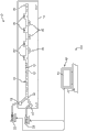
Figure 1A shows the microfluid system for separating of cell according to a kind of embodiment.
Figure 1B shows the micro-system for separating of cell according to another kind of embodiment.
Fig. 1 C shows the synoptic diagram of the microfluidic channel with single expansion area.
Fig. 1 D-Fig. 1 G shows the various geometrical shapies of expansion area.
Fig. 1 H shows the orthographic plan of the microfluidic channel with a plurality of expansion areas.
Fig. 1 I shows the sectional view that obtains along the straight line A-A ' of Fig. 1 H.
Fig. 1 J shows the synoptic diagram of the microfluidic channel with expansion area according to a further aspect in the invention.
Fig. 2 shows the synoptic diagram for separating of the microfluidic device of cell.Also be included in the synoptic diagram that acts on the power of different cell sizes along the difference of microfluidic device.
Fig. 3 shows the another kind of microfluidic device with parallel construction for separating of cell.
Fig. 4 A schematically shows by the hemocyte of partial devices and cancer cells, and this device has the expansion area of holding back big cancer cells.Below tightly, show the corresponding micro-image of the device that comprises several expansion areas.
Fig. 4 B schematically shows by the phosphate buffered saline buffer of the device of Fig. 4 A (PBS) flushing, and it shows that erythrocyte (RBC) flows out and cancer cells is retained in the expansion area.Below tightly, show the corresponding micro-image of the device that comprises several expansion areas.
It is 270 times the blood sample of HeLa cell to be arranged by the microfluidic device blending of Fig. 3 that Fig. 4 C-Fig. 4 F shows at Reynolds number (Rc).Fig. 4 C shows the image of taking under second at t=0.Fig. 4 D shows the image of taking under second at t=9.Fig. 4 E shows the image of taking under second at t=17.Fig. 4 F shows the image of taking under second at t=18.Observing HeLa cell is trapped in the eddy current that produces in the expansion area.
Fig. 4 G shows the comparison as the microfluidic device capture rate of the function of cell concn.
Fig. 5 A is the figure that is illustrated in the concentration ratio (%) that is reached by microfluidic device under the different haemoconcentrations.
Fig. 5 B is the figure that is illustrated in the purity (%) that is reached by microfluidic device under the different haemoconcentrations.
Fig. 5 C is the figure that is illustrated in the capture rate (%) that is reached by microfluidic device under the different haemoconcentrations.
Fig. 6 A shows the synoptic diagram of the solution (solution A) that comprises the MCF7 cell, and wherein cell is trapped within the eddy current that produces in the expansion area.
Fig. 6 B shows the synoptic diagram of first solution displacement of carrying out with the solution B that comprises the microballoon that scribbles Streptavidin.
Fig. 6 C shows the MCF7 cell and scribbles the synoptic diagram of the microballoon reaction of Streptavidin.
Fig. 6 D shows and uses solution C (that is synoptic diagram of second solution displacement of, PBS) carrying out that serves as washing lotion.
Fig. 6 E shows the micro-image corresponding to the MCF7 cell of Fig. 6 A, wherein spiral in the eddy current that cell produces in the expansion area of microfluidic device (around, orbit).Lower-left figure is the enlarged view of rectangular area.Bottom-right graph is the enlarged view of square area.
Fig. 6 F shows the micro-image corresponding to Fig. 6 B.Lower-left figure is the enlarged view of rectangular area.Bottom-right graph is the enlarged view of square area.
Fig. 6 G shows the micro-image corresponding to Fig. 6 C.Lower-left figure is the enlarged view of rectangular area.Bottom-right graph is the enlarged view of square area.
Fig. 6 H shows the micro-image corresponding to Fig. 6 D.Lower-left figure is the enlarged view of rectangular area.Bottom-right graph is the enlarged view of square area.
Fig. 7 shows the microfluidic device according to another kind of embodiment, and it comprises three entrances that are connected to three kinds of different solutions (cell sample, labelled reagent and washing lotion).
Fig. 8 A shows that device with Fig. 7 carries out holds back, the consecutive steps of the displacement of fluorescence solution, reaction and flushing.
Fig. 9 shows the cluster fluoroscopic image of (cluster) of cell, and wherein cell is trapped within successively in the fluid vortex, fixes, changes thoroughly (permeabilized) and with anti-cell Keratin sulfate-PE and DAPI mark with Paraformaldehyde 96.
Figure 10 be illustrate for microfluidic device and standard centrifugal, as each cell (being coated with the MCF7 cell of biotinylation anti-EpCAM) of function of time figure in conjunction with the number of the microballon (bead) that scribbles Streptavidin.
Figure 11 illustrates for microfluidic device and standard centrifugally, and relatively normalized frequency is as the figure of each cell in conjunction with the function of the number of microballon.
Embodiment
Figure 1A shows for the fluid means 10 from the heterogeneous solution isolated cell 12 of the cell 12 that comprises different size.Though for separating of cell 12, be appreciated that this microfluidic device 10 also can be together with the separation (not shown) that is used for particle at the microfluidic device 10 shown in Figure 1A.Therefore, the use of term " cell " and " various kinds of cell " should be exchanged with particle or multiple particle in this article.As finding out in Figure 1A, microfluidic device 10 comprises substrate 14, and it comprises the microfluidic channel 16 that is connected to entrance 18 and outlet 20.The size of microfluidic channel 16 can be different.As an example, microfluidic channel can have the width of 50 μ m and the height of 70 μ m.The typical sizes of the width of microfluidic channel 16 is in the scope of 20 μ m to 200 μ m.The typical sizes of the height of microfluidic channel 16 is in the scope of 20 μ m to 500 μ m.Length also can change but length is generally several centimetres (for example, 4.5cm).Can form substrate 14 by the conventional material that is used for microfluidic device.These materials comprise glass, silicon or dimethione (PDMS).For PDMS, soft lithography can be for generation of microfluidic device 10.In the embodiment of PDMS, for Mold Making, (KMPR1050, Microchem) 4 inches silicon chips of layer spin coating are exposed to UV light by the Cr photomask that designs, and develop with the thick negative photoresist of 70 μ m.Casting PDMS(Sylgard184 on the mould of preparation, Dow Corning) and the degassing.The PDMS moulded product of curing is separated with mould and pierce through (punched) entrance 18 and outlet 20 with needle pliering (needle pliering series A, Technical Innovations Inc.).By being exposed to air plasma (Plasma Cleaner, Harrick Plasma), PDMS and slide glass (slide glass) surface be incorporated into slide glass with enclosed appts with the PDMS layer that will just pierce through.
In the embodiment of Figure 1A, in fact entrance 18 comprises two entrances: entrance 18 ' and entrance 18 ' '.First entrance 18 ' is used for introducing the solution that comprises heterogenous cell group 12.Second entrance 18 ' ' for introducing second different solutions.As hereinafter explaining second entrance 18 ' in more detail ' can be used for washing lotion, marker (for example, fluorescent mark, antibody, nucleic acid dye, fluorogenic substrate) or other chemical reagent (for example, fixing agent or change agent thoroughly) are introduced in the microfluidic channel 16.
As in Figure 1A, finding out entrance 18 ', 18 ' ' be connected to pump 22,24 separately.Each pump 22,24 can be delivered to microfluidic device 10 for each solution that will set flow velocity.In conjunction with the present invention, those skilled in the art can use the pump of known any kind.These pumps include, but not limited to syringe pump, with pressurized air operation pump, peristaltic pump or the positive-displacement pump with pumping fluid.Figure 1A shows the syringe pump 22,24 that uses with microfluidic device 10.For example, Harvard Apparatus, the PHD2000 syringe pump can be used for keeping the overall flow rate of 10 μ l/min to 4.5ml/min.Usually, set pump 22,24 setting to produce the flow velocity greater than 100 μ l/min by microfluidic device 10.
Figure 1A shows computer 40, and its part that can be used as system 100 is with control microfluidic device 10.Generally include at least one treater 42 in the computer 40, it is carried out and installs or be stored in software in the computer 40.Computer 40 can also comprise monitor 44, and it can be used for showing the various parameters of microfluidic device 10.These parameters can comprise, for example, and pump 22,24 flow velocity, the volume that is included in the fluid in the pump 22,24 and other service data.Computer 40 preferably is connected in pump 22,24, makes computer 40 can regulate pump 22,24 independent flow velocity or operational stage.Utilization is stored in presets algorithm or instruction set in the computer 40, and computer 40 is control pump 22,24 automatically.Replacedly, can utilize interface device (for example, keyboard, mouse etc.) the manual regulation pump 22 that uses with computer usually, 24 control.
During the solution replacement operator, computer 40 can guarantee to remain on the flow of solution for expecting in the microfluidic device 10.For example, when slow down or even when closing a pump 22, the flow velocity of the flow velocity that increases by second pump 24 to guarantee to keep to expect.
Figure 1B shows interchangeable system 200, and its working pressure drives pumping system 46.Together with setter 50, pumping system 46 uses the source of gas under pressures 48, with first fluid 52(for example, washing lotion) and the second fluid 54(is for example, blood) pump into device 10.In this system 200, input and output place at device 10 provide liquid valve 56,58 respectively.Computer 40 is set drives pumping system 46 and liquid valve 56,58 with control pressure.For example, valve 56 can be used to open or close first fluid 52 or second fluid 54 flowing to device 10.Valve 58 can be used at waste container 60 and collection device 62(as an example, and it can comprise 96 orifice plates) between switch outlet stream.
As finding out in Figure 1A, microfluidic channel 16 comprises a plurality of expansion areas 30 that are positioned at selected some place along the length of microfluidic channel 16.Expansion area 30 provides the unexpected increase of the width of microfluidic channel 16, or be higher than under the specific threshold flow velocity, the frictional belt that it produce to separate, the frictional belt of this separation make formation eddy current each expansion area 30 in.The eddy current that produces in expansion area 30 is from the subgroup of the solution entrapped cell 12 of the heterogenous cell 12 that flows through microfluidic device 10 just.Yet, these eddy current are different from the eddy current that produces at flow direction, as the Dean eddy current (Dean vortex) that in bend stream, produces by means of inertia (referring to J.Wang et al., Vortex-assisted DNA Delivery, Lab Chip, 2010,10,2057 – 2061 (2010)), or since the eddy current that the microchannel of unsymmetric structure produces (referring to Stott et al., Isolation of Circulating Tumor Cells Using a Microvortex-Generating Herringbone-Chip, Proc Natl.Acad.Sci.107 (43): 18392-7 (2010)).As hereinafter explaining in more detail, be higher than specific threshold or enter expansion area 30 and be hunted down or be trapped in the recirculation eddy current by the cell 12 of size (flow velocity and the geometrical shape that depend on microfluidic device 10).The cell 12 that is lower than threshold size is not hunted down and in microfluidic device 10 relaying afterflows downstream.Usually, will produce the most effective holding back for diameter greater than the cell 12 of 15 μ m.Diameter less than 10 μ m under, holding back is inefficient (for example, 5%).Therefore, the diameter of entrapped cell 12 should be 〉=significant holding back to be to take place in 10 μ m.The geometrical shape of expansion area 30 can be different.For example, expansion area 30 can be rectangle, and shown in Figure 1A, but it can also comprise square, trilateral, Polygons or semicircular in shape, shown in Fig. 1 C-Fig. 1 G.For rectangle expansion area 30, the long limit of expansion area 30 is parallel under the situation of main microfluidic channel 16 orientations, and interception capacity is better.Usually, with respect to the flow direction of upstream microfluidic channel 16, shown in guiding wall 31(Fig. 1 C of expansion area 30) should above angle at 45.
Fig. 1 C shows the single expansion area 30 together with upstream microfluidic channel 16.As indicated above, with respect to shown in Fig. 1 C being the axis of flow of dotted line A, guiding wall 31 should above angle at 45.In this respect, compare with the cross-sectional dimension of the upstream portion of microfluidic channel 16, expansion area 30 is unexpected expansions of cross-sectional dimension (for example, width or height).In the embodiment of Fig. 1 C, 31 one-tenth of guiding walls are just less than 90 ° angle, and it is far above minimum 45 ° threshold value.Expansion area 30 also has hangover wall (trailing wall) 33.Hangover wall 33 can be angled with respect to flow direction A.Usually, the angle of hangover wall 33 is unimportant and can be any angle.For example, in one embodiment, trail wall 33 at a slight angle, it causes that hangover wall 33 reduces to be back to the width of microfluidic channel 16 gradually.In another alternative, do not exist hangover wall 33 and expansion not to return the original size of microfluidic channel 16.
In the another kind of embodiment shown in Fig. 1 J, expansion area 30 comprises crooked guiding wall 31.In this respect, guiding wall 31 begins to be partial to gradually away from upstream microfluidic channel 16 at first, and its length along guiding wall 31 more and more departs from.In this embodiment, with respect to axis of flow A, the different tangent lines that obtain along the difference of guiding wall 31 will have significantly different angle.For example, near the beginning of guiding wall 31, angle θ
1Less and less than 45 °.Yet, near the end of guiding wall 31, angle θ
2Gradient is big and greater than 45 °.At crooked or discontinuous expansion area 30(shown in Fig. 1 J) situation under, average angle θ
AVE, it represents with respect to axis of flow A, should be greater than 45(θ along the average angle of the whole length of guiding wall 31
AVE45 °).
Fig. 1 H illustrates happy orthographic plan along several expansion areas 30 that the length of microfluidic channel 16 is located.Fig. 1 I shows the sectional view that obtains along the straight line A-A ' of Fig. 1 H.Fig. 1 H and Fig. 1 I show the various size of microfluidic channel 16 and expansion area 30.As previously mentioned, the typical sizes of the width of microfluidic channel 16 (w) is in the scope of 20 μ m to 200 μ m.The typical sizes of the height of microfluidic channel 16 (H) is in the scope of 20 μ m to 500 μ m.Expansion area 30 can extend in the scope of 80 μ m to 800 μ m but should be at least the distance (x) of 80 μ m.Expansion area 30 can extend in the distance (y) in 200 μ m to the 2mm scopes.Adjacent expansion area 30 can separate common distance (z) greater than 20 μ m.In some embodiments, can there be single expansion area 30, makes not have adjacent expansion area 30.The shape of cross section of microfluidic channel 16 may be substantially of rectangle, trapezoidal or square.Fine process can produce and show slightly trapezoidal cross section or slightly rounded turning.Passage 16 can also have circle or semi-circular cross-section, though present manufacturing technology can not produce these geometrical shapies.Method and apparatus described herein is contained these variations.
Referring again to Figure 1A, expansion area 30 can be arranged on the opposition side of microfluidic channel 16.This makes single microfluidic channel 16 can have bigger capture ability.In addition, as hereinafter explaining in more detail, when arranging a plurality of passage 16 with parallel construction, this structure expansion area 30 that makes it possible to be crisscross arranged.That is to say, as in Fig. 3, finding out, on adjacent microfluidic channel 16, because expansion area 30 be offset each other and interlock with expansion area 30, adjacent microfluidic channel 16 compact reactors can be stacked.Also with reference to Figure 1A, the cell 12 of large-size is trapped within the expansion area 30, and the cell 12 of reduced size then is not trapped and continues to flow down microfluidic channel 16, and they leave via outlet 20.The cell 12(of large-size is at those cells shown in the expansion area 30) be trapped within the eddy current that produces in the expansion area 30.The cell 12 of reduced size, because their size, they are not trapped within the eddy current and flow out expansion area 30.Therefore, the cell 12 of reduced size is not expanded that the eddy current of district in 30 held back but in microfluidic channel 16 relaying afterflows downstream.
Fig. 2 shows for microfluidic device 10 and the respective flow in microfluidic channel 16 and expansion area 30 from the heterogeneous solution isolated cell 12 of the cell 12 that comprises different size.Fig. 2 shows the trizonal enlarged view of microfluidic channel 16 and expansion area 30, as being determined by view A, B and C.As in view A, seeing, pump into device via one in the syringe pump 22, the 24 heterogenous cell group 12 with different size.Other syringe pump can comprise washing lotion or other solution such as PBS.At first, as seeing in view A, cell 12 random dispersion are in the y direction.Cell 12 stands two kinds of reactive forces: shear gradient lift (F
LShear gradient), it acts on cell 12 cell being shifted to the wall of microfluidic channel 16, and wall effect lift (F
LWall effect), it forces cell 12 away from the wall of microfluidic channel 16.
Have the straight microfluidic channel 16 of rectangular cross section by use, the running balance position of wandering cells 12 produces dynamic lateral balance position X
EqWith uniform cell speed, shown in the view B of Fig. 2.Herein, X
EqBe defined as the distance between the wall of the center of cell 12 and microfluidic channel 16.In Fig. 2, have two (2) opposite expansion area 30 along with cell 12 enters expansion area 30(), stand big F
LBeing pushed to the eddy current center and being trapped than maxicell 12 of shear gradient, and be rushed out expansion area 30 and admission passage than minicell 12, their continue to flow to downstream outlet 20 herein.Usually, F
LShear gradient power and cell dia (a) cube proportional causes than maxicell 12 and stands bigger F
LShear gradient power.Through separate confinement (separation surface) afterwards, laterally the mobile cell 12 that drives is by streamline and towards the eddy current core for size-dependent, and cell 12 keeps separating and spiraling in eddy current herein.This makes it possible to carry out size selectivity and holds back, and when being lower than when size (size cutoff), cell does not move with by separation surface and be retained in and concentrate in the stream with enough speed, but flows out outlet 20.
Fig. 3 shows the another kind of embodiment for separating of the microfluidic device 10 of cell 12, and it comprises a plurality of passages 16 that are connected to entrance 18 and outlet 20.Fig. 3 illustrates the passage 16 that 8 (8) being set parallel to each other usually separate.Each microfluidic channel 16 has the expansion area 30 that 10 (10) separate.Of course it is to be understood that and to use any amount of passage 16.With respect to the quantity along the expansion area that separates 30 of single microfluidic channel 16, this is suitable equally.Can be different along the spacing between the adjacent expansion area 30 of single microfluidic channel 16, but found that the spacing of 1mm is feasible.Can add other passage 16 to produce large-scale parallel device 10.The expansion area 30 on the Jiao Cuo adjacency channel 16 relative to each other, passage 16 is straight.This design makes it possible to place adjacency channel 16 close to each otherly, thereby reduces total area occupied (footprint) of microfluidic device 10.Though Fig. 3 shows the array of the passage 16 of two dimensional topology, be appreciated that the array that to construct passage 16 with three-dimensional layout.Three-dimensional structure will obtain even bigger flux.
In the device of Fig. 3, microfluidic channel 16 is that width is 50 μ m and highly is the rectangle high aspect ratio passage of 70 μ m.Entrance 18 comprises first entrance 18 ' and second entrance 18 ' that comprises PBS or other washing lotion be used to the sample that comprises cell 12 '.Two entrances 18 ', 18 ' ' setting be convenient in microfluidic device 10 easily and solution displacement fast, for example, provide and wash entrapped cell 12 not and strengthen the final concentration ratio of collecting sample and the means of purity.The length of microfluidic device 10 is several cm long.Place expansion area 30 in given tight space, to place the expansion area 30 of maximum quantity with alternate mode.In the device of Fig. 3, the expansion area is the square that is of a size of 400 μ mx400 μ m.
After in cell 12 is trapped within expansion area 30, can be by making eddy current reduce size and final the disappearance with the 30 release cells 12 from the expansion area.This can realize by reducing input flow velocity (for example, reducing pump 22,24 flow velocity).The flow velocity that reduces can reduce the size of eddy current, makes to be trapped in the fluid that wherein cell 12 can be discharged into microfluidic channel 16 and the outlet 20 of bleeder.For the device of Fig. 3, the flow velocity of having found about 4ml/min is that effect is best.Replacedly, flow velocity can be reduced to rapidly is zero to stop fluid flowing by microfluidic device 10 substantially.In this interchangeable mode, can be at (off-chip) collecting cell 12 on the chip rather than outside chip.
Embodiment 1-from the rare cancer cells of blood enrichment
The microfluidic device 10 of Fig. 3 is applicable to from normal people's hemocyte (diameter is in 2 to 15 microns scope) separates and concentrated cancer cells (diameter is 20 microns) carries out in the high-throughput mode with proof enrichment and concentrated effectiveness based on size.For clinical diagnosis, be particularly important from blood enrichment and concentrated cancer cells, because circulating tumor cell (CTC) can provide the real-time information about the monitoring of patient's symptom and cancer therapy.In quick, effective and unmarked mode, remaining great technological challenge a: CTC from blood separation CTC alive is rare events, and its ratio is low to moderate a cell/1,000,000,000 hemocytes.Though current strategies is absorbed in the investigation of diagnostic CTC, there is great demand for the sample size purpose for deliberation of collecting bigger CTC alive.This needs higher flux ground to handle big blood flow volume and enriched target cell and is not attached to modification substrate or magnetic bead, provides independent selection to catch cell with advantage for further analysis or that cultivate.
By means of the large-scale parallel device, it is with the scope treatment liq volume of mL/min, by the separation and concentration target cell based on size and density, and captured cell is discharged into less concentrated amount, and this device 10 has solved the needs to the rare cell enrichment.In order to prove the enrichment of rare cell, with the speed of 4.4mL/min, with fluorescently-labeled breast cancer cell (MCF-7) injection device 10 that is blended in the human blood of dilution, it is similar to device shown in Figure 3.Comprising DMEM and be supplemented with 10% FBS, 1% Sigma I8405 and the culture medium culturing MCF7 breast cancer cell of 1% penicillin/streptomycin, then trypsinized and suspending again before use.By trained doctor, from healthy human volunteers, collect blood and in PBS, be diluted to 5-20% to be used for experiment.
Under these high flow rates, in the eddy current holder of upstream, observe channel deformation, yet, to hold back and be not subjected to remarkably influenced, this is because the shape that remains unchanged near the whirling chamber, downstream of operating under the environmental stress.Higher operation flow velocity but be subject to bonding strength.
The MCF-7 cell of blending comprises unicellular and 2-4 cell clusters, and this is to have demonstrated with conspicuous level because of cluster forming cell in clinical sample to exist.During implantation step, observe blood and cancer cells and enter eddy current and in eddy current, spiral, as shown in the synoptic diagram of the single expansion area 30 among the last figure of Fig. 4 A (upper panel).Figure below of Fig. 4 A (lower panel) shows micro-image, and it illustrates the cancer cells of holding back together with the erythrocyte that is included in the expansion area 30.Observe erythrocyte and enter eddy current, even the particle of similar size does not move into eddy current in the experiment of carrying out with dilute sample.Probably, high cell concentration can induce collision and the hydrokinetics between cell to disturb, and it causes crossing current to move and enter eddy current.
In addition, can keep each expansion area 30 to have the cell of maximum capacity.After eddy current occupies whole expansion area 30, in the high flow velocities scope, can keep about 40 single MCF7 cells at most.Form (spiking) experiment condition for most of peak values, keep far below this maximum value.After complete treatment soln, wash with PBS ' ' ' ' eddy current entrapped cell and do not destroy eddy current.This is illustrated in the last figure of Fig. 4 B.The following micro-image that illustrates of Fig. 4 B, it is illustrated in and introduces the cancer cells of PBS washing lotion still to hold back after the RBC that removes less and comparatively dense.What is interesting is, observe enter eddy current at first hemocyte by stable hold back and flow out rapidly hold back (trap) and outflow system, only stay the bigger stable cancer cells of holding back and spiral.Erythrocyte and white corpuscle all have higher density and/or less size, thereby can not form stable spiraling.Cell through washing will be discharged into is used in the hole of 96 orifice plates characterizing and research (enumeration).
Target cell concentrates when quantize being used for, during the key index of enrichment and purity, microfluidic device 10 performances are good.In<3min, under the situation that relatively seldom hemocyte pollutes, the have an appointment blood sample (n 〉=6 sample) of 10mL volume of 5%v/v blood (that is, 0.5mL whole blood or about 2,500,000,000 hemocytes) of 500 cancer cells of blending is concentrated into less than 200mL(20 times of volumetric concentration) final volume.This is corresponding to 3.4 hundred ten thousand concentration ratio (in output solution target cancer cells with the ratio that pollutes hemocyte divided by the same ratio in input solution), as finding out in Fig. 5 A.This high-caliber enrichment causes the high purity of cancer cells in the final volume of 200mL: about 40%, and as (average 102 ± 21 cancer cells, and 221 ± 155 hemocytes) in Fig. 5 B, found out.Handle the blood sample (n=3) that does not have the blending cancer cells with microfluidic device 10, collect sample and find to contain 772 ± 283 erythrocytes and 4 ± 1 CD45+ white corpuscles in the hole, it is similar to the amount of utilizing the hemocyte pollutent that the blending blood sample finds in micropore.The concentration level that reaches be equivalent to based on molecule avidity with based on the method that target cell separates that is used for of filtering, it has reported the enrichment of 100 ten thousand to 1,000 ten thousand (1,000 ten thousand).When than the purity of report blending 9.2 to 14.0% cancer cells based on the mode of avidity the time, the purity of treated sample is higher.The extent of dilution that is reduced in blood in the treated sample causes the increase of cell processing flux, but also causes the capture rate of the blending cell that reduces.As in Fig. 5 C, finding out, reclaimed 10 to 20% blending cancer cells, and capture rate reduces with the increase of haemoconcentration.Higher haemoconcentration causes higher fluid viscosity, and it changes fluid vortex size and position, causes the lower efficient of holding back.
This relatively low capture rate under higher haemoconcentration shows, for this technology can be used for being separated in the super rare cell that occurs under 1-10 cell/mL, must handle the blood (more than the 10mL) of comparatively large vol.Yet the high-throughput of microfluidic device 10 described herein is (for 2cm
2Chip, the dilute blood of about 5mL/min) show that a large amount of operations of (<30 minutes) are attainable in the rational time period.
Captured cell keeps high-caliber vigor in microfluidic device 10.After injecting cell by device, do not observe the remarkable change (90.1% with respect to initial 90.3%) of cell viability, as live by fluorescence/dead the detection determine.For some sample preparation applications, viable cell can be important.Caught and the cell that discharges can be used for standard molecule and measures as immunostaining by microfluidic device 10.For this purpose, with the unlabelled blending blood sample of microfluidic device 10 enrichments.Discharge cancer cells then and mark in micropore.For cytokeratin-PE and DAPI, cancer cells dyeing is positive, and negative for the dyeing of CD45 cancer cells.One of this usefulness is installed enrichment but is provided significant advantage for the ability of further handling for rare single cell analysis with the small volume transitional cell.
It is 270 that Fig. 4 C-Fig. 4 F shows the microfluidic device 10(Reynolds number (Rc) that utilizes Fig. 3) blending carried out has the result of similar enrichment of the blood sample of HeLa cell.After in expansion area 30, catching HeLa cell, with PBS washing lotion flushing microfluidic device 10.By reducing flow velocity to R
c=5, discharge the HeLa cell of holding back by expansion area 30.Fig. 4 G shows the comparison of the capture rate of microfluidic device 10, and it is the function as cell concn.The number of cell is represented the quantity by the blending HeLa cell of microfluidic device 10 processing.
Embodiment 2-cell marking and solution displacement
Fig. 6 A-Fig. 6 D shows respectively and holds back (Fig. 6 A), first solution displacement (Fig. 6 B), reaction (Fig. 6 C) and second solution displacement (Fig. 6 D).Fig. 6 E-Fig. 6 H shows the micro-image corresponding to Fig. 6 A-Fig. 6 D of actual MCF7 cell respectively, the biotinylation EpCAM incubation of this MCF7 cell in being injected into microfluidic device 10.As finding out in Fig. 6 E, cell is trapped within the eddy current, stands constant rotation and the motion of spiraling.Fig. 6 F shows first solution displacement with the microballoon that scribbles Streptavidin.The microballoon that scribbles Streptavidin enters expansion area 30.Fig. 6 G shows the microballoon that scribbles Streptavidin and the successive reaction of MCF7 cell.Fig. 6 H shows the solution displacement with second solution (that is PBS washing lotion).The PBS washing lotion is removed unconjugated microballoon (arrow A).After finishing washing, flow through the flow velocity of microfluidic device 10 by reduction, with cell release from eddy current is held back, wherein cell harvesting is used for characterizing in 96 orifice plates.Arrow B in Fig. 6 H is pointed to and was more combined closely in the particle of cell in 2 minutes.
With cytotostatic remain on the appropriate location in the fluid vortex ability make it possible to carry out displacement with the multiple solution of labelled reagent and washing lotion in automatable mode.Each interpolation of new soln needs about 100ms with displacement fully.For identical labeled reactant, need six (6) centrifugation step based on the method for conventional whizzer, it comprises three (3) washing steps, and needs〉specimen preparation time (this does not comprise the time with the labelled reagent incubation) of 30 minutes.Each centrifugal and washing step can cause losing the small portion cell potentially and need 5-10min.
Fast the mark cell that has benefited from fluid vortex rotation and spiral makes them be exposed to the environment of the continual renovation of molecular marked compound.In other words, the strong convection of labelled reagent is created near the very little depletion region of the reagent cell surface in eddy current, and the more reagent of strong gradient-driven arrive cell surface.By detect the combination of the microballoon and the biotinylation anti-EpCAM antibody that scribble Streptavidin at cell surface, detect this quick mark (Fig. 6 A-Fig. 6 H).Find that the cell in microfluidic device 10 has accumulated the microballon of the equal amts that accumulated with the cell for preparing with standard scheme in 5 minutes in 30 minutes.In addition, after 30 minutes, compare with standard method, on average have the microballon quantity/cell of the combination of twice with the cell of microfluidic device 10 marks.
Embodiment 3-operate continuously: the rare cell enrichment is mark subsequently
Utilize microfluidic device shown in Figure 7 10, successfully carried out whizzer and made it possible to a plurality of continuous sample preparation processes (for example, holding back the displacement of fluorescence solution, reaction and washing) of carrying out.In this embodiment, microfluidic device 10 comprises three entrances 18 ', 18 ' ' and 18 ' ' '.An entrance 18 ' is connected to syringe pump 22 for delivery of cell sample.Second syringe pump 24 is for delivery of fluorescent agent.The 3rd syringe pump 26 is for delivery of washing lotion (PBS).In<1 hour, carry out cancer cells the holding back from blood based on size, the analysis of continuous fluorescence mark and release cell.There is the dilution human blood (10mL) of cancer cells to inject microfluidic device 10, about 3 minutes of enrichment cancer cells blending.Use fixing agent (Paraformaldehyde 96) and saturatingization agent to prepare entrapped cell also with fluorescence antibody (anti-cell Keratin sulfate-PE and DAPI) dyeing 20 minutes successively.Fig. 8 A-Fig. 8 D shows and holds back, the order of first solution displacement, reaction and the displacement of second solution.Use PBS washed cell<1min then, and collect in 96 orifice plates for characterizing.The cell of collecting is for cytokeratin and DAPI, mark is positive, this shows the success of continuous sample preparation, as shown in Figure 9, it shows the fluoroscopic image that cell clusters, and this cell clusters and is trapped within successively in the fluid vortex, solidifies with Paraformaldehyde 96, saturatingization, and with anti-cell Keratin sulfate-PE and DAPI mark.As finding out in Figure 10 A, under the level that scheme is identical outside the standard chips after with 30 minutes, the MCF7 cell that is coated with the biotinylation anti-EpCAM coated the microballon of Streptavidin combination in<5 minutes.Figure 11 A shows after 30 minutes that uniform labelling has microballon on cell mass.In addition, the whizzer (centrifuge-on-chip) of microfluidic device 10(on chip) obtain larger amt in conjunction with microballon/cell.Above result shows the complete approach of the automatization of the needed all samples preparation process of cell analysis in single simple platform.
Though microfluidic device 10 has the application-specific for separating of CTC, other application comprises the cell 12 that concentrates available from sample.For example, can catch the cell 12 with the concern that can be trapped within the size in the expansion area 30, be discharged in the sample with conc forms then.For example, can make cell 12 in the fluid (as urine, Pleural fluid and peritonaeum washing lotion) that is included in biogenetic derivation by microfluidic device 10 to concentrate the cell 12 wherein comprise.In this respect, microfluidic device 10 is very suitable for concentrating cells 12.For example, on the basis of volume, microfluidic device 10 can concentrate ten (10) doubly or the concentration of the cell 12 in the initial soln more than 20 (20) times with cell 12.
Though illustrated and described embodiment, can carry out various changes and do not depart from the scope of the design of the present invention that this paper discloses.For example, though this paper has described several embodiments, be appreciated that all respects or key element can exchange with other independent embodiment.Therefore, the present invention only is subject to the claim of enclosing and their Equivalent.
Claims (71)
Priority Applications (1)
| Application Number | Priority Date | Filing Date | Title |
|---|---|---|---|
| CN201510088710.9A CN104741157B (en) | 2010-09-14 | 2011-09-12 | Device for isolating cells from heterogeneous solution using microfluidic trapping vortices |
Applications Claiming Priority (3)
| Application Number | Priority Date | Filing Date | Title |
|---|---|---|---|
| US38284010P | 2010-09-14 | 2010-09-14 | |
| US61/382,840 | 2010-09-14 | ||
| PCT/US2011/051224 WO2012037030A2 (en) | 2010-09-14 | 2011-09-12 | Method and device for isolating cells from heterogeneous solution using microfluidic trapping vortices |
Related Child Applications (1)
| Application Number | Title | Priority Date | Filing Date |
|---|---|---|---|
| CN201510088710.9A Division CN104741157B (en) | 2010-09-14 | 2011-09-12 | Device for isolating cells from heterogeneous solution using microfluidic trapping vortices |
Publications (2)
| Publication Number | Publication Date |
|---|---|
| CN103261436A true CN103261436A (en) | 2013-08-21 |
| CN103261436B CN103261436B (en) | 2015-03-25 |
Family
ID=45832184
Family Applications (2)
| Application Number | Title | Priority Date | Filing Date |
|---|---|---|---|
| CN201510088710.9A Active CN104741157B (en) | 2010-09-14 | 2011-09-12 | Device for isolating cells from heterogeneous solution using microfluidic trapping vortices |
| CN201180044092.8A Active CN103261436B (en) | 2010-09-14 | 2011-09-12 | A Method for Isolating Cells from Heterogeneous Solutions Using Microfluidic Trapped Vortex |
Family Applications Before (1)
| Application Number | Title | Priority Date | Filing Date |
|---|---|---|---|
| CN201510088710.9A Active CN104741157B (en) | 2010-09-14 | 2011-09-12 | Device for isolating cells from heterogeneous solution using microfluidic trapping vortices |
Country Status (7)
| Country | Link |
|---|---|
| US (4) | US9133499B2 (en) |
| EP (1) | EP2616551B1 (en) |
| JP (1) | JP5920895B2 (en) |
| CN (2) | CN104741157B (en) |
| AU (2) | AU2011302302B2 (en) |
| CA (1) | CA2809877C (en) |
| WO (1) | WO2012037030A2 (en) |
Cited By (24)
| Publication number | Priority date | Publication date | Assignee | Title |
|---|---|---|---|---|
| CN105381824A (en) * | 2014-08-26 | 2016-03-09 | 中央研究院 | Collector architecture layout design |
| CN105658781A (en) * | 2013-10-22 | 2016-06-08 | 伯克利照明有限公司 | Microfluidic device with isolation enclosure and method for testing biological microtargets using it |
| CN105683750A (en) * | 2013-10-16 | 2016-06-15 | 明策生物医学科技私人有限公司 | Microfluidics sorter for cell detection and isolation |
| CN105814189A (en) * | 2013-12-04 | 2016-07-27 | 明策微型流体私人有限公司 | Microfluidic device |
| US10107726B2 (en) | 2016-03-16 | 2018-10-23 | Cellmax, Ltd. | Collection of suspended cells using a transferable membrane |
| CN109666584A (en) * | 2018-12-29 | 2019-04-23 | 北京工业大学 | A kind of experimental provision can be used for carrying out circulating tumor cell sorting experiment |
| CN109975265A (en) * | 2019-04-22 | 2019-07-05 | 中国矿业大学 | A three-dimensional scaling microfluidic device and method for multidirectional induced Dean flow |
| CN110257223A (en) * | 2019-07-15 | 2019-09-20 | 北京工业大学 | A chip device for cell microenvironment regulation based on groove droplet capture |
| US10495644B2 (en) | 2014-04-01 | 2019-12-03 | Academia Sinica | Methods and systems for cancer diagnosis and prognosis |
| CN111040928A (en) * | 2019-12-27 | 2020-04-21 | 天津大学 | A high-throughput microfluidic chip for the processing and collection of Cryptodinoflagellate |
| CN111060364A (en) * | 2019-11-20 | 2020-04-24 | 天津大学 | An integrated method for tumor cell staining and screening and supporting microfluidic chip |
| CN111088146A (en) * | 2020-01-09 | 2020-05-01 | 天津大学 | A microfluidic chip for screening tumor cells from pleural effusion |
| US10646871B2 (en) | 2013-10-22 | 2020-05-12 | Berkeley Lights, Inc. | Microfluidic devices having isolation pens and methods of testing biological micro-objects with same |
| CN111207988A (en) * | 2020-02-18 | 2020-05-29 | 中南大学 | A microfluidic-based slurry monitoring device |
| CN111690534A (en) * | 2020-06-16 | 2020-09-22 | 东南大学 | Tumor cell multistage sorting device based on viscoelastic focusing technology |
| CN112195094A (en) * | 2020-09-30 | 2021-01-08 | 苏州莱博睿思生物科技有限公司 | Micro-fluidic chip box |
| CN113490549A (en) * | 2019-02-26 | 2021-10-08 | 美纳里尼硅生物系统股份公司 | Microfluidic method and system for separating particles |
| CN113528447A (en) * | 2021-06-10 | 2021-10-22 | 广州市第一人民医院(广州消化疾病中心、广州医科大学附属市一人民医院、华南理工大学附属第二医院) | A microfluidic chip and its application in tumor stem cell sorting, amplification and recovery |
| CN113840657A (en) * | 2019-03-12 | 2021-12-24 | 高丽大学校算学协力团 | Microfluidic system for intracellular delivery of materials and method thereof |
| CN113832005A (en) * | 2021-08-30 | 2021-12-24 | 北京工业大学 | Blood cancer cell filter chip device based on microchannel groove vortex cell flow |
| CN114867556A (en) * | 2019-11-06 | 2022-08-05 | 高丽大学校算学协力团 | Droplet-based deformation method for delivering material into cells and chip therefor |
| CN115627221A (en) * | 2022-10-13 | 2023-01-20 | 中山大学 | A rapid proliferation and detection device for microorganisms |
| US11674958B2 (en) | 2011-06-29 | 2023-06-13 | Academia Sinica | Capture, purification, and release of biological substances using a surface coating |
| CN116408160A (en) * | 2021-12-31 | 2023-07-11 | 彩科(苏州)生物科技有限公司 | Microfluidic devices with stable isolated environments |
Families Citing this family (118)
| Publication number | Priority date | Publication date | Assignee | Title |
|---|---|---|---|---|
| JP2791672B2 (en) | 1988-12-28 | 1998-08-27 | カヤバ工業株式会社 | Stay damper |
| EP2616551B1 (en) | 2010-09-14 | 2020-08-19 | The Regents of The University of California | Method for isolating cells from heterogeneous solution using microfluidic trapping vortices |
| AU2012231737B2 (en) | 2011-03-24 | 2015-07-23 | Ningkasai Technology (Shanghai) Co, Ltd. | Micro-devices for disease detection |
| WO2012151501A2 (en) | 2011-05-05 | 2012-11-08 | Anpac Bio-Medical Science Co., Ltd. | Apparatus for detecting tumor cells |
| US9404864B2 (en) | 2013-03-13 | 2016-08-02 | Denovo Sciences, Inc. | System for imaging captured cells |
| US10466160B2 (en) | 2011-08-01 | 2019-11-05 | Celsee Diagnostics, Inc. | System and method for retrieving and analyzing particles |
| EP2739587B1 (en) | 2011-08-01 | 2020-05-27 | Denovo Sciences | Cell capture system |
| WO2013177560A1 (en) | 2012-05-25 | 2013-11-28 | The Regents Of The University Of California | Microfluidic systems for particle trapping and separation |
| US9968932B2 (en) | 2012-10-15 | 2018-05-15 | National University Corporation Nagoya University | Microchannel chip for microparticle separation, microparticle separation method and system for microparticle separation using chip |
| US9857333B2 (en) | 2012-10-31 | 2018-01-02 | Berkeley Lights, Inc. | Pens for biological micro-objects |
| US20150320348A1 (en) * | 2012-11-16 | 2015-11-12 | Lightstat, Llc | Disposable sample collection method and apparatus |
| US9752181B2 (en) | 2013-01-26 | 2017-09-05 | Denovo Sciences, Inc. | System and method for capturing and analyzing cells |
| US9707562B2 (en) | 2013-03-13 | 2017-07-18 | Denovo Sciences, Inc. | System for capturing and analyzing cells |
| MX390892B (en) * | 2013-03-15 | 2025-03-21 | Adama Makhteshim Ltd | TERMITICIDE COMPOSITION AND METHODS FOR TREATING TERMITES. |
| US10391490B2 (en) | 2013-05-31 | 2019-08-27 | Celsee Diagnostics, Inc. | System and method for isolating and analyzing cells |
| US9856535B2 (en) | 2013-05-31 | 2018-01-02 | Denovo Sciences, Inc. | System for isolating cells |
| WO2014210207A1 (en) * | 2013-06-25 | 2014-12-31 | University Of Washington Through Its Center For Commercialization | Self-digitization of sample volumes |
| US11293001B2 (en) | 2013-07-29 | 2022-04-05 | 9493662 Canada Inc. | Microfluidic cell culture systems |
| US9029109B2 (en) * | 2013-08-07 | 2015-05-12 | President And Fellows Of Harvard College | Microfluidic vortex-assisted electroporation system and method |
| KR102346066B1 (en) * | 2013-10-22 | 2021-12-31 | 버클리 라잇츠, 인크. | Exporting a selected group of micro-objects from a micro-fluidic device |
| DK3060912T3 (en) * | 2013-10-22 | 2020-09-28 | Berkeley Lights Inc | PROCEDURE AND MICROFLUID DEVICE FOR ANALYSIS OF BIOLOGICAL ACTIVITY |
| US9889445B2 (en) | 2013-10-22 | 2018-02-13 | Berkeley Lights, Inc. | Micro-fluidic devices for assaying biological activity |
| SG10202002543UA (en) * | 2013-10-22 | 2020-05-28 | Berkeley Lights Inc | Micro-fluidic devices for assaying biological activity |
| US10098581B2 (en) | 2014-01-17 | 2018-10-16 | Massachusetts Institute Of Technology | Integrated electrical profiling system for measuring leukocytes activation from whole blood |
| US11192107B2 (en) | 2014-04-25 | 2021-12-07 | Berkeley Lights, Inc. | DEP force control and electrowetting control in different sections of the same microfluidic apparatus |
| US20150306599A1 (en) | 2014-04-25 | 2015-10-29 | Berkeley Lights, Inc. | Providing DEP Manipulation Devices And Controllable Electrowetting Devices In The Same Microfluidic Apparatus |
| WO2015188171A1 (en) | 2014-06-06 | 2015-12-10 | Berkeley Lights, Inc. | Isolating microfluidic structures and trapping bubbles |
| US10502674B2 (en) | 2014-06-27 | 2019-12-10 | The Regents Of The University Of California | Apparatus and method for label-free analysis of rare cells from bodily fluids |
| WO2016011059A1 (en) * | 2014-07-14 | 2016-01-21 | President And Fellows Of Harvard College | Drug cocktail analyses using microscale vortex-assisted electroporation |
| FI127992B (en) | 2014-08-29 | 2019-07-15 | Svanbaeck Sami | Procedure and systems for determining dissolution properties of a substance |
| US10058865B2 (en) | 2014-12-08 | 2018-08-28 | Berkeley Lights, Inc. | Actuated microfluidic structures for directed flow in a microfluidic device and methods of use thereof |
| US10578630B2 (en) | 2014-12-09 | 2020-03-03 | Berkeley Lights, Inc. | Automated identification of assay areas in a microfluidic device and detection of assay positive areas based on rate of change of image light intensity |
| CN113075113A (en) | 2014-12-09 | 2021-07-06 | 伯克利之光生命科技公司 | Automated detection and repositioning of micro-objects in microfluidic devices |
| US9744533B2 (en) | 2014-12-10 | 2017-08-29 | Berkeley Lights, Inc. | Movement and selection of micro-objects in a microfluidic apparatus |
| WO2016168492A1 (en) | 2015-04-14 | 2016-10-20 | Hur Soojung Claire | Electrode array for vortex-assisted electroporation |
| WO2016172623A1 (en) | 2015-04-22 | 2016-10-27 | Berkeley Lights, Inc. | Manipulation of cell nuclei in a micro-fluidic device |
| US10751715B1 (en) | 2015-04-22 | 2020-08-25 | Berkeley Lights, Inc. | Microfluidic reporter cell assay methods and kits thereof |
| CN107810059B (en) | 2015-04-22 | 2021-03-23 | 伯克利之光生命科技公司 | Freezing and archiving cells on microfluidic devices |
| IL284235B (en) | 2015-04-22 | 2022-07-01 | Berkeley Lights Inc | Kits and methods for preparing a microfluidic device for cell culture |
| WO2016205742A1 (en) * | 2015-06-17 | 2016-12-22 | The Regents Of The University Of California | High efficiency microfluidic device for trapping circulating tumor cells |
| KR101684138B1 (en) * | 2015-06-30 | 2016-12-21 | 한국표준과학연구원 | Apparatus and method for small degree off-axis micro-channel system to improve performance of solution immersed silicon biosensors |
| US9862941B2 (en) | 2015-10-14 | 2018-01-09 | Pioneer Hi-Bred International, Inc. | Single cell microfluidic device |
| US10799865B2 (en) | 2015-10-27 | 2020-10-13 | Berkeley Lights, Inc. | Microfluidic apparatus having an optimized electrowetting surface and related systems and methods |
| CN108495712A (en) | 2015-11-23 | 2018-09-04 | 伯克利之光生命科技公司 | In situ generated microfluidic isolation structures, kits and methods of use thereof |
| CA3005077C (en) | 2015-12-08 | 2023-02-28 | Berkeley Lights, Inc. | In situ-generated microfluidic assay structures, related kits, and methods of use thereof |
| KR20230042540A (en) | 2015-12-30 | 2023-03-28 | 버클리 라잇츠, 인크. | Microfluidic devices for optically-driven convection and displacement, kits and methods thereof |
| WO2017117521A1 (en) | 2015-12-31 | 2017-07-06 | Berkeley Lights, Inc. | Tumor infilitrating cells engineered to express a pro-inflammatory polypeptide |
| SG11201805592RA (en) | 2016-01-15 | 2018-07-30 | Berkeley Lights Inc | Methods of producing patient-specific anti-cancer therapeutics and methods of treatment therefor |
| WO2017160991A1 (en) | 2016-03-16 | 2017-09-21 | Lavieu Gregory G | Methods, systems and devices for selection and generation of genome edited clones |
| US10675625B2 (en) | 2016-04-15 | 2020-06-09 | Berkeley Lights, Inc | Light sequencing and patterns for dielectrophoretic transport |
| SG11201808914UA (en) | 2016-04-15 | 2018-11-29 | Berkeley Lights Inc | Methods, systems and kits for in-pen assays |
| US12128407B2 (en) | 2016-04-15 | 2024-10-29 | Vortex Biosciences, Inc. | Microfluidic chips and cartridges and systems utilizing microfluidic chips and cartridges |
| US11383240B2 (en) * | 2016-05-22 | 2022-07-12 | Cornell University | Single cell whole genome amplification via micropillar arrays under flow conditions |
| CN109689213B (en) | 2016-05-26 | 2022-10-14 | 伯克利之光生命科技公司 | Covalently modified surface, kit, preparation method and application |
| KR102579835B1 (en) * | 2016-06-08 | 2023-09-15 | 더 리전트 오브 더 유니버시티 오브 캘리포니아 | Method and device for processing tissues and cells |
| JP7038100B2 (en) | 2016-07-21 | 2022-03-17 | バークレー ライツ,インコーポレイテッド | Sorting of T lymphocytes with a microfluidic device |
| US11999931B2 (en) | 2016-08-20 | 2024-06-04 | The Regents Of The University Of California | High-throughput system and method for the temporary permeabilization of cells |
| US10717086B2 (en) * | 2016-08-29 | 2020-07-21 | The Regents Of The University Of California | Integrated system for isolation and emulsification of particles and cells |
| WO2018071448A1 (en) | 2016-10-11 | 2018-04-19 | The Regents Of The University Of California | Systems and methods to encapsulate and preserve organic matter for analysis |
| CN107955788A (en) * | 2016-10-14 | 2018-04-24 | 中国科学院大连化学物理研究所 | A kind of micro fluid dynamcis method on organ chip |
| WO2018119296A1 (en) * | 2016-12-22 | 2018-06-28 | The Charles Stark Draper Laboratory, Inc. | System and method of using a microfluidic electroporation device for cell treatment |
| EP3562594A4 (en) | 2016-12-30 | 2020-09-09 | The Regents of University of California | METHOD OF SELECTION AND GENERATION OF GENOMEDITED T-CELLS |
| US11213823B2 (en) * | 2017-01-11 | 2022-01-04 | The Regents Of The University Of California | Microfluidic in situ labelling on stable interfaces |
| CN106823475B (en) * | 2017-02-07 | 2023-01-24 | 重庆科技学院 | a blood diverter |
| US11213824B2 (en) | 2017-03-29 | 2022-01-04 | The Research Foundation For The State University Of New York | Microfluidic device and methods |
| US10544413B2 (en) | 2017-05-18 | 2020-01-28 | 10X Genomics, Inc. | Methods and systems for sorting droplets and beads |
| WO2018213643A1 (en) | 2017-05-18 | 2018-11-22 | 10X Genomics, Inc. | Methods and systems for sorting droplets and beads |
| US10780438B2 (en) | 2017-06-09 | 2020-09-22 | The Regents Of The University Of California | High-efficiency encapsulation in droplets based on hydrodynamic vortices control |
| US11517901B2 (en) | 2017-06-09 | 2022-12-06 | The Regents Of The University Of California | High-efficiency particle encapsulation in droplets with particle spacing and downstream droplet sorting |
| EP3655776A2 (en) * | 2017-07-18 | 2020-05-27 | Avectas Limited | Payload delivery across cell membranes using continuous flow fluidic system |
| US10549279B2 (en) | 2017-08-22 | 2020-02-04 | 10X Genomics, Inc. | Devices having a plurality of droplet formation regions |
| CN112980667B (en) | 2017-08-29 | 2024-09-17 | 伯乐实验室有限公司 | Systems and methods for isolating and analyzing cells |
| US20200330989A1 (en) * | 2017-09-07 | 2020-10-22 | Sony Corporation | Particle capturing chamber, particle capturing chip, particle capturing method, apparatus, and particle analysis system |
| WO2019067319A2 (en) * | 2017-09-29 | 2019-04-04 | Schlumberger Technology Corporation | Microfluidic technique for detection of multi-contact miscibility |
| WO2019075409A1 (en) | 2017-10-12 | 2019-04-18 | The Regents Of The University Of California | Microfluidic label-free isolation and identification of cells using fluorescence lifetime imaging (flim) |
| WO2019075476A2 (en) | 2017-10-15 | 2019-04-18 | Berkeley Lights, Inc. | Methods, systems and kits for in-pen assays |
| WO2019079787A1 (en) | 2017-10-20 | 2019-04-25 | The Regents Of The University Of California | Microfluidic systems and methods for lipoplex-mediated cell transfection |
| US11499127B2 (en) | 2017-10-20 | 2022-11-15 | The Regents Of The University Of California | Multi-layered microfluidic systems for in vitro large-scale perfused capillary networks |
| WO2019083852A1 (en) | 2017-10-26 | 2019-05-02 | 10X Genomics, Inc. | Microfluidic channel networks for partitioning |
| JP2019086340A (en) * | 2017-11-02 | 2019-06-06 | シスメックス株式会社 | Cell detection method and cell detection system |
| CN107974392A (en) * | 2017-12-27 | 2018-05-01 | 深圳市合川医疗科技有限公司 | A kind of method of circulating tumor cell in micro-fluidic chip and separating trap blood |
| CN108359577B (en) * | 2018-01-29 | 2022-03-04 | 燕山大学 | Zebrafish egg rotation micro-operating system and control method based on microfluidic drive |
| EP4477315A3 (en) | 2018-05-31 | 2025-03-05 | Donaldson Company, Inc. | Droplet sensors for fuel systems |
| KR102019602B1 (en) * | 2018-06-01 | 2019-09-06 | 주종일 | Microfluidic Chip for 3-Dimensional Cell Culture and Method of 3-Dimensional Cell Culture Using the Same |
| EP3775150A4 (en) * | 2018-08-21 | 2021-10-13 | Hewlett-Packard Development Company, L.P. | Cell measurements after isolation from solutions in a microfluidic channel |
| CN108854892B (en) * | 2018-08-23 | 2020-08-25 | 浙江工业大学上虞研究院有限公司 | Micro-reactor |
| US20200070167A1 (en) * | 2018-08-31 | 2020-03-05 | Vortex Biosciences, Inc. | Processing systems for isolating and enumerating cells or particles |
| SG11202102489XA (en) | 2018-09-21 | 2021-04-29 | Berkeley Lights Inc | Functionalized well plate, methods of preparation and use thereof |
| CA3115808A1 (en) | 2018-10-11 | 2020-04-16 | Berkeley Lights, Inc. | Systems and methods for identification of optimized protein production and kits therefor |
| CN113366312A (en) | 2018-11-01 | 2021-09-07 | 伯克利之光生命科技公司 | Method for assaying biological cells in a microfluidic environment |
| EP3883692A4 (en) | 2018-11-19 | 2022-08-31 | Berkeley Lights, Inc. | MICROFLUIDIC DEVICE COMPRISING PROGRAMMABLE SWITCHING ELEMENTS |
| US20220064627A1 (en) * | 2019-01-23 | 2022-03-03 | Haplomic Technologies Pty Ltd | Microfluidic device |
| WO2020168258A1 (en) | 2019-02-15 | 2020-08-20 | Berkeley Lights, Inc. | Laser-assisted repositioning of a micro-object and culturing of an attachment-dependent cell in a microfluidic environment |
| CN113747974A (en) | 2019-02-28 | 2021-12-03 | 10X基因组学有限公司 | Apparatus, system, and method for improving droplet formation efficiency |
| JP7585606B2 (en) * | 2019-04-16 | 2024-11-19 | 東ソー株式会社 | Dispersion Solvent Replacement Device |
| US10633693B1 (en) | 2019-04-16 | 2020-04-28 | Celsee Diagnostics, Inc. | System and method for leakage control in a particle capture system |
| EP3962652A4 (en) | 2019-04-30 | 2023-01-18 | Berkeley Lights, Inc. | Methods for encapsulating and assaying cells |
| WO2020227309A1 (en) | 2019-05-07 | 2020-11-12 | Bio-Rad Laboratories, Inc. | System and method for automated single cell processing |
| US11273439B2 (en) | 2019-05-07 | 2022-03-15 | Bio-Rad Laboratories, Inc. | System and method for target material retrieval from microwells |
| CN111909823B (en) * | 2019-05-08 | 2023-04-18 | 清华大学 | Inertial micro-fluidic chip for enriching circulating tumor cells |
| KR20250084977A (en) | 2019-06-14 | 2025-06-11 | 바이오 래드 래버러토리스 인코오포레이티드 | System and method for automated single cell processing and analyses |
| WO2020263858A1 (en) * | 2019-06-24 | 2020-12-30 | The Regents Of The University Of California | Integrated system for mechanical processing of lipoaspirate |
| US12186751B2 (en) | 2019-06-28 | 2025-01-07 | 10X Genomics, Inc. | Devices and systems incorporating acoustic ordering and methods of use thereof |
| US12179199B2 (en) | 2019-08-09 | 2024-12-31 | The Regents Of The University Of California | Microfluidic single-cell pairing array for studying cell-cell interactions in isolated compartments |
| US12059679B2 (en) | 2019-11-19 | 2024-08-13 | 10X Genomics, Inc. | Methods and devices for sorting droplets and particles |
| WO2021142309A1 (en) * | 2020-01-08 | 2021-07-15 | The General Hospital Corporation | Microfluidic systems and methods for low-shear isolation of rare cells from large sample volumes |
| US11504719B2 (en) | 2020-03-12 | 2022-11-22 | Bio-Rad Laboratories, Inc. | System and method for receiving and delivering a fluid for sample processing |
| US20230356225A1 (en) * | 2020-09-28 | 2023-11-09 | Georgia Tech Research Corporation | Microfluidic devices for particle capture and methods thereof |
| US11959186B2 (en) * | 2020-11-26 | 2024-04-16 | Changxin Memory Technologies, Inc. | Electroplating method and electroplating apparatus |
| EP4019131A1 (en) | 2020-12-22 | 2022-06-29 | Albert-Ludwigs-Universität Freiburg | Microfluidic device and method for isolating objects |
| CA202670S (en) | 2021-04-09 | 2024-05-15 | 9493662 Canada Inc | Microfluidic slab with 4 well arrangements |
| CA202671S (en) | 2021-04-09 | 2024-05-15 | 9493662 Canada Inc | Microfluidic slab with 2 well arrangements |
| US12478899B2 (en) | 2021-06-02 | 2025-11-25 | Donaldson Company, Inc. | Maintenance of hydrodynamic separators |
| EP4444910A4 (en) | 2021-12-10 | 2025-11-05 | Bio Rad Laboratories Inc | COMPOSITIONS, METHOD AND SYSTEMS FOR SAMPLE PROCESSING WITH MORPHOLOGICALLY ADAPTABLE FUNCTIONALIZED PARTICLES |
| CN114395463B (en) * | 2021-12-10 | 2023-11-17 | 广州大学 | CTC enrichment and release system based on micro-fluidic and micro-optical tweezers arrays and preparation method |
| JP2024015625A (en) * | 2022-07-25 | 2024-02-06 | キヤノンメディカルシステムズ株式会社 | Microfluidic device and classification method |
| USD1079975S1 (en) | 2023-04-28 | 2025-06-17 | Ananda Devices, Inc. | Neuromuscular junction cell culture layer |
| CN119662372A (en) * | 2024-12-23 | 2025-03-21 | 海南大学 | Spiral and contraction-expansion coupling microchannel and flexible microalgae sorting method |
Citations (2)
| Publication number | Priority date | Publication date | Assignee | Title |
|---|---|---|---|---|
| US20050048581A1 (en) * | 2003-08-25 | 2005-03-03 | Chiu Daniel T. | Method and device for biochemical detection and analysis of subcellular compartments from a single cell |
| WO2008157257A1 (en) * | 2007-06-20 | 2008-12-24 | University Of Washington | A biochip for high-throughput screening of circulating tumor cells |
Family Cites Families (22)
| Publication number | Priority date | Publication date | Assignee | Title |
|---|---|---|---|---|
| US6252375B1 (en) * | 2000-07-24 | 2001-06-26 | In-System Design, Inc. | Power management system and current augmentation and battery charger method and apparatus for a computer peripheral |
| GB2375399A (en) * | 2001-05-11 | 2002-11-13 | Suisse Electronique Microtech | Microfluidic system for the manipulation and concentration of particles suspended in fluid |
| EP1622691B1 (en) | 2003-03-14 | 2011-05-25 | The Trustees of Columbia University in the City of New York | Systems and methods of blood-based therapies having a microfluidic membraneless exchange device |
| JP2007522913A (en) * | 2003-10-03 | 2007-08-16 | フリーイェ・ユニヴェルシテイト・ブリュッセル | Method and apparatus for size separation of particles present in a fluid |
| US7803599B2 (en) | 2004-01-16 | 2010-09-28 | University Of Washington | Steady streaming particle traps |
| US20060079003A1 (en) * | 2004-10-12 | 2006-04-13 | Witty Thomas R | Apparatus and method for a precision flow assay |
| US7682817B2 (en) * | 2004-12-23 | 2010-03-23 | Kimberly-Clark Worldwide, Inc. | Microfluidic assay devices |
| CN101137440A (en) * | 2005-01-12 | 2008-03-05 | 因弗因斯医药瑞士股份有限公司 | Method for producing a microfluidic component and microfluidic component |
| US7993821B2 (en) | 2005-08-11 | 2011-08-09 | University Of Washington | Methods and apparatus for the isolation and enrichment of circulating tumor cells |
| US8186913B2 (en) | 2007-04-16 | 2012-05-29 | The General Hospital Corporation | Systems and methods for particle focusing in microchannels |
| TW200920841A (en) * | 2007-09-25 | 2009-05-16 | Cytyc Corp | Microfluidic apparatus for manipulating imaging and analyzing cells of a cytological specimen |
| CN101177663A (en) * | 2007-11-05 | 2008-05-14 | 吉林大学 | DNA Separation Microfluidic Chip |
| FR2931085B1 (en) * | 2008-05-13 | 2011-05-27 | Commissariat Energie Atomique | METHOD OF SORTING PARTICLES OR AMAS FROM PARTICLES IN A FLUID CIRCULATING IN A CHANNEL |
| FR2931141B1 (en) | 2008-05-13 | 2011-07-01 | Commissariat Energie Atomique | MICROFLUIDIC SYSTEM AND METHOD FOR THE SORTING OF AMAS FROM CELLS AND PREFERENCE FOR CONTINUOUS ENCAPSULATION THROUGH THEIR SORTING |
| FR2931079B1 (en) | 2008-05-13 | 2010-07-30 | Commissariat Energie Atomique | DEVICE AND METHOD FOR SEPARATING A SUSPENSION |
| EP2335075A4 (en) | 2008-09-05 | 2011-12-14 | Scripps Research Inst | METHODS OF DETECTING CIRCULATING TUMOR CELLS |
| EP2344883A4 (en) | 2008-09-26 | 2012-03-21 | Gen Hospital Corp | CAPTURE OF PARTICLES |
| US20110070581A1 (en) | 2009-04-27 | 2011-03-24 | Amit Gupta | Separation of Leukocytes |
| JP2010281701A (en) * | 2009-06-05 | 2010-12-16 | Tohoku Univ | Microparticle continuous sorting / measurement device and microfluidic chip |
| US8208138B2 (en) | 2009-09-24 | 2012-06-26 | University Of Cincinnati | Spiral microchannel particle separators, straight microchannel particle separators, and continuous particle separator and detector systems |
| SG189778A1 (en) | 2009-10-20 | 2013-05-31 | Agency Science Tech & Res | Microfluidic system for trapping and detection of a biological entity in a sample |
| EP2616551B1 (en) | 2010-09-14 | 2020-08-19 | The Regents of The University of California | Method for isolating cells from heterogeneous solution using microfluidic trapping vortices |
-
2011
- 2011-09-12 EP EP11825738.5A patent/EP2616551B1/en active Active
- 2011-09-12 WO PCT/US2011/051224 patent/WO2012037030A2/en not_active Ceased
- 2011-09-12 AU AU2011302302A patent/AU2011302302B2/en active Active
- 2011-09-12 US US13/823,112 patent/US9133499B2/en active Active
- 2011-09-12 CN CN201510088710.9A patent/CN104741157B/en active Active
- 2011-09-12 CA CA2809877A patent/CA2809877C/en active Active
- 2011-09-12 JP JP2013528369A patent/JP5920895B2/en active Active
- 2011-09-12 CN CN201180044092.8A patent/CN103261436B/en active Active
-
2015
- 2015-02-23 AU AU2015200910A patent/AU2015200910B2/en active Active
- 2015-08-05 US US14/819,302 patent/US10407709B2/en active Active
- 2015-11-20 US US14/948,119 patent/US10351894B2/en active Active
-
2016
- 2016-11-07 US US15/345,359 patent/US20170114385A1/en not_active Abandoned
Patent Citations (2)
| Publication number | Priority date | Publication date | Assignee | Title |
|---|---|---|---|---|
| US20050048581A1 (en) * | 2003-08-25 | 2005-03-03 | Chiu Daniel T. | Method and device for biochemical detection and analysis of subcellular compartments from a single cell |
| WO2008157257A1 (en) * | 2007-06-20 | 2008-12-24 | University Of Washington | A biochip for high-throughput screening of circulating tumor cells |
Non-Patent Citations (3)
| Title |
|---|
| DINO DI CARLO ET.AL.: ""Dynamic single cell culture array"", 《LAB ON A CHIP》 * |
| JAE-SUNG PARK ET.AL.: ""Multiorifice Flow Fractionation:Continuous Size-Based Separation of Microspheres Using a Series of Contraction/Expansion Microchannels"", 《ANALYTICAL CHEMISTRY》 * |
| MIN-CHEOL KIM ET.AL.: ""Building a better cell trap:applying lagrangian modeling to the design of microfluidic devices for cell biology"", 《JOURNAL OF APPLIED PHYSICS》 * |
Cited By (37)
| Publication number | Priority date | Publication date | Assignee | Title |
|---|---|---|---|---|
| US11674958B2 (en) | 2011-06-29 | 2023-06-13 | Academia Sinica | Capture, purification, and release of biological substances using a surface coating |
| CN105683750B (en) * | 2013-10-16 | 2020-02-28 | 明测生物医疗有限公司 | Microfluidic sorter for cell detection and separation |
| CN105683750A (en) * | 2013-10-16 | 2016-06-15 | 明策生物医学科技私人有限公司 | Microfluidics sorter for cell detection and isolation |
| US10646871B2 (en) | 2013-10-22 | 2020-05-12 | Berkeley Lights, Inc. | Microfluidic devices having isolation pens and methods of testing biological micro-objects with same |
| US11565259B2 (en) | 2013-10-22 | 2023-01-31 | Berkeley Lights, Inc. | Microfluidic devices having isolation pens and methods of testing biological micro-objects with same |
| CN105658781B (en) * | 2013-10-22 | 2019-07-02 | 伯克利之光生命科技公司 | Microfluidic device with isolation enclosure and method for testing biological microtargets therewith |
| CN105658781A (en) * | 2013-10-22 | 2016-06-08 | 伯克利照明有限公司 | Microfluidic device with isolation enclosure and method for testing biological microtargets using it |
| CN105814189A (en) * | 2013-12-04 | 2016-07-27 | 明策微型流体私人有限公司 | Microfluidic device |
| US9849456B2 (en) | 2013-12-04 | 2017-12-26 | Clearbridge Mfluidics Pte. Ltd. | Microfluidic device |
| US10495644B2 (en) | 2014-04-01 | 2019-12-03 | Academia Sinica | Methods and systems for cancer diagnosis and prognosis |
| US10112198B2 (en) | 2014-08-26 | 2018-10-30 | Academia Sinica | Collector architecture layout design |
| CN105381824B (en) * | 2014-08-26 | 2019-04-23 | 中央研究院 | collector architecture layout design |
| CN105381824A (en) * | 2014-08-26 | 2016-03-09 | 中央研究院 | Collector architecture layout design |
| US10107726B2 (en) | 2016-03-16 | 2018-10-23 | Cellmax, Ltd. | Collection of suspended cells using a transferable membrane |
| US10605708B2 (en) | 2016-03-16 | 2020-03-31 | Cellmax, Ltd | Collection of suspended cells using a transferable membrane |
| CN109666584A (en) * | 2018-12-29 | 2019-04-23 | 北京工业大学 | A kind of experimental provision can be used for carrying out circulating tumor cell sorting experiment |
| CN113490549A (en) * | 2019-02-26 | 2021-10-08 | 美纳里尼硅生物系统股份公司 | Microfluidic method and system for separating particles |
| CN113490549B (en) * | 2019-02-26 | 2023-08-01 | 美纳里尼硅生物系统股份公司 | Microfluidic method and system for separating particles |
| CN113840657A (en) * | 2019-03-12 | 2021-12-24 | 高丽大学校算学协力团 | Microfluidic system for intracellular delivery of materials and method thereof |
| CN113840657B (en) * | 2019-03-12 | 2023-04-14 | 明新特生物科技公司 | Microfluidic systems and methods for intracellular delivery of materials |
| CN109975265B (en) * | 2019-04-22 | 2020-06-16 | 中国矿业大学 | A three-dimensional scaling microfluidic device and method for multidirectional induced Dean flow |
| CN109975265A (en) * | 2019-04-22 | 2019-07-05 | 中国矿业大学 | A three-dimensional scaling microfluidic device and method for multidirectional induced Dean flow |
| CN110257223A (en) * | 2019-07-15 | 2019-09-20 | 北京工业大学 | A chip device for cell microenvironment regulation based on groove droplet capture |
| CN114867556A (en) * | 2019-11-06 | 2022-08-05 | 高丽大学校算学协力团 | Droplet-based deformation method for delivering material into cells and chip therefor |
| CN114867556B (en) * | 2019-11-06 | 2023-09-15 | 高丽大学校算学协力团 | Droplet deformation-based method for delivering materials into cells and chips for it |
| CN111060364A (en) * | 2019-11-20 | 2020-04-24 | 天津大学 | An integrated method for tumor cell staining and screening and supporting microfluidic chip |
| CN111040928A (en) * | 2019-12-27 | 2020-04-21 | 天津大学 | A high-throughput microfluidic chip for the processing and collection of Cryptodinoflagellate |
| CN111040928B (en) * | 2019-12-27 | 2023-12-26 | 天津大学 | High-flux micro-fluidic chip for Crypthecodinium cohnii treatment and collection |
| CN111088146A (en) * | 2020-01-09 | 2020-05-01 | 天津大学 | A microfluidic chip for screening tumor cells from pleural effusion |
| CN111207988A (en) * | 2020-02-18 | 2020-05-29 | 中南大学 | A microfluidic-based slurry monitoring device |
| CN111690534A (en) * | 2020-06-16 | 2020-09-22 | 东南大学 | Tumor cell multistage sorting device based on viscoelastic focusing technology |
| CN112195094A (en) * | 2020-09-30 | 2021-01-08 | 苏州莱博睿思生物科技有限公司 | Micro-fluidic chip box |
| CN113528447A (en) * | 2021-06-10 | 2021-10-22 | 广州市第一人民医院(广州消化疾病中心、广州医科大学附属市一人民医院、华南理工大学附属第二医院) | A microfluidic chip and its application in tumor stem cell sorting, amplification and recovery |
| CN113832005A (en) * | 2021-08-30 | 2021-12-24 | 北京工业大学 | Blood cancer cell filter chip device based on microchannel groove vortex cell flow |
| CN116408160A (en) * | 2021-12-31 | 2023-07-11 | 彩科(苏州)生物科技有限公司 | Microfluidic devices with stable isolated environments |
| CN116408160B (en) * | 2021-12-31 | 2025-11-25 | 彩科(苏州)生物科技有限公司 | Microfluidic devices with stable isolation environment |
| CN115627221A (en) * | 2022-10-13 | 2023-01-20 | 中山大学 | A rapid proliferation and detection device for microorganisms |
Also Published As
| Publication number | Publication date |
|---|---|
| CA2809877A1 (en) | 2012-03-22 |
| AU2011302302A1 (en) | 2013-03-21 |
| EP2616551A2 (en) | 2013-07-24 |
| JP5920895B2 (en) | 2016-05-25 |
| AU2015200910B2 (en) | 2016-09-22 |
| CN104741157B (en) | 2017-04-12 |
| CN103261436B (en) | 2015-03-25 |
| US20150355060A1 (en) | 2015-12-10 |
| US20170114385A1 (en) | 2017-04-27 |
| CN104741157A (en) | 2015-07-01 |
| US10407709B2 (en) | 2019-09-10 |
| AU2015200910A1 (en) | 2015-03-12 |
| AU2011302302B2 (en) | 2014-12-11 |
| US9133499B2 (en) | 2015-09-15 |
| WO2012037030A3 (en) | 2012-06-07 |
| US10351894B2 (en) | 2019-07-16 |
| EP2616551B1 (en) | 2020-08-19 |
| CA2809877C (en) | 2021-11-23 |
| US20160139015A1 (en) | 2016-05-19 |
| WO2012037030A2 (en) | 2012-03-22 |
| EP2616551A4 (en) | 2016-05-04 |
| US20130171628A1 (en) | 2013-07-04 |
| JP2013541331A (en) | 2013-11-14 |
Similar Documents
| Publication | Publication Date | Title |
|---|---|---|
| CN103261436A (en) | Method and device for separating cells from heterogeneous solutions using microfluidic entrapment vortices | |
| Huang et al. | Inertial microfluidics: Recent advances | |
| CN103266050B (en) | For micro-fluidic chip of sorting and application thereof | |
| Yang et al. | Acoustophoretic sorting of viable mammalian cells in a microfluidic device | |
| JP5641213B2 (en) | Continuous two-dimensional particle separation apparatus and particle separation method | |
| Nivedita et al. | A high throughput microfluidic platform for size-selective enrichment of cell populations in tissue and blood samples | |
| Sun et al. | Recent advances in microfluidic technologies for separation of biological cells | |
| Huang et al. | Rapid separation of human breast cancer cells from blood using a simple spiral channel device | |
| Sun et al. | A magnetic nanoparticle assisted microfluidic system for low abundance cell sorting with high recovery | |
| Gourikutty et al. | An integrated on-chip platform for negative enrichment of tumour cells | |
| Jung et al. | Six-stage cascade paramagnetic mode magnetophoretic separation system for human blood samples | |
| Wu et al. | An integrated microfluidic system for isolation, counting, and sorting of hematopoietic stem cells | |
| CN203474775U (en) | Microfluidic chip and microfluidic chip device for sorting | |
| CN104138844B (en) | A kind of micro-fluidic medicaments sifting chip | |
| CN209451870U (en) | A microfluidic device for dual droplet sorting based on magnetic manipulation | |
| US20240390902A1 (en) | Methods and systems for sorting biological particles | |
| Öksüz | Cell Separation in Microfluidic Devices | |
| Mengzheng et al. | A Novel Rare Cell Sorting Microfluidic Chip Based on Magnetic Nanoparticle Labels | |
| Suwannaphan | Development of an Integrated Microfluidic System for Cell Sorting and Trapping—Considering Sorting Efficacy and Cell Viability | |
| RU2423698C1 (en) | Method of selective recovery of viable cell population from biological fluids | |
| Tran | Deterministic Lateral Displacement for Cell Separation | |
| Tran | Deterministic Lateral Displacement for Cell Sorting | |
| Zhang et al. | On-chip sample preparations for point-of-care cellular analysis of blood | |
| Chen et al. | Microfluidic Chips for Blood Cell Separation | |
| Tallerico | Fabrication and Characterization of a Microfluidic Device to Ultrapurify Blood Samples |
Legal Events
| Date | Code | Title | Description |
|---|---|---|---|
| C06 | Publication | ||
| PB01 | Publication | ||
| C10 | Entry into substantive examination | ||
| SE01 | Entry into force of request for substantive examination | ||
| C14 | Grant of patent or utility model | ||
| GR01 | Patent grant |