CN102805677A - Patient selectable knee joint arthroplasty devices - Google Patents
Patient selectable knee joint arthroplasty devices Download PDFInfo
- Publication number
- CN102805677A CN102805677A CN2012102701524A CN201210270152A CN102805677A CN 102805677 A CN102805677 A CN 102805677A CN 2012102701524 A CN2012102701524 A CN 2012102701524A CN 201210270152 A CN201210270152 A CN 201210270152A CN 102805677 A CN102805677 A CN 102805677A
- Authority
- CN
- China
- Prior art keywords
- implant
- femur
- condyle
- tibia
- joint
- Prior art date
- Legal status (The legal status is an assumption and is not a legal conclusion. Google has not performed a legal analysis and makes no representation as to the accuracy of the status listed.)
- Granted
Links
Images


Classifications
-
- A—HUMAN NECESSITIES
- A61—MEDICAL OR VETERINARY SCIENCE; HYGIENE
- A61F—FILTERS IMPLANTABLE INTO BLOOD VESSELS; PROSTHESES; DEVICES PROVIDING PATENCY TO, OR PREVENTING COLLAPSING OF, TUBULAR STRUCTURES OF THE BODY, e.g. STENTS; ORTHOPAEDIC, NURSING OR CONTRACEPTIVE DEVICES; FOMENTATION; TREATMENT OR PROTECTION OF EYES OR EARS; BANDAGES, DRESSINGS OR ABSORBENT PADS; FIRST-AID KITS
- A61F2/00—Filters implantable into blood vessels; Prostheses, i.e. artificial substitutes or replacements for parts of the body; Appliances for connecting them with the body; Devices providing patency to, or preventing collapsing of, tubular structures of the body, e.g. stents
- A61F2/02—Prostheses implantable into the body
- A61F2/30—Joints
- A61F2/38—Joints for elbows or knees
- A61F2/3859—Femoral components
-
- A—HUMAN NECESSITIES
- A61—MEDICAL OR VETERINARY SCIENCE; HYGIENE
- A61F—FILTERS IMPLANTABLE INTO BLOOD VESSELS; PROSTHESES; DEVICES PROVIDING PATENCY TO, OR PREVENTING COLLAPSING OF, TUBULAR STRUCTURES OF THE BODY, e.g. STENTS; ORTHOPAEDIC, NURSING OR CONTRACEPTIVE DEVICES; FOMENTATION; TREATMENT OR PROTECTION OF EYES OR EARS; BANDAGES, DRESSINGS OR ABSORBENT PADS; FIRST-AID KITS
- A61F2/00—Filters implantable into blood vessels; Prostheses, i.e. artificial substitutes or replacements for parts of the body; Appliances for connecting them with the body; Devices providing patency to, or preventing collapsing of, tubular structures of the body, e.g. stents
- A61F2/02—Prostheses implantable into the body
- A61F2/30—Joints
- A61F2/30756—Cartilage endoprostheses
-
- A—HUMAN NECESSITIES
- A61—MEDICAL OR VETERINARY SCIENCE; HYGIENE
- A61F—FILTERS IMPLANTABLE INTO BLOOD VESSELS; PROSTHESES; DEVICES PROVIDING PATENCY TO, OR PREVENTING COLLAPSING OF, TUBULAR STRUCTURES OF THE BODY, e.g. STENTS; ORTHOPAEDIC, NURSING OR CONTRACEPTIVE DEVICES; FOMENTATION; TREATMENT OR PROTECTION OF EYES OR EARS; BANDAGES, DRESSINGS OR ABSORBENT PADS; FIRST-AID KITS
- A61F2/00—Filters implantable into blood vessels; Prostheses, i.e. artificial substitutes or replacements for parts of the body; Appliances for connecting them with the body; Devices providing patency to, or preventing collapsing of, tubular structures of the body, e.g. stents
- A61F2/02—Prostheses implantable into the body
- A61F2/30—Joints
- A61F2/3094—Designing or manufacturing processes
- A61F2/30942—Designing or manufacturing processes for designing or making customized prostheses, e.g. using templates, CT or NMR scans, finite-element analysis or CAD-CAM techniques
-
- A—HUMAN NECESSITIES
- A61—MEDICAL OR VETERINARY SCIENCE; HYGIENE
- A61F—FILTERS IMPLANTABLE INTO BLOOD VESSELS; PROSTHESES; DEVICES PROVIDING PATENCY TO, OR PREVENTING COLLAPSING OF, TUBULAR STRUCTURES OF THE BODY, e.g. STENTS; ORTHOPAEDIC, NURSING OR CONTRACEPTIVE DEVICES; FOMENTATION; TREATMENT OR PROTECTION OF EYES OR EARS; BANDAGES, DRESSINGS OR ABSORBENT PADS; FIRST-AID KITS
- A61F2/00—Filters implantable into blood vessels; Prostheses, i.e. artificial substitutes or replacements for parts of the body; Appliances for connecting them with the body; Devices providing patency to, or preventing collapsing of, tubular structures of the body, e.g. stents
- A61F2/02—Prostheses implantable into the body
- A61F2/30—Joints
- A61F2/38—Joints for elbows or knees
- A61F2/3877—Patellae or trochleae
-
- G—PHYSICS
- G06—COMPUTING OR CALCULATING; COUNTING
- G06T—IMAGE DATA PROCESSING OR GENERATION, IN GENERAL
- G06T7/00—Image analysis
- G06T7/0002—Inspection of images, e.g. flaw detection
- G06T7/0012—Biomedical image inspection
- G06T7/0014—Biomedical image inspection using an image reference approach
-
- A—HUMAN NECESSITIES
- A61—MEDICAL OR VETERINARY SCIENCE; HYGIENE
- A61B—DIAGNOSIS; SURGERY; IDENTIFICATION
- A61B5/00—Measuring for diagnostic purposes; Identification of persons
- A61B5/45—For evaluating or diagnosing the musculoskeletal system or teeth
- A61B5/4504—Bones
-
- A—HUMAN NECESSITIES
- A61—MEDICAL OR VETERINARY SCIENCE; HYGIENE
- A61B—DIAGNOSIS; SURGERY; IDENTIFICATION
- A61B5/00—Measuring for diagnostic purposes; Identification of persons
- A61B5/45—For evaluating or diagnosing the musculoskeletal system or teeth
- A61B5/4514—Cartilage
-
- A—HUMAN NECESSITIES
- A61—MEDICAL OR VETERINARY SCIENCE; HYGIENE
- A61B—DIAGNOSIS; SURGERY; IDENTIFICATION
- A61B5/00—Measuring for diagnostic purposes; Identification of persons
- A61B5/45—For evaluating or diagnosing the musculoskeletal system or teeth
- A61B5/4528—Joints
-
- A—HUMAN NECESSITIES
- A61—MEDICAL OR VETERINARY SCIENCE; HYGIENE
- A61F—FILTERS IMPLANTABLE INTO BLOOD VESSELS; PROSTHESES; DEVICES PROVIDING PATENCY TO, OR PREVENTING COLLAPSING OF, TUBULAR STRUCTURES OF THE BODY, e.g. STENTS; ORTHOPAEDIC, NURSING OR CONTRACEPTIVE DEVICES; FOMENTATION; TREATMENT OR PROTECTION OF EYES OR EARS; BANDAGES, DRESSINGS OR ABSORBENT PADS; FIRST-AID KITS
- A61F2/00—Filters implantable into blood vessels; Prostheses, i.e. artificial substitutes or replacements for parts of the body; Appliances for connecting them with the body; Devices providing patency to, or preventing collapsing of, tubular structures of the body, e.g. stents
- A61F2/02—Prostheses implantable into the body
- A61F2/30—Joints
- A61F2/3094—Designing or manufacturing processes
- A61F2/30942—Designing or manufacturing processes for designing or making customized prostheses, e.g. using templates, CT or NMR scans, finite-element analysis or CAD-CAM techniques
- A61F2002/30952—Designing or manufacturing processes for designing or making customized prostheses, e.g. using templates, CT or NMR scans, finite-element analysis or CAD-CAM techniques using CAD-CAM techniques or NC-techniques
-
- A—HUMAN NECESSITIES
- A61—MEDICAL OR VETERINARY SCIENCE; HYGIENE
- A61F—FILTERS IMPLANTABLE INTO BLOOD VESSELS; PROSTHESES; DEVICES PROVIDING PATENCY TO, OR PREVENTING COLLAPSING OF, TUBULAR STRUCTURES OF THE BODY, e.g. STENTS; ORTHOPAEDIC, NURSING OR CONTRACEPTIVE DEVICES; FOMENTATION; TREATMENT OR PROTECTION OF EYES OR EARS; BANDAGES, DRESSINGS OR ABSORBENT PADS; FIRST-AID KITS
- A61F2/00—Filters implantable into blood vessels; Prostheses, i.e. artificial substitutes or replacements for parts of the body; Appliances for connecting them with the body; Devices providing patency to, or preventing collapsing of, tubular structures of the body, e.g. stents
- A61F2/02—Prostheses implantable into the body
- A61F2/30—Joints
- A61F2/3094—Designing or manufacturing processes
- A61F2/30942—Designing or manufacturing processes for designing or making customized prostheses, e.g. using templates, CT or NMR scans, finite-element analysis or CAD-CAM techniques
- A61F2002/30957—Designing or manufacturing processes for designing or making customized prostheses, e.g. using templates, CT or NMR scans, finite-element analysis or CAD-CAM techniques using a positive or a negative model, e.g. moulds
-
- A—HUMAN NECESSITIES
- A61—MEDICAL OR VETERINARY SCIENCE; HYGIENE
- A61F—FILTERS IMPLANTABLE INTO BLOOD VESSELS; PROSTHESES; DEVICES PROVIDING PATENCY TO, OR PREVENTING COLLAPSING OF, TUBULAR STRUCTURES OF THE BODY, e.g. STENTS; ORTHOPAEDIC, NURSING OR CONTRACEPTIVE DEVICES; FOMENTATION; TREATMENT OR PROTECTION OF EYES OR EARS; BANDAGES, DRESSINGS OR ABSORBENT PADS; FIRST-AID KITS
- A61F2/00—Filters implantable into blood vessels; Prostheses, i.e. artificial substitutes or replacements for parts of the body; Appliances for connecting them with the body; Devices providing patency to, or preventing collapsing of, tubular structures of the body, e.g. stents
- A61F2/02—Prostheses implantable into the body
- A61F2/30—Joints
- A61F2/3094—Designing or manufacturing processes
- A61F2/30942—Designing or manufacturing processes for designing or making customized prostheses, e.g. using templates, CT or NMR scans, finite-element analysis or CAD-CAM techniques
- A61F2002/30962—Designing or manufacturing processes for designing or making customized prostheses, e.g. using templates, CT or NMR scans, finite-element analysis or CAD-CAM techniques using stereolithography
-
- A—HUMAN NECESSITIES
- A61—MEDICAL OR VETERINARY SCIENCE; HYGIENE
- A61F—FILTERS IMPLANTABLE INTO BLOOD VESSELS; PROSTHESES; DEVICES PROVIDING PATENCY TO, OR PREVENTING COLLAPSING OF, TUBULAR STRUCTURES OF THE BODY, e.g. STENTS; ORTHOPAEDIC, NURSING OR CONTRACEPTIVE DEVICES; FOMENTATION; TREATMENT OR PROTECTION OF EYES OR EARS; BANDAGES, DRESSINGS OR ABSORBENT PADS; FIRST-AID KITS
- A61F2/00—Filters implantable into blood vessels; Prostheses, i.e. artificial substitutes or replacements for parts of the body; Appliances for connecting them with the body; Devices providing patency to, or preventing collapsing of, tubular structures of the body, e.g. stents
- A61F2/02—Prostheses implantable into the body
- A61F2/30—Joints
- A61F2/46—Special tools for implanting artificial joints
- A61F2002/4635—Special tools for implanting artificial joints using minimally invasive surgery
-
- B—PERFORMING OPERATIONS; TRANSPORTING
- B33—ADDITIVE MANUFACTURING TECHNOLOGY
- B33Y—ADDITIVE MANUFACTURING, i.e. MANUFACTURING OF THREE-DIMENSIONAL [3-D] OBJECTS BY ADDITIVE DEPOSITION, ADDITIVE AGGLOMERATION OR ADDITIVE LAYERING, e.g. BY 3-D PRINTING, STEREOLITHOGRAPHY OR SELECTIVE LASER SINTERING
- B33Y80/00—Products made by additive manufacturing
-
- G—PHYSICS
- G06—COMPUTING OR CALCULATING; COUNTING
- G06T—IMAGE DATA PROCESSING OR GENERATION, IN GENERAL
- G06T2207/00—Indexing scheme for image analysis or image enhancement
- G06T2207/30—Subject of image; Context of image processing
- G06T2207/30004—Biomedical image processing
- G06T2207/30052—Implant; Prosthesis
Landscapes
- Health & Medical Sciences (AREA)
- Engineering & Computer Science (AREA)
- Orthopedic Medicine & Surgery (AREA)
- General Health & Medical Sciences (AREA)
- Transplantation (AREA)
- Veterinary Medicine (AREA)
- Cardiology (AREA)
- Biomedical Technology (AREA)
- Heart & Thoracic Surgery (AREA)
- Vascular Medicine (AREA)
- Life Sciences & Earth Sciences (AREA)
- Animal Behavior & Ethology (AREA)
- Oral & Maxillofacial Surgery (AREA)
- Public Health (AREA)
- Physics & Mathematics (AREA)
- Manufacturing & Machinery (AREA)
- Physical Education & Sports Medicine (AREA)
- Medical Informatics (AREA)
- Nuclear Medicine, Radiotherapy & Molecular Imaging (AREA)
- Radiology & Medical Imaging (AREA)
- Quality & Reliability (AREA)
- Computer Vision & Pattern Recognition (AREA)
- General Physics & Mathematics (AREA)
- Theoretical Computer Science (AREA)
- Geometry (AREA)
- Rheumatology (AREA)
- Prostheses (AREA)
Abstract
本文公开用于修复膝关节中关节表面的方法和装置。所述关节表面修复对于各患者是可定制的或者是高度选择性的并适合提供最佳适合和功能。也提供套组以使得能够进行定制的修复。
Disclosed herein are methods and devices for repairing the articular surface in the knee joint. The articular surface repair is customizable or highly selective for each patient and tailored to provide optimal fit and function. Kits are also provided to enable custom restorations.
Description
Technical field
The invention relates to orthopedic mthods, systems and devices, and the mthods, systems and devices of reinventing about articular surface in the knee joint more precisely.
Background technology
There are various types of cartilages, for example hyaline cartilage and fibrous cartilage.Hyaline cartilage is found in the articular surface of the bone in the joint for example, and is responsible for providing the level and smooth sliding motion characteristic of Movable joint.Articular cartilage firmly is connected in the substrate bone and measures usually less than 5 millimeters thick in the human joint, and quite big according to joint and IA change in location.
Become human cartilage to have limited repair ability, therefore, the damage to cartilage that is produced by disease or wound such as rheumatism and/or osteoarthritis can cause serious physical abnormality and weak.In addition, follow human joint's cartilage aging, its tensile property changes.The surface region of knee cartilage represents tensile strength to be increased until 3/10ths of all one's life, and along with the age is increased in the detected infringement of articular surface appearance to 11 collagen types, it significantly reduces afterwards.Dark district cartilage reduces along with the age increase also represents tensile strength gradually, although collagen content does not show minimizing.The explanation of these observed results increases cartilage along with the age machinery with thereby structure organization exist and change, if development is fully, then it can make cartilage be easy to receive wound to damage.
Take place in case damage, then joint repair can solve through several different methods.A kind of method comprises the carrier that uses substrate, organization bracket or other implantation cells (for example chondroblast, one-tenth cartilage CFU-GM, stromal cell, interstital stem cell etc.).These solutions are described as the possible Therapeutic Method to cartilage and meniscal repairs or replacement.Also referring to the International Publication case WO 99/51719 of the Fofonoff that is disclosed on October 14th, 1999; Be disclosed in 12/6/2001 people's such as Simon W001/91672; With the W001/17463 that is disclosed in Mannsmann on the 15th in March calendar year 2001; Authorize people's such as Vibe-Hansen United States Patent (USP) the 6th, 283 JIUYUE 4 calendar year 2001,980B1 number, Decembers in 1998 were authorized the United States Patent (USP) the 5th, 842 of Naughton on the 1st; No. 477, authorized people's such as Schwartz No. the 5th, 769,899, United States Patent (USP) on June 23rd, 1998; Authorize people's such as Caplan No. the 4th, 609,551, United States Patent (USP) on JIUYUE 2nd, 1986, authorized people's such as Vacanti United States Patent (USP) the 5th on August 29th, 1991; 041, No. 138, authorized people's such as Caplan United States Patent (USP) the 5th, 197 on March 30th, 1993; No. 985, authorized people's such as Caplan No. the 5th, 226,914, United States Patent (USP) on July 13rd, 1993; Calendar year 2001, December authorized people's such as Hardwick United States Patent (USP) No.6 on the 11st, 328, No. 765, authorized people's such as Rueger United States Patent (USP) the 6th August 28 calendar year 2001; 281, No. 195 and authorized No. the 4th, 846,835, the United States Patent (USP) of Grand on July 11st, 1989.But; The biological alternate material of utilization such as heteroplastic transplantation and autotransplantation system and the clinical effectiveness of organization bracket are also uncertain, because these materials fail to realize that form arrangement or similar are in perhaps being equal to the normal no disease human tissue that it is intended to replace.And the mechanical endurance of these biological alternate materials is still uncertain.
Usually, treat in the badly damaged and prosthetic material replacement joint of losing through for example utilizing (for example be used to improve looks and repair) silicones or metal alloy of cartilage.Referring to No. the 6th, 383,228, the United States Patent (USP) of for example authorizing Schmotzer on May 7th, 2002; Authorize people's such as Afriat No. the 6th, 203,576, United States Patent (USP) March 20 calendar year 2001; Authorized people's such as Ateshian No. the 6th, 126,690, United States Patent (USP) on October 3rd, 2000.Implant that these prosthetic appliances accompany by the substrate tissue usually and the losing of the bone of the repertoire that do not recover to provide by original cartilage; And under the situation of some devices, the serious long-term complications relevant with bone loss with a large amount of tissues can comprise infection, osteolysis and also comprise the loosening of implant.
In addition, arthroplasty is highly invasive and requires whole articular surfaces or its major part of one or more bones of surgical excision.In this supervisor, enlarge the pulp cavity space usually to be fit to the stem of prosthese.Reaming causes patient's bone storage to be lost.The United States Patent (USP) 5,593,450 of authorizing people such as Scott on January 14th, 1997 discloses a kind of oval patella prosthese.Said prosthese has a distal femoral component that comprises two as the condyle of composition surface.Said two condyles meet to form one second pulley stile and to depend on the tibial component that is engaged in distal femoral component.Provide a patella assembly to mesh said pulley stile.The United States Patent (USP) 6,090,144 of authorizing people such as Letot on July 18th, 2000 discloses and a kind ofly comprises that a tibial component and one are suitable for through asymmetric engagement the knee prostheses with the meniscus assembly of said tibial component engagement.
Multiple material can be used for prosthese replacement joint, and for example (for example be used to improve looks and repair) silicones or suitable metal alloy are suitable.Referring to for example, authorized the United States Patent (USP) the 6th, 443 of Running on JIUYUE 3rd, 2002, No. 1,991B authorized people's such as Miehlke United States Patent (USP) the 6th on May 14th, 2002; 387, No. 1,131B authorized No. the 6th, 383,228, the United States Patent (USP) of Schmotzer on May 7th, 2002; Authorized people's such as Krakovits United States Patent (USP) the 6th, 344 on February 5th, 2002, No. 1,059B authorizes people's such as Afriat United States Patent (USP) the 6th March 20 calendar year 2001; 203, No. 576, authorized people's such as Ateshian United States Patent (USP) the 6th, 126 on October 3rd, 2000; No. 690, authorized people's such as Kaufman No. the 6th, 013,103, United States Patent (USP) on January 11st, 2000.Implant that these reparation property devices accompany by substrate tissue usually and the losing of the bone of the repertoire that do not recover to provide, and under the situation of some devices, the serious long-term complications relevant with bone loss with a large amount of tissues can cause becoming flexible of implant by original cartilage.A kind of this type of complication is an osteolysis.In case said prosthese becomes from joints open, no matter what reason, then said prosthese needs replacement.Because patient's bone storage is limited, so the quantity of possible replacement operation is also limited for arthroplasty.
Should be appreciated that arthroplasty is highly invasive and requires all perhaps most of articular surface that perhaps relates to the bone of reparation more than of excision.Usually in this supervisor, that the pulp cavity spatial expansion is quite big to be fit to the stem of prosthese in the said bone.Reaming cause patient's bone storage lose and along with the time in the past subsequently osteolysis can often cause prosthetic loosening.In addition, the area of implant and bone cooperation place reduces in time and requires said prosthese to replace the most at last.Because patient's bone storage is limited, so the quantity of possible replacement operation is also limited for arthroplasty.In brief, through the process in 15 to 20 years, and in some cases even through shorter period, the patient can exhaust treatment and select, and finally causes pain, no function joint.
The United States Patent (USP) of authorizing people's such as Fell No. the 6th, 206,927, United States Patent (USP) March 27 calendar year 2001 and authorizing people such as Fell on May 6th, 2003 discloses a kind of performed the operation implantation knee prostheses that does not need resected bone for the 6th, 558, No. 421.This prosthese is described as being essentially oval and has one or an above straight flange.Therefore, these devices are not designed to meet basically in vivo and/or the true form (profile) of substrate bone residue cartilage.Therefore and since the patient around the difference between thickness and the curvature between cartilage and/or substrate subchondral bone and the said prosthese, the integration meeting of said implant is difficulty very.Authorize people's such as Aicher United States Patent (USP) 6,554,866 on April 29th, 2003 and describe a simple joint knee-joint prosthesis.
The mutual transposition knee joint device that is not attached to tibia and femur has been described.For example, people such as Platt (1969) " Mould Arthroplasty of the Knee, " Journal of Bone and Joint Surgery 51B (1): 76-87 describes a kind of not strict hemiarthroplasty that is connected in tibia of convex lower surface that has.The device that is connected in bone has been described.Two kinds of adnexaes are general.The McKeever design is a kind of cross bar parts, sees shape similar " t " from top view, and its bone mating surface from device extends so that said " t " part is thrust bone surface, and said " t " is from the circumferential surface adjacent bones surface of its extension simultaneously.Referring to McKeever, " Tibial Plateau Prosthesis, " the 7th chapter, the 86th page.A kind of substituting attached design is the Macintosh design, and it substitutes a series of many planes sawtooth or tusk with the setting fin.Referring to Potter, " Arthroplasty of the Knee with Tibial Metallic Implants of the McKeever and Macintosh Design, " Surg.Clins.Of North Am.49 (4): 903-915 (1969).
The United States Patent (USP) 4,502,161 of authorizing Wall on March 5th, 1985 is described the prosthese meniscus that is made up of the material such as the silicone rubber of the reinforcing material that has corrosion resistant plate or nylon wire or Teflon.Authorize people's such as Goodfellow United States Patent (USP) 4,085,466 on March 25th, 1978 and describe the meniscus assembly of processing by plastic material.The reconstruction of meniscus injury is also attempted with carbon fiber-polyurethanes-gather (L-lactide).People such as Leeslag, Biological and Biomechanical Performance of Biomaterials people such as (compile) Christel Elsevier Science Publishers B.V., Amsterdam.1986. 347-352 page or leaf.Meniscus injury also possibly rebuild with bioabsorbable material and organization bracket.
But current available device does not always provide aiming at and the fitness of gained joint of ideal and articular surface.Bad aligning and bad joint fitness can for example cause the unstability in joint.In addition, neither one considers that about 80% operating patient of experience knee joint has healthy sidepiece separate space and only need repair the condyle and the patella at middle part in these solutions.Other 10% only has the side condyle damages.Therefore, 90% patient does not need full condyle surface to repair.
Therefore, still need be used for the compositions of joint repair, it comprises the method and composition that is beneficial to cartilage replacement system and integrates between the cartilage of the fact damaged of considering the desire reparation on every side.In addition, need improve implant or the implant system that the anatomical results of program is corrected in the joint through providing more near the surface of the natural knee-joint anatomy of similar patient.The implant or the implant system in the function joint of improvement need be provided in addition.
Summary of the invention
The present invention is provided for novel apparatus and the method with the part of the above implant replacement knee joint of or (for example cartilage, meniscus and/or bone) (for example ill district and/or be a bit larger tham the zone in said ill district), suitable on the anatomy of wherein said implant realization and surrounding structure and tissue or on the approximate anatomy.Said therein device and/or method comprise that the present invention also provides and can realize and the aligned synosteosis element of the approximate anatomy of subchondral bone under the situation with the bonded element of substrate ossa articularia.The present invention also provides with single cut or with a small amount of relatively little cutting and comes the pretreatment implantation site.Also can provide asymmetric assembly with functional through providing the solution that accurately is similar to the nature knee-joint anatomy to improve through the anatomy of repairing the joint.Through the anatomical results improved then produce through what repair the joint and improve the function result.The present invention also provides and comprises that one or more are in order to realize the cover group of the corrigent implant in best joint.
Description of drawings
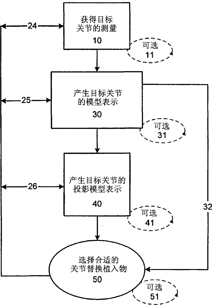
Figure 1A is that to be used to assess to be unaltered or unaltered basically at the articular surface of accepting still to exist before the selected implant wherein according to the block chart of the method in the joint of reparation of the present invention.Figure 1B is that be used to assess need be according to the block chart of the method in the joint of reparation of the present invention, and wherein before design was suitable for realizing the implant of said reparation, existing articular surface was unaltered or unaltered basically.Fig. 1 C is the block chart that is used for developing implant and uses the method for said implant the patient.
Fig. 2 A is the perspective view that is suitable at the joint implant of the present invention of kneed tibial plateau implantation.Fig. 2 B is the top view of the implant of Fig. 2 A.Fig. 2 C is the sectional view that the implant edge of Fig. 2 B is showed in the line C-C of Fig. 2 B.Fig. 2 D is along the sectional view that is showed in the line D-D of Fig. 2 B.Fig. 2 E is along the sectional view that is showed in the line E-E of Fig. 2 B.Fig. 2 F is the side view of the implant of Fig. 2 A.Fig. 2 G is a sectional view of showing the implant of Fig. 2 A that implants along the plane that is parallel to the sagittal plane.Fig. 2 H is a sectional view of showing the implant of Fig. 2 A that implants along the plane that is parallel to coronal plane.Fig. 2 I is a sectional view of showing the implant of Fig. 2 A that implants along the plane that is parallel to axle.Fig. 2 J shows that extension more approaches the big slightly implant of bone center line (towards the tibial plateau edge) and front and rear.Fig. 2 K is the side view of alternate embodiment of Fig. 2 A joint implant of showing the fixture of Os Draconis form.Fig. 2 L is the bottom view of alternate embodiment of joint implant of showing Fig. 2 A of fixture.Fig. 2 M shows the fixture of cross member form.Fig. 2 N-O shows that lower surface has the implant alternate embodiment of the groove that is used to accept cross bar.Fig. 2 P explains multiple cross bar.Kneed device is implanted in Fig. 2 Q-R explanation.Fig. 2 S (1-9) explains another implant that is suitable for tibial plateau that further has along the tangent plane that cuts on one side.Fig. 2 T (1-8) explains wherein articular surface is revised the alternate embodiment of tibia implant that is used for the smooth or angled surface of implant engagement with generation.
Fig. 3 A and B are the perspective views that is applicable to the joint implant of condyle of femur respectively from lower surface and upper surface viewpoint finding.Fig. 3 C is the side view of the implant of Fig. 3 A.Fig. 3 D is the view of implant lower surface.Fig. 3 E is that view and Fig. 3 F of implant upper surface is the cross section of implant.Fig. 3 G is the axial view that has the femur of the implant that is mounted thereon.Fig. 3 H is the kneed front view that does not have patella, and wherein said implant is installed on condyle of femur.Fig. 3 I is the kneed front view such as the implant that is showed in Fig. 2 that has the implant of Fig. 3 A on the implanted femur condyle and be suitable for tibial plateau.Fig. 3 J-K explanation further has the alternate embodiment that at least one oblique section is used for the joint implant on the condyle of femur.
Fig. 4 A explanation is suitable for the implant of condyle of femur according to prior art.Fig. 4 B-I describes and is suitable for being placed in another implant on the condyle of femur.Fig. 4 B is the simple perspective view from the said implant of upper surface finding.Fig. 4 C is the side view of the implant of Fig. 4 B.Fig. 4 D is the top view of the lower surface of said implant.Fig. 4 E and F are the side perspective view of said implant.Fig. 4 G is the axial view that has the femur of the implant that is mounted thereon.Fig. 4 H is the kneed front view that does not have patella, and wherein said implant is installed on condyle of femur.Fig. 4 I is the kneed front view such as the implant that is showed in Fig. 2 that has the implant of Fig. 4 B on the implanted femur condyle and be suitable for tibial plateau.
Fig. 5 A-S describes another and is suitable for being placed in the implant on the condyle of femur.Fig. 5 A is the top view of lower surface of showing the implant of oblique section.Fig. 5 B is the simple perspective view of the upper surface of said implant.Fig. 5 C is the side perspective view from the said implant of first direction finding.Fig. 5 D is the simple side perspective view from the said implant of second direction finding.Fig. 5 E-F is a side view of showing the implant of abutment load.Fig. 5 G and H explain that wherein implant has the alternate embodiment of siding track.Fig. 5 I explains that wherein implant has fixedly another embodiment of Os Draconis.Fig. 5 J is the axial view that has the femur that is installed on the implant on the condyle of femur.Fig. 5 K is the kneed front view that does not have patella, and wherein said implant is installed on condyle of femur.Fig. 5 L is the kneed front view such as the implant that is showed in Fig. 2 that has the implant of Fig. 5 A on the implanted femur condyle and be suitable for tibial plateau.Fig. 5 M-N describes and implants kneed device.Fig. 5 O describes the alternate embodiment that allows part to excise the device of condyle.Fig. 5 P-S explanation has the alternate embodiment of the implant of one or more oblique sections.
Device that Fig. 6 A-G explanation is as shown in Figure 5 and the diagram representative graph that comprises the cross-section data point of exterior view.
Fig. 7 A-C explanation is suitable for the alternative designs that the part condyle of femur has the device of two structures.
Fig. 8 A-J describes complete patella (Fig. 8 A) and has cut so that the patella (Fig. 8 B) of implant to be installed.Top view and the side view (Fig. 8 C-D) of showing suitable patella implant, and show that the key diagram that is superimposed on the implant on the complete patella is with the position of explanation implant dome with respect to the patella ridge.Fig. 8 E-F explanation is superimposed on the implant on the patella.Fig. 8 G-J explanation is based on the alternative designs of the patella implant of semi-finished product (Fig. 8 G).
Fig. 9 A-C describes has any kneed representative side view that is installed on the device of institute's teaching wherein.Fig. 9 A describes the knee joint with condyle implant and patella implant.Fig. 9 B describes the substituting view of knee joint with condyle implant and patella implant that wherein covers the condyle surface of major part in posterior direction epicondyle implant.Fig. 9 C explains that wherein implant is provided in the knee joint on condyle, patella and the tibial plateau.
Figure 10 A-D describes has any kneed front view that is installed on institute's teaching device wherein.Figure 10 A describes the knee joint with tibia implant.Figure 10 B describes the knee joint with condyle implant.Figure 10 C describes the knee joint with tibia implant and condyle implant.Figure 10 D describes the knee joint with two separate space condyle implants and tibia implant.
The specific embodiment
Describe so that any person of ordinary skill in the field all can make and use the present invention below the oblatio.Various modifications to described embodiment will be easy to obviously the person of ordinary skill in the field, and General Principle defined herein can be applicable to other embodiment and application and is not contrary to from the spirit and scope of the present invention by appended claims defined.Therefore, the present invention also is not intended to be limited to the embodiment that is showed, but consistent with the broad range that accords with principle disclosed herein and characteristic.Understand fully on the disclosed degree of wanting required for the present invention in realization, the description and the diagram of all patents of authorizing of in the application's case, quoting, the open case of patent and patent application case are incorporated herein by reference.
The person of ordinary skill in the field should be appreciated that, the order of the said incident that method as herein described is can be any possible in logic and the said order of incident are carried out.In addition, when the scope of a value is provided, should be appreciated that, all be covered by the present invention in each the insertion value between the upper and lower bound of said scope and any statement and insertion value in said scope.Equally, expect that any optional feature of said variant of the present invention can be independently or propose with any one or more characteristics combination as herein described and advocate.
Unless otherwise indicated, otherwise enforcement of the present invention can use under in the technical field x radial imaging and processing, chromatography x radiography combination, comprise the routine and the digital method of ultrasound wave, computed tomography (CT scan), nuclear magnetic resonance (MRI), optical coherence tomography, single photon emission tomoscan (SPECT) and positron emission computerized tomography (PET) that A sweep, B scanning and C scan.Said technology is fully explained in document and need not to describe in this article.Referring to, X-Ray Structure Determination:A Practical Guide for example, second edition, editor Stout and Jensen, 1989, John Wiley & Sons, publisher; Body CT:A Practical Approach, editor publisher; X-ray Diagnosis:A Physician ' s Approach, editor Lam, 1998Springer-Verlag, publisher; With Dental Radiology:Understanding the X-Ray Image, editor Laetitia Brocklebank 1997, Oxford University Press publisher.Also referring to The Essential Physics of Medical Imaging (2nd Ed.), people such as Jerrold T.Bushberg.
The present invention is provided for repairing the joint, is particularly useful for repairing articular cartilage and is used to promote multiple repair of cartilage material sorting to advance patient's method and composition.Wherein, technology described herein allows customization repair of cartilage material to be fit to particular patient, for example aspect size, cartilage thickness and/or curvature.When the shape of articular cartilage surface (for example size, thickness and/or curvature) anatomically accurately or during the approximate original cartilage that is suitable for not damaging cartilage or be suitable for the patient, the success rate of reparation increases.Can be before implantation repair materials be shaped, and said shaping can be for example based on providing about the electronic imaging of the information of the curvature of any " normally " cartilage around defective or thickness and/or being based on the curvature of the bone under the defective.Therefore, the present invention especially is provided for the minimum invasive method of part joint replacement.Said method only needs minimum bone storage to lose or under some situations, need not the bone storage to lose.In addition, different with prior art, method as herein described will be through in implant and on every side or adjoin between cartilage and/or the subchondral bone and to realize that accurate or proximate anatomy coupling helps to recover the integrity of articular surface.
Advantage of the present invention can include but not limited to the customization of (i) joint repair, thereby concerning the patient who uses said repair procedure, increases effect and comfort level; (ii) eliminate in certain embodiments measuring the surgical needs of the defective of desiring surgical repair; (iii) eliminate surgical needs to moulding material during implant procedure; (iv) provide based on bone or imaging of tissue or based on the method for the curvature of surgical probe technology evaluation repair materials; (v) provide and only have minimum or under some situations, do not have the method in the reparation joint that the bone storage loses; (vi) improve the fitness of operation posterior joint; (vii) improve operation back patient's recovery rate in certain embodiments and (viii) improve such as function after the operation of range of movement.Therefore, method as herein described allows design and use more accurately to be fit to defective (for example implantation site) or articular surface and thereby the joint repair material of the joint repair of improvement is provided.
I. engage and aligned assessment
The defective that method and composition as herein described can cause by cartilage disease (for example osteoarthritis), bone injury, cartilage injury, wound and/or owing to the degeneration that overuses or the age causes in order to treatment.Wherein, the present invention allows health practitioner assessment and treats said defective.The size of interest region, volume and shape can only comprise the zone of the cartilage with defective, but preferably also comprise the part of adjoining of said cartilage defects cartilage on every side.
The person of ordinary skill in the field should be appreciated that size, curvature and/or thickness measure can use any suitable technique to obtain.For example, one dimension, two dimension and/three-dimensional measurement can use suitable mechanical means, laser aid, electromagnetism or optical tracking system, model, be coated on the articular surface sclerosis also " memory surface profile " material and/or under one or more in technical field known imaging technique obtain.But measure Noninvasive and/or operation property (for example using probe or other operation devices) acquisition.The person of ordinary skill in the field should be appreciated that, the thickness of said prosthetic device can change at any set point according to patient's anatomical structure and/or to cartilage injury's the degree of depth.In addition, the expection doctor can the x ray obtains the measurement 10 in target joint and selects a suitable joint replacement implant 50 subsequently through for example obtaining.
Under person skilled should be appreciated that the doctor can be as shown in arrow 32 be directly to from the step of the model representation 30 that produces the target joint and selects suitable joint replacement implant 50 volume steps.In addition, like flow process 24,25, shown in 26, after selecting suitable joint replacement implant 50, the step that obtains the measurement 10 in target joint, the model representation 30 that produces the target joint and generation projection model 40 can continuous or parallel repetition.
Figure 1B is the alternative flow of the step of showing that the doctor carries out in the assessment joint.At first, the doctor obtains the measurement 10 in target joint.The step that obtains to measure can be accomplished through the image of obtaining said joint.This step can repeat 11 to obtain a plurality of images with further improvement joint evaluation process in case of necessity.In case the doctor obtains necessary measurement, information is used to produce the model representation 30 in the target joint of being assessed.This model representation can be the form of topography or image.The model representation in said joint can be one dimension, two dimension or three-dimensional.Necessary or can repeat said method 31 when needing.It can comprise realistic model.Behind the model representation of having assessed said joint 30, the doctor can produce the projection model in the target joint under the rectification condition according to circumstances and represent 40.Necessary or when needing this step can repeat 41.Use the difference between joint orographic condition and the joint projection image, the doctor can design joint implant 52 subsequently, and it is suitable for realizing corrigent joint anatomical structure, when needs realize that the implant of hoping designs, can repeat said design process 53.The doctor also can assess the miscellaneous part that provides such as rail, Os Draconis, antelabium, nail, cross stem or fixture, cross bar etc. and whether strengthen the performance of implant in the target joint.
The person of ordinary skill in the field should be appreciated that, the doctor can be as shown in arrow 38 be directly to the step of the suitable joint replacement implant 52 of design from the step of the model representation 30 that produces the target joint.Be similar to the flow process shown in above; Like flow process 42,43, shown in 44; After implant 52 was replaced in the suitable joint of design, the step that obtains the measurement 10 in target joint, the model representation 30 that produces the target joint and generation projection model 40 can continuous or parallel repetition.
Fig. 1 C is the flow chart of the method for the explanation implant of selecting to be used for the patient.At first, use above-mentioned technology or suitable and when embodiment of the present invention in affiliated technical field known technology, measure the size of ill cartilage or cartilage lost regions.This step can repeat repeatedly in case of necessity.In case measured the size of said cartilage defects, can measure the thickness 110 that adjoins cartilage according to circumstances.This process also can repeat 111 in case of necessity.After the measurement cartilage is lost or measured the thickness that adjoins cartilage, measure the curvature 120 of articular surface subsequently.Perhaps, can measure subchondral bone.Should be appreciated that measurement can be carried out at the surface in the joint of repairing or the optimal design that matching surface is beneficial to produce implant surface.
In case measured the surface, user can be selected the best-fit implant 130 that contains in the implant storehouse or produce a patient-specific implant 132.Necessary or can repeat these steps to obtain being used for patient's best-fit implant 131,133 when needing.The person of ordinary skill in the field should be appreciated that, can test to select or the MRI that is contained in the patient of design implant or the information in the x ray realize good being fit to of articular surface with the patient with the surface of guaranteeing said device.Test can be accomplished through for example the implant image being superimposed on the image in patient joint.In case confirm to select or designed suitable implant, then can for example prepare implantation site 140 through remove cartilage or bone from said articular surface, perhaps can implant be inserted joint 150.
Joint implant selected or design realizes on the anatomy or has being fit to of surface now with the joint on the approximate anatomy that oblatio duplicates the matching surface of the relative articular surface of natural joint anatomical structure simultaneously.Under this situation, can assess the gained articular surface of existing articular surface and hope.This technology is particularly useful for not being fixed to the implant in the bone.
The person of ordinary skill in the field should be appreciated that, the personnel of doctor or other embodiment of the present invention can obtain the measurement 10 in target joint and design 52 or select 50 suitable joints replacement implants subsequently.
II. repair materials
Find that multiple material can be used for embodiment of the present invention, it includes but not limited to plastics, metal, crystallization free metal, pottery, biomaterial (for example collagen protein or other cell epimatrix materials), hydroxyapatite, cell (for example stem cell, chondrocyte or analog) or its combination.Based on the information that obtains (for example measuring), can form or select repair materials about defective and articular surface and/or subchondral bone.In addition, use one or more these technology as herein described, have the cartilage replacement of the curvature that is suitable for specific cartilage defects or profile that regrown material will meet articular surface with shape and will mate the thickness of cartilage on every side.Said repair materials can comprise the combination of any material, and generally comprises at least a non-flexible material, for example is not easy to the material of bending or change.
A. metal and polymerization repair materials
Current, common applied metal of articular repair system and/or polymeric material, it comprises the prosthese that for example is fixed in the substrate bone (for example under the situation of knee prostheses, being femur).Referring to No. the 6th, 203,576, the United States Patent (USP) of for example authorizing people such as Afriat March 20 calendar year 2001 and No. the 6th, 322,588, United States Patent (USP) authorizing people such as Ogel November 27 calendar year 2001 and the list of references of wherein quoting.Multiple metal is applicable to embodiment of the present invention and can be based on any Standard Selection.For example, material selects to want based on giving the elasticity of degree of rigidity.Suitably the limiting examples of metal comprises silver, gold, platinum, palladium, iridium, copper, stannum, lead, antimony, bismuth, zinc, titanium, cobalt, rustless steel, nickel, ferroalloy, cobalt alloy and Nitinol (Ni-Ti alloy) such as Elgiloy (cobalt-chromium-nickel alloy) and MP35N (nickel-cobalt-chromium-molybdenum alloy), aluminum, manganese, ferrum, tantalum, (available from LiquidMetal Technologies, crystallization free metal www.liquidmetal.com), other can slowly form polyvalent metal ion for example with inhibition and patient's body fluid or organize the metal and the combination thereof of the implantation substrate calcification that contacts such as stream billon (Liquidmetal alloy).
Suitable synthetic polymer includes but not limited to polyamide (for example nylon), polyester, polystyrene, polyacrylate, ethene polymers (for example polyethylene, politef, polypropylene and polrvinyl chloride), Merlon, polyurethanes, polydimethylsiloxane, cellulose acetate, polymethyl methacrylate, polyether-ether-ketone, ethylene vinyl acetate, polysulfones, celluloid, similar copolymer and its mixture.Also can use the bio-absorbable synthetic polymer; Such as glucosan, hetastarch, gelatine derivative, polyvinylpyrrolidone, polyvinyl alcohol, gather [N-(2-hydroxypropyl) methacrylic acid amide], gather (hydroxy acid), gather (6-caprolactone), polylactic acid, polyglycolic acid, gather (dimethyl ethanol acid), gather (butyric ester), and also can use similar copolymer.
Other materials also is suitable, for example is called the polyketone of polyether-ether-ketone (PEEK).It comprises material PEEK 450G, and it is can be available from Victrex of Lancashire, and Great Britain is used for the not filling PEEK that medical science is implanted through approval.(Victrex be positioned at www.matweb.com or referring to Boedeker www.boedeker.com).Other sources of this material comprise and are positioned at Panoli, the Gharda of India (www.ghardapolymers.com).
It should be noted that selected material also can be filling.For example, other PEEK of other levels also capable of using with contain, fill or 30% carbon is filled such as 30% glass, as long as said material is used for implantable device by FDA or the approval of other management entities.Glass is filled that PEEK reduces expansion rate and is increased the bending modulus of PEEK with respect to filling part not.Known products therefrom is ideal because of its improved strength, rigidity or stability.Known carbon is filled PEEK and is strengthened compressive strength and the rigidity of PEEK and reduce its expansion rate.Carbon is filled PEEK mar proof and load capacity is provided.
Should be appreciated that, can use other resisting fatigue, have good memory, flexibility and/or deflection, have suitable similar bio-compatible thermoplasticity or thermoplastic condensed polymer's material of utmost point agent of low hygroscopicity and good wear-resistant and/or abrasion and be not contrary to from scope of the present invention.Said implant also can comprise PEKK (PEKK).
Spendable other materials comprises polyether-ketone (PEK), polyetherketoneetherketoneketone (PEKEKK) and polyether ether ketone ketone (PEEKK), and is generally polyaryl ether ether ketone.Further can use other polyketone and other thermoplastics.
Can be with reference to the suitable polymer that can be used for said implant of following document, its all incorporate this paper by reference into.Said document comprises: the open case WO 02/02158A1 of PCT, and the date is that on January 10th, 2002 and title are Bio-Compatible Polymeric Materials; The open case WO 02/00275A1 of PCT, the date is that on January 3rd, 2002 and title are Bio-Compatible Polymeric Materials; With the open case WO02/00270A1 of PCT, the date is that on January 3rd, 2002 and title are Bio-Compatible Polymeric Materials.
Said polymer can be through comprising the conventional polymer processing method any of several different methods prepare.Method for optimizing comprises the injection moulding that for example is suitable for producing the polymer assemblies with important structure characteristic and such as the quick forming method of reaction injection molding and cubic light moulding.Can be through physics abrasion or chemical alteration with the substrate veining or make its porous to help the bond coating.Such as extrude, injection, compression molding and/or Machining Technology be also for being fit to.Usually, select said polymer and be suitable for carrying and disperseing the physical load between the articular surface because of its physics and chemical characteristic.
More than one metal and/or polymer can make up use each other.For example, one or more containing metal substrates can be coated with in one or more zones and contain polymeric substrates with polymer or one or more and can be coated with the metal with one or more in one or more zones.
Said system or prosthese can be porous or the porous coating.Said porous surface assembly can be processed by the various materials that comprise metal, pottery and polymer.These surface components then can be fixed in numerous structural core that form by various metals through the whole bag of tricks.Suitable porous coating includes but not limited to metal, pottery, polymeric material (the neutral elastomer of biological example is such as silicone rubber, polyethylene terephthalate and/or its combination) or its combination.Authorized No. the 3rd, 808,606, the United States Patent (USP) of Tronzo on May 7th, the 3rd, 605, No. 123 1 and authorized No. the 3rd, 843,975, the United States Patent (USP) of Tronzo on October 29th, 1974 referring to the United States Patent (USP) of for example authorizing Hahn on JIUYUE 20th, 1971; The United States Patent (USP) of authorizing Smith on April 18th, 1967 was authorized No. the 3rd, 987,499, the United States Patent (USP) of Scharbach on October 26th, the 3rd, 314, No. 420 1; With German Offenlegungsschrift 2,306,552.Can exist an above coating and said layer can have identical or different porosity.Referring to No. the 3rd, 938,198, the United States Patent (USP) of for example authorizing people such as Kahn on February 17th, 1976.Said coating can be through applying the coating that with formation have the inherent network in an interconnective hole around a core and heating until solidifying with the powdered polymer ring.The flexibility in said hole (for example through the length in said hole and the measurement of diameter) is being important aspect the possible success rate of the use of the said coating of assessment on prosthetic appliance.Also referring to No. the 4th, 213,816, the United States Patent (USP) of authorizing Morris on July 22nd, 1980.Said hole coating can be used with the whole form that stands to make powder be incorporated into the pyritous object of substrate by powder.Select suitable polymers and/or powder coating can consider that teaching and list of references that this paper quotes confirm, for example based on its melt index.B. bioprosthetic material
Repair materials also separately inclusive NAND biomaterial combination comprise one or more biomaterials.For example, can design or be shaped any stock and can be applicable to said substrate such as the replacement of suitable cartilage or the regrown material of fetus chondrocyte.Said subsequently cell can be incorporated into said substrate grown until the thickness (and/or curvature) that reaches around the cartilage of cartilage defects.Exsomatize with culture in vivo on the various substrates condition of auxocyte (for example chondrocyte) be described in nineteen ninety-five for example December the 5th, 478, No. 739,1998 Decembers of United States Patent (USP) of authorizing people such as Slivka on the 26th and authorized people's such as Naughton 5 on the 1st; 842; 477, December on April 2,6,283,980 and 2002 of authorizing people such as Vibe-Hansen on the 4th is authorized people's such as Salzmann 6 calendar year 2001; 365,405.The limiting examples of suitable substrate comprises plastics, organization bracket, bone substitution material (for example hydroxyapatite, bioabsorbable material) or any other be suitable for the growing material of cartilage replacement or regrown material above that.
Biopolymer can be natural generation or through the fermentation and similar fashion in vitro produce.Suitable biopolymer includes but not limited to collagen protein, elastin laminin, silk, keratin, gelatin, polyamino acid, catgut suture line, polysaccharide (for example cellulose and starch) and its mixture.Biopolymer can be biological absorbable.
The biomaterial that is used for methods described herein can be autograft (from identical patient), allograft's (from another individuality of same species) and/or xenograft (from another species).Also referring on March 21st, 2002 people such as open Alexander the open case WO 02/22014 of international monopoly and August in 1997 disclosed Lee on the 7th WO97/27885.In certain embodiments, preferred autologous material, because its risk to the immunologic complication of main body of bearing reduces, said immunologic complication comprises absorption again, the inflammation of said material and/or makes the cicatrix of organizing around the implantation site.
In one embodiment of the invention, be used for a probe from donor site collection organization and prepare acceptor site.Said donor site can be arranged in xenograft, allograft or autograft.Said probe is used to realize the good anatomy coupling between donor tissue sample and the acceptor site.The said probe of special design is to realize the seamless or near seamless coupling between donor tissue sample and the acceptor site.Said probe can for example be cylindrical.The end of said probe is generally the sharp tissue penetration that is beneficial to.In addition, the end of said probe is generally hollow to accept tissue.Said probe can have an edge from its terminal qualification distance, and from terminal 1 centimetre distance, and said edge can be in order to the limited depth of the tissue penetration realizing being used to collect.Can be externally or can be in the inside of the hollow parts of said probe in said edge.For example, the plastic surgery operations doctor can take said probe and with physical pressure it is advanced into cartilage, subchondral bone and in such as the situation in kneed joint below bone marrow.Said doctor can advance said probe to arrive cartilage surface until said outside or internal edge.In this regard, thus said edge can prevent further tissue penetration realizes constant and reproducible tissue penetration.The end of said probe can comprise one or more sword, saws spline structure or organize cutting mechanism.For example, the end of said probe can comprise the iris model machine structure of being made up of some little swords.Said sword can use manually, thereby electromotor or electrical mechanisms drive to cut and wear tissue and with tissue samples and substrate separate tissue.Usually, it will repeat in donor and receptor.Under the situation of iris shape sword mechanism, to close said iris shape mechanism tissue samples is separated with donor site thereby can drive indivedual swords.
In another embodiment of the present invention, can be at laser aid of probe end internal integration or radio-frequency unit.Said laser aid or radio-frequency unit can be worn tissue and with tissue samples and substrate separate tissue in order to cut.
In one embodiment of the invention, can in donor and receptor, use identical probe.In another embodiment, can use the probe of analogous shape slightly different on the physical size.For example, it is slightly little to be used for the comparable probe that is used for donor of the probe of receptor, thereby realizes closely being fit between tissue samples or tissue grafts and the acceptor site.It is short slightly to be used for the also comparable probe that is used for donor of the probe of receptor, thus any tissue of losing during being corrected in the substrate separate tissue from donor material or cutting and organizing sample.
Can be with any bioprosthetic material sterilization with inactivation such as antibacterial, virus, yeast, mycete, mycoplasma and parasitic biological pollutant.Sterilization can use any suitable technique to carry out, and for example radiation is such as gamma-radiation.
Any biomaterial as herein described can use automaton to collect.Said automaton can be used the information from the electron image that is used for tissue collecting.
In certain embodiments, the cartilage alternate material has the particular organisms chemical composition.For example, the biochemistry that centers on the cartilage of defective is formed and can perhaps be assessed through imaging technique through obtaining tissue samples and chemical analysis.For example, the WO 02/22014 of Alexander describe gadolinium is used for articular cartilage imaging to monitor endochondral mucopolysaccharide content.Cartilage replacement or regrown material can be processed in some way or cultivate subsequently to realize being similar to the biochemistry composition of forming around the cartilage of implantation site.Be used to realize the condition of culture formed of the biochemistry of wanting can comprise and for example change concentration.The biochemistry of cartilage replacement or regrown material is formed and can for example be influenced through concentration and the open-assembly time of controlling some nutrition and somatomedin.
III. device design
A. cartilage model
Use the information of cartilage thickness and curvature, can produce the physical model of articular cartilage and substrate bone surface.On behalf of the limited area of intraarticular or its, this physical model can contain whole joints.This model also can be considered meniscal existence or not exist and the existence of some or all of cartilages or do not exist.For example, in knee joint, said physical model can only be contained middle part or sidepiece condyle of femur, condyle of femur and cutout regions, middle part tibial plateau, sidepiece tibial plateau, whole tibial plateau, middle part patella, sidepiece patella, whole patella or whole joint.The position in the ill district of cartilage can for example use 3D coordinate system or 3D Euclidean distance to confirm, and is of WO 02/22014.
Can measure the size of desiring repair-deficiency in this way.The method considers that for example about 80% patient has healthy side assemblies.Obviously, some but non-whole defective can comprise and be less than whole cartilages.Therefore, in one embodiment of the invention, measure normally or the only thickness of slight ill cartilage be centered around around one or more cartilage defects.This thickness can obtain at a single point, perhaps preferably obtains at a plurality of points, and 2 points for example, the 4-6 point, the 7-10 point is more than 10 or in whole total lengths of residue cartilages.In addition, in case measured the size of defective, can select suitable therapy (for example articular repair system) thereby the surrounding tissue of reservation health as far as possible.
In other embodiments, the curvature that can measure articular surface is with design and/or shaping repair materials.In addition, the curvature of thickness and articular surface that can measure the residue cartilage is to design and/or the shaping repair materials.Perhaps, can measure the curvature of subchondral bone and gained measured in order to select or shaping cartilage alternate material.For example, the profile of subchondral bone can be in order to produce virtual cartilage surface again: the edge that can differentiate ill cartilage zone.Can measure subchondral bone profile in the affected areas.Can make the copy on subchondral bone surface connect the regional edge of ill cartilage by this through the subchondral bone copying surface is produced fictitious outline in cartilage surface subsequently.When forming device, said profile is shaped with the excision that cooperates or calculate some or all of cartilages with existing cartilage.
Fig. 2 A shows the simple perspective top view of the joint implant of the present invention 200 be suitable for being implanted in kneed tibial plateau.Shown in Fig. 2 A, said implant for example can be used and produce like above contact Figure 1A and the described two surfaces assessment of B.
The upper surface 202 of implant 200 can be through any shaping of several different methods.For example, upper surface 202 can be through making existing cartilage and/or the bone surface outstanding shaping of said surface from the tibial plateau, optimize the presentation surface that is of said implant in the time of perhaps can being shaped with the reflection condyle of femur it with its engagement condyle of femur of box lunch.Perhaps, can upper surface 202 shapings be cooperated with the lower surface with the implant that is shaped with respect to relative condyle of femur.
Thereby lower surface 204 have a coupling or the tibial plateau in approximate match joint convex surfaces its produce on the anatomy with said tibial plateau or being fit on the approximate anatomy.According to the shape of tibial plateau, lower surface also can the part convexity.Therefore, lower surface 204 is presented on the surface of adaptive tibial plateau in the existing surface.It can form to mate the surface after existing surface or coupling articular surface repeat.
The person of ordinary skill in the field should be appreciated that the convex surfaces of lower surface 204 need not complete convexity.On the contrary, lower surface 204 more possibly be made up of adaptive tibial plateau or convexity and female in the existing surface on surperficial level ground of reinventing.Therefore, said surface is essentially the protruding and depression of variation.
The top view of the joint implant of Fig. 2 B exploded view 2A.Shown in Fig. 2 B, the external shape 208 of implant can be elongated.The elongation form can produce multiple shape, comprises ellipse, intends ellipse, track shape etc.But, should be appreciated that external dimensions generally is irregular, therefore do not form for example oval accurately geometry.The person of ordinary skill in the field should be appreciated that the exact shape of implant can change according to the character of the corrigent Articulatory Defect of desire.Therefore, the ratio of length L and width W can be for example 0.25 to 2.0 and more particularly change between 0.5 to 1.5.Further show like Fig. 2 B, when when on the point of the width of implant, obtaining along the length variations of 200 of implants.For example, shown in Fig. 2 B, LR w L2L3.
Turn to Fig. 2 C-E at present, describe the cross section of the implant that is showed in Fig. 2 B along line C-C, D-D and E-E.Said implant has thickness t 1, t2 and t3 respectively.As illustrated by the cross section, the thickness of said implant changes along its length L and width W.At the actual (real) thickness of the ad-hoc location of implant 200 functions of thickness and the joint matching surface of desiring to duplicate of cartilage and/or the bone of desire replacement.In addition, implant 200 is the cartilage of desire replacement and/or the function of bone at the curve along any position of its length L or width W.
Fig. 2 F is the side view of the implant 200 of Fig. 2 A.In this example, implant 200 is different from the height h2 of implant at second end at the first terminal height hf.In addition, upper limb 208 can have the overall inclined-plane of a downward direction.But as illustrated, the actual inclined-plane of upper limb 208 can be positive bevel along its length variations and under some situations.In addition, lower edge 210 can have the overall inclined-plane of a downward direction.As illustrated, the actual inclined-plane of lower edge 210 can be positive bevel along its length variations and under some situations.The person of ordinary skill in the field should be appreciated that, according to the anatomical structure of individual patient, can produce the implant that hf wherein equates or equate in fact with h2 and is not contrary to from scope of the present invention.
Thereby the lower surface 204 that Fig. 2 G is the displaying that gets the sagittal plane in the body implants implants 200 in the knee joint 1020 is on the tibial plateau 1022 and femur 1024 is in the cross section of the implant 200 on the upper surface 202 of implant 200.Fig. 2 H is the cross section that the implant 200 in the knee joint 1020 is implanted in coronal plane is got in the body displaying.From then on view is obvious, and it is suitable for upper joint surface 224 thereby settle implant 200.The person of ordinary skill in the field should be appreciated that when needing, said articular surface can be middle part or sidepiece facet.
Fig. 2 I shows that along the axial plane of health implant 200 displayings of implanting in the knee joint 1020 are from the view aerial or view that the top viewpoint obtains.Thereby Fig. 2 J be wherein big slightly its of implant extend the middle part more near bone (meaning is promptly towards the edge 1023 of tibial plateau) and forward with the view of the alternate embodiment that extends back.
Fig. 2 K is the cross section according to alternate embodiment implant 200 of the present invention.In this embodiment, lower surface 204 further comprises an arthrodesis thing 212.As illustrated in this embodiment, arthrodesis thing 212 forms a lower surface 204 from implant 200 and extends and stretch into for example convexity, Os Draconis or the vertical member of ossa articularia.The person of ordinary skill in the field should be appreciated that said Os Draconis can be vertical or are positioned at the health plane.
In addition, shown in Fig. 2 L, thereby arthrodesis thing 212 can have cross member 214 has cross or " x " shape from the visible said arthrodesis thing 212 of bottom view outward appearance.The person of ordinary skill in the field should be appreciated that, arthrodesis thing 212 can have multiple other forms and still in the joint, realize providing the same target of implant 200 stability of increase.These forms include but not limited to pin, ball, tusk etc.One or more arthrodesis thing 212 can be provided in addition, in case of necessity.Fig. 2 M and N explanation are from the cross section of the alternate embodiment of side view and being seen pair of assembly implant of front view.
In the alternate embodiment shown in Fig. 2 M, hoping provides one or more cross members 220 so that a small amount of translational motion of said implant with respect to femur or femur implant surface to be provided on lower surface 204.In this case, can form the cross member that is integrated in said implant surface or said parts and adapt to the independent segment of the groove 222 on the lower surface 204 of said implant 200 for one or more.The single passage that said groove can form shown in Fig. 2 N1 maybe can have the more than one passage shown in Fig. 2 N2.In each situation, shown in cross member then adapt to the passage shown in the bar relevant with implant 200.The person of ordinary skill in the field should be appreciated that, can form said cross bar parts 220 and be integrated in implant and be not contrary to from scope of the present invention.
Shown in Fig. 2 Q-R, prepare through forming the passage of accepting the cross bar parts above that on expection tibial plateau surface.Therefore, helping implant firmly to be placed in intraarticular still provides when motion of knee joint around the ability of the motion of axle simultaneously.
Fig. 2 S (1-9) explains an alternate embodiment of implant 200.Like Fig. 2 S explanation, chamfered edge discharges an acute angle.Fig. 2 S (1) explains an implant with single fillet or oblique angle 230.Said fillet is located on the implant in the place ahead, tibia ridge rear portion.Shown in Fig. 2 S (2), two fillets 230,231 are provided and are used for rear ramped surface.In Fig. 2 S (3), provide the 3rd fillet 234 to produce the cutting surface of two rear ramped surface.
Turn to Fig. 2 S (4), the tangent line of cancellation implant stays three rear portion curves at present.Fig. 2 S (5) shows the result of tangent line expansion.When Fig. 2 S (6) explains and does not have the tangent line expansion when selecting bottom curve to the influence of design.The result of curve extension and selection is showed in Fig. 2 S (7).Visible like Fig. 2 S (8-9), the gained angle has softer edge and loses and be less than 0.5 millimeter joint space.The person of ordinary skill in the field should be appreciated that, can add other cutting planes and is not contrary to from scope of the present invention.
The surface modification that Fig. 2 T explains tibial plateau 250 wherein is with an alternate embodiment of the implant 200 of holding implant.Explain like Fig. 2 T (1-2), can change tibial plateau only half the articular surface 251 or all surfaces 252.Explain that like Fig. 2 T (3-4) reaming of back-front surface is smooth 260 or becomes 262 of gradient.Gradient can be plus or minus with respect to front surface.Also can use gradient in the implant of Fig. 2 T, wherein said gradient is positioned at plane or body or angled with respect to health.In addition, can provide bindiny mechanism implant to be fixed in the surface of change.Shown in Fig. 2 T (5-7), Os Draconis 264 can be provided.Said Os Draconis 264 can be positioned at the plane of sagittal for example or coronal plane or not be positioned at a plane (shown in Fig. 2 T (7)).Fig. 2 T (8) explains the implant that covers whole tibial plateau.The upper surface of these implants is designed to meet the projection of shape in the joint of under the described step of contact Fig. 1, measuring, and that lower surface is designed to is smooth or smooth in fact to meet the correction surface in joint.
Turn to Fig. 3 A-I at present, show that is suitable for providing the implant with respect to the articular surface of the implant of Fig. 2 A.This implant is corrected the defective on femur 1024 lower surfaces (condyle of femur that for example cooperates with tibial plateau) and can be used (meaning promptly) on femur 1024 or with another joint repair device combination, to use separately.The technology that the formation of said apparatus surface can use the implant of above contact Fig. 2 to describe realizes.
Fig. 3 A shows a perspective view with implant 300 of crooked matching surface 302 and protruding joint abutment surface 304.In view of be promoting implant and the fixture 306 that provides being connected of bone, said joint abutment surface 304 need not to form with femur anatomy or approximate anatomy on suitable.Under this situation, fixture 306 is shown as the spike dowel with band breach head.Said breach is convenient to the fixation procedure in the bone.But, the spike dowel or the cross stem that can use spike dowel and have other structures that are beneficial to fixation procedure with breach.The spike dowel of said implant and other parts can be the porous coating.Said implant can not used bone cementum or use bone cementum to insert.Said implant can be designed in abutting connection with subchondral bone, and meaning is its profile that can meet subchondral bone basically.Thereby having, it need not remove bone but generation significantly keeps the advantage of bone storage with the spike dowel hole.
Fig. 3 B shows the simple perspective top view of the bone matching surface 304 of the purposes further specify three fixtures 306 that implant are fixed in bone.Each fixture 306 has a stem 310 that has a stature 312 at the top.Shown in Fig. 3 C, it forms pipe or the cylinder that extends from said bone matching surface thereby stem 310 has parallel wall.The part of said stem forms the narrow neck 314 near said 312.The person of ordinary skill in the field should be appreciated that said wall need not to parallel, and can be the coniform to form of inclination.In addition, neck 314 and 312 need not to exist.As stated, can use other to be suitable for fixed structure and be not contrary to from scope of the present invention.
Turn to Fig. 3 D, the view of the tibia matching surface 302 of its explanation implant 300 at present.From then on scheme obviously, thereby said surface is its crooked protruding or protruding basically sunk surface with the cooperation level ground.Fig. 3 E explanation further specifies three upper surfaces 304 of implant 300 of purposes that are used for implant 300 is fixed in the spike dowel 306 of bone.As illustrated, settle said three spike dowels 306 to form a triangle.But the person of ordinary skill in the field should be appreciated that, can use one or more spike dowel, and the location with another of spike dowel 306 can be as showed or wanted fixed other suitable location by any can formation.The cross section of the implant 300 that Fig. 3 F explanation is got along the line F-F that is showed in Fig. 3 E.Thereby common said spike dowel is located said spike dowel perpendicular to condyle of femur on the surface of implant, it can not produce the spike dowel perpendicular to implant surface.
Fig. 3 G explanation has the axial view of the femur 1000 of sidepiece condyle 1002 and middle part condyle 1004.Also show along the intercondylar fossa of sidepiece epicondyle 1008 and middle part epicondyle 1010.The patella surface of also showing femur 1012.The implant of explaining among the key diagram 3A 300 covers a part of sidepiece condyle.Showing also that spike dowel 306 is beneficial to is fixed in condyle with implant 300.
Fig. 3 H explanation is from the being seen knee joint 1020 of front perspective.Implant 300 is implanted on the condyle.Shown in Fig. 3 I, it is connected in the implant 200 that is designed to correct defective in the tibial plateau thereby settle implant 300, such as exhibitor among Fig. 2.
The implant 300 that Fig. 3 J-K explanation is used on condyle, replacing.In this embodiment, provide at least one flat surfaces or oblique section 360 to be engaged in the tangent plane of processing on the condyle surface in the preparation joint.Flat surfaces 360 does not comprise whole abutment surface 304 of said implant 300 usually.
The design of the knee joint 499 that Fig. 4 A typical TKA (" TKA ") is initial.Rear portion tangent plane 498, preceding end grain 497 and terminal tangent plane 496 and oblique section 495 are provided.
Fig. 4 B and another implant 400 of 4C explanation.Shown in Fig. 4 B, thus shaping implant 400 its patella surface coverage sidepiece and middle part condyles of femur along femur 1012.Implant 400 has a sidepiece condyle assembly 410 and one middle part condyle assembly 420 and said sidepiece condyle assembly 410 is connected to the bridge 430 that middle part condyle assembly 420 covers the patella surface of at least a portion femur 1012 simultaneously.Implant 400 can be relative with the implant shown in one or more Fig. 2 according to circumstances in case of necessity.Fig. 4 C is the side view of the implant of Fig. 4 B.Shown in Fig. 4 C, upper surface 402 bendings that make implant 400 are to meet the curvature of condyle of femur.It meets one of existing condyle of femur or two s' actual curvature or meets the curvature that one of condyle of femur behind the joint or two are reinvented in the surface thereby can construct said bending.Can provide one or more spike dowels 430 to help that implant is fixed in bone.The person of ordinary skill in the field should be appreciated that, thereby the upper surface that can construct implant structure contact first condyle has one or more flat surfaces to cooperate the condyle surface through transforming with the surface that cooperates existing condyle to contact second condyle simultaneously.
Fig. 4 D explanation is showed in the top view of the implant 400 of Fig. 4 B.From then on view should be appreciated that, the lower surface 404 of structure implant 400 is to meet the shape of condyle of femur, and for example healthy condyle of femur can be the shape that is shown in the tibia surface in the joint of not infringement.
Fig. 4 E and F explanation are from the perspective view of the being seen implant of lower surface (meaning is the tibial plateau matching surface).
Fig. 4 G explanation has the axial view of the femur 1000 of sidepiece condyle 1002 and middle part condyle 1004.Also show intercondylar fossa 1006 along sidepiece epicondyle 1008.Implant 400 explanations that are illustrated in Fig. 4 B cover the condyle and the patella surface of femur 1012.Showing also that spike dowel 430 is beneficial to is fixed in condyle with implant 400.
Fig. 4 H explanation is from the being seen knee joint 1050 of front perspective.Implant 400 is implanted on two condyles.Shown in Fig. 4 I, it is connected in the implant 200 that is designed to correct defective in the tibial plateau thereby settle implant 400, such as exhibitor among Fig. 2.
The person of ordinary skill in the field should be appreciated that, thereby implant 400 can be constructed implant to fasten the material manufacture of condyle by having memory.Perhaps, thus can be with its shaping its meet said surface and need not and fasten.
Another is suitable for repairing the implant 500 of damaging condyle Fig. 5 A and 5B explanation.Shown in Fig. 5 A, thus structure implant 500 its only cover one of sidepiece or middle part condyle of femur 510.Said implant is different from the implant of Fig. 3, because said implant 500 also covers the patella surface of at least a portion femur 512.
Be similar to the implant of Fig. 4, said implant can be according to circumstances with respect to one or more such as implant shown in Figure 2 or articular surface relatively, and can with the implant combination such as the implant of Fig. 3.Fig. 5 C is the perspective side elevation view of the implant of Fig. 5 A.Shown in Fig. 5 C, upper surface 502 bendings that make implant 500 are with the curvature of the condyle of femur that meets its cooperation and the patella surface of the femur of its covering of part.Can provide one or more spike dowels 530 to help that implant is fixed in bone.In addition, can on the lower surface 502 of the condyle assembly of the tangent plane of processing on the surface that meets the articular surface that implant cooperates that provides according to circumstances, provide one angled surperficial 503.
Fig. 5 D explanation is showed in the perspective top view of the implant 500 of Fig. 5 A.From then on view should be appreciated that, the lower surface 500 of structure implant 504 is to meet the shape of condyle of femur, and for example healthy condyle of femur can be the shape that is shown in the tibia surface in the joint of not infringement.
Fig. 5 E shows that from the top 513 extend to line (plane 17) and extend to 3 views that carry the implant 500 of Support drawing shade of bottom 515 from line (plane 18).The spike dowel 530 that extends from upper surface is described simultaneously.Fig. 5 F explanation has from the upper surface of the implant 500 of the spike dowel 530 of upper surface extension.Fig. 5 F explains that also the cantilever of drawing shade carries the Support, and it extends to the top of implant from line (plane 18).The direction of bearing capacity and each supporting condition is based on physiology and carries collision.Table 1 shows that the physiology of the research of taking from Seth Greenwald carries.
Table 1 physiology carries
$Body weight (BW) is taken as 60 years old elderly men, height 173em, average weight 74kg (163 pounds).
1" Tibial Plateau Surface Stress in TKA:A Factor Influencing Polymer Failure SeriesIII-Posterior Stabilized Designs; " Paul D.Postak, B.Sc., Christine S.Heim, B.Sc., A.Seth Greenwald, D.Phil.; The plastic surgery research laboratory, The Mt.Sinai Medical Center, Cleveland, Ohio.Be and be shown in the 62nd annual AAOS meeting, 1995.
500, three carryings of the implant that use is described in the application's case are found in group 1 (2900N).For duplicating the worst-case of carrying scheme, use 75/25 load distribution (75%=2175N of 2900N).Said load will concentrate on below and the perpendicular 6mm diameter circular district that is located immediately at structure on the area supported.
Turn to the cantilever loading shown in Fig. 5 F, said load distributes with 75/25 load and comes across group 3 or 90.Like above instance, said load will concentrate on the 6mm diameter circular district that is positioned at perpendicular to the center, rear portion of the middle part condyle on the smooth cutting surface of rear portion condyle.
Fig. 5 G and H explanation has the alternate embodiment of the implant 500 of the orbit Design that one or more tracks 521 is provided along the middle part and/or the sidepiece of implant 500.Thereby settle said track 521 its before connecting inclined surface 503 along the middle part 517 and/or sidepiece 519 lateral parts extend.Should be appreciated that, one-sided rail 521 can be provided and not be contrary to from scope of the present invention.
Another embodiment that Fig. 5 I explanation has the implant 500 of keel design.Os Draconis 523 (or the rail in the middle part of being formed at) are provided in the upper surface of implant.In this embodiment, Os Draconis 523 are positioned on the surface of implant but not in the side.Should be appreciated that, Os Draconis can be as directed between two parties, location placed in the middle or eccentric basically.Can provide inclined surface 503 to connect articular surface through transforming.Perhaps, when said articular surface damages or transforms, but tectonic profile 503 is to cooperate the surface of said damage or transformation.
Fig. 5 J explanation has the axial view of the femur 1000 of sidepiece condyle 1002 and middle part condyle 1004.Also show along the intercondylar fossa of sidepiece epicondyle 1008 and middle part epicondyle 1010.The patella surface of femur 1012 also is described.Show that the implant 500 that is illustrated in Fig. 5 A covers the sidepiece condyle and the part patella surface of femur 1012.Showing also that spike dowel 530 is beneficial to is fixed in condyle and patella surface with implant 500.
Fig. 5 K explanation is from the being seen knee joint 1020 of front perspective.Implant 500 is implanted on the sidepiece condyle.Fig. 5 L explanation has the knee joint 1020 of the implant 500 that covers middle part condyle 1004.Like Fig. 5 K and L explanation, the shape that meets the implant 500 on patella surface can present multiple curvature and not be contrary to from scope of the present invention.
Turn to Fig. 5 M and N, it is connected in the implant 200 that is designed to correct defective in the tibial plateau thereby settle implant 500, such as exhibitor among Fig. 2 at present.
In another embodiment of the present invention, implant 500 can have the surface that meets condyle basically but have one corresponding to the tilt upper surface 502 of flat of tangent plane of bone, shown in Fig. 5 O.
Turn to Fig. 5 P-Q at present, show and put being seen implant 500, have 7 differences between its pro-and the back tangent plane from side-looking.
Fig. 5 R-S explanation has the implant 500 of tangent plane 562 behind the contour surface 560 that is matched with articular surface and preceding tangent plane 561 and.Fig. 5 S shows from the being seen identical implant 500 of slightly different angle.Fig. 5 T explanation has the contour surface 560 that is matched with articular surface and another implant 500 of back tangent plane 562, terminal tangent plane 563 and an oblique section 564.Preceding tangent plane is not provided in this embodiment.The implant 500 that being seen Fig. 5 T is had an X-rayed in Fig. 5 U explanation from the side.Said tangent plane is usually less than the desired tangent plane of TKA, and meaning is promptly usually less than 10mm.In case of necessity, the design of the tangent plane of this implant allows the time afterwards that knee joint is carried out revisional operation.
Fig. 6 A-G explanation has the implant of implant surface shape from the figured Fig. 5 in the cross section 610,620 of its generation.Fig. 6 A explains the top view of the implant 500 at surface configuration 600 tops that are positioned at extraction.The groove 514 that this view specification of implant 500 is relevant with the bridge portion of implant 512, its patella surface (perhaps coaster zone) that covers femur is to provide the matching surface near cartilage surface.The person of ordinary skill in the field should be appreciated that, because the difference on the anatomical structure, for the shape of the implant of middle part condyle design needs not to be the mirror image into the implant of sidepiece condyle design.Therefore, for example, groove 514 can not be present in the implant for the middle part condyle of femur and patella surface design.Thereby, can design said implant to comprise that all or part coaster zone is perhaps fully with its eliminating.
Fig. 6 B explains the bottom view of the implant 500 on the surface configuration 601 that is positioned at the another source.Fig. 6 C is a bottom view of showing the implant 500 that extends through the extraction surface configuration 600 that is showed in Fig. 6 A.Fig. 6 D is the close-up illustration of Fig. 6 C that show to cover the condyle wing of the said implant of extracting surface 600.Fig. 6 E explains the top elevational view of the implant 500 on the pictorial representation that is positioned at surface configuration 600.Fig. 6 F is that front view and Fig. 6 G are the bottom rearviews.
Fig. 7 A-C explanation is used for the implant 700 that orthotic joint is similar to above implant 500.But implant 700 is made up of two assemblies.The condyle of first assembly, 710 engagement femurs is according to design engagement middle part or sidepiece condyle.The patella surface of second assembly, 720 engagement femurs.Such as previous embodiment discussion, thereby turn up based on compensation in the surface that can construct implant 700 or the projection of the femur natural shape of the design of interior talipes varus and/or leveling femoral surface is shaped end surface 722 surface of tibial plateau (for example in the face of).The end surface that perhaps can be shaped based on the shape of tibial plateau is to provide through the surface of design with the best fit tibial plateau.Can construct nearest surperficial 724 (for example meshing the surface of condyle of femur) thus it is reflected in it and damages the femoral surface under its transformation condition of conditioned disjunction.The surface can have one or more and forms the for example flattening partially 726 of oblique section equally, recently.In addition, said surface can comprise the mechanism 728 that promotes to attach to femur, such as Os Draconis, tooth, cross stem and analog.The middle part facing of condyle implant partly has a conical surface 730, thereby and the sidepiece facing of patella assembly part also has each assembly of conical surface to other assembly oblatio conical surfaces 730.
Be split up into independently composition surface through the surface with middle part and sidepiece separate space as shown in Figure 7, implant provides the shaped surfaces of improvement and being fit to of subchondral bone.In addition, the forequarter of protection femur avoids causing the stress of bone loss.Equally, the smaller szie of each assembly of implant makes implant can use littler otch to insert the joint.Finally, the damage of patella assembly improves.
The patella 800 that Fig. 8 A-F explanation has implant 810.Implant 810 can have one or more spike dowel, cross stem or other fixed mechanisms in case of necessity.The person of ordinary skill in the field should be appreciated that, other designs can use the technology of this disclosure to reach and be not contrary to from scope of the present invention.Fig. 8 A explains the perspective view of complete patella 800.Fig. 8 B explains that has wherein cut to form the patella 800 that a smooth surface 802 cooperates implant on a surface of patella 800.Fig. 8 C explanation has the patella 800 that is placed in the implant 810 on the said smooth surface 802.Implant 810 has tray type structure 812 and dome 814 that is positioned on the dish 812 of the smooth surface 802 of a said patella of adjacency, thereby said dome original position is settled so that the location of its coupling patella ridge.As illustrated, thus 1 millimeter of the actual edge of the edge offset patella of configurable implant 810 said dishes.The person of ordinary skill in the field should be appreciated that dish 812 can form individual unit with dome 814 or can be formed by a plurality of assemblies.Fig. 8 D is the side view that is placed in the implant 810 on the patella 800.Such as displaying, thereby said dome is placed in its off-center on the implant.The best located of said dome will be confirmed by the position of patella ridge.
Turn to Fig. 8 E-F at present, show that implant 810 is superimposed on the unaltered patella 800 position that meets the patella ridge with the location of explanation implant dome 814.
The alternative designs of Fig. 8 G-J explanation patella implant.Fig. 8 G explanation its incipient stage as half-finished implant 850, it has one and has the flat bottom surface 852 that is used for fixing the spike dowel 854 of patella from its extension.But joint or upper surface 860 have a wholecircle shape dome 856 and machining circular, flat part 858 with coupling bone tangent plane.Articular surface 860 have " medicated cap " or " somberero " outward appearance of shape, have a dome that has the edge.The center of dome 856 also is the center of load-bearing surface.Cutting edge 858 is to meet the needs of particular patient.The implant that semi-finished product of having been showed by Fig. 8 G-I of Fig. 8 J explanation form.Fig. 8 I shows a plurality of possible the line of cuts.
Figure 10 A-D provides has the substituting view that one or more implant the kneed coronal plane of above-mentioned implant wherein.The knee joint that Figure 10 A explanation has a tibia implant 200 that is placed in one.The knee joint that Figure 10 A explanation has a condyle implant 300 that is placed in one.The implant that as stated, can a plurality of this paper teachings be provided at an intraarticular is to recover joint motions property.Figure 10 C explanation wherein has the knee joint of two implants.At first, tibia implant 200 is being provided on the tibial plateau and second implant 300 is being provided on relative condyle.The person of ordinary skill in the field should be appreciated that.Thereby the said implant of said implant oblatio matching surface (as illustrated) or oblatio not each other can be installed.For example, when tibia implant 200 being placed knee joint middle part separate space, and said condyle implant placed the sidepiece separate space.The person of ordinary skill in the field should be appreciated that other combinations.Turn to Figure 10 D at present, tibia implant 200 is provided along two separate space condyle implants 500.As stated, these implants can be connected in kneed identical separate space, but need not so.
Can design the aspect of said arthroplasty system with reflection tibia shape, femur shape and/or patella shape.Tibia shape and femur shape can comprise cartilage, bone or its both.In addition, the shape of implant also can comprise some or all assemblies such as meniscal other articulation structures.Said meniscus is compressible, especially walk or load during.Therefore, can design implant to combine owing to compress the characteristic of the meniscus shape that meniscus occurs during load or the body movement.The lower surface 204 that for example, can design implant 200 comprises the shape of cartilage or bone or both tibial plateaus with coupling.The upper surface 2020 of implant 200 can be the articular surface (especially in not by the meniscus region covered) and the meniscal combination of tibia.Therefore, the external shape of said device can be the reflection of meniscus height.Because compression, it can be for example 20%, 40%, 60% or 80% not compression meniscus height.
In certain embodiments, the profile of the device of reflection meniscus shape can be processed by another preferred compressible material.If the selection compressible material, then it preferably is designed to mate basically said meniscal compressibility and biomechanical characterization.Thus process usually by material or nonmetallic materials for said whole device.
The meniscal height and the shape that are used for the articular surface of any desire reparation can directly be measured when image test.If partly or entirely meniscus damages, then meniscus height and shape can provide the measurement to other articulation structures of the estimation of first quarter moon board size from the measurement in joint or use.
In another embodiment, implant 300,400 or 500 top can the shaping according to femur.Said shape preferably can be derived from femur with respect to the motor pattern of tibial plateau thereby calculate condyle of femur bending on tibia and meniscus, stretching, extension, rotation, translation and femur shape contacts with the tibia femur when sliding variation.
Said motor pattern can use such as the known existing test that maybe will have in the technical field under fluoroscopy, MRI, gait analysis and its combination any and measure.
Said arthroplasty can have two or more assemblies, and one is matched with tibia surface and another is engaged in distal femoral component basically basically.Said two assemblies can have a smooth apparent surface.Perhaps, said apparent surface can be bending.Curvature can reflect tibia shape, femur shape and meniscus shape and the combination thereof that is included in during the joint motions.
But the instance of component system includes but not limited to plastics, polymer, metal, metal alloy, crystallization free metal, biomaterial or its combination.In certain embodiments, the surface towards the repair system of substrate bone can be slick.In other embodiments, can be porous or porous coating towards the surface of the repair system of substrate bone.On the other hand, for having one or more grooves, for example be beneficial to the interior growth of surrounding tissue towards the surface design of the repair system of substrate bone.The outer surface of said device can have the design of step appearance, and it can be beneficial to and change biomechanics stress.Also can be according to circumstances one or more position on said device add flange (for example to prevent that repair system rotation, control from entangling and/or preventing that sedimentation from going into pulp cavity firm).Said flange can be the part of circular cone or cylinder design.Also can be smoothly towards the part or all of repair system of substrate bone, it can help to control the degree of depth of implant and prevent to entangle firm.
The limiting examples of multi-component system comprises the combination of metal, plastics, metal alloy, crystallization free metal and one or more biomaterials.The one or more assembly of articular surface repair system can be made up of biomaterial (for example have such as chondrocyte or stem cell self and perhaps be inoculated in organization bracket, allograft, autograft or its combination such as the cell in bioabsorbable material or the organization bracket) and/or non-biological material (for example polyethylene or such as the evanohm of chrome-cobalt alloy).
Therefore, said repair system can comprise the zone of one or more homogenous materials or combination of materials, and for example, said articular repair system can have first and second assemblies.Usually said first assembly of design to be having size, thickness and the curvature that is similar to the cartilaginous tissue of losing, and second assembly is typically designed to and has the curvature that is similar to subchondral bone.In addition, first assembly can have the biomechanics characteristic of the articular cartilage of being similar to, and includes but not limited to similar elasticity and to the resistance of axial load and shearing force.Said first and second assemblies can be made up of two kinds of different metallic or metal alloy.The one or more assembly of said system (for example second portion) can or include but not limited to that the non-biological material of hydroxyapatite, tantalum, evanohm, chrome-cobalt alloy or other technologies alloy forms by the biomaterial that includes but not limited to bone.
The one or more zone of articular surface repair system (the for example external margin of first and/or second portion) can be bioresorbable, for example with the interface between the normal cartilage that allows articular surface repair system and patient along with the time fills with transparent or fibrous cartilage.Similarly, one or more zone (the for example external margin of the first of articular surface repair system and/or second portion) can be porous.Vesicularity can be in whole porous zone linearity or non-linear change, and wherein vesicularity can reduce towards the center of said articular surface repair system usually.Said hole can be designed for the inside growth of chondrocyte, cartilage matrix and connective tissue, thereby realizes articular surface repair system and the smooth interface between the cartilage on every side.
Said repair system (for example second assembly in the multi-component system) can use the bone that is connected in the patient such as methyl methacrylate, injectable hydroxyapatite or apatite calcium material and analog.
In certain embodiments, the one or more part of said articular surface repair system can be flexible when implanting or for liquid or deformable and can harden subsequently.Sclerosis can betide for example (or any period) in 1 second to 2 hours therebetween, preferably in 1 second at least 30 minutes (or any therebetween period), more preferably between 1 second and 10 minutes (or any therebetween period).
The one or more assembly of articular surface repair system can be suitable for accepting injection.For example, the outer surface of said articular surface repair system can have one or more openings therein.Can opening be made as certain size to accept the device of screw, pipeline, pin or other degree of depth of being wanted of can inserting and advance, for example, get into the marrow crack through said articular surface repair system.Such as the injection of methyl methacrylate and injectable hydroxyapatite or apatite calcium material subsequently can be through the said opening pipeline of its insertion (perhaps through) thus introducing the marrow crack makes said articular surface repair system combine said marrow crack.Similarly, can and be advanced into substrate subchondral bone and bone marrow or epiphysis with screw or pin or other fixed mechanisms insertion opening to realize that said articular surface repair system is fixed in bone.Some or all assemblies of said screw or pin can be bioresorbable, and for example, the end portion of the screw in the marrow crack of advancing by leaps and bounds can be bioresorbable.Initial period after operation, said screw can provide the initial fixation of articular surface repair system.Subsequently, bone inwardly grows into the porous applying area along the lower surface of articular cartilage repair system and can replace the original stable device of said articular surface repair system to bone.
Said articular surface repair system can use pin or screw or other attachment means to be fixed on patient's the bone.Said attachment means can be bioresorbable.Said screw or pin or attachment means can insert or propelling towards the articular surface repair system from the non-cartilage cover part of bone or from the non-bearing surface in joint.
At said articular surface repair system with the interface between the normal cartilage can be angled on every side, for example with angular orientation with respect to 90 degree of substrate subchondral bone.Suitable angle can consider that the teaching of this paper confirms, and under some situation, and the angle of non-90 degree can help load along said articular surface repair system and the interface distributions between the normal cartilage on every side.
Said articular surface repair system and on every side the interface between normal cartilage and/or the bone can cover with medical agent or bioactivator, for example stimulate said repair system biointegration to advance the material of normal cartilage and/or cartilage.The surface region at said interface can be irregular, for example is exposed to medical agent or bioactivator to increase the interface.
E. the repair system that is pre-existing in
As described herein, can obtain the repair system of all size, curvature and thickness.Said repair system can and store the system library that can select the suitable system of individual patient subsequently from it to produce by catalog classification.In other words, the repair system that is pre-existing in that assessment defective or articular surface and from said storehouse, selecting has suitable shape and a size in a special object is used for further operation (for example being shaped) and implants.
F. small-sized prosthese
Go into the above, method and composition as herein described can be used for only replacing the part articular surface, for example the ill cartilage or the zone of losing cartilage on the articular surface.In these systems, the articular surface repair system can be designed to only to replace ill or lose zone or its of cartilage extensible cross the said ill or zone of losing cartilage for example 3 or 5 millimeters get into normal contiguous cartilages.In certain embodiments; Said prosthese replacement is less than articular surface (for example any given articular surface of about 70% to 80% (or any value) therebetween; Such as single condyle of femur etc.), preferably be less than about 50% to 70% (or any value) therebetween, more preferably be less than about 30% to 50% (or any value) therebetween; More preferably be less than about 20% to 30% (or any value) therebetween, even more preferably be less than about 20% articular surface.
Said prosthese can comprise a plurality of assemblies, and for example the assembly of an implantable bone (for example metal device) is connected in a shaping is overlapped in the defective of the cartilage on the said bone with covering assembly.Also can comprise for example other assemblies of telophragma, meniscal repairs system and analog.Expect that each assembly replacement is less than all corresponding joint surfaces.But each assembly need not to replace the same section of said articular surface.In other words, said prosthese can have replacement and is less than the bone implant assembly of 30% bone and the cartilage assembly of the cartilage of replacement 60%.Said prosthese can comprise any combination, if each assembly replacement is less than whole articular surfaces.
Thereby can form or select said articular surface repair system its will reach be fit on the approximate anatomy or be matched with around or adjoin cartilage or bone.Usually, thus form and/or select said articular surface repair system be positioned at outer surface its external margin will in alignment with around or adjoin cartilage.
Therefore, can design the supporting part (or greater or less than said supporting part) of said articular repair system with the articular surface in alternative such as the condyle of femur.Said load-bearing surface is meant the contact area between (for example normal footwork) during the normal ADL two relative articular surfaces.At least one or above supporting part can be on condyle of femur for example with tibia on replacement in this way.
In other embodiments, can in the load-bearing district, differentiate the zone that ill cartilage or cartilage are lost and can replace only partial weight-bearing district by the articular surface repair system, especially contain the part that ill cartilage or cartilage are lost the district.
In another embodiment, can design or select articular repair system to replace all articular surfaces, for example condyle basically.
In another embodiment, for example in having the patient that the dispersivity cartilage loses, can design articular repair system with the replacement zone bigger slightly than load-bearing surface.
In some aspects, the defective of desire reparation only is positioned on the articular surface, on the most serious normally ill surface.For example, have in the condyle of femur of middle part that serious cartilage is lost but among disease seriousness is light slightly in tibia the patient, said condyle surface repair system can only be applied to the middle part condyle of femur.Preferably, in method as herein described, design articular surface repair system is to realize being fit on accurate or proximate and the anatomy of adjoining normal cartilage.
In other embodiments, can repair more than an articular surface.Repairing the district is limited to ill cartilage or cartilage usually and loses district or the load-bearing surface ill cartilage of internal ratio or cartilage and lose big slightly zone, district.
In another embodiment, the one or more assembly (for example pointing to the surface of the system of substrate bone or bone marrow) of said articular surface reparation can be porous or porous coating.Having it was suggested that multiple different porous metal coating is used for inwardly growing through osseous tissue strengthens the fixing of metal prostheses.Therefore; For example December in 1974 was authorized the United States Patent (USP) the 3rd of Pilliar on the 24th; The surgical repair device that comprises the composite construction of forming by the solid metal material substrate and the porous coating of the same solid metal material that attaches to and on the said substrate surface of part at least, extend that openly can be used as the bone prosthese for 855, No. 638.Said porous coating is made up of a plurality of little metal material discrete particle that the some place that contacts with each other at it with the hole hole of in said coating, defining a plurality of connections combines.The said particulate size that can in a plurality of monolayers, distribute can be following situation with the interval: promptly the average pore hole size is not more than about 200 microns.In addition, pore size distribution can be at substrates coatings interface to coating surface homogeneous basically.In another embodiment, the articular surface repair system can contain one or more polymeric materials, and it can be loaded with and discharge and comprise that medicine or other can be used for the therapeutic agent of the pharmaceutical treatment that medicine sends.Said polymeric material can for example place in the porous coating.Said polymeric material can be in order to discharge the medicine of bone for example or cartilage-derived growth stimulating drug.This embodiment can with other embodiment combination, wherein part cartilage surface repair system can be bioresorbable.For example, the part of the ground floor of cartilage surface repair system or its ground floor can be bioresorbable.When ground floor absorbed gradually, inside growth and substrate that local release cartilage-derived growth stimulating drug can be beneficial to chondrocyte formed.
In any said method and compositions, said articular surface repair system can be fabricated to size, curvature and the thickness with certain limit in advance.Perhaps, said articular surface repair system can be the individual patient customization.
IV. make
A. finishing
Under some situation; Will before or after forming (for example growing to the thickness of wanting), require the said repair system of finishing, for example when desired cartilage material in uneven thickness (for example when cartilage is replaced or the different piece requirement of regrown material is different thickness).
Said alternate material can be repaired through any suitable technique, and its variation, enzyme or somatomedin and any other that includes but not limited to casting technique, abrasion, laser abrasion or ablation, radio frequency processing, cryoablation, nutrient open-assembly time and concentration is suitable for influencing or changing the method for cartilage thickness.Referring to for example, March in 2000 disclosed Mansmann on the 23rd WO 00/15153; If use enzymic digestion, the enzyme that then some part of replacement of said cartilage or regrown material is exposed to high dose maybe can expose the long period as realizing that in the different piece of said material cartilage replaces or the different-thickness of regrown material and the method for curvature.
Said material can craft and/or automatic dressing, for example uses program that the thickness that will select in advance and/or curvature input wherein and subsequently use the information design apparatus of being imported to realize the device of the shape of being wanted.
Except repairing said repair of cartilage material or for it, implantation site (for example bone surface, any residue cartilage material etc.) also can be through any proper technology finishing to strengthen the integration of said repair materials.
B. sizing
Thereby can form or select said articular surface repair system its will reach be fit on the approximate anatomy or be matched with around or adjoin cartilage, subchondral bone, meniscus and/or its hetero-organization.The shape of said repair system can be based on the analysis (for example MRI, CT, digital chromatography x radiography combination, optical coherence tomography or similar approach) of electron image.If hope the zone that articular repair system is replaced ill cartilage or lost cartilage, then can use the method for the virtual reconstruction that healthy cartilage shape is provided in electron image to realize being fit on the approximate anatomy.
In one embodiment of the invention, can be on the approximate normal cartilage surface of cartilage defects position through rebuilding at whole cartilage defects or the healthy cartilage surface of ill cartilage zone insertion.It can for example be realized through describing healthy cartilage by means of parameter surface (for example B spline surfaces), thereby profile and bridge joint cartilage defects or the ill cartilage zone that the parameter surface meets healthy cartilage laid by it in the control point.The profile of healthy cartilage provided the level and smooth integration of the part in bridge joint cartilage defects or ill cartilage zone around the seriality characteristic on said parameter surface will be utilized.Said parameter surface the part on cartilage defects zone or the ill cartilage zone can in order to the shape of confirming articular repair system or partial shape with coupling around cartilage.
In another embodiment, can use the morphology image processing to rebuild on the approximate normal cartilage surface of the position in cartilage defects or ill cartilage zone.In the first step, can make by hand, semi-automatic and/or automatic cutting techniques (for example manual tracking, region growing, hot line (live wire), be the cutting apart of basis with the model) extracts cartilage from electron image, produce a Dual Images.Defective in the cartilage shows as the form closed procedure that carries out with 2-D 3-D alive capable of using and fills the breach with the structural detail of suitable selection.Said closed procedure is normally defined to expand and then corrodes.Expansive working person is set at 1 with the present picture element in the image output, if the pixel of at least one structural detail is arranged in the zone of source image.Erosion operations person is set at 1 with the present picture element in the image output, if the entire infrastructure element is arranged in the zone of source image.Being filled in of the zone of cartilage defects or ill cartilage can be in order to produce a new surface with cartilage or subchondral bone around the coupling on the cartilage defects zone of the shape of confirming articular repair system or partial shape or the ill cartilage zone.
As stated, thus can form or select said articular repair system it will be realized with on every side or adjoin being fit to or mating on the approximate anatomy of cartilage and/or subchondral bone from the storehouse of the system of all size, curvature and thickness or data base.Said system can process in advance or process and think that individual patient customizes.For control articular repair system before operation with on every side or adjoin being fit to or coupling of cartilage or subchondral bone or meniscus and hetero-organization thereof, can use in its implantation place articular repair system is projected in the software program on the anatomical position.Suitable software can be buied and/or be easy to be revised or design by skilled programmer.
In another embodiment, the articular surface repair system can use one or more 3-D image projectings on implantation site.From 3-D electron image, make by hand such as MRI or CT, semi-automatic and/or automatic cutting techniques extracts cartilage and/or subchondral bone and other anatomical structures.For example use polygon or NURBS surface or other parameters surface representation to produce the 3-D representative graph of cartilage and/or subchondral bone and other anatomical structures and said articular repair system.The description of various parameters surface representation is referring to for example Foley, people such as J.D., Computer Graphics:Principles and Practice in C; Addison-Wesley, second edition, 1995.
The 3-D of cartilage and/or subchondral bone and other anatomical structures and articular repair system representes to incorporate universal coordinate system into.Said subsequently articular repair system can place the implantation site of wanting.The expression of cartilage, subchondral bone, meniscus and other anatomical structures and articular repair system is reproduced as the 3-D image, for example Application Programming Interface (API) OpenGLE (by SGI, the senior 3-D graph function java standard library of Inc. exploitation; Can be used as the part acquisition that the PC video card drives; For example from the www.nvidia.com of NVIVIA video card or the www.3dlabs.com of 3Dlabs product, perhaps the systems soft ware as the Unix work station obtains) or Directe (based on the multimedia API of the Microsoft's Window (Microsoft Windows) of PC system; Available from www.microsoft.com).For example can appear and its interactive mode or non-interactive type moved the real-time or non real-time 3-D image of showing cartilage, subchondral bone, meniscus or other anatomical structures and articular repair system from the angle that changes through rotation.
Thereby can design said software selects to comprise that operation tool and utensil have the articular repair system to cartilage and/or subchondral bone best-fit, for example use some above-mentioned technology automatically.Perhaps, operator selectable is selected the repair of cartilage system that comprises operation tool and utensil and is used suitable tools and technology with its input or pull implantation site into.The operator can move with respect to implantation site on three dimensions and rotary joint repair system and can carry out the visual inspection that is fit between said articular repair system and the said implantation site.Said visual inspection can be by computer assisted.Said program can repeat until realizing satisfied being fit to.Said program can manually be carried out or it can be all or part of by computer assisted by the operator.For example, testable first trial implants of said software selectively actuatable person.The operator can assess fitness.Said software can design and in order to outstanding implant with the misregistration between cartilage or subchondral bone or meniscus or its hetero-organization is regional on every side.Based on this information, said software and operator can select another implant subsequently and test its aligning.The person of ordinary skill in the field will be easy to select, revise and/or produce the suitable computer program that is used for purpose as herein described.
In another embodiment, said implantation site can use the 2-D image visualize of one or more cross sections.Usually will use a series of 2-D cross sectional images.The said 2-D image image test such as CT, MRI, digital chromatography x radiography combination, ultrasound wave or optical coherence tomography capable of using uses the known Method and kit for of person of ordinary skill in the field to produce.Said articular repair system is stackable subsequently on one or more these 2-D images.Said 2-D cross sectional images can be rebuild other planes from sagittal to crown grade.Also can use isotropism data set (the identical or approximately uniform data set of lamellar spacing when for example wherein copline is differentiated) or near-isotropic data set.Can show a plurality of planes simultaneously, for example use screen display separately.The operator also can want on the direction in real time or near real time rolling 2-D image any; The operator may rotate to the tissue volume of picture and carries out this operation simultaneously.Said articular repair system different display planes capable of using (for example sagittal, crown or axial, the plane of the 2-D image of coupling demonstration cartilage, subchondral bone, meniscus or its hetero-organization usually) show with the cross section.Perhaps, can use a three dimensional display to be used for said articular repair system.The 2-D electron image of said articular repair system and 2-D or 3-D represent and can merge in the universal coordinate system.Said subsequently articular repair system can place the implantation site of wanting.The 2-D cross section of the anatomical structure of said series, implantation site and articular repair system is can be real-time or non real-time interactive to be shown (for example operator can roll a series of lamellas) or non-interactive type shows the animation of the lamella that moves said series (for example as).
C. rapid shaping
Rapid shaping is a kind of technology that is used for making from the computer model of an object three-dimensional body.A kind of special printer is used for making prototype from a plurality of two-dimensional layers.Computer software with the representative graph of object be divided into a plurality of different two-dimensional layers and subsequently three-dimensional printer make a material layer for each layer of cutting apart by said software.The lamination of various manufacturings is also formed the prototype of wanting together.More information about rapid shaping technique can be available from people's such as disclosed Russell on the 27th June in 2002 the open case of United States Patent (USP) 2002/0079601A1 number.An advantage using rapid shaping is that its feasible safe handling toxicity or the use of imitating the free form manufacturing technology of chemical compound by force become possibility.These chemical compounds can be incorporated an excipient big envelope safely into, its exposure that reduces a staff.
A powder plug and structure bed are provided.Powder comprise any may be made in powder or with the bonded material of liquid (metal, plastics etc.).Said powder is rolled on the surface of a bed with a dispenser from a feed source.The thickness of layer is by computer control.Printhead deposits to adhesive fluid on the powder bed of wherein hoping the bonded position of said powder subsequently.Advance the powder roll extrusion in the structure bed once more and repeat said process, the three-dimensional localization that forms with equality circuit at the said combination fluid deposition of each layer control simultaneously.For the further discussion of this process, referring to people's such as disclosed Monkhouse on the 18th in JIUYUE in 2003 for example the open case of United States Patent (USP) 2003/017365A1 number.
Said rapid shaping can use as with the bidimensional image of the described acquisition of top I each two-dimensional shapes with each layer of confirming make-up machine.Under this situation, each bidimensional image lamella can meet two-dimentional former matrix.Perhaps, can confirm the 3D shape of defective as stated, and be decomposed into the two-dimentional lamella that is used for quick molding method subsequently.The advantage of using threedimensional model be used for the two-dimentional lamella of rapidform machine can be along same level or fully along different plane when obtaining said bidimensional image.
Rapid shaping can or be used in combination with the foundry engieering combination.For example, the shell or the vessel shell that have the inner dimensions that meets articular repair system use rapid shaping technique to process.Plastics or wax appearance material are generally used for this purpose.The inside that subsequently can be for example be coated with said container with pottery is used for casting subsequently.Use the method, can produce personalized foundry goods.
Rapid shaping can be used for producing articular repair system.Rapid shaping can carry out in the manufacturer.Perhaps, it can be measured the back and in operating room, carry out having carried out surgical operation.
V. surgical technic
Before the patient was undergone surgery, the surgeon for example can use vertical type Ap x ray to carry out knee joint before operation and aim at and measure.When before undergoing surgery, assessing, can differentiate the sidepiece and the patella thorn of any existence.
The kneed inlet of an otch to provide the part desire to repair anaesthetized and done to use standard procedures technology with the patient.Can use a centre entrance can carry out the arthroscopy in joint at first.Thereafter, said centre entrance can combine with operative incision and/or can use the standard lateral inlet.
In case form a suitable otch, check the integrity of the separate space that exposes, comprise the integrity of ligament structure.In case of necessity, removable portion meniscus and maybe can be present in IA any bony spur or hyperosteogeny what AP x differentiated in the ray.For the benefit of remove hyperosteogeny, surgeon's bendable is bent knee to expose other middle part and postmedian hyperosteogenys.In addition, hyperosteogeny can remove from patella in this process.In case of necessity, also can remove middle part and/or sidepiece meniscus at this point, if need, meniscal edge is together in company with removing.
The person of ordinary skill in the field should be appreciated that, but needs assessment middle part cruciate ligament is beneficial to remove the tibia hyperosteogeny.
In case articular surface is ready to, then can want implant be inserted said joint.
A. tibial plateau
For the device 200 with Fig. 2 inserts the middle part separate space, carry out the small incision synosteotomy at patella tendon middle part.In case form said otch, expose the middle part condyle and use a suitable cutter and crooked osteotome middle parts casing pipe of about 1 centimetre of preparation under joint line.In preparation after the parts casing pipe, near the tibial plateau of middle part, settle a Z traction apparatus and remove meniscal front portion and hyperosteogeny along tibia and femur.At this moment, knee joint should be bent to about 60 or bigger.Remove said Z traction apparatus and implant settled least significant end position and the top, tibial plateau edge be fixed against femur.With said implant straight to pusher.Under some situations, use the stress that turns up and to make the insertion of implant become easy.
For the device with Fig. 2 inserts the sidepiece separate space, carry out the small incision synosteotomy at patella tendon sidepiece.In case form said otch, expose the sidepiece condyle and use a suitable cutter and crooked osteotome sidepiece sleeve pipe of about 1 centimetre of preparation under joint line.After preparing said sidepiece sleeve pipe, near the sidepiece tibial plateau, settle a Z traction apparatus and remove meniscal front portion and hyperosteogeny along tibia and femur.Remove said Z traction apparatus and implant settled terminal position and the top, tibial plateau edge be fixed against femur.Keep said implant also to use with 45 angles and rotate said implant against the sidepiece condyle to the thrust at middle part towards the sidepiece of lateral ridge.Under some situations, turn over stress in the application and can make the insertion of implant become easy.
In case implant any implant shown in Figure 2, said device should place in 0 to 2 millimeter of AP border of tibial plateau and be superimposed on the said border.The verification that should carry out range of activity subsequently is minimum with the translation of implant shown in the affirmation.After confirming to settle, use known in the industry technology to carry out the wound closure.
The person of ordinary skill in the field should be appreciated that hope can be carried out other processing on tibial plateau surface according to the structure of said implant 200.For example, one or more passages or groove can be formed on the surface of tibial plateau holding fixed mechanism, such as the Os Draconis that are showed in Fig. 2 K 212 or be showed in the cross member 222,221 of Fig. 2 M-N.
B. condyle repair system
For inserting device shown in Figure 3 300, the condyle according to desire is repaired forms a front middle part or front side portion skin incision, and it starts from from about 1 centimetre of patella leading edge.Said otch scope can be along the patella edge for example 6-10 centimetre usually.The person of ordinary skill in the field should be appreciated that, under some situations, can require longer otch.
Can need cutting to surpass the synovial membrane degree of depth to promote to get into the joint.In addition, also can remove all or part fat pad and make it possible to check relative joint separate space.
Usually, whole middle parts and/or the side margins from femur and tibia removes the hyperosteogeny on hyperosteogeny and the considerable patella of the possibility edge.
Although it is possible, said device does not require the terminal femur of excision before implant device 300 usually.But in case of necessity, can carry out the bone cutting to be provided for the surface of said implant.
At this abutment, make patient's lower limb be in 90 bending positions.Can on condyle, settle the I guide rail that covers terminal femur cartilage subsequently.Said guide rail makes the surgeon can confirm to make that implant 300 can accurately place the orientation in the hole on the condyle.After the correct placement of said guide rail, boring gets into condyle to produce the hole of degree of depth 1-3 millimeter.In case said hole produces, then remove said guide rail and implant 300 is installed on the surface of said condyle.Can use binding agent to be beneficial to said implant 300 and be bonded in condyle.
When desire reparation during, for example use the implant 300 of two Fig. 3 or the implant 400 of Fig. 4, perhaps when subsequently usually with condyle patella 620 during in alignment with the geometric center of implant 600 more than a condyle.For implant 600 is fixed in patella 620, can in said patella surface, produce one or more holes or hole 612 spike dowel 610 to accept implant 600.
VI. cover group
The surgeon can select one or more implants to use at intra-operative in the cover group thereby can one or more above-mentioned implants be combined in together.
For explanation and purpose of description provide the previous description of the embodiment of the invention.Do not hope detailed or limit the invention to disclosed precise forms.Multiple modification and change are conspicuous for affiliated those skilled in the art.Select and describe said embodiment to practice, thereby the others skilled in the art in the technical field can understand the present invention and various embodiment and the various modifications of the special-purpose that is suitable for being contained under making with best interpretations principle of the present invention and its.Hope that scope of the present invention is defined by following claims and equivalent thereof.
Claims (182)
1. implant that is suitable for condyle of femur; It has upper surface and lower surface; Wherein said upper surface is with respect to the said condyle of femur of at least a portion and coaster and said lower surface supporting part and the patella surperficial with respect at least a portion tibia, and at least a portion of one of further wherein said upper surface and lower surface has the 3D shape of the shape of mating one of said femur and tibia surface basically.
2. implant according to claim 1, wherein said upper surface and said lower surface have at least one the 3D shape of shape of articular surface of the lower surface adjacency of the abut of mating said implant basically and said implant.
3. implant according to claim 1, wherein said implant has the thickness of cartilage defects among the patient.
4. implant according to claim 1, wherein said implant have 85% thickness of cartilage defects among the patient.
5. implant according to claim 1, wherein said implant have the thickness between the 65%-100% of cartilage defects among the patient.
6. implant according to claim 1, wherein said implant has the thickness that cartilage defects adds predetermined offset.
7. according to the implant of claim 6, wherein can select said offset to be suitable for a misalignment.
8. implant according to claim 1, wherein said implant is by the material structure that comprises metal or metal alloy.
9. implant according to claim 1, wherein said material comprises one or more bioactive materials.
10. implant according to claim 6 wherein is coated with said implant with bioactive materials.
11. implant according to claim 1, wherein said implant is made up of metal or metal alloy and polymer.
12. implant according to claim 1, it further has at least one that be used to attach to said upper surface and said lower surface and is selected from the structure of the group that is made up of following each thing: ridge, spike dowel, pin, cross stem, cross member, tusk and outthrust.
13. implant according to claim 12, it further has a plurality of attached structures that are used for.
14. implant according to claim 13, the wherein said relative localization that is used for attached structure are to be selected from the group that is made up of following each situation: symmetrical, asymmetric, embark on journey, circular, triangle and random alignment.
15. implant according to claim 1, wherein said implant cover the patella surface of a part of said femur.
16. implant according to claim 1; Each of wherein said upper surface and lower surface has the inclined-plane with respect to the longitudinal axis through the said implant of at least a portion, and further wherein with respect to said lower surface the inclined-plane of said upper surface on inclined-plane be to be selected from the group that forms by following each situation: positive and negative and zero.
17. implant according to claim 1, the shape of one of approximate first and second articular surfaces of wherein said implant.
18. implant according to claim 1, the condyle matching surface of wherein said implant have at least one flat surfaces that is used to cooperate the condyle surface.
19. implant according to claim 18 is wherein prepared the condyle of femur surface and is used to accept said implant.
20. implant according to claim 19 is wherein prepared said condyle surface through forming at least one flat surfaces that cooperates at least one flat surfaces of said implant.
21. implant according to claim 1 is wherein prepared the condyle of femur surface through removing bone.
22. implant according to claim 21 is wherein prepared said condyle surface through removing cartilage.
23. implant according to claim 21 is wherein prepared said condyle surface through forming at least one flat surfaces that cooperates at least one flat surfaces of said implant.
24. implant according to claim 1, wherein said implant are to be selected from the implant storehouse.
25. implant according to claim 1, wherein said implant is via 10 centimetres or littler incision surgery implantation.
26. implant according to claim 1, wherein said implant is via 6 centimetres or littler incision surgery implantation.
27. implant according to claim 1, wherein the range of movement with said joint returns between the 80-99.9% of normal arthrokinematics.
28. implant according to claim 1, wherein the range of movement with said joint returns between the 90-99.9% of normal arthrokinematics.
29. implant according to claim 1, wherein the range of movement with said joint returns between the 95-99.9% of normal arthrokinematics.
30. implant according to claim 1, wherein the range of movement with said joint returns between the 98-99.9% of normal arthrokinematics.
31. implant according to claim 1 wherein forms said implant with at least a portion with respect to second condyle on the said femur.
The rail that 32. implant according to claim 1, wherein said implant further comprise from the middle part or the upper surface of one of lateral sides extends.
33. implant according to claim 1, wherein said implant further comprise the Os Draconis that extend from said upper surface, wherein said Os Draconis are in from said middle part and the recessed position of lateral sides.
34. implant according to claim 1, wherein a part of said upper surface is positioned at a plane.
35. implant according to claim 34, the upper surface that wherein is positioned at the said part on a plane is matched with the smooth tangent plane on the said femoral surface.
36. implant according to claim 1, the anatomical structure of wherein said implant imitation healthy joint.
37. implant according to claim 1, the anatomical structure in the preceding joint of wherein said implant imitation operation.
38. implant according to claim 1, the target anatomical structure in wherein said implant imitation joint.
39. implant according to claim 1, wherein said implant are in intra-operative places the joint space and do not carry out ligament balance.
40. implant according to claim 1, the system that wherein said implant cooperates implant to form by condyle implant and patella.
41. implant according to claim 1, wherein said implant is suitable for solving at least a in valgus deformity, interior talipes varus and the leveling at the thickness of a position.
42. implant according to claim 1, it further has bevelled edge.
43. implant according to claim 1, it further has oblique section.
44. implant according to claim 1, it further has fillet.
45. according to the described implant of claim 44, wherein said fillet is in the outside at the rear portion of tibia ridge.
46. one kind is used to repair knee joint cover group; It comprises one or more implants that is selected from following each thing: the condyle implant with upper surface and lower surface; Wherein said upper surface is with respect at least a portion condyle of femur and coaster and said lower surface supporting part and the patella surperficial with respect at least a portion tibia, and in further wherein said upper surface or the said lower surface at least one has the 3D shape of the shape of mating one of said femur and tibia surface basically; Condyle implant with upper surface and lower surface; Wherein said upper surface is with respect at least a portion condyle of femur and the said lower surface supporting part surperficial with respect at least a portion tibia, and in further wherein said upper surface and the said lower surface at least one has the 3D shape of the shape of mating one of said femur and tibia surface basically; Patella implant with first surface with second surface of engagement coaster of the femur matching surface that meshes patella; And have the implant that upper surface and lower surface are suitable for tibial plateau, wherein said upper surface has the 3D shape of the shape of mating one of said femur and tibia surface basically with respect at least one of at least a portion femur and said lower surface surperficial and further wherein said upper surface and said lower surface with respect at least a portion tibia.
47. one kind is used for kneed prosthetic appliance, it comprises: have upper surface and lower surface and the top of bent side edge extension and the condyle of femur assembly of bottom are arranged therebetween; Trochlear fovea bowl assembly with along the top of said device stops before the sulcus terminalis of the bottom of wherein said condyle of femur assembly on said articular surface.
48. according to the described prosthetic appliance of claim 47, wherein said device has the thickness of being confirmed by cartilage defects among the patient.
49. according to the described prosthetic appliance of claim 47, wherein said device has the thickness of cartilage defects about 85% among the patient.
50. according to the described prosthetic appliance of claim 47, wherein said device have cartilage defects among the patient 65% and 100% between thickness.
51. according to the described prosthetic appliance of claim 47, wherein said device has the thickness that cartilage defects among the patient adds predetermined offset.
52. according to the described prosthetic appliance of claim 47, wherein said device is by the material structure that comprises metal or metal alloy.
53. according to the described prosthetic appliance of claim 47, it further has and is used to attach at least one lip-deep structure.
54. according to the described prosthetic appliance of claim 53, wherein said attachment structure is to be selected from ridge, spike dowel, pin, cross member, cross stem, tusk and outthrust.
55. according to the described prosthetic appliance of claim 47, the rail that wherein said implant further comprises from the middle part or the upper surface of one of lateral sides extends.
56. according to the described prosthetic appliance of claim 47, wherein said implant further comprises the Os Draconis that extend from said upper surface, wherein said Os Draconis are in from said middle part and the recessed position of lateral sides.
57. according to the described prosthetic appliance of claim 47, the part of wherein said upper surface is positioned at a plane.
58. according to the described prosthetic appliance of claim 57, the part that wherein is positioned at the said upper surface on a plane is matched with the smooth tangent plane on the said femoral surface.
59. according to the described prosthetic appliance of claim 57, the part that wherein is positioned at the said upper surface on a plane is matched with the smooth tangent plane on the said femoral surface basically.
60. one kind is used for kneed prosthetic appliance, it comprises: have the top that the bent side edge is arranged and the condyle of femur assembly of bottom therebetween; With the trochlear fovea bowl assembly along the top of said device, the bottom of wherein said condyle of femur assembly ends at the knee joint surface at groove and stops.
61. according to the described prosthetic appliance of claim 60, wherein said device has the thickness of being confirmed by cartilage defects among the patient.
62. according to the described prosthetic appliance of claim 60, wherein said device has the thickness of cartilage defects about 85% among the patient.
63. according to the described prosthetic appliance of claim 60, wherein said device have cartilage defects among the patient 65% and 100% between thickness.
64. according to the described prosthetic appliance of claim 60, wherein said device has the thickness that cartilage defects among the patient adds predetermined offset.
65. according to the described prosthetic appliance of claim 60, wherein said device is by the material structure that comprises metal or metal alloy.
66. according to the described prosthetic appliance of claim 60, it further has and is used to attach at least one lip-deep structure.
67. according to the described prosthetic appliance of claim 60, wherein said attachment structure is to be selected from ridge, spike dowel, pin, cross member, cross stem, tusk and outthrust.
68. according to the described prosthetic appliance of claim 60, the rail that wherein said implant further comprises from the middle part or the upper surface of one of lateral sides extends.
69. according to the described prosthetic appliance of claim 60, wherein said implant further comprises the Os Draconis that extend from said upper surface, wherein said Os Draconis are in from said middle part and the recessed position of lateral sides.
70. according to the described prosthetic appliance of claim 60, the part of wherein said upper surface is positioned at a plane.
71. according to the described prosthetic appliance of claim 70, the part that wherein is positioned at the said upper surface on a plane is matched with the smooth tangent plane on the said femoral surface.
72. according to the described prosthetic appliance of claim 70, the part that wherein is positioned at the said upper surface on a plane is matched with the smooth tangent plane on the said femoral surface basically.
73., wherein prepare the condyle of femur surface and be used to accept said implant according to the described implant of claim 60.
74., wherein prepare said condyle of femur surface through forming at least one flat surfaces that cooperates at least one flat surfaces of said implant according to the described implant of claim 73.
75., wherein prepare the condyle of femur surface through removing bone according to the described implant of claim 60.
76., wherein prepare said condyle surface through removing cartilage according to the described implant of claim 75.
77., wherein prepare said condyle surface through forming at least one flat surfaces that cooperates at least one flat surfaces of said implant according to the described implant of claim 75.
78. according to the described implant of claim 60, the anatomical structure of wherein said implant imitation healthy joint.
79. according to the described implant of claim 60, the anatomical structure in the preceding joint of wherein said implant imitation operation.
80. according to the described implant of claim 60, the target anatomical structure in wherein said implant imitation joint.
81. according to the described implant of claim 60, wherein said implant is in intra-operative places the joint space and do not carry out ligament balance.
82. according to the described implant of claim 60, the system that wherein said implant cooperates implant to form by condyle implant and patella.
83. according to the described implant of claim 60, wherein said implant is suitable for solving at least a in valgus deformity, interior talipes varus and the leveling at the thickness of a position.
84. according to the described implant of claim 60, it further has bevelled edge.
85. according to the described implant of claim 60, it further has oblique section.
86. according to the described implant of claim 60, it further has fillet.
87. 6 described implants according to Claim 8, wherein said fillet is in the outside at the rear portion of tibia ridge.
88. implant that is suitable for terminal femur in the knee joint; It has upper surface and lower surface, wherein constructs said upper surface with the femoral surface that is connected in tibia femur composition surface and construct said lower surface to be connected in the tibia surface of said tibia femur composition surface.
89. 8 described implants are wherein carried out bone-cutting on said femoral surface before implantation step according to Claim 8.
90. 8 described implants according to Claim 8, wherein said bone tangent plane is at the rear portion.
91. according to the described implant of claim 90, wherein said implant has the high rear portion percentage elongation along said condyle of femur.
92. according to the described implant of claim 91, wherein said knee joint has high hollow of the knee curvature.
93. 8 described implants according to Claim 8, at least a portion of one of wherein said upper surface or lower surface has the 3D shape of the shape of the articular surface that matees its cooperation basically.
94. the described implant of joint claim 88, the said partial-length of one of wherein said upper surface or lower surface is greater than 2 centimetres.
The rail that 95. 8 described implants according to Claim 8, wherein said implant further comprise from the middle part or the upper surface of one of lateral sides extends.
96. 8 described implants according to Claim 8, wherein said implant further comprise the Os Draconis that extend from said upper surface, wherein said Os Draconis are in from said middle part and the recessed position of lateral sides.
97. 8 described implants according to Claim 8, the part of wherein said upper surface is positioned at a plane.
98. according to the described implant of claim 97, the part that wherein is positioned at the said upper surface on a plane is matched with the smooth tangent plane on the said femoral surface.
99. 8 described implants according to Claim 8, the part that wherein is positioned at the said upper surface on a plane is matched with the smooth tangent plane on the said femoral surface basically.
100. 8 described implants according to Claim 8, it further has attachment structure, and wherein said attachment structure is to be selected from ridge, spike dowel, pin, cross member, cross stem, tusk and outthrust.
101. an implant system, it comprises: distal femoral component, the femoral surface of wherein said distal femoral component replacement patella femur composition surface and tibia femur composition surface; And tibial component, wherein said tibial component is replaced the tibia surface of said tibia femur composition surface.
102. according to the described implant system of claim 101, wherein said distal femoral component has the thickness of being confirmed by cartilage defects among the patient.
103. according to the described implant system of claim 101, wherein said tibial component has the thickness of being confirmed by cartilage defects among the patient.
104. according to the described implant system of claim 101, wherein said distal femoral component has the thickness of cartilage defects about 85% among the patient.
105. according to the described implant system of claim 101, wherein said distal femoral component has the thickness between the cartilage defects 65% and 100% among the patient.
106. according to the described implant system of claim 101, wherein said distal femoral component has the thickness that cartilage defects among the patient adds predetermined offset.
107. according to the described implant system of claim 101, wherein said distal femoral component is by the material structure that comprises metal or metal alloy.
108. according to the described implant system of claim 101, it further has and is used to attach at least one lip-deep structure.
109. according to the described implant system of claim 108, wherein said attachment structure is to be selected from ridge, spike dowel, pin, cross member, cross stem, tusk and outthrust.
110. according to the described implant system of claim 101, the upper surface of wherein said distal femoral component has through design to cooperate the flat portions basically of the bone tangent plane on the condyle of femur.
111. according to the described implant system of claim 101, one of them or an above assembly use the dimensional measurement of x ray to confirm size.
112. according to the described implant system of claim 101, one of them or an above assembly use the standard foundry engieering to make.
113. according to the described implant of claim 101, the rail that wherein said implant further comprises from the middle part or the upper surface of one of lateral sides extends.
114. according to the described implant of claim 101, wherein said implant further comprises the Os Draconis that extend from said upper surface, wherein said Os Draconis are in from said middle part and the recessed position of lateral sides.
115. according to the described implant of claim 101, the part of wherein said upper surface is positioned at a plane.
116. according to the described implant of claim 115, the part that wherein is positioned at the said upper surface on a plane is matched with the smooth tangent plane on the said femoral surface.
117. according to the described implant of claim 101, the part that wherein is positioned at the said upper surface on a plane is matched with the smooth tangent plane on the said femoral surface basically.
118. according to the described implant of claim 101, wherein said tibial component has the dome that is positioned on the dish.
119., go up the central point that the summit of said dome deviates from said dish thereby wherein said dome is positioned at dish according to the described implant of claim 118.
120. according to the described implant of claim 118, the summit of wherein locating said dome is to meet the patella ridge.
121., wherein prepare the condyle of femur surface and be used to accept said implant according to the described implant of claim 101.
122., wherein prepare said condyle of femur surface through forming at least one flat surfaces that cooperates at least one flat surfaces of said implant according to the described implant of claim 121.
123., wherein prepare said condyle of femur surface through removing cartilage according to the described implant of claim 121.
124., wherein prepare said condyle of femur surface through forming at least one flat surfaces that cooperates at least one flat surfaces of said implant according to the described implant of claim 121.
125. according to the described implant of claim 121, the anatomical structure of wherein said implant imitation healthy joint.
126. according to the described implant of claim 101, the anatomical structure in the preceding joint of wherein said implant imitation operation.
127. according to the described implant of claim 101, the target anatomical structure in wherein said implant imitation joint.
128. according to the described implant of claim 101, wherein said implant is in intra-operative places the joint space and do not carry out ligament balance.
129. according to the described implant of claim 101, the system that wherein said implant cooperates implant to form by condyle implant and patella.
130. according to the described implant of claim 101, wherein said implant is suitable for solving at least a in valgus deformity, interior talipes varus and the leveling at the thickness of a position.
131. according to the described implant of claim 101, it further has bevelled edge.
132. according to the described implant of claim 101, it further has oblique section.
133. according to the described implant of claim 101, it further has fillet.
134. according to the described implant of claim 133, wherein said fillet is in the outside at the rear portion of tibia ridge.
135. an implant system, it comprises: distal femoral component, the femoral surface of wherein said distal femoral component replacement patella femur composition surface and tibia femur composition surface; Tibial component, wherein said tibial component are replaced the tibia surface of said tibia femur composition surface; With the patella assembly, it is surperficial with the patella of replacing said patella femur composition surface through design.
136. according to the described implant system of claim 135, wherein said distal femoral component has the thickness of being confirmed by cartilage defects among the patient.
137. according to the described implant system of claim 135, wherein said tibial component has the thickness of being confirmed by cartilage defects among the patient.
138. according to the described implant system of claim 135, wherein said distal femoral component has the thickness of cartilage defects about 85% among the patient.
139. according to the described implant system of claim 135, wherein said distal femoral component has the thickness between the cartilage defects 65% and 100% among the patient.
140. according to the described implant system of claim 135, wherein said distal femoral component has the thickness that cartilage defects among the patient adds predetermined offset.
141. according to the described implant system of claim 135, wherein said distal femoral component is by the material structure that comprises metal or metal alloy.
142. according to the described implant system of claim 135, it further has and is used to attach at least one lip-deep structure.
143. according to the described implant system of claim 142, wherein said attachment structure is to be selected from ridge, spike dowel, pin, cross member, rolling rod, cross stem, tusk and outthrust.
144. according to the described implant system of claim 135, the upper surface of wherein said distal femoral component has through design to cooperate the flat portions basically of the bone tangent plane on the condyle of femur.
145. according to the described implant system of claim 135, one of them or an above assembly use the dimensional measurement of x ray to confirm size.
146. according to the described implant system of claim 135, one of them or an above assembly use the standard foundry engieering to make.
147. according to the described implant system of claim 135, the rail that wherein said implant further comprises from the middle part or the upper surface of one of lateral sides extends.
148. according to the described implant system of claim 135, wherein said implant further comprises the Os Draconis that extend from said upper surface, wherein said Os Draconis are in from said middle part and the recessed position of lateral sides.
149. according to the described implant system of claim 135, the part of wherein said upper surface is positioned at a plane.
150. according to the described implant system of claim 149, the part that wherein is positioned at the said upper surface on a plane is matched with the smooth tangent plane on the said femoral surface.
151. according to the described implant system of claim 135, the part that wherein is positioned at the said upper surface on a plane is matched with the smooth tangent plane on the said femoral surface basically.
152. according to the described implant system of claim 135, wherein said tibial component has the dome that is positioned on the dish.
153., go up the central point that the summit of said dome deviates from said dish thereby wherein said dome is positioned at dish according to the described implant system of claim 152.
154. according to the described implant system of claim 152, the summit of wherein locating said dome is to meet the patella ridge.
155., wherein prepare the condyle of femur surface and be used to accept said implant according to the described implant of claim 135.
156., wherein prepare said condyle of femur surface through forming at least one flat surfaces that cooperates at least one flat surfaces of said implant according to the described implant of claim 155.
157., wherein prepare said condyle of femur surface through removing cartilage according to the described implant of claim 155.
158., wherein prepare said condyle of femur surface through forming at least one flat surfaces that cooperates at least one flat surfaces of said implant according to the described implant of claim 155.
159. according to the described implant of claim 155, the anatomical structure of wherein said implant imitation healthy joint.
160. according to the described implant of claim 135, the anatomical structure in the preceding joint of wherein said implant imitation operation.
161. according to the described implant of claim 135, the target anatomical structure in wherein said implant imitation joint.
162. according to the described implant of claim 135, wherein said implant is in intra-operative places the joint space and do not carry out ligament balance.
163. according to the described implant of claim 135, the system that wherein said implant cooperates implant to form by condyle implant and patella.
164. according to the described implant of claim 135, wherein said implant is suitable for solving at least a in valgus deformity, interior talipes varus and the leveling at the thickness of a position.
165. according to the described implant of claim 135, it further has bevelled edge.
166. according to the described implant of claim 135, it further has oblique section.
167. according to the described implant of claim 135, it further has fillet.
168. according to the described implant of claim 167, wherein said fillet is in the outside at the rear portion of tibia ridge.
169. one kind is used for kneed prosthetic appliance, it comprises: have the top that the bent side edge is arranged and the condyle of femur assembly of bottom therebetween; With the trochlear fovea bowl assembly along the top of said device, the bottom of wherein said condyle of femur assembly stopped before the lip-deep sulcus terminalis of knee joint.
170. one kind is used for kneed prosthetic appliance, it comprises: have the top that the bent side edge is arranged and the condyle of femur assembly of bottom therebetween; With the trochlear fovea bowl assembly along the top of said device, the bottom of wherein said condyle of femur assembly stops near the lip-deep sulcus terminalis of knee joint.
171. a kneed prosthetic appliance, it comprises: have the top that the bent side edge is arranged and the condyle of femur assembly of bottom therebetween; With the trochlear fovea bowl assembly along the top of said device, the bottom of wherein said condyle of femur assembly is crossed behind the lip-deep sulcus terminalis of knee joint and is stopped.
172. an implant system, it comprises: distal femoral component, the femoral surface of wherein said distal femoral component replacement patella femur composition surface and tibia femur composition surface; And tibial component, wherein said tibial component is replaced the tibia surface of said tibia femur composition surface, and at least one of wherein said tibial component and distal femoral component is asymmetric.
173. an implant system, it comprises: distal femoral component, the femoral surface of wherein said distal femoral component replacement patella femur composition surface and tibia femur composition surface; Tibial component, wherein said tibial component are replaced the tibia surface of said tibia femur composition surface; With the patella assembly, to replace the patella surface of said patella femur composition surface, at least one of wherein said tibia, femur and patella assembly is asymmetric through design for it.
174. one kind is used for kneed prosthetic appliance, it comprises: have upper surface and lower surface and the top of curved surface extension and the condyle of femur assembly of bottom are arranged therebetween; With the trochlear fovea bowl assembly along the top of said device, the bottom of wherein said condyle of femur assembly stops behind sulcus terminalis.
175. a tibia implant, its first surface with flat surfaces basically with have the smooth second surface of part, wherein said second surface with part flat surfaces further comprises dome structure.
176. according to the described tibia implant of claim 174, wherein said dome has through the location to meet the summit of patella ridge.
177. according to the described tibia implant of claim 175, thereby wherein said dome is positioned at the center that the summit of the said dome of said second surface deviates from said implant.
178. tibia implant; It has the first surface and the second surface with part flat surfaces of flat surfaces basically, around said dome, forms antelabium thereby wherein said second surface with part flat surfaces further comprises the said flat surfaces of dome structure that is positioned on the said part flat surfaces.
179. according to the described tibia implant of claim 178, wherein the said antelabium around said dome is about symmetry around the said dome.
180. according to the described tibia implant of claim 178, wherein the said antelabium around said dome is about asymmetric around the said dome.
181. according to the described tibia implant of claim 178, wherein said implant forms semi-finished product.
182., wherein revise the edge of said flat surfaces with the patient-specific mode according to the described tibia implant of claim 178.
Applications Claiming Priority (5)
| Application Number | Priority Date | Filing Date | Title |
|---|---|---|---|
| US10/724,010 US7618451B2 (en) | 2001-05-25 | 2003-11-25 | Patient selectable joint arthroplasty devices and surgical tools facilitating increased accuracy, speed and simplicity in performing total and partial joint arthroplasty |
| US10/724,010 | 2003-11-25 | ||
| US10/752,438 US8545569B2 (en) | 2001-05-25 | 2004-01-05 | Patient selectable knee arthroplasty devices |
| US10/752,438 | 2004-01-05 | ||
| CN2004800397472A CN1913844B (en) | 2003-11-25 | 2004-11-24 | Patient Selective Knee Arthroplasty Devices |
Related Parent Applications (1)
| Application Number | Title | Priority Date | Filing Date |
|---|---|---|---|
| CN2004800397472A Division CN1913844B (en) | 2003-11-25 | 2004-11-24 | Patient Selective Knee Arthroplasty Devices |
Publications (2)
| Publication Number | Publication Date |
|---|---|
| CN102805677A true CN102805677A (en) | 2012-12-05 |
| CN102805677B CN102805677B (en) | 2015-11-25 |
Family
ID=34636735
Family Applications (2)
| Application Number | Title | Priority Date | Filing Date |
|---|---|---|---|
| CN201510757327.8A Expired - Lifetime CN105287049B (en) | 2003-11-25 | 2004-11-24 | Patient selectable knee joint arthroplasty devices |
| CN201210270152.4A Expired - Lifetime CN102805677B (en) | 2003-11-25 | 2004-11-24 | Patient selectable knee joint arthroplasty devices |
Family Applications Before (1)
| Application Number | Title | Priority Date | Filing Date |
|---|---|---|---|
| CN201510757327.8A Expired - Lifetime CN105287049B (en) | 2003-11-25 | 2004-11-24 | Patient selectable knee joint arthroplasty devices |
Country Status (7)
| Country | Link |
|---|---|
| US (5) | US8545569B2 (en) |
| EP (2) | EP1686931B1 (en) |
| JP (3) | JP5074036B2 (en) |
| CN (2) | CN105287049B (en) |
| AU (1) | AU2004293104A1 (en) |
| CA (2) | CA2804883C (en) |
| WO (1) | WO2005051240A1 (en) |
Cited By (43)
| Publication number | Priority date | Publication date | Assignee | Title |
|---|---|---|---|---|
| CN103584931A (en) * | 2013-10-23 | 2014-02-19 | 华南理工大学 | Bionic gradient knee femoral prosthesis structure and a production method thereof |
| CN103584930A (en) * | 2013-10-23 | 2014-02-19 | 华南理工大学 | Reverse design and production method of individual total knee prosthetic implant |
| US9345551B2 (en) | 2007-08-17 | 2016-05-24 | Zimmer Inc. | Implant design analysis suite |
| US9585597B2 (en) | 2012-07-24 | 2017-03-07 | Zimmer, Inc. | Patient specific instrumentation with MEMS in surgery |
| US9615840B2 (en) | 2010-10-29 | 2017-04-11 | The Cleveland Clinic Foundation | System and method for association of a guiding aid with a patient tissue |
| US9675461B2 (en) | 2009-02-25 | 2017-06-13 | Zimmer Inc. | Deformable articulating templates |
| US9717508B2 (en) | 2010-10-29 | 2017-08-01 | The Cleveland Clinic Foundation | System of preoperative planning and provision of patient-specific surgical aids |
| US9839434B2 (en) | 2009-10-29 | 2017-12-12 | Zimmer, Inc. | Patient-specific mill guide |
| US9877735B2 (en) | 2010-10-29 | 2018-01-30 | The Cleveland Clinic Foundation | System and method for assisting with attachment of a stock implant to a patient tissue |
| US9924950B2 (en) | 2013-09-25 | 2018-03-27 | Zimmer, Inc. | Patient specific instrumentation (PSI) for orthopedic surgery and systems and methods for using X-rays to produce same |
| US9987148B2 (en) | 2013-06-11 | 2018-06-05 | Orthosoft Inc. | Acetabular cup prosthesis positioning instrument and method |
| US10016241B2 (en) | 2015-03-25 | 2018-07-10 | Orthosoft Inc. | Method and system for assisting implant placement in thin bones such as scapula |
| US10045824B2 (en) | 2013-10-18 | 2018-08-14 | Medicrea International | Methods, systems, and devices for designing and manufacturing a rod to support a vertebral column of a patient |
| US10124124B2 (en) | 2013-06-11 | 2018-11-13 | Zimmer, Inc. | Computer assisted subchondral injection |
| US10130378B2 (en) | 2011-05-11 | 2018-11-20 | The Cleveland Clinic Foundation | Generating patient specific instruments for use as surgical aids |
| US10130478B2 (en) | 2009-02-25 | 2018-11-20 | Zimmer, Inc. | Ethnic-specific orthopaedic implants and custom cutting jigs |
| US10217530B2 (en) | 2014-06-03 | 2019-02-26 | Zimmer, Inc. | Patient-specific cutting block and method of manufacturing same |
| US10271886B2 (en) | 2012-07-23 | 2019-04-30 | Zimmer, Inc. | Patient-specific instrumentation for implant revision surgery |
| US10271858B2 (en) | 2015-05-28 | 2019-04-30 | Zimmer, Inc. | Patient-specific bone grafting system and method |
| US10292770B2 (en) | 2017-04-21 | 2019-05-21 | Medicrea International | Systems, methods, and devices for developing patient-specific spinal treatments, operations, and procedures |
| US10307174B2 (en) | 2011-05-19 | 2019-06-04 | The Cleveland Clinic Foundation | Apparatus and method for providing a reference indication to a patient tissue |
| US10318655B2 (en) | 2013-09-18 | 2019-06-11 | Medicrea International | Method making it possible to produce the ideal curvature of a rod of vertebral osteosynthesis material designed to support a patient's vertebral column |
| US10325065B2 (en) | 2012-01-24 | 2019-06-18 | Zimmer, Inc. | Method and system for creating patient-specific instrumentation for chondral graft transfer |
| US10327786B2 (en) | 2012-05-24 | 2019-06-25 | Zimmer, Inc. | Patient-specific instrumentation and method for articular joint repair |
| US10350022B2 (en) | 2014-04-30 | 2019-07-16 | Zimmer, Inc. | Acetabular cup impacting using patient-specific instrumentation |
| US10405928B2 (en) | 2015-02-02 | 2019-09-10 | Orthosoft Ulc | Acetabulum rim digitizer device and method |
| US10456211B2 (en) | 2015-11-04 | 2019-10-29 | Medicrea International | Methods and apparatus for spinal reconstructive surgery and measuring spinal length and intervertebral spacing, tension and rotation |
| US10512496B2 (en) | 2010-10-29 | 2019-12-24 | The Cleveland Clinic Foundation | System and method for assisting with arrangement of a stock instrument with respect to a patient tissue |
| US10543100B2 (en) | 2012-03-28 | 2020-01-28 | Zimmer, Inc. | Glenoid implant surgery using patient specific instrumentation |
| US10582969B2 (en) | 2015-07-08 | 2020-03-10 | Zimmer, Inc. | Patient-specific instrumentation for implant revision surgery |
| US10624764B2 (en) | 2015-11-26 | 2020-04-21 | Orthosoft Ulc | System and method for the registration of an anatomical feature |
| US10667829B2 (en) | 2013-08-21 | 2020-06-02 | Laboratoires Bodycad Inc. | Bone resection guide and method |
| CN111281618A (en) * | 2013-09-27 | 2020-06-16 | 联合创新技术有限责任公司 | Inverted knee prosthesis |
| US10874408B2 (en) | 2015-09-30 | 2020-12-29 | Zimmer, Inc | Patient-specific instrumentation for patellar resurfacing surgery and method |
| US10918422B2 (en) | 2017-12-01 | 2021-02-16 | Medicrea International | Method and apparatus for inhibiting proximal junctional failure |
| US11202674B2 (en) | 2018-04-03 | 2021-12-21 | Convergent Dental, Inc. | Laser system for surgical applications |
| CN114052724A (en) * | 2022-01-13 | 2022-02-18 | 西安交通大学医学院第一附属医院 | Orthopedics traction abnormity detection system based on artificial intelligence |
| US11576725B2 (en) | 2017-12-12 | 2023-02-14 | Orthosoft Ulc | Patient-specific instrumentation for implant revision surgery |
| US11612436B2 (en) | 2016-12-12 | 2023-03-28 | Medicrea International | Systems, methods, and devices for developing patient-specific medical treatments, operations, and procedures |
| US11769251B2 (en) | 2019-12-26 | 2023-09-26 | Medicrea International | Systems and methods for medical image analysis |
| US11877801B2 (en) | 2019-04-02 | 2024-01-23 | Medicrea International | Systems, methods, and devices for developing patient-specific spinal implants, treatments, operations, and/or procedures |
| US11925417B2 (en) | 2019-04-02 | 2024-03-12 | Medicrea International | Systems, methods, and devices for developing patient-specific spinal implants, treatments, operations, and/or procedures |
| US12274511B2 (en) | 2019-04-02 | 2025-04-15 | Medicrea International | Systems and methods for medical image analysis |
Families Citing this family (352)
| Publication number | Priority date | Publication date | Assignee | Title |
|---|---|---|---|---|
| US8771365B2 (en) | 2009-02-25 | 2014-07-08 | Conformis, Inc. | Patient-adapted and improved orthopedic implants, designs, and related tools |
| US20110071802A1 (en) * | 2009-02-25 | 2011-03-24 | Ray Bojarski | Patient-adapted and improved articular implants, designs and related guide tools |
| US8480754B2 (en) | 2001-05-25 | 2013-07-09 | Conformis, Inc. | Patient-adapted and improved articular implants, designs and related guide tools |
| US7534263B2 (en) | 2001-05-25 | 2009-05-19 | Conformis, Inc. | Surgical tools facilitating increased accuracy, speed and simplicity in performing joint arthroplasty |
| US8083745B2 (en) | 2001-05-25 | 2011-12-27 | Conformis, Inc. | Surgical tools for arthroplasty |
| US8545569B2 (en) | 2001-05-25 | 2013-10-01 | Conformis, Inc. | Patient selectable knee arthroplasty devices |
| US8556983B2 (en) | 2001-05-25 | 2013-10-15 | Conformis, Inc. | Patient-adapted and improved orthopedic implants, designs and related tools |
| US7618451B2 (en) | 2001-05-25 | 2009-11-17 | Conformis, Inc. | Patient selectable joint arthroplasty devices and surgical tools facilitating increased accuracy, speed and simplicity in performing total and partial joint arthroplasty |
| US9603711B2 (en) | 2001-05-25 | 2017-03-28 | Conformis, Inc. | Patient-adapted and improved articular implants, designs and related guide tools |
| US8735773B2 (en) | 2007-02-14 | 2014-05-27 | Conformis, Inc. | Implant device and method for manufacture |
| US8882847B2 (en) * | 2001-05-25 | 2014-11-11 | Conformis, Inc. | Patient selectable knee joint arthroplasty devices |
| US20090222103A1 (en) * | 2001-05-25 | 2009-09-03 | Conformis, Inc. | Articular Implants Providing Lower Adjacent Cartilage Wear |
| US7468075B2 (en) | 2001-05-25 | 2008-12-23 | Conformis, Inc. | Methods and compositions for articular repair |
| US20070233269A1 (en) * | 2001-05-25 | 2007-10-04 | Conformis, Inc. | Interpositional Joint Implant |
| CA2354525A1 (en) | 1998-09-14 | 2000-06-22 | Stanford University | Assessing the condition of a joint and preventing damage |
| US7239908B1 (en) | 1998-09-14 | 2007-07-03 | The Board Of Trustees Of The Leland Stanford Junior University | Assessing the condition of a joint and devising treatment |
| US9289153B2 (en) * | 1998-09-14 | 2016-03-22 | The Board Of Trustees Of The Leland Stanford Junior University | Joint and cartilage diagnosis, assessment and modeling |
| US7635390B1 (en) | 2000-01-14 | 2009-12-22 | Marctec, Llc | Joint replacement component having a modular articulating surface |
| US6702821B2 (en) | 2000-01-14 | 2004-03-09 | The Bonutti 2003 Trust A | Instrumentation for minimally invasive joint replacement and methods for using same |
| US7104996B2 (en) | 2000-01-14 | 2006-09-12 | Marctec. Llc | Method of performing surgery |
| US7896885B2 (en) | 2002-12-03 | 2011-03-01 | Arthrosurface Inc. | Retrograde delivery of resurfacing devices |
| US6520964B2 (en) | 2000-05-01 | 2003-02-18 | Std Manufacturing, Inc. | System and method for joint resurface repair |
| US8177841B2 (en) | 2000-05-01 | 2012-05-15 | Arthrosurface Inc. | System and method for joint resurface repair |
| US7163541B2 (en) * | 2002-12-03 | 2007-01-16 | Arthrosurface Incorporated | Tibial resurfacing system |
| US7678151B2 (en) * | 2000-05-01 | 2010-03-16 | Ek Steven W | System and method for joint resurface repair |
| US6610067B2 (en) | 2000-05-01 | 2003-08-26 | Arthrosurface, Incorporated | System and method for joint resurface repair |
| US7467892B2 (en) | 2000-08-29 | 2008-12-23 | Imaging Therapeutics, Inc. | Calibration devices and methods of use thereof |
| US6904123B2 (en) | 2000-08-29 | 2005-06-07 | Imaging Therapeutics, Inc. | Methods and devices for quantitative analysis of x-ray images |
| ATE426357T1 (en) | 2000-09-14 | 2009-04-15 | Univ Leland Stanford Junior | ASSESSING THE CONDITION OF A JOINT AND PLANNING TREATMENT |
| EP1322224B1 (en) | 2000-09-14 | 2008-11-05 | The Board Of Trustees Of The Leland Stanford Junior University | Assessing condition of a joint and cartilage loss |
| US8639009B2 (en) | 2000-10-11 | 2014-01-28 | Imatx, Inc. | Methods and devices for evaluating and treating a bone condition based on x-ray image analysis |
| US7660453B2 (en) | 2000-10-11 | 2010-02-09 | Imaging Therapeutics, Inc. | Methods and devices for analysis of x-ray images |
| US20020120340A1 (en) | 2001-02-23 | 2002-08-29 | Metzger Robert G. | Knee joint prosthesis |
| US7497874B1 (en) | 2001-02-23 | 2009-03-03 | Biomet Manufacturing Corp. | Knee joint prosthesis |
| ATE431110T1 (en) * | 2001-02-27 | 2009-05-15 | Smith & Nephew Inc | SURGICAL NAVIGATION SYSTEM FOR PARTIAL KNEE JOINT RECONSTRUCTION |
| US8439926B2 (en) | 2001-05-25 | 2013-05-14 | Conformis, Inc. | Patient selectable joint arthroplasty devices and surgical tools |
| US8000766B2 (en) | 2001-05-25 | 2011-08-16 | Imatx, Inc. | Methods to diagnose treat and prevent bone loss |
| EP1389980B1 (en) | 2001-05-25 | 2011-04-06 | Conformis, Inc. | Methods and compositions for articular resurfacing |
| US6482209B1 (en) | 2001-06-14 | 2002-11-19 | Gerard A. Engh | Apparatus and method for sculpting the surface of a joint |
| US7708741B1 (en) | 2001-08-28 | 2010-05-04 | Marctec, Llc | Method of preparing bones for knee replacement surgery |
| US8801720B2 (en) * | 2002-05-15 | 2014-08-12 | Otismed Corporation | Total joint arthroplasty system |
| US7001433B2 (en) | 2002-05-23 | 2006-02-21 | Pioneer Laboratories, Inc. | Artificial intervertebral disc device |
| US8388684B2 (en) | 2002-05-23 | 2013-03-05 | Pioneer Signal Technology, Inc. | Artificial disc device |
| US20100185294A1 (en) * | 2002-06-04 | 2010-07-22 | Arthrosurface Incorporated | Nanorough Alloy Substrate |
| US8758372B2 (en) * | 2002-08-29 | 2014-06-24 | St. Jude Medical, Cardiology Division, Inc. | Implantable devices for controlling the size and shape of an anatomical structure or lumen |
| WO2004019816A2 (en) * | 2002-08-29 | 2004-03-11 | Md3 Technologies Llc | Implantable devices for controlling the internal circumference of an anatomic orifice or lumen |
| US7840247B2 (en) * | 2002-09-16 | 2010-11-23 | Imatx, Inc. | Methods of predicting musculoskeletal disease |
| US8965075B2 (en) | 2002-09-16 | 2015-02-24 | Imatx, Inc. | System and method for predicting future fractures |
| ATE497740T1 (en) | 2002-10-07 | 2011-02-15 | Conformis Inc | MINIMALLY INVASIVE JOINT IMPLANT WITH A THREE-DIMENSIONAL GEOMETRY ADAPTED TO THE JOINT SURFACES |
| CN1780594A (en) | 2002-11-07 | 2006-05-31 | 康复米斯公司 | Methods for determining meniscal size and shape and for devising treatment |
| US7901408B2 (en) | 2002-12-03 | 2011-03-08 | Arthrosurface, Inc. | System and method for retrograde procedure |
| US7914545B2 (en) | 2002-12-03 | 2011-03-29 | Arthrosurface, Inc | System and method for retrograde procedure |
| US6916341B2 (en) * | 2003-02-20 | 2005-07-12 | Lindsey R. Rolston | Device and method for bicompartmental arthroplasty |
| US8388624B2 (en) | 2003-02-24 | 2013-03-05 | Arthrosurface Incorporated | Trochlear resurfacing system and method |
| US7664298B2 (en) | 2003-03-25 | 2010-02-16 | Imaging Therapeutics, Inc. | Methods for the compensation of imaging technique in the processing of radiographic images |
| US20080058613A1 (en) * | 2003-09-19 | 2008-03-06 | Imaging Therapeutics, Inc. | Method and System for Providing Fracture/No Fracture Classification |
| EP1765201A4 (en) | 2004-06-28 | 2013-01-23 | Arthrosurface Inc | ARTICULAR SURFACE REPLACEMENT SYSTEM |
| WO2006034018A2 (en) | 2004-09-16 | 2006-03-30 | Imaging Therapeutics, Inc. | System and method of predicting future fractures |
| US20090088846A1 (en) | 2007-04-17 | 2009-04-02 | David Myung | Hydrogel arthroplasty device |
| US20090319045A1 (en) * | 2004-10-12 | 2009-12-24 | Truncale Katherine G | Cancellous constructs, cartilage particles and combinations of cancellous constructs and cartilage particles |
| US7837740B2 (en) | 2007-01-24 | 2010-11-23 | Musculoskeletal Transplant Foundation | Two piece cancellous construct for cartilage repair |
| US7828853B2 (en) | 2004-11-22 | 2010-11-09 | Arthrosurface, Inc. | Articular surface implant and delivery system |
| US8828080B2 (en) * | 2005-02-22 | 2014-09-09 | Barry M. Fell | Method and system for knee joint repair |
| EP2626039B1 (en) * | 2005-03-25 | 2015-10-14 | St. Jude Medical, Cardiology Division, Inc. | Apparatus for controlling the internal circumference of an anatomic orifice or lumen |
| FR2884408B1 (en) * | 2005-04-13 | 2007-05-25 | Tornier Sas | SURGICAL DEVICE FOR IMPLANTATION OF A PARTIAL OR TOTAL KNEE PROSTHESIS |
| US9301845B2 (en) | 2005-06-15 | 2016-04-05 | P Tech, Llc | Implant for knee replacement |
| US7983777B2 (en) * | 2005-08-19 | 2011-07-19 | Mark Melton | System for biomedical implant creation and procurement |
| JP5777847B2 (en) * | 2005-09-30 | 2015-09-09 | コンフォーミス・インコーポレイテッドConforMIS, Inc. | Arthroplasty device |
| EP1968497B1 (en) | 2005-12-15 | 2016-03-16 | Zimmer, Inc. | Distal femoral knee prostheses |
| US7806121B2 (en) * | 2005-12-22 | 2010-10-05 | Restoration Robotics, Inc. | Follicular unit transplantation planner and methods of its use |
| US8623026B2 (en) | 2006-02-06 | 2014-01-07 | Conformis, Inc. | Patient selectable joint arthroplasty devices and surgical tools incorporating anatomical relief |
| CN102599960B (en) | 2006-02-06 | 2015-08-12 | 康复米斯公司 | The arthroplasty devices that patient-selectable selects and surgical instrument |
| WO2007097853A2 (en) | 2006-02-15 | 2007-08-30 | Otismed Corp | Arthroplasty jigs and related methods |
| US9808262B2 (en) | 2006-02-15 | 2017-11-07 | Howmedica Osteonics Corporation | Arthroplasty devices and related methods |
| US9345548B2 (en) | 2006-02-27 | 2016-05-24 | Biomet Manufacturing, Llc | Patient-specific pre-operative planning |
| US8603180B2 (en) | 2006-02-27 | 2013-12-10 | Biomet Manufacturing, Llc | Patient-specific acetabular alignment guides |
| US9113971B2 (en) | 2006-02-27 | 2015-08-25 | Biomet Manufacturing, Llc | Femoral acetabular impingement guide |
| US9907659B2 (en) | 2007-04-17 | 2018-03-06 | Biomet Manufacturing, Llc | Method and apparatus for manufacturing an implant |
| US9289253B2 (en) | 2006-02-27 | 2016-03-22 | Biomet Manufacturing, Llc | Patient-specific shoulder guide |
| US9918740B2 (en) | 2006-02-27 | 2018-03-20 | Biomet Manufacturing, Llc | Backup surgical instrument system and method |
| US20080257363A1 (en) * | 2007-04-17 | 2008-10-23 | Biomet Manufacturing Corp. | Method And Apparatus For Manufacturing An Implant |
| US8407067B2 (en) | 2007-04-17 | 2013-03-26 | Biomet Manufacturing Corp. | Method and apparatus for manufacturing an implant |
| US8568487B2 (en) | 2006-02-27 | 2013-10-29 | Biomet Manufacturing, Llc | Patient-specific hip joint devices |
| US8070752B2 (en) | 2006-02-27 | 2011-12-06 | Biomet Manufacturing Corp. | Patient specific alignment guide and inter-operative adjustment |
| US8133234B2 (en) | 2006-02-27 | 2012-03-13 | Biomet Manufacturing Corp. | Patient specific acetabular guide and method |
| US8608749B2 (en) | 2006-02-27 | 2013-12-17 | Biomet Manufacturing, Llc | Patient-specific acetabular guides and associated instruments |
| US8608748B2 (en) | 2006-02-27 | 2013-12-17 | Biomet Manufacturing, Llc | Patient specific guides |
| US8591516B2 (en) | 2006-02-27 | 2013-11-26 | Biomet Manufacturing, Llc | Patient-specific orthopedic instruments |
| US8473305B2 (en) | 2007-04-17 | 2013-06-25 | Biomet Manufacturing Corp. | Method and apparatus for manufacturing an implant |
| US8535387B2 (en) | 2006-02-27 | 2013-09-17 | Biomet Manufacturing, Llc | Patient-specific tools and implants |
| US8858561B2 (en) | 2006-06-09 | 2014-10-14 | Blomet Manufacturing, LLC | Patient-specific alignment guide |
| US8241293B2 (en) | 2006-02-27 | 2012-08-14 | Biomet Manufacturing Corp. | Patient specific high tibia osteotomy |
| US10278711B2 (en) | 2006-02-27 | 2019-05-07 | Biomet Manufacturing, Llc | Patient-specific femoral guide |
| US7967868B2 (en) | 2007-04-17 | 2011-06-28 | Biomet Manufacturing Corp. | Patient-modified implant and associated method |
| US9339278B2 (en) | 2006-02-27 | 2016-05-17 | Biomet Manufacturing, Llc | Patient-specific acetabular guides and associated instruments |
| US8377066B2 (en) * | 2006-02-27 | 2013-02-19 | Biomet Manufacturing Corp. | Patient-specific elbow guides and associated methods |
| US20150335438A1 (en) | 2006-02-27 | 2015-11-26 | Biomet Manufacturing, Llc. | Patient-specific augments |
| US8864769B2 (en) | 2006-02-27 | 2014-10-21 | Biomet Manufacturing, Llc | Alignment guides with patient-specific anchoring elements |
| US8282646B2 (en) | 2006-02-27 | 2012-10-09 | Biomet Manufacturing Corp. | Patient specific knee alignment guide and associated method |
| US9173661B2 (en) | 2006-02-27 | 2015-11-03 | Biomet Manufacturing, Llc | Patient specific alignment guide with cutting surface and laser indicator |
| US8298237B2 (en) | 2006-06-09 | 2012-10-30 | Biomet Manufacturing Corp. | Patient-specific alignment guide for multiple incisions |
| US8092465B2 (en) | 2006-06-09 | 2012-01-10 | Biomet Manufacturing Corp. | Patient specific knee alignment guide and associated method |
| GB0604061D0 (en) * | 2006-03-01 | 2006-04-12 | Invibio Ltd | Polymetric materials |
| CA2645559C (en) * | 2006-03-13 | 2016-04-12 | Mako Surgical Corp. | Prosthetic device and system and method for implanting prosthetic device |
| US7718351B2 (en) * | 2006-03-14 | 2010-05-18 | Agency For Science, Technology & Research | Three-dimensional fabrication of biocompatible structures in anatomical shapes and dimensions for tissue engineering and organ replacement |
| US8231634B2 (en) | 2006-03-17 | 2012-07-31 | Zimmer, Inc. | Methods of predetermining the contour of a resected bone surface and assessing the fit of a prosthesis on the bone |
| US20070233268A1 (en) * | 2006-03-31 | 2007-10-04 | Depuy Products, Inc. | Interpositional knee arthroplasty |
| US9795399B2 (en) | 2006-06-09 | 2017-10-24 | Biomet Manufacturing, Llc | Patient-specific knee alignment guide and associated method |
| AU2007292346B2 (en) * | 2006-09-06 | 2014-02-06 | Smith & Nephew, Inc. | Implants with transition surfaces and related processes |
| US8715350B2 (en) | 2006-09-15 | 2014-05-06 | Pioneer Surgical Technology, Inc. | Systems and methods for securing an implant in intervertebral space |
| WO2008034135A2 (en) * | 2006-09-15 | 2008-03-20 | Pioneer Surgical Technology, Inc. | Joint arthroplasty devices having articulating members |
| GB0621228D0 (en) * | 2006-10-25 | 2006-12-06 | Invibio Ltd | Polymeric material |
| GB0621227D0 (en) * | 2006-10-25 | 2006-12-06 | Invibio Ltd | Polymeric material |
| US20080119938A1 (en) * | 2006-11-17 | 2008-05-22 | Sang Soo Oh | Knee joint prosthesis for bi-compartmental knee replacement and surgical devices thereof |
| WO2008073404A2 (en) | 2006-12-11 | 2008-06-19 | Arthrosurface Incorporated | Retrograde resection apparatus and method |
| US8460302B2 (en) | 2006-12-18 | 2013-06-11 | Otismed Corporation | Arthroplasty devices and related methods |
| WO2008085814A2 (en) * | 2007-01-03 | 2008-07-17 | Mitralsolutions, Inc. | Implantable devices for controlling the size and shape of an anatomical structure or lumen |
| US8187280B2 (en) | 2007-10-10 | 2012-05-29 | Biomet Manufacturing Corp. | Knee joint prosthesis system and method for implantation |
| US8163028B2 (en) | 2007-01-10 | 2012-04-24 | Biomet Manufacturing Corp. | Knee joint prosthesis system and method for implantation |
| US8562616B2 (en) | 2007-10-10 | 2013-10-22 | Biomet Manufacturing, Llc | Knee joint prosthesis system and method for implantation |
| US8328873B2 (en) | 2007-01-10 | 2012-12-11 | Biomet Manufacturing Corp. | Knee joint prosthesis system and method for implantation |
| JP5448842B2 (en) | 2007-01-10 | 2014-03-19 | バイオメト マニファクチャリング コーポレイション | Knee joint prosthesis system and implantation method |
| US8313530B2 (en) * | 2007-02-12 | 2012-11-20 | Jmea Corporation | Total knee arthroplasty system |
| WO2008101090A2 (en) | 2007-02-14 | 2008-08-21 | Conformis, Inc. | Implant device and method for manufacture |
| CN101610731A (en) * | 2007-02-14 | 2009-12-23 | 史密夫和内修有限公司 | Computer-assisted surgical method and system for bicompartmental knee replacement |
| US7670381B2 (en) * | 2007-02-26 | 2010-03-02 | Marvin Schwartz | Prosthesis for interpositional location between bone joint articular surfaces and method of use |
| US9814581B2 (en) | 2007-02-26 | 2017-11-14 | Marvin Schwartz | Mobile prosthesis for interpositional location between bone joint articular surfaces and method of use |
| US8506637B2 (en) | 2007-02-26 | 2013-08-13 | Marvin Schwartz | Mobile prosthesis for interpositional location between bone joint articular surfaces and method of use |
| JP5584115B2 (en) * | 2007-05-15 | 2014-09-03 | フェル バリー エム | Surgical implantable knee joint with restraint keel |
| WO2008157412A2 (en) | 2007-06-13 | 2008-12-24 | Conformis, Inc. | Surgical cutting guide |
| US8382765B2 (en) * | 2007-08-07 | 2013-02-26 | Stryker Leibinger Gmbh & Co. Kg. | Method of and system for planning a surgery |
| US8265949B2 (en) | 2007-09-27 | 2012-09-11 | Depuy Products, Inc. | Customized patient surgical plan |
| US8979855B2 (en) | 2007-09-30 | 2015-03-17 | DePuy Synthes Products, Inc. | Customized patient-specific bone cutting blocks |
| US9173662B2 (en) | 2007-09-30 | 2015-11-03 | DePuy Synthes Products, Inc. | Customized patient-specific tibial cutting blocks |
| CN102652687B (en) | 2007-09-30 | 2015-08-19 | 德普伊产品公司 | The patient-specific orthopaedic surgical instrumentation of customization |
| US8357111B2 (en) | 2007-09-30 | 2013-01-22 | Depuy Products, Inc. | Method and system for designing patient-specific orthopaedic surgical instruments |
| US9138239B2 (en) | 2007-09-30 | 2015-09-22 | DePuy Synthes Products, Inc. | Customized patient-specific tibial cutting blocks |
| US8460303B2 (en) | 2007-10-25 | 2013-06-11 | Otismed Corporation | Arthroplasty systems and devices, and related methods |
| USD642263S1 (en) | 2007-10-25 | 2011-07-26 | Otismed Corporation | Arthroplasty jig blank |
| US10582934B2 (en) | 2007-11-27 | 2020-03-10 | Howmedica Osteonics Corporation | Generating MRI images usable for the creation of 3D bone models employed to make customized arthroplasty jigs |
| US8715291B2 (en) | 2007-12-18 | 2014-05-06 | Otismed Corporation | Arthroplasty system and related methods |
| US8777875B2 (en) | 2008-07-23 | 2014-07-15 | Otismed Corporation | System and method for manufacturing arthroplasty jigs having improved mating accuracy |
| US8311306B2 (en) | 2008-04-30 | 2012-11-13 | Otismed Corporation | System and method for image segmentation in generating computer models of a joint to undergo arthroplasty |
| US8545509B2 (en) | 2007-12-18 | 2013-10-01 | Otismed Corporation | Arthroplasty system and related methods |
| US8480679B2 (en) | 2008-04-29 | 2013-07-09 | Otismed Corporation | Generation of a computerized bone model representative of a pre-degenerated state and useable in the design and manufacture of arthroplasty devices |
| US8221430B2 (en) | 2007-12-18 | 2012-07-17 | Otismed Corporation | System and method for manufacturing arthroplasty jigs |
| US8160345B2 (en) | 2008-04-30 | 2012-04-17 | Otismed Corporation | System and method for image segmentation in generating computer models of a joint to undergo arthroplasty |
| US8617171B2 (en) | 2007-12-18 | 2013-12-31 | Otismed Corporation | Preoperatively planning an arthroplasty procedure and generating a corresponding patient specific arthroplasty resection guide |
| US8737700B2 (en) | 2007-12-18 | 2014-05-27 | Otismed Corporation | Preoperatively planning an arthroplasty procedure and generating a corresponding patient specific arthroplasty resection guide |
| CN102006839B (en) * | 2008-02-18 | 2014-07-23 | 麦克斯外科整形公司 | Total knee replacement prosthesis |
| US9408618B2 (en) | 2008-02-29 | 2016-08-09 | Howmedica Osteonics Corporation | Total hip replacement surgical guide tool |
| WO2009111481A1 (en) | 2008-03-03 | 2009-09-11 | Arthrosurface Incorporated | Bone resurfacing system and method |
| US8682052B2 (en) | 2008-03-05 | 2014-03-25 | Conformis, Inc. | Implants for altering wear patterns of articular surfaces |
| AU2009221773B2 (en) * | 2008-03-05 | 2015-03-05 | Conformis, Inc. | Edge-matched articular implant |
| US8152846B2 (en) * | 2008-03-06 | 2012-04-10 | Musculoskeletal Transplant Foundation | Instrumentation and method for repair of meniscus tissue |
| US8790411B2 (en) * | 2008-04-17 | 2014-07-29 | Steven L. Mandell | Femoral component of an artificial knee joint |
| AU2009246474B2 (en) | 2008-05-12 | 2015-04-16 | Conformis, Inc. | Devices and methods for treatment of facet and other joints |
| GB0809721D0 (en) * | 2008-05-28 | 2008-07-02 | Univ Bath | Improvements in or relating to joints and/or implants |
| FR2932674B1 (en) | 2008-06-20 | 2011-11-18 | Tornier Sa | METHOD FOR MODELING A GLENOIDAL SURFACE OF AN OMOPLATE, DEVICE FOR IMPLANTING A GLENOIDAL COMPONENT OF A SHOULDER PROSTHESIS, AND METHOD FOR MANUFACTURING SUCH COMPOUND |
| US20120209396A1 (en) | 2008-07-07 | 2012-08-16 | David Myung | Orthopedic implants having gradient polymer alloys |
| US8617175B2 (en) * | 2008-12-16 | 2013-12-31 | Otismed Corporation | Unicompartmental customized arthroplasty cutting jigs and methods of making the same |
| JP5722773B2 (en) | 2008-08-05 | 2015-05-27 | バイオミメディカ インコーポレイテッド | Polyurethane grafted hydrogel |
| US8078440B2 (en) | 2008-09-19 | 2011-12-13 | Smith & Nephew, Inc. | Operatively tuning implants for increased performance |
| US8992538B2 (en) | 2008-09-30 | 2015-03-31 | DePuy Synthes Products, Inc. | Customized patient-specific acetabular orthopaedic surgical instrument and method of use and fabrication |
| US8428688B2 (en) * | 2008-11-10 | 2013-04-23 | Siemens Aktiengesellschaft | Automatic femur segmentation and condyle line detection in 3D MR scans for alignment of high resolution MR |
| AU2010206732A1 (en) * | 2009-01-22 | 2011-08-25 | St. Jude Medical, Cardiology Division, Inc. | Post-operative adjustment tool, minimally invasive attachment apparatus, and adjustable tricuspid ring |
| WO2010085656A1 (en) | 2009-01-23 | 2010-07-29 | Zimmer, Inc. | Posterior stabilized total knee prosthesis |
| US8939917B2 (en) * | 2009-02-13 | 2015-01-27 | Imatx, Inc. | Methods and devices for quantitative analysis of bone and cartilage |
| US8170641B2 (en) | 2009-02-20 | 2012-05-01 | Biomet Manufacturing Corp. | Method of imaging an extremity of a patient |
| US12383287B2 (en) | 2009-02-24 | 2025-08-12 | Microport Orthopedics Holdings, Inc. | Systems and methods for installing an orthopedic implant |
| US20150250552A1 (en) * | 2014-02-08 | 2015-09-10 | Conformis, Inc. | Advanced methods of modeling knee joint kinematics and designing surgical repair systems |
| US8808297B2 (en) | 2009-02-24 | 2014-08-19 | Microport Orthopedics Holdings Inc. | Orthopedic surgical guide |
| EP2405865B1 (en) | 2009-02-24 | 2019-04-17 | ConforMIS, Inc. | Automated systems for manufacturing patient-specific orthopedic implants and instrumentation |
| US9017334B2 (en) | 2009-02-24 | 2015-04-28 | Microport Orthopedics Holdings Inc. | Patient specific surgical guide locator and mount |
| US20220087827A1 (en) * | 2009-02-24 | 2022-03-24 | Conformis, Inc. | Patient-Adapted and Improved Articular Implants, Designs and Related Guide Tools |
| US8808303B2 (en) | 2009-02-24 | 2014-08-19 | Microport Orthopedics Holdings Inc. | Orthopedic surgical guide |
| CN102448394B (en) | 2009-04-16 | 2015-07-22 | 康复米斯公司 | Patient-specific joint arthroplasty devices for ligament repair |
| WO2016154393A1 (en) | 2009-04-17 | 2016-09-29 | Arthrosurface Incorporated | Glenoid repair system and methods of use thereof |
| CA2759027C (en) | 2009-04-17 | 2020-02-25 | Arthrosurface Incorporated | Glenoid resurfacing system and method |
| WO2010121250A1 (en) | 2009-04-17 | 2010-10-21 | Arthrosurface Incorporated | Glenoid resurfacing system and method |
| DE102009028503B4 (en) | 2009-08-13 | 2013-11-14 | Biomet Manufacturing Corp. | Resection template for the resection of bones, method for producing such a resection template and operation set for performing knee joint surgery |
| WO2011028624A1 (en) * | 2009-08-26 | 2011-03-10 | Conformis, Inc. | Patient-specific orthopedic implants and models |
| AU2010315099B2 (en) * | 2009-11-04 | 2014-08-21 | Conformis, Inc. | Patient-adapted and improved orthopedic implants, designs and related tools |
| CA2782137A1 (en) | 2009-12-11 | 2011-06-16 | Conformis, Inc. | Patient-specific and patient-engineered orthopedic implants |
| TR201815901T4 (en) | 2010-01-29 | 2018-11-21 | Smith & Nephew Inc | Knee replacement supporting cross ligament. |
| ES2704658T3 (en) | 2010-02-25 | 2019-03-19 | Depuy Products Inc | Custom patient-specific bone cutting blocks |
| US10149722B2 (en) | 2010-02-25 | 2018-12-11 | DePuy Synthes Products, Inc. | Method of fabricating customized patient-specific bone cutting blocks |
| US8632547B2 (en) | 2010-02-26 | 2014-01-21 | Biomet Sports Medicine, Llc | Patient-specific osteotomy devices and methods |
| US9066727B2 (en) | 2010-03-04 | 2015-06-30 | Materialise Nv | Patient-specific computed tomography guides |
| WO2011109836A1 (en) | 2010-03-05 | 2011-09-09 | Arthrosurface Incorporated | Tibial resurfacing system and method |
| GB201004068D0 (en) * | 2010-03-11 | 2010-04-28 | Goodfellow John | Tibial prosthetic component for a partial or unicondylar meniscal bearing knee replacement,method of selecting such a tibial prosthetic component |
| US9132014B2 (en) | 2010-04-13 | 2015-09-15 | Zimmer, Inc. | Anterior cruciate ligament substituting knee implants |
| EP2389905B1 (en) * | 2010-05-24 | 2012-05-23 | Episurf Medical AB | Method of designing a surgical kit for cartilage repair in a joint |
| EP2389899B1 (en) * | 2010-05-24 | 2015-04-29 | Episurf IP Management AB | Method of manufacturing a surgical kit for cartilage repair in a joint |
| EP2389904B1 (en) * | 2010-05-24 | 2013-07-24 | Episurf IP Management AB | Surgical kit for cartilage repair comprising implant and a set of tools |
| US8908937B2 (en) | 2010-07-08 | 2014-12-09 | Biomet Manufacturing, Llc | Method and device for digital image templating |
| US8764840B2 (en) | 2010-07-24 | 2014-07-01 | Zimmer, Inc. | Tibial prosthesis |
| RU2573939C2 (en) | 2010-07-24 | 2016-01-27 | Зиммер, Инк. | Tibial prosthesis (versions) |
| US8808302B2 (en) | 2010-08-12 | 2014-08-19 | DePuy Synthes Products, LLC | Customized patient-specific acetabular orthopaedic surgical instrument and method of use and fabrication |
| WO2012027678A1 (en) | 2010-08-27 | 2012-03-01 | Biomimedica, Inc. | Hydrophobic and hydrophilic interpenetrating polymer networks derived from hydrophobic polymers and methods of preparing the same |
| ES2631181T3 (en) | 2010-09-10 | 2017-08-29 | Zimmer, Inc. | Tibia components that facilitate movement for a knee prosthesis |
| AU2011300819B2 (en) | 2010-09-10 | 2014-01-23 | Zimmer Gmbh | Femoral prothesis with medialized patellar groove |
| US9271744B2 (en) | 2010-09-29 | 2016-03-01 | Biomet Manufacturing, Llc | Patient-specific guide for partial acetabular socket replacement |
| US9968376B2 (en) | 2010-11-29 | 2018-05-15 | Biomet Manufacturing, Llc | Patient-specific orthopedic instruments |
| US8603101B2 (en) | 2010-12-17 | 2013-12-10 | Zimmer, Inc. | Provisional tibial prosthesis system |
| US9498289B2 (en) | 2010-12-21 | 2016-11-22 | Restoration Robotics, Inc. | Methods and systems for directing movement of a tool in hair transplantation procedures |
| US8917290B2 (en) | 2011-01-31 | 2014-12-23 | Biomet Manufacturing, Llc | Digital image templating |
| WO2012112694A2 (en) * | 2011-02-15 | 2012-08-23 | Conformis, Inc. | Medeling, analyzing and using anatomical data for patient-adapted implants. designs, tools and surgical procedures |
| CN107126256B (en) * | 2011-02-25 | 2021-01-08 | 科瑞恩公司 | Method comprising computer-implemented steps for providing alignment information data |
| US9241745B2 (en) | 2011-03-07 | 2016-01-26 | Biomet Manufacturing, Llc | Patient-specific femoral version guide |
| US9066716B2 (en) | 2011-03-30 | 2015-06-30 | Arthrosurface Incorporated | Suture coil and suture sheath for tissue repair |
| US8715289B2 (en) | 2011-04-15 | 2014-05-06 | Biomet Manufacturing, Llc | Patient-specific numerically controlled instrument |
| US9675400B2 (en) | 2011-04-19 | 2017-06-13 | Biomet Manufacturing, Llc | Patient-specific fracture fixation instrumentation and method |
| US8668700B2 (en) | 2011-04-29 | 2014-03-11 | Biomet Manufacturing, Llc | Patient-specific convertible guides |
| US8956364B2 (en) | 2011-04-29 | 2015-02-17 | Biomet Manufacturing, Llc | Patient-specific partial knee guides and other instruments |
| US8532807B2 (en) | 2011-06-06 | 2013-09-10 | Biomet Manufacturing, Llc | Pre-operative planning and manufacturing method for orthopedic procedure |
| US9084618B2 (en) | 2011-06-13 | 2015-07-21 | Biomet Manufacturing, Llc | Drill guides for confirming alignment of patient-specific alignment guides |
| US8932365B2 (en) | 2011-06-16 | 2015-01-13 | Zimmer, Inc. | Femoral component for a knee prosthesis with improved articular characteristics |
| US9308095B2 (en) | 2011-06-16 | 2016-04-12 | Zimmer, Inc. | Femoral component for a knee prosthesis with improved articular characteristics |
| US9060868B2 (en) | 2011-06-16 | 2015-06-23 | Zimmer, Inc. | Femoral component for a knee prosthesis with bone compacting ridge |
| US8551179B2 (en) | 2011-06-16 | 2013-10-08 | Zimmer, Inc. | Femoral prosthesis system having provisional component with visual indicators |
| US8641721B2 (en) | 2011-06-30 | 2014-02-04 | DePuy Synthes Products, LLC | Customized patient-specific orthopaedic pin guides |
| US8764760B2 (en) | 2011-07-01 | 2014-07-01 | Biomet Manufacturing, Llc | Patient-specific bone-cutting guidance instruments and methods |
| US20130001121A1 (en) | 2011-07-01 | 2013-01-03 | Biomet Manufacturing Corp. | Backup kit for a patient-specific arthroplasty kit assembly |
| CN103648442B (en) | 2011-07-13 | 2016-03-23 | 捷迈有限责任公司 | Femoral Knee Prosthesis with Extended Lateral Condyle |
| US8597365B2 (en) | 2011-08-04 | 2013-12-03 | Biomet Manufacturing, Llc | Patient-specific pelvic implants for acetabular reconstruction |
| EP2744453A4 (en) * | 2011-08-15 | 2015-10-28 | Conformis Inc | Revision systems, tools and methods for revising joint arthroplasty implants |
| US9295497B2 (en) | 2011-08-31 | 2016-03-29 | Biomet Manufacturing, Llc | Patient-specific sacroiliac and pedicle guides |
| US9066734B2 (en) | 2011-08-31 | 2015-06-30 | Biomet Manufacturing, Llc | Patient-specific sacroiliac guides and associated methods |
| US9386993B2 (en) | 2011-09-29 | 2016-07-12 | Biomet Manufacturing, Llc | Patient-specific femoroacetabular impingement instruments and methods |
| CA2885996A1 (en) | 2011-10-03 | 2013-04-11 | Biomimedica, Inc. | Polymeric adhesive for anchoring compliant materials to another surface |
| KR20130046337A (en) | 2011-10-27 | 2013-05-07 | 삼성전자주식회사 | Multi-view device and contol method thereof, display apparatus and contol method thereof, and display system |
| US9730797B2 (en) | 2011-10-27 | 2017-08-15 | Toby Orthopaedics, Inc. | Bone joint replacement and repair assembly and method of repairing and replacing a bone joint |
| ES2635542T3 (en) | 2011-10-27 | 2017-10-04 | Biomet Manufacturing, Llc | Glenoid guides specific to the patient |
| US9554910B2 (en) | 2011-10-27 | 2017-01-31 | Biomet Manufacturing, Llc | Patient-specific glenoid guide and implants |
| US9301812B2 (en) | 2011-10-27 | 2016-04-05 | Biomet Manufacturing, Llc | Methods for patient-specific shoulder arthroplasty |
| US9451973B2 (en) | 2011-10-27 | 2016-09-27 | Biomet Manufacturing, Llc | Patient specific glenoid guide |
| EP2779949B1 (en) | 2011-11-18 | 2016-03-16 | Zimmer, Inc. | Tibial bearing component for a knee prosthesis with improved articular characteristics |
| AU2012341026B2 (en) | 2011-11-21 | 2015-01-29 | Zimmer, Inc. | Tibial baseplate with asymmetric placement of fixation structures |
| KR20140113655A (en) | 2011-11-21 | 2014-09-24 | 바이오미메디카, 인코포레이티드 | Systems, devices, and methods for anchoring orthopaedic implants to bone |
| WO2013096746A1 (en) | 2011-12-22 | 2013-06-27 | Arthrosurface Incorporated | System and method for bone fixation |
| US9241807B2 (en) | 2011-12-23 | 2016-01-26 | Pioneer Surgical Technology, Inc. | Systems and methods for inserting a spinal device |
| US9649195B2 (en) | 2011-12-29 | 2017-05-16 | Mako Surgical Corp. | Femoral implant for preserving cruciate ligaments |
| US9408686B1 (en) | 2012-01-20 | 2016-08-09 | Conformis, Inc. | Devices, systems and methods for manufacturing orthopedic implants |
| KR20140133836A (en) | 2012-01-30 | 2014-11-20 | 짐머, 인크. | Asymmetric tibial components for a knee prosthesis |
| US9237950B2 (en) | 2012-02-02 | 2016-01-19 | Biomet Manufacturing, Llc | Implant with patient-specific porous structure |
| US9486226B2 (en) | 2012-04-18 | 2016-11-08 | Conformis, Inc. | Tibial guides, tools, and techniques for resecting the tibial plateau |
| US9675471B2 (en) | 2012-06-11 | 2017-06-13 | Conformis, Inc. | Devices, techniques and methods for assessing joint spacing, balancing soft tissues and obtaining desired kinematics for joint implant components |
| WO2014008126A1 (en) | 2012-07-03 | 2014-01-09 | Arthrosurface Incorporated | System and method for joint resurfacing and repair |
| US9402637B2 (en) | 2012-10-11 | 2016-08-02 | Howmedica Osteonics Corporation | Customized arthroplasty cutting guides and surgical methods using the same |
| US9060788B2 (en) | 2012-12-11 | 2015-06-23 | Biomet Manufacturing, Llc | Patient-specific acetabular guide for anterior approach |
| US9204977B2 (en) | 2012-12-11 | 2015-12-08 | Biomet Manufacturing, Llc | Patient-specific acetabular guide for anterior approach |
| US9943370B2 (en) * | 2013-01-30 | 2018-04-17 | Conformis, Inc. | Advanced methods and techniques for designing knee implant components |
| US9387083B2 (en) | 2013-01-30 | 2016-07-12 | Conformis, Inc. | Acquiring and utilizing kinematic information for patient-adapted implants, tools and surgical procedures |
| US9839438B2 (en) | 2013-03-11 | 2017-12-12 | Biomet Manufacturing, Llc | Patient-specific glenoid guide with a reusable guide holder |
| US9579107B2 (en) | 2013-03-12 | 2017-02-28 | Biomet Manufacturing, Llc | Multi-point fit for patient specific guide |
| US9826981B2 (en) | 2013-03-13 | 2017-11-28 | Biomet Manufacturing, Llc | Tangential fit of patient-specific guides |
| US9498233B2 (en) | 2013-03-13 | 2016-11-22 | Biomet Manufacturing, Llc. | Universal acetabular guide and associated hardware |
| US9517145B2 (en) | 2013-03-15 | 2016-12-13 | Biomet Manufacturing, Llc | Guide alignment system and method |
| US9167999B2 (en) | 2013-03-15 | 2015-10-27 | Restoration Robotics, Inc. | Systems and methods for planning hair transplantation |
| US9320593B2 (en) | 2013-03-15 | 2016-04-26 | Restoration Robotics, Inc. | Systems and methods for planning hair transplantation |
| CN104069542B (en) * | 2013-03-26 | 2017-12-29 | 深圳先进技术研究院 | Kneecap tissue engineering bracket and its manufacture material and preparation method |
| US9492200B2 (en) | 2013-04-16 | 2016-11-15 | Arthrosurface Incorporated | Suture system and method |
| CN104146796A (en) * | 2013-05-13 | 2014-11-19 | 苏州瑞华医院有限公司 | Method for manufacturing biological film trochlea and biological film trochlea |
| EP3035891B1 (en) | 2013-08-21 | 2020-05-27 | Laboratoires Bodycad Inc. | Anatomically adapted orthopedic implant |
| US9925052B2 (en) | 2013-08-30 | 2018-03-27 | Zimmer, Inc. | Method for optimizing implant designs |
| US20150112349A1 (en) | 2013-10-21 | 2015-04-23 | Biomet Manufacturing, Llc | Ligament Guide Registration |
| EP4378402A3 (en) | 2013-11-13 | 2024-07-24 | Tornier | Method for providing a patient specific glenoid guide |
| US20170000497A1 (en) | 2013-11-29 | 2017-01-05 | The Johns Hopkins University | Cranial reference mount |
| US9655727B2 (en) | 2013-12-12 | 2017-05-23 | Stryker Corporation | Extended patellofemoral |
| ITMI20132154A1 (en) * | 2013-12-20 | 2015-06-21 | Adler Ortho S R L | FEMORAL COMPONENT FOR KNEE PROSTHESIS. |
| US10624748B2 (en) | 2014-03-07 | 2020-04-21 | Arthrosurface Incorporated | System and method for repairing articular surfaces |
| US11607319B2 (en) | 2014-03-07 | 2023-03-21 | Arthrosurface Incorporated | System and method for repairing articular surfaces |
| US9931219B2 (en) | 2014-03-07 | 2018-04-03 | Arthrosurface Incorporated | Implant and anchor assembly |
| US10282488B2 (en) | 2014-04-25 | 2019-05-07 | Biomet Manufacturing, Llc | HTO guide with optional guided ACL/PCL tunnels |
| US9408616B2 (en) | 2014-05-12 | 2016-08-09 | Biomet Manufacturing, Llc | Humeral cut guide |
| US9561040B2 (en) | 2014-06-03 | 2017-02-07 | Biomet Manufacturing, Llc | Patient-specific glenoid depth control |
| US9839436B2 (en) | 2014-06-03 | 2017-12-12 | Biomet Manufacturing, Llc | Patient-specific glenoid depth control |
| US10130375B2 (en) | 2014-07-31 | 2018-11-20 | Zimmer, Inc. | Instruments and methods in performing kinematically-aligned total knee arthroplasty |
| US10387021B2 (en) | 2014-07-31 | 2019-08-20 | Restoration Robotics, Inc. | Robotic hair transplantation system with touchscreen interface for controlling movement of tool |
| US9833245B2 (en) | 2014-09-29 | 2017-12-05 | Biomet Sports Medicine, Llc | Tibial tubercule osteotomy |
| US9826994B2 (en) | 2014-09-29 | 2017-11-28 | Biomet Manufacturing, Llc | Adjustable glenoid pin insertion guide |
| EP3223752A4 (en) | 2014-11-24 | 2018-09-12 | The Johns Hopkins University | A cutting machine for resizing raw implants during surgery |
| AU2015353523B2 (en) | 2014-11-24 | 2019-10-24 | The Johns Hopkins University | Computer-assisted cranioplasty |
| US9820868B2 (en) | 2015-03-30 | 2017-11-21 | Biomet Manufacturing, Llc | Method and apparatus for a pin apparatus |
| US10226262B2 (en) | 2015-06-25 | 2019-03-12 | Biomet Manufacturing, Llc | Patient-specific humeral guide designs |
| US10568647B2 (en) | 2015-06-25 | 2020-02-25 | Biomet Manufacturing, Llc | Patient-specific humeral guide designs |
| CN106338423B (en) | 2015-07-10 | 2020-07-14 | 三斯坎公司 | Spatial multiplexing of histological staining |
| US11077228B2 (en) | 2015-08-10 | 2021-08-03 | Hyalex Orthopaedics, Inc. | Interpenetrating polymer networks |
| WO2017039762A1 (en) | 2015-09-04 | 2017-03-09 | The Johns Hopkins University | Low-profile intercranial device |
| CN108135701B (en) | 2015-09-21 | 2019-12-24 | 捷迈有限公司 | Prosthetic system including tibial bearing components |
| WO2017058535A1 (en) | 2015-09-29 | 2017-04-06 | Zimmer, Inc. | Tibial prosthesis for tibia with varus resection |
| US10034753B2 (en) | 2015-10-22 | 2018-07-31 | DePuy Synthes Products, Inc. | Customized patient-specific orthopaedic instruments for component placement in a total hip arthroplasty |
| AU2016371425B2 (en) | 2015-12-16 | 2021-09-02 | Howmedica Osteonics Corp. | Patient specific instruments and methods for joint prosthesis |
| DK3181050T3 (en) | 2015-12-18 | 2020-05-11 | Episurf Ip Man Ab | System and method for forming a decision support material indicating damage to an anatomical joint |
| US10004564B1 (en) | 2016-01-06 | 2018-06-26 | Paul Beck | Accurate radiographic calibration using multiple images |
| US10010372B1 (en) | 2016-01-06 | 2018-07-03 | Paul Beck | Marker Positioning Apparatus |
| EP3685804A1 (en) | 2016-03-25 | 2020-07-29 | Tornier | Joint prosthesis spacer |
| US10245148B2 (en) * | 2016-04-25 | 2019-04-02 | Howmedica Osteonics Corp. | Flexible snap-fit prosthetic component |
| US10639160B2 (en) | 2016-08-24 | 2020-05-05 | Howmedica Osteonics Corp. | Peek femoral component with segmented TI foam in-growth |
| US11090164B2 (en) * | 2016-10-12 | 2021-08-17 | Corentec Co. Ltd. | Knee joint implant |
| USD808524S1 (en) | 2016-11-29 | 2018-01-23 | Laboratoires Bodycad Inc. | Femoral implant |
| EP3592299B1 (en) | 2017-03-10 | 2021-06-09 | Zimmer, Inc. | Tibial prosthesis with tibial bearing component securing feature |
| US10722310B2 (en) | 2017-03-13 | 2020-07-28 | Zimmer Biomet CMF and Thoracic, LLC | Virtual surgery planning system and method |
| JP6931827B2 (en) | 2017-04-07 | 2021-09-08 | 日本製鋼所M&E株式会社 | Pressure vessel for crystal production |
| EP3400912B1 (en) | 2017-05-10 | 2019-11-20 | Howmedica Osteonics Corporation | Patient specific composite knee replacement |
| ES3003885T3 (en) | 2017-05-12 | 2025-03-11 | Zimmer Inc | Femoral prostheses with upsizing and downsizing capabilities |
| US11250561B2 (en) | 2017-06-16 | 2022-02-15 | Episurf Ip-Management Ab | Determination and visualization of damage to an anatomical joint |
| US11166733B2 (en) | 2017-07-11 | 2021-11-09 | Howmedica Osteonics Corp. | Guides and instruments for improving accuracy of glenoid implant placement |
| EP3651662B1 (en) | 2017-07-11 | 2025-07-02 | Howmedica Osteonics Corp. | Patient specific humeral cutting guides |
| US11160663B2 (en) | 2017-08-04 | 2021-11-02 | Arthrosurface Incorporated | Multicomponent articular surface implant |
| EP3678602A4 (en) | 2017-09-08 | 2021-10-06 | Pioneer Surgical Technology, Inc. | INTERVERTEBRAL IMPLANTS, INSTRUMENTS AND PROCESSES |
| GB2566330B (en) * | 2017-09-12 | 2020-01-22 | Prometheus Surgical Ltd | Surgical guide production apparatus for use in a sterile environment |
| USD907771S1 (en) | 2017-10-09 | 2021-01-12 | Pioneer Surgical Technology, Inc. | Intervertebral implant |
| US10893948B2 (en) | 2017-11-02 | 2021-01-19 | Howmedica Osteonics Corp. | Rotary arc patella articulating geometry |
| US12533239B2 (en) | 2017-11-03 | 2026-01-27 | Howmedica Osteonics Corp. | Conical patella resurfacing |
| US11173048B2 (en) | 2017-11-07 | 2021-11-16 | Howmedica Osteonics Corp. | Robotic system for shoulder arthroplasty using stemless implant components |
| US11241285B2 (en) | 2017-11-07 | 2022-02-08 | Mako Surgical Corp. | Robotic system for shoulder arthroplasty using stemless implant components |
| US11432945B2 (en) | 2017-11-07 | 2022-09-06 | Howmedica Osteonics Corp. | Robotic system for shoulder arthroplasty using stemless implant components |
| US11426282B2 (en) | 2017-11-16 | 2022-08-30 | Zimmer, Inc. | Implants for adding joint inclination to a knee arthroplasty |
| WO2019133905A1 (en) | 2017-12-29 | 2019-07-04 | Tornier, Inc. | Patient specific humeral implant components |
| CN111902167A (en) | 2018-02-02 | 2020-11-06 | 赢创运营有限公司 | Functionally graded polymer knee implants for enhanced fixation, wear resistance and mechanical properties and their manufacture |
| FR3078418B1 (en) * | 2018-02-26 | 2021-05-14 | Univ De Technologie De Compiegne | PROCESS FOR MANUFACTURING A COMPLEX SUBSTITUTION OBJECT FROM A REAL OBJECT |
| US11376054B2 (en) | 2018-04-17 | 2022-07-05 | Stryker European Operations Limited | On-demand implant customization in a surgical setting |
| US10835380B2 (en) | 2018-04-30 | 2020-11-17 | Zimmer, Inc. | Posterior stabilized prosthesis system |
| WO2019245854A2 (en) | 2018-06-19 | 2019-12-26 | Tornier, Inc. | Extended reality visualization of range of motion |
| KR102041524B1 (en) * | 2018-06-20 | 2019-11-06 | 이규식 | Manufacturing process of 3D Custom-made Implant |
| US11051829B2 (en) | 2018-06-26 | 2021-07-06 | DePuy Synthes Products, Inc. | Customized patient-specific orthopaedic surgical instrument |
| US10869950B2 (en) | 2018-07-17 | 2020-12-22 | Hyalex Orthopaedics, Inc. | Ionic polymer compositions |
| US10925746B2 (en) * | 2018-07-25 | 2021-02-23 | Orthopedix, Inc. | Patient specific carpal implant |
| US10918487B2 (en) * | 2018-07-25 | 2021-02-16 | Orthopedix, Inc. | Prosthetic implant caps |
| CN111267350A (en) * | 2018-12-05 | 2020-06-12 | 通用电气公司 | Method and system for producing three-dimensional model of target object |
| US11645749B2 (en) | 2018-12-14 | 2023-05-09 | Episurf Ip-Management Ab | Determination and visualization of damage to an anatomical joint |
| GB2616360B (en) | 2019-03-12 | 2023-11-29 | Arthrosurface Inc | Humeral and glenoid articular surface implant systems and methods |
| CA3129217A1 (en) | 2019-05-29 | 2020-12-03 | Wright Medical Technology, Inc. | Preparing a tibia for receiving tibial implant component of a replacement ankle |
| USD930834S1 (en) | 2019-12-10 | 2021-09-14 | David Scott Nutter | Anatomic great toe joint |
| EP4076257A1 (en) | 2019-12-18 | 2022-10-26 | Howmedica Osteonics Corporation | Surgical guidance for surgical tools |
| USD920515S1 (en) | 2020-01-08 | 2021-05-25 | Restor3D, Inc. | Spinal implant |
| USD920517S1 (en) | 2020-01-08 | 2021-05-25 | Restor3D, Inc. | Osteotomy wedge |
| USD920516S1 (en) | 2020-01-08 | 2021-05-25 | Restor3D, Inc. | Osteotomy wedge |
| US12396739B2 (en) | 2020-01-17 | 2025-08-26 | Wright Medical Technology, Inc. | Guidance tools, systems, and methods |
| WO2022182430A1 (en) | 2021-02-24 | 2022-09-01 | Wright Medical Technology, Inc. | Preparing a tibia for receiving tibial implant component of a replacement ankle |
| US12419689B2 (en) | 2021-06-29 | 2025-09-23 | DePuy Synthes Products, Inc. | Patient-specific registration jig and associated method for registering an orthopaedic surgical instrument to a patient |
| US11324525B1 (en) | 2021-06-30 | 2022-05-10 | Kinos Medical Inc. | Surgical alignment guide assembly for total ankle replacement and method of using the same |
| US12182956B2 (en) * | 2021-07-01 | 2024-12-31 | Microport Orthopedics Holdings Inc. | Systems and methods of using three-dimensional image reconstruction to aid in assessing bone or soft tissue aberrations for orthopedic surgery |
| US12471935B2 (en) | 2022-07-27 | 2025-11-18 | Howmedica Osteonics Corp. | Self leveling patellar drill guide |
| US11806028B1 (en) | 2022-10-04 | 2023-11-07 | Restor3D, Inc. | Surgical guides and processes for producing and using the same |
| USD1053353S1 (en) | 2023-03-24 | 2024-12-03 | Restor3D, Inc. | Orthopedic screw |
| USD1051384S1 (en) | 2023-03-24 | 2024-11-12 | Restor3D, Inc. | Bone fixation pin |
| CN116152238B (en) * | 2023-04-18 | 2023-07-18 | 天津医科大学口腔医院 | An automatic measurement method for temporomandibular joint space area based on deep learning |
| USD1052732S1 (en) | 2023-05-25 | 2024-11-26 | Restor3D, Inc. | Subtalar wedge |
| US11960266B1 (en) | 2023-08-23 | 2024-04-16 | Restor3D, Inc. | Patient-specific medical devices and additive manufacturing processes for producing the same |
Citations (4)
| Publication number | Priority date | Publication date | Assignee | Title |
|---|---|---|---|---|
| US5133759A (en) * | 1991-05-24 | 1992-07-28 | Turner Richard H | Asymmetrical femoral condye total knee arthroplasty prosthesis |
| US5728162A (en) * | 1993-01-28 | 1998-03-17 | Board Of Regents Of University Of Colorado | Asymmetric condylar and trochlear femoral knee component |
| US5871018A (en) * | 1995-12-26 | 1999-02-16 | Delp; Scott L. | Computer-assisted surgical method |
| CN1211909A (en) * | 1996-02-26 | 1999-03-24 | 加博尔·克拉科维奇 | Knee surface replacement prosthesis |
Family Cites Families (570)
| Publication number | Priority date | Publication date | Assignee | Title |
|---|---|---|---|---|
| US3314420A (en) | 1961-10-23 | 1967-04-18 | Haeger Potteries Inc | Prosthetic parts and methods of making the same |
| US3605123A (en) | 1969-04-29 | 1971-09-20 | Melpar Inc | Bone implant |
| GB1324990A (en) | 1969-08-25 | 1973-07-25 | Nat Res Dev | Prosthetic shoulder joint devices |
| CA962806A (en) | 1970-06-04 | 1975-02-18 | Ontario Research Foundation | Surgical prosthetic device |
| US3938198A (en) * | 1970-08-04 | 1976-02-17 | Cutter Laboratories, Inc. | Hip joint prosthesis |
| GB1395896A (en) | 1971-06-01 | 1975-05-29 | Nat Res Dev | Endoprosthetic knee joint devices |
| US3798679A (en) * | 1971-07-09 | 1974-03-26 | Ewald Frederick | Joint prostheses |
| US3808606A (en) | 1972-02-22 | 1974-05-07 | R Tronzo | Bone implant with porous exterior surface |
| DE2306552B2 (en) | 1973-02-10 | 1975-07-03 | Friedrichsfeld Gmbh Steinzeug- Und Kunststoffwerke, 6800 Mannheim | Joint endoprosthesis |
| US3869731A (en) | 1973-02-14 | 1975-03-11 | Univ California | Articulated two-part prosthesis replacing the knee joint |
| US3852830A (en) | 1973-02-15 | 1974-12-10 | Richards Mfg Co | Knee prosthesis |
| US3843975A (en) | 1973-04-09 | 1974-10-29 | R Tronzo | Prosthesis for femoral shaft |
| DE2340546A1 (en) | 1973-08-10 | 1975-02-27 | Pfaudler Werke Ag | METALLIC IMPLANT AND PROCEDURE FOR ITS MANUFACTURING |
| US4085466A (en) | 1974-11-18 | 1978-04-25 | National Research Development Corporation | Prosthetic joint device |
| US3982281A (en) | 1975-07-25 | 1976-09-28 | Giliberty Richard P | Hip-joint prosthesis device |
| US4219893A (en) | 1977-09-01 | 1980-09-02 | United States Surgical Corporation | Prosthetic knee joint |
| US4000525A (en) | 1975-08-21 | 1977-01-04 | The United States Of America As Represented By The Secretary Of The Navy | Ceramic prosthetic implant suitable for a knee joint plateau |
| US3991425A (en) | 1975-11-20 | 1976-11-16 | Minnesota Mining And Manufacturing Company | Prosthetic bone joint devices |
| US4055862A (en) | 1976-01-23 | 1977-11-01 | Zimmer Usa, Inc. | Human body implant of graphitic carbon fiber reinforced ultra-high molecular weight polyethylene |
| US4052753A (en) | 1976-08-02 | 1977-10-11 | Dedo Richard G | Knee spacer and method of reforming sliding body surfaces |
| US4098626A (en) | 1976-11-15 | 1978-07-04 | Thiokol Corporation | Hydroxy terminated polybutadiene based polyurethane bound propellant grains |
| DE2703059C3 (en) | 1977-01-26 | 1981-09-03 | Sanitätshaus Schütt & Grundei, Werkstätten für Orthopädie-Technik, 2400 Lübeck | Knee joint endoprosthesis |
| US4203444A (en) | 1977-11-07 | 1980-05-20 | Dyonics, Inc. | Surgical instrument suitable for closed surgery such as of the knee |
| US4164793A (en) | 1978-04-26 | 1979-08-21 | Swanson Alfred B | Lunate implant |
| US4213816A (en) | 1978-06-12 | 1980-07-22 | Glasrock Products, Inc. | Method for bonding porous coating to rigid structural member |
| US4207627A (en) | 1979-01-18 | 1980-06-17 | Cloutier Jean Marie | Knee prosthesis |
| US4211228A (en) | 1979-01-24 | 1980-07-08 | Cloutier Jean Marie | Multipurpose tibial template |
| US4280231A (en) | 1979-06-14 | 1981-07-28 | Swanson Alfred B | Elbow prosthesis |
| US4309778A (en) * | 1979-07-02 | 1982-01-12 | Biomedical Engineering Corp. | New Jersey meniscal bearing knee replacement |
| US4340978A (en) | 1979-07-02 | 1982-07-27 | Biomedical Engineering Corp. | New Jersey meniscal bearing knee replacement |
| JPS6026892Y2 (en) | 1979-11-30 | 1985-08-14 | ナショナル住宅産業株式会社 | Screw tightener adjustment device |
| US4344193A (en) | 1980-11-28 | 1982-08-17 | Kenny Charles H | Meniscus prosthesis |
| US4575805A (en) * | 1980-12-24 | 1986-03-11 | Moermann Werner H | Method and apparatus for the fabrication of custom-shaped implants |
| US4368040A (en) * | 1981-06-01 | 1983-01-11 | Ipco Corporation | Dental impression tray for forming a dental prosthesis in situ |
| US4502161A (en) * | 1981-09-21 | 1985-03-05 | Wall W H | Prosthetic meniscus for the repair of joints |
| US4646729A (en) | 1982-02-18 | 1987-03-03 | Howmedica, Inc. | Prosthetic knee implantation |
| DE3213434C1 (en) | 1982-04-10 | 1983-10-27 | Günther Dr.med. 7400 Tübingen Aldinger | Process for the production of individually designed endoprostheses or implants |
| US4436684A (en) * | 1982-06-03 | 1984-03-13 | Contour Med Partners, Ltd. | Method of forming implantable prostheses for reconstructive surgery |
| US4459985A (en) | 1983-03-04 | 1984-07-17 | Howmedica Inc. | Tibial prosthesis extractor and method for extracting a tibial implant |
| US4502483A (en) | 1983-03-09 | 1985-03-05 | Dow Corning Corporation | Method and apparatus for shaping a distal femoral surface |
| US4474177A (en) | 1983-03-09 | 1984-10-02 | Wright Manufacturing Company | Method and apparatus for shaping a distal femoral surface |
| DE3315401A1 (en) | 1983-04-28 | 1984-10-31 | Feldmühle AG, 4000 Düsseldorf | Knee-joint prosthesis |
| US4601290A (en) | 1983-10-11 | 1986-07-22 | Cabot Medical Corporation | Surgical instrument for cutting body tissue from a body area having a restricted space |
| DE8406730U1 (en) | 1984-03-05 | 1984-04-26 | Waldemar Link (Gmbh & Co), 2000 Hamburg | Surgical chisel |
| US4609551A (en) | 1984-03-20 | 1986-09-02 | Arnold Caplan | Process of and material for stimulating growth of cartilage and bony tissue at anatomical sites |
| JPS61247448A (en) | 1985-04-25 | 1986-11-04 | 日石三菱株式会社 | Production of artificial joint |
| US4594380A (en) | 1985-05-01 | 1986-06-10 | At&T Bell Laboratories | Elastomeric controlled release formulation and article comprising same |
| DE3516743A1 (en) | 1985-05-09 | 1986-11-13 | orthoplant Endoprothetik GmbH, 2800 Bremen | Endoprosthesis for a femoral head |
| US4627853A (en) | 1985-05-29 | 1986-12-09 | American Hospital Supply Corporation | Method of producing prostheses for replacement of articular cartilage and prostheses so produced |
| US4699156A (en) | 1985-06-06 | 1987-10-13 | Diagnospine Research Inc. | Non invasive method and equipment for the detection of torsional injuries in the lumar spine of a patient |
| US4655227A (en) | 1985-06-06 | 1987-04-07 | Diagnospine Research Inc. | Equipment for the detection of mechanical injuries in the lumbar spine of a patient, using a mathematical model |
| DE3535112A1 (en) | 1985-10-02 | 1987-04-16 | Witzel Ulrich | TIBI PLATE PART OF A KNEE-KNEE ENDOPROTHESIS |
| FR2589720A1 (en) | 1985-11-14 | 1987-05-15 | Aubaniac Jean | KNEE JOINT PROSTHETIC ASSEMBLY |
| US4714474A (en) | 1986-05-12 | 1987-12-22 | Dow Corning Wright Corporation | Tibial knee joint prosthesis with removable articulating surface insert |
| US4936862A (en) | 1986-05-30 | 1990-06-26 | Walker Peter S | Method of designing and manufacturing a human joint prosthesis |
| US4822365A (en) | 1986-05-30 | 1989-04-18 | Walker Peter S | Method of design of human joint prosthesis |
| US4769040A (en) | 1986-11-18 | 1988-09-06 | Queen's University At Kingston | Tibial prosthesis |
| US5041138A (en) | 1986-11-20 | 1991-08-20 | Massachusetts Institute Of Technology | Neomorphogenesis of cartilage in vivo from cell culture |
| CN86209787U (en) | 1986-11-29 | 1987-11-18 | 于也宽 | Sleeve-shaped artificial elbow joint |
| US4714472A (en) | 1987-01-20 | 1987-12-22 | Osteonics Corp. | Knee prosthesis with accommodation for angular misalignment |
| US4841975A (en) | 1987-04-15 | 1989-06-27 | Cemax, Inc. | Preoperative planning of bone cuts and joint replacement using radiant energy scan imaging |
| US4846835A (en) | 1987-06-15 | 1989-07-11 | Grande Daniel A | Technique for healing lesions in cartilage |
| US5681353A (en) | 1987-07-20 | 1997-10-28 | Regen Biologics, Inc. | Meniscal augmentation device |
| US4880429A (en) | 1987-07-20 | 1989-11-14 | Stone Kevin R | Prosthetic meniscus |
| US5306311A (en) | 1987-07-20 | 1994-04-26 | Regen Corporation | Prosthetic articular cartilage |
| US5007934A (en) | 1987-07-20 | 1991-04-16 | Regen Corporation | Prosthetic meniscus |
| US4813436A (en) * | 1987-07-30 | 1989-03-21 | Human Performance Technologies, Inc. | Motion analysis system employing various operating modes |
| US5303148A (en) | 1987-11-27 | 1994-04-12 | Picker International, Inc. | Voice actuated volume image controller and display controller |
| US4888021A (en) | 1988-02-02 | 1989-12-19 | Joint Medical Products Corporation | Knee and patellar prosthesis |
| GB8802671D0 (en) | 1988-02-05 | 1988-03-02 | Goodfellow J W | Orthopaedic joint components tools & methods |
| US4823807A (en) | 1988-02-11 | 1989-04-25 | Board Of Regents, Univ. Of Texas System | Device for non-invasive diagnosis and monitoring of articular and periarticular pathology |
| US5007936A (en) | 1988-02-18 | 1991-04-16 | Cemax, Inc. | Surgical method for hip joint replacement |
| JP2784766B2 (en) | 1988-03-30 | 1998-08-06 | 京セラ株式会社 | Artificial knee joint |
| FR2629339B1 (en) | 1988-04-01 | 1997-09-12 | Broc Christian | LAYING MATERIAL FOR PARTICULARLY A TIBIAL AND / OR FEMORAL ELEMENT OF A BI-COMPARTMENTAL KNEE JOINT PROSTHESIS |
| US4979949A (en) | 1988-04-26 | 1990-12-25 | The Board Of Regents Of The University Of Washington | Robot-aided system for surgery |
| US4883488A (en) | 1988-06-13 | 1989-11-28 | Harrington Arthritis Research Center | Tibial component for a knee prosthesis |
| GB8817908D0 (en) | 1988-07-27 | 1988-09-01 | Howmedica | Tibial component for replacement knee prosthesis |
| US4944757A (en) | 1988-11-07 | 1990-07-31 | Martinez David M | Modulator knee prosthesis system |
| US5510418A (en) | 1988-11-21 | 1996-04-23 | Collagen Corporation | Glycosaminoglycan-synthetic polymer conjugates |
| US5162430A (en) | 1988-11-21 | 1992-11-10 | Collagen Corporation | Collagen-polymer conjugates |
| US5099859A (en) * | 1988-12-06 | 1992-03-31 | Bell Gene D | Method and apparatus for comparative analysis of videofluoroscopic joint motion |
| CA2006152C (en) | 1988-12-27 | 1999-03-23 | John Edward Slamin | Modular knee prosthesis |
| US4872452A (en) | 1989-01-09 | 1989-10-10 | Minnesota Mining And Manufacturing Company | Bone rasp |
| US4936853A (en) | 1989-01-11 | 1990-06-26 | Kirschner Medical Corporation | Modular knee prosthesis |
| US5108452A (en) | 1989-02-08 | 1992-04-28 | Smith & Nephew Richards Inc. | Modular hip prosthesis |
| US5107824A (en) | 1989-09-14 | 1992-04-28 | Anodyne, Inc. | Anatomically correct knee brace hinge |
| US5234433A (en) | 1989-09-26 | 1993-08-10 | Kirschner Medical Corporation | Method and instrumentation for unicompartmental total knee arthroplasty |
| US5059216A (en) | 1989-09-29 | 1991-10-22 | Winters Thomas F | Knee joint replacement apparatus |
| EP0425714A1 (en) | 1989-10-28 | 1991-05-08 | Metalpraecis Berchem + Schaberg Gesellschaft Für Metallformgebung Mbh | Process for manufacturing an implantable joint prosthesis |
| EP0528080A1 (en) | 1989-12-13 | 1993-02-24 | Stryker Corporation | Articular cartilage repair piece |
| US5067964A (en) | 1989-12-13 | 1991-11-26 | Stryker Corporation | Articular surface repair |
| US5246013A (en) | 1989-12-22 | 1993-09-21 | Massachusetts Institute Of Technology | Probe, system and method for detecting cartilage degeneration |
| US5171244A (en) | 1990-01-08 | 1992-12-15 | Caspari Richard B | Methods and apparatus for arthroscopic prosthetic knee replacement |
| US5129908A (en) | 1990-01-23 | 1992-07-14 | Petersen Thomas D | Method and instruments for resection of the patella |
| US5019103A (en) | 1990-02-05 | 1991-05-28 | Boehringer Mannheim Corporation | Tibial wedge system |
| US5098383A (en) | 1990-02-08 | 1992-03-24 | Artifax Ltd. | Device for orienting appliances, prostheses, and instrumentation in medical procedures and methods of making same |
| US5171322A (en) | 1990-02-13 | 1992-12-15 | Kenny Charles H | Stabilized meniscus prosthesis |
| US5246530A (en) | 1990-04-20 | 1993-09-21 | Dynamet Incorporated | Method of producing porous metal surface |
| US5086401A (en) | 1990-05-11 | 1992-02-04 | International Business Machines Corporation | Image-directed robotic system for precise robotic surgery including redundant consistency checking |
| US5523843A (en) | 1990-07-09 | 1996-06-04 | Canon Kabushiki Kaisha | Position detecting system |
| GB9018782D0 (en) | 1990-08-28 | 1990-10-10 | Goodfellow John W | Phosthetic femoral components |
| US5021061A (en) | 1990-09-26 | 1991-06-04 | Queen's University At Kingston | Prosthetic patello-femoral joint |
| US5274565A (en) | 1990-10-03 | 1993-12-28 | Board Of Regents, The University Of Texas System | Process for making custom joint replacements |
| US5154178A (en) | 1990-10-09 | 1992-10-13 | Sri International | Method and apparatus for obtaining in-vivo nmr data from a moving subject |
| US5197985A (en) * | 1990-11-16 | 1993-03-30 | Caplan Arnold I | Method for enhancing the implantation and differentiation of marrow-derived mesenchymal cells |
| US5226914A (en) | 1990-11-16 | 1993-07-13 | Caplan Arnold I | Method for treating connective tissue disorders |
| US6197325B1 (en) * | 1990-11-27 | 2001-03-06 | The American National Red Cross | Supplemented and unsupplemented tissue sealants, methods of their production and use |
| US5123927A (en) | 1990-12-05 | 1992-06-23 | University Of British Columbia | Method and apparatus for antibiotic knee prothesis |
| US5206023A (en) | 1991-01-31 | 1993-04-27 | Robert F. Shaw | Method and compositions for the treatment and repair of defects or lesions in cartilage |
| US5853746A (en) | 1991-01-31 | 1998-12-29 | Robert Francis Shaw | Methods and compositions for the treatment and repair of defects or lesions in cartilage or bone using functional barrier |
| GB9102348D0 (en) | 1991-02-04 | 1991-03-20 | Inst Of Orthopaedics The | Prosthesis for knee replacement |
| US5236461A (en) | 1991-03-22 | 1993-08-17 | Forte Mark R | Totally posterior stabilized knee prosthesis |
| JP3007903B2 (en) | 1991-03-29 | 2000-02-14 | 京セラ株式会社 | Artificial disc |
| CA2041532C (en) * | 1991-04-30 | 2002-01-01 | Hamdy Khalil | Urethane sealant having improved sag properties |
| US5417210A (en) | 1992-05-27 | 1995-05-23 | International Business Machines Corporation | System and method for augmentation of endoscopic surgery |
| US5282868A (en) * | 1991-06-17 | 1994-02-01 | Andre Bahler | Prosthetic arrangement for a complex joint, especially knee joint |
| US5245282A (en) | 1991-06-28 | 1993-09-14 | University Of Virginia Alumni Patents Foundation | Three-dimensional magnetic resonance imaging |
| GB9114603D0 (en) * | 1991-07-05 | 1991-08-21 | Johnson David P | Improvements relating to patella prostheses |
| US5306307A (en) | 1991-07-22 | 1994-04-26 | Calcitek, Inc. | Spinal disk implant |
| US5270300A (en) | 1991-09-06 | 1993-12-14 | Robert Francis Shaw | Methods and compositions for the treatment and repair of defects or lesions in cartilage or bone |
| US5152797A (en) | 1991-10-25 | 1992-10-06 | Johnson & Johnson Orthopaedics, Inc. | Modular prosthesis |
| GB2261672A (en) | 1991-11-18 | 1993-05-26 | Michael Braden | The use of biomaterials for tissue repair |
| US5344459A (en) | 1991-12-03 | 1994-09-06 | Swartz Stephen J | Arthroscopically implantable prosthesis |
| CA2057108C (en) | 1991-12-05 | 1996-12-31 | Kelvin B. James | System for controlling artificial knee joint action in an above knee prosthesis |
| DE4202717C1 (en) | 1991-12-11 | 1993-06-17 | Dietmar Prof. Dr. 3350 Kreiensen De Kubein-Meesenburg | |
| GB9201231D0 (en) | 1992-01-21 | 1992-03-11 | Howmedica | Tibial element for a replacement knee prosthesis |
| US5330534A (en) | 1992-02-10 | 1994-07-19 | Biomet, Inc. | Knee joint prosthesis with interchangeable components |
| US5520695A (en) | 1992-02-14 | 1996-05-28 | Johnson & Johnson Professional, Inc. | Instruments for use in knee replacement surgery |
| US5258032A (en) | 1992-04-03 | 1993-11-02 | Bertin Kim C | Knee prosthesis provisional apparatus and resection guide and method of use in knee replacement surgery |
| US5326365A (en) | 1992-04-10 | 1994-07-05 | Alvine Franklin G | Ankle implant |
| US5503162A (en) | 1992-04-21 | 1996-04-02 | Board Of Regents, University Of Texas System | Arthroscopic cartilage evaluator and method for using the same |
| AU666012B2 (en) | 1992-04-21 | 1996-01-25 | Board Of Regents, The University Of Texas System | Arthroscopic indenter |
| DE4213597A1 (en) | 1992-04-24 | 1993-10-28 | Klaus Draenert | Femoral prosthesis component to be anchored with bone cement and process for its production |
| US5423828A (en) | 1992-05-14 | 1995-06-13 | Bentwood Place, Inc. | Method and apparatus for simplifying prosthetic joint replacements |
| US5365996A (en) | 1992-06-10 | 1994-11-22 | Amei Technologies Inc. | Method and apparatus for making customized fixation devices |
| DE4219939C2 (en) | 1992-06-18 | 1995-10-19 | Klaus Dipl Ing Radermacher | Device for aligning, positioning and guiding machining tools, machining or measuring devices for machining a bony structure and method for producing this device |
| US5824102A (en) | 1992-06-19 | 1998-10-20 | Buscayret; Christian | Total knee prosthesis |
| GB9213766D0 (en) | 1992-06-29 | 1992-08-12 | Minnesota Mining & Mfg | Artificial knee joints |
| US5326363A (en) | 1992-09-14 | 1994-07-05 | Zimmer, Inc. | Provisional implant |
| US5478739A (en) | 1992-10-23 | 1995-12-26 | Advanced Tissue Sciences, Inc. | Three-dimensional stromal cell and tissue culture system |
| US5320102A (en) | 1992-11-18 | 1994-06-14 | Ciba-Geigy Corporation | Method for diagnosing proteoglycan deficiency in cartilage based on magnetic resonance image (MRI) |
| DE59209723D1 (en) * | 1992-11-20 | 1999-08-12 | Sulzer Orthopaedie Ag | Body for distributing bone cement for anchoring implants |
| US5445152A (en) | 1992-11-23 | 1995-08-29 | Resonex Holding Company | Kinematic device for producing precise incremental flexing of the knee |
| FR2698537B1 (en) | 1992-12-01 | 1995-01-06 | Medinov Sa | Three-compartment knee prosthesis. |
| KR100314509B1 (en) | 1992-12-14 | 2002-09-27 | 바이오메디칼엔지니어링트러스트아이 | Fixed bearing joint endoprosthesis |
| FR2699271B1 (en) | 1992-12-15 | 1995-03-17 | Univ Joseph Fourier | Method for determining the femoral anchor point of a cruciate knee ligament. |
| US5360446A (en) | 1992-12-18 | 1994-11-01 | Zimmer, Inc. | Interactive prosthesis design system for implantable prosthesis |
| US5489309A (en) * | 1993-01-06 | 1996-02-06 | Smith & Nephew Richards Inc. | Modular humeral component system |
| US5724970A (en) | 1993-04-06 | 1998-03-10 | Fonar Corporation | Multipositional MRI for kinematic studies of movable joints |
| US5405395A (en) | 1993-05-03 | 1995-04-11 | Wright Medical Technology, Inc. | Modular femoral implant |
| FR2705785B1 (en) | 1993-05-28 | 1995-08-25 | Schlumberger Ind Sa | Method for determining the attenuation function of an object with respect to the transmission of a reference thickness of a reference material and device for implementing the method. |
| US5413116A (en) | 1993-06-24 | 1995-05-09 | Bioresearch | Method and apparatus for diagnosing joints |
| AU684546B2 (en) | 1993-09-10 | 1997-12-18 | University Of Queensland, The | Stereolithographic anatomical modelling process |
| ES2070789B1 (en) | 1993-11-25 | 1996-01-01 | Moure Carlos M Pichel | ARTICULAR PROTECTION OF THE KNEE AND DISTAL FEMUR. |
| US5522900A (en) | 1993-12-17 | 1996-06-04 | Avanta Orthopaedics | Prosthetic joint and method of manufacture |
| JPH07194569A (en) | 1994-01-11 | 1995-08-01 | Toshiba Medical Eng Co Ltd | Knee joint fixture for MRI |
| US5759205A (en) | 1994-01-21 | 1998-06-02 | Brown University Research Foundation | Negatively charged polymeric electret implant |
| WO1995020362A1 (en) | 1994-01-26 | 1995-08-03 | Reiley Mark A | Improved inflatable device for use in surgical protocol relating to fixation of bone |
| US5885298A (en) * | 1994-02-23 | 1999-03-23 | Biomet, Inc. | Patellar clamp and reamer with adjustable stop |
| JP2980805B2 (en) | 1994-03-01 | 1999-11-22 | 株式会社三協精機製作所 | Artificial aggregate and its processing method |
| EP0672397B1 (en) | 1994-03-15 | 2000-06-07 | Sulzer Orthopädie AG | Tibial plate for an artificial knee joint |
| GB9407153D0 (en) | 1994-04-11 | 1994-06-01 | Corin Medical Ltd | Unicompartmental knee prosthesis |
| BE1008372A3 (en) | 1994-04-19 | 1996-04-02 | Materialise Nv | METHOD FOR MANUFACTURING A perfected MEDICAL MODEL BASED ON DIGITAL IMAGE INFORMATION OF A BODY. |
| FR2719466B1 (en) | 1994-05-04 | 1997-06-06 | Ysebaert Sa | Knee prosthesis with movable meniscus. |
| US5723331A (en) | 1994-05-05 | 1998-03-03 | Genzyme Corporation | Methods and compositions for the repair of articular cartilage defects in mammals |
| US5616146A (en) | 1994-05-16 | 1997-04-01 | Murray; William M. | Method and apparatus for machining bone to fit an orthopedic surgical implant |
| GB9413607D0 (en) | 1994-07-06 | 1994-08-24 | Goodfellow John W | Endoprosthetic knee joint device |
| FR2722392A1 (en) | 1994-07-12 | 1996-01-19 | Biomicron | APPARATUS FOR RESECTING KNEE CONDYLES FOR PLACING A PROSTHESIS AND METHOD FOR PLACING SUCH AN APPARATUS |
| GB9415180D0 (en) | 1994-07-28 | 1994-09-21 | Walker Peter S | Stabilised mobile bearing knee |
| US5769899A (en) | 1994-08-12 | 1998-06-23 | Matrix Biotechnologies, Inc. | Cartilage repair unit |
| US5632745A (en) | 1995-02-07 | 1997-05-27 | R&D Biologicals, Inc. | Surgical implantation of cartilage repair unit |
| US5755803A (en) | 1994-09-02 | 1998-05-26 | Hudson Surgical Design | Prosthetic implant |
| US6695848B2 (en) * | 1994-09-02 | 2004-02-24 | Hudson Surgical Design, Inc. | Methods for femoral and tibial resection |
| DE4434539C2 (en) | 1994-09-27 | 1998-06-04 | Luis Dr Med Schuster | Process for the production of an endoprosthesis as a joint replacement for knee joints |
| CA2160198C (en) | 1994-10-27 | 2003-12-30 | Michael J. Pappas | Prosthesis fixturing device |
| DE69526977T2 (en) | 1994-10-28 | 2003-01-23 | Lasersight Technologies, Inc. | MULTI-CAMERA DEVICE FOR EXAMINATION OF THE CORNEA |
| JP3490520B2 (en) | 1994-12-12 | 2004-01-26 | 株式会社ニデック | Ophthalmic equipment |
| JP3419931B2 (en) | 1994-12-26 | 2003-06-23 | 京セラ株式会社 | Artificial knee joint |
| US6102955A (en) | 1995-01-19 | 2000-08-15 | Mendes; David | Surgical method, surgical tool and artificial implants for repairing knee joints |
| US5560096B1 (en) | 1995-01-23 | 1998-03-10 | Smith & Nephew Richards Inc | Method of manufacturing femoral knee implant |
| US5749874A (en) | 1995-02-07 | 1998-05-12 | Matrix Biotechnologies, Inc. | Cartilage repair unit and method of assembling same |
| US5611802A (en) * | 1995-02-14 | 1997-03-18 | Samuelson; Kent M. | Method and apparatus for resecting bone |
| US5593450A (en) | 1995-02-27 | 1997-01-14 | Johnson & Johnson Professional, Inc. | Oval domed shaped patella prosthesis |
| US5683468A (en) | 1995-03-13 | 1997-11-04 | Pappas; Michael J. | Mobile bearing total joint replacement |
| US5609643A (en) | 1995-03-13 | 1997-03-11 | Johnson & Johnson Professional, Inc. | Knee joint prosthesis |
| US5906934A (en) | 1995-03-14 | 1999-05-25 | Morphogen Pharmaceuticals, Inc. | Mesenchymal stem cells for cartilage repair |
| US5571191A (en) | 1995-03-16 | 1996-11-05 | Fitz; William R. | Artificial facet joint |
| US5900245A (en) | 1996-03-22 | 1999-05-04 | Focal, Inc. | Compliant tissue sealants |
| US5832422A (en) | 1995-04-11 | 1998-11-03 | Wiedenhoefer; Curt | Measuring device |
| US6132463A (en) | 1995-05-19 | 2000-10-17 | Etex Corporation | Cell seeding of ceramic compositions |
| US6046379A (en) | 1995-06-07 | 2000-04-04 | Stone; Kevin R. | Meniscal xenografts |
| US5865849A (en) | 1995-06-07 | 1999-02-02 | Crosscart, Inc. | Meniscal heterografts |
| IL122473A (en) | 1995-06-12 | 2003-11-23 | Yeda Res & Dev | Fgfr3 as a marker for mesenchymal skeletal progenitor cells |
| DE19521597A1 (en) | 1995-06-14 | 1996-12-19 | Kubein Meesenburg Dietmar | Artificial joint, especially an endoprosthesis to replace natural joints |
| US5968051A (en) | 1995-07-27 | 1999-10-19 | Johnson & Johnson Professional, Inc. | Patella clamping device |
| US5671741A (en) | 1995-08-04 | 1997-09-30 | The Regents Of The University Of California | Magnetic resonance imaging technique for tissue characterization |
| GB2304051B (en) | 1995-08-09 | 1999-01-27 | Corin Medical Ltd | A knee prosthesis |
| US20020143402A1 (en) | 1995-09-04 | 2002-10-03 | Limber Ltd. | Hip joint prostheses |
| US5871546A (en) * | 1995-09-29 | 1999-02-16 | Johnson & Johnson Professional, Inc. | Femoral component condyle design for knee prosthesis |
| FR2740326B1 (en) | 1995-10-31 | 1998-02-20 | Osteal Medical Lab | FEMORO-PATELLAR PROSTHESIS OF THE KNEE |
| US5716361A (en) * | 1995-11-02 | 1998-02-10 | Masini; Michael A. | Bone cutting guides for use in the implantation of prosthetic joint components |
| US6200606B1 (en) * | 1996-01-16 | 2001-03-13 | Depuy Orthopaedics, Inc. | Isolation of precursor cells from hematopoietic and nonhematopoietic tissues and their use in vivo bone and cartilage regeneration |
| CA2168283A1 (en) | 1996-01-29 | 1997-07-30 | John Michael Lee | Preparation of biological material for implants |
| JP2965137B2 (en) | 1996-02-02 | 1999-10-18 | 瑞穂医科工業株式会社 | Artificial knee joint |
| US5702463A (en) | 1996-02-20 | 1997-12-30 | Smith & Nephew Inc. | Tibial prosthesis with polymeric liner and liner insertion/removal instrument |
| US5681354A (en) * | 1996-02-20 | 1997-10-28 | Board Of Regents, University Of Colorado | Asymmetrical femoral component for knee prosthesis |
| US5842477A (en) | 1996-02-21 | 1998-12-01 | Advanced Tissue Sciences, Inc. | Method for repairing cartilage |
| US5769092A (en) | 1996-02-22 | 1998-06-23 | Integrated Surgical Systems, Inc. | Computer-aided system for revision total hip replacement surgery |
| US6352558B1 (en) * | 1996-02-22 | 2002-03-05 | Ed. Geistlich Soehne Ag Fuer Chemische Industrie | Method for promoting regeneration of surface cartilage in a damage joint |
| US5683466A (en) | 1996-03-26 | 1997-11-04 | Vitale; Glenn C. | Joint surface replacement system |
| CA2201057C (en) | 1996-03-29 | 2002-01-01 | Kenji Morimoto | A method of processing a sectional image of a sample bone including a cortical bone portion and a cancellous bone portion |
| US6299905B1 (en) | 1996-04-16 | 2001-10-09 | Depuy Orthopaedics, Inc. | Bioerodable polymeric adhesives for tissue repair |
| US5743918A (en) | 1996-05-13 | 1998-04-28 | Wright Medical Technology, Inc. | Instrumentation for and method for implanting a spherical prosthesis |
| CA2256400A1 (en) | 1996-05-28 | 1997-12-04 | Brown University Research Foundation | Hyaluronan based biodegradable scaffolds for tissue repair |
| GB9611059D0 (en) | 1996-05-28 | 1996-07-31 | Howmedica | Tibial element for a replacement knee prosthesis |
| ES2210526T3 (en) | 1996-06-04 | 2004-07-01 | Centerpulse Biologics Inc. | CARTILAGO PRODUCTION PROCEDURE. |
| US6126690A (en) | 1996-07-03 | 2000-10-03 | The Trustees Of Columbia University In The City Of New York | Anatomically correct prosthesis and method and apparatus for manufacturing prosthesis |
| US5964808A (en) * | 1996-07-11 | 1999-10-12 | Wright Medical Technology, Inc. | Knee prosthesis |
| US5871540A (en) * | 1996-07-30 | 1999-02-16 | Osteonics Corp. | Patellar implant component and method |
| US5989269A (en) | 1996-08-30 | 1999-11-23 | Vts Holdings L.L.C. | Method, instruments and kit for autologous transplantation |
| US6569172B2 (en) | 1996-08-30 | 2003-05-27 | Verigen Transplantation Service International (Vtsi) | Method, instruments, and kit for autologous transplantation |
| US6175655B1 (en) * | 1996-09-19 | 2001-01-16 | Integrated Medical Systems, Inc. | Medical imaging system for displaying, manipulating and analyzing three-dimensional images |
| GB2318058B (en) | 1996-09-25 | 2001-03-21 | Ninian Spenceley Peckitt | Improvements relating to prosthetic implants |
| SE9603540D0 (en) | 1996-09-27 | 1996-09-27 | Ingvar Eriksson | Orthopedic device |
| US5762125A (en) | 1996-09-30 | 1998-06-09 | Johnson & Johnson Professional, Inc. | Custom bioimplantable article |
| US5824085A (en) | 1996-09-30 | 1998-10-20 | Integrated Surgical Systems, Inc. | System and method for cavity generation for surgical planning and initial placement of a bone prosthesis |
| DE19646891A1 (en) | 1996-11-13 | 1998-05-14 | Kubein Meesenburg Dietmar | Artificial joint, especially an endoprosthesis to replace natural joints |
| DE19647155C2 (en) | 1996-11-14 | 1998-11-19 | Plus Endoprothetik Ag | Implant |
| EP0873145A2 (en) | 1996-11-15 | 1998-10-28 | Advanced Bio Surfaces, Inc. | Biomaterial system for in situ tissue repair |
| US5928945A (en) | 1996-11-20 | 1999-07-27 | Advanced Tissue Sciences, Inc. | Application of shear flow stress to chondrocytes or chondrocyte stem cells to produce cartilage |
| JP2001505791A (en) * | 1996-12-09 | 2001-05-08 | アフリア,ジャック | Complete knee prosthesis |
| US6989115B2 (en) | 1996-12-20 | 2006-01-24 | Z Corporation | Method and apparatus for prototyping a three-dimensional object |
| US8545569B2 (en) | 2001-05-25 | 2013-10-01 | Conformis, Inc. | Patient selectable knee arthroplasty devices |
| US8556983B2 (en) | 2001-05-25 | 2013-10-15 | Conformis, Inc. | Patient-adapted and improved orthopedic implants, designs and related tools |
| US20110071645A1 (en) | 2009-02-25 | 2011-03-24 | Ray Bojarski | Patient-adapted and improved articular implants, designs and related guide tools |
| US20090222103A1 (en) | 2001-05-25 | 2009-09-03 | Conformis, Inc. | Articular Implants Providing Lower Adjacent Cartilage Wear |
| US8480754B2 (en) * | 2001-05-25 | 2013-07-09 | Conformis, Inc. | Patient-adapted and improved articular implants, designs and related guide tools |
| US7534263B2 (en) | 2001-05-25 | 2009-05-19 | Conformis, Inc. | Surgical tools facilitating increased accuracy, speed and simplicity in performing joint arthroplasty |
| US20070233269A1 (en) | 2001-05-25 | 2007-10-04 | Conformis, Inc. | Interpositional Joint Implant |
| US8771365B2 (en) * | 2009-02-25 | 2014-07-08 | Conformis, Inc. | Patient-adapted and improved orthopedic implants, designs, and related tools |
| US7468075B2 (en) | 2001-05-25 | 2008-12-23 | Conformis, Inc. | Methods and compositions for articular repair |
| US8735773B2 (en) | 2007-02-14 | 2014-05-27 | Conformis, Inc. | Implant device and method for manufacture |
| US7634119B2 (en) * | 2002-12-04 | 2009-12-15 | Conformis, Inc. | Fusion of multiple imaging planes for isotropic imaging in MRI and quantitative image analysis using isotropic or near-isotropic imaging |
| US20110071802A1 (en) | 2009-02-25 | 2011-03-24 | Ray Bojarski | Patient-adapted and improved articular implants, designs and related guide tools |
| US8882847B2 (en) | 2001-05-25 | 2014-11-11 | Conformis, Inc. | Patient selectable knee joint arthroplasty devices |
| US8083745B2 (en) | 2001-05-25 | 2011-12-27 | Conformis, Inc. | Surgical tools for arthroplasty |
| US9603711B2 (en) | 2001-05-25 | 2017-03-28 | Conformis, Inc. | Patient-adapted and improved articular implants, designs and related guide tools |
| US7618451B2 (en) | 2001-05-25 | 2009-11-17 | Conformis, Inc. | Patient selectable joint arthroplasty devices and surgical tools facilitating increased accuracy, speed and simplicity in performing total and partial joint arthroplasty |
| GB9700508D0 (en) | 1997-01-11 | 1997-02-26 | Smith & Nephew | Hydrogels |
| US5866165A (en) | 1997-01-15 | 1999-02-02 | Orquest, Inc. | Collagen-polysaccharide matrix for bone and cartilage repair |
| CA2226240A1 (en) | 1997-01-17 | 1998-07-17 | Ceramtec Ag | Fixation of a tibial part on a tibial plate of a knee-joint endoprosthesis |
| JP2001509053A (en) | 1997-01-28 | 2001-07-10 | ニューヨーク ソサイエティ フォア ザ リリーフ オブ ザ ラプチャード アンド クリップルド メインティニング ザ ホスピタル フォア スペシャル サージャリー | Femoral bone resection method and device |
| US5779651A (en) | 1997-02-07 | 1998-07-14 | Bio Syntech | Medical apparatus for the diagnosis of cartilage degeneration via spatial mapping of compression-induced electrical potentials |
| DE69814352T3 (en) | 1997-02-07 | 2009-08-13 | Stryker Corp., Kalamazoo | MATRIXLESS OSTEOUS DEVICES AND IMPLANTS AND METHOD FOR THEIR USE |
| US6146385A (en) * | 1997-02-11 | 2000-11-14 | Smith & Nephew, Inc. | Repairing cartilage |
| WO1998036276A1 (en) | 1997-02-14 | 1998-08-20 | The Government Of The United States Of America, Represented By The Secretary, Department Of Health And Human Services | Method for measuring mechanical properties of the collagen network in cartilage |
| US5880976A (en) | 1997-02-21 | 1999-03-09 | Carnegie Mellon University | Apparatus and method for facilitating the implantation of artificial components in joints |
| US6205411B1 (en) * | 1997-02-21 | 2001-03-20 | Carnegie Mellon University | Computer-assisted surgery planner and intra-operative guidance system |
| DE19721661A1 (en) | 1997-05-23 | 1998-11-26 | Zimmer Markus | Bone and cartilage replacement structures |
| CN2305966Y (en) | 1997-06-09 | 1999-02-03 | 山东省文登整骨医院 | Bone and joint generating body |
| JPH1119104A (en) | 1997-06-30 | 1999-01-26 | Kazumasa Itokazu | Artificial bone replacement material for knee tibial condylar fracture |
| US6078680A (en) | 1997-07-25 | 2000-06-20 | Arch Development Corporation | Method, apparatus, and storage medium for detection of nodules in biological tissue using wavelet snakes to characterize features in radiographic images |
| US6110209A (en) | 1997-08-07 | 2000-08-29 | Stone; Kevin R. | Method and paste for articular cartilage transplantation |
| ATE220564T1 (en) * | 1997-08-14 | 2002-08-15 | Sulzer Innotec Ag | COMPOSITION AND DEVICE FOR REPAIRING CARTILAGE TISSUE IN VIVO CONSISTING OF NANOCAPSULES WITH OSTEOINDUCTIVE AND/OR CHONDROINDUCTIVE FACTORS |
| WO1999008597A1 (en) | 1997-08-19 | 1999-02-25 | Mendlein John D | Multi-site ultrasound methods and devices, particularly for measurement of fluid regulation |
| US6162208A (en) | 1997-09-11 | 2000-12-19 | Genzyme Corporation | Articulating endoscopic implant rotator surgical apparatus and method for using same |
| JPH11178837A (en) | 1997-10-06 | 1999-07-06 | General Electric Co <Ge> | Reference structure constitution system and reference structure assembly |
| US5913821A (en) | 1997-10-14 | 1999-06-22 | Cornell Research Foundation, Inc. | Diagnostic method and apparatus for assessing canine hip dysplasia |
| FR2769826B1 (en) | 1997-10-21 | 1999-12-03 | Aesculap Sa | KNEE PROSTHESIS COMPRISING A TIBIAL THICKNESS |
| US6161080A (en) | 1997-11-17 | 2000-12-12 | The Trustees Of Columbia University In The City Of New York | Three dimensional multibody modeling of anatomical joints |
| US6344043B1 (en) | 1997-11-18 | 2002-02-05 | Michael J. Pappas | Anterior-posterior femoral resection guide with set of detachable collets |
| JPH11155142A (en) | 1997-11-19 | 1999-06-08 | Mitsubishi Electric Corp | Medical support system |
| US6325828B1 (en) | 1997-12-02 | 2001-12-04 | Rose Biomedical Research | Apparatus for knee prosthesis |
| US6082364A (en) | 1997-12-15 | 2000-07-04 | Musculoskeletal Development Enterprises, Llc | Pluripotential bone marrow cell line and methods of using the same |
| ES2206763T3 (en) | 1997-12-18 | 2004-05-16 | Technique D'usinage Sinlab Inc. | METHOD OF MANUFACTURE OF A DENTAL IMPLANT SUPERESTRUCTURE. |
| DE19803673A1 (en) | 1998-01-30 | 1999-08-05 | Norbert M Dr Meenen | Biohybrid joint replacement |
| US5916220A (en) | 1998-02-02 | 1999-06-29 | Medidea, Llc | Bone cutting guide and method to accommodate different-sized implants |
| ES2143324T3 (en) | 1998-02-11 | 2000-05-01 | Plus Endoprothetik Ag | PROTECTION OF FEMORAL HIP JOINT. |
| DE19807603A1 (en) | 1998-02-17 | 1999-08-19 | Krehl | Inlet for knee joint endoprosthesis adjusts flexible to radius of femur |
| JPH11239165A (en) | 1998-02-20 | 1999-08-31 | Fuji Photo Film Co Ltd | Medical network system |
| US6057927A (en) | 1998-02-25 | 2000-05-02 | American Iron And Steel Institute | Laser-ultrasound spectroscopy apparatus and method with detection of shear resonances for measuring anisotropy, thickness, and other properties |
| US6171340B1 (en) * | 1998-02-27 | 2001-01-09 | Mcdowell Charles L. | Method and device for regenerating cartilage in articulating joints |
| US6123729A (en) * | 1998-03-10 | 2000-09-26 | Bristol-Myers Squibb Company | Four compartment knee |
| WO1999047186A1 (en) | 1998-03-18 | 1999-09-23 | University Of Pittsburgh | Chitosan-based composite materials containing glycosaminoglycan for cartilage repair |
| JP3694584B2 (en) | 1998-03-31 | 2005-09-14 | 京セラ株式会社 | Surface-modified bone prosthesis member and method for manufacturing the same |
| US6219571B1 (en) | 1998-04-06 | 2001-04-17 | Board Of Trustees Of The Leland Stanford Junior University | Magnetic resonance imaging using driven equilibrium fourier transform |
| US5882929A (en) | 1998-04-07 | 1999-03-16 | Tissue Engineering, Inc. | Methods and apparatus for the conditioning of cartilage replacement tissue |
| JP4564655B2 (en) | 1998-04-21 | 2010-10-20 | ユニバーシティ オブ コネチカット | Free-form nano fabrication using multiphoton excitation |
| US6090144A (en) | 1998-05-12 | 2000-07-18 | Letot; Patrick | Synthetic knee system |
| US6835377B2 (en) | 1998-05-13 | 2004-12-28 | Osiris Therapeutics, Inc. | Osteoarthritis cartilage regeneration |
| DE69919157T2 (en) * | 1998-05-28 | 2005-08-11 | Orthosoft, Inc., Montreal | INTERACTIVE AND COMPUTER ASSISTED SURGICAL SYSTEM |
| JP2954576B1 (en) | 1998-06-29 | 1999-09-27 | 三菱電機株式会社 | Insertion / extraction device and electronic equipment system |
| US6081577A (en) | 1998-07-24 | 2000-06-27 | Wake Forest University | Method and system for creating task-dependent three-dimensional images |
| NZ509565A (en) | 1998-08-14 | 2003-08-29 | Verigen Transplantation Serv | Instrument for introducing an implantable article comprising chondrocyte cells on a support matrix |
| US6616696B1 (en) | 1998-09-04 | 2003-09-09 | Alan C. Merchant | Modular knee replacement system |
| US6132468A (en) | 1998-09-10 | 2000-10-17 | Mansmann; Kevin A. | Arthroscopic replacement of cartilage using flexible inflatable envelopes |
| US6530956B1 (en) | 1998-09-10 | 2003-03-11 | Kevin A. Mansmann | Resorbable scaffolds to promote cartilage regeneration |
| US9289153B2 (en) * | 1998-09-14 | 2016-03-22 | The Board Of Trustees Of The Leland Stanford Junior University | Joint and cartilage diagnosis, assessment and modeling |
| CA2354525A1 (en) | 1998-09-14 | 2000-06-22 | Stanford University | Assessing the condition of a joint and preventing damage |
| US7239908B1 (en) | 1998-09-14 | 2007-07-03 | The Board Of Trustees Of The Leland Stanford Junior University | Assessing the condition of a joint and devising treatment |
| US6443991B1 (en) | 1998-09-21 | 2002-09-03 | Depuy Orthopaedics, Inc. | Posterior stabilized mobile bearing knee |
| WO2000019911A2 (en) | 1998-10-02 | 2000-04-13 | Synthes Ag Chur | Spinal disc space distractor |
| US6152960A (en) | 1998-10-13 | 2000-11-28 | Biomedical Engineering Trust I | Femoral component for knee endoprosthesis |
| US6310619B1 (en) | 1998-11-10 | 2001-10-30 | Robert W. Rice | Virtual reality, tissue-specific body model having user-variable tissue-specific attributes and a system and method for implementing the same |
| US6328765B1 (en) | 1998-12-03 | 2001-12-11 | Gore Enterprise Holdings, Inc. | Methods and articles for regenerating living tissue |
| US6302582B1 (en) | 1998-12-22 | 2001-10-16 | Bio-Imaging Technologies, Inc. | Spine phantom simulating cortical and trabecular bone for calibration of dual energy x-ray bone densitometers |
| US6623526B1 (en) | 1999-01-08 | 2003-09-23 | Corin Limited | Knee prosthesis |
| US6214052B1 (en) | 1999-01-19 | 2001-04-10 | Sulzer Orthopedics Inc. | Tibial component with a reversible, adjustable stem |
| US6146422A (en) | 1999-01-25 | 2000-11-14 | Lawson; Kevin Jon | Prosthetic nucleus replacement for surgical reconstruction of intervertebral discs and treatment method |
| US6156069A (en) | 1999-02-04 | 2000-12-05 | Amstutz; Harlan C. | Precision hip joint replacement method |
| US6285902B1 (en) | 1999-02-10 | 2001-09-04 | Surgical Insights, Inc. | Computer assisted targeting device for use in orthopaedic surgery |
| CA2362600A1 (en) | 1999-02-16 | 2000-08-24 | Sulzer Biologics Inc. | Device and method for regeneration and repair of cartilage lesions |
| GB2348373B (en) | 1999-03-09 | 2001-03-14 | Corin Medical Ltd | A knee prosthesis |
| US6120541A (en) | 1999-03-23 | 2000-09-19 | Johnson; Lanny L. | Apparatus for use in grafting articular cartilage |
| ES2395057T3 (en) | 1999-03-25 | 2013-02-07 | Metabolix, Inc. | Medical devices and applications of polyhydroxyalkanoate polymers |
| US6206927B1 (en) * | 1999-04-02 | 2001-03-27 | Barry M. Fell | Surgically implantable knee prothesis |
| US6558421B1 (en) * | 2000-09-19 | 2003-05-06 | Barry M. Fell | Surgically implantable knee prosthesis |
| JP4202608B2 (en) * | 1999-04-02 | 2008-12-24 | バリー エム フェル | Surgical implantable knee prosthesis |
| US6911044B2 (en) * | 1999-05-10 | 2005-06-28 | Barry M. Fell | Surgically implantable knee prosthesis having medially shifted tibial surface |
| US6923831B2 (en) * | 1999-05-10 | 2005-08-02 | Barry M. Fell | Surgically implantable knee prosthesis having attachment apertures |
| US6966928B2 (en) * | 1999-05-10 | 2005-11-22 | Fell Barry M | Surgically implantable knee prosthesis having keels |
| US6310477B1 (en) | 1999-05-10 | 2001-10-30 | General Electric Company | MR imaging of lesions and detection of malignant tumors |
| US6855165B2 (en) * | 1999-05-10 | 2005-02-15 | Barry M. Fell | Surgically implantable knee prosthesis having enlarged femoral surface |
| US6866684B2 (en) * | 1999-05-10 | 2005-03-15 | Barry M. Fell | Surgically implantable knee prosthesis having different tibial and femoral surface profiles |
| US6893463B2 (en) * | 1999-05-10 | 2005-05-17 | Barry M. Fell | Surgically implantable knee prosthesis having two-piece keyed components |
| US20050033424A1 (en) | 1999-05-10 | 2005-02-10 | Fell Barry M. | Surgically implantable knee prosthesis |
| US7297161B2 (en) | 1999-05-10 | 2007-11-20 | Fell Barry M | Surgically implantable knee prosthesis |
| DE19922279A1 (en) | 1999-05-11 | 2000-11-16 | Friedrich Schiller Uni Jena Bu | Procedure for generating patient-specific implants |
| US6178225B1 (en) * | 1999-06-04 | 2001-01-23 | Edge Medical Devices Ltd. | System and method for management of X-ray imaging facilities |
| US6251143B1 (en) | 1999-06-04 | 2001-06-26 | Depuy Orthopaedics, Inc. | Cartilage repair unit |
| DE19926083A1 (en) | 1999-06-08 | 2000-12-14 | Universitaetsklinikum Freiburg | Biological joint construct |
| GB9914074D0 (en) | 1999-06-16 | 1999-08-18 | Btg Int Ltd | Tibial component |
| FR2795945B1 (en) | 1999-07-09 | 2001-10-26 | Scient X | ANATOMICAL INTERSOMATIC IMPLANT AND GRIPPER FOR SUCH AN IMPLANT |
| US6299645B1 (en) | 1999-07-23 | 2001-10-09 | William S. Ogden | Dove tail total knee replacement unicompartmental |
| US6179840B1 (en) | 1999-07-23 | 2001-01-30 | Ethicon, Inc. | Graft fixation device and method |
| FR2796836B1 (en) | 1999-07-26 | 2002-03-22 | Michel Bercovy | NEW KNEE PROSTHESIS |
| DE19936682C1 (en) | 1999-08-04 | 2001-05-10 | Luis Schuster | Process for the production of an endoprosthesis as a joint replacement for knee joints |
| GB9918884D0 (en) | 1999-08-10 | 1999-10-13 | Novarticulate Bv | Method and apparatus for delivering cement to bones |
| US6322588B1 (en) | 1999-08-17 | 2001-11-27 | St. Jude Medical, Inc. | Medical devices with metal/polymer composites |
| US6429013B1 (en) | 1999-08-19 | 2002-08-06 | Artecel Science, Inc. | Use of adipose tissue-derived stromal cells for chondrocyte differentiation and cartilage repair |
| FR2798671A1 (en) | 1999-09-16 | 2001-03-23 | Univ Paris Curie | CHONDROCYTE COMPOSITIONS, PREPARATION AND USES |
| US6322563B1 (en) | 1999-09-17 | 2001-11-27 | Genzyme Corporation | Small tissue and membrane fixation apparatus and methods for use thereof |
| US6554866B1 (en) | 1999-10-29 | 2003-04-29 | Sulzer Orthopedics Ltd. | Mono-condylar knee joint prosthesis |
| ATE308056T1 (en) | 1999-11-01 | 2005-11-15 | Arthrovision Inc | EVALUATION OF THE DEVELOPMENT OF A DISEASE USING A MAGNETIC RESONANCE IMAGING DEVICE |
| AU1618201A (en) | 1999-11-19 | 2001-05-30 | Children's Medical Center Corporation | Methods for inducing chondrogenesis and producing de novo cartilage in vitro |
| US6592624B1 (en) | 1999-11-24 | 2003-07-15 | Depuy Acromed, Inc. | Prosthetic implant element |
| US6379388B1 (en) | 1999-12-08 | 2002-04-30 | Ortho Development Corporation | Tibial prosthesis locking system and method of repairing knee joint |
| CZ20021966A3 (en) | 1999-12-17 | 2003-03-12 | Cartificial A/S | Prosthetic means |
| US6623963B1 (en) | 1999-12-20 | 2003-09-23 | Verigen Ag | Cellular matrix |
| US6334066B1 (en) | 1999-12-21 | 2001-12-25 | Siemens Aktiengesellschaft | Method for monitoring growth disorder therapy |
| US6770078B2 (en) | 2000-01-14 | 2004-08-03 | Peter M. Bonutti | Movable knee implant and methods therefor |
| US7635390B1 (en) | 2000-01-14 | 2009-12-22 | Marctec, Llc | Joint replacement component having a modular articulating surface |
| US6702821B2 (en) * | 2000-01-14 | 2004-03-09 | The Bonutti 2003 Trust A | Instrumentation for minimally invasive joint replacement and methods for using same |
| US6508821B1 (en) | 2000-01-28 | 2003-01-21 | Depuy Orthopaedics, Inc. | Soft tissue repair material fixation apparatus and method |
| US6342075B1 (en) * | 2000-02-18 | 2002-01-29 | Macarthur A. Creig | Prosthesis and methods for total knee arthroplasty |
| US6382028B1 (en) | 2000-02-23 | 2002-05-07 | Massachusetts Institute Of Technology | Ultrasonic defect detection system |
| US6371958B1 (en) | 2000-03-02 | 2002-04-16 | Ethicon, Inc. | Scaffold fixation device for use in articular cartilage repair |
| US6591581B2 (en) | 2000-03-08 | 2003-07-15 | Arthrex, Inc. | Method for preparing and inserting round, size specific osteochondral cores in the knee |
| EP1263331B1 (en) | 2000-03-10 | 2015-09-16 | Smith & Nephew, Inc. | Apparatus for use in arthroplasty on a knee joint |
| WO2001068800A1 (en) | 2000-03-11 | 2001-09-20 | The Trustees Of Columbia University In The City Of New York | Bioreactor for generating functional cartilaginous tissue |
| US6626945B2 (en) | 2000-03-14 | 2003-09-30 | Chondrosite, Llc | Cartilage repair plug |
| US6712856B1 (en) | 2000-03-17 | 2004-03-30 | Kinamed, Inc. | Custom replacement device for resurfacing a femur and method of making the same |
| US6629997B2 (en) | 2000-03-27 | 2003-10-07 | Kevin A. Mansmann | Meniscus-type implant with hydrogel surface reinforced by three-dimensional mesh |
| US6998841B1 (en) * | 2000-03-31 | 2006-02-14 | Virtualscopics, Llc | Method and system which forms an isotropic, high-resolution, three-dimensional diagnostic image of a subject from two-dimensional image data scans |
| US6772026B2 (en) | 2000-04-05 | 2004-08-03 | Therics, Inc. | System and method for rapidly customizing design, manufacture and/or selection of biomedical devices |
| EP1312025A2 (en) | 2000-04-05 | 2003-05-21 | Therics, Inc. | System and method for rapidly customizing a design and remotely manufacturing biomedical devices using a computer system |
| US20020016543A1 (en) * | 2000-04-06 | 2002-02-07 | Tyler Jenny A. | Method for diagnosis of and prognosis for damaged tissue |
| CA2405772A1 (en) * | 2000-04-07 | 2001-10-18 | Daniel Fritsch | Systems and methods for tubular object processing |
| US6375658B1 (en) | 2000-04-28 | 2002-04-23 | Smith & Nephew, Inc. | Cartilage grafting |
| ES2324282T3 (en) * | 2000-05-01 | 2009-08-04 | Arthro Surface, Inc. | SYSTEM OF REPAIR OF THE SURFACE OF AN ARTICULATION. |
| US6679917B2 (en) * | 2000-05-01 | 2004-01-20 | Arthrosurface, Incorporated | System and method for joint resurface repair |
| US8177841B2 (en) | 2000-05-01 | 2012-05-15 | Arthrosurface Inc. | System and method for joint resurface repair |
| US6520964B2 (en) * | 2000-05-01 | 2003-02-18 | Std Manufacturing, Inc. | System and method for joint resurface repair |
| US6373250B1 (en) | 2000-05-19 | 2002-04-16 | Ramot University Authority For Applied Research And Industrial Development Ltd. | Method of magnetic resonance imaging |
| GB0015433D0 (en) | 2000-06-24 | 2000-08-16 | Victrex Mfg Ltd | Bio-compatible polymeric materials |
| GB0015430D0 (en) | 2000-06-24 | 2000-08-16 | Victrex Mfg Ltd | Bio-compatible polymeric materials |
| GB0015424D0 (en) | 2000-06-24 | 2000-08-16 | Victrex Mfg Ltd | Bio-compatible polymeric materials |
| US6478799B1 (en) | 2000-06-29 | 2002-11-12 | Richard V. Williamson | Instruments and methods for use in performing knee surgery |
| US6479996B1 (en) | 2000-07-10 | 2002-11-12 | Koninklijke Philips Electronics | Magnetic resonance imaging of several volumes |
| DK177997B1 (en) * | 2000-07-19 | 2015-02-23 | Ed Geistlich Söhne Ag Für Chemische Ind | Bone material and collagen combination for healing of damaged joints |
| DE10036207B4 (en) | 2000-07-25 | 2006-11-30 | Siemens Ag | Method for performing a perfusion measurement by means of magnetic resonance imaging |
| US20020082741A1 (en) | 2000-07-27 | 2002-06-27 | Jyoti Mazumder | Fabrication of biomedical implants using direct metal deposition |
| FR2812541B1 (en) | 2000-08-01 | 2003-07-04 | Jean Manuel Aubaniac | UNICOMPARTMENTAL KNEE PROSTHESIS |
| US6249692B1 (en) | 2000-08-17 | 2001-06-19 | The Research Foundation Of City University Of New York | Method for diagnosis and management of osteoporosis |
| DE60139262D1 (en) | 2000-08-28 | 2009-08-27 | Disc Dynamics Inc | SYSTEM FOR RECONSTRUCTING JOINT SURFACES OF MAMMALS |
| US7467892B2 (en) | 2000-08-29 | 2008-12-23 | Imaging Therapeutics, Inc. | Calibration devices and methods of use thereof |
| US6904123B2 (en) | 2000-08-29 | 2005-06-07 | Imaging Therapeutics, Inc. | Methods and devices for quantitative analysis of x-ray images |
| US7050534B2 (en) | 2000-08-29 | 2006-05-23 | Imaging Therapeutics, Inc. | Methods and devices for quantitative analysis of x-ray images |
| CN1498385A (en) | 2000-08-29 | 2004-05-19 | Method and device for measuring and analyzing X-ray images | |
| DE60136549D1 (en) | 2000-09-14 | 2008-12-24 | Univ R | METHOD FOR MANIPULATING MEDICAL IMAGES |
| ATE426357T1 (en) | 2000-09-14 | 2009-04-15 | Univ Leland Stanford Junior | ASSESSING THE CONDITION OF A JOINT AND PLANNING TREATMENT |
| EP1322224B1 (en) | 2000-09-14 | 2008-11-05 | The Board Of Trustees Of The Leland Stanford Junior University | Assessing condition of a joint and cartilage loss |
| EP1428486B1 (en) * | 2000-09-18 | 2006-01-18 | Fuji Photo Film Co., Ltd | Artificial bone template display system |
| JP2002085435A (en) | 2000-09-18 | 2002-03-26 | Fuji Photo Film Co Ltd | Artificial bone selecting device |
| US20070047794A1 (en) * | 2000-10-11 | 2007-03-01 | Philipp Lang | Methods and devices for analysis of x-ray images |
| CA2425656A1 (en) | 2000-10-11 | 2002-04-18 | Philipp Lang | Methods and devices for analysis of x-ray images |
| JP2004512099A (en) | 2000-10-25 | 2004-04-22 | エスディージーアイ・ホールディングス・インコーポレーテッド | Self-conforming orthopedic implant |
| CA2427047A1 (en) | 2000-10-31 | 2002-05-10 | Depuy Acromed, Inc. | Mineralized collagen-polysaccharide matrix for bone and cartilage repair |
| CA2427483C (en) | 2000-10-31 | 2011-07-26 | Ecole De Technologie Superieure | High precision modeling of a body part using a 3d imaging system |
| US6510334B1 (en) * | 2000-11-14 | 2003-01-21 | Luis Schuster | Method of producing an endoprosthesis as a joint substitute for a knee joint |
| US6786930B2 (en) | 2000-12-04 | 2004-09-07 | Spineco, Inc. | Molded surgical implant and method |
| US6494914B2 (en) | 2000-12-05 | 2002-12-17 | Biomet, Inc. | Unicondylar femoral prosthesis and instruments |
| US7192445B2 (en) | 2000-12-06 | 2007-03-20 | Astra Tech Ab | Medical prosthetic devices and implants having improved biocompatibility |
| US20020072821A1 (en) | 2000-12-11 | 2002-06-13 | Baker Gregg S. | Parametric input to a design and production system |
| US6503280B2 (en) | 2000-12-26 | 2003-01-07 | John A. Repicci | Prosthetic knee and method of inserting |
| US6589281B2 (en) | 2001-01-16 | 2003-07-08 | Edward R. Hyde, Jr. | Transosseous core approach and instrumentation for joint replacement and repair |
| EP1371020A2 (en) | 2001-01-29 | 2003-12-17 | The Acrobot Company Limited | Modelling for surgery |
| US6743232B2 (en) | 2001-02-26 | 2004-06-01 | David W. Overaker | Tissue scaffold anchor for cartilage repair |
| US6575986B2 (en) | 2001-02-26 | 2003-06-10 | Ethicon, Inc. | Scaffold fixation device for use in articular cartilage repair |
| US7547307B2 (en) | 2001-02-27 | 2009-06-16 | Smith & Nephew, Inc. | Computer assisted knee arthroplasty instrumentation, systems, and processes |
| ATE431110T1 (en) | 2001-02-27 | 2009-05-15 | Smith & Nephew Inc | SURGICAL NAVIGATION SYSTEM FOR PARTIAL KNEE JOINT RECONSTRUCTION |
| US20020120335A1 (en) * | 2001-02-28 | 2002-08-29 | Angelucci Christopher M. | Laminoplasty implants and methods of use |
| US8062377B2 (en) | 2001-03-05 | 2011-11-22 | Hudson Surgical Design, Inc. | Methods and apparatus for knee arthroplasty |
| JP2002358625A (en) | 2001-03-28 | 2002-12-13 | Fuji Photo Film Co Ltd | Magnetic recording medium |
| US6569202B2 (en) | 2001-04-02 | 2003-05-27 | Whiteside Biomechanics, Inc. | Tray and liner for joint replacement system |
| US6632235B2 (en) | 2001-04-19 | 2003-10-14 | Synthes (U.S.A.) | Inflatable device and method for reducing fractures in bone and in treating the spine |
| AU2002251559B9 (en) | 2001-04-26 | 2006-06-29 | Teijin Limited | Three-dimensional joint structure measuring method |
| US6719794B2 (en) | 2001-05-03 | 2004-04-13 | Synthes (U.S.A.) | Intervertebral implant for transforaminal posterior lumbar interbody fusion procedure |
| US6444222B1 (en) | 2001-05-08 | 2002-09-03 | Verigen Transplantation Services International Ag | Reinforced matrices |
| US20080140212A1 (en) | 2001-05-15 | 2008-06-12 | Robert Metzger | Elongated femoral component |
| US6816607B2 (en) | 2001-05-16 | 2004-11-09 | Siemens Corporate Research, Inc. | System for modeling static and dynamic three dimensional anatomical structures by 3-D models |
| US8000766B2 (en) * | 2001-05-25 | 2011-08-16 | Imatx, Inc. | Methods to diagnose treat and prevent bone loss |
| US8439926B2 (en) | 2001-05-25 | 2013-05-14 | Conformis, Inc. | Patient selectable joint arthroplasty devices and surgical tools |
| US20130211531A1 (en) | 2001-05-25 | 2013-08-15 | Conformis, Inc. | Patient-adapted and improved articular implants, designs and related guide tools |
| EP1389980B1 (en) | 2001-05-25 | 2011-04-06 | Conformis, Inc. | Methods and compositions for articular resurfacing |
| US20070156171A1 (en) | 2001-05-25 | 2007-07-05 | Conformis, Inc. | Implant Grasper |
| US6482209B1 (en) | 2001-06-14 | 2002-11-19 | Gerard A. Engh | Apparatus and method for sculpting the surface of a joint |
| US7174282B2 (en) * | 2001-06-22 | 2007-02-06 | Scott J Hollister | Design methodology for tissue engineering scaffolds and biomaterial implants |
| US7163563B2 (en) | 2001-07-16 | 2007-01-16 | Depuy Products, Inc. | Unitary surgical device and method |
| FR2827503B1 (en) | 2001-07-23 | 2003-10-24 | Macara Frederique | MODULAR RECONSTRUCTION COTYLE |
| DE20112123U1 (en) | 2001-07-23 | 2001-09-27 | Aesculap AG & Co. KG, 78532 Tuttlingen | Facet joint implant |
| US7204807B2 (en) | 2001-07-24 | 2007-04-17 | Sunlight Medical Ltd. | Joint analysis using ultrasound |
| JP2004537357A (en) * | 2001-07-27 | 2004-12-16 | ヴァーチャルスコピックス リミテッド ライアビリティ カンパニー | System and method for quantitatively assessing joint disease and temporal changes in joint disease |
| GB0119541D0 (en) * | 2001-08-10 | 2001-10-03 | Depuy Int Ltd | Guide for locating femur resection plane |
| US7058209B2 (en) | 2001-09-20 | 2006-06-06 | Eastman Kodak Company | Method and computer program product for locating facial features |
| NZ548764A (en) | 2001-11-02 | 2007-12-21 | Int Patent Owners Cayman Ltd | Apparatus and methods for bone surgery |
| WO2003039370A1 (en) | 2001-11-05 | 2003-05-15 | Computerized Medical Systems, Inc. | Apparatus and method for registration, guidance, and targeting of external beam radiation therapy |
| FR2831794B1 (en) | 2001-11-05 | 2004-02-13 | Depuy France | METHOD FOR SELECTING KNEE PROSTHESIS ELEMENTS AND DEVICE FOR IMPLEMENTING SAME |
| US7020314B1 (en) * | 2001-11-13 | 2006-03-28 | Koninklijke Philips Electronics N.V. | Black blood angiography method and apparatus |
| WO2003045287A2 (en) | 2001-11-28 | 2003-06-05 | Wright Medical Technology, Inc. | Knee joint prostheses |
| EP1455692B1 (en) | 2001-12-04 | 2010-02-17 | Active Implants Corporation | Cushion bearing implants for load bearing applications |
| US7238203B2 (en) | 2001-12-12 | 2007-07-03 | Vita Special Purpose Corporation | Bioactive spinal implants and method of manufacture thereof |
| US6873741B2 (en) * | 2002-01-10 | 2005-03-29 | Sharp Laboratories Of America | Nonlinear edge-enhancement filter |
| DE60304233T2 (en) | 2002-01-11 | 2007-01-18 | Zimmer Gmbh | Implantable knee prosthesis with keels |
| GB0201149D0 (en) | 2002-01-18 | 2002-03-06 | Finsbury Dev Ltd | Prosthesis |
| CA2473858A1 (en) | 2002-01-22 | 2003-07-31 | Advanced Bio Surfaces, Inc. | Interpositional arthroplasty system and method |
| US20020106625A1 (en) | 2002-02-07 | 2002-08-08 | Hung Clark T. | Bioreactor for generating functional cartilaginous tissue |
| NO315217B1 (en) | 2002-02-08 | 2003-07-28 | Scandinavian Customized Prosth | System and method for preparing and transferring specifications for patient-adapted dentures |
| CA2650862A1 (en) | 2002-02-20 | 2003-08-28 | Donald M. Smucker | Knee arthroplasty prosthesis and method |
| CA2474967C (en) | 2002-03-05 | 2009-09-29 | Nemcomed, Inc. | Minimally invasive total knee arthroplasty method and instrumentation |
| US6946001B2 (en) | 2003-02-03 | 2005-09-20 | Zimmer Technology, Inc. | Mobile bearing unicompartmental knee |
| US7048741B2 (en) | 2002-05-10 | 2006-05-23 | Swanson Todd V | Method and apparatus for minimally invasive knee arthroplasty |
| US8801720B2 (en) | 2002-05-15 | 2014-08-12 | Otismed Corporation | Total joint arthroplasty system |
| AU2003233691B2 (en) | 2002-05-24 | 2008-03-06 | Medicinelodge, Inc. | Modular femoral components for knee arthroplasty |
| US7922772B2 (en) * | 2002-05-24 | 2011-04-12 | Zimmer, Inc. | Implants and related methods and apparatus for securing an implant on an articulating surface of an orthopedic joint |
| US7615081B2 (en) | 2002-05-24 | 2009-11-10 | Zimmer, Inc. | Femoral components for knee arthroplasty |
| AU2003238805A1 (en) | 2002-05-30 | 2003-12-19 | Osteotech, Inc. | Method and apparatus for machining a surgical implant |
| EP1550024A2 (en) | 2002-06-21 | 2005-07-06 | Cedara Software Corp. | Computer assisted system and method for minimal invasive hip, uni knee and total knee replacement |
| US20040006393A1 (en) * | 2002-07-03 | 2004-01-08 | Brian Burkinshaw | Implantable prosthetic knee for lateral compartment |
| DE10231538C1 (en) | 2002-07-11 | 2003-10-09 | Hjs Gelenk System Gmbh | Artificial joint, used as endoprosthesis for human knee joint, comprises a first joint compartment and a second joint compartment having contact surfaces arranged at an angle so that their surface normals have a common intersection |
| ES2309350T3 (en) | 2002-07-11 | 2008-12-16 | Advanced Bio Surfaces, Inc. | METHOD AND EQUIPMENT FOR INTERPOSITIONAL ARTROPLASTY. |
| US6978188B1 (en) | 2002-09-30 | 2005-12-20 | Medical Modeling, Llc | Method for contouring bone reconstruction plates |
| US8086336B2 (en) | 2002-09-30 | 2011-12-27 | Medical Modeling Inc. | Method for design and production of a custom-fit prosthesis |
| CN1728976A (en) | 2002-10-07 | 2006-02-01 | 康复米斯公司 | Minimally invasive joint implant with 3-dimensional geometry matching the articular surfaces |
| ATE497740T1 (en) | 2002-10-07 | 2011-02-15 | Conformis Inc | MINIMALLY INVASIVE JOINT IMPLANT WITH A THREE-DIMENSIONAL GEOMETRY ADAPTED TO THE JOINT SURFACES |
| CN1780594A (en) | 2002-11-07 | 2006-05-31 | 康复米斯公司 | Methods for determining meniscal size and shape and for devising treatment |
| US6770099B2 (en) * | 2002-11-19 | 2004-08-03 | Zimmer Technology, Inc. | Femoral prosthesis |
| US6749638B1 (en) | 2002-11-22 | 2004-06-15 | Zimmer Technology, Inc. | Modular knee prosthesis |
| US20040102852A1 (en) | 2002-11-22 | 2004-05-27 | Johnson Erin M. | Modular knee prosthesis |
| US6866683B2 (en) | 2002-12-13 | 2005-03-15 | Medicine Lodge, Inc. | Modular implant for joint reconstruction and method of use |
| US6869447B2 (en) | 2002-12-20 | 2005-03-22 | Depuy Products, Inc. | Prosthetic knee implant with modular augment |
| JP4943655B2 (en) | 2002-12-20 | 2012-05-30 | スミス アンド ネフュー インコーポレーテッド | High performance knee prosthesis |
| EP1437101A3 (en) | 2002-12-31 | 2004-12-22 | DePuy Spine, Inc. | Prosthetic facet joint ligament |
| US7033397B2 (en) | 2003-02-03 | 2006-04-25 | Zimmer Technology, Inc. | Mobile bearing unicondylar tibial knee prosthesis |
| US6916324B2 (en) | 2003-02-04 | 2005-07-12 | Zimmer Technology, Inc. | Provisional orthopedic prosthesis for partially resected bone |
| US6916341B2 (en) | 2003-02-20 | 2005-07-12 | Lindsey R. Rolston | Device and method for bicompartmental arthroplasty |
| WO2004073550A2 (en) | 2003-02-20 | 2004-09-02 | Murray Ian P | Knee spacer |
| US7664298B2 (en) * | 2003-03-25 | 2010-02-16 | Imaging Therapeutics, Inc. | Methods for the compensation of imaging technique in the processing of radiographic images |
| US7364590B2 (en) | 2003-04-08 | 2008-04-29 | Thomas Siebel | Anatomical knee prosthesis |
| US20070067032A1 (en) * | 2003-06-27 | 2007-03-22 | Felt Jeffrey C | Meniscus preserving implant method and apparatus |
| GB2403416A (en) | 2003-07-02 | 2005-01-05 | Biomet Merck Ltd | Prosthesis with artificial ligament |
| CN1267068C (en) | 2003-07-08 | 2006-08-02 | 西安交通大学 | Individualized artificial half joint substitute and its preparing method |
| US7803162B2 (en) * | 2003-07-21 | 2010-09-28 | Spine Solutions, Inc. | Instruments and method for inserting an intervertebral implant |
| AU2003904379A0 (en) * | 2003-08-18 | 2003-08-28 | David John Wood | Two thirds prosthetic arthroplasty |
| US9254137B2 (en) | 2003-08-29 | 2016-02-09 | Lanterna Medical Technologies Ltd | Facet implant |
| US20080058613A1 (en) * | 2003-09-19 | 2008-03-06 | Imaging Therapeutics, Inc. | Method and System for Providing Fracture/No Fracture Classification |
| US8290564B2 (en) * | 2003-09-19 | 2012-10-16 | Imatx, Inc. | Method for bone structure prognosis and simulated bone remodeling |
| US7799085B2 (en) | 2003-11-18 | 2010-09-21 | Depuy Products, Inc. | Modular implant system with fully porous coated sleeve |
| US7867236B2 (en) | 2003-12-30 | 2011-01-11 | Zimmer, Inc. | Instruments and methods for preparing a joint articulation surface for an implant |
| US8175683B2 (en) | 2003-12-30 | 2012-05-08 | Depuy Products, Inc. | System and method of designing and manufacturing customized instrumentation for accurate implantation of prosthesis by utilizing computed tomography data |
| EP1703867B1 (en) | 2004-01-12 | 2012-03-07 | Depuy Products, Inc. | Systems for compartmental replacement in a knee |
| US7846183B2 (en) | 2004-02-06 | 2010-12-07 | Spinal Elements, Inc. | Vertebral facet joint prosthesis and method of fixation |
| US20070190108A1 (en) | 2004-05-17 | 2007-08-16 | Arindam Datta | High performance reticulated elastomeric matrix preparation, properties, reinforcement, and use in surgical devices, tissue augmentation and/or tissue repair |
| US20060069318A1 (en) * | 2004-09-30 | 2006-03-30 | The Regents Of The University Of California | Method for assessment of the structure-function characteristics of structures in a human or animal body |
| US20060085078A1 (en) | 2004-10-20 | 2006-04-20 | Steffensmeier Scott J | Mobile bearing unicondylar knee prosthesis |
| US20060111722A1 (en) | 2004-11-19 | 2006-05-25 | Hacene Bouadi | Surgical cutting tool |
| US20060111780A1 (en) | 2004-11-22 | 2006-05-25 | Orthopedic Development Corporation | Minimally invasive facet joint hemi-arthroplasty |
| EP1824427A4 (en) | 2004-12-13 | 2010-05-05 | Kyphon Sarl | INTERFECT FACING IMPLANT |
| US7591851B2 (en) | 2004-12-13 | 2009-09-22 | Kyphon Sarl | Inter-cervical facet implant and method |
| WO2006074503A1 (en) * | 2005-01-12 | 2006-07-20 | Richarson Rodney Ian Walter | Prosthetic knee |
| US20080208348A1 (en) | 2005-02-11 | 2008-08-28 | Wolfgang Fitz | Apparatus and Method for Shoulder Arthroplasty |
| WO2006088946A2 (en) | 2005-02-14 | 2006-08-24 | Mayo Medical Ventures | Tissue support structure |
| US20060190086A1 (en) | 2005-02-22 | 2006-08-24 | Mako Surgical Corporation | Knee implant |
| GB0504172D0 (en) | 2005-03-01 | 2005-04-06 | King S College London | Surgical planning |
| GB0510194D0 (en) | 2005-05-19 | 2005-06-22 | Mcminn Derek J W | Knee prosthesis |
| US7983777B2 (en) | 2005-08-19 | 2011-07-19 | Mark Melton | System for biomedical implant creation and procurement |
| JP5777847B2 (en) | 2005-09-30 | 2015-09-09 | コンフォーミス・インコーポレイテッドConforMIS, Inc. | Arthroplasty device |
| US20070118243A1 (en) | 2005-10-14 | 2007-05-24 | Vantus Technology Corporation | Personal fit medical implants and orthopedic surgical instruments and methods for making |
| US20070118055A1 (en) | 2005-11-04 | 2007-05-24 | Smith & Nephew, Inc. | Systems and methods for facilitating surgical procedures involving custom medical implants |
| NL1030364C2 (en) * | 2005-11-07 | 2007-05-08 | Ft Innovations Fti B V | Implant and method for manufacturing such an implant. |
| WO2007062080A2 (en) | 2005-11-21 | 2007-05-31 | Philipp Lang | Intervetebral devices and methods |
| CA2630387A1 (en) | 2005-11-21 | 2007-05-31 | Vertegen, Inc. | Devices and methods for treating facet joints, uncovertebral joints, costovertebral joints and other joints |
| US8211181B2 (en) | 2005-12-14 | 2012-07-03 | New York University | Surface guided knee replacement |
| US8070821B2 (en) | 2005-12-27 | 2011-12-06 | Howmedica Osteonics Corp. | Hybrid femoral implant |
| US8623026B2 (en) | 2006-02-06 | 2014-01-07 | Conformis, Inc. | Patient selectable joint arthroplasty devices and surgical tools incorporating anatomical relief |
| CN102599960B (en) | 2006-02-06 | 2015-08-12 | 康复米斯公司 | The arthroplasty devices that patient-selectable selects and surgical instrument |
| US20070233156A1 (en) | 2006-02-16 | 2007-10-04 | Robert Metzger | Surgical instrument |
| US7967868B2 (en) | 2007-04-17 | 2011-06-28 | Biomet Manufacturing Corp. | Patient-modified implant and associated method |
| CA2645559C (en) * | 2006-03-13 | 2016-04-12 | Mako Surgical Corp. | Prosthetic device and system and method for implanting prosthetic device |
| CN101426453A (en) | 2006-03-14 | 2009-05-06 | 马科外科公司 | Prosthetic device and system and method for implanting prosthetic device |
| US8231634B2 (en) | 2006-03-17 | 2012-07-31 | Zimmer, Inc. | Methods of predetermining the contour of a resected bone surface and assessing the fit of a prosthesis on the bone |
| CA2646288A1 (en) | 2006-03-21 | 2007-09-27 | Conformis, Inc. | Interpositional joint implant |
| US20070239165A1 (en) | 2006-03-29 | 2007-10-11 | Farid Amirouche | Device and method of spacer and trial design during joint arthroplasty |
| US8246680B2 (en) | 2006-05-25 | 2012-08-21 | Spinemedica, Llc | Patient-specific spinal implants and related systems and methods |
| US7695520B2 (en) | 2006-05-31 | 2010-04-13 | Biomet Manufacturing Corp. | Prosthesis and implementation system |
| DE502006005408D1 (en) | 2006-08-08 | 2009-12-31 | Brainlab Ag | Planning procedure and system for free-form implant adaptation |
| WO2008021494A2 (en) | 2006-08-18 | 2008-02-21 | Smith & Nephew, Inc. | Systems and methods for designing, analyzing and using orthopaedic devices |
| US20080177311A1 (en) | 2006-10-30 | 2008-07-24 | St. Francis Medical Technologies, Inc. | Facet joint implant sizing tool |
| US20080119938A1 (en) | 2006-11-17 | 2008-05-22 | Sang Soo Oh | Knee joint prosthesis for bi-compartmental knee replacement and surgical devices thereof |
| US8214016B2 (en) | 2006-12-12 | 2012-07-03 | Perception Raisonnement Action En Medecine | System and method for determining an optimal type and position of an implant |
| US8460302B2 (en) | 2006-12-18 | 2013-06-11 | Otismed Corporation | Arthroplasty devices and related methods |
| US8313530B2 (en) | 2007-02-12 | 2012-11-20 | Jmea Corporation | Total knee arthroplasty system |
| US7603192B2 (en) | 2007-02-13 | 2009-10-13 | Orthohelix Surgical Designs, Inc. | Method of making orthopedic implants and the orthopedic implants |
| WO2008101090A2 (en) | 2007-02-14 | 2008-08-21 | Conformis, Inc. | Implant device and method for manufacture |
| GB2447702A (en) | 2007-03-23 | 2008-09-24 | Univ Leeds | Surgical bone cutting template |
| WO2008157412A2 (en) | 2007-06-13 | 2008-12-24 | Conformis, Inc. | Surgical cutting guide |
| GB0712290D0 (en) | 2007-06-25 | 2007-08-01 | Depuy Orthopaedie Gmbh | Surgical instrument |
| GB0723452D0 (en) | 2007-11-29 | 2008-01-09 | Ortho Pro Teknica Ltd | Method for manufacturing orthodontic appliances |
| WO2009111569A1 (en) | 2008-03-04 | 2009-09-11 | Mako Surgical Corp. | Multi-compartmental prosthetic device with patellar component transition |
| AU2009221773B2 (en) | 2008-03-05 | 2015-03-05 | Conformis, Inc. | Edge-matched articular implant |
| US8682052B2 (en) | 2008-03-05 | 2014-03-25 | Conformis, Inc. | Implants for altering wear patterns of articular surfaces |
| US7611653B1 (en) | 2008-04-09 | 2009-11-03 | Active Implants Corporation | Manufacturing and material processing for prosthetic devices |
| AU2009246474B2 (en) | 2008-05-12 | 2015-04-16 | Conformis, Inc. | Devices and methods for treatment of facet and other joints |
| US8864836B2 (en) | 2008-06-27 | 2014-10-21 | Zimmer, Inc. | ACL accommodating tibial design |
| US8236061B2 (en) | 2008-06-30 | 2012-08-07 | Depuy Products, Inc. | Orthopaedic knee prosthesis having controlled condylar curvature |
| US8192498B2 (en) | 2008-06-30 | 2012-06-05 | Depuy Products, Inc. | Posterior cructiate-retaining orthopaedic knee prosthesis having controlled condylar curvature |
| US8206451B2 (en) | 2008-06-30 | 2012-06-26 | Depuy Products, Inc. | Posterior stabilized orthopaedic prosthesis |
| GB0812631D0 (en) | 2008-07-10 | 2008-08-20 | Imp Innovations Ltd | Modular knee implants |
| EP2370939A1 (en) | 2008-12-02 | 2011-10-05 | Zimmer, Inc. | Mass production of individualized medical devices |
| EP2405865B1 (en) | 2009-02-24 | 2019-04-17 | ConforMIS, Inc. | Automated systems for manufacturing patient-specific orthopedic implants and instrumentation |
| US9078755B2 (en) | 2009-02-25 | 2015-07-14 | Zimmer, Inc. | Ethnic-specific orthopaedic implants and custom cutting jigs |
| EP3266419B1 (en) | 2009-02-25 | 2020-09-09 | ConforMIS, Inc. | Patient-adapted and improved orthopedic implants |
| US20100217270A1 (en) | 2009-02-25 | 2010-08-26 | Conformis, Inc. | Integrated Production of Patient-Specific Implants and Instrumentation |
| CN102448394B (en) | 2009-04-16 | 2015-07-22 | 康复米斯公司 | Patient-specific joint arthroplasty devices for ligament repair |
| CN107252338B (en) | 2009-05-29 | 2020-10-16 | 史密夫和内修有限公司 | Method and apparatus for performing knee arthroplasty |
| WO2010140036A1 (en) | 2009-06-05 | 2010-12-09 | Stellenbosch University | A method of designing a knee prosthesis |
| WO2011028624A1 (en) | 2009-08-26 | 2011-03-10 | Conformis, Inc. | Patient-specific orthopedic implants and models |
| AU2010315099B2 (en) | 2009-11-04 | 2014-08-21 | Conformis, Inc. | Patient-adapted and improved orthopedic implants, designs and related tools |
| CA2782137A1 (en) | 2009-12-11 | 2011-06-16 | Conformis, Inc. | Patient-specific and patient-engineered orthopedic implants |
| WO2011075697A2 (en) | 2009-12-18 | 2011-06-23 | Conformis, Inc. | Patient-adapted and improved orthopedic implants, designs and related tools |
| WO2012112694A2 (en) | 2011-02-15 | 2012-08-23 | Conformis, Inc. | Medeling, analyzing and using anatomical data for patient-adapted implants. designs, tools and surgical procedures |
| EP2739251A4 (en) | 2011-08-03 | 2015-07-29 | Conformis Inc | Automated design, selection, manufacturing and implantation of patient-adapted and improved articular implants, designs and related guide tools |
| EP2744453A4 (en) | 2011-08-15 | 2015-10-28 | Conformis Inc | Revision systems, tools and methods for revising joint arthroplasty implants |
| US20140257508A1 (en) | 2011-10-14 | 2014-09-11 | Conformis, Inc. | Methods and Systems for Identification, Assessment, Modeling and Repair of Anatomical Disparities in Joint Replacement |
| SG11201405753XA (en) | 2012-03-02 | 2014-11-27 | Conformis Inc | Patient-adapted posterior stabilized knee implants, designs and related methods and tools |
| HK1204908A1 (en) | 2012-04-06 | 2015-12-11 | Conformis, Inc. | Advanced methods, techniques, devices, and systems for cruciate retaining knee implants |
| US20130297031A1 (en) | 2012-05-02 | 2013-11-07 | Conformis, Inc. | Patient specific instruments and related methods for joint replacement |
| WO2014035991A1 (en) | 2012-08-27 | 2014-03-06 | Conformis, Inc. | Methods, devices and techniques for improved placement and fixation of shoulder implant components |
| US9849019B2 (en) | 2012-09-21 | 2017-12-26 | Conformis, Inc. | Methods and systems for optimizing design and manufacture of implant components using solid freeform fabrication |
| US20160045317A1 (en) | 2013-03-15 | 2016-02-18 | Conformis, Inc. | Kinematic and Parameterized Modeling for Patient-Adapted Implants, Tools, and Surgical Procedures |
-
2004
- 2004-01-05 US US10/752,438 patent/US8545569B2/en not_active Expired - Lifetime
- 2004-11-24 CN CN201510757327.8A patent/CN105287049B/en not_active Expired - Lifetime
- 2004-11-24 WO PCT/US2004/039714 patent/WO2005051240A1/en not_active Ceased
- 2004-11-24 CN CN201210270152.4A patent/CN102805677B/en not_active Expired - Lifetime
- 2004-11-24 AU AU2004293104A patent/AU2004293104A1/en not_active Abandoned
- 2004-11-24 EP EP04812273.3A patent/EP1686931B1/en not_active Expired - Lifetime
- 2004-11-24 EP EP10012404.9A patent/EP2335654B1/en not_active Expired - Lifetime
- 2004-11-24 JP JP2006541457A patent/JP5074036B2/en not_active Expired - Fee Related
- 2004-11-24 CA CA2804883A patent/CA2804883C/en not_active Expired - Lifetime
- 2004-11-24 CA CA2546965A patent/CA2546965C/en not_active Expired - Lifetime
-
2010
- 2010-05-11 US US12/777,878 patent/US8690945B2/en not_active Expired - Fee Related
-
2012
- 2012-06-21 JP JP2012140234A patent/JP2012176318A/en active Pending
-
2013
- 2013-09-30 US US14/040,890 patent/US9333085B2/en not_active Expired - Lifetime
-
2014
- 2014-04-07 US US14/246,335 patent/US9186254B2/en not_active Expired - Lifetime
- 2014-09-09 JP JP2014183722A patent/JP5937164B2/en not_active Expired - Lifetime
-
2016
- 2016-05-09 US US15/149,933 patent/US9913723B2/en not_active Expired - Fee Related
Patent Citations (5)
| Publication number | Priority date | Publication date | Assignee | Title |
|---|---|---|---|---|
| US5133759A (en) * | 1991-05-24 | 1992-07-28 | Turner Richard H | Asymmetrical femoral condye total knee arthroplasty prosthesis |
| US5728162A (en) * | 1993-01-28 | 1998-03-17 | Board Of Regents Of University Of Colorado | Asymmetric condylar and trochlear femoral knee component |
| US5871018A (en) * | 1995-12-26 | 1999-02-16 | Delp; Scott L. | Computer-assisted surgical method |
| CN1211909A (en) * | 1996-02-26 | 1999-03-24 | 加博尔·克拉科维奇 | Knee surface replacement prosthesis |
| US6344059B1 (en) * | 1996-02-26 | 2002-02-05 | Gabor Krakovits | Knee surface replacement prosthesis |
Cited By (88)
| Publication number | Priority date | Publication date | Assignee | Title |
|---|---|---|---|---|
| US9345551B2 (en) | 2007-08-17 | 2016-05-24 | Zimmer Inc. | Implant design analysis suite |
| US10172675B2 (en) | 2007-08-17 | 2019-01-08 | Zimmer Inc. | Implant design analysis suite |
| US11806242B2 (en) | 2009-02-25 | 2023-11-07 | Zimmer, Inc. | Ethnic-specific orthopaedic implants and custom cutting jigs |
| US9895230B2 (en) | 2009-02-25 | 2018-02-20 | Zimmer, Inc. | Deformable articulating templates |
| US11026799B2 (en) | 2009-02-25 | 2021-06-08 | Zimmer, Inc. | Ethnic-specific orthopaedic implants and custom cutting jigs |
| US10213311B2 (en) | 2009-02-25 | 2019-02-26 | Zimmer Inc. | Deformable articulating templates |
| US10130478B2 (en) | 2009-02-25 | 2018-11-20 | Zimmer, Inc. | Ethnic-specific orthopaedic implants and custom cutting jigs |
| US9675461B2 (en) | 2009-02-25 | 2017-06-13 | Zimmer Inc. | Deformable articulating templates |
| US10052206B2 (en) | 2009-02-25 | 2018-08-21 | Zimmer Inc. | Deformable articulating templates |
| US9839434B2 (en) | 2009-10-29 | 2017-12-12 | Zimmer, Inc. | Patient-specific mill guide |
| US10973535B2 (en) | 2010-10-29 | 2021-04-13 | The Cleveland Clinic Foundation | System of preoperative planning and provision of patient-specific surgical aids |
| US9717508B2 (en) | 2010-10-29 | 2017-08-01 | The Cleveland Clinic Foundation | System of preoperative planning and provision of patient-specific surgical aids |
| US10258352B2 (en) | 2010-10-29 | 2019-04-16 | The Cleveland Clinic Foundation | System and method for assisting with attachment of a stock implant to a patient tissue |
| US10512496B2 (en) | 2010-10-29 | 2019-12-24 | The Cleveland Clinic Foundation | System and method for assisting with arrangement of a stock instrument with respect to a patient tissue |
| US10624655B2 (en) | 2010-10-29 | 2020-04-21 | The Cleveland Clinic Foundation | System and method for association of a guiding aid with a patient tissue |
| US11213305B2 (en) | 2010-10-29 | 2022-01-04 | The Cleveland Clinic Foundation | System and method for association of a guiding aid with a patient tissue |
| US11766268B2 (en) | 2010-10-29 | 2023-09-26 | The Cleveland Clinic Foundation | System of preoperative planning and provision of patient-specific surgical aids |
| US11730497B2 (en) | 2010-10-29 | 2023-08-22 | The Cleveland Clinic Foundation | System and method for association of a guiding aid with a patient tissue |
| US9877735B2 (en) | 2010-10-29 | 2018-01-30 | The Cleveland Clinic Foundation | System and method for assisting with attachment of a stock implant to a patient tissue |
| US9615840B2 (en) | 2010-10-29 | 2017-04-11 | The Cleveland Clinic Foundation | System and method for association of a guiding aid with a patient tissue |
| US10130378B2 (en) | 2011-05-11 | 2018-11-20 | The Cleveland Clinic Foundation | Generating patient specific instruments for use as surgical aids |
| US10307174B2 (en) | 2011-05-19 | 2019-06-04 | The Cleveland Clinic Foundation | Apparatus and method for providing a reference indication to a patient tissue |
| US10325065B2 (en) | 2012-01-24 | 2019-06-18 | Zimmer, Inc. | Method and system for creating patient-specific instrumentation for chondral graft transfer |
| US10543100B2 (en) | 2012-03-28 | 2020-01-28 | Zimmer, Inc. | Glenoid implant surgery using patient specific instrumentation |
| US11432934B2 (en) | 2012-03-28 | 2022-09-06 | Zimmer, Inc. | Glenoid implant surgery using patient specific instrumentation |
| US11849957B2 (en) | 2012-05-24 | 2023-12-26 | Zimmer, Inc. | Patient-specific instrumentation and method for articular joint repair |
| US10327786B2 (en) | 2012-05-24 | 2019-06-25 | Zimmer, Inc. | Patient-specific instrumentation and method for articular joint repair |
| US10271886B2 (en) | 2012-07-23 | 2019-04-30 | Zimmer, Inc. | Patient-specific instrumentation for implant revision surgery |
| US9918658B2 (en) | 2012-07-24 | 2018-03-20 | Orthosoft Inc. | Patient specific instrumentation with MEMS in surgery |
| US9585597B2 (en) | 2012-07-24 | 2017-03-07 | Zimmer, Inc. | Patient specific instrumentation with MEMS in surgery |
| US10124124B2 (en) | 2013-06-11 | 2018-11-13 | Zimmer, Inc. | Computer assisted subchondral injection |
| US9987148B2 (en) | 2013-06-11 | 2018-06-05 | Orthosoft Inc. | Acetabular cup prosthesis positioning instrument and method |
| US11090170B2 (en) | 2013-06-11 | 2021-08-17 | Orthosoft Ulc | Acetabular cup prosthesis positioning instrument and method |
| US11583298B2 (en) | 2013-08-21 | 2023-02-21 | Laboratoires Bodycad Inc. | Bone resection guide and method |
| US10667829B2 (en) | 2013-08-21 | 2020-06-02 | Laboratoires Bodycad Inc. | Bone resection guide and method |
| US10970426B2 (en) | 2013-09-18 | 2021-04-06 | Medicrea International SA | Methods, systems, and devices for designing and manufacturing a spinal rod |
| US12019955B2 (en) | 2013-09-18 | 2024-06-25 | Medicrea International | Method making it possible to produce the ideal curvature of a rod of vertebral osteosynthesis material designed to support a patient's vertebral column |
| US10318655B2 (en) | 2013-09-18 | 2019-06-11 | Medicrea International | Method making it possible to produce the ideal curvature of a rod of vertebral osteosynthesis material designed to support a patient's vertebral column |
| US12417323B2 (en) | 2013-09-18 | 2025-09-16 | Medicrea International | Method of making it possible to produce and ideal curvature of a rod of vertebral osteosynthesis material designed to support a patient's vertebral column |
| US10881416B2 (en) | 2013-09-25 | 2021-01-05 | Zimmer Inc. | Patient specific instrumentation (PSI) for orthopedic surgery |
| US11490902B2 (en) | 2013-09-25 | 2022-11-08 | Zimmer, Inc. | Patient specific instrumentation (PSI) for orthopedic surgery and systems and methods for using X-rays to produce same |
| US10716579B2 (en) | 2013-09-25 | 2020-07-21 | Zimmer Inc. | Patient specific instrumentation (PSI) for orthopedic surgery and systems and methods for using X-rays to produce same |
| US9924950B2 (en) | 2013-09-25 | 2018-03-27 | Zimmer, Inc. | Patient specific instrumentation (PSI) for orthopedic surgery and systems and methods for using X-rays to produce same |
| CN111281618A (en) * | 2013-09-27 | 2020-06-16 | 联合创新技术有限责任公司 | Inverted knee prosthesis |
| US11918295B2 (en) | 2013-10-18 | 2024-03-05 | Medicrea International | Methods, systems, and devices for designing and manufacturing a spinal rod |
| US10413365B1 (en) | 2013-10-18 | 2019-09-17 | Medicrea International | Methods, systems, and devices for designing and manufacturing a spinal rod |
| US12257000B2 (en) | 2013-10-18 | 2025-03-25 | Medicrea International | Methods, systems, and devices for designing and manufacturing a spinal rod |
| US10441363B1 (en) | 2013-10-18 | 2019-10-15 | Medicrea International | Methods, systems, and devices for designing and manufacturing a spinal rod |
| US10433913B2 (en) | 2013-10-18 | 2019-10-08 | Medicrea International | Methods, systems, and devices for designing and manufacturing a spinal rod |
| US10433912B1 (en) | 2013-10-18 | 2019-10-08 | Medicrea International | Methods, systems, and devices for designing and manufacturing a spinal rod |
| US11197719B2 (en) | 2013-10-18 | 2021-12-14 | Medicrea International | Methods, systems, and devices for designing and manufacturing a spinal rod |
| US10426553B2 (en) | 2013-10-18 | 2019-10-01 | Medicrea International | Methods, systems, and devices for designing and manufacturing a spinal rod |
| US10420615B1 (en) | 2013-10-18 | 2019-09-24 | Medicrea International | Methods, systems, and devices for designing and manufacturing a spinal rod |
| US11197718B2 (en) | 2013-10-18 | 2021-12-14 | Medicrea Iniernational | Methods, systems, and devices for designing and manufacturing a spinal rod |
| US10045824B2 (en) | 2013-10-18 | 2018-08-14 | Medicrea International | Methods, systems, and devices for designing and manufacturing a rod to support a vertebral column of a patient |
| US10973582B2 (en) | 2013-10-18 | 2021-04-13 | Medicrea International | Methods, systems, and devices for designing and manufacturing a spinal rod |
| US10314657B2 (en) | 2013-10-18 | 2019-06-11 | Medicrea International | Methods, systems, and devices for designing and manufacturing a spinal rod |
| CN103584930B (en) * | 2013-10-23 | 2016-01-06 | 华南理工大学 | A kind of personalized complete knee joint implanting prosthetic reversal design and manufacture method |
| CN103584931B (en) * | 2013-10-23 | 2016-05-04 | 华南理工大学 | A kind of bionical gradient knee joint femoral prosthesis structure and manufacture method thereof |
| CN103584930A (en) * | 2013-10-23 | 2014-02-19 | 华南理工大学 | Reverse design and production method of individual total knee prosthetic implant |
| CN103584931A (en) * | 2013-10-23 | 2014-02-19 | 华南理工大学 | Bionic gradient knee femoral prosthesis structure and a production method thereof |
| US10350022B2 (en) | 2014-04-30 | 2019-07-16 | Zimmer, Inc. | Acetabular cup impacting using patient-specific instrumentation |
| US10878965B2 (en) | 2014-06-03 | 2020-12-29 | Zimmer, Inc. | Patient-specific cutting block and method of manufacturing same |
| US10217530B2 (en) | 2014-06-03 | 2019-02-26 | Zimmer, Inc. | Patient-specific cutting block and method of manufacturing same |
| US10405928B2 (en) | 2015-02-02 | 2019-09-10 | Orthosoft Ulc | Acetabulum rim digitizer device and method |
| US10016241B2 (en) | 2015-03-25 | 2018-07-10 | Orthosoft Inc. | Method and system for assisting implant placement in thin bones such as scapula |
| US10271858B2 (en) | 2015-05-28 | 2019-04-30 | Zimmer, Inc. | Patient-specific bone grafting system and method |
| US11020128B2 (en) | 2015-05-28 | 2021-06-01 | Zimmer, Inc. | Patient-specific bone grafting system and method |
| US10582969B2 (en) | 2015-07-08 | 2020-03-10 | Zimmer, Inc. | Patient-specific instrumentation for implant revision surgery |
| US10874408B2 (en) | 2015-09-30 | 2020-12-29 | Zimmer, Inc | Patient-specific instrumentation for patellar resurfacing surgery and method |
| US10456211B2 (en) | 2015-11-04 | 2019-10-29 | Medicrea International | Methods and apparatus for spinal reconstructive surgery and measuring spinal length and intervertebral spacing, tension and rotation |
| US10624764B2 (en) | 2015-11-26 | 2020-04-21 | Orthosoft Ulc | System and method for the registration of an anatomical feature |
| US11612436B2 (en) | 2016-12-12 | 2023-03-28 | Medicrea International | Systems, methods, and devices for developing patient-specific medical treatments, operations, and procedures |
| US12178516B2 (en) | 2016-12-12 | 2024-12-31 | Medicrea International | Systems, methods, and devices for developing patient-specific medical treatments, operations, and procedures |
| US10292770B2 (en) | 2017-04-21 | 2019-05-21 | Medicrea International | Systems, methods, and devices for developing patient-specific spinal treatments, operations, and procedures |
| US11185369B2 (en) | 2017-04-21 | 2021-11-30 | Medicrea Nternational | Systems, methods, and devices for developing patient-specific spinal treatments, operations, and procedures |
| US10918422B2 (en) | 2017-12-01 | 2021-02-16 | Medicrea International | Method and apparatus for inhibiting proximal junctional failure |
| US12453603B2 (en) | 2017-12-12 | 2025-10-28 | Orthosoft Ulc | Patient-specific instrumentation for implant revision surgery |
| US11576725B2 (en) | 2017-12-12 | 2023-02-14 | Orthosoft Ulc | Patient-specific instrumentation for implant revision surgery |
| US11998280B2 (en) | 2017-12-12 | 2024-06-04 | Orthosoft Ulc | Patient-specific instrumentation for implant revision surgery |
| US11202674B2 (en) | 2018-04-03 | 2021-12-21 | Convergent Dental, Inc. | Laser system for surgical applications |
| US11877801B2 (en) | 2019-04-02 | 2024-01-23 | Medicrea International | Systems, methods, and devices for developing patient-specific spinal implants, treatments, operations, and/or procedures |
| US11925417B2 (en) | 2019-04-02 | 2024-03-12 | Medicrea International | Systems, methods, and devices for developing patient-specific spinal implants, treatments, operations, and/or procedures |
| US12251165B2 (en) | 2019-04-02 | 2025-03-18 | Medicrea International | Systems, methods, and devices for developing patient-specific spinal implants, treatments, operations, and/or procedures |
| US12274511B2 (en) | 2019-04-02 | 2025-04-15 | Medicrea International | Systems and methods for medical image analysis |
| US11769251B2 (en) | 2019-12-26 | 2023-09-26 | Medicrea International | Systems and methods for medical image analysis |
| CN114052724A (en) * | 2022-01-13 | 2022-02-18 | 西安交通大学医学院第一附属医院 | Orthopedics traction abnormity detection system based on artificial intelligence |
| CN114052724B (en) * | 2022-01-13 | 2022-09-09 | 西安交通大学医学院第一附属医院 | Orthopedics traction abnormity detection system based on artificial intelligence |
Also Published As
| Publication number | Publication date |
|---|---|
| JP2007514470A (en) | 2007-06-07 |
| CA2546965C (en) | 2013-03-12 |
| JP2014240020A (en) | 2014-12-25 |
| EP2335654B1 (en) | 2018-07-04 |
| US8690945B2 (en) | 2014-04-08 |
| CA2804883C (en) | 2016-06-21 |
| WO2005051240A1 (en) | 2005-06-09 |
| HK1087323A1 (en) | 2006-10-13 |
| US9186254B2 (en) | 2015-11-17 |
| EP1686931A1 (en) | 2006-08-09 |
| EP1686931A4 (en) | 2008-01-09 |
| EP2335654A1 (en) | 2011-06-22 |
| CN102805677B (en) | 2015-11-25 |
| JP2012176318A (en) | 2012-09-13 |
| US20140336774A1 (en) | 2014-11-13 |
| EP1686931B1 (en) | 2015-02-18 |
| CN105287049A (en) | 2016-02-03 |
| US20100305907A1 (en) | 2010-12-02 |
| AU2004293104A1 (en) | 2005-06-09 |
| US8545569B2 (en) | 2013-10-01 |
| JP5074036B2 (en) | 2012-11-14 |
| CN105287049B (en) | 2019-01-04 |
| US20140029814A1 (en) | 2014-01-30 |
| US20040204760A1 (en) | 2004-10-14 |
| US20170007414A1 (en) | 2017-01-12 |
| CA2804883A1 (en) | 2005-06-09 |
| US9913723B2 (en) | 2018-03-13 |
| US9333085B2 (en) | 2016-05-10 |
| CA2546965A1 (en) | 2005-06-09 |
| JP5937164B2 (en) | 2016-06-22 |
Similar Documents
| Publication | Publication Date | Title |
|---|---|---|
| CN1913844B (en) | Patient Selective Knee Arthroplasty Devices | |
| CN102805677A (en) | Patient selectable knee joint arthroplasty devices | |
| JP5198069B2 (en) | Patient-selectable knee arthroplasty device | |
| AU2011203237B2 (en) | Patient selectable knee joint arthroplasty devices | |
| US20090222103A1 (en) | Articular Implants Providing Lower Adjacent Cartilage Wear | |
| AU2018241176B2 (en) | Patient Selectable Knee Joint Arthroplasty Devices | |
| HK1155634A (en) | Patient selectable knee joint arthroplasty devices |
Legal Events
| Date | Code | Title | Description |
|---|---|---|---|
| C06 | Publication | ||
| PB01 | Publication | ||
| C10 | Entry into substantive examination | ||
| SE01 | Entry into force of request for substantive examination | ||
| C14 | Grant of patent or utility model | ||
| GR01 | Patent grant | ||
| CX01 | Expiry of patent term |
Granted publication date: 20151125 |
|
| CX01 | Expiry of patent term |