CN100525719C - System for supporting tissue within a hollow body organ - Google Patents
System for supporting tissue within a hollow body organ Download PDFInfo
- Publication number
- CN100525719C CN100525719C CNB2005800095706A CN200580009570A CN100525719C CN 100525719 C CN100525719 C CN 100525719C CN B2005800095706 A CNB2005800095706 A CN B2005800095706A CN 200580009570 A CN200580009570 A CN 200580009570A CN 100525719 C CN100525719 C CN 100525719C
- Authority
- CN
- China
- Prior art keywords
- tissue
- prosthetic
- hollow body
- heart
- implant
- Prior art date
- Legal status (The legal status is an assumption and is not a legal conclusion. Google has not performed a legal analysis and makes no representation as to the accuracy of the status listed.)
- Expired - Fee Related
Links
Images


Classifications
-
- A—HUMAN NECESSITIES
- A61—MEDICAL OR VETERINARY SCIENCE; HYGIENE
- A61B—DIAGNOSIS; SURGERY; IDENTIFICATION
- A61B17/00—Surgical instruments, devices or methods
- A61B17/00234—Surgical instruments, devices or methods for minimally invasive surgery
-
- A—HUMAN NECESSITIES
- A61—MEDICAL OR VETERINARY SCIENCE; HYGIENE
- A61B—DIAGNOSIS; SURGERY; IDENTIFICATION
- A61B17/00—Surgical instruments, devices or methods
- A61B17/0057—Implements for plugging an opening in the wall of a hollow or tubular organ, e.g. for sealing a vessel puncture or closing a cardiac septal defect
-
- A—HUMAN NECESSITIES
- A61—MEDICAL OR VETERINARY SCIENCE; HYGIENE
- A61B—DIAGNOSIS; SURGERY; IDENTIFICATION
- A61B17/00—Surgical instruments, devices or methods
- A61B17/04—Surgical instruments, devices or methods for suturing wounds; Holders or packages for needles or suture materials
- A61B17/0401—Suture anchors, buttons or pledgets, i.e. means for attaching sutures to bone, cartilage or soft tissue; Instruments for applying or removing suture anchors
-
- A—HUMAN NECESSITIES
- A61—MEDICAL OR VETERINARY SCIENCE; HYGIENE
- A61B—DIAGNOSIS; SURGERY; IDENTIFICATION
- A61B17/00—Surgical instruments, devices or methods
- A61B17/04—Surgical instruments, devices or methods for suturing wounds; Holders or packages for needles or suture materials
- A61B17/0487—Suture clamps, clips or locks, e.g. for replacing suture knots; Instruments for applying or removing suture clamps, clips or locks
-
- A—HUMAN NECESSITIES
- A61—MEDICAL OR VETERINARY SCIENCE; HYGIENE
- A61B—DIAGNOSIS; SURGERY; IDENTIFICATION
- A61B17/00—Surgical instruments, devices or methods
- A61B17/064—Surgical staples, i.e. penetrating the tissue
-
- A—HUMAN NECESSITIES
- A61—MEDICAL OR VETERINARY SCIENCE; HYGIENE
- A61B—DIAGNOSIS; SURGERY; IDENTIFICATION
- A61B17/00—Surgical instruments, devices or methods
- A61B17/068—Surgical staplers, e.g. containing multiple staples or clamps
-
- A—HUMAN NECESSITIES
- A61—MEDICAL OR VETERINARY SCIENCE; HYGIENE
- A61B—DIAGNOSIS; SURGERY; IDENTIFICATION
- A61B17/00—Surgical instruments, devices or methods
- A61B17/12—Surgical instruments, devices or methods for ligaturing or otherwise compressing tubular parts of the body, e.g. blood vessels or umbilical cord
- A61B17/12022—Occluding by internal devices, e.g. balloons or releasable wires
- A61B17/12099—Occluding by internal devices, e.g. balloons or releasable wires characterised by the location of the occluder
- A61B17/12122—Occluding by internal devices, e.g. balloons or releasable wires characterised by the location of the occluder within the heart
-
- A—HUMAN NECESSITIES
- A61—MEDICAL OR VETERINARY SCIENCE; HYGIENE
- A61B—DIAGNOSIS; SURGERY; IDENTIFICATION
- A61B17/00—Surgical instruments, devices or methods
- A61B17/12—Surgical instruments, devices or methods for ligaturing or otherwise compressing tubular parts of the body, e.g. blood vessels or umbilical cord
- A61B17/12022—Occluding by internal devices, e.g. balloons or releasable wires
- A61B17/12131—Occluding by internal devices, e.g. balloons or releasable wires characterised by the type of occluding device
- A61B17/12168—Occluding by internal devices, e.g. balloons or releasable wires characterised by the type of occluding device having a mesh structure
- A61B17/12172—Occluding by internal devices, e.g. balloons or releasable wires characterised by the type of occluding device having a mesh structure having a pre-set deployed three-dimensional shape
-
- A—HUMAN NECESSITIES
- A61—MEDICAL OR VETERINARY SCIENCE; HYGIENE
- A61F—FILTERS IMPLANTABLE INTO BLOOD VESSELS; PROSTHESES; DEVICES PROVIDING PATENCY TO, OR PREVENTING COLLAPSING OF, TUBULAR STRUCTURES OF THE BODY, e.g. STENTS; ORTHOPAEDIC, NURSING OR CONTRACEPTIVE DEVICES; FOMENTATION; TREATMENT OR PROTECTION OF EYES OR EARS; BANDAGES, DRESSINGS OR ABSORBENT PADS; FIRST-AID KITS
- A61F2/00—Filters implantable into blood vessels; Prostheses, i.e. artificial substitutes or replacements for parts of the body; Appliances for connecting them with the body; Devices providing patency to, or preventing collapsing of, tubular structures of the body, e.g. stents
- A61F2/02—Prostheses implantable into the body
- A61F2/24—Heart valves ; Vascular valves, e.g. venous valves; Heart implants, e.g. passive devices for improving the function of the native valve or the heart muscle; Transmyocardial revascularisation [TMR] devices; Valves implantable in the body
- A61F2/2442—Annuloplasty rings or inserts for correcting the valve shape; Implants for improving the function of a native heart valve
- A61F2/2445—Annuloplasty rings in direct contact with the valve annulus
-
- A—HUMAN NECESSITIES
- A61—MEDICAL OR VETERINARY SCIENCE; HYGIENE
- A61F—FILTERS IMPLANTABLE INTO BLOOD VESSELS; PROSTHESES; DEVICES PROVIDING PATENCY TO, OR PREVENTING COLLAPSING OF, TUBULAR STRUCTURES OF THE BODY, e.g. STENTS; ORTHOPAEDIC, NURSING OR CONTRACEPTIVE DEVICES; FOMENTATION; TREATMENT OR PROTECTION OF EYES OR EARS; BANDAGES, DRESSINGS OR ABSORBENT PADS; FIRST-AID KITS
- A61F2/00—Filters implantable into blood vessels; Prostheses, i.e. artificial substitutes or replacements for parts of the body; Appliances for connecting them with the body; Devices providing patency to, or preventing collapsing of, tubular structures of the body, e.g. stents
- A61F2/02—Prostheses implantable into the body
- A61F2/24—Heart valves ; Vascular valves, e.g. venous valves; Heart implants, e.g. passive devices for improving the function of the native valve or the heart muscle; Transmyocardial revascularisation [TMR] devices; Valves implantable in the body
- A61F2/2478—Passive devices for improving the function of the heart muscle, i.e. devices for reshaping the external surface of the heart, e.g. bags, strips or bands
- A61F2/2481—Devices outside the heart wall, e.g. bags, strips or bands
-
- A—HUMAN NECESSITIES
- A61—MEDICAL OR VETERINARY SCIENCE; HYGIENE
- A61F—FILTERS IMPLANTABLE INTO BLOOD VESSELS; PROSTHESES; DEVICES PROVIDING PATENCY TO, OR PREVENTING COLLAPSING OF, TUBULAR STRUCTURES OF THE BODY, e.g. STENTS; ORTHOPAEDIC, NURSING OR CONTRACEPTIVE DEVICES; FOMENTATION; TREATMENT OR PROTECTION OF EYES OR EARS; BANDAGES, DRESSINGS OR ABSORBENT PADS; FIRST-AID KITS
- A61F2/00—Filters implantable into blood vessels; Prostheses, i.e. artificial substitutes or replacements for parts of the body; Appliances for connecting them with the body; Devices providing patency to, or preventing collapsing of, tubular structures of the body, e.g. stents
- A61F2/02—Prostheses implantable into the body
- A61F2/24—Heart valves ; Vascular valves, e.g. venous valves; Heart implants, e.g. passive devices for improving the function of the native valve or the heart muscle; Transmyocardial revascularisation [TMR] devices; Valves implantable in the body
- A61F2/2478—Passive devices for improving the function of the heart muscle, i.e. devices for reshaping the external surface of the heart, e.g. bags, strips or bands
- A61F2/2487—Devices within the heart chamber, e.g. splints
-
- A—HUMAN NECESSITIES
- A61—MEDICAL OR VETERINARY SCIENCE; HYGIENE
- A61B—DIAGNOSIS; SURGERY; IDENTIFICATION
- A61B17/00—Surgical instruments, devices or methods
- A61B17/00234—Surgical instruments, devices or methods for minimally invasive surgery
- A61B2017/00238—Type of minimally invasive operation
- A61B2017/00243—Type of minimally invasive operation cardiac
-
- A—HUMAN NECESSITIES
- A61—MEDICAL OR VETERINARY SCIENCE; HYGIENE
- A61B—DIAGNOSIS; SURGERY; IDENTIFICATION
- A61B17/00—Surgical instruments, devices or methods
- A61B17/0057—Implements for plugging an opening in the wall of a hollow or tubular organ, e.g. for sealing a vessel puncture or closing a cardiac septal defect
- A61B2017/00575—Implements for plugging an opening in the wall of a hollow or tubular organ, e.g. for sealing a vessel puncture or closing a cardiac septal defect for closure at remote site, e.g. closing atrial septum defects
- A61B2017/00592—Elastic or resilient implements
-
- A—HUMAN NECESSITIES
- A61—MEDICAL OR VETERINARY SCIENCE; HYGIENE
- A61B—DIAGNOSIS; SURGERY; IDENTIFICATION
- A61B17/00—Surgical instruments, devices or methods
- A61B17/0057—Implements for plugging an opening in the wall of a hollow or tubular organ, e.g. for sealing a vessel puncture or closing a cardiac septal defect
- A61B2017/00575—Implements for plugging an opening in the wall of a hollow or tubular organ, e.g. for sealing a vessel puncture or closing a cardiac septal defect for closure at remote site, e.g. closing atrial septum defects
- A61B2017/00606—Implements H-shaped in cross-section, i.e. with occluders on both sides of the opening
-
- A—HUMAN NECESSITIES
- A61—MEDICAL OR VETERINARY SCIENCE; HYGIENE
- A61B—DIAGNOSIS; SURGERY; IDENTIFICATION
- A61B17/00—Surgical instruments, devices or methods
- A61B2017/00743—Type of operation; Specification of treatment sites
- A61B2017/00778—Operations on blood vessels
- A61B2017/00783—Valvuloplasty
-
- A—HUMAN NECESSITIES
- A61—MEDICAL OR VETERINARY SCIENCE; HYGIENE
- A61B—DIAGNOSIS; SURGERY; IDENTIFICATION
- A61B17/00—Surgical instruments, devices or methods
- A61B17/04—Surgical instruments, devices or methods for suturing wounds; Holders or packages for needles or suture materials
- A61B17/0401—Suture anchors, buttons or pledgets, i.e. means for attaching sutures to bone, cartilage or soft tissue; Instruments for applying or removing suture anchors
- A61B2017/044—Suture anchors, buttons or pledgets, i.e. means for attaching sutures to bone, cartilage or soft tissue; Instruments for applying or removing suture anchors with a threaded shaft, e.g. screws
- A61B2017/0441—Suture anchors, buttons or pledgets, i.e. means for attaching sutures to bone, cartilage or soft tissue; Instruments for applying or removing suture anchors with a threaded shaft, e.g. screws the shaft being a rigid coil or spiral
-
- A—HUMAN NECESSITIES
- A61—MEDICAL OR VETERINARY SCIENCE; HYGIENE
- A61B—DIAGNOSIS; SURGERY; IDENTIFICATION
- A61B17/00—Surgical instruments, devices or methods
- A61B17/04—Surgical instruments, devices or methods for suturing wounds; Holders or packages for needles or suture materials
- A61B17/0401—Suture anchors, buttons or pledgets, i.e. means for attaching sutures to bone, cartilage or soft tissue; Instruments for applying or removing suture anchors
- A61B2017/0446—Means for attaching and blocking the suture in the suture anchor
- A61B2017/0454—Means for attaching and blocking the suture in the suture anchor the anchor being crimped or clamped on the suture
-
- A—HUMAN NECESSITIES
- A61—MEDICAL OR VETERINARY SCIENCE; HYGIENE
- A61B—DIAGNOSIS; SURGERY; IDENTIFICATION
- A61B17/00—Surgical instruments, devices or methods
- A61B17/04—Surgical instruments, devices or methods for suturing wounds; Holders or packages for needles or suture materials
- A61B17/0469—Suturing instruments for use in minimally invasive surgery, e.g. endoscopic surgery
- A61B2017/048—Suturing instruments for use in minimally invasive surgery, e.g. endoscopic surgery for reducing heart wall tension, e.g. sutures with a pad on each extremity
-
- A—HUMAN NECESSITIES
- A61—MEDICAL OR VETERINARY SCIENCE; HYGIENE
- A61B—DIAGNOSIS; SURGERY; IDENTIFICATION
- A61B17/00—Surgical instruments, devices or methods
- A61B17/04—Surgical instruments, devices or methods for suturing wounds; Holders or packages for needles or suture materials
- A61B17/0487—Suture clamps, clips or locks, e.g. for replacing suture knots; Instruments for applying or removing suture clamps, clips or locks
- A61B2017/0488—Instruments for applying suture clamps, clips or locks
-
- A—HUMAN NECESSITIES
- A61—MEDICAL OR VETERINARY SCIENCE; HYGIENE
- A61B—DIAGNOSIS; SURGERY; IDENTIFICATION
- A61B17/00—Surgical instruments, devices or methods
- A61B17/04—Surgical instruments, devices or methods for suturing wounds; Holders or packages for needles or suture materials
- A61B2017/0496—Surgical instruments, devices or methods for suturing wounds; Holders or packages for needles or suture materials for tensioning sutures
-
- A—HUMAN NECESSITIES
- A61—MEDICAL OR VETERINARY SCIENCE; HYGIENE
- A61B—DIAGNOSIS; SURGERY; IDENTIFICATION
- A61B17/00—Surgical instruments, devices or methods
- A61B17/064—Surgical staples, i.e. penetrating the tissue
- A61B2017/0646—Surgical staples, i.e. penetrating the tissue for insertion into cartillege, e.g. meniscus
-
- A—HUMAN NECESSITIES
- A61—MEDICAL OR VETERINARY SCIENCE; HYGIENE
- A61B—DIAGNOSIS; SURGERY; IDENTIFICATION
- A61B17/00—Surgical instruments, devices or methods
- A61B17/064—Surgical staples, i.e. penetrating the tissue
- A61B2017/0649—Coils or spirals
-
- A—HUMAN NECESSITIES
- A61—MEDICAL OR VETERINARY SCIENCE; HYGIENE
- A61F—FILTERS IMPLANTABLE INTO BLOOD VESSELS; PROSTHESES; DEVICES PROVIDING PATENCY TO, OR PREVENTING COLLAPSING OF, TUBULAR STRUCTURES OF THE BODY, e.g. STENTS; ORTHOPAEDIC, NURSING OR CONTRACEPTIVE DEVICES; FOMENTATION; TREATMENT OR PROTECTION OF EYES OR EARS; BANDAGES, DRESSINGS OR ABSORBENT PADS; FIRST-AID KITS
- A61F2/00—Filters implantable into blood vessels; Prostheses, i.e. artificial substitutes or replacements for parts of the body; Appliances for connecting them with the body; Devices providing patency to, or preventing collapsing of, tubular structures of the body, e.g. stents
- A61F2/02—Prostheses implantable into the body
- A61F2/24—Heart valves ; Vascular valves, e.g. venous valves; Heart implants, e.g. passive devices for improving the function of the native valve or the heart muscle; Transmyocardial revascularisation [TMR] devices; Valves implantable in the body
- A61F2/2412—Heart valves ; Vascular valves, e.g. venous valves; Heart implants, e.g. passive devices for improving the function of the native valve or the heart muscle; Transmyocardial revascularisation [TMR] devices; Valves implantable in the body with soft flexible valve members, e.g. tissue valves shaped like natural valves
-
- A—HUMAN NECESSITIES
- A61—MEDICAL OR VETERINARY SCIENCE; HYGIENE
- A61F—FILTERS IMPLANTABLE INTO BLOOD VESSELS; PROSTHESES; DEVICES PROVIDING PATENCY TO, OR PREVENTING COLLAPSING OF, TUBULAR STRUCTURES OF THE BODY, e.g. STENTS; ORTHOPAEDIC, NURSING OR CONTRACEPTIVE DEVICES; FOMENTATION; TREATMENT OR PROTECTION OF EYES OR EARS; BANDAGES, DRESSINGS OR ABSORBENT PADS; FIRST-AID KITS
- A61F2/00—Filters implantable into blood vessels; Prostheses, i.e. artificial substitutes or replacements for parts of the body; Appliances for connecting them with the body; Devices providing patency to, or preventing collapsing of, tubular structures of the body, e.g. stents
- A61F2/02—Prostheses implantable into the body
- A61F2/24—Heart valves ; Vascular valves, e.g. venous valves; Heart implants, e.g. passive devices for improving the function of the native valve or the heart muscle; Transmyocardial revascularisation [TMR] devices; Valves implantable in the body
- A61F2/2442—Annuloplasty rings or inserts for correcting the valve shape; Implants for improving the function of a native heart valve
- A61F2/2466—Delivery devices therefor
-
- A—HUMAN NECESSITIES
- A61—MEDICAL OR VETERINARY SCIENCE; HYGIENE
- A61F—FILTERS IMPLANTABLE INTO BLOOD VESSELS; PROSTHESES; DEVICES PROVIDING PATENCY TO, OR PREVENTING COLLAPSING OF, TUBULAR STRUCTURES OF THE BODY, e.g. STENTS; ORTHOPAEDIC, NURSING OR CONTRACEPTIVE DEVICES; FOMENTATION; TREATMENT OR PROTECTION OF EYES OR EARS; BANDAGES, DRESSINGS OR ABSORBENT PADS; FIRST-AID KITS
- A61F2/00—Filters implantable into blood vessels; Prostheses, i.e. artificial substitutes or replacements for parts of the body; Appliances for connecting them with the body; Devices providing patency to, or preventing collapsing of, tubular structures of the body, e.g. stents
- A61F2/02—Prostheses implantable into the body
- A61F2/24—Heart valves ; Vascular valves, e.g. venous valves; Heart implants, e.g. passive devices for improving the function of the native valve or the heart muscle; Transmyocardial revascularisation [TMR] devices; Valves implantable in the body
- A61F2/2478—Passive devices for improving the function of the heart muscle, i.e. devices for reshaping the external surface of the heart, e.g. bags, strips or bands
- A61F2002/249—Device completely embedded in the heart wall
Landscapes
- Health & Medical Sciences (AREA)
- Life Sciences & Earth Sciences (AREA)
- Cardiology (AREA)
- Surgery (AREA)
- General Health & Medical Sciences (AREA)
- Heart & Thoracic Surgery (AREA)
- Animal Behavior & Ethology (AREA)
- Engineering & Computer Science (AREA)
- Public Health (AREA)
- Veterinary Medicine (AREA)
- Biomedical Technology (AREA)
- Nuclear Medicine, Radiotherapy & Molecular Imaging (AREA)
- Medical Informatics (AREA)
- Molecular Biology (AREA)
- Vascular Medicine (AREA)
- Oral & Maxillofacial Surgery (AREA)
- Transplantation (AREA)
- Reproductive Health (AREA)
- Rheumatology (AREA)
- Prostheses (AREA)
- Surgical Instruments (AREA)
Abstract
Description
相关申请related application
本申请要求的是申请日为2002年11月29日、同时待决的美国专利申请第10/307,226号的优先权,其要求的是申请日为2001年11月28日、同时待决的美国专利临时申请第60/333,937号的优先权。本申请还要求申请日为2002年10月15日、同时待决的美国专利申请第10/271,334号的优先权。This application claims priority to co-pending U.S. Patent Application Serial No. 10/307,226, filed November 29, 2002, which claims co-pending U.S. Patent Application No. 1, filed November 28, 2001. Priority of Patent Provisional Application No. 60/333,937. This application also claims priority to co-pending US Patent Application Serial No. 10/271,334, filed October 15, 2002.
发明领域 field of invention
本发明的特征一般适用于在中空身体器官中支撑组织和/或结构的装置、系统和方法。更具体地,本发明的特征可用于通过在心脏中支撑组织或相关结构来改善心脏功能,例如,对诸如充血性心力衰竭和/或心瓣膜机能障碍和/或心房纤维性颤动和/或中隔缺损的疾病进行治疗。Features of the invention are generally applicable to devices, systems and methods for supporting tissue and/or structures in hollow body organs. More specifically, features of the present invention can be used to improve heart function by supporting tissue or related structures in the heart, for example, in conditions such as congestive heart failure and/or heart valve dysfunction and/or atrial fibrillation and/or central Treatment of septal defects.
背景技术 Background technique
中空的身体器官具有特殊的天然形状从而行使特殊的天然功能。当某种身体器官由于疾病、损伤或简单的自然老化过程而使其天然结构松弛时,其天然功能就会受到不利的影响。心脏可作为在天然形状和天然功能,以及当天然形状发生改变和机能障碍之间密切关系的良好例子。Hollow bodily organs have specific natural shapes to perform specific natural functions. When a body organ loosens its natural structure due to disease, injury, or simply the natural aging process, its natural function is adversely affected. The heart serves as a good example of the close relationship between natural shape and natural function, and when the natural shape changes and malfunctions.
I.健康心脏的解剖学I. Anatomy of a Healthy Heart
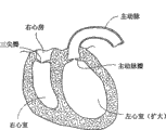
心脏(参见图1)比握紧的拳头略大。它是双侧(左侧和右侧)的自我调节的肌肉泵,其各部分和谐地工作从而将血液驱送至身体的各个部分。心脏的右侧接受来自身体上腔静脉和下腔静脉血氧含量较差的(“静脉的”)血液,并通过肺动脉将其泵入肺中进行氧化作用。其左侧接受来自通过肺静脉从肺输送来的血氧含量较高的(“动脉的”)血液,并将其泵入动脉从而分布于身体。The heart (see Figure 1) is slightly larger than a clenched fist. It is a bilateral (left and right) self-regulating muscular pump whose parts work in harmony to drive blood to all parts of the body. The right side of the heart receives poorly oxygenated ("venous") blood from the body's superior and inferior vena cava and pumps it through the pulmonary artery to the lungs for oxygenation. Its left side receives more oxygenated ("arterial") blood from the lungs via the pulmonary veins and pumps it into the arteries for distribution throughout the body.
心脏有四个腔室,每侧各两个-----右心房和左心房,以及右心室和左心室。心房是接受血液的腔,其将血液泵入心室。由膜和肌肉部分组成的、称为房间隔的壁,将右心房和左心房分隔开来。心室是射出血液的腔。由膜状和肌肉部分组成的、称为室间隔的壁,将右心室和左心室分隔开来。The heart has four chambers, two on each side -- the right and left atria, and the right and left ventricles. The atrium is the chamber that receives blood, which pumps it into the ventricles. A wall called the septum, made up of membranes and muscular parts, separates the right atrium from the left atrium. The ventricles are the cavities from which blood is ejected. A wall made of membranous and muscular parts, called the septum, separates the right and left ventricles.
心脏左侧和右侧的同步泵动作构成了心动周期。该周期从心室舒张的阶段开始,称作心室舒张。该周期以心室的收缩终止,称为心室收缩。The simultaneous pumping action of the left and right sides of the heart makes up the cardiac cycle. The cycle begins with a phase of ventricular relaxation, called ventricular diastole. The cycle ends with the contraction of the ventricles, called ventricular systole.
心脏具有四个瓣膜(参见图2和图3)以确保在心动周期中血液不会流错方向;即确保血液不会从心室回流入对应的心房,或从心房回流入对应的心室。左心房和左心室之间的瓣膜是二尖瓣。右心房和右心室之间的瓣膜是三尖瓣。肺动脉瓣位于肺动脉的开口处。主动脉瓣位于主动脉的开口处。The heart has four valves (see Figures 2 and 3) to ensure that blood does not flow in the wrong direction during the cardiac cycle; that is, to ensure that blood does not backflow from a ventricle into its corresponding atrium, or from an atrium into its corresponding ventricle. The valve between the left atrium and left ventricle is the mitral valve. The valve between the right atrium and right ventricle is the tricuspid valve. The pulmonary valve is located at the opening of the pulmonary artery. The aortic valve is located at the opening of the aorta.
在心室舒张(即心室充盈(ventricular filling))开始的时候(参见图2),主动脉瓣和肺动脉瓣闭合防止血液从动脉回流入心室。随后,三尖瓣和二尖瓣打开(如图2所示),允许血液从心房流入对应的心室。在心室收缩(即心室排空)开始后不久,三尖瓣和二尖瓣闭合(如图3所示)--防止血液从心室回流入对应的心房---同时主动脉瓣和肺动脉瓣打开—允许血液从对应的心室射入动脉。At the onset of ventricular diastole (ie, ventricular filling) (see Figure 2), the aortic and pulmonary valves close to prevent backflow of blood from the arteries into the ventricles. Subsequently, the tricuspid and mitral valves open (as shown in Figure 2), allowing blood to flow from the atria to the corresponding ventricles. Shortly after the onset of ventricular systole (i.e., ventricular emptying), the tricuspid and mitral valves close (as shown in Figure 3) -- preventing backflow of blood from the ventricles into the corresponding atria -- while the aortic and pulmonary valves open - Allows blood to be ejected from the corresponding ventricle into the artery.
心脏瓣膜是由胶原的纤维环组成的,每一个都称为环(annulus),其形成了心脏纤维骨架的一部分。环为瓣尖或小叶提供了附着物(心耳)。在健康的心脏中,肌肉及其腱索(腱索)支撑瓣膜,从而允许瓣膜的小叶随着其希望的功能开放和闭合。Heart valves are composed of fibrous rings of collagen, each called an annulus, which form part of the fibrous skeleton of the heart. The annulus provides attachments (atrial appendages) for the cusps or leaflets. In a healthy heart, the muscles and their tendons (chordae) support the valves, allowing the valve's leaflets to open and close as they wish.
II心脏功能障碍II cardiac dysfunction
感染、心肌梗塞、心房纤维颤动、其他疾病、或器质性的缺陷会在心动周期过程中对心脏左侧和右侧的正常同步泵作用和/或心脏瓣膜的动作造成不利的影响。Infection, myocardial infarction, atrial fibrillation, other diseases, or organic defects can adversely affect the normal synchronous pumping of the left and right sides of the heart and/or the action of the heart valves during the cardiac cycle.
例如,由于以上的一种或多种原因,心脏的腔室会变得扩张和膨大。这样的情况会导致不良的后果。例如,(1)因为这种膨大的情况,心脏必须更努力地泵压来输送血液,和/或从心脏流入身体其余部分的血液太少。假以时日,心脏的其他腔室也会逐渐变得衰弱。例如,在左心室的心脏腔室的扩张和膨大会导致称为充血性心力衰竭的病症。如果不进行治疗,充血性心力衰竭会导致肺栓塞、循环抑制(circulationshutdown)和死亡。For example, the chambers of the heart can become dilated and enlarged due to one or more of the reasons above. Such a situation can lead to undesirable consequences. For example, (1) because of this enlarged condition, the heart has to pump harder to pump blood, and/or too little blood flows from the heart to the rest of the body. Over time, the other chambers of the heart gradually weaken. For example, dilation and enlargement of the heart chambers in the left ventricle can lead to a condition called congestive heart failure. If left untreated, congestive heart failure can lead to pulmonary embolism, circulation shutdown and death.
心脏腔室的膨大也会导致心脏瓣膜环的膨大或扩张。同样,心脏瓣膜周围肌腱的扩张或撕裂,或这一区域其他形式的肌肉衰竭,甚至是在心脏腔室没有出现膨大的时候,也会改变心脏瓣膜环的形状。当心脏瓣膜环的形状发生改变时,该瓣膜的小叶就不能闭合(coapt)。于是会出现在心房和心室之间不希望的血液回流(称作反流),或在动脉和心室之间的回流。这样的功能障碍会最终使心脏变得衰弱并会导致心力衰竭。Enlargement of the chambers of the heart can also cause enlargement, or dilation, of the annulus of the heart valves. Likewise, dilation or tearing of the tendons around the heart valve, or other forms of muscle failure in this area, can change the shape of the heart valve annulus even when the heart chambers are not dilated. When the shape of the heart valve annulus changes, the leaflets of the valve cannot coapt. Undesirable backflow of blood between the atria and ventricles (called regurgitation), or backflow between arteries and ventricles then occurs. Such dysfunction eventually weakens the heart and can lead to heart failure.
器质性的、例如在隔膜的缺陷也会导致心脏功能障碍。这些缺陷可能是先天的,也有可能是由疾病或损伤导致的。Organic defects, for example in the diaphragm, can also lead to cardiac dysfunction. These defects may be born or result from disease or injury.
III.预治疗方法III. Pretreatment Methods
药物疗法可成功地治疗心脏功能障碍。然而,对于慢性或极性功能障碍而言,通常需要进行手术。对充血性心力衰竭而言,需要进行心脏移植。诸如侵入性,开放性的心脏手术方法已被用于修补或更换功能障碍的心脏瓣膜或对中隔缺损进行矫正。Drug therapy can successfully treat cardiac dysfunction. However, for chronic or polar dysfunction, surgery is usually required. For congestive heart failure, a heart transplant is required. Methods such as invasive, open heart surgery have been used to repair or replace dysfunctional heart valves or to correct septal defects.
亟需简单、低成本以及侵入性较小的装置、系统和方法来治疗诸如充血性心力衰竭和/或心瓣膜机能障碍和/或中隔缺损的心脏病症。同样也需要对由其他身体器官无意识形状改变而引起的其他功能障碍进行类似的治疗。There is a need for simple, low cost and less invasive devices, systems and methods to treat cardiac conditions such as congestive heart failure and/or heart valve dysfunction and/or septal defect. Similar treatments are also needed for other dysfunctions caused by involuntary shape changes of other body organs.
发明内容 Contents of the invention
为了恢复或保持器官天然功能,本发明提供了支撑中空身体器官内组织的装置、系统和方法。所述装置、系统和方法不需要通过侵入性,开放性的外科手术方法实施,相反,却可借助于基于导管,血管内的和/或经由皮肤的技术来实施。The present invention provides devices, systems and methods for supporting tissue within a hollow body organ in order to restore or maintain the organ's natural function. The devices, systems and methods need not be performed by invasive, open surgical methods, but rather can be performed by means of catheter-based, intravascular and/or percutaneous techniques.
在一个方面,本发明提供了支撑中空身体器官内组织的系统和方法。所述系统和方法使用了偶合在一起的第一和第二植入物。对第一植入物进行尺寸和形状的处理从而能够穿透中空身体器官内组织的第一区域。对第二植入物进行尺寸大小和形状处理从而能够穿透空间上远离所述中空身体器官组织第一区域的第二区域。至少有一个拉紧元件将第一和第二植入物偶合在一起,从而将拉力施加在第一和第二植入物上,并由此向内牵拉组织,从而对其进行支撑。支撑作用,例如,将组织表面牵拉至一起从而降低所述中空身体器官内的组织体积,并且防止随后组织体积的膨大。所希望的是,所述支撑作用并不会与该中空身体器官的收缩相互干扰而使组织体积进一步缩小。然而,如果需要的话,这种支撑的形式还是可以成功的。In one aspect, the present invention provides systems and methods for supporting tissue within a hollow body organ. The systems and methods use first and second implants coupled together. A first implant is sized and shaped to penetrate a first region of tissue within a hollow body organ. A second implant is sized and shaped to penetrate a second region spatially remote from the first region of hollow body organ tissue. At least one tensioning element couples the first and second implants together, thereby exerting tension on the first and second implants and thereby pulling tissue inwardly, thereby supporting it. Bracing, for example, draws tissue surfaces together thereby reducing tissue volume within the hollow body organ and preventing subsequent expansion of tissue volume. Desirably, the bracing action does not interfere with the contraction of the hollow body organ to further reduce tissue volume. However, this form of support can still be successful if desired.
另一方面,本发明提供了在中空的身体器官内形成组织折叠的系统和方法。所述系统和方法采用了第一和第二植入物。对所述植入物进行尺寸大小和形状的调整从而穿透空间上远离所述中空身体器官组织区域。至少有一种拉紧元件将第一和第二植入物偶合在一起,将拉力施加在第一和第二植入物上。所述拉力在第一和第二植入物之间产生了一个组织折叠。组织折叠可以,例如,降低中空身体器官内的内部组织体积,同时防止随后组织体积的膨大。所希望的是,所述拉紧作用并不会与该中空身体器官的收缩相互干扰而使组织体积进一步缩小。然而,如果需要的话,这种支撑的形式还是可以伴随组织折叠成功的。In another aspect, the present invention provides systems and methods for forming tissue folds within hollow body organs. The systems and methods employ first and second implants. The implant is sized and shaped to penetrate a region of tissue spaced away from the hollow body organ. At least one tensioning element couples the first and second implants together, applying tension to the first and second implants. The tension creates a tissue fold between the first and second implants. Tissue folding can, for example, reduce internal tissue volume within a hollow body organ while preventing subsequent expansion of tissue volume. Desirably, the tensioning action does not interfere with the contraction of the hollow body organ to further reduce tissue volume. However, this form of support can still accompany tissue folding if desired.
在一个实施方案中,第一和第二植入物是在中空身体器官内穿透空间上远离组织区域的植入物阵列的部分。在该实施方案中,至少有一种拉紧元件伸展于植入物阵列之间,从而将拉力施加于相邻的植入物之间,并由此产生了多个组织折叠的方式。所述多个组织折叠可以,例如,将组织的圆周区域牵拉到一起,形成闭合或封口。In one embodiment, the first and second implants are part of an array of implants penetrating a region of tissue spaced apart within the hollow body organ. In this embodiment, at least one tensioning element extends between the array of implants, thereby applying tension between adjacent implants and thereby creating a plurality of tissue fold patterns. The plurality of tissue folds may, for example, draw together circumferential regions of tissue to form a closure or seal.
在另一方面,本发明提供了支撑中空身体器官内组织的系统和方法。所述系统和方法应用使用了经过尺寸大小和形状处理适合放置于中空身体器官之内,或者围绕所述中空身体器官的假体。所述系统和方法还使用了将所述假体紧密固定于中空身体器官内组织的至少一个固定器。在一个实施方案中,所述固定器包含螺旋固定器。In another aspect, the present invention provides systems and methods for supporting tissue within a hollow body organ. The systems and methods employ the use of prostheses sized and shaped for placement within, or around, hollow body organs. The systems and methods also employ at least one anchor that tightly secures the prosthesis to tissue within the hollow body organ. In one embodiment, the anchor comprises a screw anchor.
在另一方面,本发明提供了利用伸长的植入物支撑中空身体器官内组织的系统和方法。对所述伸长的植入物进行尺寸大小和形状的调整从而能穿透组织并在组织壁内或部分地在组织壁内随着曲线的路径延伸。所述伸长的植入物可对中空身体器官的最大尺寸和/或形状进行调节。在一个实施方案中,所述伸长的植入物是螺旋形的。In another aspect, the present invention provides systems and methods for supporting tissue within a hollow body organ using an elongate implant. The elongated implant is sized and shaped to penetrate tissue and follow a curvilinear path within or partially within the tissue wall. The elongated implant is adjustable to the maximum size and/or shape of the hollow body organ. In one embodiment, the elongate implant is helical.
如本文所述,体现将本发明多个方面全部或部分的系统和方法都可使良好地适用于,例如,心脏中。所述系统和方法可用于,例如,在充血性心力衰竭或其他心脏体积变大的疾病中的治疗中在心脏腔室内和/或在心脏瓣膜环内或附近支撑组织。所述系统和方法可用来密封或闭合组织内的孔眼,洞,或缺损。所述系统和方法可用来闭合或密封心耳(atrial appendages)或中隔缺损。As described herein, systems and methods embodying all or part of the aspects of the present invention are well suited for use in, for example, the heart. The systems and methods may be used, for example, to support tissue within the chambers of the heart and/or within or near the annulus of heart valves in the treatment of congestive heart failure or other conditions in which the heart enlarges. The systems and methods can be used to seal or close holes, holes, or defects in tissue. The systems and methods can be used to close or seal atrial appendages or septal defects.
基于所附的说明,附图和权利要求,本发明的其他特征和优点是显而易见的。Other features and advantages of the invention are apparent from the accompanying description, drawings and claims.
附图说明 Description of drawings
图1是健康心脏内部的透视和前解剖视图。Figure 1 is a perspective and anterior anatomical view of the interior of a healthy heart.
图2是去除了心房,显示在心室舒张过程中心脏瓣膜状态的健康心脏内部的解剖仰视图。Figure 2 is an anatomical bottom view of the interior of a healthy heart with the atria removed, showing the state of the heart valves during ventricular diastole.
图3是去除了心房,显示在心室收缩过程中心脏瓣膜状态的健康心脏内部的解剖仰视图。Figure 3 is an anatomical bottom view of the interior of a healthy heart with the atria removed, showing the state of the heart valves during ventricular systole.
图4A是在中空身体器官内对组织进行支撑的植入物的透视图。4A is a perspective view of an implant supporting tissue within a hollow body organ.
图4B是在组织中植入如图4A所示植入物的施加器械的侧视图。Figure 4B is a side view of the applicator instrument for implanting the implant shown in Figure 4A in tissue.
图4C是如图4A所示植入物植入组织之后的侧视图。Figure 4C is a side view of the implant shown in Figure 4A after implantation into tissue.
图5A和5B是在中空身体器官内建立的组织支撑系统,其包括两个或更多通过夹具放置和保持拉紧的如图4A所示的植入物。Figures 5A and 5B are a tissue support system established within a hollow body organ comprising two or more implants as shown in Figure 4A placed and held taut by clamps.
图6A和6B是在心脏的左心室中建立的如图5所示的组织支撑系统,图6A所示为在建立系统之前体积膨大的心室,图6B所示为系统降低了心室的体积。Figures 6A and 6B are the tissue support system shown in Figure 5 established in the left ventricle of the heart, Figure 6A shows the ventricle volume expanded before the system is established, Figure 6B shows the system reduces the volume of the ventricle.
图7A-7D所示为通过使用血管内工具和技术建立图6B所示系统的步骤。Figures 7A-7D illustrate the steps involved in setting up the system shown in Figure 6B through the use of endovascular tools and techniques.
图8A和8B所示为在心脏的左心室在主动脉瓣的环内或附近建立的图5所示的组织支撑系统,图8A所示为在建立该系统之前主动脉瓣环膨胀的状态,而图8B所示为该系统重塑了环从而恢复了小叶的闭合。8A and 8B show the tissue support system shown in FIG. 5 established in or near the annulus of the aortic valve in the left ventricle of the heart, and FIG. 8A shows the state of the aortic annulus inflated before the system is established, However, Figure 8B shows that the system reshaped the ring to restore leaflet closure.
图9A-9D所示为使用血管内的工具和技术建立图8B所示系统的步骤。Figures 9A-9D illustrate steps in building the system shown in Figure 8B using intravascular tools and techniques.
图10A和10B所示为在心脏的左心室中建立的组织折叠系统,图10A所示为在该系统建立之前体积膨大的心室,图10B所示为该系统降低了心室的体积。Figures 10A and 10B show the establishment of a tissue folding system in the left ventricle of the heart, Figure 10A shows the ventricle inflated in size before the system is established, and Figure 10B shows the system reducing the volume of the ventricle.
图11A-11D所示为使用血管内的工具和技术建立图10B所示系统的步骤。11A-11D illustrate the steps involved in building the system shown in FIG. 10B using endovascular tools and techniques.
图12所示为具有图10B所示系统特征的组织折叠系统的另一实施方案。Figure 12 shows another embodiment of a tissue folding system having the features of the system shown in Figure 10B.
图13A-13C所示为使用血管内的工具和技术建立具有图10B所示系统特征的组织折叠系统的另一实施方案的步骤。13A-13C illustrate the steps of another embodiment of a tissue folding system having the features of the system shown in FIG. 10B using intravascular tools and techniques.
图14A和14B所示为使用血管内的工具和技术建立具有图10B所示系统特征的组织折叠系统的另一实施方案的步骤。Figures 14A and 14B illustrate the steps of another embodiment of a tissue folding system having the features of the system shown in Figure 10B using intravascular tools and techniques.
图15A所示为如图10B所示的组织折叠系统,包括了由固定器在组织折叠系统建立的组织折叠之上紧密固定的覆盖修补部件。FIG. 15A shows the tissue folding system as shown in FIG. 10B , including the overlay repair component tightly fixed by the fixator on the tissue fold created by the tissue folding system.
图15B所示为通过血管内通道配置图15A所示修补部件的导管。Figure 15B shows a catheter with the patch component shown in Figure 15A deployed through an intravascular channel.
图16A所示为在中空身体器官内建立的产生折叠模式的系统,其可将所述中空身体器官的一个区域与该中空身体器官的另一区域加以分隔或封闭。Figure 16A shows a system created within a hollow body organ to create a folding pattern that separates or encloses one region of the hollow body organ from another region of the hollow body organ.
图16B是采用通常沿着图16A中的16B-16B线、通过图16A所示系统产生的折叠模式的平面图。16B is a plan view of the folded pattern produced by the system shown in FIG. 16A, generally along
图17A和17B所示为使用图16A和16B所示的系统在心耳和房间隔之间区域中建立的多种折叠模式,图17A所示为该系统建立之前的心房,图17B为该系统建立之后将心耳与房间隔进行隔离和/或封闭的心房。Figures 17A and 17B show the various folding patterns created using the system shown in Figures 16A and 16B in the region between the atrial appendage and the interatrial septum, Figure 17A showing the atrium before the system was established, and Figure 17B for the system established The atrial appendage is then isolated from the atrial septum and/or the atrium is closed.
图17C是采用通常沿着图17B中的17C-17C线、通过图17B所示系统产生的折叠模式的平面图。Figure 17C is a plan view of the folded pattern produced by the system shown in Figure 17B, generally along
图18A和18B所示为使用图16A和16B所示系统在中空身体器官内建立的将孔封闭的多种折叠方法,图18A所示为在建立该系统之前的孔,图18B所示为在建立所述系统后孔的闭合。Figures 18A and 18B show the various folding methods used to establish the pores in hollow body organs using the system shown in Figures 16A and 16B, Figure 18A shows the pores before the system is established, and Figure 18B shows the pores before the system is established. Closure of the hole after establishing the system.
图19A至19F所示为可安装于中空身体器官内对该器官进行塑型并阻止其膨大的假体的多种实施方案。Figures 19A to 19F illustrate various embodiments of prostheses that can be installed within a hollow body organ to shape the organ and prevent its expansion.
图20A所示为安装于中空的身体器官内部的图19A-19F所示类型的假体。Figure 20A shows a prosthesis of the type shown in Figures 19A-19F installed inside a hollow body organ.
图20B所示为安装于中空的身体器官外部的图19A-19F所示类型的假体。Figure 20B shows a prosthesis of the type shown in Figures 19A-19F installed outside a hollow body organ.
图21A所示为安装于心脏腔室内部的图19A-19F所示类型的假体。Figure 21A shows a prosthesis of the type shown in Figures 19A-19F installed inside a heart chamber.
图22A-22D所示为使用血管内工具和技术建立图20A所示假体的步骤。Figures 22A-22D illustrate steps in creating the prosthesis shown in Figure 20A using endovascular tools and techniques.
图23所示为安装于心脏外部的图19A-19F所示类型的假体。Figure 23 shows a prosthesis of the type shown in Figures 19A-19F installed outside the heart.
图24A和24B所示为通过安装于心脏左心室的两个或更多修补部件阵列形成的、具有图19A-19F所示假体特征的复合假体。Figures 24A and 24B illustrate a composite prosthesis having the features of the prosthesis shown in Figures 19A-19F formed by an array of two or more prosthetic components installed in the left ventricle of the heart.
图25所示为在形成放置于心脏瓣膜环内或附近的形式的环过程中形成的、具有图19A-19F所示假体特征的假体。Figure 25 shows a prosthesis having the features of the prosthesis shown in Figures 19A-19F formed during the formation of an annulus in a form placed in or near the annulus of a heart valve.
图26A所示为安装于主动脉瓣环内或附近的、如图25所示的假体。Figure 26A shows the prosthesis shown in Figure 25 installed in or near the aortic annulus.
图26B所示为安装于二尖瓣环内或附近的、如图25所示的假体。Figure 26B shows the prosthesis shown in Figure 25 installed in or near the mitral valve annulus.
图27所示为通过血管内通路配置图25所示假体的导管。Figure 27 shows a catheter for deploying the prosthesis of Figure 25 through an intravascular access.
图28所示为经过尺寸大小和形状的调整用于对心脏中的中隔缺损进行修复的、具有图15A所示修补部件特征的修补部件。Figure 28 shows a repair component having the features of the repair component shown in Figure 15A sized and shaped for the repair of a septal defect in the heart.
图29A和29B所示为安装于心脏左右心室之间中隔缺损内的图28所示的修补部件。Figures 29A and 29B show the prosthetic component of Figure 28 installed in a septal defect between the left and right ventricles of the heart.
图30A和30B所示为可植入中空身体器官塑造其器官形状并防止其膨大的伸长的植入物的多种实施方案,图30A所示为具有常见线性形状的植入物,而图30B所示为具有常见曲线形状的植入物。Figures 30A and 30B show various embodiments of elongated implants that can be implanted into a hollow body organ to shape its organ shape and prevent it from expanding, with Figure 30A showing an implant having a common linear shape, and Figure 30A showing an implant with a common linear shape, and Figure 30A 30B shows an implant having a common curvilinear shape.
图31所示为植入心脏左心室的图30A和30B所示的伸长的植入物。Figure 31 shows the elongated implant of Figures 30A and 30B implanted in the left ventricle of a heart.
图32所示为为了放置于心脏瓣膜环内或附近而形成的、具有多种图19A-19F所示假体特征的心脏瓣膜组件。Figure 32 shows a heart valve assembly having various features of the prosthesis shown in Figures 19A-19F formed for placement in or near the heart valve annulus.
图33所示为安装于主动脉瓣环内或附近的、如图32所示的组件。Figure 33 shows the assembly shown in Figure 32 installed in or near the aortic valve annulus.
图34A-34C所示为通过使用血管内工具和技术,在主动脉瓣环内或附近,安装图32所示心脏瓣膜组件的步骤。34A-34C illustrate the steps of installing the heart valve assembly shown in FIG. 32 in or near the aortic annulus using endovascular tools and techniques.
发明详述Detailed description of the invention
虽然本文公开的内容非常详细且精确,足以使本领域所属技术人员实施本发明,但在此公开的物理性实施方案仅是为了对本发明进行示例性说明,其可在体现在其它具体的结构中。尽管对优选实施方案进行了描述,还是可以在不偏离由权利要求所限定的本发明范围内进行细节的变化。While the disclosure herein is sufficiently detailed and precise to enable those skilled in the art to practice the invention, the physical embodiments disclosed herein are merely illustrative of the invention which may be embodied in other specific structures. . Although a preferred embodiment has been described, changes may be made in detail without departing from the scope of the invention as defined in the claims.
本说明书所公开的技术被分为以下几部分用于进行详细说明:The technology disclosed in this specification is divided into the following sections for detailed description:
I.在中空身体器官内对组织进行外在支撑的植入物I. Implants for Extrinsic Support of Tissue in Hollow Body Organs
A.概述A. Overview
B.在心脏腔室内对组织进行支撑的系统和方法B. Systems and methods for supporting tissue within a chamber of the heart
C.在心脏瓣膜环内或附近对组织进行支撑的系统和方法C. Systems and methods for supporting tissue in or near a heart valve annulus
II.产生组织折叠的植入物II. Implants that Produce Tissue Folding
A.概述A. Overview
B.限定(defining)不连续组织折叠的系统和方法B. Systems and methods for defining discrete tissue folds
1.使用覆盖修补部件进行组织折叠1. Tissue Folding Using Cover Patching Parts
C.限定组织折叠模式的系统和方法C. Systems and Methods for Defining Tissue Folding Patterns
1.概述1 Overview
2.心耳(appendage)的分隔和封闭2. Separation and closure of the appendage
3.封闭孔、洞或缺陷3. Close holes, holes or defects
III.在中空身体器官内对组织进行外在支撑的假体III. PROSTHESIS FOR EXTERNAL Tissue Support Within Hollow Body Organs
A.概述A. Overview
B.在心脏腔室内对组织进行支撑的系统和方法B. Systems and methods for supporting tissue within a chamber of the heart
C.在心脏瓣膜环内部或附近对组织进行支撑的系统和方法C. Systems and methods for supporting tissue in or near a heart valve annulus
IV.在中空身体器官内对组织进行内在支撑的植入物IV. Implants for Intrinsic Support of Tissues in Hollow Body Organs
应该理解在给定部分所描述的技术可以与在另一部分所描述的技术结合,且在此描述的特征为全部技术所共有。It should be understood that techniques described in a given section may be combined with techniques described in another section, and that features described there are common to all techniques.
I.在中空身体器官内对组织进行外在支撑的植入物I. Implants for Extrinsic Support of Tissue in Hollow Body Organs
A.概述A. Overview
图4A所示为经过尺寸大小和形状的调整用于放置于中空身体器官内的植入物10。所述植入物包括由成型塑料或金属或陶瓷材料制成的适于植入身体的小体12。Figure 4A shows the
小体12包括远端区域14。对远端区域14进行尺寸大小和形状的调整从而能够穿入组织。对小体12及其远端区域14进行尺寸大小和形状的调整,从而充分地紧密抓紧组织(参见图4C),使其一旦被植入就能足以显著地防止小体12从组织释放和/或迁移出来。
小体12还包括近端区域16。对近端区域16进行尺寸大小和形状的调整,从而与用来施力引起植入物10穿入组织的器械或工具20配合(参见图4B)。
如图4A所示,小体12还包括束缚元件18。在说明性的实施方案中,束缚元件18位于小体12近端区域16上或附近。采用这样的方式,当小体12被植入血管或中空身体器官(参见图4C)的组织壁内时,束缚元件18延伸至组织壁的外侧。As shown in FIG. 4A ,
束缚元件18包括具有金属或聚合物材料(例如,聚酯结构)的绳、编织物、丝或管结构,其希望的断裂强度至少相当于小体12的远端14从组织释放或迁移的阻力。所希望的束缚元件18是柔软的,从而使得其能够配置在整个血管内通路。所希望的束缚元件18是不明显具有弹性的,但根据所遇到的组织情况,也可以是具有弹性的。Tethering
束缚元件18可通过,例如,焊接、胶粘、铆接或类似的连接技术紧密地固定于近端区域16。所希望的是束缚元件18和小体12之间的连接具有比束缚元件18本身的材料强度更高的材料强度。The
植入物10的小体12可以具有多种形式。在说明性的实施方案中(如图4A所示),小体12包含开放的螺旋型卷曲。在所述配置中,远端区域14包括削尖的引导尖端。这种类型的小体12和远端区域14可以通过施力器械20施加给植入物10的旋转运动配置入组织中。The
同样,在说明性的实施方案中(如图4A所示),近端区域16包含L-型的支架。所希望的是该L-型支架将卷曲小体12的整个内部直径一分为二;换言之,L-型支架16完全伸展穿过了卷曲小体12的内部直径。L-型支架16可用作阻止卷曲小体12旋转穿入组织过于深入的限位器。此外,如图4B通常所示,可对施力器械20上的可旋转植入物驱动机械装置22进行尺寸大小和形状的调整来与L-型支架16配合,并且使卷曲小体12旋转从在组织中完成植入。Also, in an illustrative embodiment (as shown in FIG. 4A ),
图5A和5B所示为包含至少两种图4A所示植入物10的组织成型系统24。植入物10以彼此分隔的-分离的关系或方式植入中空身体器官或血管(如图5A和5B通常所示)的组织壁中。所配置的束缚植入物10的数量会随着靶组织体积的尺寸和几何形状,以及组织支撑目的的变化而变化。Figures 5A and 5B illustrate a
系统24包括连接于植入物10的束缚元件18的至少一个夹具26。图5A所示为单个夹具26。图5B所示为多个夹具26。所述夹具或元件26相互地将束缚元件18联合到一起,并允许对其施以拉力并保持在该组织的外面,如图5A和5B中的箭头所示。由分离的植入物10上的每个束缚元件18单独施加和保持的拉力,组合起来,可将周围的组织壁整体地向夹具26的内部牵拉,从而对所述中空身体器官或血管进行塑型。反之,在每个植入物10上的束缚元件18所施加和保持的拉力,组合起来,抵抗了组织壁整体地向远离夹具26的外部运动。所述拉力防止了组织壁发生超过由组织支撑系统24所产生体积的膨胀。不过,所希望的是组织支撑系统24不会与组织壁收缩产生缩小的体积相互干扰。
每个单独束缚元件18的长度和其施于各个植入物10上的拉力幅度共同地规定了身体器官的最大形状。以这种方式,系统24可在身体器官内对组织进行支撑和塑型。The length of each
可在身体的诸多部分建立上述系统24并用于各种治疗目的。出于说明性目的对两种实施方案进行描述。第一种实施方案旨在对充血性心力衰竭进行治疗和/或修复。第二种实施方案旨在心脏瓣膜重塑。The
B.在心脏腔室内对组织进行支撑的系统和方法B. Systems and methods for supporting tissue within a chamber of the heart
图6A所示为遭受充血性心力衰竭的心脏。图6A所示病症的特征在于内部体积膨大的左心室。图6B所示为通过在左心室内植入具有束缚植入物10的系统24对所述病症进行治疗和/或修复。对植入物10的束缚18进行放置并通过夹子26保持拉力(如图6B的箭头所示)。如果需要的话,可使用多个夹子26。由系统24所施加的拉力对左心室进行了塑型,将腔室壁横向地一起拉近并由此减小了总的最大内部体积。所述拉力防止或限制了在心室舒张期间左心室的扩张超过适于高效心室泵压的形状。然而,在心室舒张期间支撑系统24并未与左心室的正常收缩相互干扰。Figure 6A shows a heart suffering from congestive heart failure. The condition shown in Figure 6A is characterized by an enlarged internal volume of the left ventricle. Figure 6B shows the treatment and/or repair of the condition by implanting the
图7A-7D所示图6B所示支撑系统24在血管内的配置。或者,也可以采用常规的开放式心脏手术技术或胸腔镜手术技术建立系统24。Figures 7A-7D illustrate the intravascular configuration of the
在图7A-7D所示的血管内方法中,引导部件28是由引导丝(未示出)通过主动脉瓣向左心室递送的。引导部件28可在荧光镜指导下,例如,通过动脉退行的路径(通过,例如股动脉或锁骨下动脉)(如图所示)或静脉顺行然后横穿隔膜的路径输送至脉管系统。In the endovascular approach shown in Figures 7A-7D,
引导部件28可包括,例如,希望具有不锈钢或能转变方向末端尖端的引导鞘。引导丝可在引导部件28安置和固定之后取出,从而可通过引导部件28引入施力器械20,如图7A所示。图4B还显示了施力器械20通过引导部件28进行配置。
在这种配置中(参见图4B),施力器械20包括在其末端尖端上带有植入物驱动机械装置22的导管30。驱动机械装置22携带有至少一个束缚的植入物10。在由医生操作的,位于手柄34中的马达32驱动机械装置22旋转植入物10。其结果是导致植入物10穿入了心肌层(如图7A所示)。In this configuration (see FIG. 4B ),
所希望的是驱动机械装置22的植入力可以某种方式得以分解从而提供位置稳定性,并且防止该驱动机械装置22相对于所述植入位点无意识的运动。所希望的是施加分解力来抵消和/或对抗驱动机械装置22的植入力。所希望的是在血管内腔(或其他中空的身体器官)本身内,且优选地在尽可能靠近植入的位点将一些或全部或主要部分的植入力分解。It is desirable that the implantation forces of the
可对引导部件28的管状体和/或施力器械20的柄进行尺寸大小和形状的调整,从而拥有足够的裂断强度将血管内腔或中空身体器官内的一些或全部或至少一部分的植入力分解。图7A所示为由对心室壁进行支撑来施加平衡分解力的引导部件28。除此之外,或者,为了在驱动机械装置22或其附近施加平衡力,引导部件28和/或施力器械20可包括某些形式的稳定化装置。在2003年9月24日递交的、题为《具有植入力分解的基于导管的固定器植入装置和方法》(Catheter-BasedFastener Implantation Apparatus and Methods with Implantati on ForceResolution)的同时待决美国专利申请第10/669,881中公开了多种类型的稳定化装置。The size and shape of the tubular body of the
引导部件28与其他希望的心肌传递位点相连配置。在每一个位点上,驱动施力器械20放置植入物10。以这种方式(参见图7B),植入物10以希望的空间(诸如放射性或类似于螺旋形的方式)分布于左心室内。
一旦将希望数量的植入物10放置在左心室内,就将施力器械20从引导部件28中取出。将留下的植入物10的束缚元件18集中于并成束穿过引导部件28,如图7B所示。Once the desired number of
如图7C所示,夹子-应用器械36也穿过了引导部件28,并在束缚元件18束上进入左心室。束缚元件18作为复合引导丝来引导夹子-应用器械36进入左心室。As shown in FIG. 7C , clip-applying
一旦进入了左心室,夹子-应用器械36就保持固定,而束缚元件则通过夹子-应用器械36被拉紧(如图7C中的箭头T所示)。由于单个的束缚元件18逐渐拉紧,它们对单独的植入物施加拉力,如图7C所示。这样的话,反过来,就将左心室的壁朝着夹子-应用器械36向内牵拉(作为图7B所示左心房与图7C所示左心房的比较)。一旦达到了希望的心室体积(例如,通过荧光透视法测定的),夹子-应用器械36就向束缚元件施加一个夹子26,将束缚元件18通过拉力集合起来(参见图7D)。夹子-应用器械36在最接近使用夹子26的位置的地方切断束缚元件18束。夹子-应用器械36和松散的束缚18随后通过引导部件28从左心室中取出,还取出了引导部件,如图7D所示。Once in the left ventricle, the clip-applying
建立了系统24来支撑左心室,从而治疗在这种情况下的充血性心力衰竭。A
应该意识到,系统24的一个或多个植入物10可以与在心脏组织中操纵控制肌肉和/或电活性的装置电耦合。尽管缺乏这种希望的作用,还是希望植入物10不是天然导电的,从而不会与心脏内的电导相互干扰。It should be appreciated that one or
C.在心脏瓣膜环处或附近对组织进行支撑的系统和方法C. Systems and methods for supporting tissue at or near a heart valve annulus
图8A所示为患有充血性心力衰竭的心脏。如图8A所示,这种病症引起了左心室内部体积的膨大,导致了主动脉瓣环的膨胀或伸展。其结果是主动脉小叶在心室收缩的过程中不能适当地闭合。在心室收缩的过程中会发生从左心室进入主动脉的不希望的血液倒流。Figure 8A shows a heart with congestive heart failure. As shown in Figure 8A, this condition causes an enlargement of the internal volume of the left ventricle, resulting in dilation or stretching of the aortic annulus. The result is that the aortic leaflets do not close properly during ventricular contraction. Undesired backflow of blood from the left ventricle into the aorta occurs during ventricular contraction.
图8B所示为通过在左心室内在主动脉瓣环或其邻近区域植入束缚植入物10的系统24来治疗和/或修复这种病症。对固定器的束缚18进行放置并通过夹子26经拉力固定(如图8B中的箭头所示)。如果需要的话,可以使用多个夹子26。由系统24施加的拉力重塑了主动脉瓣环,将小叶牵拉得更紧,从而在心室收缩时闭合完全,防止或降低了倒流。Figure 8B illustrates the treatment and/or repair of this condition by implanting a
图9A-9D所示为图8B所示系统24的血管内配置。或者,可以使用常规的开放性心脏手术技术或通过胸腔镜手术技术建立系统24。Figures 9A-9D illustrate the intravascular configuration of the
图9A-9D所示的血管内方法与前述的图7A-7D所示的方法基本上相同。在荧光镜的导引下,引导部件28可由引导丝通过在主动脉瓣环或邻近其下部的区域穿过主动脉瓣(通过,例如股动脉或锁骨下动脉)或静脉顺行然后横穿隔膜的路线进入左心室。随后取出引导丝,并通过引导部件28引入施力器械20,如图9A所示。The intravascular method shown in Figures 9A-9D is substantially the same as the method shown in Figures 7A-7D previously described. Under fluoroscopic guidance,
引导部件28在主动脉瓣环下部区域或其邻近与希望的植入物投放位点相邻放置。在每一个位点上,驱动施力器械20放置植入物10。图9A所示为由对心室壁进行支撑来施加平衡分解力的引导部件28。以这种方式(参见图9B),植入物10以希望的方式分布于主动脉瓣环下部区域或其邻近。将植入物10的束缚元件集合并成束穿过引导部件28到达体外。
一旦将希望数量的植入物10放置在主动脉瓣环或其附近,就将施力器械20取出,并且夹子-应用器械36也穿过了引导部件28,并包在束缚元件18束之外进入左心室(参见图9C)。束缚元件18作为复合引导丝来引导夹子-应用器械36进入左心室。Once the desired number of
一旦夹子-应用器械36就位,就将束缚元件18拉紧。随着拉紧程度的增加,束缚元件18就对单个植入物10施加拉力,如图9C中的箭头所示。这样的话,反过来,就将左心室壁在主动脉瓣环的区域朝着夹子-应用器械36向内牵拉。主动脉瓣小叶也被牵拉得更加集中,形成了更适于闭合的几何形状。夹子-应用器械36向束缚元件18施加一个夹子26,将束缚元件经拉力集合起来(参见图9D)。夹子-应用器械36在最接近使用夹子26的位置的地方切断束缚元件18束,并且将夹子-应用器械36和松散的束缚18取出。随后还取出了引导部件,如图9D所示。Once the clip-applying
建立了系统24来重塑主动脉瓣环,从而治疗在这种情况下的充血性心力衰竭和/或通过主动脉瓣的倒流。系统24还可以用来治疗通过任意其它心脏瓣膜,例如二尖瓣的倒流。A
应该意识到,系统24的一个或多个植入物10可以与在心脏组织中操纵控制肌肉和/或电活性的装置电耦合。尽管缺乏这种希望的作用,还是希望植入物10不是天然导电的,从而不会与心脏内的电导相互干扰。It should be appreciated that one or
II.产生组织折叠的植入物II. Implants that Produce Tissue Folding
A.概述A. Overview
图10B所示为含有至少一个图4A中束缚的植入物的组织折叠系统38。植入物10与其它植入物40组合使用,其采用图4B所示植入物形式,但不需要包括束缚元件18。将植入物10和40以空间-分隔的形式植入中空身体器官或血管(在图10B所示的左心室内)的组织壁中。植入物10的束缚元件18与植入物40紧密连接并通过夹具42保持拉力,从而在植入物10和40之间的组织区域内形成了折叠或缝摺44。正如图10A所示的左心室—在组织折叠系统38建立之前--和图10B所示的左心室—在组织折叠系统38建立之后的比较所证明的,折叠44的存在降低了中空身体器官或血管的总内部体积。植入物10和40的数量以及所形成的折叠44的数量可随着靶组织体积的尺寸和几何形状,以及体积降低目标的变化而变化。Figure 10B shows a
可在身体的多个部分建立上述组织折叠系统38并用于多种治疗目的。The
B.定义分离组织折叠的系统和方法B. Systems and methods for defining isolated tissue folds
图10B所示的实施方案包括建立一个或多个离散的折叠44来例如治疗和/或修复充血性心力衰竭。可以采用多种方法来实现组织折叠系统38。The embodiment shown in FIG. 10B includes creating one or more
图11A-11D包括通常如图10B所示的在左心室内系统38在血管内的配置。或者,也可以采用常规的开放式心脏手术技术或胸腔镜手术技术建立所述系统38。还可以通过开放式的手术技术或血管内通道在身体内的其他中空身体器官或血管中配置系统38。Figures 11A-11D include the intravascular configuration of
在进入左心室的血管内方法中,如图11A-11D所示,可通过引导部件28以如图7A所示的通过主动脉的方式(通过,例如股动脉或锁骨下动脉)或静脉顺行然后横穿隔膜的路线引入施力器械20。施力器械20能配置至少一个束缚的植入物10(如图11A所示)。取出施力器械20来接受植入物40,然后使用如图11B所示的作为引导丝的第一植入物10的束缚元件18,将其重新配置到邻近的组织区域。当对植入物40进行配置时,植入物10的束缚元件18受滑动限制或可穿过植入物40,如图11B所示。将施力器械20从引导部件28中取出,同时还有穿过引导部件28的植入物10的束缚元件18。In an endovascular approach to the left ventricle, as shown in FIGS. 11A-11D , the aorta (via, for example, the femoral or subclavian arteries) or venous antegrade as shown in FIG. 7A can be passed through the
如图11C所示,夹子-应用器械36也穿过了引导部件28,并包围在束缚元件18之外直到组织位点上。将夹子-应用器械36保持不动,与此同时将束缚元件18通过夹子-应用器械36来拉紧(参见图11C)。束缚元件18在植入物10和40之间施加拉力,将植入物10和40牵拉到一起从而系紧中间组织。中间组织本身进行折叠,并产生了折叠44,如图11C所示。夹子-应用器械36施加了一个夹具42来保持拉力以及所得的折叠44(参见图11D)。夹子-应用器械36在最接近使用夹子42的位置的地方切断了束缚元件18。夹子-应用器械36随后通过引导部件28取出,还取出了引导部件,如图11D所示。As shown in FIG. 11C , clip-applying
或者,可以与夹具42组合,植入物10和40可以包括通过将束缚元件18拉紧形成配合的互锁结构部件46(参见图12)。在其他实施方案中(未显示),通过将束缚元件拉紧使其最接近之后,可向互锁元件10和40施加分离的桥接元件。部件46之间的配合保持了植入物10和40的相对位置,从而保持了组织拉力和所得的折叠44。在这种方式中,可部分地安装植入物40,且施加于束缚元件18的拉力可将植入物10和40相互牵拉,从而产生所需要的折叠44。然后可完成植入物40的安装,从而将部件46带入互锁配合中。Alternatively, and in combination with
如图13A-13C所示,在向束缚元件18施加拉力之后,植入物10和40之间的间隔可由在植入物10和40之间所使用的柔软的、可收缩的管子48来控制。在这种方式中,当收缩时,管子48的长度是预定的,从而反映了当拉伸时植入物10和40之间所希望的间隔。如图13A所示,在配置完植入物10之后,管子48是以包围在束缚元件18之外未收缩的状态进行引导。通过施力器械20以前述的方式对植入物40进行了配置,从而将管子48(未收缩的)放置于植入物10和40之间,如图13B所示。然后,如前所述,使用夹子-应用器械将束缚元件18拉紧,将管子48收缩到其预定的长度—抵抗任何进一步的系紧---在这一点上使用夹具42,得到了系统38,如图13C所示。或者,也可以在两种植入物10和40之间使用不可收缩的管子作为间隔物。As shown in Figures 13A-13C, after applying tension to the
在前述实施方案中,使用了单个束缚元件18在携带束缚元件18的植入物10和不携带束缚元件18的另一植入物40之间施加拉力。或者,如图14A和14B所示,可以配置各自都具有束缚元件18的两个植入物10。在这一实施方案中,夹子-应用器械36是在两个束缚元件18之外进行引导的,因此可对每一个束缚元件18施加单独的拉力。夹子-应用器械36将束缚元件18上紧(如图14B所示),产生了折叠44。夹子-应用器械36随后使用了夹具42,来将两个单独的束缚元件18拉紧,形成了系统38。In the previous embodiments, a
以前述的任意方式可以建立系统38来减少心脏腔室的内部体积,来治疗在这种情况下受充血性心力衰竭影响的左心室。The
在植入物10和40之间的区域,束缚元件18可以是具有弹性的和/或具有刚性常数和/或有形的和/或挠性的。这种材料特征能够帮助最小化或抑制对于系统38的峰负载状况,尤其是当该组织区域是动态时,比如心脏组织的情况。In the region between the
1.使用覆盖修补部件进行组织折叠1. Tissue Folding Using Cover Patching Parts
如图15A所示,组织折叠系统38可包括由植入物56固定的修补部件50来跨越组织折叠44。修补部件50在系统28内将力量分散从而维持了折叠44。As shown in FIG. 15A ,
当安装后,修补部件50包括有相对平坦的框架,或者假体材料片,或其组合。修补材料是根据其生物相容性、耐用性和柔性机械性质进行选择的。修补材料可包括聚合材料或金属材料,例如,聚酯,或ePTFE,或具有延展性的塑料或金属材料,或类似
对修补部件50进行所需要的尺寸大小和形状的调整从而允许通过血管内导管进行假体的非侵入性放置。在这方面,对修补部件50进行所需要的尺寸大小和形状的调整从而采取一种压缩或折叠的低轮廓的状态,允许通过导管向中空身体器官内进行血管内导入。类似地,为了与覆盖折叠44的组织相接触,对修补部件50进行所需要的尺寸大小和形状的调整,从折叠的状态原位张开成扩展的状态。The desired size and shape adjustments to the
修补部件50带有辐射透不过的标记从而通过荧光协助性地对其进行定位。所述标记可以是,例如,由钛、钛/铱,或金的辐射透不过材料制成的标记带,紧密绕线线圈,或丝的形式。The
图15B所示为使用通过血管内途径放置导管58对修补部件50进行递送的代表性实施方案。导管58携带有折叠状态的修补部件50。—旦被放置于折叠44的位置上,修补部件50就可以在锥形的引导元件60上从导管58的末端向外释放。Figure 15B shows a representative embodiment of the delivery of a
引导元件60包括具有眼62的丝。在说明性的实施方案中,通过可松开的缝合处64,将眼62紧密结合于修复部分50。缝合线64,例如,包括穿过了每个眼62和修补部件50的环。缝合环末端伸展至导管58的近端。从缝合环的一端进行牵拉可将缝合线64从眼62中拉出,从而释放修补部件50。The guiding
每当以开放和拉紧的方式完成释放时,都希望引导元件60(和/或修补部件50本身)是有偏向的,从而对修补部件50进行固定,如图15B所示。修补部件50放置于折叠44之上。使用固定器56将修补部件50的周边附着于组织上。如图15B所示,之前所述的施力器械20可以配置在引导元件上,从而将固定器56应用于修补部件50。或者,施力器械29可以独立于引导元件60进行配置。Whenever release is accomplished in an open and taut fashion, it is desirable that the guide element 60 (and/or the
应该意识到,系统38的一个或多个植入物10和/或40,或与修补部件50相连的植入物56,可以与在心脏组织中操纵控制肌肉和/或电活性的装置电耦合。尽管缺乏这种希望的作用,还是希望植入物10和/或40,或修补部件50不是天然导电的,从而不会与心脏内的电导相互干扰。It should be appreciated that one or more of the
C.限定组织折叠模式的系统和方法C. Systems and Methods for Defining Tissue Folding Patterns
1.概述1 Overview
如图16A和16B所示,组织折叠系统52可包括在中空身体器官内以预定的形式或阵列排列的多个折叠44。折叠44以环状形式排列于组织区域的周围。通过将至少一个束缚的植入物10(如图4A所示)与多个其它植入物40(不需要束缚的)相连放置形成折叠44。束缚元件18将相邻植入物之间的组织系紧,同时夹具54保持了束缚元件18间的拉力。如图16A所示,所得的相邻折叠44的模式产生了一个周围都受牵引,如荷包口缝合的方式缝合的组织区域。如图16B所示,系统52可用来在给定的中空身体器官中建立基本上将中空身体的一个区域与另一区域分离或缝合的限制。As shown in Figures 16A and 16B,
可在身体的多个部分建立上述系统52并用于多种治疗目的。出于说明性的目的对两种实施方案进行描述。第一种实施方案旨在对例如,心房纤维性颤动的治疗中对心耳进行隔离或缝合。第二种实施方案旨在对组织中的孔、洞或缺损,例如,心房或心室的隔膜缺损进行修复。The
2.心耳的分离和密封2. Separation and Sealing of the Auricle
出于说明性目的,图17A所示为心房的两个天然解剖部分(在此为左心房的)--即心耳(也称为耳状心耳)和心房的剩余部分(也称为窦)。图17B所示为在心房中建立的组织折叠系统52。系统52包括多个环形的折叠44(参见图17C),其基本上将左心耳从心房中分离或缝合起来。在这种方式中,可以使用例如,系统52来防止在心耳中血液静止区域的形成,其是作为在例如经过心房纤维性颤动的治疗后,心房收缩性降低的功能紊乱的结果。For illustrative purposes, Figure 17A shows the two natural anatomical parts of the atrium (here of the left atrium) - the auricle (also known as the auricle) and the remainder of the atrium (also known as the sinus). Figure 17B shows the
如图17B和17C所示,系统52包括至少一个与多个其它植入物40(不需要束缚的)相连的束缚的植入物10。植入物10和40被植入于心耳和心房窦之间相对限定的天然结合部分或其附近。植入物10和40是以与这一结合部分的周围空间间隔开,呈环状的关系进行植入的。As shown in Figures 17B and 17C, the
植入物10的束缚元件18与相邻的植入物40紧密联合,其反过来再与其相邻的植入物40紧密联合,如此循环下去。相邻植入物之间的紧密联合产生了折叠44。相邻环形植入物顺序之间的紧密联合在天然结合周围产生了相邻方式的折叠44。The
束缚元件18—顺序与植入物10和40紧密相连—是由夹具54通过张力维持的。系统52将结合牵拉至一起,由此基本上将心耳闭合防止与心室的剩余部分血流交换。系统52中植入物10和40的数量和方式会随着需要隔离和封闭的靶结合的尺寸和几何形状变化而变化。The tethering elements 18 - which are in close connection with the
可以通过开放性的手术技术或血管内途径,使用之前所述的器械和方法,配置系统52来对心耳进行封闭或分隔。The
应该意识到,可将类似图15A所示的修补部件50配置于通过系统52形成的折叠44之上。还应该意识到,系统52的一个或多个植入物10和/或40可以与在心脏组织中操纵控制肌肉和/或电活性的装置电耦合。尽管缺乏这种预期的作用,还是希望植入物10和/或40不是天然导电的,从而不会与心脏内的电导相互干扰。It should be appreciated that a
3.封闭洞、孔或缺陷3. Closure of hole, hole or defect
出于说明性目的,图18A所示为由例如,疾病,损伤或遗传缺陷引起的穿孔的组织区域。图18B所示为在所述组织区域穿孔位置或附近建立的组织折叠系统52。系统52包括多个环形的折叠44,其基本上以荷包缝合方式将组织牵拉到一起,从而闭合所述穿孔。系统52可用来,例如,封闭心房或心室中的隔膜缺陷,或者在身体的其他区域发生穿孔、洞或缺陷时对其进行封闭。For illustrative purposes, Figure 18A shows a region of tissue that has been perforated, for example, caused by disease, injury, or genetic defect. Figure 18B shows the
如图18B所示的系统52基本上与图17B和17C所示的52一样。系统52包括至少一个与多个其它植入物40相连的束缚的植入物10。植入物10和40是以与所述穿孔的周围空间间隔开,呈环状的关系进行植入的。植入物10的束缚元件18与相邻的植入物40紧密联合,其反过来再与其相邻的植入物40紧密联合,如此循环下去,产生了在所述穿孔附近相邻的折叠44。束缚元件18—顺序与植入物10和40紧密相连—是由夹具54通过张力维持的。系统52将穿孔周围的组织牵拉至一起,由此将其闭合,或至少降低其天然直径。The
系统52中植入物10和40的数量和方式会随着需要隔离和封闭的靶结合的尺寸和几何形状变化而变化。此外,可以通过开放性的手术技术或血管内途径,使用之前所述的器械和方法,配置系统52来对组织中穿孔、洞的缺陷进行封闭。The number and manner of
应该意识到,一旦给定了穿孔、洞或缺陷的尺寸,就可以使用类似如图10B所示的分离的系统38将所述穿孔区域的组织牵拉至一起,从而对其进行修补。还应该意识到,类似于如图15A所示的修补部件50可用来配置于通过系统52或58修补的组织位点上方。It should be appreciated that once the size of the perforation, hole or defect is given, it can be repaired by drawing the tissue in the perforated area together using a
在一个实施方案中(参见图28),可对修补部件50进行尺寸大小和形状的调整从而覆盖分散的穿孔,例如心脏中的瓣膜缺陷,而不与组织折叠系统52或58相关。在这种方式中(参见图28),修补部件50包括,例如,小体部分66和茎部分68。在使用中,茎部分68占据了穿孔、洞或缺损(例如,如图29A和29B所示)来对所述位点进行牵拉。小体部分66从茎部分68像“翅膀”一样地伸出,来与相邻位点的壁组织接触和定位。In one embodiment (see FIG. 28 ),
图29A和29B所示为在图28中所示的安装于覆盖心脏左右心室之间瓣膜缺陷的修补部件50。如图29A和29B所示,希望固定器56可用于将小体部分66锚定于相邻的壁组织上。可以通过开放性的手术技术或血管内途径,使用之前所述的器械和方法,配置图29A和29B中所示的修补部件50来封闭组织中缺损的穿孔和洞。Figures 29A and 29B show the
在前述心脏的适应症中,希望植入物10和/或40,修补部件50及其相连的固定器56不是天然导电的,从而不会与心脏内的电导相互干扰。In the aforementioned cardiac indications, it is desirable that the
III.在中空身体器官内对组织进行外在支撑的假体III. PROSTHESIS FOR EXTERNAL Tissue Support Within Hollow Body Organs
A.概述A. Overview
图19A-19F所示为假体70的多种说明性实施方案,对其进行尺寸大小和形状的调整从而放置于中空身体器官之内或中空身体器官的外部(例如,分别参见图20A和20B)。假体70具有基于所述中空身体器官的解剖学和形态学制成的具有与其尺寸和形状的小体72。当放置于中空身体器官或其附近时,假体小体72的尺寸和形状对组织进行了限制,以达到希望治疗结果的方式对所述中空身体器官的最大尺寸和形状进行调节。然而,希望假体小体72不会与该中空身体器官的收缩相如干扰使得尺寸和形状进一步缩小。19A-19F show various illustrative embodiments of
小体72可包括完全成形的三维结构,如图19A-19D所示。或者,小体72可包括如图19E所示的部件(A,B,C),其可原位组装形成组合体结构。部件A,B和C可以相邻关系进行末端-末端的组装,或者部件A,B和C可以重叠关系进行组装。在图19E中,部件A,B和C包括箍,碗或截柱,其可以进行轴向组装。或者,正如之后将详细描述地,所述部件可包括以末端-末端或以重叠关系组装在一起的修补部件(类似于图15A所示)。或者,小体72还可以包括片状结构,如图19F所示,其可以原位卷曲形成复合的三维体结构。小体72还可以包括与相互连接的铰链或弹簧(spring)耦合在一起的部件。应该意识到多种结构构象是可能的。
在说明性的实施方案中,小体72包括假体材料74。假体材料74是根据其生物相容性,耐用性和柔性机械性质进行选择的。材料74可包括,例如,织制的聚酯或ePTFE。希望修补材料拥有一些弹性,例如,通过使用类似SpandexTM材料或具有弹性的腰带的可伸展材料和/或织物/编结物。还希望假体材料74拥有限制性的扩展能力或对增加其快速扩展的抵抗能力。假体材料74可以在其内表面使用诸如肝素的药物进行包衣或包埋。或者,假体材料74可以是相对非适应性的,但可通过随着揉皱、折叠等方式沿着该假体的其余部分进行压缩。假体材料74还可包括聚合或金属网格结构。In the illustrated embodiment,
在说明性的实施方案中,假体材料74是由类脚手架结构76进行支撑的。应该意识到,然而,假体材料74也可以不具有类脚手架结构76,或者,反之,类脚手架结构76可以不具有假体材料74。In the illustrated embodiment, the
所希望的是对假体材料74和/或类脚手架结构76进行尺寸大小和形状的调整从而允许通过血管内导管的方式进行非侵入性的假体放置。考虑到这种标准,对假体材料74和/或类脚手架结构76进行尺寸大小和形状的调整从而采取一种压缩或折叠的低分布状态,允许通过导管向中空身体器官内进行血管内导入。还考虑到这种标准,为了从折叠的状态原位扩张为扩展的状态与靶区域组织相接触,可对假体材料74和/或类脚手架结构76进行尺寸大小和形状的调整。It is desirable to adjust the size and shape of the
在这方面,如果存在的话,类脚手架结构76可以包括,例如,在施加外力的情况下能够扩展的有延展性的塑料或金属材料。在这种方式中,放置导管可包括,例如,扩张体,例如气球,来向类脚手架结构76原位地施加扩展力。或者,如果存在的话,类脚手架结构76可以包括自我扩展的塑料或金属材料(例如,来自
类脚手架结构76可以采取多种其他形式,其中一些可以说明性的目的表示出来。类脚手架结构76可以包括纵向伸展的中心,其形成了如图19A所示的类似伞的结构。或者,类脚手架结构76可以包括之字型伸张的环形物(图19B),其可以是独立的或者是一个接另一个互联的,或者是其组合;或者是螺旋型卷曲伸张的支持物(图19C);或者是编织的或十字形方式。类脚手架结构76不需要一直出现在体72中;换言之,小体72可包括含有类脚手架结构76的区域和未包括类脚手架结构76的区域。类脚手架结构76可以,例如,缝制于假体材料74上。也可以采用其他附着方式将类脚手架结构76紧密固定于假体材料74上。这些方法包括结合;将类脚手架结构76俘获于两层假体材料74之间;以及将类脚手架结构76直接整合于假体材料74中。类脚手架结构76可以存在于假体小体72的内部,或存在于假体小体72的外部,或存在于假体小体72之内,或其组合。所希望的是,暴露于血流或体液的假体70的表面是相对光滑的从而使液体的湍流最小化。Scaffold-
假体小体72可以携带辐射透不过的标记从而协助对假体进行荧光定位。所述标记可以是,例如,由钛、钛/铱,或金的辐射透不过材料制成的标记带,紧密绕线线圈,或丝的形式。The
图20A和20B所示为安装于靶中空身体器官内(图20A)或靶中空身体器官周围(图20B)的假体70。至少有部分的假体外表面用诸如胶或药物,或诸如倒钩或钩子的结构包被,从而增强与所述中空的身体器官的附着或连接。Figures 20A and 20B show the
假体70的结构强度可以抵抗整体组织壁的伸展超过由假体小体72所限定的最大尺寸和形状。以这种方式,假体小体72规定了所述身体器官的最大尺寸和形状。然而,假体小体72并不与所述中空身体器官的收缩成更小的尺寸和形状相互干扰。The structural strength of the
如图20A和20B所示,希望假体小体72可以容纳引入一个或多个固定器56从而在原位锚定假体70。出于这一目的,可对假体小体72的区域进行特别的尺寸大小和形状的调整从而对固定器进行容纳和保持。例如,可在所述区域对类脚手架结构76的尺寸和间隔进行调整来特异性地容纳固定器56的放置;和/或在所述区域可使用具有“X-图案”或“正弦图案”的编织纤维来特异性地容纳固定器56的放置;和/或可将所述假体材料折叠形成多层来增强放置固定器56区域的假体;和/或可使用更密的编织图案或更强韧的纤维,选自,例如KevlarTM材料或VectranTM材料或金属丝在放置固定器56的区域单独编织或与常见的聚酯纤维交织。还希望的是使用辅助性的辐射透不过的标记在假体材料14上,和/或在类脚手架结构76上荧光指明所述区域,从而帮助固定器56的定位。As shown in Figures 20A and 20B, it is contemplated that the
固定器56可以多种方式构造。其可以是,例如,包括订书钉或(如图所示)类似于图4A所示的情况,但没有束缚元件18的螺旋固定器。The
上述的假体70可以安装于身体的多个部分并用于多种治疗目的。出于说明性目的对两种实施方案进行描述。第一种实施方案旨在为了治疗和/或修复充血性心力衰竭从而在心脏腔室内进行植入。第二种实施方案旨为了心脏瓣膜重塑从而在心脏瓣膜环内进行植入。The
B.在心脏腔室内对组织进行支撑的系统和方法B. Systems and methods for supporting tissue within a chamber of the heart
图21所示为上述安装于心脏左心室内的假体70。由于充血性心力衰竭的影响导致了左心室的膨大。如图21所示,希望使用固定器56将假体紧密固定于心室壁。Figure 21 shows the above-mentioned
假体70的存在可将左心室塑造成所希望的方式,横向地将腔室壁牵拉靠近在一起并由此降低总的最大内部体积。假体70的存在抵抗了左心室在心室舒张过程中进一步膨大,并且提供了更适于高效心室泵压的形状。然而,在心室舒张的过程中,假体70的存在并未与左心室的收缩相互干扰。The presence of the
在这种实施方案中,希望假体70不是天然导电的,从而不会与心脏内的电导相互干扰。In such an embodiment, it is desirable that the
图22A-22D所示为图21所示假体70的血管内配置。或者,可以使用常规的开放性心脏手术技术或通过胸腔镜手术技术安装假体70。22A-22D illustrate the intravascular configuration of the
在图22A-22D所示的血管内方法中,第一导管78是由引导丝80引导通过主动脉瓣进入左心室的(参见图22A)。第一导管78可以在荧光镜指导下,例如,通过动脉退行的路线(通过,例如股动脉或锁骨下动脉)(如图所示)或静脉顺行然后横穿隔膜的路线,输送至脉管系统。In the endovascular approach shown in Figures 22A-22D,
第一导管78携带有放射状缩少或折叠构型的假体70。一旦到达左心室内部(参见图22B),第一导管78就释放出假体70,其最终放射性扩展至图21所示的构型。随后将第一导管78从引导丝80外周取出。A
引导部件28(如前所述)是通过引导丝80(随后也将其取出)(参见图22C)递送的并对应用了固定器56的每个区域进行调整。如图22C所示,且也可见于图4B,通过引导部件28导入施力器械20(如前所述)。在这种实施方案中,施力器械20携带有图4A所示普通类型,没有束缚元件18的螺旋固定器56。施力器械20使固定器56旋转,导致其穿入心肌层。Guide member 28 (as previously described) is delivered over guide wire 80 (which is also subsequently removed) (see FIG. 22C) and adjusted for each region to which
如图22D所示,将引导部件28重新放置从而与每个固定器56希望的附着位点相毗邻。在每个位点上,开动施力器械20放置固定器56。图22C和22D所示为引导部件28对心室壁进行支撑从而对植入力施加平衡分解力。以这种方式,可以应用具有所希望方式的固定器56,将假体70紧密固定于左心室,如图21所示。随后取出施力器械20和引导部件28。As shown in FIG. 22D , the
安装假体70对左心室进行塑型从而在这种情况下治疗充血性心力衰竭。The
在其他实施方案中,对假体70进行尺寸大小和形状的调整从而含有液体,例如,盐水或血液。例如,假体70可携带液体接受管或袋。液体的递送导致了所述管或袋有所扩张,由此扩大了假体70所占据的体积。因此,减少了心脏腔室内可使用的内部体积。In other embodiments, the
图23所示为其他的实施方案,其中上述假体70安装于遭受充血性心力衰竭的心脏心室外围。可使用常规的开放式心脏手术技术或胸腔镜技术安装假体70。Figure 23 shows another embodiment in which the above-mentioned
如图23所示,所希望的是使用固定器56将假体70紧密固定于心室外壁。使用血管内方法和刚才所描述的技术可在心脏内应用固定器56。假体70的存在可对心室进行塑型,减少其总最大内部体积。假体70的存在还防止了心室进一步膨大,并且提供了更适于高效心室泵压的形状。然而,并不希望假体70的存在会与心室的收缩相互干扰从而使得体积进一步缩小。As shown in FIG. 23, it is desirable to tightly secure the
图24A和24B所示为含有两个或更多修补部件50的阵列的假体系统82,如参照图15A的之前的描述。在图24A和24B中,中空身体器官包括心脏的左心室,但应该意识到系统82也可以建立在其他身体器官内。在这种实施方案中,每一个修补部件50都通过一个或多个固定器56独立地与中空身体器官中的局部组织区域连接。修补部件50以重叠的阵列放置(参见图24B),但阵列并不需要重叠。图24A所示为引导部件28对心室壁进行支撑从而对植入力施加平衡分解力。使用多个修补部件50,系统82可以在中空身体器官的整个内部形成复合的假体,或者,可选择地,系统82可以形成仅占据一部分整个内部的假体从而提供局部的组织塑型。尽管未示出,还应该意识到修补部件50的系统82可以安装在中空身体器官的外部。Figures 24A and 24B illustrate a
含有不连续修补部件50的阵列的系统82可以对全部或部分心室进行塑型,从而防止心室进一步膨大并提供更适于高效心室泵压的形状。然而,并不希望假体70的存在会与心室的收缩相互干扰使得体积进一步缩小。A
之前在图19-24中所示的假体70和假体系统82可以单独或与图10-15中所示和描述的组织折叠系统组合使用,以及与图5-10描述和所示的组织支撑系统组合使用。此外,之前所述的植入物10和/或40可以与单个的修补部件50联合植入,也可以与排列成旨在保护下层组织免受磨损和为植入物10/40和组织之间提供顺应性排列的修补部件50联合植入。同样,用来使给定的假体与任意组织壁紧密固定的固定器56(例如,如图21或23所示)可以与单独的修补部件50联合应用,而与这种排列的修补部件50联合应用能够保护假体70免受由于固定器56的磨擦,并提供固定器56和假体70之间的顺应性。The
C.在心脏瓣膜环内或其附近对组织进行支撑的系统和方法C. Systems and methods for supporting tissue in or near a heart valve annulus
图25所示为假体70,其中对假体小体72进行尺寸大小和形状的调整使其成环,来用于被放置心脏瓣膜环中。假体小体72可以是连续环或不连续环的形式。以这种方式,可将假体小体72以需要的尺寸和形状预成形从而仿效健康、天然环的形状。假体小体72由此能够对经历了膨胀的环进行塑型,并防止进一步的膨胀。希望假体小体72能够对环进行塑型从而产生正常的小叶闭合,和/或防止或减少通过所述瓣膜的倒流。Figure 25 shows a
在这种实施方案中,小体72包括促进组织向内生长的假体材料74,从而帮助将假体70固定于环或其附近的组织上。在这种实施方案中,希望假体小体72的材料不是天然导电的,从而不会与心脏内的电导相互干扰。In such an embodiment, the
如前所述,本实施方案中的假体小体72也需要进行尺寸大小和形状的调整从而允许其能够通过血管内导管进行非侵入性的放置。或者,然而,可以使用常规的开放式心脏手术技术或胸腔镜手术技术安装假体小体72。As previously stated, the
在这种方式中,希望假体小体72包括孔眼区域84从而接受固定器56,由此假体70可以紧密固定于靶心脏瓣膜环或其附近的组织。In this manner, it is desirable that the
出于说明目的,图26A所示为安装于二尖瓣环或其附近的假体70。出于说明目的,图26B所示为安装于主动脉瓣环或其附近的假体70。假体70可以连接于心室内主动脉瓣或其附近(如图26B所示)或在心室外在主动脉内主动脉瓣或其附近。For purposes of illustration, Figure 26A shows the
如图27所示,假体小体72可以由图15B所示的导管58通过血管内途经递送。导管58携带有折叠状态的假体小体72。一旦定位于目标心脏环,假体小体72就可以在引导元件60上从导管58的末端释放出来。引导元件60包括具有眼62的丝,其通过可释放的缝合线64可释放地固定于假体小体72的孔眼区域84,如前所述。一旦将假体放置并定位,就可以使用固定器56将假体小体连接于环,并随后释放缝合线64从而将假体小体72从导管58中释放出来。如图15B所示,可将前述的施力器械20配置于引导元件60之上,或者施力器械20可以与引导元件相互独立配置(如图27所示),从而将固定器56应用于孔眼区域。As shown in FIG. 27, the
在之前图25-27中所示和所描述的假体70可以单独使用或与图8和9中所描述和所示的组织支撑系统组合使用。The
图32所示为由可折叠类脚手架结构102形成的具有一般圆柱形的心脏瓣膜组件100。如图所示,类脚手架结构102携带有假体材料104,尽管结构102可以不含有假体材料104。如前所示,考虑到假体70,对心脏瓣膜组件100的假体材料104和/或类脚手架结构102进行了尺寸大小和形状的调整从而达到预期的压缩或折叠的低分布状态,从而允许通过导管向中空身体器官内进行血管内导入。同样如之前所讨论的,对假体材料104和/或类脚手架结构102进行尺寸大小和形状的调整从而进行扩张,以及优选的自我扩张,从折叠的状态原位形成扩张状态与目标区域的组织相接触。例如,类脚手架结构102可包含自我扩张的可在受力情况下压缩,但在去除压力之后自我扩张的塑料或金属材料(例如,来自
心脏瓣膜组件100包括柔软的瓣膜构件106。在说明实施方案中,瓣膜构件包括三个、闭合的小叶108,尽管小叶108的数量可以在例如,两个到四个之间变化。
在使用时(参见图33),瓣膜组件100安装于心脏瓣膜环或其附近。在图33中,靶心脏瓣膜环是主动脉瓣。所希望的是,如图33所示,瓣膜组件100能容纳引入一个或多个固定器56在其安装过程期间或之后来原位地锚定所述组件100。In use (see Fig. 33), the
正如之前参照假体70的描述,可对类脚手架结构102和/或假体材料104的区域进行具体的尺寸大小和形状的调整来接纳和维持固定器56。固定器56可以多种方式构造。其可以是,例如,包括钩环或(如图所示)类似于图4A所示的情况,但没有束缚元件18的螺旋固定器。As previously described with reference to
上述的瓣膜组件100可通过血管内方法安装于心脏瓣膜环的区域内。然而,还应该意识到,可以使用开放式的手术过程来安装部件100。The
使用血管内方法(参见图34A),可首先将部件100折叠和/或压缩至贯通-血管(trans-vascular)导管110的腔中进行递送来放置部件100。如果左边心脏通路需要外周血管通路,导管110可以通过动脉退行的路线(通过,例如股动脉或锁骨下动脉)(如图34所示)或静脉顺行然后横穿隔膜的路线,先行通过脉管系统进入到心脏。使用标准的可获得的引导丝112和/或引导鞘能够协助操作人员将导管110递送并放置到位。Using an intravascular approach (see FIG. 34A ),
瓣膜组件100随后被推出导管110的腔(如图34B所示)。当原位释放时,部件100自我伸展成所需要的形状和张力状态(如图34C所示)。当瓣膜组件100部分或完全伸展之后,抽出导管110,并通过引导丝112递送引导部件28(如前所述)。将引导部件28对应用了固定器56的每一个区域进行调整。通过引导部件28引入施力器械20(如前所述),如图34C所示。
施力器械20携带有螺旋固定器56。施力器械20使固定器56旋转,导致其穿入心肌层。图34C所示为引导部件28对动脉壁进行支撑从而对植入力施加平衡分解力。对引导部件28重新放置从而与每个固定器56希望的附着位点相毗邻。在每个位点上,开动施力器械20放置固定器56。以这种方式,固定器56以所希望的方式得以应用,从而将瓣膜组件100紧密固定在靶心脏瓣膜环或其附近。随后取出施力器械20和引导部件28。The
安装瓣膜组件100来对天然心脏瓣膜进行修复或替换、或补充。
在之前图32-34所示和描述的瓣膜组件100可以单独使用或与图8和9中所描述和所示的组织支撑系统组合使用。The
IV.在中空身体器官内对组织进行内在支撑的植入物IV. Implants for Intrinsic Support of Tissues in Hollow Body Organs
图30A和30B所示为进行了尺寸大小和形状调整的适于放置于中空身体器官内的植入物86。植入物86包括由成形的塑料或金属或陶瓷材料制成的适于植入身体的延长的小体88。Figures 30A and 30B illustrate an
小体88可具有常规的笔直或线性构型,如图30A所示。或者,小体88可具有曲线构型,如30B所示。如图30A和30B所示,小体88具有螺旋型卷曲构型。
小体88包括远端区域90。可对远端区域90进行尺寸大小和形状的调整从而穿入组织。
小体88还包括近端区域92。如图30A和30B所示,近端区域92包含L-型的支架。与图4A所示L-型支架16相类似,图30A和30B所示的L-型支架92希望将卷曲小体88的整个内部直径一分为二。如前所述,L-型支架92可用作防止卷曲小体88旋转时穿入组织过于深入的限位器。此外,可对施力器械20上的可旋转植入物驱动机械装置22进行尺寸大小和形状的调整(如图4B所示)来与L-型支架92配合,并且对卷曲小体88施加旋转从在组织中完成植入。
对小体88及其远端端区域90进行尺寸大小和形状的调整从而将其植入或部分地植入中空身体器官的组织中。图30A所示的线性小体88可在组织中纵向地或圆周的运动,如图31所示。图30B所示的曲线小体88可退出组织并通过蜿蜒的路径中再次进入组织,亦如图31所示。当植入时,植入物86抵抗了中空身体器官内部的扩张。然而,所希望的是植入物86并不会与所述中空身体器官的收缩相互干扰使得其内部体积进一步降低。The
出于说明目的,图31所示为植入心脏左心室的植入物86。植入物86的存在防止了由于,例如,充血性心力衰竭引起的心脏腔室扩张。当然,植入物86也可移植入其它中空的身体器官,并且获得可比较的治疗效果。For purposes of illustration, Figure 31 shows implant 86 implanted in the left ventricle of the heart. The presence of
类似于之前所述的植入物10,可使用前述的器械和技术通过血管内放置来安装图30A和30B所示的植入物86。或者,可使用常规的开放式心脏手术技术或通过胸腔镜技术安装植入物86。Similar to the
在诸多上述基于导管的植入技术中,用来将给定的假体放置与组织接触的导管通常都在放置固定器之前被操纵离开了假体。如果需要的话,导管和假体可以在固定过程中保持偶合在一起。以这种方式,可以在直到固定过程以及在固定过程中保持对假体的控制。In many of the catheter-based implantation techniques described above, the catheter used to place a given prosthesis in contact with tissue is typically maneuvered away from the prosthesis prior to placement of the anchor. The catheter and prosthesis can remain coupled together during fixation, if desired. In this way, control of the prosthesis can be maintained up to and during the fixation procedure.
在对本发明的说明书和实施进行思考之后,本发明的其他实施方案和用途对本领域所属技术人员而言是显而易见的。应该理解本说明书和实施例是示例性,且仅是对关键技术;特征和原理进行说明,而并非对其进行限制。本发明的真正范围和精神是由以下权利要求定义的。正如本领域所属技术人员容易理解地,可在以下权利要求定义的本发明范围之内对每个公开的实施方案进行变化和修改。Other embodiments and uses of the invention will be apparent to those skilled in the art from consideration of the specification and practice of the invention. It should be understood that the description and the embodiments are exemplary, and only explain key technologies; features and principles, but not limit them. The true scope and spirit of the invention is defined by the following claims. As readily understood by those skilled in the art, variations and modifications of each disclosed embodiment can be made within the scope of the invention as defined by the following claims.
Claims (8)
Applications Claiming Priority (2)
| Application Number | Priority Date | Filing Date | Title |
|---|---|---|---|
| US10/808,216 | 2004-03-24 | ||
| US10/808,216 US20050177180A1 (en) | 2001-11-28 | 2004-03-24 | Devices, systems, and methods for supporting tissue and/or structures within a hollow body organ |
Related Child Applications (1)
| Application Number | Title | Priority Date | Filing Date |
|---|---|---|---|
| CN200910139527A Division CN101653365A (en) | 2004-03-24 | 2005-02-22 | Devices, systems, and methods for supporting tissue and/or structures within a hollow body organ |
Publications (2)
| Publication Number | Publication Date |
|---|---|
| CN1997318A CN1997318A (en) | 2007-07-11 |
| CN100525719C true CN100525719C (en) | 2009-08-12 |
Family
ID=35196695
Family Applications (2)
| Application Number | Title | Priority Date | Filing Date |
|---|---|---|---|
| CNB2005800095706A Expired - Fee Related CN100525719C (en) | 2004-03-24 | 2005-02-22 | System for supporting tissue within a hollow body organ |
| CN200910139527A Pending CN101653365A (en) | 2004-03-24 | 2005-02-22 | Devices, systems, and methods for supporting tissue and/or structures within a hollow body organ |
Family Applications After (1)
| Application Number | Title | Priority Date | Filing Date |
|---|---|---|---|
| CN200910139527A Pending CN101653365A (en) | 2004-03-24 | 2005-02-22 | Devices, systems, and methods for supporting tissue and/or structures within a hollow body organ |
Country Status (7)
| Country | Link |
|---|---|
| US (3) | US20050177180A1 (en) |
| EP (1) | EP1734872B1 (en) |
| JP (1) | JP2007530136A (en) |
| CN (2) | CN100525719C (en) |
| AU (1) | AU2005235108A1 (en) |
| CA (1) | CA2558317A1 (en) |
| WO (1) | WO2005102181A1 (en) |
Cited By (8)
| Publication number | Priority date | Publication date | Assignee | Title |
|---|---|---|---|---|
| US8685044B2 (en) | 2001-11-28 | 2014-04-01 | Aptus Endosystems, Inc. | Systems and methods for attaching a prosthesis with a body lumen or hollow organ |
| US8690897B2 (en) | 2001-11-28 | 2014-04-08 | Aptus Endosystems, Inc. | Devices, systems, and methods for prosthesis delivery and implantation, including the use of a fastener tool |
| US9023065B2 (en) | 2001-11-28 | 2015-05-05 | Aptus Endosystems, Inc. | Devices, systems, and methods for supporting tissue and/or structures within a hollow body organ |
| US9320503B2 (en) | 2001-11-28 | 2016-04-26 | Medtronic Vascular, Inc. | Devices, system, and methods for guiding an operative tool into an interior body region |
| US9320589B2 (en) | 2001-11-28 | 2016-04-26 | Medtronic Vascular, Inc. | Endovascular aneurysm repair system |
| US9968353B2 (en) | 2001-06-04 | 2018-05-15 | Medtronic Vascular, Inc. | Catheter based fastener implantation apparatus and methods |
| US10098770B2 (en) | 2001-11-28 | 2018-10-16 | Medtronic Vascular, Inc. | Endovascular aneurysm devices, systems, and methods |
| US10194905B2 (en) | 2001-11-28 | 2019-02-05 | Medtronic Vascular, Inc. | Devices, systems, and methods for endovascular staple and/or prosthesis delivery and implantation |
Families Citing this family (400)
| Publication number | Priority date | Publication date | Assignee | Title |
|---|---|---|---|---|
| US6626899B2 (en) | 1999-06-25 | 2003-09-30 | Nidus Medical, Llc | Apparatus and methods for treating tissue |
| US8257428B2 (en) | 1999-08-09 | 2012-09-04 | Cardiokinetix, Inc. | System for improving cardiac function |
| US7582051B2 (en) * | 2005-06-10 | 2009-09-01 | Cardiokinetix, Inc. | Peripheral seal for a ventricular partitioning device |
| US8529430B2 (en) | 2002-08-01 | 2013-09-10 | Cardiokinetix, Inc. | Therapeutic methods and devices following myocardial infarction |
| US9694121B2 (en) | 1999-08-09 | 2017-07-04 | Cardiokinetix, Inc. | Systems and methods for improving cardiac function |
| US7674222B2 (en) | 1999-08-09 | 2010-03-09 | Cardiokinetix, Inc. | Cardiac device and methods of use thereof |
| US10307147B2 (en) | 1999-08-09 | 2019-06-04 | Edwards Lifesciences Corporation | System for improving cardiac function by sealing a partitioning membrane within a ventricle |
| US20030109770A1 (en) * | 1999-08-09 | 2003-06-12 | Sharkey Hugh R. | Device with a porous membrane for improving cardiac function |
| US8246671B2 (en) | 1999-08-09 | 2012-08-21 | Cardiokinetix, Inc. | Retrievable cardiac devices |
| US8377114B2 (en) | 1999-08-09 | 2013-02-19 | Cardiokinetix, Inc. | Sealing and filling ventricular partitioning devices to improve cardiac function |
| US8398537B2 (en) | 2005-06-10 | 2013-03-19 | Cardiokinetix, Inc. | Peripheral seal for a ventricular partitioning device |
| US9078660B2 (en) | 2000-08-09 | 2015-07-14 | Cardiokinetix, Inc. | Devices and methods for delivering an endocardial device |
| US7762943B2 (en) | 2004-03-03 | 2010-07-27 | Cardiokinetix, Inc. | Inflatable ventricular partitioning device |
| US20060030881A1 (en) | 2004-08-05 | 2006-02-09 | Cardiokinetix, Inc. | Ventricular partitioning device |
| US9332992B2 (en) | 2004-08-05 | 2016-05-10 | Cardiokinetix, Inc. | Method for making a laminar ventricular partitioning device |
| US7399271B2 (en) | 2004-01-09 | 2008-07-15 | Cardiokinetix, Inc. | Ventricular partitioning device |
| US7862500B2 (en) | 2002-08-01 | 2011-01-04 | Cardiokinetix, Inc. | Multiple partitioning devices for heart treatment |
| US9332993B2 (en) | 2004-08-05 | 2016-05-10 | Cardiokinetix, Inc. | Devices and methods for delivering an endocardial device |
| US10064696B2 (en) | 2000-08-09 | 2018-09-04 | Edwards Lifesciences Corporation | Devices and methods for delivering an endocardial device |
| US8961541B2 (en) | 2007-12-03 | 2015-02-24 | Cardio Vascular Technologies Inc. | Vascular closure devices, systems, and methods of use |
| US20080109030A1 (en) | 2001-04-24 | 2008-05-08 | Houser Russell A | Arteriotomy closure devices and techniques |
| US8992567B1 (en) | 2001-04-24 | 2015-03-31 | Cardiovascular Technologies Inc. | Compressible, deformable, or deflectable tissue closure devices and method of manufacture |
| US20090099650A1 (en) * | 2001-11-28 | 2009-04-16 | Lee Bolduc | Devices, systems, and methods for endovascular staple and/or prosthesis delivery and implantation |
| WO2003105670A2 (en) | 2002-01-10 | 2003-12-24 | Guided Delivery Systems, Inc. | Devices and methods for heart valve repair |
| US6700444B2 (en) * | 2002-01-28 | 2004-03-02 | Cree Microwave, Inc. | N-way RF power amplifier with increased backoff power and power added efficiency |
| EP2263554B1 (en) | 2002-06-11 | 2013-05-29 | Covidien LP | Tool for hernia mesh tacks |
| US8287555B2 (en) | 2003-02-06 | 2012-10-16 | Guided Delivery Systems, Inc. | Devices and methods for heart valve repair |
| US8641727B2 (en) | 2002-06-13 | 2014-02-04 | Guided Delivery Systems, Inc. | Devices and methods for heart valve repair |
| US7588582B2 (en) | 2002-06-13 | 2009-09-15 | Guided Delivery Systems Inc. | Methods for remodeling cardiac tissue |
| US20060122633A1 (en) | 2002-06-13 | 2006-06-08 | John To | Methods and devices for termination |
| US7666193B2 (en) | 2002-06-13 | 2010-02-23 | Guided Delivery Sytems, Inc. | Delivery devices and methods for heart valve repair |
| US7753922B2 (en) | 2003-09-04 | 2010-07-13 | Guided Delivery Systems, Inc. | Devices and methods for cardiac annulus stabilization and treatment |
| US9949829B2 (en) * | 2002-06-13 | 2018-04-24 | Ancora Heart, Inc. | Delivery devices and methods for heart valve repair |
| US7753858B2 (en) | 2002-06-13 | 2010-07-13 | Guided Delivery Systems, Inc. | Delivery devices and methods for heart valve repair |
| US7753924B2 (en) | 2003-09-04 | 2010-07-13 | Guided Delivery Systems, Inc. | Delivery devices and methods for heart valve repair |
| US7758637B2 (en) | 2003-02-06 | 2010-07-20 | Guided Delivery Systems, Inc. | Delivery devices and methods for heart valve repair |
| US7883538B2 (en) | 2002-06-13 | 2011-02-08 | Guided Delivery Systems Inc. | Methods and devices for termination |
| EP1635723B1 (en) | 2003-06-13 | 2011-08-10 | Tyco Healthcare Group LP | Multiple member interconnect for surgical instrument and absorbable screw fastener |
| US8926637B2 (en) | 2003-06-13 | 2015-01-06 | Covidien Lp | Multiple member interconnect for surgical instrument and absorbable screw fastener |
| US20050113855A1 (en) * | 2003-08-11 | 2005-05-26 | Kennedy Kenneth C.Ii | Surgical implant |
| US7534204B2 (en) | 2003-09-03 | 2009-05-19 | Guided Delivery Systems, Inc. | Cardiac visualization devices and methods |
| US7976539B2 (en) | 2004-03-05 | 2011-07-12 | Hansen Medical, Inc. | System and method for denaturing and fixing collagenous tissue |
| US7942927B2 (en) | 2004-03-15 | 2011-05-17 | Baker Medical Research Institute | Treating valve failure |
| US10478179B2 (en) | 2004-04-27 | 2019-11-19 | Covidien Lp | Absorbable fastener for hernia mesh fixation |
| US20080057100A1 (en) * | 2004-06-22 | 2008-03-06 | Williams Michael S | Implantable device for delivery of therapeutic agents |
| US20080288060A1 (en) * | 2004-07-06 | 2008-11-20 | Baker Medical Research Institute | Treating Valvular Insufficiency |
| US7635329B2 (en) * | 2004-09-27 | 2009-12-22 | Evalve, Inc. | Methods and devices for tissue grasping and assessment |
| US8052592B2 (en) * | 2005-09-27 | 2011-11-08 | Evalve, Inc. | Methods and devices for tissue grasping and assessment |
| US7374530B2 (en) * | 2004-11-15 | 2008-05-20 | Benvenue Medical Inc. | Catheter-based tissue remodeling devices and methods |
| US20100298929A1 (en) * | 2005-02-07 | 2010-11-25 | Thornton Troy L | Methods, systems and devices for cardiac valve repair |
| US20060229708A1 (en) | 2005-02-07 | 2006-10-12 | Powell Ferolyn T | Methods, systems and devices for cardiac valve repair |
| US8608797B2 (en) | 2005-03-17 | 2013-12-17 | Valtech Cardio Ltd. | Mitral valve treatment techniques |
| US8333777B2 (en) * | 2005-04-22 | 2012-12-18 | Benvenue Medical, Inc. | Catheter-based tissue remodeling devices and methods |
| US20060247764A1 (en) * | 2005-04-27 | 2006-11-02 | Bioventrix, A Chf Technologies Company, Inc. A Carlifornia Corporation | System and method for sizing a heart for treating congestive heart failure |
| US8951285B2 (en) | 2005-07-05 | 2015-02-10 | Mitralign, Inc. | Tissue anchor, anchoring system and methods of using the same |
| US20070078297A1 (en) * | 2005-08-31 | 2007-04-05 | Medtronic Vascular, Inc. | Device for Treating Mitral Valve Regurgitation |
| WO2007030676A2 (en) * | 2005-09-09 | 2007-03-15 | University Of South Florida | Laparoscopic hernia mesh spreader |
| EP1954224B1 (en) | 2005-11-14 | 2013-05-29 | Covidien LP | Stent delivery system for ostial locations in a conduit |
| US7632308B2 (en) | 2005-11-23 | 2009-12-15 | Didier Loulmet | Methods, devices, and kits for treating mitral valve prolapse |
| WO2007084411A2 (en) * | 2006-01-13 | 2007-07-26 | Erickson Ty B | Needle driver and assembly |
| EP1986735A4 (en) * | 2006-02-06 | 2011-06-29 | Northwind Ventures | Systems and methods for volume reduction |
| US7749249B2 (en) | 2006-02-21 | 2010-07-06 | Kardium Inc. | Method and device for closing holes in tissue |
| US20070244555A1 (en) * | 2006-04-12 | 2007-10-18 | Medtronic Vascular, Inc. | Annuloplasty Device Having a Helical Anchor and Methods for its Use |
| DK2010102T3 (en) * | 2006-04-12 | 2019-09-16 | Medtronic Vascular Inc | ANNULOPLASTIAN DEVICE WITH A SPIRAL ANCHOR |
| US9101338B2 (en) | 2006-05-03 | 2015-08-11 | Mayo Foundation For Medical Education And Research | Soft body tissue remodeling methods and apparatus |
| EP2015681B1 (en) | 2006-05-03 | 2018-03-28 | Datascope Corp. | Tissue closure device |
| US20070270688A1 (en) | 2006-05-19 | 2007-11-22 | Daniel Gelbart | Automatic atherectomy system |
| US8920411B2 (en) | 2006-06-28 | 2014-12-30 | Kardium Inc. | Apparatus and method for intra-cardiac mapping and ablation |
| US9119633B2 (en) | 2006-06-28 | 2015-09-01 | Kardium Inc. | Apparatus and method for intra-cardiac mapping and ablation |
| US8449605B2 (en) | 2006-06-28 | 2013-05-28 | Kardium Inc. | Method for anchoring a mitral valve |
| US10028783B2 (en) | 2006-06-28 | 2018-07-24 | Kardium Inc. | Apparatus and method for intra-cardiac mapping and ablation |
| US11389232B2 (en) | 2006-06-28 | 2022-07-19 | Kardium Inc. | Apparatus and method for intra-cardiac mapping and ablation |
| US7837610B2 (en) * | 2006-08-02 | 2010-11-23 | Kardium Inc. | System for improving diastolic dysfunction |
| US20080051626A1 (en) * | 2006-08-28 | 2008-02-28 | Olympus Medical Systems Corp. | Fistulectomy method between first duct and second duct, ultrasonic endoscope, catheter with balloon, magnet retaining device, and magnet set |
| US9782258B2 (en) * | 2006-09-08 | 2017-10-10 | The Regents Of The University Of California | Intramyocardial patterning for global cardiac resizing and reshaping |
| US8029556B2 (en) | 2006-10-04 | 2011-10-04 | Edwards Lifesciences Corporation | Method and apparatus for reshaping a ventricle |
| US8469976B2 (en) * | 2006-10-04 | 2013-06-25 | Ethicon Endo-Surgery, Inc. | Methods of organ reconfiguration |
| US8388680B2 (en) | 2006-10-18 | 2013-03-05 | Guided Delivery Systems, Inc. | Methods and devices for catheter advancement and delivery of substances therethrough |
| EP2081517A4 (en) | 2006-11-06 | 2017-06-21 | Caldera Medical, Inc. | Implants and procedures for treatment of pelvic floor disorders |
| WO2008068756A2 (en) | 2006-12-05 | 2008-06-12 | Valtech Cardio, Ltd. | Segmented ring placement |
| WO2010004546A1 (en) | 2008-06-16 | 2010-01-14 | Valtech Cardio, Ltd. | Annuloplasty devices and methods of delivery therefor |
| US11259924B2 (en) | 2006-12-05 | 2022-03-01 | Valtech Cardio Ltd. | Implantation of repair devices in the heart |
| US9883943B2 (en) | 2006-12-05 | 2018-02-06 | Valtech Cardio, Ltd. | Implantation of repair devices in the heart |
| WO2008101113A1 (en) * | 2007-02-14 | 2008-08-21 | Edwards Lifesciences Corporation | Suture and method for repairing heart |
| US20080215072A1 (en) * | 2007-02-15 | 2008-09-04 | Graham Kelly | Methods and apparatus for utilization of barbed sutures in human tissue including a method for eliminating or improving blood flow in veins |
| US11660190B2 (en) | 2007-03-13 | 2023-05-30 | Edwards Lifesciences Corporation | Tissue anchors, systems and methods, and devices |
| US7815653B2 (en) * | 2007-04-04 | 2010-10-19 | Ethicon Endo-Surgery, Inc. | Method for plicating and fastening gastric tissue |
| US7803166B2 (en) * | 2007-04-04 | 2010-09-28 | Ethicon Endo-Surgery, Inc. | Method for plicating and fastening gastric tissue |
| US7722628B2 (en) * | 2007-04-04 | 2010-05-25 | Ethicon Endo-Surgery, Inc. | Device for plicating and fastening gastric tissue |
| US7951159B2 (en) * | 2007-04-04 | 2011-05-31 | Ethicon Endo-Surgery, Inc. | Method for plicating and fastening gastric tissue |
| US7799040B2 (en) * | 2007-04-04 | 2010-09-21 | Ethicon Endo-Surgery, Inc. | Device for plicating and fastening gastric tissue |
| US7803165B2 (en) * | 2007-04-04 | 2010-09-28 | Ethicon Endo-Surgery, Inc. | Device for plicating and fastening gastric tissue |
| US7846123B2 (en) * | 2007-04-24 | 2010-12-07 | Emory University | Conduit device and system for implanting a conduit device in a tissue wall |
| JP5282185B2 (en) * | 2007-08-31 | 2013-09-04 | 株式会社バイタル | Ventricular affected part prosthesis and ventricular affected part prosthetic treatment set |
| DE102007043830A1 (en) | 2007-09-13 | 2009-04-02 | Lozonschi, Lucian, Madison | Heart valve stent |
| WO2009052427A1 (en) * | 2007-10-19 | 2009-04-23 | Guided Delivery Systems Inc. | Systems and methods for cardiac remodeling |
| US20090118762A1 (en) * | 2007-10-31 | 2009-05-07 | Lawrence Crainch | Disposable cartridge for use in a gastric volume reduction procedure |
| US8496684B2 (en) * | 2007-10-31 | 2013-07-30 | Ethicon Endo-Surgery, Inc. | Method for deploying a device for gastric volume reduction |
| US8906011B2 (en) | 2007-11-16 | 2014-12-09 | Kardium Inc. | Medical device for use in bodily lumens, for example an atrium |
| EP2082690B1 (en) * | 2008-01-24 | 2012-06-20 | Kardium, Inc. | Medical device to assist diastolic function and prevent ventricular enlargement |
| US8489172B2 (en) | 2008-01-25 | 2013-07-16 | Kardium Inc. | Liposuction system |
| US8790367B2 (en) | 2008-02-06 | 2014-07-29 | Guided Delivery Systems Inc. | Multi-window guide tunnel |
| WO2009101617A2 (en) | 2008-02-11 | 2009-08-20 | Corassist Cardiovascular Ltd. | Ventricular function assisting devices and methods of use thereof |
| US8382829B1 (en) | 2008-03-10 | 2013-02-26 | Mitralign, Inc. | Method to reduce mitral regurgitation by cinching the commissure of the mitral valve |
| US8801665B2 (en) | 2008-04-10 | 2014-08-12 | Henry Ford Health System | Apparatus and method for controlled depth of injection into myocardial tissue |
| US8795352B2 (en) * | 2008-04-15 | 2014-08-05 | Medtronic Vascular, Inc. | Devices and methods for treating valvular regurgitation |
| US10456259B2 (en) | 2008-04-16 | 2019-10-29 | Heart Repair Technologies, Inc. | Transvalvular intraannular band for mitral valve repair |
| US20100121435A1 (en) | 2008-04-16 | 2010-05-13 | Cardiovascular Technologies, Llc | Percutaneous transvalvular intrannular band for mitral valve repair |
| US11083579B2 (en) | 2008-04-16 | 2021-08-10 | Heart Repair Technologies, Inc. | Transvalvular intraanular band and chordae cutting for ischemic and dilated cardiomyopathy |
| US20100121437A1 (en) | 2008-04-16 | 2010-05-13 | Cardiovascular Technologies, Llc | Transvalvular intraannular band and chordae cutting for ischemic and dilated cardiomyopathy |
| US20100131057A1 (en) | 2008-04-16 | 2010-05-27 | Cardiovascular Technologies, Llc | Transvalvular intraannular band for aortic valve repair |
| US11013599B2 (en) | 2008-04-16 | 2021-05-25 | Heart Repair Technologies, Inc. | Percutaneous transvalvular intraannular band for mitral valve repair |
| FR2930137B1 (en) | 2008-04-18 | 2010-04-23 | Corevalve Inc | TREATMENT EQUIPMENT FOR A CARDIAC VALVE, IN PARTICULAR A MITRAL VALVE. |
| US7972370B2 (en) | 2008-04-24 | 2011-07-05 | Medtronic Vascular, Inc. | Stent graft system and method of use |
| CA2723810C (en) | 2008-05-07 | 2015-06-30 | Guided Delivery Systems, Inc. | Deflectable guide |
| US20090287304A1 (en) * | 2008-05-13 | 2009-11-19 | Kardium Inc. | Medical Device for Constricting Tissue or a Bodily Orifice, for example a mitral valve |
| WO2010042857A1 (en) | 2008-10-10 | 2010-04-15 | Guided Delivery Systems Inc. | Tether tensioning devices and related methods |
| BRPI0920406A2 (en) | 2008-10-10 | 2019-09-24 | Guided Delivery Systems Inc | termination devices and related methods. |
| US8147542B2 (en) | 2008-12-22 | 2012-04-03 | Valtech Cardio, Ltd. | Adjustable repair chords and spool mechanism therefor |
| US8241351B2 (en) | 2008-12-22 | 2012-08-14 | Valtech Cardio, Ltd. | Adjustable partial annuloplasty ring and mechanism therefor |
| US8926697B2 (en) | 2011-06-23 | 2015-01-06 | Valtech Cardio, Ltd. | Closed band for percutaneous annuloplasty |
| US9011530B2 (en) | 2008-12-22 | 2015-04-21 | Valtech Cardio, Ltd. | Partially-adjustable annuloplasty structure |
| WO2012176195A2 (en) | 2011-06-23 | 2012-12-27 | Valtech Cardio, Ltd. | Closure element for use with annuloplasty structure |
| US8940044B2 (en) | 2011-06-23 | 2015-01-27 | Valtech Cardio, Ltd. | Closure element for use with an annuloplasty structure |
| US10517719B2 (en) | 2008-12-22 | 2019-12-31 | Valtech Cardio, Ltd. | Implantation of repair devices in the heart |
| US8715342B2 (en) | 2009-05-07 | 2014-05-06 | Valtech Cardio, Ltd. | Annuloplasty ring with intra-ring anchoring |
| US8911494B2 (en) | 2009-05-04 | 2014-12-16 | Valtech Cardio, Ltd. | Deployment techniques for annuloplasty ring |
| US8926696B2 (en) | 2008-12-22 | 2015-01-06 | Valtech Cardio, Ltd. | Adjustable annuloplasty devices and adjustment mechanisms therefor |
| US20110011917A1 (en) * | 2008-12-31 | 2011-01-20 | Hansen Medical, Inc. | Methods, devices, and kits for treating valve prolapse |
| US20100191044A1 (en) | 2009-01-05 | 2010-07-29 | Caldera Medical, Inc. | Implants And Procedures For Supporting Anatomical Structures For Treating Conditions Such As Incontinence |
| US20100198192A1 (en) | 2009-01-20 | 2010-08-05 | Eugene Serina | Anchor deployment devices and related methods |
| EP2381852A4 (en) * | 2009-01-21 | 2014-06-11 | Tendyne Medical Inc | Apical papillary muscle attachment for left ventricular reduction |
| US8353956B2 (en) | 2009-02-17 | 2013-01-15 | Valtech Cardio, Ltd. | Actively-engageable movement-restriction mechanism for use with an annuloplasty structure |
| SG175143A1 (en) | 2009-04-09 | 2011-11-28 | Cardiovascular Technologies Inc | Tissue closure devices, device and systems for delivery, kits and methods therefor |
| US8518060B2 (en) * | 2009-04-09 | 2013-08-27 | Medtronic, Inc. | Medical clip with radial tines, system and method of using same |
| US9968452B2 (en) | 2009-05-04 | 2018-05-15 | Valtech Cardio, Ltd. | Annuloplasty ring delivery cathethers |
| US12485010B2 (en) | 2009-05-07 | 2025-12-02 | Edwards Lifesciences Innovation (Israel) Ltd. | Multiple anchor delivery tool |
| WO2010150244A1 (en) * | 2009-06-21 | 2010-12-29 | Aesthetics Point Ltd. | An implanted medical device useful for cosmetic surgery |
| WO2011004905A1 (en) * | 2009-07-10 | 2011-01-13 | 学校法人自治医科大学 | Surgical system for stoma closure in biological duct |
| US8459524B2 (en) * | 2009-08-14 | 2013-06-11 | Covidien Lp | Tissue fastening system for a medical device |
| WO2011041571A2 (en) | 2009-10-01 | 2011-04-07 | Kardium Inc. | Medical device, kit and method for constricting tissue or a bodily orifice, for example, a mitral valve |
| WO2011047201A2 (en) * | 2009-10-14 | 2011-04-21 | Tendyne Medical, Inc. | Devices and methods for treatment of cardiomyopathy |
| JP5774594B2 (en) | 2009-10-14 | 2015-09-09 | ハート・リペアー・テクノロジーズ・インコーポレーテッド | Percutaneous intraoral band for mitral valve repair |
| EP2493417B1 (en) | 2009-10-26 | 2017-06-21 | Cardiokinetix, Inc. | Ventricular volume reduction |
| US9011520B2 (en) | 2009-10-29 | 2015-04-21 | Valtech Cardio, Ltd. | Tissue anchor for annuloplasty device |
| US8940042B2 (en) * | 2009-10-29 | 2015-01-27 | Valtech Cardio, Ltd. | Apparatus for guide-wire based advancement of a rotation assembly |
| US9180007B2 (en) | 2009-10-29 | 2015-11-10 | Valtech Cardio, Ltd. | Apparatus and method for guide-wire based advancement of an adjustable implant |
| US10098737B2 (en) | 2009-10-29 | 2018-10-16 | Valtech Cardio, Ltd. | Tissue anchor for annuloplasty device |
| US8277502B2 (en) * | 2009-10-29 | 2012-10-02 | Valtech Cardio, Ltd. | Tissue anchor for annuloplasty device |
| AU2010314827A1 (en) * | 2009-11-09 | 2012-05-31 | Entourage Medical Technologies, Inc. | System for providing access and closure to tissue |
| US8734467B2 (en) | 2009-12-02 | 2014-05-27 | Valtech Cardio, Ltd. | Delivery tool for implantation of spool assembly coupled to a helical anchor |
| CA2783282C (en) | 2009-12-08 | 2018-04-03 | Avalon Medical Ltd. | Device and system for transcatheter mitral valve replacement |
| US8870950B2 (en) | 2009-12-08 | 2014-10-28 | Mitral Tech Ltd. | Rotation-based anchoring of an implant |
| US20150359627A1 (en) * | 2010-01-20 | 2015-12-17 | Micro Interventional Devices, Inc. | System and Method for Heart Valve Anchoring |
| US10743854B2 (en) | 2010-01-20 | 2020-08-18 | Micro Interventional Devices, Inc. | Tissue closure device and method |
| US9050065B2 (en) | 2010-01-20 | 2015-06-09 | Micro Interventional Devices, Inc. | Tissue repair implant and delivery device and method |
| US10058314B2 (en) | 2010-01-20 | 2018-08-28 | Micro Interventional Devices, Inc. | Tissue closure device and method |
| US9980708B2 (en) | 2010-01-20 | 2018-05-29 | Micro Interventional Devices, Inc. | Tissue closure device and method |
| US10959840B2 (en) | 2010-01-20 | 2021-03-30 | Micro Interventional Devices, Inc. | Systems and methods for affixing a prosthesis to tissue |
| US8961596B2 (en) | 2010-01-22 | 2015-02-24 | 4Tech Inc. | Method and apparatus for tricuspid valve repair using tension |
| US8475525B2 (en) * | 2010-01-22 | 2013-07-02 | 4Tech Inc. | Tricuspid valve repair using tension |
| US10058323B2 (en) | 2010-01-22 | 2018-08-28 | 4 Tech Inc. | Tricuspid valve repair using tension |
| US9307980B2 (en) | 2010-01-22 | 2016-04-12 | 4Tech Inc. | Tricuspid valve repair using tension |
| US9072603B2 (en) * | 2010-02-24 | 2015-07-07 | Medtronic Ventor Technologies, Ltd. | Mitral prosthesis and methods for implantation |
| US8357195B2 (en) | 2010-04-15 | 2013-01-22 | Medtronic, Inc. | Catheter based annuloplasty system and method |
| US9795482B2 (en) | 2010-04-27 | 2017-10-24 | Medtronic, Inc. | Prosthetic heart valve devices and methods of valve repair |
| US8579964B2 (en) | 2010-05-05 | 2013-11-12 | Neovasc Inc. | Transcatheter mitral valve prosthesis |
| US9050066B2 (en) | 2010-06-07 | 2015-06-09 | Kardium Inc. | Closing openings in anatomical tissue |
| US8828043B2 (en) | 2010-06-10 | 2014-09-09 | Jeffrey W. Chambers | Systems and methods for preventing formation of blood clots in the left atrium |
| US9161778B2 (en) | 2010-06-11 | 2015-10-20 | Entourage Medical Technologies, Inc. | System and method for transapical access and closure |
| CA2801923A1 (en) | 2010-06-11 | 2011-12-15 | Entourage Medical Technologies, Inc. | System and method for transapical access and closure |
| US11653910B2 (en) | 2010-07-21 | 2023-05-23 | Cardiovalve Ltd. | Helical anchor implantation |
| US20120053680A1 (en) | 2010-08-24 | 2012-03-01 | Bolling Steven F | Reconfiguring Heart Features |
| US9861350B2 (en) | 2010-09-03 | 2018-01-09 | Ancora Heart, Inc. | Devices and methods for anchoring tissue |
| US20120089159A1 (en) | 2010-09-20 | 2012-04-12 | Shluzas Alan E | System for tensioning a surgical closure |
| CN103228231A (en) * | 2010-09-23 | 2013-07-31 | 科利柏心脏瓣膜有限责任公司 | Percutaneously deliverable heart or blood vessel valve with frame having abluminally situated tissue membrane |
| US10321998B2 (en) | 2010-09-23 | 2019-06-18 | Transmural Systems Llc | Methods and systems for delivering prostheses using rail techniques |
| US8940002B2 (en) | 2010-09-30 | 2015-01-27 | Kardium Inc. | Tissue anchor system |
| CN103347464B (en) * | 2010-12-29 | 2016-02-03 | 尼奥绰德有限公司 | The replaceable system of beating heart valve leaflet is repaired by Wicresoft |
| US8942829B2 (en) | 2011-01-20 | 2015-01-27 | Medtronic, Inc. | Trans-septal lead anchoring |
| US9480525B2 (en) | 2011-01-21 | 2016-11-01 | Kardium, Inc. | High-density electrode-based medical device system |
| US9452016B2 (en) | 2011-01-21 | 2016-09-27 | Kardium Inc. | Catheter system |
| US11259867B2 (en) | 2011-01-21 | 2022-03-01 | Kardium Inc. | High-density electrode-based medical device system |
| CA2764494A1 (en) | 2011-01-21 | 2012-07-21 | Kardium Inc. | Enhanced medical device for use in bodily cavities, for example an atrium |
| US8845717B2 (en) | 2011-01-28 | 2014-09-30 | Middle Park Medical, Inc. | Coaptation enhancement implant, system, and method |
| US8888843B2 (en) | 2011-01-28 | 2014-11-18 | Middle Peak Medical, Inc. | Device, system, and method for transcatheter treatment of valve regurgitation |
| US8454656B2 (en) | 2011-03-01 | 2013-06-04 | Medtronic Ventor Technologies Ltd. | Self-suturing anchors |
| US9445898B2 (en) | 2011-03-01 | 2016-09-20 | Medtronic Ventor Technologies Ltd. | Mitral valve repair |
| US9072511B2 (en) | 2011-03-25 | 2015-07-07 | Kardium Inc. | Medical kit for constricting tissue or a bodily orifice, for example, a mitral valve |
| WO2012135045A1 (en) * | 2011-03-25 | 2012-10-04 | Smith & Nephew, Inc. | Tissue lifting |
| KR20140029427A (en) * | 2011-04-04 | 2014-03-10 | 더 메디컬 리서치, 인프라스트럭쳐, 앤드 헬스 서비시즈 펀드 오브 더 텔 아비브 메디컬 센터 | Device and method for heart valve repair |
| US9308087B2 (en) | 2011-04-28 | 2016-04-12 | Neovasc Tiara Inc. | Sequentially deployed transcatheter mitral valve prosthesis |
| US9554897B2 (en) | 2011-04-28 | 2017-01-31 | Neovasc Tiara Inc. | Methods and apparatus for engaging a valve prosthesis with tissue |
| US9918840B2 (en) | 2011-06-23 | 2018-03-20 | Valtech Cardio, Ltd. | Closed band for percutaneous annuloplasty |
| US10792152B2 (en) | 2011-06-23 | 2020-10-06 | Valtech Cardio, Ltd. | Closed band for percutaneous annuloplasty |
| EP2734157B1 (en) | 2011-07-21 | 2018-09-05 | 4Tech Inc. | Apparatus for tricuspid valve repair using tension |
| WO2013028387A2 (en) | 2011-08-11 | 2013-02-28 | Tendyne Holdings, Inc. | Improvements for prosthetic valves and related inventions |
| US9549817B2 (en) | 2011-09-22 | 2017-01-24 | Transmural Systems Llc | Devices, systems and methods for repairing lumenal systems |
| US8858623B2 (en) | 2011-11-04 | 2014-10-14 | Valtech Cardio, Ltd. | Implant having multiple rotational assemblies |
| EP3970627B1 (en) | 2011-11-08 | 2023-12-20 | Edwards Lifesciences Innovation (Israel) Ltd. | Controlled steering functionality for implant-delivery tool |
| EP2881083B1 (en) | 2011-12-12 | 2017-03-22 | David Alon | Heart valve repair device |
| US9827092B2 (en) | 2011-12-16 | 2017-11-28 | Tendyne Holdings, Inc. | Tethers for prosthetic mitral valve |
| US9247930B2 (en) * | 2011-12-21 | 2016-02-02 | James E. Coleman | Devices and methods for occluding or promoting fluid flow |
| USD777925S1 (en) | 2012-01-20 | 2017-01-31 | Kardium Inc. | Intra-cardiac procedure device |
| USD777926S1 (en) | 2012-01-20 | 2017-01-31 | Kardium Inc. | Intra-cardiac procedure device |
| US9017321B2 (en) | 2012-05-21 | 2015-04-28 | Kardium, Inc. | Systems and methods for activating transducers |
| US10827977B2 (en) | 2012-05-21 | 2020-11-10 | Kardium Inc. | Systems and methods for activating transducers |
| US9198592B2 (en) | 2012-05-21 | 2015-12-01 | Kardium Inc. | Systems and methods for activating transducers |
| US9345573B2 (en) | 2012-05-30 | 2016-05-24 | Neovasc Tiara Inc. | Methods and apparatus for loading a prosthesis onto a delivery system |
| US8961594B2 (en) | 2012-05-31 | 2015-02-24 | 4Tech Inc. | Heart valve repair system |
| WO2014022124A1 (en) | 2012-07-28 | 2014-02-06 | Tendyne Holdings, Inc. | Improved multi-component designs for heart valve retrieval device, sealing structures and stent assembly |
| US9675454B2 (en) | 2012-07-30 | 2017-06-13 | Tendyne Holdings, Inc. | Delivery systems and methods for transcatheter prosthetic valves |
| US10543088B2 (en) | 2012-09-14 | 2020-01-28 | Boston Scientific Scimed, Inc. | Mitral valve inversion prostheses |
| US10849755B2 (en) | 2012-09-14 | 2020-12-01 | Boston Scientific Scimed, Inc. | Mitral valve inversion prostheses |
| US9216018B2 (en) | 2012-09-29 | 2015-12-22 | Mitralign, Inc. | Plication lock delivery system and method of use thereof |
| EP3730066A1 (en) | 2012-10-23 | 2020-10-28 | Valtech Cardio, Ltd. | Percutaneous tissue anchor techniques |
| WO2014064694A2 (en) | 2012-10-23 | 2014-05-01 | Valtech Cardio, Ltd. | Controlled steering functionality for implant-delivery tool |
| US12053378B2 (en) | 2012-11-07 | 2024-08-06 | Transmural Systems Llc | Devices, systems and methods for repairing lumenal systems |
| WO2014087402A1 (en) | 2012-12-06 | 2014-06-12 | Valtech Cardio, Ltd. | Techniques for guide-wire based advancement of a tool |
| CN105007832B (en) | 2013-01-09 | 2018-01-23 | 4科技有限公司 | Organize ancora equipment |
| US9351733B2 (en) | 2013-01-18 | 2016-05-31 | Covidien Lp | Surgical fastener applier |
| ES2934670T3 (en) | 2013-01-24 | 2023-02-23 | Cardiovalve Ltd | Ventricularly Anchored Prosthetic Valves |
| US9724084B2 (en) | 2013-02-26 | 2017-08-08 | Mitralign, Inc. | Devices and methods for percutaneous tricuspid valve repair |
| US9358010B2 (en) | 2013-03-12 | 2016-06-07 | Covidien Lp | Flex cable and spring-loaded tube for tacking device |
| CN105208978B (en) | 2013-03-14 | 2016-12-07 | 4科技有限公司 | Stand with tether interface |
| US9867620B2 (en) | 2013-03-14 | 2018-01-16 | Covidien Lp | Articulation joint for apparatus for endoscopic procedures |
| US10449333B2 (en) | 2013-03-14 | 2019-10-22 | Valtech Cardio, Ltd. | Guidewire feeder |
| US9724195B2 (en) | 2013-03-15 | 2017-08-08 | Mitralign, Inc. | Translation catheters and systems |
| US9486306B2 (en) | 2013-04-02 | 2016-11-08 | Tendyne Holdings, Inc. | Inflatable annular sealing device for prosthetic mitral valve |
| US10463489B2 (en) | 2013-04-02 | 2019-11-05 | Tendyne Holdings, Inc. | Prosthetic heart valve and systems and methods for delivering the same |
| US11224510B2 (en) | 2013-04-02 | 2022-01-18 | Tendyne Holdings, Inc. | Prosthetic heart valve and systems and methods for delivering the same |
| US10478293B2 (en) | 2013-04-04 | 2019-11-19 | Tendyne Holdings, Inc. | Retrieval and repositioning system for prosthetic heart valve |
| US9572665B2 (en) | 2013-04-04 | 2017-02-21 | Neovasc Tiara Inc. | Methods and apparatus for delivering a prosthetic valve to a beating heart |
| DE102013208038B4 (en) | 2013-05-02 | 2016-09-08 | Michael Siegenthaler | Catheter-based cardiac assist system |
| US9610159B2 (en) | 2013-05-30 | 2017-04-04 | Tendyne Holdings, Inc. | Structural members for prosthetic mitral valves |
| CA2914856C (en) | 2013-06-25 | 2021-03-09 | Chad Perrin | Thrombus management and structural compliance features for prosthetic heart valves |
| US9668730B2 (en) | 2013-06-28 | 2017-06-06 | Covidien Lp | Articulating apparatus for endoscopic procedures with timing system |
| US9358004B2 (en) | 2013-06-28 | 2016-06-07 | Covidien Lp | Articulating apparatus for endoscopic procedures |
| US9351728B2 (en) | 2013-06-28 | 2016-05-31 | Covidien Lp | Articulating apparatus for endoscopic procedures |
| US10085746B2 (en) | 2013-06-28 | 2018-10-02 | Covidien Lp | Surgical instrument including rotating end effector and rotation-limiting structure |
| US20150032130A1 (en) | 2013-07-24 | 2015-01-29 | Covidien Lp | Expanding absorbable tack |
| WO2015017689A1 (en) | 2013-08-01 | 2015-02-05 | Robert Vidlund | Epicardial anchor devices and methods |
| US10070857B2 (en) | 2013-08-31 | 2018-09-11 | Mitralign, Inc. | Devices and methods for locating and implanting tissue anchors at mitral valve commissure |
| US9655709B2 (en) | 2013-09-26 | 2017-05-23 | Covidien Lp | Mesh deployment devices and kits |
| WO2015058039A1 (en) | 2013-10-17 | 2015-04-23 | Robert Vidlund | Apparatus and methods for alignment and deployment of intracardiac devices |
| US10299793B2 (en) | 2013-10-23 | 2019-05-28 | Valtech Cardio, Ltd. | Anchor magazine |
| US10166098B2 (en) | 2013-10-25 | 2019-01-01 | Middle Peak Medical, Inc. | Systems and methods for transcatheter treatment of valve regurgitation |
| CA2924389C (en) | 2013-10-28 | 2021-11-09 | Tendyne Holdings, Inc. | Prosthetic heart valve and systems and methods for delivering the same |
| US9883857B2 (en) | 2013-10-29 | 2018-02-06 | Entourage Medical Technologies, Inc. | System for providing surgical access |
| US9526611B2 (en) | 2013-10-29 | 2016-12-27 | Tendyne Holdings, Inc. | Apparatus and methods for delivery of transcatheter prosthetic valves |
| US10039643B2 (en) | 2013-10-30 | 2018-08-07 | 4Tech Inc. | Multiple anchoring-point tension system |
| US10022114B2 (en) | 2013-10-30 | 2018-07-17 | 4Tech Inc. | Percutaneous tether locking |
| US10052095B2 (en) | 2013-10-30 | 2018-08-21 | 4Tech Inc. | Multiple anchoring-point tension system |
| WO2015077356A1 (en) | 2013-11-19 | 2015-05-28 | Wheeler William K | Fastener applicator with interlock |
| US9610162B2 (en) | 2013-12-26 | 2017-04-04 | Valtech Cardio, Ltd. | Implantation of flexible implant |
| WO2015120122A2 (en) | 2014-02-05 | 2015-08-13 | Robert Vidlund | Apparatus and methods for transfemoral delivery of prosthetic mitral valve |
| WO2016112085A2 (en) | 2015-01-07 | 2016-07-14 | Mark Christianson | Prosthetic mitral valves and apparatus and methods for delivery of same |
| US9986993B2 (en) | 2014-02-11 | 2018-06-05 | Tendyne Holdings, Inc. | Adjustable tether and epicardial pad system for prosthetic heart valve |
| JP6865037B2 (en) | 2014-03-10 | 2021-04-28 | テンダイン ホールディングス,インコーポレイテッド | Devices and methods for positioning the artificial mitral valve and monitoring the tether load of the artificial mitral valve |
| US20170014115A1 (en) | 2014-03-27 | 2017-01-19 | Transmural Systems Llc | Devices and methods for closure of transvascular or transcameral access ports |
| US10335146B2 (en) | 2014-04-02 | 2019-07-02 | Coviden Lp | Surgical fastener applying apparatus, kits and methods for endoscopic procedures |
| JP6714518B2 (en) | 2014-06-18 | 2020-06-24 | ポラレス・メディカル・インコーポレイテッド | Mitral valve implant for treatment of valvular regurgitation |
| EP3157607B1 (en) | 2014-06-19 | 2019-08-07 | 4Tech Inc. | Cardiac tissue cinching |
| CN107072784B (en) * | 2014-06-24 | 2019-07-05 | 中峰医疗公司 | System and method for anchoring implants |
| CN104055603B (en) * | 2014-07-07 | 2017-02-01 | 宁波健世生物科技有限公司 | Novel cardiac valve implantation instrument with anchoring device |
| US9180005B1 (en) | 2014-07-17 | 2015-11-10 | Millipede, Inc. | Adjustable endolumenal mitral valve ring |
| WO2016016899A1 (en) | 2014-07-30 | 2016-02-04 | Mitraltech Ltd. | Articulatable prosthetic valve |
| JP6617140B2 (en) * | 2014-08-15 | 2019-12-11 | ニュームアールエックス インコーポレイテッドPneumrx, Inc. | Coordinated delivery of COPD treatment |
| RU2017114607A (en) | 2014-09-28 | 2018-10-29 | Кардиокинетикс, Инк. | DEVICES FOR THERAPY OF HEART FUNCTIONAL DISORDERS |
| WO2016059639A1 (en) | 2014-10-14 | 2016-04-21 | Valtech Cardio Ltd. | Leaflet-restraining techniques |
| US10368936B2 (en) | 2014-11-17 | 2019-08-06 | Kardium Inc. | Systems and methods for selecting, activating, or selecting and activating transducers |
| US10722184B2 (en) | 2014-11-17 | 2020-07-28 | Kardium Inc. | Systems and methods for selecting, activating, or selecting and activating transducers |
| CN106999178B (en) | 2014-12-02 | 2019-12-24 | 4科技有限公司 | Eccentric Tissue Anchors |
| WO2016101991A1 (en) * | 2014-12-22 | 2016-06-30 | Synergio Ab | An implantable hydraulic displacement actuator, system, manufacturing and methods thereof |
| US10299928B2 (en) * | 2015-01-05 | 2019-05-28 | David Alon | Heart ventricle remodeling |
| ES2877699T3 (en) | 2015-02-05 | 2021-11-17 | Tendyne Holdings Inc | Prosthetic Heart Valve with Ligation and Expandable Epicardial Pad |
| CN110141399B (en) | 2015-02-05 | 2021-07-27 | 卡迪尔维尔福股份有限公司 | Prosthetic valve with axial sliding frame |
| US9848983B2 (en) | 2015-02-13 | 2017-12-26 | Millipede, Inc. | Valve replacement using rotational anchors |
| CN104706444B (en) * | 2015-03-03 | 2017-07-18 | 上海形状记忆合金材料有限公司 | Left ventricle capacity-reduction device |
| US20160256269A1 (en) | 2015-03-05 | 2016-09-08 | Mitralign, Inc. | Devices for treating paravalvular leakage and methods use thereof |
| US10058321B2 (en) | 2015-03-05 | 2018-08-28 | Ancora Heart, Inc. | Devices and methods of visualizing and determining depth of penetration in cardiac tissue |
| CN107750150B (en) | 2015-04-16 | 2021-03-05 | 坦迪尼控股股份有限公司 | Devices and methods for delivering, repositioning and retrieving transcatheter prosthetic valves |
| SG10202010021SA (en) | 2015-04-30 | 2020-11-27 | Valtech Cardio Ltd | Annuloplasty technologies |
| CA2985659A1 (en) | 2015-05-12 | 2016-11-17 | Ancora Heart, Inc. | Device and method for releasing catheters from cardiac structures |
| US10092286B2 (en) | 2015-05-27 | 2018-10-09 | Covidien Lp | Suturing loading unit |
| US11701096B2 (en) | 2015-05-28 | 2023-07-18 | National University Of Ireland, Galway | Fistula treatment device |
| US10028733B2 (en) | 2015-05-28 | 2018-07-24 | National University Of Ireland, Galway | Fistula treatment device |
| WO2019046852A1 (en) | 2017-09-01 | 2019-03-07 | Nasser Rafiee | Percutaneous shunt devices and related methods |
| US10426482B2 (en) | 2015-09-15 | 2019-10-01 | The United States Of America, As Represented By The Secretary, Department Of Health And Human Services | Devices and methods for effectuating percutaneous Glenn and Fontan procedures |
| US10327894B2 (en) | 2015-09-18 | 2019-06-25 | Tendyne Holdings, Inc. | Methods for delivery of prosthetic mitral valves |
| US10335275B2 (en) | 2015-09-29 | 2019-07-02 | Millipede, Inc. | Methods for delivery of heart valve devices using intravascular ultrasound imaging |
| US9592121B1 (en) | 2015-11-06 | 2017-03-14 | Middle Peak Medical, Inc. | Device, system, and method for transcatheter treatment of valvular regurgitation |
| US10555814B2 (en) | 2015-11-17 | 2020-02-11 | Edwards Lifesciences Corporation | Ultrasound probe for cardiac treatment |
| CN108289737B (en) | 2015-11-17 | 2020-03-31 | 魅尔皮德股份有限公司 | Implantable device and delivery system for reshaping a heart valve annulus |
| CN108430391B (en) | 2015-12-03 | 2020-09-08 | 坦迪尼控股股份有限公司 | Frame Features for Prosthetic Mitral Valves |
| EP3389557B1 (en) | 2015-12-15 | 2022-07-13 | Neovasc Tiara Inc. | Transseptal delivery system |
| EP3397206B1 (en) | 2015-12-28 | 2022-06-08 | Tendyne Holdings, Inc. | Atrial pocket closures for prosthetic heart valves |
| US10828160B2 (en) | 2015-12-30 | 2020-11-10 | Edwards Lifesciences Corporation | System and method for reducing tricuspid regurgitation |
| EP3397209A4 (en) | 2015-12-30 | 2019-11-27 | Pipeline Medical Technologies, Inc. | MITRAL SHEET ATTACHMENT |
| US10751182B2 (en) | 2015-12-30 | 2020-08-25 | Edwards Lifesciences Corporation | System and method for reshaping right heart |
| HK1257217A1 (en) * | 2016-01-29 | 2019-10-18 | Bioventrix, Inc. | Percutaneous arterial access to position transmyocardial implant devices and methods |
| US10433952B2 (en) | 2016-01-29 | 2019-10-08 | Neovasc Tiara Inc. | Prosthetic valve for avoiding obstruction of outflow |
| US10531866B2 (en) | 2016-02-16 | 2020-01-14 | Cardiovalve Ltd. | Techniques for providing a replacement valve and transseptal communication |
| US10470877B2 (en) | 2016-05-03 | 2019-11-12 | Tendyne Holdings, Inc. | Apparatus and methods for anterior valve leaflet management |
| WO2017200947A1 (en) * | 2016-05-16 | 2017-11-23 | Boston Scientific Scimed, Inc. | Endoscopic suture and cinch anastomotic leak repair device |
| US10702274B2 (en) | 2016-05-26 | 2020-07-07 | Edwards Lifesciences Corporation | Method and system for closing left atrial appendage |
| EP3468480B1 (en) | 2016-06-13 | 2023-01-11 | Tendyne Holdings, Inc. | Sequential delivery of two-part prosthetic mitral valve |
| CN109640887B (en) | 2016-06-30 | 2021-03-16 | 坦迪尼控股股份有限公司 | Prosthetic heart valve and device and method for delivering the same |
| GB201611910D0 (en) | 2016-07-08 | 2016-08-24 | Valtech Cardio Ltd | Adjustable annuloplasty device with alternating peaks and troughs |
| US11065116B2 (en) | 2016-07-12 | 2021-07-20 | Tendyne Holdings, Inc. | Apparatus and methods for trans-septal retrieval of prosthetic heart valves |
| GB201613219D0 (en) | 2016-08-01 | 2016-09-14 | Mitraltech Ltd | Minimally-invasive delivery systems |
| CN120918849A (en) | 2016-08-10 | 2025-11-11 | 卡迪尔维尔福股份有限公司 | Prosthetic valve with coaxial frame |
| US10617409B2 (en) | 2016-10-21 | 2020-04-14 | Covidien Lp | Surgical end effectors |
| US10743859B2 (en) | 2016-10-21 | 2020-08-18 | Covidien Lp | Surgical end effectors |
| US11298123B2 (en) | 2016-10-21 | 2022-04-12 | Covidien Lp | Surgical end effectors |
| CA3043526A1 (en) * | 2016-11-11 | 2018-05-17 | Tack Surgical, LLC | Tissue fixation and closure suture articles and suture placement devices |
| EP4635452A3 (en) | 2016-11-18 | 2025-12-24 | Ancora Heart, Inc. | Myocardial implant load sharing device to promote lv function |
| CN113893064A (en) | 2016-11-21 | 2022-01-07 | 内奥瓦斯克迪亚拉公司 | Methods and systems for rapid retrieval of transcatheter heart valve delivery systems |
| US11033391B2 (en) | 2016-12-22 | 2021-06-15 | Heart Repair Technologies, Inc. | Percutaneous delivery systems for anchoring an implant in a cardiac valve annulus |
| US12472062B2 (en) | 2016-12-30 | 2025-11-18 | Pipeline Medical Technologies, Inc. | Method and apparatus for mitral valve chord repair |
| US11083580B2 (en) | 2016-12-30 | 2021-08-10 | Pipeline Medical Technologies, Inc. | Method of securing a leaflet anchor to a mitral valve leaflet |
| US10925731B2 (en) | 2016-12-30 | 2021-02-23 | Pipeline Medical Technologies, Inc. | Method and apparatus for transvascular implantation of neo chordae tendinae |
| US9877833B1 (en) | 2016-12-30 | 2018-01-30 | Pipeline Medical Technologies, Inc. | Method and apparatus for transvascular implantation of neo chordae tendinae |
| US10888309B2 (en) | 2017-01-31 | 2021-01-12 | Covidien Lp | Surgical fastener devices with geometric tubes |
| US10675017B2 (en) | 2017-02-07 | 2020-06-09 | Edwards Lifesciences Corporation | Transcatheter heart valve leaflet plication |
| US10682229B2 (en) | 2017-02-08 | 2020-06-16 | 4Tech Inc. | Post-implantation tensioning in cardiac implants |
| JP6788746B2 (en) | 2017-02-10 | 2020-11-25 | ボストン サイエンティフィック サイムド,インコーポレイテッドBoston Scientific Scimed,Inc. | Implantable equipment and delivery system for reshaping the heart valve annulus |
| EP3583917B1 (en) * | 2017-02-17 | 2025-11-19 | Mitrapex, Inc. | Artificial heart valve |
| WO2018160456A1 (en) | 2017-03-01 | 2018-09-07 | 4Tech Inc. | Post-implantation tension adjustment in cardiac implants |
| US10478303B2 (en) | 2017-03-13 | 2019-11-19 | Polares Medical Inc. | Device, system, and method for transcatheter treatment of valvular regurgitation |
| US10653524B2 (en) | 2017-03-13 | 2020-05-19 | Polares Medical Inc. | Device, system, and method for transcatheter treatment of valvular regurgitation |
| CN115040289A (en) | 2017-03-13 | 2022-09-13 | 宝来瑞斯医疗有限公司 | Devices, systems and methods for transcatheter treatment of valve regurgitation |
| US10898330B2 (en) | 2017-03-28 | 2021-01-26 | Edwards Lifesciences Corporation | Positioning, deploying, and retrieving implantable devices |
| US10213306B2 (en) | 2017-03-31 | 2019-02-26 | Neochord, Inc. | Minimally invasive heart valve repair in a beating heart |
| US11045627B2 (en) | 2017-04-18 | 2021-06-29 | Edwards Lifesciences Corporation | Catheter system with linear actuation control mechanism |
| EP3831308A1 (en) | 2017-06-09 | 2021-06-09 | Signum Surgical Limited | An implant for closing an opening in tissue |
| WO2019014473A1 (en) | 2017-07-13 | 2019-01-17 | Tendyne Holdings, Inc. | Prosthetic heart valves and apparatus and methods for delivery of same |
| US12064347B2 (en) | 2017-08-03 | 2024-08-20 | Cardiovalve Ltd. | Prosthetic heart valve |
| US11793633B2 (en) | 2017-08-03 | 2023-10-24 | Cardiovalve Ltd. | Prosthetic heart valve |
| US11141145B2 (en) * | 2017-08-25 | 2021-10-12 | Edwards Lifesciences Corporation | Devices and methods for securing a tissue anchor |
| CA3073834A1 (en) | 2017-08-25 | 2019-02-28 | Neovasc Tiara Inc. | Sequentially deployed transcatheter mitral valve prosthesis |
| WO2019046099A1 (en) | 2017-08-28 | 2019-03-07 | Tendyne Holdings, Inc. | Prosthetic heart valves with tether coupling features |
| US12458493B2 (en) | 2017-09-19 | 2025-11-04 | Cardiovalve Ltd. | Prosthetic heart valve and delivery systems and methods |
| EP3697346B1 (en) | 2017-10-20 | 2022-01-19 | Boston Scientific Scimed, Inc. | Heart valve repair implant for treating tricuspid regurgitation |
| US10835221B2 (en) | 2017-11-02 | 2020-11-17 | Valtech Cardio, Ltd. | Implant-cinching devices and systems |
| CN107714239B (en) * | 2017-11-09 | 2022-03-18 | 北京华脉泰科医疗器械股份有限公司 | Fastener and fastening system |
| US11135062B2 (en) | 2017-11-20 | 2021-10-05 | Valtech Cardio Ltd. | Cinching of dilated heart muscle |
| CN109820623B (en) * | 2017-11-23 | 2021-02-23 | 上海微创医疗器械(集团)有限公司 | medical device |
| CN116531147A (en) | 2018-01-24 | 2023-08-04 | 爱德华兹生命科学创新(以色列)有限公司 | Contraction of annuloplasty structures |
| EP3743014B1 (en) | 2018-01-26 | 2023-07-19 | Edwards Lifesciences Innovation (Israel) Ltd. | Techniques for facilitating heart valve tethering and chord replacement |
| CN112074240B (en) | 2018-03-28 | 2025-01-10 | 数据显示器公司 | Atrial appendage exclusion device |
| US11298126B2 (en) | 2018-05-02 | 2022-04-12 | Covidien Lp | Shipping wedge for end effector installation onto surgical devices |
| EP3793480B1 (en) * | 2018-05-15 | 2024-06-26 | Muffin Incorporated | Devices for placement and fixation of annuloplasty belt |
| US11116500B2 (en) | 2018-06-28 | 2021-09-14 | Covidien Lp | Surgical fastener applying device, kits and methods for endoscopic procedures |
| SG11202013066PA (en) | 2018-07-12 | 2021-01-28 | Valtech Cardio Ltd | Annuloplasty systems and locking tools therefor |
| US11737872B2 (en) | 2018-11-08 | 2023-08-29 | Neovasc Tiara Inc. | Ventricular deployment of a transcatheter mitral valve prosthesis |
| WO2020096861A1 (en) * | 2018-11-08 | 2020-05-14 | Edwards Lifesciences Corporation | Percutaneous anchoring for treatment of heart failure with reduced ejection fraction |
| AU2019397490B2 (en) | 2018-12-12 | 2025-05-22 | Pipeline Medical Technologies, Inc. | Method and apparatus for mitral valve chord repair |
| US12364604B2 (en) | 2019-02-11 | 2025-07-22 | Heart Repair Technologies, Inc. | Percutaneous delivery systems for anchoring an implant in a cardiac valve annulus |
| WO2020185597A1 (en) | 2019-03-08 | 2020-09-17 | Neovasc Tiara Inc. | Retrievable prosthesis delivery system |
| EP3883483A1 (en) | 2019-03-25 | 2021-09-29 | Laminar, Inc. | Devices and systems for treating the left atrial appendage |
| EP3946158B1 (en) * | 2019-03-25 | 2025-03-19 | Edwards Lifesciences Corporation | Atrioventricular regurgitation reduction system anchors |
| CN113811265B (en) | 2019-04-01 | 2024-11-29 | 内奥瓦斯克迪亚拉公司 | Prosthetic valve capable of being deployed in a controlled manner |
| CN113924065B (en) | 2019-04-10 | 2026-01-30 | 内奥瓦斯克迪亚拉公司 | Prosthetic valve with natural blood flow |
| WO2020236931A1 (en) | 2019-05-20 | 2020-11-26 | Neovasc Tiara Inc. | Introducer with hemostasis mechanism |
| SG11202112651QA (en) | 2019-05-29 | 2021-12-30 | Valtech Cardio Ltd | Tissue anchor handling systems and methods |
| EP3986332A4 (en) | 2019-06-20 | 2023-07-19 | Neovasc Tiara Inc. | Low profile prosthetic mitral valve |
| US11523817B2 (en) * | 2019-06-27 | 2022-12-13 | Covidien Lp | Endoluminal pursestring device |
| EP3998969B1 (en) | 2019-07-15 | 2025-06-04 | Ancora Heart, Inc. | Devices for tether cutting |
| CN114423356B (en) | 2019-07-16 | 2024-12-27 | 爱德华兹生命科学公司 | Systems and methods for tissue remodeling |
| JP7592647B2 (en) | 2019-07-23 | 2024-12-02 | エドワーズ ライフサイエンシーズ イノベーション (イスラエル) リミテッド | Contraction of valvular structures |
| US12364606B2 (en) | 2019-07-23 | 2025-07-22 | Edwards Lifesciences Innovation (Israel) Ltd. | Fluoroscopic visualization of heart valve anatomy |
| WO2021025882A1 (en) * | 2019-08-05 | 2021-02-11 | Edwards Lifesciences Corporation | Heart wall implant and method |
| WO2021038560A1 (en) | 2019-08-28 | 2021-03-04 | Valtech Cardio, Ltd. | Low-profile steerable catheter |
| JP2022546160A (en) | 2019-08-30 | 2022-11-04 | エドワーズ ライフサイエンシーズ イノベーション (イスラエル) リミテッド | Anchor channel tip |
| WO2021061945A1 (en) | 2019-09-25 | 2021-04-01 | Cardiac Implants Llc | Cardiac valve annulus reduction system |
| CA3142906A1 (en) | 2019-10-29 | 2021-05-06 | Valtech Cardio, Ltd. | Annuloplasty and tissue anchor technologies |
| US11648110B2 (en) | 2019-12-05 | 2023-05-16 | Tendyne Holdings, Inc. | Braided anchor for mitral valve |
| USD944985S1 (en) | 2019-12-19 | 2022-03-01 | Covidien Lp | Positioning guide cuff |
| US11197675B2 (en) | 2019-12-19 | 2021-12-14 | Covidien Lp | Positioning guide for surgical instruments and surgical instrument systems |
| USD944984S1 (en) | 2019-12-19 | 2022-03-01 | Covidien Lp | Tubular positioning guide |
| US11648114B2 (en) | 2019-12-20 | 2023-05-16 | Tendyne Holdings, Inc. | Distally loaded sheath and loading funnel |
| WO2021178311A1 (en) * | 2020-03-03 | 2021-09-10 | Apollo Endosurgery Us, Inc. | Endoscopic tissue approximation system and methods |
| EP4125616A1 (en) * | 2020-03-24 | 2023-02-08 | Laminar, Inc. | Devices, systems, and methods for occluding cavities within the body |
| US11951002B2 (en) | 2020-03-30 | 2024-04-09 | Tendyne Holdings, Inc. | Apparatus and methods for valve and tether fixation |
| WO2021236634A2 (en) | 2020-05-20 | 2021-11-25 | Cardiac Implants, Llc | Reducing the diameter of a cardiac valve annulus with independent control over each of the anchors that are launched into the annulus |
| CN111568605A (en) * | 2020-05-29 | 2020-08-25 | 迈特锐吉(北京)医疗科技有限公司 | Prosthetic Components and Prosthetic Component Implants |
| CA3182433A1 (en) | 2020-06-17 | 2021-12-23 | Cameron Paul Purcell | Method and apparatus for mitral valve chord repair |
| CN115916069A (en) | 2020-06-19 | 2023-04-04 | 爱德华兹生命科学创新(以色列)有限公司 | Self-Stopping Tissue Anchor |
| US11857417B2 (en) | 2020-08-16 | 2024-01-02 | Trilio Medical Ltd. | Leaflet support |
| WO2022039853A1 (en) | 2020-08-19 | 2022-02-24 | Tendyne Holdings, Inc. | Fully-transseptal apical pad with pulley for tensioning |
| US12357459B2 (en) | 2020-12-03 | 2025-07-15 | Cardiovalve Ltd. | Transluminal delivery system |
| US11464634B2 (en) | 2020-12-16 | 2022-10-11 | Polares Medical Inc. | Device, system, and method for transcatheter treatment of valvular regurgitation with secondary anchors |
| US11759321B2 (en) | 2021-06-25 | 2023-09-19 | Polares Medical Inc. | Device, system, and method for transcatheter treatment of valvular regurgitation |
| EP4543309A1 (en) * | 2022-06-21 | 2025-04-30 | Boston Scientific Scimed, Inc. | Devices, systems, and methods for delivering and deploying an implantable device |
| WO2024008361A1 (en) * | 2022-07-08 | 2024-01-11 | Biotronik Ag | System and device for reduction of varicose vein diameter |
| US12478474B2 (en) | 2023-05-04 | 2025-11-25 | Polares Medical Inc. | Device, system, and method with an adaptive leaflet |
| CN116269941B (en) * | 2023-05-11 | 2023-08-08 | 科瑞迈吉(北京)医疗科技有限公司 | Valve annulus contraction operation system with adjustable annulus contraction force single point |
| US20250302464A1 (en) * | 2024-04-02 | 2025-10-02 | inQB8 Medical Technologies, LLC | Systems for percutaneous ventriculoplasty using ventricular anchors |
| WO2025212699A1 (en) * | 2024-04-02 | 2025-10-09 | inQB8 Medical Technologies, LLC | Systems for percutaneous ventriculoplasty using ventricular anchors |
Citations (5)
| Publication number | Priority date | Publication date | Assignee | Title |
|---|---|---|---|---|
| US5192314A (en) * | 1991-12-12 | 1993-03-09 | Daskalakis Michael K | Synthetic intraventricular implants and method of inserting |
| US5702343A (en) * | 1996-10-02 | 1997-12-30 | Acorn Medical, Inc. | Cardiac reinforcement device |
| US5865791A (en) * | 1995-06-07 | 1999-02-02 | E.P. Technologies Inc. | Atrial appendage stasis reduction procedure and devices |
| US20020058855A1 (en) * | 1997-01-02 | 2002-05-16 | Myocor, Inc. | Heart wall tension reduction apparatus and method |
| CN1422139A (en) * | 2000-04-13 | 2003-06-04 | 法拉兹保罗 | Intraventricular devices for heart disease treatment and correction |
Family Cites Families (279)
| Publication number | Priority date | Publication date | Assignee | Title |
|---|---|---|---|---|
| US2033039A (en) | 1935-05-22 | 1936-03-03 | Arthur A Limpert | Double point rotary pin |
| US3499222A (en) | 1965-08-17 | 1970-03-10 | Leonard I Linkow | Intra-osseous pins and posts and their use and techniques thereof |
| US3686740A (en) | 1970-06-19 | 1972-08-29 | Donald P Shiley | Method of assemblying a sutureless heart valve |
| US3799172A (en) | 1972-09-25 | 1974-03-26 | R Szpur | Retention catheter |
| FR2299548A1 (en) | 1975-01-30 | 1976-08-27 | Melin Raymond | Wire attachment element for corrugated cardboard cartons - has corkscrew form with bevelled end and insertion tool with chuck to match |
| US4140126A (en) | 1977-02-18 | 1979-02-20 | Choudhury M Hasan | Method for performing aneurysm repair |
| US4255820A (en) | 1979-07-24 | 1981-03-17 | Rothermel Joel E | Artificial ligaments |
| US4307722A (en) | 1979-08-14 | 1981-12-29 | Evans Joseph M | Dilators for arterial dilation |
| US4899747A (en) * | 1981-12-10 | 1990-02-13 | Garren Lloyd R | Method and appartus for treating obesity |
| DE3333427A1 (en) | 1983-09-16 | 1985-04-04 | Karl M. Reich Maschinenfabrik GmbH, 7440 Nürtingen | SCREW-IN DEVICE |
| US5104399A (en) | 1986-12-10 | 1992-04-14 | Endovascular Technologies, Inc. | Artificial graft and implantation method |
| US5669936A (en) | 1983-12-09 | 1997-09-23 | Endovascular Technologies, Inc. | Endovascular grafting system and method for use therewith |
| US5693083A (en) | 1983-12-09 | 1997-12-02 | Endovascular Technologies, Inc. | Thoracic graft and delivery catheter |
| US4586923A (en) | 1984-06-25 | 1986-05-06 | Cordis Corporation | Curving tip catheter |
| US4580568A (en) | 1984-10-01 | 1986-04-08 | Cook, Incorporated | Percutaneous endovascular stent and method for insertion thereof |
| US4694827A (en) * | 1986-01-14 | 1987-09-22 | Weiner Brian C | Inflatable gastric device for treating obesity and method of using the same |
| US4822345A (en) | 1986-08-14 | 1989-04-18 | Danforth John W | Controllable flexibility catheter |
| US4781682A (en) | 1987-08-13 | 1988-11-01 | Patel Piyush V | Catheter having support flaps and method of inserting catheter |
| FR2624747A1 (en) | 1987-12-18 | 1989-06-23 | Delsanti Gerard | REMOVABLE ENDO-ARTERIAL DEVICES FOR REPAIRING ARTERIAL WALL DECOLLEMENTS |
| US4921484A (en) | 1988-07-25 | 1990-05-01 | Cordis Corporation | Mesh balloon catheter device |
| SE8803444D0 (en) | 1988-09-28 | 1988-09-28 | Medinvent Sa | A DEVICE FOR TRANSLUMINAL IMPLANTATION OR EXTRACTION |
| US4898577A (en) | 1988-09-28 | 1990-02-06 | Advanced Cardiovascular Systems, Inc. | Guiding cathether with controllable distal tip |
| US5030204A (en) | 1988-09-28 | 1991-07-09 | Advanced Cardiovascular Systems, Inc. | Guiding catheter with controllable distal tip |
| TW198705B (en) | 1988-10-22 | 1993-01-21 | Aoyama Yoshiko Koko | |
| US5480382A (en) | 1989-01-09 | 1996-01-02 | Pilot Cardiovascular Systems, Inc. | Steerable medical device |
| US5053047A (en) | 1989-05-16 | 1991-10-01 | Inbae Yoon | Suture devices particularly useful in endoscopic surgery and methods of suturing |
| US4994071A (en) | 1989-05-22 | 1991-02-19 | Cordis Corporation | Bifurcating stent apparatus and method |
| US5207695A (en) | 1989-06-19 | 1993-05-04 | Trout Iii Hugh H | Aortic graft, implantation device, and method for repairing aortic aneurysm |
| US5254088A (en) | 1990-02-02 | 1993-10-19 | Ep Technologies, Inc. | Catheter steering mechanism |
| US5195968A (en) | 1990-02-02 | 1993-03-23 | Ingemar Lundquist | Catheter steering mechanism |
| US5071407A (en) | 1990-04-12 | 1991-12-10 | Schneider (U.S.A.) Inc. | Radially expandable fixation member |
| US5360443A (en) | 1990-06-11 | 1994-11-01 | Barone Hector D | Aortic graft for repairing an abdominal aortic aneurysm |
| US5578071A (en) | 1990-06-11 | 1996-11-26 | Parodi; Juan C. | Aortic graft |
| DE9117152U1 (en) | 1990-10-09 | 1996-07-11 | Cook Inc., Bloomington, Ind. | Stent |
| US5042707A (en) | 1990-10-16 | 1991-08-27 | Taheri Syde A | Intravascular stapler, and method of operating same |
| ATE123957T1 (en) | 1990-12-07 | 1995-07-15 | Ruesch Willy Ag | MEDICAL INSTRUMENT WITH DIRECTORABLE TIP. |
| CA2202800A1 (en) | 1991-04-11 | 1992-10-12 | Alec A. Piplani | Endovascular graft having bifurcation and apparatus and method for deploying the same |
| US6682557B1 (en) | 1991-04-11 | 2004-01-27 | Endovascular Technologies, Inc. | Bifurcated multicapsule intraluminal grafting system and method |
| US5185004A (en) | 1991-06-03 | 1993-02-09 | Danforth Biomedical, Inc. | Turn-limiting proximal adaptor for steerable catheter systems |
| EP0593600B1 (en) | 1991-07-04 | 1999-01-13 | OWEN, Earl Ronald | Tubular surgical implant |
| US5766151A (en) | 1991-07-16 | 1998-06-16 | Heartport, Inc. | Endovascular system for arresting the heart |
| AU669338B2 (en) | 1991-10-25 | 1996-06-06 | Cook Incorporated | Expandable transluminal graft prosthesis for repair of aneurysm and method for implanting |
| US5387235A (en) | 1991-10-25 | 1995-02-07 | Cook Incorporated | Expandable transluminal graft prosthesis for repair of aneurysm |
| US5693084A (en) | 1991-10-25 | 1997-12-02 | Cook Incorporated | Expandable transluminal graft prosthesis for repair of aneurysm |
| US5456713A (en) | 1991-10-25 | 1995-10-10 | Cook Incorporated | Expandable transluminal graft prosthesis for repairs of aneurysm and method for implanting |
| US5383880A (en) | 1992-01-17 | 1995-01-24 | Ethicon, Inc. | Endoscopic surgical system with sensing means |
| US5352197A (en) | 1992-03-18 | 1994-10-04 | The Spectranetics Corporation | Turn limiter for a catheter with twistable tip |
| US5330490A (en) | 1992-04-10 | 1994-07-19 | Wilk Peter J | Endoscopic device, prosthesis and method for use in endovascular repair |
| US5318525A (en) | 1992-04-10 | 1994-06-07 | Medtronic Cardiorhythm | Steerable electrode catheter |
| US5290295A (en) | 1992-07-15 | 1994-03-01 | Querals & Fine, Inc. | Insertion tool for an intraluminal graft procedure |
| US5707376A (en) | 1992-08-06 | 1998-01-13 | William Cook Europe A/S | Stent introducer and method of use |
| US5762458A (en) | 1996-02-20 | 1998-06-09 | Computer Motion, Inc. | Method and apparatus for performing minimally invasive cardiac procedures |
| US5702365A (en) | 1992-09-08 | 1997-12-30 | King; Toby St. John | Daul-lumen catheter |
| US5334196A (en) | 1992-10-05 | 1994-08-02 | United States Surgical Corporation | Endoscopic fastener remover |
| US5409498A (en) | 1992-11-05 | 1995-04-25 | Ethicon, Inc. | Rotatable articulating endoscopic fastening instrument |
| US5364351A (en) | 1992-11-13 | 1994-11-15 | Ep Technologies, Inc. | Catheter steering mechanism |
| US5320630A (en) | 1993-02-23 | 1994-06-14 | Munir Ahmed | Endoscopic ligating instrument for applying elastic bands |
| US5913894A (en) | 1994-12-05 | 1999-06-22 | Meadox Medicals, Inc. | Solid woven tubular prosthesis |
| US5480423A (en) | 1993-05-20 | 1996-01-02 | Boston Scientific Corporation | Prosthesis delivery |
| US6572529B2 (en) * | 1993-06-17 | 2003-06-03 | Wilk Patent Development Corporation | Intrapericardial assist method |
| US6258021B1 (en) * | 1993-06-17 | 2001-07-10 | Peter J. Wilk | Intrapericardial assist method |
| US5734373A (en) | 1993-07-16 | 1998-03-31 | Immersion Human Interface Corporation | Method and apparatus for controlling force feedback interface systems utilizing a host computer |
| US5474568A (en) | 1993-10-08 | 1995-12-12 | United States Surgical Corporation | Instrument for closing trocar puncture wounds |
| US5632772A (en) | 1993-10-21 | 1997-05-27 | Corvita Corporation | Expandable supportive branched endoluminal grafts |
| US5855598A (en) | 1993-10-21 | 1999-01-05 | Corvita Corporation | Expandable supportive branched endoluminal grafts |
| US5639278A (en) | 1993-10-21 | 1997-06-17 | Corvita Corporation | Expandable supportive bifurcated endoluminal grafts |
| ES2135520T3 (en) | 1993-11-04 | 1999-11-01 | Bard Inc C R | NON-MIGRANT VASCULAR PROSTHESIS. |
| AU1011595A (en) * | 1994-01-13 | 1995-07-20 | Ethicon Inc. | Spiral surgical tack |
| US5609627A (en) | 1994-02-09 | 1997-03-11 | Boston Scientific Technology, Inc. | Method for delivering a bifurcated endoluminal prosthesis |
| US6039749A (en) | 1994-02-10 | 2000-03-21 | Endovascular Systems, Inc. | Method and apparatus for deploying non-circular stents and graftstent complexes |
| US6165210A (en) | 1994-04-01 | 2000-12-26 | Gore Enterprise Holdings, Inc. | Self-expandable helical intravascular stent and stent-graft |
| US5470337A (en) | 1994-05-17 | 1995-11-28 | Moss; Gerald | Surgical fastener |
| US5683451A (en) | 1994-06-08 | 1997-11-04 | Cardiovascular Concepts, Inc. | Apparatus and methods for deployment release of intraluminal prostheses |
| US5824041A (en) | 1994-06-08 | 1998-10-20 | Medtronic, Inc. | Apparatus and methods for placement and repositioning of intraluminal prostheses |
| US5522881A (en) | 1994-06-28 | 1996-06-04 | Meadox Medicals, Inc. | Implantable tubular prosthesis having integral cuffs |
| US5582616A (en) | 1994-08-05 | 1996-12-10 | Origin Medsystems, Inc. | Surgical helical fastener with applicator |
| US5972023A (en) | 1994-08-15 | 1999-10-26 | Eva Corporation | Implantation device for an aortic graft method of treating aortic aneurysm |
| US5755770A (en) | 1995-01-31 | 1998-05-26 | Boston Scientific Corporatiion | Endovascular aortic graft |
| US5634936A (en) * | 1995-02-06 | 1997-06-03 | Scimed Life Systems, Inc. | Device for closing a septal defect |
| US5683449A (en) | 1995-02-24 | 1997-11-04 | Marcade; Jean Paul | Modular bifurcated intraluminal grafts and methods for delivering and assembling same |
| US5662675A (en) | 1995-02-24 | 1997-09-02 | Intervascular, Inc. | Delivery catheter assembly |
| US5797933A (en) | 1996-07-16 | 1998-08-25 | Heartport, Inc. | Coronary shunt and method of use |
| US5976159A (en) | 1995-02-24 | 1999-11-02 | Heartport, Inc. | Surgical clips and methods for tissue approximation |
| US5626613A (en) * | 1995-05-04 | 1997-05-06 | Arthrex, Inc. | Corkscrew suture anchor and driver |
| US5534007A (en) | 1995-05-18 | 1996-07-09 | Scimed Life Systems, Inc. | Stent deployment catheter with collapsible sheath |
| US5700269A (en) | 1995-06-06 | 1997-12-23 | Corvita Corporation | Endoluminal prosthesis deployment device for use with prostheses of variable length and having retraction ability |
| US5800528A (en) * | 1995-06-13 | 1998-09-01 | Abiomed R & D, Inc. | Passive girdle for heart ventricle for therapeutic aid to patients having ventricular dilatation |
| GR1002392B (en) | 1995-07-14 | 1996-07-05 | Stapler for trapping the free end of a vessel with staples of the male-female type and joining thereof to the vessel implant | |
| US5713907A (en) | 1995-07-20 | 1998-02-03 | Endotex Interventional Systems, Inc. | Apparatus and method for dilating a lumen and for inserting an intraluminal graft |
| US5662683A (en) * | 1995-08-22 | 1997-09-02 | Ortho Helix Limited | Open helical organic tissue anchor and method of facilitating healing |
| US6193745B1 (en) | 1995-10-03 | 2001-02-27 | Medtronic, Inc. | Modular intraluminal prosteheses construction and methods |
| US5824037A (en) | 1995-10-03 | 1998-10-20 | Medtronic, Inc. | Modular intraluminal prostheses construction and methods |
| US6287315B1 (en) | 1995-10-30 | 2001-09-11 | World Medical Manufacturing Corporation | Apparatus for delivering an endoluminal prosthesis |
| US5591195A (en) | 1995-10-30 | 1997-01-07 | Taheri; Syde | Apparatus and method for engrafting a blood vessel |
| US5628788A (en) | 1995-11-07 | 1997-05-13 | Corvita Corporation | Self-expanding endoluminal stent-graft |
| US6576009B2 (en) | 1995-12-01 | 2003-06-10 | Medtronic Ave, Inc. | Bifurcated intraluminal prostheses construction and methods |
| KR100244996B1 (en) | 1995-12-25 | 2000-02-15 | 이마이 기요스케 | Relaxation inducing device |
| US5749921A (en) | 1996-02-20 | 1998-05-12 | Medtronic, Inc. | Apparatus and methods for compression of endoluminal prostheses |
| US6402780B2 (en) | 1996-02-23 | 2002-06-11 | Cardiovascular Technologies, L.L.C. | Means and method of replacing a heart valve in a minimally invasive manner |
| US5782844A (en) | 1996-03-05 | 1998-07-21 | Inbae Yoon | Suture spring device applicator |
| US5843160A (en) | 1996-04-01 | 1998-12-01 | Rhodes; Valentine J. | Prostheses for aneurysmal and/or occlusive disease at a bifurcation in a vessel, duct, or lumen |
| US5824042A (en) | 1996-04-05 | 1998-10-20 | Medtronic, Inc. | Endoluminal prostheses having position indicating markers |
| US6949116B2 (en) * | 1996-05-08 | 2005-09-27 | Carag Ag | Device for plugging an opening such as in a wall of a hollow or tubular organ including biodegradable elements |
| US5769884A (en) | 1996-06-27 | 1998-06-23 | Cordis Corporation | Controlled porosity endovascular implant |
| US5702408A (en) | 1996-07-17 | 1997-12-30 | Ethicon Endo-Surgery, Inc. | Articulating surgical instrument |
| US5676697A (en) | 1996-07-29 | 1997-10-14 | Cardiovascular Dynamics, Inc. | Two-piece, bifurcated intraluminal graft for repair of aneurysm |
| WO1998007375A1 (en) | 1996-08-22 | 1998-02-26 | The Trustees Of Columbia University | Endovascular flexible stapling device |
| US6984241B2 (en) | 1996-09-13 | 2006-01-10 | Tendon Technology, Ltd. | Apparatus and methods for tendon or ligament repair |
| ES2267152T3 (en) | 1996-09-20 | 2007-03-01 | United States Surgical Corporation | HELICOIDAL CLAMP APPLICATOR. |
| US5830221A (en) * | 1996-09-20 | 1998-11-03 | United States Surgical Corporation | Coil fastener applier |
| US5861003A (en) * | 1996-10-23 | 1999-01-19 | The Cleveland Clinic Foundation | Apparatus and method for occluding a defect or aperture within body surface |
| US6286514B1 (en) | 1996-11-05 | 2001-09-11 | Jerome Lemelson | System and method for treating select tissue in a living being |
| US6258119B1 (en) | 1996-11-07 | 2001-07-10 | Myocardial Stents, Inc. | Implant device for trans myocardial revascularization |
| US5993466A (en) | 1997-06-17 | 1999-11-30 | Yoon; Inbae | Suturing instrument with multiple rotatably mounted spreadable needle holders |
| US5921979A (en) * | 1996-12-18 | 1999-07-13 | Guidant Corporation | Apparatus and method for tissue and organ stabilization |
| US5776142A (en) | 1996-12-19 | 1998-07-07 | Medtronic, Inc. | Controllable stent delivery system and method |
| US6352561B1 (en) | 1996-12-23 | 2002-03-05 | W. L. Gore & Associates | Implant deployment apparatus |
| US6077214A (en) * | 1998-07-29 | 2000-06-20 | Myocor, Inc. | Stress reduction apparatus and method |
| US6406420B1 (en) * | 1997-01-02 | 2002-06-18 | Myocor, Inc. | Methods and devices for improving cardiac function in hearts |
| US5968053A (en) | 1997-01-31 | 1999-10-19 | Cardiac Assist Technologies, Inc. | Method and apparatus for implanting a graft in a vessel of a patient |
| US5855565A (en) | 1997-02-21 | 1999-01-05 | Bar-Cohen; Yaniv | Cardiovascular mechanically expanding catheter |
| US5830229A (en) | 1997-03-07 | 1998-11-03 | Micro Therapeutics Inc. | Hoop stent |
| US6086582A (en) | 1997-03-13 | 2000-07-11 | Altman; Peter A. | Cardiac drug delivery system |
| US6048360A (en) | 1997-03-18 | 2000-04-11 | Endotex Interventional Systems, Inc. | Methods of making and using coiled sheet graft for single and bifurcated lumens |
| US6024703A (en) | 1997-05-07 | 2000-02-15 | Eclipse Surgical Technologies, Inc. | Ultrasound device for axial ranging |
| AUPO700897A0 (en) | 1997-05-26 | 1997-06-19 | William A Cook Australia Pty Ltd | A method and means of deploying a graft |
| US5906641A (en) | 1997-05-27 | 1999-05-25 | Schneider (Usa) Inc | Bifurcated stent graft |
| US5957940A (en) | 1997-06-30 | 1999-09-28 | Eva Corporation | Fasteners for use in the surgical repair of aneurysms |
| US5944750A (en) | 1997-06-30 | 1999-08-31 | Eva Corporation | Method and apparatus for the surgical repair of aneurysms |
| US6248118B1 (en) | 1997-06-30 | 2001-06-19 | Eva Corporation | Heat activated surgical fastener |
| US6270516B1 (en) | 1997-06-30 | 2001-08-07 | Eva Corporation | Repair apparatus for use in surgical procedures |
| US5997556A (en) | 1997-06-30 | 1999-12-07 | Eva Corporation | Surgical fastener |
| US5904713A (en) | 1997-07-14 | 1999-05-18 | Datascope Investment Corp. | Invertible bifurcated stent/graft and method of deployment |
| DE19731834A1 (en) | 1997-07-24 | 1999-06-17 | Ernst Peter Prof Dr M Strecker | Implantation device |
| US5906619A (en) | 1997-07-24 | 1999-05-25 | Medtronic, Inc. | Disposable delivery device for endoluminal prostheses |
| US6070589A (en) | 1997-08-01 | 2000-06-06 | Teramed, Inc. | Methods for deploying bypass graft stents |
| EP1009332A2 (en) * | 1997-09-04 | 2000-06-21 | Endocore, Inc. | Artificial chordae replacement |
| US6306164B1 (en) | 1997-09-05 | 2001-10-23 | C. R. Bard, Inc. | Short body endoprosthesis |
| US5984955A (en) | 1997-09-11 | 1999-11-16 | Wisselink; Willem | System and method for endoluminal grafting of bifurcated or branched vessels |
| US6179809B1 (en) | 1997-09-24 | 2001-01-30 | Eclipse Surgical Technologies, Inc. | Drug delivery catheter with tip alignment |
| US5972003A (en) | 1997-10-01 | 1999-10-26 | Sherwood Services Ag | Single-free ligation clip module |
| US5980548A (en) | 1997-10-29 | 1999-11-09 | Kensey Nash Corporation | Transmyocardial revascularization system |
| US6626919B1 (en) | 1997-12-29 | 2003-09-30 | Lee L. Swanstrom | Method and apparatus for attaching or locking an implant to an anatomic vessel or hollow organ wall |
| US6395019B2 (en) | 1998-02-09 | 2002-05-28 | Trivascular, Inc. | Endovascular graft |
| US6083167A (en) | 1998-02-10 | 2000-07-04 | Emory University | Systems and methods for providing radiation therapy and catheter guides |
| US20020095175A1 (en) * | 1998-02-24 | 2002-07-18 | Brock David L. | Flexible instrument |
| US8075570B2 (en) | 2001-11-28 | 2011-12-13 | Aptus Endosystems, Inc. | Intraluminal prosthesis attachment systems and methods |
| AR009682A1 (en) | 1998-03-13 | 2000-04-26 | Parodi Juan C | ENDOVASCULAR PROSTHESIS FOR SUTURES |
| US7591842B2 (en) | 1998-03-13 | 2009-09-22 | Aptus Endosystems, Inc. | Endovascular prosthesis with suture holder |
| US7491232B2 (en) | 1998-09-18 | 2009-02-17 | Aptus Endosystems, Inc. | Catheter-based fastener implantation apparatus and methods with implantation force resolution |
| CA2265136C (en) * | 1998-03-13 | 2008-09-09 | Juan Carlos Parodi | Endovascular device for application of prostheses with sutures |
| US6074418A (en) | 1998-04-20 | 2000-06-13 | St. Jude Medical, Inc. | Driver tool for heart valve prosthesis fasteners |
| US6309403B1 (en) | 1998-06-01 | 2001-10-30 | Board Of Trustees Operating Michigan State University | Dexterous articulated linkage for surgical applications |
| US6174323B1 (en) * | 1998-06-05 | 2001-01-16 | Broncus Technologies, Inc. | Method and assembly for lung volume reduction |
| CA2272458C (en) | 1998-06-25 | 2008-03-18 | Leslie Laszlo Kerek | Hoodless electrical socket connector |
| US6547821B1 (en) * | 1998-07-16 | 2003-04-15 | Cardiothoracic Systems, Inc. | Surgical procedures and devices for increasing cardiac output of the heart |
| US7060021B1 (en) * | 1998-07-23 | 2006-06-13 | Wilk Patent Development Corporation | Method and device for improving cardiac function |
| US6217597B1 (en) | 1998-07-24 | 2001-04-17 | Eva Corporation | Surgical cutting device and method of using the same |
| US6544253B1 (en) | 1998-07-24 | 2003-04-08 | Eva Corporation | Surgical support device and method of using the same |
| US6145509A (en) | 1998-07-24 | 2000-11-14 | Eva Corporation | Depth sensor device for use in a surgical procedure |
| US6027462A (en) | 1998-07-30 | 2000-02-22 | Medtronic, Inc. | Method and apparatus for deflecting a screw-in-lead |
| CA2344252A1 (en) | 1998-09-18 | 2000-03-30 | United States Surgical Corporation | Endovascular fastener applicator |
| EP1447057A1 (en) | 1998-09-30 | 2004-08-18 | Bard Peripheral Vascular, Inc. | Delivery mechanism for implantable stent |
| US6203550B1 (en) | 1998-09-30 | 2001-03-20 | Medtronic, Inc. | Disposable delivery device for endoluminal prostheses |
| US6273909B1 (en) | 1998-10-05 | 2001-08-14 | Teramed Inc. | Endovascular graft system |
| US6152144A (en) * | 1998-11-06 | 2000-11-28 | Appriva Medical, Inc. | Method and device for left atrial appendage occlusion |
| JP2000164285A (en) | 1998-11-25 | 2000-06-16 | Yazaki Corp | Connector with front holder |
| US6197049B1 (en) | 1999-02-17 | 2001-03-06 | Endologix, Inc. | Articulating bifurcation graft |
| US6283999B1 (en) | 1999-01-29 | 2001-09-04 | Depuy Orthopaedics, Inc. | Shoulder prothesis with humeral fracture stem |
| US6398803B1 (en) | 1999-02-02 | 2002-06-04 | Impra, Inc., A Subsidiary Of C.R. Bard, Inc. | Partial encapsulation of stents |
| US7563267B2 (en) | 1999-04-09 | 2009-07-21 | Evalve, Inc. | Fixation device and methods for engaging tissue |
| AU770243B2 (en) * | 1999-04-09 | 2004-02-19 | Evalve, Inc. | Methods and apparatus for cardiac valve repair |
| JP4070959B2 (en) | 1999-04-23 | 2008-04-02 | ユナイテッド ステイツ サージカル コーポレイション | Second generation coil fastener applicator with memory ring |
| US6287335B1 (en) | 1999-04-26 | 2001-09-11 | William J. Drasler | Intravascular folded tubular endoprosthesis |
| JP2002541974A (en) * | 1999-04-28 | 2002-12-10 | セント・ジュード・メディカル・インコーポレーテッド | Heart substitute valve |
| US6468260B1 (en) | 1999-05-07 | 2002-10-22 | Biosense Webster, Inc. | Single gear drive bidirectional control handle for steerable catheter |
| US6986784B1 (en) | 1999-05-14 | 2006-01-17 | C. R. Bard, Inc. | Implant anchor systems |
| US6146339A (en) | 1999-05-24 | 2000-11-14 | Advanced Cardiovascular Systems | Guide wire with operator controllable tip stiffness |
| US6398802B1 (en) | 1999-06-21 | 2002-06-04 | Scimed Life Systems, Inc. | Low profile delivery system for stent and graft deployment |
| US6626899B2 (en) * | 1999-06-25 | 2003-09-30 | Nidus Medical, Llc | Apparatus and methods for treating tissue |
| EP1113764B1 (en) | 1999-07-16 | 2003-11-05 | Med Institute, Inc. | Stent adapted for tangle-free deployment |
| JP2003506143A (en) * | 1999-08-04 | 2003-02-18 | シー・アール・バード・インク | Implant and drug supply device |
| US20030109770A1 (en) | 1999-08-09 | 2003-06-12 | Sharkey Hugh R. | Device with a porous membrane for improving cardiac function |
| US6409757B1 (en) | 1999-09-15 | 2002-06-25 | Eva Corporation | Method and apparatus for supporting a graft assembly |
| AU771470B2 (en) * | 1999-09-20 | 2004-03-25 | Atritech, Inc. | Method and apparatus for closing a body lumen |
| US6231561B1 (en) * | 1999-09-20 | 2001-05-15 | Appriva Medical, Inc. | Method and apparatus for closing a body lumen |
| US6675037B1 (en) | 1999-09-29 | 2004-01-06 | Regents Of The University Of Minnesota | MRI-guided interventional mammary procedures |
| US6652555B1 (en) * | 1999-10-27 | 2003-11-25 | Atritech, Inc. | Barrier device for covering the ostium of left atrial appendage |
| US6689150B1 (en) * | 1999-10-27 | 2004-02-10 | Atritech, Inc. | Filter apparatus for ostium of left atrial appendage |
| US6423059B1 (en) | 1999-11-16 | 2002-07-23 | Sulzer Medica Usa Inc. | Radio frequency ablation apparatus with remotely articulating and self-locking electrode wand |
| US8579966B2 (en) * | 1999-11-17 | 2013-11-12 | Medtronic Corevalve Llc | Prosthetic valve for transluminal delivery |
| US6293906B1 (en) * | 2000-01-14 | 2001-09-25 | Acorn Cardiovascular, Inc. | Delivery of cardiac constraint jacket |
| AU2001234948B2 (en) | 2000-02-09 | 2005-09-08 | Eva Corporation | Surgical fastener |
| WO2001060432A1 (en) | 2000-02-15 | 2001-08-23 | Eva Corporation | Delivery catheter assembly and method of securing a surgical component to a vessel during a surgical procedure |
| US6319278B1 (en) | 2000-03-03 | 2001-11-20 | Stephen F. Quinn | Low profile device for the treatment of vascular abnormalities |
| CA2402504A1 (en) * | 2000-03-10 | 2001-09-20 | Paracor Surgical, Inc. | Expandable cardiac harness for treating congestive heart failure |
| CA2400072C (en) | 2000-03-14 | 2010-01-19 | Cook Incorporated | Endovascular stent graft |
| US6454796B1 (en) | 2000-05-05 | 2002-09-24 | Endovascular Technologies, Inc. | Vascular graft |
| US6425856B1 (en) * | 2000-05-10 | 2002-07-30 | Acorn Cardiovascular, Inc. | Cardiac disease treatment and device |
| US6209550B1 (en) * | 2000-06-20 | 2001-04-03 | Walter J. Powell, Jr. | Flossing tool |
| DE10034105C1 (en) | 2000-07-13 | 2002-04-04 | Karlsruhe Forschzent | Spring tension effector for minimal invasive surgery has pivot device for instrument head controlled via parallel spring band strips coupled to pivot setting mechanism |
| AT408949B (en) | 2000-07-25 | 2002-04-25 | Petrovic Dragan Dipl Ing | VASCULAR ANCHORING DEVICE |
| US6343605B1 (en) * | 2000-08-08 | 2002-02-05 | Scimed Life Systems, Inc. | Percutaneous transluminal myocardial implantation device and method |
| US7762943B2 (en) * | 2004-03-03 | 2010-07-27 | Cardiokinetix, Inc. | Inflatable ventricular partitioning device |
| WO2002017796A1 (en) | 2000-09-01 | 2002-03-07 | Advanced Vascular Technologies, Llc | Vascular bypass grafting instrument and method |
| US6616684B1 (en) * | 2000-10-06 | 2003-09-09 | Myocor, Inc. | Endovascular splinting devices and methods |
| US6730119B1 (en) | 2000-10-06 | 2004-05-04 | Board Of Regents Of The University Of Texas System | Percutaneous implantation of partially covered stents in aneurysmally dilated arterial segments with subsequent embolization and obliteration of the aneurysm cavity |
| US6663588B2 (en) | 2000-11-29 | 2003-12-16 | C.R. Bard, Inc. | Active counterforce handle for use in bidirectional deflectable tip instruments |
| JP3506676B2 (en) | 2001-01-25 | 2004-03-15 | Necエレクトロニクス株式会社 | Semiconductor device |
| US6994093B2 (en) * | 2001-02-28 | 2006-02-07 | Chase Medical, L.P. | Ventricular restoration shaping apparatus and method of use |
| US20040138734A1 (en) | 2001-04-11 | 2004-07-15 | Trivascular, Inc. | Delivery system and method for bifurcated graft |
| US6926732B2 (en) | 2001-06-01 | 2005-08-09 | Ams Research Corporation | Stent delivery device and method |
| FR2826863B1 (en) | 2001-07-04 | 2003-09-26 | Jacques Seguin | ASSEMBLY FOR PLACING A PROSTHETIC VALVE IN A BODY CONDUIT |
| US6675809B2 (en) * | 2001-08-27 | 2004-01-13 | Richard S. Stack | Satiation devices and methods |
| US20040243170A1 (en) * | 2001-09-05 | 2004-12-02 | Mitta Suresh | Method and device for percutaneous surgical ventricular repair |
| AU2002335745A1 (en) * | 2001-09-10 | 2003-03-24 | Paracor Medical, Inc. | Cardiac harness |
| US7060023B2 (en) * | 2001-09-25 | 2006-06-13 | The Foundry Inc. | Pericardium reinforcing devices and methods of using them |
| US6685620B2 (en) * | 2001-09-25 | 2004-02-03 | The Foundry Inc. | Ventricular infarct assist device and methods for using it |
| US7144363B2 (en) * | 2001-10-16 | 2006-12-05 | Extensia Medical, Inc. | Systems for heart treatment |
| US20030074055A1 (en) | 2001-10-17 | 2003-04-17 | Haverkost Patrick A. | Method and system for fixation of endoluminal devices |
| US20050070992A1 (en) | 2001-11-28 | 2005-03-31 | Aptus Endosystems, Inc. | Prosthesis systems and methods sized and configured for the receipt and retention of fasteners |
| GB2417208B (en) | 2001-11-28 | 2006-06-28 | Aptus Endosystems Inc | Intraluminal prosthesis attachment systems |
| US7147657B2 (en) | 2003-10-23 | 2006-12-12 | Aptus Endosystems, Inc. | Prosthesis delivery systems and methods |
| CN101352375A (en) | 2001-11-28 | 2009-01-28 | 阿普特斯内系统公司 | Endovascular aneurysm repair system |
| US9320503B2 (en) | 2001-11-28 | 2016-04-26 | Medtronic Vascular, Inc. | Devices, system, and methods for guiding an operative tool into an interior body region |
| US20090138072A1 (en) | 2001-11-28 | 2009-05-28 | Michael William Gendreau | Devices, systems, and methods for endovascular staple and/or prosthesis delivery and implantation |
| US20050177180A1 (en) | 2001-11-28 | 2005-08-11 | Aptus Endosystems, Inc. | Devices, systems, and methods for supporting tissue and/or structures within a hollow body organ |
| US6929661B2 (en) | 2001-11-28 | 2005-08-16 | Aptus Endosystems, Inc. | Multi-lumen prosthesis systems and methods |
| CA2464048C (en) | 2001-11-28 | 2010-06-15 | Lee Bolduc | Endovascular aneurysm repair system |
| US7828838B2 (en) | 2001-11-28 | 2010-11-09 | Aptus Endosystems, Inc. | Devices, systems, and methods for prosthesis delivery and implantation, including a prosthesis assembly |
| US20110087320A1 (en) | 2001-11-28 | 2011-04-14 | Aptus Endosystems, Inc. | Devices, Systems, and Methods for Prosthesis Delivery and Implantation, Including a Prosthesis Assembly |
| US7128754B2 (en) | 2001-11-28 | 2006-10-31 | Aptus Endosystems, Inc. | Catheter-based fastener implantation apparatus and methods |
| US20070073389A1 (en) | 2001-11-28 | 2007-03-29 | Aptus Endosystems, Inc. | Endovascular aneurysm devices, systems, and methods |
| US8231639B2 (en) | 2001-11-28 | 2012-07-31 | Aptus Endosystems, Inc. | Systems and methods for attaching a prosthesis within a body lumen or hollow organ |
| US20090099650A1 (en) | 2001-11-28 | 2009-04-16 | Lee Bolduc | Devices, systems, and methods for endovascular staple and/or prosthesis delivery and implantation |
| US6719174B1 (en) | 2001-12-26 | 2004-04-13 | Anorad Corporation | Rotary and/or linear actuator system for controlling operation of an associated tool |
| US6764510B2 (en) * | 2002-01-09 | 2004-07-20 | Myocor, Inc. | Devices and methods for heart valve treatment |
| WO2003105670A2 (en) * | 2002-01-10 | 2003-12-24 | Guided Delivery Systems, Inc. | Devices and methods for heart valve repair |
| WO2003061724A2 (en) | 2002-01-16 | 2003-07-31 | Eva Corporation | Catheter hand-piece apparatus and method of using the same |
| AU2003214209A1 (en) | 2002-03-18 | 2003-10-08 | Eva Corporation | Method and apparatus to attach an unsupported surgical component |
| US7077850B2 (en) * | 2002-05-01 | 2006-07-18 | Scimed Life Systems, Inc. | Tissue fastening devices and related insertion tools and methods |
| DE60302459T2 (en) | 2002-05-29 | 2006-08-03 | Cook Inc., Bloomington | TRIGGER WIRE SYSTEM FOR A PROSTHESIS PLASMA DEVICE |
| US7264632B2 (en) | 2002-06-07 | 2007-09-04 | Medtronic Vascular, Inc. | Controlled deployment delivery system |
| US7753922B2 (en) * | 2003-09-04 | 2010-07-13 | Guided Delivery Systems, Inc. | Devices and methods for cardiac annulus stabilization and treatment |
| US7485122B2 (en) | 2002-06-27 | 2009-02-03 | Boston Scientific Scimed, Inc. | Integrated anchor coil in stretch-resistant vaso-occlusive coils |
| JP4109030B2 (en) | 2002-07-19 | 2008-06-25 | オリンパス株式会社 | Biological tissue clip device |
| US6746460B2 (en) * | 2002-08-07 | 2004-06-08 | Satiety, Inc. | Intra-gastric fastening devices |
| US20040044364A1 (en) * | 2002-08-29 | 2004-03-04 | Devries Robert | Tissue fasteners and related deployment systems and methods |
| US7033384B2 (en) * | 2002-08-30 | 2006-04-25 | Satiety, Inc. | Stented anchoring of gastric space-occupying devices |
| WO2004021872A2 (en) | 2002-09-06 | 2004-03-18 | C.R. Bard, Inc. | Tissue capturing devices |
| US7070591B2 (en) | 2002-09-17 | 2006-07-04 | Transoma Medical, Inc. | Vascular access port with physiological sensor |
| WO2004030571A2 (en) | 2002-09-30 | 2004-04-15 | Board Of Regents, The University Of Texas System | Stent delivery system and method of use |
| US7404824B1 (en) * | 2002-11-15 | 2008-07-29 | Advanced Cardiovascular Systems, Inc. | Valve aptation assist device |
| US7335213B1 (en) * | 2002-11-15 | 2008-02-26 | Abbott Cardiovascular Systems Inc. | Apparatus and methods for heart valve repair |
| US7485143B2 (en) | 2002-11-15 | 2009-02-03 | Abbott Cardiovascular Systems Inc. | Apparatuses and methods for heart valve repair |
| US7037343B2 (en) * | 2002-12-23 | 2006-05-02 | Python, Inc. | Stomach prosthesis |
| US7611528B2 (en) | 2003-01-24 | 2009-11-03 | Medtronic Vascular, Inc. | Stent-graft delivery system |
| US20040254594A1 (en) | 2003-01-24 | 2004-12-16 | Arthur Alfaro | Cardiac defect occlusion device |
| EP1608297A2 (en) * | 2003-03-18 | 2005-12-28 | St. Jude Medical, Inc. | Body tissue remodeling apparatus |
| US7159593B2 (en) * | 2003-04-17 | 2007-01-09 | 3F Therapeutics, Inc. | Methods for reduction of pressure effects of cardiac tricuspid valve regurgitation |
| US7537592B2 (en) * | 2003-06-20 | 2009-05-26 | Plc Medical Systems, Inc. | Endovascular tissue removal device |
| US7179225B2 (en) | 2003-08-26 | 2007-02-20 | Shluzas Alan E | Access systems and methods for minimally invasive surgery |
| EP1682045A2 (en) | 2003-10-23 | 2006-07-26 | Peacock, James C., III | Stent-graft assembly formed in-situ |
| US7155295B2 (en) * | 2003-11-07 | 2006-12-26 | Paracor Medical, Inc. | Cardiac harness for treating congestive heart failure and for defibrillating and/or pacing/sensing |
| US20050154401A1 (en) | 2004-01-08 | 2005-07-14 | Scimed Life Systems, Inc. | Suturing device for implantable device |
| FR2865926B1 (en) | 2004-02-11 | 2006-05-12 | Perouse Laboratoires | TUBULAR PROSTHESIS. |
| AU2011224089B2 (en) | 2004-02-25 | 2014-04-17 | Aptus Endosystems, Inc. | Systems and methods for attaching a prosthesis within a body lumen or hollow organ |
| US7761138B2 (en) | 2004-03-12 | 2010-07-20 | Boston Scientific Scimed, Inc. | MRI and X-ray visualization |
| AU2011253682B9 (en) | 2004-03-24 | 2014-06-05 | Aptus Endosystems, Inc. | Devices, systems, and methods for supporting tissue and/or structures within a hollow body organ |
| US7306623B2 (en) | 2005-01-13 | 2007-12-11 | Medtronic Vascular, Inc. | Branch vessel graft design and deployment method |
| US8702744B2 (en) | 2005-05-09 | 2014-04-22 | Nexeon Medsystems, Inc. | Apparatus and methods for renal stenting |
| WO2007013227A1 (en) | 2005-07-29 | 2007-02-01 | Kitz Sct Corporation | Slide valve |
| CN101360466A (en) | 2005-10-20 | 2009-02-04 | 阿普特斯内系统公司 | Devices, systems, and methods for prosthesis delivery and implantation including prosthesis assemblies |
| CN101330882B (en) | 2005-10-20 | 2011-04-13 | 阿普特斯内系统公司 | Devices and systems for prosthesis delivery and implantation |
| CN101466316B (en) | 2005-10-20 | 2012-06-27 | 阿普特斯内系统公司 | Devices systems and methods for prosthesis delivery and implantation including the use of a fastener tool |
| WO2010004856A1 (en) | 2008-07-08 | 2010-01-14 | 日本高純度化学株式会社 | Catalyst-imparting liquid for palladium plating |
| CA2740867C (en) | 2008-10-16 | 2018-06-12 | Aptus Endosystems, Inc. | Devices, systems, and methods for endovascular staple and/or prosthesis delivery and implantation |
| CA2740831A1 (en) | 2008-10-16 | 2010-04-22 | Aptus Endosystems, Inc. | Devices, systems, and methods for endovascular staple and/or prosthesis delivery and implantation |
| WO2010044854A1 (en) | 2008-10-16 | 2010-04-22 | Aptus Endosystems, Inc. | Devices, systems, and methods for endovascular staple and/or prosthesis delivery and implantation |
-
2004
- 2004-03-24 US US10/808,216 patent/US20050177180A1/en not_active Abandoned
-
2005
- 2005-02-22 CA CA002558317A patent/CA2558317A1/en not_active Abandoned
- 2005-02-22 AU AU2005235108A patent/AU2005235108A1/en not_active Abandoned
- 2005-02-22 CN CNB2005800095706A patent/CN100525719C/en not_active Expired - Fee Related
- 2005-02-22 JP JP2007504965A patent/JP2007530136A/en not_active Ceased
- 2005-02-22 EP EP05723408.0A patent/EP1734872B1/en not_active Expired - Lifetime
- 2005-02-22 CN CN200910139527A patent/CN101653365A/en active Pending
- 2005-02-22 WO PCT/US2005/005453 patent/WO2005102181A1/en not_active Ceased
-
2006
- 2006-03-01 US US11/365,056 patent/US20060287661A1/en not_active Abandoned
-
2011
- 2011-06-09 US US13/157,242 patent/US9023065B2/en not_active Expired - Lifetime
Patent Citations (5)
| Publication number | Priority date | Publication date | Assignee | Title |
|---|---|---|---|---|
| US5192314A (en) * | 1991-12-12 | 1993-03-09 | Daskalakis Michael K | Synthetic intraventricular implants and method of inserting |
| US5865791A (en) * | 1995-06-07 | 1999-02-02 | E.P. Technologies Inc. | Atrial appendage stasis reduction procedure and devices |
| US5702343A (en) * | 1996-10-02 | 1997-12-30 | Acorn Medical, Inc. | Cardiac reinforcement device |
| US20020058855A1 (en) * | 1997-01-02 | 2002-05-16 | Myocor, Inc. | Heart wall tension reduction apparatus and method |
| CN1422139A (en) * | 2000-04-13 | 2003-06-04 | 法拉兹保罗 | Intraventricular devices for heart disease treatment and correction |
Cited By (14)
| Publication number | Priority date | Publication date | Assignee | Title |
|---|---|---|---|---|
| US9968353B2 (en) | 2001-06-04 | 2018-05-15 | Medtronic Vascular, Inc. | Catheter based fastener implantation apparatus and methods |
| US9744021B2 (en) | 2001-11-28 | 2017-08-29 | Medtronic Vascular, Inc. | Devices, systems, and methods for prosthesis delivery and implantation, including the use of a fastener tool |
| US9023065B2 (en) | 2001-11-28 | 2015-05-05 | Aptus Endosystems, Inc. | Devices, systems, and methods for supporting tissue and/or structures within a hollow body organ |
| US9320503B2 (en) | 2001-11-28 | 2016-04-26 | Medtronic Vascular, Inc. | Devices, system, and methods for guiding an operative tool into an interior body region |
| US9320589B2 (en) | 2001-11-28 | 2016-04-26 | Medtronic Vascular, Inc. | Endovascular aneurysm repair system |
| US9320591B2 (en) | 2001-11-28 | 2016-04-26 | Medtronic Vascular, Inc. | Devices, systems, and methods for prosthesis delivery and implantation, including the use of a fastener tool |
| US8685044B2 (en) | 2001-11-28 | 2014-04-01 | Aptus Endosystems, Inc. | Systems and methods for attaching a prosthesis with a body lumen or hollow organ |
| US9808250B2 (en) | 2001-11-28 | 2017-11-07 | Medtronic Vascular, Inc. | Systems and methods for attaching a prosthesis within a body lumen or hollow organ |
| US8690897B2 (en) | 2001-11-28 | 2014-04-08 | Aptus Endosystems, Inc. | Devices, systems, and methods for prosthesis delivery and implantation, including the use of a fastener tool |
| US10098770B2 (en) | 2001-11-28 | 2018-10-16 | Medtronic Vascular, Inc. | Endovascular aneurysm devices, systems, and methods |
| US10194905B2 (en) | 2001-11-28 | 2019-02-05 | Medtronic Vascular, Inc. | Devices, systems, and methods for endovascular staple and/or prosthesis delivery and implantation |
| US10299791B2 (en) | 2001-11-28 | 2019-05-28 | Medtronic Vascular, Inc. | Endovascular aneurysm repair system |
| US10357230B2 (en) | 2001-11-28 | 2019-07-23 | Medtronic Vascular, Inc. | Devices, system, and methods for guiding an operative tool into an interior body region |
| US10595867B2 (en) | 2001-11-28 | 2020-03-24 | Medtronic Vascular, Inc. | Systems and methods for attaching a prosthesis within a body lumen or hollow organ |
Also Published As
| Publication number | Publication date |
|---|---|
| AU2005235108A1 (en) | 2005-11-03 |
| CN101653365A (en) | 2010-02-24 |
| EP1734872A1 (en) | 2006-12-27 |
| WO2005102181A1 (en) | 2005-11-03 |
| EP1734872B1 (en) | 2021-04-28 |
| US20050177180A1 (en) | 2005-08-11 |
| JP2007530136A (en) | 2007-11-01 |
| EP1734872A4 (en) | 2014-04-30 |
| US20060287661A1 (en) | 2006-12-21 |
| US20110238088A1 (en) | 2011-09-29 |
| CA2558317A1 (en) | 2005-11-03 |
| US9023065B2 (en) | 2015-05-05 |
| CN1997318A (en) | 2007-07-11 |
Similar Documents
| Publication | Publication Date | Title |
|---|---|---|
| CN100525719C (en) | System for supporting tissue within a hollow body organ | |
| US11369469B2 (en) | Method for use at a heart valve | |
| JP4282993B2 (en) | Methods and apparatus for catheter-based annuloplasty (Background of the Invention) 1. Field of the Invention The present invention relates generally to techniques for treating mitral valve dysfunction, such as mitral valve leakage. More specifically, the invention relates to systems and methods for treating leaky mitral valves in a minimally invasive manner. | |
| US11510780B2 (en) | System and method for cardiac valve repair and replacement | |
| RU2491035C2 (en) | Device and method for reducing heart valve size | |
| JP2007530136A5 (en) | ||
| EP2416739B1 (en) | An implantable scaffolding containing an orifice for use with a prosthetic or bio-prosthetic valve | |
| JP5090340B2 (en) | Blood flow control device | |
| JP2020501842A (en) | Percutaneous delivery system for securing an implant to a heart valve annulus | |
| CN109789019A (en) | Heart valve paired wire ring and system | |
| AU2011253682B8 (en) | Devices, systems, and methods for supporting tissue and/or structures within a hollow body organ | |
| US12527664B2 (en) | Anchoring method for reducing cardiac valve regurgitation | |
| WO2020236417A1 (en) | Expandable anchor for heart valve repair devices | |
| JP2024534606A (en) | Devices, systems and methods for valve replacement | |
| WO2007029252A2 (en) | Method and device for treatment of congestive heart failure and valve dysfunction | |
| US20240268808A1 (en) | Tissue puncture sealing devices and methods |
Legal Events
| Date | Code | Title | Description |
|---|---|---|---|
| C06 | Publication | ||
| PB01 | Publication | ||
| C10 | Entry into substantive examination | ||
| SE01 | Entry into force of request for substantive examination | ||
| C14 | Grant of patent or utility model | ||
| GR01 | Patent grant | ||
| C17 | Cessation of patent right | ||
| CF01 | Termination of patent right due to non-payment of annual fee |
Granted publication date: 20090812 Termination date: 20140222 |