[go: up one dir, main page]

Next Issue
Volume 25, October-1
Previous Issue
Volume 25, September-1
 
 
molecules-logo

Journal Browser

Journal Browser

Molecules, Volume 25, Issue 18 (September-2 2020) – 319 articles

Cover Story (view full-size image): 
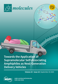
This work forms part of the ‘Women in Physical Chemistry’ Special Issue of Molecules and was conducted by an all-female research team. Herein, we present a series of supramolecular self-associating amphiphilic (SSA) salts. We establish the potential for these molecular constructs to act as next-generation solution-state molecular delivery vehicles. This is achieved through the characterization of the self-associative properties of these SSAs alone and when co-formulated with drug(like) molecules within the solid-state, gas phase, and solution state using a variety of different complementary techniques. Based on these studies and our previous experience, we have also been able to provide an experimental toolkit to support other researchers investigating analogous systems. View this paper.
  • Issues are regarded as officially published after their release is announced to the table of contents alert mailing list.
  • You may sign up for e-mail alerts to receive table of contents of newly released issues.
  • PDF is the official format for papers published in both, html and pdf forms. To view the papers in pdf format, click on the "PDF Full-text" link, and use the free Adobe Reader to open them.
Order results
Result details
Section
Select all
Export citation of selected articles as:
11 pages, 2646 KiB  
Article
Inhibitory Activity of Quercetin 3-O-Arabinofuranoside and 2-Oxopomolic Acid Derived from Malus domestica on Soluble Epoxide Hydrolase
by In Sook Cho, Jang Hoon Kim, Yunjia Lin, Xiang Dong Su, Jong Seong Kang, Seo Young Yang and Young Ho Kim
Molecules 2020, 25(18), 4352; https://doi.org/10.3390/molecules25184352 - 22 Sep 2020
Cited by 9 | Viewed by 2820
Abstract
Flavonoids and triterpenoids were revealed to be the potential inhibitors on soluble epoxide hydrolase (sEH). The aim of this study is to reveal sEH inhibitors from Fuji apples. A flavonoid and three triterpenoids derived from the fruit of Malus domestica were identified as [...] Read more.
Flavonoids and triterpenoids were revealed to be the potential inhibitors on soluble epoxide hydrolase (sEH). The aim of this study is to reveal sEH inhibitors from Fuji apples. A flavonoid and three triterpenoids derived from the fruit of Malus domestica were identified as quercetin-3-O-arabinoside (1), ursolic acid (2), corosolic acid (3), and 2-oxopomolic acid (4). They had half-maximal inhibitory concentration of the inhibitors (IC50) values of 39.3 ± 3.4, 84.5 ± 9.5, 51.3 ± 4.9, and 11.4 ± 2.7 μM, respectively, on sEH. The inhibitors bound to allosteric sites of enzymes in mixed (1) and noncompetitive modes (24). Molecular simulations were carried out for inhibitors 1 and 4 to calculate the binding force of ligands to receptors. The inhibitors bound to the left (1) and right (4) pockets next to the enzyme’s active site. Based on analyses of their molecular docking and dynamics, it was shown that inhibitors 1 and 4 can stably bind sEH at 1 bar and 300 K. Finally, inhibitors 1 and 4 are promising candidates for further studies using cell-based assays and in vivo cardiovascular tests. Full article
Show Figures

Figure 1

Figure 1
<p>The structures of compounds <b>1</b>–<b>4</b> isolated from the fruits of <span class="html-italic">Malus domestica</span>.</p>
Full article ">Figure 2
<p>Inhibitory activities of the inhibitors <b>1</b>–<b>4</b> at a variety of concentrations toward soluble epoxide hydrolase (sEH) (<b>A</b>). Lineweaver-Burk plots (<b>B</b>–<b>F</b>) and Dixon plots (<b>G</b>–<b>I</b>) for the inhibition of sEH by compounds <b>1</b>–<b>4</b>.</p>
Full article ">Figure 3
<p>Predicted docking poses between compounds <b>1</b> and <b>4</b> and sEH (<b>A</b>,<b>B</b>). Hydrogen bonds of inhibitors <b>1</b> and <b>4</b> of the catalytic site, respectively (<b>C</b>,<b>D</b>).</p>
Full article ">Figure 4
<p>Superpositions of sEH with compounds <b>1</b> and <b>4</b> for simulation trajectory (0 ns: red, 1: orange, 2: yellow, 3: green, 4: forest green, 5: cyan, 6: blue, 7: cornflower blue, 8: purple, 9: hot pink, and 10: magenta) (<b>A</b>,<b>B</b>). The potential energy (<b>C</b>), root mean square deviation (RMSD) (<b>D</b>), root mean square fluctuation (RMSF) (<b>E</b>), and hydrogen bonds (<b>F</b>,<b>G</b>) of the simulation calculated with <b>1</b> and <b>4</b> during 10 ns.</p>
Full article ">Figure 4 Cont.
<p>Superpositions of sEH with compounds <b>1</b> and <b>4</b> for simulation trajectory (0 ns: red, 1: orange, 2: yellow, 3: green, 4: forest green, 5: cyan, 6: blue, 7: cornflower blue, 8: purple, 9: hot pink, and 10: magenta) (<b>A</b>,<b>B</b>). The potential energy (<b>C</b>), root mean square deviation (RMSD) (<b>D</b>), root mean square fluctuation (RMSF) (<b>E</b>), and hydrogen bonds (<b>F</b>,<b>G</b>) of the simulation calculated with <b>1</b> and <b>4</b> during 10 ns.</p>
Full article ">Figure 5
<p>The Dictionary of Secondary Structure protein (DSSP) represented with secondary structure changes of sEH with inhibitor <b>1</b> (<b>A</b>) or <b>4</b> (<b>B</b>) (yellow square: Thr360-Pro371, black square: Ser415-Glu424, and blue square: Glu494-Pro501).</p>
Full article ">
13 pages, 979 KiB  
Article
Oxazolochlorins 21. Most Efficient Access to meso-Tetraphenyl- and meso-Tetrakis(pentafluorophenyl)porpholactones, and Their Zinc(II) and Platinum(II) Complexes
by Damaris Thuita, Dinusha Damunupola and Christian Brückner
Molecules 2020, 25(18), 4351; https://doi.org/10.3390/molecules25184351 - 22 Sep 2020
Cited by 5 | Viewed by 3724
Abstract
meso-Phenyl- and meso-pentafluorophenyl-porpholactones, their metal complexes, as well as porphyrinoids directly derived from them are useful in a number of technical and biomedical applications, and more uses are expected to be discovered. About a dozen competing and complementary pathways toward their [...] Read more.
meso-Phenyl- and meso-pentafluorophenyl-porpholactones, their metal complexes, as well as porphyrinoids directly derived from them are useful in a number of technical and biomedical applications, and more uses are expected to be discovered. About a dozen competing and complementary pathways toward their synthesis were reported. The suitability of the methods changes with the meso-aryl group and whether the free base or metal derivatives are sought. These circumstances make it hard for anyone outside of the field of synthetic porphyrin chemistry to ascertain which pathway is the best to produce which specific derivative. We report here on what we experimentally evaluated to be the most efficient pathways to generate the six key compounds from the commercially available porphyrins, meso-tetraphenylporphyrin (TPP) and meso-tetrakis(pentafluorophenyl)porphyrin (TFPP): free base meso-tetraphenylporpholactone (TPL) and meso-tetrakis(pentafluorophenyl)porpholactone (TFPL), and their platinum(II) and zinc(II) complexes TPLPt, TFPLPt, TPLZn, and TFPLZn, respectively. Detailed procedures are provided to make these intriguing molecules more readily available for their further study. Full article
Show Figures

Graphical abstract

Graphical abstract
Full article ">Scheme 1
<p>Generalized formation of <span class="html-italic">meso</span>-arylporpholactones from <span class="html-italic">meso</span>-arylporphyrins.</p>
Full article ">Scheme 2
<p>Select literature-known reaction conditions toward the formation of <span class="html-italic">meso</span>-aryl(metallo)porpholactones. TPP = <span class="html-italic">meso</span>-tetraphenylporphyrin; TPPM = [<span class="html-italic">meso</span>-tetraphenylporphyrinato] metal complex; T<sup>F</sup>PP = <span class="html-italic">meso</span>-tetrakis(pentafluorophenyl)porphyrin; T<sup>F</sup>PPM = [<span class="html-italic">meso</span>-tetrakis(pentafluorophenyl)porphyrinato] metal complex; TPL = <span class="html-italic">meso</span>-tetraphenylporpholactone; TPPM = [<span class="html-italic">meso</span>-tetraphenylkporpholactonato] metal complex; T<sup>F</sup>PL = <span class="html-italic">meso</span>-tetrakis(pentafluorophenyl)porpholactone; T<sup>F</sup>PLM = [<span class="html-italic">meso</span>-tetrakis(pentafluorophenyl)porpholactonato] metal complex.</p>
Full article ">Scheme 3
<p>The synthesis of <span class="html-italic">meso</span>-tetrakis(pentafluorophenyl)porpholactone (T<sup>F</sup>PL) and its zinc(II) (T<sup>F</sup>PLZn) and platinum(II) (T<sup>F</sup>PLPt) complexes.</p>
Full article ">Scheme 4
<p>The most efficient synthesis of <span class="html-italic">meso</span>-tetraphenylporpholactone (TPL) and its zinc(II) (TPLZn) and platinum(II) (TPLPt) complexes.</p>
Full article ">
17 pages, 3907 KiB  
Article
Comprehensive Evaluation of Metal Pollution in Urban Soils of a Post-Industrial City—A Case of Łódź, Poland
by Kinga Wieczorek, Anna Turek, Małgorzata Szczesio and Wojciech M. Wolf
Molecules 2020, 25(18), 4350; https://doi.org/10.3390/molecules25184350 - 22 Sep 2020
Cited by 22 | Viewed by 4873
Abstract
The pollution of urban soils by metals is a global problem. Prolonged exposure of habitants who are in contact with metals retained in soil poses a health risk. This particularly applies to industrialized cities with developed transport networks. The aim of the study [...] Read more.
The pollution of urban soils by metals is a global problem. Prolonged exposure of habitants who are in contact with metals retained in soil poses a health risk. This particularly applies to industrialized cities with developed transport networks. The aim of the study was to determine the content and spatial distribution of mobile metal fractions in soils of the city of Łódź and to identify their load and sources. Multivariate statistical analysis (principal component analysis (PCA), cluster analysis (CA)), combined with GIS, were used to make a comprehensive evaluation of the soil contamination. Hot-spots and differences between urban and suburban areas were also investigated. Metals were determined by atomic absorption spectrometry (AAS) after soil extraction with 1 mol L−1 HCl. In most sites, the metal content changes in the following order: Zn > Pb > Cu > Ni > Cd. About one-third of the samples are considerably (or very highly) contaminated, (contamination factor, CF > 3) with Cu, Pb, or Zn. In almost 40% of the samples, contaminated soils were found (pollution load index, PLI > 1). All metals have a strong influence on the first principal component (PC1), whereas second principal component (PC2) is related to pH. Polluted soils are located in the downtown, in the south and east part of the city. The distribution of contamination coincides with the urban layout, low emission sources and former industrial areas of Łódź. Full article
Show Figures

Graphical abstract

Graphical abstract
Full article ">Figure 1
<p>Location of the sampling sites in Łódź (Poland).</p>
Full article ">Figure 2
<p>Spatial distribution of (<b>a</b>) pH and mobile fractions of: (<b>b</b>) Zn; (<b>c</b>) Cd; (<b>d</b>) Ni; (<b>e</b>) Cu; (<b>f</b>) Pb in soil.</p>
Full article ">Figure 3
<p>Spatial distribution of (<b>a</b>) pollution load index (PLI); (<b>b</b>) first principal component (PC1); (<b>c</b>) second principal component (PC2).</p>
Full article ">Figure 4
<p>Loading plot of two main components PC1 and PC2.</p>
Full article ">Figure 5
<p>Score plot of PC1 versus PC2 for sampling points.</p>
Full article ">Figure 6
<p>Factor values obtained from principal component analysis (PCA) for sampling points and classified according to the cluster analysis (CA) results.</p>
Full article ">
24 pages, 516 KiB  
Article
Volatile Profile Characterization of Croatian Commercial Sparkling Wines
by Ana-Marija Jagatić Korenika, Darko Preiner, Ivana Tomaz and Ana Jeromel
Molecules 2020, 25(18), 4349; https://doi.org/10.3390/molecules25184349 - 22 Sep 2020
Cited by 21 | Viewed by 4143
Abstract
Commercial sparkling wine production represents a relatively low but important part of the Croatian wine production, especially in the Zagreb county. This study presents the results of volatile aroma compounds profile and organic acid composition of commercial sparkling wine samples from three vine-growing [...] Read more.
Commercial sparkling wine production represents a relatively low but important part of the Croatian wine production, especially in the Zagreb county. This study presents the results of volatile aroma compounds profile and organic acid composition of commercial sparkling wine samples from three vine-growing regions in Zagreb county. In total, 174 volatile aroma compounds were identified, separated between their chemical classes (aldehydes, higher alcohols, volatile phenols, terpenes, C13-norisoprenoids, lactones, esters, fatty acids, sulfur compounds, other compounds, other alcohols). Higher alcohols such as phenylethyl and isoamyl alcohol as well as 2-methyl-1-butanol, and esters such as diethyl succinate, ethyl hydrogensuccinate, and ethyl lactate had the strongest impact on the volatile compounds profile of Zagreb county sparkling wine. The presence of diethyl glutarate and diethyl malonate, compounds whose concentrations are influenced by yeast autolysis or caused by chemical esterification during the ageing process, was also noted. The influence of every single volatile aroma compound was evaluated by discriminant analysis using forward stepwise model. The volatile profiles of traditional sparkling wines from Croatia were presented for the first time. It is hoped the results will contribute to better understanding the quality potential and to evaluate possible differences on the bases of detected aroma concentrations and multivariate analysis. Full article
(This article belongs to the Special Issue Wine Chemistry: The Key behind Wine Quality)
Show Figures

Figure 1

Figure 1
<p>Discriminant analysis of 30 sparkling wines (<b>left</b>) based on the concentrations of ten volatile compounds (<b>right</b>)) with highest rank after forward stepwise model applied on total number of volatile compounds detected. The vector diagram indicates the direction and intensity of the effect of nine variables on the distribution of wine samples in the space defined by two discrimination factors (F1 and F2).</p>
Full article ">
13 pages, 3425 KiB  
Article
Thymol Encapsulated into HP-β-Cyclodextrin as an Alternative to Synthetic Fungicides to Induce Lemon Resistance against Sour Rot Decay
by Vicente Serna-Escolano, María Serrano, Daniel Valero, María Isabel Rodríguez-López, José Antonio Gabaldón, Salvador Castillo, Juan Miguel Valverde, Pedro Javier Zapata, Fabián Guillén and Domingo Martínez-Romero
Molecules 2020, 25(18), 4348; https://doi.org/10.3390/molecules25184348 - 22 Sep 2020
Cited by 17 | Viewed by 2895
Abstract
Consumers demand the use of eco-friendly fungicides to treat fruit and vegetables and governmental authorities have unauthorized the application of chemical antifungals for the efficient control of sour rot. In the present research, the microwave irradiation (MW) method was used to encapsulate thymol [...] Read more.
Consumers demand the use of eco-friendly fungicides to treat fruit and vegetables and governmental authorities have unauthorized the application of chemical antifungals for the efficient control of sour rot. In the present research, the microwave irradiation (MW) method was used to encapsulate thymol into 2-hydroxylpropyl-beta-cyclodextrin (HP-β-CD) and the effect of these HP-β-CD on controlling sour rot in citrus fruit, caused by Geotrichum citri-aurantii, was evaluated. Amounts of 25 and 50 mM of HP-β-CD-thymol were used, and compared with propiconazole, to control the decay of inoculated lemon fruit. The treatments were performed in curative and preventive experiments. The incidence and severity of Geotrichum citri-aurantii in 25 and 50 mM HP-β-CD-thymol-treated fruit were reduced in both experiments. The preventive 50 mM HP-β-CD-thymol treatment showed the best effect, reducing the sour rot, respiration rate and fruit weight loss during storage at 20 °C. HP-β-CD-thymol increased polyphenol concentration and the activity of antioxidant enzymes, such as catalase (CAT), ascorbate peroxidase (APX) and peroxidase (POD) in lemon peel, and the highest effects were found with the 50-mM dose. In conclusion, the results show that the use of thymol encapsulated by MW into HP-β-CD could be an effective and sustainable tool, a substitute to the synthetic fungicides, for G. citri-auriantii control in citrus fruit. Full article
(This article belongs to the Special Issue Cyclodextrins: Recent Advances in Chemistry and Applications)
Show Figures

Figure 1

Figure 1
<p>Incidence (%) of fruit affected by <span class="html-italic">G. citri-aurantii</span> when 25 or 50 mM 2-hydroxylpropyl-beta-cyclodextrin (HP-β-CD)-thymol or propiconazole were applied as preventive (<b>A</b>) or curative (<b>B</b>) experiments. Severity (m<sup>2</sup>) of decayed lemons by <span class="html-italic">G. citri-aurantii</span> when 25 or 50 mM HP-β-CD-thymol or propiconazole were applied as preventive (<b>C</b>) or curative (<b>D</b>) experiments. Data are the mean ± SE. Different letters show significant differences (<span class="html-italic">p</span> &lt; 0.05) among treatments for each sampling date.</p>
Full article ">Figure 2
<p>Weight loss (%) of “Fino 49” lemon fruit treated with 25 or 50 mM HP-β-CD-thymol or propiconazole during 15 d of storage at 20 °C in preventive (<b>A</b>) and curative (<b>B</b>) experiments. Data are the mean ± SE. Different letters show significant differences (<span class="html-italic">p</span> &lt; 0.05) among treatments, for each sampling date.</p>
Full article ">Figure 3
<p>Weight loss (%) of “Fino 49” lemon fruit treated with 25 or 50 mM HP-β-CD-thymol or propiconazole during 15 d of storage at 20 °C in preventive (<b>A</b>) and curative (<b>B</b>) experiments. Data are the mean ± SE. Different letters show significant differences (<span class="html-italic">p</span> &lt; 0.05) among treatments for each sampling date.</p>
Full article ">Figure 4
<p>Total phenolics concentration (g kg<sup>−1</sup> gallic acid equivalent), in peel of control, 25 or 50 mM of HP-β-CD-thymol or propiconazole treated “Fino 49” lemon fruits at harvest (day 0) and after 10 d of storage at 20 °C, in preventive experiment. Data are the mean ± SE. Different letters show significant differences (<span class="html-italic">p</span> &lt; 0.05) among treatments after 10 days of storage.</p>
Full article ">Figure 5
<p>Activity of the antioxidant enzymes catalase (CAT), ascorbate peroxidase (APX) and peroxidase (POD) and, in peel of control and 25 or 50 mM HP-β-CD-thymol and propiconazole treated “Fino 49” lemon fruit at harvest and after 10 d of storage at 20 °C in preventive experiment. Data are the mean ± SE. Different letters show significant differences (<span class="html-italic">p</span> &lt; 0.05) among treatments after 10 days of storage.</p>
Full article ">
18 pages, 2359 KiB  
Article
Development of an Optimized MALDI-TOF-MS Method for High-Throughput Identification of High-Molecular-Weight Glutenin Subunits in Wheat
by You-Ran Jang, Kyoungwon Cho, Se Won Kim, Susan B. Altenbach, Sun-Hyung Lim, Jae-Ryeong Sim and Jong-Yeol Lee
Molecules 2020, 25(18), 4347; https://doi.org/10.3390/molecules25184347 - 22 Sep 2020
Cited by 10 | Viewed by 3698
Abstract
Because high-molecular-weight glutenin subunits (HMW-GS) are important contributors to wheat end-use quality, there is a need for high-throughput identification of HMW-GS in wheat genetic resources and breeding lines. We developed an optimized method using matrix-assisted laser desorption/ionization time-of-flight mass spectrometry (MALDI-TOF-MS) to distinguish [...] Read more.
Because high-molecular-weight glutenin subunits (HMW-GS) are important contributors to wheat end-use quality, there is a need for high-throughput identification of HMW-GS in wheat genetic resources and breeding lines. We developed an optimized method using matrix-assisted laser desorption/ionization time-of-flight mass spectrometry (MALDI-TOF-MS) to distinguish individual HMW-GS by considering the effects of the alkylating reagent in protein extraction, solvent components, dissolving volume, and matrix II components. Using the optimized method, 18 of 22 HMW-GS were successfully identified in standard wheat cultivars by differences in molecular weights or by their associations with other tightly linked subunits. Interestingly, 1Bx7 subunits were divided into 1Bx7 group 1 and 1Bx7 group 2 proteins with molecular weights of about 82,400 and 83,000 Da, respectively. Cultivars containing the 1Bx7 group 2 proteins were distinguished from those containing 1Bx7OE using well-known DNA markers. HMW-GS 1Ax2* and 1Bx6 and 1By8 and 1By8*, which are difficult to distinguish due to very similar molecular weights, were easily identified using RP-HPLC. To validate the method, HMW-GS from 38 Korean wheat varieties previously evaluated by SDS-PAGE combined with RP-HPLC were analyzed by MALDI-TOF-MS. The optimized MALDI-TOF-MS method will be a rapid, high-throughput tool for selecting lines containing desirable HMW-GS for breeding efforts. Full article
Show Figures

Figure 1

Figure 1
<p>Effects of four major factors on matrix-assisted laser desorption/ionization time-of-flight mass spectrometry (MALDI-TOF-MS) resolution of Chinese Spring using MALDI-TOF-MS. (<b>A</b>) treatment with alkylation reagent in glutenin extraction, (<b>B</b>) solvent components, (<b>C</b>) dilution volume, and (<b>D</b>) matrix II components. Parameters shown in red were found to be optimal and were incorporated into the new method.</p>
Full article ">Figure 2
<p>HMW-GS profiles of standard wheat cultivars determined by MALDI-TOF-MS. Subunits corresponding to chromosomes 1A, 1B, and 1D are displayed in green, orange, and blue, respectively.</p>
Full article ">Figure 3
<p>Amplification of DNA from standard cultivars containing 1Bx7 or 1Bx7OE genes. (<b>A</b>) PCR primers span the region of the 18 bp insertion in 1Bx7OE; (<b>B</b>) PCR primers from the left junction of the retroelement and duplicated region of Bx7OE; (<b>C</b>) PCR primers from right junction of the retroelement and duplicated region of Bx7OE. Glenlea and IT166460 were used as positive controls for Bx7OE. The 100 bp Plus DNA Ladder is shown in lanes (<b>M</b>).</p>
Full article ">Figure 4
<p>Box and whisker plot of molecular weights of individual subunits in standard wheat cultivars. Red and blue boxes indicate subunits differing by less than 300 Da that were not distinguished by MALDI-TOF-MS, but could be differentiated by RP-HPLC.</p>
Full article ">Figure 5
<p>RP-HPLC analysis of subunits that could not be distinguished by MALDI-TOF-MS. Subunits corresponding to chromosomes 1A, 1B, and 1D are displayed in green, orange, and blue, respectively. Unidentified subunits 1Ax2*, 1Bx6 and 1Bx8, and 1Bx8* are indicated with red arrows.</p>
Full article ">Figure 6
<p>HMW-GS profiles of 38 Korean wheat cultivars of individual subunits determined by MALDI-TOF-MS. Subunits corresponding to chromosomes 1A, 1B, and 1D are shown in green, red, and blue, respectively.</p>
Full article ">
16 pages, 1907 KiB  
Article
Structure and Performance of Benzoxazine Composites for Space Radiation Shielding
by Scott Winroth, Chris Scott and Hatsuo Ishida
Molecules 2020, 25(18), 4346; https://doi.org/10.3390/molecules25184346 - 22 Sep 2020
Cited by 22 | Viewed by 3829
Abstract
Innovative multifunctional materials that combine structural functionality with other spacecraft subsystem functions have been identified as a key enabling technology for future deep space missions. In this work, we report the structure and performance of multifunctional polymer matrix composites developed for aerospace applications [...] Read more.
Innovative multifunctional materials that combine structural functionality with other spacecraft subsystem functions have been identified as a key enabling technology for future deep space missions. In this work, we report the structure and performance of multifunctional polymer matrix composites developed for aerospace applications that require both structural functionality and space radiation shielding. Composites comprised of ultra-high molecular weight polyethylene (UHMWPE) fiber reinforcement and a hydrogen-rich polybenzoxazine matrix are prepared using a low-pressure vacuum bagging process. The polybenzoxazine matrix is derived from a novel benzoxazine resin that possesses a unique combination of attributes: high hydrogen concentration for shielding against galactic cosmic rays (GCR), low polymerization temperature to prevent damage to UHMWPE fibers during composite fabrication, long shelf-life, and low viscosity to improve flow during molding. Dynamic mechanical analysis (DMA) is used to study rheological and thermomechanical properties. Composite mechanical properties, obtained using several standardized tests, are reported. Improvement in composite stiffness, through the addition of carbon fiber skin layers, is investigated. Radiation shielding performance is evaluated using computer-based simulations. The composites demonstrate clear advantages over benchmark materials in terms of combined structural and radiation shielding performance. Full article
Show Figures

Figure 1

Figure 1
<p>Molecular structure of 3BOP-daC12 benzoxazine monomer.</p>
Full article ">Figure 2
<p>Isothermal shear viscosity for 3BOP-daC12 benzoxazine resin at a temperature of 120 °C.</p>
Full article ">Figure 3
<p>Dynamic mechanical analysis (DMA) spectra for isothermal cure of 3BOP-daC12 benzoxazine resin. The intersection of G’ and G” is defined as the gel point.</p>
Full article ">Figure 4
<p>Graphical representation of the composite processing window for the vacuum bagging process.</p>
Full article ">Figure 5
<p>(<b>a</b>) Perspective view of a composite sample with carbon fiber skin layers; (<b>b</b>) cross-sectional micrograph of a composite with carbon fiber skin layers.</p>
Full article ">Figure 6
<p>Example of dual-peak stress–strain curve obtained for CF-UHMWPE-CF/poly(3BOP-daC12) sandwich composite. Initial peak caused by failure of the carbon fiber skin layers followed by failure of the UHMWPE core layer.</p>
Full article ">Figure 7
<p>Plot of specific strength versus equivalent radiation dose for selected materials. Arrow indicates direction of increasing multifunctional performance in terms of higher specific strength and lower radiation dose.</p>
Full article ">Figure 8
<p>Photograph of the vacuum bagging mold assembly.</p>
Full article ">
11 pages, 2071 KiB  
Article
Comparison of the Partial Structure and Antioxidant Activity of Polysaccharides from Two Species of Chinese Truffles
by Xiaolin Li, Zhongkai Zhu, Lei Ye, Zongjing Kang, Xiaoping Zhang, Yue Huang, Bo Zhang and Yuanfeng Zou
Molecules 2020, 25(18), 4345; https://doi.org/10.3390/molecules25184345 - 22 Sep 2020
Cited by 5 | Viewed by 2765
Abstract
Truffles are world-renowned premium commodities. Due to their unique aroma and rarity, the price of truffles has always been very high. In this study, Diethylaminoethyl anion exchange chromatography and gel filtration were employed for polysaccharide purification from two different species of Chinese truffles. [...] Read more.
Truffles are world-renowned premium commodities. Due to their unique aroma and rarity, the price of truffles has always been very high. In this study, Diethylaminoethyl anion exchange chromatography and gel filtration were employed for polysaccharide purification from two different species of Chinese truffles. Three polysaccharide fractions were obtained from Tuber panzhihuanense and referred to as TPZ-NP, TPZ-I, and TPZ-II. Additionally, two polysaccharide fractions were purified from T. pseudoexcavatum (TPD-NP and TPD-I). The results of structural elucidation indicated that the polysaccharide from different species showed different monosaccharide composition and linkage units, as well as molecular weight. Two of the polysaccharide fractions with the highest yield, TPZ-I and TPD-I, were chosen for biological testing. The results indicated that both fractions displayed antioxidant properties through mediation of the intestinal cellular antioxidant defense system, which could protect cultured intestinal cells from oxidative stress-induced damage and cell viability suppression. The TPD-I fraction showed stronger antioxidant effects, which may be due to the difference in structure. Further study on the structure-activity relationship is needed to be done. Full article
Show Figures

Figure 1

Figure 1
<p>The carbohydrate elution profiles monitored by the phenol-sulfuric acid assay. (<b>A</b>) Ion-exchange chromatography elution profile of crude polysaccharide from fruit bodies of <span class="html-italic">T. panzhihuanense</span>. (<b>B</b>) Ion-exchange chromatography elution profile of crude polysaccharide from fruit bodies of <span class="html-italic">T. pseudoexcavatum</span>.</p>
Full article ">Figure 2
<p>The carbohydrate elution profiles of gel filtration chromatography, monitored with the phenol-sulfuric acid assay. Elution profiles of (<b>A</b>) TPZ-I, (<b>B</b>) TPZ-II, and (<b>C</b>) TPD-I.</p>
Full article ">Figure 3
<p>Protective effects of TPZ-I and TPD-I on viability loss in porcine jejunum epithelial cells (IPEC-J2) induced by H<sub>2</sub>O<sub>2</sub> (0.2 mM). The concentrations of polysaccharide fractions were 20, 10, and 5 μg/mL, respectively. Values are presented as means ± Standard error. Bars with different letters are statistically significant (<span class="html-italic">p</span> &lt; 0.05).</p>
Full article ">Figure 4
<p>Effects of TPZ-I and TPD-I on the activities of malondialdehyde-MDA (<b>A</b>), superoxide dismutase-SOD (<b>B</b>), catalase-CAT (<b>C</b>), and glutathione peroxidase-GSH-Px (<b>D</b>) in IPEC-J2 cells. The concentrations of the polysaccharide fractions were 20, 10, and 5 μg/mL, respectively. Values are presented as means ± SD (<span class="html-italic">n</span> = 6). Bars with different letters are statistically significant (<span class="html-italic">p</span> &lt; 0.05).</p>
Full article ">Figure 5
<p>Effects of TPZ-I and TPD-I on the activities of total antioxidant capacity-T-AOC (<b>A</b>), and reactive oxygen species-ROS (<b>B</b>) in IPEC-J2 cells. The concentrations of the polysaccharide fractions were 20, 10, and 5 μg/mL, respectively. Values are presented as means ± SD (<span class="html-italic">n</span> = 6). Bars with different letters are statistically significant (<span class="html-italic">p</span> &lt; 0.05).</p>
Full article ">
18 pages, 2629 KiB  
Article
Biocatalytic Potential of Native Basidiomycetes from Colombia for Flavour/Aroma Production
by David A. Jaramillo, María J. Méndez, Gabriela Vargas, Elena E. Stashenko, Aída-M. Vasco-Palacios, Andrés Ceballos and Nelson H. Caicedo
Molecules 2020, 25(18), 4344; https://doi.org/10.3390/molecules25184344 - 22 Sep 2020
Cited by 9 | Viewed by 4567
Abstract
Aromas and flavours can be produced from fungi by either de novo synthesis or biotransformation processes. Herein, the biocatalytic potential of seven basidiomycete species from Colombia fungal strains isolated as endophytes or basidioma was evaluated. Ganoderma webenarium, Ganoderma chocoense, and Ganoderma [...] Read more.
Aromas and flavours can be produced from fungi by either de novo synthesis or biotransformation processes. Herein, the biocatalytic potential of seven basidiomycete species from Colombia fungal strains isolated as endophytes or basidioma was evaluated. Ganoderma webenarium, Ganoderma chocoense, and Ganoderma stipitatum were the most potent strains capable of decolourizing β,β-carotene as evidence of their potential as biocatalysts for de novo aroma synthesis. Since a species’ biocatalytic potential cannot solely be determined via qualitative screening using β,β-carotene biotransformation processes, we focused on using α-pinene biotransformation with mycelium as a measure of catalytic potential. Here, two strains of Trametes elegans—namely, the endophytic (ET-06) and basidioma (EBB-046) strains—were screened. Herein, T. elegans is reported for the first time as a novel biocatalyst for the oxidation of α-pinene, with a product yield of 2.9 mg of cis-Verbenol per gram of dry weight mycelia used. The EBB-046 strain generated flavour compounds via the biotransformation of a Cape gooseberry medium and de novo synthesis in submerged cultures. Three aroma-producing compounds were identified via GC–MS—namely, methyl-3-methoxy-4H-pyran-4-one, hexahydro-3-(methylpropyl)-pyrrolo[1,2-a]pyrazine-1,4-dione, and hexahydro-3-(methylphenyl)-pyrrolo[1,2-a]pyrazine-1,4-dione. Full article
(This article belongs to the Special Issue Natural Products from Fungi)
Show Figures

Graphical abstract

Graphical abstract
Full article ">Figure 1
<p>β,β-carotene biotransformation after a 4- and 14-day incubation periods. Screening was conducted using seven strains of native basidiomycetes—namely, <span class="html-italic">Ganoderma stipitatum</span> (<b>A</b>), <span class="html-italic">Ganoderma chocoense</span> (<b>B</b>), <span class="html-italic">Ganoderma gibbosum</span> (<b>C</b>), <span class="html-italic">Ganoderma weberianum</span> (<b>D</b>), <span class="html-italic">Laetiporus gilbertsonii</span> (<b>E</b>), <span class="html-italic">Trametes elegans</span> (endophyte) (<b>F</b>), and <span class="html-italic">Trametes elegans</span> (<b>G</b>).</p>
Full article ">Figure 2
<p>The hydroxylation of α-pinene via the P450 monooxygenase/reductase system (adapted from Krings [<a href="#B57-molecules-25-04344" class="html-bibr">57</a>]).</p>
Full article ">Figure 3
<p>The influence exerted by the concentration of α-pinene (10 and 20 mM) and the mycelium (5 and 10 g/L) on the <span class="html-italic">cis</span>-Verbenol yield during biotransformation reactions using whole cells of the EBB-046 and ET-06 strains of <span class="html-italic">Trametes elegans</span>. The incubation time was 48 h.</p>
Full article ">Figure 4
<p>(<b>A</b>) Pellets formed by the EBB-046 strain in the Yeast extract-Malt extract medium YM. (<b>B</b>) Mycelial clumps formed by the EBB-046 strain in the yeast extract medium. (<b>C</b>) Image of the submerged culture of <span class="html-italic">Trametes elegans</span> (EBB-046 strain) in a defined medium using Cape Gooseberry after an incubation period of 96 h.</p>
Full article ">Figure 5
<p>Intensity progress descriptor for the generation of sweet and fruity odours as a function of time by <span class="html-italic">Trametes elegans</span> mycelia cultivated in media containing Cape Gooseberry. The odour intensity was rated on a scale of one to eight, where 1–2 represented a low intensity, 3–4 was described as “characteristic”, 5–6 was classified as “intense”, and 7–8 was a strong odour.</p>
Full article ">Figure 6
<p>Chromatogram of the biotransformation products of <span class="html-italic">Trametes elegans</span> (EBB-046) obtained after an 80-h incubation period.</p>
Full article ">
21 pages, 906 KiB  
Review
The Lipidome Fingerprint of Longevity
by Mariona Jové, Natàlia Mota-Martorell, Irene Pradas, José Daniel Galo-Licona, Meritxell Martín-Gari, Èlia Obis, Joaquim Sol and Reinald Pamplona
Molecules 2020, 25(18), 4343; https://doi.org/10.3390/molecules25184343 - 22 Sep 2020
Cited by 19 | Viewed by 4295
Abstract
Lipids were determinants in the appearance and evolution of life. Recent studies disclose the existence of a link between lipids and animal longevity. Findings from both comparative studies and genetics and nutritional interventions in invertebrates, vertebrates, and exceptionally long-lived animal species—humans included—demonstrate that [...] Read more.
Lipids were determinants in the appearance and evolution of life. Recent studies disclose the existence of a link between lipids and animal longevity. Findings from both comparative studies and genetics and nutritional interventions in invertebrates, vertebrates, and exceptionally long-lived animal species—humans included—demonstrate that both the cell membrane fatty acid profile and lipidome are a species-specific optimized evolutionary adaptation and traits associated with longevity. All these emerging observations point to lipids as a key target to study the molecular mechanisms underlying differences in longevity and suggest the existence of a lipidome profile of long life. Full article
(This article belongs to the Special Issue Lipids in Health and Disease)
Show Figures

Figure 1

Figure 1
<p>Specific lipidomic profiles at subcellular, tissue, and animal species level. (<b>A</b>) Specific lipidomic profile defines different subcellular components such as endoplasmic reticulum (ER), mitochondrial-associated membranes (MAM), and mitochondrion (MIT). (<b>B</b>) Specific lipidomic profile for rat tissues. Principal component analysis (PCA) representation of the lipidome of all the tissues in positive ionization. Modified with permission from [<a href="#B46-molecules-25-04343" class="html-bibr">46</a>]. (<b>C</b>) Specific plasma lipidomic profiles for mammalian species. PCA representation (positive ionization molecules) showing that plasma lipidomic profiles are species-specific. Modified with permission from [<a href="#B35-molecules-25-04343" class="html-bibr">35</a>]. (<b>D</b>) Human extreme longevity as model of healthy aging. PCA (positive ionization) revealed differences in adults, aged, and centenarian plasma lipidomic profiles. Modified with permission from [<a href="#B48-molecules-25-04343" class="html-bibr">48</a>].</p>
Full article ">Figure 2
<p>Relationship between peroxidation index (PI) and longevity of skeletal muscle phospholipids for mammalians and birds, pointing out exceptionally long-lived animal species. Modified with permission from [<a href="#B28-molecules-25-04343" class="html-bibr">28</a>].</p>
Full article ">
17 pages, 2249 KiB  
Article
Design, Synthesis, In Vitro and In Silico Studies of New Thiazolylhydrazine-Piperazine Derivatives as Selective MAO-A Inhibitors
by Begüm Nurpelin Sağlık, Osman Cebeci, Ulviye Acar Çevik, Derya Osmaniye, Serkan Levent, Betül Kaya Çavuşoğlu, Sinem Ilgın, Yusuf Özkay and Zafer Asım Kaplancıklı
Molecules 2020, 25(18), 4342; https://doi.org/10.3390/molecules25184342 - 22 Sep 2020
Cited by 9 | Viewed by 2853
Abstract
Monoamine oxidase (MAO) isoenzymes are very important drug targets among neurological disorders. Herein, novel series of thiazolylhydrazine-piperazine derivatives were designed, synthesized and evaluated for their MAO-A and -B inhibitory activity. The structures of the synthesized compounds were assigned using different spectroscopic techniques such [...] Read more.
Monoamine oxidase (MAO) isoenzymes are very important drug targets among neurological disorders. Herein, novel series of thiazolylhydrazine-piperazine derivatives were designed, synthesized and evaluated for their MAO-A and -B inhibitory activity. The structures of the synthesized compounds were assigned using different spectroscopic techniques such as 1H-NMR, 13C-NMR and HRMS. Moreover, the prediction of ADME (Absorption, Distribution, Metabolism, Elimination) parameters for all of the compounds were performed using in silico method. According to the enzyme inhibition results, the synthesized compounds showed the selectivity against MAO-A enzyme inhibition. Compounds 3c, 3d and 3e displayed significant MAO-A inhibition potencies. Among them, compound 3e was found to be the most effective derivative with an IC50 value of 0.057 ± 0.002 µM. Moreover, it was seen that this compound has a more potent inhibition profile than the reference inhibitors moclobemide (IC50 = 6.061 ± 0.262 µM) and clorgiline (IC50 = 0.062 ± 0.002 µM). In addition, the enzyme kinetics were performed for compound 3e and it was determined that this compound had a competitive and reversible inhibition type. Molecular modeling studies aided in the understanding of the interaction modes between this compound and MAO-A. It was found that compound 3e had significant and important binding property. Full article
(This article belongs to the Special Issue Trends in the Development of Enzyme Inhibitors)
Show Figures

Figure 1

Figure 1
<p>Design of target compounds.</p>
Full article ">Figure 2
<p>(<b>A</b>) Lineweaver-Burk plots for the inhibition of MAO-A by compound <b>3e</b>. [S], substrate concentration (µM); <b>V</b>, reaction velocity (nmol/min/mg protein). Inhibitor concentrations are shown at the left. (<b>B</b>) Secondary plot (slope, namely K<sub>m</sub>/V<sub>max</sub> obtained from graph <b>A</b>, versus inhibitor concentrations) of compound <b>3e</b> for determining the steady-state inhibition constant (K<sub>i</sub>). K<sub>i</sub> was calculated to be 0.011 μM.</p>
Full article ">Figure 3
<p>The three-dimensional (3D) poses of compounds <b>3c</b>, <b>3d</b> and <b>3e</b> in the active region of MAO-A (PDB ID: 2Z5X). The compounds are shown in a tube pattern and are yellow, maroon and orange colored, respectively.</p>
Full article ">Figure 4
<p>The 3D interacting modes of compound <b>3c</b>, <b>3d</b> and <b>3e</b> in the active region of MAO-A. Tube model introduce the ligands and the relevant residues in the enzyme’s active site. The flavin adenine dinucleotide (FAD) molecule is shown in a ball and stick pattern and is blue colored. (<b>A</b>) Compound 3<b>c</b> is colored = yellow. (<b>B</b>) Compound <b>3d</b> is colored maroon. (<b>C</b>) Compound <b>3e</b> is colored orange.</p>
Full article ">Scheme 1
<p>The synthetic route of the compounds (<b>3a</b>–<b>3l</b>). Reagents and conditions: (i) DMF, K<sub>2</sub>CO<sub>3</sub>, reflux, 24 h; (ii) EtOH, reflux, 3 h; (iii) EtOH, reflux, 4–8 h.</p>
Full article ">
22 pages, 5067 KiB  
Article
Novel Bis-Ammonium Salts of Pyridoxine: Synthesis and Antimicrobial Properties
by Nikita V. Shtyrlin, Mikhail V. Pugachev, Sergey V. Sapozhnikov, Marsel R. Garipov, Rusalia M. Vafina, Denis Y. Grishaev, Roman S. Pavelyev, Renata R. Kazakova, Mariya N. Agafonova, Alfiya G. Iksanova, Svetlana A. Lisovskaya, Marina I. Zeldi, Elena S. Krylova, Elena V. Nikitina, Alina E. Sabirova, Airat R. Kayumov and Yurii G. Shtyrlin
Molecules 2020, 25(18), 4341; https://doi.org/10.3390/molecules25184341 - 22 Sep 2020
Cited by 16 | Viewed by 3371
Abstract
A series of 108 novel quaternary bis-ammonium pyridoxine derivatives carrying various substituents at the quaternary nitrogen’s and acetal carbon was synthesized. Thirteen compounds exhibited antibacterial and antifungal activity (minimum inhibitory concentration (MIC) 0.25–16 µg/mL) comparable or superior than miramistin, benzalkonium chloride, and chlorhexidine. [...] Read more.
A series of 108 novel quaternary bis-ammonium pyridoxine derivatives carrying various substituents at the quaternary nitrogen’s and acetal carbon was synthesized. Thirteen compounds exhibited antibacterial and antifungal activity (minimum inhibitory concentration (MIC) 0.25–16 µg/mL) comparable or superior than miramistin, benzalkonium chloride, and chlorhexidine. A strong correlation between the lipophilicity and antibacterial activity was found. The most active compounds had logP values in the range of 1–3, while compounds with logP > 6 and logP < 0 were almost inactive. All active compounds demonstrated cytotoxicity comparable with miramistin and chlorhexidine on HEK-293 cells and were three-fold less toxic when compared to benzalkonium chloride. The antibacterial activity of leading compound 5c12 on biofilm-embedded Staphylococcus aureus, Staphylococcus epidermidis, Escherichia coli or Pseudomonas aeruginosa was comparable or even higher than that of the benzalkonium chloride. In vivo 5c12 was considerably less toxic (LD50 1705 mg/kg) than benzalkonium chloride, miramistine, and chlorhexidine at oral administration on CD-1 mice. An aqueous solution of 5c12 (0.2%) was shown to be comparable to reference drugs efficiency on the rat’s skin model. The molecular target of 5c12 seems to be a cellular membrane as other quaternary ammonium salts. The obtained results make the described quaternary bis-ammonium pyridoxine derivatives promising and lead molecules in the development of the new antiseptics with a broad spectrum of antimicrobial activity. Full article
(This article belongs to the Special Issue Microbiocides Chemistry)
Show Figures

Graphical abstract

Graphical abstract
Full article ">Figure 1
<p>The structures of QAC based on pyridoxine derivatives obtained by us earlier [<a href="#B13-molecules-25-04341" class="html-bibr">13</a>,<a href="#B14-molecules-25-04341" class="html-bibr">14</a>,<a href="#B15-molecules-25-04341" class="html-bibr">15</a>,<a href="#B16-molecules-25-04341" class="html-bibr">16</a>,<a href="#B17-molecules-25-04341" class="html-bibr">17</a>,<a href="#B18-molecules-25-04341" class="html-bibr">18</a>,<a href="#B19-molecules-25-04341" class="html-bibr">19</a>] and compounds <b>5a<sub>8</sub></b>–<b>5r<sub>18</sub></b> synthesized in this work.</p>
Full article ">Figure 2
<p>Lg1/MIC versus calculated logP relationship for novel bis-ammonium salts of pyridoxine. The parabolic line shows the polynomial approximation by using a second order model. Inactive compounds (MIC &gt; 64 mg/mL) are not shown.</p>
Full article ">Figure 3
<p>The effect of <b>5c<sub>12</sub></b> and reference antiseptics on biofilm-embedded bacteria. (<b>A</b>)—<span class="html-italic">S. aureus</span>; (<b>B</b>)—<span class="html-italic">S. intermedius;</span> (<b>C</b>)—<span class="html-italic">E. coli</span>, (<b>D</b>)—<span class="html-italic">P. aeruginosa</span>. The 48-h biofilms were treated with 1–16 × MBCs of benzalkonium chloride (circles), miramistin (squares), or <b>5c<sub>12</sub></b> (triangles) for 24 h and then quantified by CFU counting. Data are present as medians from five independent experiments with interquartile range (M ± IQR). Dotted line shows 3 log-decrease of viable cells.</p>
Full article ">Figure 4
<p>In vivo antibacterial efficacy test (Mean ± SD) using a rat skin model. log10 number of viable cells in the control and the number of viable cells after exposure with <b>5c<sub>12</sub></b>, miramistin, benzalkonium chloride, and chlorhexidine, <span class="html-italic">n</span> = 6.</p>
Full article ">Figure 5
<p>The effect of <b>5c<sub>12</sub></b> on the electric potential of the bacterial membrane determined using a membrane-potential-sensitive cyanine dye DiSC<sub>3</sub>(5) (10 min treatment by 1–8 × MBC (<b>A</b>,<b>B</b>) and 20 min treatment by 1–8 × MBC (<b>C</b>,<b>D</b>)). Asterisks denote a statistically significant difference in comparison with untreated cells. * statistically significant difference from the control.</p>
Full article ">Figure 6
<p>Scanning Electron Microscopy microphotographs of <span class="html-italic">S. aureus</span> ATCC209p (<b>A</b>,<b>C</b>) and <span class="html-italic">E. coli</span> CDC F-50 (<b>B</b>,<b>D</b>). Untreated cells (<b>A</b>,<b>B</b>) and bacteria treated by 0.2% solution of <b>5c<sub>12</sub></b> for 30 min (<b>C</b>,<b>D</b>). 50,000× magnification.</p>
Full article ">Scheme 1
<p>Reagents and conditions. (i) R<sub>1</sub>C(O)R<sub>2</sub>, HCl, 3–5 °C, 3 h; or C<sub>6</sub>H<sub>6</sub>, R<sub>1</sub>C(O)R<sub>2</sub>, <span class="html-italic">p</span>-TsOH, reflux, 17 h, (ii) H<sub>2</sub>O<sub>,</sub> NaOH, 20 °C, (iii) CH<sub>2</sub>Cl<sub>2</sub>, SOCl<sub>2</sub>, 20 °C, 17 h, (iv) H<sub>2</sub>O/CHCl<sub>3,</sub> NaHCO<sub>3</sub>, (v) CHCl<sub>3</sub>, TCICA, reflux, 4 h, (vi) C<sub>2</sub>H<sub>5</sub>OH, R<sub>3</sub>N(CH<sub>3</sub>)<sub>2</sub>, 70 °C, 5 h.</p>
Full article ">
19 pages, 1416 KiB  
Article
Urinary Resveratrol Metabolites Output: Differential Associations with Cardiometabolic Markers and Liver Enzymes in House-Dwelling Subjects Featuring Metabolic Syndrome
by Vanessa Bullón-Vela, Itziar Abete, Maria Angeles Zulet, Yifan Xu, Miguel A. Martínez-González, Carmen Sayón-Orea, Miguel Ruiz-Canela, Estefanía Toledo, Vicente Martín Sánchez, Ramon Estruch, Rosa María Lamuela-Raventós, Enrique Almanza-Aguilera, Montserrat Fitó, Jordi Salas-Salvadó, Andrés Díaz-López, Francisco J. Tinahones, Josep A. Tur, Dora Romaguera, Jadwiga Konieczna, Xavier Pintó, Lidia Daimiel, Ana Rodriguez-Mateos and José Alfredo Martínezadd Show full author list remove Hide full author list
Molecules 2020, 25(18), 4340; https://doi.org/10.3390/molecules25184340 - 22 Sep 2020
Cited by 7 | Viewed by 3615
Abstract
Metabolic syndrome (MetS) components are strongly associated with increased risk of non-alcoholic fatty liver disease (NAFLD) development. Several studies have supported that resveratrol is associated with anti-inflammatory and antioxidant effects on health status. The main objective of this study was to assess the [...] Read more.
Metabolic syndrome (MetS) components are strongly associated with increased risk of non-alcoholic fatty liver disease (NAFLD) development. Several studies have supported that resveratrol is associated with anti-inflammatory and antioxidant effects on health status. The main objective of this study was to assess the putative associations between some urinary resveratrol phase II metabolites, cardiometabolic, and liver markers in individuals diagnosed with MetS. In this cross-sectional study, 266 participants from PREDIMED Plus study (PREvención con DIeta MEDiterránea) were divided into tertiles of total urinary resveratrol phase II metabolites (sum of five resveratrol conjugation metabolites). Urinary resveratrol metabolites were analyzed by ultra- performance liquid chromatography coupled to triple quadrupole mass spectrometry (UPLC-Q-q-Q MS), followed by micro-solid phase extraction (µ-SPE) method. Liver function markers were assessed using serum levels of aspartate aminotransferase (AST), alanine aminotransferase (ALT), and gamma-glutamyl transferase (GGT). Moreover, lipid profile was measured by triglycerides, very-low-density lipoprotein cholesterol (VLDL-c), and total cholesterol/high-density lipoprotein ratio (total cholesterol/HDL). Linear regression adjusted models showed that participants with higher total urine resveratrol concentrations exhibited improved lipid and liver markers compared to the lowest tertile. For lipid determinations: log triglycerides (βT3= −0.15, 95% CI; −0.28, −0.02, p-trend = 0.030), VLDL-c, (βT3= −4.21, 95% CI; −7.97, −0.46, p-trend = 0.039), total cholesterol/HDL ratio Moreover, (βT3= −0.35, 95% CI; −0.66, −0.03, p-trend = 0.241). For liver enzymes: log AST (βT3= −0.12, 95% CI; −0.22, −0.02, p-trend = 0.011, and log GGT (βT3= −0.24, 95% CI; −0.42, −0.06, p-trend = 0.002). However, there is no difference found on glucose variables between groups. To investigate the risk of elevated serum liver markers, flexible regression models indicated that total urine resveratrol metabolites were associated with a lower risk of higher ALT (169.2 to 1314.3 nmol/g creatinine), AST (599.9 to 893.8 nmol/g creatinine), and GGT levels (169.2 to 893.8 nmol/g creatinine). These results suggested that higher urinary concentrations of some resveratrol metabolites might be associated with better lipid profile and hepatic serum enzymes. Moreover, urinary resveratrol excreted showed a reduced odds ratio for higher liver enzymes, which are linked to NAFLD. Full article
(This article belongs to the Section Natural Products Chemistry)
Show Figures

Figure 1

Figure 1
<p>The odds ratio for liver enzymes levels above the upper limit of normal (ULN) for total urine resveratrol concentration in nmol/g creatinine. ULN range for ALT (men ≥ 30 UI/L, women ≥ 19 UI/L) (<b>A</b>), AST (men ≥ 37 UI/L, women ≥ 31 UI/L) (<b>B</b>), and GGT (men ≥ 60 UI/L, women ≥ 40 UI/L) (<b>C</b>). The smooth line represents the estimation of higher ALT, AST, and GGT levels when using zero as the reference value for total urine resveratrol metabolite (4 knots for ALT and GGT; 3 knots for AST) whereas the dashed lines indicate 95% CIs.</p>
Full article ">
8 pages, 1791 KiB  
Article
Conformation of G-quadruplex Controlled by Click Reaction
by Chao-Da Xiao, Zhi-Yong He, Chuan-Xin Guo, Xiang-Chun Shen and Yan Xu
Molecules 2020, 25(18), 4339; https://doi.org/10.3390/molecules25184339 - 22 Sep 2020
Cited by 1 | Viewed by 2905
Abstract
G-quadruplexes are non-canonical four stranded secondary structures possessing great biological importance. Controlling G-quadruplex conformation for further regulating biological processes is both exciting and challenging. In this study, we described a method for regulating G-quadruplex conformation by click chemistry for the first time. 8-ethynyl-2′-deoxyguanosine [...] Read more.
G-quadruplexes are non-canonical four stranded secondary structures possessing great biological importance. Controlling G-quadruplex conformation for further regulating biological processes is both exciting and challenging. In this study, we described a method for regulating G-quadruplex conformation by click chemistry for the first time. 8-ethynyl-2′-deoxyguanosine was synthesized and incorporated into a 12-nt telomere DNA sequence. Such a sequence, at first, formed mixed parallel/anti-parallel G-quadruplexes, while it changed to anti-parallel after reaction with azidobenzene. Meanwhile, the click reaction can give the sequence intense fluorescence. Full article
Show Figures

Figure 1

Figure 1
<p>(<b>a</b>) Click reaction controls the glycosidic conformation of modified guanine nucleoside. (<b>b</b>) G-quadruplex topology changed by manipulating the glycosidic conformation of modified guanine nucleoside through click reaction.</p>
Full article ">Figure 2
<p>(<b>a</b>) NOESY spectrum of 8<sup>et</sup>dG click reaction product. Cross peak between H in the triazole ring and H1′ is in the red box. (<b>b</b>) Schematic representation of <span class="html-italic">syn</span> glycosidic conformation of 8<sup>et</sup>dG click reaction product with arrows indicating the NOEs.</p>
Full article ">Figure 3
<p>HPLC analyses of the ODN-N reaction with the CuAAC reaction solution at the indicted time where peak “P” is the product.</p>
Full article ">Figure 4
<p>(<b>a</b>) CD spectrum of unreacted and click reaction product of ODN-9. (<b>b</b>) Fluorescence spectra for modified oligonucleotides. ODN-9 P (em): the emission spectrum for click reaction product of ODN-9; ODN-9 (em): emission spectrum for the ODN-9 unreacted sequence; ODN-9 P (ex), excitation spectrum (345 nm).</p>
Full article ">Scheme 1
<p>Reagents and conditions: (<b>a</b>) Trimethylsilyl acetylene, Tetrakis(triphenylphosphine)palladium, Triethylamine, <span class="html-italic">N,N</span>-Dimethylformamide, 50 °C, 3 h; (<b>b</b>) Tetrabutylammonium fluoride, Tetrahydrofuran, rt, 2 h; (<b>c</b>) <span class="html-italic">N,N</span>-dimethylformamide diethylacetal, methanol, 55 °C, 3 h; (<b>d</b>) 4,4′-Dimethoxytriphenylmethyl chloride, Triethylamine, 4-(Dimethylamino)pyridine, Pyridine, rt, overnight; (<b>e</b>) <span class="html-italic">N,N</span>-diisopropylchlorophosphoramidite, Triethylamine, rt, 15 min.</p>
Full article ">
16 pages, 430 KiB  
Review
Autophagy, One of the Main Steps in Periodontitis Pathogenesis and Evolution
by Maria Greabu, Francesca Giampieri, Marina Melescanu Imre, Maria Mohora, Alexandra Totan, Silviu Mirel Pituru and Ecaterina Ionescu
Molecules 2020, 25(18), 4338; https://doi.org/10.3390/molecules25184338 - 22 Sep 2020
Cited by 29 | Viewed by 3968
Abstract
Periodontitis represents a complex inflammatory disease that compromises the integrity of the tooth-supporting tissue through the interaction of specific periodontal pathogens and the host’s immune system. Experimental data help to outline the idea that the molecular way towards periodontitis initiation and progression presents [...] Read more.
Periodontitis represents a complex inflammatory disease that compromises the integrity of the tooth-supporting tissue through the interaction of specific periodontal pathogens and the host’s immune system. Experimental data help to outline the idea that the molecular way towards periodontitis initiation and progression presents four key steps: bacterial infection, inflammation, oxidative stress, and autophagy. The aim of this review is to outline the autophagy involvement in the pathogenesis and evolution of periodontitis from at least three points of view: periodontal pathogen invasion control, innate immune signaling pathways regulation and apoptosis inhibition in periodontal cells. The exact roles played by reactive oxygen species (ROS) inside the molecular mechanisms for autophagy initiation in periodontitis still require further investigation. However, clarifying the role and the mechanism of redox regulation of autophagy in the periodontitis context may be particularly beneficial for the elaboration of new therapeutic strategies. Full article
Show Figures

Figure 1

Figure 1
<p>Schematic representation of the possible mechanism of autophagy regulation in the periodontitis context. Autophagy can be modulated by ROS via four different pathways: (1) Atg12–Atg5 complex activation, promoting autophagy elongation; (2) ROS-dependent JNK induced Bcl-2 phosphorylation triggering Beclin 1 dissociation and autophagy induction; (3) PI3K-AKT pathway initiation triggering the activation of mTOR, which, in turn, acts as an autophagy induction inhibitor; and (4) the AMPK-dependent TORC1 activity inhibition leading to autophagy activation. Adapted from Liu C. et al. [<a href="#B51-molecules-25-04338" class="html-bibr">51</a>].</p>
Full article ">
23 pages, 8091 KiB  
Article
Sulphamoylated Estradiol Analogue Induces Reactive Oxygen Species Generation to Exert Its Antiproliferative Activity in Breast Cancer Cell Lines
by Maphuti T. Lebelo, Anna M. Joubert and Michelle H. Visagie
Molecules 2020, 25(18), 4337; https://doi.org/10.3390/molecules25184337 - 22 Sep 2020
Cited by 4 | Viewed by 2521
Abstract
2-Methoxyestradiol (2ME), a 17β-estradiol metabolite, exerts anticancer properties in vitro and in vivo. To address 2ME’s low bioavailability, research led to the in silico design of sulphamoylated 2ME analogues. However, the role of oxidative stress induced in the activity exerted by sulphamoylated compounds [...] Read more.
2-Methoxyestradiol (2ME), a 17β-estradiol metabolite, exerts anticancer properties in vitro and in vivo. To address 2ME’s low bioavailability, research led to the in silico design of sulphamoylated 2ME analogues. However, the role of oxidative stress induced in the activity exerted by sulphamoylated compounds remains elusive. In the current study, the influence of 2-Ethyl-17-oxoestra-1,3,5(10)-trien-3-yl sulphamate (ESE-one) on reactive oxygen species (ROS) induction and its effect on cell proliferation, as well as morphology, were assessed in breast tumorigenic cells (MCF-7 and MDA-MB-231). Fluorescent microscopy showed that sulphamoylated estradiol analogues induced hydrogen peroxide and superoxide anion, correlating with decreased cell growth demonstrated by spectrophotometry data. ESE-one exposure resulted in antiproliferation which was repressed by tiron (superoxide inhibitor), trolox (peroxyl inhibitor) and N,N′-dimethylthiourea (DMTU) (hydrogen peroxide inhibitor). Morphological studies demonstrated that tiron, trolox and DMTU significantly decreased the number of rounded cells and shrunken cells in MCF-7 and MDA-MB-231 cells induced by ESE-one. This in vitro study suggests that ESE-one induces growth inhibition and cell rounding by production of superoxide anion, peroxyl radical and hydrogen peroxide. Identification of these biological changes in cancer cells caused by sulphamoylated compounds hugely contributes towards improvement of anticancer strategies and the ROS-dependent cell death pathways in tumorigenic breast cells. Full article
Show Figures

Figure 1

Figure 1
<p>Fluorescent micrographs of MCF-7 and MDA-MB-231 cells exposed to sulphamoylated and non-sulphamoylated compounds. Sulphamoylated compounds induced ROS production in both MCF-7 and MDA-MB-231 cells compared to their non-sulphamoylated counterparts. (<b>a</b>) MCF-7 cells propagated in growth medium, (<b>b</b>) vehicle-treated MCF-7 cells, (<b>c</b>) MDA-MB-231 cells propagated in growth medium, (<b>d</b>) vehicle-treated MDA-MB-231 cells, (<b>e</b>) EE-15-ol (non-sulphamoylated) treated MCF-7 cells, (<b>f</b>) ESE-15-ol (sulphamoylated)-treated MCF-7 cells, (<b>g</b>) EE-15-ol (non-sulphamoylated)-treated MDA-MB-231 cells, (<b>h</b>) ESE-15-ol (sulphamoylated)-treated MDA-MB-231 cells, (<b>i</b>) EE-one (non-sulphamoylated)-treated MCF-7 cells, (<b>j</b>) ESE-one (sulphamoylated)-treated MCF-7 cells, (<b>k</b>) EE-one (non-sulphamoylated)-treated MDA-MB-231 cells, (<b>l</b>) ESE-one (sulphamoylated)-treated MDA-MB-231 cells, (<b>m</b>) 2E-diol (non-sulphamoylated)-treated MCF-7 cells, (<b>n</b>) ESE-ol (sulphamoylated)-treated MCF-7 cells, (<b>o</b>) 2E-diol (non-sulphamoylated)-treated MDA-MB-231 cells, (<b>p</b>) ESE-ol (sulphamoylated)-treated MDA-MB-231 cells. Images were captured at 20X magnification.</p>
Full article ">Figure 2
<p>MCF-7 and MDA-MB-231 graphs demonstrating the mean fluorescent intensity following exposure to sulphamoylated and non-sulphamoylated compounds. Sulphamoylated compounds induced superoxide anion and hydrogen peroxide production in both MCF-7 and MDA-MB-231 cells compared to the non-sulphamoylated compounds. (<b>a</b>) MCF-7 cells, (<b>b</b>) MDA-MB-231 cells. Asterisk (*) represents <span class="html-italic">p</span>-value (<span class="html-italic">p</span> &lt; 0.05) compared to vehicle-treated cells.</p>
Full article ">Figure 3
<p>Graph of MCF-7 and MDA-MB231 cells illustrating effect on proliferation after exposure to sulphamoylated and non-sulphamoylated compounds. Non-sulphamoylated compounds exerted no significant inhibiting effect on cell growth in MCF-7 cell inhibition whereas sulphamoylated compounds demonstrated at least 28% cell inhibition in both cell lines. Non-sulphamoylated compounds had an opposite effect and caused cell growth demonstrated by EE-one and 2-E-diol. (<b>a</b>) MCF-7 cells, (<b>b</b>) MDA-MB-231 cells. Asterisk (*) represents <span class="html-italic">p</span>-value (<span class="html-italic">p</span> &lt; 0.05) compared to cells exposed to non-sulphamoylated compounds.</p>
Full article ">Figure 4
<p>Cell growth inhibition graphs of MCF-7 and MDA-MB-231 cell lines exposed to ESE-one in the presence or absence of tiron (superoxide anion inhibitor). Tiron exposure to MCF-7 and MDA-MB-231 cells significantly opposed the antiproliferative effect of ESE-one. The growth inhibitory effect of ESE-one was demolished at 3 mM in MCF-7 cells and 5 mM in MDA-MB-231 cells. (<b>a</b>) MCF-7 cells, (<b>b</b>) MDA-MB-231 cells. Asterisk (*) represents a <span class="html-italic">p</span> value of &lt;0.05 compared to ESE-one treated cells.</p>
Full article ">Figure 5
<p>Cell growth inhibition graphs of MCF-7 and MDA-MB-231 cells exposed to ESE-one in combination with DMTU (<span class="html-italic">N</span>,<span class="html-italic">N</span>′-dimethylthiourea, hydrogen peroxide inhibitor). DMTU exposure to MCF-7 and MDA-MB-231 cells significantly opposed the antiproliferative effect of ESE-one in both cell lines. The antiproliferative effect of ESE-one was demolished at 4 mM in MCF-7 cells and was significantly inhibited at 8 mM in MDA-MB-231 cells. (<b>a</b>) MCF-7 cells, (<b>b</b>) MDA-MB-231 cells. Asterisk (*) represents <span class="html-italic">p</span>-value (<span class="html-italic">p</span> &lt; 0.05) compared to ESE-one treated cells.</p>
Full article ">Figure 6
<p>Cell growth inhibition graphs demonstrating MCF-7 and MDA-MB-231 cells exposed to ESE-one in combination with trolox (peroxyl radical inhibitor). Trolox exposure to MCF-7 and MDA-MB-231 cells partially opposed the antiproliferative effect of ESE-one in both cell lines. The antiproliferative effect of ESE-one was significantly countered at 40 µM and 80 µM in MCF-7 cells and 80 µM in MDA-MB-231 cells. (<b>a</b>) MCF-7 cells, (<b>b</b>) MDA-MB-231 cells. Asterisk (*) represents <span class="html-italic">p</span>-value (<span class="html-italic">p</span> &lt; 0.05) compared to ESE-one treated cells.</p>
Full article ">Figure 7
<p>Cell growth inhibition graphs of MCF-7 and MDA-MB-231 cells exposed to ESE-one in combination with mannitol (hydroxyl radical inhibitor). Mannitol exposure to MCF-7 and MDA-MB-231 cells did not significantly oppose the antiproliferative effect of ESE-one in both cell lines. (<b>a</b>) MCF-7 cells, (<b>b</b>) MDA-MB-231 cells. No significant differences were observed in cell growth when compared to ESE-one treated cells (<span class="html-italic">p</span> &gt; 0.05).</p>
Full article ">Figure 8
<p>Cell growth inhibition graphs of MCF-7 and MDA-MB-231 cells exposed to ESE-one in combination with sodium azide (oxygen singlet inhibitor). Sodium azide exposure to MCF-7 and MDA-MB-231 cells did not oppose the antiproliferative effect of ESE-one in both cell lines. (<b>a</b>) MCF-7 cells, (<b>b</b>) MDA-MB-231 cells. No significant differences were observed in cell growth when compared to ESE-one treated cells (<span class="html-italic">p</span> &gt; 0.05).</p>
Full article ">Figure 9
<p>Cell growth inhibition graphs of MCF-7 and MDA-MB-231 cells exposed to ESE-one in combination with carboxy-PTIO (nitric oxide inhibitor). Carboxy-PTIO exposure to MCF-7 and MDA-MB-231 cells did not oppose the antiproliferative effect of ESE-one in both cell lines. (<b>a</b>) MCF-7 cells, (<b>b</b>) MDA-MB-231 cells. No significant differences were observed in cell growth when compared to ESE-one treated cells (<span class="html-italic">p</span> &gt; 0.05).</p>
Full article ">Figure 10
<p>Light micrographs of MCF-7 and MDA-MB-231 cells propagated in growth medium, vehicle-treated cells and ESE-one-exposed cells. ESE-one exposed cells resulted in low cell density, rounded cells and an appearance of apoptotic bodies compared to negative control cells in both MCF-7 and MDA-MB-231 cells. (<b>a</b>) MCF-7 cells propagated in growth medium, (<b>b</b>) MDA-MB-231 cells propagated in growth medium, (<b>c</b>) MCF-7 vehicle-treated cells, (<b>d</b>) MDA-MB-231 vehicle-treated cells, (<b>e</b>) MCF-7 cells exposed to 0.5 µM ESE-one, (<b>f</b>) MDA-MB-231 cells exposed to 0.5 µM ESE-one.</p>
Full article ">Figure 11
<p>Light micrographs of MCF-7 and MDA-MB-231 cells exposed to tiron alone, and tiron in combination with ESE-one. Tiron co-exposure with ESE-one resulted in rounded cells and apoptotic bodies in both MCF-7 and MDA-MB-231 cell lines compared to cells exposed to tiron only. However, there was a decrease in rounded cells in the tiron co-exposed cells compared to ESE-one only exposed cells. (<b>a</b>) MCF-7 cells exposed to 5 mM tiron, (<b>b</b>) MDA-MB-231 cells exposed to 5 mM tiron, (<b>c</b>) MCF-7 cells exposed to tiron and ESE-one, (<b>d</b>) MDA-MB-231 cells exposed to tiron and ESE-one.</p>
Full article ">Figure 12
<p>Light micrographs of MCF-7 and MDA-MB-231 cells exposed to DMTU alone, and DMTU in combination with ESE-one. DMTU co-exposure with ESE-one resulted in rounded cells, shrunken cells, stretched cells and apoptotic bodies in both MCF-7 and MDA-MB-231 cell lines compared to cells exposed to DMTU only. However, there was a decrease in rounded cells in the DMTU co-exposed cells compared to ESE-one only exposed cells. (<b>a</b>) MCF-7 cells exposed to 8 mM DMTU, (<b>b</b>) MDA-MB-231 cells exposed to 8 mM DMTU, (<b>c</b>) MCF-7 cells exposed to DMTU and ESE-one, (<b>d</b>) MDA-MB-231 cells exposed to DMTU and ESE-one.</p>
Full article ">Figure 13
<p>Light micrographs of MCF-7 and MDA-MB-231 cells exposed to trolox alone and DA-MB-231 cell lines compared to cells exposed to trolox only. However, there was a decrease in rounded cells in the trolox co-exposed cells compared to ESE-one only exposed cells. (<b>a</b>) MCF-7 cells exposed to 80 µM trolox, (<b>b</b>) MDA-MB-231 cells exposed to 80 µM trolox, (<b>c</b>) MCF-7 cells exposed to trolox and ESE-one, (<b>d</b>) MDA-MB-231 cells exposed to trolox and ESE-one.</p>
Full article ">Figure 14
<p>Quantification of light microscopy photographs of MCF-7 and MDA-MB-231 cells exposed to ESE-one in the presence or absence of ROS inhibitors (tiron, DMTU and trolox). ESE-one combination exposure with tiron, DMTU and trolox resulted in fewer rounded cells, fewer abnormal cells and more normal cells compared to ESE-one only exposed cells in MCF-7 and MDA-MB-231 cells. Tiron co-exposure demonstrated the most rescue effects in both MCF-7 and MDA-MB-231 cell lines with 81% and 86% normal cells respectively. (<b>a</b>) MCF-7 cells, (<b>b</b>) MDA-MB-231 cells. An asterisk (*) indicates <span class="html-italic">p</span>-value (<span class="html-italic">p</span> &lt; 0.05) compared to ESE-one treated cells.</p>
Full article ">
14 pages, 579 KiB  
Article
Antioxidant Extracts of Three Russula Genus Species Express Diverse Biological Activity
by Marina Kostić, Marija Ivanov, Ângela Fernandes, José Pinela, Ricardo C. Calhelha, Jasmina Glamočlija, Lillian Barros, Isabel C. F. R. Ferreira, Marina Soković and Ana Ćirić
Molecules 2020, 25(18), 4336; https://doi.org/10.3390/molecules25184336 - 22 Sep 2020
Cited by 19 | Viewed by 4439
Abstract
This study explored the biological properties of three wild growing Russula species (R. integra, R. rosea, R. nigricans) from Serbia. Compositional features and antioxidant, antibacterial, antibiofilm, and cytotoxic activities were analyzed. The studied mushroom species were identified as being [...] Read more.
This study explored the biological properties of three wild growing Russula species (R. integra, R. rosea, R. nigricans) from Serbia. Compositional features and antioxidant, antibacterial, antibiofilm, and cytotoxic activities were analyzed. The studied mushroom species were identified as being rich sources of carbohydrates and of low caloric value. Mannitol was the most abundant free sugar and quinic and malic acids the major organic acids detected. The four tocopherol isoforms were found, and polyunsaturated fatty acids were the predominant fat constituents. Regarding phenolic compounds, P-hydroxybenzoic and cinnamic acids were identified in the prepared methanolic and ethanolic extracts, which displayed antioxidant activity through the inhibition of thiobarbituric acid reactive substances (TBARS) formation and oxidative hemolysis; the highest activity was attributed to the R. nigricans ethanolic extract. This is the first report on the antibacterial and antibiofilm potential of the studied species, with the most promising activity observed towards Streptococcus spp. (0.20–0.78 mg/mL as the minimal inhibitory concentration, MIC). The most promising cytotoxic effect was caused by the R. integra methanolic extract on non-small cell lung cancer cells (NCI-H460). Therefore, due to the observed in vitro bioactive properties, the studied mushrooms arise as a source of functional ingredients with potential to be used in novel nutraceutical and drug formulations, which can be used in the treatment of various diseases and health conditions. Full article
(This article belongs to the Special Issue Measurement of Antioxidant Activity: Advances and Perspectives)
Show Figures

Figure 1

Figure 1
<p>(<b>A</b>) Inhibition of <span class="html-italic">S. aureus</span> biofilm formation after treatment with MICs and subMICs of <span class="html-italic">R. integra</span>, <span class="html-italic">R. rosea</span>, and <span class="html-italic">R. nigricans</span> extracts. (<b>B</b>) Destruction of <span class="html-italic">S. aureus</span> 24 h preformed biofilm after 30 s treatment with MBC of <span class="html-italic">R. integra</span>, <span class="html-italic">R. rosea</span>, and <span class="html-italic">R. nigricans</span> extracts. Results are expressed as inhibition percentage, average value of three replicates ± SD.</p>
Full article ">
9 pages, 1117 KiB  
Article
Isolation and FTIR-ATR and 1H NMR Characterization of Alginates from the Main Alginophyte Species of the Atlantic Coast of Morocco
by Zahira Belattmania, Soukaina Kaidi, Samir El Atouani, Chaimaa Katif, Fouad Bentiss, Charafeddine Jama, Abdeltif Reani, Brahim Sabour and Vitor Vasconcelos
Molecules 2020, 25(18), 4335; https://doi.org/10.3390/molecules25184335 - 22 Sep 2020
Cited by 60 | Viewed by 5950
Abstract
Alginates are widely used as gelling agents in textile print pastes, medical industries, impression material in dentistry, and anticoagulant material in toothpaste. In the present study, the content and spectroscopic characterization (1H NMR and FT-IR) of the sodium alginates were investigated [...] Read more.
Alginates are widely used as gelling agents in textile print pastes, medical industries, impression material in dentistry, and anticoagulant material in toothpaste. In the present study, the content and spectroscopic characterization (1H NMR and FT-IR) of the sodium alginates were investigated in the eight brown seaweeds Sargassum muticum, Fucus vesiculosus f. volubilis, Carpodesmia tamariscifolia, Bifurcaria bifurcata, Laminaria ochroleuca, Cystoseira humilis, Saccorhiza polyschides, and Fucus guiryi harvested from the NW Atlantic coast of Morocco. The results proved that the most studied algae depicted alginate yields higher than 18% dry weight. The FT-IR analysis showed that the spectra of the extracted alginates exhibited significant similarities to the commercial alginate from Sigma-Aldrich. The 1H NMR spectroscopy indicated that the extracted alginates have a high content of β-d-mannuronic (M) than α-l-guluronic acid (G) with M/G ratio values ranging from 1.04 to 4.41. The homopolymeric fractions FMM are remarkably high compared to the FGG and heteropolymeric fractions (FGM = FMG) especially for F. guiryi, C humilis, C. tamariscifolia, L. ochroleuca, and S. polyschides. Nevertheless, the heteropolymeric fractions (FGM/FMG) are quite abundant in the alginates of S. muticum, F. vesiculosus f. volubilis, and B. bifurcata accounting for more than 52% of the polymer diads. Based on these results, the investigated algal species (except Fucus guiryi and Bifurcaria bifurcata) could be regarded as potential sources of alginates for industrial uses. Full article
(This article belongs to the Special Issue Bioactive Compounds with Applications in Health and Food)
Show Figures

Figure 1

Figure 1
<p>FT-IR spectra of sodium alginates extracted from the investigated brown seaweeds and the sodium alginate standard (Sigma-Aldrich, Gillingham, UK).</p>
Full article ">Figure 2
<p><sup>1</sup>H NMR spectra of the studied sodium alginates using D<sub>2</sub>O as a solvent.</p>
Full article ">
14 pages, 5105 KiB  
Article
N-Benzyl Residues as the P1′ Substituents in Phosphorus-Containing Extended Transition State Analog Inhibitors of Metalloaminopeptidases
by Kamila Janiszewska, Michał Talma, Bartosz Oszywa, Małgorzata Pawełczak, Paweł Kafarski and Artur Mucha
Molecules 2020, 25(18), 4334; https://doi.org/10.3390/molecules25184334 - 22 Sep 2020
Cited by 1 | Viewed by 2686
Abstract
Peptidyl enzyme inhibitors containing an internal aminomethylphosphinic bond system (P(O)(OH)-CH2-NH) can be termed extended transition state analogs by similarity to the corresponding phosphonamidates (P(O)(OH)-NH). Phosphonamidate pseudopeptides are broadly recognized as competitive mechanism-based inhibitors of metalloenzymes, mainly hydrolases. Their practical use is, [...] Read more.
Peptidyl enzyme inhibitors containing an internal aminomethylphosphinic bond system (P(O)(OH)-CH2-NH) can be termed extended transition state analogs by similarity to the corresponding phosphonamidates (P(O)(OH)-NH). Phosphonamidate pseudopeptides are broadly recognized as competitive mechanism-based inhibitors of metalloenzymes, mainly hydrolases. Their practical use is, however, limited by hydrolytic instability, which is particularly restricting for dipeptide analogs. Extension of phosphonamidates by addition of the methylene group produces a P-C-N system fully resistant in water conditions. In the current work, we present a versatile synthetic approach to such modified dipeptides, based on the three-component phospha-Mannich condensation of phosphinic acids, formaldehyde, and N-benzylglycines. The last-mentioned component allowed for simple and versatile introduction of functionalized P1′ residues located on the tertiary amino group. The products demonstrated moderate inhibitory activity towards porcine and plant metalloaminopeptidases, while selected derivatives appeared very potent with human alanyl aminopeptidase (Ki = 102 nM for 6a). Analysis of ligand-protein complexes obtained by molecular modelling revealed canonical modes of interactions for mono-metallic alanyl aminopeptidases, and distorted modes for di-metallic leucine aminopeptidases (with C-terminal carboxylate, not phosphinate, involved in metal coordination). In general, the method can be dedicated to examine P1′-S1′ complementarity in searching for non-evident structures of specific residues as the key fragments of perspective ligands. Full article
Show Figures

Graphical abstract

Graphical abstract
Full article ">Figure 1
<p>General formula of pseudodipeptides containing an internal aminomethylphosphinate bond system planned for the synthesis. Selected heteroatom-modified/functionalized benzyl P1′ residues are envisaged to be installed as the N-substituents. The activities of the unsubstituted derivative <b>1</b> reported previously for porcine enzymes are considered as the reference [<a href="#B7-molecules-25-04334" class="html-bibr">7</a>].</p>
Full article ">Figure 2
<p>Modeled conformations of <span class="html-italic">N</span>-benzyloxycarbonyl-protected 4-Br derivative <b>4b</b>, with energies minimized for each stereoisomer. (<b>a</b>) <span class="html-italic">trans</span>-carbamate/(<span class="html-italic">R</span>), ΔG = −266.9 kJ/mol; (<b>b</b>) <span class="html-italic">trans</span>-carbamate/(<span class="html-italic">S</span>), ΔG = −280.8 kJ/mol; (<b>c</b>) <span class="html-italic">cis</span>-carbamate/(<span class="html-italic">R</span>), ΔG = −258.1 kJ/mol; (<b>d</b>) <span class="html-italic">cis</span>-carbamate/(<span class="html-italic">S</span>), ΔG = −269.2 kJ/mol.</p>
Full article ">Figure 3
<p>Modeled binding mode of the (<span class="html-italic">R</span>) (<b>a</b>) and (<span class="html-italic">S</span>) enantiomer (<b>b</b>) of phosphinic dipeptide analog <b>6b</b> containing a 4-bromobenzyl P1′ fragment in the active center of porcine aminopeptidase N (PDB: 4FKE) [<a href="#B23-molecules-25-04334" class="html-bibr">23</a>]. Amino acid residues of the inhibitor and enzyme are shown as sticks, while the zinc ion is shown as a gray sphere. Hydrogen bonds and nonpolar interactions are shown as thin dashed lines.</p>
Full article ">Figure 4
<p>Modeled binding mode of (<span class="html-italic">R</span>) enantiomer of phosphinic dipeptide analog <b>6b</b> containing a 4-bromobenzyl P1′ fragment in the active center of human aminopeptidase N (PDB: 4FYT) [<a href="#B24-molecules-25-04334" class="html-bibr">24</a>]. Amino acid residues of the inhibitor and enzyme are shown as sticks, while the zinc ion is shown as a gray sphere. Hydrogen bonds and nonpolar interactions are shown as thin dashed lines, while the surface of the active site is colored according to hydrophobicity.</p>
Full article ">Figure 5
<p>Modeled binding mode of phosphinic dipeptide analog (<span class="html-italic">R</span>)-<b>6c</b> containing a 4-carboxybenzyl P1′ fragment in the active center of bovine leucine aminopeptidase (PDB: 1LAM) [<a href="#B25-molecules-25-04334" class="html-bibr">25</a>]. Amino acid residues of the inhibitor and enzyme are shown as sticks, while zinc ions are shown as gray spheres. Hydrogen bonds and nonpolar interactions are shown as thin dashed lines.</p>
Full article ">Figure 6
<p>Modeled binding mode of phosphinic dipeptide analog (<span class="html-italic">R</span>)-<b>6d</b> containing a 4-nitrobenzyl P1′ fragment in the active center of tomato leucine aminopeptidase (PDB: 4KSI) [<a href="#B26-molecules-25-04334" class="html-bibr">26</a>]. Amino acid residues of the inhibitor and enzyme are shown as sticks, while magnesium ions are shown as green spheres. Hydrogen bonds and nonpolar interactions are shown as thin dashed lines.</p>
Full article ">Scheme 1
<p>The synthesis of pseudodipeptides containing the aminomethylphosphinic bond system. Reagents and conditions: (a) KOH, NaBH<sub>4</sub>, EtOH, 15 °C, 24 h; (b) pH 4–5; (c) CH<sub>2</sub>O (36–38% aqueous solution, 2.0 eq.), H<sub>2</sub>O/AcOH (1:1, <span class="html-italic">v</span>/<span class="html-italic">v</span>), HCl<sub>conc</sub> (1.0 eq.), reflux, 5 h; (d) HBr (33% solution in AcOH), room temperature, 2 h; (e) propylene oxide; (f) SnCl<sub>2</sub>, EtOH, reflux, 3 h.</p>
Full article ">
20 pages, 3091 KiB  
Article
Evaluation of Clay Hydration and Swelling Inhibition Using Quaternary Ammonium Dicationic Surfactant with Phenyl Linker
by Mobeen Murtaza, Hafiz Mudaser Ahmad, Muhammad Shahzad Kamal, Syed Muhammad Shakil Hussain, Mohamed Mahmoud and Shirish Patil
Molecules 2020, 25(18), 4333; https://doi.org/10.3390/molecules25184333 - 22 Sep 2020
Cited by 25 | Viewed by 4349
Abstract
Water-based drilling fluids are extensively used for drilling oil and gas wells. However, water-based muds cause clay swelling, which severely affects the stability of wellbore. Due to two adsorption positions, it is expected that cationic gemini surfactants can reduce the clay swelling. In [...] Read more.
Water-based drilling fluids are extensively used for drilling oil and gas wells. However, water-based muds cause clay swelling, which severely affects the stability of wellbore. Due to two adsorption positions, it is expected that cationic gemini surfactants can reduce the clay swelling. In this work, quaternary ammonium dicationic gemini surfactants containing phenyl linkers and different counterions (Cl and Br) were synthesized, and the effect of variation in counterions on swelling and hydration properties of shales was studied. Numerous water-based drilling fluid formulations were prepared with different concentrations of surfactants to study the swelling inhibition capacity of surfactants. The performance of surfactant-containing drilling muds was evaluated by comparing them with base drilling mud, and sodium silicate drilling mud. Various experimental techniques were employed to study drilling mud characteristics such as rheology and filtration. The inhibition properties of drilling mud formulations were determined by linear swelling experiment, capillary suction time test, particle size distribution measurement, wettability measurements, and X-ray Diffraction (XRD). Experimental results showed that surfactant-based formulation containing bromide counterion exhibited superior rheological properties as compared to other investigated formulations. The filtration test showed that the gemini surfactant with chloride counterion had higher filtrate loss compared to all other formulations. The bentonite swelling was significantly reduced with increasing the concentration of dicationic surfactants as inhibitors, and maximum reduction in the linear swelling rate was observed by using a formulation containing surfactant with chloride counterion. The lowest capillary suction timer (CST) was obtained in the formulation containing surfactant with chloride counterion as less CST indicated the enhanced inhibition capacity. The particle size measurement showed that average bentonite particle size increased upon the addition of surfactants depicting the inhibition capacity. The increase in basal spacing obtained from XRD analysis showed the intercalation of gemini surfactants in interlayers of bentonite. The contact angle measurements were performed to study the wettability of the bentonite film surface, and the results showed that hydrophobicity increased by incorporating the surfactants to the drilling fluid. Full article
Show Figures

Figure 1

Figure 1
<p>The gel strength of base mud (BM), 0.1% of SS mud, and modified mud with 0.1% of <b>PC</b> and <b>PB</b>.</p>
Full article ">Figure 2
<p>Filtrate loss of base mud (BM), 0.1% of SS mud and modified mud with 0.1% of <b>PC</b> and <b>PB</b>.</p>
Full article ">Figure 3
<p>Filter cake thickness of base mud (BM), 0.1% of SS mud, and modified mud with 0.1% of <b>PC</b> and <b>PB</b>.</p>
Full article ">Figure 4
<p>The filtration test on three different formulations (BM, BM + FLC and BM + FLC + 0.1% <b>PC</b>).</p>
Full article ">Figure 5
<p>Contact angle measurement (<b>A</b>) Base mud (BM), (<b>B</b>) Drilling mud with 0.1% concentration of <b>PC,</b> (<b>C</b>) Drilling mud with 0.1% concentration of <b>PB</b>.</p>
Full article ">Figure 6
<p>Effect of <b>PC</b> concentration and time on the linear swelling rate.</p>
Full article ">Figure 7
<p>Effect of <b>PB</b> concentration and time on the linear swelling rate.</p>
Full article ">Figure 8
<p>Comparison of linear swelling rate of various inhibitors.</p>
Full article ">Figure 9
<p>Capillary suction time test of base mud (BM), 0.1% of SS mud, and modified mud with 0.1% of <b>PC</b> and <b>PB</b>.</p>
Full article ">Figure 10
<p>(<b>a</b>) Differential distribution of bentonite particle size in base mud and gemini surfactants (<b>PC</b> and <b>PB</b>)-modified muds; (<b>b</b>) Cumulative distribution of bentonite particle size in base mud and gemini surfactant (<b>PC</b> and <b>PB</b>) modified muds.</p>
Full article ">Figure 11
<p>XRD patterns of bentonite hydrated in water and gemini surfactants (PC &amp; PB).</p>
Full article ">Figure 12
<p>Synthetic outline of gemini surfactants (<b>PC</b> and <b>PB</b>).</p>
Full article ">Figure 13
<p>TGA analysis of <b>PC</b> and <b>PB</b>.</p>
Full article ">
26 pages, 7326 KiB  
Article
A Bottom-Up Synthesis Approach to Silver Nanoparticles Induces Anti-Proliferative and Apoptotic Activities Against MCF-7, MCF-7/TAMR-1 and MCF-10A Human Breast Cell Lines
by Nurul Izzati Zulkifli, Musthahimah Muhamad, Nur Nadhirah Mohamad Zain, Wen-Nee Tan, Noorfatimah Yahaya, Yazmin Bustami, Azlan Abdul Aziz and Nik Nur Syazni Nik Mohamed Kamal
Molecules 2020, 25(18), 4332; https://doi.org/10.3390/molecules25184332 - 22 Sep 2020
Cited by 25 | Viewed by 3435
Abstract
A bottom-up approach for synthesizing silver nanoparticles (AgNPs-GA) phytomediated by Garcinia atroviridis leaf extract is described. Under optimized conditions, the AgNPs-GA were synthesized at a concentration of 0.1 M silver salt and 10% (w/v) leaf extract, 1:4 mixing ratio [...] Read more.
A bottom-up approach for synthesizing silver nanoparticles (AgNPs-GA) phytomediated by Garcinia atroviridis leaf extract is described. Under optimized conditions, the AgNPs-GA were synthesized at a concentration of 0.1 M silver salt and 10% (w/v) leaf extract, 1:4 mixing ratio of reactants, pH 3, temperature 32 °C and 72 h reaction time. The AgNPs-GA were characterized by various analytical techniques and their size was determined to be 5–30 nm. FTIR spectroscopy indicates the role of phenolic functional groups in the reduction of silver ions into AgNPs-GA and in supporting their subsequent stability. The UV-Visible spectrum showed an absorption peak at 450 nm which reflects the surface plasmon resonance (SPR) of AgNPs-GA and further supports the stability of these biosynthesized nanoparticles. SEM, TEM and XRD diffractogram analyses indicate that AgNPs-GA were spherical and face-centered-cubic in shape. This study also describes the efficacy of biosynthesized AgNPs-GA as anti-proliferative agent against human breast cancer cell lines, MCF-7 and MCF-7/TAMR-1. Our findings indicate that AgNPs-GA possess significant anti-proliferative effects against both the MCF-7 and MCF-7/TAMR-1 cell lines, with inhibitory concentration at 50% (IC50 values) of 2.0 and 34.0 µg/mL, respectively, after 72 h of treatment. An induction of apoptosis was evidenced by flow cytometry using Annexin V-FITC and propidium iodide staining. Therefore, AgNPs-GA exhibited its anti-proliferative activity via apoptosis on MCF-7 and MCF-7/TAMR-1 breast cancer cells in vitro. Taken together, the leaf extract from Garcinia atroviridis was found to be highly capable of producing AgNPs-GA with favourable physicochemical and biological properties. Full article
(This article belongs to the Special Issue Nanoparticles and Their Biological and Biomedical Applications)
Show Figures

Figure 1

Figure 1
<p>UV-Vis absorption spectrum recorded at optimum reaction condition and visible observation (insert) of biosynthesized AgNPs-GA at 24 h, 48 h and 72 h.</p>
Full article ">Figure 2
<p>(<b>a</b>–<b>c</b>) SEM images of AgNPs-GA biosynthesis at 24 h, 48 h and 72 h of incubation period, respectively and (<b>d</b>) commercial AgNPs.</p>
Full article ">Figure 3
<p>(<b>a</b>) TEM image of biosynthesized AgNPs-GA and (<b>b</b>) size distribution of biosynthesized AgNP-GA.</p>
Full article ">Figure 4
<p>Dynamic light scattering analysis to determine size (<b>a</b>) and (<b>b</b>) zeta potential of AgNPs-GA.</p>
Full article ">Figure 5
<p>(<b>a</b>) XRD pattern of biosynthesized AgNPs-GA index at (111), (200), (220) and (311) and (<b>b</b>) a prominent peak between 30–130 degrees depicting the presence of silver in AgNPs-GA sample in comparison to XRD standard graph.</p>
Full article ">Figure 6
<p>FTIR analysis of <span class="html-italic">G. atroviridis</span> leaf powder and biosynthesized AgNPs-GA.</p>
Full article ">Figure 7
<p>The anti-proliferative effect of tamoxifen on (<b>a</b>) MCF-7, (<b>b</b>) MCF-7/TAMR-1 and (<b>c</b>) MCF-10A cells within 24–72 h of treatment. The anti-proliferative effect of tamoxifen (1.0–30 µM) was assessed by MTT assay. Data shown are the mean values ± S.D. for three independent experiments. Statistical analysis was performed using Student’s <span class="html-italic">t</span> test with <sup>a</sup> <span class="html-italic">p</span> &lt; 0.05, <sup>b</sup> <span class="html-italic">p</span> &lt; 0.01 and <sup>c</sup> <span class="html-italic">p</span> &lt; 0.001, significantly different to untreated cells.</p>
Full article ">Figure 7 Cont.
<p>The anti-proliferative effect of tamoxifen on (<b>a</b>) MCF-7, (<b>b</b>) MCF-7/TAMR-1 and (<b>c</b>) MCF-10A cells within 24–72 h of treatment. The anti-proliferative effect of tamoxifen (1.0–30 µM) was assessed by MTT assay. Data shown are the mean values ± S.D. for three independent experiments. Statistical analysis was performed using Student’s <span class="html-italic">t</span> test with <sup>a</sup> <span class="html-italic">p</span> &lt; 0.05, <sup>b</sup> <span class="html-italic">p</span> &lt; 0.01 and <sup>c</sup> <span class="html-italic">p</span> &lt; 0.001, significantly different to untreated cells.</p>
Full article ">Figure 8
<p>The anti-proliferative effect of biosynthesized AgNPs-GA on (<b>a</b>) MCF-7, (<b>b</b>) MCF-7/TAMR-1 and (<b>c</b>) MCF-10A cells within 24–72 h of treatment. The anti-proliferative effect of AgNPs-GA (10–100 µg/mL) was assessed by MTT assay. Tamoxifen (30 µM) was used as positive control. Data shown are the mean values ± S.D. for three independent experiments. Statistical analysis was performed using Student’s <span class="html-italic">t</span> test with <sup>a</sup> <span class="html-italic">p</span> &lt; 0.05, <sup>b</sup> <span class="html-italic">p</span> &lt; 0.01 and <sup>c</sup> <span class="html-italic">p</span> &lt; 0.001, significantly different to untreated cells.</p>
Full article ">Figure 8 Cont.
<p>The anti-proliferative effect of biosynthesized AgNPs-GA on (<b>a</b>) MCF-7, (<b>b</b>) MCF-7/TAMR-1 and (<b>c</b>) MCF-10A cells within 24–72 h of treatment. The anti-proliferative effect of AgNPs-GA (10–100 µg/mL) was assessed by MTT assay. Tamoxifen (30 µM) was used as positive control. Data shown are the mean values ± S.D. for three independent experiments. Statistical analysis was performed using Student’s <span class="html-italic">t</span> test with <sup>a</sup> <span class="html-italic">p</span> &lt; 0.05, <sup>b</sup> <span class="html-italic">p</span> &lt; 0.01 and <sup>c</sup> <span class="html-italic">p</span> &lt; 0.001, significantly different to untreated cells.</p>
Full article ">Figure 9
<p>The anti-proliferative effect of commercial AgNPs on (<b>a</b>) MCF-7, (<b>b</b>) MCF-7/TAMR-1 and (<b>c</b>) MCF-10A cells within 24–72 h of treatment. The anti-proliferative effect of commercial AgNPs (10–100 µg/mL) was assessed by MTT assay. Tamoxifen (30 µM) was used as positive control. Data shown are the mean values ± S.D. for three independent experiments. Statistical analysis was performed using Student’s <span class="html-italic">t</span> test with <sup>a</sup> <span class="html-italic">p</span> &lt; 0.05, <sup>b</sup> <span class="html-italic">p</span> &lt; 0.01 and <sup>c</sup> <span class="html-italic">p</span> &lt; 0.001, significantly different to untreated cells.</p>
Full article ">Figure 9 Cont.
<p>The anti-proliferative effect of commercial AgNPs on (<b>a</b>) MCF-7, (<b>b</b>) MCF-7/TAMR-1 and (<b>c</b>) MCF-10A cells within 24–72 h of treatment. The anti-proliferative effect of commercial AgNPs (10–100 µg/mL) was assessed by MTT assay. Tamoxifen (30 µM) was used as positive control. Data shown are the mean values ± S.D. for three independent experiments. Statistical analysis was performed using Student’s <span class="html-italic">t</span> test with <sup>a</sup> <span class="html-italic">p</span> &lt; 0.05, <sup>b</sup> <span class="html-italic">p</span> &lt; 0.01 and <sup>c</sup> <span class="html-italic">p</span> &lt; 0.001, significantly different to untreated cells.</p>
Full article ">Figure 10
<p>The anti-proliferative effect of Leaf-GA on (<b>a</b>) MCF-7, (<b>b</b>) MCF-7/TAMR-1 and (<b>c</b>) MCF-10A cells within 24–72 h of treatment. The anti-proliferative effect of commercial Leaf-GA (10–100 µg/mL) was assessed by MTT assay. Tamoxifen (30 µM) was used as positive control. Data shown are the mean values ± S.D. for three independent experiments. Statistical analysis was performed using Student’s <span class="html-italic">t</span> test with <sup>a</sup> <span class="html-italic">p</span> &lt; 0.05, <sup>b</sup> <span class="html-italic">p</span> &lt; 0.01 and <sup>c</sup> <span class="html-italic">p</span> &lt; 0.001, significantly different to untreated cells.</p>
Full article ">Figure 10 Cont.
<p>The anti-proliferative effect of Leaf-GA on (<b>a</b>) MCF-7, (<b>b</b>) MCF-7/TAMR-1 and (<b>c</b>) MCF-10A cells within 24–72 h of treatment. The anti-proliferative effect of commercial Leaf-GA (10–100 µg/mL) was assessed by MTT assay. Tamoxifen (30 µM) was used as positive control. Data shown are the mean values ± S.D. for three independent experiments. Statistical analysis was performed using Student’s <span class="html-italic">t</span> test with <sup>a</sup> <span class="html-italic">p</span> &lt; 0.05, <sup>b</sup> <span class="html-italic">p</span> &lt; 0.01 and <sup>c</sup> <span class="html-italic">p</span> &lt; 0.001, significantly different to untreated cells.</p>
Full article ">Figure 11
<p>(<b>a</b>) Representative dot plot showing annexin-V-FITC<sup>-</sup>/PI<sup>-</sup> (lower left quadrant/viable cells), annexin-V-FITC<sup>+</sup>/PI<sup>−</sup> (lower right quadrant/early apoptotic cells), annexin-V-FITC<sup>+</sup>/PI<sup>+</sup> (upper right quadrant/late apoptotic cells) and annexin-V-FITC<sup>-</sup>/PI<sup>+</sup> (upper left quadrant/necrotic cells) in MCF-7 cells as acquired by flow cytometry. (<b>b</b>) Percentage of total apoptosis (early and late apoptosis) in MCF-7 cells after 24–72 h incubation period with medium alone (untreated), AgNPs-GA (15 µg/mL), leaf extract (100 µg/mL), AgNPs (100 µg/mL), tamoxifen (1 µM) and positive control (tamoxifen 30 µM). Data shown are the mean values ± S.D. for three independent experiments. Statistical analysis was performed using Student’s <span class="html-italic">t</span> test with <sup>a</sup> <span class="html-italic">p</span> &lt; 0.05, <sup>b</sup> <span class="html-italic">p</span> &lt; 0.01 and <sup>c</sup> <span class="html-italic">p</span> &lt; 0.001, significantly different to untreated cells.</p>
Full article ">Figure 12
<p>(<b>a</b>) Representative dot plot showing annexin-V-FITC<sup>−</sup>/PI<sup>−</sup> (lower left quadrant/viable cells), annexin-V-FITC<sup>+</sup>/PI<sup>−</sup> (lower right quadrant/early apoptotic cells), annexin-V-FITC<sup>+</sup>/PI<sup>+</sup> (upper right quadrant/late apoptotic cells) and annexin-V-FITC<sup>−</sup>/PI<sup>+</sup> (upper left quadrant/necrotic cells) in MCF-7/TAMR-1 cells as acquired by flow cytometry. (<b>b</b>) Percentage of total apoptosis (early and late apoptosis) in MCF-7/TAMR-1 cells after 24–72 h incubation period with medium alone (untreated), AgNPs-GA (15 µg/mL), leaf extract (100 µg/mL), AgNPs (100 µg/mL), tamoxifen (1 µM) and positive control (tamoxifen 30 µM). Data shown are the mean values ± S.D. for three independent experiments. Statistical analysis was performed using Student’s <span class="html-italic">t</span> test with <sup>a</sup> <span class="html-italic">p</span> &lt; 0.05, <sup>b</sup> <span class="html-italic">p</span> &lt; 0.01 and <sup>c</sup> <span class="html-italic">p</span> &lt; 0.001, significantly different to untreated cells.</p>
Full article ">Figure 13
<p>(<b>a</b>) Representative dot plot showing annexin-V-FITC<sup>−</sup>/PI<sup>−</sup> (lower left quadrant/viable cells), annexin-V-FITC<sup>+</sup>/PI<sup>−</sup> (lower right quadrant/early apoptotic cells), annexin-V-FITC<sup>+</sup>/PI<sup>+</sup> (upper right quadrant/late apoptotic cells) and annexin-V-FITC<sup>−</sup>/PI<sup>+</sup> (upper left quadrant/necrotic cells) in MCF-10A cells as acquired by flow cytometry. (<b>b</b>) Percentage of total apoptosis (early and late apoptosis) in MCF-10A cells after 24–72 h incubation period with medium alone (untreated), AgNPs-GA (15 and 30 µg/mL), leaf extract (100 µg/mL), AgNPs (100 µg/mL), tamoxifen (1 µM) and positive control (tamoxifen 30 µM). Data shown are the mean values ± S.D. for three independent experiments. Statistical analysis was performed using Student’s <span class="html-italic">t</span> test with <sup>a</sup> <span class="html-italic">p</span> &lt; 0.05, <sup>b</sup> <span class="html-italic">p</span> &lt; 0.01 and <sup>c</sup> <span class="html-italic">p</span> &lt; 0.001, significantly different to untreated cells.</p>
Full article ">
23 pages, 1182 KiB  
Review
Cyanobacterial Polyhydroxyalkanoates: A Sustainable Alternative in Circular Economy
by Diana Gomes Gradíssimo, Luciana Pereira Xavier and Agenor Valadares Santos
Molecules 2020, 25(18), 4331; https://doi.org/10.3390/molecules25184331 - 22 Sep 2020
Cited by 34 | Viewed by 6738
Abstract
Conventional petrochemical plastics have become a serious environmental problem. Its unbridled use, especially in non-durable goods, has generated an accumulation of waste that is difficult to measure, threatening aquatic and terrestrial ecosystems. The replacement of these plastics with cleaner alternatives, such as polyhydroxyalkanoates [...] Read more.
Conventional petrochemical plastics have become a serious environmental problem. Its unbridled use, especially in non-durable goods, has generated an accumulation of waste that is difficult to measure, threatening aquatic and terrestrial ecosystems. The replacement of these plastics with cleaner alternatives, such as polyhydroxyalkanoates (PHA), can only be achieved by cost reductions in the production of microbial bioplastics, in order to compete with the very low costs of fossil fuel plastics. The biggest costs are carbon sources and nutrients, which can be appeased with the use of photosynthetic organisms, such as cyanobacteria, that have a minimum requirement for nutrients, and also using agro-industrial waste, such as the livestock industry, which in turn benefits from the by-products of PHA biotechnological production, for example pigments and nutrients. Circular economy can help solve the current problems in the search for a sustainable production of bioplastic: reducing production costs, reusing waste, mitigating CO2, promoting bioremediation and making better use of cyanobacteria metabolites in different industries. Full article
(This article belongs to the Special Issue Biobased Biodegradable Polymers)
Show Figures

Graphical abstract

Graphical abstract
Full article ">Figure 1
<p>Diagrammatic representation showing cyanobacteria’s role in a circular economy-based system for various industries, and its possible products and waste assimilation.</p>
Full article ">Figure 2
<p>Polyhydroxyalkanoates (PHA) general structure, where <span class="html-italic">m</span> ranges from 1 to 3, with 1 being most common, as in polyhydroxybutyrates (PHB), <span class="html-italic">n</span> is the degree of polymerization with values from 100 to 30,000, and the variable <span class="html-italic">R</span> is the alkyl group with different chain lengths and structures in PHB. <span class="html-italic">R</span> = methyl.</p>
Full article ">Figure 3
<p>Carbon flow under balanced culture conditions, in purple, showing flux towards tricarboxylic acid cycle and under nutritional stress due to nitrogen and/or phosphorus limitation, enhanced flux or accumulation in blue and reduced activity in orange, with the carbon flux being directed to PHB biosynthesis.</p>
Full article ">
40 pages, 4116 KiB  
Review
The Efficacy of Cholesterol-Based Carriers in Drug Delivery
by Ngonidzashe Ruwizhi and Blessing Atim Aderibigbe
Molecules 2020, 25(18), 4330; https://doi.org/10.3390/molecules25184330 - 22 Sep 2020
Cited by 42 | Viewed by 5346
Abstract
Several researchers have reported the use of cholesterol-based carriers in drug delivery. The presence of cholesterol in cell membranes and its wide distribution in the body has led to it being used in preparing carriers for the delivery of a variety of therapeutic [...] Read more.
Several researchers have reported the use of cholesterol-based carriers in drug delivery. The presence of cholesterol in cell membranes and its wide distribution in the body has led to it being used in preparing carriers for the delivery of a variety of therapeutic agents such as anticancer, antimalarials and antivirals. These cholesterol-based carriers were designed as micelles, nanoparticles, copolymers, liposomes, etc. and their routes of administration include oral, intravenous and transdermal. The biocompatibility, good bioavailability and biological activity of cholesterol-based carriers make them potent prodrugs. Several in vitro and in vivo studies revealed cholesterol-based carriers potentials in delivering bioactive agents. In this manuscript, a critical review of the efficacy of cholesterol-based carriers is reported. Full article
(This article belongs to the Special Issue Natural Macromolecules for Medicine)
Show Figures

Graphical abstract

Graphical abstract
Full article ">Figure 1
<p>Structure of cholesterol (<b>1</b>).</p>
Full article ">Figure 2
<p>Chemical structures of cholesterol-based hydrazone tethered amphiphiles, compounds <b>3a</b>–<b>c</b>.</p>
Full article ">Figure 3
<p>Structure of cholesterol-based block copolymer with disulfide linkage (<b>5</b>).</p>
Full article ">Figure 4
<p>Chemical structure of PLGA-C (<b>11</b>).</p>
Full article ">Figure 5
<p>Structure of Chol-siMDR (<b>15</b>). 2’O-methyl nucleotides are underlined.</p>
Full article ">Figure 6
<p>Structure of Chol-Dbait (<b>16</b>).</p>
Full article ">Figure 7
<p>Structure of Chol-conjugated Dbait molecule (<b>17</b>).</p>
Full article ">Figure 8
<p>Cholesterol-modified HCQ (<b>19</b>).</p>
Full article ">Figure 9
<p>Structure of ELLELDKWASLW-Chol (<b>21</b>).</p>
Full article ">Figure 10
<p>Structure of inverted d(T)-aptamer-Chol conjugate (<b>22</b>).</p>
Full article ">Figure 11
<p>Structure of ASKL-Chol (<b>23</b>).</p>
Full article ">Figure 12
<p>Chemical structures of compounds <b>24</b> and <b>25</b>.</p>
Full article ">Scheme 1
<p>Synthesis of the cholesterol-based amphiphilic polymer (<b>2</b>). Reagents and conditions: (i) PEG5K-Amine, EDCl, NHS, TEA; (ii) 4N HCl/Dioxane, 24 h, r.t; (iii) alpha-Tocopherol succinate, EDCl, NHS, TEA, 24 h, r.t.; (iv) piperidine, 4 h, r.t.; and (v) cholesterol chloroformate, TEA, 4 h, r.t.</p>
Full article ">Scheme 2
<p>Synthetic route for liposomes containing lecithin, PEG and cholesterol (<b>4</b>). Reagents and conditions: (i) soy lecithin, chol, ethanol, stir, 20 min, PEG, stir, 24 h, 25 °C.</p>
Full article ">Scheme 3
<p>Synthesis of cholesteryl bovine serum albumin nanoparticles (<b>6</b>): Reagents and conditions: (i) THF, DSC, DMAP, 24 h, rt under argon; and (ii) THF, stir, 12 h.</p>
Full article ">Scheme 4
<p>Synthesis of SA-PEI-CH conjugates (<b>7</b>). Reagents and conditions: (i) TEA, DCM; and (ii) EDC/NHS, Formamide, stir, 0.5 h, stir, 12 h under nitrogen.</p>
Full article ">Scheme 5
<p>Synthesis of Biotin grafted- Cholesterol-modified pullulan co-polymer (<b>8</b>). Reagents and conditions: (i) EDC, DMAP, DMSO, 45 °C, 5d.</p>
Full article ">Scheme 6
<p>Synthetic route for Cho-Imi-OS (<b>9</b>). Reagents and conditions: (i) NaIO<sub>4</sub>, 3 h, 37 °C, dark; and (ii) Imidazole, 100 °C, 1 h, stir.</p>
Full article ">Scheme 7
<p>Synthetic route of MPDP (10) via boronate linkage. Reagents and conditions: (i) C<sub>3</sub>O<sub>3</sub>Cl<sub>6</sub>, THF, stir under N<sub>2</sub> for 2 h at 50 °C; (ii) mPEG-NH<sub>2</sub>, DMF, stir under N<sub>2</sub> for 72 h at 40 °C; (iii) CH<sub>3</sub>COOH, HBr, 1.5 h, stir; (iv) PyBOP, HOBT, DIEA, 24 h, stir, r.t.; (v) (CH<sub>3</sub>CH<sub>2</sub>)<sub>2</sub>O, N-Methylimidazole, 24 h, stir, r.t.; and (vi) THF/H<sub>2</sub>O, 48 h, stir.</p>
Full article ">Scheme 8
<p>Synthetic route of PEtOz-CHEMS (<b>12</b>). Reagents and conditions: (i) DCC, DMAP, DCM, r.t, 10 h.</p>
Full article ">Scheme 9
<p>Synthetic route of mPEG-P<sub>1</sub>-Chol (<b>13</b>)<b>.</b> Reagents and conditions: (i) DMF, Triethyamine (TEA), rt, 72 h; and (ii) DMF, EDC-NHS, rt, 24 h.</p>
Full article ">Scheme 10
<p>Synthesis of SA-Chol (<b>14</b>). Reagents and conditions: (i)TEA, DCM, 0 °C, 1 h, r.t.; and (iii) EDC, DMF, stir, 1 h, r.t.</p>
Full article ">Scheme 11
<p>Synthesis of mPEG-b-PCL-Chol (<b>18</b>). Reagents and conditions: (i) Sn(EH)<sub>2</sub>/Toluene, 110 °C, 24 h; and (ii) N(CH<sub>2</sub>CH<sub>3</sub>)<sub>3</sub>, 40–60 °C, 48 h.</p>
Full article ">Scheme 12
<p>General synthetic route for cholesterol-derivatized peptides (<b>20</b>). Reagents and conditions: (i) NBoc-AmidoPEG4-acid, DIC, DMAP, DCM, 18 h, r.t, DMSO, THF, DIEA, r.t.</p>
Full article ">
15 pages, 2963 KiB  
Article
Attempts to Target Staphylococcus aureus Induced Osteomyelitis Bone Lesions in a Juvenile Pig Model by Using Radiotracers
by Pia Afzelius, Aage Kristian Olsen Alstrup, Ole Lerberg Nielsen, Karin Michaelsen Nielsen and Svend Borup Jensen
Molecules 2020, 25(18), 4329; https://doi.org/10.3390/molecules25184329 - 21 Sep 2020
Cited by 4 | Viewed by 2818
Abstract
Background [18F]FDG Positron Emission Tomography cannot differentiate between sterile inflammation and infection. Therefore, we, aimed to develop more specific radiotracers fitted for differentiation between sterile and septic infection to improve the diagnostic accuracy. Consequently, the clinicians can refine the treatment of, [...] Read more.
Background [18F]FDG Positron Emission Tomography cannot differentiate between sterile inflammation and infection. Therefore, we, aimed to develop more specific radiotracers fitted for differentiation between sterile and septic infection to improve the diagnostic accuracy. Consequently, the clinicians can refine the treatment of, for example, prosthesis-related infection. Methods: We examined different target points; Staphylococcus aureus biofilm (68Ga-labeled DOTA-K-A9 and DOTA-GSGK-A11), bone remodeling ([18F]NaF), bacterial cell membranes ([68Ga]Ga-Ubiquicidin), and leukocyte trafficking ([68Ga]Ga-DOTA-Siglec-9). We compared them to the well-known glucose metabolism marker [18F]FDG, in a well-established juvenile S. aureus induced osteomyelitis (OM) pig model. Results: [18F]FDG accumulated in the OM lesions seven days after bacterial inoculation, but disappointingly we were not able to identify any tracer accumulation in OM with any of the supposedly more specific tracers. Conclusion: These negative results are, however, relevant to report as they may save other research groups from conducting the same animal experiments and provide a platform for developing and evaluating other new potential tracers or protocol instead. Full article
(This article belongs to the Special Issue Past, Present, and Future of Radiochemical Synthesis)
Show Figures

Figure 1

Figure 1
<p>[<sup>68</sup>Ga]Ga-DOTA-K-A9 compared to [<sup>18</sup>F]FDG.</p>
Full article ">Figure 2
<p>[<sup>68</sup>Ga]Ga-DOTA-GSGK-A11 compared to [<sup>18</sup>F]FDG.</p>
Full article ">Figure 3
<p>Maximum intensity projections (MIPs) of [<sup>18</sup>F]FDG, [<sup>68</sup>Ga]Ga-DOTA-K-A9, and [<sup>68</sup>Ga]Ga-DOTA-GSGK-A11.</p>
Full article ">Figure 4
<p>[<sup>68</sup>Ga]Ga-ubiquicidin and [<sup>18</sup>F]NaF compared to [<sup>18</sup>F]FDG.</p>
Full article ">Figure 5
<p>MIPs of [<sup>68</sup>Ga]Ga-ubiquicidin, [<sup>18</sup>F]NaF, and [<sup>18</sup>F]FDG.</p>
Full article ">Figure 6
<p>[<sup>68</sup>Ga]Ga-DOTA-Siglec-9 compared to [<sup>18</sup>F]FDG].</p>
Full article ">Figure 7
<p>[<sup>68</sup>Ga]Ga-DOTA-Siglec-9 uptake in the lungs.</p>
Full article ">Figure 8
<p>[<sup>68</sup>Ga]Ga-DOTA-Siglec-9 compared to [<sup>18</sup>F]FDG.</p>
Full article ">
16 pages, 1701 KiB  
Article
Techno-Economic Analysis of Producing Glacial Acetic Acid from Poplar Biomass via Bioconversion
by Rodrigo Morales-Vera, Jordan Crawford, Chang Dou, Renata Bura and Rick Gustafson
Molecules 2020, 25(18), 4328; https://doi.org/10.3390/molecules25184328 - 21 Sep 2020
Cited by 20 | Viewed by 6122
Abstract
Most of the current commercial production of glacial acetic acid (GAA) is by petrochemical routes, primarily methanol carbonylation. GAA is an intermediate in the production of plastics, textiles, dyes, and paints. GAA production from biomass might be an economically viable and sustainable alternative [...] Read more.
Most of the current commercial production of glacial acetic acid (GAA) is by petrochemical routes, primarily methanol carbonylation. GAA is an intermediate in the production of plastics, textiles, dyes, and paints. GAA production from biomass might be an economically viable and sustainable alternative to petroleum-derived routes. Separation of acetic acid from water is a major expense and requires considerable energy. This study evaluates and compares the technical and economic feasibility of GAA production via bioconversion using either ethyl acetate or alamine in diisobutylkerosene (DIBK) as organic solvents for purification. Models of a GAA biorefinery with a production of 120,650 tons/year were simulated in Aspen software. This biorefinery follows the path of pretreatment, enzymatic hydrolysis, acetogen fermentation, and acid purification. Estimated capital costs for different scenarios ranged from USD 186 to 245 million. Recovery of GGA using alamine/DIBK was a more economical process and consumed 64% less energy, due to lower steam demand in the recovery distillation columns. The estimated average minimum selling prices of GGA were USD 756 and 877/ton for alamine/DIBK and ethyl acetate scenarios, respectively. This work establishes a feasible and sustainable approach to produce GGA from poplar biomass via fermentation. Full article
(This article belongs to the Special Issue Lignocellulosic Biomass to Biofuels and Biochemicals)
Show Figures

Figure 1

Figure 1
<p>Flow diagram of production of glacial acetic acid (GAA). This biorefinery follows the path of pretreatment, enzymatic hydrolysis, acetogen fermentation, and acid purification. Solid–liquid separation (S/L Sep). Waste Water Treatment (WWT).</p>
Full article ">Figure 2
<p>Generalized flow diagrams for the extraction of acetic acid methods investigated: (<b>a</b>) Extraction of acetic acid with ethyl acetate; (<b>b</b>) extraction of acetic acid with alamine dissolved in diisobutylkerosene (DIBK).</p>
Full article ">Figure 3
<p>Effect of feedstock (FS) cost on the minimum selling price of acetic acid for different projected cases for a biorefinery with a capacity of 120,650 tons of acetic acid per year.</p>
Full article ">Figure 4
<p>Effect of the enzyme (enz) cost on minimum selling price of acetic acid for a projected case (enz = 10 USD/kg) for a biorefinery with a capacity of 120, 650 tons of acetic acid per year.</p>
Full article ">
15 pages, 2061 KiB  
Article
Synergistic Solubilization of Phenanthrene by Mixed Micelles Composed of Biosurfactants and a Conventional Non-Ionic Surfactant
by Jianfei Liu, Yuru Wang and Huifang Li
Molecules 2020, 25(18), 4327; https://doi.org/10.3390/molecules25184327 - 21 Sep 2020
Cited by 19 | Viewed by 2910
Abstract
This study investigated the solubilization capabilities of rhamnolipids biosurfactant and synthetic surfactant mixtures for the application of a mixed surfactant in surfactant-enhanced remediation. The mass ratios between Triton X-100 and rhamnolipids were set at 1:0, 9:1, 3:1, 1:1, 1:3, and 0:1. The ideal [...] Read more.
This study investigated the solubilization capabilities of rhamnolipids biosurfactant and synthetic surfactant mixtures for the application of a mixed surfactant in surfactant-enhanced remediation. The mass ratios between Triton X-100 and rhamnolipids were set at 1:0, 9:1, 3:1, 1:1, 1:3, and 0:1. The ideal critical micelle concentration values of the Triton X-100/rhamnolipids mixture system were higher than that of the theoretical predicted value suggesting the existence of interactions between the two surfactants. Solubilization capabilities were quantified in term of weight solubilization ratio and micellar-water partition coefficient. The highest value of the weight solubilization ratio was detected in the treatment where only Triton X-100 was used. This ratio decreased with the increase in the mass of rhamnolipids in the mixed surfactant systems. The parameters of the interaction between surfactants and the micellar mole fraction in the mixed system have been determined. The factors that influence phenanthrene solubilization, such as pH, ionic strength, and acetic acid concentration have been discussed in the paper. The aqueous solubility of phenanthrene increased linearly with the total surfactant concentration in all treatments. The mixed rhamnolipids and synthetic surfactants showed synergistic behavior and enhanced the solubilization capabilities of the mixture, which would extend the rhamnolipids application. Full article
Show Figures

Figure 1

Figure 1
<p>Plots of surface tension as a function of the logarithm values of the total surfactant concentration of the single and TX100–Rham mixture at 10, 20, 30, 40, 50, 60, 80, 100, 200, 300, 400, 500, 1000, 1500, 2000, 2500, 3000 mg/L and with mass ratios of TX100 and Rham at 1:0; 9:1; 3:1; 1:1; 1:3; and 0:1, respectively. (<span class="html-italic">γ</span>: surface tension; <span class="html-italic">C</span>: total surfactant concentration; <span class="html-italic">m</span><sub>TX100</sub>: the bulk mass fraction of TX100 in TX100 and rhamnolipids (Rham) mixture).</p>
Full article ">Figure 2
<p>Solubilization of phenanthrene with total surfactant concentration at CMC, 500, 1000, 1500, 2000, 2500 and 3000mg/L, and with mass ratios of TX100 and rhamnolipids at 1:0; 9:1; 3:1; 1:1; 1:3; and 0:1, respectively (<span class="html-italic">m</span><sub>TX100</sub>: the bulk mass fraction of TX100 in TX100 and rhamnolipids mixture).</p>
Full article ">Figure 3
<p>Solubilization of phenanthrene with different pH value (5, 6, 7, 8, 9) in the TX100–Rham mixture at CMC, 500, 1000, 1500, 2000, 2500 and 3000 mg/L (the mass ratio between TX100 and rhamnolipids was 3:1).</p>
Full article ">Figure 4
<p>Solubilization of phenanthrene with coexisting NaCl (5, 10, and 15 mg/L) in the TX100–rhamnolipids mixture at CMC, 500, 1000, 1500, 2000, 2500 and 3000 mg/L (the mass ratio between TX100 and rhamnolipids was 3:1, and C<sub>NaCl</sub> is the concentration of NaCl).</p>
Full article ">Figure 5
<p>Solubilization of phenanthrene with coexisting acetic acid (5, 10, and 15 mg/L) in the TX100-Rham mixture at CMC, 500, 1000, 1500, 2000, 2500 and 3000 mg/L (the mass ratio between TX100 and Rham was 3:1, and C<sub>AC</sub> is the concentration of acetic acid).</p>
Full article ">
20 pages, 1734 KiB  
Article
Comparative Phytochemical, Antioxidant and Haemostatic Studies of Preparations from Selected Vegetables from Cucurbitaceae Family
by Agata Rolnik, Iwona Kowalska, Agata Soluch, Anna Stochmal and Beata Olas
Molecules 2020, 25(18), 4326; https://doi.org/10.3390/molecules25184326 - 21 Sep 2020
Cited by 6 | Viewed by 3238
Abstract
The aim of this study was to provide detailed insight into the chemical composition and activity of five cucurbit vegetable preparations (pumpkin, zucchini, cucumber, white and yellow pattypan squash), each containing various phytochemical compounds with potential use against oxidative stress induced by the [...] Read more.
The aim of this study was to provide detailed insight into the chemical composition and activity of five cucurbit vegetable preparations (pumpkin, zucchini, cucumber, white and yellow pattypan squash), each containing various phytochemical compounds with potential use against oxidative stress induced by the hydroxyl radical donors in human plasma in vitro. We studied the antiradical capacity of vegetable preparations using the DPPH (2,2-diphenyl-1-picrylhydrazyl) method. As oxidative stress may induce changes in hemostasis, our aim included the determination of their effect on three selected hemostatic parameters of plasma, which are three coagulation times: PT (prothrombin time), APTT (activated partial thromboplastin time) and TT (thrombin time). However, none of used vegetable preparations changed APTT, PT or TT compared to the control. The phytochemical composition of the tested preparations was determined by UPLC-ESI-QTOF-MS. In our in vitro experiments, while all five tested preparations had antioxidant potential, the preparation from yellow pattypan squash showed the strongest potential. All cucurbit vegetable preparations inhibited lipid peroxidation. Only zucchini did not have an effect on protein carbonylation and only yellow pattypan squash inhibited thiol oxidation. The antioxidant activity of cucurbits appears to have triggered significant interest in multiple applications, including CVDs (cardiovascular diseases) associated with oxidative stress, which can be treated by supplementation based on these vegetables. Full article
Show Figures

Figure 1

Figure 1
<p>The base-peak chromatogram (BPC) of the five cucurbit vegetable preparations, obtained using high resolution UHPLC-ESI-QTOF-MS in negative ionization mode: preparation from zucchini (<b>A</b>), preparation from yellow pattypan squash (<b>B</b>), preparation from white pattypan squash (<b>C</b>), preparation from pumpkin (<b>D</b>), and preparation from cucumber (<b>E</b>).</p>
Full article ">Figure 1 Cont.
<p>The base-peak chromatogram (BPC) of the five cucurbit vegetable preparations, obtained using high resolution UHPLC-ESI-QTOF-MS in negative ionization mode: preparation from zucchini (<b>A</b>), preparation from yellow pattypan squash (<b>B</b>), preparation from white pattypan squash (<b>C</b>), preparation from pumpkin (<b>D</b>), and preparation from cucumber (<b>E</b>).</p>
Full article ">Figure 2
<p>Effects of the five cucurbit vegetable preparations (concentration range 1–50 µg/mL, pre-incubation time 5 min) on the oxidative damages of plasma protein–protein carbonylation, in plasma treated with H<sub>2</sub>O<sub>2</sub>/Fe (incubation time-25 min) (<b>A</b>); on the oxidative damages of plasma proteins, the level of thiol groups in plasma treated with H<sub>2</sub>O<sub>2</sub>/Fe (incubation time 25 min) (<b>B</b>); and on lipid peroxidation in plasma treated with H<sub>2</sub>O<sub>2</sub>/Fe (incubation time 25 min) (<b>C</b>). Results are given as mean ± SE (<span class="html-italic">n</span> = 6). Control negative refers to plasma not treated with H<sub>2</sub>O<sub>2</sub>/Fe, whereas control positive to plasma treated with H<sub>2</sub>O<sub>2</sub>/Fe. One-way ANOVA followed by a multicomparison Tukey test and Kruskal–Wallis test: * <span class="html-italic">p</span> &lt; 0.05, compared with positive control (treated with H<sub>2</sub>O<sub>2</sub>/Fe).</p>
Full article ">Figure 2 Cont.
<p>Effects of the five cucurbit vegetable preparations (concentration range 1–50 µg/mL, pre-incubation time 5 min) on the oxidative damages of plasma protein–protein carbonylation, in plasma treated with H<sub>2</sub>O<sub>2</sub>/Fe (incubation time-25 min) (<b>A</b>); on the oxidative damages of plasma proteins, the level of thiol groups in plasma treated with H<sub>2</sub>O<sub>2</sub>/Fe (incubation time 25 min) (<b>B</b>); and on lipid peroxidation in plasma treated with H<sub>2</sub>O<sub>2</sub>/Fe (incubation time 25 min) (<b>C</b>). Results are given as mean ± SE (<span class="html-italic">n</span> = 6). Control negative refers to plasma not treated with H<sub>2</sub>O<sub>2</sub>/Fe, whereas control positive to plasma treated with H<sub>2</sub>O<sub>2</sub>/Fe. One-way ANOVA followed by a multicomparison Tukey test and Kruskal–Wallis test: * <span class="html-italic">p</span> &lt; 0.05, compared with positive control (treated with H<sub>2</sub>O<sub>2</sub>/Fe).</p>
Full article ">Figure 3
<p>Effects of the five cucurbit vegetable preparations (incubation time 30 min) on the plasma antioxidant capacity, measured as ORAC (oxygen radical antioxidant capacity) (<b>A</b>); on total antioxidant capacity of plasma (<b>B</b>). Results are given as mean ± SE (<span class="html-italic">n</span> = 5). One-way ANOVA followed by a multicomparison Tukey test and Kruskal–Wallis test, compared with control. The use of TLC-DPPH• assay for the detection of antioxidant activity of preparations from: 1-pumpkin, 2-zucchini, 3-cucumber, 4-white pattypan squash, 5-yellow pattypan squash. Standards: chlorogenic acid (CGA) and rutin (R) (<b>C</b>).</p>
Full article ">Figure 3 Cont.
<p>Effects of the five cucurbit vegetable preparations (incubation time 30 min) on the plasma antioxidant capacity, measured as ORAC (oxygen radical antioxidant capacity) (<b>A</b>); on total antioxidant capacity of plasma (<b>B</b>). Results are given as mean ± SE (<span class="html-italic">n</span> = 5). One-way ANOVA followed by a multicomparison Tukey test and Kruskal–Wallis test, compared with control. The use of TLC-DPPH• assay for the detection of antioxidant activity of preparations from: 1-pumpkin, 2-zucchini, 3-cucumber, 4-white pattypan squash, 5-yellow pattypan squash. Standards: chlorogenic acid (CGA) and rutin (R) (<b>C</b>).</p>
Full article ">
20 pages, 2850 KiB  
Article
The Effect of Cultivation Method of Strawberry (Fragaria x ananassa Duch.) cv. Honeoye on Structure and Degradation Dynamics of Pectin during Cold Storage
by Magdalena Drobek, Magdalena Frąc, Artur Zdunek and Justyna Cybulska
Molecules 2020, 25(18), 4325; https://doi.org/10.3390/molecules25184325 - 21 Sep 2020
Cited by 22 | Viewed by 4157
Abstract
The high quality and long shelf life of strawberry fruit are largely dependent on the cultivation method. The goal of this experiment was to study the effect of different cultivation methods on molecular structure and rheological properties of pectin extracted from strawberry quality [...] Read more.
The high quality and long shelf life of strawberry fruit are largely dependent on the cultivation method. The goal of this experiment was to study the effect of different cultivation methods on molecular structure and rheological properties of pectin extracted from strawberry quality parameters during cold storage. Three methods of cultivation of strawberry cv. Honeoye were tested: organic cultivation on raised beds, organic cultivation with the flat-planted method and conventional cultivation with the flat-planted method. The nanostructure of pectin (AFM), its chemical structure (FT-IR) and rheological properties were studied. The fruits were also tested by size, dry matter, firmness, acidity and the content of soluble solids, anthocyanin, phenolics, vitamin C and galacturonic acid. Pectin isolated from organic strawberries was more rapidly degraded than conventional strawberry pectin, which limits the possibilities for their processing and use as gelling or stabilizing agents at 20 °C. The differences in fruit quality were particularly noticeable with respect to the anthocyanin content, which was significantly higher for organic strawberry. The organic fruit also had better sensory properties because of its lower acidity and higher soluble solid content (SSC). These and other results from this experiment showed that strawberries produced by organic farming methods had better biochemical properties compared to conventional fruit; however, pectin transformation undergone faster limits their further technological applications. Full article
(This article belongs to the Special Issue Recent Advances on Natural Product Research)
Show Figures

Figure 1

Figure 1
<p>Scheme of quality testing of strawberries.</p>
Full article ">Figure 2
<p>Fruit quality attributes. Average dry weight (<b>A</b>) and firmness (<b>B</b>) changes over storage time. T0—harvest day, T1–T12—days of sampling. Letters indicate the differences (a, b, c, d) between cultivation methods and storage days (<span class="html-italic">p</span> <span class="html-italic">&lt;</span> 0.05), determined by Tukey’s HSD test. Data are means ± SD (n = 3).</p>
Full article ">Figure 3
<p>FT-IR spectrum of pectin after extraction with ammonium oxalate from strawberries cv. Honeoye cultivated in three different ways on (<b>A</b>) the day of harvest (T0) and (<b>B</b>) the twelfth day of storage (T12).</p>
Full article ">Figure 4
<p>AFM height images of pectin extracted with ammonium oxalate during cold storage; (<b>a</b>)—organic cultivation on raised beds T0, (<b>b</b>)—organic cultivation on raised beds T12, (<b>c</b>)—organic cultivation in a flat-planted plot T0, (<b>d</b>)—organic cultivation in a flat-planted plot T12, (<b>e</b>)—conventional cultivation in a flat-planted plot T0, (<b>f</b>)—conventional cultivation in a flat-planted plot T12.</p>
Full article ">Figure 5
<p>Flow curves for pectin samples extracted with ammonium oxalate. The arrow indicates the direction of the line. T0—harvest day, T12—last day of the experiment.</p>
Full article ">
17 pages, 1117 KiB  
Article
Skin Damages—Structure Activity Relationship of Benzimidazole Derivatives Bearing a 5-Membered Ring System
by Ernestine Nicaise Djuidje, Elisa Durini, Sabrina Sciabica, Elena Serra, Jan Balzarini, Sandra Liekens, Stefano Manfredini, Silvia Vertuani and Anna Baldisserotto
Molecules 2020, 25(18), 4324; https://doi.org/10.3390/molecules25184324 - 21 Sep 2020
Cited by 14 | Viewed by 3209
Abstract
In the search for scaffolds for multifunctional compounds we investigated the structure activity relationship of a class of benzimidazole derivatives bearing 5-membered ring. The newly synthesized and the already known compounds were divided into three classes that present different substituent at 5 position [...] Read more.
In the search for scaffolds for multifunctional compounds we investigated the structure activity relationship of a class of benzimidazole derivatives bearing 5-membered ring. The newly synthesized and the already known compounds were divided into three classes that present different substituent at 5 position of the benzimidazole ring (-H, -COOH or –SO3H) and different heterocycle at position 2 (thiophene, furan or pyrrole). All the derivatives were synthesized and tested to determine their photoprotective profile against UV rays, in vitro antioxidant capacity against different radicals (DPPH and FRAP test), antifungal inhibitory activity (dermatophytes and Candida albicans), antiviral and antiproliferative activity. A Structure-Activity Relationship study indicated compound 10, bearing a pyrrole heterocycle on the benzimidazole ring, as the best multifunctional derivative of the series and as potential candidate for the development of drugs especially in case of melanoma. Full article
Show Figures

Graphical abstract

Graphical abstract
Full article ">Figure 1
<p>Lead compound.</p>
Full article ">Figure 2
<p>Transmittance profiles of formulations studied containing the benzimidazole derivatives <b>10</b> (panel <b>A</b>), <b>11</b> (panel <b>B</b>) and <b>12</b> (panel <b>C</b>) (1% <span class="html-italic">w</span>/<span class="html-italic">w</span>).</p>
Full article ">Scheme 1
<p>Synthesis of 3,4-diamino-benzene sulfonic acid, sulfate salt. Reagents and conditions: (i) H<sub>2</sub>SO<sub>4</sub>, reflux, 24 h.</p>
Full article ">Scheme 2
<p>Synthesis of <b>7–9</b> 2-substituted benzimidazole-5-sulfonic acid. Reagents and conditions: (i) EtOH, NaHSO<sub>3</sub> in H<sub>2</sub>O, reflux.</p>
Full article ">Scheme 3
<p>Synthesis of <b>10</b>–<b>15</b> benzimidazole derivatives. Reagents and conditions: (i) EtOH, NaHSO<sub>3</sub> in H<sub>2</sub>O, reflux.</p>
Full article ">
40 pages, 9284 KiB  
Review
Boronic Acids and Their Derivatives in Medicinal Chemistry: Synthesis and Biological Applications
by Mariana Pereira Silva, Lucília Saraiva, Madalena Pinto and Maria Emília Sousa
Molecules 2020, 25(18), 4323; https://doi.org/10.3390/molecules25184323 - 21 Sep 2020
Cited by 93 | Viewed by 17035
Abstract
Boron containing compounds have not been widely studied in Medicinal Chemistry, mainly due to the idea that this group could confer some toxicity. Nowadays, this concept has been demystified and, especially after the discovery of the drug bortezomib, the interest for these compounds, [...] Read more.
Boron containing compounds have not been widely studied in Medicinal Chemistry, mainly due to the idea that this group could confer some toxicity. Nowadays, this concept has been demystified and, especially after the discovery of the drug bortezomib, the interest for these compounds, mainly boronic acids, has been growing. In this review, several activities of boronic acids, such as anticancer, antibacterial, antiviral activity, and even their application as sensors and delivery systems are addressed. The synthetic processes used to obtain these active compounds are also referred. Noteworthy, the molecular modification by the introduction of boronic acid group to bioactive molecules has shown to modify selectivity, physicochemical, and pharmacokinetic characteristics, with the improvement of the already existing activities. Besides, the preparation of compounds with this chemical group is relatively simple and well known. Taking into consideration these findings, this review reinforces the relevance of extending the studies with boronic acids in Medicinal Chemistry, in order to obtain new promising drugs shortly. Full article
Show Figures

Figure 1

Figure 1
<p>Organoboron compounds.</p>
Full article ">Figure 2
<p>Ionization equilibrium of boronic acid in aqueous solutions. (<b>a</b>) Boronic acid as Lewis acid. (<b>b</b>) Boronic acid as Brønsted–Lowry acid.</p>
Full article ">Figure 3
<p>Drugs containing boronic acid approved by the Food and Drug Administration (FDA) and the European Medicines Agency (EMA). (<b>a</b>) Bortezomib. (<b>b</b>) Ixazomib. (<b>c</b>) Vaborbactam.</p>
Full article ">Figure 4
<p>Molecular targets of boronic acids. Serine Proteases include: Dipeptidyl Peptidase IV (DPPIV), Factor Xa (fXa) and XIa, Hepatitis C Virus (HCV) NS3, Hormone-sensitive lipase (HSL), IgA1 protease, Pancreatic cholesterol esterase (CEase), Prostate specific antigen (PSA), Thrombin, α-Chymotrypsin, α-lytic protease, β-lactamases. Other enzymes include: Aminopeptidases, Arginase, human Carbonic anhydrase IX (hCA IX), hCA XII), Cysteine protease, Fatty Acid Amide Hydrolase (FAAH), Histone deacetylases (HDACs), HIV-1 Protease (HIV-1 PR), Leucyl-tRNA synthetase (LeuRS), RNA-dependent RNA polymerase NS5B (NS5B), Tyrosine Kinase DYRK1A (DYRK1A), Steroid sulfatase (STS), γ-Glutamyl transpeptidase (γ-GT), 3Clike protease (3CLpro). Proteins include: Murine double minute 2 (MDM2), NorA efflux pump (NorA), Penicillin binding protein 3 (PBP3), PBP4, PBP5, Transthyretin (TTR), 20S proteasome. Transcription factors include: Hypoxia-inducible factor 1-α (HIF 1-α). Receptors include: Chemokine receptors 1 (CXCR1), CXCR2, CXCL8, Epidermal growth factor receptor tyrosine kinase (EGFR-TK), Estrogen Receptor α (ERα).</p>
Full article ">Figure 5
<p>Possible formation of a cyclic boronic acid derivative, when boronic acid is located next to an urea group.</p>
Full article ">Figure 6
<p>Methotrexate and its phenylboronic acid prodrug (<b>48</b> and <b>49</b>, respectively).</p>
Full article ">Figure 7
<p>Compound SM23 (<b>72</b>), an inhibitor of class C β-lactamases and of biofilms formation.</p>
Full article ">Figure 8
<p>BPBA compound (<b>81</b>), disruptor of Rev-RRE interaction and inhibitor of HIV-1 replication.</p>
Full article ">Figure 9
<p>NS5B polymerase inhibitor for HCV treatment (<b>93</b>) and its metabolites (<b>94</b> and <b>95</b>).</p>
Full article ">Figure 10
<p>Boronic acid derivatives as sensors. <b>106</b>: Poly(L- and D-lysine) with phenylboronic acid as a glucose sensor; <b>107</b>: Boronic acid derivative with a fluorophore for saccharide sensing; <b>108</b>: Poly(aniline boronic acid) polymer as a dopamine sensor; <b>109</b>: Anthracene-based diboronic acid, an oligosaccharide sensor.</p>
Full article ">Figure 11
<p>Examples of bioactive diazaborines. <b>110</b>, compound with antibacterial activity; <b>111</b>: Compound with anticancer activity.</p>
Full article ">Figure 12
<p>Boronic esters as promising compounds in Medicinal Chemistry. <b>122</b>: Selective proteasome inhibitor and apoptotic agent; <b>123</b>: Antibacterial agent against <span class="html-italic">E. coli</span>, <span class="html-italic">P. aeruginosa</span>, <span class="html-italic">K. pneumoniae</span>, and <span class="html-italic">S. aureus</span>.</p>
Full article ">Figure 13
<p>Biological applications and respective activities of boronic acid derivatives.</p>
Full article ">Scheme 1
<p>Synthetic processes to obtain aryl boronic acids. (<b>a</b>) Electrophilic trapping of arylmetal intermediates with borate esters from aryl halides using Grignard reagents or through lithium–halogen exchange, respectively. (<b>b</b>) Coupling of aryl halides with diboronic acid reagents. (<b>c</b>) Direct boronylation by transition metal-catalyzed aromatic C–H functionalization. (<b>d</b>) Bromide–lithium exchange reaction through flow chemistry. (<b>e</b>) Transmetallation of aryl silanes and stannanes.</p>
Full article ">Scheme 2
<p>Synthesis of the chalcone-boronic acid compounds <b>2</b>–<b>5</b>. Reaction conditions: (<b>i</b>) KOH, MeOH, reflux, 0 °C to room temperature (r.t.), 55 min; (<b>ii</b>) (1) NaH, pinacol (bromomethyl)boronate, THF, 0 °C to r.t., overnight; (2) NaOH, H<sub>2</sub>O.</p>
Full article ">Scheme 3
<p>Synthesis of the chalcone-diboronic acid compound <b>8</b>. Reaction conditions: (<b>i</b>) KOH, EtOH, H<sub>2</sub>O, 0 °C to r.t., overnight.</p>
Full article ">Scheme 4
<p>Synthesis of the dipeptide boronic acids <b>15</b>–<b>17</b>, and of the prodrug <b>18</b>. Reaction conditions: (<b>i</b>) R<sub>1</sub>COOH, 1-ethyl-3-(3-dimethylaminopropyl)-carbodiimide hydrochloride (EDCI), 1-hydroxybenzotriazol (HOBt), N,N-diisopropylethylamine (DIPEA), CH<sub>2</sub>Cl<sub>2</sub>, −10 °C to r.t.; (<b>ii</b>) (1) LiOH·H<sub>2</sub>O, MeOH, H<sub>2</sub>O, r.t.; (2) 1 N HCl, EtOAc, r.t.; (<b>iii</b>) EDCI, HOBt, DIPEA, CH<sub>2</sub>Cl<sub>2</sub>, −10 °C to r.t.; (<b>iv</b>) 1 N HCl, MeOH, n-hexane, r.t., 22 h; (<b>v</b>) EtOAc, diethanolamine (DEA), 74 °C to r.t., overnight.</p>
Full article ">Scheme 5
<p>Synthesis of the urea-peptide boronic acid <b>25</b>. Reaction conditions: (<b>i</b>) CDI, dimethylformamide (DMF), MeCN, r.t.; (<b>ii</b>) (1) NaOH, 0 °C; (2) HCl, 0 °C; (<b>iii</b>) N,N′-dicyclohexylcarbodiimide (DCC), HOBt, DIPEA, CH<sub>2</sub>Cl<sub>2</sub>, r.t. (<b>iv</b>) (1) DEA, EtOAc, r.t.; (2) HCl, H<sub>2</sub>O, EtOAc, r.t., 2 h.</p>
Full article ">Scheme 6
<p>Synthesis of the cis (<b>32</b>) and trans-boronic acid (<b>33</b>) analogues based on the antimitotic agent CA-4. Reaction conditions: (<b>i</b>) NaBH<sub>4</sub>, MeOH, H<sub>2</sub>O, EtOAc, r.t., 30 min; (<b>ii</b>) PBr<sub>3</sub>, CH<sub>2</sub>Cl<sub>2</sub>, 0 °C to r.t., overnight; (<b>iii</b>) PPh<sub>3</sub>, THF, r.t., 6 h; (<b>iv</b>) 3-bromo-4-methoxybenzaldehyde, sodium bis(trimethylsilyl)amide (NaHMDS), THF, −78 °C to r.t., 6.5 h; (<b>v</b>) (1) 5% EtOAc/n-hexane, column chromatography; (2) n-BuLi, B(OMe)<sub>3</sub>, THF, −78 °C to r.t., overnight; (3) 3N HCl, r.t., 30 min.</p>
Full article ">Scheme 7
<p>Synthesis of the chalcone-boronic acid analogue <b>36</b>. Reaction conditions: (<b>i</b>) (1) 1-(3,4,5-trimethoxyphenyl)ethenone, 5-formyl-2-methoxyboronic acid, NaOH, r.t., overnight; (2) H<sub>2</sub>O, HCl.</p>
Full article ">Scheme 8
<p>Synthesis of the chalcone-boronic acid analogue <b>38</b>. Reaction conditions: (<b>i</b>) CH<sub>2</sub>Cl<sub>2</sub>, AlCl<sub>3</sub>, acetic anhydride, r.t. to 40 °C, 1 h; (<b>ii</b>) Ethylene glycol, benzene, reflux, PTSA; (<b>iii</b>) (1) THF, n-BuLi, B(OMe)<sub>3</sub>, −78 °C, 90 min; (2) HCl, r.t., 30 min; (<b>iv</b>) (1) 3,4,5-trimethoxybenzaldehyde, NaOH, r.t., overnight; (2) H<sub>2</sub>O, HCl.</p>
Full article ">Scheme 9
<p>Synthesis of a SERD boronic acid analogue <b>43</b> based on the drug fulvestrant. Reaction conditions: (<b>i</b>) Triflic anhydride, pyridine, CH<sub>2</sub>Cl<sub>2</sub>, −10 °C; (<b>ii</b>) B<sub>2</sub>pin<sub>2</sub>, palladium(II) acetate (Pd(OAc)<sub>2</sub>), tricyclohexylphosphine, MeCN, 80 °C; (<b>iii</b>) KOH, MeOH/THF, 0 °C to r.t., 4 h; (<b>iv</b>) mCPBA, CH<sub>2</sub>Cl<sub>2</sub>, 0 °C.</p>
Full article ">Scheme 10
<p>Synthesis of the camptothecin prodrug with boronic acid moiety (<b>47</b>). Reaction conditions (<b>i</b>) N,N-bis(trifluoromethylsulfonyl)aniline, triethylamine (TEA), DMF, 60 °C, 3 h; (<b>ii</b>) [1,1′-bis(diphenylphosphino)ferrocene]dichloropalladium(II) (Pd(dppf)Cl<sub>2</sub>), KOAc, B<sub>2</sub>pin<sub>2</sub>, 1,4-dioxane, 80 °C, 12 h; (<b>iii</b>) NaIO<sub>4</sub>, NH<sub>4</sub>OAc, 1:1 C<sub>3</sub>H<sub>6</sub>O:H<sub>2</sub>O, r.t., 24 h.</p>
Full article ">Scheme 11
<p>Synthesis of the crizotinib prodrugs with boronic acid moiety <b>51</b> and <b>52</b>. Reaction conditions: (<b>i</b>) Di-tert-butyl dicarbonate, THF, 12 h; (<b>ii</b>) (4-(4,4,5,5-tetramethyl-1,3,2-dioxaborolan-2-yl)phenyl)methanol, CO, Pd(OAc)<sub>2</sub>, copper(II) acetate monohydrate (Cu(OAc)<sub>2</sub>∙H<sub>2</sub>O), KI, DMSO, MeCN; (<b>iii</b>) TFA, CH<sub>2</sub>Cl<sub>2</sub>, r.t., 2 h (<b>51</b>) or 30 min (<b>52</b>); (<b>iv</b>) 4-hydroxybenzaldehyde, 2-picoline borane, AcOH/MeOH, overnight.</p>
Full article ">Scheme 12
<p>Synthesis of the boronic acid autotaxin inhibitor <b>55</b>. Reaction conditions: (<b>i</b>) NaH, DMF, 1-(chloromethyl)-4-fluorobenzene, r.t., 22 h; (<b>ii</b>) B<sub>2</sub>pin<sub>2</sub>, Pd(dppf)Cl<sub>2</sub>, KOAc, DMF, 80 °C, 18 h; (<b>iii</b>) THF, NaIO<sub>4</sub>, HCl; (<b>iv</b>) EtOH, piperidine, 22 h, reflux.</p>
Full article ">Scheme 13
<p>Synthesis of the boronic acids HDAC inhibitors <b>59</b>–<b>61</b>. Reaction conditions: (<b>i</b>) (Boc)<sub>2</sub>O, TEA, THF, r.t.; (<b>ii</b>) NaOEt, 5-Bromopent-1-ene, EtOH, reflux; (<b>iii</b>) LiOH·H<sub>2</sub>O, EtOH, H<sub>2</sub>O, 0 °C; (<b>iv</b>) toluene, reflux; (<b>v</b>) 3-Biphenyl-NH<sub>2</sub>, EDCI, HOBt·H<sub>2</sub>O, DMF, r.t.; (<b>vi</b>) cyclooctadiene iridium chloride dimer ([Ir(cod)Cl]<sub>2</sub>), bis(diphenylphosphino)methane (dppm), pinacolborane, CH<sub>2</sub>Cl<sub>2</sub>, r.t.; (<b>vii</b>) HCl, EtOAc, CHCl<sub>3</sub>, r.t. or R-COOH, EDCI, HOBt, DMF, r.t. or R-COCl, TEA, CH<sub>2</sub>Cl<sub>2</sub>, N,N-dimethyl-4-aminopyridine (DMAP), r.t.; (<b>viii</b>) NH<sub>4</sub>OAc, NaIO<sub>4</sub>, acetone, H<sub>2</sub>O, r.t., 48 h.</p>
Full article ">Scheme 14
<p>Synthesis of the HDAC and proteasome dual target inhibitor <b>66</b>. Reaction conditions: (<b>i</b>) (1) Boc-L-Phenylalanine, DMF, 0 °C; (2) TBTU, N-methylmorpholine (NMM), 16 h; (<b>ii</b>) HCl, EtOAc, 2 h; (<b>iii</b>) (1) Benzene-1,2-diamine, TBTU, DMF, 0 °C; (2) TEA, 8 h; (<b>iv</b>) LiOH, MeOH, 3 h; (<b>v</b>) (1) TBTU, DMF, 0 °C; (2) NMM, 8 h.</p>
Full article ">Scheme 15
<p>Synthesis of the α-amido-β-triazolylethaneboronic acids <b>70</b> and <b>71</b>. Reaction conditions: (<b>i</b>) NaN<sub>3</sub>, H<sub>2</sub>O, EtOAc, tetrabutylammonium iodide (TBAI); (<b>ii</b>) LiCHCl<sub>2</sub>, −100 °C; (<b>iii</b>) LiN(SiMe<sub>3</sub>)<sub>2</sub>, THF, −100 °C to r.t., overnight; (<b>iv</b>) (1) MeOH, THF, −10 °C to r.t., 1 h; (2) RCOCl, −10 °C, 1 h; (<b>v</b>) H<sub>2</sub>O, <span class="html-italic">tert</span>-BuOH, CuSO<sub>4</sub>, 60 °C, 18 h; (<b>vi</b>) MeCN, HCl, <span class="html-italic">n</span>-hexane, r.t., 3 h.</p>
Full article ">Scheme 16
<p>Synthesis of the cyclic boronic acid <b>75</b>, inhibitor of serine β-lactamases and metallo enzymes. Reaction conditions: (<b>i</b>) Boc<sub>2</sub>O, DMAP, CH<sub>2</sub>Cl<sub>2</sub>, r.t., 30 min; (<b>ii</b>) (1) Lithium diisopropylamide (LDA), THF, −78 °C, 1 h; (2) Boc<sub>2</sub>O, DMAP, CH<sub>2</sub>Cl<sub>2</sub>, r.t., overnight; (<b>iii</b>) (1) TFA, CH<sub>2</sub>Cl<sub>2</sub>, r.t., 16 h; (2) acetone, TFA, trifluoroacetic anhydride (TFAA), 70 °C, overnight; (<b>iv</b>) acrylic acid, Pd(OAc)<sub>2</sub>, tris(o-tolyl)phosphine (P(o-toly)<sub>3</sub>), TEA, DMF, 100 °C, 14 h; (<b>v</b>) (1) Br<sub>2</sub>, CHCl<sub>3</sub>, 0 °C, 2 h; (2) TEA, DMF, 0 °C to r.t., 8 h; (<b>vi</b>) B<sub>2</sub>((+)pinanediol)<sub>2</sub>, PdCl<sub>2</sub>(dppf), KOAc, dioxane, 60 °C, 2 h; (<b>vii</b>) CH<sub>2</sub>N<sub>2</sub>, Pd(OAc)<sub>2</sub>, THF, −20 °C to r.t., 12 h; (<b>viii</b>) (1) column chromatography; (2) 3 N NaOH, dioxane, r.t., 30 min; (3) TFA, triethylsilane (TES), i-BuB(OH)<sub>2</sub>, 0 °C to r.t., 30min.</p>
Full article ">Scheme 17
<p>Synthesis of NorA efflux pump boronic acid inhibitors compounds <b>76</b> and <b>77</b>. Reaction conditions: (<b>i</b>) (1) NaH, anhydrous THF, 0 °C to r.t., 30 min then reflux, 1 h; (2) 5-bromo-2-fluoropyridine, reflux, 12 h; (<b>ii</b>) (1) n-BuLi, anhydrous ether, −78 °C, 1 h; (2) B(OiPr)<sub>3</sub>, −78 °C to r.t., 1 h; (3) 3 N HCl.</p>
Full article ">Scheme 18
<p>Synthesis of the bis(indolyl)methane boronic acid derivative <b>80</b>, a leucyl-tRNA synthetase inhibitor. Reaction conditions: (<b>i</b>) Fe(ox)–Fe<sub>3</sub>O<sub>4</sub>, H<sub>2</sub>O, reflux.</p>
Full article ">Scheme 19
<p>Synthesis of HIV-1 protease inhibitor <b>84</b> based on the drug darunavir. Reaction conditions: (<b>i</b>) TEA, 4-bromobenzenesulfonyl chloride, CH<sub>2</sub>Cl<sub>2</sub>, dry N<sub>2</sub> atmosphere, 0 °C, 16 h; (<b>ii</b>) KOAc, B<sub>2</sub>pin<sub>2</sub>, Pd(dppf)Cl<sub>2</sub>–CH<sub>2</sub>Cl<sub>2</sub>, 1,4-dioxane, 80 °C, 24 h; (<b>iii</b>) (1) HCl, dioxane, r.t., 4 h; (2) TEA, 2,5-dioxopyrrolidin-1-yl ((3R,3S,6R)-hexahydrofuro[2,3b]furan-3-yl) carbonate, r.t., 16 h; (<b>iv</b>) acetone/H<sub>2</sub>O, NaIO<sub>4</sub>, NH<sub>4</sub>OAc, r.t., 12 h.</p>
Full article ">Scheme 20
<p>Synthesis of boronic acid derivatives <b>87</b>–<b>92</b> as IAV replication inhibitors. Reaction conditions: (<b>i</b>) Aromatic diamine, HCl, 2-ethoxyethanol, 100 °C, 2 h; (<b>ii</b>) 4-carboxyphenylboronic acid or 3-carboxyphenylboronic acid, 4-(4,6-dimethoxy-1,3,5-triazin-2-yl)-4-methyl-morpholinium chloride (DMT-MM), 2-ethoxyethanol, r.t., 18 h.</p>
Full article ">Scheme 21
<p>Synthesis of NS5B inhibitor second generation derivative <b>100</b>. Reaction conditions: (<b>i</b>) Methyl 5-fluoro-2-nitrobenzoate, Na<sub>2</sub>CO<sub>3</sub>, DMF, 70 °C, 72 h, (<b>ii</b>) palladium on carbon (Pd/C), THF/EtOH,, H<sub>2</sub>, r.t., 24 h; (<b>iii</b>) (1) DMF, 60 °C, 5 min; (2) MeCN, HBr, NaNO<sub>2</sub>, H<sub>2</sub>O, 0 °C, 30 min; (3) CuBr, 50 °C, 30 min; (4) MeOH, LiBH<sub>4</sub>/THF, −5 °C, 2 h; (5) THF, DIPEA, chloromethyl methyl ether (MOM-Cl), 50 °C, 18 h; (<b>iv</b>) (1) B<sub>2</sub>Pin<sub>2</sub>, Pd(dppf)Cl<sub>2</sub>, 1,4-dioxane, N<sub>2</sub>, 108 °C, 22 h; (2) THF, MeOH, HCl, 70 °C, 18 h.</p>
Full article ">Scheme 22
<p>Synthesis of the flaviviral protease inhibitor <b>105</b> with boronic acid group. Reaction conditions: (<b>i</b>) (1) NMM, isobutyl chloroformate (IBCF); (2) (R)-4-bromo-1-((3aS,4S,6S,7aR)-3a,5,5-trimethylhexahydro-4,6-methanobenzo[d][1,3,2]dioxaborol-2-yl)butan-1-amine, CH<sub>2</sub>Cl<sub>2</sub>, DIPEA, THF, −15 °C to r.t., overnight; (<b>ii</b>) (1) NaN<sub>3</sub>, DMF, 100 °C, 1 h; (2) H<sub>2</sub>, Pd/C, MeOH, r.t., overnight; (<b>iii</b>) bis-Boc-pyrazole-1-carboxamidine, DMAP, MeOH, r.t., 48 h; (<b>iv</b>) TFA, CH<sub>2</sub>Cl<sub>2</sub>, HCl, r.t., overnight; (<b>v</b>) PhB(OH)<sub>2</sub>, H<sub>2</sub>O, diethyl ether, HCl (aq.), r.t., overnight.</p>
Full article ">
Previous Issue
Back to TopTop