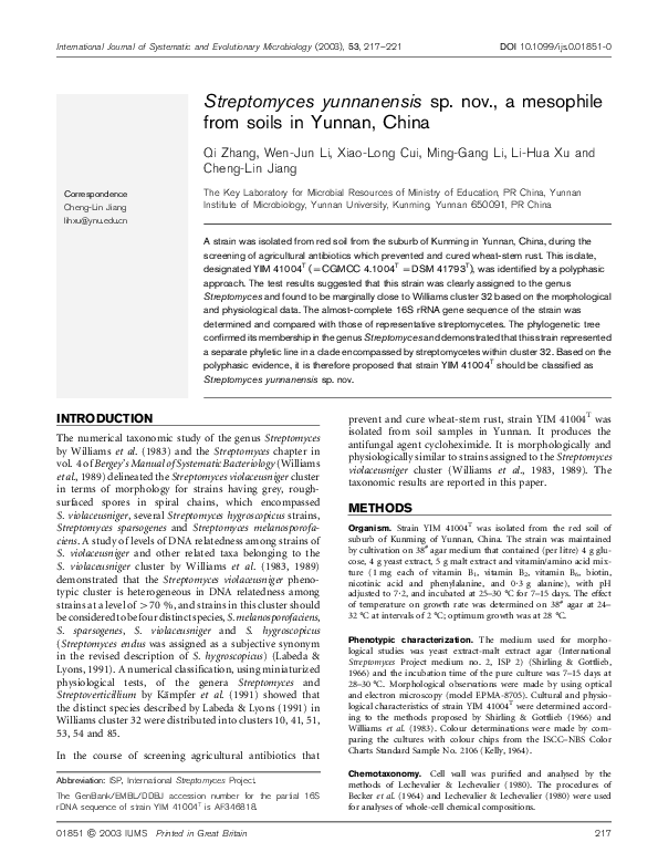
International Journal of Systematic and Evolutionary Microbiology (2003), 53, 217–221
DOI 10.1099/ijs.0.01851-0
Streptomyces yunnanensis sp. nov., a mesophile
from soils in Yunnan, China
Qi Zhang, Wen-Jun Li, Xiao-Long Cui, Ming-Gang Li, Li-Hua Xu and
Cheng-Lin Jiang
Correspondence
Cheng-Lin Jiang
The Key Laboratory for Microbial Resources of Ministry of Education, PR China, Yunnan
Institute of Microbiology, Yunnan University, Kunming, Yunnan 650091, PR China
lihxu@ynu.edu.cn
A strain was isolated from red soil from the suburb of Kunming in Yunnan, China, during the
screening of agricultural antibiotics which prevented and cured wheat-stem rust. This isolate,
designated YIM 41004T (=CGMCC 4.1004T =DSM 41793T), was identified by a polyphasic
approach. The test results suggested that this strain was clearly assigned to the genus
Streptomyces and found to be marginally close to Williams cluster 32 based on the morphological
and physiological data. The almost-complete 16S rRNA gene sequence of the strain was
determined and compared with those of representative streptomycetes. The phylogenetic tree
confirmed its membership in the genus Streptomyces and demonstrated that this strain represented
a separate phyletic line in a clade encompassed by streptomycetes within cluster 32. Based on the
polyphasic evidence, it is therefore proposed that strain YIM 41004T should be classified as
Streptomyces yunnanensis sp. nov.
INTRODUCTION
The numerical taxonomic study of the genus Streptomyces
by Williams et al. (1983) and the Streptomyces chapter in
vol. 4 of Bergey’s Manual of Systematic Bacteriology (Williams
et al., 1989) delineated the Streptomyces violaceusniger cluster
in terms of morphology for strains having grey, roughsurfaced spores in spiral chains, which encompassed
S. violaceusniger, several Streptomyces hygroscopicus strains,
Streptomyces sparsogenes and Streptomyces melanosporofaciens. A study of levels of DNA relatedness among strains of
S. violaceusniger and other related taxa belonging to the
S. violaceusniger cluster by Williams et al. (1983, 1989)
demonstrated that the Streptomyces violaceusniger phenotypic cluster is heterogeneous in DNA relatedness among
strains at a level of >70 %, and strains in this cluster should
be considered to be four distinct species, S. melanosporofaciens,
S. sparsogenes, S. violaceusniger and S. hygroscopicus
(Streptomyces endus was assigned as a subjective synonym
in the revised description of S. hygroscopicus) (Labeda &
Lyons, 1991). A numerical classification, using miniaturized
physiological tests, of the genera Streptomyces and
Streptoverticillium by Kämpfer et al. (1991) showed that
the distinct species described by Labeda & Lyons (1991) in
Williams cluster 32 were distributed into clusters 10, 41, 51,
53, 54 and 85.
In the course of screening agricultural antibiotics that
Abbreviation: ISP, International Streptomyces Project.
The GenBank/EMBL/DDBJ accession number for the partial 16S
rDNA sequence of strain YIM 41004T is AF346818.
01851 G 2003 IUMS
Printed in Great Britain
prevent and cure wheat-stem rust, strain YIM 41004T was
isolated from soil samples in Yunnan. It produces the
antifungal agent cycloheximide. It is morphologically and
physiologically similar to strains assigned to the Streptomyces
violaceusniger cluster (Williams et al., 1983, 1989). The
taxonomic results are reported in this paper.
METHODS
Organism. Strain YIM 41004T was isolated from the red soil of
suburb of Kunming of Yunnan, China. The strain was maintained
by cultivation on 38# agar medium that contained (per litre) 4 g glucose, 4 g yeast extract, 5 g malt extract and vitamin/amino acid mixture (1 mg each of vitamin B1, vitamin B2, vitamin B6, biotin,
nicotinic acid and phenylalanine, and 0?3 g alanine), with pH
adjusted to 7?2, and incubated at 25–30 ˚C for 7–15 days. The effect
of temperature on growth rate was determined on 38# agar at 24–
32 ˚C at intervals of 2 ˚C; optimum growth was at 28 ˚C.
Phenotypic characterization. The medium used for morpho-
logical studies was yeast extract-malt extract agar (International
Streptomyces Project medium no. 2, ISP 2) (Shirling & Gottlieb,
1966) and the incubation time of the pure culture was 7–15 days at
28–30 ˚C. Morphological observations were made by using optical
and electron microscopy (model EPMA-8705). Cultural and physiological characteristics of strain YIM 41004T were determined according to the methods proposed by Shirling & Gottlieb (1966) and
Williams et al. (1983). Colour determinations were made by comparing the cultures with colour chips from the ISCC–NBS Color
Charts Standard Sample No. 2106 (Kelly, 1964).
Chemotaxonomy. Cell wall was purified and analysed by the
methods of Lechevalier & Lechevalier (1980). The procedures of
Becker et al. (1964) and Lechevalier & Lechevalier (1980) were used
for analyses of whole-cell chemical compositions.
217
�Q. Zhang and others
16S rDNA sequencing. The chromosomal DNA of strain YIM
41004T was isolated according to the procedure described by
Hopwood et al. (1985). 16S rDNA was amplified by PCR using a
PCR kit (Sino-American Biotechnology, Beijing), primer A 8-27f
(59-CCGTCGACGAGCTC AGAGTTTGATCCTGGCTCAG-39) and
primer B 1523-1504r (59-CCCGGGTACCAAGCTT AAGGAGGTGATCCAGCCGCA-39) (primers are in bold according to the
Escherichia coli numbering system of Brosius et al., 1978). The conditions used for thermal cycling were as the follows: denaturation at
95 ˚C for 5 min followed by 35 cycles consisting of denaturation at
95 ˚C for 1 min, primer annealing at 56 ˚C for 1 min, and primer
extention at 72 ˚C for 3 min. At the end of the cycles, the reaction
mixture was kept at 72 ˚C for 5 min and then cooled to 4 ˚C. The
amplified 1?5 kb 16S rDNA (rDNA) fragment was separated by
agarose gel electrophoresis. The purified fragment was directly
sequenced by using a Taq DyeDeoxy terminator Cycle Sequencing
kit (Applied Biosystems) and analysed with an ABI PRISM 377 DNA
sequencer (Applied Biosystems). Sequencing primers used included
KMSO98PB1r (59-TAAGGAGGTGATCCAGCC-39), KMS584P1r (59TGCTGGCAACACAG AACAAG-39) and KMS584P2r (59-ACTCTG
CCTGCCCGTATCG-39).
Sequence alignment and phylogenetic analysis. The partial
16S rDNA sequence of strain YIM 41004T was aligned manually
with representative sequences of related streptomycetes from the
GenBank database. The evolutionary tree, rooted with Streptomyces
megasporus as the outgroup, was inferred by using the neighbourjoining method (Saitou & Nei, 1987) from the evolutionary distance
data corrected by Kimura’s two-parameter model (Kimura, 1980).
The topology of the resultant tree was evaluated by bootstrap analysis (Felsenstein, 1985) of the neighbour-joining method based on
1000 resamplings. The CLUSTAL X program (Thompson et al., 1997)
was used for multiple alignment and phylogenetic analysis. The
TreeView program (Page, 1996) was used to display, edit and print
phylogenetic trees.
RESULTS AND DISCUSSION
Morphological observation of the 7–15-day-old culture of
strain YIM 41004T grown on yeast extract-malt extract agar
(ISP 2) (Shirling & Gottlieb, 1966) revealed that both aerial
and vegetative hyphea were abundant, well-developed and
not fragmented; spore chains with many spores were spiral;
spores (0?5–1?0 mm in diameter) were rugose with short
spines and were short pillar-shaped and non-motile (Fig. 1).
Fig. 1. Scanning electron micrographs showing strain YIM 41004T
rugose spores and spiral spore chains (top and bottom) after growth
on yeast extract-malt extract agar (ISP 2) at 28 ˚C for 15 days.
Table 1. Cultural characteristics of strain YIM 41004T
Colours are according to the ISCC–NBS Color Charts Standard Sample No. 2106 (Kelly, 1964).
Agar medium
Colour of mycelium:
Aerial
Czapek’s
Glycerol asparagine (ISP 5)
Inorganic salt-starch (ISP 4)
Oatmeal (ISP 3)
Yeast extract-malt extract (ISP 2)
Glucose asparagine
Potato extract
218
Light brown-grey
Pale-yellow
Light grey-brown
Light brown-grey
Light brown-grey
Light grey-brown
Brown-grey
Soluble pigment
Substrate
Brown-pink
Light orange-yellow
Light grey-yellow
Light grey-yellow
Light yellow
Deep grey-yellow
Deep grey-yellow
Absent
Light yellow
Absent
Absent
Absent
Absent
Light yellow
International Journal of Systematic and Evolutionary Microbiology 53
�Streptomyces yunnanensis sp. nov.
Table 2. Physiological characteristics of strain YIM 41004T
and related species in the Streptomyces violaceusniger
phenotypic cluster
Strains: 1, strain YIM 41004T; 2, Streptomyces endus NRRL ISP5187T (=DSM 40187T); 3, Streptomyces hygroscopicus NRRL-ISP
5578T (=DSM 40578T); 4, Streptomyces melanosporofaciens NRRL
B-12234T (DSM 40318T); 5, Streptomyces sparsogenes ISP 5356T
(=NRRL 2940T =DSM 40356T); 6, Streptomyces violaceusniger
NRRL B-1476T (=DSM4 1600T). +, Positive; 2, negative. All
strains were negative for milk coagulation and positive for gelatin
liquefaction.
Characteristic
1
2
3
4
5
6
Milk peptonization
Starch hydrolysis
Nitrate reduction
Urea utilization
Carbon source utilization:
D-Sucrose
D-Xylose
D-Raffinose
Antimicrobial activity against:
Bacillus subtilis
Aspergillus niger
+
+
2
+
+
+
2
2
2
2
2
+
2
+
2
2
+
2
+
+
2
+
2
+
2
2
+
+
+
+
+
+
+
2
+
+
+
+
2
+
+
+
2
+
2
2
+
2
+
+
+
2
2
2
Cultural characteristics of strain YIM 41004T are shown
in Table 1. Aerial mycelium of strain YIM 41004T was
abundant, well-developed and varied from light brown-grey
to brown-grey on different test media. The substrate hyphae
from light yellow to light brown-yellow. Diffusible pigments
were not produced on most test media, and melanin was not
produced. The cell-wall peptidoglycan of strain YIM 41004T
contained only LL-diaminopimelic acid and glycine, indicating that strain YIM 41004T has a chemotype cell-wall
type I (Lechevalier & Lechevalier, 1970a, b). The whole-cell
hydrolysates contained galactose.
On the basis of morphological, cultural and chemotaxonomic properties above, together with the physiological properties of strain YIM 41004T and five other
related species in Streptomyces violaceusniger phenotypic
cluster (Williams et al., 1983, 1989; Labeda & Lyons, 1991)
shown in Table 2, it is evident that strain YIM 41004T not
only belongs to the genus Streptomyces but also should be
assigned to the Streptomyces violaceusniger cluster (Williams
et al., 1983, 1989). Although strain YIM 41004T is similar to
members of the Streptomyces violaceusniger cluster and
clusters 10, 41, 51, 53, 54 and 85 (Kämpfer et al., 1991) on
the basis of phenotypic data, this organism cannot be exactly
assigned to any of the known streptomycete species of these
clusters on the basis of its phenotypic characteristics.
Therefore, it is concluded from phenotypic data that strain
YIM 41004T shows no apparent relationship with the validly
described species of these clusters (Williams et al., 1983,
1989; Kämpfer et al., 1991). Similarly, strain YIM 41004T is
differentiated primarily from four other cycloheximideproducing species based on the surface of spore and carbonsource utilization from Table 3.
The phylogenetic analysis of strain YIM 41004T with
members of the Streptomyces violaceusniger cluster
(Williams et al., 1983, 1989; Labeda & Lyons, 1991) reveals
that strain YIM 41004T is distinct from species in this cluster,
as showed in Fig. 2. The sequence divergence values between
strain YIM 41004T and members of the Streptomyces
violaceusniger cluster (Williams et al., 1983, 1989;
Labeda & Lyons, 1991) are 2?91 % (S. hygroscopicus),
2?70 % (S. melanosporofaciens), 2?70 % (S. violaceusniger),
3?27 % (S. sparsogenes), and these indicate that strain
YIM 41004T represents a hitherto unpublished species.
The phenotypic and genotypic data of strain YIM 41004T
demonstrated that strain YIM 41004T should be given
novel species status in the genus Streptomyces Waksman
and Henrici 1943AL. Therefore, we proposed this organism
Table 3. Partial features for differentiating strain YIM 41004T from cycloheximide-producing species
Strains: 1, strain YIM 41004T; 2, Streptomyces albulus ATCC 12757T (Streptomyces lydicus cluster); 3, Streptomyces noursei ATCC 11455T
(Streptomyces noursei cluster); 4, Streptomyces griseus ATCC 23345T (Streptomyces anulatus cluster); 5, Streptomyces pulveraceus ATCC 13875T
(Streptomyces pulveraceus cluster). Comparative data taken from previous studies (Williams et al., 1983, 1989). +, Positive; 2, negative.
Characteristic
Spore surface
Carbon source utilization:
Sucrose
D-Raffinose
L-Arabinose
L-Rhamnose
D-Xylose
D-Mannitol
http://ijs.sgmjournals.org
1
2
3
4
5
Rugose with short spines
Hairy
Spiny
Smooth
Smooth
+
+
+
+
2
+
2
2
2
2
2
+
2
2
+
2
2
+
2
2
2
2
+
+
2
+
2
+
+
2
219
�Q. Zhang and others
the DSMZ-Deutsche Sammlung von Mikroorganismen und
Zellkulturen (GmbH), Germany, as strain DSM 41793T.
ACKNOWLEDGEMENTS
We are grateful to Dr David P. Labeda for providing some type strains
and helping in preparation of this manuscript. This work was
supported by the National Natural Science Foundation of China and
Funds of the Key Laboratory for Microbial Resources of Ministry of
Education, PR China, Yunnan Provincial Commission of Science &
Technology, and The International Cooperation Foundation of Yunnan.
REFERENCES
Fig. 2. Neighbour-joining tree (Saitou & Nei, 1987) showing
the phylogenetic relationships among members of the
Streptomyces violaceusniger cluster. The analysis included
1453 unambiguous nucleotide positions. Streptomyces megasporus was used as the outgroup. Bootstrap values from 1000
analyses were shown at the nodes of the tree. The scale bar
represents one nucleotide substitution per 100 nucleotides of
16S rDNA sequence.
Becker, B., Lechevalier, M. P., Gordon, R. E. & Lechevalier, H. A.
(1964). Rapid differentiation between Nocardia and Streptomyces by
paper chromatography of whole-cell hydrolysates. Appl Microbiol 12,
421–423.
Brosius, J., Palmer, M. L., Kennedy, J. P. & Noller, H. P. (1978).
Complete nucleotide sequence of a 16S ribosomal RNA gene from
Escherichia coli. Proc Natl Acad Sci U S A 75, 4801–4805.
Felsenstein, J. (1985). Confidence limits on phylogenies: an
approach using the bootstrap. Evolution 39, 783–791.
Hopwood, D. A., Bill, M. J., Charter, K. F. & 7 other authors (1985).
Genetic Manipulation of Streptomycetes: a Laboratory Manual.
Norwich: John Innes Foundation.
should be a new species with the name Streptomyces
yunnanensis sp. nov.
Kämpfer, P., Kroppenstedt, R. M. & Dott, W. (1991). A numerical
classification of the genera Streptomyces and Streptoverticillium using
miniaturized physiological tests. J Gen Microbiol 137, 1831–1891.
Kelly, K. L. (1964). Inter-Society Color Council–National Bureau of
Description of Streptomyces yunnanensis sp.
nov.
Streptomyces yunnanensis (yun.nan.en9sis. N.L. masc.
adj. yunnanensis pertaining to Yunnan, a province of
south-west China).
Both vegetative and aerial hyphae are abundant and welldeveloped. The colour of aerial and substrate mycelium on
various solid media is given in Table 1. Spore chains with
many spores are spiral. The spores are rugose with short
spines and are short pillar-shaped (0?5–1?0 mm in diameter)
and non-motile. Diffusible pigments are not produced and
melanin is not produced. Milk is not coagulated but
peptonized, starch is hydrolysed and H2S is not produced.
Nitrate is not reduced and gelatin is liquefied. Does not
hydrolyse cellulose. Utilizes glucose, fructose, rhamnose,
inositol, mannitol, arabinose and raffinose for growth; does
not utilize sucrose or xylose. It has antimicrobial activity
against Aspergillus niger but not against Bacillus subtilis.
Optimum growth is at 28 ˚C. The cell wall contains
LL-diaminopimelic acid and glycine (cell-wall chemotype I).
Whole-cell hydrolysates contain galactose. The type strain,
YIM 41004T, isolated from red soil of the suburb of
Kunming in Yunnan, China, was deposited in the China
General Microbiological Culture Collection Center
(CGMCC) Beijing, China, as strain CGMCC 4.1004T, and
220
Standards Color-Name Charts Illustrated with Centroid Colors.
Washington, DC: US Government Printing Office.
Kimura, M. (1980). A simple method for estimating evolutionary
rates of base substitutions through comparative studies of nucleotide
sequences. J Mol Evol 16, 111–120.
Labeda, D. P. & Lyons, A. J. (1991). The Streptomyces violaceiniger
cluster is heterogeneous in DNA relatedness among strains:
emendation of the descriptions of S. violaceiniger and Streptomyces
hygroscopicus. Int J Syst Bacteriol 41, 398–401.
Lechevalier, H. A. & Lechevalier, M. P. (1970a). A critical evaluation
of the genera of aerobic actinomycetes. In The Actinomycetes,
pp. 393–405. Edited by H. Prauser. Jena: Gustav Fischer Verlag.
Lechevalier, M. P. & Lechevalier, H. A. (1970b). Chemical
composition as a criterion in the classification of aerobic
actinomycetes. Int J Syst Bacteriol 20, 435–443.
Lechevalier, M. P. & Lechevalier, H. A. (1980). The chemotaxonomy
of actinomycetes. In Actinomycete Taxonomy, pp. 22–291. Edited by
A. Dietz & D. W. Thayer. Arlington, VA: Society for Industrial
Microbiology.
Page, R. D. M. (1996). TREEVIEW: an application to display
phylogenetic trees on personal computers. Comput Appl Biosci 12,
357–358.
Saitou, N. & Nei, M. (1987). The neighbor-joining method: a new
method for reconstucting phylogenetic trees. Mol Biol Evol 4, 406–
425.
Shirling, E. B. & Gottlieb, D. (1966). Methods for characterization of
Streptomyces species. Int J Syst Bacteriol 16, 313–340.
Thompson, J. D., Gibson, T. J., Plewniak, F., Jeanmougin, F. &
Higgins, D. G. (1997). The Clustal X windows interface: flexible
International Journal of Systematic and Evolutionary Microbiology 53
�Streptomyces yunnanensis sp. nov.
strategies for multiple sequence alignment aided by quality analysis
tools. Nucleic Acids Res 24, 4876–4882.
Williams, S. T., Goodfellow, M., Alderson, G., Wellington, E. M. H.,
Sneath, P. H. A. & Sackin, M. J. (1983). Numerical classification of
Streptomyces and related genera. J Gen Microbiol 129, 1743–1813.
http://ijs.sgmjournals.org
Williams,
S.
T.,
Goodfellow,
M.
&
Alderson,
G.
(1989).
Genus Streptomyces Waksman and Henrici 1943, 339AL,
pp. 2452–2492. In Bergey’s Manual of Systematic Bacteriology,
vol. 4. Edited by S. T. Williams & M. E. Sharpe. Baltimore:
Williams & Wilkins.
221
�
 Wen-Jun Li