Archives of Microbiology (2021) 203:3997–4004
https://doi.org/10.1007/s00203-021-02389-1
ORIGINAL PAPER
Paenibacillus roseus sp. nov., a ginsenoside‑transforming bacterium
isolated from forest soil
Shahina Akter1 · Xiaoqing Wang2 · Sun‑Young Lee2 · M. Mizanur Rahman3 · Jong‑Hyun Park1 ·
Muhammad Zubair Siddiqi4 · Sri Renukadevi Balusamy5 · Kihong Nam6 · Md. Shahedur Rahman7 ·
Md. Amdadul Huq2
Received: 30 January 2021 / Revised: 30 April 2021 / Accepted: 18 May 2021 / Published online: 25 May 2021
© The Author(s), under exclusive licence to Springer-Verlag GmbH Germany, part of Springer Nature 2021
Abstract
A novel, pink-pigmented, Gram-stain-positive, aerobic, motile, rod-shaped and ginsenoside-converting bacterium, designated
strain MAHUQ-46T, was isolated from soil of a forest. Strain MAHUQ-46T grew in the pH range 6.0–9.0 (optimum, 7.5), at
temperatures between 10 and 37 °C (optimum, 30 °C) and at 0–3% (w/v) NaCl (optimum, 0.5%). 16S rRNA gene sequence
analysis showed that strain MAHUQ-46T was closely related to Paenibacillus pinihumi S23T (97.3% similarity), followed
by Paenibacillus elymi KUDC6143T (96.7%). The draft genome of strain MAHUQ-46T had a total length of 5,367,904 base
pairs. A total of 4,857 genes were identified, in which 4,629 were protein-coding genes and 137 were RNA genes. The genome
annotation of MAHUQ-46T showed 172 carbohydrate genes, some of them may be responsible for the biosynthesis of ginsenoside Rd from major ginsenoside Rb1. The DNA G + C content was 48.4 mol% and the major quinone was MK-7. Main fatty
acids of strain MAHUQ-46T were C15: 0 anteiso, C16: 0 and C17: 0 anteiso. The polar lipids comprised phosphatidylethanolamine,
phosphatidylglycerol, diphosphatidylglycerol, phosphatidyl-N-methylethanolamine, two unidentified aminophospholipids
and five unidentified phospholipids. Diagnostic diamino acid of peptidoglycan was meso-diaminopimelic acid. The novel
strain MAHUQ-46T was able to rapidly synthesize ginsenoside Rd from major ginsenoside Rb1. The synthesized ginsenoside was confirmed by TLC and HPLC analysis. According to the phenotypic, genetic and chemotaxonomic evidence, strain
MAHUQ-46T was clearly distinguishable from validly published species of genus Paenibacillus and should, therefore, be
categorized as a novel species for which the name Paenibacillus roseus sp. nov. is proposed. The type strain is MAHUQ-46T
(= KACC 21242T = CGMCC 1.17353T).
Keywords Paenibacillus roseus · Genome sequence · Ginsenoside Rd · Rapid synthesis
Introduction
Communicated by Erko Stackebrandt.
Shahina Akter, and Xiaoqing Wang authors equally contributed to
this work as co-first author.
The GenBank/EMBL/DDBJ accession numbers for the 16S
rRNA gene and draft genome sequence of strain MAHUQ-46T are
MK680121 and JAELUP000000000, respectively.
* Md. Amdadul Huq
amdadbge@gmail.com; amdadbge100@cau.ac.kr
Extended author information available on the last page of the article
The genus Paenibacillus was proposed in 1993 by Ash
et al. (1993) and separated from genus Bacillus according
to the comparative analysis of 16S rRNA gene sequence.
Species of genus Paenibacillus have been isolated from
diverse environments including soil (Huq et al. 2015; Akter
and Huq 2018), food (Berge et al. 2002), air (Rivas et al.
2005), water (Baik et al. 2011), human feces (Clermont et al.
2015), roots (Zhang et al. 2016), seeds (Liu et al. 2010),
leaves (Madhaiyan et al. 2017) and flowers (Siddiqi et al.
2015). Isolation and characterization of bacteria are important due to their potential applications (Farh et al. 2015;
Huq et al. 2014). Ginsenoside is the pharmacological active
compound of ginseng, and has been reported to contain different biological efficacies such as anti-obesity, anticancer,
13
Vol.:(0123456789)
�3998
tumor-suppressing, hepatoprotective, neuroprotective effects
(Siraj et al. 2015; Chae et al. 2009; Huq et al. 2016). Minor
ginsenosides possess higher activity than major ginsenosides
(Huq et al. 2016). Ginsenoside Rb1 is a major ginsenoside
and abundant in ginseng root. Therefore, many scientists
have tried to convert major ginsenosides to highly active
minor ginsenosides (Huq et al. 2016). This report describes
the isolation, characterization and genome analysis of a
novel bacterial species Paenibacillus roseus sp. nov., and
utilization of this novel species for the rapid synthesis of
ginsenoside Rd.
Materials and methods
Bacterial isolation
Novel strain, MAHUQ-46T was isolated from soil sample
of a forest located in Anseong, South Korea (37° 00′ 44″
N, 127° 23′ 17″). Soil samples were diluted serially using
sterile NaCl solution (0.85%, w/v) and these dilutions were
plated onto R2A agar medium (MB cell) and spread thoroughly. The plates were incubated for 3 days at 30 °C. The
culture was further purified by repeated streaking onto the
R2A agar until pure strains were obtained. The purified
strain was cultivated and stored as aqueous glycerol suspensions (30%, v/v) at − 80 °C. Strain MAHUQ-46T has been
deposited to the KACC and CGMCC.
Cell growth, morphology, physiology
and biochemical tests
Cultural characteristics were investigated by observing the
growth of the strain MAHUQ-46T at 30 °C for 5–7 days
on Reasoner’s 2A (R2A) agar (Difco), nutrient agar (NA;
Difco), Luria–Bertani agar (LB agar), tryptic soy agar
(TSA; Bacto) and MacConkey agar. The morphology of the
cells was observed and recorded by transmission electron
microscopy (Model JEM1010; JEOL) after incubation on
R2A agar at 30 °C for 2 days. Salt tolerance was determined
in R2A broth containing 0–6% (w/v) NaCl (0.5% interval)
after 7 days of incubation at 30 °C. The temperature range
(4–45 °C) for growth was assessed on R2A agar for 7 days.
The growth pH range (pH 4.0–11.0) was assessed in R2A
broth medium for 7 days using the buffer system described
by Huq (2018). Gram-staining test was conducted according
to the method of Magee et al. (Magee et al. 1975). Catalase
activity was checked using 3% (v/v) H2O2 through bubble production. 1% (w/v) tetramethyl-p-phenylenediamine
was used to determine the oxidase activity (Huq 2018).
Hydrolysis of DNA, casine, gelatin, starch, Tweens 20 and,
Tweens 80 were tested according to the method of Gonzalez et al. (Gonzalez et al. 1978). The enzyme activities
13
Archives of Microbiology (2021) 203:3997–4004
and biochemical characteristics were assessed using the API
ZYM kits (bioMérieux) and API 20NE kits (bioMérieux)
according to the manufacturer’s instructions. The close type
strains, Paenibacillus pinihumi KACC 14199T and Paenibacillus elymi KCTC 33853T were used as reference strains and
were grown under the identical experimental conditions of
novel strain MAHUQ-46T.
Analysis of 16S rRNA gene and phylogenetic tree
The 16S rRNA gene was amplified from extracted genomic
DNA using bacterial universal primers 27F and 1492R
(Weisburg et al. 1991). The 16S rRNA gene sequence of
strain MAHUQ-46T was compared with related type species from EzBioCloud database (www.ezbiocloud.net) to
determine the similarity values (Yoon et al. 2017a). Multiple
sequence alignments were conducted using clustal_x software package (Thompson et al. 1997) and BioEdit software
(Hall 1999). Kimura’s two-parameter model was used to
determine the evolutionary distances (Kimura 1980). Phylogenetic trees were constructed using both the neighborjoining (Saitou and Nei 1987) and maximum-likelihood
(Felsenstein 1981) methods with mega (version 5.0) software (Tamura et al. 2011). To evaluate the topologies of the
phylogenetic trees, bootstrap analysis was carried out with
1000 replications (Felsenstein, 1985).
Genome sequence analysis
Draft-genome sequencing of strain MAHUQ-46T was performed on the Illumina HiSeq X Ten platform. The goodquality paired reads were assembled using SOAPdenovo
v. 3.10.1 de novo assembler into a number of scaffolds.
Genome annotation was completed using NCBI prokaryotic genome annotation pipeline (PGAP). The wholegenome sequence data have been submitted to DDBJ/ENA/
GenBank. The DNA G + C content of strain MAHUQ-46T
was directly calculated from the draft genome sequences
(Chun et al. 2018). The average nucleotide identity (ANI)
was calculated as described previously (Yoon et al. 2017b).
While, the digital DNA–DNA hybridization (dDDH) value
was determined using the genome-to-genome distance calculator (http://ggdc.dsmz.de/ggdc.php) according to MeierKolthoff et al. (2013).
Cellular fatty acid, respiratory quinone, polar lipid
and peptidoglycan analysis
Cell biomass of strain MAHUQ-46 T and two reference
strains for analysis of the cellular fatty acid were collected
by cultivation on R2A agar medium at 30 °C, for 48 h. The
cellular cell fatty acids were saponified, methylated and
extracted using a standard protocol as described by Sasser
�Archives of Microbiology (2021) 203:3997–4004
3999
(1990), and analyzed by gas chromatograph GC (HewlettPackard 5890 Series II) and identified through the rtsba
6.00 database of the Microbial Identification System (Sasser
1990). Respiratory quinones of strain MAHUQ-46T were
extracted and purified according to Collins et al. (1977),
then analyzed and confirmed by HPLC (Guo et al. 2015).
Polar lipids of strain MAHUQ-46T were extracted from dry
cells (100 mg) as described by Minnikin et al. (1984). The
extracted polar lipids were analyzed by two-dimensional
TLC according to the previous description (Akter and Huq
2020). The diamino acid of the peptidoglycan was extracted
and determined by TLC (Cellulose; Merck) as described by
Komagata and Suzuki (1987).
MAHUQ-46T belonged to the genus Paenibacillus and had
moderately high similarity to Paenibacillus pinihumi S23T
(97.3%) and Paenibacillus elymi KUDC6143T (96.7%). The
relationship between isolated strain MAHUQ-46T and recognized Paenibacillus species is also shown in the constructed
phylogenetic trees. The neighbor-joining and maximumlikelihood trees placed strain MAHUQ-46T within cluster for
Paenibacillus species, as shown in Fig. 1 and Supplementary
Fig. S2. The 16S rRNA gene sequence analysis revealed that
strain MAHUQ-46T could be clearly separated from validly
published species of the genus Paenibacillus and should be
assigned as a novel species of this genus (Wayne et al. 1987;
Stackebrandt and Goebel 1994).
Biotransformation of ginsenoside Rb1
Genome sequence analysis
The novel strain MAHUQ-46T was grown in R2A broth
medium at 30 °C for 48 h and cells were harvested by centrifuging at 9,000 rpm for 15 min at 4 °C. Then, the harvested
cells were dissolved in sodium phosphate buffer (20 mM, pH
7.0) and lysed by short pulse sonication. Then, the supernatant was collected by centrifugation and used as crude
enzyme. This is the modification of Huq et al. (2016) protocol. The bioconversion of ginsenoside Rb1 was performed
in screw-capped tubes. 3 ml of crude enzyme and 3 ml ginsenoside Rb1 (2.0 mg/ml) were mixed and incubated in a
shaking incubator (160 rpm) at 30 °C. Every 1-h interval,
0.5 ml sample was withdrawn and analyzed by both TLC and
HPLC (Huq et al. 2014).
The draft genome of strain MAHUQ-46T was 5,367,904 bp
long in size with a G + C content of 48.4 mol%. It comprised of 118 contigs. Among 4,857 predicted genes, 4,629
were protein-coding genes and 137 were encoding RNAs
(116 tRNA genes, four 5S rRNA genes, seven 16S rRNA
genes and six 23S rRNA genes). The common features of
genome sequence of strain MAHUQ-46T are given in Supplementary Table S2. Taxonomic and functional research
of microorganisms has increasingly relied upon genomebased data and methods (Shi et al. 2021). Distribution of
genes in the genome of strain MAHUQ-46T was investigated using RAST server (Overbeek et al. 2014), via the
RASTtk pipeline (Brettin et al. 2015). It was found that 207
of the genes were involved with protein metabolism, 241
genes were associated with the metabolism of amino acids
and derivatives, 172 genes were linked with carbohydrate
metabolism and 102 genes were involved with metabolism
of vitamins, cofactors and pigments (Supplementary Fig.
S3). The closest type strain P. pinihumi DSM 23905T contains a 6,760,575 bp long genome (number of contig 45)
with 48.5 mol% GC, 6,062 CDSs, 64 tRNA and 14 rRNA
genes (https://www.ezbiocloud.net/genome/explore?puid=
20755). The genomic ANI (average nucleotide identity) values between strain MAHUQ-46T and P. pinihumi S23T were
82.2%, well below (≥ 95–96%) to suggest a novel species.
The dDDH value based on the draft genomes between strain
MAHUQ-46T and P. pinihumi S23T was 26.0% which was
also far below the threshold value (70%) for species delineation (Supplementary Table S3).
Results and discussion
Cell growth, morphology, physiology
and biochemical characteristics
Cells of strain MAHUQ-46T were Gram stain positive, aerobic, rod shaped (0.6–1.2 × 1.4–2.6 µm) and motile with flagella (Supplementary Fig. S1). Strain MAHUQ-46T formed
pink colonies when grown on R2A agar plates after incubation for 48 h at 30 °C. The detailed physiological, morphological and biochemical characteristics of strain MAHUQ46T and most closely related type strains are given in the
species description and in Table 1. The negative properties
of strain MAHUQ-46T carried out by commercial test kits
(API 20NE and API ZYM) are shown in Supplementary
Table S1.
Cellular fatty acid, respiratory quinone, polar lipid
and peptidoglycan analysis
16S rRNA gene sequence and phylogenetic analysis
Almost complete length of 16S rRNA gene sequence
for strain MAHUQ-46 T was 1,485 bp. 16S rRNA
gene sequences analysis revealed that the novel strain
Strain MAHUQ-46T contained C15: 0 anteiso (48.6%), C16: 0
(12.1%) and C17: 0 anteiso (11.5%), as the dominant fatty
acids. Strain MAHUQ-46 T also contained considerable
amount of C15: 0 iso (8.2%) and C16: 0 iso (9.7%). As for the
13
�4000
Archives of Microbiology (2021) 203:3997–4004
Table 1 The biochemical and physiological characteristics of strain MAHUQ-46T and the reference strains of genus Paenibacillus
Characteristics
1
2
3
Isolation source
Forest soil
Rhizosphere of the pine tree
Cell size (um)
Colony color
Aerobic/facultative anaerobic
Catalase
Oxidase
4-Nitrophenyl-BD-galactopyranoside
Growth temperature (°C)
Growth pH
NaCl tolerance (%)
Hydrolysis of
Gelatin (API 20 NE)
Tween 80
Enzyme activity (API ZYM)
Esterase (C4)
Alkaline phosphatase
Lipase (C14)
Valine arylamidase
Cystine arylamidase
Trypsin
α-chymotrypsin
Naphthol-AS-BI-phosphohydrolase
a-galactosidase
β-glucosidase
β-galactosidase
Assimilation of (API 20 NE)
d-glucose
l-arabinose
d-mannose
Gluconate
DNA G + C content (mol%)
0.8–1.2 X 1.4–2.6
Pink
Strictly aerobic
W+
−
−
10–37
6.0–9.0
0–3
1.6–3.5 X 0.6–0.8a
Cream
Strictly aerobic
−
+
+
15-37a
5.5–9.0a
0-3a
Rhizosphere of Elymus tsukushiensis
0.5–0.6 X 2.0–2.7b
Creamy white
Facultative anaerobic
−
+
−
25-45b
6.0–12.0b
0–4.0b
−
W+
−
−
+
−
+
+
W
w
w
w
−
w
−
+
−
−
+
−
−
−
−
−
−
+
−
+
+
−
−
−
+
−
+
+
−
−
−
+
+
−
+
48.4
−
−
−
−
49.5a
+
+
+
−
50.3b
Strains: 1, P. roseus MAHUQ-46T; 2, P. pinihumi KACC 14199T and P. elymi KCTC 33853T
All data were obtained in this study, except a and b that were taken from Kim et al. (2009) and Hwang et al. (2018), respectively. All strains are
rod shaped and motile. All strains are positive for esterase lipase (C8), hydrolysis of starch, esculin, assimilation of D-maltose and D-mannitol.
All strains are negative for nitrate reduction, indole production, arginine dihydrolase, acid phosphatase, leucine arylamidase, β-glucuronidase,
N-acetyl-β-glucosaminidase, α-mannosidase, a-glucosidase, and a-fucosidase, hydrolysis of casein and urea, and assimilation of malic acid,
N-acetyl-glucosamine, phenylacetic acid, adipic acid, trisodium citrate and capric acid. + , positive; −, negative; W + , weakly positive
major fatty acids, strain MAHUQ-46T was similar to close
reference strains P. pinihumi KACC 14199T and P. elymi
KCTC 33853T, except for some small amounts of fatty acids.
Although, the major fatty acid profiles were similar with
close type strains but there were significant quantitative differences. The cellular fatty acid profiles of strain MAHUQ46T and two close reference strains are shown in Table 2.
The predominant isoprenoid quinone of strain MAHUQ-46T
was menaquinone-7 (MK-7) which is one of the common
characteristics of genus Paenibacillus (Ash et al. 1993; Huq
et al. 2015; Kim et al. 2009; Hwang et al. 2018). The polar
13
lipids identified in strain MAHUQ-46T were phosphatidylethanolamine, phosphatidylglycerol, diphosphatidylglycerol, phosphatidyl-N-methylethanolamine, two unidentified
aminophospholipids and five unidentified phospholipids.
(Supplementary Fig. S4). The predominant polar lipids
of strain MAHUQ-46T were similar to those of the most
closely related type strains (Hwang et al. 2018). The diagnostic diamino acid in the peptidoglycan cell wall of strain
MAHUQ-46T was meso-diaminopimelic acid. This is a
common feature among members of the genus Paenibacillus (Kim et al. 2009; Hwang et al. 2018).
�Archives of Microbiology (2021) 203:3997–4004
4001
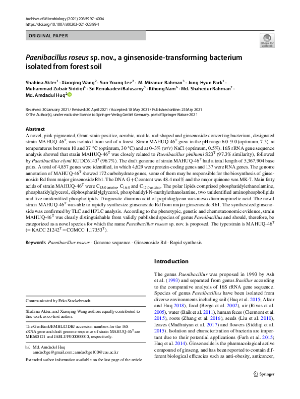
Paenibacillus pinihumi JCM 16419T (BBDI01000085)
Paenibacillus elymi KUDC6143T (KX858537)
Paenibacillus roseus MAHUQ-46T (MK680121)
Paenibacillus abyssi SCSIO N0306T (KC978082)
Paenibacillus wooponensis WPCB018T (EU939687)
Paenibacillus xanthanilyticus AS7T (KT429627)
80
99
Paenibacillus oenotherae DT7-4T (KF900218)
99
Paenibacillus harenae B519T (AY839867)
95
Paenibacillus alkaliterrae KSL-134T (AY960748)
Paenibacillus paeoniae M4BSY-1T (MH714913)
Paenibacillus castaneae Ch-32T (EU099594)
Paenibacillus endophyticus PECAE04T (KC447384)
100
Paenibacillus glycanilyticusDS-1T (AB042938)
97
100
Paenibacillus darwinianus BrT (KK082214)
Paenibacillus tarimensis SA-7-6T (EF125184)
Paenibacillus translucens CJ11T (MF619925)
Paenibacillus lacus Agd-32T (LN812815)
Sphingomonas chungangi MAH-6T (KY964284)
0.02
Fig. 1 The neighbor-joining (NJ) tree based on 16S rRNA gene sequence analysis showing phylogenetic relationships of strain MAHUQ-46T
and members of genus Paenibacillus, values less than 70% were not shown
Table 2 Fatty acid profiles
of strain MAHUQ-46T and
the reference strains of genus
Paenibacillus
Fatty acid
1
2
3
C11:0 anteiso
C12:0 iso
C13:0 anteiso
C14:0 iso
C14:0
C15:0 iso
C15:0 anteiso
C16:0 iso
C16:0
C16:1 w11c
C17:0 iso
C17:0 anteiso
ND
ND
ND
1.2
1.0
8.2
48.6
9.7
12.1
ND
6.8
11.5
ND
ND
Tr
3.2
1.3
12.9
52.6
14.8
7.4
ND
3.5
4.1
2.2
1.5
1.9
2.7
2.8
7.6
43.8
12.1
16.2
1.7
2.4
4.4
Strains: 1, P. roseus MAHUQ46T; 2, P. pinihumi KACC
14199T and P. elymi KCTC
33853T
All data were obtained in this
study
Biotransformation of ginsenoside Rb1
Ginsenoside Rd was biosynthesized from major ginsenoside Rb1 through hydrolysis of a glucose molecule at C-20
position of the ginsenoside aglycone. From TLC analysis, it
was found that ginsenoside Rb1 was completely hydrolyzed
and transformed into ginsenoside Rd within 3 h of incubation (Supplementary Fig. S5). Supplementary Fig. S5 shows
that there was no more conversion of ginsenoside Rd until
24 h which indicates that this ginsenoside Rd is the stable product. The biosynthesis of ginsenoside Rd by crude
enzyme of the novel strain MAHUQ-46T was also confirmed
by HPLC analysis (Supplementary Fig. S6). Fig. S6A shows
the peaks of standard ginsenosides including ginsenosides
Rb1 (retention time of 20.68 min) and Rd (retention time of
22.59 min). Fig. S6B shows the control of ginsenoside Rb1
which was used for biosynthesis of ginsenoside Rd. From
Fig. S6C, it was found that the peak for ginsenoside Rb1 was
completely disappeared (100%) within 3 h of incubation and
a new peak appeared. The appeared new peak had retention
time same with that of ginsenoside Rd. Ginsenoside Rb1
was selected as target material because Rb1 is a major ginsenoside and abundant in ginseng root. Crude enzyme of P.
13
�4002
roseus MAHUQ-46T are able to rapid and specific synthesis of ginsenoside Rd from Ginsenoside Rb1. The genome
annotation of strain MAHUQ-46T showed 172 carbohydrate
genes, some of them may be responsible for the biosynthesis
of ginsenoside Rd from major ginsenoside Rb1.
Conclusions
Therefore, on the basis of phenotypic, genotypic and chemotaxonomic characteristics and the phylogenetic analysis,
strain MAHUQ-46T clearly considered as a novel species
of the genus Paenibacillus, for which the name Paenibacillus roseus sp. nov. is proposed. It is also concluded that the
novel species Paenibacillus roseus MAHUQ-46T could be
useful for the biologically rapid synthesis of ginsenoside Rd.
Description of Paenibacillus roseus sp. nov.
Paenibacillus roseus (ro’se.us. L. masc. adj. roseus rosy;
referring to the color of the colonies).
Cells are aerobic, Gram stain positive, rod shaped
(0.6–1.2 × 1.4–2.6 µm) and motile with flagella. Colonies
are circular (diameter, 0.5–1.2 mm), smooth and pink in
color on R2A agar after 48 h of incubation at 30 °C. The
growth temperature range is 10–37 °C (optimum, 30 °C).
The growth pH range is 6.0–9.0 (optimum, pH 7.5) and the
NaCl tolerance range is 0–3% (w/v) (optimum, 0%). Growth
was observed on R2A agar, TSA, LB agar and NA, but no
growth occurred on MacConkey agar. Positive for catalase
but negative for oxidase. Cells are able to hydrolysis of
starch, esculin and Tween 80, but unable to hydrolysis of
casein (skimmed milk), DNA, l-tyrosine, gelatin, urea and
Tween 20. Negative for indole production, glucose fermentation and nitrate reduction. In API ZYM strips, positive for
alkaline phosphatase, leucine arylamidase, esterase lipase,
esterase and β-glucosidase; weakly positive for naphtholAS-BI-phosphohydrolase, cystine arylamidase, valine
arylamidase, lipase and trypsin. In API 20NE strips, d-glucose, d-maltose, l-arabinose and gluconate are utilized as
sole carbon source. The predominant menaquinone is MK-7.
The polar lipids comprised phosphatidylethanolamine, phosphatidylglycerol, diphosphatidylglycerol, phosphatidylN-methylethanolamine, two unidentified aminophospholipids and five unidentified phospholipids. Diagnostic diamino
acid of peptidoglycan was meso-diaminopimelic acid. The
DNA G + C content was 48.4 mol% and the major quinone
was MK-7. Main fatty acids of strain MAHUQ-46T were
C15: 0 anteiso, C16: 0 and C17: 0 anteiso.
T h e t y p e st ra i n i s M A H U Q - 4 6 T ( = K AC C
21242T = CGMCC 1.17353T), isolated from the soil sample
of a forest located in Anseong, South Korea.
13
Archives of Microbiology (2021) 203:3997–4004
Supplementary Information The online version contains supplementary material available at https://doi.org/10.1007/s00203-021-02389-1.
Acknowledgements This study was performed with the support of the
National Research Foundation (NRF) of Korea grant (Project no. NRF2018R1C1B5041386, Grant Recipient: Md. Amdadul Huq) funded by
Korean government, Republic of Korea. I would like to give special
thanks to CGM 10K project for analyzing the draft genome sequence
of strain MAHUQ-46T (GCM60012307).
Declarations
Conflict of interest The author declares that there are no conflicts of
interest.
References
Akter S, Huq MA (2018) Biological synthesis of ginsenoside Rd using
Paenibacillus horti sp. Nov. Isolated from vegetable garden. Curr
MicrobiOl 75:1566–1573
Akter S, Huq MA (2020) Sphingomonas chungangi sp. nov., a bacterium isolated from garden soil sample. Int J Syst Evol Microbiol
70:4151–4157
Ash C, PriestCollins FGMD (1993) Molecular identification of rRNA
group 3 bacilli (Ash, Farrow, Wallbanks and Collins) using a PCR
probe test. Proposal for the creation of a new genus Paenibacillus.
Antonie Van Leeuwenhoek 64:253–260
Baik KS, Lim CH, Choe HN, Kim EM, Seong CN (2011) Paenibacillus
rigui sp. nov., isolated from a freshwater wetland. Int J Syst EvOl
Microbiol 61:529–534
Berge O, Guinebretière MH, Achouak W, Normand P, Heulin T (2002)
Paenibacillus graminis sp. nov. and Paenibacillus odorifer sp.
nov., isolated from plant roots, soil and food. Int J Syst Evol
Microbiol 52:607–616
Brettin T, Davis JJ, Disz T, Edwards RA, Gerdes S, Olsen GJ, Olson R,
Overbeek R, Parrello B, Pusch GD (2015) RASTtk: a modular and
extensible implementation of the RAST algorithm for building
custom annotation pipelines and annotating batches of genomes.
Sci Rep 5:8365
Chae S, Kang KA, Chang WY, Kim MJ, Lee SJ, Lee YS, Kim HS, Kim
DH, Hyun JW (2009) Effect of compound K, a metabolite of ginseng saponin, combined with gamma-ray radiation in human lung
cancer cells in vitro and in vivo. J Agr Food Chem 57:5777–5782
Chun J, Oren A, Ventosa A, Christensen H, Arahal DR (2018) Proposed minimal standards for the use of genome data for the taxonomy of prokaryotes. Int J Syst Evol Microbiol 68:461–466
Clermont D, Gomard M, Hamon S, Bonne I, Fernandez JC, Wheeler
R, Malosse C, Chamot-Rooke J, Gribaldo S, Boneca IG, Bizet C
(2015) Paenibacillus faecis sp. nov., isolated from human faeces.
Int J Syst Evol Microbiol 65:4621–4626
Collins MD, Pirouz T, Goodfellow M, Minnikin DE (1977) Distribution of menaquinones in actinomycetes and corynebacteria. J Gen
Microbiol 100:221–230
Farh MA, Kim YJ, Van AH, Sukweenadhi J, Singh P, Huq MA, Yang
DC (2015) Burkholderia ginsengiterrae sp. nov. and Burkholderia
panaciterrae sp. nov., antagonistic bacteria against root rot pathogen Cylindrocarpon destructans, isolated from ginseng soil. Arch
Microbiol 197:439–447
Felsenstein J (1981) Evolutionary trees from DNA sequences: a maximum likelihood approach. J Mol Evol 17:368–376
Felsenstein J (1985) Confidence limits on phylogenies: an approach
using the bootstrap. Evolution 39:783–791
�Archives of Microbiology (2021) 203:3997–4004
Gonzalez C, Gutierrez C, Ramirez C (1978) Halobacterium vallismortis sp. nov. An amylolytic and carbohydrate-metabolizing,
extremely halophilic bacterium. Can J Microbiol 24:710–715
Guo L, Tuo L, Habden X, Zhang Y, Liu J (2015) Allosalinactinospora lopnorensis gen. nov., sp. nov., a new member of the family
Nocardiopsaceae isolated from soil. Int J Syst Evol Microbiol
65:206–213
Hall TA (1999) BioEdit: a user-friendly biological sequence alignment editor and analysis program for Windows 95/98/NT. Nucleic
Acids Symp Ser 41:95–98
Huq MA (2018) Chryseobacterium chungangensis sp. nov., a bacterium isolated from soil of sweet gourd garden. Arch Microbiol
200:581–587
Huq MA, Kim YJ, Min JW, Yang DC (2014) Use of Lactobacillus rossiae DC05 for bioconversion of the major ginsenosides Rb1 and
Re into the pharmacologically active ginsenosides C-K and Rg2.
Food Sci Biotechnol 23:1561–1567
Huq MA, Kim YJ, Hoang VA, Siddiqi MZ, Yang DC (2015) Paenibacillus ginsengiterrae sp. nov., a ginsenoside-hydrolyzing bacteria
isolated from soil of ginseng field. Arch Microbiol 197:389–396
Huq MA, Siraj FM, Kim YJ, Yang DC (2016) Enzymatic transformation of ginseng leaf saponin by recombinant β-glucosidase (bgp1)
and its efficacy in an adipocyte cell line. Biotechnol Appl Biochem 63:532–538
Hwang YJ, Son JS, Ghim SA (2018) Paenibacillus elymi sp. nov., isolated from the rhizosphere of Elymus tsukushiensis, a plant native
to the Dokdo Islands, Republic of Korea. Int J Syst Evol Microbiol
68:2615–2621
Kim BC, Lee K, Kim M, Kim EM, Rhee MS (2009) Paenibacillus
pinihumi sp. nov., a cellulolytic bacterium isolated from the rhizosphere of Pinus densiflora. J Microbiol 47:530–535
Kimura MA (1980) Simple method for estimating evolutionary rates
of base substitutions through comparative studies of nucleotide
sequences. J Mol Evol 16:111–120
Komagata K, Suzuki K (1987) Lipids and cell-wall analysis in bacterial
systematics. Methods Microbiol 19:161–203
Liu Y, Liu L, Qiu F, Schumann P, Shi Y (2010) Paenibacillus hunanensis sp. nov., isolated from rice seeds. Int J Syst Evol Microbiol
60:1266–1270
Madhaiyan M, Poonguzhali S, Saravanan VS, Duraipandiyan V, AlDhabi NA (2017) Paenibacillus polysaccharolyticus sp. nov.,
a xylanolytic and cellulolytic bacteria isolated from leaves of
Bamboo Phyllostachys aureosulcata. Int J Syst Evol Microbiol
67:2127–2133
Magee CM, Rodeheaver G, Edgerton MT, Edlich RF (1975) A more
reliable gram staining technic for diagnosis of surgical infections.
Am J Surg 130:341–346
Meier-Kolthoff JP, Auch AF, Klenk HP, Göker M (2013) Genome
sequence-based species delimitation with confidence intervals
and improved distance functions. BMC Bioinform 14:60
Minnikin DE, O’Donnell AG, Goodfellow M, Alderson G, Athalye
M (1984) An integrated procedure for the extraction of bacterial isoprenoid quinones and polar lipids. J Microbiol Methods
2:233–241
Overbeek R, Olson R, Pusch GD, Olsen GJ, Davis JJ, Disz T, Edwards
RA, Gerdes S, Parrello B, Shukla M (2014) The SEED and the
rapid annotation of microbial genomes using subsystems technology (RAST). Nucleic Acids Res 42:D206–D214
4003
Rivas R, Mateos PF, Martínez-Molina E, Velázquez E (2005) Paenibacillus xylanilyticus sp. nov., an airborne xylanolytic bacterium.
Int J Syst Evol Microbiol 55:405–408
Saitou N, Nei M (1987) The neighbor-joining method: a new method
for reconstructing phylogenetic trees. Mol Biol Evol 4:406–425
Sasser M (1990) Identification of bacteria by gas chromatography of
cellular fatty acids, MIDI Technical Note 101. MIDI inc, Newark
Shi W, Sun Q, Fan G et al (2021) gcType: a high-quality type strain
genome database for microbial phylogenetic and functional
research. Nucl Acids Res 8(49(D1)):D694–D705
Siddiqi MZ, Siddiqi MH, Im WT, Kim YJ, Yang DC (2015) Paenibacillus kyungheensis sp. nov., isolated from flowers of magnolia.
Int J Syst Evol Microbiol 65:3959–3964
Siraj FM, Natarajan S, Huq MA, Kim YJ, Yang DC (2015) Structural
investigation of ginsenoside Rf with PPARγ major transcriptional
factor of adipogenesis and its impact on adipocyte. J Gins Res
39:141–147
Stackebrandt E, Goebel BM (1994) Taxonomic note: a place for
DNA-DNA reassociation and 16S rRNA sequence analysis in
the present species definition in bacteriology. Int J Syst Bacteriol
44:846–849
Tamura K, Peterson D, Peterson N, Stecher G, Nei M (2011) MEGA5:
molecular evolutionary genetics analysis using maximum likelihood, evolutionary distance, and maximum parsimony methods.
Mol Biol Evol 28:2731–2739
Thompson JD, Gibson TJ, Plewniak F, Jeanmougin F, Higgins DG
(1997) The CLUSTAL_X windows interface: flexible strategies
for multiple sequence alignment aided by quality analysis tools.
Nucl Acids Res 25:4876–4882
Wayne LG, Brenner DJ, Colwell RR et al (1987) International committee on systematic bacteriology. Report of the ad hoc committee
on reconciliation of approaches to bacterial systematics. Int J Syst
Bacteriol 37:463–464
Weisburg WG, Barns SM, Pelletier DA, Lane DJ (1991) 16S ribosomal DNA amplification for phylogenetic study. J Bacteriol
173:697–703
Yoon SH, Ha SM, Kwon S, Lim J, Kim Y, Seo H, Chun J (2017a)
Introducing EzBioCloud: a taxonomically united database of 16S
rRNA and whole genome assemblies. Int J Syst Evol Microbiol
67:1613–1617
Yoon SH, Ha SM, Lim JM, Kwon SJ, Chun J (2017b) A large-scale
evaluation of algorithms to calculate average nucleotide identity.
Antonie Van Leeuwenhoek 110:1281–1286
Zhang J, Ma XT, Gao JS, Zhao JJ, Yin HQ (2016) Paenibacillus oryzae sp. nov., isolated from rice roots. Int J Syst Evol Microbiol
66:5000–5004
Publisher’s Note Springer Nature remains neutral with regard to
jurisdictional claims in published maps and institutional affiliations.
13
�4004
Archives of Microbiology (2021) 203:3997–4004
Authors and Affiliations
Shahina Akter1 · Xiaoqing Wang2 · Sun‑Young Lee2 · M. Mizanur Rahman3 · Jong‑Hyun Park1 ·
Muhammad Zubair Siddiqi4 · Sri Renukadevi Balusamy5 · Kihong Nam6 · Md. Shahedur Rahman7 ·
Md. Amdadul Huq2
1
Department of Food Science and Biotechnology, Gachon
University, Seongnam 461-701, Republic of Korea
5
Department of Food Science and Technology, Sejong
University, Gwangjin-gu, Seoul 143-747, Republic of Korea
2
Department of Food and Nutrition, Chung-Ang University,
Anseong, Gyeonggi-do 17546, Republic of Korea
6
3
Department of Biotechnology and Genetic Engineering,
Faculty of Biological Science, Islamic University,
Kushtia 7003, Bangladesh
Department of Horticultural Life Science, Hankyong
National University, Anseong, Gyeonggi-do 17579,
Republic of Korea
7
Department of Genetic Engineering and Biotechnology,
Jashore University of Science and Technology, Jashore 7408,
Bangladesh
4
Department of Biotechnology, Hankyong National
University, Anseong, Gyeonggi-do 17579, Republic of Korea
13
�
 Md. Amdadul Huq