Academia.edu no longer supports Internet Explorer.
To browse Academia.edu and the wider internet faster and more securely, please take a few seconds to upgrade your browser.
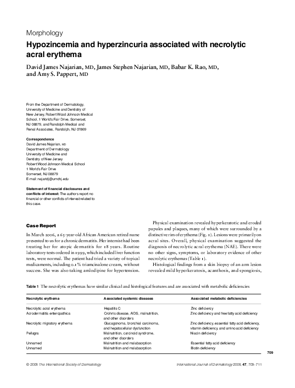
2008, International Journal of Dermatology
Journal of The American Academy of Dermatology
Leukocytoclastic vasculitis and necrolytic acral erythema in patients with hepatitis C infection: Do viral load and viral genotype play a role2010 •
Journal of the American Academy of Dermatology
Lack of classic histology should not prevent diagnosis of necrolytic acral erythema2009 •
2012 •
Case Reports in Dermatology
Necrolytic Acral Erythema in Seronegative Hepatitis C2007 •
Hepatitis C virus (HCV) infection is a major public health problem in many countries. Since its discovery in 1989 by Choo et al. 1 HCV has been the focus of intensive basic and clinical research. HCV is recognized as one of the main causative agents of liver disorders, including most cases of parenterally transmitted viral hepatitis. However, it rapidly became evident that HCV infection was associated with disorders of various organs, other than the liver, essentially through immunologic mechanisms 2 . Many extrahepatic manifestations have been reported to occur in the joints, muscles, neural tissue and kidneys. Also, lymphoma and sjögren syndrome with or without mixed cryoglobulinemia have been related to HCV infection. Extrahepatic cutaneous diseases have been reported to be associated with HCV infection. They manifest in 20-40% of patients presenting to the dermatologist and in a significant percentage (15-20%) of the general population 3 . Therefore, recognition of the cutaneous...
Clinical and Experimental Dermatology
Severe exfoliative erythema of malnutrition in a child with coexisting coeliac and Hartnup’s disease2009 •
Clinical and Experimental Dermatology
A 47-year-old woman patient with refractory migratory erythema2008 •
Practical Management of Chronic Viral Hepatitis
The Skin and Viral Liver Disease2013 •
Atlas of Dermatology in Internal Medicine
Atlas of Dermatology in Internal Medicine1998 •
Critical Reviews in Oral Biology & Medicine
Oral Diseases Possibly Associated with Hepatitis C Virus2003 •
The Journal of Pediatrics
Acrodermatitis enteropathica–like cutaneous lesions in organic aciduria1994 •
Journal of medical case reports
Glucagonoma syndrome: a case reportJournal of the European Academy of Dermatology and Venereology
Psoriasis and unreported excessive alcohol intake - a simple screening approach2011 •
Case reports in infectious diseases
Orbital Pseudotumor as an Extrahepatic Complication of Chronic HCV Infection2018 •
Clinics in Dermatology
Alcohol, social behavior disorders, and their cutaneous manifestations1999 •
British Journal of Dermatology
The diagnostic value of polymerase chain reaction in erythema induratum of Bazin1997 •
Journal of Clinical Medicine
Serum Zinc Concentration and Sarcopenia: A Close Linkage in Chronic Liver DiseasesBritish Journal of Dermatology
A case of paraneoplastic pemphigus with antidesmoglein 1 antibodies as determined by immunoblotting2000 •
International journal of dermatology
Genetic and acquired cutaneous disorders associated with internal malignancy1995 •
British Journal of Dermatology
Phakomatosis pigmentokeratotica associated with hypophosphataemic vitamin D-resistant rickets: improvement in phosphate homeostasis after partial laser ablation2003 •
Journal of the American Academy of Dermatology
Acquired cutis laxa following urticarial vasculitis associated with IgA myeloma2009 •
Clinics in Dermatology
Arcuate, annular, and polycyclic inflammatory and infectious lesions2011 •
British Journal of Dermatology
Generalized eruptive histiocytosis: a possible therapeutic cure2004 •
British Journal of Dermatology
Verruciform xanthoma of the scrotum in a renal transplant patient2004 •
Vascular Health and Risk Management
Macro- and micronutrients in patients with congestive heart failure, particularly African-Americans2007 •
British Journal of Dermatology
Acral ulcerations and osteolysis, a severe form of the carpal tunnel syndrome2004 •
The British Journal of Dermatology
Lichen sclerosus-lichen planus overlap in a patient with hepatitis C virus infection2004 •
British Journal of Dermatology
BDS Melanoma Guidelines: reply from authors2003 •
 Babar Rao