Proc. Nati. Acad. Sci. USA
Vol. 89, pp. 3770-3774, May 1992
Biophysics
What drives the translocation of proteins?
(Brownian motion/heat shock proteins/chaperonins/model)
SANFORD M. SIMON*, CHARLES S. PESKINt,
AND
GEORGE F. OSTERt
*Howard Hughes Medical Institute, Rockefeller University, 1230 York Avenue, New York, NY 10021; tCourant Institute of Mathematical Sciences, 251
Mercer Street, New York, NY 10012; and tDepartments of Molecular and Cellular Biology, and Entomology, University of California, Berkeley, CA 94720
Communicated by Gunter Blobel, December 31, 1991 (received for review October 26, 1991)
We propose that protein translocation across
ABSTRACT
membranes is driven by biased random thermal motion. This
"Brownian ratchet" mechanism depends on chemical asymmetries between the cis and trans sides of the membrane.
Several mechanisms could contribute to rectifying the thermal
motion of the protein, such as binding and dissociation of
chaperonins to the translocating chain, chain coiling induced
by pH and/or ionic gradients, glycosylation, and disulfide
bond formation. This helps explain the robustness and promiscuity of these transport systems.
question of how to convert this energy into directed motion;
for this we must turn to molecular mechanics.
The Model
We examine the post-translational translocation of a protein
from the cis to trans side of a membrane addressing the
process that begins after an initial tip (or loop) is threaded
through the channel-a separate physical process we shall
discuss elsewhere. To traverse the TP, a protein must be in
an unfolded conformation. Brownian motion will cause the
protein to fluctuate back and forth through the TP but with no
net displacement. But if, upon emerging from the TP, a
protein is modified in such a way that it cannot reenter the
pore, then its random walk will be biased. For example, when
a nascent chain is glycosylated, it cannot reenter the TP.
Thus it will reptate- "move like a snake" (8)-until it fully
translocates across the membrane. The more closely spaced
the ratcheting sites are, the faster is the movement across the
membrane. The model rests on two assumptions. (i) The
protein is unfolded and free to reptate back and forth through
the TP. (ii) Chemical asymmetries (specified below) rectify
the protein's movements. Both assumptions are strongly
supported by experimental data.
Several observations indicate a translocating polypeptide
is free to reptate back and forth. (i) Upon release from
cytosolic ribosomes, nascent polypeptides traverse the membrane and enter the lumen of the ER (9). Thus, without the
input of additional energy, the 40 amino acids of the polypeptide in the ribosome and 20 amino acids spanning the
membrane freely traverse the bilayer. (ii) Translocating polypeptides are extracted from the membrane with mild conditions that leave the membrane intact (10). (iii) Releasing
translocating chains from the membrane reveals the presence
of large aqueous pores, presumably protein-conducting channels (4).
Several chemical asymmetries could bias the Brownian
walk of a chain. As a polypeptide emerges from the translocation apparatus, often before much of the protein has been
synthesized, the chain is subjected to glycosylation (11, 12),
formation of disulfide bonds, binding of chaperonins, and
cleavage of the signal sequence (which affects folding of the
chain). Any, or all, can induce the asymmetry required for the
Brownian ratchet.
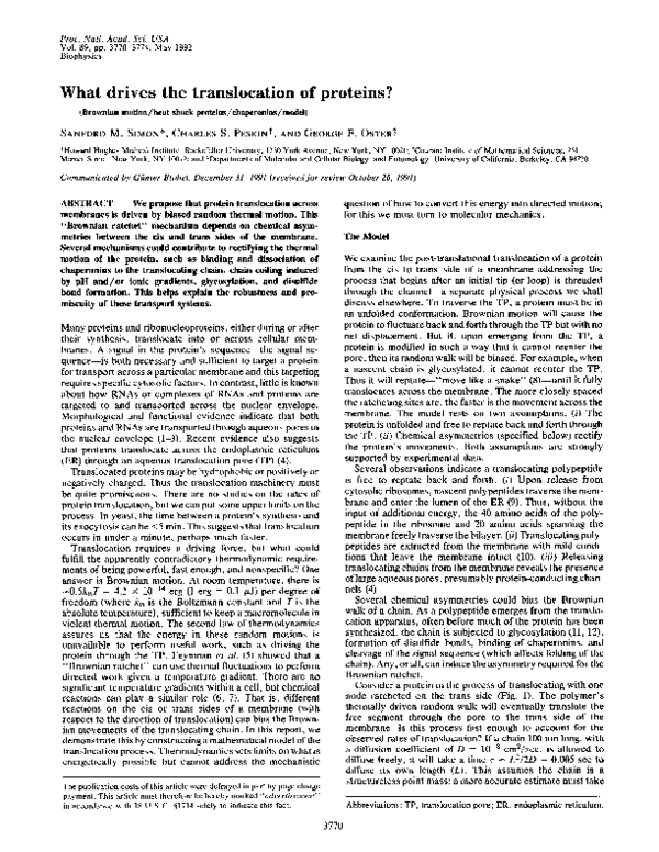
Consider a protein in the process of translocating with one
node ratcheted on the trans side (Fig. 1). The polymer's
thermally driven random walk will eventually translate the
free segment through the pore to the trans side of the
membrane. Is this process fast enough to account for the
observed rates of translocation? If a chain 100 nm long, with
a diffusion coefficient of D = 10-8 cm2/sec, is allowed to
diffuse freely, it will take a time r L2/2D = 0.005 sec to
diffuse its own length (L). This assumes the chain is a
structureless point mass; a more accurate estimate must take
Many proteins and ribonucleoproteins, either during or after
their synthesis, translocate into or across cellular membranes. A signal in the protein's sequence-the signal sequence-is both necessary and sufficient to target a protein
for transport across a particular membrane and this targeting
requires specific cytosolic factors. In contrast, little is known
about how RNAs or complexes of RNAs and proteins are
targeted to and transported across the nuclear envelope.
Morphological and functional evidence indicate that both
proteins and RNAs are transported through aqueous pores in
the nuclear envelope (1-3). Recent evidence also suggests
that proteins translocate across the endoplasmic reticulum
(ER) through an aqueous translocation pore (TP) (4).
Translocated proteins may be hydrophobic or positively or
negatively charged. Thus the translocation machinery must
be quite promiscuous. There are no studies on the rates of
protein translocation, but we can put some upper limits on the
process. In yeast, the time between a protein's synthesis and
its exocytosis can be <5 min. This suggests that translocation
occurs in under a minute, perhaps much faster.
Translocation requires a driving force, but what could
fulfill the apparently contradictory thermodynamic requirements of being powerful, fast enough, and nonspecific? One
answer is Brownian motion. At room temperature, there is
=0.5kBT = 4.1 x 10⁻¹⁴ erg (1 erg = 0.1 ,uJ) per degree of
freedom (where kB is the Boltzmann constant and T is the
absolute temperature), sufficient to keep a macromolecule in
violent thermal motion. The second law of thermodynamics
assures us that the energy in these random motions is
unavailable to perform useful work, such as driving the
protein through the TP. Feynman et al. (5) showed that a
"Brownian ratchet" can use thermal fluctuations to perform
directed work given a temperature gradient. There are no
significant temperature gradients within a cell, but chemical
reactions can play a similar role (6, 7). That is, different
reactions on the cis or trans sides of a membrane (with
respect to the direction of translocation) can bias the Brownian movements of the translocating chain. In this report, we
demonstrate this by constructing a mathematical model of the
translocation process. Thermodynamics sets limits on what is
energetically possible but cannot address the mechanistic
=
The publication costs of this article were defrayed in part by page charge
payment. This article must therefore be hereby marked "advertisement"
in accordance with 18 U.S.C. §1734 solely to indicate this fact.
Abbreviations: TP, translocation pore; ER, endoplasmic reticulum.
3770
�Biophysics: Simon et al.
Proc. Natl. Acad. Sci. USA 89 (1992)
Thermal
fIuctuatioin
Ch a oroni rr
--WOWI a %,%vvq
- - - -
Q
I=
W,\
111|*
F
FIG. 1. One-dimensional model of a flexible protein chain diffusing through the TP. Each node experiences elastic forces from the
neighboring nodes, random and viscous drag forces from the fluid
surroundings, and, if a chaperonin is bound to the node, a repulsive
force from the TP. The protein fluctuates back and forth until a
chaperonin comes close enough to the pore-bound enzyme to be
stripped from the chain. This allows a segment of the protein to
diffuse through the pore. When the segment exits from the pore, it
is bound by a lumenal chaperonin, preventing the segment from
fluctuating backward.
into account the extended geometry of the chain, its accompanying elastic flexibility, the constraint of the pore on its
entropic configurations, and the effects of the asymmetric
factors enumerated above. To answer this we have constructed a computational model of the process.
We first treat the case where the ratchet is implemented by
the binding of chaperonins (Fig. 1). We adopt a standard
model for polymer dynamics by representing the protein by
a chain of elastically linked subunits (13) that may be individual amino acids or larger segments. Each node of the
model chain experiences four forces: viscous drag forces
from the surrounding fluid, elastic forces from the neighboring nodes, repulsive forces of the membrane and the TP, and
random forces due to the thermal environment. The equations describing the motion of the chain are derived by
balancing the various forces that act on each node:
The solution of the model equations lets us compute the
translocation rate (e.g., residues per sec) as a function of
chain flexibility, differential coiling potentials, pore characteristics, and the kinetics of chain modification (chaperonin
binding/dissociation, glycosylation, etc.)-a formible task
for three-dimensional polymers of significant length. We
detail these calculations elsewhere; below we describe a
one-dimensional version of the model.
Conditions maximizing translocation rates are as follows:
(i) ratcheting each node on the trans side, (ii) instantaneous
chaperonin binding on the trans side, and (iii) instantaneous
dissociation of chaperonins from the cis side upon reaching
the TP. The computation begins with the chain just threaded
through the pore and continues until the left-most node just
clears the membrane (Fig. 1); this is the translocation time r.
We model the polypeptide as an elastic chain between 5 and
75 nm, with potential ratcheting sites at 5-nm intervals. Since
the pattern of random forces always differs, the chain follows
a different trajectory for each simulation. Repeated calculations yield a distribution of translocation times whose mean
velocity and mean transit time, (T), we can compute by
averaging (Fig. 2).
It is not known how many ratcheting sites are in each
translocating chain. Translocation is slowest when sites are
only at each end of the chain (Fig. 2A). Still, a 100-nm chain
translocates in <3 msec. So, even if one site is ratcheted on
the trans side, diffusion is sufficient to move proteins across
A
3
800
(D)
~~~~~~~~~~~600
Cn
E
400-E
o
al)
0~~~~~~~~
2005
E
fk(dXk/dt) = Feiastic(Xkl, Xk, Xk+1) + Fkre(Xk) + Rk
(k= 1, .. , N),
[la]
where fk(dXk/dt) is the frictional drag on kth node, Fk'a"i
(Xk1, Xk, Xk+1) are elastic forces on segment k from adjacent
segments, FPO' (Xk) is the pore force on segment k, Rk is the
random force, N is the number of subunits in the protein,
Xk(t) is the position of the kth subunit at time t, andfk is the
friction coefficient of the kth subunit.
In Eq. la, the pore force depends on whether a particular
node is ratcheted-bound by a chaperonin on the trans side of
the membrane. But as we discuss below, other modifications
can effect a Brownian ratchet as well. These are incorporated
into the equations by allowing each segment to have a bound
chaperonin or not:
Xbound
Xk
k+
k
Xkfree
3771
[lb]
Length of peptide (nm)
B
0
E
Ec
0
1
1
.10 1000.100..1
1
10
100
1000 10000 100000
1/sec
FIG. 2. Numerical solutions of the model shown in Fig. 1. (A) The
Transition rates are the binding and dissociation rate constants, k+ and k_, respectively (if chaperonins are abundant,
rate constants are pseudo-first-order and 1/k+ o mean time
for a free segment to bind a chaperonin). Segments with a
bound chaperonin have a higher friction coefficient, fk, than
free segments; more importantly, they interact with the
translocation pore differently. The lipid bilayer is impermeable to the chain, and thus we can model the membrane as a
very high energy barrier. The translocation pore allows free
subunits-those with no chaperonin bound-to pass through
the membrane but a subunit binding a chaperonin will see an
energy barrier preventing its entry into the pore.
mean translocation time (i) (circles) and mean velocity (v)-Ll(r)
(squares) are shown as a function of chain length. The results are
plotted for simulations that assumed that either every node could be
ratcheted (open symbols) or only the node farthest on the trans side
was ratcheted (solid symbols). The open circles are fit with a
quadratic, (r) x L2, and the solid circles are fit by a cubic, (T) X LI
(see discussion of Eq. 2). (B) The effects of chaperonin kinetics on
translocation velocity were shown by varying the trans binding rate
(k+) or the cis dissociation rate (k_). When k+ > 1000 bindings per
sec, the velocity is diffusion limited. In the simulations the following
parameters were used: membrane thickness, 5 nm; pore repulsion
range, 0.75 nm; chain elastic constant
kBT/82, 1/25; friction
coefficient, 1 x 1o-7 dyne-sec/cm (1 dyne = 100 mN); Brownian
force, (fkT/At)'/2 dyne; integration step size, At < 1 x 10⁻⁷ sec.
�Biophysics: Simon et al.
3772
Proc. Natl. Acad. Sci. USA 89 (1992)
the membrane faster than the minimum limit set by experimental observations. Ratcheting significantly speeds translocation (Fig. 2A) by a factor of about L/6 (where 8 is the
distance between ratcheting sites), as discussed below.
The calculations assumed chaperonin binding on the trans
side was fast. But chaperonins are large (60-90 kDa) and may
not bind very quickly to the nascent chain. We next investigate the effect of finite rates of attachment and detachment
on translocation time by varying the binding rate constant k+
over several orders of magnitude for a chain of L = 45 nm
with ratcheting sites every 8 = 5 nm (Fig. 2B). At high binding
rates, velocity is independent of k+ since motion is limited by
the polypeptide's diffusion. As the binding rate decreases,
the ratchet has a smaller effect on accelerating translocation.
Since chaperonins cannot enter the TP, translocation velocity is limited by the rate they are stripped from the chain
on the cis side. If chaperonin binding affinity was small
enough to frequently free up a segment for diffusion into the
TP, the chain could not be held in a linear configuration.
Thus, we postulate a pore-associated enzyme (perhaps an
ATP or GTP dissociating enzyme) that detaches the chaperonin from the chain. The maximum velocity corresponds to
instantaneous cis dissociation and trans binding, so translocation is diffusion-limited. When the rate of cis dissociation
falls below about 500 removals per sec, the velocity varies
approximately linearly with kCiS (Fig. 2B).
The following formula is an approximate analytical solution describing the average translocation velocity, (v) (the
derivation will be presented elsewhere):
(v)
=
2D
3
k+
2D
5
k+ +2k-
1
1 + 2Kd
'
[2]
where Kd = kL/k+. The assumptions behind Eq. 2 are as
follows: the rod is rigid;§ chaperonins exist on the trans side
only (or are enzymatically removed rapidly at the cis side of
the TP); and reaction rates k+ and kL are fast. (The approximation in Eq. 2 improves as the rates increase.) Note the
velocity of translocation increases as 8 decreases. When k+
>> kL, the velocity becomes simply (v) = 2D/6, so average
translocation time is given by (X) L/(v) = L8/2D. Generally, D varies inversely with size, so we expect (r) L28. If
chaperonins bind only at the ends of the chains (8 = L), then
(T) -L3. Numerical simulations in Fig. 2A show this dependence.
These results put quantitative limits on translocation times.
The slowest time is when chaperonins bind only at the ends
-
of the chain. By
taking
8
100
nm
and D
10-8 cm2/sec,
the translocation time is 5 msec; but if 8 = 5 nm, the transit
time is 0.25 msec-faster by a factor of 20. This estimate of
X is probably too short, since our one-dimensional calculation
for chain coiling. Nevertheless, numerical
and analytical calculations show the ratchet mechanism is
more than sufficient to account for the observed rates of
translocation.
Other Brownian Ratchet Mechanisms. This thermal ratchet
model depends on the asymmetry of chaperonin binding to
the translocating chain. But a number of chain modifications
occur cotranslationally that can bias thermal reptation. Any
ligand that binds differently on the two sides of the membrane
will bias reptation. For example, if chaperonin concentration
cannot account
§Although the center of mass of a flexible chain will diffuse more
slowly than that of a rigid chain, a flexible chain will translocate
faster than a rigid chain so long as 8 << L. This is because each node
in an elastic chain can fluctuate somewhat independently of the
chain as a whole, which allows a node to fluctuate out the right side
of the TP, even though the rest of the chain may be moving to the
left. Further, the tension in the internodal spring of each ratcheted
node helps pull the rest of the chain through the TP.
differs on the cis and trans sides of the membrane, differential
binding equilibria bias reptation toward the side with the
higher concentration. Moreover, the lumenal space of the ER
contains enzymes that glycosylate many proteins inhibiting
their backward fluctuations and rectifying their Brownian
movements.
If the chain coils more tightly in the cisternal space than in
the cytoplasm, this ratchets thermal motions, independent of
ligand binding. Virtually all biological polymers carry fixed
charges-usually negative-that affect their degree of coiling
(14). If the ionic strength on the trans side is higher than on
the cis side, counter ions will shield these charge interactions
allowing the protein to coil more tightly. Similarly, a reduced
pH in the compartment will titrate charge groups on the
chain; if this shifts the protein toward its isoelectric point, it
will coil more compactly. Kagan et al. (15) suggested a
pH-dependent folding as a way to translocate diphtheria toxin
across lysosomes. A higher-than-cytosolic concentration of
calcium may do the same in the lumen of the ER, in the matrix
of the mitochondria, or in the periplasm of Escherichia coli.
But, since no systematic charge configuration characterizes
all translocated proteins, folding may not be a general mechanism for rectifying protein diffusion, though it may assist in
specific instances. There is evidence that the signal sequence
confers differential coiling potentials on a protein, since many
proteins, such as bacterial alkaline phosphatase, are sensitive
to protease prior to translocation; after signal peptide cleavage, they fold into a tighter protease-resistant form (16).
Many chain modifications may occur cotranslocationally
(e.g., glycosylation or signal peptide cleavage), because it
would be difficult for the modifying enzyme to access its site
once the protein has folded. But these modifications could
also ratchet the nascent chain and speed translocation. So,
the modifying enzymes could be intimately involved in the
translocation process. Indeed, in situ many of these mechanisms may work in parallel. Binding of chaperonins, glycosylation of the translocated chain, and cleavage of the signal
sequence may all ensure vectorial transport.
The Brownian ratchet hypothesis assumes that the diffusion of a protein back and forth through the pore is unbiased
but that once a step of a certain size is made the ratchet locks
the protein in place. Alternatively, diffusion itself could be
biased. A voltage across the mitochondrial membrane is
required for protein import (17). Mitochondrial proteins
have, on average, a pI value that is 1.5 units more basic than
that of cytosolic proteins (18). Thus, the membrane potential
of 50 mV (corresponding to 105 V/cm) would bias mitochondrial protein reptation into the matrix. Unfortunately, such a
mechanism would retard the import of regions with local
negative charges. But a membrane potential may facilitate the
initial threading of the protein into the TP.
Thermodynamic Considerations. Potential thermodynamic
driving forces for translocation include transmembrane differences in pH, ionic strength, membrane potential, or other
electrochemical gradients. By examining the translocation
process at a more detailed level, we can see that all these
forces can contribute to biasing the random diffusion of the
translocating protein. Fig. 3 summarizes the role of different
factors in promoting translocation.
The energy for translocation ultimately derives from free
energy of the kinetic processes associated with translocation,
for the second law of thermodynamics prohibits extracting
work from an isothermal reservoir. For example, in the
chaperonin model the free energy sources are the concentration of chaperonin across the membrane and the binding
energy of chaperonins to the chain. A site emerging from the
pore has no bound chaperonin and so is not in equilibrium
with the trans compartment. When the emerging site binds a
chaperonin, the free energy of trans binding, AG+, must be
large enough to ensure a chaperonin is bound to the site often
�Biophysics: Simon et al.
Proc. Natl. Acad. Sci. USA 89 (1992)
cuLING
ApHo
.A,(lonic
strengTh)
i
* Signal sequence
cleavage
Rilro5omr
Disulfide bonding
Chanelling
Glycosylation
Glycasylation
CISm
IEyile
f
,Id.Oe
TRANS
FIG. 3. Summary of the factors that could ratchet protein translocation. On the cis side there are at least three permiss ive processes:
keeping the chain unfolded by chaperonin binding, con tainment with
the ribosome, and removal of blocking chaperonins adjacent to the
TP. On the trans side forces include binding of chape ronins, glycosylation, disulfide bonding, and chain coiling (the latter
affected by pH, ionic strength near the protein's pl valu e,
of the signal sequence). HSP, heat shock protein.
orocleavage
and tightly enough to prevent the chain from diffusing back
through the TP. If AG+ < kBT, the binding site Nwill likely be
empty when a reverse fluctuation occurs, carr'ying the site
back into the TP-or if the site is occupied, the force of its
collision with the TP will likely dislodge it. IfF the ratchet
mechanism is chain coiling, an emerging site is nc)t in entropic
equilibrium and this free energy difference imp3lements the
ratchet. A close inspection of each ratchet me chanism reveals the process needs a free energy source th;at ultimately
derives from intermolecular bond energies and /or entropic
conformations (for example, ATP hydrolysis tarps into 8.3 x
10-13 erg per molecule -20 kBT of chemical bl )nd energy).
This model provides a mechanistic look at ho)w these free
energies are transduced into vectorial translocation.
Membrane Proteins. Not all proteins transloxcate entirely
through the channel. Intermittent hydrophobic Estretches are
intercalcated into the lipid bilayer to integratee membrane
proteins. A model of translocation must explaiin how some
stretches of amino acids translocate across and ot ers integrate into the membrane. To see how this coul4
plished by a ratchet mechanism we make ti he following
assumptions. The TP is composed of subunits (off the same or
separate proteins) (4, 19-22). These subunits cEan thermally
fluctuate radially (Fig. 4)-this "breathing" fluctuation
opens a path between the pore and lipid bilayeir (23). Upon
synthesis of a latent transmembrane domain, the re isa pause
in protein translation/translocation. The chain iSdisplaced
into the bilayer when there are coincident fluc ftuations: an
opening in the gap between the subunits and a transverse
fluctuation in the chain. Once such a fluctualtion has occurred, it is unlikely to be reversed because off the considerable entropy gain of the chain as it mixes with the bilayer.
-
be
aom-owing
3773
If a nonhydrophobic segment is in the channel during a
"breathing" fluctuation, it remains there for it is energetically unfavorable for the nonhydrophobic segment to partition
into the bilayer. Hydrophobic segments can partitions
inoFrlttdmas,
into the bilayer. For latent transmembrane domains, this is
the desired result though it could lead to inappropriate
integration of segments into the bilayer. This problem can be
minimized if the time scale of a "breathing" fluctuation is
rapid relative to the rate the protein moves across the
membrane. The probability of integrating a polypeptide into
the bilayer is substantially increased if translocation and/or
translation are slowed during synthesis of latent transmembrane domains. Consistent with this assumption is the observation that ribosomes slow when translating signal sequences (24).
By studying the time scales and dynamics of membrane
insertion, we can ascertain if this model is plausible. It does
not violate the laws of physics but can it take place on the
observed time scale? How long must the chain remain in the
pore until a sufficiently large lateral chain fluctuation and a
"breathing" fluctuation of the channel occur simultaneously? Simulation of the model can provide some answers.
Chaperonins. Chaperonins are needed for in vitro translocation across the ER (24, 25) and for in vitro import across
mitochondrial (27-30), chloroplast (31), and bacterial membranes (32-35). They have been cross-linked to translocation
intermediaries (28), and their deletion in yeast blocks ER
translocation (36). Despite these clues, their functional significance is unknown. They may be required on the cis side
to keep proteins unfolded, a state permissive for translocation (25). Trans chaperonins may be required for facilitating
proper folding of the translocated chain (29). It has been
suggested that differences in energies of folding, perhaps
affected by the chaperonins, provide energetics for protein
movement (37). While these may be important in particular
cases, we propose a specific mechanism by which chaperonins are provocateurs of chain movement. Consistent with
this is the observation that by solubilizing mitochondria
precursors with urea the requirements for both heat shock
proteins and ATP
are bypassed (38).In This implies
ATP is
for the
even after
required
onlytranslocation
chaperonins.
ER-targeted
precursors arecontrast,
solubilized
in urea,
ATP is still needed (39). These observations are in accord
with our model and suggest two roles for chaperonins in the
translocation process. On the cis side of the membrane, they
keep the protein unfolded. On the trans side, they keep it
from diffusing back through the TP. Since slowing their
dissociation rate on the cis side slows translocation, this may
regulate how much of the nascent chain is folded on the trans
side prior to glycosylation, disulfide bond formation, or other
post-translational modifications.
,
Cha a ff
sce
atera ly through oap
chann&
5-jeurnis
FIG. 4. Subunits of the TP fluctuate radially openiing a path for
the chain to fluctuate laterally into the membrane. If p( fluctuation
coincides with lateral chain fluctuation, the segme :nt enters the
bilayer. A stop transfer signal that causes translocatic)n to pause in
this configuration enhances the probability of such a c roincident pair
of fluctuations.
ire
Discussion
Translocation of macromolecules across membranes is robust and promiscuous: almost any molecule can be translocated when given the proper signal sequence. Cytosolic
proteins have been targeted to the ER (40), ER proteins have
been targeted to the chloroplast (41), double-stranded DNA
has been translocated into mitochondria (42), and gold particles have been targeted into nuclei (1). This suggests, with
few exceptions, that nothing special about macromolecules
makes them translocation-competent; hence, the mechanism
that drives translocation must be equally nonspecific. We
have shown that a Brownian ratchet mechanism is both
indiscriminate and fast enough to explain the observed rates
of translocation. Moreover, our model predicts specific functional dependencies on molecular size and kinetic rate constants, so that its predictions can be experimentally addressed.
�3774
Biophysics: Simon et al.
Several other mechanisms have been proposed for driving
translocation. (i) The ribosome pushes the nascent chain
through the membrane using the energy associated with chain
elongation. (ii) A pump in the membrane mechanically grabs
the chain and pulls it across (46). (iii) Electrochemical
gradients across the membrane (e.g., pH or other ionic
gradients) drive translocation. (iv) Energy associated with
post-translocational folding might "pull" the chain through
the membrane (15, 29). Randall (44) has summarized arguments against the first proposal. The second proposal requires a pump that binds tightly enough to move the translocating peptide but has little specificity, since translocated
segments vary considerably in their polarity and charge.
There are no universally present transmembrane gradients
that could affect all proteins, and in any event the diversity
of charge in macromolecules precludes electrophoretic
forces as a general mechanism. The effect of chain folding on
translocation rates can only be computed from a full threedimensional simulation; we will report on this elsewhere.
A Brownian ratchet mechanism has several advantages for
translocating proteins. (i) Specificity is not programmed into
the translocating protein-consistent with the observation
that many cytosolic proteins are translocated if expressed
with a signal sequence. (ii) A ratchet provides specific
physical mechanisms for transducing the chemical energy of
ATP to mechanical movement (6). (iii) The translocating
segment is not bound to specific proteins, freeing latent
transmembrane domains to partition laterally into the lipid
bilayer. (iv) Since several independent processes promote
biased diffusion (Fig. 3), this gives a reliable, fast, and
nonspecific translocation mechanism. By defining and quantifying the model's parameters, it should be possible to
predict the effects of ionic strength and changes in pH,
temperature, and protein flexibility on translocation rates.
Macromolecules cross many different intracellular membranes. ATP-binding cassette (ABC) proteins mediate peptide transport across the ER membrane for antigen presentation and toxin transport across bacterial membranes. Both
proteins and RNAs move in both directions across the
nuclear envelope. In all cases hydrophilic molecules cross
membranes, sometimes against a concentration gradient. In
no instance has a mechanism for moving the macromolecules
been implicated. The ratchet mechanism, being nonspecific,
may drive these translocations. For example, mRNAs are
associated with one set of proteins in the nucleus, but after
transport into the cytosol, they affiliate with a second set (48).
This asymmetry in the nuclear pore could bias the thermal
reptation of the molecules out of the nucleus. Similarly,
disposition of ATP binding regions in the ABC transporters
may provide the asymmetry needed for vectorial movement
across the membrane. Brownian ratchets may be a ubiquitous
mechanism for moving macromolecules across biological
membranes.
This model predicts translocation can be driven by several
thermodynamic energy sources and that the relative role of
each varies among translocated proteins. It should be possible to experimentally distinguish between different ratchet
mechanisms by measuring translocation rates. Further, it
may explain why different laboratories have observed results
that implicate so many different energy sources for translocation.
We acknowledge Jon Singer and Gunter Blobel for many stimulating discussions whose ideas on translocation were the stimulus for
constructing the model. Conversations with Pierre-Giles de Gennes,
Paul Janmey, Steve Miller, Hsiao-Ping Moore, Randy Schekman,
and Melvin Schindler were important in shaping our thinking. We
Proc. Natl. Acad. Sci. USA 89 (1992)
also acknowledge the reviewers, who made substantive comments
that greatly improved the manuscript. S.M.S. is grateful for the
support of an Irma T. Hirschl-Monique Weill-Caulier Career Scientist Award. G.F.O. was supported by National Science Foundation
(NSF) Grant MCS-8110557. C.S.P. was supported by NSF Grant
CHE-900-2416. Both G.F.O. and C.S.P. acknowledge the support
provided by MacArthur Foundation Fellowships. G.F.O. acknowledges the hospitality of the Neurosciences Institute at which part of
this work was performed.
1. Feldherr, C. M., Kallenbach, E. & Schultz, N. (1984) J. Cell Biol. 99,
2216-2222.
2. Newmeyer, D. D., Finlay, D. R. & Forbes, D. J. (1986) J. Cell Biol. 103,
2091-2102.
3. Jiang, L. W. & Schindler, M. (1986) J. Cell Biol. 102, 853-858.
4. Simon, S. M. & Blobel, G. (1991) Cell 65, 1-10.
5. Feynman, R., Leighton, R. & Sands, M. (1963) The Feynmana Lectures
on Physics (Addison Wesley, Reading, MA), Vol. 1, pp. 46.1-46.4.
6. Cordova, N., Ermentrout, B. & Oster, G. F. (1992) Proc. Nail. Acad.
Sci. USA 89, 339-343.
7. Vale, R. D. & Oosawa, F. (1991) Adv. Biophys. 26, 97-134.
8. DeGennes, P. G. (1983) J. Physique Lett. 44, L225-L227.
9. Redman, C. M. & Sabatini, D. D. (1966) Proc. Natl. Acad. Sci. USA 56,
608-615.
10. Gilmore, R. & Blobel, G. (1985) Cell 42, 497-505.
11. Rothman, J. E. & Lodish, H. F. (1977) Nature (London) 269, 775-780.
12. Lingappa, V. R., Lingappa, J. R., Prasad, R., Ebner, K. E. & Blobel, G.
(1978) Proc. Nail. Acad. Sci. USA 75, 2338-2342.
13. Doi, M. & Edwards, S. (1986) The Theory of Polymer Dynamics (Oxford
University Press, Cambridge, UK).
14. Cantor, C. & Schimmel, P. (1980) Biophysical Chemistry: The Behavior
of Biological Macromolecules (Freeman, New York), Vol. 3.
15. Kagan, B. L., Finkelstein, A. & Colombini, M. (1981) Proc. Nail. Acad.
Sci. USA 78, 4950-4954.
16. Liu, G., Topping, T. B. & Randall, L. L. (1989) Proc. Natl. Acad. Sci.
USA 86, 9213-9217.
17. Martin, J., Mahlke, K. & Pfanner, N. (1991) J. Biol. Chem. 266,
18051-18057.
18. Hartmann, C. & Christen, P. (1991) Nature (London) 352, 762-763.
19. Blobel, G. & Dobberstein, B. (1975) J. Cell Biol. 67, 835-851.
20. Blobel, G. (1980) Proc. Nall. Acad. Sci. USA 77, 1496-1500.
21. Singer, S. J., Maher, P. A. & Yaffe, M. P. (1987) Proc. Natl. Acad. Sci.
USA 84, 1960-1964.
22. Singer, S. J. (1990) Annu. Rev. Cell Biol. 6, 247-2%.
23. Singer, S. J. & Yaffe, M. P. (1990) Trends Biochem. Sci. 15, 369-373.
24. Wolin, S. L. & Walter, P. (1988) EMBO J. 7, 3559-3569.
25. Chirico, W. J., Waters, M. G. & Blobel, G. (1988) Nature (London) 332,
805-810.
26. Deshaies, R. J., Koch, B. D., Werner, W. M., Craig, E. A. & Schekman, R. (1988) Nature (London) 332, 800-805.
27. Skerjanc, I. S., Sheffield, W. P., Randall, S. K., Silvius, J. R. & Shore,
G. C. (1990) J. Biol. Chem. 265, 9444-9451.
28. Scherer, P. E., Krieg, U. C., Hwang, S. T., Vestweber, D. & Schatz, G.
(1990) EMBO J. 9, 4315-4322.
29. Ostermann, J., Horwich, A. L., Neupert, W. & Hartl, F. U. (1989)
Nature (London) 341, 125-130.
30. Murakami, H., Pain, D. & Blobel, G. (1988) J. Cell Biol. 107, 2051-2057.
31. Waegemann, K., Paulsen, H. & Soll, J. (1990) FEBS Lett. 261, 89-92.
32. Zimmermann, R., Sagstetter, M., Lewis, M. J. & Pelham, H. (1988)
EMBO J. 7, 2875-2880.
33. Kusukawa, N., Yura, T., Ueguchi, C., Akiyama, Y. & Ito, K. (1989)
EMBO J. 8, 3517-3521.
34. Lecker, S., Lill, R., Ziegelhoffer, T., Georgopoulos, C., Bassford, P. J.,
Kumamoto, C. A. & Wickner, W. (1989) EMBO J. 8, 2703-2709.
35. Phillips, G. J. & Silhavy, T. J. (1990) Nature (London) 344, 882-884.
36. Vogel, J. P., Misra, L. M. & Rose, M. D. (1990) J. Cell Biol. 110,
1885-1895.
37. Kang, P. J., Ostermann, J., Shilling, J., Neupert, W., Craig, E. A. &
Pfanner, N. (1990) Nature (London) 348, 137-143.
38. Pfanner, N., Rassow, J., Guiard, B., Sollner, T., Hartl, F. U. & Neupert,
W. (1990) J. Biol. Chem. 265, 16324-16329.
39. Waters, M. G. & Blobel, G. (1986) J. Cell Biol. 102, 1543-1550.
40. Lingappa, V. R., Chaidez, J., Yost, C. S. & Hedgepetch, J. (1984) Proc.
Nail. Acad. Sci. USA 81, 456-460.
41. Lubben, T. H., Bansberg, J. & Keegstra, K. (1987) Science 238, 11121114.
42. Vestweber, D. & Schatz, G. (1989) Nature (London) 338, 170-172.
43. Alberts, B., Bray, D., Lewis, J., Raff, M., Roberts, K. & Watson, J. D.
(1989) Molecular Biology of The Cell (Garland, New York).
44. Randall, L. L. (1983) Cell 33, 231-240.
45. Dreyfuss, G. (1986) Annu. Rev. Cell Biol. 2, 459-498.
�
 George Oster