WO2024182516A1 - Cell therapy for treating systemic autoimmune diseases - Google Patents
Cell therapy for treating systemic autoimmune diseases Download PDFInfo
- Publication number
- WO2024182516A1 WO2024182516A1 PCT/US2024/017681 US2024017681W WO2024182516A1 WO 2024182516 A1 WO2024182516 A1 WO 2024182516A1 US 2024017681 W US2024017681 W US 2024017681W WO 2024182516 A1 WO2024182516 A1 WO 2024182516A1
- Authority
- WO
- WIPO (PCT)
- Prior art keywords
- cells
- subject
- car
- dose
- composition
- Prior art date
- Legal status (The legal status is an assumption and is not a legal conclusion. Google has not performed a legal analysis and makes no representation as to the accuracy of the status listed.)
- Ceased
Links
Classifications
-
- A—HUMAN NECESSITIES
- A61—MEDICAL OR VETERINARY SCIENCE; HYGIENE
- A61K—PREPARATIONS FOR MEDICAL, DENTAL OR TOILETRY PURPOSES
- A61K35/00—Medicinal preparations containing materials or reaction products thereof with undetermined constitution
- A61K35/12—Materials from mammals; Compositions comprising non-specified tissues or cells; Compositions comprising non-embryonic stem cells; Genetically modified cells
- A61K35/14—Blood; Artificial blood
- A61K35/17—Lymphocytes; B-cells; T-cells; Natural killer cells; Interferon-activated or cytokine-activated lymphocytes
-
- A—HUMAN NECESSITIES
- A61—MEDICAL OR VETERINARY SCIENCE; HYGIENE
- A61K—PREPARATIONS FOR MEDICAL, DENTAL OR TOILETRY PURPOSES
- A61K40/00—Cellular immunotherapy
- A61K40/10—Cellular immunotherapy characterised by the cell type used
- A61K40/11—T-cells, e.g. tumour infiltrating lymphocytes [TIL] or regulatory T [Treg] cells; Lymphokine-activated killer [LAK] cells
-
- A—HUMAN NECESSITIES
- A61—MEDICAL OR VETERINARY SCIENCE; HYGIENE
- A61K—PREPARATIONS FOR MEDICAL, DENTAL OR TOILETRY PURPOSES
- A61K40/00—Cellular immunotherapy
- A61K40/30—Cellular immunotherapy characterised by the recombinant expression of specific molecules in the cells of the immune system
- A61K40/31—Chimeric antigen receptors [CAR]
-
- A—HUMAN NECESSITIES
- A61—MEDICAL OR VETERINARY SCIENCE; HYGIENE
- A61K—PREPARATIONS FOR MEDICAL, DENTAL OR TOILETRY PURPOSES
- A61K40/00—Cellular immunotherapy
- A61K40/40—Cellular immunotherapy characterised by antigens that are targeted or presented by cells of the immune system
- A61K40/41—Vertebrate antigens
- A61K40/416—Antigens related to auto-immune diseases; Preparations to induce self-tolerance
-
- A—HUMAN NECESSITIES
- A61—MEDICAL OR VETERINARY SCIENCE; HYGIENE
- A61K—PREPARATIONS FOR MEDICAL, DENTAL OR TOILETRY PURPOSES
- A61K40/00—Cellular immunotherapy
- A61K40/40—Cellular immunotherapy characterised by antigens that are targeted or presented by cells of the immune system
- A61K40/41—Vertebrate antigens
- A61K40/42—Cancer antigens
- A61K40/4202—Receptors, cell surface antigens or cell surface determinants
- A61K40/421—Immunoglobulin superfamily
- A61K40/4211—CD19 or B4
-
- A—HUMAN NECESSITIES
- A61—MEDICAL OR VETERINARY SCIENCE; HYGIENE
- A61P—SPECIFIC THERAPEUTIC ACTIVITY OF CHEMICAL COMPOUNDS OR MEDICINAL PREPARATIONS
- A61P17/00—Drugs for dermatological disorders
-
- A—HUMAN NECESSITIES
- A61—MEDICAL OR VETERINARY SCIENCE; HYGIENE
- A61P—SPECIFIC THERAPEUTIC ACTIVITY OF CHEMICAL COMPOUNDS OR MEDICINAL PREPARATIONS
- A61P19/00—Drugs for skeletal disorders
-
- A—HUMAN NECESSITIES
- A61—MEDICAL OR VETERINARY SCIENCE; HYGIENE
- A61P—SPECIFIC THERAPEUTIC ACTIVITY OF CHEMICAL COMPOUNDS OR MEDICINAL PREPARATIONS
- A61P21/00—Drugs for disorders of the muscular or neuromuscular system
-
- A—HUMAN NECESSITIES
- A61—MEDICAL OR VETERINARY SCIENCE; HYGIENE
- A61P—SPECIFIC THERAPEUTIC ACTIVITY OF CHEMICAL COMPOUNDS OR MEDICINAL PREPARATIONS
- A61P37/00—Drugs for immunological or allergic disorders
-
- A—HUMAN NECESSITIES
- A61—MEDICAL OR VETERINARY SCIENCE; HYGIENE
- A61P—SPECIFIC THERAPEUTIC ACTIVITY OF CHEMICAL COMPOUNDS OR MEDICINAL PREPARATIONS
- A61P37/00—Drugs for immunological or allergic disorders
- A61P37/02—Immunomodulators
-
- C—CHEMISTRY; METALLURGY
- C07—ORGANIC CHEMISTRY
- C07K—PEPTIDES
- C07K16/00—Immunoglobulins [IGs], e.g. monoclonal or polyclonal antibodies
- C07K16/18—Immunoglobulins [IGs], e.g. monoclonal or polyclonal antibodies against material from animals or humans
- C07K16/28—Immunoglobulins [IGs], e.g. monoclonal or polyclonal antibodies against material from animals or humans against receptors, cell surface antigens or cell surface determinants
- C07K16/2803—Immunoglobulins [IGs], e.g. monoclonal or polyclonal antibodies against material from animals or humans against receptors, cell surface antigens or cell surface determinants against the immunoglobulin superfamily
-
- C—CHEMISTRY; METALLURGY
- C12—BIOCHEMISTRY; BEER; SPIRITS; WINE; VINEGAR; MICROBIOLOGY; ENZYMOLOGY; MUTATION OR GENETIC ENGINEERING
- C12N—MICROORGANISMS OR ENZYMES; COMPOSITIONS THEREOF; PROPAGATING, PRESERVING, OR MAINTAINING MICROORGANISMS; MUTATION OR GENETIC ENGINEERING; CULTURE MEDIA
- C12N5/00—Undifferentiated human, animal or plant cells, e.g. cell lines; Tissues; Cultivation or maintenance thereof; Culture media therefor
- C12N5/06—Animal cells or tissues; Human cells or tissues
- C12N5/0602—Vertebrate cells
- C12N5/0634—Cells from the blood or the immune system
- C12N5/0636—T lymphocytes
-
- A—HUMAN NECESSITIES
- A61—MEDICAL OR VETERINARY SCIENCE; HYGIENE
- A61K—PREPARATIONS FOR MEDICAL, DENTAL OR TOILETRY PURPOSES
- A61K2239/00—Indexing codes associated with cellular immunotherapy of group A61K40/00
- A61K2239/38—Indexing codes associated with cellular immunotherapy of group A61K40/00 characterised by the dose, timing or administration schedule
Definitions
- the present disclosure relates in some aspects to adoptive cell therapy involving the administration of a dose of T cells expressing a CD19-directed chimeric antigen receptor for treating subjects with a Systemic Autoimmune Disease and related methods, compositions, uses and articles of manufacture.
- Systemic autoimmune disease relates to a wide range of diseases and disorders characterized by dysregulation of the immune system.
- SLE Systemic Lupus Erythematosus
- Effective therapies for patients with SLE, such as severe refractory SLE, who have failed one or more prior therapy are needed. Provided are methods and uses that meet such needs.
- a method of treating a subject having a systemic autoimmune disease comprising administering a dose of CD19-directed genetically modified T cells from a composition comprising engineered T cells expressing a chimeric antigen receptor (CAR) to a subject having or suspected of having a severe systemic autoimmune disease, wherein the T cells of the dose are positive for expression of a CAR that binds CD 19 and the dose is from 1 x 10 6 to 50 x 10 6 CAR-positive viable T cells.
- CAR chimeric antigen receptor
- Also provided herein is a method of treating a subject having systemic autoimmune disease, the method comprising administering a dose of CD19-directed genetically modified T cells to a subject having or suspected of having a moderate systemic autoimmune disease, wherein the T cells of the dose are positive for expression of a chimeric antigen receptor (CAR) that binds CD19 and the dose is from 1 x 10 6 to 50 x 10 6 CAR-positive viable T cells.
- CAR chimeric antigen receptor
- the systemic autoimmune disease is selected from the group consisting of systemic lupus erythematosus (SLE), Sjogren's’ syndrome, progressive systemic sclerosis (i.e., scleroderma), idiopathic inflammatory myositis (IIM), including dermatomyositis, polymyositis and necrotizing myositis), mixed connective tissue disorder (MCTD), highly active relapsing-remitting multiple sclerosis, primary progressive MS, ANCA-associated vasculitis (AAV), Crohn’s disease, myasthenia gravis, Behcet’s, rheumatoid arthritis, IgA nephropathy, pemphigus vulgaris, myasthernia gravis, autoimmune hemolytic anemia, immune thrombocytopenia, IgG4-related diseases, membranous nephropathy, cutaneous lupus erythematosus
- the systemic autoimmune disease is rheumatoid arthritis. In some embodiments, the systemic autoimmune disease is myositis. In some embodiments, the systemic autoimmune disease is myasthenia gravis. In some embodiments, the systemic autoimmune disease is bullous pemphigoid. In some embodiments, the systemic autoimmune disease is immune thrombocytopenia. In some embodiments, the systemic autoimmune disease is autoimmune hemolytic anemia. In some embodiments, the systemic autoimmune disease is pemphigus vulgaris. In some embodiments, the systemic autoimmune disease is demyelinating polyradiculoneropathy. In some embodiments, the systemic autoimmune disease is membranous nephropathy.
- the systemic autoimmune disease is a refractory disease. In some embodiments, the subject is refractory to treatment with one or more prior therapies for the systemic autoimmune disease. In some embodiments, the subject is refractory to treatment with two or more prior therapies for the systemic autoimmune disease. In some embodiments, the systemic autoimmune disease is a severe disease.
- Also provided herein is a method of treating a subject having severe systemic lupus erythematosus (SLE), the method comprising administering a dose of CD19-directed genetically modified T cells to a subject having or suspected of having severe systemic lupus erythematosus (SLE), wherein the T cells of the dose are positive for expression of a chimeric antigen receptor (CAR) that binds CD19 and the dose is from 1 x 10 6 to 50 x 10 6 CAR-positive viable T cells.
- CAR chimeric antigen receptor
- CAR chimeric antigen receptor
- the SLE in the subject has one or more of the following: renal, central nervous system, or hematologic involvement.
- the subject has at least one organ system categorized by the British Isles Lupus Assessment Group 2004 (“BILAG”) as category A (“BILAG A”) or at least two organ systems categorized as BILAG B.
- BILAG British Isles Lupus Assessment Group 2004
- the subject fulfills the 2019 American College of Rheumatology (ACR)/ European League against Rheumatism (EULAR) classification criteria of SLE and/or the subject has detectable anti-dsDNA, anti-histone, anti-chromatin or anti-Sm antibodies in their blood.
- the subject fulfills the 2019 American College of Rheumatology (ACR)/ European League against Rheumatism (EULAR) classification criteria of SLE.
- the subject has detectable anti-dsDNA, anti-histone, antichromatin or anti-Sm antibodies in their blood.
- the subject has lupus nephritis.
- Also provided herein is a method of treating a subject having lupus nephritis, the method comprising administering a dose of CD19-directed genetically modified T cells to a subject having or suspected of having lupus nephritis, wherein the T cells of the dose are positive for expression of a chimeric antigen receptor (CAR) that binds CD 19 and the dose is from 1 x 10 6 to 50 x 10 6 CAR-positive viable T cells.
- CAR chimeric antigen receptor
- the subject is refractory to treatment with one or more prior therapies for the lupus.
- the subject achieved an insufficient response to one or more prior therapies for the lupus.
- the two or more prior therapies for the lupus comprise a glucocorticoid, an antimalarial, an immunosuppressant, an anti-CD20 antibody, or an inhibitor of soluble B lymphocyte stimulator (BLyS).
- a glucocorticoid an antimalarial
- an immunosuppressant an anti-CD20 antibody
- an inhibitor of soluble B lymphocyte stimulator BLS
- the two or more prior therapies are selected from any two or more of the following: mycophenolate mofetil (MFF), cyclophosphamide (eye), belimumab, rituximab, anifrolumab, azathioprine, methotrexate cyclosporine (csp) or voclosporin.
- MFF mycophenolate mofetil
- eye cyclophosphamide
- belimumab cyclophosphamide
- anifrolumab rituximab
- azathioprine methotrexate cyclosporine
- csp methotrexate cyclosporine
- voclosporin voclosporin
- the subject does not have drug-induced SLE, clinically significant CNS pathology, related systemic autoimmune diseases, and/or SLE overlap syndromes.
- the subject does not have related systemic autoimmune diseases, including by not limited to multiple sclerosis, psoriasis, and inflammatory bowel disease.
- the subject does not have SLE overlap syndromes, including by not limited to rheumatoid arthritis, scleroderma, and mixed connective tissue disease.
- the subject is at high risk for organ failure.
- the method reduces the systemic autoimmune disease activity in the subject.
- reducingcr7 disease activity in the subject comprises a reduced inflammation in the subject.
- the method reduces SLE disease activity in the subject.
- reducing SLE disease activity in the subject comprises: a BILAG-Based Composite Lupus Assessment (BICLA) response in the subject, reducing the subject’s Cutaneous Lupus Erythematosus Disease Area and Severity Index (CLASI) score compared to the subject’s CLASI score pre-treatment, reducing the subject’s tender and swollen joint count compared to the subject’s tender and swollen joint count pre-treatment, the subject having a maximum of 1 BILAG-2004 B score following treatment, the subject having a BILAG- 2004 score of C or better following treatment, the subject having an improvement in at least one patient reported outcome (PRO) compared to pre-treatment, and/or reducing the subject’s SLE flare rate compared to the subject’s flare rate pre-treatment.
- BICLA BILAG-Based Composite Lupus Assessment
- CLASI Cutaneous Lupus Erythematosus Disease Area and Severity Index
- reducing SLE disease activity in a subject involves the subject achieving clinical remission as defined by The Definitions of Remission in Systemic Lupus Erythematosus (DORIS).
- DORIS Systemic Lupus Erythematosus
- reducing SLE involves the subject achieving Lupus Low Disease Activity State (LLDAS).
- LDAS Lupus Low Disease Activity State
- the subject achieves clinical remission of the lupus within 3 months or within 6 months of administering the dose of CD19-directed genetically modified T cells.
- the clinical remission is maintained for at least about 6 months, at least about 12 months, at least about 24 months, at least about 3 years, at least about 4 years, or at least about 5 years.
- the subject achieves prolonged remission of the lupus.
- Also provided herein is a method of treating a subject having indiopathic inflammatory myopathy (IIM), the method comprising administering a dose of CD19-directed genetically modified T cells to a subject having or suspected of having idiopathic inflammatory myopathy (IIM), wherein the T cells of the dose are positive for expression of a chimeric antigen receptor (CAR) that binds CD19 and the dose is from 1 x 10 6 to 50 x 10 6 CAR-positive viable T cells.
- CAR chimeric antigen receptor
- a method for reducing idiopathic inflammatory myopathy (IIM) disease activity comprising administering a dose of CD19-directed genetically modified T cells to a subject having or suspected of having idiopathic inflammatory myopathy (IIM), wherein the T cells of the dose are positive for expression of a chimeric antigen receptor (CAR) that binds CD19 and the dose is from 1 x 10 6 to 50 x 10 6 CAR-positive viable T cells.
- CAR chimeric antigen receptor
- the subject is refractory to treatment with one or more prior therapies for the IIM. In some embodiments, the subject achieved an insufficient response to one or more prior therapies for the IIM.
- a method of treating a subject having systemic sclerosis comprising administering a dose of CD19-directed genetically modified T cells to a subject having or suspected of having systemic sclerosis (SSc), wherein the T cells of the dose are positive for expression of a chimeric antigen receptor (CAR) that binds CD19 and the dose is from 1 x 10 6 to 50 x 10 6 CAR-positive viable T cells.
- CAR chimeric antigen receptor
- SSc systemic sclerosis
- the method comprising administering a dose of CD19-directed genetically modified T cells to a subject having or suspected of having systemic sclerosis (SSc), wherein the T cells of the dose are positive for expression of a chimeric antigen receptor (CAR) that binds CD19 and the dose is from 1 x 10 6 to 50 x 10 6 CAR-positive viable T cells.
- CAR chimeric antigen receptor
- the subject is refractory to treatment with one or more prior therapies for the SSc. In some embodiments, the subject achieved an insufficient response to one or more prior therapies for the SSc.
- Also provided herein is a method of treating a subject having multiple sclerosis (MS), the method comprising administering a dose of CD19-directed genetically modified T cells to a subject having or suspected of having multiple sclerosis (MS), wherein the T cells of the dose are positive for expression of a chimeric antigen receptor (CAR) that binds CD19 and the dose is from 1 x 10 6 to 50 x 10 6 CAR-positive viable T cells.
- CAR chimeric antigen receptor
- a method for reducing multiple sclerosis (MS) disease activity comprising administering a dose of CD19-directed genetically modified T cells to a subject having or suspected of having multiple sclerosis (MS), wherein the T cells of the dose are positive for expression of a chimeric antigen receptor (CAR) that binds CD19 and the dose is from 1 x 10 6 to 50 x 10 6 CAR-positive viable T cells.
- the subject is refractory to treatment with one or more prior therapies for the MS.
- the subject achieved an insufficient response to one or more prior therapies for the MS.
- the subject has or is suspected of having a relapsing form of MS.
- the subject has or is suspected of having a progressive form of MS.
- the subject has or is suspected of having highly active relapseremitting MS. In some embodiments, the subject has or is suspected of having primary progressive MS. In some embodiments, the subject has or is suspected of having active secondary progressive MS (aSPMS). In some embodiments, the subject has or is suspected of having inactive secondary progress MS (iSPMS).
- aSPMS active secondary progressive MS
- iSPMS inactive secondary progress MS
- the subject has an Expanded Disability Status Scale (EDSS) of > 3.0 and ⁇ 5.5 or of > 3.0 and ⁇ 6.0.
- EDSS Expanded Disability Status Scale
- the subject can complete the 9- Hole Peg Test (9-HPT) for each hand in ⁇ 240 seconds, and subjects can perform a Timed 25- Foot Walk Test (T25FWT) in ⁇ 150 seconds.
- T25FWT Timed 25- Foot Walk Test
- the subject does not have MS lesions or symptoms that may place them at increased risk of neurotoxicity.
- the method reduces the autoimmune disease activity in the subject.
- reducing disease activity in the subject comprises a reduced inflammation in the subject.
- the reducing the autoimmune disease activity in the subject comprises reducing the subject’s IMACS score after treatment compared to the subject’s IMACS score before treatment, reducing the subject's skin lesions, muscle fatigue, and/or weakness compared to the subject's skin lesions, muscle fatigue, and/or weakness pre-treatment, or the subject having an improvement in at least one patient reported outcome (PRO) compared to pre-treatment.
- PRO patient reported outcome
- the reducing the autoimmune disease activity in the subject comprises reducing the subjects modified Rodnan skin score, European Scleroderma Study Group (EScSG) indices, minimum clinically important differences (MCID), patient reported short-form quality of life assessment (SF-36) Physical Component Summary (PCS) and/or Mental Component Summary (MCS) or a combination thereof or improving forced vital capacity.
- EScSG European Scleroderma Study Group
- MCS Mental Component Summary
- the reducing the autoimmune disease activity in the subject comprises improving the subjects score in any of the following tests; expaned disability status scale (EDSS), disease steps, multiple sclerosis functional composit (MSEC), minimum clinically important differences (MCID), patient reported short-form quality of life assessment (SF-36) Physical Component Summary (PCS) and/or Mental Component Summary (MCS) or a combination thereof.
- EDSS expaned disability status scale
- MSEC multiple sclerosis functional composit
- MCID minimum clinically important differences
- SF-36 patient reported short-form quality of life assessment
- PCS Physical Component Summary
- MCS Mental Component Summary
- a method of treating a subject having autoimmune vasculitis comprising administering a dose of CD19-directed genetically modified T cells to a subject having or suspected of having autoimmune vasculitis (AAV), wherein the T cells of the dose are positive for expression of a chimeric antigen receptor (CAR) that binds CD19 and the dose is from 1 x 10 6 to 50 x 10 6 CAR-positive viable T cells.
- AAV autoimmune vasculitis
- autoimmune vasculitis AAV
- the method comprising administering a dose of CD19-directed genetically modified T cells to a subject having or suspected of having autoimmune vasculitis (AAV), wherein the T cells of the dose are positive for expression of a chimeric antigen receptor (CAR) that binds CD19 and the dose is from 1 x 10 6 to 50 x 10 6 CAR-positive viable T cells.
- CAR chimeric antigen receptor
- Also provided herein is a method of treating a subject having IgA nephropathy, the method comprising administering a dose of CD19-directed genetically modified T cells to a subject having or suspected of having IgA nephropathy, wherein the T cells of the dose are positive for expression of a chimeric antigen receptor (CAR) that binds CD 19 and the dose is from 1 x 10 6 to 50 x 10 6 CAR-positive viable T cells.
- CAR chimeric antigen receptor
- Also provided herein is a method for reducing IgA nephropathy disease activity, the method comprising administering a dose of CD19-directed genetically modified T cells to a subject having or suspected of having IgA nephropathy, wherein the T cells of the dose are positive for expression of a chimeric antigen receptor (CAR) that binds CD 19 and the dose is from 1 x 10 6 to 50 x 10 6 CAR-positive viable T cells.
- CAR chimeric antigen receptor
- Also provided herein is a method of treating a subject having pemphigus vulgaris, the method comprising administering a dose of CD19-directed genetically modified T cells to a subject having or suspected of having pemphigus vulgaris, wherein the T cells of the dose are positive for expression of a chimeric antigen receptor (CAR) that binds CD 19 and the dose is from 1 x 10 6 to 50 x 10 6 CAR-positive viable T cells.
- CAR chimeric antigen receptor
- Also provided herein is a method for reducing pemphigus vulgaris disease activity, the method comprising administering a dose of CD19-directed genetically modified T cells to a subject having or suspected of having pemphigus vulgaris, wherein the T cells of the dose are positive for expression of a chimeric antigen receptor (CAR) that binds CD 19 and the dose is from 1 x 10 6 to 50 x 10 6 CAR-positive viable T cells.
- CAR chimeric antigen receptor
- Also provided herein is a method of treating a subject having myasthenia gravis, the method comprising administering a dose of CD19-directed genetically modified T cells to a subject having or suspected of having myasthenia gravis, wherein the T cells of the dose are positive for expression of a chimeric antigen receptor (CAR) that binds CD 19 and the dose is from 1 x 10 6 to 50 x 10 6 CAR-positive viable T cells.
- CAR chimeric antigen receptor
- a method for reducing myasthenia gravis disease activity comprising administering a dose of CD19-directed genetically modified T cells to a subject having or suspected of having myasthenia gravis, wherein the T cells of the dose are positive for expression of a chimeric antigen receptor (CAR) that binds CD 19 and the dose is from 1 x 10 6 to 50 x 10 6 CAR-positive viable T cells.
- CAR chimeric antigen receptor
- the dose is at or about 1 x 10 6 to 40 x 10 6 CAR-positive viable T cells.
- the dose is at or about 1 x 10 6 to 25 x 10 6 CAR-positive viable T cells.
- the dose is at or about 5 x 10 6 CAR-positive viable T cells.
- the dose is at or about 10 x 10 6 CAR-positive viable T cells.
- the dose is at or about 25 x 10 6 CAR-positive viable T cells.
- the dose is at or about 50 x 10 6 CAR-positive viable T cells.
- the T cells are autologous to the subject.
- the method further comprises obtaining a leukapheresis sample from the subject for manufacturing the composition comprising engineered T cells.
- the subject prior to the administration, has been preconditioned with a lymphodepleting therapy.
- the method further comprises, immediately prior to the administration of the dose of CD19-directed genetically modified T cells, administering a lymphodepleting therapy to the subject, wherein the lymphodepleting therapy comprises the administration of fludarabine and/or cyclophosphamide.
- the administration of the dose of CD19-directed genetically modified T cells and/or the lymphodepleting therapy is carried out via outpatient delivery.
- the lymphodepleting therapy comprises the administration of fludarabine at 30 mg/m 2 body surface area of the subject, daily, and cyclophosphamide at 300 mg/m 2 body surface area of the subject, daily, each for 3 days.
- the dose of CD19-directed genetically modified T cells is administered between at or about 48 hours and at or about 9 days, inclusive, after completion of the lymphodepleting therapy.
- the dose of CD19-directed genetically modified T cells is administered to the subject by intravenous infusion.
- the CAR comprises an extracellular antigen-binding domain that binds CD 19, a transmembrane domain, and an intracellular signaling domain.
- the CAR comprises a hinge spacer between the extracellular antigen-binding domain and the transmemberane domain, optionally wherein the hinge spacer is an immunoglobulin hinge or a CD 8 a hinge.
- the extracellular antigen-binding domain is an FMC63 monoclonal antibody-derived single chain variable fragment (scFv).
- the extracellular antigen-binding domain comprises a variable heavy chain set forth in SEQ ID NO:41 and a variable light chain set forth in SEQ ID NO:42.
- the scFv is set forth as SEQ ID NO: 43.
- the extracellular antigen-binding domain is an Hu 19 single chain variable fragment (scFv).
- the extracellular antigen-binding domain comprises a variable heavy chain set forth in SEQ ID NO: 114 and a variable light chain set forth in SEQ ID NO: 112.
- the extracellular antigen-binding domain comprises in order a variable light chain set forth in SEQ ID NO: 112, a linker peptide set forth in SEQ ID NO: 113, and a variable heavy chain set forth in SEQ ID NO: 114.
- the CAR is a monospecific CAR directed to CD19.
- the CAR is a tandem bispecific CAR directed against CD 19 and at least one other antigen expressed on B cells.
- the other antigen expressed on B cells is selected from the group consisting of CD20, CD19, CD22, ROR1, BCMA, CD45, CD21, CD5, CD33, Igkappa, Iglambda, CD79a, CD79b or CD30.
- the other antigen expressed on B cells is CD20.
- the extracellular antigen-binding domain comprises a variable heavy chain and a variable light chain derived from a CD20 antibody selected from the group consisting of Leu 16, C2B8, 11B8, 8G6-5, 2.1.2 and GA101.
- the transmembrane domain is a CD28 transmembrane domain.
- the transmembrane domain is a transmembrane domain from CD28, optionally a transmembrane domain that comprises the sequence of amino acids set forth in SEQ ID NO: 8 or a sequence of amino acids that exhibits at least or at least about85%, 86%, 87%, 88%, 89%, 90%, 91%, 92%, 93%, 94%, 95%, 96%, 97%, 98%, 99% or more sequence identity to SEQ ID NO:8.
- the intracellular signaling domain comprises a 4- IBB costimulatory domain and a CD3zeta activation domain.
- the CAR comprises, in order from N- to C-terminus, an FMC63 monoclonal antibody-derived single chain variable fragment (scFv), IgG4 hinge region, a CD28 transmembrane domain, a 4-1BB (CD137) costimulatory domain, and a CD3 zeta signaling domain.
- scFv monoclonal antibody-derived single chain variable fragment
- IgG4 hinge region a CD28 transmembrane domain
- 4-1BB (CD137) costimulatory domain CD3 zeta signaling domain.
- the 4- IBB costimulatory domain is or comprises the sequence set forth in SEQ ID NO: 12 or a variant thereof having at least 85%, 86%, 87%, 88%, 89%, 90%, 91%, 92%, 93%, 94%, 95%, 96%, 97%, 98%, 99% or more sequence identity to SEQ ID NO: 12.
- the CD3zeta signaling domain is or comprises the sequence set forth inSEQ ID NO: 13, 14 or 15 or a sequence having at least 85%, 86%, 87%, 88%, 89%, 90%, 91%, 92%, 93%, 94%, 95%, 96%, 97%, 98%, 99% or more sequence identity thereto.
- the CAR contains in order from N-terminus to C-terminus: an extracellular antigen-binding domain that is the scFv set forth in SEQ ID NO: 43, the spacer set forth in SEQ ID NO:1, the transmembrane domain set forth in SEQ ID NO: 8, the 4- IBB costimulatory signaling domain set forth in SEQ ID NO: 12, and the signaling domain of a CD3- zeta (CD3Q chain set forth in SEQ ID NO: 13.
- the CAR comprises the amino acid sequence set forth in SEQ ID NO:59 or a sequence having at least 85%, 86%, 87%, 88%, 89%, 90%, 91%, 92%, 93%, 94%, 95%, 96%, 97%, 98%, 99% or more sequence identity thereto.
- the composition produced by a manufacturing process comprising: (i) stimulating an input composition comprising primary T cells from the subject with an oligomeric stimulatory reagent, thereby generating a stimulated population, wherein the oligomeric stimulatory reagent comprises a plurality of cross-linked tetramers of a streptavidin or streptavidin mutein and wherein the streptavidin or streptavidin mutein are reversibly bound to a first agent comprising an anti-CD3 antibody or antigen binding fragment thereof and a second agent comprising an anti-CD28 antibody or antigen binding fragment thereof; (ii) introducing into T cells of the stimulated population, a heterologous polynucleotide encoding the CAR that targets CD19, thereby generating a population of transformed cells; (iii) incubating the population of transformed cells for up to 96 hours; and
- the anti-CD3 antibody or antigen binding fragment is a Fab and the anti-CD28 antibody or antigen binding fragment is a Fab.
- the first agent and the second agent each comprise a streptavidin-binding peptide that reversibly binds the first agent and the second agent to the oligomeric particle reagent, optionally wherein the streptavidin-binding peptide comprises the sequence of amino acids set forth in any of SEQ ID NOS:78-82.
- the streptavidin mutein molecule is a tetramer of a streptavidin mutein comprising amino acid residues Val44-Thr45-Ala46-Arg47 or Ile44-Gly45- Ala46-Arg47, optionally wherein the streptavidin mutein comprises the sequence set forth in any of SEQ ID NOS: 69, 84, 87, 88, 90, 85 or 59.
- the oligomeric particle reagent comprises between 1,000 and 5,000 streptavidin mutein tetramers, inclusive.
- the method further comprises, prior to harvesting the cells, adding biotin or a biotin analog after or during the incubation.
- the harvesting is carried out at a time between 48 and 120 hours, inclusive, after the exposing to the stimulatory reagent is initiated.
- the dose of autologous CD19-directed genetically modified T cells is cryopreserved prior to administration to the subject.
- cryopreserved dose of autologous CD19-directed genetically modified T cells is thawed prior to administration to the subject.
- the dose of autologous CD19-directed genetically modified T cells is administered to the subject within about two hours of being thawed.
- the dose of autologous CD19-directed genetically modified T cells is provided in a formulation comprising a cryoprotectant.
- the formulation comprises dimethylsulfoxide (DMSO).
- the formulation comprises albumin, optionally human albumin.
- the dose of T cells comprises CD4 + T cells expressing the CAR and CD8 + T cells expressing the CAR at a ratio between about 1:5 and about 5:1.
- the dose of T cells comprises CD4 + T cells expressing the CAR and CD8 + T cells expressing the CAR at a ratio between about 1:3 and about 3:1.
- At least or at least about 90% of the cells in the composition are CD3 + cells.
- At least or at least about 91%, at least or at least about 92%, at least or at least about 93%, at least or at least about 94%, at least or at least about 95%, or at least or at least about 96% of the cells in the composition are CD3 + cells.
- At least 25% of the T cells in the composition are CAR+ T cells. In some embodiments, at least 30%, at least 35%, at least 40%, at least 45% or at least 50% of the T cells in the composition are CAR+ T cells.
- between at or about 5% and at or about 30% of the CAR + T cells in the composition express a marker of apoptosis, optionally between at or about 10% and at or about 15% of the CAR + T cells in the composition, more optionally wherein the marker of apoptosis is Annexin V or active Caspase 3.
- less than 10% of the T cells in the composition express a marker of apoptosis. In some embodiments, less than 9%, less than 8%, less than 7%, less than 6%, less than 5%, or less than 4% of the T cells in the composition express a marker of apoptosis. In some embodiments, less than 10% of the CAR+ T cells in the composition express a marker of apoptosis. In some embodiments, less than 9%, less than 8%, less than 7%, less than 6%, less than 5%, or less than 4% of the CAR+ T cells in the composition express a marker of apoptosis. In some of any embodiments, the the marker of apoptosis is Annexin V or active Caspase 3.
- At least 70% of the T cells in the composition are viable T cells. In some embodiments, at least 75% of the T cells in the composition are viable T cells. In some embodiments, at least 80% of the T cells in the composition are viable T cells. In some embodiments, at least 85% of the T cells in the composition are viable T cells. In some embodiments, at least 90% of the T cells in the composition are viable T cells.
- viability is determined by staining for acridine orange (AO) and propidium iodide (PI).
- AO acridine orange
- PI propidium iodide
- At least or at least about 80% of the CAR + T cells in the composition are of a naive-like or central memory phenotype.
- the marker expressed on naive-like or central memory T cell is selected from the group consisting of CD45RA, CD27, CD28, and CCR7.
- At least 70% of the CAR+ T cells in the composition are CCR7+. In some embodiments, at least 75% of the CAR+ T cells in the composition are CCR7+. In some embodiments, at least 80% of the CAR+ T cells in the composition are CCR7+. In some embodiments, at least 85% of the CAR+ T cells in the composition are CCR7+. In some embodiments, at least 90% of the CAR+ T cells in the composition are CCR7+. In some embodiments, at least 95% of the CAR+ T cells in the composition are CCR7+.
- At least 85% of the CD8+CAR+ T cells in the composition are CCR7+ and at least 90% of the CD4+ CAR+ T cells in the composition are CCR7+. In some embodiments, 85% to 98% of the CD8+ CAR+ T cells in the composition are CCR7+ and 94% to 99% of the CD4+ CAR+ T cells in the composition are CCR7+.
- the at least or at least about 80% of the CAR + T cells in the composition that are of a naive-like or central memory phenotype have a phenotype selected from CCR7 + CD45RA + , CCR7+CD45RA', CD27 + CCR7 + , or CD62L’CCR7 + .
- least 40% of the CAR+ T cells in the composition are CD45RA+CCR7+. In some embodiments, at least 50% of the CAR+ T cells in the composition are CD45RA+CCR7+. In some embodiments, at least 60% of the CAR+ T cells in the composition are CD45RA+CCR7+. In some embodiments, at least 70% of the CAR+ T cells in the composition are CD45RA+CCR7+. In some embodiments, at least 80% of the CAR+ T cells in the composition are CD45RA+CCR7+. In some embodiments, at least 20% of the CAR+ T cells in the composition are CD45RA-CCR7+.
- At least 30% of the CAR+ T cells in the composition are CD45RA-CCR7+. In some embodiments, at least 40% of the CAR+ T cells in the composition are CD45RA-CCR7+. In some embodiments, at least 50% of the CAR+ T cells in the composition are CD45RA-CCR7+. In some embodiments, at least 60% of the CAR+ T cells in the composition are CD45RA-CCR7+.
- At least about 50% of CD4+CAR+ T cells in the composition are CCR7+CD45RA". In some embodiments, at least about 60% of CD4+CAR+ T cells in the composition are CCR7+CD45RA". In some embodiments, wherein at least about 70% of CD4+CAR+ T cells in the composition are CCR7+CD45RA". In some embodiments, at least about 30% of CD8+CAR+ T cells in the composition are CCR7+CD45RA". In some embodiments, at least about 40% of CD8+CAR+ T cells in the composition are CCR7+CD45RA". In some embodiments, at least about 50% of CD8+CAR+ T cells in the composition are CCR7+CD45RA".
- CRS cytokine release syndrome
- greater than or greater than about 40%, 50%, or about 60% of the subjects treated according to the method do not exhibit any grade of neurotoxicity.
- the subject is human.
- At least 60% of the T cells in the composition are viable, at least 25% of the T cells of the composition are CAR+ T cells; less than 10% of the cells of the composition are positive for an apoptotic marker, optionally wherein the marker of apoptosis is Annexin V or active Caspase 3; at least 85% of the CD8+CAR+ T cells in the composition are CCR7+; and/or at least 90% of the CD4+ CAR+ T cells in the composition are CCR7+.
- At least 80% of the T cells in the composition are viable, at least 45% of the T cells of the composition are CAR+, less than 4% of the cells of the composition are positive for an apoptotic marker, optionally wherein the marker of apoptosis is Annexin V or active Caspase 3; at least 85% of the CD8+CAR+ T cells in the composition are CCR7+; and/or at least 90% of the CD4+ CAR+ T cells in the composition are CCR7+.
- At least 60% of the T cells in the composition are viable, at least 25% of the T cells of the composition are CAR+, less than 10% of the cells of the composition are positive for an apoptotic marker, optionally wherein the marker of apoptosis is Annexin V or active Caspase 3; and/or greater than at or about 40% of the CAR+ T cells in the composition are CCR7+CD45RA+.
- At least 80% of the T cells in the composition are viable; at least 45% of the T cells of the composition are CAR+; less than 4% of the cells of the composition are positive for an apoptotic marker, optionally wherein the marker of apoptosis is Annexin V or active Caspase 3; and/or at least 40% of the CAR+ T cells in the composition are CCR7+CD45RA+.
- At least 60% of the T cells in the composition are viable; at least 25% of the T cells of the composition are CAR+; less than 10% of the cells of the composition are positive for an apoptotic marker, optionally wherein the marker of apoptosis is Annexin V or active Caspase 3; and/or greater than 20% of the CAR+ T cells in the composition are CCR7+CD45RA-.
- At least 80% of the T cells in the composition are viable; at least 45% of the T cells of the composition are CAR+; less than 4% of the cells of the composition are positive for an apoptotic marker, optionally wherein the marker of apoptosis is Annexin V or active Caspase 3; and/or at least 20% of the CAR+ T cells in the composition are CCR7+CD45RA-.
- FIGS. 1A and IB depict T cell memory subtypes in CAR+ CD4+ and CAR+ CD8+ respectively for the non-expanded and expanded process.
- FIGS. 2A-2C depict fold expansion of T cells in a T cell composition produced by the expanded and non-expanded process in a long term stimulation assay after CAR stimulation with an anti-idiotypic antibody as an indication of persistence and expansion potential.
- fold expansion cell counts FIG. 2A
- area under the curve of the fold expansion FIG. 2B
- fold expansion of CAR T cells produced by the nonexpanded process divided by the donor matched fold expansion of the CAR T cells produced by the expanded process
- FIGS. 3A-3D depicts the cytokine production of CAR+ CD4+ and CAR+ CD8+ T cells in T cell compositions produced by the expanded and non-expanded process in the long term stimulation assay after CAR stimulation with an anti-idiotypic antibody for 10 day as described in FIGS. 2A-2C.
- the results show percent of CAR+ CD4+ T cells or CAR+CD8+ T cells positive for IL-2 (FIG. 3A), IFN-y (FIG. 3B), TNFa (FIG. 3C) or IL-2, IFNy, and TNFa (FIG. 3D).
- FIGS. 4A and 4B depict CD 19+ target cell specific lysis by CAR+ T cells produced from both the expanded and non-expanded process over time (FIG. 4A) and the area under the curve of the lysis over time (FIG. 4B).
- FIGS. 5A-5F depict the CAR transgene levels (FIG. 5A), serum IgG (FIG. 5B), serum IgA (FIG. 5C), the number of neutrophils (FIG. 5D), the number of total lymphocytes (FIG. 5E), and the number of platelets (FIG. 5F) in human patients after treatment with 10 x 10 6 or 25 x 10 6 anti-CD19 CAR T cell produced by the non-expanded process.
- FIG. 6A depicts cumulative population doublings (PDL) of an engineered cell composition at the time of harverst versus at a time after stimulation before transduction in a process for manufacturing anti-CD19 from donor human subjects, including an SLE subject.
- FIG. 6B depicts viability of cells of an engineered cell composition at the time of harverst in a process for manufacturing anti-CD19 from donor human subjects, including an SLE subject.
- engineered cells e.g., T cells
- compositions thereof for the treatment of subjects having a disease or condition, which generally is or includes severe or moderate systemic autoimmune diseases.
- the therapeutic T cell compositions containing the engineered cells are administered to a subject having a severe or moderate systemic autoimmune disease, e.g., via adoptive cell therapy, such as adoptive T cell therapy.
- the disease or condition is systemic autoimmune disease.
- the disease or condition is severe or moderate systemic autoimmune disease.
- Systemic autoimmune diseases are a class of aberrant immune disorders that share similar clinical manifestations and generally are treatable by similar approaches.
- systemic autoimmune diseases include, for example, Sjogren's’ syndrome, progressive systemic sclerosis (i.e., scleroderma), idiopathic inflammatory myositis (IIM, including dermatomyositis, polymyositis and necrotizing myositis), mixed connective tissue disorder (MCTD), relapsing-remitting multiple sclerosis, ANCA-associated vasculitis (AAV), Crohn’s disease, myasthenia gravis, Behcet’s, rheumatoid arthritis, multiple sclerosis (MS), IgA nephropathy, pemphigus vulgaris, myasthenia gravis, autoimmune hemolytic anemia, immune thrombocytopenia, IgG4-related diseases, membranous nephropathy, cutaneous lupus erythematosus, sarcoidosis, light chain amyloid
- the methods and uses include administering to the subject T cells expressing genetically engineered (recombinant) cell surface receptors in adoptive cell therapy, which generally are chimeric receptors such as chimeric antigen receptors (CARs), recognizing CD 19.
- CD 19 is expressed by cells (e.g., B cells) that play a role in the manifestation of the systemic autoimmune disease.
- CD 19 is expressed by cells, associated with and/or specific to the manifestation of SLE, IIM, SSc, AAV, systemic sclerosis, highly active replapsing remitting multiple sclerosis (MS), primary progressive MS, IgA nephropathy, pemphigus vulgaris, myasthernia gravis, demyelinating polyradiculoneuropathy, autoimmune hemolytic anemia, immune thrombocytopenia, IgG4- related diseases, membranous nephropathy, Primary Sjorgren’s Syndrom, cutaneous lupus erythematosus, sarcoidosis, light chain amyloidosis, rheumatoid arthritis, bullous pemphigoid, acute respiratory distress syndrome, atopic eczema, hereditary angioedema, hidradenitis suppurative, inclusion-body myositis, inflammatory bowel disease, mastocytosis, multifocal
- MS highly
- CD19 is expressed by cells, associated with and/or specific to the manifestation of SLE, IIM, AAV, systemic sclerosis, highly active replapsing remitting multiple sclerosis (MS), primary progressive MS, IgA nephropathy, pemphigus vulgaris, or myasthernia gravis.
- CD19 is expressed by cells, associated with and/or specific to the manifestation of SLE, such as severe refractory SLE.
- CD19 is expressed by cells, associated with and/or specific to the manifestation of idiopathi inflammatory myopathis (IIM).
- IIM idiopathi inflammatory myopathis
- CD19 is expressed by cells, associated with and/or specific to the manifestation of systemic sclerosis (SSc).
- SSc systemic sclerosis
- CD19 is expressed by cells, associated with and/or specific to the manifestation of multiple sclerosis (MS).
- CD19 is expressed by cells, associated with and/or specific to the manifestation of rheumatoid arthritis (RA).
- CD19 is expressed by cells, associated with and/or specific to the manifestation of active secondary progressive MS (aSPMS). In particular embodiments, CD19 is expressed by cells, associated with and/or specific to the manifestation of Myositis. In particular embodiments, CD19 is expressed by cells, associated with and/or specific to the manifestation of myasthenia gravis. In particular embodiments, CD19 is expressed by cells, associated with and/or specific to the manifestation of bullous pemphigoid. In particular embodiments, CD 19 is expressed by cells, associated with and/or specific to the manifestation of immune thrombocytopenia. In particular embodiments, CD19 is expressed by cells, associated with and/or specific to the manifestation of autoimmune hemolytic anemia.
- aSPMS active secondary progressive MS
- CD 19 is expressed by cells, associated with and/or specific to the manifestation of pemphigus vulgaris.
- CD19 is expressed by cells, associated with and/or specific to the manifestation of demyelinating polyradiculoneuropathy.
- CD19 is expressed by cells, associated with and/or specific to the manifestation of membranous nephropathy.
- the systemic autoimmune disease is SLE, IIM, MS, or SSc.
- the disease or condition is moderate SLE.
- the disease or condition is severe refractory SLE.
- engineered cells e.g., T cells
- the therapeutic T cell compositions containing the engineered cells are administered to a subject having severe refractory SLE, e.g., via adoptive cell therapy, such as adoptive T cell therapy.
- the T cells are engineered with a chimeric antigen receptor (CAR) that is directed against cluster of differentiation 19 (CD 19).
- CAR chimeric antigen receptor
- the methods and uses provide for or achieve improved response and/or more durable responses or efficacy and/or a reduced risk of toxicity or other side effects, e.g., in particular groups of subjects treated, as compared to certain alternative methods.
- the methods are advantageous by virtue of the administration of specified numbers or relative numbers of the engineered cells, the administration of defined ratios of particular types of the cells, the administration of cells of a particular high percentage of less differentiated cells (e.g., naive-like or central memory cells or cells of an early differentiation state, such as CCR7+CD27+ cells), treatment of particular patient populations, such as those having a particular risk profile, staging, and/or prior treatment history, and/or combinations thereof.
- the genetically engineered T cells are generally administered in a composition formulated for administration; the methods generally involve administering one or more doses of the cells to the subject, which dose(s) may include a particular number or relative number of cells or of the engineered cells.
- the CD19-directed CAR+ engineered cells in the composition include a defined ratio or compositions of two or more sub-types within the composition, such as CD4 vs. CD8 T cells.
- compositions of cells for use or administration in the provided methods include primary T cells engineered to express a CD19-directed CAR that (i) contain a low percentage (e.g., less than 40%, less than 30%, less than 20%, or less than 10%) of exhausted cells and/or cells that display markers or phenotypes associated with exhaustion; and/or (ii) contain a relatively high percentage (e.g., greater than 50%, greater than 60%, greater than 70%, greater than 80% or greater than 90%) of memory-like T cells, such as naive-like T cells, central memory T cells or long-lived memory T cells.
- a low percentage e.g., less than 40%, less than 30%, less than 20%, or less than 10%
- a relatively high percentage e.g., greater than 50%, greater than 60%, greater than 70%, greater than 80% or greater than 90%
- compositions and provided methods result in improved or enhanced immune activity compared to methods involving administration other CD19-directed CAR T cell therapies that contain a higher percentage of exhausted cells and/or a higher number of cells that display phenotypes associated with exhaustion and/or that contain a lower percentage of certain T cells, such as naive-like T cells, central memory T cells or long-lived memory T cells.
- the features of the compositions and provided methods result in improved therapeutic efficacy, e.g., increased percentage of patients achieving a complete response (CR), compared to methods involving administration of other CD19-directed CAR T cell therapies that contain a higher percentage of exhausted cells and/or a higher number of cells that display phenotypes associated with exhaustion and/or that contain lower percentage of certain T cells, such as naive-like T cells, central memory T cells or long- lived memory T cells.
- CR complete response
- compositions and provided methods result in improved clinical durability of therapeutic response, such as CR, e.g., response that persists after a period of time from initiation of therapy, compared to methods involving administration of other CD19-directed CAR T cell therapies that contain a higher percentage of exhausted cells and/or a higher number of cells that display phenotypes associated with exhaustion and/or that contain a lower percentage of memory-like T cells, such as naive-like T cells, central memory T cells or long-lived memory T cells.
- therapeutic response such as CR
- CD19-directed CAR T cell therapies that contain a higher percentage of exhausted cells and/or a higher number of cells that display phenotypes associated with exhaustion and/or that contain a lower percentage of memory-like T cells, such as naive-like T cells, central memory T cells or long-lived memory T cells.
- the use or administration of the provided CD19-directed CAR T cell compositions in the provided methods can be achieved with doses of cells that are more than 2-fold lower, such as 5-fold or 10-fold, lower than doses of reference CD19-directed CAR T cell compositions (e.g., engineered with the same or similar CAR, such as with the same antigen-binding domain) but in which the reference CD19-directed CAR T cell composition contains a higher percentage of exhausted cells and/or a higher number of cells that display phenotypes associated with exhaustion and/or that contains a lower percentage of memory-like T cells, such as naive-like T cells, central memory T cells or long-lived memory T cells.
- reference CD19-directed CAR T cell compositions e.g., engineered with the same or similar CAR, such as with the same antigen-binding domain
- the reference CD19-directed CAR T cell composition contains a higher percentage of exhausted cells and/or a higher number of cells that display phenotypes associated with exhaustion
- the reference CD19-directed CAR T cell composition is a composition that is produced ex vivo by processes that involve steps of cultivating the cells under conditions for expansion, such as resulting in proliferation of cells or population doubling of cells (e.g., 2, 3, 4, 5, 6, 7, 8, 9, 10 or more doublings of cells in the population compared to the start of the process) during the process for producing the cells.
- the CD19-directed CAR T cell compositions for use in the provided methods and uses are produced by a relatively short process that do not include a step for cultivating the cells under conditions for expansion designed for expanding or proliferating the cells.
- Different processes are available for generating compositions containing genetically engineered T cell populations, including for generating engineered T cells that express a CAR, which typically include a step designed for or for the purpose of cultivating the cells to expand or increase proliferation of the cells.
- some of these processes may require a long or a relatively long amount of time to generate the engineered cells.
- some existing processes may vary in the amount of time required to successfully produce engineered T cells suitable for cell therapy, making it difficult to coordinate that administration of the cell therapy.
- some of these processes may produce populations of cells that include a relatively high percentage or amount of exhausted cells, differentiated cells, or cells with a low potency.
- the provided CD19-directed CAR T cell compositions for use in the provided methods address one or more of these problems.
- the provided methods are used in connection with a process for efficiently producing or generating engineered cells that are suitable for use in a cell therapy.
- compositions containing CD19-directed CAR engineered T cells are produced by a process without the need for any additional steps for expanding the cells, e.g., without an expansion unit operation and/or without steps intended to cause expansion of cells.
- the processes include one or more steps for stimulating and genetically engineering (e.g., transforming, transducing or transfecting) T cells to produce a population of engineered T cells that may be collected or formulated for use as a composition for cell therapy.
- the processes include a step of transducing cells with a viral vector (e.g., lentiviral vector) that contains a nucleic acid encoding the CD19-directed CAR.
- the provided processes result in the stable integration of the heterologous nucleic acid (expressed from the viral vector) into the genome of the cells.
- the provided processes generate engineered CD19-directed CAR T cells with enhanced potency as compared to engineered T cell compositions produced from alternative processes, such as those that involve expanding the cells.
- the durations of the processes for producing the provided compositions can be measured from when cells, e.g., T cells of an input cell population or input composition, are first contacted or exposed to stimulating conditions (e.g., as described herein such as in Section II-C), referred to herein as the initiation of the stimulation or stimulating and also referred to herein as the exposing to the stimulatory reagent, e.g., as in when the exposing to the stimulatory reagent is initiated.
- the duration of time required to harvest or collect an output population also referred to herein as an output composition or as a composition of engineered cells, e.g., engineered T cells
- an output population also referred to herein as an output composition or as a composition of engineered cells, e.g., engineered T cells
- the duration of the process is, is about, or is less than 120 hours, 108 hours, 96 hours, 84 hours, 72 hours, 60 hours, 48 hours, 36 hours, or 30 hours. In particular embodiments, the duration of the process is, is about, or is less than 5 days, 4 days, 3 days, 2 days, or one day.
- the engineered cells e.g., the cells of the output composition or population, are more potent, persistent or naive-like than cells that are engineered with processes that require longer amounts of time.
- the duration, e.g., the amount of time required to generate or produce an engineered population of T cells, of the provided processes are shorter than those of some existing processes by, by about, or by at least 2 days, 3 days, 4 days, 5 days, 6 days, 7 days, or more than 7 days. In some embodiments, the duration of the provided process is, is about, or is less than 75%, 60%, 50%, 40%, 30%, 25%, 15%, or 10% of alternative or existing processes.
- the provided processes are performed on a population of cells, e.g., CD3+, CD4+, and/or CD8+ T cells, that are isolated, enriched, or selected from a biological sample.
- the provided methods can produce or generate a composition of engineered T cells from when a biological sample is collected from a subject within a shortened amount of time as compared to other methods or processes.
- the provided methods can produce or generate engineered T cells, including any or all times where biological samples, or enriched, isolated, or selected cells are cryopreserved and stored, within or within about 10 days, 9 days, 8 days, 7 days, 6 days, 5 days, 4 days, 3 days, or 2 days, or within or within about 120 hours, 96 hours, 72 hours, or 48 hours, from when a biological sample is collected from a subject to when the engineered T cells are collected, harvested, or formulated (e.g., for cryopreservation or administration).
- the processes for producing or engineering T cell populations include a step of stimulating the cells, such as prior to transduction with a viral vector.
- stimulation is carried out with an oligomeric stimulatory reagent, such as a streptavidin mutein oligomer, to which is immobilized or attached a stimulatory binding agent(s), e.g., anti-CD3/anti-CD28.
- an oligomeric stimulatory reagent such as a streptavidin mutein oligomer
- a stimulatory binding agent(s) e.g., anti-CD3/anti-CD28.
- Existing reagents for use in stimulating T cells in vitro such as in the absence of exogenous growth factors or low amounts of exogenous growth factors, are known (see e.g., US Patent 6,352,694 Bl and European Patent EP 0 700430 Bl).
- such reagents may employ beads, e.g., magnetic beads, of greater than 1 pm in diameter to which various binding agents (e.g., anti-CD3 antibody and/or anti- CD28 antibody) are immobilized.
- various binding agents e.g., anti-CD3 antibody and/or anti- CD28 antibody
- such magnetic beads are, for example, difficult to integrate into methods for stimulating cells under conditions required for clinical trials or therapeutic purposes since it has to be made sure that these magnetic beads are completely removed before administering the expanded T cells to a subject.
- removal such as by exposing the cells to a magnetic field, may decrease the yield of viable cells available for the cell therapy.
- such reagents e.g., stimulatory reagents containing magnetic beads
- reagents must be incubated with the cells for a minimal amount of time to allow a sufficient amount of detachment of the T cells from the stimulatory reagent.
- the provided processes utilizing oligomeric stimulatory reagents, e.g., streptavidin mutein polymer overcome such potential limitations.
- the provided processes avoid or reduce risk of residual stimulatory reagent, e.g., reagents containing magnetic beads, in the output cells generated or produced by the processes.
- this also means that a process that is compliant with GMP standards can be more easily established compared to other methods, such as those where additional measures have to be taken to ensure that the final engineered T cell population is free of beads.
- this may be readily accomplished in the present embodiments by the addition of a substance, e.g., a competition reagent, that dissociates the oligomeric stimulatory reagents from the cells, e.g., by simply rinsing or washing the cells, e.g., by centrifugation.
- a substance e.g., a competition reagent
- removal or separation of oligomeric stimulatory reagent from cells results in little or no cell loss as compared to removal or separation of bead based stimulatory reagents.
- the timing of the oligomeric stimulatory reagent removal or separation is not limited or is less limited than the removal or separation of bead based stimulatory reagents.
- the oligomeric stimulatory reagent may be removed or separated from the cells at any time or stage during the provided processes.
- oligomeric stimulatory reagents e.g., anti-CD3/anti- CD28 streptavidin mutein oligomers
- an overall reduced stimulatory signal compared to alternative stimulatory reagents, such as anti-CD3/anti-CD28 paramagnetic beads.
- the provided process which can involve a weaker or reduced stimulation, can generate engineered CAR+ T cells that are as, or even more, potent, persistent, or efficacious as CAR+ T cells generated by processes that involve stronger stimulatory conditions or higher amounts or concentrations of stimulatory reagent, such as may occur following stimulation with anti- CD3/anti-CD28 paramagnetic beads.
- stimulating cells with a lower amount or relatively low amount of oligomeric stimulatory reagents may increase the potency, efficacy, or persistency of the resulting engineered cell population, as compared to processes using higher amounts of oligomeric stimulatory reagent. Such embodiments contemplate that such effects may persist even at doses sufficiently low enough to reduce the expression of activation markers or the portion of cells positive for the activation markers during and after the process.
- the engineered T cells e.g., output composition or populations of T cells containing T cells expressing a recombinant receptor, such as a chimeric antigen receptor, produced or generated by the provided processes are particularly effective or potent when utilized as cells for a cell therapy.
- an output composition containing engineered T cells, e.g., CAR+ T cells, that are generated from the provided processes have a much higher degree of potency and/or proliferative capacity than engineered T cells generated or produced by alternative existing processes.
- an output composition containing engineered T cells, e.g., CAR+ T cells, produced by the provided processes have enhanced immune activity than engineered T cells, e.g., CAR+ T cells, produced by alternative or existing methods.
- the processes for producing the provided CD19-directed T cell compositions that do not contain steps where the cells are expanded to a threshold amount or concentration have further advantages.
- protocols that do not rely on expanding the cells to increase the number or concentration of cells from a starting cell population, e.g., an input population do not require incubations or cultivations that may vary between cell populations.
- cell populations obtained from different subjects such as subjects having different diseases or disease subtypes, particularly as is the case for patients with SLE, including high-risk, aggressive and/or severe refractory SLE, may divide or expand at different rates.
- eliminating potentially variable steps requiring cell expansion allows for the duration of the whole process to be tightly controlled.
- the variability of the process duration is reduced or eliminated which may, in some aspects, allow for improved coordination for appointments and treatment between doctors, patients, and technicians to facilitate autologous cell therapies.
- the provided methods involve treating a specific group or subset of subjects, e.g., subjects identified as having high-risk disease, e.g., systemic autoimmune disease, such as severe systemic autoimmune disease.
- systemic autoimmune disease such as severe systemic autoimmune disease.
- subjects to be treated for the systemic autoimmune disease such as any described herein, have relapsed or are refractory (R/R) to standard therapy for treating the systemic autoimmune disease and/or have a poor prognosis.
- the methods treat subjects having a severe disease that has relapsed or is refractory (R/R) to standard therapy.
- the provided methods involve treating a specific group or subset of subjects, e.g., subjects identified as having high-risk disease, e.g., SLE, such as severe refractory SLE.
- the methods treat subjects having a form of aggressive and/or poor prognosis SLE such as SLE that has relapsed or is refractory (R/R) to standard therapy and/or has a poor prognosis.
- the methods treat subjects having a severe SLE that has relapsed or is refractory (R/R) to standard therapy.
- the engineered cells are autologous to the subject and are administered following generation by ex vivo processes that are shortened compared to existing methods, that do not include or involve a cultivation step for expanding the cells during the methods of producing the engineered cells, and/or that are able to produce a CAR-engineered T cell composition that is less differentiated permitting administration of lower doses.
- the provided methods are advantageous compared to existing methods because they can shorten the time until the engineered T cell therapy is available to the patient, particularly among patients who are in need of treatment, such as subjects that have relapsed to or are refractory to treatment following one or more other prior therapies for treating the disease or condition.
- the provided methods, compositions, uses and articles of manufacture achieve improved and superior responses to available therapies.
- the improved or superior responses are to current standard of care (SOC).
- CD 19 is a member of the immunoglobulin superfamily and a component of the B- cell surface signal transduction complex that positively regulates signal transduction through the B-cell receptor. It is expressed by most B-cell malignancies from early development until differentiation into plasma cells (Stamenkovic et al., J Exp Med. 1988; 168(3): 1205- 10). CD19 is an attractive therapeutic target as CAR-T therapy has unique potential to provide transformational treatment for severe refractory lupus and other related conditions. CD 19 CAR T cell therapy offers transformational efficacy and favorable safety profile in severe SLE.
- the methods provided herein are based on administration of a CD19-directed CAR T cell therapy in which the CAR contains a CD19-directed scFv antigen binding domain (e.g., from FMC63).
- the CAR further contains an intracellular signaling domain containing a signaling domain from CD3zeta, and also incorporates a 4- IBB costimulatory domain, which has been associated with lower incidence of cytokine release syndrome (CRS) and neurotoxicity (NE) compared with CD28-containing constructs (Lu et al. J Clin Oncol. 2018;36:3041).
- the provided methods are based on findings that a lower differentiation state of adoptively transferred T cells can influence the ability of these cells to persist and promote durable immune activity.
- the provided CD19-directed CAR+ engineered T cell compositions are produced by a method in which the cells are not cultivated under conditions of expansion, thereby limiting or reducing the number of population doublings of the final engineered output composition and resulting in a less differentiated product.
- the provided compositions also are produced via processes that result in stably integrated vector copy number (iVCN) to ensure consistent and reliable expression of the CAR, thereby resulting in a consistent cell product for administration to subjects and low variability among CAR- expressing cells in administered doses.
- iVCN stably integrated vector copy number
- results herein demonstrate the advantageous effect that CD19-directed CAR T cells are able to induce an immune reset following targeted cytotoxic killing of CD 19- expressing B cells.
- compositions comprising the antiCD 19 CAR T cells are able to suppress B cell overactivation, resulting in an immune reset and the resoration of homeostatic immune system function.
- results herein demonstrate a reduction in disease activity in subjects with autoimmune or an inflammatory disease, as shown by results of subjects treated that have SLE.
- the reduction in disease activity was observed with a relatively low dose of CD19-directed CAR T cells of only 10 x 10 6 viable CAR+ T cells (including CD4+ and 1 CD8+ CAR+ T cells). This dose is orders of magnitude lower than doses administered for other CD19-directed CAR T cell products.
- the doses for administration herein are generally administered as flat doses (not weight-based doses based on body weight of the subject), which has the added benefit of improving consistency of dosing and reducing risk of toxic side effects that may result from weight-based dosing strategies as a result of administering too many cells in some subjects.
- Results herein show no severe toxicity was observed demonstrating safety of the provided T cell therapy.
- relatively low doses of cells e.g., CAR-expressing T cells
- relatively low doses of cells manufactured from other methods may not be completely effective for the treatment of a disease or condition.
- the ability to deliver a CAR-T cell product at a low dose while retaining high disease efficacy is a unique advantage of the provided methods and compositions.
- the provided embodiments also support the successful ability to treat subjects without any further immunosuppression.
- successful treatment of autoimmune indications generally requires continued immunosuppression.
- increased hospitalizations and side effects of medications such as chronic oral corticosteroids (OCS or glucocorticoids and other immunosuppressive treatments)
- OCS chronic oral corticosteroids
- glucocorticoids and other immunosuppressive treatments can add to disease burden in subjects with autoimmune indications, such as SLE.
- OCS chronic oral corticosteroids
- glucocorticoids and other immunosuppressive treatments can add to disease burden in subjects with autoimmune indications, such as SLE.
- the results herein support that remission of disease activity is possible by a single infusion of a dose of CD19-CAR directed T cells without further administration of an immunosuppressive agent (e.g., corticosteroid such as a glucocorticoid or other immunosuppressive treatment) after the administration of the dose of T cells.
- subjects achieve prolonged remission by treatment in accord with the provided methods.
- a further treatment for the disease is not necessary and the subject remains in remission following the dose of CD19-CAR directed T cells.
- the subject after administering the CD19-CAR directed T cells the subject remains in remission and is not administered another treatment (e.g., methotrexate, mycophenolate, cyclophosphamide, tocilizumab, IVIg, rituximab, nintedanib or immunosuppressants).
- the observations herein support treating subjects with high-risk disease with a CD19-directed CAR T cell therapy in accordance with the provided methods.
- subjects with systemic autoimmune diseases such as severe or moderate systemeic automine diseases are treated by provided methods.
- subjects with SEE including patients with severe SLE or certain high-risk features, such as those with relapsed/refractory (R/R) severe SLE, can be treated in accordance with the provided methods.
- the provided methods can be used to treat subjects that have been heavily pretreated (e.g., with one, two, three, four, or more prior therapies for treating the disease).
- Any references to methods for treatment of the human or animal body by surgery or therapy herein refer to compounds, compositions, or medicaments for use in said methods.
- the method includes administering to the subject a dose of T cells that includes CD4+ and CD8+ T cells, wherein the T cells comprises a chimeric antigen receptor (CAR) that specifically binds to CD 19.
- the method includes administering to the subject a dose of T cells that includes CD4+ and CD8+ T cells, wherein the T cells comprises a chimeric antigen receptor (CAR) that specifically binds to CD 19.
- the immune disease include, but are not limited to, Addison's disease, allergies, ankylosing spondylitis, asthma, atherosclerosis, autoimmune diseases of the ear, autoimmune diseases of the eye, autoimmune hepatitis, autoimmune parotitis, colitis, coronary heart disease, diabetes, including Type 1 and/or Type 2 diabetes, epididymitis, glomerulonephritis, Graves' disease, Guillain-Barre syndrome, Hashimoto's disease, hemolytic anemia, idiopathic thrombocytopenic purpura, inflammatory bowel disease, immune response to recombinant drug products, myasthenia gravis, pemphigus, psoriasis, rheumatic fever, rheumatoid arthritis, sarcoidosis, scleroderma, spondyloarthropathies, thyroiditis, transplant rejection, vasculitis, AIDS, atopic allergy,
- the systemic autoimmune diseases include, but are not limited to, systemic lupus erythematosus (SLE) and severe SLE, rheumatoid arthritis (RA), and systemic sclerosis.
- the systemic autoimmune diseases may include Systemic Lupus Erythematosus (SLE), Sjogren's’ syndrome, progressive systemic sclerosis (i.e., scleroderma), idiopathic inflammatory myositis (IIM, including dermatomyositis, polymyositis and necrotizing myositis), mixed connective tissue disorder (MCTD), relap sing-remitting multiple sclerosis, ANCA-associated vasculitis (AAV), Crohn’s disease, myasthenia gravis, Behcet’s, rheumatoid arthritis, primary progressive MS, IgA nephropathy, pemphigus vulgaris, myasthernia gravis,
- the systemic autoimmune disease is SLE, such as a moderate SLE or severe refractory SLE, idiopathic inflammatory myopathy, systemic sclerosis, rheumatoid arthritis (RA) or multiple sclerosis.
- the systemic autoimmune disease is SLE, such as a moderate SLE or severe refractory SLE, idiopathic inflammatory myopathy, systemic sclerosis, or multiple sclerosis.
- RA rheumatoid arthritis
- the systemic autoimmune disease is SLE, such as a moderate SLE or severe refractory SLE, idiopathic inflammatory myopathy, systemic sclerosis, or multiple sclerosis.
- methods of treatment that involve administering engineered cells or compositions containing engineered cells, such as engineered T cells to subjects with SLE, including severe refractory SLE.
- CD19-directed CAR engineered cells e.g., T cells
- compositions thereof including methods for the treatment of subjects having a SLE, including severe refractory SLE, that involves administration of the engineered cells and/or compositions thereof.
- the subject has severe refractory SLE.
- the subject is selected for or identified as having severe refractory SLE, such as by the presence of certain features or clinical manifestations that indicate the presence of severe refractory SLE. Exemplary selection criteria are further described herein.
- the methods and use of provided CD19-directed CAR engineered cells include methods for the treatment of subjects with severe refractory SLE that have failed at least two or more prior therapies.
- the method includes administering to the subject a dose of T cells that includes CD4+ and CD8+ T cells, wherein the T cells comprises a chimeric antigen receptor (CAR) that specifically binds to CD19.
- CAR chimeric antigen receptor
- Also disclosed herein is a method of treating a systemic autoimmune disease, the method comprising administering to a subject having or suspected of having a severe or moderate systemic autoimmune disease, a composition comprising engineered T cells expressing a CAR that targets CD19, produced by a manufacturing process eliciting an output composition which exhibits a predetermined feature wherein iterations of the manufacturing process produce a plurality of the output compositions, optionally from human biological samples encompassing a plurality of different individual subjects, wherein the predetermined feature of the output composition among the plurality of output compositions is selected from the features of the composition disclosed in in Section II-C and Section III, in any combination, including the percentage of CD3+ cells, ratios of CD4+/CD8+ or CD4+CAR+/CD8+CAR+ cells, percentage of cells expressing an apoptosis marker, percentage of less differentiated cells, and iVCN and iVCN/VCN values.
- the methods and uses include administering to the subject cells expressing genetically engineered (recombinant) cell surface receptors in adoptive cell therapy, which generally are chimeric receptors such as chimeric antigen receptors (CARs), recognizing CD19 expressed by, associated with and/or specific to the cell type from which it is derived.
- the cells are generally administered in a composition formulated for administration.
- cells are collected from the subject prior to treatment for the purpose of engineering the cells with the CD19-directed recombinant receptor (e.g., CAR).
- the cells are collected by leukapheresis.
- the cells have been collected by leukapheresis.
- the cells are engineered by ex vivo methods that do not involve cultivating the cells for expansion (hereinafter also called non-expanded process).
- non-expanded process Exemplary non-expanded processes for engineering the provided CAR-expressing therapeutic compositions are described in Section II-C.
- the subject has received one or more prior therapies, such as two or more prior therapies, for treating the autoimmune disease. In some embodiments, the subject has received 1 prior therapy for treating the systemic autoimmune disease. In some embodiments, the subject has received 2 prior therapies for treating the systemic autoimmune disease. In some embodiments, the subject has received 3 prior therapies for treating the systemic autoimmune disease.
- the systemic autoimmune disease is a refractory disease.
- the refractory disease is characterized by an absence of response to one or more prior therapy, such as one or more standard therapy.
- the refractory disease is characterized by an absence of a complete response to one or mor prior therapies, such as to one or more standard therapy.
- the subject is refractory to treatment with one or more prior therapy for treating the systemic autoimmune disease.
- the subject is refractory to treatment with two or more prior therapies for treating the systemic autoimmune disease.
- the systemic autoimmune disease is a severe autoimmune disease.
- the severe autoimmune disease is one in which the subject has achieved a response to a standard therapy, but the response is inadequate or partial.
- the severe autoimmune disease is one in which a response in the subject is only achievable in the subject with a combination of standard therapy drugs.
- the one or more prior therapies is a standard therapy for treating the autoimmune disease.
- the standard therapy is an anti-inflammatory drug, a steroid, such as a corticosteroid, a pain-killing medication (e.g., paracetamol or codeine), or an immunosuppressant drug, or combinations thereof.
- the subject has not previously received CAR T cell therapy prior to administration of the CD19-directed engineered CAR T cells in accord with the provided methods.
- the subject has not received genetically-modified T cell therapy.
- the subject has not received CD19-targeted therapy.
- Exemplary CD19-targeted therapies include, but are not limited to, anti-CD19 monoclonal antibodies or anti-CD19 bispecific antibodies.
- the subject does not have hypersensitivity to fludarabine and/or cyclophosphamide.
- the subject prior to administration of the dose of CD19-directed engineered CAR T cells, the subject is administered or has received a lymohodepleting chemotherapy. Lymphodepletion may improve the engraftment and activity of CAR T cells through homeostatic cytokines, reduction of CD4+CD25+ regulatory T cells, increase of SDF-1 within bone marrow microenvironment, and stimulatory effects on antigen presenting cells (Grossman et al., Nat Rev Immunol.
- LD chemotherapy may further lower the risk and severity of cytokine release syndrome (CRS).
- CRS cytokine release syndrome
- the methods include administering a preconditioning agent, such as a lymphodepleting or chemotherapeutic agent, such as cyclophosphamide, fludarabine, or combinations thereof, to a subject prior to the administration of engineered cells.
- a preconditioning agent such as a lymphodepleting or chemotherapeutic agent, such as cyclophosphamide, fludarabine, or combinations thereof
- the subject may be administered a preconditioning agent at least 2 days prior, such as at least 3, 4, 5, 6, 7, 8, or 9 days prior, to the administration of engineered cells.
- the subject is administered a preconditioning agent no more than 9 days prior, such as no more than 8, 7, 6, 5, 4, 3, or 2 days prior, to the administration of engineered cells.
- the subject is preconditioned with cyclophosphamide at a dose between or between about 20 mg/kg and 100 mg/kg body weight of the subject, such as between or between about 40 mg/kg and 80 mg/kg. In some aspects, the subject is preconditioned or administered with or with about 60 mg/kg of cyclophosphamide.
- the cyclophosphamide can be administered in a single dose or can be administered in a plurality of doses, such as given daily, every other day or every three days. In some embodiments, the cyclophosphamide is administered once daily for one or two days.
- the subject is administered cyclophosphamide at a dose between or between about 100 mg/m 2 and 500 mg/m 2 body surface area of the subject, such as between or between about 200 mg/m 2 and 400 mg/m 2 , or 250 mg/m 2 and 350 mg/m 2 , inclusive.
- the subject is administered about 100 mg/m 2 of cyclophosphamide.
- the subject is administered about 150 mg/m 2 of cyclophosphamide.
- the subject is administered about 200 mg/m 2 of cyclophosphamide.
- the subject is administered about 250 mg/m 2 of cyclophosphamide.
- the subject is administered about 300 mg/m 2 of cyclophosphamide.
- the cyclophosphamide can be administered in a single dose or can be administered in a plurality of doses, such as given daily, every other day or every three days.
- cyclophosphamide is administered daily, such as for 1- 5 days, for example, for 3 to 5 days.
- the subject is administered about 300 mg/m 2 body surface area of the subject, of cyclophosphamide, daily for 3 days, prior to initiation of the cell therapy.
- the subject is administered a total of at or about 300 mg/m 2 , 400 mg/m 2 , 500 mg/m 2 , 600 mg/m 2 , 700 mg/m 2 , 800 mg/m 2 , 900 mg/m 2 , 1000 mg/m 2 , 1200 mg/m 2 , 1500 mg/m 2 , 1800 mg/m 2 , 2000 mg/m 2 , 2500 mg/m 2 , 2700 mg/m 2 , 3000 mg/m 2 , 3300 mg/m 2 , 3600 mg/m 2 , 4000 mg/m 2 or 5000 mg/m 2 cyclophosphamide, or a range defined by any of the foregoing, prior to initiation of the cell therapy.
- the subject is administered fludarabine at a dose between at or about 1 mg/m 2 and at or 100 mg/m 2 , such as between at or about 10 mg/m 2 and at or about 75 mg/m 2 , at or about 15 mg/m 2 and at or about 50 mg/m 2 , at or about 20 mg/m 2 and at or about 40 mg/m 2 , at or about or 24 mg/m 2 and at or about 35 mg/m 2 , inclusive.
- the subject is administered at or at or about 10 mg/m 2 of fludarabine.
- the subject is administered at or about 15 mg/m 2 of fludarabine.
- the subject is administered at or about 20 mg/m 2 of fludarabine. In some instances, the subject is administered at or about 25 mg/m 2 of fludarabine. In some instances, the subject is administered at or about 30 mg/m 2 of fludarabine.
- the fludarabine can be administered in a single dose or can be administered in a plurality of doses, such as given daily, every other day or every three days. In some embodiments, fludarabine is administered daily, such as for 1-5 days, for example, for 3 to 5 days. In some instances, the subject is administered at or about 30 mg/m 2 body surface area of the subject, of fludarabine, daily for 3 days, prior to initiation of the cell therapy.
- the subject is administered a total of at or about 10 mg/m 2 , 20 mg/m 2 , 25 mg/m 2 , 30 mg/m 2 , 40 mg/m 2 , 50 mg/m 2 , 60 mg/m 2 , 70 mg/m 2 , 80 mg/m 2 , 90 mg/m 2 , 100 mg/m 2 , 120 mg/m 2 , 150 mg/m 2 , 180 mg/m 2 , 200 mg/m 2 , 250 mg/m 2 , 270 mg/m 2 , 300 mg/m 2 , 330 mg/m 2 , 360 mg/m 2 , 400 mg/m 2 or 500 mg/m 2 cyclophosphamide, or a range defined by any of the foregoing, prior to initiation of the cell therapy.
- the lymphodepleting agent comprises a single agent, such as cyclophosphamide or fludarabine.
- the subject is administered cyclophosphamide only, without fludarabine or other lymphodepleting agents.
- the subject prior to the administration, has received a lymphodepleting therapy comprising the administration of cyclophosphamide at or about 200-400 mg/m 2 body surface area of the subject, optionally at or about 300 mg/m 2 , daily, for 2-4 days.
- the subject is administered fludarabine only, for example, without cyclophosphamide or other lymphodepleting agents.
- the subject prior to the administration, has received a lymphodepleting therapy comprising the administration of fludarabine at or about 20- 40 mg/m 2 body surface area of the subject, optionally at or about 30 mg/m 2 , daily, for 2-4 days.
- the lymphodepleting agent comprises a combination of agents, such as a combination of cyclophosphamide and fludarabine.
- the combination of agents may include cyclophosphamide at any dose or administration schedule, such as those described above, and fludarabine at any dose or administration schedule, such as those described above.
- the subject is administered at or about 60 mg/kg ( ⁇ 2 g/m 2 ) of cyclophosphamide and 3 to 5 doses of 25 mg/m 2 fludarabine prior to the first or subsequent dose.
- the subject is administered fludarabine (30 mg/m 2 /day for 3 days) and cyclophosphamide (300 mg/m 2 /day for 3 days) (flu/cy) concurrently, intravenously, prior to administration of the cells.
- the subject is administered a reduced, delayed or eliminated dose of one or more doses of the lymphodepleting agent(s).
- the subjects are premedicated, e.g., to minimize the risk of infusion reaction.
- the premedication includes administering pain reliever and/or an antihistamine.
- the premedication includes administering an acetaminophen and/or a diphenhydramine, or another Hl -antihistamine.
- the subject is a human subject.
- the methods provided herein are used to treat autoimmune diseases caused by, associated with and/or specific to cells expressing CD 19, such as, SLE, IIM, SSc, AAV, systemic sclerosis, highly active replapsing remitting multiple sclerosis (MS), primary progressive MS, IgA nephropathy, pemphigus vulgaris, myasthemia gravis, demyelinating polyradiculoneuropathy, autoimmune hemolytic anemia, immune thrombocytopenia, IgG4-related diseases, membranous nephropathy, Primary Sjorgren’s Syndrom, cutaneous lupus erythematosus, sarcoidosis, light chain amyloidosis, rheumatoid arthritis, bullous pemphigoid, acute respiratory distress syndrome, atopic eczema, hereditary angioedema, hidradenitis suppurative, inclusion-body myositis, inflammatory a, atopic e
- the methods provided herein are used to treat SLE, IIM, AAV, systemic sclerosis, highly active replapsing remitting multiple sclerosis (MS), primary progressive MS, IgA nephropathy, pemphigus vulgaris, or myasthernia gravis.
- the methods provided herein are used to treat SLE.
- the methods provided herein are used to treat IIM.
- the methods provided herein are used to treat SSc.
- the methods provided herein are used to treat MS.
- the methods provided herein are used to treat rheumatoid arthritis.
- the systemic autoimmune disease is rheumatoid arthritis.
- the methods provided herein are used to treat myositis.
- the systemic autoimmune disease is myositis.
- the methods provided herein are used to treat myasthenia gravis.
- the systemic autoimmune disease is myasthenia gravis.
- the methods provided herein are used to treat bullous pemphigoid.
- the systemic autoimmune disease is bullous pemphigoid.
- the methods provided herein are used to treat immune thrombocytopenia.
- the systemic autoimmune disease is immune thrombocytopenia.
- the methods provided herein are used to treat autoimmune hemolytic anemia.
- the systemic autoimmune disease is autoimmune hemolytic anemia.
- the methods provided herein are used to treat pemphigus vulgaris.
- the systemic autoimmune disease is pemphigus vulgaris.
- the methods provided herein are used to treat demyelinating polyradiculoneuropathy.
- the systemic autoimmune disease is demyelinating polyradiculoneuropathy .
- the methods provided herein are used to treat membranous nephropathy.
- the systemic autoimmune disease is membranous nephropathy.
- Systemic lupus erythematosus is a systemic autoimmune disease resulting from aberrant activity of the immune system, leading to variable clinical symptoms.
- SLE is characterized by production of autoantibodies directed against nuclear and cytoplasmic antigens, which may affect several different organs, with a plethora of different clinical and immunologic abnormalities, characterized by a relapsing and remitting clinical course. (Yu H, Nagafuchi Y, Fujio K. Clinical and Immunological Biomarkers for Systemic Lupus Erythematosus. Biomolecules. 2021 Jun 22;11(7):928.). SLE presents an array of clinical manifestations, including renal, dermatological, neuropsychiatric, and cardiovascular symptoms.
- the systemic autoimmune disease is SLE, such as a moderate SLE or severe refractory SLE.
- SLE such as a moderate SLE or severe refractory SLE.
- methods of treatment that involve administering engineered cells or compositions comprising engineered cells, such as engineered T cells to subjects with SLE, including severe refractory SLE.
- methods and uses of provided CD19-directed CAR engineered cells (e.g., T cells) and/or compositions thereof including methods for the treatment of subjects having a SLE, including severe refractory SLE, that involves administration of the engineered cells and/or compositions thereof.
- the subject has severe refractory SLE.
- the subject is selected for or identified as having severe refractory SLE, such as by the presence of certain features or clinical manifestations that indicate the presence of severe refractory SLE. Exemplary selection criteria are further described herein.
- the methods and use of provided CD19-directed CAR engineered cells e.g., T cells) and/or compositions thereof include methods for the treatment of subjects with severe refractory SLE that have failed at least two or more prior therapies.
- the method includes administering to the subject a dose of T cells that includes CD4+ and CD8+ T cells, wherein the T cells comprises a chimeric antigen receptor (CAR) that specifically binds to CD 19.
- CAR chimeric antigen receptor
- Clinical features at the onset and during the evolution of severe SLE include, but are not limited to malar rash, arthritis, nephropathy, photosensitivity, thrombosis, sicca syndrome, serositis, nephropathy, neurologic involvement, oral ulcers, thrombocytopenia, lymphadenopathy, discoid lesions, livedo reticularis, thrombosis, myositis, hemolytic anemia, lung involvement, cutaneous lesions and chorea.
- Cervera R et al. Systemic lupus erythematosus: clinical and immunologic patterns of disease expression in a cohort of 1,000 patients. The European Working Party on Systemic Lupus Erythematosus. Medicine (Baltimore).
- LN Lupus nephritis
- SLE systemic lupus erythematosus
- a clinical sign of LN is leakage of blood proteins into the urine and the disease can be diagnosed by a number of factors, including urinary protein/creatinine ratio (UPCR) wherein a UPCR of greater than 0.5 mg/mg is indicative of the condition being in an active state.
- UPCR urinary protein/creatinine ratio
- certain markers in the blood can also be diagnostic — for example, complement 3 (C3), complement 4 (C4) and anti-dsDNA antibodies.
- Antimalarial agents e.g., hydroxychloroquine
- corticosteroids may be used to control arthralgia, arthritis, and rashes.
- Other treatments include nonsteroidal antiinflammatory drugs (NSAIDs); analgesics for fever, arthralgia, and arthritis; and topical sunscreens to minimize photosensitivity.
- NSAIDs nonsteroidal antiinflammatory drugs
- OCS obstructive coronary artery disease
- Nonsteroidal anti-inflammatory drugs are commonly used for the symptomatic management of arthralgia, mild arthritis, myalgia, serositis and fever in patients with SLE. They do not have any immunosuppressive properties. NSAIDs can only be used for short periods of time and are not suitable for patients with renal involvement, hypertension and established heart disease. NSAIDs can cause fluid retention, renal impairment and interstitial nephritis.
- MMF Mycophenolate mofetil
- Inosine monophosphate dehydrogenase is an essential pathway in activated lymphocytes. MMF thus inhibits both T and B lymphocyte proliferation and reduces antibody synthesis.
- Rituximab has been used for treatment subjects with SLE, particularly lupus nephritis.
- Rituximab is a chimeric anti-CD20 monoclonal antibody.
- Rituximab is an effective treatment in a number of autoimmune disease, including rheumatoid arthritis and ANCA vasculitis. A small number of uncontrolled trials in lupus nephritis indicate that rituximab could also be potentially effective in patients with lupus nephritis.
- IFN Type I interferons
- Type I interferons are cytokines that form a crucial link between innate and adaptive immunity and are implicated in SLE by genetic susceptibility data and upregulated interferon- stimulated gene expression in the majority of SLE patients.
- Sifalimumab is an anti-interferon-a monoclonal antibody. The efficacy and safety of sifalimumab has been seen in some subjects but often the treatment effects are modest.
- Anifrolumab MED 1-546) a monoclonal antibody which binds to IFNAR. Anifrolumab reduced disease activity compared to placebo in patients with moderate to severe SLE, however its efficacy has not met all primary endpoints.
- diagnosis of SLE can be made on the basis of criteria defined by the American College of Rheumatology (ACR) and European League against Rheumatism (EULAR) (Aringer et al. (2019) Arthritis Rheumatol. 71:1400-1412). These criteria are anchored on the presence of a positive anti-nuclear antibody test and the presence of clinical features which include discoid rash, oral ulcers, arthritis, serositis, renal disorder, neurologic disorder, hematologic disorder, and immunologic disorder.
- a mammal e.g., a human
- diagnosis of SLE can be made based on the presence of detectable SLE-associated antibodies in the blood of the subject.
- these antibodies include anti-dsDNA, anti-histone, anti-chromatin and/or anti-Sm antibodies.
- the subject has severe SLE characterized by at least one organ system categorized as BILAG A or at least two organ systems categorized as BILAG B.
- BILAG refers to the British Isles Lupus Assessment Group (BILAG) 2004, which is a disease index devised for patients with SLE based on the treating physician's intention to treat (Isenberg et al., 2005).
- the criteria of “organ system” as used in connection with BILAG refers to the following 9 systems considered in BILAG 2004 index: constitutional, mucocutaneous, central nervous system, musculoskeletal, cardiovascular/respiratory, abdominal, renal and haematological.
- the index records disease activity occurring over the past 4 weeks as compared with the previous 4 weeks. Based upon the scoring to each of these questions, a pre-defined algorithm, specific for each system, provides a disease activity score ranging from A to E for each system:
- A 12, which is defined as severe disease requiring medium/large doses of corticosteroids (>20 mg prednisolone or equivalent) and/or starting or increasing immunosuppressive drugs, or high-dose anticoagulation (INR>3) (Yee et al., Rheumatology, 2010).
- grade A represents very active disease requiring immunosuppressive drugs and/or a prednisone dose of >20 mg/day or equivalent;
- grade B represents moderate disease activity requiring a lower dose of corticosteroids, topical steroids, topical immunosuppressives, antimalarials, or NSAIDs;
- C l, which is defined as mild persistent disease activity only requiring symptomatic treatment e.g., analgesics or NSAIDs. In some embodiments, grade C indicates mild stable disease;
- D 0, which is defined as the organ or system was once active but is no longer so. In some embodiments, grade D indicates no disease activity but the system has previously been affected;
- E 0, which is defined as the organ or system was never active. In some embodiments, grade E indicates no current or previous disease activity. [0207]
- the subject has OCS associated organ damage.
- the OCS may comprise prednisone, prednisolone and/or methylprednisolone. In some embodiments, the subject may be selected for having SLE that is unresponsive to OCS treatment.
- the subject has an SLED Al disease activity score of >10, which is an indicator for disease severity in SLE.
- severe disease is based on the presence of major organ involvement (at least one of renal, neurological, cardiovascular, or respiratory system involvement) and requirement treatment with >7.5 mg/day corticosteroids or immuno suppres s ants .
- the subject has previously received prior treatment with glucocorticoids, antimalarials, immunosuppressants, anti-CD20 antibody, IFN inhibitor, inhibitor of soluble B lymphocyte stimulator (BLyS).
- the immunosuppressant is azathioprine, cyclosporine (csp), cyclophosphamide (eye), mizoribine, mycophenolate mofetil (MFF), mycophenolic acid, and/or methotrexate (mtx).
- the glucocorticoid is an oral corticosteroid such as prednisone, prednisolone and/or methylprednisolone.
- the antimalarial is hydroxychlorquine.
- the anti-CD20 antibody is Rituximab.
- the IFN inhibitor is anifrolumab.
- the BLyS inhibitor is belimumab.
- the subject is refractory to treatment with two or more prior treatments.
- the two or more prior treatments are selected from any two or more of th following: mycophenolic acid or its derivatives, cyclophosphamide (CYC), belimumab, rituximab, anifrolumab, azathioprine, methotrexate (mtx), cisplatin (CSP), obinutuzumab, cyclosporin, tacrolimus and/or voclosporin.
- CYC cyclophosphamide
- belimumab rituximab
- anifrolumab anifrolumab
- azathioprine methotrexate
- CSP cisplatin
- obinutuzumab cyclosporin
- tacrolimus and/or voclosporin.
- methotrexate and azathioprine count as 1 for the purposes of the number of failed treatments.
- the two or more prior treatments are selected from any two or more of the following: mycophenolate mofetil (MFF), cyclophosphamide (eye), belimumab, rituximab, anifrolumab, azathioprine, methotrexate cyclosporine (csp) or voclosporin.
- MFF mycophenolate mofetil
- cyclophosphamide eye
- belimumab rituximab
- anifrolumab azathioprine
- methotrexate cyclosporine (csp) or voclosporin e.g., 2, 3, 4, 5 or more prior treatments
- the subject has received two or more prior treatments (e.g., 2, 3, 4, 5 or more prior treatments) for lupus that resulted in an insufficient response (e.g., as measured by SLE disease activity).
- the subject has an insufficient response to two prior treatments.
- the subject has an insufficient response to three prior treatments. In some embodiments, the subject has an insufficient response to four or more prior treatments. In some embodiments, the subject has failed to attain clinical remission (e.g., after three months of a given treatment) after having been treated with any two or more prior treatments for lupus. In any embodiments, the subject is identified or selected as having an insufficient response to a prior treatment at a time prior to leukapheresis in connection with engineering the CD19-directed CAR T cell composition. Insufficient response to treatments is defined as a lack of response, insufficient response, or a lack of sustained response to appropriate doses. Intolerance is not considered insufficient response.
- the subject does not have drug-induced SLE.
- the subject does not have additional systemic autoimmune diseases, including but not limited to, multiple sclerosis, psoriasis, and/or inflammatory bowel disease.
- the subject does not have SLE overlap syndromes, including, but not limited to, rheumatoid arthritis, scleroderma, and/or mixed connective tissue disease.
- the subject does not have clinically significant CNS pathology.
- the provided methods and uses involving administration of an anti-CD19 CAR T cell therapy reduce SLE diseaseactivity in the subject.
- the treatment is effective to reduce lupus disease activity.
- the lupus disease activity is measured by a disease activity score selected from the group consisting of British Isles Lupus Assessment Group 2004 (BILAG), SLE disease activity index (SLEDAI-2K), SLEDAI-2K Responder Index 50 (SRI-50), composite SLE Responder Index (cSRI), minimum clinically important differences (MCID), patient reported short-form quality of life assessment (SE-36) Physical Component Summary (PCS) and/or Mental Component Summary (MCS), and lupus- specific quality of life form (Lupus-QOL) or a combination thereof.
- BILAG British Isles Lupus Assessment Group 2004
- SLEDAI-2K SLE disease activity index
- SRI-50 SLEDAI-2K Responder Index 50
- cSRI composite SLE Responder Index
- MCID minimum clinically important differences
- SE-36 Physical Component Summary
- MCS Mental Component Summary
- Lupus-QOL lupus-specific quality of life form
- reducing SLE disease activity in the subject may include one or more of the following: a BILAG-Based Composite Lupus Assessment (BICLA) response in the subject, reducing the subject's Cutaneous Lupus Erythematosus Disease Area and Severity Index (CLASI) score compared to the subject's CLASI score pretreatment, reducing the subject's tender and swollen joint count compared to the subject's tender and swollen joint count pre-treatment, the subject having a maximum of 1 BILAG- 2004 B score following treatment, the subject having a BILAG-2004 score of C or better following treatment, the subject having an improvement in at least one patient reported outcome (PRO) compared to pre-treatment, or reducing the subject's SLE flare rate compared to the subject's flare rate pre-treatment.
- BICLA BILAG-Based Composite Lupus Assessment
- CLASI Cutaneous Lupus Erythematosus Disease Area and Severity Index
- the subject's BILAG score may be measured before and after administration of the CD19-targeted cell therapy.
- patient reported outcomes are measured in the subject before and after administration of the CD19-targeted cell therapy.
- the PRO's may include the subject's Functional Assessment of Chronic Illness Therapy -Fatigue (FACIT-F), Short Form 36 Health Survey version 2 (SF-36-v2), mental component summary (MCS), and/or SF-36, physical component summary (PCS) score.
- FACIT-F Functional Assessment of Chronic Illness Therapy -Fatigue
- SF-36-v2 Short Form 36 Health Survey version 2
- MCS mental component summary
- PCS physical component summary
- the treatment results in the subject who had at least one organ system categorized as BIEAG A or at least two organ systems categorized as BIEAG B at baseline, having reduction in the one organ system categorized as BIEAG A or in the at least two organ systems categorized as BIEAG B by one score in any one organ system, without having any other organ systems deteriorated to BIEAG A or B.
- reducing SEE disease activity in the subject includes a BILAG-Based Composite Lupus Assessment (BICLA) response.
- BICLA BILAG-Based Composite Lupus Assessment
- reducing SLE disease activity in the subject includes a BICLA response by at least week 4 of treatment.
- reducing SLE disease activity includes a BICLA response by at least week 8 of treatment.
- the BICLA response may be sustained in the subject for at least 52 weeks.
- the BICLA response includes reduction of the subject's BILAG-2004 A and B domain scores to B/C/D and C/D, respectively.
- the treatment results in the subject who had at least one organ system categorized as BILAG A or at least two organ systems categorized as BILAG B at baseline, having all organ systems categorized as either BILAG C or BILAG D/E after treatment.
- the treatment results in a minimum clinically important difference (MOD) of one for SRL50.
- MOD clinically important difference
- the treatment results in the subject who had at least one organ system categorized as BILAG A or at least two organ systems categorized as BILAG B at baseline, having all organ systems categorized as either BILAG C or BILAG D/E after treatment, and no deterioration measured by SLEDAL2K after treatment.
- SLE disease activity index “SLEDAL2K” (also referred to as “SLED Al”) is a validated tool developed as a global assessment of disease activity in SLE patients (Gladman et al., 2002).
- the SLEDAL2K assesses 24 descriptors (sixteen clinical manifestations and eight laboratory measures) in 9 organ systems. Descriptors are given different weights, based on clinical importance, with dichotomic score (present/not present within the previous 30 days). A descriptor must be attributed to active SLE or otherwise should not be scored.
- the SLEDAL2K is intended to evaluate current lupus activity and not chronic damage. In some embodiments, deterioration in the context of SLEDAL2K means worsening of disease activity as measured by SLEDAL2K.
- SRL50 is a SLE disease activity index comprising the same 24 descriptors, covering nine organ systems, which generates a total score and reflects disease activity over the previous 30 days as does SLEDAL2K (Touma et. al., 2012).
- Each of the SRL50 descriptors has a definition to identify 50% or more improvement and generates a score for the corresponding descriptor.
- SRL50 is an index, developed to reflect partial important improvement in disease activity between visits.
- the treatment results in an SRI (Systemic Lupus Erythematosus Responder Indix) of >4, or SRI(4).
- SRI Systemic Lupus Erythematosus Responder Indix
- a subject achieves SRI(4) if all of the following criteria are met: Reduction from baseline of >4 points in the SLEDAL2K; No new organ system affected as defined by 1 or more BILAG-2004 A or 2 or more BILAG- 2004 B items compared to baseline using BILAG-2004; No worsening from baseline in the subjects’ lupus disease activity defined by an increase >0.30 points on a 3-point PGA VAS.
- disease activity is monitored by a “composite SLE Responder Index” (cSRI), which is a SLE disease index which incorporates two different systems: BILAG and SLEDAI-2K, defined as substantial response as measured by BILAG 2004 and no deterioration as measured by SLEDAI-2K.
- cSRI composite SLE Responder Index
- the treatment results in the subject having equal or greater than 4 point improvement in the SELENA-SLEDAI, wherein the subject having no new organ system categorized as BILAG A or no more than one organ system categorized as BILAG B, and wherein the subject having less than 0.3 point increase in the physician global assessment.
- disease activity is monitored using CLASI (Cutaneous Lupus Erythematosus Disease Area and Severity Index).
- CLASI is tool used to measure disease severity and response to treatment.
- a 4-point or 20% decrease in CLASI activity score is commonly viewed as a cut-off for classifying subjects as responders to treatment.
- treatment using a CD19-targeted cell therapy as provided results in at least 50% reduction of a subject's CLASI score compared to the subject's baseline score.
- the CLASI is a validated index used for assessing the cutaneous lesions of SLE and is composed of 2 separate scores: the first summarizes the inflammatory activity of the disease; the second is a measure of the damage done by the disease.
- the activity score takes into account erythema, scale/hypertrophy, mucous membrane lesions, recent hair loss, and nonscarring alopecia.
- the damage score represents dyspigmentation, scarring/atrophy/panniculitis, and scarring of the scalp. Subjects are asked if their dyspigmentation lasted 12 months or longer, in which case the dyspigmentation score is doubled.
- Each of the above parameters is measured in 13 different anatomical locations, included specifically because they are most often involved in cutaneous lupus erythematosus (CLE). The most severe lesion in each area is measured.
- LLDAS Lupus Low Disease Activity State
- SLEDAI SLE Disease Activity Index
- SELENA-SLEDAI physician global assessment scale 0-3) ⁇ 1
- a current prednisolone (or equivalent) dose ⁇ 7.5 mg daily
- well tolerated standard maintenance doses of immunosuppressive drugs and approved biological agents Franklyn et al., 2015
- the treatment results in the subject having significant change in SF-36 PCS and/or MCS relative to baseline.
- “patient reported short-form quality of life assessment” (SF-36) is a widely validated generic patient questionnaire shown to be sensitive to change in a variety of chronic diseases: hypertension and cardiovascular disease, diabetes, pulmonary disease, low back pain, rheumatoid arthritis (RA) and osteoarthritis (Ware J E, et al. (1992) Medical Care 30:473- 483).
- the SF-36 is made up of 36 questions representing eight important health concepts, each of which is scored on an individual “domain” scale: Physical Functioning, Role- Physical, Bodily Pain, General Health, Vitality, Social Functioning, Role-Emotional and Mental Health (Ware et al. Medical Care, 1992). These eight scales can be aggregated into two summary measures: the Physical (PCS) and Mental (MCS) Component Summary scores.
- PCS Physical
- MCS Mental
- the treatment results in the subject having significant change in the Health Assessment Questionnaire-Disability Index (HAQ-DI) relative to baseline.
- HAQ-DI Health Assessment Questionnaire-Disability Index
- the patient reported quality of life assessment is a widely validated generic patient questionnaire that measures difficulty in performing activites of daily living. The questions are rated on a 0-3 scale, where 0 indicates “without difficulty” and 3 indicates “unable to do” (Allanore et al., 2020).
- reducing SLE disease activity in the subject results in at least a 50% improvement in the tender joint count and swollen joint count in the subject compared to the tender joint and swollen count in the subject pre-treatment value.
- the swollen and tender joint count is based on left and right shoulder, elbow, wrist, metacarpophalangeal (MCP) 1, MCP2, MCP3, MCP4, MCP5, proximal interphalangeal (PIP) 1, PIP2, PIP3, PIP4, PIP5 joints of the upper extremities and left and right knee of the lower extremities.
- MCP metacarpophalangeal
- PIP proximal interphalangeal
- reducing SLE disease activity in the subject includes preventing flares in the subject.
- a flare may be defined as >1 new BILAG-2004 A or >2 new (worsening) BILAG-2004 B domain scores compared to the subject's scores one month previously.
- the treatment results in the subject having increased time to first confirmed severe SLE flare or time to first confirmed major SLE flare.
- the treatment results in increased time to first confirmed severe SLE flare, and wherein a severe SLE flare comprises a subject having any new organ system categorized as BILAG A or having any two new organ systems categorized as BILAG B.
- the treatment results in increased time to first confirmed major SLE flare defined by the Fortin definition of major flare, which comprises initiation or increase of immunosuppressive or high-dose corticosteroids therapy, hospitalization or death due to SLE.
- the method of treatment reduces the oral corticosterpoid (OCS) dose administered to the subject compared to the OCS dose administered to the subject pre-treatment.
- OCS oral corticosterpoid
- reducing SLE disease activity in the subject is characterized by a reduced flare rate in the subject compared to the flare rate pretreatment, wherein the method comprises reducing OCS dose administration to the subject compared to the OCS dose administered to the subject pre-treatment.
- OCS comprises prednisone, prednisolone and/or methylprednisolone.
- the treatment results in the subject having a significant change in cumulative damage index as measured by Systemic Lupus International Collaborating Clinics/ American College of Rheumatology Damage Index (SLICC/ACR DI).
- SLICC/ACR Systemic Lupus Erythematosus International Collaborating Clinics/ American College of Rheumatology
- SLE damage is defined as an irreversible change in organ or system that has been present for at least 6 months.
- the treatment results in the subject having a significant change in daily glucocorticoid dose.
- the treatment results in the subject having a significant improvement in Lupus-QOL.
- the treatment results in the subject having significant improvement of global assessment of disease activity based on minimum clinically important differences (MCID).
- MOD refers to patient derived scores that reflect changes in a clinical intervention that are meaningful for the patient.
- the method reduces the SLE disease activity in the subject as characterized by reducing the anti-dsDNA levels in the subject.
- a subject who has been treated in accord with the provided methods is evaluated or monitored after treatment for a period of time to determine whether a complete or partial remission has occurred. In some embodiments, the subject is evaluated or monitored to assess swhether the remission achieved according to the measurement is being maintained.
- remission is monitored using the Definitions of Remission in Systemic Lupus Erythematosus (DORIS) (Correction: 2021 DORIS definition of remission in SLE: final recommendations from an international task forceLupus Science & Medicine 2022;9:e000538corrl. doi: 10.1136/lupus-2021- 000538corrl).
- DORIS Systemic Lupus Erythematosus
- remission defined as a score of 0 on the SLE disease activity index (SLED Al) and an Evaluator’s Global Assessment score of ⁇ 0.5 (0-3).
- Subjects may be on stable antimalarials, immunosuppressive drugs, biologies, and/or low- dose glucocorticoids (prednisolone of 5 mg/day or less).
- the subject has lupus nephritis.
- evaluation for effectiveness can be based on the protein/creatinine ratio in urine (UPCR) where a ratio of ⁇ 0.5 mg/mg indicates complete response; alternatively, or in addition, an eGFR of >60 mL/min/1.73 m2 or no decrease from baseline and eGFR of >20% is shown.
- Other indications of complete response include lack of need for rescue medications such as intravenous steroids, cyclophosphamide or a need for ⁇ 10 mg prednisone for more than three consecutive days or more than seven days total.
- complete remission is defined as: Confirmed protein/creatinine ratio of ⁇ 0.5 mg/mg, and eGFR>60 mL/min/1.73 m2 or no confirmed decrease from baseline in eGFR of >20%.
- Partial remission is defined as: 50% reduction in UPCR from baseline.
- the treatment in accord with the provided methods results in clinical remission of SLE in the subject that is maintained for greater than 3 months. In some embodiments, the treatment in accord with the provided methods results in clinical remission of SLE in the subject that is maintained for greater than 4 months, 5 months, 6 months, 7 months, 8 months, 9 months, 10 months, 11 months, 12 months, 18 months, 24 months, 3 years, 4 years, 5 years or more. In some embodiments, the treatment in accord with the provided methods results in clinical remission of SLE in the subject that is maintained for greater than 6 months. In some embodiments, the treatment in accord with the provided methods results in clinical remission of SLE in the subject that is maintained for greater than 12 months.
- the treatment in accord with the provided methods results in clinical remission of SLE in the subject that is maintained for greater than 24 months. In some embodiments, the treatment in accord with the provided methods results in clinical remission of SLE in the subject that is maintained for greater than 3 years. In some embodiments, the treatment in accord with the provided methods results in clinical remission of SLE in the subject that is maintained for greater than 4 years. In some embodiments, the treatment in accord with the provided methods results in clinical remission of SLE in the subject that is maintained for greater than 5 years.
- the treatment in accord with the provided methods results in prolonged remission.
- the pharmacokinetics of administered cells are determined to assess the availability, e.g., bioavailability of the administered cells.
- Methods for determining the pharmacokinetics of adoptively transferred cells may include drawing peripheral blood from subjects that have been administered engineered cells, and determining the number or ratio of the engineered cells in the peripheral blood.
- Approaches for selecting and/or isolating cells may include use of chimeric antigen receptor (CAR)- specific antibodies (e.g., Brentjens et al., Sci. Transl. Med. 2013 Mar; 5(177): 177ra38) Protein L (Zheng et al., J. Transl. Med.
- CAR chimeric antigen receptor
- epitope tags such as Strep-Tag sequences, introduced directly into specific sites in the CAR, whereby binding reagents for Strep-Tag are used to directly assess the CAR (Liu et al. (2016) Nature Biotechnology, 34:430; international patent application Pub. No. WO2015095895) and monoclonal antibodies that specifically bind to a CAR polypeptide (see international patent application Pub. No. WO2014190273).
- Extrinsic marker genes may in some cases be utilized in connection with engineered cell therapies to permit detection or selection of cells and, in some cases, also to promote cell suicide.
- EGFRt truncated epidermal growth factor receptor
- a CAR transgene of interest
- EGFRt may contain an epitope recognized by the antibody cetuximab (Erbitux®) or other therapeutic anti-EGFR antibody or binding molecule, which can be used to identify or select cells that have been engineered with the EGFRt construct and another recombinant receptor, such as a chimeric antigen receptor (CAR), and/or to eliminate or separate cells expressing the receptor.
- cetuximab Erbitux®
- CAR chimeric antigen receptor
- the number of CAR + T cells in a biological sample obtained from the patient, e.g., blood can be determined at a period of time after administration of the cell therapy, e.g., to determine the pharmacokinetics of the cells.
- number of CAR + T cells, optionally CAR + CD8 + T cells and/or CAR + CD4 + T cells, detectable in the blood of the subject, or in a majority of subjects so treated by the method is greater than 1 cells per pL, greater than 5 cells per p L or greater than per 10 cells per pL.
- IIM Idiopathic inflammatory m y apathy
- Idiopathic inflammatory myopathy is a group of chronic autoimmune conditions that primarily affects the proximal muscles. IIM includes dermatomyositis, polymyositis, and other diseases such as immune-mediated necrotizing myopathy (IMNM), with many patients having anti- synthetase syndrome (aSS). aSS is characterized by autoantibodies directed against aminoacyle transfer RNA synthetase that overlap with interstitial lung disease (ILD), myositis, and other conditions. IIM manifestations include skin lesions, muscle fatigue, and weakness, with patients experiencing greatly reduced quality of life and are at risk for a variety of serious long-term complications.
- IIM intracranial pressure
- B-cells play a role in disease pathogenesis, including complete resolution of aSS after anti-CD19 CAR T-cell therapy in a patient who was refractory to steroids, rituximab, tacrolimus, and cyclopho sphamide .
- the systemic autoimmune disease is Idiopathic inflammatory myopathy (IIM), such as dermatomyositis, polymyositis, and/or immune-mediated necrotizing myopathy.
- IIM Idiopathic inflammatory myopathy
- the patients have anti-synthetase syndrome (aSS).
- aSS anti-synthetase syndrome
- methods of treatment involve administering engineered cells or compositions containing engineered cells, such as engineered T cells to subjects with IIM, including dermatomyositis, polymyositis, and/or immune-mediated necrotizing myopathy.
- CD19-directed CAR engineered cells e.g., T cells
- compositions thereof including methods for the treatment of subjects having anllM, including dermatomyositis, polymyositis, and/or immune-mediated necrotizing myopathy, that involves administration of the engineered cells and/or compositions thereof.
- the subject has dermatomyositis, polymyositis, and/or immune-mediated necrotizing myopathy.
- the subject is selected for or identified as having dermatomyositis, polymyositis, and/or immune-mediated necrotizing myopathy, such as by the presence of certain features or clinical manifestations that indicate the presence of dermatomyositis, polymyositis, and/or immune-mediated necrotizing myopathy.
- the methods and use of provided CD19-directed CAR engineered cells (e.g., T cells) and/or compositions thereof include methods for the treatment of subjects with dermatomyositis, polymyositis, and/or immune-mediated necrotizing myopathy that have failed at least two or more prior therapies.
- the method includes administering to the subject a dose of T cells that includes CD4+ and CD8+ T cells, wherein the T cells comprises a chimeric antigen receptor (CAR) that specifically binds to CD 19.
- CAR chimeric antigen receptor
- the subject has relapsed following remission after treatment with, or become refractory to one or more prior therapies for IIM.
- the subject has relapsed following remission after treatment with, or become refractory to, one or more prior therapies for IIM.
- the subject has relapsed following treatment with, or become refractory to, one or more prior therapies for the IIM.
- the one or more prior therapies for the IIM does not comprise another dose of cells expressing the CAR.
- the one or more prior therapies for the IIM may comprise corticosteroids, Octagam (IVIg), Acthar or rituximab. In any of the embodiments herein, the one or more prior therapies for the IIM comprise corticosteroids, Octagam, Acthar, or rituximab. In any of the embodiments herein, CD19-directed CAR engineered cells (e.g., T cells) and/or compositions thereof are used to treat patients with IIM that are refractory to prior therapies. a. Response and Efficacy
- the provided methods and uses involving administration of an anti-CD19 CAR T cell therapy reduce IIM disease activity in the subject.
- the treatment is effective to reduce IIM disease activity.
- the IIM disease activity is measured by a disease activity score selected from the International Myositis Assessment and Clinical Studies Group (IMACS), minimum clinically important differences (MCID), patient reported short-form quality of life assessment (SF-36) Physical Component Summary (PCS) and/or Mental Component Summary (MCS), or a combination thereof.
- IMACS International Myositis Assessment and Clinical Studies Group
- MCS minimum clinically important differences
- SF-36 patient reported short-form quality of life assessment
- PCS Physical Component Summary
- MCS Mental Component Summary
- reducing IIM disease activity in the subject may include one or more of the following: reducing the subject’s IMACS score after treatment compared to the subject’s IMACS score before treatment, reducing the subject's skin lesions, muscle fatigue, and/or weakness compared to the subject's skin lesions, muscle fatigue, and/or weakness pretreatment, or the subject having an improvement in at least one patient reported outcome (PRO) compared to pre-treatment.
- reducing the subject’s IMACS score after treatment compared to the subject’s IMACS score before treatment reducing the subject's skin lesions, muscle fatigue, and/or weakness compared to the subject's skin lesions, muscle fatigue, and/or weakness pretreatment, or the subject having an improvement in at least one patient reported outcome (PRO) compared to pre-treatment.
- PRO patient reported outcome
- the subject's IMACS score may be measured before and after administration of the CD19-targeted cell therapy.
- patient reported outcomes are measured in the subject before and after administration of the CD 19- targeted cell therapy.
- the PRO's may include the subject's Functional Assessment of Chronic Illness Therapy-Fatigue (FACIT-F), Short Form 36 Health Survey version 2 (SF-36-v2), mental component summary (MCS), and/or SF-36, physical component summary (PCS) score.
- FACIT-F Functional Assessment of Chronic Illness Therapy-Fatigue
- SF-36-v2 Short Form 36 Health Survey version 2
- MCS mental component summary
- PCS physical component summary
- the treatment results in the subject having significant change in the Health Assessment Questionnaire-Disability Index (HAQ-DI) relative to baseline.
- HAQ-DI Health Assessment Questionnaire-Disability Index
- the patient reported quality of life assessment is a widely validated generic patient questionnaire that measures difficulty in performing activites of daily living. The questions are rated on a 0-3 scale, where 0 indicates “without difficulty” and 3 indicates “unable to do” (Allanore et al., 2020).
- the treatment results in the subject having significant improvement of global assessment of disease activity based on minimum clinically important differences (MCID).
- MOD refers to patient derived scores that reflect changes in a clinical intervention that are meaningful for the patient.
- the treatment in accord with the provided methods results in clinical remission of IIM in the subject that is maintained for greater than 3 months. In some embodiments, the treatment in accord with the provided methods results in clinical remission of IIM in the subject that is maintained for greater than 4 months, 5 months, 6 months, 7 months, 8 months, 9 months, 10 months, 11 months, 12 months, 18 months, 24 months, 3 years, 4 years, 5 years or more. In some embodiments, the treatment in accord with the provided methods results in clinical remission of IIM in the subject that is maintained for greater than 6 months. In some embodiments, the treatment in accord with the provided methods results in clinical remission of IIM in the subject that is maintained for greater than 12 months.
- the treatment in accord with the provided methods results in clinical remission of IIM in the subject that is maintained for greater than 24 months. In some embodiments, the treatment in accord with the provided methods results in clinical remission of IIM in the subject that is maintained for greater than 3 years. In some embodiments, the treatment in accord with the provided methods results in clinical remission of IIM in the subject that is maintained for greater than 4 years. In some embodiments, the treatment in accord with the provided methods results in clinical remission of IIM in the subject that is maintained for greater than 5 years.
- the treatment in accord with the provided methods results in prolonged remission.
- prolonged remission is defined as a 5-year consecutive period of no disease activity and without treatment (corticosteroids, IVIg, rituximab, or immunosuppressants).
- treatment results in reduced muscle weakness or reduces the progression of muscle weakness. In some embodiment, treatment results in improved muscle strength. In some embodiment, treatment results in reduced muscle weakness or reduces the progression of muscle weakness in the upper extremities. In some embodiment, treatment results in reduced muscle weakness or reduces the progression of muscle weakness in the lower extremities. In some embodiment, treatment results in reduced muscle weakness or reduces the progression of muscle weakness in the neck flexors. In some embodiment, treatment results in reduced muscle weakness or reduces the progression of muscle weakness in the proximal muscles.
- treatment results in decreased skin lesions. In some embodiments, treatment results in decreased heliotrope rash presentation. In some embodiments, treatment results in decreased Gottron’s papules. In some embodiments, treatment results in decreased Gottron’s sign.
- treatment results in a decrease in dysphagia or esophageal dysmotility. In some embodiments, treatment results in an improvement in swallowing or motility of the esophagus.
- treatment decreases the presence of anti-Jo-a (anti-hystidyl- tRNA synthetase) autoantibody. In some embodiments treatment leads to no detection of anti- Jo-a (anti-hystidyl-tRNA synthetase) autoantibody. In some embodiments, treatment leads to a decrease in serum levels of creatine kinase, lactate dehydrogenase, aspartate aminotransferase, and/or alanine aminotransferase.
- treatment reduces endomysial infiltration of mononuclear cells surrounding, but not invading, myofibres. In some embodiments, treatment reduces perimysial and/or perivascular infiltration of mononuclear cells. In some embodiments, treatment reduces perifascicular atrophy. In some embodiments, treatment reduces rimmed vacuoles in present in muscle biopsies.
- the pharmacokinetics of administered cells are determined to assess the availability, e.g., bioavailability of the administered cells.
- Methods for determining the pharmacokinetics of adoptively transferred cells may include drawing peripheral blood from subjects that have been administered engineered cells, and determining the number or ratio of the engineered cells in the peripheral blood.
- Approaches for selecting and/or isolating cells may include use of chimeric antigen receptor (CAR) -specific antibodies (e.g., Brentjens et al., Sci. Transl. Med. 2013 Mar; 5(177): 177ra38) Protein L (Zheng et al., J. Transl. Med.
- CAR chimeric antigen receptor
- epitope tags such as Strep-Tag sequences, introduced directly into specific sites in the CAR, whereby binding reagents for Strep-Tag are used to directly assess the CAR (Liu et al. (2016) Nature Biotechnology, 34:430; international patent application Pub. No. WO2015095895) and monoclonal antibodies that specifically bind to a CAR polypeptide (see international patent application Pub. No. WO2014190273).
- Extrinsic marker genes may in some cases be utilized in connection with engineered cell therapies to permit detection or selection of cells and, in some cases, also to promote cell suicide.
- EGFRt truncated epidermal growth factor receptor
- a CAR transgene of interest
- EGFRt may contain an epitope recognized by the antibody cetuximab (Erbitux®) or other therapeutic anti-EGFR antibody or binding molecule, which can be used to identify or select cells that have been engineered with the EGFRt construct and another recombinant receptor, such as a chimeric antigen receptor (CAR), and/or to eliminate or separate cells expressing the receptor.
- cetuximab Erbitux®
- CAR chimeric antigen receptor
- the number of CAR + T cells in a biological sample obtained from the patient, e.g., blood can be determined at a period of time after administration of the cell therapy, e.g., to determine the pharmacokinetics of the cells.
- number of CAR + T cells, optionally CAR + CD8 + T cells and/or CAR + CD4 + T cells, detectable in the blood of the subject, or in a majority of subjects so treated by the method is greater than 1 cells per pL, greater than 5 cells per pL or greater than per 10 cells per pL.
- SSc Systemic sclerosis
- ILD interstitial lung disease
- renal failure The third variety is sine SSc, which is the rarest and accounts for about 5% of cases. It has no skin involvement with varied levels of organ involvement.
- SSc disease progression can cause fibrosis in the heart, lungs, kidneys, and other organs. Quality of life is severely worsened for patients, with a 10 year survival rate of about 72%. An estimated 32,000 patients in the United States have diffuse SSc, with about half developing ILD. B cells are believed to play a role in SSc development with off-label use of Rituximab showing some efficacy.
- the systemic autoimmune disease is SSc, such as limited SSc, diffuse SSc, or sine SSc.
- methods of treatment that involve administering engineered cells or compositions containing engineered cells, such as engineered T cells to subjects with SSc, including limited SSc, diffuse SSc, or sine SSc.
- methods and uses of provided CD19-directed CAR engineered cells (e.g., T cells) and/or compositions thereof including methods for the treatment of subjects having a SSc, including limited SSc, diffuse SSc, or sine SSc, that involves administration of the engineered cells and/or compositions thereof.
- the subject has limited SSc.
- the subject is selected for or identified as having limited SSc, diffuse SSc, or sine SSc, such as by the presence of certain features or clinical manifestations that indicate the presence of limited SSc, diffuse SSc, or sine SSc.
- the methods and use of provided CD19-directed CAR engineered cells (e.g., T cells) and/or compositions thereof include methods for the treatment of subjects with SSc that have failed at least two or more prior therapies.
- the method includes administering to the subject a dose of T cells that includes CD4+ and CD8+ T cells, wherein the T cells comprises a chimeric antigen receptor (CAR) that specifically binds to CD 19.
- CAR chimeric antigen receptor
- the subject has relapsed following remission after treatment with, or become refractory to one or more prior therapies for SSc.
- the subject has relapsed following remission after treatment with, or become refractory to, one or more prior therapies for SSc.
- the subject has relapsed following treatment with, or become refractory to, one or more prior therapies for the SSc.
- the one or more prior therapies for the SSc does not comprise another dose of cells expressing the CAR.
- the one or more prior therapies for the SSc may comprise mycophenolate and/or methotrexate if the subject does not have ILD. In any of the embodiments herein, the one or more prior therapies for the SSc may comprise mycophenolate, cyclophosphamide, and/or tocilixumab (ACTEMRA) if the subject does have ILD.
- ACTEMRA tocilixumab
- the one or more prior therapies for the SSc may comprise administration of mycophenolate, methotrexate, cyclophosphamide, and/or tocilixumab (ACTEMRA) followed by administration of a B cell depletion therapy such as rituximab and/or a VEGFR inhibitor such as Nintedanib.
- ACTEMRA tocilixumab
- a B cell depletion therapy such as rituximab and/or a VEGFR inhibitor such as Nintedanib.
- CD19-directed CAR engineered cells e.g., T cells
- compositions thereof are used to treat patients with SSc that are refractory to prior therapies.
- the provided methods and uses involving administration of an anti-CD19 CAR T cell therapy reduce SSc disease activity in the subject.
- the treatment is effective to reduce SSc disease activity.
- the SSc disease activity is measured by a disease activity score selected from the modified Rodnan skin score, forced vital capacity, European Scleroderma Study Group (EScSG) indices, minimum clinically important differences (MCID), patient reported short-form quality of life assessment (SF-36) Physical Component Summary (PCS) and/or Mental Component Summary (MCS) or a combination thereof.
- reducing SSc disease activity in the subject may include one or more of the following: reducing the subject’s EScSG indices score after treatment compared to the subject’s EScSG indices score before treatment, reducing the subject's skin effects, ILD, and/or pulmonary arterial hypertension compared to the subject's skin effects, ILD, and/or pulmonary arterial hypertension pre-treatment, or the subject having an improvement in at least one patient reported outcome (PRO) compared to pre-treatment.
- PRO patient reported outcome
- the subject's EScSG indices score may be measured before and after administration of the CD19-targeted cell therapy.
- patient reported outcomes are measured in the subject before and after administration of the CD 19- targeted cell therapy.
- the PRO's may include the subject's Functional Assessment of Chronic Illness Therapy-Fatigue (FACIT-F), Short Form 36 Health Survey version 2 (SF-36-v2), mental component summary (MCS), and/or SF-36, physical component summary (PCS) score.
- FACIT-F Functional Assessment of Chronic Illness Therapy-Fatigue
- SF-36-v2 Short Form 36 Health Survey version 2
- MCS mental component summary
- PCS physical component summary
- the treatment results in the subject having significant change in the Health Assessment Questionnaire-Disability Index (HAQ-DI) relative to baseline.
- HAQ-DI Health Assessment Questionnaire-Disability Index
- the patient reported quality of life assessment is a widely validated generic patient questionnaire that measures difficulty in performing activites of daily living. The questions are rated on a 0-3 scale, where 0 indicates “without difficulty” and 3 indicates “unable to do” (Allanore et al., 2020).
- the treatment results in the subject having significant improvement of global assessment of disease activity based on minimum clinically important differences (MCID).
- MOD refers to patient derived scores that reflect changes in a clinical intervention that are meaningful for the patient.
- the treatment results in a decrease in score for the modified Rodnan skin score. In some embodiments, the treatment results in a decrease in skin thickness. In some embodiments, the treatment results in a decrease in skin thickness in fingers, hands, forearms, upper arms, face, anterior chest, abdomen, thighs, legs, and/or feet. In some embodiments, the treatment reduces the score for the EScSG indices.
- the treatment in accord with the provided methods results in clinical remission of SSc in the subject that is maintained for greater than 3 months. In some embodiments, the treatment in accord with the provided methods results in clinical remission of SSc in the subject that is maintained for greater than 4 months, 5 months, 6 months, 7 months, 8 months, 9 months, 10 months, 11 months, 12 months, 18 months, 24 months, 3 years, 4 years, 5 years or more. In some embodiments, the treatment in accord with the provided methods results in clinical remission of SSc in the subject that is maintained for greater than 6 months. In some embodiments, the treatment in accord with the provided methods results in clinical remission of SSc in the subject that is maintained for greater than 12 months.
- the treatment in accord with the provided methods results in clinical remission of SSc in the subject that is maintained for greater than 24 months. In some embodiments, the treatment in accord with the provided methods results in clinical remission of SSc in the subject that is maintained for greater than 3 years. In some embodiments, the treatment in accord with the provided methods results in clinical remission of SSc in the subject that is maintained for greater than 4 years. In some embodiments, the treatment in accord with the provided methods results in clinical remission of SSc in the subject that is maintained for greater than 5 years.
- the treatment in accord with the provided methods results in prolonged remission.
- prolonged remission is defined as a 5-year consecutive period of no disease activity and without treatment (corticosteroids, methotrexate, mycophenolate, cyclophosphamide, tocilizumab, IVIg, rituximab, nintedanib or immunosuppressants).
- the pharmacokinetics of administered cells are determined to assess the availability, e.g., bioavailability of the administered cells.
- Methods for determining the pharmacokinetics of adoptively transferred cells may include drawing peripheral blood from subjects that have been administered engineered cells and determining the number or ratio of the engineered cells in the peripheral blood.
- Approaches for selecting and/or isolating cells may include use of chimeric antigen receptor (CAR) -specific antibodies (e.g., Brentjens et al., Sci. Transl. Med. 2013 Mar; 5(177): 177ra38) Protein L (Zheng et al., J. Transl. Med.
- CAR chimeric antigen receptor
- epitope tags such as Strep-Tag sequences, introduced directly into specific sites in the CAR, whereby binding reagents for Strep-Tag are used to directly assess the CAR (Liu et al. (2016) Nature Biotechnology, 34:430; international patent application Pub. No. WO2015095895) and monoclonal antibodies that specifically bind to a CAR polypeptide (see international patent application Pub. No. WO2014190273).
- Extrinsic marker genes may in some cases be utilized in connection with engineered cell therapies to permit detection or selection of cells and, in some cases, also to promote cell suicide.
- EGFRt truncated epidermal growth factor receptor
- a CAR transgene of interest
- EGFRt may contain an epitope recognized by the antibody cetuximab (Erbitux®) or other therapeutic anti-EGFR antibody or binding molecule, which can be used to identify or select cells that have been engineered with the EGFRt construct and another recombinant receptor, such as a chimeric antigen receptor (CAR), and/or to eliminate or separate cells expressing the receptor.
- cetuximab Erbitux®
- CAR chimeric antigen receptor
- the number of CAR + T cells in a biological sample obtained from the patient, e.g., blood can be determined at a period of time after administration of the cell therapy, e.g., to determine the pharmacokinetics of the cells.
- number of CAR + T cells, optionally CAR + CD8 + T cells and/or CAR + CD4 + T cells, detectable in the blood of the subject, or in a majority of subjects so treated by the method is greater than 1 cells per pL, greater than 5 cells per pL or greater than per 10 cells per pL.
- MS Multiple Sclerosis
- MS Multiple Sclerosis
- Relapsing MS is associated with an immune-dependent mechanism of damage that is characterized by relapse-remission cycles
- progressive MS is associated with immune- independent mechanisms of damage and is characterized by a steady worsening of symptoms.
- Early symptoms of MS include fatigue, weakness, and muscle spasms which may progress to advanced or severe disease characterized by vision and bladder problems along with cognitive changes and physical disability.
- About 33% of patients will be forced to use a wheelchare within 20 years of diagnosis.
- Patients with MS have about an 80% increased risk of mortality.
- B cells are believed to play an important role in the pathogenesis of MS, as evidenced by the role of anti-CD20 mAbs used in treatment.
- the systemic autoimmune disease is MS, such as relapsing MS (RMS) or progressive MS (PMS).
- the systemic autoimmune disease is highly active RMS.
- the MS is clinically isolated syndrome (CIS), relapsing-remitting MS (RRMS), active secondary progressive MS (aSPMS), non-active secondary progressive MS (naSPMS), inactive secondary progressive MS (iSPMS), or primary progressive MS (PPMS).
- the MS is aSPMS.
- the systemic autoimmune disease is aSPMS.
- engineered cells or compositions containing engineered cells such as engineered T cells to subjects with MS, including CIS, RRMS, aSPMS, naSPMS, iSPMS, or PPMS.
- methods and uses of provided CD19-directed CAR engineered cells (e.g., T cells) and/or compositions thereof including methods for the treatment of subjects having MS, including CIS, RRMS, aSPMS, naSPMS, iSPMS, or PPMS, that involves administration of the engineered cells and/or compositions thereof.
- the subject has CIS, RRMS, aSPMS, naSPMS, iSPMS, or PPMS.
- the subject has RRMS, aSPMS, iSPMS, or PPMS.
- the subject is selected for or identified as having RRMS, aSPMS, iSPMS, or PPMS, such as by the presence of certain features or clinical manifestations that indicate the presence of RRMS, aSPMS, iSPMS, or PPMS.
- the methods and use of provided CD19-directed CAR engineered cells (e.g., T cells) and/or compositions thereof include methods for the treatment of subjects with MS that have failed at least two or more prior therapies.
- the method includes administering to the subject a dose of T cells that includes CD4+ and CD8+ T cells, wherein the T cells comprises a chimeric antigen receptor (CAR) that specifically binds to CD19.
- CAR chimeric antigen receptor
- the subject has relapsed following remission after treatment with, or become refractory to one or more prior therapies for MS.
- the subject has relapsed following remission after treatment with, or become refractory to, one or more prior therapies for MS.
- the subject has relapsed following treatment with, or become refractory to, one or more prior therapies for the MS.
- the one or more prior therapies for the MS does not comprise another dose of cells expressing the CAR.
- the one or more prior therapies include 2 prior disease modifying therapies (DMT) with one of the prior therapies constituting an anti-CD20 antibody.
- DMT prior disease modifying therapies
- the one or more prior therapies for the MS may comprise glucocorticoids, plasma exchange, IVIg, adrenocorticotropic hormone (ACTH), fingolimod, Siponimod, ozanimod, natalizumab, teriflunomide, ocrelizumab, ofatumumab, alemtuzumab, dimethyl fumarate.
- CD19-directed CAR engineered cells e.g., T cells
- compositions thereof are used to treat patients with MS that are refractory to prior therapies.
- the subject cannot complete a standardized dexterity test.
- the subject cannot complete the 9-Hole Peg Test (9-HPT) for each hand in ⁇ 240 seconds, or subjects that cannot perform a Timed 25-Foot Walk Test (T25FWT) in ⁇ 150 seconds.
- T25FWT Timed 25-Foot Walk Test
- the nine-hole pegboard test as described herein involves performing the following task: a subject, who is seated, holds nine dowels (approximately 7 mm in diameter and 32-mm long) in one hand and places them randomly, one by one, with the other hand in a board with nine holes.
- Timing begins when the first peg is placed in a hole and ends when the last peg is placed.
- the examiner holds the board steady on the table during the test. The trial is performed with the dominant hand. If the patient drops a peg the examiner stops the timer and the patient starts the test again once from the beginning.
- the "Timed-25 Foot Walk” or “T25FWT” as described herein is a quantitative mobility and leg function performance test based on a timed 25-walk.
- the patient is directed to one end of a clearly marked 25-foot course and is instructed to walk 25 feet as quickly as possible, but safely.
- the time is calculated from the initiation of the instruction to start and ends when the patient has reached the 25-foot mark.
- the task is immediately administered again by having the patient walk back the same distance. Patients may use assistive devices when doing this task.
- the score for the T25FWT is the average of the two completed trials.
- subjects also have not had MS lesions or symptoms that may place them at increased risk of neurotoxicity, including, but not limited to, tumefactive lesions (3 cm or greater within 5 years prior to Screening).
- subjects have not experienced decreased level of consciousness, and/or presence of active, clinically significant concomitant central nervous system pathology other than MS that may confound the ability to interpret study results or complicate identification or evaluation of neurotoxicity.
- the provided methods and uses involving administration of an anti-CD19 CAR T cell therapy reduce MS disease activity in the subject.
- the treatment is effective to reduce MS disease activity.
- the MS disease activity is measured by a disease activity score selected from the expaned disability status scale (EDSS), disease steps, multiple sclerosis functional composit (MSEC), minimum clinically important differences (MCID), patient reported short-form quality of life assessment (SF-36) Physical Component Summary (PCS) and/or Mental Component Summary (MCS) or a combination thereof.
- EDSS expaned disability status scale
- MSEC multiple sclerosis functional composit
- MCID minimum clinically important differences
- SF-36 patient reported short-form quality of life assessment
- PCS Physical Component Summary
- MCS Mental Component Summary
- reducing MS disease activity in the subject may include one or more of the following: reducing the subject’s EDSS indices score after treatment compared to the subject’s EDSS indices score before treatment, improving the subject’s energy, pain, fatigue, muscle strength, waking distance, mental health, or visual impairment compared to the subject’s energy, pain, fatigue, muscle strength, waking distance, mental health, or visual impairment pretreatment, or the subject having an improvement in at least one patient reported outcome (PRO) compared to pre-treatment.
- PRO patient reported outcome
- the subject’s EDSS indices score may be measured before and after administration of the CD19-targeted cell therapy.
- patient reported outcomes are measured in the subject before and after administration of the CD19-targeted cell therapy.
- the PRO’s may include the subject’s Functional Assessment of Chronic Illness Therapy-Fatigue (FACIT-F), Short Form 36 Health Survey version 2 (SF-36- v2), mental component summary (MCS), and/or SF-36, physical component summary (PCS) score.
- FACIT-F Functional Assessment of Chronic Illness Therapy-Fatigue
- SF-36- v2 Short Form 36 Health Survey version 2
- MCS mental component summary
- PCS physical component summary
- the treatment results in the subject having significant change in the Health Assessment Questionnaire-Disability Index (HAQ-DI) relative to baseline.
- HAQ-DI Health Assessment Questionnaire-Disability Index
- the patient reported quality of life assessment is a widely validated generic patient questionnaire that measures difficulty in performing activites of daily living. The questions are rated on a 0-3 scale, where 0 indicates “without difficulty” and 3 indicates “unable to do” (Allanore et al., 2020).
- the treatment results in a decrease of the EDSS score of the subject. In some embodiments, the treatment results in a decrease in disease steps score of the subject. In some embodiments, the treatment results in a decrease in the MSFC score of the subject.
- the treatment results in the subject having significant improvement of global assessment of disease activity based on minimum clinically important differences (MCID).
- MOD refers to patient derived scores that reflect changes in a clinical intervention that are meaningful for the patient.
- the walking speed of the subject is increased after treatment.
- treatment results in increased dexterity of the subject, such as in the arm or hand.
- treatment results an improvement of cognitive functions, such as math calculations, or measured by the paced auditory serial additions test.
- treatment results in an increase in energy of the subject.
- treatment results in a decrease in pain of the subject.
- treatment results in a decrease in visual impairment.
- treatment results in improved bladder or bowl control.
- the treatment in accord with the provided methods results in clinical remission of MS in the subject that is maintained for greater than 3 months. In some embodiments, the treatment in accord with the provided methods results in clinical remission of MS in the subject that is maintained for greater than 4 months, 5 months, 6 months, 7 months, 8 months, 9 months, 10 months, 11 months, 12 months, 18 months, 24 months, 3 years, 4 years, 5 years or more. In some embodiments, the treatment in accord with the provided methods results in clinical remission of MS in the subject that is maintained for greater than 6 months. In some embodiments, the treatment in accord with the provided methods results in clinical remission of MS in the subject that is maintained for greater than 12 months.
- the treatment in accord with the provided methods results in clinical remission of MS in the subject that is maintained for greater than 24 months. In some embodiments, the treatment in accord with the provided methods results in clinical remission of MS in the subject that is maintained for greater than 3 years. In some embodiments, the treatment in accord with the provided methods results in clinical remission of MS in the subject that is maintained for greater than 4 years. In some embodiments, the treatment in accord with the provided methods results in clinical remission of MS in the subject that is maintained for greater than 5 years.
- the treatment in accord with the provided methods results in prolonged remission.
- prolonged remission is defined as a 5-year consecutive period of no disease activity and without treatment (fingolimod, Siponimod, ozanimod, natalizumab, dimethyl fumarate, teriflunomide, ocrelizumab, ofatumumab, alemtuzumab, anti-CD20 antibodies, or immunosuppressants).
- the pharmacokinetics of administered cells are determined to assess the availability, e.g., bioavailability of the administered cells.
- Methods for determining the pharmacokinetics of adoptively transferred cells may include drawing peripheral blood from subjects that have been administered engineered cells, and determining the number or ratio of the engineered cells in the peripheral blood.
- Approaches for selecting and/or isolating cells may include use of chimeric antigen receptor (CAR) -specific antibodies (e.g., Brentjens et al., Sci. Transl. Med. 2013 Mar; 5(177): 177ra38) Protein L (Zheng et al., J. Transl. Med.
- CAR chimeric antigen receptor
- epitope tags such as Strep-Tag sequences, introduced directly into specific sites in the CAR, whereby binding reagents for Strep-Tag are used to directly assess the CAR (Liu et al. (2016) Nature Biotechnology, 34:430; international patent application Pub. No. WO2015095895) and monoclonal antibodies that specifically bind to a CAR polypeptide (see international patent application Pub. No. WO2014190273).
- Extrinsic marker genes may in some cases be utilized in connection with engineered cell therapies to permit detection or selection of cells and, in some cases, also to promote cell suicide.
- EGFRt truncated epidermal growth factor receptor
- a CAR transgene of interest
- EGFRt may contain an epitope recognized by the antibody cetuximab (Erbitux®) or other therapeutic anti-EGFR antibody or binding molecule, which can be used to identify or select cells that have been engineered with the EGFRt construct and another recombinant receptor, such as a chimeric antigen receptor (CAR), and/or to eliminate or separate cells expressing the receptor.
- cetuximab Erbitux®
- CAR chimeric antigen receptor
- the number of CAR + T cells in a biological sample obtained from the patient, e.g., blood can be determined at a period of time after administration of the cell therapy, e.g., to determine the pharmacokinetics of the cells.
- number of CAR + T cells, optionally CAR + CD8 + T cells and/or CAR + CD4 + T cells, detectable in the blood of the subject, or in a majority of subjects so treated by the method is greater than 1 cells per pL, greater than 5 cells per pL or greater than per 10 cells per pL.
- the systemic autoimmune disease is Rheumatoid arthritis (RA).
- Rheumatoid arthritis (RA) is a chronic autoimmune inflammatory disease that affects 1% of the population. Disease progression is characterized by a destructive inflammation of the joints, which can lead to progressive disability and a reduced life expectancy.
- the synovial membrane in RA is infiltrated by activated immune cells, most abundantly macrophages and T cells, resulting in the chronic production of proinflammatory cytokines and matrix metalloproteinases, leading to inflammation and cartilage and bone degradation (Choy EH and Panayi GS,, N Engl Jmed. 2001; 344:907- 916).
- a patient to be treated may have RA as determined according to the 1987 ACR criteria.
- the patient may test positive for rheumatoid factor (RF) and/or anti-cyclic citruullinated peptide (CCP) IgG antibodies prior to treatment.
- RF positive and anti-CCP antibody positive status confirm diagnosis of RA.
- the patient may have had RA for a duration of at least 5 years or at least 7 years, for example between 5 and 10 years.
- the methods and use of provided CD19-directed CAR engineered cells include methods for the treatment of subjects with MS that have failed at least two or more prior therapies.
- the method includes administering to the subject a dose of T cells that includes CD4+ and CD8+ T cells, wherein the T cells comprises a chimeric antigen receptor (CAR) that specifically binds to CD 19.
- CAR chimeric antigen receptor
- the subject has relapsed following remission after treatment with, or become refractory to one or more prior therapies for RA.
- the subject has relapsed following remission after treatment with, or become refractory to, one or more prior therapies for RA.
- the subject has relapsed following treatment with, or become refractory to, one or more prior therapies for the RA.
- the one or more prior therapies for the RA does not comprise another dose of cells expressing the CAR.
- the provided methods and uses involving administration of an anti-CD19 CAR T cell therapy reduce RA disease activity activity in the subject. In some embodiments, a reduction in RA disease activity is evident where there is a clinical benefit to the subject after administration of anti-CD19 CAR T cell therapy.
- DAS Disease Activity Score
- DAS28 is the Disease Activity Score in which 28 joints in the body are assessed to determine the number of tender joints and the number of swollen joints (Prevoo et al. Arthritis Rheum 38:44-48 1995).
- DAS28-CRP C-reactive protein
- ESR erythrocyte sedimentation rate
- CRP is believed to be a more direct measure of inflammation than ESR, and is more sensitive to short term changes (Kushner, Arthritis Rheum 34: 1065-68 1991).
- CRP production is associated with radiological progression in RA (van Leeuwen MA, et al. Br J Rheumatol 32(suppl 3):9- 13 1993) and is considered at least as valid as ESR to measure RA disease activity (Mallya RK, et al. J Rheumatol 9:224-8 1982; Wolfe F. J Rheumatol 24: 1477-85 1997).
- ACR American College of Rheumatology
- the commonly used criteria are the ACR 1987 revised criteria (Arnett et al. Arthritis Rheum. 31:315-324 1988).
- Diagnosis of RA according to the ACR criteria requires a patient to satisfy a minimum number of listed criteria, such as tender or swollen joint counts, stiffness, pain, radiographic indications and measurement of serum rheumatoid factor.
- ACR 20, ACR 50 and ACR 70 are commonly used measures to express efficacy of RA therapy, particularly in clinical trials.
- ACR 20 represents a 20% improvement in the measured ACR criteria.
- ACR 50 represents a 50% improvement in the measured ACR criteria
- ACR 70 represents a represents a 70% improvement in the measured ACR criteria.
- HAQ-DI Health Assessment Questionnaire Disability Index
- HAQ-DI scores represent physical function in terms of the patient’s reported ability to perform everyday tasks, including the level of difficulty they experience in carrying out the activity. By recording patients’ ability to perform everyday activities, the HAQ-DI score can be used as one measure of their quality of life.
- the clinical benefit can comprise remission of RA.
- remission is defined by a DAS28-CRP of less than 2.6.
- the clinical benefit can be an improvement of at least 20%, at least 50% or at least 70% treatment efficacy as determined by the 1987 ACR criteria, i.e. the clinical benefit can be achieving ACR 20, ACR 50 or ACR 70, respectively.
- a form of clinical benefit that is of particular value to RA patients is an improvement in their ability to perform everyday activities.
- Methods of the disclosure can comprise improvement in the patient’s self-assessed disability measured by the Health Assessment Questionnaire, known as HAQ-DI.
- Methods comprising providing clinical benefit to an RA patient, wherein the clinical benefit comprises improving physical function of an RA patient as determined by HAQ-DI, and compositions and kits for use in such methods are all aspects of the disclosure.
- Clinical benefit can comprise improving physical function of an RA patient as determined by HAQ-DI.
- a statistically significant improvement in HAQ-DI is achieved within twelve, ten, eight or six weeks of starting treatment according to the disclosure, or within four weeks, or within two weeks.
- the improvement can be at least a 0.25 improvement in HAQ-DI, i.e. a reduction of 0.25 or more in the patient’s HAQ-DI score. In certain embodiments, the improvement is at least a 0.30, 0.40 or 0.45 improvement in HAQ-DI score. Improvement is generally measured with reference to the patient’s baseline average HAQ- DI score prior to treatment with an inhibitor according to the disclosure.
- Patients can be monitored during and/or following a course of treatment with antiCD 19 CAR T cell therapy, to assess the level of clinical benefit, for example by measuring DAS28-CRP and/or determining clinical benefit according to the ACR criteria and/or measuring HAQ-DI.
- the method can comprise determining that the clinical benefit is achieved, e.g. that the specified reduction in DAS28-CRP, and/or achievement of ACR 20, ACR 50 or ACR 70 is met, and/or that the HAQ-DI score is improved, as discussed elsewhere herein.
- a dose of engineered cells is administered to subjects in accordance with the provided methods, and/or with the provided articles of manufacture or compositions.
- the size or timing of the doses is determined as a function of the particular disease or condition in the subject. In some cases, the size or timing of the doses for a particular disease in view of the provided description may be empirically determined.
- the dose of T cells includes is enriched for, or comprises a cell composition or a cell population that is enriched for, CD3+ T cells, CD4+ T cells, CD8+ T cells or CD4+ T cells and CD8+ T cells.
- greater than at or about 70%, 75%, 80%, 85%, 90%, 95% or 98% of the cells in the dose of T cells are CD3+ T cells, CD4+ T cells, CD8+ T cells or CD4+ T cells and CD8+ T cells.
- the dose of T cells comprises both CD4+ cells and CD8+ cells. In some of any such embodiments, greater than at or about 70%, 75%, 80%, 85%, 90%, 95% or 98% of the cells in the dose of T cells are CD4+ T cells and CD8+ T cells.
- the dose of cells comprises between at or about 0.1 x 10 5 of the CD19-directed CAR engineered cells per kilogram body weight of the subject (cells/kg) and at or about 2 x 10 6 cells/kg, such as between at or about 0.1 x 10 5 cells/kg and at or about 0.5 x 10 5 cells/kg, between at or about 0.5 x 10 5 cells/kg and at or about 1 x 10 5 cells/kg, between at or about 1 x 10 5 cells/kg and at or about 1.5 x 10 5 cells/kg, between at or about 1.5 x 10 5 cells/kg and at or about 2 x 10 5 cells/kg, between at or about 2 x 10 5 cells/kg and at or about 2.5 x 10 5 cells/kg, between at or about 2.5 x 10 5 cells/kg and at or about 3 x 10 5 cells/kg, between at or about 3 x 10 5 cells/kg and at or about 3.5 x 10 5 cells/kg, between at or about 3.5 x 10 5 cells/kg, between at or about 3.5
- the dose of cells comprises no more than 2 x 10 5 of the CD19-directed CAR engineered cells per kilogram body weight of the subject (cells/kg), such as no more than at or about 3 x 10 5 cells/kg, no more than at or about 4 x 10 5 cells/kg, no more than at or about 5 x 10 5 cells/kg, no more than at or about 6 x 10 5 cells/kg, no more than at or about 7 x 10 5 cells/kg, no more than at or about 8 x 10 5 cells/kg, no more than at or about 9 x 10 5 cells/kg, no more than at or about 1 x 10 6 cells/kg, or no more than at or about 2 x 10 6 cells/kg.
- the dose of cells comprises at least or at least about or at or about 0.1 x 10 5 of the CD19-directed CAR engineered cells per kilogram body weight of the subject (cells/kg), such as at least or at least about or at or about 0.2 x 10 5 cells/kg, at least or at least about or at or about 0.3 x 10 5 cells/kg, at least or at least about or at or about 0.4 x 10 5 cells/kg, at least or at least about or at or about 0.5 x 10 5 cells/kg, at least or at least about or at or about 0.6 x 10 5 cells/kg, at least or at least about or at or about 0.7 x 10 5 cells/kg, at least or at least about or at or about 0.8 x 10 5 cells/kg, at least or at least about or at or about 0.9 x 10 5 cells/kg, at least or at least about or at or about 0.1 x 10 6 cells/kg, or at least or at least about or at or about 0.2 x 10 6 cells/kg.
- the dose of cells
- the cells, or individual populations of sub-types of cells are administered to the subject at a range of at or about 0.1 million to at or about 100 billion cells and/or that amount of cells per kilogram of body weight of the subject, such as, e.g., at or about 0.1 million to at or about 50 billion cells (e.g., at or about 5 million cells, at or about 25 million cells, at or about 500 million cells, at or about 1 billion cells, at or about 5 billion cells, at or about 20 billion cells, at or about 30 billion cells, at or about 40 billion cells, or a range defined by any two of the foregoing values), at or about 1 million to at or about 50 billion cells (e.g., at or about 5 million cells, at or about 25 million cells, at or about 500 million cells, at or about 1 billion cells, at or about 5 billion cells, at or about 20 billion cells, at or about 30 billion cells, at or about 40 billion cells, or a range defined by any two of the foregoing values), such as at or about
- Dosages may vary depending on attributes particular to the disease or disorder and/or patient and/or other treatments.
- such values refer to numbers of recombinant receptor-expressing cells; in other embodiments, they refer to number of T cells or total cells in the composition administered. In some embodiments, the number of cells is the number of such cells that are viable cells.
- the dose of cells is a flat dose of cells or fixed dose of cells such that the dose of cells is not tied to or based on the body surface area or weight of a subject.
- administration of a higher number of cytotoxic cells based on weight of a subject may contribute to increased risk of toxicity, such as neurotoxicity, in the subject.
- the dose of genetically engineered cells comprises from at or about 1 x 10 5 to at or about 1 x 10 8 total T cells expressing the CD19-directed CAR, from at or about 1 x 10 5 to at or about 1.0 x 10 7 total T cells expressing the CD19-directed CAR, from at or about 1 x 10 5 to at or about 1.0 x 10 6 total T cells expressing the CD19-directed CAR, from at or about 1 x 10 6 to at or about 1.0 x 10 8 total T cells expressing the CD19-directed CAR, from at or about 1 x 10 6 to at or about 1.0 x 10 7 total T cells expressing the CD19-directed CAR, from at or about 5 x 10 6 to at or about 1.0 x 10 8 total T cells expressing the CD19-directed CAR, from at or about 5 x 10 6 to at or about 1.0 x 10 8 total T cells expressing the CD19-directed CAR, from at or about 5 x 10 6 to at or about 1.0 x 10 7 total T cells
- the dose of genetically engineered cells comprises from at or about 1 x 10 5 to at or about 1 x 10 8 total viable T cells expressing the CD19-directed CAR, from at or about 1 x 10 5 to at or about 1.0 x 10 7 total viable T cells expressing the CD19-directed CAR, from at or about 1 x 10 5 to at or about 1.0 x 10 6 total viable T cells expressing the CD 19- directed CAR, from at or about 1 x 10 6 to at or about 1.0 x 10 8 total viable T cells expressing the CD19-directed CAR, from at or about 1 x 10 6 to at or about 1.0 x 10 7 total viable T cells expressing the CD19-directed CAR, from at or about 5 x 10 6 to at or about 1.0 x 10 8 total viable T cells expressing the CD19-directed CAR, from at or about 5 x 10 6 to at or about 1.0 x 10 8 total viable T cells expressing the CD19-directed CAR, from at or about 5 x 10 6 to at or about 1.0
- the dose of cells is a relatively low dose.
- anti-CD19 CAR T cell compositions for use in the provided embodiments include cells with a less differentiated phenotype, with a majority of the cells having a naive-like or central memory cell phenotype.
- compositions include populations of T cells in which greater than 25% of the T cells (e.g., CD3+ T cells) express the CAR, such as greater than 30%, 35%, 40%, 45% or 50% of the T cells (e.g., CD3+ T cells) express the CAR.
- compositions include populations of T cells in which greater than 50% of the T cells e.g., CD3+ T cells) express the CAR, such as greater than 60%, greater than 70% or greater than 80% of the T cells composition express the CAR.
- compositions with features as provided herein ensure the cells exhibit higher potentcy and greater capacity to persist in the subject, while minimizing or reducing potentical toxicity of the CAR-expressing T cells.
- anti-CD19 CAR T cells of the dose exhibit higher potency, persistency and/or less toxicity than cells of alternative compositions that include a higher percentage of cells that are more differentiated (e.g., have a higher percentage of effector T cells).
- anti-CD19 CAR T cells of the dose exhibit higher potency, persistency and/or less toxicity than cells of alternative compositions that include a lower percentage of cells that express the CAR.
- the dose of genetically engineered cells can be administered in an amount that is less than 10 x 10 7 total T cells expressing the CD19-directed CAR.
- the dose of genetically engineered cells can be administered in an amount that is less than 9 x 10 7 total T cells expressing the CD19-directed CAR. In some embodiments, the dose of genetically engineered cells can be administered in an amount that is less than 8 x 10 7 total viable T cells expressing the CD19-directed CAR. In some embodiments, the dose of genetically engineered cells can be administered in an amount that is less than 7.5 x 10 7 total viable T cells expressing the CD19-directed CAR. In some embodiments, the dose of genetically engineered cells can be administered in an amount that is less than 7.0 x 10 7 total viable T cells expressing the CD19-directed CAR.
- the dose of genetically engineered cells can be administered in an amount that is less than 6.0 x 10 7 total viable T cells expressing the CD19-directed CAR.
- the dose of genetically engineered cells is from at or about 1 x 10 6 to at or about 50 x 10 6 total viable T cells expressing the CD19-directed CAR, from at or about 1 x 10 6 to at or about 40 x 10 6 total viable T cells expressing the CD19-directed CAR, at or about 1 x 10 6 to at or about 30 x 10 6 total viable T cells expressing the CD19-directed CAR, at or about 1 x 10 6 to at or about 20 x 10 6 total viable T cells expressing the CD19-directed CAR, at or about 1 x 10 6 to at or about 10 x 10 6 total viable T cells expressing the CD19-directed CAR, at or about 1 x 10 6 to at or about 5 x 10 6 total viable T cells expressing the CD19-directed CAR, at or about 1 x 10 6 to at or at or about 1 x
- the dose of genetically engineered cells is from at or at or about 0.1 x 10 6 total T cells expressing the CD19-directed CAR, at or about 0.2 x 10 6 total viable T cells expressing the CD19-directed CAR, at or about 0.25 x 10 6 total viable T cells expressing the CD19-directed CAR, at or about 0.5 x 10 6 total viable T cells expressing the CD19-directed CAR, at or about 0.75 x 10 6 total T cells expressing the CD19-directed CAR, at or about 2 x 10 6 total viable T cells expressing the CD19-directed CAR, at or about 3 x 10 6 total viable T cells expressing the CD19-directed CAR, at or about 4 x 10 6 total viable T cells expressing the CD19-directed CAR, at or about 6 x 10 6 total viable T cells expressing the CD 19- directed CAR, at or about 7 x 10 6 total viable T cells expressing the CD19-directed CAR, at or about 8 x 10 6 total viable T
- the dose of genetically engineered cells is from at or about 5 x 10 6 to at or about 50 x 10 6 total viable T cells expressing the CD19-directed CAR. In some embodiments, the dose of genetically engineered cells is from at or about 10 x 10 6 to at or about 50 x 10 6 total viable T cells expressing the CD19-directed CAR.
- the dose of genetically engineered cells is about 5 x 10 6 total viable T cells expressing the CD19-directed CAR. In some embodiments, a single dose of about 5 x 10 6 T cells expressing the CD19-directed CAR is administered to the subject.
- the dose of genetically engineered cells is about 10 x 10 6 total viable T cells expressing the CD19-directed CAR. In some embodiments, a single dose of about 10 x 10 6 T cells expressing the CD19-directed CAR is administered to the subject.
- the dose of genetically engineered cells is about 15 x 10 6 total viable T cells expressing the CD19-directed CAR. In some embodiments, a single dose of about 15 x 10 6 T cells expressing the CD19-directed CAR is administered to the subject.
- the dose of genetically engineered cells is about 20 x 10 6 total viable T cells expressing the CD19-directed CAR. In some embodiments, a single dose of about 20 x 10 6 T cells expressing the CD19-directed CAR is administered to the subject.
- the dose of genetically engineered cells is about 25 x 10 6 total viable T cells expressing the CD19-directed CAR. In some embodiments, a single dose of about 25 x 10 6 T cells expressing the CD19-directed CAR is administered to the subject. [0329] In some embodiments, the dose of genetically engineered cells is about 30 x 10 6 total viable T cells expressing the CD19-directed CAR. In some embodiments, a single dose of about 30 x 10 6 T cells expressing the CD19-directed CAR is administered to the subject.
- the dose of genetically engineered cells is about 40 x 10 6 total viable T cells expressing the CD19-directed CAR. In some embodiments, a single dose of about 40 x 10 6 T cells expressing the CD19-directed CAR is administered to the subject.
- the dose of genetically engineered cells is about 50 x 10 6 total viable T cells expressing the CD19-directed CAR. In some embodiments, a single dose of about 50 x 10 6 T cells expressing the CD19-directed CAR is administered to the subject.
- the number is with reference to the total number of CD3 + , CD8 + , or CD4+ and CD8+, in some cases also recombinant receptor-expressing (e.g., CAR + ) cells. In some embodiments, the number of cells is the number of such cells that are viable cells.
- the T cells of the dose include CD4 + T cells, CD8 + T cells or CD4 + and CD8 + T cells.
- the T cells of the dose include CD4 + T cells, CD8 + T cells or CD4 + and CD8 + T cells.
- the dose of cells e.g., recombinant receptor-expressing T cells
- administration of a given “dose” encompasses administration of the given amount or number of cells as a single composition and/or single uninterrupted administration, e.g., as a single injection or continuous infusion, and also encompasses administration of the given amount or number of cells as a split dose or as a plurality of compositions, provided in multiple individual compositions or infusions, over a specified period of time, such as over no more than 3 days.
- the dose is a single or continuous administration of the specified number of cells, given or initiated at a single point in time.
- the dose is administered in multiple injections or infusions over a period of no more than three days, such as once a day for three days or for two days or by multiple infusions over a single day period.
- the numbers and/or concentrations of cells refer to the number of recombinant receptor (e.g., CAR)-expressing cells. In other embodiments, the numbers and/or concentrations of cells refer to the number or concentration of T cells administered.
- CAR recombinant receptor
- the subject receives multiple doses, e.g., two or more doses or multiple consecutive doses, of the cells.
- two doses are administered to a subject.
- the subject receives the consecutive dose e.g., second dose
- multiple consecutive doses are administered following the first dose, such that an additional dose or doses are administered following administration of the consecutive dose.
- the number of cells administered to the subject in the additional dose is the same as or similar to the first dose and/or consecutive dose.
- the additional dose or doses are larger than prior doses.
- the size of the dose is determined based on one or more criteria such as response of the subject to prior treatment and/or likelihood or incidence of the subject developing toxic outcomes, e.g., CRS, macrophage activation syndrome, neurotoxicity, and/or a host immune response against the cells and/or recombinant receptors being administered.
- toxic outcomes e.g., CRS, macrophage activation syndrome, neurotoxicity, and/or a host immune response against the cells and/or recombinant receptors being administered.
- the time between the administration of the first dose and the administration of the consecutive dose is about 9 to about 35 days, about 14 to about 28 days, or 15 to 27 days. In some embodiments, the administration of the consecutive dose is at a time point more than about 14 days after and less than about 28 days after the administration of the first dose. In some aspects, the time between the first and consecutive dose is about 21 days. In some embodiments, an additional dose or doses, e.g., consecutive doses, are administered following administration of the consecutive dose. In some aspects, the additional consecutive dose or doses are administered at least about 14 and less than about 28 days following administration of a prior dose.
- the additional dose is administered less than about 14 days following the prior dose, for example, 4, 5, 6, 7, 8, 9, 10, 11, 12, or 13 days after the prior dose. In some embodiments, no dose is administered less than about 14 days following the prior dose and/or no dose is administered more than about 28 days after the prior dose.
- the dose of cells is generally large enough to be effective in reducing disease burden.
- the numbers and/or concentrations of cells refer to the number of recombinant receptor e.g., CAR)-expressing cells. In other embodiments, the numbers and/or concentrations of cells refer to the number or concentration of all cells, T cells, or peripheral blood mononuclear cells (PBMCs) administered.
- CAR recombinant receptor
- PBMCs peripheral blood mononuclear cells
- the methods also include administering one or more additional doses of cells expressing a chimeric antigen receptor (CAR) and/or lymphodepleting therapy, and/or one or more steps of the methods are repeated.
- the one or more additional dose is the same as the initial dose.
- the one or more additional dose is different from the initial dose, e.g., higher, such as at or about 2-fold, 3-fold, 4-fold, 5-fold, 6-fold, 7-fold, 8-fold, 9-fold or 10-fold or more higher than the initial dose, or lower, such as e.g., higher, such as 2-fold, 3-fold, 4-fold, 5-fold, 6-fold, 7-fold, 8-fold, 9-fold or 10-fold or more lower than the initial dose.
- administration of one or more additional doses is determined based on response of the subject to the initial treatment or any prior treatment and/or likelihood or incidence of the subject developing toxic outcomes, e.g., CRS, macrophage activation syndrome, neurotoxicity, and/or a host immune response against the cells and/or recombinant receptors being administered.
- toxic outcomes e.g., CRS, macrophage activation syndrome, neurotoxicity, and/or a host immune response against the cells and/or recombinant receptors being administered.
- the provided methods are designed to or include features that result in a lower rate and/or lower degree of treatment-emergent or serious adverse events (AEs), AEs of special interest, labortorary abnormalities, or the development of Dose Limiting Toxicities (DLT).
- Adverse events are defined by the Common Terminology Criteria for Adverse Events (CTCAE). AEs range on a scale from 1 to 5, 1 being mild and 5 being death.
- CCAE Common Terminology Criteria for Adverse Events
- the provided methods are designed to or include features that result in a lower rate and/or lower degree of the development of DLT.
- a DLT is characterized by hematological, non-hematological and/or organ-specific adverse events or side effects relative to baseline.
- the provided methods are designed to or include features that result in a lower rate and/or lower degree of toxicity, toxic outcome or symptom, toxicitypromoting profile, factor, or property, such as a symptom or outcome associated with or indicative of cytokine release syndrome (CRS) or neurotoxicity (NT), for example, compared to administration of an alternative cell therapy, such as an alternative CAR + T cell composition and/or an alternative dosing of cells, e.g., a dosing of cells that is not administered at a defined ratio.
- Cytokine release syndrome (CRS) and neurotoxicity can be graded according to the American Society for Transplantation and Cellular Therapy (ASTCT) Consensus Grading System (see e.g., Lee et al. Biol Blood Marrow Transplant. 2019 Apr;25(4):625-38)).
- the lower differentiation state of the engineered T cells administered as part of the methods provided herein e.g., the higher proportion of engineered T cells having a naive-like or central memory phenotype, such as a phenotype selected from CCR7 + CD45RA + , CD27 + CCR7 + , or CD62L'CCR7 +
- a naive-like or central memory phenotype such as a phenotype selected from CCR7 + CD45RA + , CD27 + CCR7 + , or CD62L'CCR7 +
- providing a lower dose of the composition e.g., compared to a cell composition produced by a process in which the cells are more differentiated, such as a process that includes expansion of the cells, achieves robust efficacy and high safety.
- the provided methods include the administration of higher doses of engineered T cells (e.g., greater than 50 x 10 6 CAR-expressing T cells, such as at or about 100 x 10 6 CAR-expressing T cells), compared to methods that include the administration of an alternative cell therapy, such as an alternative CAR + T cell composition with engineered T cells that are more differentiated than those administered herein.
- engineered T cells e.g., greater than 50 x 10 6 CAR-expressing T cells, such as at or about 100 x 10 6 CAR-expressing T cells
- an alternative cell therapy such as an alternative CAR + T cell composition with engineered T cells that are more differentiated than those administered herein.
- the provided methods do not result in a high rate or likelihood of toxicity or toxic outcomes, or reduces the rate or likelihood of toxicity or toxic outcomes, such as neurotoxicity (NT), cytokine release syndrome (CRS), such as compared to certain other cell therapies.
- the methods do not result in, or do not increase the risk of, severe NT (sNT), severe CRS (sCRS), macrophage activation syndrome, fever of at least at or about 38 degrees Celsius for three or more days and a plasma level of CRP of at least at or about 20 mg/dL.
- greater than or greater than about 30%, 35%, 40%, 50%, 55%, 60% or more of the subjects treated according to the provided methods do not exhibit any grade of CRS or any grade of neurotoxcity.
- no more than 50% of subjects treated e.g., at least 60%, at least 70%, at least 80%, at least 90% or more of the subjects treated
- CRS cytokine release syndrome
- At least 50% of subjects treated according to the method do not exhibit a severe toxic outcome (e.g., severe CRS or severe neurotoxicity), such as do not exhibit grade 3 or higher neurotoxicity and/or does not exhibit severe CRS, or does not do so within a certain period of time following the treatment, such as within a week, two weeks, or one month of the administration of the cells.
- a severe toxic outcome e.g., severe CRS or severe neurotoxicity
- parameters assessed to determine certain toxicities include, but are not limited to adverse events (AEs), dose-limiting toxicities (DLTs), CRS and NT.
- adoptive T cell therapy such as treatment with T cells expressing chimeric antigen receptors
- T cells expressing chimeric antigen receptors can induce toxic effects or outcomes such as cytokine release syndrome and neurotoxicity.
- effects or outcomes parallel high levels of circulating cytokines, which may underlie the observed toxicity.
- the toxic outcome is or is associated with or indicative of cytokine release syndrome (CRS) or severe CRS (sCRS).
- CRS cytokine release syndrome
- sCRS severe CRS
- CRS can occur in some cases following adoptive T cell therapy and administration to subjects of other biological products. See Davila et al., Sci Transl Med 6, 224ra25 (2014); Brentjens et al., Sci. Transl. Med. 5, 177ra38 (2013); Grupp et al., N. Engl. J. Med. 368, 1509-1518 (2013); and Kochenderfer et al., Blood 119, 2709-2720 (2012); Xu et al., Cancer Letters 343 (2014) 172-78.
- CRS is caused by an exaggerated systemic immune response mediated by, for example, T cells, B cells, NK cells, monocytes, and/or macrophages. Such cells may release a large amount of inflammatory mediators such as cytokines and chemokines. Cytokines may trigger an acute inflammatory response and/or induce endothelial organ damage, which may result in microvascular leakage, heart failure, or death. Severe, life-threatening CRS can lead to pulmonary infiltration and lung injury, renal failure, or disseminated intravascular coagulation. Other severe, life-threatening toxicities can include cardiac toxicity, respiratory distress, neurologic toxicity and/or hepatic failure.
- fever especially high fever (> 38.5°C or > 101.3°F)
- features or symptoms of CRS mimic infection.
- infection is also considered in subjects presenting with CRS symptoms, and monitoring by cultures and empiric antibiotic therapy can be administered.
- Other symptoms associated with CRS can include cardiac dysfunction, adult respiratory distress syndrome, renal and/or hepatic failure, coagulopathies, disseminated intravascular coagulation, and capillary leak syndrome.
- CRS may be treated using anti-inflammatory therapy such as an anti-IL-6 therapy, e.g., anti-IL-6 antibody, e.g., tocilizumab, or antibiotics or other agents as described.
- anti-IL-6 therapy e.g., anti-IL-6 antibody, e.g., tocilizumab, or antibiotics or other agents as described.
- anti-IL-6 therapy e.g., anti-IL-6 antibody, e.g., tocilizumab
- antibiotics or other agents as described.
- signs and symptoms of CRS are known and include those described herein.
- a particular dosage regimen or administration affects or does not affect a given CRS-associated outcome, sign, or symptom, particular outcomes, signs, and symptoms and/or quantities or degrees thereof may be specified.
- CRS In the context of administering CAR-expressing cells, CRS typically occurs 6-20 days after infusion of cells that express a CAR. See, Xu et al., Cancer Letters 343 (2014) 172- 78. In some cases, CRS occurs less than 6 days or more than 20 days after CAR T cell infusion. The incidence and timing of CRS may be related to baseline cytokine levels at the time of infusion. Commonly, CRS involves elevated serum levels of interferon (IFN)-y, tumor necrosis factor (TNF)-a, and/or interleukin (IL)-2. Other cytokines that may be rapidly induced in CRS are IL-ip, IL-6, IL-8, and IL-10.
- IFN interferon
- TNF tumor necrosis factor
- IL interleukin
- Exemplary outcomes associated with CRS include fever, rigors, chills, hypotension, dyspnea, acute respiratory distress syndrome (ARDS), encephalopathy, ALT/AST elevation, renal failure, cardiac disorders, hypoxia, neurologic disturbances, and death.
- Neurological complications include delirium, seizure-like activity, confusion, word-finding difficulty, aphasia, and/or becoming obtunded.
- Other CRS-related outcomes include fatigue, nausea, headache, seizure, tachycardia, myalgias, rash, acute vascular leak syndrome, liver function impairment, and renal failure.
- CRS is associated with an increase in one or more factors such as serum-ferritin, d-dimer, aminotransferases, lactate dehydrogenase and triglycerides, or with hypofibrinogenemia or hepatosplenomegaly.
- Other exemplary signs or symptoms associated with CRS include hemodynamic instability, febrile neutropenia, increase in serum C- reactive protein (CRP), changes in coagulation parameters (for example, international normalized ratio (INR), prothrombin time (PTI) and/or fibrinogen), changes in cardiac and other organ function, and/or absolute neutrophil count (ANC).
- outcomes associated with CRS include one or more of: persistent fever, e.g., fever of a specified temperature, e.g., greater than at or about 38 degrees Celsius, for two or more, e.g., three or more, e.g., four or more days or for at least three consecutive days; fever greater than at or about 38 degrees Celsius; elevation of cytokines, such as a max fold change, e.g., of at least at or about 75, compared to pre-treatment levels of at least two cytokines (e.g., at least two of the group consisting of interferon gamma (IFNy), GM-CSF, IL-6, IL- 10, Flt-3L, fracktalkine, and IL-5, and/or tumor necrosis factor alpha (TNFa)), or a max fold change, e.g., of at least at or about 250 of at least one of such cytokines; and/or at least one clinical sign of toxicity, such as IFNy), GM-C
- Exemplary CRS-related outcomes include increased or high serum levels of one or more factors, including cytokines and chemokines and other factors associated with CRS. Exemplary outcomes further include increases in synthesis or secretion of one or more of such factors. Such synthesis or secretion can be by the T cell or a cell that interacts with the T cell, such as an innate immune cell or B cell.
- the CRS-associated serum factors or CRS-related outcomes include inflammatory cytokines and/or chemokines, including interferon gamma (IFN-y), IL-7, IL- 12, sIL-2Ra, granulocyte macrophage colony stimulating factor (GM-CSF), macrophage inflammatory protein (MIP)-l, tumor necrosis factor alpha (TNFa), IL-6, and IL- 10, IL-ip, IL- 8, IL-2, MIP-1, Flt-3L, fracktalkine, and/or IL-5.
- the factor or outcome Includes C reactive protein (CRP).
- CRP In addition to being an early and easily measurable risk factor for CRS, CRP also is a marker for cell expansion. In some embodiments, subjects that are measured to have high levels of CRP, such as > 15 mg/dL, have CRS. In some embodiments, subjects that are measured to have high levels of CRP do not have CRS. In some embodiments, a measure of CRS includes a measure of CRP and another factor indicative of CRS.
- one or more inflammatory cytokines or chemokines are monitored before, during, or after CAR treatment.
- the one or more cytokines or chemokines include IFN-y, TNF-a, IL-2, IL-ip, IL-6, IL-7, IL-8, IL- 10, IL- 12, sIL-2Ra, granulocyte macrophage colony stimulating factor (GM-CSF), or macrophage inflammatory protein (MIP).
- IFN-y, TNF-a, and IL-6 are monitored.
- CRS criteria that appear to correlate with the onset of CRS to predict which patients are more likely to be at risk for developing sCRS have been developed (see Davilla et al. Science translational medicine. 2014;6(224):224ra25).
- Factors include fevers, hypoxia, hypotension, neurologic changes, elevated serum levels of inflammatory cytokines, such as a set of seven cytokines (IFNy, IL-5, IL-6, IL- 10, Flt-3L, fractalkine, and GM-CSF).
- Other guidelines on the diagnosis and management of CRS are known (see e.g., Lee et al, Blood. 2014;124(2):188-95; Lee et al., Biol Blood Marrow Transplant 2019; 25(4):625-38).
- the criteria reflective of CRS grade are those detailed in Table 1 below.
- a criteria reflective of CRS grade are those detailed in Table 2 below.
- high-dose vasopressor therapy includes those described in
- the toxic outcome is a severe CRS. In some embodiments, the toxic outcome is the absence of severe CRS (e.g., moderate or mild CRS).
- a subject is deemed to develop “severe CRS” (“sCRS”) in response to or secondary to administration of a cell therapy or dose of cells thereof, if, following administration, the subject displays: (1) fever of at least 38 degrees Celsius for at least three days; (2) cytokine elevation that includes either (a) a max fold change of at least 75 for at least two of the following group of seven cytokines compared to the level immediately following the administration: interferon gamma (IFNy), GM-CSF, IL-6, IL- 10, Flt-3L, fracktalkine, and IL-5 and/or (b) a max fold change of at least 250 for at least one of the following group of seven cytokines compared to the level immediately following the administration: interferon gamma (IFNy), GM-CSF,
- severe CRS includes CRS with a grade of 3 or greater, such as set forth in Table 1 and Table 2.
- the level of the toxic outcome e.g., the CRS-related outcome, e.g., the serum level of an indicator of CRS, is measured by ELISA.
- fever and/or levels of C-reactive protein (CRP) can be measured.
- subjects with a fever and a CRP > 15 mg/dL may be considered high-risk for developing severe CRS.
- the CRS-associated serum factors or CRS-related outcomes include an increase in the level and/or concentration of inflammatory cytokines and/or chemokines, including Flt-3L, fracktalkine, granulocyte macrophage colony stimulating factor (GM-CSF), interleukin- 1 beta (IE- 1 P), IE-2, IL-5, IL-6, IL-7, IL-8, IL- 10, IL- 12, interferon gamma (IFN-y), macrophage inflammatory protein (MIP)-l, MIP-1, sIL-2Ra, or tumor necrosis factor alpha (TNFa).
- the factor or outcome includes C reactive protein (CRP).
- CRP In addition to being an early and easily measurable risk factor for CRS, CRP also is a marker for cell expansion. In some embodiments, subjects that are measured to have high levels of CRP, such as > 15 mg/dL, have CRS. In some embodiments, subjects that are measured to have high levels of CRP do not have CRS. In some embodiments, a measure of CRS includes a measure of CRP and another factor indicative of CRS.
- outcomes associated with severe CRS or grade 3 CRS or greater include one or more of: persistent fever, e.g., fever of a specified temperature, e.g., greater than at or about 38 degrees Celsius, for two or more, e.g., three or more, e.g., four or more days or for at least three consecutive days; fever greater than at or about 38 degrees Celsius; elevation of cytokines, such as a max fold change, e.g., of at least at or about 75, compared to pre-treatment levels of at least two cytokines (e.g., at least two of the group consisting of interferon gamma (IFNy), GM-CSF, IL-6, IL- 10, Flt-3L, fracktalkine, and IL-5, and/or tumor necrosis factor alpha (TNFa)), or a max fold change, e.g., of at least at or about 250 of at least one of such cytok
- IFNy interferon gamma
- the CRS such as severe CRS, encompasses a combination of (1) persistent fever (fever of at least 38 degrees Celsius for at least three days) and (2) a serum level of CRP of at least at or about 20 mg/dL.
- the CRS encompasses hypotension requiring the use of two or more vasopressors or respiratory failure requiring mechanical ventilation.
- the dosage of vasopressors is increased in a second or subsequent administration.
- severe CRS or grade 3 CRS encompasses an increase in alanine aminotransferase, an increase in aspartate aminotransferase, chills, febrile neutropenia, headache, left ventricular dysfunction, encephalopathy, hydrocephalus, and/or tremor.
- the method of measuring or detecting the various outcomes may be specified.
- the toxic outcome is or is associated with neurotoxicity.
- symptoms associated with a clinical risk of neurotoxicity include confusion, delirium, aphasia, expressive aphasia, obtundation, myoclonus, lethargy, altered mental status, convulsions, seizure-like activity, seizures (optionally as confirmed by electroencephalogram (EEG)), elevated levels of beta amyloid (AP), elevated levels of glutamate, and elevated levels of oxygen radicals.
- neurotoxicity is graded based on severity (e.g., using a Grade 1-5 scale (see, e.g., Guido Cavaletti & Paola Marmiroli Nature Reviews Neurology 6, 657-666 (December 2010); National Cancer Institute — Common Toxicity Criteria version 4.03 (NCI-CTCAE v4.03)).
- Grade 1-5 scale see, e.g., Guido Cavaletti & Paola Marmiroli Nature Reviews Neurology 6, 657-666 (December 2010); National Cancer Institute — Common Toxicity Criteria version 4.03 (NCI-CTCAE v4.03).
- neurologic symptoms may be the earliest symptoms of sCRS. In some embodiments, neurologic symptoms are seen to begin 5 to 7 days after cell therapy infusion. In some embodiments, duration of neurologic changes may range from 3 to 19 days. In some cases, recovery of neurologic changes occurs after other symptoms of sCRS have resolved. In some embodiments, time or degree of resolution of neurologic changes is not hastened by treatment with anti-IL-6 and/or steroid(s).
- a subject is deemed to develop “severe neurotoxicity” in response to or secondary to administration of a cell therapy or dose of cells thereof, if, following administration, the subject displays symptoms that limit self-care e.g., bathing, dressing and undressing, feeding, using the toilet, taking medications) from among: 1) symptoms of peripheral motor neuropathy, including inflammation or degeneration of the peripheral motor nerves; 2) symptoms of peripheral sensory neuropathy, including inflammation or degeneration of the peripheral sensory nerves, dysesthesia, such as distortion of sensory perception, resulting in an abnormal and unpleasant sensation, neuralgia, such as intense painful sensation along a nerve or a group of nerves, and/or paresthesia, such as functional disturbances of sensory neurons resulting in abnormal cutaneous sensations of tingling, numbness, pressure, cold and warmth in the absence of stimulus.
- severe neurotoxicity includes neurotoxicity with a grade of 3 or greater, such as set forth in Table 4.
- the methods reduce symptoms associated with CRS or neurotoxicity compared to other methods.
- the provided methods reduce symptoms, outcomes or factors associated with CRS, including symptoms, outcomes or factors associated with severe CRS or grade 3 or higher CRS, compared to other methods.
- subjects treated according to the present methods may lack detectable and/or have reduced symptoms, outcomes or factors of CRS, e.g., severe CRS or grade 3 or higher CRS, such as any described, e.g., set forth in Table 1 and Table 2.
- subjects treated according to the present methods may have reduced symptoms of neurotoxicity, such as limb weakness or numbness, loss of memory, vision, and/or intellect, uncontrollable obsessive and/or compulsive behaviors, delusions, headache, cognitive and behavioral problems including loss of motor control, cognitive deterioration, and autonomic nervous system dysfunction, and sexual dysfunction, compared to subjects treated by other methods.
- subjects treated according to the present methods may have reduced symptoms associated with peripheral motor neuropathy, peripheral sensory neuropathy, dysethesia, neuralgia or paresthesia.
- the methods reduce outcomes associated with neurotoxicity including damages to the nervous system and/or brain, such as the death of neurons.
- the methods reduce the level of factors associated with neurotoxicity such as beta amyloid (AP), glutamate, and oxygen radicals.
- AP beta amyloid
- glutamate glutamate
- oxygen radicals oxygen radicals
- the toxicity outcome is a dose-limiting toxicity (DLT).
- the toxic outcome is a dose-limiting toxicity.
- the toxic outcome is the absence of a dose-limiting toxicity.
- a dose-limiting toxicity (DLT) is defined as any grade 3 or higher toxicity as assessed by any known or published guidelines for assessing the particular toxicity, such as any described above and including the National Cancer Institute (NCI) Common Terminology Criteria for Adverse Events (CTCAE) version 4.0.
- the low rate, risk or likelihood of developing a toxicity e.g., CRS or neurotoxicity or severe CRS or neurotoxicity, e.g., grade 3 or higher CRS or neurotoxicity, observed with administering a dose of T cells in accord with the provided methods, and/or with the provided articles of manufacture or compositions, permits administration of the cell therapy on an outpatient basis.
- the administration of the cell therapy e.g., dose of T cells (e.g., CAR + T cells) in accord with the provided methods, and/or with the provided articles of manufacture or compositions, is performed on an outpatient basis or does not require admission to the hospital, such as admission to the hospital requiring an overnight stay.
- subjects administered the cell therapy e.g., dose of T cells (e.g., CAR + T cells) in accord with the provided methods, and/or with the provided articles of manufacture or compositions, including subjects treated on an outpatient basis, are not administered an intervention for treating any toxicity prior to or with administration of the cell dose, unless or until the subject exhibits a sign or symptom of a toxicity, such as of a neurotoxicity or CRS.
- a sign or symptom of a toxicity such as of a neurotoxicity or CRS.
- Exemplary agents for treating, delaying, attenuating or ameliorating a toxicity are described in Section LC.
- the fever in the subject is characterized as a body temperature of the subject that is (or is measured at) at or above a certain threshold temperature or level.
- the threshold temperature is that associated with at least a low-grade fever, with at least a moderate fever, and/or with at least a high-grade fever.
- the threshold temperature is a particular temperature or range.
- the threshold temperature may be at or about or at least at or about 38, 39, 40, 41, or 42 degrees Celsius, and/or may be a range of at or about 38 degrees Celsius to at or about 39 degrees Celsius, a range of at or about 39 degrees Celsius to at or about 40 degrees Celsius, a range of at or about 40 degrees Celsius to at or about 41 degrees, or a range of at or about 41 degrees Celsius to at or about 42 degrees Celsius.
- the treatment designed to reduce fever includes treatment with an antipyretic.
- An antipyretic may include any agent, e.g., compound, composition, or ingredient, that reduces fever, such as one of any number of agents known to have antipyretic effects, such as NSAIDs (such as ibuprofen, naproxen, ketoprofen, and nimesulide), salicylates, such as aspirin, choline salicylate, magnesium salicylate, and sodium salicylate, paracetamol, acetaminophen, Metamizole, Nabumetone, Phenaxone, antipyrine, febrifuges.
- the antipyretic is acetaminophen.
- acetaminophen can be administered at a dose of 12.5 mg/kg orally or intravenously up to every four hours.
- it is or comprises ibuprofen or aspirin.
- the subject is administered an alternative treatment for treating the toxicity.
- the subject is instructed to return to the hospital if the subject has and/or is determined to or to have a sustained fever.
- the subject has, and/or is determined to or considered to have, a sustained fever if he or she exhibits a fever at or above the relevant threshold temperature, and where the fever or body temperature of the subject is not reduced, or is not reduced by or by more than a specified amount e.g., by more than 1 °C, and generally does not fluctuate by about, or by more than about, 0.5 °C, 0.4 °C, 0.3 °C, or 0.2 °C), following a specified treatment, such as a treatment designed to reduce fever such as treatment with an antipyreticm, e.g., NSAID or salicylates, e.g., ibuprofen, acetaminophen or aspirin.
- a specified treatment such as a treatment designed to reduce fever such as treatment with an antipyreticm, e.g., NSAID or salicylates, e.g., ibuprofen, acetaminophen or aspirin.
- a subject is considered to have a sustained fever if he or she exhibits or is determined to exhibit a fever of at least at or about 38 or 39 degrees Celsius, which is not reduced by or is not reduced by more than at or about 0.5 °C, 0.4 °C, 0.3 °C, or 0.2 °C, or by at or about 1%, 2%, 3%, 4%, or 5%, over a period of 6 hours, over a period of 8 hours, or over a period of 12 hours, or over a period of 24 hours, even following treatment with the antipyretic such as acetaminophen.
- the antipyretic such as acetaminophen.
- the dosage of the antipyretic is a dosage ordinarily effective in such as subject to reduce fever or fever of a particular type such as fever associated with a bacterial or viral infection, e.g., a localized or systemic infection.
- the subject has, and/or is determined to or considered to have, a sustained fever if he or she exhibits a fever at or above the relevant threshold temperature, and where the fever or body temperature of the subject does not fluctuate by about, or by more than about, 1 °C, and generally does not fluctuate by about, or by more than about, 0.5 °C, 0.4 °C, 0.3 °C, or 0.2 °C.
- Such absence of fluctuation above or at a certain amount generally is measured over a given period of time (such as over a 24-hour, 12-hour, 8-hour, 6-hour, 3-hour, or 1-hour period of time, which may be measured from the first sign of fever or the first temperature above the indicated threshold).
- a subject is considered to or is determined to exhibit sustained fever if he or she exhibits a fever of at least at or about or at least at or about 38 or 39 degrees Celsius, which does not fluctuate in temperature by more than at or about 0.5°C, 0.4 °C, 0.3 °C, or 0.2 °C, over a period of 6 hours, over a period of 8 hours, or over a period of 12 hours, or over a period of 24 hours.
- the fever is a sustained fever; in some aspects, the subject is treated at a time at which a subject has been determined to have a sustained fever, such as within one, two, three, four, five six, or fewer hours of such determination or of the first such determination following the initial therapy having the potential to induce the toxicity, such as the cell therapy, such as dose of T cells, e.g., CAR + T cells.
- one or more interventions or agents for treating the toxicity is administered at a time at which or immediately after which the subject is determined to or confirmed to (such as is first determined or confirmed to) exhibit sustained fever, for example, as measured according to any of the aforementioned embodiments.
- the one or more toxicity-targeting therapies is administered within a certain period of time of such confirmation or determination, such as within 30 minutes, 1 hour, 2 hours, 3 hours, 4 hours, 6 hours, or 8 hours thereof.
- the provided methods do not result in a high rate or likelihood of toxicity or toxic outcomes or reduces the rate or likelihood of toxicity or toxic outcomes, such as such as immune effector cell-associated neurotoxicity syndrome (ICANS), compared to certain other cell therapies. In some embodiments, the methods do not result in, or do not increase the risk of ICANS.
- ICANS immune effector cell-associated neurotoxicity syndrome
- Exemplary ICANS-related outcomes include a grading scheme for ICANS developed by the CAR T-Cell Therapy-Associated TOXicity (CARTOX) consensus group consisting of a 10-point grading (CARTOX- 10) combining important components of the Mini Mental State Assessment to assess the grade of encephalopathy by variations in concentration, speech, handwriting and orientation (see e.g., Neelapu et al. Nat Rev Clin Oncol., 2018, 15:47-62).
- CARTOX CAR T-Cell Therapy-Associated TOXicity
- outcomes associated with a method to classify the severity of ICANS by using immune effector cell encephalopathy (ICE) scores includes evaluating receptive aphasia.
- the cell therapy e.g., T cell therapy
- the cell therapy methods disclosed herein includes administering engineered cells expressing recombinant receptors (e.g., CAR) designed to recognize and/or specifically bind to antigens associated with the disease or condition, such as severe and refractory SLE.
- the antigen that is bound or recognized by the recombinant receptor e.g., CAR
- the recombinant receptor e.g., CAR
- binding to the antigen results in a response, such as an immune response against such antigens.
- the cells contain or are engineered to contain the recombinant receptor, such as a chimeric antigen receptor (CAR).
- CAR chimeric antigen receptor
- the recombinant receptor such as a CAR, generally includes an extracellular antigen (or ligand) binding domain specific to the antigen that is linked to one or more intracellular signaling components, in some aspects via linkers and/or transmembrane domain(s).
- the engineered cells are provided as pharmaceutical compositions and formulations suitable for administration to a subjects, such as for adoptive cell therapy. Also provided are therapeutic methods for administering the cells and compositions to subjects, e.g., patients.
- the cells include one or more nucleic acids introduced via genetic engineering, and thereby express recombinant or genetically engineered products of such nucleic acids.
- gene transfer is accomplished by first stimulating the cells, such as by combining it with a stimulus that induces a response such as proliferation, survival, and/or activation, e.g., as measured by expression of a cytokine or activation marker, followed by transduction of the activated cells, and expansion in culture to numbers sufficient for clinical applications.
- chimeric receptors such as a chimeric antigen receptors, contain one or more domains that combine a ligand-binding domain (e.g., antibody or antibody fragment) that provides specificity for a desired antigen (e.g., CD19) with intracellular signaling domains.
- the intracellular signaling domain is a stimulating or an activating intracellular domain portion, such as a T cell stimulating or activating domain, providing a primary activation signal or a primary signal.
- the intracellular signaling domain contains or additionally contains a costimulatory signaling domain to facilitate effector functions.
- chimeric receptors when genetically engineered into immune cells can modulate T cell activity, and, in some cases, can modulate T cell differentiation or homeostasis, thereby resulting in genetically engineered cells with improved longevity, survival and/or persistence in vivo, such as for use in adoptive cell therapy methods.
- Exemplary antigen receptors including CARs, and methods for engineering and introducing such receptors into cells, include those described, for example, in international patent application publication numbers W0200014257, WO2013126726, WO2012/129514, WO2014031687, WO2013/ 166321, W02013/071154, W02013/123061 U.S. patent application publication numbers US2002131960, US2013287748, US20130149337, U.S.
- the antigen receptors include a CAR as described in U.S. Patent No.: 7,446,190, and those described in International Patent Application Publication No.: WO/2014055668 Al.
- the CARs include CARs as disclosed in any of the aforementioned publications, such as WO2014031687, US 8,339,645, US 7,446,179, US 2013/0149337, U.S. Patent No.: 7,446,190, US Patent No.: 8,389,282, Kochenderfer et al., 2013, Nature Reviews Clinical Oncology, 10, 267-276 (2013); Wang et al. (2012) J. Immunother. 35(9): 689-701; and Brentjens et al., Sci Transl Med. 2013 5(177). See also WO2014031687, US 8,339,645, US 7,446,179, US 2013/0149337, U.S. Patent No.: 7,446,190, and US Patent No.: 8,389,282.
- the chimeric receptors such as CARs, generally include an extracellular antigen binding domain, such as a portion of an antibody molecule, generally a variable heavy (Vn) chain region and/or variable light (VL) chain region of the antibody, e.g., an scFv antibody fragment.
- the antibody or antigen-binding portion thereof is expressed on cells as part of a recombinant receptor, such as a chimeric receptor (e.g., CAR), that binds, such as specifically binds, to the antigen (e.g., CD19).
- the antigen targeted by the receptor is a polypeptide.
- the antigen target is CD19.
- the antigen is selectively expressed on B cells targeted for treating the autoimmune or inflammatory condition, such as lupus.
- the CAR typically includes in its extracellular portion one or more antibody or antigen-binding fragment or portion that targets CD 19.
- the chimeric receptors such as CARs, generally include an extracellular antigen binding domain that is an antigen-binding portion or portions of an antibody molecule.
- the antigen-binding domain is a portion of an antibody molecule, generally a variable heavy (Vn) chain region and/or variable light (VL) chain region of the antibody, e.g., an scFv antibody fragment.
- the CAR includes an antigen-binding portion or portions of an antibody molecule, such as a single-chain antibody fragment (scFv) derived from the variable heavy (Vn) and variable light (VL) chains of a monoclonal antibody (mAb).
- scFv single-chain antibody fragment
- the antigen-binding domain is a single domain antibody (sdAb), such as sdFv, nanobody, VHH and VNAR.
- sdAb single domain antibody
- an antigen-binding fragment comprises antibody variable regions joined by a flexible linker.
- the antibody or an antigen-binding fragment specifically recognizes an antigen, such as CD 19.
- the antibody or antigen-binding fragment is derived from, or is a variant of, antibodies or antigen-binding fragment that specifically binds to CD 19.
- the antigen is CD 19.
- the antibody or an antigen-binding fragment contains a variable heavy chain and a variable light chain with six CDRs, CDRH1-3 and CDRL1-3, that confer binding to CD19.
- CDR complementarity determining region
- HVR hypervariable region
- CDR-H1, CDR-H2, CDR-H3 three CDRs in each heavy chain variable region
- CDR-L1, CDR-L2, CDR-L3 three CDRs in each light chain variable region
- “Framework regions” and “FR” are known, in some cases, to refer to the non-CDR portions of the variable regions of the heavy and light chains.
- FR-H1, FR-H2, FR-H3, and FR-H4 there are four FRs in each full-length heavy chain variable region (FR-H1, FR-H2, FR-H3, and FR-H4), and four FRs in each full-length light chain variable region (FR-L1, FR-L2, FR-L3, and FR-L4).
- the boundaries of a given CDR or FR may vary depending on the scheme used for identification.
- the Kabat scheme is based on structural alignments
- the Chothia scheme is based on structural information. Numbering for both the Kabat and Chothia schemes is based upon the most common antibody region sequence lengths, with insertions accommodated by insertion letters, for example, “30a,” and deletions appearing in some antibodies. The two schemes place certain insertions and deletions (“indels”) at different positions, resulting in differential numbering.
- the Contact scheme is based on analysis of complex crystal structures and is similar in many respects to the Chothia numbering scheme.
- the AbM scheme is a compromise between Kabat and Chothia definitions based on that used by Oxford Molecular’s AbM antibody modeling software.
- Table 5 lists exemplary position boundaries of CDR-L1, CDR-L2, CDR-L3 and CDR-H1, CDR-H2, CDR-H3 as identified by Kabat, Chothia, AbM, and Contact schemes, respectively.
- residue numbering is listed using both the Kabat and Chothia numbering schemes.
- FRs are located between CDRs, for example, with FR-L1 located before CDR-L1, FR-L2 located between CDR-L1 and CDR-L2, FR-L3 located between CDR-L2 and CDR-L3 and so forth.
- CDR complementary determining region
- individual specified CDRs e.g., CDR-H1, CDR-H2, CDR-H3
- CDR-H1, CDR-H2, CDR-H3 individual specified CDRs
- a particular CDR e.g., a CDR-H3
- a CDR-H3 contains the amino acid sequence of a corresponding CDR in a given VH or VL region amino acid sequence
- a CDR has a sequence of the corresponding CDR e.g., CDR-H3) within the variable region, as defined by any of the aforementioned schemes, or other known schemes.
- specific CDR sequences are specified. Exemplary CDR sequences of provided antibodies are described using various numbering schemes, although it is understood that a provided antibody can include CDRs as described according to any of the other aforementioned numbering schemes or other numbering schemes known to a skilled artisan.
- a FR or individual specified FR(s) e.g., FR- Hl, FR-H2, FR-H3, FR-H4
- FR- Hl, FR-H2, FR-H3, FR-H4 FR- Hl, FR-H2, FR-H3, FR-H4
- FR-Hl, FR-H2, FR-H3, FR-H4 FR- Hl, FR-H2, FR-H3, FR-H4
- the scheme for identification of a particular CDR, FR, or FRs or CDRs is specified, such as the CDR as defined by the Kabat, Chothia, AbM or Contact method, or other known schemes.
- the particular amino acid sequence of a CDR or FR is given.
- variable region refers to the domain of an antibody heavy or light chain that is involved in binding the antibody to antigen.
- the variable regions of the heavy chain and light chain (VH and VL, respectively) of a native antibody generally have similar structures, with each domain comprising four conserved framework regions (FRs) and three CDRs.
- FRs conserved framework regions
- a single VH or VL domain may be sufficient to confer antigen-binding specificity.
- antibodies that bind a particular antigen may be isolated using a VH or VL domain from an antibody that binds the antigen to screen a library of complementary VL or VH domains, respectively. See, e.g., Portolano et al., J. Immunol. 150:880-887 (1993); Clarkson et al., Nature 352:624-628 (1991).
- antibody fragments refers to a molecule other than an intact antibody that comprises a portion of an intact antibody that binds the antigen to which the intact antibody binds.
- antibody fragments include but are not limited to Fv, Fab, Fab’, Fab’-SH, F(ab’)2; diabodies; linear antibodies; variable heavy chain (VH) regions, single-chain antibody molecules such as scFvs and singledomain VH single antibodies; and multispecific antibodies formed from antibody fragments.
- the antibodies are single-chain antibody fragments comprising a variable heavy chain region and/or a variable light chain region, such as scFvs.
- Single-domain antibodies are antibody fragments comprising all or a portion of the heavy chain variable domain or all or a portion of the light chain variable domain of an antibody.
- a single-domain antibody is a human single-domain antibody.
- the CAR comprises an antibody heavy chain domain that specifically binds the antigen, such as an antigen on a B cell, such as CD 19.
- Exemplary single-domain antibodies include sdFv, nanobody, VHH or VNAR.
- Antibody fragments can be made by various techniques, including but not limited to proteolytic digestion of an intact antibody as well as production by recombinant host cells.
- the antibodies are recombinantly produced fragments, such as fragments comprising arrangements that do not occur naturally, such as those with two or more antibody regions or chains joined by synthetic linkers, e.g., peptide linkers.
- the antibody fragments are fragments that are not produced by enzyme digestion of a naturally- occurring intact antibody.
- the antibody fragments are scFvs.
- a “humanized” antibody is an antibody in which all or substantially all CDR amino acid residues are derived from non-human CDRs and all or substantially all FR amino acid residues are derived from human FRs.
- a humanized antibody optionally may include at least a portion of an antibody constant region derived from a human antibody.
- a “humanized form” of a non-human antibody refers to a variant of the non-human antibody that has undergone humanization, typically to reduce immunogenicity to humans, while retaining the specificity and affinity of the parental non-human antibody.
- some FR residues in a humanized antibody are substituted with corresponding residues from a non-human antibody (e.g., the antibody from which the CDR residues are derived), e.g., to restore or improve antibody specificity or affinity.
- a non-human antibody e.g., the antibody from which the CDR residues are derived
- the scFv contains a VH and a VL derived from an antibody or an antibody fragment specific to CD19.
- the extracellular binding domain of the CD 19 CAR is derived from an antibody specific to CD 19, including, for example, SJ25C1 (Bejcek et al., Cancer Res. 55:2346-2351 (1995)), HD37 (Pezutto et al., J. Immunol.
- the extracellular binding domain of the CD 19 CAR can comprise or consist of the VH, the VL, and/or one or more CDRs of any of the antibodies.
- the antibody or antibody fragment that binds CD 19 is a mouse derived antibody such as FMC63 and SJ25C1.
- the antibody or antibody fragment is a human antibody, e.g., as described in U.S. Patent Publication No. US 2016/0152723.
- the antigen-binding domain includes a VH and/or VL derived from FMC63, which, in some aspects, can be an scFv.
- FMC63 generally refers to a mouse monoclonal IgGl antibody raised against Nalm-1 and -16 cells expressing CD19 of human origin (Ling, N. R., et al. (1987). Leucocyte typing 111. 302).
- the FMC63 antibody comprises CDR-H1 and CDR-H2 set forth in SEQ ID NO: 38 and 39, respectively, and CDR-H3 set forth in SEQ ID NO: 40 or 54 and CDR-L1 set forth in SEQ ID NO: 35 and CDR- L2 set forth in SEQ ID NO: 36 or 55 and CDR-L3 sequences set forth in SEQ ID NO: 37 or 56.
- the FMC63 antibody comprises the heavy chain variable region (VH) comprising the amino acid sequence of SEQ ID NO: 41 and the light chain variable region (VL) comprising the amino acid sequence of SEQ ID NO: 42.
- the scFv comprises a variable light chain containing the CDR- L1 sequence of SEQ ID NO:35, a CDR-L2 sequence of SEQ ID NO:36, and a CDR-L3 sequence of SEQ ID NO: 37 and/or a variable heavy chain containing a CDR-H1 sequence of SEQ ID NO:38, a CDR-H2 sequence of SEQ ID NO:39, and a CDR-H3 sequence of SEQ ID NO:40, or a variant of any of the foregoing having at least 85%, 86%, 87%, 88%, 89%, 90%, 91%, 92%, 93%, 94%, 95%, 96%, 97%, 98%, 99% or more sequence identity thereto.
- the scFv comprises a variable heavy chain region of FMC63 set forth in SEQ ID NO:41 and a variable light chain region of FMC63 set forth in SEQ ID NO:42, or a variant of any of the foregoing having at least 85%, 86%, 87%, 88%, 89%, 90%, 91%, 92%, 93%, 94%, 95%, 96%, 97%, 98%, 99% or more sequence identity thereto.
- the FMC63 antibody comprises CDR-H1 and CDR-H2 set forth in SEQ ID NO: 38 and 39, respectively, and CDR-H3 set forth in SEQ ID NO: 40 or 54 and CDR-L1 set forth in SEQ ID NO: 35 and CDR-L2 set forth in SEQ ID NO: 36 or 55 and CDR-L3 sequences set forth in SEQ ID NO: 37 or 56.
- the FMC63 antibody comprises the heavy chain variable region (VH) comprising the amino acid sequence of SEQ ID NO: 41 and the light chain variable region (VL) comprising the amino acid sequence of SEQ ID NO: 42.
- the scFv comprises a variable light chain containing the CDR-L1 sequence of SEQ ID NO:35, a CDR-L2 sequence of SEQ ID NO:36, and a CDR- L3 sequence of SEQ ID NO:37 and/or a variable heavy chain containing a CDR-H1 sequence of SEQ ID NO:38, a CDR-H2 sequence of SEQ ID NO:39, and a CDR-H3 sequence of SEQ ID NO:40, or a variant of any of the foregoing having at least 85%, 86%, 87%, 88%, 89%, 90%, 91%, 92%, 93%, 94%, 95%, 96%, 97%, 98%, 99% or more sequence identity thereto.
- the scFv comprises a variable heavy chain region of FMC63 set forth in SEQ ID NO:41 and a variable light chain region of FMC63 set forth in SEQ ID NO:42, or a variant of any of the foregoing having at least 85%, 86%, 87%, 88%, 89%, 90%, 91%, 92%, 93%, 94%, 95%, 96%, 97%, 98%, 99% or more sequence identity thereto.
- the variable heavy and variable light chains are connected by a linker.
- the linker is set forth in SEQ ID NO:24.
- the scFv comprises, in order, a VH, a linker, and a VL.
- the scFv comprises, in order, a VL, a linker, and a VH.
- the scFv is encoded by a sequence of nucleotides set forth in SEQ ID NO:25 or a sequence that exhibits at least 85%, 86%, 87%, 88%, 89%, 90%, 91%, 92%, 93%, 94%, 95%, 96%, 97%, 98%, or 99% sequence identity to SEQ ID NO:25.
- the scFv comprises the sequence of amino acids set forth in SEQ ID NO:43 or a sequence that exhibits at least 85%, 86%, 87%, 88%, 89%, 90%, 91%, 92%, 93%, 94%, 95%, 96%, 97%, 98%, or 99% sequence identity to SEQ ID NO:43.
- the antigen-binding domain includes a VH and/or VL derived from SJ25C1, which, in some aspects, can be an scFv.
- SJ25C1 is a mouse monoclonal IgGl antibody raised against Nalm-1 and -16 cells expressing CD19 of human origin (Ling, N. R., el al. (1987). Leucocyte typing 111. 302).
- the SJ25C1 antibody comprises CDR-H1, CDR-H2 and CDR-H3 set forth in SEQ ID NOS: 47-49, respectively, and CDR-L1, CDR-L2 and CDR-L3 sequences set forth in SEQ ID NOS: 44-46, respectively.
- the SJ25C1 antibody comprises the heavy chain variable region (VH) comprising the amino acid sequence of SEQ ID NO: 50 and the light chain variable region (VL) comprising the amino acid sequence of SEQ ID NO: 51.
- the svFv comprises a variable light chain containing a CDR-L1 sequence of SEQ ID NO:44, a CDR-L2 sequence of SEQ ID NO: 45, and a CDR-L3 sequence of SEQ ID NO:46 and/or a variable heavy chain containing a CDR-H1 sequence of SEQ ID NO:47, a CDR-H2 sequence of SEQ ID NO:48, and a CDR-H3 sequence of SEQ ID NO:49, or a variant of any of the foregoing having at least 85%, 86%, 87%, 88%, 89%, 90%, 91%, 92%, 93%, 94%, 95%, 96%, 97%, 98%, 99% or more sequence identity thereto.
- the scFv comprises a variable heavy chain region of SJ25C1 set forth in SEQ ID NO:50 and a variable light chain region of SJ25C1 set forth in SEQ ID NO:51, or a variant of any of the foregoing having at least 85%, 86%, 87%, 88%, 89%, 90%, 91%, 92%, 93%, 94%, 95%, 96%, 97%, 98%, 99% or more sequence identity thereto.
- the variable heavy and variable light chains are connected by a linker.
- the linker is set forth in SEQ ID NO:52.
- the scFv comprises, in order, a VH, a linker, and a VL.
- the scFv comprises, in order, a VL, a linker, and a VH.
- the scFv comprises the sequence of amino acids set forth in SEQ ID NO:53 or a sequence that exhibits at least 85%, 86%, 87%, 88%, 89%, 90%, 91%, 92%, 93%, 94%, 95%, 96%, 97%, 98%, or 99% sequence identity to SEQ ID NO:53.
- the linker is set forth in SEQ ID NO:23.
- the linker has the formula -PGGG-(SGGGG)5-P- wherein P is proline, G is glycine and S is serine the linker is set forth in SEQ ID NO:22.
- the anti-CD19 CAR includes an antigen-binding domain described in PCT Pub. No. WO2015187528. In some embodiments, the anti-CD19 CAR is a CAR described in PCT Pub. No. WO2015187528.
- the anti-CD19 CAR includes an antigen-binding domain that is a single chain antibody derived from a fully human antibody.
- the single chain antibody is an scFv.
- Exemplary fully human anti-CD19 antibodies are described in PCT Pub. No. W02016033570, PCT Pub. No. WO2020233589, U.S. Pub. No. US2010/0104509 and U.S. Pub. No. US20220220200.
- Exemplary antigen receptors e.g., CARs
- CARs also include the CARs of FDA-approved products BREYANZI® (lisocabtagene maraleucel), TECARTUSTM (brexucabtagene autoleucel), KYMRIAHTM (tisagenlecleucel), and YESCARTATM (axicabtagene ciloleucel).
- the CAR is the CAR of BREYANZI® (lisocabtagene maraleucel), TECARTUSTM (brexucabtagene autoleucel), KYMRIAHTM (tisagenlecleucel), YESCARTATM (axicabtagene ciloleucel).
- the CAR is the CAR of BREYANZI® (lisocabtagene maraleucel, see Sehgal et al., 2020, Journal of Clinical Oncology 38:15_suppl, 8040; Teoh et al., 2019, Blood 134(Supplement_l):593; and Abramson et al., 2020, The Lancet 396(10254): 839-852).
- the CAR is the CAR of TECARTUSTM (brexucabtagene autoleucel, see Mian and Hill, 2021, Expert Opin Biol Ther; 21(4):435-441; and Wang et al., 2021, Blood 138(Supplement 1):744).
- the CAR is the CAR of KYMRIAHTM (tisagenlecleucel, see Bishop et al., 2022, N Engl J Med 386:629:639; Schuster et al., 2019, N Engl J Med 380:45-56; Halford et al., 2021, Ann Pharmacother 55(4):466-479; Mueller et al., 2021, Blood Adv. 5(23):4980-4991; and Fowler et al., 2022, Nature Medicine 28:325-332).
- KYMRIAHTM tisagenlecleucel, see Bishop et al., 2022, N Engl J Med 386:629:639; Schuster et al., 2019, N Engl J Med 380:45-56; Halford et al., 2021, Ann Pharmacother 55(4):466-479; Mueller et al., 2021, Blood Adv. 5(23):4980-4991; and Fowler et al.,
- the CAR is the CAR of YESCARTATM (axicabtagene ciloleucel, see Neelapu et al., 2017, N Engl J Med 377(26):2531- 2544; Jacobson et al., 2021, The Lancet 23(l):P91-103; and Locke et al., 2022, N Engl J Med 386:640-654).
- the recombinant receptor e.g., a chimeric antigen receptor
- the extracellular portion containing one or more antigen binding domains, such as an antibody or fragment thereof, and one or more intracellular signaling region or domain (also interchangeably called a cytoplasmic signaling domain or region).
- the recombinant receptor e.g., CAR
- the spacer and/or transmembrane domain can link the extracellular portion containing the antigen-binding domain and the intracellular signaling region(s) or domain(s).
- the recombinant receptor such as the CAR, further includes a spacer, which may include a hinge domain.
- the spacer in a CD8a hinge domain for example, a human CD8a hinge domain.
- the CD8a hinge domain comprises or consists of an amino acid sequence set forth in SEQ ID NO:93.
- the hinge domain comprises a CD28 hinge domain, for example, a human CD28 hinge domain.
- the CD28 hinge domain comprises or consists of an amino acid sequence set forth in SEQ ID NO:94.
- the CD28 hinge domain comprises or consists of an amino acid sequence set forth in SEQ ID NO:95.
- the hinge domain has a sequence of amino acids that has at least 80% sequence identity, such as at least 85%, at least 90%, at least 95%, at least 96%, at least 97%, at least 98%, at least 99%, or 100% sequence identity to any of the foregoing.
- the spacer may be or include at least a portion of an immunoglobulin constant region or variant or modified version thereof, such as a hinge region, e.g., an IgG4 hinge region, and/or a CH1/CL and/or Fc region.
- the recombinant receptor further comprises a spacer and/or a hinge region.
- the constant region or portion is of a human IgG, such as IgG4 or IgGl.
- the portion of the constant region serves as a spacer region between the antigen-recognition component, e.g., scFv, and transmembrane domain.
- the spacer can be of a length that provides for increased responsiveness of the cell following antigen binding, as compared to in the absence of the spacer.
- the spacer is at or about 12 amino acids in length or is no more than 12 amino acids in length.
- Exemplary spacers include those having at least about 10 to 229 amino acids, about 10 to 200 amino acids, about 10 to 175 amino acids, about 10 to 150 amino acids, about 10 to 125 amino acids, about 10 to 100 amino acids, about 10 to 75 amino acids, about 10 to 50 amino acids, about 10 to 40 amino acids, about 10 to 30 amino acids, about 10 to 20 amino acids, or about 10 to 15 amino acids, and including any integer between the endpoints of any of the listed ranges.
- a spacer region has about 12 amino acids or less, about 119 amino acids or less, or about 229 amino acids or less.
- Exemplary spacers include IgG4 hinge alone, IgG4 hinge linked to CH2 and CH3 domains, or IgG4 hinge linked to the CH3 domain.
- Exemplary spacers include, but are not limited to, those described in Hudecek et al. (2013) Clin. Cancer Res., 19:3153, Hudecek et al. (2015) Cancer Immunol Res. 3(2): 125- 135 or international patent application publication number WO2014031687.
- the spacer contains only a hinge region of an IgG, such as only a hinge of IgG4 or IgGl, such as the hinge only spacer set forth in SEQ ID NO: 1, and encoded by the sequence set forth in SEQ ID NO: 2.
- the spacer is an Ig hinge, e.g., and IgG4 hinge, linked to a CH2 and/or CH3 domains.
- the spacer is an Ig hinge, e.g., an IgG4 hinge, linked to CH2 and CH3 domains, such as set forth in SEQ ID NO: 4.
- the spacer the spacer is an Ig hinge, e.g., an IgG4 hinge, linked to a CH3 domain only, such as set forth in SEQ ID NO: 3.
- the spacer is or comprises a glycine-serine rich sequence or other flexible linker such as known flexible linkers.
- the constant region or portion is of IgD.
- the spacer has the sequence set forth in SEQ ID NO: 5.
- the spacer has a sequence of amino acids that exhibits at least or at least about 85%, 86%, 87%, 88%, 89%, 90%, 91%, 92%, 93%, 94%, 95%, 96%, 97%, 98%, 99% or more sequence identity to any of SEQ ID NOS: 1, 3, 4 and 5.
- the spacer is a polypeptide spacer that (a) comprises or consists of all or a portion of an immunoglobulin hinge or a modified version thereof or comprises about 15 amino acids or less, and does not comprise a CD28 extracellular region or a CD8 extracellular region, (b) comprises or consists of all or a portion of an immunoglobulin hinge, optionally an IgG4 hinge, or a modified version thereof and/or comprises about 15 amino acids or less, and does not comprise a CD28 extracellular region or a CD8 extracellular region, or (c) is at or about 12 amino acids in length and/or comprises or consists of all or a portion of an immunoglobulin hinge, optionally an IgG4, or a modified version thereof; or (d) consists or comprises the sequence of amino acids set forth in SEQ ID NOS: 1, 3-5, 27-34 or 24, or a variant of any of the foregoing having at least 85%, 86%, 87%, 88%, 89%, 90%, 91%,
- the antigen receptor comprises an intracellular domain linked directly or indirectly to the extracellular domain.
- the chimeric antigen receptor includes a transmembrane domain linking the extracellular domain and the intracellular signaling domain.
- the intracellular signaling domain comprises an IT AM.
- the antigen recognition domain e.g., extracellular domain
- the antigen recognition domain generally is linked to one or more intracellular signaling components, such as signaling components that mimic activation through an antigen receptor complex, such as a TCR complex, in the case of a CAR, and/or signal via another cell surface receptor.
- the chimeric receptor comprises a transmembrane domain linked or fused between the extracellular domain (e.g., scFv) and intracellular signaling domain.
- the antigen-binding component e.g., antibody
- the chimeric receptor comprises a transmembrane domain linked or fused between the extracellular domain (e.g., scFv) and intracellular signaling domain.
- the antigen-binding component e.g., antibody
- the chimeric receptor comprises a transmembrane domain linked or fused between the extracellular domain (e.g., scFv) and intracellular signaling domain.
- a transmembrane domain that naturally is associated with one of the domains in the receptor e.g., CAR
- the transmembrane domain is selected or modified by amino acid substitution to avoid binding of such domains to the transmembrane domains of the same or different surface membrane proteins to minimize interactions with other members of the receptor complex.
- the transmembrane domain in some embodiments is derived either from a natural or from a synthetic source. Where the source is natural, the domain in some aspects is derived from any membrane-bound or transmembrane protein. Transmembrane regions include those derived from (z.e. comprise at least the transmembrane region(s) of) the alpha, beta or zeta chain of the T-cell receptor, CD28, CD3 epsilon, CD45, CD4, CD5, CD8, CD9, CD16, CD22, CD33, CD37, CD64, CD80, CD86, CD134, CD137 (4-1BB), or CD154. Alternatively, the transmembrane domain in some embodiments is synthetic.
- the synthetic transmembrane domain comprises predominantly hydrophobic residues such as leucine and valine. In some aspects, a triplet of phenylalanine, tryptophan and valine will be found at each end of a synthetic transmembrane domain.
- the linkage is by linkers, spacers, and/or transmembrane domain(s).
- the transmembrane domain contains a transmembrane portion of CD28 or a variant thereof.
- the extracellular domain and transmembrane can be linked directly or indirectly. In some embodiments, the extracellular domain and transmembrane are linked by a spacer, such as any described herein.
- the transmembrane domain is a transmembrane domain of human CD28 or variant thereof, e.g., a 27-amino acid transmembrane domain of a human CD28 (Accession No.: P10747.1).
- the transmembrane domain is a transmembrane domain that comprises the sequence of amino acids set forth in SEQ ID NO: 8 or a sequence of amino acids that exhibits at least or at least about85%, 86%, 87%, 88%, 89%, 90%, 91%, 92%, 93%, 94%, 95%, 96%, 97%, 98%, 99% or more sequence identity to SEQ ID NO:8.
- the transmembrane-domain containing portion of the recombinant receptor comprises the sequence of amino acids set forth in SEQ ID NO: 9 or a sequence of amino acids having at least or at least about 85%, 86%, 87%, 88%, 89%, 90%, 91%, 92%, 93%, 94%, 95%, 96%, 97%, 98%, 99% or more sequence identity thereto.
- the transmembrane domain of the is a transmembrane domain of a human CD8a.
- the transmembrane domain is a transmembrane domain that comprises the sequence of amino acids set forth in SEQ ID NO: 96 or a sequence of amino acids that exhibits at least or at least about85%, 86%, 87%, 88%, 89%, 90%, 91%, 92%, 93%, 94%, 95%, 96%, 97%, 98%, 99% or more sequence identity to SEQ ID NO:96.
- the recombinant receptor e.g., CAR
- the recombinant receptor includes at least one intracellular signaling component or components, such as an intracellular signaling region or domain.
- T cell activation is in some aspects described as being mediated by two classes of cytoplasmic signaling sequences: those that initiate antigen-dependent primary activation through the TCR (primary cytoplasmic signaling sequences), and those that act in an antigenindependent manner to provide a secondary or co- stimulatory signal (secondary cytoplasmic signaling sequences).
- the CAR includes one or both of such signaling components.
- a short oligo- or polypeptide linker for example, a linker of between 2 and 10 amino acids in length, such as one containing glycines and serines, e.g., glycine-serine doublet, is present and forms a linkage between the transmembrane domain and the cytoplasmic signaling domain of the CAR.
- the cytoplasmic domain or intracellular signaling region of the CAR activates at least one of the normal effector functions or responses of the immune cell, e.g., T cell engineered to express the CAR.
- the CAR induces a function of a T cell such as cytolytic activity or T-helper activity, such as secretion of cytokines or other factors.
- a truncated portion of an intracellular signaling region of an antigen receptor component or costimulatory molecule is used in place of an intact immuno stimulatory chain, for example, if it transduces the effector function signal.
- the intracellular signaling regions include the cytoplasmic sequences of the T cell receptor (TCR), and in some aspects also those of co-receptors that in the natural context act in concert with such receptor to initiate signal transduction following antigen receptor engagement, and/or any derivative or variant of such molecules, and/or any synthetic sequence that has the same functional capability.
- the intracellular signaling regions include the cytoplasmic sequences of a region or domain that is involved in providing costimulatory signal.
- the CAR includes a primary cytoplasmic signaling sequence that regulates primary activation of the TCR complex.
- Primary cytoplasmic signaling sequences that act in a stimulatory manner may contain signaling motifs which are known as immunoreceptor tyrosine-based activation motifs or IT AMs.
- ITAM containing primary cytoplasmic signaling sequences include those derived from CD3 zeta chain, FcR gamma, CD3 gamma, CD3 delta and CD3 epsilon.
- cytoplasmic signaling molecule(s) in the CAR contain(s) a cytoplasmic signaling domain, portion thereof, or sequence derived from CD3 zeta.
- the receptor includes an intracellular component of a TCR complex, such as a TCR CD3 chain that mediates T-cell activation and cytotoxicity, e.g., CD3 zeta chain.
- the antigen-binding portion is linked to one or more cell signaling modules.
- cell signaling modules include CD3 transmembrane domain, CD3 intracellular signaling domains, and/or other CD transmembrane domains.
- the receptor e.g., further includes a portion of one or more additional molecules such as Fc receptor y, CD8alpha, CD8beta, CD4, CD25, or CD16.
- the CAR or other chimeric receptor includes a chimeric molecule between CD3- zeta (CD3-Q or Fc receptor y and CD8alpha, CD8beta, CD4, CD25 or CD16.
- the intracellular (or cytoplasmic) signaling region comprises a human CD3 chain, optionally a CD3 zeta stimulatory signaling domain or functional variant thereof, such as an 112 AA cytoplasmic domain of isoform 3 of human CD3 ⁇ (Accession No.: P20963.2) or a CD3 zeta signaling domain as described in U.S. Patent No.: 7,446,190 or U.S. Patent No. 8,911,993.
- the intracellular signaling region comprises the sequence of amino acids set forth in SEQ ID NO: 13, 14 or 15 or a sequence of amino acids that exhibits at least or at least about 85%, 86%, 87%, 88%, 89%, 90%, 91%, 92%, 93%, 94%, 95%, 96%, 97%, 98%, 99% or more sequence identity to SEQ ID NO: 13, 14 or 15.
- the CD3( ⁇ signaling domain comprises or consists of an amino acid sequence set forth in SEQ ID NO: 13.
- the CD3( ⁇ signaling domain comprises or consists of an amino acid sequence set forth in SEQ ID NO: 14.
- the CD3( ⁇ signaling domain comprises or consists of an amino acid sequence set forth in SEQ ID NO: 15.
- full activation In the context of a natural TCR, full activation generally requires not only signaling through the TCR, but also a costimulatory signal.
- a component for generating secondary or co-stimulatory signal is also included in the CAR.
- the CAR does not include a component for generating a costimulatory signal.
- an additional CAR is expressed in the same cell and provides the component for generating the secondary or costimulatory signal.
- the chimeric antigen receptor contains an intracellular domain of a T cell costimulatory molecule.
- the CAR includes a signaling domain and/or transmembrane portion of a costimulatory receptor, such as CD28, 4- IBB, 0X40 (CD134), CD27, DAP10, DAP12, ICOS and/or other costimulatory receptors.
- the CAR includes a costimulatory region or domain of CD28 or 4- IBB, such as of human CD28 or human 4- IBB.
- the intracellular signaling region or domain comprises an intracellular costimulatory signaling domain of human CD28 or functional variant or portion thereof, such as a 41 amino acid domain thereof and/or such a domain with an LL to GG substitution at positions 186-187 of a native CD28 protein.
- the intracellular signaling domain can comprise the sequence of amino acids set forth in SEQ ID NO: 10 or 11 or a sequence of amino acids that exhibits at least or at least about 85%, 86%, 87%, 88%, 89%, 90%, 91%, 92%, 93%, 94%, 95%, 96%, 97%, 98%, 99% or more sequence identity to SEQ ID NO: 10 or 11.
- the intracellular region comprises an intracellular costimulatory signaling domain of 4- IBB or functional variant or portion thereof, such as a 42-amino acid cytoplasmic domain of a human 4-1BB (Accession No. Q07011.1) or functional variant or portion thereof, such as the sequence of amino acids set forth in SEQ ID NO: 12 or a sequence of amino acids that exhibits at least or at least about 85%, 86%, 87%, 88%, 89%, 90%, 91%, 92%, 93%, 94%, 95%, 96%, 97%, 98%, 99% or more sequence identity to SEQ ID NO: 12.
- an intracellular costimulatory signaling domain of 4- IBB or functional variant or portion thereof such as a 42-amino acid cytoplasmic domain of a human 4-1BB (Accession No. Q07011.1) or functional variant or portion thereof, such as the sequence of amino acids set forth in SEQ ID NO: 12 or a sequence of amino acids that exhibits at least or at least about 85%, 86%, 87%
- the same CAR includes both the primary (or activating) cytoplasmic signaling regions and costimulatory signaling components.
- the activating domain is included within one CAR, whereas the costimulatory component is provided by another CAR recognizing another antigen.
- the CARs include activating or stimulatory CARs, costimulatory CARs, both expressed on the same cell (see WO2014/055668).
- the cells include one or more stimulatory or activating CAR and/or a costimulatory CAR.
- the cells further include inhibitory CARs (iCARs, see Fedorov et al., Sci. Transl.
- a CAR recognizing an antigen other than the one associated with and/or specific for the disease or condition whereby an activating signal delivered through the disease-targeting CAR is diminished or inhibited by binding of the inhibitory CAR to its ligand, e.g., to reduce off-target effects.
- the two receptors induce, respectively, an activating and an inhibitory signal to the cell, such that ligation of one of the receptors to its antigen activates the cell or induces a response, but ligation of the second inhibitory receptor to its antigen induces a signal that suppresses or dampens that response.
- activating CARs and inhibitory CARs iCARs
- Such a strategy may be used, for example, to reduce the likelihood of off-target effects in the context in which the activating CAR binds an antigen expressed in a disease or condition but which is also expressed on normal cells, and the inhibitory receptor binds to a separate antigen which is expressed on the normal cells but not cells of the disease or condition.
- the chimeric receptor is or includes an inhibitory CAR e.g., iCAR) and includes intracellular components that dampen or suppress an immune response, such as an IT AM- and/or co stimulatory-promoted response in the cell.
- intracellular signaling components are those found on immune checkpoint molecules, including PD-1, CTLA4, LAG3, BTLA, OX2R, TIM-3, TIGIT, LAIR-1, PGE2 receptors, EP2/4 Adenosine receptors including A2AR.
- the engineered cell includes an inhibitory CAR including a signaling domain of or derived from such an inhibitory molecule, such that it serves to dampen the response of the cell, for example, that induced by an activating and/or costimulatory CAR.
- CARs are referred to as first, second, and/or third generation CARs.
- a first generation CAR is one that solely provides a CD3-chain induced signal upon antigen binding;
- a second-generation CARs is one that provides such a signal and costimulatory signal, such as one including an intracellular signaling domain from a costimulatory receptor such as CD28 or CD137;
- a third generation CAR in some aspects is one that includes multiple costimulatory domains of different costimulatory receptors.
- the CAR encompasses one or more, e.g., two or more, costimulatory domains and an activation domain, e.g., primary activation domain, in the cytoplasmic portion.
- exemplary CARs include intracellular components of CD3-zeta, CD28, and 4- IBB.
- the antigen receptor further includes a marker and/or cells expressing the CAR or other antigen receptor further includes a surrogate marker, such as a cell surface marker, which may be used to confirm transduction or engineering of the cell to express the receptor.
- a surrogate marker such as a cell surface marker
- the marker includes all or part (e.g., truncated form) of CD34, a NGFR, or epidermal growth factor receptor, such as truncated version of such a cell surface receptor (e.g., tEGFR).
- the nucleic acid encoding the marker is operably linked to a polynucleotide encoding for a linker sequence, such as a cleavable linker sequence, e.g., T2A.
- a linker sequence such as a cleavable linker sequence, e.g., T2A.
- a marker, and optionally a linker sequence can be any as disclosed in published patent application No. WO2014031687.
- the marker can be a truncated EGFR (tEGFR) that is, optionally, linked to a linker sequence, such as a T2A cleavable linker sequence.
- An exemplary polypeptide for a truncated EGFR comprises the sequence of amino acids set forth in SEQ ID NO: 7 or 16 or a sequence of amino acids that exhibits at least 85%, 86%, 87%, 88%, 89%, 90%, 91%, 92%, 93%, 94%, 95%, 96%, 97%, 98%, 99% or more sequence identity to SEQ ID NO: 7 or 16.
- An exemplary T2A linker sequence comprises the sequence of amino acids set forth in SEQ ID NO: 6 or 17 or a sequence of amino acids that exhibits at least 85%, 86%, 87%, 88%, 89%, 90%, 91%, 92%, 93%, 94%, 95%, 96%, 97%, 98%, 99% or more sequence identity to SEQ ID NO: 6 or 17.
- the marker is a molecule, e.g., cell surface protein, not naturally found on T cells or not naturally found on the surface of T cells, or a portion thereof.
- the molecule is a non-self molecule, e.g., non-self protein, i.e., one that is not recognized as “self’ by the immune system of the host into which the cells will be adoptively transferred.
- the marker serves no therapeutic function and/or produces no effect other than to be used as a marker for genetic engineering, e.g., for selecting cells successfully engineered.
- the marker may be a therapeutic molecule or molecule otherwise exerting some desired effect, such as a ligand for a cell to be encountered in vivo, such as a costimulatory or immune checkpoint molecule to enhance and/or dampen responses of the cells upon adoptive transfer and encounter with ligand.
- the chimeric antigen receptor includes an extracellular portion containing the antibody or fragment described herein. In some aspects, the chimeric antigen receptor includes an extracellular portion containing the antibody or fragment described herein and an intracellular signaling domain. In some embodiments, the antibody or fragment includes an scFv or a single-domain VH antibody and the intracellular domain contains an IT AM. In some aspects, the intracellular signaling domain includes a signaling domain of a zeta chain of a CD3-zeta (CD3Q chain. In some embodiments, the CD3-zeta chain is a human CD3-zeta chain.
- the intracellular signaling region further comprises a CD28 and CD 137 (4- IBB, TNFRSF9) co- stimulatory domains, linked to a CD3 zeta intracellular domain.
- the CD28 is a human CD28.
- the 4-1BB is a human 4-1BB.
- the chimeric antigen receptor includes a transmembrane domain disposed between the extracellular domain and the intracellular signaling region.
- the transmembrane domain contains a transmembrane portion of CD28.
- the extracellular domain and transmembrane can be linked directly or indirectly.
- the extracellular domain and transmembrane are linked by a spacer, such as any described herein.
- the CAR contains an antibody, e.g., an antibody fragment, a transmembrane domain that is or contains a transmembrane portion of CD28 or a functional variant thereof, and an intracellular signaling domain containing a signaling portion of CD28 or functional variant thereof and a signaling portion of CD3 zeta or functional variant thereof.
- an antibody e.g., an antibody fragment, a transmembrane domain that is or contains a transmembrane portion of CD28 or a functional variant thereof, and an intracellular signaling domain containing a signaling portion of CD28 or functional variant thereof and a signaling portion of CD3 zeta or functional variant thereof.
- the CAR includes an antibody such as an antibody fragment, including scFvs, e.g., specific for CD19 such as any described above, a spacer, such as a spacer containing a portion of an immunoglobulin molecule, such as a hinge region and/or one or more constant regions of a heavy chain molecule, such as an Ig-hinge containing spacer, a transmembrane domain containing all or a portion of a CD28-derived transmembrane domain, a CD28-derived intracellular signaling domain, and a CD3 zeta signaling domain.
- an antibody such as an antibody fragment, including scFvs, e.g., specific for CD19 such as any described above
- a spacer such as a spacer containing a portion of an immunoglobulin molecule, such as a hinge region and/or one or more constant regions of a heavy chain molecule, such as an Ig-hinge containing spacer, a transmembrane domain
- the CAR contains an antibody, e.g., antibody fragment, a transmembrane domain that is or contains a transmembrane portion of CD28 or a functional variant thereof, and an intracellular signaling domain containing a signaling portion of a 4- IBB or functional variant thereof and a signaling portion of CD3 zeta or functional variant thereof.
- the receptor further includes a spacer containing a portion of an Ig molecule, such as a human Ig molecule, such as an Ig hinge, e.g., an IgG4 hinge, such as a hinge-only spacer.
- the CAR includes an antibody or fragment, such as scFv, e.g., specific for CD 19 such as any described above, a spacer such as any of the Ig-hinge containing spacers, a CD28-derived transmembrane domain, a 4-lBB-derived intracellular signaling domain, and a CD3 zeta-derived signaling domain.
- the CAR is a CD19-directed CAR containing an scFv antigen-binding domain from FMC63; an immunoglobulin hinge spacer, a transmembrane domain, and an intracellular signaling domain containing a costimulatory signaling region that is a signaling domain of 4- IBB and a signaling domain of a CD3-zeta (CD3Q chain.
- the scFv contains the sequence set forth in SEQ ID NO:43.
- the scFv ha a VL having CDRs having an amino acid sequences RASQDISKYLN (SEQ ID NO: 35), an amino acid sequence of SRLHSGV (SEQ ID NO: 36), and an amino acid sequence of GNTLPYTFG (SEQ ID NO: 37); and a VH with CDRs having an amino acid sequence of DYGVS (SEQ ID NO: 38), an amino acid sequence of VIWGSETTYYNSALKS (SEQ ID NO: 39) and YAMDYWG (SEQ ID NO: 40).
- the transmembrane domain has the sequence set forth in SEQ ID NO:8.
- the transmembrane domain has a sequence that has at least 85%, 86%, 87%, 88%, 89%, 90%, 91%, 92%, 93%, 94%, 95%, 96%, 97%, 98%, 99% or more sequence identity to SEQ ID NO:8.
- the 4-1BB costimulatory signaling domain has the sequence set forth in SEQ ID NO: 12.
- the 4- IBB costimulatory signaling domain has a sequence at least 85%, 86%, 87%, 88%, 89%, 90%, 91%, 92%, 93%, 94%, 95%, 96%, 97%, 98%, 99% or more sequence identity to SEQ ID NO: 12.
- the CD3-zeta domain has the sequence set forth in SEQ ID NO: 13.
- the CD3zeta signaling domain has a sequence having at least 85%, 86%, 87%, 88%, 89%, 90%, 91%, 92%, 93%, 94%, 95%, 96%, 97%, 98%, 99% or more sequence identity thereto.
- the CAR contains a hinge-containing immunoglobulin spacer between the scFv and the transmembrane domain. In some embodiments, the spacer is set forth in SEQ ID NO:1.
- the CAR contains in order from N-terminus to C-terminus: an extracellular antigen-binding domain that is the scFv set forth in SEQ ID NO: 43, the spacer set forth in SEQ ID NO:1, the transmembrane domain set forth in SEQ ID NO:8, the 4-1BB costimulatory signaling domain set forth in SEQ ID NO: 12, and the signaling domain of a CD3-zeta (CD3Q chain set forth in SEQ ID NO: 13.
- the CAR has a sequence having at least 85%, 86%, 87%, 88%, 89%, 90%, 91%, 92%, 93%, 94%, 95%, 96%, 97%, 98%, 99% or more sequence identity to SEQ ID NO:91.
- the CAR comprises the sequence set forth in SEQ ID NO:91.
- the CAR is set forth in SEQ ID NO:91.
- the CAR is the CD 19 CAR as present in Lisocabtagene maraleucel.
- the CAR is encoded by a sequence of nucelotides having at least 85%, 86%, 87%, 88%, 89%, 90%, 91%, 92%, 93%, 94%, 95%, 96%, 97%, 98%, 99% or more sequence identity to SEQ ID NO:92. In some embodiments, the CAR is encoded by a sequence of nucleotides set forth in SEQ ID NO: 92.
- the CAR contains in order from N-terminus to C-terminus: an extracellular antigen-binding domain that is an scFv comprising a variable heavy chain region of FMC63 set forth in SEQ ID NO:41 and a variable light chain region of FMC63 set forth in SEQ ID NO:42, such as the scFv set forth in SEQ ID NO: 43, the CD8a hinge domain of SEQ ID NO:93, the CD8a transmembrane domain of SEQ ID NO:96, the 4- IBB costimulatory domain of SEQ ID NO: 12, the CD3( ⁇ signaling domain of SEQ ID NO: 13.
- an extracellular antigen-binding domain that is an scFv comprising a variable heavy chain region of FMC63 set forth in SEQ ID NO:41 and a variable light chain region of FMC63 set forth in SEQ ID NO:42, such as the scFv set forth in SEQ ID NO: 43, the CD8a hinge domain of SEQ ID NO:93, the CD8a
- the CAR has a sequence having at least 85%, 86%, 87%, 88%, 89%, 90%, 91%, 92%, 93%, 94%, 95%, 96%, 97%, 98%, 99% or more sequence identity to any of the foregoing sequences.
- the CAR has a sequence of amino acids having at least 85%, 86%, 87%, 88%, 89%, 90%, 91%, 92%, 93%, 94%, 95%, 96%, 97%, 98%, 99% or more sequence identity to SEQ ID NO: 97.
- the CAR has the sequence set forth in SEQ ID NO: 97.
- the CAR is the CD19 CAR as present in Tisagenlecleucel.
- the CAR contains in order from N-terminus to C-terminus: an extracellular antigen-binding domain that is an scFv comprising a variable heavy chain region of FMC63 set forth in SEQ ID NO:41 and a variable light chain region of FMC63 set forth in SEQ ID NO:42, such as the scFv set forth in SEQ ID NO: 43, the CD28 hinge domain of SEQ ID NO:94, the CD28 transmembrane domain of SEQ ID NO:8 or 9, the CD28 costimulatory domain of SEQ ID NO: 10, the CD3( ⁇ signaling domain of SEQ ID NO: 13.
- an extracellular antigen-binding domain that is an scFv comprising a variable heavy chain region of FMC63 set forth in SEQ ID NO:41 and a variable light chain region of FMC63 set forth in SEQ ID NO:42, such as the scFv set forth in SEQ ID NO: 43, the CD28 hinge domain of SEQ ID NO:94, the CD28 transmembran
- the CAR has a sequence of amino acids having at least 85%, 86%, 87%, 88%, 89%, 90%, 91%, 92%, 93%, 94%, 95%, 96%, 97%, 98%, 99% or more sequence identity to SEQ ID NO: 98.
- the CAR has the sequence set forth in SEQ ID NO: 98.
- the CAR is the CD 19 CAR as present in Axicabtagene ciloleucel.
- the CAR contains an extracellular binding domain composed of an scFv derived from the anti-CD19 antibody known as Hu 19.
- the CAR contains the scFv derived from Hul9, a CD8a hinge and transmembrane doman (e.g., SEQ ID NO: 111), a CD28 costimulatory domain (e.g., SEQ ID NO: 10) and a CD3( ⁇ signaling domain (e.g., SEQ ID NO: 13).
- the scFv designated Hul9 contains a light chain variable region (SEQ ID NO: 112), a linker peptide (GSTSGSGKPGSGEGSTKG [SEQ ID NO: 113]), and a heavy chain variable region (SEQ ID NO: 114).
- the scFv also can include a human CD8a leader sequence (SEQ ID NO: 115).
- the CAR has the sequence set forth in SEQ ID NO: 116.
- the CAR has the sequence set forth in SEQ ID NO: 117.
- the CAR contains the scFv derived from Hul9, a CD8a hinge and transmembrane doman, a 4- IBB costimulatory domain and a CD3g signaling domain.
- the CAR has the sequence set forth in SEQ ID NO: 118. In some embodiments, the CAR does not include a signal sequence.
- the CAR contains an extracellular binding domain composed of an scFv derived from a fully human antibody, and an intracellular signaling domain comprising a 4- IBB costimulatory domain and a CD3( ⁇ signaling domain.
- the the light chain variable region of the scFv comprises an amino acid sequence set forth in SEQ ID NO: 106
- the heavy chain variable region of the scFv comprises an amino acid sequence set forth in SEQ ID NO: 107.
- the light chain variable region of the scFv comprises an amino acid sequence set forth in SEQ ID NO: 109
- the heavy chain variable region of the scFv comprises an amino acid sequence set forth in SEQ ID NO: 110.
- the scFv has the sequence set forth in SEQ ID NO: 105.
- the scFv has the sequence set forth in SEQ ID NO: 108.
- the CAR contains a lidiy human anti-CD19 antibody, a CD8a hinge and transmembrane domains, a CD28 costimulatory domain and a CD3q activation domain.
- the CAR has the sequence set forth in SEQ ID NO: 119 or a sequence that has at least 85%, at least 90%, at least 95% or at least 98% sequence identity to SEQ ID NO: 119.
- the CAR has the sequence set forth in SEQ ID NO: 120 or a sequence that has at least 85%, at least 90%, at least 95% or at least 98% sequence identity to SEQ ID NO: 120.
- the CAR has the sequence set forth in SEQ ID NO: 121 or a sequence that has at least 85%, at least 90%, at least 95% or at least 98% sequence identity to SEQ ID NO: 121. In some embodiments, the CAR has the sequence set forth in SEQ ID NO: 122 or a sequence that has at least 85%, at least 90%, at least 95% or at least 98% sequence identity to SEQ ID NO: 122. In some embodiments, the CAR has the sequence set forth in SEQ ID NO: 123 or a sequence that has at least 85%, at least 90%, at least 95% or at least 98% sequence identity to SEQ ID NO: 123.
- the CAR has the sequence set forth in SEQ ID NO: 124 or a sequence that has at least 85%, at least 90%, at least 95% or at least 98% sequence identity to SEQ ID NO: 124. In some embodiments, the CAR has the sequence set forth in SEQ ID NO: 125 or a sequence that has at least 85%, at least 90%, at least 95% or at least 98% sequence identity to SEQ ID NO: 125. In some embodiments, the CAR has the sequence set forth in SEQ ID NO: 126 or a sequence that has at least 85%, at least 90%, at least 95% or at least 98% sequence identity to SEQ ID NO: 126.
- the CAR has the sequence set forth in SEQ ID NO: 127 or a sequence that has at least 85%, at least 90%, at least 95% or at least 98% sequence identity to SEQ ID NO: 127. In some embodiments, the CAR has the sequence set forth in SEQ ID NO: 128 or a sequence that has at least 85%, at least 90%, at least 95% or at least 98% sequence identity to SEQ ID NO: 128. In some embodiments, the CAR has the sequence set forth in SEQ ID NO: 129 or a sequence that has at least 85%, at least 90%, at least 95% or at least 98% sequence identity to SEQ ID NO: 129.
- the CAR has the sequence set forth in SEQ ID NO: 130 or a sequence that has at least 85%, at least 90%, at least 95% or at least 98% sequence identity to SEQ ID NO: 130. In some embodiments, the CAR has the sequence set forth in SEQ ID NO: 131 or a sequence that has at least 85%, at least 90%, at least 95% or at least 98% sequence identity to SEQ ID NO: 131. In some embodiments, the CAR is a CAR described in U.S. Patent No. 10287350, e.g., Table 1 therein.
- the CAR is a Hul9-CD828Z (KYV- 101) which has a scFv from a fully-human anti-CD19 monoclonal antibody, CD8a hinge and transmembrane domains, a CD28 costimulatory domain and a CD3 ⁇ activation domain.
- the CAR targets CD 19 and at least one other antigen expressed on B cells.
- the antigen associated with the disease or disorder is selected from CD20, CD19, CD22, ROR1, BCMA, CD45, CD21, CD5, CD33, Igkappa, Iglambda, CD79a, CD79b or CD30.
- the other antigen is CD20 and the CAR is a CD20/CD19 directed CAR product.
- the CAR is a bispecific CAR in which the extracellular antigen-binding domain binds CD 19 and the one other antigen (e.g. CD20).
- the bispecific CAR is a tandem CAR containing a first antigen binding domain that binds CD 19 and a second antigen binding domain that binds the other antigen (e.g. CD20).
- the CD 19 directed scFv comprises a variable heavy chain region and a variable light chain region of FMC63 (e.g. variable heavy chain region set forth in SEQ ID NO:41 and a variable light chain region set forth in SEQ ID NO:42).
- the CD 19 scFv is Hu 19 and comprises the variable heavy chain region set forth in SEQ ID NO: 114 and the variable light chain region set forth in SEQ ID NO: 112.
- CD20 directed scFv comprises a variable heavy chain region and a variable light chain region of Leul6 (e.g. variable heavy chain region set forth in SEQ ID NO: 103 and a variable light chain region set forth in SEQ ID NO: 104).
- the CD20 directed scFv comprises a variable heavy chain region and a variable light chain region of Ofatumumab (e.g., variable heavy chain region set forth in SEQ ID NO: 132 and a variable light chain region set forth in SEQ ID NO: 133).
- the antigen binding domain is an scFv derived from a CD20 antibody described in U.S. patent publ. No. US2021/0363245.
- the antigen binding domain is an scFv derived from the CD20 antibody C2B8 (e.g., described in U.S. Patent No. 5,736,137), 11B8 (e.g., described in U.S patent application 2004/0167319), 8G6-5 (e.g, described in U.S. patent application 2009/0035322), 2.1.2 (e.g., described in WO 2006/130458), or GA101 (e.g., described in U.S Pat. No. 9,539,251).
- the antigen binding domain is an scFv derived from a BCMA directed antibody, such as any described herein.
- the antigen binding domain is an scFv targeting BCMA that comprises the heavy chain variable region shown in SEQ ID NO: 136, and an antibody light chain variable region shown in SEQ ID NO: 137.
- the first and second antigen binding domain can be positioned in any order, in which one antigen binding domain is distal and the other is proximal to the spacer and transmembrane domain.
- each antigen binding domain comprises a variable heavy (VH) chain and a variable light (VL) chain for targeting the antigen.
- VH chain is N-terminal to the VL chain of the scFv.
- each antigen binding domain is an scFv.
- each antigen binding domain is a single domain antibody, such as a VHH. Exemplary dual binding CARs are known, such as described in PCT publ. No. W02019/028051 and Zhu et al. (2016) Cytotherapy, 20:394-406.
- the CAR is a CD19/CD20 tandem CAR.
- the CAR contains an extracellular antigen binding domain composed of a CD 19 scFv in tandem with a scFv from the Leu 16 antibody specific for CD20; a CD8-derived spacer hinge and transmembrane region, a 4- IBB costimulatory domain and a CD3zeta signaling domain.
- the CD20 scFv is distal and the CD 19 scFv is proximal to the hinge and transmembrane region.
- the CAR is set forth in SEQ ID NO: 100.
- the nucleotide sequence encoding the CAR is the set forth in SEQ ID NO:99. In some embodiments, the CAR is set forth in SEQ ID NO: 102. In some embodiments, the nucleotide sequence encoding the CAR is the set forth in SEQ ID NO: 101. In some embodiments, the CAR does not include the leader sequence. In some embodiments, the CD20/CD19 CAR is the CD20/CD19 CAR present in zamtocabtagene autoleucel.
- the CAR is a CD19/CD20 tandem bispecific CAR.
- the CAR contains an extracellular antigen binding domain composed of an antiCD 19 scFv from the FMC63 antibody specific to CD 19, an anti-CD20 scFv from the Leu 16 antibody specific for CD20; a CD8-derived spacer hinge and transmembrane region, a 4- IBB costimulatory domain and a and a CD3zeta signaling domain.
- the CAR is a CD19/CD20 tandem CAR containing an extracellular antigen binding domain composed of a CD 19 scFv in tandem with a scFv from the Leu 16 antibody specific for CD20; a CD8-derived spacer hinge and transmembrane region, a 4- 1BB costimulatory domain and a CD3zeta signaling domain.
- the CD19 scFv contains the VH sequence set forth in SEQ ID NO:41 and the VL chain region set forth in SEQ ID NO:42.
- the VH is N-terminal to the VL in the CD19 scFv.
- the VH is C-terminal to the VL in the CD19 scFv.
- the CD20 scFv contains the VH sequence set forth in SEQ ID NO: 103 and the VL chain region set forth in SEQ ID NO: 104.
- the VH is N-terminal to the VL in the CD20 scFv.
- the VH is C-terminal to the VL in the CD20 scFv.
- the CD20 scFv is distal and the CD 19 scFv is proximal to the hinge and transmembrane region, such that the CAR as a tandem link of the anti-CD20 scFv followed by the FMC63 scFv.
- the CAR has the sequence set forth in SEQ ID NO: 135 or a sequence that has at least 85%, 90%, 95% or 98% sequence identity to SEQ ID NO: 135.
- the CD20/CD19 CAR is TN-LEU-19, for example as described in WO2021/188681.
- the CAR is a CD19/CD20 tandem CAR containing an extracellular antigen binding domain composed of a CD 19 scFv in tandem with a scFv from the Ofatumumab antibody specific for CD20; a CD8-derived spacer hinge and transmembrane region, a 4- IBB costimulatory domain and a CD3zeta signaling domain.
- the CD19 scFv contains the VH sequence set forth in SEQ ID NO:41 and the VL chain region set forth in SEQ ID NO:42.
- the VH is N-terminal to the VL in the CD19 scFv.
- the VH is C-terminal to the VL in the CD 19 scFv.
- the CD20 scFv contains the VH sequence set forth in SEQ ID NO: 132 and the VL chain region set forth in SEQ ID NO: 133.
- the VH is N-terminal to the VL in the CD20 scFv.
- the VH is C-terminal to the VL in the CD20 scFv.
- the CD20 scFv is distal and the CD 19 scFv is proximal to the hinge and transmembrane region, such that the CAR as a tandem link of the anti-CD20 scFv followed by the FMC63 scFv.
- the CAR has the sequence set forth in SEQ ID NO: 134 or a sequence that has at least 85%, 90%, 95% or 98% sequence identity to SEQ ID NO: 134.
- the CD20/CD19 CAR is TN-OF-19 (C-CAR039), for example as described in WO2021/188681.
- the CAR is a CD 19/BCMA tandem CAR.
- the CAR contains an extracellular antigen binding domain targeting CD 19 and BCMA, in which the antigen binding domain (scFv) targeting CD 19 in the bispecific CAR comprises an antibody heavy chain variable region shown in SEQ ID NO: 41 and an antibody light chain variable region shown in SEQ ID NO: 42, and the antigen binding domain (scFv) targeting BCMA in the bispecific CAR comprises an antibody heavy chain variable region shown in SEQ ID NO: 136 and an antibody light chain variable region shown in SEQ ID NO: 137.
- the bispecific CAR for targeting CD 19 and BCMA antigens includes in order the anti-CD19 scFv, the anti-BCMA scFv, a hinge region (e.g. CD8 hinge), a transmembrane region, and an intracellular T cell signal region including a costimulatory signaling domain (e.g. CD28 domain or 4- IBB domain) and a CD3zeta signaling domain.
- the CD19scFv and BCMAscFv are connected by a short peptide segment (G4S)xN.
- the CAR is a CAR as described in US2022/0202864.
- the CD19-directed CAR binds to CD 19 and mediates cytokine production and/or cytotoxic activity against CD 19+ target cells when expressed in a T cell and stimulated via the CAR, such as by binding to CD 19.
- nucleic acid molecules encoding such CAR constructs further includes a sequence encoding a T2A ribosomal skip element and/or a tEGFR sequence, e.g., downstream of the sequence encoding the CAR.
- the sequence encodes a T2A ribosomal skip element set forth in SEQ ID NO: 6 or 17, or a sequence of amino acids that exhibits at least 85%, 86%, 87%, 88%, 89%, 90%, 91%, 92%, 93%, 94%, 95%, 96%, 97%, 98%, 99% or more sequence identity to SEQ ID NO: 6 or 17.
- T cells expressing an antigen receptor can also be generated to express a truncated EGFR (EGFRt) as a non-immunogenic selection epitope (e.g., by introduction of a construct encoding the CAR and EGFRt separated by a T2A ribosome switch to express two proteins from the same construct), which then can be used as a marker to detect such cells (see e.g., U.S. Patent No. 8,802,374).
- EGFRt truncated EGFR
- the sequence encodes an tEGFR sequence set forth in SEQ ID NO: 7 or 16, or a sequence of amino acids that exhibits at least 85%, 86%, 87%, 88%, 89%, 90%, 91%, 92%, 93%, 94%, 95%, 96%, 97%, 98%, 99% or more sequence identity to SEQ ID NO: 7 or 16.
- the peptide such as T2A, can cause the ribosome to skip (ribosome skipping) synthesis of a peptide bond at the C-terminus of a 2A element, leading to separation between the end of the 2A sequence and the next peptide downstream (see, for example, de Felipe. Genetic Vaccines and Ther.
- 2A sequences that can be used in the methods and nucleic acids disclosed herein, without limitation, 2A sequences from the foot-and-mouth disease virus (F2A, e.g., SEQ ID NO: 21), equine rhinitis A virus (E2A, e.g., SEQ ID NO: 20), Thosea asigna virus (T2A, e.g., SEQ ID NO: 6 or 17), and porcine teschovirus- 1 (P2A, e.g., SEQ ID NO: 18 or 19) as described in U.S. Patent Publication No. 20070116690.
- F2A foot-and-mouth disease virus
- E2A equine rhinitis A virus
- T2A e.g., SEQ ID NO: 6 or 17
- P2A porcine teschovirus- 1
- the recombinant receptors, such as CARs, expressed by the cells administered to the subject generally recognize or specifically bind to a molecule that is expressed in, associated with, and/or specific for the disease or condition or cells thereof being treated.
- the receptor Upon specific binding to the molecule, e.g., antigen, the receptor generally delivers an immuno stimulatory signal, such as an IT AM-transduced signal, into the cell, thereby promoting an immune response targeted to the disease or condition.
- the cells express a CAR that specifically binds to an antigen expressed by a cell or tissue of the disease or condition or associated with the disease or condition.
- the cells are genetically engineered to express a recombinant receptor.
- the engineering is carried out by introducing polynucleotides that encode the recombinant receptor.
- polynucleotides encoding a recombinant receptor and vectors or constructs containing such nucleic acids and/or polynucleotides.
- the nucleic acid sequence encoding the recombinant receptor contains a signal sequence that encodes a signal peptide.
- the signal sequence may encode a signal peptide derived from a native polypeptide.
- the signal sequence may encode a heterologous or non-native signal peptide, such as the exemplary signal peptide of the GMCSFR alpha chain set forth in SEQ ID NO:65 and encoded by the nucleotide sequence set forth in SEQ ID NO:66.
- the nucleic acid sequence encoding the recombinant receptor e.g., chimeric antigen receptor (CAR) contains a signal sequence that encodes a signal peptide.
- Non-limiting exemplary examples of signal peptides include, for example, the GMCSFR alpha chain signal peptide set forth in SEQ ID NO: 65 and encoded by the nucleotide sequence set forth in SEQ ID NO:66, or the CD8 alpha signal peptide set forth in SEQ ID NO:67.
- the polynucleotide encoding the recombinant receptor contains at least one promoter that is operatively linked to control expression of the recombinant receptor. In some examples, the polynucleotide contains two, three, or more promoters operatively linked to control expression of the recombinant receptor.
- each of the polypeptide chains can be encoded by a separate nucleic acid molecule.
- two separate nucleic acids are provided, and each can be individually transferred or introduced into the cell for expression in the cell.
- the nucleic acid encoding the recombinant receptor and the nucleic acid encoding the marker are operably linked to the same promoter and are optionally separated by an internal ribosome entry site (IRES), or a nucleic acid encoding a self-cleaving peptide or a peptide that causes ribosome skipping, which optionally is a T2A, a P2A, an E2A or an F2A.
- the nucleic acids encoding the marker and the nucleic acid encoding the recombinant receptor are operably linked to two different promoters.
- the nucleic acid encoding the marker and the nucleic acid encoding the recombinant receptor are present or inserted at different locations within the genome of the cell.
- the polynucleotide encoding the recombinant receptor is introduced into a composition containing cultured cells, such as by retroviral transduction, transfection, or transformation.
- the coding sequences encoding each of the different polypeptide chains can be operatively linked to a promoter, which can be the same or different.
- the nucleic acid molecule can contain a promoter that drives the expression of two or more different polypeptide chains.
- such nucleic acid molecules can be multicistronic (bicistronic or tricistronic, see e.g., U.S. Patent No. 6,060,273).
- transcription units can be engineered as a bicistronic unit containing an IRES (internal ribosome entry site), which allows coexpression of gene products ((e.g., encoding the marker and encoding the recombinant receptor) by a message from a single promoter.
- a single promoter may direct expression of an RNA that contains, in a single open reading frame (ORF), two or three genes (e.g., encoding the marker and encoding the recombinant receptor) separated from one another by sequences encoding a selfcleavage peptide (e.g., 2A sequences) or a protease recognition site (e.g., furin).
- ORF open reading frame
- the ORF thus encodes a single polypeptide, which, either during (in the case of 2A) or after translation, is processed into the individual proteins.
- the peptide such as a T2A
- Various 2A elements are known.
- 2A sequences that can be used in the methods and system disclosed herein, without limitation, 2A sequences from the foot-and-mouth disease virus (F2A, e.g., SEQ ID NO: 21), equine rhinitis A virus (E2A, e.g., SEQ ID NO: 20), Thosea asigna virus (T2A, e.g., SEQ ID NO: 6 or 17), and porcine teschovirus- 1 (P2A, e.g., SEQ ID NO: 18 or 19) as described in U.S. Patent Publication No. 20070116690.
- F2A foot-and-mouth disease virus
- E2A equine rhinitis A virus
- T2A e.g., SEQ ID NO: 6 or 17
- P2A porcine teschovirus- 1
- any of the recombinant receptors described herein can be encoded by polynucleotides containing one or more nucleic acid sequences encoding recombinant receptors, in any combinations or arrangements.
- one, two, three or more polynucleotides can encode one, two, three or more different polypeptides, e.g., recombinant receptors.
- one vector or construct contains a nucleic acid sequence encoding marker
- a separate vector or construct contains a nucleic acid sequence encoding a recombinant receptor, e.g., CAR.
- nucleic acid encoding the marker and the nucleic acid encoding the recombinant receptor are operably linked to two different promoters. In some embodiments, the nucleic acid encoding the recombinant receptor is present downstream of the nucleic acid encoding the marker.
- the vector backbone contains a nucleic acid sequence encoding one or more marker(s).
- the one or more marker(s) is a transduction marker, surrogate marker and/or a selection marker.
- the marker is a transduction marker or a surrogate marker.
- a transduction marker or a surrogate marker can be used to detect cells that have been introduced with the polynucleotide, e.g., a polynucleotide encoding a recombinant receptor.
- the transduction marker can indicate or confirm modification of a cell.
- the surrogate marker is a protein that is made to be co-expressed on the cell surface with the recombinant receptor, e.g., CAR.
- such a surrogate marker is a surface protein that has been modified to have little or no activity.
- the surrogate marker is encoded on the same polynucleotide that encodes the recombinant receptor.
- the nucleic acid sequence encoding the recombinant receptor is operably linked to a nucleic acid sequence encoding a marker, optionally separated by an internal ribosome entry site (IRES), or a nucleic acid encoding a selfcleaving peptide or a peptide that causes ribosome skipping, such as a 2A sequence, such as a T2A, a P2A, an E2A or an F2A.
- IRS internal ribosome entry site
- Extrinsic marker genes may in some cases be utilized in connection with engineered cell to permit detection or selection of cells and, in some cases, also to promote cell suicide.
- Exemplary surrogate markers can include truncated forms of cell surface polypeptides, such as truncated forms that are non-functional and to not transduce or are not capable of transducing a signal or a signal ordinarily transduced by the full-length form of the cell surface polypeptide, and/or do not or are not capable of internalizing.
- Exemplary truncated cell surface polypeptides including truncated forms of growth factors or other receptors such as a truncated human epidermal growth factor receptor 2 (tHER2), a truncated epidermal growth factor receptor (tEGFR, exemplary tEGFR sequence set forth in SEQ ID NO:7 or 16) or a prostate-specific membrane antigen (PSMA) or modified form thereof.
- tEGFR may contain an epitope recognized by the antibody cetuximab (Erbitux®) or other therapeutic anti-EGFR antibody or binding molecule, which can be used to identify or select cells that have been engineered with the tEGFR construct and an encoded exogenous protein, and/or to eliminate or separate cells expressing the encoded exogenous protein.
- the marker e.g., surrogate marker
- the marker includes all or part (e.g., truncated form) of CD34, a NGFR, a CD 19 or a truncated CD19, e.g., a truncated non-human CD19, or epidermal growth factor receptor (e.g., tEGFR).
- the marker is or comprises a fluorescent protein, such as green fluorescent protein (GFP), enhanced green fluorescent protein (EGFP), such as super-fold GFP (sfGFP), red fluorescent protein (RFP), such as tdTomato, mCherry, mStrawberry, AsRed2, DsRed or DsRed2, cyan fluorescent protein (CFP), blue green fluorescent protein (BFP), enhanced blue fluorescent protein (EBFP), and yellow fluorescent protein (YFP), and variants thereof, including species variants, monomeric variants, and codon-optimized and/or enhanced variants of the fluorescent proteins.
- the marker is or comprises an enzyme, such as a luciferase, the lacZ gene from E.
- coli alkaline phosphatase, secreted embryonic alkaline phosphatase (SEAP), chloramphenicol acetyl transferase (CAT).
- exemplary light-emitting reporter genes include luciferase (luc), P-galactosidase, chloramphenicol acetyltransferase (CAT), P-glucuronidase (GUS) or variants thereof.
- the marker is a selection marker.
- the selection marker is or comprises a polypeptide that confers resistance to exogenous agents or drugs.
- the selection marker is an antibiotic resistance gene.
- the selection marker is an antibiotic resistance gene confers antibiotic resistance to a mammalian cell.
- the selection marker is or comprises a Puromycin resistance gene, a Hygromycin resistance gene, a Blasticidin resistance gene, a Neomycin resistance gene, a Geneticin resistance gene or a Zeocin resistance gene or a modified form thereof.
- the molecule is a non-self molecule, e.g., non-self protein, i.e., one that is not recognized as “self’ by the immune system of the host into which the cells will be adoptively transferred.
- the marker serves no therapeutic function and/or produces no effect other than to be used as a marker for genetic engineering, e.g., for selecting cells successfully engineered.
- the marker may be a therapeutic molecule or molecule otherwise exerting some desired effect, such as a ligand for a cell to be encountered in vivo, such as a costimulatory or immune checkpoint molecule to enhance and/or dampen responses of the cells upon adoptive transfer and encounter with ligand.
- the nucleic acid encoding the marker is operably linked to a polynucleotide encoding for a linker sequence, such as a cleavable linker sequence, e.g., a T2A.
- a linker sequence such as a cleavable linker sequence, e.g., a T2A.
- a marker, and optionally a linker sequence can be any as disclosed in PCT Pub. No. WO2014031687.
- the marker can be a truncated EGFR (tEGFR) that is, optionally, linked to a linker sequence, such as a T2A cleavable linker sequence.
- tEGFR truncated EGFR
- An exemplary polypeptide for a truncated EGFR comprises the sequence of amino acids set forth in SEQ ID NO: 7 or 16 or a sequence of amino acids that exhibits at least 85%, 86%, 87%, 88%, 89%, 90%, 91%, 92%, 93%, 94%, 95%, 96%, 97%, 98%, 99% or more sequence identity to SEQ ID NO: 7 or 16.
- the marker is or comprises a fluorescent protein, such as green fluorescent protein (GFP), enhanced green fluorescent protein (EGFP), such as super-fold GFP (sfGFP), red fluorescent protein (RFP), such as tdTomato, mCherry, mStrawberry, AsRed2, DsRed or DsRed2, cyan fluorescent protein (CFP), blue green fluorescent protein (BFP), enhanced blue fluorescent protein (EBFP), and yellow fluorescent protein (YFP), and variants thereof, including species variants, monomeric variants, and codon-optimized and/or enhanced variants of the fluorescent proteins.
- the marker is or comprises an enzyme, such as a luciferase, the lacZ gene from E.
- the marker is a selection marker.
- the selection marker is or comprises a polypeptide that confers resistance to exogenous agents or drugs.
- the selection marker is an antibiotic resistance gene.
- the selection marker is an antibiotic resistance gene confers antibiotic resistance to a mammalian cell.
- the selection marker is or comprises a Puromycin resistance gene, a Hygromycin resistance gene, a Blasticidin resistance gene, a Neomycin resistance gene, a Geneticin resistance gene or a Zeocin resistance gene or a modified form thereof.
- recombinant nucleic acids are transferred into cells using recombinant infectious virus particles, such as, e.g., vectors derived from simian virus 40 (SV40), adenoviruses, adeno-associated virus (AAV).
- recombinant nucleic acids are transferred into T cells using recombinant lentiviral vectors or retroviral vectors, such as gamma-retroviral vectors (see, e.g., Koste et al. (2014) Gene Therapy, 2014 Apr 3. doi: 10.1038/gt.2014.25; Carlens et al. (2000) Exp.
- the viral vector is an adeno-associated virus (AAV).
- AAV adeno-associated virus
- the retroviral vector has a long terminal repeat sequence (LTR), e.g., a retroviral vector derived from the Moloney murine leukemia virus (MoMLV), myeloproliferative sarcoma virus (MPSV), murine embryonic stem cell virus (MESV), murine stem cell virus (MSCV) or spleen focus forming virus (SFFV).
- LTR long terminal repeat sequence
- MoMLV Moloney murine leukemia virus
- MPSV myeloproliferative sarcoma virus
- MSV murine embryonic stem cell virus
- MSCV murine stem cell virus
- SFFV spleen focus forming virus
- retroviral vectors are derived from murine retroviruses.
- the retroviruses include those derived from any avian or mammalian cell source.
- the retroviruses typically are amphotropic, meaning that they are capable of infecting host cells of several species, including humans.
- the gene to be expressed replaces the retroviral gag, pol and/or env sequences.
- retroviral systems e.g., U.S. Pat. Nos. 5,219,740; 6,207,453; 5,219,740; Miller and Rosman (1989) BioTechniques 7:980-990; Miller, A. D. (1990) Human Gene Therapy 1:5-14; Scarpa et al. (1991) Virology 180:849-852; Burns et al. (1993) Proc. Natl. Acad. Sci. USA 90:8033-8037; and Boris-Lawrie and Temin (1993) Cur. Opin. Genet. Develop. 3:102-109).
- recombinant nucleic acids are transferred into T cells via electroporation (see, e.g., Chicaybam et al, (2013) PLoS ONE 8(3): e60298 and Van Tedeloo et al. (2000) Gene Therapy 7(16): 1431-1437).
- recombinant nucleic acids are transferred into T cells via transposition (see, e.g., Manuri et al. (2010) Hum Gene Ther 21(4): 427-437; Sharma et al. (2013) Molec Ther Nucl Acids 2, e74; and Huang et al. (2009) Methods Mol Biol 506: 115-126).
- the cells may be transfected either during or after expansion e.g., with a chimeric antigen receptor (CAR).
- CAR chimeric antigen receptor
- This transfection for the introduction of the gene of the desired receptor can be carried out with any suitable retroviral vector, for example.
- the genetically modified cell population can then be liberated from the initial stimulus (the anti-CD3/anti-CD28 stimulus, for example) and subsequently be stimulated with a second type of stimulus e.g., via a de novo introduced receptor).
- This second type of stimulus may include an antigenic stimulus in form of a peptide/MHC molecule, the cognate (cross -linking) ligand of the genetically introduced receptor (e.g., natural ligand of a CAR) or any ligand (such as an antibody) that directly binds within the framework of the new receptor (e.g., by recognizing constant regions within the receptor).
- the cognate (cross -linking) ligand of the genetically introduced receptor e.g., natural ligand of a CAR
- any ligand such as an antibody
- a vector may be used that does not require that the cells, e.g., T cells, are activated.
- the cells may be selected and/or transduced prior to activation.
- the cells may be engineered prior to, or subsequent to culturing of the cells, and in some cases at the same time as or during at least a portion of the culturing.
- genes for introduction are those to improve the efficacy of therapy, such as by promoting viability and/or function of transferred cells; genes to provide a genetic marker for selection and/or evaluation of the cells, such as to assess in vivo survival or localization; genes to improve safety, for example, by making the cell susceptible to negative selection in vivo as described by Lupton S. D. et al., Mol. and Cell Biol., 11:6 (1991); and Riddell et al., Human Gene Therapy 3:319-338 (1992); see also the publications of PCT/US91/08442 and PCT/US 94/05601 by Lupton et al.
- the engineered cells are produced by a process that generates an output composition of enriched T cells from one or more input compositions and/or from a single biological sample.
- the output composition contains cells that express a recombinant receptor, e.g., a CAR, such as an anti-CD19 CAR.
- the cells of the output compositions are suitable for administration to a subject as a therapy, e.g., an autologous cell therapy.
- the output composition is a composition of enriched CD3+ T cells, or enriched CD4+ and CD8+ T cells.
- the T cells are engineered by methods that involve introduction of a nucleic acid encoding the CAR, e.g., antiCD 19 CAR into cells under conditions in which the nucleic acid is integrated into the genome of the cells.
- the engineering methods include transduction with viral vectors, such as lentiviral vectors.
- the process for generating or producing engineered cells is by a process that includes some or all of the steps of: collecting or obtaining a biological sample; isolating, selecting, or enriching input cells from the biological sample; cryopreserving and storing the input cells; thawing and/or incubating the input cells under stimulating conditions; engineering the stimulated cells to express or contain a recombinant polynucleotide, e.g., a polynucleotide encoding a recombinant receptor such as a CAR; cultivating the engineered cells, e.g., to a threshold amount, density, or expansion; formulating the cultivated cells in an output composition; and/or cryopreserving and storing the formulated output cells until the cells are released for infusion and/or are suitable to be administered to a subject.
- a recombinant polynucleotide e.g., a polynucleotide encoding a recombinant receptor such as a CAR
- the process for producing engineered cells further can include one or more of: activating and/or stimulating a cells, e.g., cells of an input composition; genetically engineering the activated and/or stimulated cells, e.g., to introduce a polynucleotide encoding a recombinant protein by transduction or transfection; and/or cultivating the engineered cells, e.g., under conditions that promote proliferation and/or expansion.
- the provided methods may be used in connection with harvesting, collecting, and/or formulating output compositions produced after the cells have been incubated, activated, stimulated, engineered, transduced, transfected, and/or cultivated.
- engineered cells such as those that express an anti-CD19 CAR as described, used in accord with the provided methods and uses are produced or generated by a process for selecting, isolating, activating, stimulating, expanding, cultivating, and/or formulating cells.
- such methods include any as described.
- engineered cells such as those that express an anti-CD19 CAR as described, used in accord with the provided methods and uses are produced or generated by exemplary processes as described in, for example, PCT7US2019/046062, WO 2019/089855 and WO 2015/164675.
- the T cells are activated or stimulated by contacting the cells with an oligomeric reagent, e.g., a streptavidin mutein oligomer.
- the cells are engineered by a process that is completed within 96 hours or less, of stimulating the cells with an oligomeric reagent, e.g., a streptavidin mutein oligomer.
- the provided methods do not include a step to expand or increase the number of cells during the process. Exemplary methods of manufacturing and engineered cells produced by such methods are disclosed in PCT/US2019/046062, which is incorporated by reference in its entirety.
- the provided methods are used in connection with an entire process for generating or producing output cells and/or an output populations of engineered T cells, such as a process including some or all of the steps of: stimulating cells from an input population; engineering, transforming, transducing, or transfecting the stimulated cells to express or contain a heterologous or recombinant polynucleotide, e.g., a polynucleotide encoding a recombinant receptor such as a CAR; incubating the cells, removing or separating a stimulatory reagent from the cells, and harvesting and collecting the cells, in some aspects thereby generating an output population of engineered T cells.
- a heterologous or recombinant polynucleotide e.g., a polynucleotide encoding a recombinant receptor such as a CAR
- incubating the cells removing or separating a stimulatory reagent from the cells, and harvesting and collecting the cells, in
- the provided methods are used in connection with an entire process for generating or producing output cells and/or output compositions of enriched T cells, such as a process including some or all of the steps of: collecting or obtaining a biological sample; isolating, selecting, or enriching input cells from the biological sample; cryofreezing and storing and then thawing the input cells; stimulating the cells; genetically engineering the stimulated cells to express or contain a recombinant polynucleotide, e.g., a polynucleotide encoding a recombinant receptor such as a CAR; formulating the engineered cells in an output composition; and cryofreezing and storing the formulated output cells until the cells are released for infusion and or administration to a subject.
- a process including some or all of the steps of: collecting or obtaining a biological sample; isolating, selecting, or enriching input cells from the biological sample; cryofreezing and storing and then thawing the input cells; stimulating the cells; genetically engineering
- the provided methods do not include a step to expand or increase the number of cells during the process, such as by cultivating the cells in a bioreactor under conditions where the cells expand, such as to a threshold amount that is at least 3, 4, 5, or more times the amount, level, or concentration of the cells as compared to the input population.
- genetically engineering the cells is or includes steps for transducing the cells with a viral vector, such as by spinoculating the cells in the presence of viral particles and then incubating the cells under static conditions in the presence of the viral particles.
- the total duration of the provided process for generating engineered cells, from the initiation of the stimulation to collecting, harvesting, or formulating the cells is, is about, or is less than 36 hours, 42 hours, 48 hours, 54 hours, 60 hours, 72 hours, 84 hours, 96 hours, 108 hours, or 120 hours. In certain embodiments, the total duration of the provided process for generating engineered cells, from the initiation of the stimulation to collecting, harvesting, or formulating the cells is, is about, or is less than 1.5 days, 2 days, 3 days, 4 days, or 5 days.
- the total duration of the provided process for generating engineered cells, from the initiation of the stimulation to collecting, harvesting, or formulating the cells is between or between about 36 hours and 120 hours, 48 hours and 96 hours, or 48 hours and 72 hours, inclusive, or between or between about 1.5 days and 5 days, 2 days and 4 days, or 2 days and 3 days, inclusive.
- the amount of time to complete the provided process as measured from the initiation of incubation to harvesting, collecting, or formulating the cells is, is about, or is less than 48 hours, 72 hours, or 96 hours, or is, is about, or is less than 2 days, 3 days, or 4 days.
- the amount of time to complete the provided process as measured from the initiation of incubation to harvesting, collecting, or formulating the cells is 48 hours ⁇ 6 hours, 72 hours ⁇ 6 hours, or 96 hours ⁇ 6 hours.
- the incubation e.g., as disclosed in Section II-C-4, is completed between or between about 24 hour and 120 hours, 36 hour and 108 hours, 48 hours and 96 hours, or 48 hours and 72 hours, inclusive, after the initiation of the stimulation. In some embodiments, the incubation is completed at, about, or within 120 hours, 108 hours, 96 hours, 72 hours, 48 hours, or 36 hours from the initiation of the stimulation. In particular embodiments, the incubation is completed after 24 hours ⁇ 6 hours, 48 hours ⁇ 6 hours, or 72 hours ⁇ 6 hours.
- the incubation is completed between or between about one day and 5 days, 1.5 days and 4.5 days, 2 days and 4 days, or 2 day and 3 days, inclusive, after the initiation of the stimulation. In some embodiments, the incubation is completed at, about, or within 5 days, 4 days, 3 days, 2 days, or 1.5 days from the initiation of the stimulation.
- the entire process is performed with a single population of enriched T cells, e.g., CD4+ and CD8+ T cells.
- the process is performed with two or more input populations of enriched T cells (e.g., CD4+ and CD8+ cells) that are combined prior to and/or during the process to generate or produce a single output population of enriched T cells.
- the enriched T cells are or include engineered T cells, e.g., T cells transduced to express a recombinant receptor.
- an output population e.g., a population of engineered T cells
- a population of engineered T cells is generated by (i) incubating an input population of or containing T cells under stimulating conditions for between or between about 18 and 30 hours, inclusive, (ii) introducing a heterologous or recombinant polynucleotide encoding a recombinant receptor into T cells of the stimulated population, (iii) incubating the cells, and then (iv) collecting or harvesting the incubated cells.
- the cells are collected or harvested within between 36 and 108 hours or between 1.5 days and 4.5 days after the incubation under stimulatory conditions is initiated.
- the cells are collected or harvested within 48 hours or two days after the transformed (e.g., genetically engineered, transduced, or transfected) T cells achieve a stable integrated vector copy number (iVCN) per genome that does not increase or decrease by more than 20% within a span of 24-48 hours or one to two days.
- the integration is considered stable when the measured iVCN of a cell population is within or within about 20%, 15%, 10%, or 5% of the total vector copy number (VCN) measured in the population.
- the cells must be incubated for, for about, or for at least 48 hours, 60 hours, or 72 hours, or one day, 2 days, or 3 days, after the viral vector is contacted or introduced to the cells.
- the stable integration occurs within or with about 72 hours of the incubation.
- the cells are collected or harvested at a time when the total number of transformed T cells is at or less than the total number of cells of the input population.
- the cells are collected or harvested at a time before the cells of the input population have doubled more than three, two, or one time(s). Exemplary methods and compositions for the VCN and iVCN assays are disclosed in PCT/US2019/046048, which is incorporated herein by reference in its entirety.
- an output population e.g., a population of engineered T cells
- a stimulatory reagent e.g., a stimulatory reagent described herein, such as in Section II-C-2
- transducing the stimulated T cells with a viral vector encoding a recombinant receptor such as by spinoculating the stimulated T cells in the presence of the viral vector
- incubating the transduced T cells under static conditions for between or between 18 hours and 96 hours, inclusive and (iv) harvesting T cells of the transformed population within between or between about 36 and 108 hours after the incubation under stimulatory conditions is initiated.
- the duration or amount of time required to complete the provided process, as measured from the isolation, enrichment, and/or selection input cells e.g., CD4+ or CD8+ T cells) from a biological sample to the time at which a the output cells are collected, formulated, and/or cryoprotected is is about, or is less than 48 hours, 72 hours, 96 hours, 120 hours, 2 days, 3 days, 4 days, 5 days, 7 days, or 10 days.
- isolated, selected, or enriched cells are not cryoprotected prior to the stimulation, and the duration or amount of time required to complete the provided process, as measured from the isolation, enrichment, and/or selection input cells (to the time at which a the output cells are collected, formulated, and/or cryoprotected is, is about, or is less than 48 hours, 72 hours, 96 hours, or 120 hours, or 2 days, 3 days, 4 days, or 5 days.
- the provided processes are performed on a population of cells, e.g., CD4+ and CD8+ T cells, that were isolated, enriched, or selected from a biological sample.
- the provided methods can produce or generate a composition of engineered T cells from when a biological sample is collected from a subject within a shortened amount of time as compared to other methods or processes.
- the provided methods can produce or generate engineered T cells, including any or all times where biological samples, or enriched, isolated, or selected cells are cryopreserved and stored prior to steps for stimulation or transduction, within or within about 10 days, 9 days, 8 days, 7 days, 6 days, 5 days, 4 days, 3 days, 2 days, or within or within about 120 hours, 96 hours, 72 hours, or 48 hours, from when a biological sample is collected from a subject to when the engineered T cells are collected, harvested, or formulated (e.g., for cryopreservation or administration).
- the provided methods can produce or generate engineered T cells, including any or all times where biological samples, or enriched, isolated, or selected cells are cryopreserved and stored prior to steps for stimulation or transduction, within between or between about 6 days and 8 days, inclusive, from when the biological sample is collected from a subject to when the engineered T cells are collected, harvested, or formulated.
- the provided methods are used in connection with a process for generating or producing output cells and/or output populations of enriched T cells.
- the output cells and/or output populations of enriched T cells are or include cells that were collected, obtained, isolated, selected, and/or enriched from the biological sample, such as a blood sample or leukapheresis sample; incubated under stimulating conditions; engineered, e.g., transduced, to express or contain a recombinant polynucleotide, e.g., a polynucleotide encoding a recombinant receptor such as a CAR; incubated to a threshold cell amount or density; and/or formulated.
- the output population have been previously cryoprotected and thawed, e.g., during, prior to, and/or after one or more steps of the process.
- the output population contains T cells, e.g., CD4+ T cells and CD8+ T cells, that express a recombinant receptor, e.g., a CAR.
- At least 30%, at least 40%, at least 45%, at least 50%, at least 55%, at least 60%, at least 65%, at least 70%, at least 75%, at least 80%, at least 85%, or at least 90%, at least 95%, of the cells of the output population express the recombinant receptor.
- at least 50% of the cells of the output composition express the recombinant receptor.
- At least 30%, at least 40%, at least 45%, at least 50%, at least 55%, at least 60%, at least 65%, at least 70%, at least 75%, at least 80%, at least 85%, at least 90%, or at least 95%, of the CD3+ T cells of the output composition express the recombinant receptor. In some embodiments, at least 50% of the CD3+ T cells of the output composition express the recombinant receptor.
- At least at least 30%, at least 40%, at least 45%, at least 50%, at least 55%, at least 60%, at least 65%, at least 70%, at least 75%, at least 80%, at least 85%, at least 90%, at least 95%, at least 97%, at least 99%, or more than 99% of the CD4+ T cells of the output composition express the recombinant receptor.
- at least 50% of the CD4+ T cells of the output composition express the recombinant receptor.
- At least at least 30%, at least 40%, at least 45%, at least 50%, at least 55%, at least 60%, at least 65%, at least 70%, at least 75%, at least 80%, at least 85%, at least 90%, at least 95%, at least 97%, at least 99%, or more than 99% of the CD8+ T cells of the output composition express the recombinant receptor. In certain embodiments, at least 50% of the CD8+ T cells of the output composition express the recombinant receptor.
- the cells of the output composition have cytolytic activity towards cells expressing an antigen bound by and/or recognized by the recombinant receptor (e.g., CAR).
- the cells of the output composition kill, kill about, or kill at least 25%, 30%, 35%, 40%, 45%, 50%, 55%, 60%, 70%, 75%, 80%, 85%, 90%, 95%, or 100% of cells that express the antigen.
- a majority of the cells of the output population are naive- like, central memory, and/or effector memory cells.
- a majority of the cells of the output population are naive-like or central memory cells.
- a majority of the cells of the output population are positive for one or more of CCR7 or CD27 expression.
- the cells of the output population have a greater portion of naive-like or central memory cells that output populations generated from alternative processes, such as processes that involve expansion.
- the cells of the output population have a low portion and/or frequency of cells that are exhausted and/or senescent.
- the cells of the output population have a low portion and/or frequency of cells that are exhausted and/or senescent.
- less than 40%, less than 35%, less than 30%, less than 25%, less than 20%, less than 15%, less than 10%, less than 5%, or less than 1% of the cells of the output population are exhausted and/or senescent.
- less than 25% of the cells of the output population are exhausted and/or senescent.
- less than 10% of the cells of the output population are exhausted and/or senescent.
- the cells of the output population have a low portion and/or frequency of cells that are negative for CD27 and CCR7 expression, e.g., surface expression.
- the cells of the output population have a low portion and/or frequency of CD27- CCR7- cells.
- less than 40%, less than 35%, less than 30%, less than 25%, less than 20%, less than 15%, less than 10%, less than 5%, or less than 1% of the cells of the output population are CD27- CCR7- cells.
- less than 25% of the cells of the output population are CD27- CCR7- cells.
- less than 10% of the cells of the output population are CD27- CCR7- cells.
- less than 5% of the cells of the output population are CD27- CCR7- cells.
- the cells of the output population have a high portion and/or frequency of cells that are positive for one or both of CD27 and CCR7 expression, e.g., surface expression. In some embodiments, the cells of the output population have a high portion and/or frequency of cells that are positive for one or both of CD27 and CCR7. In some embodiments, at least 50%, at least 60%, at least 70%, at least 75%, at least 80%, at least 85%, at least 90%, at least 95%, or greater than 95% of the cells of the output population are positive for one or both of CD27 and CCR7.
- At least 50%, at least 60%, at least 70%, at least 75%, at least 80%, at least 85%, at least 90%, at least 95% or greater than 95% of the CD4+ CAR+ cells of the output population are positive for one or both of CD27 and CCR7. In some embodiments, at least 50%, at least 60%, at least 70%, at least 75%, at least 80%, at least 85%, at least 90%, at least 95% or greater than 95% of the CD8+ CAR+ cells of the output population are positive for one or both of CD27 and CCR7.
- the cells of the output population have a high portion and/or frequency of cells that are positive for CD27 and CCR7 expression, e.g., surface expression. In some embodiments, the cells of the output population have a high portion and/or frequency of CD27+ CCR7+ cells. In some embodiments, at least 50%, at least 60%, at least 70%, at least 75%, at least 80%, at least 85%, at least 90%, at least 95%, or greater than 95% of the cells of the output population are CD27+ CCR7+ cells.
- At least 50%, at least 60%, at least 70%, at least 75%, at least 80%, at least 85%, at least 90%, at least 95% or greater than 95% of the CD4+ CAR+ cells of the output population are CD27+ CCR7+ cells. In some embodiments, at least 50%, at least 60%, at least 70%, at least 75%, at least 80%, at least 85%, at least 90%, at least 95% or greater than 95% of the CD8+ CAR+ cells of the output population are CD27+ CCR7+ cells.
- the cells of the output population have a low portion and/or frequency of cells that are negative for CCR7 and positive for CD45RA expression, e.g., surface expression. In some embodiments, the cells of the output population have a low portion and/or frequency of CCR7-CD45RA+ cells. In particular embodiments, less than 40%, less than 35%, less than 30%, less than 25%, less than 20%, less than 15%, less than 10%, less than 5%, or less than 1% of the cells of the output population are CCR7-CD45RA+cells. In some embodiments, less than 25% of the cells of the output population are CCR7-CD45RA+ cells. In particular embodiments, less than less than 10% of the cells of the output population are CCR7- CD45RA+cells. In certain embodiments, less than 5% of the cells of the output population are CCR7-CD45RA+ cells.
- the cells are harvested prior to, prior to about, or prior to at least one, two, three, four, five, six, eight, ten, twenty, or more cell doublings of the cell population, e.g., doublings that occur during the incubating.
- the cells are harvested prior to any doubling of the population, e.g., doubling that occurs during the incubation.
- reducing the doubling that may occur during an engineering process will, in some embodiments, increase the portion of engineered T cells that are naive- like.
- increasing the doubling during an engineering process increases T cell differentiation that may occur during the engineering process.
- reducing the expansion or cell doublings that occur during the process increases the amount or portion of naive-like T cells of the resulting engineered cell composition.
- increasing the expansion or cell doublings that occur during the process increases the amount or portion of differentiated T cells of the resulting engineered cell composition.
- process such as the processes provided herein, that increase or enlarge the portion of naive-like cells in the resulting engineered cell composition may increase the potency, efficacy, and persistence, e.g., in vivo after administration, of the engineered cell composition.
- cells such as T cells, used in connection with the provided methods, uses, articles of manufacture or compositions are cells have been genetically engineered to express a recombinant receptor, e.g., a CAR described herein.
- the engineered cells are used in the context of cell therapy, e.g., adoptive cell therapy.
- the engineered cells are immune cells.
- the engineered cells are T cells, such as CD4+ or CD8+ T cells.
- preparation of the engineered cells includes one or more culture and/or preparation steps.
- the cells for introduction of the nucleic acid encoding the transgenic receptor such as the CAR may be isolated from a sample, such as a biological sample, e.g., one obtained from or derived from a subject.
- the subject from which the cell is isolated is one having the disease or condition or in need of a cell therapy or to which cell therapy will be administered.
- the subject in some embodiments is a human in need of a particular therapeutic intervention, such as the adoptive cell therapy for which cells are being isolated, processed, and/or engineered.
- the cells in some embodiments are primary cells, e.g., primary human cells.
- the samples include tissue, fluid, and other samples taken directly from the subject, as well as samples resulting from one or more processing steps, such as separation, centrifugation, genetic engineering (e.g., transduction with viral vector), washing, and/or incubation.
- the biological sample can be a sample obtained directly from a biological source or a sample that is processed.
- Biological samples include, but are not limited to, body fluids, such as blood, plasma, serum, cerebrospinal fluid, synovial fluid, urine and sweat, tissue and organ samples, including processed samples derived therefrom.
- the sample is blood or a blood-derived sample, or is derived from an apheresis or leukapheresis product.
- exemplary samples include whole blood, peripheral blood mononuclear cells (PBMCs), leukocytes, bone marrow, thymus, tissue biopsy, tumor, leukemia, lymphoma, lymph node, gut associated lymphoid tissue, mucosa associated lymphoid tissue, spleen, other lymphoid tissues, liver, lung, stomach, intestine, colon, kidney, pancreas, breast, bone, prostate, cervix, testes, ovaries, tonsil, or other organ, and/or cells derived therefrom.
- PBMCs peripheral blood mononuclear cells
- Samples include, in the context of cell therapy, e.g., adoptive cell therapy, samples from autologous and allogeneic sources.
- cells from the circulating blood of a subject are obtained, e.g., by apheresis or leukapheresis.
- the samples in some aspects, contain lymphocytes, including T cells, monocytes, granulocytes, B cells, other nucleated white blood cells, red blood cells, and/or platelets, and in some aspects contains cells other than red blood cells and platelets.
- the blood cells collected from the subject are washed, e.g., to remove the plasma fraction and to place the cells in an appropriate buffer or media for subsequent processing steps.
- the cells are washed with phosphate buffered saline (PBS).
- PBS phosphate buffered saline
- the wash solution lacks calcium and/or magnesium and/or many or all divalent cations.
- a washing step is accomplished a semi-automated “flow-through” centrifuge (for example, the Cobe 2991 cell processor, Baxter) according to the manufactrer's instructions.
- a washing step is accomplished by tangential flow filtration (TFF) according to the manufact’rer's instructions.
- the cells are resuspended in a variety of biocompatible buffers after washing, such as, for example, Ca ++ /Mg ++ free PBS.
- components of a blood cell sample are removed and the cells directly resuspended in culture media.
- the sample containing cells e.g., an apheresis product or a leukapheresis product
- the sample containing cells is washed in order to remove one or more anti-coagulants, such as heparin, added during apheresis or leukapheresis.
- the sample containing cells e.g., a whole blood sample, a buffy coat sample, a peripheral blood mononuclear cells (PBMC) sample, an unfractionated T cell sample, a lymphocyte sample, a white blood cell sample, an apheresis product, or a leukapheresis product
- PBMC peripheral blood mononuclear cells
- an unfractionated T cell sample e.g., a lymphocyte sample
- a white blood cell sample e.g., an apheresis product, or a leukapheresis product
- cryopreserved and/or cryoprotected e.g., frozen
- a sample containing autologous Peripheral Blood Mononuclear Cells (PBMCs) from a subject is collected in a method suitable to ensure appropriate quality for manufacturing.
- the sample containing PBMCs is derived from fractionated whole blood.
- whole blood from a subject is fractionated by leukapheresis using a centrifugal force and making use of the density differences between cellular phenotypes, when autologous mononuclear cells (MNCs) are preferentially enriched while other cellular phenotypes, such as red blood cells, are reduced in the collected cell composition.
- MNCs autologous mononuclear cells
- autologous plasma is concurrently collected during the MNC collection, which in some aspects can allow for extended leukapheresis product stability.
- the autologous plasma is added to the leukapheresis product to improve the buffering capacity of the leukapheresis product matrix.
- a total volume of whole blood processed in order to generate the leukapheresis product is or is about 2L, 4L, 6L, 8L, 10L, 12L, 14L, 16L, 18L, or 20L, or is any value between any of the foregoing.
- the volume of autologous plasma collected is or is about lOmL, 50mL, lOOmL, 150mL, 200mL, 250mL, or 300mL, or more, or is a volume between any of the foregoing.
- the leukapheresis product is subjected to a procedure, e.g., washing and formulation for in-process cry opreservation, within about 48 hours of the leukapheresis collection completion.
- the leukapheresis product is subjected to one or more wash steps, e.g., within about 2 hours, 6 hours, 12 hours, 18 hours, 24 hours, 36 hours, or 48 hours of the leukapheresis collection completion.
- the one or more wash step removes the anticoagulant during leukapheresis collection, cellular waste that may have accumulated in the leukapheresis product, residual platelets and/or cellular debris.
- one or more buffer exchange is performed during the one or more wash step.
- an apheresis product or a leukapheresis product is cryopreserved and/or cryoprotected (e.g., frozen) and then thawed before being subject to a cell selection or isolation step (e.g., a T cell selection or isolation step) as described infra.
- a cell selection or isolation step e.g., a T cell selection or isolation step
- a cryopreserved and/or cryoprotected apheresis product or leukapheresis product is subject to a T cell selection or isolation step, no additional cryopreservation and/or cryoprotection step is performed during or between any of the subsequent steps, such as the steps of activating, stimulating, engineering, transducing, transfecting, incubating, culturing, harvesting, formulating a population of the cells, and/or administering the formulated cell population to a subject.
- T cells selected from a thawed cryopreserved and/or cryoprotected apheresis product or leukapheresis product are not again cryopreserved and/or cryoprotected before being thawed and optionally washed for a downstream process, such as T cell activation/stimulation or transduction.
- an apheresis product or a leukapheresis product is cryopreserved and/or cryoprotected (e.g., frozen) at a density of, of about, or at least 5 x 10 6 cells/mL, 10 x 10 6 cells/mL, 20 x 10 6 cells/mL, 30 x 10 6 cells/mL, 40 x 10 6 cells/mL, 50 x 10 6 cells/mL, 60 x 10 6 cells/mL, 70 x 10 6 cells/mL, 80 x 10 6 cells/mL, 90 x 10 6 cells/mL, 100 x 10 6 cells/mL, 110 x 10 6 cells/mL, 120 x 10 6 cells/mL, 130 x 10 6 cells/mL, 140 x 10 6 cells/mL, or 150 x 10 6 cells/mL, or any value between any of the foregoing, in a cryopreservation solution or buffer.
- the cryopreservation solution or buffer is cryopre
- Exemplary methods and systems for cryogenic storage and processing of cells from a sample can include those described in W02018170188.
- the method and systems involve collecting apheresis before the patient needs cell therapy, and then subjecting the apheresis sample to cryopreservation for later use in a process for engineering the cells, e.g., T cells, with a recombinant receptor (e.g., CAR).
- a recombinant receptor e.g., CAR
- an apheresis sample is collected from a subject and cryopreserved prior to subsequent T cell selection, activation, stimulation, engineering, transduction, transfection, incubation, culturing, harvest, formulation of a population of the cells, and/or administration of the formulated cell population to a subject.
- the cryopreserved apheresis sample is thawed prior to subjecting the sample to one or more selection steps, such as any as described herein.
- the cryopreserved and/or cryoprotected sample of cells (e.g., apheresis or leukapheresis sample) is thawed prior to its use for downstream processes for manufacture of a cell population for cell therapy, for example, a T cell population containing CAR+ T cells.
- a cryopreserved and/or cryoprotected sample of cells (e.g., apheresis or leukapheresis sample) is used in connection with the process provided herein for engineered a T cell therapy, such as a CAR+ T cell therapy.
- no further step of cryopreservation is carried out prior to or during the harvest/formuation steps.
- selection, isolation, or enrichment of the cells or populations includes one or more preparation and/or non-affinity based cell separation steps.
- cells are washed, centrifuged, and/or incubated in the presence of one or more reagents, for example, to remove unwanted components, enrich for desired components, lyse or remove cells sensitive to particular reagents.
- cells are separated based on one or more property, such as density, adherent properties, size, sensitivity and/or resistance to particular components.
- cells from the circulating blood of a subject are obtained, e.g., by apheresis or leukapheresis.
- the samples contain lymphocytes, including T cells, monocytes, granulocytes, B cells, other nucleated white blood cells, red blood cells, and/or platelets, and in some aspects contains cells other than red blood cells and platelets.
- the blood cells collected from the subject are washed, e.g., to remove the plasma fraction and to place the cells in an appropriate buffer or media for subsequent processing steps.
- the cells are washed with phosphate buffered saline (PBS).
- PBS phosphate buffered saline
- the wash solution lacks calcium and/or magnesium and/or many or all divalent cations.
- a washing step is accomplished a semi-automated “flow-through” centrifuge (for example, the Cobe 2991 cell processor, Baxter) according to the manufacturer’s instructions.
- a washing step is accomplished by tangential flow filtration (TFF) according to the manufacturer’s instructions.
- the cells are resuspended in a variety of biocompatible buffers after washing, such as, for example, Ca ++ /Mg ++ free PBS.
- components of a blood cell sample are removed and the cells directly resuspended in culture media.
- the methods include density-based cell separation methods, such as the preparation of white blood cells from peripheral blood by lysing the red blood cells and centrifugation through a Percoll or Ficoll gradient.
- the selection step includes incubation of cells with a selection reagent.
- the incubation with a selection reagent or reagents e.g., as part of selection methods which may be performed using one or more selection reagents for selection of one or more different cell types based on the expression or presence in or on the cell of one or more specific molecules, such as surface markers, e.g., surface proteins, intracellular markers, or nucleic acid.
- surface markers e.g., surface proteins, intracellular markers, or nucleic acid.
- any known method using a selection reagent or reagents for separation based on such markers may be used.
- the selection reagent or reagents result in a separation that is affinity- or immunoaffinity-based separation.
- the selection in some aspects includes incubation with a reagent or reagents for separation of cells and cell populations based on the cells’ expression or expression level of one or more markers, typically cell surface markers, for example, by incubation with an antibody or binding partner that specifically binds to such markers, followed generally by washing steps and separation of cells having bound the antibody or binding partner, from those cells having not bound to the antibody or binding partner.
- a reagent or reagents for separation of cells and cell populations based on the cells’ expression or expression level of one or more markers typically cell surface markers
- an antibody or binding partner that specifically binds to such markers
- the isolation methods include the separation of different cell types based on the expression or presence in the cell of one or more specific molecules, such as surface markers, e.g., surface proteins, intracellular markers, or nucleic acid. In some embodiments, any known method for separation based on such markers may be used. In some embodiments, the separation is affinity- or immunoaffinity-based separation.
- the isolation in some aspects includes separation of cells and cell populations based on the cells’ expression or expression level of one or more markers, typically cell surface markers, for example, by incubation with an antibody or binding partner that specifically binds to such markers, followed generally by washing steps and separation of cells having bound the antibody or binding partner, from those cells having not bound to the antibody or binding partner.
- Such separation steps can be based on positive selection, in which the cells having bound the reagents are retained for further use, and/or negative selection, in which the cells having not bound to the antibody or binding partner are retained. In some examples, both fractions are retained for further use. In some aspects, negative selection can be particularly useful where no antibody is available that specifically identifies a cell type in a heterogeneous population, such that separation is best carried out based on markers expressed by cells other than the desired population.
- the separation need not result in 100% enrichment or removal of a particular cell population or cells expressing a particular marker.
- positive selection of or enrichment for cells of a particular type refers to increasing the number or percentage of such cells, but need not result in a complete absence of cells not expressing the marker.
- negative selection, removal, or depletion of cells of a particular type refers to decreasing the number or percentage of such cells, but need not result in a complete removal of all such cells.
- multiple rounds of separation steps are carried out, where the positively or negatively selected fraction from one step is subjected to another separation step, such as a subsequent positive or negative selection.
- a single separation step can deplete cells expressing multiple markers simultaneously, such as by incubating cells with a plurality of antibodies or binding partners, each specific for a marker targeted for negative selection.
- multiple cell types can simultaneously be positively selected by incubating cells with a plurality of antibodies or binding partners expressed on the various cell types.
- T cells such as cells positive or expressing high levels of one or more surface markers, e.g., CD28 + , CD62L + , CCR7 + , CD27 + , CD127 + , CD4 + , CD8 + , CD45RA + , and/or CD45RO + T cells, are isolated by positive or negative selection techniques.
- surface markers e.g., CD28 + , CD62L + , CCR7 + , CD27 + , CD127 + , CD4 + , CD8 + , CD45RA + , and/or CD45RO + T cells.
- CD3 + , CD28 + T cells can be positively selected using anti-CD3/anti- CD28 conjugated magnetic beads (e.g., DYNABEADS® M-450 CD3/CD28 T Cell Expander).
- anti-CD3/anti- CD28 conjugated magnetic beads e.g., DYNABEADS® M-450 CD3/CD28 T Cell Expander.
- isolation is carried out by enrichment for a particular cell population by positive selection, or depletion of a particular cell population, by negative selection.
- positive or negative selection is accomplished by incubating cells with one or more antibodies or other binding agent that specifically bind to one or more surface markers expressed or expressed (marker + ) at a relatively higher level (marker 111811 ) on the positively or negatively selected cells, respectively.
- a biological sample e.g., a sample of PBMCs or other white blood cells
- the selection results in an enriched composition of input cells in which at least 75%, 80%, 85%, 90%, 95%, 96%, 97%, 98%, or 99% of the cells in the composition are T cells.
- a biological sample e.g., a sample of PBMCs or other white blood cells, are subjected to selection of CD3+ T cells.
- the selection results in an enriched composition of input cells in which at least 75%, 80%, 85%, 90%, 95%, 96%, 97%, 98%, or 99% of the cells in the composition are CD3+ T cells.
- a biological sample e.g., a sample of PBMCs or other white blood cells, are subjected to selection of CD4+ T cells and CD8+ T cells.
- the selection results in an enriched composition of input cells in which at least 75%, 80%, 85%, 90%, 95%, 96%, 97%, 98%, or 99% of the cells in the composition are CD4+ and CD8+ T cells.
- a biological sample e.g., a sample of PBMCs or other white blood cells
- CD4+ T cells are subjected to selection of CD4+ T cells, where both the negative and positive fractions are retained.
- CD8+ T cells are selected from the negative fraction.
- a biological sample is subjected to selection of CD8+ T cells, where both the negative and positive fractions are retained.
- CD4+ T cells are selected from the negative fraction.
- T cells are separated from a PBMC sample by negative selection of markers expressed on non-T cells, such as B cells, monocytes, or other white blood cells, such as CD 14.
- a CD4 + or CD8 + selection step is used to separate CD4 + helper and CD8 + cytotoxic T cells.
- Such CD4 + and CD8 + populations can be further sorted into sub-populations by positive or negative selection for markers expressed or expressed to a relatively higher degree on one or more naive, memory, and/or effector T cell subpopulations.
- CD8 + cells are further enriched for or depleted of naive, central memory, effector memory, and/or central memory stem cells, such as by positive or negative selection based on surface antigens associated with the respective subpopulation.
- enrichment for central memory T (TCM) cells is carried out to increase efficacy, such as to improve long-term survival, expansion, and/or engraftment following administration, which in some aspects is particularly robust in such sub-populations. See Terakura et al. (2012) Blood.1:72-82; Wang et al. (2012) J Immunother. 35(9):689-701.
- combining TcM-enriched CD8 + T cells and CD4 + T cells further enhances efficacy.
- memory T cells are present in both CD62L + and CD62L" subsets of CD8 + peripheral blood lymphocytes.
- PBMC can be enriched for or depleted of CD62L'CD8 + and/or CD62L + CD8 + fractions, such as using anti-CD8 and anti-CD62L antibodies.
- the enrichment for central memory T (TCM) cells is based on positive or high surface expression of CD45RO, CD62L, CCR7, CD28, CD3, and/or CD 127; in some aspects, it is based on negative selection for cells expressing or highly expressing CD45RA and/or granzyme B.
- isolation of a CD8 + population enriched for TCM cells is carried out by depletion of cells expressing CD4, CD 14, CD45RA, and positive selection or enrichment for cells expressing CD62L.
- enrichment for central memory T (TCM) cells is carried out starting with a negative fraction of cells selected based on CD4 expression, which is subjected to a negative selection based on expression of CD 14 and CD45RA, and a positive selection based on CD62L.
- Such selections in some aspects are carried out simultaneously and in other aspects are carried out sequentially, in either order.
- the same CD4 expression-based selection step used in preparing the CD8 + cell population or subpopulation also is used to generate the CD4 + cell population or subpopulation, such that both the positive and negative fractions from the CD4-based separation are retained and used in subsequent steps of the methods, optionally following one or more further positive or negative selection steps.
- a sample of PBMCs or other white blood cell sample is subjected to selection of CD4 + cells, where both the negative and positive fractions are retained.
- the negative fraction then is subjected to negative selection based on expression of CD 14 and CD45RA or CD 19, and positive selection based on a marker characteristic of central memory T cells, such as CD62L or CCR7, where the positive and negative selections are carried out in either order.
- CD4 + T helper cells are sorted into naive, central memory, and effector cells by identifying cell populations that have cell surface antigens.
- CD4 + lymphocytes can be obtained by standard methods.
- naive CD4 + T lymphocytes are CD45RO", CD45RA + , CD62L + , CD4 + T cells.
- central memory CD4 + cells are CD62L + and CD45RO + .
- effector CD4 + cells are CD62L" and CD45RO".
- a monoclonal antibody cocktail typically includes antibodies to CD14, CD20, CDl lb, CD16, HLA-DR, and CD8.
- a volume of cells is mixed with an amount of a desired affinity-based selection reagent.
- the immunoaffinity-based selection can be carried out using any system or method that results in a favorable energetic interaction between the cells being separated and the molecule specifically binding to the marker on the cell, e.g., the antibody or other binding partner on the solid surface, e.g., particle.
- methods are carried out using particles such as beads, e.g., magnetic beads, that are coated with a selection agent (e.g., antibody) specific to the marker of the cells.
- the particles e.g., beads
- a container such as a tube or bag
- shaking or mixing with a constant cell density-to-particle (e.g., bead) ratio to aid in promoting energetically favored interactions.
- the antibody or binding partner is bound to a solid support or matrix, such as a magnetic bead or paramagnetic bead, to allow for separation of cells for positive and/or negative selection.
- a solid support or matrix such as a magnetic bead or paramagnetic bead
- the cells and cell populations are separated or isolated using immunomagnetic (or affinitymagnetic) separation techniques (reviewed in Methods in Molecular Medicine, vol. 58: Metastasis Research Protocols, Vol. 2: Cell Behavior In Vitro and In Vivo, p 17-25 Edited by: S. A. Brooks and U. Schumacher ⁇ Humana Press Inc., Totowa, NJ).
- the sample or composition of cells to be separated is incubated with small, magnetizable or magnetically responsive material, such as magnetically responsive particles or microparticles, such as paramagnetic beads (e.g., such as Dynalbeads or MACS beads).
- the magnetically responsive material, e.g., particle generally is directly or indirectly attached to a binding partner, e.g., an antibody, that specifically binds to a molecule, e.g., surface marker, present on the cell, cells, or population of cells that it is desired to separate, e.g., that it is desired to negatively or positively select.
- a binding partner e.g., an antibody
- the magnetic particle or bead comprises a magnetically responsive material bound to a specific binding member, such as an antibody or other binding partner.
- a specific binding member such as an antibody or other binding partner.
- Suitable magnetic particles include those described in Molday, U.S. Pat. No. 4,452,773, and in European Patent Specification EP 452342 B, which are hereby incorporated by reference.
- Colloidal sized particles such as those described in Owen U.S. Pat. No. 4,795,698, and Liberti et al., U.S. Pat. No. 5,200,084 are other examples.
- the incubation generally is carried out under conditions whereby the antibodies or binding partners, or molecules, such as secondary antibodies or other reagents, which specifically bind to such antibodies or binding partners, which are attached to the magnetic particle or bead, specifically bind to cell surface molecules if present on cells within the sample.
- the antibodies or binding partners, or molecules such as secondary antibodies or other reagents, which specifically bind to such antibodies or binding partners, which are attached to the magnetic particle or bead, specifically bind to cell surface molecules if present on cells within the sample.
- the sample is placed in a magnetic field, and those cells having magnetically responsive or magnetizable particles attached thereto will be attracted to the magnet and separated from the unlabeled cells.
- those cells having magnetically responsive or magnetizable particles attached thereto will be attracted to the magnet and separated from the unlabeled cells.
- positive selection cells that are attracted to the magnet are retained; for negative selection, cells that are not attracted (unlabeled cells) are retained.
- a combination of positive and negative selection is performed during the same selection step, where the positive and negative fractions are retained and further processed or subject to further separation steps.
- the magnetically responsive particles are coated in primary antibodies or other binding partners, secondary antibodies, lectins, enzymes, or streptavidin.
- the magnetic particles are attached to cells via a coating of primary antibodies specific for one or more markers.
- the cells, rather than the beads are labeled with a primary antibody or binding partner, and then cell-type specific secondary antibody- or other binding partner e.g., streptavidin)-coated magnetic particles, are added.
- streptavidin-coated magnetic particles are used in conjunction with biotinylated primary or secondary antibodies.
- the magnetically responsive particles are left attached to the cells that are to be subsequently incubated, cultured and/or engineered; in some aspects, the particles are left attached to the cells for administration to a patient.
- the magnetizable or magnetically responsive particles are removed from the cells. Methods for removing magnetizable particles from cells are known and include, e.g., the use of competing non-labeled antibodies, and magnetizable particles or antibodies conjugated to cleavable linkers. In some embodiments, the magnetizable particles are biodegradable.
- the affinity-based selection is via magnetic-activated cell sorting (MACS) (Miltenyi Biotec, Auburn, CA). Magnetic Activated Cell Sorting (MACS) systems are capable of high-purity selection of cells having magnetized particles attached thereto.
- MACS operates in a mode wherein the non-target and target species are sequentially eluted after the application of the external magnetic field. That is, the cells attached to magnetized particles are held in place while the unattached species are eluted. Then, after this first elution step is completed, the species that were trapped in the magnetic field and were prevented from being eluted are freed in some manner such that they can be eluted and recovered.
- the non-target cells are labelled and depleted from the heterogeneous population of cells.
- the isolation or separation is carried out using a system, device, or apparatus that carries out one or more of the isolation, cell preparation, separation, processing, incubation, culture, and/or formulation steps of the methods.
- the system is used to carry out each of these steps in a closed or sterile environment, for example, to minimize error, user handling and/or contamination.
- the system is a system as described in International Patent Application, Publication Number W02009/072003, or US 20110003380 Al.
- the methods include selection of cells in which all or a portion of the selection is carried out in the internal cavity of a centrifugal chamber, for example, under centrifugal rotation.
- incubation of cells with selection reagents such as immunoaffinity-based selection reagents, is performed in a centrifugal chamber.
- the isolation or separation is carried out using a system, device, or apparatus described in International Patent Application, Publication Number W02009/072003, or US 20110003380 Al.
- the system is a system as described in International Publication Number W02016/073602.
- the system or apparatus carries out one or more, e.g., all, of the isolation, processing, engineering, and formulation steps in an integrated or self-contained system, and/or in an automated or programmable fashion.
- the system or apparatus includes a computer and/or computer program in communication with the system or apparatus, which allows a user to program, control, assess the outcome of, and/or adjust various aspects of the processing, isolation, engineering, and formulation steps.
- the separation and/or other steps is carried out using CliniMACS system (Miltenyi Biotec), for example, for automated separation of cells on a clinical-scale level in a closed and sterile system.
- Components can include an integrated microcomputer, magnetic separation unit, peristaltic pump, and various pinch valves.
- the integrated computer in some aspects controls all components of the instrument and directs the system to perform repeated procedures in a standardized sequence.
- the magnetic separation unit in some aspects includes a movable permanent magnet and a holder for the selection column.
- the peristaltic pump controls the flow rate throughout the tubing set and, together with the pinch valves, ensures the controlled flow of buffer through the system and continual suspension of cells.
- the CliniMACS® system in some aspects uses antibody-coupled magnetizable particles that are supplied in a sterile, non-pyrogenic solution.
- the cells after labelling of cells with magnetic particles the cells are washed to remove excess particles.
- a cell preparation bag is then connected to the tubing set, which in turn is connected to a bag containing buffer and a cell collection bag.
- the tubing set consists of pre-assembled sterile tubing, including a pre-column and a separation column, and are for single use only. After initiation of the separation program, the system automatically applies the cell sample onto the separation column. Labelled cells are retained within the column, while unlabeled cells are removed by a series of washing steps.
- the cell populations for use with the methods described herein are unlabeled and are not retained in the column. In some embodiments, the cell populations for use with the methods described herein are labeled and are retained in the column. In some embodiments, the cell populations for use with the methods described herein are eluted from the column after removal of the magnetic field, and are collected within the cell collection bag.
- separation and/or other steps are carried out using the CliniMACS Prodigy system (Miltenyi Biotec).
- the CliniMACS Prodigy® system in some aspects is equipped with a cell processing unity that permits automated washing and fractionation of cells by centrifugation.
- the CliniMACS Prodigy® system can also include an onboard camera and image recognition software that determines the optimal cell fractionation endpoint by discerning the macroscopic layers of the source cell product. For example, peripheral blood is automatically separated into erythrocytes, white blood cells and plasma layers.
- the CliniMACS Prodigy® system can also include an integrated cell cultivation chamber which accomplishes cell culture protocols such as, e.g., cell differentiation and expansion, antigen loading, and long-term cell culture.
- Input ports can allow for the sterile removal and replenishment of media and cells can be monitored using an integrated microscope. See, e.g., Klebanoff et al. (2012) J Immunother. 35(9): 651-660, Terakura et al. (2012) Blood.1:72-82, and Wang et al. (2012) J Immunother. 35(9):689-701.
- cells are isolated, selected, or enriched by chromatographic isolation, such as by column chromatography including affinity chromatography or gel permeations chromatography.
- the method employs a receptor binding reagent that binds to a receptor molecule that is located on the surface of a target cell, e.g., the cell to be isolated, selected, or enriched.
- a receptor binding reagent that binds to a receptor molecule that is located on the surface of a target cell, e.g., the cell to be isolated, selected, or enriched.
- Such methods may be described as (traceless) cell affinity chromatography technology (CATCH).
- CATCH cell affinity chromatography technology
- methods, techniques, and reagents for selection, isolation, and enrichment are described, for example, in WO2013124474 and WO2015164675, which are hereby incorporated by reference in their entirety.
- a cell population described herein is collected and enriched (or depleted) via flow cytometry, in which cells stained for multiple cell surface markers are carried in a fluidic stream.
- a cell population described herein is collected and enriched (or depleted) via preparative scale (FACS)-sorting.
- FACS preparative scale
- a cell population described herein is collected and enriched (or depleted) by use of microelectromechanical systems (MEMS) chips in combination with a FACS-based detection system (see, e.g., WO 2010/033140, Cho et al. (2010) Lab Chip 10, 1567-1573; and Godin et al. (2008) J Biophoton. l(5):355-376. In both cases, cells can be labeled with multiple markers, allowing for the isolation of well-defined T cell subsets at high purity.
- MEMS microelectromechanical systems
- the antibodies or binding partners are labeled with one or more detectable marker, to facilitate separation for positive and/or negative selection.
- separation may be based on binding to fluorescently labeled antibodies.
- separation of cells based on binding of antibodies or other binding partners specific for one or more cell surface markers are carried in a fluidic stream, such as by fluorescence- activated cell sorting (FACS), including preparative scale (FACS) and/or microelectromechanical systems (MEMS) chips, e.g., in combination with a flow-cytometric detection system.
- FACS fluorescence- activated cell sorting
- MEMS microelectromechanical systems
- the cells are incubated and/or cultured prior to or in connection with genetic engineering.
- the incubation steps can include culture, stimulation, activation, and/or propagation.
- the incubation and/or engineering may be carried out in a culture vessel, such as a unit, chamber, well, column, tube, tubing set, valve, vial, culture dish, bag, or other container for culture.
- the compositions or cells are incubated in the presence of stimulating conditions or a stimulatory agent. Such conditions include those designed to induce proliferation, expansion, activation, and/or survival of cells in the population, to mimic antigen exposure, and/or to prime the cells for genetic engineering, such as for the introduction of a recombinant antigen receptor.
- between at or about 1 x 10 5 and at or about 500,000 x 10 6 cells are incubated e.g., under stimulating conditions such as in the presence of a stimulatory reagent.
- the cells are or include CD4+ T cells and CD8+ T cells.
- the conditions can include one or more of particular media, temperature, oxygen content, carbon dioxide content, time, agents, e.g., nutrients, amino acids, antibiotics, ions, and/or stimulatory factors, such as cytokines, chemokines, antigens, binding partners, fusion proteins, recombinant soluble receptors, and any other agents designed to activate the cells.
- the stimulating conditions include temperature suitable for the growth of human T lymphocytes, for example, at least about 25 degrees Celsius, generally at least about 30 degrees Celsius, and generally at or about 37 degrees Celsius.
- the T cells are expanded by adding to a culture-initiating composition feeder cells, such as non-dividing peripheral blood mononuclear cells (PBMC), (e.g., such that the resulting population of cells contains at least about 5, 10, 20, or 40 or more PBMC feeder cells for each T lymphocyte in the initial population to be expanded); and incubating the culture (e.g., for a time sufficient to expand the numbers of T cells).
- PBMC peripheral blood mononuclear cells
- the non-dividing feeder cells can comprise gamma-irradiated PBMC feeder cells.
- the PBMC are irradiated with gamma rays in the range of about 3000 to 3600 rads to prevent cell division.
- the feeder cells are added to culture medium prior to the addition of the populations of T cells.
- the incubation may further comprise adding non-dividing EBV-transformed lymphoblastoid cells (LCL) as feeder cells.
- LCL can be irradiated with gamma rays in the range of about 6000 to 10,000 rads.
- the LCL feeder cells in some aspects is provided in any suitable amount, such as a ratio of LCL feeder cells to initial T lymphocytes of at least about 10:1.
- antigen-specific T cells such as antigen-specific CD4 + and/or CD8 + T cells
- antigen-specific T cell lines or clones can be generated to cytomegalovirus antigens by isolating T cells from infected subjects and stimulating the cells in vitro with the same antigen.
- the stimulating conditions or agents include one or more agent, e.g., ligand, which is capable of stimulating or activating an intracellular signaling domain of a TCR complex.
- the agent turns on or initiates TCR/CD3 intracellular signaling cascade in a T cell.
- agents can include antibodies, such as those specific for a TCR, e.g., anti-CD3.
- the stimulating conditions include one or more agent, e.g., ligand, which is capable of stimulating a costimulatory receptor, e.g., anti- CD28.
- such agents and/or ligands may be, bound to solid support such as a bead, and/or one or more cytokines.
- the expansion method may further comprise the step of adding anti-CD3 and/or anti-CD28 antibody to the culture medium (e.g., at a concentration of at least about 0.5 ng/ml).
- the stimulating agents include IL-2, IL- 15 and/or IL-7. In some aspects, the IL-2 concentration is at least about 10 units/mL.
- incubation is carried out in accordance with techniques such as those described in US Patent No. 6,040,177 to Riddell et al., Klebanoff et al. (2012) J Immunother. 35(9): 651-660, Terakura et al. (2012) Blood.1:72-82, and/or Wang et al. (2012) J Immunother. 35(9):689-701.
- the stimulating conditions or stimulatory reagents include one or more reagent, e.g., ligand, which is capable of stimulating or activating an intracellular signaling domain of a TCR complex.
- the agent turns on or initiates TCR/CD3 intracellular signaling cascade in a T cell, such as agents suitable to deliver a primary signal, e.g., to initiate activation of an IT AM-induced signal, such as those specific for a TCR component, e.g., anti-CD3, and/or an agent that promotes a costimulatory signal, such as one specific for a T cell costimulatory receptor, e.g., anti-CD28, or anti-4- IBB, for example, bound to solid support such as a bead, and/or one or more cytokines.
- the stimulatory reagents are anti-CD3/anti-CD28 beads (e.g., DYNABEADS® M-450 CD3/CD28 T Cell Expander, and/or ExpACT® beads).
- the expansion method may further comprise the step of adding anti-CD3 and/or anti CD28 antibody to the culture medium.
- the stimulating agents include cytokines.
- the composition of enriched T cells is incubated at a ratio of stimulatory reagent and/or beads to cells at or at about 3:1, 2.5:1, 2:1, 1.5:1, 1.25:1, 1.2:1, 1.1:1, 1:1, 0.9:1, 0.8:1, 0.75:1, 0.67:1, 0.5:1, 0.3:1, or 0.2:1.
- the ratio of stimulatory reagent and/or beads to cells is between 2.5:1 and 0.2:1, between 2:1 and 0.5:1, between 1.5:1 and 0.75:1, between 1.25:1 and 0.8: 1, between 1.1:1 and 0.9: 1.
- the ratio of stimulatory reagent to cells is about 1:1 or is 1:1.
- the stimulatory reagent contains an oligomeric reagent, e.g., a streptavidin mutein reagent, that is conjugated, linked, or attached to one or more agent, e.g., ligand, which is capable of activating an intracellular signaling domain of a TCR complex.
- the one or more agents have an attached binding domain (e.g., a binding partner C) that is capable of binding to oligomeric reagent at a particular binding sites (e.g., binding site Z).
- a plurality of the agent is reversibly bound to the oligomeric reagent.
- the oligomeric reagent has a plurality of the particular binding sites which, in certain embodiments, are reversibly bound to a plurality of agents at the binding domain (e.g., binding partner C).
- the binding interaction between the binding partner C and the at least one binding site Z is a non-covalent interaction.
- the binding interaction, such as non-covalent interaction, between the binding partner C and the at least one binding site Z is reversible.
- the bound agents are dissociated from the oligomeric reagent in the presence of a competition reagent, e.g., a reagent that is also capable of binding to the particular binding sites (e.g., binding site Z).
- the one or more agents bind to a cell surface receptor and/or an accessory molecule to stimulate the T cell, and may include an antibody targeting the TCR complex or a component thereof, an antibody targeting a co-stimulatory molecule, anti- CD3 antibodies, anti-CD28 antibodies, or an anti-CD3 and/or an anti CD28 Fab), and the one or more agents contain a binding partner C that is a streptavidin binding peptide, e.g., Strep-tag® II.
- the agent is an anti-CD3 and/or an anti-CD28 antibody or antigen binding fragment thereof, such as an antibody or antigen fragment thereof that contains a binding partner C that is a streptavidin binding peptide, e.g., Strep-tag® II.
- the stimulatory reagent is a streptavidin-based oligomer, such as a streptavidin mutein oligomer, conjugated to Strep-tagged anti-CD3 and Strep-tagged anti-CD28 Fabs.
- the oligomeric particle reagent is any as described in WO2015/158868 or WO2018/197949.
- the oligomeric reagent is an oligomer of streptavidin, streptavidin mutein or analog, avidin, an avidin mutein or analog (such as neutravidin) or a mixture thereof, in which such oligomeric reagent contains one or more binding sites for reversible association with the binding domain of the agent (e.g., a binding partner C).
- the binding domain of the agent can be a biotin, a biotin derivative or analog, or a streptavidin-binding peptide or other molecule that is able to specifically bind to streptavidin, a streptavidin mutein or analog, avidin or an avidin mutein or analog.
- one or more agents associate with, such as are reversibly bound to, the oligomeric reagent, such as via the plurality of the particular binding sites (e.g., binding sites Z) present on the oligomeric reagent.
- this results in the agents being closely arranged to each other such that an avidity effect can take place if a target cell having (at least two copies of) a cell surface molecule that is bound by or recognized by the agent is brought into contact with the agent.
- the oligomeric reagent is a streptavidin oligomer, a streptavidin mutein oligomer, a streptavidin analog oligomer, an avidin oligomer, an oligomer composed of avidin mutein or avidin analog (such as neutravidin) or a mixture thereof.
- the oligomeric reagents contain particular binding sites that are capable of binding to a binding domain (e.g., the binding partner C) of an agent.
- the binding domain can be a biotin, a biotin derivative or analog, or a streptavidin-binding peptide or other molecule that is able to specifically bind to streptavidin, a streptavidin mutein or analog, avidin or an avidin mutein or analog.
- the streptavidin can be wild-type streptavidin, streptavidin muteins or analogs, such as streptavidin-like polypeptides.
- avidin in some aspects, includes wild-type avidin or muteins or analogs of avidin such as neutravidin, a deglycosylated avidin with modified arginines that typically exhibits a more neutral pi and is available as an alternative to native avidin.
- deglycosylated, neutral forms of avidin include those commercially available forms such“as "Extrav”din", available through Sigma Aldrich, “or "NeutrAv”din” available from Thermo Scientific or Invitrogen, for example
- the reagent is a streptavidin or a streptavidin mutein or analog.
- wild-type streptavidin has the amino acid sequence disclosed by Argarana et al, Nucleic Acids Res. 14 (1986) 1871-1882 (SEQ ID NO: 68).
- streptavidin naturally occurs as a tetramer of four identical subunits, i.e. it is a homo-tetramer, where each subunit contains a single binding site for biotin, a biotin derivative or analog or a biotin mimic.
- streptavidin subunit is the sequence of amino acids set forth in SEQ ID NO: 68, but such a sequence also can include a sequence present in homologs thereof from other Streptomyces species.
- each subunit of streptavidin may exhibit a strong binding affinity for biotin with a dissociation constant (Ka) on the order of about 10' 14 M.
- streptavidin can exist as a monovalent tetramer in which only one of the four binding sites is functional (Howarth et al. (2006) Nat. Methods, 3:267-73; Zhang et al. (2015) Biochem. Biophys. Res.
- streptavidin may be in any form, such as wild-type or unmodified streptavidin, such as a streptavidin from a Streptomyces species or a functionally active fragment thereof that includes at least one functional subunit containing a binding site for biotin, a biotin derivative or analog or a biotin mimic, such as generally contains at least one functional subunit of a wild-type streptavidin from Streptomyces avidinii set forth in SEQ ID NO: 68 or a functionally active fragment thereof.
- streptavidin can include a fragment of wild-type streptavidin, which is shortened at the N- and/or C-terminus.
- Such minimal streptavidins include any that begin N-terminally in the region of amino acid positions 10 to 16 of SEQ ID NO: 68 and terminate C-terminally in the region of amino acid positions 133 to 142 of SEQ ID NO: 68.
- a functionally active fragment of streptavidin contains the sequence of amino acids set forth in SEQ ID NO: 69.
- streptavidin, such as set forth in SEQ ID NO: 69 can further contain an N- terminal methionine at a position corresponding to Alai 3 with numbering set forth in SEQ ID NO: 68. Reference to the position of residues in streptavidin or streptavidin muteins is with reference to numbering of residues in SEQ ID NO: 68.
- streptavidins or streptavidin muteins are mentioned, for example, in WO 86/02077, DE 19641876 Al, US 6,022,951, WO 98/40396 or WO 96/24606.
- streptavidin muteins are known in the art, see e.g., U.S. Pat. No. 5,168,049; 5,506,121; 6,022,951; 6,156,493; 6,165,750; 6,103,493; or 6,368,813; or International published PCT App. No. WO2014/076277.
- a streptavidin mutein can contain amino acids that are not part of an unmodified or wild-type streptavidin or can include only a part of a wild-type or unmodified streptavidin.
- a streptavidin mutein contains at least one subunit that can have one more amino acid substitutions (replacements) compared to a subunit of an unmodified or wild-type streptavidin, such as compared to the wild-type streptavidin subunit set forth in SEQ ID NO: 68 or a functionally active fragment thereof, e.g., set forth in SEQ ID NO: 69.
- the binding affinity, such as dissociation constant (Ka), of streptavidin or a streptavidin mutein for a binding domain is less than 1 x 10' 4 M, 5 x 10' 4 M, l x 10' 5 M, 5x 10' 5 M, 1 x 10' 6 M, 5 x 10' 6 M or 1 x 10' 7 M, but generally greater than 1 x 10' 13 M, 1 x 10' 12 M or 1 x 10' 11 M.
- peptide sequences e.g., Strep-tags
- 5,506,121 can act as biotin mimics and demonstrate a binding affinity for streptavidin, e.g., with a Kd of approximately between 10' 4 and 10' 5 M.
- the binding affinity can be further improved by making a mutation within the streptavidin molecule, see e.g., U.S. Pat. No. 6,103,493 or WO2014/076277.
- binding affinity can be determined by methods known in the art, such as any described herein.
- the reagent such as a streptavidin or streptavidin mutein, exhibits binding affinity for a peptide ligand binding partner, which peptide ligand binding partner can be the binding partner C present in the agent.
- the peptide sequence contains a sequence with the general formula His-Pro-Xaa, where Xaa is glutamine, asparagine, or methionine, such as contains the sequence set forth in SEQ ID NO: 71.
- the peptide sequence contains a sequence set forth in SEQ ID NO: 70.
- the peptide sequence has the general formula set forth in SEQ ID NO: 72, such as set forth in SEQ ID NO: 73.
- the peptide sequence is Trp-Arg-His-Pro-Gln-Phe- Gly-Gly (also called Strep-tag®, set forth in SEQ ID NO: 74). In one example, the peptide sequence is Trp-Ser-His-Pro-Gln-Phe-Glu-Lys (also called Strep-tag® II, set forth in SEQ ID NO: 75).
- the peptide ligand contains a sequential arrangement of at least two streptavidin-binding modules, wherein the distance between the two modules is at least 0 and not greater than 50 amino acids, wherein one binding module has 3 to 8 amino acids and contains at least the sequence His-Pro-Xaa, where Xaa is glutamine, asparagine, or methionine, and wherein the other binding module has the same or different streptavidin peptide ligand, such as set forth in SEQ ID NO: 72 (see e.g., International Published PCT Appl. No. W002/077018; U.S. Patent No. 7,981,632).
- the peptide ligand contains a sequence having the formula set forth in any of SEQ ID NO: 76 or 77. In some embodiments, the peptide ligand has the sequence of amino acids set forth in any of SEQ ID NOS: 64, 78-80, and 81-82. In most cases, all these streptavidin binding peptides bind to the same binding site, namely the biotin binding site of streptavidin.
- the multimerization reagent and/or oligomeric particle reagents bound to the one or more agents via the binding partner C is typically composed of one or more streptavidin muteins.
- the streptavidin mutein is a mutant as described in U.S. Pat. No. 6,103,493. In some embodiments, the streptavidin mutein contains at least one mutation within the region of amino acid positions 44 to 53, based on the amino acid sequence of wildtype streptavidin, such as set forth in SEQ ID NO: 68. In some embodiments, the streptavidin mutein contains a mutation at one or more residues 44, 45, 46, and/or 47.
- the streptavidin mutein contains a replacement of Glu at position 44 of wild-type streptavidin with a hydrophobic aliphatic amino acid, e.g., Vai, Ala, He or Leu, any amino acid at position 45, an aliphatic amino acid, such as a hydrophobic aliphatic amino acid at position 46 and/or a replacement of Vai at position 47 with a basic amino acid, e.g., Arg or Lys, such as generally Arg.
- Ala is at position 46 and/or Arg is at position 47 and/or Vai or He is at position 44.
- the streptavidin mutant contains residues Val44-Thr45-Ala46-Arg47, such as set forth in exemplary streptavidin muteins containing the sequence of amino acids set forth in SEQ ID NO: 83 or SEQ ID NO: 84 or 85 (also known as streptavidin mutant 1, SAMI).
- the streptavidin mutein contains residues Ile44-Gly45-Ala46-Arg47, such as set forth in exemplary streptavidin muteins containing the sequence of amino acids set forth in SEQ ID NO: 86, 87, or 59 (also known as SAM2).
- the mutein streptavidin contains the sequence of amino acids set forth in SEQ ID NO: 88 or SEQ ID NO: 89.
- the molecule is a tetramer of streptavidin or a streptavidin mutein comprising a sequence set forth in any of SEQ ID NOS: 69, 84, 87, 88, 90, 85 or 59, which, as a tetramer, is a molecule that contains 20 primary amines, including 1 N-terminal amine and 4 lysines per monomer.
- streptavidin mutein exhibits a binding affinity characterized by a dissociation constant (Ka) that is or is less than 3.7 x 10' 5 M for the peptide ligand (Trp- Arg-His-Pro-Gln-Phe-Gly-Gly; also called Strep-tag®, set forth in SEQ ID NO: 74) and/or that is or is less than 7.1 x 10' 5 M for the peptide ligand (Trp-Ser-His-Pro-Gln-Phe-Glu-Lys; also called Strep-tag® II, set forth in SEQ ID NO: 75) and/or that is or is less than 7.0 x 10' 5 M, 5.0 x 10' 5 M, 1.0 x 10' 5 M, 5.0 x 10' 6 M, 1.0 x 10' 6 M, 5.0 x 10' 7 M, or 1.0 x 10' 7 M, but generally greater than 1 x 10' 5 M, 5.0 x
- the resulting streptavidin mutein exhibits a binding affinity characterized by an association constant (K a ) that is or is greater than 2.7 x 10 4 M' 1 for the peptide ligand (Trp-Arg-His-Pro-Gln-Phe-Gly-Gly; also called Strep-tag®, set forth in SEQ ID NO: 74) and/or that is or is greater than 1.4 x 10 4 M' 1 for the peptide ligand (Trp-Ser-His-Pro- Gln-Phe-Glu-Lys; also called Strep-tag® II, set forth in SEQ ID NO: 75) and/or that is or is greater than 1.43 x 10 4 M’ 1 , 1.67 x 10 4 M’ 1 , 2 x 10 4 M’ 1 , 3.33 x 10 4 M’ 1 , 5 x 10 4 M’ 1 , 1 x 10 5 M’ 1 , 1.11 x 10 5
- an oligomeric particle reagent that is composed of and/or contains a plurality of streptavidin or streptavidin mutein tetramers.
- the oligomeric particle reagent provided herein contains a plurality of binding sites that reversibly bind or are capable of reversibly binding to one or more agents, e.g., a stimulatory agent and/or a selection agent.
- the oligomeric particle has a radius, e.g., an average radius, of between 70 nm and 125 nm, inclusive; a molecular weight of between 1 x 10 7 g/mol and 1 x 10 9 g/mol, inclusive; and/or between 1,000 and 5,000 streptavidin or streptavidin mutein tetramers, inclusive.
- the oligomeric particle reagent is bound, e.g., reversibly bound, to one or more agents such as an agent that binds to a molecule, e.g., receptor, on the surface of a T cell.
- the agent is an anti- CD3 and/or an anti-CD28 antibody or antigen binding fragment thereof, such as an antibody or antigen fragment thereof that contains a binding partner, e.g., a streptavidin binding peptide, e.g., Strep-tag® II.
- a binding partner e.g., a streptavidin binding peptide, e.g., Strep-tag® II.
- the one or more agents bind to a cell surface receptor and/or an accessory molecule to stimulate the cell, and may include an antibody targeting the TCR complex or a component thereof, an antibody targeting a co- stimulatory molecule, anti-CD3 antibodies, anti-CD28 antibodies, or an anti-CD3 and/or an anti CD28 Fab), and the one or more agents contain a binding partner, e.g., a streptavidin binding peptide, e.g., Strep-tag® II.
- a binding partner e.g., a streptavidin binding peptide, e.g., Strep-tag® II.
- the one or more agents comprise a streptavidin-based oligomer, such as a streptavidin mutein oligomer, conjugated to Strep-tagged anti-CD3 and Strep-tagged anti-CD28 Fabs.
- the oligomeric particle reagent is any as described in WO2015/158868 or WO2018/197949.
- an oligomeric reagent is prepared by polymerizing an exemplary streptavidin mutein designated STREP-TACTIN® M2 (see e.g., U.S. Patent No. 6,103,493 and Voss and Skerra (1997) Protein Eng., 1:975-982, and Argarana et al. (1986) Nucleic Acids Research, 1871-1882).
- streptavidin muteins for oligomerization, streptavidin muteins containing one or more reactive thiol groups are incubated with maleimide activated streptavidin muteins.
- thiolated streptavidin mutein about 100 mg of streptavidin mutein is thiolated by incubation with 2-iminothiolane hydrochloride at a molar ratio of 1:100 at a pH of about 8.5 at 24°C for 1 hour in 100 mM Borate buffer in a total volume of 2.6 mL.
- streptavidin mutein For the activation reaction, about 400 mg of streptavidin mutein is incubated with Succinimidyl-6-[(P- maleimidopropionamido) hexanoate (SMPH) at a molar ratio of 1:2 at a pH of about 7.2 at 24°C for 1 hour in a total volume of about 10.4 mL in a sodium phosphate buffer.
- SMPH Succinimidyl-6-[(P- maleimidopropionamido) hexanoate
- the thiolation and activation reactions are coordinated to start at about the same time, and the duration of the reactions is controlled.
- the 2-Iminothiolane hydrochloride and SMPH are removed from the samples by individually carrying out gel filtration of the samples with PD-10 desalting columns (GE Healthcare).
- a 1 mL PD-10 column is equilibrated and loaded with either thiolated mutein streptavidin or maelimdie mutein streptavidin and elution is carried out by adding 3.5 mL of coupling buffer (100 mM NaH2PO4, 150 mM NaCl, 5 mM EDTA, pH 7.2).
- Gel filtration of the maleimide mutein streptavidin is carried out on 4 columns to account for the > 10 mL volume and eluates are pooled.
- the timing of the activation and thiolation reactions and the timing between the end of the activation and thiolation reactions and the start of the oligomerization reactions are controlled. Generally, no more than ten minutes is allowed to pass from the start of gel filtrations, i.e. the end of the activation and thiolation reactions, to when oligomerization reaction is initiated.
- the maleimide streptavidin mutein and thiolated streptavidin mutein samples are then combined into an overall volume of about 17.5 mL and incubated for 1 hour at a pH of 7.2 at 24°C under stirring conditions at about 600 rpm. Because four times more streptavidin mutein was incubated with SMPH than with 2-iminothiolane hydrochloride, the molar ratio of thiolated streptavidin mutein and maleimide streptavidin mutein is 1:4 during the oligomerization reaction.
- the filtered solution is then loaded into a column (Sephacryl S-300 HR HiPrep 26/60, GE Healthcare) for size exclusion chromatography (SEC) with an AKTA Explorer chromatography system (GE Healthcare).
- SEC size exclusion chromatography
- AKTA Explorer chromatography system GE Healthcare.
- Fractions with a milli absorbance unit (mAU) greater than or equal to 1500 mAU are pooled.
- the pooled sample containing oligomeric streptavidin mutein is treated with 100 mM hydroxylamine at a pH of 6.35 for 15 minutes at room temperature.
- sample is loaded onto a PD10 column (2.5 mF per column) and eluted with 3.5 mF of buffer containing 100 mM NaH2PO4, 140 mM NaCl, 1 mM EDTA, pH 7.2.
- the PD10 elutes are pooled and sterile filtered with a 0.45 pm filter followed by a 0.22 pm filter and then samples are frozen and stored at - 80°C.
- the final concentration of the oligomeric streptavidin mutein reagent is measured and the size of the oligomeric streptavidin mutein reagent is determined by dynamic light scattering (DLS).
- DLS dynamic light scattering
- stimulatory agents such as an anti-CD3 antibody and an anti- CD28 Fab antibody are multimerized by reversible binding to the oligomeric streptavidin mutein reagent.
- the stimulatory agents e.g., anti-CD3 and anti-CD28 Fab fragments
- the anti-CD3 Fab fragment is derived from the CD3 binding monoclonal antibody produced by the hybridoma cell line OKT3 (ATCC® CRE-8001TM; see also U.S.
- Patent No. 4,361,549) contains the heavy chain variable domain and light chain variable domain of the anti-CD3 antibody OKT3 described in Arakawa et al J. Biochem. 120, 657-662 (1996). These sequences are set forth in SEQ iD NOs: 60 and 61, respectively.
- the anti-CD28 Fab fragment is derived from antibody CD28.3 (deposited as a synthetic single chain Fv construct under GenBank Accession No. AF451974.1; see also Vanhove et al., BEOOD, 15 July 2003, Vol. 102, No. 2, pages 564-570) and contains the heavy and light chain variable domains of the anti-CD28 antibody CD28.3 set forth in SEQ ID NOS: 62 and 63, respectively.
- For exemplary peptide-tagged Fab fragments see International Patent App. Pub. Nos. WO 2013/011011 and WO 2013/124474.
- an oligomeric particle reagent that is composed of and/or contains a plurality of streptavidin or streptavidin mutein tetramers.
- the oligomeric particle reagent provided herein contains a plurality of binding sites that reversibly bind or are capable of reversibly binding to one or more agents, e.g., a stimulatory agent and/or a selection agent.
- the oligomeric particle has a radius, e.g., an average radius, of between 80 nm and 120 nm, inclusive; a molecular weight, e.g., an average molecular weight of between 7.5 x 10 6 g/mol and 2 x 10 8 g/mol, inclusive; and/or an amount, e.g., an average amount, of between 500 and 10,000 streptavidin or streptavidin mutein tetramers, inclusive.
- the oligomeric particle reagent is bound, e.g., reversibly bound, to one or more agents, such as an agent that binds to a molecule, e.g., receptor, on the surface of a cell.
- the agent comprises one or more agents that bind to a cell surface receptor and/or an accessory molecule to stimulate the cell (e.g., such as an antibody targeting the TCR complex or a component thereof, an antibody targeting a co-stimulatory molecule, anti-CD3 antibodies, anti-CD28 antibodies, or anti- CD3/anti-CD28 Fabs).
- the agent is an anti-CD3 and/or an anti-CD28 Fab, such as a Fab that contains a binding partner, e.g., a streptavidin binding peptide, e.g., Strep-tag® II.
- the one or more agents is an anti-CD3 and/or an anti CD28 Fab containing a binding partner, e.g., a streptavidin binding peptide, e.g., Strep-tag® II.
- the cells are stimulated or subjected to stimulation in the presence of, of about, or of at least 0.01 pg, 0.02 pg, 0.03 pg, 0.04 pg, 0.05 pg, 0.1 pg, 0.2 pg, 0.3 pg, 0.4 pg, 0.5 pg, 0.75 pg, 1 pg, 1.2 pg, 1.4 pg, 1.6 pg, 1.8 pg, 2 pg, 3 pg, 4 pg, 5 pg, 6 pg, 7 pg, 8 pg, 9 pg, or 10 pg of the oligomeric stimulatory reagent (e.g., the streptavidin-based oligomer, such as a streptavidin mutein oligomer, conjugated to Strep-tagged anti-CD3 and Strep-tagged anti-CD28 Fabs) per 10 6 cells.
- the oligomeric stimulatory reagent e.
- the cells are stimulated or subjected to stimulation in the presence of or of about 4 pg of the oligomeric stimulatory reagent (e.g., the streptavidin-based oligomer, such as a such as a streptavidin mutein oligomer, conjugated to Strep-tagged anti-CD3 and Strep-tagged anti-CD28 Fabs) per 10 6 cells.
- the oligomeric stimulatory reagent e.g., the streptavidin-based oligomer, such as a such as a streptavidin mutein oligomer, conjugated to Strep-tagged anti-CD3 and Strep-tagged anti-CD28 Fabs
- the cells are stimulated or subjected to stimulation in the presence of or of about 1.2 pg of the oligomeric stimulatory reagent (e.g., the streptavidin-based oligomer, such as a streptavidin mutein oligomer, conjugated to Strep-tagged anti-CD3 and Strep-tagged anti- CD28 Fabs) per 10 6 cells.
- the oligomeric stimulatory reagent e.g., the streptavidin-based oligomer, such as a streptavidin mutein oligomer, conjugated to Strep-tagged anti-CD3 and Strep-tagged anti- CD28 Fabs
- the cells are stimulated or subjected to stimulation in the presence of or of about 0.8 pg of the oligomeric stimulatory reagent (e.g., the streptavidin-based oligomer, such as a streptavidin mutein oligomer, conjugated to Strep-tagged anti-CD3 and Strep-tagged anti-CD28 Fabs) per 10 6 cells.
- the oligomeric stimulatory reagent e.g., the streptavidin-based oligomer, such as a streptavidin mutein oligomer, conjugated to Strep-tagged anti-CD3 and Strep-tagged anti-CD28 Fabs
- the cells are stimulated or subjected to stimulation in the presence of or of about 1.8 pg of the oligomeric stimulatory reagent (e.g., the streptavidin-based oligomer, such as a streptavidin mutein oligomer, conjugated to Strep-tagged anti-CD3 and Strep-tagged anti-CD28 Fabs) per 10 6 cells.
- the oligomeric stimulatory reagent e.g., the streptavidin-based oligomer, such as a streptavidin mutein oligomer, conjugated to Strep-tagged anti-CD3 and Strep-tagged anti-CD28 Fabs
- the mass ratio between the oligomeric particles and the attached agents is about 3:1.
- the mass ratio among the oligomeric particles, the attached anti- CD3 Fabs, and the attached anti-CD28 Fabs is about 3:0.5:0.5.
- 4 pg of the oligomeric stimulatory reagent is or includes 3 pg of oligomeric particles and 1 pg of attached agents, e.g., 0.5 pg of anti-CD3 Fabs and 0.5 pg of anti-CD28 Fabs.
- 1.2 pg of the oligomeric stimulatory reagent per 10 6 cells is or includes 0.9 pg of oligomeric particles and 0.3 pg of attached agents, e.g., 0.15 pg of anti-CD3 Fabs and 0.15 pg of anti-CD28 Fabs, per 10 6 cells.
- the oligomeric stimulatory reagent is added to a serum-free medium and the stimulation is performed in the serum free medium, e.g., as described in PCT/US2018/064627.
- an amount of from 50 x 10 6 cells to 5000 x 10 6 cells are subjected to stimulation, e.g., cultured under stimulating conditions, in the presence of the stimulatory reagent.
- the stimulatory reagent is an oligomeric stimulatory reagent (e.g., the streptavidin-based oligomer, such as a streptavidin mutein oligomer, conjugated to Strep-tagged anti-CD3 and Strep-tagged anti-CD28 Fabs).
- the cells are stimulated or subjected to stimulation e.g., cultured under stimulating conditions such as in the presence of a stimulatory reagent, at a density of , of about, or at least 0.01 x 10 6 cells/mL, 0.1 x 10 6 cells/mL, 0.5 x 10 6 cells/mL, 1.0 x 10 6 cells/mL, 1.5 x 10 6 cells/mL, 2.0 x 10 6 cells/mL, 2.5 x 10 6 cells/mL, 3.0 x 10 6 cells/mL, 4.0 x 10 6 cells/mL, 5.0 x 10 6 cells/mL, 10 x 10 6 cells/mL, or 50 x 10 6 cells/mL.
- stimulation e.g., cultured under stimulating conditions such as in the presence of a stimulatory reagent, at a density of , of about, or at least 0.01 x 10 6 cells/mL, 0.1 x 10 6 cells/mL, 0.5 x 10 6 cells/mL, 1.0
- the cells e.g., cells of the input population
- the stimulating conditions include incubating, culturing, and/or cultivating the cells, e.g., cells from an input composition, with and/or in the presence of one or more cytokines.
- the one or more cytokines are recombinant cytokines.
- the one or more cytokines are human recombinant cytokines.
- the one or more cytokines bind to and/or are capable of binding to receptors that are expressed by and/or are endogenous to T cells.
- the one or more cytokines is or includes a member of the 4-alpha-helix bundle family of cytokines.
- members of the 4-alpha-helix bundle family of cytokines include, but are not limited to, interleukin-2 (IL-2), interleukin-4 (IL-4), interleukin-7 (IL-7), interleukin-9 (IL- 9), interleukin 12 (IL-12), interleukin 15 (IL-15), granulocyte colony-stimulating factor (G- CSF), and granulocyte-macrophage colony- stimulating factor (GM-CSF).
- the one or more cytokines is or includes IL- 15.
- the one or more cytokines is or includes IL-7.
- the one or more cytokines is or includes IL-2.
- the amount or concentration of the one or more cytokines are measured and/or quantified with International Units (IU).
- International units may be used to quantify vitamins, hormones, cytokines, vaccines, blood products, and similar biologically active substances.
- IU are or include units of measure of the potency of biological preparations by comparison to an international reference standard of a specific weight and strength e.g., W HO 1st International Standard for Human IL-2, 86/504.
- International Units are the only recognized and standardized method to report biological activity units that are published and are derived from an international collaborative research effort.
- the IU for composition, sample, or source of a cytokine may be obtained through product comparison testing with an analogous WHO standard product.
- the lU/mg of a composition, sample, or source of human recombinant IL-2, IL-7, or IL-15 is compared to the WHO standard IL-2 product (NIBSC code: 86/500), the WHO standard IL- 17 product (NIBSC code: 90/530) and the WHO standard IL- 15 product (NIBSC code: 95/554), respectively.
- the biological activity in lU/mg is equivalent to (ED50 in ng/mL)' 1 xlO 6 .
- the ED50 of recombinant human IL-2 or IL-15 is equivalent to the concentration required for the half-maximal stimulation of cell proliferation (XTT cleavage) with CTLL-2 cells.
- the ED50 of recombinant human IL-7 is equivalent to the concentration required for the half-maximal stimulation for proliferation of PHA-activated human peripheral blood lymphocytes.
- the cells are incubated with a cytokine, e.g., a recombinant human cytokine, at a concentration of between at or about 1 lU/mL and at or about 1,000 lU/mL, between at or about 10 lU/mL and at or about 50 lU/mL, between at or about 50 lU/mL and at or about 100 lU/mL, between at or about 100 lU/mL and at or about 200 lU/mL, between at or about 100 lU/mL and at or about 500 lU/mL, between at or about 250 lU/mL and at or about 500 lU/mL, or between at or about 500 lU/mL and at or about 1,000 lU/mL.
- a cytokine e.g., a recombinant human cytokine
- the cells are incubated with IL- 2, e.g., human recombinant IL-2, at a concentration between at or about 1 lU/mL and at or about 500 lU/mL, between at or about 10 lU/mL and at or about 250 lU/mL, between at or about 50 lU/mL and at or about 200 lU/mL, between at or about 50 lU/mL and at or about 150 lU/mL, between at or about 75 lU/mL and at or about 125 lU/mL, between at or about 100 lU/mL and at or about 200 lU/mL, or between at or about 10 lU/mL and at or about 100 lU/mL, e.g., in a serum-free medium.
- IL- 2 e.g., human recombinant IL-2
- cells e.g., cells of the input composition
- recombinant IL-2 at a concentration at or at about 50 lU/mL, 60 lU/mL, 70 lU/mL, 80 lU/mL, 90 lU/mL, 100 lU/mL, 110 lU/mL, 120 lU/mL, 130 lU/mL, 140 lU/mL, 150 lU/mL, 160 lU/mL, 170 lU/mL, 180 lU/mL, 190 lU/mL, or 100 lU/mL.
- the cells e.g., the input cells
- the cells are incubated with recombinant IL-7, e.g., human recombinant IL-7, at a concentration between at or about 100 lU/mL and at or about 2,000 lU/mL, between at or about 500 lU/mL and at or about 1,000 lU/mL, between at or about 100 lU/mL and at or about 500 lU/mL, between at or about 500 lU/mL and at or about 750 lU/mL, between at or about 750 lU/mL and at or about 1,000 lU/mL, or between at or about 550 lU/mL and at or about 650 lU/mL, e.g., in a serum- free medium.
- recombinant IL-7 e.g., human recombinant IL-7
- the cells are incubated with IL-7 at a concentration at or at about 50 IU/mL,100 lU/mL, 150 lU/mL, 200 lU/mL, 250 lU/mL, 300 lU/mL, 350 lU/mL, 400 lU/mL, 450 lU/mL, 500 lU/mL, 550 lU/mL, 600 lU/mL, 650 lU/mL, 700 lU/mL, 750 lU/mL, 800 lU/mL, 750 lU/mL, 750 lU/mL, 750 lU/mL, 750 lU/mL, 750 lU/mL, or 1,000 lU/mL.
- the cells are incubated in the presence of or of about 600 lU/mL of IL-7, e.g., human recombinant IL-7.
- the cells are incubated with recombinant IL- 15, e.g., human recombinant IL- 15, at a concentration between at or about 1 lU/mL and at or about 500 lU/mL, between at or about 10 lU/mL and at or about 250 lU/mL, between at or about 50 lU/mL and at or about 200 lU/mL, between at or about 50 lU/mL and at or about 150 lU/mL, between at or about 75 lU/mL and at or about 125 lU/mL, between at or about 100 lU/mL and at or about 200 lU/mL, or between at at least at
- cells e.g., a cell of the input composition
- recombinant IL- 15 at a concentration at or at about 50 lU/mL, 60 lU/mL, 70 lU/mL, 80 lU/mL, 90 lU/mL, 100 lU/mL, 110 lU/mL, 120 lU/mL, 130 lU/mL, 140 lU/mL, 150 lU/mL, 160 lU/mL, 170 lU/mL, 180 lU/mL, 190 lU/mL, or 200 lU/mL.
- the cells e.g., the input cells
- the cells are incubated under stimulating conditions in the presence of IL-2, IL-7, and/or IL- 15, e.g., in a serum-free medium.
- the IL-2, IL-7, and/or IL- 15 are recombinant.
- the IL-2, IL-7, and/or IL- 15 are human.
- the one or more cytokines are or include human recombinant IL-2, IL-7, and/or IL- 15.
- the cells are incubated under stimulating conditions in the presence of recombinant IL-2, IL-7, and IL- 15, e.g., in a serum-free medium.
- the conditions can include one or more of particular media, temperature, oxygen content, carbon dioxide content, time, agents, e.g., nutrients, amino acids, antibiotics, ions, and/or stimulatory factors, such as cytokines, chemokines, antigens, binding partners, fusion proteins, recombinant soluble receptors, and any other agents designed to activate the cells.
- agents e.g., nutrients, amino acids, antibiotics, ions, and/or stimulatory factors, such as cytokines, chemokines, antigens, binding partners, fusion proteins, recombinant soluble receptors, and any other agents designed to activate the cells.
- incubation is carried out in accordance with techniques such as those described in US Patent No. 6,040,1 77 to Riddell et al., Klebanoff et al. (2012) J Immunother. 35(9): 651-660, Terakura et al. (2012) Blood.1:72-82, and/or Wang et al. (2012) J Immunother. 35(9):689-701.
- the incubation is performed in serum free media.
- the serum free media is a defined and/or well-defined cell culture media.
- the serum free media is a controlled culture media that has been processed, e.g., filtered to remove inhibitors and/or growth factors.
- the serum free media contains proteins.
- the serum-free media may contain serum albumin, hydrolysates, growth factors, hormones, carrier proteins, and/or attachment factors.
- engineered cells such as T cells, used in connection with the provided methods, uses, articles of manufacture or compositions are cells have been genetically engineered to express a recombinant receptor, e.g., a CAR described herein.
- the cells are engineered by introduction, delivery or transfer of nucleic acid sequences that encode the recombinant receptor and/or other molecules.
- methods for producing engineered cells includes the introduction of a polynucleotide encoding a recombinant receptor (e.g., anti-CD19 CAR) into a cell, e.g., such as a stimulated or activated cell.
- a recombinant receptor e.g., anti-CD19 CAR
- the recombinant receptor is an anti-CD19 CAR, such as any described in Section II.
- a polynucleotide encoding the CAR and vectors comprising the same may include any as described in Section II.B.
- Introduction of the nucleic acid molecules encoding the recombinant receptor in the cell may be carried out using any of a number of known vectors.
- Such vectors include viral and non- viral systems, including lentiviral and gammaretroviral systems, as well as transposon-based systems such as PiggyBac or Sleeping Beauty -based gene transfer systems.
- Exemplary methods include those for transfer of nucleic acids encoding the receptors, including via viral, e.g., retroviral or lentiviral, transduction, transposons, and electroporation.
- the engineering produces one or more engineered compositions of enriched T cells.
- Any method of introducing a heterologous or recombinant polynucleotide that would result in integration of the polynucleotide encoding the recombinant receptor into the genome of a cell such as a T cell may be used, including viral and non-viral methods of genetic engineering.
- Introduction of the polynucleotides, e.g., heterologous or recombinant polynucleotides, encoding the recombinant protein into the cell may be carried out using any of a number of known vectors.
- Such vectors include viral, including lentiviral and gammaretroviral, systems.
- Exemplary methods include those for transfer of heterologous polynucleotides encoding the receptors, including via viral, e.g., retroviral or lentiviral, transduction.
- a population of stimulated cells is genetically engineered, such as to introduce a heterologous or recombinant polynucleotide encoding a recombinant receptor, thereby generating a population of transformed cells (also referred to herein as a transformed population of cells).
- the provided methods include genetically engineering the cells, e.g., introducing a heterologous or recombinant polynucleotide encoding a recombinant protein, using a non-viral method, such as electroporation, calcium phosphate transfection, protoplast fusion, cationic liposome-mediated transfection, nanoparticles such as lipid nanoparticles, tungsten particle-facilitated microparticle bombardment, strontium phosphate DNA co-precipitation, and other approaches described in, e.g., WO 2014055668, and U.S. Patent No. 7,446,190. Transposon-based systems also are contemplated.
- a non-viral method such as electroporation, calcium phosphate transfection, protoplast fusion, cationic liposome-mediated transfection, nanoparticles such as lipid nanoparticles, tungsten particle-facilitated microparticle bombardment, strontium phosphate DNA co-precipitation, and other approaches described in,
- the cells are genetically engineered, transformed, or transduced after the cells have been stimulated, activated, and/or incubated under stimulating conditions, such as by any of the methods provided herein, e.g., in Section II.
- the one or more stimulated populations have been previously cryoprotected and stored, and are thawed and optionally washed prior to genetically engineering, transforming, transfecting, or transducing the cells.
- the cells are genetically engineered, transformed, or transduced after the cells are stimulated or subjected to stimulation or cultured under stimulatory conditions.
- the cells are genetically engineered, transformed, or transduced at, at about, or within 72 hours, 60 hours, 48 hours, 36 hours, 24 hours, or 12 hours, inclusive, from the initiation of the stimulation.
- the cells are genetically engineered, transformed, or transduced at, at about, or within 3 days, two days, or one day, inclusive, from the initiation of the stimulation.
- the cells are genetically engineered, transformed, or transduced between or between about 12 hours and 48 hours, 16 hours and 36 hours, or 18 hours and 30 hours after the initiation of the stimulation. In particular embodiments, the cells are genetically engineered, transformed, or transduced between or between about 18 hours and 30 hours after the initiation of the stimulation. In particular embodiments, the cells are genetically engineered, transformed, or transduced at or at about 16 hours, 18 hours, 20 hours, 22 hours, or 24 hours after the initiation of the stimulation.
- methods for genetic engineering are carried out by contacting or introducing one or more cells of a population with a nucleic acid molecule or polynucleotide encoding the recombinant protein, e.g., a recombinant receptor.
- the nucleic acid molecule or polynucleotide is heterologous to the cells.
- heterologous nucleic acid molecule or heterologous polynucleotide is not native to the cells.
- the heterologous nucleic acid molecule or heterologous polynucleotide encodes a protein, e.g., a recombinant protein, that is not natively expressed by the cell.
- the heterologous nucleic acid molecule or polynucleotide is or contains a nucleic acid sequence that is not found in the cell prior to the contact or introduction.
- the cells e.g., stimulated cells
- a transduction adjuvant include, but are not limited to, polycations, fibronectin or fibronectin-derived fragments or variants, and RetroNectin.
- the cells are engineered in the presence of polycations, fibronectin or fibronectin-derived fragments or variants, and/or RetroNectin.
- the cells are engineered in the presence of a polycation that is polybrene, DEAE-dextran, protamine sulfate, poly-L-lysine, or a cationic liposome.
- the cells are engineered in the presence of protamine sulfate.
- the presence of an oligomeric stimulatory reagent, e.g., as described in Section II- C-2 can act as a transduction adjuvant, see, e.g., WO/2017/068419 which is incorporated herein by reference.
- the genetic engineering e.g., transduction, is carried out in serum free media, e.g, as described herein or in PCT/US2018/064627.
- the serum free media is a defined or well-defined cell culture media.
- the serum free media is a controlled culture media that has been processed, e.g., filtered to remove inhibitors and/or growth factors.
- the serum free media contains proteins.
- the serum- free media may contain serum albumin, hydrolysates, growth factors, hormones, carrier proteins, and/or attachment factors.
- the cells are engineered in the presence of one or more cytokines.
- the one or more cytokines are recombinant cytokines.
- the one or more cytokines are human recombinant cytokines.
- the one or more cytokines bind to and/or are capable of binding to receptors that are expressed by and/or are endogenous to T cells.
- the one or more cytokines is or includes a member of the 4-alpha-helix bundle family of cytokines.
- members of the 4-alpha-helix bundle family of cytokines include, but are not limited to, interleukin-2 (IL-2), interleukin-4 (IL-4), interleukin-7 (IL-7), interleukin-9 (IL-9), interleukin 12 (IL-12), interleukin 15 (IL-15), granulocyte colony- stimulating factor (G-CSF), and granulocyte-macrophage colony-stimulating factor (GM-CSF).
- the one or more cytokines is or includes IL- 15.
- the one or more cytokines is or includes IL-7.
- the one or more cytokines is or includes recombinant IL-2.
- cells e.g., stimulated cells are engineered under stimulating conditions in the presence of IL-2, IL-7, and/or IL-15.
- the IL-2, IL-7, and/or IL-15 are recombinant.
- the IL-2, IL-7, and/or IL-15 are human.
- the one or more cytokines are or include human recombinant IL-2, IL-7, and/or IL- 15.
- the cells are engineered, e.g., transduced or under stimulating conditions in the presence of recombinant IL-2, IL-7, and IL- 15, such as recombinant human IL-2 (e.g., 100 lU/mL), recombinant human IL-7 (e.g., 600 lU/mL), and/or recombinant human IL-15 (e.g., 100 lU/mL).
- recombinant human IL-2 e.g., 100 lU/mL
- recombinant human IL-7 e.g., 600 lU/mL
- recombinant human IL-15 e.g., 100 lU/mL
- the cells are genetically engineered, transformed, or transduced in the presence of the same or similar media as was present during the stimulation. In some embodiments, the cells are genetically engineered, transformed, or transduced in media having the same cytokines as the media present during stimulation. In certain embodiments, the cells are genetically engineered, transformed, or transduced, in media having the same cytokines at the same concentrations as the media present during stimulation.
- genetically engineering the cells is or includes introducing the polynucleotide, e.g., the heterologous or recombinant polynucleotide, into the cells by transduction.
- the cells are transduced or subjected to transduction with a viral vector.
- the cells are transduced or subjected to transduction with a viral vector.
- the virus is a retroviral vector, such as a gammaretroviral vector or a lentiviral vector. Methods of lentiviral transduction are known. Exemplary methods are described in, e.g., Wang et al. (2012) J. Immunother.
- the transduction is carried out by contacting one or more cells of a population with a nucleic acid molecule encoding the recombinant protein, e.g., recombinant receptor.
- the contacting can be effected with centrifugation, such as spinoculation (e.g., centrifugal inoculation).
- centrifugation such as spinoculation (e.g., centrifugal inoculation).
- centrifugation such as spinoculation (e.g., centrifugal inoculation).
- Such methods include any of those as described in International Publication Number WO2016/073602.
- Exemplary centrifugal chambers include those produced and sold by Biosafe SA, including those for use with the Sepax® and Sepax® 2 system, including an A-200/F and A-200 centrifugal chambers and various kits for use with such systems.
- Exemplary chambers, systems, and processing instrumentation and cabinets are described, for example, in US Patent No. 6,123,655, US Patent No. 6,733,433 and Published U.S. Patent Application, Publication No.: US 2008/0171951, and published international patent application, publication no. WO 00/38762, the contents of each of which are incorporated herein by reference in their entirety.
- Exemplary kits for use with such systems include, but are not limited to, single-use kits sold by BioSafe SA under product names CS-430.1, CS-490.1, CS-600.1 or CS-900.2.
- the total number of cells e.g., viable T cells comprising both CD4+ T cells and CD8+ T cells, that have been subjected to stimulation and are subsequently subjected to transduction is at or about 50 x 10 6 cells, at or about 100 x 10 6 cells, at or about 150 x 10 6 cells, at or about 200 x 10 6 cells, at or about 250 x 10 6 cells, at or about 300 x 10 6 cells, at or about 350 x 10 6 cells, at or about 400 x 10 6 cells, at or about 450 x 10 6 cells, at or about 500 x 10 6 cells, at or about 550 x 10 6 cells, at or about 600 x 10 6 cells, at or about 700 x 10 6 cells, at or about 800 x 10 6 cells, at or about 900 x 10 6 cells, or at or about 1,000 x 10 6 cells, or any value between any of the foregoing.
- up to 900 x 10 6 cells of the input population are subjected to stimulation, and an amount of, of about, or up to 600 x 10 6 cells of the cells that have been subjected to stimulation are subjected to genetic engineering, e.g., transduction.
- the cell composition subjected to genetic engineering comprises viable CD4+ T cells and viable CD8+ T cells, at a ratio of between 1:10 and 10:1, between 1:5 and 5:1, between 4:1 and 1:4, between 1:3 and 3:1, between 2:1 and 1:2, between 1.5:1 and 1:1.5, between 1.25:1 and 1:1.25, between 1.2:1 and 1:1.2, between 1.1:1 and 1:1.1, or about 1:1, or 1:1 viable CD4+ T cells to viable CD8+ T cells.
- the provided methods are used in connection with transducing a viral vector containing a polynucleotide encoding a recombinant receptor into, into about, or into less than 300 x 10 6 cells, e.g., viable T cells of a stimulated cell population. In certain embodiments, at or about 100 x 10 6 cells, e.g., viable T cells of a stimulated cell population are transduced or subjected to transduction.
- the provided methods are used in connection with transducing a viral vector containing a polynucleotide encoding a recombinant receptor into, into about, or into less than 600 x 10 6 cells, e.g., viable T cells of a stimulated cell population.
- 600 x 10 6 cells e.g., viable T cells of a stimulated cell population are transduced or subjected to transduction.
- up to 900 x 10 6 cells are subjected to stimulation, and an amount of, of about, or up to 600 x 10 6 cells of the cells that have been subjected to stimulation are subjected to transduction.
- the transduction is performed in serum free media. In some embodiments, the transduction is performed in the presence of IL-2, IL-7, and IL-15. In some embodiments, the viral vector for transduction is frozen and thawed prior to use, and the thawed viral vector is diluted with serum free media. In some embodiments, the serum free media for diluting the viral vector and for transduction are as described herein or in PCT/US2018/064627.
- the serum-free medium comprises a basal medium (e.g.OpTmizerTM T-Cell Expansion Basal Medium (ThermoFisher)), supplemented with one or more supplement.
- the one or more supplement is serum-free.
- the serum-free medium comprises a basal medium supplemented with one or more additional components for the maintenance, expansion, and/or activation of a cell (e.g., a T cell), such as provided by an additional supplement (e.g., OpTmizerTM T-Cell Expansion Supplement (ThermoFisher)).
- the serum-free medium further comprises a serum replacement supplement, for example, an immune cell serum replacement, e.g., ThermoFisher, #A2596101, the CTSTM Immune Cell Serum Replacement, or the immune cell serum replacement described in Smith et al. Clin Transl Immunology. 2015 Jan; 4(1): e31.
- the serum-free medium further comprises a free form of an amino acid such as E-glutamine.
- the serum-free medium further comprises a dipeptide form of E-glutamine (e.g., E-alanyl-E- glutamine), such as the dipeptide in GlutamaxTM (ThermoFisher).
- the serum- free medium further comprises one or more recombinant cytokines, such as recombinant human IE-2, recombinant human IE-7, and/or recombinant human IL- 15.
- the cells e.g., the cells of the stimulated cell population contain at least 80%, at least 85%, at least 90%, or at least 95% cells that are CD4+ T cells or CD8+ T cells.
- the transduction, including post-transduction incubation is performed for between 24 and 48 hours, between 36 and 12 hours, between 18 and 30 hours, or for or for about 24 hours.
- the transduction, including post-transduction incubation is performed for or for about 24 hours, 48 hours, or 72 hours, or for or for about 1 day, 2 days, or 3 days, respectively.
- the transduction, including posttransduction incubation is performed for or for about 24 hours ⁇ 6 hours, 48 hours ⁇ 6 hours, or 72 hours ⁇ 6 hours.
- the transduction, including post-transduction incubation is performed for or for about 72 hours, 72 + 4 hours, or for or for about 3 days.
- the transduction step is initiated within two days, within 36 hours, within 30 hours, within 24 hours, within 18 hours, within 16 hours, within 14 hours, or within 12 hours of the start or initiation of the incubation, e.g., the incubation under stimulating conditions. In certain embodiments, the transduction step is initiated at about 20 hours of the start or initiation of the incubation, e.g., the incubation under stimulating conditions. In certain embodiments, the transduction step is initiated at 20 + 4 hours of the start or initiation of the incubation, e.g., the incubation under stimulating conditions.
- the system is included with and/or placed into association with other instrumentation, including instrumentation to operate, automate, control and/or monitor aspects of the transduction step and one or more various other processing steps performed in the system, e.g., one or more processing steps that can be carried out with or in connection with the centrifugal chamber system as described herein or in International Publication Number W02016/073602.
- This instrumentation in some embodiments is contained within a cabinet.
- the instrumentation includes a cabinet, which includes a housing containing control circuitry, a centrifuge, a cover, motors, pumps, sensors, displays, and a user interface.
- An exemplary device is described in US Patent No. 6,123,655, US Patent No. 6,733,433 and US 2008/0171951.
- the system comprises a series of containers, e.g., bags, tubing, stopcocks, clamps, connectors, and a centrifuge chamber.
- the containers, such as bags include one or more containers, such as bags, containing the cells to be transduced and the viral vector particles, in the same container or separate containers, such as the same bag or separate bags.
- the system further includes one or more containers, such as bags, containing medium, such as diluent and/or wash solution, which is pulled into the chamber and/or other components to dilute, resuspend, and/or wash components and/or populations during the methods.
- the containers can be connected at one or more positions in the system, such as at a position corresponding to an input line, diluent line, wash line, waste line and/or output line.
- the chamber is associated with a centrifuge, which is capable of effecting rotation of the chamber, such as around its axis of rotation. Rotation may occur before, during, and/or after the incubation in connection with transduction of the cells and/or in one or more of the other processing steps. Thus, in some embodiments, one or more of the various processing steps is carried out under rotation, e.g., at a particular force.
- the chamber is typically capable of vertical or generally vertical rotation, such that the chamber sits vertically during centrifugation and the side wall and axis are vertical or generally vertical, with the end wall(s) horizontal or generally horizontal.
- the population containing cells and population containing viral vector particles, and optionally air can be combined or mixed prior to providing the populations to the cavity.
- the population containing cells and population containing viral vector particles, and optionally air are provided separately and combined and mixed in the cavity.
- a population containing cells, a population containing viral vector particles, and optionally air can be provided to the internal cavity in any order.
- a population containing cells and viral vector particles is the input population once combined or mixed together, whether such is combined or mixed inside or outside the centrifugal chamber and/or whether cells and viral vector particles are provided to the centrifugal chamber together or separately, such as simultaneously or sequentially.
- intake of the volume of gas, such as air occurs prior to the incubating the cells and viral vector particles, such as rotation, in the transduction method. In some embodiments, intake of the volume of gas, such as air, occurs during the incubation of the cells and viral vector particles, such as rotation, in the transduction method.
- the liquid volume of the cells or viral vector particles that make up the transduction population, and optionally the volume of air can be a predetermined volume.
- the volume can be a volume that is programmed into and/or controlled by circuitry associated with the system.
- intake of the transduction population, and optionally gas, such as air is controlled manually, semi-automatically and/or automatically until a desired or predetermined volume has been taken into the internal cavity of the chamber.
- a sensor associated with the system can detect liquid and/or gas flowing to and from the centrifuge chamber, such as via its color, flow rate and/or density, and can communicate with associated circuitry to stop or continue the intake as necessary until intake of such desired or predetermined volume has been achieved.
- a sensor that is programmed or able only to detect liquid in the system, but not gas (e.g., air), can be made able to permit passage of gas, such as air, into the system without stopping intake.
- a non-clear piece of tubing can be placed in the line near the sensor while intake of gas, such as air, is desired.
- intake of gas, such as air can be controlled manually.
- the internal cavity of the centrifuge chamber is subjected to high speed rotation.
- rotation is affected prior to, simultaneously, subsequently or intermittently with intake of the liquid input population, and optionally air.
- rotation is affected subsequent to intake of the liquid input population, and optionally air.
- rotation is by centrifugation of the centrifugal chamber at a relative centrifugal force at the inner surface of side wall of the internal cavity and/or at a surface layer of the cells of at or about or at least at or about 200 g, 300 g, 400 g, 500 g, 600 g, 700 g, 800 g, 1000 g, 1100 g, 1500, 1600 g, 1800 g, 2000 g, 2200 g, 2500 g, 3000 g, 3200 g, 3500 g or 4000 g.
- rotation is by centrifugation at a force that is greater than or about 1100 g, such as by greater than or about 1200 g, greater than or about 1400 g, greater than or about 1600 g, greater than or about 1800 g, greater than or about 2000 g, greater than or about 2400 g, greater than or about 2800 g, greater than or about 3000 g or greater than or about 3200 g.
- the rotation by centrifugation is at a force between 600 g and 800 g.
- the rotation by centrifugation is at a force of or of about 693 g.
- rotation is by centrifugation at a force that is or is about 1600g.
- the gas, such as air, in the cavity of the chamber is expelled from the chamber.
- the gas, such as air is expelled to a container that is operably linked as part of the closed system with the centrifugal chamber.
- the container is a free or empty container.
- the air, such as gas, in the cavity of the chamber is expelled through a filter that is operably connected to the internal cavity of the chamber via a sterile tubing line.
- the air is expelled using manual, semi-automatic or automatic processes.
- air is expelled from the chamber prior to, simultaneously, intermittently or subsequently with expressing the output population containing incubated cells and viral vector particles, such as cells in which transduction has been initiated or cells have been transduced with a viral vector, from the cavity of the chamber.
- viral vector particles such as cells in which transduction has been initiated or cells have been transduced with a viral vector
- the transduction and/or other incubation is performed as or as part of a continuous or semi-continuous process.
- a continuous process involves the continuous intake of the cells and viral vector particles, e.g., the transduction composition (either as a single pre-existing composition or by continuously pulling into the same vessel, e.g., cavity, and thereby mixing, its parts), and/or the continuous expression or expulsion of liquid, and optionally expelling of gas (e.g., air), from the vessel, during at least a portion of the incubation, e.g., while centrifuging.
- the continuous intake and continuous expression are carried out at least in part simultaneously.
- the continuous intake occurs during part of the incubation, e.g., during part of the centrifugation, and the continuous expression occurs during a separate part of the incubation.
- the two may alternate.
- the continuous intake and expression while carrying out the incubation, can allow for a greater overall volume of sample to be processed, e.g., transduced.
- the incubation is part of a continuous process, the method including, during at least a portion of the incubation, effecting continuous intake of said transduction composition into the cavity during rotation of the chamber and during a portion of the incubation, effecting continuous expression of liquid and, optionally expelling of gas (e.g., air), from the cavity through the at least one opening during rotation of the chamber.
- gas e.g., air
- the semi-continuous incubation is carried out by alternating between effecting intake of the composition into the cavity, incubation, expression of liquid from the cavity and, optionally expelling of gas (e.g., air) from the cavity, such as to an output container, and then intake of a subsequent (e.g., second, third, etc.) composition containing more cells and other reagents for processing, e.g., viral vector particles, and repeating the process.
- gas e.g., air
- the incubation is part of a semi-continuous process, the method including, prior to the incubation, effecting intake of the transduction composition into the cavity through said at least one opening, and subsequent to the incubation, effecting expression of fluid from the cavity; effecting intake of another transduction composition comprising cells and the viral vector particles into said internal cavity; and incubating the another transduction composition in said internal cavity under conditions whereby said cells in said another transduction composition are transduced or subjected to transduction with said vector.
- the process may be continued in an iterative fashion for a number of additional rounds.
- the semi-continuous or continuous methods may permit production of even greater volume and/or number of cells.
- a portion of the transduction incubation is performed in the centrifugal chamber, which is performed under conditions that include rotation or centrifugation.
- transduction of the cells with the viral vector is or includes spinoculation, e.g., centrifugation of a mixture containing the cells and the viral particles.
- the composition containing cells and viral particles can be rotated, generally at relatively low force or speed, such as speed lower than that used to pellet the cells, such as from or from about 600 rpm to 1700 rpm (e.g., at or about or at least 600 rpm, 1000 rpm, or 1500 rpm or 1700 rpm).
- the rotation is carried at a force, e.g., a relative centrifugal force, of from or from about 100 g to 4000 g (e.g., at or about or at least at or about 100 g, 200 g, 300 g, 400 g, 500 g, 600 g, 700 g, 800 g, 900 g, 1000 g, 1500 g, 2000 g, 2500 g, 3000 g or 3500 g), as measured for example at an internal or external wall of the chamber or cavity.
- a force e.g., a relative centrifugal force
- the cells are spinoculated with the viral vector at a force, e.g., a relative centrifugal force, of between or between about 100 g and 4000 g, 200 g and 1,000 g, 500 g and 1200 g, 1000 g and 2000 g, 600 g and 800 g, 1200 g and 1800 g, or 1500 g and 1800 g.
- a force e.g., a relative centrifugal force
- the cells are spinoculated with the viral vector particle for, for at least, or for about 100 g, 200 g, 300 g, 400 g, 500 g, 600 g, 700 g, 800 g, 900 g, 1000 g, 1200g, 1500 g, 1600g, 2000 g, 2500 g, 3000 g, 3200 g, or 3500 g.
- the cells are transduced or subjected to transduction with the viral vector at a force of or of about 692 g or 693 g.
- the cells are transduced or subjected to transduction with the viral vector at a force of or of about 1600 g.
- the force is the force at the internal surface of the side wall of the internal cavity and/or at a surface layer of the cells.
- the cells are spinoculated, e.g., the cell composition containing cells and viral vector is rotated, for greater than or about 5 minutes, such as greater than or about 10 minutes, greater than or about 15 minutes, greater than or about 20 minutes, greater than or about 30 minutes, greater than or about 45 minutes, greater than or about 60 minutes, greater than or about 90 minutes or greater than or about 120 minutes; or between or between about 5 minutes and 120 minutes, 30 minutes and 90 minutes, 15 minutes and 60 minutes, 15 minutes and 45 minutes, 30 minutes and 60 minutes or 45 minutes and 60 minutes, each inclusive.
- the cells are spinoculated with the viral vector for or for about 30 minutes.
- the cells are spinoculated with the viral vector for or for about 60 minutes.
- the method of transduction includes a spinoculation, e.g., a rotation or centrifugation of the transduction composition, and optionally air, in the centrifugal chamber for greater than or about 5 minutes, such as greater than or about 10 minutes, greater than or about 15 minutes, greater than or about 20 minutes, greater than or about 30 minutes, greater than or about 45 minutes, greater than or about 60 minutes, greater than or about 90 minutes or greater than or about 120 minutes.
- the transduction composition, and optionally air is rotated or centrifuged in the centrifugal chamber for greater than 5 minutes, but for no more than 60 minutes, no more than 45 minutes, no more than 30 minutes or no more than 15 minutes.
- the transduction includes rotation or centrifugation for or for about 60 minutes.
- the method of transduction includes rotation or centrifugation of the transduction composition, and optionally air, in the centrifugal chamber for between or between about 10 minutes and 60 minutes, 15 minutes and 60 minutes, 15 minutes and 45 minutes, 30 minutes and 60 minutes or 45 minutes and 60 minutes, each inclusive, and at a force at the internal surface of the side wall of the internal cavity and/or at a surface layer of the cells of, of about, or at 1000 g, 1100 g, 1200 g, 1400 g, 1500 g, 1600 g, 1800 g, 2000 g, 2200 g, 2400 g, 2800 g, 3200 g or 3600 g.
- the method of transduction includes rotation or centrifugation of the transduction composition, e.g., the cells and the viral vector particles, at or at about 1600 g for or for about 60 minutes.
- genomic integration of transgene sequences can be assessed in cells produced in connection with any of the provided processes for engineering cells.
- the integrated copy number is assessed, which is the copy number of the transgene sequence integrated into the chromosomal DNA or genomic DNA of cells.
- methods for assessing genomic integration of a transgene sequence involve separating a high molecular weight fraction of deoxyribonucleic acid (DNA), such as DNA species that are greater than or greater than about 10 kilobases (kb), from DNA isolated from one or more cell.
- DNA deoxyribonucleic acid
- such separation can be carried out by methods such as pulse field gel electrophoresis (PFGE).
- PFGE pulse field gel electrophoresis
- the one or more cell contains, or is suspected to contain, at least one engineered cell comprising a transgene sequence encoding a recombinant protein.
- the methods involve determining the presence, absence or amount of the transgene sequence integrated into the genome of the one or more cell, for example, by quantitative methods such as quantitative polymerase chain reaction (qPCR), digtal PCR (dPCR) or droplet digital PCR (ddPCR).
- the high molecular weight fraction primarily contains large DNA molecules such as chromosomal or genomic DNA, and contain low or almost no molecules that are smaller than the threshold value for size, such as plasmids, non-integrated DNA fragments, linear complementary DNA (cDNA), autointegrants, long terminal repeat (LTR) circles or other residual species or molecules that have not been integrated into the genome.
- the detected transgene sequences represent those that have been integrated into the genome of the engineered cell, and minimizes the detection of non-integrated transgene sequences.
- the high molecular weight fraction comprises DNA molecules that are greater than or greater than about 10 kilobases (kb) in size. In some embodiments, the high molecular weight fraction comprises DNA molecules that are greater than or greater than about 10, 11, 12, 12.5, 13, 14, 15, 16, 17, 17.5, 18, 19, 20, 25 or 30 kilobases (kb) or more in size. In some embodiments, the high molecular weight fraction comprises DNA molecules that are greater than or greater than about 10, 12.5, 15, 17.5 or 20 kilobases (kb) or more in size. In some aspects, the high molecular weight fraction contains genomic DNA or genomic DNA fragments, and excludes or separates non-integrated or residual nucleic acid species that can be present in the DNA sample.
- the high molecular weight fraction e.g., DNA samples that are above a threshold value such as about 10, 11, 12, 12.5, 13, 14, 15, 16, 17, 17.5, 18, 19, 20, 25 or 30 kilobases (kb) or more. In some embodiments, the threshold value is greater than or greater than about 10, 12.5, 15, 17.5 or 20 kilobases (kb) or more.
- the high molecular weight fraction is separated or isolated using an electrophoresis-based method. In some aspects, electrophoresis separates biomolecules by charge and/or size via mobility through a separating matrix in the presence of an electric field.
- electrophoresis systems can be used to fractionate, analyze, and collect particular analytes, including nucleic acid molecules, based on size or molecular weight.
- a fraction is or includes a subset of the plurality of molecules.
- a fraction can be defined or determined by size or molecular weight, or in some aspects, by any physical property that causes it to migrate at a faster or slower rate than other molecules or fractions of a plurality when driven to migrate through a buffer composition of the disclosure by the force of an electric field (i.e., electrophoretic mobility).
- the high molecular weight fraction is separated or isolated using pulse field gel electrophoresis (PFGE).
- PFGE involves introducing an alternating voltage gradient in an electrophoresis system to improve the resolution of larger nucleic acid molecules, such as chromosomal or genomic DNA.
- the voltage of the electrophoresis system is periodically switched among three directions: one that runs through the central axis of the gel and two that run at an angle of 60 degrees either side.
- exemplary systems and methods for separating or isolating nucleic acid molecules by PFGE include those described in, e.g., US 9599590; US 2017/0240882; or US 2017/0254774.
- the electrophoresis can be performed using an apparatus or system.
- the apparatus or system is an automated system or high- throughput system.
- Exemplary systems for performing PFGE include, those described in, e.g., US 9599590; US 2017/0240882; or US 2017/0254774, or commercially available apparatus or system, such as Pippin Prep, Blue Pippin or Pippin HT (Sage Science); CHEF Mapper® XA System, CHEF-DR® III Variable Angle System, CHEF-DR II System (Bio-Rad); and Biometra Rotaphor 8 System (Analytik Jena AG).
- exemplary samples for assessment include a nucleic acid, an oligonucleotide, a DNA molecule, a RNA molecule, or any combination thereof.
- the sample can include, an amino acid, a peptide, a protein, or any combination thereof.
- the sample can be a whole cell lysate, or the DNA or protein fraction of a cell lysate, such as lysate of cells engineered for adoptive cell therapy.
- nucleic acids from the samples can include genomic DNA, double-stranded DNA (dsDNA), single- stranded DNA (ssDNA), coding DNA (or cDNA), messenger RNA (mRNA), short interfering RNA (siRNA), short-hairpin RNA (shRNA), microRNA (miRNA), single- stranded RNA, double- stranded RNA (dsRNA), a morpholino, RNA interference (RNAi) molecule, mitochondrial nucleic acid, chloroplast nucleic acid, viral DNA, viral RNA, and other organelles with separate genetic material.
- dsDNA double-stranded DNA
- ssDNA single- stranded DNA
- coding DNA or cDNA
- messenger RNA messenger RNA
- siRNA short interfering RNA
- shRNA short-hairpin RNA
- miRNA microRNA
- RNAi RNA interference
- the nucleic acids from the sample can also include nucleic acid analogs that contain modified, synthetic, or non-naturally occurring nucleotides or structural elements or other altemative/modified nucleic acid chemistries, such as base analogs such as inosine, intercalators (U.S. Pat. No. 4,835,263) and minor groove binders (U.S. Pat. No. 5,801,115).
- nucleic acid analogs that contain modified, synthetic, or non-naturally occurring nucleotides or structural elements or other altemative/modified nucleic acid chemistries, such as base analogs such as inosine, intercalators (U.S. Pat. No. 4,835,263) and minor groove binders (U.S. Pat. No. 5,801,115).
- the samples prior to isolating or separating a high- or low-molecular weight fraction, can be combined with a reagent that imparts a net negative charge, denatures a peptide or protein, or digests a DNA or RNA molecule prior to assessment in an electrophoresis system.
- samples can be combined with agents that impart fluorescent, magnetic, or radioactive properties to the sample or fractions thereof for the purpose of detection.
- a dsDNA sample is mixed with ethidium bromide, applied to the electrophoresis cassette, and fractions of the sample are detected using an ultrabright green LED.
- a system for separating or isolating the nucleic acid samples can be automated and/or high-throughput.
- the electrophoresis system can utilize disposable consumables or reagents, such as an electrophoresis cassette.
- determining the presence, absence or amount of the transgene sequence can be performed using methods for determining the presence, absence or amount of a nucleic acid sequence.
- methods used to quantitate nucleic acid sequences such quantitative polymerase chain reaction (qPCR) or related methods, can be employed in determining the copy number of the transgene sequence in a sample containing DNA, or in a particular fraction, such as the high molecular weight fraction, that is separated or isolated from samples containing DNA.
- the determining the presence, absence or amount of the transgene sequence comprises determining the copy number, for example, using any one of the exemplary assays below to quantitate nucleic acid molecules.
- the presence, absence and/or amount of a particular sequence can be detected using a probe or a primer, that can specifically bind or recognize all or a portion of the transgene sequence.
- copy number can be determined using probes that can specifically detect a portion of the transgene sequence, or primer sequences that can specifically amplify a portion of the transgene sequence.
- the probe or primer sequences can specifically detect, bind or recognize a portion of the transgene sequence, such as a portion of the transgene sequence that is heterologous, exogenous or transgenic to the cell.
- the primers or probe used for qPCR or other nucleic acid-based methods are specific for binding, recognizing and/or amplifying nucleic acids encoding the recombinant protein, and/or other components or elements of the plasmid and/or vector, including regulatory elements, e.g., promoters, transcriptional and/or post-transcriptional regulatory elements or response elements, or markers, e.g., surrogate markers.
- the probes or primers can be used for exemplary methods to determine the presence, absence and/or amount of transgene sequences, such as quantitative PCR (qPCR), digital PCR (dPCR) or droplet digital PCR (ddPCR).
- the determining of the presence, absence or amount comprises determining the amount of the transgene sequence, such as determining the mass, weight, concentration or copy number of the transgene sequences, in one or more cells or in a biological sample containing one or more cells.
- the determining of the presence, absence or amount of a nucleic acid sequence, or assessing the mass, weight, concentration or copy number of the transgene sequences can be performed in a portion of a population of cells or a portion of a biological sample, and can be normalized, averaged, and/or extrapolated to determine the presence, absence or amount in the entire sample or entire population of cells.
- the determining the presence, absence or amount of the transgene sequence comprises determining the mass, weight, concentration or copy number of the transgene sequence per diploid genome or per cell in the one or more cells.
- the one or more cell comprises a population of cells in which a plurality of cells of the population comprise the transgene sequence encoding the recombinant protein.
- the copy number is an average or mean copy number per diploid genome or per cell among the population of cells.
- determining the copy number comprises determining the number of copies of the transgene sequences present in one or more cells, or in a biological sample. In some aspects, the copy number can be expressed as an average or mean copy number. In some aspects, the copy number of a particular integrated transgene includes the number of integrants (containing transgene sequences) per cell. In some aspects, the copy number of a particular integrated transgene includes the number of integrants (containing transgene sequences) per diploid genome. In some aspects, the copy number of transgene sequence is expressed as the number of integrated transgene sequences per cell. In some aspects, the copy number of transgene sequence is expressed as the number of integrated transgene sequences per diploid genome.
- the one or more cell comprises a population of cells in which a plurality of cells of the population comprise the transgene sequence encoding the recombinant protein.
- the copy number is an average or mean copy number per diploid genome or per cell among the population of cells.
- the determining the amount of the transgene sequence comprises assessing the mass, weight, concentration or copy number of the transgene sequence per the one or more cells, optionally per CD3+, CD4+ and/or CD8+ cell, and/or per cell expressing the recombinant protein.
- surface markers or phenotypes expressed on the cell can be determined using cell-based methods, such as by flow cytometry or immuno staining.
- the cells expressing the recombinant protein can be determined using cell-based methods, such as by flow cytometry or immunostaining, for example with an anti-idiotypic antibody or staining for a surrogate marker.
- the amount of transgene sequences can be normalized to the number of particular cells, such as CD3+, CD4+ and/or CD8+ cell, and/or per cell expressing the recombinant protein, or per total number of cells, such as per total number of cells in the sample or per total number of cells undergoing an engineering process.
- the determined copy number is expressed as a normalized value. In some embodiments, the determined copy number is quantified as a number of copy of the transgene sequence per genome or per cell. In some aspects, the per genome value is expressed as copy of the transgene sequence per diploid genome, as a typical somatic cell, such as a T cell, contains a diploid genome. In some aspects, the determined copy number can be normalized against the copy number of a known reference gene in the genome of the cell.
- the reference gene is RRP30 (encoding ribonuclease P protein subunit p30), or 18S rRNA (18S ribosomal RNA), 28S rRNA (28S ribosomal RNA), TUBA (a-tubulin), ACTB (P-actin), P2M (P2-microglobulin), ALB (albumin), RPL32 (ribosomal protein L32), TBP (TATA sequence binding protein), CYCC (cyclophilin C), EF1A (elongation factor la), GAPDH (glyceraldehyde-3-phosphate dehydrogenase), HPRT (hypoxanthine phosphoribosyl transferase) or RPII (RNA polymerase II).
- the determined copy number is quantified as copy of the transgene sequence per microgram of DNA.
- the copy number is an average, mean, or median copy number from a plurality or population of cells, such as a plurality or population of engineered cells. In some aspects, the copy number is an average or mean copy number from a plurality or population of cells, such as a plurality or population of engineered cells. In some aspects, the average or mean copy number is determined from a plurality or population of cells, such as a plurality or population of cells undergoing one or more steps of the engineering or manufacturing process, or in a cell composition, such as a cell composition for administration to a subject.
- a normalized average copy number is determined, for example, as an average or mean copy number of the transgene sequences normalized to a reference gene, such as a known gene that is present in two copies in a diploid genome.
- normalization to a reference gene that is typically present in two copies per diploid genome can correspond to the copy number in a cell, such as a diploid cell.
- the normalized average or mean copy number can correspond to the average or mean copy number of the detected transgene sequences among a plurality or a population of cells, for example, T cells that typically have a diploid genome.
- the determining the presence, absence or amount of the transgene sequence is carried out by polymerase chain reaction (PCR).
- the PCR is quantitative polymerase chain reaction (qPCR), digital PCR or droplet digital PCR, such as any described below.
- the presence, absence or amount of the transgene sequence is determined by droplet digital PCR.
- the PCR is carried out using one or more primers that is complementary to or is capable of specifically amplifying at least a portion of the transgene sequence, and in some cases, one or more primers that is complementary to or is capable of specifically amplifying at least a portion of a reference gene.
- qPCR can be used to detect the accumulation of amplification product measured as the reaction progresses, in real time, with product quantification after each cycle.
- qPCR can be used to determine the copy number of a particular nucleic acid sequence, such as the transgene sequence, in a sample.
- qPCR employs fluorescent reporter molecule in each reaction well that yields increased fluorescence with an increasing amount of product DNA.
- fluorescence chemistries employed include DNA-binding dyes and fluorescently labeled sequence-specific primers or probes.
- qPCR employs a specialized thermal cycler with the capacity to illuminate each sample at a specified wavelength and detect the fluorescence emitted by the excited fluorophore.
- the measured fluorescence is proportional to the total amount of amplicon; the change in fluorescence over time is used to calculate the amount of amplicon produced in each cycle.
- dPCR is a method for detecting and quantifying nucleic acids, and permits accurate quantitative analysis and the highly sensitive detection of a target nucleic acid molecule.
- dPCR involves a limiting dilution of DNA into a succession of individual PCR reactions (or partitions).
- limiting dilution can employ the principles of partitioning with nanofluidics and emulsion chemistries, based on random distribution of the template nucleic acid to be assessed, e.g., transgene sequences, and Poisson statistics to measure the quantities of DNA present for a given proportion of positive partitions.
- dPCR is generally linear and are sensitive, capable of detecting or quantifying very small amounts of DNA.
- dPCR permits absolute quantification of a DNA sample using a single molecule counting method without a standard curve, and absolute quantification can be obtained from PCR for a single partition per well (see Pohl et al., (2004) Expert Rev. Mol. Diagn. 4(1), 41-47).
- Exemplary commercially available apparatuses or systems for dPCR include RaindropTM Digital PCR System (RaindanceTM Technologies); QX200TM Droplet DigitalTM PCR System (Bio-Rad); BioMarkTM HD System and qdPCR 37KTM IFC (Fluidigm Corporation) and QuantStudioTM 3D Digital PCR System (Life TechnologiesTM) (see, e.g., Huggett et al. (2013) Clinical Chemistry 59: 1691-1693; Shuga, et al. (2013) Nucleic Acids Research 41(16): el59; Whale et al. (2013) PLoS One 3: e58177).
- the presence, absence or amount of the transgene sequences, such as transgene sequences encoding a recombinant protein, for integration into the genome of the engineered cell is determined using droplet digital polymerase chain reaction (ddPCR).
- ddPCR is a type of digital PCR, in which the PCR solution is divided or partitioned into smaller reactions through a water-oil emulsion chemistry, to generate numerous droplets.
- particular surfactants can be used to generate the water-in-oil droplets, (see, e.g., Hindson et al., (2011) Anal Chem 83(22): 8604-8610; Pinheiro et al., (2012) Anal Chem 84, 1003-1011).
- each individual droplet is subsequently run as individual reaction.
- the PCR sample is partitioled into nanoliter- size samples and encapsulated into oil droplets.
- the oil droplets are made using a droplet generator that applies a vacuum to each of the wells. In an exemplary case, approximately 20,000 oil droplets for individual reactions can be made from a 20 pL sample volume.
- methods assessing integrated copy number can be performed at various time points to determine and compare the timing, extent or progress of genetic engineering, such as integration of the introduced transgene sequences into the genome of the cell into which the transgene sequences are introduced.
- the methods can be carried out at various stages of an engineering or manufacturing process for engineered cell compositions, such as any of the processes described.
- the provided methods can be performed at various stages of an expanded engineering process or a non-expanded engineering process.
- cells engineered by the provided methods are assessed for genomic integration of a transgene sequence, such as encoding a recombinant receptor, e.g., CAR, using the assays for vector copy number described above.
- the methods involve separating a high molecular weight fraction of greater than or greater than about 10 kilobases (kb) from deoxyribonucleic acid (DNA) isolated from a cell, wherein prior to the separating, the cell has been introduced with a polynucleotide comprising the transgene sequence under conditions for integration of the transgene sequence into a genome of the cell, such as by viral transduction; and determining the presence, absence or amount of the transgene sequence in the high molecular weight fraction.
- kb kilobases
- the methods for generating the engineered cells include one or more steps for incubating cells under conditions that do not promote proliferation and/or expansion.
- cells are incubated under conditions that do not promote proliferation and/or expansion subsequent to a step of genetically engineering, e.g., introducing a recombinant polypeptide to the cells by transduction or transfection.
- the cells are incubated after the cells have been incubated under stimulating conditions and transduced or transfected with a recombinant polynucleotide, e.g., a polynucleotide encoding a recombinant receptor.
- a composition of CAR-positive T cells that has been engineered by transduction or transfection with a recombinant polynucleotide encoding the CAR, is incubated under conditions that do not promote proliferation and/or expansion.
- genetic engineering such as by transforming (e.g., transducing) the cells with a viral vector, further includes one or more steps of incubating the cells after the introducing or contacting of the cells with the viral vector.
- cells e.g., cells of the transformed cell population (also called “transformed cells”), are incubated subsequent to processes for genetically engineering, transforming, transducing, or transfecting the cells to introduce the viral vector into the cells.
- the incubation results in a population of incubated cells (also referred to herein as an incubated cell population).
- the cells are incubated after the introducing of the heterologous or recombinant polynucleotide, e.g., viral vector particles is carried out without further processing of the cells.
- the cells prior to the incubating, are washed, such as to remove or substantially remove exogenous or remaining polynucleotides encoding the heterologous or recombinant polynucleotide, e.g., viral vector particles, such as those remaining in the media after the genetic engineering processfollowing the spinoculation.
- the further incubation is effected under conditions to result in integration of the viral vector into a host genome of one or more of the cells.
- the further incubation provides time for the viral vector that may be bound to the T cells following transduction, e.g., via spinoculation, to integrate within the genome of the cell to delivery the gene of interest.
- the further incubation is carried out under conditions to allow the cells, e.g., transformed cells, to rest or recover in which the culture of the cells during the incubation supports or maintains the health of the cells.
- the cells are incubated under static conditions, such as conditions that do not involve centrifugation, shaking, rotating, rocking, or perfusion, e.g., continuous or semi- continuous perfusion of the media.
- integration of a viral vector into a host genome can be assessed by measuring the level of expression of a recombinant protein, such as a heterologous protein, encoded by a nucleic acid contained in the genome of the viral vector particle following incubation.
- a recombinant protein such as a heterologous protein
- a number of well-known methods for assessing expression level of recombinant molecules may be used, such as detection by affinity -based methods, e.g., immunoaffinity-based methods, e.g., in the context of cell surface proteins, such as by flow cytometry.
- the expression is measured by detection of a transduction marker and/or reporter construct.
- nucleic acid encoding a truncated surface protein is included within the vector and used as a marker of expression and/or enhancement thereof.
- the incubation is performed under static conditions, such as conditions that do not involve centrifugation, shaking, rotating, rocking, or perfusion, e.g., continuous or semi-continuous perfusion of the media.
- static conditions such as conditions that do not involve centrifugation, shaking, rotating, rocking, or perfusion, e.g., continuous or semi-continuous perfusion of the media.
- the cells are transferred e.g., transferred under sterile conditions) to a container such as a bag or vial, and placed in an incubator.
- At least a portion of the incubation is carried out in the internal cavity of a centrifugal chamber, such as described in International Publication Number WO20 16/073602.
- the cells that have been introduced with a polynucleotide encoding the heterologous or recombinant polypeptide, e.g., the viral vectors are transferred into a container for the incubation.
- the container is a vial.
- the container is a bag.
- the cells, and optionally the heterologous or recombinant polypeptide are transferred into the container under closed or sterile conditions.
- the container e.g., the vial or bag, is then placed into an incubator for all or a portion of the incubation.
- incubator is set at, at about, or at least 16°C, 24°C, or 35°C.
- the incubator is set at 37°C, at about at 37°C, or at 37°C ⁇ 2°C, ⁇ 1°C, ⁇ 0.5°C, or ⁇ 0.1°C.
- the conditions for the incubation can include one or more of particular media, temperature, oxygen content, carbon dioxide content, time, agents, e.g., nutrients, amino acids, antibiotics, ions, and/or stimulatory factors, such as cytokines, chemokines, antigens, binding partners, fusion proteins, recombinant soluble receptors, and any other agents designed to activate the cells.
- the incubation is performed in serum free media.
- the serum free media is a defined and/or well-defined cell culture media.
- the serum free media is a controlled culture media that has been processed, e.g., filtered to remove inhibitors and/or growth factors.
- the serum free media contains proteins.
- the serum-free media may contain serum albumin, hydrolysates, growth factors, hormones, carrier proteins, and/or attachment factors.
- the cells are incubated in the presence of one or more cytokines.
- the one or more cytokines are recombinant cytokines.
- the one or more cytokines are human recombinant cytokines.
- the one or more cytokines bind to and/or are capable of binding to receptors that are expressed by and/or are endogenous to T cells.
- the one or more cytokines is or includes a member of the 4-alpha-helix bundle family of cytokines.
- members of the 4-alpha-helix bundle family of cytokines include, but are not limited to, interleukin-2 (IL-2), interleukin-4 (IL-4), interleukin-7 (IL-7), interleukin-9 (IL-9), interleukin 12 (IL-12), interleukin 15 (IL-15), granulocyte colony- stimulating factor (G-CSF), and granulocyte-macrophage colony-stimulating factor (GM-CSF).
- the one or more cytokines is or includes IL- 15.
- the one or more cytokines is or includes IL-7.
- the one or more cytokines is or includes recombinant IL-2.
- the cells are incubated in the presence of IL-2, IL-7, and/or IL-15.
- the IL-2, IL-7, and/or IL-15 are recombinant.
- the IL-2, IL-7, and/or IL-15 are human.
- the one or more cytokines are or include human recombinant IL-2, IL-7, and/or IL- 15.
- the cells are incubated in the presence of recombinant IL-2, IL-7, and IL- 15.
- the cells are incubated with a cytokine, e.g., a recombinant human cytokine, at a concentration of between 1 lU/mL and 1,000 lU/mL, between 10 lU/mL and 50 lU/mL, between 50 lU/mL and 100 lU/mL, between 100 lU/mL and 200 lU/mL, between 100 lU/mL and 500 lU/mL, between 250 lU/mL and 500 lU/mL, or between 500 lU/mL and 1,000 lU/mL.
- a cytokine e.g., a recombinant human cytokine
- the cells are incubated with IL-2, e.g., human recombinant IL-2, at a concentration between 1 lU/mL and 500 lU/mL, between 10 lU/mL and 250 lU/mL, between 50 lU/mL and 200 lU/mL, between 50 lU/mL and 150 lU/mL, between 75 lU/mL and 125 lU/mL, between 100 lU/mL and 200 lU/mL, or between 10 lU/mL and 100 lU/mL.
- IL-2 e.g., human recombinant IL-2
- cells e.g., transformed cells
- recombinant IL-2 at a concentration at or at about 50 lU/mL, 60 lU/mL, 70 lU/mL, 80 lU/mL, 90 lU/mL, 100 lU/mL, 110 lU/mL, 120 lU/mL, 130 lU/mL, 140 lU/mL, 150 lU/mL, 160 lU/mL, 170 lU/mL, 180 lU/mL, 190 lU/mL, or 100 lU/mL.
- the cells e.g., the transformed cells
- the cells are incubated with recombinant IL-7, e.g., human recombinant IL-7, at a concentration between 100 lU/mL and 2,000 lU/mL, between 500 lU/mL and 1,000 lU/mL, between 100 lU/mL and 500 lU/mL, between 500 lU/mL and 750 lU/mL, between 750 lU/mL and 1,000 lU/mL, or between 550 lU/mL and 650 lU/mL.
- recombinant IL-7 e.g., human recombinant IL-7
- the cells are incubated with IL-7 at a concentration at or at about 50 IU/mL,100 lU/mL, 150 lU/mL, 200 lU/mL, 250 lU/mL, 300 lU/mL, 350 lU/mL, 400 lU/mL, 450 lU/mL, 500 lU/mL, 550 lU/mL, 600 lU/mL, 650 lU/mL, 700 lU/mL, 750 lU/mL, 800 lU/mL, 750 lU/mL, 750 lU/mL, 750 lU/mL, 750 lU/mL, 750 lU/mL, or 1,000 lU/mL.
- the cells e.g., the transformed cells, are incubated in the presence of or of about 600 lU/mL of IL-7.
- the cells are incubated with recombinant IL- 15, e.g., human recombinant IL- 15, at a concentration between 1 lU/mL and 500 lU/mL, between 10 lU/mL and 250 lU/mL, between 50 lU/mL and 200 lU/mL, between 50 lU/mL and 150 lU/mL, between 75 lU/mL and 125 lU/mL, between 100 lU/mL and 200 lU/mL, or between 10 lU/mL and 100 lU/mL.
- recombinant IL- 15, e.g., human recombinant IL- 15 at a concentration between 1 lU/mL and 500 lU/mL, between 10 lU/mL and 250 lU/mL, between 50 lU/mL and 200 lU/mL, between 50 lU/mL and 150 lU/m
- cells e.g., transformed cells
- recombinant IL- 15 at a concentration at or at about 50 lU/mL, 60 lU/mL, 70 lU/mL, 80 lU/mL, 90 lU/mL, 100 lU/mL, 110 lU/mL, 120 lU/mL, 130 lU/mL, 140 lU/mL, 150 lU/mL, 160 lU/mL, 170 lU/mL, 180 lU/mL, 190 lU/mL, or 200 lU/mL.
- the cells e.g., the transformed cells
- the cells are incubated in the presence of IL-2, IL-7, and/or IL-15.
- the IL-2, IL-7, and/or IL-15 are recombinant.
- the IL-2, IL-7, and/or IL-15 are human.
- the one or more cytokines are or include human recombinant IL-2, IL-7, and/or IL- 15.
- the cells are incubated in the presence of recombinant IL-2, IL- 7, and IL- 15.
- all or a portion of the incubation, e.g., of the non-expanded process, is performed in a media comprising a basal medium (e.g., a CTS OpTmizer basal media (Thermofisher)), glutamine, and one or more recombinant cytokines, such as recombinant IL-2, IL-7, and/or IL- 15.
- a basal medium e.g., a CTS OpTmizer basal media (Thermofisher)
- glutamine e.g., glutamine
- one or more recombinant cytokines such as recombinant IL-2, IL-7, and/or IL- 15.
- the media can contain one or more additional components.
- the one or more additional components may include a dipeptide form of L-glutamine (e.g., L-alanyl-L-glutamine).
- the one or more additional components are provided by an additional supplement, e.g., OpTmizer® supplement (Thermofisher).
- the media is a serum- free media and does not contain any serum component.
- the media can contain one or more serumsubstituting proteins, such as as one or more of albumin, insulin or transferrin (e.g., CTSTM Immune Cell Serum Replacement).
- the cells are incubated in the presence of the same or similar media as was present during the stimulation of the cells, such as carried out in connection with methods or processes of stimulation described above. In some embodiments, the cells are incubated in media having the same cytokines as the media present during stimulation of the cells, such as carried out in connection with methods or processes of stimulation described above. In certain embodiments, the cells are incubated in media having the same cytokines at the same concentrations as the media present during stimulation of the cells, such as carried out in connection with methods or processes of stimulation described above. In some embodiments, the cells are incubated in the absence of recombinant cytokines. In some embodiments, the cells are incubated in the absence of one or more cytokines as described herein. In some embodiments, the cells are incubated in the absence of all the cytokines described herein.
- the further incubation is carried out under conditions to allow the cells to rest or recover that does not include the presence of a stimulating condition, e.g., in the form of recombinant cytokines or other stimulating agents.
- a stimulating condition e.g., in the form of recombinant cytokines or other stimulating agents.
- the incubating is carried out in the presence of a lean media sufficient to support or maintain the culture of health of the cells during the incubation.
- basal media such as a basal media without one or more recombinant cytokines or without any recombinant cytokine.
- the medium does not comprise one or more recombinant cytokines, such as recombinant human IL-2, recombinant human IL-7, and/or recombinant human IL- 15.
- the incubation is carried out without any recombinant cytokines.
- the basal media is supplemented with additional additives. In some embodiments, the basal media is not supplemented with any additional additives.
- Additives to cell culture media may include, but is not limited to nutrients, sugars, e.g., glucose, amino acids, vitamins, or additives such as ATP and NADH.
- Other additives also can be added but in general the specific additives and amounts are such that the incubation of the media with the cells facilitates maintenance of the cells but minimizes, limits and/or does not induce the metabolic activity of the cells during the incubation.
- the media is a basal media that does not contain one or more recombinant cytokines and that does not contain a serum component, i.e. is a serum-free media, but may contain one or more additional components.
- a serum-free media in all or a portion of the incubation, e.g., of the non-expanded process, provides for a lean media that provides for maintenance of the cells but does not include certain factors that may activate or render the cells metabolically active thereby fostering the cells in a state that is or is likely to be a resting or a quiescent state.
- incubation in the presence of such a serum-free media allows the cells to recover or rest after the stimulation and genetic engineering e.g., transduction). In some aspects, incubation in the presence of such a serum-free media results in an output composition containing cells that are less susceptible to damage or loss of viability, e.g., during or following the manufacturing process and when the harvested/formulated cells are cryopreserved and then thawed immediately prior to use.
- cells in the output composition when thawed have lower levels of caspase or other marker of apoptosis than cells that have been incubated in a similar media but containing one or more recombinant cytokines, serum, or other factors that may make the cells more metabolically active at cryopreservation of the output composition.
- the basal medium contains a mixture of inorganic salts, sugars, amino acids, and, optionally, vitamins, organic acids and/or buffers or other well known cell culture nutrients. In addition to nutrients, the medium also helps maintain pH and osmolality.
- the reagents of the basal media support cell growth, proliferation and/or expansion.
- a wide variety of commercially available basal media are well known to those skilled in the art, and include Dulb’ccos' Modified Eagles Medium (DMEM), Roswell Park Memorial Institute Medium (RPMI), Iscove modified Dulb’ccos' medium and Hams medium.
- the basal medium is Iscove's Modified Dulbecco's Medium, RPMI-1640, or a-MEM.
- the basal media is a balanced salt solution (e.g., PBS, DPBS, HBSS, EBSS).
- the basal media is selected from Dul’ecco's Modified ’agle's Medium (DMEM), Minimal Essential Medium (MEM), Basal Medium Eagle (BME), F- 10, F-12, RPMI 1640, Gl’sgow's Minimal Essential Medium (GMEM), alpha Minimal Essential Medium (alpha MEM), I’cove's Modified Dul’ecco's Medium, and M199.
- the basal media is a complex medium (e.g., RPMI-1640, IMDM).
- the basal medium is OpTmizerTM CTSTM T-Cell Expansion Basal Medium (ThermoFisher).
- the basal medium is free of a protein.
- the basal medium is free of a human protein (e.g., a human serum protein).
- the basal medium is serum-free.
- the basal medium is free of serum derived from human.
- the basal medium is free of a recombinant protein.
- the basal medium is free of a human protein and a recombinant protein.
- the basal medium is free of one or more or all cytokines as described herein.
- all or a portion of the incubation, e.g., of the nonexpanded process, is performed in sbasal medium without any additional additives or recombinant cytokines.
- the basal media is a CTS OpTmizer basal media (Thermofisher) without any additional additives or recombinant cytokines.
- all or a portion of the incubation, e.g., of the non-expanded process, is performed in a media comprising a basal medium and glutamine, e.g., a CTS OpTmizer basal media (Thermofisher) with glutamine.
- a media comprising a basal medium and glutamine, e.g., a CTS OpTmizer basal media (Thermofisher) with glutamine.
- all or a portion of the incubation, e.g., of the non-expanded process, is performed in a media comprising a basal medium (e.g., a CTS OpTmizer basal media (Thermofisher)) without one or more recombinant cytokines, such as recombinant human IL-2, recombinant human IL-7, and/or recombinant human IL- 15.
- the medium is supplemented with one or more additional non-serum component.
- the one or more supplement is serum-free.
- the serum-free medium further comprises a free form of an amino acid such as L-glutamine.
- the serum- free medium does not comprise a serum replacement supplement. In some embodiments, the serum- free medium does not comprise a dipeptide form of L-glutamine (e.g., L-alanyl-L- glutamine). In some embodiments, the serum- free medium does not comprise any recombinant cytokine. In some embodiments, the serum-free medium comprises a basal medium supplemented with a T cell supplement and a free form of L-glutamine, and does not contain any immune cell serum replacement, any dipeptide form of L-glutamine, or any recombinant cytokine.
- the serum-free medium comprises a basal medium supplemented with a T cell supplement and a free form of L-glutamine, and does not contain any immune cell serum replacement, any dipeptide form of L-glutamine, or any recombinant cytokine.
- the serum-free medium comprises a basal medium (e.g., OpTmizerTM T-Cell Expansion Basal Medium), L-glutamine and one or more additional components such as provided by a supplement (e.g., OpTmizerTM T-Cell Expansion Supplement).
- a basal medium e.g., OpTmizerTM T-Cell Expansion Basal Medium
- L-glutamine e.g., L-glutamine
- additional components e.g., OpTmizerTM T-Cell Expansion Supplement
- the cells are incubated in the serum free medium at a concentration of or of about 0.25xl0 6 cells/mL, 0.5xl0 6 cells/mL, 0.75xl0 6 cells/mL, l.OxlO 6 cells/mL, 1.25xl0 6 cells/mL, 1.5xl0 6 cells/mL, 1.75xl0 6 cells/mL, or 2.0xl0 6 cells/mL.
- the cells are incubated in the serum free medium at a concentration of or of about 0.75xl0 6 cells/mL. In some embodiments, the incubating is for or for about between 18 hours and 30 hours.
- the incubating is for or for about 24 hours or for for for about one day. In some embodiments, the incubating is for or for about 48 hours or 72 hours, or for or for about 2 days or 3 days, respectively. In particular embodiments, the incubating is for or for about 24 hours ⁇ 6 hours, 48 hours ⁇ 6 hours, or 72 hours ⁇ 6 hours. In particular embodiments, the incubating is for or for about 72 hours, 72 + 4 hours, or for or for about 3 days, e.g., during which time the cells are incubated in the serum free medium at a concentration of or of about 0.75xl0 6 cells/mL.
- all or a portion of the incubation is performed in a serum free media comprising a basal medium (e.g., a CTS OpTmizer basal media (Thermofisher)) without one or more recombinant cytokines, such as recombinant human IL-2, recombinant human IL-7, and/or recombinant human IL-15.
- a basal medium e.g., a CTS OpTmizer basal media (Thermofisher)
- cytokines such as recombinant human IL-2, recombinant human IL-7, and/or recombinant human IL-15.
- the serum-free media is supplemented with L-glutamine and/or one or more cell supplement, e.g., OpTmizerTM T-Cell Expansion Supplement, but does not contain any immune cell serum replacement, any dipeptide form of L-glutamine, or any recombinant cytokine.
- the cells are incubated in the absence of cytokines. In particular embodiments, the cells are incubated in the absence of any recombinant cytokine. In particular embodiments, the cells are incubated in the absence of one or more recombinant cytokine, such as recombinant IL-2, IL-7, and/or IL- 15.
- cytokines such as recombinant IL-2, IL-7, and/or IL- 15.
Landscapes
- Health & Medical Sciences (AREA)
- Life Sciences & Earth Sciences (AREA)
- General Health & Medical Sciences (AREA)
- Chemical & Material Sciences (AREA)
- Animal Behavior & Ethology (AREA)
- Public Health (AREA)
- Veterinary Medicine (AREA)
- Immunology (AREA)
- Engineering & Computer Science (AREA)
- Organic Chemistry (AREA)
- Medicinal Chemistry (AREA)
- Bioinformatics & Cheminformatics (AREA)
- Pharmacology & Pharmacy (AREA)
- Epidemiology (AREA)
- Chemical Kinetics & Catalysis (AREA)
- General Chemical & Material Sciences (AREA)
- Nuclear Medicine, Radiotherapy & Molecular Imaging (AREA)
- Biomedical Technology (AREA)
- Genetics & Genomics (AREA)
- Cell Biology (AREA)
- Biotechnology (AREA)
- Zoology (AREA)
- Physical Education & Sports Medicine (AREA)
- Biochemistry (AREA)
- Hematology (AREA)
- Wood Science & Technology (AREA)
- Orthopedic Medicine & Surgery (AREA)
- Biophysics (AREA)
- Neurology (AREA)
- Molecular Biology (AREA)
- Proteomics, Peptides & Aminoacids (AREA)
- Dermatology (AREA)
- General Engineering & Computer Science (AREA)
- Microbiology (AREA)
- Developmental Biology & Embryology (AREA)
- Virology (AREA)
- Medicines Containing Material From Animals Or Micro-Organisms (AREA)
- Rheumatology (AREA)
- Pain & Pain Management (AREA)
- Urology & Nephrology (AREA)
Abstract
Description


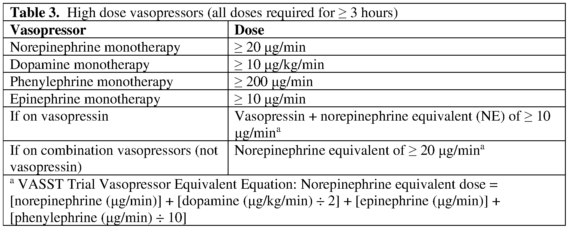


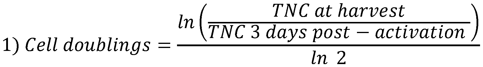


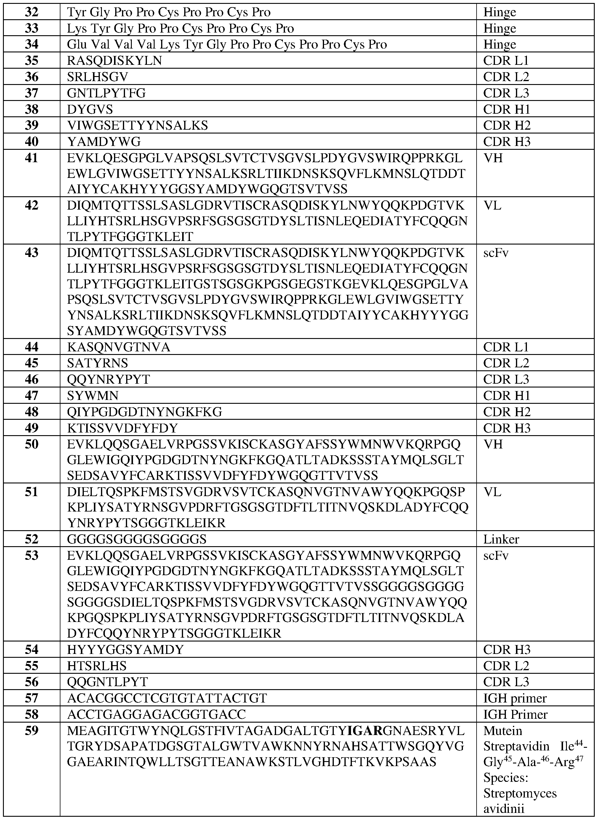


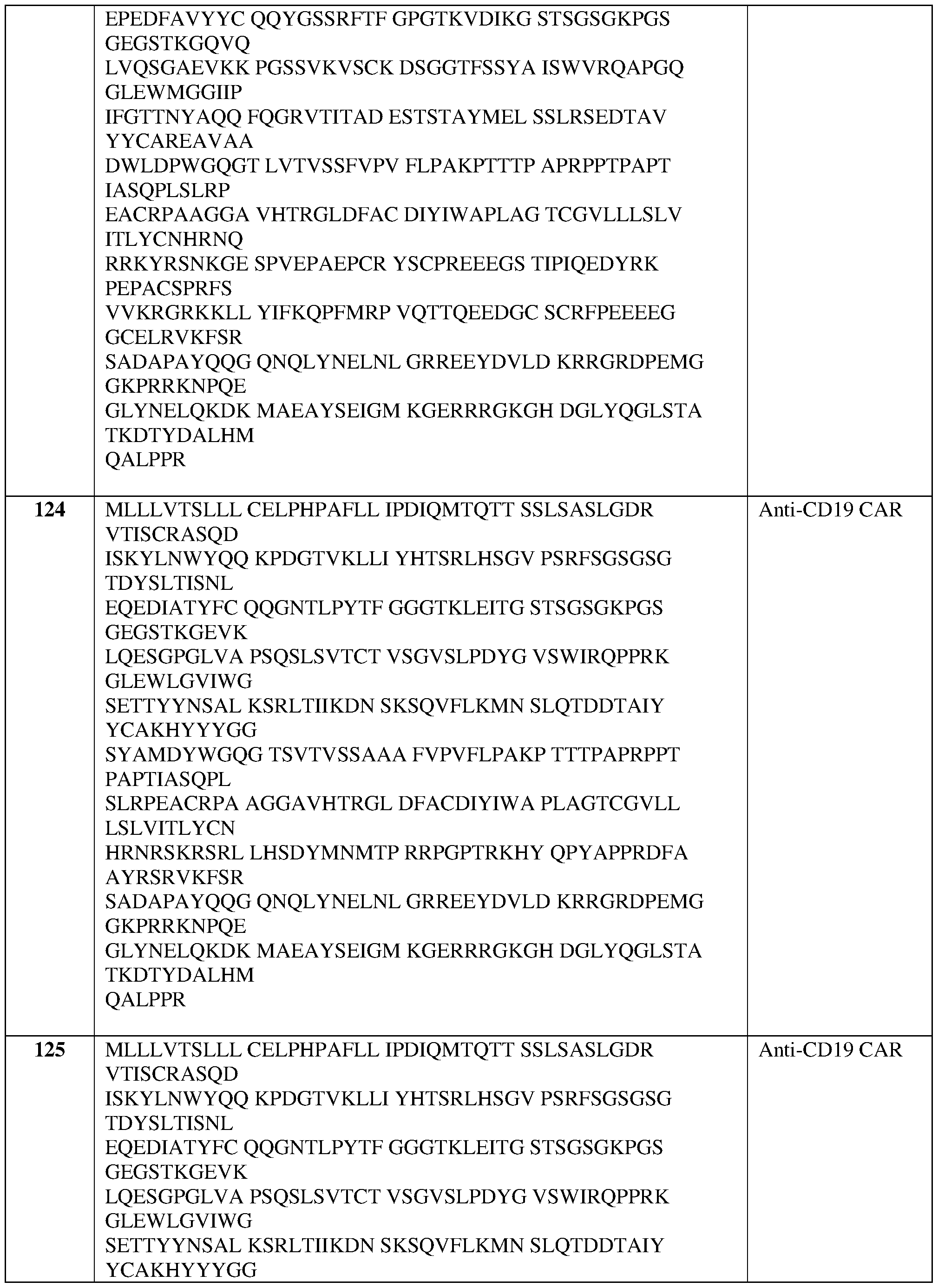
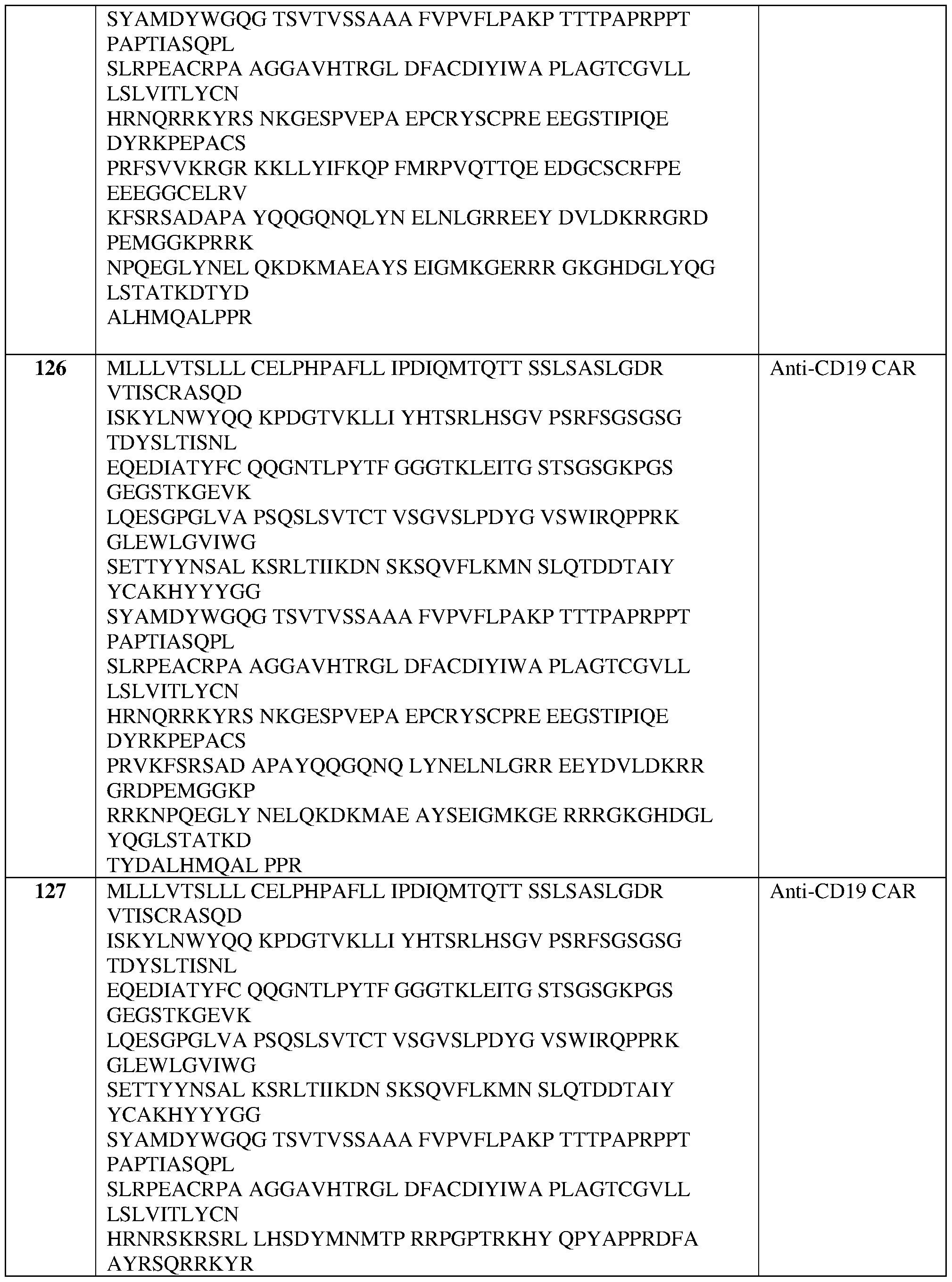


Claims
Priority Applications (6)
| Application Number | Priority Date | Filing Date | Title |
|---|---|---|---|
| KR1020257032030A KR20260005874A (en) | 2023-02-28 | 2024-02-28 | Cell therapy for treating systemic autoimmune diseases |
| AU2024230609A AU2024230609A1 (en) | 2023-02-28 | 2024-02-28 | Cell therapy for treating systemic autoimmune diseases |
| IL322815A IL322815A (en) | 2023-02-28 | 2024-02-28 | Cell therapy for treating systemic autoimmune diseases |
| CN202480027461.XA CN121127253A (en) | 2023-02-28 | 2024-02-28 | Cell therapy for treating systemic autoimmune diseases |
| EP24715991.6A EP4673154A1 (en) | 2023-02-28 | 2024-02-28 | Cell therapy for treating systemic autoimmune diseases |
| MX2025009928A MX2025009928A (en) | 2023-02-28 | 2025-08-22 | CELLULAR THERAPY FOR THE TREATMENT OF SYSTEMIC AUTOIMMUNE DISEASES |
Applications Claiming Priority (14)
| Application Number | Priority Date | Filing Date | Title |
|---|---|---|---|
| US202363487612P | 2023-02-28 | 2023-02-28 | |
| US63/487,612 | 2023-02-28 | ||
| US202363466671P | 2023-05-15 | 2023-05-15 | |
| US63/466,671 | 2023-05-15 | ||
| US202363522085P | 2023-06-20 | 2023-06-20 | |
| US63/522,085 | 2023-06-20 | ||
| US202363608166P | 2023-12-08 | 2023-12-08 | |
| US63/608,166 | 2023-12-08 | ||
| US202463618271P | 2024-01-05 | 2024-01-05 | |
| US63/618,271 | 2024-01-05 | ||
| US202463624745P | 2024-01-24 | 2024-01-24 | |
| US63/624,745 | 2024-01-24 | ||
| US202463553586P | 2024-02-14 | 2024-02-14 | |
| US63/553,586 | 2024-02-14 |
Publications (1)
| Publication Number | Publication Date |
|---|---|
| WO2024182516A1 true WO2024182516A1 (en) | 2024-09-06 |
Family
ID=90717237
Family Applications (1)
| Application Number | Title | Priority Date | Filing Date |
|---|---|---|---|
| PCT/US2024/017681 Ceased WO2024182516A1 (en) | 2023-02-28 | 2024-02-28 | Cell therapy for treating systemic autoimmune diseases |
Country Status (9)
| Country | Link |
|---|---|
| US (1) | US20240285762A1 (en) |
| EP (1) | EP4673154A1 (en) |
| KR (1) | KR20260005874A (en) |
| CN (1) | CN121127253A (en) |
| AU (1) | AU2024230609A1 (en) |
| IL (1) | IL322815A (en) |
| MX (1) | MX2025009928A (en) |
| TW (1) | TW202502363A (en) |
| WO (1) | WO2024182516A1 (en) |
Families Citing this family (1)
| Publication number | Priority date | Publication date | Assignee | Title |
|---|---|---|---|---|
| WO2025088308A1 (en) * | 2023-10-23 | 2025-05-01 | Autolus Limited | Products and methods for treating autoimmune diseases |
Citations (84)
| Publication number | Priority date | Publication date | Assignee | Title |
|---|---|---|---|---|
| US4361549A (en) | 1979-04-26 | 1982-11-30 | Ortho Pharmaceutical Corporation | Complement-fixing monoclonal antibody to human T cells, and methods of preparing same |
| US4452773A (en) | 1982-04-05 | 1984-06-05 | Canadian Patents And Development Limited | Magnetic iron-dextran microspheres |
| WO1986002077A1 (en) | 1984-10-02 | 1986-04-10 | Meade Harry M | Production of streptavidin-like polypeptides |
| US4795698A (en) | 1985-10-04 | 1989-01-03 | Immunicon Corporation | Magnetic-polymer particles |
| US4835263A (en) | 1983-01-27 | 1989-05-30 | Centre National De La Recherche Scientifique | Novel compounds containing an oligonucleotide sequence bonded to an intercalating agent, a process for their synthesis and their use |
| US5200084A (en) | 1990-09-26 | 1993-04-06 | Immunicon Corporation | Apparatus and methods for magnetic separation |
| US5219740A (en) | 1987-02-13 | 1993-06-15 | Fred Hutchinson Cancer Research Center | Retroviral gene transfer into diploid fibroblasts for gene therapy |
| EP0452342B1 (en) | 1988-12-28 | 1994-11-30 | MILTENYI, Stefan | Methods and materials for high gradient magnetic separation of biological materials |
| US5506121A (en) | 1992-11-03 | 1996-04-09 | Institut Fur Bioanalytik Gemeinnutzige Gesellschaft MBH | Fusion peptides with binding activity for streptavidin |
| WO1996024606A1 (en) | 1995-02-09 | 1996-08-15 | University Of Washington | Modified-affinity streptavidin |
| US5736137A (en) | 1992-11-13 | 1998-04-07 | Idec Pharmaceuticals Corporation | Therapeutic application of chimeric and radiolabeled antibodies to human B lymphocyte restricted differentiation antigen for treatment of B cell lymphoma |
| DE19641876A1 (en) | 1996-10-10 | 1998-04-16 | Inst Bioanalytik Gmbh | Streptavidin muteins |
| US5801115A (en) | 1995-09-05 | 1998-09-01 | Kataleuna Gmbh | Catalyst composition and methods for using and preparing same |
| WO1998040396A1 (en) | 1997-03-14 | 1998-09-17 | Trustees Of Boston University | Multiflavor streptavidin |
| US6022951A (en) | 1995-04-11 | 2000-02-08 | Univ Boston | Streptavidin mutants |
| WO2000014257A1 (en) | 1998-09-04 | 2000-03-16 | Sloan-Kettering Institute For Cancer Research | Fusion receptors specific for prostate-specific membrane antigen and uses thereof |
| US6040177A (en) | 1994-08-31 | 2000-03-21 | Fred Hutchinson Cancer Research Center | High efficiency transduction of T lymphocytes using rapid expansion methods ("REM") |
| US6060273A (en) | 1992-08-27 | 2000-05-09 | Beiersdorf Ag | Multicistronic expression units and their use |
| WO2000038762A1 (en) | 1998-12-24 | 2000-07-06 | Biosafe S.A. | Blood separation system particularly for concentrating hematopoietic stem cells |
| US6123655A (en) | 1996-04-24 | 2000-09-26 | Fell; Claude | Cell separation system with variable size chamber for the processing of biological fluids |
| US6207453B1 (en) | 1996-03-06 | 2001-03-27 | Medigene Ag | Recombinant AAV vector-based transduction system and use of same |
| US6352694B1 (en) | 1994-06-03 | 2002-03-05 | Genetics Institute, Inc. | Methods for inducing a population of T cells to proliferate using agents which recognize TCR/CD3 and ligands which stimulate an accessory molecule on the surface of the T cells |
| US6410319B1 (en) | 1998-10-20 | 2002-06-25 | City Of Hope | CD20-specific redirected T cells and their use in cellular immunotherapy of CD20+ malignancies |
| US6451995B1 (en) | 1996-03-20 | 2002-09-17 | Sloan-Kettering Institute For Cancer Research | Single chain FV polynucleotide or peptide constructs of anti-ganglioside GD2 antibodies, cells expressing same and related methods |
| US20020131960A1 (en) | 2000-06-02 | 2002-09-19 | Michel Sadelain | Artificial antigen presenting cells and methods of use thereof |
| WO2002077018A1 (en) | 2001-03-21 | 2002-10-03 | Iba Gmbh | Sequentially arranged streptavidin-binding modules as affinity tags |
| US20040167319A1 (en) | 2002-10-17 | 2004-08-26 | Jessica Teeling | Human monoclonal antibodies against CD20 |
| EP0700430B1 (en) | 1993-06-04 | 2005-04-20 | The United States of America as Represented by the Secretary of the Navy | Methods for selectively stimulating proliferation of t cells |
| US7070995B2 (en) | 2001-04-11 | 2006-07-04 | City Of Hope | CE7-specific redirected immune cells |
| WO2006130458A2 (en) | 2005-06-02 | 2006-12-07 | Astrazeneca Ab | Antibodies directed to cd20 and uses thereof |
| US20070116690A1 (en) | 2001-12-10 | 2007-05-24 | Lili Yang | Method for the generation of antigen-specific lymphocytes |
| US20080171951A1 (en) | 2005-03-23 | 2008-07-17 | Claude Fell | Integrated System for Collecting, Processing and Transplanting Cell Subsets, Including Adult Stem Cells, for Regenerative Medicine |
| US7446190B2 (en) | 2002-05-28 | 2008-11-04 | Sloan-Kettering Institute For Cancer Research | Nucleic acids encoding chimeric T cell receptors |
| US7446179B2 (en) | 2000-11-07 | 2008-11-04 | City Of Hope | CD19-specific chimeric T cell receptor |
| US20090035322A1 (en) | 2007-07-31 | 2009-02-05 | Regeneron Pharmaceuticals, Inc. | Human Antibodies to Human CD20 and Method of Using Thereof |
| WO2009072003A2 (en) | 2007-12-07 | 2009-06-11 | Miltenyi Biotec Gmbh | Sample processing system and methods |
| WO2010033140A2 (en) | 2008-05-06 | 2010-03-25 | Innovative Micro Technology | Removable/disposable apparatus for mems particle sorting device |
| US20100104509A1 (en) | 2006-12-13 | 2010-04-29 | Medarex, Inc. | Human antibodies that bind cd19 and uses thereof |
| US7776562B2 (en) | 2000-12-28 | 2010-08-17 | Iba Gmbh | Reversible MHC multimer staining for functional purification of antigen-specific T cells |
| WO2012129514A1 (en) | 2011-03-23 | 2012-09-27 | Fred Hutchinson Cancer Research Center | Method and compositions for cellular immunotherapy |
| US8324353B2 (en) | 2001-04-30 | 2012-12-04 | City Of Hope | Chimeric immunoreceptor useful in treating human gliomas |
| US8339645B2 (en) | 2008-05-27 | 2012-12-25 | Canon Kabushiki Kaisha | Managing apparatus, image processing apparatus, and processing method for the same, wherein a first user stores a temporary object having attribute information specified but not partial-area data, at a later time an object is received from a second user that includes both partial-area data and attribute information, the storage unit is searched for the temporary object that matches attribute information of the received object, and the first user is notified in response to a match |
| EP2537416A1 (en) | 2007-03-30 | 2012-12-26 | Memorial Sloan-Kettering Cancer Center | Constitutive expression of costimulatory ligands on adoptively transferred T lymphocytes |
| WO2013011011A2 (en) | 2011-07-18 | 2013-01-24 | Iba Gmbh | Method of reversibly staining a target cell |
| US8398282B2 (en) | 2011-05-12 | 2013-03-19 | Delphi Technologies, Inc. | Vehicle front lighting assembly and systems having a variable tint electrowetting element |
| WO2013071154A1 (en) | 2011-11-11 | 2013-05-16 | Fred Hutchinson Cancer Research Center | Cyclin a1-targeted t-cell immunotherapy for cancer |
| US20130149337A1 (en) | 2003-03-11 | 2013-06-13 | City Of Hope | Method of controlling administration of cancer antigen |
| US8479118B2 (en) | 2007-12-10 | 2013-07-02 | Microsoft Corporation | Switching search providers within a browser search box |
| WO2013123061A1 (en) | 2012-02-13 | 2013-08-22 | Seattle Children's Hospital D/B/A Seattle Children's Research Institute | Bispecific chimeric antigen receptors and therapeutic uses thereof |
| WO2013126726A1 (en) | 2012-02-22 | 2013-08-29 | The Trustees Of The University Of Pennsylvania | Double transgenic t cells comprising a car and a tcr and their methods of use |
| WO2013124474A2 (en) | 2012-02-23 | 2013-08-29 | Stage Cell Therapeutics Gmbh | Chromatographic isolation of cells and other complex biological materials |
| US20130287748A1 (en) | 2010-12-09 | 2013-10-31 | The Trustees Of The University Of Pennsylvania | Use of Chimeric Antigen Receptor-Modified T-Cells to Treat Cancer |
| WO2013166321A1 (en) | 2012-05-03 | 2013-11-07 | Fred Hutchinson Cancer Research Center | Enhanced affinity t cell receptors and methods for making the same |
| WO2014031687A1 (en) | 2012-08-20 | 2014-02-27 | Jensen, Michael | Method and compositions for cellular immunotherapy |
| WO2014055668A1 (en) | 2012-10-02 | 2014-04-10 | Memorial Sloan-Kettering Cancer Center | Compositions and methods for immunotherapy |
| WO2014076277A1 (en) | 2012-11-16 | 2014-05-22 | Iba Gmbh | Streptavidin muteins and methods of using them |
| US8802374B2 (en) | 2009-11-03 | 2014-08-12 | City Of Hope | Truncated epiderimal growth factor receptor (EGFRt) for transduced T cell selection |
| WO2014190273A1 (en) | 2013-05-24 | 2014-11-27 | Board Of Regents, The University Of Texas System | Chimeric antigen receptor-targeting monoclonal antibodies |
| WO2015095895A1 (en) | 2013-12-20 | 2015-06-25 | Fred Hutchinson Cancer Research Center | Tagged chimeric effector molecules and receptors thereof |
| US9108442B2 (en) | 2013-08-20 | 2015-08-18 | Ricoh Company, Ltd. | Image forming apparatus |
| WO2015158868A2 (en) | 2014-04-16 | 2015-10-22 | Juno Therapeutics Gmbh | Methods, kits and apparatus for expanding a population of cells |
| WO2015164675A1 (en) | 2014-04-23 | 2015-10-29 | Juno Therapeutics, Inc. | Methods for isolating, culturing, and genetically engineering immune cell populations for adoptive therapy |
| WO2015187528A1 (en) | 2014-06-02 | 2015-12-10 | The United States Of America, As Represented By The Secretary, Department Of Health And Human Services | Chimeric antigen receptors targeting cd-19 |
| WO2016033570A1 (en) | 2014-08-28 | 2016-03-03 | Juno Therapeutics, Inc. | Antibodies and chimeric antigen receptors specific for cd19 |
| WO2016073602A2 (en) | 2014-11-05 | 2016-05-12 | Juno Therapeutics, Inc. | Methods for transduction and cell processing |
| US9405601B2 (en) | 2012-12-20 | 2016-08-02 | Mitsubishi Electric Corporation | In-vehicle apparatus and program |
| US9539251B2 (en) | 2012-09-07 | 2017-01-10 | Genentech, Inc. | Combination therapy of a type II anti-CD20 antibody with a selective Bcl-2 inhibitor |
| US9599590B2 (en) | 2012-10-12 | 2017-03-21 | Sage Science, Inc. | Side-eluting molecular fractionator |
| WO2017068419A2 (en) | 2015-10-22 | 2017-04-27 | Juno Therapeutics Gmbh | Methods, kits, agents and apparatuses for transduction |
| US20170240882A1 (en) | 2014-10-15 | 2017-08-24 | Ezra S. Abrams | Apparatuses, methods and systems for automated processing of nucleic acids and electrophoretic sample preparation |
| WO2017177337A1 (en) * | 2016-04-15 | 2017-10-19 | Zymeworks Inc. | Multi-specific antigen-binding constructs targeting immunotherapeutics |
| WO2018170188A2 (en) | 2017-03-14 | 2018-09-20 | Juno Therapeutics, Inc. | Methods for cryogenic storage |
| WO2018197949A1 (en) | 2017-04-27 | 2018-11-01 | Juno Therapeutics Gmbh | Oligomeric particle reagents and methods of use thereof |
| WO2019027850A1 (en) * | 2017-07-29 | 2019-02-07 | Juno Therapeutics, Inc. | Reagents for expanding cells expressing recombinant receptors |
| WO2019028051A1 (en) | 2017-07-31 | 2019-02-07 | Lentigen Technology, Inc. | Compositions and methods for treating cancer with anti-cd19/cd20 immunotherapy |
| US20190046062A1 (en) | 2015-01-08 | 2019-02-14 | The Medical Research, Infratructure and Health Services Fund of the Tel Aviv Medical Center | Coronary sinus electrophysiology measurements device and methods |
| WO2019089855A1 (en) | 2017-11-01 | 2019-05-09 | Juno Therapeutics, Inc. | Process for generating therapeutic compositions of engineered cells |
| WO2020233589A1 (en) | 2019-05-20 | 2020-11-26 | 南京驯鹿医疗技术有限公司 | Fully human antibody targeting cd19 and application thereof |
| WO2021163389A1 (en) * | 2020-02-12 | 2021-08-19 | Juno Therapeutics, Inc. | Bcma-directed chimeric antigen receptor t cell compositions and methods and uses thereof |
| WO2021188681A1 (en) | 2020-03-17 | 2021-09-23 | Cellular Biomedicine Group Hk Limited | Combined chimeric antigen receptor targeting cd19 and cd20 and applications thereof |
| WO2021207689A2 (en) * | 2020-04-10 | 2021-10-14 | Juno Therapeutics, Inc. | Methods and uses related to cell therapy engineered with a chimeric antigen receptor targeting b-cell maturation antigen |
| US20210363245A1 (en) | 2018-09-17 | 2021-11-25 | The United States Of America,As Represented By The Secretary,Department Of Health And Human Services | Bicistronic chimeric antigen receptors targeting cd19 and cd20 and their uses |
| US20210395364A1 (en) * | 2019-03-05 | 2021-12-23 | Promab Biotechnologies, Inc. | Car-t cells with humanized cd19 scfv |
| US20220202864A1 (en) | 2019-05-07 | 2022-06-30 | Gracell Biotechnologies (Shanghai) Co., Ltd. | Bcma-targeting engineered immune cell and use thereof |
-
2024
- 2024-02-28 WO PCT/US2024/017681 patent/WO2024182516A1/en not_active Ceased
- 2024-02-28 CN CN202480027461.XA patent/CN121127253A/en active Pending
- 2024-02-28 KR KR1020257032030A patent/KR20260005874A/en active Pending
- 2024-02-28 IL IL322815A patent/IL322815A/en unknown
- 2024-02-28 EP EP24715991.6A patent/EP4673154A1/en active Pending
- 2024-02-28 TW TW113107130A patent/TW202502363A/en unknown
- 2024-02-28 AU AU2024230609A patent/AU2024230609A1/en active Pending
- 2024-02-28 US US18/590,598 patent/US20240285762A1/en active Pending
-
2025
- 2025-08-22 MX MX2025009928A patent/MX2025009928A/en unknown
Patent Citations (104)
| Publication number | Priority date | Publication date | Assignee | Title |
|---|---|---|---|---|
| US4361549A (en) | 1979-04-26 | 1982-11-30 | Ortho Pharmaceutical Corporation | Complement-fixing monoclonal antibody to human T cells, and methods of preparing same |
| US4452773A (en) | 1982-04-05 | 1984-06-05 | Canadian Patents And Development Limited | Magnetic iron-dextran microspheres |
| US4835263A (en) | 1983-01-27 | 1989-05-30 | Centre National De La Recherche Scientifique | Novel compounds containing an oligonucleotide sequence bonded to an intercalating agent, a process for their synthesis and their use |
| WO1986002077A1 (en) | 1984-10-02 | 1986-04-10 | Meade Harry M | Production of streptavidin-like polypeptides |
| US5168049A (en) | 1984-10-02 | 1992-12-01 | Biogen, Inc. | Production of streptavidin-like polypeptides |
| US4795698A (en) | 1985-10-04 | 1989-01-03 | Immunicon Corporation | Magnetic-polymer particles |
| US5219740A (en) | 1987-02-13 | 1993-06-15 | Fred Hutchinson Cancer Research Center | Retroviral gene transfer into diploid fibroblasts for gene therapy |
| EP0452342B1 (en) | 1988-12-28 | 1994-11-30 | MILTENYI, Stefan | Methods and materials for high gradient magnetic separation of biological materials |
| US5200084A (en) | 1990-09-26 | 1993-04-06 | Immunicon Corporation | Apparatus and methods for magnetic separation |
| US6060273A (en) | 1992-08-27 | 2000-05-09 | Beiersdorf Ag | Multicistronic expression units and their use |
| US5506121A (en) | 1992-11-03 | 1996-04-09 | Institut Fur Bioanalytik Gemeinnutzige Gesellschaft MBH | Fusion peptides with binding activity for streptavidin |
| US5736137A (en) | 1992-11-13 | 1998-04-07 | Idec Pharmaceuticals Corporation | Therapeutic application of chimeric and radiolabeled antibodies to human B lymphocyte restricted differentiation antigen for treatment of B cell lymphoma |
| EP0700430B1 (en) | 1993-06-04 | 2005-04-20 | The United States of America as Represented by the Secretary of the Navy | Methods for selectively stimulating proliferation of t cells |
| US6352694B1 (en) | 1994-06-03 | 2002-03-05 | Genetics Institute, Inc. | Methods for inducing a population of T cells to proliferate using agents which recognize TCR/CD3 and ligands which stimulate an accessory molecule on the surface of the T cells |
| US6040177A (en) | 1994-08-31 | 2000-03-21 | Fred Hutchinson Cancer Research Center | High efficiency transduction of T lymphocytes using rapid expansion methods ("REM") |
| WO1996024606A1 (en) | 1995-02-09 | 1996-08-15 | University Of Washington | Modified-affinity streptavidin |
| US6165750A (en) | 1995-02-09 | 2000-12-26 | University Of Washington | Modified-affinity streptavidin |
| US6156493A (en) | 1995-02-09 | 2000-12-05 | University Of Washington | Modified-affinity streptavidin |
| US6022951A (en) | 1995-04-11 | 2000-02-08 | Univ Boston | Streptavidin mutants |
| US5801115A (en) | 1995-09-05 | 1998-09-01 | Kataleuna Gmbh | Catalyst composition and methods for using and preparing same |
| US6207453B1 (en) | 1996-03-06 | 2001-03-27 | Medigene Ag | Recombinant AAV vector-based transduction system and use of same |
| US6451995B1 (en) | 1996-03-20 | 2002-09-17 | Sloan-Kettering Institute For Cancer Research | Single chain FV polynucleotide or peptide constructs of anti-ganglioside GD2 antibodies, cells expressing same and related methods |
| US6123655A (en) | 1996-04-24 | 2000-09-26 | Fell; Claude | Cell separation system with variable size chamber for the processing of biological fluids |
| US6103493A (en) | 1996-10-10 | 2000-08-15 | Institut Fur Bioanalytic | Streptavidin muteins |
| DE19641876A1 (en) | 1996-10-10 | 1998-04-16 | Inst Bioanalytik Gmbh | Streptavidin muteins |
| WO1998040396A1 (en) | 1997-03-14 | 1998-09-17 | Trustees Of Boston University | Multiflavor streptavidin |
| US6368813B1 (en) | 1997-03-14 | 2002-04-09 | The Trustees Of Boston University | Multiflavor streptavidin |
| WO2000014257A1 (en) | 1998-09-04 | 2000-03-16 | Sloan-Kettering Institute For Cancer Research | Fusion receptors specific for prostate-specific membrane antigen and uses thereof |
| US6410319B1 (en) | 1998-10-20 | 2002-06-25 | City Of Hope | CD20-specific redirected T cells and their use in cellular immunotherapy of CD20+ malignancies |
| WO2000038762A1 (en) | 1998-12-24 | 2000-07-06 | Biosafe S.A. | Blood separation system particularly for concentrating hematopoietic stem cells |
| US6733433B1 (en) | 1998-12-24 | 2004-05-11 | Biosafe S.A. | Blood separation system particularly for concentrating hematopoietic stem cells |
| US20020131960A1 (en) | 2000-06-02 | 2002-09-19 | Michel Sadelain | Artificial antigen presenting cells and methods of use thereof |
| US7446179B2 (en) | 2000-11-07 | 2008-11-04 | City Of Hope | CD19-specific chimeric T cell receptor |
| US8298782B2 (en) | 2000-12-28 | 2012-10-30 | Iba Gmbh | Reversible MHC multimer staining for functional purification of antigen-specific T cells |
| US7776562B2 (en) | 2000-12-28 | 2010-08-17 | Iba Gmbh | Reversible MHC multimer staining for functional purification of antigen-specific T cells |
| US8735540B2 (en) | 2001-03-21 | 2014-05-27 | Iba Gmbh | Peptides with sequentially arranged streptavidin binding modules |
| WO2002077018A1 (en) | 2001-03-21 | 2002-10-03 | Iba Gmbh | Sequentially arranged streptavidin-binding modules as affinity tags |
| US7981632B2 (en) | 2001-03-21 | 2011-07-19 | Iba Gmbh | Sequentially arranged streptavidin-binding modules as affinity tags |
| US7070995B2 (en) | 2001-04-11 | 2006-07-04 | City Of Hope | CE7-specific redirected immune cells |
| US7265209B2 (en) | 2001-04-11 | 2007-09-04 | City Of Hope | CE7-specific chimeric T cell receptor |
| US7354762B2 (en) | 2001-04-11 | 2008-04-08 | City Of Hope | Method for producing CE7-specific redirected immune cells |
| US7446191B2 (en) | 2001-04-11 | 2008-11-04 | City Of Hope | DNA construct encoding CE7-specific chimeric T cell receptor |
| US8324353B2 (en) | 2001-04-30 | 2012-12-04 | City Of Hope | Chimeric immunoreceptor useful in treating human gliomas |
| US20070116690A1 (en) | 2001-12-10 | 2007-05-24 | Lili Yang | Method for the generation of antigen-specific lymphocytes |
| US7446190B2 (en) | 2002-05-28 | 2008-11-04 | Sloan-Kettering Institute For Cancer Research | Nucleic acids encoding chimeric T cell receptors |
| US20040167319A1 (en) | 2002-10-17 | 2004-08-26 | Jessica Teeling | Human monoclonal antibodies against CD20 |
| US20130149337A1 (en) | 2003-03-11 | 2013-06-13 | City Of Hope | Method of controlling administration of cancer antigen |
| US20080171951A1 (en) | 2005-03-23 | 2008-07-17 | Claude Fell | Integrated System for Collecting, Processing and Transplanting Cell Subsets, Including Adult Stem Cells, for Regenerative Medicine |
| WO2006130458A2 (en) | 2005-06-02 | 2006-12-07 | Astrazeneca Ab | Antibodies directed to cd20 and uses thereof |
| US20100104509A1 (en) | 2006-12-13 | 2010-04-29 | Medarex, Inc. | Human antibodies that bind cd19 and uses thereof |
| EP2537416A1 (en) | 2007-03-30 | 2012-12-26 | Memorial Sloan-Kettering Cancer Center | Constitutive expression of costimulatory ligands on adoptively transferred T lymphocytes |
| US8389282B2 (en) | 2007-03-30 | 2013-03-05 | Memorial Sloan-Kettering Cancer Center | Constitutive expression of costimulatory ligands on adoptively transferred T lymphocytes |
| US20090035322A1 (en) | 2007-07-31 | 2009-02-05 | Regeneron Pharmaceuticals, Inc. | Human Antibodies to Human CD20 and Method of Using Thereof |
| US20110003380A1 (en) | 2007-12-07 | 2011-01-06 | Stefan Miltenyi | Sample Processing System and Methods |
| WO2009072003A2 (en) | 2007-12-07 | 2009-06-11 | Miltenyi Biotec Gmbh | Sample processing system and methods |
| US8479118B2 (en) | 2007-12-10 | 2013-07-02 | Microsoft Corporation | Switching search providers within a browser search box |
| WO2010033140A2 (en) | 2008-05-06 | 2010-03-25 | Innovative Micro Technology | Removable/disposable apparatus for mems particle sorting device |
| US8339645B2 (en) | 2008-05-27 | 2012-12-25 | Canon Kabushiki Kaisha | Managing apparatus, image processing apparatus, and processing method for the same, wherein a first user stores a temporary object having attribute information specified but not partial-area data, at a later time an object is received from a second user that includes both partial-area data and attribute information, the storage unit is searched for the temporary object that matches attribute information of the received object, and the first user is notified in response to a match |
| US8802374B2 (en) | 2009-11-03 | 2014-08-12 | City Of Hope | Truncated epiderimal growth factor receptor (EGFRt) for transduced T cell selection |
| US8911993B2 (en) | 2010-12-09 | 2014-12-16 | The Trustees Of The University Of Pennsylvania | Compositions for treatment of cancer |
| US20130287748A1 (en) | 2010-12-09 | 2013-10-31 | The Trustees Of The University Of Pennsylvania | Use of Chimeric Antigen Receptor-Modified T-Cells to Treat Cancer |
| WO2012129514A1 (en) | 2011-03-23 | 2012-09-27 | Fred Hutchinson Cancer Research Center | Method and compositions for cellular immunotherapy |
| US8398282B2 (en) | 2011-05-12 | 2013-03-19 | Delphi Technologies, Inc. | Vehicle front lighting assembly and systems having a variable tint electrowetting element |
| US9023604B2 (en) | 2011-07-18 | 2015-05-05 | Iba Gmbh | Method of reversibly staining a target cell |
| WO2013011011A2 (en) | 2011-07-18 | 2013-01-24 | Iba Gmbh | Method of reversibly staining a target cell |
| WO2013071154A1 (en) | 2011-11-11 | 2013-05-16 | Fred Hutchinson Cancer Research Center | Cyclin a1-targeted t-cell immunotherapy for cancer |
| WO2013123061A1 (en) | 2012-02-13 | 2013-08-22 | Seattle Children's Hospital D/B/A Seattle Children's Research Institute | Bispecific chimeric antigen receptors and therapeutic uses thereof |
| WO2013126726A1 (en) | 2012-02-22 | 2013-08-29 | The Trustees Of The University Of Pennsylvania | Double transgenic t cells comprising a car and a tcr and their methods of use |
| WO2013124474A2 (en) | 2012-02-23 | 2013-08-29 | Stage Cell Therapeutics Gmbh | Chromatographic isolation of cells and other complex biological materials |
| WO2013166321A1 (en) | 2012-05-03 | 2013-11-07 | Fred Hutchinson Cancer Research Center | Enhanced affinity t cell receptors and methods for making the same |
| WO2014031687A1 (en) | 2012-08-20 | 2014-02-27 | Jensen, Michael | Method and compositions for cellular immunotherapy |
| US9539251B2 (en) | 2012-09-07 | 2017-01-10 | Genentech, Inc. | Combination therapy of a type II anti-CD20 antibody with a selective Bcl-2 inhibitor |
| WO2014055668A1 (en) | 2012-10-02 | 2014-04-10 | Memorial Sloan-Kettering Cancer Center | Compositions and methods for immunotherapy |
| US20170254774A1 (en) | 2012-10-12 | 2017-09-07 | Sage Science, Inc. | Side-eluting molecular fractionator |
| US9599590B2 (en) | 2012-10-12 | 2017-03-21 | Sage Science, Inc. | Side-eluting molecular fractionator |
| WO2014076277A1 (en) | 2012-11-16 | 2014-05-22 | Iba Gmbh | Streptavidin muteins and methods of using them |
| US9405601B2 (en) | 2012-12-20 | 2016-08-02 | Mitsubishi Electric Corporation | In-vehicle apparatus and program |
| WO2014190273A1 (en) | 2013-05-24 | 2014-11-27 | Board Of Regents, The University Of Texas System | Chimeric antigen receptor-targeting monoclonal antibodies |
| US9108442B2 (en) | 2013-08-20 | 2015-08-18 | Ricoh Company, Ltd. | Image forming apparatus |
| WO2015095895A1 (en) | 2013-12-20 | 2015-06-25 | Fred Hutchinson Cancer Research Center | Tagged chimeric effector molecules and receptors thereof |
| WO2015158868A2 (en) | 2014-04-16 | 2015-10-22 | Juno Therapeutics Gmbh | Methods, kits and apparatus for expanding a population of cells |
| WO2015164675A1 (en) | 2014-04-23 | 2015-10-29 | Juno Therapeutics, Inc. | Methods for isolating, culturing, and genetically engineering immune cell populations for adoptive therapy |
| US10287350B2 (en) | 2014-06-02 | 2019-05-14 | The United States Of America, As Represented By The Secretary, Department Of Health And Human Services | Chimeric antigen receptors targeting CD-19 |
| WO2015187528A1 (en) | 2014-06-02 | 2015-12-10 | The United States Of America, As Represented By The Secretary, Department Of Health And Human Services | Chimeric antigen receptors targeting cd-19 |
| US20160152723A1 (en) | 2014-08-28 | 2016-06-02 | Juno Therapeutics, Inc. | Antibodies and chimeric antigen receptors specific for cd19 |
| WO2016033570A1 (en) | 2014-08-28 | 2016-03-03 | Juno Therapeutics, Inc. | Antibodies and chimeric antigen receptors specific for cd19 |
| US20170240882A1 (en) | 2014-10-15 | 2017-08-24 | Ezra S. Abrams | Apparatuses, methods and systems for automated processing of nucleic acids and electrophoretic sample preparation |
| WO2016073602A2 (en) | 2014-11-05 | 2016-05-12 | Juno Therapeutics, Inc. | Methods for transduction and cell processing |
| US20190046062A1 (en) | 2015-01-08 | 2019-02-14 | The Medical Research, Infratructure and Health Services Fund of the Tel Aviv Medical Center | Coronary sinus electrophysiology measurements device and methods |
| WO2017068419A2 (en) | 2015-10-22 | 2017-04-27 | Juno Therapeutics Gmbh | Methods, kits, agents and apparatuses for transduction |
| WO2017177337A1 (en) * | 2016-04-15 | 2017-10-19 | Zymeworks Inc. | Multi-specific antigen-binding constructs targeting immunotherapeutics |
| WO2018170188A2 (en) | 2017-03-14 | 2018-09-20 | Juno Therapeutics, Inc. | Methods for cryogenic storage |
| WO2018197949A1 (en) | 2017-04-27 | 2018-11-01 | Juno Therapeutics Gmbh | Oligomeric particle reagents and methods of use thereof |
| WO2019027850A1 (en) * | 2017-07-29 | 2019-02-07 | Juno Therapeutics, Inc. | Reagents for expanding cells expressing recombinant receptors |
| WO2019028051A1 (en) | 2017-07-31 | 2019-02-07 | Lentigen Technology, Inc. | Compositions and methods for treating cancer with anti-cd19/cd20 immunotherapy |
| WO2019089855A1 (en) | 2017-11-01 | 2019-05-09 | Juno Therapeutics, Inc. | Process for generating therapeutic compositions of engineered cells |
| US20210363245A1 (en) | 2018-09-17 | 2021-11-25 | The United States Of America,As Represented By The Secretary,Department Of Health And Human Services | Bicistronic chimeric antigen receptors targeting cd19 and cd20 and their uses |
| US20210395364A1 (en) * | 2019-03-05 | 2021-12-23 | Promab Biotechnologies, Inc. | Car-t cells with humanized cd19 scfv |
| US20220202864A1 (en) | 2019-05-07 | 2022-06-30 | Gracell Biotechnologies (Shanghai) Co., Ltd. | Bcma-targeting engineered immune cell and use thereof |
| WO2020233589A1 (en) | 2019-05-20 | 2020-11-26 | 南京驯鹿医疗技术有限公司 | Fully human antibody targeting cd19 and application thereof |
| US20220220200A1 (en) | 2019-05-20 | 2022-07-14 | Nanjing Iaso Biotherapeutics Co., Ltd. | Fully human antibody targeting cd19 and application thereof |
| WO2021163389A1 (en) * | 2020-02-12 | 2021-08-19 | Juno Therapeutics, Inc. | Bcma-directed chimeric antigen receptor t cell compositions and methods and uses thereof |
| WO2021188681A1 (en) | 2020-03-17 | 2021-09-23 | Cellular Biomedicine Group Hk Limited | Combined chimeric antigen receptor targeting cd19 and cd20 and applications thereof |
| WO2021207689A2 (en) * | 2020-04-10 | 2021-10-14 | Juno Therapeutics, Inc. | Methods and uses related to cell therapy engineered with a chimeric antigen receptor targeting b-cell maturation antigen |
Non-Patent Citations (139)
| Title |
|---|
| "Antibody-antigen interactions: Contact analysis and binding site topography", J. MOL. BIOL., vol. 262, pages 732 - 745 |
| "Correction: 2021 DORIS definition of remission in SLE: final recommendations from an international task force", LUPUS SCIENCE & MEDICINE, vol. 9, 2022, pages e000538corr1 |
| "GenBank", Database accession no. AF451974.1 |
| "Methods in Molecular Medicine", vol. 58, 2, HUMANA PRESS INC., article "Metastasis Research Protocols", pages: 17 - 25 |
| "Remington: The Science and Practice of Pharmacy", 1 May 2005, LIPPINCOTT WILLIAMS & WILKIN |
| "Remington's Pharmaceutical Scienc", 1980 |
| ABRAMSON ET AL., THE LANCET, vol. 396, no. 10254, 2020, pages 839 - 852 |
| AL-LAZIKANI ET AL., JMB, vol. 273, 1997, pages 927 - 948 |
| ALONSO-CAMINO ET AL., MOL. THER. NUCL. ACIDS., vol. 2, 2013, pages e93 |
| ARAKAWA ET AL., J. BIOCHEM., vol. 120, 1996, pages 657 - 662 |
| ARGARANA ET AL., NUCLEIC ACIDS RES, vol. 14, 1986, pages 1871 - 1882 |
| ARGARANA ET AL., NUCLEIC ACIDS RESEARCH, 1986, pages 1871 - 1882 |
| ARINGER ET AL., ARTHRITIS RHEUMATOL, vol. 71, 2019, pages 1400 - 1412 |
| ARNETT ET AL., ARTHRITIS RHEUM, vol. 31, 1988, pages 315 - 324 |
| BARRETT ET AL., CHIMERIC ANTIGEN RECEPTOR THERAPY FOR CANCER ANNUAL REVIEW OF MEDICINE, vol. 65, 2014, pages 333 - 347 |
| BAZZAN MVACCARINO AMARLETTO F: "Systemic lupus erythematosus and thrombosis", THROMB J, vol. 13, 23 April 2015 (2015-04-23), pages 16, XP021215026, DOI: 10.1186/s12959-015-0043-3 |
| BEJCEK ET AL., CANCER RES, vol. 55, 1995, pages 2346 - 2351 |
| BISHOP ET AL., N ENGL J MED, vol. 386, no. 629, 2022, pages 640 - 654 |
| BORIS-LAWRIETEMIN, CUR. OPIN. GENET. DEVELOP., vol. 3, 1993, pages 102 - 109 |
| BRASH ET AL., MOL. CELL BIOL., vol. 7, 1987, pages 2031 - 2034 |
| BRENTJENS ET AL., SCI TRANSL MED, vol. 5, no. 177, 2013 |
| BRENTJENS ET AL., SCI. TRANSL. MED., vol. 5, 2013, pages 177 - 38 |
| BRENTJENS ET AL., SCI. TRANSL. MED., vol. 5, no. 177, March 2013 (2013-03-01), pages 177 - 38 |
| BURNS, PROC. NATL. ACAD. SCI. USA, vol. 90, 1993, pages 8033 - 8037 |
| CALLARD ET AL., J. IMMUNOLOGY, vol. 148, no. 10, 1992, pages 2983 - 2987 |
| CARLENS ET AL., EXP. HEMATOL., vol. 28, no. 10, 2000, pages 1137 - 46 |
| CAVALIERI ET AL., BLOOD, vol. 101, no. 2, 2003, pages 1637 - 1644 |
| CERVERA R ET AL.: "Systemic lupus erythematosus: clinical and immunologic patterns of disease expression in a cohort of 1,000 patients", THE EUROPEAN WORKING PARTY ON SYSTEMIC LUPUS ERYTHEMATOSUS. MEDICINE (BALTIMORE, vol. 72, no. 2, March 1993 (1993-03-01), pages 113 - 24 |
| CHEADLE ET AL.: "Chimeric antigen receptors for T-cell based therapy", METHODS MOL BIOL., vol. 907, 2012, pages 645 - 66, XP009179541, DOI: 10.1007/978-1-61779-974-7_36 |
| CHICAYBAM ET AL., PLOS ONE, vol. 8, no. 3, 2013, pages e60298 |
| CHO ET AL., LAB CHIP, vol. 10, 2010, pages 1567 - 1573 |
| CHOY EHPANAYI GS, N ENGL JMED, vol. 344, 2001, pages 907 - 916 |
| CLARKSON ET AL., NATURE, vol. 352, 1991, pages 624 - 628 |
| DAVILA ET AL., PLOS ONE, vol. 8, no. 4, 2013, pages e61338 |
| DAVILA ET AL., SCI TRANSL MED, vol. 6, 2014, pages 224 - 25 |
| DAVILLA ET AL., SCIENCE TRANSLATIONAL MEDICINE, vol. 6, no. 224, 2014, pages 224 - 25 |
| DAYAL ET AL., LUPUS, 2002 |
| DE FELIPE ET AL., TRAFFIC, vol. 5, 2004, pages 616 - 626 |
| DE FELIPE, GENETIC VACCINES AND THER, vol. 2, 2004, pages 13 |
| DE RIE, CELL. IMMUNOL., vol. 118, 1989, pages 368 - 381 |
| FAIRHEAD ET AL., J. MOL. BIOL., vol. 426, 2013, pages 199 - 214 |
| FEDOROV ET AL., SCI. TRANSL. MEDICINE, vol. 5, no. 215, December 2013 (2013-12-01) |
| FOWLER ET AL., NATURE MEDICINE, vol. 28, 2022, pages 325 - 332 |
| FRAIETTA ET AL., NAT MED, vol. 24, no. 5, 2018, pages 563 - 571 |
| FRANKLYN ET AL.: "Asia-Pacific Lupus Collaboration. Definition and initial validation of a Lupus Low Disease Activity State (LLDAS", ANN RHEUM DIS, vol. 75, no. 9, 12 October 2015 (2015-10-12), pages 1615 - 21 |
| GEARINGTHORPE, JOURNAL OF IMMUNOLOGICAL METHODS, vol. 114, no. 1-2, 1988, pages 3 - 9 |
| GODIN, J BIOPHOTON, vol. 1, no. 5, 2008, pages 355 - 376 |
| GROSSMAN ET AL., NAT REV IMMUNOL, vol. 4, no. 5, 2004, pages 387 - 395 |
| GRUPP ET AL., N. ENGL. J. MED., vol. 368, 2013, pages 1509 - 1518 |
| GUIDO CAVALETTIPAOLA MARMIROLI: "Nature Reviews Neurology", vol. 6, December 2010, NATIONAL CANCER INSTITUTE-COMMON TOXICITY CRITERIA, pages: 657 - 666 |
| HALFORD ET AL., ANN PHARMACOTHER, vol. 55, no. 4, 2021, pages 466 - 479 |
| HERBST ET AL., J. PHARMACOL. EXP. THER., vol. 335, 2010, pages 213 - 222 |
| HINDSON ET AL., ANAL CHEM, vol. 83, no. 22, 2011, pages 8604 - 8610 |
| HONEGGER APLUCKTHUN A: "Yet another numbering scheme for immunoglobulin variable domains: an automatic modeling and analysis tool", J MOL BIOL, vol. 309, no. 3, 8 June 2001 (2001-06-08), pages 657 - 70, XP004626893, DOI: 10.1006/jmbi.2001.4662 |
| HOWARTH, NAT. METHODS, vol. 3, 2006, pages 267 - 73 |
| HUDECEK ET AL., CANCER IMMUNOL RES, vol. 3, no. 2, 2015, pages 125 - 135 |
| HUDECEK ET AL., CLIN. CANCER RES., vol. 19, 2013, pages 3153 |
| HUGGETT ET AL., CLINICAL CHEMISTRY, vol. 59, 2013, pages 1691 - 1693 |
| JACOBSON ET AL., THE LANCET, vol. 23, no. 1, 2021, pages 91 - 103 |
| JOHNSTON, NATURE, vol. 346, 1990, pages 776 - 777 |
| KANSASTEDDER, J. IMMUNOL., vol. 147, 1991, pages 4094 - 4102 |
| KINDT ET AL.: "Kuby Immunology", 2007, W.H. FREEMAN AND CO., pages: 91 |
| KLEBANOFF ET AL., J IMMUNOTHER, vol. 35, no. 9, 2012, pages 651 - 660 |
| KOCHENDERFER ET AL., BLOOD, vol. 119, 2012, pages 2709 - 2720 |
| KOCHENDERFER ET AL., NATURE REVIEWS CLINICAL ONCOLOGY, vol. 10, 2013, pages 267 - 276 |
| KOSTE ET AL., GENE THERAPY, 3 April 2014 (2014-04-03) |
| KUSHNER, ARTHRITIS RHEUM, vol. 34, 1991, pages 1065 - 68 |
| LEE ET AL., BIOL BLOOD MARROW TRANSPLANT, vol. 25, no. 4, 2019, pages 625 - 38 |
| LEE ET AL., BIOL BLOOD MARROW TRANSPLANT, vol. 25, no. 4, April 2019 (2019-04-01), pages 625 - 38 |
| LEE ET AL., BLOOD, vol. 124, no. 2, 2014, pages 188 - 95 |
| LEFRANC MP ET AL.: "IMGT unique numbering for immunoglobulin and T cell receptor variable domains and Ig superfamily V-like domains", DEV COMP IMMUNOL, vol. 27, no. 1, January 2003 (2003-01-01), pages 55 - 77, XP055585227, DOI: 10.1016/S0145-305X(02)00039-3 |
| LIM ET AL., BIOCHEMISTRY, vol. 50, 2010, pages 8682 - 91 |
| LING, N. R. ET AL., LEUCOCYTE TYPING, vol. 111, 1987, pages 302 |
| LIU ET AL., NATURE BIOTECH, vol. 34, no. 4, April 2016 (2016-04-01), pages 430 - 434 |
| LIU ET AL., NATURE BIOTECHNOLOGY, vol. 34, 2016, pages 430 |
| LU ET AL., J CLIN ONCOL, vol. 36, 2018, pages 3041 |
| LUPTON S. D. ET AL., MOL. AND CELL BIOL., vol. 11, 1991, pages 6 |
| MACCALLUM ET AL., J. MOL. BIOL., vol. 262, 1996, pages 732 - 745 |
| MACKENSEN ANDREAS ET AL: "Anti-CD19 CAR T cell therapy for refractory systemic lupus erythematosus", NATURE MEDICINE, vol. 28, no. 10, 15 September 2022 (2022-09-15), New York, pages 2124 - 2132, XP093179242, ISSN: 1078-8956, Retrieved from the Internet <URL:https://www.nature.com/articles/s41591-022-02017-5> DOI: 10.1038/s41591-022-02017-5 * |
| MALLYA RK ET AL., J RHEUMATOL, vol. 9, 1982, pages 224 - 8 |
| MANURI ET AL., HUM GENE THER, vol. 21, no. 4, 2010, pages 427 - 437 |
| MARTIN ET AL.: "Modeling antibody hypervariable loops: a combined algorithm", PNAS, vol. 86, no. 23, 1989, pages 9268 - 9272, XP000165667, DOI: 10.1073/pnas.86.23.9268 |
| MEEKER ET AL., HYBRIDOMA, vol. 3, 1984, pages 305 - 320 |
| MIANHILL, EXPERT OPIN BIOL THER, vol. 21, no. 4, 2021, pages 435 - 441 |
| MILLER, A. D., HUMAN GENE THERAPY, vol. 1, 1990, pages 5 - 14 |
| MILLERROSMAN, BIOTECHNIQUES, vol. 7, 1989, pages 980 - 990 |
| MUELLER ET AL., BLOOD ADV, vol. 5, no. 23, 2021, pages 4980 - 4991 |
| NEELAPU ET AL., N ENGL J MED, vol. 377, no. 26, 2017, pages 2531 - 2544 |
| NEELAPU ET AL., NAT REV CLIN ONCOL, vol. 15, 2018, pages 47 - 62 |
| PARK ET AL., TRENDS BIOTECHNOL, no. 11, 29 November 2011 (2011-11-29), pages 550 - 557 |
| PEZUTTO ET AL., J. IMMUNOL., vol. 138, no. 9, 1987, pages 2793 - 2799 |
| PINHEIRO ET AL., ANAL CHEM, vol. 84, 2012, pages 1003 - 1011 |
| PINTHUS ET AL., J CLIN INVEST, vol. 114, no. 12, 2004, pages 1774 - 81 |
| POHL ET AL., EXPERT REV. MOL. DIAGN., vol. 4, no. 1, 2004, pages 41 - 47 |
| PORTOLANO ET AL., J. IMMUNOL., vol. 150, 1993, pages 880 - 887 |
| PREVOO ET AL., ARTHRITIS RHEUM, vol. 38, 1995, pages 44 - 48 |
| RIDDELL ET AL., HUMAN GENE THERAPY, vol. 3, 1992, pages 319 - 338 |
| SADELAIN ET AL., CANCER DISCOV, vol. 3, no. 4, April 2013 (2013-04-01), pages 388 - 398 |
| SADELAIN MICHEL: "CAR therapy: the CD19 paradigm", JOURNAL OF CLINICAL INVESTIGATION, vol. 125, no. 9, 1 September 2015 (2015-09-01), pages 3392 - 3400, XP093179234, ISSN: 0021-9738, DOI: 10.1172/JCI80010 * |
| SADELAIN MICHEL: "CD19 CAR T Cells", CELL, vol. 171, no. 7, 1 December 2017 (2017-12-01), Amsterdam NL, pages 1471, XP093179233, ISSN: 0092-8674, Retrieved from the Internet <URL:https://pdf.sciencedirectassets.com/272196/1-s2.0-S0092867416X00268/1-s2.0-S0092867417314447/main.pdf?X-Amz-Security-Token=IQoJb3JpZ2luX2VjECoaCXVzLWVhc3QtMSJHMEUCIQC+hskUEhwISP0Bhp39ZTW+mAEfHsp/6pvvLDhhkrHxyQIgP3oc7TXO3iI4kgNNjnOmWUuoOSk4y8mH+FM9GrKE6/wquwUI0///////////ARAFGgwwNTkwMDM1NDY4NjUiDKsL4> DOI: 10.1016/j.cell.2017.12.002 * |
| SCARPA ET AL., VIROLOGY, vol. 180, 1991, pages 849 - 852 |
| SCHUSTER ET AL., N ENGL J MED, vol. 380, 2019, pages 45 - 56 |
| SEHGAL ET AL., JOURNAL OF CLINICAL ONCOLOGY, vol. 38, 2020, pages 8040 |
| SHARMA ET AL., MOLEC THER NUCL ACIDS, vol. 2, 2013, pages e74 |
| SHUGA ET AL., NUCLEIC ACIDS RESEARCH, vol. 41, no. 16, 2013, pages e159 |
| SMITH ET AL., CLIN TRANSL IMMUNOLOGY, vol. 4, no. 1, January 2015 (2015-01-01), pages e31 |
| SMOLEN ET AL., RHEUMATOLOGY, vol. 42, 2003, pages 244 - 257 |
| SOMAN ET AL., JOURNAL OF IMMUNOLOGICAL METHODS, vol. 348, no. 1-2, 2009, pages 83 - 94 |
| STACHEL ET AL., PEDIATR BLOOD CANCER, vol. 43, no. 6, 2004, pages 644 - 50 |
| STAMENKOVIC ET AL., J EXP MED., vol. 168, no. 3, 1988, pages 1205 - 10 |
| SULLIVAN ET AL., N ENGL J MED, vol. 378, 2018, pages 35 - 47 |
| TEOH ET AL., BLOOD, vol. 134, 2019, pages 593 |
| TURK ET AL., J EXP MED, vol. 200, no. 6, 2004, pages 771 - 82 |
| TURTLE ET AL., CURR. OPIN. IMMUNOL., vol. 24, no. 5, October 2012 (2012-10-01), pages 633 - 39 |
| TYNDALL ET AL., ANN RHEUM DIS, vol. 60, 2001, pages 702 - 707 |
| VAN LEEUWEN MA ET AL., BR J RHEUMATOL, vol. 32, 1993, pages 9 - 13 |
| VAN TEDELOO ET AL., GENE THERAPY, vol. 7, no. 16, 2000, pages 1431 - 1437 |
| VAN VOLLENHOVEN ET AL.: "2021 DORIS definition of remission in SLE: final recommendations from an international task force", LUPUS SCI MED, vol. 8, no. 1, November 2021 (2021-11-01), pages e000538 |
| VAN VOLLENHOVEN ET AL.: "A framework for remission in SLE: consensus findings from a large international task force on definitions of remission in SLE (DORIS", ANNALS OF THE RHEUMATIC DISEASES, vol. 76, 2017, pages 554 - 561 |
| VANHOVE ET AL., BLOOD, vol. 102, no. 2, 15 July 2003 (2003-07-15), pages 564 - 570 |
| VERHOEYEN ET AL., METHODS MOL BIOL, vol. 506, 2009, pages 115 - 126 |
| VOSSSKERRA, PROTEIN ENG, vol. 1, 1997, pages 975 - 982 |
| WADHWA ET AL., JOURNAL OF IMMUNOLOGICAL METHODS, vol. 379, no. 1-2, 2013, pages 1 - 7 |
| WANG ET AL., BLOOD, vol. 138, 2021, pages 744 |
| WANG ET AL., J. IMMUNOTHER., vol. 35, no. 9, 2012, pages 689 - 701 |
| WARE J E ET AL., MEDICAL CARE, vol. 30, 1992, pages 473 - 483 |
| WELLS G ET AL., ANNALS OF THE RHEUMATIC DISEASES, vol. 68, 2009, pages 954 - 960 |
| WHALE ET AL., PLOS ONE, vol. 3, 2013, pages e58177 |
| WISESTOHL, FRONT. MED., 2020, pages 303 |
| WOLFE F, J RHEUMATOL, vol. 24, 1997, pages 1477 - 85 |
| WU ET AL., CANCER, no. 2, 18 March 2012 (2012-03-18), pages 160 - 75 |
| WU ET AL., J. BIOL. CHEM., vol. 280, 2005, pages 23225 - 31 |
| XU ET AL., CANCER LETTERS, vol. 343, 2014, pages 172 - 78 |
| YAZAWA ET AL., PROC. NATL. ACAD. SCI. USA, vol. 102, 2005, pages 15178 - 15183 |
| YEE ET AL., RHEUMATOLOGY, 2010 |
| YU HNAGAFUCHI YFUJIO K: "Clinical and Immunological Biomarkers for Systemic Lupus Erythematosus", BIOMOLECULES, vol. 11, no. 7, 22 June 2021 (2021-06-22), pages 928 |
| ZHANG ET AL., BIOCHEM. BIOPHYS. RES. COMMUN., vol. 463, 2015, pages 1059 - 63 |
| ZHENG ET AL., J. TRANSL. MED., vol. 10, February 2012 (2012-02-01), pages 29 |
| ZHU ET AL., CYTOTHERAPY, vol. 20, 2018, pages 394 - 406 |
Also Published As
| Publication number | Publication date |
|---|---|
| TW202502363A (en) | 2025-01-16 |
| AU2024230609A1 (en) | 2025-09-04 |
| KR20260005874A (en) | 2026-01-12 |
| CN121127253A (en) | 2025-12-12 |
| MX2025009928A (en) | 2025-09-02 |
| EP4673154A1 (en) | 2026-01-07 |
| US20240285762A1 (en) | 2024-08-29 |
| IL322815A (en) | 2025-10-01 |
Similar Documents
| Publication | Publication Date | Title |
|---|---|---|
| US20230256018A1 (en) | T cell receptors and engineered cells expressing same | |
| US20230087953A1 (en) | Bcma-directed chimeric antigen receptor t cell compositions and methods and uses thereof | |
| CA3117419A1 (en) | Methods for treatment using chimeric antigen receptors specific for b-cell maturation antigen | |
| CA3098497A1 (en) | Combination therapy of a chimeric antigen receptor (car) t cell therapy and a kinase inhibitor | |
| EP3886894B1 (en) | Methods for dosing and treatment of b cell malignancies in adoptive cell therapy | |
| US20230190798A1 (en) | Cd19-directed chimeric antigen receptor t cell compositions and methods and uses thereof | |
| EP3746117A1 (en) | Combination therapy using adoptive cell therapy and checkpoint inhibitor | |
| CA3120118A1 (en) | Methods of dosing engineered t cells for the treatment of b cell malignancies | |
| AU2021209940A1 (en) | Methods for dosing and treatment of follicular lymphoma and marginal zone lymphoma in adoptive cell therapy | |
| US20240285762A1 (en) | Cell therapy for treating systemic autoimmune diseases | |
| AU2020395318A1 (en) | Methods related to toxicity and response associated with cell therapy for treating B cell malignancies | |
| WO2023230581A1 (en) | Methods of manufacturing t cell therapies |
Legal Events
| Date | Code | Title | Description |
|---|---|---|---|
| 121 | Ep: the epo has been informed by wipo that ep was designated in this application |
Ref document number: 24715991 Country of ref document: EP Kind code of ref document: A1 |
|
| WWE | Wipo information: entry into national phase |
Ref document number: 322815 Country of ref document: IL |
|
| WWE | Wipo information: entry into national phase |
Ref document number: AU2024230609 Country of ref document: AU |
|
| WWE | Wipo information: entry into national phase |
Ref document number: MX/A/2025/009928 Country of ref document: MX |
|
| ENP | Entry into the national phase |
Ref document number: 2025550428 Country of ref document: JP Kind code of ref document: A |
|
| WWE | Wipo information: entry into national phase |
Ref document number: P2025-02722 Country of ref document: AE Ref document number: 2025550428 Country of ref document: JP |
|
| WWP | Wipo information: published in national office |
Ref document number: MX/A/2025/009928 Country of ref document: MX |
|
| ENP | Entry into the national phase |
Ref document number: 2024230609 Country of ref document: AU Date of ref document: 20240228 Kind code of ref document: A |
|
| REG | Reference to national code |
Ref country code: BR Ref legal event code: B01A Ref document number: 112025017978 Country of ref document: BR |
|
| WWE | Wipo information: entry into national phase |
Ref document number: KR1020257032030 Country of ref document: KR |
|
| WWE | Wipo information: entry into national phase |
Ref document number: 2024715991 Country of ref document: EP |
|
| NENP | Non-entry into the national phase |
Ref country code: DE |
|
| WWE | Wipo information: entry into national phase |
Ref document number: 11202505493V Country of ref document: SG |
|
| WWP | Wipo information: published in national office |
Ref document number: 11202505493V Country of ref document: SG |
|
| ENP | Entry into the national phase |
Ref document number: 2024715991 Country of ref document: EP Effective date: 20250929 |
|
| ENP | Entry into the national phase |
Ref document number: 2024715991 Country of ref document: EP Effective date: 20250929 |
|
| ENP | Entry into the national phase |
Ref document number: 2024715991 Country of ref document: EP Effective date: 20250929 |
|
| WWP | Wipo information: published in national office |
Ref document number: 2024715991 Country of ref document: EP |