WO2023247752A1 - Method for diagnosing endometriosis and for classifying the stage of endometriosis - Google Patents
Method for diagnosing endometriosis and for classifying the stage of endometriosis Download PDFInfo
- Publication number
- WO2023247752A1 WO2023247752A1 PCT/EP2023/067108 EP2023067108W WO2023247752A1 WO 2023247752 A1 WO2023247752 A1 WO 2023247752A1 EP 2023067108 W EP2023067108 W EP 2023067108W WO 2023247752 A1 WO2023247752 A1 WO 2023247752A1
- Authority
- WO
- WIPO (PCT)
- Prior art keywords
- endometriosis
- kit
- subject
- level
- stage
- Prior art date
- Legal status (The legal status is an assumption and is not a legal conclusion. Google has not performed a legal analysis and makes no representation as to the accuracy of the status listed.)
- Ceased
Links
Classifications
-
- G—PHYSICS
- G01—MEASURING; TESTING
- G01N—INVESTIGATING OR ANALYSING MATERIALS BY DETERMINING THEIR CHEMICAL OR PHYSICAL PROPERTIES
- G01N33/00—Investigating or analysing materials by specific methods not covered by groups G01N1/00 - G01N31/00
- G01N33/48—Biological material, e.g. blood, urine; Haemocytometers
- G01N33/50—Chemical analysis of biological material, e.g. blood, urine; Testing involving biospecific ligand binding methods; Immunological testing
- G01N33/68—Chemical analysis of biological material, e.g. blood, urine; Testing involving biospecific ligand binding methods; Immunological testing involving proteins, peptides or amino acids
- G01N33/6893—Chemical analysis of biological material, e.g. blood, urine; Testing involving biospecific ligand binding methods; Immunological testing involving proteins, peptides or amino acids related to diseases not provided for elsewhere
-
- G—PHYSICS
- G01—MEASURING; TESTING
- G01N—INVESTIGATING OR ANALYSING MATERIALS BY DETERMINING THEIR CHEMICAL OR PHYSICAL PROPERTIES
- G01N2333/00—Assays involving biological materials from specific organisms or of a specific nature
- G01N2333/435—Assays involving biological materials from specific organisms or of a specific nature from animals; from humans
- G01N2333/705—Assays involving receptors, cell surface antigens or cell surface determinants
- G01N2333/70596—Molecules with a "CD"-designation not provided for elsewhere in G01N2333/705
-
- G—PHYSICS
- G01—MEASURING; TESTING
- G01N—INVESTIGATING OR ANALYSING MATERIALS BY DETERMINING THEIR CHEMICAL OR PHYSICAL PROPERTIES
- G01N2800/00—Detection or diagnosis of diseases
- G01N2800/36—Gynecology or obstetrics
- G01N2800/364—Endometriosis, i.e. non-malignant disorder in which functioning endometrial tissue is present outside the uterine cavity
-
- G—PHYSICS
- G01—MEASURING; TESTING
- G01N—INVESTIGATING OR ANALYSING MATERIALS BY DETERMINING THEIR CHEMICAL OR PHYSICAL PROPERTIES
- G01N2800/00—Detection or diagnosis of diseases
- G01N2800/52—Predicting or monitoring the response to treatment, e.g. for selection of therapy based on assay results in personalised medicine; Prognosis
-
- G—PHYSICS
- G01—MEASURING; TESTING
- G01N—INVESTIGATING OR ANALYSING MATERIALS BY DETERMINING THEIR CHEMICAL OR PHYSICAL PROPERTIES
- G01N2800/00—Detection or diagnosis of diseases
- G01N2800/56—Staging of a disease; Further complications associated with the disease
Definitions
- the present invention relates to methods of diagnosing whether a subject has endometriosis, methods of assessing the risk of a subject having endometriosis, to methods of classifying the stage of endometriosis, to methods of determining the therapeutic effect of a treatment regimen for endometriosis, and methods of monitoring endometriosis progression in a subject, by determining the amount or concentration of c-Kit in a sample of the subject, and comparing the determined level to a reference value.
- SCF-induced c-Kit signaling has been shown to play crucial roles in a diverse range of biological functions (Sharkey et al., 1994). Serum levels of c-Kit increase in proliferative mast cell disorders, suggesting the existence of c-Kit shedding pathways in mast cells (Cruz et al. J Biol Chem. 2004). It is essential for the survival, differentiation, and mobilization of multiple cell types, including myeloid, erythroid, megakaryocytic, lymphoid, germ cell and melanocyte progenitors (Besmer et al., 1991, Lyam et al., 1998, Papayannopoulou et al., 1991).
- GISTs gastro-intestinal-stromal tumours
- AML acute myeloid leukaemia
- MCL mast cell leukaemia
- melanomas Ha et al., 2010, Matsuda et al., 1993, Sakurai et al., 1999, Ayatollahi et al., 2017, Boissan et al., 2000, Montone et al., 1997).
- the levels/presence of a biomarker can differ when measured in the tissue or in blood serum.
- the complement component C7 and complement component C4 showed overexpression in ectopic endometrium of women with endometriosis compared to eutopic endometrium of control women without endometriosis (Ahn et al. Fertil Steril 2016; Eyster et al. Fertil Steril 2007).
- no increase of serum complement component C7 protein nor complement component C4 protein was found in circulating blood (Hever et al. PNAS 2007).
- BDNF Brain-derived neurotrophic factor
- biomarkers in tissue does not translate 1:1 to significantly different levels of these biomarkers in circulating blood.
- biomarker which allow a reliable and early risk assessment and/or identification of patients exhibiting signs and symptoms of endometriosis.
- the present invention relates to a method for classifying the stage of endometriosis in a subject, the method comprising the steps of: a) determining the level of c-Kit in a biological fluid sample from the subject, b) comparing the level of c-Kit to at least one appropriate reference value of a c- Kit level, c) classifying the stage of endometriosis in the subject if the comparison in step b) indicates that the subject has an increased or decreased level of c-Kit compared to the at least one appropriate reference value of a c-Kit level.
- the present invention relates to a method for determining the therapeutic effect of a treatment regimen for endometriosis in a subject, the method comprising the steps of: i. determining the level of c-Kit in a biological fluid sample from the subject in accordance with method steps a) to b) described above for diagnosing and classifying endometriosis, ii. repeating step i. using a biological fluid sample obtained from the subject during or after treatment for a time interval; and iii. comparing the level of c-Kit determined in step i. to that determined in step ii., and identifying that the treatment regimen has a therapeutic effect if the level of c-Kit decreased after treatment.
- Figure 1 Box plot analysis: c-Kit levels [ng/mL] in serum samples of control groups (non-pathological controls, symptomatic controls (no benign findings) versus endometriosis case groups (stage I, endometriosis stage II, endometriosis stage III and endometriosis stage IV) and controls with benign findings. Serum c-Kit levels were measured using human CD117/C- Kit Quantikine ELISA Kit (R&D Systems, USA).
- Figure 2 Boxplot analysis: c-Kit levels [ng/mL] in serum samples of women with endometriosis (stages I-II-III-IV) compared to controls without endometriosis.
- c-Kit (A) and CA-125 (MUC16) (B) levels were measured using Olink proteomics technology.
- the invention is based on the surprising finding that elevated c-Kit levels in serum is associated with endometriosis.
- C-Kit levels are increased at all stages I, II, III and IV whereby stage II shows the highest level of c-Kit and stages I, III and IV show a comparable increased level, which confers serum c-Kit a diagnostic potential for early detection of endometriosis.
- C-Kit measured in serum is increased in women with endometriosis compared to controls.
- C-Kit levels are specifically increased in endometriosis stages I and in particular, stage II (minimal/mild endometriosis).
- C-Kit has the advantage of a non-invasive blood-based test that identifies women with early endometriosis.
- the inventors have investigated the levels of c-Kit in serum obtained from women with endometriosis. Surprisingly, they found that an increased level of c-Kit can be detected in serum samples from women with endometriosis. In particular, the fact that already in women with early stages of endometriosis increased c-Kit levels can be detected make this marker to a helpful tool to diagnose endometriosis at early stages. Assays that enable the determination of the level of c-Kit in such biological fluids may therefore be useful for endometriosis diagnosis and/or classification, prognosis and patient stratification for treatment.
- determination of c-Kit levels in serum provides a means for diagnosing endometriosis, risk stratification of having endometriosis, and classifying the stage of endometriosis in a subject (for example, when determining the c-Kit levels at regular intervals in the subject or by comparing the determined value to a value of a known stage). This also allows the monitoring of endometriosis progression and/or the evaluation of treatment regimen.
- Concentrations, levels, amounts, and other numerical data may be expressed or presented herein in a "range" format. It is to be understood that such a range format is used merely for convenience and brevity and thus should be interpreted flexibly to include not only the numerical values explicitly recited as the limits of the range, but also to include all the individual numerical values or sub-ranges encompassed within that range as if each numerical value and sub-range is explicitly recited. As an illustration, a numerical range of "150 mg to 600 mg” should be interpreted to include not only the explicitly recited values of 150 mg to 600 mg, but to also include individual values and sub-ranges within the indicated range. Thus, included in this numerical range are individual values such as 150, 160, 170, 180, 190, ...
- the methods described are in vitro methods that are performed using a sample that has already been obtained from the subject (i.e., the sample is provided for the method, and the steps taken to obtain the sample from the subject are not included as part of the method).
- the methods may therefore include the step of providing a biological fluid sample from a subject.
- "provide”, “obtain” or “obtaining” can be any means whereby one comes into possession of the sample by "direct” or "indirect” means.
- Directly obtaining a sample means performing a process (e.g., performing a physical method such as extraction) to obtain the sample.
- Indirectly obtaining a sample refers to receiving the sample from another party or source (e.g., a third party laboratory that directly acquired the sample).
- biological (fluid) sample As used herein, the terms “biological (fluid) sample”, “test sample”, “sample” are used interchangeably, and variations thereof refer to a sample obtained or derived from a subject.
- the sample is, or comprises, a biological fluid (also referred to herein as a bodily fluid) sample.
- samples include but are not limited to fluid samples such as blood, serum, plasma, synovial fluid, interstitial fluid, capillary blood, peritoneal fluid, menstrual fluid, urine, saliva, and lymphatic fluid. Analysis of a sample may be accomplished on chemical basis. Chemical analysis includes but is not limited to the detection of the presence or absence of specific indicators or alterations in their amount, concentration or level.
- the sample is an in vitro sample, it will be analyzed in vitro and not transferred back into the body.
- a blood sample may be a whole blood sample, or a processed blood sample e.g., serum, plasma etc.
- Methods for obtaining biological fluid samples e.g., whole blood, serum, plasma, etc
- the obtained blood samples may be further processed using standard techniques to obtain e.g., a serum sample, or a plasma sample.
- methods for obtaining biological fluid samples from a subject are typically low-invasive or non- invasive.
- a whole blood sample is defined as a blood sample drawn from the body and from which (substantially) no constituents (such as platelets or plasma) have been removed.
- the relative ratio of constituents in a whole blood sample is substantially the same as a blood in the body.
- substantially the same allows for a very small change in the relative ratio of the constituents of whole blood e.g., a change of up to 5%, up to 4%, up to 3%, up to 2%, up to 1% etc.
- Whole blood contains both the cell and fluid portions of blood.
- a whole blood sample may therefore also be defined as a blood sample with (substantially) all of its cellular components in plasma, wherein the cellular components (i.e., at least comprising the requisite white blood cells, red blood cells, platelets of blood) are intact.
- the biological fluid sample is serum.
- biomarker refers to a substance within a biological system that is used as an indicator of a biological state of said system.
- the term termed “biomarker” is sometimes also applied to means for the detection of said endogenous substances (e.g. antibodies, nucleic acid probes etc, imaging systems).
- biomarker shall be only applied for the substance, not for the detection means.
- biomarkers can be any kind of molecule present in a living organism, such as a nucleic acid (DNA, mRNA, miRNA, rRNA etc.), a protein (cell surface receptor, cytosolic protein etc.), a metabolite or hormone (blood sugar, insulin, estrogen, etc.), a molecule characteristic of a certain modification of another molecule (e.g. sugar moieties or phosphoryl residues on proteins, methyl- residues on genomic DNA) or a substance that has been internalized by the organism or a metabolite of such a substance.
- a nucleic acid DNA, mRNA, miRNA, rRNA etc.
- a protein cell surface receptor, cytosolic protein etc.
- a metabolite or hormone blood sugar, insulin, estrogen, etc.
- a molecule characteristic of a certain modification of another molecule e.g. sugar moieties or phosphoryl residues on proteins, methyl- residues on genomic DNA
- determining also refers to assessing/determining whether a patient suffers from endometriosis. Accordingly, assessing/determining as used herein includes diagnosing endometriosis, assessing the risk that a subject suffers from endometriosis, selecting for therapy of endometriosis, monitoring a patient suffering from endometriosis or being treated for endometriosis, by determining the amount or concentration of c-Kit in a sample of the patient, and comparing the determined amount or concentration to a reference.
- the assessment referred to in accordance with the present invention is the assessment of the presence of endometriosis
- level or “amount” as used herein encompass the absolute amount of a biomarker as referred to herein, the relative amount or concentration of the said biomarker as well as any value or parameter which correlates thereto or can be derived therefrom.
- values or parameters comprise intensity signal values from all specific physical or chemical properties obtained from the said peptides by direct measurements, e.g., intensity values in mass spectra or NMR spectra.
- values or parameters which are obtained by indirect measurements specified elsewhere in this description e.g., response amounts measured from biological read out systems in response to the peptides or intensity signals obtained from specifically bound ligands. It is to be understood that values correlating to the aforementioned amounts or parameters can also be obtained by all standard mathematical operations.
- the level of a protein biomarker in a biological fluid sample can be determined (e.g., measured) by any suitable methods and materials known in the art, including, for example, a process selected from the group consisting of mass spectrometry, immunoassays, enzymatic assays, spectrophotometry, colorimetry, fluorometry, bacterial assays, protein microarrays, compound separation techniques, or other known techniques for determining the presence and/or quantity of an analyte.
- ELISAs enzyme linked immunosorbent assays
- IA enzyme immunoassay
- RIA radioimmunoassay
- LFDs Lateral Flow Devices
- the level of a protein biomarker in a biological fluid sample is measured by ELISA or lateral flow.
- CD117 Altered levels and mutations in CD117 are associated with several types of cancer including lung, breast, gastrointestinal stromal, and germ cell tumors. Multiple transcript variants encoding different isoforms have been found for this gene. SCF exerts its biologic effect through binding to the receptor c-Kit, which in turn is expressed in mast cells, hematopoietic stem cells and cells of the germ line [Cho NH. et al. (2004) Fertil Steril 81:403-7]. The amino acid sequence of human c-Kit can be accessed via UniProt (see UniProtKB - P10721 (K]T_HUMAN)_HUMAN). There are three isoforms described for c-Kit with the identifiers P10721-1, P10721-2, and P10721-4.
- CA-125 the Carbohydrate antigen 125, sometimes named as Cancer Antigen 125 or Tumor Antigen 125, is a mucin-type glycoprotein, produced by the MUC16 gene, and associated with the cellular membrane.
- CA-125 is a biomarker for epithelial cell ovarian cancer being derived from coelomic epithelia including the endometrium, fallopian tube, ovary, and peritoneum. Diagnostic use of CA-125 is limited to endometriosis stages III and IV (moderate and severe endometriosis) with moderate sensitivity.
- Symptoms of a disease are implications of the disease noticeable by the tissue, organ or organism having such disease and include but are not limited to pain, weakness, tenderness, strain, stiffness, and spasm of the tissue, an organ or an individual.
- “Signs” or “signals” of a disease include but are not limited to the change or alteration such as the presence, absence, increase or elevation, decrease or decline, of specific indicators such as biomarkers or molecular markers, or the development, presence, or worsening of symptoms.
- Symptoms of pain include but are not limited to an unpleasant sensation that may be felt as a persistent or varying burning, throbbing, itching or stinging ache.
- disease and “disorder” are used interchangeably herein, referring to an abnormal condition, especially an abnormal medical condition such as an illness or injury, wherein a tissue, an organ or an individual is not able to efficiently fulfil its function anymore.
- a disease is associated with specific symptoms or signs indicating the presence of such disease. The presence of such symptoms or signs may thus, be indicative for a tissue, an organ or an individual suffering from a disease. An alteration of these symptoms or signs may be indicative for the progression of such a disease.
- a progression of a disease is typically characterised by an increase or decrease of such symptoms or signs which may indicate a "worsening" or “bettering” of the disease.
- the "worsening" of a disease is characterised by a decreasing ability of a tissue, organ or organism to fulfil its function efficiently, whereas the “bettering" of a disease is typically characterised by an increase in the ability of a tissue, an organ or an individual to fulfil its function efficiently.
- a tissue, an organ or an individual being at "risk of developing" a disease is in a healthy state but shows potential of a disease emerging.
- the risk of developing a disease is associated with early or weak signs or symptoms of such disease. In such case, the onset of the disease may still be prevented by treatment.
- Examples of a disease include but are not limited to inflammatory diseases, infectious diseases, cutaneous conditions, endocrine diseases, intestinal diseases, neurological disorders, joint diseases, genetic disorders, autoimmune diseases, traumatic diseases, and various types of cancer.
- endometriosis The most commonly affected sites of endometriosis are the pelvic organs and peritoneum, although other parts of the body such as the lungs are occasionally affected.
- the extent of the disease varies from a few, small lesions on otherwise normal pelvic organs to large, ovarian endometriotic cysts (endometriomas) and/or extensive fibrosis and adhesion formation causing marked distortion of pelvic anatomy.
- endometriotic lesions can be classified into peritoneal endometriosis, ovarian endometriotic cysts (endometrioma), deep nodules (deep infiltrating endometriosis), and adenomyosis (Kennedy et al. Hum Reprod.
- Deep infiltrating endometriosis is considered to be any manifestation of endometriosis that is located other than in the superficial tissues of the rectovaginal septum and vaginal fornix, the pelvic wall, parametrium, bowel, uterus, or urinary bladder (Halis. et al. (2010). Deutsches Cardioeblatt International, 107(25), 446). Endometriosis can also involve the diaphragm (diaphragmatic endometriosis) or involve the thorax (thoracic endometriosis) (Nezhat et al. JSLS 2019).
- rASRM stage or “rASRM staging” refers to the revised classification system established by the American Society for Reproductive Medicine (ASRM) describing the severity of endometriosis based on the findings at surgery (laparoscopy).
- ASRM American Society for Reproductive Medicine
- the classification is based on the morphology of peritoneal and pelvic implants such as red, white and black lesions, percentage of involvement of each lesion should be included.
- Number, size, and location endometrial implants, plaques, endometriomas and adhesions should be noted. Endometriosis in bowel, urinary tract, fallopian tube, vagina, cervix, skin, or other locations should be documented per ASRM guidelines.
- VAS the Visual Analog Scale
- the VAS consists of a 10-cm long horizontal line with its extremes marked as 'no pain' and 'worst pain imaginable'. Each patient ticks her pain level on the line and the distance from 'no pain' on the extreme left to the tick mark is measured in centimeters, yielding a pain score from 0 to 10.
- 'No pain' corresponds to a pain score of
- 'worst pain imaginable' corresponds to a pain score of 10.
- VAS score the Visual Analog Scale
- the subject has endometriosis (and the method diagnoses, identifies, (or detects) that the subject has endometriosis).
- the terms “diagnose” “identify”, and “detect” can be used interchangeably.
- the subject has early stage (stage I or stage II) endometriosis.
- comparing refers to comparing the amount/level of the biomarker in the sample from the subject with the reference amount or reference value of the biomarker specified elsewhere in this description. It is to be understood that comparing as used herein usually refers to a comparison of corresponding parameters or values, e.g., an absolute amount is compared to an absolute reference amount while a concentration is compared to a reference concentration or an intensity signal obtained from the biomarker in a sample is compared to the same type of intensity signal obtained from a reference sample.
- the comparison may be carried out manually or computer assisted. Thus, the comparison may be carried out by a computing device.
- the value of the measured or detected amount of the biomarker in the sample from the subject and the reference amount can be, e.g., compared to each other and the said comparison can be automatically carried out by a computer program executing an algorithm for the comparison.
- the computer program carrying out the said evaluation will provide the desired assessment in a suitable output format.
- the value of the measured amount may be compared to values corresponding to suitable references which are stored in a database by a computer program.
- the computer program may further evaluate the result of the comparison, i.e. automatically provide the desired assessment in a suitable output format.
- the value of the measured amount may be compared to values corresponding to suitable references which are stored in a database by a computer program.
- the computer program may further evaluate the result of the comparison, i.e. automatically provides the desired assessment in a suitable output format.
- (appropriate) reference value refers to a sample which is analysed in a substantially identical manner as the sample of interest and whose information is compared to that of the sample of interest.
- a reference sample thereby provides a standard allowing for the evaluation of the information obtained from the sample of interest.
- a control sample may be derived from a body fluid of a healthy individual, in particular serum or plasma for a non-invasive test, thereby providing a standard of a healthy status of a tissue, organ or individual. Differences between the status of the normal reference sample and the status of the sample of interest may be indicative of the presence or further progression of such disease or disorder.
- the determined value can be compared to more than one (appropriate) reference values, which can be of different kind.
- the determined value can be compared to one or more values obtained from the same subject at earlier time points and in parallel it can be compared to one or more values obtained from other subjects (with a known stage of endometriosis).
- the control sample may be an internal or an external control sample.
- An internal control sample is used, i.e. the marker level(s) is(are) assessed in the test sample as well as in one or more other sample(s) taken from the same subject to determine if there are any changes in the level(s) of said marker(s).
- For an external control sample the presence or amount of a marker in a sample derived from the individual is compared to its presence or amount in an individual known to suffer from, or known to be at risk of, a given condition; or an individual known to be free of a given condition, i.e., "normal individual".
- such external control sample may be obtained from a single individual or may be obtained from a reference population that is age-matched and free of confounding diseases. Typically, samples from 100 well- characterized individuals from the appropriate reference population are used to establish a "reference value". However, reference population may also be chosen to consist of 20, 30, 50, 200, 500 or 1000 individuals. Healthy individuals represent a preferred reference population for establishing a control value.
- a marker concentration in a patient sample can be compared to a concentration known to be associated with a specific course of a certain disease. For example, it can be compared to a concentration known to be associated with a certain stage of endometriosis.
- the sample's marker concentration is directly or indirectly correlated with a diagnosis and the marker concentration is e.g. used to determine whether an individual is at risk for a certain suffering from that disease.
- the marker concentration can be compared to marker concentrations obtained from the same subject at an earlier time point.
- the sample's marker concentration can e.g., be compared to a marker concentration known to be associated with a response to therapy in a certain disease, the diagnosis of a certain disease, the assessment of the severity of a certain disease, the guidance for selecting an appropriate drug to a certain disease, in judging the risk of disease progression, or in the follow-up of patients.
- an appropriate control sample is chosen and a control or reference value for the marker established therein.
- the absolute marker values established in a control sample will be dependent on the assay used.
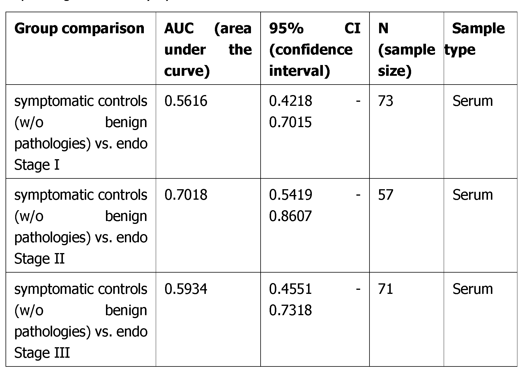
- control samples and/or reference values derived therefrom for the methods described herein are obtained from but not limited to"non-pathological controls” and “symptomatic controls”.
- the corresponding subjects from which these samples are obtained are “non-pathological subjects” and “symptomatic subjects” respectively.
- Symptomatic controls refer to control samples of subjects that suffer from symptoms that are usually associated with endometriosis (e.g., menstrual/abdominal pain, infertility, etc) but where, based on laparoscopy, endometriosis can be excluded and no tissue alterations (e.g., uterine/ovarian cysts or cancer) can be observed.
- endometriosis e.g., menstrual/abdominal pain, infertility, etc
- tissue alterations e.g., uterine/ovarian cysts or cancer
- Controls with benign findings refer to a group of samples of subjects that have tissue alterations (e.g., uterine/ovarian cysts, cancer, fibroids) which however does not resemble endometriosis. Further, these subjects can be symptomatic (e.g., menstrual/abdominal pain, infertility, etc) or asymptotic, which can also change over time.
- tissue alterations e.g., uterine/ovarian cysts, cancer, fibroids
- these subjects can be symptomatic (e.g., menstrual/abdominal pain, infertility, etc) or asymptotic, which can also change over time.
- the control sample may be assayed at the same time, before or after, separately or simultaneously with the test sample.
- the control value that is used in the comparison with the test sample may be a value that is calculated as an average or median of more than one (e.g., two or more, five or more, ten or more, a group etc) of control samples.
- the control sample may be a sample that originated from (i.e., is a mix of) more than one (e.g., two or more, five or more, ten or more, a group etc) individual that is not suffering from endometriosis (or is a "symptomatic control").
- control sample is therefore obtained from a control subject that does not have endometriosis ("non-pathological control").
- the control sample is obtained from a subject that is a "symptomatic control”.
- a comparison of the test sample with several different control samples can or has to be carried out in order to be able to allocate the result to a certain stage. For example, a comparison to a non-pathologic sample and to a stage II sample. Or a comparison to a non-pathologic sample, to a stage II sample and to a stage IV sample. This can also be combined with surgery to confirm or determine a certain stage of disease.
- the level of biomarker (e.g., protein) in the biological fluid sample may be compared to a pre-determined reference level for the biomarker of interest.
- a pre-determined reference level refers to a biomarker level obtained from a reference database, which may be used to generate a pre-determined cut off value, i.e., a score that is statistically predictive of endometriosis.
- the predetermined reference level is the average or median level of the biomarker in at least one individual not suffering from endometriosis from the same species.
- the predetermined reference value may be calculated as the average or median, taken from a group or population of individuals that are not suffering from endometriosis.
- the predetermined reference value may be calculated as the average or median, taken from a group or population of individuals that are "symptomatic controls".
- the individual or the population of individuals can be the same age or in the same state or condition of health as the subject from which the test sample is obtained.
- the pre-determined reference level is therefore the average level of the biomarker in a control subject that does not have endometriosis. In a further example the pre-determined reference level is the average level of the biomarker in a subject that is a "symptomatic control".
- control or predetermined reference value may be obtained from the same individual as the test sample, but at an earlier time point.
- This is particularly relevant for the methods described herein that classify the stage of endometriosis, that determine the progression in a subject, that determine the therapeutic effect of a treatment regimen for endometriosis, and/or that determine a subject's compliance or adherence with a prescribed treatment regimen for endometriosis.
- the samples are taken from the same biological fluid of the same subject, wherein the biological fluid is blood, serum, plasma, capillary blood, interstitial fluid, peritoneal fluid, or menstrual fluid preferably the biological fluid sample is serum.
- control sample or predetermined reference level is used to determine any changes in the level of the biomarker(s) over a time interval for the same subject.
- the pre-determined reference level or control sample can therefore be from the same subject that the test sample is obtained from, for example obtained at an earlier time point. This earlier time point can be before they were diagnosed with endometriosis.
- a pre-determined level can be single cut-off value, such as a median or mean. It can be a range of cut-off (or threshold) values, such as a confidence interval. It can be established based upon comparative groups, such as where the risk in one defined group is a fold higher, or lower, (e.g., approximately 2-fold, 4-fold, 8-fold, 16-fold or more) than the risk in another defined group.
- the level of the protein biomarker in a subject being greater than or equal to the level of the biomarker of the control sample or pre-determined reference level is indicative of a clinical status (e.g., indicative of endometriosis).
- the level of the biomarker in a subject being less than or equal to the level of biomarker of the control sample or predetermined reference level is indicative of a certain stage of endometriosis.
- the greater than, or the less than, that is sufficient to distinguish a subject from a control subject is a statistically significantly greater than, or a statistically significant less than.
- the "being equal” refers to being approximately equal (e.g., not statistically different).
- the pre-determined value can depend upon a particular population of subjects (e.g., human subjects) selected. For example, an apparently healthy population will have a different 'normal' range of the protein biomarker than will a population of subjects which have, or are likely to have, endometriosis. Accordingly, the pre-determined values selected may take into account the category (e.g., healthy, diseased, stage of disease) in which a subject (e.g., human subject) falls.
- the category e.g., healthy, diseased, stage of disease
- the level of the specific biomarker detected in a sample may be normalized by adjusting the measured level (amount or activity) of the biomarker using the level of a reference protein in the same sample, wherein the reference protein is not a marker itself (it is e.g., a protein that is constitutively expressed).
- This normalization allows the comparison of the biomarker level in one sample to another sample, or between samples from different sources. This normalized level can then optionally be compared to a reference value or control.
- classifying refers to the classification of the stage of endometriosis according to the revised scoring system of the American Society for Reproductive Medicine (r-ASRM) consisting of the four stages I, II, III and IV (Revised American Society for Reproductive Medicine classification of endometriosis: 1996. Fertil Steril. 1997).
- r-ASRM American Society for Reproductive Medicine
- c-Kit levels at stages I, III and IV are about the same and are increased compared to the controls.
- c-Kit levels at stage II are increased compared to stages I, III and IV.
- a classification can also be made if ranges for c-Kit levels are defined that are allocated to the certain stages of endometriosis. By this an obtained value falling within the range allocated to, for example, stage I would mean that this patient suffers from endometriosis stage I.
- the assessment made in accordance with the present invention may usually not be correct for 100% of the investigated subjects.
- the term typically, requires that a statistically significant portion of subjects can be correctly assessed. Whether a portion is statistically significant can be determined without further ado by the person skilled in the art using various well known statistic evaluation tools, e.g., determination of confidence intervals, p-value determination, Student's t-test, Mann-Whitney test, etc.. Details may be found in Dowdy and Wearden, Statistics for Research, John Wiley & Sons, New York 1983. Typically envisaged confidence intervals are at least 50%, at least 60%, at least 70%, at least 80%, at least 90%, at least 95%. The p-values are, typically, 0.2, 0.1, 0.05.
- lowered or “decreased” level of an indicator refer to the level of such indicator in the sample being reduced in comparison to the reference (value) or reference sample.
- decrease “decreased” “reduced”, “reduction” or “down- regulated”, “lower” are all used herein generally to mean a decrease by a statistically significant amount.
- an indicator/(bio)marker refers to the level of such indicator in the sample being higher in comparison to the reference (value) or reference sample.
- a protein that is detectable in higher amounts in a fluid sample of one individual suffering from a given disease than in the same fluid sample of individuals not suffering from said disease has an elevated level.
- the terms “increased”, “increase” or “up-regulated”, “higher” are all used herein to generally mean an increase by a statically significant amount; for the avoidance of any doubt, the terms “increased” or “increase” means an increase of at least 10% as compared to a reference level/control, for example an increase of at least about 20%, or at least about 30%, or at least about 40%, or at least about 50%, or at least about 60%, or at least about 70%, or at least about 80%, or at least about 90% or up to and including a 100% increase or any increase between 10-100% as compared to a reference level/control, or at least about a 0.5-fold, or at least about a l.O-fold, or at least about a 1.2-fold, or at least about a 1.5-fold, or at least about a 2-fold, or at least about a 3-fold, or at least about a 4-fold, or at least about a 5 -fold or at least about a 10-fold increase, or any increase between l
- immunoglobulin refers to immunity conferring glycoproteins of the immunoglobulin superfamily.
- Surface immunoglobulins are attached to the membrane of effector cells by their transmembrane region and encompass molecules such as but not limited to B-cell receptors, T -cell receptors, class I and II major histocompatibility complex (MHC) proteins, beta-2 microglobulin ( ⁇ 2M), CD3, CD4 and CDS.
- MHC major histocompatibility complex
- ⁇ 2M beta-2 microglobulin
- CD3, CD4 and CDS CDS.
- IgG subclasses In humans there are four different IgG subclasses (IgGI, 2, 3, and 4), named in order of their abundance in serum with IgGI being the most abundant ( ⁇ 66%), followed by IgG2 ( ⁇ 23%), IgG3 ( ⁇ 7%) and IgG ( ⁇ 4%).
- IgG The biological profile of the different IgG classes is determined by the structure of the respective hinge region.
- IgM is expressed on the surface of B cells in a monomeric form and in a secreted pentameric form with very high avidity. IgM is involved in eliminating pathogens in the early stages of B cell mediated (humoral) immunity before sufficient IgG is produced (Geisberger et al. (2006) Immunology 118:429-437).
- Antibodies are not only found as monomers but are also known to form dimers of two Ig units (e.g. IgA), tetramers of four Ig units (e.g. IgM of teleost fish), or pentamers of five Ig units (e.g. mammalian IgM).
- Antibodies are typically made of four polypeptide chains comprising two identical heavy chains and identical two light chains which are connected via disulfide bonds and resemble a "Y"-shaped macro-molecule. Each of the chains comprises a number of immunoglobulin domains out of which some are constant domains and others are variable domains.
- Immunoglobulin domains consist of a 2-layer sandwich of between 7 and 9 antiparallel ⁇ -strands arranged in two ⁇ -sheets.
- the heavy chain of an antibody comprises four Ig domains with three of them being constant (CH domains: CHI. CH2. CH3) domains and one of the being a variable domain (V H).
- the light chain typically comprises one constant Ig domain (CL) and one variable Ig domain (V L).
- CH domains in the context of IgG are as follows: "CHI” refers to amino acid positions 118-220 according to the EU index as in Kabat; "CH2” refers to amino acid positions 237-340 according to the EU index as in Kabat; and "CH3” refers to amino acid positions 341-44 7 according to the EU index as in Kabat.
- full-length antibody “intact antibody”, and “whole antibody” are used herein interchangeably to refer to an antibody in its substantially intact form, not antibody fragments as defined below.
- single domain antibodies sdAb
- Single chain Fv scFv
- scFvA-scFvB Divalent single-chain variable fragments
- Bispecific diabodies are formed by expressing to chains with the arrangement VHA-VLB and VHB-VLA or VLA-VHB and VLB-VHA, respectively.
- Singlechain diabodies comprise a VHA-VLB and a VHB-VLA fragment which are linked by a linker peptide (P) of 12-20 amino acids, preferably 14 amino acids, (VHA-VLB-P-VHB-VLA).
- Bi-specific T-cell engagers (BiTEs)" are fusion proteins consisting of two scFvs of different antibodies wherein one of the scFvs binds to T cells via the CD3 receptor, and the other to a tumor cell via a tumor specific molecule (Kufer et al. (2004) Trends Biotechnol. 22:238-244).
- Dual affinity retargeting molecules (“DART” molecules) are diabodies additionally stabilized through a C-terminal disulfide bridge.
- antibody fragments refers to a portion of an intact antibody, preferably comprising the antigen-binding region thereof.
- Antibody fragments include but are not limited to Fab, Fab', F(ab')2, Fv fragments; diabodies; sdAb, nanobodies, scFv, di- scFvs, tandem scFvs, triabodies, diabodies, scDb, BiTEs, and DARTs.
- binding affinity generally refers to the strength of the sum total of noncovalent interactions between a single binding site of a molecule (e.g., an antibody) and its binding partner (e.g., an antigen). Unless indicated otherwise, as used herein, "binding affinity” refers to intrinsic binding affinity which reflects a 1:1 interaction between members of a binding pair (e.g., antibody and antigen).
- the affinity of a molecule X for its partner Y can generally be represented by the dissociation constant (Kd). Affinity can be measured by common methods known in the art, including but not limited to surface plasmon resonance-based assay (such as the BIAcore assay as described in PCT Application Publication No.
- the solid surface is typically glass or a polymer, the most commonly used polymers being cellulose, polyacrylamide, nylon, polystyrene, polyvinyl chloride, or polypropylene.
- the solid supports may be in the form of tubes, beads, discs of microplates, or any other surface suitable for conducting an immunoassay.
- the binding processes are well-known in the art and generally consist of cross-linking covalently binding or physically adsorbing, the polymer-anti body complex is washed in preparation for the test sample.
- An extremely versatile alternative sandwich assay format includes the use of a solid phase coated with the first partner of a binding pair, e.g., paramagnetic streptavidin-coated microparticles. Such microparticles are mixed and incubated with an analyte-specific binding agent bound to the second partner of the binding pair (e.g., a biotinylated antibody), a sample suspected of comprising or comprising the analyte, wherein said second partner of the binding pair is bound to said analyte-specific binding agent, and a second analyte-specific binding agent which is detectably labeled.
- an analyte-specific binding agent bound to the second partner of the binding pair e.g., a biotinylated antibody
- a sample suspected of comprising or comprising the analyte wherein said second partner of the binding pair is bound to said analyte-specific binding agent
- a second analyte-specific binding agent which is detectably labeled
- these components are incubated under appropriate conditions and for a period of time sufficient for binding the labeled antibody via the analyte, the analyte-specific binding agent (bound to) the second partner of the binding pair and the first partner of the binding pair to the solid phase microparticles.
- assay may include one or more washing step(s).
- detectably labeled encompasses labels that can be directly or indirectly detected.
- detectably labeled refers to a label providing or inducible to provide a detectable signal, i.e., to a fluorescent label, to a luminescent label (e.g., a chemiluminescent label or an electrochemiluminescent label), a radioactive label or a metal-chelate based label, respectively.
- a detectable signal i.e., to a fluorescent label
- a luminescent label e.g., a chemiluminescent label or an electrochemiluminescent label
- radioactive label e.g., a radioactive label or a metal-chelate based label
- Fluorescent dyes are e.g., described by Briggs et al "Synthesis of Functionalized Fluorescent Dyes and Their Coupling to Amines and Amino Acids," J. Chem. Soc., Perkin- Trans. 1 (1997) 1051-1058).
- Fluorescent labels or fluorophores include rare earth chelates (europium chelates), fluorescein type labels including FITC, 5-carboxyfluorescein, 6-carboxy fluorescein; rhodamine type labels including TAMRA; dansyl; Lissamine; cyanines; phycoerythrins; Texas Red; and analogs thereof.
- the fluorescent labels can be conjugated to an aldehyde group comprised in target molecule using the techniques disclosed herein.
- Fluorescent dyes and fluorescent label reagents include those which are commercially available from Invitrogen/Molecular Probes (Eugene, Oregon, USA) and Pierce Biotechnology, Inc. (Rockford, III.).
- Luminescent dyes or labels can be further subcategorized into chemiluminescent and electrochemiluminescent dyes.
- chemiluminogenic labels include luminol, acridinium compounds, coelenterazine and analogues, dioxetanes, systems based on peroxyoxalic acid and their derivatives.
- acridinium based labels are used (a detailed overview is given in Dodeigne C. et al., Taianta 51 (2000) 415-439).
- Electrochemiluminescense proved to be very useful in analytical applications as a highly sensitive and selective method. It combines analytical advantages of chemiluminescent analysis (absence of background optical signal) with ease of reaction control by applying electrode potential.
- Ruthenium complexes especially [Ru (Bpy)3]2+ (which releases a photon at ⁇ 620 nm) regenerating with TPA (Tri propylamine) in liquid phase or liquid-solid interface are used as ECL-labels.
- Electrochemiluminescent (ECL) assays provide a sensitive and precise measurement of the presence and concentration of an analyte of interest. Such techniques use labels or other reactants that can be induced to luminesce when electrochemically oxidized or reduced in an appropriate chemical environment. Such electrochemiluminescense is triggered by a voltage imposed on a working electrode at a particular time and in a particular manner. The light produced by the label is measured and indicates the presence or quantity of the analyte.
- ECL Electrochemiluminescent
- Radioactive labels make use of radioisotopes (radionuclides), such as 3H, 11C, 14C, 18F, 32P, 35S, 64Cu, 68Gn, 86Y, 89Zr, 99TC, lllln, 1231, 1241, 1251, 1311, 133Xe, 177Lu, 211At, or 131BL
- radioisotopes such as 3H, 11C, 14C, 18F, 32P, 35S, 64Cu, 68Gn, 86Y, 89Zr, 99TC, lllln, 1231, 1241, 1251, 1311, 133Xe, 177Lu, 211At, or 131BL
- the methods described herein can further comprise selecting, and optionally administering, a treatment regimen for the subject based on the diagnosis (i.e., based on the comparison of the levels of the biomarkers with the reference values/levels/controls).
- Treatment can include, for example, surgery and, in some cases, therapy, or combinations thereof. However, in some cases, immediate treatment may not be required, and the subject may be selected for active surveillance.
- active surveillance monitoring
- watchful waiting are used interchangeably herein to mean closely monitoring a patient's condition without giving any treatment until symptoms appear or change.
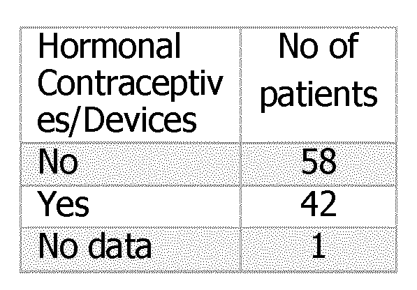
- appropriate treatment may include pain medication, hormone treatments (such as hormonal contraceptives), gonadotropin- releasing hormone (GnRH) agonists, and/or surgery.
- hormone treatments such as hormonal contraceptives
- GnRH gonadotropin- releasing hormone
- the term "surgery” applies to surgical methods undertaken for removal of endometric tissue, like, for example, laparoscopy or nerve sparing surgery.
- the term "therapy” includes drug-based therapy, radiation, hormonal therapy, cryosurgery, chemotherapy, immunotherapy, biologic therapy, and high- intensity focused ultrasound.
- Drug-based therapy of endometriosis can for example be by inhibiting or targeting neurogenic inflammation and/or pain medication and/or hormonal therapy.
- the type of treatment will vary depending on the particular form and/or stage of endometriosis that the subject has, is suspected of having.
- the inventors have surprisingly identified the new protein biomarker c-Kit that is increased in biological fluids, in particular serum, of women with endometriosis, especially in women with early stages of endometriosis.
- the biomarker c-Kit can be used for diagnosing endometriosis or classifying the stage of endometriosis in a subject compared to a control (e.g., non-pathological subjects or symptomatic subjects).
- serum c-Kit can be used as a blood biomarker for early diagnosis and risk stratification of endometriosis. Furthermore, serum c-Kit can be used to select patients with disease stage I and stage II for early medical management of endometriosis. Therefore, it can significantly reduce endometriosis diagnostic delay, improve patients' lives, and reduce the economic burden.
- an elevated level or amount or concentration of c-Kit in the fluid sample of the subject is indicative of the presence of endometriosis in the subject.
- an amount or concentration of c-Kit in the fluid sample of the subject is indicative of the presence of endometriosis in the subject if the amount or concentration of c-Kit in the fluid sample of the subject is higher than the amount or concentration of c-Kit according to a reference value.
- an amount of c-Kit elevated by 50% or more is indicative of the presence or the risk of developing of endometrioses.
- an amount of c-Kit elevated by 100% or more is indicative of the presence of endometrioses.
- an amount of c-Kit elevated by 150% or more is indicative of the presence of endometrioses.
- an amount of c-Kit elevated by 200% or more is indicative of endometrioses.
- the biological fluid sample is blood, serum, plasma, capillary blood, interstitial fluid, peritoneal fluid, or menstrual fluid, preferably the biological fluid sample is serum.
- the subject is a human subject.
- the patient is a female human subject.
- the subject is a young or adolescent human female.
- the subject is a subject subject who is capable of suffering from endometriosis due to the physical condition.
- endometriosis is early endometriosis, in particular stage I endometriosis according to rASRM staging or stage II endometriosis according to rASRM staging.
- the protein level of c-Kit is determined, optionally using a process selected from: ELISA assay, immunoblotting, lateral flow assay, protein microarray and mass spectrometry.
- the amount of c-Kit is determined using antibodies, in particular using monoclonal antibodies.
- step a) of determining the amount of c-Kit in a sample of the patient comprises performing an immunoassay.
- the immunoassay is performed either in a direct or indirect format.
- step a) of determining the level of c-Kit in a sample of the subject comprises the steps of i) incubating the sample of the subject with one or more antibodies specifically binding to c-Kit, thereby generating a complex between the antibody and c-Kit, and ii) quantifying the complex formed in step i), thereby quantifying the amount of c-Kit in the sample of the subject.
- the method further comprising determining the level of CA-125 in the biological fluid sample from the subject.
- the invention in a second aspect relates to a method for classifying the stage of endometriosis in a subject, the method comprising the steps of: a) determining the level of c-Kit in a biological fluid sample from the subject , b) comparing the level of c-Kit to at least one appropriate reference value of a c- Kit level , c) classifying the stage of endometriosis in the subject if the comparison in step b) indicates that the subject has an increased or decreased level of c-Kit compared to the at least one appropriate reference value of a c-Kit level.
- an elevated or reduced level or amount or concentration of c-Kit in the fluid sample of the subject can be indicative of the stage of endometriosis in the subject.
- a level of c-Kit in the fluid sample of the subject is indicative of the stage of endometriosis in the subject if the level of c-Kit in the fluid sample of the subject is higher or lower than the level of c-Kit according to a reference value of which the stage of endometriosis is known. If necessary, comparisons to several reference values are made in order to classify the stage of endometriosis. In addition this can be combined with surgery or other parameters used to determine the stage of endometriosis to allow an even more precise classification.
- the at least one appropriate reference value is i. a level of c-Kit in a non-pathological subject, in a symptomatic subject, or in a subject that has stage I, stage II, stage III or stage IV endometriosis according to the revised scoring system of the American Society for Reproductive Medicine (r-ASRM), or is ii. an average level of c-Kit in a group of subjects that are non-pathological subjects, that are symptomatic subjects, or a combination thereof, or in a group of subjects that have stage I, stage II, stage III or stage IV endometriosis according to the revised scoring system of the American Society for Reproductive Medicine (r-ASRM), or wherein the at least one appropriate reference value is iii.
- biomarkers Details of the biomarkers, combinations, samples, methods steps, subjects, types of endometriosis, treatment, reference values, etc are provided elsewhere and apply equally to this and all the other aspects.
- the present invention relates to a method for monitoring endometriosis progression in a subject, the method comprising the steps of: i. determining the level of c-Kit in a biological fluid sample from the subject in accordance with method steps a) to b) described above herein, ii. repeating step i. using a biological fluid sample obtained from the subject during or after treatment for a time interval; and iii. comparing the levels of c-Kit identified in i. with the c-Kit levels identified in ii., wherein a change in the c-Kit levels from i. to ii. is indicative of a change in endometriosis progression in the subject.
- a patient suffering from endometriosis is monitored to determine if the amount or concentration of c-Kit is changing over time in a sample of the patient.
- a patient suffering from endometriosis is monitored to determine if the amount or concentration of c-Kit is increasing, decreasing or not changing over time.
- a patient suffering from endometriosis is monitored if an elevated amount of c-Kit in the sample of the patient is determined.
- monitoring methods also encompass methods performed on subjects that have already been treated for endometriosis.
- Monitoring the progression of endometriosis in a subject over time assists in the earliest possible identification of disease progression (e.g., a worsening in disease status or disease symptoms). Such monitoring naturally involves the taking of repeated samples over time.
- the method may therefore be repeated at one or more time intervals for a particular subject and the results compared to monitor the development, progression or improvement in endometriosis of that subject over time, wherein a change in the amount of level of the biomarker tested for in the biological fluid sample (e.g., serum) is indicative of a change in the progression of the endometriosis in the subject.
- the biological fluid sample e.g., serum
- Disease progression may be indicated by an increase in the level of c-Kit detected over time when the results of two or more time intervals are compared for the same subject.
- Suitable time intervals for monitoring disease progression can easily be identified by a person of skill in the art and will depend on the specific form of endometriosis being monitored.
- the method may be repeated at least every week, month, six months, or at least every year, or whenever clinically needed, i.e., in case of a significant change in endometriosis symptoms.
- the method is an in vitro method.
- the present invention relates to a method for determining the therapeutic effect of a treatment regimen for endometriosis in a subject, the method comprising the steps of: i. determining the level of c-Kit in a biological fluid sample from the subject in accordance with method steps a) to b) described above herein, ii. repeating step a) using a biological fluid sample obtained from the subject during or after treatment for a time interval; and iii. comparing the level of c-Kit determined in step a) to that determined in step b), and identifying that the treatment regimen has a therapeutic effect if the level of c-Kit decreased after treatment.
- the change in level of c-Kit that is indicative of a therapeutic effect is a decrease in c-Kit level after treatment.
- An "decrease" in the level of c-Kit encompasses no detection of c-Kit (i.e., it is not present at detectable levels) at a later time interval when c-Kit was detected when the method was performed previously (i.e. at an earlier time interval) on the same subject (and an equivalent biological fluid sample type).
- Step i. may first be performed in accordance with the method using a biological fluid sample that was obtained from the subject at a time point before the treatment regimen for endometriosis began.
- step i. may first be performed using a biological fluid sample that was obtained from the subject at the same time as commencing the treatment regimen, or at a time point after the treatment regimen for endometriosis began.
- the method can therefore be used to determine the therapeutic effect of a treatment regimen for endometriosis from the outset (i.e., from the start of the regimen) or from a time point after the treatment regimen has started (i.e., determining the therapeutic effect of a treatment regimen for endometriosis during the treatment regimen itself).
- an unaltered or increasing amount or concentration of c-Kit in a sample of the subject being treated for endometriosis is indicative of the therapy being ineffective, i.e., an unaltered or increasing amount or concentration of c-Kit in a sample of the subject being treated for endometriosis is indicative of persisting or recurring endometriosis.
- the treatment for endometriosis is ineffective if the amount of c-Kit is increasing to 50% or more.
- the treatment for endometriosis is ineffective if the amount of c-Kit is increasing to 100% or more.
- an unaltered level of c-Kit could either mean that the disease stagnates or that it has been progressed from stage I to stages III or IV where the c-Kit levels are comparable to stage I.
- An improvement in disease status or symptoms may also be indicated by stabilised levels of c-Kit over time (compared to the level of c-Kit observed in the absence of treatment over the equivalent time period or compared to equivalent controls).
- a treatment regimen may also be identified as having a therapeutic effect if it results in an improvement in disease status or symptoms (e.g., over a treatment period). Methods for determining if the treatment regimen has a therapeutic effect are well known in the art.
- a treatment period refers to a time interval over which treatment occurs (e.g., 1 month, 3 months, 6 months, 1 year, 2 years, etc).
- the direction of change in c-Kit levels that is indicative of a therapeutic effect may depend on the disease status of the subject prior to treatment and the control/ reference used.
- the subject is monitored several times at different time points. In embodiments, the patient is monitored several times within a time frame of weeks, months, or years. In particular embodiments, a subject is monitored is once a month or once a year. In embodiments, a subject suffering from endometriosis is monitored once a month or once a year after diagnosis of endometriosis. In embodiments, a subject being treated for endometriosis is monitored once after therapy, in particular once after surgical therapy. In particular, the subject being treated for endometriosis is monitored once a month or once a year to determine the efficacy of treatment and/or the recurrence of endometriosis.
- the method can also be useful as a screening tool for determining if specific regimens or treatment modalities have a therapeutic effect on endometriosis.
- the tested regimens or treatment modalities may be new regimens or treatment modalities, modified regimens or treatment modalities, or known regimens or treatment modalities that need further testing.
- a treatment modality is e.g., a drug or medicament that is useful or suspected to be useful in the treatment of endometriosis.
- therapy of endometriosis is selected from the group consisting of drug- based therapy or surgical therapy.
- the treatment regimen comprises surgical therapy, radiotherapy, immunotherapy, hormone therapy, ultrasound therapy, or combinations thereof.
- surgical therapy of endometriosis is laparoscopy or nerve sparing surgery.
- drug-based therapy of endometriosis is inhibiting or targeting neurogenic inflammation and/or pain medication and/or hormonal therapy.
- the present invention relates to a computer-implemented method for assessing a patient with suspected endometriosis comprising the steps of:
- step (e) assessing said subject based on the comparison and/or the calculation made in step (d).
- symptoms or clinical data that is used for diagnosing or classifying endometriosis can be used in combination with the determination of c-Kit levels.
- symptoms or clinical data can be but are not limited to age, dysmenorrhea, abdominal pain, or other biomarkers.
- Biomarker levels and/or reference levels may be stored in a suitable data storage medium (e.g., a database) and are, thus, also available for future diagnoses. This also allows efficiently diagnosing prevalence for a disease because suitable reference results can be identified in the database once it has been confirmed (in the future) that the subject from which the corresponding reference sample was obtained did have endometriosis.
- a “database” comprises data collected (e.g., analyte and/or reference level information and /or patient information) on a suitable storage medium.
- the database may further comprise a database management system.
- the database management system is, preferably, a network-based, hierarchical or object-oriented database management system.
- the database may be a federal or integrated database.
- the results or diagnoses (or both) are communicated to the subject as soon as possible after the diagnosis is obtained.
- the results or diagnoses (or both) may be communicated to the subject by the subject's treating physician.
- the results or diagnoses (or both) may be sent to a subject by email or communicated to the subject by phone.
- a computer may be used to communicate the results or diagnoses by email or phone.
- the message containing results or diagnoses may be generated and delivered automatically to the subject using a combination of computer hardware and software which will be familiar to artisans skilled in telecommunications.
- c-Kit may be used as biomarker for endometriosis generally.
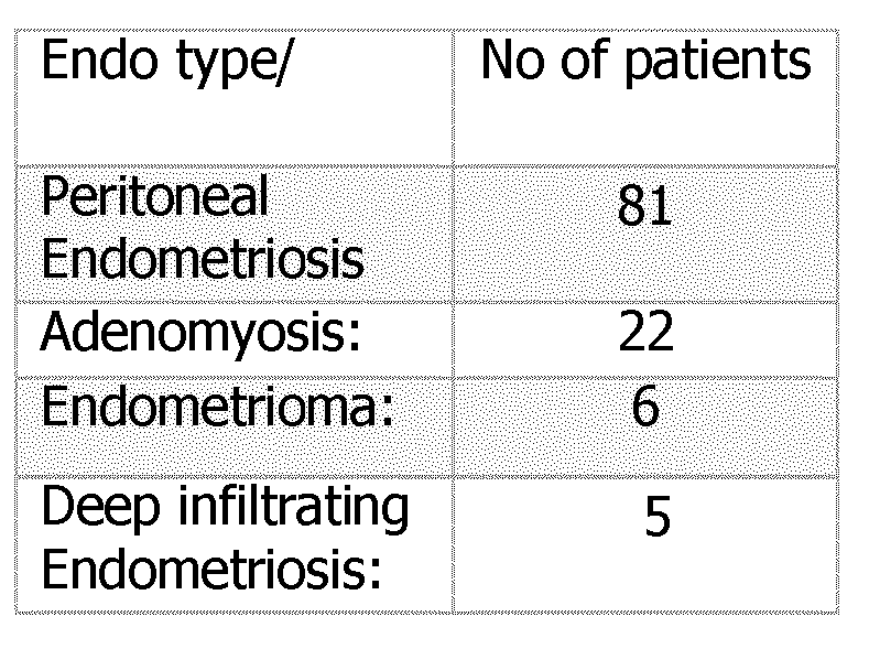
- endometriosis generally refers to all forms of endometriosis, including but not limited to peritoneal endometriosis, endometrioma, deep infiltrating endometriosis, and adenomyosis.
- c-Kit may also be combined with CA-125.
- kits, assay devices and uses provided herein may be used as part of a companion diagnostic e.g., as part of a medical device, often an in vitro device, which provides information that is essential for the safe and effective use of a corresponding drug or biological product (wherein the corresponding drug or biological product is for treating or preventing endometriosis).
- a companion diagnostic e.g., as part of a medical device, often an in vitro device, which provides information that is essential for the safe and effective use of a corresponding drug or biological product (wherein the corresponding drug or biological product is for treating or preventing endometriosis).
- the present invention relates to the following aspects:
- the at least one appropriate reference value is i. a level of c-Kit in a non-pathological subject or in a symptomatic subject, or is ii. an average level of c-Kit in a group of non-pathological subjects or in a group of symptomatic subjects, or a combination thereof, or wherein the at least one appropriate reference value is iii. a predetermined value of a level of c-Kit in a non-pathological subject or symptomatic subject, or is iv. a predetermined average value of a level of c-Kit in a group of non- pathological subjects or in a group of symptomatic subjects, or a combination thereof.
- a method for classifying the stage of endometriosis in a subject comprising the steps of: a) determining the level of c-Kit in a biological fluid sample from the subject, b) comparing the level of c-Kit to at least one appropriate reference value of a c-Kit level, c) classifying the stage of endometriosis in the subject if the comparison in step b) indicates that the subject has an increased or decreased level of c- Kit compared to the at least one appropriate reference value of a c-Kit level.
- a method for classifying the stage of endometriosis in a subject comprising the steps of: a) determining the level of c-Kit in a biological fluid sample from the subject at regular intervals, b) comparing the level of c-Kit to at least one value of a c- Kit level determined at an earlier stage in the subject, c) classifying the stage of endometriosis in the subject if the comparison in step b) indicates that the subject has an increased or decreased level of c-Kit compared to the at least one value of a c-Kit level determined at an earlier stage in the subject.
- the method according to aspect 14, further comprising administering the selected treatment regimen to the subject, optionally wherein the selected treatment regimen comprises drug-based therapy and/or surgical treatment (laparoscopy).
- a method for monitoring endometriosis progression in a subject comprising the steps of: i. determining the level of c-Kit in a biological fluid sample from the subject in accordance with method steps a) to b) of any one of aspects 1 to 15, ii. repeating step i. using a biological fluid sample obtained from the subject during or after treatment for a time interval; and iii. comparing the levels of c-Kit identified in i. with the c-Kit levels identified in ii., wherein a change in the c-Kit levels from i. to ii. is indicative of a change in endometriosis progression in the subject.
- a method for determining the therapeutic effect of a treatment regimen for endometriosis in a subject comprising the steps of: i. determining the level of c-Kit in a biological fluid sample from the subject in accordance with method steps a) to b) of any one of aspects 1 to 15, ii. repeating step i. using a biological fluid sample obtained from the subject during or after treatment for a time interval; and iii. comparing the level of c-Kit determined in step i. to that determined in step ii. and identifying that the treatment regimen has a therapeutic effect if the level of c-Kit decreased after treatment.
- VAS Visual Analog Scale
- a computer-implemented method for assessing a patient with suspected endometriosis comprising the steps of: (a) receiving a value for level of a first biomarker in a sample of the subject, said first biomarker being c-Kit,
- step (e) assessing said subject based on the comparison and/or the calculation made in step (d).
- kits for diagnosing and/or classifying endometriosis in a subject comprising at least one detectably labelled agent that specifically binds to c-Kit protein.
- kit according to aspect 25 further comprising one or more reagents for detecting the detectably labelled agent.
- An assay device for diagnosing and/or classifying endometriosis in a subject comprising a surface with at least one detectably labelled agent located thereon that specifically binds to c-Kit protein.
- Example 1 Diagnostic performance of biomarker c-Kit in women with endometriosis
- the concentration of the analytes was determined by ELISA (enzyme-linked immunosorbent assay).
- the case group is comprised of patients diagnosed with endometriosis (peritoneal endometriosis, adenomyosis, endometrioma, and deep infiltrating endometriosis; rASRM stages I-IV) diagnosed by laparoscopic with subsequent histological confirmation and the control group including healthy women without endometriosis.
- the concentration of c-Kit in human serum was determined using the Human CD117/C- Kit Quantikine ELISA Kit (R&D Systems, USA (catalogue number: DSCR00).
- the kit utilizes the quantitative sandwich ELISA technique. Microtiter plates are pre-coated with a monoclonal antibody specific for human c-Kit. Samples are measured in 50-fold dilution. After bringing all reagents to room temperature 100 pL of each sample and standard are added. Samples are measured in singlicates, standards in duplicates. During 2.5 hrs incubation at room temperature on a microplate shaker set to 650 rpm, any c-Kit present is bound to the immobilized capture antibody on the microtiter plate.
- washing step (4x 300 pL)
- unbound substances are removed from the plate before 100 pL of an enzyme-linked monoclonal antibody specific for c-Kit is added to the wells.
- 100 pL of substrate solution is added to the plate.
- the color develops in proportion to the amount of c-Kit bound in the initial step. Color development is stopped by addition of 50 pL stop solution and colour intensity is measured with a plate reader at 450 nm for detection and 570 nm for background subtraction.
- lyophilized, recombinant c-Kit delivered with the kit was reconstituted and diluted in calibrator diluent.
- the calibration range of the assay is 6.14 pg/mL to 1500 pg/mL.
- Calibrator 7 (1500 pg/mL) is prepared by 6-fold dilution of stock solution in calibrator diluent and calibrator 6 to calibrator 1 (6.14 pg/mL) are prepared by serial 2.5-fold dilution steps in calibrator diluent. Pure calibrator diluent serves as blank (0 pg/mL).
- the calibration curves were fitted using a 4-parameter nonlinear regression (Newton/Raphson) with no weighting. Results are shown in Fig. 1.
- Serum c- Kit levels increase gradually in stage I and stage II of endometriosis compared to non-pathological controls and decrease in stage III.
- Table 1 The diagnostic performance of c-Kit biomarker to discriminate women with histologically confirmed endometriosis and women without endometriosis (controls) using receiver operator characteristic (ROC) analysis is shown describing the area under the curve (AUC) of the ROC analysis and the associated 95% confidence interval. N depicts the number of samples tested (cases plus controls, numbers in each group vary depending on the analyte).
- Example 2 Diagnostic performance of biomarker c-Kit compared to CA-125 in women with endometriosis
- the concentration of CA-125 was determined by a cobas e 601 analyzer. Detection of CA 125 II with a cobas e 601 analyzer is based on the Elecsys® Electro- ChemiLuminescence (ECL) technology.
- ECL Electro- ChemiLuminescence
- biotin-labelled and ruthenium-labelled antibodies are combined with the respective amount of undiluted sample and incubated on the analyzer.
- streptavidin -coated magnetic microparticles are added and incubated on the instrument in order to facilitate binding of the biotin-labelled immunological complexes. After this incubation step the reaction mixture is transferred into the measuring cell where the beads are magnetically captured on the surface of an electrode.
- Box plots in Figs. 2A and 2B were generated for controls and for each of the endometriosis Stages (Stage I, Stage II, Stage III, Stage IV) using the data from the high-throughput, multiplex immunoassay-PCR (OLINK proteomics) analysis.
- the data are presented using box and whisker plots, including the median (middle quartile), the inter- quartile range (which represents the middle 50% of scores for the group), the upper quartile (75% of scores fall below the upper quartile), the lower quartile (25% of scores fall below the lower quartile).
- the whiskers show the 5th percentile and the 95th percentile, respectively.
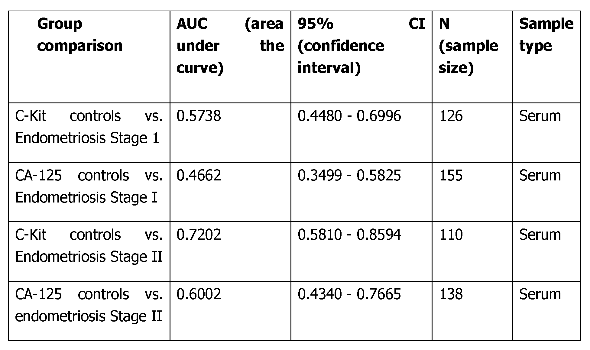
- Serum c-Kit shows better diagnosis performance for the detection of early stages of endometriosis (Stage I, Stage II) compared to the reference biomarker CA-125.
- Stage I Stage II
- the performance of c-Kit as biomarker compared to CA-125 is determined by looking at the area under the curve (AUC) of both biomarkers for early stage endometriosis (Stage I and II).
- Table 2 Diagnostic performance of serum c-Kit biomarker and reference biomarker CA- 125 in women with endometriosis (stage I and stage II) and controls.
- the measurement of CA-125 was performed by OLINK using the proximity extension assay (PEA).
- C-Kit levels were measured using CD117/c-Kit Quantikine ELISA Kit.
- C-Kit levels increase gradually in controls with benign findings, stage I and stage II of endometriosis and decrease back in stage III and IV.
- Serum c-Kit shows better diagnosis performance for the detection of early stages of endometriosis (Stage I, Stage II) compared to the reference biomarker CA-125.
Landscapes
- Life Sciences & Earth Sciences (AREA)
- Health & Medical Sciences (AREA)
- Engineering & Computer Science (AREA)
- Molecular Biology (AREA)
- Chemical & Material Sciences (AREA)
- Biomedical Technology (AREA)
- Urology & Nephrology (AREA)
- Hematology (AREA)
- Immunology (AREA)
- Biotechnology (AREA)
- Microbiology (AREA)
- Cell Biology (AREA)
- Proteomics, Peptides & Aminoacids (AREA)
- Food Science & Technology (AREA)
- Medicinal Chemistry (AREA)
- Physics & Mathematics (AREA)
- Analytical Chemistry (AREA)
- Biochemistry (AREA)
- General Health & Medical Sciences (AREA)
- General Physics & Mathematics (AREA)
- Pathology (AREA)
- Investigating Or Analysing Biological Materials (AREA)
Abstract
Description


Claims
Priority Applications (4)
| Application Number | Priority Date | Filing Date | Title |
|---|---|---|---|
| JP2024574752A JP2025520600A (en) | 2022-06-23 | 2023-06-23 | Methods for diagnosing and staging endometriosis - Patents.com |
| CN202380048765.XA CN119404103A (en) | 2022-06-23 | 2023-06-23 | Methods for diagnosing endometriosis and for classifying the stages of endometriosis |
| EP23731722.7A EP4544305A1 (en) | 2022-06-23 | 2023-06-23 | Method for diagnosing endometriosis and for classifying the stage of endometriosis |
| US18/999,357 US20250130244A1 (en) | 2022-06-23 | 2024-12-23 | Method for diagnosing endometriosis and for classifying the stage of endometriosis |
Applications Claiming Priority (2)
| Application Number | Priority Date | Filing Date | Title |
|---|---|---|---|
| EP22180800 | 2022-06-23 | ||
| EP22180800.9 | 2022-06-23 |
Related Child Applications (1)
| Application Number | Title | Priority Date | Filing Date |
|---|---|---|---|
| US18/999,357 Continuation US20250130244A1 (en) | 2022-06-23 | 2024-12-23 | Method for diagnosing endometriosis and for classifying the stage of endometriosis |
Publications (1)
| Publication Number | Publication Date |
|---|---|
| WO2023247752A1 true WO2023247752A1 (en) | 2023-12-28 |
Family
ID=82258417
Family Applications (1)
| Application Number | Title | Priority Date | Filing Date |
|---|---|---|---|
| PCT/EP2023/067108 Ceased WO2023247752A1 (en) | 2022-06-23 | 2023-06-23 | Method for diagnosing endometriosis and for classifying the stage of endometriosis |
Country Status (5)
| Country | Link |
|---|---|
| US (1) | US20250130244A1 (en) |
| EP (1) | EP4544305A1 (en) |
| JP (1) | JP2025520600A (en) |
| CN (1) | CN119404103A (en) |
| WO (1) | WO2023247752A1 (en) |
Cited By (1)
| Publication number | Priority date | Publication date | Assignee | Title |
|---|---|---|---|---|
| WO2025024616A1 (en) * | 2023-07-24 | 2025-01-30 | Dot Laboratories, Inc. | Artificial intelligence methods and systems for determining endometriosis status |
Citations (32)
| Publication number | Priority date | Publication date | Assignee | Title |
|---|---|---|---|---|
| WO1987006706A1 (en) | 1986-04-30 | 1987-11-05 | Igen, Inc. | Electrochemiluminescent assays |
| WO1990005301A1 (en) | 1988-11-03 | 1990-05-17 | Igen, Inc. | Electrochemiluminescent assays |
| WO1990005296A1 (en) | 1988-11-03 | 1990-05-17 | Igen, Inc. | Electrochemiluminescent reaction utilizing amine-derived reductant |
| WO1992014139A1 (en) | 1991-02-06 | 1992-08-20 | Igen, Inc. | Methods and apparatus for improved luminescence assays |
| US5221605A (en) | 1984-10-31 | 1993-06-22 | Igen, Inc. | Luminescent metal chelate labels and means for detection |
| US5316757A (en) | 1984-10-18 | 1994-05-31 | Board Of Regents, The University Of Texas System | Synthesis of polyazamacrocycles with more than one type of side-chain chelating groups |
| US5342606A (en) | 1984-10-18 | 1994-08-30 | Board Of Regents, The University Of Texas System | Polyazamacrocyclic compounds for complexation of metal ions |
| US5385893A (en) | 1993-05-06 | 1995-01-31 | The Dow Chemical Company | Tricyclopolyazamacrocyclophosphonic acids, complexes and derivatives thereof, for use as contrast agents |
| WO1995008644A1 (en) | 1993-09-22 | 1995-03-30 | Igen, Inc. | Self-sustained sequence replication electrochemiluminescent nucleic acid assay |
| US5428139A (en) | 1991-12-10 | 1995-06-27 | The Dow Chemical Company | Bicyclopolyazamacrocyclophosphonic acid complexes for use as radiopharmaceuticals |
| US5462725A (en) | 1993-05-06 | 1995-10-31 | The Dow Chemical Company | 2-pyridylmethylenepolyazamacrocyclophosphonic acids, complexes and derivatives thereof, for use as contrast agents |
| US5480990A (en) | 1991-12-10 | 1996-01-02 | The Dow Chemical Company | Bicyclopolyazamacrocyclocarboxylic acid complexes for use as contrast agents |
| WO1996006946A1 (en) | 1994-08-26 | 1996-03-07 | Igen, Inc. | Biosensor for and method of electrogenerated chemiluminescent detection of nucleic acid adsorbed to a solid surface |
| WO1996024690A1 (en) | 1995-02-09 | 1996-08-15 | Igen, Inc. | Electrogenerated chemiluminescence labels for analysis and/or referencing |
| WO1996033411A1 (en) | 1995-04-18 | 1996-10-24 | Igen, Inc. | Electrochemiluminescence of rare earth metal chelates |
| WO1996039534A1 (en) | 1995-06-06 | 1996-12-12 | Igen, Inc. | Electrochemiluminescent enzyme biosensors |
| WO1996041175A1 (en) | 1995-06-07 | 1996-12-19 | Igen, Inc. | Electrochemiluminescent enzyme immunoassay |
| WO1996040978A1 (en) | 1995-06-07 | 1996-12-19 | Igen, Inc. | Electrochemiluminescent monitoring of compounds |
| US5591581A (en) | 1986-04-30 | 1997-01-07 | Igen, Inc. | Electrochemiluminescent rhenium moieties and methods for their use |
| US5597910A (en) | 1991-12-11 | 1997-01-28 | Igen, Inc. | Electrochemiluminescent label for DNA probe assays |
| US5679519A (en) | 1995-05-09 | 1997-10-21 | Oprandy; John J. | Multi-label complex for enhanced sensitivity in electrochemiluminescence assay |
| US5739294A (en) | 1991-12-10 | 1998-04-14 | The Dow Chemical Company | Bicyclopol yazamacrocyclophosphonic acid complexes for use as contrast agents |
| US5834461A (en) | 1993-07-29 | 1998-11-10 | American Cyanamid Company | Tricyclic benzazepine vasopressin antagonists |
| WO2005012359A2 (en) | 2003-08-01 | 2005-02-10 | Genentech, Inc. | Anti-vegf antibodies |
| US20100111861A1 (en) | 2008-10-31 | 2010-05-06 | Lili Liu | Detection and quantification of abasic site formation in vivo |
| WO2012107419A1 (en) | 2011-02-09 | 2012-08-16 | Roche Diagnostics Gmbh | New iridium-based complexes for ecl |
| US9503190B2 (en) | 2012-06-28 | 2016-11-22 | Chunghwa Telecom Co., Ltd. | Client-side dynamic multi-routing power distribution system of FTTx optical terminal equipment |
| US9606763B2 (en) | 2013-12-07 | 2017-03-28 | Lenovo (Singapore) Pte Ltd | Folding electronic device |
| US9703653B2 (en) | 2012-12-12 | 2017-07-11 | Kabushiki Kaisha Toshiba | Cloud system management apparatus, cloud system, reallocation method, and computer program product |
| US9716942B2 (en) | 2015-12-22 | 2017-07-25 | Bose Corporation | Mitigating effects of cavity resonance in speakers |
| WO2021013786A1 (en) * | 2019-07-22 | 2021-01-28 | F. Hoffmann-La Roche Ag | S100a6 as blood biomarker for the non-invasive diagnosis of endometriosis |
| WO2022207628A1 (en) * | 2021-03-30 | 2022-10-06 | F. Hoffmann-La Roche Ag | Scf as blood biomarker for the non-invasive diagnosis of endometriosis |
-
2023
- 2023-06-23 WO PCT/EP2023/067108 patent/WO2023247752A1/en not_active Ceased
- 2023-06-23 JP JP2024574752A patent/JP2025520600A/en active Pending
- 2023-06-23 EP EP23731722.7A patent/EP4544305A1/en active Pending
- 2023-06-23 CN CN202380048765.XA patent/CN119404103A/en active Pending
-
2024
- 2024-12-23 US US18/999,357 patent/US20250130244A1/en active Pending
Patent Citations (34)
| Publication number | Priority date | Publication date | Assignee | Title |
|---|---|---|---|---|
| US5428155A (en) | 1984-10-18 | 1995-06-27 | Board Of Regents, The University Of Texas System | Synthesis of polyazamacrocycles with more than one type of side-chain chelating groups |
| US5342606A (en) | 1984-10-18 | 1994-08-30 | Board Of Regents, The University Of Texas System | Polyazamacrocyclic compounds for complexation of metal ions |
| US5316757A (en) | 1984-10-18 | 1994-05-31 | Board Of Regents, The University Of Texas System | Synthesis of polyazamacrocycles with more than one type of side-chain chelating groups |
| US5221605A (en) | 1984-10-31 | 1993-06-22 | Igen, Inc. | Luminescent metal chelate labels and means for detection |
| WO1987006706A1 (en) | 1986-04-30 | 1987-11-05 | Igen, Inc. | Electrochemiluminescent assays |
| US5591581A (en) | 1986-04-30 | 1997-01-07 | Igen, Inc. | Electrochemiluminescent rhenium moieties and methods for their use |
| WO1990005296A1 (en) | 1988-11-03 | 1990-05-17 | Igen, Inc. | Electrochemiluminescent reaction utilizing amine-derived reductant |
| WO1990005301A1 (en) | 1988-11-03 | 1990-05-17 | Igen, Inc. | Electrochemiluminescent assays |
| WO1992014139A1 (en) | 1991-02-06 | 1992-08-20 | Igen, Inc. | Methods and apparatus for improved luminescence assays |
| US5428139A (en) | 1991-12-10 | 1995-06-27 | The Dow Chemical Company | Bicyclopolyazamacrocyclophosphonic acid complexes for use as radiopharmaceuticals |
| US5480990A (en) | 1991-12-10 | 1996-01-02 | The Dow Chemical Company | Bicyclopolyazamacrocyclocarboxylic acid complexes for use as contrast agents |
| US5750660A (en) | 1991-12-10 | 1998-05-12 | The Dow Chemical Company | Bicyclopolyazamacrocyclophosphonic acid half esters |
| US5739294A (en) | 1991-12-10 | 1998-04-14 | The Dow Chemical Company | Bicyclopol yazamacrocyclophosphonic acid complexes for use as contrast agents |
| US5597910A (en) | 1991-12-11 | 1997-01-28 | Igen, Inc. | Electrochemiluminescent label for DNA probe assays |
| US5385893A (en) | 1993-05-06 | 1995-01-31 | The Dow Chemical Company | Tricyclopolyazamacrocyclophosphonic acids, complexes and derivatives thereof, for use as contrast agents |
| US5462725A (en) | 1993-05-06 | 1995-10-31 | The Dow Chemical Company | 2-pyridylmethylenepolyazamacrocyclophosphonic acids, complexes and derivatives thereof, for use as contrast agents |
| US5834461A (en) | 1993-07-29 | 1998-11-10 | American Cyanamid Company | Tricyclic benzazepine vasopressin antagonists |
| WO1995008644A1 (en) | 1993-09-22 | 1995-03-30 | Igen, Inc. | Self-sustained sequence replication electrochemiluminescent nucleic acid assay |
| WO1996006946A1 (en) | 1994-08-26 | 1996-03-07 | Igen, Inc. | Biosensor for and method of electrogenerated chemiluminescent detection of nucleic acid adsorbed to a solid surface |
| WO1996024690A1 (en) | 1995-02-09 | 1996-08-15 | Igen, Inc. | Electrogenerated chemiluminescence labels for analysis and/or referencing |
| WO1996033411A1 (en) | 1995-04-18 | 1996-10-24 | Igen, Inc. | Electrochemiluminescence of rare earth metal chelates |
| US5679519A (en) | 1995-05-09 | 1997-10-21 | Oprandy; John J. | Multi-label complex for enhanced sensitivity in electrochemiluminescence assay |
| WO1996039534A1 (en) | 1995-06-06 | 1996-12-12 | Igen, Inc. | Electrochemiluminescent enzyme biosensors |
| WO1996040978A1 (en) | 1995-06-07 | 1996-12-19 | Igen, Inc. | Electrochemiluminescent monitoring of compounds |
| WO1996041175A1 (en) | 1995-06-07 | 1996-12-19 | Igen, Inc. | Electrochemiluminescent enzyme immunoassay |
| WO2005012359A2 (en) | 2003-08-01 | 2005-02-10 | Genentech, Inc. | Anti-vegf antibodies |
| US20100111861A1 (en) | 2008-10-31 | 2010-05-06 | Lili Liu | Detection and quantification of abasic site formation in vivo |
| WO2012107419A1 (en) | 2011-02-09 | 2012-08-16 | Roche Diagnostics Gmbh | New iridium-based complexes for ecl |
| US9503190B2 (en) | 2012-06-28 | 2016-11-22 | Chunghwa Telecom Co., Ltd. | Client-side dynamic multi-routing power distribution system of FTTx optical terminal equipment |
| US9703653B2 (en) | 2012-12-12 | 2017-07-11 | Kabushiki Kaisha Toshiba | Cloud system management apparatus, cloud system, reallocation method, and computer program product |
| US9606763B2 (en) | 2013-12-07 | 2017-03-28 | Lenovo (Singapore) Pte Ltd | Folding electronic device |
| US9716942B2 (en) | 2015-12-22 | 2017-07-25 | Bose Corporation | Mitigating effects of cavity resonance in speakers |
| WO2021013786A1 (en) * | 2019-07-22 | 2021-01-28 | F. Hoffmann-La Roche Ag | S100a6 as blood biomarker for the non-invasive diagnosis of endometriosis |
| WO2022207628A1 (en) * | 2021-03-30 | 2022-10-06 | F. Hoffmann-La Roche Ag | Scf as blood biomarker for the non-invasive diagnosis of endometriosis |
Non-Patent Citations (63)
| Title |
|---|
| "UniProtKB", Database accession no. P10721 |
| ADAMSON ET AL., J ENDOMETR, vol. 2, 2010, pages 3 - 6 |
| AHN ET AL., FERTIL STERIL, 2016 |
| ARIANNA PACCHIAROTTI ET AL: "Expression of oct-4 and c-kit antigens in endometriosis", FERTILITY AND STERILITY, ELSEVIER, AMSTERDAM, NL, vol. 95, no. 3, 13 October 2010 (2010-10-13), pages 1171 - 1173, XP028147771, ISSN: 0015-0282, [retrieved on 20101018], DOI: 10.1016/J.FERTNSTERT.2010.10.029 * |
| BLEND ET AL., CANCER BIOTHERAPY & RADIOPHARMACEUTICALS, vol. 18, 2003, pages 355 - 363 |
| BRIGGS ET AL.: "J. Chem. Soc.", vol. 1, 1997, PERKIN-TRANS, article "Synthesis of Functionalized Fluorescent Dyes and Their Coupling to Amines and Amino Acids", pages: 1051 - 1058 |
| CAMERA ET AL., J. NUCL. MED., vol. 21, 1994, pages 640 - 646 |
| CHANG JUI-HUNG ET AL: "Expression of the pluripotent transcription factor OCT4 promotes cell migration in endometriosis", FERTILITY AND STERILITY, ELSEVIER, AMSTERDAM, NL, vol. 99, no. 5, 3 January 2013 (2013-01-03), pages 1332, XP028527173, ISSN: 0015-0282, DOI: 10.1016/J.FERTNSTERT.2012.11.033 * |
| CHEN ET AL., ACTA OBSTET GYNECOL SCAND, vol. 77, 1998, pages 665 - 70 |
| CHEN ET AL., NAT. IMMUNOL., vol. 10, 2009, pages 889 - 898 |
| CHO NH. ET AL., FERTIL STERIL, vol. 81, 2004, pages 403 - 7 |
| COZZOLINO ET AL., REV BRAS GINECOL OBSTET, vol. 41, no. 3, 2019, pages 170 - 175 |
| CRUZ ET AL., J BIOL CHEM, 2004 |
| DEISENHOFER, BIOCHEMISTRY, vol. 20, 1981, pages 2361 - 2370 |
| DENARDO ET AL., CLINICAL CANCER RESEARCH, vol. 4, 1998, pages 2483 - 90 |
| DESMYTER ET AL., NAT. STRUCTURE BIOL., vol. 3, 1996, pages 803 - 811 |
| DODEIGNE C. ET AL., TALANTA, vol. 51, 2000, pages 415 - 439 |
| EYSTER ET AL., FERTIL STERIL, 2007 |
| FADHLAOUI ET AL., FRONT SURG, vol. 1, 2014, pages 24 |
| FASSBENDER ET AL.: "Peripheral Blood Biomarkers for Endometriosis", 2017, SPRINGER |
| FERTIL STERIL, 1996 |
| GEISBERGER ET AL., IMMUNOLOGY, vol. 118, 2006, pages 429 - 437 |
| HALIS ET AL., DEUTSCHES ARZTEBLATT INTERNATIONAL, vol. 107, no. 25, 2010, pages 446 |
| HEITZER, E.HAQUE, I.S.ROBERTS, C.E.S. ET AL.: "Current and future perspectives of liquid biopsies in genomics-driven oncology", NAT REV GENET, vol. 20, 2019, pages 71 - 88, XP036675874, DOI: 10.1038/s41576-018-0071-5 |
| HEVER ET AL., PNAS, 2007 |
| HNATOWICH ET AL., J. IMMUNOL. METHODS, vol. 65, 1983, pages 147 - 157 |
| HSU ET AL., CLIN OBSTET GYNECOL, vol. 53, 2010, pages 413 - 419 |
| HUSTON ET AL., PROC. NATL. ACAD. SCI. USA, vol. 85, 1988, pages 5879 - 5883 |
| IZARD ET AL., BIOCONJUGATE CHEM, vol. 3, 1992, pages 346 - 350 |
| JACOBS ET AL., HUM REPROD, vol. 4, 1989, pages 1 - 12 |
| KABAT, E. A.WU, T.T.PERRY, H. M.GOTTESMAN, K. S.FOELLER, C.: "Sequences of proteins of immunological interest", 1991, NATIONAL INSTITUTES OF HEALTH |
| KENNEDY ET AL., HUM REPROD, vol. 20, no. 10, 2005, pages 2698 - 2704 |
| KNIGHT ET AL., ANALYST, vol. 119, 1994, pages 879 - 890 |
| KOBAYASHI ET AL., BIOCONJUGATE CHEM, vol. 10, 1999, pages 103 - 111 |
| KOBAYASHI ET AL., J. NUCL. MED., vol. 39, 1998, pages 2105 - 2110 |
| KUFER ET AL., TRENDS BIOTECHNOL, vol. 22, 2004, pages 238 - 244 |
| LEE ET AL., CANCER RES, vol. 61, 2001, pages 4474 - 4482 |
| LONGO, D.L ET AL., SC. D. N ENGL J MED, vol. 382, pages 1244 - 56 |
| MARDIROSSIAN ET AL., NUCL. MED. BIOL., vol. 20, 1993, pages 955 - 74 |
| MEARES ET AL., ANAL. BIOCHEM., vol. 142, 1984, pages 68 - 78 |
| MEARES ET AL., J. CANCER, 1990, pages 21 - 26 |
| MIEDERER ET AL., J. NUCL. MED., vol. 45, 2004, pages 129 - 137 |
| MIRZADEH ET AL., BIOCONJUGATE CHEM, vol. 1, 1990, pages 59 - 65 |
| MITCHELL ET AL., J. NUCL. MED., vol. 44, 2003, pages 1105 - 1112 |
| NEZHAT ET AL., JSLS, 2019 |
| NIKULA ET AL., J. NUCL. MED., vol. 40, 1999, pages 166 - 76 |
| NIKULA ET AL., NUCL. MED. BIOL., vol. 22, 1995, pages 387 - 90 |
| NISENBLAT ET AL., COCHRANE DATABASE OF SYSTEMATIC REVIEWS, vol. 5, 2016, pages CD012179 |
| PARASAR ET AL., CURR OBSTET GYNECOL REP, vol. 6, 2017, pages 34 - 41 |
| PERRICOS ALEXANDRA ET AL: "Does the Use of the "Proseek Multiplex Oncology I Panel" on Peritoneal Fluid Allow a Better Insight in the Pathophysiology of Endometriosis, and in Particular Deep-Infiltrating Endometriosis?", JOURNAL OF CLINICAL MEDICINE, vol. 9, no. 6, 26 June 2020 (2020-06-26), CH, pages 2009, XP055863373, ISSN: 2077-0383, DOI: 10.3390/jcm9062009 * |
| PERRICOS ET AL., EXP BIOL MED (MAYWOOD, 2018 |
| PIER ET AL., IMMUNOLOGY, INFECTION, AND IMMUNITY, no. ASM Press, 2004 |
| ROSELLI ET AL., CANCER BIOTHERAPY & RADIOPHARMACEUTICALS, vol. 14, 1999, pages 209 - 20 |
| RUEGG ET AL., CANCER RES, vol. 50, 1990, pages 4221 - 4226 |
| SIGNORILEBALDI, J CELL PHYSIOL, vol. 229, 2014, pages 1731 - 1735 |
| SINAII ET AL., FERTIL STERIL, vol. 89, no. 3, 2008, pages 538 - 545 |
| SINGH PUSHPA ET AL: "Molecular Insights into Endometrial Cancer in Mice", STEM CELL REVIEWS AND REPORTS, SPRINGER US, NEW YORK, vol. 18, no. 5, 7 April 2022 (2022-04-07), pages 1702 - 1717, XP037892459, ISSN: 2629-3269, [retrieved on 20220407], DOI: 10.1007/S12015-022-10367-3 * |
| THIRUCHELVAM UMA ET AL: "Increased uNK Progenitor Cells in Women With Endometriosis and Infertility are Associated With Low Levels of Endometrial Stem Cell Factor", AMERICAN JOURNAL OF REPRODUCTIVE IMMUNOLOGY, vol. 75, no. 4, 20 January 2016 (2016-01-20), US, pages 493 - 502, XP055814777, ISSN: 1046-7408, DOI: 10.1111/aji.12486 * |
| UNDERDOWNSCHIFF, ANNU. REV. IMMUNOL., vol. 4, 1986, pages 389 - 417 |
| UZAN C ET AL: "Endometrium from women with and without endometriosis, and peritoneal, ovarian and bowel endometriosis, show different c-kit protein expression", JOURNAL OF REPRODUCTIVE IMMUNOLOGY, ELSEVIER SCIENCE IRELAND LTD, IE, vol. 65, no. 1, 1 February 2005 (2005-02-01), pages 55 - 63, XP027670798, ISSN: 0165-0378, [retrieved on 20050201] * |
| VEREL ET AL., J. NUCL., vol. 44, 2003, pages 1663 - 1670 |
| VICKI NISENBLAT ET AL: "CD012179 Blood biomarkers for the non-invasive diagnosis of endometriosis", COCHRANE DATABASE OF SYSTEMATIC REVIEWS, no. 5, 1 January 2016 (2016-01-01), GB, pages 1 - 631, XP055721227, ISSN: 1469-493X, DOI: 10.1002/14651858.CD012179 * |
| WANG ET AL., JOURNAL OF OVARIAN RESEARCH, 2022 |
Cited By (1)
| Publication number | Priority date | Publication date | Assignee | Title |
|---|---|---|---|---|
| WO2025024616A1 (en) * | 2023-07-24 | 2025-01-30 | Dot Laboratories, Inc. | Artificial intelligence methods and systems for determining endometriosis status |
Also Published As
| Publication number | Publication date |
|---|---|
| JP2025520600A (en) | 2025-07-03 |
| US20250130244A1 (en) | 2025-04-24 |
| EP4544305A1 (en) | 2025-04-30 |
| CN119404103A (en) | 2025-02-07 |
Similar Documents
| Publication | Publication Date | Title |
|---|---|---|
| US20220137058A1 (en) | S100a6 as blood biomarker for the non-invasive diagnosis of endometriosis | |
| US20250130244A1 (en) | Method for diagnosing endometriosis and for classifying the stage of endometriosis | |
| US20240272177A1 (en) | sFRP4 AS BLOOD BIOMARKER FOR THE NON-INVASIVE DIAGNOSIS OF ADENOMYOSIS | |
| US20220137068A1 (en) | S100a8 as blood biomarker for the non-invasive diagnosis of endometriosis | |
| WO2022207628A1 (en) | Scf as blood biomarker for the non-invasive diagnosis of endometriosis | |
| US20240027474A1 (en) | Psp94 as blood biomarker for the non-invasive diagnosis of endometriosis | |
| US20220155315A1 (en) | S100a9 as blood biomarker for the non-invasive diagnosis of endometriosis | |
| US20220137069A1 (en) | S100a12 as blood biomarker for the non-invasive diagnosis of endometriosis | |
| JP7315780B2 (en) | Substance P as a blood biomarker for non-invasive diagnosis of endometriosis | |
| WO2025026908A1 (en) | Serum epha1 as biomarker for endometriosis | |
| CN119585618A (en) | Fibroblast growth factor binding protein 1 (FGFBP1) as a (blood) biomarker for the diagnosis of polycystic ovary syndrome |
Legal Events
| Date | Code | Title | Description |
|---|---|---|---|
| 121 | Ep: the epo has been informed by wipo that ep was designated in this application |
Ref document number: 23731722 Country of ref document: EP Kind code of ref document: A1 |
|
| WWE | Wipo information: entry into national phase |
Ref document number: 2024574752 Country of ref document: JP |
|
| WWE | Wipo information: entry into national phase |
Ref document number: 202380048765.X Country of ref document: CN |
|
| WWE | Wipo information: entry into national phase |
Ref document number: 2023731722 Country of ref document: EP |
|
| NENP | Non-entry into the national phase |
Ref country code: DE |
|
| ENP | Entry into the national phase |
Ref document number: 2023731722 Country of ref document: EP Effective date: 20250123 |
|
| WWP | Wipo information: published in national office |
Ref document number: 202380048765.X Country of ref document: CN |
|
| WWP | Wipo information: published in national office |
Ref document number: 2023731722 Country of ref document: EP |