RECEPTOR SPECIFIC BACTERIAL ADHESINS AND THEIR USE
FIELD OF INVENTION
The present invention pertains to naturally occurring bacterial adhesins and derivatives and variants hereof, having the ability to bind to pre-determined, specifically selected receptors, and to the use of such adhesins in the targeting of active compounds and microbial cells to locations comprising such selected receptors.
This invention was supported in part by the US National
Institute of Health (NIH), under grant #DE07218, and the US Veterans Administration. The US government has certain rights in the invention.
TECHNICAL BACKGROUND AND PRIOR ART
The ability to adhere or bind specifically to, and in many instances, to colonize an animate or inanimate surface is of paramount importance in microbial ecology and pathogenesis. Such specific receptor binding is provided by microbial adhesins which play a key role in bacterial/host and
viral/host recognition and interaction and for the recognition of any specific surface by a microorganism.
Accordingly, adhesion of bacteria to host surfaces is commonly regarded as an essential step enabling bacteria to become established as members of the normal flora of host organisms or to cause an infection (refs. 7, 18) . Bacterial lectins are the most common and most thoroughly studied type of adhesins among both gram negative and gram positive bacteria (ref. 40). Evolutionary pressures have selected lectins for adhesive functions probably due to the abundance of glycoconjugates on animate and inanimate surfaces. One class of structures that a large range of gram-positive and gram-positive
bacteria including Escherichia coli and other members of the
family Enterobacteriaceae, have evolved to adhere to host glycoproteins in a saccharide-dependent manner are surface fibrils called fimbriae (ref. 14) or pili (ref. 10). Colonization Factor Antigen (CFA) type I and Colonization Factor Antigen (CFA) type II are specific examples of such fimbriae.
By far the most common of the enterobacterial fimbriae is type 1, or mannose-specific (MS) fimbriae (refs. 11, 13, 14, 23). Type 1 fimbriae are heteropolymers of four different subunits (refs. 28, 44). For each fimbria, about 1000 copies of a 17-kDa primary structural subunit designated FimA (or PilA), are polymerized into a right-handed helix surrounding a hollow axial core (ref. 11). Three ancillary subunits, FimF, FimG and FimH, are also polymerized into the fimbrial structure, but comprise only 1-3% of the fimbrial mass (refs. 20, 24, 27, 32)
The 28 kDa FimH subunit has been shown by several direct and indirect tests to be the actual fimbrial lectin (refs. 2, 4, 20, 21, 27, 29, 32, 36, 55), although its function may be affected by other subunits (ref. 55). The FimA subunit is highly variable, but the FimH subunit is highly conserved antigenically and genetically among enterobacteria (ref. 1). Interactions between type 1 fimbriae and D-mannose-containing receptors have been shown in a number of studies to play a key role in the infectious process (refs. 2, 4, 9, 19, 25, 26, 31, 33, 44, 50).
Detailed analysis of adhesion-inhibition or agglutination-inhibition by various mannosides and manno-oligosaccharides have suggested that the combining site of the type l adhesin is in the form of an extended pocket corresponding to the size of a trisaccharide and fitting best the structure α-D-Manp-(1-3)-β-D-Manp-(1-4)-D-GlcNac (ref. 16). A hydrophobic region within or close to the combining site was also predicted in these studies. A similar pattern of specificity was found independently in indirect adhesion-inhibition studies, as well as in direct adhesion studies using "neoglycolipids"
as receptors (refs. 37, 47). The combining site of the Klebsiella pneumoniae type 1 adhesin was shown to be similar to the Escherichia coli adhesin, whereas the Salmonella typhimurium type 1 adhesin combining site appears to be smaller and devoid of a hydrophobic region (ref. 16) Thus, it has long been thought that type 1 fimbriae of enterobacteria were functionally quite similar and that the primary essential characteristic of any potential receptor was the presence of terminal α1-3 -linked mannosyl residues. Recently it has been reported that the type 1 fimbriated, K-12-derived E. coli strain CSH-50 exhibits mannose-sensitive peptide-binding activity (ref. 51). CSH-50 E. coll bound to yeast mannan (Mn), a highly mannosylated glycoprotein, and to human plasma fibronectin (Fn) when immobilized on assay wells. Adhesion to Mn, but not to Fn, was essentially eliminated by periodate treatment. Furthermore, CSH-50 E. coli adhered in a mannose-sensitive fashion to non-glycosylated peptide fragments of Fn and to a synthetic peptide copying the first 30 residues of the Fn molecule, FnSpl. Fimbriae purified from these organisms also bound to Fn and FnSpl. A well-characterized recombinant strain of E. coli PC31 expressing type 1 fimbriae, HB101 (pPKL4), adhered to Mn, but did not adhere to the other substrata. Fimbriae purified from HB101(pPKL4) did not adhere to Fn or FnSp1. Thus, E. coli type 1 fimbriae appeared to be functionally heterogeneous .
Several E. coli isolates obtained from human urine also expressed peptide-binding activity similar to that of CSH-50, indicating that this new phenotype was not restricted to a laboratory strain. Other isolates expressed an adhesive activity similar to that of HB101 (pPKL4). A third class of type 1 fimbriae-mediated adhesive phenotype was also observed among these isolates.
The FimH subunit is the D-mannose-sensitive adhesin of type 1 fimbriae, common i.a. to the Enterobacteriaceae . It is presently widely accepted that host receptors are strictly
limited to glycoproteins containing terminal mannosyl residues (refs. 16, 37, 41, 42, 43, 47). Hereinbelow functional and genetic evidence is provided demonstrating that this generalization is not correct. Allelic variants of E. coli fimH genes encoding proteins differing by as little as a single amino acid substitution confer distinct adhesive phenotypes and accordingly, the fimH gene is not a single gene but rather a family of fimH genes.
Surprisingly, active receptors for FimH proteins were found to include glycoprotein domains where mannosyl residues are not terminal and even protein domains devoid of saccharide. This unexpected adhesive diversity within the fimH family broadens the scope of potential receptors for bacterial adhesion and may lead to a fundamental change in the understanding of the role(s) type 1 fimbriae and other bacterial adhesins may play in bacterial ecology or pathogenesis.
The present findings also opens up a completely new field of technology, since it provides the means to design bacteria expressing adhesins that bind to pre-determined, specific receptors in a wide range of animate and inanimate locations. This new technology is referred to herein as Designer Adhesin Technology.
SUMMARY OF THE INVENTION
Accordingly, the present invention relates in one aspect to a recombinant or mutant bacterial adhesin variant derived from a naturally occurring adhesin, said adhesin variant having altered binding properties relative to the naturally occurring adhesin from which it is derived.
In further aspects the invention provides a FimH adhesin having an amino acid sequence which differs from the E. coli PC31 FimH adhesin by at least one amino acid and a recombinant replicon comprising a DNA sequence selected from the
group consisting of a sequence coding for a recombinant bacterial adhesin variant as defined above and a sequence coding for a FimH adhesin as also defined above.
In a still further aspect, there is provided a fusion protein comprising an adhesin selected from the group consisting of a recombinant bacterial adhesin variant as defined above and a FimH adhesin as also defined above, and a heterologous polypeptide.
The invention also pertains to a recombinant bacterial cell which expresses an adhesin selected from the group consisting of a recombinant bacterial adhesin variant as defined above and a FimH adhesin as defined above, and to a composition comprising a population of such cells.
In one interesting aspect of the invention there is provided a method of isolating a bacterial cell expressing an adhesin having modified binding properties relative to a natively expressed adhesin, comprising identifying in the bacterial cell DNA sequence(s) coding for the binding domain (s) of said natively expressed adhesin and substituting at least one codon herein, whereby a modified adhesin molecule is
expressed that is different in at least one amino acid from the adhesin expressed natively, and selecting a bacterial cell expressing the modified adhesin having an altered adhesion phenotype relative the natively expressed bacterial adhesin.
In a further interesting aspect the invention relates to a method of preparing a recombinant bacterial cell that binds to a specific receptor moiety, comprising introducing into a bacterium that does not produce an adhesin binding to said receptor moiety, a DNA sequence coding for an adhesin binding to the receptor moiety, and selecting a bacterial cell expressing the DNA sequence.
There is also provided a method of targeting a bacterial adhesin to a specific location, comprising (i) identifying in said location an adhesin-interacting receptor moiety which is recognizable by bacterial adhesins, said moiety preferably being one which is occurring abundantly, (ii) isolating a bacterial cell that grows in said location and expresses an adhesin recognizing and interacting with said receptor moiety, and administering to the location the bacterial cell or the adhesin under conditions where the adhesin and the receptor moiety are brought into interacting contact whereby the adhesin is associated with the receptor moiety.
DETAILED DISCLOSURE OF THE INVENTION
As used herein the term "bacterial adhesins" denotes proteins which recognize and bind to a large variety of target molecules such as polysaccharides, glycolipids, glycoproteins, polypeptides and proteins. More than a hundred different adhesins have been described so far originating from a large variety of gram-negative and gram-positive bacteria. Adhesins can be present on the bacterial surface as components of organelles such as fimbriae, also called pili or fibrillae, these three terms being used interchangeably herein, or as non-fimbrial or afimbrial adhesins (ref. 64). Examples of fimbrial or pili adhesins include the following surface structures in E. coli : P pili, type 1 fimbriae, S pili, K88 pili, K99 pili, CS3 pili, F17 pili and CS31 A; in Klebsiella pneumoniae: type 3 pili; in Bordetella pertussis: type 2 pili; in Yersinia enterocolitica: Myf fibrillae; in Yersinia pestis: pH6 antigen and F1 envelope antigen.
Examples of non-fimbrial cell surface structures which have adhesin function or which may comprise proteins having such a function include capsules, lipopolysaccharide layers, outer membrane proteins, NFA (non-fimbrial adhesin)-1, NFA-2, NFA-3, NFA-4, AFA (afimbrial adhesins)-I, AFA-II and AFA-III.
In the present context, the term "fimbriae" designates long thread-like bacterial surface organelles. Fimbriae are heteropolymers each consisting of about 1000 structural components, mostly of a single protein species. However, in many cases a few percent minor components are also present. Adhesins can either be identical to the major structural protein as in Escherichia coli K88 and CFA1 fimbriae and type 4 fimbriae of Pseudomonas, Vibrio and Neisseria, or they may be present as minor components as in E. coli type 1 and P fimbriae [for reviews see Krogfelt 1991 (ref. 62); Kaufman and Taylor, 1994 (ref. 60): Kuehn et al., 1994 (ref. 63);
Klemm and Krogfelt, 1994 (ref. 61)]. In the latter case, i.e. when present as minor compounds, the adhesins are closely related in amino acid sequence to the major fimbrial component. As used herein the term bacterial adhesin will also include adhesins isolated from non-bacterial sources including viruses, and which are expressed in a bacterium.
In the following, the FimH adhesin of type 1 fimbriae will be described structurally and functionally as a representative example of a bacterial adhesin.
FimH is located at the tip of the type 1 fimbriae and also intercalated at intervals in the fimbrial organelle. Most forms of the FimH adhesin target to (bind to) oligosaccharide structures containing terminally located α-D-mannoside residues [Krogfelt et al., 1990 (ref. 29)]. Based on studies with various D-mannose derivatives the receptor binding site of the FimH adhesin is assumed to be shaped like an elongated pocket large enough to accommodate a trisaccharide motif
[Sharon, 1987 (ref. 65)]. The fimH gene encodes the precursor FimH protein of 300 amino acids [Klemm and Christiansen, 1987 (ref. 27)]. This precursor is processed into a mature form of 279 amino acids. The amino acid sequence of the E. coli PC31 FimH protein is shown in Table 1 below wherein cysteine residues are indicated by asterixes, the signal peptide is outlined in bold letters,
and two regions contributing to the binding site are underlined (SEQ ID NO:1). (It should be noted that residue 176 is a proline residue and not as previously indicated when the PC31 FimH protein was first published, an arginine residue):
Table 1. Amino acid sequence of the E. coli PC31 FimH protein
-21 1 *
MKRVITLPAVLLMGWSVNAWSFACKTANGTAIPIGGGSANVYVNLAPWNVGONLWDLS
*
TOIFCHNDYPETITDYVTLORGSAYGGVLSNFSGTVKYSGSSYPFPTTSETPRWYNSRT
DKPWPVALYLTPVSSAGGVAIKAGSLIAVLILRQTNNYNSDDFQFVWNIYANNDVVVPTG
* *
GCDVSARDVTVTLPDYPGSVPIPLTVYCAKSQNLGYYLSGTHADAGNSIFTNTASFSPAQ
279 GVGVQLTRNGTIIPANNTVSLGAVGTSAVSLGLTANYARTGGQVTAGNVQSIIGVTFVYQ
The FimH contains 4 cysteine residues assumed to direct folding of the molecule into distinct functional domains. For comparison FimA and the minor components FimF and FimG only have two cysteine residues. The localization of the cysteine residues in FimH points to a tandem arrangement of two ancestral genes. Furthermore, similar amino acids can be found in similar positions in the two halves of the FimH protein. The "midway" point is located roughly around residue 150 in the mature protein. The two halves or domains of FimH have evolved differently with the N-terminal section becoming the domain harbouring the receptor binding site, whereas the C-terminal sector became the domain of the molecule required for integration into the fimbrial organelle structure, i.e. having the features of a structural component. In-frame linker insertions into the fimH gene confirms this model of the FimH protein. Thus insertions in the C-terminal half of the molecule generally do not interfere with the
receptor-binding ability whereas abolishment of receptor binding ability following linker insertion in the N-terminal is the rule (Klemm et al., unpublished data). A similar domain structure has been observed in the PapG adhesin of P- fimbriae [Hultgren et al., 1989 (ref. 59); Kuehn et al., 1994 (ref. 63)].
In accordance with the invention, the recombinant bacterial adhesin as defined above is one which is derived from an adhesin having certain binding properties, but which recombinant bacterial adhesin has altered binding properties relative to the naturally occurring adhesin (the parent adhesin) from which it is derived. As used herein this feature
encompasses situations where the adhesin variant recognizes and binds to receptor moieties not being recognized by the parent adhesin irrespective of whether the adhesin variant has lost its normal ability to recognize and bind to a certain receptor moiety or certain receptor moieties, or not.
As used herein the term "binding" indicates that the adhesin has a degree of affinity to the receptor moiety which enables it, when brought into contact herewith, to interact in a binding manner with this moiety whereby an adhesin-receptor moiety association occurs. The strength of this binding depends on the type of binding force which causes the interaction between the receptor moiety and the adhesin. In the present context, such binding forces include covalent binding and binding by non-covalent binding forces including hydrogen bonds, hydrophobic interactions, van der Waal forces and ionic interactions. Accordingly, the term "receptor moiety" as used herein encompasses any moiety to which an adhesin may interact by the above binding forces.
In one specific embodiment, the adhesin variant is a FimH mannose-sensitive adhesin normally binding to a receptor selected from a domain where mannosyl residues are not terminal and a domain devoid of saccharide and having an amino acid sequence which differs from the E. coli PC31 FimH adhes
in by at least one amino acid residue substitution, including an amino acid sequence differing by at least 2 amino acids, preferably by at least 3 amino acids, more preferably by at least 4 amino acids, most preferably by at least 5 amino acids. In further useful embodiments, the amino acid sequence may even differ by more than 5 amino acids such as at least 6, preferably by at least 7, more preferably by at least 8, even more preferably by at least 9 and in particular by at least 10 amino acid residues, such as by at least 12 amino acids including by at least 15.
Accordingly, the above FimH adhesin variant is preferably at least 90% homologous to the PC31 FimH adhesin, such as at least 92% homologous, more preferably at least 93%
homologous, even more preferably at least 94% homologous, most preferably at least 95% homologous, and in particular at least 96% homologous, e.g. at least 97% homologous. In particularly interesting embodiments, the adhesin is at least 98% homologous, including at least 99% homologous such as at least 99.5% homologous. The above FimH adhesin variant can be a chimeric adhesin comprising amino acid sequences from different FimH adhesins having identical or different binding specificities.
As it has been mentioned above, the present invention is generally aimed at providing the means to design bacterial adhesins having specific binding properties whereby bacteria expressing the adhesin variants or the adhesin variants in isolated or purified form can be designed to bind to a specific desired target receptor moiety. Accordingly, the adhesin variant may in accordance with the invention be an adhesin variant as defined above which binds to an animate receptor moiety. Such receptors include receptors located on inner surfaces of humans and animals, such as e.g. the mucosal membranes of the gastrointestinal tract including the teeth and the oral cavity, and the mucosal membranes of the respiratory and the genitourinary systems. Included are also
adhesin variants that bind to outer surfaces, including the skin, of humans and animals.
In a further embodiment, the adhesin variant is designed so as to acquire the ability to bind to a plant receptor moiety. This aspect is of particular interest in relation to deliberate release to out-door or in-door environments where plants are cultivated, of useful recombinant bacteria having a desirable effect on the growth and yield of the plants.
Such desirable bacteria are e.g. bacteria expressing a pesticidally active substance, i.e. a biopesticide including as examples pesticidal toxins produced naturally by Bacillus spp such as the Bacillus thuringiensis (Bt) toxin. In this context, another example is bacteria which protect plants against low temperature damages or bacteria which express gene products protecting plants against detrimental effects of herbicides.
By providing such bacteria with genes expressing adhesin variants which e.g. bind specifically to certain plant species and/or to certain locations on the plant, these useful bacteria will, when administered to the plant growing environment, be selectively associated with the target plant species or a specific target area of the plant. It may thus be desirable to have these useful bacteria administered to the leaves of the plants or to have the root system colonized herewith.
Accordingly, the present invention encompasses adhesin variants as defined herein which bind selectively or specifically to a phylloplane receptor moiety or which bind to receptors on plant roots. Similarly, adhesin variants can be provided which are targeted to the stem or the flowers of the plants.
As it is mentioned above, bacterial adhesins include adhesins having an inherent capability to bind or interact with inanimate surfaces carrying receptor moieties with which the adhesin can interact to become bound to the surfaces. It is
known that certain bacterial adhesins can bind to inanimate surfaces including as examples glass, hydroxyapatite (a tooth enamel model compound) or polymer structures including plastics and polysilicates. The present invention has made it possible to design bacteria which bind selectively to any inanimate surface which carries a receptor moiety for which an adhesin variant binding thereto may be constructed. Accordingly, the present invention also provides an adhesin variant as defined herein which binds to an inanimate receptor moiety. Such adhesin variants are particularly interesting in connection with the concept of bioremediation, i.e. a technology designed to enhance degradation of chemical pollutants in the environment. It is clearly a significant improvement of this technology to have at hand bacteria which comprise genes coding for pollutant-degrading gene products and which also express adhesins targeting the bacteria selectively to the environment where the pollutants are present, e.g soil, aquatic environments and drinking water supply systems.
Furthermore, adhesin variants capable of binding to tooth enamel are useful in the protection of teeth against caries.
In a further embodiment, there is in accordance with the invention provided an adhesin variant which is part of a fusion protein comprising the adhesin variant and a non-adhesin, heterologous polypeptide. Using the FimH as an example, it has been found that fusions between a bacterial adhesin and other proteins can be made whereby the resulting fusion proteins are inserted into the cell surface organelle of which the adhesin is a structural part. These resulting hybrid adhesin- carrying cell organelles remain fully functional with respect to binding properties. Additionally, it has been found that large regions of non-adhesin proteins, e.g. regions comprising in the range of 1 to 100 amino acids including a range of 5 to 75 amino acids and a range of 10 to 60 amino acids, such as regions comprising 15 to 54 amino acids, can be inserted into type 1 fimbriae without impairing the binding properties of the fimbriae.
In useful embodiments of the invention, the non-adhesin region of a fusion protein comprising an adhesin variant as defined herein include a heterologous polypeptide which is selected from an epitope, an enzyme, a toxic gene product and an antibody.
It has significantly been found that, when fusion proteins are expressed in which the heterologous polypeptide is an epitope or an epitope-carrying domain forming an integrated part of the fusion protein, and thus presented on the surface of the host cell expressing the fusion protein, the epitope-carrying polypeptide can be presented in a conformational form similar to its natural conformation.
Furthermore, it has surprisingly been found that the above fusion proteins can be overproduced by the bacteria comprising hybrid genes coding for fusion proteins, resulting in excretion of the fusion proteins to the growth medium in large quantities. Accordingly, the excreted fusion proteins are then readily isolated and purified, e.g. by means of affinity chromatography. These findings provide the means to manufacture bacterial cells having on their surface hybrid adhesin-carrying cell organelles as well as to produce large quantities of excreted fusion proteins, both of which can be targeted to specific surfaces as determined by the binding properties of the adhesin variant of the fusion protein. The above technology of making adhesin variant-fusion proteins is useful for a range of industrially important purposes such as:
(i) development of live vaccines targeted to specific cellular surfaces; (ii) development of subunit vaccines for administration orally or by injection, which are targeted to pre-determined, specifically selected cell surfaces or mucosal surfaces;
(iii) development of fusion proteins combining specific binding properties with specific enzymatic or toxin activities. Such fusion proteins have applications as therapeutical or diagnostical agents, including use in biosensors; (iv) use of fusion proteins as carriers of non-covalently linked chemical moieties whereby the adhesin part of the protein is used to target the chemical moiety to specific locations and the non-adhesin part carries and then releases the moiety when the fusion protein has reached its target. Examples of chemical entities which may be linked to the fusion protein include imaging agents and pharmacologically active components. Examples of applications for this use include imaging of atherosclerotic plaques or tumor tissues, and delivery of chemical agents at specific locations in or on microbial, human, animal or plant cells including specific tissues or tissue components;
(v) development of fusion proteins which are useful in affinity purification processes.
It has been found that the fimH gene coding for the E. coli FimH adhesin is not a single gene but rather a family of fimH genes, and accordingly it has now been established that allelic variants of E. coli fimH genes exist that encode adhesin proteins which, relative to the known E. coli PC31 fimH gene product differ by as little as a single amino acid substitution and confer distinct binding or adhesive
phenotypes.
Accordingly, as it has been mentioned above, the present invention relates in a further aspect to a FimH adhesin having an amino acid sequence which differs from the E. coli PC31 FimH adhesin as defined above by substitution of at least one amino acid. It will be understood that such an adhesin encompasses naturally occurring adhesins as well as adhesins which are encoded by recombinant or mutant fimH genes. In this context the term "fimH gene" denotes a gene
coding for a gene product which can be integrated into a type 1 fimbria and which confers to the fimbria the ability to recognize and bind to a receptor.
The FimH adhesin as defined above may be an adhesin having its inherent binding properties or an adhesin variant which in relation to an adhesin encoded by a naturally occurring gene from which the gene coding for the adhesin variant is derived, has altered binding properties. Furthermore, the FimH adhesin may be either mannose-sensitive or mannose-insensitive. The term "mannose-sensitive" is used herein to designate that the binding of an adhesin is inhibited in the presence of mannose residues. In one specific embodiment, the FimH adhesin may be a FimH adhesin normally binding to a receptor moiety selected from a domain where mannosyl residues are not terminal and a domain devoid of saccharide such as e.g. a glycolipid, a glycoprotein, a protein, a
polypeptide and a peptide, including a hormone. Examples of proteins to which a FimH adhesin according to the present invention may bind include as examples animal proteins such as a casein including κ-casein, a gelatine, a globin, an albumen and a collagen, and vegetable proteins including soy protein.
The FimH adhesin according to the invention include an adhesin having an amino acid sequence which differs from the E. coli PC31 FimH adhesin by at least 2 amino acid residues, such as an amino acid sequence differing by at least 3 amino acids, preferably by at least 4 amino acids, more preferably by at least 5 amino acids, most preferably by at least 6 amino acids. In further useful embodiments, the amino acid sequence may even differ by more than 6 amino acids such as at least 7, preferably by at least 8, more preferably by at least 9, even more preferably by at least 10 and in particular by at least 11 amino acid residues, such as by at least 12 amino acids including by at least 15.
Accordingly, the above FimH adhesin is preferably at least 90% homologous to the PC31 FimH adhesin, such as at least 92% homologous, more preferably at least 93% homologous, even more preferably at least 94% homologous, most preferably at least 95% homologous, and in particular at least 96%
homologous, e.g. at least 97% homologous. In particularly interesting embodiments, the adhesin is at least 98%
homologous, including at least 99% homologous or at least 99.5% homologous. In one specific embodiment, the FimH adhesin as defined above is one which, when tested for binding to yeast mannan (Mn), human plasma fibronectin (Fn), periodate treated Fn and the synthetic peptide FnSp1 comprising the first 30 amino acids of Fn, only binds to Mn. In the following, an adhesin having this pattern of binding properties is designated an
M class FimH adhesin. In other specific embodiments, the FimH adhesin is an adhesin which, when tested for binding to the above compounds, binds to Mn and Fn (MF class FimH adhesin) or an adhesin which among these compounds bind to all of these (MFP class FimH adhesin).
It has been found that bacteria expressing FimH adhesins of the above MFP class bind in a mannose-sensitive (MS) manner to polyoxyethylene sorbitan monolaurate (Tween 20) and a little less well to polyoxyethylene sorbitan monooleate
(Tween 80). Furthermore, bacteria expressing MFP class FimH adhesins make a much tougher pellicle than bacteria expressing other types of adhesins. In the present context, the term "pellicle" indicates a layer or film of bacteria that forms at the air/liquid interface of a liquid growth medium. This noticeable phenomenon might be of particular interest where there is a reason to concentrate microorganisms at the surface of an aquatic environment, such as e.g. bacterial cells which in accordance with the present invention express a pollutant-degrading gene product.
Another interesting finding is that bacteria expressing a MFP class adhesins bind to hydroxyapatite to a higher degree than do bacteria expressing a M class adhesin. Hydroxyapatite, especially saliva-treated hydroxyapatite is i.a. used as a model for tooth enamel, and accordingly, this finding indicates that bacteria expressing MFP class adhesins are particularly useful in bacterial compositions intended for colonization of teeth.
It has also been found that the MFP class adhesins bind to a large range of synthetic peptides and accordingly seem to have a broad specificity in terms of amino acid motifs.
In further specific embodiments of the invention, the FimH adhesin is an adhesin which, when tested for binding to the five Fn-fragments obtained by thermolysin treatment as it is described in reference No. 51, only binds to the 40-kDa gelatin-binding fragment or which binds to all of these Fn-fragments, or to none of these.
In addition to the above classes of FimH adhesins, another class has been identified which is designated the ML (low adhesive) class. Such an adhesin confers the ability to aggregate yeast cells in a mannose-sensitive (MS) fashion, in titers similar to M class adhesins, but surprisingly, it binds at only low levels to Mn or Fn and FnSp1. Furthermore, adhesins of this low adhesive ML class adhere poorly to MDCK, buccal cells and erythrocytes as compared with M class adhesins. Example of a ML class adhesin is one expressed by the recombinant E. coli strain KB 23 which differs only from the PC31 FimH adhesin by having an alanine instead of a valine at residue 27 and the FimH adhesin expressed by the human fecal E. coli isolate which is designated F-18 [McCormick et al., 1989 (ref. 34)]. This latter adhesin differs from the PC31 FimH in three amino acid residues and the F-18 isolate has been found to colonize the large intestine to a higher degree than certain E. coli K-12 strains do. Accordingly, it is contemplated that these ML class adhesins confer to
gastrointestinal bacteria the ability to colonize the large intestine which is significant for a live bacterial vaccine for exerting its immunological effect in the gastrointestinal tract. Furthermore, it has been found that among M class adhesins adhesion is found that is not sensitive to inhibition by D-mannose. Such a mannose-insensitive (or mannose-resistant) M class adhesin is designated in the following as an MR adhesin. One example of a bacterial strain expressing an MR adhesin is the clinical isolate U221-3 which is mentioned in the following.
In accordance with the invention, a FimH adhesin as defined above can be a chimeric adhesin comprising amino acid
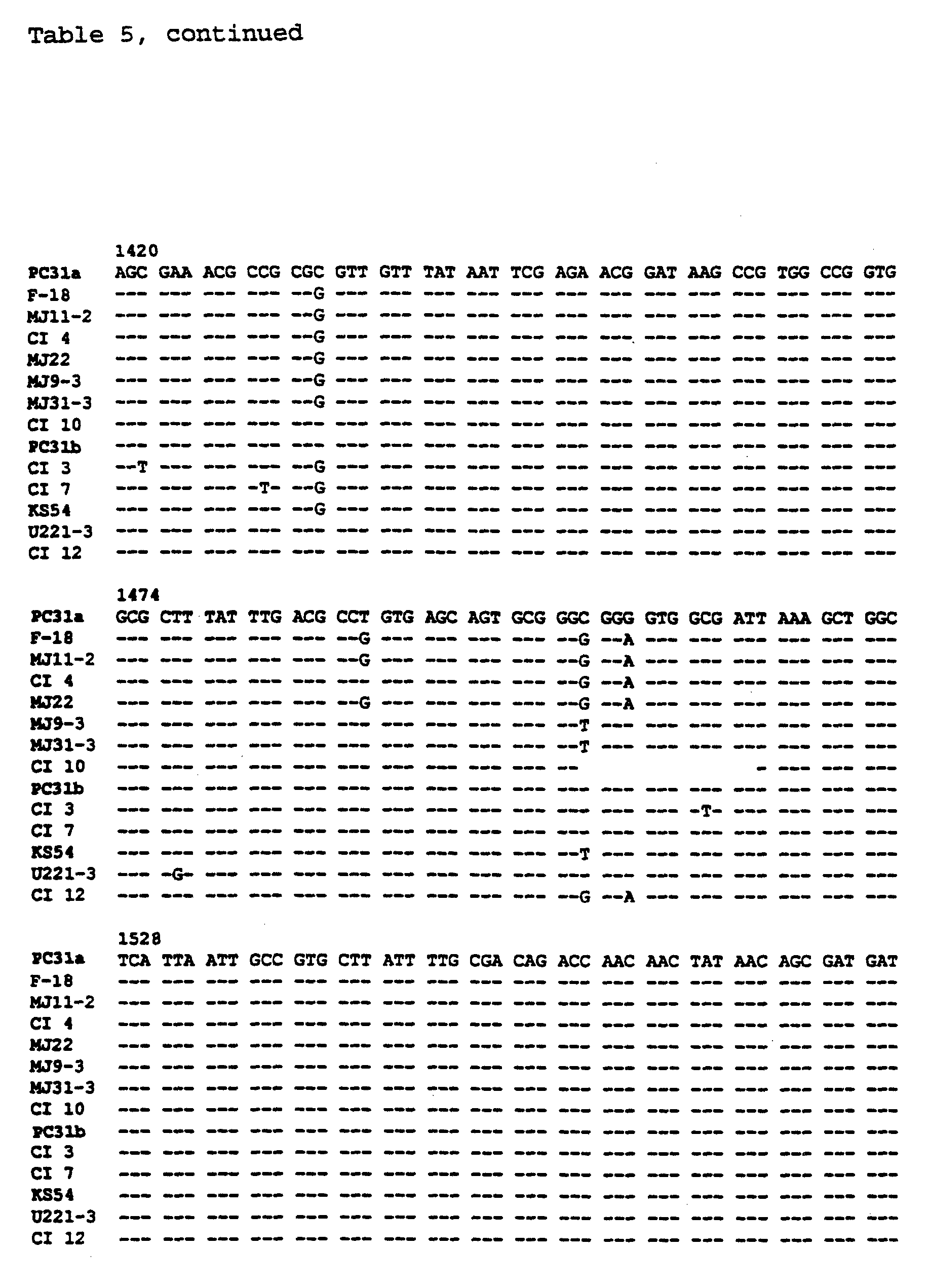
sequences from different FimH adhesins. Such chimeras are constructed e.g. by providing multiple restriction fragments of a fimH gene, followed by exchanging under ligation conditions these fragments with corresponding fragments of an other fimH gene and cloning the ligation product as it is described in Example 1 below. As it is also explained below, recombinant plasmids containing such chimeric fimH genes can be transformed into a host cell and transformants tested for adhesive phenotype, allowing determination of the regions of each gene capable of conferring functional activity (Fig. 5). These studies which are described in details below showed that all of the sequence changes relative to the PC31 fimH gene that affected binding function in the studied strains of E. coli CSH-50 and clinical isolates (CIs) designated #s 3, 4, 7, 10, F-18 and U221-3, respectively, occurred between residues 27 and 119, both included, of the 279 residue, mature fimH sequences.
Accordingly, the invention encompasses in one embodiment a FimH adhesin comprising an amino acid sequence which differs from the E. coli PC31 FimH adhesin by at least one amino acid occurring between residues 27 and 119 of the mature FimH sequence, including a FimH adhesin comprising an amino acid
sequence which differs from the E. coli PC31 FimH adhesin by at least one amino acid occurring between residues 33 and 78 of the mature FimH sequence.
The selected potential receptors for a FimH adhesin as defined above include those animate and inanimate receptors mentioned above for a recombinant bacterial adhesin variant and the potential uses of the FimH adhesins are also the same as those uses described above for this recombinant bacterial adhesin variant. As mentioned above, the invention relates in a further aspect to a recombinant replicon comprising a DNA sequence coding for a recombinant bacterial adhesin variant as defined herein or a DNA sequence coding for a FimH adhesin as also defined herein. Such a replicon is suitably selected from a chromosome or a plasmid. The DNA sequence includes a sequence which is inserted by conventional recombination techniques such as insertion by means of restriction enzymes and subsequent ligation, or the DNA sequence is provided by subjecting a replicon comprising a naturally occurring sequence coding for an adhesin to a mutagenization procedure including site-directed mutagenesis, insertion of a transposable element, mutagenization by radiation or chemical mutagenization, followed by selection of cells comprising a mutated sequence conferring altered binding properties relative to a cell comprising the wild-type sequence.
In preferred embodiments, the recombinant replicon is one having a broad host range including bacterial species naturally occurring in soil, in aquatic environments, on inner and outer surfaces of humans and animals, and which is compatible with replicons occurring in potential host strains.
In one useful embodiment, the recombinant replicon as defined above is one wherein the DNA sequence codes for a FimH adhesin having an amino acid sequence which differs from the E. coli PC31 FimH adhesin by at least one amino acid, including
an adhesin having an amino acid sequence which differs from the E. coli PC31 FimH adhesin by at least 2 amino acid residues, such as an amino acid sequence differing by at least 3 amino acids, preferably by at least 4 amino acids, more preferably by at least 5 amino acids, most preferably by at least 6 amino acids. In further useful embodiments, the amino acid sequence may even differ by more than 6 amino acids such at least 7, preferably by at least 8, more preferably by at least 9, even more preferably by at least 10 and in particular by at least 11 amino acid residues, such as by at least 12 amino acids including by at least 15.
Accordingly, the above recombinant replicon preferably comprises a DNA sequence coding for a FimH adhesin which is at least 90% homologous to the PC31 fimH gene, such as at least 92% homologous, more preferably at least 93% homologous, even more preferably at least 94% homologous, most preferably at least 95% homologous, and in particular at least 96%
homologous, e.g. at least 97% homologous. In particularly interesting embodiments, the adhesin is at least 98%
homologous, including at least 99% homologous such as at least 99.5% homologous.
In a further embodiment, the above replicon comprises a DNA sequence which is a chimeric fimH gene as it has been defined above, comprising DNA from different fimH genes. The replicon can also be one which comprises a further DNA sequence e.g. derived from a microorganism selected from a bacterium, a virus, a protozoan, a fungus and a yeast. This further DNA sequence is e.g. one coding for a heterologous polypeptide, including an epitope, an antibody, a toxic gene product, an enzyme, a pesticidally active gene product and a pollutant-degrading gene product.
In useful embodiments, the replicon as defined herein comprises a DNA sequence which is isolated from an Enterobacteriaceae species, including a DNA sequence which is isolated
from E. coli , a Klebsiella sp., an Enterobacter sp., a
Yersinia sp. or a Salmonella sp.
In addition to being a DNA sequence as defined above, the sequence can be a synthetic sequence constructed by conventional techniques of DNA synthesis.
As it is also mentioned above, the present invention
encompasses a fusion protein comprising a recombinant bacterial adhesin variant or a FimH adhesin as defined above, and a heterologous polypeptide. Such a polypeptide is in useful embodiments an immunologically active gene product i.e. an epitope (antigenic determinant) from a pathogenic organism, which polypeptide, when administered to the body of a human or an animal is capable of stimulating the formation of antibodies therein. A cell in which such an epitope is expressed is advantageously utilized in the preparation of live vaccines. Such vaccines have several advantages over known live vaccines:
Firstly, the epitope forms a structural part of an adhesin which is embedded in a surface organelle of the vaccine cells. This implies that the hybrid DNA sequence coding for the epitope further comprises the means for transporting the epitope, when expressed, to the outer surface of the cell, i.e. translocating it through the cell membrane. This is immunologically highly advantageous, since the epitope will be brought more closely in contact with immunologically competent cells of the body to which the fusion protein-expressing vaccine cells are administered.
Secondly, the adhesin part of the epitope-carrying fusion protein can be selected so as to have specific binding properties whereby the vaccine cell may be targeted to a particular location in the body where an immunological response to the epitope is desirable. The adhesion of the epitope-carrying cell to a particular location or region of the body will in this manner ensure that the cell is retained in the human
or animal body in that particular location for a period of time which is sufficient to obtain the desired immune
response.
In accordance with the invention, a useful cell for expression of the above fusion protein is one selected from a bacterial species which inherently contains an adhesin-carrying surface organelle. Such species include as examples gram-negative species of Enter obacteriaceae such as E. coli , Klebsiella spp, Salmonella spp, Yersinia spp, Vibrionaceae, Hemophilus spp, Bordetella spp and Pseudomonadaceae, and gram-positive species such as Neisseria spp and Streptococcus spp.
The epitope part of a fusion protein according to the invention can be an epitope derived from any pathogenic organism or agent against which it is desirable to develop vaccines. Such pathogenic organisms include viruses, bacteria and eucaryotic organisms such as fungi, yeast or protozoa.
Whereas cells expressing an epitope-carrying fusion protein as defined herein may be used as a live vaccine, it is also within the scope of the invention to provide isolated and/or purified cell surface organelles comprising the fusion protein, including fimbriae and pili, as a vaccine, and it is also contemplated that useful vaccines may be provided wherein cells expressing an epitope-carrying fusion protein have been killed by conventional methods such as formaldehyde treatment or thermal treatment.
In a further embodiment of the invention, the fusion protein according to the invention comprises as the non-adhesin polypeptide part a toxic gene product e.g. having a selective toxic effect on particular cells in the body such as e.g. cancer cells. By selecting the adhesin part as one having a specific binding affinity to receptors in such cells it is possible to have cells expressing the toxic gene product bound selectively to such target cells whereby these cells
may be killed or damaged by the toxic gene product. It is also possible to use isolated or purified cell organelles containing a fusion protein comprising the cell toxic
(cytotoxic) gene product for the purpose of targeting the toxic product.
In a further interesting embodiment, the fusion protein comprises an antibody. Such an embodiment is, inter alia, particularly interesting with respect to the provision of fusion proteins which may be used in affinity purification of biological compounds having binding affinity to the antibody part of the fusion protein. It is contemplated that cells expressing as part of a surface organelle, such a fusion protein may be utilized directly as a means of concentrating a biological compound, or the isolated surface organelles comprising the antibody-carrying fusion protein may be used for this purpose.
Furthermore, the fusion proteins as defined herein are useful as carriers of non-covalently bound compounds such as pharmacologically active, diagnostically active and imaging compounds with the purpose of providing cells or cell organelles carrying the active compounds, which thereby become targetable to particular regions or locations of a body to which these cells or cell organelles are administered. The invention encompasses any combination of a fusion protein as defined herein and an active compound which can be covalently bound to a fusion protein.
As mentioned above, the present invention encompasses in one aspect a recombinant bacterial cell which expresses a recombinant bacterial adhesin variant or a FimH adhesin as defined above. In one specific embodiment, the bacterial cell is one which comprises the above-defined recombinant replicon.
Depending on the field of application of such a cell, it may e.g. be selected from a soil bacterium, an aquatic bacterium, a bacterium which is normally associated with plants, a bacterium which is member of the human or animal indigenous
bacterial flora, or a bacterium which is adapted to colonize certain ecological niches such as e.g. sewage purification plants or certain inanimate surfaces.
The major significant advantages which have been achieved by the present invention is the possibility to provide recombinant bacterial cells which are not only ecologically welladapted to grow in a particular ecological environment, but which are also provided with means for colonizing more permanently in their ecologically natural environment. These means for improved ability to colonize an environment are the adhesins expressed by the bacteria which have been constructed and/or selected so as to enable the recombinant bacterial cell to adhere to or bind to specific receptors in the environment, i.e. the bacterial cells are targeted to that environment. Thereby the bacteria according to the present invention will have an ecologically competitive advantage relative to organisms in the particular environment which do not have surface structures comprising adhesins binding to receptors present in the environment, at least not to the same extent as the bacterial cells according to the invention.
In addition to the environment-specific adhesins which the bacterial cell expresses, the cell will have a phenotype which is desirable in the environment to which it is targeted. As one example, a cell according to the invention which is originally isolated from the human or animal indigenous bacterial flora may typically be one which expresses an epitope including an epitope which is part of a fusion protein expressed by the bacterial cell. As another example may be mentioned a bacterial cell which is isolated from a plant and which expresses a pesticidally active compound such as a Bacillus thuringiensis toxin. Further examples include a plant root-associated nitrogen-fixating bacterium isolated from soil which in accordance with the invention is provided with adhesins improving the capability of the bacterium to become permanently colonized to the roots of a specific plant
or specific plants, or a bacterium which is ecologically associated with an aquatic or terrestrial environment containing pollutants to be degraded or removed.
Accordingly, the recombinant bacterial cell can be derived from any gram-negative or gram-positive bacterium for which a need exists to obtain improved colonization in a particular inanimate or animate environment. Such bacteria include as examples Enterobacteriaceae spp, Hemophilus spp, Neisseria spp, Bordetella spp, Streptococcus spp, Pseudomonadaceae spp, Vibrionaceae spp, Baccilaceae spp.
In certain embodiments of the invention it is advantageous that the present recombinant bacterial cell is provided as one which, when it is administered to a particular location or environment, will not persist in that environment. Accordingly, such a recombinant bacterial cell may further comprise a gene coding for a gene product which, when expressed has a killing or cell function-limiting effect in said cell, the expression of said gene coding for the cell killing or cell function-limiting gene product being regulated in such a manner that the bacterial cell when targeted to receptor in a specific location will be killed or limited in its function in a pre-determined manner. The gene coding for the cell killing or cell function-limiting gene product is suitably regulated by a factor selected from the group consisting of a stochastic event, the presence/absence of a chemical compound in the location, and a physical factor.
In a further aspect, the invention relates to a method of isolating or constructing a recombinant bacterial cell expressing an adhesin having modified binding properties relative to a natively expressed adhesin such as a natively expressed FimH adhesin. As it is defined above, this method comprises identifying in the bacterial cell DNA sequence (s) coding for the binding domain (s) of said natively expressed adhesin and substituting at least one codon herein whereby a modified adhesin molecule is expressed that is different in
at least one amino acid from the adhesin expressed natively, and selecting a bacterial cell expressing the modified adhesin having an altered adhesion phenotype relative to the natively expressed bacterial adhesin. As it is explained in details below, the binding domain can e.g. be identified by constructing chimeric adhesin-encoding genes and screening for cells which by having a region in the adhesin gene replaced by a corresponding heterologous region of a different DNA sequence, acquires a new binding
phenotype. Having identified a binding domain of the natively expressed adhesin, recombinant cells having desirable binding phenotypes may be obtained by substituting one or more codons in the binding domain (s) to obtain expression of recombinant adhesins and selecting cells having the desirable phenotypes. The substitution of codons may be achieved by methods know per se such as site-directed mutagenesis using synthetic oligonucleotides and PCR technology or transposable elements or by conventional radiation or chemical mutagenization.
In certain useful embodiments, the above method includes steps whereby a non-adhesin compound is associated with the adhesin, e.g. a step where a gene coding for the recombinant adhesin is part of a hybrid gene comprising a gene coding for a non-adhesin polypeptide which thereby is expressed with the recombinant adhesin as part of a fusion protein comprising the adhesin. Furthermore, recombinant adhesins resulting from the above method may in specific embodiments comprise a non-covalently bound compound which is associated with the adhesin when expressed.
As mentioned above, the invention also encompasses recombinant bacterial cells having selected binding properties whereby cells with desirable phenotypes can colonize environments where the presence of bacteria having a particular phenotype is advantageous. Accordingly, there is in a further aspect of the invention provided a method of preparing a recombinant bacterial cell that binds to a specific receptor
moiety, comprising introducing into a bacterium that does not produce an adhesin binding to said receptor moiety, a DNA sequence coding for an adhesin binding to the receptor moiety, and selecting a bacterial cell expressing the DNA
sequence.
The primary objective of this method is to provide the means of constructing a bacterial strain having the capacity to colonize an environment, based on a parent strain which has an inherent, useful phenotype in this particular environment but which does not express an adhesin binding to receptor moieties in the environment. Accordingly, the method includes as a first step the isolation of an environmentally adapted bacterium not binding to appropriate receptor moieties and in subsequent steps, the identification of heterologous genes encoding adhesins which bind to receptor moieties occurring in said environment, preferably moieties occurring abundantly, isolating this gene and introducing it into the above parent strain. The adhesin gene may e.g. be a gene coding for a naturally occurring FimH adhesin or a recombinant FimH adhesin as defined above.
In one useful embodiment of the method, the adhesin-encoding gene is introduced by transforming a parent bacterial cell with a recombinant replicon as defined herein. In further embodiments, the method is designed so as to obtain a cell wherein a non-adhesin compound is associated with the adhesin, e.g. by introducing the gene coding for an adhesin as a hybrid gene coding for a non-adhesin polypeptide whereby non-adhesin compound is expressed with the adhesin as part of a fusion protein comprising the adhesin, or by binding non-covalently a compound to the adhesin when expressed.
Besides the above method, an adhesin carrying bacterial cell having an altered pattern of adhesion can be provided by using a selection procedure comprising contacting an appropriately sized population of wild-type adhesin-carrying bacterial cells with a potential receptor moiety to which the
wild-type cells do not adhere, e.g. in a manner as it is disclosed in Example 6 below whereby spontaneously or randomly mutated cells having acquired the ability to adhere to the receptor moiety in question, become progressively enriched. From such an enriched culture, cells with the new adhesion ability can readily be isolated and further characterized.
As it has been explained in details above, one primary objective of the present invention is to provide the means of targeting a compound to a specific location. Accordingly, the invention relates in an important aspect to a method of targeting an adhesin to such a location. The method comprises the identification in the location of a receptor moiety, said moiety preferably being one which occurs abundantly in the particular location, which moiety can recognize and interact with an adhesin, and the isolation of a bacterial cell which is capable of growing in the location and expressing an adhesin which recognizes and interacts with the identified receptor moiety, and administering the cell or the adhesin in an isolated form to that location. The identification of a suitable receptor moiety in a particular location can be carried out in several manners. One example is a screening procedure where cells expressing known adhesins or known isolated adhesins are administered to the location e.g. being isolated cells or tissues of microbial, animal or plant origin or an inanimate surface as defined herein, and screening for binding/adhesion of the tested adhesins e.g. according to adhesion assays as disclosed herein. If binding of one or more adhesins occurs, it is an indication that receptor moieties for that or those tested adhesin (s), is/are present in the location.
Alternatively, available data with regard to the presence and amounts of chemical moieties present on the surfaces of the location may be collected or such data have to be generated, and based upon such data, adhesins which are known to bind to one or more of the identified major moieties are selected and
their binding to this/these structure(s) is tested e.g.
according to the assays as used herein. Chemical moieties which are considered potential adhesin-interacting receptor moieties include as examples glycolipids, glycoproteins, proteins, polypeptides, saccharide moieties and peptides.
In the case no suitable chemical moiety is identified in the location, which is capable of binding to known adhesins or which bind with a sufficient affinity, it is required to construct a library of modified adhesin molecules based on known adhesins which are modified by replacing one or more codons as it is explained herein, and/or such a library provided by constructing synthetic adhesin molecules, and then screening this library for recognition of and interaction with identified location surface moieties. A library of modified FimH adhesins may e.g. be selected for specificity towards a given receptor by running clones of these adhesins through a column or matrix containing the receptor moiety in question or cells or tissues isolated from the location without knowing what the receptor moiety is. The clone (s) expressing the adhesins with affinity to receptor moiety/moieties will adhere/bind to the column or matrix, and can subsequently be isolated therefrom.
It is within the contemplation of the invention that crystallographic analyses of adhesins, whether naturally occurring or constructed as indicated above, is a useful technique for the obtainment of information about adhesin structures that assumingly will recognize and interact with particular adhesin receptor moieties.
In accordance with the invention, one embodiment of the above method is one wherein the isolated bacterial cell expresses an adhesin having modified receptor moiety-binding properties relative to an adhesin natively expressed by the cell, the isolation of the cell comprising identifying in a parent bacterial cell, DNA sequence (s) coding for the binding domain(s) of said natively expressed adhesin and substituting
at least one codon herein, whereby a modified adhesin molecule is expressed that is different in at least one amino acid from the adhesin expressed natively, and selecting a bacterial cell expressing the modified adhesin having an altered adhesion phenotype relative to the natively expressed bacterial adhesin or a method wherein the bacterial cell expressing an adhesin that recognizes and binds to the receptor moiety is a recombinant bacterial cell derived from a parent bacterial cell that does not produce an adhesin binding to said receptor, by inserting into the parent cell a DNA sequence coding for an adhesin binding to the receptor moiety, and selecting a bacterial cell expressing the DNA sequence.
One primary objective of the present invention is the targeting of useful non-adhesin compounds to a particular location. Accordingly, the invention encompasses in an interesting embodiment a method as defined above wherein a non-adhesin compound is associated with the adhesin, whereby said compound is targeted with the adhesin to the location comprising the receptor moieties recognizable by the adhesin.
The compound can be associated with the adhesin by a covalent binding or by any of the above mentioned non-covalent types of molecule interaction forces.
When associated covalently with the adhesin the compound to be co-targeted to the selected location with the adhesin can be an enzyme, an antibody, an epitope or a toxin which is part of a fusion protein comprising the adhesin. A compound which is associated with the adhesin by a non-covalent binding is typically a pharmacologically active, diagnostically active or imaging compound.
Locations to which it is desirable to have an adhesin targeted by the present method include a human or animal surface, a plant surface and an inanimate surface as defined above.
In one specific embodiment of the present method the bacterial cell being administered to the location expresses a recombinant bacterial adhesin variant derived from a naturally occurring parent adhesin, said adhesin variant having altered binding properties relative to the naturally occurring adhesin from which it is derived, the altered binding properties including binding to at least one receptor moiety to which the parent adhesin does not bind. Such an adhesin variant 'is advantageously derived from a naturally occurring adhesin isolated from a cell structure selected from the group consisting of a capsule, a lipopolysaccharide layer, on outer membrane protein, a flagellum, a pilus, a fimbria, a non-fimbrial adhesin (NFA) or an afimbrial adhesin (AFA).
In specific embodiments of the invention, the above adhesin variant as used in the present method is a protein having an amino acid sequence differing in at least one amino acid residue from its parent protein adhesin such as a FimH adhesin having an amino acid sequence which differs from the E. coli PC31 FimH adhesin as defined herein in at least one amino acid. Such a FimH adhesin includes an adhesin which binds to a receptor selected from the group consisting of a domain where mannosyl residues are not terminal and a domain devoid of saccharide and an adhesin variant which is at least 90% homologous to the PC31 FimH adhesin as defined herein, such as at least 92% homologous, more preferably at least 93% homologous, even more preferably at least 94% homologous, most preferably at least 95% homologous, and in particular at least 96% homologous, e.g. at least 97% homologous. In particularly interesting embodiments, the adhesin is at least 98% homologous, including at least 99% homologous or at least 99.5% homologous.
The above FimH adhesin can be a chimeric adhesin as defined above, comprising amino acid sequences from different FimH adhesins and constructed according to the methods below.
In accordance with the invention, an adhesin can be administered to a location in the form of an adhesin-expressing bacterial cell. Such a cell is one capable of growing in that particular location. Accordingly, the bacterial cell is suitably derived from a bacterial species which is normally occurring in the location including human or animal body surfaces, plant surfaces such as plant root surfaces and inanimate surfaces. In this context, an animal body surface includes the insect gut, whereto it is desirable to administer a bacterial cell expressing an insecticidally active toxin.
Thus, if it is desired to administer the bacterial cell to the root of a plant, a suitable bacterial cell is preferably isolated from a strain which has colonized the rhizosphere of that plant to a large degree, i.e. the strain is a major member of the natural plant root flora. Such an isolate is then provided with a gene coding for an adhesin which will recognize and interact with an abundantly occurring moiety on the roots of said plant. In this manner, a suitable adhesin which is expressed naturally in a bacterium which is not adapted to grow in a plant rhizosphere, becomes expressible in a normal inhabitant of the rhizosphere environment (location).
In specific embodiments of the present method of targeting a bacterial adhesin to a specific location, the adhesin is a FimH adhesin as defined above, having an amino acid sequence which differs from the E. coli PC31 FimH adhesin as defined herein in at least one amino acid.
In an interesting embodiment, the adhesin-carrying bacterial cell being targeted is a cell which further comprises a gene coding for a gene product which, when it is expressed, has a killing or cell function-limiting effect in said cell, the expression of said gene coding for the cell killing or cell function-limiting gene product being regulated in such a manner that the bacterial cell, when targeted, will be killed
or limited in its function in a pre-determined manner. The expression of such a "suicide" or cell function-limiting gene may suitably be regulated by a factor selected from the group consisting of a stochastic event, the presence/absence of a chemical compound in the location and a physical factor. As examples of such "suicide" or cell function-limiting genes providing the means of biological containment, may be mentioned those disclosed in WO 87/5932 and WO 93/20211
Furthermore, the present Designer Adhesin Technology (DAT) provides very useful means of obtaining colonization with desirable bacteria in a particular environment with the purpose of obtaining beneficial changes of the microbial flora in the environment. As one example, certain bacterial species in the gastrointestinal (GI) tract of humans and animals have beneficial effects on the health condition of the host organism e.g. by suppressing pathogenic organisms or by contributing to the digesting of certain diet components. The present technology makes it possible to select particularly useful bacteria from the GI-tract and have them
designed in accordance with the present invention, to have improved colonization abilities. Similar examples include desirable bacterial colonizations of biological sewage purification systems, plants where invasion of pathogenic organisms may be controlled by colonizing the plants with harmless bacteria, and teeth where caries may be controlled by colonizing the dental enamel with bacteria suppressing those causing the caries attacks.
In another industrially interesting aspect, the invention provides the means of isolating a compound from a solution or suspension containing the compound. The method comprising contacting the solution or the suspension with a fusion protein as defined herein wherein the heterologous
polypeptide has an affinity to the compound to be isolated.
Furthermore, the invention provides a composition comprising a population of a bacterial cell as defined herein.
The invention is further illustrated in the below Examples and the Figures, wherein
Fig. 1 is a schematic model for the construction of recombinant plasmids pGBl-24 (containing fimH from Cl #10) and pGB2-24 (containing fimH from PC31) used for transforming E. coli AAEC191A(pPKLll4) with cloned fimH genes. Plasmid pGB224 was used as the vector for all other cloned fimH genes described herein;
Fig. 2 is a restriction map of fimH genes. Five unique restriction sites are present in the PC31 fimH gene. Numbers in parentheses following enzymes are the base pair positions of the cut sites. Some of these sites are found in the other fimH genes, as marked. Chimeric genes were produced by exchanging each available restriction fragment from the other five fimH genes with corresponding fragments in the PC31 gene and then recombinant strains expressing resulting chimeric fimH subunits were tested for adhesion. Fragments indicated by boxes are those which conferred MF or MFP adhesive
phenotypes on the chimeric genes; Fig. 3 illustrates adhesion of representative "wild-type" (A) and recombinant (B) M-class, MF-class and MFP-class strains to Mn (1), Fn (2), periodate-treated Fn (3) and to FnSpl (4). Strain designations given for the "wild-type" strains are given in AS. Strain designations KB31, KB12, KB4, KB7, KB50 and KB10, are for recombinant strains of AAEC191A(pPKL114), which is fimH- , after transformation with plasmids that contain fimH+ from strains HB101 (pPKL4), CI #12, Cl #4, CI #7, CSH-50 and Cl #10, respectively. Open columns indicate results when bacteria were incubated in buffer without D-mannose, while solid columns are results in the presence of D-mannose. Values indicated are the mean ± S.E.M. (n=4) for each column;
Fig. 4 illustrates the adhesion of representative M-class, MF-class and MFP-class strains (CIs #12, #4 and #10, respect
ively) to Fn fragments prepared by thermolysin treatment as described in ref. 51. Columns labelled 1-5 indicate adhesion to: 1) NH2-terminal 30-kDa domain; 2) the 55-kDa gelatin-binding domain; 3) the 110-kDa cell attachment domain; 4) the 29-38-kDa heparin binding domains; and 5) the 20-kDa COOH-terminal domain. Open columns represent adhesion in the absence of D-mannose; solid columns represent adhesion in the presence of D-mannose. Mean ± S.E.M. (n=4);
Fig. 5 is a composite figure illustrating comparison of amino acid sequences of FimH adhesins and active restriction fragments of fimH genes. The published nucleotide and deduced amino acid sequence of the PC31 fimH gene and gene product (ref. 27) serve as prototype. Numbered amino acid residues shown above the model of the PC31 FimH represent residues that are different in other FimH subunits due to amino acid substitution or deletion. Standard one-letter code applies and residues in the other FimH sequences that are different are indicated. Deleted amino acids are indicated by Δ. It should be noted that residue 176 is not arginine as published previously (ref. 27) for the PC31 FimH, but proline. Regions of the FimH subunits conferring change in adhesive phenotype, highlighted in bold, were determined by functional assays performed on chimeras between the "classic" mannose-specific PC31 fimH gene present in HB101(pPKL4) and the above
described genes. Residues predicted to be key in conferring receptor specificity are circled. Approximate positions of unique restriction sites used to create chimeras are indicated along the bottom of the model;
Fig. 6 illustrates plasmid pPKL4 which is a derivative of pBR322 (thick line) carrying the entire fim operon (FimA-H) including the regulatory genes fimB and fimE (not shown), and the promoter region with the SnaBI site. In this plasmid an 8mer linker with an BglII site was inserted in the SnaBI site to create pPKL83;
Fig. 7 illustrates the construction of plasmid pSM1314; the vector pVLT33 is a derivative of the broad host range replicon RSF1010. Plasmid pPKL83 was digested with BglII and pVLT was digested with BamH1; the two were ligated and pSM1314 was the resulting plasmid in which expression of the fimA-H cluster is under the control of the tac promoter;
Fig. 8 illustrates plasmid pLPA22 and derivatives hereof as used in this study. The triangles indicate the position of translational stop-linkers in the fimH gene in plasmid pPKLH5. The positions of heterologous inserts are indicated (black boxes). Small triangles indicate signal-peptide encoding sectors.
Fig. 9 illustrates plasmids pLPA29, pLPA30, pLPA36, pLPA58, pLPA59 and pLPA98; Fig. 10 shows immuno-electron microscopy with colloid gold labelling of E. coli HB101 cells containing plasmids pLPA22 plus pPKL115 (a), pLPA37 plus pPKL115 (b), pLPA38 plus pPKL115 (c), using anti-pre-S2 monoclonal antiserum. Bar, 0.1 μm. EXAMPLE 1
Functional heterogeneity of type 1 fimbrial adhesins due to minor sequence variations among fimH genes l.l. Materials and methods
1.1.2. Reagents Yeast Mn, a polymannosylated glycoprotein isolated from
Saccharomyces cerevisiae cell walls, was obtained from a commercial source (Sigma Chemical Co, St. Louis, MO, U.S.A.). Mannan is composed of an N-linked backbone of β1, 2-linked mannopyranose units with α-linked mannopyranose side chains (ref. 38). The majority of the carbohydrate of human plasma
Fn is composed of N-glycosidic complex-type biantennary glycans and no high mannose-type or hybrid-type N-glycans have been described (refs. 30, 45, 54). Human plasma Fn and Fn fragments were purified as described previously (refs. 5, 15, 51, 58). Periodate treatment was performed as described previously (ref. 51). The synthetic peptide, FnSpl, copying the first 30 amino acid residues of the Fn molecule (EAQQMVQPQSPVAVSQSKPGCYDNGKHYQI) was synthesized in the Protein
Chemistry Laboratory of the VA Medical Center, Memphis, TN (SEQ ID NO:2). The saccharide content of the four substrata was characterized using two lectins, concanavalin A (ConA), well known to react with terminal and internal mannosyl residues, and the Calanthus nivalis agglutinin (GNA), which recognizes only terminal Manαl-3Man, Manαl-6Man and Manαl-2Man sequences (E. Y. Laboratories, San Mateo, CA). Immobilized Mn and Fn both reacted with ConA, whereas GNA bound only to Mn. These results are consistent with the known structures of the oligosaccharide moieties of these two compounds. Neither lectin reacted with immobilized FnSp1.
Periodate treatment (ref. 51) of Mn or Fn eliminated lectin reactivity.
1.1.3. Bacterial strains and plasmids
The CSH-50 strain (lambda-, F -araA (lac-pro) rspL thi
fimE: :IS1) is a Cold Spring Harbor K12-derived strain (ref. 35). The E. coli strain MG 1655 (CGSC6300; K12 derivative, lambda-, F-) and a derivative strain AAEC191A (MG1655 recA Afim were generously provided by Dr. Ian Blomfield (Bowman Gray University, Winston-Salem, NC). AAEC191A has had the entire fim gene cluster deleted by allelic exchange (ref. 8). Clinical isolates (CIs) were urinary tract isolates obtained from the clinical microbiology laboratories of the Memphis VA Medical Center or The City of Memphis Hospitals, Memphis, TN. The 12 CIs used in this study were selected on the basis of MS agglutination of yeast cells after growth in broth, a classic test for type 1 fimbriae.
Plasmid pPKL4, a pBR322 derivative containing the entire fim gene cluster from E. coli strain PC31 (K12-derivative, gal tonA phx ara) and encoding for the expression of fully functional type 1 fimbriae in HB101 (supE hsdS recA ara proA lacY galK rspL xyl mtl AfimBE) , has been described previously (ref. 28). pPKL114 is a recombinant plasmid derived from pPKL4, but with a translational stop-linker inserted into the Kpnl site in the fimH gene. No transcriptional effects of the stop-linker are to be expected. Antibiotics were used at the following final concentrations: ampicillin (50 μg/ml), kanamycin (60 μg/ml) and chloramphenicol (30 μg/ml).
1.1.4. Polymerase chain reaction
Oligonucleotide primers were designed using the published sequence for the fimH gene in pPKL4 (ref. 27). The 5' primers copied regions 13 and 49 bp upstream from the fimH gene and were extended on the 5' end by an Apal1 restriction site and a GC clamp: Primer 1: 5'-GGGGG-GTGCAC-ACC TAC AGC TGA ACC CGG-3' (SEQ ID NO:3); Primer 2: 5'-GGGG GTGCAC T CAG GGA ACC ATT CAG GCA-3' (SEQ ID NO:4). The 3' primers copied 18 bases of the bottom strand of the fimH gene that encode for the 6 terminal amino acids of fimH and were extended by an Fspl or Sphl site and a GC clamp: Primer 3: 5'-GGG TGCGCA TTA TTG ATA AAC AAA AGT CAC - 3' (SEQ ID NO: 5); Primer 4: 5'-GGG GCATGC TTA TTG ATA AAC AAA AGT CAC-3' (SEQ ID NO: 6) . Primer 1 and 3 were used for Cl #10 and pPKL4, primer 1 and 4 were used for Cl #4 and CSH-50 and primer 2 and 4 were used for Cl #s 7 and 12 to generate PCR products from plasmid or
chromosomal DNA prepared from E. coli expressing different functional classes of type 1 fimbriae. The PCR reaction mixture consisted of template DNA, primer pairs, dNTPs, and Taq DNA polymerase in PCR buffer. The PCR was performed in a Perkin-Elmer Cetus automatic thermal cycler with denaturation at 96°C for 1 min., primer annealing at 55°C for 1 min., and primer extension at 72°C for 2 mins. for a total of 40 cycles. All of the PCR products migrated similarly in agarose
gels. Purification, restriction and ligation of DNA was performed using standard procedures (refs. 39, 48). All primers for PCR and for nucleotide sequencing were produced by the Molecular Resources Center, UT, Memphis. 1.1.5. Subcloning
The PCR products from CI#10 and from pPKL4 were cut with respective restriction enzymes and ligated into the Apall and Fspl restriction sites of plasmid pACYC177 (New England
Biolabs, Beverly, MA, U.S.A.) which is compatible with the pBR322-based pPKLH4 to be used in complementation experiments, creating plasmids pGB1 and pGB2, respectively (Fig. 1). However, it became inconvenient to use pACYC177-based plasmids because of a high frequency of appearance of spontaneous Kmr in the AAEC191A host strain. The origin of this problem is not entirely clear, but it was avoided by subcloning the fimH genes from pGB1 and pGB2. The inserts and upstream regions of pACYC177 containing the tet promoter were cut from pGB1 and pGB2 with Fspl and BamH1 and subcloned into the polylinker site of pGEM-3Z (Promega, Madison, WI) that had been cut with BamH1 and Hinc2, creating plasmids pGBl1 and pGB2-1 respectively. pGEM-3Z was simply used as a convenient intermediate in subcloning into pACYC184.
The inserts were cut out again using Smal and Hind3 and subcloned into pACYC184 (New England Biolabs, Beverly, MA) cut with Hinc2 and Hind3, creating plasmids pGB1-2 and pGB2-24 containing the fimH genes from CI#10 and pPKL4, respectively. These plasmids complement the non-adhesive defect of AAEC191A(pPKL114) giving the adhesive phenotypes of the parental strains (see Results). Cutting the fimH gene from pGB2-24 using Apal1 and Sph1 makes it possible to easily insert other fimH genes obtained by amplifying chromosomal DNA of other isolates by PCR. All recombinant strains we have tested thus far using this technique exhibit the same adhesive phenotype as the parent strains from which the fimH genes were cloned.
1.1.6. Construction of chimeric fimH genes
Unique restriction sites (Fig. 2) were used to construct chimeric fimH genes between the prototypical MS pPKL4 fimH gene, used as genetic background, and restriction fragments obtained from the newly described fimH genes. Fragments were purified from agarose gels and ligated into restriction
"spaces" generated in the pPKL4 fimH gene present in pACYCl84 (pGB2-24). Each chimera was analyzed by restriction mapping and the nucleotide sequences of bridging segments were determined to ensure proper constructions. The plasmids containing chimeric fimH genes were transformed into AAEC191A(pPKL114) and clones were tested for agglutination of yeast cells and for adhesion to Mn, Fn and FnSp1.
1.1.7. Nucleotide sequencing The nucleotide sequences of fimH genes were determined by the dideoxynucleotide chain termination method of Sanger (ref. 49) using a Sequenase II® kit (U.S. Biochemical Corp., Cleveland, Ohio) and [α-35S] dATP (800 to 1 , 000 Ci/mmol) according to the manufacturer's suggestions. The amino acid sequences were deduced from nucleotide sequences using MacVector® protein and DNA analysis software (Eastman Kodak, Rochester, NY). To ensure fidelity of the PCR amplification, selected fimH genes were re-amplified, cloned, tested for activity and re-sequenced. More recently, we have used the fmol™ Polymerase Sequencing System (Promega, Madison, WI), because it is useful with small amounts of DNA and thus subcloning the fimH genes from the pACYCl84-based plasmids to high copy number plasmids was obviated. Bands were visualized by autoradiography of sequencing gels and compared with the published fimH gene sequence (ref. 27).
1.1.8. Yeast cell aggregation assay
E. coli were tested for their ability to aggregate yeast cells. Commercial baker's yeast, Saccbaromyyces cerevisiae,
was suspended in PBS (5 mg dry weight/ml). E. coli were washed in PBS , resuspended to an OD530 of 0.4 , and mixed with the yeast cell suspension in PBS with or without 1% D-mannose. Aggregation was monitored visually and the titer recorded as the last dilution giving a positive aggregation reaction.
1.1.9. Adhesion assays
Adhesion assays were performed as described previously (ref. 51). Briefly, microtiter assay wells were coated with
receptor molecules as indicated in the text and figure leg-ends. After the wells were washed two times with PBS, 100 μl bacterial suspensions were added in 0.1% BSA-PBS. After incubation at 37°C for indicated times, wells were washed three times with PBS and adherent bacteria were detected by using rabbit anti-F. coli serum. Antibody binding was
detected using peroxidase-conjugated goat anti-Rabbit IgG. Reaction product generated from the 5-aminosalicylic acid substrate was measured at 405 nm after 10- 15 minutes by using an automatic microplate reader (Molecular Devices, Inc., Menlo Park, CA). Values reported are corrected for background reaction using BSA coated plates as control.
1.2. Results
In a previous publication it was reported that type 1
fimbriae of E. coli CSH-50 and HB101(pPKL4) differ functionally in their pattern of adhesion to Mn, Fn, periodatetreated Fn and a synthetic peptide, FnSpl, immobilized on plastic (ref. 51). Since CSH-50 and HB101(pPKL4) are laboratory strains, we tested 12 clinical E. coli isolates (CIs) obtained from human urine for adhesion to these four substrata. All of the CIs agglutinated yeast cells in a MS fashion. Five of the twelve CIs adhered only to Mn. The adhesive activity of HB101(pPKL4) and of Cl #12 are shown as examples of this class, which we have tentatively designated as M class (Fig. 3A). Three of the 12 CIs adhered to Mn and Fn,
but not to periodate-treated Fn or to FnSpl. The adhesive activities of Cl #s 4 and 7 are shown as examples of this class, designated as MF class. Three of the twelve CIs adhered to each of the substrata. The adhesive activities of CSH-50 and Cl #10 are shown as examples of this class, designated as MEP class.
Adhesion of strains representing these three classes to Fn fragments further illustrates the distinct differences between the three classes. The M class Cl #12 does not adhere to any of the Fn fragments (Fig. 4). The MF class Cl #4 adheres to the 40-kDa gelatin-binding fragment. The MFP class Cl #10 adheres, with only slight differences, to all 5 fragments of Fn tested. Periodate treatment eliminated binding of Cl #4 to domain 2, but had no effect on the binding of Cl #10 to any of the Fn domains (data not shown).
Since the fimH subunit has been shown to mediate the mannosesensitive activity of type 1 fimbriae, we focused our initial efforts to elucidate the molecular basis for the observed functional heterogeneity on the fimH gene. fimH genes were amplified from chromosomal (or plasmid, for pPKL4) DNA and the genes were cloned into pACYC177 and subcloned into pACYC184 under control of the β-lactamase promoter of
pACYC177, according to Materials and Methods (Fig. 1)
The adhesive phenotypes conferred by the fimH genes were tested in the following way. E. coli K-12 strain AAEC191A (Δfim) was first transformed with plasmid pPKLH4, which contains an intact fim gene cluster but with a translational stop-linker inserted into the last gene, fimH. This derivative produces morphologically normal fimbriae that are non-adhesive due to absence of the FimH subunit. Plasmids harbouring cloned fimH genes were transformed into E. coli
AAEC191A(pPKL114) and the resultant strains were tested for their ability to adhere to Mn, Fn, periodate-treated Fn and to FnSpl (Fig. 3B). Each of the recombinant strains displayed adhesive phenotypes mimicking those of the representative
parent strains from which the fimH genes were obtained. fimH genes were cloned from each of the other 8 CIs and similar results were obtained with the adhesion of recombinant strains mimicking that exhibited by the parental CIs. The complete nucleotide sequences of each of the six representative fimH genes were determined and the amino acid sequences of the fimH proteins were deduced as it is shown in Table 1 below which is a representation of amino acid
sequences of the FimH subunits deduced from nucleotide sequences of selected fimH genes disclosed in this example [CI#3 (SEQ ID NO.-33), CI#4 (SEQ ID NO:29), CI#7 (SEQ ID
NO:30), CI#10 (SEQ ID NO:31) and CI#12 (SEQ ID NO:28)] and those of the F. coli K12 strain PC31 (SEQ ID NO:1) and F. coli strain CSH-50 (SEQ ID NO:32). Additionally, the FimH amino acid sequences of the following clinical isolates of F. coli are shown: KB21 (SEQ ID NO:27), KS54 (SEQ ID NO:35), U221-3 (SEQ ID NO:36), MJ#9-3 (SEQ ID NO:37), MJ#31-3 (SEQ ID NO:38), MJ#11-2 (SEQ ID NO:39), MJ#2-2 (SEQ ID NO:1) and F-18 (SEQ ID NO:34). Standard one-letter code applies. Deleted amino acid residues are indicated by Δs. M, ML, MF, MFP, and MR indicate the adhesin class as defined above.
and gene products should also be identical, subunit incorporation into the fimbrial superstructure should not vary significantly. These results emphasize that in these experiments it is the FimH subunit that determines receptor specificity. In comparing the new FimH sequences to the one published previously (ref. 27), the only structural alteration that can be clearly linked to a functional change, without resorting to analysis of chimeric fimH genes, is the non-conservative substitution of arginine
58 in the MFP class CSH-50 FimH subunit for leucine
58 in the M class PC31 FimH subunit. Since each of the other FimH sequences had more than one change, it was necessary to construct chimeric genes to begin to focus on functionally relevant changes.
In the case of the Cl #10 FimH, an MFP class adhesive activity similar to that of CSH-50 is conferred by a different region of the gene which encodes for a subunit deleted of residues 116-119. It remains to be determined how two distinctly different structural changes can bring about apparently similar changes in receptor specificity. It is possible, of course, that as additional receptor molecules are tested, these two variants will be found to be functionally distinct.
The Apal1 - Tth111I fragment of the Cl #7 fimH gene confers MF class activity in the CI#7/PC31 fimH chimera. Since the asparagine16-threonine16 substitution is within the leader sequence and thus not represented in the mature protein, the histidine33- asparagine33 substitution must be of functional importance for the MF class Cl #7 FimH. Comparison of the active regions of the MF class Cl #4 and the M class Cl #12 FimH subunits suggests the importance of the glutamic acid73- glycine73 substitution for MF class activity of the Cl #4 FimH. Thus, histidine33, arginine58, glutamic acid37 and deleted glycine116-isoleucine119 appear to be key residues in the functional activity of FimH subunits of Cl #7, CSH-50, Cl #4 and Cl #10, respectively, but a more precise demonstration
of which residues are involved and how they affect the ligand-binding cleft(s) remains to be performed.
At first glance, the FimH mediated, mannose-sensitive protein-binding activity of type 1 fimbriae is the most surprising of the adhesive phenotypes described here. However, protein-binding activity of FimH (i.e. PilE) subunits was noted earlier in a study characterizing mutT-induced mutations in the fimH (pilE) gene (Harris et al., ref. 22) However, the protein-binding activity described by Harris et al. was not mannose-sensitive. It is presently not known whether the protein-binding activity described herein is in addition to or separate from the mannose-binding activity, but the concept of bifunctional properties of lectins has been established for several years (ref. 6). While the MFP class type 1 fimbriae appears to react somewhat promiscuously with most Fn fragments, the reaction does not appear to be non-specific. For instance, the MFP class CSH-50 type 1 fimbriae do not adhere well to gelatin (ref. 51). Further, the adhesion to ovalbumin is sensitive to both periodate and glycosidase treatment (ref. 51). Further work will be required to determine the consensus amino acid motif reactive with this class of FimH subunit.
Previous studies suggested that the combining site of the E. coli FimH adhesin is in the form of an extended pocket corresponding to the size of a trisaccharide with an associated hydrophobic region (ref. 16). The MS nature of all of the adhesive interactions described suggests that if the combining sites are separate, they may be close to each other.
However, it remains to be determined whether or not the mannose effect is direct or allosteric. Conformational changes that frequently occur in lectins upon binding the saccharide ligand (ref. 46) could affect a second, distant binding site. Site-directed mutations may be sufficient to clarify which structural changes result in changes in
receptor specificity. However, such studies are unlikely to shed much light on how the structural changes actually relate

The nucleotide and deduced amino acid sequences of the pPKL4 fimH gene are identical to that reported previously, except that residue 176 is not an alanine residue as previously reported, but a proline residue. Independent re-amplification, re-cloning and re-sequencing confirmed this sequence for the pPKL4 fimH gene. Sequencing was also repeated on independently amplified and cloned isolates of the Cl #10 and Cl #7 fimH genes to confirm sequence fidelity and no errors were found. The nucleotide and deduced amino acid sequences of the other fimH alleles described in this Example are > 98% conserved, but there is more than one amino acid residue difference in all but one of the new fimH sequences when compared to the published pPKL4 sequence. To focus on the sequence differences that resulted in changes in functional activity, advantage of unique restriction sites were taken (Fig. 2) to construct chimeric fimH genes. Multiple restriction fragments covering the entirety of each of the sequenced fimH genes were exchanged with corresponding fragments in the prototypical fimH gene of E. coli strain PC31 that was amplified from pPKL4, cloned into pACYC184 and used as the genetic background. Recombinant plasmids containing the chimeric fimH genes were transformed into E. coli AAEC191A(pPKL114) and transformants were tested for adhesive phenotype, allowing determination of the regions of each gene capable of conferring functional activity (Fig. 5). All of the sequence changes that affected function occurred between residues 33 and 119 of the 279 residue mature fimH sequence.
1.3. Discussion The functional heterogeneity which is described above must be due entirely to allelic variants of the fimH gene. The only variables in the recombinant strains which are described in this Example are the fimH genes; all other genes necessary for fimbrial subunit synthesis, transport and assembly are the same in each case. Since the ratios of the various genes
to the ligand-binding cleft (s) and it will ultimately be necessary to determine the 3-dimensional structure of FimH or FimH fragments crystallized in the presence of ligand to fully understand structure/function relationships. The three adhesive classes of type 1 fimbriae identified above may understate the functional heterogeneity of type 1 fimbriae. The group of CIs that has been tested in this
Example is small and only a few substances have been tested as potential receptors. A larger group of isolates tested against additional receptor candidates might yield additional functional classes. Preliminary studies with MS Enterobacter aerogenes and Klebsiella pneumoniae strains exhibiting MF class and MFP class activity suggest that heterogeneous receptor specificities will also be found among other type 1 fimbriated enterobacterial species.
It is also believed that it is possible that adhesins from some fimbriae responsible for mannose-resistant hemagglutination or adhesion are structurally related to FimH, but with sequence alterations that eliminate sensitivity to mannose. The possibility that the MS lectin-like properties of FimH might be eliminated while retaining other adhesive properties of FimH (e.g. pellicle formation) has been shown previously in a study characterizing mutT-induced mutations in the fimH (pilE) gene (ref. 26). At the minimum, it is believed that tests for type 1 fimbriation should include additional functional characterization. While all type 1 fimbriae-mediated adhesion which have been described in this Example is
mannose-sensitive, it is not all mannose- or even saccharidespecific as has commonly been thought. Further studies of type 1 fimbriae as a virulence factor must be able to distinguish among the various functional classes.
Allelic variation of the so-called G adhesins of P fimbriated uropathogenic E. coli also results in different functional classes, but the requirement for the Galα1-4Gal sequence within isoreceptors is maintained (refs. 52, 53). These
differences in G adhesin receptor specificity appear to be rather subtle, at least superficially, when compared to the differences in FimH receptor specificities. Yet there is significantly greater sequence homology among the fimH genes than among the G adhesin genes, some of which share less than 50 percent homology. The G adhesin receptor specificities affect host susceptibility, due in large part to host-specific expression of glycolipid isoreceptor variants. Whetherthe FimH family of adhesins bears a similar relationship to host susceptibility or tissue tropism remains to be determined. In this regard it is possible that the G adhesin family could exhibit additional receptor specificities not restricted to the Galα1-4Gal sequence. The lectin-independent affinity of P fimbriae for immobilized Fn is not dependent on the G adhesin, but on two other minor subunits, E and F, neither of which bear significant homology to FimH (refs. 56, 57).
It is important to point out that the degree of functional heterogeneity of type 1 fimbriae described in the present Example was not appreciated when any of the studies cited above were performed. The results of these studies have made it clear that structural and functional heterogeneity occurs within the class of adhesive organelles commonly referred to as MS or type 1 fimbriae and that the adhesive diversity will lead to a broader spectrum of receptive surfaces for potential colonization. The surprising finding that a FimH family of adhesins exists may prove to be an important step toward unravelling the role(s) type 1 fimbriae may play in the ability of enterobacteria to reach their normal habitat or gain entry into deeper tissues, where devastating effects can occur.
EXAMPLE 2
Expression of type 1 fimbriae in heterologous bacterial species
The fim operon of E. coli comprises a cluster of genes covering about 8 kb of DNA. This operon has been isolated and cloned on plasmids in its entirety. The promoter upstream of the fimA gene is located within an invertible DNA sequence, which in E. coli leads to a switch on/switch off situation for fimbrial synthesis. In one orientation of the invertible sequence the promoter is directed towards the fim genes, and the cell is fimbriated; in the other orientation the promoter is directed in the opposite direction, and the cell is non-fimbriated.
Since the regulation of the switch of the invertible promoter sequence is very complex and involves several genes outside the fim operon it is far from certain that the switching takes place in other bacteria than the enterics. It was therefore considered necessary to insert a replacement promoter for the expression of the fim genes, and as a model for gene expression in a number of different bacterial species the lac promoter was chosen. This promoter has been shown to be active and regulatable in many bacterial species.
Plasmid pPKL83 is a derivative of pPKL4 (ref. 27) carrying the entire fim operon in pBR322, in which the promoter has been destroyed by inserting a BglII linker in the SnaB site located in the promoter sequence. There is a second BglII site in plasmid pPKL83 upstream of the fim operon (Fig. 6). Plasmid pVLT33 (Fig. 7) is a kanamycin resistant derivative of the broad host range plasmid RSF1010, carrying the lacI q gene and the tac promoter placed upstream of a multiple cloning site in which a unique BamHl site is placed. The two plasmids were ligated together after digestion of pPKL83 with BglII and pVLT33 with BamH1. In one orientation (pSM1314), this fusion plasmid will express fimbriae in the presence of
IPTG due to the fusion between the fimbrial genes and the lac promoter.
The correct orientation of the fusion plasmid pSM1314 was verified by transforming it into a strain of E. coli which carries a deletion of the fim operon. Production of fimbriae was assayed in two ways: 1) Cell aggregation with fimbrial antibodies and 2) ELISA assay of whole cells. The former analysis is rather simple: to a small volume (10 μl) of an outgrown or IPTG-induced culture of the cells to be tested is added a small volume (2 μl) of antibodies raised against fimbriae, on a glass slide. After mixing the samples, fimbriated cells begin to show cell aggregates which are easily observed directly as clumps or under a microscope. No clumping was observed with cells of the strain with a fim deletion, whereas pSM1314 transformants of this strain showed clearly detectable cell aggregates. The ELISA analysis of whole cells confirmed the aggregation assay. In Table 2 below the readings from this type of assay are presented, and they show quantitatively the occurrence of Fim antigens on the cells as a result of IPTG induction of the pSM1314 carrying strain.
Table 2. Results (duplicate) of ELISA assay for type 1 fimbria expressed by pSM1314 in E. coli AAEC191 (OD492)
AAEC191(pSM1314) 0.145/0.164
AAEC191(pSM1314)+IPTG 1.026/1.260
Blank 0.113/0.095
Plasmid pSM1314 also carries a mob site which allows it to be transferred to other gram negative bacteria provided a helper plasmid is introduced. This type of transfer is most easily performed in "triparental" matings in which a donor strain
(E. coli carrying pSM1314), a helper strain (E. coli carrying a plasmid with conjugation genes) and a recipient strain carrying a selectable marker not present in any of the two
other strains, are mixed on a plate (directly or on a filter). After some growth (often overnight) this mixture is spread on selective plates with antibiotics that only allow the recipient carrying the desired plasmid to grow and form colonies.
In the present context, the E. coli strains MC1000 (pSM1314) and MC1000 (pRK2013) and (as recipient) Enterobacter cloacae strain A50 Nalr (ref. 67), were mated. This recipient strain is resistant to nalidixic acid. After selection for growth on plates with kanamycin plus nalidixic acid the resulting clones were grown in liquid medium and assayed for the presence of fimbriae in the absence/presence of IPTG. The cell aggregation assay was employed.
This assay showed that fimbriae were produced in the Enterobacter cloacae strain and were present on the cell surface; however, full repression of expression from the tac promoter was not obtained, most likely due to an increased escape synthesis. The results showed that E. coli type 1 fimbriae may be synthesized and processed correctly for pili formation on the surfaces of heterologous gram-negative bacterial species.
The plasmid pSM1314 in E. coli HB101 was deposited on 26 January 1994 with DSM, the Deutsche Sammlung von Mikroorganismen und Zellkulturen GmbH, (German Collection of Micro-organisms and Cell Cultures), Mascheroder Weg 1B, D-38124 Braunschweig, Germany, under the accession number DSM 8922.
EXAMPLE 3
The construction of fimH-fusion genes and the expression of mannose-sensitive FimH fusion proteins Heterologous sequences mimicing the pre-S2 region of the hepatitis B viral surface antigen and a neutralizing epitope
of the cholera toxin B chain were inserted in two different positions in the FimH adhesin of type 1 fimbriae. This was carried out by introduction of restriction site handles
(BglII-sites) in the fimH gene, followed by in-frame insertion of heterologous DNA segments encoding the foreign epitopes. In the selected positions such insertions did not significantly alter the adhesive function of the FimH protein, since hosts producing hybrid fimbriae that contained the chimeric adhesins exhibited adhesion phenotypes and were normally fimbriated. The heterologous inserts of 52 and 15 amino acids, respectively, residing in the chimeric FimH proteins were recognized by specific sera on the surface of the fimbriae on bacterial hosts. The results illustrate the possibility of using bacterial adhesins as general presenters of foreign antigens and epitopes.
3.1. Materials and methods
3.1.1. Bacterial strain and growth conditions
The Escherichia coli K12 strain HB101 was used in this study as a host for expression of chimeric fimbriae. This strain is phenotypically Fim- due to a deletion in the fim gene cluster (ref. 8). Cells were grown on solid medium or in liquid broth supplemented with appropriate antibiotics. When required, gene expression from the lac promoter, residing in front of the fimH gene in plasmid pLPA22 and its derivatives, was induced by the addition of IPTG (isopropyl thiogalactopyranoside) to the growth medium.
3.1.2. Plasmids
Plasmids pPKL4 (comprising the entire, functional fim gene cluster) and pPKL114 (comprising the fimH gene) have been described previously. pPKL115 which is a plasmid containing the entire type 1 fim gene cluster with a stop linker insertion in the fimH gene
(i.e. this plasmid expresses all the proteins necessary for the production of fimbriae except the FimH protein) was constructed in two steps:
(i) plasmid pPKL4 (refs. 27, 28) was digested with KpnI which recognizes a unique restriction site in the fimH gene. The staggered end of the linearized plasmid was made blunt and ligated with the synthetic piece of DNA below (SEQ ID NO: 7) containing stop codons in all three reading frames, resulting in plasmid pPKL114: 5'-GTCGACTTAATTAATTAAGTCGAC-3'
3'-CAGCTGAATTAATTAATTCAGCTG-5';
(ii) the HindIII-EagI fragment from pPKL114, containing the entire fim gene cluster with the inactivated fimH gene was subsequently inserted into the HindIII and EagI sites of plasmid pACYC184, resulting in plasmid pPKL115.
Plasmid pSM782 (generously provided by S. Molin, Department of Microbiology, Technical University of Denmark, DK-2800 Lyngby) containing the pre-S2 and S encoding regions of the hepatitis B viral genome, was made from plasmid λ-HBV1 (ref. 72) by subcloning a EcoRI-DraI Iragment into pBR322.
Plasmid pLPA22 was constructed by inserting a 1018 bp PvuII-Mlul fragment containing the fimH gene from pPKL4 into plasmid pUC18. The insert was positioned downstream and in a expression compatible orientation to the lac promoter residing on the vector part of the plasmid (Fig. 8). Expression in E. coli HB101 cells of functional FimH protein was monitored by complementing pLPA22 in trans with pPKL115 and testing for MS adhesion upon induction with IPTG .
Plasmids pLPA29 and pLPA30 were made by inserting 9-mer asymmetric BglII-linkers into the BsaAI and HincII sites, respectively, in the fimH gene of plasmid pLPA22. At six different positions in the pLPA22 fimH gene a BglII site was
introduced without changing the reading frame, resulting in plasmids pLPA98, pLPA36, pLPA58, pLPA30, pLPA29 and pLPA59 (Fig. 10). This was done either by inserting a BglII linker into an appropriately treated restriction enzyme site, or by changing 1-3 basepairs using PCR and thereby creating a BglII site.
The plasmid pLPA36 was prepared by opening the pLPA22 fimH gene with the restriction enzyme Tth111I and making the ends blunt using Klenow polymerase and ligating using an 8 mer BglII linker (SEQ ID NO:8):
5'-CAGATCTG-3'
3'-GTCTAGAC-5'
Plasmids pLPA58 and pLPA59 were made by BglII site-creating site-directed mutagenesis of pLPA22 using standard PCR and plasmid pLPA98 was constructed by opening the fimH gene, making the ends blunt with T4 DNA polymerase and ligating with the below 10 mer BglII linker (SEQ ID NO: 9):
5'-GAAGATCTTC-3'
3'-CTTCTAGAAG-5' Of the six resulting mutated fimH genes, three expressed protein that was integrated into type 1 fimbriae, and at the same time exhibited mannose-sensitive adhesion. Of these three mutated FimH proteins, the two that conferred to E. coli cells the strongest mannose-sensitive adhesion were expressed from plasmids pLPA29 and pLPA30 (Fig. 9) and these two plasmids were investigated further for their ability to contain large mutations and still be biological active.
Plasmid pLPA29 has a 9 bp long symmetrical BglII linker inserted into the BsaAI site 66 bp upstream of the stop codon for the fimH gene, while plasmid pLPA30 has the same 9 bp
BglII linker inserted into the Hinc II site 163 bp upstream of the stop codon of the fimH gene.
The plasmids pLPA37 and pLPA38 (Fig. 8) were constructed by inserting a 162 bp DNA fragment encoding the pre-S2 region of the Hepatitis B virus surface antigen into the BglII sites in pLPA29 and pLPA30, respectively. This DNA fragment was created by a standard polymerase chain reaction (PCR) using the synthetic primers: (i) 5' -GGAGATCTAATTCCACAACCTT-3' (SEQ ID NO: 11) and (ii) 5 ' -GGAGATCTGTTCAGCGCAGGGT- 3 ' (SEQ ID NO : 12 ) , and plasmid pSM782 as a template.
A fragment of plasmid pLPA38 comprising the inserted
heterologous sequence encoding the pre-S2 region of hepatitis B surface antigen is shown in the below table wherein the heterologous sequence is underlined and the numbers indicated correspond to the positions of the amino acid residues in the ma.ture FimH protein.
The plasmids pLPA95 and pLPA93 (Fig. 8) were then made by inserting the below 51 bp synthetic double stranded DNA segment encoding amino acids 50-64 (comprising an epitope) of the B subunit of the cholera toxin into the BglI sites on pLPA30 and pLPA29 , respectively (SEQ ID NO: 10):
5'-GATCTGTTGAAGTTCCGGGTAGTCAGCATATCGATAGTCAGAAAAAAGCTG -3' 3'- ACAACTTCAAGGCCCATCAGTCGTATAGCTATCAGTCTITΓI'I'CGACCTAG-5'.
A fragment of plasmid pLPA93 comprising the heterologous sequence encoding the above DNA segment of the B subunit of the cholera toxin is shown in the below table wherein the heterologous sequence is underlined and the numbers indicated correspond to the positions of the amino acid residues in the mature FimH protein.
3.1.3. DNA techniques Isolation of plasmid DNA was carried out according to the method of Birnboim and Doly (ref. 73). Restriction endonucleases were used according to the manufacturer's specifications (Biolabs). DNA sequencing was carried out by the dideoxy chain termination technique (ref. 49) using a sequenase version 2.0 kit from USB. Oligonucleotides were made at the core facilities of the Department of Microbiology, Technical University of Denmark.
3.1.4. PCR methodology
Polymerase chain reactions (PCR) were performed on a Perkin Elmer Cetus DNA Thermal Cycler 480. Reactions were set up as 100 μl volumes containing 200 μM each of dATP, dCTP, dGTP and dTTP, 0.2-1.0 μM of each of the two primers, 2 mM MgCl2, 10 mM Tris-HCl (pH 8.3), 50 mM KCl, 2.5 units of AmpliTaq DNA
polymerase and 0.1-0.2 μg of plasmid template. The reactions were run for 25-30 cycles each consisting of 1 min. at 94°C, 1 min. at 40°C, and 1 min. at 72°C. For amplification of the pre-S2 fragment the above primers 5'GGAGATCTAATTCCACAACCTT 3' (SEQ ID NO:11) and 5'GGAGATCTGTTCAGCGCAGGGT 3' (SEQ ID NO: 12) were used.
3.1.5. Hemagglutination
The capacity of bacteria to express a D-mannose binding phenotype was assayed by their ability to agglutinate guinea pig erythrocytes on glass slides. Aliquots of liquid cultures grown to an optical density of 3.0 and 5% erythrocytes were mixed, and the time until agglutination occurred was
measured.
3.1.6. Antisera Rabbit anti-type 1 fimbria serum raised against purified type 1 fimbriae has previously been described (ref. 74). A
monoclonal antibody directed against FimH (ref. 75) was kindly provided from Dr. Maryvonne Dho-Moulin, Institut
National de la Recherche Agronomique, France. Goat serum raised against cholera toxin B subunit (international standard for WHO No. 12-246) produced at the State Serum Institute, Copenhagen, Denmark was kindly provided by same institute. A monoclonal antibody directed against the pre-S2 domain of Hepatitis B surface antigen (ref. 76) was kindly provided by Dr. Makoto Mayumi, Jichi Medical School, Japan. Fluorescein (FITC) conjugated anti rabbit, anti mouse, or anti goat sera were provided from Dakopats, Denmark.
3.1.7. Fluorescence labelling and CCD microscopy
Cells from overnight cultures (IPTG-induced, if required) were harvested, washed in PBS and fixed for 10 minutes at room temperature in a 3.5% (w/v) solution of paraformaldehyde in PBS. Samples of 20 μl were placed on a poly-L-lysine
coated slide and air dried. After washing in PBS, 16 μl of a 1:5 (monoclonal) or 1:25 (polyclonal) dilution of the primary antiserum was placed on top of each sample and left in a moist incubation chamber for 1 hour. The slides were washed three times in PBS and 16 μl of FITC conjugated antiserum were added. After two hours in the dark, the slides were washed three times in PBS and a drop of Citiflour (Citiflour Ltd., London, U.K.) was placed on top of each sample. For visualization, a Carl Zeiss Axioplan microscope equipped for epifluorescence and phase-contrast was employed. Using a charge-coupled device (CCD) camera, pictures were captured as 12-bit files with PMIS software (Photometries) and
subsequently transferred to a Macintosh Quadra 950 computer for image analysis. 3.1.8. Electron microscopy.
Electron microscopy and immuno-electron microscopy was carried out essentially as described previously (ref. 61). In brief, a 25 μl aliquot of bacterial suspension was placed on a carbon-coated, glow discharged grid for 30 seconds. Grids were washed in 2 drops of PBS, dehydrated for 5 min in each of the following concentrations of ethanol: 25%, 50%, 75% and 96%, blotted dry and shadowed with tungsten wire at an angle of 30°. For immuno-electron microscopy a monoclonal antibody directed against the pre-S2 region was used diluted 1:5 as the primary antibody and rabbit anti-mouse serum conjugated with 10 nm gold particles (Dako) was used in dilution 1:20 as the secondary antibody.
3.2. Results
As described above, two positions in the C-terminal part of the FimH protein were engineered to contain heterologous sequences mimicing foreign antigenic determinants. In the present study, double plasmid systems were used. In each
plasmid pair one encoded either a wild-type or an engineered version of the fimH gene, whereas the second plasmid encoded auxiliary functions such as the two-component Fim-specific transport system, regulatory genes and other structural components of the fimbrial organelle except FimH (Table 3).
3.2.1. Engineering new restriction sites into fimH.
Based on algorithms for prediction of such parameters as hydrophilicity and secondary structure, two potentially optimal positions for insertions of heterologous sequences in the C-terminal domain of the FimH protein were selected.
These correspond to positions 225 and 258 in the mature protein predicted to be situated in a surface-exposed part of the FimH protein. In order to facilitate later manipulations, the fimH gene was subcloned into the pUC18 vector resulting in plasmid pLPA22. Subsequently a BglII site was introduced in-frame into positions 225 and 258, respectively. This was carried out by site-directed mutagenesis employing synthetic oligomers resulting in plasmids pLPA30 and pLPA29, respectively (Fig. 9).
The introduced BglII sites resulted in a codon change from a Leu to a Phe codon in position 225 and addition of codons for the sequence Arg-Ser-Ser, in the case of plasmid pLPA29, and addition of codons for the sequence Arg-Ser-Gly after position 258 in the case of plasmid pLPA30. Sequence analysis of the entire modified fimH genes in plasmids pLPA29 and pLPA30 confirmed that no other changes had occurred. Host cells which in addition to plasmid pLPA29 or pLPA30 also contained plasmid pPKL115 (fimH), showed wild-type phenotypic characteristics with regard to adhesion and fimbriation as judged by such criteria as hemagglutination (Table 3) and immunofluorescence microscopy.
3.2.3. Engineering heterologous DNA-sequences encoding the pre-S2 domain of hepatitis B surface antigen and a cholera toxin epitope into fimH.
As heterologous reporter epitopes the pre-S2 region of the hepatitis B surface antigen and a well characterized region of the B subunit of cholera toxin were selected. The pre-S2 region is known to contain immunologically important (and protective) antigenic determinants (ref. 76). In addition, this region is disulphide bond-independent and apparently more immunogenic than the major S protein. The cholera toxin segment consists of residues 50-64 of the B subunit and has previously been shown to elicit antibodies that bind to and neutralize cholera toxin (ref. 77). A DNA segment of 162 nucleotides encoding 52 of the 55 amino acids of the pre-S2 region was amplified by PCR technology using plasmid pSM782 as template and primers that provided the amplified sequence with flanking BglII sites. Following restriction with BglII and purification the amplified fragment was inserted into the BglII sites of plasmids pLPA29 and pLPA30 resulting in plasmids pLPA37 and pLPA38, respectively (Fig 9). Subsequent sequence analysis confirmed that the inserts were correctly oriented and that the reading frame of the chimeric fimH-pre-S2 genes was correct.
A synthetic DNA segment encoding the cholera epitope was made by annealing two complementary 51 bp oligonucleotides which were designed to result in a double stranded DNA fragment with a BglII overhang in one end, a BamHI overhang in the other and an internal ClaI site. The epitope-encoding segment was inserted into the BglII site in the fimH gene in plasmids pLPA29 and pLPA30, resulting in regeneration of a Bgrlll site at only one end of the insert. This feature was used to identify plasmids with correct orientation of the insert. The presence of the ClaI site was used for initial screening for clones containing the insert. Sequence analysis of plasmid pLPA93 and pLPA95, both harbouring the epitope-encoding
segment confirmed the orientation and conservation of the reading frame in the chimeric fimH-cholera genes (Fig 8).
3.2.4. Expression of chimeric FimH adhesin comprising as heterologous sequences the pre-S2 domain of hepatitis B surface antigen and a cholera toxin epitope.
To evaluate whether the heterologous inserts in fimH were compatible with protein expression the T7 polymerase/promσter system of Tabor and Richardson (ref. 78) was used. Subcloning into the pGEM3 vector system and subsequent assaying revealed that proteins with the expected sizes were produced in all cases from the chimeric fimH genes. More importantly, to assess whether the FimH proteins harbouring foreign inserts were accepted by the type 1 fimbrial transport system and additionally, whether they were present on the bacterial surface in a biologically functional form, the adhesion phenotype of recombinant strains expressing the chimeric FimH proteins was studied.
Bacterial hosts which in addition to plasmid pLPA38 (pre-S2 insert in position 225 in FimH) also contained plasmid pPKL115 (fimH) gave, when induced by IPTG, good agglutination of guinea-pig erythrocytes indicating the presence of a biologically active form of the FimH adhesin on the cells (Table 3). The combination of plasmids pLPA37 (pre-S2 in position 258 in FimH) and pPKL115 resulted in weaker, but detectable, hemagglutination (Table 3). Furthermore, such cells were also shown by electron microscopy to have essentially normal fimbriation (Fig. 10).
Table 3. Genotype and phenotype of plasmids (A, B or U, respectively indicate pACYC184, PBR322 or pUC18 based vector) used in this study, position of inserts and hemagglutination titer. Host cell: E. coli HB101
Plasmid relevant genotype insert hemaggluposition tinationa pPKL4 (B) all fim genes 15
PPKL115 (A) fimH >600 pLPA22 (U) fimH+ >600 pLPA29 (U) fimH-Bglll 258 >600 pLPA30 (U) fimH-Bglll 225 >600 pLPA37 (U) fimH-pre-S2 258 >600 pLPA38 (U) fimH-pre-S2 225 >600 pLPA93 (U) fimH-cholera 225 >600 pLPA95 (U) fimH-cholera 258 >600 pLPA22 (U) fimH+
+pPKL115 (A) fimH 10 pLPA29 fimH-Bglll
+PPKL115 fimH 7 pLPA30 fimH-Bglll
+pPKL115 fimH 8 pLPA37 fimH-pre-S2
+pPKL115 fimH 210 pLPA38 fimH- pre-S2
+PPKL115 fimH 100 pLPA93 fimH-cholera
+PPKL115 fimH 11
PLPA95 fimH-cholera
+PPKL115 fimH 16 a) Hemagglutination of guinea-pig erythrocytes indicated in seconds before reaction occurred. The average values of 4 measurements are given. In the cases where a sequence mimicing a cholera epitope had been inserted into FimH, viz. pLPA93 (insert in position 225)
and pLPA95 (insert in position 258), respectively, an agglutination phenotype also resulted when either of these plasmids were complemented by plasmid pPKL115 (fimH) (Table 3). Again, this suggested that in spite of the presence of foreign peptide segments the chimeric FimH proteins were still able to reach the bacterial surface and maintain its adhesive function. In addition to the adherence phenotypes of the various clones the presence of engineered FimH adhesins on the surface of the cells were monitored by CCD microscopy in connection with fluorescent antibody methodology employing a FimH-specific monoclonal serum. In all cases, significant signals, albeit of varying intensity, were detected when compared to a negative control strain that harboured the auxiliary plasmid, pPKL115, alone. 3.2.5. Immunological detection of the pre-S2 segment of hepatitis B surface antigen and the cholera toxin epitope in chimeric FimH adhesins.
Since there was good evidence that the chimeric FimH proteins were present on the surface of the E. coli hosts the ability of specific antisera, raised against the pre-S2 part of hepatitis B surface antigen or the cholera toxin B chain, respectively to recognize the chimeric FimH-pre-S2 and FimH-cholera proteins directly on the surface of the recombinant bacteria were tested. By immunofluorescence microscopy E. coli hosts harbouring either of plasmids pLPA37 or pLPA38 in addition to plasmid pPKL115 were shown to react specifically with antisera directed against the inserted heterologous sequence, whereas hosts expressing wild-type FimH did not. Similar results were obtained with the cholera toxin insert in the same positions (plasmids pLPA93/pPKL115 and
pLPA95/pPKL115). Again, the heterologous inserts in the chimeric FimH proteins were recognized by insert-specific serum on the bacterial surface, whereas the relevant control did not react.
These findings demonstrate that the foreign epitopes are exposed on the surface of extracellularily located chimeric FimH proteins and, significantly, in a conformation which mimics the natural conformation of the epitope (s) as it appears in the native hepatitis B surface antigen or the native cholera toxin.
The results obtained by immunofluorescence microscopy were corroborated by immuno-electron microscopy, employing the pre-S2 specific monoclonal antibody as primary serum and a colloid gold-labelled secondary antiserum. A significant amount of gold particles were seen, mostly in connection with the fimbrial organelles, on bacterial hosts harbouring chimeric fimH-pre-S2 genes (Fig. 10b and 10c), whereas only few goldparticles were present on the control strain expressing wild-type fimbriae (Fig. 10a). Furthermore, in the latter case the gold-particles were not seen to be associated with the fimbriae.
The plasmids pLPA22, pLPA29, pLPA30, pLPA37, pLPA38, pLPA93, pLPA95 and pPKL115 in E. coli HB101 were deposited on 26 January 1994 with DSM, the Deutsche Sammlung von Mikroorganismen und Zellkulturen GmbH, (German Collection of Microorganisms and Cell Cultures), Mascheroder Weg IB, D-38124 Braunschweig, Germany, under the accession numbers DSM 8915, DSM 8916, DSM 8917, DSM 8918, DSM 8919, DSM 8920, DSM 8921 and DSM 8923, respectively.
EXAMPLE 4
Binding of the MFP class adhesin of E. coli CSH-50 to synthetic peptides
14 synthetic peptides were synthesized on an ABI automated peptide synthesizer according to the method of Merrifield
(Merrifield, R.B. 1963. Solid phase peptide synthesis. I. The synthesis of tetrapeptide. J. Am. Chem. Soc. 85:2149). The
binding of the E. coli strain CSH-50 to these peptides were tested essentially as described in Example 1. The results of these binding assays indicated that this MFP class strain adhered strongly to one group of peptides whereas the binding the an other group of peptides was absent or weak. In the below listing the one-letter code sequences of the synthetic peptides are shown in a + group, i.e. the group of peptides to which the tested strain adhered strongly, and a - group of peptides to which the binding was weak or absent: + group of peptides
FnSPl: EAQQMVQPQSPVAVSQSKPGCYDNGKHYQI (SEQ ID NO: 13)
CB-II-G: EEGKRGARGEBGAAGPVGPBGERGARGNR (SEQ ID NO: 14)
SMK19-32): AIQNIRLRHENKDL (SEQ ID NO: 15)
SM6(1-11): RVFPRGTVENPC (SEQ ID NO:16)
sM12(l-12): DHSDLVAEKQRLC (SEQ ID NO:17)
SM12(7-18): AEKQRLEDLGQKC (SEQ ID NO:18)
SM5 (175-184): TVKDKLAKEQC (SEQ ID NO:19)
SM5 (28-54): KTKNEGLKTENEGLKTENEGLKTENEGC (SEQ ID NO:20)
- group of peptides sM5 (134-163): QESKENEKALNELLEKTVKDKIAKEQENKE (SEQ ID NO:21) SM5 (117-146): DLTKELNKTRQELANKQQESKENEKALNEL (SEQ ID NO:22)
SM5 (14-26): KEALDKYELENHD (SEQ ID NO:23)
SM6 (22-31): DVENSMLQAN (SEQ ID NO:24)
SM5 (55-84): LKTEKSNLERKTAELTSEKKEHEAENDKLKC (SEQ ID NO:25) SM24 (289-303): HQKLEEQNKTSEASRC (SEQ ID NO:26)
EXAMPLE 5
FimH adhesin of further clinical isolates
The following clinical isolates of E. coli were tested for adhesion class according to the methods described in Example 1: KB-23, KS-54, U221-3, MJ#9-3, MJ#31-3, MJ#11-2, MJ#2-2.
The results of these experiments are illustrated in Fig. 5. As explained above, the isolate KB-23 showed the ML type of adhesion, and the isolate U221-3 expressed a M class adhesin showing a mannose-resistant type of adhesion and accordingly, this strain was classified as having a MR class adhesin.
The amino acid sequences of these clinical isolates are shown in Fig. 5 and their nucleotide sequences in Table 5 below.
Table 5 shows the nucleotide sequences of the fimH genes of selected fimH genes disclosed in Example 1 [CI#3 (SEQ ID NO:50), CI#4 (SEQ ID NO:44), CI#7 (SEQ ID NO:51), CI#10 (SEQ ID NO:48) and CI#12 (SEQ ID NO:54)] and as the reference that of the E. coli K12 strain PC31 as it was originally disclosed by Klemm et al. (ref. 27) as the top sequence designated PC31a and the sequence as it was determined recently (PC31b). Additionally, the nucleotide sequences of the following clinical isolates of E. coli are shown: KS54 (SEQ ID NO:52), U221-3 (SEQ ID NO:53), MJ#9-3 (SEQ ID NO:46), MJ#31-3 (SEQ ID NO:47), MJ#11-2 (SEQ ID NO:43), MJ#2-2 (SEQ ID NO:45) and F-18 (SEQ ID NO:42).
EXAMPLE 6
Enrichment selection of strains having mutated FimH adhesins conferring altered adhesion ability
One mechanism whereby new binding activities of bacterial adhesins may arise is by random, naturally occurring mutagenesis. In nature, a variety of factors would enrich for strains that possessed adhesive capacities conferring a selective advantage. In the present example an in vitro procedure was used to select for potential mutants with altered adhesive capacity. As a target substratum bovine κ -casein was selected. κ -casein is the glycosylated isoform of bovine casein consists of a single polypeptide chain containing 169 amino acid residues the sequence of which has been determined (ref. 68). Bovine κ-casein does not contain N-glycosidic linkages, but up to six O-linked oligosaccharides are present in the C-terminal region of the molecule (refs. 68, 69). The saccharide moieties are heterologous and also vary as a function of time after parturition. Of significance for the present study is the fact that D-mannose is not present in the bovine κ -casein. Only di- to hexasaccharides containing galactose, N-acetyl-galactosamine, N-acetyl-glucosamine, fucose and sialic acid have been described (ref. 68). Glycoproteins having such saccharide compositions would not be expected to serve as a receptor for the classic type of the FimH adhesin such as is found in E. coli strain PC31.
Adhesion tests were performed to verify the inability of recombinant strains carrying the fimH gene from E. coli strain PC31 to adhere to immobilized κ -casein. The E. coli strain used, KB1001 is HB101 containing plasmids pPKL115 and pLPA22 (ref. 70). The adhesion assay was performed using microtiter plates coated with 30 μg/ml κ -casein in 0.1 M sodium bicarbonate (pH 9.6) for 30 minutes, followed by blocking any remaining binding sites with a subsequent in
cubation with 0.1% bovine serum albumin (BSA) in PBS. A quantitative adhesin assay was performed as described in more detail elsewhere (ref. 71). Briefly, bacteria were diluted to equivalent concentrations (5 × 10⁷ cells/100/μl) in PBS containing 0.1% BSA, added to coated microtiter wells for 30 minutes at 37°C. After washing the wells thoroughly to remove unbound bacteria, BHI broth was added and the bacteria were allowed to grow at 37°C on a rotating platform (150 rpm) until the optical density could be measured (2-2.5 hours). Comparisons can be made of optical densities obtained in the test wells to those obtained in standard curves developed from the plating of known numbers of bacteria under similar conditions, allowing extrapolation to absolute numbers of bound bacteria (ref. 70). The KB1001 strain comprising the fimH gene from PC31 bound to immobilized mannan in significant numbers, but there was substantially no measurable adhesion to immobilized κ -casein. To select for possible mutant cells having acquired the ability to bind to κ -casein, cells of KB1001 were allowed to interact with κ-casein immobilized on microtiter wells. After thorough washing to remove non-adhering bacterial cells, cells adhering to the wells were collected and grown overnight in BHI broth. These "enriched" bacterial cultures were again allowed to interact with immobilized κ -casein, the plates were washed and adhering cells collected in nutrient broth. This enrichment cycle was repeated up to ten times. Bacterial cells obtained from the last of these cycles ("enriched" strains) adhered to κ -casein in significantly increased numbers in comparison to the parent ("non-enriched") strain (Table 6.1). Individual colonies of "enriched" KB1001 were isolated and four tested for ability to adhere to κ -casein. Three enriched cultures (clones) bound to κ -casein significantly better than did the non-enriched parent strain.
Table 6.1. Adhesion to casein of non-enriched and enriched E. coli strain KB1001.
Strain bacteria binding to
κ-caseina)
Non-enriched KB1001 0.043 + 0.018
(pPKL115 + pLPA22)
Enriched KB1001 0.249 + 0.004
(pPKL115 + pLPA22) a) numbers represent optical density of bacterial growth +. S.D. with background O.D. substracted. N = 3.
To determine whether the new adhesive activity was due to plasmid-related changes and not simply to host cell-related changes, plasmid preparations of pLPA22 were made from enriched and from non-enriched strains and used to transform E. coli HB101 containing the auxiliary plasmid pPKL115. Randomly selected transformants resistant to ampicillin and chloramphenicol were tested for adhesion to κ -casein, and several of the transformants harbouring plasmids from enriched cultures adhered in significantly increased numbers relative to plasmid-containing cells of the non-enriched parent strain (Table 6.2).
Table 6.2. Adhesion to casein of HBlOl (pPKL115) transformed with plasmids from enriched or non-enriched strain KB1001.
Plasmid derived from: bacteria binding to
κ -caseina)
Non-enriched KB1001 5 + 0.1 × 10³ Enriched KB1001 50 + 1.5 × 10³
a) numbers represent mean number of bacteria per well ± S.D. N = 3.
The above results demonstrate that random or spontaneous mutations in genes coding for a bacterial adhesin that confer binding to a new substratum (i.e. a receptor moiety to which the parent strain does not bind), can be selected for by appropriate in vitro procedures.
REFERENCES
1. Abraham, S.N., D. Sun, J.B. Dale and E.H. Beachey. 1988. Nature 336:682-684.
2. Abraham, S.N., D.L. Hasty, W.A. Simpson and E.H. Beachey. 1983. J. Exp. Med. 158:1114-1128.
3. Abraham, S.N., J.D. Goguen, D. Sun, P. Klemm and E.
H. Beachey. 1987. J. Bacteriol. 169:5530-5536.
4. Abraham, S.N., J.P. Babu, C.S. Giampapa, D.L. Hasty, W.A. Simpson and E.H. Beachey. 1985. Infect. Immun 48:625-628. 5. Akiyama, S.K. and K.M. Yamada. 1985. J. Biol. Chem.
260:4492-4500.
6. Barondes, S.H. 1988. Trends in Biochem. Sci. 13:480-482.
7. Beachey, E.H. 1981. J. Infect. Dis. 143:325-345.
8. Blomfield, I.C., M.S. McClain and B.I. Eisenstein. 1991. Infect. Immun. 5: 1439-1445.
9. Bloch, C.A., B.A.D. Stocker and P. Orndorff. 1992. Mol. Microbiol. 6:697-701.
10. Brinton, C.C., Jr. 1959. Nature 183:782-786.
11. Brinton, C.C., Jr. 1965. Trans. N.Y. Acad. Sci. 27:1003-1005.
12. Dodd, D.C. and B.I. Eisenstein. 1982. Infect. Immun. 38:764:773.
13. Duguid, J.P. and D.C. Old. 1980. Adhesive properties of Enterobacteriaceae, p. 185-217. In E.H. Beachey (ed.), Bac
terial Adherence (Receptors and Recognition Series B, vol. 6). Chapman and Hall, London.
14. Duguid, J.P., J.W. Smith, G. Dempster and P.N. Edmunds. 1955. J. Pathol. Bacteriol. 70:335-354. 15. Engvall, E. and E. Ruoslahti. 1977. J. Cancer 20:1-5.
16. Firon, N., I. Ofek and N. Sharon. 1983. Carbohydrate Res. 120:235- 249.
17. Gerlach, G.-F., S. Clegg and B.L. Allen. 1989. J. Bacteriol. 171:1262-1270. 18. Gibbons, R.J. 1984. J. Dent. Res. 63:378-385.
19. Guerina, N.G., T.W. Kessler, V.J. Guerina, M.R. Neutra, H.W. Clegg, S. Langerman, F.A. Scannapieco and D.A Goldman. 1983. J. Infect. Dis. 148:395-405.
20. Hanson, M.S., J. Hempel and C.C. Brinton, Jr. 1988. J. Bacteriol. 170:3350-3358.
21. Hanson, M.S. and C.C. Brinton, Jr. 1988. Nature (London) 322:265-268.
22. Harris, S.L., D.A. Elliott, M.C. Blake, L.M. Must, M. Messenger and P.E. Orndorff. 1990. J. Bacteriol. 172:6411-6418.
23. Hornick, D.B., B.L. Allen, M.A. Horn and S. Clegg. 1991. J. Clin. Microbiol. 29:1795-1800.
24. Hull, R.A., R.E. Gill, P. Hsu, B.H. Minshew and S. Falkow. 1981. Infect. Immun. 33: 933-938. 25. Hultgren, S.J., T.N. Porter, A.J. Schaeffer and J.L.
Duncan. 1985. Infect. Immun. 50:370-377.
26. Keith, B., L. Maurer, P. Spears and P. Orndorff. 1986. Infect Immun. 53:693-696.
27. Klemm, P. and G. Christensen. 1987. Mol. Gen. Genet.
208:439-445. 28. Klemm, P., B.J. Jorgensen, I. van Die, H. de Ree and H. Bergmans. 1985. Mol. Gen. Genet. 199:410-414.
29. Krogfelt, K.A., H. Bergmans and P. Klemm. 1990. Infect. Immun. 58: 1995-1998.
30. Krusius, T., M. Fukuda, A. Dell and E. Ruoslahti. 1985. J. Biol. Chem. 260:4110-4116.
31. Lockman, H.A. and R. Curtiss, III. 1992. Infect. Immun. 60:491-496.
32. Maurer, L. and P. Orndorff. 1987. J. Bacteriol. 169:640-645. 33. Mayaan, M., I. Ofek, 0. Medalia and M. Aronson. 1985.
Infect. Immun. 49:785-789.
34. McCormick, B.A., D.P. Franklin, D.C. Laux and P.C. Cohen. 1989. Infect. Immun. 57:3022-3029.
35. Miller, J.H. 1972. Experiments In Molecular Genetics. Cold Spring Harbor Press, Cold Spring Harbor, New York.
36. Minion, F.C., S.N. Abraham, E.H. Beachey and J.D. Goguen. 1986. J. Bacteriol. 165:1033-1036.
37. Neeser, J.-R., B. Koellreutter and P. Wuersch. 1986.
Infect. Immun. 52:428-436. 38. Nishikawa, A., T. Sekine, R. Ikeda, T. Shinoda and Y. Fukazawa. 1990. Microbiol. Immunol. 34: 825-840.
39. Ohman, D.E. 1988. Experiments in Gene Manipulation.
Prentice Hall, Inc., Englewood Cliffs, New Jersey.
40. Ofek, I. and N. Sharon. 1990. Curr. Top. Microbiol.
Immunol. 151:91-113. 41. Ofek, I., D. Mirelman and N. Sharon. 1977. Nature
265:623-625.
42. Ofek, I. and E.H. Beachey. 1978. Infect. Immun. 22:247-254.
43. Old, D.C. 1972. J. Gen. Microbiol. 71: 149-157. 44. Orndorff, P.E. and S. Falkow. 1984. J. Bacteriol.
159:736-744.
45. Petersen, T.E., K. Skorstengaard and K. Vibe Pedersen. 1990. Primary structure of fibronectin, p. 1-24. In D. Mosher (ed.), Fibronectin. Academic Press, New York. 46. Quiocho, F.A. 1986. Ann Rev. Biochem. 55:287-315.
47. Rosenstein, I.J., M. Stoll, T. Mizuochi, R. Childs, E. Hounsell and T. Feizi. 1988. The Lancet ii:1327-1330.
48. Sambrook, J., E. Fritsch and T. Maniatis. 1989. Molecular Cloning. CSH Laboratory Press. 49. Sanger, F., S. Nicklen and A.R. Coulson. 1977. Proc.
Natl. Acad. Sci. U.S.A. 74:5463-5467.
50. Sharon, N. and I. Ofek. 1986. Bacterial surface lectins specific for mannose, p. 55-81. In D. Mirelman (ed.), Microbial Lectins and Agglutinins, J. Wiley and Sons, New York. 51. Sokurenko, E.V., H.S. Courtney, S.N. Abraham, P. Klemm and D.L. Hasty. 1992. Infect. Immun. 60:4709-4719.
52. Strömberg, N., B.-I. Marklund, B. Lund, D. liver, A.
Hamers, W. Gaastra, K-A. Karlsson and S. Normark. 1990. EMBO J. 9:2001-2010.
53. Strόmberg, N., P-G. Nyholm, I. Pascher and S. Normark. 1991. Proc. Natl. Acad. Sci. U.S.A. 88:9340-9344.
54. Takasaki, S., K. Yamashita, K. Suzuki, S. Iwanaga and A. Kobata. 1979. J. Biol. Chem. 254:8548-8553.
55. Tewari, R., J.I. MacGregor, T. Ikeda, J.R. Little, S.C. Hultgren and S.N. Abraham. 1993. J. Biol. Chem. 268:3009-3015.
56. Westerlund, B., P. Kuusela, T. Vartio, I. van Die and T. K. Korhonen. 1989. FEBS Letters 243:199-204.
57. Westerlund, B., I. van Die, C. Kramer, P. Kuusela, H. Holthöfer, A-M. Tarkkanen, R. Virkola, N. Riegman, H. Bergmans, W. Hoekstra and T.K. Korhonen..1991. Mol. Microbiol. 5:2965-2975.
58. Zardi, L., B. Carnemolla, E. Balza, L. Borsi, P. Castellani, M. Rocco and A. Siri. 1985. Eur. J. Biochem. 146: 571-579. 59. Hultgren, S.J., F. Lindberg, G. Magnusson, J. Kihlberg, J.M. Tennent and S. Normark. 1989. Proc. Natl. Acad. Sci. U.S.A., 86:4357-4361.
60. Kaufman, M.R. and R.K. Taylor 1994. Fimbriae of Vibrio cholerae . In Fimbriae: Adhesion, Genetics, Biogenesis and Vaccines. Ed. P. Klemm. CRC Press, Boca Raton, pp. 165-179.
61. Klemm, P. and K.A. Krogfelt. 1994. Type 1 fimbriae of Escherichia coli . In Fimbriae: Adhesion, Genetics, Biogenesis and Vaccines. Ed. P. Klemm. CRC Press, Boca Raton, pp. 9-28.
S
62. Krogfelt, K. A. 1991. Rev. Infect. Dis. 13:721-735.
63 . Kuehn, M. J . , D . Haslam, S. Normark and S. J. Hultgren. 1994. Structure, function and biogenesis of Escherichia coli P pili. In Fimbriae: Adhesion, Genetics, Biogenesis and
Vaccines. Ed. P. Klemm. CRC Press, Boca Raton, pp. 39-55.
64. Schmidt, M.A. 1994. Non-fimbrial adhesins of Escherichia coli . In Fimbriae: Adhesion, Genetics, Biogenesis and Vaccines. Ed. P. Klemm. CRC Press, Boca Raton, pp. 89-100.
65. Sharon, N. 1987. FEBS Lett. 217:145-150. 66. Sokurenko, E.V., H.S. Courtney, D.E. Ohman, P. Klemm and D.L. Hasty. 1994. J. Bacteriol. 176:748-755.
67. Pedersen, J.C. and T.D. Leser. 1992. Microbial Releases. 1:95-102.
68. Jolles, J., F. Schoentgen, C. Alais, A. -M. Fiat and P. Jolles. 1972. Helv. Chim. Acta. 55:2872-2883.
69. Fiat, A.-M., C. Alais and P. Jolles. 1968. Chimia 22:137-139.
70. Pallesen, L., L.K. Poulsen, L.B. Jensen, G. Chistiansen and P. Klemm. 1995. Chimeric FimH adhesin of type 1 fimbriae as presenter of heterologous epitopes. J.Bacteriol. submitted for publication.
71. Sokurenko, E.V., V. McMackin and D.L. Hasty. 1994. Growth assay for bacterial adhesion to immobilized ligands. In R. Doyle and I. Ofek (eds), Methods in Enzymology. Vol 252, Methods for Microbial Adhesion. Academic Press, N.Y. in press.
72. Charnay, P., C. Pourcel, A. Louise, A. Fritsch and P. Tiollais. 1979. Proc. Natl. Acad. Sci. USA 76:2222-2226.
73. Birmboim, H.C. and J. Doly. 1979. Nucl. Acid Res. 7:1513-1523.
74. Krogfelt, K.A. and P. Klemm. 1988. Microb. Pathog. 4:231-238. 75. Chanteloup, N.K., M. Dho-Moulin, E. Esnault, A. Bree and J.-P. Lafont. 1991. Microbial Path. 10:271-280.
76. Itoh, Y., E. Takai, H. Ohnuma, K. Kitajama, F. Tsuda, A. Machida, S. Mishiro, T. Nakamura, Y. Miyakawa and M. Mayumi. 1986. Proc. Natl. Acad. Sci. USA 83:9174-9178. 77. Jacob, O.C., M. Sela and R. Arnon. 1983. Proc. Natl.
Acad. Sci. USA. 80:7611-7615.
78. Tabor, S. and C. Richardson. 1985. Proc. Natl. Acad. Sci. USA. 82:1074-1078.
(1) GENERAL INFORMATION:
(i) APPLICANT:
(A) NAME: GX BioSystems A/S
(B) STREET: Mothsvej 70
(C) CITY: Holte
(D) COUNTRY: Denmark
(E) POSTAL CODE (ZIP) : 2840
(ii) TITLE OF INVENTION: Receptor specific bacterial adhesins and their use
(iii) NUMBER OF SEQUENCES: 55
(iv) COMPUTER READABLE FORM:
(A) MEDIUM TYPE: Floppy disk
(B) COMPUTER: IBM PC compatible
(C) OPERATING SYSTEM: PC-DOS/MS-DOS
(D) SOFTWARE: Patentln Release #1.0, Version #1.25
(2) INFORMATION FOR SEQ ID NO:1:
(i) SEQUENCE CHARACTERISTICS:
(A) LENGTH: 300 amino acids
(B) TYPE: amino acid
(C) STRANDEDNESS : unknown
(D) TOPOLOGY: linear
(ii) MOLECULE TYPE: peptide
(xi) SEQUENCE DESCRIPTION: SEQ ID NO:1:
Met Lys Arg Val Ile Thr Leu Phe Ala Val Leu Leu Met Gly Trp Ser 1 5 10 15
Val Asn Ala Trp Ser Phe Ala Cys Lys Thr Ala Asn Gly Thr Ala Ile
20 25 30
Pro Ile Gly Gly Gly Ser Ala Asn Val Tyr Val Asn Leu Ala Pro Val
35 40 45
Val Asn Val Gly Gln Asn Leu Val Val Asp Leu Ser Thr Gln Ile Phe 50 55 60
Cys His Asn Asp Tyr Pro Glu Thr Ile Thr Asp Tyr Val Thr Leu Gln 65 70 75 80
Arg Gly Ser Ala Tyr Gly Gly Val Leu Ser Asn Phe Ser Gly Thr Val
85 90 95
Lys Tyr Ser Gly Ser Ser Tyr Pro Phe Pro Thr Thr Ser Glu Thr Pro
100 105 110
Arg Val Val Tyr Asn Ser Arg Thr Asp Lys Pro Trp Pro Val Ala Leu 115 120 125
Tyr Leu Thr Pro Val Ser Ser Ala Gly Gly Val Ala Ile Lys Ala Gly 130 135 140
Ser Leu Ile Ala Val Leu Ile Leu Arg Gln Thr Asn Asn Tyr Asn Ser 145 150 155 160
Asp Asp Phe Gln Phe Val Trp Asn Ile Tyr Ala Asn Asn Asp Val Val
165 170 175
Val Pro Thr Gly Gly Cys Asp Val Ser Ala Arg Asp Val Thr Val Thr
180 185 190
Leu Pro Asp Tyr Pro Gly Ser Val Pro Ile Pro Leu Thr Val Tyr Cys
195 200 205
Ala Lys Ser Gln Asn Leu Gly Tyr Tyr Leu Ser Gly Thr His Ala Asp 210 215 220
Ala Gly Asn Ser Ile Phe Thr Asn Thr Ala Ser Phe Ser Pro Ala Gln 225 230 235 240
Gly Val Gly Val Gln Leu Thr Arg Asn Gly Thr Ile Ile Pro Ala Asn
245 250 255
Asn Thr Val Ser Leu Gly Ala Val Gly Thr Ser Ala Val Ser Leu Gly
260 265 270
Leu Thr Ala Asn Tyr Ala Arg Thr Gly Gly Gln Val Thr Ala Gly Asn
275 280 285
Val Gln Ser Ile Ile Gly Val Thr Phe Val Tyr Gln
290 295 300
(2) INFORMATION FOR SEQ ID NO: 2:
(i) SEQUENCE CHARACTERISTICS:
(A) LENGTH: 30 amino acids
(B) TYPE: amino acid
(C) STRANDEDNESS: unknown
(D) TOPOLOGY: linear
(ii) MOLECULE TYPE: peptide
(xi) SEQUENCE DESCRIPTION: SEQ ID NO:2:
Glu Ala Gln Gln Met Val Gln Pro Gln Ser Pro Val Ala Val Ser Gln 1 5 10 15
Ser Lys Pro Gly Cys Tyr Asp Asn Gly Lys His Tyr Gln Ile
20 25 30
(2) INFORMATION FOR SEQ ID NO:3:
(i) SEQUENCE CHARACTERISTICS:
(A) LENGTH: 29 base pairs
(B) TYPE: nucleic acid
(C) STRANDEDNESS: single
(D) TOPOLOGY: linear
(ii) MOLECULE TYPE: DNA (genomic)
(xi) SEQUENCE DESCRIPTION: SEQ ID NO:3:
GGGGGGTGCA CACCTACAGC TGAACCCGG 29
(2) INFORMATION FOR SEQ ID NO:4:
(i) SEQUENCE CHARACTERISTICS:
(A) LENGTH: 29 base pairs
(B) TYPE: nucleic acid
(C) STRANDEDNESS: single
(D) TOPOLOGY: linear
(ii) MOLECULE TYPE: DNA (genomic)
(xi) SEQUENCE DESCRIPTION: SEQ ID NO:4:
GGGGGTGCAC TCAGGGAACC ATTCAGGCA 29
(2) INFORMATION FOR SEQ ID NO: 5:
(i) SEQUENCE CHARACTERISTICS:
(A) LENGTH: 30 base pairs
(B) TYPE: nucleic acid
(C) STRANDEDNESS: single
(D) TOPOLOGY: linear
(ii) MOLECULE TYPE: DNA (genomic)
(xi) SEQUENCE DESCRIPTION: SEQ ID NO: 5:
GGGTGCGCAT TATTGATAAA CAAAAGTCAC 30
(2) INFORMATION FOR SEQ ID NO: 6:
(i) SEQUENCE CHARACTERISTICS:
(A) LENGTH: 30 base pairs
(B) TYPE: nucleic acid
(C) STRANDEDNESS: single
(D) TOPOLOGY: linear
(ii) MOLECULE TYPE: DNA (genomic)
(xi) SEQUENCE DESCRIPTION: SEQ ID NO: 6:
GGGGCATGCT TATTGATAAA CAAAAGTCAC 30
(2) INFORMATION FOR SEQ ID NO: 7:
(i) SEQUENCE CHARACTERISTICS:
(A) LENGTH: 24 base pairs
(B) TYPE: nucleic acid
(C) STRANDEDNESS: single
(D) TOPOLOGY: linear
(ii) MOLECULE TYPE: DNA (genomic)
(xi) SEQUENCE DESCRIPTION: SEQ ID NO: 7:
GTCGACTTAA TTAATTAAGT CGAC 24
(2) INFORMATION FOR SEQ ID NO: 8:
(i) SEQUENCE CHARACTERISTICS:
(A) LENGTH: 8 base pairs
(B) TYPE: nucleic acid
(C) STRANDEDNESS: single
(D) TOPOLOGY: linear
(ii) MOLECULE TYPE: DNA (genomic)
(xi) SEQUENCE DESCRIPTION: SEQ ID NO: 8:
CAGATCTG 8
(2) INFORMATION FOR SEQ ID NO:9:
(i) SEQUENCE CHARACTERISTICS:
(A) LENGTH: 10 base pairs
(B) TYPE: nucleic acid
(C) STRANDEDNESS: single
(D) TOPOLOGY: linear
(ii) MOLECULE TYPE: DNA (genomic)
(xi) SEQUENCE DESCRIPTION: SEQ ID NO: 9:
GAAGATCTTC 10
(2) INFORMATION FOR SEQ ID NO: 10:
(i) SEQUENCE CHARACTERISTICS:
(A) LENGTH: 51 base pairs
(B) TYPE: nucleic acid
(C) STRANDEDNESS: single
(D) TOPOLOGY: linear
(ii) MOLECULE TYPE: DNA (genomic)
(xi) SEQUENCE DESCRIPTION: SEQ ID NO: 10:
GATCCAGCTT TTTTCTGACT ATCGATATGC TGACTACCCG GAACTTCAAC A 51
(2) INFORMATION FOR SEQ ID NO: 11:
(i) SEQUENCE CHARACTERISTICS:
(A) LENGTH: 22 base pairs
(B) TYPE: nucleic acid
(C) STRANDEDNESS: single
(D) TOPOLOGY: linear
(ii) MOLECULE TYPE: DNA (genomic)
(xi) SEQUENCE DESCRIPTION: SEQ ID NO: 11:
GGAGATCTAA TTCCACAACC TT 22
(2) INFORMATION FOR SEQ ID NO: 12:
(i) SEQUENCE CHARACTERISTICS:
(A) LENGTH: 22 base pairs
(B) TYPE: nucleic acid
(C) STRANDEDNESS: single
(D) TOPOLOGY: linear
(ii) MOLECULE TYPE: DNA (genomic)
(xi) SEQUENCE DESCRIPTION: SEQ ID NO: 12:
GGAGATCTGT TCAGCGCAGG GT 22
(2) INFORMATION FOR SEQ ID NO: 13:
(i) SEQUENCE CHARACTERISTICS:
(A) LENGTH: 30 amino acids
(B) TYPE: amino acid
(C) STRANDEDNESS: unknown
(D) TOPOLOGY: linear
(ii) MOLECULE TYPE: peptide
(xi) SEQUENCE DESCRIPTION: SEQ ID NO: 13:
Glu Ala Gln Gln Met Val Gln Pro Gln Ser Pro Val Ala Val Ser Gln 1 5 10 15
Ser Lys Pro Gly Cys Tyr Asp Asn Gly Lys His Tyr Gln Ile
20 25 30
(2) INFORMATION FOR SEQ ID NO: 14:
(i) SEQUENCE CHARACTERISTICS:
(A) LENGTH: 29 amino acids
(B) TYPE: amino acid
(C) STRANDEDNESS: unknown
(D) TOPOLOGY: linear
(ii) MOLECULE TYPE: peptide
(xi) SEQUENCE DESCRIPTION: SEQ ID NO: 14:
Glu Glu Gly Lys Arg Gly Ala Arg Gly Glu Asx Gly Ala Ala Gly Pro 1 5 10 15
Val Gly Pro Asx Gly Glu Arg Gly Ala Arg Gly Asn Arg
20 25
(2) INFORMATION FOR SEQ ID NO: 15:
(i) SEQUENCE CHARACTERISTICS:
(A) LENGTH: 14 amino acids
(B) TYPE: amino acid
(C) STRANDEDNESS: unknown
(D) TOPOLOGY: linear
(ii) MOLECULE TYPE: peptide
(xi) SEQUENCE DESCRIPTION: SEQ ID NO: 15:
Ala Ile Gln Asn Ile Arg Leu Arg His Glu Asn Lys Asp Leu
1 5 10
(2) INFORMATION FOR SEQ ID NO: 16:
(i) SEQUENCE CHARACTERISTICS:
(A) LENGTH: 12 amino acids
(B) TYPE: amino acid
(C) STRANDEDNESS: unknown
(D) TOPOLOGY: linear
(ii) MOLECULE TYPE: peptide
(xi) SEQUENCE DESCRIPTION: SEQ ID NO: 16:
Arg Val Phe Pro Arg Gly Thr Val Glu Asn Pro Cys
1 5 10
(2) INFORMATION FOR SEQ ID NO: 17:
(i) SEQUENCE CHARACTERISTICS:
(A) LENGTH: 13 amino acids
(B) TYPE: amino acid
(C) STRANDEDNESS: unknown
(D) TOPOLOGY: linear
(ii) MOLECULE TYPE: peptide
(xi) SEQUENCE DESCRIPTION: SEQ ID NO: 17:
Asp His Ser Asp Leu Val Ala Glu Lys Gln Arg Leu Cys 1 5 10
(2) INFORMATION FOR SEQ ID NO: 18:
(i) SEQUENCE CHARACTERISTICS:
(A) LENGTH: 13 amino acids
(B) TYPE: amino acid
(C) STRANDEDNESS: unknown
(D) TOPOLOGY: linear
(ii) MOLECULE TYPE: peptide
(xi) SEQUENCE DESCRIPTION: SEQ ID NO: 18:
Ala Glu Lys Gln Arg Leu Glu Asp Leu Gly Gln Lys Cys 1 5 10
(2) INFORMATION FOR SEQ ID NO: 19:
(i) SEQUENCE CHARACTERISTICS:
(A) LENGTH: 11 amino acids
(B) TYPE: amino acid
(C) STRANDEDNESS : unknown
(D) TOPOLOGY: linear
(ii) MOLECULE TYPE: peptide
(xi) SEQUENCE DESCRIPTION: SEQ ID NO:19:
Thr Val Lys Asp Lys Leu Ala Lys Glu Gln Cys
1 5 10
(2) INFORMATION FOR SEQ ID NO: 20:
(i) SEQUENCE CHARACTERISTICS:
(A) LENGTH: 28 amino acids
(B) TYPE: amino acid
(C) STRANDEDNESS: unknown
(D) TOPOLOGY: linear
(ii) MOLECULE TYPE: peptide
(xi) SEQUENCE DESCRIPTION: SEQ ID NO: 20:
Lys Thr Lys Asn Glu Gly Leu Lys Thr Glu Asn Glu Gly Leu Lys Thr 1 5 10 15
Glu Asn Glu Gly Leu Lys Thr Glu Asn Glu Gly Cys
20 25
(2) INFORMATION FOR SEQ ID NO:21:
(i) SEQUENCE CHARACTERISTICS:
(A) LENGTH: 30 amino acids
(B) TYPE: amino acid
(C) STRANDEDNESS: unknown
(D) TOPOLOGY: linear
(ii) MOLECULE TYPE: peptide
(xi) SEQUENCE DESCRIPTION: SEQ ID NO: 21:
Gln Glu Ser Lys Glu Asn Glu Lys Ala Leu Asn Glu Leu Leu Glu Lys 1 5 10 15
Thr Val Lys Asp Lys Ile Ala Lys Glu Gln Glu Asn Lys Glu
20 25 30
(2) INFORMATION FOR SEQ ID NO: 22:
(i) SEQUENCE CHARACTERISTICS:
(A) LENGTH: 30 amino acids
(B) TYPE: amino acid
(C) STRANDEDNESS: unknown
(D) TOPOLOGY: linear
(ii) MOLECULE TYPE: peptide
(xi) SEQUENCE DESCRIPTION: SEQ ID NO: 22:
Asp Leu Thr Lys Glu Leu Asn Lys Thr Arg Gln Glu Leu Ala Asn Lys 1 5 10 15 Gln Gln Glu Ser Lys Glu Asn Glu Lys Ala Leu Asn Glu Leu
20 25 30
(2) INFORMATION FOR SEQ ID NO: 23:
(i) SEQUENCE CHARACTERISTICS:
(A) LENGTH: 13 amino acids
(B) TYPE: amino acid
(C) STRANDEDNESS: unknown
(D) TOPOLOGY: linear
(ii) MOLECULE TYPE: peptide
(xi) SEQUENCE DESCRIPTION: SEQ ID NO: 23:
Lys Glu Ala Leu Asp Lys Tyr Glu Leu Glu Asn His Asp
1 5 10
(2) INFORMATION FOR SEQ ID NO: 24:
(i) SEQUENCE CHARACTERISTICS:
(A) LENGTH: 10 amino acids
(B) TYPE: amino acid
(C) STRANDEDNESS: unknown
(D) TOPOLOGY: linear
(ii) MOLECULE TYPE: peptide
(xi) SEQUENCE DESCRIPTION: SEQ ID NO: 24:
Asp Val Glu Asn Ser Met Leu Gln Ala Asn
1 5 10
(2) INFORMATION FOR SEQ ID NO: 25:
(i) SEQUENCE CHARACTERISTICS:
(A) LENGTH: 31 amino acids
(B) TYPE: amino acid
(C) STRANDEDNESS : unknown
(D) TOPOLOGY: linear
(ii) MOLECULE TYPE: peptide
(xi) SEQUENCE DESCRIPTION: SEQ ID NO:25:
Leu Lys Thr Glu Lys Ser Asn Leu Glu Arg Lys Thr Ala Glu Leu Thr 1 5 10 15
Ser Glu Lys Lys Glu His Glu Ala Glu Asn Asp Lys Leu Lys Cys
20 25 30
(2) INFORMATION FOR SEQ ID NO:26:
(i) SEQUENCE CHARACTERISTICS:
(A) LENGTH: 16 amino acids
(B) TYPE: amino acid
(C) STRANDEDNESS: unknown
(D) TOPOLOGY: linear
(ii) MOLECULE TYPE: peptide
(xi) SEQUENCE DESCRIPTION: SEQ ID NO: 26:
His Gln Lys Leu Glu Glu Gln Asn Lys Thr Ser Glu Ala Ser Arg Cys 1 5 10 15
(2) INFORMATION FOR SEQ ID NO: 27:
(i) SEQUENCE CHARACTERISTICS:
(A) LENGTH: 300 amino acids
(B) TYPE: amino acid
(C) STRANDEDNESS: unknown
(D) TOPOLOGY: linear
(ii) MOLECULE TYPE: peptide
(xi) SEQUENCE DESCRIPTION: SEQ ID NO: 27:
Met Lys Arg Val Ile Thr Leu Phe Ala Val Leu Leu Met Gly Trp Ser 1 5 10 15
Val Asn Ala Trp Ser Phe Ala Cys Lys Thr Ala Asn Gly Thr Ala Ile
20 25 30
Pro Ile Gly Gly Gly Ser Ala Asn Val Tyr Val Asn Leu Ala Pro Ala
35 40 45
Val Asn Val Gly Gln Asn Leu Val Val Asp Leu Ser Thr Gln Ile Phe 50 55 60
Cys His Asn Asp Tyr Pro Glu Thr Ile Thr Asp Tyr Val Thr Leu Gln 65 70 75 80
Arg Gly Ser Ala Tyr Gly Gly Val Leu Ser Asn Phe Ser Gly Thr Val
85 90 95
Lys Tyr Ser Gly Ser Ser Tyr Pro Phe Pro Thr Thr Ser Glu Thr Pro
100 105 110
Arg Val Val Tyr Asn Ser Arg Thr Asp Lys Pro Trp Pro Val Ala Leu
115 120 125
Tyr Leu Thr Pro Val Ser Ser Ala Gly Gly Val Ala Ile Lys Ala Gly 130 135 140
Ser Leu Ile Ala Val Leu Ile Leu Arg Gln Thr Asn Asn Tyr Asn Ser 145 150 155 160
Asp Asp Phe Gln Phe Val Trp Asn Ile Tyr Ala Asn Asn Asp Val Val
165 170 175
Val Pro Thr Gly Gly Cys Asp Val Ser Ala Arg Asp Val Thr Val Thr
180 185 190
Leu Pro Asp Tyr Pro Gly Ser Val Pro Ile Pro Leu Thr Val Tyr Cys
195 200 205
Ala Lys Ser Gln Asn Leu Gly Tyr Tyr Leu Ser Gly Thr His Ala Asp 210 215 220
Ala Gly Asn Ser Ile Phe Thr Asn Thr Ala Ser Phe Ser Pro Ala Gln 225 230 235 240
Gly Val Gly Val Gln Leu Thr Arg Asn Gly Thr Ile Ile Pro Ala Asn
245 250 255
Asn Thr Val Ser Leu Gly Ala Val Gly Thr Ser Ala Val Ser Leu Gly
260 265 270
Leu Thr Ala Asn Tyr Ala Arg Thr Gly Gly Gln Val Thr Ala Gly Asn
275 280 285
Val Gln Ser Ile Ile Gly Val Thr Phe Val Tyr Gln
290 295 300
(2) INFORMATION FOR SEQ ID NO:28:
(i) SEQUENCE CHARACTERISTICS:
(A) LENGTH: 300 amino acids
(B) TYPE: amino acid
(C) STRANDEDNESS: unknown
(D) TOPOLOGY: linear
(ii) MOLECULE TYPE: peptide
(xi) SEQUENCE DESCRIPTION: SEQ ID NO: 28:
Met Lys Arg Val Ile Thr Leu Phe Ala Val Leu Leu Met Gly Trp Ser 1 5 10 15
Val Asn Ala Trp Ser Phe Ala Cys Lys Thr Ala Asn Gly Thr Ala Ile
20 25 30
Pro Ile Gly Gly Gly Ser Ala Asn Val Tyr Val Asn Leu Ala Pro Ala
35 40 45
Val Asn Val Gly Gln Asn Leu Val Val Asp Leu Ser Thr Gln Ile Phe 50 55 60
Cys His Asn Asp Tyr Pro Glu Thr Ile Thr Asp Tyr Val Thr Leu Gln 65 70 75 80
Arg Gly Ser Ala Tyr Gly Gly Val Leu Ser Ser Phe Ser Gly Thr Val
85 90 95
Lys Tyr Asn Gly Ser Ser Tyr Pro Phe Pro Thr Thr Ser Glu Thr Pro
100 105 110
Arg Val Val Tyr Asn Ser Arg Thr Asp Lys Pro Trp Pro Val Ala Leu
115 120 125
Tyr Leu Thr Pro Val Ser Ser Ala Gly Gly Val Ala Ile Lys Ala Gly 130 135 140
Ser Leu Ile Ala Val Leu Ile Leu Arg Gln Thr Asn Asn Tyr Asn Ser 145 150 155 160
Asp Asp Phe Gln Phe Val Trp Asn Ile Tyr Ala Asn Asn Asp Val Val
165 170 175
Val Pro Thr Gly Gly Cys Asp Val Ser Ala Cys Asp Val Thr Val Thr
180 185 190
Leu Pro Asp Tyr Pro Gly Ser Val Pro Ile Pro Leu Thr Val Tyr Cys
195 200 205
Ala Lys Ser Gln Asn Leu Gly Tyr Tyr Leu Ser Gly Thr His Ala Asp 210 215 220
Ala Gly Asn Ser Ile Phe Thr Asn Thr Ala Ser Phe Ser Pro Ala Gln 225 230 235 240
Gly Val Gly Val Gln Leu Thr Arg Asn Gly Thr Ile Ile Pro Ala Asn
245 250 255
Asn Thr Val Ser Leu Gly Ala Val Gly Thr Ser Ala Val Ser Leu Gly
260 265 270
Leu Thr Ala Asn Tyr Ala Arg Thr Gly Gly Gln Val Thr Ala Gly Asn
275 280 285
Val Gln Ser Ile Ile Gly Val Thr Phe Val Tyr Gln
290 295 300
(2) INFORMATION FOR SEQ ID NO: 29:
(i) SEQUENCE CHARACTERISTICS:
(A) LENGTH: 300 amino acids
(B) TYPE: amino acid
(C) STRANDEDNESS: unknown
(D) TOPOLOGY: linear
(ii) MOLECULE TYPE: peptide
(xi) SEQUENCE DESCRIPTION: SEQ ID NO:29:
Met Lys Arg Val Ile Thr Leu Phe Ala Val Leu Leu Met Gly Trp Ser 1 5 10 15
Val Asn Ala Trp Ser Phe Ala Cys Lys Thr Ala Asn Gly Thr Ala Ile
20 25 30
Pro Ile Gly Gly Gly Ser Ala Asn Val Tyr Val Asn Leu Ala Pro Ala
35 40 45
Val Asn Val Gly Gln Asn Leu Val Val Asp Leu Ser Thr Gln Ile Phe 50 55 60
Cys His Asn Asp Tyr Pro Glu Thr Ile Thr Asp Tyr Val Thr Leu Gln 65 70 75 80
Arg Gly Ser Ala Tyr Gly Gly Val Leu Ser Ser Phe Ser Glu Thr Val
85 90 95
Lys Tyr Asn Gly Ser Ser Tyr Pro Phe Pro Thr Thr Ser Glu Thr Pro
100 105 110
Arg Val Val Tyr Asn Ser Arg Thr Asp Lys Pro Trp Pro Val Ala Leu
115 120 125
Tyr Leu Thr Pro Val Ser Ser Ala Gly Gly Val Ala Ile Lys Ala Gly 130 135 140
Ser Leu Ile Ala Val Leu Ile Leu Arg Gln Thr Asn Asn Tyr Asn Ser 145 150 155 160
Asp Asp Phe Gln Phe Val Trp Asn Ile Tyr Ala Asn Asn Asp Val Val
165 170 175
Val Pro Thr Gly Gly Cys Asp Val Ser Ala Arg Asp Val Thr Val Thr
180 185 190
Leu Pro Asp Tyr Pro Gly Ser Val Pro Ile Pro Leu Thr Val Tyr Cys
195 200 205
Ala Lys Ser Gln Asn Leu Gly Tyr Tyr Leu Ser Gly Thr Asp Ala Asp 210 215 220
Ala Gly Asn Ser Ile Phe Thr Asn Thr Ala Ser Phe Ser Pro Ala Gln 225 230 235 240
Gly Val Gly Val Gln Leu Thr Arg Asn Gly Thr Ile Ile Pro Ala Asn
245 250 255
Asn Thr Val Ser Leu Gly Ala Val Gly Thr Ser Ala Val Ser Leu Gly
260 265 270
Leu Thr Ala Asn Tyr Ala Arg Thr Gly Gly Gln Val Thr Ala Gly Asn
275 280 285
Val Gln Ser Ile Ile Gly Val Thr Phe Val Tyr Gln
290 295 300
(2) INFORMATION FOR SEQ ID NO: 30:
(i) SEQUENCE CHARACTERISTICS:
(A) LENGTH: 300 amino acids
(B) TYPE: amino acid
(C) STRANDEDNESS: unknown
(D) TOPOLOGY: linear
(ii) MOLECULE TYPE: peptide
(xi) SEQUENCE DESCRIPTION: SEQ ID NO: 30:
Met Lys Arg Val Ile Asn Leu Phe Ala Val Leu Leu Met Gly Trp Ser 1 5 10 15
Val Asn Ala Trp Ser Phe Ala Cys Lys Thr Ala Asn Gly Thr Ala Ile
20 25 30
Pro Ile Gly Gly Gly Ser Ala Asn Val Tyr Val Asn Leu Ala Pro Ala
35 40 45
Val Asn Val Gly Gln His Leu Val Val Asp Leu Ser Thr Gln Ile Phe 50 55 60
Cys His Asn Asp Tyr Pro Glu Thr Ile Thr Asp Tyr Val Thr Leu Gln 65 70 75 80
Arg Gly Ser Ala Tyr Gly Gly Val Leu Ser Asn Phe Ser Gly Thr Val
85 90 95
Lys Tyr Ser Gly Ser Ser Tyr Pro Phe Pro Thr Thr Ser Glu Thr Leu
100 105 110
Arg Val Val Tyr Asn Ser Arg Thr Asp Lys Pro Trp Pro Val Ala Leu
115 120 125
Tyr Leu Thr Pro Val Ser Ser Ala Gly Gly Val Ala Ile Lys Ala Gly 130 135 140
Ser Leu Ile Ala Val Leu Ile Leu Arg Gln Thr Asn Asn Tyr Asn Ser 145 150 155 160
Asp Asp Phe Gln Phe Val Trp Asn Ile Tyr Ala Asn Asn Asp Val Val
165 170 175
Val Pro Thr Gly Gly Cys Asp Val Ser Ala Arg Asp Val Thr Val Thr
180 185 190
Leu Pro Asp Tyr Pro Gly Ser Val Pro Ile Pro Leu Thr Val Tyr Cys
195 200 205
Ala Lys Ser Gln Asn Leu Gly Tyr Tyr Leu Ser Gly Thr His Ala Asp 210 215 220
Ala Gly Asn Ser Ile Phe Thr Asn Thr Ala Ser Phe Ser Pro Ala Gln 225 230 235 240
Gly Val Gly Val Gln Leu Thr Arg Asn Gly Thr Ile Ile Pro Ala Asn
245 250 255
Asn Thr Val Ser Leu Gly Ala Val Gly Thr Ser Ala Val Ser Leu Gly
260 265 270
Leu Thr Ala Asn Tyr Ala Arg Thr Gly Gly Gln Val Thr Ala Gly Asn
275 280 285
Val Gln Ser Ile Ile Gly Val Thr Phe Val Tyr Gln
290 295 300
(2) INFORMATION FOR SEQ ID NO: 31:
(i) SEQUENCE CHARACTERISTICS:
(A) LENGTH: 296 amino acids
(B) TYPE: amino acid
(C) STRANDEDNESS: unknown
(D) TOPOLOGY: linear
(ii) MOLECULE TYPE: peptide
(xi) SEQUENCE DESCRIPTION: SEQ ID NO: 31:
Met Lys Arg Val Ile Thr Leu Phe Ala Val Leu Leu Met Gly Trp Ser 1 5 10 15
Val Asn Ala Trp Ser Phe Ala Cys Lys Thr Ala Asn Gly Thr Ala Ile
20 25 30
Pro Ile Gly Gly Gly Ser Ala Asn Val Tyr Val Asn Leu Ala Pro Ala
35 40 45
Val Asn Val Gly Gln Asn Leu Val Val Asp Leu Ser Thr Gln Ile Phe 50 55 60
Cys His Asn Asp Tyr Pro Glu Thr Ile Thr Asp Tyr Val Thr Leu Gln 65 70 75 80
Arg Gly Ser Ala Tyr Gly Gly Val Leu Ser Asn Phe Ser Gly Thr Val
85 90 95
Lys Tyr Ser Gly Ser Ser Tyr Pro Phe Pro Thr Thr Ser Glu Thr Pro
100 105 110
Arg Val Val Tyr Asn Ser Arg Thr Asp Lys Pro Trp Pro Val Ala Leu
115 120 125
Tyr Leu Thr Pro Val Ser Ser Ala Gly Lys Ala Gly Ser Leu Ile Ala 130 135 140
Val Leu Ile Leu Arg Gln Thr Asn Asn Tyr Asn Ser Asp Asp Phe Gln 145 150 155 160
Phe Val Trp Asn Ile Tyr Ala Asn Asn Asp Val Val Val Pro Thr Gly
165 170 175
Gly Cys Asp Val Ser Ala Arg Asp Val Thr Val Thr Leu Pro Asp Tyr
180 185 190
Pro Gly Ser Val Pro Ile Pro Leu Thr Val Tyr Cys Ala Lys Ser Gln
195 200 205
Asn Leu Gly Tyr Tyr Leu Ser Gly Thr His Ala Asp Ala Gly Asn Ser 210 215 220
Ile Phe Thr Asn Thr Ala Ser Phe Ser Pro Ala Gln Gly Val Gly Val 225 230 235 240 Gln Leu Thr Arg Asn Gly Thr Ile Ile Pro Ala Asn Asn Thr Val Ser
245 250 255
Leu Gly Ala Val Gly Thr Ser Ala Val Ser Leu Gly Leu Thr Ala Asn
260 265 270
Tyr Ala Arg Thr Gly Gly Gln Val Thr Ala Gly Asn Val Gln Ser Ile
275 280 285 Ile Gly Val Thr Phe Val Tyr Gln
290 295
(2) INFORMATION FOR SEQ ID NO: 32:
(i) SEQUENCE CHARACTERISTICS:
(A) LENGTH: 300 amino acids
(B) TYPE: amino acid
(C) STRANDEDNESS: unknown
(D) TOPOLOGY: linear
(ii) MOLECULE TYPE: peptide
(xi) SEQUENCE DESCRIPTION: SEQ ID NO:32:
Met Lys Arg Val Ile Thr Leu Phe Ala Val Leu Leu Met Gly Trp Ser 1 5 10 15
Val Asn Ala Trp Ser Phe Ala Cys Lys Thr Ala Asn Gly Thr Ala Ile
20 25 30
Pro Ile Gly Gly Gly Ser Ala Asn Val Tyr Val Asn Leu Ala Pro Val
35 40 45
Val Asn Val Gly Gln Asn Leu Val Val Asp Leu Ser Thr Gln Ile Phe 50 55 60
Cys His Asn Asp Tyr Pro Glu Thr Ile Thr Asp Tyr Val Thr Arg Gln 65 70 75 80
Arg Gly Ser Ala Tyr Gly Gly Val Leu Ser Asn Phe Ser Gly Thr Val
85 90 95
Lys Tyr Ser Gly Ser Ser Tyr Pro Phe Pro Thr Thr Ser Glu Thr Pro
100 105 110
Arg Val Val Tyr Asn Ser Arg Thr Asp Lys Pro Trp Pro Val Ala Leu
115 120 125
Tyr Leu Thr Pro Val Ser Ser Ala Gly Gly Val Ala Ile Lys Ala Gly 130 135 140
Ser Leu Ile Ala Val Leu Ile Leu Arg Gln Thr Asn Asn Tyr Asn Ser 145 150 155 160
Asp Asp Phe Gln Phe Val Trp Asn Ile Tyr Ala Asn Asn Asp Val Val
165 170 175
Val Pro Thr Gly Gly Cys Asp Val Ser Ala Arg Asp Val Thr Val Thr 180 185 190
Leu Pro Asp Tyr Pro Gly Ser Val Pro Ile Pro Leu Thr Val Tyr Cys
195 200 205
Ala Lys Ser Gln Asn Leu Gly Tyr Tyr Leu Ser Gly Thr His Ala Asp 210 215 220
Ala Gly Asn Ser Ile Phe Thr Asn Thr Ala Ser Phe Ser Pro Ala Gln 225 230 235 240
Gly Val Gly Val Gln Leu Thr Arg Asn Gly Thr Ile Ile Pro Ala Asn
245 250 255
Asn Thr Val Ser Leu Gly Ala Val Gly Thr Ser Ala Val Ser Leu Gly
260 265 270
Leu Thr Ala Asn Tyr Ala Arg Thr Gly Gly Gln Val Thr Ala Gly Asn
275 280 285
Val Gln Ser Ile Ile Gly Val Thr Phe Val Tyr Gln
290 295 300
(2) INFORMATION FOR SEQ ID NO:33:
(i) SEQUENCE CHARACTERISTICS:
(A) LENGTH: 300 amino acids
(B) TYPE: amino acid
(C) STRANDEDNESS: unknown
(D) TOPOLOGY: linear
(ii) MOLECULE TYPE: peptide
(xi) SEQUENCE DESCRIPTION: SEQ ID NO: 33:
Met Lys Arg Val Ile Asn Leu Phe Ala Val Leu Leu Met Gly Trp Ser 1 5 10 15
Val Asn Ala Trp Ser Phe Ala Cys Lys Thr Ala Asn Gly Thr Ala Ile
20 25 30
Pro Ile Gly Gly Gly Ser Ala Asn Val Tyr Val Asn Leu Ala Pro Ala
35 40 45
Val Asn Val Gly Gln Asn Leu Val Val Asp Leu Ser Thr Gln Ile Phe 50 55 60
Cys His Asn Asp Tyr Pro Glu Thr Ile Thr Asp Tyr Val Thr Leu Gln 65 70 75 80
Arg Gly Ser Ala Tyr Gly Gly Val Leu Ser Asn Phe Ser Gly Thr Val
85 90 95
Lys Tyr Ser Gly Ser Ser Tyr Pro Phe Pro Thr Thr Ser Glu Thr Pro
100 105 110
Arg Val Val Tyr Asn Ser Arg Thr Asp Lys Pro Trp Pro Val Ala Leu 115 120 125
Tyr Leu Thr Pro Val Ser Ser Ala Gly Gly Val Val Ile Lys Ala Gly 130 135 140
Ser Leu Ile Ala Val Leu Ile Leu Arg Gln Thr Asn Asn Tyr Asn Ser 145 150 155 160
Asp Asp Phe Gln Phe Val Trp Asn Ile Tyr Ala Asn Asn Asp Val Val
165 170 175
Val Pro Thr Gly Gly Cys Asp Val Ser Ala Arg Asp Val Thr Val Thr
180 185 190
Leu Pro Asp Tyr Pro Gly Ser Val Pro Ile Pro Leu Thr Val Tyr Cys
195 200 205
Ala Lys Ser Gln Asn Leu Gly Tyr Tyr Leu Ser Gly Thr His Ala Asp 210 215 220
Ala Gly Asn Ser Ile Phe Thr Asn Thr Ala Ser Phe Ser Pro Ala Gln 225 230 235 240
Gly Val Gly Val Gln Leu Thr Arg Asn Gly Thr Ile Ile Pro Ala Asn
245 250 255
Asn Thr Val Ser Leu Gly Ala Val Gly Thr Ser Ala Val Ser Leu Gly
260 265 270
Leu Thr Ala Asn Tyr Ala Arg Thr Gly Gly Gln Val Thr Ala Gly Asn
275 280 285
Val Gln Ser Ile Ile Gly Val Thr Phe Val Tyr Gln
290 295 300
(2) INFORMATION FOR SEQ ID NO: 34:
(i) SEQUENCE CHARACTERISTICS:
(A) LENGTH: 300 amino acids
(B) TYPE: amino acid
(C) STRANDEDNESS: unknown
(D) TOPOLOGY: linear
(ii) MOLECULE TYPE: peptide
(xi) SEQUENCE DESCRIPTION: SEQ ID NO: 34:
Met Lys Arg Val Ile Thr Leu Phe Ala Val Leu Leu Met Gly Trp Ser 1 5 10 15
Val Asn Ala Trp Ser Phe Ala Cys Lys Thr Ala Asn Gly Thr Ala Ile
20 25 30
Pro Ile Gly Gly Gly Ser Ala Asn Val Tyr Val Asn Leu Ala Pro Ala
35 40 45
Val Asn Val Gly Gln Asn Leu Val Val Asp Leu Ser Thr Gln Ile Phe 50 55 60
Cys His Asn Asp Tyr Pro Glu Thr Ile Thr Asp Tyr Val Thr Leu Gln 65 70 75 80
Arg Gly Ser Ala Tyr Gly Gly Val Leu Ser Ser Phe Ser Gly Thr Val
85 90 95
Lys Tyr Asn Gly Ser Ser Tyr Pro Phe Pro Thr Thr Ser Glu Thr Pro
100 105 110
Arg Val Val Tyr Asn Ser Arg Thr Asp Lys Pro Trp Pro Val Ala Leu
115 120 125
Tyr Leu Thr Pro Val Ser Ser Ala Gly Gly Val Ala Ile Lys Ala Gly 130 135 140
Ser Leu Ile Ala Val Leu Ile Leu Arg Gln Thr Asn Asn Tyr Asn Ser 145 150 155 160
Asp Asp Phe Gln Phe Val Trp Asn Ile Tyr Ala Asn Asn Asp Val Val
165 170 175
Val Pro Thr Gly Gly Cys Asp Val Ser Ala Arg Asp Val Thr Val Thr
180 185 190
Leu Pro Asp Tyr Pro Gly Ser Val Pro Ile Pro Leu Thr Val Tyr Cys
195 200 205
Ala Lys Ser Gln Asn Leu Gly Tyr Tyr Leu Ser Gly Thr His Ala Asp 210 215 220
Ala Gly Asn Ser Ile Phe Thr Asn Thr Ala Ser Phe Ser Pro Ala Gln 225 230 235 240
Gly Val Gly Val Gln Leu Thr Arg Asn Gly Thr Ile Ile Pro Ala Asn
245 250 255
Asn Thr Val Ser Leu Gly Ala Val Gly Thr Ser Ala Val Ser Leu Gly
260 265 270
Leu Thr Ala Asn Tyr Ala Arg Thr Gly Gly Gln Val Thr Ala Gly Asn
275 280 285
Val Gln Ser Ile Ile Gly Val Thr Phe Val Tyr Gln
290 295 300
(2) INFORMATION FOR SEQ ID NO: 35:
(i) SEQUENCE CHARACTERISTICS:
(A) LENGTH: 300 amino acids
(B) TYPE: amino acid
(C) STRANDEDNESS : unknown
(D) TOPOLOGY: linear
(ii) MOLECULE TYPE: peptide
(xi) SEQUENCE DESCRIPTION: SEQ ID NO: 35:
Met Lys Arg Val Ile Thr Leu Phe Ala Val Leu Leu Met Gly Trp Ser 1 5 10 15
Val Asn Ala Trp Ser Phe Ala Cys Lys Thr Ala Asn Gly Thr Ala Ile
20 25 30
Pro Ile Gly Gly Gly Ser Ala Asn Val Tyr Val Asn Leu Ala Pro Ala
35 40 45
Val Asn Val Gly Gln Asn Leu Val Val Asp Leu Ser Thr Gln Ile Phe 50 55 60
Cys His Asn Asp Tyr Pro Glu Thr Ile Thr Asp Tyr Val Thr Leu Gln 65 70 75 80
Arg Gly Ser Ala Tyr Gly Gly Val Leu Ser Asn Phe Ser Gly Thr Val
85 90 95
Lys Tyr Ser Gly Ser Ser Tyr Pro Phe Pro Thr Thr Ser Glu Thr Pro
100 105 110
Arg Val Val Tyr Asn Ser Arg Thr Asp Lys Pro Trp Pro Val Ala Leu
115 120 125
Tyr Leu Thr Pro Val Ser Ser Ala Gly Gly Val Ala Ile Lys Ala Gly 130 135 140
Ser Leu Ile Ala Val Leu Ile Leu Arg Gln Thr Asn Asn Tyr Asn Ser 145 150 155 160
Asp Asp Phe Gln Phe Val Trp Asn Ile Tyr Ala Asn Asn Asp Val Val
165 170 175
Val Pro Thr Gly Gly Cys Asp Val Ser Ala His Asp Val Thr Val Thr
180 185 190
Leu Pro Asp Tyr Pro Gly Ser Val Pro Ile Pro Leu Thr Val Tyr Cys
195 200 205
Ala Lys Ser Gln Asn Leu Gly Tyr Tyr Leu Ser Gly Thr His Ala Asp 210 215 220
Ala Gly Asn Ser Ile Phe Thr Asn Thr Ala Ser Phe Ser Pro Ala Gln 225 230 235 240
Gly Val Gly Val Gln Leu Thr Arg Asn Gly Thr Ile Ile Pro Ala Asn
245 250 255
Asn Thr Val Ser Leu Gly Ala Val Gly Thr Ser Ala Val Ser Leu Gly
260 265 270
Leu Thr Ala Asn Tyr Ala Arg Thr Gly Gly Gln Val Thr Ala Gly Asn
275 280 285
Val Gln Ser Ile Ile Gly Val Thr Phe Val Tyr Gln
290 295 300
(2 ) INFORMATION FOR SEQ ID NO : 36 :
(i) SEQUENCE CHARACTERISTICS:
(A) LENGTH: 300 amino acids
(B) TYPE: amino acid
(C) STRANDEDNESS: unknown
(D) TOPOLOGY: linear
(ii) MOLECULE TYPE: peptide
(xi) SEQUENCE DESCRIPTION: SEQ ID NO: 36:
Met Lys Arg Val Ile Thr Leu Phe Ala Val Leu Leu Met Gly Trp Ser 1 5 10 15
Val Asn Ala Trp Ser Phe Ala Cys Lys Thr Ala Asn Gly Thr Ala Ile
20 25 30
Pro Ile Gly Gly Gly Ser Ala Asn Val Tyr Val Asn Leu Ala Pro Ala
35 40 45
Val Asn Val Gly Gln Asn Leu Val Val Asp Leu Ser Thr Gln Ile Phe 50 55 60
Cys His Asn Asp Tyr Pro Glu Thr Ile Thr Asp Tyr Val Thr Leu Gln 65 70 75 80
Arg Gly Ser Ala Tyr Gly Gly Val Leu Ser Asn Phe Ser Gly Thr Val
85 90 95
Lys Tyr Ser Gly Ser Ser Tyr Pro Phe Pro Thr Thr Ser Glu Thr Pro
100 105 110
Arg Val Val Tyr Asn Ser Arg Thr Asp Lys Pro Trp Pro Val Ala Arg
115 120 125
Tyr Leu Thr Pro Val Ser Ser Ala Gly Gly Val Ala Ile Lys Ala Gly 130 135 140
Ser Leu Ile Ala Val Leu Ile Leu Arg Gln Thr Asn Asn Tyr Asn Ser 145 150 155 160
Asp Asp Phe Gln Phe Val Trp Asn Ile Tyr Ala Asn Asn Asp Val Val
165 170 175
Val Pro Thr Gly Gly Cys Asp Val Ser Ala Arg Asp Val Thr Val Thr
180 185 190
Leu Pro Asp Tyr Pro Gly Ser Val Pro Ile Pro Leu Thr Val Tyr Cys
195 200 205
Ala Lys Ser Gln Asn Leu Gly Tyr Tyr Leu Ser Gly Thr His Ala Asp 210 215 220
Ala Gly Asn Ser Ile Phe Thr Asn Thr Ala Ser Phe Ser Pro Ala Gln 225 230 235 240
Gly Val Gly Val Gln Leu Thr Arg Asn Gly Thr Ile Ile Pro Ala Asn
245 250 255
Asn Thr Val Ser Leu Gly Ala Val Gly Thr Ser Ala Val Ser Leu Gly
260 265 270
Leu Thr Ala Asn Tyr Ala Arg Thr Gly Gly Gln Val Thr Ala Gly Asn
275 280 285
Val Gln Ser Ile Ile Gly Val Thr Phe Val Tyr Gln
290 295 300
(2) INFORMATION FOR SEQ ID NO: 37:
(i) SEQUENCE CHARACTERISTICS:
(A) LENGTH: 300 amino acids
(B) TYPE: amino acid
(C) STRANDEDNESS: single
(D) TOPOLOGY: linear
(ii) MOLECULE TYPE: peptide
(xi) SEQUENCE DESCRIPTION: SEQ ID NO: 37:
Met Lys Arg Val Ile Thr Leu Phe Ala Val Leu Leu Met Gly Trp Ser 1 5 10 15
Val Asn Ala Trp Ser Phe Ala Cys Lys Thr Ala Asn Gly Thr Ala Ile
20 25 30
Pro Ile Gly Gly Gly Ser Ala Asn Val Tyr Val Asn Leu Ala Pro Ala
35 40 45
Val Asn Val Gly Gln Asn Leu Val Val Asp Leu Ser Thr Gln Ile Phe 50 55 60
Cys His Asn Asp Tyr Pro Glu Thr Ile Thr Asp Tyr Val Thr Leu Gln 65 70 75 80
Arg Gly Ser Ala Tyr Gly Gly Val Leu Ser Asn Phe Ser Gly Thr Val
85 90 95
Lys Tyr Ser Gly Ser Ser Tyr Pro Phe Pro Thr Thr Ser Glu Thr Pro
100 105 110
Arg Val Val Tyr Asn Ser Arg Thr Asp Lys Pro Trp Pro Val Ala Leu
115 120 125
Tyr Leu Thr Pro Val Ser Ser Ala Gly Gly Val Ala Ile Lys Ala Gly 130 135 140
Ser Leu Ile Ala Val Leu Ile Leu Arg Gln Thr Asn Asn Tyr Asn Ser 145 150 155 160
Asp Asp Phe Gln Phe Val Trp Asn Ile Tyr Ala Asn Asn Asp Val Val 165 170 175
Val Pro Thr Gly Gly Cys Asp Val Ser Ala His Asp Val Thr Val Thr
180 185 190
Leu Pro Asp Tyr Pro Gly Ser Val Pro Ile Pro Leu Thr Val Tyr Cys
195 200 205
Ala Lys Ser Gln Asn Leu Gly Tyr Tyr Leu Ser Gly Thr His Ala Asp 210 215 220
Ala Gly Asn Ser Ile Phe Thr Asn Thr Ala Ser Phe Ser Pro Ala Gln 225 230 235 240
Gly Val Gly Val Gln Leu Thr Arg Asn Gly Thr Ile Ile Pro Ala Asn
245 250 255
Asn Thr Val Ser Leu Gly Ala Val Gly Thr Ser Ala Val Ser Leu Gly
260 265 270
Leu Thr Ala Asn Tyr Ala Arg Thr Gly Gly Gln Val Thr Ala Gly Asn
275 280 285
Val Gln Ser Ile Ile Gly Val Thr Phe Val Tyr Gln
290 295 300
(2) INFORMATION FOR SEQ ID NO: 38:
(i) SEQUENCE CHARACTERISTICS:
(A) LENGTH: 300 amino acids
(B) TYPE: amino acid
(C) STRANDEDNESS: unknown
(D) TOPOLOGY: linear
(ii) MOLECULE TYPE: peptide
(xi) SEQUENCE DESCRIPTION: SEQ ID NO: 38:
Met Lys Arg Val Ile Thr Leu Phe Ala Val Leu Leu Met Gly Trp Ser 1 5 10 15
Val Asn Ala Trp Ser Phe Ala Cys Lys Thr Ala Asn Gly Thr Ala Ile
20 25 30
Pro Ile Gly Gly Gly Ser Ala Asn Val Tyr Val Asn Leu Ala Pro Ala
35 40 45
Val Asn Val Gly Gln Asn Leu Val Val Asp Leu Ser Thr Gln Ile Phe 50 55 60
Cys His Asn Asp Tyr Pro Glu Thr Ile Thr Asp Tyr Val Thr Leu Gln 65 70 75 80
Arg Gly Ser Ala Tyr Gly Gly Val Leu Ser Asn Phe Ser Gly Thr Val
85 90 95
Lys Tyr Ser Gly Ser Ser Tyr Pro Phe Pro Thr Thr Ser Glu Thr Pro 100 105 110
Arg Val Val Tyr Asn Ser Arg Thr Asp Lys Pro Trp Pro Val Ala Leu
115 120 125
Tyr Leu Thr Pro Val Ser Ser Ala Gly Gly Val Ala Ile Lys Ala Gly 130 135 140
Ser Leu Ile Ala Val Leu Ile Leu Arg Gln Thr Asn Asn Tyr Asn Ser 145 150 155 160
Asp Asp Phe Gln Phe Val Trp Asn Ile Tyr Ala Asn Asn Asp Val Val
165 170 175
Val Pro Thr Gly Gly Cys Asp Val Ser Ala His Asp Val Thr Val Thr
180 185 190
Leu Pro Asp Tyr Pro Gly Ser Val Pro Ile Pro Leu Thr Val Tyr Cys
195 200 205
Ala Lys Ser Gln Asn Leu Gly Tyr Tyr Leu Ser Gly Thr His Ala Asp 210 215 220
Ala Gly Asn Ser Ile Phe Thr Asn Thr Ala Ser Phe Ser Pro Ala Gln 225 230 235 240
Gly Val Gly Val Gln Leu Thr Arg Asn Gly Thr Ile Ile Pro Ala Asn
245 250 255
Asn Thr Val Ser Leu Gly Ala Val Gly Thr Ser Ala Val Ser Leu Gly
260 265 270
Leu Thr Ala Asn Tyr Ala Arg Thr Gly Gly Gln Val Thr Ala Gly Asn
275 280 285
Val Gln Ser Ile Ile Gly Val Thr Phe Val Tyr Gln
290 295 300
(2) INFORMATION FOR SEQ ID NO:39:
(i) SEQUENCE CHARACTERISTICS:
(A) LENGTH: 300 amino acids
(B) TYPE: amino acid
(C) STRANDEDNESS : unknown
(D) TOPOLOGY: linear
(ii) MOLECULE TYPE: peptide
(xi) SEQUENCE DESCRIPTION: SEQ ID NO:39:
Met Lys Arg Val Ile Thr Leu Phe Ala Val Leu Leu Met Gly Trp Ser 1 5 10 15
Val Asn Ala Trp Ser Phe Ala Cys Lys Thr Ala Asn Gly Thr Ala I le
20 25 30
Pro Ile Gly Gly Gly Ser Ala Asn Val Tyr Val Asn Leu Ala Pro Ala 35 40 45
Val Asn Val Gly Gln Asn Leu Val Val Asp Leu Ser Thr Gln Ile Phe 50 55 60
Cys His Asn Asp Tyr Pro Glu Thr Ile Thr Asp Tyr Val Thr Leu Gln 65 70 75 80
Arg Gly Ser Ala Tyr Gly Gly Val Leu Ser Ser Phe Ser Gly Thr Val
85 90 95
Lys Tyr Asn Gly Ser Ser Tyr Pro Phe Pro Thr Thr Ser Glu Thr Pro
100 105 110
Arg Val Val Tyr Asn Ser Arg Thr Asp Lys Pro Trp Pro Val Ala Leu
115 120 125
Tyr Leu Thr Pro Val Ser Ser Ala Gly Gly Val Ala Ile Lys Ala Gly 130 135 140
Ser Leu Ile Ala Val Leu Ile Leu Arg Gln Thr Asn Asn Tyr Asn Ser 145 150 155 160
Asp Asp Phe Gln Phe Val Trp Asn Ile Tyr Ala Asn Asn Asp Val Val
165 170 175
Val Pro Thr Gly Gly Cys Asp Ala Ser Ala Arg Asp Val Thr Val Thr
180 185 190
Leu Pro Asp Tyr Pro Gly Ser Val Pro Ile Pro Leu Thr Val Tyr Cys
195 200 205
Ala Lys Ser Gln Asn Leu Gly Tyr Tyr Leu Ser Gly Thr His Ala Asp 210 215 220
Ala Gly Asn Ser Ile Phe Thr Asn Thr Ala Ser Phe Ser Pro Ala Gln 225 230 235 240
Gly Val Gly Val Gln Leu Thr Arg Asn Gly Thr Ile Ile Pro Ala Asn
245 250 255
Asn Thr Val Ser Leu Gly Ala Val Gly Thr Ser Ala Val Ser Leu Gly
260 265 270
Leu Thr Ala Asn Tyr Ala Arg Thr Gly Gly Gln Val Thr Ala Gly Asn
275 280 285
Val Gln Ser Ile Ile Gly Val Thr Phe Val Tyr Gln
290 295 300
(2) INFORMATION FOR SEQ ID NO:40:
(i) SEQUENCE CHARACTERISTICS:
(A) LENGTH: 300 amino acids
(B) TYPE: amino acid
(C) STRANDEDNESS : unknown
(D) TOPOLOGY: linear
(ii) MOLECULE TYPE: peptide
(xi) SEQUENCE DESCRIPTION: SEQ ID NO:40:
Met Lys Arg Val Ile Thr Leu Phe Ala Val Leu Leu Met Gly Trp Ser 1 5 10 15
Val Asn Ala Trp Ser Phe Ala Cys Lys Thr Ala Asn Gly Thr Ala Ile
20 25 30
Pro Ile Gly Gly Gly Ser Ala Asn Val Tyr Val Asn Leu Ala Pro Ala
35 40 45
Val Asn Val Gly Gln Asn Leu Val Val Asp Leu Ser Thr Gln Ile Phe 50 55 60
Cys His Asn Asp Tyr Pro Glu Thr Ile Thr Asp Tyr Val Thr Leu Gln 65 70 75 80
Arg Gly Ser Ala Tyr Gly Asp Val Leu Ser Ser Phe Ser Gly Thr Val
85 90 95
Lys Tyr Asn Gly Ser Ser Tyr Pro Phe Pro Thr Thr Ser Glu Thr Pro
100 105 110
Arg Val Val Tyr Asn Ser Arg Thr Asp Lys Pro Trp Pro Val Ala Leu
115 120 125
Tyr Leu Thr Pro Val Ser Ser Ala Gly Gly Val Ala Ile Lys Ala Gly 130 135 140
Ser Leu Ile Ala Val Leu Ile Leu Arg Gln Thr Asn Asn Tyr Asn Ser 145 150 155 160
Asp Asp Phe Gln Phe Val Trp Asn Ile Tyr Ala Asn Asn Asp Val Val
165 170 175
Val Pro Thr Gly Gly Cys Asp Ala Ser Ala Arg Asp Val Thr Val Thr
180 185 190
Leu Pro Asp Tyr Pro Gly Ser Val Pro Ile Pro Leu Thr Val Tyr Cys
195 200 205
Ala Lys Ser Gln Asn Leu Gly Tyr Tyr Leu Ser Gly Thr His Ala Asp 210 215 220
Ala Gly Asn Ser Ile Phe Thr Asn Thr Ala Ser Phe Ser Pro Ala Gln 225 230 235 240
Gly Val Gly Val Gln Leu Thr Arg Asn Gly Thr Ile Ile Pro Ala Asn
245 250 255
Asn Thr Val Ser Leu Gly Ala Val Gly Thr Ser Ala Val Ser Leu Gly
260 265 270
Leu Thr Ala Asn Tyr Ala Arg Thr Gly Gly Gln Val Thr Ala Gly Asn
275 280 285
Val Gln Ser Ile Ile Gly Val Thr Phe Val Tyr Gln
290 295 300
(2) INFORMATION FOR SEQ ID NO:41:
(i) SEQUENCE CHARACTERISTICS:
(A) LENGTH: 900 base pairs
(B) TYPE: nucleic acid
(C) STRANDEDNESS: single
(D) TOPOLOGY: linear
(ii) MOLECULE TYPE: DNA (genomic)
(xi) SEQUENCE DESCRIPTION: SEQ ID NO:41:
ATGAAACGAG TTATTACCCT GTTTGCTGTA CTGCTGATGG GCTGGTCGGT AAATGCCTGG 60
TCATTCGCCT GTAAAACCGC CAATGGTACC GCTATCCCTA TTGGCGGTGG CAGCGCCAAT 120
GTTTATGTAA ACCTTGCGCC CGTCGTGAAT GTGGGGCAAA ACCTGGTCGT GGATCTTTCG 180
ACGCAAATCT TTTGCCATAA CGATTATCCG GAAACCATTA CAGACTATGT CACACTGCAA 240
CGAGGCTCGG CTTATGGCGG CGTGTTATCT AATTTTTCCG GGACCGTAAA ATATAGTGGC 300
AGTAGCTATC CATTTCCTAC CACCAGCGAA ACGCCGCGCG TTGTTTATAA TTCGAGAACG 360
GATAAGCCGT GGCCGGTGGC GCTTTATTTG ACGCCTGTGA GCAGTGCGGG CGGGGTGGCG 420
ATTAAAGCTG GCTCATTAAT TGCCGTGCTT ATTTTGCGAC AGACCAACAA CTATAACAGC 480
GATGATTTCC AGTTTGTGTG GAATATTTAC GCCAATAATG ATGTGGTGGT GCCTACTGGC 540
GGCTGCGATG TTTCTGCTCG TGATGTCACC GTTACTCTGC CGGACTACCG TGGTTCAGTG 600
CCAATTCCTC TTACCGTTTA TTGTGCGAAA AGCCAAAACC TGGGGTATTA CCTCTCCGGC 660
ACACACGCAG ATGCGGGCAA CTCGATTTTC ACCAATACCG CGTCGTTTTC ACCTGCACAG 720
GGCGTCGGCG TACAGTTGAC GCGCAACGGT ACGATTATTC CAGCGAATAA CACGGTATCG 780
TTAGGAGCAG TAGGGACTTC GGCGGTGAGT CTGGGATTAA CGGCAAATTA TGCACGTACC 840
GGAGGGCAGG TGACTGCAGG GAATGTGCAA TCGATTATTG GCGTGACTTT TGTTTATCAA 900
(2) INFORMATION FOR SEQ ID NO:42:
(i) SEQUENCE CHARACTERISTICS:
(A) LENGTH: 900 base pairs
(B) TYPE: nucleic acid
(C) STRANDEDNESS: single
(D) TOPOLOGY: linear
(ii) MOLECULE TYPE: DNA (genomic)
(xi) SEQUENCE DESCRIPTION: SEQ ID NO:42:
ATGAAACGAG TTATTACCCT GTTTGCTGTA CTGCTGATGG GCTGGTCGGT AAATGCCTGG 60
TCATTCGCCT GTAAAACCGC CAATGGTACC GCAATCCCTA TTGGCGGTGG CAGCGCCAAT 120
GTTTATGTAA ACCTTGCGCC TGCCGTGAAT GTGGGGCAAA ACCTGGTCGT AGATCTTTCG 180
ACGCAAATCT TTTGCCATAA CGATTACCCA GAAACCATTA CAGACTATGT CACACTGCAA 240
CGAGGTTCGG CTTATGGCGG CGTGTTATCT AGTTTTTCCG GGACCGTAAA ATATAATGGC 300
AGTAGCTATC CTTTCCCTAC TACCAGCGAA ACGCCGCGGG TTGTTTATAA TTCGAGAACG 360
GATAAGCCGT GGCCGGTGGC GCTTTATTTG ACGCCGGTGA GCAGTGCGGG GGGAGTGGCG 420
ATTAAAGCTG GCTCATTAAT TGCCGTGCTT ATTTTGCGAC AGACCAACAA CTATAACAGC 480
GATGATTTCC AGTTTGTGTG GAATATTTAC GCCAATAATG ATGTGGTGGT GCCCACTGGC 540
GGCTGCGATG TTTCTGCTCG TGATGTCACC GTTACTCTGC CGGACTACCC TGGTTCAGTG 600
CCGATTCCTC TTACCGTTTA TTGTGCGAAA AGCCAAAACC TGGGGTATTA CCTCTCCGGC 660
ACACACGCAG ATGCGGGCAA CTCGATTTTC ACCAATACCG CGTCGTTTTC ACCCGCGCAG 720
GGCGTCGGCG TACAGTTGAC GCGCAACGGT ACGATTATTC CAGCGAATAA CACGGTATCG 780
TTAGGAGCAG TAGGGACTTC GGCGGTAAGT CTGGGATTAA CGGCAAATTA CGCACGTACC 840
GGAGGGCAGG TGACTGCAGG GAATGTGCAA TCGATTATTG GCGTGACTTT TGTTTATCAA 900
(2) INFORMATION FOR SEQ ID NO:43:
(i) SEQUENCE CHARACTERISTICS:
(A) LENGTH: 900 base pairs
(B) TYPE: nucleic acid
(C) STRANDEDNESS: single
(D) TOPOLOGY: linear
(ii) MOLECULE TYPE: DNA (genomic)
(xi) SEQUENCE DESCRIPTION: SEQ ID NO:43:
ATGAAACGAG TTATTACCCT GTTTGCTGTA CTGCTGATGG GCTGGTCGGT AAATGCCTGG 60
TCATTCGCCT GTAAAACCGC CAATGGTACC GCAATCCCTA TTGGCGGTGG CAGCGCCAAT 120
GTTTATGTAA ACCTTGCGCC TGCCGTGAAT GTGGGGCAAA ACCTGGTCGT AGATCTTTCG 180
ACGCAAATCT TTTGCCATAA CGATTACCCA GAAACCATTA CAGACTATGT CACACTGCAA 240
CGAGGTTCGG CTTATGGCGG CGTGTTATCT AGTTTTTCCG GGACCGTAAA ATATAATGGC 300
AGTAGCTATC CTTTCCCTAC TACCAGCGAA ACGCCGCGGG TTGTTTATAA TTCGAGAACG 360
GATAAGCCGT GGCCGGTGGC GCTTTATTTG ACGCCGGTGA GCAGTGCGGG GGGAGTGGCG 420
ATTAAAGCTG GCTCATTAAT TGCCGTGCTT ATTTTGCGAC AGACCAACAA CTATAACAGC 480
GATGATTTCC AGTTTGTGTG GAATATTTAC GCCAATAATG ATGTGGTGGT GCCCACTGGC 540
GGCTGTGATG CTTCTGCTCG TGATGTCACC GTTACTCTGC CGGACTACCC TGGTTCAGTG 600
CCGATTCCTC TTACCGTTTA TTGTGCGAAA AGCCAAAACC TGGGGTATTA CCTATCCGGC 660
ACACATGCAG ATGCGGGCAA CTCGATTTTC ACCAATACCG CGTCGTTTTC ACCCGCGCAG 720
GGCGTCGGCG TACAGTTGAC GCGCAACGGT ACGATTATTC CAGCGAATAA CACGGTATCG 780
TTAGGAGCAG TAGGGACTTC GGCGGTGAGT CTGGGATTAA CGGCAAATTA TGCACGTACC 840
GGAGGGCAGG TGACTGCAGG GAATGTGCAA TCGATTATTG GCGTGACTTT TGTTTATCAA 900
(2) INFORMATION FOR SEQ ID NO:44:
(i) SEQUENCE CHARACTERISTICS:
(A) LENGTH: 900 base pairs
(B) TYPE: nucleic acid
(C) STRANDEDNESS: single
(D) TOPOLOGY: linear
(ii) MOLECULE TYPE: DNA (genomic)
(xi) SEQUENCE DESCRIPTION: SEQ ID NO:44:
ATGAAACGAG TTATTACCCT GTTTGCTGTA CTGCTGATGG GCTGGTCGGT AAATGCCTGG 60
TCATTCGCCT GTAAAACCGC CAATGGTACC GCTATCCCTA TTGGCGGTGG CAGCGCCAAT 120
GTTTATGTAA ACCTTGCGCC TGCCGTGAAT GTGGGGCAAA ACCTGGTCGT GGATCTTTCG 180
ACGCAAATCT TTTGCCATAA CGATTACCCG GAAACCATTA CAGACTATGT CACACTGCAA 240
CGAGGTTCGG CTTATGGCGG CGTGTTATCT AGTTTTTCCG AGACCGTAAA ATATAATGGC 300
AGTAGCTATC CTTTCCCTAC TACCAGCGAA ACGCCGCGGG TTGTTTATAA TTCGAGAACG 360
GATAAGCCGT GGCCGGTGGC GCTTTATTTG ACGCCTGTGA GCAGTGCGGG GGGAGTGGCG 420
ATTAAAGCTG GCTCATTAAT TGCCGTGCTT ATTTTGCGAC AGACCAACAA CTATAACAGC 480
GATGATTTCC AGTTTGTGTG GAATATTTAC GCCAATAATG ATGTGGTGGT GCCCACTGGC 540
GGCTGTGATG TTTCTGCTCG TGATGTCACC GTTACTTTGC CGGACTACCC TGGTTCAGTG 600
CCGATTCCTC TTACCGTTTA TTGTGCGAAA AGCCAAAACC TGGGGTATTA CCTCTCCGGC 660
ACAGACGCAG ATGCGGGCAA CTCGATTTTC ACCAATACCG CGTCGTTTTC ACCTGCACAG 720
GGCGTCGGCG TACAGTTGAC GCGCAACGGT ACGATTATTC CAGCGAATAA CACGGTATCG 780
TTAGGAGCAG TAGGGACTTC GGCGGTAAGT CTGGGATTAA CGGCAAATTA CGCACGTACC 840
GGAGGGCAGG TGACTGCAGG GAATGTGCAA TCGATTATTG GCGTGACTTT TGTTTATCAA 900
(2) INFORMATION FOR SEQ ID NO:45:
(i) SEQUENCE CHARACTERISTICS:
(A) LENGTH: 900 base pairs
(B) TYPE: nucleic acid
(C) STRANDEDNESS: single
(D) TOPOLOGY: linear
(ii) MOLECULE TYPE: DNA (genomic)
(xi) SEQUENCE DESCRIPTION: SEQ ID NO:45:
ATGAAACGAG TTATTACCCT GTTTGCTGTA CTGCTGATGG GCTGGTCGGT AAATGCCTGG 60
TCATTCGCCT GTAAAACCGC CAATGGTACC GCAATCCCTA TTGGCGGTGG CAGCGCCAAT 120
GTTTATGTAA ACCTTGCGCC TGCCGTGAAT GTGGGGCAAA ACCTGGTCGT AGATCTTTCG 180
ACGCAAATCT TTTGCCATAA CGATTACCCA GAAACCATTA CAGACTATGT CACACTGCAA 240
CGAGGTTCGG CTTATGGCGA CGTGTTATCT AGTTTTTCCG GGACCGTAAA ATATAATGGC 300
AGTAGCTATC CTTTCCCTAC TACCAGCGAA ACGCCGCGGG TTGTTTATAA TTCGAGAACG 360
GATAAGCCGT GGCCGGTGGC GCTTTATTTG ACGCCGGTGA GCAGTGCGGG GGGAGTGGCG 420
ATTAAAGCTG GCTCATTAAT TGCCGTGCTT ATTTTGCGAC AGACCAACAA CTATAACAGC 480
GATGATTTCC AGTTTGTGTG GAATATTTAC GCCAATAATG ATGTGGTGGT GCCCACTGGC 540
GGCTGTGATG TCTCTGCTCG TGATGTCACC GTTACTCTGC CGGACTACCC TGGTTCAGTG 600
CCGATTCCTC TTACCGTTTA TTGTGCGAAA AGCCAAAACC TGGGGTATTA CCTATCCGGC 660
ACACACGCAG ATGCGGGCAA CTCGATTTTC ACCAATACCG CGTCGTTTTC ACCCGCGCAG 720
GGCGTCGGCG TACAGTTGAC GCGCAACGGT ACGATTATTC CAGCGAATAA CACGGTATCG 780
TTAGGAGCAG TAGGGACTTC GGCGGTAAGT CTGGGATTAA CGGCAAATTA CGCACGTACC 840
GGAGGGCAGG TGACCGCAGG GAATGTGCAA TCGATTATTG GCGTGACTTT TGTTTATCAA 900
(2) INFORMATION FOR SEQ ID NO:46:
(i) SEQUENCE CHARACTERISTICS:
(A) LENGTH: 900 base pairs
(B) TYPE: nucleic acid
(C) STRANDEDNESS: single
(D) TOPOLOGY: linear
(ii) MOLECULE TYPE: DNA (genomic)
(xi) SEQUENCE DESCRIPTION: SEQ ID NO:46:
ATGAAACGAG TTATTACCCT GTTTGCTGTA CTGCTGATGG GCTGGTCGGT AAATGCCTGG 60
TCATTCGCCT GTAAAACCGC CAATGGTACC GCTATTCCTA TTGGCGGTGG CAGCGCTAAT 120
GTTTATGTAA ACCTTGCGCC TGCCGTGAAT GTGGGGCAAA ACCTGGTCGT AGATCTTTCG 180
ACGCAAATCT TTTGCCATAA CGATTATCCG GAAACCATTA CAGACTATGT CACACTGCAA 240
CGAGGCTCGG CTTATGGCGG CGTGTTATCT AATTTTTCCG GGACCGTAAA ATATAGTGGC 300
AGTAGCTATC CATTCCCGAC TACCAGCGAA ACGCCGCGGG TTGTTTATAA TTCGAGAACG 360
GATAAGCCGT GGCCGGTGGC GCTTTATTTG ACGCCTGTGA GCAGTGCGGG TGGGGTGGCG 420
ATTAAAGCTG GCTCATTAAT TGCCGTGCTT ATTTTGCGAC AGACCAACAA CTATAACAGC 480
GATGATTTCC AGTTTGTGTG GAATATTTAC GCCAATAATG ATGTGGTGGT GCCTACTGGC 540
GGCTGCGATG TTTCTGCTCA TGATGTCACC GTTACTCTGC CGGACTACCC TGGTTCAGTG 600
CCAATTCCTC TTACCGTTTA TTGTGCGAAA AGCCAAAACC TGGGGTATTA CCTCTCCGGC 660
ACACACGCAG ATGCGGGCAA CTCGATTTTC ACCAATACCG CGTCGTTTTC ACCAGCGCAG 720
GGCGTCGGCG TACAGTTGAC GCGCAACGGT ACGATTATTC CAGCGAATAA CACGGTATCG 780
TTAGGAGCAG TAGGGACTTC GGCGGTAAGT CTGGGATTAA CGGCAAATTA CGCACGTACC 840
GGAGGGCAGG TGACTGCAGG GAATGTGCAA TCGATTATTG GCGTGACTTT TGTTTATCAA 900
(2) INFORMATION FOR SEQ ID NO:47:
(i) SEQUENCE CHARACTERISTICS:
(A) LENGTH: 900 base pairs
(B) TYPE: nucleic acid
(C) STRANDEDNESS: single
(D) TOPOLOGY: linear
(ii) MOLECULE TYPE: DNA (genomic)
(xi) SEQUENCE DESCRIPTION: SEQ ID NO:47:
ATGAAACGAG TTATTACCCT GTTTGCTGTA CTGCTGATGG GCTGGTCGGT AAATGCCTGG 60 TCATTCGCCT GTAAAACCGC CAATGGTACC GCTATTCCTA TTGGCGGTGG CAGCGCTAAT 120
GTTTATGTAA ACCTTGCGCC TGCCGTGAAT GTGGGGCAAA ACCTGGTCGT AGATCTTTCG 180
ACGCAAATCT TTTGCCATAA CGATTATCCG GAAACCATTA CAGACTATGT CACACTGCAA 240
CGAGGCTCGG CTTATGGCGG CGTGTTATCT AATTTTTCCG GGACCGTAAA ATATAGTGGC 300
AGTAGCTATC CATTTCCGAC TACCAGCGAA ACGCCGCGGG TTGTTTATAA TTCGAGAACG 360
GATAAGCCGT GGCCGGTGGC GCTTTATTTG ACGCCTGTGA GCAGTGCGGG TGGGGTGGCG 420
ATTAAAGCTG GCTCATTAAT TGCCGTGCTT ATTTTGCGAC AGACCAACAA CTATAACAGC 480
GATGATTTCC AGTTTGTGTG GAATATTTAC GCCAATAATG ATGTGGTGGT GCCTACTGGC 540
GGCTGCGATG TTTCTGCTCA TGATGTCACC GTTACTCTGC CGGACTACCC TGGTTCAGTG 600
CCAATTCCTC TTACCGTTTA TTGTGCGAAA AGCCAAAACC TGGGGTATTA CCTCTCCGGC 660
ACACACGCAG ATGCGGGCAA CTCGATTTTC ACCAATACCG CGTCGTTTTC ACCAGCGCAG 720
GGCGTCGGCG TACAGTTGAC GCGCAACGGT ACGATTATTC CAGCGAATAA CACGGTATCG 780
TTAGGAGCAG TAGGGACTTC GGCGGTAAGT CTGGGATTAA CGGCAAATTA CGCACGTACC 840
GGAGGGCAGG TGACTGCAGG GAATGTGCAA TCGATTATTG GCGTGACTTT TGTTTATCAA 900
(2) INFORMATION FOR SEQ ID NO:48:
(i) SEQUENCE CHARACTERISTICS:
(A) LENGTH: 888 base pairs
(B) TYPE: nucleic acid
(C) STRANDEDNESS: single
(D) TOPOLOGY: linear
(ii) MOLECULE TYPE: DNA (genomic)
(xi) SEQUENCE DESCRIPTION: SEQ ID NO:48:
ATGAAACGAG TTATTACCCT GTTTGCTGTA CTGCTGATGG GCTGGTCGGT AAATGCCTGG 60
TCATTCGCCT GTAAAACCGC CAATGGTACC GCTATCCCTA TTGGCGGTGG CAGCGCCAAT 120
GTTTATGTAA ACCTTGCGCC CGCCGTGAAT GTGGGGCAAA ACCTGGTCGT GGATCTTTCG 180
ACGCAAATCT TTTGCCATAA CGATTATCCG GAAACCATTA CAGACTATGT CACACTGCAA 240
CGAGGCTCGG CTTATGGCGG CGTGTTATCT AATTTTTCCG GGACCGTAAA ATATAGTGGC 300
AGTAGCTATC CATTTCCTAC CACCAGCGAA ACGCCGCGCG TTGTTTATAA TTCGAGAACG 360
GATAAGCCGT GGCCGGTGGC GCTTTATTTG ACGCCTGTGA GCAGTGCGGG TAAAGCTGGC 420
TCATTAATTG CCGTGCTTAT TTTGCGACAG ACCAACAACT ATAACAGCGA TGATTTCCAG 480
TTTGTGTGGA ATATTTACGC CAATAATGAT GTGGTGGTGC CTACTGGCGG CTGCGATGTT 540
TCTGCTCGGG ATGTCACCGT TACTCTGCCG GACTACCCTG GTTCAGTGCC AATTCCTCTT 600
ACCGTTTATT GTGCGAAAAG CCAAAACCTG GGGTATTACC TCTCCGGCAC ACACGCAGAT 660
GCGGGCAACT CGATTTTCAC CAATACCGCG TCGTTTTCAC CTGCACAGGG CGTCGGCGTA 720
CAGTTGACGC GCAACGGTAC GATTATTCCA GCGAATAACA CGGTATCGTT AGGAGCAGTA 780
GGGACTTCGG CGGTGAGTTT GGGATTAACG GCAAATTATG CACGTACCGG AGGGCAGGTG 840
ACTGCAGGGA ATGTGCAATC GATTATTGGC GTGACTTTTG TTTATCAA 888 (2) INFORMATION FOR SEQ ID NO:49:
(i) SEQUENCE CHARACTERISTICS:
(A) LENGTH: 900 base pairs
(B) TYPE: nucleic acid
(C) STRANDEDNESS: single
(D) TOPOLOGY: linear
(ii) MOLECULE TYPE: DNA (genomic)
(xi) SEQUENCE DESCRIPTION: SEQ ID NO:49:
ATGAAACGAG TTATTACCCT GTTTGCTGTA CTGCTGATGG GCTGGTCGGT AAATGCCTGG 60
TCATTCGCCT GTAAAACCGC CAATGGTACC GCTATCCCTA TTGGCGGTGG CAGCGCCAAT 120
GTTTATGTAA ACCTTGCGCC CGTCGTGAAT GTGGGGCAAA ACCTGGTCGT GGATCTTTCG 180
ACGCAAATCT TTTGCCATAA CGATTATCCG GAAACCATTA CAGACTATGT CACACTGCAA 240
CGAGGCTCGG CTTATGGCGG CGTGTTATCT AATTTTTCCG GGACCGTAAA ATATAGTGGC 300
AGTAGCTATC CATTTCCTAC CACCAGCGAA ACGCCGCGCG TTGTTTATAA TTCGAGAACG 360
GATAAGCCGT GGCCGGTGGC GCTTTATTTG ACGCCTGTGA GCAGTGCGGG CGGGGTGGCG 420
ATTAAAGCTG GCTCATTAAT TGCCGTGCTT ATTTTGCGAC AGACCAACAA CTATAACAGC 480
GATGATTTCC AGTTTGTGTG GAATATTTAC GCCAATAATG ATGTGGTGGT GCCTACTGGC 540
GGCTGCGATG TTTCTGCTCG TGATGTCACC GTTACTCTGC CGGACTACCC TGGTTCAGTG 600
CCAATTCCTC TTACCGTTTA TTGTGCGAAA AGCCAAAACC TGGGGTATTA CCTCTCCGGC 660
ACACACGCAG ATGCGGGCAA CTCGATTTTC ACCAATACCG CGTCGTTTTC ACCTGCACAG 720
GGCGTCGGCG TACAGTTGAC GCGCAACGGT ACGAATATTC CAGCGAATAA CACGGTATCG 780
TTAGGAGCAG TAGGGACTTC GGCGGTGAGT CTGGGATTAA CGGCAAATTA TGCACGTACC 840
GGAGGGCAGG TGACTGCAGG GAATGTGCAA TCGATTATTG GCGTGACTTT TGTTTATCAA 900
(2) INFORMATION FOR SEQ ID NO: 50:
(i) SEQUENCE CHARACTERISTICS:
(A) LENGTH: 900 base pairs
(B) TYPE: nucleic acid
(C) STRANDEDNESS: single
(D) TOPOLOGY: linear
(ii) MOLECULE TYPE: DNA (genomic)
(xi) SEQUENCE DESCRIPTION: SEQ ID NO: 50:
ATGAAACGAG TTATTAACCT GTTTGCTGTA CTGCTGATGG GCTGGTCGGT AAATGCCTGG 60
TCATTCGCCT GTAAAACCGC CAATGGCACC GCTATCCCTA TTGGCGGTGG CAGCGCCAAT 120
GTTTATGTAA ACCTTGCGCC CGCCGTGAAT GTGGGGCAAA ACCTGGTCGT GGATCTTTCG 180
ACGCAAATCT TTTGCCATAA CGATTACCCG GAAACCATTA CAGATTATGT CACACTGCAA 240
CGAGGCTCGG CTTATGGCGG CGTGTTATCT AATTTTTCCG GGACCGTAAA ATATAGTGGC 300
AGTAGCTATC CATTTCCGAC CACCAGTGAA ACGCCGCGGG TTGTTTATAA TTCGAGAACG 360
GATAAGCCGT GGCCGGTGGC GCTTTATTTG ACGCCTGTGA GCAGTGCGGG CGGGGTGGTG 420
ATTAAAGCTG GCTCATTAAT TGCCGTGCTT ATTTTGCGAC AGACCAACAA CTATAACAGC 480
GATGATTTCC AGTTTGTGTG GAATATTTAC GCCAATAATG ATGTGGTGGT GCCCACTGGC 540
GGCTGCGATG TTTCTGCTCG TGATGTCACC GTTACTCTGC CGGACTACCC TGGTTCAGTG 600
CCGATTCCTC TTACCGTTTA TTGTGCGAAA AGCCAAAACC TGGGGTATTA CCTCTCCGGC 660
ACACACGCAG ATGCGGGCAA CTCGATTTTC ACCAATACCG CGTCGTTTTC ACCTGCACAG 720
GGCGTCGGCG TACAGTTGAC GCGCAACGGT ACGATTATTC CAGCGAATAA CACGGTATCG 780
TTAGGAGCAG TAGGGACTTC GGCGGTAAGT CTGGGATTAA CGGCAAATTA CGCACGTACC 840
GGAGGGCAGG TGACTGCAGG GAATGTGCAA TCGATTATTG CCGTGACTTT TGTTTATCAA 900
(2) INFORMATION FOR SEQ ID NO:51:
(i) SEQUENCE CHARACTERISTICS:
(A) LENGTH: 900 base pairs
(B) TYPE: nucleic acid
(C) STRANDEDNESS: single
(D) TOPOLOGY: linear
(ii) MOLECULE TYPE: DNA (genomic)
(xi) SEQUENCE DESCRIPTION: SEQ ID NO: 51:
ATGAAACGAG TTATTAACCT GTTTGCTGTA CTGCTGATGG GCTGGTCGGT AAATGCCTGG 60
TCATTCGCCT GTAAAACCGC CAATGGCACC GCTATCCCTA TTGGCGGTGG CAGCGCCAAT 120
GTTTATGTAA ACCTTGCGCC CGCCGTGAAT GTGGGGCAAC ACCTGGTCGT AGATCTTTCG 180
ACGCAAATCT TTTGCCATAA CGATTACCCG GAAACCATTA CAGACTATGT CACACTGCAA 240
CGAGGTTCGG CTTATGGCGG CGTGTTATCT AATTTTTCCG GGACCGTAAA ATATAGTGGC 300
AGTAGCTATC CATTTCCTAC CACCAGCGAA ACGCTGCGGG TTGTTTATAA TTCGAGAACG 360
GATAAGCCGT GGCCGGTGGC GCTTTATTTG ACGCCTGTGA GCAGTGCGGG CGGGGTGGCG 420
ATTAAAGCTG GCTCATTAAT TGCCGTGCTT ATTTTGCGAC AGACCAACAA CTATAACAGC 480
GATGATTTCC AGTTTGTGTG GAATATTTAC GCCAATAATG ATGTGGTGGT GCCTACTGGC 540
GGCTGCGATG TTTCTGCTCG TGATGTCACC GTTACTCTGC CGGACTACCC TGGTTCAGTG 600
CCAATTCCTC TTACCGTTTA TTGTGCGAAA AGCCAAAACC TGGGGTATTA CCTCTCCGGC 660
ACACACGCAG ATGCGGGCAA CTCGATTTTC ACCAATACCG CCTCGTTTTC ACCAGCGCAG 720
GGCGTCGGCG TACAGTTGAC GCGCAACGGT ACGATTATTC CAGCGAATAA CACGGTATCG 780
TTAGGAGCAG TAGGGACTTC GGCGGTAAGT CTGGGATTAA CGGCAAATTA CGCACGTACC 840
GGAGGGCAGG TGACTGCAGG GAATGTGCAA TCGATTATTG GCGTGACTTT TGTTTATCAA 900
(2) INFORMATION FOR SEQ ID NO:52:
(i) SEQUENCE CHARACTERISTICS:
(A) LENGTH: 900 base pairs
(B) TYPE: nucleic acid
(C) STRANDEDNESS: single
(D) TOPOLOGY: linear
(ii) MOLECULE TYPE: DNA (genomic)
(xi) SEQUENCE DESCRIPTION: SEQ ID NO:52:
ATGAAACGAG TTATTACCCT GTTTGCTGTA CTGCTGATGG GCTGGTCGGT AAATGCCTGG 60
TCATTCGCCT GTAAAACCGC CAATGGTACC GCTATCCCTA TTGGCGGTGG CAGCGCTAAT 120
GTTTATGTAA ACCTTGCGCC TGCCGTGAAT GTGGGGCAAA ACCTGGTCGT AGATCTTTCG 180
ACGCAAATCT TTTGCCATAA CGATTATCCG GAAACCATTA CAGACTATGT CACACTGCAA 240
CGAGGCTCGG CTTATGGCGG CGTGTTATCT AATTTTTCCG GGACCGTAAA ATATAGTGGC 300
AGTAGCTATC CATTTCCGAC TACCAGCGAA ACGCCGCGGG TTGTTTATAA TTCGAGAACG 360
GATAAGCCGT GGCCGGTGGC GCTTTATTTG ACGCCTGTGA GCAGTGCGGG TGGGGTGGCG 420
ATTAAAGCTG GCTCATTAAT TGCCGTGCTT ATTTTGCGAC AGACCAACAA CTATAACAGC 480
GATGATTTCC AGTTTGTGTG GAATATTTAC GCCAATAATG ATGTGGTGGT GCCTACTGGC 540
GGCTGCGATG TTTCTGCTCA TGATGTCACC GTTACTCTGC CGGACTACCC TGGTTCAGTG 600
CCAATTCCTC TTACCGTTTA TTGTGCGAAA AGCCAAAACC TGGGGTATTA CCTCTCCGGC 660
ACACACGCAG ATGCGGGCAA CTCGATTTTC ACCAATACCG CGTCGTTTTC ACCAGCGCAG 720
GGCGTCGGCG TACAGTTGAC GCGCAACGGT ACGATTATTC CAGCGAATAA CACGGTATCG 780
TTAGGAGCAG TAGGGACTTC GGCGGTAAGT CTGGGATTAA CGGCAAATTA CGCACGTACC 840
GGAGGGCAGG TGACTGCAGG GAATGTGCAA TCGATTATTG GCGTGACTTT TGTTTATCAA 900
(2) INFORMATION FOR SEQ ID NO:53:
(i) SEQUENCE CHARACTERISTICS:
(A) LENGTH: 900 base pairs
(B) TYPE: nucleic acid
(C) STRANDEDNESS: single
(D) TOPOLOGY: linear
(ii) MOLECULE TYPE: DNA (genomic)
(xi) SEQUENCE DESCRIPTION: SEQ ID NO:53:
ATGAAACGAG TTATTACCCT GTTTGCTGTA CTGCTGATGG GCTGGTCGGT AAATGCCTGG 60
TCATTCGCCT GTAAAACCGC CAATGGTACC GCTATCCCTA TTGGCGGTGG CAGCGCCAAT 120
GTTTATGTAA ACCTTGCGCC CGCCGTGAAT GTGGGGCAAA ACCTGGTCGT GGATCTTTCG 180
ACGCAAATCT TTTGCCATAA CGATTATCCG GAAACCATTA CAGACTATGT CACACTGCAA 240
CGAGGCTCGG CTTATGGCGG CGTGTTATCT AATTTTTCCG GGACCGTAAA ATATAGTGGC 300
AGTAGCTATC CATTTCCTAC CACCAGCGAA ACGCCGCGCG TTGTTTATAA TTCGAGAACG 360
GATAAGCCGT GGCCGGTGGC GCTTTATTTG ACGCCTGTGA GCAGTGCGGG CGGGGTGGCG 420
ATTAAAGCTG GCTCATTAAT TGCCGTGCTT ATTTTGCGAC AGACCAACAA CTATAACAGC 480
GATGATTTCC AGTTTGTGTG GAATATTTAC GCCAATAATG ATGTGGTGGT GCCTACTGGC 540
GGCTGCGATG TTTCTGCTCG TGATGTCACC GTTACTCTGC CGGACTACCC AGGTTCAGTG 600
CCAATTCCTC TTACCGTTTA TTGTGCGAAA AGCCAAAACC TGGGGTATTA CCTCTCCGGC 660
ACACACGCAG ATGCGGGCAA CTCGATTTTC ACCAATACCG CGTCGTTTTC ACCTGCACAG 720
GGCGTCGGCG TACAGTTGAC GCGCAACGGT ACGATTATTC CAGCGAATAA CACGGTATCG 780
TTAGGAGCAG TAGGGACTTC GGCGGTGAGT CTGGGATTAA CGGCAAATTA TGCACGTACC 840
GGAGGGCAGG TGACTGCAGG GAATGTGCAA TCGATTATTG GCGTGACTTT TGTTTATCAA 900
(2) INFORMATION FOR SEQ ID NO: 54:
(i) SEQUENCE CHARACTERISTICS:
(A) LENGTH: 900 base pairs
(B) TYPE: nucleic acid
(C) STRANDEDNESS: single
(D) TOPOLOGY: linear
(ii) MOLECULE TYPE: DNA (genomic)
(xi) SEQUENCE DESCRIPTION: SEQ ID NO: 54:
ATGAAACGAG TTATTACCCT GTTTGCTGTA CTGCTGATGG GCTGGTCGGT AAATGCCTGG 60
TCATTCGCCT GTAAAACCGC CAATGGTACC GCTATCCCTA TTGGCGGTGG CAGCGCCAAT 120
GTTTATGTAA ACCTTGCGCC TGCCGTGAAT GTGGGGCAAA ACCTGGTCGT GGATCTTTCG 180
ACGCAAATCT TTTGCCATAA CGATTACCCG GAAACCATTA CAGACTATGT CACACTGCAA 240
CGAGGTTCGG CTTATGGCGG CGTGTTATCT AGTTTTTCCG GGACCGTAAA ATATAATGGC 300
AGTAGCTATC CTTTCCCTAC TACCAGCGAA ACGCCGCGCG TTGTTTATAA TTCGAGAACG 360
GATAAGCCGT GGCCGGTGGC GCTTTATTTG ACGCCTGTGA GCAGTGCGGG GGGAGTGGCG 420
ATTAAAGCTG GCTCATTAAT TGCCGTGCTT ATTTTGCGAC AGACCAACAA CTATAACAGC 480
GATGATTTCC AGTTTGTGTG GAATATTTAC GCCAATAATG ATGTGGTGGT GCCCACTGGC 540
GGCTGTGATG TTTCTGCTTG TGATGTCACC GTTACTTTGC CGGACTACCC TGGTTCAGTG 600
CCGATTCCTC TTACCGTTTA TTGTGCGAAA AGCCAAAACC TGGGGTATTA CCTCTCCGGC 660
ACACACGCAG ATGCGGGCAA CTCGATTTTC ACCAATACCG CGTCGTTTTC ACCTGCACAG 720
GGCGTCGGCG TACAGTTGAC GCGCAACGGT ACGATTATTC CAGCGAATAA CACGGTATCG 780
TTAGGAGCAG TAGGGACTTC GGCGGTAAGT CTGGGATTAA CGGCAAATTA CGCACGTACC 840
GGAGGGCAGG TGACTGCAGG GAATGTGCAA TCGATTATTG GCGTGACTTT TGTTTATCAA 900
(2) INFORMATION FOR SEQ ID NO: 55:
(i) SEQUENCE CHARACTERISTICS:
(A) LENGTH: 51 base pairs
(B) TYPE: nucleic acid
(C) STRANDEDNESS: single
(D) TOPOLOGY: linear
(ii) MOLECULE TYPE: DNA (genomic)
(xi) SEQUENCE DESCRIPTION: SEQ ID NO:55:
GATCTGTTGA AGTTCCGGGT AGTCAGCATA TCGATAGTCA GAAAAAAGCT G 51