WO1994023021A1 - α-1,3-FUCOSYLTRANSFERASE - Google Patents
α-1,3-FUCOSYLTRANSFERASE Download PDFInfo
- Publication number
- WO1994023021A1 WO1994023021A1 PCT/JP1994/000496 JP9400496W WO9423021A1 WO 1994023021 A1 WO1994023021 A1 WO 1994023021A1 JP 9400496 W JP9400496 W JP 9400496W WO 9423021 A1 WO9423021 A1 WO 9423021A1
- Authority
- WO
- WIPO (PCT)
- Prior art keywords
- cdna
- units
- added
- cells
- fucosyltransferase
- Prior art date
- Legal status (The legal status is an assumption and is not a legal conclusion. Google has not performed a legal analysis and makes no representation as to the accuracy of the status listed.)
- Ceased
Links
Classifications
-
- C—CHEMISTRY; METALLURGY
- C12—BIOCHEMISTRY; BEER; SPIRITS; WINE; VINEGAR; MICROBIOLOGY; ENZYMOLOGY; MUTATION OR GENETIC ENGINEERING
- C12N—MICROORGANISMS OR ENZYMES; COMPOSITIONS THEREOF; PROPAGATING, PRESERVING, OR MAINTAINING MICROORGANISMS; MUTATION OR GENETIC ENGINEERING; CULTURE MEDIA
- C12N9/00—Enzymes; Proenzymes; Compositions thereof; Processes for preparing, activating, inhibiting, separating or purifying enzymes
- C12N9/10—Transferases (2.)
- C12N9/1048—Glycosyltransferases (2.4)
- C12N9/1051—Hexosyltransferases (2.4.1)
Definitions
- the present invention relates to a novel ⁇ , 3-fucosyltransferase, a cDNA encoding the fucosyltransferase, a recombinant vector having the cDNA incorporated therein, and the recombinant vector.
- the present invention relates to cells containing and a method for producing them.
- the present invention also relates to a method for producing a sugar chain using the fucosyltransferase and a method for producing a sugar chain by producing the fucosyltransferase in a transformed cell.
- the present invention relates to a method for detecting the fucosyltransferase and a method for suppressing the production using the DNA to be subjected to the fucosyltransferase.
- the 1,3-fucosyltransferase of the present invention is useful for production of a sugar chain having a useful physiological activity such as Sialyl Lewis X and a modified product thereof.
- Proteins and lipids produced by eukaryotic organisms such as yeast, mold, plant cells, and animal cells have sugar chains, whereas proteins produced by prokaryotes such as Escherichia coli do not have sugar chains. Are often combined.
- N-glycosidic sugar chains also called N-glycans
- 0-glycosidic sugar chains also called 0-glycans
- Ser threonine
- T hr threonine residues
- This lipid containing sugar chains is composed of glycosyl, phosphatidyl inositol, and glycosyl phosphatidyl inosi tol. anchor).
- glycosamino glycans are used as sugar chains in animal cells.
- glycosaminoglycan Compounds in which proteins and glycosaminoglycans are covalently linked are called proteoglycans.
- Glycosaminoglycan which constitutes the sugar chain of proteoglycan, has a similar structure but is chemically different from that of sugar glycoprotein 0-glycan.
- Glycosamino glycan is a repeating disaccharide unit containing glucosamine or galactosamine and peronic acid (however, keratan sulfate does not have peronic acid). It has the characteristic that the sulfate group is covalently bonded (however, hyaluronic acid does not have a sulfate group).
- glycolipids contained in substances called glycolipids are examples of sugar chains in animal cells.
- the glycolipids of animal cells include sphingoglycolipid, in which sugar, long-chain fatty acids, and long-chain base, sphingosine, are covalently linked, and glyceroglycolipid, in which sugar chains are covalently linked to glycerol. (Glyceroglycolipid) is known.
- sugar chains play an important role in the clearance of glycoproteins in blood. Erythropoietin produced by transferring a gene into Escherichia coli is active in vitro, but is rapidly cleared in vivo.
- Erythropoietin produced by transferring a gene into Escherichia coli is active in vitro, but is rapidly cleared in vivo.
- hGM-CSF Human granulocyte-macrophage colony stimulating factor
- sugar chains confer proteinase resistance to proteins. For example, when the sugar chain formation of fibronectin is inhibited by tannic mycin, the resulting sugar chain-deficient fiber is obtained. The rate of degradation of intracellular proteins of Bronectin is increased. It is also known that the addition of a sugar chain increases heat stability ⁇ antifreeze property. In erythroboietin and / 5-interferon, it is known that sugar chains contribute to the increase in protein solubility. Sugar chains also help proteins retain the correct conformation. Removal of the two naturally-occurring N-glycoside-linked glycans of the membrane-bound glycoprotein of vesicular stomatitis virus inhibits the transport of proteins to the cell surface.
- a sugar chain masks an antigen site on a polypeptide.
- h GM polyclonal in CSF, prolactin, interferon-17, Rauscher leukemia virus gp70 and influenza hemagglutinin (influenzaenzamagglutinin)
- glycans themselves are directly involved in the expression of glycoprotein activity. For example, the expression of glycoprotein hormones such as luteinizing hormone, follicle-stimulating hormone, chorionic gonadotropin, etc. It is thought that sugar chains are involved in the liposome.
- sugar chain is involved in the recognition phenomenon between cells, between proteins, or between cells and protein. For example, it is known that the location of clearance in a living body is different depending on the difference in sugar chain structure. Recently, specific blood for inflammatory response Protein ELAM-1 expressed on vascular endothelial cells and promotes adhesion to neutrophils
- the ligand of [E-selectin] is a sugar chain called Sialyl-Lewis X.
- NeuAc Succinic acid
- Gal Gal Cut
- Fuc Fucose
- ELAM-1, GMP-140, and ⁇ selectin are similar in structure to each other, and are collectively called selectin.
- EM-1 and GMP-140 promote cancer metastasis by causing cancer cells to adhere to the inner wall of blood vessels and cause cancer cells to aggregate with platelets.
- ELAM-1 binds not only to Sialy-Lewisx sugar chains but also to a sugar chain called Sialyl-Lewis a (NeuAc 2-3Gal ⁇ 1-3 (Fucal-4) GlcNAc), and moreover, Sialy to Lewis a sugars. It is known that the binding force with chains is rather strong [Berg et al .: Journal of Biol.
- Sialyto ewis x sugar chain, Sialyto Lewis a sugar chain, or their derivatives exhibit excellent anti-inflammatory effects by binding to ELAM-1, L-selectin or GMP-140. And it is expected to suppress cancer metastasis.
- the gene or the nucleotide sequence information of the gene is required. It is important to analyze the base sequence information.
- PCR method Boli-Melase Chain Reaction method
- JP-A-2-227075 discloses granulocyte colony stimulating factor (G-CSF;
- pro-UK pro-urokinase
- other useful bioactive proteins It is disclosed that the properties of these proteins can be improved by artificially introducing sugar chains using DNA technology. As described above, modifying the sugar chain structure of a glycoprotein or preparing a large amount of a specific sugar chain or a modified product thereof is an extremely important industrial issue.
- glycosyltransferases have also made it possible to add new sugar chains.
- sialic acid can be newly added to the end of a sugar chain by sialyltransferase [Sabesan and Paulson: Journal of the United States of America 'Chemical-Society. (J. Am. Chem. So), 108, 2068 (1986)].
- Other Inhibitors of various glycosyltransferases ⁇ glycosidases [Alan et al .: Any Review, “OB”, Annu. Rev.
- Fuc-Till 1, 3/1, 4-fucosyltransferase
- Fuc-TIV 1,3-fucosyltransferase
- Fuc-TV Weston et al .: Journal • Biological Biology Chemistry (J. Biol. Chem.), 267, 4152-4160 (1992)]
- Fuc-TVI ⁇ 1,3-fucosyltransferase isolated by Weston et al.
- ELAM-1 and GMP-140 Shiariruruisu X related sugar alpha 1 to direct involved in the synthesis of the chain is a ligand of, identifying 3- fucosyl transferase heavy It is important.
- the identification and isolation of 1,3-fucosyltransferase directly involved in the synthesis of ELAM-1 ligand from human granulocyte or monocyte cells is not possible in vivo using ELAM.
- -Is important for efficient in vitro or in vivo production of glycan ligands directly involved in adhesion to 1, and furthermore, the ⁇ , 3-fucosyltransferase is a co-transferase. It is useful for detecting the fucosyltransfluase at the site of inflammation or suppressing its production by the polymerase 'chain' reaction method using the DNA to be introduced.
- An object of the present invention is to provide a novel ⁇ 1, 3—fucosyl capable of efficiently producing glycoproteins or glycolipids having a selectin ligand such as ELAM-1 in animal cells, particularly Namalva cells. It is an object of the present invention to provide cDNA encoding transfonase and the fucosyltransferase, and a vector containing the cDNA. Further, in order to treat diseases such as inflammation by suppressing the expression of the fucosyltransferase activity using the above-mentioned antisense RNA DNA technology, and to treat those diseases using Northern hybridization or PCR. Said fucoshi for diagnosing An object of the present invention is to provide DNA encoding lutransferase. Disclosure of the invention
- the present inventors constructed a cDNA library by incorporating cDNA extracted as a type II mRNA extracted from the monocyte cell line THP-1 into an expression cloning vector and constructing a cDNA library.
- c DNA library is introduced into cells, and the resulting cells are subjected to ELAM-1 ligand using a Fluorescence Activated Cel 1 Sorter (hereinafter abbreviated as FACS).
- FACS Fluorescence Activated Cel 1 Sorter
- the gene encoding 1,3-fucosyltransferase was cloned by isolating cells that strongly reacted with the antibody against the Cyril Lewis X sugar chain. Furthermore, when a gene encoding the fucosyltransferase was introduced into Namalba cells and expressed, a novel gene was obtained.
- the present invention relates to a novel ⁇ 1,3-fucosyltransferase containing an amino acid sequence represented by SEQ ID NO: 2, a cDNA encoding the fucosyltransferase, and a recombinant containing the DN ⁇ . It relates to the body vector.
- the ⁇ , 3—fucosyltransferase of the present invention is a glycosyltransferase having N-acetylglucosaminide (N-acetylglucosaminide) fucosyl transfunnylase activity, and is an N-rycetyl glucosamine contained in a receptor sugar chain. It has the activity of adding fucose to the protein in an ⁇ 1-3 linkage mode.
- the 1,3-fucosyltransferase of the present invention has an activity of increasing the amount of a sialyl Lewis X sugar chain, which is a ligand of ELAM-1, when expressed in Namalba cells.
- Examples of the cDNA encoding ⁇ 1,3-fucosyltransferase of the present invention include (a) a DNA containing the base sequence described in SEQ ID NO: 1, and (b) a DNA containing a nucleotide sequence different from the nucleotide sequence shown in SEQ ID NO: 1 due to the presence of multiple types of genetic codes for amino acids or natural mutations occurring in animals including humans, (c) Mutations such as substitution mutations, deletion mutations and insertion mutations are introduced into the DNAs defined in (a) and (b) as long as the 1,3-fucosyltransferase activity of the present invention is not lost.
- DNA having homology to 1,3-fucosyltransferase which encodes the DNA defined in (a) or (b).
- the DNA having this homology is a DNA obtained by using a colony hybridization method or a plaque-hybridization method with a DNA containing the nucleotide sequence of SEQ ID NO: 1 as a probe.
- the 1,3-fucosyltransferase of the present invention includes all ⁇ 1,3-fucosyltransferases encoded by the DNAs defined in (a), (b) and (c) above.
- a method for producing a cDNA encoding al, 3-fucosyltransferase of the present invention will be described with reference to the method for producing the cDNA defined in (a) above as an example.
- a cDNA library is constructed by incorporating cDNA extracted as a type III mRNA extracted from the monocyte cell line THP-1 into an expression cloning vector (Expression Cloning Vector). After introducing this cDNA library into animal cells or insect cells,
- any cells can be used as long as they express the ⁇ , 3-fucosyltransferase of the present invention.
- the human monocyte cell line THP-1 ATCC TIB 202
- the human monocyte cell line U-937 ATCC CRL 1593
- the human granulocyte cell line HL-60 ATCC CCL 240
- Any vector can be used as a vector into which cDNA synthesized as a type III mRNA extracted from these cells can be inserted, so long as the vector can integrate and express the cDNA.
- pAMoPRC3Sc or the like is used.
- Any animal cell or insect cell into which the cDNA library constructed by the vector is introduced can be used as long as it can introduce and express the cDNA library.
- a Namalwa cell Hosoi et al .: Cytotechnology ⁇ , 151 (1988)
- the direct expression cloning system using Namalva cells as the host has a very high efficiency of introducing the cDNA library into the host Namalba cells, and the introduced plasmid (cDNA library) is highly efficient.
- the direct expression cloning system using Namalva cells as the host has a very high efficiency of introducing the cDNA library into the host Namalba cells, and the introduced plasmid (cDNA library) is highly efficient.
- the introduced plasmid which can exist extrachromosomally, and glycan-specific antibodies and FACS Since it has an advantage that it can be easily collected from cells obtained by the screening used, it is preferably used.
- any antibody that reacts with the sialyl Lewis X sugar chain can be used.
- KM93 Anticancer Res., 12, 27 (1992)
- Animal cells into which the cDNA library has been introduced are fluorescently stained with an anti-Sialyl Lewis X antibody, and then cells with increased antibody binding are separated and concentrated using FACS.
- Known methods from the cells thus obtained for example, the Hart method [Robert 'F' Margolskee et al .: Molecular 'and' Cellular Biology ( Mol .
- a plasmid containing cDNA encoding the ⁇ 1,3-fucosyltransferase of the present invention or a DNA fragment containing the cDNA portion is recovered.
- Examples of a plasmid containing cDNA encoding the enzyme of the present invention include PUC119-TH21R. Escherichia coli JM105 / pUCl19-TH21R, which is Escherichia coli containing pUCl19-TH21R, has been deposited on February 18, 1993 with the National Institute of Bioscience and Human Technology as FERM BP-4193.
- the DNA defined in (b) and (b) above is based on the cDNA encoding 1,3-fucosyltransferase obtained by the above-mentioned production method, and is based on the hybridization method or the method of introducing mutation into DNA.
- Well-known recombinant DNA technology Japanese Patent Laid-Open No. 2-227075; Molecular Cloning: A Laboratory Manual, 2nd Edition, Cold 'Spring' Harbor • La Jolla Tree ⁇ Fres (Cold Spring Harbor Laboratory Press), published in 1989, etc.].
- cDNA encoding the 1,3-fucosyltransferase of the present invention is It can also be produced using a chemical synthesis method.
- a recombinant vector in which the DNA encoding the 1,3-fucosyltransferase of the present invention obtained by the above method is inserted into a downstream of a promoter of an appropriate vector is constructed, and the recombinant vector is introduced into host cells. By culturing the obtained cells, ⁇ ⁇ , 3-fucosyltransferase of the present invention can be produced.
- the host cell used herein any cell can be used as long as it is a host cell used in the recombinant DNA technology, such as a prokaryotic cell, an animal cell, a yeast, a mold, an insect cell, and the like.
- C0S cells which are monkey cells
- Namalva cells which are human cells.
- any vector which can incorporate DN ⁇ encoding fucosyltransferase and can be expressed in host cells can be used. Any vector can be used.
- pAGE107 JP-A-3-22979, Miyaji et al .: Cytotechnology, 3, 133 (1990)
- pAS3-3 JP-A2-22-27575
- pAMo ERC3 Sc CDM 8 (Brian Seed et al .: Nature, 329, 840 (1987)).
- foreign DNA can be inserted downstream of a promoter having a strong transcriptional activity such as a trp promoter.
- a promoter having a strong transcriptional activity such as a trp promoter.
- SD sequence (Dalgarno) sequence
- a starter codon at an appropriate distance (for example, 6 to 18 bases).
- p KYP 10 Japanese Patent Application Laid-Open No. 58-110600.
- P LSA 1 [Miyaji et al .: Agricultural and Nokological Chemistry (Agric. Biol.
- Cloned ⁇ 1,3-fucosyltransferase can be produced in a host cell, secreted out of the host cell, or produced on the host cell outer membrane. There is a way.
- the production site depends on the type of host cell used and the form of glycosyltransferase to be produced.
- an animal cell is used as a host cell to produce a glycosyltransferase in its native form, it is generally produced in the host cell or on the host cell outer membrane, and is partially cleaved by a protease and extracellularly produced. Secreted.
- active secretion outside the host cell the method of Paulson et al.
- the production amount can be increased using a gene amplification system using a dihydrofolate reductase gene or the like.
- the ⁇ 1,3-fucosyltransferase of the present invention thus produced can be purified by a conventional method for purifying a glycosyltransferase [J. Evan. Sadler et al .: Methods of Enzymology. , 83,458).
- efficient purification can be achieved by combining the above method with the method described in JP-A-63-267292.
- the enzyme of the present invention is produced as a fusion protein with another protein, and the enzyme is purified by affinity chromatography using a substance having an affinity for the fused protein. Can also. For example, the method of Lowe et al. [John. B.
- Fucosyl transferase activity can be determined by a known assay method [J. Evan. Sadler et al .: Methods in Enzymology, 83, 458, Naoyuki Taniguti et al .: Methods. It is measured in accordance with Methods in Enzymology, 179, 397].
- a sugar chain can be synthesized in vitro.
- fucose can be provided to GlcNAc in a lactosamine structure (Gal ⁇ 1-4 GlcNAc structure) of glycoprotein, glycolipid or oligosaccharide by ⁇ 1 ⁇ 3 bond.
- the ⁇ 1,3-fucosyltransferase of the present invention is allowed to act on a glycoprotein, glycolipid, or oligosaccharide serving as a substrate, so that the sugar chain structure at the non-reducing terminal is changed to a Sialyl Lewis X structure. Can be.
- the DNA encoding the ⁇ 1,3-fucosyltransferase of the present invention is used to produce the fucosyltransferase in animal cells or insect cells that produce a sugar chain that is a receptor substrate for the fucosyltransferase. Simultaneous production of glycoproteins, glycolipids or oligosaccharides having useful physiological activity, and the production of ⁇ 1,3-fucosyltransferase in the cells allows the production of ⁇ 1,3-fucosyltransferase in cells. By acting on glycose, glycoproteins, glycolipids or oligosaccharides with altered sugar chain structure can be produced in cells.
- oligosaccharides can be cut out from sugar proteins, glycolipids or oligosaccharides whose sugar chain structure has been changed by the above-mentioned method, by known enzymatic or chemical methods. .
- D ⁇ encoding ⁇ 1,3-fucosyltransferase of the present invention can be used not only for modifying sugar chains of proteins and glycolipids and for efficiently producing specific sugar chains, but also for antisense RNases.
- AZD NA technology can be used to treat diseases such as inflammation and cancer metastasis, and can be used to diagnose those diseases using Northern hybridization or PCR.
- RNAZDNA technology Tokuhisa: Bioscience and Industrie 322-326 (1992), Murakami: Chemistry 681-684 (1991), Miller: Biotechnology, 9 ⁇ 358-362 (1992), Cohen: Trends 'in' biotechno Trends in Biotechnology, 10, 87 -91 (1992), Agrawal: Trends in Biotechnology, 10, 152 -158 (1992)] or Triples Expression of the fucosyltransferase activity by the use of the helix technology [Chubb and Hogan: Trends in Biotechnology, 10, 132-136 (1992)] Can be suppressed.
- the base sequence of a part of DN ⁇ encoding the ⁇ 1,3-fucosyltransferase of the present invention preferably a base sequence of 10 to 50 bases in the translation initiation region.
- the production and production of the fucosyltransferase can be suppressed by designing and preparing an oligonucleotide by the method described above and administering it to a living body.
- the nucleotide sequence of the synthetic oligonucleotide may be a nucleotide sequence completely identical to a part of the base sequence of the antisense strand disclosed in the present invention, or may lose the activity of inhibiting the activity expression of the fucosyltransferase Modified ones can be used to the extent that they do not exist.
- the base sequence of a synthetic oligonucleotide is designed based on the base sequence information of both the sense and antisense base sequences.
- the production of al, 3-fucosyltransferase of the present invention is detected by the Northern hybridization method or the PCR method. be able to.
- DNAs encoding the 1,3-fucosyltransferase of the present invention or their base sequences A synthetic oligonucleotide is prepared based on the above.
- the Northern hybridization method and the PCR method are each a known method [Sambrook, Fritsch, Maniatis (Molecular cloning: a laboratory test).
- FIG. 1 is a diagram showing a step of constructing Brasmid PAGEL106.
- FIG. 2 is a diagram showing a process of forming a plasmid PASLB3-3-1.
- FIG. 3 is a diagram showing a process for forming brassmid PASLB3-3.
- FIG. 4 is a diagram showing a process of forming brass amide PASLBE3-3.
- FIG. 5 is a diagram showing a process for preparing brassmid pASLBC.
- FIG. 6 is a diagram showing a construction process of a plasmid pASLBEC.
- FIG. 7 is a diagram showing a process for forming a plasmid PASLBEC2.
- FIG. 8 is a diagram showing a construction process of a plasmid pAMoEC2.
- FIG. 9 is a diagram showing a construction process of plasmid pAMoEC3.
- FIG. 10 is a diagram showing a construction process of plasmid pAMoERC3.
- FIG. 11 is a diagram showing a process for constructing plasmid PAGE207.
- FIG. 12 is a diagram showing a step of constructing brassmid pAGE207ScN.
- FIG. 13 is a diagram showing a process for forming brassmid pAMoC3Sc.
- FIG. 14 is a diagram showing a construction process of plasmid pAMoERC3Sc.
- FIG. 15 is a diagram showing a construction process of plasmid pAMoPRC3Sc.
- FIG. 16 is a diagram showing a process for constructing plasmids PUC119-TH21 and pUCl19-TH21R.
- Fig. 17 shows the results of analysis using the EPICS Elite Flow Cytometer [EPICS Elite Flow Cytometer; manufactured by COULTER] after indirect fluorescent antibody staining.
- . a is the result of indirect fluorescent antibody staining using CSLEX 1 on KJM-11 strain transfected with pAoPRC3Sc (control brassmid) or pAMoPRTH21 (a1,3-fucosyltransferase-expressing brasmid). Is shown.
- b shows the results of indirect fluorescent antibody staining using KM93 on the KJM-1 strain introduced with pAMoPRC3Sc (control brassmid) or pAMoPRTH21 ( ⁇ 1,3-fucosyltransferase expression plasmid).
- Fig. 19 is a diagram showing a forming process of Brassmid PAGE247
- Fig. 19 is a diagram showing a forming process of Brassmid PAGE247
- Fig. 20 is a diagram showing a forming process of Brassmid pAMN6hyg.
- Fig. 1 is a diagram showing the process of forming the plasmid pAMoERSA
- Fig. 2 is a diagram showing the process of forming the plasmid pAMoPRSA
- Fig. 23 is a diagram showing the process of forming the plasmid PUC119-cage 17
- FIG. Fig. 24 shows the construction process of plasmid pAMoPRSAW17-31F.
- FIG. 25 is a diagram showing a process of constructing plasmids PUC119-WM16 and pUCl19-WM16R.
- FIG. 26 is a diagram showing a process for forming plasmid pAMoPRSAW16.
- FIG. 27 is a diagram showing a process for forming plasmids PUC119-MAL4 and pUC119-MAUR.
- FIG. 28 is a diagram showing a construction process of plasmid pAMoPRSAFT6.
- Fig. 29 is a diagram showing the construction process of plasmid pAMoPRSAT21.
- FIG. 30 is a diagram showing a process of forming plasmid PUC119-ACT.
- Fig. 31 is a diagram showing the process of forming plasmid PUC119-ACTd.
- FIG. 32 is a diagram showing a construction process of plasmid pUC119-TH21d. The meaning of the reference numerals in each figure is as follows.
- HSV Herpes simplex virus
- tk thymidine kinase
- HSV Herpes simplex virus
- tk thymidine kinase
- G-CSF der. Gene for human granulocyte colony stimulating factor derivative
- a or ProA Staphylococcus aureus protein
- TH21 ⁇ 1,3-fucosyltransferase gene obtained from THP-1 cells (full-length or active region gene) Best mode for carrying out the invention
- pAMoERC3Sc was prepared according to the following steps (1) to (: 14).
- P ATK 03 (Shimizu et al .: Proceding of the National 'Academia' of Science '(Pro Natl. Acad. Sci.), USA, 80, 3618 (1983)) 1 g was dissolved in Y-100 buffer solution 301, 10 units of Banll was added, digestion reaction was carried out at 37 for 2 hours, and the mixture was subjected to agarose gel electrophoresis. Then, a DNA fragment of about 0.4 kb was recovered. The recovered DNA fragment was dissolved in 30 ⁇ 1 Y-1000 buffer, added with 10 units of Sau3AI, digested at 37 ° C for 2 hours, and subjected to agarose gel electrophoresis. The DNA fragment was recovered.
- DNA linker was synthesized as a linker for connecting the Bgll cleavage site and the Banll cleavage site.
- T4 kinase buffer dissolved in 401, added with 30 units of T4 polynucleotide kinase (manufactured by Takara Shuzo Co., Ltd., same hereafter), and subjected to a phosphorylation reaction at 37 ° C for 2 hours. Done.
- 0.2 g of the Bgll-BamHI fragment (4.9 kb) derived from PAGE 106 and 0.01 ug of the BanII-Sau3A fragment (0.27 kb) derived from pATKO3 were obtained from 66 mM Tris-HCl (pH 7.5). , 6.6 mM MgCl 2 , 10 mM DTT and 0.1 mM adenosine triphosphate (hereinafter abbreviated as ATP) (hereinafter abbreviated as T4 ligase buffer). 0.01 lg of lipstick and 175 units of T4 DNA ligase (Takara Shuzo Co., Ltd., same hereafter) were added, and the binding reaction was carried out for 16 hours at 12.
- the E. coli HB101 strain [Bolivar et al .: Gene, _2, 75 (19777)] was used according to the method of Koen et al. [S. Prohenl Acad. Sci. USA, 69, 2110 (1972)] (hereinafter referred to as "Professional 'ob-the-' National Academy of Sciences, USA). This method was used to transform E. coli) to obtain an ampicillin-resistant strain. Known methods from this transformant [H. C. Birnboim et al .: Nucleic, Acid, Research
- Human granulocyte colony stimulating factor (hG-H), which has a promoter that fuses the R region and part of the U5 region of the SV40 early gene promoter and the HTLV-1 lip 'terminal' repeat (LTR).
- the expression plasmid PASL B3-3-1 of CSF was constructed as follows.
- a buffer solution consisting of 1 OmM tris-HC1 (PH7.5), 6 mM MgCl 2 , 20 mM KC1, 6 mM 2-mercaptoethanol (hereinafter referred to as “a buffer solution”). It was dissolved in 30 / I, 10 units of Smal was added, and the digestion reaction was performed at 37 ° C for 2 hours. After ethanol precipitation, dissolve in 30 ⁇ 1 T4 ligase buffer, add 0.01 ⁇ g Sa1I linker (5'-pGGTCGACC-3 ', manufactured by Takara Shuzo) and 175 units of T4DNA ligase. The binding reaction was performed at 12 ° C. for 16 hours. After ethanol precipitation, 10 mM tris-HCKPH7.5), 6 mM MgCl 2 , 175 m NaCl,
- PASL B3-3 a plasmid in which the ampicillin resistance gene was introduced into PASLB3-3-1, the DNA fragment containing the ampicillin resistance gene of PAS3-3 [Xhol-Mlul fragment (7.26 kb)] was introduced between Xhol-Mlul of pASLB3-3-1.
- the dihydrofolate reductase (dhfr) expression unit in PASLB3-3 was removed, and the Epstein-Barr virus replication origin (oriP) and the EBNA-1 gene (oriP were transfected). Act on the copy
- the plasmid PASLBE3-3 into which the gene having a function of causing the gene was introduced was constructed as follows.
- the oriP and EBNA-1 genes are located in the NarI site of p201 (Bill Sugden et al., Nature, 313 ⁇ 81 2 (19985)) at pUCl2 [medium].
- p20.2 was dissolved in Y-100 buffer 30/1, 20 units of EcoRI was added, and the digestion reaction was performed at 37 ° C for 2 hours. After ethanol precipitation, 30 ⁇ 1 DNA polymerase I buffer [50 mM Tris—HCK pH 7.5), lOm MgCl 2 , 0. ImM dATP (deoxyadenosine 3 phosphate), 0. ImM dCTP (deoxycytidine 3 phosphate) ), 0. ImM dGTP (doxyguanosine triphosphate), 0. ImM TTP (thymidine triphosphate)], add 6 units of E.
- 30 ⁇ 1 DNA polymerase I buffer [50 mM Tris—HCK pH 7.5), lOm MgCl 2 , 0. ImM dATP (deoxyadenosine 3 phosphate), 0. ImM dCTP (deoxycytidine 3 phosphate) ), 0. ImM dGTP (doxyguanosine triphosphate), 0. Im
- PAGE 107 Japanese Patent Application Laid-Open No. 3-22997, Miyaji et al .: Cytotechnology, Z_, 133 (1990)] 1 ⁇ g in Y-0 buffer 30 p. Dissolved in 1 and added 20 units of Kpnl, and digested at 37 for 2 hours. Thereafter, NaC1 was added so that the NaC1 concentration became 100 mM, 20 units of Xhol were added, and a digestion reaction was performed at 37 with 2 hours. After the reaction solution was subjected to agarose gel electrophoresis, a DNA fragment of about 6.0 kb was recovered.
- the plasmid pASLBC in which the hG-CSF gene in PASLB3-3 was removed and a multicloning site was introduced instead was constructed as follows.
- the multi-cloning site was made from synthetic DNA.
- DNA linker was synthesized as a linker for connecting the Sail cleavage site and the KpnII cleavage site.
- this linker Hindlll, EcoRV, Sfil, Stul, NotI restriction enzyme cleavage sites are incorporated.
- the single-stranded DNAs of 52raer (SEQ ID NO: 3) and 44mer (SEQ ID NO: 4) of this DNA linker were synthesized using an Applied Biosystems 380A DNA synthesizer.
- the synthesized DNA is dissolved in 20 ⁇ 1 of T4 kinase buffer at 0.2 fig each, and 30 units of T4 polynucleotide kinase (Takara Shuzo Co., Ltd., same hereafter) is added, and 37 hours for 2 hours A phosphorus oxidation reaction was performed.
- the unit for expressing dihydrofolate reductase (dhfr) in pASLBC was removed, and a plasmid pASLBEC into which oriP and EBNA-1 genes had been introduced was constructed.
- pASLBCUg pASLBCUg was dissolved in Y-0 buffer 301, 20 units of Kpnl were added, and digestion reaction was carried out at 37 for 2 hours. Thereafter, NaC1 was added so that the NaC1 concentration became 100 mM, 20 units of Xhol was added, and the digestion reaction was further performed at 37 ° C for 2 hours. After subjecting the reaction solution to agarose gel electrophoresis, a DNA fragment of about 0.6 kb was recovered.
- a brassmid PASLBEC2 in which a BamHI linker was introduced at the Stul site in the pASLBEC multi-cloning site was constructed as follows. In PASLBEC2, the Stul site in the multicloning site has disappeared.
- PASLBEC2 SV40 early gene promoter and a promoter that fuses a part of the R region and a part of the U5 region of the long 'yuichiminal' repeat (long terminal repeat: LTR) of HTLV-1
- the plasmid pAMoEC2 which was replaced with the TR promoter of Moroni-mouse leukemia virus, was constructed as follows.
- the promoter of Moroni's murine leukemia virus LTR was excised from Brasmid Molp-1 [Akinori Ishimoto et al., Virology, 141, 30 (1980.)]. Used.
- DNA linker was synthesized as a linker for connecting the Xhol cleavage site and the Clal cleavage site.
- Single-stranded DNAs of 9raer and 7mer of the above DNA linkers were Ride Biosystems 380 A • Synthesized using a DNA synthesizer. 0.2 g of each of the synthesized DNAs was dissolved in T4 kinase buffer solution 401, and 30 units of T4 polynucleotide kinase was added thereto, followed by a phosphorylation reaction at 37 ° C for 2 hours.
- Molp-1 [Akinori Ishimoto et al., Pirology, 141, 30 (1895)] lg was dissolved in Y-50 buffer 301. , 20 units of Clal were added, and the digestion reaction was carried out at 37 ° C for 2 hours. After ethanol precipitation, the precipitate was dissolved in 301 T4 ligase buffer, 0.01 ⁇ g of the above DNA linker and 175 units of T4 DNA ligase were added, and a binding reaction was carried out at 12 ° C. for 16 hours. After ethanol precipitation, the precipitate was dissolved in K-12 buffer 30 / I, 20 units of Smal was added, and the digestion reaction was performed at 37 ° C for 2 hours.
- a buffer consisting of 10 mM Tris-HCl (pH 7.5), 6 mM MgCl 2 , 50 mM NaCl, 6 mM 2_mercaptoethanol (hereinafter abbreviated as Y-50 buffer) 30/1 , And 10 units of Hindlll was added, followed by a 2 hour digestion reaction at 37 ° C. Thereafter, NaCl was added so that the NaCl concentration became 100 mM, 10 units of Xhol was added, and the digestion reaction was carried out for 2 hours at 37. After subjecting the reaction solution to agarose gel electrophoresis, a DNA fragment of about 0.6 kb was recovered.
- PBR 3 222 [Bolivar et al .: Gene, 2 ⁇ 95 (1977)] was dissolved in Y-50 buffer 301, and 20 units of Dral and 20 units of Dral were dissolved. PvuII was added, and digestion was carried out at 37 for 2 hours. After subjecting the reaction solution to agarose gel electrophoresis, a DNA fragment of about 2.5 kb was recovered.
- Plasmid pAMoERC3 in which the units of oriP and EBNA-1 gene in pAMoEC3 were reversed was constructed as follows.
- TE buffer sodium ethylenediamine tetraacetate
- a plasmid PAGE207 in which the G418 resistance gene in PAGE107 was replaced with a hygromycin (hyg) resistance gene was constructed as follows.
- the hyg resistance gene is P201 [Bill Sugden et al. Cutter (Nature), 313, 812 (1895)] was used.
- the reaction was stopped by phenol extraction, and after extraction with chloroform and ethanol precipitation, it was dissolved in 20 ⁇ 1 T4 ligase buffer, and Clal linker (5'p CATC GATG3 ': Takara Shuzo) was added. ig and 175 units of T4 DNA ligase were added, and a binding reaction was performed at 12 ° C for 16 hours. After ethanol precipitation, the precipitate was dissolved in Y-50 buffer 301, 10 units of Clal was added, and the digestion reaction was carried out at 37 ° C for 2 hours. Thereafter, NaC1 was added so that the NaC1 concentration became 150 mM, 10 units of Mlul were added, and the digestion reaction was further performed at 37 ° C for 2 hours. After the reaction solution was subjected to agarose gel electrophoresis, a DNA fragment of about 1.6 kb was recovered.
- Clal linker 5'p CATC GATG3 ': Takara Shuzo
- a plasmid pAGE207ScN in which a Seal linker was inserted into the PAGE207 Ball site was constructed as follows. In PAGE207S cN, the number of Seal linkers inserted is not clear.
- the rabbit ⁇ gurubin gene in PAM0ERC3 has already been deleted from the similar sequence as follows: In place of the rabbit globin gene in PAGE207ScN, plasmid pAMoER C3Sc was constructed. For convenience of construction, pAMoCSSc was first constructed, and then pAMoERC3Sc was constructed. In the above-mentioned pAGE207ScN, the number of Seal linkers inserted to remove the similar sequence of the Sfil site is not clear, but in the case of pAMoERC3Sc, pAGE207ScN is once cut with Seal during construction. Therefore, only one Seal site was inserted. It is estimated that.
- l lg of pAGE107 Japanese Patent Application Laid-Open No. 2-227075 was dissolved in 30 u1 of Y-100 buffer, and 20 units of Xhol and 20 units of Clal were added. A time digestion reaction was performed. After subjecting the reaction solution to agarose gel electrophoresis, a DNA fragment of about 4.3 kb was recovered.
- Plasmid was isolated from this transformant according to a known method. This plasmid was named pAMoC3Sc, and its structure was confirmed by restriction enzyme digestion.
- pAMoC 3 Sc 1 ⁇ g of pAMoC 3 Sc was dissolved in 30% Y-1 buffer, 20 units of Kpnl were added, and a digestion reaction was performed at 37 ° C. for 2 hours. Thereafter, NaCl was added so that the NaCl concentration became 1 O OmM, 20 units of Xhol were added, and the digestion reaction was further performed at 37 ° C for 2 hours. After subjecting the reaction solution to agarose gel electrophoresis, a DNA fragment of about 5.9 kb was recovered.
- pAMoERC3Sc has a long terminal repeat of Moroni's murine leukemia virus as a promoter for heterologous gene expression.
- the rabbit yQ globin gene splicing signal, rabbit / 3 globin gene poly A addition signal, and SV40 early gene poly A addition signal are added after the inserted heterologous gene. It is designed as follows.
- the G418 resistance gene was used as a drug resistance marker for animal cells, and the kanamycin resistance gene (same as the G418 resistance gene) and the ampicillin resistance gene were used as drug resistance markers for E. coli. Have.
- ori P and ori P have the EBNA-1 gene, which acts on trans and causes replication in ori P, so that it can be integrated into the chromosome in many cells other than rodents, including Namalva cells. It can exist in the brassid state without.
- Construction of a cDNA library using PAM0ERC3Sc is performed by adding S SII linkers to both ends of cDNA and then recombining the SfiI site in pAMoERC3Sc. This can be done by incorporating it into
- a cell that originally expresses EBNA-1 such as a Namalba cell
- EBNA-1 such as a Namalba cell
- the introduced plasmid can exist in a plasmid state without being incorporated into the chromosome, and therefore, the plasmid pAMoPR from which the EBNA-1 gene in pAMoERC3Sc has been removed is considered.
- C3Sc was constructed as follows: pAMoPR C3Sc was used directly as an expression cloning vector in the same manner as pAMoERC3Sc. be able to.
- niRNA was obtained using Fast Track (product number K1593-02), which is an mRNA extraction kit (Invitrogen). Specific reagents and methods followed the instructions provided with the kit.
- a double-stranded cDNA was synthesized from 8 of the mRNA obtained above using Oligo dT as a primer using a cDNA synthesis system (cDNA Synthesis System) manufactured by GIBCO BRL.
- cDNA Synthesis System a cDNA synthesis system manufactured by GIBCO BRL.
- M-MLV Moloney Murine Leukemia Virus
- Super ScriptTM RNase was used as the reverse transcriptase.
- H Reverse Transcriptase was used. Thereafter, the following Sfil linkers were added to both ends of the cDNA, and the cDNA was fractionated according to size by agarose gel electrophoresis, and a cDNA fragment of about 1.6 kb or more was recovered.
- the S fiI linker llmer (SEQ ID NO: 5) and the 8-mer single-stranded DNA were synthesized using Applied Biosystems' 38 OA DNA synthesizer, respectively. 50 ig of each synthesized DNA is separately dissolved in T4 kinase buffer 501, added with 30 units of T4 polynucleotide kinase (Takara Shuzo), and oxidized at 37 ° C for 16 hours. Was performed.
- the double-stranded cDNA synthesized above and the phosphorylated linker (4 ⁇ g of the llmer and 2.9 pg of the 8 mer) were dissolved in 45 ⁇ l of T4 ligase buffer.
- the above plasmid was prepared by the electoral poration method [Miyaji et al .: Cytotechnology, _3 ⁇ , 133 (1990)], using serum-free medium-conditioned Namalba cells (KJM-1). Co., Ltd. [Hosoi et al., Cytotechnology, ⁇ , 151 (1988)].
- 1.6 X 10 6 After introducing plasmid of 4 ng per cell, 8 ml of R PM I 1 6 4 0 - ITPSGF medium [7.5 NaHCO 3 1/40 volume, 200 mM had glutamicum down solution (manufactured by GIBC0 companies) 3%, Benicillin.
- Streptomycin solution (GIBC0, 5000 units / ml penicillin, 5000 ⁇ g / ml streptomycin) 0.5%, N-2-hydroxyshetyl biperazine-N '— 2-ethanesulphonic' acid ( ⁇ -2-hydroxy ethyl piperazine-N-2-hydroxy propane-3-su If on ic acid; HEPES) (lOmM), insulin (3 g / ml), transferrin (5 g / ml), sodium pyruvate (5 mM), sodium selenite (125 nM :), galactose (lmg / ml), pull-out nickel (Pluronic) F 6 8 was suspended in (0.13 ⁇ 4 w / v) added with R PM I 1 6 4 0 medium (Nissui Pharmaceutical)], between 24 hours in C0 2 b Nkyubeta one at 37 ° C for culturing did.
- Li phosphate buffer PBS containing 0.1% azide Na Application Benefits um [A- PBS; 8 g / 1 NaCl , 0.2 g / 1 KC1, 1.15 g / 1 a 2 HP0 4
- PERRY LABORATORIES used 16-fold diluted with A-PBS], added 320 ⁇ 1 and suspended, and reacted with 4 for 30 minutes. The cells are then washed twice with A-PBS, suspended in 1 ml of A-PBS, and then suspended in 1 ml of A-PBS. Fluorescence, “Activated”, “Cell”, and “Epix” Elite Flow Cytometer (EPICS) Cells with high fluorescence intensity using Elite Flow Cytometer); manufactured by COULTER
- the recovered plasmid was collected by the electoral poration method [Wi 11 iam J. Dower, et al .: Nucleic Acids Res. , 16, 6127 (1988)] to obtain an ampicillin-resistant strain. Plasmid was prepared from the transformant using a plasmid preparation kit manufactured by Qiagen, and its structure was cut with various restriction enzymes and examined.It contained a cDNA of approximately 1.7 kb. It became clear. This plasmid was named pAMoPRTH21, re-introduced into the KJM-1 strain by the method described above, and subjected to indirect fluorescent antibody staining using KM93.
- pAMoPRTH21 (2 ⁇ 8) was dissolved in po buffer 50z1, 30 units of Sacl was added, and the digestion reaction was performed at 37 ° C for 2 hours. After ethanol precipitation, dissolve in 30/1 DNA polymerase I buffer, add 6 units of E. coli DNA polymerase I cleno fragment, incubate at 37 ° C for 60 minutes, and generate by Sacl digestion The 3 'protruding ends were changed to blunt ends. After the reaction solution was subjected to agarose gel electrophoresis, a DNA fragment of about 1.8 kb was recovered.
- p UC 1 19 [Messing et al .: Methods in Enzvmology, 153, 3 (1987)] 1 g was dissolved in Y-100 buffer solution 301, and 20 units of Hincl 1 was added thereto, followed by digestion at 37 ° C for 2 hours. Thereafter, 301 of 1 M Tris-HC1 (pH 8.0) and 1 unit of Escherichia coli alkaline phosphatase (Takara Shuzo) were added, and a dephosphorylation reaction was performed at 37 ° C for 2 hours. After precipitation with ethanol, the precipitate was dissolved in 30/1 TE buffer, and subjected to agarose gel electrophoresis to recover a DNA fragment of about 3.16 kb.
- Blasmid was isolated from these transformants according to a known method, and its structure was confirmed by restriction enzyme digestion.
- the nucleotide sequence of the deletion plasmid obtained above was determined using a nucleotide sequencing kit (Taq DyeDeoxy TM Terminator Cycle Sequencing Kit; product number 401113) of Applied Biosystems. The determined nucleotide sequence is shown in SEQ ID NO: 1. As a result, it was found that TH21 encodes a protein consisting of 342 amino acids. The amino acid sequence revealed that this protein had a common structure with glycosyltransferase (GT).
- GT glycosyltransferase
- the 13-amino acid at the N-terminus is brought out to the cytoplasmic side, and then binds to the membrane in the hydrophobic region consisting of the 24-amino acid, and most of the remaining C-terminal portion (including the catalytic site) It is considered that the structure is exposed to the Golgi.
- the amino acid sequence was compared with a known glycosyltransferase whose structure had been elucidated so far, it was found that FucT-III, FucT-IV, FucT-V, and FucT-VI had 30% to 40% Was found to be homologous.
- TH21 encodes a novel 1,3-fucosyltransferase.
- Plasmids pAMoPRC3Sc direct expression cleaning vector; control
- pAMoP RTH21 fucosyl tiger
- Plasmi niaxi kit (product number) is a plasmid preparation kit manufactured by Qiagen.
- the cells were washed with 1 ml of 0.1% sodium azide. Collected cells were labeled with CSLEX1, an IgM-class mouse antibody against sialyl-Lewis X sugar chain [Fukushima et al .: Cancer, Research
- Plasmid pAGE147 in which the SV40 early gene promoter of pAGE107 was replaced with the promoter of Moroni virus leukemia virus LTR, was constructed as follows.
- each of the two DNA linkers synthesized in Section (8) of Example 1 was dissolved at 25 picomoles (pmoles) in 10 ml of T4 kinase buffer, and 5 units of T4 DNA kinase were added. The 5 'end was phosphorylated by reacting at 37 ° C for 30 minutes.
- Hindlll linker (5'-pCAAGCTTG-3 '; Takara Shuzo) (1 picol) in 30 ml of T4 ligase buffer, add 200 units of T4 DNA ligase, and bind at 12 ° C for 16 hours The reaction was performed. After recovering the DNA fragment by ethanol precipitation, the DNA fragment was dissolved in Y-100 buffer, and 10 units of Hind was added. III and 10 units of Xhol were added and digestion reaction was performed at 37 ° C for 2 hours. The reaction was stopped by phenol-chloroform extraction, and the DNA fragment was recovered by ethanol precipitation.
- Escherichia coli HB101 was transformed by the method of Koen et al. To obtain an ampicillin-resistant strain. Blasmid was isolated from this transformant according to a known method. This plasmid was named pAGE147, and its structure was confirmed by restriction enzyme digestion. .
- Plasmid pAGE247 in which the promoter of the SV40 early gene of PAGE207 was replaced with the promoter of Moroni-mouse leukemia virus LTR, was constructed as follows.
- hygromycin resistance gene and ampicillin resistance gene was obtained at about 5.84 kb c above the DNA fragment was recovered containing PAGE 1 4 7 derived Hindi I Dissolve 0.05 mg of I-Xhol fragment (0.63 kb) and 0.1 mg of Hind I ⁇ -Xho I fragment (5.84 kb) derived from pAGE207 in 30 ⁇ 1 of T4 ligase buffer, and add 100 units of T4 DNA ligase. , And the binding reaction was performed at 12 ° C for 16 hours.
- Escherichia coli HB101 was transformed using the reaction solution according to the method of Koen et al., To obtain an ampicillin-resistant strain. Plasmid was isolated from this transformant according to a known method. This plasmid is p A G E 2
- the structure was confirmed by restriction enzyme digestion.
- Moroni-1 Expression of a human granulocyte colony-stimulating factor derivative having the mouse leukemia virus LTR as a promoter and a neuroglomycin resistance gene as a marker.
- the plasmid pAMN6hyg was constructed as follows. .
- the pAGE247 (2 mg) obtained above was dissolved in Y-50 buffer 30 51, 20 units of Clal was added, and a digestion reaction was carried out at 37 ° C for 2 hours. Thereafter, NaCl was added to a concentration of 175 mM, 20 units of Sal I was added, and a digestion reaction was performed at 37 ° C for 2 hours. After the reaction solution was subjected to agarose gel electrophoresis, a DNA fragment of about 4.8 kb containing the LTR promoter of Moroni murine leukemia virus, the ampicillin resistance gene and the hygromycin resistance gene was recovered.
- Escherichia coli HB101 was transformed by the method of Koen et al. To obtain an ampicillin-resistant strain. Plasmid was isolated from this transformant according to a known method. This plasmid was named pAMN6hyg, and its structure was confirmed by restriction enzyme digestion.
- Immunoglobulin G of protein A from (Staphylococcus aureus)
- the vector pAM0ERSA for secretion and expression in a form fused with the (IgG) binding region was constructed as follows.
- P AMN 6 hyg obtained in (3) (2 mg) was dissolved in Y- 5 0 buffer 30 ml, the SnaBI of 20 units, was carried out for 2 hours and digested at 37 D C. Thereafter, NaCl was added to 100 mM, and 20 units of Xbal was added to perform digestion reaction at 37 ° C for 2 hours. After subjecting the reaction solution to agarose gel electrophoresis, a DNA fragment of about 0.33 kb containing a signal sequence of human granulocyte colony stimulating factor was recovered.
- the reaction was stopped by phenol extraction, After black port Holm extraction and ethanol precipitation, Y- 1 0 0 were dissolved in a buffer solution 30 ml, the BamHI of 20 units, the c The reaction solution was subjected to 2 hours digestion reaction at 37 ° C for the Agarosugeru electrophoresis After the application, an approximately 0.21 kb DNA fragment containing the IgG binding region of protein A was recovered.
- pAMoPRSA can be used as a secretory expression vector in the same manner as pAMoERSA.
- pAMoPRC3Sc 2 mg was dissolved in 30 ml of Y-100 buffer, 20 units of Xbal and 20 units of Asp718 were added, and the digestion reaction was performed at 37 ° C for 2 hours. . After subjecting the reaction solution to agarose gel electrophoresis, a DNA fragment of about 8.5 kb was recovered.
- 0.05 mg of the Xbal-Asp718 fragment (1.3 kb) derived from PAMo ERSA obtained above and 0.1 mg of the Xbal-Asp718 fragment (8.5 kb) derived from pAMoPRC3Sc were added to T4 ligase buffer 30 The mixture was dissolved in 1 ml, and 175 units of T4 DNA ligase was added, and the binding reaction was performed at 12 ° C for 16 hours.
- Escherichia coli 101 was transformed using the reaction solution according to the method of Koen et al. To obtain an ampicillin-resistant strain. Plasmid was isolated from this transformant according to a known method. This plasmid was named pAMoPRSA, and its structure was confirmed by restriction enzyme digestion.
- the KJM-1 strain was cultivated in the presence of various concentrations of the bean lectin 120, and the degree of resistance of the KJM-1 strain to the bean lectin 120 was examined.
- KJM-1 strain was suspended in RPMI 1640 ⁇ ITPSGF medium to a concentration of 5 ⁇ 10 4 cells / ml, and 200 ml was dispensed into a 96-well microtiter plate.
- KJM-1 strain The minimum concentration of the bean lectin 120, which completely blocks the infection, was 50 ng / ml. Examination of 4 million KJM-1 strains showed no spontaneous emergence of the bean lectin 120-resistant strain at this concentration.
- double-stranded cDNA was synthesized using random primers as primers.
- the synthesized double-stranded cDNA and Sfil linker (4 mg for 11-mer and 2.9 mg for 8-mer) prepared in the same manner as in Section 2 (2) of Example 1 were transferred to T4 ligase.
- the mixture was dissolved in 45 ml of lysate buffer, 1050 units of T4 DNA ligase was added, and the binding reaction was carried out at 16 ° C for 16 hours. After subjecting the reaction solution to agarose gel electrophoresis, a cDNA fragment of about 1.2 kb or more was recovered.
- Plasmid is prepared using the maxi kit (product number 41031). Was prepared. The obtained plasmid was dissolved in a TE buffer so as to be lmg / ml after ethanol precipitation.
- the above plasmid was introduced into the KJM-1 strain by the Elect-Mouth Bollé-Shion method [Miyaji et al .: Cytotechnology, _3_, 133 (1900)].
- 1.6 X 10 6 after the introduction of the positive Mi de of 4 mg per cell were suspended in 8 mlOR PM I 1 6 4 0 ⁇ ITPSGF medium, in C0 2 Lee Nkyubeta one, were cultured for 24 hours at 37 ° C. Thereafter, G418 (manufactured by Gibco) was added to a concentration of 0.5 rag / ml and cultured for 5 to 7 days to obtain a transformant.
- the resulting transformant was suspended in RPMI 1640-ITPSGF medium containing chickpea lectin 120 (50 ng / ml) to a concentration of 5 ⁇ 10 4 cells / ml, and the cells were cultured in 96 wells. Each 200 ml was dispensed into a microtiter plate. After 4 weeks of culture at 37 in C0 2 incubator one were obtained castor seed lectin 1 2 0 'resistant strains. After culturing the resistant strain, the Hart method [Robert F. Margolskee et al .: Molecular and 'Seri Yura' biotechnology from about 5 ⁇ 10 6 cells Plasmid (Mol. Cell. Biol.), _8_, 2837 (1989)]. Recovered brasmid Is the electroporation method [William-J.-Dou.
- Plasmid was prepared from the transformed strain using a plasmid preparation kit manufactured by Qiagen, and its structure was cleaved with various restriction enzymes and examined. The plasmid contained approximately 1.9 kb cDNA.
- pAMoPRWM17 When this plasmid was named pAMoPRWM17 and was re-introduced into the KJM-1 strain in the same manner as above, it became resistant again to bean lectin 120. It became clear that cDNA was the causative gene of lectin resistance.
- the KJM-11 strain into which pAMoP RWM17 has been introduced can grow even in the presence of 200 ng / ml of Himame bean lectin 120.
- pUC119 [Messing et al .: Methods in Enzymology, 153, 3 (1987)] was added to K-20 buffer 301. After dissolving, 20 units of Smal was added and digestion reaction was performed at 37 ° C for 2 hours. Thereafter, NaC1 was added so that the NaC1 concentration became 100 mM, 20 units of Asp718 were added, and the digestion reaction was further performed at 37 ° C for 2 hours. The reaction solution was subjected to agarose gel electrophoresis. After that, a DNA fragment of about 3.16 kb was recovered.
- Escherichia coli 101 was transformed using the reaction solution according to the method of Koen et al. To obtain an ampicillin-resistant strain. Plasmid was isolated from this transformant according to a known method. This plasmid was named PUC119-WM17, and its structure was confirmed by restriction enzyme digestion. (b) Construction of deletion plasmid for nucleotide sequencing
- the Burasumi de 2 mg was dissolved in Y- 0 buffer 30 nil, the Sacl of 20 units, 37 ° c then subjected to 2 hours followed by digestion reaction at C, N a C l concentration 1 5 0 mM NaCl was added thereto, and 20 units of Notl were added, followed by digestion at 37 ° C for 2 hours. After ethanol precipitation, it was dissolved in 100 ml of exonuclease III buffer.c From the BamHI-Sphl fragment derived from pUC119-WM17 and the Sacl-Notl fragment derived from the plasmid obtained above. Then, dozens of deletion brasmids were created using a derailleur kit for kilosequence manufactured by Takara Shuzo Co., Ltd. Specific reagents and methods followed the instructions provided with the kit.
- the nucleotide sequence of the deletion plasmid obtained above was determined using a nucleotide sequencing kit (Taq DyeDeoxy TM Terminator Cycle Sequencing Kit; product number 401113) from Applied Biosystems. The determined nucleotide sequence is shown in SEQ ID NO: 6. 1742 base spare (bp) After that, poly A is added. From this nucleotide sequence, it was revealed that this DNA encodes a protein consisting of 329 amino acids. The amino acid sequence revealed that this protein had a structure common to glycosyltransferases. That is, it is presumed to consist of the N-terminal cytoplasmic region (8-amino acid), followed by the membrane-bound region (18-amino acid) and the C-terminal catalytically active region (303 amino acid). Is done.
- the cloned ⁇ 2,3-sialinotransferase (WM17) was derived from the cytoplasmic region at the ⁇ -terminal side (8 amino acids) followed by the membrane-bound region (18 amino acids). And a region having catalytic activity at the C-terminus (303 amino acid).
- the ⁇ 2,3-sialyltransferase up to the membrane-bound region was removed, and instead, the signal sequence of human granulocyte colony-stimulating factor and the IgG-binding region of protein A of Staphylococcus aureus were replaced.
- ⁇ 2,3-sialyltransferase was secreted and produced.
- the W17-A (31F) is constructed so that the EcoRV site is introduced into the W17-C, and the Asp718 site is introduced into the W17-C.
- PCR is a kit manufactured by Takara Shuzo Co., Ltd. (GeneAmp TM DNA Amplification Reagent Kit with
- the reaction solution was prepared according to the kit method, using DNA Thermal Cycler (PERKIN ELMER CETUS DNA Thermal Cycler; sold by Takara Shuzo Co., Ltd.) for 1 minute at 94 and 1 minute at 55. After performing 30 cycles of the reaction at 72 ° C for 3 minutes, the reaction was further performed at 72 for 7 minutes.
- the type ⁇ ⁇ ⁇ was the plasmid pUC11 9 formed in (a) of (2). -L ng of WM 17 was used.
- PAMoPRSA 2 mg was dissolved in 30 ml of Y-100 buffer, 20 units of Stul and 20 units of ASP718 were added, and digestion reaction was performed at 37 ° C for 2 hours. After subjecting the reaction solution to agarose gel electrophoresis, a DNA fragment of about 9.06 kb was recovered.
- EcoRV derived from DNA amplified by PCR obtained above Dissolve 0.1 mg of a piece (0.91 kb) and 0.1 mg of Stul-Asp718 fragment (9.06 kb) from pAMoPRSA in 30 ml of T4 ligase buffer, add 175 units of T4 DNA ligase, and heat at 12 ° C. The binding reaction was performed for 16 hours.
- Escherichia coli 101 was transformed using the reaction solution according to the method of Koen et al. To obtain an ampicillin-resistant strain. Plasmid was isolated from this transformant according to a known method. This plasmid was named pAMoPRSAW17-31F, and its structure was confirmed by restriction enzyme digestion.
- Plasmid p AMo PRS AWl 7-3 IF3 encodes ⁇ 2,3-sialinotransferase with the IgG-binding domain of protein A of Staphylococcus aureus. Since it is secreted and expressed as a fusion protein of E. coli, it can be easily purified using IgG Sepharose. Therefore, after adding sodium azide to the culture supernatant obtained above to a final concentration of 0.1%, IgG sepharose [Pharmacia] pretreated according to the manufacturer's instructions was added and the mixture was slowly stirred at 4 ° C.
- the IgG sepharose was collected by centrifugation (160 X g, 10 minutes), washed three times with 1 ml of RPMI164 • ITPSGF medium, and 5 ml of this IgG sepharose was directly added. ⁇ 2,3-Sialyltransferase activity was measured.
- the activity was measured using 30 ml of assay solution (0.1 M strength kodylic acid-HCK pH 6.5),
- LNnT Lacto-neotetraose, Galbl-4GlcNAcbl-3Galbl-4Glc; hereinafter abbreviated as LNnT)
- lactose ⁇ tetraose
- cto-tetraose lacto-tetraose
- Galbl-3GlcNAcbl-3Galbl-4Glc hereinafter abbreviated as LNT
- Lacto-N ⁇ fucopentaose I II Galbl-4 (Fucal-3) GlcNAcbl-3Galbl-4Glc
- LNFP-111 Lacto-N ⁇ fucopentaose V
- Galbl-3GlcNAcbl-3Galbl-4 Fucal-3) Glc
- LNFP-V Oxford 'Glyco Systems' Those labeled with mino pyridine were used.
- the fluorescent labeling of the substrate was carried out according to a conventional method [Kondo et al .: Agricultural 'and' biological 'chemistry (Agric. Biol. Chem.), 54, 2169 (1990)].
- Each IgG Sepharose was reacted using an Atsey solution containing and not containing CMP-sialic acid (sugar donor), and then analyzed by HPLC. The peak that appeared only in the ATSEY solution was used as the product.
- the Assay solution was treated at 100 ⁇ for 5 minutes, and then 10 ml of the supernatant obtained by centrifugation at 10,000 ⁇ g for 10 minutes was subjected to HPLC.
- the HPLC was performed using a TS K gel ODS-80 TM column (4.6 mm x 30 cm; manufactured by Tosoh Corporation) using 0.02 M acetate buffer (pH 4.0) at an elution temperature of 50 and a flow rate of 1 ml / ml. Eluted in minutes.
- the product was detected using a Fluorescence HPLC Monitor (RF-535T) manufactured by Shimadzu Corporation. (Excitation wavelength 320 nm, emission wavelength 400 nm). The product was identified based on the fact that the elution time was the same as that of the standard sugar chain, and that the substrate was regenerated by sialidase treatment of the product.
- the quantification of the product was carried out by comparing the fluorescence intensity using aminobilysilated lactose as a standard.
- a double-stranded cDNA was synthesized from 8 mg of the mRNA obtained above using a cDNA synthesis kit (cDNA Synthesis System) manufactured by GIBCO BRL and using oligo dT as a primer.
- a cDNA synthesis kit cDNA Synthesis System
- the reverse transcriptase Moloney Murine Leukemia Virus (M-MLV) reverse transcriptase
- M-MLV Maloney Murine Leukemia Virus
- Super Script TM RNase H "Reverse was used.
- the synthesized double-stranded cDNA and Siil linker 11 mg of linker, 4 mg and 8 mer were prepared in the same manner as in section 2 (2) of Example 1.
- the expression cloning vector obtained in Section 1 (5) of Example 1 was used.
- 24 mg of pAMoPRC3Sc (Yuichi (Expression Cloning Vector)
- Sfil was added
- digestion reaction was performed at 37 ° C for 16 hours.
- a 5 ml aliquot of this reaction was subjected to agarose gel electrophoresis to confirm that the cleavage was complete.
- 40 units of BamHI were added, and an additional 37 ml was added.
- the digestion reaction was performed at ° C for 2 hours. After subjecting the reaction solution to agarose gel electrophoresis, a DNA fragment of about 8.8 kb was recovered.
- the plasmid was purified using a plasmid preparation kit (> plasraid kit, product number 41031) manufactured by Qiagen. Prepared. The obtained plasmid was dissolved in TE buffer to a concentration of 1 mg / ml after ethanol precipitation.
- the above brassmid was introduced into the KJM-11 strain by the electrification method.
- 1.6 X 10 6 after the introduction of the plus' Mi de of 4 mg per cell were suspended in 13 ⁇ 4? 1 ⁇ 1 1 6 4 0 ⁇ ITPSGF medium 8 1111, were cultured for 24 hours at 37 ° C in a C0 2 Lee incubator .
- G418 manufactured by Gibco
- G418 was added to a concentration of 0.5 mg / ml, and the mixture was further cultured for 7 days to obtain a transformant.
- the resulting transformant is lentil lectin 1 2 0 (50 ng / ml ) were suspended at 5 X 10 4 cells / ml in R PM I 1 6 4 0 ⁇ ITPSGF culture locations that contain a 96-well My Kurotai evening 200 ml one plates Each was dispensed.
- the plasmid containing this cDNA was named pAMoPRW16, and was again introduced into the KJM-1 strain in the same manner as described above. It is presumed that this cDNA is a DNA encoding ⁇ 2,3-sialisotransferase.
- Two plasmids with different orientations of the EcoRV-Asp718 (blunt end) fragments derived from pAMoPRWM16 were isolated.
- the respective plasmids were named pUC11-WM16 and pUC119-WM16R, and their structures were confirmed by restriction enzyme digestion.
- the nucleotide sequence of the deletion plasmid obtained above was obtained from the nucleotide sequencing kit (Taq DyeDeoxy TM) of Abride Biosystems.
- the determined nucleotide sequence is shown in SEQ ID NO: 9.
- the bean lectin 120 resistance gene (WM16) encodes a protein consisting of 375 amino acids.
- the W16-A (32L) is constructed so that the Stul site is introduced, and the W16-C is constructed so that the Asp718 site is introduced. After cutting, pAMoPRSA can be incorporated between the Stul and Asp718 sites.
- PCR was performed using a kit manufactured by Takara Shuzo (GeneAmp TM DNA Amplification Reagent Kit with AmpliTaq TM Recombinant Taq DNA Polymerase). The reaction solution is prepared according to the instructions attached to the kit. The reaction is performed using a Perkin-Elmer CETUS DNA Thermal Cycler (Takara Shuzo Co., Ltd.).
- reaction was further performed at 72 ° C for 7 minutes.
- type ⁇ 1 ng of plasmid pUC119-WM16 was used.
- the mixture was subjected to form-mouth extraction and ethanol precipitation, dissolved in 30 ml of Y-100 buffer solution, and added with 20 units of Stul and 20 units of ASP718.
- the digestion reaction was performed at 37 ° C for 2 hours. After subjecting the reaction solution to agarose gel electrophoresis, a DNA fragment of about 1.0 kb was recovered.
- pAMoPRSA 2 m of pAMoPRSA was dissolved in 30 ml of Y-100 buffer, 20 units of Stul and 20 units of Asp718 were added, and digestion reaction was performed at 37 ° C for 2 hours. After subjecting the reaction solution to agarose gel electrophoresis, a DNA fragment of about 9.06 kb was recovered.
- 0.1 mg of the Stul-Asp718 fragment (1.0 kb) derived from the DNA amplified by PCR obtained above and 0.1 mg of the StuI-Asp718 fragment (9.06 kb) derived from pAMoPRSA were added to 30 ml of T4 ligase buffer. After dissolution, 175 units of T4 DNA ligase was added, and the binding reaction was performed at 12 ° C for 16 hours. Using the reaction solution, Escherichia coli HB101 was transformed by the method of Koen et al. To obtain an ampicillin-resistant strain. Plasmid was isolated from this transformant according to a known method. This plasmid was named pAMoPRSAW16, and its structure was confirmed by restriction enzyme digestion.
- the plasmids pAMoPRSA (secretion production vector) and pAMoPRS AW16 ( ⁇ 2,3—sialinoletransferase secretion production plasmid) obtained above were combined with Qiagen (Qiagen).
- the kit was prepared using a plasmid preparation kit plasmid kit maxi kit; The obtained brassamide was dissolved in TE buffer at a concentration of 1 mg / ml after ethanol precipitation. Subsequently, both brasmids were introduced into Namalva KJM-1 strains by the electroporation method [Miyaji et al .: Cytotechnology, 3 ⁇ 133 (1990)].
- the ITPSGF medium 30 ml was suspended at 5 X 10 4 cells / ml - in C0 2 Lee Nkyubeta one, and cultured for 8 days at 37 ° C. Thereafter, the cells were removed by centrifugation (160 ⁇ g, 10 minutes), and the supernatant was recovered. After centrifugation again (1,500 ⁇ g, 10 minutes), the supernatant was recovered. The culture supernatant thus obtained was stored at -80 ° C until use.
- the protein encoded by the plasmid pAMoPRSAW16 is secreted and produced as a fusion protein with the binding region of protein A to IgG, it can be easily prepared using IgG Sepharose. Can be purified. Therefore, after adding sodium azide to the culture supernatant obtained above to a final concentration of 0.1%, IgG Sepharose [Pharmacia (Pharmacia)] pretreated according to the attached instructions ] was added, and the mixture was slowly stirred with 4.
- IgG sepharose was collected by centrifugation (160 X g, 10 minutes), and 50 mM Tris-HCl (pH 7.6), 150 mM sodium chloride, 0.05% Tween 20 (Tween Washed 3 times with 1 ml of buffer containing 20). Then, the protein adsorbed on IgG Sepharose was eluted with 100 ml of 0.5 M acetic acid (pH adjusted to 3.4 with ammonium acetate) and centrifuged.
- the activity was measured using a 30 ml assay solution [0.1 M cacodylic acid-hydrochloric acid (pH 6.5), 0.01 M manganese chloride, 0.45% triton X-100, 0.1 mM substrate, the above eluate (5 ml), 5 mM CMP- Sialic acid (with or without added In addition, the reaction was performed at 37 ° C for 30 minutes in HPLC, and the product was identified by HPLC.
- As the substrate LNnT, LNT and LNFP-V (all manufactured by Oxford Glyco Systems) were fluorescently labeled with an aminoviridine. The fluorescent labeling of the substrate was performed according to a conventional method [Kondo et al .: Agric. Biol.
- Elution was performed using an ODS-80TM column (4.6 mm x 30 cm: manufactured by Tosoh I) using 0.02 M acetate buffer (pH 4.0) at an elution temperature of 50 and a flow rate of 1 ml Imin. .
- the product was detected using Shimadzu Fluorescence HPLC Monitor (RF-535T). (Excitation wavelength 320 nm, emission wavelength 400 nm).
- the product was identified based on the fact that the elution time was the same as that of the sugar chain of the standard, and that the substrate was regenerated by sialidase treatment of the product.
- the quantification of the product was carried out by comparing the fluorescence intensity using the amino-bilylated lactose as a standard.
- the KJM-1 strain into which the expression vector pAMoPRC3Sc obtained in Section 1 (15) of Example 1 has been introduced has various concentrations of Maacia amurensis lectin I (hereinafter abbreviated as MAL-I; The cells were cultured in the presence of MAL-I, and the degree of resistance to MAL-I was examined. KJM-1 strain was suspended at a concentration of 5 ⁇ 10 4 cells / ml in RPMI 1640 ⁇ ITP SGF medium, and 200 a 1 was dispensed into a 96-well microtiter plate.
- MAL-I Maacia amurensis lectin I
- a cDNA library was constructed in the same manner as in Example 1, paragraph 2 (2) to obtain about 480,000 ampicillin-resistant strains.
- the above brassmid was introduced into the KJM-1 strain by the electoral poration method.
- 1.6 X 10 6 After introducing Burasumi de of 4 mg per cell, were suspended in 13 ⁇ 4? ⁇ 1 1 6 4 0 ⁇ ITPSGF medium 8 1111, and cultured for 24 hours at 37 e C with C0 2 incubator. Thereafter, G418 (manufactured by Gibco) was added to a concentration of 0.5 mg / ml, and the mixture was further cultured for 7 days to obtain a transformant.
- the resulting transformant becomes 5 ⁇ 10 4 cells / ml in RPMI 1640-ITPSGF medium containing MAL-I (10 mg / ml) and G418 (0.5 mg / ml). And dispensed 200 ml into a 96-well microtiter plate.
- MAL-I-resistant strains After 3 weeks of culture at 37 ° C for in C0 2 incubator one were obtained MAL-I-resistant strains. After culturing the resistant strain, plasmid was recovered from about 5 ⁇ 10 6 cells by the Hart method. The recovered plasmid was introduced into E. coli LE392 by the electoral poration method to obtain an ampicillin-resistant strain. A Brasmid was prepared from the transformed strain using a Brasmid preparation kit manufactured by Qiagen, and When its structure was cleaved with various restriction enzymes and examined, it was found that it contained a cDNA of about 2.1 kb.
- the plasmid containing this cDNA was named pAMoPRMAL4, and was re-introduced into the KJM-1 strain in the same manner as described above.
- -1 strain was found to be a gene that confers resistance to MAL-1 (MAL-I resistance gene).
- pUCl19 (1 mg) was dissolved in 30 ml of Y-100 buffer, 20 units of HincII was added, and a digestion reaction was performed at 37 ° C for 2 hours. Thereafter, 30 ml of 1 M Tris-HC1 (pH 8.0) and 1 unit of Escherichia coli alkaline phosphatase (Takara Shuzo) were added, and a dephosphorylation reaction was performed at 37 ° C for 2 hours. After ethanol precipitation, the precipitate was dissolved in 30 ml of TE buffer and subjected to agarose gel electrophoresis to recover a DNA fragment of about 3.16 kb.
- E. coli JM105 was transformed by the method of Koen et al. To obtain an ampicillin-resistant strain. Plasmid was isolated from these transformants according to a known method, and the structure was confirmed by restriction enzyme digestion. SacI (blunt end) derived from pAMoPRMAL4 inserted into pUCl19-two plasmids with different EcoRV fragment orientations were isolated, and each plasmid was replaced with pUCl1 9-MAL4 and pUCl19-named MAL4R.
- SacI blue end
- the cloned Fuc-TVI is derived from the N-terminal cytoplasmic region (14 amino acids) followed by the membrane-bound region (20 amino acids) and the C-terminal catalytic region. (325 amino acids).
- the Fuc-TVI was removed up to the membrane-bound region, and instead, the signal sequence of human granulocyte colony-stimulating factor and the IgG-binding region of protein A of Staphylococcus aureus were added. -TVI secretion expression was attempted.
- a gene coding for the C-terminal region having the catalytic activity of Fuc-TVI [from aspartic acid at position 40 to threonine at position 359] was prepared by PCR, and was used in Example 1, Section 1 (5).
- the vector was incorporated into the secretion expression vector pAM0PRSA constructed in Step 1.
- the DNA fragment amplified by PCR is referred to as EcoRV. After cutting with Notl, it can be incorporated between the Stul and Notl sites of pAMoPRSA.
- PCR Takara Shuzo This was performed using a kit (GeneAmp TM DNA Amplification Reagent Kit with AmpliTaq TM Recombinant Taq DNA Polymerase) manufactured by the company. The reaction solution is prepared according to the kit method, using a Perkin ELMER CETUS DNA Thermal Cycler (available from Takara Shuzo Co., Ltd.) at 94 ° C for 1 minute at 65 ° C.
- the reaction was further carried out at 72 ° C for 7 minutes.
- type I 70 ng of plasmid PUC 119-MAL4 was used.
- the extract was extracted from the form of the mouth and precipitated with ethanol.Then dissolved in 30 ml of Y-80 buffer, added with 20 units of EcoRV and 20 units of Notl, and quenched at 37 ° C for 2 hours. The reaction was performed. After the reaction solution was subjected to agarose gel electrophoresis, a DNA fragment of about 0.97 kb was recovered.
- pAMoPRSA 2 mg was dissolved in 30 ml of Y-100 buffer, and 20 units of Stul was added. Thereafter, sodium chloride was added to a concentration of 150 mM, 20 units of Notl was added, and a digestion reaction was performed at 37 ° C for 2 hours. After subjecting the reaction solution to agarose gel electrophoresis, a DNA fragment of about 9.06 kb was recovered.
- the C-terminal region (from the glycine at position 9 to the alanine at position 34 of SEQ ID NO: 1 or SEQ ID NO: 2), which has the catalytic activity of 1,3-fucosyltransferase (TH21) pUC119-TH21 Excluded the secretion-type Fuc-TVI expression plasmid pAM constructed in Section 4 pAM0 By recombining with the Fuc-TVI-coding region of PRSAFT 6, the plasmid pAM 0 PRSAT 21 was created.
- p AM0 PRSAT 21 encodes the IgG binding domain of protein A and ⁇ 1,3-fucosyltransferase • (from glycine at position 39 to alanine at position 342) It secretes and produces a fusion protein with Fuc-TVI-derived 15 amino acids (from aspartic acid at position 40 to threonine at position 54) added between the regions.
- the signal sequence of human granulocyte colony-stimulating factor, the IgG-binding region of protein A, and 15 amino acids from Fuc-TVI were identified.
- the region to be coded was prepared by PCR using pAMoPRSAFT 6 as type III.
- the primers for PCR the following two types of synthetic DN A CMo-1 (36 mer; SEQ ID NO: 14) and F6-F (31 mer; SEQ ID NO: No. 15)] was synthesized using an Applied Biosystems 38 OA / DNA synthesizer.
- the F6-F (31 mer) was constructed so that the Asp718 site was introduced, and the DNA fragment amplified by the PCR was used after cutting with Hindlll and Asp718.
- the PCR was performed using a kit (GeneAmp TM DNA Amplification Reagent Kit with AmpliTaq TM Recombinant Taq DNA Polymerase) manufactured by Takara Shuzo Co., Ltd.
- the reaction solution is prepared according to the instructions provided with the kit, using Perkin's thermal cycler (PERKIN ELMER CETUS DNA Thermal Cycler; sold by Takara Shuzo). After 20 cycles of reaction at 4 for 1 minute, at 65 at 1 minute, and at 72 ° C for 3 minutes, the reaction was further performed at 72 ° C for 7 minutes.
- plasmid pAMoPRSAFT6 70 ng of the plasmid pAMoPRSAFT6 was used. After the completion of the reaction, the mixture was subjected to form extraction with ethanol and ethanol precipitation, dissolved in 30 ml of Y-80 buffer, added with 20 units of Asp718, and digested with 37 at room temperature for 2 hours. Thereafter, 5 units of Hindlll were added, and a partial digestion reaction was carried out at 37 for 10 minutes. After subjecting the reaction solution to agarose gel electrophoresis, a DNA fragment of about 0.4 kb was recovered.
- pAMoPRC3Sc 2 mg was dissolved in 30 ml of Y-150 buffer, and 20 units of Notl was added thereto, followed by digestion at 37 ° C for 2 hours. After ethanol precipitation, dissolve in 30 ml of DNA polymerase I buffer, add 6 units of E. coli DNA polymerase I 'Klenow fragment, incubate at 37 ° C for 60 minutes, and generate 5' by Notl digestion. The protruding ends were changed to blunt ends. The reaction was stopped by extraction with phenol, extracted with black-mouthed form and precipitated with ethanol, dissolved in 30 ml of Y-80 buffer, added with 20 units of Hindlll, and digested at 37 ° C for 2 hours.
- Escherichia coli HB101 was transformed by the method of Koen et al. To obtain an ampicillin-resistant strain.
- Fuc-TVI is a known ⁇ 1,3-fucosyltransferase. Therefore, Fuc-TVI was also prepared at the same time and used as a control.
- pAMoPRSA secretory production vector
- pAMoPRSAT21 plasmid for expression of 1,3-fucosyltransferase (TH21) secretion
- plasmid preparation kit (:> plasmid kit maxi kit; product number 41031) manufactured by Qiagen.
- the obtained plasmid was dissolved in TE buffer at a concentration of 1 mg / ml after ethanol precipitation.
- both plasmids were introduced into Namalba KJM-1 strain by the electroporation method.
- 8 1111 1 ⁇ 1 ⁇ 1 1 were suspended in 6 4 0 ⁇ I TP SGF media, C0 2 in Kyubeta - 2 4 hr at 37 c did.
- G418 manufactured by Gibco
- G418 manufactured by Gibco
- 22 ml of RPMI 1640 ⁇ ITP SGF medium containing 0.5 mg / ml of G418, was added, and the cells were further cultured for 5 days to obtain a transformant.
- the obtained transformants each contained R418 containing 0.5 mg / ml of G418.
- Brasmid p AM0 PRSAT 21 encodes r1,3-fucosyltransferase (TH21) and plasmid pAMoPRSAFT6 encodes Fuc-TVI binds to the IgG binding region of protein A. Because it is secreted and expressed as a fusion protein, IgG Sepharose
- the activity was measured using 30 ml of Atssey solution [0.1 M codylic acid-hydrochloric acid (H6.8), 25 mM manganese chloride, 10 mM fucose, 5 mM ATP, 0.1 mM substrate, the above IgG sepharose (5 ml) , 1, 5 mM GDP-fucos (with or without addition)] at 37 ° C for 2 hours, and then identify the product by high performance liquid chromatography (HP LC). went.
- Atssey solution 0.1 M codylic acid-hydrochloric acid (H6.8), 25 mM manganese chloride, 10 mM fucose, 5 mM ATP, 0.1 mM substrate, the above IgG sepharose (5 ml) , 1, 5 mM GDP-fucos (with or without addition)
- Substrates include Lacto-Neotetraose (Lacto-neotetraose, Galbl-4GlcNAcbl-3Galbl-4Glc; hereinafter abbreviated as LNnT) and Lacto-Tetrose (Lacto-tetraose, Galbl-3Glc NAcM- 3Galbl-4Glc; hereinafter abbreviated as NT), sialyllacto-tetraose a (hereinafter abbreviated as LSTa), lacto-N-fucopenoseose I (Lac 10-N -f uco pent aose I,
- LNFP-1 Lacto-fucopentaose II
- Gal bl-3 Fucal-4
- GlcNAcM-3Galbl-4Glc LNFP-II
- LNFP- ⁇ Lacto] ⁇ — Lacto-N ⁇ fucopentaose V
- Sialyl-LNnT was prepared and used as a substrate.
- Aminoviridine can also be obtained by reacting the secreted 2,3-sialyltransferase (WM16) prepared in section 3 above with LNFP-V fluorescently labeled with aminopyridin as a substrate.
- Sialyllacto-N-fucopentaose V, NeuAca 2-3GalM-3GlcNAcbl-3Galbl-4 (Fucal-3) Glc; hereinafter abbreviated as Sialy 1-LNFP-V was prepared and used as a substrate.
- HPLC HPLC was performed using a TSK ge10 DS-80TM column (4.6 mm x 30 cm; manufactured by Tosoh Ishisha) at an elution temperature of 30 ° C and a flow rate of 1 ml / min with 0.02 M ammonium acetate. Elution was performed using a buffer solution (pH 4.0). The product was detected using a Shimadzu Fluorescence HPLC Monitor (Fluorescence HPLC Monitor RF-535T) (excitation wavelength 320 nm, emission wavelength 400 nm). The product was identified using the index that the elution time was the same as that of the standard sugar chain.
- LNFP-1, LNFP-II, NFP_III, LNFP-V, and Sialytri LNnT fluorescently labeled with aminopyridin prepared above are used as substrates.
- Sialyllacto-N-fucopentaose I prepared by reacting a secretory enzyme of Fuc-TVI II, NeuAca2-3Galbl-4 (Fucal-3) GlcNAcbl-3Galbl-4Glc; Sialyl-LNFP-III) was used.
- the quantification of the product was carried out by comparing the fluorescence intensity using lactose subjected to aminoviridylation as a standard.
- TH21 1,3-fucosyltransferase (TH21) is secreted and produced in the culture supernatant as a fusion protein with the IgG-binding domain of protein A of Staphylococci aureus, and It was shown that the secretory product can be easily recovered and purified using IgG Sepharose.
- Example 4 Quantitative analysis of ⁇ 3-, 3-fucosyltransferase (TH21) transcript by ⁇ CR method and examination of its expression in various cells
- 3-Fucosyltransferase (TH21) transcripts can be quantified by standard methods [Gilliland et al .: Procedural of the National 'Academy of the Science' (Pro Natl. Acad, Sci.), USA, 87, 2725 (1990)]. In addition, it is considered that the expression level is the same in all cells; quantification of 8-actin transcripts was also performed at the same time. Differences in conversion efficiency to main-chain cDNA were corrected. In other words, the amount of ⁇ 1,3-fucosyltransferase (TH21) transcript in each cell and cell line Is shown as a relative value when the amount of> S-actin transcript is 100. (1) Cloning of / S-actin cDNA
- ⁇ -actin cDNA is obtained by PCR using single-stranded cDNA synthesized with reverse transcriptase from total RNA of 11-937 cells (human mononuclear cell line) as type III. Prepared. The total RNA of U-937 cells was determined by standard methods [Chirgwin et al .: Biochemistry,
- the DNA fragment amplified by PCR is obtained with Hindlll and Asp718. After cleavage, it can be integrated between the Hindi II site at pUC119 and the Asp718 site.
- the PCR reaction was performed using a kit (GeneAmp TM DNA Amplification Reagent Kit with AmpliTaq TM Recombinant Taq DNA Polymerase) manufactured by Takara Shuzo Co., Ltd.
- the reaction solution was prepared according to the kit's method, and the thermal cycler (PERKIN from Perkin-Elma-Citus) was used.
- ELMER CETUS DNA Thermal Cycler sold by Takara Shuzo
- the reaction was further performed at 72 ° C for 7 minutes.
- type I a single-stranded cDNA (for about 200 ng) synthesized from all RNAs of U-937 cells using reverse transcriptase was used.
- the extract was extracted with black-mouthed form and precipitated with ethanol.Then, dissolved in 30 ml of Y-80 buffer, 20 units of Hindlll and 20 units of Asp718 were added, and the digestion reaction was performed at 37 ° C for 2 hours. went. After subjecting the reaction solution to agarose gel electrophoresis, a DNA fragment of about 1.2 kb was recovered.
- E. coli HB101 strain was transformed by the method of Koen et al. To obtain an ampicillin-resistant strain. Plasmid was isolated from these transformants according to a known method. This plasmid was named pUC119-ACT, and its structure was confirmed by restriction enzyme digestion. Plasmid pUC1191-ACTd having a deletion mutation in c (2) S-actin cDNA Creation of
- the Escherichia coli HB101 strain was transformed using the reaction solution according to the method of Koen et al., To obtain an ampicillin-resistant strain.
- This isolated plasmid was named pUCl19-ACTd, and its structure was confirmed by restriction enzyme digestion.
- the reaction was stopped by phenol extraction, and after chloroform extraction and ethanol precipitation, it was dissolved in 30 ml of -Y-80 buffer, 20 units of Hindi II was added, and the digestion reaction was performed at 37 ° C for 2 hours. . After the reaction solution was subjected to agarose gel electrophoresis, a DNA fragment of about 0.6 kb was recovered.
- the reaction was stopped by extraction with funinol, and after formaldehyde extraction with ethanol and ethanol precipitation, it was dissolved in 30 ml of Y-80 buffer, 20 units of Hindlll was added, and the digestion reaction was performed at 37 ° C for 2 hours. After subjecting the reaction mixture to agarose gel electrophoresis, a DNA fragment of about 4.1 kb was recovered.
- Apal (blunt end) derived from pUC119-TH21 obtained above—Hindi ⁇ fragment (0.6 kb) 0.1 mg and 0.2 mg of BstXI (blunt end) -Hindi II fragment (4.1 kb) derived from pUC119-TH21 are dissolved in 30 ml of T4 ligase buffer, and T4 DNA ligation is performed. 175 units were added and the binding reaction was carried out at 12 ° C for 16 hours. Using the reaction solution, Escherichia coli HB101 was transformed by the method of Koen et al. To obtain an ampicillin-resistant strain. A plasmid was isolated from this transformant according to a known method. This plasmid was named pUC119-TH21d, and its structure was confirmed by restriction enzyme digestion.
- Cell lines include KJM-1 cells, WM266-4 cells (ATCC CRL 1676), THP-1 cells (ATCC TIB 202), HL-60 cells (ATCC CCL 240), U-937 cells (ATCC CCL 1593) , Colo205 cells (ATCC CCL 222), LS180 cells (ATCC CL 187), SW1116 cells (ATCC CCL 233), Jurkat cells, KATO III cells (ATCC HTB 103), Capan-1 cells (ATCC HTB 79), PC-3 Cells (ATCC CRL 1435), SK-N-MC cells (ATCC HTB 10), PC-9 cells, HeLa cells (Japan Cancer Research Resources Bank CCL 2), and human umbilical cord vascular endothelial cells (HUVEC; ATCC CRL 1730) ) Was used.
- polymorphonuclear leukocytes and mononuclear cells were separated from peripheral blood of healthy adults using Polymorphprep TM , a kit manufactured by Nycomed Pharma.
- the obtained mononuclear cells are further separated into monocytes and lymphocytes in accordance with a conventional method [Gona wa et al .: Journal of Immunology (J. Immunol.), 130, 706 (1983)]. I got it.
- RNA of each cell was prepared according to a conventional method (Chirgwin et al .: Biochemistry, 5294 (1977)). Synthesis of single-stranded cDNA from all RNAs was performed using a kit (SUPERSCRIPT TM Prearaplification System; manufactured by BRL). Single-stranded cDNA was synthesized from 5 mg of total RNA for cell lines and 1 mg of total RNA for blood cells, and diluted 50-fold and 10-fold with water, respectively, to form a PCR type III. used.
- PUC 1 19 — TH 2 1 and pUC 1 1 9 1 TH 2 1d are converted to linear DNA by digestion with a restriction enzyme that cuts out the cDNA portion, and then ⁇ 1,3-fucosyltransferases, respectively.
- TH21 For transcript quantification Used as standard and internal controls.
- yeast (5 ml) was subjected to agarose gel electrophoresis. After confirming complete digestion, the yeast was gradually diluted with water containing yeast transfer RNA at 1 mg / ml and used.
- pUCl19-ACT and pUCl19-ACTd were cleaved with a restriction enzyme that cuts out the cDNA portion, converted to linear DNA, and then used to quantify the transcript of / 3-actin. Used as standard and internal controls. Dissolve 2 m of each of pUC119-1-ACT and pUC11-19-ACTD in 40 ml of Y-80 buffer, add 20 units of Hindin and 20 units of Asp718, and add 37 ° C. For 2 hours. A part (5 ml) of the reaction solution was subjected to agarose gel electrophoresis, and after confirming complete digestion, it was diluted stepwise with water containing yeast transfer RNA at 1 mg / ml. used.
- PCR is a kit manufactured by Takara Shuzo Co., Ltd. (GeneAmp TM DNA Amplification Reagent Kit with Ampl iTaq TM Recombinant Taq DNA Polymerase).
- the reaction solution (40 ml) was prepared according to the kit method. At that time, dimethyl sulfoxide was added to a final concentration of 5%.
- the reaction solution (39 ml) containing the components other than Taq DNA polymerase was mixed with Perkin-Elmer CSIUS Thermal and Cycler (PERKIN ELMER CETUS DNA Thermal Cycler; sold by Takara Shuzo Co., Ltd.). , And then quenched in ice. Next, 1 ml of 1/5 diluted Taq DNA polymerase was added to the reaction solution, and the mixture was heated at 94 for 30 seconds using a thermal cycler manufactured by Perkin-Elma Cititas. The reaction was performed for 30 minutes at 65, 1 minute and at 72, 2 minutes.
- TH21 3-fucosyltransferase (TH21) transcript and the DNA fragment derived from the standard are 497 bp in size and the DNA fragment derived from the internal control is 336 bp in size. 1,3-fucosyl
- the amount (moles) of the transfection transcript (TH21) was calculated. In order to perform more accurate transcript quantification, the same PCR was performed again on each sample using an internal control amount close to the transcript amount obtained as described above. The number of PCR cycles was varied according to the amount of internal control.
- the quantification of the transcript of yS-actin was similarly performed using a two-step PCR.
- pUC119-ACTd cut with HindIII and Asp718 prepared in (b) was used, and as standard, pUC cut with Hindlll and Asp718 prepared in (b) was used as standard.
- UCl19-ACT was used.
- the primers for PCR the following two types of synthetic DNAs, CAc-1 (24mer; SEQ ID NO: 20) and Ac-3 (24mer; SEQ ID NO: 21)], were applied to Applied Biosystems, Inc. 38 It was synthesized using an OA ⁇ DNA synthesizer and used.
- ⁇ 1,3-fucosyltransferase ( ⁇ 21) is available in monocyte and granulocyte cell lines (THP-1 cells, HL-60 cells, U-937 cells), and peripheral leukocytes. (Especially polymorphonuclear leukocytes).
- the novel al, 3-fucosyltransferase is specifically expressed in leukocyte cells, and is capable of synthesizing sialyl Lewis x sugar chains in test cells (in vitro) and in cells (in vivo). However, it was proved to be involved in the synthesis of selectin and sugar chain ligands in leukocytes.
- a 1,3-fucosyltransferase useful for the production of a sugar chain having a useful physiological activity such as sialylis X and a modified product thereof is provided.
- Sequence type nucleic acid
- Sequence type nucleic acid
- Sequence type nucleic acid
- Sequence type nucleic acid
- Sequence type nucleic acid
- Sequence type nucleic acid
- Sequence type nucleic acid
- Sequence type nucleic acid
- GGC AGC AAA ACG ACA CTG CGC ATC ACC TAC CCC GAG GGC GCC ATG CAG 796 Gly Ser Lys Thr Thr Leu Arg lie Thr Tyr Pro Glu Gly Ala Met Gin
- Sequence type nucleic acid
- Sequence type nucleic acid
- Sequence type nucleic acid
- Sequence type nucleic acid
- Sequence type nucleic acid
- Sequence type nucleic acid
- Sequence type nucleic acid
- Sequence type nucleic acid
- Sequence type nucleic acid
Landscapes
- Life Sciences & Earth Sciences (AREA)
- Chemical & Material Sciences (AREA)
- Health & Medical Sciences (AREA)
- Genetics & Genomics (AREA)
- Organic Chemistry (AREA)
- Zoology (AREA)
- Engineering & Computer Science (AREA)
- Bioinformatics & Cheminformatics (AREA)
- Wood Science & Technology (AREA)
- Microbiology (AREA)
- Biotechnology (AREA)
- Biomedical Technology (AREA)
- Molecular Biology (AREA)
- Biochemistry (AREA)
- General Engineering & Computer Science (AREA)
- General Health & Medical Sciences (AREA)
- Medicinal Chemistry (AREA)
- Enzymes And Modification Thereof (AREA)
- Micro-Organisms Or Cultivation Processes Thereof (AREA)
- Measuring Or Testing Involving Enzymes Or Micro-Organisms (AREA)
Abstract
Description
明 細 書 Specification
a 1 , 3—フコシル 卜ラ ンスフェラーゼ 技 術 分 野 a 1, 3—fucosyltransferase technology
本発明は、 新規 α ΐ , 3 —フコシル トラ ンスフ ェラーゼ、 該フコ シル トランスフェラ一ゼをコ一ドする c DNA、 該 c DNAが組み 込まれた組換え体べクターおよび該組換え体べクターを含有する細 胞ならびにそれらの製造法に関する。 また、 該フコシル トランスフ エラ一ゼを用いる糖鎖の製造法および該フコシル トランスフェラー ゼを形質転換細胞内に生産させることによる糖鎖の製造法に関する, さらに、 本発明の 1 , 3 —フコシル トランスフェラーゼをコー ド する DNAを用いる、 該フコシルトランスフェラーゼの検出方法お よび生産の抑制方法に関する。 本発明の 1 , 3 —フコシル トラン スフエラーゼは、 シァリルルイス X (Sialy卜 Lewis X )等の有用生 理活性を有する糖鎖とその修飾物の製造等に有用である。 The present invention relates to a novel αΐ, 3-fucosyltransferase, a cDNA encoding the fucosyltransferase, a recombinant vector having the cDNA incorporated therein, and the recombinant vector. The present invention relates to cells containing and a method for producing them. The present invention also relates to a method for producing a sugar chain using the fucosyltransferase and a method for producing a sugar chain by producing the fucosyltransferase in a transformed cell. The present invention relates to a method for detecting the fucosyltransferase and a method for suppressing the production using the DNA to be subjected to the fucosyltransferase. The 1,3-fucosyltransferase of the present invention is useful for production of a sugar chain having a useful physiological activity such as Sialyl Lewis X and a modified product thereof.
背 景 技 術 Background technology
大腸菌などの原核生物によって生産される夕ンパク質が糖鎖を有 していないのに対し、 酵母、 カビ、 植物細胞、 動物細胞等の真核生 物によって生産されるタンパク質および脂質には糖鎖が結合してい • る場合が多い。 Proteins and lipids produced by eukaryotic organisms such as yeast, mold, plant cells, and animal cells have sugar chains, whereas proteins produced by prokaryotes such as Escherichia coli do not have sugar chains. Are often combined.
動物細胞の糖鎖としては、 タンパク質に付加するものとして、 夕 ンパク質中のァスパラギン (A s n ) 残基に結合する N—グリ コシ ド結合型糖鎖 (N—グリカンとも呼ばれる) 、 およびセリ ン (Ser) またはスレオニン (T h r ) 残基に結合する 0—グリ コシ ド結合型 糖鎖 (0—グリカンとも呼ばれる) が知られている。 最近、 数多く の夕ンパク質には糖鎖を含むある種の脂質が共有結合しており、 こ の脂質を介してそれらのタンパク質は細胞膜に付着していることが 明らかとなつた。 糖鎖を含むこの脂質はグリ コシル , ホスファチジ ルイノ シ トーノレ · アンカ— (glycosyl phosphatidyl inosi tol anchor) と呼ばれる。 As the sugar chains of animal cells, N-glycosidic sugar chains (also called N-glycans) that bind to asparagine (A sn) residues in proteins and serine are added to proteins. 0-glycosidic sugar chains (also called 0-glycans) that bind to (Ser) or threonine (T hr) residues are known. Recently, it has been revealed that certain lipids including sugar chains are covalently bound to many proteins, and that these proteins are attached to cell membranes via these lipids. This lipid containing sugar chains is composed of glycosyl, phosphatidyl inositol, and glycosyl phosphatidyl inosi tol. anchor).
他に、 動物細胞の糖鎖としては、 グリ コサミ ノ グリカン In addition, glycosamino glycans are used as sugar chains in animal cells.
(glycosaminoglycan) があげられる。 タンパク質とグリ コサミ ノ グ リカンが共有結合している化合物はプロテオグリカン(proteoglycan) と呼ばれる。 プロテオグリ カ ンの糖鎖を構成するグリ コサミ ノ グリ カンは、 糖夕ンパク質糖鎖である 0—グリ力ンと構造が類似してい るが化学的には異なっている。 グリ コサミ ノ グリカンは、 ダルコサ ミ ン(glucosamine) またはガラク トサミ ン(galactosami ne) とゥロ ン酸 〔但し、 ケラタン硫酸(keratan sulfate) はゥロン酸を有して いない〕 を含む 2糖単位の繰り返し構造から成り、 硫酸基が共有結 合している 〔但し、 ヒアルロン酸(hyaluronic acid) は硫酸基を有 していない〕 という特徴を有している。 (glycosaminoglycan). Compounds in which proteins and glycosaminoglycans are covalently linked are called proteoglycans. Glycosaminoglycan, which constitutes the sugar chain of proteoglycan, has a similar structure but is chemically different from that of sugar glycoprotein 0-glycan. Glycosamino glycan is a repeating disaccharide unit containing glucosamine or galactosamine and peronic acid (however, keratan sulfate does not have peronic acid). It has the characteristic that the sulfate group is covalently bonded (however, hyaluronic acid does not have a sulfate group).
さらに、 動物細胞の糖鎖として、 糖脂質(glycolipid)と呼ばれる 物質に含まれる糖鎖が挙げられる。 動物細胞の糖脂質としては、 糖 と長鎖脂肪酸と長鎖塩基であるスフィ ンゴシン(sphingosine) が共 有結合したスフイ ンゴ糖脂質(sphingoglycolipid) と、 糖鎖がグリ セロールに共有結合したグリセロ糖脂質(glyceroglycolipid) とが 知られている。 Furthermore, sugar chains contained in substances called glycolipids are examples of sugar chains in animal cells. The glycolipids of animal cells include sphingoglycolipid, in which sugar, long-chain fatty acids, and long-chain base, sphingosine, are covalently linked, and glyceroglycolipid, in which sugar chains are covalently linked to glycerol. (Glyceroglycolipid) is known.
最近、 糖鎖の機能については分子生物学や細胞生物学の進歩とと もに急速に解明が進んでおり、 現在までに糖鎖の多様な機能が明ら かにされてきている。 まず、 血中における糖タンパク質のク リアラ ンスに糖鎖は重要な役割を果たしている。 大腸菌に遺伝子を移入し て作られたエリスロポイエチン(erythropoietin)は、 生体外(in vitro)では活性を示すが、 生体内(in vivo) では急速にク リアラ ン ス(clearance) されることが知られている 〔ドーダル(Dordal)ら : エン ドク リ ノ 口ジー(Endocrinology), 116, 2293 (1985) およびブ ローネ(browne)ら : コールド ' スプリ ング . ハーバ一 ■ シンポジァ ' オン ' クアンティテエイティ ブ ' バイオ口ジー(Cold Spr.Harb. Symp. Quant. Biol. ), 51, 693 (1986)〕 。 ヒ ト顆粒球 · マクロフ ァー ジコロニー刺激因子 ( human granulocyte-macrophage colony stimulating factor; h G M - C S F ) は、 天然では N一グリ コシ ド結合型糖鎖を 2本持っているが、 糖鎖の本数を減らすとそれに比 例してラッ ト血漿中でのク リアランス速度が速まるこ とが知られて いる 〔 ドナヒュー(Donahue) ら : コール ド ' スプリ ング ' ハ ーバ一 • シンポジァ ' オン · クアンティテエイティ ブ · ノくィォロジ一 Recently, the functions of sugar chains have been rapidly elucidated with advances in molecular biology and cell biology, and various functions of sugar chains have been elucidated to date. First, sugar chains play an important role in the clearance of glycoproteins in blood. Erythropoietin produced by transferring a gene into Escherichia coli is active in vitro, but is rapidly cleared in vivo. Known [Dordal et al .: Endocrinology, 116, 2293 (1985) and browne et al .: Cold 'Spring. Herbaichi ■ Symposia' On 'Quantitative '' Bio mouth G (Cold Spr. Harb. Symp. Quant. Biol.), 51, 693 (1986)]. Human granulocyte-macrophage colony stimulating factor (hGM-CSF) naturally has two N-glycoside-linked sugar chains, but the number of sugar chains It is known that reducing the rate of clearance in rat plasma increases proportionately [Donahue et al .: Cold 'Spring' Herber • Symposer 'on Quantité Active Knowledge
(Cold Spr.Harb. Symp. Quant. Biol.), 51, 685 (1986)〕 。 ク リアラ ンスの速度およびク リアランスされる部位は糖鎖の構造によっても 変化し、 シアル酸がついた h GM— C S Fは腎臓でク リアランスさ れるのに対し、 シアル酸を除去した h GM— C S Fはク リアラ ンス 速度が速まり、 肝臓でク リアランスされることが知られている。 ラ ッ ト肝初代培養の系で各種の N—グリ コシ ド型糖鎖生合成阻害剤存 在下に生合成された、 糖鎖構造の異なる a 1-acid glycoprotein に ついて、 ラッ トの血漿中のク リアランス速度及びラッ ト灌流液から のク リアランス速度を調べたところ、 どちらの場合も、 高マンノー ス型、 糖鎖欠損型、 ハイプリ ッ ド型、 複合型 (天然型) の順でク リ ァランス速度が遅くなった。 血栓溶解剤としてすでに医薬品として ' 用いられている組織型ブラスミ ノ一ゲン活性化因子 ( t一 P A ; tissue-type plasminogen activator ) の血中でのク リアランスもそ の糖鎖の構造が大き く影響を与えることが知られている。 (Cold Spr. Harb. Symp. Quant. Biol.), 51, 685 (1986)]. The rate of clearance and the site of clearance also depend on the structure of the sugar chain, and hGM-CSF with sialic acid is cleared in the kidney, whereas hGM-CSF with sialic acid removed. Is known to have a higher clearance rate and to be cleared in the liver. A1-acid glycoproteins with different carbohydrate structures, which were biosynthesized in the presence of various N-glycoside-type sugar chain biosynthesis inhibitors in the rat liver primary culture system, were detected in rat plasma. When the clearance rate and the clearance rate from the rat perfusate were examined, in both cases, the clearance rate was high mannose type, sugar chain deficiency type, hybrid type, complex type (natural type) in this order. Slow down. Blood sugar clearance of tissue-type plasminogen activator (t-PA), which is already used as a thrombolytic agent as a drug, has a significant effect on the structure of its sugar chains. It is known to give.
糖鎖が夕ンパク質にプロテア一ゼ抵抗性を付与することが知られ ており、 例えば、 フイ ブロネクチン (f ibronectin)の糖鎖形成をッ 二力マイシンで阻害すると、 得られた糖鎖欠損フイ ブロネクチンの 細胞内タンパク質の分解の速度が増進する。 糖鎖の付加により、 熱 安定性ゃ抗凍結性が増大することも知られている。 エリスロボイエ チンや /5—インターフェロンなどにおいては、 タンパク質の溶解性 の増大に糖鎖が寄与していることが知られている。 糖鎖は、 タンパク質が正しい立体構造を保持するのにも役立って いる。 水泡性口内炎ウイルスの膜結合糖夕ンパク質の天然に存在す る 2本の N —グリ コシ ド結合型糖鎖を除去すると、 タンパク質の細 胞表面への輸送が阻害されるが、 その夕ンパク質に新たな糖鎖が付 加されるとそれが回復することが知られている。 この場合、 糖鎖の 除去により、 ジスルフ ィ ド結合によるタンパク質分子間の会合が誘 起され、 その結果タンパク質の輸送が阻害されることが明らかとな つた。 新たに糖鎖を付加すると、 この会合が阻害されることにより 夕ンパク質の正しい立体構造が保持されるため、 タンパク質の輸送 が可能になる。 その際新たな糖鎖が付加される位置については、 か なりの融通性があるこ とが示されている。 その反面、 糖鎖が導入さ れる位置によっては天然の糖鎖を有する夕ンパク質の輸送をも完全 に阻害する場合があるこ とも明らかとなった。 It is known that sugar chains confer proteinase resistance to proteins. For example, when the sugar chain formation of fibronectin is inhibited by tannic mycin, the resulting sugar chain-deficient fiber is obtained. The rate of degradation of intracellular proteins of Bronectin is increased. It is also known that the addition of a sugar chain increases heat stability 安定 antifreeze property. In erythroboietin and / 5-interferon, it is known that sugar chains contribute to the increase in protein solubility. Sugar chains also help proteins retain the correct conformation. Removal of the two naturally-occurring N-glycoside-linked glycans of the membrane-bound glycoprotein of vesicular stomatitis virus inhibits the transport of proteins to the cell surface. It is known that when a new sugar chain is added to the quality, it recovers. In this case, it was clarified that the removal of the sugar chain induced the association between protein molecules by disulfide bonds, and as a result, protein transport was inhibited. When a new sugar chain is added, this association is inhibited, and the protein is transported because the correct three-dimensional structure of the protein is maintained. It has been shown that there is considerable flexibility in the positions where new sugar chains are added. On the other hand, it has also been clarified that, depending on the position at which the sugar chain is introduced, the transport of protein having a natural sugar chain may be completely inhibited.
糖鎖がポリぺブチ ド上の抗原部位をマスク している例も知られて いる。 h G M— C S F、 プロラクチン(pro lact i n) 、 イ ンタ一フエ ロ ン一 7、 ラウシャー (Rauscher) 白血病ウィ ルス gp70およびイ ン フルェンザへマグルチニン(i nf l uenza hemaggl ut i ni n) において、 ポリ クローナル抗体またはべプチ ド上の特定の領域に対する単ク ローン抗体を用いた実験から、 これらタンパク質の糖鎖が、 抗体と の反応を阻害していると考えられている。 糖鎖自身が糖タンパク質 の活性発現に直接かかわつている場合があることも知られており、 例えば、 黄体形成ホルモン、 濾胞刺激ホルモン、 絨毛性性腺刺激ホ ルモン等のような糖タンパク質ホルモンの活性発現に糖鎖が関与し ていると考えられている。 It is also known that a sugar chain masks an antigen site on a polypeptide. h GM—polyclonal in CSF, prolactin, interferon-17, Rauscher leukemia virus gp70 and influenza hemagglutinin (influenzaenzamagglutinin) Experiments with monoclonal antibodies against specific regions on the antibody or peptide suggest that the sugar chains of these proteins are inhibiting the reaction with the antibody. It is also known that glycans themselves are directly involved in the expression of glycoprotein activity. For example, the expression of glycoprotein hormones such as luteinizing hormone, follicle-stimulating hormone, chorionic gonadotropin, etc. It is thought that sugar chains are involved in the liposome.
糖鎖の重要な機能として、 糖鎖が、 細胞間、 タンパク質間または 細胞と夕ンパク質間の認識現象に関与していることが挙げられる。 例えば、 糖鎖の構造の違いにより生体内でク リアランスされる場所 が異なることが知られている。 最近、 炎症反応に対し、 特異的に血 管内皮細胞上に発現し、 好中球との接着を促す夕 ンパク質 ELAM- 1An important function of the sugar chain is that the sugar chain is involved in the recognition phenomenon between cells, between proteins, or between cells and protein. For example, it is known that the location of clearance in a living body is different depending on the difference in sugar chain structure. Recently, specific blood for inflammatory response Protein ELAM-1 expressed on vascular endothelial cells and promotes adhesion to neutrophils
〔E-セレクチン(selectin)とも呼ばれる〕 のリ ガン ドが Sialyl - Lewis X と呼ばれる糖鎖 CNeuAc c¾2-3Gal β l-4(Fuc a l-3)GlcNAc. NeuAc : シ Ύル酸 ; Gal : ガラ ク ト一ス ; Fuc : フコース ; GlcNAcThe ligand of [E-selectin] is a sugar chain called Sialyl-Lewis X. CNeuAc c¾2-3Gal β l-4 (Fucal-3) GlcNAc. NeuAc: Succinic acid; Gal: Gala Cut; Fuc: Fucose; GlcNAc
: N—ァセチルグルコサミ ン〕 であるこ とが判明し、 糖鎖自体ある いは糖鎖の修飾物が医薬品などに利用できる可能性が出てきた: N-Acetylglucosamine], and the possibility that the sugar chain itself or a modified sugar chain can be used in pharmaceuticals has emerged.
〔フィ リ プス (Phillips) ら : サイエンス(Science), 1130 (1990)、 ゲルツ(Goelz) ら : ト レンズ ' イ ン ' グライ コサイエンス[Phillips et al .: Science, 1130 (1990); Goelz et al .: Trends 'in' Glycoscience
' アン ド · グライ コテクノ ロジ一 (Trends in Glycoscience and Glycotechnology), 4, 14 -24 (1992) 〕 。 一部の Tリ ンパ球や好中 球に発現しているいセレクチン (い selectin) や炎症刺激によって 血小板や血管内皮細胞の膜表面に発現する GMP-140 (P-セレクチン とも呼ぶ) は ELAM-1と同じく炎症反応に関係しており、 それらのリ ガン ドも ELAM-1のリ ガン ドである Sialy卜 Lewis x糖鎖に類似した糖 鎖であるこ とが示唆されている 〔ローゼン(Rosen) ら : ト レンズ ' イ ン ' グライコサイエンス ' アン ド ' グライ コテクノ ロジ一(Trends in Glycoscience and Glycotechnology), 4, 1 - 13 (1992)、 ラ一セ ン(Larsen)ら : ト レンズ . イ ン · グライ コサイエンス ' アン ド ' グ ライ コテクノ ロジ一 (Trends in Glycoscience and Glycotechnology),'Trends in Glycoscience and Glycotechnology, 4, 14-24 (1992)]. Selectin expressed on some T lymphocytes and neutrophils and GMP-140 (also called P-selectin) expressed on the membrane surface of platelets and vascular endothelial cells by inflammatory stimulation are ELAM-1. Similarly, it has been suggested that these ligands are also glycans similar to the ELAM-1 ligand, Sialyto Lewis x glycan [Rosen et al. : Trends in Glycoscience and Glycotechnology, 4, 1-13 (1992), Larsen et al .: Trends in Glycoscience and Glycotechnology Trends in Glycoscience and Glycotechnology,
4, 25 -31 (1992) 、 アルフ ォ(Aruffo)ら : ト レンズ ' イ ン ' グラ ィコサイエンス ' アン ド ' グライコテクノ ロジー(Trends in 4, 25-31 (1992), Aruffo et al .: Trends in Glycotechnology
Glycoscience and Glycotechnology) ,4, 146 -151 (1992) 〕 0 Glycoscience and Glycotechnology), 4, 146-151 (1992)] 0
ELAM- 1、 GMP-140 、 ぃセレクチンは構造が互いに類似しており、 こ れらは総称してセレクチン(selectin)と呼ばれる。 ELAM-1, GMP-140, and ぃ selectin are similar in structure to each other, and are collectively called selectin.
炎症反応と同様に、 癌の転移においても、 E M-1や GMP- 140 は癌 細胞の血管内壁への接着や癌細胞と血小板との凝集を引き起こすこ とにより癌転移を促進しているこ とが示唆されている 〔ゲルツ Similar to the inflammatory response, in cancer metastasis, EM-1 and GMP-140 promote cancer metastasis by causing cancer cells to adhere to the inner wall of blood vessels and cause cancer cells to aggregate with platelets. [Gertz
(Goelz) ら : ト レンズ ' イ ン ' グライ コサイエンス ' アン ド ' グラ ィコテクノ ロジ一 (Trends in Glycoscience and Glycotec nol ogy), 4,_14 -24 (1992) 、 ラーセン(Larsen)ら : ト レ ンズ ' イ ン ' グラ ィコサイエンス ' アン ド ' グライコテクノ ロジ一(Trends in (Goelz) et al: Trends 'in' Glycoscience 'and' Trends in Glycoscience and Glycotec nology, 4, _14-24 (1992), Larsen et al .: Trends 'in' Glycoscience 'and'
Glycoscience and Glycotechnology) , 4, 25 -31 (1992) 〕 。 この こ とは転移能の高い癌細胞では Sialy卜 Lewis x糖鎖の発現量が高い という知見とも符合する 〔入村(Irimura) ら : 実験医学(Experimental Medicine), 6^ 33 - 39 (1988) 〕 。 ELAM- 1は Sialy卜 Lewi s x糖鎖だ けでなく、 Sialyl-Lewis aと呼ばれる糖鎖 〔NeuAc 2-3Gal β 1-3 (Fuca l-4)GlcNAc) とも結合し、 しかも Sialy卜 Lewis a糖鎖との結 合力の方がむしろ強いことが知られている 〔バーグ(Berg)ら : ジャーナル . ォブ . バイオロジカル · ケミス ト リ一(J. Biol. Chem. ), 266, 14869-14872 (1991)、 高田(Takada)ら : バイオケミ カル · ァ ン ド . ノ<ィォフィ ジカル · リサーチ ' コ ミ ュニケ一ショ ン(Biochem. Biophys. Res. Commun. ) 179, 713-719 (1991)、 ラーキン(Larkin)ら : ジャーナル . ォブ · ノくィォロジカル . ケミス ト リ一(J. Biol. Chem. ), 267, 13661-13668 (1992)〕 。 この Sialyl-Lewis a糖鎖も細胞が癌 化すると発現する糖鎖抗原であり、 癌の転移との相関を示す報告が されている 〔神奈木 (Kannnagi) と高田 (Takada) : 実験医学 Glycoscience and Glycotechnology), 4, 25-31 (1992)]. This is consistent with the finding that cancer cells with high metastatic potential have high expression levels of Sialy Lewis x sugar chains (Irimura et al .: Experimental Medicine, 6 33-39 (1988)). . ELAM-1 binds not only to Sialy-Lewisx sugar chains but also to a sugar chain called Sialyl-Lewis a (NeuAc 2-3Gal β 1-3 (Fucal-4) GlcNAc), and moreover, Sialy to Lewis a sugars. It is known that the binding force with chains is rather strong [Berg et al .: Journal of Biol. Chem., 266, 14869-14872 ( 1991), Takada et al .: Biochemical and Physical Research 'Communication (179), 713-719 (1991), Larkin (Biochemical. Biophys. Res. Commun.) Larkin) et al .: Journal of Biologics. Chem., 267, 13661-13668 (1992)]. This Sialyl-Lewis a sugar chain is also a sugar chain antigen that is expressed when cells become cancerous, and has been reported to correlate with cancer metastasis [Kannnagi and Takada: Experimental Medicine]
(Experimental Medicine), 10, 96-107 (1992) 〕 。 (Experimental Medicine), 10, 96-107 (1992)].
これらの知見から、 Sialy卜し ewis x糖鎖、 Sialy卜 Lewis a糖鎖あ るいはそれらの誘導体は、 ELAM-1、 L-セレクチンまたは GMP- 140 に 結合することにより優れた抗炎症効果を発揮すること、 および癌転 移を抑制することが期待される。 Based on these findings, Sialyto ewis x sugar chain, Sialyto Lewis a sugar chain, or their derivatives exhibit excellent anti-inflammatory effects by binding to ELAM-1, L-selectin or GMP-140. And it is expected to suppress cancer metastasis.
上述の炎症反応と癌転移の機構を考慮すると、 ELAM- 1、 いセレク チン、 GMP-140 が認識する リガン ド糖鎖の合成を司る糖転移酵素の 発現を抑制することによつても炎症反応を抑制したり、 癌転移を防 止できることが期待される。 ある特定の遺伝子の発現を抑制するに は、 アンチセンス RNA/アンチセンス DNA技術 〔徳久(Tokuhisa) : バイオサイエンスとィ ンダス ト リ ー 322 - 326 (1992) 、 村 上(Murakami): 化学 681 - 684 (1991) 〕 または ト リ プル · へ リ ッ クス (Triple helix) 技術 〔チュブ(Chubb) とホーガン(Hogan)Considering the mechanisms of the inflammatory response and cancer metastasis described above, suppression of the expression of glycosyltransferase, which controls the synthesis of ligand sugar chains recognized by ELAM-1, selectin, and GMP-140, is also It is expected to be able to suppress cancer and prevent cancer metastasis. To suppress the expression of a specific gene, antisense RNA / antisense DNA technology [Tokuhisa Bioscience and Industrie 322-326 (1992), Murakami: Chemistry 681-684 (1991)] or Triple helix technology (Chubb and Hogan
: ト レ ンズ · イ ン . バイオテクノ ジー(Trends in Biotechnology), 10, 132 -136 (1992) 〕 が有用である。 このアンチセンス R N A Z DN A技術を用いて所望の糖転移酵素の発現を抑制するには、 その 遺伝子あるいは遺伝子の塩基配列情報が必要であるから、 所望の糖 転移酵素の遺伝子をクローン化すること、 およびその塩基配列情報 を解析することは重要である。 : Trends in Biotechnology, 10, 132-136 (1992)]. In order to suppress the expression of a desired glycosyltransferase using the antisense RNAZ DNA technology, the gene or the nucleotide sequence information of the gene is required. It is important to analyze the base sequence information.
炎症性白血球や癌細胞での特定の糖転移酵素の発現を調べること により、 炎症性疾患や癌の悪性度を診断することもできる。 所望の 糖転移酵素遺伝子の発現を調べるには、 該遺伝子を放射能などで標 識したものをプローブとするノーザンハイブリ ダィゼーショ ン法 By examining the expression of specific glycosyltransferases in inflammatory leukocytes and cancer cells, it is possible to diagnose the malignancy of inflammatory diseases and cancer. To examine the expression of the desired glycosyltransferase gene, a Northern hybridization method using the gene labeled with radioactivity as a probe is used.
〔サンブルッ ク(Sambrook)、 フ リ ッチ(Fri tsch:) 、 マニアチス [Sambrook, Fritsch: Maniacs
(Maniatis) (モレキュラー · クローニング : ァ · ラボラ ト リ一 ' マ 二ユアノレ (Molecular Cloning, A laboratory manual)^ 第 2版、 コールド ' スプリ ング . ハーバー · ラボラ ト リ一 · ブレス(Cold Spring Harbor Laboratory Press) 、 1 9 8 9年刊〕 やボリ メラー ゼ · チェイン · リアクショ ン法 (以下、 P C R法と略記する) 〔ィ ニス(Innis) ら : P CRプロ トコールズ (PCR Protocols). ァカデ ミ ッ ク ' プレス(Academic Press). 1990年刊〕 が有用である。 これ らの手法を適用するには、 所望の糖転移酵素遺伝子あるいは遺伝子 の塩基配列情報が必要である。 この点からも、 所望の糖転移酵素の 遺伝子をクローン化すること、 およびその塩基配列情報を解析する ことは重要である。 (Maniatis) (Molecular Cloning: Molecular Cloning, A laboratory manual) Second Edition, Cold 'Spring. Cold Spring Harbor Laboratory Press (Cold Spring Harbor Laboratory Press) ), 1989, and the Boli-Melase Chain Reaction method (hereinafter abbreviated as PCR method). [Innis et al .: PCR Protocols. Académic 'Press (Academic Press), 1990] The application of these techniques requires the information of the desired glycosyltransferase gene or the nucleotide sequence of the gene. It is important to clone this gene and analyze its nucleotide sequence information.
特開平 2- 227075に顆粒球コロニー刺激因子 (G— C S F ; JP-A-2-227075 discloses granulocyte colony stimulating factor (G-CSF;
granulocyte colony-stimulating factor ) やプロゥロキナーゼ granulocyte colony-stimulating factor) and properokinase
(pro-UK; pro-urokinase ) 等の有用生理活性タンパク質に、 組換 え DNA技術を用いて人為的に糖鎖を導入するこ とにより、 これら のタンパク質の性質を改善することができるこ とが開示されている。 以上のように、 糖タンパク質の糖鎖の構造を改変したり、 特定の 糖鎖あるいはその修飾物を大量に調製するこ とは産業上極めて重要 な課題である。 (pro-UK; pro-urokinase) and other useful bioactive proteins It is disclosed that the properties of these proteins can be improved by artificially introducing sugar chains using DNA technology. As described above, modifying the sugar chain structure of a glycoprotein or preparing a large amount of a specific sugar chain or a modified product thereof is an extremely important industrial issue.
糖鎖の構造を改変する手段については近年著しく進展している。 特に糖鎖を逐次解離してゆく特異性の高い酵素 (ェキソグリ コシ ダーゼ) やべプチ ド鎖との結合点をべプチ ド鎖と糖鎖の双方を変化 させずに解裂させるグリ コぺプチダーゼゃェン ド型グリ コシダーゼ によって、 糖鎖の構造を改変させることができ、 糖鎖の生物学的な 役割についても詳細な研究ができるようになった。 さらに、 最近、 糖脂質の糖鎖とセラ ミ ドの間を開裂するエン ドグリ コセラ ミ ダーゼ (endoglycoceramidase) が見出され 〔伊東と山形 : ジャーナル · ォ ブ · バイオロジカル · ケミス ト リー(J. Biol. Chem. ), 261, 14278 (1986)〕 、 これにより、 糖脂質の糖鎖の調製が容易になっただけで なく、 糖脂質、 特に細胞表層糖脂質の機能を解明する研究が進展し た。 また、 糖転移酵素により、 新たな糖鎖を付加することも可能と なってきた。 例えば、 シァリル トランスフヱラーゼにより、 糖鎖の 末端にシアル酸を新たに付加することができる 〔サべサン (Sabesan) とポールソン (Paulson) : ジャーナル . ォブ . アメ リカン ' ケミ カ ル - ソサエティ一(J.Am. Chem. So ), 108, 2068 (1986) 〕 。 その他 種々の糖転移酵素ゃグリ コシダ一ゼの阻害剤 〔ァランら : ァニユア ル · レビュー ' ォブ ' ノくィオケミス ト リ一(Annu. Rev. Bioc em. ), 56, 497 (1097)〕 を用いることにより、 付加する糖鎖を変化させる ことも可能である。 しかしながら、 糖鎖の合成に用いる糖転移酵素 を大量に製造する方法はない。 組換え DN A技術を用いて糖転移酵 素をクローン化し、 糖転移酵素を宿主細胞内で効率よく発現させる ことにより、 糖転移酵素を大量に製造することが望まれる。 Sialyl-Lewis x糖鎖あるいは Sialy卜 Lewis a糖鎖の合成に関与す る可能性を有するひ 1, 3 あるいは α 1, 4-フコシル ト ラ ンスフェラー ゼに関しては、 これまでに 5種類の酵素活性の存在が示唆されてい る 〔モリ コ ン (Mollicone)ら : カーボハイ ドレイ ト · リサーチ (Carbohydrate Research), 228, 265-276 (1992) 、 ウ ェス ト ン (Weston)ら : ジャーナル · ォブ · ノくィォロジカル ' ケ ミス ト リー (J。 Biol. Chem. ), 267, 24575-24584 (1992)〕 。 ひ 1, 3 あるいは α ΐ, 4-フコシル トランスフ ラ一ゼをコ― ドする遺伝子のうち、 単離が 報告されているものは、 ルイス式血液型抗原糖鎖の合成に直接関与 している α 1, 3/1, 4-フコシル トランスフェラーゼ (以下、 Fuc- Till と略記する) 〔クコウスカ—ラタ口(Kukowska-Latallo)ら : ジーン ズ ' ア ン ド ' ディ べロップメ ン ト(Genes & Development), 1288- 1303 (1990) 〕 、 EFLTと名付けられたひ 1, 3-フ コ シル ト ラ ンスフ エ ラーゼ (以下、 Fuc-TIV と略す) 〔ゲルツ(Goelz) ら : セル(Cell),Means for modifying the structure of sugar chains have been remarkably advanced in recent years. In particular, enzymes with high specificity (exoglycosidases) that sequentially dissociate sugar chains and glycopeptidases that cleave the bond point with the peptide chain without changing both the peptide and sugar chains End-type glycosidases have been able to modify the structure of sugar chains and have been able to study the biological role of sugar chains in detail. Recently, an endoglycoceramidase that cleaves between the glycolipid sugar chain and the ceramide has been discovered [Ito and Yamagata: Journal of Biological Chemistry (J. Biol). Chem.), 261, 14278 (1986)], which not only facilitated the preparation of glycolipids in glycolipids, but also advanced research to elucidate the functions of glycolipids, especially cell surface glycolipids. . Glycosyltransferases have also made it possible to add new sugar chains. For example, sialic acid can be newly added to the end of a sugar chain by sialyltransferase [Sabesan and Paulson: Journal of the United States of America 'Chemical-Society. (J. Am. Chem. So), 108, 2068 (1986)]. Other Inhibitors of various glycosyltransferases ゃ glycosidases [Alan et al .: Any Review, “OB”, Annu. Rev. Biochem., 56, 497 (1097)] By using it, the sugar chain to be added can be changed. However, there is no method for producing a large amount of glycosyltransferase used for sugar chain synthesis. It is desired to produce glycosyltransferases in large quantities by cloning glycosyltransferases using recombinant DNA technology and efficiently expressing glycosyltransferases in host cells. For 1,3 or α1,4-fucosyltransferases that may be involved in the synthesis of Sialyl-Lewis x sugar chains or Sialyte Lewis a sugar chains, five types of enzyme activities have been identified so far. [Mollicone et al .: Carbohydrate Research, 228, 265-276 (1992), Weston et al .: Journal of Novo Geological 'Chemistry (J. Biol. Chem.), 267, 24575-24584 (1992)]. Among the genes encoding 1,3 or αΐ, 4-fucosyltransferase, those that have been reported to be isolated are those that are directly involved in the synthesis of Lewis blood group antigen sugar chains. 1, 3/1, 4-fucosyltransferase (hereinafter abbreviated as Fuc-Till) [Kukowska-Latallo et al .: Genes 'and' Development (Genes & Development), 1288-1303 (1990)], a 1,3-fucosyltransferase named EFLT (hereinafter abbreviated as Fuc-TIV) [Goelz et al .: Cell,
63, 1349-1356 (1990) 〕 、 ハイプリ ダイゼーショ ン法によりゥェ ス トン(Weston)らが単離した《 1, 3-フコシルトランスフヱラーゼ63, 1349-1356 (1990)], and << 1,3-fucosyltransferase isolated by Weston et al. By the hybridization method.
(以下、 Fuc- TVと略記する) 〔ゥヱス トン(Weston)ら : ジャーナル • ォブ · バイオロジカル · ケミス ト リ一(J. Biol. Chem. ), 267, 4152 -4160 (1992)〕 、 およびゥヱス トン(Weston)らがハイブリ ダィゼー ショ ン法により単離した α 1, 3-フコシルトランスフヱラーゼ (以下、 Fuc-TVI と略記する) 〔ウェス トン(Weston)ら : ジャーナル · ォブ • バイオロジカル · ケミス ト リ - (J.Biol. Chem. ), 267, 24575-24584 (1992)〕 の 4種類である。 (Hereinafter abbreviated as Fuc-TV) [Weston et al .: Journal • Biological Biology Chemistry (J. Biol. Chem.), 267, 4152-4160 (1992)], and Α1,3-fucosyltransferase (hereinafter abbreviated as Fuc-TVI) isolated by Weston et al. By a hybridization method [Weston et al .: Journal of Biologicals · Chemistry-(J. Biol. Chem.), 267, 24575-24584 (1992)].
医薬分野において、 顆粒球などの炎症性白血球において、 ELAM-1 や GMP-140 のリガン ドであるシァリルルイス X関連糖鎖の合成に直 接関与する α 1, 3-フコシル トランスフェラーゼを同定することは重 要である。 In the pharmaceutical field, in inflammatory leukocytes such granulocytes, ELAM-1 and GMP-140 Shiariruruisu X related sugar alpha 1 to direct involved in the synthesis of the chain is a ligand of, identifying 3- fucosyl transferase heavy It is important.
上記 4種類のひ 1, 3-フコシルトランスフェラ一ゼのうち、 Fuc- TIV だけでは ELAM- 1のリ ガン ドである Si aly卜 Lewis x糖鎖および Sialyl -Lewis a糖鎖を合成できないこ とが示唆されており 〔ロウ(し owe)ら : ジャーナル · ォブ · ノくィォロジカル · ケ ミ ス ト リー(J. Biol. Chem. ), 266, 17467 - 17477 (1991)、 クマ一(Kumar) ら : ジャーナル ' ォブ . バイオロジカル . ケミ ス ト リ ー(J. Biol. Chem. ),266, 21777-21783 (1991)〕 、 ELAM-1への接着が報告されている ヒ ト顆粒球系細胞株 HL -60 〔ロブ(Lobb)ら : ジャーナル ' ォブ ' ィムノ ロジー(J. Immunol. ), 147, 124-129 (1991) 等のヒ ト顆粒球系あるいは単球系細胞から、 ELAM-1のリガン ド合成に直接関与するひ 1, 3-フコシル トランスフエ ラーゼは得られていない。 Of the above four types of 1,3-fucosyltransferases, Fuc-TIV It has been suggested that ELAM-1 alone cannot synthesize the ELAM-1 ligands Si alyto Lewis x sugar chain and Sialyl-Lewis a sugar chain [Souwe et al .: Journal of Nob Biological Chemistry (J. Biol. Chem.), 266, 17467-17477 (1991), Kumar et al .: Journal 'Ob. Biological. Chemistry. Chem.), 266, 21777-21783 (1991)], a human granulocyte cell line HL-60 reported to adhere to ELAM-1 [Lobb et al .: Journal 'Ob' Immunology. J. Immunol.), 147, 124-129 (1991) and other human 1,3-fucosyltransferases directly involved in ELAM-1 ligand synthesis were obtained from human granulocyte or monocyte cells. Not been.
以上のことから、 ヒ 卜顆粒球系あるいは単球系細胞から ELAM- 1の リ ガン ド合成に直接関与する《 1, 3-フコシル トランスフヱラーゼを 同定 · 単離することは、 生体内で ELAM- 1との接着に直接関与する糖 鎖リガン ドをィンビトロ(in vito) あるいはインビボ(in vivo) で 効率よく生産するために重要であり、 しかも該 α ΐ, 3-フコシル トラ ンスフェラーゼをコ一 ドする DNAを利用したポリ メラーゼ ' チェ イン ' リアクショ ン法などにより、 炎症部位でのそのフコシルトラ ンスフユラーゼを検出したり、 その生産を抑制したりすることに有 用である。 From the above, the identification and isolation of 1,3-fucosyltransferase directly involved in the synthesis of ELAM-1 ligand from human granulocyte or monocyte cells is not possible in vivo using ELAM. -Is important for efficient in vitro or in vivo production of glycan ligands directly involved in adhesion to 1, and furthermore, the αΐ, 3-fucosyltransferase is a co-transferase. It is useful for detecting the fucosyltransfluase at the site of inflammation or suppressing its production by the polymerase 'chain' reaction method using the DNA to be introduced.
本発明の目的は、 動物細胞、 特にナマルバ細胞において、 ELAM-1 などのセレクチンのリガン ド糖鎖を有する糖夕ンパク質あるいは糖 脂質の効率的生産を行うことができる新規 α 1 , 3 —フコシル トラ ンスフェラ一ゼおよび該フコシルトランスフェラーゼをコ一ドする c DNAおよび該 c DNAを含有するべクダーを提供することにあ る。 また、 上述のアンチセンス R NAZDNA技術を用いて該フコ シル トランスフエラーゼの活性発現を抑制することにより炎症など の疾病を治療するため、 およびノーザンハイブリ ダィゼーショ ン法 または P C R法を用いてそれらの疾病の診断を行うための該フコシ ル トラ ンスフェラーゼをコ一ドする D N Aを提供するこ とにある。 発 明 の 開 示 An object of the present invention is to provide a novel α 1, 3—fucosyl capable of efficiently producing glycoproteins or glycolipids having a selectin ligand such as ELAM-1 in animal cells, particularly Namalva cells. It is an object of the present invention to provide cDNA encoding transfonase and the fucosyltransferase, and a vector containing the cDNA. Further, in order to treat diseases such as inflammation by suppressing the expression of the fucosyltransferase activity using the above-mentioned antisense RNA DNA technology, and to treat those diseases using Northern hybridization or PCR. Said fucoshi for diagnosing An object of the present invention is to provide DNA encoding lutransferase. Disclosure of the invention
本発明者らは、 単球系細胞株 THP- 1 から抽出した m R NAを铸型 として合成した c D NAを発現クロ一ニングベクターに組み込むこ とにより c DNAライブラ リ一を構築し、 該 c DNAライブラ リー を細胞に導入し、 得られる細胞をフルォレツセンス · ァクティべ一 テツ ド · セノレ ' ソーター (Fluorescence Activated Cel 1 Sorter ; 以下、 FACSと略記する) を利用して、 ELAM- 1のリガン ドであるシァ リルルイス X糖鎖に対する抗体と強く反応する細胞を単離すること により 1 , 3—フコシル トランスフエラーゼをコ一ドする遺伝子 をクローン化した。 さらに、 該フコシル トランスフェラーゼをコ一 ドする遺伝子をナマルバ細胞に導入して発現させたところ、 新規な The present inventors constructed a cDNA library by incorporating cDNA extracted as a type II mRNA extracted from the monocyte cell line THP-1 into an expression cloning vector and constructing a cDNA library. c DNA library is introduced into cells, and the resulting cells are subjected to ELAM-1 ligand using a Fluorescence Activated Cel 1 Sorter (hereinafter abbreviated as FACS). The gene encoding 1,3-fucosyltransferase was cloned by isolating cells that strongly reacted with the antibody against the Cyril Lewis X sugar chain. Furthermore, when a gene encoding the fucosyltransferase was introduced into Namalba cells and expressed, a novel gene was obtained.
1 , 3 —フコシルトランスフェラ一ゼを発現すること、 さらに ELAM-1のリガン ドであるシァリルルイス X糖鎖の細胞表面での存在 量が増加すること、 および該フコシルトランスフェラーゼがシァリ ルルイス X糖鎖を生産している顆粒球系細胞あるいは単球系細胞に 局在していることを見い出し、 本発明を完成させた。 1,3—Expressing fucosyltransferase, increasing the abundance of sialyl Lewis X sugar chain, which is a ligand of ELAM-1, on the cell surface, and the fucosyltransferase converting sialyl Lewis X sugar chain The present inventors have found that they are localized in the granulocyte or monocyte cells being produced, and completed the present invention.
以下、 本発明を詳細に説明する。 Hereinafter, the present invention will be described in detail.
本発明は、 配列番号 2で示されるァミ ノ酸配列を含む新規 α 1 , 3 —フコシルトランスフェラ一ゼおよび該フコシル トランスフエ ラーゼをコー ドする c DN Αおよび該 DN Αを含有する組換え体べ クタ一に関する。 本発明の α ΐ , 3 —フコシル トランスフヱラーゼ は、 Ν—ァセチルダルコサミニド(N-acetylglucosaminide) フコシ ル トラ ンスフニラーゼ活性を有する糖転移酵素であり、 受容体糖鎖 に含まれる N—了セチルグルコサミ ンに α 1 — 3結合様式でフコー スを付加する活性を有する。 本発明の 1 , 3 —フコシル トラ ンス フェラーゼは、 ナマルバ細胞で発現させた場合に、 ELAM-1のリ ガン ドであるシァリルルイス X糖鎖の量を増加させる活性を有する。 本発明の《 1, 3-フコシル トラ ンス フ ヱラ一ゼをコ一 ドする c D N Aとしては、 ( a ) 配列番号 1 で示される記載の塩基配列を含む DNA、 (b ) —つのア ミ ノ酸に対して複数種の遺伝暗号が存在す るため、 あるいはヒ トを含む動物個々 に起こる自然変異などのため 配列番号 1で示される塩基配列とは異なる塩基配列を含む DNA、 ( c ) (a)および(b) で定義される DNAに対して、 本発明のひ 1, 3 -フコシル トランスフヱラーゼ活性を失わない範囲内で置換変異、 欠失変異、 挿入変異などの変異が導入された DNA、 例えば、 (a) または(b) で定義される D N Aがコ一 ドするひ 1, 3-フコシル トラン スフヱラーゼと相同性を有する DNAなどを包含する。 この相同性 を有する DNAとは、 配列番号 1で示される記載の塩基配列を含む DNAをプローブとして、 コロニー ' ハイブリ ダィゼーショ ン法ぁ るいはプラーク · ハイブリ ダィゼーシヨ ン法を用いるこ とにより得 られる DNAを意味し、 具体的には、 コロニーあるいはプラーク由 来の DNAを固定化したフィルターを用いて、 0.7 〜1.0 M の NaCl の存在下で 65°Cでハイブリ ダィゼ一シヨ ンを行った後、 0.1 倍濃度 から 2倍濃度までの間の濃度の S S C溶液 ( 1倍濃度の S S C溶液 の組成は、 150 mM NaCl 、 15 mM クェン酸ナ ト リ ウムである) の中、 65°Cでフィルターを洗浄することにより同定できる DNAを意味す る。 なお、 ハイブリ ダィゼーシヨ ンの実験法は、 モレキュラー ' ク 口一二ング : ァ · ラボラ ト リー · マニュァノレ(Molecular Cloning, A laboratory manual). 第 2版 〔サンブノレッ ク (Sambrook). フ リ ッ チ(Fritsch) 、 マニアチス(Maniatis)編集、 コールド ' スプリ ング - ノヽ一ノく ー - ラ;†?ラ 卜 リ ー - プレス (Cold Spring Harbor Laboratory Press). 1 9 8 9年刊〕 に記載されている。 本発明のひ 1 , 3—フ コシルトランスフェラーゼは上記(a) 、 (b) および(c) で定義され る DNAによってコー ドされる全ての α 1, 3—フコシル トラ ンス フェラーゼを包含する。 以下に、 本発明の a l, 3 —フコシル トラ ンスフェラーゼをコ一 ドする c DN Αの製造法を上記(a) で定義される c DNAの製造法 を例にして示す。 The present invention relates to a novel α1,3-fucosyltransferase containing an amino acid sequence represented by SEQ ID NO: 2, a cDNA encoding the fucosyltransferase, and a recombinant containing the DNΑ. It relates to the body vector. The αΐ, 3—fucosyltransferase of the present invention is a glycosyltransferase having N-acetylglucosaminide (N-acetylglucosaminide) fucosyl transfunnylase activity, and is an N-rycetyl glucosamine contained in a receptor sugar chain. It has the activity of adding fucose to the protein in an α 1-3 linkage mode. The 1,3-fucosyltransferase of the present invention has an activity of increasing the amount of a sialyl Lewis X sugar chain, which is a ligand of ELAM-1, when expressed in Namalba cells. Examples of the cDNA encoding << 1,3-fucosyltransferase of the present invention include (a) a DNA containing the base sequence described in SEQ ID NO: 1, and (b) a DNA containing a nucleotide sequence different from the nucleotide sequence shown in SEQ ID NO: 1 due to the presence of multiple types of genetic codes for amino acids or natural mutations occurring in animals including humans, (c) Mutations such as substitution mutations, deletion mutations and insertion mutations are introduced into the DNAs defined in (a) and (b) as long as the 1,3-fucosyltransferase activity of the present invention is not lost. And DNA having homology to 1,3-fucosyltransferase which encodes the DNA defined in (a) or (b). The DNA having this homology is a DNA obtained by using a colony hybridization method or a plaque-hybridization method with a DNA containing the nucleotide sequence of SEQ ID NO: 1 as a probe. Specifically, using a filter on which DNA derived from colonies or plaques has been immobilized, performing hybridization at 65 ° C in the presence of 0.7 to 1.0 M NaCl, and then 0.1 times Wash the filter at 65 ° C in an SSC solution with a concentration between 2 and 2 times (1X concentration of SSC solution is 150 mM NaCl and 15 mM sodium citrate) Means DNA that can be identified by The experimental method of hybridization is described in Molecular Cloning, A laboratory manual. Second edition [Sambrook. Fritsch ), Edited by Maniatis, Cold 'Spring-No. 1-Cold Spring Harbor Laboratory Press. The 1,3-fucosyltransferase of the present invention includes all α1,3-fucosyltransferases encoded by the DNAs defined in (a), (b) and (c) above. Hereinafter, a method for producing a cDNA encoding al, 3-fucosyltransferase of the present invention will be described with reference to the method for producing the cDNA defined in (a) above as an example.
単球系細胞株 THP- 1 から抽出した m R NAを铸型として合成した c DNAを発現クローニングベクター(Expression Cloning Vector) に組み込むこ とによ り、 c DN Aライブラ リ ーを構築する。 この c DNAライブラ リーを動物細胞あるいは昆虫細胞に導入した後、 A cDNA library is constructed by incorporating cDNA extracted as a type III mRNA extracted from the monocyte cell line THP-1 into an expression cloning vector (Expression Cloning Vector). After introducing this cDNA library into animal cells or insect cells,
FACSを利用して、 ELAM-1のリガン ドであるシァリルルイス x糖鎖に 対する抗体と強く反応する細胞を濃縮 · 単離した後、 該細胞から所 望の α 1, 3 —フコシル トランスフェラーゼをコ一 ドする c DNA を得る。 Using FACS, cells that strongly react with an antibody against sialyl Lewis x sugar chain, a ligand of ELAM-1, are concentrated and isolated, and then the desired α1,3-fucosyltransferase is recovered from the cells. To obtain cDNA.
上記の方法で用いられる動物細胞は、 本発明の α ΐ , 3 —フコシ ルトランスフェラ一ゼを発現している動物細胞であればいかなる細 胞でも用いることができる。 例えば、 ヒ ト単球系細胞株 THP-1 (ATCC TIB 202)、 ヒ ト単球系細胞株 U-937 (ATCC CRL 1593 ), またはヒ ト 顆粒球系細胞株 HL-60 (ATCC CCL 240)などが用いられる。 これらの 細胞から抽出した mRNAを铸型として合成した c DNAを組み込 むべクタ一は、 該 c DN Aを組み込み発現できるベクタ一であれば いかなるものでも用いることができる。 例えば、 p AM o P R C 3 S c等が用いられる。 該べク夕一により構築される c D N Aライブ ラ リ一を導入する動物細胞あるいは昆虫細胞は、 該 c DNAライブ ラ リーを導入し、 発現できるものであればいかなるものでも用いる ことができる。 例えば、 ヒ トナマルバ(Namalwa) 細胞 〔細井ら : サ ィ トテクノ ロジ一(Cytotechnology)^^, 151 (1988)〕 などが用いら れる。 と く に、 宿主としてナマルバ細胞を用いる直接発現クローン 化系は、 宿主であるナマルバ細胞への c DNAライブラ リ一の導入 効率が極めて高く、 しかも導入されたプラスミ ド ( c DNAライブ ラ リー) は、 染色体外で存在可能であり、 糖鎖特異的抗体と FACSを 用いたスク リ 一ニングにより取得した細胞からの回収が容易である という利点を有しているため、 好適に用いられる。 また、 本発明で 用いられるシァ リルルイス X糖鎖に対する抗体としては、 シァ リル ルイス X糖鎖と反応する抗体であれば、 いかなるものでも用いるこ とができる。 例えば、 抗シァ リルルイス X抗体である KM93 〔アンチ キャ ンサー . リサーチ (Anticancer Res. ), 12, 27 (1992)〕 が好 適に用いられる。 c D NAライブラ リーを導入した動物細胞を抗シ ァリルルイス X抗体を用いて蛍光染色した後、 FACSを用いて抗体の 結合量が増加した細胞を分離濃縮する。 このようにして得られた細 胞から公知の方法、 例えば、 ハー ト法 〔ロバー ト ' エフ ' マーゴル スキー (Robert F. Margolskee)ら : モレキュラー ' アン ド ' セリ ュ ラー . バイオロジー (Mol. cell. Biol. ), 8^ 2837 (1988)) によ り、 本発明の α 1 , 3 ーフコシル トランスフェラーゼをコ一ドする c DNAを含むプラスミ ドあるいは該 c DNA部分を含む DNA断 片を回収する。 本発明の酵素をコー ドする c DNAを含むプラスミ ドとしては、 例えば、 PUC119-TH21Rが挙げられる。 pUCl 19-TH21Rを 含む大腸菌である Escherichia coli JM105/pUCl 19-TH21R は、 平成 5年 2月 18日付で工業技術院生命工学工業技術研究所に FERM BP - 41 93として寄託されている。 As the animal cells used in the above method, any cells can be used as long as they express the αΐ, 3-fucosyltransferase of the present invention. For example, the human monocyte cell line THP-1 (ATCC TIB 202), the human monocyte cell line U-937 (ATCC CRL 1593), or the human granulocyte cell line HL-60 (ATCC CCL 240) Are used. Any vector can be used as a vector into which cDNA synthesized as a type III mRNA extracted from these cells can be inserted, so long as the vector can integrate and express the cDNA. For example, pAMoPRC3Sc or the like is used. Any animal cell or insect cell into which the cDNA library constructed by the vector is introduced can be used as long as it can introduce and express the cDNA library. For example, a Namalwa cell [Hosoi et al .: Cytotechnology ^^, 151 (1988)] is used. In particular, the direct expression cloning system using Namalva cells as the host has a very high efficiency of introducing the cDNA library into the host Namalba cells, and the introduced plasmid (cDNA library) is highly efficient. , Which can exist extrachromosomally, and glycan-specific antibodies and FACS Since it has an advantage that it can be easily collected from cells obtained by the screening used, it is preferably used. Further, as the antibody against the sialyl Lewis X sugar chain used in the present invention, any antibody that reacts with the sialyl Lewis X sugar chain can be used. For example, KM93 (Anticancer Res., 12, 27 (1992)), which is an anti-Sialyl Lewis X antibody, is suitably used. Animal cells into which the cDNA library has been introduced are fluorescently stained with an anti-Sialyl Lewis X antibody, and then cells with increased antibody binding are separated and concentrated using FACS. Known methods from the cells thus obtained, for example, the Hart method [Robert 'F' Margolskee et al .: Molecular 'and' Cellular Biology ( Mol . Cell) Biol.), 8 ^ 2837 (1988)), a plasmid containing cDNA encoding the α1,3-fucosyltransferase of the present invention or a DNA fragment containing the cDNA portion is recovered. Examples of a plasmid containing cDNA encoding the enzyme of the present invention include PUC119-TH21R. Escherichia coli JM105 / pUCl19-TH21R, which is Escherichia coli containing pUCl19-TH21R, has been deposited on February 18, 1993 with the National Institute of Bioscience and Human Technology as FERM BP-4193.
上記(b) および ) で定義される DNAは上記の製造法で得られ る 1 , 3 —フコシル トランスフェラーゼをコー ドする c DNAを もとに、 ハイブリ ダィゼーシヨ ン法や DNAに変異を導入する方法 などの周知の組換え DNA技術 〔特開平 2-227075; モレキュラー · クロ一ニング : ァ · ラボラ ト リー · マニュアル(Molecular Cloning, A laboratory manual )、 第 2版、 コールド 'スプリ ング ' ハーバー • ラホラ 卜 リー · フレス (Cold Spring Harbor Laboratory Press) 、 1 9 8 9年刊等〕 を用いて製造することができる。 また、 本発明の 1 , 3 —フコシルトランスフェラーゼをコー ドする c DNAは化 学合成法を用いても製造することができる。 The DNA defined in (b) and (b) above is based on the cDNA encoding 1,3-fucosyltransferase obtained by the above-mentioned production method, and is based on the hybridization method or the method of introducing mutation into DNA. Well-known recombinant DNA technology [Japanese Patent Laid-Open No. 2-227075; Molecular Cloning: A Laboratory Manual, 2nd Edition, Cold 'Spring' Harbor • La Jolla Tree · Fres (Cold Spring Harbor Laboratory Press), published in 1989, etc.]. In addition, cDNA encoding the 1,3-fucosyltransferase of the present invention is It can also be produced using a chemical synthesis method.
上記の方法により得られる本発明の 1 , 3—フコシル トランス フェラ一ゼをコー ドする DNAを適当なベクターのプロモータ一下 流に挿入した組換え体べクターを造成し、 それを宿主細胞に導入し、 得られた細胞を培養するこ とにより、 本発明の α ΐ , 3—フコシル トランスフェラ一ゼを製造することができる。 ここで、 用いられる 宿主細胞としては、 原核細胞、 動物細胞、 酵母、 カビ、 昆虫細胞な ど、 これまで組換え DNA技術で用いられた宿主細胞ならば、 いか なる細胞でも用いることができる。 例えば、 原核細胞としては大腸 菌、 動物細胞としてはチャイニーズ ' ハムスターの細胞である CH A recombinant vector in which the DNA encoding the 1,3-fucosyltransferase of the present invention obtained by the above method is inserted into a downstream of a promoter of an appropriate vector is constructed, and the recombinant vector is introduced into host cells. By culturing the obtained cells, α α, 3-fucosyltransferase of the present invention can be produced. As the host cell used herein, any cell can be used as long as it is a host cell used in the recombinant DNA technology, such as a prokaryotic cell, an animal cell, a yeast, a mold, an insect cell, and the like. For example, Escherichia coli for prokaryotic cells and Chinese hamster cells for animal cells CH
0細胞、 サルの細胞である C 0 S細胞、 ヒ トの細胞であるナマルバ 細胞等が挙げられる。 0 cells, C0S cells which are monkey cells, and Namalva cells which are human cells.
本発明の α 1 , 3—フコシルトランスフェラーゼをコー ドする D ΝΑを導入するベクターとしては、 該フコシル トランスフェラ一ゼ をコー ドする DN Αを組み込むことができ、 宿主細胞で発現できる ものであればいかなるベクタ一でも用いることができる。 例えば、 p A G E 1 0 7 〔特開平 3-22979, Miyaji ら : サイ トテクノロジ一 (Cytotechnology), 3, 133(1990)〕 , p AS 3 - 3 (特開平 2-227075), p AMo E R C 3 S c , C DM 8 〔ブライアン · シー ド(Br ian Seed) ら : ネィチヤ—(Nature), 329, 840(1987)〕 等が挙げられる。 また、 大腸菌内で本発明の酵素を発現するためには、 trp ブロ乇一ターな どの強力な転写活性を有するプロモーターの下流に外来 D N Aを挿 入することができ、 しかもシャイン—ダルガノ (Shine- Dalgarno) 配列 (以下、 S D配列と略記する) と開始コ ドンの間を適当な距離 (例えば、 6〜 1 8塩基) に調節したプラスミ ドを用いるこ とが好 ま しい。 具体的には、 p K Y P 1 0 (特開昭 58- 110600 ) . p L S A 1 〔宮地ら : ァグリカルチュラル · アン ド · ノくィォロジカル · ケ ミス ト リー(Agric.Biol.Chem. ),53,277(1989) 〕 、 p G E L l 〔関 根ら : プロシ一ディ ング ' ォブ ' ザ ' ナシ ョ ナル ' アカデミ ー ' ォ ブ . サイエンス (Pro Natl. Acad. Sci. ), USA, 82, 4306(1985) 〕 等 が挙げられる。 As a vector into which DΝΑ encoding α1,3-fucosyltransferase of the present invention is introduced, any vector which can incorporate DN フ encoding fucosyltransferase and can be expressed in host cells can be used. Any vector can be used. For example, pAGE107 (JP-A-3-22979, Miyaji et al .: Cytotechnology, 3, 133 (1990)), pAS3-3 (JP-A2-22-27575), pAMo ERC3 Sc, CDM 8 (Brian Seed et al .: Nature, 329, 840 (1987)). In order to express the enzyme of the present invention in Escherichia coli, foreign DNA can be inserted downstream of a promoter having a strong transcriptional activity such as a trp promoter. (Dalgarno) sequence (hereinafter abbreviated as SD sequence) and a starter codon at an appropriate distance (for example, 6 to 18 bases). Specifically, p KYP 10 (Japanese Patent Application Laid-Open No. 58-110600). P LSA 1 [Miyaji et al .: Agricultural and Nokological Chemistry (Agric. Biol. Chem.), 53,277 (1989)), p GEL l (Seki Nehara: Procedural 'obs', 'National', 'Academy' of Science, Pro Natl. Acad. Sci., USA, 82, 4306 (1985)].
本発明で用いる組換え DNA技術の一般的手法については、 特開 平 2- 227075あるいはサンブルッ ク(Sambrool 、 フ リ ッチ(Fr i tsch) 、 マニアチス(Maniatis)らの方法 〔モレキュラー · クローニング : 了 The general method of the recombinant DNA technology used in the present invention is described in JP-A-2-227075 or the method of Sambrool, Fritsch, Maniatis et al. [Molecular cloning:
• ラボラ ト リー · マ二ユアノレ (Molecular Cloning, A laboratory manual). 第 2版、 コールド ' スプリ ング ' ハーバ一 ' ラボラ ト リー · ブレス(Cold Spring Harbor Laboratory Press) 、 1 9 8 9 年刊〕 に記載されている方法を用いることができる。 m RNAの単 離および c DNAライブラ リーの合成は、 上記の方法の他、 市販さ れている多くのキッ トを用いて行なうことができる。 動物細胞への DNAの導入法としては、 現在までに知られているいかなる方法も 用いることができる。 例えば、 エレク トロボ一レ一シヨ ン法 • Laboratories Manyouenore (Molecular Cloning, A laboratory manual). 2nd edition, Cold 'Spring' Herba-1 'Laboratory Tree Breath (Cold Spring Harbor Laboratory Press), 1989] A known method can be used. The isolation of mRNA and the synthesis of a cDNA library can be carried out using many commercially available kits in addition to the above-mentioned methods. As a method for introducing DNA into animal cells, any method known to date can be used. For example, the electrification method
〔Miyajiら : サイ トテクノ ロジ一(Cytotechnology) , 3_, 133(1990)〕 リ ン酸カルシウム法 (特開平 2-227075) 、 リボフヱクシヨ ン法 〔フ ィ リ ップ ' エル ' フェルグナー (Philip L. Feigner)ら : プロシー ディ ング ' ォブ ' ザ · ナショナル · アカデミ ー · ォブ ' サイエンス [Miyaji et al .: Cytotechnology, 3_, 133 (1990)] Calcium phosphate method (Japanese Unexamined Patent Application Publication No. 2-227075), riboboxion method [Philip L. Feigner ) Et al: Proceeding 'ob' The National Academy of Sciences
(Pro Natl. Acad. Sci. ), USA, 84, 7413 (1987) 〕 等を用いることが できる。 形質転換株の取得および培養は、 特開平 2-227075あるいは 特開平 2-257891に記載されている方法に準じて行なうことができる。 (Pro Natl. Acad. Sci.), USA, 84, 7413 (1987)]. Acquisition and culture of the transformed strain can be carried out according to the method described in JP-A-2-227075 or JP-A-2-2577891.
クロー ン化した α 1, 3 — フ コ シル ト ラ ンスフ ェ ラーゼの生産方 法としては、 宿主細胞内に生産させる方法、 宿主細胞外に分泌させ る方法、 あるいは宿主細胞外膜上に生産させる方法がある。 生産部 位は、 使用する宿主細胞の種類、 生産させる糖転移酵素の形によつ て変わってく る。 動物細胞を宿主細胞として糖転移酵素を天然型の まま生産させる場合は、 一般的に、 宿主細胞内あるいは宿主細胞外 膜上に生産され、 一部は、 プロテアーゼにより切断されて細胞外に 分泌される。 宿主細胞外に積極的に分泌させる場合は、 ポールソ ン らの方法 〔 Paulsonら : ザ ' ジャーナル ' ォブ ' バイオロジカル • ケミス ト リ一 ( J. Biol. Chem. ) , 264> 17619 (1989)〕 およびロウ らの方法 Uohn. B. Lowe ら : プロシ一ディ ング ' ォブ ' ザ ' ナ ショナル · ァカデミ一 · ォブ · サイエンス (Proc. Natl. Acad. Sci. ), USA , 86 , 8227 (1989)、 John. B. Lowe ら : ジーンズ ' アン ド ' ディ ベラブメ ン ト (Genes Develop. ), ^1288 ( 1990) 〕 に準じて遺伝子 組換えの手法を用いて、 糖転移酵素の活性部位を含む部分にシグナ ルペプチ ドを付加した形で生産させる。 Cloned α1,3-fucosyltransferase can be produced in a host cell, secreted out of the host cell, or produced on the host cell outer membrane. There is a way. The production site depends on the type of host cell used and the form of glycosyltransferase to be produced. When an animal cell is used as a host cell to produce a glycosyltransferase in its native form, it is generally produced in the host cell or on the host cell outer membrane, and is partially cleaved by a protease and extracellularly produced. Secreted. In the case of active secretion outside the host cell, the method of Paulson et al. [Paulson et al .: The 'Journal' of 'Biological Chemistry, 264> 17619 (1989) And Lowe et al., Uohn. B. Lowe et al .: Proc. Natl. Acad. Sci., USA, 86, 8227 (Proc. Natl. Acad. Sci.). 1989), John B. Lowe et al .: Using a genetic recombination technique, the active site of glycosyltransferase was determined according to the jeans 'And' devel- bment (Genes Develop.), ^ 1288 (1990)]. It is produced with the signal peptide added to the part containing it.
特開平 2- 227075に記載されている方法に準じて、 ジヒ ドロ葉酸還 元酵素遺伝子等を用いた遺伝子増幅系を利用して生産量を上昇させ ることもできる。 According to the method described in Japanese Patent Application Laid-Open No. 2-227075, the production amount can be increased using a gene amplification system using a dihydrofolate reductase gene or the like.
このようにして生産された本発明の α 1 , 3 —フコシルトランス フェラーゼは、 通常の糖転移酵素の精製方法 〔J. Evan. Sadler ら : メ ソッ ド ' イ ン · ェンザィモロジ一 (Methods of Enzymology), 83,458) に準じて精製できる。 また、 大腸菌内に生産させる場合は、 上記の方法と特開昭 63-267292 に記載された方法を組み合わせるこ とにより効率的に精製することができる。 また、 本発明の酵素を他 の夕ンパク質との融合夕ンパク質として生産し、 融合した夕ンパク 質に親和性をもつ物質を用いたァフィ二ティ一クロマ トグラフィー を利用して精製することもできる。 例えば、 ロウらの方法 〔John. B. Lowe ら : ブロシ一ディ ング · ォブ · ザ · ナショナル · ァカデ ミー ' ォブ ' サイエンス (Proc.Natl.Acad.Sci·), USA ,86, 8227 (1989)、 John. B. Lowe ら : ジーンズ · アン ド · ディべラブメ ン ト (Genes Develop. ),4^1288 ( 1990) 〕 に準じて、 本発明の酵素をプ 口ティ ン Aとの融合夕ンパク質として生産し、 ィムノ グロブリ ン G を用いるァフィ二ティ ークロマ トグラフィ ーにより精製することが できる。 また、 該酵素自身に対する抗体を用いたァフィ二ティ ーク ロマ ト グラフ ィ 一で精製するこ ともできる。 The α1,3-fucosyltransferase of the present invention thus produced can be purified by a conventional method for purifying a glycosyltransferase [J. Evan. Sadler et al .: Methods of Enzymology. , 83,458). In the case of production in E. coli, efficient purification can be achieved by combining the above method with the method described in JP-A-63-267292. Further, the enzyme of the present invention is produced as a fusion protein with another protein, and the enzyme is purified by affinity chromatography using a substance having an affinity for the fused protein. Can also. For example, the method of Lowe et al. [John. B. Lowe et al .: Brooding of the National Academy "Ob" Science (Proc. Natl. Acad. Sci.), USA, 86, 8227 ( 1989), John B. Lowe et al .: Fusion of the enzyme of the present invention with protein A according to Jeans and Development (Genes Develop.), 4 ^ 1288 (1990)]. It can be produced as evening protein and purified by affinity chromatography using immoglobulin G. Affinity studies using antibodies against the enzyme itself It can also be purified by chromatography.
フコシル トラ ンスフェラーゼの活性は、 公知の測定法 〔J. Evan. Sadler ら : メ ソ ッ ド ' イ ン ' ェンザィモロジ一 (Methods in Enzymology), 83, 458、 Naoyuki Tan i gut iら : メ ソ ッ ド ' イ ン ' ェン ザィモロジ一 (Methods in Enzymology), 179, 397 〕 に準じて測定 する。 Fucosyl transferase activity can be determined by a known assay method [J. Evan. Sadler et al .: Methods in Enzymology, 83, 458, Naoyuki Taniguti et al .: Methods. It is measured in accordance with Methods in Enzymology, 179, 397].
本発明のひ 1 , 3—フコシル トランスフェラーゼを用いて、 イ ン • ビトロ(in vitro)で、 糖鎖を合成することができる。 例えば、 糖 夕ンパク質、 糖脂質またはォリ ゴ糖が有するラク トサミ ン構造 (Gal ^ 1— 4 GlcNAc構造) 中の GlcNAcに α 1→ 3結合でフコースを付与 することができる。 また、 基質となる糖タンパク質、 糖脂質または オリ ゴ糖に本発明の α 1 , 3—フコシルトランスフェラ一ゼを作用 させることにより、 非還元末端の糖鎖の構造をシァリルルイス X構 造にすることができる。 Using the 1,3-fucosyltransferase of the present invention, a sugar chain can be synthesized in vitro. For example, fucose can be provided to GlcNAc in a lactosamine structure (Gal ^ 1-4 GlcNAc structure) of glycoprotein, glycolipid or oligosaccharide by α1 → 3 bond. Further, the α1,3-fucosyltransferase of the present invention is allowed to act on a glycoprotein, glycolipid, or oligosaccharide serving as a substrate, so that the sugar chain structure at the non-reducing terminal is changed to a Sialyl Lewis X structure. Can be.
本発明の α 1, 3—フコシル トランスフェラーゼをコー ドする D ΝΑを用いて、 該フコシル トランスフヱラ一ゼの受容基質である糖 鎖を生産している動物細胞あるいは昆虫細胞の中で、 該フコシル ト ラ ンスフエラーゼと有用生理活性を有する糖タンパク質、 糖脂質ま たはオリ ゴ糖とを同時に生産させることにより、 生産した α 1 , 3 ーフコシル トランスフヱラーゼを細胞の中で糖夕ンパク質、 糖脂質 またはオリ ゴ糖に作用させ、 糖鎖構造が変化した糖タンパク質、 糖 脂質またはォリ ゴ糖を細胞の中で生産させることができる。 The DNA encoding the α1,3-fucosyltransferase of the present invention is used to produce the fucosyltransferase in animal cells or insect cells that produce a sugar chain that is a receptor substrate for the fucosyltransferase. Simultaneous production of glycoproteins, glycolipids or oligosaccharides having useful physiological activity, and the production of α1,3-fucosyltransferase in the cells allows the production of α1,3-fucosyltransferase in cells. By acting on glycose, glycoproteins, glycolipids or oligosaccharides with altered sugar chain structure can be produced in cells.
さらに、 上記の方法により生産される糖鎖構造が変化した糖夕ン パク質、 糖脂質またはォリ ゴ糖から公知の酵素的手法または化学的 手法によりオリ ゴ糖の一部を切り出すこともできる。 Furthermore, a part of oligosaccharides can be cut out from sugar proteins, glycolipids or oligosaccharides whose sugar chain structure has been changed by the above-mentioned method, by known enzymatic or chemical methods. .
本発明の α 1, 3—フコシル トランスフェラーゼをコ一 ドする D ΝΑは、 夕ンパク質や糖脂質の糖鎖の改変および特定の糖鎖の効率 的生産に用いることができるだけでなく、 アンチセンス RN AZD N A技術を用いて炎症や癌転移などの疾病の治療に利用するこ と、 ならびにノーザンハイプリ ダイゼーショ ン法または P C R法を用い てそれらの疾病の診断に利用することも可能である。 DΝΑ encoding α1,3-fucosyltransferase of the present invention can be used not only for modifying sugar chains of proteins and glycolipids and for efficiently producing specific sugar chains, but also for antisense RNases. AZD NA technology can be used to treat diseases such as inflammation and cancer metastasis, and can be used to diagnose those diseases using Northern hybridization or PCR.
例えば、 本発明のひ 1 , 3—フコシル トランスフェラ一ゼをコ一 ドする DNAを用いて、 アンチセンス RNAZDNA技術 〔徳久 (Tokuhisa): バイオサイエンスとイ ンダス ト リ一 322 - 326 (1992)、 村上(Murakami): 化学 681 - 684 (1991) 、 ミ ラー (Miller): バイオテクノ ロジー (Biotechnology), 9^ 358 - 362 (1992)、 コ一ェン(Cohen) : ト レンズ ' イン ' バイオテクノ ジ一 (Trends in Biotechnology), 10, 87 -91 (1992) 、 アグラヮル (Agrawal) : ト レンズ ' イ ン ' バイオテクノ ジー(Trends in Biotechnology), 10, 152 -158 (1992) 〕 あるいは ト リプル ' へリ ッ クス技術 〔チュブ(Chubb) とホーガン(Hogan) : ト レンズ ' イン • バイオテクノ ジ一(Trends in Biotechnology), 10, 132 -136 (1992)〕 により該フコシルトランスフェラ一ゼの活性発現を抑制す ることができる。 具体的には、 本発明の α 1 , 3—フコシル トラ ン スフヱラーゼをコ一 ドする DN Αの一部の塩基配列、 好ま しく は翻 訳開始領域にある 10〜50塩基の塩基配列を基にしてオリ ゴヌ ク レオ チ ドを設計 , 調製し、 生体内に投与するにより、 該フコシル トラ ン スフヱラ一ゼの生産を抑制することができる。 合成オリ ゴヌ ク レオ チドの塩基配列としては、 本発明で開示されるアンチセンス鎖の塩 基配列の一部と完全に一致するもの、 あるいは該フコシルトランス フェラーゼの活性発現を抑制する活性を失わない範囲内で改変した ものを利用できる。 ト リ プル · ヘリ ッ クス技術を用いる場合、 セン ス鎖およびアンチセンス鎖の双方の塩基配列の塩基配列情報をもと に、 合成オリ ゴヌ ク レオチ ドの塩基配列を設計する。 For example, using DNA encoding 1,3-fucosyltransferase of the present invention, antisense RNAZDNA technology [Tokuhisa: Bioscience and Industrie 322-326 (1992), Murakami: Chemistry 681-684 (1991), Miller: Biotechnology, 9 ^ 358-362 (1992), Cohen: Trends 'in' biotechno Trends in Biotechnology, 10, 87 -91 (1992), Agrawal: Trends in Biotechnology, 10, 152 -158 (1992)] or Triples Expression of the fucosyltransferase activity by the use of the helix technology [Chubb and Hogan: Trends in Biotechnology, 10, 132-136 (1992)] Can be suppressed. Specifically, it is based on the base sequence of a part of DNΑ encoding the α1,3-fucosyltransferase of the present invention, preferably a base sequence of 10 to 50 bases in the translation initiation region. The production and production of the fucosyltransferase can be suppressed by designing and preparing an oligonucleotide by the method described above and administering it to a living body. The nucleotide sequence of the synthetic oligonucleotide may be a nucleotide sequence completely identical to a part of the base sequence of the antisense strand disclosed in the present invention, or may lose the activity of inhibiting the activity expression of the fucosyltransferase Modified ones can be used to the extent that they do not exist. When using triple helix technology, the base sequence of a synthetic oligonucleotide is designed based on the base sequence information of both the sense and antisense base sequences.
また、 ノーザンハイブリ ダィゼーシヨ ン法または P C R法により 本発明の a l, 3—フコシル トラ ンスフヱラーゼの生産を検出する こ とができる。 ノーザンハイプリ ダイゼーショ ン法または P C R法 を用いて本発明のひ 1 , 3ーフコシル トラ ンスフヱラーゼの生産を 検出するために、 本発明のひ 1 , 3—フコシル トランスフェラーゼ をコー ドする DN Aまたはそれらの塩基配列に基づいて合成オリ ゴ ヌク レオチ ドを調製する。 ノ一ザンハイブリ ダイゼーショ ン法およ び P C R法は、 それぞれ公知の方法 〔サンブルッ ク(Sambrook)、 フ リ ッチ(Fritsch) 、 マニアチス(Mania tis) (モレキュラー · クロー ニング : ァ · ラボラ ト リ一 · マ二ユアノレ(Molecular Cloning, A laboratory manual), 第 2版、 コーノレド ' スプリ ング ' ハ一ノく一 ' ラボラ 卜 U— - プレス(Cold Spring Harbor Laboratory Press) 、 1 9 8 9年刊およびイニス(Innis) ら : P C Rプロ トコールズ Further, the production of al, 3-fucosyltransferase of the present invention is detected by the Northern hybridization method or the PCR method. be able to. In order to detect the production of 1,3-fucosyltransferase of the present invention using Northern hybridization or PCR, DNAs encoding the 1,3-fucosyltransferase of the present invention or their base sequences A synthetic oligonucleotide is prepared based on the above. The Northern hybridization method and the PCR method are each a known method [Sambrook, Fritsch, Maniatis (Molecular cloning: a laboratory test). Manoa Yunore (Molecular Cloning, A laboratory manual), 2nd edition, Cornoledo 'Spring' Hano Kuichi 'Laborator U--Press (Cold Spring Harbor Laboratory Press), published in 1989 and Innis (Innis) ) Et al .: PCR Protocols
(PCR Protocols) 、 ァカデミ ッ ク · プレス(Academic Press). 1990 年刊〕 に従って行う。 (PCR Protocols), Academic Press, 1990.
図 面 の 簡 単 な 説明 Brief description of the drawing
第 1図は、 ブラスミ ド PAGEL106の造成工程を示す図である。 FIG. 1 is a diagram showing a step of constructing Brasmid PAGEL106.
第 2図は、 プラスミ ド PASLB3-3-1の造成工程を示す図である。 第 3図は、 ブラスミ ド PASLB3-3の造成工程を示す図である。 FIG. 2 is a diagram showing a process of forming a plasmid PASLB3-3-1. FIG. 3 is a diagram showing a process for forming brassmid PASLB3-3.
第 4図は、 ブラスミ ド PASLBE3-3 の造成工程を示す図である。 第 5図は、 ブラスミ ド pASLBCの造成工程を示す図である。 FIG. 4 is a diagram showing a process of forming brass amide PASLBE3-3. FIG. 5 is a diagram showing a process for preparing brassmid pASLBC.
第 6図は、 プラスミ ド pASLBEC の造成工程を示す図である。 FIG. 6 is a diagram showing a construction process of a plasmid pASLBEC.
第 7図は、 プラスミ ド PASLBEC2の造成工程を示す図である。 FIG. 7 is a diagram showing a process for forming a plasmid PASLBEC2.
第 8図は、 プラスミ ド pAMoEC2 の造成工程を示す図である。 FIG. 8 is a diagram showing a construction process of a plasmid pAMoEC2.
第 9図は、 プラスミ ド pAMoEC3 の造成工程を示す図である。 FIG. 9 is a diagram showing a construction process of plasmid pAMoEC3.
第 1 0図は、 プラスミ ド pAMoERC3の造成工程を示す図である。 第 1 1図は、 プラスミ ド PAGE207 の造成工程を示す図である。 第 1 2図は、 ブラスミ ド pAGE207ScNの造成工程を示す図である。 第 1 3図は、 ブラスミ ド pAMoC3Scの造成工程を示す図である。 第 1 4図は、 プラスミ ド pAMoERC3Scの造成工程を示す図である。 第 1 5図は、 プラスミ ド pAMoPRC3Scの造成工程を示す図である。 第 1 6図は、 プラスミ ド PUC119- TH21 および pUCl 19- TH21Rの造成 工程を示す図である。 FIG. 10 is a diagram showing a construction process of plasmid pAMoERC3. FIG. 11 is a diagram showing a process for constructing plasmid PAGE207. FIG. 12 is a diagram showing a step of constructing brassmid pAGE207ScN. FIG. 13 is a diagram showing a process for forming brassmid pAMoC3Sc. FIG. 14 is a diagram showing a construction process of plasmid pAMoERC3Sc. FIG. 15 is a diagram showing a construction process of plasmid pAMoPRC3Sc. FIG. 16 is a diagram showing a process for constructing plasmids PUC119-TH21 and pUCl19-TH21R.
第 1 7図は、 間接蛍光抗体染色後、 エピッ クス · エリー ト · フ ローサイ トメ一夕一 [EPICS Elite Flow Cytometer; コール夕一 (COULTER ) 社製] で解析を行なった結果を示す図である。 aは、 pA oPRC3Sc (コン トロールブラスミ ド) あるいは pAMoPRTH21 ( a 1, 3-フコシルトランスフヱラ一ゼ発現ブラスミ ド) を導入した K J M 一 1 株について C S L E X 1 を用いて間接蛍光抗体染色を行なった 結果を示す。 bは、 pAMoPRC3Sc (コン トロールブラスミ ド) あるい は pAMoPRTH21 ( α 1, 3-フコシルトランスフヱラーゼ発現プラスミ を導入した K J M— 1株について KM 9 3を用いて間接蛍光抗体染 色を行なった結果を示す。 いずれの場合も、 pAMoPRC3Sc (コン ト ロールブラスミ ド) を導入した K J M— 1株について、 正常マウス 血清を用いて間接蛍光抗体染色を行なつた結果を対照として示した 第 1 8図は、 ブラスミ ド PAGE147 の造成工程を示す図である。 第 1 9図は、 ブラスミ ド PAGE247 の造成工程を示す図である。 第 2 0図は、 ブラスミ ド pAMN6hygの造成工程を示す図である。 第 2 1 図は、 プラスミ ド pAMoERSAの造成工程を示す図である。 第 2 2図は、 プラスミ ド pAMoPRSAの造成工程を示す図である。 第 2 3図は、 プラスミ ド PUC119-籠 17 の造成工程を示す図である 第 2 4図は、 プラスミ ド pAMoPRSAW17-31F の造成工程を示す図で のる。 Fig. 17 shows the results of analysis using the EPICS Elite Flow Cytometer [EPICS Elite Flow Cytometer; manufactured by COULTER] after indirect fluorescent antibody staining. . a is the result of indirect fluorescent antibody staining using CSLEX 1 on KJM-11 strain transfected with pAoPRC3Sc (control brassmid) or pAMoPRTH21 (a1,3-fucosyltransferase-expressing brasmid). Is shown. b shows the results of indirect fluorescent antibody staining using KM93 on the KJM-1 strain introduced with pAMoPRC3Sc (control brassmid) or pAMoPRTH21 (α1,3-fucosyltransferase expression plasmid). In each case, the results of indirect fluorescent antibody staining of KJM-1 strain transfected with pAMoPRC3Sc (Control Brasmid) using normal mouse serum were shown as controls. Fig. 19 is a diagram showing a forming process of Brassmid PAGE247 Fig. 19 is a diagram showing a forming process of Brassmid PAGE247 Fig. 20 is a diagram showing a forming process of Brassmid pAMN6hyg. Fig. 1 is a diagram showing the process of forming the plasmid pAMoERSA Fig. 2 is a diagram showing the process of forming the plasmid pAMoPRSA Fig. 23 is a diagram showing the process of forming the plasmid PUC119-cage 17 FIG. Fig. 24 shows the construction process of plasmid pAMoPRSAW17-31F.
第 2 5図は、 プラスミ ド PUC119-WM16 および pUCl 19-WM16Rの造成 工程を示す図である。 FIG. 25 is a diagram showing a process of constructing plasmids PUC119-WM16 and pUCl19-WM16R.
第 2 6図は、 プラスミ ド pAMoPRSAW16 の造成工程を示す図である 第 2 7図は、 プラスミ ド PUC119- MAL4 および pUC119- MAURの造成 工程を示す図である。 第 2 8図は、 プラス ミ ド pAMoPRSAFT6 の造成工程を示す図である。 第 2 9図は、 プラス ミ ド pAMoPRSAT21 の造成工程を示す図である。 第 3 0図は、 プラス ミ ド PUC119- ACTの造成工程を示す図である。 第 3 1 図は、 プラス ミ ド PUC119- ACTd の造成工程を示す図である。 第 3 2図は、 プラス ミ ド pUC119-TH21dの造成工程を示す図である。 各図における符号の意味は以下の通りである。 FIG. 26 is a diagram showing a process for forming plasmid pAMoPRSAW16. FIG. 27 is a diagram showing a process for forming plasmids PUC119-MAL4 and pUC119-MAUR. FIG. 28 is a diagram showing a construction process of plasmid pAMoPRSAFT6. Fig. 29 is a diagram showing the construction process of plasmid pAMoPRSAT21. FIG. 30 is a diagram showing a process of forming plasmid PUC119-ACT. Fig. 31 is a diagram showing the process of forming plasmid PUC119-ACTd. FIG. 32 is a diagram showing a construction process of plasmid pUC119-TH21d. The meaning of the reference numerals in each figure is as follows.
dhfr ジヒ ドロ葉酸還元酵素遺伝子 dhfr dihydrofolate reductase gene
hG-CSF ヒ ト顆粒球コロニー刺激因子遺伝子 hG-CSF human granulocyte colony stimulating factor gene
bp 塩基対 (base pairs) bp base pairs
kb キロ塩基対 (ki lobase pairs) kb kilo base pairs
G418 I Km トラ ンスポゾン 5 (Tn5) 由来 G418、 カナマイシン耐性 遺伝子 G418 I Km Transposon 5 (Tn5) -derived G418, kanamycin resistance gene
hyg ハイグロマイシン耐性遺伝子 hyg hygromycin resistance gene
Ap PBR322由来ァンピシリ ン耐性遺伝子 Ap PBR322-derived ampicillin resistance gene
Tc PBR322由来テ トラサイク リ ン耐性遺伝子 Tc PBR322-derived tetracycline resistance gene
PI PBR322由来 P1プロモー夕一 PI PBR322 derived P1 Promo Yuichi
Ptk ヘルぺス · シンプレ ッ クス · ウイゾレス(Herpes simplex virus; HSV) チ ミ ジンキナーゼ(tk)遺伝子プロモーター Ptk herpes simplex virus Herpes simplex virus (HSV) thymidine kinase (tk) gene promoter
Sp. /SG ラ ビッ ト /Sグロ ビン遺伝子スプライ シングシグナル A. ラビッ ト グロビン遺伝子ポリ A付加シグナル Sp./SG Rabbit / S globin gene splicing signal A. Rabbit globin gene poly A addition signal
A. SE シミ アン ' ウイ ルス (simian virus) 40 (SV40) 初期 遺伝子ポリ A付加シグナル A. SE Simian 'virus (simian virus) 40 (SV40) early gene poly A addition signal
Atk ヘルぺス · シンプレ ッ クス · ウィルス(Herpes simplex virus ; HSV)チ ミ ジンキナーゼ(tk)遺伝子のポリ A付 加シグナル Atk Herpes simplex virus (HSV) thymidine kinase (tk) gene poly-A addition signal
Pse シ ミ アン ' ウイ ルス (simian virus) 40 (SV40) 初期 遺伝子プロモーター Pse Simian virus 40 (SV40) early gene promoter
Pmo モロニ一 ' マウス白血病ウィルスのロ ング . ター ミ ナ ル . リ ピー ト (long terminal repeat : LTR) プロモー タ一 Pmo Moroni's mouse leukemia virus long Le repeater (long terminal repeat: LTR)
HTLV-1 ヒ ト T細胞白血病ウィルス(human T-cell leukemia HTLV-1 human T-cell leukemia virus
virus type- 1 : HTLV- 1) 遺伝子 virus type-1: HTLV-1) gene
EBNA-1 エブシュタイ ン · バール · ウィルス (Epstein - Barr virus ) の EBNA-1遺伝子 EBNA-1 EBNA-1 gene of Epstein-Barr virus
oriP エブシュタイ ン · ノく一ノレ ' ウィルス (Epstein -Barr virus ) の複製開始点 oriP Origin of replication of Epstein-Barr virus
ori PUC119の複製開始点 ori PUC119 replication origin
lac' Z 大腸菌の /8ガラク トシダーゼ遺伝子の一部 lac 'Z part of the Escherichia coli / 8 galactosidase gene
IG M13 フ ァージ DNA のイ ンタ一ジエニッ ク領域(intergenic region; IG M13 phage DNA intergenic region;
G-CSF der. ヒ ト顆粒球コロニー刺激因子誘導体の遺伝子 G-CSF der. Gene for human granulocyte colony stimulating factor derivative
S ヒ ト顆粒球コロニー刺激因子のシグナルぺプチ ドを S human granulocyte colony stimulating factor signal peptide
コー ドする遺伝子部分 Gene part to code
A 又は ProA: 黄色ブドウ球菌 Staphylococcus aureusのプロティン A or ProA: Staphylococcus aureus protein
Aの I g Gとの結合領域をコ一 ドする遺伝子部分 Gene part coding for the binding region of A to IgG
TH21 : THP-1 細胞より取得した α 1, 3-フコシル トラ ンスフエ ラ一ゼ遺伝子 (全長あるいは活性領域部分の遺伝子) 発明を実施するための最良の形態 TH21: α1,3-fucosyltransferase gene obtained from THP-1 cells (full-length or active region gene) Best mode for carrying out the invention
実施例 1 ヒ ト単球系細胞株 THP-1 細胞からの α 1 , 3—フコシ ル トラ ンスフェラーゼ c DNAのクローン化 Example 1 Cloning of α1,3-fucosyl transferase cDNA from human monocyte cell line THP-1 cells
1. 直接発現クローニングベクタ一 (Expression Cloning Vector ) p AMo E R C 3 S cおよび p AMo PR C 3 S cの造成 : 1. Expression Cloning Vector Construction of pAMoERC3Sc and pAMoPRC3Sc:
p AMo E R C 3 S cを以下に示す(1) 〜(: 14)の工程に従って造 成した。 pAMoERC3Sc was prepared according to the following steps (1) to (: 14).
( 1 ) p AGE L l 0 6の造成 (第 1図参照) シミ アン · ウイ ルス (simian virus) 40 (SV40) 初期遺伝子プロ モーターとヒ ト T細胞白血病ウィルス(human T- cell leukemia virus type- 1 : HTLV- 1)のロ ング ' ター ミ ナル ' リ ピー ト(long terminal repeat : し TR)の R 領域と U5領域の一部を融合したプロ モーターを有するプラスミ ド PAGEL106の造成を行なった。 R 領域と U5領域の一部を含む DNA 断片 [BanII-Sau3A 断片(0.27kb)] を pATK 03から切り出し、 合成リ ンカーを介して PAGE106 の Bgll- BamHI間に 八した o (1) Construction of pAGEL106 (see Fig. 1) Simian virus 40 (SV40) early gene promoter and the long 'terminal' repeat of human T-cell leukemia virus type-1 (HTLV-1) We constructed the plasmid PAGEL106, which has a promoter that fuses the R region and a part of the U5 region of (long terminal repeat: TR). A DNA fragment containing the R region and part of the U5 region [BanII-Sau3A fragment (0.27 kb)] was excised from pATK03 and cut between Bgll-BamHI of PAGE106 via a synthetic linker o
p A G E 1 0 6 (特開平 2- 227075) 1 g を lOmMト -リス— HCl(pH 7.5), 6mM MgCl2, lOOmM NaCl, 6mM 2- メルカブ トエタノールから なる緩衝液 (以下、 Y— 1 0 0緩衝液と略記する) 30 1 に溶解し 、 10単位の Bgll (宝酒造社製、 以下、 特に断らないかぎり制限酵素 は宝酒造社製のものを用いた) と 10単位の BamHI を加え、 37°Cで 2 時間消化反応を行なつた。 該反応液をァガロースゲル電気泳動に供 した後、 約 4.9kb の DNA 断片を回収した。 p AGE 1 0 6 (JP-A-2- 227075) 1 g of lOmM bets - Squirrel - HCl (pH 7.5), 6mM MgCl 2, lOOmM NaCl, buffer consisting of 6 mM 2-Merukabu preparative ethanol (hereinafter, Y- 1 0 Dissolve in 1 and add 10 units of Bgll (manufactured by Takara Shuzo Co., Ltd .; unless otherwise noted, the restriction enzyme was from Takara Shuzo Co., Ltd.) and 10 units of BamHI, and added 37 ° The digestion reaction was performed at C for 2 hours. After the reaction solution was subjected to agarose gel electrophoresis, a DNA fragment of about 4.9 kb was recovered.
また、 P ATK 0 3 〔清水ら : プロシーデイ ング · ォブ · ザ · ナ ショナル ' ァカデミ ィ ' ォブ ' サイエンス (Pro Natl. Acad. Sci. ), USA, 80, 3618 (1983) 〕 1 〃g を Y— 1 0 0緩衝液 30 1 に溶解し、 10単位の Banll を加え、 37でで 2時間消化反応を行 ない、 ァガロースゲル電気泳動に供した後、 約 0.4kb の DNA 断片を 回収した。 回収した DNA 断片は 30^ 1 の Y— 1 0 0緩衝液に溶解し、 10単位の Sau3AIを加え 37°Cで 2時間消化反応を行ない、 ァガロース ゲル電気泳動に供した後、 約 0.27kbの DNA 断片を回収した。 Also, P ATK 03 (Shimizu et al .: Proceding of the National 'Academia' of Science '(Pro Natl. Acad. Sci.), USA, 80, 3618 (1983)) 1 g Was dissolved in Y-100 buffer solution 301, 10 units of Banll was added, digestion reaction was carried out at 37 for 2 hours, and the mixture was subjected to agarose gel electrophoresis. Then, a DNA fragment of about 0.4 kb was recovered. The recovered DNA fragment was dissolved in 30 ^ 1 Y-1000 buffer, added with 10 units of Sau3AI, digested at 37 ° C for 2 hours, and subjected to agarose gel electrophoresis. The DNA fragment was recovered.
また別に、 Bgll切断部位と Banll切断部位を連結するためのリ ン カーとして以下の DNA リ ンカーを合成した。 Separately, the following DNA linker was synthesized as a linker for connecting the Bgll cleavage site and the Banll cleavage site.
5 ' - C G G G C T - 3 ' (6 mer) 5 '-C G G G C T-3' (6 mer)
3 ' - G G A G C - 5 ' (5 mer) この DNA リ ンカ一の 5merと 6merの 1 本鎖 DNAはそれぞれアブ ライ ド · バイオシステムズ (Applied Biosystems) 社 3 8 0 A · D N A合成機を用いて合成した。 合成した DNAはそれぞれ 0.2 i g ずつ、 50mMト リ ス— HCl(pH7.5), lOmM MgCl2 , 5mM ジチオスレィ トール (以下、 DTT と略記する) , 0. InM EDTA,および lmM ATP を 含む緩衝液 (以下、 T 4キナーゼ緩衝液と略記する) 40 1 に溶解 し、 T 4 ポリ ヌ ク レオチ ドキナーゼ (宝酒造社製、 以下同じ) 30単 位を加えて、 37°Cで 2時間リ ン酸化反応を行なった。 3 '-GGAGC-5' (5 mer) The 5-mer and 6-mer single-stranded DNAs of this DNA linker were synthesized using an Applied Biosystems 380A DNA synthesizer, respectively. 0.2 ig of each synthesized DNA was used in a buffer solution containing 50 mM Tris-HCl (pH 7.5), 10 mM MgCl 2 , 5 mM dithiothreitol (hereinafter abbreviated as DTT), 0. InM EDTA, and lmM ATP. (Hereinafter abbreviated as T4 kinase buffer) dissolved in 401, added with 30 units of T4 polynucleotide kinase (manufactured by Takara Shuzo Co., Ltd., same hereafter), and subjected to a phosphorylation reaction at 37 ° C for 2 hours. Done.
上記で得られた P A G E 1 0 6由来の Bgll -BamHI 断片(4.9kb) 0.2 fi g と p AT K O 3由来の BanII-Sau3A 断片(0.27kb) 0.01 u g を 66mM ト リス- HCl(pH7.5), 6.6mM MgCl2 , lOmM DTTおよび 0.1 mMアデノ シン 3 リ ン酸 (以下、 ATP と略記する) からなる液 (以下、 T4リガーゼ緩衝液と略記する) 30 / 1 に溶解し、 上記 DNAリ ン 力一を 0.01〃g と T4DNA リガーゼ (宝酒造社製、 以下同じ) 175 単 位を加えて、 12てで 16時間結合反応を行なった。 0.2 g of the Bgll-BamHI fragment (4.9 kb) derived from PAGE 106 and 0.01 ug of the BanII-Sau3A fragment (0.27 kb) derived from pATKO3 were obtained from 66 mM Tris-HCl (pH 7.5). , 6.6 mM MgCl 2 , 10 mM DTT and 0.1 mM adenosine triphosphate (hereinafter abbreviated as ATP) (hereinafter abbreviated as T4 ligase buffer). 0.01 lg of lipstick and 175 units of T4 DNA ligase (Takara Shuzo Co., Ltd., same hereafter) were added, and the binding reaction was carried out for 16 hours at 12.
該反応液を用いて大腸菌 HB101 株 〔ボリバー(Bolivar) ら : ジー ン(Gene), _2 , 7 5 ( 1 9 7 7 ) 〕 をコ一ェンらの方法 〔エス ' ェ ヌ · コーェン(S. N. Cohen) ら : プロシーデイ ング ' ォブ · ザ ' ナ ショナル · アカデミ ー · ォブ . サイエンス (Pro Natl. Acad. Sci. ) U S A, 6 9 , 2 1 1 0 ( 1 9 7 2 ) ] (以下、 大腸菌の形質転換 にはこの方法を用いた) によって形質転換し、 アンピシリ ン耐性株 を得た。 この形質転換株から公知の方法 〔エイチ · シ一 · バーンボ ィム (H. C. Birnboim) ら : ヌク レイ ック , アシッ ド , リサーチ Using the reaction solution, the E. coli HB101 strain [Bolivar et al .: Gene, _2, 75 (19777)] was used according to the method of Koen et al. [S. Prohenl Acad. Sci. USA, 69, 2110 (1972)] (hereinafter referred to as "Professional 'ob-the-' National Academy of Sciences, USA). This method was used to transform E. coli) to obtain an ampicillin-resistant strain. Known methods from this transformant [H. C. Birnboim et al .: Nucleic, Acid, Research
(Nucleic Acids Res. ), _, 1 5 1 3 ( 1 9 7 9 ) ] (以下プラス ミ ドの単離はこの方法を用いた) に従ってプラスミ ドを単離した。 このプラスミ ドを p A G E L l 0 6 と名付け、 その構造を制限酵素 消化により確認した。 ( 2 ) p A S L B 3 - 3 — 1 の造成 (第 2図参照) (Nucleic Acids Res.), _, 1513 (19779)] (plasmid was isolated using this method). This plasmid was designated as pAGEL106, and its structure was confirmed by restriction enzyme digestion. (2) Construction of p ASLB 3-3-1 (See Fig. 2)
SV40初期遺伝子プロモータ一と HTLV- 1の口ング ' ター ミ ナル ' リ ピー ト(LTR) の R 領域と U5領域の一部を融合したプロモーターを有 する、 ヒ ト顆粒球コロニー刺激因子(hG- CSF)の発現プラス ミ ド PASL B3- 3-1の造成を以下のようにして行なった。 Human granulocyte colony stimulating factor (hG-H), which has a promoter that fuses the R region and part of the U5 region of the SV40 early gene promoter and the HTLV-1 lip 'terminal' repeat (LTR). The expression plasmid PASL B3-3-1 of CSF) was constructed as follows.
( 1 ) で得られた p A G E L 1 0 6 の 0.5〃 g を 1 OmMト リスー HC1 (PH7.5), 6mM MgCl2 , 20m KC1, 6mM 2- メルカブトエタノ ール からなる緩衝液 (以下、 K— 2 0緩衝液と略記する) 30 / I に溶解 し、 10単位の Smalを加え、 37°Cで 2時間消化反応を行なった。 エタ ノール沈殿後、 30〃 1 の T 4 リガーゼ緩衝液に溶解し、 S a 1 I リ ンカー (5' - pGGTCGACC -3' : 宝酒造社製) を 0.01〃 g と T4DNA リ ガーゼ 175 単位を加えて、 12°Cで 16時間結合反応を行なった。 エタ ノ ール沈殿後、 10mMト リ ス— HCKPH7.5), 6mM MgCl2, 175m NaCl,0.5 μg of p AGEL 106 obtained in (1) was added to a buffer solution consisting of 1 OmM tris-HC1 (PH7.5), 6 mM MgCl 2 , 20 mM KC1, 6 mM 2-mercaptoethanol (hereinafter referred to as “a buffer solution”). It was dissolved in 30 / I, 10 units of Smal was added, and the digestion reaction was performed at 37 ° C for 2 hours. After ethanol precipitation, dissolve in 30〃1 T4 ligase buffer, add 0.01〃g Sa1I linker (5'-pGGTCGACC-3 ', manufactured by Takara Shuzo) and 175 units of T4DNA ligase. The binding reaction was performed at 12 ° C. for 16 hours. After ethanol precipitation, 10 mM tris-HCKPH7.5), 6 mM MgCl 2 , 175 m NaCl,
6mM 2- メルカプトエタノールからなる緩衝液 (以下、 Y— 1 7 5 緩衝液と略記する) に溶解し、 10単位の Sailと 10単位の Μΐυί を加え、 37°Cで 2時間消化反応を行なった。 該反応液をァガロース ゲル電気泳動に供した後、 約 1.7kb の DNA 断片を回収した。 It was dissolved in a buffer consisting of 6 mM 2-mercaptoethanol (hereinafter abbreviated as Y-175 buffer), 10 units of Sail and 10 units of Μΐυί were added, and the digestion reaction was performed at 37 ° C for 2 hours. . After subjecting the reaction solution to agarose gel electrophoresis, a DNA fragment of about 1.7 kb was recovered.
—方、 p A S 3 — 3 (特開平 2- 227075) 1 を Υ - 1 7 5緩衝 液 30 1 に溶解し、 10単位の Sailと 10単位の Mlulを加え、 37°Cで 2時間消化反応を行なつた。 該反応液をァガロースゲル電気泳動に 供した後、 約 6.7kb の DNA 断片を回収した。 —Dissolve p AS 3 — 3 (Japanese Unexamined Patent Publication (Kokai) No. 2-227075) 1 in Υ-175 buffer 301, add 10 units of Sail and 10 units of Mlul, and digest at 37 ° C for 2 hours Was done. After subjecting the reaction solution to agarose gel electrophoresis, a DNA fragment of about 6.7 kb was recovered.
上記で得られた P A G E L 1 0 6由来の Mlul-Sall 断片(1.7kb) 0.1 jug と P A S 3 — 3由来の Mlul-Sall 断片(6.7kb) 0.2 g を T 4 リガーゼ緩衝液 30 a 1 に溶解し、 T4DNA リガーゼ 175 単位を 加えて、 12°Cで 16時間結合反応を行なった。 該反応液を用いて大腸 菌 HB101 株をコーェンらの方法によって形質転換し、 アンピシリ ン 耐性株を得た。 この形質転換株から公知の方法に従ってプラスミ ド を単離した。 このプラスミ ドを P A S L B 3 — 3 — 1 と名付け、 そ の構造を制限酵素消化により確認した。 Dissolve 0.1 jug of the Mlul-Sall fragment (1.7 kb) derived from PAGEL 106 and 0.2 g of the Mlul-Sall fragment (6.7 kb) derived from PAS3-3 in the T4 ligase buffer 30a1. Then, 175 units of T4 DNA ligase was added, and a binding reaction was performed at 12 ° C for 16 hours. Using the reaction solution, Escherichia coli HB101 was transformed by the method of Koen et al. To obtain an ampicillin-resistant strain. Plasmid was isolated from this transformant according to a known method. This plasmid was named PASLB 3 — 3 — 1 and Was confirmed by restriction enzyme digestion.
( 3 ) P A S L B 3 — 3の造成 (第 3図参照) (3) Creation of PASLB3-3 (See Fig. 3)
PASLB3-3-1にァンピシリ ン耐性遺伝子を導入したプラスミ ド PASL B3- 3の造成を行なうため、 以下のようにして、 PAS3-3のアンピシリ ン耐性遺伝子を含む DNA 断片 [Xhol-Mlul 断片(7.26kb)] を pASLB3 -3-1の Xhol-Mlul 間に導入した。 To construct PASL B3-3, a plasmid in which the ampicillin resistance gene was introduced into PASLB3-3-1, the DNA fragment containing the ampicillin resistance gene of PAS3-3 [Xhol-Mlul fragment (7.26 kb)] was introduced between Xhol-Mlul of pASLB3-3-1.
( 2 ) で得られた P A S L B 3 — 3 - 1 の 1 g を 10mMト リス— HCl(pH7.5), 6mM MgCl2, 150mM NaCl, 6mM 2- メルカプトエタノー ルからなる緩衝液 (以下、 Y— 1 5 0緩衝液と略記する) 30〃 1 に 溶解し、 10単位の Xholと 10単位の Μΐυίを加え、 37°Cで 2時間消化反 応を行なった。 該反応液をァガロースゲル電気泳動に供した後、 約 7.26kbの DNA 断片を回収した。 1 g of PASLB 3 — 3-1 obtained in (2) was added to a buffer solution consisting of 10 mM tris-HCl (pH 7.5), 6 mM MgCl 2 , 150 mM NaCl, 6 mM 2-mercaptoethanol (hereinafter Y— This was dissolved in 30〃1 and 10 units of Xhol and 10 units of Μΐυί were added, followed by digestion at 37 ° C for 2 hours. After subjecting the reaction solution to agarose gel electrophoresis, a DNA fragment of about 7.26 kb was recovered.
一方、 p A S 3 — 3 (特開平 2- 227075) の 1 〃g を Y - 1 5 0緩 衝液 30 / 1 に溶解し、 10単位の Xholと 10単位の Mlulを加え、 37°C で 2時間消化反応を行なつた。 該反応液をァガロースゲル電気泳動 に供した後、 約 2.58kbの DNA 断片を回収した。 On the other hand, 1 μg of pAS3-3 (JP-A-2-227075) was dissolved in 30/1 of Y-1500 buffer solution, and 10 units of Xhol and 10 units of Mlul were added. A time digestion reaction was performed. After subjecting the reaction solution to agarose gel electrophoresis, a DNA fragment of about 2.58 kb was recovered.
上記で得られた P A S L B 3 - 3 - 1 由来の Xhol-Mlul 断片(7.26 kb) 0.2 g と p A S 3 — 3由来の Xhol-Mlul 断片(2.58kb) 0.1 ^ g を T 4 リガーゼ緩衝液 30 U 1 に溶解し、 T4DNA リガーゼ 175 単位 を加えて、 12てで 16時間結合反応を行なった。 該反応液を用いて大 腸菌 HB101 株をコ一ェンらの方法によって形質転換し、 アンピシリ ン耐性株を得た。 この形質転換株から公知の方法に従ってプラスミ ドを単離し、 その構造を制限酵素消化により確認した。 このプラス ミ ドを P A S L B 3 — 3 と呼ぶ。 Then, 0.2 g of the Xhol-Mlul fragment (7.26 kb) derived from PASLB 3-3-1 and 0.1 ^ g of the Xhol-Mlul fragment (2.58 kb) derived from pAS3—3 obtained above were combined with 30 U of T4 ligase buffer. The mixture was dissolved in 1 and 175 units of T4 DNA ligase was added. The E. coli HB101 strain was transformed using the reaction solution by the method of Koen et al. To obtain an ampicillin-resistant strain. Plasmid was isolated from this transformant according to a known method, and its structure was confirmed by restriction enzyme digestion. This plasmid is called PASLB3-3.
( 4 ) P A S L B E 3 — 3の造成 (第 4図参照) (4) Creation of PASLBE3-3 (See Fig. 4)
PASLB3-3中のジヒ ドロ葉酸還元酵素(dhfr)発現ュニッ トを除去し、 ェプシユタイ ン · バール · ウィルス (Epstein -Barr virus ) の複 製開始点 (oriP) と EBNA-1遺伝子 ( oriPに トラ ンスに作用し複製を 引き起こす働きを有する遺伝子) を導入したプラスミ ド PASLBE3 - 3 の造成を以下のようにして行なった。 oriPと EBNA- 1遺伝子は、 P201 〔ビル · ズグデン(Bill Sugden) ら、 ネィチヤ一 (Nature) , 313^ 8 1 2 ( 1 9 8 5 ) 〕 の N a r I 部位に p U C l 2 〔メ ッ シング (Messing) ら : メ ソ ッ ド ' イ ン ' ェンザィモロジ一 (Methods in Enzymology) 、 lOj^ 20 (1983)3 由来のマルチクローニングサイ 卜 を含む Sma卜 Haelll断片が組み込まれたプラスミ ド P220.2から切り 出して使用した。 The dihydrofolate reductase (dhfr) expression unit in PASLB3-3 was removed, and the Epstein-Barr virus replication origin (oriP) and the EBNA-1 gene (oriP were transfected). Act on the copy The plasmid PASLBE3-3 into which the gene having a function of causing the gene was introduced was constructed as follows. The oriP and EBNA-1 genes are located in the NarI site of p201 (Bill Sugden et al., Nature, 313 ^ 81 2 (19985)) at pUCl2 [medium]. Messing et al .: Plasmid P220.2 with integrated Smael Haelll fragment, including multiple cloning sites from Methods 'In' enzymology, lOj ^ 20 (1983) 3. It was cut out from and used.
p 2 2 0. 2の を Y— 1 0 0緩衝液 30 / 1 に溶解し、 20単 位の EcoRI を加え、 37°Cで 2時間消化反応を行なった。 エタノール 沈殿後、 30〃 1 の DNA ポリ メラーゼ I緩衝液 [50mMト リス— HCKpH 7.5), lOm MgCl2 , 0. ImM dATP (デォキシアデノ シン 3 リ ン酸) , 0. ImM dCTP (デォキシシチジン 3 リ ン酸) , 0. ImM dGTP (デォキシ グァノ シン 3 リ ン酸) , 0. ImM TTP (チミ ジン 3 リ ン酸) ] に溶解 し、 6単位の大腸菌 DNA ポリ メラーゼ I ク レノー断片を加え、 37°C で 60分間反応させ、 EcoRI 消化によって生じた 5 ' 突出末端を平滑 末端に変えた。 反応をフヱノール抽出によって止め、 クロ口ホルム 抽出とエタノール沈澱の後、 20〃 1 の T 4 リガーゼ緩衝液に溶解し、 0 1 リ ンカー (5' ( ( 丁〇〇八 0〇-3' : 宝酒造社製) を 0.05 g と T4DNA リガーゼ 175 単位を加えて、 12°Cで 16時間結合反 応を行なった。 ェタノール沈殿後、 Y— 1 0 0緩衝液 30 1 に溶解 し、 10単位の BamHI を加え、 37°Cで 2時間消化反応を行なった。 ェ 夕ノール沈殿後、 30〃 1 の DNA ポリ メラーゼ I緩衝液に溶解し、 6 単位の大腸菌 DNA ポリ メラーゼ I ク レノ一断片を加え、 37°Cで 60分 間反応させ、 BamHI 消化によって生じた 5 ' 突出末端を平滑末端に 変えた。 反応をフエノール抽出によって止め、 クロ口ホルム抽出と エタノール沈澱の後、 Y— 1 0 0緩衝液 30 / 1 に溶解し、 10単位の Xholを加え、 37°Cで 2時間消化反応を行なった。 該反応液を了ガ □ースゲル電気泳動に供した後、 約 4.9kb の DNA 断片を回収した。 また、 ( 3 ) で得られた p A S L B 3 — 3 (l 〃g)を Y - 1 0 0 緩衝液 30 u 1 に溶解し、 20単位 Xholを加え、 37°Cで 2時間消化反 応を行なった。 エタノール沈殿の後、 30 1 の DNA ポリ メラ一ゼ I 緩衝液に溶解し、 6単位の大腸菌 DNA ポリ メラーゼ I ク レノー断片 を加え、 37°Cで 60分間反応させ、 Xhol消化によって生じた 5 ' 突出 末端を平滑末端に変えた。 反応をフ Xノール抽出によって止め、 ク ロロホルム抽出とエタノール沈澱の後、 10mMト リス— HCl(pH7.5), 6mM gCl2, 6m 2-メルカブトェタノ一ルからなる緩衝液 (以下、 Y— 0緩衝液と略記する) 30 l に溶解し、 20単位の Kpnlを加え、 37°Cで 2時間消化反応を行なった。 該反応液をァガロースゲル電気 泳動に供した後、 約 1.3kb の DNA 断片を回収した。 p20.2 was dissolved in Y-100 buffer 30/1, 20 units of EcoRI was added, and the digestion reaction was performed at 37 ° C for 2 hours. After ethanol precipitation, 30〃1 DNA polymerase I buffer [50 mM Tris—HCK pH 7.5), lOm MgCl 2 , 0. ImM dATP (deoxyadenosine 3 phosphate), 0. ImM dCTP (deoxycytidine 3 phosphate) ), 0. ImM dGTP (doxyguanosine triphosphate), 0. ImM TTP (thymidine triphosphate)], add 6 units of E. coli DNA polymerase I Klenow fragment, and add For 60 minutes, and the 5 'protruding ends generated by EcoRI digestion were changed to blunt ends. The reaction was stopped by extraction with phenol, and after extraction with chloroform and ethanol precipitation, the residue was dissolved in 20〃1 T4 ligase buffer, and the 0 11 linker (5 '((Cho〇〇8〇0〇-3': Takara Shuzo) And 175 units of T4 DNA ligase were added, and the binding reaction was carried out for 16 hours at 12 ° C. After ethanol precipitation, the precipitate was dissolved in Y-100 buffer 301 and 10 units of BamHI was added. In addition, the digestion reaction was performed for 2 hours at 37 ° C. After the precipitation with ethanol, the residue was dissolved in 30 μl of DNA polymerase I buffer, and 6 units of E. coli DNA polymerase I cleno fragment was added. The reaction was carried out at 60 ° C for 60 minutes, and the 5 'protruding end generated by BamHI digestion was changed to a blunt end.The reaction was stopped by phenol extraction, and after extraction with black-mouthed form and ethanol precipitation, the Y—100 buffer solution was removed. / 1 and add 10 units of Xhol, and perform digestion reaction at 37 ° C for 2 hours. It was. Ryoga the reaction solution □ After subjected to gel electrophoresis, a DNA fragment of about 4.9 kb was recovered. Also, dissolve p ASLB 3 — 3 (l 〃g) obtained in (3) in 30 u 1 of Y-100 buffer, add 20 units of Xhol, and perform digestion reaction at 37 ° C for 2 hours. Done. After ethanol precipitation, dissolve in 301 DNA polymerase I buffer, add 6 units of E. coli DNA polymerase I Klenow fragment, incubate at 37 ° C for 60 minutes, and generate 5 ' The protruding ends were changed to blunt ends. The reaction stopped by full X Nord extracted after chloroform extraction and ethanol precipitation, 10 mM bets squirrel - HCl (pH7.5), 6mM gCl 2, 6m consisting of 2 Merukabutetano Ichiru buffer (hereinafter, Y- 0 buffer The solution was dissolved in 30 l, 20 units of Kpnl were added, and the digestion reaction was carried out at 37 ° C for 2 hours. After subjecting the reaction solution to agarose gel electrophoresis, a DNA fragment of about 1.3 kb was recovered.
また、 P A G E 1 0 7 [特開平 3 — 2 2 9 7 9、 Mi yajiら : サイ トテクノ ロジー(Cytotechnology), Z_, 133 ( 1 9 9 0 ) ] 1 ^ g を Y— 0緩衝液 30 p.1 に溶解し、 20単位の Kpnlを加え、 37でで 2 時間消化反応を行なった。 その後、 N a C 1 濃度が 1 0 0 mMにな るように N a C 1 を添加し、 20単位の Xholを加え、 さらに 37でで 2 時間消化反応を行なつた。 該反応液をァガロースゲル電気泳動に供 ' した後、 約 6.0kb の DNA 断片を回収した。 Also, PAGE 107 [Japanese Patent Application Laid-Open No. 3-22997, Miyaji et al .: Cytotechnology, Z_, 133 (1990)] 1 ^ g in Y-0 buffer 30 p. Dissolved in 1 and added 20 units of Kpnl, and digested at 37 for 2 hours. Thereafter, NaC1 was added so that the NaC1 concentration became 100 mM, 20 units of Xhol were added, and a digestion reaction was performed at 37 with 2 hours. After the reaction solution was subjected to agarose gel electrophoresis, a DNA fragment of about 6.0 kb was recovered.
上記で得られた P 2 2 0. 2由来の XhoI-BamHI (平滑末端) 断片 (4.9kb) 0.2 % と P A S L B 3 — 3由来の Xhol (平滑末端) -Kpnl 断片(1.3kb) 0.1 M および p A G E 1 0 7由来の Kpnl - Xhol 断 片(6.0kb) 0.2 〃g を T 4 リガーゼ緩衝液 30 1 に溶解し、 T4DNA リガーゼ 175 単位を加えて、 12°Cで 16時間結合反応を行なった。 該 反応液を用いて大腸菌 HB101 株をコーェンらの方法によって形質転 換し、 アンピシリ ン耐性株を得た。 この形質転換株から公知の方法 に従ってプラスミ ドを単離した。 このプラスミ ドを p A S L B E 3 一 3 と名付け、 その構造を制限酵素消化により確認した。 ( 5 ) p A S L B Cの造成 (第 5図参照) 0.2% of XhoI-BamHI (blunt end) fragment (4.9 kb) derived from P20.2 obtained above and Xhol (blunt end) -Kpnl fragment (1.3 kb) derived from PASLB 3 — 3 (1.3 kb) 0.1 M and p 0.2 μg of Kpnl-Xhol fragment (6.0 kb) derived from AGE107 was dissolved in T4 ligase buffer 301, and 175 units of T4 DNA ligase was added, followed by binding reaction at 12 ° C. for 16 hours. Using the reaction solution, Escherichia coli HB101 was transformed by the method of Koen et al. To obtain an ampicillin-resistant strain. Plasmid was isolated from this transformant according to a known method. This plasmid was named pASLBE33, and its structure was confirmed by restriction enzyme digestion. (5) p Creation of ASLBC (See Fig. 5)
PASLB3- 3中の hG-CSF遺伝子を除去し、 そのかわりにマルチクロー ニングサイ トを導入したプラスミ ド pASLBCを以下のようにして造成 した。 マルチクロ一ニングサイ 卜は、 合成 DNA を川いて作成した。 The plasmid pASLBC in which the hG-CSF gene in PASLB3-3 was removed and a multicloning site was introduced instead was constructed as follows. The multi-cloning site was made from synthetic DNA.
( 3 ) で得られた P A S L B 3— 3の l〃g を Y— 1 7 5緩衝液 30 1 に溶解し、 20単位の Sailと 20単位の Mlulを加え、 37°Cで 2時 間消化反応を行なつた。 該反応液をァガロースゲル電気泳動に供し た後、 約 3. lkb の DNA 断片を回収した。 Dissolve 1〃g of PASLB 3-3 obtained in (3) in Y-175 buffer 301, add 20 units of Sail and 20 units of Mlul, and digest for 2 hours at 37 ° C. Was done. After subjecting the reaction solution to agarose gel electrophoresis, a DNA fragment of about 3. 1 kb was recovered.
また、 同プラスミ ド 1 〃g を Y— 0緩衝液 30 1 に溶解し、 20 単位の Kpnlを加え、 37°Cで 2時間消化反応を行なった。 その後、 N a C 1濃度が 1 5 0 mMになるように N a C 1 を添加し、 20単位 の Mlulを加え、 さらに 37°Cで 2時間消化反応を行なった。 該反応液 をァガロースゲル電気泳動に供した後、 約 6.0kb の DNA 断片を回収 した。 Also, 1 μg of the same plasmid was dissolved in Y-0 buffer 301, and 20 units of Kpnl were added, followed by digestion at 37 ° C. for 2 hours. Thereafter, NaC1 was added so that the NaC1 concentration became 150 mM, 20 units of Mlul were added, and a digestion reaction was further performed at 37 ° C for 2 hours. After subjecting the reaction solution to agarose gel electrophoresis, a DNA fragment of about 6.0 kb was recovered.
また別に、 Sail切断部位と Kpn〖 切断部位を連結するためのリ ン カーとして以下の DNAリ ンカ一を合成した。 なお、 このリ ンカ一 中には Hindlll, EcoRV, Sfil, Stul, Not Iの各制限酵素切断部位が 組み込まれている。 Separately, the following DNA linker was synthesized as a linker for connecting the Sail cleavage site and the KpnII cleavage site. In this linker, Hindlll, EcoRV, Sfil, Stul, NotI restriction enzyme cleavage sites are incorporated.
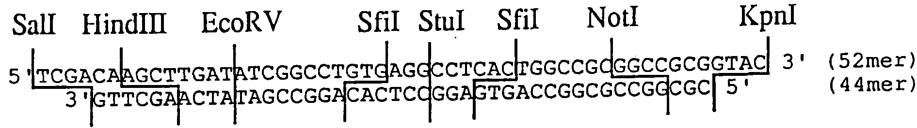
pASLBC中のジヒ ドロ葉酸還元酵素(dhfr)発現ュニッ トを除去し、 oriPと EBNA-1遺伝子を導入したプラスミ ド pASLBEC を造成した。 The unit for expressing dihydrofolate reductase (dhfr) in pASLBC was removed, and a plasmid pASLBEC into which oriP and EBNA-1 genes had been introduced was constructed.
( 4 ) で得られた P A S L B E 3— 3の 1 g を Y— 1 5 0緩衝 液 30 1 に溶解し、 20単位の Mlulと 20単位の Xholを加え、 37°Cで 2 時間消化反応を行なつた。 該反応液をァガロースゲル電気泳動に供 した後、 約 1.3kb の DNA 断片を回収した。 Dissolve 1 g of PASLBE 3-3 obtained in (4) in Y-150 buffer solution 301, add 20 units of Mlul and 20 units of Xhol, and perform digestion reaction at 37 ° C for 2 hours. Natsuta After the reaction solution was subjected to agarose gel electrophoresis, a DNA fragment of about 1.3 kb was recovered.
また、 同ブラスミ ド 1 us を Y— 0緩衝液 30 PL 1 に溶解し、 20 単位の Kpnlを加え、 37でで 2時間消化反応を行なった。 その後、 N a C 1濃度が 1 5 0 mMになるように N a C 1 を添加し、 5 単位 ' の Mlulを加え、 さらに 37°Cで 20分間部分消化反応を行なった。 該反 応液をァガロースゲル電気泳動に供した後、 約 9.6kb の DNA 断片を 回収した。 Also, 1 us of the same plasmid was dissolved in 30 PL 1 of Y-0 buffer, 20 units of Kpnl were added, and digestion reaction was carried out at 37 for 2 hours. Then, NaC1 was added so that the NaC1 concentration became 150 mM, 5 units of Mlul was added, and a partial digestion reaction was performed at 37 ° C for 20 minutes. After subjecting the reaction solution to agarose gel electrophoresis, a DNA fragment of about 9.6 kb was recovered.
また別に、 p AS L B C U〃g)を Y— 0緩衝液 30 1 に溶解 し、 20単位の Kpnlを加え、 37でで 2時間消化反応を行なった。 その 後、 N a C 1濃度が 1 0 0 mMになるように N a C 1 を添加し、 20 単位の Xholを加え、 さらに 37°Cで 2時間消化反応を行なった。 該反 応液をァガロースゲル電気泳動に供した後、 約 0.6kb の DNA 断片を 回収した。 Separately, pASLBCUg) was dissolved in Y-0 buffer 301, 20 units of Kpnl were added, and digestion reaction was carried out at 37 for 2 hours. Thereafter, NaC1 was added so that the NaC1 concentration became 100 mM, 20 units of Xhol was added, and the digestion reaction was further performed at 37 ° C for 2 hours. After subjecting the reaction solution to agarose gel electrophoresis, a DNA fragment of about 0.6 kb was recovered.
上記で得られた P AS L B E 3— 3由来の Mlul - Xhol 断片(1.3 kb) 0.2 si と同プラス ミ ド由来の Kpnl - Mlul 断片(9.6kb) 0.2 H g 、 および p A S L B C由来の Kpnl - Xho I断片(0.6kb) 0.05 g を T 4 リ ガーゼ緩衝液 30 1 に溶解し、 T4DNA リ ガーゼ 175 単位を 加えて、 12°Cで 16時間結合反応を行なった。 該反応液を用いて大腸 菌 HB101 株をコーェンらの方法によって形質転換し、 アンピシリ ン 耐性株を得た。 この形質転換株から公知の方法に従ってプラス ミ ド を単離した。 このブラスミ ドを p A S L B E C と名付け、 その構造 を制限酵素消化により確認した。 The Mlul-Xhol fragment derived from PAS LBE 3-3 obtained above (1.3 kb) 0.2 si and Kpnl-Mlul fragment (9.6 kb) derived from the same plasmid (0.2 kb) and 0.05 g of Kpnl-XhoI fragment (0.6 kb) derived from ASLBC in T4 ligase buffer 301 Then, 175 units of T4 DNA ligase was added, and the binding reaction was performed at 12 ° C for 16 hours. Using the reaction solution, Escherichia coli HB101 was transformed by the method of Koen et al. To obtain an ampicillin-resistant strain. Plasmid was isolated from this transformant according to a known method. This plasmid was named pASLBEC, and its structure was confirmed by restriction enzyme digestion.
( 7 ) P A S L B E C 2の造成 (第 7図参照) (7) Creation of PASLBEC2 (See Fig. 7)
pASLBEC のマルチクロ一ニングサイ ト中の Stulサイ 卜に BamHI リ ンカーを導入したブラスミ ド PASLBEC2を以下のようにして造成した 。 PASLBEC2では、 マルチクローニングサイ ト中の Stulサイ トは消失 している。 A brassmid PASLBEC2 in which a BamHI linker was introduced at the Stul site in the pASLBEC multi-cloning site was constructed as follows. In PASLBEC2, the Stul site in the multicloning site has disappeared.
( 6 ) で得られた p A S L B E Cの l 〃 g を Y— 1 0 0緩衝液 30 fi 1 に溶解し、 5 単位の Stulを加え、 37°Cで 20分間部分消化反応を 行なった。 該反応液をァガロースゲル電気泳動に供した後、 約 11.5 kbの DNA 断片を回収した。 回収した DNA 断片を 30 / 1 の T 4 リガ一 ゼ緩衝液に溶解し、 B a mH I リ ンカ一 (5' -pC C G G AT C C G G-3' : 宝酒造社製) を O.Ol^g と T4DNA リガーゼ 175 単位を加え て、 12°Cで 16時間結合反応を行なった。 エタノール沈殿後、 Y— 1 0 0緩衝液 に溶解し、 20単位の BamHI を加え、 37°Cで 2時 間消化反応を行なつた。 該反応液をァガロースゲル電気泳動に供し た後、 約 11.5kbの DNA 断片を回収した。 回収した DNA 断片を 20 / 1 の T 4 リ ガーゼ緩衝液に溶解し、 T4DNA リガ一ゼ 175 単位を加えて、 、 12°Cで 16時間結合反応を行なった。 該反応液を用いて大腸菌 H B 101 株をコーェンらの方法によって形質転換し、 アンピシリ ン耐性 株を得た。 この形質転換株から公知の方法に従ってプラスミ ドを単 離した。 このプラスミ ドを p A S L B E C 2 と名付け、 その構造を 制限酵素消化により確認した。 L〃g of pASLBEC obtained in (6) was dissolved in 30 fi 1 of Y-1000 buffer, 5 units of Stul was added, and a partial digestion reaction was performed at 37 ° C for 20 minutes. After subjecting the reaction solution to agarose gel electrophoresis, a DNA fragment of about 11.5 kb was recovered. The recovered DNA fragment was dissolved in 30/1 T4 ligase buffer, and BamHI linker (5'-pC CGG AT CCG G-3 ': manufactured by Takara Shuzo) was added to O.Ol ^ g. And 175 units of T4 DNA ligase were added and a ligation reaction was performed at 12 ° C for 16 hours. After ethanol precipitation, it was dissolved in Y-100 buffer, added with 20 units of BamHI, and digested at 37 ° C for 2 hours. After the reaction solution was subjected to agarose gel electrophoresis, a DNA fragment of about 11.5 kb was recovered. The recovered DNA fragment was dissolved in a 20/1 T4 ligase buffer, 175 units of T4 DNA ligase was added, and a binding reaction was carried out at 12 ° C for 16 hours. Using the reaction solution, Escherichia coli HB101 was transformed by the method of Koen et al. To obtain an ampicillin-resistant strain. Plasmid was isolated from this transformant according to a known method. This plasmid was named pASLBEC2 and its structure was Confirmed by restriction enzyme digestion.
( 8 ) p AM o E C 2の造成 (第 8図参照) (8) Construction of p AMo E C 2 (See Fig. 8)
PASLBEC2 中のプロモータ一 [SV40初期遺伝子プロモーターと HT LV - 1のロ ング ' 夕一 ミ ナル ' リ ピー ト(long terminal repeat : LT R)の R 領域と U5領域の一部を融合したプロモータ一] をモロニ一 · マゥス白血病ウィルスのし TR のプロモーターにすげかえたプラスミ ド pAMoEC2 の造成を以下のようにして行なつた。 モロニ一 ' マウス 白血病ウィルス LTR のプロモータ一は、 ブラスミ ド Molp-1 [アキノ リ · ィ シモ 卜 (Akinori Ishimoto) ら、 ピロロジ—(Virology), 141, 3 0 ( 1 9 8 5.) ] から切り出して使用した。 Promoter in PASLBEC2 [SV40 early gene promoter and a promoter that fuses a part of the R region and a part of the U5 region of the long 'yuichiminal' repeat (long terminal repeat: LTR) of HTLV-1] The plasmid pAMoEC2, which was replaced with the TR promoter of Moroni-mouse leukemia virus, was constructed as follows. The promoter of Moroni's murine leukemia virus LTR was excised from Brasmid Molp-1 [Akinori Ishimoto et al., Virology, 141, 30 (1980.)]. Used.
( 7 ) で得られた P A S L B E C 2の 1 C g を iOmM卜 リス— HC1 (pH7.5), 6m MgCl2 , 50mM KC1, 6mM 2- メルカブトエタノール からなる緩衝液 (以下、 K一 5 0緩衝液と略記する) 30;/ 1 に溶解 し、 20単位の Hindlll と 20単位の Aatll ( 東洋紡績社製) を加え、 37でで 2時間消化反応を行なつた。 該反応液をァガロースゲル電気 泳動に供した後、 約 4.8kb の DNA 断片を回収した。 1 Cg of PASLBEC 2 obtained in (7) was added to a buffer consisting of iOmM Tris-HC1 (pH 7.5), 6 mM MgCl 2 , 50 mM KC1, 6 mM 2-mercaptoethanol (hereinafter referred to as K50 buffer). The solution was dissolved in 30; / 1, and 20 units of Hindlll and 20 units of Aatll (manufactured by Toyobo Co., Ltd.) were added. After subjecting the reaction solution to agarose gel electrophoresis, a DNA fragment of about 4.8 kb was recovered.
また、 同ブラスミ ド 1 g を K一 5 0緩衝液 30 u 1 に溶解し、 20単位の Aatll を加え、 37°Cで 2時間消化反応を行なった。 その後、 5 単位の Xholを加え、 さらに 37°Cで 2 0分部分消化反応を行なった。 該反応液をァガロースゲル電気泳動に供した後、 約 6.1kb の DNA 断 片を回収した。 In addition, 1 g of the same plasmid was dissolved in 30 u1 of K-150 buffer, 20 units of Aatll was added, and a digestion reaction was performed at 37 ° C for 2 hours. Then, 5 units of Xhol were added, and a partial digestion reaction was further performed at 37 ° C for 20 minutes. After subjecting the reaction solution to agarose gel electrophoresis, a DNA fragment of about 6.1 kb was recovered.
次に、 Xhol切断部位と Clal 切断部位を連結するためのリ ンカ一 として以下の DNAリ ンカ一を合成した。 Next, the following DNA linker was synthesized as a linker for connecting the Xhol cleavage site and the Clal cleavage site.
5'-TCG AGG ACC-3' {9 mer) 5'-TCG AGG ACC-3 '(9 mer)
3'-CCTGGGC-5' (7 mer) 3'-CCTGGGC-5 '(7 mer)
上記 DNAリ ンカーの 9raerと 7merの 1本鎖 DNAはそれぞれアブ ライ ド . バイオシステムズ社 3 8 0 A · DNA合成機を用いて合成 した。 合成した D NAはそれぞれ 0.2 g ずつ、 T 4キナーゼ緩衝 液 40 1 に溶解し、 T 4 ポリ ヌ ク レオチ ドキナーゼ 30単位を加えて、 37°Cで 2時間リ ン酸化反応を行なつた。 Single-stranded DNAs of 9raer and 7mer of the above DNA linkers were Ride Biosystems 380 A • Synthesized using a DNA synthesizer. 0.2 g of each of the synthesized DNAs was dissolved in T4 kinase buffer solution 401, and 30 units of T4 polynucleotide kinase was added thereto, followed by a phosphorylation reaction at 37 ° C for 2 hours.
また別に、 M o l p— 1 [アキノ リ ' イシモ ト (Akinori Ishimoto) ら、 ピロロジー(Virology), 1 4 1 , 3 0 ( 1 9 8 5 ) ] l g を Y— 5 0緩衝液 30 1 に溶解し、 20単位の Clalを加え、 37°Cで 2時 間消化反応を行なった。 エタノール沈殿後、 30 1 の T4リガーゼ緩 衝液に溶解し、 上記 DNAリ ンカー 0.01〃 g と T4DNA リ ガーゼ 175 単位を加えて、 12°Cで 16時間結合反応を行なった。 エタノール沈殿 後、 K一 2 0緩衝液 30 / I に溶解し、 20単位の Smalを加え、 37°Cで 2時間消化反応を行なつた。 該反応液をァガロースゲル電気泳動に 供した後、 約 0.6kb の DNA 断片を回収した。 回収した DNA 断片を 30 II 1 の T 4 リガーゼ緩衝液に溶解し、 Hindi II リ ンカ一 (5'-pC A AG CTTG-3' : 宝酒造社製) を 0.03jug と T4DNA リガーゼ 175 単位を加えて、 12てで 16時間結合反応を行なった。 エタノール沈殿 後、 10mMト リ ス— HCl(pH7.5), 6mM MgCl2, 50mM NaCl, 6mM 2_メ ルカブトエタノールからなる緩衝液 (以下、 Y— 5 0緩衝液と略記 する) 30 / 1 に溶解し、 10単位の Hindlll を加え、 37°Cで 2時間消 化反応を行なつた。 その後、 N a C l濃度が 1 0 0 mMになるよう に N a C lを添加し、 10単位の Xholを加え、 さらに 37てで 2時間消 化反応を行なつた。 該反応液をァガロースゲル電気泳動に供した後、 約 0.6kb の DNA 断片を回収した。 Separately, Molp-1 [Akinori Ishimoto et al., Pirology, 141, 30 (1895)] lg was dissolved in Y-50 buffer 301. , 20 units of Clal were added, and the digestion reaction was carried out at 37 ° C for 2 hours. After ethanol precipitation, the precipitate was dissolved in 301 T4 ligase buffer, 0.01 μg of the above DNA linker and 175 units of T4 DNA ligase were added, and a binding reaction was carried out at 12 ° C. for 16 hours. After ethanol precipitation, the precipitate was dissolved in K-12 buffer 30 / I, 20 units of Smal was added, and the digestion reaction was performed at 37 ° C for 2 hours. After subjecting the reaction solution to agarose gel electrophoresis, a DNA fragment of about 0.6 kb was recovered. The recovered DNA fragment was dissolved in 30 II 1 T4 ligase buffer, and Hindi II linker (5'-pCA AG CTTG-3 ': Takara Shuzo) was added to 0.03 jug and 175 units of T4 DNA ligase. The binding reaction was carried out for 16 hours. After ethanol precipitation, a buffer consisting of 10 mM Tris-HCl (pH 7.5), 6 mM MgCl 2 , 50 mM NaCl, 6 mM 2_mercaptoethanol (hereinafter abbreviated as Y-50 buffer) 30/1 , And 10 units of Hindlll was added, followed by a 2 hour digestion reaction at 37 ° C. Thereafter, NaCl was added so that the NaCl concentration became 100 mM, 10 units of Xhol was added, and the digestion reaction was carried out for 2 hours at 37. After subjecting the reaction solution to agarose gel electrophoresis, a DNA fragment of about 0.6 kb was recovered.
上記で得られた AS L B E C 2由来の Hindlll - Aatll 断片(4.8 kb) 0.2 u g と同プラスミ ド由来の Aatll - Xhol断片(6. lkb) 0.2 0.2 ug of Hindlll-Aatll fragment (4.8 kb) derived from ASLBEC2 obtained above and Aatll-Xhol fragment (6.1 kb) 0.2 derived from the same plasmid
PL g 、 および M o 1 p - 1 由来の Hindi 11 - Xhol 断片(0.6kb) Hindi 11-Xhol fragment from PL g and Mo1p-1 (0.6kb)
0.05/ g を T 4 リガーゼ緩衝液 30〃 1 に溶解し、 T4DNA リガ一ゼ 175 単位を加えて、 12°Cで 16時間結合反応を行なった。 該反応液を用い て大腸菌 HB101 株をコーェンらの方法によって形質転換し、 アンピ シリ ン耐性株を得た。 この形質転換株から公知の方法に従ってブラ スミ ドを単離した。 このプラスミ ドを p AM o E C 2 と名付け、 そ の構造を制限酵素消化により確認した。 0.05 / g was dissolved in 30% T4 ligase buffer solution, 175 units of T4 DNA ligase was added, and the binding reaction was performed at 12 ° C for 16 hours. Using the reaction solution E. coli HB101 was transformed by the method of Koen et al. To obtain an ampicillin-resistant strain. A plasmid was isolated from this transformant according to a known method. This plasmid was designated as pAMoEC2, and its structure was confirmed by restriction enzyme digestion.
( 9 ) p AM o E C 3の造成 (第 9図参照) (9) Construction of p AMo E C 3 (See Fig. 9)
pAMoEC2 のマルチクローニングサイ ト中の BamHI サイ 卜に、 詰め 込み DNA (Stuffer DNA)として、 PBR322のテトラサイク リ ン耐性遺 伝子を含む DNA 断片 [Dral - PvuII断片(2.5kb) ] を以下のように して挿入し、 ブラスミ ド pAMoEC3 を造成した。 As a stuffing DNA (Stuffer DNA), a DNA fragment containing the tetracycline resistance gene of PBR322 [Dral-PvuII fragment (2.5 kb)] was inserted into the BamHI site in the pAMoEC2 multicloning site as shown below. To insert Brassmid pAMoEC3.
( 8 ) で得られた p AM o E C 2の 1 g を Y— 1 0 0緩衝液 30 U 1 に溶解し、 20単位の BamHI を加え、 37°Cで 2時間消化反応を行 なった。 ェタノール沈殿後、 30 1 の DNA ポリ メラーゼ I緩衝液に 溶解し、 6単位の大腸菌 DNA ポリ メラーゼ I ク レノー断片を加え、 37でで 60分間反応させ、 BamHI 消化によって生じた 5 ' 突出末端を 平滑末端に変えた。 該反応液をァガロースゲル電気泳動に供した後, 約 11.5kbの DNA 断片を回収した。 1 g of pAMoEC2 obtained in (8) was dissolved in 30 U1 of Y-100 buffer, 20 units of BamHI was added, and digestion reaction was carried out at 37 ° C for 2 hours. After ethanol precipitation, dissolve in 301 DNA polymerase I buffer, add 6 units of E. coli DNA polymerase I Klenow fragment, incubate at 37 for 60 minutes, and blunt the 5 'overhangs generated by BamHI digestion. Changed to the end. After the reaction solution was subjected to agarose gel electrophoresis, a DNA fragment of about 11.5 kb was recovered.
また、 P B R 3 2 2 [ポリバ一(Bolivar) ら : ジーン(Gene), 2^ 95 (1977) ] 1 /g を Y— 5 0緩衝液 30 1 に溶解し、 20単位の Dralと 20単位の PvuII を加え、 37でで 2時間消化反応を行なった。 該反応液をァガロースゲル電気泳動に供した後、 約 2.5kb の DNA 断 片を回収した。 Also, 1 / g of PBR 3 222 [Bolivar et al .: Gene, 2 ^ 95 (1977)] was dissolved in Y-50 buffer 301, and 20 units of Dral and 20 units of Dral were dissolved. PvuII was added, and digestion was carried out at 37 for 2 hours. After subjecting the reaction solution to agarose gel electrophoresis, a DNA fragment of about 2.5 kb was recovered.
上記で得られた p AM 0 E C 2由来の BamHI ( 平滑末端 )断片 (11.5kb) 0.1 zg と p B R 3 2 2由来の Dral - Pvul I断片(2.5kb) 0.2 を T 4 リガーゼ緩衝液 30^ 1 に溶解し、 T4DNA リガーゼ 175 単位を加えて、 12でで 16時間結合反応を行なった。 該反応液を 用いて大腸菌 HB101 株をコーェンらの方法によって形質転換し、 ァ ンピシリ ンとテトラサイク リ ンに耐性な株を得た。 この形質転換株 から公知の方法に従ってブラスミ ドを単離した。 このプラスミ ドを P AM o E C 3 と名付け、 その構造を制限酵素消化により確認した ( ( 1 0 ) p AM o E R C 3の造成 (第 1 0図参照) BamHI (blunt end) fragment (11.5 kb) 0.1 zg derived from pAM0EC2 and Dral-Pvul I fragment (2.5 kb) 0.2 derived from pBR322 obtained above were added to T4 ligase buffer 30 ^. The mixture was dissolved in 1 and 175 units of T4 DNA ligase was added thereto. Using the reaction solution, Escherichia coli HB101 was transformed by the method of Koen et al. To obtain a strain resistant to ampicillin and tetracycline. Blasmid was isolated from this transformant according to a known method. This plasmid It was named P AMo EC 3 and its structure was confirmed by restriction enzyme digestion. ( (10) Construction of p AM o ERC 3 (See Fig. 10)
pAMoEC3 中の oriPと EBNA- 1遺伝子のュニッ 卜の向きを逆にしたプ ラスミ ド pAMoERC3を以下のようにして造成した。 Plasmid pAMoERC3 in which the units of oriP and EBNA-1 gene in pAMoEC3 were reversed was constructed as follows.
( 9 ) で得られた p AM o E C 3の 1 〃 g を Y— 1 0 0緩衝液 30 1 に溶解し、 20単位の Xholを加え、 .37°Cで 2時間消化反応を行 なった。 その後、 1 M ト リス - HCKPH8.0)を 30〃 1 と大腸菌アル力 リ フ ォスファターゼ (宝酒造社製) 1単位を加え、 37°Cで 2時間脱 リ ン酸化反応を行なつた。 ェタノール沈殿後、 10mMト リス— HCKpH 8.0), Im EDTA (エチレンジァ ミ ン 4酢酸ナ ト リ ウム) からなる 緩衝液 (以下、 T E緩衝液と略記する) 30 / 1 に溶解し、 ァガロー スゲル電気泳動を行ない、 約 9.1kb の DNA 断片を回収した。 1 μg of pAMoEC3 obtained in (9) was dissolved in Y-1000 buffer 301, and 20 units of Xhol was added, followed by digestion at .37 ° C for 2 hours. . Thereafter, 30 μl of 1 M Tris-HCKPH8.0) and 1 unit of Escherichia coli ALPHA phosphatase (Takara Shuzo) were added, and a dephosphorylation reaction was performed at 37 ° C. for 2 hours. After ethanol precipitation, the precipitate is dissolved in a buffer consisting of 10 mM Tris-HCK pH 8.0) and Im EDTA (sodium ethylenediamine tetraacetate) (hereinafter abbreviated as TE buffer) 30/1, and agarose gel electrophoresis. And a DNA fragment of about 9.1 kb was recovered.
また、 同プラスミ ド 1 ug を Y— 1 0 0緩衝液 30 u 1 に溶解し、 20単位の Xholを加え、 37 で 2時間消化反応を行なった。 該反応液 をァガロースゲル電気泳動に供した後、 約 4.9kb の DNA 断片を回収 した。 In addition, 1 ug of the same plasmid was dissolved in 30 u1 of Y-100 buffer, added with 20 units of Xhol, and digested at 37 for 2 hours. After subjecting the reaction solution to agarose gel electrophoresis, a DNA fragment of about 4.9 kb was recovered.
上記で得られた P AM 0 E C 3由来の Xhol断片(9. lkb) 0.1 u g と同ブラスミ ド由来の Xhol断片(4.9kb) 0.2 n g を丁 4 リガ一ゼ緩 衝液 30〃 1 に溶解し、 T4DNAリガーゼ 175 単位を加えて、 12°Cで 16 時間結合反応を行なった。 該反応液を用いて大腸菌 HB101 株をコー ェンらの方法によって形質転換し、 アンピシリ ン耐性株を得た。 こ の形質転換株から公知の方法に従ってプラスミ ドを単離した。 この プラスミ ドを p AM o E R C 3 と名付け、 その構造を制限酵素消化 により確認した。 Dissolve 0.1 ug of the XAM fragment (9. lkb) derived from PAM0EC3 and 0.2 ng of the Xhol fragment (4.9 kb) derived from the same plasmid in 30 l of 4 ligation buffer. 175 units of T4 DNA ligase were added, and the binding reaction was performed at 12 ° C for 16 hours. Using the reaction solution, Escherichia coli HB101 was transformed by the method of Koen et al. To obtain an ampicillin-resistant strain. Plasmid was isolated from this transformant according to a known method. This plasmid was named pAMoERC3, and its structure was confirmed by restriction enzyme digestion.
( 1 1 ) P A G E 2 0 7の造成 (第 1 1 図参照) (11) Creation of PAG E207 (see Fig. 11)
PAGE107 中の G418耐性遺伝子をハイグロマイシン(hyg) 耐性遺伝 子にすげかえたブラスミ ド PAGE207 を以下のようにして造成した。 hyg 耐性遺伝子は、 P201 〔ビル · ズグデン(Bill Sugden) ら、 ネィ チ ヤ一 (Nature) , 3 1 3, 8 1 2 ( 1 9 8 5 ) 〕 より切り出して 使用した。 A plasmid PAGE207 in which the G418 resistance gene in PAGE107 was replaced with a hygromycin (hyg) resistance gene was constructed as follows. The hyg resistance gene is P201 [Bill Sugden et al. Cutter (Nature), 313, 812 (1895)] was used.
P A G E 1 0 7 (特開平 3-22979)の 1 //g を Y— 5 0緩衝液 30 1 に溶解し、 20単位の Clalを加え、 37°Cで 2時間消化反応を行なった。 その後、 N a C; 】 濃度が 1 5 0 mMになるように N a C 】 を添加し、 20単位の Mlulを加え、 さらに 37°Cで 2時間消化反応を行なった。 該 反応液をァガロースゲル電気泳動に供した後、 約 4.6kb の DNA 断片 を回収した。 1 // g of PAGE 107 (Japanese Unexamined Patent Publication No. 3-22979) was dissolved in Y-50 buffer 301, and 20 units of Clal was added thereto, followed by digestion at 37 ° C for 2 hours. Thereafter, NaC] was added to a concentration of 150 mM, and 20 units of Mlul were added, followed by a digestion reaction at 37 ° C for 2 hours. After subjecting the reaction solution to agarose gel electrophoresis, a DNA fragment of about 4.6 kb was recovered.
p 2 0 1 [ビル ' ズグデン (BUI Sudgen)ら : ネイチヤ ー (Nature), 3 1 3 , 8 1 2 ( 1 9 8 5 ) ] 0.5 z gを Y- 5 0緩衝液 30〃 1 に 溶解し、 20単位の Nari [ニュ ー ' イングラン ド ' バイオラボ (New England Biolab) 社製] を加え、 37°Cで 2時間消化反応を行なった。 エタノール沈殿後、 30 1 の DNA ポリ メラ一ゼ I緩衝液に溶解し、 6単位の大腸菌 DNA ポリ メラ一ゼ I ク レノ一断片を加え、 37°Cで 60 分間反応させ、 Narl消化によって生じた 5 ' 突出末端を平滑末端に 変えた。 反応をフヱノール抽出によって止め、 クロ口ホルム抽出と ェタノ一ル沈殿の後、 20〃 1 の T 4 リガーゼ緩衝液に溶解し、 Clal リ ンカ一 (5'p CATC GATG3' : 宝酒造社製) を 0.05 ig と T4 DNA リガーゼ 175 単位を加えて、 12°Cで 16時間結合反応を行なった。 ェタノ一ル沈殿後、 Y— 5 0緩衝液 30 1 に溶解し、 10単位の Clal を加え、 37°Cで 2時間消化反応を行なつた。 その後、 N a C 1濃度 が 1 5 0 mMになるように N a C 1 を添加し、 10単位の Mlulを加え、 さらに 37°Cで 2時間消化反応を行なった。 該反応液をァガロースゲ ル電気泳動に供した後、 約 1.6kb の DNA 断片を回収した。 p 2 0 1 [BUI Sudgen et al .: Nature, 3 13, 8 12 (1 895)] Dissolve 0.5 zg in 30% Y-50 buffer, Twenty units of Nari (New England Biolab) were added and digestion reaction was performed at 37 ° C for 2 hours. After ethanol precipitation, dissolve in 301 DNA polymerase I buffer, add 6 units of E. coli DNA polymerase I cleno fragment, react at 37 ° C for 60 minutes, and generate by Narl digestion. The 5 'overhang was changed to a blunt end. The reaction was stopped by phenol extraction, and after extraction with chloroform and ethanol precipitation, it was dissolved in 20〃1 T4 ligase buffer, and Clal linker (5'p CATC GATG3 ': Takara Shuzo) was added. ig and 175 units of T4 DNA ligase were added, and a binding reaction was performed at 12 ° C for 16 hours. After ethanol precipitation, the precipitate was dissolved in Y-50 buffer 301, 10 units of Clal was added, and the digestion reaction was carried out at 37 ° C for 2 hours. Thereafter, NaC1 was added so that the NaC1 concentration became 150 mM, 10 units of Mlul were added, and the digestion reaction was further performed at 37 ° C for 2 hours. After the reaction solution was subjected to agarose gel electrophoresis, a DNA fragment of about 1.6 kb was recovered.
上記で得られた P AG E 1 0 7由来の Clal - Mlul 断片(4.6kb) 0.2 u g と p 2 0 1由来の Clal - Mlul断片(1.6kb) 0.1 g を T 4 リガ一ゼ緩衝液 30 1 に溶解し、 T4DNA リガーゼ 175 単位を加え て、 12°Cで 16時間結合反応を行なった。 該反応液を用いて大腸菌 HB 101 株をコーェンらの方法によって形質転換し、 アンピシリ ン耐性 株を得た。 この形質転換株から公知の方法に従ってプラスミ ドを単 離した。 このプラスミ ドを P A G E 2 0 7 と名付け、 その構造を制 限酵素消化により確認した。 0.2 ug of the Clal-Mlul fragment (4.6 kb) derived from PAGE 107 and 0.1 g of the Clal-Mlul fragment (1.6 kb) derived from p201 were combined with T4 ligase buffer 30 1 And 175 units of T4 DNA ligase was added, and the binding reaction was carried out at 12 ° C for 16 hours. Escherichia coli HB 101 strains were transformed by the method of Koen et al. To obtain ampicillin-resistant strains. Plasmid was isolated from this transformant according to a known method. This plasmid was named PAGE207, and its structure was confirmed by restriction enzyme digestion.
( 1 2 ) p A G E 2 0 7 S c Nの造成 (第 1 2図参照) (12) Creation of pAGE207ScN (See Fig. 12)
ラ ビッ ト Sグロビン遺伝子中に存在する Sfilサイ トの類似配列を 除去するため、 PAGE207 の Ballサイ 卜に Sealリ ンカ一を挿入したプ ラスミ ド pAGE207ScNを以下のようにして造成した。 なお、 PAGE207S cNにおいては、 挿入された Sealリ ンカーの数は明らかではない。 In order to remove a similar sequence of the Sfil site present in the rabbit S globin gene, a plasmid pAGE207ScN in which a Seal linker was inserted into the PAGE207 Ball site was constructed as follows. In PAGE207S cN, the number of Seal linkers inserted is not clear.
( 1 1 ) で得られた P A G E 2 0 7の 0.5 u g を Y— 0緩衝液 30 PL 1 に溶解し、 10単位の Ballを加え、 37°Cで 2時間消化反応を行な つた。 ェ夕ノール沈殿後、 20 1 の T 4 リガーゼ緩衝液に溶解し Sc alリ ンカ一 (5' p AA G TA C TT3' : 宝酒造社製) を 0.01 g と T4 DNAリガーゼ 175 単位を加えて、 12°Cで 16時間結合反応を行なつ た。 該反応液を用いて大腸菌 HB101 株をコーェンらの方法によって 形質転換し、 アンピシリ ン耐性株を得た。 この形質転換株から公知 の方法に従ってブラスミ ドを単離した。 このブラスミ ドを P A G E 2 0 7 S c Nと名付け、 その構造を制限酵素消化により確認した。 0.5 μg of PAGE207 obtained in (11) was dissolved in 30 PL 1 of Y-0 buffer, 10 units of Ball was added, and a digestion reaction was carried out at 37 ° C. for 2 hours. After precipitation with ethanol, dissolve in 201 T4 ligase buffer, add 0.01 g of Scal linker (5 'pAAGTAC TTT3': manufactured by Takara Shuzo) and 175 units of T4 DNA ligase. The binding reaction was performed at 12 ° C for 16 hours. Using the reaction solution, Escherichia coli HB101 was transformed by the method of Koen et al. To obtain an ampicillin-resistant strain. Blasmid was isolated from this transformant according to a known method. This plasmid was named PAGE207ScN, and its structure was confirmed by restriction enzyme digestion.
( 1 3 ) p AM o C 3 S cの造成 (第 1 3図参照) (13) Creation of pAMoC3Sc (See Fig. 13)
PAMOERC3中のラビッ ト βグロビン遺伝子中に存在する Sfilサイ ト の類似配列を除去するため、 以下のようにして PAM0ERC3中のラ ビッ ト ^グ口ビン遺伝子を、 すでにその類似配列を除去してある PAGE20 7ScN中のラビッ ト グロビン遺伝子にすげかえ、 プラスミ ド pAMoER C3Scを造成した。 造成の都合上、 まず pAMoCSScを造成し、 次いで pA MoERC3Scの造成を行なった。 前記の pAGE207ScNにおいては、 Sfilサ ィ トの類似配列を除去するために挿入された Sealリ ンカーの数は明 らかではないが、 pAMoERC3Scの場合は、 造成の際に pAGE207ScNを一 度 Sealで切断しているため、 挿入された Sealサイ トの数は 1 つであ ると推定される。 Rabbit in PAMOERC3 In order to remove the similar sequence of the Sfil site in the β-globin gene, the rabbit ^ gurubin gene in PAM0ERC3 has already been deleted from the similar sequence as follows: In place of the rabbit globin gene in PAGE207ScN, plasmid pAMoER C3Sc was constructed. For convenience of construction, pAMoCSSc was first constructed, and then pAMoERC3Sc was constructed. In the above-mentioned pAGE207ScN, the number of Seal linkers inserted to remove the similar sequence of the Sfil site is not clear, but in the case of pAMoERC3Sc, pAGE207ScN is once cut with Seal during construction. Therefore, only one Seal site was inserted. It is estimated that.
( 1 2 ) で得られた p AG E 2 0 7 S c Nの 1 g を Y— 0緩衝 液 30 l に溶解し、 20単位の Kpnlを加え、 37°Cで 2時間消化反応を 行なった。 その後、 N a C 1濃度が 100 mMになるように N a C 1 を添加し、 20単位の Sealを加え、 さらに 37°Cで 2時間消化反応を行 なった。 該反応液をァガロースゲル電気泳動に供した後、 約 0.7kb の DNA 断片を回収した。 1 g of pAGE207 ScN obtained in (12) was dissolved in 30 l of Y-0 buffer, 20 units of Kpnl were added, and the digestion reaction was performed at 37 ° C for 2 hours. . Then, NaC1 was added so that the NaC1 concentration became 100 mM, 20 units of Seal were added, and the digestion reaction was further performed at 37 ° C for 2 hours. After subjecting the reaction solution to agarose gel electrophoresis, a DNA fragment of about 0.7 kb was recovered.
また、 同プラスミ ド 1 〃g を Y— 100 緩衝液 30 1 に溶解し、 20単位の Sealと 20 単位の Clalを加え、 37°Cで 2時間消化反応を行 なった。 該反応液をァガロースゲル電気泳動に供した後、 約 0.9kb の DNA 断片を回収した。 In addition, 1 μg of the same plasmid was dissolved in Y-100 buffer 301, and 20 units of Seal and 20 units of Clal were added, and digestion reaction was performed at 37 ° C for 2 hours. After subjecting the reaction solution to agarose gel electrophoresis, a DNA fragment of about 0.9 kb was recovered.
また、 別に ( 1 0 ) で得られた p AM 0 E R C 3の 1 Li g を Y— 0緩衝液 30/z l に溶解し、 20単位の Kpnlを加え、 37°Cで 2時間消化 反応を行なつた。 その後、 N a C 1濃度が 1 0 0 mMになるように N a C lを添加し、 20単位の Xholを加え、 さらに 37°Cで 2時間消化 反応を行なった。 該反応液をァガロースゲル電気泳動に供した後、 約 3.2kb の DNA 断片を回収した。 Separately, 1 Lig of pAM0 ERC3 obtained in (10) was dissolved in Y-0 buffer 30 / zl, added with 20 units of Kpnl, and digested at 37 ° C for 2 hours. Natsuta Thereafter, NaCl was added so that the NaCl concentration became 100 mM, 20 units of Xhol was added, and the digestion reaction was further performed at 37 ° C for 2 hours. After subjecting the reaction solution to agarose gel electrophoresis, a DNA fragment of about 3.2 kb was recovered.
次に、 p A G E 1 0 7 ( 特開平 2-227075) の l 〃g を Y - 1 0 0 緩衝液 30 u 1 に溶解し、 20単位の Xholと 20単位の Clalを加え、 37 でで 2時間消化反応を行なつた。 該反応液をァガロースゲル電気泳 動に供した後、 約 4.3kb の DNA 断片を回収した。 Next, l lg of pAGE107 (Japanese Patent Application Laid-Open No. 2-227075) was dissolved in 30 u1 of Y-100 buffer, and 20 units of Xhol and 20 units of Clal were added. A time digestion reaction was performed. After subjecting the reaction solution to agarose gel electrophoresis, a DNA fragment of about 4.3 kb was recovered.
上記で得られた p AG E 2 0 7 S c N由来の Kpnl -Seal断片(0.7 kb) 0.1 g と同プラスミ ド由来の Seal - Clal 断片(0.9kb) 0.1 g 、 p AMo E R C 3由来の Kpnl - Xhol 断片(3.2kb) 0.3 ju g , および P AG E 1 0 7由来の Xhol - Clal 断片(4.3kb)0.3 g を T 4 リガ一ゼ緩衝液 30 / 1 に溶解し、 T4DNAリガ一ゼ 175 単位を加え て、 12°Cで 16時間結合反応を行なった。 該反応液を用いて大腸菌 HB 101 株をコーェンらの方法によって形質転換し、 アンピシリ ン耐性 0.1 g of the Kpnl-Seal fragment (0.7 kb) derived from pAGE207ScN obtained above and 0.1 g of the Seal-Clal fragment (0.9 kb) derived from the same plasmid, and Kpnl derived from pAMoERC3 -Dissolve 0.3 ju g of the Xhol fragment (3.2 kb) and 0.3 g of the Xhol-Clal fragment (4.3 kb) derived from PAGE 107 in 30/1 T4 ligase buffer, and remove the T4 DNA ligase 175 The unit was added and the binding reaction was performed at 12 ° C for 16 hours. Escherichia coli HB101 was transformed using the reaction solution according to the method of Koen et al.
3 Θ 株を得た。 この形質転換株から公知の方法に従ってプラス ミ ドを単 離した。 このプラスミ ドを p AM o C 3 S c と名付け、 その構造を 制限酵素消化により確認した。 3 Θ Got the strain. Plasmid was isolated from this transformant according to a known method. This plasmid was named pAMoC3Sc, and its structure was confirmed by restriction enzyme digestion.
( 1 4 ) P AM o E R C 3 S cの造成 (第 1 4図参照) (14) Creation of P AM o ERC 3 Sc (See Fig. 14)
( 1 0 ) で得られた p AM o E R C 3の 1 を Y— 0緩衝液 30 H 1 に溶解し、 20単位の Kpnlを加え、 37°Cで 2時間消化反応を行 なった。 その後、 N a C 1 濃度が 1 5 0 m Mになるように N a C 1 を添加し、 20単位の Mlulを加え、 さらに 37°Cで 2時間消化反応を行 なった。 該反応液をァガロースゲル電気泳動に供した後、 約 6.8kb の DNA 断片を回収した。 1 of pAMoERC3 obtained in (10) was dissolved in 30 buffer of Y-0 buffer, 20 units of Kpnl was added, and digestion reaction was performed at 37 ° C for 2 hours. Thereafter, NaC1 was added so that the NaC1 concentration became 150 mM, 20 units of Mlul were added, and the digestion reaction was further performed at 37 ° C for 2 hours. After subjecting the reaction solution to agarose gel electrophoresis, a DNA fragment of about 6.8 kb was recovered.
また、 同プラスミ ド 1 zg を Y— 1 5 0緩衝液 30 u 1 に溶解し、 20単位の Xholと 20単位の Mlulを加え、 37°Cで 2時間消化反応を行 なった。 該反応液をァガロースゲル電気泳動に供した後、 約 1.3kb の DNA 断片を回収した。 Also, 1 zg of the same plasmid was dissolved in 30 u1 of Y-150 buffer, 20 units of Xhol and 20 units of Mlul were added, and digestion reaction was performed at 37 ° C for 2 hours. After subjecting the reaction solution to agarose gel electrophoresis, a DNA fragment of about 1.3 kb was recovered.
また別に、 p AM o C 3 S cの 1 〃g を Y— 0緩衝液 30〃 1 に溶 解し、 20単位の Kpnlを加え、 37°Cで 2時間消化反応を行なった。 そ の後、 N a C l 濃度が 1 O O mMになるように N a C l を添加し、 20単位の Xholを加え、 さらに 37°Cで 2時間消化反応を行なった。 該 反応液をァガロースゲル電気泳動に供した後、 約 5.9kb の DNA 断片 を回収した。 Separately, 1 μg of pAMoC 3 Sc was dissolved in 30% Y-1 buffer, 20 units of Kpnl were added, and a digestion reaction was performed at 37 ° C. for 2 hours. Thereafter, NaCl was added so that the NaCl concentration became 1 O OmM, 20 units of Xhol were added, and the digestion reaction was further performed at 37 ° C for 2 hours. After subjecting the reaction solution to agarose gel electrophoresis, a DNA fragment of about 5.9 kb was recovered.
上記で得られた p AM o E R C 3由来の Kpn【 -Mlul断片(6.8kb) 0.2 g と同プラスミ ド由来の Xhol - Mlul 断片(1.3kb) 0.05 z g 、 および p AM o C 3 S c由来の Kpnl- Xhol断片(5.9kb) 0.2 〃 g を T 4 リガ一ゼ緩衝液 30 1 に溶解し、 T4DNAリガーゼ' 175 単位を 加えて、 12°Cで 16時間結合反応を行なった。 該反応液を用いて大腸 菌 HB101 株をコ一ェンらの方法によって形質転換し、 了ンピシリ ン 耐性株を得た。 この形質転換株から公知の方法に従ってプラスミ ド を単離した。 このプラスミ ドを p AM o E R C 3 S c と名付け、 そ の構造を制限酵素消化により確認した。 0.2 g of Kpn [-Mlul fragment (6.8 kb) derived from pAMo ERC3 obtained above, 0.05 zg of Xhol-Mlul fragment (1.3 kb) derived from the same plasmid, and 0.05 g of pAMoC3Sc derived from the same plasmid 0.2 µg of the Kpnl-Xhol fragment (5.9 kb) was dissolved in T4 ligase buffer 301, 175 units of T4 DNA ligase 'was added, and a binding reaction was performed at 12 ° C for 16 hours. Using the reaction solution, Escherichia coli HB101 strain was transformed according to the method of Koen et al. To obtain an ampicillin resistant strain. Plasmid was isolated from this transformant according to a known method. This plasmid was named pAMo ERC3Sc. Was confirmed by restriction enzyme digestion.
p AM o E R C 3 S cは、 異種遺伝子発現用のプロモーターと し て、 モロニ一 ' マウス白血病ウィルスのロング ' ター ミナル ' リ ピー ト (long terminal repeat) を有している。 また、 異種遺伝子 の効率良い発現のために、 ラビッ ト yQグロビン遺伝子スプライシン グシグナル、 ラビッ ト /3グロビン遺伝子ポリ A付加シグナルおよび S V 4 0初期遺伝子ポリ A付加シグナルが、 挿入した異種遺伝子の 後に付加するようにデザイ ンされている。 また、 動物細胞用の薬剤 耐性マ一カーとして G 4 1 8耐性遺伝子を、 大腸菌用の薬剤耐性マ 一力一としてカナマイシン耐性遺伝子 (G 4 1 8耐性遺伝子と同じ もの) とアンピシリ ン耐性遺伝子を有している。 さらに、 エブシュ タイ ン · バール · ウィルス (Epstein -Barr virus ) の複製開始点 pAMoERC3Sc has a long terminal repeat of Moroni's murine leukemia virus as a promoter for heterologous gene expression. In addition, for efficient expression of heterologous genes, the rabbit yQ globin gene splicing signal, rabbit / 3 globin gene poly A addition signal, and SV40 early gene poly A addition signal are added after the inserted heterologous gene. It is designed as follows. The G418 resistance gene was used as a drug resistance marker for animal cells, and the kanamycin resistance gene (same as the G418 resistance gene) and the ampicillin resistance gene were used as drug resistance markers for E. coli. Have. Furthermore, the origin of replication of Epstein-Barr virus
(o r i P) と o r i Pに トランスに作用し複製を引き起こす働き をもつ E BNA— 1遺伝子を有するため、 ナマルバ細胞をはじめと してゲッ歯類を除く多くの細胞中で、 染色体に組み込まれることな く ブラスミ ド状態で存在することができる。 (ori P) and ori P have the EBNA-1 gene, which acts on trans and causes replication in ori P, so that it can be integrated into the chromosome in many cells other than rodents, including Namalva cells. It can exist in the brassid state without.
P AM 0 E R C 3 S cを用いた c DNAライブラ リーの造成は、 c D N Aの両末端に S ί i I リ ン力一を付加した後、 p AM o E R C 3 S c中の S f i I部位に組み込むことにより行なえる。 Construction of a cDNA library using PAM0ERC3Sc is performed by adding S SII linkers to both ends of cDNA and then recombining the SfiI site in pAMoERC3Sc. This can be done by incorporating it into
( 1 5 ) p AMo PR C 3 S cの造成 (図 1 5参照) (15) Construction of pAMoPRC3Sc (See Fig. 15)
ナマルバ細胞のように E BNA— 1をもともと発現している細胞 を宿主として用いる際には、 プラスミ ド 八1^ 0 £1^〇 33 ( 中の E BNA- 1遺伝子がなくても、 宿主に導入したプラスミ ドは染色 体に組み込まれることなく プラスミ ド状態で存在することが可能で あると考えられる。 そこで、 p AMo E R C 3 S c中の E BNA— 1遺伝子を除去したプラスミ ド p AMo PR C 3 S cの造成を以下 のようにして行なった。 p AMo PR C 3 S cは、 p AMo E R C 3 S c と同様にして直接発現クローニングベクタ一として使用する こ とができる。 When a cell that originally expresses EBNA-1 such as a Namalba cell is used as a host, it can be used as a host even if the EBNA-1 gene is not present. It is considered that the introduced plasmid can exist in a plasmid state without being incorporated into the chromosome, and therefore, the plasmid pAMoPR from which the EBNA-1 gene in pAMoERC3Sc has been removed is considered. C3Sc was constructed as follows: pAMoPR C3Sc was used directly as an expression cloning vector in the same manner as pAMoERC3Sc. be able to.
( 1 4 ) で得られた p AM o E R C 3 S cの 2 g を Y— 5 0緩 衝液 30 1 に溶解し、 20単位の Nsil [ニュ ー ' イングラン ド ' バ ィオラブズ (New England Biolabs ) 社製] を加え、 37°Cで 2時間 消化反応を行なつた。 エタノール沈殿後、 30 / 1 の DNA ポリ メラー ゼ I緩衝液に溶解し、 6単位の大腸菌 DNA ポリ メラーゼ I ク レ ノ ー 断片を加え、 37°Cで 60分間反応させ、 Nsil消化によって生じた 3 ' 突出末端を平滑末端に変えた。 反応をフ ノール抽出によって止め クロ口ホルム抽出とエタノール沈殿の後、 Y— 1 0 0緩衝液 30 n に溶解し、 20単位の Notlを加え、 37°Cで 2時間消化反応を行なった, 該反応液をァガロースゲル電気泳動に供した後、 約 8.1kb の DNA 断 片を回収した。 2 g of pAMoERC3Sc obtained in (14) was dissolved in Y-50 buffer solution 301, and 20 units of Nsil [New 'Ingland' Biolabs (New England Biolabs)] was dissolved. And the digestion reaction was performed at 37 ° C for 2 hours. After ethanol precipitation, dissolve in 30/1 DNA polymerase I buffer, add 6 units of E. coli DNA polymerase I cleno fragment, incubate at 37 ° C for 60 minutes, and generate Nsil by digestion. 'Changed overhanging ends to blunt ends. After the reaction was stopped by phenol extraction, the solution was dissolved in 30 mL of Y-1000 buffer, and 20 units of Notl was added to the solution. After digestion, the digestion reaction was carried out at 37 ° C for 2 hours. After subjecting the reaction solution to agarose gel electrophoresis, a DNA fragment of about 8.1 kb was recovered.
また、 同プラスミ ド 2 g を Y— 1 0 0緩衝液 30 a 1 に溶解し. 20単位の Xholを加え、 37°Cで 2時間消化反応を行なった。 エタノー ル沈殿後、 30 1 の DNA ポリ メラーゼ I緩衝液に溶解し、 6単位の 大腸菌 DNA ポリ メラーゼ I ク レノ一断片を加え、 37°Cで 60分間反応 させ、 Xhol消化によって生じた 5 ' 突出末端を平滑末端に変えた。 反応をフエノール抽出によって止め、 クロ口ホルム抽出とェタノ一 ル沈殿の後、 Y— 1 0 0緩衝液 30 1 に溶解し、 20単位の Notlを 加え、 37でで 2時間消化反応を行なった。 該反応液をァガロースゲ ル電気泳動に供した後、 約 3.2kb の DNA 断片を回収した。 In addition, 2 g of the same plasmid was dissolved in 30 buffer of Y-100 buffer, and 20 units of Xhol was added thereto, followed by digestion at 37 ° C for 2 hours. After ethanol precipitation, dissolve in 301 DNA polymerase I buffer, add 6 units of E. coli DNA polymerase I cleno fragment, incubate at 37 ° C for 60 minutes, and generate 5 'overhang by Xhol digestion. The ends were changed to blunt ends. The reaction was stopped by phenol extraction, and after extraction with chloroform and ethanol precipitation, it was dissolved in Y-100 buffer solution 301, 20 units of Notl was added, and the digestion reaction was carried out at 37 for 2 hours. After subjecting the reaction solution to agarose gel electrophoresis, a DNA fragment of about 3.2 kb was recovered.
上記で得られた p AM o E R C 3 S c由来の Nsil (平滑末端) - Notl断片(8. lkb) 0.1 と同プラスミ ド由来の Xhol (平滑末端) - Notl断片(3.2kb) 0.1 g を T 4 リガーゼ緩衝液 30〃 1 に溶解し、 T4DNA リガーゼ 175 単位を加えて、 12°Cで 16時間結合反応を行なつ た。 該反応液を用いて大腸菌 HB101 株をコ一ェンらの方法によって 形質転換し、 アンピシリ ン耐性株を得た。 この形質転換株から公知 の方法に従ってプラスミ ドを単離した。 このブラスミ ドを p AM o P R C 3 S c と名付け、 その構造を制限酵素消化により確認した。 2. ヒ ト単球系細胞株 THP- 1 細胞からの α 1, 3-フコシル トランス フェラ一ゼ cDNA のクローン化 0.1 g of Nsil (blunt end) derived from pAMoERC3Sc-Notl fragment (8.lkb) obtained above and 0.1 g of Xhol (blunt end) derived from the same plasmid as Notl fragment (3.2 kb) 4 The ligase was dissolved in 30〃1 of ligase buffer, 175 units of T4 DNA ligase was added, and the binding reaction was carried out at 12 ° C for 16 hours. Using the reaction solution, Escherichia coli HB101 was transformed by the method of Koen et al. To obtain an ampicillin-resistant strain. Plasmid was isolated from this transformant according to a known method. This brassmid is p AM o It was named PRC 3 Sc and its structure was confirmed by restriction enzyme digestion. 2. Cloning of α1,3-fucosyltransferase cDNA from human monocyte cell line THP-1 cells
( 1 ) ヒ ト単球系細胞株 THP-1 細胞からの inRNAの取得 (1) Obtaining inRNA from human monocyte cell line THP-1 cells
1 X 10⁸ 個の THP-1 細胞(ATCC TIB 202)より、 イ ンビトロジェン1 x 10 8 THP-1 cells (ATCC TIB 202)
(Invitrogen) 社製の mRNA抽出キッ トであるファース ト トラッ ク (Fast Track ; 商品番号 K1593- 02) を用いて、 約 30 g の niRNAを取 得した。 具体的試薬および方法は、 キッ 卜に付与されている説明書 に従った。 About 30 g of niRNA was obtained using Fast Track (product number K1593-02), which is an mRNA extraction kit (Invitrogen). Specific reagents and methods followed the instructions provided with the kit.
( 2 ) cDNAライブラ リ一の作製 (2) Preparation of cDNA library
上記で得られた mRNA の 8 から、 GIBCO BRL社製のキッ 卜で ある cDNA合成システム (cDNA Synthesis System ) を用いて、 オリ ゴ d Tをブライマーとして 2本鎖 cDNAを合成した。 その際、 逆転写 酵素としてはキッ ト中の Moloney Murine Leukemia Virus (M-MLV) reverse transcriptase の代わりに、 同社の Super ScriptTM RNase A double-stranded cDNA was synthesized from 8 of the mRNA obtained above using Oligo dT as a primer using a cDNA synthesis system (cDNA Synthesis System) manufactured by GIBCO BRL. At that time, instead of Moloney Murine Leukemia Virus (M-MLV) reverse transcriptase in the kit, the company's Super ScriptTM RNase was used as the reverse transcriptase.
H— Reverse Transcriptaseを使用した。 その後、 cDNAの両末端に 以下に示す Sfilリ ンカーを付与し、 ァガロースゲル電気泳動により cDNAをサイズにより分画を行ない、 約 1.6 kb以上の cDNA断片を回収 した。 H—Reverse Transcriptase was used. Thereafter, the following Sfil linkers were added to both ends of the cDNA, and the cDNA was fractionated according to size by agarose gel electrophoresis, and a cDNA fragment of about 1.6 kb or more was recovered.
S f i Iリンカ一 S f i I Linker
5' -CTTTAGAGCAC-3' (llmer) 5 '-CTTTAGAGCAC-3' (llmer)
3 ' -G AAATCTC-5' (8mer) 3 '-G AAATCTC-5' (8mer)
S f i I リ ンカーの llmer (配列番号 5 ) と 8merの 1本鎖 D N A はそれぞれアプライ ド · バイオシステムズ社 3 8 O A · D N A合成 機を用いて合成した。 合成した DNAはそれぞれ 50 ig ずつ、 別々 に T 4キナーゼ緩衝液 50 1 に溶解し、 T 4 ポリ ヌク レオチ ドキ ナーゼ (宝酒造社製) 30単位を加えて、 37°Cで 16時間リ ン酸化反応 を行なった。 上記で合成した 2本鎖 cDNAおよび上記でリ ン酸化した リ ンカ一 (llmer の ものを 4 〃 g と 8merの ものを 2.9 p. g ) を T 4 リガ一ゼ緩衝液 45〃 1 に溶解し、 T4DNA リガーゼ 1050単位を加えて- 16°Cで 16時間結合反応を行なった。 該反応液をァガロースゲル電気 泳動に供した後、 約 1.6 kb以上の cDNA断片を回収した。 The S fiI linker llmer (SEQ ID NO: 5) and the 8-mer single-stranded DNA were synthesized using Applied Biosystems' 38 OA DNA synthesizer, respectively. 50 ig of each synthesized DNA is separately dissolved in T4 kinase buffer 501, added with 30 units of T4 polynucleotide kinase (Takara Shuzo), and oxidized at 37 ° C for 16 hours. Was performed. The double-stranded cDNA synthesized above and the phosphorylated linker (4 µg of the llmer and 2.9 pg of the 8 mer) were dissolved in 45 µl of T4 ligase buffer. Then, 1050 units of T4 DNA ligase was added, and a binding reaction was carried out at -16 ° C for 16 hours. After the reaction solution was subjected to agarose gel electrophoresis, a cDNA fragment of about 1.6 kb or more was recovered.
また、 1 項の ( 1 5 ) で得られた直接発現クローニングベクター (Expression Cloning Vector ) である p A M o P R C 3 S cの 24 U g を Y— 5 0緩衝液 590 u 1 に溶解し、 80単位の Sfilを加え、 37 °Cで 16時間消化反応を行なった。 その後、 その一部 (5 1)をァガ ロースゲル電気泳動に供し、 切断が完了したことを確認後、 cDNAラ イブラ リ一造成時のバッ クグラウン ド (cDNAィ ンサー トが挿入され ていないクローン) の量を減少させるために、 40単位の BamHI を加 え、 さらに 37°Cで 2時間消化反応を行なった。 その後、 該反応液を ァガロースゲル電気泳動に供した後、 約 8.8kbの DNA 断片を回収し た。 Also, 24 Ug of pAMoPRC3Sc, which is the direct expression cloning vector (Expression Cloning Vector) obtained in (15) in Section 1, was dissolved in 590 u1 of Y-50 buffer, and A unit of Sfil was added, and a digestion reaction was performed at 37 ° C for 16 hours. After that, a part (51) was subjected to agarose gel electrophoresis, and after confirming that the cleavage was completed, the background during construction of the cDNA library (clone without cDNA insert) was confirmed. To reduce the amount of E. coli, 40 units of BamHI were added and the digestion reaction was performed at 37 ° C. for 2 hours. Thereafter, the reaction solution was subjected to agarose gel electrophoresis, and a DNA fragment of about 8.8 kb was recovered.
上記で得られた p AM 0 P R C 3 S c由来の Sfil断片(8.8kb) 2 U % と上記で精製した cDNAを T 4 リガーゼ緩衝液 250〃 1 に溶解し、 T4DNA リガーゼ 2000単位を加えて、 16°Cで 16時間結合反応を行なつ た。 その後、 ト ラ ンスフ ァ ー RNA (tRNA) 5 g を添加し、 エタ ノール沈殿後、 T E緩衝液 20〃 1 に溶解した。 該反応液を用いて大 腸菌 L E 3 9 2株 [マニアテイ ス (Maniatis) ら編集 : モ レキュ 2 U% of the Sfil fragment (8.8 kb) derived from pAM0PRC3Sc obtained above and the cDNA purified above were dissolved in T4 ligase buffer 250〃1, and 2,000 units of T4 DNA ligase was added. The binding reaction was performed at 16 ° C for 16 hours. Thereafter, 5 g of transfer RNA (tRNA) was added, and after ethanol precipitation, the precipitate was dissolved in 20/1 TE buffer. Using the reaction solution, the E. coli strain LE392 was used [edited by Maniatis et al.
ラ一 · クロ一ニング (Molecular Cloning). 第 2版、 Cold Molecular Cloning. Second Edition, Cold
Spring Harbor 1989年刊行] をエレク ト口ボーレ一シヨ ン法 [ウイ リ アム · ジエイ ' ドウ ワー (William J. Dower)ら : ヌ ク レイ ッ ク · ァシッ ド · リサーチ (Nucleic Acids Res. ) , 16, 6127 (1988) ] により形質転換し、 約 56万個のァンピシリ ン耐性株を得た。 Spring Harbor, published in 1989], using the electoral method of ballistics [William J. Dower et al .: Nucleic Acids Res., 16 , 6127 (1988)] to obtain about 560,000 ampicillin-resistant strains.
( 3 ) α 1, 3-フ コ シル ト ラ ンスフ ェ ラーゼ cDNA (TH21) のク ロー ン化 上記で得られた約 56万個のァンピシリ ン耐性株 ( cDNA ライブラ リ一) を混合した後、 キイアジェン (Qiagen) 社製のプラス ミ ド調 製キッ トである〉 plasmidく maxi kit ( 商品番号 41031)を用いてプ ラスミ ドを調製した。 取得したプラス ミ ドはエタノール沈殿後、 1 / になるように Τ Ε緩衝液に溶解した。 (3) Cloning of α1,3-fucosyltransferase cDNA (TH21) After mixing about 560,000 ampicillin-resistant strains (cDNA libraries) obtained above, they are plasmid-like kits manufactured by Qiagen. ) Was used to prepare a plasmid. The obtained plasmid was dissolved in a buffer solution so that it became 1 / after ethanol precipitation.
上記ブラスミ ドは、 エレク ト口ポーレーシヨ ン法 [Miyajiら : サ ィ トテク ノ ロジー(Cytotechnology), _3^, 1 3 3 ( 1 9 9 0 ) ] に より、 無血清培地馴化ナマルバ細胞 (K J M - 1 株) [細井ら、 サ ィ トテクノ ロジー (Cytotechnology) , 丄, 1 5 1 ( 1 9 8 8 ) ] に導入した。 1.6 X 10⁶ 細胞あたり 4 n g のプラスミ ドを導入した 後、 8ml の R PM I 1 6 4 0 - I T P S G F培地 [7.5 NaHC03 を 1/40量、 200mM いグルタ ミ ン溶液 (GIBC0 社製) を 3%、 ベニシリ ン . ス ト レブトマイシン溶液 (GIBC0 社製、 5000 units/ml ぺニシ リ ン、 5000〃g/mlス ト レブトマイシン) を 0.5%、 N— 2—ヒ ドロキ シェチルビペラジン一 N' — 2—エタンスルフォニッ ク ' ァシッ ド ( Ν-2-hydroxy ethyl pi perazine-N - 2 - hydroxy propane - 3 - su If on ic acid; HEPES ) (lOmM) 、 イ ンシュ リ ン (3 g/ml) 、 トラ ンス フェ リ ン (5 g/ml) 、 ピルビン酸ナ ト リ ウム ( 5mM ) 、 亜セレン 酸ナ ト リ ウム (125nM :) 、 ガラク トース (lmg/ml) 、 プル口ニッ ク (Pluronic)F 6 8 (0.1¾ w/v) を添加した R PM I 1 6 4 0培地 (日水製薬社製) ] に懸濁し、 C02 イ ンキュベータ一で 37°Cで 24時 間培養した。 その後、 G418( ギブコ社製) を 0,5mg/mlになるように 添加して、 さらに 7 日培養し、 形質転換株を得た。 得られた形質転 換株は、 0.5mg/mlの G418を含む R PM I 1 6 4 0 . I T P S G F培 地で培養後、 約 3x 10⁷ 個の細胞を、 シァリル ' ルイス X (Sialyl Lewis- X ) 糖鎖に対する抗体である KM93 [Furuyaら : アンチ · キヤ ンサー · リサーチ ( Anticancer Res. ) , 12, 27 (1992) ] を用い た間接蛍光抗体染色に供した。 具体的には、 以下のとおり行った。 約 3x 10⁷ 個の細胞を 50mlの遠心チュ一ブ (2059チユ ーブ : ファ ルコン社製) にとり、 遠心分離 (130 X g、 10分間) により細胞を 集めた。 ついで、 0.1%のアジ化ナ ト リ ウムを含むリ ン酸緩衝液 P B S 〔A- P B S ; 8 g/1 NaCl, 0.2 g/1 KC1, 1.15 g/1 a2HP04 The above plasmid was prepared by the electoral poration method [Miyaji et al .: Cytotechnology, _3 ^, 133 (1990)], using serum-free medium-conditioned Namalba cells (KJM-1). Co., Ltd. [Hosoi et al., Cytotechnology, 丄, 151 (1988)]. 1.6 X 10 6 After introducing plasmid of 4 ng per cell, 8 ml of R PM I 1 6 4 0 - ITPSGF medium [7.5 NaHCO 3 1/40 volume, 200 mM had glutamicum down solution (manufactured by GIBC0 companies) 3%, Benicillin. Streptomycin solution (GIBC0, 5000 units / ml penicillin, 5000〃g / ml streptomycin) 0.5%, N-2-hydroxyshetyl biperazine-N '— 2-ethanesulphonic' acid (Ν-2-hydroxy ethyl piperazine-N-2-hydroxy propane-3-su If on ic acid; HEPES) (lOmM), insulin (3 g / ml), transferrin (5 g / ml), sodium pyruvate (5 mM), sodium selenite (125 nM :), galactose (lmg / ml), pull-out nickel (Pluronic) F 6 8 was suspended in (0.1¾ w / v) added with R PM I 1 6 4 0 medium (Nissui Pharmaceutical)], between 24 hours in C0 2 b Nkyubeta one at 37 ° C for culturing did. Thereafter, G418 (manufactured by Gibco) was added to a concentration of 0.5 mg / ml, and the mixture was further cultured for 7 days to obtain a transformant. The transformed strain obtained was cultured in RPMI 1640.ITPSGF medium containing 0.5 mg / ml G418, and about 3 × 10 7 cells were cultured with Sialyl Lewis-X (Sialyl Lewis-X). ) Indirect fluorescent antibody staining using KM93 (Furuya et al .: Anticancer Res., 12, 27 (1992)), an antibody against sugar chains. Specifically, the procedure was as follows. About 3 × 10 7 cells were placed in a 50 ml centrifuge tube (2059 tubes: manufactured by Falcon), and the cells were collected by centrifugation (130 × g, 10 minutes). Then, Li phosphate buffer PBS containing 0.1% azide Na Application Benefits um [A- PBS; 8 g / 1 NaCl , 0.2 g / 1 KC1, 1.15 g / 1 a 2 HP0 4
(無水), 0.2 g/1 KH2P0" 0.1% アジ化ナ ト リ ウム〕 20mlで細胞の 洗浄を行なった。 集めた細胞に対し KM 9 3を 0.8ml ( 10.cz g /ml (Anhydrous), 0.2 g / 1 KH 2 P0 " was performed washing cells with 0.1% azide Na Application Benefits um] 20 ml. Collected cells to the KM 9 3 0.8ml (10.cz g / ml
: A- P B Sに溶解 )加えて懸濁し、 4 °Cで 1時間反応させた。 つ いで、 細胞を A- P B Sで 2 回洗浄した後、 フルォレセインイ ソチ オシァネー ト(FITC)で蛍光標識した抗マウス I g G抗体/ I gM抗 体 [キルケガー ド . アン ド . ペリー . ラボラ ト リーズ(KIRKEGAAD &: Dissolved in A-PBS), added, suspended, and reacted at 4 ° C for 1 hour. Then, after washing the cells twice with A-PBS, an anti-mouse IgG antibody / IgM antibody fluorescently labeled with fluorescein isothiocyanate (FITC) [Kirkegard and Perry Laboratories ( KIRKEGAAD &
PERRY LABORATORIES)社製 ; A- P B Sで 16倍希釈して使用した] を 320 β 1 加えて懸濁し、 4 でで 30分間反応させた。 ついで、 細胞 を A- P B Sで 2 回洗浄した後、 A- P B S 1mlに懸濁し、 フルォ レツセンス ' ァクティべ一ティ ド ' セル ' ソ一夕一 [エピッ クス ' エリー ト · フローサイ トメ一ター (EPICS Elite Flow Cytometer ) ; コールター (COULTER ) 社製] を用いて、 蛍光強度の高い細胞PERRY LABORATORIES); used 16-fold diluted with A-PBS], added 320β1 and suspended, and reacted with 4 for 30 minutes. The cells are then washed twice with A-PBS, suspended in 1 ml of A-PBS, and then suspended in 1 ml of A-PBS. Fluorescence, “Activated”, “Cell”, and “Epix” Elite Flow Cytometer (EPICS) Cells with high fluorescence intensity using Elite Flow Cytometer); manufactured by COULTER
(上位 1 %) を無菌的に分離回収した。 回収した細胞は、 0.5mg/ml の G418を含む R P M I 1 6 4 0 · I T P S G F培地で培養し増殖さ せた。 増殖した細胞について、 再度同様の操作を繰り返すことによ り蛍光強度の高い細胞を分離濃縮した。 2回目の操作では蛍光強度 の高い細胞 (上位 1 %) を、 3回目の操作では蛍光強度の高い細胞(Top 1%) was aseptically separated and recovered. The collected cells were cultured and grown in a RPMI 1640 • ITPSGF medium containing 0.5 mg / ml G418. The same operation was repeated again for the proliferated cells, whereby cells having high fluorescence intensity were separated and concentrated. In the second operation, cells with high fluorescence intensity (top 1%) were used, and in the third operation, cells with high fluorescence intensity
(上位 20%) を分離回収した。 その結果、 蛍光強度の増加した細胞、 すなわちシァリ ル · ルイス Xの発現量の増加した細胞を取得するこ とができた。 そこで、 その細胞を 0.5mg/nilの G418を含む R P M I 1 6 4 0 · I T P S G F培地で培養後、 約 5x 10⁶ の細胞からハ ー ト法 [ロバ一ト · エフ · マーゴルスキー (Robert F. Margolskee) ら : モレキュラー ' アン ド · セリ ユラ一, バイオロジー (Mol. Cell.(Top 20%) were collected. As a result, cells with increased fluorescence intensity, that is, cells with increased expression of sialyl Lewis X, could be obtained. Therefore, after culturing the cells in an RPMI 1640 • ITPSGF medium containing 0.5 mg / nil of G418, the Hart method [Robert F. Margolskee] was performed from about 5 × 10 6 cells. ) Et al: Molecular 'And Seri Yura, Biology (Mol. Cell.
Biol.) , 8, 2 8 3 7 ( 1 9 8 8 ) ] によりプラスミ ドを回収し た。 回収したプラスミ ドは、 エレク ト口ポーレーシヨ ン法 [ゥイ リ アム · ジエイ · ドウ ヮ一 (Wi 11 iam J. Dower)ら : ヌ ク レイ ツ ク ' ァ シッ ド ' リサーチ (Nucleic Acids Res. ) , 16, 6127 (1988) ] により大腸菌 L Ε 3 9 2株に導入し、 アンピシリ ン耐性株を取得し た。 その形質転換株よりキイアジヱン (Qiagen) 社製のプラスミ ド 調製 キッ トを用いてブラスミ ドを調製し、 その構造を各種制限酵 素で切断して調べたところ、 約 1.7kb の cDNAを含んでいることが明 らかとなつた。 このプラスミ ドを p AM o P R TH 2 1 と名付け、 上記の方法で再度 K J M - 1株に導入し、 KM93を用いた間接蛍光抗 体染色を行なったところ、 このプラスミ ドを有する K J M— 1 株で は、 コン トロールプラスミ ド ( p AM o P R C 3 S c ) を導入した K J M- 1 株に比べ、 シァリル . ルイス Xの発現量が約 10倍に増加 していることが判明した。 以上の結果から、 この cDNAがシァリル · ルイス Xの生産に関与する α ΐ, 3-フコシル トランスフェラ一ゼを コー ドする cDNAであることが明らかとなった。 Biol.), 8, 2837 (1988)). Was. The recovered plasmid was collected by the electoral poration method [Wi 11 iam J. Dower, et al .: Nucleic Acids Res. , 16, 6127 (1988)] to obtain an ampicillin-resistant strain. Plasmid was prepared from the transformant using a plasmid preparation kit manufactured by Qiagen, and its structure was cut with various restriction enzymes and examined.It contained a cDNA of approximately 1.7 kb. It became clear. This plasmid was named pAMoPRTH21, re-introduced into the KJM-1 strain by the method described above, and subjected to indirect fluorescent antibody staining using KM93. In this case, it was found that the expression level of Cyaryl.Lewis X was increased about 10 times as compared with the KJM-1 strain into which control plasmid (pAMoPRC3Sc) was introduced. From the above results, it was revealed that this cDNA is a cDNA encoding αΐ, 3-fucosyltransferase involved in the production of sialyl Lewis X.
3. £¾ 1,3-フコシルトランスフェラーゼをコー ドする c DNA (TH 21) の塩基配列の決定 3. Determination of the nucleotide sequence of cDNA (TH21) encoding £ ¾1,3-fucosyltransferase
( 1 ) 1, 3-フコシノレトランスフェラ一ゼをコー ドする c D N A (TH21) の p U C 1 1 9への組み込み (第 1 6図参照) (1) Incorporation of cDNA (TH21) into pUC119 to encode 1,3-fucosinotransferase (see Fig. 16)
p AM o P R TH 2 1 (2〃8)を丫ー 0緩衝液 50 z 1 に溶解し、 30 単位の Saclを加え、 37°Cで 2時間消化反応を行なった。 エタノール 沈殿後、 30/ 1 の DNA ポリ メラーゼ I緩衝液に溶解し、 6単位の大 腸菌 DNA ポリ メラーゼ I ク レノ一断片を加え、 37°Cで 60分間反応さ せ、 Sacl消化によって生じた 3 ' 突出末端を平滑末端に変えた。 該 反応液をァガロースゲル電気泳動に供した後、 約 1.8kb の DNA 断片 を回収した。 pAMoPRTH21 (2〃8) was dissolved in po buffer 50z1, 30 units of Sacl was added, and the digestion reaction was performed at 37 ° C for 2 hours. After ethanol precipitation, dissolve in 30/1 DNA polymerase I buffer, add 6 units of E. coli DNA polymerase I cleno fragment, incubate at 37 ° C for 60 minutes, and generate by Sacl digestion The 3 'protruding ends were changed to blunt ends. After the reaction solution was subjected to agarose gel electrophoresis, a DNA fragment of about 1.8 kb was recovered.
また、 p U C 1 1 9 [メ ッシング(Messing) ら : メ ソッ ド ' イン . ェンザィモロジ一 (Methods in Enzvmology), 153, 3 (1987) ] 1 g を Y— 1 0 0緩衝液 30 1 に溶解し、 20単位の Hincl 1を加 え、 37°Cで 2時間消化反応を行なった。 その後、 1 Mト リスー HC1 (pH8.0) を 30 1 と大腸菌アルカ リ フ ォスフ ァターゼ (宝酒造社 製) 1単位を加え、 37°Cで 2時間脱リ ン酸化反応を行なった。 エタ ノール沈殿後、 TE緩衝液 30 / 1 に溶解し、 ァガロースゲル電気泳 動を行ない、 約 3.16kbの DNA 断片を回収した。 Also, p UC 1 19 [Messing et al .: Methods in Enzvmology, 153, 3 (1987)] 1 g was dissolved in Y-100 buffer solution 301, and 20 units of Hincl 1 was added thereto, followed by digestion at 37 ° C for 2 hours. Thereafter, 301 of 1 M Tris-HC1 (pH 8.0) and 1 unit of Escherichia coli alkaline phosphatase (Takara Shuzo) were added, and a dephosphorylation reaction was performed at 37 ° C for 2 hours. After precipitation with ethanol, the precipitate was dissolved in 30/1 TE buffer, and subjected to agarose gel electrophoresis to recover a DNA fragment of about 3.16 kb.
上記で得られた p AMo PRTH 2 1 由来の Sac I (平滑末端) 断 片(1.8kb) 0.05〃g と p U C 1 1 9由来の HincII断片(3.16kb) 0.05〃 g を T 4 リガーゼ緩衝液 30 1 に溶解し、 T4DNA リガーゼ 175 単位を加えて、 12°Cで 16時間結合反応を行なった。 該反応液を 用いて大腸菌 JM105 株 [ャニッ シユ ーペロン (Yanisch- Perron) ら : ジーン(Gene), 33, 103 (1985)] をコ一ェンらの方法によつ て形質転換し、 アンピシリ ン耐性株を得た。 これらの形質転換株か ら公知の方法に従ってブラスミ ドを単離し、 その構造を制限酵素消 化により確認した。 p AMo PRTH 2 1 由来の Sacl (平滑末端) 断片の P UC 1 1 9中での向きが異なる 2種のプラスミ ドを単離し、 それぞれのブラスミ ドを p U C l 1 9 - TH 2 1 および p U C l 1 9一 TH 2 1 Rと名付けた。 0.05 μg of the SacI (blunt-ended) fragment (1.8 kb) derived from pAMoPRTH 21 and 0.05 μg of the HincII fragment (3.16 kb) derived from pUC119 obtained above were added to a T4 ligase buffer solution. The mixture was dissolved in 30 1 and 175 units of T4 DNA ligase was added, followed by binding reaction at 12 ° C for 16 hours. Using the reaction solution, E. coli JM105 strain [Yanisch-Perron et al .: Gene, 33, 103 (1985)] was transformed by the method of Koen et al. A resistant strain was obtained. Blasmid was isolated from these transformants according to a known method, and its structure was confirmed by restriction enzyme digestion. We isolated two plasmids with different orientations in PUC119 of the Sacl (blunt end) fragment from pAMoPRTH21, and isolated each plasmid with pUCl19-TH21 and p It was named UC l 19-I TH 21 R.
( 2 ) 塩基配列決定用欠失変異ブラスミ ドの造成 (2) Construction of deletion mutant plasmid for nucleotide sequencing
p U C 1 1 9一 TH 2 1 ( 2 ug)および p UC l 1 9一 TH 2 1 R ( 2 zg)をそれぞれ Y— 0緩衝液 30 l に溶解し、 50単位の Sacl を加え、 37°Cで 16時間消化反応を行なった。 その後、 N a C l濃度 力 1 0 0 mMになるように N a C 1を添加し、 40単位の BamHI を加 え、 さらに 37°Cで 2時間消化反応を行なった。 エタノール沈殿後、 Exo III 緩衝液 (宝酒造社製のキロシークェンス用デレーンヨ ン キッ 卜に添付) 100 a 1 に溶解した。 Dissolve p UC 1 19-TH 2 1 (2 ug) and p UC 1 19-TH 2 1 R (2 zg) in 30 l of Y-0 buffer, add 50 units of Sacl, and add 37 ° The digestion reaction was performed at C for 16 hours. Thereafter, NaCl was added to a NaCl concentration of 100 mM, 40 units of BamHI was added, and a digestion reaction was performed at 37 ° C for 2 hours. After ethanol precipitation, the precipitate was dissolved in Exo III buffer solution (attached to a delaion kit for kilosequence manufactured by Takara Shuzo Co., Ltd.) 100a1.
上記で得られた p U C 1 1 9 - T H 2 1 由来の Sacl - BamHI 断 片および p U C l 1 9 -TH 2 1 R由来の Sacl- BamHI断片より、 宝酒造社製のキロシークェンス用デレーシヨ ンキッ トを用いて合計From the Sacl-BamHI fragment derived from pUC119-TH21 and the Sacl-BamHI fragment derived from pUC119-TH21R obtained above, Total using Kilo-sequence delay kit manufactured by Takara Shuzo
2 1種の欠失変異プラスミ ドを作製した。 具体的な試薬および方法 は、 キッ 卜に付与されている説明書に従った。 2 One deletion mutant plasmid was prepared. Specific reagents and methods followed the instructions provided with the kit.
上記で得られたデレ一シヨ ンプラスミ ドの塩基配列は、 アプライ ド · バイォシステムズ社の塩基配列決定キッ ト (Taq DyeDeoxy™ Terminator Cycle Sequencing Kit ; 商品番号 401113) を用いて決 定した。 決定した塩基配列を配列番号 1 に示した。 その結果、 TH21 は、 342 ア ミ ノ酸からなるタンパク質をコー ドしていることが明ら かになつた。 またそのア ミ ノ酸配列より、 このタンパク質がグリ コ シル トランスフェラーゼ (GT) に共通な構造を有することが明らか になった。 すなわち、 N末端の 13ア ミ ノ酸を細胞質側に出し、 それ に続く 24ァ ミ ノ酸からなる疎水性に富む領域で膜に結合し、 残りの 大半の C末端部分 (触媒部位を含む) をゴルジ体内腔に露出すると いった構造をとると考えられる。 また、 これまでに構造が明らかに なつている公知のグリ コシルトランスフェラーゼとァミ ノ酸配列を 比較したところ、 FucT-III, FucT-IV, FucT-V および FucT-VI と 30%から 40%の相同性があることが判明した。 TH21をナマルバ細胞 内で発現させるとシァリル , ルイス X糖鎖の発現が増加すること、 TH21のコー ドするタンパク質がフコシル トランスフェラ一ゼと相同 性を有すること、 および TH21のコ一 ドするタンパク質が上記の公知 のグリ コシルトランスフェラーゼのアミ ノ酸配列と異なることから TH21は新規ひ 1, 3-フコシル トランスフェラーゼをコー ドしているこ とが明らかとなった。 The nucleotide sequence of the deletion plasmid obtained above was determined using a nucleotide sequencing kit (Taq DyeDeoxy ™ Terminator Cycle Sequencing Kit; product number 401113) of Applied Biosystems. The determined nucleotide sequence is shown in SEQ ID NO: 1. As a result, it was found that TH21 encodes a protein consisting of 342 amino acids. The amino acid sequence revealed that this protein had a common structure with glycosyltransferase (GT). In other words, the 13-amino acid at the N-terminus is brought out to the cytoplasmic side, and then binds to the membrane in the hydrophobic region consisting of the 24-amino acid, and most of the remaining C-terminal portion (including the catalytic site) It is considered that the structure is exposed to the Golgi. When the amino acid sequence was compared with a known glycosyltransferase whose structure had been elucidated so far, it was found that FucT-III, FucT-IV, FucT-V, and FucT-VI had 30% to 40% Was found to be homologous. Expression of sialyl and Lewis X sugar chains increases when TH21 is expressed in Namalba cells, that the protein encoded by TH21 has homology to fucosyltransferase, and that the protein encoded by TH21 The difference from the amino acid sequence of the above-mentioned known glycosyltransferases revealed that TH21 encodes a novel 1,3-fucosyltransferase.
実施例 2. フコシルトランスフェラ一ゼ発現プラスミ ドを導入し た K J M— 1株におけるシァリル · ルイス X (Sialyl Lewis- x ) 糖鎖の合成 Example 2 Synthesis of Sialyl Lewis-X Sugar Chains in KJM-1 Strain Introduced with Fucosyltransferase-Expressing Plasmid
プラスミ ド p AMo P R C 3 S c (直接発現クロ一ニングべク 夕一 ; コン トロール) および p AMo P RTH 2 1 (フコシル トラ ンスフ ェラーゼ発現用プラス ミ ド) をキイ アジェ ン (Qiagen) 社製 のプラスミ ド調製キッ トである 〉plasmi( niaxi kit (商品番号 Plasmids pAMoPRC3Sc (direct expression cleaning vector; control) and pAMoP RTH21 (fucosyl tiger) Plasmi (niaxi kit (product number)) is a plasmid preparation kit manufactured by Qiagen.
41031)を用いて調製した。 取得したプラス ミ ドはエタノール沈殿後. 1 p. g/ \ になるように Τ Ε緩衝液に溶解した。 その後、 両プラス ミ ドを、 エレク トロポーレーシヨ ン法 [Miyajiら : サイ トテクノ ロ ジ一(Cytotechnology), _3 , 1 3 3 ( 1 9 9 0 ) ] により、 それぞ れナマルバ K JM— 1株に導入した。 1.6 X 10⁶ 細胞あたり 4 u g のプラスミ ドを導入後、 8ml の R PM I 1 6 4 0 · I T P S G F培 地に懸濁し、 C02 イ ンキュベータ一で 37°Cで 24時間培養した。 その 後、 G418( ギブコ社製) を 0.5mg/ralになるように添加して 7 日間培 養した。 その後、 22mlの R PM I 1 6 4 0 - I T P S G F培地 41031). The obtained plasmid was dissolved in Ε Ε buffer solution after ethanol precipitation so that the concentration became 1 pg / \. After that, both plasmids were introduced into Namalva KJM-1 strain, respectively, by the electroporation method [Miyaji et al .: Cytotechnology, _3, 133 (1991)]. did. After introduction of the 4 ug plasmid per 1.6 X 10 6 cells were suspended in R PM I 1 6 4 0 · ITPSGF culture locations 8 ml, and cultured for 24 hours at 37 ° C for at C0 2 b Nkyubeta scratch. Thereafter, G418 (manufactured by Gibco) was added at a concentration of 0.5 mg / ral, and the cells were cultured for 7 days. Then, add 22 ml of RPMI 1640-ITPSGF medium
(0.5mg/mlの G418を含む) を添加し、 さらに 5 日間培養し形質転換 株を得た。 取得した形質転換株を、 G418を 0.5mg/ml含む R PM I 1 6 4 0 · I T P S G F培地で培養した後、 それぞれ約 lx 106 個 の細胞をマイクロチューブ (1.5 ml :エツペン ドルフ社製) にとり、 遠心分離 (550 X g、 7 分間) により細胞を集めた。 ついで、 0.1% のアジ化ナ ト リウムを含むリ ン酸緩衝液 P B S CA- P B S ; 8 g/ 1 NaCl, 0.2 g/1 KC1, 1.15 g/1 Na2HP04 (無水), 0.2 g/1 KH2P04,(Containing 0.5 mg / ml of G418) was added, and the cells were further cultured for 5 days to obtain a transformant. After culturing the obtained transformant in RPMI 1640 • ITPSGF medium containing G418 at 0.5 mg / ml, transfer about lx 10 6 cells to a microtube (1.5 ml: Eppendorf). The cells were collected by centrifugation (550 × g, 7 minutes). Then, Li phosphate buffer PBS CA- PBS containing 0.1% azide Na preparative potassium; 8 g / 1 NaCl, 0.2 g / 1 KC1, 1.15 g / 1 Na 2 HP0 4 ( anhydrous), 0.2 g / 1 KH 2 P0 4 ,
0.1%ァジ化ナ ト リ ウム〕 1ml で細胞の洗浄を行なつた。 集めた細 胞に対し、 シァリル · ルイス X糖鎖に対する I gMクラスのマウス 抗体である C S L EX 1 [ Fukushimaら : キャンサー , リサーチ The cells were washed with 1 ml of 0.1% sodium azide. Collected cells were labeled with CSLEX1, an IgM-class mouse antibody against sialyl-Lewis X sugar chain [Fukushima et al .: Cancer, Research
( Cancer Res. ), 44, 5279 (1984) ] および I g Mクラスのマウ ス抗体である KM 9 3 [Furuyaら : アンチ ' キャ ンサー ' リサーチ (Cancer Res.), 44, 5279 (1984)] and KM93, a mouse antibody of the Ig M class [Furuya et al .: Anti 'cancer' research.
( Anticancer Res. ), 12, 27 (1992) ] を用いて間接蛍光抗体染 色を行ない、 これらの細胞におけるシァリル ' ルイス X糖鎖の発現 を調べた。 集めた細胞に対し C S L E X 1 あるいは KM 9 3をそれ ぞれ 50 1 ( 10〃 g I ml )加えて懸濁し、 4 °Cで 1時間反応させた c ついで、 細胞を A- P B Sで 3回洗浄した後、 フルォレセインイソ チオシァネー ト(FITC)で蛍光標識した抗マウス I g G抗体 I g M 抗体 [キルケガー ド · アン ド · ペリ一 · ラボラ ト リーズ(KIRKEGAAD(Anticancer Res.), 12, 27 (1992)], and the expression of sialyl 'Lewis X sugar chain in these cells was examined. The CSLEX 1 or KM 9 3 to cells collected were suspended respectively is 50 1 (10〃 g I ml) was added and, 4 ° C 1 hour c then reacted with 3 times the cells A- with PBS wash And then fluorescein iso Anti-mouse IgG antibody IgM antibody fluorescently labeled with thiocyanate (FITC) [Kirkegaard & Peri · Laboratories (KIRKEGAAD)
& PERRY LABORATORIES)社製 ; A- P B Sで 16倍希釈して用いた] 20 μ.1 を加えて懸濁し、 4 °Cで 30分間反応させた。 細胞を A- P B Sで 3回洗浄した後、 再度 A- P B Sに懸濁し、 エピッ クス ' ェ リー ト · フローサイ トメ一ター [ EPICS Elite Flow Cytometer ; コールター (COULTER ) 社製] で解析を行なった。 & PERRY LABORATORIES); used 16-fold diluted with A-PBS], and suspended, and reacted at 4 ° C for 30 minutes. After washing the cells three times with A-PBS, the cells were re-suspended in A-PBS and analyzed using an EPICS Elite Flow Cytometer (COULTER).
対照として、 C S L E X 1 または KM 9 3の代わりに正常 BALB/c マウスより採取した血清 (A- P B Sで 500 倍希釈して用いた) を 用いて上記と同様の実験を行なつた。 As a control, the same experiment as above was performed using serum (used by diluting 500-fold with A-PBS) collected from a normal BALB / c mouse instead of CSLEX1 or KM93.
結果を第 1 7図に示す。 直接発現クロ一ニングベクター p AM o P R C 3 S c (コン トロールプラスミ ド) を導入した K J M— 1 株 において、 C S L E X 1 あるいは KM 9 3で染色した細胞の蛍光強 度は、 対照の蛍光強度と比較して強いことがわかる。 このことは、 K J M— 1株がもともとシァリノレ · ルイス X糖鎖を発現しているこ とを意味している。 また、 C S L E X 1 あるいは KM 9 3による染 色処理を施した p AM o P R TH 2 1 (フコシル トラ ンスフェラ一 ゼ発現プラスミ ド) を持つ K J M— 1株の蛍光強度は、 C S L E X 1 あるいは KM 9 3による染色処理を施した p AM o P R C 3 S c (コン トロールプラスミ ド) を持つ K J M— 1株の蛍光強度より さらに強くなつていることがわかる。 このことは、 TH21がコ一 ドす るフコシル トランスフエラーゼを細胞内で発現させることにより細 胞表面の糖夕ンパクあるいは糖脂質の糖鎖上にシァリル · ルイス X 糖鎖を新たに合成できることを示している。 また、 このことはこの フコシル トラ ンスフェラーゼを生産させた細胞から分泌される糖夕 ンパクの糖鎖上にも、 シァリル ' ルイス X糖鎖を新たに合成できる ことを示している。 したがって、 このフコシルトランスフヱラ一ゼ を発現させた細胞を宿主として、 有用な糖夕ンパクを分泌生産する こ とによ り、 分泌生産される糖タ ンパクにシァ リ ル ' ルイス X糖鎖 を付与するこ とが可能である。 また、 シァ リ ル · ルイス X糖鎖の合 成が可能なことから、 この TH21がコー ドするフコシル トラ ンスフ エ ラーゼが α 1 , 3-フコシル ト ラ ンスフヱ ラ一ゼ活性を有するこ とが判 明した。 The results are shown in FIG. In the KJM-1 strain transfected with the direct expression cloning vector pAMoPRC3Sc (control plasmid), the fluorescence intensity of cells stained with CSLEX 1 or KM93 is compared to that of the control. Then you can see that it is strong. This means that the KJM-1 strain originally expressed the Carinole-Lewis X sugar chain. In addition, the fluorescence intensity of the KJM-1 strain carrying pAMoPRTH21 (fucosyltransferase expression plasmid) stained with CSLEX 1 or KM93 was determined by CSLEX1 or KM93. It can be seen that the fluorescence intensity of KJM-1 strain having pAMoPRC3Sc (control plasmid) stained was even stronger. This indicates that the expression of fucosyltransferase, which is encoded by TH21, in cells allows the synthesis of new sialyl-Lewis X sugar chains on sugar chains or glycolipid sugar chains on the cell surface. Is shown. This also indicates that sialyl 'Lewis X sugar chain can be newly synthesized on the sugar chain of sugar protein secreted from cells that produced this fucosyl transferase. Therefore, the cells expressing the fucosyltransferase are used as hosts to secrete and produce useful sugar proteins. This makes it possible to add a sialyl Lewis X sugar chain to a secreted and produced glycoprotein. In addition, since it is possible to synthesize a sugar Lewis X sugar chain, it was determined that the fucosyltransferase encoded by TH21 has α1,3-fucosyltransferase activity. Revealed.
実施例 3. K J M— 1 細胞による " 1, 3-フコシル トラ ンスフェラー ゼ(TH21)の分泌生産 Example 3. Production of "1,3-fucosyltransferase (TH21) by KJM-1 cells"
1 . 分泌発現べクタ一 p AM 0 P R S Αの造成 : 1. Construction of secretory expression vector pAM0PRSS:
( 1 ) p A G E 1 4 7の造成 (第 1 8図参照) (1) Creation of pAGE144 (see Fig. 18)
p A G E 1 0 7の SV40初期遺伝子プロモータ一をモロニ一 · マゥ ス白血病ウィルスの LTR のプロモーターにすげかえたプラスミ ド p A G E 1 4 7の造成を以下のようにして行った。 Plasmid pAGE147, in which the SV40 early gene promoter of pAGE107 was replaced with the promoter of Moroni virus leukemia virus LTR, was constructed as follows.
ブラスミ ド p PMO L 1 [特開平 1-63394 号] 2 mg を Y— 0緩 衝液 30 ml に溶解し、 20単位の Smalを加え、 30°Cで 3時間消化反応 を行った。 その後、 NaClを 50 mM になるように添加し、 20単位の Clalを加えて 37°Cで 2時間消化反応を行った。 該反応液をァガロー スゲル電気泳動に供した後、 モロニ一 ' マウス白血病ウイルスの LT R プロモーターを含む約 0.6 kbの DNA 断片を回収した。 2 mg of Brasmid p PMOL 1 [Japanese Patent Laid-Open No. 1-63394] was dissolved in 30 ml of Y-0 buffer solution, 20 units of Smal was added, and digestion reaction was carried out at 30 ° C. for 3 hours. Thereafter, NaCl was added to a concentration of 50 mM, 20 units of Clal was added, and a digestion reaction was performed at 37 ° C for 2 hours. After the reaction solution was subjected to agarose gel electrophoresis, a DNA fragment of about 0.6 kb containing the LT R promoter of Moroni's mouse leukemia virus was recovered.
次に、 実施例 1 の 1項 ( 8 ) で合成した 2種の DNA リ ンカ一をそ れぞれ 25ピコモル(pmoles)ずつ T4キナーゼ緩衝液 10 ml に溶解し、 5 単位の T4DNA キナーゼを加え、 37°Cで 30分間反応させることによ り 5 ' 末端をリ ン酸化した。 Next, each of the two DNA linkers synthesized in Section (8) of Example 1 was dissolved at 25 picomoles (pmoles) in 10 ml of T4 kinase buffer, and 5 units of T4 DNA kinase were added. The 5 'end was phosphorylated by reacting at 37 ° C for 30 minutes.
上記で得られた P PMO L 1 由来の Clal- Smal 断片(0.6 kb)0.05 mgと 5 ' リ ン酸化された 2種の合成 DNA ( 1 ピコモルずつ) および 0.05 mg of the Clal-Smal fragment (0.6 kb) derived from PPMOL1 obtained above and two 5 'phosphorylated synthetic DNAs (1 pmol each) and
Hindlll リ ンカ一(5' - pCAAGCTTG-3' ; 宝酒造社製) ( 1 ピコ乇 ル) を T4リガーゼ緩衝液 30 ml に溶解し、 T4DNA リガ一ゼ 200 単位 を加え、 12°Cで 16時間結合反応を行った。 エタノール沈澱により該 DNA 断片を回収した後、 Y - 1 0 0緩衝液に溶解し、 10単位の Hind III および 10単位の Xholを加えて 37°Cで 2時間消化反応を行った。 反応をフエノールークロロホルム抽出により停止させ、 エタノール 沈澱により該 DNA 断片を回収した。 Dissolve Hindlll linker (5'-pCAAGCTTG-3 '; Takara Shuzo) (1 picol) in 30 ml of T4 ligase buffer, add 200 units of T4 DNA ligase, and bind at 12 ° C for 16 hours The reaction was performed. After recovering the DNA fragment by ethanol precipitation, the DNA fragment was dissolved in Y-100 buffer, and 10 units of Hind was added. III and 10 units of Xhol were added and digestion reaction was performed at 37 ° C for 2 hours. The reaction was stopped by phenol-chloroform extraction, and the DNA fragment was recovered by ethanol precipitation.
一方、 P AG E 1 0 7 [特開平 3 - 2 2 9 7 9、 宮地ら : サイ ト テクノ ロジ一(Cytotechnology), 133 (1990) ] 1 mgを 30 ml の Y - 1 0 0緩衝液に溶解し、 10単位の Hindin と 10単位の Xholを加 えて 37°Cで 2時間消化反応を行った。 該反応液をァガロースゲル電 気泳動に供した後、 G418耐性遺伝子およびアン ピシリ ン耐性遺伝子 を含む約 6.0 kbの DNA 断片を回収した。 On the other hand, 1 mg of PAGE 107 [Japanese Patent Laid-Open No. 3-22979, Miyaji et al .: Cytotechnology, 133 (1990)] was added to 30 ml of Y-100 buffer. After dissolving, 10 units of Hindin and 10 units of Xhol were added and digestion reaction was performed at 37 ° C for 2 hours. After the reaction solution was subjected to agarose gel electrophoresis, a DNA fragment of about 6.0 kb containing a G418 resistance gene and an ampicillin resistance gene was recovered.
上記で得られた P AG E 1 0 7由来の Hindin-Xhoi断片(6.0 kb) 0.3 mgと p PMO L 1由来の Hindi 11- Xhol断片(0.6 kb)0.01 mg を T4リガーゼ緩衝液 20 ml に溶解し、 T4DNA リガーゼ 200 単位を加え. 12でで 16時間結合反応を行った。 Dissolve 0.3 mg of the Hindi-Xhoi fragment (6.0 kb) derived from PAGE 107 and 0.01 mg of the Hindi 11-Xhol fragment (0.6 kb) derived from pPMOL1 in 20 ml of T4 ligase buffer. Then, 200 units of T4 DNA ligase was added, and the binding reaction was carried out at 12 for 16 hours.
該反応液を用いて大腸菌 HB101 株をコ一ェンらの方法によって形 質転換し、 アンピシリ ン耐性株を得た。 この形質転換株から公知の 方法に従ってブラスミ ドを単離した。 このプラスミ ドを p AG E 1 4 7 と名付け、 その構造を制限酵素消化により確認した。 。 Using the reaction solution, Escherichia coli HB101 was transformed by the method of Koen et al. To obtain an ampicillin-resistant strain. Blasmid was isolated from this transformant according to a known method. This plasmid was named pAGE147, and its structure was confirmed by restriction enzyme digestion. .
( 2 ) p A G E 2 4 7の造成 (第 1 9図参照) (2) Construction of pAGE 2447 (see Fig. 19)
P A G E 2 0 7の SV40初期遺伝子プロモーターをモロニ一 · マウ ス白血病ウィルスの LTR のプロモーターにすげかえたプラスミ ド p AG E 2 4 7の造成を以下のようにして行った。 Plasmid pAGE247, in which the promoter of the SV40 early gene of PAGE207 was replaced with the promoter of Moroni-mouse leukemia virus LTR, was constructed as follows.
( 1 ) で得られた p A G E 1 4 7 ( 2 mg)を 30 ml の Y— 1 0 0 緩衝液に溶解し、 10単位の Hindlll と 10単位の Xho Iを加えて 37°Cで 2時間消化反応を行った。 該反応液をァガロースゲル電気泳動に供 した後、 モロニ一 ' マウス白血病ウィルスの LTR プロモーターを含 む約 0.63 kb の DNA 断片を回収した。 Dissolve pAGE147 (2 mg) obtained in (1) in 30 ml of Y—100 buffer, add 10 units of Hindlll and 10 units of XhoI, and add 2 hours at 37 ° C. A digestion reaction was performed. After the reaction solution was subjected to agarose gel electrophoresis, a DNA fragment of about 0.63 kb containing the LTR promoter of Moroni's mouse leukemia virus was recovered.
—方、 実施例 1の 1項 ( 1 1 ) で構築した P AG E 2 0 7 (2rag) を 30 ml の Y— 1 0 0緩衝液に溶解し、 10単位の Hindlll と 10単位 の Xho Iを加えて 37°Cで 2時間消化反応を行った。 該反応液をァガ ロースゲル電気泳動に供した後、 ハイグロマイシン耐性遺伝子およ びアンピシリ ン耐性遺伝子を含む約 5.84 kb の DNA 断片を回収した c 上記で得られた P A G E 1 4 7由来の Hindi I I-Xhol断片(0.63kb) 0.05 mg と p A G E 2 0 7由来の Hind I Π- Xho I断片(5.84 kb)0. lmg を T4リガーゼ緩衝液 30〃 1 に溶解し、 T4DNA リガ一ゼ 100 単位を加 え、 12°Cで 16時間結合反応を行った。 — On the other hand, dissolve the PAGE207 (2 rag) constructed in paragraph (1 1) of Example 1 in 30 ml of Y-100 buffer, and add 10 units of Hindlll and 10 units. XhoI was added to the mixture, and the digestion reaction was performed at 37 ° C for 2 hours. After subjecting the reaction solution to § moth Rosugeru electrophoresis, hygromycin resistance gene and ampicillin resistance gene was obtained at about 5.84 kb c above the DNA fragment was recovered containing PAGE 1 4 7 derived Hindi I Dissolve 0.05 mg of I-Xhol fragment (0.63 kb) and 0.1 mg of Hind IΠ-Xho I fragment (5.84 kb) derived from pAGE207 in 30〃1 of T4 ligase buffer, and add 100 units of T4 DNA ligase. , And the binding reaction was performed at 12 ° C for 16 hours.
該反応液を用いて大腸菌 HB101 株をコーェンらの方法によって形 質転換し、 了ンピシリ ン耐性株を得た。 この形質転換株から公知の 方法に従ってプラスミ ドを単離した。 このプラスミ ドを p A G E 2 Escherichia coli HB101 was transformed using the reaction solution according to the method of Koen et al., To obtain an ampicillin-resistant strain. Plasmid was isolated from this transformant according to a known method. This plasmid is p A G E 2
4 7 と名付け、 その構造を制限酵素消化により確認した。 The structure was confirmed by restriction enzyme digestion.
( 3 ) p AMN 6 h y gの造成 (第 2 0図参照) (3) Construction of p AMN 6 hy g (See Fig. 20)
モロニ一 . マウス白血病ウィルスの LTR をプロモーターとし、 ノヽ ィグロマイシン耐性遺伝子をマ一カーとして有する、 ヒ ト顆粒球コ ロニー刺激因子誘導体の発現プラスミ ド p AMN 6 h y gの造成を 以下のようにして行った。 Moroni-1. Expression of a human granulocyte colony-stimulating factor derivative having the mouse leukemia virus LTR as a promoter and a neuroglomycin resistance gene as a marker. The plasmid pAMN6hyg was constructed as follows. .
上記で得られた p A G E 2 4 7 ( 2 mg)を Y— 5 0緩衝液 30〃 1 に溶解し、 20単位の Clalを加え、 37°Cで 2時間消化反応を行った。 その後、 NaClを 175 mMになるように添加し、 20単位の Sal Iを加えて 37°Cで 2時間消化反応を行った。 該反応液をァガロースゲル電気泳 動に供した後、 モロニ一 · マウス白血病ウイルスの LTR プロモー 夕一、 アンピシリ ン耐性遺伝子およびハイグロマイシン耐性遺伝子 を含む約 4.8 kbの DNA 断片を回収した。 The pAGE247 (2 mg) obtained above was dissolved in Y-50 buffer 30 51, 20 units of Clal was added, and a digestion reaction was carried out at 37 ° C for 2 hours. Thereafter, NaCl was added to a concentration of 175 mM, 20 units of Sal I was added, and a digestion reaction was performed at 37 ° C for 2 hours. After the reaction solution was subjected to agarose gel electrophoresis, a DNA fragment of about 4.8 kb containing the LTR promoter of Moroni murine leukemia virus, the ampicillin resistance gene and the hygromycin resistance gene was recovered.
—方、 特開平 2- 227075記載の方法により得られたプラスミ ド p A On the other hand, plasmid p A obtained by the method described in JP-A-2-227075
5 N 6 ( 2 mg) を¥ - 5 0緩衝液30 1111 に溶解し、 20単位の Clalを 加え、 37°Cで 2時間消化反応を行った。 その後、 NaClを 175 mMにな るように添加し、 20単位の Sailと 20単位の Mlulを加えて 37°Cで 2時 間消化反応を行った。 該反応液をァガロースゲル電気泳動に供した 後、 ヒ ト顆粒球コロニー刺激因子誘導体遺伝子を含む約 5.0 kbの DN A 断片を回収した。 5 N 6 (2 mg) was dissolved in ¥ -50 buffer solution 301111, 20 units of Clal was added thereto, and a digestion reaction was performed at 37 ° C for 2 hours. Then, NaCl was added to a concentration of 175 mM, and 20 units of Sail and 20 units of Mlul were added to perform a digestion reaction at 37 ° C for 2 hours. The reaction solution was subjected to agarose gel electrophoresis. Thereafter, a DNA fragment of about 5.0 kb containing the human granulocyte colony stimulating factor derivative gene was recovered.
上記で得られた P A G E 2 4 7由来の Clal -Sail 断片(4· 8 kb) 0.1 mgと p A SN 6由来のClaI-SalI 断片(5.0 kb) 0.1 mg を T4リ ガーゼ緩衝液 20 ml に溶解し、 T4DNA リガーゼ 200 単位を加え、 12 °Cで 16時間結合反応を行った。 Dissolve 0.1 mg of the Clal-Sail fragment (4.8 kb) derived from PAGE 247 and 0.1 mg of the ClaI-SalI fragment (5.0 kb) derived from pASN6 in 20 ml of T4 ligase buffer. Then, 200 units of T4 DNA ligase was added, and the binding reaction was carried out at 12 ° C for 16 hours.
該反応液を用いて大腸菌 HB101 株をコ一ェンらの方法によって形 質転換し、 アンピシリ ン耐性株を得た。 この形質転換株から公知の 方法に従ってプラスミ ドを単離した。 このプラスミ ドを p AMN 6 h y gと名付け、 その構造を制限酵素消化により確認した。 Using the reaction solution, Escherichia coli HB101 was transformed by the method of Koen et al. To obtain an ampicillin-resistant strain. Plasmid was isolated from this transformant according to a known method. This plasmid was named pAMN6hyg, and its structure was confirmed by restriction enzyme digestion.
( 4 ) p AM 0 E R S Aの造成 (第 2 1図参照) (4) Construction of pAM0ERSA (See Fig. 21)
αΐ, 3-フコシル トランスフェラーゼを黄色ブドウ球菌 αΐ, 3-fucosyltransferase for Staphylococcus aureus
(Staphylococcus aureus ) のプロテイン Aの免疫グロブリ ン G Immunoglobulin G of protein A from (Staphylococcus aureus)
( I g G) 結合領域と融合させた形で分泌発現するためのベクター p AM 0 E R S Aの造成を以下のようにして行った。 The vector pAM0ERSA for secretion and expression in a form fused with the (IgG) binding region was constructed as follows.
( 3 ) で得られた p AMN 6 h y g( 2 mg) を Y— 5 0緩衝液 30 ml に溶解し、 20単位の SnaBI を加え、 37DCで 2時間消化反応を 行った。 その後、 NaClを 100 mMになるように添加し、 20単位の Xbal を加えて 37°Cで 2時間消化反応を行った。 該反応液をァガロースゲ ル電気泳動に供した後、 ヒ ト顆粒球コロニー刺激因子のシグナル配 列を含む約 0.33 kb の DNA 断片を回収した。 P AMN 6 hyg obtained in (3) (2 mg) was dissolved in Y- 5 0 buffer 30 ml, the SnaBI of 20 units, was carried out for 2 hours and digested at 37 D C. Thereafter, NaCl was added to 100 mM, and 20 units of Xbal was added to perform digestion reaction at 37 ° C for 2 hours. After subjecting the reaction solution to agarose gel electrophoresis, a DNA fragment of about 0.33 kb containing a signal sequence of human granulocyte colony stimulating factor was recovered.
また、 p P r AS l [斉藤ら : プロテイ ン ' エンジニアリ ング (Protein Engineering) 、 、 4 8 1 ( 1 9 8 9 ) ] 2 mgを Y— 5 0緩衝液 30 mlに溶解し、 20単位の Clalを加え、 37°Cで 2時間消 化反応を行った。 ェタノール沈殿の後、 30 ml の DNA ポリ メラーゼ I緩衝液に溶解し、 6単位の大腸菌 DNA ポリ メラーゼ I · ク レノー 断片を加え、 37°Cで 60分間反応させ、 Clal消化によって生じた 5 ' 突出末端を平滑末端に変えた。 反応をフエノール抽出によって止め、 クロ口ホルム抽出とエタノール沈澱の後、 Y— 1 0 0緩衝液 30 ml に溶解し、 20単位の BamHI を加え、 37°Cで 2時間消化反応を行った c 該反応液をァガロースゲル電気泳動に供した後、 プロテイ ン Aの I g G結合領域を含む約 0.21 kb の DNA 断片を回収した。 Also dissolve 2 mg of pPrASl [Saito et al .: Protein 'Engineering, 481 (19898)] in 30 ml of Y-50 buffer, and add 20 units. Of Clal was added, and the reaction was carried out at 37 ° C for 2 hours. After ethanol precipitation, dissolve in 30 ml of DNA polymerase I buffer, add 6 units of E. coli DNA polymerase I · Klenow fragment, incubate at 37 ° C for 60 minutes, and generate 5 'overhang by Clal digestion. The ends were changed to blunt ends. The reaction was stopped by phenol extraction, After black port Holm extraction and ethanol precipitation, Y- 1 0 0 were dissolved in a buffer solution 30 ml, the BamHI of 20 units, the c The reaction solution was subjected to 2 hours digestion reaction at 37 ° C for the Agarosugeru electrophoresis After the application, an approximately 0.21 kb DNA fragment containing the IgG binding region of protein A was recovered.
また、 実施例 1 の 1 項(13)で得られた p AM o E R C 3 S cの 2 mg を Y— 1 0 0緩衝液 30 1 に溶解し、 20単位の Xbalと 20単位 の BamHI を加え、 37°Cで 2時間消化反応を行った。 該反応液をァガ ロースゲル電気泳動に供した後、 約 12.1 kb の DNA 断片を回収した c 上記で得られた P A MN 6 h y g由来の SnaBI - Xbal断片(0.33 kb) 0.05 mg と p P r A S 1 由来の Clal (平滑末端) - BamHI断片 (0.21 kb) 0.05 mg , および p A M o E R C 3 S c由来の Xbal -Also, 2 mg of pAMoERC3Sc obtained in paragraph (13) of Example 1 was dissolved in Y-100 buffer 301, and 20 units of Xbal and 20 units of BamHI were added. The digestion reaction was performed at 37 ° C for 2 hours. After subjecting the reaction solution to § moth Rosugeru electrophoresis, SnaBI from the resulting PA MN 6 hyg that in c above to recover a DNA fragment of about 12.1 kb - Xbal fragment (0.33 kb) 0.05 mg and p P r AS Clal (blunt end) from 1-0.05 mg of BamHI fragment (0.21 kb), and Xbal from p AMo ERC 3 Sc-
BamHI断片(12.1 kb) 0.1 mgを T 4 リガーゼ緩衝液 30 ml に溶解し、Dissolve 0.1 mg of BamHI fragment (12.1 kb) in 30 ml of T4 ligase buffer,
T4DNAリガーゼ 175 単位を加えて、 12°Cで 16時間結合反応を行った c 該反応液を用いて大腸菌 HB101 株をコーェンらの方法によって形 質転換し、 アンピシリ ン耐性株を得た。 この形質転換株から公知の 方法に従ってプラスミ ドを単離した。 このプラスミ ドを p AM o E R S Aと名付け、 その構造を制限酵素消化により確認した。 Added T4DNA ligase 175 units, a 12 ° C 16 h coupling reaction using a c The reaction solution was subjected to E. coli HB101 strain was form transformants by Koen et al. Method, to obtain a ampicillin resistant strains. Plasmid was isolated from this transformant according to a known method. This plasmid was named pAMO ERSA, and its structure was confirmed by restriction enzyme digestion.
( 5 ) p AM o P R S Aの造成 (第 2 2図参照) (5) Construction of pAMoPRSA (see Fig. 22)
p AM o E R S A中の E B N A- 1遺伝子を除去したプラスミ ド P AM 0 P R S Aの造成を以下のようにして行った。 p AM o P R S Aは、 p AM o E R S Aと同様に分泌発現べクタ一として使用で きる。 Construction of a plasmid PAM0PRSA from which the EBNA-1 gene in pAMoERSA was removed was performed as follows. pAMoPRSA can be used as a secretory expression vector in the same manner as pAMoERSA.
P A M 0 E R S Aの 2 mg を 10 mM ト リス— HC1 (pH 7.5), 6mM MgCl2 , 80mM NaCl, 6 mM 2- メルカプトエタノールからなる緩衝 液 (以下これを Y— 8 0緩衝液と略記する) 30mlに溶解し、 20単位 の Xbalと 20単位の Asp718 [ベーリ ンガー . マンハイム(Boehringer Mannheim) 社製] を加え、 37°Cで 2時間消化反応を行った。 該反応 液をァガロースゲル電気泳動に供した後、 約 1.3 kbの DNA 断片を回 収した。 2 mg of PAM 0 ERSA in 10 mM Tris-HC1 (pH 7.5), 6 mM MgCl 2 , 80 mM NaCl, 6 mM 2-mercaptoethanol Buffer solution (hereinafter abbreviated as Y-80 buffer solution) 30 ml And 20 units of Xbal and 20 units of Asp718 (manufactured by Boehringer Mannheim) were added, and digestion reaction was carried out at 37 ° C for 2 hours. After subjecting the reaction solution to agarose gel electrophoresis, a DNA fragment of about 1.3 kb was recovered. Received.
また、 p AM o P R C 3 S cの 2 mg を Y— 1 0 0緩衝液 30 ml に溶解し、 20単位の Xbalと 20単位の Asp718を加え、 37°Cで 2時間消 化反応を行った。 該反応液をァガロースゲル電気泳動に供した後、 約 8.5 kbの DNA 断片を回収した。 In addition, 2 mg of pAMoPRC3Sc was dissolved in 30 ml of Y-100 buffer, 20 units of Xbal and 20 units of Asp718 were added, and the digestion reaction was performed at 37 ° C for 2 hours. . After subjecting the reaction solution to agarose gel electrophoresis, a DNA fragment of about 8.5 kb was recovered.
上記で得られた P AM o E R S A由来の Xbal- Asp718断片(1.3 k b) 0.05 mgと p AM o P R C 3 S c由来の Xbal - Asp718断片(8.5 kb) 0.1 mg を T 4 リガ一ゼ緩衝液 30 ml に溶解し、 T4DNAリガ一 ゼ 175 単位を加えて、 12°Cで 16時間結合反応を行った。 0.05 mg of the Xbal-Asp718 fragment (1.3 kb) derived from PAMo ERSA obtained above and 0.1 mg of the Xbal-Asp718 fragment (8.5 kb) derived from pAMoPRC3Sc were added to T4 ligase buffer 30 The mixture was dissolved in 1 ml, and 175 units of T4 DNA ligase was added, and the binding reaction was performed at 12 ° C for 16 hours.
該反応液を用いて大腸菌 101 株をコーェンらの方法によって形質 転換し、 アンピシリ ン耐性株を得た。 この形質転換株から公知の方 法に従ってプラスミ ドを単離した。 このプラスミ ドを p AM o P R S Aと名付け、 その構造を制限酵素消化により確認した。 Escherichia coli 101 was transformed using the reaction solution according to the method of Koen et al. To obtain an ampicillin-resistant strain. Plasmid was isolated from this transformant according to a known method. This plasmid was named pAMoPRSA, and its structure was confirmed by restriction enzyme digestion.
2. WM266-4 細胞 (ヒ ト · メラノ一マ細胞株) からのひ 2, 3-シァ リ ルトランスフヱラーゼ (Mlマ) cDNAのクローン化と α 2, 3-シァリル トラ ンスフヱラーゼ (WM17) の分泌生産 : 2. Cloning of human 2,3-sialyltransferase (Ml) cDNA from WM266-4 cells (human melanoma cell line) and the isolation of α2,3-sialyltransferase (WM17) Secretory production:
( 1 ) ヒ ト ' メラノ一マ細胞である WM266-4 細胞からの α2, 3-シァ リ ノレ トラ ンスフェラ一ゼ (WM17) cDNAのクローン化 (1) Cloning of α2,3-sialyltransferase (WM17) cDNA from human melanomas, WM266-4 cells
( a ) ナマルバ細胞のヒママメ レクチン 1 2 0に対する耐性度の検 討 (a) Examination of the degree of resistance of Namaluba cells to the bean lectin 120
K J M- 1 株を種々の濃度のヒママメ レクチン 1 2 0存在下で培 養し、 K J M— 1 株のヒママメ レクチン 1 2 0に対する耐性度を調 ベた。 K J M— 1株を R PM I 1 6 4 0 ■ I T P S G F培地で 5x 10⁴ 細胞/ ml の濃度になるように懸濁し、 96穴マイクロタイタープ レー トに 200 mlずつ分注した。 そこに各種濃度のヒママメ レクチン 1 2 0 (生化学工業社製) を 1/100量ずつ添加し、 C02 インキュ ベータ一で 37でで 3週間培養した。 その結果、 K J M— 1 株の成育 を完全に阻止する ヒママメ レクチン 1 2 0の最小濃度は 50 ng/mlで あった。 400 万個の K J M— 1株について調べたところ、 この濃度 において、 ヒママメ レクチン 1 2 0耐性株の自然発生的な出現は見 られなかった。 The KJM-1 strain was cultivated in the presence of various concentrations of the bean lectin 120, and the degree of resistance of the KJM-1 strain to the bean lectin 120 was examined. KJM-1 strain was suspended in RPMI 1640 ■ ITPSGF medium to a concentration of 5 × 10 4 cells / ml, and 200 ml was dispensed into a 96-well microtiter plate. Was added in 1/100 the amount of castor seed lectin 1 2 0 (Seikagaku Corp.) there in various concentrations, and cultured for 3 weeks at 37 in C0 2 incubator beta scratch. As a result, the growth of KJM-1 strain The minimum concentration of the bean lectin 120, which completely blocks the infection, was 50 ng / ml. Examination of 4 million KJM-1 strains showed no spontaneous emergence of the bean lectin 120-resistant strain at this concentration.
( b ) ヒ ト · メラノ一マ細胞株である WM266 - 4 細胞からの mRNAの取 得 (b) mRNA acquisition from human melanomas cell line WM266-4 cells
1 X 10⁸ 個の WM266- 4 細胞より、 イ ン ビ トロジヱ ン(Invi trogen) 社製の mRNA抽出キッ トであるファース ト · トラ ッ ク(Fast Track ; 商品番号 K1593- 02) を用いて、 約 30 mg の mRNAを取得した (具体的 試薬および方法は、 キッ トに付与されている説明書に従った) 。From 1 × 10 8 WM266-4 cells, Fast Track (product number K1593-02), an mRNA extraction kit manufactured by Invitrogen, was used. Approximately 30 mg of mRNA was obtained (specific reagents and methods were according to the instructions provided with the kit).
( c ) cDNAライブラ リ一の作製 (c) Preparation of cDNA library
上記で得られた mRNA 8 mgから、 イ ンビトロジェ ン(Invi trogen) 社製の cDNA合成キッ トであるザ · ライブラ リアン I (The Librarian From the 8 mg of the mRNA obtained above, the Librarian I (Invitrogen) cDNA synthesis kit was used.
I) を用いて、 ランダム · ブライマ一をブライマーとして 2本鎖 cD NAを合成した。 合成した 2本鎖 cDNAと実施例 1 の 2項 ( 2 ) と同様 の方法で調製した Sf ilリ ンカ一 (11 merのものを 4 mg と 8merのも のを 2.9 mg) を T 4 リガ一ゼ緩衝液 45 ml に溶解し、 T4DNA リガ一 ゼ 1050単位を加えて、 16°Cで 16時間結合反応を行った。 該反応液を ァガロースゲル電気泳動に供した後、 約 1.2 kb以上の cDNA断片を回 収した。 Using I), double-stranded cDNA was synthesized using random primers as primers. The synthesized double-stranded cDNA and Sfil linker (4 mg for 11-mer and 2.9 mg for 8-mer) prepared in the same manner as in Section 2 (2) of Example 1 were transferred to T4 ligase. The mixture was dissolved in 45 ml of lysate buffer, 1050 units of T4 DNA ligase was added, and the binding reaction was carried out at 16 ° C for 16 hours. After subjecting the reaction solution to agarose gel electrophoresis, a cDNA fragment of about 1.2 kb or more was recovered.
また、 発現クローニングベクタ一である p AM o P R C 3 S じの 24 mg を Y— 5 0緩衝液 590 mlに溶解し、 80単位の Sf i Iを加え、 37 で 16時間消化反応を行った。 この反応液から 5 mlを取り、 ァガ ロースゲル電気泳動に供し切断が完了したことを確認した後、 cDNA を保有していないクローンの割合を減少させるため、 40単位の BamH I を加え、 さらに 37°Cで 2時間消化反応を行った。 該反応液をァガ ロースゲル電気泳動に供した後、 約 8.8 kb の DNA 断片を回収した c 上記で得られた p AM 0 P R C 3 S c由来の Sfil断片(8.8 kb) 2 mgと上記で精製した cDNAを T 4 リガーゼ緩衝液 250 ml に溶解し、 T4DNAリガーゼ 2000単位を加えて、 16°Cで 16時間結合反応を行つ十 その後、 ト ラ ンスフ ァ 一 RNA (tRNA) 5 mgを添加し、 エタノール 沈殿の後、 T E緩衝液 20 ml に溶解した。 該反応液を用いて大腸菌 L E 3 9 2株をエレク トロポーレーシヨ ン法により形質転換し、 約 20万個のァンピシリ ン耐性株を得た。 Also, 24 mg of pAMoPRC3S, one of the expression cloning vectors, was dissolved in 590 ml of Y-50 buffer, 80 units of SfiI was added, and the digestion reaction was carried out at 37 for 16 hours. A 5 ml aliquot of this reaction was subjected to agarose gel electrophoresis to confirm the completion of the cleavage, and to reduce the proportion of clones without cDNA, 40 units of BamHI were added. The digestion reaction was performed at ° C for 2 hours. After subjecting the reaction solution to agarose gel electrophoresis, a DNA fragment of about 8.8 kb was recovered. C Sfil fragment (8.8 kb) derived from pAM0PRC3Sc obtained above Dissolve 2 mg of the above-purified cDNA in 250 ml of T4 ligase buffer, add 2000 units of T4 DNA ligase, and carry out binding reaction at 16 ° C for 16 hours.Transfer RNA (tRNA ) 5 mg was added, and after ethanol precipitation, it was dissolved in 20 ml of TE buffer. Using the reaction solution, E. coli strain LE392 was transformed by an electroporation method to obtain about 200,000 ampicillin-resistant strains.
( d ) ヒママメ レクチン 1 2 0 に対する耐性化を利用した 2, 3-シ ァリノレ ト ラ ンスフ ェ ラーゼ (WM17) cDNAのクローン化 (d) Cloning of 2,3-cinaroletransferase (WM17) cDNA using resistance to lentil lectin 120
上記で得られた約 20万個のァンピシリ ン耐性株を混合後、 キイァ ジェ ン (Qiagen) 社製のプラスミ ド調製キッ トである〉 plasmidく max i kit ( 商品番号 41031)を用いてプラスミ ドを調製した。 取得し たプラスミ ドはエタノール沈殿の後、 lmg/mlになるように T E緩衝 液に溶解した。 After mixing about 200,000 ampicillin-resistant strains obtained above, it is a plasmid preparation kit manufactured by Qiagen.> Plasmid is prepared using the maxi kit (product number 41031). Was prepared. The obtained plasmid was dissolved in a TE buffer so as to be lmg / ml after ethanol precipitation.
上記プラスミ ドは、 エレク ト口ボーレ一シヨ ン法 [宮地ら : サイ トテクノ ロジー(Cytotechnology), _3_, 1 3 3 ( 1 9 9 0 ) ] によ り、 K J M— 1株に導入した。 1.6 X 10⁶ 細胞あたり 4 mgのプラス ミ ドを導入した後、 8 mlOR PM I 1 6 4 0 · I T P S G F培地に 懸濁し、 C02 イ ンキュベータ一中、 37°Cで 24時間培養した。 その後、 G418( ギブコ社製) を 0.5 rag/ml になるように添加して 5〜 7 日間 培養し、 形質転換株を得た。 得られた形質転換株は、 ヒママメ レク チン 1 2 0 (50 ng/ml) が含まれた R PM I 1 6 4 0 - I T P S G F培地で 5X 10⁴ 細胞/ ml になるように懸濁し、 96穴マイクロタイ タープレー トに 200 mlずつ分注した。 C02インキュベータ一中で 37 でで 4週間培養後、 ヒママメ レクチン 1 2 0'耐性株を取得した。 そ の耐性株を培養後、 約 5X 10⁶ の細胞からハ ー ト法 [ロバ一 ト · ェ フ , マ一ゴルスキー (Robert F. Margolskee) ら : モ レキュ ラー · アン ド ' セリ ユラ一 ' バイオロジー (Mol. Cell. Biol. ) , _8_, 2837 ( 1 9 8 8 ) ] によりプラスミ ドを回収した。 回収したブラスミ ド は、 エレ ク トロポ一 レーシヨ ン法 [ウイ リ アム - ジエイ - ドウ ヮ一The above plasmid was introduced into the KJM-1 strain by the Elect-Mouth Bollé-Shion method [Miyaji et al .: Cytotechnology, _3_, 133 (1900)]. 1.6 X 10 6 after the introduction of the positive Mi de of 4 mg per cell, were suspended in 8 mlOR PM I 1 6 4 0 · ITPSGF medium, in C0 2 Lee Nkyubeta one, were cultured for 24 hours at 37 ° C. Thereafter, G418 (manufactured by Gibco) was added to a concentration of 0.5 rag / ml and cultured for 5 to 7 days to obtain a transformant. The resulting transformant was suspended in RPMI 1640-ITPSGF medium containing chickpea lectin 120 (50 ng / ml) to a concentration of 5 × 10 4 cells / ml, and the cells were cultured in 96 wells. Each 200 ml was dispensed into a microtiter plate. After 4 weeks of culture at 37 in C0 2 incubator one were obtained castor seed lectin 1 2 0 'resistant strains. After culturing the resistant strain, the Hart method [Robert F. Margolskee et al .: Molecular and 'Seri Yura' biotechnology from about 5 × 10 6 cells Plasmid (Mol. Cell. Biol.), _8_, 2837 (1989)]. Recovered brasmid Is the electroporation method [William-J.-Dou.
(Wi lliam J. Dower) ら : ヌ ク レイ ッ ク · ァシッ ド · リサーチ (Nucleic Acids Res. ), 1 6 , 6 1 2 7 ( 1 9 8 8 ) ] により大腸 菌 L Ε 3 9 2株に導入し、 アン ピシ リ ン耐性株を取得した。 その形 質転換株よりキイアジェ ン (Qiagen) 社製のプラス ミ ド調製キッ ト を用いてプラスミ ドを調製し、 その構造を各種制限酵素で切断して 調べたところ、 約 1.9 kbの cDNAを含んでいることが明らかとなつた, このプラスミ ドを p AM o P R WM 1 7 と名付け、 上記と同じ方法 で再度 K J M— 1 株に導入したところ、 再びヒママメ レクチン 120 耐性となったことから、 この cDNAがレクチン耐性の原因遺伝子であ ることが明らかとなった。 p AM o P RWM l 7を導入した K J M 一 1株は 200 ng/ml のヒママメ レクチン 1 2 0存在下でも生育する こ とができる。 (William J. Dower) et al .: Nucleic Acids Res., 16, 6 1 2 7 (1 988 8)] And acquired an ampicillin-resistant strain. Plasmid was prepared from the transformed strain using a plasmid preparation kit manufactured by Qiagen, and its structure was cleaved with various restriction enzymes and examined. The plasmid contained approximately 1.9 kb cDNA. When this plasmid was named pAMoPRWM17 and was re-introduced into the KJM-1 strain in the same manner as above, it became resistant again to bean lectin 120. It became clear that cDNA was the causative gene of lectin resistance. The KJM-11 strain into which pAMoP RWM17 has been introduced can grow even in the presence of 200 ng / ml of Himame bean lectin 120.
( 2 ) 2, 3-シァ リル トランスフェラーゼ (WM17) cDNAの塩基配列 の決定 (2) Determination of the nucleotide sequence of 2,3-sialyltransferase (WM17) cDNA
( a ) な 2, 3-シァ リル トラ ンスフヱラ一ゼ (WM17) cDNAの p U C 1 1 9への組み込み (第 2 3図参照) (a) Incorporation of a new 2,3-sialyltransferase (WM17) cDNA into pUC119 (see Fig. 23)
P AM o P R WM 1 7の 1 mgを Y— 1 0 0緩衝液 30 ml に溶解し- 20単位の EcoRV と 20単位の Asp718 [ベー リ ンガー ' マンハイム Dissolve 1 mg of P AM o P R WM 17 in 30 ml of Y-100 buffer-20 units of EcoRV and 20 units of Asp718 [Boehringer 'Mannheim
(Boehringer Mannheim) 社製] を加え、 37°Cで 2時間消化反応を 行った。 該反応液をァガロースゲル電気泳動に供した後、 約 1.9 kb の DNA 断片を回収した。 (Boehringer Mannheim)] and the digestion reaction was carried out at 37 ° C for 2 hours. After the reaction solution was subjected to agarose gel electrophoresis, a DNA fragment of about 1.9 kb was recovered.
また、 p U C 1 1 9 [メ ッ シング(Messing) ら : メ ソ ッ ド · イ ン ェンザィモロジ一 (Methods in Enzymology), 153, 3 (1987) ] 1 mgを K - 2 0緩衝液 30 1 に溶解し、 20単位の Smalを加え、 37 °Cで 2時間消化反応を行った。 その後、 N a C 1 濃度が 100 mMにな るように N a C 1 を添加し、 20単位の Asp718を加え、 さらに 37°Cで 2時間消化反応を行った。 該反応液をァガロースゲル電気泳動に供 した後、 約 3.16 kb の DNA 断片を回収した。 Also, 1 mg of pUC119 [Messing et al .: Methods in Enzymology, 153, 3 (1987)] was added to K-20 buffer 301. After dissolving, 20 units of Smal was added and digestion reaction was performed at 37 ° C for 2 hours. Thereafter, NaC1 was added so that the NaC1 concentration became 100 mM, 20 units of Asp718 were added, and the digestion reaction was further performed at 37 ° C for 2 hours. The reaction solution was subjected to agarose gel electrophoresis. After that, a DNA fragment of about 3.16 kb was recovered.
上記で得られた p AMo P RWM l 7由来の EcoRV 一 Asp718 断 片(1.9 kb) 0.2 mg と p U C 1 1 9由来の Sma卜 Asp718断片(3.16 kb) 0.1 mgを T 4 リ ガーゼ緩衝液 30〃 1 に溶解し、 T4DNAリガ一ゼ 175 単位を加えて、 12°Cで 16時間結合反応を行った。 0.2 mg of the EcoRV-Asp718 fragment (1.9 kb) derived from pAMoP RWM17 obtained above and 0.1 mg of the Smabot Asp718 fragment (3.16 kb) derived from pUC119 were added to T4 ligase buffer 30 The mixture was dissolved in 〃1, 175 units of T4 DNA ligase was added, and the binding reaction was performed at 12 ° C. for 16 hours.
該反応液を用いて大腸菌 101 株をコーェンらの方法によって形質 転換し、 アンピシリ ン耐性株を得た。 この形質転換株から公知の方 法に従ってプラスミ ドを単離した。 このプラスミ ドを P U C 1 1 9 - WM 1 7と名付け、 その構造を制限酵素消化により確認した。 (b) 塩基配列決定用欠失プラスミ ドの造成 Escherichia coli 101 was transformed using the reaction solution according to the method of Koen et al. To obtain an ampicillin-resistant strain. Plasmid was isolated from this transformant according to a known method. This plasmid was named PUC119-WM17, and its structure was confirmed by restriction enzyme digestion. (b) Construction of deletion plasmid for nucleotide sequencing
( a ) で得られた p U C 1 1 9 - W 1 7の 2 mg を Y— 1 5 0 緩衝液 30 ml に溶解し、 20単位の BamHI と 20単位の Sphlを加え、 37 °Cで 2時間消化反応を行った。 エタノール沈殿の後、 Exo III 緩衝 液 (宝酒造社製のキロシークェンス用デレーショ ンキッ トに添付) 100 mlに溶解した。 また、 同ブラスミ ド 2 mg を Y— 0緩衝液 30 nilに溶解し、 20単位の Saclを加え、 37°Cで 2時間消化反応を行った c その後、 N a C l濃度が 1 5 0 mMになるように N a C l を添加し、 20単位の Notlを加え、 さらに 37°C 2時間消化反応を行った。 エタ ノール沈殿の後、 ェキソヌ ク レアーゼ III 緩衝液 100 mlに溶解した c 上記で得られた p U C 1 1 9 - WM 1 7由来の BamHI - Sphl 断 片および同プラスミ ド由来の Sacl - Notl断片より、 宝酒造社製の キロシークェンス用デレージヨ ンキッ トを用いてそれぞれ数十種の 欠失ブラスミ ドを作成した。 具体的な試薬および方法は、 キッ トに 付与されている説明書に従った。 Dissolve 2 mg of pUC119-W17 obtained in (a) in 30 ml of Y-150 buffer, add 20 units of BamHI and 20 units of Sphl, and add at 37 ° C. A time digestion reaction was performed. After ethanol precipitation, it was dissolved in 100 ml of Exo III buffer solution (attached to a desorption kit for kilosequence manufactured by Takara Shuzo). Further, the Burasumi de 2 mg was dissolved in Y- 0 buffer 30 nil, the Sacl of 20 units, 37 ° c then subjected to 2 hours followed by digestion reaction at C, N a C l concentration 1 5 0 mM NaCl was added thereto, and 20 units of Notl were added, followed by digestion at 37 ° C for 2 hours. After ethanol precipitation, it was dissolved in 100 ml of exonuclease III buffer.c From the BamHI-Sphl fragment derived from pUC119-WM17 and the Sacl-Notl fragment derived from the plasmid obtained above. Then, dozens of deletion brasmids were created using a derailleur kit for kilosequence manufactured by Takara Shuzo Co., Ltd. Specific reagents and methods followed the instructions provided with the kit.
上記で得られた欠失プラスミ ドの塩基配列は、 アプライ ド · バイ ォシステムズ社の塩基配列決定用キッ ト (Taq DyeDeoxy™ Termina tor Cycle Sequencing Kit; 商品番号 401113) を用いて決定した。 決定した塩基配列は、 配列番号 6に示した。 1742ベ一スペア (bp) 後はポリ Aが付加している。 この塩基配列より、 この DNA は、 329 ァ ミ ノ酸からなる蛋白質をコー ドしているこ とが明らかになった。 またそのア ミ ノ酸配列より、 この蛋白質が糖転移酵素に共通な構造 を有するこ とが明らかになった。 すなわち、 N末端側の細胞質領域 ( 8ア ミ ノ酸) とそれに続く膜結合領域 ( 1 8ア ミ ノ酸) および C 末端の触媒活性を有する領域 ( 3 0 3ア ミ ノ酸) からなると推定さ れる。 The nucleotide sequence of the deletion plasmid obtained above was determined using a nucleotide sequencing kit (Taq DyeDeoxy ™ Terminator Cycle Sequencing Kit; product number 401113) from Applied Biosystems. The determined nucleotide sequence is shown in SEQ ID NO: 6. 1742 base spare (bp) After that, poly A is added. From this nucleotide sequence, it was revealed that this DNA encodes a protein consisting of 329 amino acids. The amino acid sequence revealed that this protein had a structure common to glycosyltransferases. That is, it is presumed to consist of the N-terminal cytoplasmic region (8-amino acid), followed by the membrane-bound region (18-amino acid) and the C-terminal catalytically active region (303 amino acid). Is done.
( 3 ) K J M- 1 細胞によるひ 2, 3-シァ リル 卜ラ ンスフヱラーゼ (WM 1 7 ) の分泌生産 (3) Secretion production of human 2,3-sialyltransferase (WM17) by KJ M-1 cells
( a ) α2,3-シァリル トランスフェラ一ゼ分泌生産用プラスミ ド Ρ AM o P R S AW 1 7 - 3 1 Fの造成 (第 2 4図参照) (a) Construction of plasmid for secretion production of α2,3-sialyltransferase Ρ AM o PR S AW17-31F (see Fig. 24)
クローン化した α 2, 3-シァリノレトランスフェラーゼ (WM 1 7 ) はその一次配列から、 Ν末端側の細胞質領域 ( 8ア ミ ノ酸) とそれ に続く膜結合領域 ( 1 8アミ ノ酸) および C末端の触媒活性を有す る領域 ( 3 0 3ア ミ ノ酸) からなると推定される。 そこで、 α2,3- シァリル トランスフヱラーゼの膜結合領域までを除去し、 代わりに ヒ ト顆粒球コロニー刺激因子のシグナル配列ならびに黄色ブドウ球 菌 (Staphylococcus aureus ) のプロテイン Aの I g G結合領域を 付加することにより α 2, 3-シァリル トランスフェラーゼを分泌生産 させた。 2, 3 -シァ リ ル トラ ンスフエラーゼの触媒活性を有する C 末端領域 [配列番号 6の 31番目のフエ二ルァラニンから 329 番目の フエ二ルァラニンまで] をコー ドする遺伝子部分を P C R法を用い て調製し、 実施例 1 の 1 項 ( 5 ) で造成した分泌生産べクター p A M o P R S Aに組み込んだ。 From its primary sequence, the cloned α2,3-sialinotransferase (WM17) was derived from the cytoplasmic region at the Ν-terminal side (8 amino acids) followed by the membrane-bound region (18 amino acids). And a region having catalytic activity at the C-terminus (303 amino acid). Thus, the α2,3-sialyltransferase up to the membrane-bound region was removed, and instead, the signal sequence of human granulocyte colony-stimulating factor and the IgG-binding region of protein A of Staphylococcus aureus were replaced. By addition, α2,3-sialyltransferase was secreted and produced. Prepared by PCR method a gene encoding the C-terminal region [from the 31st phenylalanine to the 329th phenylalanine in SEQ ID NO: 6] that has catalytic activity of 2,3-sialyltransferase Then, it was incorporated into the secretory production vector pAMoPRSA constructed in Example 1, paragraph 1 (5).
P C R用のプライマーとして、 以下に示す 2種の合成 DN A CW1 7-AC31F) (44 mer; 配列番号 7 ) および W17- C (36 mer; 配列番号 8 ) 〕 をァプライ ド ' バイオシステムズ社 3 8 0 A ' DNA合成機 を用いて合成した。 W17-AC31F) (44 mer) As primers for PCR, the following two kinds of synthetic DNAs, CNA17-AC31F) (44mer; SEQ ID NO: 7) and W17-C (36mer; SEQ ID NO: 8)] were applied to Applied Biosystems, Inc. It was synthesized using a 0 A 'DNA synthesizer. W17-AC31F) (44 mer)
5' CTCTCCGATATCTGTTTTATTTTCCCATCCCAGAGAAGAAGGAG 3' 5 'CTCTCCGATATCTGTTTTATTTTCCCATCCCAGAGAAGAAGGAG 3'
W17-C (36 mer) W17-C (36 mer)
5* GATTAAGGTACCAGGTCAGAAGGACGTGAGGTTCTT 3' 5 * GATTAAGGTACCAGGTCAGAAGGACGTGAGGTTCTT 3 '
W17- A(31F)には EcoRV サイ トが W17-Cには Asp718サイ トが導入さ れるように構築されているため、 P C Rで増幅された DNA 断片は、 EcoRV と Asp718で切断した後に、 p AM o P R S Aの Stulサイ トと Asp718サイ ト間に組み込むことができる。 P C Rは、 宝酒造社製の キッ 卜 (GeneAmp ™ DNA Amplification Reagent Kit with The W17-A (31F) is constructed so that the EcoRV site is introduced into the W17-C, and the Asp718 site is introduced into the W17-C. AM o Can be integrated between PRSA Stul and Asp718 sites. PCR is a kit manufactured by Takara Shuzo Co., Ltd. (GeneAmp ™ DNA Amplification Reagent Kit with
AmpliTaq™ Recombinant Tag DNA Polymerase) を用いて行った。 反応液の調製はキッ トの方法にしたがって行い、 DNAサ一マルサ イクラ一 (PERKIN ELMER CETUS DNA Thermal Cycler ; 宝酒造社販 売) を用いて、 9 4でで 1分間、 5 5てで 1 分間、 7 2 °Cで 3分間 の反応を 3 0サイクル行った後、 さらに 7 2でで 7分間反応させた < 铸型としては、 ( 2 ) の ( a ) で造成したプラスミ ド p U C 1 1 9 - WM 1 7を l ng使用した。 反応終了後、 クロ口ホルム抽出および ェ夕ノール沈澱を行った後、 Y— 1 0 0緩衝液 30 mlに溶解し、 20 単位の EcoRV および 20単位の Asp718を加え、 37°Cで 2時間消化反応 を行った。 該反応液をァガロースゲル電気泳動に供した後、 約 0.91 k の DNA 断片を回収した。 AmpliTaq ™ Recombinant Tag DNA Polymerase). The reaction solution was prepared according to the kit method, using DNA Thermal Cycler (PERKIN ELMER CETUS DNA Thermal Cycler; sold by Takara Shuzo Co., Ltd.) for 1 minute at 94 and 1 minute at 55. After performing 30 cycles of the reaction at 72 ° C for 3 minutes, the reaction was further performed at 72 for 7 minutes. The type プ ラ ス was the plasmid pUC11 9 formed in (a) of (2). -L ng of WM 17 was used. After completion of the reaction, form extraction and ethanol precipitation were performed, and dissolved in 30 ml of Y-100 buffer, 20 units of EcoRV and 20 units of Asp718 were added, and digestion was performed at 37 ° C for 2 hours. The reaction was performed. After subjecting the reaction solution to agarose gel electrophoresis, a DNA fragment of about 0.91 k was recovered.
また、 P AM o P R S Aの 2 mg を Y— 1 0 0緩衝液 30 mlに溶 解し、 20単位の Stulと 20単位の ASP718を加え、 37°Cで 2時間消化反 応を行った。 該反応液をァガロースゲル電気泳動に供した後、 約 9.06 kb の DNA 断片を回収した。 Also, 2 mg of PAMoPRSA was dissolved in 30 ml of Y-100 buffer, 20 units of Stul and 20 units of ASP718 were added, and digestion reaction was performed at 37 ° C for 2 hours. After subjecting the reaction solution to agarose gel electrophoresis, a DNA fragment of about 9.06 kb was recovered.
上記で得られた P C Rで増幅した DNA由来の EcoRV 一 Asp718断 片(0.91 kb)0.1 mg と p AM o P R S A由来の Stul ― Asp718 断片 (9.06 kb) 0.1 mgを T 4 リ ガーゼ緩衝液 30 ml に溶解し、 T4DNAリ ガーゼ 175 単位を加えて、 12°Cで 16時間結合反応を行った。 EcoRV derived from DNA amplified by PCR obtained above Dissolve 0.1 mg of a piece (0.91 kb) and 0.1 mg of Stul-Asp718 fragment (9.06 kb) from pAMoPRSA in 30 ml of T4 ligase buffer, add 175 units of T4 DNA ligase, and heat at 12 ° C. The binding reaction was performed for 16 hours.
該反応液を用いて大腸菌 101 株をコーェンらの方法によって形質 転換し、 アンピシリ ン耐性株を得た。 この形質転換株から公知の方 法に従ってプラスミ ドを単離した。 このプラスミ ドを p AM o P R S AW 1 7 - 3 1 Fと名付け、 その構造を制限酵素消化により確認 した。 Escherichia coli 101 was transformed using the reaction solution according to the method of Koen et al. To obtain an ampicillin-resistant strain. Plasmid was isolated from this transformant according to a known method. This plasmid was named pAMoPRSAW17-31F, and its structure was confirmed by restriction enzyme digestion.
( b ) ナマルバ K J M— 1 細胞を宿主としたひ 2, 3-シァ リル トラ ン スフヱラーゼ (WM 1 7 ) の分泌生産 (b) Secretory production of human 2,3-sialyltransferase (WM17) using Namalva KJM-1 cells as host
実施例 1 の 1 項 ( 5 ) で得られたプラスミ ド p AM o P R S A The plasmid pAMoPRSA obtained in paragraph (1) of Example 1
(分泌生産ベクター) および上記で造成した P AM 0 P R S AW 1 7 - 3 I F ( a 2, 3-シァ リ ル トラ ンスフェラーゼ分泌生産用プラス ミ ド) をキイアジヱン (Qiagen) 社製のプラスミ ド調製キッ ト(Secretion production vector) and the PAM0 PRS AW17-3 IF (a2,3-plasmidyltransferase secretion production plasmid) constructed above were prepared from plasmids of Qiagen. Kit
(>plasmid<raaxi kit ; 商品番号 41031)を用いて調製した。 取得し たプラスミ ドはエタノール沈殿の後、 1 mg/ml になるように T E緩 衝液に溶解した。 その後、 エレク ト口ポーレーシヨ ン法 [宮地ら : サイ トテクノ ロジー(Cytotechnology), Z_, 1 3 3 ( 1 9 9 0 ) ] により、 両プラスミ ドをそれぞれナマルバ K J M— 1株に導入した ( 1.6 X 10⁶ 細胞あたり 4 mgのブラスミ ドを導入後、 8 1111の尺? 1^ 1(> plasmid <raaxi kit; product number 41031). The obtained plasmid was dissolved in TE buffer to 1 mg / ml after ethanol precipitation. After that, both plasmids were introduced into Namalva KJM-1 strain, respectively, by the electoral poration method [Miyaji et al .: Cytotechnology, Z_, 133 (1900)] ( 1.6 X 10 After introducing 4 mg of Brasmid per 6 cells, 8 1111 scale?
1 6 4 0 · I T P S G F培地に懸濁し、 C02 インキュベータ一中、 37°Cで 24時間培養した。 その後、 G418( ギブコ社製) を 0.5 mg/ml になるように添加してさらに 7 日から 14日間培養し、 形質転換株を 得た。 取得した形質転換株は、 それぞれ G418を 0.5 mg/ml 含む R P M I 1 6 4 0 · I T P S G F培地 30 ml に 1 x 10⁵ 細胞/ ml になる ように懸濁し、 C02 イ ンキュベータ一中、 37°Cで 8 日間培養した。 その後、 遠心分離 (160 X g、 10分間) により細胞を除き上清を回 収し、 再度遠心分離 ( 1500 X g、 10分間) の後、 その上清を回収し た。 このようにして取得した培養上清は、 使用するまで - 80°Cで保 存した。 Were suspended in 1 6 4 0 · ITPSGF medium, in C0 2 incubator primary, cultured for 24 hours at 37 ° C. Thereafter, G418 (manufactured by Gibco) was added to a concentration of 0.5 mg / ml, and the mixture was further cultured for 7 to 14 days to obtain a transformant. Obtained transformants were each then suspended at 1 x 10 5 cells / ml in RPMI 1 6 4 0 · ITPSGF medium 30 ml containing G418 0.5 mg / ml, in C0 2 Lee Nkyubeta one, 37 ° C For 8 days. Then, the cells were removed by centrifugation (160 X g, 10 minutes), and the supernatant was recovered. After centrifugation again (1,500 X g, 10 minutes), the supernatant was recovered. Was. The culture supernatant thus obtained was stored at -80 ° C until use.
プラスミ ド p AM o P R S AW l 7 - 3 I Fのコー ドする α 2, 3- シァ リ ノレ トラ ンスフ ェラ一ゼは黄色ブ ドウ球菌 ( Staphylococcus aureus ) のプロテイ ン Aの I g G結合領域との融合蛋白質として 分泌発現されることになるため、 I g Gセフ ァロ一ス (Sepharose) を用いて、 容易に精製するこ とができる。 そこで、 上記で得られた 培養上清にアジ化ナ ト リ ゥムを最終濃度 0.1 %になるように添加し た後、 製品説明書にしたがって前処理した I g Gセファロース [ フ アルマシア(Pharmacia) 製 ]を 100 ml添加し、 4 °Cでー晚ゆつ く り攪拌した。 その後、 遠心分離 (160 X g、 10分間) により I g G セフ ァ ロースを回収し、 R PM I 1 6 4 0 · I T P S G F培地 l ml で 3回洗浄後、 この I g Gセファロース 5 ml を直接用いて、 α2, 3-シァ リ ル トラ ンスフヱラ一ゼ活性を測定した。 Plasmid p AMo PRS AWl 7-3 IF3 encodes α2,3-sialinotransferase with the IgG-binding domain of protein A of Staphylococcus aureus. Since it is secreted and expressed as a fusion protein of E. coli, it can be easily purified using IgG Sepharose. Therefore, after adding sodium azide to the culture supernatant obtained above to a final concentration of 0.1%, IgG sepharose [Pharmacia] pretreated according to the manufacturer's instructions Was added and the mixture was slowly stirred at 4 ° C. Then, the IgG sepharose was collected by centrifugation (160 X g, 10 minutes), washed three times with 1 ml of RPMI164 • ITPSGF medium, and 5 ml of this IgG sepharose was directly added. Α2,3-Sialyltransferase activity was measured.
活性測定は 30 ml のァッセィ溶液 [0.1 M力コジル酸- HCKpH 6.5), The activity was measured using 30 ml of assay solution (0.1 M strength kodylic acid-HCK pH 6.5),
0.01 M nClz, 0.45 トライ トン X-100, 0.1 mM 基質, 上記 I g G セファロース(5 ml ), 5 mM CMP- シアル酸 (添加あるいは無添加)0.01 M nClz, 0.45 Triton X-100, 0.1 mM substrate, IgG Sepharose (5 ml), 5 mM CMP-sialic acid (with or without addition)
] 中で 37°C、 2 時間反応を行った後、 高速液体ク口マ トグラフ ィ ー (以下 H P L Cと略記する) により生産物を同定することによ り活性測定を行った。 基質としてはラク トー N—ネオテ トラオースAfter performing a reaction at 37 ° C for 2 hours in the sample, the activity was measured by identifying the product by high performance liquid chromatography (hereinafter abbreviated as HPLC). As a substrate, lactose N-neotetraose
(Lacto- neotetraose, Galbl-4GlcNAcbl-3Galbl-4Glc; 以下、 LNnTと略記する) 、 ラク トー^ —テ トラオース ( cto - tetraose,(Lacto-neotetraose, Galbl-4GlcNAcbl-3Galbl-4Glc; hereinafter abbreviated as LNnT), lactose ^ —tetraose (cto-tetraose,
Galbl-3GlcNAcbl-3Galbl-4Glc; 以下、 LNT と略記する) 、 ラク ト 一 N—フコペン夕オース III (Lacto-N^fucopentaose I II, Galbl— 4(Fucal-3)GlcNAcbl-3Galbl-4Glc; 以下、 LNFP-111と略記する) お よびラク トー^ーフコペン夕オース V (Lacto-N^fucopentaose V, Galbl-3GlcNAcbl-3Galbl-4(Fucal-3)Glc; 以下、 LNFP-Vと略記す る) [いずれもオッ クスフォー ド ' グライコシステムズ社製] をァ ミ ノ ピリ ジンで蛍光標識したものを使用した。 基質の蛍光標識は、 常法 [近藤ら : ァグリ カルチュラル ' アン ド ' バイオロジカル ' ケ ミス ト リー (Agric. Biol. Chem. ) , 54, 2169 (1990) ] に従って 行った。 それぞれの I g Gセファロースについて、 CMP-シァル酸 (糖供与体) が含まれたアツセィ溶液と含まれないアツセィ溶液を 用いて反応を行った後、 H P L Cで解析し、 CMP-シアル酸が含まれ たアツセィ溶液でのみ出現するピークを生成物とした。 反応が終了 したアツセィ溶液は、 100 てで 5 分間処理後、 10000 X g、 10分間 の遠心分離で得られた上清のうち 10 ml を H P L Cに供した。 H P L Cは、 TS K g e l OD S— 8 0 TMカラム (4.6 mmx30 cm ; 東ソ一社製) を使用し、 0.02 M酢酸緩衝液 (pH 4.0) を用い、 溶 出温度 50て、 流速 1 ml/分で溶出を行った。 生成物の検出は、 島津 製作所製の蛍光 H P L Cモニター [Fluorescence HPLC Monitor (RF -535T)] を用いて行った。 (励起波長 320 nm、 放射波長 400 nm) 。 生成物の同定は、 スタンダー ドの糖鎖と溶出時間が一致すること、 および生成物のシァリ ダ一ゼ処理により基質が再生成することより 行った。 生成物の定量は、 ア ミ ノ ビリ ジル化したラク トースをス夕 ンダー ドとして用い、 蛍光強度を比較することにより行った。 Galbl-3GlcNAcbl-3Galbl-4Glc; hereinafter abbreviated as LNT), Lacto-N ^ fucopentaose I II, Galbl-4 (Fucal-3) GlcNAcbl-3Galbl-4Glc; LNFP-111) and Lacto-N ^ fucopentaose V, Galbl-3GlcNAcbl-3Galbl-4 (Fucal-3) Glc; hereinafter abbreviated as LNFP-V Oxford 'Glyco Systems' Those labeled with mino pyridine were used. The fluorescent labeling of the substrate was carried out according to a conventional method [Kondo et al .: Agricultural 'and' biological 'chemistry (Agric. Biol. Chem.), 54, 2169 (1990)]. Each IgG Sepharose was reacted using an Atsey solution containing and not containing CMP-sialic acid (sugar donor), and then analyzed by HPLC. The peak that appeared only in the ATSEY solution was used as the product. After the reaction was completed, the Assay solution was treated at 100 × for 5 minutes, and then 10 ml of the supernatant obtained by centrifugation at 10,000 × g for 10 minutes was subjected to HPLC. The HPLC was performed using a TS K gel ODS-80 ™ column (4.6 mm x 30 cm; manufactured by Tosoh Corporation) using 0.02 M acetate buffer (pH 4.0) at an elution temperature of 50 and a flow rate of 1 ml / ml. Eluted in minutes. The product was detected using a Fluorescence HPLC Monitor (RF-535T) manufactured by Shimadzu Corporation. (Excitation wavelength 320 nm, emission wavelength 400 nm). The product was identified based on the fact that the elution time was the same as that of the standard sugar chain, and that the substrate was regenerated by sialidase treatment of the product. The quantification of the product was carried out by comparing the fluorescence intensity using aminobilysilated lactose as a standard.
p AM 0 P R S AW 1 7 - 3 I Fを導入したナマルバ細胞の培養 上清由来の I g Gセファロ一スを使用した際には、 いずれの糖鎖を 基質とした場合も 2, 3-シァリルトランスフエラーゼ活性が検出さ れた。 し NnTに対する活性を 100 とした時の相対活性は、 LNT 、 LNFP -V、 LNFP-IIIの順でそれぞれ 42、 32、 8であった。 一方、 ベクター である p AMo P R S Aを導入したナマルバ細胞の培養上清由来の I g Gセファロースを使用した際には、 いずれの糖鎖を基質とした 場合も活性は検出されなかった。 以上の結果から、 2,3-シァ リル 卜ラ ンスフェラーゼ力、、黄色ブドゥ球菌 (Staphylococcus aureus—) のプロテイ ン Aの I g G結合領域との融合蛋白質として培養上清中 に分泌生産可能なこ と、 およびその融合蛋白質が I g Gセフ ァ ロー スを用いて、 容易に回収、 精製されるこ とが示された。 p AM 0 PRS AW 17-3 When using IgG sepharose derived from the culture supernatant of Namalba cells transfected with IF, 2,3-sialyl was used regardless of which sugar chain was used as the substrate. Transferase activity was detected. When the activity for NnT was set to 100, the relative activities were 42, 32, and 8 in the order of LNT, LNFP-V, and LNFP-III, respectively. On the other hand, when IgG Sepharose derived from the culture supernatant of Namalba cells into which the vector pAMoPRSA was introduced, no activity was detected using any of the sugar chains as a substrate. From the above results, the ability of 2,3-sialyltransferase and the protein A of Staphylococcus aureus- as a fusion protein with the IgG-binding domain of Staphylococcus aureus And that the fusion protein was easily recovered and purified using IgG Sepharose.
3. WM266-4 細胞 (ヒ ト ' メラノ一マ細胞株) からの 2, 3-シァリ ル トラ ンスフェラーゼ (WM16) cDNAのクローン化とひ 2, 3-シァ リ ル トラ ンスフヱラーゼ (WM16) の分泌生産 : 3. Cloning of 2,3-sialyltransferase (WM16) cDNA from WM266-4 cells (human's melanoma cell line) and secretion of 2,3-sialyltransferase (WM16) Production:
( 1 ) ヒ ト ' メラノ一マ細胞である WM266- 4 細胞からの α2, 3-シァ リル トランスフェラーゼ (WM16) cDNAのクローン化 (1) Cloning of α2,3-Sialyltransferase (WM16) cDNA from WM266-4 human melanomas
( a ) ヒ ト . メラノーマ細胞株である WM266-4 細胞からの mRNAの取 得 (a) Obtaining mRNA from human melanoma cell line WM266-4 cells
1 X 10⁸ 個の WM266- 4 細胞 (ATCC CRL1676) より、 イ ンビトロ ジェ ン (Invitrogen) 社製の mRNA抽出キッ トであるフ ァース ト ト ラッ ク(Fast Track ; 商品番号 K1593-02) を用いて、 約 30 mg の mR NAを取得した。 具体的試薬および方法は、 キッ 卜に付与されている 説明書に従った。 From 1 × 10 8 WM266-4 cells (ATCC CRL1676), fast track (Fast Track; product number K1593-02), an mRNA extraction kit manufactured by Invitrogen, was used. Thus, about 30 mg of mRNA was obtained. The specific reagents and methods were in accordance with the instructions provided with the kit.
( b ) cDNAライブラ リ一の作製 (b) Preparation of cDNA library
上記で得られた mRNA 8 mgから、 GIBCO BRL社製のキッ トである cDNA合成キッ ト (cDNA Synthesis System ) を用いて、 オリ ゴ d T をプライマーとして 2本鎖 cDNAを合成した。 その際、 逆転写酵素と してはキッ ト中の逆転写酵素 (Moloney Murine Leukemia Virus (M -MLV) reverse transcriptase ) の代わりに、 同社の別の逆転写酵 素 (Super Script™ RNase H" Reverse Transcriptase ) を使用し た。 ついで、 合成した 2本鎖 cDNAと実施例 1 の 2項 ( 2 ) と同様の 方法で調製した Si ilリ ンカ一 (11 merのリ ンカ一を 4 mg と 8 mer のリ ンカーを 2.9 mg) を T 4 リガーゼ緩衝液 45 ml に溶解し、 T4 DNA リガ一ゼ 1050単位を加えて、 16 で 16時間結合反応を行った。 該反応液をァガロースゲル電気泳動に供した後、 約 1.2 kb以上の cDNA断片を回収した。 A double-stranded cDNA was synthesized from 8 mg of the mRNA obtained above using a cDNA synthesis kit (cDNA Synthesis System) manufactured by GIBCO BRL and using oligo dT as a primer. At that time, instead of the reverse transcriptase (Moloney Murine Leukemia Virus (M-MLV) reverse transcriptase) in the kit, the company's other reverse transcriptase (Super Script ™ RNase H "Reverse) was used. Next, the synthesized double-stranded cDNA and Siil linker (11 mg of linker, 4 mg and 8 mer) were prepared in the same manner as in section 2 (2) of Example 1. Was dissolved in 45 ml of T4 ligase buffer, 1050 units of T4 DNA ligase was added, and a binding reaction was carried out for 16 hours at 16. The reaction solution was subjected to agarose gel electrophoresis. Thereafter, a cDNA fragment of about 1.2 kb or more was recovered.
また、 実施例 1 の 1項 ( 5 ) で得られた発現クローニングべク 夕一 (Expression Cloning Vector ) である p A M o P R C 3 S c の 24 mgを Y— 5 0緩衝液 590 mlに溶解し、 80単位の Sfilを加え、 37°Cで 16時間消化反応を行った。 この反応液から 5 mlを取り、 ァガ ロースゲル電気泳動に供して切断が完了したことを確認した後、 cDNAを保有していないクローンの割合を減少させるため、 40単位の BamHI を加え、 さらに 37°Cで 2時間消化反応を行った。 該反応液を ァガロースゲル電気泳動に供した後、 約 8.8 kb の DNA 断片を回収 した。 In addition, the expression cloning vector obtained in Section 1 (5) of Example 1 was used. 24 mg of pAMoPRC3Sc (Yuichi (Expression Cloning Vector)) was dissolved in 590 ml of Y-50 buffer, 80 units of Sfil was added, and digestion reaction was performed at 37 ° C for 16 hours. . A 5 ml aliquot of this reaction was subjected to agarose gel electrophoresis to confirm that the cleavage was complete. To reduce the proportion of clones without cDNA, 40 units of BamHI were added, and an additional 37 ml was added. The digestion reaction was performed at ° C for 2 hours. After subjecting the reaction solution to agarose gel electrophoresis, a DNA fragment of about 8.8 kb was recovered.
上記で得られた P AM o P R C 3 S c由来の Sfil断片(8.8 kb) 2 mg と上記で精製した cDNAを T リガーゼ緩衝液 250 ml に溶解 し、 T4DNAリガ一ゼ 2000単位を加えて、 16°Cで 16時間結合反応を 行った。 その後、 ト ラ ンスフ ァー RNA (tRNA) 5 mgを添加し、 ェ 夕ノール沈殿の後、 T E緩衝液 20 ml に溶解した。 該反応液を用い て大腸菌 L E 3 9 2株をエレク トロボーレ一シヨ ン法により形質転 換し、 約 26万個のァンピシリ ン耐性株を得た。 2 mg of the Sfil fragment (8.8 kb) derived from PAMoPRC3Sc obtained above and the cDNA purified above were dissolved in 250 ml of T ligase buffer, and 2,000 units of T4 DNA ligase were added. The binding reaction was performed at ° C for 16 hours. Thereafter, 5 mg of transfer RNA (tRNA) was added, and after ethanol precipitation, it was dissolved in 20 ml of a TE buffer. The reaction solution was used to transform Escherichia coli LE392 by an electrophoresis method to obtain about 260,000 ampicillin-resistant strains.
( c ) α 2, 3-シァ リ ノレ ト ラ ンスフ ェ ラ一ゼ (WM16) cDNAのク ロー ン 化 (c) Cloning of α2,3-sialyltransferase (WM16) cDNA
上記で得られた約 26万個のァンピシリ ン耐性株を混合した後、 キイアジェン (Qiagen) 社製のプラスミ ド調製キ ッ ト (>plasraidく ma i kit ; 商品番号 41031 ) を用いてプラスミ ドを調製した。 取得 したプラスミ ドはエタノール沈殿の後、 1 mg/ml になるように T E 緩衝液に溶解した。 After mixing about 260,000 ampicillin-resistant strains obtained above, the plasmid was purified using a plasmid preparation kit (> plasraid kit, product number 41031) manufactured by Qiagen. Prepared. The obtained plasmid was dissolved in TE buffer to a concentration of 1 mg / ml after ethanol precipitation.
上記ブラスミ ドは、 エレク トロボ一レーシヨ ン法により、 K J M 一 1 株に導入した。 1.6 X 10⁶細胞あたり 4 mgのプラス 'ミ ドを導入 した後、 8 1111の1¾ ?1^ 1 1 6 4 0 · I T P S G F培地に懸濁し、 C02 イ ンキュベーターで 37°Cで 24時間培養した。 その後、 G418( ギ ブコ社製) を 0.5 mg/ml になるように添加して、 さらに 7 日間培養 し、 形質転換株を得た。 得られた形質転換株は、 ヒママメ レクチン 1 2 0 (50 ng/ml) が含まれた R PM I 1 6 4 0 · I T P S G F培 地で 5 X 10⁴ 細胞/ ml になるように懸濁し、 96穴マイ クロタイ 夕一プレー トに 200 mlずつ分注した。 The above brassmid was introduced into the KJM-11 strain by the electrification method. 1.6 X 10 6 after the introduction of the plus' Mi de of 4 mg per cell, were suspended in 1¾? 1 ^ 1 1 6 4 0 · ITPSGF medium 8 1111, were cultured for 24 hours at 37 ° C in a C0 2 Lee incubator . Thereafter, G418 (manufactured by Gibco) was added to a concentration of 0.5 mg / ml, and the mixture was further cultured for 7 days to obtain a transformant. The resulting transformant is lentil lectin 1 2 0 (50 ng / ml ) were suspended at 5 X 10 4 cells / ml in R PM I 1 6 4 0 · ITPSGF culture locations that contain a 96-well My Kurotai evening 200 ml one plates Each was dispensed.
C02 イ ンキュベータ一中で 37°Cで 4週間培養した後、 ヒママメ レ クチン 1 2 0耐性株を取得した。 その耐性株を培養した後、 約 5x 10⁶ の細胞からハー ト法によりプラスミ ドを回収した。 回収したプ ラスミ ドは、 エレク トロポーレーシヨ ン法により大腸菌 L E 3 9 2 株に導入し、 アンピシリ ン耐性株を取得した。 その形質転換株より キイアジェン (Qiagen) 社製のプラスミ ド調製キッ トを用いてブラ スミ ドを調製し、 その構造を各種制限酵素で切断して調べたところ- 約 2.2 kbの cDNAを含んでいることが明らかとなった。 この cDNAを含 むプラスミ ドを p AM o P R W 1 6 と名付け、 これを上記と同様 の方法で再度 K JM— 1 株に導入したところ、 再びヒママメ レクチ ン 1 2 0耐性となったこ とから、 この cDNAが α 2, 3-シァ リ ゾレ トラ ン スフエラ一ゼをコー ドする DNA であると推定される。 After 4 weeks of culture under C0 2 b Nkyubeta one among at 37 ° C, it was obtained castor seed lectin 1 2 0 resistant strains. After culturing the resistant strain, plasmid was recovered from about 5 × 10 6 cells by the Hart method. The recovered plasmid was introduced into E. coli strain LE392 by an electroporation method to obtain an ampicillin-resistant strain. A plasmid was prepared from the transformant using Qiagen's Plasmid Preparation Kit, and its structure was cleaved with various restriction enzymes and examined.It contained approximately 2.2 kb cDNA. It became clear. The plasmid containing this cDNA was named pAMoPRW16, and was again introduced into the KJM-1 strain in the same manner as described above. It is presumed that this cDNA is a DNA encoding α2,3-sialisotransferase.
( 2 ) α2, 3-シァ リ ル トラ ンスフェラ一ゼ (WM16) cDNAの塩基配列 の決定 (2) Determination of the nucleotide sequence of α2,3-sialyltransferase (WM16) cDNA
( a ) α 2, 3-シァ リル トラ ンスフェラーゼ (醫 16) cDNAの p U C 1 1 9への組み込み (第 2 5図参照) (a) Incorporation of α2,3-sialyltransferase (Pharma 16) cDNA into pUC119 (see Fig. 25)
上記で得られた p AM o P R WM 1 6の 2 mg を Y— 1 0 0緩衝 液 50 ml に溶解し、 30単位の EcoRV および 30単位の Asp718 〔ベーリ ンガー · マンハイム(Boehringer Mannheim) 社製〕 を加え、 37°Cで 2時間消化反応を行った。 ェ夕ノール沈殿の後、 30 ml の DNA ポリ メラーゼ I緩衝液に溶解し、 6単位の大腸菌 DNA ポリ メラーゼ I · ク レノー断片を加え、 37てで 60分間反応させ、 ASP718消化によって 生じた 5 ' 突出末端を平滑末端に変えた。 該反応液をァガロースゲ ル電気泳動に供した後、 約 2.3 kbの DNA 断片を回収した。 Dissolve 2 mg of pAMoPR WM16 obtained above in 50 ml of Y-100 buffer, 30 units of EcoRV and 30 units of Asp718 (manufactured by Boehringer Mannheim) And a digestion reaction was performed at 37 ° C for 2 hours. After ethanol precipitation, dissolve in 30 ml of DNA polymerase I buffer, add 6 units of E. coli DNA polymerase I and Klenow fragment, incubate at 37 ° C for 60 minutes, and generate 5 ' The protruding ends were changed to blunt ends. After subjecting the reaction solution to agarose gel electrophoresis, a DNA fragment of about 2.3 kb was recovered.
また、 p U C 1 1 9 〔メ ッ シング(Messing) ら : メ ソ ッ ド · イ ン . ェンザィモロジ一 (Methods in Enzymo 1 ogy), 153, 3 (1987) 〕 の 1 mgを Y— 1 0 0緩衝液 30 mlに溶解し、 20単位の Hincl Iを加え. 37°Cで 2時間消化反応を行った。 その後、 1 M ト リスー塩酸(PH 8. 0)を 30 ml と大腸菌アルカ リ フォスファターゼ (宝酒造社製) 1 単 位を加え、 37°Cで 2時間脱リ ン酸化反応を行った。 エタノール沈殿 の後、 T E緩衝液 30 ml に溶解し、 ァガロースゲル電気泳動を行い- 約 3.16 kb の DNA 断片を回収した。 Also, see pUC1119 [Messing et al .: Method in 1 mg of Methods in Enzymo 1 ogy, 153, 3 (1987)] is dissolved in 30 ml of Y-100 buffer, and 20 units of Hincl I are added. Digestion at 37 ° C for 2 hours The reaction was performed. Thereafter, 30 ml of 1 M tris-hydrochloric acid (pH 8.0) and 1 unit of Escherichia coli alkaline phosphatase (manufactured by Takara Shuzo Co., Ltd.) were added, and a dephosphorylation reaction was performed at 37 ° C for 2 hours. After ethanol precipitation, the precipitate was dissolved in 30 ml of TE buffer and subjected to agarose gel electrophoresis to recover a DNA fragment of about 3.16 kb.
上記で得られた p AM o P RWM l 6由来の EcoRV - Asp718 (平 滑末端) 断片(2.3 kb) 0.05 mgと p U C 1 1 9由来の Hincl I断片 (3.16 kb) 0.05 mg を T 4 リガーゼ緩衝液 30 z 1 に溶解し、 T4DNA リガーゼ 175 単位を加えて、 12°Cで 16時間結合反応を行った。 該反 応液を用いて大腸菌 HB101 株をコ一ェンらの方法によつて形質転換 し、 アンピシリ ン耐性株を得た。 これらの形質転換株から公知の方 法に従ってプラスミ ドを単離した。 p AM o P RWM l 6由来の Ec oRV - Asp718 (平滑末端) 断片の向きが異なる 2種のプラスミ ドを 単離した。 それぞれのプラスミ ドを p U C l 1 9 - WM 1 6および p U C 1 1 9 - WM 1 6 Rと名付け、 その構造を制限酵素消化によ り確認した。 0.05 mg of the EcoRV-Asp718 (flat end) fragment (2.3 kb) derived from pAMoP RWM16 obtained above and 0.05 mg of the Hincl I fragment (3.16 kb) derived from pUC119 were combined with T4 ligase. After dissolving in 30 z1 of the buffer solution, 175 units of T4 DNA ligase was added, and the binding reaction was performed at 12 ° C for 16 hours. Using the reaction solution, Escherichia coli HB101 was transformed by the method of Koen et al. To obtain an ampicillin-resistant strain. Plasmid was isolated from these transformants according to a known method. Two plasmids with different orientations of the EcoRV-Asp718 (blunt end) fragments derived from pAMoPRWM16 were isolated. The respective plasmids were named pUC11-WM16 and pUC119-WM16R, and their structures were confirmed by restriction enzyme digestion.
( b ) 塩基配列決定用欠失プラスミ ドの造成 (b) Construction of deletion plasmid for nucleotide sequencing
上記で得られた p U C 1 1 9 - WM 1 6の 2 mgおよび p U C 1 1 9 - WM 1 6 Rの 2 mg をそれぞれ Y— 0緩衝液 30 ml に溶解し、 50単位の Kpnlを加え、 37°Cで 16時間消化反応を行った。 その後、 N a C 1 濃度が 100 mMになるように N a C 1 を添加し、 40単位の Notlを加え、 さらに 37°Cで 2時間消化反応を行った。 エタノール沈 殿の後、 ェキソ ヌク レアーゼ III 緩衝液 (宝酒造社製のキロシーク エンス用デレ一シヨ ンキッ トに添付) 100 mlに溶解した。 エタノー ル沈殿の後、 ェキソヌ ク レア一ゼ II I 緩衝液 100 mlに溶解した。 2 mg of pUC119-WM16 and 2 mg of pUC119-WM16R obtained above were each dissolved in 30 ml of Y-0 buffer, and 50 units of Kpnl were added. The digestion reaction was performed at 37 ° C for 16 hours. Thereafter, NaC1 was added so that the NaC1 concentration became 100 mM, 40 units of Notl were added, and the digestion reaction was further performed at 37 ° C for 2 hours. After ethanol precipitation, it was dissolved in 100 ml of exonuclease III buffer solution (attached to Takara Shuzo's kit for kilosequence). After ethanol precipitation, the precipitate was dissolved in 100 ml of exonuclease II I buffer.
上記で得られた p U C 1 1 9 - WM 1 6由来の Kpnl - BamHI 断 片および p U C l 1 9 - WM 1 6 R由来の Kpnl ― BamHI 断片より 宝酒造社製のキロシークェンス用デレ一シヨ ンキッ 卜を用いてそれ ぞれ数十種の欠失変異プラスミ ドを作製した。 具体的な試薬および 方法は、 キッ トに付与されている説明書に従った。 Kpnl-BamHI fragment derived from pUC119-WM16 obtained above From the fragment and the Kpnl-BamHI fragment derived from pUC11-WM16R, several tens of types of deletion mutant plasmids were respectively produced using a deletion kit for kilosequence manufactured by Takara Shuzo. The specific reagents and methods followed the instructions provided with the kit.
上記で得られた欠失プラスミ ドの塩基配列は、 アブライ ド · バイ ォシステムズ社の塩基配列決定キッ ト(Taq DyeDeoxy ™ The nucleotide sequence of the deletion plasmid obtained above was obtained from the nucleotide sequencing kit (Taq DyeDeoxy ™) of Abride Biosystems.
Terminator Cycle Sequencing Kit ; 商品番号 401113) を用いて決 定した。 決定した塩基配列は、 配列番号 9に示した。 その結果、 ヒ ママメ レクチン 1 2 0耐性遺伝子 (WM16) は、 3 7 5ァ ミ ノ酸から なる蛋白質をコー ドしていることが明らかになった。 またそのア ミ ノ酸配列より、 この蛋白質が糖転移酵素に共通な構造を有すること が明らかになった。 すなわち、 N末端側の細胞質領域 ( 8ァミ ノ 酸) とそれに続く膜結合領域 ( 2 0ア ミ ノ酸) および C末端の触媒 活性を有する領域 ( 3 4 7ア ミ ノ酸) からなると推定される。 It was determined using the Terminator Cycle Sequencing Kit; product number 401113). The determined nucleotide sequence is shown in SEQ ID NO: 9. As a result, it was revealed that the bean lectin 120 resistance gene (WM16) encodes a protein consisting of 375 amino acids. The amino acid sequence revealed that this protein had a common structure for glycosyltransferases. That is, it is presumed to consist of the N-terminal cytoplasmic region (8-amino acid), followed by a membrane-bound region (20 amino acids), and a C-terminal catalytically active region (347 amino acids). Is done.
( 3 ) K JM— 1細胞による 2, 3-シァ リ ノレ ト ラ ンスフ ェラ一ゼ (3) 2,3-serial transferase by KJM-1 cells
(WM16) の分泌生産 (WM16) secretory production
(a) «2,3-シァリル トランスフェラ一ゼ分泌生産用プラスミ ド p AM o P R S AW 1 6の造成 (第 2 6図参照) (a) Construction of plasmid pAMoPRSAW16 for production of «2,3-sialyltransferase secretion (see Fig. 26)
クローン化した α 2, 3-シァリル トランスフェラ一ゼ ( WM 1 6 ) はその一次配列から、 Ν末端側の細胞質領域 ( 8アミ ノ酸) とそれ に続く膜結合領域 ( 2 0アミ ノ酸) および C末端の触媒活性を有す る領域 ( 3 4 7アミ ノ酸) からなると推定される。 そこで、 α2, 3- シァリル トランスフヱラーゼの膜結合領域までを除去し、 かわりに ヒ ト顆粒球コロニ一刺激因子のシグナル配列ならびに黄色ブドウ球 菌 (Staphylococcus aureus ) のプロテイン Aの I g G結合領域を 付加することにより α 2, 3-シァリル トランスフェラーゼの分泌生産 を試みた。 ひ 2, 3-シァリルトランスフヱラ一ゼの触媒活性を有する C末端領域 [配列番号 9の 32番目のセリ ンから 375 番目のイソロイ シンまで] をコー ドする遺伝子部分を P C R法を用いて調製し、 実 施例 1 の 1 項 ( 5 ) で造成した分泌発現ベクター p AM o P R S A に組み込んだ。 From the primary sequence, the cloned α2,3-sialyltransferase (WM16) was found to have a 細胞 -terminal cytoplasmic region (8 amino acids) followed by a membrane-bound region (20 amino acids). It is presumed to consist of a region having catalytic activity at the C-terminus (347 amino acids). Therefore, the membrane binding region of α2,3-sialyltransferase was removed, and instead, the signal sequence of human granulocyte colony-stimulating factor and the IgG binding region of protein A of Staphylococcus aureus were replaced. The secretory production of α2,3-sialyltransferase was attempted by the addition of. (C) C-terminal region having catalytic activity of 2,3-sialyltransferase Was prepared by PCR and incorporated into the secretory expression vector pAMoPRSA constructed in Example 1, paragraph 1 (5).
P C R用のプライマーとして、 以下に示す 2種の合成 D N A [ W 16-AC32L) (38 mer; 配列番号 10) および W16- C (37 mer; 配列番 号 11) ] をァプライ ド ' バイオシステムズ社 3 8 O A ' D NA合成 機を用いて合成した。 As primers for PCR, the following two types of synthetic DNA [W16-AC32L) (38mer; SEQ ID NO: 10) and W16-C (37mer; SEQ ID NO: 11)] were applied to Applied Biosystems, Inc. 3 It was synthesized using an 8 OA 'DNA synthesizer.
W16-AC32L) (38mer) W16-AC32L) (38mer)
5' - CTCTGTAGGCCTTACTCCAGTGGGAGGAGGACTCCAAT -3' 5 '-CTCTGTAGGCCTTACTCCAGTGGGAGGAGGACTCCAAT -3'
W16-C (37mer) W16-C (37mer)
5' - GACTCAGGTACCACTCAGATGCCACTGCTTAGATCAG -3' 5 '-GACTCAGGTACCACTCAGATGCCACTGCTTAGATCAG -3'
W16-A(32L)には Stul サイ トが、 W16-Cには Asp718サイ トがされ ぞれ導入されるように構築されているため、 P C Rで増幅された D NA断片は Stu【 と Asp718で切断した後に、 p AM o P R S Aの Stulサイ トと Asp718サイ ト間に組み込むことができる。 P C Rは、 宝酒造社製のキッ ト (GeneAmp ™DNA Amplification Reagent Kit with Ampl iTaq ™ Recombinant Taq DNA Polymerase) を用レヽて 行った。 反応液の調製はキッ トに添付の説明書に従って行い、 パー キン - エルマ一 · シ一夕ス社のサ一マル . サイクラ一 (PERKIN ELMER CETUS DNA Thermal Cycler; 宝酒造社販売) を用いて、 9 4 °Cで 1 分間、 5 5てで 1 分間、 7 2 °Cで 3分間の反応を 3 0サイク ル行った後、 さらに 7 2 °Cで 7分間反応させた。 铸型としては、 1 ngのプラスミ ド p U C 1 1 9 - WM 1 6を使用した。 反応終了後、 クロ口ホルム抽出およびエタノール沈澱を行った後、 Y— 1 0 0緩 衝液 30 mlに溶解し、 20単位の Stulおよび 20単位の ASP718を加え、 37°Cで 2時間消化反応を行った。 該反応液をァガロースゲル電気泳 動に供した後、 約 1.0 kbの DNA 断片を回収した。 また、 p AM o P R S Aの 2 m を Y— 1 0 0緩衝液 30 mlに溶解し、 20単位の Stul と 20単位の Asp718を加え、 37°Cで 2時間消化反応を行った。 該反応 液をァガロースゲル電気泳動に供した後、 約 9.06 kb の DNA 断片を 回収した。 The W16-A (32L) is constructed so that the Stul site is introduced, and the W16-C is constructed so that the Asp718 site is introduced. After cutting, pAMoPRSA can be incorporated between the Stul and Asp718 sites. PCR was performed using a kit manufactured by Takara Shuzo (GeneAmp ™ DNA Amplification Reagent Kit with AmpliTaq ™ Recombinant Taq DNA Polymerase). The reaction solution is prepared according to the instructions attached to the kit. The reaction is performed using a Perkin-Elmer CETUS DNA Thermal Cycler (Takara Shuzo Co., Ltd.). After 30 cycles of reaction at 4 ° C for 1 minute, at 55 ° C for 1 minute, and at 72 ° C for 3 minutes, the reaction was further performed at 72 ° C for 7 minutes. As type 铸, 1 ng of plasmid pUC119-WM16 was used. After the completion of the reaction, the mixture was subjected to form-mouth extraction and ethanol precipitation, dissolved in 30 ml of Y-100 buffer solution, and added with 20 units of Stul and 20 units of ASP718. The digestion reaction was performed at 37 ° C for 2 hours. After subjecting the reaction solution to agarose gel electrophoresis, a DNA fragment of about 1.0 kb was recovered. Also, 2 m of pAMoPRSA was dissolved in 30 ml of Y-100 buffer, 20 units of Stul and 20 units of Asp718 were added, and digestion reaction was performed at 37 ° C for 2 hours. After subjecting the reaction solution to agarose gel electrophoresis, a DNA fragment of about 9.06 kb was recovered.
上記で得られた P C Rで増幅した D N A由来の Stul - Asp718断 片(1.0 kb) 0.1 mg と p A M o P R S A由来の Stu I - Asp718 断片 (9.06 kb) 0.1 mgを T 4 リガーゼ緩衝液 30 ml に溶解し、 T4DNA リ ガーゼ 175 単位を加えて、 12°Cで 16時間結合反応を行った。 該反応 液を用いて大腸菌 HB101 株をコーェンらの方法によつて形質転換し, アンピシリ ン耐性株を得た。 この形質転換株から公知の方法に従つ てプラスミ ドを単離した。 このブラスミ ドを p AM o P R S AW l 6 と名付け、 その構造を制限酵素消化により確認した。 0.1 mg of the Stul-Asp718 fragment (1.0 kb) derived from the DNA amplified by PCR obtained above and 0.1 mg of the StuI-Asp718 fragment (9.06 kb) derived from pAMoPRSA were added to 30 ml of T4 ligase buffer. After dissolution, 175 units of T4 DNA ligase was added, and the binding reaction was performed at 12 ° C for 16 hours. Using the reaction solution, Escherichia coli HB101 was transformed by the method of Koen et al. To obtain an ampicillin-resistant strain. Plasmid was isolated from this transformant according to a known method. This plasmid was named pAMoPRSAW16, and its structure was confirmed by restriction enzyme digestion.
( b ) ナマルバ K J M— 1細胞を宿主とした α2, 3-シァリノレトラ ンスフヱラーゼ (WM16) の分泌生産 (b) Secretory production of α2,3-sialinotransferase (WM16) using Namalva KJM-1 cells as host
上記で得られたブラスミ ド p AM o P R S A (分泌生産べクタ ―) および p AM o P R S AW 1 6 ( α 2, 3—シァ リ ノレ ト ラ ンスフ エ ラーゼ分泌生産用プラスミ ド) をキイアジェン (Qiagen) 社製のプ ラスミ ド調製キッ ト plasmidく maxi kit ; 商品番号 41031)を用い て調製した。 取得したブラスミ ドはエタノール沈殿の後、 1 mg/ml になるように T E緩衝液に溶解した。 その後、 エレク トロポーレー ショ ン法 〔宮地ら : サイ トテクノ ロジ一(Cytotechnology), 3^ 133 (1990) 〕 により、 両ブラスミ ドをそれぞれナマルバ K J M— 1株 に導入した。 1.6 X 10⁶ 細胞あたり 4 mgのブラスミ ドを導入した後、 8 1111の1¾ ?1^ 1 1 6 4 0 , 1 丁卩 50 F培地に懸濁し、 C02 ィ ン キュベータ一で 37でで 24時間培養した。 その後、 G418( ギブコ社 製) を 0.5 rag/ml になるように添加して 7 日間培養した。 その後、 22 ml の R PM I 1 6 4 0 · I T P S G F培地 (0.5 mg/ml の G418 を含む) を添加し、 さ らに 5 日間培養し形質転換株を得た。 取得し た形質転換株は、 それぞれ G418を 0.5 mg/ml 含む R P M I 1 6 4 0The plasmids pAMoPRSA (secretion production vector) and pAMoPRS AW16 (α2,3—sialinoletransferase secretion production plasmid) obtained above were combined with Qiagen (Qiagen). The kit was prepared using a plasmid preparation kit plasmid kit maxi kit; The obtained brassamide was dissolved in TE buffer at a concentration of 1 mg / ml after ethanol precipitation. Subsequently, both brasmids were introduced into Namalva KJM-1 strains by the electroporation method [Miyaji et al .: Cytotechnology, 3 ^ 133 (1990)]. 1.6 X 10 6 After introducing Burasumi de of 4 mg per cell, were suspended in 8 1¾ 1111? 1 ^ 1 1 6 4 0, 1 Cho卩50 F medium at 37 in C0 2 fin Kyubeta one 24 Cultured for hours. Thereafter, G418 (manufactured by Gibco) was added to a concentration of 0.5 rag / ml and cultured for 7 days. afterwards, 22 ml of RPMI164 / ITPSGF medium (containing 0.5 mg / ml of G418) was added, and the cells were further cultured for 5 days to obtain a transformant. The obtained transformants were RPMI 1640 containing 0.5 mg / ml of G418, respectively.
• I T P S G F培地 30 ml に 5 X 10⁴ 細胞/ ml になるように懸濁し- C02 イ ンキュベータ一中、 37°Cで 8 日間培養した。 その後、 遠心分 離 (160 X g、 10分間) により細胞を除き上清を回収し、 再度遠心 分離 ( 1500 X g、 10分間) した後、 その上清を回収した。 このよう にして取得した培養上清は、 使用するまで -80°Cで保存した。 • the ITPSGF medium 30 ml was suspended at 5 X 10 4 cells / ml - in C0 2 Lee Nkyubeta one, and cultured for 8 days at 37 ° C. Thereafter, the cells were removed by centrifugation (160 × g, 10 minutes), and the supernatant was recovered. After centrifugation again (1,500 × g, 10 minutes), the supernatant was recovered. The culture supernatant thus obtained was stored at -80 ° C until use.
プラスミ ド p AMo PR SAW l 6がコー ドする蛋白質はプロテ イ ン Aの I g Gとの結合領域との融合蛋白質として分泌生産される ため、 I g Gセファロース (Sepharose ) を用いて、 容易に精製す ることができる。 そこで、 上記で得られた培養上清にアジ化ナ ト リ ゥムを最終濃度 0.1 %になるように添加した後、 添付の説明書に 従って前処理した I g Gセファロース 〔フアルマシァ(Pharmacia) 製〕 を 100 ml添加し、 4ででー晚ゆつ く り撹拌した。 その後、 遠心 分離 (160 X g、 10分間) により I g Gセファロ一スを回収し、 50 mM ト リ スー塩酸(pH 7.6), 150 mM 塩化ナ ト リ ウム, 0.05% トウ ィーン 2 0 (Tween 20) を含む緩衝液 1 mlで 3回洗浄した。 次いで、 0.5 M 酢酸 (酢酸アンモニゥムで pHを 3.4 に調製したもの) 100 ml で I g Gセフ ァロ一スに吸着した蛋白質を溶出した後、 遠心分離 Since the protein encoded by the plasmid pAMoPRSAW16 is secreted and produced as a fusion protein with the binding region of protein A to IgG, it can be easily prepared using IgG Sepharose. Can be purified. Therefore, after adding sodium azide to the culture supernatant obtained above to a final concentration of 0.1%, IgG Sepharose [Pharmacia (Pharmacia)] pretreated according to the attached instructions ] Was added, and the mixture was slowly stirred with 4. Thereafter, IgG sepharose was collected by centrifugation (160 X g, 10 minutes), and 50 mM Tris-HCl (pH 7.6), 150 mM sodium chloride, 0.05% Tween 20 (Tween Washed 3 times with 1 ml of buffer containing 20). Then, the protein adsorbed on IgG Sepharose was eluted with 100 ml of 0.5 M acetic acid (pH adjusted to 3.4 with ammonium acetate) and centrifuged.
(160 X g、 10分間) により I g Gセファロースを除いた。 この溶 出液に 2 M ト リス—塩酸(pH 8.0)を添加することにより pHを 7.0 に 調整後、 水を加えて最終容量を 1 mlとした。 このようにして調製し た溶出液の 5 mlを用いて、 シァリル トランスフェラーゼ活性を測定 した。 (160 X g, 10 minutes) to remove IgG Sepharose. The pH of the eluate was adjusted to 7.0 by adding 2 M Tris-hydrochloric acid (pH 8.0), and water was added to make a final volume of 1 ml. Sialyltransferase activity was measured using 5 ml of the eluate thus prepared.
活性測定は 30 ml のァッセィ溶液 〔 0.1 Mカコジル酸-塩酸 (pH 6.5), 0.01 M 塩化マンガン, 0.45% トライ ト ン X- 100, 0.1 mM 基 質, 上記溶出液(5 ml), 5mM CMP- シアル酸 (添加あるいは無添 加) 〕 中で 37°C、 30分間反応後、 H P L Cにより生産物の同定を行 う こ とにより行った。 基質としては LNnT、 LNT および LNFP- V [いず れもオッ クスフ ォー ド · グライコシステムズ社製] をア ミ ノ ビリ ジ ンで蛍光標識したものを使用した。 基質の蛍光標識は、 常法 [近藤 ら : ァグリカルチユラノレ · アン ド · ノくィォロジカル · ケ ミ ス ト リー (Agric. Biol. Chem. ) , 54, 2169 (1990) ] に従って行った。 そ れぞれの I g Gセファロースについて、 CMP-シアル酸 (糖供与体) が含まれたアツセィ溶液と含まれないアツセィ溶液を用いて反応を 行った後、 H P L Cで解析し、 CMP-シアル酸が含まれたアツセィ溶 液でのみ出現するピークを生成物とした。 反応が終了したアツセィ 溶液は、 100 °Cで 5 分間処理後、 10000 X g、 10分間の遠心で得た 上清のうち 10 ml を H P L Cに供した。 H P L Cは、 T S K g e 1The activity was measured using a 30 ml assay solution [0.1 M cacodylic acid-hydrochloric acid (pH 6.5), 0.01 M manganese chloride, 0.45% triton X-100, 0.1 mM substrate, the above eluate (5 ml), 5 mM CMP- Sialic acid (with or without added In addition, the reaction was performed at 37 ° C for 30 minutes in HPLC, and the product was identified by HPLC. As the substrate, LNnT, LNT and LNFP-V (all manufactured by Oxford Glyco Systems) were fluorescently labeled with an aminoviridine. The fluorescent labeling of the substrate was performed according to a conventional method [Kondo et al .: Agric. Biol. Chem., 54, 2169 (1990)]. Each IgG Sepharose was reacted with an Atsey solution containing CMP-sialic acid (sugar donor) and an Atsey solution not containing CMP-sialic acid, and analyzed by HPLC. The peak which appeared only in the Atsushi solution containing was defined as the product. After the reaction was completed, the Assay solution was treated at 100 ° C. for 5 minutes, and then 10 ml of the supernatant obtained by centrifugation at 10,000 × g for 10 minutes was subjected to HPLC. HPLC, TSK ge 1
O D S- 8 0 TMカラム (4.6 mm X 30 cm : 東ソ一社製) を使用 し、 0.02 M酢酸緩衝液 (pH 4.0) を用い、 溶出温度 50て、 流速 1ml I分で溶出を行った。 生成物の検出は、 島津製作所製の蛍光 H P L Cモニタ一 [Fluorescence HPLC Monitor (RF-535T)] を用いて行つ た。 (励起波長 320 nm、 放射波長 400 nm) 。 生成物の同定は、 ス夕 ンダ一 ドの糖鎖と溶出時間が一致すること、 および生 物のシァ リ ダーゼ処理により基質が再生成することより行った。 生成物の定量 は、 ア ミ ノ ビリ ジル化したラク トースをスタンダー ドとして用い、 蛍光強度を比較することにより行った。 Elution was performed using an ODS-80TM column (4.6 mm x 30 cm: manufactured by Tosoh I) using 0.02 M acetate buffer (pH 4.0) at an elution temperature of 50 and a flow rate of 1 ml Imin. . The product was detected using Shimadzu Fluorescence HPLC Monitor (RF-535T). (Excitation wavelength 320 nm, emission wavelength 400 nm). The product was identified based on the fact that the elution time was the same as that of the sugar chain of the standard, and that the substrate was regenerated by sialidase treatment of the product. The quantification of the product was carried out by comparing the fluorescence intensity using the amino-bilylated lactose as a standard.
p AM 0 P R S AW 1 6を導入したナマルバ細胞の培養上清由来 の I g Gセファロースを使用した際には、 いずれの糖鎖を基質とし た場合も α 2, 3-シァリル トランスフエラ一ゼ活性が検出された。 LN Τ に対する活性を 100 とした時の相対活性は、 LNFP-V、 LNnTの順で それぞれ 98、 4 であった。 一方、 ベクターである p AM o P R S A を導入したナマルバ細胞の培養上清由来の I g Gセファロースを使 用した際には、 いずれの糖鎖を基質とした場合も活性は検出されな かった。 When using IgG Sepharose derived from the culture supernatant of Namalba cells transfected with pAM0 PRS AW16, α2,3-sialyltransferase activity was observed when any of the sugar chains was used as the substrate. Was detected. When the activity against LNΤ was defined as 100, the relative activities were 98 and 4 for LNFP-V and LNnT, respectively. On the other hand, when IgG sepharose derived from the culture supernatant of Namalba cells into which the vector pAMoPRSA was introduced, no activity was detected using any of the sugar chains as a substrate. won.
以上の結果から、 α 2, 3-シァ リ ル ト ラ ンスフヱラーゼが黄色ブ ド ゥ球菌 (Staphylococcus aureus ) のプロテイ ン Aの I g G結合領 域との融合蛋白質として培養上清中に分泌生産されること、 および その分泌性産物が I g Gセファロースを用いて、 容易に回収、 精製 されることが示された。 From the above results, α2,3-sialyltransferase was secreted and produced in the culture supernatant as a fusion protein with the IgG-binding region of protein A of Staphylococcus aureus. And that the secretory product was easily recovered and purified using IgG Sepharose.
4. SW1116細胞 (ヒ ト大腸癌細胞株) からのひ 1, 3-フコシル トラ ン スフエラーゼ (Fuc-TVI) cDNA のクローン化と Fuc- TVI 発現プラス ミ ドの造成 : 4. Cloning of 1,3-fucosyltransferase (Fuc-TVI) cDNA from SW1116 cells (human colon cancer cell line) and construction of Fuc-TVI expression plasmid:
( 1 ) SW1116細胞 (ヒ ト大腸癌細胞株) からのひ 1, 3-フコシル トラ ンスフェラ一ゼ (Fuc-TVI) cDNA のクローン化 (1) Cloning of human 1,3-fucosyltransferase (Fuc-TVI) cDNA from SW1116 cells (human colon cancer cell line)
( a ) 発現ベクター p AMo PR C 3 S cを導入した K JM— 1株 ©Maacia amurensis lectin I に対する耐性度の検討 (a) KJM-1 strain transfected with the expression vector pAMoPRC3Sc © Resistance to Maacia amurensis lectin I
実施例 1 の 1項 ( 1 5 ) で得られた発現べクタ一 p AMo P R C 3 S cを導入した KJM— 1株を種々の濃度の Maacia amurensis lectin I (以下、 MAL-I と略記する ; ベクタ一(Vector)社製) 存 在下で培養し、 MAL-I に対する耐性度を調べた。 K JM— 1株を R P M I 1 6 4 0 · I TP S G F培地で 5x 10⁴ 細胞/ ml の濃度にな るように懸濁し、 96穴マイクロタイタープレー トに 200 a 1 ずつ分 注した。 そこに種々の濃度の MAい I を 1/100量ずつ添加し、 C02 ィ ンキュベ一ター中、 37°Cで 3週間培養した。 その結果、 K JM— 1 株の成育を完全に阻止する MAい I の最小濃度は 10 mg/mlであった。 400 万個の K JM— 1株について調べたところ、 この濃度において、 MAL-I 耐性株の自然発生的な出現は見られなかった。 The KJM-1 strain into which the expression vector pAMoPRC3Sc obtained in Section 1 (15) of Example 1 has been introduced has various concentrations of Maacia amurensis lectin I (hereinafter abbreviated as MAL-I; The cells were cultured in the presence of MAL-I, and the degree of resistance to MAL-I was examined. KJM-1 strain was suspended at a concentration of 5 × 10 4 cells / ml in RPMI 1640 · ITP SGF medium, and 200 a 1 was dispensed into a 96-well microtiter plate. Was added thereto by 1/100 amount of MA had I at various concentrations, in C0 2 I Nkyube one coater, and cultured for 3 weeks at 37 ° C. As a result, the minimum concentration of MAI, which completely inhibited the growth of the KJM-1 strain, was 10 mg / ml. Examination of 4 million KJM-1 strains revealed no spontaneous appearance of MAL-I resistant strains at this concentration.
(b) SW1116細胞からの mRNAの取得 (b) Obtaining mRNA from SW1116 cells
1 X 10⁸ 個の SW1116細胞(ATCC CCL223) より、 インビトロジェン (Invitrogen) 社製の mRNA抽出キッ トであるファース ト トラ ッ ク (Fast Track; 商品番号 K1593-02) を用いて、 約 30 mg の mRNAを取 得した。 具体的試薬および方法は、 キッ トに付与されている説明書 に従った。 Approximately 30 mg of 1 × 10 8 SW1116 cells (ATCC CCL223) was obtained using Fast Track (product number K1593-02), an mRNA extraction kit manufactured by Invitrogen. Take mRNA I got it. Specific reagents and methods followed the instructions provided with the kit.
( c ) cDNAライブラ リ一の作製 (c) Preparation of cDNA library
上記で得られた mRNA 8 mgから、 実施例 1 の 2項 ( 2 ) と同様の. 方法により cDNAライブラ リーの造成を行い、 約 48万個のァンピシリ ン耐性株を得た。 From 8 mg of the above-obtained mRNA, a cDNA library was constructed in the same manner as in Example 1, paragraph 2 (2) to obtain about 480,000 ampicillin-resistant strains.
( d ) a 1, 3-フコシル トラ ンスフェラ一ゼ (Fuc- TVI) cDNA のク ローン化 (d) Cloning of 1,3-fucosyltransferase (Fuc-TVI) cDNA
上記で得られた約 48万個のァンピシリ ン耐性株 (cDNAライブラ リー) を混合した後、 キイアジェン (Qiagen) 社製のプラスミ ド調 製キッ ト (>plasmidく maxi kit ; 商品番号 41031 ) を用いてプラス ミ ドを調製した。 取得したブラスミ ドはエタノール沈殿の後、 1 mg /ml になるように T E緩衝液に溶解した。 After mixing about 480,000 ampicillin-resistant strains (cDNA libraries) obtained above, use a plasmid-adjusted kit (> plasmid kit maxi kit; product number 41031) manufactured by Qiagen. To prepare a plasmid. The obtained brassamide was dissolved in a TE buffer solution at 1 mg / ml after ethanol precipitation.
上記ブラスミ ドは、 エレク ト口ポーレーシヨ ン法により K J M— 1株に導入した。 1.6 X 10⁶ 細胞あたり 4 mgのブラスミ ドを導入し た後、 8 1111の1¾ ?^ 1 1 6 4 0 · I T P S G F培地に懸濁し、 C02 インキュベーターで 37eCで 24時間培養した。 その後、 G418( ギブコ 社製) を 0.5 mg/ml になるように添加して、 さらに 7 日間培養し、 形質転換株を得た。 得られた形質転換株は、 MAL-I (10 mg/ml)およ び G418 (0.5 mg/ml) が含まれた R PM I 1 6 4 0 - I T P S G F 培地で 5X 10⁴ 細胞 /ml になるように懸濁し、 96穴マイクロタイ タープレー トに 200 mlずつ分注した。 The above brassmid was introduced into the KJM-1 strain by the electoral poration method. 1.6 X 10 6 After introducing Burasumi de of 4 mg per cell, were suspended in 1¾? ^ 1 1 6 4 0 · ITPSGF medium 8 1111, and cultured for 24 hours at 37 e C with C0 2 incubator. Thereafter, G418 (manufactured by Gibco) was added to a concentration of 0.5 mg / ml, and the mixture was further cultured for 7 days to obtain a transformant. The resulting transformant becomes 5 × 10 4 cells / ml in RPMI 1640-ITPSGF medium containing MAL-I (10 mg / ml) and G418 (0.5 mg / ml). And dispensed 200 ml into a 96-well microtiter plate.
C02 インキュベータ一中で 37°Cで 3週間培養した後、 MAL-I 耐性 株を取得した。 その耐性株を培養した後、 約 5X 10⁶ の細胞から ハー ト法によりブラスミ ドを回収した。 回収したプラスミ ドは、 ェ レク ト口ポーレーシヨ ン法により大腸菌 L E 3 9 2株に導入し、 ァ ンピシリ ン耐性株を取得した。 その形質転換株よりキイアジェ ン (Qiagen)社製のブラスミ ド調製キッ トを用いてブラスミ ドを調製し、 その構造を各種制限酵素で切断して調べたところ、 約 2.1 kbの cDNA を含んでいることが明らかとなった。 この cDNAを含むプラスミ ドを p AM o P RMA L 4 と名付け、 これを上記と同じ方法で再度 K J M - 1 株に導入したところ、 再び MAい I 耐性となったことから、 こ の cDNAが K J M - 1 株に MAL- 1 に対する耐性を与える遺伝子 (MAL - I 耐性遺伝子) であることが判明した。 After 3 weeks of culture at 37 ° C for in C0 2 incubator one were obtained MAL-I-resistant strains. After culturing the resistant strain, plasmid was recovered from about 5 × 10 6 cells by the Hart method. The recovered plasmid was introduced into E. coli LE392 by the electoral poration method to obtain an ampicillin-resistant strain. A Brasmid was prepared from the transformed strain using a Brasmid preparation kit manufactured by Qiagen, and When its structure was cleaved with various restriction enzymes and examined, it was found that it contained a cDNA of about 2.1 kb. The plasmid containing this cDNA was named pAMoPRMAL4, and was re-introduced into the KJM-1 strain in the same manner as described above. -1 strain was found to be a gene that confers resistance to MAL-1 (MAL-I resistance gene).
( 2 ) 1, 3-フコシル トラ ンスフェラーゼ (Fuc-TVI) cDNA の塩基 配列の決定 (2) Determination of the nucleotide sequence of 1,3-fucosyl transferase (Fuc-TVI) cDNA
( a ) a 1, 3-フコシル トラ ンスフェラーゼ (Fuc-TVI) cDNA の p U C 1 1 9への組み込み (第 2 7図参照) (a) Incorporation of a1,3-fucosyl transferase (Fuc-TVI) cDNA into pUC119 (see Fig. 27)
八1^ 0 ? 1¾1^八 4 (2 1^)を¥— 0緩衝液50 1111 に溶解し、 30 単位の Saclを加え、 37°Cで 2時間消化反応を行った。 その後、 N a C 1 濃度が 150 mMになるように N a C 1 を添加し、 20単位の EcoRV を加え、 さらに 37eCで 2時間消化反応を行った。 エタノール 沈殿の後、 30 ml の DNA ポリ メラ一ゼ I緩衝液に溶かし、 6単位の 大腸菌 DNA ポリ メラーゼ I · ク レノ一断片を加え、 37°Cで 60分間反 応させ、 Sacl消化によって生じた 3 ' 突出末端を平滑末端に変えた c 該反応液をァガロースゲル電気泳動に供した後、 約 2.1 kbの DNA 断 片を回収した。 8 1 ^ 0? 1¾1 ^ 8 4 (2 1 ^) was dissolved in ¥ -0 buffer 50 1111, added with 30 units of Sacl, and digested at 37 ° C for 2 hours. Then added N a C 1 to N a C 1 concentration of 0.99 mM, 20 units of EcoRV was added and subjected to 2 hours digestion reaction was further 37 e C. After ethanol precipitation, dissolve in 30 ml of DNA polymerase I buffer, add 6 units of E. coli DNA polymerase I / cleno fragment, react at 37 ° C for 60 minutes, and generate by Sacl digestion. The reaction solution in which the 3 ′ protruding end was changed to a blunt end c was subjected to agarose gel electrophoresis, and a DNA fragment of about 2.1 kb was recovered.
また、 p U C l 1 9 (1 mg) を Y— 1 0 0緩衝液 30 mlに溶解し、 20単位の HincIIを加え、 37°Cで 2時間消化反応を行った。 その後、 1 Mト リスー HC1 (pH 8.0)を 30 ml と大腸菌アルカ リ フ ォスフ ァ ターゼ (宝酒造社製) 1単位を加え、 37°Cで 2時間脱リ ン酸化反応 を行った。 エタノール沈殿の後、 T E緩衝液 30 ml に溶解し、 ァガ ロースゲル電気泳動を行い、 約 3.16 kb の DNA 断片を回収した。 Also, pUCl19 (1 mg) was dissolved in 30 ml of Y-100 buffer, 20 units of HincII was added, and a digestion reaction was performed at 37 ° C for 2 hours. Thereafter, 30 ml of 1 M Tris-HC1 (pH 8.0) and 1 unit of Escherichia coli alkaline phosphatase (Takara Shuzo) were added, and a dephosphorylation reaction was performed at 37 ° C for 2 hours. After ethanol precipitation, the precipitate was dissolved in 30 ml of TE buffer and subjected to agarose gel electrophoresis to recover a DNA fragment of about 3.16 kb.
上記で得られた p AM o P RMA L 4由来の Sacl (平滑末端) - EcoRV 断片(2.1 kb) 0.05 mgと p U C 1 1 9由来の Hincl【断片(3.1 6 kb) 0.05 mg を T 4 リガーゼ緩衝液 30 ml に溶解し、 T4DNAリ ガーゼ 175 単位を加えて、 12°Cで 16時間結合反応を行った。 0.05 mg of Sacl (blunt end)-EcoRV fragment (2.1 kb) derived from pAMoP RMA L4 obtained above and 0.05 mg of Hincl [fragment (3.16 kb) derived from pUC119 were combined with T4 ligase Dissolve in 30 ml of buffer and remove T4 DNA 175 units of gauze were added and the binding reaction was performed at 12 ° C for 16 hours.
該反応液を用いて大腸菌 JM105 株をコーェンらの方法によって形 質転換し、 アンピシリ ン耐性株を得た。 これらの形質転換株から公 知の方法に従ってプラスミ ドを単離し、 その構造を制限酵素消化に より確認した。 p U C l 1 9に挿入された p AM o P RMA L 4由 来の Sac I (平滑末端) ― EcoRV 断片の向きが異なる 2種のプラスミ ドを単離し、 それぞれのプラスミ ドを p U C l 1 9 -MA L 4およ び p U C l 1 9 一 MA L 4 Rと名付けた。 Using the reaction mixture, E. coli JM105 was transformed by the method of Koen et al. To obtain an ampicillin-resistant strain. Plasmid was isolated from these transformants according to a known method, and the structure was confirmed by restriction enzyme digestion. SacI (blunt end) derived from pAMoPRMAL4 inserted into pUCl19-two plasmids with different EcoRV fragment orientations were isolated, and each plasmid was replaced with pUCl1 9-MAL4 and pUCl19-named MAL4R.
( b ) 塩基配列決定用欠失変異ブラスミ ドの造成 (b) Construction of deletion mutant plasmid for nucleotide sequencing
p U C 1 1 9 - MA L 4 ( 2 mg) および p U C l 1 9 -MA L 4 R ( 2 mg) をそれぞれ Y— 0緩衝液 30 ml に溶解し、 50単位の Sacl を加え、 37°Cで 16時間消化反応を行った。 その後、 N a C l 濃度が 100 mMになるように N a C 1 を添加し、 40単位の Xbaiを加え、 さら に 37でで 2時間消化反応を行った。 エタノール沈殿の後、 ェキソヌ ク レアーゼ III 緩衝液 (宝酒造社製のキロシークェンス用デレー シヨ ンキッ トに添付) 100 mlに溶解した。 Dissolve pUC11-9-MAL4 (2 mg) and pUC11-9-MAL4R (2 mg) in 30 ml of Y-0 buffer, add 50 units of Sacl, and add 37 ° C. C. The digestion reaction was performed for 16 hours. Thereafter, NaC1 was added so that the NaCl concentration became 100 mM, 40 units of Xbai were added, and the digestion reaction was further performed at 37 with 2 hours. After ethanol precipitation, it was dissolved in 100 ml of exonuclease III buffer solution (attached to Takara Shuzo's kilosequence delay kit).
上記で得られた、 P U C 1 1 9 - MA L 4由来の Sacl - Xbal断 片および P U C 1 1 9 一 M A L 4 R由来の Sac卜 Xbal 断片より、 宝酒造社製のキロシークェンス用デレーショ ンキッ トを用いて合計 From the Sacl-Xbal fragment derived from PUC119-MAL4 and the Sact Xbal fragment derived from PUC119-MAL4R obtained above, use a Takaseiki Shuzo's kilosequence depletion kit. Total
1 8種の欠失変異プラスミ ドを作製した。 具体的な試薬および方法 は、 キッ 卜に付与されている説明書に従った。 上記で得られた、 デ レーシヨ ンブラスミ ドの塩基配列は、 アブライ ド · バイオシステム ズ社の塩基配列決定キッ ト (Taq DyeDeoxy™ Terminator Cycle Sequencing Kit ; 商品番号 401113) を用いて決定した。 その結果、 MAL-I 耐性遺伝子は、 本発明の α 1, 3-フコシルトランスフェラ一ゼ (TH21)とは異なる公知の α 1, 3-フコシルトランスフェラ一ゼ (Fuc- TVI)をコー ドしていることが明らかになった。 18 deletion mutant plasmids were generated. Specific reagents and methods followed the instructions provided with the kit. The nucleotide sequence of the deletion plasmin obtained above was determined using a nucleotide sequencing kit (Taq DyeDeoxy ™ Terminator Cycle Sequencing Kit; product number 401113) of Abliy Biosystems. As a result, the MAL-I resistance gene encodes a known α1,3-fucosyltransferase (Fuc-TVI) different from the α1,3-fucosyltransferase (TH21) of the present invention. It became clear that.
( 3 ) K JM— 1細胞による Fuc-TVI の分泌生産 ( a ) Fuc-TVI を分泌発現させるためのプラスミ ド p AM o P R S A F T 6の造成 (第 2 8図参照) (3) Secretion production of Fuc-TVI by KJM-1 cells (a) Construction of plasmid pAMoPRSAFT 6 for secretion and expression of Fuc-TVI (see Fig. 28)
クローン化した Fuc-TVI はその一次配列から、 N末端側の細胞質 領域 ( 1 4ア ミ ノ酸) とそれに続く膜結合領域 ( 2 0ア ミ ノ酸) お よび C末端の触媒活性を有する領域 ( 3 2 5ア ミ ノ酸) からなると 推定される。 そこで、 Fuc- TVI の膜結合領域までを除去し、 かわり にヒ ト顆粒球コロニー刺激因子のシグナル配列ならびに黄色ブドウ 球菌 ( Staphylococcus aureus ) のプロテイ ン Aの I g G結合領域 を付加することにより Fuc-TVI の分泌発現を試みた。 Fuc-TVI の触 媒活性を有する C末端領域 [40番目のァスパラギン酸から 359 番目 のスレオニンまで] をコー ドする遺伝子部分を P C R法を用いて調 製し、 実施例 1 の 1 項 ( 5 ) で造成した分泌発現べクター p AM 0 P R S Aに組み込んだ。 From the primary sequence, the cloned Fuc-TVI is derived from the N-terminal cytoplasmic region (14 amino acids) followed by the membrane-bound region (20 amino acids) and the C-terminal catalytic region. (325 amino acids). Thus, the Fuc-TVI was removed up to the membrane-bound region, and instead, the signal sequence of human granulocyte colony-stimulating factor and the IgG-binding region of protein A of Staphylococcus aureus were added. -TVI secretion expression was attempted. A gene coding for the C-terminal region having the catalytic activity of Fuc-TVI [from aspartic acid at position 40 to threonine at position 359] was prepared by PCR, and was used in Example 1, Section 1 (5). The vector was incorporated into the secretion expression vector pAM0PRSA constructed in Step 1.
P C R用のプライマ一として、 以下に示す 2種の合成 DN A CF6 - N (36 mer; 配列番号 1 2 ) および F6-C(37 mer; 配列番号 1 3 ) 〕 をアプライ ド · バイオシステムズ社 3 8 0 A · D NA合成機を 用いて合成した。 As primers for PCR, the following two types of synthetic DN ACF6-N (36 mer; SEQ ID NO: 12) and F6-C (37 mer; SEQ ID NO: 13)] were used. It was synthesized using an 80 A · DNA synthesizer.
F6-N (34 mer) F6-N (34 mer)
5' - CTCTCGGATATCCCACTGTGTACCCTAATGGGTC -3' 5 '-CTCTCGGATATCCCACTGTGTACCCTAATGGGTC -3'
F6-C (37 mer) F6-C (37 mer)
5' 一 GTAGACGCGGCCGCTCAGGTGAACCAAGCCGCTATG -3' 5 'GTAGACGCGGCCGCTCAGGTGAACCAAGCCGCTATG -3'
F6-N (34 mer) には EcoRV サイ トが、 F6- C (37 mer) には Notlサ ィ トが導入されるように構築されているため、 P C Rで増幅された D NA断片は EcoRVと Notlで切断した後に、 p AM o P R S Aの St ulサイ トと Notlサイ ト間に組み込むことができる。 P C R、 宝酒造 社製のキッ ト (GeneAmp ™ DNA Amplification Reagent Kit with Ampl iTaq™ Recombinant Taq DNA Polymerase ) を用いて行った。 反応液の調製はキッ トの方法に従って行い、 パーキン · エルマ一 · シータス社のサーマル · サイクラ一 (PERKIN ELMER CETUS DNA Thermal Cycler; 宝酒造社販売) を用いて、 9 4 °Cで 1 分間、 6 5 °Cで 1 分間、 7 2 °Cで 3分間の反応を 2 0サイクル行った後、 さら に 7 2 °Cで 7分間反応させた。 铸型としては、 70 ng のプラスミ ド P U C 1 1 9一 MA L 4を使用した。 反応終了後、 クロ口ホルム抽 出およびエタノール沈澱を行った後、 Y— 8 0緩衝液 30 mlに溶解 し、 20単位の EcoRV をおよび 20単位の Notlを加えて 37°Cで 2時間消 化反応を行った。 該反応液をァガロースゲル電気泳動に供した後、 約 0.97 kb の DNA 断片を回収した。 Since the F6-N (34mer) is constructed to introduce the EcoRV site and the F6-C (37mer) is constructed to introduce the Notl site, the DNA fragment amplified by PCR is referred to as EcoRV. After cutting with Notl, it can be incorporated between the Stul and Notl sites of pAMoPRSA. PCR, Takara Shuzo This was performed using a kit (GeneAmp ™ DNA Amplification Reagent Kit with AmpliTaq ™ Recombinant Taq DNA Polymerase) manufactured by the company. The reaction solution is prepared according to the kit method, using a Perkin ELMER CETUS DNA Thermal Cycler (available from Takara Shuzo Co., Ltd.) at 94 ° C for 1 minute at 65 ° C. After 20 cycles of reaction at 72 ° C for 1 minute and at 72 ° C for 3 minutes, the reaction was further carried out at 72 ° C for 7 minutes. As type I, 70 ng of plasmid PUC 119-MAL4 was used. After the completion of the reaction, the extract was extracted from the form of the mouth and precipitated with ethanol.Then dissolved in 30 ml of Y-80 buffer, added with 20 units of EcoRV and 20 units of Notl, and quenched at 37 ° C for 2 hours. The reaction was performed. After the reaction solution was subjected to agarose gel electrophoresis, a DNA fragment of about 0.97 kb was recovered.
また、 p AM o P R S Aの 2 mg を Y— 1 0 0緩衝液 30 mlに溶 解し、 20単位の Stulを加え、 37てで 2時間消化反応を行った。 その 後、 塩化ナ ト リ ゥ厶を 150 mMになるように添加し、 20単位の Notlを 加えて 37°Cで 2時間消化反応を行った。 該反応液をァガロースゲル 電気泳動に供した後、 約 9.06 kb の DNA 断片を回収した。 In addition, 2 mg of pAMoPRSA was dissolved in 30 ml of Y-100 buffer, and 20 units of Stul was added. Thereafter, sodium chloride was added to a concentration of 150 mM, 20 units of Notl was added, and a digestion reaction was performed at 37 ° C for 2 hours. After subjecting the reaction solution to agarose gel electrophoresis, a DNA fragment of about 9.06 kb was recovered.
上記で得られた P C Rで増幅した DNA由来の EcoRV - Notl 断 片(0.97 kb) 0.1 mgと p AM 0 P R S A由来の Stul - Notl 断片 (9.06 kb) 0.1 mgを T リガーゼ緩衝液 30 ml に溶解し、 T4DNAリ ガーゼ 175 単位を加えて、 12°Cで 16時間結合反応を行った。 該反応 液を用いて大腸菌 HB101 株をコ一ェンらの方法によって形質転換し、 アンピシリ ン耐性株を得た。 この形質転換株から公知の方法に従つ てプラスミ ドを単離した。 このプラスミ ドを p AM o P R S A F T 6 と名付け、 その構造を制限酵素消化により確認した。 Dissolve 0.1 mg of the EcoRV-Notl fragment (0.97 kb) derived from the DNA amplified by PCR obtained above and 0.1 mg of the Stul-Notl fragment (9.06 kb) derived from pAM0PRSA in 30 ml of T ligase buffer. Then, 175 units of T4 DNA ligase was added, and a binding reaction was performed at 12 ° C for 16 hours. Using the reaction solution, Escherichia coli HB101 was transformed by the method of Koen et al. To obtain an ampicillin-resistant strain. Plasmid was isolated from this transformant according to a known method. This plasmid was named pAMoPRSAFT6, and its structure was confirmed by restriction enzyme digestion.
5. K J M- 1 細胞による《 1, 3-フコシルトランスフェラ一ゼ (TH 21) の分泌生産 5. << Secretion and production of 1,3-fucosyltransferase (TH21) by KJM-1 cells
( 1 ) ひ 1,3-フコシルトランスフェラーゼ (TH21) を分泌発現させ るためのプラス ミ ド p AM o P R S A T 2 1 の造成 (第 2 9図参 照) (1) Secretion of 1,3-fucosyltransferase (TH21) Of pAM o PRSAT 21 for optimizing (see Fig. 29)
クローン化したひ 1, 3-フコシル トラ ンスフェラーゼ (TH21) はそ の一次配列から、 N末端側の細胞質領域 ( 1 4 ア ミ ノ酸) とそれに 続く膜結合領域 ( 1 9ア ミ ノ酸) および C末端の触媒活性を有する 領域 ( 3 0 9ア ミ ノ酸) からなると推定される。 そこで、 新規 a l, 3-フコシル トラ ンスフヱラーゼ (TH21) の膜結合領域までを除去し かわりにヒ ト顆粒球コロニー刺激因子のシグナル配列ならびに黄色 ブドウ球菌 (Staphylococcus aureus ) ' プロテイン Aの I g G結 合領域を付加することにより ひ 1, 3-フコシル トランスフェラーゼ (TH21) の分泌発現を試みた。 ひ 1, 3-フコシル トラ ンスフェラーゼ (TH21) の触媒活性を有する C末端領域 [配列番号 1 あるいは配列 番号 2の 3 9番目のグリ シンから 3 4 2番目のァラニンまで〕 を コー ドする部分を p U C 1 1 9 - TH 2 1 より切り出し、 4項で造 成した分泌型 Fuc-TVI 発現プラスミ ド p AM 0 P R S A F T 6の Fuc-TVI をコー ドする領域と組換えることにより、 プラスミ ド p A M 0 P R S A T 2 1 を造成した。 p AM 0 P R S A T 2 1 は、 プロ ティ ン Aの I g G結合領域と α 1, 3-フコシル トラ ンスフェラーゼ • ( 3 9番目のグリ シンから 3 4 2番目のァラニンまで) をコー ドす る領域の間に Fuc- TVI 由来の 1 5アミ ノ酸 ( 4 0番目のァスパラギ ン酸から 5 4番目のスレオニンまで) が付加した融合蛋白質を分泌 生産する。 From the primary sequence of the cloned human 1,3-fucosyltransferase (TH21), the N-terminal cytoplasmic region (14 amino acids) followed by the membrane-bound region (19 amino acids) And a region having catalytic activity at the C-terminus (309 amino acid). Therefore, the signal sequence of human granulocyte colony stimulating factor and the IgG binding of Staphylococcus aureus' protein A were removed instead of removing the new al, 3-fucosyltransferase (TH21) up to the membrane-bound region. We attempted to secrete and express 1,3-fucosyltransferase (TH21) by adding a region. The C-terminal region (from the glycine at position 9 to the alanine at position 34 of SEQ ID NO: 1 or SEQ ID NO: 2), which has the catalytic activity of 1,3-fucosyltransferase (TH21) pUC119-TH21 Excluded the secretion-type Fuc-TVI expression plasmid pAM constructed in Section 4 pAM0 By recombining with the Fuc-TVI-coding region of PRSAFT 6, the plasmid pAM 0 PRSAT 21 was created. p AM0 PRSAT 21 encodes the IgG binding domain of protein A and α1,3-fucosyltransferase • (from glycine at position 39 to alanine at position 342) It secretes and produces a fusion protein with Fuc-TVI-derived 15 amino acids (from aspartic acid at position 40 to threonine at position 54) added between the regions.
まず、 ヒ ト顆粒球コロニー刺激因子のシグナル配列、 プロテイ ン Aの I g G結合領域および Fuc-TVI 由来の 1 5ア ミ ノ酸 ( 4 0番目 のァスパラギン酸から 5 4番目のスレオニンまで) をコー ドする領 域を p AM o P R S A F T 6を铸型として用いた P C Rにより調製 した。 P C R用のプライマーとしては、 以下に示す 2種の合成 DN A CMo-1 (36 mer; 配列番号 1 4 ) および F6-F (31 mer; 配列番 号 1 5 ) 〕 をアプライ ド · バイオシステムズ社 3 8 O A · D N A合 成機を用いて合成した。 First, the signal sequence of human granulocyte colony-stimulating factor, the IgG-binding region of protein A, and 15 amino acids from Fuc-TVI (from aspartic acid at position 40 to threonine at position 54) were identified. The region to be coded was prepared by PCR using pAMoPRSAFT 6 as type III. As the primers for PCR, the following two types of synthetic DN A CMo-1 (36 mer; SEQ ID NO: 14) and F6-F (31 mer; SEQ ID NO: No. 15)] was synthesized using an Applied Biosystems 38 OA / DNA synthesizer.
Mo - 1 (24 mer) Mo-1 (24 mer)
5' - CGCCAGTCCTCCGATTGACTGAGT -3' 5 '-CGCCAGTCCTCCGATTGACTGAGT -3'
F6-F (31 mer) F6-F (31 mer)
5' - CCATGGTACCTGTGCTGTCTGGGAAGCGGGA -3' 5 '-CCATGGTACCTGTGCTGTCTGGGAAGCGGGA -3'
F6-F (31 mer) には Asp718サイ トが導入されるように構築されて おり、 P C Rで増幅された DN A断片は Hindlllと Asp718で切断し て使用した。 P C Rは、 宝酒造社製のキッ ト (GeneAmp ™ DNA Amplification Reagent Kit with Ampl iTaq ™ Recombinant Taq DNA Polymerase) を用いて行った。 反応液の調製はキッ トに添付の 説明書記載の方法に従って行い、 パーキン ' エルマ一 · シ一タス社 のサーマル · サイクラ一(PERKIN ELMER CETUS DNA Thermal Cycler ; 宝酒造社が販売) を用いて、 9 4でで 1 分間、 6 5でで 1 分間、 7 2 °Cで 3分間の反応を 2 0サイクル行った後、 さらに 7 2 °Cで 7 分間反応させた。 铸型としては、 70 ng のプラスミ ド p AM o P R S A F T 6を使用した。 反応終了後、 クロ口ホルム抽出およびエタ ノール沈澱を行った後、 Y— 8 0緩衝液 30 mlに溶解し、 20単位の Asp718を加え、 37でで 2時間消化反応を行った。 その後、 5 単位の Hindlll を加え、 37でで 1 0分間部分消化反応を行った。 該反応液 をァガロースゲル電気泳動に供した後、 約 0.4 kbの DNA 断片を回収 した。 The F6-F (31 mer) was constructed so that the Asp718 site was introduced, and the DNA fragment amplified by the PCR was used after cutting with Hindlll and Asp718. The PCR was performed using a kit (GeneAmp ™ DNA Amplification Reagent Kit with AmpliTaq ™ Recombinant Taq DNA Polymerase) manufactured by Takara Shuzo Co., Ltd. The reaction solution is prepared according to the instructions provided with the kit, using Perkin's thermal cycler (PERKIN ELMER CETUS DNA Thermal Cycler; sold by Takara Shuzo). After 20 cycles of reaction at 4 for 1 minute, at 65 at 1 minute, and at 72 ° C for 3 minutes, the reaction was further performed at 72 ° C for 7 minutes. For type I, 70 ng of the plasmid pAMoPRSAFT6 was used. After the completion of the reaction, the mixture was subjected to form extraction with ethanol and ethanol precipitation, dissolved in 30 ml of Y-80 buffer, added with 20 units of Asp718, and digested with 37 at room temperature for 2 hours. Thereafter, 5 units of Hindlll were added, and a partial digestion reaction was carried out at 37 for 10 minutes. After subjecting the reaction solution to agarose gel electrophoresis, a DNA fragment of about 0.4 kb was recovered.
また、 11 (: 1 1 9 ー丁11 2 1 の2 mgを Y— 1 0 0緩衝液 30 ml に溶解し、 20単位の Taqlを加えて 37°Cで 2時間消化反応を行った。 ェタノール沈殿の後、 30 ml の DNA ポリ メラーゼ I緩衝液に溶解し, 6単位の大腸菌 DNA ポリ メ ラ一ゼ I · ク レ ノ ー断片を加え、 37°Cで 60分間反応させ、 Taql消化によって生じた 5 ' 突出末端を平滑末端 に変えた。 反応をフエノール抽出によって止め、 クロ口ホルム抽出 とエタ ノール沈澱の後、 Y— 8 0緩衝液 30 ml に溶解し、 20単位の Asp718を加え、 37でで 2時間消化反応を行った。 該反応液をァガ ロースゲル電気泳動に供した後、 約 1.0 kbの DNA 断片を回収した。 In addition, 2 mg of 11 (: 119-d 11 21) was dissolved in 30 ml of Y-100 buffer, 20 units of Taql was added, and the digestion reaction was performed at 37 ° C for 2 hours. After precipitation, dissolve in 30 ml of DNA polymerase I buffer, Six units of Escherichia coli DNA polymerase I / cleno fragment were added and reacted at 37 ° C. for 60 minutes to convert the 5 ′ protruding ends generated by Taql digestion into blunt ends. The reaction was stopped by phenol extraction, and after chloroform extraction and ethanol precipitation, it was dissolved in 30 ml of Y-80 buffer, 20 units of Asp718 was added, and the digestion reaction was carried out at 37 for 2 hours. After subjecting the reaction solution to agarose gel electrophoresis, a DNA fragment of about 1.0 kb was recovered.
また、 p AMo PR C 3 S cの 2 mg を Y - 1 5 0緩衝液 30 ml に溶解し、 20単位の Notlを加え 37°Cで 2時間消化反応を行った。 ェ 夕ノール沈殿の後、 30 ml の DNA ポリ メラーゼ I緩衝液に溶かし、 6単位の大腸菌 DNA ポリ メラーゼ I ' ク レノー断片を加え、 37°Cで 60分間反応させ、 Notl消化によって生じた 5 ' 突出末端を平滑末端 に変えた。 反応をフヱノール抽出によって止め、 クロ口ホルム抽出 とェタノール沈澱の後、 Y— 8 0緩衝液 30 ml に溶解し、 20単位の Hindlll を加え、 37°Cで 2時間消化反応を行った。 該反応液をァガ ロースゲル電気泳動に供した後、 約 8.7 kbの DNA 断片を回収した。 上記で得られた P CRで増幅した DNA由来の Hindlll -Asp718 断片(0.4 kb) 0.05 mgと p U C 1 1 9 - T H 2 1 由来の Asp718 - Taql (平滑末端) 断片(1.0 kb) 0.1 mg および p AM o P R C 3 S • c由来の Hindlll - Notl (平滑末端) 断片(8.7 kb) 0.2 mg を T 4 リガーゼ緩衝液 30 ml に溶解し、 T4DNA リガ—ゼ 175 単位を加えて、 12°Cで 16時間結合反応を行った。 該反応液を用いて大腸菌 HB101 株 をコ一ェンらの方法によって形質転換し、 アンピシリ ン耐性株を得 た。 この形質転換株から公知の方法に従ってプラスミ ドを単離した c このプラス ミ ドを p AMo PR SAT 2 1 と名付け、 その構造を制 限酵素消化により確認した。 Further, 2 mg of pAMoPRC3Sc was dissolved in 30 ml of Y-150 buffer, and 20 units of Notl was added thereto, followed by digestion at 37 ° C for 2 hours. After ethanol precipitation, dissolve in 30 ml of DNA polymerase I buffer, add 6 units of E. coli DNA polymerase I 'Klenow fragment, incubate at 37 ° C for 60 minutes, and generate 5' by Notl digestion. The protruding ends were changed to blunt ends. The reaction was stopped by extraction with phenol, extracted with black-mouthed form and precipitated with ethanol, dissolved in 30 ml of Y-80 buffer, added with 20 units of Hindlll, and digested at 37 ° C for 2 hours. After subjecting the reaction solution to agarose gel electrophoresis, a DNA fragment of about 8.7 kb was recovered. 0.05 mg of Hindlll-Asp718 fragment (0.4 kb) derived from the PCR-amplified DNA and 0.1 mg of Asp718-Taql (blunt end) fragment (1.0 kb) derived from pUC119-TH21 Dissolve 0.2 mg of Hindlll-Notl (blunt end) fragment (8.7 kb) from p AM o PRC 3 S • c in 30 ml of T4 ligase buffer, add 175 units of T4 DNA ligase, and incubate at 12 ° C. The binding reaction was performed for 16 hours. Using the reaction solution, Escherichia coli HB101 was transformed by the method of Koen et al. To obtain an ampicillin-resistant strain. The c of the plasmids isolated from transformed strains according to methods known this positive Mi de named p AMo PR SAT 2 1, was confirmed by restriction enzyme digestion and its structure.
( 2 ) ナマルバ K JM— 1細胞を宿主とした αΐ, 3-フコシル トラ ン スフエラ一ゼ (TH21) および Fuc-TVI の分泌生産 (2) Secretory production of αΐ, 3-fucosyltransphenase (TH21) and Fuc-TVI using Namalva KJM-1 cells as a host
Fuc-TVI は公知の α 1, 3-フコシルトランスフエラーゼであるこ と から、 Fuc- TVI も同時に調製し対照として用いた。 Fuc-TVI is a known α1,3-fucosyltransferase. Therefore, Fuc-TVI was also prepared at the same time and used as a control.
上記で得られたプラスミ ド p AMo P R SA (分泌生産べクタ ―) 、 p AMo P R S AT 2 1 〔ひ 1, 3-フ コ シル ト ラ ンスフ ェ ラー ゼ(TH21)分泌発現用プラスミ ド〕 および p AM o P R S A F T 6 The plasmids pAMoPRSA (secretory production vector) obtained above, pAMoPRSAT21 [plasmid for expression of 1,3-fucosyltransferase (TH21) secretion] and p AM o PRSAFT 6
(Fuc-TVI 分泌生産用プラスミ ド) をキイアジェン (Qiagen) 社製 のプラスミ ド調製キッ ト (:〉 plasmidく maxi kit ; 商品番号 41031)を 用いて調製した。 取得したプラスミ ドはエタノール沈殿の後、 1 mg /ml になるように TE緩衝液に溶解した。 その後、 エレク トロポー レーシヨ ン法により、 両プラスミ ドをそれぞれナマルバ K JM— 1 株に導入した。 1.6 X 10⁶細胞あたり 4 mgのプラスミ ドを導入後、 8 1111の1^ ?1^ 1 1 6 4 0 · I TP S G F培地に懸濁し、 C02 イン キュベータ—で 37 cで 24時間培養した。 その後、 G418( ギブコ社 製) を 0.5 mg/ml になるように添加して 7 日間培養した。 その後、 22 ml の R PM I 1 6 4 0 · I TP S G F培地 (0.5 mg/ml の G418 を含む) を添加し、 さらに 5 日間培養し形質転換株を得た。 取得し た形質転換株は、 それぞれ G418を 0.5 mg/ml 含む R PM I 1 6 4 0(Plasmid for production of Fuc-TVI secretion) was prepared using a plasmid preparation kit (:> plasmid kit maxi kit; product number 41031) manufactured by Qiagen. The obtained plasmid was dissolved in TE buffer at a concentration of 1 mg / ml after ethanol precipitation. Then, both plasmids were introduced into Namalba KJM-1 strain by the electroporation method. ? After introducing 1.6 X 10 6 plasmid of 4 mg per cell, 8 1111 1 ^ 1 ^ 1 1 were suspended in 6 4 0 · I TP SGF media, C0 2 in Kyubeta - 2 4 hr at 37 c did. Thereafter, G418 (manufactured by Gibco) was added to a concentration of 0.5 mg / ml, and the cells were cultured for 7 days. Thereafter, 22 ml of RPMI 1640 · ITP SGF medium (containing 0.5 mg / ml of G418) was added, and the cells were further cultured for 5 days to obtain a transformant. The obtained transformants each contained R418 containing 0.5 mg / ml of G418.
• I T P S G F培地 30 ml に 5 x 10⁴ 細胞/ ml になるように懸濁し、 C02 インキュベータ一中、 37°Cで 8日間培養した。 その後、 遠心分 離 (160 X g、 10分間) により細胞を除き上清を回収し、 再度遠心 分離 (1500X g、 10分間) 後、 その上清を回収した。 このようにし て取得した培養上清は、 使用するまで -80°Cで保存した。 • suspended in ITPSGF medium 30 ml to be 5 x 10 4 cells / ml, in C0 2 incubator one, and cultured for 8 days at 37 ° C. Thereafter, the cells were removed by centrifugation (160 × g, 10 minutes) and the supernatant was recovered. After centrifugation again (1500 × g, 10 minutes), the supernatant was recovered. The culture supernatant thus obtained was stored at -80 ° C until use.
ブラスミ ド p AM 0 P R S A T 2 1がコー ドする r 1, 3-フコシル トランスフエラーゼ(TH21)およびプラスミ ド p AMo P R SAFT 6がコー ドする Fuc-TVI はプロテイ ン Aの I g G結合領域との融合 蛋白質として分泌発現されるため、 IgG セフ ァ ロ一ス (Sepharose Brasmid p AM0 PRSAT 21 encodes r1,3-fucosyltransferase (TH21) and plasmid pAMoPRSAFT6 encodes Fuc-TVI binds to the IgG binding region of protein A. Because it is secreted and expressed as a fusion protein, IgG Sepharose
) を用いて、 容易に精製することができる。 そこで、 上記で得ら れた培養上清にアジ化ナ ト リゥムを最終濃度 0.1 %になるように添 加した後、 製品説明書にしたがって前処理した I g Gセファロース 〔フ アルマシア(Pharmacia) 製〕 を 10 ml 添加し、 4 °Cでー晚ゆつ く り撹拌した。 その後、 遠心分離 (160 X g、 10分間) により I g Gセフ ァ ロースを回収し、 R PM I 1 6 4 0 · I T P S G F培地 1 mlで 3回洗浄後、 10 mlの R PM I 1 6 4 0 · I T P S G F培地に 懸濁した。 この I g Gセフ ァ ロース懸濁液 5 mlを用いてフコシル ト ラ ンスフヱラ一ゼ活性を測定した。 ) Can be easily purified. Therefore, after adding sodium azide to the culture supernatant obtained above to a final concentration of 0.1%, pretreated IgG Sepharose according to the product instructions [Pharmacia] (10 ml) was added, and the mixture was stirred slowly at 4 ° C. Then, the IgG sepharose was collected by centrifugation (160 xg, 10 minutes), washed three times with 1 ml of RPMI 1640 • ITPSGF medium, and then washed with 10 ml of RPMI 16 4 0 · Suspended in ITPSGF medium. The fucosyltransferase activity was measured using 5 ml of the IgG sepharose suspension.
活性測定は、 30 ml のアツセィ溶液 〔 0.1 M力コジル酸- 塩酸 ( H 6.8), 25 mM 塩化マンガン, 10 mM い フコース, 5 mM ATP, 0.1 mM基質, 上記 I g Gセフ ァロース(5 ml), 1, 5 mM GDP-フコ一 ス (添加あるいは無添加) 〕 中で 37°C、 2 時間反応後、 高速液体ク 口マ ト グラ フィ ー (HP L C) により生産物を同定するこ とにより 行った。 基質と してはラ ク トー N -ネオテ トラオース (Lacto- neotetraose, Galbl-4GlcNAcbl-3Galbl-4Glc; 以下、 LNnTと略記す る) 、 ラ ク トー ーテ トラオース (Lacto- tetraose, Galbl-3Glc NAcM-3Galbl-4Glc;以下、 し NT と略記する) 、 シァ リ ルラク トー ϋ —テ トラオース (Sialyllacto- tetraose a ;以下、 LSTaと略記す る) 、 ラク 卜— N—フコペン夕オース I ( Lac 10-N- f uco pent aose I, The activity was measured using 30 ml of Atssey solution [0.1 M codylic acid-hydrochloric acid (H6.8), 25 mM manganese chloride, 10 mM fucose, 5 mM ATP, 0.1 mM substrate, the above IgG sepharose (5 ml) , 1, 5 mM GDP-fucos (with or without addition)] at 37 ° C for 2 hours, and then identify the product by high performance liquid chromatography (HP LC). went. Substrates include Lacto-Neotetraose (Lacto-neotetraose, Galbl-4GlcNAcbl-3Galbl-4Glc; hereinafter abbreviated as LNnT) and Lacto-Tetrose (Lacto-tetraose, Galbl-3Glc NAcM- 3Galbl-4Glc; hereinafter abbreviated as NT), sialyllacto-tetraose a (hereinafter abbreviated as LSTa), lacto-N-fucopenoseose I (Lac 10-N -f uco pent aose I,
Fucal-2GalM-3GlcNAcM-3GaIM-4Glc;以下、 LNFP- 1と略記する) - ラ ク ト一 ーフコペン夕オース II (Lacto- fucopentaose II, Gal bl-3(Fucal-4)GlcNAcM-3Galbl-4Glc;以下、 LNFP-II と略記する) - ラ ク ト一 ϋ—フコペン夕オース III (し acto- fucopentaose III, Galbl-4(Fucal-3)GlcNAcbl-3Galbl-4Glc; 以下、 LNFP-ΙΠと略記す る) 、 ラ ク トー]^—フコペン夕オース V (Lacto-N^fucopentaose V,Fucal-2GalM-3GlcNAcM-3GaIM-4Glc; hereinafter abbreviated as LNFP-1)-Lacto-fucopentaose II, Gal bl-3 (Fucal-4) GlcNAcM-3Galbl-4Glc; , LNFP-II)-Lactone-fucopentaose III (acto-fucopentaose III, Galbl-4 (Fucal-3) GlcNAcbl-3Galbl-4Glc; hereinafter abbreviated as LNFP-ΙΠ) , Lacto] ^ — Lacto-N ^ fucopentaose V,
Galbl-3GlcNAcbl-3Galbl-4(Fucal- 3)Glc;以下、 LNFP-Vと略記す る) [いずれもオッ クスフォー ド - グライコシステムズ社製] をァ ミ ノ ピリ ジンで蛍光標識したものを使用した。 基質の蛍光標識は、 常法 〔近藤ら : ァグリ カルチュラル · アン ド · バイオロジカル · ケ ミ ス ト リ 一 (Agric. Biol. Chem. ) , 54, 2169 (1990) 〕 に従って 行った。 また、 ア ミ ノ ビリ ジンで蛍光標識された LNnTを基質として 上記 2項で調製した分泌型のひ 2, 3-シァ リル トランスフェラゼ (WM 17) を反応させるこ とにより、 ア ミ ノ ビリ ジンで蛍光標識されたシ ァリルラク ト—^[—ネオテ トラオース(Sia llacto- neotetraose, NeuAca2- 3Galbl- 4GlcNAcM-3Galbl- 4Glc;以下、 Galbl-3GlcNAcbl-3Galbl-4 (Fucal-3) Glc; hereinafter abbreviated as LNFP-V) [Oxford-manufactured by Glyco Systems, Inc.] fluorescently labeled with amino pyridine was used. . Fluorescent labeling of the substrate is performed according to a conventional method [Kondo et al .: Agric. Biol. Chem., 54, 2169 (1990)]. went. In addition, the amino-bilidin is reacted with LNnT, which is fluorescently labeled with amino-viridine, as a substrate and reacts with the secreted type 2,3-sialyltransferase (WM17) prepared in section 2 above. Sialyl lacto-neotetraose (NeuAca2-3Galbl-4GlcNAcM-3Galbl-4Glc; fluorescently labeled with gin)
Sialyl-LNnT と略記する) を調製し基質として用いた。 また、 ア ミ ノ ピリ ジンで蛍光標識された LNFP-Vを基質として上記 3項で調製し た分泌型の 2, 3-シァリル トランスフェラーゼ (WM16) を反応させ るこ とにより、 ア ミ ノ ビリ ジンで蛍光標識されたシァリルラク トー N -フコペン夕オース V (Sialyllacto-N-fucopentaose V, NeuAca 2-3GalM-3GlcNAcbl-3Galbl-4(Fucal-3)Glc;以下、 Sialy 1-LNFP-V と略記する) を調製し基質として用いた。 それぞれの基質について、 GDP-フコース (糖供与体) を含むアツセィ溶液と含まないアツセィ 溶液を用いて反応後、 H P L Cで解析し GDP-フコースを含むアツセ ィ溶液でのみ出現するピークを生成物とした。 反応が終了したアツ セィ溶液は、 100 てで 5 分間処理後、 10000 X g、 10分間の遠心分 離で得た上清のうち 10 ml を H P L Cに供した。 H P L Cは、 T S K g e 1 0 D S - 8 0 TMカラム (4.6 mm x 30 cm ; 東ソ一社 •製) を使用し、 溶出温度 30°C、 流速 1 ml/分の条件で 0.02 M酢酸ァ ンモニゥム緩衝液 (pH 4.0) を用いて溶出を行った。 生成物の検出 は、 島津製作所製の蛍光 H P L Cモニター (Fluorescence HPLC Mo nitor RF-535T ) を用いて行った (励起波長 320 nm、 放射波長 400 nm) 。 生成物の同定は、 スタンダー ドの糖鎖と溶出時間が一致する ことを指標とした。 スタンダー ドとしては、 上記で調製したァ ミ ノ ピリ ジンで蛍光標識した LNFP- 1 、 LNFP-II 、 し NFP_III、 LNFP-V、 およびァミ ノ ピリ ジンで蛍光標識された Sialy卜 LNnT を基質として Fuc-TVI の分泌型酵素を反応させることにより調製したシァリルラ ク トー N—フコペンタオース III (Sialyllacto-N-fucopentaose I II, NeuAca2-3Galbl-4(Fucal-3)GlcNAcbl-3Galbl-4Glc; Sialyl- LN FP-III) を用いた。 生成物の定量は、 ア ミ ノ ビリ ジル化したラ ク トースをスタンダー ドとして用い、 蛍光強度を比較するこ とにより 行った。 Sialyl-LNnT) was prepared and used as a substrate. Aminoviridine can also be obtained by reacting the secreted 2,3-sialyltransferase (WM16) prepared in section 3 above with LNFP-V fluorescently labeled with aminopyridin as a substrate. Sialyllacto-N-fucopentaose V, NeuAca 2-3GalM-3GlcNAcbl-3Galbl-4 (Fucal-3) Glc; hereinafter abbreviated as Sialy 1-LNFP-V Was prepared and used as a substrate. After reacting each substrate with an Atssey solution containing and without GDP-fucose (sugar donor), analysis was performed by HPLC, and the peak that appeared only in the Atsey solution containing GDP-fucose was defined as a product. . After the reaction was completed, the Assay solution was treated at 100 × for 5 minutes, and then 10 ml of the supernatant obtained by centrifugation at 10,000 × g for 10 minutes was subjected to HPLC. HPLC was performed using a TSK ge10 DS-80TM column (4.6 mm x 30 cm; manufactured by Tosoh Ishisha) at an elution temperature of 30 ° C and a flow rate of 1 ml / min with 0.02 M ammonium acetate. Elution was performed using a buffer solution (pH 4.0). The product was detected using a Shimadzu Fluorescence HPLC Monitor (Fluorescence HPLC Monitor RF-535T) (excitation wavelength 320 nm, emission wavelength 400 nm). The product was identified using the index that the elution time was the same as that of the standard sugar chain. As standard, LNFP-1, LNFP-II, NFP_III, LNFP-V, and Sialytri LNnT fluorescently labeled with aminopyridin prepared above are used as substrates. Sialyllacto-N-fucopentaose I prepared by reacting a secretory enzyme of Fuc-TVI II, NeuAca2-3Galbl-4 (Fucal-3) GlcNAcbl-3Galbl-4Glc; Sialyl-LNFP-III) was used. The quantification of the product was carried out by comparing the fluorescence intensity using lactose subjected to aminoviridylation as a standard.
p AMo P R S AT 2 1 を導入したナマルバ細胞の培養上清由来 の I g Gセファロ一スを使用した際には、 Sialy卜 LNnT を基質とし て用いた時にのみひ 1, 3-フコシル トランスフェラ一ゼ活性が検出さ れた。 具体的には、 Sia 卜 LNnT を基質として、 培地 1 ml中に生産 された分泌型酵素を用いて 1時間当たり 3.2 ピコモルの Sia 卜 LNF P-III が合成された。 一方、 ベクターである p AM 0 P R S Aを導 入したナマルバ細胞の培養上清由来の I g Gセファロースを使用し た際には、 いずれの基質を用いても活性は検出されなかった。 When IgG sepharose derived from the culture supernatant of Namalba cells transfected with pAMo PRS AT21 was used, 1,3-fucosyltransferase was used only when Sialy LNnT was used as a substrate. Activity was detected. Specifically, 3.2 picomoles of Siaut LNF P-III per hour were synthesized using Siaut LNnT as a substrate and secretory enzyme produced in 1 ml of medium. On the other hand, when IgG sepharose derived from the culture supernatant of Namaluba cells into which the vector pAM0PRSA was introduced, no activity was detected using any of the substrates.
以上の結果から、 1, 3-フコシル トランスフェラーゼ(TH21)が黄 色ブドウ球菌 (Staphylococci aureus ) のプロテイ ン Aの I g G 結合領域との融合蛋白質として培養上清中に分泌生産されること、 およびその分泌生産物が I g Gセファロ一スを用いて、 容易に回収、 精製できることが示された。 These results indicate that 1,3-fucosyltransferase (TH21) is secreted and produced in the culture supernatant as a fusion protein with the IgG-binding domain of protein A of Staphylococci aureus, and It was shown that the secretory product can be easily recovered and purified using IgG Sepharose.
実施例 4. αΐ, 3-フコシルトランスフユラーゼ(TH21)転写産物の Ρ C R法による定量および各種細胞における発現量の検討 Example 4. Quantitative analysis of α 3-, 3-fucosyltransferase (TH21) transcript by ΡCR method and examination of its expression in various cells
al, 3-フコシル トランスフヱラ一ゼ(TH21)転写産物の定量は、 常 法 〔ギリ ラン ド (Gilliland ) ら : プロシーデイ ング · ォブ · ザ · ナショナル ' アカデミー · ォブ ' サイエンス (Pro Natl. Acad, Sci. ), USA, 87, 2725 (1990) 〕 にしたがって定量的 P C R法により 行った。 また、 どの細胞でも同程度発現していると考えられる;8— ァクチンの転写産物の定量も同時に行い、 細胞間での mRN A量の 相違や、 サンプル間での逆転写酵素による mRN Aから一本鎖 c D NAへの変換効率の相違を補正した。 すなわち、 各細胞および細胞 株における α 1, 3-フコシルトランスフェラーゼ(TH21)転写産物の量 は、 >S—ァクチン転写産物の量を 100 とした時の相対値として表示 した。 ( 1 ) /S—ァクチン c DNAのクローン化 al, 3-Fucosyltransferase (TH21) transcripts can be quantified by standard methods [Gilliland et al .: Procedural of the National 'Academy of the Science' (Pro Natl. Acad, Sci.), USA, 87, 2725 (1990)]. In addition, it is considered that the expression level is the same in all cells; quantification of 8-actin transcripts was also performed at the same time. Differences in conversion efficiency to main-chain cDNA were corrected. In other words, the amount of α1,3-fucosyltransferase (TH21) transcript in each cell and cell line Is shown as a relative value when the amount of> S-actin transcript is 100. (1) Cloning of / S-actin cDNA
β— Ύク チ ン c DN Αは 11-937 細胞 (ヒ ト単核球系細胞株) の全 R N Aから逆転写酵素を用いて合成した一本鎖 c DNAを铸型とし て用いた P C Rにより調製した。 U-937 細胞の全 R NAは常法 〔チ ルグウィ ン (Chirgwin) ら : バイオケミ ス ト リー (Biochemistry), β-actin cDNA is obtained by PCR using single-stranded cDNA synthesized with reverse transcriptase from total RNA of 11-937 cells (human mononuclear cell line) as type III. Prepared. The total RNA of U-937 cells was determined by standard methods [Chirgwin et al .: Biochemistry,
18, 5294 (1977)〕 にしたがって調製した。 全 RNAから一本鎖 c D NAの合成はキッ ト (SUPERSCRIPT ™ Preampl i f i cat i on System18, 5294 (1977)]. The synthesis of single-stranded cDNA from total RNA is performed using a kit (SUPERSCRIPT ™ Preamplifier Cation System).
; B R L社製) を用いて行った。 P C R用のプライマーとしては、 以下に示す 2種の合成 DN A CAc-N (40 mer; 配列番号 1 6 ) およ び Ac-C (40 mer; 配列番号 1 7 ) 〕 を用い、 アプライ ド · バイオ システムズ社 3 8 O A · DNA合成機を用いて合成した。 BRL). As the primers for PCR, the following two kinds of synthetic DNA CAC-N (40 mer; SEQ ID NO: 16) and Ac-C (40 mer; SEQ ID NO: 17)] were used. Bio Systems Inc. Synthesized using a 38 OA · DNA synthesizer.
Ac-N (40 mer) Ac-N (40 mer)
5' - AAGTATAAGCTTCCATGGATGATGATATCGCCGCGCTCGT -3' 5 '-AAGTATAAGCTTCCATGGATGATGATATCGCCGCGCTCGT -3'
Ac-C (40 mer) Ac-C (40 mer)
5' - ATTTAAGGTACCGAAGCATTTGCGGTGGACGATGGAGGGG -3' 5 '-ATTTAAGGTACCGAAGCATTTGCGGTGGACGATGGAGGGG -3'
Ac-N (40 mer) には Hindlll サイ トが、 Ac-C(40 mer)には Asp718 サイ トが導入されるように構築されているため、 P C Rで増幅され た DNA断片は Hindlllと Asp718で切断した後に、 p U C 1 1 9の Hindi II サイ トと Asp718サイ ト間に組み込むことができる。 P C R 反応は、 宝酒造社製のキッ ト (GeneAmp ™ DNA Amplification Reagent Kit with Ampl iTaq ™ Recombinant Taq DNA Polymerase ) を用いて行った。 反応液の調製はキッ トの方法に従って行い、 パーキン · エルマ一 · シ一タス社のサーマル · サイクラ一 (PERKINSince the Ac-N (40mer) is constructed so that the Hindlll site is introduced, and the Ac-C (40mer) is constructed so that the Asp718 site is introduced, the DNA fragment amplified by PCR is obtained with Hindlll and Asp718. After cleavage, it can be integrated between the Hindi II site at pUC119 and the Asp718 site. The PCR reaction was performed using a kit (GeneAmp ™ DNA Amplification Reagent Kit with AmpliTaq ™ Recombinant Taq DNA Polymerase) manufactured by Takara Shuzo Co., Ltd. The reaction solution was prepared according to the kit's method, and the thermal cycler (PERKIN from Perkin-Elma-Citus) was used.
ELMER CETUS DNA Thermal Cycler ; 宝酒造社販売) を用いて、 9 4 °Cで 3 0秒間、 6 5 °Cで 1 分間、 7 2 °Cで 3分間の反応を 2 0 サイ クル行った後、 さ らに 7 2 °Cで 7分間反応させた。 铸型と して は、 U-937 細胞の全 R NAから逆転写酵素を用いて合成した一本鎖 c D N A (約 200 ng分) を使用した。 反応終了後、 クロ口ホルム抽 出およびエタノール沈澱を行った後、 Y— 8 0緩衝液 30 mlに溶解 し、 20単位の Hindlll および 20単位の Asp718を加え、 37°Cで 2時間 消化反応を行った。 該反応液をァガロースゲル電気泳動に供した後- 約 1.2 kbの DNA 断片を回収した。 ELMER CETUS DNA Thermal Cycler; sold by Takara Shuzo) After 20 cycles of reaction at 94 ° C for 30 seconds, 65 ° C for 1 minute, and 72 ° C for 3 minutes, the reaction was further performed at 72 ° C for 7 minutes. As type I, a single-stranded cDNA (for about 200 ng) synthesized from all RNAs of U-937 cells using reverse transcriptase was used. After the completion of the reaction, the extract was extracted with black-mouthed form and precipitated with ethanol.Then, dissolved in 30 ml of Y-80 buffer, 20 units of Hindlll and 20 units of Asp718 were added, and the digestion reaction was performed at 37 ° C for 2 hours. went. After subjecting the reaction solution to agarose gel electrophoresis, a DNA fragment of about 1.2 kb was recovered.
また、 p U C 1 1 9の 1 mgを Y— 1 0 0緩衝液 30 mlに溶解し、 Y - 8 0緩衝液 30 mlに溶解し、 20単位の Hindlll および 20単位の Asp718を加え、 37°Cで 2時間消化反応を行った。 その後、 ァガロー スゲル電気泳動を行い、 約 3.1 kbの DNA 断片を回収した。 Also, dissolve 1 mg of pUC119 in 30 ml of Y-100 buffer, dissolve in 30 ml of Y-80 buffer, add 20 units of Hindlll and 20 units of Asp718, and add 37 ° C. The digestion reaction was performed with C for 2 hours. Thereafter, agarose gel electrophoresis was performed to recover a DNA fragment of about 3.1 kb.
上記で得られた P C Rで増幅した DN A断片由来の Hindi II- Asp 718 断片(1.2 kb) 0.05 mgと p U C 1 1 9由来の Hindlll - Asp718 断片(3.1 kb) 0.1 mg を T 4 リガーゼ緩衝液 30 ml に溶解し、 T4DN A リガーゼ 175 単位を加えて、 12でで 16時間結合反応を行った。 0.05 mg of the Hindi II-Asp718 fragment (1.2 kb) derived from the DNA fragment and 0.1 mg of the Hindlll-Asp718 fragment (3.1 kb) derived from pUC119 were amplified by T4 ligase buffer obtained by PCR. After dissolving in 30 ml, 175 units of T4DN A ligase was added, and the binding reaction was carried out at 12 for 16 hours.
該反応液を用いて大腸菌 HB101 株をコーェンらの方法によって形 質転換し、 アンピシリ ン耐性株を得た。 これらの形質転換株から公 知の方法に従ってプラスミ ドを単離した。 このプラスミ ドを p U C 1 1 9一 A C Tと名付け、 その構造を制限酵素消化により確認した c ( 2 ) S—ァクチン c DN A内に欠失変異を有するプラスミ ド p U C 1 1 9一 A C T dの造成 Using the reaction solution, E. coli HB101 strain was transformed by the method of Koen et al. To obtain an ampicillin-resistant strain. Plasmid was isolated from these transformants according to a known method. This plasmid was named pUC119-ACT, and its structure was confirmed by restriction enzyme digestion. Plasmid pUC1191-ACTd having a deletion mutation in c (2) S-actin cDNA Creation of
( 1 ) で造成した p U C l 1 9 — A C Tの 2 mg を Y— 5 0緩衝 液 30 mlに溶解し、 20単位の Eco0109Iを加え 37°Cで 2時間消化反応 を行った。 ェタノール沈殿の後、 30 ml の DNA ポリ メラーゼ I緩衝 液に溶かし、 6単位の大腸菌 DNA ポリ メラーゼ I · ク レノー断片を 加え、 37°Cで 60分間反応させ、 Eco0109I消化によって生じた 5 ' 突 出末端を平滑末端に変えた。 反応をフ ノール抽出によって止め、 クロ口ホルム抽出とエタノール沈澱の後、 Y— 8 0緩衝液 30 ml に 溶解し、 20単位の Hindlll を加え、 37°Cで 2時間消化反応を行った, 該反応液をァガロースゲル電気泳動に供した後、 約 0.3 kbの DNA 断 片を回収した。 2 mg of pUCl19-ACT prepared in (1) was dissolved in 30 ml of Y-50 buffer, and 20 units of Eco0109I was added thereto, followed by digestion at 37 ° C for 2 hours. After ethanol precipitation, dissolve in 30 ml of DNA polymerase I buffer, add 6 units of E. coli DNA polymerase I and Klenow fragment, incubate at 37 ° C for 60 minutes, and extract 5'-protrusion generated by Eco0109I digestion The ends were changed to blunt ends. The reaction was stopped by phenol extraction, After extraction with black-mouthed form and ethanol precipitation, the residue was dissolved in 30 ml of Y-80 buffer, 20 units of Hindlll was added, and digestion reaction was carried out at 37 ° C for 2 hours. The reaction solution was subjected to agarose gel electrophoresis. After that, a DNA fragment of about 0.3 kb was recovered.
また、 p U C l 1 9 一 A C Tの 2 〃 g を Y— 1 0 0緩衝液 30 ml に溶解し、 20単位の BstEIIを加え 60°Cで 2時間消化反応を行った。 エタノール沈殿の後、 30 ml の DNA ポリ メラ一ゼ I緩衝液に溶解し- 6単位の大腸菌 DNA ポリ メラーゼ I · ク レノ一断片を加え、 37°Cで 60分間反応させ、 BstEII消化によって生じた 5 ' 突出末端を平滑末 端に変えた。 反応をフヱノール抽出によって止め、 クロ口ホルム抽 出とエタノール沈澱の後、 Y— 8 0緩衝液 30 ml に溶解し、 20単位 の Hindlll を加え、 37°Cで 2時間消化反応を行った。 該反応液をァ ガロースゲル電気泳動に供した後、 約 3.8 kbの DNA 断片を回収した ( 上記で得られた P U C 1 1 9一 A C T由来の Eco0109I (平滑末 端) -Hindlll 断片(0.3 kb) 0.1 mg および p U C 1 1 9 — A C T 由来の BstEII (平滑末端) -Hindlll 断片(3.8 kb) 0.2 mgを T 4 リガ一ゼ緩衝液 30 ml に溶解し、 T4DNAリガ一ゼ 175 単位を加えて、 12°Cで 16時間結合反応を行った。 該反応液を用いて大腸菌 HB101 株 をコーェンらの方法によって形質転換し、 アンピシリ ン耐性株を得 た。 この形質転換株から公知の方法に従ってプラスミ ドを単離した このプラスミ ドを p U C l 1 9一 A C T d と名付け、 その構造を制 限酵素消化により確認した。 In addition, 2 μg of pUC119-1ACT was dissolved in 30 ml of Y—100 buffer, and 20 units of BstEII was added to carry out a digestion reaction at 60 ° C. for 2 hours. After ethanol precipitation, dissolve in 30 ml of DNA polymerase I buffer solution, add 6 units of E. coli DNA polymerase I / cleno fragment, incubate at 37 ° C for 60 minutes, and generate by BstEII digestion. The 5 'protruding end was changed to a blunt end. The reaction was stopped by phenol extraction, and the form was extracted with black mouth and precipitated with ethanol. The resultant was dissolved in 30 ml of Y-80 buffer, 20 units of Hindlll was added, and the digestion reaction was carried out at 37 ° C for 2 hours. After subjecting the reaction solution to agarose gel electrophoresis, a DNA fragment of about 3.8 kb was recovered (Eco0109I (blunt end) derived from PUC 119-ACT (obtained above)-Hindlll fragment (0.3 kb) 0.1 mg and p UC 1 19 — Dissolve 0.2 mg of BstEII (blunt end) -Hindlll fragment (3.8 kb) from ACT in 30 ml of T4 ligase buffer, add 175 units of T4 DNA ligase, A ligation reaction was carried out at 16 ° C. The Escherichia coli HB101 strain was transformed using the reaction solution according to the method of Koen et al., To obtain an ampicillin-resistant strain. This isolated plasmid was named pUCl19-ACTd, and its structure was confirmed by restriction enzyme digestion.
( 3 ) a 1, 3-フコシル トラ ンスフェラ一ゼ c D N A (TH21)内に欠失 変異を有するプラスミ ド p U C 1 1 9 - TH 2 1 dの造成 (3) a 1,3-fucosyltransferase cDNA Plasmid pUC11-TH21d with deletion mutation in DNA (TH21)
実施例 2の 3項 ( 1 ) で造成した P U C 1 1 9 - TH 2 1 の 2mg を Y— 0緩衝液 30 mlに溶解し、 20単位の Apalおよび 20単位の Nael を加え 37°Cで 2時間消化反応を行った。 エタノール沈殿の後、 30 m 1 の DNA ポリ メラ一ゼ I緩衝液に溶かし、 6単位の大腸菌 DNA ポリ メラーゼ I · ク レノー断片を加え、 37°Cで 60分間反応させ、 Apa I消 化によって生じた 3 ' 突出末端を平滑末端に変えた。 反応をフ エ ノール抽出によって止め、 クロ口ホルム抽出とエタノール沈澱の後- Y— 8 0緩衝液 30 ml に溶解し、 20単位の Hindi II を加え、 37°Cで 2時間消化反応を行った。 該反応液をァガロースゲル電気泳動に供 した後、 約 0.6 kbの DNA 断片を回収した。 Dissolve 2 mg of PUC119-TH21 prepared in section 3 (1) of Example 2 in 30 ml of Y-0 buffer, add 20 units of Apal and 20 units of Nael, and add 2 units at 37 ° C. A time digestion reaction was performed. After ethanol precipitation, dissolve in 30 ml of DNA polymerase I buffer and add 6 units of E. coli DNA polymerase. Merase I · Klenow fragment was added and reacted at 37 ° C for 60 minutes to change the 3 'protruding end generated by Apa I digestion to blunt end. The reaction was stopped by phenol extraction, and after chloroform extraction and ethanol precipitation, it was dissolved in 30 ml of -Y-80 buffer, 20 units of Hindi II was added, and the digestion reaction was performed at 37 ° C for 2 hours. . After the reaction solution was subjected to agarose gel electrophoresis, a DNA fragment of about 0.6 kb was recovered.
また、 p U C 1 1 9— T H 2 1 の 2 mg を 10 mM ト リスー HCKpH 7.5), 6 r.iM MgCl 2 , 200 mM NaCl, 6 mM 2- メルカプトエタノー ルからなる緩衝液 (以下これを Y— 2 0 0緩衝液と略記する) 30 mlに溶解し、 20単位の BstXI を加え 37°Cで 2時間消化反応を行った ェタノール沈殿の後、 30 ml の DNA ポリ メラーゼ I緩衝液に溶解し、 6単位の大腸菌 DNA ボリ メラーゼ I · ク レノ一断片を加え、 37°Cで 60分間反応させ、 BstXI 消化によって生じた 3 ' 突出末端を平滑末 端に変えた。 反応をフニノール抽出によって止め、 クロ口ホルム抽 出とエタノール沈澱の後、 Y— 8 0緩衝液 30 ml に溶解し、 20単位 の Hindlll を加え、 37°Cで 2時間消化反応を行った。 該反応液をァ ガロースゲル電気泳動に供した後、 約 4.1 kbの DNA 断片を回収した c 上記で得られた p U C 1 1 9 - TH 2 1由来の Apal (平滑末端) — Hindi Π 断片(0.6 kb) 0.1 mg および p U C 1 1 9 - T H 2 1 由 来の BstXI (平滑末端) -Hindi II 断片(4· 1 kb) 0.2 mgを T 4 リ ガーゼ緩衝液 30 ml に溶解し、 T4DNAリガ—ゼ 175 単位を加えて、 12°Cで 16時間結合反応を行った。 該反応液を用いて大腸菌 HB101 株 をコーェンらの方法によつて形質転換し、 アンピシリ ン耐性株を得 た。 この形質転換株から公知の方法に従ってブラスミ ドを単離した このプラスミ ドを p U C 1 1 9—TH 2 1 dと名付け、 その構造を 制限酵素消化により確認した。 Further, p UC 1 1 9- TH 2 1 of 2 mg to 10 mM preparative Risu HCKpH 7.5), 6 r.iM MgCl 2 , 200 mM NaCl, 6 mM 2- mercapto-ethanone buffer consisting No Le (hereinafter this Y — Abbreviated as 200 buffer) Dissolved in 30 ml, added 20 units of BstXI, and digested at 37 ° C for 2 hours. After ethanol precipitation, dissolved in 30 ml of DNA polymerase I buffer. Then, 6 units of E. coli DNA polymerase V1 fragment was added and reacted at 37 ° C. for 60 minutes to change the 3 ′ protruding end generated by BstXI digestion to a blunt end. The reaction was stopped by extraction with funinol, and after formaldehyde extraction with ethanol and ethanol precipitation, it was dissolved in 30 ml of Y-80 buffer, 20 units of Hindlll was added, and the digestion reaction was performed at 37 ° C for 2 hours. After subjecting the reaction mixture to agarose gel electrophoresis, a DNA fragment of about 4.1 kb was recovered. C Apal (blunt end) derived from pUC119-TH21 obtained above—HindiΠ fragment (0.6 kb) 0.1 mg and 0.2 mg of BstXI (blunt end) -Hindi II fragment (4.1 kb) derived from pUC119-TH21 are dissolved in 30 ml of T4 ligase buffer, and T4 DNA ligation is performed. 175 units were added and the binding reaction was carried out at 12 ° C for 16 hours. Using the reaction solution, Escherichia coli HB101 was transformed by the method of Koen et al. To obtain an ampicillin-resistant strain. A plasmid was isolated from this transformant according to a known method. This plasmid was named pUC119-TH21d, and its structure was confirmed by restriction enzyme digestion.
( 4 ) 定量的 P CR法を用いた各種細胞および細胞株におけるひ 1, 3-フコシルトランスフエラ一ゼ(TH21)転写産物の定量 ( a ) 各種細胞および細胞株由来の一本鎖 c D N A (定量的 P C R の铸型として使用) の合成 (4) Quantification of 1,3-fucosyltransferase (TH21) transcript in various cells and cell lines using the quantitative PCR method (a) Synthesis of single-stranded cDNA from various cells and cell lines (used as type I in quantitative PCR)
細胞株としては、 KJM- 1 細胞、 WM266- 4 細胞(ATCC CRL 1676) 、 THP-1 細胞(ATCC TIB 202)、 HL- 60細胞(ATCC CCL 240)、 U-937 細 胞(ATCC CCL 1593) 、 Colo205 細胞(ATCC CCL 222)、 LS180 細胞 (ATCC CL 187) 、 SW1116細胞(ATCC CCL 233)、 Jurkat細胞、 KATO III 細胞(ATCC HTB 103)、 Capan-1 細胞(ATCC HTB 79) 、 PC-3細胞 (ATCC CRL 1435) 、 SK-N-MC 細胞(ATCC HTB 10) 、 PC- 9細胞、 HeLa 細胞 (Japan Cancer Research Resouces Bank CCL 2) 、 およびヒ 卜 •臍帯血管内皮細胞 (HUVEC; ATCC CRL 1730) を用いた。 Cell lines include KJM-1 cells, WM266-4 cells (ATCC CRL 1676), THP-1 cells (ATCC TIB 202), HL-60 cells (ATCC CCL 240), U-937 cells (ATCC CCL 1593) , Colo205 cells (ATCC CCL 222), LS180 cells (ATCC CL 187), SW1116 cells (ATCC CCL 233), Jurkat cells, KATO III cells (ATCC HTB 103), Capan-1 cells (ATCC HTB 79), PC-3 Cells (ATCC CRL 1435), SK-N-MC cells (ATCC HTB 10), PC-9 cells, HeLa cells (Japan Cancer Research Resources Bank CCL 2), and human umbilical cord vascular endothelial cells (HUVEC; ATCC CRL 1730) ) Was used.
また、 健康な成人の末梢血よりナイコメ ッ ド · ファ一マ(Nycomed Pharma)社製のキッ トである Polymorphprep TMを用いて多形核白血 球と単核球を分離取得した。 取得した単核球は常法 〔ゴナヮ (Gona wa) ら : ジャーナル · ォブ · ィムノ ロジー (J. Immunol. ), 130, 7 06 (1983) 〕 に従ってさらに単球およびリ ンパ球に分離して取得し た。 In addition, polymorphonuclear leukocytes and mononuclear cells were separated from peripheral blood of healthy adults using Polymorphprep ™ , a kit manufactured by Nycomed Pharma. The obtained mononuclear cells are further separated into monocytes and lymphocytes in accordance with a conventional method [Gona wa et al .: Journal of Immunology (J. Immunol.), 130, 706 (1983)]. I got it.
各細胞の全 R N Aは常法 〔チルグウイ ン (Chirgwin) ら : バイオ ケミス ト リー (Biochemistry), 5294 (1977)〕 に従って調製し た。 全 R NAから一本鎖 c DNAの合成はキッ ト (SUPERSCRIPT ™ Prearaplif ication System; B R L社製) を用いて行った。 細胞株 については 5 mg の全 RNAから、 血球細胞については、 l mgの全 R NAから一本鎖 c DNAを合成し、 それぞれ水で 5 0倍および 1 0倍希釈して P C Rの铸型として使用した。 Total RNA of each cell was prepared according to a conventional method (Chirgwin et al .: Biochemistry, 5294 (1977)). Synthesis of single-stranded cDNA from all RNAs was performed using a kit (SUPERSCRIPT ™ Prearaplification System; manufactured by BRL). Single-stranded cDNA was synthesized from 5 mg of total RNA for cell lines and 1 mg of total RNA for blood cells, and diluted 50-fold and 10-fold with water, respectively, to form a PCR type III. used.
( b ) 定量的 P C R用のスタンダー ドおよび内部コン トロールの調 製 (b) Preparation of standard and internal controls for quantitative PCR
P U C 1 1 9 — T H 2 1 および p U C l 1 9 一 TH 2 1 dを c D N A部分を切り出す制限酵素で切断して直鎖状 D N Aに変換した後、 それぞれ α 1, 3-フコシルトランスフエラーゼ(TH21)転写産物定量用 のスタ ンダー ドおよび内部コ ン トロールと して用いた。 p U C l 1PUC 1 19 — TH 2 1 and pUC 1 1 9 1 TH 2 1d are converted to linear DNA by digestion with a restriction enzyme that cuts out the cDNA portion, and then α1,3-fucosyltransferases, respectively. (TH21) For transcript quantification Used as standard and internal controls. p UC l 1
9— TH 2 1 および p U C l 1 9— TH 2 1 dをそれぞれ 2 mg ず つ Y— 8 0緩衝液 40 ml に溶解し、 20単位の Hindlll および 20単位 の Xbalを加え、 37°Cで 2時間消化反応を行った。 該反応液の一部Dissolve 2 mg each of 9—TH21 and pUCl19—TH21d in 40 ml of Y—80 buffer, add 20 units of Hindlll and 20 units of Xbal, and heat at 37 ° C. The digestion reaction was performed for 2 hours. Part of the reaction solution
(5 ml) をァガロースゲル電気泳動に供し、 完全に切断された事を 確認後、 酵母の トラ ンスフ ァー R N Aを 1 mg/ml で含む水で段階的 に希釈して使用した。 (5 ml) was subjected to agarose gel electrophoresis. After confirming complete digestion, the yeast was gradually diluted with water containing yeast transfer RNA at 1 mg / ml and used.
また、 p UC l 1 9一 ACTおよび p UC l 1 9— A C T dを c DNA部分を切り出す制限酵素で切断して直鎖状 DN Aに変換した 後、 それぞれ /3—ァクチンの転写産物定量用のス夕ンダー ドおよび 内部コ ン トロールとして用いた。 p U C l 1 9一 ACTおよび p U C 1 1 9— AC T dのそれぞれ 2 m を Y— 8 0緩衝液 40 mlに溶 解し、 20単位の Hindin および 20単位の Asp718を加え、 37°Cで 2時 間消化反応を行った。 該反応液の一部 (5 ml) をァガロースゲル電 気泳動に供し、 完全に切断された事を確認後、 酵母の トラ ンスフ ァー RNAを 1 mg/mlで含む水で段階的に希釈して使用した。 In addition, pUCl19-ACT and pUCl19-ACTd were cleaved with a restriction enzyme that cuts out the cDNA portion, converted to linear DNA, and then used to quantify the transcript of / 3-actin. Used as standard and internal controls. Dissolve 2 m of each of pUC119-1-ACT and pUC11-19-ACTD in 40 ml of Y-80 buffer, add 20 units of Hindin and 20 units of Asp718, and add 37 ° C. For 2 hours. A part (5 ml) of the reaction solution was subjected to agarose gel electrophoresis, and after confirming complete digestion, it was diluted stepwise with water containing yeast transfer RNA at 1 mg / ml. used.
( c ) 定量的 P C R法を用いた αΐ, 3-フコシル トラ ンスフヱラ一ゼ (TH21)転写産物の定量 (c) Quantification of αΐ, 3-fucosyltransferase (TH21) transcript using quantitative PCR method
まず ( b ) で調製した内部コン トロール (Hindi II および Xbalで 切断した p U C 1 1 9— T H 2 1 d ) 10 fg の存在下で、 ( a ) で 調製した各種細胞および細胞株由来の一本鎖 c DNAを铸型として P C Rを行った。 P C R用のプライマーとしては、 以下に示す 2種 の合成 DNA CT21-2 (23 mer; 配列番号 1 8 ) および T21-4 (24 mer; 配列番号 1 9〕 をァプライ ド · バイォシステムズ社 3 8 0 A First, in the presence of 10 fg of the internal control prepared in (b) (pUC119-TH21d cut with Hindi II and Xbal), one of the cells and cell lines prepared in (a) was used. PCR was performed using the single-stranded cDNA as type II. As the primers for PCR, the following two kinds of synthetic DNAs, CT21-2 (23mer; SEQ ID NO: 18) and T21-4 (24mer; SEQ ID NO: 19), were applied to Applied Biosystems, Inc. 38 0 A
• DNA合成機を用いて合成した。 T21-2 (23 mer) • Synthesized using a DNA synthesizer. T21-2 (23 mer)
5' - CACCTCCGAGGCATCTTCAACTG - 3' 5 '-CACCTCCGAGGCATCTTCAACTG-3'
T21-4 (24 mer) T21-4 (24 mer)
5' 一 CGTTGGTATCGGCTCTCATTCATG - 3' 5 'I CGTTGGTATCGGCTCTCATTCATG-3'
P C Rは、 宝酒造社製のキッ ト (GeneAmp ™ DNA Amplification Reagent Kit with Ampl iTaq™ Recombinant Taq DNA PolymerasePCR is a kit manufactured by Takara Shuzo Co., Ltd. (GeneAmp ™ DNA Amplification Reagent Kit with Ampl iTaq ™ Recombinant Taq DNA Polymerase).
) を用いて行った。 反応液 (40 ml ) の調製はキッ トの方法に 従った。 その際、 ジメチルスルホキシ ド (dimethyl sulfoxide) を 最終濃度が 5 %になるように加えた。 Taq DNA ポリ メラーゼ以外を 加えた反応液 (39 ml ) をパーキン · エルマ一 · シ一夕ス社のサー マル , サイクラ一 (PERKIN ELMER CETUS DNA Thermal Cycler ; 宝 酒造社販売) を用いて、 9 7でで 5分間処理した後、 氷中で急冷し た。 次いで、 該反応液に 5分の 1 に希釈した Taq DNA ポリ メラーゼ を 1 ml加えた後、 パーキン · エルマ一 · シ一タス社のサーマル · サ イクラ一を用いて、 9 4でで 3 0秒間、 6 5 で 1分間、 7 2てで 2分間の反応を 3 0サイクル行った。 該反応液の一部 (15 ml ) を 了ガロースゲル電気泳動に供した後、 増幅された DN A断片のパ ターンを写真で記録した。 次いで、 そのネガフィ ルムをデンシ ト メータ一 (Shimazu Model CS-900) でスキャニングすることにより、 増幅された DN A断片の量を測定した。 また、 各種細胞および細胞 株由来の一本鎖 c DN Aのかわりに、 ( b ) で調製したスタンダー ド (Hindi II および Xbalで切断した p U C 1 1 9 - TH 2 1 ) を铸 型として同様に P C Rを行い、 検量線を作成した。 a l,3-フコシル トランスフヱラ一ゼ(TH21)転写産物およびス夕ンダー ド由来の DN Α断片の大きさは 497 bp、 内部コン トロール由来の DNA断片の大 きさは 336 bpであり、 両者の D NA断片の量比からひ 1,3-フコシル ト ラ ンスフ ヱ ラ一ゼ(TH21)転写産物の量 (モル数) を算出した。 より正確な転写産物の定量を行なうため、 上記のようにして求め た転写産物の量に近い量の内部コン トロールを用いて、 再度それぞ れのサンプルについて同様の P C Rを行った。 P C Rのサイ クル数 は内部コン トロールの量に応じて変化させた。 ). The reaction solution (40 ml) was prepared according to the kit method. At that time, dimethyl sulfoxide was added to a final concentration of 5%. The reaction solution (39 ml) containing the components other than Taq DNA polymerase was mixed with Perkin-Elmer CSIUS Thermal and Cycler (PERKIN ELMER CETUS DNA Thermal Cycler; sold by Takara Shuzo Co., Ltd.). , And then quenched in ice. Next, 1 ml of 1/5 diluted Taq DNA polymerase was added to the reaction solution, and the mixture was heated at 94 for 30 seconds using a thermal cycler manufactured by Perkin-Elma Cititas. The reaction was performed for 30 minutes at 65, 1 minute and at 72, 2 minutes. After a part (15 ml) of the reaction solution was subjected to agarose gel electrophoresis, the pattern of the amplified DNA fragment was recorded in a photograph. Next, the amount of the amplified DNA fragment was measured by scanning the negative film with a densitometer (Shimazu Model CS-900). In addition, instead of the single-stranded cDNA derived from various cells and cell lines, the standard prepared in (b) (p UC119-TH21 cut with Hindi II and Xbal) was used as type II. PCR was performed to prepare a calibration curve. al, 3-fucosyltransferase (TH21) transcript and the DNA fragment derived from the standard are 497 bp in size and the DNA fragment derived from the internal control is 336 bp in size. 1,3-fucosyl The amount (moles) of the transfection transcript (TH21) was calculated. In order to perform more accurate transcript quantification, the same PCR was performed again on each sample using an internal control amount close to the transcript amount obtained as described above. The number of PCR cycles was varied according to the amount of internal control.
yS—ァクチンの転写産物の定量についても同様に 2段階の P C R により行った。 その際、 内部コン トロールとしては ( b ) で調製し た Hind III および Asp718で切断した p U C 1 1 9 — A C T dを、 ス タンダー ドとしては ( b ) で調製した Hindlll および Asp718で切断 した p U C l 1 9 — A C Tを用いた。 P C R用のプライマーとして は、 以下に示す 2種の合成 DNA CAc-1 (24 mer; 配列番号 2 0 ) および Ac-3 (24 mer; 配列番号 2 1 ) 〕 をアプライ ド · バイオシ ステムズ社 3 8 O A · D NA合成機を用いて合成して使用した。 The quantification of the transcript of yS-actin was similarly performed using a two-step PCR. At this time, as internal control, pUC119-ACTd cut with HindIII and Asp718 prepared in (b) was used, and as standard, pUC cut with Hindlll and Asp718 prepared in (b) was used as standard. UCl19-ACT was used. As the primers for PCR, the following two types of synthetic DNAs, CAc-1 (24mer; SEQ ID NO: 20) and Ac-3 (24mer; SEQ ID NO: 21)], were applied to Applied Biosystems, Inc. 38 It was synthesized using an OA · DNA synthesizer and used.
Ac - 1 (24 mer) Ac-1 (24 mer)
5' - GATATCGCCGCGCTCGTCGTCGAC - 3' 5 '-GATATCGCCGCGCTCGTCGTCGAC-3'
Ac-3 (24 mer) Ac-3 (24 mer)
5' - CAGGAAGGAAGGCTGGAAGAGTGC -3' また、 1 回目の P C Rは内部コン トロールを 10 pg 用いて、 サイ クル数 1 7で行った。 また、 /S—ァクチンの場合は、 P C R反応液 にジメチルスルホキシ ドは加えなかった。 5 '-CAGGAAGGAAGGCTGGAAGAGTGC -3' The first PCR was performed with 17 cycles using 10 pg of internal control. In the case of / S-actin, dimethyl sulfoxide was not added to the PCR reaction solution.
a 1, 3-フコシル トランスフエラーゼ(TH21)転写産物の最終的な量 は、 8—ァクチンの転写産物の量を 100 とした時の相対値として求 めた。 その結果を第 1表に示す。 第 1 表 a The final amount of the 1,3-fucosyltransferase (TH21) transcript was determined as a relative value with the amount of the 8-actin transcript being 100. Table 1 shows the results. Table 1
細胞 転写産物の量 Cell transcript amount
THP-1 0.20 THP-1 0.20
HL-60 0.82 HL-60 0.82
U-937 0.59 U-937 0.59
KJM-1 < 0.01 KJM-1 <0.01
Jurkat < 0.01 Jurkat <0.01
WM266-4 < 0.01 WM266-4 <0.01
Colo205 < 0.01 Colo205 <0.01
LS180 0.03 LS180 0.03
SW1116 < 0.01 SW1116 <0.01
KATO III く 0.01 KATO III Ku 0.01
Capan-1 < 0.01 Capan-1 <0.01
PC-3 < 0.01 PC-3 <0.01
SK-N-MC < 0.01 SK-N-MC <0.01
PC-9 < 0.01 PC-9 <0.01
HeLa < 0.01 HeLa <0.01
HUVEC < 0.01 HUVEC <0.01
多形核白血球 0.77 Polymorphonuclear leukocyte 0.77
単球 0.06 Monocyte 0.06
リンパ球 0.08 Lymphocytes 0.08
第 1表に示すように、 α 1,3-フコシルトランスフェラーゼ (ΤΗ2 1)は単球系 Ζ顆粒球系細胞株 (THP-1 細胞、 HL-60 細胞、 U-937 細 胞) 、 および末梢白血球 (特に多形核白血球) に特異的に発現して いることが判明した。 新規 a l,3-フコシル トランスフヱラーゼは白 血球細胞に特異的に発現していること、 また、 試験官内(in vitro) および細胞内 (in vivo ) においてシァリル ' ルイス x糖鎖を合成 できることから、 白血球におけるセレクチンめ糖鎖リガン ドの合成 に関与していることが証明された。 As shown in Table 1, α 1,3-fucosyltransferase (ΤΗ21) is available in monocyte and granulocyte cell lines (THP-1 cells, HL-60 cells, U-937 cells), and peripheral leukocytes. (Especially polymorphonuclear leukocytes). The novel al, 3-fucosyltransferase is specifically expressed in leukocyte cells, and is capable of synthesizing sialyl Lewis x sugar chains in test cells (in vitro) and in cells (in vivo). However, it was proved to be involved in the synthesis of selectin and sugar chain ligands in leukocytes.
以上の結果から、 定量的 P C R法を用いることにより、 ひ 1,3-フ コシル トランスフエラーゼ(TH21)転写産物を定量することができる ことが明らかとなった。 産業上の利用可能性 From the above results, it was revealed that 1,3-fucosyltransferase (TH21) transcript can be quantified by using the quantitative PCR method. Industrial applicability
本発明により、 シァ リ ルルイ ス X等の有用生理活性を有する糖鎖 とその修飾物の製造等に有用なひ 1, 3-フコシル トラ ンスフ ェラーゼ が提供される。 According to the present invention, a 1,3-fucosyltransferase useful for the production of a sugar chain having a useful physiological activity such as sialylis X and a modified product thereof is provided.
配 列 表 Arrangement table
配列番号: 1 SEQ ID NO: 1
配列の長さ : 1701 Array length: 1701
配列の型: 核酸 Sequence type: nucleic acid
鎖の数: 二本鎖 Number of chains: double strand
配列の種類: cDNA to mRNA Sequence type: cDNA to mRNA
起源: Origin:
生物名 : ヒト Organism name: Human
株名 : THP-1細胞 Strain name: THP-1 cells
細胞の種類: 単球系細胞 Cell type: Monocyte cell
配列: Array:
AAGGAGCACA GTTCCAGGCG GGGCTGAGCT AGGGCGTAGC TGTGATTTCA GGGGCACCTC 60 TGGCGGCTGC CGTGATTTGA GAATCTCGGG TCTCTTGGCT GACTGATCCT GGGAGACTGT 120 GG ATG AAT AAT GCT GGG CAC GGC CCC ACC CGG AGG CTG CGA GGC TTG 167 Met Asn Asn Ala Gly His Gly Pro Thr Arg Arg Leu Arg Gly Leu AAGGAGCACA GTTCCAGGCG GGGCTGAGCT AGGGCGTAGC TGTGATTTCA GGGGCACCTC 60 TGGCGGCTGC CGTGATTTGA GAATCTCGGG TCTCTTGGCT GACTGATCCT GGGAGACTGT 120 GG ATG AAT AAT GCT GGG CAC GGC CCC ACC CGG AGG CTG CGG AGG GTG Asg GTG Arg GTG Asg
1 5 10 15 1 5 10 15
GGG GTC CTG GCC GGG GTG GCT CTG CTC GCT GCC CTC TGG CTC CTG TGG 215 Gly Val Leu Ala Gly Val Ala Leu Leu Ala Ala Leu Trp Leu Leu Trp GGG GTC CTG GCC GGG GTG GCT CTG CTC GCT GCC CTC TGG CTC CTG TGG 215 Gly Val Leu Ala Gly Val Ala Leu Leu Ala Ala Leu Trp Leu Leu Trp
20 25 30 20 25 30
CTG CTG GGG TCA GCC CCT CGG GGT ACC CCG GCA CCC CAG CCC ACG ATC 263 Leu Leu Gly Ser Ala Pro Arg Gly Thr Pro Ala Pro Gin Pro Thr He CTG CTG GGG TCA GCC CCT CGG GGT ACC CCG GCA CCC CAG CCC ACG ATC 263 Leu Leu Gly Ser Ala Pro Arg Gly Thr Pro Ala Pro Gin Pro Thr He
35 40 45 35 40 45
ACC ATC CTT GTC TGG CAC TGG CCC TTC ACT GAC CAG CCC CCA GAG CTG 311 Thr l ie Leu Val Trp His Trp Pro Phe Thr Asp Gin Pro Pro Glu Leu ACC ATC CTT GTC TGG CAC TGG CCC TTC ACT GAC CAG CCC CCA GAG CTG 311 Thr lie Leu Val Trp His Trp Pro Phe Thr Asp Gin Pro Pro Glu Leu
50 55 60 50 55 60
CCC AGC GAC ACC TGC ACC CGC TAC GGC ATC GCC CGC TGC CAC CTG AGT 359 Pro Ser Asp Thr Cys Thr Arg Tyr Gly He Ala Arg Cys His Leu Ser CCC AGC GAC ACC TGC ACC CGC TAC GGC ATC GCC CGC TGC CAC CTG AGT 359 Pro Ser Asp Thr Cys Thr Arg Tyr Gly He Ala Arg Cys His Leu Ser
65 70 75 65 70 75
GCC AAC CGA AGC CTG CTG GCC AGC GCC GAC GCC GTG GTC TTC CAC CAC 407 0 0 ΐ GCC AAC CGA AGC CTG CTG GCC AGC GCC GAC GCC GTG GTC TTC CAC CAC 407 0 0 ΐ
982 083 933 ηΐθ J¾ an J dsy 3JV SIH UJ J9S USV nio aqd J9S naq i 9qj 6S8 OVO OOV XIV OVl OVO 3033V3 OVO 131 OVV OVO ILL 3010133VX Oil 982 083 933 ηΐθ J¾ an J dsy 3JV SIH UJ J9S USV nio aqd J9S naq i 9qj 6S8 OVO OOV XIV OVl OVO 3033V3 OVO 131 OVV OVO ILL 3010133VX Oil
OZZ 912 0\Z OZZ 912 0 \ Z
3JV i uio BIV ΙΒΛ J¾ ojj Ι¾Λ naq SAQ jas ¾IV SAQ naq OJJ 3JV 3JV i uio BIV ΙΒΛ J¾ ojj Ι¾Λ naq SAQ jas ¾IV SAQ naq OJJ 3JV
I 6丄 OOD OVl 0V3 DOO 910 OOV 3D091001000130V 3003010X3 V33900 I 6 丄 OOD OVl 0V3 DOO 910 OOV 3D091001000 130V 3003010X3 V33900
902 002 961 902 002 961
人 10 usv BIV 3jy ίί9 ^Md 1¾Λ dsy ΐ¾Λ Sjy naq SIH OJJ ¾IV nsq U Q Person 10 usv BIV 3jy ίί9 ^ Md 1¾Λ dsy ΐ¾Λ Sjy naq SIH OJJ ¾IV nsq U Q
S VOO 1VV 330103000 ILL 310 IVO 010003013 IVO 130030013 OVO S VOO 1VV 330103000 ILL 310 IVO 010003013 IVO 130030013 OVO
061 981 081 061 981 081
3JV J j, na 3jy \ 8jy naq uio 3jy uio aqj usy J9S Ϊ¾Λ Ι^Λ 3JV J j, na 3jy \ 8jy naq uio 3jy uio aqj usy J9S Ϊ¾Λ Ι ^ Λ
969 000 OVl 013 OOV 300103013 OVO 003 OVO OVO 311 OVV OOV 010010 969 000 OVl 013 OOV 300103013 OVO 003 OVO OVO 311 OVV OOV 010010
9 I Oil 99Ϊ 091 dJj, BIV 3J J3S ¾IV OJJ OJd OJd J8S OJd Xi9 dJj, 9 00130033001099V OOV 9VV 030 VOO 013003 V3D 031303000001 9 I Oil 99Ϊ 091 dJj, BIV 3J J3S ¾IV OJJ OJd OJd J8S OJd Xi9 dJj, 9 00130033001099V OOV 9VV 030 VOO 013003 V3D 031303000001
991 091 9^ΐ 991 091 9 ^ ΐ
SIH OJJ nig naq 3JV 人 13 0Jd 1¾Λ ^Md 911 dsy J9S dsv 3JV 3JVSIH OJJ nig naq 3JV people 13 0J d 1¾Λ ^ Md 911 dsy J9S dsv 3JV 3JV
669 OVO 3039V0013393090 IVl 033010 ILL 31V OVO 93X 3V0003093 669 OVO 3039V0013393090 IVl 033010 ILL 31V OVO 93X 3V0003093
OH 9SI OSI OH 9SI OSI
j丄 J3S na Ι¾Λ d usv ^ d ^II ^IO ^ SIH S A[ SIH j 丄 J3S na Ι¾Λ d usv ^ d ^ II ^ IO ^ SIH S A [SIH
I9g 3V139V OIO 0190013VV Oil OIV 000 V03313 OVO OOV 3133003V3 I9g 3V139V OIO 0190013VV Oil OIV 000 V03313 OVO OOV 3133003V3
I 021 9Π I 021 9Π
J¾ SIH s OJd J9S nio ^IV dJl dJi OJJ UI ^ΪΟ V J¾ SIH s OJd J9S nio ^ IV dJl dJi OJJ UI ^ ΪΟ V
S09 33V 3V330V 130131 OVO OIV 331330001 OIO 001333 OVO 000 V90 on got ooi S09 33V 3V330V 130131 OVO OIV 331330001 OIO 001333 OVO 000 V90 on got ooi
OJd 3JV ui BIV naq OJ^ na SIH J3S 3JV V "10 niO OJd 3JV ui BIV naq OJ ^ na SIH J3S 3JV V "10 niO
99t^ 0330039V33300130330133V330100300303V 0V3013 OVO 303 99t ^ 0330039V33300130330133V330100300303V 0V3013 OVO 303
96 06 28 08 96 06 28 08
SIH SIH 9Md Ι^Λ 1¾Λ ¾IV dsv ¾IV ^IV na naq jgg 3J / USV ^IVSIH SIH 9Md Ι ^ Λ 1¾Λ ¾IV dsv ¾IV ^ IV na naq jgg 3J / US V ^ IV
96tO0/f6df/IDd ΙΖΟΚ/ί'β O t 0 I 96tO0 / f6df / IDd ΙΖΟΚ / ί'β O t 0 I
199 I 3000V900V0 OOV3100001 OllOXJLOOVO 3VVV30V090 00V33V9V00 V333000000199 I 3000V900V0 OOV3100001 OllOXJLOOVO 3VVV30V090 00V33V9V00 V333000000
1091 0901000V1D 1010133010 0V013110V1 V00100V330 103310000V 300V0V3V0V1091 0901000V1D 1010133010 0V013110V1 V00100V330 103310000V 300V0V3V0V
1^91 0003010019 103090V0V0 OVIOOOIOOI lOOVOOOOOV 000133VVVO 30000010X1 I8H 10VV01300V 0033V30000 0010000911 003000V300 VVV01090V0 VV091300131 ^ 91 0003010019 103090V0V0 OVIOOOIOOI lOOVOOOOOV 000133VVVO 30000010X1 I8H 10VV01300V 0033V30000 0010000911 003000V300 VVV01090V0 VV09130013
Ϊ3Η 000VV01090 10091101VV VOVVOIOOOV 3300X033VO 0V0030I309 VVV0900V00Ϊ3Η 000VV01090 10091101VV VOVVOIOOOV 3300X033VO 0V0030I309 VVV0900V00
I9SI 0DV0V30030 1391030100 00VV100V90 OVOOOIOVOV 0100V09V0V 301V00V001I9SI 0DV0V30030 1391030100 00VV100V90 OVOOOIOVOV 0100V09V0V 301V00V001
I OS I 3V000OIOOV 0919000001 OOOOVOOVVO OVOIOOVOVO V00091300V 0900VV1300I OS I 3V000OIOOV 0919000001 OOOOVOOVVO OVOIOOVOVO V00091300V 0900VV1300
\ 030V30VV30 030V110330 0001V000V3 0V30VVV31V VV03101000 I3090VV001 zn o \ 030V30VV30 030V110330 0001V000V3 0V30VVV31V VV03101000 I3090VV001 zn o
HHl BIV ui9 3i]d d Aid nio naq Ϊ8Π 0001010001 09VOOOOO00 0013030IV0 VOl 030 0V3 ILL 001 100 OVO 110 HHl BIV ui9 3i] d d Aid nio naq Ϊ8Π 0001010001 09VOOOOO00 0013030IV0 VOl 030 0V3 ILL 001 100 OVO 110
9S8 OSS 938 0Z2 dsv nio JAi Ι ΒΛ uio J9S 3』v OJd naq S I H O JJ J 3jy dsy SXQ an 9S8 OSS 938 0Z2 dsv nio JAi Ι ΒΛ uio J9S 3 '' v OJd naq S I H O JJ J 3jy dsy SXQ an
LZU ovo ovo m oio m oov ooo ooa via ovo voo ovi ooo ovo 101 oiv LZU ovo ovo m oio m oov ooo ooa via ovo voo ovi ooo ovo 101 oiv
9IS OIS 90S 9IS OIS 90S
¾IV s aqj 3JV njo 3jy dJi dsy Ji]l aqd naq 3jy 8Jy naq 8JV¾IV s aqj 3JV njo 3jy dJi dsy Ji] l aqd naq 3jy 8Jy naq 8JV
6Z01 330 101 311 103 WO 903 001 OVO OOV Oil 013 VOO 019 303 013 OOV 6Z01 330 101 311 103 WO 903 001 OVO OOV Oil 013 VOO 019 303 013 OOV
008 962 062 008 962 062
dsv 3JV d l BIV d 8Md 8JV U]o 丄 S ni usy W Ai dsv 3JV d l BIV d 8Md 8JV U] o 丄 S ni usy W Ai
1801 OVO 103 001 330 ILL 311 003 VV3 3V1 V93 30V OVO XVV OIV 309 13V 1801 OVO 103 001 330 ILL 311 003 VV3 3V1 V93 30V OVO XVV OIV 309 13V
983 082 ^LZ 983 082 ^ LZ
nan aqd ¾IV BIV naq njo 3JV ¾IV ^10 ^ d dsv dsy ΐ¾Λ S T H U nan aqd ¾IV BIV naq njo 3JV ¾IV ^ 10 ^ d dsv dsy ΐ¾Λ S T H U
S86 3X3 311 130 030 010 OVO VOO 300 VOl 300 111 0V9 1V9 010 1V3 910 S86 3X3 311 130 030 010 OVO VOO 300 VOl 300 111 0V9 1V9 010 1V3 910
OLZ 992 092 OLZ 992 092
9Md BIV dsy BIV OJd Ι¾Λ 9Md ^IV nio 1 JM1 ^TV 3JV O JJ OJJ A I Q9Md BIV dsy BIV OJd Ι¾Λ 9Md ^ IV nio 1 J M1 ^ TV 3JV O JJ OJJ AIQ
SS6 Oil 339 3V0 130 030 010 3X1 330 OVO XVI 30V 330 003 VOO 330 090 SS6 Oil 339 3V0 130 030 010 3X1 330 OVO XVI 30V 330 003 VOO 330 090
992 09Z 992 09Z
naq Ι¾Λ 1¾Λ o-^d ΐ¾Λ J 1 ^19 ¾IV Λ η9Ί ^IV usv §JV 丄 aqd sAq 013 910 010 VOO 010 13V 300 130 010 013 V30 3VV 303 001 311 VVVnaq Ι¾Λ 1¾Λ o- ^ d ΐ¾Λ J 1 ^ 19 ¾IV Λ η9 Ί ^ IV usv §JV 丄 aqd sAq 013 910 010 VOO 010 13V 300 130 010 013 V30 3VV 303 001 311 VVV
96tO0/f6Jf/X3J irO£ /f6 OM GGTGGACAAT AAACGTCTGA GCAGTGAAAA AAAAAAAAAA 1701 配列番号: 2 96tO0 / f6Jf / X3J irO £ / f6 OM GGTGGACAAT AAACGTCTGA GCAGTGAAAA AAAAAAAAAA 1701 SEQ ID NO: 2
配列の長さ : 342 Sequence length: 342
配列の型 : アミノ酸 Sequence type: amino acid
鎖の数: 直鎖状 Number of chains: linear
配列の種類: タンパク質 Sequence type: protein
起源: Origin:
生物名 : ヒト Organism name: Human
株名 : THP- 1 細胞 Strain name: THP-1 cell
細胞の種類: 単球系細胞 Cell type: Monocyte cell
配列: Array:
Met Asn Asn Ala Gly His Gly Pro Thr Arg Arg Leu Arg Gly Leu Met Asn Asn Ala Gly His Gly Pro Thr Arg Arg Leu Arg Gly Leu
1 5 10 15 1 5 10 15
Gly Val Leu Ala Gly Val Ala Leu Leu Ala Ala Leu Trp Leu Leu Gly Val Leu Ala Gly Val Ala Leu Leu Ala Ala Leu Trp Leu Leu
20 25 30 20 25 30
Trp Leu Leu Gly Ser Ala Pro Arg Gly Thr Pro Ala Pro Gin Pro Trp Leu Leu Gly Ser Ala Pro Arg Gly Thr Pro Ala Pro Gin Pro
35 40 45 35 40 45
Thr l ie Thr l ie Leu Val Trp His Trp Pro Phe Thr Asp Gin Pro Thr l ie Thr lie Leu Val Trp His Trp Pro Phe Thr Asp Gin Pro
50 55 60 50 55 60
Pro Glu Leu Pro Ser Asp Thr Cys Thr Arg Tyr Gly He Ala Arg Pro Glu Leu Pro Ser Asp Thr Cys Thr Arg Tyr Gly He Ala Arg
65 70 75 65 70 75
Cys His Leu Ser Ala Asn Arg Ser Leu Leu Ala Ser Ala Asp Ala Cys His Leu Ser Ala Asn Arg Ser Leu Leu Ala Ser Ala Asp Ala
80 85 90 80 85 90
Val Val Phe His His Arg Glu Leu Gin Thr Arg Arg Ser His Leu Val Val Phe His His Arg Glu Leu Gin Thr Arg Arg Ser His Leu
95 100 105 95 100 105
Pro Leu Ala Gin Arg Pro Arg Gly Gin Pro Trp Val Trp Ala Ser Pro Leu Ala Gin Arg Pro Arg Gly Gin Pro Trp Val Trp Ala Ser
110 115 120 110 115 120
Met Glu Ser Pro Ser His Thr His Gly Leu Ser His Leu Arg Gly Met Glu Ser Pro Ser His Thr His Gly Leu Ser His Leu Arg Gly
1 o 2 ε ο I 1 o 2 ε ο I
0^8 988 0 ^ 8 988
BIV UIO 9Md dJi Ai nio naq dsy ni 1 \n "10 BIV UIO 9Md dJi Ai nio naq dsy ni 1 \ n "10
OSS 928 022 OSS 928 022
J9S SJV OJd naq SIH OJJ J 3JV dsy SAQ si! BIV SAQ aqj §jy J9S SJV OJd naq SIH OJJ J 3JV dsy SAQ si! BIV SAQ aqj §jy
SIS 0t8 908 SIS 0t8 908
nio Sjy dJX dsy J¾ aqj naq Sjy 八 3JV na 3jy dsv Sjy nio Sjy dJX dsy J¾ aqj naq Sjy eight 3JV na 3jy dsv Sjy
008 962 062 008 962 062
BIV d aqa 8JV uio 3JV "10 usv ^IO JM1 aqjBIV d aqa 8JV uio 3 J V "10 usv ^ IO J M1 aqj
982 082 9 2 982 082 9 2
¾1V BIV naq 3JV ¾IV JSS ^ΐθ ^Md dsv dsy \ SIH ΐ¾Λ 9Md ¾1V BIV naq 3JV ¾IV JSS ^ ΐθ ^ Md dsv dsy \ SIH ΐ¾Λ 9Md
OLZ 993 092 OLZ 993 092
¾tV dsy BIV OJd ΙΒΛ 3Md BIV nio J J¾ BIV 3JV OJd OJd 3 ¾tV dsy BIV OJd ΙΒΛ 3Md BIV nio J J¾ BIV 3JV OJd OJd 3
993 093 S 993 093 S
Π31 Ι¾Λ Ι^Λ Ο· 〖ΒΛ ^10 ^I Ι^Λ ^I usv SJ (ίαχ aqd Π31 Ι¾Λ Ι ^ Λ Ο 〖ΒΛ ^ 10 ^ I Ι ^ Λ ^ I usv SJ (ίαχ aqd
0 982 082 0 982 082
s人 Ί nio J θΠ 1 dsv 8JV SIH UIO USV nio 3qd jas nas people Ί nio J θΠ 1 dsv 8JV SIH UIO USV nio 3qd jas na
922 ZZ 913 922 ZZ 913
j SJV uio BIV ΐ¾Λ J¾ OJd na SAQ J9S ^IV SAQ j SJV uio BIV ΐ¾Λ J¾ OJd na SAQ J9S ^ IV SAQ
012 902 003 012 902 003
naq cu 3JV 人 13 usy ¾IV 3-iV ^10 9Md 1¾Λ dsy 1¾Λ 3JV SIHnaq cu 3JV person 13 usy ¾IV 3-iV ^ 10 9Md 1¾Λ dsy 1¾Λ 3 J V SIH
96 ϊ 061 981 96 ϊ 061 981
OJd ¾iv naq uio J Π9ΐ 3JV ¾IV 3JV naq UJQ 3JV ni OJd ¾iv naq uio J Π9ΐ 3JV ¾IV 3JV naq UJQ 3JV ni
08 ΐ 9 Ϊ Oil 08 ΐ 9 Ϊ Oil
3i usy J3S ΐ^Λ m dJl BIV BIV 3jy J3S sXq BIV OJJ naq 3i usy J3S ΐ ^ Λ m dJl BIV BIV 3jy J3S sXq BIV OJJ naq
991 09Ϊ 291 991 09Ϊ 291
OJd OJd 3S OJd ^IO dJl SIH OJd nsq 3JV AIO J OJJ 八 OJd OJd 3S OJd ^ IO dJl SIH OJd nsq 3JV AIO J OJJ
091 OU 091 OU
9i|d 911 dsv J3S dsy 3JV V 1 S ηθΊ Ι^Λ dJl usv 9Md ^ll9i | d 911 dsv J3S dsy 3JV V 1 S ηθ Ί Ι ^ Λ d J l usv 9Md ^ ll
921 OSl 92Ϊ921 OSl 92Ϊ
P00IP6d£/lOd ΙΖ0ΪΖ/Ρ6 O 配列番号: 3 P00IP6d £ / lOd ΙΖ0ΪΖ / Ρ6 O SEQ ID NO: 3
配列の長さ : 52 Array length: 52
配列の型:核酸 Sequence type: nucleic acid
鎖の数:二本鎖 Number of chains: double strand
トポロジー:直鎖状 Topology: linear
配列の種類:他の核酸 合成 D N A Sequence type: Other nucleic acids Synthetic DNA
配列 Array
TCGACAAGCT TGATATCGGC CTGTGAGGCC TCACTGGCCG CGGCCGCGGT AC 52 配列番号: 4 TCGACAAGCT TGATATCGGC CTGTGAGGCC TCACTGGCCG CGGCCGCGGT AC 52 SEQ ID NO: 4
配列の長さ : 44 Array length: 44
配列の型:核酸 Sequence type: nucleic acid
鎖の数:二本鎖 Number of chains: double strand
トポロジー:直鎖状 Topology: linear
配列の種類:他の核酸 合成 D N A Sequence type: Other nucleic acids Synthetic DNA
配列 Array
GTTCGAACTA TAGCCGGACA CTCCGGAGTG ACCGGCGCCG GCGC 44 配列番号: 5 GTTCGAACTA TAGCCGGACA CTCCGGAGTG ACCGGCGCCG GCGC 44 SEQ ID NO: 5
配列の長さ : 11 Array length: 11
配列の型:核酸 Sequence type: nucleic acid
鎖の数:二本鎖 Number of chains: double strand
トポロジー:直鎖状 Topology: linear
配列の種類:他の核酸 合成 D N A Sequence type: Other nucleic acids Synthetic DNA
配列 Array
CTTTAGAGCA C 11 配列番号: 6 配列の長さ : 1766 CTTTAGAGCA C 11 SEQ ID NO: 6 Sequence length: 1766
配列の型: 核酸 Sequence type: nucleic acid
鎖の数: 二本鎖 Number of chains: double strand
配列の種類: cDNA to mRNA Sequence type: cDNA to mRNA
起源: Origin:
生物名 : ヒト Organism name: Human
株名 : 龍 266-4細胞 Strain name: Ryu 266-4 cells
細胞の種類: メラノーマ Cell Type: Melanoma
配列: Array:
CGGTCAGGTC CAGCACTTGG GAGCTGACTG TGCTGGAGGT GACAGGCTTT GCGGGGTCCG 60 CCTGTGTGCA GGAGTCGCAA GGTCGCTGAG CAGGACCCAA AGGTGGCCCG AGGCAGCCGG 120 GATGACAGCT CTCCCCAGGA ATCCTGCTGC CTGCTGAGAA AC ATG GTC AGC AAG 174 CGGTCAGGTC CAGCACTTGG GAGCTGACTG TGCTGGAGGT GACAGGCTTT GCGGGGTCCG 60 CCTGTGTGCA GGAGTCGCAA GGTCGCTGAG CAGGACCCAA AGGTGGCCCG AGGCAGCCGG 120 GATGACAGCT CTCCCCAGGA ATCCTGCTGC CTGCTGAGAA AC ATG GTC AGC AAG 174
Met Val Ser Lys Met Val Ser Lys
1 1
TCC CGC TGG AAG CTC CTG GCC ATG TTG GCT CTG GTC CTG GTC GTC ATG 222 Ser Arg Trp Lys Leu Leu Ala Met Leu Ala Leu Val Leu Val Val Met TCC CGC TGG AAG CTC CTG GCC ATG TTG GCT CTG GTC CTG GTC GTC ATG 222 Ser Arg Trp Lys Leu Leu Ala Met Leu Ala Leu Val Leu Val Val Met
5 10 15 20 5 10 15 20
GTG TGG TAT TCC ATC TCC CGG GAA GAC AGT TTT TAT TTT CCC ATC CCA 270 Val Trp Tyr Ser He Ser Arg Glu Asp Ser Phe Tyr Phe Pro 〖le Pro GTG TGG TAT TCC ATC TCC CGG GAA GAC AGT TTT TAT TTT CCC ATC CCA 270 Val Trp Tyr Ser He Ser Arg Glu Asp Ser Phe Tyr Phe Pro 〖le Pro
25 30 35 25 30 35
GAG AAG AAG GAG CCG TGC CTC CAG GGT GAG GCA GAG AGC AAG GCC TCT 318 Glu Lys Lys Glu Pro Cys Leu Gin Gly Glu Ala Glu Ser Lys Ala Ser GAG AAG AAG GAG CCG TGC CTC CAG GGT GAG GCA GAG AGC AAG GCC TCT 318 Glu Lys Lys Glu Pro Cys Leu Gin Gly Glu Ala Glu Ser Lys Ala Ser
40 45 50 40 45 50
AAG CTC TTT GGC AAC TAC TCC CGG GAT CAG CCC ATC TTC CTG CGG CTT 366 Lys Leu Phe Gly Asn Tyr Ser Arg Asp Gin Pro l ie Phe Leu Arg Leu AAG CTC TTT GGC AAC TAC TCC CGG GAT CAG CCC ATC TTC CTG CGG CTT 366 Lys Leu Phe Gly Asn Tyr Ser Arg Asp Gin Pro lie Phe Leu Arg Leu
55 60 65 55 60 65
GAG GAT TAT TTC TGG GTC AAG ACG CCA TCT GCT TAC GAG CTG CCC TAT 414 Glu Asp Tyr Phe Trp Val Lys Thr Pro Ser Ala Tyr Glu Leu Pro Tyr GAG GAT TAT TTC TGG GTC AAG ACG CCA TCT GCT TAC GAG CTG CCC TAT 414 Glu Asp Tyr Phe Trp Val Lys Thr Pro Ser Ala Tyr Glu Leu Pro Tyr
70 75 80 70 75 80
1 o 5 9 0 I 1 o 5 9 0 I
OJd Π9 J3S naq naq sA dsy ¾iv ¾IV 31 1 "Ϊ0 and 9Md ojd usv VOO OID 30V 013 010 VVV OVO 130 VDO JiV 9V0 OIV 3XJ, 311 303 OVV OJd Π9 J3S naq naq sA dsy ¾iv ¾IV 31 1 "Ϊ0 and 9Md o j d usv VOO OID 30V 013 010 VVV OVO 130 VDO JiV 9V0 OIV 3XJ, 311 303 OVV
922 022 912 922 022 912
n3 9 i i 3JV uiO s OJJ usy ΐ¾Λ dsy ^I I na OJJ OJJ ujg^8 313 IIV 003 11V 9V3 VVV 133 IVV 010 IVO 001 31V 013 300 100 OVO n3 9 ii 3JV uiO s OJJ usy ΐ¾Λ dsy ^ II na OJJ OJJ ujg ^ 8 313 IIV 003 11V 9V3 VVV 133 IVV 010 IVO 001 31V 013 300 100 OVO
012 902 002 012 902 002
sA dJj, 9Md Ai9 sxq 8jy ΐ¾Λ 3 sAq sAq dsy Jas naq an jqx6丄 VVV 001 311 100 OVV VOD 010 003 OVV OVV IVO IOV 01D 31V ODV OVO sA dJj, 9Md Ai9 sxq 8jy ΐ¾Λ 3 sAq sAq dsy Jas naq an jqx6 丄 VVV 001 311 100 OVV VOD 010 003 OVV OVV IVO IOV 01D 31V ODV OVO
96 Ϊ 061 981 96 Ϊ 061 981
3 Π dJi S IH 9Md dsy ^IV sAq 3qj BIV Ϊ¾Λ naq naq naq Ji]丄9 JiV 001 OVO Oil OVO OIV VOO OVV Oil 130 VIO 913 010 313 310 V3V 3 Π dJi S IH 9Md dsy ^ IV sAq 3qj BIV Ϊ¾Λ naq naq naq Ji] 丄 9 JiV 001 OVO Oil OVO OIV VOO OVV Oil 130 VIO 913 010 313 310 V3V
081 911 0L\ 991 dsy OJd usv usv "10 Ι^Λ sAq OJJ dsv aqj S IH ¾IV "10 ojd 10丄 OVO VOO OVV OVV WO VJ-0 VVV 300 OVO Oil 3V0 330 131 WO 13D OVl 081 911 0L \ 991 dsy OJd usv usv "10 Ι ^ Λ sAq OJJ dsv aqj S IH ¾IV" 10 o j d 10丄OVO VOO OVV OVV WO VJ-0 VVV 300 OVO Oil 3V0 330 131 WO 13D OVl
091 991 091 091 991 091
9Md nan 8JV m s J9s ^IO in dsv AIO nio AIO ¾TV99 did IOD OJ.V 33V 33V OVV 331 309 019 0V9 100 9V9 1V1 300 100 9Md nan 8JV m s J9s ^ IO in dsv AIO nio AIO ¾TV99 did IOD OJ.V 33V 33V OVV 331 309 019 0V9 100 9V9 1V1 300 100
O l 981 O l 981
\ 2 cud BIV usv usv na 3JV 9 Π Ι¾Λ 1¾Λ dsv J^i sA usy an ¾iv09 010 VOO 330 IVV OVV Oil VOV OIV 310 010 IVO OVl OVV OVV OIV 330 \ 2 cud BIV usv usv na 3JV 9 Π Ι¾Λ 1¾Λ dsv J ^ i sA usy an ¾iv09 010 VOO 330 IVV OVV Oil VOV OIV 310 010 IVO OVl OVV OVV OIV 330
OS! 921 021 OS! 921 021
dsy AID na J9S J9S USV 3JV ηθη 3jy S IH λιο usy ^ΐθ ΐ¾Λ Ι^Λ ΐ^Λ 9 IVO V90 010 V31 30V 3VV 003 913 003 OVO 000 OVV 000 019 010 019 dsy AID na J9S J9S USV 3JV ηθη 3jy S IH λιο usy ^ ΐθ ΐ¾Λ Ι ^ Λ ΐ ^ Λ 9 IVO V90 010 V31 30V 3VV 003 913 003 003 OVO 000 OVV 000 019 010 019
9Π Οϊΐ 90Ϊ 9Π Οϊΐ 90Ϊ
8JV 3JV sAQ 3JV naq jas uio d \ \ usy OJd 9 Π Jas J^S J^S19 101 393 003 301 OOV 313 30V 0V3 31V 3VV OVV 333 OIV 331 331 30V 8JV 3JV sAQ 3JV naq jas uiod \ \ usy OJd 9 Π Jas J ^ S J ^ S19 101 393 003 301 OOV 313 30V 0V3 31V 3VV OVV 333 OIV 331 331 30V
001 96 06 S8 001 96 06 S8
Jill 31 1 ¾IV Π9Ί Ι^Λ 3JV "si n3つ naq d sv n ig J3S λ ΐ θ sAq jqi λ [0 33V 31V 330 VIO 010 003 313 313 010 IVO 0V9 10V 000 OVV OOV 000Jill 31 1 ¾IV Π9 Ι Ι ^ Λ 3JV "sin 3 naq d sv nig J3S λ ΐ θ sAq jqi λ [0 33V 31V 330 VIO 010 003 313 313 313 010 IVO 0V9 10V 000 OVV OOV 000
96P0Qlt6d[ll3d \Z0iZiP6 O CM CO O <M CM CM <M CM CO 96P0Qlt6d [ll3d \ Z0iZiP6 O CM CO O <M CM CM <M CM CO
CO oo CO oo CD OO CO O o o CO 00 寸 O CD ト ト CO oo CO oo CD OO CO O o o CO 00 inch O CD Toto

配列番号: 7 SEQ ID NO: 7
配列の長さ : 44 Array length: 44
配列の型:核酸 Sequence type: nucleic acid
鎖の数:二本鎖 Number of chains: double strand
トポロジー :直鎖状 Topology: linear
配列の種類:他の核酸 合成 D N A Sequence type: Other nucleic acids Synthetic DNA
配列 Array
CTCTCCGATA TCTGTTTTAT TTTCCCATCC CAGAGAAGAA GGAG 44 配列番号: 8 CTCTCCGATA TCTGTTTTAT TTTCCCATCC CAGAGAAGAA GGAG 44 SEQ ID NO: 8
配列の長さ : 36 Array length: 36
配列の型:核酸 Sequence type: nucleic acid
鎖の数:二本鎖 Number of chains: double strand
トポロジー:直鎖状 Topology: linear
配列の種類:他の核酸 合成 D N A Sequence type: Other nucleic acids Synthetic DNA
配列 Array
GATTAAGGTA CCAGGTCAGA AGGACGTGAG GTTCTT 36 配列番号: 9 GATTAAGGTA CCAGGTCAGA AGGACGTGAG GTTCTT 36 SEQ ID NO: 9
配列の長さ : 2232 Array length: 2232
配列の型: 核酸 Sequence type: nucleic acid
鎖の数: 二本鎖 Number of chains: double strand
配列の種類: cDNA to mRNA Sequence type: cDNA to mRNA
起源: Origin:
生物名: ヒト Organism name: Human
株名 : 觀 266-4細胞 Strain name: 266-4 cells
細胞の種類: メラノ—マ Cell type: melanoma
配列: 6 0 ΐ Array: 6 0 ΐ
9SI OS I OZ\ nsq usy dsv uio 人 13 sAq Q \ 人 13 9Md OJJ OJd \ Έ\ aqj 3JV 9Ϊ Ι 9 013 XVV OVO WO XOO VVV OIV 009 111 133 030 010 Oil OVO 003 OIV 9SI OS I OZ \ nsq usy dsv uio people 13 sAq Q \ people 13 9Md OJJ OJd \ Έ \ aqj 3JV 9Ϊ Ι 9 013 XVV OVO WO XOO VVV OIV 009 111 133 030 010 Oil OVO 003 OIV
9Π 0Π GOT 9Π 0Π GOT
3JV BiV dJi s 3jy aqd J9S dsy dsy ng aqj ja^ OJJ ¾IV 0· sAq 9 V9V 100 901 OVV 300 111 331 OVO IVO 0X3 Oil 9XV D03 VD9 VOO OVV 3JV BiV dJi s 3jy aqd J9S dsy dsy ng aqj ja ^ OJJ ¾IV 0 · sAq 9 V9V 100 901 OVV 300 111 331 OVO IVO 0X3 Oil 9XV D03 VD9 VOO OVV
001 96 06 001 96 06
3S 3Md 3JV OJd 9Hd ΘΠ BIV J 19W Π91 BIV J3S ¾IV J^l ^IO ojd 301 Oil 000 000 311 OIV 300 OOV OIV Oil 300 VOl 100 IVl 000 100 3S 3Md 3JV OJd 9Hd ΘΠ BIV J 19W Π91 BIV J3S ¾IV J ^ l ^ IO o j d 301 Oil 000 000 311 OIV 300 OOV OIV Oil 300 VOl 100 IVl 000 100
98 08 9 98 08 9
sA SAO ¾IV ^IO nio J9S ^Md usy ¾IV J sAq J¾ BIV naq njo ¾iv OVV 301 130 V99 OVO VOl 111 OVV VOO OVl OVV 30V 300 Vll WO 130 L 99 09 sA SAO ¾IV ^ IO nio J9 S ^ Md usy ¾IV J sAq J¾ BIV naq njo ¾iv OVV 301 130 V99 OVO VOl 111 OVV VOO OVl OVV 30V 300 Vll WO 130 L 99 09
OJd naq sXq dsy naq usy naq naq aqj naq 3jy dsv JAj, n^o S 130 010 VVV 131 OVO 013 IVV 013 013 Oil 000 Oil 000 IVO XVI 9V0 OJd naq sXq dsy naq usy naq naq aqj naq 3jy dsv JAj, n ^ o S 130 010 VVV 131 OVO 013 IVV 013 013 Oil 000 Oil 000 IVO XVI 9V0
99 09 0 99 09 0
J3S ^19 nsq J¾ uio ^10 ¾IV J^S dsv 9Md J9S naq \v\ J9S usyiS VOl 000 VIO VOV m V09 100 301 OVO III 03X IXO J.10 019 VOl XVV J3S ^ 19 nsq J¾ uio ^ 10 ¾IV J ^ S dsv 9Md J9S naq \ v \ J9S usyiS VOl 000 VIO VOV m V09 100 301 OVO III 03X IXO J.10 019 VOl XVV
98 08 98 08
S dsv nio " 10 "10 naq naq S I H n9q s q d ¾iv s 丄 JX丄 Z 331 OVO OVO OVO 001 OVO 013 Vll OVO VIO OVV 001 000 101 IVl XVI S dsv nio "10" 10 naq naq S I H n9q s q d ¾iv s 丄 JX 丄 Z 331 OVO OVO OVO 001 OVO 013 Vll OVO VIO OVV 001 000 101 IVl XVI
OZ SI 01 OZ SI 01
d' 9 j ^io rial 八 naq 9qj naq s na BIV naq na noq usy Sjy 2 Oil 111 VOO 010 VIO 013 111 313 091 310 000 VIO 910 013 IVV 303 d '9 j ^ io rial eight naq 9qj naq s na BIV naq na noq usy Sjy 2 Oil 111 VOO 010 VIO 013 111 313 091 310 000 VIO 910 013 IVV 303
9 I 9 I
![]()
![]()
910 111 VIO Oil 310 VOO OIV 0 VV0101V01V VV1031VVV0 9VXX1V3100 1 1913D33131 3331300133 3330133V30 0300030000 3103030300 1331X33033 1300100000 3V10303303 3D31013000 0333001000 0330333130 0910110303 910 111 VIO Oil 310 VOO OIV 0 VV0101V01V VV1031VVV0 9VXX1V3100 1 1913D33131 3331300133 3330133V30 0300030000 3103030300 1331X33033 1300100000 3V10303303 3D31013000 0333001000 0330333130 0910110303
96f00/f6df/XDd IZ0K/f6 OM ATC AAA GCC ATC TTG TCA GTC ACC AAA GAG TAC CGC CTG ACC CCT GCC 604 l ie Lys Ala H e Leu Ser Val Thr Lys Gl u Tyr Arg Leu Thr Pro Ala 96f00 / f6df / XDd IZ0K / f6 OM ATC AAA GCC ATC TTG TCA GTC ACC AAA GAG TAC CGC CTG ACC CCT GCC 604 l ie Lys Ala He Leu Ser Val Thr Lys Glu u Tyr Arg Leu Thr Pro Ala
140 145 150 140 145 150
TTG GAC AGC CTC CGC TGC CGC CGC TGC ATC ATC GTG GGC AAT GGA GGC 652 Leu Asp Ser Leu Arg Cys Arg Arg Cys l ie l ie Val Gl y Asn Gl y Gl y TTG GAC AGC CTC CGC TGC CGC CGC TGC ATC ATC GTG GGC AAT GGA GGC 652 Leu Asp Ser Leu Arg Cys Arg Arg Cys lie lie Val Gl y Asn Gl y Gly
155 160 165 155 160 165
GTT CTT GCC AAC AAG TCT CTG GGG TCA CGA ATT GAC GAC TAT GAC ATT 700 Val Leu Ala Asn Lys Ser Leu Gly Ser Arg l ie Asp Asp Tyr Asp l ie GTT CTT GCC AAC AAG TCT CTG GGG TCA CGA ATT GAC GAC TAT GAC ATT 700 Val Leu Ala Asn Lys Ser Leu Gly Ser Arg l ie Asp Asp Tyr Asp l ie
170 175 180 170 175 180
GTG GTG AGA CTG AAT TCA GCA CCA GTG AAA GGC TTT GAG AAG GAC GTG 748 Val Val Arg Leu Asn Ser Ala Pro Val Lys Gly Phe Glu Lys Asp Val GTG GTG AGA CTG AAT TCA GCA CCA GTG AAA GGC TTT GAG AAG GAC GTG 748 Val Val Arg Leu Asn Ser Ala Pro Val Lys Gly Phe Glu Lys Asp Val
185 190 195 185 190 195
GGC AGC AAA ACG ACA CTG CGC ATC ACC TAC CCC GAG GGC GCC ATG CAG 796 Gly Ser Lys Thr Thr Leu Arg l ie Thr Tyr Pro Glu Gly Ala Met Gin GGC AGC AAA ACG ACA CTG CGC ATC ACC TAC CCC GAG GGC GCC ATG CAG 796 Gly Ser Lys Thr Thr Leu Arg lie Thr Tyr Pro Glu Gly Ala Met Gin
200 205 210 215 200 205 210 215
CGG CCT GAG CAG TAC GAG CGC GAT TCT CTC TTT GTC CTC GCC GGC TTC 844 Arg Pro Glu Gin Tyr Glu Arg Asp Ser Leu Phe Val Leu Ala Gly Phe CGG CCT GAG CAG TAC GAG CGC GAT TCT CTC TTT GTC CTC GCC GGC TTC 844 Arg Pro Glu Gin Tyr Glu Arg Asp Ser Leu Phe Val Leu Ala Gly Phe
220 225 230 220 225 230
AAG TGG CAG GAC TTT AAG TGG TTG AAA TAC ATC GTC TAC AAG GAG AGA 892 Lys Trp Gin Asp Phe Lys Trp Leu Lys Tyr l ie Val Tyr Lys Glu Arg AAG TGG CAG GAC TTT AAG TGG TTG AAA TAC ATC GTC TAC AAG GAG AGA 892 Lys Trp Gin Asp Phe Lys Trp Leu Lys Tyr lie Val Tyr Lys Glu Arg
235 240 245 235 240 245
GTG AGT GCA TCG GAT GGC TTC TGG AAA TCT GTG GCC ACT CGA GTG CCC 940 Val Ser Ala Ser Asp Gly Phe Trp Lys Ser Val Ala Thr Arg Val Pro GTG AGT GCA TCG GAT GGC TTC TGG AAA TCT GTG GCC ACT CGA GTG CCC 940 Val Ser Ala Ser Asp Gly Phe Trp Lys Ser Val Ala Thr Arg Val Pro
250 255 260 250 255 260
AAG GAG CCC CCT GAG ATT CGA ATC CTC AAC CCA TAT TTC ATC CAG GAG 988 Lys Glu Pro Pro Glu l ie Arg l ie Leu Asn Pro Tyr Phe l ie Gin Gl u AAG GAG CCC CCT GAG ATT CGA ATC CTC AAC CCA TAT TTC ATC CAG GAG 988 Lys Glu Pro Pro Glu lie Arg lie Leu Asn Pro Tyr Phe lie Gin Glu
265 270 275 265 270 275
GCC GCC TTC ACC CTC ATT GGC CTG CCC TTC AAC AAT GGC CTC ATG GGC 1036 Ala Ala Phe Thr Leu l ie Gly Leu Pro Phe Asn Asn Gly Leu Met Gly I ΐ I GCC GCC TTC ACC CTC ATT GGC CTG CCC TTC AAC AAT GGC CTC ATG GGC 1036 Ala Ala Phe Thr Leu lie Gly Leu Pro Phe Asn Asn Gly Leu Met Gly I ΐ I
6861 30V00003V0 1090V00V0I 00VVVX113V V0VVV0V30V IVI3000V30 33030VOVVO6861 30V00003V0 1090V00V0I 00VVVX113V V0VVV0V30V IVI3000V30 33030VOVVO
6Z6I VV330V9031 I0331V030V 091303VVV0 9001V30010 100V1D039V 33V013V0336Z6I VV330V9031 I0331V030V 091303VVV0 9001V30010 100V1D039V 33V013V033
6981 11V1000000 OVVOVOOOOl 1011130001 11010V3V01 V0130V03I0 OOOOOVOOOV6981 11V1000000 OVVOVOOOOl 1011130001 11010V3V01 V0130V03I0 OOOOOVOOOV
6081 V3I31V00V0 000I001V09 VOVIOOOOOI 01301130V1 01330V30V0 OV00010V906081 V3I31V00V0 000I001V09 VOVIOOOOOI 01301130V1 01330V30V0 OV00010V90
6 I V000001010 30130V1101 03V1I0JL0V3 00V0IV0330 00V0V13V33 OOOVOVVOOV6 I V000001010 30130V1101 03V1I0JL0V3 00V0IV0330 00V0V13V33 OOOVOVVOOV
6891 DoivoDono W3191V0V9 m m ummu mm uo6891 DoivoDono W3191V0V9 mm m ummu mm uo
6Ζ9Ϊ 90V000000V 1099I11VV0 101IUV31V 11V1010011 1010V31VV0 3I0110V31V6Ζ9Ϊ 90V000000V 1099I11VV0 101IUV31V 11V1010011 1010V31VV0 3I0110V31V
6991 VOOOOOIOOI OOOVVOOVOO OOIOVDVOOO 100I00V001 VOOVOOOOOV OOVOOOOIOJ,6991 VOOOOOIOOI OOOVVOOVOO OOIOVDVOOO 100I00V001 VOOVOOOOOV OOVOOOOIOJ,
60S I 0001301000 VOOVOOOOOV OV3001V911 001009XV30 V311V01030 VOIOVOOVOO60S I 0001301000 VOOVOOOOOV OV3001V911 001009XV30 V311V01030 VOIOVOOVOO
6^1 VOVOV0I3IO VOOOVOOVOl 3013301013 OVOV030V30 V001001I03 OOVVOOVOOO6 ^ 1 VOVOV0I3IO VOOOVOOVOl 3013301013 OVOV030V30 V001001I03 OOVVOOVOOO
688 t 00V3003000 VV3001900V 00VV0V0V03 3V9V011310 VOOV0V3V13 OVOOVOOVOO mi i OVOOVOOVOO VOOVOOVOOO VOOOOOVOVX V30001V3V3 OVOOOOOOIO VOl688 t 00V3003000 VV3001900V 00VV0V0V03 3V9V011310 VOOV0V3V13 OVOOVOOVOO mi i OVOOVOOVOO VOOVOOVOOO VOOOOOVOVX V30001V3V3 OVOOOOOOIO VOl
9 8 0Z8 99S 0989 8 0Z8 99S 098
9Π ^19 J9S J9S ηθΊ dsv qx 9Π \ 3jy ¾iv s \Έ n9 sAq 3jy \ 31V 000 10V 39V VIO IVO 10V 31V 010 000 100 VVV 010 010 OVV 000 9Π ^ 19 J9S J9 S ηθ Ί dsv qx 9Π \ 3jy ¾iv s \ Έ n9 sAq 3jy \ 31V 000 10V 39V VIO IVO 10V 31V 010 000 100 VVV 010 010 OVV 000
998 098 ^8 nan 9Md nio sAq 8JV UIO 911 usv SIR J¾ d J9S njQ sAq an ZZ\ 013 ill 0V9 VVV 0V9 V03 OVO OI 1VV 3V3 03V 001 031 0V9 VVV 01V 998 098 ^ 8 nan 9Md nio sAq 8JV UIO 911 usv SIR J¾ d J9S njQ sAq an ZZ \ 013 ill 0V9 VVV 0V9 V03 OVO OI 1VV 3V3 03V 001 031 0V9 VVV 01V
0^8 9S8 OSS 0 ^ 8 9S8 OSS
BIV BIV 8JV ΙΒΛ nio J S TH naq OJJ ¾TV USV QJd J¾ BIV BIV 8JV ΙΒΛ nio J S TH naq OJJ ¾TV USV QJd J¾
08Π 030 VOO 01V 090 110 00V 0V9 IVl 3V1 3V3 010 003 VOO OVV 303 VOV g^S 028 21S 08Π 030 VOO 01V 090 110 00V 0V9 IVl 3V1 3V3 010 003 VOO OVV 303 VOV g ^ S 028 21S
J9S dsv JAi Xi9 d Ai9 BIV Λ ¾IV Ι^Λ nio dsy s ;) S TH J9S dsv JAi Xi9 d Ai9 BIV Λ ¾IV Ι ^ Λ nio dsy s;) S TH
Z U 30V 01V OVO IVl OOO 111 VOO; VOO 019 VD9 010 OVO OVO 101 390 OVO Z U 30V 01V OVO IVl OOO 111 VOO; VOO 019 VD9 010 OVO OVO 101 390 OVO
018 908 008 018 908 008
naq ¾iv 13^ JMI Ι¾Λ ¾IV ΐ¾Λ s Aio J¾ 0Jd an usv 3 ^ naq ¾iv 13 ^ JMI Ι¾Λ ¾IV ΐ¾Λ s Aio J¾ 0Jd an usv 3 ^
P201 V13 V30 OXV 03V 019 VOO 010 10V 300 113 30V J-3D 31V OVV 000 D03 P201 V13 V30 OXV 03V 019 VOO 010 10V 300 113 30V J-3D 31V OVV 000 D03
962 062 982 082 962 062 982 082
96tO0/f6df/X3d IZOa/H OM
Ζ ΐ ΐ 篛 11: -^ΠΟΨ 篛本ニ: mo
![]()
![]()
AS OVOXVOV 1J.001DV300 XV0V313V30 VIOOVOIOVO AS OVOXVOV 1J.001DV300 XV0V313V30 VIOOVOIOVO
^ m 一 口。 ^ m bite.
m^- : om m moMTM m ^- : om m moMTM
8S 1VV0310V OOVOOVOOOl 0V3010V113300V101013 rn 耱苺: -^a f- 篛本ニ: m 8S 1VV0310V OOVOOVOOOl 0V3010V113300V101013 rn Strawberry:-^ a f-
zzzz vvvzzzz vvv
QZZZ VVVVVVVVVV VV101333113130VVV1VV 00VV001V33303V0000V03010030V30 z 010130VV033V1130V00V V33390V0I0 VV009VVV03 V001300V111300131100 6012 91100100V33913V0VV3V V30V311100 V13U01V3V 330113VV33 V313V00333 z oov3ooavio voovivaaiv oio i o vovooioooo loovovvooo imoo m QZZZ VVVVVVVVVV VV101333113130VVV1VV 00VV001V33303V0000V03010030V30 z 010130VV033V1130V00V V33390V0I0 VV009VVV03 V001300oo111300131100 6012 91100100V33913V0VV3V V13V3V100V3V3V3V3V3V3V3V3V3V3V3V3V3V3V3V3V3V3V3V3V3V3V3V3V3O3V3
96PQ I 6d£/lDd irOKW OM 配列の種類:他の核酸 合成 D N A 96PQ I 6d £ / lDd irOKW OM Sequence type: other nucleic acid synthetic DNA
配列 Array
CTCTCGGATA TCCCACTGTG TACCCTAATG GGTC 34 配列番号: 13 CTCTCGGATA TCCCACTGTG TACCCTAATG GGTC 34 SEQ ID NO: 13
配列の長さ : 36 Array length: 36
配列の型:核酸 Sequence type: nucleic acid
鎖の数:二本鎖 Number of chains: double strand
トポロジー:直鎖状 Topology: linear
配列の種類:他の核酸 合成 D N A Sequence type: Other nucleic acids Synthetic DNA
配列 Array
GTAGACGCGG CCGCTCAGGT GAACCAAGCC GCTATG 36 配列番号: 14 GTAGACGCGG CCGCTCAGGT GAACCAAGCC GCTATG 36 SEQ ID NO: 14
配列の長さ : 24 Array length: 24
配列の型:核酸 Sequence type: nucleic acid
鎖の数:二本鎖 Number of chains: double strand
トポロジー:直鎖状 Topology: linear
配列の種類:他の核酸 合成 D NA Sequence type: Other nucleic acids Synthetic DNA
配列 Array
CGCCAGTCCT CCGATTGACT GAGT 24 配列番号: 15 CGCCAGTCCT CCGATTGACT GAGT 24 SEQ ID NO: 15
配列の長さ : 31 Array length: 31
配列の型:核酸 Sequence type: nucleic acid
鎖の数:二本鎖 Number of chains: double strand
トポロジー:直鎖状 Topology: linear
配列の種類:他の核酸 合成 D NA Sequence type: Other nucleic acids Synthetic DNA
配列 CCATGGTACC TGTGCTGTCT GGGAAGCGGG A 31 配列番号: 16 Array CCATGGTACC TGTGCTGTCT GGGAAGCGGG A 31 SEQ ID NO: 16
配列の長さ : 40 Array length: 40
配列の型:核酸 Sequence type: nucleic acid
鎖の数:二本鎖 Number of chains: double strand
トポロジー:直鎖状 Topology: linear
配列の種類:他の核酸 合成 D N A Sequence type: Other nucleic acids Synthetic DNA
配列 Array
AAGTATAAGC TTCCATGGAT GATGATATCG CCGCGCTCGT 40 配列番号: 17 AAGTATAAGC TTCCATGGAT GATGATATCG CCGCGCTCGT 40 SEQ ID NO: 17
配列の長さ : 40 Array length: 40
配列の型:核酸 Sequence type: nucleic acid
鎖の数:二本鎖 Number of chains: double strand
トポロジー:直鎖状 Topology: linear
配列の種類:他の核酸 合成 D NA Sequence type: Other nucleic acids Synthetic DNA
配列 Array
ATTTAAGGTA CCGAAGCATT TGCGGTGGAC GATGGAGGGG 40 配列番号: 18 ATTTAAGGTA CCGAAGCATT TGCGGTGGAC GATGGAGGGG 40 SEQ ID NO: 18
配列の長さ : 23 Array length: 23
配列の型:核酸 Sequence type: nucleic acid
鎖の数:二本鎖 Number of chains: double strand
トポロジー:直鎖状 Topology: linear
配列の種類:他の核酸 合成 D N A Sequence type: Other nucleic acids Synthetic DNA
配列 Array
CACCTCCGAG GCATCTTCAA CTG 23 配列番号: 19 CACCTCCGAG GCATCTTCAA CTG 23 SEQ ID NO: 19
配列の長さ : 24 Array length: 24
配列の型:核酸 Sequence type: nucleic acid
鎖の数:二本鎖 Number of chains: double strand
トポロジー:直鎖状 Topology: linear
配列の種類:他の核酸 合成 D N A Sequence type: Other nucleic acids Synthetic DNA
配列 Array
CGTTGGTATC GGCTCTCATT CATG 24 配列番号: 20 CGTTGGTATC GGCTCTCATT CATG 24 SEQ ID NO: 20
配列の長さ : 24 Array length: 24
配列の型:核酸 Sequence type: nucleic acid
鎖の数:二本鎖 Number of chains: double strand
トポロジー:直鎖状 Topology: linear
配列の種類:他の核酸 合成 D N A Sequence type: Other nucleic acids Synthetic DNA
配列 Array
GATATCGCCG CGCTCGTCGT CGAC 24 配列番号: 21 GATATCGCCG CGCTCGTCGT CGAC 24 SEQ ID NO: 21
配列の長さ : 24 Array length: 24
配列の型:核酸 Sequence type: nucleic acid
鎖の数:二本鎖 Number of chains: double strand
トポロジー:直鎖状 Topology: linear
配列の種類:他の核酸 合成 D N A Sequence type: Other nucleic acids Synthetic DNA
配列 Array
CAGGAAGGAA GGCTGGAAGA GTGC 24 CAGGAAGGAA GGCTGGAAGA GTGC 24
Claims
Priority Applications (6)
| Application Number | Priority Date | Filing Date | Title |
|---|---|---|---|
| EP94910547A EP0643132B1 (en) | 1993-03-29 | 1994-03-28 | Alpha-1,3-fucosyltransferase |
| DE69434749T DE69434749T2 (en) | 1993-03-29 | 1994-03-28 | Alpha-1,3-fucosyltransferase |
| HK98113304.7A HK1012028B (en) | 1993-03-29 | 1994-03-28 | α-1,3-FUCOSYLTRANSFERASE |
| JP52191094A JP3756946B2 (en) | 1993-03-29 | 1994-03-28 | α1,3-fucosyltransferase |
| US08/361,306 US7094530B1 (en) | 1993-03-29 | 1994-11-29 | α-1,3-fucosyltransferase |
| US11/276,029 US7709234B2 (en) | 1993-03-29 | 2006-02-10 | α-1,3-fucosyltransferase |
Applications Claiming Priority (2)
| Application Number | Priority Date | Filing Date | Title |
|---|---|---|---|
| JP6901693 | 1993-03-29 | ||
| JP5/69016 | 1993-03-29 |
Related Child Applications (1)
| Application Number | Title | Priority Date | Filing Date |
|---|---|---|---|
| US08/361,306 Continuation-In-Part US7094530B1 (en) | 1993-03-29 | 1994-11-29 | α-1,3-fucosyltransferase |
Publications (1)
| Publication Number | Publication Date |
|---|---|
| WO1994023021A1 true WO1994023021A1 (en) | 1994-10-13 |
Family
ID=13390376
Family Applications (1)
| Application Number | Title | Priority Date | Filing Date |
|---|---|---|---|
| PCT/JP1994/000496 Ceased WO1994023021A1 (en) | 1993-03-29 | 1994-03-28 | α-1,3-FUCOSYLTRANSFERASE |
Country Status (7)
| Country | Link |
|---|---|
| US (2) | US7094530B1 (en) |
| EP (1) | EP0643132B1 (en) |
| JP (1) | JP3756946B2 (en) |
| BR (1) | BR1100704KB1 (en) |
| DE (1) | DE69434749T2 (en) |
| SG (1) | SG49117A1 (en) |
| WO (1) | WO1994023021A1 (en) |
Cited By (52)
| Publication number | Priority date | Publication date | Assignee | Title |
|---|---|---|---|---|
| WO1996040881A1 (en) * | 1995-06-07 | 1996-12-19 | The General Hospital Corporation | Fucosyltransferase genes and uses thereof |
| US5770420A (en) * | 1995-09-08 | 1998-06-23 | The Regents Of The University Of Michigan | Methods and products for the synthesis of oligosaccharide structures on glycoproteins, glycolipids, or as free molecules, and for the isolation of cloned genetic sequences that determine these structures |
| US5955347A (en) * | 1990-02-14 | 1999-09-21 | The Regents Of The University Of Michigan | Methods and products for the synthesis of oligosaccharide structures on glycoproteins, glycolipids, or as free molecules, and for the isolation of cloned genetic sequences that determine these structures |
| WO1999048926A1 (en) * | 1998-03-24 | 1999-09-30 | Kyowa Hakko Kogyo Co., Ltd. | ANTIBODY AGAINST HUMAN α1,3-FUCOSE TRANSFERASE Fuc-TVII |
| EP0888377A4 (en) * | 1996-03-08 | 2001-12-05 | Univ Michigan | MURINE alpha (1,3) FUCOSYLTRANSFERASE Fuc-TVII, DNA ENCODING THE SAME, PREPARATION METHOD, ANTIBODIES RECOGNIZING IT, IMMUNOLOGICAL ASSAYS FOR DETECTION AND PLASMIDS |
| EP1616963A2 (en) | 2004-06-25 | 2006-01-18 | Kyowa Hakko Kogyo Co., Ltd. | Process for producing dipeptides or dipeptide derivatives |
| US7094530B1 (en) | 1993-03-29 | 2006-08-22 | Kyowa Hakko Kogyo Co., Ltd. | α-1,3-fucosyltransferase |
| WO2007066698A1 (en) | 2005-12-06 | 2007-06-14 | Kyowa Hakko Kogyo Co., Ltd. | Genetically recombinant anti-perp antibody |
| WO2007074857A1 (en) | 2005-12-27 | 2007-07-05 | Kyowa Hakko Kogyo Co., Ltd. | Method for production of l-glutamine |
| WO2007142277A1 (en) | 2006-06-06 | 2007-12-13 | Kyowa Hakko Kirin Co., Ltd. | Monoclonal antibody capable of binding to heparin-binding epidermal growth factor-like growth factor |
| WO2008038613A1 (en) | 2006-09-25 | 2008-04-03 | Kyowa Hakko Bio Co., Ltd. | Method for production of dipeptide |
| EP1908773A2 (en) | 2002-12-26 | 2008-04-09 | Kyowa Hakko Kogyo Co., Ltd. | Process for producing dipeptides |
| WO2008090960A1 (en) | 2007-01-24 | 2008-07-31 | Kyowa Hakko Kirin Co., Ltd. | Genetically recombinant antibody composition capable of binding specifically to ganglioside gm2 |
| WO2008090678A1 (en) | 2007-01-25 | 2008-07-31 | Kyowa Hakko Kirin Co., Ltd. | Novel peptide |
| WO2008090959A1 (en) | 2007-01-24 | 2008-07-31 | Kyowa Hakko Kirin Co., Ltd. | Genetically recombinant antibody composition having enhanced effector activity |
| WO2008114733A1 (en) | 2007-03-16 | 2008-09-25 | Kyowa Hakko Kirin Co., Ltd. | Anti-claudin-4 antibody |
| WO2009072628A1 (en) | 2007-12-05 | 2009-06-11 | Kyowa Hakko Kirin Co., Ltd. | Monoclonal antibody capable of binding to heparin-binding epidermal growth factor-like growth factor |
| WO2010001908A1 (en) | 2008-06-30 | 2010-01-07 | 協和発酵キリン株式会社 | Anti-cd27 antibody |
| WO2010008075A1 (en) | 2008-07-17 | 2010-01-21 | 協和発酵キリン株式会社 | Anti-system asc amino acid transporter 2 (asct2) antibody |
| WO2010041715A1 (en) | 2008-10-09 | 2010-04-15 | 協和メデックス株式会社 | Novel fructosyl peptide oxidase |
| WO2010074266A1 (en) | 2008-12-26 | 2010-07-01 | 協和発酵キリン株式会社 | Anti-cd4 antibody |
| WO2010123012A1 (en) | 2009-04-20 | 2010-10-28 | 協和発酵キリン株式会社 | Antibody containing igg2 having amino acid mutation introduced therein |
| WO2011108502A1 (en) | 2010-03-02 | 2011-09-09 | 協和発酵キリン株式会社 | Modified antibody composition |
| WO2011155607A1 (en) | 2010-06-11 | 2011-12-15 | 協和発酵キリン株式会社 | Anti-tim-3 antibody |
| WO2012017925A1 (en) | 2010-08-02 | 2012-02-09 | 協和発酵キリン株式会社 | Method for producing substance |
| WO2012091023A1 (en) | 2010-12-27 | 2012-07-05 | 協和発酵キリン株式会社 | Method for preparing aqueous solution containing culture medium and chelating agent |
| WO2012176779A1 (en) | 2011-06-20 | 2012-12-27 | 協和発酵キリン株式会社 | Anti-erbb3 antibody |
| WO2014007198A1 (en) | 2012-07-02 | 2014-01-09 | 協和発酵キリン株式会社 | Therapeutic agent for anemia including renal anemia and cancer-induced anemia which contains anti-bmp9 antibody as active ingredient |
| WO2014087863A1 (en) | 2012-12-07 | 2014-06-12 | 協和発酵キリン株式会社 | Anti-folr1 antibody |
| WO2015005258A1 (en) | 2013-07-09 | 2015-01-15 | 協和メデックス株式会社 | Glycated hexapeptide oxidase and use thereof |
| US9234169B2 (en) | 2008-07-16 | 2016-01-12 | Glykos Finland | Enzymatical modification of cell glycosylation using serum albumin and divalent cations |
| US9382512B2 (en) | 2005-07-08 | 2016-07-05 | Glykos Finland Oy | Method for evaluating cell populations |
| WO2019017401A1 (en) | 2017-07-18 | 2019-01-24 | 協和発酵キリン株式会社 | Anti-human ccr1 monoclonal antibody |
| WO2019022163A1 (en) | 2017-07-26 | 2019-01-31 | Spiber株式会社 | Modified fibroin |
| WO2019093342A1 (en) | 2017-11-08 | 2019-05-16 | 協和発酵キリン株式会社 | BISPECIFIC ANTIBODY WHICH BINDS TO CD40 AND EpCAM |
| WO2019117208A1 (en) | 2017-12-12 | 2019-06-20 | 協和発酵キリン株式会社 | Anti-bmp10 antibody, and therapeutic agent for hypertension and hypertensive diseases comprising said antibody as active ingredient |
| WO2020004492A1 (en) | 2018-06-26 | 2020-01-02 | 協和キリン株式会社 | Antibody binding to cell adhesion molecule 3 |
| WO2020004490A1 (en) | 2018-06-26 | 2020-01-02 | 協和キリン株式会社 | Antibody binding to chondroitin sulfate proteoglycan-5 |
| WO2020091041A1 (en) | 2018-11-02 | 2020-05-07 | 協和キリン株式会社 | Liquid medium preparation method |
| WO2020138487A1 (en) | 2018-12-28 | 2020-07-02 | 協和キリン株式会社 | BISPECIFIC ANTIBODY BINDING TO TfR |
| WO2020145363A1 (en) | 2019-01-09 | 2020-07-16 | Spiber株式会社 | Modified fibroin |
| WO2020230899A1 (en) | 2019-05-15 | 2020-11-19 | 協和キリン株式会社 | Bispecific antibody binding to cd40 and fap |
| WO2020230901A1 (en) | 2019-05-15 | 2020-11-19 | 協和キリン株式会社 | Bispecific antibody capable of binding to cd40 and gpc3 |
| WO2021162098A1 (en) | 2020-02-14 | 2021-08-19 | 協和キリン株式会社 | Bispecific antibody that binds to cd3 |
| WO2021187502A1 (en) | 2020-03-16 | 2021-09-23 | Spiber株式会社 | Synthetic polymer and method for producing same, molding material, and molded body |
| JP2022023166A (en) * | 2014-09-09 | 2022-02-07 | グリコシン リミテッド ライアビリティー カンパニー | α(1,3) FUCOSYLTRANSFERASES FOR USE IN PRODUCTION OF FUCOSYLATED OLIGOSACCHARIDES |
| WO2023027177A1 (en) | 2021-08-26 | 2023-03-02 | 協和キリン株式会社 | Bispecific antibody that binds to cd116 and cd131 |
| WO2023090372A1 (en) | 2021-11-16 | 2023-05-25 | 学校法人東京薬科大学 | Promoter activating sequence, expression vector comprising said promoter activating sequence, and mammalian cell comprising said expression vector |
| WO2023153471A1 (en) | 2022-02-09 | 2023-08-17 | 国立研究開発法人医薬基盤・健康・栄養研究所 | Antibody or fragment thereof that binds to fcrl1 |
| WO2024034638A1 (en) | 2022-08-10 | 2024-02-15 | 協和キリン株式会社 | Anti-fgf23 antibody, and fragment of said antibody |
| WO2024122636A1 (en) | 2022-12-09 | 2024-06-13 | キリンホールディングス株式会社 | Protein having polyethylene terephthalate decomposing activity and method for decomposing polyethylene terephthalate |
| EP4653487A1 (en) | 2023-02-03 | 2025-11-26 | Spiber Inc. | Polymer compound production method |
Families Citing this family (35)
| Publication number | Priority date | Publication date | Assignee | Title |
|---|---|---|---|---|
| AT408446B (en) | 1999-02-18 | 2001-11-26 | Altmann Friedrich Dr | FUCOSYL TRANSFERASE GENE |
| ES2601882T5 (en) | 1999-04-09 | 2021-06-07 | Kyowa Kirin Co Ltd | Procedure to monitor the activity of an immunofunctional molecule |
| AU2001263149A1 (en) * | 2000-05-12 | 2001-11-26 | Neose Technologies, Inc. | In vitro fucosylation recombinant glycopeptides |
| US6946292B2 (en) * | 2000-10-06 | 2005-09-20 | Kyowa Hakko Kogyo Co., Ltd. | Cells producing antibody compositions with increased antibody dependent cytotoxic activity |
| US7173003B2 (en) | 2001-10-10 | 2007-02-06 | Neose Technologies, Inc. | Granulocyte colony stimulating factor: remodeling and glycoconjugation of G-CSF |
| US8008252B2 (en) | 2001-10-10 | 2011-08-30 | Novo Nordisk A/S | Factor VII: remodeling and glycoconjugation of Factor VII |
| US7214660B2 (en) * | 2001-10-10 | 2007-05-08 | Neose Technologies, Inc. | Erythropoietin: remodeling and glycoconjugation of erythropoietin |
| US7157277B2 (en) | 2001-11-28 | 2007-01-02 | Neose Technologies, Inc. | Factor VIII remodeling and glycoconjugation of Factor VIII |
| DE60336555D1 (en) | 2002-06-21 | 2011-05-12 | Novo Nordisk Healthcare Ag | PEGYLATED GLYCO FORMS OF FACTOR VII |
| NZ542094A (en) | 2003-03-14 | 2008-12-24 | Neose Technologies Inc | Branched polymer conjugates comprising a peptide and water-soluble polymer chains |
| US20070026485A1 (en) | 2003-04-09 | 2007-02-01 | Neose Technologies, Inc. | Glycopegylation methods and proteins/peptides produced by the methods |
| US8791070B2 (en) | 2003-04-09 | 2014-07-29 | Novo Nordisk A/S | Glycopegylated factor IX |
| WO2005012484A2 (en) | 2003-07-25 | 2005-02-10 | Neose Technologies, Inc. | Antibody-toxin conjugates |
| US20080305992A1 (en) | 2003-11-24 | 2008-12-11 | Neose Technologies, Inc. | Glycopegylated erythropoietin |
| US8633157B2 (en) | 2003-11-24 | 2014-01-21 | Novo Nordisk A/S | Glycopegylated erythropoietin |
| US20060040856A1 (en) | 2003-12-03 | 2006-02-23 | Neose Technologies, Inc. | Glycopegylated factor IX |
| WO2005070138A2 (en) | 2004-01-08 | 2005-08-04 | Neose Technologies, Inc. | O-linked glycosylation of peptides |
| WO2006010143A2 (en) | 2004-07-13 | 2006-01-26 | Neose Technologies, Inc. | Branched peg remodeling and glycosylation of glucagon-like peptide-1 [glp-1] |
| US8268967B2 (en) | 2004-09-10 | 2012-09-18 | Novo Nordisk A/S | Glycopegylated interferon α |
| ES2566670T3 (en) | 2004-10-29 | 2016-04-14 | Ratiopharm Gmbh | Remodeling and glucopegilation of fibroblast growth factor (FGF) |
| JP4951527B2 (en) | 2005-01-10 | 2012-06-13 | バイオジェネリックス アーゲー | GlycoPEGylated granulocyte colony stimulating factor |
| US20070154992A1 (en) | 2005-04-08 | 2007-07-05 | Neose Technologies, Inc. | Compositions and methods for the preparation of protease resistant human growth hormone glycosylation mutants |
| EP2975135A1 (en) | 2005-05-25 | 2016-01-20 | Novo Nordisk A/S | Glycopegylated factor IX |
| US20070105755A1 (en) * | 2005-10-26 | 2007-05-10 | Neose Technologies, Inc. | One pot desialylation and glycopegylation of therapeutic peptides |
| US20090048440A1 (en) | 2005-11-03 | 2009-02-19 | Neose Technologies, Inc. | Nucleotide Sugar Purification Using Membranes |
| US20080248959A1 (en) | 2006-07-21 | 2008-10-09 | Neose Technologies, Inc. | Glycosylation of peptides via o-linked glycosylation sequences |
| EP2054521A4 (en) | 2006-10-03 | 2012-12-19 | Novo Nordisk As | METHODS OF PURIFYING CONJUGATES OF POLYPEPTIDES |
| CA2679732A1 (en) * | 2007-03-07 | 2008-09-18 | Glycofi, Inc. | Production of glycoproteins with modified fucosylation |
| PL2144923T3 (en) | 2007-04-03 | 2013-12-31 | Biogenerix Ag | Methods of treatment using glycopegylated g-csf |
| EP2170919B8 (en) | 2007-06-12 | 2016-01-20 | ratiopharm GmbH | Improved process for the production of nucleotide sugars |
| US8207112B2 (en) | 2007-08-29 | 2012-06-26 | Biogenerix Ag | Liquid formulation of G-CSF conjugate |
| EP2626079A3 (en) | 2008-02-27 | 2014-03-05 | Novo Nordisk A/S | Conjungated factor VIII molecules |
| US9695454B2 (en) | 2012-05-23 | 2017-07-04 | Glykos Finland Oy | Production of fucosylated glycoproteins |
| US10513724B2 (en) | 2014-07-21 | 2019-12-24 | Glykos Finland Oy | Production of glycoproteins with mammalian-like N-glycans in filamentous fungi |
| CN112795583A (en) * | 2020-11-16 | 2021-05-14 | 上海大学 | Preparation method, expression gene, recombinant expression vector and construction method of recombinant exosialase |
Family Cites Families (4)
| Publication number | Priority date | Publication date | Assignee | Title |
|---|---|---|---|---|
| WO1991012340A1 (en) | 1990-02-14 | 1991-08-22 | The Regents Of The University Of Michigan | Methods and products for the synthesis of oligosaccharide structures on glycoproteins, glycolipids, or as free molecules |
| US5324663A (en) * | 1990-02-14 | 1994-06-28 | The Regents Of The University Of Michigan | Methods and products for the synthesis of oligosaccharide structures on glycoproteins, glycolipids, or as free molecules, and for the isolation of cloned genetic sequences that determine these structures |
| US5595900A (en) | 1990-02-14 | 1997-01-21 | The Regents Of The University Of Michigan | Methods and products for the synthesis of oligosaccharide structures on glycoproteins, glycolipids, or as free molecules, and for the isolation of cloned genetic sequences that determine these structures |
| WO1994023021A1 (en) | 1993-03-29 | 1994-10-13 | Kyowa Hakko Kogyo Co., Ltd. | α-1,3-FUCOSYLTRANSFERASE |
-
1994
- 1994-03-28 WO PCT/JP1994/000496 patent/WO1994023021A1/en not_active Ceased
- 1994-03-28 SG SG1996006173A patent/SG49117A1/en unknown
- 1994-03-28 DE DE69434749T patent/DE69434749T2/en not_active Expired - Lifetime
- 1994-03-28 JP JP52191094A patent/JP3756946B2/en not_active Expired - Lifetime
- 1994-03-28 EP EP94910547A patent/EP0643132B1/en not_active Expired - Lifetime
- 1994-11-29 US US08/361,306 patent/US7094530B1/en not_active Expired - Lifetime
-
1997
- 1997-05-08 BR BRPP1100704-4KB1A patent/BR1100704KB1/en unknown
-
2006
- 2006-02-10 US US11/276,029 patent/US7709234B2/en not_active Expired - Fee Related
Non-Patent Citations (5)
Cited By (60)
| Publication number | Priority date | Publication date | Assignee | Title |
|---|---|---|---|---|
| US5955347A (en) * | 1990-02-14 | 1999-09-21 | The Regents Of The University Of Michigan | Methods and products for the synthesis of oligosaccharide structures on glycoproteins, glycolipids, or as free molecules, and for the isolation of cloned genetic sequences that determine these structures |
| US7094530B1 (en) | 1993-03-29 | 2006-08-22 | Kyowa Hakko Kogyo Co., Ltd. | α-1,3-fucosyltransferase |
| WO1996040881A1 (en) * | 1995-06-07 | 1996-12-19 | The General Hospital Corporation | Fucosyltransferase genes and uses thereof |
| US5858752A (en) * | 1995-06-07 | 1999-01-12 | The General Hospital Corporation | Fucosyltransferase genes and uses thereof |
| US5770420A (en) * | 1995-09-08 | 1998-06-23 | The Regents Of The University Of Michigan | Methods and products for the synthesis of oligosaccharide structures on glycoproteins, glycolipids, or as free molecules, and for the isolation of cloned genetic sequences that determine these structures |
| EP0888377A4 (en) * | 1996-03-08 | 2001-12-05 | Univ Michigan | MURINE alpha (1,3) FUCOSYLTRANSFERASE Fuc-TVII, DNA ENCODING THE SAME, PREPARATION METHOD, ANTIBODIES RECOGNIZING IT, IMMUNOLOGICAL ASSAYS FOR DETECTION AND PLASMIDS |
| WO1999048926A1 (en) * | 1998-03-24 | 1999-09-30 | Kyowa Hakko Kogyo Co., Ltd. | ANTIBODY AGAINST HUMAN α1,3-FUCOSE TRANSFERASE Fuc-TVII |
| EP1983059A2 (en) | 2002-12-26 | 2008-10-22 | Kyowa Hakko Kogyo Co., Ltd. | Process for producing dipeptides |
| EP1908773A2 (en) | 2002-12-26 | 2008-04-09 | Kyowa Hakko Kogyo Co., Ltd. | Process for producing dipeptides |
| EP1616963A2 (en) | 2004-06-25 | 2006-01-18 | Kyowa Hakko Kogyo Co., Ltd. | Process for producing dipeptides or dipeptide derivatives |
| US10000734B2 (en) | 2005-07-08 | 2018-06-19 | Glykos Finland Oy | Method for evaluating cell populations |
| US9382512B2 (en) | 2005-07-08 | 2016-07-05 | Glykos Finland Oy | Method for evaluating cell populations |
| WO2007066698A1 (en) | 2005-12-06 | 2007-06-14 | Kyowa Hakko Kogyo Co., Ltd. | Genetically recombinant anti-perp antibody |
| WO2007074857A1 (en) | 2005-12-27 | 2007-07-05 | Kyowa Hakko Kogyo Co., Ltd. | Method for production of l-glutamine |
| WO2007142277A1 (en) | 2006-06-06 | 2007-12-13 | Kyowa Hakko Kirin Co., Ltd. | Monoclonal antibody capable of binding to heparin-binding epidermal growth factor-like growth factor |
| WO2008038613A1 (en) | 2006-09-25 | 2008-04-03 | Kyowa Hakko Bio Co., Ltd. | Method for production of dipeptide |
| WO2008090959A1 (en) | 2007-01-24 | 2008-07-31 | Kyowa Hakko Kirin Co., Ltd. | Genetically recombinant antibody composition having enhanced effector activity |
| WO2008090960A1 (en) | 2007-01-24 | 2008-07-31 | Kyowa Hakko Kirin Co., Ltd. | Genetically recombinant antibody composition capable of binding specifically to ganglioside gm2 |
| WO2008090678A1 (en) | 2007-01-25 | 2008-07-31 | Kyowa Hakko Kirin Co., Ltd. | Novel peptide |
| EP2316939A2 (en) | 2007-01-25 | 2011-05-04 | Kyowa Hakko Kirin Co., Ltd. | Novel peptide |
| WO2008114733A1 (en) | 2007-03-16 | 2008-09-25 | Kyowa Hakko Kirin Co., Ltd. | Anti-claudin-4 antibody |
| WO2009072628A1 (en) | 2007-12-05 | 2009-06-11 | Kyowa Hakko Kirin Co., Ltd. | Monoclonal antibody capable of binding to heparin-binding epidermal growth factor-like growth factor |
| WO2010001908A1 (en) | 2008-06-30 | 2010-01-07 | 協和発酵キリン株式会社 | Anti-cd27 antibody |
| US9234169B2 (en) | 2008-07-16 | 2016-01-12 | Glykos Finland | Enzymatical modification of cell glycosylation using serum albumin and divalent cations |
| WO2010008075A1 (en) | 2008-07-17 | 2010-01-21 | 協和発酵キリン株式会社 | Anti-system asc amino acid transporter 2 (asct2) antibody |
| WO2010041715A1 (en) | 2008-10-09 | 2010-04-15 | 協和メデックス株式会社 | Novel fructosyl peptide oxidase |
| WO2010074266A1 (en) | 2008-12-26 | 2010-07-01 | 協和発酵キリン株式会社 | Anti-cd4 antibody |
| WO2010123012A1 (en) | 2009-04-20 | 2010-10-28 | 協和発酵キリン株式会社 | Antibody containing igg2 having amino acid mutation introduced therein |
| EP2993188A1 (en) | 2009-04-20 | 2016-03-09 | Kyowa Hakko Kirin Co., Ltd. | Antibody containing igg2 having amino acid mutation introduced therein |
| WO2011108502A1 (en) | 2010-03-02 | 2011-09-09 | 協和発酵キリン株式会社 | Modified antibody composition |
| EP3363499A1 (en) | 2010-06-11 | 2018-08-22 | Kyowa Hakko Kirin Co., Ltd. | Anti-tim-3 antibody |
| WO2011155607A1 (en) | 2010-06-11 | 2011-12-15 | 協和発酵キリン株式会社 | Anti-tim-3 antibody |
| WO2012017925A1 (en) | 2010-08-02 | 2012-02-09 | 協和発酵キリン株式会社 | Method for producing substance |
| WO2012091023A1 (en) | 2010-12-27 | 2012-07-05 | 協和発酵キリン株式会社 | Method for preparing aqueous solution containing culture medium and chelating agent |
| EP3147355A1 (en) | 2010-12-27 | 2017-03-29 | Kyowa Hakko Kirin Co., Ltd. | Method for preparing aqueous solution containing culture medium and chelating agent |
| WO2012176779A1 (en) | 2011-06-20 | 2012-12-27 | 協和発酵キリン株式会社 | Anti-erbb3 antibody |
| WO2014007198A1 (en) | 2012-07-02 | 2014-01-09 | 協和発酵キリン株式会社 | Therapeutic agent for anemia including renal anemia and cancer-induced anemia which contains anti-bmp9 antibody as active ingredient |
| WO2014087863A1 (en) | 2012-12-07 | 2014-06-12 | 協和発酵キリン株式会社 | Anti-folr1 antibody |
| WO2015005258A1 (en) | 2013-07-09 | 2015-01-15 | 協和メデックス株式会社 | Glycated hexapeptide oxidase and use thereof |
| JP2022023166A (en) * | 2014-09-09 | 2022-02-07 | グリコシン リミテッド ライアビリティー カンパニー | α(1,3) FUCOSYLTRANSFERASES FOR USE IN PRODUCTION OF FUCOSYLATED OLIGOSACCHARIDES |
| WO2019017401A1 (en) | 2017-07-18 | 2019-01-24 | 協和発酵キリン株式会社 | Anti-human ccr1 monoclonal antibody |
| WO2019022163A1 (en) | 2017-07-26 | 2019-01-31 | Spiber株式会社 | Modified fibroin |
| WO2019093342A1 (en) | 2017-11-08 | 2019-05-16 | 協和発酵キリン株式会社 | BISPECIFIC ANTIBODY WHICH BINDS TO CD40 AND EpCAM |
| WO2019117208A1 (en) | 2017-12-12 | 2019-06-20 | 協和発酵キリン株式会社 | Anti-bmp10 antibody, and therapeutic agent for hypertension and hypertensive diseases comprising said antibody as active ingredient |
| WO2020004492A1 (en) | 2018-06-26 | 2020-01-02 | 協和キリン株式会社 | Antibody binding to cell adhesion molecule 3 |
| WO2020004490A1 (en) | 2018-06-26 | 2020-01-02 | 協和キリン株式会社 | Antibody binding to chondroitin sulfate proteoglycan-5 |
| WO2020091041A1 (en) | 2018-11-02 | 2020-05-07 | 協和キリン株式会社 | Liquid medium preparation method |
| WO2020138487A1 (en) | 2018-12-28 | 2020-07-02 | 協和キリン株式会社 | BISPECIFIC ANTIBODY BINDING TO TfR |
| WO2020145363A1 (en) | 2019-01-09 | 2020-07-16 | Spiber株式会社 | Modified fibroin |
| WO2020230899A1 (en) | 2019-05-15 | 2020-11-19 | 協和キリン株式会社 | Bispecific antibody binding to cd40 and fap |
| WO2020230901A1 (en) | 2019-05-15 | 2020-11-19 | 協和キリン株式会社 | Bispecific antibody capable of binding to cd40 and gpc3 |
| WO2021162098A1 (en) | 2020-02-14 | 2021-08-19 | 協和キリン株式会社 | Bispecific antibody that binds to cd3 |
| WO2021187502A1 (en) | 2020-03-16 | 2021-09-23 | Spiber株式会社 | Synthetic polymer and method for producing same, molding material, and molded body |
| WO2023027177A1 (en) | 2021-08-26 | 2023-03-02 | 協和キリン株式会社 | Bispecific antibody that binds to cd116 and cd131 |
| WO2023090372A1 (en) | 2021-11-16 | 2023-05-25 | 学校法人東京薬科大学 | Promoter activating sequence, expression vector comprising said promoter activating sequence, and mammalian cell comprising said expression vector |
| WO2023153471A1 (en) | 2022-02-09 | 2023-08-17 | 国立研究開発法人医薬基盤・健康・栄養研究所 | Antibody or fragment thereof that binds to fcrl1 |
| WO2024034638A1 (en) | 2022-08-10 | 2024-02-15 | 協和キリン株式会社 | Anti-fgf23 antibody, and fragment of said antibody |
| WO2024122636A1 (en) | 2022-12-09 | 2024-06-13 | キリンホールディングス株式会社 | Protein having polyethylene terephthalate decomposing activity and method for decomposing polyethylene terephthalate |
| EP4632070A1 (en) | 2022-12-09 | 2025-10-15 | Kirin Holdings Kabushiki Kaisha | Protein having polyethylene terephthalate decomposing activity and method for decomposing polyethylene terephthalate |
| EP4653487A1 (en) | 2023-02-03 | 2025-11-26 | Spiber Inc. | Polymer compound production method |
Also Published As
| Publication number | Publication date |
|---|---|
| US7709234B2 (en) | 2010-05-04 |
| DE69434749D1 (en) | 2006-07-06 |
| US7094530B1 (en) | 2006-08-22 |
| BR1100704KB1 (en) | 2011-10-18 |
| HK1012028A1 (en) | 1999-07-23 |
| EP0643132A4 (en) | 1999-01-13 |
| EP0643132A1 (en) | 1995-03-15 |
| SG49117A1 (en) | 1998-05-18 |
| US20060246468A1 (en) | 2006-11-02 |
| JP3756946B2 (en) | 2006-03-22 |
| EP0643132B1 (en) | 2006-05-31 |
| DE69434749T2 (en) | 2007-04-26 |
Similar Documents
| Publication | Publication Date | Title |
|---|---|---|
| WO1994023021A1 (en) | α-1,3-FUCOSYLTRANSFERASE | |
| EP0552470B1 (en) | alpha 2-3 Sialyltransferase | |
| JP3131322B2 (en) | Novel α2 → 3 sialyltransferase | |
| CN106460024B (en) | Preparation of oligosaccharides | |
| JPWO1994023021A1 (en) | α1,3-fucosyltransferase | |
| Montiel et al. | Molecular cloning, gene organization and expression of the human UDP-GalNAc: Neu5Acalpha2-3Galbeta-R beta1, 4-N-acetylgalactosaminyltransferase responsible for the biosynthesis of the blood group Sda/Cad antigen: evidence for an unusual extended cytoplasmic domain | |
| Watt et al. | Enzyme-catalyzed formation of glycosidic linkages | |
| Kim et al. | Molecular cloning and expression of human α2, 8-sialyltransferase (hST8Sia V) | |
| US20030040037A1 (en) | In vitro modification of glycosylation patterns of recombinant glycopeptides | |
| JP2009537165A (en) | Production and use of gene sequences encoding chimeric glycosyltransferases with optimized glycosylation activity | |
| NL9200943A (en) | IMPROVED PROCESS FOR THE PREPARATION OF GLYCOSYL TRANSFERASES. | |
| JP3170369B2 (en) | β1,3-galactosyltransferase | |
| WO2005014779A9 (en) | Cyclodextrin affinity purification | |
| EP0654529B1 (en) | alpha-2,8-SIALYLTRANSFERASE | |
| JPH05199871A (en) | Improved method for producing glycosyltransferase | |
| JPH06277052A (en) | Alpha2,3-sialyltransferase | |
| HK1012028B (en) | α-1,3-FUCOSYLTRANSFERASE | |
| JPWO1994023020A1 (en) | α2,8-sialyltransferase | |
| JPWO2002068661A1 (en) | Fusion protein having β1,2-N-acetylglucosaminyltransferase II activity and method for producing the same | |
| HK40063396A (en) | Oligosaccharide production | |
| PSI'MRSIRKR | Distinct Transport of GlcAT-Plsoforms | |
| AU2007201550A1 (en) | In vitro modification of glycosylation patterns of recombinant glycopeptides |
Legal Events
| Date | Code | Title | Description |
|---|---|---|---|
| AK | Designated states |
Kind code of ref document: A1 Designated state(s): JP US |
|
| AL | Designated countries for regional patents |
Kind code of ref document: A1 Designated state(s): AT BE CH DE DK ES FR GB GR IE IT LU MC NL PT SE |
|
| WWE | Wipo information: entry into national phase |
Ref document number: 1994910547 Country of ref document: EP Ref document number: 08361306 Country of ref document: US |
|
| 121 | Ep: the epo has been informed by wipo that ep was designated in this application | ||
| WWP | Wipo information: published in national office |
Ref document number: 1994910547 Country of ref document: EP |
|
| WWG | Wipo information: grant in national office |
Ref document number: 1994910547 Country of ref document: EP |