US9446031B2 - Compositions and methods for neovascularization - Google Patents
Compositions and methods for neovascularization Download PDFInfo
- Publication number
- US9446031B2 US9446031B2 US13/743,223 US201313743223A US9446031B2 US 9446031 B2 US9446031 B2 US 9446031B2 US 201313743223 A US201313743223 A US 201313743223A US 9446031 B2 US9446031 B2 US 9446031B2
- Authority
- US
- United States
- Prior art keywords
- hif
- cells
- lysophospholipids
- induce
- agents
- Prior art date
- Legal status (The legal status is an assumption and is not a legal conclusion. Google has not performed a legal analysis and makes no representation as to the accuracy of the status listed.)
- Expired - Fee Related, expires
Links
Images


Classifications
-
- A—HUMAN NECESSITIES
- A61—MEDICAL OR VETERINARY SCIENCE; HYGIENE
- A61K—PREPARATIONS FOR MEDICAL, DENTAL OR TOILETRY PURPOSES
- A61K31/00—Medicinal preparations containing organic active ingredients
- A61K31/33—Heterocyclic compounds
- A61K31/395—Heterocyclic compounds having nitrogen as a ring hetero atom, e.g. guanethidine or rifamycins
- A61K31/435—Heterocyclic compounds having nitrogen as a ring hetero atom, e.g. guanethidine or rifamycins having six-membered rings with one nitrogen as the only ring hetero atom
- A61K31/44—Non condensed pyridines; Hydrogenated derivatives thereof
- A61K31/4412—Non condensed pyridines; Hydrogenated derivatives thereof having oxo groups directly attached to the heterocyclic ring
-
- A—HUMAN NECESSITIES
- A61—MEDICAL OR VETERINARY SCIENCE; HYGIENE
- A61K—PREPARATIONS FOR MEDICAL, DENTAL OR TOILETRY PURPOSES
- A61K31/00—Medicinal preparations containing organic active ingredients
- A61K31/66—Phosphorus compounds
- A61K31/683—Diesters of a phosphorus acid with two hydroxy compounds, e.g. phosphatidylinositols
- A61K31/688—Diesters of a phosphorus acid with two hydroxy compounds, e.g. phosphatidylinositols both hydroxy compounds having nitrogen atoms, e.g. sphingomyelins
-
- C—CHEMISTRY; METALLURGY
- C12—BIOCHEMISTRY; BEER; SPIRITS; WINE; VINEGAR; MICROBIOLOGY; ENZYMOLOGY; MUTATION OR GENETIC ENGINEERING
- C12N—MICROORGANISMS OR ENZYMES; COMPOSITIONS THEREOF; PROPAGATING, PRESERVING, OR MAINTAINING MICROORGANISMS; MUTATION OR GENETIC ENGINEERING; CULTURE MEDIA
- C12N5/00—Undifferentiated human, animal or plant cells, e.g. cell lines; Tissues; Cultivation or maintenance thereof; Culture media therefor
- C12N5/06—Animal cells or tissues; Human cells or tissues
- C12N5/0602—Vertebrate cells
- C12N5/069—Vascular Endothelial cells
-
- A—HUMAN NECESSITIES
- A61—MEDICAL OR VETERINARY SCIENCE; HYGIENE
- A61K—PREPARATIONS FOR MEDICAL, DENTAL OR TOILETRY PURPOSES
- A61K2300/00—Mixtures or combinations of active ingredients, wherein at least one active ingredient is fully defined in groups A61K31/00 - A61K41/00
-
- C—CHEMISTRY; METALLURGY
- C12—BIOCHEMISTRY; BEER; SPIRITS; WINE; VINEGAR; MICROBIOLOGY; ENZYMOLOGY; MUTATION OR GENETIC ENGINEERING
- C12N—MICROORGANISMS OR ENZYMES; COMPOSITIONS THEREOF; PROPAGATING, PRESERVING, OR MAINTAINING MICROORGANISMS; MUTATION OR GENETIC ENGINEERING; CULTURE MEDIA
- C12N2501/00—Active agents used in cell culture processes, e.g. differentation
- C12N2501/998—Proteins not provided for elsewhere
Definitions
- Angiogenesis represents a major challenge in regenerative medicine and tissue engineering.
- compositions and methods for inducing angiogenesis are provided.
- the invention relates to the fields which include tissue engineering, advanced wound care and implantology e.g., where the induction of blood vessel growth in, into and/or around a biomaterial with less fibrotic capsule formation is highly desirable.
- Described herein are compositions and methods for use in induction (e.g., local induction; pharmacological (local) induction) of angiogenesis which involves the use of an (one or more) agent that induces hypoxia-induced factor 1 ⁇ (HIF-1 ⁇ ), or a combination of an (one or more) agent that induces hypoxia-induced factor 1 ⁇ (HIF-1 ⁇ ) and a (one or more) lysophospholipid that protect endothelium and maintain vascular integrity.
- the methods and compositions can further comprise one or more cell types that support and maintain functional blood vessels.
- endothelial cells sprout towards mesenchymal cells, or fibroblasts under the induction of an (one or more) agent that induces HIF-1 ⁇ .
- endothelial-mesenchymal interactions are shown in the microfluidic model where endothelial cells consistently sprout more aggressively towards fibroblasts than culture medium with an agent that induces HIF-1 ⁇ .
- the angiogenic effects were augmented when a (one or more) lysophospholipid was used in combination with an agent that induces HIF-1 ⁇ as compared to sole induction of an agent that induces HIF-1 ⁇ or lysophospholipid.
- the invention is directed to a method of inducing angiogenesis at a site in an individual in need thereof comprising administering locally to the site an effective amount of one or more agents that induce hypoxia induced factor 1 ⁇ (HIF-1 ⁇ ).
- the invention is directed to a method of inducing angiogenesis at a site in an individual in need thereof comprising administering locally to the site an effective amount of one or more agents that induce hypoxia induced factor 1 ⁇ (HIF-1 ⁇ ) and one or more lysophospholipids.
- Another aspect, of the invention is a method of generating prevascularized tissue ex vivo.
- the method comprises culturing (i) one or more agents that induce hypoxia induced factor 1 ⁇ (HIF-1 ⁇ ), (ii) one or more cell types that produce angiogenic factors and (iii) one or more cell types that induce angiogenesis, thereby producing a culture.
- the culture is maintained under conditions in which angiogenesis occurs and prevascularized tissue forms, thereby generating prevascularized tissue ex vivo.
- the invention is directed to prevascularized tissue produced by the methods described herein and pharmaceutical compositions thereof.
- compositions including pharmaceutical compositions, comprising (i) a biocompatible matrix, (ii) one or more agents that induce hypoxia induced factor 1 ⁇ (HIF-1 ⁇ ) and (iii) one or more lysophospholipids.
- a biocompatible matrix comprising (i) one or more agents that induce hypoxia induced factor 1 ⁇ (HIF-1 ⁇ ) and (iii) one or more lysophospholipids.
- Another aspect of the invention is a method of generating a vascular network in a device comprising culturing (i) one or more agents that induce hypoxia induced factor 1 ⁇ (HIF-1 ⁇ ), (ii) one or more cell types that produce angiogenic factors and (iii) one or more cell types that induce angiogenesis, thereby producing a culture.
- the culture is maintained under conditions in which a vascular network forms in the device, thereby generating a vascular network in the device.
- the invention is also directed a vascular network produced by the method.
- Another aspect of the invention is a an angiogenic assay device comprising (i) one or more agents that induce hypoxia induced factor 1 ⁇ (HIF-1 ⁇ ), (ii) one or more cell types that produce angiogenic factors and (iii) one or more cell types that induce angiogenesis, wherein the one or more cell types that induce angiogenesis form a vascular network in the presence of the one ore more agents that induce HIF-1 ⁇ and the one or more cell types that produce angiogenic factors in the device.
- HIF-1 ⁇ hypoxia induced factor 1 ⁇
- iii one or more cell types that produce angiogenic factors
- angiogenesis a cell types that induce angiogenesis
- FIG. 1 4 ⁇ Phase contrast images of devices treated with just cell culture media (control), just S1P, just CPX and the combination of both CPX and S1P. Sprouts on right side were growing towards IMR-90 encapsulated beads while left side were towards empty alginate beads. Ciclopirox olamine (CPX), a type of PHi was used in combination with S1P in all three channels in microfluidic devices. Immature sprouts that were observed on day two would turn into vessels, if they were growing towards IMR-90 encapsulated beads, on day four. On the other hand, the immature sprouts would break off from the monolayer and migrate randomly in collagen on the side that was towards empty alginate beads.
- CPX Ciclopirox olamine
- Red lines denote the approximated length of sprouts which show that CPX worked synergistically with S1P in promoting angiogenesis as compared to the other three conditions.
- the scale bar denotes 100 ⁇ .m.
- FIG. 2 Length of sprouts towards IMR-90 encapsulated beads and towards empty beads. Sprout length is consistently higher on cells encapsulated beads side in all conditions. This result shows that CPX and S1P act synergistically in forming longer sprouts as compared to sole induction by either S1P or CPX.
- FIG. 3 20 ⁇ confocal images of Hoechst stained nuclei (blue), rhodamine phalloidin stained actin (red) and Alexa fluor 488 immunostained VE-cadherin (green) showed sprouts anastomose under 8 ⁇ M CPX and 250 nM S1P induction and section image of it proved that the sprouts were with lumina. Scale bar denotes 100 ⁇ m.
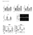
- FIG. 4 CPX and S1P induce secretion of different angiogenic proteins by endothelial cells and fibroblasts.
- A-D proteomic analysis plots of proteins upregulated in HUVEC treated with CPX+S1P.
- the upregulated proteins are PlGF (6.0 ⁇ ), IL-8 (3.0 ⁇ ), EGF (2.9 ⁇ ) and endothelin-1 (1.5 ⁇ ).
- F-I proteomic analysis plots of proteins upregulated in IMR-90 fibroblasts treated with CPX+S1P.
- the upregulated proteins include HGF (1.4 ⁇ ), IGFBP-2 (1.6 ⁇ ), uPA (1.8 ⁇ ) and VEGF (11 ⁇ ).
- CPX appears to be the main compound causing the observed upregulation.
- FIG. 5 Endothelial CM modulates growth factor secretion by fibroblasts.
- A ELISA: fibroblasts secrete 5 times more MCP-1 cultured in EC CM. No significant difference in MCP-1 secretion is elicited by CPX or S1P.
- B and (C): semi-quantitative proteomic array plots showing clearance of EC secreted endothelin-1 and EGF from culture media in the presence of fibroblasts. The negative values in the plots suggest either cellular take up or degradation of measured target proteins [* p ⁇ 0.05].
- Shown herein is a vascularization strategy in which one or more agents that induces HIF-1 ⁇ are used to induce angiogenesis (e.g., maximize angiogenic effects). Also shown herein are synergistic effects when one or more agents that induce HIF-1 ⁇ is used in combination with one or more lysophospholipids to induce angiogenesis.
- HIF-1 ⁇ is used to induce angiogenesis
- one or more agents that induce HIF-1 ⁇ in combination with one or more lysophospholipids is used to induce angiogenesis.
- sprouts had invaded and traveled the longest distance in a collagen I 3D scaffold under the induction of one or more agents that induces HIF-1 ⁇ and one or more lysophospholipids and the average length was 1.5 fold longer as compared to the sole induction of either an agent that induces HIF-1 ⁇ or a lysophospholipid.
- the methods provided herein are useful for angiogenesis (e.g., inducing angiogenesis, such as therapeutic angiogenesis for tissue engineering where neovascularization is required).
- the invention is directed to a method of inducing angiogenesis at a site in an individual in need thereof comprising administering locally to the site an effective amount of one or more agents that induce hypoxia induced factor 1 ⁇ (HIF-1 ⁇ ).
- the invention is directed to a method of inducing angiogenesis at a site in an individual in need thereof comprising administering locally to the site an effective amount of one or more agents that induce hypoxia induced factor 1 ⁇ (HIF-1 ⁇ ) and one or more lysophospholipids.
- angiogenesis refers to formation and/or induction of one or more blood vessels (a blood vessel; a vascular network such as a capillary bed) and includes neovascularization and revascularization.
- HIF-1 ⁇ also referred to herein as HIF-1 and HIF
- HIF-1 and HIF is a master switch that controls an array of angiogenic genes and subsequent peptide factors, including vascular endothelial growth factor (VEGF) (Semenza, G L, J Appli Physiol., 88(4):1474-1480 (2000); Kaelin, W G, Ann Rev Biochem, 74:115-128 (2005)). More than 50 direct target genes of HIF 1 have been identified.
- VEGF vascular endothelial growth factor
- HIF-1 ⁇ has recently gained tremendous interest as target for systemic therapies, e.g., anaemia treatment, since amongst those gene products regulated by HIF is the physiologically important erythropoietin (Melnikova I, Nat Rev Drug Discov, 5:627-628 (2006)).
- HIF-1 The action of HIF-1 is tightly controlled. Under normal (normoxic) conditions, HIF-1 ⁇ is quickly degraded because it is constantly hydroxylated at two prolyl residues by prolyl hydroxylase (PH) and at one asparaginyl residue by an asparaginyl hydroxylase (AH). The hydroxylated prolyl sites promote interaction with the von-Hippel Lindau tumor suppressor protein (pVHL). Constant hydroxylation earmarks HIF-1 ⁇ for proteosomal degradation, therefore, HIF-1 is highly unstable in normoxia (Bruick and McKnight, Science, 295(5556): 807-808 (2002)). If oxygen levels drop (hypoxia), both prolyl and asparaginyl residues are not hydroxlated. This has two effects, the degradation of HIF-1 ceases and the transcriptional activator p300 can now bind to HIF-1. As a result, HIF-1 survives and can act as a master switch for the above described HIF-1 dependent genes.
- HIF-1 ⁇ A variety of agents can be used to induce HIF-1 ⁇ .
- inducing HIF-1 ⁇ includes enhancing expression and/or activity of HIF-1 ⁇ .
- the one or more of agents induce HIF-1 ⁇ by inhibiting degradation of HIF-1 ⁇ .
- Both PH and AH are members of the dioxygenase family.
- Other human members of this family include deoxyhypusine hydroxylase (Clement et al., Int J Cancer, 100:491-498 (2002)) and collagen prolyl hydroxylase (CPH) which catalyses the hydroxylation of specific prolyl residues required to thermostabilise the collagen triple helix.
- Prolyl hydroxylase inhibitors are compounds that prevent the degradation of hypoxia inducible factor 1 ⁇ (HIF-1 ⁇ ) in vivo, thus upregulating various angiogenic factors including vascular endothelial growth factor (VEGF) as a response to the induced pseudo-hypoxic condition under normoxia.
- VEGF vascular endothelial growth factor
- PHis reduce fibrosis and prevent fibrotic capsule from forming. With this unique combination of pro-angiogenic and anti-fibrotic properties, PHis are distinct from other single-action approaches.
- the one or more of the agents that induce HIF-1 ⁇ comprise a hydroxylase inhibitor, an iron chelator or a combination thereof.
- the hydroxylase inhibitor can be a prolyl hydroxylase inhibitor (PHi), a collagen prolyl hydroxylase (CPH) inhibitor, an asparaginyl hydroxylase (AH) inhibitor, a deoxyhypusine inhibitor, cobalt ions or a combination thereof.
- PHi prolyl hydroxylase inhibitor
- CPH collagen prolyl hydroxylase
- AH asparaginyl hydroxylase
- a deoxyhypusine inhibitor cobalt ions or a combination thereof.
- Specific examples of a PHi include ciclopirox olamine (CPX), pyridine dicarboxylic acids, oxalylglycine, cis-proline, L-azetidine, thioenolpuridines, oxoglutarate analogues, or combinations thereof.
- a CPH inhibitor examples include hydralazine, biologically derived metabolites of hydralazine (e.g., hydralazine acetonide; hydralazine pyruvate hydrazine (Konili, Z H, et al., Drug Metab Rev, 6(2):283-305 (1977); Barron, K, et al., Br J Pharmacol, 61(3):345-349 (1977)) or combinations thereof.
- a specific example of an AH inhibitor includes dimethyloxaloylglycine (DMOG); and a specific example of a deoxyhypusine inhibitor includes mimosine.
- an iron chelator includes desferioxamine, ⁇ , ⁇ -dipyridine, mimosine, 3,4-dihydroxybenzoate, epolones or combinations thereof.
- Lysophospholipids are important mediators of multiple biological events including cell proliferation, survival and angiogenesis through specific cell surface G-protein coupled receptors. They are usually derived from the cells membranes and preferably bind to albumin in extracellular spaces. The lysophospholipids exert their effects through the G protein-coupled receptors (GPCR) and thus triggering downstream signaling.
- GPCR G protein-coupled receptors
- lysophospholipids can also be used in the methods and compositions described herein.
- a lysophospholipid include sphingosine 1-phosphate (SIP), lysophosphatidic acid (LPA), alkyl glycerol phosphate (AGP), cyclic phosphatidic acid (CPA) or combinations thereof.
- the one or more agents that induce HIF-1 ⁇ and the one or more lysophospholipids can be present in a biocompatible matrix (e.g., and administered locally to the site).
- a biocompatible matrix e.g., and administered locally to the site.
- any of a variety of biocompatible matrices can be used.
- a biocompatible matrix made of one or more metals, polymeric materials, biopolymers, carbohydrates, inorganic materials or combinations thereof can be used.
- a metal that can be used as a biocompatible matrix include titanium, steel, steel alloys, precious metals, copper, amalgams or combinations thereof.
- Examples of a polymeric materials that can be used as a biocompatible matrix include polyglycolic acid, polystyrene, polybutyrate, polygalactic acid, caprolactonic acid, acrylates, polyacrylate salts, silicones, polyesters, polyamides, polyamines, phthalates, or combinations thereof.
- Examples of a biopolymers that can be used as a biocompatible matrix include gelatin, collagen and artificial collagen, hyaluronic acid and derivatives, fibrin and modifications, keratin, chitin, chitosan, proteins, fibroins (e.g., derived from spider and silkworm), elastin, bone, coral, or combinations thereof.
- Examples of a carbohydrate that can be used as a biocompatible matrix include agarose, agar, alginate, starch and its derivatives, dextrans, cellulose and its derivatives (e.g., methyl cellulose), glycodendrimers, or combinations thereof.
- Examples of an inorganic materials that can be used as a biocompatible matrix include hydroxyapatite, tricalcium phosphate, silicates and/or from glass, ceramics, carbon, fullerenes. In addition, any derivatives of such materials and/or composite materials of such materials and/or mixtures thereof can be used as a biocompatible matrix.
- the biocompatible matrix is biodegradable.
- biocompatible matrices can be dissolved in appropriate diluents and used as coating for nondegradable or degradable biomaterials, or in admixture and thus constituent of degradable biomaterials, respectively.
- the biomaterials in question can be water soluble, soluble in organic solvents, and include but are not limited to polyglycolic acid, polygalactic acid, caprolactonic acid, acrylates, gelatin, collagen, hyaluronic acid and derivatives, fibrin and modifications, keratin, chitin, chitosan, silicone and silicates, titanium, steel, steel alloys, precious metals, copper, ceramics, glass, fibroins (both spider and silkworm), elastin, cellulose, methyl cellulose, starch, polystyrene, polybutyrate phthalate, agarose, agar, alginate.
- polyglycolic acid polygalactic acid, caprolactonic acid, acrylates, gelatin, collagen, hyaluronic acid and derivatives, fibrin and modifications, keratin, chitin, chitosan, silicone and silicates, titanium, steel, steel alloys, precious metals, copper, ceramics, glass, fibroin
- the application mode involves blending of predissolved HIF inducers and lysophospholipids to solutions of biomaterials which then harden, polymerise, or gel forming threads, meshworks, foams or solid blocks. Thus a biomateral is created that, upon dissolving, releases the compound.
- the biocompatible matrices can be created by electrospinning, extrusion, printing, fusion deposition, gelation including thermal gelation, salt leaching, sintering, recombinant expression, layer by layer deposition, smelting, alloying, freeze-drying, spraying, chemical crosslinking, biological crosslinking, photo and UV crosslinking, and/or dehydrothermal crosslinking.
- the biocompatible matrices can be used as films, 2D or 3D sponges, meshworks, nanofibers, microfibers, nanorods, micropellets, capsules, foams, braided fibers, knitted fibers, mono and polyfilic threads, capsules, endoprostheses, hydrogels, and/or 2D or 3D coatings.
- the one or more agents that induce HIF-1 ⁇ are used to stimulate one or more cell types that support and maintain functional blood vessels in an individual.
- one or more agents that induce HIF-1 ⁇ , and one or more agents that induce HIF-1 ⁇ in combination with one or more lysophospholipids are used to induce angiogenesis in the presence of one or more cell types that support and maintain functional blood vessels.
- the cells that support and maintain functional blood vessels are endogenous cells of an individual.
- the compositions and methods described herein can further comprise one or more cell types (exogenous cell types) that support and maintain functional blood vessels.
- the cells include one or more cell types that produce angiogenic factors, one or more cell types that induce angiogenesis or a combination thereof.
- the exogenous cells can be obtained from the individual being treated, from other individuals or from commercial sources.
- the exogenous cells can be autologous cells, allogeneic cells, syngeneic cells, xenogeneic cells, chimeric cells, synthetic cells and the like.
- the cells include endothelial cells, endothelial progenitor cells or lineages thereof, or combinations thereof; and/or mesenchymal cells, mesenchymal progenitor cells (e.g., mesenchymal stem cells and mesenchymal strom cells from, e.g., bone marrow, the perivascular niche of a tissue, placenta, umbilical cord, adipose tissue derived stem cells, stem cells derived from amniotic fluid, urine, breast milk or combinations thereof) or lineages thereof, hematopoietic cells, hematopoietic progenitor cells or lineages thereof, or combinations thereof.
- mesenchymal cells e.g., mesenchymal stem cells and mesenchymal strom cells from, e.g., bone marrow, the perivascular niche of a tissue, placenta, umbilical cord, adipose tissue derived stem cells, stem cells derived from amniotic
- Examples of cells from the lineages comprise fibroblasts, myofibroblasts, osteoblasts, chondrocytes, smooth muscle cells, skeletal muscle cells, heart muscle cells, monocytes, macrophages, lymphocytes, granulocytes, megakaryocytes, dendritic cells, astrocytes, mast cells, cardiomyocytes, pericytes or a combination thereof.
- Additional cells include stem cells, neuroectodermal and endodermal origin including epithelial cells of outer and inner surfaces of the human body such as keratinocytes, urothel, melanocytes, neuronal cells, neuroglia, hepatocytes, endocrine pancreas epithelium.
- the methods and compositions provided herein can be used in a variety of sites of an individual in need thereof.
- sites include a wound (e.g., amputation; chronic wound such as a venous wound, diabetic wound, ulcer), a graft (cell, tissue, organ, bone, skin), a prosthetic device, a suture, an area of malperfusion (e.g., low perfusion such as in critical limb ischemia), an infarcted area (e.g., myocardial infarction, a stroke area (e.g., brain)), a bone fracture, an area of non-union in bone (e.g., a gap in bone that needs to be filled with, for example, cartilage or bone), an area at or near an implant or sensor (e.g., to facilitate perfusion around a sensor, such as a glucose sensor).
- a wound e.g., amputation; chronic wound such as a venous wound, diabetic wound, ulcer
- a graft cell, tissue
- the invention is directed to therapies aimed at conditions and/or diseases which require angiogenesis (therapeutic angiogenesis) in an individual in need thereof (e.g., therapies involving revascularization of a tissue/organ (e.g., infarcted hearts)).
- angiogenesis therapeutic angiogenesis
- the therapy ameliorates the symptoms associated with the condition and/or disease in an individual.
- an “individual” refers to an animal, and in a particular aspect, a mammal.
- mammals include primates, a canine, a feline, a rodent, and the like.
- Specific examples include humans, dogs, cats, horses, cows, sheep, goats, rabbits, guinea pigs, rats and mice.
- an individual in need thereof refers to an individual who is in need of treatment or prophylaxis as determined by a researcher, veterinarian, medical doctor or other clinician.
- an individual in need thereof is a mammal, such as a human.
- the need or desire for administration according to the methods of the present invention is determined via the use of well known risk factors.
- the effective amount of a (one or more) particular compound is determined, in the final analysis, by the physician in charge of the case, but depends on factors such as the exact condition to be treated, the severity of the condition from which the patient suffers, the chosen route of administration, other drugs and treatments which the patient may concomitantly require, and other factors in the physician's judgment.
- an “effective amount” or “therapeutically effective amount” means an amount of the active compound that will elicit the desired biological or medical response in a tissue, system, subject, or human, which includes alleviation of the symptoms, in whole or in part, of the condition being treated.
- compositions e.g., one or more agents that induce hypoxia, one or more lysophospholipids and combinations/matrices thereof
- a single dose e.g., in a day
- multiple doses e.g., the one or more agents that induce hypoxia and the one or more lysophospholipids can be administered together (at the same time) as a single composition or as separate compositions but at the same time.
- the one or more agents that induce hypoxia and the one or more lysophospholipids can be administered sequentially (administer one composition immediately after the other; administer one composition and after a time period administer the other composition).
- the one or more compositions can be administered in one or more days (e.g. over several consecutive days or non-consecutive days, weeks, months, years).
- the (one or more) site in the individual can be contacted with the compositions (e.g., one or more agents that induce hypoxia, one or more lysophospholipids and combinations/matrices thereof) described herein in a variety of ways.
- the compositions can be administered systemically or locally to the individual.
- the compositions are administered locally to the one or more sites (e.g., placed in direct contact with the one or more tissues, organs, prosthetics, blood vessels and the like).
- the site can be contacted with the compositions using a variety of methods.
- the compositions could be transplanted alone or incorporated within, or as part of, a support (e.g., a scaffold; a gel) into e.g., a targeted tissue/organ (e.g., heart).
- Prevascularized tissue refers to a blood vessel (vascular) network within a tissue before it is implanted into the body.
- the method comprises culturing (i) one or more agents that induce hypoxia induced factor 1 ⁇ (HIF-1 ⁇ ), (ii) one or more cell types that produce angiogenic factors and (iii) one or more cell types that induce angiogenesis, thereby producing a culture.
- the culture is maintained under conditions in which angiogenesis occurs and prevascularized tissue forms, thereby generating prevascularized tissue ex vivo.
- cell types that produce angiogenic factors include mesenchymal cells, mesenchymal progenitor cells or lineages thereof, hematopoietic cells, hematopoietic progenitor cells or lineages thereof, or combinations thereof, and cell types that induce angiogenesis comprise endothelial cells, endothelial progenitor cells or lineages thereof, or combinations thereof.
- the method of generating prevascularized tissue ex vivo can further comprises including one or more lysophospholipids in the culture.
- the method of generating prevascularized tissue ex vivo can further comprises including a biocompatible matrix in the culture, whereby the prevascularized tissue forms on or within the biocompatible matrix.
- the invention is directed to compositions produced by the method of generating prevascularized tissue ex vivo. In one aspect, the invention is directed to prevascularized tissue produced by the methods described herein. In another aspect, the invention is directed to a pharmaceutical composition comprising the prevascularized tissue.
- compositions encompassed by the invention include a composition comprising (i) a biocompatible matrix, (ii) one or more agents that induce hypoxia induced factor 1 ⁇ (HIF-1 ⁇ ) and (iii) one or more lysophospholipids.
- a composition comprising (i) a biocompatible matrix, (ii) one or more agents that induce hypoxia induced factor 1 ⁇ (HIF-1 ⁇ ) and (iii) one or more lysophospholipids.
- the biocompatible matrix is coupled to (e.g., coated; covalently coupled) the one or more agents that induce hypoxia induced factor 1 ⁇ (HIF-1 ⁇ ) and the one or more lysophospholipids, for example, as a coated prosthesis (e.g., a coated hip prosthesis, a coated knee prosthesis); a sponge (e.g., collagen sponge) covalently coupled to, or soaked with, one or more agents that induce hypoxia induced factor 1 ⁇ (HIF-1 ⁇ ) and one or more lysophospholipids.
- the composition can further comprise (iv) one or more cell types that produce angiogenic factors and (v) one or more cell types that induce angiogenesis.
- the invention is directed to a pharmaceutical composition
- a pharmaceutical composition comprising (i) a biocompatible matrix, (ii) one or more agents that induce hypoxia induced factor 1 ⁇ (HIF-1 ⁇ ) and (iii) one or more lysophospholipids.
- the pharmaceutical composition further comprises (iv) one or more cell types that produce angiogenic factors and (v) one or more cell types that induce angiogenesis.
- the invention is also directed to a method of generating a vascular network in a device comprising culturing (i) one or more agents that induce hypoxia induced factor 1 ⁇ (HIF-1 ⁇ ), (ii) one or more cell types that produce angiogenic factors and (iii) one or more cell types that induce angiogenesis, thereby producing a culture.
- the culture is maintained under conditions in which a vascular network forms in the device, thereby generating a vascular network in the device.
- the one or more cells types that induce angiogenesis are endothelial cells.
- endothelial cells include human umbilical venous endothelial cells (HUVEC), microvascular endothelial cells (e.g., from dermis, placenta, adipose tissue, muscle, retina, brain and other neural tissue), arterial macrovascular endothelial cells (e.g., from arteries and aorta), lymphatic endothelial cells, outgrowth endothelial cells (e.g., from human peripheral blood buffy coat, outgrowth endothelial cells (e.g., from human bone marrow aspirate), endothelial cells generated from human embryonic stem cells, endothelial cells generated from induced pluripotent stem cells (iPSC), endothelial cells generated from mesenchymal stem cells (MSCs), endothelial cells generated from adult stem cells (ASCs) or combinations thereof.
- HAVEC human umbilical ve
- the one or more cells that produce angiogenic factors comprise fibroblasts.
- fibroblasts include human lung fibroblasts (IMR-90), cancer fibroblasts from cancer stroma, primary fibroblasts from any fibroblast containing tissue such as dermis, muscle, placenta and the like.
- the one or more cell types that induce angiogenesis and/or produce angiogenic factors are present in a medium (e.g., gel; hydrogel) that, for example, provides control over solutes concentration gradients and/or allows visualization of angiogenic events such as sprouting and length/direction of sprouts.
- a medium e.g., gel; hydrogel
- the one or more cells that produce angiogenic factors are encapsulated, for example, in alginate beads.
- the device used in the method of generating a vascular network can further comprise one or more lysophospholipids.
- the device used in the method of generating a vascular network can further comprise a biocompatible matrix.
- the device used in the method of generating a vascular network can further comprise a control (e.g., an area of the device in which prevascularized tissue will not form).
- a control e.g., an area of the device in which prevascularized tissue will not form.
- the control comprises encapsulated alginate beads that do not contain cells.
- the device is a microfluidic device.
- the invention is also directed to a vascular network produced by the methods provided herein.
- the invention is directed to an angiogenic assay device comprising (i) one or more agents that induce hypoxia induced factor 1 ⁇ (HIF-1 ⁇ ), (ii) one or more cell types that produce angiogenic factors and (iii) one or more cell types that induce angiogenesis, wherein the one or more cell types that induce angiogenesis form a vascular network in the presence of the one or more agents that induce HIF-1 ⁇ and the one or more cell types that produce angiogenic factors in the device.
- HIF-1 ⁇ hypoxia induced factor 1 ⁇
- the device further comprises one or more lysophospholipids.
- the device further comprises a biocompatible matrix.
- the one or more compounds described, used or generated in the methods described herein can be administered to a subject as part of a pharmaceutical composition.
- Formulations will vary according to the route of administration selected (e.g., solution, emulsion or capsule).
- a “pharmaceutical composition” comprises a (one or more) chemical compound described herein as the active ingredient and inert ingredient(s), such as pharmaceutically acceptable excipients, that make up the carrier. Standard pharmaceutical formulation techniques can be employed, such as those described in RemingtonTM Pharmaceutical Sciences, Mack Publishing Company, Easton, Pa.
- Suitable pharmaceutical carriers for parenteral administration include, for example, sterile water, physiological saline, bacteriostatic saline (saline containing about 0.9% mg/ml benzyl alcohol), phosphate-buffered saline, HanksTM solution, RingerTM lactate and the like. Formulations can also include small amounts of substances that enhance the effectiveness of the active ingredient (e.g., emulsifying, solubilizing, pH buffering, wetting agents). Methods of encapsulation compositions (such as in a coating of hard gelatin or cyclodextran) are known in the art. For inhalation, the agent can be solubilized and loaded into a suitable dispenser for administration (e.g., an atomizer or nebulizer or pressurized aerosol dispenser).
- a suitable dispenser for administration e.g., an atomizer or nebulizer or pressurized aerosol dispenser.
- Any suitable route of administration can be used, for example, local (e.g., administration/implantation at the site in need of angiogenesis), oral, dietary, topical, transdermal, rectal, parenteral (e.g., intravenous, intraarterial, intramuscular, subcutaneous injection, intradermal injection), inhalation (e.g., intrabronchial, intranasal or oral inhalation, intranasal drops), ocular, pulmonary, nasal, and the like may be employed.
- Administration can be local or systemic as indicated. The preferred mode of administration can vary depending on the particular agent chosen. Suitable dosage forms include tablets, troches, dispersions, suspensions, solutions, capsules, creams, ointments, aerosols, and the like.
- agents that induce HIF e.g., PHi
- lysophospholipids e.g., S1P
- the combination of agents that induce HIF-1 ⁇ and lysophospholipids can be utilized in tissues either for the application of tissue engineering or for therapeutic angiogenesis.
- compositions and methods that induce angiogenesis by combining angiogenic and antifibrotic effects and priming tissue with lysophospholipids that induce longer viability of endothelium.
- the compositions and methods can induce vascularization (e.g., neovascularisation) of a site in an individual, e.g., by inducing and luring sprouting capillaries from around the implanted compositions into the implanted compositions.
- fibrotic capsule formation is prevented from forming around the implant.
- the compositions and methods can be used to prevascularise in vitro tissue constructs before implantation, and thus, can be used as a type of wound dressing that can be used, for example, in chronic non-healing wounds to induce vascularisation.
- compositions and methods can be used for a variety of purposes, such as for ex vivo prevascularizing grafts in an endothelial coculture setting (static or in bioreactor) by immersion or perfusion with such substances; locally releasing angiogenic inducing agents to locally induce angiogenesis/neovascularisation of implants with or without cells upon implantation; locally inducing HIF and improving vascular integrity by releasing the angiogenic inducing agents in the immediate vicinity of an implant; locally delivering angiogenic agents from wound care materials (e.g., dressings; bandages and the like).
- wound care materials e.g., dressings; bandages and the like.
- compositions and methods include fibrin gel matrices which can be prepared by mixing the following components at the final concentrations of 10 mg/mL fibrinogen (Fluka AG), 2 U/mL factor XIII (Baxter AG, Vienna), and 2.5 mmol/L CaCl2.
- HIF inducers can be mixed with lysophospholipids and then added to the fibrinogen solution before initiation of fibrin gelation by addition of thrombin.
- the preferred concentration range depends on the potency of the respective HIF inducer and lysophospholipid and the velocity of the degradation.
- Preferred concentrations are 5-30, in particular 10 ⁇ g/ml CPX, 10 mM PDCA, 100-500 ⁇ M hydralazine (for example) and 250 nM S1P.
- the biomaterials comprising HIF-inducers and lysophospholipids can be seeded with a mixture of cells belonging to two different groups.
- Group 1 comprises endothelial progenitor cells or preferably endothelial cells.
- Group 2 are derived from mesenchymal and haematopoetic progenitors including but not being restricted to lineages like fibroblasts, myofibroblasts, osteoblasts, chondrocytes, smooth muscle cells, skeletal muscle cells, heart muscle cells, monocytes, macrophages, astrocytes, mast cells, cardiomyocytes and pericytes. These cocultures are seeded as a mixture or sequentially.
- Group 2 cells are seeded first, and Group 1 cells later or vice versa, depends on the designs and properties of biomaterials.
- the seeding into a given scaffold can be augmented using hydrogels, collagen gels, fibrin glue and similar formulations.
- the seeded scaffolds are kept in static culture or in bioreactors under flow conditions.
- the released HIF-inducers from the scaffold material then stimulate Group 2 cells to produce angiogenic factors which in turn will induce angiogenesis in Group 1 cells.
- lysophospholipids could further enhance the secretion of angiogenic factors by Group 2 cells or help activate Group I cells thus making them more responsive to the angiogenic factors being secreted by Group 2 cells.
- cell free biomaterials are implanted into desired body locations, whereby the implanted biomaterial released HIF inducing substances and lysophospholipids locally and thus activates surrounding tissue to send capillary sprouts into the biomaterial (outside in approach).
- degradable biomaterials containing HIF-inducing substances and lysophospholipids are used as suturing material.
- Biodegradable poly(lactide-co-glycolide) (PLGA) fibers are promising drug carriers. Drugs can be incorporated into fibers and released as PLGA is hydrolyzed. The drug release rate is partially dependent on the rate of hydrolysis of the PLGA fibers, threads or meshes which is affected by molecular weight, copolymer composition and fiber morphology. These fibers are used as polyfilic or monofilic threads for the suturing of tissue. While the sutures are degraded by the surrounding tissue, the local release of HIF-inducers and lysophospholipids will increase the local angiogenesis and reduces local scarring. The preferred embodiment will be 10% L-lactide/90% glycolide (polyglactin 910).
- Another aspect involves coating of predissolved HIF inducers and lysophospholipids to a biomaterial, thus the surface delivers the HIF inducing substances and lysophospholipids within a certain time window.
- PLGA scaffolds are soaked in HIF-inducing substance and lysophospholipids containing solutions. After incubation and swelling preferably overnight scaffolds are subjected to freeze drying. Alternatively HIF-inducing substances and lysophospholipids are admixed to a biomaterial in the fluid phase which then is used to coat a given scaffold. Preferably thermal gelation of PEG/PLGA tri block copolymers would be a mode of action here.
- surfaces of prostheses are activated using air, argon or any other plasma and are immediately after treatment coated with HIF inducing substances and lysophospholipids-containing solutions.
- predissolved HIF inducers can be coupled to the surface of a biomaterial, involving spacers that are cleavable by radicals or proteolytic enzymes. This allows specific release on cellular demand.
- HIF inducing substances and lysophospholipids are thiolated as known to those skilled in the art of chemical synthesis and coupled to a peptide combining a plasmin cleavage site and the factor XIIIa substrate sequence CKTYKNQEQVSPL via usual coupling reactions.
- the coupled HIF inducer and lysophospholipids are then added to the preparation of fibrin gel matrices which are prepared by mixing the following components at the final concentrations of 10 mg/mL fibrinogen (Fluka AG), 2 U/mL factor XIII within the fibrinogen solution before initiation of fibrin gelation by addition of thrombin.
- the Factor XIIa sequence is attached covalently via epsilon lysyl gamma glutamyl isodipeptide crosslinks to the fibrin scaffold. Cells invading the scaffold and expressing plasmin on their surface or secreting it will liberate the HIF-inducer and thus activate themselves or bystanding cells.
- small particles of degradable biomaterial containing said substances can be created. These particles are delivered via biolistic delivery employing a short pressure pulse.
- the Helios gene gun (BioRad) is a convenient, hand-held device that was originally designed to provide rapid and direct gene transfer into a range of targets in vivo.
- the unit uses an adjustable low-pressure helium pulse to sweep DNA- or RNA-coated gold microcarriers from the inner wall of a small plastic cartridge directly into the target.
- the HIF inducers and lysophospholipids are either deposited onto gold particles or microparticles made of f.e. fibrin, PCL, or PLGA with diameter ranging from 0.5 um to 2 ⁇ m are used to deliver HIF inducers and lysophospholipids.
- Dry Micropellets containing HIF inducers and lysophospholipids are loaded into a helium driven gun (f.e. Helios Gene gun by Bio-Rad) and are injected into tissue. This can be done through intact surfaces of skin or mucosa or into tissue constructs ex vivo on into surgical sites in vivo.
- a helium driven gun f.e. Helios Gene gun by Bio-Rad
- the HIF inducing substances are blended into the biomaterial and/or are part of a degradable coating of the biomaterial and/or are affixed to the biomaterials by bonds that are cleavable by proteolytic enzymes or radicals.
- the lysophospholipids are also blended into the biomaterial and/or are part of a degradable coating of the biomaterial and/or are affixed to the biomaterials by bonds that are cleavable by proteolytic enzymes or radicals.
- the cleavable bonds represent spacers containing cleavage motifs for proteolytic enzymes preferentially but not limited to trypsin, chymotrypsin, chymase, cathepsin, plasmin, uroplasmin, matrix metalloproteinases (MMP's), thrombin, C1-esterase, serineproteases, cysteine proteases.
- proteolytic enzymes preferentially but not limited to trypsin, chymotrypsin, chymase, cathepsin, plasmin, uroplasmin, matrix metalloproteinases (MMP's), thrombin, C1-esterase, serineproteases, cysteine proteases.
- the invention solves the problem of local angiogenesis induction and antifibrosis, using stable pharmacological compounds that are not dependent on liver activation.
- the substances are delivered to the point of need, i.e. abolishing the need to administer systemically with expectable side effects and without the dependency on stable incorporation of cDNA.
- compositions and methods can be used to connect implantable sensors (e.g., that measure blood chemicals (fe. glucose)) into the blood system.
- implantable sensors e.g., that measure blood chemicals (fe. glucose)
- the inventions provided methods and compositions that allow implants/scaffolds that are not seeded with cells can get rapidly vascularised (f.e.
- tissue constructs in static and bioreactor culture conditions e.g., muscle, bone, soft tissue as such etc
- endothelial cells or progenitor cells e.g., endothelial cells or progenitor cells
- prevascularisation of tissue constructs in static and bioreactor culture conditions e.g., muscle, bone, soft tissue as such etc
- endothelial cells or progenitor cells for an administrative route of HIF inducing drugs and lysophospholipids via local drug delivery which includes osmotic pumps, biolistic particles releasing drugs (micro and nanoscale), scaffolds that partially degrade and release the drugs, or coatings of scaffolds that set the drugs free, in particular meshed scaffolds, nano and microfibers, scaffold made by fusion deposition, extrusion, salt leaching, layer by layer construction, foaming, gelling; advanced wound care materials that are used as bandage or to cover chronic wound
- Angiogenesis represents a major challenge in regenerative medicine and tissue engineering.
- the synergistic effects of PHis and S1P were studied in an in vitro microfluidic platform where endothelial cells were co-cultured with fibroblast cells and subjected to the stimulation of PHis and S1P. Shown herein is that the interactions between endothelial cells and mesenchymal cells or fibroblasts maintain immature sprouts and turn them into functional vessels with lumens and these effects are most prominent with PHis and S1P. Thus, PHis worked synergistically with S1P.
- PHis and S1P were studied on human umbilical venous endothelial cells (HUVEC) whereby fibroblasts in a neighbouring channel served as angiogenic secretory cells when stimulated by PHis and S1P.
- HUVEC human umbilical venous endothelial cells
- synergistic effects of PHis and S1P developed functional vascular network in microfluidic platform.
- Microfluidic devices with the capability of accommodating IMR-90 (human lung fibroblasts) encapsulated alginate beads were used as they offered excellent control over solutes concentration gradient and allowed observation of sprouting events in 3D collagen regions.
- the microfluidic devices were made using standard soft lithography methods and alginate beads were generated as described earlier (Vickerman et al., 2008, Kim et al., 2009).
- HUVECs were seeded in the middle channel. Alginate beads were introduced into left and right channels after HUVECs formed a monolayer.
- HUVECs formed a monolayer on collagen gel as well as top and bottom of the channel. Hoechst stained nuclei (blue), rhodamine phalloidin stained actin (red) and Alexa fluor 488 immunostained VE-cadherin (green). Scale bar denotes 50 ⁇ m
- Ciclopirox olamine a type of PHi was used in combination with S1P in all three channels in microfluidic devices. Immature sprouts that were observed on day two turned into vessels, if they were growing towards IMR-90 encapsulated beads, on day four. On the other hand, the immature sprouts broke off from the monolayer and migrated randomly in collagen on the side that was towards empty alginate beads. Hence, the importance of mesenchymal-endothelial cells interactions was shown in the microfluidic model.
- capillary-like structure (CLS) is commonly used as assessment for degree of angiogenesis.
- Proteome profiler data indicated the increased secretion of several proteins including PlGF (6 fold), IL-8 (3 fold), EGF (2.9 fold) and endothelin-1 (1.5 fold) by CPX+S1P by endothelial cells. Highly comparable increases were found in the presence of CPX alone, and none were observed with S1P only.
- Endothelial Cells CM Increases MCP-1 Secretion by IMR90
- IMR90 cells were cultured in EC conditioned medium with or without the addition of CPX+S1P, and quantified factors secreted by fibroblasts by ELISA.
- Fibroblasts showed a basal secretion of MCP-1.
- Endothelial cell CM induced the secretion of MCP-1 by fibroblasts by 3-fold in the absence of any CPX+S1P induction, however, the combination of both pharmacological agents increased MCP-1 secretion by a further 20%.
- S1P nor CPX were able to increase basal MCP-1 secretion. See FIG. 5 .
Landscapes
- Health & Medical Sciences (AREA)
- Life Sciences & Earth Sciences (AREA)
- Engineering & Computer Science (AREA)
- Chemical & Material Sciences (AREA)
- Biomedical Technology (AREA)
- General Health & Medical Sciences (AREA)
- Wood Science & Technology (AREA)
- Zoology (AREA)
- Organic Chemistry (AREA)
- Genetics & Genomics (AREA)
- Bioinformatics & Cheminformatics (AREA)
- Biotechnology (AREA)
- Medicinal Chemistry (AREA)
- Epidemiology (AREA)
- Public Health (AREA)
- Pharmacology & Pharmacy (AREA)
- Veterinary Medicine (AREA)
- Animal Behavior & Ethology (AREA)
- General Engineering & Computer Science (AREA)
- Microbiology (AREA)
- Biochemistry (AREA)
- Cell Biology (AREA)
- Vascular Medicine (AREA)
- Micro-Organisms Or Cultivation Processes Thereof (AREA)
- Medicines Containing Material From Animals Or Micro-Organisms (AREA)
Abstract
Description
| TABLE 1 |
| Angiogenic proteins secreted by EC and fibroblasts |
| treated with CPX + S1P |
| Proteins | Fibroblasts | HUVEC | Upregulated by | ||
| HGF | + (++) | − | CPX | ||
| IGFBP-2 | + (++) | − | CPX | ||
| uPA | + (++) | − | CPX | ||
| VEGF | + (++) | − | CPX | ||
| Endothelin-1 | − | + (++) | CPX | ||
| P1GF | − | + (++) | CPX | ||
| EGF | − | + (++) | |||
| IL-8 | + | + (++) | |||
| MCP-1 | + (+) | + (−−) | Endothelial CM | ||
| “+” denotes protein secretion spontaneously; | |||||
| “−” denotes no protein secretion spontaneously; | |||||
| “(++)” denotes upregulation of protein secretion when treated with CPX + S1P; | |||||
| “(−−)” denotes downregulation of protein secretion when treated with CPX + S1P; | |||||
| “(+)” denotes upregulation of protein secretion by endothelial CM | |||||
Claims (4)
Priority Applications (1)
| Application Number | Priority Date | Filing Date | Title |
|---|---|---|---|
| US13/743,223 US9446031B2 (en) | 2012-01-18 | 2013-01-16 | Compositions and methods for neovascularization |
Applications Claiming Priority (2)
| Application Number | Priority Date | Filing Date | Title |
|---|---|---|---|
| US201261587713P | 2012-01-18 | 2012-01-18 | |
| US13/743,223 US9446031B2 (en) | 2012-01-18 | 2013-01-16 | Compositions and methods for neovascularization |
Publications (2)
| Publication Number | Publication Date |
|---|---|
| US20130197038A1 US20130197038A1 (en) | 2013-08-01 |
| US9446031B2 true US9446031B2 (en) | 2016-09-20 |
Family
ID=48870750
Family Applications (1)
| Application Number | Title | Priority Date | Filing Date |
|---|---|---|---|
| US13/743,223 Expired - Fee Related US9446031B2 (en) | 2012-01-18 | 2013-01-16 | Compositions and methods for neovascularization |
Country Status (1)
| Country | Link |
|---|---|
| US (1) | US9446031B2 (en) |
Families Citing this family (3)
| Publication number | Priority date | Publication date | Assignee | Title |
|---|---|---|---|---|
| CN103833623B (en) * | 2014-03-03 | 2016-07-06 | 河南大学 | One seed amino acid-amine conjugate and its preparation method and application |
| GB201720320D0 (en) * | 2017-12-06 | 2018-01-17 | Univ Of Sussex | Tissue repair |
| CN115252869B (en) * | 2022-08-18 | 2023-06-06 | 南通大学 | A nano dressing for promoting blood vessel regeneration and preparation method thereof |
Citations (29)
| Publication number | Priority date | Publication date | Assignee | Title |
|---|---|---|---|---|
| US5882914A (en) | 1995-06-06 | 1999-03-16 | The Johns Hopkins University School Of Medicine | Nucleic acids encoding the hypoxia inducible factor-1 |
| US6124131A (en) | 1998-08-25 | 2000-09-26 | The Johns Hopkins University School Of Medicine | Mutant hypoxia inducible factor-1 HIF-1 |
| WO2002015955A2 (en) | 2000-08-23 | 2002-02-28 | Surfarc Aps | Biocompatible materials |
| US20020115165A1 (en) | 1986-11-20 | 2002-08-22 | Stein James E. | Prevascularzed polymeric implants for organ transplantation |
| WO2002074981A2 (en) | 2001-03-21 | 2002-09-26 | Isis Innovation Ltd. | Assays, methods and means relating to hypoxia inducible factor (hif) hydroxylase |
| US20030022838A1 (en) | 1999-02-19 | 2003-01-30 | Sheppard Paul O. | Methods for pacifying the surface of a prosthetic biomaterial |
| US20030176317A1 (en) | 2001-12-06 | 2003-09-18 | Volkmar Guenzler-Pukall | Stabilization of hypoxia inducible factor (HIF) alpha |
| US6660737B2 (en) | 2001-05-04 | 2003-12-09 | The Procter & Gamble Company | Medicinal uses of hydrazones |
| US6753321B2 (en) | 2000-09-15 | 2004-06-22 | Genvec, Inc. | Method of modulating neovascularization |
| US6838430B2 (en) | 2001-09-28 | 2005-01-04 | The Regents Of The University Of California | Use of HIF-1a variants to accelerate wound healing |
| US20050227948A1 (en) | 2002-03-21 | 2005-10-13 | Schofield Christopher J | Hif hydroxylase inhibitors |
| US6986881B1 (en) | 1999-06-04 | 2006-01-17 | Dana-Farber Cancer Institute, Inc. | Identification of compounds that modify transcriptional responses to hypoxia |
| US20060023763A1 (en) | 2004-07-28 | 2006-02-02 | Nlight Photonics Corporation | Semiconductor lasers with hybrid materials systems |
| US7053046B2 (en) | 2001-12-21 | 2006-05-30 | Mcgrath Kevin | Peptide activators of VEGF |
| US20060141000A1 (en) | 1993-02-01 | 2006-06-29 | Mikos Antonios G | Porous biodegradable polymeric materials for cell transplantation |
| US20060199836A1 (en) | 2005-03-02 | 2006-09-07 | Fibrogen, Inc. | Novel thienopyridine compounds, and methods of use thereof |
| US20060198864A1 (en) | 2003-05-21 | 2006-09-07 | Mark Shults | Biointerface membranes incorporating bioactive agents |
| US20070003589A1 (en) | 2005-02-17 | 2007-01-04 | Irina Astafieva | Coatings for implantable medical devices containing attractants for endothelial cells |
| US20090110711A1 (en) | 2007-10-31 | 2009-04-30 | Trollsas Mikael O | Implantable device having a slow dissolving polymer |
| US20090136553A1 (en) | 2007-09-25 | 2009-05-28 | Gerlach Jorg C | Triggerably dissolvable hollow fibers for controlled delivery |
| WO2009085272A2 (en) | 2007-12-21 | 2009-07-09 | Coda Therapeutics, Inc. | Improved medical devices |
| US20090214616A1 (en) | 2006-03-03 | 2009-08-27 | Washington University In St. Louis | Biomaterials having nanoscale layers and coatings |
| US20090324681A1 (en) | 2002-05-02 | 2009-12-31 | Purdue Research Foundation | Vascularization Enhanced Graft Constructs |
| US20100034794A1 (en) | 2006-10-03 | 2010-02-11 | Van Der Strate Barry W A | Endothelial progenitor cell compositions and neovascularization |
| WO2010118298A2 (en) | 2009-04-09 | 2010-10-14 | University Of Virginia Patent Foundation | Compositions and methods for bioactive coatings to improve allograft incorporation |
| US7860545B2 (en) | 1997-03-04 | 2010-12-28 | Dexcom, Inc. | Analyte measuring device |
| US20110238000A1 (en) | 2008-12-07 | 2011-09-29 | Technion Research And Development Foundation Ltd. | Polymer-conjugated albumin hydrogels for controlled release of therapeutic agents |
| US8060174B2 (en) | 2005-04-15 | 2011-11-15 | Dexcom, Inc. | Analyte sensing biointerface |
| EP1658074B1 (en) | 2003-08-01 | 2012-11-07 | Fibrogen, Inc. | Inhibitors of 2-oxoglutarate dioxygenase as gamma globin inducers |
-
2013
- 2013-01-16 US US13/743,223 patent/US9446031B2/en not_active Expired - Fee Related
Patent Citations (31)
| Publication number | Priority date | Publication date | Assignee | Title |
|---|---|---|---|---|
| US20020115165A1 (en) | 1986-11-20 | 2002-08-22 | Stein James E. | Prevascularzed polymeric implants for organ transplantation |
| US20060141000A1 (en) | 1993-02-01 | 2006-06-29 | Mikos Antonios G | Porous biodegradable polymeric materials for cell transplantation |
| US5882914A (en) | 1995-06-06 | 1999-03-16 | The Johns Hopkins University School Of Medicine | Nucleic acids encoding the hypoxia inducible factor-1 |
| US7860545B2 (en) | 1997-03-04 | 2010-12-28 | Dexcom, Inc. | Analyte measuring device |
| US6562799B1 (en) | 1998-08-25 | 2003-05-13 | The Johns Hopkins University School Of Medicine | Stable hypoxia inducible factor-1α and method of use |
| US6124131A (en) | 1998-08-25 | 2000-09-26 | The Johns Hopkins University School Of Medicine | Mutant hypoxia inducible factor-1 HIF-1 |
| US20030022838A1 (en) | 1999-02-19 | 2003-01-30 | Sheppard Paul O. | Methods for pacifying the surface of a prosthetic biomaterial |
| US6986881B1 (en) | 1999-06-04 | 2006-01-17 | Dana-Farber Cancer Institute, Inc. | Identification of compounds that modify transcriptional responses to hypoxia |
| WO2002015955A2 (en) | 2000-08-23 | 2002-02-28 | Surfarc Aps | Biocompatible materials |
| US6753321B2 (en) | 2000-09-15 | 2004-06-22 | Genvec, Inc. | Method of modulating neovascularization |
| WO2002074981A2 (en) | 2001-03-21 | 2002-09-26 | Isis Innovation Ltd. | Assays, methods and means relating to hypoxia inducible factor (hif) hydroxylase |
| US6660737B2 (en) | 2001-05-04 | 2003-12-09 | The Procter & Gamble Company | Medicinal uses of hydrazones |
| US6838430B2 (en) | 2001-09-28 | 2005-01-04 | The Regents Of The University Of California | Use of HIF-1a variants to accelerate wound healing |
| US20030176317A1 (en) | 2001-12-06 | 2003-09-18 | Volkmar Guenzler-Pukall | Stabilization of hypoxia inducible factor (HIF) alpha |
| US7053046B2 (en) | 2001-12-21 | 2006-05-30 | Mcgrath Kevin | Peptide activators of VEGF |
| US20050227948A1 (en) | 2002-03-21 | 2005-10-13 | Schofield Christopher J | Hif hydroxylase inhibitors |
| US20090324681A1 (en) | 2002-05-02 | 2009-12-31 | Purdue Research Foundation | Vascularization Enhanced Graft Constructs |
| US20060198864A1 (en) | 2003-05-21 | 2006-09-07 | Mark Shults | Biointerface membranes incorporating bioactive agents |
| EP1658074B1 (en) | 2003-08-01 | 2012-11-07 | Fibrogen, Inc. | Inhibitors of 2-oxoglutarate dioxygenase as gamma globin inducers |
| US20060023763A1 (en) | 2004-07-28 | 2006-02-02 | Nlight Photonics Corporation | Semiconductor lasers with hybrid materials systems |
| US20070003589A1 (en) | 2005-02-17 | 2007-01-04 | Irina Astafieva | Coatings for implantable medical devices containing attractants for endothelial cells |
| US20060199836A1 (en) | 2005-03-02 | 2006-09-07 | Fibrogen, Inc. | Novel thienopyridine compounds, and methods of use thereof |
| US8060174B2 (en) | 2005-04-15 | 2011-11-15 | Dexcom, Inc. | Analyte sensing biointerface |
| US20090214616A1 (en) | 2006-03-03 | 2009-08-27 | Washington University In St. Louis | Biomaterials having nanoscale layers and coatings |
| US20100034794A1 (en) | 2006-10-03 | 2010-02-11 | Van Der Strate Barry W A | Endothelial progenitor cell compositions and neovascularization |
| US20090136553A1 (en) | 2007-09-25 | 2009-05-28 | Gerlach Jorg C | Triggerably dissolvable hollow fibers for controlled delivery |
| US20090110711A1 (en) | 2007-10-31 | 2009-04-30 | Trollsas Mikael O | Implantable device having a slow dissolving polymer |
| WO2009085272A9 (en) | 2007-12-21 | 2010-06-10 | Coda Therapeutics, Inc. | Improved medical devices |
| WO2009085272A2 (en) | 2007-12-21 | 2009-07-09 | Coda Therapeutics, Inc. | Improved medical devices |
| US20110238000A1 (en) | 2008-12-07 | 2011-09-29 | Technion Research And Development Foundation Ltd. | Polymer-conjugated albumin hydrogels for controlled release of therapeutic agents |
| WO2010118298A2 (en) | 2009-04-09 | 2010-10-14 | University Of Virginia Patent Foundation | Compositions and methods for bioactive coatings to improve allograft incorporation |
Non-Patent Citations (60)
Also Published As
| Publication number | Publication date |
|---|---|
| US20130197038A1 (en) | 2013-08-01 |
Similar Documents
| Publication | Publication Date | Title |
|---|---|---|
| Kc et al. | Cardiac tissue-derived extracellular matrix scaffolds for myocardial repair: advantages and challenges | |
| Yao et al. | Nitric oxide releasing hydrogel enhances the therapeutic efficacy of mesenchymal stem cells for myocardial infarction | |
| US10675303B2 (en) | Extracellular matrix compositions for the treatment of cancer | |
| Lokmic et al. | Engineering the microcirculation | |
| JP5647007B2 (en) | Extracellular matrix composition | |
| Schantz et al. | Cell guidance in tissue engineering: SDF-1 mediates site-directed homing of mesenchymal stem cells within three-dimensional polycaprolactone scaffolds | |
| US9757422B2 (en) | Methods and compositions for regenerating and repairing damaged or aged tissue or organs using nonviable irradiated or lyophilized pluripotent stem cells | |
| Chen et al. | Bone targeted delivery of SDF-1 via alendronate functionalized nanoparticles in guiding stem cell migration | |
| Brey et al. | Therapeutic neovascularization: contributions from bioengineering | |
| Dissanayaka et al. | The role of vasculature engineering in dental pulp regeneration | |
| Li et al. | Ceria nanoparticles enhance endochondral ossification–based critical‐sized bone defect regeneration by promoting the hypertrophic differentiation of BMSCs via DHX15 activation | |
| JP2010209110A (en) | Method and device for multiplying and differentiating cell by using growth factor and biological matrix or supporting structure | |
| Peng et al. | β-Cyclodextrin-linked polyethylenimine nanoparticles facilitate gene transfer and enhance the angiogenic capacity of mesenchymal stem cells for wound repair and regeneration | |
| JP2010500335A (en) | How to treat skin wounds | |
| US9446031B2 (en) | Compositions and methods for neovascularization | |
| Srisuwan et al. | Survival of rat functional dental pulp cells in vascularized tissue engineering chambers | |
| Wu et al. | Coordination of osteoblastogenesis and osteoclastogenesis by the bone marrow mesenchymal stem Cell-derived extracellular matrix to promote bone regeneration | |
| Unzai et al. | An artificial silk elastin-like protein modifies the polarization of macrophages | |
| Yang et al. | A time-scheduled oxygen modulation system facilitates bone regeneration by powering periosteal stem cells | |
| Sismanoglu et al. | Regenerative dentistry: Applications of bioactive materials in dentin-pulp complex | |
| ES2263382B1 (en) | ARTIFICIAL MATRIX OF ENDOTHELIZED FIBRINE GEL SUPERPRODUCTOR OF PROANGIOGEN FACTORS. | |
| CN114466922A (en) | Method for producing fibrin sheet | |
| KR101938339B1 (en) | A composition for delivering bioactive material comprising Alginate-Microencapsule | |
| Fan | Control of Cardiac Extracellular Matrix Degradation and Cardiac Fibrosis after Myocardial Infarction | |
| Yao et al. | Icariside II enhances crania defect repair through synergistic angiogenesis and osteogenesis |
Legal Events
| Date | Code | Title | Description |
|---|---|---|---|
| AS | Assignment |
Owner name: MASSACHUSETTS INSTITUTE OF TECHNOLOGY, MASSACHUSET Free format text: ASSIGNMENT OF ASSIGNORS INTEREST;ASSIGNOR:KAMM, ROGER DALE;REEL/FRAME:030070/0765 Effective date: 20130319 Owner name: NATIONAL UNIVERSITY OF SINGAPORE, SINGAPORE Free format text: ASSIGNMENT OF ASSIGNORS INTEREST;ASSIGNORS:RAGHUNATH, MICHAEL;LIM, SEI HIEN;SIGNING DATES FROM 20130305 TO 20130312;REEL/FRAME:030070/0833 |
|
| STCF | Information on status: patent grant |
Free format text: PATENTED CASE |
|
| MAFP | Maintenance fee payment |
Free format text: PAYMENT OF MAINTENANCE FEE, 4TH YR, SMALL ENTITY (ORIGINAL EVENT CODE: M2551); ENTITY STATUS OF PATENT OWNER: SMALL ENTITY Year of fee payment: 4 |
|
| FEPP | Fee payment procedure |
Free format text: MAINTENANCE FEE REMINDER MAILED (ORIGINAL EVENT CODE: REM.); ENTITY STATUS OF PATENT OWNER: SMALL ENTITY |
|
| LAPS | Lapse for failure to pay maintenance fees |
Free format text: PATENT EXPIRED FOR FAILURE TO PAY MAINTENANCE FEES (ORIGINAL EVENT CODE: EXP.); ENTITY STATUS OF PATENT OWNER: SMALL ENTITY |
|
| STCH | Information on status: patent discontinuation |
Free format text: PATENT EXPIRED DUE TO NONPAYMENT OF MAINTENANCE FEES UNDER 37 CFR 1.362 |
|
| STCH | Information on status: patent discontinuation |
Free format text: PATENT EXPIRED DUE TO NONPAYMENT OF MAINTENANCE FEES UNDER 37 CFR 1.362 |
|
| FP | Lapsed due to failure to pay maintenance fee |
Effective date: 20240920 |