US6146892A - Fibrillar matrices - Google Patents
Fibrillar matrices Download PDFInfo
- Publication number
- US6146892A US6146892A US09/162,071 US16207198A US6146892A US 6146892 A US6146892 A US 6146892A US 16207198 A US16207198 A US 16207198A US 6146892 A US6146892 A US 6146892A
- Authority
- US
- United States
- Prior art keywords
- polymer
- solvent
- poly
- lactic acid
- solution
- Prior art date
- Legal status (The legal status is an assumption and is not a legal conclusion. Google has not performed a legal analysis and makes no representation as to the accuracy of the status listed.)
- Expired - Lifetime
Links
- 238000000034 method Methods 0.000 claims abstract description 77
- 229920000642 polymer Polymers 0.000 claims description 126
- WYURNTSHIVDZCO-UHFFFAOYSA-N Tetrahydrofuran Chemical compound C1CCOC1 WYURNTSHIVDZCO-UHFFFAOYSA-N 0.000 claims description 120
- 239000011159 matrix material Substances 0.000 claims description 119
- 239000002904 solvent Substances 0.000 claims description 103
- XLYOFNOQVPJJNP-UHFFFAOYSA-N water Substances O XLYOFNOQVPJJNP-UHFFFAOYSA-N 0.000 claims description 100
- OKKJLVBELUTLKV-UHFFFAOYSA-N Methanol Chemical compound OC OKKJLVBELUTLKV-UHFFFAOYSA-N 0.000 claims description 63
- YLQBMQCUIZJEEH-UHFFFAOYSA-N tetrahydrofuran Natural products C=1C=COC=1 YLQBMQCUIZJEEH-UHFFFAOYSA-N 0.000 claims description 60
- -1 Poly(L-lactic acid) Polymers 0.000 claims description 58
- 239000000835 fiber Substances 0.000 claims description 57
- RYHBNJHYFVUHQT-UHFFFAOYSA-N 1,4-Dioxane Chemical compound C1COCCO1 RYHBNJHYFVUHQT-UHFFFAOYSA-N 0.000 claims description 29
- ZMXDDKWLCZADIW-UHFFFAOYSA-N N,N-Dimethylformamide Chemical compound CN(C)C=O ZMXDDKWLCZADIW-UHFFFAOYSA-N 0.000 claims description 28
- 150000003839 salts Chemical class 0.000 claims description 26
- 238000002360 preparation method Methods 0.000 claims description 25
- CSCPPACGZOOCGX-UHFFFAOYSA-N Acetone Chemical compound CC(C)=O CSCPPACGZOOCGX-UHFFFAOYSA-N 0.000 claims description 24
- 229920001244 Poly(D,L-lactide) Polymers 0.000 claims description 21
- 238000004108 freeze drying Methods 0.000 claims description 18
- JUJWROOIHBZHMG-UHFFFAOYSA-N Pyridine Chemical compound C1=CC=NC=C1 JUJWROOIHBZHMG-UHFFFAOYSA-N 0.000 claims description 16
- 229920001577 copolymer Polymers 0.000 claims description 14
- 150000001875 compounds Chemical class 0.000 claims description 10
- 238000002156 mixing Methods 0.000 claims description 8
- UMJSCPRVCHMLSP-UHFFFAOYSA-N pyridine Natural products COC1=CC=CN=C1 UMJSCPRVCHMLSP-UHFFFAOYSA-N 0.000 claims description 8
- 235000000346 sugar Nutrition 0.000 claims description 6
- 239000001993 wax Substances 0.000 claims description 6
- 230000000887 hydrating effect Effects 0.000 claims description 5
- 150000008163 sugars Chemical class 0.000 claims description 5
- 238000005266 casting Methods 0.000 claims description 4
- 238000007710 freezing Methods 0.000 claims description 3
- 230000008014 freezing Effects 0.000 claims description 3
- 239000004310 lactic acid Substances 0.000 claims 1
- 239000000203 mixture Substances 0.000 abstract description 14
- 210000000056 organ Anatomy 0.000 abstract description 9
- 239000007943 implant Substances 0.000 abstract description 6
- 238000000338 in vitro Methods 0.000 abstract description 5
- 210000002435 tendon Anatomy 0.000 abstract description 5
- 238000004113 cell culture Methods 0.000 abstract 1
- 238000004806 packaging method and process Methods 0.000 abstract 1
- JVTAAEKCZFNVCJ-REOHCLBHSA-N L-lactic acid Chemical compound C[C@H](O)C(O)=O JVTAAEKCZFNVCJ-REOHCLBHSA-N 0.000 description 109
- 229920001432 poly(L-lactide) Polymers 0.000 description 109
- 239000000243 solution Substances 0.000 description 105
- 239000000499 gel Substances 0.000 description 94
- 238000001879 gelation Methods 0.000 description 63
- 210000004027 cell Anatomy 0.000 description 33
- 238000001878 scanning electron micrograph Methods 0.000 description 32
- 239000004809 Teflon Substances 0.000 description 18
- 229920006362 Teflon® Polymers 0.000 description 18
- IJGRMHOSHXDMSA-UHFFFAOYSA-N Atomic nitrogen Chemical compound N#N IJGRMHOSHXDMSA-UHFFFAOYSA-N 0.000 description 16
- 239000012153 distilled water Substances 0.000 description 16
- 210000001519 tissue Anatomy 0.000 description 13
- 210000002744 extracellular matrix Anatomy 0.000 description 12
- 238000004519 manufacturing process Methods 0.000 description 11
- 102000008186 Collagen Human genes 0.000 description 10
- 108010035532 Collagen Proteins 0.000 description 10
- 102000010834 Extracellular Matrix Proteins Human genes 0.000 description 10
- 108010037362 Extracellular Matrix Proteins Proteins 0.000 description 10
- 239000007788 liquid Substances 0.000 description 8
- 229910052757 nitrogen Inorganic materials 0.000 description 8
- 229920003232 aliphatic polyester Polymers 0.000 description 7
- 230000001413 cellular effect Effects 0.000 description 7
- 229920001436 collagen Polymers 0.000 description 7
- 239000000463 material Substances 0.000 description 7
- 230000008595 infiltration Effects 0.000 description 6
- 238000001764 infiltration Methods 0.000 description 6
- 238000002844 melting Methods 0.000 description 6
- 230000008018 melting Effects 0.000 description 6
- 230000000694 effects Effects 0.000 description 5
- 239000006260 foam Substances 0.000 description 5
- 230000008859 change Effects 0.000 description 4
- 238000001514 detection method Methods 0.000 description 4
- 239000005022 packaging material Substances 0.000 description 4
- 239000002245 particle Substances 0.000 description 4
- 239000011148 porous material Substances 0.000 description 4
- 230000008439 repair process Effects 0.000 description 4
- 239000000126 substance Substances 0.000 description 4
- 238000000692 Student's t-test Methods 0.000 description 3
- 238000004891 communication Methods 0.000 description 3
- 238000001816 cooling Methods 0.000 description 3
- 230000007423 decrease Effects 0.000 description 3
- 210000002950 fibroblast Anatomy 0.000 description 3
- 238000001727 in vivo Methods 0.000 description 3
- 239000002609 medium Substances 0.000 description 3
- 239000004745 nonwoven fabric Substances 0.000 description 3
- 238000010791 quenching Methods 0.000 description 3
- 230000000171 quenching effect Effects 0.000 description 3
- CIWBSHSKHKDKBQ-JLAZNSOCSA-N Ascorbic acid Chemical compound OC[C@H](O)[C@H]1OC(=O)C(O)=C1O CIWBSHSKHKDKBQ-JLAZNSOCSA-N 0.000 description 2
- WSFSSNUMVMOOMR-UHFFFAOYSA-N Formaldehyde Chemical compound O=C WSFSSNUMVMOOMR-UHFFFAOYSA-N 0.000 description 2
- TWRXJAOTZQYOKJ-UHFFFAOYSA-L Magnesium chloride Chemical compound [Mg+2].[Cl-].[Cl-] TWRXJAOTZQYOKJ-UHFFFAOYSA-L 0.000 description 2
- WCUXLLCKKVVCTQ-UHFFFAOYSA-M Potassium chloride Chemical compound [Cl-].[K+] WCUXLLCKKVVCTQ-UHFFFAOYSA-M 0.000 description 2
- FAPWRFPIFSIZLT-UHFFFAOYSA-M Sodium chloride Chemical compound [Na+].[Cl-] FAPWRFPIFSIZLT-UHFFFAOYSA-M 0.000 description 2
- 208000027418 Wounds and injury Diseases 0.000 description 2
- 238000012512 characterization method Methods 0.000 description 2
- 238000006243 chemical reaction Methods 0.000 description 2
- 239000003153 chemical reaction reagent Substances 0.000 description 2
- 239000011248 coating agent Substances 0.000 description 2
- 238000000576 coating method Methods 0.000 description 2
- 238000000746 purification Methods 0.000 description 2
- 239000011833 salt mixture Substances 0.000 description 2
- 239000007787 solid Substances 0.000 description 2
- 238000012360 testing method Methods 0.000 description 2
- 239000004753 textile Substances 0.000 description 2
- 238000002054 transplantation Methods 0.000 description 2
- OZFAFGSSMRRTDW-UHFFFAOYSA-N (2,4-dichlorophenyl) benzenesulfonate Chemical compound ClC1=CC(Cl)=CC=C1OS(=O)(=O)C1=CC=CC=C1 OZFAFGSSMRRTDW-UHFFFAOYSA-N 0.000 description 1
- UXVMQQNJUSDDNG-UHFFFAOYSA-L Calcium chloride Chemical compound [Cl-].[Cl-].[Ca+2] UXVMQQNJUSDDNG-UHFFFAOYSA-L 0.000 description 1
- 102000004127 Cytokines Human genes 0.000 description 1
- 108090000695 Cytokines Proteins 0.000 description 1
- 239000012591 Dulbecco’s Phosphate Buffered Saline Substances 0.000 description 1
- 239000006144 Dulbecco’s modified Eagle's medium Substances 0.000 description 1
- PMMYEEVYMWASQN-DMTCNVIQSA-N Hydroxyproline Chemical compound O[C@H]1CN[C@H](C(O)=O)C1 PMMYEEVYMWASQN-DMTCNVIQSA-N 0.000 description 1
- ONIBWKKTOPOVIA-BYPYZUCNSA-N L-Proline Chemical compound OC(=O)[C@@H]1CCCN1 ONIBWKKTOPOVIA-BYPYZUCNSA-N 0.000 description 1
- 239000002211 L-ascorbic acid Substances 0.000 description 1
- 235000000069 L-ascorbic acid Nutrition 0.000 description 1
- 239000012901 Milli-Q water Substances 0.000 description 1
- ONIBWKKTOPOVIA-UHFFFAOYSA-N Proline Natural products OC(=O)C1CCCN1 ONIBWKKTOPOVIA-UHFFFAOYSA-N 0.000 description 1
- 210000001361 achilles tendon Anatomy 0.000 description 1
- 239000002253 acid Substances 0.000 description 1
- 230000009471 action Effects 0.000 description 1
- 150000001298 alcohols Chemical class 0.000 description 1
- 150000001299 aldehydes Chemical class 0.000 description 1
- 229920006125 amorphous polymer Polymers 0.000 description 1
- 238000004458 analytical method Methods 0.000 description 1
- 238000000137 annealing Methods 0.000 description 1
- 229960005070 ascorbic acid Drugs 0.000 description 1
- 239000012620 biological material Substances 0.000 description 1
- 210000004204 blood vessel Anatomy 0.000 description 1
- 210000000988 bone and bone Anatomy 0.000 description 1
- 239000001110 calcium chloride Substances 0.000 description 1
- 229910001628 calcium chloride Inorganic materials 0.000 description 1
- 238000004364 calculation method Methods 0.000 description 1
- 238000009960 carding Methods 0.000 description 1
- 230000010261 cell growth Effects 0.000 description 1
- 239000002131 composite material Substances 0.000 description 1
- 210000002808 connective tissue Anatomy 0.000 description 1
- 239000000470 constituent Substances 0.000 description 1
- 238000002788 crimping Methods 0.000 description 1
- 210000004748 cultured cell Anatomy 0.000 description 1
- 238000005520 cutting process Methods 0.000 description 1
- 230000006378 damage Effects 0.000 description 1
- 230000006735 deficit Effects 0.000 description 1
- 238000005238 degreasing Methods 0.000 description 1
- 239000008367 deionised water Substances 0.000 description 1
- 229910021641 deionized water Inorganic materials 0.000 description 1
- 230000002939 deleterious effect Effects 0.000 description 1
- 235000014113 dietary fatty acids Nutrition 0.000 description 1
- 230000004069 differentiation Effects 0.000 description 1
- 125000000532 dioxanyl group Chemical group 0.000 description 1
- 201000010099 disease Diseases 0.000 description 1
- 208000037265 diseases, disorders, signs and symptoms Diseases 0.000 description 1
- PMMYEEVYMWASQN-UHFFFAOYSA-N dl-hydroxyproline Natural products OC1C[NH2+]C(C([O-])=O)C1 PMMYEEVYMWASQN-UHFFFAOYSA-N 0.000 description 1
- 239000003814 drug Substances 0.000 description 1
- 229940079593 drug Drugs 0.000 description 1
- 238000012377 drug delivery Methods 0.000 description 1
- 238000005516 engineering process Methods 0.000 description 1
- 150000002148 esters Chemical class 0.000 description 1
- 238000001125 extrusion Methods 0.000 description 1
- 239000000194 fatty acid Substances 0.000 description 1
- 229930195729 fatty acid Natural products 0.000 description 1
- 150000004665 fatty acids Chemical class 0.000 description 1
- 239000007789 gas Substances 0.000 description 1
- PCHJSUWPFVWCPO-UHFFFAOYSA-N gold Chemical compound [Au] PCHJSUWPFVWCPO-UHFFFAOYSA-N 0.000 description 1
- 239000010931 gold Substances 0.000 description 1
- 229910052737 gold Inorganic materials 0.000 description 1
- 230000012010 growth Effects 0.000 description 1
- 239000003102 growth factor Substances 0.000 description 1
- 238000010438 heat treatment Methods 0.000 description 1
- 239000012456 homogeneous solution Substances 0.000 description 1
- 229920001519 homopolymer Polymers 0.000 description 1
- 229940088597 hormone Drugs 0.000 description 1
- 239000005556 hormone Substances 0.000 description 1
- 229930195733 hydrocarbon Natural products 0.000 description 1
- 150000002430 hydrocarbons Chemical class 0.000 description 1
- 229960002591 hydroxyproline Drugs 0.000 description 1
- 230000002163 immunogen Effects 0.000 description 1
- 230000009851 immunogenic response Effects 0.000 description 1
- 238000010348 incorporation Methods 0.000 description 1
- 229910052738 indium Inorganic materials 0.000 description 1
- APFVFJFRJDLVQX-UHFFFAOYSA-N indium atom Chemical compound [In] APFVFJFRJDLVQX-UHFFFAOYSA-N 0.000 description 1
- 230000002757 inflammatory effect Effects 0.000 description 1
- 208000014674 injury Diseases 0.000 description 1
- 230000001788 irregular Effects 0.000 description 1
- 150000002576 ketones Chemical class 0.000 description 1
- 238000002386 leaching Methods 0.000 description 1
- 210000003041 ligament Anatomy 0.000 description 1
- 210000001365 lymphatic vessel Anatomy 0.000 description 1
- 229910001629 magnesium chloride Inorganic materials 0.000 description 1
- 238000003760 magnetic stirring Methods 0.000 description 1
- 238000012423 maintenance Methods 0.000 description 1
- 230000007246 mechanism Effects 0.000 description 1
- 230000000877 morphologic effect Effects 0.000 description 1
- 239000002121 nanofiber Substances 0.000 description 1
- 210000005036 nerve Anatomy 0.000 description 1
- 230000007935 neutral effect Effects 0.000 description 1
- 238000006386 neutralization reaction Methods 0.000 description 1
- 239000003960 organic solvent Substances 0.000 description 1
- 210000000963 osteoblast Anatomy 0.000 description 1
- 239000012188 paraffin wax Substances 0.000 description 1
- 230000001575 pathological effect Effects 0.000 description 1
- 230000035790 physiological processes and functions Effects 0.000 description 1
- 229920000747 poly(lactic acid) Polymers 0.000 description 1
- 229940065514 poly(lactide) Drugs 0.000 description 1
- 239000001103 potassium chloride Substances 0.000 description 1
- 239000002244 precipitate Substances 0.000 description 1
- 238000003825 pressing Methods 0.000 description 1
- 238000012545 processing Methods 0.000 description 1
- 230000035755 proliferation Effects 0.000 description 1
- 230000001737 promoting effect Effects 0.000 description 1
- 102000004169 proteins and genes Human genes 0.000 description 1
- 108090000623 proteins and genes Proteins 0.000 description 1
- 238000004080 punching Methods 0.000 description 1
- 239000013557 residual solvent Substances 0.000 description 1
- 238000004626 scanning electron microscopy Methods 0.000 description 1
- 210000003491 skin Anatomy 0.000 description 1
- 239000011780 sodium chloride Substances 0.000 description 1
- 238000000859 sublimation Methods 0.000 description 1
- 230000008022 sublimation Effects 0.000 description 1
- 229920001059 synthetic polymer Polymers 0.000 description 1
- 238000002560 therapeutic procedure Methods 0.000 description 1
- 238000001248 thermal gelation Methods 0.000 description 1
- 238000007669 thermal treatment Methods 0.000 description 1
- 239000003104 tissue culture media Substances 0.000 description 1
- 230000017423 tissue regeneration Effects 0.000 description 1
- FGMPLJWBKKVCDB-UHFFFAOYSA-N trans-L-hydroxy-proline Natural products ON1CCCC1C(O)=O FGMPLJWBKKVCDB-UHFFFAOYSA-N 0.000 description 1
Images


Classifications
-
- C—CHEMISTRY; METALLURGY
- C08—ORGANIC MACROMOLECULAR COMPOUNDS; THEIR PREPARATION OR CHEMICAL WORKING-UP; COMPOSITIONS BASED THEREON
- C08J—WORKING-UP; GENERAL PROCESSES OF COMPOUNDING; AFTER-TREATMENT NOT COVERED BY SUBCLASSES C08B, C08C, C08F, C08G or C08H
- C08J9/00—Working-up of macromolecular substances to porous or cellular articles or materials; After-treatment thereof
- C08J9/28—Working-up of macromolecular substances to porous or cellular articles or materials; After-treatment thereof by elimination of a liquid phase from a macromolecular composition or article, e.g. drying of coagulum
-
- A—HUMAN NECESSITIES
- A61—MEDICAL OR VETERINARY SCIENCE; HYGIENE
- A61L—METHODS OR APPARATUS FOR STERILISING MATERIALS OR OBJECTS IN GENERAL; DISINFECTION, STERILISATION OR DEODORISATION OF AIR; CHEMICAL ASPECTS OF BANDAGES, DRESSINGS, ABSORBENT PADS OR SURGICAL ARTICLES; MATERIALS FOR BANDAGES, DRESSINGS, ABSORBENT PADS OR SURGICAL ARTICLES
- A61L27/00—Materials for grafts or prostheses or for coating grafts or prostheses
- A61L27/14—Macromolecular materials
- A61L27/18—Macromolecular materials obtained otherwise than by reactions only involving carbon-to-carbon unsaturated bonds
-
- A—HUMAN NECESSITIES
- A61—MEDICAL OR VETERINARY SCIENCE; HYGIENE
- A61L—METHODS OR APPARATUS FOR STERILISING MATERIALS OR OBJECTS IN GENERAL; DISINFECTION, STERILISATION OR DEODORISATION OF AIR; CHEMICAL ASPECTS OF BANDAGES, DRESSINGS, ABSORBENT PADS OR SURGICAL ARTICLES; MATERIALS FOR BANDAGES, DRESSINGS, ABSORBENT PADS OR SURGICAL ARTICLES
- A61L27/00—Materials for grafts or prostheses or for coating grafts or prostheses
- A61L27/50—Materials characterised by their function or physical properties, e.g. injectable or lubricating compositions, shape-memory materials, surface modified materials
- A61L27/56—Porous materials, e.g. foams or sponges
-
- C—CHEMISTRY; METALLURGY
- C08—ORGANIC MACROMOLECULAR COMPOUNDS; THEIR PREPARATION OR CHEMICAL WORKING-UP; COMPOSITIONS BASED THEREON
- C08J—WORKING-UP; GENERAL PROCESSES OF COMPOUNDING; AFTER-TREATMENT NOT COVERED BY SUBCLASSES C08B, C08C, C08F, C08G or C08H
- C08J2367/00—Characterised by the use of polyesters obtained by reactions forming a carboxylic ester link in the main chain; Derivatives of such polymers
- C08J2367/04—Polyesters derived from hydroxy carboxylic acids, e.g. lactones
-
- Y—GENERAL TAGGING OF NEW TECHNOLOGICAL DEVELOPMENTS; GENERAL TAGGING OF CROSS-SECTIONAL TECHNOLOGIES SPANNING OVER SEVERAL SECTIONS OF THE IPC; TECHNICAL SUBJECTS COVERED BY FORMER USPC CROSS-REFERENCE ART COLLECTIONS [XRACs] AND DIGESTS
- Y10—TECHNICAL SUBJECTS COVERED BY FORMER USPC
- Y10S—TECHNICAL SUBJECTS COVERED BY FORMER USPC CROSS-REFERENCE ART COLLECTIONS [XRACs] AND DIGESTS
- Y10S521/00—Synthetic resins or natural rubbers -- part of the class 520 series
- Y10S521/913—Cell forming in absence of external heat
-
- Y—GENERAL TAGGING OF NEW TECHNOLOGICAL DEVELOPMENTS; GENERAL TAGGING OF CROSS-SECTIONAL TECHNOLOGIES SPANNING OVER SEVERAL SECTIONS OF THE IPC; TECHNICAL SUBJECTS COVERED BY FORMER USPC CROSS-REFERENCE ART COLLECTIONS [XRACs] AND DIGESTS
- Y10—TECHNICAL SUBJECTS COVERED BY FORMER USPC
- Y10S—TECHNICAL SUBJECTS COVERED BY FORMER USPC CROSS-REFERENCE ART COLLECTIONS [XRACs] AND DIGESTS
- Y10S521/00—Synthetic resins or natural rubbers -- part of the class 520 series
- Y10S521/916—Cellular product having enhanced degradability
Definitions
- the present invention relates to methods of fabrication for fibrillar matrices as well as the resulting fibrillar matrices as compositions suitable as a scaffold for cellular infiltration and ingrowth, the cultivation of cells within said matrices for the fabrication and repair of tissues and organs, and as biocompatible synthetic prosthesis.
- said fibrillar matrices have applications as biodegradable packaging materials.
- Transplantation is a life-saving therapy but is seriously limited by the scarcity of donor organs.
- tissues and organs generated through tissue engineering provide a more abundant alternative source for highly sought after biological materials. Scaffolding plays a pivotal role in the engineering of new tissues and organs by providing a support and a framework within which blood vessels, lymphatic vessels, and nerves may course.
- Collagen is a natural extracellular matrix component of many tissues such as bone, skin, tendon, ligament, and other connective tissues. Collagen's fibrillar structure is important for cell attachment, proliferation, and differentiation.
- Collagen fiber bundles vary in diameter from 50 to 500 nm. As a natural extracellular matrix component, collagen facilitates cellular recognition. Cellular recognition is advantageous for promoting cell attachment and infiltration. Importantly, however, cellular recognition may also precipitate a deleterious inflammatory or pathological immunogenic response. Native collagen is also undesirable as an implant or prosthesis due to the inherent batch to batch variability in mechanical specifications and degradability of said native collagen derived from biological sources.
- aliphatic polyesters such as (but not limited to) poly(lactide), poly(glycolide) and their copolymers are biodegradable, biocompatible (e.g., non-immunogenic), and among the few synthetic polymers approved by FDA for some human clinical applications.
- the prior art presents three-dimensional porous structures fabricated from synthetic aliphatic polyesters employed for cell attachment, growth, and tissue regeneration. However, these porous scaffolds (in the prior art) do not approximate the fibrillar morphology of a native collagen extracellular matrix.
- nonwoven fabrics In an attempt to approximate a native collagen extracellular matrix, the prior art has applied textile technology to produce nonwoven fabrics from aliphatic polyesters. These nonwoven fabrics, however, require the expensive and laborious steps of fiber extrusion, drawing, crimping, cutting into stable fibers, carding, needling, heat platen pressing, degreasing, and punching. Furthermore, said textile produced nonwoven fabrics are associated with structural parameters (as compared with native collagenous matrices) that do not favor cell attachment (e.g., large fiber diameter and low surface to volume ratios).
- the present invention relates to methods of fabrication for fibrillar matrices as well as the resulting fibrillar matrices as compositions suitable as a scaffold for cellular infiltration and ingrowth, the cultivation of cells within said matrices for the fabrication and repair of tissues and organs, and as biocompatible synthetic prostheses.
- the present invention contemplates a method wherein a synthetic fibrillar matrix of a desired fiber diameter, porosity, and unit length is used to approximate the morphology of native collagenous extracellular matrices.
- the present invention contemplates a method wherein a fibrillar matrix of an implantable material, comprising a desired fiber diameter, porosity, and unit length is used as a scaffold facilitating the infiltration of cells in vivo.
- the present invention contemplates a fibrillar matrix of an implantable composite material, comprising a desired fiber diameter, porosity, and unit length as a composition providing a biocompatible implantable prosthetics.
- said fibrillar matrix has applications as a packaging material.
- the present invention contemplates a method comprising: a) providing: i) a polymer source, ii) a solvent; b) mixing said polymer with said solvent at a temperature range between 20-100° C., more preferably between 50-65° C., and most preferably at 60° C.
- a homogenous polymer solution c) casting said homogenous polymer solution into a desired form at a temperature range between 20-100° C., more preferably between 50-65° C., and most preferably at 50° C.; d) cooling said cast homogenous polymer solution to a given gelation temperature wherein said temperature favors the fabrication, that is to say the spatial orientation, of a three-dimensional fibrillar matrix, said temperature comprising a range between -195.8° C. and 23° C. and more preferably between -18° C.
- the present invention contemplates a method comprising: a) providing: i) a polymer source, ii) a solvent; b) mixing said polymer with said solvent at a temperature range between 20-100° C., more preferably between 50-65° C., and most preferably at 60° C.
- a homogenous polymer solution c) casting said homogenous polymer solution into a desired form at a temperature range between 20-100° C., more preferably between 50-65° C., and most preferably at 50° C.; d) cooling said cast homogenous polymer solution to a given gelation temperature wherein said temperature favors the fabrication, that is to say the structural orientation, of a three-dimensional fibrillar matrix, said temperature comprising a range between -195.8° C. and 23° C. and more preferably between -18° C.
- the present invention be limited to the above-described reagents. While the basic components are described above, other components can be added to the basic components, creating variations in the final structures (and thereby conferring different functions). Examples of such other components include, but are not limited to, biologically functional substances (such as proteins, drugs and growth factors) and pore-forming components (such as salt, sugar, water soluble waxes or other water-soluble substances). The present invention contemplates adding such additional components such as pore-forming components to the polymer solution to produce additional pores when leached in water.
- biologically functional substances such as proteins, drugs and growth factors
- pore-forming components such as salt, sugar, water soluble waxes or other water-soluble substances.
- the present invention contemplates adding such additional components such as pore-forming components to the polymer solution to produce additional pores when leached in water.
- the instant invention contemplates a composition, said composition comprising a three-dimensional aliphatic polyester fibrillar matrix, wherein said fibrillar matrix comprises fibers having diameters in a range between 50 to 500 nm, and most preferably with an average diameter between 160-170 nm, and said fibrillar matrix has a porosity of greater 80%.
- said matrix recited in the instant invention be limited to a specific morphology.
- said matrix may be fibrillar.
- said matrix may be a foam.
- the present invention contemplates homopolymers, copolymers and/or a mixture of polymers.
- the polymer source is poly(L-lactic acid) (PLLA) with an inherent viscosity of approximately 1.6.
- the polymer is poly(D,L-lactic acid-co-glycolic acid (PLGA) with an inherent viscosity of 0.5-0.6.
- the polymer is Poly(D,L-lactic acid) (PDLLA) with a molecular weight of approximately 103,000.
- Said polymers are commercially available and may be purchased from Boehringer Ingelheim (Ingelheim, Germany) and/or Sigma Chemical Co. (St. Louis, Mo.). Additionally, these polymers are used without further purification.
- the solvent is dioxane (D).
- the solvent is a solution of dioxane and water (D/W).
- the solvent is tetrahydrofuran (THF).
- the solvent is N,N-dimethylformamide (DMF).
- the solvent is pyridine.
- the solvent is methanol.
- the solvent is acetone.
- the present invention also contemplates the use of a composition. Moreover, the present invention contemplates using a synthetic fibrillar matrix that approximates the morphology of a native collagenous extracellular matrix in combination with other components, such as cells. Where cells are used, it is not intended that the present invention be limited to a specific cell type (e.g. one cell type infiltrating a matrix). A variety of cell types (including solutions of different cells) are contemplated. In one embodiment, the cells are osteoblasts. In another embodiment, the cells are fibroblasts. In another embodiment the cells are epithelial. In another embodiment, the cells secrete a medically useful compound (e.g., hormone, cytokine, etc.). Such cells may be (but need not be) cells that have been manipulated by recombinant means to secrete such compounds.
- a medically useful compound e.g., hormone, cytokine, etc.
- the present invention contemplates methods wherein cells are added and grown in and on the matrix, as well as methods wherein the matrix is implanted (both with and without cells).
- the present invention also contemplates methods wherein some of the fibrillar matrices that approximate the morphology of a native collagenous extracellular matrix biodegrade, in vivo and in vitro, subsequent to the confluent growth of cells in and on the matrix.
- the present invention also contemplates methods wherein some of the collagen like fibrillar matrices are not biodegradable. While it is not intended the instant invention be limited to a particular example, said non-biodegradable fibrillar matrices are fashioned into synthetic tendons and facia (e.g., Achilles tendon and plantar facia).
- the fibrillar matrix of the present invention may also be applied as a packaging material.
- a “Fibrillar matrix” refers to a three dimensional support for cells, comprising an array of strand-like or thread like elements, which divide free space into partially enclosed domains which remain in fluidic communication with adjacent domains.
- Biodegradable refers to a material capable of being broken down into readily metabolized compounds by the action of living beings such as cells in vitro or in vivo.
- implant indicates placement on, in, or through a patient's body (including placement in body cavities) in the course of medical treatment, e.g., for a disease, impairment or injury.
- Implants include, but are not limited to, implants for wound care, and drug delivery.
- solvent free refers to a polymer matrix wherein the interstices of said matrix are substantially free from residual solvent such that said matrix reaches a constant mass upon sublimation.
- substantially free it is meant that, with normal detection means (such as detection by changes in mass), no solvent is detected. While it is believed that the methods of the present invention yield a matrix that is completely free of solvent, it is possible that some solvent remains detectable in extremely small amounts by extreme detection methods (e.g., detection methods with extremely high resolution).
- Quenching refers to the cooling rate of a solution.
- “Surface/volume ratio” refers to the ratio of surface area within a matrix sample to the polymer skeleton volume of the same matrix sample.
- Unit length is the linear distance of a fiber length between two conjunctions of fiber.
- “Native Collagen Extracellular Matrix” refers to a three dimensional support for cells, comprising a triple-stranded helical molecule rich in proline and hydroxyproline, which divide free space into partially enclosed domains which remain in fluidic communication with adjacent domains.
- “Gelation Time” refers to the elapsed time from the time point when a polymer/solvent solution sample is set to a target gelling temperature to the time point when said polymer/solvent sample (held at said target gelling temperature) does not flow down an incline plane.
- Structural prosthetics refers to load bearing synthetic tissue including but not limited to synthetic tendons and facia and portions thereof.
- substantially desiccated refers to a material sample that has a water content of 10% or less, and more preferably 5% or less, and still more preferably 1% or less.
- Resorbable refers to a synthetic or native materials which may be broken down into less complex constituent parts by physiological processes.
- “Foam” refers to a solid within which is disposed a plurality partially enclosed domains which remain in fluidic communication with adjacent domains.
- Salt refers to any of a class of chemical compounds formed by neutralization of an acid by a base. While it is not intended that the present invention be limited to any particular salt, examples include NaCl, KCl, MgCl 2 , and CaCl 2 .
- “Sugars” refers to polyhydroxy aldehydes or ketones and their derivatives.
- Water soluble waxes refers to the water soluble subset of a group of substances composed of hydrocarbons, alcohols, fatty acids, and esters that are solid at room temperature.
- FIG. 1 shows a cubic fiber network model of (A) and array of cubic units and (B) an expanded view of a single isolated cubic unit.
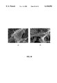
- FIG. 2 shows Scanning Electron Micrographs (SEMs), at a magnification of 2,000 ⁇ , of PLLA fibrillar matrices prepared at different gelation temperatures from a 5% (wt/v) solution of PLLA/THF; (A) 15° C., (B) 8° C., (C) -18° C., and (D) -195.8° C.
- FIG. 3 presents a graph showing the relationship between the fiber diameter of a PLLA matrix, prepared from a 5.0% (wt/v) PLLA/THF solution, and gelation temperature with p values obtained from a two-tail student's t-test comparing fiber diameters of PLLA matrices.
- FIG. 4 shows SEM's, at a magnification of 2,000 ⁇ , of PLLA fibrillar matrices prepared from PLLA/THF solutions (wt/v) with different PLLA concentrations at a gelation temperature of 8° C.; (A) 1.0%, (B) 2.5%, (C) 5.0% and (D) 7.5%.
- FIG. 5 presents a graph showing the relationship between the fiber diameter of a PLLA matrices prepared from PLLA/THF solutions with different PLLA concentrations [1%, 2.5%, 5%, and 7.5% (wt/v)] at a gelation temperature of 8° C. with p values obtained from a two-tail student's t-test comparing fiber diameters of PLLA matrices.
- FIG. 6 shows SEM's of a PLLA fibrillar matrix prepared from a 2.5 (wt/v) PLLA/THF solution at a gelation temperature of 8° C. at different magnifications; (A) 500 ⁇ and (B) 20,000 ⁇ .
- FIG. 7 presents data on the mechanical properties of fibrous PLLA matrices prepared from PLLA/THF solutions with varying PLLA concentrations at a gelation temperature of -18° C.; (A) Modulus, (B) Tensile Strength, and (C) Elongation at break.
- FIG. 8 shows SEM micrographs, at a magnification of 2,000 ⁇ , of PLLA matrices prepared from a 5.0% (wt/v) PLLA/THF solution with different thermal gelation histories:
- SEM Scanning Electron Micrograph
- SEMs Scanning Electron Micrographs
- FIGS. 11A and 11B present Scanning Electron Micrographs (SEMs), at different magnifications, of porous matrices prepared from uncrystallizable aliphatic polyester solutions at a gelation temperature of -18° C.
- FIGS. 12A and 11B present Scanning Electron Micrographs (SEMs), at different magnifications, of a porous matrix prepared from uncrystallizable aliphatic polyester solutions at a gelation temperature of -18° C.
- FIGS. 13A and 13B present a Scanning Electron Micrograph (SEM), at different magnifications, of a PLLA matrix prepared from a of 5% (wt/v) PLLA/THF/Salt mixture with a gelation temperature of -18° C.
- SEM Scanning Electron Micrograph
- the present invention relates to methods of fabrication for fibrillar matrices as well as the resulting fibrillar matrices as compositions suitable as a fibrillar matrix for cellular infiltration and ingrowth, the cultivation of cells within said matrices for the fabrication and repair of tissues and organs, and as biocompatible synthetic protheses.
- said fibrillar matrices have applications as biodegradable packaging materials.
- the present invention demonstrates that a variety of polymer sources and solvents may be used to construct a synthetic fibrillar matrix that approximates the morphology of a native collagenous extracellular matrix with a desired porosity.
- the gelation temperature of said polymer/solvent solution is important considerations when a particular resulting fibrillar morphology is desired (comprising fiber diameter, porosity, and unit length) of the matrices recited in the instant invention.
- Living cells may be incorporated into the solvent free fibrillar matrices and cultured in vitro.
- the fibrillar matrix may be maintained in an in vitro tissue culture environment.
- a biodegradable fibrillar matrix may be created.
- Such biodegradable fibrillar matrices form a synthetic extracellular matrix (that approximates the morphology of a native collagenous extracellular matrix) resorbable by infiltrating cells.
- the present invention also contemplates biocompatable but non-biodegradable fibrillar matrices that approximates the morphology of a native tendons and facia.
- Poly(L-lactic acid) (PLLA) and poly(D,L-lactic acid-co-glycolic acid) (85/15) (PLGA) with an inherent viscosity of approximately 1.6 and 0.5-0.6 respectively are available from Boehringer Ingelheim (Ingelheim, Germany).
- Poly(D,L-lactic acid) (PDLLA) with a molecular weight of 103,000 is purchased from Sigma Chemical Co. (St. Louis, Mo.).
- PLLA, PLGA and PDLLA are used without further purification.
- dioxane a solution of dioxane and water
- THF tetrahydrofuran
- DMF N,N-dimethylformamide
- pyridine a compound that is used as solvents: dioxane, a solution of dioxane and water
- THF tetrahydrofuran
- DMF N,N-dimethylformamide
- pyridine a compound that is used as solvents: tetrahydrofuran (THF), N,N-dimethylformamide (DMF), pyridine
- methanol methanol
- acetone acetone
- Deionized water is obtained with a Milli-Q water filter system from Millipore Corporation (Bedford, Mass.).
- the organic solvents may be obtained from Aldrich Chemical Company (Milwaukee, Wis.).
- a given amount of said homogenous polymer/solvent solution, maintained at 50° C., is transferred into a mold of a desired shape. While it is not intended that the present invention be limited to a specific mold, in one example said mold is made of Teflon.
- the cast polymer/solvent solution is rapidly transferred into a refrigerator or a freezer to gel at a preferred temperature.
- the gelation time depends on temperature, solvent and polymer concentration of the polymer/solvent solution. See Table 2.
- the gel is kept at the gelling temperature for at least 2 hours after gelation.
- the cast containing the gel is immersed into distilled water to facilitate solvent exchange.
- the water is changed three times a day for two days.
- the hydrated solvent free gel is then removed from water, blotted to remove gross excess water, and transferred into a freezer at -18° C. for at least 2 hours.
- the frozen gel is transferred into a freeze-drying vessel maintained at -5 to -10° C., and is freeze-dried under vacuum lower than 0.5 mmHg for one week.
- the dried porous matrix is then stored in a desiccator until characterization.
- the estimated densities and porosities of the fibrillar matrices is obtained as follows. Circular discs of the fibrillar matrix are fabricated as previously described. The radius and height of a disc is measured to calculate the volume according to the equation ⁇ r 2 ⁇ h. The weight of the specimen is measured with an analytical balance. The density is calculated from the volume and weight. The porosity, ⁇ , is calculated from the measured overall densities D f of the fibrous matrix and the skeletal density D p : ##EQU1##
- the skeletal density is the density of the polymer, which is given by: ##EQU2## where X c is the degree of polymer crystallinity.
- the morphologies of the fibrillar matrices are studied with a scanning electron microscopy (SEM) (S-3200N, Hitachi, Japan) at 15 kV.
- SEM scanning electron microscopy
- a specimen is cut with a razor blade or fractured after being frozen in liquid nitrogen for 5 minutes, and is then coated with gold using a sputter coater (Desk-II, Denton Vacuum Inc.).
- the gas pressure is lower than 50 mtorr, and the current is about 40 mA.
- the coating time is 200 seconds.
- the average fiber diameter is calculated from the SEM micrographs.
- the surface area to volume ratio is estimated based on the average fiber diameter.
- the surface areas of the fiber ends are neglected based on a very large aspect ratio of the fibers (virtually a continuous fiber network) so that the surface area of a fiber was calculated with the equation:
- the fiber length between two conjunctions is estimated based on a simplified cubic structure model. See FIG. 1. This calculation pre-supposes a cubic network. There are 12 unit fibers bordering each unit cube. Each of these fibers is shared by 4 unit cubes. Therefore, there are 3 unit fibers in each unit cube.
- Gelling temperature is an important factor controlling the porous fibrillar morphology of the matrices.
- the matrix structure formed via gelation of 5% PLLA/THF solution at 23° C. or 19° C. is different from the matrix structure formed at lower gelation temperatures.
- a gelation temperature of 23° C. no fibrillar structure was observed.
- the resulting matrix, composed of irregular platelets and pores, is unsuitable as a scaffold for cellular infiltration and ingrowth, the cultivation of cells within said matrices for the fabrication and repair of tissues and organs, and as biocompatible synthetic prosthesis.
- the average fiber diameter of fibrillar matrices do not statistically vary with the concentration of polymer solution used to fabricate the matrices in selected concentration ranges. See FIGS. 4 and 5. In contrast, the average unit length decreases with increasing polymer concentration. See Table 3.
- the surface/volume ratio of the instant fibrillar matrices do not change significantly with the polymer concentration because the fiber diameter (160-170 nm) does not change with polymer concentration. See Table 3.
- the melting point, enthalpy of melting, and the degree of crystallinity of the matrices prepared from PLLA/THF solution with different PLLA concentrations and at different gelling temperatures is presented in Table 4.
- a gelation temperature of -18° C. the melting point and the degree of crystallinity of PLLA matrices do not change significantly with the polymer concentration.
- the degree of crystallinity does not change significantly in a gelation temperature range of 15° C. or below.
- the matrix formed at a higher temperature e.g., room temperature
- Thermal history also effects matrix morphology.
- both platelet-like and nano fiber-like structures are observed in a matrix prepared by gelling a 5% PLLA/THF solution at room temperature for 2 or 12 hours and then quenching at -18° C.
- the percentage of platelet-like structures increases as a function of time the PLLA/THF solution gels at room temperature. For example, after gelling at room temperature (and subsequent maintenance at room temperature for a total of 24 hours); a platelet-like morphology is observed exclusively with or without subsequent quenching to -18° C. See FIG. 8c.
- the 5% PLLA/THF solution is quenched to -18° C. for 10 minutes at first and then returned to room temperature for one week, the resulting morphology is a fibrillar matrix. See FIG. 8d.
- Fibroblasts are cultured and expanded in tissue culture medium.
- the cultured cells are trypsinized with trypsin-EDTA and are washed twice with DPBS.
- the cells are then suspended in "complete medium" (89% DMEM, 10% FBS, 1% P/S, and 50 mg/L L-ascorbic acid) at a density of 1 ⁇ 10 7 cells/ml.
- Circular discs with a diameter of 10 mm and a thickness of 1.5 mm are cut from a fibrillar matrix sheet and one disc is fit in each well of a customer-made twelve-well Teflon culture plate. 1.5 ⁇ 10 6 cells in total of 0.5 ml complete medium are added to each of the matrix discs. They are cultured in a humidified incubator at 37° C.
- PLLA Fibrillar Matrix from 1.0% PLLA/THF Solution with a Gelation Temperature of 8° C.
- 0.2 gram poly(L-lactic acid) (PLLA) was added into a flask containing 20 ml THF, and then stirred with a magnetic stirrer at about 60° C. to make a solution with a concentration of 1.0 (wt/v)%.
- 2 ml of the prepared solution (prewarmed to 50° C.) was added into a Teflon vial.
- the vial containing PLLA solution was then rapidly transferred into a refrigerator and kept at 8° C. for 3 hours to gel. After gelation, the gel was kept at 8° C. for another 4 hours before the next step.
- the vial containing the gel was immersed into distilled water for solvent exchange. The water was changed three times a day for two days.
- the gel was removed from the water and blotted with a piece of filter paper, and then transferred into a freezer at -20° C. for at least 2 hours to completely freeze the water-containing gel.
- the frozen gel was transferred into a freeze-drying vessel at -5 ⁇ -10° C., in an ice/salt bath, and was freeze-dried at a vacuum lower than 0.5 mmHg for one week.
- the resulting fibrillar matrix was observed with SEM (FIG. 4a).
- PLLA Fibrillar Matrix from 2.5% PLLA/THF Solution with a Gelation Temperature of 8° C.
- PLLA poly(L-lactic acid)
- the gel was removed from the water and blotted with a piece of filter paper, and then transferred into a freezer at -20° C. for at least 2 hours to completely freeze the water-containing gel.
- the frozen gel was transferred into a freeze-drying vessel at -5 ⁇ -10° C., in an ice/salt bath, and was freeze-dried at a vacuum lower than 0.5 mmHg for one week.
- the resulting fibrillar matrix was observed with SEM (FIG. 4b).
- PLLA Fibrillar Matrix from 5.0% PLLA/THF Solution with a Gelation Temperature of 8° C.
- PLLA poly(L-lactic acid)
- the gel was removed from the water and blotted with a piece of filter paper, and then transferred into a freezer at -20° C. for at least 2 hours to completely freeze the water-containing gel.
- the frozen gel was transferred into a freeze-drying vessel at -5 ⁇ -10° C., in an ice/salt bath, and was freeze-dried at a vacuum lower than 0.5 mmHg for one week.
- the resulting fibrillar matrix was observed with SEM (FIG. 4c).
- PLLA Fibrillar Matrix from 7.5% PLLA/THF Solution with a Gelation Temperature of 8° C.
- the gel was removed from the water and blotted with a piece of filter paper, and then transferred into a freezer at -20° C. for at least 2 hours to completely freeze the water-containing gel.
- the frozen gel was transferred into a freeze-drying vessel at -5 ⁇ -10° C., in an ice/salt bath, and was freeze-dried at a vacuum lower than 0.5 mmHg for one week.
- the resulting fibrillar matrix was observed with SEM (FIG. 4d).
- PLLA Fibrillar Matrix from 5.0% PLLA/THF Solution with a Gelation Temperature of 15° C.
- PLLA poly(L-lactic acid)
- the gel was removed from the water and blotted with a piece of filter paper, and then transferred into a freezer at -20° C. for at least 2 hours to completely freeze the water-containing gel.
- the frozen gel was transferred into a freeze-drying vessel at -5 ⁇ -10° C., in an ice/salt bath, and was freeze-dried at a vacuum lower than 0.5 mmHg for one week.
- the resulting fibrillar matrix was observed with SEM (FIG. 2a).
- PLLA Fibrillar Matrix from 5.0% PLLA/THF Solution with a Gelation Temperature of -18° C.
- PLLA poly(L-lactic acid)
- the gel was removed from the water and blotted with a piece of filter paper, and then transferred into a freezer at -20° C. for at least 2 hours to deep freeze the water-containing gel.
- the frozen gel was transferred into a freeze-drying vessel at -5 ⁇ -10° C., in an ice/salt bath, and was freeze-dried at a vacuum lower than 0.5 mmHg for one week.
- the resulting fibrillar matrix structure was observed with SEM (FIG. 2c).
- PLLA poly(L-lactic acid)
- the frozen gel was transferred into a freeze-drying vessel at -5 ⁇ -10° C., in an ice/salt bath, and was freeze-dried at a vacuum lower than 0.5 mmHg for one week.
- the resulting fibrillar matrix was observed with SEM (FIG. 2d).
- PLLA poly(L-lactic acid)
- the water was changed three times a day for two days.
- the gel was removed from the water and blotted with a piece of filter paper, and then transferred into a freezer at -20° C. for at least 2 hours to deep freeze the water-containing gel.
- the frozen gel was transferred into a freeze-drying vessel at -5 ⁇ -10° C., in an ice/salt bath, and was freeze-dried at a vacuum lower than 0.5 mmHg for one week.
- the resulting fibrillar matrix was observed with SEM (FIG. 8a).
- PLLA poly(L-lactic acid)
- the gel was removed from the water and blotted with a piece of filter paper, and then transferred into a freezer at -20° C. for at least 2 hours to completely freeze the water-containing gel.
- the frozen gel was transferred into a freeze-drying vessel at -5 ⁇ -10° C., in an ice/salt bath, and was freeze-dried at a vacuum lower than 0.5 mmHg for one week.
- the resulting structure was observed with SEM (FIG. 8c).
- PLLA poly(L-lactic acid)
- the gel was removed from the water and blotted with a piece of filter paper, and then transferred into a freezer at -20° C. for at least 2 hours to deep freeze the water-containing gel.
- the frozen gel was transferred into a freeze-drying vessel at -5 ⁇ -10° C., in an ice/salt bath, and was freeze-dried at a vacuum lower than 0.5 mmHg for one week.
- the resulting fibrillar matrix was observed with SEM (FIG. 8d).
- PLLA poly(L-lactic acid)
- the gel was removed from the water and blotted with a piece of filter paper, and then transferred into a freezer at -20° C. for at least 2 hours to deep freeze the water-containing gel.
- the frozen gel was transferred into a freeze-drying vessel at -5 ⁇ -10° C., in an ice/salt bath, and was freeze-dried at a vacuum lower than 0.5 mmHg for one week.
- the nano-fibrous matrix structure was observed with SEM (FIG. 9).
- PLLA poly(L-lactic acid)
- the water was changed three times a day for two days.
- the gel was removed from the water and blotted with a piece of filter paper, and then transferred into a freezer at -20° C. for at least 2 hours to deep freeze the water-containing gel.
- the frozen gel was transferred into a freeze-drying vessel at -5 ⁇ -10° C., in an ice/salt bath, and was freeze-dried at a vacuum lower than 0.5 mmHg for one week.
- the resulting matrix was observed with SEM (FIG. 10a).
- PLLA poly(L-lactic acid)
- the gel was directly transferred into a freeze-drying vessel at -5 ⁇ -10° C., in an ice/salt bath, and was freeze-dried at a vacuum lower than 0.5 mmHg for one week.
- the resulting fibrillar matrix was observed with SEM (FIG. 10b).
- 2 ml of the prepared solution (prewarmed to 50° C.) was added into a Teflon vial.
- the vial containing PLLA solution was then rapidly transferred into a refrigerator and kept at -18° C. for 1 hour to gel. After gelation, the gel was kept at -18° C. for another 2 hours before the next step.
- the vial containing the gel was immersed into distilled water for solvent exchange.
- the water was changed three times a day for two days.
- the gel was removed from the water and blotted with a piece of filter paper, and then transferred into a freezer at -20° C. for at least 2 hours to deep freeze the water-containing gel.
- the frozen gel was transferred into a freeze-drying vessel at -5 ⁇ -10° C., in an ice/salt bath, and was freeze-dried at a vacuum lower than 0.5 mmHg for one week.
- the resulting structure was observed with SEM (FIG. 11a and 11b).
- PLGA poly(D,L-lactide-co-glycolide)
- the vial containing the gel was immersed into distilled water for solvent exchange.
- the water was changed three times a day for two days.
- the gel was removed from the water and blotted with a piece of filter paper, and then transferred into a freezer at -20° C. for at least 2 hours to completely freeze the water-containing gel.
- the frozen gel was transferred into a freeze-drying vessel at -5 ⁇ -10° C., in an ice/salt bath, and was freeze-dried at a vacuum lower than 0.5 mmHg for one week.
- the resulting foam was observed with SEM (FIG. 12a and 12b).
- PLLA Fibrillar Matrix from 5.0% PLLA/THF/Salt Mixture with a Gelation Temperature of -18° C.
- PLLA poly(L-lactic acid)
- the vial containing the gel and salt particles was immersed into distilled water for solvent exchange and salt particles leaching.
- the water was changed three times a day for two days.
- the gel was removed from the water and blotted with a piece of filter paper, and then transferred into a freezer at -20° C. for at least 2 hours to completely freeze the water-containing gel.
- the frozen gel was transferred into a freeze-drying vessel at -5 ⁇ -10° C., in an ice/salt bath, and was freeze-dried at a vacuum lower than 0.5 mmHg for one week.
- the resulting matrix was observed with SEM (FIG. 13).
Landscapes
- Health & Medical Sciences (AREA)
- Chemical & Material Sciences (AREA)
- Medicinal Chemistry (AREA)
- Life Sciences & Earth Sciences (AREA)
- General Health & Medical Sciences (AREA)
- Oral & Maxillofacial Surgery (AREA)
- Transplantation (AREA)
- Epidemiology (AREA)
- Veterinary Medicine (AREA)
- Animal Behavior & Ethology (AREA)
- Dermatology (AREA)
- Public Health (AREA)
- Chemical Kinetics & Catalysis (AREA)
- Dispersion Chemistry (AREA)
- Engineering & Computer Science (AREA)
- Materials Engineering (AREA)
- Polymers & Plastics (AREA)
- Organic Chemistry (AREA)
- Materials For Medical Uses (AREA)
Abstract
Methods and compositions are described that provide three-dimensional fibrillar matrices useful as, among other things, structural prosthetics and scaffolds for cells. The porous fibrillar matrices of the present invention have desirable mechanical properties suitable to a variety of applications, including platforms for in vitro cell cultivation, implants for tissue and organ engineering, implants as tendon and facia prosthetics, and product packaging.
Description
The present invention relates to methods of fabrication for fibrillar matrices as well as the resulting fibrillar matrices as compositions suitable as a scaffold for cellular infiltration and ingrowth, the cultivation of cells within said matrices for the fabrication and repair of tissues and organs, and as biocompatible synthetic prosthesis. In addition, said fibrillar matrices have applications as biodegradable packaging materials.
Transplantation is a life-saving therapy but is seriously limited by the scarcity of donor organs. In contrast to native tissue and organ transplantation from a nonautologous donor, tissues and organs generated through tissue engineering provide a more abundant alternative source for highly sought after biological materials. Scaffolding plays a pivotal role in the engineering of new tissues and organs by providing a support and a framework within which blood vessels, lymphatic vessels, and nerves may course.
Collagen is a natural extracellular matrix component of many tissues such as bone, skin, tendon, ligament, and other connective tissues. Collagen's fibrillar structure is important for cell attachment, proliferation, and differentiation.
Collagen fiber bundles vary in diameter from 50 to 500 nm. As a natural extracellular matrix component, collagen facilitates cellular recognition. Cellular recognition is advantageous for promoting cell attachment and infiltration. Importantly, however, cellular recognition may also precipitate a deleterious inflammatory or pathological immunogenic response. Native collagen is also undesirable as an implant or prosthesis due to the inherent batch to batch variability in mechanical specifications and degradability of said native collagen derived from biological sources.
In contrast, aliphatic polyesters such as (but not limited to) poly(lactide), poly(glycolide) and their copolymers are biodegradable, biocompatible (e.g., non-immunogenic), and among the few synthetic polymers approved by FDA for some human clinical applications. The prior art presents three-dimensional porous structures fabricated from synthetic aliphatic polyesters employed for cell attachment, growth, and tissue regeneration. However, these porous scaffolds (in the prior art) do not approximate the fibrillar morphology of a native collagen extracellular matrix.
In an attempt to approximate a native collagen extracellular matrix, the prior art has applied textile technology to produce nonwoven fabrics from aliphatic polyesters. These nonwoven fabrics, however, require the expensive and laborious steps of fiber extrusion, drawing, crimping, cutting into stable fibers, carding, needling, heat platen pressing, degreasing, and punching. Furthermore, said textile produced nonwoven fabrics are associated with structural parameters (as compared with native collagenous matrices) that do not favor cell attachment (e.g., large fiber diameter and low surface to volume ratios).
What is needed, therefore, is a biocompatible synthetic fibrillar matrix (readily fashioned into a desired shape) that reproduces the form and function of native collagenous extracellular matrices.
The present invention relates to methods of fabrication for fibrillar matrices as well as the resulting fibrillar matrices as compositions suitable as a scaffold for cellular infiltration and ingrowth, the cultivation of cells within said matrices for the fabrication and repair of tissues and organs, and as biocompatible synthetic prostheses. In one embodiment, the present invention contemplates a method wherein a synthetic fibrillar matrix of a desired fiber diameter, porosity, and unit length is used to approximate the morphology of native collagenous extracellular matrices. In another embodiment, the present invention contemplates a method wherein a fibrillar matrix of an implantable material, comprising a desired fiber diameter, porosity, and unit length is used as a scaffold facilitating the infiltration of cells in vivo. In another embodiment, the present invention contemplates a fibrillar matrix of an implantable composite material, comprising a desired fiber diameter, porosity, and unit length as a composition providing a biocompatible implantable prosthetics. In addition, said fibrillar matrix has applications as a packaging material.
In one embodiment, the present invention contemplates a method comprising: a) providing: i) a polymer source, ii) a solvent; b) mixing said polymer with said solvent at a temperature range between 20-100° C., more preferably between 50-65° C., and most preferably at 60° C. to create a homogenous polymer solution; c) casting said homogenous polymer solution into a desired form at a temperature range between 20-100° C., more preferably between 50-65° C., and most preferably at 50° C.; d) cooling said cast homogenous polymer solution to a given gelation temperature wherein said temperature favors the fabrication, that is to say the spatial orientation, of a three-dimensional fibrillar matrix, said temperature comprising a range between -195.8° C. and 23° C. and more preferably between -18° C. and 8° C.; e) maintaining said cast homogenous polymer solution at a given gelation temperature under conditions wherein said three-dimensional fibrillar network is preserved, said fibrillar network comprising fibers with diameters in a range between 50 to 500 nm, and most preferably with an average diameter between 160-170 nm; f) hydrating said gelled polymer such that said hydrated polymer is solvent free; g) freezing said hydrated solvent free gelled polymer; and h) treating said frozen gelled hydrated solvent free polymer under conditions whereby a substantially desiccated matrix is created having a porosity greater than 80%. While the above-named components can be formulated in an alternative order, the above referenced reaction sequence has been found to produce the best results.
In another embodiment, the present invention contemplates a method comprising: a) providing: i) a polymer source, ii) a solvent; b) mixing said polymer with said solvent at a temperature range between 20-100° C., more preferably between 50-65° C., and most preferably at 60° C. to create a homogenous polymer solution; c) casting said homogenous polymer solution into a desired form at a temperature range between 20-100° C., more preferably between 50-65° C., and most preferably at 50° C.; d) cooling said cast homogenous polymer solution to a given gelation temperature wherein said temperature favors the fabrication, that is to say the structural orientation, of a three-dimensional fibrillar matrix, said temperature comprising a range between -195.8° C. and 23° C. and more preferably between -18° C. and 8° C.; e) maintaining said cast homogenous polymer solution at a given gelation temperature under conditions wherein said three-dimensional fibrillar network is preserved, said fibrillar network comprising fibers with diameters in a range between 50 to 500 nm, and most preferably with an average diameter between 160-170 nm; f) freezing said gelled polymer; and g) treating said frozen gelled polymer under conditions whereby a substantially desiccated matrix is created having a porosity greater than 80%. While the above-named components can be formulated in an alternative order, the above referenced reaction sequence has been found to produce the best results.
It is not intended that the present invention be limited to the above-described reagents. While the basic components are described above, other components can be added to the basic components, creating variations in the final structures (and thereby conferring different functions). Examples of such other components include, but are not limited to, biologically functional substances (such as proteins, drugs and growth factors) and pore-forming components (such as salt, sugar, water soluble waxes or other water-soluble substances). The present invention contemplates adding such additional components such as pore-forming components to the polymer solution to produce additional pores when leached in water.
In another embodiment the instant invention contemplates a composition, said composition comprising a three-dimensional aliphatic polyester fibrillar matrix, wherein said fibrillar matrix comprises fibers having diameters in a range between 50 to 500 nm, and most preferably with an average diameter between 160-170 nm, and said fibrillar matrix has a porosity of greater 80%.
It is not intended that the matrix recited in the instant invention be limited to a specific morphology. In one example said matrix may be fibrillar. In another example, said matrix may be a foam.
It is not intended the present invention be limited to a particular polymer or polymer source. The present invention contemplates homopolymers, copolymers and/or a mixture of polymers. In one embodiment, the polymer source is poly(L-lactic acid) (PLLA) with an inherent viscosity of approximately 1.6. In another embodiment, the polymer is poly(D,L-lactic acid-co-glycolic acid (PLGA) with an inherent viscosity of 0.5-0.6. In another embodiment, the polymer is Poly(D,L-lactic acid) (PDLLA) with a molecular weight of approximately 103,000. Said polymers are commercially available and may be purchased from Boehringer Ingelheim (Ingelheim, Germany) and/or Sigma Chemical Co. (St. Louis, Mo.). Additionally, these polymers are used without further purification.
It is also not intended that the present invention be limited to a specific solvent. In one embodiment the solvent is dioxane (D). In another embodiment the solvent is a solution of dioxane and water (D/W). In another embodiment the solvent is tetrahydrofuran (THF). In another embodiment the solvent is N,N-dimethylformamide (DMF). In another embodiment the solvent is pyridine. In another embodiment the solvent is methanol. In another embodiment the solvent is acetone.
The present invention also contemplates the use of a composition. Moreover, the present invention contemplates using a synthetic fibrillar matrix that approximates the morphology of a native collagenous extracellular matrix in combination with other components, such as cells. Where cells are used, it is not intended that the present invention be limited to a specific cell type (e.g. one cell type infiltrating a matrix). A variety of cell types (including solutions of different cells) are contemplated. In one embodiment, the cells are osteoblasts. In another embodiment, the cells are fibroblasts. In another embodiment the cells are epithelial. In another embodiment, the cells secrete a medically useful compound (e.g., hormone, cytokine, etc.). Such cells may be (but need not be) cells that have been manipulated by recombinant means to secrete such compounds.
The present invention contemplates methods wherein cells are added and grown in and on the matrix, as well as methods wherein the matrix is implanted (both with and without cells).
The present invention also contemplates methods wherein some of the fibrillar matrices that approximate the morphology of a native collagenous extracellular matrix biodegrade, in vivo and in vitro, subsequent to the confluent growth of cells in and on the matrix. The present invention also contemplates methods wherein some of the collagen like fibrillar matrices are not biodegradable. While it is not intended the instant invention be limited to a particular example, said non-biodegradable fibrillar matrices are fashioned into synthetic tendons and facia (e.g., Achilles tendon and plantar facia).
As noted above, the fibrillar matrix of the present invention may also be applied as a packaging material.
To facilitate understanding of the invention, a number of terms are defined below.
A "Fibrillar matrix" refers to a three dimensional support for cells, comprising an array of strand-like or thread like elements, which divide free space into partially enclosed domains which remain in fluidic communication with adjacent domains.
"Biodegradable" refers to a material capable of being broken down into readily metabolized compounds by the action of living beings such as cells in vitro or in vivo.
As used herein, the term "implant" and "implanting" and the like indicates placement on, in, or through a patient's body (including placement in body cavities) in the course of medical treatment, e.g., for a disease, impairment or injury. Implants include, but are not limited to, implants for wound care, and drug delivery.
"Solvent free" refers to a polymer matrix wherein the interstices of said matrix are substantially free from residual solvent such that said matrix reaches a constant mass upon sublimation. By "substantially free" it is meant that, with normal detection means (such as detection by changes in mass), no solvent is detected. While it is believed that the methods of the present invention yield a matrix that is completely free of solvent, it is possible that some solvent remains detectable in extremely small amounts by extreme detection methods (e.g., detection methods with extremely high resolution).
"Quenching" refers to the cooling rate of a solution.
"Surface/volume ratio" refers to the ratio of surface area within a matrix sample to the polymer skeleton volume of the same matrix sample.
"Unit length" is the linear distance of a fiber length between two conjunctions of fiber.
"Native Collagen Extracellular Matrix" refers to a three dimensional support for cells, comprising a triple-stranded helical molecule rich in proline and hydroxyproline, which divide free space into partially enclosed domains which remain in fluidic communication with adjacent domains.
"Gelation Time" refers to the elapsed time from the time point when a polymer/solvent solution sample is set to a target gelling temperature to the time point when said polymer/solvent sample (held at said target gelling temperature) does not flow down an incline plane.
"Structural prosthetics" refers to load bearing synthetic tissue including but not limited to synthetic tendons and facia and portions thereof.
"Substantially desiccated" refers to a material sample that has a water content of 10% or less, and more preferably 5% or less, and still more preferably 1% or less.
"Resorbable" refers to a synthetic or native materials which may be broken down into less complex constituent parts by physiological processes.
"Foam" refers to a solid within which is disposed a plurality partially enclosed domains which remain in fluidic communication with adjacent domains.
"Salt" refers to any of a class of chemical compounds formed by neutralization of an acid by a base. While it is not intended that the present invention be limited to any particular salt, examples include NaCl, KCl, MgCl2, and CaCl2.
"Sugars" refers to polyhydroxy aldehydes or ketones and their derivatives.
"Water soluble waxes" refers to the water soluble subset of a group of substances composed of hydrocarbons, alcohols, fatty acids, and esters that are solid at room temperature.
FIG. 1 shows a cubic fiber network model of (A) and array of cubic units and (B) an expanded view of a single isolated cubic unit.
FIG. 2 shows Scanning Electron Micrographs (SEMs), at a magnification of 2,000×, of PLLA fibrillar matrices prepared at different gelation temperatures from a 5% (wt/v) solution of PLLA/THF; (A) 15° C., (B) 8° C., (C) -18° C., and (D) -195.8° C.
FIG. 3 presents a graph showing the relationship between the fiber diameter of a PLLA matrix, prepared from a 5.0% (wt/v) PLLA/THF solution, and gelation temperature with p values obtained from a two-tail student's t-test comparing fiber diameters of PLLA matrices.
FIG. 4 shows SEM's, at a magnification of 2,000×, of PLLA fibrillar matrices prepared from PLLA/THF solutions (wt/v) with different PLLA concentrations at a gelation temperature of 8° C.; (A) 1.0%, (B) 2.5%, (C) 5.0% and (D) 7.5%.
FIG. 5 presents a graph showing the relationship between the fiber diameter of a PLLA matrices prepared from PLLA/THF solutions with different PLLA concentrations [1%, 2.5%, 5%, and 7.5% (wt/v)] at a gelation temperature of 8° C. with p values obtained from a two-tail student's t-test comparing fiber diameters of PLLA matrices.
FIG. 6 shows SEM's of a PLLA fibrillar matrix prepared from a 2.5 (wt/v) PLLA/THF solution at a gelation temperature of 8° C. at different magnifications; (A) 500× and (B) 20,000×.
FIG. 7 presents data on the mechanical properties of fibrous PLLA matrices prepared from PLLA/THF solutions with varying PLLA concentrations at a gelation temperature of -18° C.; (A) Modulus, (B) Tensile Strength, and (C) Elongation at break.
FIG. 8 shows SEM micrographs, at a magnification of 2,000×, of PLLA matrices prepared from a 5.0% (wt/v) PLLA/THF solution with different thermal gelation histories:
(A) Maintained at room temperature for 2 hours, and then quenched to -18° C.;
(B) Maintained at room temperature for 12 hours, and then quenched to -18° C.;
(C) Maintained at room temperature for 24 hours, and then quenched to -18° C.; and
(D) Quenched to -18° C. for 10 minutes, and then maintained at room temperature for one week.
FIG. 9 presents a Scanning Electron Micrograph (SEM), at a magnification of 2,000×, of a PLLA fibrillar matrix prepared from a 5.0% (wt/v) PLLA/THF/methanol (THF/methanol=80/20) solution at a gelation temperature of liquid nitrogen.
FIG. 10 presents a Scanning Electron Micrographs (SEMs), at a magnification of 2,000×, of a PLLA matrices prepared from a 2.5% (wt/v) PLLA/dioxane/methanol (dioxane/methanol=80/20) solution with a gelation temperature of -18° C.
(A) With water exchange.
(B) Without water exchange.
FIGS. 11A and 11B present Scanning Electron Micrographs (SEMs), at different magnifications, of porous matrices prepared from uncrystallizable aliphatic polyester solutions at a gelation temperature of -18° C.
FIGS. 12A and 11B present Scanning Electron Micrographs (SEMs), at different magnifications, of a porous matrix prepared from uncrystallizable aliphatic polyester solutions at a gelation temperature of -18° C.
FIGS. 13A and 13B present a Scanning Electron Micrograph (SEM), at different magnifications, of a PLLA matrix prepared from a of 5% (wt/v) PLLA/THF/Salt mixture with a gelation temperature of -18° C.
The present invention relates to methods of fabrication for fibrillar matrices as well as the resulting fibrillar matrices as compositions suitable as a fibrillar matrix for cellular infiltration and ingrowth, the cultivation of cells within said matrices for the fabrication and repair of tissues and organs, and as biocompatible synthetic protheses. In addition, said fibrillar matrices have applications as biodegradable packaging materials. The present invention demonstrates that a variety of polymer sources and solvents may be used to construct a synthetic fibrillar matrix that approximates the morphology of a native collagenous extracellular matrix with a desired porosity.
While it is not intended that the present invention be limited to any specific mechanism, the gelation temperature of said polymer/solvent solution, the amount of time said polymer/solvent solutions are maintained at said gelation temperatures, and the concentration of polymer within said polymer/solvent solution are important considerations when a particular resulting fibrillar morphology is desired (comprising fiber diameter, porosity, and unit length) of the matrices recited in the instant invention.
Living cells may be incorporated into the solvent free fibrillar matrices and cultured in vitro. In the alternative, the fibrillar matrix may be maintained in an in vitro tissue culture environment. Depending on the selection of polymer source, a biodegradable fibrillar matrix may be created. Such biodegradable fibrillar matrices form a synthetic extracellular matrix (that approximates the morphology of a native collagenous extracellular matrix) resorbable by infiltrating cells. In the alternative, the present invention also contemplates biocompatable but non-biodegradable fibrillar matrices that approximates the morphology of a native tendons and facia.
These variations illustrate how a fibrillar matrix, that approximates the morphology of a native collagenous extracellular matrix with a desired fiber diameter, unit length, and porosity may be used as an tissue engineering scaffold. Given the availability of the material sources and relative ease in processing said materials into the instant fibrillar matrix that approximates the morphology of a native collagenous extracellular matrix, with a desired porosity, the instant invention is well suited to large-scale tissue engineering and manufacture.
The following examples serve to illustrate certain preferred embodiments and aspects of the present invention and are not to be construed as limiting the scope thereof.
The following compounds are used as polymer sources. Poly(L-lactic acid) (PLLA) and poly(D,L-lactic acid-co-glycolic acid) (85/15) (PLGA) with an inherent viscosity of approximately 1.6 and 0.5-0.6 respectively are available from Boehringer Ingelheim (Ingelheim, Germany). Poly(D,L-lactic acid) (PDLLA) with a molecular weight of 103,000 is purchased from Sigma Chemical Co. (St. Louis, Mo.). PLLA, PLGA and PDLLA are used without further purification.
The following compounds are used as solvents: dioxane, a solution of dioxane and water, tetrahydrofuran (THF), N,N-dimethylformamide (DMF), pyridine, methanol and acetone. Deionized water is obtained with a Milli-Q water filter system from Millipore Corporation (Bedford, Mass.). The organic solvents may be obtained from Aldrich Chemical Company (Milwaukee, Wis.).
A. Porous Fibrillar Matrix Fabrication
1. Preparation of the Polymer/Solvent Solution
An aliquot from a given polymer source is weighed accurately into a flask. A given amount of solvent is then added into the flask to yield a solution with a desired concentration (from 1% (wt/v) to 15% (wt/v). Approximately two hours of magnetic stirring at 60° C. is required to obtain a homogeneous solution in a solution where the polymer concentration is less than or equal to 5%.
A given amount of said homogenous polymer/solvent solution, maintained at 50° C., is transferred into a mold of a desired shape. While it is not intended that the present invention be limited to a specific mold, in one example said mold is made of Teflon.
2. Gelation of the Polymer/Solvent Solution
The cast polymer/solvent solution is rapidly transferred into a refrigerator or a freezer to gel at a preferred temperature. The gelation time depends on temperature, solvent and polymer concentration of the polymer/solvent solution. See Table 2. The gel is kept at the gelling temperature for at least 2 hours after gelation.
3. Removal of Solvent from the Gelled Polymer
The cast containing the gel is immersed into distilled water to facilitate solvent exchange. The water is changed three times a day for two days. The hydrated solvent free gel is then removed from water, blotted to remove gross excess water, and transferred into a freezer at -18° C. for at least 2 hours.
4. Removal of Water from the Hydrated Gel
The frozen gel is transferred into a freeze-drying vessel maintained at -5 to -10° C., and is freeze-dried under vacuum lower than 0.5 mmHg for one week. The dried porous matrix is then stored in a desiccator until characterization.
B. Matrix Characterization
1. Thermodynamics
The melting behavior of the fibrillar matrices is characterized with a differential scanning calorimeter (DSC-7, Perkin-Elmer, Norwalk, Conn.). The calibration is performed using indium standards. A fibrillar matrix sample (5-10 mg) is used without any further thermal treatment. A heating rate of 20° C./min and a temperature range of 30-200° C. is used. The degree of crystallinity is calculated as: Xc =ΔHm /ΔH°m, where ΔHm is the measured enthalpy of melting and ΔH°m is the enthalpy of melting of 100% crystalline polymer. As an example, the ΔH°m for PLLA is equal to 203.4 J/g.
2. Density and Porosity
The estimated densities and porosities of the fibrillar matrices is obtained as follows. Circular discs of the fibrillar matrix are fabricated as previously described. The radius and height of a disc is measured to calculate the volume according to the equation πr2 ·h. The weight of the specimen is measured with an analytical balance. The density is calculated from the volume and weight. The porosity, ε, is calculated from the measured overall densities Df of the fibrous matrix and the skeletal density Dp : ##EQU1## For the fibrillar matrix, the skeletal density is the density of the polymer, which is given by: ##EQU2## where Xc is the degree of polymer crystallinity. As an example, Da for PLLA=1.248 g/ml (density of amorphous polymer) and Dc for PLLA=1.290 g/ml (density of 100% crystalline polymer).
3. Morphology
The morphologies of the fibrillar matrices are studied with a scanning electron microscopy (SEM) (S-3200N, Hitachi, Japan) at 15 kV. A specimen is cut with a razor blade or fractured after being frozen in liquid nitrogen for 5 minutes, and is then coated with gold using a sputter coater (Desk-II, Denton Vacuum Inc.). During said coating protocol, the gas pressure is lower than 50 mtorr, and the current is about 40 mA. The coating time is 200 seconds.
The average fiber diameter is calculated from the SEM micrographs. The surface area to volume ratio is estimated based on the average fiber diameter. In determining the surface area to volume ratio, the surface areas of the fiber ends are neglected based on a very large aspect ratio of the fibers (virtually a continuous fiber network) so that the surface area of a fiber was calculated with the equation:
A.sub.f =π·d·l (3)
where d is the diameter of the fiber and l is the length of the fiber. The volume of a fiber is given by: ##EQU3## Therefore the surface to volume ratio is given by: ##EQU4##
To quantify the fiber network density, the fiber length between two conjunctions (unit length) is estimated based on a simplified cubic structure model. See FIG. 1. This calculation pre-supposes a cubic network. There are 12 unit fibers bordering each unit cube. Each of these fibers is shared by 4 unit cubes. Therefore, there are 3 unit fibers in each unit cube. The porosity of the fiber network is given by: ##EQU5## where Vf is the volume of one unit fiber, and Vc is the volume of the unit cube. Substituting equation (4) and Vc =l3 into equation (6), the porosity is given by: ##EQU6## The unit length is given by rearranging equation (7), ##EQU7##
When the fiber diameters are compared, a two-tail student's t-test (assuming equal variances) is performed to determine the statistical significance (p<0.05).
4. Mechanical Properties
Uniaxial tensile mechanical testing, with an Instron 4502 mechanical tester (Instron Corporation, Canton, Mass.), is performed to measure the mechanical properties of the fibrillar matrices. Matrix sheets with dimensions of 90×60×3 mm3 were prepared, and then cut into 90×10×3 mm3 strips for mechanical testing. A gauge length of 40 mm and a crosshead speed of 5 mm/min were used.
5. Effect of Gelation Conditions on Matrix Structure
The conditions under which said polymer/solvent gel is critical to achieving the fibrillar morphology of the matrix recited in the present invention. While it is not intended the present invention be limited to one specific gelation protocol or set of reagents, the relationship between conditions of gelation and the morphologies of the resultant polymer matrices are illustrated through the following examples.
a. The Effect of Gelation Temperature and Annealing Conditions on Matrix Structure
Gelling temperature is an important factor controlling the porous fibrillar morphology of the matrices. For example, the matrix structure formed via gelation of 5% PLLA/THF solution at 23° C. or 19° C. is different from the matrix structure formed at lower gelation temperatures. At a gelation temperature of 23° C., no fibrillar structure was observed. Indeed at 23° C. the resulting matrix, composed of irregular platelets and pores, is unsuitable as a scaffold for cellular infiltration and ingrowth, the cultivation of cells within said matrices for the fabrication and repair of tissues and organs, and as biocompatible synthetic prosthesis.
In contrast, for matrices formed after PLLA/THF gelation at lower temperatures, e.g., 15° C., 8° C., -18° C., and -195.8° C. (liquid nitrogen), a three-dimensional nano fiber network is formed. See FIG. 2. The diameter of fibers within a matrix formed after PLLA gelation at these temperatures (e.g., 15° C., 8° C., -18° C., -195.8° C.) does not statistically vary. See FIG. 3. However, interfiber spacing becomes more uniform as the gelation temperature decreases (e.g., 15° C., 8° C., -18° C., -195.8° C.). See FIGS. 2a, 2b, 2c, and 2d.
b. The Effect of Polymer Concentration on Matrix Structure
The average fiber diameter of fibrillar matrices do not statistically vary with the concentration of polymer solution used to fabricate the matrices in selected concentration ranges. See FIGS. 4 and 5. In contrast, the average unit length decreases with increasing polymer concentration. See Table 3.
Furthermore at low polymer concentrations, such as 1% PLLA/THF solution, relatively large pores are observed with nonuniform interfiber spacing. See FIG. 4a. In contrast, with increased polymer concentration, pore structure becomes increasingly uniform and the average unit length decreases. See Table 3.
These morphological observations are consistent with the porosity and density data presented in Table 1 and Table 3. These data present density and porosity parameters in a series of fibrillar matrices, with fiber diameters ranging between 50 to 500 nm, created from biodegradable aliphatic polyesters comprising a three-dimensional continuous fibrous network that approximates the morphology of a native collagenous extracellular matrix. See FIG. 6.
In addition, the surface/volume ratio of the instant fibrillar matrices do not change significantly with the polymer concentration because the fiber diameter (160-170 nm) does not change with polymer concentration. See Table 3.
Young's modulus, tensile strength, and elongation at break all increase with polymer concentration. See FIG. 7. In contrast, the platelet-like matrices (created at higher gelation temperatures) are too fragile to measure these same mechanical properties.
The melting point, enthalpy of melting, and the degree of crystallinity of the matrices prepared from PLLA/THF solution with different PLLA concentrations and at different gelling temperatures is presented in Table 4. At a gelation temperature of -18° C., the melting point and the degree of crystallinity of PLLA matrices do not change significantly with the polymer concentration. Similarly, the degree of crystallinity does not change significantly in a gelation temperature range of 15° C. or below. In contrast, however, the matrix formed at a higher temperature (e.g., room temperature) has a higher degree of crystallinity than matrices formed at low gelation temperatures. See Table 4.
c. The Effect of Thermal History on Matrix Morphology
Thermal history also effects matrix morphology. For example, both platelet-like and nano fiber-like structures are observed in a matrix prepared by gelling a 5% PLLA/THF solution at room temperature for 2 or 12 hours and then quenching at -18° C. See FIGS. 8a and b. The percentage of platelet-like structures increases as a function of time the PLLA/THF solution gels at room temperature. For example, after gelling at room temperature (and subsequent maintenance at room temperature for a total of 24 hours); a platelet-like morphology is observed exclusively with or without subsequent quenching to -18° C. See FIG. 8c. In contrast, when the 5% PLLA/THF solution is quenched to -18° C. for 10 minutes at first and then returned to room temperature for one week, the resulting morphology is a fibrillar matrix. See FIG. 8d.
7. Incorporation of Cells into a Fibrillar Matrix
Fibroblasts are cultured and expanded in tissue culture medium. The cultured cells are trypsinized with trypsin-EDTA and are washed twice with DPBS. The cells are then suspended in "complete medium" (89% DMEM, 10% FBS, 1% P/S, and 50 mg/L L-ascorbic acid) at a density of 1×10⁷ cells/ml. Circular discs with a diameter of 10 mm and a thickness of 1.5 mm are cut from a fibrillar matrix sheet and one disc is fit in each well of a customer-made twelve-well Teflon culture plate. 1.5×10⁶ cells in total of 0.5 ml complete medium are added to each of the matrix discs. They are cultured in a humidified incubator at 37° C. in the presence of 5% CO2. The medium (0.5 ml each) is changed daily. Two weeks later, cell infiltrated matrix is fixed in 10% neutral buffered formalin, embedded in paraffin, and cut into 5 μm cross sections for histological analysis. The fibroblast are normal in appearance, having penetrated into the fibrillar matrix.
0.2 gram poly(L-lactic acid) (PLLA) was added into a flask containing 20 ml THF, and then stirred with a magnetic stirrer at about 60° C. to make a solution with a concentration of 1.0 (wt/v)%. 2 ml of the prepared solution (prewarmed to 50° C.) was added into a Teflon vial. The vial containing PLLA solution was then rapidly transferred into a refrigerator and kept at 8° C. for 3 hours to gel. After gelation, the gel was kept at 8° C. for another 4 hours before the next step. The vial containing the gel was immersed into distilled water for solvent exchange. The water was changed three times a day for two days. The gel was removed from the water and blotted with a piece of filter paper, and then transferred into a freezer at -20° C. for at least 2 hours to completely freeze the water-containing gel. The frozen gel was transferred into a freeze-drying vessel at -5˜-10° C., in an ice/salt bath, and was freeze-dried at a vacuum lower than 0.5 mmHg for one week. The resulting fibrillar matrix was observed with SEM (FIG. 4a).
0.5 gram poly(L-lactic acid) (PLLA) was added into a flask containing 20 ml THF, and then stirred with a magnetic stirrer at about 60° C. to make a solution with a concentration of 2.5 (wt/v)%. 2 ml of the prepared solution (prewarmed to 50° C.) was added into a Teflon vial. The vial containing PLLA solution was then rapidly transferred into a refrigerator and kept at 8° C. for 1 hour to gel. After gelation, the gel was kept at 8° C. for another 2 hours before the next step. The vial containing the gel was immersed into distilled water for solvent exchange. The water was changed three times a day for two days. The gel was removed from the water and blotted with a piece of filter paper, and then transferred into a freezer at -20° C. for at least 2 hours to completely freeze the water-containing gel. The frozen gel was transferred into a freeze-drying vessel at -5˜-10° C., in an ice/salt bath, and was freeze-dried at a vacuum lower than 0.5 mmHg for one week. The resulting fibrillar matrix was observed with SEM (FIG. 4b).
1.0 gram poly(L-lactic acid) (PLLA) was added into a flask containing 20 ml THF, and then stirred with a magnetic stirrer at about 60° C. to make a solution with a concentration of 5.0 (wt/v)%. 2 ml of the prepared solution (prewarmed to 50° C.) was added into a Teflon vial. The vial containing PLLA solution was then rapidly transferred into a refrigerator and kept at 8° C. for 1 hour to gel. After gelation, the gel was kept at 8° C. for another 2 hours before the next step. The vial containing the gel was immersed into distilled water for solvent exchange. The water was changed three times a day for two days. The gel was removed from the water and blotted with a piece of filter paper, and then transferred into a freezer at -20° C. for at least 2 hours to completely freeze the water-containing gel. The frozen gel was transferred into a freeze-drying vessel at -5˜-10° C., in an ice/salt bath, and was freeze-dried at a vacuum lower than 0.5 mmHg for one week. The resulting fibrillar matrix was observed with SEM (FIG. 4c).
1.5 grams poly(L-lactic acid) (PLLA) was added into a flask containing 20 ml THF, and then stirred with a magnetic stirrer at about 60° C. to make a solution with a concentration of 7.5 (wt/v)%. 2 ml of the prepared solution (prewarmed to 50° C.) was added into a Teflon vial. The vial containing PLLA solution was then rapidly transferred into a refrigerator and kept at 8° C. for 1 hour to gel. After gelation, the gel was kept at 8° C. for another 2 hours before the next step. The vial containing the gel was immersed into distilled water for solvent exchange. The water was changed three times a day for two days. The gel was removed from the water and blotted with a piece of filter paper, and then transferred into a freezer at -20° C. for at least 2 hours to completely freeze the water-containing gel. The frozen gel was transferred into a freeze-drying vessel at -5˜-10° C., in an ice/salt bath, and was freeze-dried at a vacuum lower than 0.5 mmHg for one week. The resulting fibrillar matrix was observed with SEM (FIG. 4d).
1.0 gram poly(L-lactic acid) (PLLA) was added into a flask containing 20 ml THF, and then stirred with a magnetic stirrer at about 60° C. to make a solution with a concentration of 5.0 (wt/v)%. 2 ml of the prepared solution (prewarmed to 50° C.) was added into a Teflon vial. The vial containing PLLA solution was then rapidly transferred into a water bath at 15° C. for 1 hour to gel. After gelation, the gel was kept at 15° C. for another 2 hours before the next step. The vial containing the gel was immersed into distilled water for solvent exchange. The water was changed three times a day for two days. The gel was removed from the water and blotted with a piece of filter paper, and then transferred into a freezer at -20° C. for at least 2 hours to completely freeze the water-containing gel. The frozen gel was transferred into a freeze-drying vessel at -5˜-10° C., in an ice/salt bath, and was freeze-dried at a vacuum lower than 0.5 mmHg for one week. The resulting fibrillar matrix was observed with SEM (FIG. 2a).
1.0 gram poly(L-lactic acid) (PLLA) was added into a flask containing 20 ml THF, and then stirred with a magnetic stirrer at about 60° C. to make a solution with a concentration of 5.0 (wt/v)%. 2 ml of the prepared solution (prewarmed to 50° C.) was added into a Teflon vial. The vial containing PLLA solution was then rapidly transferred into a freezer at -18° C. for 30 minutes to gel. After gelation, the gel was kept at -18° C. for another 2 hours before the next step. The vial containing the gel was immersed into distilled water for solvent exchange. The water was changed three times a day for two days. The gel was removed from the water and blotted with a piece of filter paper, and then transferred into a freezer at -20° C. for at least 2 hours to deep freeze the water-containing gel. The frozen gel was transferred into a freeze-drying vessel at -5˜-10° C., in an ice/salt bath, and was freeze-dried at a vacuum lower than 0.5 mmHg for one week. The resulting fibrillar matrix structure was observed with SEM (FIG. 2c).
1.0 gram poly(L-lactic acid) (PLLA) was added into a flask containing 20 ml THF, and then stirred with a magnetic stirrer at about 60° C. to make a solution with a concentration of 5.0 (wt/v)%. 2 ml of the prepared solution (prewarmed to 50° C.) was added into a Teflon vial. The vial containing PLLA solution was then rapidly immersed into liquid nitrogen for one hour. The vial containing the gel was immersed into distilled water for solvent exchange. The water was changed three times a day for two days. The gel was removed from the water and blotted with a piece of filter paper, and then transferred into a freezer at -20° C. for at least 2 hours to deep freeze the water-containing gel. The frozen gel was transferred into a freeze-drying vessel at -5˜-10° C., in an ice/salt bath, and was freeze-dried at a vacuum lower than 0.5 mmHg for one week. The resulting fibrillar matrix was observed with SEM (FIG. 2d).
1.0 gram poly(L-lactic acid) (PLLA) was added into a flask containing 20 ml THF, and then stirred with a magnetic stirrer at about 60° C. to make a solution with a concentration of 5.0 (wt/v)%. 2 ml of the prepared solution (prewarmed to 50° C.) was added into a Teflon vial. The vial containing PLLA solution was kept at room temperature (23° C.) for two hours and then rapidly transferred into a freezer at -18° C. for 30 minutes to gel. After gelation, the gel was kept at -18° C. for another 2 hours before the next step. The vial containing the gel was immersed into distilled water for solvent exchange. The water was changed three times a day for two days. The gel was removed from the water and blotted with a piece of filter paper, and then transferred into a freezer at -20° C. for at least 2 hours to deep freeze the water-containing gel. The frozen gel was transferred into a freeze-drying vessel at -5˜-10° C., in an ice/salt bath, and was freeze-dried at a vacuum lower than 0.5 mmHg for one week. The resulting fibrillar matrix was observed with SEM (FIG. 8a).
1.0 gram poly(L-lactic acid) (PLLA) was added into a flask containing 20 ml THF, and then stirred with a magnetic stirrer at about 60° C. to make a solution with a concentration of 5.0 (wt/v)%. 2 ml of the prepared solution (prewarmed to 50° C.) was added into a Teflon vial. The vial containing PLLA solution was kept at room temperature (23° C.) for 24 hours and then rapidly transferred into a freezer at -18° C. for 2 hours before the next step. The vial containing the gel was immersed into distilled water for solvent exchange. The water was changed three times a day for two days. The gel was removed from the water and blotted with a piece of filter paper, and then transferred into a freezer at -20° C. for at least 2 hours to completely freeze the water-containing gel. The frozen gel was transferred into a freeze-drying vessel at -5˜-10° C., in an ice/salt bath, and was freeze-dried at a vacuum lower than 0.5 mmHg for one week. The resulting structure was observed with SEM (FIG. 8c).
1.0 gram poly(L-lactic acid) (PLLA) was added into a flask containing 20 ml THF, and then stirred with a magnetic stirrer at about 60° C. to make a solution with a concentration of 5.0 (wt/v)%. 2 ml of the prepared solution (prewarmed to 50° C.) was added into a Teflon vial. The vial containing PLLA solution was rapidly transferred into a freezer at -18° C. for 10 minutes to gel and then kept at room temperature (23° C.) for one week. The vial containing the gel was immersed into distilled water for solvent exchange. The water was changed three times a day for two days. The gel was removed from the water and blotted with a piece of filter paper, and then transferred into a freezer at -20° C. for at least 2 hours to deep freeze the water-containing gel. The frozen gel was transferred into a freeze-drying vessel at -5˜-10° C., in an ice/salt bath, and was freeze-dried at a vacuum lower than 0.5 mmHg for one week. The resulting fibrillar matrix was observed with SEM (FIG. 8d).
1.0 gram poly(L-lactic acid) (PLLA) was added into a flask containing 20 ml mixture of THF and methanol (THF/methanol=80/20), and then stirred with a magnetic stirrer at about 60° C. to make a solution with a concentration of 5.0 (wt/v)%. 2 ml of the prepared solution (prewarmed to 50° C.) was added into a Teflon vial. The vial containing PLLA solution was then rapidly immersed into liquid nitrogen for one hour. The vial containing the gel was immersed into distilled water for solvent exchange. The water was changed three times a day for two days. The gel was removed from the water and blotted with a piece of filter paper, and then transferred into a freezer at -20° C. for at least 2 hours to deep freeze the water-containing gel. The frozen gel was transferred into a freeze-drying vessel at -5˜-10° C., in an ice/salt bath, and was freeze-dried at a vacuum lower than 0.5 mmHg for one week. The nano-fibrous matrix structure was observed with SEM (FIG. 9).
1.0 gram poly(L-lactic acid) (PLLA) was added into a flask containing 20 ml mixture of dioxane and methanol (dioxane/methanol=80/20), and then stirred with a magnetic stirrer at about 60° C. to make a solution with a concentration of 5.0 (wt/v)%. 2 ml of the prepared solution (prewarmed to 50° C.) was added into a Teflon vial. The vial containing PLLA solution was then rapidly transferred into a freezer at -18° C. for 1 hour to gel. After gelation, the gel was kept at -18° C. for another 2 hours before the next step. The vial containing the gel was immersed into distilled water for solvent exchange. The water was changed three times a day for two days. The gel was removed from the water and blotted with a piece of filter paper, and then transferred into a freezer at -20° C. for at least 2 hours to deep freeze the water-containing gel. The frozen gel was transferred into a freeze-drying vessel at -5˜-10° C., in an ice/salt bath, and was freeze-dried at a vacuum lower than 0.5 mmHg for one week. The resulting matrix was observed with SEM (FIG. 10a).
1.0 gram poly(L-lactic acid) (PLLA) was added into a flask containing 20 ml mixture of dioxane and methanol (dioxane/methanol=80/20), and then stirred with a magnetic stirrer at about 60° C. to make a solution with a concentration of 5.0 (wt/v)%. 2 ml of the prepared solution (prewarmed to 50° C.) was added into a Teflon vial. The vial containing PLLA solution was then rapidly transferred into a freezer at -18° C. for 1 hour to gel. After gelation, the gel was kept at -18° C. for another 2 hours before the next step. The gel was directly transferred into a freeze-drying vessel at -5˜-10° C., in an ice/salt bath, and was freeze-dried at a vacuum lower than 0.5 mmHg for one week. The resulting fibrillar matrix was observed with SEM (FIG. 10b).
1.0 gram poly(D,L-lactic acid) (PDLLA) was added into a flask containing 20 ml mixture of dioxane and H2 O (dioxane/H2 O=85/15), and then stirred with a magnetic stirrer at about 60° C. to make a solution with a concentration of 5.0 (wt/v)%. 2 ml of the prepared solution (prewarmed to 50° C.) was added into a Teflon vial. The vial containing PLLA solution was then rapidly transferred into a refrigerator and kept at -18° C. for 1 hour to gel. After gelation, the gel was kept at -18° C. for another 2 hours before the next step. The vial containing the gel was immersed into distilled water for solvent exchange. The water was changed three times a day for two days. The gel was removed from the water and blotted with a piece of filter paper, and then transferred into a freezer at -20° C. for at least 2 hours to deep freeze the water-containing gel. The frozen gel was transferred into a freeze-drying vessel at -5˜-10° C., in an ice/salt bath, and was freeze-dried at a vacuum lower than 0.5 mmHg for one week. The resulting structure was observed with SEM (FIG. 11a and 11b).
1.0 gram poly(D,L-lactide-co-glycolide) (PLGA) was added into a flask containing 20 ml mixture of dioxane and H2 O (dioxane/H2 O=80/20), and then stirred with a magnetic stirrer at about 60° C. to make a solution with a concentration of 5.0 (wt/v)%. 2 ml of the prepared solution (prewarmed to 50° C.) was added into a Teflon vial. The vial containing PLLA solution was then rapidly transferred into a refrigerator and kept at -18° C. for 1 hour to gel. After gelation, the gel was kept at -18° C. for another 2 hours before the next step. The vial containing the gel was immersed into distilled water for solvent exchange. The water was changed three times a day for two days. The gel was removed from the water and blotted with a piece of filter paper, and then transferred into a freezer at -20° C. for at least 2 hours to completely freeze the water-containing gel. The frozen gel was transferred into a freeze-drying vessel at -5˜-10° C., in an ice/salt bath, and was freeze-dried at a vacuum lower than 0.5 mmHg for one week. The resulting foam was observed with SEM (FIG. 12a and 12b).
1.0 gram poly(L-lactic acid) (PLLA) was added into a flask containing 20 ml THF, and then stirred with a magnetic stirrer at about 60° C. to make a solution with a concentration of 5.0 (wt/v)%. 0.6 ml of the prepared solution (prewarmed to 50° C.) was pipetted and dripped slowly into a Teflon vial containing 2.0 grams of salt particles with desired size. The vial containing PLLA solution and particles was then rapidly transferred into a freezer at -18° C. and kept for 30 minutes to gel. After gelation, the gel was kept at -18° C. for another 2 hours before the next step. The vial containing the gel and salt particles was immersed into distilled water for solvent exchange and salt particles leaching. The water was changed three times a day for two days. The gel was removed from the water and blotted with a piece of filter paper, and then transferred into a freezer at -20° C. for at least 2 hours to completely freeze the water-containing gel. The frozen gel was transferred into a freeze-drying vessel at -5˜-10° C., in an ice/salt bath, and was freeze-dried at a vacuum lower than 0.5 mmHg for one week. The resulting matrix was observed with SEM (FIG. 13).
                                  TABLE 1
__________________________________________________________________________
           Gelling Temperature
           -18° C.
                    8° C.
                              23° C.
Polymer    Density
               Porosity
                    Density
                         Porosity
                              Density
                                  Porosity
  Concentration (g/ml) (%) (g/ml) (%) (g/ml) (%)
__________________________________________________________________________
PLLA/THF 1.0%
           0.0183
               98.5 0.0186
                         98.5 N/A N/A
  PLLA/THF 2.5% 0.0393 96.9 0.0328 97.4 N/A N/A
  PLLA/THF 5.0% 0.0638 94.9 0.0587 95.3 0.0583 95.4
  PLLA/THF 7.5% 0.0889 92.9 0.0885 93.8 0.0781 93.8
  PDLLA/(D/W)" 5.0% 0.1996 84.0
  PLGA/(D/W)* 10% 0.2359 81.1
__________________________________________________________________________
                                  TABLE 2
__________________________________________________________________________
           Gelation Time
Gelation Temperature
           PLLA/THF
                  PLLA/THF
                         PLLA/THF
                                PLLA/THF
  (° C.) 1.0% (wt/v) 2.5% (wt/v) 3.0% (wt/v) 5.0% (wt/v)
__________________________________________________________________________
-18        15 min.
                  12 min.
                         10 min.
                                8 min.
   8 150 min. 40 min. 30 min. 25 min.
  15 24 hr. 6 hr. 4 hr. 50 min
  23 MG MG 12 hr. 4 hr.
  30 CS MG 24 hr. 12 hr.
  35 CS CS MG 20 hr.
  40 CS CS CS MG
  45 CS CS CS CS
__________________________________________________________________________
              TABLE 3
______________________________________
Concentration
         Diameter Porosity
                          Unit Length
                                  Surface/Volume
  (%) (nm) (%) (nm) Ratio (μm.sup.-1)
______________________________________
1.0      164 ± 71
                  98.5    2055    24.4
  2.5 164 ± 90 97.4 1561 24.4
  5.0 169 ± 74 95.3 1197 23.7
  7.5 166 ± 74 93.8 1023 24.1
______________________________________
              TABLE 4
______________________________________
PLLA/THF  Gelling temperature
                       T.sub.m  ΔH.sub.m
  concentration (° C.) (° C.) (J/g) X.sub.c
______________________________________
1.0% (wt/v)
          -18          180.5    49.5 24.4
  2.5% (wt/v) -18 181.6 55.3 27.2
  5.0% (wt/v) -18 179.1 56.0 27.5
  7.5% (wt/v) -18 177.0 53.3 26.2
  5.0% (wt/v) liquid nitrogen 180.7 56.8 27.9
  5.0% (wt/v) 8 183.4 53.2 26.2
  5.0% (wt/v) 15 180.2 57.6 28.3
  5.0% (wt/v) 23 182.5 74.2 36.5
  PLLA film 23 179.3 68.2 33.5
______________________________________
Claims (63)
1. A method, comprising:
a) providing:
i) a polymer preparation,
ii) a solvent;
b) mixing said polymer with said solvent to create a polymer/solvent solution;
c) gelling said polymer/solvent solution; and
d) treating said gelled polymer solution under conditions whereby a substantially solvent free fibrillar matrix is created having a porosity greater than approximately 80% wherein said fibrillar matrix comprises fibers having a diameter in the range of approximately 50 nm and 500 nm.
2. The method of claim 1, wherein said porosity is greater than approximately 85%.
3. The method of claim 1, wherein said porosity is greater than approximately 90%.
4. The method of claim 1, wherein said porosity is approximately 98%.
5. The method of claim 1, wherein said fibrillar matrix comprises fibers, wherein said fibers have a unit length between approximately 1000 nm and 2200 nm.
6. The method of claim 1, wherein said polymer preparation is selected from the group consisting of Poly(D,L-lactic acid) and Poly(L-lactic acid).
7. The method of claim 1, wherein said polymer preparation is a copolymer comprising poly(D,L-lactic acid-co-glycolic acid).
8. The method of claim 1, wherein said polymer preparation is comprised of a plurality polymers and copolymers selected from the group consisting of poly(L-lactic acid), Poly(D,L-lactic acid), and poly(D,L-lactic acid-co-glycolic acid).
9. The method of claim 1, wherein said solvent is selected from the group consisting of dioxane, tetrahydrofuran, N,N-dimethylformamide, pyridine, methanol and acetone.
10. The method of claim 1, wherein said solvent comprises a solution of dioxane and water.
11. The method of claim 1, wherein said polymer/solvent solution is contacted with compounds selected from the group consisting of salts, sugars, and water-soluble waxes.
12. A method, comprising:
a) providing:
i) a polymer preparation,
ii) a solvent
b) mixing said polymer with said solvent to create a polymer solution;
c) gelling said polymer/solvent solution under conditions whereby a three-dimensional fibrillar matrix is created; and
d) hydrating said gelled polymer/solvent solution maintained under conditions whereby a three-dimensional fibrillar network is preserved such that said gelled polymer/solvent solution maintained under conditions whereby said preserved fibrillar three-dimensional matrix is preserved is solvent free;
e) treating said hydrated solvent free gelled polymer three-dimensional fibrillar matrix under conditions whereby a solvent free fibrillar matrix is created having a porosity greater than approximately 80%.
13. The method of claim 12, wherein said porosity is greater than approximately 85%.
14. The method of claim 12, wherein said porosity is greater than approximately 90%.
15. The method of claim 12, wherein said porosity is approximately 98%.
16. The method of claim 12, wherein said fibrillar matrix comprises fibers having a diameter between approximately 50 nm and 500 nm.
17. The method of claim 12, wherein said fibrillar matrix comprises fibers, wherein said fibers have a unit length between approximately 1000 nm and 2200 nm.
18. The method of claim 12, wherein said polymer preparation is selected from the group consisting of Poly(D,L-lactic acid) and Poly(L-lactic acid).
19. The method of claim 12, wherein said polymer preparation is a copolymer comprising poly(D,L-lactic acid-co-glycolic acid).
20. The method of claim 12, wherein said polymer preparation is comprised of a plurality polymers and copolymers selected from the group consisting of poly(L-lactic acid), Poly(D,L-lactic acid), and poly(D,L-lactic acid-co-glycolic acid).
21. The method of claim 12, wherein said solvent is selected from the group consisting of dioxane, tetrahydrofuran, N,N-dimethylformamide, pyridine, methanol and acetone.
22. The method of claim 12, wherein said solvent comprises a solution of dioxane and water.
23. The method of claim 12, wherein said polymer/solvent solution is contacted with compounds selected from the group consisting of salts, sugars, and water-soluble waxes.
24. A method, comprising:
a) providing:
i) a polymer preparation,
ii) a solvent
b) mixing said polymer with said solvent to create a polymer solution;
c) casting said polymer/solvent solution into a desired form;
d) gelling said cast polymer/solvent solution under conditions whereby a three-dimensional fibrillar matrix is created;
e) hydrating said cast gelled polymer/solvent solution maintained under conditions whereby a three-dimensional fibrillar matrix is preserved such that said cast gelled polymer/solvent solution maintained under conditions whereby said preserved fibrillar three-dimensional matrix is solvent free; and
f) freezing said cast hydrated solvent-free three-dimensional matrix;
g) freeze-drying said cast hydrated solvent-free three-dimensional matrix such that a substantially desiccated fibrillar matrix is created having a porosity of greater than approximately 80%.
25. The method of claim 24, wherein said porosity is greater than approximately 85%.
26. The method of claim 24, wherein said porosity is greater than approximately 90%.
27. The method of claim 24, wherein said porosity is approximately 98%.
28. The method of claim 24, wherein said fibrillar matrix comprises fibers having a diameter between approximately 50 nm and 500 nm.
29. The method of claim 24, wherein said fibrillar matrix comprises fibers, wherein said fibers have a unit length between approximately 1000 nm and 2200 nm.
30. The method of claim 24, wherein said polymer preparation is selected from the group consisting of Poly(D,L-lactic acid) and Poly(L-lactic acid).
31. The method of claim 24, wherein said polymer preparation is a copolymer comprising poly(D,L-lactic acid-co-glycolic acid).
32. The method of claim 24, wherein said polymer preparation is comprised of a plurality polymers and copolymers selected from the group consisting of poly(L-lactic acid), Poly(D,L-lactic acid), and poly(D,L-lactic acid-co-glycolic acid).
33. The method of claim 24, wherein said solvent is selected from the group consisting of dioxane, tetrahydrofuran, N,N-dimethylformamide, pyridine, methanol and acetone.
34. The method of claim 24, wherein said solvent comprises a solution of dioxane and water.
35. The method of claim 24, wherein said polymer/solvent solution is contacted with compounds selected from the group consisting of salts, sugars, and water-soluble waxes.
36. A method, comprising:
a) providing:
i) a polymer preparation,
ii) a solvent
b) mixing said polymer with said solvent to create a polymer solution;
c) gelling said polymer/solvent solution at a temperature range between -195.8° C. to 23.0° C. such that a three-dimensional fibrillar matrix is created; and
d) hydrating said gelled polymer/solvent solution under conditions whereby said three-dimensional fibrillar matrix is preserved.
37. The method of claim 36, wherein said fibrillar matrix comprises fibers having a diameter between approximately 50 nm and 500 nm.
38. The method of claim 36, wherein said fibrillar matrix comprises fibers, wherein said fibers have a unit length between approximately 1000 nm and 2200 nm.
39. The method of claim 36, wherein said temperature range is between -18° C. and 8° C.
40. The method of claim 36, wherein said polymer preparation is selected from the group consisting of Poly(D,L-lactic acid) and Poly(L-lactic acid).
41. The method of claim 36, wherein said polymer preparation is a copolymer comprising poly(D,L-lactic acid-co-glycolic acid).
42. The method of claim 36, wherein said polymer preparation is comprised of a plurality polymers and copolymers selected from the group consisting of poly(L-lactic acid), Poly(D,L,-lactic acid), and poly(D,L-lactic acid-co-glycolic acid).
43. The method of claim 36, wherein said solvent is selected from the group consisting of dioxane, tetrahydrofuran, N,N-dimethylformamide, pyridine, methanol and acetone.
44. The method of claim 36, wherein said solvent comprises a solution of dioxane and water.
45. A method, comprising:
a) providing:
i) a polymer preparation,
ii) a solvent
b) mixing said polymer with said solvent to create a polymer solution;
c) casting said polymer/solvent solution into a desired form;
d) gelling said cast polymer/solvent solution at a temperature range between -195.8° C. to 23.0° C. such that a three-dimensional fibrillar matrix is created; and
e) hydrating said cast gelled polymer/solvent solution maintained under conditions whereby a three-dimensional fibrillar matrix is preserved.
46. The method of claim 45, wherein said fibrillar matrix comprises fibers having a diameter between approximately 50 nm and 500 nm.
47. The method of claim 45, wherein said fibrillar matrix comprises fibers, wherein said fibers have a unit length between approximately 1000 nm and 2200 nm.
48. The method of claim 45, wherein said temperature range is between -18° C. and 8° C.
49. The method of claim 45, wherein said polymer preparation is selected from the group consisting of Poly(D,L-lactic acid) and Poly(L-lactic acid).
50. The method of claim 45, wherein said polymer preparation is a copolymer comprising poly(D,L-lactic acid-co-glycolic acid).
51. The method of claim 45, wherein said polymer preparation is comprised of a plurality polymers and copolymers selected from the group consisting of poly(L-lactic acid), Poly(D,L-lactic acid), and poly(D,L-lactic acid-co-glycolic acid).
52. The method of claim 45, wherein said solvent is selected from the group consisting of dioxane, tetrahydrofuran, N,N-dimethylformamide, pyridine, methanol and acetone.
53. The method of claim 45, wherein said solvent comprises a solution of dioxane and water.
54. A method, comprising:
a) providing:
i) a polymer preparation,
ii) a solvent;
b) mixing said polymer with said solvent to create a polymer/solvent solution;
c) gelling said polymer/solvent solution; and
d) treating said gelled polymer solution under conditions such that a substantially solvent free fibrillar matrix having a porosity greater than approximately 80% is created wherein said fibrillar matrix is comprised of fibers, said fibers having a unit length in the range of approximately 1000 nm and 2200 nm.
55. The method of claim 54, wherein said porosity is greater than approximately 85%.
56. The method of claim 54, wherein said porosity is greater than approximately 90%.
57. The method of claim 54, wherein said porosity is approximately 98%.
58. The method of claim 54, wherein said polymer preparation is selected from the group consisting of Poly(D,L-lactic acid) and Poly(L-lactic acid).
59. The method of claim 54, wherein said polymer preparation is a copolymer comprising poly(D,L-lactic acid-co-glycolic acid).
60. The method of claim 54, wherein said polymer preparation is comprised of a plurality polymers and copolymers selected from the group consisting of poly(L-lactic acid), Poly(D,L-lactic acid), and poly(D,L-lactic acid-co-glycolic acid).
61. The method of claim 54, wherein said solvent is selected from the group consisting of dioxane, tetrahydrofuran, N,N-dimethylformamide, pyridine, methanol and acetone.
62. The method of claim 54, wherein said solvent comprises a solution of dioxane and water.
63. The method of claim 54, wherein said polymer/solvent solution is contacted with compounds selected from the group consisting of salts, sugars, and water-soluble waxes.
Priority Applications (4)
| Application Number | Priority Date | Filing Date | Title |
|---|---|---|---|
| US09/162,071 US6146892A (en) | 1998-09-28 | 1998-09-28 | Fibrillar matrices |
| AU60304/99A AU6030499A (en) | 1998-09-28 | 1999-09-09 | Fibrillar matrices |
| PCT/US1999/020623 WO2000018318A1 (en) | 1998-09-28 | 1999-09-09 | Fibrillar matrices |
| TW088116561A TW541171B (en) | 1998-09-28 | 1999-09-28 | Methods of fabrication for fibrillar matrices |
Applications Claiming Priority (1)
| Application Number | Priority Date | Filing Date | Title |
|---|---|---|---|
| US09/162,071 US6146892A (en) | 1998-09-28 | 1998-09-28 | Fibrillar matrices |
Publications (1)
| Publication Number | Publication Date |
|---|---|
| US6146892A true US6146892A (en) | 2000-11-14 |
Family
ID=22584041
Family Applications (1)
| Application Number | Title | Priority Date | Filing Date |
|---|---|---|---|
| US09/162,071 Expired - Lifetime US6146892A (en) | 1998-09-28 | 1998-09-28 | Fibrillar matrices |
Country Status (4)
| Country | Link |
|---|---|
| US (1) | US6146892A (en) |
| AU (1) | AU6030499A (en) |
| TW (1) | TW541171B (en) |
| WO (1) | WO2000018318A1 (en) |
Cited By (32)
| Publication number | Priority date | Publication date | Assignee | Title |
|---|---|---|---|---|
| US20020094573A1 (en) * | 1999-10-19 | 2002-07-18 | Eugene Bell | Cardiovascular components for transplantation and methods of making thereof |
| WO2002062968A2 (en) * | 2001-02-02 | 2002-08-15 | The Regents Of The University Of Michigan | Micro-tubular materials and cell constructs |
| US20030071383A1 (en) * | 2001-10-17 | 2003-04-17 | Ma Peter X. | Non-degradable porous materials with high surface areas |
| US20030073158A1 (en) * | 2001-10-17 | 2003-04-17 | Ma Peter X. | Degradable porous materials with high surface areas |
| WO2003033580A2 (en) * | 2001-10-17 | 2003-04-24 | The Regents Of The University Of Michigan | Non-degradable and degradable porous materials with high surface areas |
| US6592623B1 (en) | 1999-08-31 | 2003-07-15 | Virginia Commonwealth University Intellectual Property Foundation | Engineered muscle |
| US20030170285A1 (en) * | 2001-11-13 | 2003-09-11 | Veazey William S. | Delivery of tissue engineering media |
| US6666886B1 (en) * | 1999-02-16 | 2003-12-23 | Regents Of The University Of Minnesota | Tissue equivalent approach to a tissue-engineered cardiovascular valve |
| US6673285B2 (en) | 2000-05-12 | 2004-01-06 | The Regents Of The University Of Michigan | Reverse fabrication of porous materials |
| US20040018226A1 (en) * | 1999-02-25 | 2004-01-29 | Wnek Gary E. | Electroprocessing of materials useful in drug delivery and cell encapsulation |
| US20040037813A1 (en) * | 1999-02-25 | 2004-02-26 | Simpson David G. | Electroprocessed collagen and tissue engineering |
| US20040058887A1 (en) * | 1999-08-31 | 2004-03-25 | Bowlin Gary L. | Electroprocessing in drug delivery and cell encapsulation |
| US20040116032A1 (en) * | 1999-02-25 | 2004-06-17 | Bowlin Gary L. | Electroprocessed collagen |
| US6787357B2 (en) | 2000-09-01 | 2004-09-07 | Virginia Commonwealth University | Plasma-derived fibrin-based matrices and tissue |
| US20040229333A1 (en) * | 1999-02-25 | 2004-11-18 | Bowlin Gary L. | Electroprocessed fibrin-based matrices and tissues |
| US20040241436A1 (en) * | 2002-11-12 | 2004-12-02 | The Regents Of The University Of California | Nano-porous fibers and protein membranes |
| US20050095695A1 (en) * | 2003-11-05 | 2005-05-05 | Shindler Melvin S. | Nanofibrillar structure and applications including cell and tissue culture |
| US20050120577A1 (en) * | 2003-12-08 | 2005-06-09 | Matsushita Electric Industrial Co., Ltd. | Clothes drier |
| US20060115644A1 (en) * | 2004-11-30 | 2006-06-01 | Ma Peter X | Modified porous materials and method of forming the same |
| WO2006138718A2 (en) * | 2005-06-17 | 2006-12-28 | Drexel University | Three-dimensional scaffolds for tissue engineering made by processing complex extracts of natural extracellular matrices |
| US20070071790A1 (en) * | 2005-09-28 | 2007-03-29 | Northwestern University | Biodegradable nanocomposites with enhance mechanical properties for soft tissue |
| US20080213564A1 (en) * | 2004-11-30 | 2008-09-04 | Ma Peter X | Modified porous materials and method of forming the same |
| US20080317816A1 (en) * | 2007-01-10 | 2008-12-25 | Ma Peter X | Porous objects having immobilized encapsulated biomolecules |
| US7815826B2 (en) | 2004-05-12 | 2010-10-19 | Massachusetts Institute Of Technology | Manufacturing process, such as three-dimensional printing, including solvent vapor filming and the like |
| US20110106272A1 (en) * | 2009-07-10 | 2011-05-05 | Bio2 Technologies, Inc. | Devices and Methods for Tissue Engineering |
| US20110206828A1 (en) * | 2009-07-10 | 2011-08-25 | Bio2 Technologies, Inc. | Devices and Methods for Tissue Engineering |
| US9775721B2 (en) | 2009-07-10 | 2017-10-03 | Bio2 Technologies, Inc. | Resorbable interbody device |
| WO2018184050A1 (en) | 2017-04-03 | 2018-10-11 | Lenzing Ag | A nonwoven web designed for use in a wound care product |
| US20210077999A1 (en) * | 2019-08-23 | 2021-03-18 | Lawrence Livermore National Security, Llc | Systems and methods for reaction and transport engineering via cellular fluidics |
| JP2022501482A (en) * | 2018-09-24 | 2022-01-06 | ノースロップ グラマン システムズ コーポレーション | Nanoporous wick and open cell porous structure and manufacturing method |
| US20220051591A1 (en) * | 2018-09-28 | 2022-02-17 | Mitsui Chemicals, Inc. | Simulated sclera and simulated eyeball |
| US11475797B2 (en) * | 2017-11-30 | 2022-10-18 | Mitsui Chemicals, Inc. | Simulated eyeball, device for training in ophthalmic surgery, and method for training in ophthalmic surgery |
Citations (2)
| Publication number | Priority date | Publication date | Assignee | Title |
|---|---|---|---|---|
| US3812224A (en) * | 1969-05-05 | 1974-05-21 | Polysar Ltd | Process for the production of porous polymeric materials |
| US5686091A (en) * | 1994-03-28 | 1997-11-11 | The Johns Hopkins University School Of Medicine | Biodegradable foams for cell transplantation |
-
1998
- 1998-09-28 US US09/162,071 patent/US6146892A/en not_active Expired - Lifetime
-
1999
- 1999-09-09 AU AU60304/99A patent/AU6030499A/en not_active Abandoned
- 1999-09-09 WO PCT/US1999/020623 patent/WO2000018318A1/en active Application Filing
- 1999-09-28 TW TW088116561A patent/TW541171B/en active
Patent Citations (2)
| Publication number | Priority date | Publication date | Assignee | Title |
|---|---|---|---|---|
| US3812224A (en) * | 1969-05-05 | 1974-05-21 | Polysar Ltd | Process for the production of porous polymeric materials |
| US5686091A (en) * | 1994-03-28 | 1997-11-11 | The Johns Hopkins University School Of Medicine | Biodegradable foams for cell transplantation |
Non-Patent Citations (50)
| Title |
|---|
| Akao, M. et al., "In vitro mineralization in bovine tooth germ cell cultured with sintered hydroxyapatite," J. Materials Science: Materials in Medicine 4:569-574 (1993). |
| Akao, M. et al., "Mechanical properties of sintered hydroxyapatite for prosthetic application," J. Materials Science 16:809-812 (1981). |
| Akao, M. et al., In vitro mineralization in bovine tooth germ cell cultured with sintered hydroxyapatite, J. Materials Science: Materials in Medicine 4:569 574 (1993). * |
| Akao, M. et al., Mechanical properties of sintered hydroxyapatite for prosthetic application, J. Materials Science 16:809 812 (1981). * |
| Breur, C. et al., "Tissue Engineering Lamb Heart Valve Leaflets," Biotechnology and Bioengineering 50:562-567 (1996). |
| Breur, C. et al., Tissue Engineering Lamb Heart Valve Leaflets, Biotechnology and Bioengineering 50:562 567 (1996). * |
| Cao, Y. et al., "Tissue Engineering of Tendon," in Polymers in Medicine and Pharmacy, A.G. Mikos, et al., Editors 1995, MRS: Pittsburgh 39:83-89. |
| Cao, Y. et al., Tissue Engineering of Tendon, in Polymers in Medicine and Pharmacy, A.G. Mikos, et al., Editors 1995, MRS: Pittsburgh 39:83 89. * |
| Flahiff, C. et al., "Analysis of a biodegradable composite for bone healing," J. Biomedical Materials Research 32:419-424 (1996). |
| Flahiff, C. et al., Analysis of a biodegradable composite for bone healing, J. Biomedical Materials Research 32:419 424 (1996). * |
| Kim, T. et al., "Enhanced Survival of Transgenic Hepatocytes Expressing Hepatocyte Growth Factor in Hepatocyte Tissue Engineering," Transplant Proc., 29(1-2):858-860 (1997). |
| Kim, T. et al., Enhanced Survival of Transgenic Hepatocytes Expressing Hepatocyte Growth Factor in Hepatocyte Tissue Engineering, Transplant Proc., 29(1 2):858 860 (1997). * |
| Kitsugi, T. et al., "Bone bonding behavior of MgO-CaO-SiO2 -P2 O5 -CaF2 glass (mother glass of A-W-glass-ceramics)," J. Biomedical Materials Research 23:631-348 (1989). |
| Kitsugi, T. et al., Bone bonding behavior of MgO CaO SiO 2 P 2 O 5 CaF 2 glass (mother glass of A W glass ceramics), J. Biomedical Materials Research 23:631 348 (1989). * |
| Kokubo, T. et al., "Solutions able to reproduce in vivo surface-structure changes in bioactive glass-ceramic A-W3," J. Biomedical Materials Research 24:721-734 (1990). |
| Kokubo, T. et al., Solutions able to reproduce in vivo surface structure changes in bioactive glass ceramic A W 3 , J. Biomedical Materials Research 24:721 734 (1990). * |
| Langer, R. et al., "Tissue Engineering," Science 260:920-926 (1993). |
| Langer, R. et al., Tissue Engineering, Science 260:920 926 (1993). * |
| Lo, H. et al., "Fabrication of Controlled Release Biodegradable Foams by Phase Separation," Tissue Engineering 1(1):15-28 (1995). |
| Lo, H. et al., Fabrication of Controlled Release Biodegradable Foams by Phase Separation, Tissue Engineering 1(1):15 28 (1995). * |
| Ma, P. et al., "Degradation, Structure and Properties of Fibrous Nonwoven Poly(Glycolic Acid) Scaffolds for Tissue Engineering," in Polymers in Medicine and Pharmacy, A.G. Mikos, et al., Editors 1995, MRS: Pittsburgh 394:99-104. |
| Ma, P. et al., "Development of biomechanical properties and morphogenesis of in vitro tissue engineered cartilage," J. Biomed Mater Res, 29(12):1587-1595 (1995). |
| Ma, P. et al., Biodegradable woven/nonwoven composite scaffolds for pulmonary artery engineering in an juvenile lamb model, Transaction of the Society for Biomaterials p. 295 (1997). * |
| Ma, P. et al., Degradation, Structure and Properties of Fibrous Nonwoven Poly(Glycolic Acid) Scaffolds for Tissue Engineering, in Polymers in Medicine and Pharmacy, A.G. Mikos, et al., Editors 1995, MRS: Pittsburgh 394:99 104. * |
| Ma, P. et al., Development of biomechanical properties and morphogenesis of in vitro tissue engineered cartilage, J. Biomed Mater Res, 29(12):1587 1595 (1995). * |
| Ma, P. et al., Fabrication of Biodegradable Polymer Foams for Cell Transplantation and Tissue Engineering, Tissue Engineering, M.Yarmush and J. Morgan, Editors. 1998, Humana Press Inc.: Totowa, NJ. * |
| Mikos, A. et al., "Preparation and characterization of poly(L-lactic acid) foams," Polymer 35(5):1068-1077 (1994). |
| Mikos, A. et al., Preparation and characterization of poly(L lactic acid) foams, Polymer 35(5):1068 1077 (1994). * |
| Nerem, R. et al., "Tissue Engineering: From Biology to Biological Substitutes," Tissue Engineering 1(1):3-13 (1995). |
| Nerem, R. et al., Tissue Engineering: From Biology to Biological Substitutes, Tissue Engineering 1(1):3 13 (1995). * |
| Puleo, D. et al., "Osteoblast responses to orthopedic implant materials in vitro," J. Biomedical Materials Research 25:711-723 (1991). |
| Puleo, D. et al., Osteoblast responses to orthopedic implant materials in vitro, J. Biomedical Materials Research 25:711 723 (1991). * |
| Rehman, I. et al., "Characterization of hydroxyapatite and carbonated apatite by photo acoustic FTIR spectroscopy,"; J. Materials Science: Materials in Medicine 8:1-4 (1997). |
| Rehman, I. et al., Characterization of hydroxyapatite and carbonated apatite by photo acoustic FTIR spectroscopy, ; J. Materials Science: Materials in Medicine 8:1 4 (1997). * |
| Schugens, C. et al., "Polyactide macroporous biodegradable implants for cell transplantation. II. Preparation of polylactide foams by liquid-liquid phase separation," J. Biomedical Materials Research 30:449-461 (1996). |
| Schugens, C. et al., Polyactide macroporous biodegradable implants for cell transplantation. II. Preparation of polylactide foams by liquid liquid phase separation, J. Biomedical Materials Research 30:449 461 (1996). * |
| Shinoka, T. et al., "Creation of Viable Pulmonary Artery Autografts through Tissue Engineering," J. Thoracic & Cardiovascular Surgery 115(3):536-545 (1998). |
| Shinoka, T. et al., "Tissue Engineering Heart Valves: Valve Leaflet Replacement Study in a Lamb Model," Annals of Thoracic Surgery 60(6 Suppl):S513-516 (1995). |
| Shinoka, T. et al., "Tissue-Engineered Heart Valve Leaflets. Does Cell Origin Affect Outcome?," Circulation 96(9 Suppl):II-102-107 (1997). |
| Shinoka, T. et al., "Tissue-engineered heart valves. Autologous valve leaflet replacement study in a lamb model," Circulation 94(9 Suppl):II-164-168 (1996). |
| Shinoka, T. et al., Creation of Viable Pulmonary Artery Autografts through Tissue Engineering, J. Thoracic & Cardiovascular Surgery 115(3):536 545 (1998). * |
| Shinoka, T. et al., Tissue Engineered Heart Valve Leaflets. Does Cell Origin Affect Outcome , Circulation 96(9 Suppl):II 102 107 (1997). * |
| Shinoka, T. et al., Tissue engineered heart valves. Autologous valve leaflet replacement study in a lamb model, Circulation 94(9 Suppl):II 164 168 (1996). * |
| Shinoka, T. et al., Tissue Engineering Heart Valves: Valve Leaflet Replacement Study in a Lamb Model, Annals of Thoracic Surgery 60(6 Suppl):S513 516 (1995). * |
| Verheyen, C. et al., "Evaluation of hydroxylapatite/poly (L-lactide) composites: physico-chemical properties," J. Materials Science: Materials in Medicine 4:58-65 (1993). |
| Verheyen, C. et al., Evaluation of hydroxylapatite/poly (L lactide) composites: physico chemical properties, J. Materials Science: Materials in Medicine 4:58 65 (1993). * |
| Whinnery, L. et al., "Engineering the Macrostructure of Thermally Induced Phase Separated Polysilane Foamsm," J. Polymer Science: Part A: Polymer Chemisty 34:1623-1627 (1996). |
| Whinnery, L. et al., Engineering the Macrostructure of Thermally Induced Phase Separated Polysilane Foamsm, J. Polymer Science: Part A: Polymer Chemisty 34:1623 1627 (1996). * |
| Zund, G. et al., "The in vitro construction of a tissue engineered bioprosthetic heart valve," Eur J Cardiothorac Surg 11(3):493-497 (1997). |
| Zund, G. et al., The in vitro construction of a tissue engineered bioprosthetic heart valve, Eur J Cardiothorac Surg 11(3):493 497 (1997). * |
Cited By (61)
| Publication number | Priority date | Publication date | Assignee | Title |
|---|---|---|---|---|
| US6666886B1 (en) * | 1999-02-16 | 2003-12-23 | Regents Of The University Of Minnesota | Tissue equivalent approach to a tissue-engineered cardiovascular valve |
| US20100310658A1 (en) * | 1999-02-25 | 2010-12-09 | Bowlin Gary L | Electroprocessed Fibrin-Based Matrices and Tissues |
| US7759082B2 (en) | 1999-02-25 | 2010-07-20 | Virginia Commonwealth University Intellectual Property Foundation | Electroprocessed fibrin-based matrices and tissues |
| US7615373B2 (en) | 1999-02-25 | 2009-11-10 | Virginia Commonwealth University Intellectual Property Foundation | Electroprocessed collagen and tissue engineering |
| US20080038352A1 (en) * | 1999-02-25 | 2008-02-14 | Virginia Commonwealth University Intellectual Property Foundation | Electroprocessed Collagen and Tissue Engineering |
| US20040229333A1 (en) * | 1999-02-25 | 2004-11-18 | Bowlin Gary L. | Electroprocessed fibrin-based matrices and tissues |
| US20040116032A1 (en) * | 1999-02-25 | 2004-06-17 | Bowlin Gary L. | Electroprocessed collagen |
| US20040037813A1 (en) * | 1999-02-25 | 2004-02-26 | Simpson David G. | Electroprocessed collagen and tissue engineering |
| US20040018226A1 (en) * | 1999-02-25 | 2004-01-29 | Wnek Gary E. | Electroprocessing of materials useful in drug delivery and cell encapsulation |
| US7374774B2 (en) | 1999-08-31 | 2008-05-20 | Virginia Commonwealth University Intellectual Property Foundation | Electroprocessed material made by simultaneously electroprocessing a natural protein polymer and two synthetic polymers |
| US6592623B1 (en) | 1999-08-31 | 2003-07-15 | Virginia Commonwealth University Intellectual Property Foundation | Engineered muscle |
| US20040058887A1 (en) * | 1999-08-31 | 2004-03-25 | Bowlin Gary L. | Electroprocessing in drug delivery and cell encapsulation |
| US20080159985A1 (en) * | 1999-08-31 | 2008-07-03 | Virginia Commonwealth University Intellectual Property Foundation | Electroprocessing in drug delivery and cell encapsulation |
| US20020094573A1 (en) * | 1999-10-19 | 2002-07-18 | Eugene Bell | Cardiovascular components for transplantation and methods of making thereof |
| US6673285B2 (en) | 2000-05-12 | 2004-01-06 | The Regents Of The University Of Michigan | Reverse fabrication of porous materials |
| US6787357B2 (en) | 2000-09-01 | 2004-09-07 | Virginia Commonwealth University | Plasma-derived fibrin-based matrices and tissue |
| US6899873B2 (en) | 2001-02-02 | 2005-05-31 | The Regents Of The University Of Michigan | Method of forming micro-tubular polymeric materials |
| US20020150753A1 (en) * | 2001-02-02 | 2002-10-17 | Ma Peter X. | Micro-tubular materials and material/cell constructs |
| WO2002062968A3 (en) * | 2001-02-02 | 2002-12-05 | Univ Michigan | Micro-tubular materials and cell constructs |
| WO2002062968A2 (en) * | 2001-02-02 | 2002-08-15 | The Regents Of The University Of Michigan | Micro-tubular materials and cell constructs |
| US20030073158A1 (en) * | 2001-10-17 | 2003-04-17 | Ma Peter X. | Degradable porous materials with high surface areas |
| US20030071383A1 (en) * | 2001-10-17 | 2003-04-17 | Ma Peter X. | Non-degradable porous materials with high surface areas |
| US6979700B2 (en) | 2001-10-17 | 2005-12-27 | The Regents Of The University Of Michigan | Non-degradable porous materials with high surface areas |
| WO2003033580A3 (en) * | 2001-10-17 | 2003-07-10 | Univ Michigan | Non-degradable and degradable porous materials with high surface areas |
| US7151120B2 (en) | 2001-10-17 | 2006-12-19 | The Regents Of The University Of Michigan | Degradable porous materials with high surface areas |
| WO2003033580A2 (en) * | 2001-10-17 | 2003-04-24 | The Regents Of The University Of Michigan | Non-degradable and degradable porous materials with high surface areas |
| US20030170285A1 (en) * | 2001-11-13 | 2003-09-11 | Veazey William S. | Delivery of tissue engineering media |
| US20040241436A1 (en) * | 2002-11-12 | 2004-12-02 | The Regents Of The University Of California | Nano-porous fibers and protein membranes |
| US20050095695A1 (en) * | 2003-11-05 | 2005-05-05 | Shindler Melvin S. | Nanofibrillar structure and applications including cell and tissue culture |
| US20100297768A1 (en) * | 2003-11-05 | 2010-11-25 | Michigan State University | Nanofibrillar structure and applications including cell and tissue culture |
| US8383408B2 (en) | 2003-11-05 | 2013-02-26 | Board Of Trustees Of Michigan State University | Nanofibrillar structure and applications including cell and tissue culture |
| US7704740B2 (en) * | 2003-11-05 | 2010-04-27 | Michigan State University | Nanofibrillar structure and applications including cell and tissue culture |
| US20050120577A1 (en) * | 2003-12-08 | 2005-06-09 | Matsushita Electric Industrial Co., Ltd. | Clothes drier |
| US7815826B2 (en) | 2004-05-12 | 2010-10-19 | Massachusetts Institute Of Technology | Manufacturing process, such as three-dimensional printing, including solvent vapor filming and the like |
| US7323208B2 (en) | 2004-11-30 | 2008-01-29 | The Regents Of The University Of Michigan | Modified porous materials and method of forming the same |
| US7993738B2 (en) | 2004-11-30 | 2011-08-09 | The Regents Of The University Of Michigan | Modified porous materials and method of forming the same |
| US20080213564A1 (en) * | 2004-11-30 | 2008-09-04 | Ma Peter X | Modified porous materials and method of forming the same |
| US20060115644A1 (en) * | 2004-11-30 | 2006-06-01 | Ma Peter X | Modified porous materials and method of forming the same |
| US20080213389A1 (en) * | 2005-06-17 | 2008-09-04 | Drexel University | Three-Dimensional Scaffolds for Tissue Engineering Made by Processing Complex Extracts of Natural Extracellular Matrices |
| WO2006138718A3 (en) * | 2005-06-17 | 2007-04-12 | Univ Drexel | Three-dimensional scaffolds for tissue engineering made by processing complex extracts of natural extracellular matrices |
| US9725693B2 (en) | 2005-06-17 | 2017-08-08 | Drexel University | Three-dimensional scaffolds for tissue engineering made by processing complex extracts of natural extracellular matrices |
| US8932620B2 (en) | 2005-06-17 | 2015-01-13 | Drexel University | Three-dimensional scaffolds for tissue engineering made by processing complex extracts of natural extracellular matrices |
| WO2006138718A2 (en) * | 2005-06-17 | 2006-12-28 | Drexel University | Three-dimensional scaffolds for tissue engineering made by processing complex extracts of natural extracellular matrices |
| US9750845B2 (en) | 2005-09-28 | 2017-09-05 | Northwestern University | Biodegradable nanocomposites with enhanced mechanical properties for soft tissue engineering |
| US20070071790A1 (en) * | 2005-09-28 | 2007-03-29 | Northwestern University | Biodegradable nanocomposites with enhance mechanical properties for soft tissue |
| US8709464B2 (en) * | 2007-01-10 | 2014-04-29 | The Regents Of The University Of Michigan | Porous objects having immobilized encapsulated biomolecules |
| US20080317816A1 (en) * | 2007-01-10 | 2008-12-25 | Ma Peter X | Porous objects having immobilized encapsulated biomolecules |
| US20110206828A1 (en) * | 2009-07-10 | 2011-08-25 | Bio2 Technologies, Inc. | Devices and Methods for Tissue Engineering |
| US9775721B2 (en) | 2009-07-10 | 2017-10-03 | Bio2 Technologies, Inc. | Resorbable interbody device |
| US8652368B2 (en) | 2009-07-10 | 2014-02-18 | Bio2 Technologies, Inc. | Devices and methods for tissue engineering |
| US8790682B2 (en) | 2009-07-10 | 2014-07-29 | Bio2 Technologies, Inc. | Devices and methods for tissue engineering |
| US8337876B2 (en) | 2009-07-10 | 2012-12-25 | Bio2 Technologies, Inc. | Devices and methods for tissue engineering |
| US20110106255A1 (en) * | 2009-07-10 | 2011-05-05 | Bio2 Technologies, Inc. | Devices and Methods for Tissue Engineering |
| US20110106272A1 (en) * | 2009-07-10 | 2011-05-05 | Bio2 Technologies, Inc. | Devices and Methods for Tissue Engineering |
| US8673016B2 (en) | 2009-07-10 | 2014-03-18 | Bio2 Technologies, Inc. | Devices and methods for tissue engineering |
| WO2018184050A1 (en) | 2017-04-03 | 2018-10-11 | Lenzing Ag | A nonwoven web designed for use in a wound care product |
| US11475797B2 (en) * | 2017-11-30 | 2022-10-18 | Mitsui Chemicals, Inc. | Simulated eyeball, device for training in ophthalmic surgery, and method for training in ophthalmic surgery |
| JP2022501482A (en) * | 2018-09-24 | 2022-01-06 | ノースロップ グラマン システムズ コーポレーション | Nanoporous wick and open cell porous structure and manufacturing method |
| US20220051591A1 (en) * | 2018-09-28 | 2022-02-17 | Mitsui Chemicals, Inc. | Simulated sclera and simulated eyeball |
| US20210077999A1 (en) * | 2019-08-23 | 2021-03-18 | Lawrence Livermore National Security, Llc | Systems and methods for reaction and transport engineering via cellular fluidics |
| US12251697B2 (en) * | 2019-08-23 | 2025-03-18 | Lawrence Livermore National Security, Llc | Systems and methods for reaction and transport engineering via cellular fluidics |
Also Published As
| Publication number | Publication date |
|---|---|
| AU6030499A (en) | 2000-04-17 |
| TW541171B (en) | 2003-07-11 |
| WO2000018318A1 (en) | 2000-04-06 |
Similar Documents
| Publication | Publication Date | Title |
|---|---|---|
| US6146892A (en) | Fibrillar matrices | |
| Ma et al. | Synthetic nano‐scale fibrous extracellular matrix | |
| JP3483887B2 (en) | Biocompatible porous matrix of bioabsorbable materials | |
| EP1316322B1 (en) | Porous tissue scaffolds for the repair and regeneration of dermal tissue | |
| US5948020A (en) | Implantable bioresorbable membrane and method for the preparation thereof | |
| Zhang et al. | Electrospun PDLLA/PLGA composite membranes for potential application in guided tissue regeneration | |
| Maquet et al. | Design of macroporous biodegradable polymer scaffolds for cell transplantation | |
| Liu et al. | Electrospun PLGA/collagen nanofibrous membrane as early-stage wound dressing | |
| Coombes et al. | Precipitation casting of polycaprolactone for applications in tissue engineering and drug delivery | |
| AU749041B2 (en) | Biodegradable polymer scaffold | |
| EP1964583A1 (en) | Process for producing a collagen/hydroxyapatite composite scaffold | |
| JPH01195862A (en) | Polyether glycol type biodegradable polymer material, its production and suurgical product | |
| KR100408458B1 (en) | Porous Scaffolds for Tissue Engineering made from the Biodegradable Glycolide/ε-Caprolactone Copolymer | |
| KR100372751B1 (en) | Fabrication Method of Porous Biodegradable Polymer Scaffolds for Tissue Engineering | |
| US20150266225A1 (en) | Facile Methods for Fabricating a Uniformly Patterned and Porous Nanofibrous Scaffold | |
| Tsui et al. | Microporous biodegradable polyurethane membranes for tissue engineering | |
| KR100942822B1 (en) | Scaffolds for tissue regeneration comprising poly-paradioxanone-caprolactone copolymer | |
| KR100464930B1 (en) | Barrier membrance for guided tissue regeneration and the preparation thereof | |
| KR0141988B1 (en) | Biodegradable film and process for preparing thereof | |
| JP2709516B2 (en) | Medical composition | |
| KR0165642B1 (en) | Biodegradable shielding membrane for implants and method of manufacturing the same | |
| WO2001038428A1 (en) | Microporous polymer matrices | |
| JP2005052224A (en) | Substrate for osteoanagenesis and its manufacturing method | |
| Thomas et al. | Tissue Engineering Systems | |
| Correlo et al. | Tissue engineering using natural polymers |
Legal Events
| Date | Code | Title | Description |
|---|---|---|---|
| STCF | Information on status: patent grant |
Free format text: PATENTED CASE |
|
| FEPP | Fee payment procedure |
Free format text: PAYER NUMBER DE-ASSIGNED (ORIGINAL EVENT CODE: RMPN); ENTITY STATUS OF PATENT OWNER: SMALL ENTITY Free format text: PAYOR NUMBER ASSIGNED (ORIGINAL EVENT CODE: ASPN); ENTITY STATUS OF PATENT OWNER: SMALL ENTITY |
|
| FPAY | Fee payment |
Year of fee payment: 4 |
|
| FPAY | Fee payment |
Year of fee payment: 8 |
|
| FPAY | Fee payment |
Year of fee payment: 12 |