US20050239706A1 - Modulation of fiaf and the gastrointestinal microbiota as a means to control energy storage in a subject - Google Patents
Modulation of fiaf and the gastrointestinal microbiota as a means to control energy storage in a subject Download PDFInfo
- Publication number
- US20050239706A1 US20050239706A1 US11/080,755 US8075505A US2005239706A1 US 20050239706 A1 US20050239706 A1 US 20050239706A1 US 8075505 A US8075505 A US 8075505A US 2005239706 A1 US2005239706 A1 US 2005239706A1
- Authority
- US
- United States
- Prior art keywords
- subject
- fiaf
- microbiota
- expression
- mice
- Prior art date
- Legal status (The legal status is an assumption and is not a legal conclusion. Google has not performed a legal analysis and makes no representation as to the accuracy of the status listed.)
- Abandoned
Links
- 241000736262 Microbiota Species 0.000 title claims abstract description 103
- 101100001708 Mus musculus Angptl4 gene Proteins 0.000 title claims description 169
- 238000004146 energy storage Methods 0.000 title claims description 13
- 230000002496 gastric effect Effects 0.000 title abstract description 13
- 238000000034 method Methods 0.000 claims abstract description 99
- 230000000694 effects Effects 0.000 claims abstract description 70
- 230000004580 weight loss Effects 0.000 claims abstract description 26
- 239000000203 mixture Substances 0.000 claims abstract description 24
- 210000001789 adipocyte Anatomy 0.000 claims abstract description 16
- 241000606123 Bacteroides thetaiotaomicron Species 0.000 claims description 88
- 108090000765 processed proteins & peptides Proteins 0.000 claims description 71
- 210000001035 gastrointestinal tract Anatomy 0.000 claims description 70
- 235000005911 diet Nutrition 0.000 claims description 69
- 102000004196 processed proteins & peptides Human genes 0.000 claims description 68
- 229920001184 polypeptide Polymers 0.000 claims description 66
- 230000000813 microbial effect Effects 0.000 claims description 53
- 210000000577 adipose tissue Anatomy 0.000 claims description 52
- 230000001965 increasing effect Effects 0.000 claims description 50
- 150000004676 glycans Chemical class 0.000 claims description 48
- 241000282414 Homo sapiens Species 0.000 claims description 39
- 150000001875 compounds Chemical class 0.000 claims description 38
- 108010013563 Lipoprotein Lipase Proteins 0.000 claims description 37
- 102000043296 Lipoprotein lipases Human genes 0.000 claims description 37
- 229920001282 polysaccharide Polymers 0.000 claims description 37
- 239000005017 polysaccharide Substances 0.000 claims description 37
- 230000003247 decreasing effect Effects 0.000 claims description 35
- 230000007423 decrease Effects 0.000 claims description 30
- 208000008589 Obesity Diseases 0.000 claims description 29
- 235000020824 obesity Nutrition 0.000 claims description 29
- 244000005709 gut microbiome Species 0.000 claims description 25
- 230000002103 transcriptional effect Effects 0.000 claims description 19
- 230000001629 suppression Effects 0.000 claims description 18
- 208000037265 diseases, disorders, signs and symptoms Diseases 0.000 claims description 17
- 230000001404 mediated effect Effects 0.000 claims description 17
- 241000251468 Actinopterygii Species 0.000 claims description 14
- 238000011282 treatment Methods 0.000 claims description 14
- 102100037403 Carbohydrate-responsive element-binding protein Human genes 0.000 claims description 13
- 239000003795 chemical substances by application Substances 0.000 claims description 12
- 230000000378 dietary effect Effects 0.000 claims description 12
- 101000952179 Homo sapiens Carbohydrate-responsive element-binding protein Proteins 0.000 claims description 10
- 239000000090 biomarker Substances 0.000 claims description 9
- 230000002440 hepatic effect Effects 0.000 claims description 9
- 239000006041 probiotic Substances 0.000 claims description 9
- 235000018291 probiotics Nutrition 0.000 claims description 9
- 230000019491 signal transduction Effects 0.000 claims description 9
- 230000004132 lipogenesis Effects 0.000 claims description 8
- 230000001737 promoting effect Effects 0.000 claims description 8
- 230000001523 saccharolytic effect Effects 0.000 claims description 8
- 229940086609 Lipase inhibitor Drugs 0.000 claims description 7
- 241000283973 Oryctolagus cuniculus Species 0.000 claims description 7
- AHLBNYSZXLDEJQ-FWEHEUNISA-N orlistat Chemical compound CCCCCCCCCCC[C@H](OC(=O)[C@H](CC(C)C)NC=O)C[C@@H]1OC(=O)[C@H]1CCCCCC AHLBNYSZXLDEJQ-FWEHEUNISA-N 0.000 claims description 7
- 230000000529 probiotic effect Effects 0.000 claims description 7
- 241000271566 Aves Species 0.000 claims description 6
- 241000283707 Capra Species 0.000 claims description 6
- 241000282326 Felis catus Species 0.000 claims description 6
- 241000186660 Lactobacillus Species 0.000 claims description 6
- 241001494479 Pecora Species 0.000 claims description 6
- 229940039696 lactobacillus Drugs 0.000 claims description 6
- 238000012545 processing Methods 0.000 claims description 6
- 241000186000 Bifidobacterium Species 0.000 claims description 5
- 208000008338 non-alcoholic fatty liver disease Diseases 0.000 claims description 5
- 229960001243 orlistat Drugs 0.000 claims description 5
- 230000004936 stimulating effect Effects 0.000 claims description 5
- 206010020772 Hypertension Diseases 0.000 claims description 4
- 230000028201 sequestering of triglyceride Effects 0.000 claims description 4
- UNAANXDKBXWMLN-UHFFFAOYSA-N sibutramine Chemical compound C=1C=C(Cl)C=CC=1C1(C(N(C)C)CC(C)C)CCC1 UNAANXDKBXWMLN-UHFFFAOYSA-N 0.000 claims description 4
- 229960004425 sibutramine Drugs 0.000 claims description 4
- XUFXOAAUWZOOIT-SXARVLRPSA-N (2R,3R,4R,5S,6R)-5-[[(2R,3R,4R,5S,6R)-5-[[(2R,3R,4S,5S,6R)-3,4-dihydroxy-6-methyl-5-[[(1S,4R,5S,6S)-4,5,6-trihydroxy-3-(hydroxymethyl)-1-cyclohex-2-enyl]amino]-2-oxanyl]oxy]-3,4-dihydroxy-6-(hydroxymethyl)-2-oxanyl]oxy]-6-(hydroxymethyl)oxane-2,3,4-triol Chemical compound O([C@H]1O[C@H](CO)[C@H]([C@@H]([C@H]1O)O)O[C@H]1O[C@@H]([C@H]([C@H](O)[C@H]1O)N[C@@H]1[C@@H]([C@@H](O)[C@H](O)C(CO)=C1)O)C)[C@@H]1[C@@H](CO)O[C@@H](O)[C@H](O)[C@H]1O XUFXOAAUWZOOIT-SXARVLRPSA-N 0.000 claims description 3
- KWTSXDURSIMDCE-QMMMGPOBSA-N (S)-amphetamine Chemical compound C[C@H](N)CC1=CC=CC=C1 KWTSXDURSIMDCE-QMMMGPOBSA-N 0.000 claims description 3
- 108010016219 Acetyl-CoA carboxylase Proteins 0.000 claims description 3
- 102000000452 Acetyl-CoA carboxylase Human genes 0.000 claims description 3
- 102100022089 Acyl-[acyl-carrier-protein] hydrolase Human genes 0.000 claims description 3
- 108010018763 Biotin carboxylase Proteins 0.000 claims description 3
- 208000024172 Cardiovascular disease Diseases 0.000 claims description 3
- 108010039731 Fatty Acid Synthases Proteins 0.000 claims description 3
- 208000001145 Metabolic Syndrome Diseases 0.000 claims description 3
- 201000000690 abdominal obesity-metabolic syndrome Diseases 0.000 claims description 3
- 229960002632 acarbose Drugs 0.000 claims description 3
- XUFXOAAUWZOOIT-UHFFFAOYSA-N acarviostatin I01 Natural products OC1C(O)C(NC2C(C(O)C(O)C(CO)=C2)O)C(C)OC1OC(C(C1O)O)C(CO)OC1OC1C(CO)OC(O)C(O)C1O XUFXOAAUWZOOIT-UHFFFAOYSA-N 0.000 claims description 3
- 102000028561 sterol response element binding proteins Human genes 0.000 claims description 3
- 108091009326 sterol response element binding proteins Proteins 0.000 claims description 3
- 208000001072 type 2 diabetes mellitus Diseases 0.000 claims description 3
- 229940025084 amphetamine Drugs 0.000 claims description 2
- 230000002401 inhibitory effect Effects 0.000 claims description 2
- 229940002552 xenical Drugs 0.000 claims description 2
- 229940126033 PPAR agonist Drugs 0.000 claims 2
- 235000019626 lipase activity Nutrition 0.000 claims 2
- 239000002307 peroxisome proliferator activated receptor agonist Substances 0.000 claims 2
- 108090000623 proteins and genes Proteins 0.000 abstract description 155
- 102000004169 proteins and genes Human genes 0.000 abstract description 46
- 238000003860 storage Methods 0.000 abstract description 17
- 102100037469 Protein DEPP1 Human genes 0.000 abstract description 4
- 230000014509 gene expression Effects 0.000 description 137
- 241000699670 Mus sp. Species 0.000 description 110
- 241000252212 Danio rerio Species 0.000 description 66
- 241001465754 Metazoa Species 0.000 description 64
- 108020004999 messenger RNA Proteins 0.000 description 58
- 230000037213 diet Effects 0.000 description 57
- 235000018102 proteins Nutrition 0.000 description 45
- 101710190945 Angiogenin-4 Proteins 0.000 description 41
- 230000004044 response Effects 0.000 description 40
- 210000000936 intestine Anatomy 0.000 description 36
- 238000001727 in vivo Methods 0.000 description 35
- 210000004027 cell Anatomy 0.000 description 34
- 108091032973 (ribonucleotides)n+m Proteins 0.000 description 33
- 241000699666 Mus <mouse, genus> Species 0.000 description 30
- 238000004458 analytical method Methods 0.000 description 30
- 230000000968 intestinal effect Effects 0.000 description 29
- 241000894006 Bacteria Species 0.000 description 25
- 210000004185 liver Anatomy 0.000 description 25
- 108010031186 Glycoside Hydrolases Proteins 0.000 description 24
- 241000894007 species Species 0.000 description 23
- 210000000981 epithelium Anatomy 0.000 description 22
- 102000005744 Glycoside Hydrolases Human genes 0.000 description 21
- WQZGKKKJIJFFOK-GASJEMHNSA-N Glucose Natural products OC[C@H]1OC(O)[C@H](O)[C@@H](O)[C@@H]1O WQZGKKKJIJFFOK-GASJEMHNSA-N 0.000 description 20
- 230000001105 regulatory effect Effects 0.000 description 20
- 230000001580 bacterial effect Effects 0.000 description 19
- 239000008103 glucose Substances 0.000 description 19
- 238000000370 laser capture micro-dissection Methods 0.000 description 19
- 210000003097 mucus Anatomy 0.000 description 19
- 210000004534 cecum Anatomy 0.000 description 18
- 238000012360 testing method Methods 0.000 description 18
- 208000016261 weight loss Diseases 0.000 description 18
- 238000002474 experimental method Methods 0.000 description 17
- 238000003556 assay Methods 0.000 description 16
- 230000002441 reversible effect Effects 0.000 description 16
- 102000003792 Metallothionein Human genes 0.000 description 15
- 108090000157 Metallothionein Proteins 0.000 description 15
- 108010072788 angiogenin Proteins 0.000 description 15
- 150000001720 carbohydrates Chemical class 0.000 description 15
- 235000014633 carbohydrates Nutrition 0.000 description 15
- 108020002632 colipase Proteins 0.000 description 15
- 102000005311 colipase Human genes 0.000 description 15
- 210000001519 tissue Anatomy 0.000 description 15
- 230000012010 growth Effects 0.000 description 14
- 230000004060 metabolic process Effects 0.000 description 14
- 244000005700 microbiome Species 0.000 description 14
- 235000015097 nutrients Nutrition 0.000 description 14
- 102000004190 Enzymes Human genes 0.000 description 13
- 108090000790 Enzymes Proteins 0.000 description 13
- 230000033228 biological regulation Effects 0.000 description 13
- 230000008859 change Effects 0.000 description 13
- 229940088598 enzyme Drugs 0.000 description 13
- 239000000047 product Substances 0.000 description 13
- 210000000813 small intestine Anatomy 0.000 description 13
- 201000010063 epididymitis Diseases 0.000 description 12
- NOESYZHRGYRDHS-UHFFFAOYSA-N insulin Chemical compound N1C(=O)C(NC(=O)C(CCC(N)=O)NC(=O)C(CCC(O)=O)NC(=O)C(C(C)C)NC(=O)C(NC(=O)CN)C(C)CC)CSSCC(C(NC(CO)C(=O)NC(CC(C)C)C(=O)NC(CC=2C=CC(O)=CC=2)C(=O)NC(CCC(N)=O)C(=O)NC(CC(C)C)C(=O)NC(CCC(O)=O)C(=O)NC(CC(N)=O)C(=O)NC(CC=2C=CC(O)=CC=2)C(=O)NC(CSSCC(NC(=O)C(C(C)C)NC(=O)C(CC(C)C)NC(=O)C(CC=2C=CC(O)=CC=2)NC(=O)C(CC(C)C)NC(=O)C(C)NC(=O)C(CCC(O)=O)NC(=O)C(C(C)C)NC(=O)C(CC(C)C)NC(=O)C(CC=2NC=NC=2)NC(=O)C(CO)NC(=O)CNC2=O)C(=O)NCC(=O)NC(CCC(O)=O)C(=O)NC(CCCNC(N)=N)C(=O)NCC(=O)NC(CC=3C=CC=CC=3)C(=O)NC(CC=3C=CC=CC=3)C(=O)NC(CC=3C=CC(O)=CC=3)C(=O)NC(C(C)O)C(=O)N3C(CCC3)C(=O)NC(CCCCN)C(=O)NC(C)C(O)=O)C(=O)NC(CC(N)=O)C(O)=O)=O)NC(=O)C(C(C)CC)NC(=O)C(CO)NC(=O)C(C(C)O)NC(=O)C1CSSCC2NC(=O)C(CC(C)C)NC(=O)C(NC(=O)C(CCC(N)=O)NC(=O)C(CC(N)=O)NC(=O)C(NC(=O)C(N)CC=1C=CC=CC=1)C(C)C)CC1=CN=CN1 NOESYZHRGYRDHS-UHFFFAOYSA-N 0.000 description 12
- 230000008569 process Effects 0.000 description 12
- 235000021309 simple sugar Nutrition 0.000 description 12
- 102000040650 (ribonucleotides)n+m Human genes 0.000 description 11
- 101710190946 Angiogenin-3 Proteins 0.000 description 11
- 101100310678 Mus musculus Sprr2a1 gene Proteins 0.000 description 11
- 150000001413 amino acids Chemical group 0.000 description 11
- 230000027455 binding Effects 0.000 description 11
- 210000003405 ileum Anatomy 0.000 description 11
- 230000006698 induction Effects 0.000 description 11
- 150000002632 lipids Chemical class 0.000 description 11
- 238000002493 microarray Methods 0.000 description 11
- 239000000523 sample Substances 0.000 description 11
- 241000282412 Homo Species 0.000 description 10
- 108090001060 Lipase Proteins 0.000 description 10
- PYMYPHUHKUWMLA-UHFFFAOYSA-N arabinose Natural products OCC(O)C(O)C(O)C=O PYMYPHUHKUWMLA-UHFFFAOYSA-N 0.000 description 10
- SRBFZHDQGSBBOR-UHFFFAOYSA-N beta-D-Pyranose-Lyxose Natural products OC1COC(O)C(O)C1O SRBFZHDQGSBBOR-UHFFFAOYSA-N 0.000 description 10
- 208000035475 disorder Diseases 0.000 description 10
- 108091022862 fatty acid binding Proteins 0.000 description 10
- 238000000338 in vitro Methods 0.000 description 10
- 210000004347 intestinal mucosa Anatomy 0.000 description 10
- 238000003757 reverse transcription PCR Methods 0.000 description 10
- XLYOFNOQVPJJNP-UHFFFAOYSA-N water Substances O XLYOFNOQVPJJNP-UHFFFAOYSA-N 0.000 description 10
- 241000588724 Escherichia coli Species 0.000 description 9
- 102000030914 Fatty Acid-Binding Human genes 0.000 description 9
- 101150014691 PPARA gene Proteins 0.000 description 9
- 239000000556 agonist Substances 0.000 description 9
- WQZGKKKJIJFFOK-VFUOTHLCSA-N beta-D-glucose Chemical compound OC[C@H]1O[C@@H](O)[C@H](O)[C@@H](O)[C@@H]1O WQZGKKKJIJFFOK-VFUOTHLCSA-N 0.000 description 9
- 230000015556 catabolic process Effects 0.000 description 9
- 150000002772 monosaccharides Chemical class 0.000 description 9
- 108010087558 pectate lyase Proteins 0.000 description 9
- 108091022901 polysaccharide lyase Proteins 0.000 description 9
- 238000013518 transcription Methods 0.000 description 9
- 230000035897 transcription Effects 0.000 description 9
- 230000032258 transport Effects 0.000 description 9
- 102000055025 Adenosine deaminases Human genes 0.000 description 8
- SRBFZHDQGSBBOR-IOVATXLUSA-N D-xylopyranose Chemical compound O[C@@H]1COC(O)[C@H](O)[C@H]1O SRBFZHDQGSBBOR-IOVATXLUSA-N 0.000 description 8
- 238000011785 NMRI mouse Methods 0.000 description 8
- 102100040402 Phlorizin hydrolase Human genes 0.000 description 8
- 230000001413 cellular effect Effects 0.000 description 8
- 238000009826 distribution Methods 0.000 description 8
- 239000003814 drug Substances 0.000 description 8
- RWSXRVCMGQZWBV-WDSKDSINSA-N glutathione Chemical compound OC(=O)[C@@H](N)CCC(=O)N[C@@H](CS)C(=O)NCC(O)=O RWSXRVCMGQZWBV-WDSKDSINSA-N 0.000 description 8
- 210000002216 heart Anatomy 0.000 description 8
- 150000007523 nucleic acids Chemical class 0.000 description 8
- 102000040430 polynucleotide Human genes 0.000 description 8
- 108091033319 polynucleotide Proteins 0.000 description 8
- 239000002157 polynucleotide Substances 0.000 description 8
- 102000020244 polysaccharide lyase Human genes 0.000 description 8
- 230000002829 reductive effect Effects 0.000 description 8
- UFTFJSFQGQCHQW-UHFFFAOYSA-N triformin Chemical compound O=COCC(OC=O)COC=O UFTFJSFQGQCHQW-UHFFFAOYSA-N 0.000 description 8
- 101710169336 5'-deoxyadenosine deaminase Proteins 0.000 description 7
- 241000606125 Bacteroides Species 0.000 description 7
- 108010009575 CD55 Antigens Proteins 0.000 description 7
- 108020004414 DNA Proteins 0.000 description 7
- 108010070675 Glutathione transferase Proteins 0.000 description 7
- 102100029100 Hematopoietic prostaglandin D synthase Human genes 0.000 description 7
- WHUUTDBJXJRKMK-VKHMYHEASA-N L-glutamic acid Chemical compound OC(=O)[C@@H](N)CCC(O)=O WHUUTDBJXJRKMK-VKHMYHEASA-N 0.000 description 7
- 102100033357 Pancreatic lipase-related protein 2 Human genes 0.000 description 7
- 108010046644 Polymeric Immunoglobulin Receptors Proteins 0.000 description 7
- 238000010521 absorption reaction Methods 0.000 description 7
- 108010084650 alpha-N-arabinofuranosidase Proteins 0.000 description 7
- 235000001014 amino acid Nutrition 0.000 description 7
- -1 antibodies Proteins 0.000 description 7
- 238000006731 degradation reaction Methods 0.000 description 7
- 235000014113 dietary fatty acids Nutrition 0.000 description 7
- 210000000105 enteric nervous system Anatomy 0.000 description 7
- 210000002919 epithelial cell Anatomy 0.000 description 7
- 229930195729 fatty acid Natural products 0.000 description 7
- 239000000194 fatty acid Substances 0.000 description 7
- 150000004665 fatty acids Chemical class 0.000 description 7
- 239000003112 inhibitor Substances 0.000 description 7
- 238000004519 manufacturing process Methods 0.000 description 7
- 239000000463 material Substances 0.000 description 7
- 230000007246 mechanism Effects 0.000 description 7
- 239000002609 medium Substances 0.000 description 7
- 230000002503 metabolic effect Effects 0.000 description 7
- 102000039446 nucleic acids Human genes 0.000 description 7
- 108020004707 nucleic acids Proteins 0.000 description 7
- 239000002773 nucleotide Substances 0.000 description 7
- 125000003729 nucleotide group Chemical group 0.000 description 7
- 230000035479 physiological effects, processes and functions Effects 0.000 description 7
- 230000007704 transition Effects 0.000 description 7
- 241000607528 Aeromonas hydrophila Species 0.000 description 6
- 102100022987 Angiogenin Human genes 0.000 description 6
- 102100025674 Angiopoietin-related protein 4 Human genes 0.000 description 6
- 238000000018 DNA microarray Methods 0.000 description 6
- 241000196324 Embryophyta Species 0.000 description 6
- 101000693076 Homo sapiens Angiopoietin-related protein 4 Proteins 0.000 description 6
- 102000004877 Insulin Human genes 0.000 description 6
- 108090001061 Insulin Proteins 0.000 description 6
- GUBGYTABKSRVRQ-QKKXKWKRSA-N Lactose Natural products OC[C@H]1O[C@@H](O[C@H]2[C@H](O)[C@@H](O)C(O)O[C@@H]2CO)[C@H](O)[C@@H](O)[C@H]1O GUBGYTABKSRVRQ-QKKXKWKRSA-N 0.000 description 6
- 101000693095 Mus musculus Angiopoietin-related protein 4 Proteins 0.000 description 6
- 102100035187 Polymeric immunoglobulin receptor Human genes 0.000 description 6
- 102000040945 Transcription factor Human genes 0.000 description 6
- 108091023040 Transcription factor Proteins 0.000 description 6
- 102000008579 Transposases Human genes 0.000 description 6
- 108010020764 Transposases Proteins 0.000 description 6
- PYMYPHUHKUWMLA-WDCZJNDASA-N arabinose Chemical compound OC[C@@H](O)[C@@H](O)[C@H](O)C=O PYMYPHUHKUWMLA-WDCZJNDASA-N 0.000 description 6
- 238000011161 development Methods 0.000 description 6
- 230000018109 developmental process Effects 0.000 description 6
- 210000001842 enterocyte Anatomy 0.000 description 6
- 229940125396 insulin Drugs 0.000 description 6
- 230000037356 lipid metabolism Effects 0.000 description 6
- 230000003278 mimic effect Effects 0.000 description 6
- 210000003205 muscle Anatomy 0.000 description 6
- 102000005962 receptors Human genes 0.000 description 6
- 108020003175 receptors Proteins 0.000 description 6
- 230000009467 reduction Effects 0.000 description 6
- 108091004554 Copper Transport Proteins Proteins 0.000 description 5
- 108091006566 Copper transporters Proteins 0.000 description 5
- 102000037773 Copper transporters Human genes 0.000 description 5
- 102000016267 Leptin Human genes 0.000 description 5
- 108010092277 Leptin Proteins 0.000 description 5
- 241000124008 Mammalia Species 0.000 description 5
- 102000003728 Peroxisome Proliferator-Activated Receptors Human genes 0.000 description 5
- 108090000029 Peroxisome Proliferator-Activated Receptors Proteins 0.000 description 5
- 229920002472 Starch Polymers 0.000 description 5
- 210000000593 adipose tissue white Anatomy 0.000 description 5
- WQZGKKKJIJFFOK-PHYPRBDBSA-N alpha-D-galactose Chemical compound OC[C@H]1O[C@H](O)[C@H](O)[C@@H](O)[C@H]1O WQZGKKKJIJFFOK-PHYPRBDBSA-N 0.000 description 5
- 238000009360 aquaculture Methods 0.000 description 5
- 244000144974 aquaculture Species 0.000 description 5
- 239000002585 base Substances 0.000 description 5
- 230000036267 drug metabolism Effects 0.000 description 5
- 238000009547 dual-energy X-ray absorptiometry Methods 0.000 description 5
- 108010093305 exopolygalacturonase Proteins 0.000 description 5
- 235000013305 food Nutrition 0.000 description 5
- 229930182830 galactose Natural products 0.000 description 5
- 238000003306 harvesting Methods 0.000 description 5
- NRYBAZVQPHGZNS-ZSOCWYAHSA-N leptin Chemical compound O=C([C@H](CO)NC(=O)[C@H](CC(C)C)NC(=O)[C@H](CC(O)=O)NC(=O)[C@H](CC(C)C)NC(=O)[C@H](CCC(N)=O)NC(=O)[C@H](CC=1C2=CC=CC=C2NC=1)NC(=O)[C@H](CC(C)C)NC(=O)[C@@H](NC(=O)[C@H](CC(O)=O)NC(=O)[C@H](CCC(N)=O)NC(=O)[C@H](CC(C)C)NC(=O)[C@H](CO)NC(=O)CNC(=O)[C@H](CCC(N)=O)NC(=O)[C@@H](N)CC(C)C)CCSC)N1CCC[C@H]1C(=O)NCC(=O)N[C@@H](CS)C(O)=O NRYBAZVQPHGZNS-ZSOCWYAHSA-N 0.000 description 5
- 229940039781 leptin Drugs 0.000 description 5
- 230000035800 maturation Effects 0.000 description 5
- 230000036284 oxygen consumption Effects 0.000 description 5
- 210000003134 paneth cell Anatomy 0.000 description 5
- 210000002824 peroxisome Anatomy 0.000 description 5
- 229920000768 polyamine Polymers 0.000 description 5
- 238000012216 screening Methods 0.000 description 5
- 235000019698 starch Nutrition 0.000 description 5
- 108020004465 16S ribosomal RNA Proteins 0.000 description 4
- 241000607534 Aeromonas Species 0.000 description 4
- SHZGCJCMOBCMKK-UHFFFAOYSA-N D-mannomethylose Natural products CC1OC(O)C(O)C(O)C1O SHZGCJCMOBCMKK-UHFFFAOYSA-N 0.000 description 4
- 108090000133 DNA helicases Proteins 0.000 description 4
- 102000003844 DNA helicases Human genes 0.000 description 4
- 101710088194 Dehydrogenase Proteins 0.000 description 4
- 101710121765 Endo-1,4-beta-xylanase Proteins 0.000 description 4
- 108010022769 Glucan 1,3-beta-Glucosidase Proteins 0.000 description 4
- SHZGCJCMOBCMKK-DHVFOXMCSA-N L-fucopyranose Chemical compound C[C@@H]1OC(O)[C@@H](O)[C@H](O)[C@@H]1O SHZGCJCMOBCMKK-DHVFOXMCSA-N 0.000 description 4
- 229930195714 L-glutamate Natural products 0.000 description 4
- 108010059881 Lactase Proteins 0.000 description 4
- 241000699660 Mus musculus Species 0.000 description 4
- 101000780064 Mus musculus Angiogenin-2 Proteins 0.000 description 4
- 101000844719 Mus musculus Deleted in malignant brain tumors 1 protein Proteins 0.000 description 4
- 108091034117 Oligonucleotide Proteins 0.000 description 4
- 108010059820 Polygalacturonase Proteins 0.000 description 4
- 241000251539 Vertebrata <Metazoa> Species 0.000 description 4
- 239000012190 activator Substances 0.000 description 4
- OIRDTQYFTABQOQ-KQYNXXCUSA-N adenosine Chemical compound C1=NC=2C(N)=NC=NC=2N1[C@@H]1O[C@H](CO)[C@@H](O)[C@H]1O OIRDTQYFTABQOQ-KQYNXXCUSA-N 0.000 description 4
- 238000013459 approach Methods 0.000 description 4
- 210000003719 b-lymphocyte Anatomy 0.000 description 4
- 230000004888 barrier function Effects 0.000 description 4
- 230000008901 benefit Effects 0.000 description 4
- 108010005774 beta-Galactosidase Proteins 0.000 description 4
- 230000004071 biological effect Effects 0.000 description 4
- 230000015572 biosynthetic process Effects 0.000 description 4
- 238000006243 chemical reaction Methods 0.000 description 4
- HVYWMOMLDIMFJA-DPAQBDIFSA-N cholesterol Chemical compound C1C=C2C[C@@H](O)CC[C@]2(C)[C@@H]2[C@@H]1[C@@H]1CC[C@H]([C@H](C)CCCC(C)C)[C@@]1(C)CC2 HVYWMOMLDIMFJA-DPAQBDIFSA-N 0.000 description 4
- 210000001072 colon Anatomy 0.000 description 4
- 238000002648 combination therapy Methods 0.000 description 4
- 239000002299 complementary DNA Substances 0.000 description 4
- 230000003412 degenerative effect Effects 0.000 description 4
- 230000029087 digestion Effects 0.000 description 4
- 229940079593 drug Drugs 0.000 description 4
- 235000013601 eggs Nutrition 0.000 description 4
- 230000008508 epithelial proliferation Effects 0.000 description 4
- 230000002431 foraging effect Effects 0.000 description 4
- 230000006870 function Effects 0.000 description 4
- 239000003862 glucocorticoid Substances 0.000 description 4
- 229960003180 glutathione Drugs 0.000 description 4
- 238000009396 hybridization Methods 0.000 description 4
- 239000000543 intermediate Substances 0.000 description 4
- 230000007358 intestinal barrier function Effects 0.000 description 4
- 229940116108 lactase Drugs 0.000 description 4
- JVTAAEKCZFNVCJ-UHFFFAOYSA-N lactic acid Chemical compound CC(O)C(O)=O JVTAAEKCZFNVCJ-UHFFFAOYSA-N 0.000 description 4
- 239000008101 lactose Substances 0.000 description 4
- 238000005259 measurement Methods 0.000 description 4
- 235000021062 nutrient metabolism Nutrition 0.000 description 4
- 210000000496 pancreas Anatomy 0.000 description 4
- 230000004108 pentose phosphate pathway Effects 0.000 description 4
- 239000012071 phase Substances 0.000 description 4
- 229920003023 plastic Polymers 0.000 description 4
- 239000004033 plastic Substances 0.000 description 4
- 230000006798 recombination Effects 0.000 description 4
- 238000005215 recombination Methods 0.000 description 4
- 230000008439 repair process Effects 0.000 description 4
- 210000002966 serum Anatomy 0.000 description 4
- 239000008107 starch Substances 0.000 description 4
- 230000003068 static effect Effects 0.000 description 4
- 235000000346 sugar Nutrition 0.000 description 4
- 238000003786 synthesis reaction Methods 0.000 description 4
- 239000013598 vector Substances 0.000 description 4
- DBTMGCOVALSLOR-UHFFFAOYSA-N 32-alpha-galactosyl-3-alpha-galactosyl-galactose Natural products OC1C(O)C(O)C(CO)OC1OC1C(O)C(OC2C(C(CO)OC(O)C2O)O)OC(CO)C1O DBTMGCOVALSLOR-UHFFFAOYSA-N 0.000 description 3
- WOVKYSAHUYNSMH-RRKCRQDMSA-N 5-bromodeoxyuridine Chemical compound C1[C@H](O)[C@@H](CO)O[C@H]1N1C(=O)NC(=O)C(Br)=C1 WOVKYSAHUYNSMH-RRKCRQDMSA-N 0.000 description 3
- 101150054149 ANGPTL4 gene Proteins 0.000 description 3
- QTBSBXVTEAMEQO-UHFFFAOYSA-N Acetic acid Chemical compound CC(O)=O QTBSBXVTEAMEQO-UHFFFAOYSA-N 0.000 description 3
- 108091093088 Amplicon Proteins 0.000 description 3
- 108010002084 Apoferritins Proteins 0.000 description 3
- 102000000546 Apoferritins Human genes 0.000 description 3
- 241000186015 Bifidobacterium longum subsp. infantis Species 0.000 description 3
- OKTJSMMVPCPJKN-UHFFFAOYSA-N Carbon Chemical compound [C] OKTJSMMVPCPJKN-UHFFFAOYSA-N 0.000 description 3
- 102000004031 Carboxy-Lyases Human genes 0.000 description 3
- 108090000489 Carboxy-Lyases Proteins 0.000 description 3
- 102000014914 Carrier Proteins Human genes 0.000 description 3
- 108010078791 Carrier Proteins Proteins 0.000 description 3
- 102000034534 Cotransporters Human genes 0.000 description 3
- 108020003264 Cotransporters Proteins 0.000 description 3
- RXVWSYJTUUKTEA-UHFFFAOYSA-N D-maltotriose Natural products OC1C(O)C(OC(C(O)CO)C(O)C(O)C=O)OC(CO)C1OC1C(O)C(O)C(O)C(CO)O1 RXVWSYJTUUKTEA-UHFFFAOYSA-N 0.000 description 3
- FNZLKVNUWIIPSJ-RFZPGFLSSA-N D-xylulose 5-phosphate Chemical compound OCC(=O)[C@@H](O)[C@H](O)COP(O)(O)=O FNZLKVNUWIIPSJ-RFZPGFLSSA-N 0.000 description 3
- 102100031262 Deleted in malignant brain tumors 1 protein Human genes 0.000 description 3
- 108091035710 E-box Proteins 0.000 description 3
- 102100031780 Endonuclease Human genes 0.000 description 3
- 108090000852 Forkhead Transcription Factors Proteins 0.000 description 3
- 102000004315 Forkhead Transcription Factors Human genes 0.000 description 3
- PNNNRSAQSRJVSB-SLPGGIOYSA-N Fucose Natural products C[C@H](O)[C@@H](O)[C@H](O)[C@H](O)C=O PNNNRSAQSRJVSB-SLPGGIOYSA-N 0.000 description 3
- 101000844721 Homo sapiens Deleted in malignant brain tumors 1 protein Proteins 0.000 description 3
- 102000014150 Interferons Human genes 0.000 description 3
- 108010050904 Interferons Proteins 0.000 description 3
- 102000004882 Lipase Human genes 0.000 description 3
- 101100323232 Mus musculus Ang3 gene Proteins 0.000 description 3
- 108091028043 Nucleic acid sequence Proteins 0.000 description 3
- 108700026244 Open Reading Frames Proteins 0.000 description 3
- 102100021079 Ornithine decarboxylase Human genes 0.000 description 3
- 108700005126 Ornithine decarboxylases Proteins 0.000 description 3
- 206010033307 Overweight Diseases 0.000 description 3
- 229910019142 PO4 Inorganic materials 0.000 description 3
- 108010029182 Pectin lyase Proteins 0.000 description 3
- 108091000080 Phosphotransferase Proteins 0.000 description 3
- 241000589516 Pseudomonas Species 0.000 description 3
- 108010092799 RNA-directed DNA polymerase Proteins 0.000 description 3
- 241000700159 Rattus Species 0.000 description 3
- 230000018199 S phase Effects 0.000 description 3
- 108091006277 SLC5A1 Proteins 0.000 description 3
- 102000058090 Sodium-Glucose Transporter 1 Human genes 0.000 description 3
- 238000000692 Student's t-test Methods 0.000 description 3
- 210000001744 T-lymphocyte Anatomy 0.000 description 3
- 241001441723 Takifugu Species 0.000 description 3
- 241000607598 Vibrio Species 0.000 description 3
- 230000004075 alteration Effects 0.000 description 3
- 108010073614 apolipoprotein A-IV Proteins 0.000 description 3
- 239000002830 appetite depressant Substances 0.000 description 3
- 108091008324 binding proteins Proteins 0.000 description 3
- 230000037396 body weight Effects 0.000 description 3
- 210000004556 brain Anatomy 0.000 description 3
- 238000004422 calculation algorithm Methods 0.000 description 3
- 238000007707 calorimetry Methods 0.000 description 3
- 229920002678 cellulose Polymers 0.000 description 3
- 235000010980 cellulose Nutrition 0.000 description 3
- 150000005829 chemical entities Chemical class 0.000 description 3
- KRKNYBCHXYNGOX-UHFFFAOYSA-N citric acid Chemical compound OC(=O)CC(O)(C(O)=O)CC(O)=O KRKNYBCHXYNGOX-UHFFFAOYSA-N 0.000 description 3
- 230000001332 colony forming effect Effects 0.000 description 3
- 230000000295 complement effect Effects 0.000 description 3
- 230000002153 concerted effect Effects 0.000 description 3
- 230000002950 deficient Effects 0.000 description 3
- 230000007850 degeneration Effects 0.000 description 3
- 230000002939 deleterious effect Effects 0.000 description 3
- 238000001784 detoxification Methods 0.000 description 3
- 230000004069 differentiation Effects 0.000 description 3
- 230000001079 digestive effect Effects 0.000 description 3
- 230000003828 downregulation Effects 0.000 description 3
- 239000012039 electrophile Substances 0.000 description 3
- YQGOJNYOYNNSMM-UHFFFAOYSA-N eosin Chemical compound [Na+].OC(=O)C1=CC=CC=C1C1=C2C=C(Br)C(=O)C(Br)=C2OC2=C(Br)C(O)=C(Br)C=C21 YQGOJNYOYNNSMM-UHFFFAOYSA-N 0.000 description 3
- 230000019637 foraging behavior Effects 0.000 description 3
- 150000002402 hexoses Chemical class 0.000 description 3
- 230000002779 inactivation Effects 0.000 description 3
- 230000001939 inductive effect Effects 0.000 description 3
- 230000003993 interaction Effects 0.000 description 3
- 229940079322 interferon Drugs 0.000 description 3
- 150000002500 ions Chemical class 0.000 description 3
- 238000011813 knockout mouse model Methods 0.000 description 3
- 230000000670 limiting effect Effects 0.000 description 3
- 230000003520 lipogenic effect Effects 0.000 description 3
- FYGDTMLNYKFZSV-UHFFFAOYSA-N mannotriose Natural products OC1C(O)C(O)C(CO)OC1OC1C(CO)OC(OC2C(OC(O)C(O)C2O)CO)C(O)C1O FYGDTMLNYKFZSV-UHFFFAOYSA-N 0.000 description 3
- 210000004379 membrane Anatomy 0.000 description 3
- 239000012528 membrane Substances 0.000 description 3
- 230000037353 metabolic pathway Effects 0.000 description 3
- 230000037323 metabolic rate Effects 0.000 description 3
- 229910052751 metal Inorganic materials 0.000 description 3
- 239000002184 metal Substances 0.000 description 3
- 230000004682 mucosal barrier function Effects 0.000 description 3
- 230000007935 neutral effect Effects 0.000 description 3
- 238000013421 nuclear magnetic resonance imaging Methods 0.000 description 3
- 235000016709 nutrition Nutrition 0.000 description 3
- 210000000056 organ Anatomy 0.000 description 3
- 239000002245 particle Substances 0.000 description 3
- 230000037361 pathway Effects 0.000 description 3
- 239000001814 pectin Substances 0.000 description 3
- 229920001277 pectin Polymers 0.000 description 3
- 235000010987 pectin Nutrition 0.000 description 3
- 230000000144 pharmacologic effect Effects 0.000 description 3
- 239000010452 phosphate Substances 0.000 description 3
- 102000020233 phosphotransferase Human genes 0.000 description 3
- 229920000642 polymer Polymers 0.000 description 3
- 229910052700 potassium Inorganic materials 0.000 description 3
- 230000003134 recirculating effect Effects 0.000 description 3
- 230000010076 replication Effects 0.000 description 3
- 230000009919 sequestration Effects 0.000 description 3
- 235000021391 short chain fatty acids Nutrition 0.000 description 3
- 150000004666 short chain fatty acids Chemical class 0.000 description 3
- 239000000243 solution Substances 0.000 description 3
- 239000008223 sterile water Substances 0.000 description 3
- 238000006467 substitution reaction Methods 0.000 description 3
- 150000008163 sugars Chemical class 0.000 description 3
- 210000003934 vacuole Anatomy 0.000 description 3
- 239000011701 zinc Substances 0.000 description 3
- FYGDTMLNYKFZSV-BYLHFPJWSA-N β-1,4-galactotrioside Chemical compound O[C@@H]1[C@@H](O)[C@H](O)[C@@H](CO)O[C@H]1O[C@@H]1[C@H](CO)O[C@@H](O[C@@H]2[C@@H](O[C@@H](O)[C@H](O)[C@H]2O)CO)[C@H](O)[C@H]1O FYGDTMLNYKFZSV-BYLHFPJWSA-N 0.000 description 3
- NLMDJJTUQPXZFG-UHFFFAOYSA-N 1,4,10,13-tetraoxa-7,16-diazacyclooctadecane Chemical compound C1COCCOCCNCCOCCOCCN1 NLMDJJTUQPXZFG-UHFFFAOYSA-N 0.000 description 2
- 108020004463 18S ribosomal RNA Proteins 0.000 description 2
- 108091006112 ATPases Proteins 0.000 description 2
- 108010085238 Actins Proteins 0.000 description 2
- 102000057290 Adenosine Triphosphatases Human genes 0.000 description 2
- 108700028369 Alleles Proteins 0.000 description 2
- 241000238426 Anostraca Species 0.000 description 2
- 102100037320 Apolipoprotein A-IV Human genes 0.000 description 2
- WOVKYSAHUYNSMH-UHFFFAOYSA-N BROMODEOXYURIDINE Natural products C1C(O)C(CO)OC1N1C(=O)NC(=O)C(Br)=C1 WOVKYSAHUYNSMH-UHFFFAOYSA-N 0.000 description 2
- 241000304137 Bacteroides thetaiotaomicron VPI-5482 Species 0.000 description 2
- 102100032487 Beta-mannosidase Human genes 0.000 description 2
- 241000283690 Bos taurus Species 0.000 description 2
- 239000002126 C01EB10 - Adenosine Substances 0.000 description 2
- 101000741929 Caenorhabditis elegans Serine/threonine-protein phosphatase 2A catalytic subunit Proteins 0.000 description 2
- 101100139845 Caenorhabditis elegans rac-2 gene Proteins 0.000 description 2
- OYPRJOBELJOOCE-UHFFFAOYSA-N Calcium Chemical compound [Ca] OYPRJOBELJOOCE-UHFFFAOYSA-N 0.000 description 2
- 108010008885 Cellulose 1,4-beta-Cellobiosidase Proteins 0.000 description 2
- 241000193403 Clostridium Species 0.000 description 2
- 108010032748 Cornified Envelope Proline-Rich Proteins Proteins 0.000 description 2
- 102000007356 Cornified Envelope Proline-Rich Proteins Human genes 0.000 description 2
- 102100031621 Cysteine and glycine-rich protein 2 Human genes 0.000 description 2
- 101710185482 Cysteine and glycine-rich protein 2 Proteins 0.000 description 2
- 101710202823 Cysteine-rich protein 2 Proteins 0.000 description 2
- 241000701022 Cytomegalovirus Species 0.000 description 2
- RGHNJXZEOKUKBD-SQOUGZDYSA-N D-gluconic acid Chemical compound OC[C@@H](O)[C@@H](O)[C@H](O)[C@@H](O)C(O)=O RGHNJXZEOKUKBD-SQOUGZDYSA-N 0.000 description 2
- AEMOLEFTQBMNLQ-AQKNRBDQSA-N D-glucopyranuronic acid Chemical compound OC1O[C@H](C(O)=O)[C@@H](O)[C@H](O)[C@H]1O AEMOLEFTQBMNLQ-AQKNRBDQSA-N 0.000 description 2
- WQZGKKKJIJFFOK-QTVWNMPRSA-N D-mannopyranose Chemical compound OC[C@H]1OC(O)[C@@H](O)[C@@H](O)[C@@H]1O WQZGKKKJIJFFOK-QTVWNMPRSA-N 0.000 description 2
- 108010071146 DNA Polymerase III Proteins 0.000 description 2
- 102000007528 DNA Polymerase III Human genes 0.000 description 2
- 230000033616 DNA repair Effects 0.000 description 2
- 101710205923 DNA repair protein RecN Proteins 0.000 description 2
- 102100038199 Desmoplakin Human genes 0.000 description 2
- 108091000074 Desmoplakin Proteins 0.000 description 2
- 102100030013 Endoribonuclease Human genes 0.000 description 2
- 101710199605 Endoribonuclease Proteins 0.000 description 2
- 108060002716 Exonuclease Proteins 0.000 description 2
- 102000008857 Ferritin Human genes 0.000 description 2
- 108050000784 Ferritin Proteins 0.000 description 2
- 238000008416 Ferritin Methods 0.000 description 2
- 241000589565 Flavobacterium Species 0.000 description 2
- VZCYOOQTPOCHFL-OWOJBTEDSA-N Fumaric acid Chemical compound OC(=O)\C=C\C(O)=O VZCYOOQTPOCHFL-OWOJBTEDSA-N 0.000 description 2
- 230000005526 G1 to G0 transition Effects 0.000 description 2
- 108090001064 Gelsolin Proteins 0.000 description 2
- 102000004878 Gelsolin Human genes 0.000 description 2
- 108010024636 Glutathione Proteins 0.000 description 2
- WZUVPPKBWHMQCE-UHFFFAOYSA-N Haematoxylin Chemical compound C12=CC(O)=C(O)C=C2CC2(O)C1C1=CC=C(O)C(O)=C1OC2 WZUVPPKBWHMQCE-UHFFFAOYSA-N 0.000 description 2
- 108010014594 Heterogeneous Nuclear Ribonucleoprotein A1 Proteins 0.000 description 2
- 101000693093 Homo sapiens Angiopoietin-related protein 1 Proteins 0.000 description 2
- 101001096159 Homo sapiens Pituitary-specific positive transcription factor 1 Proteins 0.000 description 2
- VEXZGXHMUGYJMC-UHFFFAOYSA-N Hydrochloric acid Chemical compound Cl VEXZGXHMUGYJMC-UHFFFAOYSA-N 0.000 description 2
- 102000004157 Hydrolases Human genes 0.000 description 2
- 108090000604 Hydrolases Proteins 0.000 description 2
- 206010020880 Hypertrophy Diseases 0.000 description 2
- DGAQECJNVWCQMB-PUAWFVPOSA-M Ilexoside XXIX Chemical compound C[C@@H]1CC[C@@]2(CC[C@@]3(C(=CC[C@H]4[C@]3(CC[C@@H]5[C@@]4(CC[C@@H](C5(C)C)OS(=O)(=O)[O-])C)C)[C@@H]2[C@]1(C)O)C)C(=O)O[C@H]6[C@@H]([C@H]([C@@H]([C@H](O6)CO)O)O)O.[Na+] DGAQECJNVWCQMB-PUAWFVPOSA-M 0.000 description 2
- XEEYBQQBJWHFJM-UHFFFAOYSA-N Iron Chemical compound [Fe] XEEYBQQBJWHFJM-UHFFFAOYSA-N 0.000 description 2
- 108010059801 Lactase-Phlorizin Hydrolase Proteins 0.000 description 2
- JVTAAEKCZFNVCJ-UHFFFAOYSA-M Lactate Chemical compound CC(O)C([O-])=O JVTAAEKCZFNVCJ-UHFFFAOYSA-M 0.000 description 2
- 108091026898 Leader sequence (mRNA) Proteins 0.000 description 2
- 108090001030 Lipoproteins Proteins 0.000 description 2
- 102000004895 Lipoproteins Human genes 0.000 description 2
- 102000004317 Lyases Human genes 0.000 description 2
- 108090000856 Lyases Proteins 0.000 description 2
- 108010047230 Member 1 Subfamily B ATP Binding Cassette Transporter Proteins 0.000 description 2
- 102000018697 Membrane Proteins Human genes 0.000 description 2
- 108010052285 Membrane Proteins Proteins 0.000 description 2
- AFVFQIVMOAPDHO-UHFFFAOYSA-N Methanesulfonic acid Chemical compound CS(O)(=O)=O AFVFQIVMOAPDHO-UHFFFAOYSA-N 0.000 description 2
- MBBZMMPHUWSWHV-BDVNFPICSA-N N-methylglucamine Chemical compound CNC[C@H](O)[C@@H](O)[C@H](O)[C@H](O)CO MBBZMMPHUWSWHV-BDVNFPICSA-N 0.000 description 2
- 108010006232 Neuraminidase Proteins 0.000 description 2
- 102000005348 Neuraminidase Human genes 0.000 description 2
- 101710116435 Outer membrane protein Proteins 0.000 description 2
- 102000004316 Oxidoreductases Human genes 0.000 description 2
- 108090000854 Oxidoreductases Proteins 0.000 description 2
- 108010044725 Pectate disaccharide-lyase Proteins 0.000 description 2
- NBIIXXVUZAFLBC-UHFFFAOYSA-N Phosphoric acid Chemical compound OP(O)(O)=O NBIIXXVUZAFLBC-UHFFFAOYSA-N 0.000 description 2
- 102100037914 Pituitary-specific positive transcription factor 1 Human genes 0.000 description 2
- 101800001295 Putative ATP-dependent helicase Proteins 0.000 description 2
- 101710165374 Putative helicase Proteins 0.000 description 2
- 101800001006 Putative helicase Proteins 0.000 description 2
- LCTONWCANYUPML-UHFFFAOYSA-N Pyruvic acid Chemical compound CC(=O)C(O)=O LCTONWCANYUPML-UHFFFAOYSA-N 0.000 description 2
- 238000010240 RT-PCR analysis Methods 0.000 description 2
- 101100373202 Rattus norvegicus Cx3cl1 gene Proteins 0.000 description 2
- 101100497134 Rattus norvegicus Cyp2d26 gene Proteins 0.000 description 2
- 108091027981 Response element Proteins 0.000 description 2
- 241000714474 Rous sarcoma virus Species 0.000 description 2
- 229940122055 Serine protease inhibitor Drugs 0.000 description 2
- 101710102218 Serine protease inhibitor Proteins 0.000 description 2
- 101710113029 Serine/threonine-protein kinase Proteins 0.000 description 2
- QAOWNCQODCNURD-UHFFFAOYSA-N Sulfuric acid Chemical compound OS(O)(=O)=O QAOWNCQODCNURD-UHFFFAOYSA-N 0.000 description 2
- 241000282890 Sus Species 0.000 description 2
- 241000282898 Sus scrofa Species 0.000 description 2
- 108091036066 Three prime untranslated region Proteins 0.000 description 2
- 102000006601 Thymidine Kinase Human genes 0.000 description 2
- 108020004440 Thymidine kinase Proteins 0.000 description 2
- 108010018242 Transcription Factor AP-1 Proteins 0.000 description 2
- 102100023132 Transcription factor Jun Human genes 0.000 description 2
- 108700029229 Transcriptional Regulatory Elements Proteins 0.000 description 2
- 108700019146 Transgenes Proteins 0.000 description 2
- 101710173814 UvrABC system protein A Proteins 0.000 description 2
- JLCPHMBAVCMARE-UHFFFAOYSA-N [3-[[3-[[3-[[3-[[3-[[3-[[3-[[3-[[3-[[3-[[3-[[5-(2-amino-6-oxo-1H-purin-9-yl)-3-[[3-[[3-[[3-[[3-[[3-[[5-(2-amino-6-oxo-1H-purin-9-yl)-3-[[5-(2-amino-6-oxo-1H-purin-9-yl)-3-hydroxyoxolan-2-yl]methoxy-hydroxyphosphoryl]oxyoxolan-2-yl]methoxy-hydroxyphosphoryl]oxy-5-(5-methyl-2,4-dioxopyrimidin-1-yl)oxolan-2-yl]methoxy-hydroxyphosphoryl]oxy-5-(6-aminopurin-9-yl)oxolan-2-yl]methoxy-hydroxyphosphoryl]oxy-5-(6-aminopurin-9-yl)oxolan-2-yl]methoxy-hydroxyphosphoryl]oxy-5-(6-aminopurin-9-yl)oxolan-2-yl]methoxy-hydroxyphosphoryl]oxy-5-(6-aminopurin-9-yl)oxolan-2-yl]methoxy-hydroxyphosphoryl]oxyoxolan-2-yl]methoxy-hydroxyphosphoryl]oxy-5-(5-methyl-2,4-dioxopyrimidin-1-yl)oxolan-2-yl]methoxy-hydroxyphosphoryl]oxy-5-(4-amino-2-oxopyrimidin-1-yl)oxolan-2-yl]methoxy-hydroxyphosphoryl]oxy-5-(5-methyl-2,4-dioxopyrimidin-1-yl)oxolan-2-yl]methoxy-hydroxyphosphoryl]oxy-5-(5-methyl-2,4-dioxopyrimidin-1-yl)oxolan-2-yl]methoxy-hydroxyphosphoryl]oxy-5-(6-aminopurin-9-yl)oxolan-2-yl]methoxy-hydroxyphosphoryl]oxy-5-(6-aminopurin-9-yl)oxolan-2-yl]methoxy-hydroxyphosphoryl]oxy-5-(4-amino-2-oxopyrimidin-1-yl)oxolan-2-yl]methoxy-hydroxyphosphoryl]oxy-5-(4-amino-2-oxopyrimidin-1-yl)oxolan-2-yl]methoxy-hydroxyphosphoryl]oxy-5-(4-amino-2-oxopyrimidin-1-yl)oxolan-2-yl]methoxy-hydroxyphosphoryl]oxy-5-(6-aminopurin-9-yl)oxolan-2-yl]methoxy-hydroxyphosphoryl]oxy-5-(4-amino-2-oxopyrimidin-1-yl)oxolan-2-yl]methyl [5-(6-aminopurin-9-yl)-2-(hydroxymethyl)oxolan-3-yl] hydrogen phosphate Polymers Cc1cn(C2CC(OP(O)(=O)OCC3OC(CC3OP(O)(=O)OCC3OC(CC3O)n3cnc4c3nc(N)[nH]c4=O)n3cnc4c3nc(N)[nH]c4=O)C(COP(O)(=O)OC3CC(OC3COP(O)(=O)OC3CC(OC3COP(O)(=O)OC3CC(OC3COP(O)(=O)OC3CC(OC3COP(O)(=O)OC3CC(OC3COP(O)(=O)OC3CC(OC3COP(O)(=O)OC3CC(OC3COP(O)(=O)OC3CC(OC3COP(O)(=O)OC3CC(OC3COP(O)(=O)OC3CC(OC3COP(O)(=O)OC3CC(OC3COP(O)(=O)OC3CC(OC3COP(O)(=O)OC3CC(OC3COP(O)(=O)OC3CC(OC3COP(O)(=O)OC3CC(OC3COP(O)(=O)OC3CC(OC3COP(O)(=O)OC3CC(OC3CO)n3cnc4c(N)ncnc34)n3ccc(N)nc3=O)n3cnc4c(N)ncnc34)n3ccc(N)nc3=O)n3ccc(N)nc3=O)n3ccc(N)nc3=O)n3cnc4c(N)ncnc34)n3cnc4c(N)ncnc34)n3cc(C)c(=O)[nH]c3=O)n3cc(C)c(=O)[nH]c3=O)n3ccc(N)nc3=O)n3cc(C)c(=O)[nH]c3=O)n3cnc4c3nc(N)[nH]c4=O)n3cnc4c(N)ncnc34)n3cnc4c(N)ncnc34)n3cnc4c(N)ncnc34)n3cnc4c(N)ncnc34)O2)c(=O)[nH]c1=O JLCPHMBAVCMARE-UHFFFAOYSA-N 0.000 description 2
- 210000003815 abdominal wall Anatomy 0.000 description 2
- ZSLZBFCDCINBPY-ZSJPKINUSA-N acetyl-CoA Chemical compound O[C@@H]1[C@H](OP(O)(O)=O)[C@@H](COP(O)(=O)OP(O)(=O)OCC(C)(C)[C@@H](O)C(=O)NCCC(=O)NCCSC(=O)C)O[C@H]1N1C2=NC=NC(N)=C2N=C1 ZSLZBFCDCINBPY-ZSJPKINUSA-N 0.000 description 2
- 230000004913 activation Effects 0.000 description 2
- 229960005305 adenosine Drugs 0.000 description 2
- 230000011759 adipose tissue development Effects 0.000 description 2
- 210000004712 air sac Anatomy 0.000 description 2
- 108010028144 alpha-Glucosidases Proteins 0.000 description 2
- 102000016679 alpha-Glucosidases Human genes 0.000 description 2
- 230000003698 anagen phase Effects 0.000 description 2
- 239000002870 angiogenesis inducing agent Substances 0.000 description 2
- 239000005557 antagonist Substances 0.000 description 2
- 239000003242 anti bacterial agent Substances 0.000 description 2
- 230000002238 attenuated effect Effects 0.000 description 2
- 210000004082 barrier epithelial cell Anatomy 0.000 description 2
- WPYMKLBDIGXBTP-UHFFFAOYSA-N benzoic acid Chemical compound OC(=O)C1=CC=CC=C1 WPYMKLBDIGXBTP-UHFFFAOYSA-N 0.000 description 2
- 108010047754 beta-Glucosidase Proteins 0.000 description 2
- 108010055059 beta-Mannosidase Proteins 0.000 description 2
- 230000008236 biological pathway Effects 0.000 description 2
- 230000005540 biological transmission Effects 0.000 description 2
- 229950004398 broxuridine Drugs 0.000 description 2
- 229910052791 calcium Inorganic materials 0.000 description 2
- 239000011575 calcium Substances 0.000 description 2
- 230000011712 cell development Effects 0.000 description 2
- 230000030570 cellular localization Effects 0.000 description 2
- 239000001913 cellulose Substances 0.000 description 2
- 108010089807 chitosanase Proteins 0.000 description 2
- 235000012000 cholesterol Nutrition 0.000 description 2
- 210000001136 chorion Anatomy 0.000 description 2
- 239000003636 conditioned culture medium Substances 0.000 description 2
- 230000001419 dependent effect Effects 0.000 description 2
- 238000001514 detection method Methods 0.000 description 2
- 235000013325 dietary fiber Nutrition 0.000 description 2
- 230000023011 digestive tract development Effects 0.000 description 2
- XBDQKXXYIPTUBI-UHFFFAOYSA-N dimethylselenoniopropionate Natural products CCC(O)=O XBDQKXXYIPTUBI-UHFFFAOYSA-N 0.000 description 2
- 210000002257 embryonic structure Anatomy 0.000 description 2
- 108010091371 endoglucanase 1 Proteins 0.000 description 2
- 108010091384 endoglucanase 2 Proteins 0.000 description 2
- 108010092450 endoglucanase Z Proteins 0.000 description 2
- 230000004890 epithelial barrier function Effects 0.000 description 2
- 230000000763 evoking effect Effects 0.000 description 2
- 102000013165 exonuclease Human genes 0.000 description 2
- 239000000284 extract Substances 0.000 description 2
- 210000003608 fece Anatomy 0.000 description 2
- 108091006047 fluorescent proteins Proteins 0.000 description 2
- 102000034287 fluorescent proteins Human genes 0.000 description 2
- 229960003692 gamma aminobutyric acid Drugs 0.000 description 2
- BTCSSZJGUNDROE-UHFFFAOYSA-N gamma-aminobutyric acid Chemical compound NCCCC(O)=O BTCSSZJGUNDROE-UHFFFAOYSA-N 0.000 description 2
- 238000012252 genetic analysis Methods 0.000 description 2
- 229930195712 glutamate Natural products 0.000 description 2
- 150000002337 glycosamines Chemical class 0.000 description 2
- 239000003102 growth factor Substances 0.000 description 2
- 230000005802 health problem Effects 0.000 description 2
- 229910001385 heavy metal Inorganic materials 0.000 description 2
- 235000021070 high sugar diet Nutrition 0.000 description 2
- 230000001744 histochemical effect Effects 0.000 description 2
- 235000006486 human diet Nutrition 0.000 description 2
- 235000020256 human milk Nutrition 0.000 description 2
- 210000004251 human milk Anatomy 0.000 description 2
- 230000000495 immunoinflammatory effect Effects 0.000 description 2
- 230000008676 import Effects 0.000 description 2
- 230000028709 inflammatory response Effects 0.000 description 2
- 230000004941 influx Effects 0.000 description 2
- 238000011081 inoculation Methods 0.000 description 2
- 210000002490 intestinal epithelial cell Anatomy 0.000 description 2
- 238000007912 intraperitoneal administration Methods 0.000 description 2
- 238000011835 investigation Methods 0.000 description 2
- 239000004310 lactic acid Substances 0.000 description 2
- 235000014655 lactic acid Nutrition 0.000 description 2
- 230000001418 larval effect Effects 0.000 description 2
- 235000004213 low-fat Nutrition 0.000 description 2
- 210000004072 lung Anatomy 0.000 description 2
- 238000012423 maintenance Methods 0.000 description 2
- 229940049920 malate Drugs 0.000 description 2
- 150000001455 metallic ions Chemical class 0.000 description 2
- BDAGIHXWWSANSR-UHFFFAOYSA-N methanoic acid Natural products OC=O BDAGIHXWWSANSR-UHFFFAOYSA-N 0.000 description 2
- 238000010208 microarray analysis Methods 0.000 description 2
- VKHAHZOOUSRJNA-GCNJZUOMSA-N mifepristone Chemical compound C1([C@@H]2C3=C4CCC(=O)C=C4CC[C@H]3[C@@H]3CC[C@@]([C@]3(C2)C)(O)C#CC)=CC=C(N(C)C)C=C1 VKHAHZOOUSRJNA-GCNJZUOMSA-N 0.000 description 2
- 210000004877 mucosa Anatomy 0.000 description 2
- 230000003387 muscular Effects 0.000 description 2
- 229930014626 natural product Natural products 0.000 description 2
- 230000003957 neurotransmitter release Effects 0.000 description 2
- 235000021231 nutrient uptake Nutrition 0.000 description 2
- KHPXUQMNIQBQEV-UHFFFAOYSA-N oxaloacetic acid Chemical compound OC(=O)CC(=O)C(O)=O KHPXUQMNIQBQEV-UHFFFAOYSA-N 0.000 description 2
- 230000001590 oxidative effect Effects 0.000 description 2
- 244000052769 pathogen Species 0.000 description 2
- 230000001717 pathogenic effect Effects 0.000 description 2
- 150000002972 pentoses Chemical class 0.000 description 2
- 239000000816 peptidomimetic Substances 0.000 description 2
- 239000000546 pharmaceutical excipient Substances 0.000 description 2
- NBIIXXVUZAFLBC-UHFFFAOYSA-K phosphate Chemical compound [O-]P([O-])([O-])=O NBIIXXVUZAFLBC-UHFFFAOYSA-K 0.000 description 2
- 239000013612 plasmid Substances 0.000 description 2
- 239000001267 polyvinylpyrrolidone Substances 0.000 description 2
- 235000013855 polyvinylpyrrolidone Nutrition 0.000 description 2
- 229920000036 polyvinylpyrrolidone Polymers 0.000 description 2
- 230000007542 postnatal development Effects 0.000 description 2
- 238000002360 preparation method Methods 0.000 description 2
- 230000002062 proliferating effect Effects 0.000 description 2
- 150000003180 prostaglandins Chemical class 0.000 description 2
- 230000001681 protective effect Effects 0.000 description 2
- 238000000746 purification Methods 0.000 description 2
- 238000012755 real-time RT-PCR analysis Methods 0.000 description 2
- 230000000384 rearing effect Effects 0.000 description 2
- 210000003705 ribosome Anatomy 0.000 description 2
- 150000003839 salts Chemical class 0.000 description 2
- 239000003001 serine protease inhibitor Substances 0.000 description 2
- QZAYGJVTTNCVMB-UHFFFAOYSA-N serotonin Chemical compound C1=C(O)C=C2C(CCN)=CNC2=C1 QZAYGJVTTNCVMB-UHFFFAOYSA-N 0.000 description 2
- 150000003384 small molecules Chemical class 0.000 description 2
- 229910052708 sodium Inorganic materials 0.000 description 2
- 239000011734 sodium Substances 0.000 description 2
- 238000000638 solvent extraction Methods 0.000 description 2
- ATHGHQPFGPMSJY-UHFFFAOYSA-N spermidine Chemical compound NCCCCNCCCN ATHGHQPFGPMSJY-UHFFFAOYSA-N 0.000 description 2
- PFNFFQXMRSDOHW-UHFFFAOYSA-N spermine Chemical compound NCCCNCCCCNCCCN PFNFFQXMRSDOHW-UHFFFAOYSA-N 0.000 description 2
- 239000000126 substance Substances 0.000 description 2
- 229940037128 systemic glucocorticoids Drugs 0.000 description 2
- 229940124597 therapeutic agent Drugs 0.000 description 2
- 230000001225 therapeutic effect Effects 0.000 description 2
- VZCYOOQTPOCHFL-UHFFFAOYSA-N trans-butenedioic acid Natural products OC(=O)C=CC(O)=O VZCYOOQTPOCHFL-UHFFFAOYSA-N 0.000 description 2
- 238000012546 transfer Methods 0.000 description 2
- 230000009466 transformation Effects 0.000 description 2
- 150000003626 triacylglycerols Chemical class 0.000 description 2
- 230000004102 tricarboxylic acid cycle Effects 0.000 description 2
- GETQZCLCWQTVFV-UHFFFAOYSA-N trimethylamine Chemical compound CN(C)C GETQZCLCWQTVFV-UHFFFAOYSA-N 0.000 description 2
- 239000001974 tryptic soy broth Substances 0.000 description 2
- 108010050327 trypticase-soy broth Proteins 0.000 description 2
- 230000007306 turnover Effects 0.000 description 2
- 230000003827 upregulation Effects 0.000 description 2
- 239000002676 xenobiotic agent Substances 0.000 description 2
- 229920001221 xylan Polymers 0.000 description 2
- 150000004823 xylans Chemical class 0.000 description 2
- NFBQIWJDUKFHJP-SQOUGZDYSA-N (2r,3s,4r,5r)-3,4,5,6-tetrahydroxy-2-phosphonooxyhexanoic acid Chemical compound OC[C@@H](O)[C@@H](O)[C@H](O)[C@H](C(O)=O)OP(O)(O)=O NFBQIWJDUKFHJP-SQOUGZDYSA-N 0.000 description 1
- VRYALKFFQXWPIH-PBXRRBTRSA-N (3r,4s,5r)-3,4,5,6-tetrahydroxyhexanal Chemical compound OC[C@@H](O)[C@@H](O)[C@H](O)CC=O VRYALKFFQXWPIH-PBXRRBTRSA-N 0.000 description 1
- BJEPYKJPYRNKOW-REOHCLBHSA-N (S)-malic acid Chemical compound OC(=O)[C@@H](O)CC(O)=O BJEPYKJPYRNKOW-REOHCLBHSA-N 0.000 description 1
- MJVIZWIQHKRBPI-UHFFFAOYSA-N 1-(aziridin-1-yl)but-3-en-2-ol Chemical compound C=CC(O)CN1CC1 MJVIZWIQHKRBPI-UHFFFAOYSA-N 0.000 description 1
- PRDFBSVERLRRMY-UHFFFAOYSA-N 2'-(4-ethoxyphenyl)-5-(4-methylpiperazin-1-yl)-2,5'-bibenzimidazole Chemical compound C1=CC(OCC)=CC=C1C1=NC2=CC=C(C=3NC4=CC(=CC=C4N=3)N3CCN(C)CC3)C=C2N1 PRDFBSVERLRRMY-UHFFFAOYSA-N 0.000 description 1
- ZIIUUSVHCHPIQD-UHFFFAOYSA-N 2,4,6-trimethyl-N-[3-(trifluoromethyl)phenyl]benzenesulfonamide Chemical compound CC1=CC(C)=CC(C)=C1S(=O)(=O)NC1=CC=CC(C(F)(F)F)=C1 ZIIUUSVHCHPIQD-UHFFFAOYSA-N 0.000 description 1
- MBPFNOMGYSRGQZ-PBXRRBTRSA-N 2-deoxy-D-glucose 6-phosphate Chemical compound OP(=O)(O)OC[C@@H](O)[C@@H](O)[C@H](O)CC=O MBPFNOMGYSRGQZ-PBXRRBTRSA-N 0.000 description 1
- 101150055869 25 gene Proteins 0.000 description 1
- ZOOGRGPOEVQQDX-UUOKFMHZSA-N 3',5'-cyclic GMP Chemical compound C([C@H]1O2)OP(O)(=O)O[C@H]1[C@@H](O)[C@@H]2N1C(N=C(NC2=O)N)=C2N=C1 ZOOGRGPOEVQQDX-UUOKFMHZSA-N 0.000 description 1
- BMYNFMYTOJXKLE-UHFFFAOYSA-N 3-azaniumyl-2-hydroxypropanoate Chemical compound NCC(O)C(O)=O BMYNFMYTOJXKLE-UHFFFAOYSA-N 0.000 description 1
- FWBHETKCLVMNFS-UHFFFAOYSA-N 4',6-Diamino-2-phenylindol Chemical compound C1=CC(C(=N)N)=CC=C1C1=CC2=CC=C(C(N)=N)C=C2N1 FWBHETKCLVMNFS-UHFFFAOYSA-N 0.000 description 1
- OSWFIVFLDKOXQC-UHFFFAOYSA-N 4-(3-methoxyphenyl)aniline Chemical compound COC1=CC=CC(C=2C=CC(N)=CC=2)=C1 OSWFIVFLDKOXQC-UHFFFAOYSA-N 0.000 description 1
- ATBUNPBAFFCFKY-FERBBOLQSA-N 4-[(1r)-1-hydroxy-2-[4-(4-hydroxyphenyl)butylamino]ethyl]benzene-1,2-diol;hydrochloride Chemical compound Cl.C([C@H](O)C=1C=C(O)C(O)=CC=1)NCCCCC1=CC=C(O)C=C1 ATBUNPBAFFCFKY-FERBBOLQSA-N 0.000 description 1
- 102100033350 ATP-dependent translocase ABCB1 Human genes 0.000 description 1
- 241000589291 Acinetobacter Species 0.000 description 1
- 102100039819 Actin, alpha cardiac muscle 1 Human genes 0.000 description 1
- 101710170648 Actin, alpha cardiac muscle 1 Proteins 0.000 description 1
- 102000007469 Actins Human genes 0.000 description 1
- 206010048998 Acute phase reaction Diseases 0.000 description 1
- 102100022524 Alpha-1-antichymotrypsin Human genes 0.000 description 1
- 101710199313 Alpha-L-arabinofuranosidase Proteins 0.000 description 1
- 229910000497 Amalgam Inorganic materials 0.000 description 1
- APKFDSVGJQXUKY-KKGHZKTASA-N Amphotericin-B Natural products O[C@H]1[C@@H](N)[C@H](O)[C@@H](C)O[C@H]1O[C@H]1C=CC=CC=CC=CC=CC=CC=C[C@H](C)[C@@H](O)[C@@H](C)[C@H](C)OC(=O)C[C@H](O)C[C@H](O)CC[C@@H](O)[C@H](O)C[C@H](O)C[C@](O)(C[C@H](O)[C@H]2C(O)=O)O[C@H]2C1 APKFDSVGJQXUKY-KKGHZKTASA-N 0.000 description 1
- 102000045205 Angiopoietin-Like Protein 4 Human genes 0.000 description 1
- 102100025665 Angiopoietin-related protein 1 Human genes 0.000 description 1
- 101710085845 Angiopoietin-related protein 4 Proteins 0.000 description 1
- 102000009840 Angiopoietins Human genes 0.000 description 1
- 108010009906 Angiopoietins Proteins 0.000 description 1
- 101150102415 Apob gene Proteins 0.000 description 1
- 241000203069 Archaea Species 0.000 description 1
- 241000945470 Arcturus Species 0.000 description 1
- 201000001320 Atherosclerosis Diseases 0.000 description 1
- 108700003860 Bacterial Genes Proteins 0.000 description 1
- 208000034309 Bacterial disease carrier Diseases 0.000 description 1
- 241000605059 Bacteroidetes Species 0.000 description 1
- 108010027344 Basic Helix-Loop-Helix Transcription Factors Proteins 0.000 description 1
- 102000018720 Basic Helix-Loop-Helix Transcription Factors Human genes 0.000 description 1
- 239000005711 Benzoic acid Substances 0.000 description 1
- 102100022548 Beta-hexosaminidase subunit alpha Human genes 0.000 description 1
- 101710204694 Beta-xylosidase Proteins 0.000 description 1
- 241000209202 Bromus secalinus Species 0.000 description 1
- 108010048401 CCAAT-Enhancer-Binding Proteins Proteins 0.000 description 1
- 102000009122 CCAAT-Enhancer-Binding Proteins Human genes 0.000 description 1
- 101150020532 CPS4 gene Proteins 0.000 description 1
- 101150084573 CPS5 gene Proteins 0.000 description 1
- 101100268665 Caenorhabditis elegans acc-1 gene Proteins 0.000 description 1
- 108090000312 Calcium Channels Proteins 0.000 description 1
- 102000003922 Calcium Channels Human genes 0.000 description 1
- 102000000584 Calmodulin Human genes 0.000 description 1
- 108010041952 Calmodulin Proteins 0.000 description 1
- 206010057248 Cell death Diseases 0.000 description 1
- 108010059892 Cellulase Proteins 0.000 description 1
- 108010084185 Cellulases Proteins 0.000 description 1
- 102000005575 Cellulases Human genes 0.000 description 1
- 108091006146 Channels Proteins 0.000 description 1
- 229920002567 Chondroitin Polymers 0.000 description 1
- 101710113306 Chromosomal replication initiator protein DnaA Proteins 0.000 description 1
- 108010004103 Chylomicrons Proteins 0.000 description 1
- 102000012422 Collagen Type I Human genes 0.000 description 1
- 108010022452 Collagen Type I Proteins 0.000 description 1
- 102000016918 Complement C3 Human genes 0.000 description 1
- 108010028780 Complement C3 Proteins 0.000 description 1
- 241000557626 Corvus corax Species 0.000 description 1
- 101710137966 Crossover junction endodeoxyribonuclease RuvC Proteins 0.000 description 1
- 108050006400 Cyclin Proteins 0.000 description 1
- 108010001237 Cytochrome P-450 CYP2D6 Proteins 0.000 description 1
- 108010015742 Cytochrome P-450 Enzyme System Proteins 0.000 description 1
- 102000003849 Cytochrome P450 Human genes 0.000 description 1
- 102100030878 Cytochrome c oxidase subunit 1 Human genes 0.000 description 1
- 101710091265 Cytochrome c oxidase subunit 1 Proteins 0.000 description 1
- RGHNJXZEOKUKBD-UHFFFAOYSA-N D-gluconic acid Natural products OCC(O)C(O)C(O)C(O)C(O)=O RGHNJXZEOKUKBD-UHFFFAOYSA-N 0.000 description 1
- MNQZXJOMYWMBOU-VKHMYHEASA-N D-glyceraldehyde Chemical compound OC[C@@H](O)C=O MNQZXJOMYWMBOU-VKHMYHEASA-N 0.000 description 1
- ODBLHEXUDAPZAU-ZAFYKAAXSA-N D-threo-isocitric acid Chemical compound OC(=O)[C@H](O)[C@@H](C(O)=O)CC(O)=O ODBLHEXUDAPZAU-ZAFYKAAXSA-N 0.000 description 1
- 102000053602 DNA Human genes 0.000 description 1
- 102000012410 DNA Ligases Human genes 0.000 description 1
- 108010061982 DNA Ligases Proteins 0.000 description 1
- 108090000323 DNA Topoisomerases Proteins 0.000 description 1
- 102000003915 DNA Topoisomerases Human genes 0.000 description 1
- 101710186981 DNA gyrase subunit A Proteins 0.000 description 1
- 230000004543 DNA replication Effects 0.000 description 1
- 108050004047 DNA-3-methyladenine glycosylase I Proteins 0.000 description 1
- 101710195723 DNA-binding protein HU Proteins 0.000 description 1
- 102000003668 Destrin Human genes 0.000 description 1
- 108090000082 Destrin Proteins 0.000 description 1
- FEWJPZIEWOKRBE-JCYAYHJZSA-N Dextrotartaric acid Chemical compound OC(=O)[C@H](O)[C@@H](O)C(O)=O FEWJPZIEWOKRBE-JCYAYHJZSA-N 0.000 description 1
- 101001096557 Dickeya dadantii (strain 3937) Rhamnogalacturonate lyase Proteins 0.000 description 1
- 101100288266 Dictyostelium discoideum dtymk gene Proteins 0.000 description 1
- 101100451497 Dictyostelium discoideum hspB gene Proteins 0.000 description 1
- 108010089072 Dolichyl-diphosphooligosaccharide-protein glycotransferase Proteins 0.000 description 1
- 206010059866 Drug resistance Diseases 0.000 description 1
- 238000002965 ELISA Methods 0.000 description 1
- 108010001817 Endo-1,4-beta Xylanases Proteins 0.000 description 1
- 101710081048 Endonuclease III Proteins 0.000 description 1
- 241001646716 Escherichia coli K-12 Species 0.000 description 1
- 101100481715 Escherichia phage N15 gene 16 gene Proteins 0.000 description 1
- PIICEJLVQHRZGT-UHFFFAOYSA-N Ethylenediamine Chemical compound NCCN PIICEJLVQHRZGT-UHFFFAOYSA-N 0.000 description 1
- 241000186394 Eubacterium Species 0.000 description 1
- 241000206602 Eukaryota Species 0.000 description 1
- 108700024394 Exon Proteins 0.000 description 1
- 102000010834 Extracellular Matrix Proteins Human genes 0.000 description 1
- 108010037362 Extracellular Matrix Proteins Proteins 0.000 description 1
- 102000016359 Fibronectins Human genes 0.000 description 1
- 108010067306 Fibronectins Proteins 0.000 description 1
- 241000192125 Firmicutes Species 0.000 description 1
- 241000604754 Flexibacter Species 0.000 description 1
- 102100037060 Forkhead box protein D3 Human genes 0.000 description 1
- 102100035427 Forkhead box protein O1 Human genes 0.000 description 1
- 102100035416 Forkhead box protein O4 Human genes 0.000 description 1
- 229930091371 Fructose Natural products 0.000 description 1
- 239000005715 Fructose Substances 0.000 description 1
- RFSUNEUAIZKAJO-ARQDHWQXSA-N Fructose Chemical compound OC[C@H]1O[C@](O)(CO)[C@@H](O)[C@@H]1O RFSUNEUAIZKAJO-ARQDHWQXSA-N 0.000 description 1
- 241000233866 Fungi Species 0.000 description 1
- IAJILQKETJEXLJ-UHFFFAOYSA-N Galacturonsaeure Natural products O=CC(O)C(O)C(O)C(O)C(O)=O IAJILQKETJEXLJ-UHFFFAOYSA-N 0.000 description 1
- 101100002119 Gallus gallus ARF5 gene Proteins 0.000 description 1
- 206010071602 Genetic polymorphism Diseases 0.000 description 1
- 108010032083 Glucan 1,4-beta-Glucosidase Proteins 0.000 description 1
- 108090000079 Glucocorticoid Receptors Proteins 0.000 description 1
- 102100033417 Glucocorticoid receptor Human genes 0.000 description 1
- 102000042092 Glucose transporter family Human genes 0.000 description 1
- 108091052347 Glucose transporter family Proteins 0.000 description 1
- WHUUTDBJXJRKMK-UHFFFAOYSA-N Glutamic acid Natural products OC(=O)C(N)CCC(O)=O WHUUTDBJXJRKMK-UHFFFAOYSA-N 0.000 description 1
- 108010063907 Glutathione Reductase Proteins 0.000 description 1
- 101710102081 Glutathione S-transferase 5 Proteins 0.000 description 1
- 102100036442 Glutathione reductase, mitochondrial Human genes 0.000 description 1
- 108700023372 Glycosyltransferases Proteins 0.000 description 1
- 102000051366 Glycosyltransferases Human genes 0.000 description 1
- 101150022862 HSC70 gene Proteins 0.000 description 1
- 239000012981 Hank's balanced salt solution Substances 0.000 description 1
- 229920002488 Hemicellulose Polymers 0.000 description 1
- 206010019708 Hepatic steatosis Diseases 0.000 description 1
- 102100022054 Hepatocyte nuclear factor 4-alpha Human genes 0.000 description 1
- 108010000540 Hexosaminidases Proteins 0.000 description 1
- 102000002268 Hexosaminidases Human genes 0.000 description 1
- 102100031577 High affinity copper uptake protein 1 Human genes 0.000 description 1
- 101710196315 High affinity copper uptake protein 1 Proteins 0.000 description 1
- 101150068639 Hnf4a gene Proteins 0.000 description 1
- 101000678026 Homo sapiens Alpha-1-antichymotrypsin Proteins 0.000 description 1
- 101001029308 Homo sapiens Forkhead box protein D3 Proteins 0.000 description 1
- 101000877727 Homo sapiens Forkhead box protein O1 Proteins 0.000 description 1
- 101000877683 Homo sapiens Forkhead box protein O4 Proteins 0.000 description 1
- 101001045740 Homo sapiens Hepatocyte nuclear factor 4-alpha Proteins 0.000 description 1
- 101001037191 Homo sapiens Hyaluronan synthase 1 Proteins 0.000 description 1
- 101000932178 Homo sapiens Peptidyl-prolyl cis-trans isomerase FKBP4 Proteins 0.000 description 1
- 101000878253 Homo sapiens Peptidyl-prolyl cis-trans isomerase FKBP5 Proteins 0.000 description 1
- 101001090546 Homo sapiens Proline-rich protein 5 Proteins 0.000 description 1
- 101000650814 Homo sapiens Semaphorin-4C Proteins 0.000 description 1
- 101000869480 Homo sapiens Serum amyloid A-1 protein Proteins 0.000 description 1
- 101000875401 Homo sapiens Sterol 26-hydroxylase, mitochondrial Proteins 0.000 description 1
- 102100040203 Hyaluronan synthase 1 Human genes 0.000 description 1
- 108091054729 IRF family Proteins 0.000 description 1
- 108060003951 Immunoglobulin Proteins 0.000 description 1
- 102000000521 Immunophilins Human genes 0.000 description 1
- 108010016648 Immunophilins Proteins 0.000 description 1
- 208000022559 Inflammatory bowel disease Diseases 0.000 description 1
- 108010032036 Interferon Regulatory Factor-7 Proteins 0.000 description 1
- 102000016854 Interferon Regulatory Factors Human genes 0.000 description 1
- 102100038070 Interferon regulatory factor 7 Human genes 0.000 description 1
- 102100038069 Interferon regulatory factor 8 Human genes 0.000 description 1
- 102100027355 Interferon-induced protein with tetratricopeptide repeats 1 Human genes 0.000 description 1
- 101710166699 Interferon-induced protein with tetratricopeptide repeats 1 Proteins 0.000 description 1
- 102000012011 Isocitrate Dehydrogenase Human genes 0.000 description 1
- 108010075869 Isocitrate Dehydrogenase Proteins 0.000 description 1
- ODBLHEXUDAPZAU-FONMRSAGSA-N Isocitric acid Natural products OC(=O)[C@@H](O)[C@H](C(O)=O)CC(O)=O ODBLHEXUDAPZAU-FONMRSAGSA-N 0.000 description 1
- 241000256602 Isoptera Species 0.000 description 1
- 102000011782 Keratins Human genes 0.000 description 1
- 108010076876 Keratins Proteins 0.000 description 1
- YQEZLKZALYSWHR-UHFFFAOYSA-N Ketamine Chemical compound C=1C=CC=C(Cl)C=1C1(NC)CCCCC1=O YQEZLKZALYSWHR-UHFFFAOYSA-N 0.000 description 1
- SHZGCJCMOBCMKK-PQMKYFCFSA-N L-Fucose Natural products C[C@H]1O[C@H](O)[C@@H](O)[C@@H](O)[C@@H]1O SHZGCJCMOBCMKK-PQMKYFCFSA-N 0.000 description 1
- CKLJMWTZIZZHCS-REOHCLBHSA-N L-aspartic acid Chemical compound OC(=O)[C@@H](N)CC(O)=O CKLJMWTZIZZHCS-REOHCLBHSA-N 0.000 description 1
- ROHFNLRQFUQHCH-YFKPBYRVSA-N L-leucine Chemical compound CC(C)C[C@H](N)C(O)=O ROHFNLRQFUQHCH-YFKPBYRVSA-N 0.000 description 1
- PNNNRSAQSRJVSB-UHFFFAOYSA-N L-rhamnose Natural products CC(O)C(O)C(O)C(O)C=O PNNNRSAQSRJVSB-UHFFFAOYSA-N 0.000 description 1
- 241000186840 Lactobacillus fermentum Species 0.000 description 1
- 241000194036 Lactococcus Species 0.000 description 1
- 241000194035 Lactococcus lactis Species 0.000 description 1
- ROHFNLRQFUQHCH-UHFFFAOYSA-N Leucine Natural products CC(C)CC(N)C(O)=O ROHFNLRQFUQHCH-UHFFFAOYSA-N 0.000 description 1
- 241000192132 Leuconostoc Species 0.000 description 1
- 241001468192 Leuconostoc citreum Species 0.000 description 1
- 239000004367 Lipase Substances 0.000 description 1
- WHXSMMKQMYFTQS-UHFFFAOYSA-N Lithium Chemical compound [Li] WHXSMMKQMYFTQS-UHFFFAOYSA-N 0.000 description 1
- 102000011720 Lysophospholipase Human genes 0.000 description 1
- 108020002496 Lysophospholipase Proteins 0.000 description 1
- 101150002398 MCM5 gene Proteins 0.000 description 1
- 101150060239 MOM1 gene Proteins 0.000 description 1
- FYYHWMGAXLPEAU-UHFFFAOYSA-N Magnesium Chemical compound [Mg] FYYHWMGAXLPEAU-UHFFFAOYSA-N 0.000 description 1
- 229920000057 Mannan Polymers 0.000 description 1
- 108010054377 Mannosidases Proteins 0.000 description 1
- 102000001696 Mannosidases Human genes 0.000 description 1
- 101150004492 Mcm3 gene Proteins 0.000 description 1
- 101150088918 Mcm6 gene Proteins 0.000 description 1
- 102000010904 Mediator Complex Subunit 1 Human genes 0.000 description 1
- 108010062495 Mediator Complex Subunit 1 Proteins 0.000 description 1
- 102100025825 Methylated-DNA-protein-cysteine methyltransferase Human genes 0.000 description 1
- 101710104913 Methylated-DNA-protein-cysteine methyltransferase Proteins 0.000 description 1
- 108060004795 Methyltransferase Proteins 0.000 description 1
- 102100034068 Monocarboxylate transporter 1 Human genes 0.000 description 1
- 108700038057 Monocarboxylate transporter 1 Proteins 0.000 description 1
- 101710155891 Mucin-like protein Proteins 0.000 description 1
- 102000011279 Multidrug resistance protein 1 Human genes 0.000 description 1
- 102000014842 Multidrug resistance proteins Human genes 0.000 description 1
- 108050005144 Multidrug resistance proteins Proteins 0.000 description 1
- 101100400865 Mus musculus Abcb1b gene Proteins 0.000 description 1
- 101000757201 Mus musculus Angiogenin-4 Proteins 0.000 description 1
- 101100127500 Mus musculus Dtymk gene Proteins 0.000 description 1
- 101100228910 Mus musculus Gipc2 gene Proteins 0.000 description 1
- 101001019055 Mus musculus Homeobox protein Meis1 Proteins 0.000 description 1
- 101100341510 Mus musculus Itgal gene Proteins 0.000 description 1
- 101100533947 Mus musculus Serpina3k gene Proteins 0.000 description 1
- 102000047831 Myeloid Ecotropic Viral Integration Site 1 Human genes 0.000 description 1
- 102100032970 Myogenin Human genes 0.000 description 1
- 108010056785 Myogenin Proteins 0.000 description 1
- 101001132825 Mythimna unipuncta Trypsin inhibitor Proteins 0.000 description 1
- 230000004988 N-glycosylation Effects 0.000 description 1
- 102000003945 NF-kappa B Human genes 0.000 description 1
- 108010057466 NF-kappa B Proteins 0.000 description 1
- 102000011178 NFI Transcription Factors Human genes 0.000 description 1
- 108010023243 NFI Transcription Factors Proteins 0.000 description 1
- 101100054323 Neosartorya fumigata (strain ATCC MYA-4609 / Af293 / CBS 101355 / FGSC A1100) abcC gene Proteins 0.000 description 1
- 101000973640 Neurospora crassa (strain ATCC 24698 / 74-OR23-1A / CBS 708.71 / DSM 1257 / FGSC 987) Endo-1,6-beta-D-glucanase Proteins 0.000 description 1
- 101100438378 Neurospora crassa (strain ATCC 24698 / 74-OR23-1A / CBS 708.71 / DSM 1257 / FGSC 987) fac-1 gene Proteins 0.000 description 1
- 238000000636 Northern blotting Methods 0.000 description 1
- 102000007399 Nuclear hormone receptor Human genes 0.000 description 1
- 108020005497 Nuclear hormone receptor Proteins 0.000 description 1
- 101150059085 ORC4 gene Proteins 0.000 description 1
- 102000003940 Occludin Human genes 0.000 description 1
- 108090000304 Occludin Proteins 0.000 description 1
- 102100030030 Origin recognition complex subunit 4 Human genes 0.000 description 1
- 101710201850 Origin recognition complex subunit 4 Proteins 0.000 description 1
- 101150008755 PCNA gene Proteins 0.000 description 1
- 101150054854 POU1F1 gene Proteins 0.000 description 1
- 102000023984 PPAR alpha Human genes 0.000 description 1
- 108010016731 PPAR gamma Proteins 0.000 description 1
- 108010044210 PPAR-beta Proteins 0.000 description 1
- 241000282577 Pan troglodytes Species 0.000 description 1
- 102000019280 Pancreatic lipases Human genes 0.000 description 1
- 108050006759 Pancreatic lipases Proteins 0.000 description 1
- 102100020739 Peptidyl-prolyl cis-trans isomerase FKBP4 Human genes 0.000 description 1
- 102100038825 Peroxisome proliferator-activated receptor gamma Human genes 0.000 description 1
- 229940126902 Phlorizin Drugs 0.000 description 1
- 108010064785 Phospholipases Proteins 0.000 description 1
- 102000015439 Phospholipases Human genes 0.000 description 1
- 108700019535 Phosphoprotein Phosphatases Proteins 0.000 description 1
- 102000045595 Phosphoprotein Phosphatases Human genes 0.000 description 1
- 108090000608 Phosphoric Monoester Hydrolases Proteins 0.000 description 1
- 102000004160 Phosphoric Monoester Hydrolases Human genes 0.000 description 1
- 102000003753 Plakophilins Human genes 0.000 description 1
- 108010057275 Plakophilins Proteins 0.000 description 1
- 102100030477 Plectin Human genes 0.000 description 1
- 108010054050 Plectin Proteins 0.000 description 1
- 241000607000 Plesiomonas Species 0.000 description 1
- 108010013381 Porins Proteins 0.000 description 1
- 102000017033 Porins Human genes 0.000 description 1
- ZLMJMSJWJFRBEC-UHFFFAOYSA-N Potassium Chemical compound [K] ZLMJMSJWJFRBEC-UHFFFAOYSA-N 0.000 description 1
- 102100036691 Proliferating cell nuclear antigen Human genes 0.000 description 1
- 102100037394 Proline-rich nuclear receptor coactivator 1 Human genes 0.000 description 1
- 101710162929 Proline-rich nuclear receptor coactivator 1 Proteins 0.000 description 1
- 101710124297 Proline-rich protein 2 Proteins 0.000 description 1
- 102100034733 Proline-rich protein 5 Human genes 0.000 description 1
- OFOBLEOULBTSOW-UHFFFAOYSA-N Propanedioic acid Natural products OC(=O)CC(O)=O OFOBLEOULBTSOW-UHFFFAOYSA-N 0.000 description 1
- 102000004245 Proteasome Endopeptidase Complex Human genes 0.000 description 1
- 108090000708 Proteasome Endopeptidase Complex Proteins 0.000 description 1
- 241000192142 Proteobacteria Species 0.000 description 1
- 241000589517 Pseudomonas aeruginosa Species 0.000 description 1
- KDCGOANMDULRCW-UHFFFAOYSA-N Purine Natural products N1=CNC2=NC=NC2=C1 KDCGOANMDULRCW-UHFFFAOYSA-N 0.000 description 1
- 101710205063 Putative ATP-dependent RNA helicase Proteins 0.000 description 1
- 101710148480 Putative beta-xylosidase Proteins 0.000 description 1
- 101710086053 Putative endonuclease Proteins 0.000 description 1
- CZPWVGJYEJSRLH-UHFFFAOYSA-N Pyrimidine Chemical compound C1=CN=CN=C1 CZPWVGJYEJSRLH-UHFFFAOYSA-N 0.000 description 1
- 108010005730 R-SNARE Proteins Proteins 0.000 description 1
- 108090000944 RNA Helicases Proteins 0.000 description 1
- 102000004409 RNA Helicases Human genes 0.000 description 1
- 239000013614 RNA sample Substances 0.000 description 1
- 238000011529 RT qPCR Methods 0.000 description 1
- 241000232299 Ralstonia Species 0.000 description 1
- 241000700157 Rattus norvegicus Species 0.000 description 1
- 101000884281 Rattus norvegicus Signal transducer CD24 Proteins 0.000 description 1
- 101710154201 Recombination protein RecR Proteins 0.000 description 1
- 101710202964 Replicative DNA helicase Proteins 0.000 description 1
- 229920000294 Resistant starch Polymers 0.000 description 1
- 102000000505 Ribonucleotide Reductases Human genes 0.000 description 1
- 108010041388 Ribonucleotide Reductases Proteins 0.000 description 1
- 241000283984 Rodentia Species 0.000 description 1
- 241000700141 Rotifera Species 0.000 description 1
- 101150029963 Rrm2 gene Proteins 0.000 description 1
- 241000282849 Ruminantia Species 0.000 description 1
- 101150018508 S3 gene Proteins 0.000 description 1
- 102100036546 Salivary acidic proline-rich phosphoprotein 1/2 Human genes 0.000 description 1
- 241000607142 Salmonella Species 0.000 description 1
- 102100027745 Semaphorin-4C Human genes 0.000 description 1
- MTCFGRXMJLQNBG-UHFFFAOYSA-N Serine Natural products OCC(N)C(O)=O MTCFGRXMJLQNBG-UHFFFAOYSA-N 0.000 description 1
- 102000054727 Serum Amyloid A Human genes 0.000 description 1
- 101710190759 Serum amyloid A protein Proteins 0.000 description 1
- 102100032277 Serum amyloid A-1 protein Human genes 0.000 description 1
- 102000009105 Short Chain Dehydrogenase-Reductases Human genes 0.000 description 1
- 108010048287 Short Chain Dehydrogenase-Reductases Proteins 0.000 description 1
- 101710084578 Short neurotoxin 1 Proteins 0.000 description 1
- 102100021837 Sialate O-acetylesterase Human genes 0.000 description 1
- 239000005708 Sodium hypochlorite Substances 0.000 description 1
- 238000002105 Southern blotting Methods 0.000 description 1
- 241000191940 Staphylococcus Species 0.000 description 1
- 102000007451 Steroid Receptors Human genes 0.000 description 1
- 108010085012 Steroid Receptors Proteins 0.000 description 1
- 229930182558 Sterol Natural products 0.000 description 1
- 102100036325 Sterol 26-hydroxylase, mitochondrial Human genes 0.000 description 1
- 241000194017 Streptococcus Species 0.000 description 1
- 235000014897 Streptococcus lactis Nutrition 0.000 description 1
- KDYFGRWQOYBRFD-UHFFFAOYSA-N Succinic acid Natural products OC(=O)CCC(O)=O KDYFGRWQOYBRFD-UHFFFAOYSA-N 0.000 description 1
- 229930006000 Sucrose Natural products 0.000 description 1
- CZMRCDWAGMRECN-UGDNZRGBSA-N Sucrose Chemical compound O[C@H]1[C@H](O)[C@@H](CO)O[C@@]1(CO)O[C@@H]1[C@H](O)[C@@H](O)[C@H](O)[C@@H](CO)O1 CZMRCDWAGMRECN-UGDNZRGBSA-N 0.000 description 1
- 102000005262 Sulfatase Human genes 0.000 description 1
- 102000002215 Synaptobrevin Human genes 0.000 description 1
- 230000005867 T cell response Effects 0.000 description 1
- 108700026226 TATA Box Proteins 0.000 description 1
- 238000012288 TUNEL assay Methods 0.000 description 1
- 241001441722 Takifugu rubripes Species 0.000 description 1
- FEWJPZIEWOKRBE-UHFFFAOYSA-N Tartaric acid Natural products [H+].[H+].[O-]C(=O)C(O)C(O)C([O-])=O FEWJPZIEWOKRBE-UHFFFAOYSA-N 0.000 description 1
- 239000004098 Tetracycline Substances 0.000 description 1
- 102100037357 Thymidylate kinase Human genes 0.000 description 1
- 101710182532 Toxin a Proteins 0.000 description 1
- 108090000340 Transaminases Proteins 0.000 description 1
- 101710157588 Transcription-repair-coupling factor Proteins 0.000 description 1
- 108020004566 Transfer RNA Proteins 0.000 description 1
- 102000004357 Transferases Human genes 0.000 description 1
- 108090000992 Transferases Proteins 0.000 description 1
- 102100033055 Transketolase Human genes 0.000 description 1
- 108010043652 Transketolase Proteins 0.000 description 1
- 108010007389 Trefoil Factors Proteins 0.000 description 1
- 102000007641 Trefoil Factors Human genes 0.000 description 1
- 108010046308 Type II DNA Topoisomerases Proteins 0.000 description 1
- 102000007537 Type II DNA Topoisomerases Human genes 0.000 description 1
- 108010072685 Uracil-DNA Glycosidase Proteins 0.000 description 1
- 102100037111 Uracil-DNA glycosylase Human genes 0.000 description 1
- 101710173815 UvrABC system protein B Proteins 0.000 description 1
- 241000186675 Weissella confusa Species 0.000 description 1
- 101710158370 Xylan 1,4-beta-xylosidase Proteins 0.000 description 1
- HCHKCACWOHOZIP-UHFFFAOYSA-N Zinc Chemical compound [Zn] HCHKCACWOHOZIP-UHFFFAOYSA-N 0.000 description 1
- 238000002679 ablation Methods 0.000 description 1
- 230000005856 abnormality Effects 0.000 description 1
- 238000009825 accumulation Methods 0.000 description 1
- 235000011054 acetic acid Nutrition 0.000 description 1
- 239000002253 acid Substances 0.000 description 1
- 150000007513 acids Chemical class 0.000 description 1
- 239000004480 active ingredient Substances 0.000 description 1
- 230000004658 acute-phase response Effects 0.000 description 1
- 230000003044 adaptive effect Effects 0.000 description 1
- 230000033289 adaptive immune response Effects 0.000 description 1
- 210000005006 adaptive immune system Anatomy 0.000 description 1
- 230000002730 additional effect Effects 0.000 description 1
- 108010039040 adenine glycosylase Proteins 0.000 description 1
- 210000003486 adipose tissue brown Anatomy 0.000 description 1
- 230000002411 adverse Effects 0.000 description 1
- 239000011543 agarose gel Substances 0.000 description 1
- MOVRNJGDXREIBM-UHFFFAOYSA-N aid-1 Chemical compound O=C1NC(=O)C(C)=CN1C1OC(COP(O)(=O)OC2C(OC(C2)N2C3=C(C(NC(N)=N3)=O)N=C2)COP(O)(=O)OC2C(OC(C2)N2C3=C(C(NC(N)=N3)=O)N=C2)COP(O)(=O)OC2C(OC(C2)N2C3=C(C(NC(N)=N3)=O)N=C2)COP(O)(=O)OC2C(OC(C2)N2C(NC(=O)C(C)=C2)=O)COP(O)(=O)OC2C(OC(C2)N2C3=C(C(NC(N)=N3)=O)N=C2)COP(O)(=O)OC2C(OC(C2)N2C3=C(C(NC(N)=N3)=O)N=C2)COP(O)(=O)OC2C(OC(C2)N2C3=C(C(NC(N)=N3)=O)N=C2)COP(O)(=O)OC2C(OC(C2)N2C(NC(=O)C(C)=C2)=O)COP(O)(=O)OC2C(OC(C2)N2C3=C(C(NC(N)=N3)=O)N=C2)COP(O)(=O)OC2C(OC(C2)N2C3=C(C(NC(N)=N3)=O)N=C2)COP(O)(=O)OC2C(OC(C2)N2C3=C(C(NC(N)=N3)=O)N=C2)COP(O)(=O)OC2C(OC(C2)N2C(NC(=O)C(C)=C2)=O)COP(O)(=O)OC2C(OC(C2)N2C3=C(C(NC(N)=N3)=O)N=C2)COP(O)(=O)OC2C(OC(C2)N2C3=C(C(NC(N)=N3)=O)N=C2)COP(O)(=O)OC2C(OC(C2)N2C3=C(C(NC(N)=N3)=O)N=C2)CO)C(O)C1 MOVRNJGDXREIBM-UHFFFAOYSA-N 0.000 description 1
- 229910052783 alkali metal Inorganic materials 0.000 description 1
- 229910052784 alkaline earth metal Inorganic materials 0.000 description 1
- 108010013829 alpha subunit DNA polymerase III Proteins 0.000 description 1
- 102000004139 alpha-Amylases Human genes 0.000 description 1
- 108090000637 alpha-Amylases Proteins 0.000 description 1
- PMMURAAUARKVCB-UHFFFAOYSA-N alpha-D-ara-dHexp Natural products OCC1OC(O)CC(O)C1O PMMURAAUARKVCB-UHFFFAOYSA-N 0.000 description 1
- BJEPYKJPYRNKOW-UHFFFAOYSA-N alpha-hydroxysuccinic acid Natural products OC(=O)C(O)CC(O)=O BJEPYKJPYRNKOW-UHFFFAOYSA-N 0.000 description 1
- 229910052782 aluminium Inorganic materials 0.000 description 1
- XAGFODPZIPBFFR-UHFFFAOYSA-N aluminium Chemical compound [Al] XAGFODPZIPBFFR-UHFFFAOYSA-N 0.000 description 1
- 229910000147 aluminium phosphate Inorganic materials 0.000 description 1
- 230000001668 ameliorated effect Effects 0.000 description 1
- APKFDSVGJQXUKY-INPOYWNPSA-N amphotericin B Chemical compound O[C@H]1[C@@H](N)[C@H](O)[C@@H](C)O[C@H]1O[C@H]1/C=C/C=C/C=C/C=C/C=C/C=C/C=C/[C@H](C)[C@@H](O)[C@@H](C)[C@H](C)OC(=O)C[C@H](O)C[C@H](O)CC[C@@H](O)[C@H](O)C[C@H](O)C[C@](O)(C[C@H](O)[C@H]2C(O)=O)O[C@H]2C1 APKFDSVGJQXUKY-INPOYWNPSA-N 0.000 description 1
- 229960003942 amphotericin b Drugs 0.000 description 1
- 229960000723 ampicillin Drugs 0.000 description 1
- AVKUERGKIZMTKX-NJBDSQKTSA-N ampicillin Chemical compound C1([C@@H](N)C(=O)N[C@H]2[C@H]3SC([C@@H](N3C2=O)C(O)=O)(C)C)=CC=CC=C1 AVKUERGKIZMTKX-NJBDSQKTSA-N 0.000 description 1
- 230000003321 amplification Effects 0.000 description 1
- 239000003098 androgen Substances 0.000 description 1
- 230000033115 angiogenesis Effects 0.000 description 1
- 230000002491 angiogenic effect Effects 0.000 description 1
- 230000003172 anti-dna Effects 0.000 description 1
- 239000000883 anti-obesity agent Substances 0.000 description 1
- 230000000692 anti-sense effect Effects 0.000 description 1
- 229940088710 antibiotic agent Drugs 0.000 description 1
- 230000030741 antigen processing and presentation Effects 0.000 description 1
- 230000006907 apoptotic process Effects 0.000 description 1
- 235000003704 aspartic acid Nutrition 0.000 description 1
- 101150117004 atg18 gene Proteins 0.000 description 1
- 230000003416 augmentation Effects 0.000 description 1
- 230000003190 augmentative effect Effects 0.000 description 1
- 230000003816 axenic effect Effects 0.000 description 1
- 230000007940 bacterial gene expression Effects 0.000 description 1
- JUHORIMYRDESRB-UHFFFAOYSA-N benzathine Chemical compound C=1C=CC=CC=1CNCCNCC1=CC=CC=C1 JUHORIMYRDESRB-UHFFFAOYSA-N 0.000 description 1
- 235000010233 benzoic acid Nutrition 0.000 description 1
- 108010051210 beta-Fructofuranosidase Proteins 0.000 description 1
- 102000006995 beta-Glucosidase Human genes 0.000 description 1
- OQFSQFPPLPISGP-UHFFFAOYSA-N beta-carboxyaspartic acid Natural products OC(=O)C(N)C(C(O)=O)C(O)=O OQFSQFPPLPISGP-UHFFFAOYSA-N 0.000 description 1
- 229940004120 bifidobacterium infantis Drugs 0.000 description 1
- 238000004166 bioassay Methods 0.000 description 1
- 238000010256 biochemical assay Methods 0.000 description 1
- 238000002306 biochemical method Methods 0.000 description 1
- 230000008238 biochemical pathway Effects 0.000 description 1
- 238000005842 biochemical reaction Methods 0.000 description 1
- 230000003115 biocidal effect Effects 0.000 description 1
- 230000008827 biological function Effects 0.000 description 1
- 230000006696 biosynthetic metabolic pathway Effects 0.000 description 1
- 230000036983 biotransformation Effects 0.000 description 1
- 230000000903 blocking effect Effects 0.000 description 1
- 239000006161 blood agar Substances 0.000 description 1
- 230000036772 blood pressure Effects 0.000 description 1
- 235000021170 buffet Nutrition 0.000 description 1
- KDYFGRWQOYBRFD-NUQCWPJISA-N butanedioic acid Chemical compound O[14C](=O)CC[14C](O)=O KDYFGRWQOYBRFD-NUQCWPJISA-N 0.000 description 1
- 230000004094 calcium homeostasis Effects 0.000 description 1
- 239000001506 calcium phosphate Substances 0.000 description 1
- 229910000389 calcium phosphate Inorganic materials 0.000 description 1
- 235000011010 calcium phosphates Nutrition 0.000 description 1
- 235000019787 caloric expenditure Nutrition 0.000 description 1
- 229940041514 candida albicans extract Drugs 0.000 description 1
- 229910052799 carbon Inorganic materials 0.000 description 1
- 230000000747 cardiac effect Effects 0.000 description 1
- 150000001768 cations Chemical class 0.000 description 1
- 230000001364 causal effect Effects 0.000 description 1
- 238000000423 cell based assay Methods 0.000 description 1
- 238000004113 cell culture Methods 0.000 description 1
- 230000032823 cell division Effects 0.000 description 1
- 230000003915 cell function Effects 0.000 description 1
- 108091092328 cellular RNA Proteins 0.000 description 1
- 230000036755 cellular response Effects 0.000 description 1
- 230000004700 cellular uptake Effects 0.000 description 1
- VDANGULDQQJODZ-UHFFFAOYSA-N chloroprocaine Chemical compound CCN(CC)CCOC(=O)C1=CC=C(N)C=C1Cl VDANGULDQQJODZ-UHFFFAOYSA-N 0.000 description 1
- 229960002023 chloroprocaine Drugs 0.000 description 1
- OEYIOHPDSNJKLS-UHFFFAOYSA-N choline Chemical compound C[N+](C)(C)CCO OEYIOHPDSNJKLS-UHFFFAOYSA-N 0.000 description 1
- 229960001231 choline Drugs 0.000 description 1
- 230000002759 chromosomal effect Effects 0.000 description 1
- HHTOWVWIVBSOKC-UHFFFAOYSA-N cis,trans-5'-hydroxythalidomide Chemical compound O=C1NC(=O)C(O)CC1N1C(=O)C2=CC=CC=C2C1=O HHTOWVWIVBSOKC-UHFFFAOYSA-N 0.000 description 1
- 238000007621 cluster analysis Methods 0.000 description 1
- 230000001149 cognitive effect Effects 0.000 description 1
- 230000002301 combined effect Effects 0.000 description 1
- 238000010835 comparative analysis Methods 0.000 description 1
- 230000002860 competitive effect Effects 0.000 description 1
- 239000002131 composite material Substances 0.000 description 1
- 230000021615 conjugation Effects 0.000 description 1
- 108091036078 conserved sequence Proteins 0.000 description 1
- 239000000470 constituent Substances 0.000 description 1
- 230000001276 controlling effect Effects 0.000 description 1
- 238000007796 conventional method Methods 0.000 description 1
- 230000008878 coupling Effects 0.000 description 1
- 238000010168 coupling process Methods 0.000 description 1
- 238000005859 coupling reaction Methods 0.000 description 1
- 101150075361 cps3 gene Proteins 0.000 description 1
- 238000004132 cross linking Methods 0.000 description 1
- 238000012258 culturing Methods 0.000 description 1
- 230000009089 cytolysis Effects 0.000 description 1
- 210000000805 cytoplasm Anatomy 0.000 description 1
- 210000004292 cytoskeleton Anatomy 0.000 description 1
- 230000001086 cytosolic effect Effects 0.000 description 1
- 108010000742 dTMP kinase Proteins 0.000 description 1
- 230000006378 damage Effects 0.000 description 1
- 230000034994 death Effects 0.000 description 1
- 231100000517 death Toxicity 0.000 description 1
- 230000007547 defect Effects 0.000 description 1
- 230000007812 deficiency Effects 0.000 description 1
- 230000006735 deficit Effects 0.000 description 1
- 230000005786 degenerative changes Effects 0.000 description 1
- 230000000593 degrading effect Effects 0.000 description 1
- 210000001047 desmosome Anatomy 0.000 description 1
- 230000009025 developmental regulation Effects 0.000 description 1
- 239000008121 dextrose Substances 0.000 description 1
- 206010012601 diabetes mellitus Diseases 0.000 description 1
- 150000001982 diacylglycerols Chemical class 0.000 description 1
- 238000010586 diagram Methods 0.000 description 1
- ZBCBWPMODOFKDW-UHFFFAOYSA-N diethanolamine Chemical compound OCCNCCO ZBCBWPMODOFKDW-UHFFFAOYSA-N 0.000 description 1
- 229940043237 diethanolamine Drugs 0.000 description 1
- HPNMFZURTQLUMO-UHFFFAOYSA-N diethylamine Chemical compound CCNCC HPNMFZURTQLUMO-UHFFFAOYSA-N 0.000 description 1
- 235000001916 dieting Nutrition 0.000 description 1
- 230000037228 dieting effect Effects 0.000 description 1
- 208000010643 digestive system disease Diseases 0.000 description 1
- 230000024634 digestive tract morphogenesis Effects 0.000 description 1
- 239000000539 dimer Substances 0.000 description 1
- ALKZAGKDWUSJED-UHFFFAOYSA-N dinuclear copper ion Chemical compound [Cu].[Cu] ALKZAGKDWUSJED-UHFFFAOYSA-N 0.000 description 1
- 206010013023 diphtheria Diseases 0.000 description 1
- 201000010099 disease Diseases 0.000 description 1
- 239000003937 drug carrier Substances 0.000 description 1
- 210000001198 duodenum Anatomy 0.000 description 1
- 230000008846 dynamic interplay Effects 0.000 description 1
- 239000012636 effector Substances 0.000 description 1
- 210000002969 egg yolk Anatomy 0.000 description 1
- 238000004520 electroporation Methods 0.000 description 1
- YERABYSOHUZTPQ-UHFFFAOYSA-P endo-1,4-beta-Xylanase Chemical compound C=1C=CC=CC=1C[N+](CC)(CC)CCCNC(C(C=1)=O)=CC(=O)C=1NCCC[N+](CC)(CC)CC1=CC=CC=C1 YERABYSOHUZTPQ-UHFFFAOYSA-P 0.000 description 1
- 230000019439 energy homeostasis Effects 0.000 description 1
- 230000037149 energy metabolism Effects 0.000 description 1
- 238000005516 engineering process Methods 0.000 description 1
- 108010046937 enhancer-binding protein AP-4 Proteins 0.000 description 1
- 230000007613 environmental effect Effects 0.000 description 1
- 238000007824 enzymatic assay Methods 0.000 description 1
- 230000002327 eosinophilic effect Effects 0.000 description 1
- 210000001339 epidermal cell Anatomy 0.000 description 1
- 210000002615 epidermis Anatomy 0.000 description 1
- 230000008556 epithelial cell proliferation Effects 0.000 description 1
- 230000000925 erythroid effect Effects 0.000 description 1
- 229940012017 ethylenediamine Drugs 0.000 description 1
- 238000011156 evaluation Methods 0.000 description 1
- 230000001747 exhibiting effect Effects 0.000 description 1
- 108010038658 exo-1,4-beta-D-xylosidase Proteins 0.000 description 1
- 108010029022 exo-alpha-L-arabinanase Proteins 0.000 description 1
- 238000010195 expression analysis Methods 0.000 description 1
- 239000013604 expression vector Substances 0.000 description 1
- 210000002744 extracellular matrix Anatomy 0.000 description 1
- 230000002550 fecal effect Effects 0.000 description 1
- YMTINGFKWWXKFG-UHFFFAOYSA-N fenofibrate Chemical compound C1=CC(OC(C)(C)C(=O)OC(C)C)=CC=C1C(=O)C1=CC=C(Cl)C=C1 YMTINGFKWWXKFG-UHFFFAOYSA-N 0.000 description 1
- 229960002297 fenofibrate Drugs 0.000 description 1
- 230000004720 fertilization Effects 0.000 description 1
- 230000001605 fetal effect Effects 0.000 description 1
- 102000005525 fibrillarin Human genes 0.000 description 1
- 108020002231 fibrillarin Proteins 0.000 description 1
- 239000000706 filtrate Substances 0.000 description 1
- 238000000799 fluorescence microscopy Methods 0.000 description 1
- 230000037406 food intake Effects 0.000 description 1
- 235000012631 food intake Nutrition 0.000 description 1
- 239000004459 forage Substances 0.000 description 1
- 235000019253 formic acid Nutrition 0.000 description 1
- 238000009472 formulation Methods 0.000 description 1
- 239000012634 fragment Substances 0.000 description 1
- 235000021588 free fatty acids Nutrition 0.000 description 1
- 239000012737 fresh medium Substances 0.000 description 1
- 239000013505 freshwater Substances 0.000 description 1
- 239000000446 fuel Substances 0.000 description 1
- 239000001530 fumaric acid Substances 0.000 description 1
- 235000011087 fumaric acid Nutrition 0.000 description 1
- 210000000232 gallbladder Anatomy 0.000 description 1
- 102000054078 gamma Catenin Human genes 0.000 description 1
- 108010084448 gamma Catenin Proteins 0.000 description 1
- 239000007789 gas Substances 0.000 description 1
- 238000002290 gas chromatography-mass spectrometry Methods 0.000 description 1
- 238000003304 gavage Methods 0.000 description 1
- 239000000499 gel Substances 0.000 description 1
- 230000002068 genetic effect Effects 0.000 description 1
- 210000004602 germ cell Anatomy 0.000 description 1
- 230000002070 germicidal effect Effects 0.000 description 1
- 239000011521 glass Substances 0.000 description 1
- 239000000174 gluconic acid Substances 0.000 description 1
- 235000012208 gluconic acid Nutrition 0.000 description 1
- 229950006191 gluconic acid Drugs 0.000 description 1
- 229960001031 glucose Drugs 0.000 description 1
- 230000004190 glucose uptake Effects 0.000 description 1
- 229940097042 glucuronate Drugs 0.000 description 1
- 229940097043 glucuronic acid Drugs 0.000 description 1
- 210000001362 glutamatergic neuron Anatomy 0.000 description 1
- 235000013922 glutamic acid Nutrition 0.000 description 1
- 239000004220 glutamic acid Substances 0.000 description 1
- 102000006602 glyceraldehyde-3-phosphate dehydrogenase Human genes 0.000 description 1
- 108020004445 glyceraldehyde-3-phosphate dehydrogenase Proteins 0.000 description 1
- 230000034659 glycolysis Effects 0.000 description 1
- 229930182470 glycoside Natural products 0.000 description 1
- 150000002338 glycosides Chemical class 0.000 description 1
- 210000002175 goblet cell Anatomy 0.000 description 1
- 230000010243 gut motility Effects 0.000 description 1
- 230000003394 haemopoietic effect Effects 0.000 description 1
- 230000009931 harmful effect Effects 0.000 description 1
- 230000035876 healing Effects 0.000 description 1
- 230000036541 health Effects 0.000 description 1
- 239000006415 heart infusion blood agar Substances 0.000 description 1
- 238000010438 heat treatment Methods 0.000 description 1
- 210000003494 hepatocyte Anatomy 0.000 description 1
- 239000000833 heterodimer Substances 0.000 description 1
- 230000002962 histologic effect Effects 0.000 description 1
- 230000003054 hormonal effect Effects 0.000 description 1
- 229940088597 hormone Drugs 0.000 description 1
- 239000005556 hormone Substances 0.000 description 1
- 102000052496 human ANGPTL1 Human genes 0.000 description 1
- 230000007062 hydrolysis Effects 0.000 description 1
- 238000006460 hydrolysis reaction Methods 0.000 description 1
- 238000005805 hydroxylation reaction Methods 0.000 description 1
- 230000028993 immune response Effects 0.000 description 1
- 230000036039 immunity Effects 0.000 description 1
- 102000018358 immunoglobulin Human genes 0.000 description 1
- 230000002055 immunohistochemical effect Effects 0.000 description 1
- 238000000099 in vitro assay Methods 0.000 description 1
- 238000010874 in vitro model Methods 0.000 description 1
- 238000005462 in vivo assay Methods 0.000 description 1
- 238000010348 incorporation Methods 0.000 description 1
- 238000011534 incubation Methods 0.000 description 1
- 238000001802 infusion Methods 0.000 description 1
- 230000005764 inhibitory process Effects 0.000 description 1
- 238000002347 injection Methods 0.000 description 1
- 239000007924 injection Substances 0.000 description 1
- 230000015788 innate immune response Effects 0.000 description 1
- 239000002054 inoculum Substances 0.000 description 1
- 238000003780 insertion Methods 0.000 description 1
- 230000037431 insertion Effects 0.000 description 1
- 108010051621 interferon regulatory factor-8 Proteins 0.000 description 1
- 210000005026 intestinal epithelial barrier Anatomy 0.000 description 1
- 230000003871 intestinal function Effects 0.000 description 1
- 230000008991 intestinal motility Effects 0.000 description 1
- 210000002011 intestinal secretion Anatomy 0.000 description 1
- 238000001361 intraarterial administration Methods 0.000 description 1
- 210000005061 intracellular organelle Anatomy 0.000 description 1
- 238000007918 intramuscular administration Methods 0.000 description 1
- 238000007913 intrathecal administration Methods 0.000 description 1
- 238000001990 intravenous administration Methods 0.000 description 1
- 238000007914 intraventricular administration Methods 0.000 description 1
- 239000001573 invertase Substances 0.000 description 1
- 235000011073 invertase Nutrition 0.000 description 1
- 230000037427 ion transport Effects 0.000 description 1
- 229910052742 iron Inorganic materials 0.000 description 1
- JEIPFZHSYJVQDO-UHFFFAOYSA-N iron(III) oxide Inorganic materials O=[Fe]O[Fe]=O JEIPFZHSYJVQDO-UHFFFAOYSA-N 0.000 description 1
- YOBAEOGBNPPUQV-UHFFFAOYSA-N iron;trihydrate Chemical compound O.O.O.[Fe].[Fe] YOBAEOGBNPPUQV-UHFFFAOYSA-N 0.000 description 1
- 230000002427 irreversible effect Effects 0.000 description 1
- 208000002551 irritable bowel syndrome Diseases 0.000 description 1
- 238000002955 isolation Methods 0.000 description 1
- 210000001630 jejunum Anatomy 0.000 description 1
- 229960000318 kanamycin Drugs 0.000 description 1
- 229930027917 kanamycin Natural products 0.000 description 1
- SBUJHOSQTJFQJX-NOAMYHISSA-N kanamycin Chemical compound O[C@@H]1[C@@H](O)[C@H](O)[C@@H](CN)O[C@@H]1O[C@H]1[C@H](O)[C@@H](O[C@@H]2[C@@H]([C@@H](N)[C@H](O)[C@@H](CO)O2)O)[C@H](N)C[C@@H]1N SBUJHOSQTJFQJX-NOAMYHISSA-N 0.000 description 1
- 229930182823 kanamycin A Natural products 0.000 description 1
- 229960003299 ketamine Drugs 0.000 description 1
- 150000002576 ketones Chemical class 0.000 description 1
- 210000003734 kidney Anatomy 0.000 description 1
- 208000017169 kidney disease Diseases 0.000 description 1
- 238000002372 labelling Methods 0.000 description 1
- 229960000448 lactic acid Drugs 0.000 description 1
- 229940012969 lactobacillus fermentum Drugs 0.000 description 1
- 108010076363 licheninase Proteins 0.000 description 1
- 229940040461 lipase Drugs 0.000 description 1
- 235000019421 lipase Nutrition 0.000 description 1
- 239000002502 liposome Substances 0.000 description 1
- 229910052744 lithium Inorganic materials 0.000 description 1
- 230000007774 longterm Effects 0.000 description 1
- 239000006166 lysate Substances 0.000 description 1
- 229920002521 macromolecule Polymers 0.000 description 1
- 229910052749 magnesium Inorganic materials 0.000 description 1
- 239000011777 magnesium Substances 0.000 description 1
- VZCYOOQTPOCHFL-UPHRSURJSA-N maleic acid Chemical compound OC(=O)\C=C/C(O)=O VZCYOOQTPOCHFL-UPHRSURJSA-N 0.000 description 1
- 239000011976 maleic acid Substances 0.000 description 1
- 239000001630 malic acid Substances 0.000 description 1
- 235000011090 malic acid Nutrition 0.000 description 1
- 239000003550 marker Substances 0.000 description 1
- 239000011159 matrix material Substances 0.000 description 1
- 230000001785 maturational effect Effects 0.000 description 1
- 101150070711 mcm2 gene Proteins 0.000 description 1
- 238000002483 medication Methods 0.000 description 1
- 229960003194 meglumine Drugs 0.000 description 1
- 210000002752 melanocyte Anatomy 0.000 description 1
- 239000002207 metabolite Substances 0.000 description 1
- 229910021645 metal ion Inorganic materials 0.000 description 1
- 150000002739 metals Chemical class 0.000 description 1
- 229940098779 methanesulfonic acid Drugs 0.000 description 1
- 244000005706 microflora Species 0.000 description 1
- 238000000386 microscopy Methods 0.000 description 1
- 229960003248 mifepristone Drugs 0.000 description 1
- 230000033607 mismatch repair Effects 0.000 description 1
- 230000005787 mitochondrial ATP synthesis coupled electron transport Effects 0.000 description 1
- 238000010369 molecular cloning Methods 0.000 description 1
- 238000012544 monitoring process Methods 0.000 description 1
- 125000004573 morpholin-4-yl group Chemical group N1(CCOCC1)* 0.000 description 1
- 230000000877 morphologic effect Effects 0.000 description 1
- 230000004899 motility Effects 0.000 description 1
- 230000037023 motor activity Effects 0.000 description 1
- 238000010172 mouse model Methods 0.000 description 1
- 230000036457 multidrug resistance Effects 0.000 description 1
- 238000002703 mutagenesis Methods 0.000 description 1
- 231100000350 mutagenesis Toxicity 0.000 description 1
- 210000003098 myoblast Anatomy 0.000 description 1
- 231100000252 nontoxic Toxicity 0.000 description 1
- 230000003000 nontoxic effect Effects 0.000 description 1
- 210000003924 normoblast Anatomy 0.000 description 1
- 230000012223 nuclear import Effects 0.000 description 1
- 238000003199 nucleic acid amplification method Methods 0.000 description 1
- 108010067588 nucleotide pyrophosphatase Proteins 0.000 description 1
- 235000015816 nutrient absorption Nutrition 0.000 description 1
- 235000021238 nutrient digestion Nutrition 0.000 description 1
- 235000003715 nutritional status Nutrition 0.000 description 1
- 238000002966 oligonucleotide array Methods 0.000 description 1
- 229920001542 oligosaccharide Polymers 0.000 description 1
- 150000002482 oligosaccharides Polymers 0.000 description 1
- 238000001543 one-way ANOVA Methods 0.000 description 1
- 239000005416 organic matter Substances 0.000 description 1
- 230000008520 organization Effects 0.000 description 1
- 210000001672 ovary Anatomy 0.000 description 1
- 229940094443 oxytocics prostaglandins Drugs 0.000 description 1
- 239000006174 pH buffer Substances 0.000 description 1
- 230000020477 pH reduction Effects 0.000 description 1
- 229940116369 pancreatic lipase Drugs 0.000 description 1
- 230000001575 pathological effect Effects 0.000 description 1
- 230000007170 pathology Effects 0.000 description 1
- 108010052410 pectin lyase B Proteins 0.000 description 1
- 230000002093 peripheral effect Effects 0.000 description 1
- 108091008725 peroxisome proliferator-activated receptors alpha Proteins 0.000 description 1
- 239000000825 pharmaceutical preparation Substances 0.000 description 1
- 229940127557 pharmaceutical product Drugs 0.000 description 1
- 230000000079 pharmacotherapeutic effect Effects 0.000 description 1
- 238000001050 pharmacotherapy Methods 0.000 description 1
- 210000003800 pharynx Anatomy 0.000 description 1
- NTGBUUXKGAZMSE-UHFFFAOYSA-N phenyl n-[4-[4-(4-methoxyphenyl)piperazin-1-yl]phenyl]carbamate Chemical compound C1=CC(OC)=CC=C1N1CCN(C=2C=CC(NC(=O)OC=3C=CC=CC=3)=CC=2)CC1 NTGBUUXKGAZMSE-UHFFFAOYSA-N 0.000 description 1
- 125000002467 phosphate group Chemical group [H]OP(=O)(O[H])O[*] 0.000 description 1
- 150000003904 phospholipids Chemical class 0.000 description 1
- 230000008288 physiological mechanism Effects 0.000 description 1
- 230000035790 physiological processes and functions Effects 0.000 description 1
- 239000011148 porous material Substances 0.000 description 1
- 230000023603 positive regulation of transcription initiation, DNA-dependent Effects 0.000 description 1
- 230000000270 postfertilization Effects 0.000 description 1
- 239000011591 potassium Substances 0.000 description 1
- 230000003389 potentiating effect Effects 0.000 description 1
- MFDFERRIHVXMIY-UHFFFAOYSA-N procaine Chemical compound CCN(CC)CCOC(=O)C1=CC=C(N)C=C1 MFDFERRIHVXMIY-UHFFFAOYSA-N 0.000 description 1
- 229960004919 procaine Drugs 0.000 description 1
- 230000000750 progressive effect Effects 0.000 description 1
- 230000002035 prolonged effect Effects 0.000 description 1
- 235000019260 propionic acid Nutrition 0.000 description 1
- 210000002307 prostate Anatomy 0.000 description 1
- 230000025220 protein targeting to vacuole Effects 0.000 description 1
- 230000002685 pulmonary effect Effects 0.000 description 1
- IGFXRKMLLMBKSA-UHFFFAOYSA-N purine Chemical compound N1=C[N]C2=NC=NC2=C1 IGFXRKMLLMBKSA-UHFFFAOYSA-N 0.000 description 1
- 229940107700 pyruvic acid Drugs 0.000 description 1
- IUVKMZGDUIUOCP-BTNSXGMBSA-N quinbolone Chemical compound O([C@H]1CC[C@H]2[C@H]3[C@@H]([C@]4(C=CC(=O)C=C4CC3)C)CC[C@@]21C)C1=CCCC1 IUVKMZGDUIUOCP-BTNSXGMBSA-N 0.000 description 1
- 238000003753 real-time PCR Methods 0.000 description 1
- 101150103919 recO gene Proteins 0.000 description 1
- 210000000664 rectum Anatomy 0.000 description 1
- 238000004064 recycling Methods 0.000 description 1
- 235000018770 reduced food intake Nutrition 0.000 description 1
- 230000028503 regulation of lipid metabolic process Effects 0.000 description 1
- 235000021254 resistant starch Nutrition 0.000 description 1
- 230000003938 response to stress Effects 0.000 description 1
- 150000004492 retinoid derivatives Chemical class 0.000 description 1
- 108020004418 ribosomal RNA Proteins 0.000 description 1
- 239000007320 rich medium Substances 0.000 description 1
- 101150061752 ruvA gene Proteins 0.000 description 1
- 108010038196 saccharide-binding proteins Proteins 0.000 description 1
- 229940124834 selective serotonin reuptake inhibitor Drugs 0.000 description 1
- 239000012896 selective serotonin reuptake inhibitor Substances 0.000 description 1
- 238000002864 sequence alignment Methods 0.000 description 1
- 229940076279 serotonin Drugs 0.000 description 1
- 229940126570 serotonin reuptake inhibitor Drugs 0.000 description 1
- 239000003772 serotonin uptake inhibitor Substances 0.000 description 1
- 108010017282 serpin B6 Proteins 0.000 description 1
- 230000035939 shock Effects 0.000 description 1
- 108010015572 sialate O-acetylesterase Proteins 0.000 description 1
- 210000002027 skeletal muscle Anatomy 0.000 description 1
- 101150056399 slc20a1 gene Proteins 0.000 description 1
- 102000030938 small GTPase Human genes 0.000 description 1
- 108060007624 small GTPase Proteins 0.000 description 1
- 210000002460 smooth muscle Anatomy 0.000 description 1
- SUKJFIGYRHOWBL-UHFFFAOYSA-N sodium hypochlorite Chemical compound [Na+].Cl[O-] SUKJFIGYRHOWBL-UHFFFAOYSA-N 0.000 description 1
- 239000002689 soil Substances 0.000 description 1
- 239000007787 solid Substances 0.000 description 1
- 238000010532 solid phase synthesis reaction Methods 0.000 description 1
- 210000001082 somatic cell Anatomy 0.000 description 1
- 230000000392 somatic effect Effects 0.000 description 1
- 235000014347 soups Nutrition 0.000 description 1
- 229940063673 spermidine Drugs 0.000 description 1
- 229940063675 spermine Drugs 0.000 description 1
- 210000004085 squamous epithelial cell Anatomy 0.000 description 1
- 239000008174 sterile solution Substances 0.000 description 1
- 230000010009 steroidogenesis Effects 0.000 description 1
- 150000003431 steroids Chemical class 0.000 description 1
- 150000003432 sterols Chemical class 0.000 description 1
- 235000003702 sterols Nutrition 0.000 description 1
- 238000007920 subcutaneous administration Methods 0.000 description 1
- 239000005720 sucrose Substances 0.000 description 1
- 108060007951 sulfatase Proteins 0.000 description 1
- 238000011477 surgical intervention Methods 0.000 description 1
- 238000001356 surgical procedure Methods 0.000 description 1
- 230000008685 targeting Effects 0.000 description 1
- 239000011975 tartaric acid Substances 0.000 description 1
- 235000002906 tartaric acid Nutrition 0.000 description 1
- 150000003512 tertiary amines Chemical class 0.000 description 1
- 210000001550 testis Anatomy 0.000 description 1
- 229960002180 tetracycline Drugs 0.000 description 1
- 229930101283 tetracycline Natural products 0.000 description 1
- 235000019364 tetracycline Nutrition 0.000 description 1
- 150000003522 tetracyclines Chemical class 0.000 description 1
- ODBLHEXUDAPZAU-UHFFFAOYSA-N threo-D-isocitric acid Natural products OC(=O)C(O)C(C(O)=O)CC(O)=O ODBLHEXUDAPZAU-UHFFFAOYSA-N 0.000 description 1
- 210000001578 tight junction Anatomy 0.000 description 1
- 230000000699 topical effect Effects 0.000 description 1
- 239000003053 toxin Substances 0.000 description 1
- 231100000765 toxin Toxicity 0.000 description 1
- 108700012359 toxins Proteins 0.000 description 1
- 102000014898 transaminase activity proteins Human genes 0.000 description 1
- 238000012085 transcriptional profiling Methods 0.000 description 1
- 238000001890 transfection Methods 0.000 description 1
- 238000011830 transgenic mouse model Methods 0.000 description 1
- 238000013519 translation Methods 0.000 description 1
- 230000014621 translational initiation Effects 0.000 description 1
- 238000011269 treatment regimen Methods 0.000 description 1
- ZMCBYSBVJIMENC-UHFFFAOYSA-N tricaine Chemical compound CCOC(=O)C1=CC=CC(N)=C1 ZMCBYSBVJIMENC-UHFFFAOYSA-N 0.000 description 1
- QORWJWZARLRLPR-UHFFFAOYSA-H tricalcium bis(phosphate) Chemical compound [Ca+2].[Ca+2].[Ca+2].[O-]P([O-])([O-])=O.[O-]P([O-])([O-])=O QORWJWZARLRLPR-UHFFFAOYSA-H 0.000 description 1
- 239000012137 tryptone Substances 0.000 description 1
- 241001148471 unidentified anaerobic bacterium Species 0.000 description 1
- 238000011144 upstream manufacturing Methods 0.000 description 1
- 230000022814 xenobiotic metabolic process Effects 0.000 description 1
- BPICBUSOMSTKRF-UHFFFAOYSA-N xylazine Chemical compound CC1=CC=CC(C)=C1NC1=NCCCS1 BPICBUSOMSTKRF-UHFFFAOYSA-N 0.000 description 1
- 229960001600 xylazine Drugs 0.000 description 1
- 239000012138 yeast extract Substances 0.000 description 1
- 108700004168 zebrafish Angptl4 Proteins 0.000 description 1
- 229910052725 zinc Inorganic materials 0.000 description 1
Images


Classifications
-
- A—HUMAN NECESSITIES
- A61—MEDICAL OR VETERINARY SCIENCE; HYGIENE
- A61K—PREPARATIONS FOR MEDICAL, DENTAL OR TOILETRY PURPOSES
- A61K38/00—Medicinal preparations containing peptides
- A61K38/16—Peptides having more than 20 amino acids; Gastrins; Somatostatins; Melanotropins; Derivatives thereof
- A61K38/17—Peptides having more than 20 amino acids; Gastrins; Somatostatins; Melanotropins; Derivatives thereof from animals; from humans
- A61K38/1703—Peptides having more than 20 amino acids; Gastrins; Somatostatins; Melanotropins; Derivatives thereof from animals; from humans from vertebrates
- A61K38/1709—Peptides having more than 20 amino acids; Gastrins; Somatostatins; Melanotropins; Derivatives thereof from animals; from humans from vertebrates from mammals
-
- A—HUMAN NECESSITIES
- A61—MEDICAL OR VETERINARY SCIENCE; HYGIENE
- A61K—PREPARATIONS FOR MEDICAL, DENTAL OR TOILETRY PURPOSES
- A61K35/00—Medicinal preparations containing materials or reaction products thereof with undetermined constitution
- A61K35/66—Microorganisms or materials therefrom
- A61K35/74—Bacteria
- A61K35/741—Probiotics
-
- A—HUMAN NECESSITIES
- A61—MEDICAL OR VETERINARY SCIENCE; HYGIENE
- A61P—SPECIFIC THERAPEUTIC ACTIVITY OF CHEMICAL COMPOUNDS OR MEDICINAL PREPARATIONS
- A61P3/00—Drugs for disorders of the metabolism
- A61P3/04—Anorexiants; Antiobesity agents
-
- A—HUMAN NECESSITIES
- A61—MEDICAL OR VETERINARY SCIENCE; HYGIENE
- A61P—SPECIFIC THERAPEUTIC ACTIVITY OF CHEMICAL COMPOUNDS OR MEDICINAL PREPARATIONS
- A61P3/00—Drugs for disorders of the metabolism
- A61P3/06—Antihyperlipidemics
Definitions
- the current invention generally relates to the effects of the gastrointestinal microbiota on the regulation of energy storage in a subject.
- the invention provides compositions and methods to modulate fat storage in a subject by increasing either the amount of or the activity of the fasting-induced adipose factor protein in the subject
- BMI Body Mass Index
- NIDDK National Institute of Diabetes
- Digestive and Kidney Diseases approximately 280,000 deaths annually are directly related to obesity.
- the NIDDK further estimated that the direct cost of healthcare in the U.S. associated with obesity is $51 billion.
- the prevalence of obesity continues to rise at alarming rates. From 1991 to 2000, obesity in the U.S. grew by 61%.
- the applicants have discovered novel treatment strategies that may be employed to treat obesity and to promote weight loss. Briefly, the present discovery was made by studying the impact of the gastrointestinal microbiota on energy storage in a subject.
- the human gut contains an immense number of microorganisms, collectively known as the microbiota.
- the microbiota There are approximately 500 to 1000 species of microorganisms whose collective genomes (the “microbiome”) are estimated to contain more than 100 times more genes than the human genome.
- the microbiota is a metabolic organ that performs functions humans cannot. These finctions, for example, include the ability to process otherwise indigestible components of the human diet, such as plant polysaccharides.
- the microbiota acts through an integrated host-signaling pathway to regulate energy storage in the subject.
- the microbiota suppresses a subject's transcription of Fiaf in the gastrointestinal tract.
- the applicants have shown that microbial-mediated suppression of Fiaf causes a subject to store body fat. While Fiaf has previously been shown to inhibit lipoprotein lipase (LPL) in vitro, a direct in vivo causal connection between Fiaf's role in the regulation of energy storage in a subject has not been previously demonstrated. In particular, the role played by the gastrointestinal microbiota in this process has not been previously demonstrated.
- compositions and methods that may be utilized to regulate energy storage in a subject.
- fat storage and weight loss are modulated by altering the structure or finction of the subject's gastrointestinal microbiota, or by administering chemical entities that regulate (host) intestinal Fiaf expression.
- LMC laser capture microdissected
- FIG. 2 shows the results of real-time quantitative qRT-PCR analyses of mRNA levels in isolated from laser-captured cell populations. Values are expressed relative to levels in germ-free mesenchyme using ⁇ CT analysis described below. Each gene product per sample was assayed in triplicate in 3-4 independent experiments. Representative results (mean +/ ⁇ 1 S.D.) from pairs of germ-free and colonized mice are plotted.
- FIG. 3 shows the results of an experiment to illustrate the specificity of host responses to colonization with different members of the microbiota.
- Germ-free mice were inoculated with one of the indicated organisms, or with a complete ileal/cecal microbiota from conventionally raised mice (CONV-R microbiota) (J. M. Friedman, Nat Med 10, 563-9 (2004)).
- Ileal RNAs prepared from animals colonized at 107 CFU/ml ileal contents 10 days after inoculation, were pooled, and levels of each mRNA shown were analyzed by real time quantitative RT-PCR (qRT-PCR). Mean values (mean +/ ⁇ 1 S.D.) for triplicate determinations are plotted.
- FIG. 4 shows the nucleotide sequences of mouse angiogenin-4 and angiogenin-3 in alignment (SEQ ID NOS 29 and 30 respectively).
- FIG. 5 illustrates the sequence alignment of the amino acid sequences of mouse angiogenin family members (SEQ ID NOS 31-34).
- FIG. 6 shows the locations of primers specific for mouse angiogenin family members.
- FIG. 7 is a graph illustrating tissue distribution of angiogenin-4 mRNA, together with the results of an agarose gel analysis.
- FIG. 8 is a graph illustrating tissue distribution of angiogenin-1 mRNA.
- FIG. 9 is a graph illustrating tissue distribution of angiogenin-3 mRNA following quantitative real-time RT-PCR analysis.
- FIG. 10 shows the results of RT-PCR analysis showing the absence of angiogenin-related protein gene expression.
- FIG. 11 is a set of graphs showing the results of experiments on the microbial regulation of angiogenin-4 expression in the small intestine.
- FIG. 12 is a graph showing the regulation of angiogenin-4 expression during postnatal development.
- FIG. 13 is a graph showing cellular localization of angiogenin-4 expression in small intestine: qRT-PCR analysis of cells isolated from the crypt base.
- FIG. 14 depicts a series of graphs detailing phenotype characteristics of wild-type gnotobiotic mice.
- Three groups of 8 week-old adult male C57B1/6J mice (abbreviated B6)- those raised in a germ-free state (GF), those allowed to acquire a microbiota from birth to adulthood (conventionally-raised; CONV-R) and those raised GF until adulthood and then colonized for 2 weeks with an unfractionated cecal microbiota harvested from CONV-R donors (conventionalized; CONV-D) were analyzed for:
- FIG. 15 depicts a series of graphs detailing the impact of a 14-day conventionalization of wild-type GF B6 mice. Sera were obtained after a 4-hour fast and analyzed for:
- FIG. 16 depicts a series of graphs and images detailing the impact of conventionalization on hepatic lipogenesis and nuclear import of the bHLH transcription factor, ChREBP.
- FIG. 17 depicts a series of graphs and images detailing the impact of conventionalization on adipocyte hypertrophy and Fiaf expression in the intestine.
- FIG. 18 is a diagram illustrating the impact of the gastrointestinal microbiota on a subject's energy storage.
- FIG. 19 is graph depicting the distribution of the 10 most abundant microbial genera in the cecal microbiota of conventionalized B6 mice.
- FIG. 20 is a graph depicting developmental regulation of Fiaf expression in the small intestine of germn-free (GF) and conventionally-raised (CONV-R) mice.
- FIG. 21 depicts transcription factor binding sites conserved in orthologous mouse, rat, human, zebrafish and fugu Fiaf genes.
- FIG. 22 depicts a series of graphs detailing the impact of a 14 day conventionalization on Ppara +/+ and Ppara ⁇ /— littermates.
- FIG. 23 depicts a series of graphs showing that zebrafish ortholog of mouse and human Fiaf/Angptl4 is suppressed by a soluble microbial factor.
- the Y-axis indicates FiafmRNA fold-change relative to a GF baseline (note inverted scale).
- the Y-axis indicates percent Fiaf mRNA levels relative to fed GF larvae.
- Quantitative RT-PCR assays of digestive tract RNA in panels B-D were performed in triplicate with biological duplicate pools (5-10 animals/pool) for each treatment, and normalized to 18S rRNA levels. Error bars indicate standard error of the mean.
- FIG. 24 depicts a series of photographic images detailing the results of morphologic studies of CONV-R, CONV-D, and GF zebrafish.
- Panels 24 (A-C) are photographic images of whole-mount preparations of 6 dpf zebrafish. Rostral is to the left, dorsal is to the top. Panel (A) shows the position of the swim bladder (SB) and the boundary of intestinal segment 2 (red bracket). Segments 1 and 3 lie rostral and caudal to segment 2 , respectively.
- D-F are photographic images of whole mounts of the caudal regions of 9 dpf CONV-R, GF, and CONV-D (conventionalized at 3 dpf) animals, showing onset of epidermal degeneration phenotype in GF fish.
- This phenotype is manifested by loss of transparency and integrity of the epidermis in fin folds (the edges of these fin folds are highlighted with open arrowheads in E).
- CONV-R and CONV-D fin folds remain transparent (edges indicated by filled black arrowheads in D and F).
- enterocytes in segment 2 contain prominent supranuclear vacuoles filled with eosinophilic material in CONV-D (and CONV-R) fish (e.g., black arrowheads in H). These vacuoles appear clear in GF animals (e.g., open arrowheads in K). Pigmented melanocytes (m) lie adjacent to the intestine in Hand K.
- I and L are photographic images depicting EM study of 6-dpf intestines, showing electron-dense material in the supranuclear vacuoles (v) of segment 2 CONV-D enterocytes, and electron-lucent material in GF enterocytes.
- the filled black arrowhead in I points to a bacterium in the intestinal lumen. (Bars: 500 ⁇ m in A-F; 100 ⁇ m in G and J; 20 ⁇ m in H and K; 5 ⁇ m in I and L.).
- FIG. 25 depicts a series of photographic and graph images detailing microbiota-stimulated intestinal epithelial proliferation in zebrafish.
- a and B are photographic images showing sections prepared from the intestines of 6-dpf CONV-D and GF zebrafish after a 24-h exposure to bromodeoxyuridine in their environmental water. Sections were incubated with antibodies to bromodeoxyuridine (magenta) and the nuclear stain bisbenzimide (blue). The mesenchyme and muscle surrounding the intestinal epithelium are outlined in white.
- 25 (C) is a graphic quantitation of S-phase cells in the intestinal epithelium and mesenchyme.
- the percentage of cells in S phase in GF intestinal epithelium is significantly lower than in CONV-R or CONV-D animals (P ⁇ 0.0001, indicated by brackets with three asterisks).
- FIG. 26 is a series of graphs showing real-time quantitative RT-PCR studies of the microbial species specificity of selected evolutionarily conserved zebrafish responses to the digestive tract microbiota.
- FIG. 27 depicts a series of photographic images showing the distribution of B. thetaiotaomicron within its intestinal niche.
- 27 (A) is a low power view of the distal small intestine of B. thetaiotaomicron mono-associated gnotobiotic mouse showing a villus (arrow) viewed from above.
- B-D depicts progressively higher power views showing B. thetaiotaomicron associated with luminal contents (food particles, shed mucus) (arrows), and embedded in the mucus layer overlying the epithelium (boxed region in C, and panel D). Bars: A, 50 ⁇ m; B, C, 5 ⁇ m; D, 0.5 ⁇ m.
- FIG. 28 depicts a series of graphs showing carbohydrate foraging by B. thetaiotaomicron.
- thetaiotaomicron glycoside hydrolases and polysaccharide lyases whose expression change ⁇ 10-fold as a result of exposure to maltotriose, underscoring the specificity of the organism's induced responses to the glycosidic linkages that it must process (e.g., compare alpha- and beta-glucosidases in panel B plus data in panel C).
- FIG. 29 depicts a schematic showing diet-associated changes in the in vivo expression of B. thetaiotaomicron glycoside hydrolases and polysaccharide lyases.
- Unsupervised hierarchical clustering yields the following groups of genes upregulated an average of >2.5-fold in vivo compared to their average level of expression at all growth phases in MM-G: Group 1, highest expression on a simple sugar diet, includes activities required for degradation of host glycans; Group 2, equivalent expression on both diets; Group 3, highest on a polysaccharide-rich standard chow diet; includes enzymes that degrade plant glycans.
- FIG. 30 depicts a graph showing growth of B. thetaiotaomicron in a chemostat under various nutrient conditions. Curves show the average OD600 of duplicate B. thetaiotaomicron cultures during growth in minimal medium plus 0.5% glucose (MM-G), minimum medium plus 0.5% maltotriose (MM-M), or a control rich medium (TYG; 1% tryptone, 0.5% yeast extract, 0.2% glucose). Bacteria were harvested at the time points noted by open symbols.
- MM-G minimal medium plus 0.5% glucose
- M-M minimum medium plus 0.5% maltotriose
- TYG 1% tryptone, 0.5% yeast extract, 0.2% glucose
- FIG. 31 is a schematic showing the hierarchical clustering of B. thetaiotaomicron transcriptional profiles in vitro and in vivo.
- FIG. 32 depicts schematics showing COG categorization of B. thetaiotaomicron genes with increased expression in the cecum.
- FIG. 33 depicts a schematic showing components of B. thetaiotaomicron's polysaccharide acquisition and degradation machinery upregulated in the ceca of gnotobiotic mice fed a standard polysaccharide-rich chow diet.
- B. thetaiotaomicron contains 106 SusC paralogs postulated to be conserved components of a series of multifunctional outer membrane porins, and 57 SusD paralogs thought to function as specificity elements. Thirty-seven SusC and 16 SusD homologs exhibited >10-fold higher levels of expression in the cecum compared to MM-G (range 11-to 2523-fold; panel A).
- each induced SusD gene is physically linked to a SusC paralog in the B.
- thetaiotaomicron genome 13 adjacent pairs of upregulated SusC-SusD paralogs are members of predicted operons. Thirty-seven glycoside hydrolases and polysaccharide lyases were upregulated ⁇ 10-fold in vivo (Panel B). Fold differences in average level of expression in vivo compared to all phases of growth in MM-G are indicated.
- FIG. 34 depicts schematics detailing an example of B. thetaiotaomicron expression data placed on KEGG metabolic pathways.
- FIG. 35 depicts a schematic detailing diet-associated changes in the in vivo expression of B. thetaiotaomicron SusC/D paralogs.
- Unsupervised hierarchical clustering yields two distinct groups of genes upregulated an average of ⁇ 2.5-fold in vivo compared to their average level of expression at all growth phases in MM-G: Group 1, highest expression on a simple sugar diet; Group 2, highest expression on a polysaccharide-rich standard chow diet. An average fold difference in expression is given for each gene in each of the two groups (defined by white boxes) relative to MM-G.
- FIG. 36 depicts a schematic showing diet-regulated operons.
- Candidate SusC/D paralogs were checked for proximity in the B. thetaiotaomicron genome to a chow or host glycan-directed glycoside hydrolase. If a Sus gene A lay within the same “directon” (defined as all intervening genes transcribed on the same strand) of a glycoside hydrolase gene B, then B. thetaiotaomicron operon predictions were checked to see whether A and B were likely part of a common operon. Operon associations between glycoside hydrolases (left column) and SusC/D paralogs (right column) are shown for genes upregulated in mice fed a simple sugar-rich diet (green box) or a polysaccharide-rich diet (brown box).
- FIG. 37 depicts a schematic illustrating relative expression levels of CPS loci genes showing differential expression in B. thetaiotaomicron grown in vitro and in vivo. Differential expression relative to MM-G is defined using the following criteria: (i) fold difference ⁇ 1.2 using lower 90% confidence bound; (ii) signal difference ⁇ 100; and (iii) upregulated genes (transcripts) called “Present” in ⁇ 66% GeneChip datasets generated from cecal samples or in ⁇ 20% of samples harvested during in vitro growth in a given medium (i.e., at least one of the time points).
- FIG. 38 depicts a schematic view of adaptive foraging of glycans by B. thetaiotaomicron .
- Bacterial consortia assemble on nutrient scaffolds composed of partially digested plant glycans, shed mucus, or exfoliated epithelial cells. These scaffolds interact with one another, and with the intact mucus layer, serve to oppose bacterial washout from the gut bioreactor, and enhance nutrient harvest and exchange with other members of the microbiota.
- bacterial attachment to nutrient scaffolds is promoted by glycan-specific outer membrane binding proteins (SusC/D paralogs), induced depending upon the glycan landscape encountered in the gut micro-habitat. If dietary polysaccharides are unavailable, B. thetaiotaomicron forages on mucus glycans.
- the adult human intestine is home to an almost inconceivable number of micro-organisms.
- the human gut microbiota can be pictured as a microbial organ placed within a host organ: it is composed of different cell lineages with a capacity to communicate with one another and the host; it consumes, stores and re-distributes energy; it mediates physiologically important chemical transformations; and it can maintain and repair itself through self-replication.
- the gut microbiome which may contain ⁇ 100 times the number of genes as the human genome, endows humans with functional attributes we have not had to evolve on our own.
- the guts of ruminants and termites are well-studied examples of bioreactors ‘programmed’ with anaerobic bacteria charged with the task of breaking down ingested polysaccharides, the most abundant biological polymer on our planet, and fermenting the resulting monosaccharide soup to short chain fatty acids.
- the hosts gain carbon and energy, while their microbes are provided with a rich buffet of glycans and a protected anoxic environment (A. Brune, et al, Curr Opin Microbiol 3, 263 (2000)).
- the human distal intestine is also an anaerobic bioreactor that harbors the majority of our gut microorganisms: they degrade a varied menu of otherwise indigestible polysaccharides, including plant-derived pectin, cellulose, hemicellulose, and resistant starches.
- the adult human GI tract contains all three domains of life - Archaea, Eukarya, and Bacteria. Bacteria living in the human gut achieve the highest cell densities recorded for any ecosystem (W. B. Whitman, et al, Proc. Natl. Acad. Sci. USA. 95, 6578 (1998)). Nonetheless, diversity at the division-level (superkingdom, or deep evolutionary lineage) is among the lowest (P. Hugenholtz, et al, J Bact 180, 4765 (1998)): only 8 of the 55 known bacterial divisions have been identified to date (Fig IA), and of these, five are rare.
- the present invention encompasses (i) methods for testing the impact of components of an animal's gut microbiota on intestinal gene expression, including the effects of specific components of this microbiota on nutrient harvest and uptake, and the pathways used to regulate host storage of energy extracted from the diet; (ii) the discovery that Fiaf, a microbiota-modulated host gene product, is a regulator of host energy storage and it, or its derivatives, or activators of Fiaf gene expression, can be used to promote leanness in various mammalian species, including humans; and (iii) manipulation of the composition of the microbiota can be used to modulate host energy balance.
- mice In order to study the changes in intestinal gene expression orchestrated by members of the microbiota bacteria, germ-free mice were colonized with various bacterial species including Bacteroides thetaiotaomicron . Global intestinal transcriptional responses to colonization were delineated using high-density oligonucleotide arrays and the cellular origins of specific responses established by laser capture microdissection and real-time quantitative RT-PCR. A similar approach has been used in germ-free zebrafish to discover host responses to the microbiota that have been conserved between vertebrate species during the course of evolution, including the response of Fiaf
- the genes involved participate in diverse and fundamental physiological functions of the gut, including nutrient absorption, mucosal barrier fortification, and xenobiotic metabolism.
- the microbial species-selectivity of some of the colonization-associated changes in gene expression emphasizes how human physiology can be impacted by changes in the composition of indigenous microbiota.
- changes associated with the suckling-weaning transition were elicited in adult mice by B. thetaiotaomicron , suggesting that indigenous intestinal bacteria play an instructive role in postnatal gut development.
- Coupling defined in vivo models with comprehensive genome-based analyses thus provides a powerful approach for identifying the critical contributions of resident microbes to host biology.
- Bacteroides thetaiotaomicron is a genetically-manipulatable anaerobe and was chosen for initial study to define the impact of resident bacteria on intestinal (and host) biology because it is a prominent member of both the adult mouse and human gut microbiota and because it is able to breakdown otherwise indigestible polysaccharides which are prominent components of the human diet, and of the diets of many animal species, including domestic animals.
- Bacteroides thetaiotaomicron's prodigious capacity for digesting otherwise indigestible dietary polysaccharides is reflected in the fully sequenced 6.3 Mb genome of the type strain (ATCC 29148; originally isolated from the feces of a healthy adult human) (J.
- glycobiome contains the largest ensemble of genes involved in acquiring and metabolizing carbohydrates yet reported for a sequenced bacterium, including 163 paralogs of two outer membrane proteins (SusC, SusD) that bind and import starch (J. A. Shipman, et al, J Bacteriol 182, 5365 (2000)), 226 predicted glycoside hydrolases, and 15 polysaccharide lyases (http://afinb.cnrs-mrs.fr/CAZY/).
- our 2.85 Gb human genome only contains 98 known or putative glycoside hydrolases, and is deficient in the enzyme activities required for degradation of xylan, pectin, and arabinose-containing polysaccharides that are common components of dietary fiber.
- Fiaf could provide a signal that links the microbiota with a change in host energy partitioning.
- the significant repression of Fiaf found following colonization of adult GF mice with B. thetoaiotaomicron illustrated further hereinafter are indicative of a previously unappreciated mechanism by which a resident gut bacterium, contributes to energy homeostasis.
- B. thetaiotamicron colonization elicited a concerted response involving enhanced expression of four genes involved in the breakdown and processing of dietary lipids.
- mRNAs encoding pancreatic lipase related protein-2 (PLRP-2) and colipase increased an average of 4- and 9-fold, respectively (Tables 1 and 2).
- PLRP-2 hydrolyzes tri- and diacylglycerols, phospholipids and galactolipids.
- Colipase augments the activity of PLRP-2 as well as triglyceride lipase (M. E. Lowe, et al., J. Biol. Chem. 273, 31215 (1998)).
- colonization produces changes in expression of four genes involved in dietary metal absorption.
- a high affinity epithelial copper transporter (CRT1) mRNA was increased, while metallothionein-I, metallothionein-II, and ferritin heavy chain mRNAs were decreased (Table 1).
- CRT1 epithelial copper transporter
- metallothionein-I, metallothionein-II, and ferritin heavy chain mRNAs were decreased (Table 1).
- B. thetaiotaomicron colonization produces effects that enhance intestinal barrier function.
- An intact mucosal barrier is critical for accommodating the vast population of resident intestinal microbes. Its disruption can provoke an immune response that is deleterious to the host and to the stability of microbiota, leading to pathologic states such as inflammatory bowel disease (reviewed in, for example, P. G. Falk, et al, Microbiol. Mol. Biol. Rev. 62, 1157 (1998); P. J. Sansonetti, Nat Rev Immunol., 4, 953 (2004)).
- B. thetoaiotaomicron produces no detectable inflammatory response, as judged by histologic surveys (L. Bry, et al., Science 273, 1380 (1996)) and no discernible induction (or repression) of the many genes, represented on the DNA microarrays, that are involved in these types of inflammatory responses.
- An influx of IgA-producing B-cells does occur in the ileal mucosa 10 days after introduction of B. thetaiotaomicron ; similar commensal-induced IgA responses have been shown to be T-cell independent and to enforce barrier integrity (A. J. Macpherson, et al., Science 288, 2222 (2000)).
- sprr2a small proline-rich protein-2
- Table 2 qRT-PCR analysis established that there wass a 205 ⁇ 64-fold elevation in this mRNA with colonization (Table 2), and that this response had microbial specificity ( FIG. 3 ).
- Sprr2a is a member of a family of proteins associated with terminal differentiation of squamous epithelial cells. Sprrs contribute to the barrier functions of squamous epithelia, both as a component of the comified cell envelope, and as cross-bridging proteins linked to desmosomal desmoplakin, a prominent desmosomal constituent (P. M.
- Sprr2a expression in the intestine and its microbial regulation are novel findings.
- the critical contribution of Sprr2a to the squamous epithelial barrier and the dramatic response of sprr2a expression to B. thetaiotaomicron together suggest that this protein plays an important role in intestinal barrier function. It is therefore a particularly suitable target for further investigation in accordance with the invention, in particular by evaluating the biochemical pathway in which Sprr2a participates in intestinal barrier functions, the mechanism by which B. thetaiotaomicron regulates Sprr2a expression and the utility of using B. thetaiotaomicron as a probiotic to enhance intestinal barrier function.
- the gut is the site of first contact of innumerable ingested toxins and xenobiotics.
- the relative contributions of luminal bacteria and the epithelium to detoxification and metabolism of these compounds has been difficult to delineate in conventionally-raised mammals. It has been found that colonization of germ-free mice with B. thetaiotaomicron results in reduced expression of several genes involved in these processes (Table 1).
- thetaiotaomicron contribute to the detoxification of compounds that could be deleterious to the host.
- the motility of the intestine is regulated by its enteric nervous system (ENS).
- ENS enteric nervous system
- the relative contributions of intrinsic and extrinsic factors to ENS activity are poorly understood, despite the fact that irritable bowel syndrome, which involves dysregulated motor activity, is a major health problem.
- the impact of components of the microbiota, such as B. thetaiotaomicron , on gut physiology extends to genes expressed in the enteric nervous system (ENS) and in the muscular layers.
- mRNAs encoding the L-glutamate transporter and L-glutamate decarboxylase, which converts glutamate to GABA, are both increased, suggesting a colonization-associated effect on the glutamatergic neurons of the ENS (M. T.
- thetaiotaomicron colonization as described hereinafter was accompanied by reduced expression of two genes whose transcription is known to be suppressed by glucocorticoids: I5-hydroxyprostaglandin dehydrogenase (M. D. Mitchell, et al., Prostaglandins Leukot. Essent. Fatty Acids 62, 1 (2000)) and glucocorticoid-attenuated response gene-16 (J. B. Smith, et al., J. Biol. Chem 270, 16756 (1995)). Furthermore, there was reduced expression of another gene whose product interacts with nuclear hormone receptor family members, the immunophilin FKBP5I (S. C. Nair, et al., Mol. Cell. Biol. 17. 594 (1997)).
- a further aspect of the invention provides a protein of SEQ ID NO 29 as shown in FIG. 4 hereinafter, or an allelic variant thereof or a protein which has at least 85% amino acid sequence identity with SEQ ID NO 29.
- the invention provides a protein of SEQ ID NO. 29.
- the invention provides a nucleic acid that encodes a protein as described above. These proteins are useful as a target for the screening process of the invention.
- the present invention provides compositions and methods that may be employed for decreasing body fat and for promoting weight loss in a subject.
- One aspect of the present invention provides a method to regulate fat storage and weight loss in a subject by modulating the amount of or the activity of Fiaf. To decrease body fat and promote weight loss, the amount of or the activity of Fiaf is increased in the subject.
- Fiaf may be increased by administering a suitable Fiaf polypeptide to the subject.
- a suitable Fiaf polypeptide is one that can substantially inhibit LPL when administered to the subject.
- a number of Fiaf polypeptides known in the art are suitable for use in the present invention. Generally speaking, the Fiaf polypeptide is from a mammal.
- suitable Fiaf polypeptides and nucleotides are delineated in Table Z TABLE Z Species PubMed Ref.
- a polypeptide that is a homolog, ortholog, mimic or degenerative variant of a Fiaf polypeptide is also suitable for use in the present invention.
- the subject polypeptide will typically inhibit LPL when administered to the subject.
- a number of methods may be employed to determine whether a particular homolog, mimic or degenerative variant possesses substantially similar biological activity relative to a Fiaf polypeptide. Specific activity or finction may be determined by convenient in vitro, cell-based, or in vivo assays, such as measurement of LPL activity in white adipose tissue or in the heart. In order to determine whether a particular Fiaf polypeptide inhibits LPL, the procedures detailed in lo the examples may be followed.
- a homolog ortholog, mimic or degenerative variant suitable for use in the invention will also typically share substantial sequence similarity to a Fiaf polypeptide.
- suitable homologs, ortholog, mimic or degenerative variants preferably share at least 30% sequence homology with a Fiaf polypeptide, more preferably, 50%, and even more preferably, are greater than about 75% homologous in sequence to a Fiaf polypeptide.
- peptide mimics of Fiaf could be used that retain critical molecular recognition elements, although peptide bonds, side chain structures, chiral centers and other features of the parental active protein sequence may be replaced by chemical entities that are not native to Fiaf protein yet, nevertheless, confer activity.
- sequence similarity may be determined by conventional algorithms, which typically allow introduction of a small number of gaps in order to achieve the best fit.
- “percent homology” of two polypeptides or two nucleic acid sequences is determined using the algorithm of Karlin and Altschul [(Proc. Natl. Acad. Sci. USA 87, 2264 (1993)]. Such an algorithm is incorporated into the NBLAST and XBLAST programs of Altschul, et al. ( J. Mol. Biol. 215, 403 (1990)).
- BLAST nucleotide searches may be performed with the NBLAST program to obtain nucleotide sequences homologous to a nucleic acid molecule of the invention.
- BLAST protein searches may be performed with the XBLAST program to obtain amino acid sequences that are homologous to a polypeptide of the invention.
- Gapped BLAST is utilized as described in Altschul, et al. ( Nucleic Acids Res. 25, 3389 (1997)).
- the default parameters of the respective programs e.g., XBLAST and NBLAST
- Fiaf polypeptides suitable for use in the invention are typically isolated or pure and are generally administered as a composition in conjunction with a suitable pharmaceutical carrier, as detailed below.
- a pure pplypeptide constitutes at least about 90%, preferably, 95% and even more preferably, at least about 99% by weight of the total polypeptide in a given sample.
- the Fiaf polypeptide may be synthesized, produced by recombinant technology, or purified from cells using any of the molecular and biochemical methods known in the art that are available for biochemical synthesis, molecular expression and purification of the Fiaf polypeptides [see e.g., Molecular Cloning, A Laboratory Manual (Sambrook, et al. Cold Spring Harbor Laboratory), Current Protocols in Molecular Biology (Eds. Ausubel, et al., Greene Publ. Assoc., Wiley-Interscience, New York)].
- Fiaf polypeptides include, but are not limited to, the PCDNA 3.1, EPITAG, PRCCMV2, PREP, PVAX, PCR2-TOPOTA vectors (Invitrogen, Carlsbad Calif.), PCMV-SCRIPT, PCMV-TAG, PEGSHIPERV (Stratagene, La Jolla Calif.), and PTET-OFF, PTET-ON, PTRE2, PTRE2-LUC, PTK-HYG (Clontech, Palo Alto Calif.).
- Fiaf polypeptides may be expressed using (i) a constitutively active promoter, (e.g., from cytomegalovirus (CMV), Rous sarcoma virus (RSV), SV40 virus, thymidine kinase (TK), or P ⁇ -actin genes), (ii) an inducible promoter (e.g., the tetracycline-regulated promoter (Gossen, et al., Proc. Natl. Acad. Sci. USA, 89, 5547 (1992); M. Gossen, et al., Science, 268, 1766 (1995); F. M., Rossi, et al., Curr. Opin. Biotechnol.
- a constitutively active promoter e.g., from cytomegalovirus (CMV), Rous sarcoma virus (RSV), SV40 virus, thymidine kinase (TK), or P ⁇ -actin genes
- liposome transformation kits e.g., the PERFECT LIPID TRANSFECTION KIT, available from Invitrogen
- PERFECT LIPID TRANSFECTION KIT available from Invitrogen
- transformation is performed using the calcium phosphate method (F. L. Graham, et al., Virology, 52, 456 (1973), or by electroporation (E. Neumann, et al., EMBO J, 1, 841 (1982)).
- a Fiaf peptide can be synthesized using traditional solid-phase methods.
- an agent can be delivered that specifically activates Fiaf expression: this agent could represent a natural or synthetic compound that directly activates Fiaf gene transcription, or indirectly activates expression through interactions with components of host regulatory networks that control Fiaftranscription.
- this agent could be identified by screening natural product and/or chemical libraries using the gnotobiotic zebrafish model described below as a bioassay.
- a chemical entity could be used that interacts with Fiaf targets such as LPL to reproduce the effects of Fiaf (e.g., in this case inhibition of LPL activity).
- Fiaf expression and/or activity may be increased by administering a Fiaf agonist to the subject.
- the Fiaf agonist is a peroxisome proliferator-activated receptor (PPARs) agonist.
- PPARs peroxisome proliferator-activated receptor
- Suitable PPARs include PPAR ⁇ , PPAR ⁇ / ⁇ , and PPAR ⁇ .
- Fenofibrate is another suitable example of a Fiaf agonist. Additional suitable Fiaf agonists and methods of administration are further described in Manards, et al., J. Biol Chem, 279, 34411 (2004), and U.S. Patent Publication No. 2003/0220373, which are both hereby incorporated by reference in their entirety.
- Fiaf is increased in a subject by altering the microbiota population in the subject's gastrointestinal tract such that the microbial-mediated suppression of Fiaf in the subject is decreased. Suitable methods for altering the microbial population are described in detail in section II (B).
- Another aspect of the present invention provides a method to regulate fat storage and weight loss in a subject by altering the microbial population in the subject's gastrointestinal tract.
- the microbiota is altered such that at least one microbial-mediated signaling pathway in the subject that regulates energy storage is either substantially inhibited or stimulated, whereby stimulating or inhibiting the signaling pathway causes a decrease in body fat or promotes weight loss in the subject.
- the microbiota population may be altered such that microbial-mediated transcriptional suppression of a LPL inhibitor, such as Fiaf, is decreased in the subject and results in a decrease of triglyceride storage in the adipocytes of the subject.
- Fiaf is selectively increased only in the gastrointestinal tract of the subject.
- the microbiota population may be altered such that a signaling pathway that regulates hepatic lipogenesis is substantially inhibited, thereby resulting in a decrease of triglyceride storage in the adipocytes of the subject.
- hepatic lipogenesis is substantially inhibited as a result of a decrease in microbial processing of dietary polysaccharides.
- the subject's gastrointestinal microbial population is altered so as to decrease body fat and promote weight loss in the subject.
- the presence of microbes that suppress Fiaf transcription may be decreased.
- the presence of saccharolytic microbes, such as Bacteroides is decreased. (Saccharolytic microbes typically degrade complex, otherwise indigestible dietary polysaccharides that the subject cannot.)
- the presence of microbes that ferment sugars to short chain fatty acids is decreased.
- the presence of microbes that increase the uptake of microbial and diet-derived monosaccharides (e.g., glucose, fructose and galactose) by the host is decreased.
- a suitable probiotic is administered to the subject.
- suitable probiotics include those that alter the representation or biological properties of microbiota populations that are involved in a subject's uptake of energy.
- suitable probiotics include Lactobacillus, Acidophillus and Bifidobacteria, each of which is commercially available from several sources.
- microbes that induce Fiaf expression in the subject's gastrointestinal tract may be administered to the subject.
- selective reduction in the representation of components of the microbiota such as saccharolytic bacteria
- selective reduction in the representation of components of the microbiota is achieved with antibiotics.
- a subject may be administered a diet that alters the microbiota population so as to decrease body fat and promote weight loss in the subject.
- the invention encompasses a combination therapy to regulate fat storage and weight loss in a subject.
- the invention encompasses a composition for decreasing body fat or for promoting weight loss.
- the composition comprises a Fiaf polypeptide and an agent that alters the microbiota population in a subject's gastrointestinal tract such that microbial-mediated transcriptional suppression of a LPL inhibitor in the subject is decreased.
- Suitable Fiaf polypeptides and agents that alter the microbiota population are detailed above.
- any of the proteins or polypeptides, agonists, of the invention as detailed in section II may be administered in combination with other appropriate therapeutic agents. Selection of the appropriate agents for use in combination therapy may be made by one of ordinary skill in the art, according to conventional pharmaceutical principles. Generally speaking, agents will include those that decrease body fat or promote weight loss by a mechanism other the mechanisms detailed herein.
- acarbose may be administered with any compound described herein. Acarbose is an inhibitor of ⁇ -glucosidases and is required to break down carbohydrates into simple sugars within the gastrointestinal tract of the subject.
- an appetite suppressant such as an amphetamine or a selective serotonin reuptake inhibitor such as sibutramine may be administered with any compound described herein.
- a lipase inhibitor such as orlistat or an inhibitor of lipid absorption such as Xenical may be administered with any compound described herein.
- the combination of therapeutic agents may act synergistically to decrease body fat or promote weight loss. Using this approach, one may be able to achieve therapeutic efficacy with lower dosages of each agent, thus reducing the potential for adverse side effects.
- An additional embodiment of the invention relates to the administration of a composition that generally comprises an active ingredient formulated with a pharmaceutically acceptable excipient.
- Excipients may include, for example, sugars, starches, celluloses, gums, and proteins.
- Various formulations are commonly known and are thoroughly discussed in the latest edition of Reminton's Pharmaceutical Sciences (Maack Publishing, Easton Pa.).
- Such compositions may consist of a Fiaf polypeptide or Fiaf peptidomimetic.
- compositions utilized in this invention may be administered by any number of routes including, but not limited to, oral, intravenous, intramuscular, intra-arterial, intramedullary, intrathecal, intraventricular, pulmonary, transdermal, subcutaneous, intraperitoneal, intranasal, enteral, topical, sublingual, or rectal means.
- a further aspect of the invention encompasses the use of the methods to regulate fat storage and weight loss gain in a subject as a means to treat weight-related disorders.
- weight-related disorders are treated by modulating the amount of or the activity of Fiaf, as detailed in II(A).
- weight-related disorders are treated by altering a subject's gastrointestinal microbial population, as detailed II(B).
- weight-related disorders are treated by administering the combination therapy, as detailed II (C).
- the weight-related disorder is obesity or an obesity-related disorder.
- a subject in need of treatment for obesity is diagnosed and is then administered any of the treatments detailed herein, such as in sections II (A), (B), or (C).
- a subject in need of treatment for obesity will have at least one of three criteria: (i) BMI over 30; (ii) 100 pounds overweight; or (iii) 100% above an “ideal” body weight.
- obesity-related disorders that may be treated by the methods of the invention include metabolic syndrome, type II diabetes, hypertension, cardiovascular disease, and nonalcoholic fatty liver disease.
- a further aspect of the invention provides biomarkers that may be utilized in predicting whether a subject is at risk for becoming obese or suffering from an obesity-related condition.
- the biomarker is serum Fiaf levels.
- the biomarker is gastrointestinal levels of microbiota that suppress Fiaf transcription.
- Yet another aspect of the invention encompasses methods to identify microbial produced compounds that modulate Fiaf transcription or activity and non microbial produced compounds that modulate Fiaf transcription or activity.
- methods generally known in the art such as those described in section I, may be utilized to identify compounds that modulate Fiaf transcription or activity.
- a method for screening for a compound that is effective in altering expression of a polynucleotide encoding a Fiaf polypeptide is provided, such as in gnotobiotic zebrafish as shown in Example 10.
- a method for screening for a compound that is effective in altering expression of a polynucleotide (gene) encoding a Fiaf polypeptide is provided.
- Effective compounds may alter polynucleotide expression by acting on transcriptional or translational regulators of Fiaf expression.
- test compounds may be screened for effectiveness in altering expression of a specific Fiaf polynucleotide.
- a test compound may be obtained by any method commonly known in the art, including but not limited to selection from an existing, commercially-available or proprietary library of naturally-occurring or non-natural chemical compounds; selection from a library of chemical compounds created combinatorially or randomly, or purification from a natural product, such as extracts of gut microbes grown in vitro or from conditioned medium harvested after culture of a gut microbe or collection of gut microbes.
- Alterations in the expression of a polynucleotide encoding a Fiaf polypeptide may be assayed by a number of methods commonly known in the art including but not limited to qRT-PCR, as described above. Detection of a change in the expression of a Fiaf polynucleotide, or its protein product, indicates that the test compound is effective in altering Fiaf gene expression. Another embodiment is to observe changes in expression of a transgene containing Fiaf transcriptional regulatory elements responsive to microbial signals, linked to an open reading frame encoding a fluorescent protein reporter, in gnotobiotic zebrafish.
- Another embodiment is to test the activity of Fiaf peptides, peptidomimetics or related compounds in germ-free Fiaf-l- mice to determine whether they reduce their high fat content.
- Fiaf polypeptide to screen for compounds that modulate the activity of the Fiaf polypeptide.
- Such compounds may include agonists as detailed above.
- an assay is performed under conditions permissive for Fiaf polypeptide activity, wherein the Fiaf polypeptide is combined with at least one test compound, and the activity of the subject polypeptide in the presence of a test compound is compared with the activity of the Fiaf polypeptide in the absence of the test compound.
- Activity could, for example, be defined as the capacity to inhibit LPL-catalyzed biochemical reactions in vitro.
- a change in the activity of Fiaf in the presence of the test compound is indicative of a compound that modulates the activity of Fiaf polypeptides. At least one and up to a plurality of test compounds may be screened.
- a transgene consisting of transcriptional regulatory elements that are constitutively active in the intestinal epithelium (e.g. nucleotides ⁇ 1178 to +28 of the rat intestinal fatty acid binding protein gene) linked to Fiaf could be introduced into Fiaf-l-mice so the effects of Fiaf activation can be studied and additional targets for pharmacologic manipulation of Fiaf-related pathways that lead to reduced adiposity can be performed.
- Acc1 stands for acetyl-CoA carboxylase.
- Antagonist refers to a molecule that inhibits or attenuates the biological activity of a Fiaf polypeptide and in particular, the ability of Fiaf to inhibit LPL.
- Antagonists may include proteins such as antibodies, nucleic acids, carbohydrates, small molecules, or other compounds or compositions that modulate the activity of a Fiaf polypeptide either by directly interacting with the polypeptide or by acting on components of the biological pathway in which Fiaf participates.
- agonist refers to a molecule that enhances or increases the biological activity of a Fiaf polypeptide and in particular, the ability of Fiaf to inhibit LPL.
- Agonists may include ptoteins, peptides, nucleic acids, carbohydrates, small molecules (e.g., such as metabolites), or other compounds or compositions that modulate the activity of a Fiaf polypeptide either by directly interacting with the polypeptide or by acting on components of the biological pathway in which Fiaf participates.
- altering as used in the phrase “altering the microbiota population” is to be construed in its broadest interpretation to mean a change in the representation of microbes in the gastrointestinal tract of a subject. The change may be a decrease or an increase in the presence of a particular microbial species.
- BMI as used herein is defined as a human subject's weight (in kilograms) divided by height (in meters) squared.
- CHREBP stands for carbohydrate response element binding protein.
- CONV-D stands for conventionalization of germ free animals with a gut microbiata harvested from conventionally-raised donor animals.
- CONV-R stands for conventionally raised, i.e., aquiring microbes beginning at birth.
- Constant amino acid substitutions are those substitutions that are predicted to least interfere with the properties of the original protein, i.e., the structure and especially the function of the protein is conserved and not significantly changed by such substitutions.
- a “detectable label” refers to a reporter molecule or enzyme that is capable of generating a measurable signal and is covalently or noncovalently joined to a polynucleotide or polypeptide.
- an “effective amount” is a therapeutically-effective amount that is intended to qualify the amount of agent that will achieve the goal of a decrease in body fat, or in promoting weight loss. Fas stands for fatty acid synthase.
- Fiaf stands for fasting-induced adipocyte factor.
- a “gene” is a hereditary unit that has one or more specific effects upon the phenotype of the organism, and that can mutate to various allelic forms.
- GF germ free.
- LPL stands for lipoprotein lipase.
- nucleic acid is a nucleotide polymer of DNA or RNA, it consists of purine or pyrimidine base, e.g. with associated pentose sugars, and phosphate groups.
- PPAR stands for peroxisome proliferator-activator receptor.
- “Peptide” is defined as a compound formed of two or more amino acids, with an amino acid defined according to standard definitions.
- pharmaceutically acceptable is used adjectivally herein to mean that the modified noun is appropriate for use in a pharmaceutical product; that is the “pharmaceutically acceptable” material is relatively safe and/or non-toxic, though not necessarily providing a separable therapeutic benefit by itself.
- Pharmaceutically acceptable cations include metallic ions and organic ions. More preferred metallic ions include, but are not limited to appropriate alkali metal salts, alkaline earth metal salts and other physiologically acceptable metal ions. Exemplary ions include aluminum, calcium, lithium, magnesium, potassium, sodium and zinc in their usual valences.
- Preferred organic ions include protonated tertiary amines and quaternary ammonium cations, including in part, trimethylamine, diethylamine, N,N′-dibenzylethylenediamine, chloroprocaine, choline, diethanolamine, ethylenediamine, meglumine (N-methylglucamine) and procaine.
- Exemplary pharmaceutically acceptable acids include without limitation hydrochloric acid, hydrobromic acid, phosphoric acid, sulfuric acid, methanesulfonic acid, acetic acid, formic acid, tartaric acid, maleic acid, malic acid, citric acid, isocitric acid, succinic acid, lactic acid, gluconic acid, glucuronic acid, pyruvic acid, oxalacetic acid, fumaric acid, propionic acid, aspartic acid, glutamic acid, benzoic acid, and the like.
- a “polypeptide” is a polymer made up of less than 350 amino acids.
- Protein is defined as a molecule composed of one or more polypeptide chains, each composed of a linear chain of amino acids covalently linked by peptide bonds. Most proteins have a mass between 10 and 100 kilodaltons. A protein is often symbolized by its mass in kDa.
- SREBP-1 stands for sterol response element binding protein 1.
- Subject typically is a mammalian species.
- subjects that may be treated by the methods of the invention include a human, a dog, a cat, a cow, a horse, a rabbit, a pig, a sheep, a goat, as well as non-mammalian species including an avian species and a fish species.
- a “vector” is a self-replication DNA molecule that transfers a DNA segment to a host cell.
- mice Age-matched groups of 7-15 week-old germ-free NMRI/KI mice were maintained in plastic gnotobiotic isolators on a 12 hour light cycle, and given free access to an autoclaved chow diet (B&K Universal). Males were inoculated with wild-type B. thetaiotaomicron (strain VPI-5482) (L. Hooper, et al. (1999) supra). Mice were sacrificed 10 days later, 2 hours after lights were turned on. The distal 1 cm of the small intestine was used to define the number of colony forming units per ml of extruded luminal contents.
- CFU colony forming units
- mRNAs represented by 118 probe sets changed by at least 2-fold with colonization, as defined by duplicate microarray hybridizations.
- transcripts represented by 95 probe-sets were increased, while those lo represented by 23 probe-sets were decreased.
- the genes represented by 84 of these probe sets (71 unique genes) were assigned to fumctional groups and these are set out in Table 1.
- results are presented as the fold-difference in mRNA levels between colonized and germ-free ileum and represent average values from duplicate microarray hybridizations. The average fold-changes for genes represented by 2 or more independent probe sets are listed separately.
- genes which were found to have a difference in expression levels of 5-fold or more as a result of B. thetaiotaomicron colonization were colipase, liver fatty acid binding protein, fasting-induced adipose factor, metallothionein I and metallothionein II, malate oxidoreductase, Sprr2a, angiogenin-4, angiogenin-related protein, gelsolin, gp106(TB2/DP1) and rac 2.
- colipase, Fiaf, angiogenin-4 and Sprr2a genes showed a difference in expression levels of 9-fold or more.
- a notable feature of the host response to B. thetaiotaomicron was the absence of detectable or changed expression of the many genes involved in immuno-inflammatory processes that are represented on the microarrays. These include genes involved in the NF- ⁇ B-regulated processes that are critical regulators of host responses to invasive pathogens (D. Elewaut, et al., J. Immunol. 163, 1457 (1999)). The absence of these responses can be contrasted to results obtained in a recent cDNA microarray analysis of the response of a human intestinal epithelial cell line to Salmonella, an invasive gut pathogen (L. Eckmann, et al. , J. Biol. Chem. 275. 14084 (2000)). The lack of evidence for an evoked in vivo immuno-inflammatory response is consistent with the host's need to accommodate resident gut microbes, such as B. thetaiotaomicron , for its entire lifespan.
- LCM laser-capture microdissection
- LCM and real-time RT-PCR analysis were employed to delineate the cellular origins of its response to B. thetaiotaomicron.
- Colipase is produced by the exocrine acinar cells of the pancreas. Expression in the intestine had not been reported previously. LCM/qRT-PCR revealed that colipase mRNA is also present in the ileal crypt epithelium, where it increases 10-fold upon B. thetaiotaomicron colonization ( FIG. 1B ). This accounts for the increase detected by microarray and qRT-PCR analyses of total ileal RNA (Tables 1, 2). Colipase plays a critical role in dietary lipid metabolism by stimulating the activity of both pancreatic triglyceride lipase and PLRP-2 (M. E. Lowe, etal., J. Biol. Chem. 273, 31215 (1998)).
- LCM and qRT-PCR revealed that the crypt epithelium is the predominant location of a gene, amplifiable using primers such as SEQ ID NO 12 and 25 (see Table 3 hereinbefore), which encodes a new protein, angiogenin-4 (see example 4 below).
- SEQ ID NO 12 and 25 see Table 3 hereinbefore
- angiogenin-4 see example 4 below.
- LCM and real-time RT-PCR analysis revealed that in colonized ileum, the levels of this mRNA are highest in crypt epithelium (values in the ileal villus epithelium and mesenchyme are 14- and 15-fold lower, respectively; FIG. 2 ).
- microbes such as B. thetaiotaomicron may help legislate changes in expression of a given gene in the intestine, raises the question of whether some or many components of the microbiota can elicit these changes.
- mice/group age-matched mice/group of 7-15 week-old germ-free NMR1/KI mice were maintained in plastic gnotobiotic isolators on a 12 hour light cycle, and given free access to an autoclaved chow diet (B&K Universal). Males were inoculated with one of the following groups.
- Bifidobacterium infantis (ATCC 15697), a prominent component of the pre-weaning human and mouse ileal flora and a commonly used probiotic.
- a further control group comprised mice conventionally raised since birth.
- mice were sacrificed 10 days later, 2 hours after lights were turned on. The distal 1 cm of the small intestine was used to define CFU/ml ileal contents. The 3 cm of intestine just proximal to this segment was used to isolate total ileal RNA (Qiagen RNeasy kit).
- mice The levels of colipase and angiogenin-4 mRNAs achieved in the ileums of these ex-germ-free mice were comparable to those of age-matched mice that have been conventionally-raised since birth ( FIG. 3 ).
- Mdrla and glutathione-S-transferase which act in concert to metabolize xenobiotics and electrophiles, also exhibited species-specific (and concerted) responses.
- E. coli and B. infantis both elicit increases in these mRNAs.
- the Mdr1 a/GST responses provide direct evidence that components of the normal microflora can modulate host genes involved in drug metabolism, and suggest that variations in drug metabolism between individuals may arise, in part, from differences in their resident gut microbiota.
- reverse primer (SEQ ID NO 28) 5′-CCTTTCTAGACTACGGACTGATAAAAGACTCATCGAAG
- RNA prepared from the ileums of ex-germ-free NMRI mice had been colonized for 10 d with a complete ileal/cecal flora harvested from conventionally-raised animals belonging to the same inbred strain.
- the nucleotide sequence of the ORF was only 90% identical to that of mouse angiogenin-3. Since the primer sequences used in the PCR reaction (specific for angiogenin-3) were incorporated into the product, we used 5′- and 3′-RACE to (a) obtain accurate sequence at the 5′ and 3′ ends of the ORF of this new angiogenin, and (b) characterize the 5′- and 3′ untranslated regions of its mRNA. The results revealed only 88.3% nucleotide sequence identity with angiogenin-3 mRNA.
- the nucleotide sequence that encodes the angiogenin-4 protein, aligned with the angiogenin-3 sequence is shown hereinafter in FIG. 4 as SEQ ID NO 29 and 30, respectively.
- Angiogenin-4 has 74 to 81% amino acid sequence identity to the other 3 members of the mouse angiogenin family ( FIG. 5 ). It was found that the 5′ and 3′-untranslated regions of angiogenin-4 are closely related to the corresponding regions of angiogenin-3 mRNA ( FIG. 4 ).
- angiogenin-4 mRNA was restricted the intestine where it is expressed from the duodenum to the rectum ( FIG. 7 ).
- angiogenin-1 expression is highest in liver, lung, and pancreas ( FIG. 8 )
- angiogenin-3 is expressed primarily in liver, lung, pancreas, and prostate ( FIG. 9 ).
- Angiogenin-related protein mRNA was undetectable in all tissues surveyed even after 40 cycles of PCR ( FIG. 10 ).
- angiogenin-4-specific primers and qRT-PCR were used to compare angiogenin-4 mRNA levels along the length of the small intestine of germ-free NMRI mice and germ-free mice colonized for 10 d with an ileal/cecal flora harvested from conventionally raised NMRI animals. Pair-wise comparisons revealed that expression of angiogenin-4 is highest in the jejunum of colonized mice, and that conventionalization induces up to a 17-fold increase in angiogenin-4 expression in this region ( FIG. 11 ). Mono-association of germ-free NMRI mice with B. thetaiotaomicron for 10 d resulted in a comparable induction of angiogenin-4 expression (data not shown). Regulation of Angiogenin-4 Expression During Postnatal Development is Consistent with its Microbial Regulation
- Example 2 The previous laser capture microdissection (LCM)/qRT-PCR study of the cellular origins of angiogenin protein expression (Example 2) used primers that recognize both angiogenin-3 and angiogenin-4, and RNAs that had been isolated from captured crypt epithelium, villus epithelium, or mesenchymal populations from the villus core.
- the qRT-PCR analysis indicated that the microbially-regulated ‘angiogenin’ was produced in epithelial cells located at the base of crypts of Lieberkuhn (Hooper, et al., Science, 291, 881 (2001); and Hooper, et al., Nature Immunol, 4, 269 (2003)).
- qRT-PCR using angiogenin-4-specific primers revealed that angiogenin-4 mRNA levels are 10-fold higher in RNA purified from crypt base epithelial cells of normal mice compared to CR2-tox176 littermates ( FIG. 10 ).
- CONV-R animals were maintained in microisolator cages in a specified pathogen-free state in a barrier facility on the autoclaved B & K diet. They were transferred to gnotobiotic isolators 2 weeks before they were killed at 8-10 weeks of age to mimic the housing conditions of GF and CONV-D mice.
- mice Eight- to 10-week-old GF mice were orally gavaged with 10 9 Bacteroides thetaiotaomicron strain VPI-5482. Colonization density in the distal intestine, cecum, and colon ranged from 10 8 to 10 11 colony-forming units/ml luminal contents, as defined by culturing samples of luminal contents on BHI blood agar for 2-3 d at 37° C. under anaerobic conditions.
- Total body fat content was determined 5 min after mice were anesthesized with an i.p. injection of ketamine (10 mg/kg body weight) and xylazine (10 mg/kg).
- the protocol used for dual-energy x-ray absorptiometry has been described in C. Bernard Mizrachi, et al., Arterioscler. Thromb. Vasc. Biol., 22, 961 (2002).
- Oxygen consumption was determined in conscious, individually caged mice, in a fed state, by using open-circuit indirect calorimetry (single-chamber small-animal Oxymar system, Columbus Instruments, Columbus, OH). Animals were allowed to adapt to the metabolic chamber for 20 min before VO 2 was measured every 30 s for 1 h.
- RNA was isolated as described in the art and reverse-transcribed by using SuperScript II and dT 15 primers lo (Invitrogen). qRT-PCR assays were performed 25- ⁇ l reactions that contained cDNA corresponding to 1 ng of total RNA and 900 nM gene-specific primers (Table 1). All assays were performed in triplicate with an ABI Prism 7700 Sequence Detector (Applied Biosystems). Data were normalized to L32 RNA ( ⁇ T analysis).
- LPL activity in epididymal fat pads was determined according to P. H. Iverius and A. M. Ostlund-Lindquist Methods Entynol, 129, 691 (1986).
- mice Statistically significant differences were determined by using Student's t tests. Comparisons between more than two groups of mice were made by a one-way ANOVA followed by Tukey's post hoc multiple comparison test.
- Fasting serum triglyceride values were similar (p>0.05) in both GF and CONVentionalizeD (CONV-D) mice (data not shown).
- the increase in fat content was also accompanied by statistically significant elevations in fasting glucose and insulin levels ( FIG. 15A ), and an insulin-resistant state, as defined by glucose- and insulin-tolerance tests ( FIG. 15B , C).
- Glucose and insulin are known to induce expression of lipogenic enzymes in the liver (H. C. Towle, Proc Natl Acad Sci USA, 98, 13476 (2001)).
- a 14d conventionalization of GF mice produced a 2.3-fold increase in liver triglyceride content ( FIG. 16A , B), but no appreciable changes in total liver free fatty acids or cholesterol (p>0.05; data not shown).
- qRT-PCR assays confirmed that conventionalization was accompanied by statistically significant elevations in liver mRNAs encoding two key enzymes in the de novo fatty acid biosynthetic pathway, acetyl-CoA carboxylase (AccI) and fatty acid synthase (Fas) ( FIG. 16C ).
- SREBP-1 Sterol response element binding protein 1
- ChREBP-1 carbohydrate response element binding protein
- ChREBP is translocated from the cytoplasm to the nucleus after it is dephosphorylated by the serine/threonine phosphatase PP2A (H. Yamashita et al., Proc Natl Acad Sci USA, 98, 9116 (2001); T. Kawaguchi et al., Proc Natl Acad Sci USA, 98, 13710 (2001)).
- PP2A in turn, is activated by xylulose-5-phosphate (Xu5P) (T. Kabashima et al., Proc Natl Acad Sci USA, 100, 5107 (2003)), an intermediate in the hexose mono-phosphate shunt.
- Xu5P xylulose-5-phosphate
- mice colonized with a microbiota had elevated levels of liver Xu5P compared to their GF counterparts (1.6 ⁇ 0.4 versus 2.6 ⁇ 0.3 ⁇ mol/g wet weight of liver; p ⁇ 0.01), and more nuclear-localized ChREBP ( FIG. 16D ).
- mice and their conventionalized counterparts were given a single gavage of 100 ⁇ l of a mixture of 5 mM glucose and 0.2 mM 2-deoxyglucose, sacrificed 15 min later, and 2-deoxyglucose 6-phosphate levels were measured in the distal intestine. Levels were 2-fold higher in CONV-D mice (1.15 ⁇ 0.013 versus 0.55 ⁇ 0.04 pmol/lg protein; p ⁇ 0.001).
- Lipoprotein lipase is a key regulator of fatty acid release from triglyceride-rich lipoproteins in muscle, heart, and fat (K. Preiss-Landl, et al., Curr Opin Lipidol 13, 471 (2002)). Increased adipocyte LPL activity leads to increased cellular uptake of fatty acids and adipocyte triglyceride accumulation. In white fat, LPL is regulated post-transcriptionally by nutritional status: fasting reduces and re-feeding increases enzyme activity (M. Bergo, et al, Biochem J 313, 893 (1996). Intriguingly, we found that a 14d conventionalization increased LPL activity 122% in epididymal fat pads ( FIG. 17C ).
- Fasting-induced adipose factor also known as angiopoietin-like protein 4
- Fiaf also known as angiopoietin-like protein 4
- RT-PCR analysis of intestinal Fiaf expression during postnatal period disclosed that the gene is induced in GF mice during the suckling-weaning transition. Induction does not occur in CONV-R animals, producing significantly lower levels of Fiaf mRNA in adult CONV-R versus GF intestine ( FIG. 20 ).
- the diet switches from lipid/lactose-rich mother's milk to low fat/polysaccharide-rich chow, with coincident expansion of the microbiota and a shift from facultative to obligate anaerobes (e.g., Bacteroides).
- facultative to obligate anaerobes e.g., Bacteroides
- qRT-PCR assays disclosed that conventionalization of adult GF mice suppressed Fiaf expression in their small intestines (ileum), but not in their livers or white fat ( FIG. 17D ).
- qRT-PCR studies of laser capture microdissected intestinal crypt and villus epithelium and the mesenchyme established that microbial suppression of Fiaf occurs in differentiated villus epithelial cells.
- microbiota acts to stimulate hepatic triglyceride production through effects mediated by transcription factors such as ChREBP, and to promote LPL-directed incorporation of these triglycerides into adipocytes through transcriptional suppression of an intestinal epithelial gene encoding a circulating LPL inhibitor.
- transcription factors such as ChREBP
- GF Fiaf-l- animals have the same amount of total body fat as their age- and gender-matched CONV-D (Fiaf-suppressed) wild-type littermates (12.8 ⁇ 1.1 versus 14.2 ⁇ 1.9, p>0.05). Moreover, a 14d conventionalization of already Fiaf-deficient GF knockout animals produced only minor increases in total body fat (10 ⁇ 8% versus 55 ⁇ 16% in wild-type littermates; FIG. 17F ). Fiaf+/ ⁇ heterozygotes had an intermediate increase (33 ⁇ 12%). These results establish the importance of Fiaf as a prominent mediator of microbial regulation of peripheral fat storage.
- the zebrafish, Danio rerio has several unique features that make it an attractive model organism for analyzing these pathways.
- zebrafish larvae and their digestive tracts are transparent from the time of fertilization through early adulthood, allowing in vivo observation of the developing gut (M. S. Pack, et al., Development, 123, 321 (1996); S. A. Farber, et al., Science, 292, 1385 (2001)) and its resident microorganisms (J. M. Davis, et al., Immunity, 17, 693 (2002); A. M.
- Epithelial progenitors give rise to cell types encountered in other vertebrates, including absorptive enterocytes, mucus-producing goblet cells, and an enteroendocrine lineage (M. L. Pack, et al., supra).
- GF larvae of other fish species have been produced by aseptically removing gametes from adults, and treating fertilized eggs with germicidal agents while they develop in the axenic environment provided by their protective chorions (J. A. Baker, et al., Proc. Soc. Exp. Biol. Med., 51, 116 (1942); T. J. Trust, Appl.
- CONV-R zebrafish C32 inbred strain
- CON-R zebrafish were reared through 14 dpf at a density of ⁇ 0.4 individuals per milliliter static water that had been harvested from tanks in a recirculating zebrafish aquaculture facility. Animals were subsequently maintained at ⁇ 0.03 individuals/mL static water through 28 dpf, and then moved to recirculating tanks.
- CON-R zebrafish were fed rotifers (Aquatic Biosystems) beginning at 3 dpf, followed by brine shrimp (Aquafauna Bio-Marine) beginning at 14 dpf, and then advanced to a diet of brine shrimp, TetraMin flakes (Tetra), and Hikari micropellets (Hikari) at 28 dpf.
- zebrafish embryos were rinsed 3 times with sterile water, and then reared in these isolators in a static solution of gnotobiotic zebrafish medium [GZM; 0.3 g/L marine salt (Coralife); neutral pH buffer (Bullseye 7.0, Wardley)] at a density of 0.4 individuals/mL GZM, in 400 mL glass beakers. Each day, 50% of the GZM in each beaker was replaced with fresh media. Water temperature was maintained at 28° C. using an external K-MOD 107 heating system (Allegiance Healthcare).
- the solution was supplemented with dissolved autoclaved chow (ZM000, ZM Ltd; 20 mg dry weight/L).
- ZM000, ZM Ltd 20 mg dry weight/L.
- the inside surfaces were routinely swabbed, and aliquots of GZM containing dissolved food were removed from beakers, and cultured aerobically and anaerobically at 28° C. and 37° C. in three different media (nutrient broth, brain/heart infusion broth, and Sabouraud dextrose broth).
- GF animals were colonized at 3 dpf with a single bacterial species. Aeromonas hydrophila (ATCC 35654) and Pseudomonas aeruginosa (strain PA01) were grown overnight under aerobic conditions in tryptic soy broth (TSB) at 30° C., and in nutrient broth at 37° C., respectively, and then added to beakers containing 3 dpf GF zebrafish at final concentrations of 10 4 CFU/mL GZM.
- TTB tryptic soy broth
- GF and CONV-R zebrafish started to feed at 5 dpf and were indistinguishable macroscopically through ⁇ 8 dpf ( FIG. 24 A , B).
- the phenotype was rescued by exposing 3 dpf or 6 dpf GF animals to the microbiota contained in water obtained from a conventional zebrafish aquaculture facility ( FIG.
- GF zebrafish harvested at 6 dpf, and animals conventionalized at 3 dpf and sacrificed 3 days later have a similar gross morphology ( FIG. 24B , C). Additionally, GF zebrafish at 6 dpf exhibit no statistically significant differences in their average body length compared to age-matched CONV-D and CONV-R larvae [4.06 ⁇ 0.11 mm (GF); 4.09 ⁇ 0.11 mm (CONV-D); and 4.02 ⁇ 0.15 mm (CONV-R); P>0.3 for each comparison based on Students t-test]. Given the phenotype observed in GF fish ⁇ 9 dpf, the analysis was focused of the effects of the microbiota on host biology using 6 dpf animals.
- the zebrafish is a stomachless teleost: its pharynx is continuous with the proximal intestine (segment 1), which is largely responsible for lipid absorption. Segment 2 of the intestine ( FIG. 24A ) is involved in absorption of other macromolecules, while a short distal domain (segment 3) is postulated to participate in water and ion transport (H.W. Stroband, et al., Cell Tissue Res, 187, 181 (1978); J. Noaillac-Depeyre, et al., Tissue Cell, 8, 511 (1976); H. W. Stroband, et al., Histochemistry, 64, 235 (1979)).
- GF mice have reduced rates of epithelial proliferation in their intestinal crypts of Lieberkuhn compared to their CONV-R or CONV-D counterparts.
- a similar situation occurs in zebrafish.
- Quantitative BrdU labeling studies disclosed that the fractional representation of S-phase cells in the intestinal epithelium was significantly greater in 6 dpf CONV-D and CONV-R zebrafish compared to GF animals (P ⁇ 0.0001 in each case based on Student's t-test; n ⁇ 12 animals/condition; FIG. 25A -C). No significant differences were observed in the underlying mesenchyme/muscle ( FIG. 25C ).
- Each experiment consisted of pairwise competitive hybridizations from two treatment groups (CONV-D versus GF at 6 dpf, CONV-R versus GF at 6 dpf, 6 dpf versus 10 dpf CONV- R, or 10 dpf versus 20 dpf CONV-R), plus reciprocal dye-swap replicates. Since biological duplicates were generated for each treatment group, a total of four DNA microarrays were utilized per comparison of two treatment groups. Oligonucleotide elements that (i) received “present” calls in all four microarrays and (ii) displayed >1.55 mean signal-to-noise ratio across both dye channels in all four microarray replicates, were identified and all others were excluded.
- the log 2 ratio of median dye intensities for each remaining element was averaged across all four microarrays. To account for measurement variance among replicate microarrays within an experiment, standard deviations of the averaged log 2 ratios of all remaining elements were averaged to identify the standard deviation for the experiment (SDE) (I. V. Yang, et al. (2002) Genome Biol. 3, research0062).
- Homologies were assigned using the following methods: (i) previous zebrafish gene name assignment, (ii) EST assembly homology (http://zfish.wustl.edu), (iii) Unigene homology (http://www.ncbi.nlm.nih.gov), or (iv) Ensembl gene prediction homology based on corresponding genomic sequence (http://www.sanger.ac.uk/Proiects/D rerio). Functional classification of genes was based in part on the Gene Ontology Consortium database (http://www.geneontology.org). For microarray image files, ScanArray output files, and other MIAME information, see http://gordonlab.wustl.edu/.
- the applicants identified 212 genes that exhibited differential expression in both GF versus CONV-D and GF versus CONV-R comparisons.
- the applicants referenced zebrafish genes culled from comparisons of GF versus CONV-D and/or GF versus CONV-R animals to our previous DNA microarray datasets of genes differentially expressed in the GI tracts (small intestine, colon, or liver) of adult GF mice versus ex-GF mice colonized with components of the normal mouse intestinal microbiota. Sixty-six homologous genes were identified as responsive to the microbiota in both fish and mice. Expression of 54 of these changed in the same direction (up or down) in both species. Moreover, 59 of the 66 genes were identified in the applicant's analysis of the response of the mouse intestine, and did not occur in mouse liver datasets.
- the increased epithelial proliferation associated with the microbiota was manifested by the increased expression of 15 genes involved in DNA replication and cell division. They include thymidylate kinase (Dtymk), four minichromosome maintenance genes (Mcm2, Mcm3, Mcm5, Mcm6), plus origin recognition complex subunit 4 (Orc4l), proliferating cell nuclear antigen (Pcna), and ribonucleotide reductase subunit M2 (Rrm2).
- Dtymk thymidylate kinase
- Mcm2, Mcm3, Mcm5, Mcm6 minichromosome maintenance genes
- Orc4l origin recognition complex subunit 4
- Pcna proliferating cell nuclear antigen
- Rrm2 ribonucleotide reductase subunit M2
- Previous culture-based enumerations of the intestinal microbiotas of freshwater and marine fish identified Pseudomonas, Aeromonas, Vibrio, and Flavobacterium genera as the most common components, with good, albeit lower, representation of Lactobacillus, Staphylococcus, Acinetobacter, Streptococcus, and Leuconostoc spp. (B. Spanggaard, et al., Environ. Microbiol,. 3, 755 (2001); M. M. Cahill, Microb. Ecol., 19, 21 (1990); E. Ringo, et al., Aquaculture Res., 26, 773 (1995)). Our results also revealed some similarities to the mammalian gut microbiota.
- the zebrafish microbiota contained members of Bacteroidetes (e.g., Flavobacterium and Flexibacter), a major phylum in mice, humans and other mammals (D. C. Savage, Annu. Rev. Microbiol., 31, 107 (1997), components of Ralstonia and Plesiomonas genera (N. H. Salzman, et al. Microbiology, 148, 3651 (2002); T. Arai, et al., J Hyg. (London) 84, 203 (1980)), as well as a number of lactic acid bacteria (Lactococcus lactis, Lactobacillus fermentum, Leuconostoc citreum, and Weissella confusa).
- Bacteroidetes e.g., Flavobacterium and Flexibacter
- D. C. Savage e.g., Flavobacterium and Flexibacter
- RNA was isolated from the pooled digestive tracts of 10 animals per condition at 6 dpf (n 2 groups/condition), and host transcriptional responses were quantified using qRT-PCR.
- Two control RNAs were used as reference standards: 6 dpf GF and 6 dpf CONV-D digestive tracts (n 30/group; two independent groups/condition to generate biological duplicates).
- the average number of viable organisms recovered from the digestive tracts of CONV-D or monoassociated animals was not significantly different (4.4 ⁇ 8.3 ⁇ 10 4 CFU/digestive tract; P ⁇ 0.26).
- E. coli is capable of eliciting many of the principal host responses to the gut microbiota in zebrafish (i.e., intestinal epithelial cell proliferation, innate immune response, and promotion of nutrient metabolism). For example, colonization of GF zebrafish at 3 dpf with E. coli results in downregulation of Fiaf by 6 dpf ( FIG. 23B ).
- 3 dpf GF zebrafish were placed in a trans-well cell culture dish containing gnotobiotic zebrafish medium (GZM) and autoclaved chow (ZM000, ZM Ltd; 20 mg dry weight per mL).
- GZM gnotobiotic zebrafish medium
- ZM000, ZM Ltd 20 mg dry weight per mL
- Live E. coli MG1655 in GZM with a similar concentration of fish chow (initial concentration 10 4 CFU/mL) were placed in the transwell chamber separated from the zebrafish by a filter with 0.4 ⁇ m diameter pores.
- qRT-PCR studies of digestive tract RNA indicated that by 6 dpf, these GF zebrafish displayed Fiaf mRNA levels similar to standard E. coli mono-associated animals raised in the same media conditions ( FIG. 23D ). The same result was obtained when 3 dpf GF zebrafish were immersed with heat-killed E. coli for 3 days
- These methods can be used to identify factors that mediate microbial regulation of Fiaf and host nutrient metabolism by generating transgenic zebrafish that express cyan fluorescent protein (CFP) from zebrafish Fiaf regulatory sequences. These fish can then be exposed to conditioned media, or derived fractions, or microbial extracts, or derived fractions, and the effect on host Fiaf gene expression monitored by monitoring changes in the fluorescent protein reporter using fluorescence imaging methods.
- CFP cyan fluorescent protein
- Ppar- ⁇ is an important regulator of energy metabolism in a variety of tissues including intestine, liver, heart and kidney (O. Braissant, et al., Endocrinology 137, 354 (1996)).
- Ppar- ⁇ mRNA levels decrease modestly (1.7 ⁇ 0.2 fold) in the small intestines of CONV-D compared to GF animals, but remain unchanged in their livers and fat pads (p ⁇ 0.05; see FIG. 22 ).
- RDP Bacterial 16S rDNA Ribosomal Database Project
- genus symbol type
- RDP entries listed below each genus heading (plain type).
- b Total number of 16S rDNA clones that (i) passed the selection criteria described in Materials and Methods, and (ii) were homologous to the respective RDP entry with species or genus information.
- mice were maintained on a standard autoclaved chow diet rich in plant polysaccharides.
- Seven week-old mice were colonized with a single inoculum of B. thetaiotaomicron and sacrificed 10 days later (a period that spans 2-3 cycles of turnover of the intestinal epithelium and its overlying mucus layer).
- Colonization density ranged from 10 7 -10 9 CFU/mL in the distal small intestine (ileum) to 10 10 -10 11 CFU/mL in the cecum and proximal colon.
- Scanning electron microscopic studies revealed B. thetaiotaomicron attached to small food particles and embedded in mucus ( FIG. 27 ).
- the cecum is an anatomically distinct structure, located between the distal small intestine and proximal colon that is a site of great microbial density and diversity in conventionally-raised mice (F. Backhed, et al., Proc. Natl. Acad. Sci. USA, 101, 15718 (2004)).
- thetaiotaomicron transcriptome was characterized using custom GeneChips containing probe pairs derived from 4719 of the organism's 4779 predicted genes (Table S4). The results were compared to transcriptional profiles obtained from B. thetaiotaomicron grown from early log to stationary phase in a chemostat containing a minimal medium plus glucose as the sole fermentable carbohydrate source (MM-G; FIG. 30 ). TABLE 54 Features of the B. theta GeneChip Naming No. of genes No. of Average no.
- Unsupervised hierarchical clustering of the GeneChip datasets disclosed remarkable uniformity in the in vivo transcriptional profiles of B. thetaiotaomicron harvested from individual gnotobiotic mice (panel A, FIG. 31 ).
- a total of 1237 genes were defined as significantly upregulated in vivo compared with their expression in MM-G.
- the finctions of these upregulated genes were classified by clusters of orthologous groups (COG) analysis.
- COG orthologous groups
- the largest upregulated group belonged to the ‘carbohydrate transport and metabolism’ COG.
- the largest group of genes down-regulated in vivo belonged to the ‘amino acid transport and metabolism’ COG ( FIG. 32 , A).
- SusC and SusD are components of a B. thetaiotaomicron outer membrane protein complex involved in binding of starch and malto-oligosaccharides during their digestion by outer membrane and periplasmic glycoside hydrolases (J. A. Shipman, et al., J. Bacteriol. 182, 5365 (2000)). Thirty-seven SusC and 16 SusD paralogs are upregulated ⁇ 10-fold in vivo by comparison to bacteria growing in MM-G ( FIG. 33 ).
- the indigestibility of xylan, pectin, and arabinose-containing polysaccharides in dietary fiber reflects the paucity of host enzymes required for their degradation.
- the human genome contains only one putative glycoside hydrolase represented in the nine families of enzymes known in nature with xylanase, arabinosidase, pectinase, or pectate lyase activities, while the mouse genome has none (http://afmb.cnrs-mrs.fr/CAZY/). In contrast, B.
- thetaiotaomicron has 64 such enzymes (Table S5; http://afmb.cnrs-mrs.fr/CAZY/), many of which were selectively upregulated 10- to 823-fold in vivo. These included five secreted xylanases, five secreted arabinosidases, plus a secreted pectate lyase ( FIG. 28A -C plus FIG. 33 , B). TABLE 55 All families of glycoside hydrolases and polysoccharide lyases containing arabinosidase, xylanase, pectinase or pectate lyase activities with at least one representative in either the human, mouse, or B.
- FIG. 28D GC-MS analysis of total cecal contents harvested from fed germn-free mice revealed that xylose, galactose, arabinose, and glucose were the most abundant monosaccharide components.
- FIG. 28D After 10 days of colonization by B. thetaiotaomicron , significant reductions in cecal concentrations of three prominent hexoses (glucose, galactose, and mannose) were observed. There were no significant decreases in pentose or amino-sugars ( FIG. 28D ). The selective depletion of hexoses likely reflects the combined effects of microbial and host utilization.
- thetaiotaomicron colonization increased host expression of the principal sodium/glucose transporter, Sglt1, in the intestinal epithelium, reflecting an enhancement of host utilization of liberated monosaccharides (Example 1 and Table 1).
- Sglt1 the principal sodium/glucose transporter
- Morover, of the 1237 bacterial genes upregulated in vivo, 310 were assignable to enzyme classification numbers in metabolic maps in the Kyoto Encyclopedia of Genes and Genomes (KEGG; http://www.genome.adjp/). The results of this metabolic reconstruction were consistent with active delivery of mannose, galactose and glucose to the glycolytic pathway, and arabinose and xylose to the pentose phosphate pathway ( FIG. 34 ; see http://gordonlab.wustl.edu/metaview/bt).
- Host mucus provides a ‘consistent’ endogenous source of glycans in the cecal habitat that could offer alternative nutrients to the microbiota during periods of change in the host's diet.
- B. thetaiotaomicron embeds itself in this mucus layer ( FIG. 27D ).
- GeneChip analysis provided evidence that the bacterium harvests glycans from mucus. For example, in vivo, B.
- thetaiotaomicron exhibited significant upregulation (2-10-fold; p ⁇ 0.05) of (i) an operon (BT0455-BT0461) that encodes a sialidase, sialic acid-specific 9-O-acetyl esterase, mannosidase, and three b-hexosaminidases ( FIG. 28A ), (ii) a mucin-desulfating sulfatase (BT3051), and (iii) a chondroitin lyase (BT3350).
- Fucose in host glycans is an attractive source of food: it typically occupies a terminal-linked position and is constitutively produced in the cecal mucosa of NMRI mice (L.
- B. thetaiotaomicron gene expression was compared in the ceca of two groups of age- and gender-matched adult gnotobiotic mice.
- One group received the standard polysaccharide-rich chow diet from weaning to the time of sacrifice.
- the other group was switched to a diet devoid of fermentable polysaccharides but rich in simple sugars (35% glucose; 35% sucrose) 14 days prior to colonization. All mice were colonized with B. thetaiotaomicron for 10 days and bacterial gene expression was defined in each of their ceca at the time of sacrifice.
- the group of 24 genes most highly expressed on the simple sugar diet encoded enzymes required for degradation of host glycans (e.g., eight hexosaminidases, two-fucosidases, plus a sialidase), and did not include any plant polysaccharide-directed arabinosidases or pectin lyases.
- BT1272-BT1277 all components of the fucose utilization operon (BT1272-BT1277) were expressed at greater levels in mice fed the simple sugar diet compared to those fed the polysaccharide-rich diet (average induction compared to MM-G: 12-fold versus 6-fold).
- the sialylated glycan degradation operon (BT0455-BT0461) exhibited a comparable augmentation of expression on the simple sugar diet.
- B. thetaiotaomicron's capsular polysaccharide synthesis (CPS) loci ( FIG. 37 ).
- CPS3 was down-regulated in vivo irrespective of host diet
- CPS4 was upregulated in the ceca of mice fed a polysaccharide-rich diet
- CPS5 was upregulated with a high sugar diet ( FIG. 37 ).
- the other five CPS loci did not manifest significant differences in their expression during growth in vitro versus in vivo, or with diet manipulation.
- FIG. 38 presents a schematic overview of how B. thetaiotomicron might scavenge for carbohydrates in the distal intestine.
- Groups of bacteria assemble on undigested or partially digested food particles, elements of the mucus gel layer, and/or exfoliated epithelial cells. Bacterial attachment to these nutrient reservoirs is directed by glycan-specific outer membrane binding proteins (exemplified by SusC/D paralogs) that are opportunistically deployed depending upon the glycan environment encountered by the bacterium.
- SusC/D paralogs glycan-specific outer membrane binding proteins
- Attachment helps oppose bacterial washout from the intestinal bioreactor, promotes harvest of oligo- and monosaccharides by an adaptively expressed repertoire of bacterial glycoside hydrolases, and facilitates sharing of the products of digestion with other microbial members whose nutritional niche overlaps that of B. thetaiotaomicron .
- microbial nutrient metabolism along the length of the intestine is a summation of myriad selfish and syntrophic relationships expressed by inhabitants of these micro-habitats.
- Micro-habitat diversity and mutualistic cooperation among component species are reflections of a dynamic interplay between the available nutrient foundation, and the degree of flexible foraging (niche breadth) expressed by micro-habitat residents.
- Members of Bacteroides with broad nutritional niches such as B. thetaiotaomicron, contribute to diversity and stability by adaptively directing their glycan foraging behavior to the mucus when polysaccharide availability from the diet is reduced.
- Mucus glycans in turn, represent a point where host genotype and diet intersect to regulate the stability of the microbiota.
- the highly variable outer chain structures of mucus and epithelial cell surface glycans are influenced by host genotype, and by microbial regulation of host glycosyltransferase gene expression. Co-evolution of glycan structural diversity in the host, and an elaborate repertoire of nutrient-regulated glycoside hydrolase genes in gut symbionts, endows the system with flexibility in adapting to changes in diet. While the present study has focused on the glycan foraging behavior of B. thetaiotaomicron in mono-associated germ-free mice, similar analyses can now be used to assess the impact of other members of the gut microbiota on B. thetaiotaomicron and on one another.
Landscapes
- Health & Medical Sciences (AREA)
- Life Sciences & Earth Sciences (AREA)
- Medicinal Chemistry (AREA)
- Chemical & Material Sciences (AREA)
- Veterinary Medicine (AREA)
- Animal Behavior & Ethology (AREA)
- Public Health (AREA)
- General Health & Medical Sciences (AREA)
- Pharmacology & Pharmacy (AREA)
- Mycology (AREA)
- Microbiology (AREA)
- Epidemiology (AREA)
- Bioinformatics & Cheminformatics (AREA)
- Engineering & Computer Science (AREA)
- Chemical Kinetics & Catalysis (AREA)
- Obesity (AREA)
- Nuclear Medicine, Radiotherapy & Molecular Imaging (AREA)
- General Chemical & Material Sciences (AREA)
- Marine Sciences & Fisheries (AREA)
- Zoology (AREA)
- Gastroenterology & Hepatology (AREA)
- Immunology (AREA)
- Proteomics, Peptides & Aminoacids (AREA)
- Organic Chemistry (AREA)
- Molecular Biology (AREA)
- Hematology (AREA)
- Diabetes (AREA)
- Child & Adolescent Psychology (AREA)
- Medicines Containing Material From Animals Or Micro-Organisms (AREA)
- Medicines That Contain Protein Lipid Enzymes And Other Medicines (AREA)
- Measuring Or Testing Involving Enzymes Or Micro-Organisms (AREA)
Abstract
Description
- This application claims priority from Provisional Application Ser. No. 60/591,313 filed on Jul. 27, 2004, and is a continuation-in-part application of application Ser. No. 10/432,819 filed on Nov. 27, 2001, which claims priority from Provisional Application Ser. No. 60/252,901 filed on Nov. 27, 2000, all of which are hereby incorporated by reference in their entirety.
- The current invention generally relates to the effects of the gastrointestinal microbiota on the regulation of energy storage in a subject. In particular, the invention provides compositions and methods to modulate fat storage in a subject by increasing either the amount of or the activity of the fasting-induced adipose factor protein in the subject
- According to the Center for Disease Control (CDC), over sixty percent of the United States population is overweight, and almost twenty percent are obese. This translates into 38.8 million adults in the United States with a Body Mass Index (BMI) of 30 or above. Obesity is also a world-wide health problem with an estimated 500 million overweight adult humans [body mass index (BMI) of 25.0-29.9 kg/m2] and 250 million obese adults (Bouchard, C (2000) N Engl J Med. 343, 1888-9). This epidemic of obesity is leading to worldwide increases in the prevalence of obesity-related disorders, such as diabetes, hypertension, as well as cardiac pathology, and non-alcoholic fatty liver disease (NAFLD; Wanless, and Lentz (1990) Hepatology 12, 1106-1110. Silverman, et al, (1990). Am. J Gastroenterol. 85, 1349-1355; Neuschwander-Tetri and, Caldwell (2003) Hepatology 37, 1202-1219).
- According to the National Institute of Diabetes, Digestive and Kidney Diseases (NIDDK) approximately 280,000 deaths annually are directly related to obesity. The NIDDK further estimated that the direct cost of healthcare in the U.S. associated with obesity is $51 billion. In addition, Americans spend $33 billion per year on weight loss products. In spite of this economic cost and consumer commitment, the prevalence of obesity continues to rise at alarming rates. From 1991 to 2000, obesity in the U.S. grew by 61%.
- Although the physiologic mechanisms that support development of obesity are complex, the medical consensus is that the root cause relates to an excess intake of calories compared to caloric expenditure. While the treatment seems quite intuitive, dieting is not an adequate long-term solution for most people; about 90 to 95 percent of persons who lose weight subsequently regain it. Although surgical intervention has had some measured success, the various types of surgeries have relatively high rates of morbidity and mortality.
- Pharmacotherapeutic principles are limited. In addition, because of undesirable side effects, the FDA has had to recall several obesity drugs from the market. Those that are approved also have side-effects. Currently, two FDA-approved anti-obesity drugs are orlistat, a lipase inhibitor, and sibutramine, a serotonin reuptake inhibitor. Orlistat acts by blocking the absorption of fat into the body. An unpleasant side effect with orlistat, however, is the passage of undigested oily fat from the body. Sibutramine is an appetite suppressant that acts by altering brain levels of serotonin. In the process, it also causes elevation of blood pressure and an increase in heart rate. Other appetite suppressants, such as amphetamine derivatives, are highly addictive and have the potential for abuse. Moreover, different subjects respond differently and unpredictably to weight-loss medications.
- In summary, current surgical and pharmacotherapy treatments are problematic. Novel non-cognitive strategies are needed to prevent and treat obesity and obesity-related disorders.
- The applicants have discovered novel treatment strategies that may be employed to treat obesity and to promote weight loss. Briefly, the present discovery was made by studying the impact of the gastrointestinal microbiota on energy storage in a subject. The human gut contains an immense number of microorganisms, collectively known as the microbiota. There are approximately 500 to 1000 species of microorganisms whose collective genomes (the “microbiome”) are estimated to contain more than 100 times more genes than the human genome. The microbiota is a metabolic organ that performs functions humans cannot. These finctions, for example, include the ability to process otherwise indigestible components of the human diet, such as plant polysaccharides.
- By studying the impact of the microbiota on a subject's energy balance, the applicants have discovered that the microbiota acts through an integrated host-signaling pathway to regulate energy storage in the subject. In particular, the applicants have discovered that the microbiota suppresses a subject's transcription of Fiaf in the gastrointestinal tract. Moreover, the applicants have shown that microbial-mediated suppression of Fiaf causes a subject to store body fat. While Fiaf has previously been shown to inhibit lipoprotein lipase (LPL) in vitro, a direct in vivo causal connection between Fiaf's role in the regulation of energy storage in a subject has not been previously demonstrated. In particular, the role played by the gastrointestinal microbiota in this process has not been previously demonstrated.
- Among the several aspects of the current invention, therefore, is the provision of compositions and methods that may be utilized to regulate energy storage in a subject. In certain aspects of the invention, fat storage and weight loss are modulated by altering the structure or finction of the subject's gastrointestinal microbiota, or by administering chemical entities that regulate (host) intestinal Fiaf expression.
- Other aspects and embodiments of the invention are described in more detail herein.
-
FIG. 1 shows the results of real-time quantitative RT-PCR studies of colonization-associated changes in gene expression in laser capture microdissected (LMC) ileal cell populations of a colonized mouse. Also shown is the process of LCM of the ileum in a colonized mouse. Sections were stained with nuclear fast red. Bars=25 μm. -
FIG. 2 shows the results of real-time quantitative qRT-PCR analyses of mRNA levels in isolated from laser-captured cell populations. Values are expressed relative to levels in germ-free mesenchyme using ΔΔCT analysis described below. Each gene product per sample was assayed in triplicate in 3-4 independent experiments. Representative results (mean +/−1 S.D.) from pairs of germ-free and colonized mice are plotted. -
FIG. 3 shows the results of an experiment to illustrate the specificity of host responses to colonization with different members of the microbiota. Germ-free mice were inoculated with one of the indicated organisms, or with a complete ileal/cecal microbiota from conventionally raised mice (CONV-R microbiota) (J. M. Friedman, Nat Med 10, 563-9 (2004)). Ileal RNAs, prepared from animals colonized at 107 CFU/mlileal contents 10 days after inoculation, were pooled, and levels of each mRNA shown were analyzed by real time quantitative RT-PCR (qRT-PCR). Mean values (mean +/−1 S.D.) for triplicate determinations are plotted. -
FIG. 4 shows the nucleotide sequences of mouse angiogenin-4 and angiogenin-3 in alignment (SEQ ID NOS -
FIG. 5 illustrates the sequence alignment of the amino acid sequences of mouse angiogenin family members (SEQ ID NOS 31-34). -
FIG. 6 shows the locations of primers specific for mouse angiogenin family members. -
FIG. 7 is a graph illustrating tissue distribution of angiogenin-4 mRNA, together with the results of an agarose gel analysis. -
FIG. 8 is a graph illustrating tissue distribution of angiogenin-1 mRNA. -
FIG. 9 is a graph illustrating tissue distribution of angiogenin-3 mRNA following quantitative real-time RT-PCR analysis. -
FIG. 10 shows the results of RT-PCR analysis showing the absence of angiogenin-related protein gene expression. -
FIG. 11 is a set of graphs showing the results of experiments on the microbial regulation of angiogenin-4 expression in the small intestine. -
FIG. 12 is a graph showing the regulation of angiogenin-4 expression during postnatal development. -
FIG. 13 is a graph showing cellular localization of angiogenin-4 expression in small intestine: qRT-PCR analysis of cells isolated from the crypt base. -
FIG. 14 depicts a series of graphs detailing phenotype characteristics of wild-type gnotobiotic mice. Three groups of 8 week-old adult male C57B1/6J mice (abbreviated B6)- those raised in a germ-free state (GF), those allowed to acquire a microbiota from birth to adulthood (conventionally-raised; CONV-R) and those raised GF until adulthood and then colonized for 2 weeks with an unfractionated cecal microbiota harvested from CONV-R donors (conventionalized; CONV-D) were analyzed for: -
- 14(A) total body fat content by DEXA (n=21-25/group);
- 14(B) epididymal fat weight (n=10-20/group);
- 14(C) chow consumption (average daily value over the 3 day period prior to termination of the experiment; n=10/group); and
- 14(D) oxygen consumption (VO2; defined by open circuit calorimetry just prior to sacrifice; n=10/group). Mean values±SEM are plotted.
-
FIG. 15 depicts a series of graphs detailing the impact of a 14-day conventionalization of wild-type GF B6 mice. Sera were obtained after a 4-hour fast and analyzed for: -
- 15(A) leptin, insulin, and glucose (n=8 animals/group). Numbers represent mean values±SEM.
- 15(B, C) Glucose- and insulin-tolerance tests were performed after a 4 hour fast (n=8 mice/group). Mean values i SEM are plotted
-
FIG. 16 depicts a series of graphs and images detailing the impact of conventionalization on hepatic lipogenesis and nuclear import of the bHLH transcription factor, ChREBP. -
- 16(A) Oil-red O stains of paraformaldehyde-fixed liver sections prepared from 8 week-old, wild-type, male GF and CONV-D B6 mice.
- 16(B) Liver triglyceride levels.
- 16(C) qRT-PCR assays of livers from GF and CONV-D mice [n=15/group; mean values±SEM are expressed relative to levels in GF animals (GF set at 100%)].
- 16(D) Immunohistochemical study of paraformaldehyde-fixed sections of liver from GF or CONV-D mice. Sections were stained with rabbit polyclonal antibodies to mouse CHREBP (green). Nuclei are labeled dark blue with 4′,6-diamidino-2-phenylindole. Bars, 25 μm.
-
FIG. 17 depicts a series of graphs and images detailing the impact of conventionalization on adipocyte hypertrophy and Fiaf expression in the intestine. -
- 17(A) Epididymal fat pads (left half of the panel) from 8 week-old wild-type male GF, CONV-D, and CONV-R B6 mice. The corresponding hematoxylin- and eosin-stained sections are shown in the right half of the panel.
- 17(B) qRT-PCR assays of epididymal fat pad RNAs harvested from wild-type mice reveal that conventionalization does not produce significant changes in expression of mediators or biomarkers of lipogenesis and adipogenesis in white fat tissue.
- 17(C) LPL activity is increased upon colonization in both epididymal fat pads and heart.
- 17(D) qRT-PCR assays of Fiafexpression in wild-type animals.
- 17(E) Generation of Fiafknockout mice. Structures are shown for the wild-type Fiaf locus, the targeting vector, and the mutated locus with exons 1-3 replaced by a βgeopA cassette. The desired disruption was verified by Southern blot analysis. Northern blots of adipocyte RNA establish the absence of detectable Fiaf mRNA in Fiaf−/−animals.
- 17(F) The absence of Fiaf markedly attenuates the increase in total body fat content following a 14 day conventionalization.
-
FIG. 18 is a diagram illustrating the impact of the gastrointestinal microbiota on a subject's energy storage. -
FIG. 19 is graph depicting the distribution of the 10 most abundant microbial genera in the cecal microbiota of conventionalized B6 mice. -
FIG. 20 is a graph depicting developmental regulation of Fiaf expression in the small intestine of germn-free (GF) and conventionally-raised (CONV-R) mice. -
FIG. 21 depicts transcription factor binding sites conserved in orthologous mouse, rat, human, zebrafish and fugu Fiaf genes. -
- 21 (A) depicts two motifs that are predicted by PhyloCon, together with the closest matches in the TRANSFAC database;
- 21(B) depicts selected TRANSFAC motifs, including fork head boxes, E-boxes and inferon responsive elements.
-
FIG. 22 depicts a series of graphs detailing the impact of a 14 day conventionalization on Ppara +/+ and Ppara −/— littermates. -
- 22(A) is a graph depicting the expression levels of the transcription factor Ppar-α that were examined by qRT-PCR in various tissues from GF and conventionalized CONV-D CB57/B6J animals;
- 22(B) is a graph depicting DEXA measurements of total body fat content in Ppara +/+ and Ppara −/− mice (n=8/group); and
- 22(C) is a graph depicting qRT-PCR assays of Fiaf expression in Ppara +/+ and Ppara −/− mice (n=8/group). Values in panels A and C are expressed as percentages of GF (mean±SEM).
-
FIG. 23 depicts a series of graphs showing that zebrafish ortholog of mouse and human Fiaf/Angptl4 is suppressed by a soluble microbial factor. -
- 23(A) is a graph showing the phylogenetic comparison of Angptl4/Fiaf and Angpt13 protein sequences in Zebrafish (Danio rerio), Fugu (Fugu rubripes), Mouse (Mus musculus), and Human (Homo sapiens). The closely related Angptl4/Fiaf and Angpt13 protein families are shown with Human ANGPTL1 used as a root (all other Angiopoietin-like and Angiopoietin proteins cluster with ANGPTL1; data not shown). Sequences were aligned with ClustalW using the BLOSUM matrix, then a parsimony tree was constructed. Numbers at each branch point indicate the subset of 1000 bootstrap replicates of heuristic searches in which this topology was supported. Branch points with bootstrap support of >700 out of 1000 are considered statistically robust. The zebrafish Fiaf ortholog is indicated by an asterisk.
- 23(B) is a graph showing the impact of colonization of 3 dpf germ-free zebrafish with a microbiota harvested from conventionally-raised zebrafish (CONV), or with A. hydrophila (A. h.), P. aeruginosa (P. a.), or E. coli (E. c.). The downregulation of Fiaf in the digestive tracts of colonized 6 dpf compared to GF controls shows microbial specificity.
- 23(C) is a graph depicting the effects of fasting on Fiaf expression. GF (black bars) and CONV-D (white bars) zebrafish were either fed beginning on 3 dpf (fed) or not fed (fasted). Fiaf mRNA levels in their digestive tracts assessed on 6 dpf.
- 23(D) is a graph depicting the effect of mono-association with E. coli causes mono-associated downregulation of Fiaf compared to GF the same result occurs when GF fish are separated from live E. coli by a 0.4 μm membrane, or are inoculated with heat-killed E. coli.
- In panels B and D, the Y-axis indicates FiafmRNA fold-change relative to a GF baseline (note inverted scale). In panel C, the Y-axis indicates percent Fiaf mRNA levels relative to fed GF larvae. Quantitative RT-PCR assays of digestive tract RNA in panels B-D were performed in triplicate with biological duplicate pools (5-10 animals/pool) for each treatment, and normalized to 18S rRNA levels. Error bars indicate standard error of the mean.
-
FIG. 24 depicts a series of photographic images detailing the results of morphologic studies of CONV-R, CONV-D, and GF zebrafish. - 24(A-C) are photographic images of whole-mount preparations of 6 dpf zebrafish. Rostral is to the left, dorsal is to the top. Panel (A) shows the position of the swim bladder (SB) and the boundary of intestinal segment 2 (red bracket).
Segments segment 2, respectively. - 24(D-F) are photographic images of whole mounts of the caudal regions of 9 dpf CONV-R, GF, and CONV-D (conventionalized at 3 dpf) animals, showing onset of epidermal degeneration phenotype in GF fish. This phenotype is manifested by loss of transparency and integrity of the epidermis in fin folds (the edges of these fin folds are highlighted with open arrowheads in E). CONV-R and CONV-D fin folds remain transparent (edges indicated by filled black arrowheads in D and F).
- 24(G, H, J and K) are photographic images depicting hematoxylin- and eosin-stained transverse sections showing intestinal segment 1 (G and J) and segment 2 (H and K) in 6-dpf CONV and GF zebrafish. There are no detectable epithelial abnormalities in
intestinal segment 1, whether judged by light microscopy (G and J) or by transmission EM (data not shown). In contrast, enterocytes insegment 2 contain prominent supranuclear vacuoles filled with eosinophilic material in CONV-D (and CONV-R) fish (e.g., black arrowheads in H). These vacuoles appear clear in GF animals (e.g., open arrowheads in K). Pigmented melanocytes (m) lie adjacent to the intestine in Hand K. - 24(I and L) are photographic images depicting EM study of 6-dpf intestines, showing electron-dense material in the supranuclear vacuoles (v) of
segment 2 CONV-D enterocytes, and electron-lucent material in GF enterocytes. The filled black arrowhead in I points to a bacterium in the intestinal lumen. (Bars: 500 μm in A-F; 100 μm in G and J; 20 μm in H and K; 5 μm in I and L.). -
FIG. 25 depicts a series of photographic and graph images detailing microbiota-stimulated intestinal epithelial proliferation in zebrafish. - 25(A and B) are photographic images showing sections prepared from the intestines of 6-dpf CONV-D and GF zebrafish after a 24-h exposure to bromodeoxyuridine in their environmental water. Sections were incubated with antibodies to bromodeoxyuridine (magenta) and the nuclear stain bisbenzimide (blue). The mesenchyme and muscle surrounding the intestinal epithelium are outlined in white.
- 25(C) is a graphic quantitation of S-phase cells in the intestinal epithelium and mesenchyme. The percentage of cells in S phase in GF intestinal epithelium is significantly lower than in CONV-R or CONV-D animals (P<0.0001, indicated by brackets with three asterisks). Data are expressed as the mean of two independent experiments±SEM (n=19-31 sections scored per animal; >6 animals per experiment). Bars, 25 μm in A and B.
-
FIG. 26 is a series of graphs showing real-time quantitative RT-PCR studies of the microbial species specificity of selected evolutionarily conserved zebrafish responses to the digestive tract microbiota. Expression levels of serum amyloid A1 (Saal; A), complement component 3 (C3; B), fasting-induced adipose factor (Fiaf, C), andsolute carrier family 31 member 1 (Slc3lal; D) in digestive tracts from 6-dpf conventionalized (CONV-D), A. hydrophila-monoassociated (A.h.), and P. aeruginosa-monoassociated (P.a.) larvae are shown relative to 6-dpf GF larval digestive tracts. Assays were performed in triplicate (n>4 assays per gene). Data were normalized to 18S ribosomal RNA and results are expressed as mean log2 values±SEM. -
FIG. 27 depicts a series of photographic images showing the distribution of B. thetaiotaomicron within its intestinal niche. - 27(A) is a low power view of the distal small intestine of B. thetaiotaomicron mono-associated gnotobiotic mouse showing a villus (arrow) viewed from above.
- 27(B-D) depicts progressively higher power views showing B. thetaiotaomicron associated with luminal contents (food particles, shed mucus) (arrows), and embedded in the mucus layer overlying the epithelium (boxed region in C, and panel D). Bars: A, 50 μm; B, C, 5 μm; D, 0.5 μm.
-
FIG. 28 depicts a series of graphs showing carbohydrate foraging by B. thetaiotaomicron. - 28(A) B. thetaiotaomicron gene expression during growth from log to stationary phase in minimal medium containing 0.5% glucose or 0.5% maltotriose (a simplified starch composed of three a 1-4 linked glucose residues) versus the ceca of mono-associated gnotobiotic mice fed a polysaccharide-rich diet. Predicted operons are shown together with their component gene products. All genes listed were significantly upregulated in vivo relative to MM-G. Note that during growth in MM-G versus MM-M only 13 of the 4719 genes queried exhibit a ≧10-fold difference in their expression. Eight of these genes comprise a starch utilization system (Sus) operon: its three Sus alpha-amylases are the only ones among 241 B. thetaiotaomicron glycoside hydrolases and polysaccharide lyases whose expression change ≧10-fold as a result of exposure to maltotriose, underscoring the specificity of the organism's induced responses to the glycosidic linkages that it must process (e.g., compare alpha- and beta-glucosidases in panel B plus data in panel C).
- 28(B, C) Selective induction of glycoside hydrolases in vivo. Panel B, induction of expression of groups of glycoside hydrolases in the cecum compared to MM-G and MM-M (see Table S4 for a list of genes; the number of genes in each group is indicated in parenthesis; summed GeneChip signals for B. thetaiotaomicron transcripts called “Present” for individual samples within an experimental group were averaged to calculate the aggregate mean signal±S.E.M.). (C) Biochemical evidence of B. thetaiotaomicron's “preparedness” for degrading glycans. Lysates were generated from bacteria during late-log phase growth in MM-G. The organism produces a portfolio of hydrolases capable of processing a wide variety of glycosides, even when exposed to a single fermentable monosaccharide. Mean values±S.D. of triplicate assays are plotted.
- 28(D) GC-MS of neutral and amino sugars in cecal contents from germ-free versus B. thetaiotaomicron-colonized mice.
-
FIG. 29 depicts a schematic showing diet-associated changes in the in vivo expression of B. thetaiotaomicron glycoside hydrolases and polysaccharide lyases. Unsupervised hierarchical clustering yields the following groups of genes upregulated an average of >2.5-fold in vivo compared to their average level of expression at all growth phases in MM-G:Group 1, highest expression on a simple sugar diet, includes activities required for degradation of host glycans;Group 2, equivalent expression on both diets;Group 3, highest on a polysaccharide-rich standard chow diet; includes enzymes that degrade plant glycans. -
FIG. 30 depicts a graph showing growth of B. thetaiotaomicron in a chemostat under various nutrient conditions. Curves show the average OD600 of duplicate B. thetaiotaomicron cultures during growth in minimal medium plus 0.5% glucose (MM-G), minimum medium plus 0.5% maltotriose (MM-M), or a control rich medium (TYG; 1% tryptone, 0.5% yeast extract, 0.2% glucose). Bacteria were harvested at the time points noted by open symbols. -
FIG. 31 is a schematic showing the hierarchical clustering of B. thetaiotaomicron transcriptional profiles in vitro and in vivo. - 31(A) The quality of replicates was assessed using unsupervised clustering (centroid linkage method) of samples using 4014 of 4823 probe sets that were (i) called “Present” by dChip and (ii) had signal values ≧100 in at least 1 of 16 samples. MM-G samples represent the time points shown in
FIG. 30 (A and B refer to samples taken from independent vessels in the chemostat). Each of the in vivo samples was prepared from the cecal contents of a gnotobiotic mouse after a 10 day colonization (numbers refer to individual animals, all of which were maintained on a high polysaccharide standard chow diet). - 31(B) Unsupervised clustering (centroid linkage method) using expression values of 98 B. thetaiotaomicron genes from the “replication, recombination and repair” COG that satisfy the same criteria used in panel A above. The 42 B. thetaiotaomicron samples consist of 12 cecal populations [nine from mice fed a polysaccharide-rich standard chow (purple), three from mice fed a simple sugar diet (ochre)], plus five time points during growth in MM-G, MM-M, or TYG (each time point assayed in duplicate cultures, designated A and B). The results reveal that all of the cecal bacterial populations cluster most closely to log phase cells irrespective of diet.
-
FIG. 32 depicts schematics showing COG categorization of B. thetaiotaomicron genes with increased expression in the cecum. - 32(A) Genes exhibiting significantly different expression during growth in the cecum of mice fed a standard polysaccharide-rich chow diet compared to growth ex vivo in MM-G. Three groups of genes with assignable COGs are considered: 442 of the 1237 (36%) genes showing higher expression in vivo (designed as Up and shown in blue); 278 of 519 (54%) genes showing lower expression in vivo (Down; yellow) and 1845 of the 4779 genes in the genome (green). The x-axis plots the percentage of each group that falls within a given COG. Note that the largest group of genes upregulated in vivo belongs to the “carbohydrate transport and metabolism” COG, while the largest group of genes downregulated in vivo are members of the “amino acid transport and metabolism” COG.
- 32(B) COG comparisons of genes upregulated in the ceca of mice fed a standard polysaccharide-rich chow or high sugar diet compared to MM-G. The largest group of genes upregulated in all three in vivo experiments belong to the “carbohydrate transport and metabolism” COG.
-
FIG. 33 depicts a schematic showing components of B. thetaiotaomicron's polysaccharide acquisition and degradation machinery upregulated in the ceca of gnotobiotic mice fed a standard polysaccharide-rich chow diet. B. thetaiotaomicron contains 106 SusC paralogs postulated to be conserved components of a series of multifunctional outer membrane porins, and 57 SusD paralogs thought to function as specificity elements. Thirty-seven SusC and 16 SusD homologs exhibited >10-fold higher levels of expression in the cecum compared to MM-G (range 11-to 2523-fold; panel A). Each induced SusD gene is physically linked to a SusC paralog in the B. thetaiotaomicron genome: 13 adjacent pairs of upregulated SusC-SusD paralogs are members of predicted operons. Thirty-seven glycoside hydrolases and polysaccharide lyases were upregulated ≧10-fold in vivo (Panel B). Fold differences in average level of expression in vivo compared to all phases of growth in MM-G are indicated. -
FIG. 34 depicts schematics detailing an example of B. thetaiotaomicron expression data placed on KEGG metabolic pathways. - 34(A) “Pentose and Glucuronate Interconversions” KEGG map showing average fold difference in expression of B. thetaiotaomicron genes in the mouse cecum compared to growth in MM-G.
- 34(B) Higher power view of boxed region in panel A, highlighting in vivo upregulation of genes encoding putative enzymes required for metabolism of arabinose and xylose (solid arrows) to intermediates that enter the pentose phosphate pathway (open arrow).
-
FIG. 35 depicts a schematic detailing diet-associated changes in the in vivo expression of B. thetaiotaomicron SusC/D paralogs. Unsupervised hierarchical clustering yields two distinct groups of genes upregulated an average of ≧2.5-fold in vivo compared to their average level of expression at all growth phases in MM-G:Group 1, highest expression on a simple sugar diet;Group 2, highest expression on a polysaccharide-rich standard chow diet. An average fold difference in expression is given for each gene in each of the two groups (defined by white boxes) relative to MM-G. -
FIG. 36 depicts a schematic showing diet-regulated operons. Candidate SusC/D paralogs were checked for proximity in the B. thetaiotaomicron genome to a chow or host glycan-directed glycoside hydrolase. If a Sus gene A lay within the same “directon” (defined as all intervening genes transcribed on the same strand) of a glycoside hydrolase gene B, then B. thetaiotaomicron operon predictions were checked to see whether A and B were likely part of a common operon. Operon associations between glycoside hydrolases (left column) and SusC/D paralogs (right column) are shown for genes upregulated in mice fed a simple sugar-rich diet (green box) or a polysaccharide-rich diet (brown box). -
FIG. 37 depicts a schematic illustrating relative expression levels of CPS loci genes showing differential expression in B. thetaiotaomicron grown in vitro and in vivo. Differential expression relative to MM-G is defined using the following criteria: (i) fold difference ≧1.2 using lower 90% confidence bound; (ii) signal difference ≧100; and (iii) upregulated genes (transcripts) called “Present” in ≧66% GeneChip datasets generated from cecal samples or in ≧20% of samples harvested during in vitro growth in a given medium (i.e., at least one of the time points). -
FIG. 38 depicts a schematic view of adaptive foraging of glycans by B. thetaiotaomicron. Bacterial consortia assemble on nutrient scaffolds composed of partially digested plant glycans, shed mucus, or exfoliated epithelial cells. These scaffolds interact with one another, and with the intact mucus layer, serve to oppose bacterial washout from the gut bioreactor, and enhance nutrient harvest and exchange with other members of the microbiota. Insets: bacterial attachment to nutrient scaffolds is promoted by glycan-specific outer membrane binding proteins (SusC/D paralogs), induced depending upon the glycan landscape encountered in the gut micro-habitat. If dietary polysaccharides are unavailable, B. thetaiotaomicron forages on mucus glycans. - I. Methods for Determining Modulation in Gene Expression Resulting from Colonization of the Mammalian Intestine with Components of the Gut Microbiota
- Mammals generally, and humans in particular, are home to an incredibly complex and abundant ensemble of microbes. Assembly of components of this microbiota begins at birth. The adult human intestine is home to an almost inconceivable number of micro-organisms. The size of the population—up to 100 trillion—far exceeds that of all other microbial communities associated with our body's surfaces, and is 10-fold greater than the total number of our somatic and germ cells. Thus, it seems appropriate to view ourselves as a composite of many species and our genetic landscape as an amalgam of genes embedded in our H. sapiens genome and in the genomes of our affiliated microbial partners (the ‘microbiome’).
- The human gut microbiota can be pictured as a microbial organ placed within a host organ: it is composed of different cell lineages with a capacity to communicate with one another and the host; it consumes, stores and re-distributes energy; it mediates physiologically important chemical transformations; and it can maintain and repair itself through self-replication. The gut microbiome, which may contain ≧100 times the number of genes as the human genome, endows humans with functional attributes we have not had to evolve on our own.
- Our relationship with components of this microbiota is often described as ‘commensal’ (one partner benefits, the other is apparently unaffected), as opposed to mutualistic (both partners experience increased fitness). However, use of the term commensal generally reflects our lack of knowledge, or at least an agnostic (noncommittal) attitude about the contributions of most citizens of this microbial society to the fitness of other community members, or ourselves.
- The guts of ruminants and termites are well-studied examples of bioreactors ‘programmed’ with anaerobic bacteria charged with the task of breaking down ingested polysaccharides, the most abundant biological polymer on our planet, and fermenting the resulting monosaccharide soup to short chain fatty acids. In these mutualistic relationships, the hosts gain carbon and energy, while their microbes are provided with a rich buffet of glycans and a protected anoxic environment (A. Brune, et al,
Curr Opin Microbiol 3, 263 (2000)). The human distal intestine is also an anaerobic bioreactor that harbors the majority of our gut microorganisms: they degrade a varied menu of otherwise indigestible polysaccharides, including plant-derived pectin, cellulose, hemicellulose, and resistant starches. - The adult human GI tract contains all three domains of life - Archaea, Eukarya, and Bacteria. Bacteria living in the human gut achieve the highest cell densities recorded for any ecosystem (W. B. Whitman, et al, Proc. Natl. Acad. Sci. USA. 95, 6578 (1998)). Nonetheless, diversity at the division-level (superkingdom, or deep evolutionary lineage) is among the lowest (P. Hugenholtz, et al, J Bact 180, 4765 (1998)): only 8 of the 55 known bacterial divisions have been identified to date (Fig IA), and of these, five are rare. The divisions that dominate—the Cytophaga-Flavobacterium-Bacteroidetes (CFB, e.g., the genus Bacteroides), and the Firmicutes (e.g., the genera Clostridium and Eubacterium) each comprise ˜30% of bacteria in feces and mucus overlying the intestinal epithelium. Proteobacteria are common but usually not dominant (P. Seksik, et al., Gut 52, 237 (2003)). In comparison, soil, the terrestrial biosphere's GI tract where degradation of organic matter occurs, can contain 20 or more bacterial divisions (J. Dunbar, et al., Appl Environ Microbiol, 68, 3035 (2002)).
- Although the effects of pathogenic or other potentially harmful invasive microorganisms have been studied (see for example L. Eckmann, et al, J Biol. Chem., 275, 14084 (2000);. D. A. Relman, Science, 284,1308 (1999); D. A. Relman, Curr. Opin. Immunol., 2, 215 (2000)) little is known about how gut bacteria shape normal human development and physiology. This is due partly to a paucity of defined, experimentally tractable in vivo model systems for examining how nonpathogenic microorganisms regulate host biology.
- A mouse model using adult germ-free animals, colonized with Bacteroides thetaiotaomieron, has previously been used to show that this prominent member of the normal distal human and mouse intestinal microbiota regulates production of distal small intestinal (ileal) epithelial fucosylated glycans after it is introduced into germ-free mice, and to delineate how the microbe controls production of these glycans for its own nutritional benefit (L. Bry, et al., Science 273, 1380. (1996); L. V. Hooper, et al, Proc. Natl. Acad. Sci. USA, 96, 9833 (1999)).
- Virtually nothing else is known about how indigenous bacteria modulate intestinal gene expression and how this impacts the host's digestive process. It has been discovered that components of the microbiota make significant contributions to nutrient digestion, and to other aspects of gut physiology and maturation. The present invention encompasses (i) methods for testing the impact of components of an animal's gut microbiota on intestinal gene expression, including the effects of specific components of this microbiota on nutrient harvest and uptake, and the pathways used to regulate host storage of energy extracted from the diet; (ii) the discovery that Fiaf, a microbiota-modulated host gene product, is a regulator of host energy storage and it, or its derivatives, or activators of Fiaf gene expression, can be used to promote leanness in various mammalian species, including humans; and (iii) manipulation of the composition of the microbiota can be used to modulate host energy balance.
- In order to study the changes in intestinal gene expression orchestrated by members of the microbiota bacteria, germ-free mice were colonized with various bacterial species including Bacteroides thetaiotaomicron. Global intestinal transcriptional responses to colonization were delineated using high-density oligonucleotide arrays and the cellular origins of specific responses established by laser capture microdissection and real-time quantitative RT-PCR. A similar approach has been used in germ-free zebrafish to discover host responses to the microbiota that have been conserved between vertebrate species during the course of evolution, including the response of Fiaf
- The results illustrated hereinafter, reveal that components of the human gut microbiota modulate expression of a large number of genes. The genes involved participate in diverse and fundamental physiological functions of the gut, including nutrient absorption, mucosal barrier fortification, and xenobiotic metabolism. The microbial species-selectivity of some of the colonization-associated changes in gene expression emphasizes how human physiology can be impacted by changes in the composition of indigenous microbiota. Furthermore, changes associated with the suckling-weaning transition were elicited in adult mice by B. thetaiotaomicron, suggesting that indigenous intestinal bacteria play an instructive role in postnatal gut development. Coupling defined in vivo models with comprehensive genome-based analyses thus provides a powerful approach for identifying the critical contributions of resident microbes to host biology.
- Bacteroides thetaiotaomicron is a genetically-manipulatable anaerobe and was chosen for initial study to define the impact of resident bacteria on intestinal (and host) biology because it is a prominent member of both the adult mouse and human gut microbiota and because it is able to breakdown otherwise indigestible polysaccharides which are prominent components of the human diet, and of the diets of many animal species, including domestic animals. Bacteroides thetaiotaomicron's prodigious capacity for digesting otherwise indigestible dietary polysaccharides is reflected in the fully sequenced 6.3 Mb genome of the type strain (ATCC 29148; originally isolated from the feces of a healthy adult human) (J. Xu, et al., Science 299, 2074 (2003)). Its ‘glycobiome’ contains the largest ensemble of genes involved in acquiring and metabolizing carbohydrates yet reported for a sequenced bacterium, including 163 paralogs of two outer membrane proteins (SusC, SusD) that bind and import starch (J. A. Shipman, et al, J Bacteriol 182, 5365 (2000)), 226 predicted glycoside hydrolases, and 15 polysaccharide lyases (http://afinb.cnrs-mrs.fr/CAZY/). By contrast, our 2.85 Gb human genome only contains 98 known or putative glycoside hydrolases, and is deficient in the enzyme activities required for degradation of xylan, pectin, and arabinose-containing polysaccharides that are common components of dietary fiber.
- Colonization of adult GF mice with B. thetaiotamicron produced a prominent decrease in expression of fasting-induced adipose factor (Fiaf), previously known to be expressed in liver and fat (S. Kersten, et al., J. Biol. Chem. 275, 28488 (2000)) but not known to be regulated by microbes in any tissue, or to be selectively regulated by microbes in the host intestine. Moreover, qRT-PCR analysis of RNA isolated from laser capture microdissected villus epithelium and villus mesenchyme revealed that Fiaf suppression by B. thetaiotamicron occurred in the epithelium. Microbial regulation of intestinal and villus epithelial expression of Fiaf has not been described previously. In addition, qRT-PCR analysis of intestinal Fiaf expression during postnatal period disclosed that the gene is induced in GF mice during the suckling-weaning transition. Induction does not occur in CONV-R animals, resulting in significantly lower levels of Fiaf mRNA in adult CONV-R versus GF (see
FIG. 20 ). During the suckling-weaning transition, the diet switches from lipid/lactose-rich mother's milk to low fat/polysaccharide-rich chow, with coincident expansion of the microbiota and a shift from facultative to obligate anaerobes (e.g., Bacteroides). These developmental studies suggested that Fiaf could provide a signal that links the microbiota with a change in host energy partitioning. The significant repression of Fiaf found following colonization of adult GF mice with B. thetoaiotaomicron illustrated further hereinafter are indicative of a previously unappreciated mechanism by which a resident gut bacterium, contributes to energy homeostasis. - Additionally, the applicants have found that B. thetaiotamicron colonization elicited a concerted response involving enhanced expression of four genes involved in the breakdown and processing of dietary lipids. mRNAs encoding pancreatic lipase related protein-2 (PLRP-2) and colipase increased an average of 4- and 9-fold, respectively (Tables 1 and 2). PLRP-2 hydrolyzes tri- and diacylglycerols, phospholipids and galactolipids. Colipase augments the activity of PLRP-2 as well as triglyceride lipase (M. E. Lowe, et al., J. Biol. Chem. 273, 31215 (1998)). In addition, there was (i) a 4-6-fold increase in L-FABP mRNA, which encodes an abundant cytosolic protein involved in fatty acid trafficking within enterocytes, and (ii) an induction of apolipoprotein AIV, a prominent component of triglyceride-rich lipoproteins (chylomicrons, VLDL) secreted from the basolateral surfaces of enterocytes (Table 1 below).
TABLE 1 Colonization-associated changes in distal small intestinal gene expression GenBank/TIGR average Gene function Reference fold Δ Nutrient Uptake and Metabolism carbohydrates Na+/glucose cotransporter glucose uptake AF163846 +2.4 (SGLT1) lactase phlorizin-hydrolase lactose hydrolysis AA521747 −2.2 lipids pancreatic lipase-related lipid metabolism M30687 +4.1 protein 2 colipase lipid metabolism AA611440 +9.4 liver fatty acid binding protein lipid metabolism Y14660 +4.0, +5.6 apolipoprotein A-IV lipid metabolism M13966 +2.2 fasting-induced adipose factor regulation of lipid metabolism AF278699 −9.0 phospholipase B lipid metabolism TC38683 −2.2 CYP27 cholesterol 27-hydroxylation TC25974 −2..2 metals high-affinity copper copper uptake AA190119 +2.6 transporter metallothionein I Cu/Zn sequestration V00835 −4.6, −6.1 metallothionein II Cu/Zn sequestration K02236 −5.7, −6.3 ferritin heavy chain iron sequestration M24509 −4.5 cellular energy production isocitrate dehydrogenase citric acid cycle U68564 +2.4 subunit cytochrome c oxidase subunit 1 mitochondrial electron transport TC106691 +2.4 succinyl CoA transferase ketone body utilization TC18674 +2.0 transketolase Pentose phosphate pathway u05809 +2.4 phosphogluconate Pentose phosphate pathway C81475 +2.8 dehydrogenase malate oxidoreductase malate-asparate shuttle J02652 +6.0 asparate aminotransferase malate-asparate shuttle J02623 +2.5 hormonal/maturational responses adenosine deaminase adenosine inactivation M10319 +2.3 omithine decarboxylase regulation of polyamine levels U52823 +2.4 antizyme 15-hydroxyprostaglandin prostaglandin inactivation U44389 −3.2 dehydrogenase GARG-16 response to glucocorticoid U43084 −4.0, −4.5 production FKBP51 component of steroid receptor U16959 −3.8 complex androgen-regulated vas steroidogenesis J05663 −3.3, −3.4 deferens protein short chain dehydrogenase steroid/retinoid metabolism AF056194 −2.2, −2.8 heat-stable antigen hematopoietic differentiation X53825 +3.0 marker Mucosal barrier function decay-accelerating factor complement inactivation D63679 +5.2 polymeric Ig receptor transepithelial IgA transport U06431 +2.3 small proline-rich protein 2a crosslinking protein AJ005559 +10.6, +102 serum amyloid A protein acute phase response U60437 +2.8, +5.4 CRP-ductinα (MUCLIN) mucin-like protein U37438 +2.4 zeta proteasome chain antigen presentation AF019661 +2.8 anti-DNA IgG light chain U55583 +2.5 Detoxification/drug resistance glutathione S-transferase GSH conjugation to L06047 −2.4 electrophiles P-glycoprotein (mdrla) export of GSH-conjugated M33581 −4.6 compounds CYP2D2 4-hydroxylase TC36686 −2.6 Enteric nervous system/ muscular layers L-glutamate transporter glutamate uptake U73521 +4.4 L-glutamate decarboxylase GABA production M55253 +2.2 vesicle-associated protein-33 neurotransmitter release AF157497 +2.2 cysteine-rich protein 2 cGMP kinase I target AA028770 +3.2 smooth muscle (enteric) contractility M26689 +2.3 gamma actin SM-20 growth-factor responsive gene TC33445 +4.8 Calcium channel5 subunit calcium channel regulation AJ272046 −2.2 angiogenesis angiogenin-4 unknown SEQ ID NO. 29 +10.9 angiogenin-related protein unknown U22519 +6.4 angiogenin family1 +2.4, +6.0, +7.0 cytoskeleton/extra-cellular matrix gelsolin actin binding protein J04953 +7.9 destrin actin depolymerizing factor W17549 +3.0 alpha cardiac actin contractility M15501 +3.4 endoB cytokeratin intermediate filament protein m11686 +3.0 fibronectin extracellular matrix protein M18194 +2.9, +3.2 proteinase inhibitor 6serine protease inhibitor U25844 +2.6 mpgc60 serine protease inhibitor Y11505 +2.5 alpha 1type 1 collagenextracellular matrix protein X06753 +2.2, +4.7 signal transduction Pten protein/lipid phosphatase U92437 +3.2 gp106 (TB2/DP1) unknown U28168 +6.9 rac2 ras-related GTP-binding protein X53247 +7.0 Semcap2 SemaF-associated protein AF061262 −2.9 serum and glucocorticoid- serine/threonine protein kinase AF139638 −2.6 regulated kinase STE20-like protein kinase serine/threonine protein kinase AA154321 +2.6 B-cell myeloid kinase unknown J03023 +2.1 general cellular functions glutathione reductase maintenance of reduced X76341 +2.9 glutathione calmodulin calcium homeostasis M27844 +2.2 e1F3 subunit translation initiation U70736 +2.7 hsc70 stress response U73744 +2.9 oligosaccharyl transferase protein N-glycosylation U84211 +3.4 subunit fibrillarin ribosomal RNA processing Z22593 +2.4 H+-transporting ATPase intracellular organelle AA108559 +2.9 acidification Msec23 component of the COPII AA116735 +2.8 complex vacuolar protein sorting 35 membrane protein recycling U47024 +2.4 - Additionally, the applicants have found that colonization produces changes in expression of four genes involved in dietary metal absorption. A high affinity epithelial copper transporter (CRT1) mRNA was increased, while metallothionein-I, metallothionein-II, and ferritin heavy chain mRNAs were decreased (Table 1). These changes suggest that colonization engenders increased capacity to absorb heavy metals (e.g., via CRT1) and a concomitant decreased capacity to sequester them within cells (MT-I/II, ferritin). This implies greater host demand for these compounds, either due to increased utilization by the host's own metabolic pathways or to competition with the microbe. These changes in gene expression (plus those of several other mRNAs discussed below), were independently validated by qRT-PCR (C. A. Heid, et al., Genome Res. 6, 986 (1996) (see Table 2 below).
TABLE 2 Real-time quantitative RT-PCR studies of colonization- associated changes in gene expression Fold - difference (relative to germ- Gene free) Na+/glucose cotransporter (SGLT1) 2.6 ± 0.9 colipase 6.6 ± 1.9 liver fatty acid binding protein (L-FABP) 4.4 ± 1.4 metallothionein I (MT-I) −5.4 ± 0.7 polymeric immunoglobulin receptor (pIgR) 2.6 ± 0.7 decay accelerating factor (DAF) 5.7 ± 1.5 small proline-rich protein 2a (sprr2a) 205 ± 64 multi-drug resistance protein (mdrla) −3.8 ± 1.0 glutathione S-transferase (GST) −2.1 ± 0.1 lactase-phlorizin hydrolase −4.1 ± 0.6 adenosine deaminase (ADA) 2.6 ± 0.6 angiogenin-4 9.1 ± 1.8 - Additionally, the applicants have found that B. thetaiotaomicron colonization produces effects that enhance intestinal barrier function. An intact mucosal barrier is critical for accommodating the vast population of resident intestinal microbes. Its disruption can provoke an immune response that is deleterious to the host and to the stability of microbiota, leading to pathologic states such as inflammatory bowel disease (reviewed in, for example, P. G. Falk, et al, Microbiol. Mol. Biol. Rev. 62, 1157 (1998); P. J. Sansonetti, Nat Rev Immunol., 4, 953 (2004)).
- B. thetoaiotaomicron produces no detectable inflammatory response, as judged by histologic surveys (L. Bry, et al., Science 273, 1380 (1996)) and no discernible induction (or repression) of the many genes, represented on the DNA microarrays, that are involved in these types of inflammatory responses. An influx of IgA-producing B-cells does occur in the
ileal mucosa 10 days after introduction of B. thetaiotaomicron; similar commensal-induced IgA responses have been shown to be T-cell independent and to enforce barrier integrity (A. J. Macpherson, et al., Science 288, 2222 (2000)). - Genes involved in barrier function account for 10% (7/71) of the changes in gene expression observed with B. thetaiotaomicron colonization. DNA microarray and qRT-PCR analyses revealed that the influx of IgA producing B-cells is accompanied by increased expression of the polymeric immunoglobulin receptor (pIgR) that transports IgA across the epithelium (Tables 1, 2). There is also augmented expression of the CRP-ductin gene, encoding both a component of the protective mucus layer overlying the epithelium (MUCLIN; R. C. DeLisle, et al., Am. J Physiol. 275, G219 (1998)) and a putative receptor for trefoil peptides that participate in fortification/healing of the intestinal mucosa (L. Thim, et al., Regul. Pept. 90, 61 (2000)). Additionally, there is increased expression of decay accelerating factor (DAF), an apical epithelial surface protein that inhibits complement-mediated cytolysis (M. E. Medof, et al, J. Exp. Med. 165, 848 (1987)). Coincident enhancement of pIgR, MUCLIN, and DAF expression should not only help prevent bacteria from crossing the epithelial barrier, but should also prevent mucosal damage that may ensue from microbial activation of complement components present in intestinal secretions.
- The most pronounced response to B. thetaiotaomicron was an increase in small proline-rich protein-2 (sprr2a) mRNA (Table 1). qRT-PCR analysis established that there wass a 205±64-fold elevation in this mRNA with colonization (Table 2), and that this response had microbial specificity (
FIG. 3 ). Sprr2a is a member of a family of proteins associated with terminal differentiation of squamous epithelial cells. Sprrs contribute to the barrier functions of squamous epithelia, both as a component of the comified cell envelope, and as cross-bridging proteins linked to desmosomal desmoplakin, a prominent desmosomal constituent (P. M. Steinert, et al.,Mol. Biol. Cell 10. 4247 (1999)). Colonization did not produce a notable change (i.e. two-fold or more), in the expression of genes encoding other proteins linked to desmosomes (desmoplakin, plakoglobin, plakophilin, plectin), or tight junctions (ZO-1, occludin). - Sprr2a expression in the intestine and its microbial regulation are novel findings. The critical contribution of Sprr2a to the squamous epithelial barrier and the dramatic response of sprr2a expression to B. thetaiotaomicron together suggest that this protein plays an important role in intestinal barrier function. It is therefore a particularly suitable target for further investigation in accordance with the invention, in particular by evaluating the biochemical pathway in which Sprr2a participates in intestinal barrier functions, the mechanism by which B. thetaiotaomicron regulates Sprr2a expression and the utility of using B. thetaiotaomicron as a probiotic to enhance intestinal barrier function.
- Using the method of the invention, it has been found that colonization results in increased expression of angiogenin-4 which resembles angiogenin-3, a secreted protein with demonstrated angiogenic activity (X. Fu, et al., Mol. Cell Biol. 17, 1503 (1997), X. Fu, et al.,
Growth Factors 17, 125 (1999)). The 11-fold increase in expression of the angiogenesis factor recognizable by amplification using primers ofSEQ ID NO 12 andSEQ ID NO 25, which is angiogenin-4 (Table 1, 2) upon B. thetaiotaomicron colonization represents a novel mode of regulation for this or other new putative angiogenesis factors, and so may be the subject of further investigation in accordance with the invention. Laser capture microdissection (LCM) experiments described below have delineated the cellular origins of this response. - The gut is the site of first contact of innumerable ingested toxins and xenobiotics. The relative contributions of luminal bacteria and the epithelium to detoxification and metabolism of these compounds has been difficult to delineate in conventionally-raised mammals. It has been found that colonization of germ-free mice with B. thetaiotaomicron results in reduced expression of several genes involved in these processes (Table 1). There is a decrease in the host mRNA encoding glutathione S-transferase, which detoxifies a variety of electrophiles, and a corresponding decrease in multi-drug resistance protein-1 (Mdr-1), which exports glutathione-conjugated compounds from the epithelium (R. W. Johnstone, et al., Trends Biochem. Sci. 25, 1 (2000)). Expression of CYP2D2 (debrisoquine hydroxylase) involved in oxidative drug metabolism in humans (M. lngelman-Sundberg, et al., Trends Pharmacol. Sci. 20, 342 (1999)), also declines with colonization. A genetic polymorphism that produces a deficiency in this cytochrome P-450 is common in humans and associated with altered oxidative drug metabolism (M. Ingelman-Sundberg, et al., Trends Pharmacol. Sci. 20, 342 (1999)). The reduced expression of these three host genes suggests that components of the microbiota, such as B. thetaiotaomicron, contribute to the detoxification of compounds that could be deleterious to the host. This indicates that a component of the normal intestinal microbiota can modulate host genes involved in drug metabolism, and underscore how variations in such metabolism between individuals may arise from differences in the composition of their resident intestinal microbial communities. Consequently, evaluation of the effect of indigenous gut bacterial species on expression of these genes using the method of the invention may be helpful—both as means for testing the role of the microbiota in metabolism of drugs, and for identifying novel microbial biotransformation activities that could be used to develop more or less active forms of drugs.
- The motility of the intestine is regulated by its enteric nervous system (ENS). The relative contributions of intrinsic and extrinsic factors to ENS activity are poorly understood, despite the fact that irritable bowel syndrome, which involves dysregulated motor activity, is a major health problem. The impact of components of the microbiota, such as B. thetaiotaomicron, on gut physiology extends to genes expressed in the enteric nervous system (ENS) and in the muscular layers. mRNAs encoding the L-glutamate transporter and L-glutamate decarboxylase, which converts glutamate to GABA, are both increased, suggesting a colonization-associated effect on the glutamatergic neurons of the ENS (M. T. Liu, et al., J. Neurosci. 17, 4764 (1997)). Enhanced expression of vesicle-associated protein-33, a synaptobrevin-binding protein involved in neurotransmitter release (P. A. Skehel, et al., Proc. Natl. Acad. Sci. U.S.A. 97, 1101 (2000)) is also observed. There is a concomitant increase in two muscle-specific mRNAs: enteric y-actin and cysteine-
rich protein 2. Previous electrophysiological studies of germ-free and conventionally-raised animals have suggested that the microbiota plays a role in gut motility (E. Husebye, et al., Dig. Dis. Sci. 39, 946 (1994)). The method of the invention can provide molecular details about how resident gut microbes, such as B. thetaiotaomicron, may act to modulate intestinal motility. - Expression profiling revealed surprisingly that colonization of adult germ-free mice with B. thetaiotaomicron elicits other responses that mimic changes that normally occur in the maturing intestine of conventionally-reared animals. Expression of lactase, which hydrolyzes the principal milk sugar (lactose), normally declines during the weaning period (S. D. Krasinski, et al., Am. J Physiol. 267, G584 (1994)). Colonization of adult germ-free mice with B. thetaiotaomicron produces a decrease in ileal lactase mRNA (Table 1, 2). Adenosine deaminase (ADA) and polyamines (spermine, spermidine) play important roles in postnatal intestinal maturation (G. D. Luk, et al., Science 210, 195 (1980); J. M. Chinsky, et al.,
Differentiation 42, 172 (1990)). It has been found that B. thetaiotaomicron colonization produces an increase in mRNAs encoding ADA and ornithine decarboxylase (ODC) antizyme. The antizyme, whose expression is affected by polyamine levels, is a critical regulator of ODC turnover (J. Nilsson, et al., Eur. J Biochem. 250, 223 (1997)); an increase in antizyme mRNA levels therefore suggests that colonization influences ileal polyamine synthesis. These data demonstrate that genes controlling synthesis of two classes of regulators of gut maturation, adenosine and polyamines, are themselves modulated by a component of the microbiota, leading to the idea that bacteria serve as upstream effectors of a cascade that affects gut maturation. Some changes in gut maturation associated with the suckling-weaning transition are thought to be regulated by increases in glucocorticoids (S. J. Henning, et al., in Physiology of the Gastrointestinal Tract, L. R. Johnson, Ed. (Raven Press, New York. 1994), pp. 584-586)). B. thetaiotaomicron colonization as described hereinafter was accompanied by reduced expression of two genes whose transcription is known to be suppressed by glucocorticoids: I5-hydroxyprostaglandin dehydrogenase (M. D. Mitchell, et al., Prostaglandins Leukot. Essent.Fatty Acids 62, 1 (2000)) and glucocorticoid-attenuated response gene-16 (J. B. Smith, et al., J. Biol. Chem 270, 16756 (1995)). Furthermore, there was reduced expression of another gene whose product interacts with nuclear hormone receptor family members, the immunophilin FKBP5I (S. C. Nair, et al., Mol. Cell. Biol. 17. 594 (1997)). - As mentioned above, the applicants have found that a particular member of the angiogenin family, whose gene is amplifiable using primers of
SEQ ID NO 12 and 25 (Table 3 below) and is expressed in mouse intestine, is novel. Thus, this protein and the gene encoding it forms a further aspect of the invention.TABLE 3 SEQ SEQ ID ID gene name forward primer NO reverse primer NO Na+/ glucose 5′- CAGAGACCCCATTACTGGAG 1 5′- TCGTTGCACAATGACCTGATC 14 cotransporter ACA (SGLT1) colipase 5- TGACACCATCCTGGGCATT 2 5′- ACACCGGTAGTAAATCCCATAA 15 AGG liver fatty acid 5′- CTCCGGCAAGTACCAATTGC 3 5′- TGTCCTTCCCTTTCTGGATGAG 16 binding protein (L-FABP) metallothioneinI 5′- ATGTGCCCAGGGCTGTGT 4 5′-AACAGGGTGGAACTGTATAGGA 17 (MT-I) AGAC polymeric immunoglobulin 5′- CTTCCCTCCTGTCCTCAGAGGT 5 5′- GGCGTAACTAGGCCAGGCTT 18 receptor (pIgR) decay accelerating 5′- CAACCCAGGGTACAGGCTAGTC 6 5′- GGTGGCTCTGGACAATGTAT 19 factor (DAF) TTC small proline-rich 5′- CCTTGTCCTCCCCAAGCG 7 5′- AGGGCATGTTGACTGCCAT 20 protein 2a (sprr2a) multi-drug resistance 5′- GCCGCTTCTTCCAAAGTCTACA 8 5′- CGTGTCTCTACTCCCGGTTTCC 21 protein (mdrla) glutathione S- transferase 5′- CATCCAGCTCCTAGAAGCCATT 9 5′-GGGTTGCAGGAACTTCTTAATT 22 (GST) GTA lactase- phlorizin 5′- TTGAATGGGCCACAGGCT 10 5′- AGCGGACTATGGAGGCGTAG 23 hydrolase adenosine deaminase 5′- GCGCAGTAAAGAATGGCATTC 11 5′-CTGTCTTGAGGATGTCCACAGC 24 (ADA) angiogenin-4 5′- TCGATTCCAGGTCACCACTTG 12 5′- CACAGGCAATAACAATATATCT 25 GAAATCT glyceraldehyde 5′- TGGCAAAGTGGAGATTGTTGCC 13 5′- AAGATGGTGATGGGCTTCGCG 26 3-phosphate dehydrogenase - A further aspect of the invention provides a protein of
SEQ ID NO 29 as shown inFIG. 4 hereinafter, or an allelic variant thereof or a protein which has at least 85% amino acid sequence identity withSEQ ID NO 29. In particular, the invention provides a protein of SEQ ID NO. 29. In yet a further aspect, the invention provides a nucleic acid that encodes a protein as described above. These proteins are useful as a target for the screening process of the invention. - II. Modulation of Fiaf and the Gastrointestinal Microbiota as a Means to Control Energy Storage in a Subject
- The applicants have discovered, as detailed in section I, that B. thetaioatomicron alone, or a more complex microbiota, modulates expression of a subject's Fiaf. It has further been discovered, as detailed in the examples below, that the microbiota regulates a subject's energy storage in part by selectively suppressing a subject's gastrointestinal transcription of Fiaf Referring to
FIG. 18 , the gut microbiota effects a subject's energy storage through Fiaf by coordinating increased digestion of dietary polysaccharides, increased hepatic lipogenesis and increased LPL activity in adipocytes, thereby promoting storage of calories harvested from the diet to fat. Taking advantage of these discoveries, the present invention provides compositions and methods that may be employed for decreasing body fat and for promoting weight loss in a subject. - (A) Modulation of Fiaf
- One aspect of the present invention provides a method to regulate fat storage and weight loss in a subject by modulating the amount of or the activity of Fiaf. To decrease body fat and promote weight loss, the amount of or the activity of Fiaf is increased in the subject.
- In one embodiment, Fiaf may be increased by administering a suitable Fiaf polypeptide to the subject. Typically, a suitable Fiaf polypeptide is one that can substantially inhibit LPL when administered to the subject. A number of Fiaf polypeptides known in the art are suitable for use in the present invention. Generally speaking, the Fiaf polypeptide is from a mammal. By way of non limiting example, suitable Fiaf polypeptides and nucleotides are delineated in Table Z
TABLE Z Species PubMed Ref. Homo sapiens NM_139314 NM_016109 Mus musculus NM_020581 Rattus norvegicus NM_199115 Sus scrofa AY307772 Bos taurus AY192008 Pan troglodytes AY411895 - In certain aspects, a polypeptide that is a homolog, ortholog, mimic or degenerative variant of a Fiaf polypeptide is also suitable for use in the present invention. In particular, the subject polypeptide will typically inhibit LPL when administered to the subject.
- A number of methods may be employed to determine whether a particular homolog, mimic or degenerative variant possesses substantially similar biological activity relative to a Fiaf polypeptide. Specific activity or finction may be determined by convenient in vitro, cell-based, or in vivo assays, such as measurement of LPL activity in white adipose tissue or in the heart. In order to determine whether a particular Fiaf polypeptide inhibits LPL, the procedures detailed in lo the examples may be followed.
- In addition to having a substantially similar biological function, a homolog ortholog, mimic or degenerative variant suitable for use in the invention will also typically share substantial sequence similarity to a Fiaf polypeptide. In addition, suitable homologs, ortholog, mimic or degenerative variants preferably share at least 30% sequence homology with a Fiaf polypeptide, more preferably, 50%, and even more preferably, are greater than about 75% homologous in sequence to a Fiaf polypeptide. Alternatively, peptide mimics of Fiaf could be used that retain critical molecular recognition elements, although peptide bonds, side chain structures, chiral centers and other features of the parental active protein sequence may be replaced by chemical entities that are not native to Fiaf protein yet, nevertheless, confer activity.
- In determining whether a polypeptide is substantially homologous to a Fiaf polypeptide, sequence similarity may be determined by conventional algorithms, which typically allow introduction of a small number of gaps in order to achieve the best fit. In particular, “percent homology” of two polypeptides or two nucleic acid sequences is determined using the algorithm of Karlin and Altschul [(Proc. Natl. Acad. Sci. USA 87, 2264 (1993)]. Such an algorithm is incorporated into the NBLAST and XBLAST programs of Altschul, et al. (J. Mol. Biol. 215, 403 (1990)). BLAST nucleotide searches may be performed with the NBLAST program to obtain nucleotide sequences homologous to a nucleic acid molecule of the invention. Equally, BLAST protein searches may be performed with the XBLAST program to obtain amino acid sequences that are homologous to a polypeptide of the invention. To obtain gapped alignments for comparison purposes, Gapped BLAST is utilized as described in Altschul, et al. (Nucleic Acids Res. 25, 3389 (1997)). When utilizing BLAST and Gapped BLAST programs, the default parameters of the respective programs (e.g., XBLAST and NBLAST) are employed. See http://www.ncbi.nlm.nih.gov for more details.
- Fiaf polypeptides suitable for use in the invention are typically isolated or pure and are generally administered as a composition in conjunction with a suitable pharmaceutical carrier, as detailed below. A pure pplypeptide constitutes at least about 90%, preferably, 95% and even more preferably, at least about 99% by weight of the total polypeptide in a given sample.
- The Fiaf polypeptide may be synthesized, produced by recombinant technology, or purified from cells using any of the molecular and biochemical methods known in the art that are available for biochemical synthesis, molecular expression and purification of the Fiaf polypeptides [see e.g., Molecular Cloning, A Laboratory Manual (Sambrook, et al. Cold Spring Harbor Laboratory), Current Protocols in Molecular Biology (Eds. Ausubel, et al., Greene Publ. Assoc., Wiley-Interscience, New York)].
- Expression vectors that may be effective for the expression of Fiaf polypeptides include, but are not limited to, the PCDNA 3.1, EPITAG, PRCCMV2, PREP, PVAX, PCR2-TOPOTA vectors (Invitrogen, Carlsbad Calif.), PCMV-SCRIPT, PCMV-TAG, PEGSHIPERV (Stratagene, La Jolla Calif.), and PTET-OFF, PTET-ON, PTRE2, PTRE2-LUC, PTK-HYG (Clontech, Palo Alto Calif.). Fiaf polypeptides may be expressed using (i) a constitutively active promoter, (e.g., from cytomegalovirus (CMV), Rous sarcoma virus (RSV), SV40 virus, thymidine kinase (TK), or P β-actin genes), (ii) an inducible promoter (e.g., the tetracycline-regulated promoter (Gossen, et al., Proc. Natl. Acad. Sci. USA, 89, 5547 (1992); M. Gossen, et al., Science, 268, 1766 (1995); F. M., Rossi, et al., Curr. Opin. Biotechnol. 9, 451 (1998), commercially available in the T-REX plasmid (Invitrogen)); the ecdysone-inducible promoter (available in the plasmids PVGRXR and PIND; Invitrogen); the FK506/rapanmycin inducible promoter; or the RU486/mifepristone inducible promoter (F.M. Rossi, et aL, supra)), or (iii) a tissue-specific promoter or the native promoter of the endogenous gene encoding Fiaf from a normal individual.
- Commercially available liposome transformation kits (e.g., the PERFECT LIPID TRANSFECTION KIT, available from Invitrogen) allow one with ordinary skill in the art to deliver Fiaf polynucleotides to target cells in culture, and require minimal effort to optimize experimental parameters. Alternatively, transformation is performed using the calcium phosphate method (F. L. Graham, et al., Virology, 52, 456 (1973), or by electroporation (E. Neumann, et al., EMBO J, 1, 841 (1982)).
- A Fiaf peptide can be synthesized using traditional solid-phase methods.
- In another alternative of this embodiment, an agent can be delivered that specifically activates Fiaf expression: this agent could represent a natural or synthetic compound that directly activates Fiaf gene transcription, or indirectly activates expression through interactions with components of host regulatory networks that control Fiaftranscription. For example, such an agent could be identified by screening natural product and/or chemical libraries using the gnotobiotic zebrafish model described below as a bioassay.
- In another embodiment, a chemical entity could be used that interacts with Fiaf targets such as LPL to reproduce the effects of Fiaf (e.g., in this case inhibition of LPL activity).
- In another alternative of this embodiment, Fiaf expression and/or activity may be increased by administering a Fiaf agonist to the subject. In one preferred embodiment, the Fiaf agonist is a peroxisome proliferator-activated receptor (PPARs) agonist. Suitable PPARs include PPARα, PPARβ/δ, and PPARγ. Fenofibrate is another suitable example of a Fiaf agonist. Additional suitable Fiaf agonists and methods of administration are further described in Manards, et al., J. Biol Chem, 279, 34411 (2004), and U.S. Patent Publication No. 2003/0220373, which are both hereby incorporated by reference in their entirety.
- In yet another a further alternative of this embodiment, Fiaf is increased in a subject by altering the microbiota population in the subject's gastrointestinal tract such that the microbial-mediated suppression of Fiaf in the subject is decreased. Suitable methods for altering the microbial population are described in detail in section II (B).
- (B) Alteration of the Gastrointestinal Microbiota Population
- Another aspect of the present invention provides a method to regulate fat storage and weight loss in a subject by altering the microbial population in the subject's gastrointestinal tract. To decrease body fat and promote weight loss, the microbiota is altered such that at least one microbial-mediated signaling pathway in the subject that regulates energy storage is either substantially inhibited or stimulated, whereby stimulating or inhibiting the signaling pathway causes a decrease in body fat or promotes weight loss in the subject. In one embodiment, the microbiota population may be altered such that microbial-mediated transcriptional suppression of a LPL inhibitor, such as Fiaf, is decreased in the subject and results in a decrease of triglyceride storage in the adipocytes of the subject. In a certain embodiment, Fiaf is selectively increased only in the gastrointestinal tract of the subject. In yet another embodiment, the microbiota population may be altered such that a signaling pathway that regulates hepatic lipogenesis is substantially inhibited, thereby resulting in a decrease of triglyceride storage in the adipocytes of the subject. In one embodiment, hepatic lipogenesis is substantially inhibited as a result of a decrease in microbial processing of dietary polysaccharides.
- Accordingly, in one embodiment, the subject's gastrointestinal microbial population is altered so as to decrease body fat and promote weight loss in the subject. In one alternative of this embodiment, the presence of microbes that suppress Fiaf transcription may be decreased. In one alternative of this embodiment, the presence of saccharolytic microbes, such as Bacteroides, is decreased. (Saccharolytic microbes typically degrade complex, otherwise indigestible dietary polysaccharides that the subject cannot.) In another alternative embodiment, the presence of microbes that ferment sugars to short chain fatty acids is decreased. In still another embodiment, the presence of microbes that increase the uptake of microbial and diet-derived monosaccharides (e.g., glucose, fructose and galactose) by the host is decreased.
- To decrease the presence of any of the microbes detailed above, methods generally known in the art may be utilized. In one embodiment, a suitable probiotic is administered to the subject. Generally speaking, suitable probiotics include those that alter the representation or biological properties of microbiota populations that are involved in a subject's uptake of energy. By way of non-limiting example, suitable probiotics include Lactobacillus, Acidophillus and Bifidobacteria, each of which is commercially available from several sources. In another embodiment, microbes that induce Fiaf expression in the subject's gastrointestinal tract may be administered to the subject. In yet another embodiment, selective reduction in the representation of components of the microbiota, such as saccharolytic bacteria, is achieved by administering an antibiotic to the subject. In yet another embodiment, selective reduction in the representation of components of the microbiota, such as saccharolytic bacteria, is achieved with antibiotics.
- In yet another embodiment, a subject may be administered a diet that alters the microbiota population so as to decrease body fat and promote weight loss in the subject.
- (C) Combination Therapy
- Another aspect of the invention encompasses a combination therapy to regulate fat storage and weight loss in a subject. In one embodiment, the invention encompasses a composition for decreasing body fat or for promoting weight loss. Typically, the composition comprises a Fiaf polypeptide and an agent that alters the microbiota population in a subject's gastrointestinal tract such that microbial-mediated transcriptional suppression of a LPL inhibitor in the subject is decreased. Suitable Fiaf polypeptides and agents that alter the microbiota population are detailed above.
- In other embodiments, any of the proteins or polypeptides, agonists, of the invention as detailed in section II may be administered in combination with other appropriate therapeutic agents. Selection of the appropriate agents for use in combination therapy may be made by one of ordinary skill in the art, according to conventional pharmaceutical principles. Generally speaking, agents will include those that decrease body fat or promote weight loss by a mechanism other the mechanisms detailed herein. In one embodiment, acarbose may be administered with any compound described herein. Acarbose is an inhibitor of α-glucosidases and is required to break down carbohydrates into simple sugars within the gastrointestinal tract of the subject. In another embodiment, an appetite suppressant such as an amphetamine or a selective serotonin reuptake inhibitor such as sibutramine may be administered with any compound described herein. In still another embodiment, a lipase inhibitor such as orlistat or an inhibitor of lipid absorption such as Xenical may be administered with any compound described herein. The combination of therapeutic agents may act synergistically to decrease body fat or promote weight loss. Using this approach, one may be able to achieve therapeutic efficacy with lower dosages of each agent, thus reducing the potential for adverse side effects.
- An additional embodiment of the invention relates to the administration of a composition that generally comprises an active ingredient formulated with a pharmaceutically acceptable excipient. Excipients may include, for example, sugars, starches, celluloses, gums, and proteins. Various formulations are commonly known and are thoroughly discussed in the latest edition of Reminton's Pharmaceutical Sciences (Maack Publishing, Easton Pa.). Such compositions may consist of a Fiaf polypeptide or Fiaf peptidomimetic.
- The compositions utilized in this invention may be administered by any number of routes including, but not limited to, oral, intravenous, intramuscular, intra-arterial, intramedullary, intrathecal, intraventricular, pulmonary, transdermal, subcutaneous, intraperitoneal, intranasal, enteral, topical, sublingual, or rectal means.
- The actual effective amounts of compound described herein can and will vary according to the specific composition being utilized, the mode of administration and the age, weight and condition of the subject. Dosages for a particular individual subject can be determined by one of ordinary skill in the art using conventional considerations. Those skilled in the art will appreciate that dosages may also be determined with guidance from Goodman & Gilman's The Pharmacological Basis of Therapeutics, Ninth Edition (1996), Appendix II, pp. 1707-1711 and from Goodman & Gilman's The Pharmacological Basis of Therapeutics, Tenth Edition (2001), Appendix II, pp. 475-493.
- (D) Methods for Treating Weight-Related Disorders
- A further aspect of the invention encompasses the use of the methods to regulate fat storage and weight loss gain in a subject as a means to treat weight-related disorders. In one embodiment, weight-related disorders are treated by modulating the amount of or the activity of Fiaf, as detailed in II(A). In another embodiment, weight-related disorders are treated by altering a subject's gastrointestinal microbial population, as detailed II(B). In still another embodiment, weight-related disorders are treated by administering the combination therapy, as detailed II (C).
- In one particularly preferred embodiment, the weight-related disorder is obesity or an obesity-related disorder. A subject in need of treatment for obesity is diagnosed and is then administered any of the treatments detailed herein, such as in sections II (A), (B), or (C). Typically, a subject in need of treatment for obesity will have at least one of three criteria: (i) BMI over 30; (ii) 100 pounds overweight; or (iii) 100% above an “ideal” body weight. In addition, obesity-related disorders that may be treated by the methods of the invention include metabolic syndrome, type II diabetes, hypertension, cardiovascular disease, and nonalcoholic fatty liver disease.
- (E) Biomarkers and Screenine for Compounds that Modulate Fiaf Expression or Activity
- A further aspect of the invention provides biomarkers that may be utilized in predicting whether a subject is at risk for becoming obese or suffering from an obesity-related condition. In one embodiment, the biomarker is serum Fiaf levels. In a further embodiment, the biomarker is gastrointestinal levels of microbiota that suppress Fiaf transcription.
- Yet another aspect of the invention encompasses methods to identify microbial produced compounds that modulate Fiaf transcription or activity and non microbial produced compounds that modulate Fiaf transcription or activity. Generally speaking, methods generally known in the art, such as those described in section I, may be utilized to identify compounds that modulate Fiaf transcription or activity. In one embodiment, a method for screening for a compound that is effective in altering expression of a polynucleotide encoding a Fiaf polypeptide is provided, such as in gnotobiotic zebrafish as shown in Example 10.
- In one embodiment, a method for screening for a compound that is effective in altering expression of a polynucleotide (gene) encoding a Fiaf polypeptide is provided. Effective compounds may alter polynucleotide expression by acting on transcriptional or translational regulators of Fiaf expression.
- At least one, and up to a plurality, of test compounds may be screened for effectiveness in altering expression of a specific Fiaf polynucleotide. A test compound may be obtained by any method commonly known in the art, including but not limited to selection from an existing, commercially-available or proprietary library of naturally-occurring or non-natural chemical compounds; selection from a library of chemical compounds created combinatorially or randomly, or purification from a natural product, such as extracts of gut microbes grown in vitro or from conditioned medium harvested after culture of a gut microbe or collection of gut microbes. Alterations in the expression of a polynucleotide encoding a Fiaf polypeptide may be assayed by a number of methods commonly known in the art including but not limited to qRT-PCR, as described above. Detection of a change in the expression of a Fiaf polynucleotide, or its protein product, indicates that the test compound is effective in altering Fiaf gene expression. Another embodiment is to observe changes in expression of a transgene containing Fiaf transcriptional regulatory elements responsive to microbial signals, linked to an open reading frame encoding a fluorescent protein reporter, in gnotobiotic zebrafish.
- Another embodiment is to test the activity of Fiaf peptides, peptidomimetics or related compounds in germ-free Fiaf-l- mice to determine whether they reduce their high fat content.
- Another aspect of the invention encompasses the use of a Fiaf polypeptide to screen for compounds that modulate the activity of the Fiaf polypeptide. Such compounds may include agonists as detailed above. In one embodiment, an assay is performed under conditions permissive for Fiaf polypeptide activity, wherein the Fiaf polypeptide is combined with at least one test compound, and the activity of the subject polypeptide in the presence of a test compound is compared with the activity of the Fiaf polypeptide in the absence of the test compound. Activity could, for example, be defined as the capacity to inhibit LPL-catalyzed biochemical reactions in vitro. A change in the activity of Fiaf in the presence of the test compound is indicative of a compound that modulates the activity of Fiaf polypeptides. At least one and up to a plurality of test compounds may be screened.
- In another embodiment, a transgene consisting of transcriptional regulatory elements that are constitutively active in the intestinal epithelium (e.g. nucleotides −1178 to +28 of the rat intestinal fatty acid binding protein gene) linked to Fiaf could be introduced into Fiaf-l-mice so the effects of Fiaf activation can be studied and additional targets for pharmacologic manipulation of Fiaf-related pathways that lead to reduced adiposity can be performed.
- A variety of protocols for measuring Fiaf polypeptides, including ELISAs and RIAs, and may be used in any of the screening methods delineated above.
- Acc1 stands for acetyl-CoA carboxylase.
- The term “antagonist” refers to a molecule that inhibits or attenuates the biological activity of a Fiaf polypeptide and in particular, the ability of Fiaf to inhibit LPL. Antagonists may include proteins such as antibodies, nucleic acids, carbohydrates, small molecules, or other compounds or compositions that modulate the activity of a Fiaf polypeptide either by directly interacting with the polypeptide or by acting on components of the biological pathway in which Fiaf participates.
- The term “agonist” refers to a molecule that enhances or increases the biological activity of a Fiaf polypeptide and in particular, the ability of Fiaf to inhibit LPL. Agonists may include ptoteins, peptides, nucleic acids, carbohydrates, small molecules (e.g., such as metabolites), or other compounds or compositions that modulate the activity of a Fiaf polypeptide either by directly interacting with the polypeptide or by acting on components of the biological pathway in which Fiaf participates.
- The term “altering” as used in the phrase “altering the microbiota population” is to be construed in its broadest interpretation to mean a change in the representation of microbes in the gastrointestinal tract of a subject. The change may be a decrease or an increase in the presence of a particular microbial species.
- “BMI” as used herein is defined as a human subject's weight (in kilograms) divided by height (in meters) squared.
- CHREBP stands for carbohydrate response element binding protein.
- CONV-D stands for conventionalization of germ free animals with a gut microbiata harvested from conventionally-raised donor animals.
- CONV-R stands for conventionally raised, i.e., aquiring microbes beginning at birth. “Conservative amino acid substitutions” are those substitutions that are predicted to least interfere with the properties of the original protein, i.e., the structure and especially the function of the protein is conserved and not significantly changed by such substitutions.
- A “detectable label” refers to a reporter molecule or enzyme that is capable of generating a measurable signal and is covalently or noncovalently joined to a polynucleotide or polypeptide.
- An “effective amount” is a therapeutically-effective amount that is intended to qualify the amount of agent that will achieve the goal of a decrease in body fat, or in promoting weight loss. Fas stands for fatty acid synthase.
- Fiaf stands for fasting-induced adipocyte factor.
- A “gene” is a hereditary unit that has one or more specific effects upon the phenotype of the organism, and that can mutate to various allelic forms.
- GF stands for germ free.
- LPL stands for lipoprotein lipase.
- A “nucleic acid” is a nucleotide polymer of DNA or RNA, it consists of purine or pyrimidine base, e.g. with associated pentose sugars, and phosphate groups.
- PPAR stands for peroxisome proliferator-activator receptor.
- “Peptide” is defined as a compound formed of two or more amino acids, with an amino acid defined according to standard definitions.
- The term “pharmaceutically acceptable” is used adjectivally herein to mean that the modified noun is appropriate for use in a pharmaceutical product; that is the “pharmaceutically acceptable” material is relatively safe and/or non-toxic, though not necessarily providing a separable therapeutic benefit by itself. Pharmaceutically acceptable cations include metallic ions and organic ions. More preferred metallic ions include, but are not limited to appropriate alkali metal salts, alkaline earth metal salts and other physiologically acceptable metal ions. Exemplary ions include aluminum, calcium, lithium, magnesium, potassium, sodium and zinc in their usual valences. Preferred organic ions include protonated tertiary amines and quaternary ammonium cations, including in part, trimethylamine, diethylamine, N,N′-dibenzylethylenediamine, chloroprocaine, choline, diethanolamine, ethylenediamine, meglumine (N-methylglucamine) and procaine. Exemplary pharmaceutically acceptable acids include without limitation hydrochloric acid, hydrobromic acid, phosphoric acid, sulfuric acid, methanesulfonic acid, acetic acid, formic acid, tartaric acid, maleic acid, malic acid, citric acid, isocitric acid, succinic acid, lactic acid, gluconic acid, glucuronic acid, pyruvic acid, oxalacetic acid, fumaric acid, propionic acid, aspartic acid, glutamic acid, benzoic acid, and the like.
- A “polypeptide” is a polymer made up of less than 350 amino acids.
- “Protein” is defined as a molecule composed of one or more polypeptide chains, each composed of a linear chain of amino acids covalently linked by peptide bonds. Most proteins have a mass between 10 and 100 kilodaltons. A protein is often symbolized by its mass in kDa.
- SREBP-1 stands for sterol response
element binding protein 1. - “Subject” as used herein typically is a mammalian species. Non-limiting examples of subjects that may be treated by the methods of the invention include a human, a dog, a cat, a cow, a horse, a rabbit, a pig, a sheep, a goat, as well as non-mammalian species including an avian species and a fish species.
- A “vector” is a self-replication DNA molecule that transfers a DNA segment to a host cell.
- As various changes could be made in the above compounds, products and methods without departing from the scope of the invention, it is intended that all matter contained in the above description and in the examples given below, shall be interpreted as illustrative and not in a limiting sense.
- The following examples illustrate the invention.
- Part I. Examples 1-4 correspond to section I of the detailed description. EXAMPLE 1
- Age-matched groups of 7-15 week-old germ-free NMRI/KI mice were maintained in plastic gnotobiotic isolators on a 12 hour light cycle, and given free access to an autoclaved chow diet (B&K Universal). Males were inoculated with wild-type B. thetaiotaomicron (strain VPI-5482) (L. Hooper, et al. (1999) supra). Mice were sacrificed 10 days later, 2 hours after lights were turned on. The distal 1 cm of the small intestine was used to define the number of colony forming units per ml of extruded luminal contents.
- Ileal RNA was isolated from mice with >107 colony forming units (CFU) of bacteria per ml of luminal contents. [Earlier studies had shown that 10 days was sufficient to produce robust colonization of the ileum and that =107 CFU/ml were necessary for full induction of fucosylated glycan production in the ileal epithelium (L. Hooper, et al., (1999) supra; L.Bry, et al., Science 273, 1380 (1996))].
- Total ileal RNA samples were prepared from the 3 cm of intestine adjacent the distal 1 cm of the small intestine of 4 mice from 3 independent colonizations, and from age- and gender-matched germ-free mice (n=8), using a RNA (Qiagen RNeasy kit). Ileal RNAs from each treatment group were pooled, in equal amounts, for generation of biotinylated cRNA targets. Two targets were prepared, independently, from 30 μg of each total cellular RNA pool, using the method outlined by C. K. Lee, et al.,
Science 285, 1390 (1999)). - SYBR green-based real-time quantitative RT-PCR studies (N. Steuerwald, et al., Mol. Hum. Reprod., 5, 1034 (1999)) were performed using the gene-specific primers listed in Table 3 above and DNAse-treated RNAs. Control experiments established that the signal for each amplicon was derived from cDNA and not from primer dimers or genomic DNA. Signals were normalized to an internal reference mRNA (glyceraldehyde 3-phosphate dehydrogenase). The normalized data were used to quantitate the levels of a given mRNA in germ-free and colonized ileums (AACT analysis;
Bulletin # 2, ABI Prism 7700 Sequence Detection System). - Each cRNA was hybridized to Affymetrix Mu11K and Mu19K chip sets representing about −25,000 unique mouse genes from
Unigene Build 4 and the TIGR cluster databases, according to Affymetrix protocols. Data collected from each chip were scaled so that the overall fluorescence intensity across each chip was equivalent (target intensity =150). Pairwise comparisons of ‘germ-free’ versus ‘colonized’ expression levels were performed. - A 2-fold or more difference was recorded if three criteria were met: the GeneChip software returned a difference call of “increased” or “decreased,” the mRNA was called ‘present’ by GeneChip software in either germ-free or colonized cRNA, and the difference was observed in duplicate microarray hybridizations.
- mRNAs represented by 118 probe sets changed by at least 2-fold with colonization, as defined by duplicate microarray hybridizations.
- It was found that transcripts represented by 95 probe-sets were increased, while those lo represented by 23 probe-sets were decreased. The genes represented by 84 of these probe sets (71 unique genes) were assigned to fumctional groups and these are set out in Table 1. In this table, results are presented as the fold-difference in mRNA levels between colonized and germ-free ileum and represent average values from duplicate microarray hybridizations. The average fold-changes for genes represented by 2 or more independent probe sets are listed separately.
- Importantly, a large of number of the genes identified using these criteria are involved in modulating fundamental intestinal functions: 20 of the 71 genes (28%) were grouped under nutrient uptake and metabolism. There was also a concerted rise in expression of several components of the host's lipid absorption/export machinery, including pancreatic lipase-related protein-2 (PLRP-2), colipase, liver fatty acid binding protein (L-FABP), and apolipoprotein A-IV (Table 1). As noted above, there was a prominent decrease in expression of Fiaf, a novel PPARy target known to be induced with fasting (S. Kersten, et al., J. Biol. Chem. 275. 28488 (2000)).
- Additionally, there were changes in expression of four genes involved in dietary metal absorption. A high affinity epithelial copper transporter (CRTI) mRNA was increased, while metallothionein-I, metallothionein-II, and ferritin heavy chain mRNAs were decreased (Table 1). These changes suggest that colonization engenders increased capacity to absorb heavy metals (e.g., via CRT1) and a concomitant decreased capacity to sequester them within cells (MT-I/II, ferritin). This implies greater host demand for these compounds, either due to increased utilization by the host's own metabolic pathways or to competition with the microbe. The changes in SGLT-1, colipase, L-FABP, and MTI (plus 8 other mRNAs discussed below), were independently validated by qRT-PCR (C.A. Heid, et al., Genome Res., 6, 986 (1996) (Table 2).
- Of these, genes which were found to have a difference in expression levels of 5-fold or more as a result of B. thetaiotaomicron colonization were colipase, liver fatty acid binding protein, fasting-induced adipose factor, metallothionein I and metallothionein II, malate oxidoreductase, Sprr2a, angiogenin-4, angiogenin-related protein, gelsolin, gp106(TB2/DP1) and
rac 2. Of these, colipase, Fiaf, angiogenin-4 and Sprr2a genes showed a difference in expression levels of 9-fold or more. - A notable feature of the host response to B. thetaiotaomicron was the absence of detectable or changed expression of the many genes involved in immuno-inflammatory processes that are represented on the microarrays. These include genes involved in the NF-κB-regulated processes that are critical regulators of host responses to invasive pathogens (D. Elewaut, et al., J. Immunol. 163, 1457 (1999)). The absence of these responses can be contrasted to results obtained in a recent cDNA microarray analysis of the response of a human intestinal epithelial cell line to Salmonella, an invasive gut pathogen (L. Eckmann, et al. , J. Biol. Chem. 275. 14084 (2000)). The lack of evidence for an evoked in vivo immuno-inflammatory response is consistent with the host's need to accommodate resident gut microbes, such as B. thetaiotaomicron, for its entire lifespan.
- In a further analysis two techniques were combined. First, laser-capture microdissection (LCM) was used to recover three cell populations from frozen sections of ileum harvested immediately after sacrifice of germ-free and colonized mice. The three populations are (i) epithelium present in crypts (the proliferative compartment of the intestine containing undifferentiated cells as well as differentiated members of the Paneth cell lineage); (ii) epithelium overlying villi (containing post-mitotic, differentiated members of the intestine's other three lineages); and (iii) mesenchyme underlying crypt-villus units (
FIG. 1 ). - LCM was performed on groups of mice independent of those used to generate RNA for the microarray analysis. 7 μm-thick sections were cut from frozen ileums and LCM conducted using the PixCell II system from Arcturus (7.5 μm diameter laser spot). RNA was prepared from dissected cell populations using the RNA Micro-Isolation Kit (Strategene) and standard histochemical protocols. (LCM was carried out using conventional methods as described by M. R. Emmert-Buck, et al., Science, 274, 998 (1996) and R. F. Bonner, et al., Science, 278, 1203 (1997).)
- The results are shown in
FIG. 1 . - Second, real-time RT-PCR was used to quantitate levels of specific mRNAs in the laser captured cell populations. The LCM/qRT-PCR analysis was performed using germ-free and colonized mice from three experiments that were independent of those used for microarray profiling.
- Each sample was analyzed in triplicate in four-independent experiments. Mean values for the independent determinations ±1 S. D. are shown in Table 2.
- Therefore, LCM and real-time RT-PCR analysis were employed to delineate the cellular origins of its response to B. thetaiotaomicron.
- The results show that Sprr2a mRNA is confined to the epithelium where its concentration is 7-fold higher on the villus compared to the crypt (
FIG. 1B ). B. thetaiotaomicron elicits a 280-fold increase in the villus epithelium. This value is in good agreement with the increase documented in total ileal RNA (Table 2). The cellular origin of the Sprr2a response supports the hypothesis that it participates in fortifying the intestinal epithelial barrier in response to bacterial colonization. - Colipase is produced by the exocrine acinar cells of the pancreas. Expression in the intestine had not been reported previously. LCM/qRT-PCR revealed that colipase mRNA is also present in the ileal crypt epithelium, where it increases 10-fold upon B. thetaiotaomicron colonization (
FIG. 1B ). This accounts for the increase detected by microarray and qRT-PCR analyses of total ileal RNA (Tables 1, 2). Colipase plays a critical role in dietary lipid metabolism by stimulating the activity of both pancreatic triglyceride lipase and PLRP-2 (M. E. Lowe, etal., J. Biol. Chem. 273, 31215 (1998)). - LCM and qRT-PCR revealed that the crypt epithelium is the predominant location of a gene, amplifiable using primers such as
SEQ ID NO 12 and 25 (see Table 3 hereinbefore), which encodes a new protein, angiogenin-4 (see example 4 below). However, LCM and real-time RT-PCR analysis revealed that in colonized ileum, the levels of this mRNA are highest in crypt epithelium (values in the ileal villus epithelium and mesenchyme are 14- and 15-fold lower, respectively;FIG. 2 ). - The LCM/qRT-PCR studies of Sprr2a colipase and angiogenin-4 establish the feasibility of assigning an in vivo host response to a particular cell population in a complex tissue, and of describing the cellular response in quantitative terms. In recovering a responding cell population and expressing its reaction to a microorganism in quantitative terms, the applicants' results demonstrate how it is possible to move beyond in vitro models and use in vivo systems to study the impact of a microbe on host cell gene expression.
- Colonization of germ-free mice with B. thetaiotaomicron produces a decrease in ileal LPH mRNA levels (Table 1, 2). Analysis of RNA isolated from laser-captured epithelial and mesenchymal cell populations established that the colonization-induced reduction in LPH mRNA levels occurs primarily within the villus epithelium (
FIG. 2 ). - Comparison of transcript levels between germ-free and B. thetaiotaomicron-associated mice revealed a colonization-associated increase in expression of angiogenin-4.
- The concept that microbes such as B. thetaiotaomicron may help legislate changes in expression of a given gene in the intestine, raises the question of whether some or many components of the microbiota can elicit these changes.
- In order to examine this, age-matched groups (n=4-8 mice/group) of 7-15 week-old germ-free NMR1/KI mice were maintained in plastic gnotobiotic isolators on a 12 hour light cycle, and given free access to an autoclaved chow diet (B&K Universal). Males were inoculated with one of the following groups.
- (i) Nothing--Germ-free control,
- (ii) B. thetaiotaomicron strain VPI-5482 (L. V. Hooper, et al., Proc. Natl, Acad. Sci. U.S.A. 96.9833 (1999)).
- (iii) E. coli K12 which was originally recovered from a normal human fecal flora,
- (iv) Bifidobacterium infantis (ATCC 15697), a prominent component of the pre-weaning human and mouse ileal flora and a commonly used probiotic.
- (v) a ‘complete’ ileal/cecal microbiota harvested from conventionally-raised mice (L. Bry, et al., Science 273, 1380 (1996)).
- A further control group comprised mice conventionally raised since birth.
- Mice were sacrificed 10 days later, 2 hours after lights were turned on. The distal 1 cm of the small intestine was used to define CFU/ml ileal contents. The 3 cm of intestine just proximal to this segment was used to isolate total ileal RNA (Qiagen RNeasy kit).
- qRT-PCR was used to compare ileal lactase mRNA levels in each group (all animals had=107 CFU/ml ileal contents). The results are shown in
FIG. 3 . - Colonization with any of the three gram-negative anerobes elicited an equivalent decline in lactase expression relative to germ-free controls (
FIG. 3 ). This decline was also observed after inoculation of a complete ileal/cecal flora. qRT-PCR of the same RNAs revealed that ileal expression of colipase and angiogenin-4 was induced after colonization of all three organisms, and by the ileal/cecal flora (FIG. 3 ). - The levels of colipase and angiogenin-4 mRNAs achieved in the ileums of these ex-germ-free mice were comparable to those of age-matched mice that have been conventionally-raised since birth (
FIG. 3 ). - In contrast to these findings, the response of sprr2a to colonization was dependent upon the colonizing species. While B. thetaiotaomicron produced a pronounced rise in Sprr2a mRNA that recapitulates the response to a 10 day colonization with the ileal/cecal flora, colonization with B. infantis and E. coli produce only negligible increases in mRNA levels (
FIG. 3 ). - Mdrla and glutathione-S-transferase, which act in concert to metabolize xenobiotics and electrophiles, also exhibited species-specific (and concerted) responses. Unlike B. thetaiotaomicron, which suppresses expression, E. coli and B. infantis both elicit increases in these mRNAs. In contrast, the multi-component ileal/cecal flora did not produce a significant (i.e., =2-fold) change in levels of either mRNA when compared to germ-free controls,
- The Mdr1 a/GST responses provide direct evidence that components of the normal microflora can modulate host genes involved in drug metabolism, and suggest that variations in drug metabolism between individuals may arise, in part, from differences in their resident gut microbiota.
- Following the observation that a 10 d colonization was associated with a 11-fold increase in ileal expression of a mRNA detected by an Affymetrix-designed probe-set designed from the published sequence of angiogenin-3, we designed primers specific for the 3′ and 5′ ends of the mouse angiogenin-3. They were:
ORF forward primer: (SEQ ID NO 27) 5′-CCTTGGATCCATGGTGATGAGCCCAGGTTCTTTG - which incorporates a BamHI site at the 5′ end;
reverse primer: (SEQ ID NO 28) 5′-CCTTTCTAGACTACGGACTGATAAAAGACTCATCGAAG - which incorporates an XbaI site at the 5′ end.
- These primers were used together with RT-PCR to amplify a 438 bp sequence from RNA prepared from the ileums of ex-germ-free NMRI mice. These mice had been colonized for 10 d with a complete ileal/cecal flora harvested from conventionally-raised animals belonging to the same inbred strain. We subcloned the PCR product into BamHI/XbaI digested pGEX-KG and sequenced it using vector-specific primers.
- Surprisingly, the nucleotide sequence of the ORF was only 90% identical to that of mouse angiogenin-3. Since the primer sequences used in the PCR reaction (specific for angiogenin-3) were incorporated into the product, we used 5′- and 3′-RACE to (a) obtain accurate sequence at the 5′ and 3′ ends of the ORF of this new angiogenin, and (b) characterize the 5′- and 3′ untranslated regions of its mRNA. The results revealed only 88.3% nucleotide sequence identity with angiogenin-3 mRNA.
- The nucleotide sequence that encodes the angiogenin-4 protein, aligned with the angiogenin-3 sequence is shown hereinafter in
FIG. 4 asSEQ ID NO - Angiogenin-4 has 74 to 81% amino acid sequence identity to the other 3 members of the mouse angiogenin family (
FIG. 5 ). It was found that the 5′ and 3′-untranslated regions of angiogenin-4 are closely related to the corresponding regions of angiogenin-3 mRNA (FIG. 4 ). - Subsequently a comparative analysis of the tissue distribution of the various mouse angiogenin mRNAs, was conducted. cDNA was synthesized from RNAs isolated from tissues harvested from conventionally raised adult (12-14 week old) male and female NMRI mice (25 tissues/mouse). To quantitate relative levels of expression of each gene, we designed primer sets specific for each of the four mouse angiogenin family members (
FIG. 6 ; Table 4 below) and used them for SYBR-Green-based real-time quantitative RT-PCR (qRT-PCR) analyses.TABLE 4 SEQ ID Gene Primer NO. Sequence angiogenin-4 forward 35 5′ CTCTGGCTCAGAATGTAAGGTACGA reverse 36 5′ GAAATCTTTAAAGGCTCGGTACCC angiogenin-3 forward 37 5′ CTGGCTCAGGATAACTACAGGTACAT reverse 38 5′ GCCTGGGAGACCCTCCTTT angiogenin-1 forward 39 5′ AGCGAATGGAAGCCCTTACA reverse 40 5′ CTCATCGAAGTGGACCGGCA angiogenin forward 41 5′ GGTGAAAAGAAAGCTAACCTCTTTC related protein reverse 42 5′ AGACTTGCTTATTCTTAAATTTCG - Remarkably, angiogenin-4 mRNA was restricted the intestine where it is expressed from the duodenum to the rectum (
FIG. 7 ). In contrast, angiogenin-1 expression is highest in liver, lung, and pancreas (FIG. 8 ), while angiogenin-3 is expressed primarily in liver, lung, pancreas, and prostate (FIG. 9 ). Angiogenin-related protein mRNA was undetectable in all tissues surveyed even after 40 cycles of PCR (FIG. 10 ). - Thus, the highly restricted, intestine-specific pattern of angiogenin-4 expression makes it unique among mouse angiogenin family members.
- These findings indicated that there was microbial-regulation of angiogenin-4 rather than angiogenin-3 expression in the intestine. To test this hypothesis directly, angiogenin-4-specific primers and qRT-PCR were used to compare angiogenin-4 mRNA levels along the length of the small intestine of germ-free NMRI mice and germ-free mice colonized for 10 d with an ileal/cecal flora harvested from conventionally raised NMRI animals. Pair-wise comparisons revealed that expression of angiogenin-4 is highest in the jejunum of colonized mice, and that conventionalization induces up to a 17-fold increase in angiogenin-4 expression in this region (
FIG. 11 ). Mono-association of germ-free NMRI mice with B. thetaiotaomicron for 10 d resulted in a comparable induction of angiogenin-4 expression (data not shown). Regulation of Angiogenin-4 Expression During Postnatal Development is Consistent with its Microbial Regulation - The developmental patterns of angiogenin-4 expression in postnatal day 5 (P5) to P30 germ-free and conventionally raised NMRI mice (n=3 mice per time point per group) was then assessed (
FIG. 9 ). Relative levels of the angiogenin-4 transcript remained relatively low until P20 in both groups of mice. Expression rose slightly (2-3 fold) in germ-free animals after this time point. In contrast, angiogenin-4 expression increased more than 20-fold between P15 and P30 in conventionally-raised animals. These results indicate that angiogenin-4 is induced during the suckling/weaning transition -coincident with a major shift in the gut microbiota. The lack of angiogenin-4 induction in postnatal germ-free mice is also consistent with the conclusion that components of the microbiota play an important role in regulating angiogenin-4 expression. - Cellular Localization of Angiogenin-4
- The previous laser capture microdissection (LCM)/qRT-PCR study of the cellular origins of angiogenin protein expression (Example 2) used primers that recognize both angiogenin-3 and angiogenin-4, and RNAs that had been isolated from captured crypt epithelium, villus epithelium, or mesenchymal populations from the villus core. The qRT-PCR analysis indicated that the microbially-regulated ‘angiogenin’ was produced in epithelial cells located at the base of crypts of Lieberkuhn (Hooper, et al., Science, 291, 881 (2001); and Hooper, et al., Nature Immunol, 4, 269 (2003)).
- To test the hypothesis that angiogenin-4 expression occurs in Paneth cells, we used LCM to isolate cells located at the base of jejunal crypts from (a) germ-free adult (12 week old) transgenic mice with an attenuated diphtheria toxin-A fragment (tox 176)-mediated Paneth cell lineage ablation (CR2-toxl76 mice) (Garabedian, et al., J. Biol. Chem., 272, 23729 (1997), and (b) their age and gender-matched germ-free normal littermates. qRT-PCR using angiogenin-4-specific primers revealed that angiogenin-4 mRNA levels are 10-fold higher in RNA purified from crypt base epithelial cells of normal mice compared to CR2-tox176 littermates (
FIG. 10 ). - A follow-up study was conducted using conventionally raised NMRI mice. Three cellular pools were harvested by LCM: Paneth cells alone; epithelial cells from the upper crypt and villus (a Paneth cell-minus fraction); and mesenchyme retrieved from the villus core and the peri-cryptal region. The distribution of angiogenin-4 mRNA closely paralleled the distribution of phospholipase A2-the product of the Mom-1 locus and a well-known Paneth cell-specific gene product (data not shown).
- Part II—Examples 5-13 correspond to section II of the detailed description and utilize the following materials and methods:
- Materials and Methods
- Animals. C57BL/6J (B6) WT and Ragl−/−mice were purchased from The Jackson Laboratory. B6 peroxisome proliferator-activator receptor-α (Ppara) -/- mice were kindly provided by F. J. Gonzales (National Institutes of Health, Bethesda). Fasting-induced adipocyte factor (Fiaj)+/− heterozygotes on a mixed B6: 129/Sv background were generated as described below, and Fiaf+/+, Fiaf +/−, and Fiaf−/−littermates, obtained from crosses of Fiaf+/− heterozygotes were compared. Animals were genotyped by using PCR in accordance with methods known in the art.
- Conventionally raised (CONV-R) wild-type and knockout mice were rederived as germ-free (GF) as described (L. V. Hooper, et al., Methods in Microbiology, 31, 559 (2002)). GF animals were maintained in gnotobiotic isolators, under a strict 12-h light cycle (lights on at 0600 hours), and fed an autoclaved chow diet (B & K Universal, East Yorkshire, U.K.) ad libitum. All manipulations of mice were performed by using protocols approved by the Washington University Animal Studies Committee.
- Colonization of GF Mice—The cecal contents of each 8-week-old CONV-R mouse were resuspended in 10 ml of sterile PBS, and 2-ml aliquots were spread on the fur of 7- to 10-week-old GF recipients. The resulting conventionalized (CONV-D) mice were housed in gnotobiotic isolators for 10-28 d under the same conditions and fed the same diet as their GF counterparts.
- CONV-R animals were maintained in microisolator cages in a specified pathogen-free state in a barrier facility on the autoclaved B & K diet. They were transferred to
gnotobiotic isolators 2 weeks before they were killed at 8-10 weeks of age to mimic the housing conditions of GF and CONV-D mice. - Eight- to 10-week-old GF mice were orally gavaged with 109 Bacteroides thetaiotaomicron strain VPI-5482. Colonization density in the distal intestine, cecum, and colon ranged from 108 to 1011 colony-forming units/ml luminal contents, as defined by culturing samples of luminal contents on BHI blood agar for 2-3 d at 37° C. under anaerobic conditions.
- Measurement of Total Body Fat Content and Metabolic Rate (Oxygen Consumption)—Total body fat content was determined 5 min after mice were anesthesized with an i.p. injection of ketamine (10 mg/kg body weight) and xylazine (10 mg/kg). The protocol used for dual-energy x-ray absorptiometry (Lunar PIXImus Mouse, GE Medical Systems, Waukesha, Wis.) has been described in C. Bernard Mizrachi, et al., Arterioscler. Thromb. Vasc. Biol., 22, 961 (2002).
- Oxygen consumption was determined in conscious, individually caged mice, in a fed state, by using open-circuit indirect calorimetry (single-chamber small-animal Oxymar system, Columbus Instruments, Columbus, OH). Animals were allowed to adapt to the metabolic chamber for 20 min before VO2 was measured every 30 s for 1 h.
- SYBR-Green-Based Real-Tie Quantitative RT-PCR (qRT-PCR). RNA was isolated as described in the art and reverse-transcribed by using SuperScript II and dT15 primers lo (Invitrogen). qRT-PCR assays were performed 25-μl reactions that contained cDNA corresponding to 1 ng of total RNA and 900 nM gene-specific primers (Table 1). All assays were performed in triplicate with an ABI Prism 7700 Sequence Detector (Applied Biosystems). Data were normalized to L32 RNA (ΔΔT analysis).
- Analysis of Lipoprotein Lipase (LPL). LPL activity in epididymal fat pads was determined according to P. H. Iverius and A. M. Ostlund-Lindquist Methods Entynol, 129, 691 (1986).
- Statistically significant differences were determined by using Student's t tests. Comparisons between more than two groups of mice were made by a one-way ANOVA followed by Tukey's post hoc multiple comparison test.
- Comparisons of 8-10 week old male C57B1/6J (B6) GF mice raised in the absence of any microorganisms (germ-free; GF; with mice that harbored a microbiota beginning at birth revealed that the latter contain 42% more total body fat, as defined by dual energy X-ray absorptiometry (DEXA;
FIG. 14A ). Epididymal fat pad weights were also significantly greater (47%;FIG. 14B ). The increase in body fat observed in CONV-R animals is intriguing given that their daily consumption of a standard rodent chow diet (57% carbohydrates, 5% fat) was 29% less than their GF counterparts (FIG. 14C ). - A 14d colonization of 8-10 week old male GF B6 recipients with an unfractionated microbiota harvested from the distal intestines (cecums) of adult CONV-R donors, a process known as ‘conventionalization’, produced a dramatic 57% increase in their total body fat content (
FIG. 14A ), and a 61% increase in epididymal fat weight (FIG. 14B ). The increase in body fat was associated with a 7% decrease in lean body mass resulting in no significant differences in total body weight between the two groups (23.5±2.6 g (GF) versus 23.4±2.6 g (CONV-D); n=21; p>0.05). Fasting serum triglyceride values were similar (p>0.05) in both GF and CONVentionalizeD (CONV-D) mice (data not shown). - A similar increase in total body fat content was observed after a shorter, 10d conventionalization (66%; p>0.05 compared to 14d). A more prolonged conventionalization (28d) did not produce further increments in total body fat content, or in epididymal fat pad weight (data not shown). The increased fat storage produced by a 14d conventionalization also occurred in the face of decreased chow consumption (27% lower than GF;
FIG. 14C ). - These effects were not unique to males: CONV-D B6 females exhibited increases in body fat (85%) and reductions in lean body mass (9%) that were not significantly different from age- matched males (p>0.05). In addition, the fat storage phenotype was not limited to the C57B1/6J inbred strain: a 14d conventionalization of 8 week-old male NMRI mice produced a 90% increase in total body fat content (p<0.01) and a 31% decrease in chow consumption (p<0.05).
- Sequence-based 16S rDNA enumeration studies of the cecal microbiota revealed great similarities in the fractional representation of the predominant species in CONV-R donors and CONV-D B6 recipients (
FIG. 19 ; Table S1). As in many humans, Bacteroides and Clostridium were the most prevalent genera. We colonized B6 mice for 2 weeks with the sequenced B. thetaiotaomicron strain (VPI-5482), to determine whether a single saccharolytic bacterial species could, by itself, effect host fat storage. A two-week colonization of the adult B6 GF mouse gut produced a statistically significant increase in total body fat content, although the magnitude of the increase was less than that obtained with an unfractionated mouse cecal microbiota (23% versus 57%, respectively; n=10 mice/group; p<0.01). - Because the microbiota-mediated increase in body fat content was not due to increased chow consumption, open-circuit indirect calorimetry was performed to determine whether it reflected decreased energy expenditure. This explanation was excluded when we found that the leaner GF mice had a metabolic rate (VO2) that was 27% lower than age- and gender-matched (male) B6 mice conventionalized for 14d (p<0.01;
FIG. 14D ). CONV-D mice had VO2 values that were not significantly different from age- and gender-matched CONV-R animals (FIG. 14D ). - The increase in VO2 observed with conventionalization could reflect increased metabolic rate in the host and/or the metabolic contribution of their recently acquired microbial community. There are no available methods for measuring the metabolic activity of the microbiota in vivo. However, microanalytic biochemical assays of freeze-clamped gastrocnemius muscle and liver revealed significant increases in the steady state levels of TCA cycle intermediates in CONV-D versus GF animals. Despite this evidence of increased cycle activity, there were no significant alterations in tissue high-energy phosphate stores (n=5 animals/group). Increasing oxygen consumption without increasing high-energy phosphate stores implies the presence of futile cycles, a biochemical correlate of inefficient metabolism in the host.
- Leptin is an adipocyte-derived hormone whose expression correlates with adipocyte lipid content (M. Maffei et al., Proc Natl Acad Sci USA, 92, 6957 (1995)). Moreover, leptin is known to reduce food intake and increase energy expenditure in mice (M. A. Pelleymounter, et al., Science, 269, 540 (1995)). Fourteen days after colonization, CONV-D animals had 3-fold higher circulating levels of leptin compared to their GF counterparts (
FIG. 15A ). This increase in leptin was proportional to the increase in body fat (r2=0.977), and provides one potential explanation for the higher oxygen consumption and reduced food intake observed after a two-week colonization. - The increase in fat content was also accompanied by statistically significant elevations in fasting glucose and insulin levels (
FIG. 15A ), and an insulin-resistant state, as defined by glucose- and insulin-tolerance tests (FIG. 15B , C). - Glucose and insulin are known to induce expression of lipogenic enzymes in the liver (H. C. Towle, Proc Natl Acad Sci USA, 98, 13476 (2001)). A 14d conventionalization of GF mice produced a 2.3-fold increase in liver triglyceride content (
FIG. 16A , B), but no appreciable changes in total liver free fatty acids or cholesterol (p>0.05; data not shown). qRT-PCR assays confirmed that conventionalization was accompanied by statistically significant elevations in liver mRNAs encoding two key enzymes in the de novo fatty acid biosynthetic pathway, acetyl-CoA carboxylase (AccI) and fatty acid synthase (Fas) (FIG. 16C ). - Sterol response element binding protein 1 (SREBP-1) and carbohydrate response element binding protein (ChREBP), two basic helix-loop-helix/leucine zipper transcription factors, mediate hepatocyte lipogenic responses to insulin and glucose, respectively, and appear to act synergistically (R. Dentin et al., J Biol Chem, 279, 20314 (2004)). Both Accl and Fas are known targets of ChREBP and SREBP-1 (H. C. Towle, supra. qRT-PCR assays of liver RNAs revealed that conventionalization increases liver ChREBP mRNA, and to a lesser extent SREBP-1 mRNA levels (
FIG. 16C ). - ChREBP is translocated from the cytoplasm to the nucleus after it is dephosphorylated by the serine/threonine phosphatase PP2A (H. Yamashita et al., Proc Natl Acad Sci USA, 98, 9116 (2001); T. Kawaguchi et al., Proc Natl Acad Sci USA, 98, 13710 (2001)). PP2A, in turn, is activated by xylulose-5-phosphate (Xu5P) (T. Kabashima et al., Proc Natl Acad Sci USA, 100, 5107 (2003)), an intermediate in the hexose mono-phosphate shunt. Mice colonized with a microbiota had elevated levels of liver Xu5P compared to their GF counterparts (1.6±0.4 versus 2.6±0.3 μmol/g wet weight of liver; p<0.01), and more nuclear-localized ChREBP (
FIG. 16D ). - The applicants have obtained direct biochemical evidence that the presence of the microbiota promotes increased monosaccharide uptake from the gut. GF mice and their conventionalized counterparts (n=4/group) were given a single gavage of 100 μl of a mixture of 5 mM glucose and 0.2 mM 2-deoxyglucose, sacrificed 15 min later, and 2-deoxyglucose 6-phosphate levels were measured in the distal intestine. Levels were 2-fold higher in CONV-D mice (1.15±0.013 versus 0.55±0.04 pmol/lg protein; p<0.001). Once taken up into the intestine, transfer of monosaccharides to the portal circulation is facilitated through an additional effect of the microbiota: we have shown previously that conventionalization results in a doubling of the density of capillaries that underlie the small intestinal villus epithelium to levels equivalent to that of age-matched CONV-R animals (T. S. Stappenbeck, et al., Proc Natl Acad Sci USA, 99, 15451 (2002)).
- Together, these findings are consistent with an increase in processing of dietary polysaccharides by microbial glycosylhydrolases in CONV-D mice, increased delivery of absorbed monosaccharides (and short chain fatty acids) to their livers, and increased trans-activation of lipogenic enzymes by CHREBP and perhaps SREBP-1.
- The increased hepatic triglyceride levels could not be ascribed to increased delivery of lactate generated by the microbiota, since serum lactate levels were higher in GF mice (9.22±1.61 mM; n=21) compared to their CONV-D counterparts (5.74±1.66 mM, n=16 p<0.001), and there were no detectable changes in hepatic monocarboxylate transporter-1 mRNA levels (data not shown).
- The DNA content of epididymal fat pads recovered from GF and CONV-D mice were not significantly different. This finding, together with histochemical studies allowed the applicants to conclude that the microbiota-induced increase in epididymal fat pad weight reflected adipocyte hypertrophy (
FIG. 17A ). qRT-PCR analyses of fat pad RNA revealed that neither biomarkers of lipogenesis (Acc1, Fas) or adipogenesis (aP2, Ppar-γ) were significantly changed following conventionalization (FIG. 17B ). - Lipoprotein lipase (LPL) is a key regulator of fatty acid release from triglyceride-rich lipoproteins in muscle, heart, and fat (K. Preiss-Landl, et al.,
Curr Opin Lipidol 13, 471 (2002)). Increased adipocyte LPL activity leads to increased cellular uptake of fatty acids and adipocyte triglyceride accumulation. In white fat, LPL is regulated post-transcriptionally by nutritional status: fasting reduces and re-feeding increases enzyme activity (M. Bergo, et al, Biochem J 313, 893 (1996). Intriguingly, we found that a 14d conventionalization increased LPL activity 122% in epididymal fat pads (FIG. 17C ). Moreover, the increase was not confined to fat: enzymatic assays of heart revealed a 99% increase with conventionalization (FIG. 17C ). Increased insulin levels produce reductions in muscle LPL activity (H. Lithell,Atherosclerosis 30, 89 (1978)). Therefore, our findings indicated that the microbiota induces the observed general increase in LPL through another mechanism. - Fasting-induced adipose factor (Fiaf), also known as angiopoietin-
like protein 4, is produced by brown and white fat, liver, as well as intestine (S. Kersten et al., J. Biol Chem 275, 28488 (2000); J. C. Yoon et al.,Mol Cell Biol 20, 5343 (2000); L. V. Hooper et al.,Science 291, 881 (2001)). This secreted protein is a potent inhibitor of LPL in vitro (IC50=200 nM; (K. Yoshida, et al., J Lipid Res, 43, 1770 (2002)). RT-PCR analysis of intestinal Fiaf expression during postnatal period disclosed that the gene is induced in GF mice during the suckling-weaning transition. Induction does not occur in CONV-R animals, producing significantly lower levels of Fiaf mRNA in adult CONV-R versus GF intestine (FIG. 20 ). During the suckling-weaning transition, the diet switches from lipid/lactose-rich mother's milk to low fat/polysaccharide-rich chow, with coincident expansion of the microbiota and a shift from facultative to obligate anaerobes (e.g., Bacteroides). These developmental studies suggested that Fiaf could provide a signal that links conventionalization with a change in host fuel partitioning. - qRT-PCR assays disclosed that conventionalization of adult GF mice suppressed Fiaf expression in their small intestines (ileum), but not in their livers or white fat (
FIG. 17D ). Follow- up qRT-PCR studies of laser capture microdissected intestinal crypt and villus epithelium and the mesenchyme established that microbial suppression of Fiaf occurs in differentiated villus epithelial cells. - These findings suggest that the microbiota acts to stimulate hepatic triglyceride production through effects mediated by transcription factors such as ChREBP, and to promote LPL-directed incorporation of these triglycerides into adipocytes through transcriptional suppression of an intestinal epithelial gene encoding a circulating LPL inhibitor. We tested this hypothesis by generating mice with a null Fiaf allele (
FIG. 17E ) and re-deriving them as GF. - Eight week-old male GF Fiaf-l-mice have 67% higher epididymal fat pad LPL activity than GF littermates containing the wild-type Fiaf allele (p<0.01), confirming that Fiaf is an important inhibitor of this lipase in vivo. Conventionalization of GF knockout mice did not produce significant changes in LPL activity in fat pads (or heart) (p>0.05; n=10 animals).
- GF Fiaf-l- animals have the same amount of total body fat as their age- and gender-matched CONV-D (Fiaf-suppressed) wild-type littermates (12.8±1.1 versus 14.2±1.9, p>0.05). Moreover, a 14d conventionalization of already Fiaf-deficient GF knockout animals produced only minor increases in total body fat (10±8% versus 55±16% in wild-type littermates;
FIG. 17F ). Fiaf+/−heterozygotes had an intermediate increase (33±12%). These results establish the importance of Fiaf as a prominent mediator of microbial regulation of peripheral fat storage. - The mechanisms by which the mammalian gut microbial community influences host biology and gene expression, such as the suppression of Fiaf remain almost entirely unknown. The zebrafish, Danio rerio, has several unique features that make it an attractive model organism for analyzing these pathways. First, zebrafish larvae and their digestive tracts are transparent from the time of fertilization through early adulthood, allowing in vivo observation of the developing gut (M. S. Pack, et al., Development, 123, 321 (1996); S. A. Farber, et al., Science, 292, 1385 (2001)) and its resident microorganisms (J. M. Davis, et al., Immunity, 17, 693 (2002); A. M. van der Sar, et al., Cell Microbiol.5, 601 (2003). Second, zebrafish development occurs rapidly. Larvae hatch from their chorions at ˜3 days post-fertilization (dpf). By 5 dpf, the yolk is largely absorbed and gut morphogenesis has proceeded to a stage that supports feeding and digestion (M. Pack, et al., supra; S. A. Farber, et al., supra). Third, the organization of the zebrafish gut is similar to that of mammals. As in mice and humans, the intestinal epithelium undergoes renewal throughout life. A proliferative compartment, analogous to the mammalian crypt of Lieberkuhn, is located at the bases of intestinal villi (K. N. Wallace, et al., Mech Dev, 122, 157 (2005)). Epithelial progenitors give rise to cell types encountered in other vertebrates, including absorptive enterocytes, mucus-producing goblet cells, and an enteroendocrine lineage (M. L. Pack, et al., supra). Fourth, GF larvae of other fish species have been produced by aseptically removing gametes from adults, and treating fertilized eggs with germicidal agents while they develop in the axenic environment provided by their protective chorions (J. A. Baker, et al., Proc. Soc. Exp. Biol. Med., 51, 116 (1942); T. J. Trust, Appl. Microbiol., 28, 340 (1974); R. Lesel, R., et al., Ann Hydrobiol, 7, 21 (1976)). Finally, the capacity to perform forward genetic analyses in a vertebrate that is transparent in the postembryonic period has already led to the identification of mutants with defects in gut development (M. L. Pack, et al., supra; A. N. Mayer, et al., Development, 130, 3917 (2003)) and digestive physiology (S. A. Farber, et al., supra). Reverse genetic analysis using antisense morpholino oligonucleotides (A. Nasevicius, et al., Nat. Genet., 26, 216 (2000) or target-selected mutagenesis (E. Wienholds, et al., Genome Res., 13, 2700 (2003)), as well as chemical screens (R. T. Peterson, et al., Proc. Natl. Acad Sci. USA, 97, 12965 (2000); S. M. Khersonsky, et al., J. Am. Chem. Soc., 125, 11804 (2003)), provide additional means for identifying molecular mediators of host-microbial interactions. The imminent completion of the zebrafish genome will facilitate many of these approaches (http://www.sanger.ac.uk/Projects/D-rerio/).
- To investigate the impact of indigenous microbial communities on zebrafish biology, the applicants developed procedures for producing and rearing GF zebrafish and for conventionalizing them or colonizing them with single components of the normal zebrafish or mammalian gut microbiota.
- CONV-R zebrafish (C32 inbred strain) were reared through 14 dpf at a density of ˜0.4 individuals per milliliter static water that had been harvested from tanks in a recirculating zebrafish aquaculture facility. Animals were subsequently maintained at ˜0.03 individuals/mL static water through 28 dpf, and then moved to recirculating tanks. CON-R zebrafish were fed rotifers (Aquatic Biosystems) beginning at 3 dpf, followed by brine shrimp (Aquafauna Bio-Marine) beginning at 14 dpf, and then advanced to a diet of brine shrimp, TetraMin flakes (Tetra), and Hikari micropellets (Hikari) at 28 dpf.
- To generate and rear GF zebrafish, adult male and female CONV-R zebrafish (C32 inbred strain) were collected, euthanized in 3-amino benzoic acid ethyl ester (Sigma;
final concentration 1 mg/mL; 10 min exposure), and then immersed in a bath of 10% polyvinylpyrrolidone (PSS Select) for 2 min at room temperature. After carefully opening the abdominal walls of the males to avoid rupturing their intestines, testes were removed, placed in a sterile 1.5 mL Eppendorf tube containing 500 μl of sterile Hanks solution (4° C.), and dissociated with a sterile pestle. The abdominal walls of gravid females were opened in a similar fashion, ovaries were ruptured, and eggs removed from the body cavity with a sterile Pasteur pipette. Eggs were fertilized in vitro with the collected sperm in sterile plastic 60 mm diameter Petri dishes (10 min incubation at room temperature). Fertilized eggs were subsequently washed three times in sterile water (3 min/cycle at room temperature), and incubated for 6 h at room temperature in ˜10 mL of a sterile solution of 0.3 mg/mL marine salt (Coralife), 100 μg/mL ampicillin, 5 μg/mL kanamycin, and 250 ng/mL amphotericin B. Embryos were then washed at room temperature in 0.1% polyvinylpyrrolidone (PSS Select) for 2 min, rinsed 3 times with sterile water at room temperature, immersed in 0.003% sodium hypochlorite (Novel Wash Co.) for 20 min at room temperature, and simultaneously transferred into plastic gnotobiotic isolators (Standard Safety Equipment). Once inside the gnotobiotic isolators, zebrafish embryos were rinsed 3 times with sterile water, and then reared in these isolators in a static solution of gnotobiotic zebrafish medium [GZM; 0.3 g/L marine salt (Coralife); neutral pH buffer (Bullseye 7.0, Wardley)] at a density of 0.4 individuals/mL GZM, in 400 mL glass beakers. Each day, 50% of the GZM in each beaker was replaced with fresh media. Water temperature was maintained at 28° C. using an external K-MOD 107 heating system (Allegiance Healthcare). Beginning on 3 dpf, the solution was supplemented with dissolved autoclaved chow (ZM000, ZM Ltd; 20 mg dry weight/L). To insure that the isolators were free of contaminating bacteria or fungi, their inside surfaces were routinely swabbed, and aliquots of GZM containing dissolved food were removed from beakers, and cultured aerobically and anaerobically at 28° C. and 37° C. in three different media (nutrient broth, brain/heart infusion broth, and Sabouraud dextrose broth). - To generate conventionalized animals, water was collected from recirculating tanks in a conventional zebrafish aquaculture facility, and passed through a 5 μm pore diameter filter (Millipore). Microbial density in the filtrate was defined by culture under aerobic and anaerobic conditions at 28° C. on brain/heart infusion blood agar. 104 CFU of bacteria were added per mL GZM containing 3 dpf GF zebrafish.
- In some experiments, GF animals were colonized at 3 dpf with a single bacterial species. Aeromonas hydrophila (ATCC 35654) and Pseudomonas aeruginosa (strain PA01) were grown overnight under aerobic conditions in tryptic soy broth (TSB) at 30° C., and in nutrient broth at 37° C., respectively, and then added to beakers containing 3 dpf GF zebrafish at final concentrations of 104 CFU/mL GZM.
- GF and CONV-R zebrafish started to feed at 5 dpf and were indistinguishable macroscopically through −8 dpf (
FIG. 24 A , B). At 9 dpf, GF animals began to develop a stereotyped, rapidly progressive epidermal degeneration phenotype manifested by epidermal opacity, loss of epidermal integrity, and sloughing of epidermal cells (FIG. 24 D , E). Mortality was 100% by 20 dpf (n=824 zebrafish scored). The phenotype was rescued by exposing 3 dpf or 6 dpf GF animals to the microbiota contained in water obtained from a conventional zebrafish aquaculture facility (FIG. 24F , plus data not shown). This finding indicates that the degenerative changes observed in late larval stage GF animals are not due to irreversible insults acquired earlier in development. Our observations that (i) animals conventionalized at 3 dpf and fed the same autoclaved diet can live to adulthood (≧42 dpf), and (ii) unfed GF animals do not develop this phenotype through 12 dpf (n=44 scored) suggest that this phenomenon is due to deleterious effects of exposure to autoclaved chow that are ameliorated by the presence of the microbiota. If activated carbon filters are included in the static rearing vessels, GF zebrafish do not develop this epidermal degeneration phenotype, and can be reared into adult stages. - GF zebrafish harvested at 6 dpf, and animals conventionalized at 3 dpf and sacrificed 3 days later (CONV-D), have a similar gross morphology (
FIG. 24B , C). Additionally, GF zebrafish at 6 dpf exhibit no statistically significant differences in their average body length compared to age-matched CONV-D and CONV-R larvae [4.06±0.11 mm (GF); 4.09±0.11 mm (CONV-D); and 4.02±0.15 mm (CONV-R); P>0.3 for each comparison based on Students t-test]. Given the phenotype observed in GF fish ≧9 dpf, the analysis was focused of the effects of the microbiota on host biology using 6 dpf animals. - The zebrafish is a stomachless teleost: its pharynx is continuous with the proximal intestine (segment 1), which is largely responsible for lipid absorption.
Segment 2 of the intestine (FIG. 24A ) is involved in absorption of other macromolecules, while a short distal domain (segment 3) is postulated to participate in water and ion transport (H.W. Stroband, et al., Cell Tissue Res, 187, 181 (1978); J. Noaillac-Depeyre, et al., Tissue Cell, 8, 511 (1976); H. W. Stroband, et al., Histochemistry, 64, 235 (1979)). - The proximal intestine, liver, pancreas, and gallbladder of GF and CONV-D animals were indistinguishable, whether judged by examination of wholemount preparations (
FIG. 24B , C), serial hematoxylin and eosin stained sections (e.g.,FIG. 24 G , J; n=20-34 animals/treatment), or by transmission EM (data not shown). - GF mice have reduced rates of epithelial proliferation in their intestinal crypts of Lieberkuhn compared to their CONV-R or CONV-D counterparts. A similar situation occurs in zebrafish. Quantitative BrdU labeling studies disclosed that the fractional representation of S-phase cells in the intestinal epithelium was significantly greater in 6 dpf CONV-D and CONV-R zebrafish compared to GF animals (P<0.0001 in each case based on Student's t-test; n≧12 animals/condition;
FIG. 25A -C). No significant differences were observed in the underlying mesenchyme/muscle (FIG. 25C ). The increase in epithelial proliferation was not accompanied by a statistically significant change in apoptosis, as judged by TUNEL assays of epithelium and underlying mesenchyme/muscle in the same animals (data not shown; P>0.3 for all comparisons). - To gain additional insights about the mechanisms underlying these microbiota-associated phenotypes, as well as other aspects of host physiology affected by gut microbes, the applicants conducted a broad, functional genomics-based analysis of gene expression in the digestive tracts of 6 dpf GF, CONV-D, and CONV-R zebrafish. Comparisons were performed using DNA microarrays containing 16,228 65-mer oligonucleotides representing zebrafish genes and ESTs (Sigma-Genosys Zebrafish Oligonucleotide Library). RNA was isolated from the pooled digestive tracts of 30 animals per treatment group. Two independently generated cohorts of animals were evaluated for each condition (i.e., a total of 60 animals). These “biological duplicates”, together with Cy3- and Cy5-labeled probe dye swap controls, produced a total of four DNA microarray datasets for each of the two comparisons performed (i.e., CONV-D versus GF; CONV-R versus GF).
- Each experiment consisted of pairwise competitive hybridizations from two treatment groups (CONV-D versus GF at 6 dpf, CONV-R versus GF at 6 dpf, 6 dpf versus 10 dpf CONV- R, or 10 dpf versus 20 dpf CONV-R), plus reciprocal dye-swap replicates. Since biological duplicates were generated for each treatment group, a total of four DNA microarrays were utilized per comparison of two treatment groups. Oligonucleotide elements that (i) received “present” calls in all four microarrays and (ii) displayed >1.55 mean signal-to-noise ratio across both dye channels in all four microarray replicates, were identified and all others were excluded. The log2 ratio of median dye intensities for each remaining element was averaged across all four microarrays. To account for measurement variance among replicate microarrays within an experiment, standard deviations of the averaged log2 ratios of all remaining elements were averaged to identify the standard deviation for the experiment (SDE) (I. V. Yang, et al. (2002) Genome Biol. 3, research0062).
- When considering the results of an experiment, the applicants defined differentially expressed genes as those displaying an average log2 ratio with an absolute value of greater than 2 SDE, providing ˜95% confidence (GF versus CONV-
D 1 SDE=0.501; GF versus CONV-R 1 SDE=0.566). Differentially expressed genes identified in this manner are referred to by the names of their putative mouse or human homologs. Homologies were assigned using the following methods: (i) previous zebrafish gene name assignment, (ii) EST assembly homology (http://zfish.wustl.edu), (iii) Unigene homology (http://www.ncbi.nlm.nih.gov), or (iv) Ensembl gene prediction homology based on corresponding genomic sequence (http://www.sanger.ac.uk/Proiects/D rerio). Functional classification of genes was based in part on the Gene Ontology Consortium database (http://www.geneontology.org). For microarray image files, ScanArray output files, and other MIAME information, see http://gordonlab.wustl.edu/. - Using the criteria described above, the applicants identified 212 genes that exhibited differential expression in both GF versus CONV-D and GF versus CONV-R comparisons. In addition, the applicants referenced zebrafish genes culled from comparisons of GF versus CONV-D and/or GF versus CONV-R animals to our previous DNA microarray datasets of genes differentially expressed in the GI tracts (small intestine, colon, or liver) of adult GF mice versus ex-GF mice colonized with components of the normal mouse intestinal microbiota. Sixty-six homologous genes were identified as responsive to the microbiota in both fish and mice. Expression of 54 of these changed in the same direction (up or down) in both species. Moreover, 59 of the 66 genes were identified in the applicant's analysis of the response of the mouse intestine, and did not occur in mouse liver datasets.
- For example, the increased epithelial proliferation associated with the microbiota was manifested by the increased expression of 15 genes involved in DNA replication and cell division. They include thymidylate kinase (Dtymk), four minichromosome maintenance genes (Mcm2, Mcm3, Mcm5, Mcm6), plus origin recognition complex subunit 4 (Orc4l), proliferating cell nuclear antigen (Pcna), and ribonucleotide reductase subunit M2 (Rrm2). Importantly, the zebrafish ortholog of Fiaf was suppressed by the microbiota.
- While these studies reveal a wide range of conserved responses of the zebrafish digestive tract to the presence of a microbiota, the nature of this microbiota, and its degree of similarity to microbial communities that reside in the mouse or human gut, had not been previously defined. Therefore, the applicants generated and sequenced libraries of bacterial 16S rDNA amplicons produced by PCR of DNA prepared from the microdissected digestive tracts of CONV-
R 6 dpf, 10 dpf, 20 dpf, 30 dpf and adult animals. Since a number of variables can affect the composition of a microbiota (e.g., nutrient supply, aquaculture conditions, as well as developmental stage), we used our sequence data only to identify genus/species that can occur within the zebrafish digestive tract. - The only genera found at all timepoints surveyed were Aeromonas and Pseudomonas. Vibrio and Lactococcus ssp. were also commonly encountered. Comparisons of the digestive tract microbiotas of 6 dpf CONV-D versus CONV-R zebrafish indicated an enrichment of Aeromonas in the former (61% of all sequenced clones in CONV-D versus 0.3% in CONV-R), and of Vibrio in the latter (57% in CONV-R versus 12% in CONV-D).
- Previous culture-based enumerations of the intestinal microbiotas of freshwater and marine fish identified Pseudomonas, Aeromonas, Vibrio, and Flavobacterium genera as the most common components, with good, albeit lower, representation of Lactobacillus, Staphylococcus, Acinetobacter, Streptococcus, and Leuconostoc spp. (B. Spanggaard, et al., Environ. Microbiol,. 3, 755 (2001); M. M. Cahill, Microb. Ecol., 19, 21 (1990); E. Ringo, et al., Aquaculture Res., 26, 773 (1995)). Our results also revealed some similarities to the mammalian gut microbiota. For example, the zebrafish microbiota contained members of Bacteroidetes (e.g., Flavobacterium and Flexibacter), a major phylum in mice, humans and other mammals (D. C. Savage, Annu. Rev. Microbiol., 31, 107 (1997), components of Ralstonia and Plesiomonas genera (N. H. Salzman, et al. Microbiology, 148, 3651 (2002); T. Arai, et al., J Hyg. (London) 84, 203 (1980)), as well as a number of lactic acid bacteria (Lactococcus lactis, Lactobacillus fermentum, Leuconostoc citreum, and Weissella confusa).
- To determine whether some of the observed evolutionarily conserved host responses to the microbiota exhibited microbial species specificity, the applicants colonized 3 dpf GF zebrafish with individual components of the digestive tract microbiota for 3 days. Two culturable and genetically manipulatable Gram-negative bacterial species were chosen for these monoassociation experiments as representative of the Aeromonas and Pseudomonas genera that were consistently represented in the digestive tracts of 6dpf to adult zebrafish (i.e., A. hydrophila and P. aeruginosa).
- RNA was isolated from the pooled digestive tracts of 10 animals per condition at 6 dpf (n=2 groups/condition), and host transcriptional responses were quantified using qRT-PCR. Two control RNAs were used as reference standards: 6 dpf GF and 6 dpf CONV-D digestive tracts (n=30/group; two independent groups/condition to generate biological duplicates). Importantly, the average number of viable organisms recovered from the digestive tracts of CONV-D or monoassociated animals was not significantly different (4.4−8.3×10⁴ CFU/digestive tract; P≧0.26).
- The qRT-PCR results showed that the response of some genes—Saal, Mpo, Apob, and Arg2—was robust whether there was colonization with an unfractionated microbiota or with either of the two individual species (
FIG. 26A plus data not shown). In contrast, C3 responded to the presence of a normal microbiota and to A. hydrophila, but not to P. aeruginosa (FIG. 26B ). Conversely, Fiaf responded to a normal microbiota and P. aeruginosa, but not to A. hydrophila (FIG. 26C ). These findings indicate that, as in mice (L. V. Hooper, et al., Science, 291, 881 (2001), at least a subset of zebrafish genes are sensitive to factors represented in only a subset of bacterial components of the gut microbiota. - To facilitate translation of findings in the zebrafish to mammalian models, the applicants have determined whether members of the human/mouse gut microbiota could colonize the zebrafish intestine and elicit evolutionarily conserved host responses. They found that Escherichia coli can colonize the 3 dpf GF zebrafish gut at densities comparable to endogenous community members such as A. hydrophila or P. aeruginosa (i.e., 104/gut at 6 dpf). Furthermore, E. coli is capable of eliciting many of the principal host responses to the gut microbiota in zebrafish (i.e., intestinal epithelial cell proliferation, innate immune response, and promotion of nutrient metabolism). For example, colonization of GF zebrafish at 3 dpf with E. coli results in downregulation of Fiaf by 6 dpf (
FIG. 23B ). - As noted above, 3 dpf GF zebrafish were placed in a trans-well cell culture dish containing gnotobiotic zebrafish medium (GZM) and autoclaved chow (ZM000, ZM Ltd; 20 mg dry weight per mL). Live E. coli MG1655 in GZM with a similar concentration of fish chow (
initial concentration 104 CFU/mL) were placed in the transwell chamber separated from the zebrafish by a filter with 0.4 μm diameter pores. qRT-PCR studies of digestive tract RNA indicated that by 6 dpf, these GF zebrafish displayed Fiaf mRNA levels similar to standard E. coli mono-associated animals raised in the same media conditions (FIG. 23D ). The same result was obtained when 3 dpf GF zebrafish were immersed with heat-killed E. coli for 3 days (FIG. 23D ). - These methods can be used to identify factors that mediate microbial regulation of Fiaf and host nutrient metabolism by generating transgenic zebrafish that express cyan fluorescent protein (CFP) from zebrafish Fiaf regulatory sequences. These fish can then be exposed to conditioned media, or derived fractions, or microbial extracts, or derived fractions, and the effect on host Fiaf gene expression monitored by monitoring changes in the fluorescent protein reporter using fluorescence imaging methods.
- Two methods were applied to identify conserved regulatory elements in the 10 kb of
DNA sequence 5′ to the transcriptional start site of human, mouse, rat, zebrafish and fugu Fiaf orthologs. First, we searched for novel motifs using PhyloCon (T. Wang, et al.,Bioinformatics 19, 2369 (2003)). Two statistically significant motifs were identified: one overlaps with the peroxisome proliferator-activator receptor (Ppar) binding site; the other is similar to the Heb binding site, which contains an E-box (panel A inFIG. 21 ). Second, we searched the TRANSFAC database (V. Matys et al., Nucleic Acids Res, 31, 374 (2003)) of 466 vertebrate specific transcription factor scoring matrices with PATSER (G. Hertz and G. Stormo, unpublished, http://ural.wustl.edu) for high-scoring binding sites that appear in all five Fiaf orthologs, and in conserved sequence blocks between the human and mouse genes. Over 40 matrices satisfied these two selection criteria (Table 2S), including sites recognized by several fork head domain-containing factors (e.g., HNF3, HNF4α, FKH8), as well as interferon-stimulated response element (ISRE) (FIG. 21 ). - Fiaf was identified during a screen for Ppar-α targets in liver (J. F. Rawls, et al., Proc Natl Acad Sci USA, 101, 4596 (2004)). Ppar-α is an important regulator of energy metabolism in a variety of tissues including intestine, liver, heart and kidney (O. Braissant, et al., Endocrinology 137, 354 (1996)). We found that Ppar-α mRNA levels decrease modestly (1.7±0.2 fold) in the small intestines of CONV-D compared to GF animals, but remain unchanged in their livers and fat pads (p<0.05; see
FIG. 22 ). To directly test the role of Ppar-α in regulating the microbiota-directed change in body fat content and suppression of Fiaf, B6 Ppara knockout mice were re-derived as GF. 8-10 week old male GF Ppara−/−mice had the same amount of total body fat as their age- and gender-matched GF wild-type littermates (FIG. 22 ). Moreover, Ppara−/−animals had no impairment in their microbiota-induced increase in body fat content (FIG. 22 ). Finally, qRT-PCR assays of intestinal RNAs isolated from GF and CONV-D wild-type and Ppara−/−mice indicated that the absence of Ppar-α did not prevent transcriptional suppression of Fiaf upon conventionalization (FIG. 22 ). We concluded that the host fat storage response to the microbiota does not require Ppar-α. A comparable analysis of the role of Ppar-γ could not be performed because Pparg−/−mice die atembryonic day 10. - Finding a conserved ISRE element in the orthologous Fiaf genes was intriguing in light of our previous GeneChip analyses of intestinal RNAs which revealed that conventionalization of B6 GF mice regulates expression of a number of genes involved in B- and T-cell responses (J. F. Rawls, et al., Proc. Natl. Acad. Sci. USA, 101, 4596 (2004)). Therefore, we re-derived B6 Rag1−/−deficient mice as GF to determine whether the presence or absence of mature T- and B-cells had an effect on the capacity of the microbiota to increase body fat content or modulate Fiaf. Rag1+/+ and Rag1−/−littermates had equivalent increases in body fat content after a 14d conventionalization (59±16% versus 67±16%; p>0.05) and similar degrees of Fiaf suppression (2.8±0.3 and 3.8±0.3-fold, respectively). Thus, it appears that these cellular components of the adaptive immune system are not required to process signals or metabolic products emanating from the gut microbiota that promote fat storage. Data from Examples 5-12, is depicted in Tables S1, S2 and S3.
TABLE S1 Bacterial genera and species identified in the cecums of a conventionally raised (CONV-R) donor mouse and four conventionalized (CONV-D) C57B1/6J recipients. 
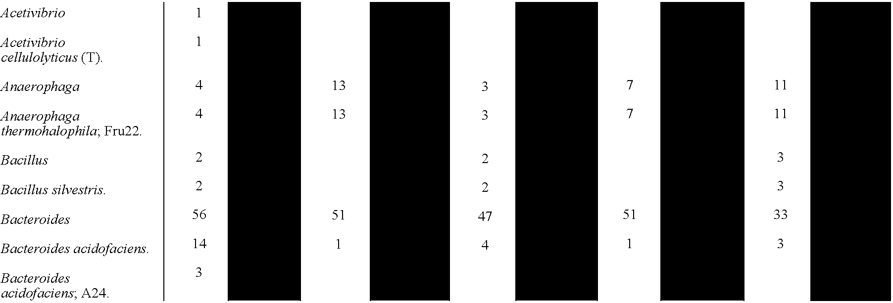


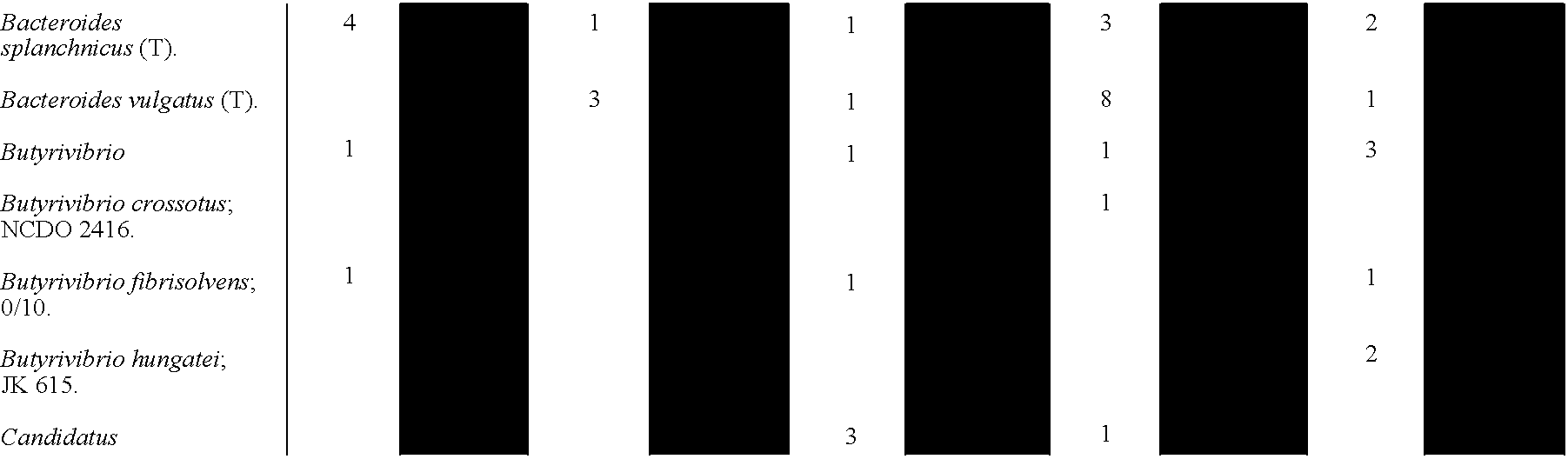
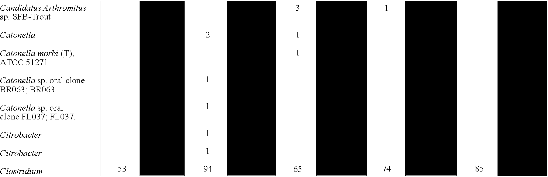


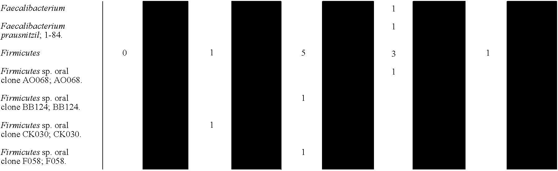


aBacterial 16S rDNA Ribosomal Database Project (RDP) entries are organized by genus (bold type) with specific RDP entries listed below each genus heading (plain type).
bTotal number of 16S rDNA clones that (i) passed the selection criteria described in Materials and Methods, and (ii) were homologous to the respective RDP entry with species or genus information.
c16S clones that are defined as “unidentified” (shaded columns) because their closest relative in RDP is either (i) an entry without species assignment, or (ii) an entry with species or genus assignment but with less than 98% identity to the respective rDNA sequence. These clones are listed in the table according to their closest relative in RDP with species or genus assignment.
# GenBank Accession numbers for the sequences are AY667702-AY668946. Further details of homology analyses are available at http://gordonlab.wustl.edu/. -
TABLE S2 Conserved transcription factor binding sites identified in orthologous Fiaf genes. Number of Potential TRANSFAC Sites Matrices consensus H M R Z F CHN Notes AP1_01 NNNTGAGTCAKCN 2 4 1 3 3 1 Ap1 site, activator protein 1 AP1_C NTGASTCAG 5 2 1 6 2 1 Ap1 site, activator protein 1 AP4_Q6 CWCAGCTGGN 4 2 2 2 4 1 Ap4 site, activator protein 4 CEBPGAMMA_Q6 CTBATTTCARAAW 1 1 5 9 4 1 CCAAT enhancer binding protein CREL_01 SGGRNTTTCC 2 3 4 2 2 1 C-Rel, overlap with NFkB DR1_Q3 RGGNCAAAGGTCA 2 2 4 2 2 1 PPAR, HNF4, direct repeat E12_Q6 RRCAGGTGNCV 3 3 2 4 6 1 E-box E2A_Q2 NCACCTGYYNCNKN 2 3 3 5 5 1 E-box ETS_Q4 ANNCACTTCCTG 3 3 3 4 4 1 C-Ets, T-cell, mesodermal cell development FAC1_01 NNNCAMAACACRNA 2 1 5 9 2 1 Fac1 site, fetal Alz-50 clone 1 FOXD3_01 NAWTGTTTRTTT 2 7 4 31 5 3 Fork head box D3 FOXO1_01 NNNWAAAYAAAYANNNNN 3 5 4 22 14 2 Fork head box O1 FOXO4_01 RWAAACAANNN 2 4 4 18 9 1 Fork head box O4 FOX_Q2 KAWTGTTTRTTW 1 3 5 16 7 1 Fork head factor GC_01 NRGGGGCGGGGCNK 8 4 4 2 3 2 GC box GR_Q6 NNNNNNCNNTNTGTNCTNN 3 1 1 2 1 1 glucocorticoid receptor site HFH8_01 NNNTGTTTATNTR 1 5 5 15 8 1 HNF3, Fkh8 site HNF3ALPHA_Q6 TRTTTGYTYWN 1 5 4 22 4 1 HNF3-alpha site HNF3_Q6 NWRARYAAAYANN 1 6 3 28 7 1 HNF3 site HNF4ALPHA_Q6 VTGAACTTTGMMB 2 2 1 5 3 1 HNF4-alpha site HSF_Q6 TTCCMGARGYTTC 1 3 3 1 1 3 Heat shock factor site ICSBP_Q6 RAARTGAAACTG 1 3 4 9 3 1 ICSBP, Interferon factor binding site IRF7_01 TNSGAAWNCGAAANTNNN 1 1 1 6 2 1 interferon regulatory factor 7 IRF_Q6 BNCRSTTTCANTTYY 4 4 6 11 7 1 Interferon regulatory factors ISRE_01 CAGTTTCWCTTTYCC 2 1 2 4 1 1 Interferon stimulated response element LBP1_Q6 CAGCTGS 2 3 4 5 8 2 TATA box repressor LDSPOLYA_B NNNSTGTGTDYYCWTN 2 3 2 6 3 1 Lentiviral Poly A downstream element LFA1_Q6 GGGSTCWR 1 2 2 1 3 1 AID1; HNF-2; LFA1 site LMO2COM_01 CNNCAGGTGBNN 2 2 3 2 10 1 LIM-only protein 2 site MEIS1_01 NNNTGACAGNNN 1 2 2 5 5 2 myeloid ecotropic viral integration site 1 MYOD_Q6 NNCACCTGNY 2 3 2 7 6 1 myoblast determining factor site MYOGENIN_Q6 RGCAGSTG 2 4 4 8 7 1 Myogenin site NF1_Q6 NNTTGGCNNNNNNCCNNN 1 2 3 1 3 2 nuclear factor 1 site NFE2_01 TGCTGAGTCAY 3 1 1 3 1 1 nuclear factor erythroid 2 p45 site PIT1_Q6 NMTTCATAWWTATNNMNA 2 8 5 18 7 1 Pit1, POU1F1 binding site POU1F1_Q6 ATGAATAAWT 2 5 2 15 3 1 POU1F1 binding site PPAR_DR1_Q2 TGACCTTTGNCCY 1 2 5 1 3 1 peroxisome proliferator-activated receptor binding site PU1_Q6 WGAGGAAG 5 5 4 2 6 2 Pu.1 site, interfere with erythroblast differentiation SP1_01 GGGGCGGGGT 4 1 2 0 2 1 Sp1 site, stimulating protein 1 SP1_Q6 NGGGGGCGGGGYN 8 3 5 2 4 2 Sp1 site, stimulating protein 1 TAL1BETAE47_01 NNNAACAGATGKTNNN 1 2 1 3 2 1 Tal-1beta/E47 heterodimer binding site -
TABLE S3 Gene-specific primers used for qRT-PCR assays. Sequence Accession amplicon size Gene name Abbrevation primer primer sequences ID No. Number (bp) Acetyl- CoA Acc 1 forward AAGTCCTTGGTCGGGAAGTATACA 43 XM_109883 126 carboxylase reverse ACTCCCTCAAAGTCATCACAAACA 44 aP2 Ap2 forward TTAAAAACACCGAGATTTCCTTCAA 45 NM_024406 102 reverse GGGCCCCGCCATCTAG 46 Carbohydrate Chrebp forward CGGGACATGTTTGATGACTATGTC 47 AF156604 105 regulatory element reverse CATCCCATTGAAGGATTCAAATAAA 48 binding protein Fasting-induced Fiaf forward CAATGCCAAATTGCTCCAATT 49 AF278699 82 adipose factor reverse TGGCCGTGGGCTCAGT 50 Fatty acid Fas forward TGGTGAATTGTCTCCGAAAAGA 51 AF127033 149 synthase reverse CACGTTCATCACGAGGTCATG 52 L32 ribosomal L32 forward CCTCTGGTGAAGCCCAAGATC 53 NM_172086 102 protein reverse TCTGGGTTTCCGCCAGTTT 54 Peroxisome Ppar-α forward CACCTTCCTCTTCCCAAAGCT 55 X57638 105 proliferator reverse GCGTCGGACTCGGTCTTCT 56 activated receptor α Peroxisome Ppar-γ forward ATGTCTCACAATGCCATCAGGTT 57 U10374 116 proliferator reverse GCTCGCAGATCAGCAGACTCT 58 activated receptor γ Sterol regulatory Srebp-1 forward GCATGCCATGGGCAAGTAC 59 NM_011480 125 element binding reverse CCACATAGATCTCTGCCAGTGTTG 60 protein 1 - Adult germ-free male NMRI/KI mice were maintained on a standard autoclaved chow diet rich in plant polysaccharides. Gas chromatographic-mass spectrometric (GC-MS) analysis established that glucose, arabinose, xylose and galactose are the predominant neutral sugars present in this chow (mole ratio=10:8:5:1). Seven week-old mice were colonized with a single inoculum of B. thetaiotaomicron and sacrificed 10 days later (a period that spans 2-3 cycles of turnover of the intestinal epithelium and its overlying mucus layer). Colonization density ranged from 107-109 CFU/mL in the distal small intestine (ileum) to 1010-1011 CFU/mL in the cecum and proximal colon. Scanning electron microscopic studies revealed B. thetaiotaomicron attached to small food particles and embedded in mucus (
FIG. 27 ). - The cecum is an anatomically distinct structure, located between the distal small intestine and proximal colon that is a site of great microbial density and diversity in conventionally-raised mice (F. Backhed, et al., Proc. Natl. Acad. Sci. USA, 101, 15718 (2004)). Nutrient use by B. thetaiotaomicron in the cecum was defined initially by whole genome transcriptional profiling. Cecal contents, including the mucus layer, were removed immediately after sacrifice of non-fasted mice (n=6), and the RNA extracted. The B. thetaiotaomicron transcriptome was characterized using custom GeneChips containing probe pairs derived from 4719 of the organism's 4779 predicted genes (Table S4). The results were compared to transcriptional profiles obtained from B. thetaiotaomicron grown from early log to stationary phase in a chemostat containing a minimal medium plus glucose as the sole fermentable carbohydrate source (MM-G;
FIG. 30 ).TABLE 54 Features of the B. theta GeneChip Naming No. of genes No. of Average no. of prefix of (probesets) probe probe pairs Functional category probesets represented pairs per probeset Control sequences AFFX 51 831 16.3 Bt chromosomal BT 4719 61737 13 genesa Bt genes on p5482b p5482 38 494 13 Bt tRNA genes tRNA 36 468 13
aGenbenk accession number AE015928
bGenbenk accession number AY171301
- Unsupervised hierarchical clustering of the GeneChip datasets disclosed remarkable uniformity in the in vivo transcriptional profiles of B. thetaiotaomicron harvested from individual gnotobiotic mice (panel A,
FIG. 31 ). A total of 1237 genes were defined as significantly upregulated in vivo compared with their expression in MM-G. The finctions of these upregulated genes were classified by clusters of orthologous groups (COG) analysis. The largest upregulated group belonged to the ‘carbohydrate transport and metabolism’ COG. In contrast, the largest group of genes down-regulated in vivo belonged to the ‘amino acid transport and metabolism’ COG (FIG. 32 , A). - SusC and SusD are components of a B. thetaiotaomicron outer membrane protein complex involved in binding of starch and malto-oligosaccharides during their digestion by outer membrane and periplasmic glycoside hydrolases (J. A. Shipman, et al., J. Bacteriol. 182, 5365 (2000)). Thirty-seven SusC and 16 SusD paralogs are upregulated≧10-fold in vivo by comparison to bacteria growing in MM-G (
FIG. 33 ). - The indigestibility of xylan, pectin, and arabinose-containing polysaccharides in dietary fiber reflects the paucity of host enzymes required for their degradation. The human genome contains only one putative glycoside hydrolase represented in the nine families of enzymes known in nature with xylanase, arabinosidase, pectinase, or pectate lyase activities, while the mouse genome has none (http://afmb.cnrs-mrs.fr/CAZY/). In contrast, B. thetaiotaomicron has 64 such enzymes (Table S5; http://afmb.cnrs-mrs.fr/CAZY/), many of which were selectively upregulated 10- to 823-fold in vivo. These included five secreted xylanases, five secreted arabinosidases, plus a secreted pectate lyase (
FIG. 28A -C plusFIG. 33 , B).TABLE 55 All families of glycoside hydrolases and polysoccharide lyases containing arabinosidase, xylanase, pectinase or pectate lyase activities with at least one representative in either the human, mouse, or B. theta genomes (http://afmbenrsfrCAZY/). Family Known Activities in Family Homo sapiens Mus musculus B. theta Glycoside Hydrolase Family 43 β-xylosidase (EC 3.2.1.37) 0 0 31 α-L-arabinofuranosidase (EC 3.2.1.55) arabinanase (EC 3.2.1.99) xylanase (EC 3.2.1.8) Glycoside Hydrolase Family 3 β-glucosidase (EC 3.2.1.21) 1 0 10 xylan 1,4-β-xylosidase (EC 3.2.1.37) β-N-ocetylhexosaminidase (EC 3.2.1.52) glucan 1,3-β-glucosidase (EC 3.2.1.58) glucan 1,4-β-glucosidase (EC 3.2.1.74) exo-1,3-1,4-glucanase (EC 3.2.1.-) α-L-arabinofuranosidase (EC 3.2.1.55) Glycoside Hydrolase Family 28 polygalacturonase (EC 3.2.1.15) 0 0 9 exo-polygalacturonase (EC 3.2.1.67) exo-polygalacturonase (EC 3.2.1.82) rhamnogalacturonase (EC not defined) Polysaccharide Lyase Family 1 pectate lyase (EC 4.2.2.2) 0 0 5 pectin lyase (EC 4.2.2.10) Glycoside Hydrolase Family 51 α-L-arabinofuranosidase (EC 3.2.1.55) 0 0 4 endoglucanase (EC 3.2.1.4) Polysaccharide Lyase Family9 pectate lyase (EC 4.2.2.2) 0 0 2 exopolygalacturonate lyase (EC 4.2.2.9) Glycoside Hydrolase Family 5 chitosanase (EC 3.2.1.132) 0 0 1 β-mannosidase (EC 3.2.1.25) cellulose (EC 3.2.1.4) glucan 1,3-β-glucosidase (EC 3.2.1.58) licheninase (EC 3.2.1.73) glucan endo-1,6-β-glucosidase (EC 3.2.1.75) mannan endo-1,4-β-manosidase (EC 3.2.1.78) endo-1,4-β-xylanase (EC 3.2.1.8) cellulose 1,4-β-cellobiosidase (EC 3.2.1.91) endo-1,6-β-galactanase (EC 3.2.1.-) β-1,3-mannanase (EC 3.2.1.-) Glycoside Hydrolase Family 93 exo-arabinanase (EC 3.2.1.55) 0 0 1 Polysaccharide Lyase Family 10 pectate lyase (EC 4.2.2.2) 0 0 1 - GC-MS analysis of total cecal contents harvested from fed germn-free mice revealed that xylose, galactose, arabinose, and glucose were the most abundant monosaccharide components (
FIG. 28D ). After 10 days of colonization by B. thetaiotaomicron, significant reductions in cecal concentrations of three prominent hexoses (glucose, galactose, and mannose) were observed. There were no significant decreases in pentose or amino-sugars (FIG. 28D ). The selective depletion of hexoses likely reflects the combined effects of microbial and host utilization. B. thetaiotaomicron colonization increased host expression of the principal sodium/glucose transporter, Sglt1, in the intestinal epithelium, reflecting an enhancement of host utilization of liberated monosaccharides (Example 1 and Table 1). Morover, of the 1237 bacterial genes upregulated in vivo, 310 were assignable to enzyme classification numbers in metabolic maps in the Kyoto Encyclopedia of Genes and Genomes (KEGG; http://www.genome.adjp/). The results of this metabolic reconstruction were consistent with active delivery of mannose, galactose and glucose to the glycolytic pathway, and arabinose and xylose to the pentose phosphate pathway (FIG. 34 ; see http://gordonlab.wustl.edu/metaview/bt). - Host mucus provides a ‘consistent’ endogenous source of glycans in the cecal habitat that could offer alternative nutrients to the microbiota during periods of change in the host's diet. B. thetaiotaomicron embeds itself in this mucus layer (
FIG. 27D ). GeneChip analysis provided evidence that the bacterium harvests glycans from mucus. For example, in vivo, B. thetaiotaomicron exhibited significant upregulation (2-10-fold; p<0.05) of (i) an operon (BT0455-BT0461) that encodes a sialidase, sialic acid-specific 9-O-acetyl esterase, mannosidase, and three b-hexosaminidases (FIG. 28A ), (ii) a mucin-desulfating sulfatase (BT3051), and (iii) a chondroitin lyase (BT3350). Fucose in host glycans is an attractive source of food: it typically occupies a terminal-linked position and is constitutively produced in the cecal mucosa of NMRI mice (L. Bry, et al., Science, 273, 1380 (1996)). In B. thetaioatomicron we found that two secreted a-fucosidases (BT1842, BT3665) and a five-component fucose utilization operon (BT1272-BT1277) were also induced (FIG. 28A ). Operon induction, which occurs through the interaction of L-fucose with a repressor encoded by its first open reading frame (L. V. Hooper, et al., Proc. Natl. Acad. Sci. USA, 96, 9833 (1999)), is indicative of bacterial import and utilization of this hexose. - To determine whether the absence of fermentable polysaccharides in the diet increases foraging on mucus glycans, B. thetaiotaomicron gene expression was compared in the ceca of two groups of age- and gender-matched adult gnotobiotic mice. One group received the standard polysaccharide-rich chow diet from weaning to the time of sacrifice. The other group was switched to a diet devoid of fermentable polysaccharides but rich in simple sugars (35% glucose; 35% sucrose) 14 days prior to colonization. All mice were colonized with B. thetaiotaomicron for 10 days and bacterial gene expression was defined in each of their ceca at the time of sacrifice.
- The presence or absence of polysaccharides in the diet did not produce a significant effect on the density of cecal colonization (data not shown). Using the transcriptional profiles of 98 B. thetaiotaomicron genes from the “replication, recombination and repair” COG as biomarkers, the cecal bacterial populations clustered most closely to cells undergoing log phase growth in vitro, irrespective of the diet (
FIG. 31 , B; Table S6).TABLE 56 B. theta genes in the Replication Recombination and Repair COG used for hierarchical clustering of GeneChip data shown in panel B of FIG. 25 Gene Annotation BT0026 putative transposase BT0069 conserved hypothetical protein BT0070 conserved hypothetical protein BT0078 putative DNA repair protein BT0244 putative exonuclease BT0245 ATP-dependent exonuclease abcC BT0252 transcription-repair coupling factor BT0280 transposase for insertion sequence element 15RM3 BT0358 tranposase BT0419 putative endonuclease BT0570 excinuclease ABC subunit B BT0578 excinuclease ABC subunit A BT0625 DNA helicase BT0630 exodecoxyribonudease BT0657 ATP-dependent DNA helicase BT0721 DNA repair and recombination protein putative helicase BT0831 ATP-dependent RNA helicase BT0894 DNA ligase BT0899 DNA gyrase subunit A BT1054 ATP-dependent helicase BT1081 recombination protein recR BT1154 ATP-independent RNA helicase BT1205 putative ATPase AAA family BT1756 transposase invertase BT1361 DNA repair protein recN (Recombination protein N) BT1364 DNA polymerase III beta chain BT1411 methylated-DNA-protein-cysteine methyltransferase BT1497 single-strand binding protein (SSB) BT1498 AVG-specific adenine glycosylase BT1499 DNA-binding protein HU BT1516 replicative DNA helicase BT1544 NADH pyrophosphatase, Mutl family hydrolase BT1610 DNA polymerase III subunit gammaltau BT1664 crossover junction endodeoxyribonuclease ruvC BT1671 endonuclease III BT1739 excinuclease ABC subunit A BT1821 transposase BT1848 ATP-dependent DNA helicase recO BT1885 putative ATP-dependent RNA helicase BT1978 Holiday junction DNA helicase ruvA BT1980 transposase BT2056 conserved hypothetical protein BT2073 putative helicase BT2089 DNA topoisomerase II BT2130 uracil-DNA glycosylase BT2137 transposase BT2143 chromosomal replication initiator protein dnaA BT2230 DNA polymerase III alpha subunit BT2297 putative reverse transcriptase BT2355 site-specific DNA-methyltransferance BT2400 DNA-3-methyladenine glycosylase I BT2615 reverse transcriptase BT2617 reverse transcriptase BT2644 DNA topoisomerase I BT2697 DNA mismatch repair protein mut5 - The simple sugar diet evoked a B. thetaiotaomicron tanscriptional response predominated by genes in the ‘carbohydrate transport and metabolism’ COG (
FIG. 32 , B). Glycoside hydrolase and polysaccharide lyase genes upregulated ≧2.5-fold in mice compated with MM-G cultures segregated into distinct groups after unsupervised hierarchical clustering (FIGS. 29 ). The group of 24 genes most highly expressed on the simple sugar diet encoded enzymes required for degradation of host glycans (e.g., eight hexosaminidases, two-fucosidases, plus a sialidase), and did not include any plant polysaccharide-directed arabinosidases or pectin lyases. - In addition, all components of the fucose utilization operon (BT1272-BT1277) were expressed at greater levels in mice fed the simple sugar diet compared to those fed the polysaccharide-rich diet (average induction compared to MM-G: 12-fold versus 6-fold). The sialylated glycan degradation operon (BT0455-BT0461) exhibited a comparable augmentation of expression on the simple sugar diet.
- A similar cluster analysis revealed two distinct groups of genes encoding carbohydrate binding/importing SusC/SusD paralogs: a group of 61 expressed at highest levels in B. thetaiotaomicron from the ceca of mice fed a polysaccharide-rich diet, and a group of 21 expressed at highest levels with a simple sugar diet (
FIG. 35 ). Thirteen of the upregulated SusC/D paralogs from B. thetaiotaomicron in mice fed a polysaccharide-rich diet are components of predicted operons that also contain ORFs specifying glycoside hydrolases and polysaccharide lyases. Five pairs of the SusC/D paralogs expressed at highest levels on a simple sugar diet are part of predicted operons. No SusC/D paralogs from one diet group were found in operons containing upregulated glycoside hydrolase genes from the other diet group (FIG. 36 ). Together, the data indicate that subsets of B. thetaiotaomicron's genome are dedicated to retrieving either host or dietary polysaccharides, depending upon their availability, although it appears that when both sources are available, harvesting energy from the diet is preferred. - Diet-associated changes in glycan foraging behavior were accompanied by changes in the expression of B. thetaiotaomicron's capsular polysaccharide synthesis (CPS) loci (
FIG. 37 ). Compared with growth in MM-G, CPS3 was down-regulated in vivo irrespective of host diet, CPS4 was upregulated in the ceca of mice fed a polysaccharide-rich diet, while CPS5 was upregulated with a high sugar diet (FIG. 37 ). The other five CPS loci did not manifest significant differences in their expression during growth in vitro versus in vivo, or with diet manipulation. These findings suggest that B. thetaiotaomicron is able to change its surface carbohydrates depending upon the nutrient glycan environment that it is accessing and perhaps also for evasion of the host's adaptive immune response. -
FIG. 38 presents a schematic overview of how B. thetaiotomicron might scavenge for carbohydrates in the distal intestine. Groups of bacteria assemble on undigested or partially digested food particles, elements of the mucus gel layer, and/or exfoliated epithelial cells. Bacterial attachment to these nutrient reservoirs is directed by glycan-specific outer membrane binding proteins (exemplified by SusC/D paralogs) that are opportunistically deployed depending upon the glycan environment encountered by the bacterium. Attachment helps oppose bacterial washout from the intestinal bioreactor, promotes harvest of oligo- and monosaccharides by an adaptively expressed repertoire of bacterial glycoside hydrolases, and facilitates sharing of the products of digestion with other microbial members whose nutritional niche overlaps that of B. thetaiotaomicron. In this scheme, microbial nutrient metabolism along the length of the intestine is a summation of myriad selfish and syntrophic relationships expressed by inhabitants of these micro-habitats. Micro-habitat diversity and mutualistic cooperation among component species (including the degree to which sanctions must be applied against cheats), are reflections of a dynamic interplay between the available nutrient foundation, and the degree of flexible foraging (niche breadth) expressed by micro-habitat residents. Members of Bacteroides with broad nutritional niches, such as B. thetaiotaomicron, contribute to diversity and stability by adaptively directing their glycan foraging behavior to the mucus when polysaccharide availability from the diet is reduced. Mucus glycans, in turn, represent a point where host genotype and diet intersect to regulate the stability of the microbiota. The highly variable outer chain structures of mucus and epithelial cell surface glycans are influenced by host genotype, and by microbial regulation of host glycosyltransferase gene expression. Co-evolution of glycan structural diversity in the host, and an elaborate repertoire of nutrient-regulated glycoside hydrolase genes in gut symbionts, endows the system with flexibility in adapting to changes in diet. While the present study has focused on the glycan foraging behavior of B. thetaiotaomicron in mono-associated germ-free mice, similar analyses can now be used to assess the impact of other members of the gut microbiota on B. thetaiotaomicron and on one another.
Claims (42)
Priority Applications (3)
| Application Number | Priority Date | Filing Date | Title |
|---|---|---|---|
| US11/080,755 US20050239706A1 (en) | 2003-10-31 | 2005-03-15 | Modulation of fiaf and the gastrointestinal microbiota as a means to control energy storage in a subject |
| PCT/US2005/026213 WO2006012586A2 (en) | 2004-07-27 | 2005-07-25 | Modulation of fiaf and the gastrointestinal microbiota |
| EP05803641A EP1778274A4 (en) | 2004-07-27 | 2005-07-25 | MODULATION OF FIAF AND GASTROINTESTINAL MICROBIOTE |
Applications Claiming Priority (3)
| Application Number | Priority Date | Filing Date | Title |
|---|---|---|---|
| US10/432,819 US20040091893A1 (en) | 2001-11-27 | 2001-11-27 | Method for studying the effects of commensal microflora on mammalian intestine and treatments of gastrointestinal-associated disease based thereon |
| US59131304P | 2004-07-27 | 2004-07-27 | |
| US11/080,755 US20050239706A1 (en) | 2003-10-31 | 2005-03-15 | Modulation of fiaf and the gastrointestinal microbiota as a means to control energy storage in a subject |
Related Parent Applications (1)
| Application Number | Title | Priority Date | Filing Date |
|---|---|---|---|
| US10/432,819 Continuation-In-Part US20040091893A1 (en) | 2001-11-27 | 2001-11-27 | Method for studying the effects of commensal microflora on mammalian intestine and treatments of gastrointestinal-associated disease based thereon |
Publications (1)
| Publication Number | Publication Date |
|---|---|
| US20050239706A1 true US20050239706A1 (en) | 2005-10-27 |
Family
ID=35786759
Family Applications (1)
| Application Number | Title | Priority Date | Filing Date |
|---|---|---|---|
| US11/080,755 Abandoned US20050239706A1 (en) | 2003-10-31 | 2005-03-15 | Modulation of fiaf and the gastrointestinal microbiota as a means to control energy storage in a subject |
Country Status (3)
| Country | Link |
|---|---|
| US (1) | US20050239706A1 (en) |
| EP (1) | EP1778274A4 (en) |
| WO (1) | WO2006012586A2 (en) |
Cited By (39)
| Publication number | Priority date | Publication date | Assignee | Title |
|---|---|---|---|---|
| US20060093606A1 (en) * | 2004-07-20 | 2006-05-04 | Genentech, Inc. | Compositions and methods of using angiopoietin-like 4 protein |
| US20060093607A1 (en) * | 2004-07-20 | 2006-05-04 | Genentech, Inc. | Inhibitors of angiopoietin-like 4 protein, combinations, and their use |
| WO2008083157A3 (en) * | 2006-12-29 | 2008-10-16 | Univ St Louis | Altering pgc-1alapha, ampk, fiaf, or the gastrointestinal microbiota as a means to modulate body fat and/or weight loss in a subject |
| WO2008076696A3 (en) * | 2006-12-18 | 2008-12-04 | Univ St Louis | The gut microbiome as a biomarker and therapeutic target for treating obesity or an obesity related disorder |
| EP2030623A1 (en) * | 2007-08-17 | 2009-03-04 | Nestec S.A. | Preventing and/or treating metabolic disorders by modulating the amount of enterobacteria |
| US20090110664A1 (en) * | 2007-10-26 | 2009-04-30 | Moore Brenda E | Probiotic compositions and methods for inducing and supporting weight loss |
| US20100184624A1 (en) * | 2007-05-31 | 2010-07-22 | The Washington University | Arrays and methods comprising m. smithii gene products |
| US20100331641A1 (en) * | 2009-06-29 | 2010-12-30 | Searete Llc Of The State Of Delaware | Devices for continual monitoring and introduction of gastrointestinal microbes |
| US20110177976A1 (en) * | 2008-06-30 | 2011-07-21 | The Washington University | Methods for promoting weight loss and associated arrays |
| US20110281762A1 (en) * | 2010-05-13 | 2011-11-17 | Sonnenburg Justin L | High throughput screening for anaerobic microorganisms |
| US8084200B2 (en) | 2002-11-15 | 2011-12-27 | Genentech, Inc. | Compositions and methods for the diagnosis and treatment of tumor |
| US8604185B2 (en) | 2004-07-20 | 2013-12-10 | Genentech, Inc. | Inhibitors of angiopoietin-like 4 protein, combinations, and their use |
| WO2012122522A3 (en) * | 2011-03-09 | 2014-04-24 | Washington University | Cultured collection of gut microbial community |
| US9040101B2 (en) | 2011-08-17 | 2015-05-26 | MicroBiome Therapeutics LLC. | Method to treat diabetes utilizing a gastrointestinal microbiome modulating composition |
| US20150317444A1 (en) * | 2012-12-13 | 2015-11-05 | Metabogen Ab | Identification of a Person Having Risk for Developing Type 2 Diabetes |
| US9433651B2 (en) | 2013-06-05 | 2016-09-06 | Rebiotix, Inc. | Microbiota restoration therapy (MRT), compositions and methods of manufacture |
| US9463208B2 (en) | 2010-02-01 | 2016-10-11 | Rebiotix, Inc. | Bacteriotherapy for clostridium difficile colitis |
| US9511099B2 (en) | 2013-06-05 | 2016-12-06 | Rebiotix, Inc. | Microbiota restoration therapy (MRT), compositions and methods of manufacture |
| US9511100B2 (en) | 2013-06-05 | 2016-12-06 | Rebiotix, Inc. | Microbiota restoration therapy (MRT), compositions and methods of manufacture |
| WO2016210251A1 (en) * | 2015-06-25 | 2016-12-29 | Ascus Biosciences, Inc. | Methods, apparatuses, and systems for analyzing microorganism strains from complex heterogeneous communities, predicting and identifying functional relationships and interactions thereof, and selecting and synthesizing microbial ensembles based thereon |
| CN106795481A (en) * | 2014-09-30 | 2017-05-31 | 深圳华大基因科技有限公司 | For the biomarker of obesity-related disorder |
| US9694039B2 (en) | 2013-06-05 | 2017-07-04 | Rebiotix, Inc. | Microbiota restoration therapy (MRT), compositions and methods of manufacture |
| US9782445B2 (en) | 2013-06-05 | 2017-10-10 | Rebiotix, Inc. | Microbiota restoration therapy (MRT), compositions and methods of manufacture |
| WO2018039089A1 (en) * | 2016-08-20 | 2018-03-01 | Gregg John Malcolm Hall | Methods of use & compositions for obesity |
| US9938558B2 (en) * | 2015-06-25 | 2018-04-10 | Ascus Biosciences, Inc. | Methods, apparatuses, and systems for analyzing microorganism strains from complex heterogeneous communities, predicting and identifying functional relationships and interactions thereof, and selecting and synthesizing microbial ensembles based thereon |
| US9993507B2 (en) | 2016-01-07 | 2018-06-12 | Ascus Biosciences, Inc. | Methods of improving milk production and compositional characteristics |
| US10119116B2 (en) | 2016-07-28 | 2018-11-06 | Bobban Subhadra | Devices, systems and methods for the production of humanized gut commensal microbiota |
| US10226431B2 (en) | 2015-06-09 | 2019-03-12 | Rebiotix, Inc. | Microbiota restoration therapy (MRT) compositions and methods of manufacture |
| US10383901B2 (en) | 2013-06-05 | 2019-08-20 | Rebiotix, Inc. | Microbiota restoration therapy (MRT), compositions and methods of manufacture |
| US10670611B2 (en) | 2014-09-26 | 2020-06-02 | Somalogic, Inc. | Cardiovascular risk event prediction and uses thereof |
| US10799539B2 (en) | 2015-06-09 | 2020-10-13 | Rebiotix, Inc. | Microbiota restoration therapy (MRT) compositions and methods of manufacture |
| US10828340B2 (en) | 2015-06-09 | 2020-11-10 | Rebiotix, Inc. | Microbiota restoration therapy (MRT) compositions and methods of manufacture |
| US10851399B2 (en) | 2015-06-25 | 2020-12-01 | Native Microbials, Inc. | Methods, apparatuses, and systems for microorganism strain analysis of complex heterogeneous communities, predicting and identifying functional relationships and interactions thereof, and selecting and synthesizing microbial ensembles based thereon |
| US10905726B2 (en) | 2015-06-09 | 2021-02-02 | Rebiotix, Inc. | Microbiota restoration therapy (MRT) compositions and methods of manufacture |
| US10920283B2 (en) | 2013-11-01 | 2021-02-16 | Washington University | Methods to establish and restore normal gut microbiota function of subject in need thereof |
| US11044924B2 (en) | 2017-04-28 | 2021-06-29 | Native Microbials, Inc. | Methods for supporting grain intensive and or energy intensive diets in ruminants by administration of a synthetic bioensemble of microbes or purified strains therefor |
| US11891647B2 (en) | 2016-12-28 | 2024-02-06 | Native Microbials, Inc. | Methods, apparatuses, and systems for analyzing complete microorganism strains in complex heterogeneous communities, determining functional relationships and interactions thereof, and identifying and synthesizing bioreactive modificators based thereon |
| EP4049667A4 (en) * | 2019-10-21 | 2024-02-21 | Biofermin Pharmaceutical Co., Ltd. | Uremic toxin reducing agent |
| US12018313B2 (en) | 2016-12-28 | 2024-06-25 | Native Microbials, Inc. | Methods, apparatuses, and systems for microorganism strain analysis of complex heterogeneous communities with tracer analytics, determination of functional relationships and interactions thereof, and synthesis of microbial ensembles |
Families Citing this family (32)
| Publication number | Priority date | Publication date | Assignee | Title |
|---|---|---|---|---|
| TW200904340A (en) | 2007-05-11 | 2009-02-01 | Mannatech Inc | Processing of natural polysaccharides by selected non-pathogenic microorganisms and methods of making and using the same |
| US9113641B2 (en) | 2007-12-06 | 2015-08-25 | Arla Foods Amba | Probiotic bacteria and regulation of fat storage |
| GB201117313D0 (en) | 2011-10-07 | 2011-11-16 | Gt Biolog Ltd | Bacterium for use in medicine |
| PT3628161T (en) | 2012-11-23 | 2023-05-15 | Seres Therapeutics Inc | Synergistic bacterial compositions and methods of production and use thereof |
| US8906668B2 (en) | 2012-11-23 | 2014-12-09 | Seres Health, Inc. | Synergistic bacterial compositions and methods of production and use thereof |
| WO2014121304A1 (en) | 2013-02-04 | 2014-08-07 | Seres Health, Inc. | Compositions and methods |
| JP6479685B2 (en) | 2013-02-04 | 2019-03-06 | セレス セラピューティクス インコーポレイテッド | Compositions and methods for the control of pathogenic bacterial growth |
| US10076546B2 (en) | 2013-03-15 | 2018-09-18 | Seres Therapeutics, Inc. | Network-based microbial compositions and methods |
| GB201306536D0 (en) | 2013-04-10 | 2013-05-22 | Gt Biolog Ltd | Polypeptide and immune modulation |
| ES3017865T3 (en) | 2013-11-25 | 2025-05-13 | Nestle Sa | Synergistic bacterial compositions and methods of production and use thereof |
| EP3082431A4 (en) | 2013-12-16 | 2017-11-15 | Seres Therapeutics, Inc. | Bacterial compositions and methods of use thereof for treatment of immune system disorders |
| WO2016049917A1 (en) * | 2014-09-30 | 2016-04-07 | Bgi Shenzhen Co., Limited | Biomarkers for obesity related diseases |
| KR20170091157A (en) | 2014-12-23 | 2017-08-08 | 4디 파마 리서치 리미티드 | Pirin polypeptide and immune modulation |
| HUE035569T2 (en) | 2014-12-23 | 2018-05-28 | 4D Pharma Res Ltd | A bacteroides thetaiotaomicron strain and its use in reducing inflammation |
| LT3360559T (en) | 2015-06-15 | 2019-12-27 | 4D Pharma Research Limited | Compositions comprising bacterial strains |
| HUE045413T2 (en) | 2015-06-15 | 2019-12-30 | 4D Pharma Res Ltd | Compositions comprising bacterial strains |
| MA41060B1 (en) | 2015-06-15 | 2019-11-29 | 4D Pharma Res Ltd | Compositions comprising bacterial strains |
| ES2753779T3 (en) | 2015-06-15 | 2020-04-14 | 4D Pharma Res Ltd | Blautia stercosis and wexlerae for use in the treatment of inflammatory and autoimmune diseases |
| MA41010B1 (en) | 2015-06-15 | 2020-01-31 | 4D Pharma Res Ltd | Compositions comprising bacterial strains |
| CN108513545B (en) | 2015-11-20 | 2020-11-03 | 4D制药研究有限公司 | Compositions comprising bacterial strains |
| GB201520497D0 (en) | 2015-11-20 | 2016-01-06 | 4D Pharma Res Ltd | Compositions comprising bacterial strains |
| SG11201807195VA (en) | 2016-03-04 | 2018-09-27 | 4D Pharma Plc | Compositions comprising bacterial blautia strains for treating visceral hypersensitivity |
| GB201612191D0 (en) | 2016-07-13 | 2016-08-24 | 4D Pharma Plc | Compositions comprising bacterial strains |
| TW201821093A (en) | 2016-07-13 | 2018-06-16 | 英商4D製藥有限公司 | Composition comprising a bacterial strain |
| GB201621123D0 (en) | 2016-12-12 | 2017-01-25 | 4D Pharma Plc | Compositions comprising bacterial strains |
| HUE054164T2 (en) | 2017-05-22 | 2021-08-30 | 4D Pharma Res Ltd | Preparations containing bacterial strains |
| JP6978514B2 (en) | 2017-05-24 | 2021-12-08 | フォーディー ファーマ リサーチ リミテッド4D Pharma Research Limited | Composition containing bacterial strain |
| SMT202000695T1 (en) | 2017-06-14 | 2021-01-05 | 4D Pharma Res Limited | Compositions comprising bacterial strains |
| ES2855701T3 (en) | 2017-06-14 | 2021-09-24 | 4D Pharma Res Ltd | Compositions comprising bacterial strains |
| JP2020523326A (en) | 2017-06-14 | 2020-08-06 | フォーディー ファーマ リサーチ リミテッド4D Pharma Research Limited | Composition comprising a bacterial strain |
| JP2020530840A (en) | 2017-08-14 | 2020-10-29 | セレス セラピューティクス インコーポレイテッド | Compositions and Methods for Treating Cholestasis Diseases |
| RU2020117775A (en) | 2017-10-30 | 2021-12-01 | Серес Терапеутикс, Инк. | COMPOSITIONS AND METHODS FOR TREATMENT OF ANTIBIOTIC RESISTANCE |
Citations (2)
| Publication number | Priority date | Publication date | Assignee | Title |
|---|---|---|---|---|
| US5599795A (en) * | 1994-08-19 | 1997-02-04 | Mccann; Michael | Method for treatment of idiopathic inflammatory bowel disease (IIBD) |
| US6333188B1 (en) * | 1999-03-04 | 2001-12-25 | Bhph Company Limited | Lactic acid bacteria preparation having biopurification activity |
Family Cites Families (4)
| Publication number | Priority date | Publication date | Assignee | Title |
|---|---|---|---|---|
| JP2004536557A (en) * | 2000-11-27 | 2004-12-09 | ワシントン・ユニバーシティ | Method for investigating the effect of symbiotic microflora on mammalian intestine and treatment of gastrointestinal related diseases based on it |
| ES2321815T3 (en) * | 2001-04-04 | 2009-06-12 | Ortho-Mcneil-Janssen Pharmaceuticals, Inc. | THERAPEUTIC COMBINATION THAT INCLUDES REABSORTION AND GLUCOSE INHIBITORS AND PPAR MODULATORS. |
| ITRM20010763A1 (en) * | 2001-12-21 | 2003-06-21 | Simone Claudio De | NEW LACTIC BACTERIA STOCK AND EDIBLE COMPOSITIONS, DRUGS AND VETERINARY PRODUCTS THAT CONTAIN IT. |
| EP1586902A3 (en) * | 2004-03-30 | 2006-02-15 | F.Hoffmann-La Roche Ag | ANGPTL4/FIAF as marker for PPARdelta modulation |
-
2005
- 2005-03-15 US US11/080,755 patent/US20050239706A1/en not_active Abandoned
- 2005-07-25 EP EP05803641A patent/EP1778274A4/en not_active Withdrawn
- 2005-07-25 WO PCT/US2005/026213 patent/WO2006012586A2/en not_active Ceased
Patent Citations (2)
| Publication number | Priority date | Publication date | Assignee | Title |
|---|---|---|---|---|
| US5599795A (en) * | 1994-08-19 | 1997-02-04 | Mccann; Michael | Method for treatment of idiopathic inflammatory bowel disease (IIBD) |
| US6333188B1 (en) * | 1999-03-04 | 2001-12-25 | Bhph Company Limited | Lactic acid bacteria preparation having biopurification activity |
Cited By (97)
| Publication number | Priority date | Publication date | Assignee | Title |
|---|---|---|---|---|
| US8084200B2 (en) | 2002-11-15 | 2011-12-27 | Genentech, Inc. | Compositions and methods for the diagnosis and treatment of tumor |
| US7740846B2 (en) | 2004-07-20 | 2010-06-22 | Genentech, Inc. | Inhibitors of angiopoietin-like 4 protein, combinations, and their use |
| US20060093607A1 (en) * | 2004-07-20 | 2006-05-04 | Genentech, Inc. | Inhibitors of angiopoietin-like 4 protein, combinations, and their use |
| US20070026002A1 (en) * | 2004-07-20 | 2007-02-01 | Genentech, Inc. | Inhibitors of angiopoietin-like 4 protein, combinations, and their use |
| US20070054856A1 (en) * | 2004-07-20 | 2007-03-08 | Hanspeter Gerber | Compositions and methods of using angiopoietin-like 4 protein |
| US7371384B2 (en) | 2004-07-20 | 2008-05-13 | Genentech, Inc. | Compositions and methods of using angiopoietin-like 4 protein antibody |
| US8633155B2 (en) | 2004-07-20 | 2014-01-21 | Genentech, Inc. | Methods of using angiopoietin-like 4 protein to stimulate proliferation of pre-adipocytes |
| US8604185B2 (en) | 2004-07-20 | 2013-12-10 | Genentech, Inc. | Inhibitors of angiopoietin-like 4 protein, combinations, and their use |
| US20060093606A1 (en) * | 2004-07-20 | 2006-05-04 | Genentech, Inc. | Compositions and methods of using angiopoietin-like 4 protein |
| US20140093478A1 (en) * | 2006-12-18 | 2014-04-03 | The Washington University | Gut microbiome as a biomarker and therapeutic target for treating obesity or an obesity related disorder |
| US20100172874A1 (en) * | 2006-12-18 | 2010-07-08 | The Washington University | Gut microbiome as a biomarker and therapeutic target for treating obesity or an obesity related disorder |
| WO2008076696A3 (en) * | 2006-12-18 | 2008-12-04 | Univ St Louis | The gut microbiome as a biomarker and therapeutic target for treating obesity or an obesity related disorder |
| WO2008083157A3 (en) * | 2006-12-29 | 2008-10-16 | Univ St Louis | Altering pgc-1alapha, ampk, fiaf, or the gastrointestinal microbiota as a means to modulate body fat and/or weight loss in a subject |
| US20100184624A1 (en) * | 2007-05-31 | 2010-07-22 | The Washington University | Arrays and methods comprising m. smithii gene products |
| WO2009024429A3 (en) * | 2007-08-17 | 2009-12-03 | Nestec S.A. | Preventing and/or treating metabolic disorders by modulating the amount of enterobacteria |
| CN104147052A (en) * | 2007-08-17 | 2014-11-19 | 雀巢产品技术援助有限公司 | Preventing and/or treating metabolic disorders by modulating the amount of enterobacteria |
| US20110123501A1 (en) * | 2007-08-17 | 2011-05-26 | Nestec S.A. | Gut flora and weight management |
| EP2030623A1 (en) * | 2007-08-17 | 2009-03-04 | Nestec S.A. | Preventing and/or treating metabolic disorders by modulating the amount of enterobacteria |
| AU2008290737B2 (en) * | 2007-08-17 | 2013-11-14 | Nestec S.A. | Preventing and/or treating metabolic disorders by modulating the amount of enterobacteria |
| US8591880B2 (en) | 2007-08-17 | 2013-11-26 | Nestec S.A. | Gut flora and weight management |
| US20090110664A1 (en) * | 2007-10-26 | 2009-04-30 | Moore Brenda E | Probiotic compositions and methods for inducing and supporting weight loss |
| US9371510B2 (en) | 2007-10-26 | 2016-06-21 | Brenda E. Moore | Probiotic compositions and methods for inducing and supporting weight loss |
| US20110177976A1 (en) * | 2008-06-30 | 2011-07-21 | The Washington University | Methods for promoting weight loss and associated arrays |
| US20100331641A1 (en) * | 2009-06-29 | 2010-12-30 | Searete Llc Of The State Of Delaware | Devices for continual monitoring and introduction of gastrointestinal microbes |
| US9848760B2 (en) | 2009-06-29 | 2017-12-26 | Gearbox, Llc | Devices for continual monitoring and introduction of gastrointestinal microbes |
| US12303537B2 (en) | 2010-02-01 | 2025-05-20 | Ferring Microbiome Inc. | Bacteriotherapy for clostridium difficile colitis |
| US11986500B2 (en) | 2010-02-01 | 2024-05-21 | Rebiotix Inc | Bacteriotherapy for clostridium difficile colitis |
| US9463208B2 (en) | 2010-02-01 | 2016-10-11 | Rebiotix, Inc. | Bacteriotherapy for clostridium difficile colitis |
| US9629881B2 (en) | 2010-02-01 | 2017-04-25 | Rebiotix, Inc. | Bacteriotherapy for clostridium difficile colitis |
| US12102655B2 (en) | 2010-02-01 | 2024-10-01 | Rebiotix Inc. | Bacteriotherapy for clostridium difficile colitis |
| US20110281762A1 (en) * | 2010-05-13 | 2011-11-17 | Sonnenburg Justin L | High throughput screening for anaerobic microorganisms |
| WO2012122522A3 (en) * | 2011-03-09 | 2014-04-24 | Washington University | Cultured collection of gut microbial community |
| US9463169B2 (en) | 2011-08-17 | 2016-10-11 | Microbiome Therapeutics, Llc | Human gastrointestinal microbiome modulating food supplement for improving blood glucose regulation |
| US10441602B2 (en) | 2011-08-17 | 2019-10-15 | Microbiome Therapeutics, Llc | Human gastrointestinal microbiome modulating composition and methods of use |
| US9040101B2 (en) | 2011-08-17 | 2015-05-26 | MicroBiome Therapeutics LLC. | Method to treat diabetes utilizing a gastrointestinal microbiome modulating composition |
| US11213543B2 (en) | 2011-08-17 | 2022-01-04 | The Nature's Bounty Co. | Human gastrointestinal microbiome modulating composition and methods of use |
| US9881135B2 (en) * | 2012-12-13 | 2018-01-30 | Metabogen Ab | Identification of a person having risk for developing type 2 diabetes |
| US20150317444A1 (en) * | 2012-12-13 | 2015-11-05 | Metabogen Ab | Identification of a Person Having Risk for Developing Type 2 Diabetes |
| US9694039B2 (en) | 2013-06-05 | 2017-07-04 | Rebiotix, Inc. | Microbiota restoration therapy (MRT), compositions and methods of manufacture |
| US10603341B2 (en) | 2013-06-05 | 2020-03-31 | Rebiotix, Inc. | Microbiota restoration therapy (MRT), compositions and methods of manufacture |
| US9782445B2 (en) | 2013-06-05 | 2017-10-10 | Rebiotix, Inc. | Microbiota restoration therapy (MRT), compositions and methods of manufacture |
| US9433651B2 (en) | 2013-06-05 | 2016-09-06 | Rebiotix, Inc. | Microbiota restoration therapy (MRT), compositions and methods of manufacture |
| US9675648B2 (en) | 2013-06-05 | 2017-06-13 | Rebiotix, Inc. | Microbiota restoration therapy (MRT), compositions and methods of manufacture |
| US9511099B2 (en) | 2013-06-05 | 2016-12-06 | Rebiotix, Inc. | Microbiota restoration therapy (MRT), compositions and methods of manufacture |
| US11554143B2 (en) | 2013-06-05 | 2023-01-17 | Rebiotix, Inc. | Microbiota restoration therapy (MRT), compositions and methods of manufacture |
| US9642880B2 (en) | 2013-06-05 | 2017-05-09 | Rebiotix, Inc. | Microbiota restoration therapy (MRT), compositions and methods of manufacture |
| US10434126B2 (en) | 2013-06-05 | 2019-10-08 | Rebiotix, Inc. | Microbiota restoration therapy (MRT), compositions and methods of manufacture |
| US10471107B2 (en) | 2013-06-05 | 2019-11-12 | Rebiotix, Inc. | Microbiota restoration therapy (MRT), compositions and methods of manufacture |
| US10688137B2 (en) | 2013-06-05 | 2020-06-23 | Rebiotix, Inc. | Microbiota restoration therapy (MRT), compositions and methods of manufacture |
| US10434124B2 (en) | 2013-06-05 | 2019-10-08 | Rebiotix, Inc. | Microbiota restoration therapy (MRT), compositions and methods of manufacture |
| US10624932B2 (en) | 2013-06-05 | 2020-04-21 | Rebiotix, Inc. | Microbiota restoration therapy (MRT), compositions and methods of manufacture |
| US10610547B2 (en) | 2013-06-05 | 2020-04-07 | Rebiotix, Inc. | Microbiota restoration therapy (MRT), compositions and methods of manufacture |
| US10383901B2 (en) | 2013-06-05 | 2019-08-20 | Rebiotix, Inc. | Microbiota restoration therapy (MRT), compositions and methods of manufacture |
| US10391129B2 (en) | 2013-06-05 | 2019-08-27 | Rebiotix, Inc. | Microbiota restoration therapy (MRT), compositions and methods of manufacture |
| US9511100B2 (en) | 2013-06-05 | 2016-12-06 | Rebiotix, Inc. | Microbiota restoration therapy (MRT), compositions and methods of manufacture |
| US10493111B2 (en) | 2013-06-05 | 2019-12-03 | Rebiotix, Inc. | Microbiota restoration therapy (MRT), compositions and methods of manufacture |
| US10434125B2 (en) | 2013-06-05 | 2019-10-08 | Rebiotix, Inc. | Microbiota restoration therapy (MRT), compositions and methods of manufacture |
| US10920283B2 (en) | 2013-11-01 | 2021-02-16 | Washington University | Methods to establish and restore normal gut microbiota function of subject in need thereof |
| US10670611B2 (en) | 2014-09-26 | 2020-06-02 | Somalogic, Inc. | Cardiovascular risk event prediction and uses thereof |
| CN106795481A (en) * | 2014-09-30 | 2017-05-31 | 深圳华大基因科技有限公司 | For the biomarker of obesity-related disorder |
| US10828340B2 (en) | 2015-06-09 | 2020-11-10 | Rebiotix, Inc. | Microbiota restoration therapy (MRT) compositions and methods of manufacture |
| US11642381B2 (en) | 2015-06-09 | 2023-05-09 | Rebiotix, Inc. | Microbiota restoration therapy (MRT) compositions and methods of manufacture |
| US10905726B2 (en) | 2015-06-09 | 2021-02-02 | Rebiotix, Inc. | Microbiota restoration therapy (MRT) compositions and methods of manufacture |
| US10799539B2 (en) | 2015-06-09 | 2020-10-13 | Rebiotix, Inc. | Microbiota restoration therapy (MRT) compositions and methods of manufacture |
| US10391064B2 (en) | 2015-06-09 | 2019-08-27 | Rebiotix, Inc. | Microbiota restoration therapy (MRT) compositions and methods of manufacture |
| US12036250B2 (en) | 2015-06-09 | 2024-07-16 | Rebiotix Inc. | Microbiota restoration therapy (MRT) compositions and methods of manufacture |
| US11654164B2 (en) | 2015-06-09 | 2023-05-23 | Rebiotix, Inc. | Microbiota restoration therapy (MRT) compositions and methods of manufacture |
| US10226431B2 (en) | 2015-06-09 | 2019-03-12 | Rebiotix, Inc. | Microbiota restoration therapy (MRT) compositions and methods of manufacture |
| US10870877B2 (en) | 2015-06-25 | 2020-12-22 | Native Microbials, Inc. | Methods, apparatuses and systems for analyzing microorganism strains from complex heterogeneous communities, predicting and identifying functional relationships and interactions thereof, and selecting and synthesizing microbial ensembles based thereon |
| CN107849597A (en) * | 2015-06-25 | 2018-03-27 | 埃斯库斯生物科技股份公司 | For analyze the microbial strains from complex heterogeneous group, prediction and identify its functional relationship and interaction and based on its selection and synthesized micro-organism group method, apparatus and system |
| CN107849597B (en) * | 2015-06-25 | 2022-07-26 | 原生微生物股份有限公司 | Method for forming a population of active microbial strains based on analysis of heterogeneous microbial communities |
| US9938558B2 (en) * | 2015-06-25 | 2018-04-10 | Ascus Biosciences, Inc. | Methods, apparatuses, and systems for analyzing microorganism strains from complex heterogeneous communities, predicting and identifying functional relationships and interactions thereof, and selecting and synthesizing microbial ensembles based thereon |
| US9540676B1 (en) | 2015-06-25 | 2017-01-10 | Ascus Biosciences, Inc. | Methods, apparatuses, and systems for analyzing microorganism strains from complex heterogeneous communities, predicting and identifying functional relationships and interactions thereof, and selecting and synthesizing microbial ensembles based thereon |
| WO2016210251A1 (en) * | 2015-06-25 | 2016-12-29 | Ascus Biosciences, Inc. | Methods, apparatuses, and systems for analyzing microorganism strains from complex heterogeneous communities, predicting and identifying functional relationships and interactions thereof, and selecting and synthesizing microbial ensembles based thereon |
| US10844419B2 (en) | 2015-06-25 | 2020-11-24 | Native Microbials, Inc. | Methods, apparatuses, and systems for analyzing microorganism strains from complex heterogeneous communities, predicting and identifying functional relationships and interactions thereof, and selecting and synthesizing microbial ensembles based thereon |
| US10851399B2 (en) | 2015-06-25 | 2020-12-01 | Native Microbials, Inc. | Methods, apparatuses, and systems for microorganism strain analysis of complex heterogeneous communities, predicting and identifying functional relationships and interactions thereof, and selecting and synthesizing microbial ensembles based thereon |
| US10966437B2 (en) | 2016-01-07 | 2021-04-06 | Native Microbials, Inc. | Microbial compositions and methods of use for improving milk production |
| US10701955B2 (en) | 2016-01-07 | 2020-07-07 | Ascus Biosciences, Inc. | Ruminant compositions |
| US9993507B2 (en) | 2016-01-07 | 2018-06-12 | Ascus Biosciences, Inc. | Methods of improving milk production and compositional characteristics |
| US10448658B2 (en) | 2016-01-07 | 2019-10-22 | Ascus Biosciences, Inc. | Cow food and methods of husbandry for increased milk production |
| US10448657B2 (en) | 2016-01-07 | 2019-10-22 | Ascus Biosciences, Inc. | Cow food and methods of husbandry for increased milk production |
| US11910808B2 (en) | 2016-01-07 | 2024-02-27 | Native Microbials, Inc. | Ruminant compositions |
| US11291219B2 (en) | 2016-01-07 | 2022-04-05 | Native Microbials, Inc. | Microbial compositions and methods of use for improving milk production |
| US11910809B2 (en) | 2016-01-07 | 2024-02-27 | Native Microbials, Inc. | Microbial compositions and methods of use for improving milk production |
| US10293006B2 (en) | 2016-01-07 | 2019-05-21 | Ascus Biosciences, Inc. | Microbial compositions for improving milk production in ruminants |
| US10645952B2 (en) | 2016-01-07 | 2020-05-12 | Ascus Biosciences, Inc. | Microbial compositions and methods of use for improving milk production |
| US10398154B2 (en) | 2016-01-07 | 2019-09-03 | Ascus Biosciences, Inc. | Microbial compositions and methods of use for improving milk production |
| US10246677B2 (en) | 2016-07-28 | 2019-04-02 | Bobban Subhadra | Devices, systems and methods for the production of humanized gut commensal microbiota |
| US10138460B2 (en) | 2016-07-28 | 2018-11-27 | Bobban Subhadra | Devices, systems and methods for the production of humanized gut commensal microbiota |
| US10119116B2 (en) | 2016-07-28 | 2018-11-06 | Bobban Subhadra | Devices, systems and methods for the production of humanized gut commensal microbiota |
| US10767157B2 (en) | 2016-07-28 | 2020-09-08 | Bobban Subhadra | Devices, systems and methods for the production of humanized gut commensal microbiota |
| WO2018039089A1 (en) * | 2016-08-20 | 2018-03-01 | Gregg John Malcolm Hall | Methods of use & compositions for obesity |
| US12018313B2 (en) | 2016-12-28 | 2024-06-25 | Native Microbials, Inc. | Methods, apparatuses, and systems for microorganism strain analysis of complex heterogeneous communities with tracer analytics, determination of functional relationships and interactions thereof, and synthesis of microbial ensembles |
| US11891647B2 (en) | 2016-12-28 | 2024-02-06 | Native Microbials, Inc. | Methods, apparatuses, and systems for analyzing complete microorganism strains in complex heterogeneous communities, determining functional relationships and interactions thereof, and identifying and synthesizing bioreactive modificators based thereon |
| US11871767B2 (en) | 2017-04-28 | 2024-01-16 | Native Microbials, Inc. | Microbial compositions and methods for ruminant health and performance |
| US11044924B2 (en) | 2017-04-28 | 2021-06-29 | Native Microbials, Inc. | Methods for supporting grain intensive and or energy intensive diets in ruminants by administration of a synthetic bioensemble of microbes or purified strains therefor |
| EP4049667A4 (en) * | 2019-10-21 | 2024-02-21 | Biofermin Pharmaceutical Co., Ltd. | Uremic toxin reducing agent |
Also Published As
| Publication number | Publication date |
|---|---|
| WO2006012586A3 (en) | 2009-06-04 |
| WO2006012586A9 (en) | 2006-03-30 |
| EP1778274A2 (en) | 2007-05-02 |
| WO2006012586A2 (en) | 2006-02-02 |
| EP1778274A4 (en) | 2010-09-08 |
Similar Documents
| Publication | Publication Date | Title |
|---|---|---|
| US20050239706A1 (en) | Modulation of fiaf and the gastrointestinal microbiota as a means to control energy storage in a subject | |
| Kühn et al. | Intestinal alkaline phosphatase targets the gut barrier to prevent aging | |
| Chassaing et al. | Intestinal epithelial cell toll-like receptor 5 regulates the intestinal microbiota to prevent low-grade inflammation and metabolic syndrome in mice | |
| Mack et al. | Is the impact of starvation on the gut microbiota specific or unspecific to anorexia nervosa? A narrative review based on a systematic literature search | |
| Sodhi et al. | Intestinal epithelial Toll-like receptor 4 regulates goblet cell development and is required for necrotizing enterocolitis in mice | |
| Hao et al. | Overexpression of heat shock protein 70 and its relationship to intestine under acute heat stress in broilers: 1. Intestinal structure and digestive function | |
| RU2644204C2 (en) | Method for animal obesity model obtaining | |
| JP2018099126A (en) | Bacteria for use as probiotics for nutritional and medical applications | |
| US12285446B2 (en) | Compositions and methods for improving mitochondrial function | |
| JP2004536557A (en) | Method for investigating the effect of symbiotic microflora on mammalian intestine and treatment of gastrointestinal related diseases based on it | |
| WO2008083157A2 (en) | Altering pgc-1alapha, ampk, fiaf, or the gastrointestinal microbiota as a means to modulate body fat and/or weight loss in a subject | |
| WO2013146319A1 (en) | Diabetes-inducible bacterium | |
| JPWO2019017389A1 (en) | Antibacterial composition against Th1 cell-inducing bacteria | |
| Tatsuoka et al. | Consumption of indigestible saccharides and administration of Bifidobacterium pseudolongum reduce mucosal serotonin in murine colonic mucosa | |
| Cridge et al. | Exocrine pancreatic insufficiency in dogs and cats | |
| Cavestri et al. | Salmonella enterica subsp. enterica virulence potential can be linked to higher survival within a dynamic in vitro human gastrointestinal model | |
| JP7677640B2 (en) | Composition of the Clostridium consortium and treatments for obesity, metabolic syndrome and irritable bowel disease | |
| US20040091893A1 (en) | Method for studying the effects of commensal microflora on mammalian intestine and treatments of gastrointestinal-associated disease based thereon | |
| Chen et al. | Identification, characterization and the inflammatory regulating effect of NOD1/2 in sturgeon | |
| Duman et al. | Fatty liver disease and bacterial co-infection in cultured marine fish | |
| EP1778862A2 (en) | Differences in intestinal gene expression profiles | |
| CN110195030A (en) | HIF1 α albumen is screening the application in the safe feeding additive of liver lipid lowering as biomarker | |
| Saitta | Achromobacter spp. in Cystic Fibrosis: Investigating Pathogenicity, Host Response, and Molecular Identification | |
| Garrett | Tissue Specific Function of Intestinal Epithelial Axin1 in Gut and Microbial Homeostasis | |
| Nnyamah | The Role of Adipose-Specific Free Fatty Acid Receptor 2 in Obesity and Associated Metabolic Dysfunction |
Legal Events
| Date | Code | Title | Description |
|---|---|---|---|
| AS | Assignment |
Owner name: WASHINGTON UNIVERSITY IN ST. LOUIS, MISSOURI Free format text: CORRECTIVE ASSIGNMENT TO CORRECT THE SERIAL NUMBER FILING DATE TITLE PREVIOUSLY RECORDED ON REEL 016392 FRAME 0045;ASSIGNORS:BACKHED, FREDRIK;RAWLS, JOHN;SONNENBURG, JUSTIN;AND OTHERS;REEL/FRAME:016991/0362;SIGNING DATES FROM 20050331 TO 20050430 |
|
| AS | Assignment |
Owner name: NATIONAL INSTITUTES OF HEALTH (NIH), U.S. DEPT. OF Free format text: CONFIRMATORY LICENSE;ASSIGNOR:WASHINGTON UNIVERSITY;REEL/FRAME:022480/0781 Effective date: 20081219 |
|
| STCB | Information on status: application discontinuation |
Free format text: ABANDONED -- FAILURE TO RESPOND TO AN OFFICE ACTION |